US11435354B2 - Adenocarcinoma detection method - Google Patents
Adenocarcinoma detection method Download PDFInfo
- Publication number
- US11435354B2 US11435354B2 US15/999,477 US201715999477A US11435354B2 US 11435354 B2 US11435354 B2 US 11435354B2 US 201715999477 A US201715999477 A US 201715999477A US 11435354 B2 US11435354 B2 US 11435354B2
- Authority
- US
- United States
- Prior art keywords
- protein
- seq
- amino acid
- protein fragment
- fragment
- Prior art date
- Legal status (The legal status is an assumption and is not a legal conclusion. Google has not performed a legal analysis and makes no representation as to the accuracy of the status listed.)
- Active, expires
Links
- 238000001514 detection method Methods 0.000 title claims abstract description 38
- 208000009956 adenocarcinoma Diseases 0.000 title abstract description 53
- 102000004169 proteins and genes Human genes 0.000 claims abstract description 402
- 108090000623 proteins and genes Proteins 0.000 claims abstract description 402
- 238000012360 testing method Methods 0.000 claims abstract description 56
- 238000000338 in vitro Methods 0.000 claims abstract description 20
- 239000012634 fragment Substances 0.000 claims description 243
- 125000001433 C-terminal amino-acid group Chemical group 0.000 claims description 89
- 208000010507 Adenocarcinoma of Lung Diseases 0.000 claims description 73
- 201000005249 lung adenocarcinoma Diseases 0.000 claims description 73
- 210000002700 urine Anatomy 0.000 claims description 54
- 238000013467 fragmentation Methods 0.000 claims description 51
- 238000006062 fragmentation reaction Methods 0.000 claims description 51
- 238000000034 method Methods 0.000 claims description 34
- 238000000691 measurement method Methods 0.000 claims description 9
- 230000000984 immunochemical effect Effects 0.000 claims description 3
- 238000004587 chromatography analysis Methods 0.000 claims description 2
- 238000003776 cleavage reaction Methods 0.000 abstract description 47
- 230000007017 scission Effects 0.000 abstract description 47
- 125000000539 amino acid group Chemical group 0.000 abstract description 42
- 230000002159 abnormal effect Effects 0.000 abstract description 38
- 238000012217 deletion Methods 0.000 abstract description 35
- 230000037430 deletion Effects 0.000 abstract description 35
- 230000007423 decrease Effects 0.000 abstract description 10
- 235000018102 proteins Nutrition 0.000 description 375
- 102100032859 Protein AMBP Human genes 0.000 description 99
- 101000797623 Homo sapiens Protein AMBP Proteins 0.000 description 72
- 210000004899 c-terminal region Anatomy 0.000 description 61
- 239000000523 sample Substances 0.000 description 57
- 102000021095 WAP Four-Disulfide Core Domain Protein 2 Human genes 0.000 description 32
- 108091002660 WAP Four-Disulfide Core Domain Protein 2 Proteins 0.000 description 32
- 150000001413 amino acids Chemical class 0.000 description 26
- 239000002243 precursor Substances 0.000 description 26
- 101800001691 Inter-alpha-trypsin inhibitor light chain Proteins 0.000 description 23
- 108090000765 processed proteins & peptides Proteins 0.000 description 22
- WEVYAHXRMPXWCK-UHFFFAOYSA-N Acetonitrile Chemical compound CC#N WEVYAHXRMPXWCK-UHFFFAOYSA-N 0.000 description 21
- 102000003966 Alpha-1-microglobulin Human genes 0.000 description 18
- 101800001761 Alpha-1-microglobulin Proteins 0.000 description 18
- 238000011282 treatment Methods 0.000 description 17
- 210000001124 body fluid Anatomy 0.000 description 15
- 239000010839 body fluid Substances 0.000 description 15
- 239000012528 membrane Substances 0.000 description 15
- 238000002552 multiple reaction monitoring Methods 0.000 description 13
- 102000004196 processed proteins & peptides Human genes 0.000 description 13
- 210000004027 cell Anatomy 0.000 description 12
- 230000002950 deficient Effects 0.000 description 12
- 239000000243 solution Substances 0.000 description 11
- 102100022002 CD59 glycoprotein Human genes 0.000 description 10
- 101000897400 Homo sapiens CD59 glycoprotein Proteins 0.000 description 10
- 239000013610 patient sample Substances 0.000 description 10
- 102100025215 CCN family member 5 Human genes 0.000 description 9
- 102100021161 Vasorin Human genes 0.000 description 9
- 235000001014 amino acid Nutrition 0.000 description 9
- 229940024606 amino acid Drugs 0.000 description 9
- 238000004458 analytical method Methods 0.000 description 9
- 210000004369 blood Anatomy 0.000 description 9
- 239000008280 blood Substances 0.000 description 9
- 210000001519 tissue Anatomy 0.000 description 9
- 238000001262 western blot Methods 0.000 description 9
- 102100022712 Alpha-1-antitrypsin Human genes 0.000 description 8
- 108010022366 Carcinoembryonic Antigen Proteins 0.000 description 8
- 102000012406 Carcinoembryonic Antigen Human genes 0.000 description 8
- 101000984121 Homo sapiens Vesicular integral-membrane protein VIP36 Proteins 0.000 description 8
- 108010001336 Horseradish Peroxidase Proteins 0.000 description 8
- 206010058467 Lung neoplasm malignant Diseases 0.000 description 8
- FAPWRFPIFSIZLT-UHFFFAOYSA-M Sodium chloride Chemical compound [Na+].[Cl-] FAPWRFPIFSIZLT-UHFFFAOYSA-M 0.000 description 8
- 102100025455 Vesicular integral-membrane protein VIP36 Human genes 0.000 description 8
- 201000005202 lung cancer Diseases 0.000 description 8
- 208000020816 lung neoplasm Diseases 0.000 description 8
- 238000004885 tandem mass spectrometry Methods 0.000 description 8
- 102000004190 Enzymes Human genes 0.000 description 7
- 108090000790 Enzymes Proteins 0.000 description 7
- 238000003556 assay Methods 0.000 description 7
- 210000000038 chest Anatomy 0.000 description 7
- 238000010586 diagram Methods 0.000 description 7
- 201000006585 gastric adenocarcinoma Diseases 0.000 description 7
- 230000014509 gene expression Effects 0.000 description 7
- 108020004999 messenger RNA Proteins 0.000 description 7
- 239000002904 solvent Substances 0.000 description 7
- 238000001228 spectrum Methods 0.000 description 7
- UOTMYNBWXDUBNX-UHFFFAOYSA-N 1-[(3,4-dimethoxyphenyl)methyl]-6,7-dimethoxyisoquinolin-2-ium;chloride Chemical compound Cl.C1=C(OC)C(OC)=CC=C1CC1=NC=CC2=CC(OC)=C(OC)C=C12 UOTMYNBWXDUBNX-UHFFFAOYSA-N 0.000 description 6
- 101150067539 AMBP gene Proteins 0.000 description 6
- 108090000007 Carboxypeptidase M Proteins 0.000 description 6
- 102100032936 Carboxypeptidase M Human genes 0.000 description 6
- 108010067306 Fibronectins Proteins 0.000 description 6
- 102000016359 Fibronectins Human genes 0.000 description 6
- NBIIXXVUZAFLBC-UHFFFAOYSA-N Phosphoric acid Chemical compound OP(O)(O)=O NBIIXXVUZAFLBC-UHFFFAOYSA-N 0.000 description 6
- 101710090241 Vasorin Proteins 0.000 description 6
- 210000001552 airway epithelial cell Anatomy 0.000 description 6
- 108010050122 alpha 1-Antitrypsin Proteins 0.000 description 6
- 229940024142 alpha 1-antitrypsin Drugs 0.000 description 6
- 238000001574 biopsy Methods 0.000 description 6
- 238000000746 purification Methods 0.000 description 6
- 102100030012 Deoxyribonuclease-1 Human genes 0.000 description 5
- 101710206036 Deoxyribonuclease-1 Proteins 0.000 description 5
- 101000934220 Homo sapiens CCN family member 5 Proteins 0.000 description 5
- 206010028980 Neoplasm Diseases 0.000 description 5
- 238000011033 desalting Methods 0.000 description 5
- 238000003745 diagnosis Methods 0.000 description 5
- 238000004949 mass spectrometry Methods 0.000 description 5
- 230000035945 sensitivity Effects 0.000 description 5
- 238000000926 separation method Methods 0.000 description 5
- 238000007619 statistical method Methods 0.000 description 5
- FWMNVWWHGCHHJJ-SKKKGAJSSA-N 4-amino-1-[(2r)-6-amino-2-[[(2r)-2-[[(2r)-2-[[(2r)-2-amino-3-phenylpropanoyl]amino]-3-phenylpropanoyl]amino]-4-methylpentanoyl]amino]hexanoyl]piperidine-4-carboxylic acid Chemical compound C([C@H](C(=O)N[C@H](CC(C)C)C(=O)N[C@H](CCCCN)C(=O)N1CCC(N)(CC1)C(O)=O)NC(=O)[C@H](N)CC=1C=CC=CC=1)C1=CC=CC=C1 FWMNVWWHGCHHJJ-SKKKGAJSSA-N 0.000 description 4
- 101710137354 CCN family member 5 Proteins 0.000 description 4
- 102000004142 Trypsin Human genes 0.000 description 4
- 108090000631 Trypsin Proteins 0.000 description 4
- 101800000045 Trypstatin Proteins 0.000 description 4
- 239000007853 buffer solution Substances 0.000 description 4
- 201000011510 cancer Diseases 0.000 description 4
- 238000002474 experimental method Methods 0.000 description 4
- 238000003018 immunoassay Methods 0.000 description 4
- 238000004255 ion exchange chromatography Methods 0.000 description 4
- 238000007477 logistic regression Methods 0.000 description 4
- 238000005259 measurement Methods 0.000 description 4
- 239000000203 mixture Substances 0.000 description 4
- 238000011002 quantification Methods 0.000 description 4
- 238000005932 reductive alkylation reaction Methods 0.000 description 4
- 239000011780 sodium chloride Substances 0.000 description 4
- 238000002415 sodium dodecyl sulfate polyacrylamide gel electrophoresis Methods 0.000 description 4
- 239000012588 trypsin Substances 0.000 description 4
- 108091032973 (ribonucleotides)n+m Proteins 0.000 description 3
- 206010006187 Breast cancer Diseases 0.000 description 3
- 102100038522 Fascin-2 Human genes 0.000 description 3
- DHMQDGOQFOQNFH-UHFFFAOYSA-N Glycine Chemical compound NCC(O)=O DHMQDGOQFOQNFH-UHFFFAOYSA-N 0.000 description 3
- 101001030534 Homo sapiens Fascin-2 Proteins 0.000 description 3
- 101001027128 Homo sapiens Fibronectin Proteins 0.000 description 3
- 101001023553 Homo sapiens NADH dehydrogenase [ubiquinone] 1 subunit C2 Proteins 0.000 description 3
- 101000750267 Homo sapiens Vasorin Proteins 0.000 description 3
- KDXKERNSBIXSRK-UHFFFAOYSA-N Lysine Natural products NCCCCC(N)C(O)=O KDXKERNSBIXSRK-UHFFFAOYSA-N 0.000 description 3
- 102100035386 NADH dehydrogenase [ubiquinone] 1 subunit C2 Human genes 0.000 description 3
- 239000000020 Nitrocellulose Substances 0.000 description 3
- 206010061902 Pancreatic neoplasm Diseases 0.000 description 3
- 238000009825 accumulation Methods 0.000 description 3
- 229910000147 aluminium phosphate Inorganic materials 0.000 description 3
- 230000015572 biosynthetic process Effects 0.000 description 3
- 239000000872 buffer Substances 0.000 description 3
- 238000004364 calculation method Methods 0.000 description 3
- 230000003247 decreasing effect Effects 0.000 description 3
- 230000018109 developmental process Effects 0.000 description 3
- 230000000694 effects Effects 0.000 description 3
- 238000011156 evaluation Methods 0.000 description 3
- 238000004128 high performance liquid chromatography Methods 0.000 description 3
- 238000001727 in vivo Methods 0.000 description 3
- 210000004072 lung Anatomy 0.000 description 3
- 210000001165 lymph node Anatomy 0.000 description 3
- 208000015486 malignant pancreatic neoplasm Diseases 0.000 description 3
- 229910000402 monopotassium phosphate Inorganic materials 0.000 description 3
- 235000019796 monopotassium phosphate Nutrition 0.000 description 3
- 229920001220 nitrocellulos Polymers 0.000 description 3
- 210000000056 organ Anatomy 0.000 description 3
- 201000002528 pancreatic cancer Diseases 0.000 description 3
- 208000008443 pancreatic carcinoma Diseases 0.000 description 3
- GNSKLFRGEWLPPA-UHFFFAOYSA-M potassium dihydrogen phosphate Chemical compound [K+].OP(O)([O-])=O GNSKLFRGEWLPPA-UHFFFAOYSA-M 0.000 description 3
- LWIHDJKSTIGBAC-UHFFFAOYSA-K potassium phosphate Substances [K+].[K+].[K+].[O-]P([O-])([O-])=O LWIHDJKSTIGBAC-UHFFFAOYSA-K 0.000 description 3
- 230000008569 process Effects 0.000 description 3
- 238000003127 radioimmunoassay Methods 0.000 description 3
- 230000003248 secreting effect Effects 0.000 description 3
- 210000002966 serum Anatomy 0.000 description 3
- 230000004083 survival effect Effects 0.000 description 3
- 102000040650 (ribonucleotides)n+m Human genes 0.000 description 2
- SQDAZGGFXASXDW-UHFFFAOYSA-N 5-bromo-2-(trifluoromethoxy)pyridine Chemical group FC(F)(F)OC1=CC=C(Br)C=N1 SQDAZGGFXASXDW-UHFFFAOYSA-N 0.000 description 2
- 206010052747 Adenocarcinoma pancreas Diseases 0.000 description 2
- 101800001415 Bri23 peptide Proteins 0.000 description 2
- 102400000107 C-terminal peptide Human genes 0.000 description 2
- 101800000655 C-terminal peptide Proteins 0.000 description 2
- 102000016289 Cell Adhesion Molecules Human genes 0.000 description 2
- 108010067225 Cell Adhesion Molecules Proteins 0.000 description 2
- 102000037716 Chondroitin-sulfate-ABC endolyases Human genes 0.000 description 2
- 108090000819 Chondroitin-sulfate-ABC endolyases Proteins 0.000 description 2
- 108020004635 Complementary DNA Proteins 0.000 description 2
- 102100037362 Fibronectin Human genes 0.000 description 2
- WHUUTDBJXJRKMK-UHFFFAOYSA-N Glutamic acid Natural products OC(=O)C(N)CCC(O)=O WHUUTDBJXJRKMK-UHFFFAOYSA-N 0.000 description 2
- PEDCQBHIVMGVHV-UHFFFAOYSA-N Glycerine Chemical compound OCC(O)CO PEDCQBHIVMGVHV-UHFFFAOYSA-N 0.000 description 2
- 102000003886 Glycoproteins Human genes 0.000 description 2
- 108090000288 Glycoproteins Proteins 0.000 description 2
- 206010073069 Hepatic cancer Diseases 0.000 description 2
- 101000988834 Homo sapiens Hypoxanthine-guanine phosphoribosyltransferase Proteins 0.000 description 2
- 102100029098 Hypoxanthine-guanine phosphoribosyltransferase Human genes 0.000 description 2
- DCXYFEDJOCDNAF-REOHCLBHSA-N L-asparagine Chemical compound OC(=O)[C@@H](N)CC(N)=O DCXYFEDJOCDNAF-REOHCLBHSA-N 0.000 description 2
- 239000004472 Lysine Substances 0.000 description 2
- 108010006035 Metalloproteases Proteins 0.000 description 2
- 102000005741 Metalloproteases Human genes 0.000 description 2
- 241001465754 Metazoa Species 0.000 description 2
- 102000035195 Peptidases Human genes 0.000 description 2
- 108091005804 Peptidases Proteins 0.000 description 2
- 208000002151 Pleural effusion Diseases 0.000 description 2
- 206010036790 Productive cough Diseases 0.000 description 2
- 239000004365 Protease Substances 0.000 description 2
- DTQVDTLACAAQTR-UHFFFAOYSA-N Trifluoroacetic acid Chemical compound OC(=O)C(F)(F)F DTQVDTLACAAQTR-UHFFFAOYSA-N 0.000 description 2
- 239000000427 antigen Substances 0.000 description 2
- 102000036639 antigens Human genes 0.000 description 2
- 108091007433 antigens Proteins 0.000 description 2
- 238000007470 bone biopsy Methods 0.000 description 2
- 201000008274 breast adenocarcinoma Diseases 0.000 description 2
- 239000013592 cell lysate Substances 0.000 description 2
- DDRJAANPRJIHGJ-UHFFFAOYSA-N creatinine Chemical compound CN1CC(=O)NC1=N DDRJAANPRJIHGJ-UHFFFAOYSA-N 0.000 description 2
- 230000002380 cytological effect Effects 0.000 description 2
- 230000006378 damage Effects 0.000 description 2
- 238000011161 development Methods 0.000 description 2
- 238000010790 dilution Methods 0.000 description 2
- 239000012895 dilution Substances 0.000 description 2
- 125000002228 disulfide group Chemical group 0.000 description 2
- 238000001962 electrophoresis Methods 0.000 description 2
- 230000002255 enzymatic effect Effects 0.000 description 2
- 239000012530 fluid Substances 0.000 description 2
- 230000008014 freezing Effects 0.000 description 2
- 238000007710 freezing Methods 0.000 description 2
- 210000004907 gland Anatomy 0.000 description 2
- 230000036541 health Effects 0.000 description 2
- 238000002372 labelling Methods 0.000 description 2
- 230000003902 lesion Effects 0.000 description 2
- 230000001926 lymphatic effect Effects 0.000 description 2
- 239000011159 matrix material Substances 0.000 description 2
- 210000001370 mediastinum Anatomy 0.000 description 2
- 208000002154 non-small cell lung carcinoma Diseases 0.000 description 2
- 201000002094 pancreatic adenocarcinoma Diseases 0.000 description 2
- 230000037361 pathway Effects 0.000 description 2
- 229920002401 polyacrylamide Polymers 0.000 description 2
- 230000002685 pulmonary effect Effects 0.000 description 2
- 238000012797 qualification Methods 0.000 description 2
- 230000009467 reduction Effects 0.000 description 2
- 238000006722 reduction reaction Methods 0.000 description 2
- 238000010187 selection method Methods 0.000 description 2
- 238000001356 surgical procedure Methods 0.000 description 2
- 208000029729 tumor suppressor gene on chromosome 11 Diseases 0.000 description 2
- 206010046766 uterine cancer Diseases 0.000 description 2
- 238000005406 washing Methods 0.000 description 2
- XLYOFNOQVPJJNP-UHFFFAOYSA-N water Chemical compound O XLYOFNOQVPJJNP-UHFFFAOYSA-N 0.000 description 2
- OIXLLKLZKCBCPS-RZVRUWJTSA-N (2s)-2-azanyl-5-[bis(azanyl)methylideneamino]pentanoic acid Chemical compound OC(=O)[C@@H](N)CCCNC(N)=N.OC(=O)[C@@H](N)CCCNC(N)=N OIXLLKLZKCBCPS-RZVRUWJTSA-N 0.000 description 1
- YVOOPGWEIRIUOX-UHFFFAOYSA-N 2-azanyl-3-sulfanyl-propanoic acid Chemical compound SCC(N)C(O)=O.SCC(N)C(O)=O YVOOPGWEIRIUOX-UHFFFAOYSA-N 0.000 description 1
- DCXYFEDJOCDNAF-UHFFFAOYSA-N Asparagine Natural products OC(=O)C(N)CC(N)=O DCXYFEDJOCDNAF-UHFFFAOYSA-N 0.000 description 1
- 238000009010 Bradford assay Methods 0.000 description 1
- 208000026310 Breast neoplasm Diseases 0.000 description 1
- 201000009030 Carcinoma Diseases 0.000 description 1
- 108010091443 Exopeptidases Proteins 0.000 description 1
- 102000018389 Exopeptidases Human genes 0.000 description 1
- 108010037362 Extracellular Matrix Proteins Proteins 0.000 description 1
- 102000010834 Extracellular Matrix Proteins Human genes 0.000 description 1
- 241000282326 Felis catus Species 0.000 description 1
- 241000287828 Gallus gallus Species 0.000 description 1
- 239000004471 Glycine Substances 0.000 description 1
- 108010020382 Hepatocyte Nuclear Factor 1-alpha Proteins 0.000 description 1
- 102100022057 Hepatocyte nuclear factor 1-alpha Human genes 0.000 description 1
- 102100027893 Homeobox protein Nkx-2.1 Human genes 0.000 description 1
- 101000761737 Homo sapiens Ubiquitin-conjugating enzyme E2 L3 Proteins 0.000 description 1
- ONIBWKKTOPOVIA-BYPYZUCNSA-N L-Proline Chemical compound OC(=O)[C@@H]1CCCN1 ONIBWKKTOPOVIA-BYPYZUCNSA-N 0.000 description 1
- QNAYBMKLOCPYGJ-REOHCLBHSA-N L-alanine Chemical compound C[C@H](N)C(O)=O QNAYBMKLOCPYGJ-REOHCLBHSA-N 0.000 description 1
- CKLJMWTZIZZHCS-REOHCLBHSA-N L-aspartic acid Chemical compound OC(=O)[C@@H](N)CC(O)=O CKLJMWTZIZZHCS-REOHCLBHSA-N 0.000 description 1
- AGPKZVBTJJNPAG-WHFBIAKZSA-N L-isoleucine Chemical compound CC[C@H](C)[C@H](N)C(O)=O AGPKZVBTJJNPAG-WHFBIAKZSA-N 0.000 description 1
- ROHFNLRQFUQHCH-YFKPBYRVSA-N L-leucine Chemical compound CC(C)C[C@H](N)C(O)=O ROHFNLRQFUQHCH-YFKPBYRVSA-N 0.000 description 1
- FFEARJCKVFRZRR-BYPYZUCNSA-N L-methionine Chemical compound CSCC[C@H](N)C(O)=O FFEARJCKVFRZRR-BYPYZUCNSA-N 0.000 description 1
- COLNVLDHVKWLRT-QMMMGPOBSA-N L-phenylalanine Chemical compound OC(=O)[C@@H](N)CC1=CC=CC=C1 COLNVLDHVKWLRT-QMMMGPOBSA-N 0.000 description 1
- QIVBCDIJIAJPQS-VIFPVBQESA-N L-tryptophane Chemical compound C1=CC=C2C(C[C@H](N)C(O)=O)=CNC2=C1 QIVBCDIJIAJPQS-VIFPVBQESA-N 0.000 description 1
- OUYCCCASQSFEME-QMMMGPOBSA-N L-tyrosine Chemical compound OC(=O)[C@@H](N)CC1=CC=C(O)C=C1 OUYCCCASQSFEME-QMMMGPOBSA-N 0.000 description 1
- 108090001090 Lectins Proteins 0.000 description 1
- 102000004856 Lectins Human genes 0.000 description 1
- ROHFNLRQFUQHCH-UHFFFAOYSA-N Leucine Natural products CC(C)CC(N)C(O)=O ROHFNLRQFUQHCH-UHFFFAOYSA-N 0.000 description 1
- 101001018085 Lysobacter enzymogenes Lysyl endopeptidase Proteins 0.000 description 1
- 241000124008 Mammalia Species 0.000 description 1
- 102000018697 Membrane Proteins Human genes 0.000 description 1
- 108010052285 Membrane Proteins Proteins 0.000 description 1
- 241000699666 Mus <mouse, genus> Species 0.000 description 1
- 241000699670 Mus sp. Species 0.000 description 1
- 241000283973 Oryctolagus cuniculus Species 0.000 description 1
- 239000002033 PVDF binder Substances 0.000 description 1
- 206010035226 Plasma cell myeloma Diseases 0.000 description 1
- ONIBWKKTOPOVIA-UHFFFAOYSA-N Proline Natural products OC(=O)C1CCCN1 ONIBWKKTOPOVIA-UHFFFAOYSA-N 0.000 description 1
- 241000700159 Rattus Species 0.000 description 1
- 101000802779 Rattus norvegicus Alpha-1-macroglobulin Proteins 0.000 description 1
- 102000002255 Secretory Proteinase Inhibitory Proteins Human genes 0.000 description 1
- 108010000303 Secretory Proteinase Inhibitory Proteins Proteins 0.000 description 1
- MTCFGRXMJLQNBG-UHFFFAOYSA-N Serine Natural products OCC(N)C(O)=O MTCFGRXMJLQNBG-UHFFFAOYSA-N 0.000 description 1
- AYFVYJQAPQTCCC-UHFFFAOYSA-N Threonine Natural products CC(O)C(N)C(O)=O AYFVYJQAPQTCCC-UHFFFAOYSA-N 0.000 description 1
- 239000004473 Threonine Substances 0.000 description 1
- 108010057966 Thyroid Nuclear Factor 1 Proteins 0.000 description 1
- QIVBCDIJIAJPQS-UHFFFAOYSA-N Tryptophan Natural products C1=CC=C2C(CC(N)C(O)=O)=CNC2=C1 QIVBCDIJIAJPQS-UHFFFAOYSA-N 0.000 description 1
- 102100024861 Ubiquitin-conjugating enzyme E2 L3 Human genes 0.000 description 1
- YKMSQZIYVKTUHI-UHFFFAOYSA-M [2-(2,5-dioxopyrrolidin-1-yl)oxy-2-oxoethyl]-tris(2,4,6-trimethoxyphenyl)phosphanium;bromide Chemical compound [Br-].COC1=CC(OC)=CC(OC)=C1[P+](C=1C(=CC(OC)=CC=1OC)OC)(C=1C(=CC(OC)=CC=1OC)OC)CC(=O)ON1C(=O)CCC1=O YKMSQZIYVKTUHI-UHFFFAOYSA-M 0.000 description 1
- 238000002835 absorbance Methods 0.000 description 1
- 238000007792 addition Methods 0.000 description 1
- 235000004279 alanine Nutrition 0.000 description 1
- 230000002421 anti-septic effect Effects 0.000 description 1
- 210000000628 antibody-producing cell Anatomy 0.000 description 1
- 239000003146 anticoagulant agent Substances 0.000 description 1
- 229940127219 anticoagulant drug Drugs 0.000 description 1
- 229940064004 antiseptic throat preparations Drugs 0.000 description 1
- 210000003567 ascitic fluid Anatomy 0.000 description 1
- 235000009582 asparagine Nutrition 0.000 description 1
- 229960001230 asparagine Drugs 0.000 description 1
- 235000003704 aspartic acid Nutrition 0.000 description 1
- OQFSQFPPLPISGP-UHFFFAOYSA-N beta-carboxyaspartic acid Natural products OC(=O)C(N)C(C(O)=O)C(O)=O OQFSQFPPLPISGP-UHFFFAOYSA-N 0.000 description 1
- 230000015556 catabolic process Effects 0.000 description 1
- 230000007910 cell fusion Effects 0.000 description 1
- 239000003153 chemical reaction reagent Substances 0.000 description 1
- 235000013330 chicken meat Nutrition 0.000 description 1
- 238000003759 clinical diagnosis Methods 0.000 description 1
- 230000000052 comparative effect Effects 0.000 description 1
- 230000000295 complement effect Effects 0.000 description 1
- 239000002299 complementary DNA Substances 0.000 description 1
- 239000012141 concentrate Substances 0.000 description 1
- 239000012468 concentrated sample Substances 0.000 description 1
- 210000002808 connective tissue Anatomy 0.000 description 1
- 238000012937 correction Methods 0.000 description 1
- 229940109239 creatinine Drugs 0.000 description 1
- 238000004132 cross linking Methods 0.000 description 1
- 238000007405 data analysis Methods 0.000 description 1
- 230000034994 death Effects 0.000 description 1
- 231100000517 death Toxicity 0.000 description 1
- 230000006837 decompression Effects 0.000 description 1
- 238000010612 desalination reaction Methods 0.000 description 1
- 230000001079 digestive effect Effects 0.000 description 1
- 238000007865 diluting Methods 0.000 description 1
- 239000003085 diluting agent Substances 0.000 description 1
- 229940042399 direct acting antivirals protease inhibitors Drugs 0.000 description 1
- 239000012153 distilled water Substances 0.000 description 1
- 230000013020 embryo development Effects 0.000 description 1
- 230000006862 enzymatic digestion Effects 0.000 description 1
- 210000002744 extracellular matrix Anatomy 0.000 description 1
- 239000000945 filler Substances 0.000 description 1
- 238000005194 fractionation Methods 0.000 description 1
- 235000011389 fruit/vegetable juice Nutrition 0.000 description 1
- 230000004927 fusion Effects 0.000 description 1
- 235000013922 glutamic acid Nutrition 0.000 description 1
- 239000004220 glutamic acid Substances 0.000 description 1
- ZDXPYRJPNDTMRX-UHFFFAOYSA-N glutamine Natural products OC(=O)C(N)CCC(N)=O ZDXPYRJPNDTMRX-UHFFFAOYSA-N 0.000 description 1
- 235000011187 glycerol Nutrition 0.000 description 1
- 230000013595 glycosylation Effects 0.000 description 1
- 238000006206 glycosylation reaction Methods 0.000 description 1
- 238000010438 heat treatment Methods 0.000 description 1
- HNDVDQJCIGZPNO-UHFFFAOYSA-N histidine Natural products OC(=O)C(N)CC1=CN=CN1 HNDVDQJCIGZPNO-UHFFFAOYSA-N 0.000 description 1
- 210000004408 hybridoma Anatomy 0.000 description 1
- AFQIYTIJXGTIEY-UHFFFAOYSA-N hydrogen carbonate;triethylazanium Chemical compound OC(O)=O.CCN(CC)CC AFQIYTIJXGTIEY-UHFFFAOYSA-N 0.000 description 1
- 230000003053 immunization Effects 0.000 description 1
- 238000002649 immunization Methods 0.000 description 1
- AGPKZVBTJJNPAG-UHFFFAOYSA-N isoleucine Natural products CCC(C)C(N)C(O)=O AGPKZVBTJJNPAG-UHFFFAOYSA-N 0.000 description 1
- 229960000310 isoleucine Drugs 0.000 description 1
- 239000002523 lectin Substances 0.000 description 1
- 239000003591 leukocyte elastase inhibitor Substances 0.000 description 1
- 239000007788 liquid Substances 0.000 description 1
- 238000004811 liquid chromatography Methods 0.000 description 1
- 238000001146 liquid chromatography-matrix-assisted laser desorption-ionisation mass spectrometry Methods 0.000 description 1
- 238000012423 maintenance Methods 0.000 description 1
- 230000014759 maintenance of location Effects 0.000 description 1
- 238000004519 manufacturing process Methods 0.000 description 1
- 239000003550 marker Substances 0.000 description 1
- 238000001906 matrix-assisted laser desorption--ionisation mass spectrometry Methods 0.000 description 1
- 230000001404 mediated effect Effects 0.000 description 1
- ABCGFHPGHXSVKI-UHFFFAOYSA-O meso-tetrakis(n-methyl-4-pyridyl)porphine(4+) Chemical compound C1=C[N+](C)=CC=C1C(C1=CC=C(N1)C(C=1C=C[N+](C)=CC=1)=C1C=CC(=N1)C(C=1C=C[N+](C)=CC=1)=C1C=CC(N1)=C1C=2C=C[N+](C)=CC=2)=C2N=C1C=C2 ABCGFHPGHXSVKI-UHFFFAOYSA-O 0.000 description 1
- 230000002503 metabolic effect Effects 0.000 description 1
- 229930182817 methionine Natural products 0.000 description 1
- 201000000050 myeloid neoplasm Diseases 0.000 description 1
- 239000002245 particle Substances 0.000 description 1
- 230000001575 pathological effect Effects 0.000 description 1
- 239000000137 peptide hydrolase inhibitor Substances 0.000 description 1
- DGTNSSLYPYDJGL-UHFFFAOYSA-N phenyl isocyanate Chemical compound O=C=NC1=CC=CC=C1 DGTNSSLYPYDJGL-UHFFFAOYSA-N 0.000 description 1
- COLNVLDHVKWLRT-UHFFFAOYSA-N phenylalanine Natural products OC(=O)C(N)CC1=CC=CC=C1 COLNVLDHVKWLRT-UHFFFAOYSA-N 0.000 description 1
- 230000026731 phosphorylation Effects 0.000 description 1
- 238000006366 phosphorylation reaction Methods 0.000 description 1
- 210000002381 plasma Anatomy 0.000 description 1
- 229920002981 polyvinylidene fluoride Polymers 0.000 description 1
- 230000004481 post-translational protein modification Effects 0.000 description 1
- 230000001323 posttranslational effect Effects 0.000 description 1
- 238000002360 preparation method Methods 0.000 description 1
- 239000003755 preservative agent Substances 0.000 description 1
- 238000012545 processing Methods 0.000 description 1
- 230000002062 proliferating effect Effects 0.000 description 1
- 238000002731 protein assay Methods 0.000 description 1
- 230000009257 reactivity Effects 0.000 description 1
- 238000003753 real-time PCR Methods 0.000 description 1
- 230000002829 reductive effect Effects 0.000 description 1
- 230000001105 regulatory effect Effects 0.000 description 1
- 238000012216 screening Methods 0.000 description 1
- 230000011664 signaling Effects 0.000 description 1
- 235000020183 skimmed milk Nutrition 0.000 description 1
- 210000003802 sputum Anatomy 0.000 description 1
- 208000024794 sputum Diseases 0.000 description 1
- 239000003381 stabilizer Substances 0.000 description 1
- 238000010186 staining Methods 0.000 description 1
- 239000000758 substrate Substances 0.000 description 1
- 210000004243 sweat Anatomy 0.000 description 1
- 208000024891 symptom Diseases 0.000 description 1
- 239000000439 tumor marker Substances 0.000 description 1
- OUYCCCASQSFEME-UHFFFAOYSA-N tyrosine Natural products OC(=O)C(N)CC1=CC=C(O)C=C1 OUYCCCASQSFEME-UHFFFAOYSA-N 0.000 description 1
- 238000000108 ultra-filtration Methods 0.000 description 1
- 210000004509 vascular smooth muscle cell Anatomy 0.000 description 1
- 230000029663 wound healing Effects 0.000 description 1
Images


Classifications
-
- C—CHEMISTRY; METALLURGY
- C07—ORGANIC CHEMISTRY
- C07K—PEPTIDES
- C07K14/00—Peptides having more than 20 amino acids; Gastrins; Somatostatins; Melanotropins; Derivatives thereof
- C07K14/435—Peptides having more than 20 amino acids; Gastrins; Somatostatins; Melanotropins; Derivatives thereof from animals; from humans
- C07K14/705—Receptors; Cell surface antigens; Cell surface determinants
- C07K14/7056—Lectin superfamily, e.g. CD23, CD72
-
- C—CHEMISTRY; METALLURGY
- C07—ORGANIC CHEMISTRY
- C07K—PEPTIDES
- C07K14/00—Peptides having more than 20 amino acids; Gastrins; Somatostatins; Melanotropins; Derivatives thereof
- C07K14/435—Peptides having more than 20 amino acids; Gastrins; Somatostatins; Melanotropins; Derivatives thereof from animals; from humans
- C07K14/46—Peptides having more than 20 amino acids; Gastrins; Somatostatins; Melanotropins; Derivatives thereof from animals; from humans from vertebrates
- C07K14/47—Peptides having more than 20 amino acids; Gastrins; Somatostatins; Melanotropins; Derivatives thereof from animals; from humans from vertebrates from mammals
- C07K14/4701—Peptides having more than 20 amino acids; Gastrins; Somatostatins; Melanotropins; Derivatives thereof from animals; from humans from vertebrates from mammals not used
- C07K14/4702—Regulators; Modulating activity
-
- C—CHEMISTRY; METALLURGY
- C07—ORGANIC CHEMISTRY
- C07K—PEPTIDES
- C07K14/00—Peptides having more than 20 amino acids; Gastrins; Somatostatins; Melanotropins; Derivatives thereof
- C07K14/435—Peptides having more than 20 amino acids; Gastrins; Somatostatins; Melanotropins; Derivatives thereof from animals; from humans
- C07K14/46—Peptides having more than 20 amino acids; Gastrins; Somatostatins; Melanotropins; Derivatives thereof from animals; from humans from vertebrates
- C07K14/47—Peptides having more than 20 amino acids; Gastrins; Somatostatins; Melanotropins; Derivatives thereof from animals; from humans from vertebrates from mammals
- C07K14/4701—Peptides having more than 20 amino acids; Gastrins; Somatostatins; Melanotropins; Derivatives thereof from animals; from humans from vertebrates from mammals not used
- C07K14/4726—Lectins
-
- C—CHEMISTRY; METALLURGY
- C07—ORGANIC CHEMISTRY
- C07K—PEPTIDES
- C07K14/00—Peptides having more than 20 amino acids; Gastrins; Somatostatins; Melanotropins; Derivatives thereof
- C07K14/435—Peptides having more than 20 amino acids; Gastrins; Somatostatins; Melanotropins; Derivatives thereof from animals; from humans
- C07K14/705—Receptors; Cell surface antigens; Cell surface determinants
-
- C—CHEMISTRY; METALLURGY
- C07—ORGANIC CHEMISTRY
- C07K—PEPTIDES
- C07K14/00—Peptides having more than 20 amino acids; Gastrins; Somatostatins; Melanotropins; Derivatives thereof
- C07K14/435—Peptides having more than 20 amino acids; Gastrins; Somatostatins; Melanotropins; Derivatives thereof from animals; from humans
- C07K14/78—Connective tissue peptides, e.g. collagen, elastin, laminin, fibronectin, vitronectin or cold insoluble globulin [CIG]
-
- C—CHEMISTRY; METALLURGY
- C07—ORGANIC CHEMISTRY
- C07K—PEPTIDES
- C07K14/00—Peptides having more than 20 amino acids; Gastrins; Somatostatins; Melanotropins; Derivatives thereof
- C07K14/81—Protease inhibitors
-
- C—CHEMISTRY; METALLURGY
- C07—ORGANIC CHEMISTRY
- C07K—PEPTIDES
- C07K16/00—Immunoglobulins [IGs], e.g. monoclonal or polyclonal antibodies
- C07K16/18—Immunoglobulins [IGs], e.g. monoclonal or polyclonal antibodies against material from animals or humans
-
- C—CHEMISTRY; METALLURGY
- C07—ORGANIC CHEMISTRY
- C07K—PEPTIDES
- C07K16/00—Immunoglobulins [IGs], e.g. monoclonal or polyclonal antibodies
- C07K16/18—Immunoglobulins [IGs], e.g. monoclonal or polyclonal antibodies against material from animals or humans
- C07K16/28—Immunoglobulins [IGs], e.g. monoclonal or polyclonal antibodies against material from animals or humans against receptors, cell surface antigens or cell surface determinants
-
- G—PHYSICS
- G01—MEASURING; TESTING
- G01N—INVESTIGATING OR ANALYSING MATERIALS BY DETERMINING THEIR CHEMICAL OR PHYSICAL PROPERTIES
- G01N33/00—Investigating or analysing materials by specific methods not covered by groups G01N1/00 - G01N31/00
- G01N33/48—Biological material, e.g. blood, urine; Haemocytometers
- G01N33/50—Chemical analysis of biological material, e.g. blood, urine; Testing involving biospecific ligand binding methods; Immunological testing
- G01N33/53—Immunoassay; Biospecific binding assay; Materials therefor
- G01N33/574—Immunoassay; Biospecific binding assay; Materials therefor for cancer
- G01N33/57407—Specifically defined cancers
- G01N33/57423—Specifically defined cancers of lung
-
- G—PHYSICS
- G01—MEASURING; TESTING
- G01N—INVESTIGATING OR ANALYSING MATERIALS BY DETERMINING THEIR CHEMICAL OR PHYSICAL PROPERTIES
- G01N33/00—Investigating or analysing materials by specific methods not covered by groups G01N1/00 - G01N31/00
- G01N33/48—Biological material, e.g. blood, urine; Haemocytometers
- G01N33/50—Chemical analysis of biological material, e.g. blood, urine; Testing involving biospecific ligand binding methods; Immunological testing
- G01N33/68—Chemical analysis of biological material, e.g. blood, urine; Testing involving biospecific ligand binding methods; Immunological testing involving proteins, peptides or amino acids
- G01N33/6803—General methods of protein analysis not limited to specific proteins or families of proteins
- G01N33/6818—Sequencing of polypeptides
Definitions
- the present invention relates to an adenocarcinoma detection method.
- the adenocarcinoma is one of epithelial malignant tumors arising from cells in the secretory gland tissue. Lung adenocarcinoma, liver adenocarcinoma, pancreatic adenocarcinoma, lymphatic adenocarcinoma, uterine adenocarcinoma, seminal adenocarcinoma, gastric adenocarcinoma or the like can occur in all organs of the body.
- the lung adenocarcinoma is one of adenocarcinomata of which initial symptom is unlikely to appear and of which early detection is difficult.
- lung cancer tissue types lung adenocarcinoma most frequently occurs.
- male patients with lung adenocarcinoma account for about 40%
- female patients with lung adenocarcinoma account for about 70%
- the whole patients with lung adenocarcinoma account for about 50%. Since lung cancer is number one in the number of malignant tumor deaths in Japan, there is a need for a technique to detect lung adenocarcinoma in an early stage.
- the survival rate for patients with lung cancer decreases with the progress of clinical stage.
- the 5-year survival rates are 82.0%, 66.1%, 54.5%, 46.1%, and 42.8% (Non-Patent Document 1).
- the median survival times of patients with non-small cell lung cancer in inoperable clinical stages IIIB and IV are 22.4 months (Non-Patent Document 2) and 10.3 months (Non-Patent Document 3), respectively.
- Serum carcinoembryonic antigen is used for clinical diagnosis as an existing tumor marker for lung adenocarcinoma.
- the positive rate of CEA in cases with lung adenocarcinoma is only 36.6 to 56.5% (Non-Patent Documents 4 and 5), and the positive rate of lung adenocarcinoma in early stage, particularly in stage I, is as low as 27% (Non-Patent Document 5).
- tumor markers to be developed for clinical application examples include serum CYBP (Patent Document 1) and UBE2L3 (Patent Document 2).
- serum CYBP serum CYBP
- UBE2L3 Patent Document 2
- these markers are subjected to the conditions where blood is collected from patients, and thus are invasive. Such examination is required to be minimally invasive.
- the present invention aims to provide an adenocarcinoma detection method.
- the present inventors have found an increase in fragmentation of amino acids of specific proteins in body fluids and a decrease in proteins having a normal structure, and exposed parts due to breaks at abnormal positions of proteins or due to deletion of intermediate positions, which are seen specifically in adenocarcinoma patients.
- the adenocarcinoma detection method according to [1] or [2], wherein the step of detecting in vitro the presence or absence of the abnormal cleavage in the one or two or more specific proteins in the test subject-derived sample includes at least one selected from the group consisting of the following (1) to (4):
- the one or two or more specific proteins contain at least (ii) ⁇ -1-microglobulin/bikunin precursor, and
- the step of detecting in vitro the presence or absence of the abnormal cleavage includes detecting a decrease in a relative amount of a protein having a normal structure which is derived from ⁇ -1-microglobulin/bikunin precursor present in a test subject sample, and/or an amount or presence of a protein fragment derived from ⁇ -1-microglobulin/bikunin precursor.
- the one or two or more specific proteins include at least (vii) WAP four-disulfide core domain protein 2, and
- the step of detecting in vitro the presence or absence of the abnormal cleavage includes detecting a cleavage of a peptide bond in any of the amino acid sequences at positions 80 to 93 of SEQ ID NO: 7 in the sequence listing, or a deletion of an amino acid residues around a cleavage site in any of the amino acid sequences at positions 80 to 93 of SEQ ID NO: 7.
- the presence or absence of the abnormal cleavage in the specific protein is determined by a fragmentation rate of the specific protein
- C n is an amount of each specific protein fragment
- I n is an amount of original protein derived from each specific protein fragment
- F p is a protein fragmentation rate in patient sample
- F h is an average value of protein fragmentation rates of healthy individual group
- the abnormal cleavage is judged to be present when the relative ratio is greater than 1.
- adenocarcinoma can be detected by a minimally invasive method using body fluids such as urine and blood.
- FIG. 1(A) is a view showing the result of western blotting performed on urine samples using a polyclonal antibody against a protein fragment having a C-terminal amino acid sequence of EYCGVPGDGDEEL (SEQ ID NO: 17).
- FIG. 1(B) is a view showing the result of western blotting performed on urine samples using a commercially available AMBP antibody.
- FIG. 1(C) is a diagram showing the relative amount of (A)/(B).
- FIG. 2 is a view showing a chest CT image in the lung adenocarcinoma case in which a protein fragment having a C-terminal amino acid sequence of EYCGVPGDGDEEL (SEQ ID NO: 17) has been found at a high level by in vitro treatment of a test subject-derived sample (urine).
- a 2 cm primary lesion is recognized on the mediastinum side of the right upper lobe S3.
- FIG. 3 is a view showing a chest PET-CT image in the lung adenocarcinoma case in which a protein fragment having a C-terminal amino acid sequence of EYCGVPGDGDEEL (SEQ ID NO: 17) has been found at a high level by in vitro treatment of a test subject-derived sample (urine). Abnormal accumulation of PET is recognized in a 2 cm primary lesion, located on the mediastinum side of the right upper lobe S3.
- FIG. 4 shows images of the lung adenocarcinoma tissue collected by transbronchial biopsy (H-E staining and chemiluminescence of the immune tissue using an anti-TTF-1 antibody) in the case with lung adenocarcinoma in which a protein fragment having a C-terminal amino acid sequence of EYCGVPGDGDEEL (SEQ ID NO: 17) has been found at a high level by in vitro treatment of a test subject-derived sample (urine).
- FIG. 5(A) is a view showing the result of western blotting performed on urine samples using a commercially available anti-bikunin antibody and an antibody recognizing the normal structure (C-terminus) of bikunin.
- FIG. 5(B) is a graph in which the amounts of proteins having a normal structure (C-terminus) in urine samples of healthy individuals and adenocarcinoma patients.
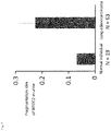
- FIG. 6 is a graph showing the fragmentation rate of WFDC2 after in vitro treatment of test subject-derived samples (urine) from healthy individuals and adenocarcinoma patients.
- FIG. 7 is a diagram showing the fragmentation index of WFDC2 after in vitro treatment of test subject samples (urine) of healthy individuals and adenocarcinoma patients.
- FIG. 8(A) is a diagram visualizing protein fragments having a C-terminal amino acid sequence of EYCGVPGDGDEEL (SEQ ID NO: 17).
- FIG. 8(B) is a diagram visualizing the abundance of the bikunin moiety of ⁇ -1-microglobulin/bikunin precursor in cell lysates derived from cells or cell lines.
- FIG. 8(C) is a diagram of plotting the relative abundance of ⁇ -1-microglobulin/bikunin precursor protein and protein fragment having a C-terminal amino acid sequence of EYCGVPGDGDEEL (SEQ ID NO: 17) in samples.
- FIG. 9 is a diagram showing the expression of AMBP in human adenocarcinoma cell lines.
- FIG. 10 is a diagram showing the expression of WFDC2 in human adenocarcinoma cell lines.
- the adenocarcinoma detection method of the present invention is based on new discovery by the present inventors. That is, the present inventors have found that, in the body of adenocarcinoma patients, one or more of several kinds of specific proteins are cleaved at the site where the proteins are not cleaved in healthy individuals, and various types of defective proteins or proteins having breaks may be increased. Hence, they have found that a phenomenon such as a deletion in the C-terminal part or the N-terminal part, a peptide bond break at an abnormal position or a deletion of amino acids accompanied by the break is observed in the specific proteins.
- Such abnormal cleavage is thought to be due to the presence of adenocarcinoma-specific protease. Therefore, in the present invention, healthy individuals and adenocarcinoma patients can be distinguished and evaluated in vitro by using breaks or deletions in the specific proteins due to such abnormal cleavage as indicators. Meanwhile, the increased abnormal cleavage in specific proteins can be evaluated by distinguishing between healthy individuals and adenocarcinoma patients using the decrease in the amount of normal protein (including the relative amount ratio) as an indicator.
- the present invention is an adenocarcinoma detection method including a step of detecting in vitro a presence or absence of an abnormal cleavage in one or two or more specific proteins in a test subject-derived sample.
- the specific protein are derived from any one of the proteins selected from the group consisting of the following (i) to (x):
- abnormal cleavage of the specific protein is not limited, and results in a primary structure, secondary structure, or tertiary structure, different from the normal structure of the specific protein.
- the abnormal cleavage include a cleavage resulting in one or more breaks in a peptide bond in the specific protein and a cleavage resulting in a deletion of one or two or more amino acid residues at one or more sites of the specific protein.
- a protein having a deletion of an amino acid residue on the C-terminal side of the specific protein or a protein having a deletion of an amino acid residue on the N-terminal side of the specific protein can be generated by the “abnormal cleavage”.
- the “abnormal cleavage” results in a break of a peptide bond between amino acid residues in a protein or a deletion of an amino acid at any intermediate position.
- the breaking of a peptide bond exposes an amino acid residue at a new cleavage site.
- the amino acid may not be lost from the original protein due to a disulfide bond or the like.
- the deletion or fragmentation of the specific protein may not occur.
- the abnormal cleavage of the present invention also includes such cleavage.
- the bound state of the original protein is maintained by, for example, a disulfide bond.
- the peptide bond is broken and is further devoid of one or two or more amino acid residues, this causes a case in which the amino acid residues in the protein are deleted at any intermediate position.
- test subject refers to a mammal as a cancer detection target, and is preferably, but not limited to, a dog, a cat, a mouse or a human.
- adenocarcinoma refers to cancer that occurs in the secretory gland tissue of each organ in the body.
- Adenocarcinoma includes, but not limited to, lung adenocarcinoma, liver adenocarcinoma, pancreatic adenocarcinoma, lymphatic adenocarcinoma, uterine adenocarcinoma, seminal adenocarcinoma, and gastric adenocarcinoma.
- the detection method of the present invention is particularly preferably used for the detection of lung adenocarcinoma.
- adenocarcinoma may be in any of the clinical stages IA, IB, IIA, IIB, IIIA, IIIB, and IV.
- Early-stage adenocarcinoma or early-stage lung adenocarcinoma refers to any of the clinical stages IA, IB, IIA, and IIB.
- test subject-derived sample refers to a body fluid derived from a test subject, and refers to a body fluid concentrate, a body fluid diluent, or another appropriately treated liquid, in addition to the body fluid itself.
- the body fluid refers to urine, blood (whole blood, plasma or serum), sputum, sweat, spinal fluid, digestive juice or ascitic fluid, but the fluid is not limited thereto.
- the body fluid is preferably urine or blood, and particularly preferably urine.
- the urine can be any of early morning midstream urine, pooled urine, and occasional urine.
- the collection amount of body fluid such as urine or blood is 10 ⁇ l to 200 ml, preferably 100 ⁇ l to 100 ml, and more preferably 1 ml to 100 ml.
- treatment of body fluid more specifically refers to pretreatment such as concentration, dilution, fractionation, and desalination, and addition of preservatives (e.g., glycerin), stabilizers (e.g., protease inhibitors), and antiseptics.
- preservatives e.g., glycerin
- stabilizers e.g., protease inhibitors
- antiseptics e.g., glycerin
- the treatment also includes a process of returning the temperature to normal temperature after a refrigerating or freezing treatment, a process of performing an appropriate treatment either before or after the refrigerating or freezing treatment, and the like.
- an anticoagulant treatment can be performed as an appropriate treatment. It is also possible to combine these treatments.
- concentration As the pretreatment before the measurement.
- This concentration method is not particularly limited, and examples thereof include a method using an ultrafiltration membrane with a fraction molecular weight, a freeze concentration method, a decompression or vacuum concentration method, and a heating method.
- the fraction molecular weight is not limited and, for example, any value such as 3 kD, 10 kD, 30 kD or 50 kD can be used.
- concentration is a useful pretreatment since the concentration of total protein in primitive urine is about 0.001 g/dL to 0.6 g/dL.
- the primitive urine can be concentrated to 200 to 250-folds and used for analysis. Although this concentrated solution can be directly used for measurement, the solution can also be used for measurement by further diluting the total protein. For example, depending on the measurement method such as western blotting, the solution can be adjusted to about 0.1 ⁇ g to 10 ⁇ g/ ⁇ l for measurement.
- Vivaspin registered trademark, manufactured by Sartorius Japan K.K.
- Amicon Ultra manufactured by Merck KGaA
- the like can be used according to the manufacturer's instructions.
- Distilled water and a buffer solution can be used for dilution and can also be used for adjustment of the concentrated urine.
- the size and length of a deletion of a specific protein is not particularly limited as long as, when compared in the primary structure, there is a deletion in the C-terminal and N-terminal parts of the full-length protein derived from the specific protein or a deletion of amino acid residues at the intermediate position.
- the term “there is a deletion in the C-terminal and N-terminal parts or at the intermediate position” means that the C-terminal and N-terminal sides of the protein are usually short and the amino acid residues to be present at the intermediate position are deleted, compared with a protein of a normally functioning unit.
- each written symbol means an ordinary character used as a one-letter expression of amino acid residue.
- a step of detecting in vitro a presence or absence of an abnormal cleavage in one or two or more specific proteins in a test subject-derived sample is not limited, and the step can be preferably a step including at least one selected from the group consisting of the following (1) to (4):
- one embodiment of the adenocarcinoma detection method of the present invention includes detecting a decrease in an amount of a protein having a normal structure of one or two or more of specific protein in a test subject-derived sample.
- the amount of the specific protein having a normal structure is measured by the presence or absence of the amino acid sequence at the C-terminal part and/or the N-terminal part of the specific protein so that it is possible to detect whether or not the amount of the protein having a normal structure is relatively reduced as compared with healthy individuals.
- another embodiment of the adenocarcinoma detection method of the present invention includes detecting in vitro a specific protein fragment using a test subject-derived sample.
- detection of the specific protein fragment can be a detection of a protein fragment present in a fragmented state in the test subject-derived sample and/or, for example, a protein fragment generated after subjecting the test subject-derived sample to a reduction treatment or the like.
- the specific protein fragments are fragments which are derived from any one of the proteins selected from the group consisting of the following (i) to (x) and which are derived from a protein having a deletion in the C-terminal side compared with the full length of the protein, or fragments derived from a protein with a break and/or an amino acid deletion at the intermediate site.
- the C-terminal sequence of the specific protein fragment is expressed as, for example, “a protein fragment having a C-terminal amino acid sequence of GTEAAGAMF”, SEQ ID NO: 11.
- a protein fragment having a C-terminal amino acid sequence of GTEAAGAMF SEQ ID NO: 11.
- this case indicates that an amino acid residue originally present on the side closer to the C-terminus than F at the right end is deleted (defective), and the amino acid residue may be present or absent on the side closer to the N-terminus than G.
- the specific protein fragment is at least one selected from the group consisting of the followings:
- the number of amino acid residues in the specific protein fragment is not particularly limited, and is at least 6, may be at least 7, preferably at least 10 or more, and more preferably at least 50 or more.
- the upper limit of the size of a protein fragment is not particularly limited, and is less than the size of the full-length protein from which each protein fragment is derived.
- the protein fragment has a structure which is lacking in the C-terminal side, compared with the full-length protein from which each protein fragment is derived.
- the protein fragment is not particularly limited as long as the size is less than the full length, and in some cases, the protein fragment may be present as a short fragment such as 200 residues or less, preferably 100 residues or less.
- the test subject-derived sample detects a presence or an increase in relative amount of a protein having a deletion of an amino acid residue on an N-terminal side of the specific protein in the test subject-derived sample; and detect a liberated protein fragment on a C-terminal side lacking in an N-terminal side in order to detect the presence or the increase in the relative amount of a protein having a break or deletion in any intermediate site of an amino acid sequence of the specific protein in the test subject-derived sample.
- the fragment is detected so that it is also possible to indirectly detect the protein fragments (a) to (y) or to detect the presence of a protein fragment in which the N terminus is defective in vivo.
- the molecular weight of the specific protein fragment of the present invention is not limited as long as the molecular weight is less than that of the full-length protein. However, depending on the method of detecting adenocarcinoma using a specimen such as urine, the molecular weight is, for example, 3 kDa or more and less than the full length, preferably 10 kDa or more and less than the full length.
- these specific protein fragments include protein fragments subjected to post-translational modification such as glycosylation or phosphorylation as long as the specific protein fragments respectively have the sequences described above.
- the specific protein fragments may be fragments derived from the full-length proteins shown below.
- proteins normally function in their original length in vivo. It is considered that, the proteins are cleaved by an enzyme whose activity is enhanced, particularly in adenocarcinoma (e.g., a matrix metalloprotease group), whereby secondary fragmentation and amino acid deletion can be increased.
- adenocarcinoma e.g., a matrix metalloprotease group
- ⁇ -1-microglobulin/bikunin precursor includes ⁇ -1-microglobulin, inter- ⁇ -trypsin inhibitor light chain, and trypstatin.
- the three mature proteins normally function in their original length in vivo.
- the mature proteins are specifically expressed, particularly in adenocarcinoma, or are cleaved by an enzyme whose activity is enhanced (e.g., the matrix metalloprotease group), whereby secondary fragmentation and amino acid deletion can be increased.
- a protein fragment whose C-terminal sequence is GTEAAGAMF can be derived from ⁇ -1-antitrypsin.
- ⁇ -1-antitrypsin is a neutrophil elastase inhibitor, and the main function is to protect the lung from protease-mediated tissue destruction.
- the sequence of ⁇ -1-antitrypsin precursor is, for example, a sequence of UniProtKB P01009(A1AT_HUMAN) (SEQ ID NO: 1 in the sequence listing).
- the specific protein fragment may be, when described with this sequence, a protein fragment having a C-terminal part which coincides with amino acid residues at positions 368 to 376 and a C-terminal side deleted from position 377.
- a protein fragment whose C-terminal sequence is CVLFPYGG (SEQ ID NO: 12) can be derived from ⁇ -1-microglobulin/bikunin precursor.
- a protein fragment whose C-terminal sequence is EYCGVPG (SEQ ID NO: 13) can be derived from ⁇ -1-microglobulin/bikunin precursor.
- a protein fragment whose C-terminal sequence is EYCGVPGDG (SEQ ID NO: 14) can be derived from ⁇ -1-microglobulin/bikunin precursor.
- a protein fragment whose C-terminal sequence is EYCGVPGDGDE (SEQ ID NO: 15) can be derived from ⁇ -1-microglobulin/bikunin precursor.
- a protein fragment whose C-terminal sequence is EYCGVPGDGDEE (SEQ ID NO: 16) can be derived from ⁇ -1-microglobulin/bikunin precursor.
- gg A protein fragment whose C-terminal sequence is EYCGVPGDGDEEL (SEQ ID NO: 17) can be derived from ⁇ -1-microglobulin/bikunin precursor.
- a protein fragment whose C-terminal sequence is EYCGVPGDGDEELL (SEQ ID NO: 18) can be derived from ⁇ -1-microglobulin/bikunin precursor.
- a protein fragment whose C-terminal sequence is GECVPGEQEPEP (SEQ ID NO: 19) can be derived from ⁇ -1-microglobulin/bikunin precursor.
- a protein fragment whose C-terminal sequence is GECVPGEQEPEPILIP SEQ ID NO: 20
- SEQ ID NO: 20 A protein fragment whose C-terminal sequence is GECVPGEQEPEPILIP
- a protein fragment whose C-terminal sequence is EYCGVPGD (SEQ ID NO: 31) can be derived from ⁇ -1-microglobulin/bikunin precursor.
- a protein fragment whose C-terminal sequence is GECVPGEQEPE (SEQ ID NO: 32) can be derived from ⁇ -1-microglobulin/bikunin precursor.
- ⁇ -1-microglobulin is a glycoprotein having a molecular weight of about 30,000.
- the sequence of the protein can be, for example, a sequence represented by UniProtKB P02760 (SEQ ID NO: 2 in the sequence listing).
- the protein having a normal structure is a protein having at least Asn at position 352 of SEQ ID NO: 2 in the sequence listing as the C-terminal.
- the specific protein fragment (bb) can be a protein fragment having inter- ⁇ -trypsin inhibitor light chain or amino acid residues at positions 312 to 319 contained in trypstatin and a defective C-terminal side, or a protein fragment containing only inter- ⁇ -trypsin inhibitor light chain part or containing only trypstatin.
- the specific protein fragments (cc) to (hh) and (xx), although not limited thereto, are protein fragments which have any of the following amino acid sequences: an amino acid sequence of amino acid residues at positions 335 to 341 or positions 335 to 342; an amino acid sequence of amino acid residues at positions 335 to 343 in this sequence; an amino acid sequence of amino acid residues at positions 335 to 345 in this sequence; an amino acid sequence of amino acid residues at positions 335 to 346 in this sequence; an amino acid sequence of amino acid residues at positions 335 to 347 in this sequence; and an amino acid sequence of amino acid residues at positions 335 to 348 in this sequence; and in which the amino acid sequence has a defective C-terminal side and contains only inter- ⁇ -trypsin inhibitor light chain part, or has a deletion in the C-terminus and contains only trypstatin.
- each of the fragments can be a protein fragment having a sequence of amino acid residues at positions 186 to 196 or positions 186 to 197 or a sequence of amino acid residues at positions 186 to 201, and having a defective C-terminal side.
- a protein fragment whose C-terminal sequence is DLCNFNEQL can be derived from CD59.
- the CD59 molecule is known as a complement regulatory factor and is a molecule that inhibits the formation of a membrane damage complex by acting on C9.
- the sequence of the protein can be, for example, a sequence represented by UniProtKB P13987(CD59_HUMAN) (SEQ ID NO: 3 in the sequence listing).
- the specific protein fragment (kk) can be, when described with the sequence represented by UniProtKB P13987, a protein fragment having amino acid residues at positions 92 to 100 and a C-terminal side deleted from position 101.
- Fibronectin is a glycoprotein with a relatively large molecular weight, i.e., a cell adhesion molecule. It is thought that the cell adhesion molecule has functions such as the adhesion of cells to the extracellular matrix, the formation and retention of connective tissue, wound healing, and the formation and maintenance of the form and division of tissues and organs during embryogenesis.
- the sequence of fibronectin is, for example, a sequence of UniProtKB P02751 (FINC_HUMAN) (SEQ ID NO: 4 in the sequence listing).
- the specific protein fragment can be, for example, when described with the sequence of UniProtKB P02751, a protein fragment having amino acid residues at positions 458 to 464 and a C-terminal side deleted from position 465.
- a protein fragment whose C-terminal sequence is LPTGYY (SEQ ID NO: 24) can be derived from Lectin, Mannose-Binding 2.
- Lectin, Mannose-Binding 2 has a sequence of UniProtKB-Q12907 (LMAN2_HUMAN) (SEQ ID NO: 5 in the sequence listing).
- the specific protein fragment can be, for example, when described with this sequence, a protein fragment having amino acid residues at positions 247 to 252 and a defective C-terminal side.
- a protein fragment whose C-terminal sequence is YLQGSSVQL can be derived from vasorin.
- Vasorin is a type 1 membrane protein expressed specifically in vascular smooth muscle cells.
- the sequence of vasorin is, for example, a sequence of UniProtKB Q6EMK4 (VASN_HUMAN) (SEQ ID NO: 6 in the sequence listing).
- the specific protein fragment can be, for example, when described with this sequence, a protein fragment having amino acid residues at positions 483 to 491 and a defective C-terminal side.
- a protein fragment whose C-terminal sequence is EGSCPQVNIN (SEQ ID NO: 26) can be derived from WAP four-disulfide core domain protein 2.
- pp A protein fragment whose C-terminal sequence is EGSCPQVNINFPQLG (SEQ ID NO: 23) can be derived from WAP four-disulfide core domain protein 2.
- a protein fragment whose C-terminal sequence is EGSCPQVNINFPQLGL can similarly be derived from WAP four-disulfide core domain protein 2.
- the sequence of WAP four-disulfide core domain protein 2 is a sequence of UniProtKB Q14508 (WFDC2_HUMAN) (SEQ ID NO: 7 in the sequence listing).
- the protein having a normal structure is a protein having at least Phe at position 124 of SEQ ID NO: 7 in the sequence listing as the C-terminus.
- the specific protein fragment can be, for example, when described with this sequence, a protein fragment having amino acid residues at positions 77 to 86, amino acid residues at positions 77 to 91 or positions 77 to 92, and a defective C-terminal side.
- a protein fragment whose C-terminal sequence is ENYNQYDLN can be derived from Membrane-bound carboxypeptidase M.
- Membrane-bound carboxypeptidase M is an exopeptidase that cleaves one residue from a C-terminal side of a substrate.
- This sequence can be, for example, a sequence of UniProtKB P14384 (CBPM_HUMAN) (SEQ ID NO: 8 in the sequence listing).
- the specific protein fragment can be, for example, when described with this sequence, a protein fragment having amino acid residues at positions 145 to 153 and a defective C-terminal side.
- a protein fragment whose C-terminal sequence is GAVVPDSALPFNFQAAY (SEQ ID NO: 28) can be derived from deoxyribonuclease-1.
- This sequence can be, for example, a sequence of UniProtKB P24855 (DNAS1_HUMAN) (SEQ ID NO: 9 in the sequence listing).
- the specific protein fragment can be, for example, when described with this sequence, a protein fragment having amino acid residues at positions 245 to 261 and a defective C-terminal side.
- a protein fragment whose C-terminal sequence is GALCLLAEDDS (SEQ ID NO: 30) can be derived from WNT1-inducible-signaling pathway protein 2. This sequence can be, for example, a sequence of UniProtKB-076076 (WISP2_HUMAN) (SEQ ID NO: 10 in the sequence listing).
- the specific protein fragment can be, for example, when described with this sequence, a protein fragment having amino acid residues at positions 88 to 98 and a defective C-terminal side.
- the mass of the specific protein having a normal structure or its presence and the amount of the specific protein fragment or its presence can be determined by measuring the mass, volume, and concentration of the specific protein and the specific protein fragment contained in a body fluid, i.e., a test subject-derived sample. Further, these amounts can be expressed by fluorescence intensity, absorbance, MS/MS spectrum intensity, and the like.
- the detection method of the present invention is not particularly limited as long as the detection method is a method for detecting abnormal cleavage in a test subject-derived sample or a method for detecting a protein having a normal structure.
- the detection method is a method for detecting abnormal cleavage in a test subject-derived sample or a method for detecting a protein having a normal structure.
- mass spectrometry there is a method for detecting by an immunochemical measurement method using an antibody (radioimmunoassay, enzyme immunoassay, Western blotting, etc.).
- the method by mass spectrometry is not particularly limited as long as the method is a method for detecting a specific protein having a normal structure in a test subject-derived sample or a specific protein fragment derived from abnormal cleavage.
- the method is a method for collecting a sample, digesting the sample with an enzyme after pretreatment and/or reduction treatment, labeling the sample with an isotope or the like, and then measuring the amount of only the peptide derived from the C-terminus by MS/MS.
- test subject-derived sample is urine
- urine is collected
- the urine specimen is subjected to concentration as a pretreatment to obtain a specimen
- this concentrated specimen is subjected to reductive alkylation, and then the resulting specimen is digested with trypsin in the presence of H 2 18 O, so that peptides derived from trypsin-digested fragments can be labeled with a stable isotope.
- trypsin in the presence of H 2 18 O
- only trypsin-digested peptides derived from the C-terminus are measured by Nano-LC-MALDI-MS/MS.
- This peptide information is analyzed so that it is possible to distinguish and detect the protein having a normal structure or the specific protein fragment derived from abnormal cleavage.
- a method for measuring a specific protein fragment can be employed in which a protein in a concentrated sample with Lys-C is subjected to enzymatic digestion, the N-terminus of all the digested peptides is blocked by phenyl isocyanate or the TMPP reagent (N-Succinimidyloxycarbonyl-methyl)tris(2,4,6-trimethoxyphenyl) phosphonium bromide) to obtain a peptide having a C-terminus other than lysine, and an MS/MS analysis is preformed using amass spectrometer.
- MRM multiple reaction monitoring
- An antibody that specifically recognizes a specific protein or antigen derived from the specific protein can be produced in such a manner that, although not limited thereto, peptides corresponding to the corresponding C-terminal part, the N-terminal part, another part, and a deletion part are administered to chickens, rats, mice, rabbits or the like, followed by immunization.
- an antigen may be immobilized on a column and an antibody may be produced by purifying a polyclonal antibody obtained from the immunized animals.
- a monoclonal antibody that specifically recognizes a site containing a C-terminal amino acid or a deletion part of each fragment may be produced.
- the method for producing a monoclonal antibody is known to those skilled in the art. For example, hybridomas having a proliferative capacity are produced by cell fusion of antibody-producing cells obtained from the above immunized animals with myeloma cells. In particular, only the clone is selected in which an antibody having specificity to the C-terminal amino acid is produced. The cells are cultured, secreting antibodies are purified, and the purified antibodies are used as the antibodies of the present invention.
- a test subject-derived sample is subjected to SDS-PAGE and transferred to a nitrocellulose membrane or a PVDF membrane.
- the transferred membrane is blocked with skim milk or the like, and then treated with a primary antibody specifically recognizing the C-terminal amino acid of each protein or protein fragment or a site containing a deletion part.
- the membrane is treated with a horseradish peroxidase (HRP)-labeled secondary antibody recognizing the primary antibody, and an electrophoretic band of a target protein fragment is detected by color development or chemiluminescence derived from the enzymatic activity of HRP.
- HRP horseradish peroxidase
- a specimen is treated with the primary antibody and the HRP-labeled secondary antibody in a 96-well plate, and a target protein fragment is detected by color development or chemiluminescence derived from the enzymatic activity of HRP. It is also possible to use a sandwich method or the like.
- the abundance of an abnormal cleavage-derived deletion and a protein fragment in a test subject-derived sample is detected by mass spectrometry, radioimmunoassay, enzyme immunoassay or Western blotting, and then the detected abundance can be compared with the abundance of total protein present in the test subject-derived sample.
- the amount of total protein in the test subject-derived sample may be quantified using a commercially available protein assay kit such as the BCA method or the Bradford method, and a liquid chromatography method or the like may be used.
- the amount of total protein may be compared with the amount of creatinine in urine which is used for correcting the changes in the water content of the urine specimen.
- the amount of a specific protein having a normal structure or the amount of a specific protein fragment in a test subject-derived sample can be compared with the abundance of all proteins derived from a specific protein coexisting in a sample and represented by a relative ratio.
- the abundance of a protein fragment in a test subject-derived sample can be compared with the amount of total protein in the test subject-derived sample and expressed by a relative ratio of the amount of the fragment to the amount of total protein.
- the presence or absence of an abnormal cleavage in a specific protein can be determined by calculating the fragmentation rate of the specific protein in a test subject sample and judging whether or not the value is greater than a threshold value. Hence, when the fragmentation rate of the specific protein is greater than the threshold value, it can be grasped that the derived test subject has adenocarcinoma.
- the fragmentation rate (F n ) of each specific protein is determined by the following calculation formula.
- the fragmentation rate of the specific protein in the patient sample is greater than 1, as expressed by the relative ratio (R n ), with respect to the average value of the healthy individual group, the specific protein can be determined to be positive.
- Protein fragmentation rate ( F n ) C n /I n [Equation 1]
- F p protein fragmentation rate of patient sample
- the amount of each specific protein fragment in C n refers to an amount of one specific protein fragment (e.g., an amount corresponding to one of the protein fragments having an abnormal C-terminus as shown in Table 3).
- the amount of original protein in I n refers to, for example, an amount of trypsin-digested peptide commonly included in a protein fragment or full-length protein.
- the measurement method is in accordance with the conditions described in Example 7.
- sequences commonly included are not limited, and the following sequences can be used.
- AMBP TVAACNLPIVR (at positions 283 to 293 of SEQ ID NO: 2 in the sequence listing)
- WFDC2 CCSAGCATFCSLPNDK (at positions 61 to 76 of SEQ ID NO: 7 in the sequence listing)
- WISP2 CPLGVPLVLDGCGCCR (at positions 39 to 54 of SEQ ID NO: 10 in the sequence listing)
- DNAS1 DSHLTAVGK (at positions 64 to 72 of SEQ ID NO: 9 in the sequence listing)
- CBPM DPEITNLINSTR (at positions 107 to 118 of SEQ ID NO: 8 in the sequence listing)
- VASN ESHVTLASPEETR (at positions 315 to 327 of SEQ ID NO: 6 in the sequence listing)
- LMAN2 DHDTFLAVR (at positions 210 to 218 of SEQ ID NO: 5 in the sequence listing)
- CD59 FEHCNFNDVTTR (at positions 67 to 78 of SEQ ID NO: 3 in the sequence listing)
- the target person is judged as an adenocarcinoma patient.
- the term “significantly decreased” means that the relative ratio of the protein having the normal structure is decreased to, for example, 0.9 times or less, preferably 0.8 times or less, more preferably 0.6 times or less, as compared with a healthy individual specimen.
- the term means that a decrease in the amount which is recognizable from the intensity of label or the like is 0.8 times or less, preferably 0.6 times or less, as compared with a healthy individual specimen.
- the target person is judged as an adenocarcinoma patient.
- the term “significantly increased” means that the relative ratio of the fragmentation rate is increased to a value greater than 1, 1.25 times or more, preferably 1.5 times or more, more preferably 2.0 times or more, as compared with a healthy individual specimen.
- the term means that the increase in the amount of a specific protein fragment that can be recognized from the intensity of a label or the like is greater than 1, preferably 1.2 times or more, more preferably about 1.5 times or more, as compared with a healthy individual specimen.
- the numerical value of the comparison here refers to a value after correction using an internal standard or the like.
- the amount of a specific protein having a normal structure or the amount of a specific protein fragment in a test subject-derived sample from a healthy individual can be determined in accordance with the preparation and measurement method of the test subject-derived sample.
- one or two or more specific proteins contains at least (ii) ⁇ -1-microglobulin/bikunin precursor
- a step of detecting in vitro a presence or absence of an abnormal cleavage includes detecting a decrease in an amount of a protein having a normal structure which is derived from ⁇ -1-microglobulin/bikunin precursor present in a test subject sample, and/or an amount or presence of a protein fragment derived from ⁇ -1-microglobulin/bikunin precursor.
- an antibody recognizing the C-terminal part of (g) EYCGVPGDGDEEL (SEQ ID NO: 17) can be suitably used.
- one or two or more specific proteins include at least (vii) WAP four-disulfide core domain protein 2, and the step of detecting in vitro the presence or absence of the abnormal cleavage includes detecting a cleavage of a peptide bond in any of the amino acid sequences at positions 80 to 93 of SEQ ID NO: 7 in the sequence listing, or a deletion of an amino acid residue around the cleavage site.
- patients with pulmonary nodule shadows or tumor shadows suspected of primary lung cancer by chest image examination, patients diagnosed with lung adenocarcinoma were selected by surgery, transbronchial biopsy, lymph node biopsy or cytology. Early morning midstream urine was collected from selected lung adenocarcinoma patients and healthy individuals using sterile cups. The collected urine samples were stored at ⁇ 80° C. until analysis.
- C-terminal protein fragments in urine from 85 cases with lung adenocarcinoma and 25 healthy individuals were comprehensively analyzed.
- a breakdown of clinical stages of the cases with lung adenocarcinoma showed that 18 cases were in clinical stage IA, 5 cases were in clinical stage IB, 1 case was in clinical stage IIA, 0 case was in clinical stage IIB, 6 cases were in clinical stage IIIA, 5 cases were in clinical stage IIIb, and 50 cases were in clinical stage IV.
- the clinical stage was judged according to the criteria described in “General Rule for Clinical and Pathological Record of Lung Cancer, The 7th Edition” edited by The Japan Lung Cancer Society (KANEHARA & Co., LTD.).
- the cases with lung adenocarcinoma (18 cases in stage IA, 5 cases in stage IB, and 1 case in stage IIA) were defined as cases with early-stage lung adenocarcinoma.
- Urine samples ( ⁇ 50 mL) were collected and markers were searched by the procedures of pretreatment, acquisition of analytical data, and statistical analysis.
- urine samples were concentrated to 200 to 250-folds using Amicon Ultra-15 (10 kDa molecular weight cut-off) and Amicon Ultra-4 (10 kDa molecular weight cut-off) (Merck KGaA) and washed 3 times with 3 mL of triethylammonium hydrogencarbonate solution containing 100 mM NaCl, thereby removing low molecular weight molecules. Thereafter, concentrated specimens were obtained. The proteins in the concentrated specimens were quantified.
- the concentrations of all the specimens were adjusted to 10 mg/mL of total protein using a buffer solution and the resulting specimens were used in the subsequent analysis process. Subsequently, the samples were subjected to reductive alkylation and then digested with trypsin in a buffer prepared with a constant concentration of H 2 18 O (method for labeling the C-terminus of a peptide with a stable isotope). After desalting and purification, the samples were labeled with iTRAQ (8-plex, AB Sciex.), eight specimens were mixed, desalted, and purified.
- iTRAQ 8-plex, AB Sciex.
- Nano-LC LC column with an inner diameter of 75 ⁇ m and a length of 100 mm, filler: Inertsil C18 (particle diameter of 3 ⁇ m); flow rate: 250 nL/min; solvent: a 0.1% trifluoroacetic acid solution)/MALDI-MS/MS (AB Sciex.) was used to eluate the fractions by acetonitrile concentration gradient (3-80%), the fractions were measured, and then only C-terminal trypsin-digested peptides were selected and extracted (independent development program “iSpec”; Reference: Fernandez-de-Cossio J., Takao T. et al. Rapid Commun.
- Mass Spectrom. 18, 2465-2472 (2004) Based on the intensity of the control peak (m/z 113, equally spiked for each assay) in reporter peaks related to comparative determination included in MS/MS spectra (m/z 113 to 119, and 121), other reporter peaks were standardized, and then the comparison between the assays was performed. As a specific evaluation criterion, for the qualification, an assay was adopted in which MS/MS spectra with five or more peaks with a certain intensity or more were selected and the number of proteins identified based on those MS/MS spectra was 50 more.
- Quantitative values of only the assay in which the peak intensity of the reference reporter peak (m/z 113) was greater than 10 and the number of MS/MS spectra was 300 or more were used for quantification. Only data satisfying these qualification and quantification criteria is used for statistical analysis so that comparison accuracy between assays can be improved, and as a result, the accuracy of statistical analysis can be improved.
- Example 2 Regarding the peak intensity (Example 2) of each of the C-terminal trypsin-digested peptides of the MS/MS spectra obtained from urine samples from lung adenocarcinoma patients and healthy individuals based on the statistical analysis, the relative ratio of the control specimen to the peak intensity was calculated, and comparison and evaluation were made between lung adenocarcinoma patients and healthy individuals. Mann-Whitney's U test was used as the assay method. For statistical analysis, JMP12 (SAS Institute Inc, Cary, N.C.) was used. The peak intensities of protein fragments in urine were compared between 85 cases with lung adenocarcinoma or 24 cases with early-stage lung adenocarcinoma and healthy individuals.
- the protein fragments satisfying the criteria that the ROC-AUC value was 0.6 or more or the p value was less than 0.1 were used as marker candidates.
- a total of 18 types of protein fragments were identified as lung adenocarcinoma diagnostic markers (Table 1) and a total of 13 types of protein fragments (Table 2) were identified as early-stage lung adenocarcinoma diagnostic markers.
- Example 2 From the urine concentrated specimens (Example 2) of healthy individuals (7 persons) and lung adenocarcinoma patients (18 cases), the chondroitin sulfate chain equivalent to 2.5 ⁇ g of protein was removed from the proteins by chondroitinase ABC treatment (Sigma-Aldrich, Inc.). Then, the specimens were subjected to SDS-polyacrylamide gel electrophoresis using 5-20% polyacrylamide gel (DRC CO., LTD.).
- the proteins separated in the gel were transferred to a nitrocellulose membrane BA85 (GE Healthcare Japan Corporation), and the specific antibody recognizing the C-terminal amino acid sequence of the protein fragment EYCGVPGDGDEEL (SEQ ID NO: 17) derived from ⁇ -1-microglobulin/bikunin precursor was used to visualize the protein fragments having the C-terminal amino acid sequence of EYCGVPGDDEEL (SEQ ID NO: 33) ( FIG. 1A ). Further, the commercially available antibody (AMBP antibody, Product No. GTX101069, GeneTex, Inc.) recognizing ⁇ -1-microglobulin/bikunin precursor was used to visualize the abundance of ⁇ -1-microglobulin/bikunin precursor protein present in urine ( FIG. 1B ).
- Each band was visualized by obtaining a chemiluminescent signal from a horseradish peroxidase-labeled secondary antibody with a cooled CCD camera (Fujifilm Holdings Corporation), and the band was quantified using image analysis software ImageJ (National Institutes of Health (NIH)).
- ImageJ National Institutes of Health (NIH)
- the relative abundance of ⁇ -1-microglobulin/bikunin precursor protein and protein fragment having a C-terminal amino acid sequence of EYCGVPGDDEEL (SEQ ID NO: 33) in each sample was plotted ( FIG. 1(C) ).
- lung adenocarcinoma diagnosis was performed by tissue diagnosis such as surgery, lung biopsy, lymph node biopsy or bone biopsy or by cytological diagnosis of expectoration, pleural effusion, and the like.
- the clinical stage of lung adenocarcinoma was determined using the intensity of accumulation by PET-CT examination, image examination by head MRI examination and chest CT examination, bone biopsy, cytological diagnosis by lymph node puncture or pleural effusion cytology examination.
- the area of the spectrum of the protein fragment having the C-terminal amino acid sequence of EYCGVPGDGDEEL was 2.1 times the average value of healthy individuals.
- Example 1 The utility of CEA and protein fragments in urine was compared among 14 cases where blood CEA levels could be confirmed, among 24 cases diagnosed as early-stage lung adenocarcinoma (Example 1). For example, when the amount of the protein fragment having the C-terminal sequence of CVLFPYGG, SEQ ID NO: 12, derived from ⁇ -1-microglobulin/bikunin precursor, was 1.25 times the average value of healthy individuals in Example 2 and this case was judged to be positive, 11 out of 14 cases with early-stage lung adenocarcinoma could be judged to be positive. Meanwhile, when the reference value of blood CEA level was 5.0 ng/ml, only 4 out of 14 cases with early-stage lung adenocarcinoma were positive.
- Specimens were prepared in the same manner as in Example 2, and patients with adenocarcinoma were screened by subjecting the patients to MRM.
- Urine samples ( ⁇ 50 mL) were collected. Then, the pretreatment and the acquisition and analysis of analytical data were performed. Specifically, in the pretreatment, the urine samples were concentrated to 200 to 250-folds using Amicon Ultra-15 (10 kDa molecular weight cut-off) and Amicon Ultra-4 (10 kDa molecular weight cut-off) (Merck KGaA). The low molecular weight molecules were removed by washing, and then concentrated specimens were obtained. The proteins in the concentrated specimens were quantified.
- the concentrations of all the specimens were adjusted to 10 mg/mL of total protein using a buffer solution and the resulting specimens were used in the subsequent analysis step. Subsequently, the samples were digested with trypsin in a buffer after reductive alkylation. After desalting and purification, trypsin-digested peptides derived from protein fragments were fractionated by ion exchange chromatography (LC column: PolySULFOETHYL ATM (PolyLC Inc.
- C n amount of each protein fragment (amount of peptide in the protein fragment sequence in Table 3)
- F p protein fragmentation rate of patient sample
- the amount of the original protein in I n refers to an amount of a trypsin-digested peptide commonly included in a protein fragment or full-length protein.
- the following internal sequences were respectively used.
- AMBP TVAACNLPIVR (at positions 283 to 293 of SEQ ID NO: 2 in the sequence listing)
- WFDC2 CCSAGCATFCSLPNDK (at positions 61 to 76 of SEQ ID NO: 7 in the sequence listing)
- WISP2 CPLGVPLVLDGCGCCR (at positions 39 to 54 of SEQ ID NO: 10 in the sequence listing)
- Urine specimens of 28 cases with early-stage lung adenocarcinoma (clinical stage IA: 19 cases, IB: 7 cases, IIA: 1 case, IIB: 1 case) and 40 healthy individuals were subjected to MRM, whereby the fragmentation rate of each protein was compared between cases with early-stage lung adenocarcinoma and healthy individuals.
- the protein fragments satisfying the criteria that the ROC-AUC value was 0.6 or more or the p value was less than 0.1 were used as early-stage lung adenocarcinoma diagnostic markers.
- a total of 12 types (Table 3) were identified as early-stage lung adenocarcinoma diagnostic markers by this analysis.
- Urine specimens of 28 cases with early-stage lung adenocarcinoma and 40 healthy individuals were subjected to MRM, and the fragmentation rate of the protein fragment having the C-terminal amino acid sequence of EYCGVPGDGDEE (SEQ ID NO: 16), derived from ⁇ -1-microglobulin/bikunin precursor, was measured.
- EYCGVPGDGDEE SEQ ID NO: 16
- the fragmentation rate as a reference value was 2.18 times the average value of the healthy individual group, 1 healthy individual was positive and 16 cases with early-stage lung adenocarcinoma were positive. Based on the reference value, the sensitivity was 57.1%, the specificity was 97.5%, and the ROC-AUC value was 0.819.
- Predicted values in each urine sample were calculated using the regression equation. When a predicted value of 0.5358 was used as a cutoff value, the sensitivity was 82.6%, the specificity was 94.3%, and the ROC-AUC value was 0.901 based on the reference value.
- Concentrated urine specimens of healthy individuals (9 persons) and lung adenocarcinoma patients (9 cases) were obtained in the same manner as in Example 2.
- the chondroitin sulfate chain equivalent to 2.5 ⁇ g of protein was removed from the proteins by chondroitinase ABC treatment (Sigma-Aldrich, Inc.). Then, the specimens were subjected to SDS-polyacrylamide gel electrophoresis using 5-20% polyacrylamide gel (DRC CO., LTD.).
- the proteins separated in the gel were transferred to a nitrocellulose membrane BA85 (GE Healthcare Japan Corporation), and the specific antibody recognizing the C-terminal amino acid sequence RFSN (SEQ ID NO: 35) of the normal protein derived from ⁇ -1-microglobulin/bikunin precursor was used to visualize the normal (full-length) protein having the C-terminal amino acid sequence of RFSN (SEQ ID NO: 35) ( FIG. 5A ; anti-bikunin C-terminal antibody). Further, the commercially available antibody (AMBP antibody, Product No.
- GTX101069, GeneTex, Inc. recognizing ⁇ -1-microglobulin/bikunin precursor was used to visualize the abundance of ⁇ -1-microglobulin/bikunin precursor protein present in urine ( FIG. 5A ; anti-bikunin antibody).
- Each band was visualized by obtaining a chemiluminescent signal from a horseradish peroxidase-labeled secondary antibody with a digital camera (Canon Inc.), and the band was quantified using image analysis software ImageJ (National Institutes of Health (NIH)).
- FIG. 5(B) The relative abundance of ⁇ -1-microglobulin/bikunin precursor protein and normal protein having a C-terminal amino acid sequence of RFSN (SEQ ID NO: 35) in each sample was plotted ( FIG. 5(B) ).
- 6 out of 9 lung adenocarcinoma patient specimens were found in which the ratio of the detected total length was 1 ⁇ 2 or less lower than the average of healthy individuals. Note that, in FIGS. 5(A) and 5(B) , lanes 1 to 6 are samples derived from healthy persons, and lanes 7 to 12 are samples derived from stage I lung adenocarcinoma patients.
- Specimens were prepared in the same manner as in Example 2, and patients with adenocarcinoma were screened by subjecting the patients to MRM.
- Urine samples ( ⁇ 50 mL) were collected. Then, the pretreatment and the acquisition and analysis of analytical data were performed. Specifically, in the pretreatment, the urine samples were concentrated to 200 to 250-folds using Amicon Ultra-15 (10 kDa molecular weight cut-off) and Amicon Ultra-4 (10 kDa molecular weight cut-off) (Merck KGaA). The low molecular weight molecules were removed by washing, and then concentrated specimens were obtained. The proteins in the concentrated specimens were quantified.
- the concentrations of all the specimens were adjusted to 10 mg/mL of total protein using a buffer solution and the resulting specimens were used in the subsequent analysis step. Subsequently, the samples were digested with trypsin in a buffer after reductive alkylation. After desalting and purification, trypsin-digested peptides derived from protein fragments were fractionated by ion exchange chromatography (LC column: PolySULFOETHYL ATM (PolyLC Inc.
- Urine specimens of 63 cases with early-stage lung adenocarcinoma (clinical stage IA: 42 cases, IB: 16 cases, IIA: 2 cases, IIB: 3 cases) and 19 healthy individuals were subjected to MRM.
- the fragmentation rate of the protein fragment of WFDC2 between early-stage lung adenocarcinoma patients and healthy individuals was determined, and graphed by setting the average value of the healthy individual group to 1 ( FIG. 6 ).
- the fragmentation rate of WFDC2 was measured using trypsin-digested peptides having the amino acid sequence EGSCPQVNINFPQLGL (SEQ ID NO: 29).
- the fragment index (Ei) of WFDC2 was calculated by the following calculation formula, using (A) the fragmentation rate of trypsin-digested peptides having the amino acid sequence EGSCPQVNINFPQLGL (SEQ ID NO: 29) and (B) the abundance of the protein fragment having the C-terminal amino acid sequence of EGSCPQVNIN (SEQ ID NO: 26).
- the patient sample whose fragmentation index was 1.3 times or more higher than the average value of the healthy individual group was defined as positive.
- Fragmentation index ( Ei ) B/A [Equation 4]
- Urine specimens of 63 cases with early-stage lung adenocarcinoma used in Example 9 (clinical stage IA: 42 cases, IB: 16 cases, IIA: 2 cases, IIB: 3 cases) and 19 healthy individuals were subjected to MRM, and the fragmentation index of WFDC2 was determined.
- the patient sample whose fragmentation index was 1.3 times or more higher than the average value of the healthy individuals was defined as positive, 5 healthy individuals were positive, and 52 cases with early-stage lung adenocarcinoma were positive. Based on the reference value, the sensitivity was 82.5%, the specificity was 73.7%, and the ROC-AUC value was 0.882.
- the proteins separated in the gel were transferred to an Immobilon-P membrane (Merck KGaA) and the specific antibody recognizing the C-terminal part of the protein fragment having the C-terminal amino acid sequence of EYCGVPGDGDEEL (SEQ ID NO: 17), derived from ⁇ -1-microglobulin/bikunin precursor, was used to visualize the protein fragments having the C-terminal amino acid sequence of EYCGVPGDGDEEL (SEQ ID NO: 17) ( FIG. 8(A) ). Further, the abundance in cell lysates derived from the cell lines was visualized using a commercially available antibody (AMBP antibody, Product No.
- Ad-1, HLC-1, PC-9, MKN-45, KatoIII, MKN-7, T3M4, and MCF7 were found in which the amount of the detected C-terminus was 1.25 times or more higher than the amount of the normal human airway epithelial cell line.
- the synthesized cDNAs were subjected to quantitative PCR using a primer & probe set (Assay ID: Hs00155697_m1, Hs00196109_m1, Hs02800695_m1, Thermo Fisher Scientific Inc.) recognizing ⁇ -1-microglobulin/bikunin precursor mRNA, WAP four-disulfide core domain protein 2 mRNA, and hypoxanthine phosphoribosyltransferase 1.
- ⁇ -1-microglobulin/bikunin precursor mRNA and WAP four-disulfide core domain protein 2 mRNA in each of the cell lines were compared by standardizing by the expression of hypoxanthine phosphoribosyltransferase 1 mRNA.
- Ad-1, MKN-45, KatoIII, and MCF7 were found in which the expression of ⁇ -1-microglobulin/bikunin precursor mRNA was 4 times or more higher than that of the normal human airway epithelial cell line ( FIG. 9 ).
Landscapes
- Health & Medical Sciences (AREA)
- Life Sciences & Earth Sciences (AREA)
- Chemical & Material Sciences (AREA)
- Molecular Biology (AREA)
- Organic Chemistry (AREA)
- Immunology (AREA)
- Engineering & Computer Science (AREA)
- Medicinal Chemistry (AREA)
- Biochemistry (AREA)
- General Health & Medical Sciences (AREA)
- Proteomics, Peptides & Aminoacids (AREA)
- Biophysics (AREA)
- Genetics & Genomics (AREA)
- Urology & Nephrology (AREA)
- Hematology (AREA)
- Biomedical Technology (AREA)
- Gastroenterology & Hepatology (AREA)
- Cell Biology (AREA)
- Zoology (AREA)
- Toxicology (AREA)
- Physics & Mathematics (AREA)
- Biotechnology (AREA)
- Microbiology (AREA)
- Food Science & Technology (AREA)
- Analytical Chemistry (AREA)
- General Physics & Mathematics (AREA)
- Pathology (AREA)
- Bioinformatics & Computational Biology (AREA)
- Bioinformatics & Cheminformatics (AREA)
- Hospice & Palliative Care (AREA)
- Oncology (AREA)
- Peptides Or Proteins (AREA)
- Investigating Or Analysing Biological Materials (AREA)
Abstract
Description
- Patent Document 1: JP-W-2012-526976
- Patent Document 2: JP-A-2014-115186
- Non-Patent Document 1: Sawabata N, et al., J Thorac Oncol. 6: 1229-35, 2011.
- Non-Patent Document 2: Atagi S, et al., Lancet Oncol. 13: 671-8, 2012.
- Non-Patent Document 3: Scagliotti G V, et al., J Clin Oncol. 26: 3543-51, 2008.
- Non-Patent Document 4: Molina R, et al., Am J Respir Crit Care Med, 2015 Oct. 14. [Epub ahead of print]
- Non-Patent Document 5: Matsuoka K, et al., Eur J Cardiothorac Surg. 32: 435-9, 2007.
(i) α-1-antitrypsin
(ii) α-1-microglobulin/bikunin precursor
(iii) CD59
(iv) Fibronectin
(v) Lectin, Mannose-Binding 2
(vi) Vasorin
(vii) WAP four-disulfide
(viii) Membrane-bound carboxypeptidase M
(ix) Deoxyribonuclease-1; and
(x) WNT1-inducible-
[2] The adenocarcinoma detection method according to [1], wherein the abnormal cleavage in the specific protein is a cleavage resulting in one or more breaks in a peptide bond and/or a cleavage resulting in a deletion of one or two or more amino acid residues at one or more sites of the specific protein.
[3] The adenocarcinoma detection method according to [1] or [2], wherein the step of detecting in vitro the presence or absence of the abnormal cleavage in the one or two or more specific proteins in the test subject-derived sample includes at least one selected from the group consisting of the following (1) to (4):
[10] The adenocarcinoma detection method according to [1], wherein
protein fragmentation rate (F n)=C n /I n,
relative ratio (R n) of protein fragmentation rates between patient and healthy individual=F p /F h,
| A | Ala | Alanine | ||
| C | Cys | Cysteine | ||
| D | Asp | Aspartic acid | ||
| E | Glu | Glutamic acid | ||
| F | Phe | Phenylalanine | ||
| G | Gly | Glycine | ||
| H | His | Histidine | ||
| I | Ile | Isoleucine | ||
| K | Lys | Lysine | ||
| L | Leu | Leucine | ||
| M | Met | Methionine | ||
| N | Asn | Asparagine | ||
| P | Pro | Proline | ||
| Q | Gln | Glutamine | ||
| R | Arg | Arginine | ||
| S | Ser | Serine | ||
| T | Thr | Threonine | ||
| V | Val | Valine | ||
| W | Trp | Tryptophan | ||
| Y | Tyr | Tyrosine | ||
(bb) A protein fragment whose C-terminal sequence is CVLFPYGG (SEQ ID NO: 12) can be derived from α-1-microglobulin/bikunin precursor.
(cc) A protein fragment whose C-terminal sequence is EYCGVPG (SEQ ID NO: 13) can be derived from α-1-microglobulin/bikunin precursor.
(dd) A protein fragment whose C-terminal sequence is EYCGVPGDG (SEQ ID NO: 14) can be derived from α-1-microglobulin/bikunin precursor.
(ee) A protein fragment whose C-terminal sequence is EYCGVPGDGDE (SEQ ID NO: 15) can be derived from α-1-microglobulin/bikunin precursor.
(ff) A protein fragment whose C-terminal sequence is EYCGVPGDGDEE (SEQ ID NO: 16) can be derived from α-1-microglobulin/bikunin precursor.
(gg) A protein fragment whose C-terminal sequence is EYCGVPGDGDEEL (SEQ ID NO: 17) can be derived from α-1-microglobulin/bikunin precursor.
(hh) A protein fragment whose C-terminal sequence is EYCGVPGDGDEELL (SEQ ID NO: 18) can be derived from α-1-microglobulin/bikunin precursor.
(ii) A protein fragment whose C-terminal sequence is GECVPGEQEPEP (SEQ ID NO: 19) can be derived from α-1-microglobulin/bikunin precursor.
(jj) A protein fragment whose C-terminal sequence is GECVPGEQEPEPILIP (SEQ ID NO: 20) can be derived from α-1-microglobulin/bikunin precursor.
(xx) A protein fragment whose C-terminal sequence is EYCGVPGD (SEQ ID NO: 31) can be derived from α-1-microglobulin/bikunin precursor.
(yy) A protein fragment whose C-terminal sequence is GECVPGEQEPE (SEQ ID NO: 32) can be derived from α-1-microglobulin/bikunin precursor.
(nn) A protein fragment whose C-terminal sequence is FGFCPMA (SEQ ID NO: 22) can be derived from fibronectin. Fibronectin is a glycoprotein with a relatively large molecular weight, i.e., a cell adhesion molecule. It is thought that the cell adhesion molecule has functions such as the adhesion of cells to the extracellular matrix, the formation and retention of connective tissue, wound healing, and the formation and maintenance of the form and division of tissues and organs during embryogenesis. The sequence of fibronectin is, for example, a sequence of UniProtKB P02751 (FINC_HUMAN) (SEQ ID NO: 4 in the sequence listing). Although not limited thereto, the specific protein fragment can be, for example, when described with the sequence of UniProtKB P02751, a protein fragment having amino acid residues at positions 458 to 464 and a C-terminal side deleted from position 465.
(qq) A protein fragment whose C-terminal sequence is LPTGYY (SEQ ID NO: 24) can be derived from Lectin, Mannose-Binding 2. Lectin, Mannose-Binding 2 has a sequence of UniProtKB-Q12907 (LMAN2_HUMAN) (SEQ ID NO: 5 in the sequence listing). Although not limited thereto, the specific protein fragment can be, for example, when described with this sequence, a protein fragment having amino acid residues at positions 247 to 252 and a defective C-terminal side.
(rr) A protein fragment whose C-terminal sequence is YLQGSSVQL (SEQ ID NO: 25) can be derived from vasorin. Vasorin is a
(ss) A protein fragment whose C-terminal sequence is EGSCPQVNIN (SEQ ID NO: 26) can be derived from WAP four-disulfide
(pp) A protein fragment whose C-terminal sequence is EGSCPQVNINFPQLG (SEQ ID NO: 23) can be derived from WAP four-disulfide
(vv) A protein fragment whose C-terminal sequence is EGSCPQVNINFPQLGL (SEQ ID NO: 29) can similarly be derived from WAP four-disulfide
(tt) A protein fragment whose C-terminal sequence is ENYNQYDLN (SEQ ID NO: 27) can be derived from Membrane-bound carboxypeptidase M. Membrane-bound carboxypeptidase M is an exopeptidase that cleaves one residue from a C-terminal side of a substrate. This sequence can be, for example, a sequence of UniProtKB P14384 (CBPM_HUMAN) (SEQ ID NO: 8 in the sequence listing). Although not limited thereto, the specific protein fragment can be, for example, when described with this sequence, a protein fragment having amino acid residues at positions 145 to 153 and a defective C-terminal side.
(uu) A protein fragment whose C-terminal sequence is GAVVPDSALPFNFQAAY (SEQ ID NO: 28) can be derived from deoxyribonuclease-1. This sequence can be, for example, a sequence of UniProtKB P24855 (DNAS1_HUMAN) (SEQ ID NO: 9 in the sequence listing). Although not limited thereto, the specific protein fragment can be, for example, when described with this sequence, a protein fragment having amino acid residues at positions 245 to 261 and a defective C-terminal side.
(ww) A protein fragment whose C-terminal sequence is GALCLLAEDDS (SEQ ID NO: 30) can be derived from WNT1-inducible-signaling
Protein fragmentation rate (F n)=C n /I n [Equation 1]
Relative ratio (R n) of protein fragmentation rates between patient and healthy individual=F p /F h [Equation 2]
| TABLE 1 | |||||||
| Relative | |||||||
| Protein | Protein | ratio to | Sensi- | Specifi- | |||
| abbrev- | fragment | healthy | tivity | city | ROC- | ||
| Protein name | iation | sequence | individuals | (%) | (%) | AUC | p-value |
| A1AT fragment | |||||||
| Alpha-1 antitrypsin | A1AT | GTEAAGAMF, | Undetected | 35 | 100 | 0.68 | 0.0007 |
| SEQ ID NO: 11 | in | ||||||
| healthy | |||||||
| individuals | |||||||
| AMBP fragment group | |||||||
| Alpha-1-microglobulin/ | AMBP | CVLFPYGG, | 2.34 | 71 | 76 | 0.78 | <0.0001 |
| bikunin precursor | SEQ ID NO: 12 | ||||||
| Alpha-1-microglobulin/ | AMBP | EYCGVPG, | 3.02 | 69 | 72 | 0.67 | 0.01 |
| bikunin precursor | SEQ ID NO: 13 | ||||||
| Alpha-1-microglobulin/ | AMBP | EYCGVPGDG, | 2.01 | 78 | 72 | 0.74 | 0.0003 |
| bikunin precursor | SEQ ID NO: 14 | ||||||
| Alpha-1-microglobulin/ | AMBP | EYCGVPGDGDE, | 2.09 | 58 | 92 | 0.7 | 0.0023 |
| bikunin precursor | SEQ ID NO: 15 | ||||||
| Alpha-1-microglobulin/ | AMBP | EYCGVPGDGDEE, | 13.01 | 39 | 92 | 0.65 | 0.0094 |
| bikunin precursor | SEQ ID NO: 16 | ||||||
| Alpha-1-microglobulin/ | AMBP | EYCGVPGDGDEEL, | 1.57 | 65 | 64 | 0.6 | 0.14 |
| bikunin precursor | SEQ ID NO: 17 | ||||||
| Alpha-1-microglobulin/ | AMBP | EYCGVPGDGDEELL, | 3 | 61 | 72 | 0.62 | 0.06 |
| bikunin precursor | SEQ ID NO: 18 | ||||||
| Alpha-1-microglobulin/ | AMBP | GECVPGEQEPEP, | 3.88 | 44 | 88 | 0.67 | 0.0037 |
| bikunin precursor | SEQ ID NO: 19 | ||||||
| Alpha-1-microglobulin/ | AMBP | GECVPGEQEPEPILIP, | 1.86 | 74 | 72 | 0.76 | <0.0001 |
| bikunin precursor | SEQ ID NO: 20 | ||||||
| CD59 fragment | |||||||
| CD59 | CD59 | DLCNFNEQL, | 10.33 | 36 | 96 | 0.66 | 0.0022 |
| SEQ ID NO: 21 | |||||||
| FINC fragment | |||||||
| Fibronectin | FINC | FGFCPMA, | 1.42 | 59 | 68 | 0.64 | 0.03 |
| SEQ ID NO: 22 | |||||||
| LMAN2 fragment | |||||||
| Lectin, Mannose- | LMAN2 | LPTGYY, | 2.03 | 57 | 76 | 0.66 | 0.0112 |
| Binding 2 | SEQ ID NO: 24 | ||||||
| VASN fragment | |||||||
| Vasorin | VASN | YLQGSSVQL, | 8.23 | 21 | 100 | 0.61 | 0.02 |
| SEQ ID NO: 25 | |||||||
| WFDC2 fragment | |||||||
| WAP Four-disulfide core | WFDC2 | EGSCPQVNIN, | 4.99 | 43 | 80 | 0.61 | 0.068 |
| domain protein 2 | SEQ ID NO: 26 | ||||||
| TABLE 2 | |||||||
| Relative | |||||||
| Protein | Protein | ratio to | Sensi- | Speci- | |||
| abbre- | fragment | healthy | tivity | ficity | ROC- | ||
| Protein name | viation | sequence | individuals | (%) | (%) | AUC | p-value |
| AMBP fragment | |||||||
| Alpha-1-microglobulin/ | AmBp | CVLFPYGG, | 1.79 | 58 | 76 | 0.7 | 0.0147 |
| bikunin precursor | SEQ ID NO: 12 | ||||||
| Alpha-1-microglobulin/ | AmBp | EYCGVPG, | 5.31 | 66 | 76 | 0.68 | 0.0284 |
| bikunin precursor | SEQ ID NO: 13 | ||||||
| Alpha-1-microglobulin/ | AmBp | EYCGVPGDG, | 1.52 | 70 | 82 | 0.69 | 0.0213 |
| bikunin precursor | SEQ ID NO: 14 | ||||||
| Alpha-1-microglobulin/ | AmBp | EYCGVPGDGDE, | 1.44 | 54 | 92 | 0.66 | 0.0559 |
| bikunin precursor | SEQ ID NO: 15 | ||||||
| Alpha-1-microglobulin/ | AmBp | EYCGVPGDGDEE, | 3.97 | 41 | 88 | 0.65 | 0.0327 |
| bikunin precursor | SEQ ID NO: 16 | ||||||
| Alpha-1-microglobulin/ | AMBP | GECVPGEQEPEP, | 3.8 | 54 | 80 | 0.69 | 0.0078 |
| bikunin precursor | SEQ ID NO: 19 | ||||||
| Alpha-1-microglobulin/ | AmBp | GECVPGEQEPEPILIP, | 2.01 | 79 | 72 | 0.79 | 0.0005 |
| bikunin precursor | SEQ ID NO: 20 | ||||||
| CBPM fragment | |||||||
| Membrane-bound | CBPM | ENYNQYDLN, | 2.63 | 45 | 84 | 0.64 | 0.0384 |
| carboxypeptidase M | SEQ ID NO: 27 | ||||||
| CD59 fragment | |||||||
| CD59 | CD59 | DLCNFNEQL, | 8.75 | 25 | 96 | 0.61 | 0.0327 |
| SEQ ID NO: 21 | |||||||
| DNAS1 fragment | |||||||
| Deoxyribonuclease-1 | DNAS1 | GAVVPDSALPFNFQAAY, | 3.21 | 50 | 84 | 0.67 | 0.0144 |
| SEQ ID NO: 28 | |||||||
| WFDC2 fragment | |||||||
| WAP Four-disulfide core | WFDC2 | EGSCPQVNINFPQLGL, | 5.66 | 33.3 | 96 | 0.63 | 0.049 |
| domain protein 2 | SEQ ID NO: 29 | ||||||
| WISP2 fragment | |||||||
| WNT1-inducib1e-signa1ing | WISP2 | GALCLLAEDDS, | 1.71 | 58 | 92 | 0.76 | 0.0018 |
| pathway protein 2 | SEQ ID NO: 30 | ||||||
Protein fragmentation rate (F n)=C n /I n [Equation 1]
Relative ratio (R n) of protein fragmentation rates between patient and healthy individual=F p /F h [Equation 2]
| TABLE 3 | |||||||||
| Fragment- | Fragment- | Relative | |||||||
| ation rate | ation | ratio | |||||||
| of lung | rate of | to | |||||||
| Protein | Protein | adeno- | healthy | healthy | sensi- | Speci- | |||
| Abbrev- | fragment | carcinoma | individual | indi- | tivity | ficity | ROC- | ||
| Protein name | iation | sequence | group | group | viduals | % | % | AUC | p-value |
| AMBP fragment | |||||||||
| group | |||||||||
| Alpha-1- | AMBP | CVLFPYGG, | 0.033 | 0.018 | 1.8 | 42.9 | 85 | 0.615 | 0.108 |
| microglobulin/ | SEQ ID NO: 12 | ||||||||
| bibikunin | |||||||||
| precursor | |||||||||
| Alpha-1- | AMBP | EYCGVPGD, | 0.052 | 0.022 | 2.4 | 55.6 | 87.2 | 0.71 | 0.0039 |
| microglobulin/ | SEQ ID NO: 31 | ||||||||
| bibikunin | |||||||||
| precursor | |||||||||
| Alpha-1- | AMBP | EYCGVPGDG, | 0.070 | 0.031 | 2.3 | 50 | 85 | 0.6574 | 0.0311 |
| microglobulin/ | SEQ ID NO: 14 | ||||||||
| bibikunin | |||||||||
| precursor | |||||||||
| Alpha-1- | AMBP | EYCGVPGDGDE, | 0.061 | 0.028 | 2.2 | 67.9 | 65 | 0.68 | 0.0118 |
| microglobulin/ | SEQ ID NO: 15 | ||||||||
| bibikunin | |||||||||
| precursor | |||||||||
| Alpha-1- | AMBP | EYCGVPGDGDEE, | 0.067 | 0.019 | 3.5 | 57.1 | 97.5 | 0.819 | <0.001 |
| microglobulin/ | SEQ ID NO: 16 | ||||||||
| bibikunin | |||||||||
| precursor | |||||||||
| Alpha-1- | AMBP | EYCGVPGDGDEEL, | 0.570 | 0.370 | 1.5 | 67.9 | 62.5 | 0.667 | 0.0198 |
| microglobulin/ | SEQ ID NO: 17 | ||||||||
| bibikunin | |||||||||
| precursor | |||||||||
| Alpha-1- | AMBP | EYCGVPGDGDEELL, | 3.100 | 0.720 | 4.3 | 71.4 | 65 | 0.695 | 0.0066 |
| microglobulin/ | SEQ ID NO: 18 | ||||||||
| bibikunin | |||||||||
| precursor | |||||||||
| Alpha-1- | AMBP | GECVPGEQEPE, | 0.023 | 0.018 | 1.3 | 57.1 | 72.5 | 0.654 | 0.0219 |
| microglobulin/ | SEQ ID NO: 32 | ||||||||
| bibikunin | |||||||||
| precursor | |||||||||
| Alpha-1- | AMBP | GECVPGEQEPEP, | 0.130 | 0.099 | 1.3 | 78.6 | 55 | 0.65 | 0.0363 |
| microglobulin/ | SEQ ID NO: 19 | ||||||||
| bibikunin | |||||||||
| precursor | |||||||||
| WFDC2 fragment | |||||||||
| WAP Four- | WFDC2 | EGSCPQVNIN, | 0.037 | 0.019 | 1.9 | 70.8 | 81.8 | 0.812 | <0.001 |
| Disulfide Core | SEQ ID NO: 26 | ||||||||
| Domain 2 | |||||||||
| WAP Four- | WFDC2 | EGSCPQVN1NFPQ | 0.025 | 0.012 | 2.1 | 69.6 | 91.4 | 0.855 | <0.001 |
| Disulfide Core | LG, | ||||||||
| Domain 2 | SEQ ID NO: 34 | ||||||||
| WI5P2 fragment | |||||||||
| WNT1-inducible- | WISP2 | GALCLLAEDDS, | 7.300 | 3.600 | 2.0 | 85.2 | 53 | 0.68 | 0.0159 |
| signaling | SEQ ID NO: 30 | ||||||||
| pathway | |||||||||
| protein 2 | |||||||||
Logit(p)=6.7597953165
−92.29827738×X
−248.897852×Y [Equation 3]
Fragmentation index (Ei)=B/A [Equation 4]
| 1: BEAS-2B | (Normal human airway epithelial cells) | ||
| 2: Ad-1 | (Lung adenocarcinoma) | ||
| 3: HLC-1 | (Lung adenocarcinoma) | ||
| 4: PC-9 | (Lung adenocarcinoma) | ||
| 5: MKN-45 | (Gastric adenocarcinoma) | ||
| 6: KatoIII | (Gastric adenocarcinoma) | ||
| 7: MKN7 | (Gastric adenocarcinoma) | ||
| 8: T3M4 | (Pancreatic cancer) | ||
| 9: MCF7 | (Breast cancer) | ||
Claims (6)
Applications Claiming Priority (4)
| Application Number | Priority Date | Filing Date | Title |
|---|---|---|---|
| JP2016-030267 | 2016-02-19 | ||
| JPJP2016-030267 | 2016-02-19 | ||
| JP2016030267 | 2016-02-19 | ||
| PCT/JP2017/005729 WO2017142025A1 (en) | 2016-02-19 | 2017-02-16 | Adenocarcinoma detection method |
Publications (2)
| Publication Number | Publication Date |
|---|---|
| US20200333343A1 US20200333343A1 (en) | 2020-10-22 |
| US11435354B2 true US11435354B2 (en) | 2022-09-06 |
Family
ID=59626027
Family Applications (1)
| Application Number | Title | Priority Date | Filing Date |
|---|---|---|---|
| US15/999,477 Active 2038-03-07 US11435354B2 (en) | 2016-02-19 | 2017-02-16 | Adenocarcinoma detection method |
Country Status (3)
| Country | Link |
|---|---|
| US (1) | US11435354B2 (en) |
| JP (1) | JP6613490B2 (en) |
| WO (1) | WO2017142025A1 (en) |
Families Citing this family (1)
| Publication number | Priority date | Publication date | Assignee | Title |
|---|---|---|---|---|
| WO2021261483A1 (en) | 2020-06-23 | 2021-12-30 | 三井化学株式会社 | Adenocarcinoma detection method, and examination kit |
Citations (9)
| Publication number | Priority date | Publication date | Assignee | Title |
|---|---|---|---|---|
| WO2005040222A1 (en) | 2003-10-16 | 2005-05-06 | Bayer Healthcare Llc | Monoclonal antibodies for detection of urinary trypsin inhibitors |
| JP2006284389A (en) | 2005-03-31 | 2006-10-19 | Mcbi:Kk | System for xenodiagnosis based on protein, partial protein and/or partial peptide or profiles of them |
| JP2006308533A (en) | 2005-05-02 | 2006-11-09 | Mcbi:Kk | New hepatoma biomarker and hepatoma detection method using same |
| WO2008144034A1 (en) | 2007-05-18 | 2008-11-27 | Duke University | Serum biomarkers for the early detection of lung cancer |
| WO2010061393A1 (en) * | 2008-11-30 | 2010-06-03 | Compugen Ltd. | He4 variant nucleotide and amino acid sequences, and methods of use thereof |
| WO2010130839A1 (en) | 2009-05-15 | 2010-11-18 | F. Hoffmann-La Roche Ag | Cybp as a marker of lung cancer |
| JP2014025868A (en) | 2012-07-30 | 2014-02-06 | Kagoshima Univ | Cancer metastasis marker and diagnosis using the same |
| JP2014115186A (en) | 2012-12-10 | 2014-06-26 | Toray Ind Inc | Method for detecting stomach cancer, lung cancer, and/or esophagus cancer |
| US20150353629A1 (en) * | 2011-02-17 | 2015-12-10 | Ingegerd Hellstrom | COMPOSITIONS AND METHODS OF USE FOR DETERMINATION OF HE4a |
Family Cites Families (5)
| Publication number | Priority date | Publication date | Assignee | Title |
|---|---|---|---|---|
| EP1175438A4 (en) * | 1999-04-09 | 2002-10-30 | 62 human secreted proteins | |
| US20080025977A1 (en) * | 2003-04-14 | 2008-01-31 | Arius Research, Inc. | Cytotoxicity mediation of cells evidencing surface expression of CD59 |
| GB0611669D0 (en) * | 2006-06-13 | 2006-07-19 | Astrazeneca Uk Ltd | Mass spectrometry biomarker assay |
| US9696320B2 (en) * | 2010-09-17 | 2017-07-04 | National Institute Of Advanced Industrial Science And Technology | Lung cancer differential marker |
| CA2861564A1 (en) * | 2012-01-30 | 2013-08-08 | Mosaiques Diagnostics And Therapeutics Ag | Polypeptide markers for diagnosis and assessment of heart failure |
-
2017
- 2017-02-16 US US15/999,477 patent/US11435354B2/en active Active
- 2017-02-16 WO PCT/JP2017/005729 patent/WO2017142025A1/en active Application Filing
- 2017-02-16 JP JP2018500195A patent/JP6613490B2/en active Active
Patent Citations (12)
| Publication number | Priority date | Publication date | Assignee | Title |
|---|---|---|---|---|
| WO2005040222A1 (en) | 2003-10-16 | 2005-05-06 | Bayer Healthcare Llc | Monoclonal antibodies for detection of urinary trypsin inhibitors |
| JP2007536206A (en) | 2003-10-16 | 2007-12-13 | バイエル・ヘルスケア・エルエルシー | Monoclonal antibodies for detecting urinary trypsin inhibitor |
| JP2006284389A (en) | 2005-03-31 | 2006-10-19 | Mcbi:Kk | System for xenodiagnosis based on protein, partial protein and/or partial peptide or profiles of them |
| JP2006308533A (en) | 2005-05-02 | 2006-11-09 | Mcbi:Kk | New hepatoma biomarker and hepatoma detection method using same |
| WO2008144034A1 (en) | 2007-05-18 | 2008-11-27 | Duke University | Serum biomarkers for the early detection of lung cancer |
| JP2010528265A (en) | 2007-05-18 | 2010-08-19 | デューク・ユニバーシティ | Serum biomarkers for early detection of lung cancer |
| WO2010061393A1 (en) * | 2008-11-30 | 2010-06-03 | Compugen Ltd. | He4 variant nucleotide and amino acid sequences, and methods of use thereof |
| WO2010130839A1 (en) | 2009-05-15 | 2010-11-18 | F. Hoffmann-La Roche Ag | Cybp as a marker of lung cancer |
| JP2012526976A (en) | 2009-05-15 | 2012-11-01 | エフ・ホフマン−ラ・ロシュ・アクチェンゲゼルシャフト | CYBP as a marker for lung cancer |
| US20150353629A1 (en) * | 2011-02-17 | 2015-12-10 | Ingegerd Hellstrom | COMPOSITIONS AND METHODS OF USE FOR DETERMINATION OF HE4a |
| JP2014025868A (en) | 2012-07-30 | 2014-02-06 | Kagoshima Univ | Cancer metastasis marker and diagnosis using the same |
| JP2014115186A (en) | 2012-12-10 | 2014-06-26 | Toray Ind Inc | Method for detecting stomach cancer, lung cancer, and/or esophagus cancer |
Non-Patent Citations (10)
| Title |
|---|
| Atagi et al., "Thoracic radiotherapy with or without daily low-dose carboplatin in elderly patients with non-small-cell lung cancer: a randomised, controlled, phase 3 trial by the Japan Clinical Oncology Group (JCOG0301)", The Lancet Oncology, Jul. 1, 2012, vol. 13, Issue 7, pp. 671-678. |
| Bingle et al., The putative ovarian tumour marker gene HE4 (WFDC2), is expressed in normal tissues and undergoes complex alternative splicing to yield multiple protein isoforms, Oncogene, 21, 2768-2773 (Year: 2002). * |
| English Translation of International Preliminary Report on Patentability (ISA/JP) for International Application No. PCT/JP2017/005729, dated Aug. 23, 2018. |
| International Search Report (ISA/JP) for International Application No. PCT/JP2017/005729, dated May 23, 2017. |
| Kimura, N. et al. (1998) "Molecular Identification of the Antigens Recognized by Monoclonal Antibody JT95 Specific for Thyroid Carcinomas," Biochemical and Biophysical Research Communications 251(2):449-453. |
| Matsuoka et al., "Prognostic value of carcinoembryonic antigen and CYFRA21-1 in patients with pathological stage I non-small cell lung cancer", European Journal of Cardio-thoracic Surgery, 32 (2007), pp. 435-439. |
| Molina et al., "Assessment of a Combined Panel of Six Serum Tumor Markers for Lung Cancer", Am J Respir Crit Care Med, Feb. 15, 2016, vol. 193, Iss 4, pp. 427-437. |
| Sawabata et al., "Japanese Lung Cancer Registry Study of 11,663 Surgical Cases in 2004: Demographic and Prognosis Changes Over Decade", Journal of Thoracic Oncology, vol. 6, No. 7, Jul. 2011, pp. 1229-1235. |
| Scagliotti et al., "Phase III Study Comparing Cisplatin Plus Gemcitabine With Cisplatin Plus Pemetrexed in Chemotherapy-Naive Patients With Advanced-Stage Non-Small-Cell Lung Cancer", Journal of Clinical Oncology, Jul. 20, 2008, vol. 26, No. 21, pp. 3543-3551. |
| Yamashita et al., Prognostic significance of HE4 expression in pulmonary adenocarcinoma, Tumor Biol. 32:265-271, Publication Date: Oct. 15, 2010 (Year: 2010). * |
Also Published As
| Publication number | Publication date |
|---|---|
| WO2017142025A1 (en) | 2017-08-24 |
| US20200333343A1 (en) | 2020-10-22 |
| JPWO2017142025A1 (en) | 2018-12-27 |
| JP6613490B2 (en) | 2019-12-04 |
Similar Documents
| Publication | Publication Date | Title |
|---|---|---|
| US20170285032A1 (en) | Compositions and methods for diagnosis and prognosis of colorectal cancer | |
| JP5424331B2 (en) | Biomarker for liver disease diagnosis | |
| JP5960717B2 (en) | Biomarkers and their use in the diagnosis and treatment of autism | |
| KR102596374B1 (en) | Biomarkers for detecting colon cancer | |
| US20100240088A1 (en) | Peptide Markers for Diagnosis of Angiogenesis | |
| Pieragostino et al. | Pre-analytical factors in clinical proteomics investigations: impact of ex vivo protein modifications for multiple sclerosis biomarker discovery | |
| AU2012261953A1 (en) | Methods and compositions for detecting endometrial or ovarian cancer | |
| WO2010071787A1 (en) | Pancreatic cancer markers and uses thereof | |
| Mangione et al. | Increasing the accuracy of proteomic typing by decellularisation of amyloid tissue biopsies | |
| EP3835424A1 (en) | Diagnostic drug and diagnostic method for alzheimer's disease | |
| EP3535587B1 (en) | Mass spectrometry-based methods for the detection of circulating histones h3 and h2b in plasma from sepsis or septic shock (ss) patients | |
| EP3637106B1 (en) | Lung cancer detection method | |
| US11435354B2 (en) | Adenocarcinoma detection method | |
| TWI842716B (en) | Biomarkers for urothelial carcinoma and applications thereof | |
| JP2020091243A (en) | Detection method of pancreatic cancer, esophagus cancer, breast cancer, stomach cancer, colorectal cancer, biliary tract cancer, liver cancer, or germ cell tumor | |
| US20090280512A1 (en) | Tumor marker for renal cancer and method for determination of occurrence of renal cancer | |
| JP2022527436A (en) | Composite marker for diagnosis of age-related macular degeneration and its use | |
| JP2010085242A (en) | Method for detecting arteriosclerosis and arteriosclerosis marker | |
| US9362094B2 (en) | Biomarkers for determining breast cancer bone metastasis | |
| KR101535717B1 (en) | Composition for pancreatic cancer diagnosis comprising complememt factor i-specific binding polypeptide or antibody |
Legal Events
| Date | Code | Title | Description |
|---|---|---|---|
| FEPP | Fee payment procedure |
Free format text: ENTITY STATUS SET TO UNDISCOUNTED (ORIGINAL EVENT CODE: BIG.); ENTITY STATUS OF PATENT OWNER: SMALL ENTITY |
|
| FEPP | Fee payment procedure |
Free format text: ENTITY STATUS SET TO SMALL (ORIGINAL EVENT CODE: SMAL); ENTITY STATUS OF PATENT OWNER: SMALL ENTITY |
|
| AS | Assignment |
Owner name: OSAKA UNIVERSITY, JAPAN Free format text: ASSIGNMENT OF ASSIGNORS INTEREST;ASSIGNORS:NAKAZATO, MASAMITSU;MATSUMOTO, NOBUHIRO;ARIMURA, YASUJI;AND OTHERS;SIGNING DATES FROM 20180710 TO 20180901;REEL/FRAME:047111/0267 Owner name: UNIVERSITY OF MIYAZAKI, JAPAN Free format text: ASSIGNMENT OF ASSIGNORS INTEREST;ASSIGNORS:NAKAZATO, MASAMITSU;MATSUMOTO, NOBUHIRO;ARIMURA, YASUJI;AND OTHERS;SIGNING DATES FROM 20180710 TO 20180901;REEL/FRAME:047111/0267 |
|
| STPP | Information on status: patent application and granting procedure in general |
Free format text: NON FINAL ACTION MAILED |
|
| STPP | Information on status: patent application and granting procedure in general |
Free format text: RESPONSE TO NON-FINAL OFFICE ACTION ENTERED AND FORWARDED TO EXAMINER |
|
| STPP | Information on status: patent application and granting procedure in general |
Free format text: FINAL REJECTION MAILED |
|
| STPP | Information on status: patent application and granting procedure in general |
Free format text: RESPONSE AFTER FINAL ACTION FORWARDED TO EXAMINER |
|
| STPP | Information on status: patent application and granting procedure in general |
Free format text: NOTICE OF ALLOWANCE MAILED -- APPLICATION RECEIVED IN OFFICE OF PUBLICATIONS |
|
| STPP | Information on status: patent application and granting procedure in general |
Free format text: PUBLICATIONS -- ISSUE FEE PAYMENT VERIFIED |
|
| STCF | Information on status: patent grant |
Free format text: PATENTED CASE |