US11435288B1 - Method for detecting mercury with pyrene functionalized silica nanoparticles - Google Patents
Method for detecting mercury with pyrene functionalized silica nanoparticles Download PDFInfo
- Publication number
- US11435288B1 US11435288B1 US17/745,358 US202217745358A US11435288B1 US 11435288 B1 US11435288 B1 US 11435288B1 US 202217745358 A US202217745358 A US 202217745358A US 11435288 B1 US11435288 B1 US 11435288B1
- Authority
- US
- United States
- Prior art keywords
- pyrene
- nps
- silica nanoparticles
- sio
- ions
- Prior art date
- Legal status (The legal status is an assumption and is not a legal conclusion. Google has not performed a legal analysis and makes no representation as to the accuracy of the status listed.)
- Active
Links
- VYPSYNLAJGMNEJ-UHFFFAOYSA-N Silicium dioxide Chemical compound O=[Si]=O VYPSYNLAJGMNEJ-UHFFFAOYSA-N 0.000 title claims abstract description 279
- 239000002105 nanoparticle Substances 0.000 title claims abstract description 174
- BBEAQIROQSPTKN-UHFFFAOYSA-N pyrene Chemical compound C1=CC=C2C=CC3=CC=CC4=CC=C1C2=C43 BBEAQIROQSPTKN-UHFFFAOYSA-N 0.000 title claims abstract description 137
- 239000000377 silicon dioxide Substances 0.000 title claims abstract description 91
- GVEPBJHOBDJJJI-UHFFFAOYSA-N fluoranthrene Natural products C1=CC(C2=CC=CC=C22)=C3C2=CC=CC3=C1 GVEPBJHOBDJJJI-UHFFFAOYSA-N 0.000 title claims abstract description 71
- 238000000034 method Methods 0.000 title claims abstract description 51
- 229910052753 mercury Inorganic materials 0.000 title abstract description 11
- QSHDDOUJBYECFT-UHFFFAOYSA-N mercury Chemical compound [Hg] QSHDDOUJBYECFT-UHFFFAOYSA-N 0.000 title abstract description 8
- -1 Hg2+ ions Chemical class 0.000 claims abstract description 49
- 239000007864 aqueous solution Substances 0.000 claims abstract description 35
- 230000008859 change Effects 0.000 claims abstract description 29
- BQPIGGFYSBELGY-UHFFFAOYSA-N mercury(2+) Chemical compound [Hg+2] BQPIGGFYSBELGY-UHFFFAOYSA-N 0.000 claims abstract description 18
- 239000000203 mixture Substances 0.000 claims abstract description 10
- 125000000217 alkyl group Chemical group 0.000 claims abstract description 7
- 125000005581 pyrene group Chemical group 0.000 claims abstract description 7
- 238000012544 monitoring process Methods 0.000 claims abstract description 6
- 239000011148 porous material Substances 0.000 claims description 16
- 150000001768 cations Chemical class 0.000 claims description 10
- 238000001514 detection method Methods 0.000 claims description 10
- XDFCIPNJCBUZJN-UHFFFAOYSA-N barium(2+) Chemical compound [Ba+2] XDFCIPNJCBUZJN-UHFFFAOYSA-N 0.000 claims description 8
- 229910052799 carbon Inorganic materials 0.000 claims description 8
- 229910052751 metal Inorganic materials 0.000 claims description 7
- 239000002184 metal Substances 0.000 claims description 7
- 238000009826 distribution Methods 0.000 claims description 6
- 229910052757 nitrogen Inorganic materials 0.000 claims description 6
- 229910052760 oxygen Inorganic materials 0.000 claims description 5
- 230000008034 disappearance Effects 0.000 claims description 3
- 150000002500 ions Chemical class 0.000 abstract description 8
- 229910052681 coesite Inorganic materials 0.000 description 99
- 229910052906 cristobalite Inorganic materials 0.000 description 99
- 229910052682 stishovite Inorganic materials 0.000 description 99
- 229910052905 tridymite Inorganic materials 0.000 description 99
- 239000013535 sea water Substances 0.000 description 16
- XEKOWRVHYACXOJ-UHFFFAOYSA-N Ethyl acetate Chemical compound CCOC(C)=O XEKOWRVHYACXOJ-UHFFFAOYSA-N 0.000 description 15
- HEMHJVSKTPXQMS-UHFFFAOYSA-M Sodium hydroxide Chemical compound [OH-].[Na+] HEMHJVSKTPXQMS-UHFFFAOYSA-M 0.000 description 15
- 230000015572 biosynthetic process Effects 0.000 description 13
- XLYOFNOQVPJJNP-UHFFFAOYSA-N water Substances O XLYOFNOQVPJJNP-UHFFFAOYSA-N 0.000 description 13
- 238000005033 Fourier transform infrared spectroscopy Methods 0.000 description 11
- IJGRMHOSHXDMSA-UHFFFAOYSA-N Atomic nitrogen Chemical compound N#N IJGRMHOSHXDMSA-UHFFFAOYSA-N 0.000 description 9
- HYISVWRHTUCNCS-UHFFFAOYSA-N pyrene-1-carboxylic acid Chemical compound C1=C2C(C(=O)O)=CC=C(C=C3)C2=C2C3=CC=CC2=C1 HYISVWRHTUCNCS-UHFFFAOYSA-N 0.000 description 9
- 239000000523 sample Substances 0.000 description 9
- 239000000126 substance Substances 0.000 description 9
- 238000005160 1H NMR spectroscopy Methods 0.000 description 8
- IAZDPXIOMUYVGZ-UHFFFAOYSA-N Dimethylsulphoxide Chemical compound CS(C)=O IAZDPXIOMUYVGZ-UHFFFAOYSA-N 0.000 description 8
- 238000000445 field-emission scanning electron microscopy Methods 0.000 description 8
- 125000000325 methylidene group Chemical group [H]C([H])=* 0.000 description 8
- 238000005424 photoluminescence Methods 0.000 description 8
- 239000008367 deionised water Substances 0.000 description 7
- 239000000463 material Substances 0.000 description 7
- 239000002245 particle Substances 0.000 description 7
- 238000002411 thermogravimetry Methods 0.000 description 7
- ZMANZCXQSJIPKH-UHFFFAOYSA-N Triethylamine Chemical compound CCN(CC)CC ZMANZCXQSJIPKH-UHFFFAOYSA-N 0.000 description 6
- 238000002441 X-ray diffraction Methods 0.000 description 6
- 125000004429 atom Chemical group 0.000 description 6
- 229910052739 hydrogen Inorganic materials 0.000 description 6
- 238000003756 stirring Methods 0.000 description 6
- 230000004580 weight loss Effects 0.000 description 6
- OKTJSMMVPCPJKN-UHFFFAOYSA-N Carbon Chemical compound [C] OKTJSMMVPCPJKN-UHFFFAOYSA-N 0.000 description 5
- BOTDANWDWHJENH-UHFFFAOYSA-N Tetraethyl orthosilicate Chemical compound CCO[Si](OCC)(OCC)OCC BOTDANWDWHJENH-UHFFFAOYSA-N 0.000 description 5
- 238000006243 chemical reaction Methods 0.000 description 5
- 238000002296 dynamic light scattering Methods 0.000 description 5
- 230000003993 interaction Effects 0.000 description 5
- 238000010791 quenching Methods 0.000 description 5
- 230000000171 quenching effect Effects 0.000 description 5
- 239000000243 solution Substances 0.000 description 5
- 238000003786 synthesis reaction Methods 0.000 description 5
- LZZYPRNAOMGNLH-UHFFFAOYSA-M Cetrimonium bromide Chemical compound [Br-].CCCCCCCCCCCCCCCC[N+](C)(C)C LZZYPRNAOMGNLH-UHFFFAOYSA-M 0.000 description 4
- HEDRZPFGACZZDS-UHFFFAOYSA-N Chloroform Chemical compound ClC(Cl)Cl HEDRZPFGACZZDS-UHFFFAOYSA-N 0.000 description 4
- LFQSCWFLJHTTHZ-UHFFFAOYSA-N Ethanol Chemical compound CCO LFQSCWFLJHTTHZ-UHFFFAOYSA-N 0.000 description 4
- 238000001157 Fourier transform infrared spectrum Methods 0.000 description 4
- 238000004458 analytical method Methods 0.000 description 4
- QVGXLLKOCUKJST-UHFFFAOYSA-N atomic oxygen Chemical compound [O] QVGXLLKOCUKJST-UHFFFAOYSA-N 0.000 description 4
- 238000005452 bending Methods 0.000 description 4
- 238000002149 energy-dispersive X-ray emission spectroscopy Methods 0.000 description 4
- 230000001747 exhibiting effect Effects 0.000 description 4
- 239000012467 final product Substances 0.000 description 4
- 239000001301 oxygen Substances 0.000 description 4
- 239000000047 product Substances 0.000 description 4
- 238000001228 spectrum Methods 0.000 description 4
- 239000004094 surface-active agent Substances 0.000 description 4
- FYSNRJHAOHDILO-UHFFFAOYSA-N thionyl chloride Chemical compound ClS(Cl)=O FYSNRJHAOHDILO-UHFFFAOYSA-N 0.000 description 4
- LMDZBCPBFSXMTL-UHFFFAOYSA-N 1-Ethyl-3-(3-dimethylaminopropyl)carbodiimide Substances CCN=C=NCCCN(C)C LMDZBCPBFSXMTL-UHFFFAOYSA-N 0.000 description 3
- JMTMSDXUXJISAY-UHFFFAOYSA-N 2H-benzotriazol-4-ol Chemical compound OC1=CC=CC2=C1N=NN2 JMTMSDXUXJISAY-UHFFFAOYSA-N 0.000 description 3
- FPQQSJJWHUJYPU-UHFFFAOYSA-N 3-(dimethylamino)propyliminomethylidene-ethylazanium;chloride Chemical compound Cl.CCN=C=NCCCN(C)C FPQQSJJWHUJYPU-UHFFFAOYSA-N 0.000 description 3
- OYPRJOBELJOOCE-UHFFFAOYSA-N Calcium Chemical compound [Ca] OYPRJOBELJOOCE-UHFFFAOYSA-N 0.000 description 3
- DGAQECJNVWCQMB-PUAWFVPOSA-M Ilexoside XXIX Chemical compound C[C@@H]1CC[C@@]2(CC[C@@]3(C(=CC[C@H]4[C@]3(CC[C@@H]5[C@@]4(CC[C@@H](C5(C)C)OS(=O)(=O)[O-])C)C)[C@@H]2[C@]1(C)O)C)C(=O)O[C@H]6[C@@H]([C@H]([C@@H]([C@H](O6)CO)O)O)O.[Na+] DGAQECJNVWCQMB-PUAWFVPOSA-M 0.000 description 3
- FYYHWMGAXLPEAU-UHFFFAOYSA-N Magnesium Chemical compound [Mg] FYYHWMGAXLPEAU-UHFFFAOYSA-N 0.000 description 3
- 238000005481 NMR spectroscopy Methods 0.000 description 3
- NPYPAHLBTDXSSS-UHFFFAOYSA-N Potassium ion Chemical compound [K+] NPYPAHLBTDXSSS-UHFFFAOYSA-N 0.000 description 3
- BQCADISMDOOEFD-UHFFFAOYSA-N Silver Chemical compound [Ag] BQCADISMDOOEFD-UHFFFAOYSA-N 0.000 description 3
- 125000003118 aryl group Chemical group 0.000 description 3
- 229910052788 barium Inorganic materials 0.000 description 3
- DSAJWYNOEDNPEQ-UHFFFAOYSA-N barium atom Chemical compound [Ba] DSAJWYNOEDNPEQ-UHFFFAOYSA-N 0.000 description 3
- 239000011575 calcium Substances 0.000 description 3
- 229910052791 calcium Inorganic materials 0.000 description 3
- 125000002915 carbonyl group Chemical group [*:2]C([*:1])=O 0.000 description 3
- 239000003153 chemical reaction reagent Substances 0.000 description 3
- 230000002860 competitive effect Effects 0.000 description 3
- 238000005859 coupling reaction Methods 0.000 description 3
- 230000007423 decrease Effects 0.000 description 3
- 238000000295 emission spectrum Methods 0.000 description 3
- 230000005284 excitation Effects 0.000 description 3
- 239000001257 hydrogen Substances 0.000 description 3
- NPZTUJOABDZTLV-UHFFFAOYSA-N hydroxybenzotriazole Substances O=C1C=CC=C2NNN=C12 NPZTUJOABDZTLV-UHFFFAOYSA-N 0.000 description 3
- 239000011777 magnesium Substances 0.000 description 3
- 229910052749 magnesium Inorganic materials 0.000 description 3
- 230000004048 modification Effects 0.000 description 3
- 238000012986 modification Methods 0.000 description 3
- 239000000843 powder Substances 0.000 description 3
- 239000011541 reaction mixture Substances 0.000 description 3
- 230000027756 respiratory electron transport chain Effects 0.000 description 3
- 229910052709 silver Inorganic materials 0.000 description 3
- 239000004332 silver Substances 0.000 description 3
- 239000011734 sodium Substances 0.000 description 3
- 229910052708 sodium Inorganic materials 0.000 description 3
- 238000005979 thermal decomposition reaction Methods 0.000 description 3
- 238000000733 zeta-potential measurement Methods 0.000 description 3
- NGNBDVOYPDDBFK-UHFFFAOYSA-N 2-[2,4-di(pentan-2-yl)phenoxy]acetyl chloride Chemical compound CCCC(C)C1=CC=C(OCC(Cl)=O)C(C(C)CCC)=C1 NGNBDVOYPDDBFK-UHFFFAOYSA-N 0.000 description 2
- 229910018557 Si O Inorganic materials 0.000 description 2
- 229910002808 Si–O–Si Inorganic materials 0.000 description 2
- UIIMBOGNXHQVGW-UHFFFAOYSA-M Sodium bicarbonate Chemical compound [Na+].OC([O-])=O UIIMBOGNXHQVGW-UHFFFAOYSA-M 0.000 description 2
- 150000001412 amines Chemical class 0.000 description 2
- 230000008878 coupling Effects 0.000 description 2
- 239000007822 coupling agent Substances 0.000 description 2
- 238000010168 coupling process Methods 0.000 description 2
- 230000003247 decreasing effect Effects 0.000 description 2
- 229910021641 deionized water Inorganic materials 0.000 description 2
- 238000010586 diagram Methods 0.000 description 2
- 239000006185 dispersion Substances 0.000 description 2
- 238000000349 field-emission scanning electron micrograph Methods 0.000 description 2
- 238000004817 gas chromatography Methods 0.000 description 2
- 238000004128 high performance liquid chromatography Methods 0.000 description 2
- 238000005286 illumination Methods 0.000 description 2
- 239000012535 impurity Substances 0.000 description 2
- 238000001095 inductively coupled plasma mass spectrometry Methods 0.000 description 2
- 238000005259 measurement Methods 0.000 description 2
- 238000000696 nitrogen adsorption--desorption isotherm Methods 0.000 description 2
- 238000005897 peptide coupling reaction Methods 0.000 description 2
- 239000012071 phase Substances 0.000 description 2
- 238000000634 powder X-ray diffraction Methods 0.000 description 2
- 230000008569 process Effects 0.000 description 2
- 125000001436 propyl group Chemical group [H]C([*])([H])C([H])([H])C([H])([H])[H] 0.000 description 2
- WGYKZJWCGVVSQN-UHFFFAOYSA-N propylamine Chemical group CCCN WGYKZJWCGVVSQN-UHFFFAOYSA-N 0.000 description 2
- 238000000425 proton nuclear magnetic resonance spectrum Methods 0.000 description 2
- 150000003220 pyrenes Chemical class 0.000 description 2
- 125000005372 silanol group Chemical group 0.000 description 2
- LIVNPJMFVYWSIS-UHFFFAOYSA-N silicon monoxide Inorganic materials [Si-]#[O+] LIVNPJMFVYWSIS-UHFFFAOYSA-N 0.000 description 2
- 239000002904 solvent Substances 0.000 description 2
- XJZNCUDTWKPVBJ-UHFFFAOYSA-N 3-triethylsilylpropan-1-amine Chemical compound CC[Si](CC)(CC)CCCN XJZNCUDTWKPVBJ-UHFFFAOYSA-N 0.000 description 1
- AUKCYOUETBBMFV-UHFFFAOYSA-N 3-trimethylsilyl-1,3-oxazolidin-2-one Chemical compound C[Si](C)(C)N1CCOC1=O AUKCYOUETBBMFV-UHFFFAOYSA-N 0.000 description 1
- 208000000044 Amnesia Diseases 0.000 description 1
- 208000026139 Memory disease Diseases 0.000 description 1
- 208000008763 Mercury poisoning Diseases 0.000 description 1
- 206010027439 Metal poisoning Diseases 0.000 description 1
- BPQQTUXANYXVAA-UHFFFAOYSA-N Orthosilicate Chemical compound [O-][Si]([O-])([O-])[O-] BPQQTUXANYXVAA-UHFFFAOYSA-N 0.000 description 1
- 229910018540 Si C Inorganic materials 0.000 description 1
- BLRPTPMANUNPDV-UHFFFAOYSA-N Silane Chemical compound [SiH4] BLRPTPMANUNPDV-UHFFFAOYSA-N 0.000 description 1
- 238000010521 absorption reaction Methods 0.000 description 1
- 230000002776 aggregation Effects 0.000 description 1
- 238000004220 aggregation Methods 0.000 description 1
- 125000001931 aliphatic group Chemical group 0.000 description 1
- 230000009435 amidation Effects 0.000 description 1
- 238000007112 amidation reaction Methods 0.000 description 1
- 239000012491 analyte Substances 0.000 description 1
- 239000008346 aqueous phase Substances 0.000 description 1
- 239000012298 atmosphere Substances 0.000 description 1
- 238000001479 atomic absorption spectroscopy Methods 0.000 description 1
- 230000008901 benefit Effects 0.000 description 1
- 230000000903 blocking effect Effects 0.000 description 1
- 210000004556 brain Anatomy 0.000 description 1
- 230000005587 bubbling Effects 0.000 description 1
- 150000001732 carboxylic acid derivatives Chemical class 0.000 description 1
- 238000012512 characterization method Methods 0.000 description 1
- 239000003795 chemical substances by application Substances 0.000 description 1
- 239000011248 coating agent Substances 0.000 description 1
- 238000000576 coating method Methods 0.000 description 1
- 238000010668 complexation reaction Methods 0.000 description 1
- 238000006482 condensation reaction Methods 0.000 description 1
- 238000011109 contamination Methods 0.000 description 1
- 239000013078 crystal Substances 0.000 description 1
- 230000006735 deficit Effects 0.000 description 1
- 238000013461 design Methods 0.000 description 1
- 239000011903 deuterated solvents Substances 0.000 description 1
- 210000002249 digestive system Anatomy 0.000 description 1
- 229910001873 dinitrogen Inorganic materials 0.000 description 1
- 239000003344 environmental pollutant Substances 0.000 description 1
- 150000002148 esters Chemical class 0.000 description 1
- 125000000524 functional group Chemical group 0.000 description 1
- 150000004820 halides Chemical class 0.000 description 1
- 230000036541 health Effects 0.000 description 1
- 125000004435 hydrogen atom Chemical group [H]* 0.000 description 1
- 230000007062 hydrolysis Effects 0.000 description 1
- 238000006460 hydrolysis reaction Methods 0.000 description 1
- 125000002887 hydroxy group Chemical group [H]O* 0.000 description 1
- 238000003384 imaging method Methods 0.000 description 1
- 229910021432 inorganic complex Inorganic materials 0.000 description 1
- 238000011835 investigation Methods 0.000 description 1
- 230000001788 irregular Effects 0.000 description 1
- 210000003734 kidney Anatomy 0.000 description 1
- 238000013507 mapping Methods 0.000 description 1
- 230000007246 mechanism Effects 0.000 description 1
- 230000006984 memory degeneration Effects 0.000 description 1
- 208000023060 memory loss Diseases 0.000 description 1
- 239000013335 mesoporous material Substances 0.000 description 1
- 229910021645 metal ion Inorganic materials 0.000 description 1
- 150000002739 metals Chemical class 0.000 description 1
- 238000001000 micrograph Methods 0.000 description 1
- 230000000877 morphologic effect Effects 0.000 description 1
- 230000000926 neurological effect Effects 0.000 description 1
- 150000002825 nitriles Chemical class 0.000 description 1
- 230000003287 optical effect Effects 0.000 description 1
- 210000000056 organ Anatomy 0.000 description 1
- 125000000962 organic group Chemical group 0.000 description 1
- 239000012044 organic layer Substances 0.000 description 1
- 239000003960 organic solvent Substances 0.000 description 1
- 238000000628 photoluminescence spectroscopy Methods 0.000 description 1
- 238000000103 photoluminescence spectrum Methods 0.000 description 1
- 238000001637 plasma atomic emission spectroscopy Methods 0.000 description 1
- 239000002243 precursor Substances 0.000 description 1
- 238000000746 purification Methods 0.000 description 1
- 230000005855 radiation Effects 0.000 description 1
- 238000004007 reversed phase HPLC Methods 0.000 description 1
- 238000005464 sample preparation method Methods 0.000 description 1
- 229920006395 saturated elastomer Polymers 0.000 description 1
- 239000012047 saturated solution Substances 0.000 description 1
- 239000011540 sensing material Substances 0.000 description 1
- 230000035945 sensitivity Effects 0.000 description 1
- 229910000077 silane Inorganic materials 0.000 description 1
- 229910010271 silicon carbide Inorganic materials 0.000 description 1
- 235000017557 sodium bicarbonate Nutrition 0.000 description 1
- 229910000030 sodium bicarbonate Inorganic materials 0.000 description 1
- 238000002336 sorption--desorption measurement Methods 0.000 description 1
- 239000008399 tap water Substances 0.000 description 1
- 235000020679 tap water Nutrition 0.000 description 1
- ISXOBTBCNRIIQO-UHFFFAOYSA-N tetrahydrothiophene 1-oxide Chemical compound O=S1CCCC1 ISXOBTBCNRIIQO-UHFFFAOYSA-N 0.000 description 1
- 230000007704 transition Effects 0.000 description 1
- QQQSFSZALRVCSZ-UHFFFAOYSA-N triethoxysilane Chemical compound CCO[SiH](OCC)OCC QQQSFSZALRVCSZ-UHFFFAOYSA-N 0.000 description 1
Images


Classifications
-
- G—PHYSICS
- G01—MEASURING; TESTING
- G01N—INVESTIGATING OR ANALYSING MATERIALS BY DETERMINING THEIR CHEMICAL OR PHYSICAL PROPERTIES
- G01N21/00—Investigating or analysing materials by the use of optical means, i.e. using sub-millimetre waves, infrared, visible or ultraviolet light
- G01N21/62—Systems in which the material investigated is excited whereby it emits light or causes a change in wavelength of the incident light
- G01N21/63—Systems in which the material investigated is excited whereby it emits light or causes a change in wavelength of the incident light optically excited
- G01N21/64—Fluorescence; Phosphorescence
- G01N21/6428—Measuring fluorescence of fluorescent products of reactions or of fluorochrome labelled reactive substances, e.g. measuring quenching effects, using measuring "optrodes"
-
- G—PHYSICS
- G01—MEASURING; TESTING
- G01N—INVESTIGATING OR ANALYSING MATERIALS BY DETERMINING THEIR CHEMICAL OR PHYSICAL PROPERTIES
- G01N33/00—Investigating or analysing materials by specific methods not covered by groups G01N1/00 - G01N31/00
- G01N33/20—Metals
- G01N33/202—Constituents thereof
- G01N33/2028—Metallic constituents
-
- G—PHYSICS
- G01—MEASURING; TESTING
- G01N—INVESTIGATING OR ANALYSING MATERIALS BY DETERMINING THEIR CHEMICAL OR PHYSICAL PROPERTIES
- G01N31/00—Investigating or analysing non-biological materials by the use of the chemical methods specified in the subgroup; Apparatus specially adapted for such methods
- G01N31/22—Investigating or analysing non-biological materials by the use of the chemical methods specified in the subgroup; Apparatus specially adapted for such methods using chemical indicators
-
- G—PHYSICS
- G01—MEASURING; TESTING
- G01N—INVESTIGATING OR ANALYSING MATERIALS BY DETERMINING THEIR CHEMICAL OR PHYSICAL PROPERTIES
- G01N21/00—Investigating or analysing materials by the use of optical means, i.e. using sub-millimetre waves, infrared, visible or ultraviolet light
- G01N21/17—Systems in which incident light is modified in accordance with the properties of the material investigated
- G01N2021/1738—Optionally different kinds of measurements; Method being valid for different kinds of measurement
- G01N2021/174—Optionally different kinds of measurements; Method being valid for different kinds of measurement either absorption-reflection or emission-fluorescence
Definitions
- the present disclosure is directed to nanoparticles, particularly to pyrene functionalized silica nanoparticles for detection of mercury ions in an aqueous solution.
- Mercury (Hg 0 , Hg + , Hg 2+ ) contamination in the environment has increased 3 to 6 fold in recent decades compared to pre-industrial estimates. This poses serious threats to human health, as mercury poisoning causes brain and neurological damage, birth deformities, kidney damage, digestive system problems, memory loss, and language impairments.
- Hg 2+ concentration such as atomic absorption spectroscopy (AAS), inductively coupled plasma-mass spectrometry (ICP-MS), and plasma-atomic emission spectrometry (AES), gas chromatography (GC), and reversed-phase high-performance liquid chromatography (HPLC).
- AS atomic absorption spectroscopy
- ICP-MS inductively coupled plasma-mass spectrometry
- AES plasma-atomic emission spectrometry
- GC gas chromatography
- HPLC reversed-phase high-performance liquid chromatography
- a method of detecting mercury (Hg 2+ ) ions in an aqueous solution includes contacting the aqueous solution with a chemosensor to form a mixture, and further monitoring a change in a fluorescence emission profile of the chemosensor in the mixture to determine a presence or absence of Hg 2+ ions in the aqueous solution.
- the chemosensor includes pyrene silica nanoparticles where at least one pyrene is bonded to a surface of a silica nanoparticle through an amide bond with a formula of, pyrene-C( ⁇ O)NHR-silica nanoparticle and where R is an alkyl chain.
- R is —CH 2 CH 2 CH 2 —.
- the pyrene silica nanoparticles have a uniform size distribution; and an average size of 30-50 nanometers (nm).
- the pyrene silica nanoparticles have a substantially spherical shape.
- the pyrene silica nanoparticles have an amorphous structure.
- the pyrene silica nanoparticles have a positively charged surface; and a zeta potential of 35-45 millivolts (mV).
- the pyrene silica nanoparticles are agglomerated to form a mesoporous structure.
- the elements silica (Si), oxygen (O), carbon (C), and nitrogen (N) are homogeneously distributed throughout the mesoporous structure.
- the pyrene silica nanoparticles have a Brunauer-Emmett-Teller (BET) surface area of 30-60 square meters per gram (m 2 /g).
- BET Brunauer-Emmett-Teller
- the pyrene silica nanoparticles have a total pore volume of 0.25-0.4 grams per cubic centimeter (cm 3 /g).
- the pyrene silica nanoparticles have an average pore size of 20-30 nm.
- the pyrene silica nanoparticles are stable up to 200 degrees centigrade (° C.).
- the method includes monitoring the change in the fluorescence emission profile of the chemosensor between 350-550 nm.
- the change in the fluorescence emission profile is measured by the disappearance of a fluorescence band from 360 to 425 nm.
- the change in the fluorescence emission profile is measured by the appearance of a fluorescence band from 400 to 525 nm.
- the change in the fluorescence emission profile linearly correlates with the concentration of Hg 2+ in the aqueous solution.
- the method further includes quantifying the change in the fluorescence emission profile to determine a concentration of Hg 2+ ions in the aqueous solution.
- the chemosensor is selective for detecting Hg 2+ ions.
- the aqueous solution further comprises at least one metal cation selected from the group consisting of sodium (Na + ), potassium (K + ), calcium (Ca 2+ ), magnesium (Mg 2+ ), barium (Ba 2+ ), and silver (Ag + ) ions, and the change in the fluorescence emission profile occurs only in the presence of Hg 2+ .
- the limit of detection (LOD) for Hg 2+ ions is 10 parts per billion (ppb).
- FIG. 1 is a schematic flow diagram of a method of detecting mercury (Hg 2+ ) ions in an aqueous solution, according to certain embodiments;
- FIG. 2 illustrates a schematic procedure of pyrene attached silica nanoparticles (Pyr-NH@SiO 2 NPs) synthesis, according to certain embodiments;
- FIG. 3A illustrates a proton nuclear magnetic resonance ( 1 H-NMR) spectra of NH 2 @SiO 2 NPs, according to certain embodiments
- FIG. 3B illustrates 1 H-NMR spectra of Pyr-NH@SiO 2 NPs, according to certain embodiments
- FIG. 4A illustrates a Fourier-transform infrared (FTIR) spectra of the NH 2 @SiO 2 NPs, according to certain embodiments
- FIG. 4B illustrates FTIR spectra of the Pyr-NH@SiO 2 NPs, according to certain embodiments
- FIG. 5 illustrates an X-ray powder diffraction (XRD) pattern for the NH 2 @SiO 2 NPs and the Pyr-NH@SiO 2 NPs, according to certain embodiments;
- FIG. 6A-6B illustrate a low- and high-resolution field emission scanning electron microscopy (FESEM) images of the NH 2 @SiO 2 NPs, according to certain embodiments;
- FESEM field emission scanning electron microscopy
- FIG. 6C-6D illustrate a low- and high-resolution FESEM images of the Pyr-NH@SiO 2 NPs, according to certain embodiments
- FIG. 7A illustrates an elemental map of the Pyr-NH@SiO 2 NPs exhibiting the presence of silica (Si) atom, according to certain embodiments
- FIG. 7B illustrates an elemental map of the Pyr-NH@SiO 2 NPs exhibiting the presence of oxygen (O) atom, according to certain embodiments
- FIG. 7C illustrates an elemental map of Pyr-NH@SiO 2 NPs exhibiting the presence of carbon (C) atom, according to certain embodiments
- FIG. 7D illustrates an elemental map of the Pyr-NH@SiO 2 NPs exhibiting the presence of nitrogen (N) atom, according to certain embodiments
- FIG. 8 illustrates thermogravimetric curves (TGA) of the NH 2 @SiO 2 NPs and the Pyr-NH@SiO 2 NPs showing a thermal decomposition of organic components and organ silicate frameworks attached to silica nanoparticles, according to certain embodiments;
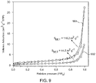
- FIG. 9 illustrates nitrogen adsorption-desorption isotherms of the NH 2 @SiO 2 NPs and the Pyr-NH@SiO 2 NPs, according to certain embodiments
- FIG. 10A illustrates a surface charge and zeta potential measurements of the NH 2 @SiO 2 NPs, according to certain embodiments
- FIG. 10B illustrates a surface charge and zeta potential measurements of the Pyr-NH@SiO 2 NPs, according to certain embodiments
- PL photoluminescence
- FIG. 12B illustrates a selectivity of the Pyr-NH@SiO 2 NPs against Hg 2+ ions upon the addition of major cations of seawater, according to certain embodiments
- the terms “approximately,” “approximate,” “about,” and similar terms generally refer to ranges that include the identified value within a margin of 20%, 10%, or preferably 5%, and any values therebetween.
- aqueous solution refers to a solution in which the solvent is mainly water or only water.
- chemosensor refers to a molecular structure (organic or inorganic complexes) used to sense an analyte to produce a detectable change or signal.
- fluorescence refers to a process where a material absorbs light at high energy, short wavelength, and emits light at lower energy, usually visible, wavelength.
- emission refers to a process of elements releasing different photons of color as their atoms return to their lower energy levels.
- nanoparticles refers to a small particle that ranges between 1 to 1,000 nanometers in size.
- amorphous refers to a shapeless or without definite character or nature.
- amide bond refers to RC( ⁇ O)NR′R′′, wherein R, R′, and R′′ represent organic groups or hydrogen atoms.
- Embodiments of the present disclosure are directed a method of detecting mercury (Hg 2+ ) ions in an aqueous solution using pyrene functionalized silica nanoparticles (Pyr-NH@SiO 2 NPs).
- the synthesized NH 2 @SiO 2 NPs and Pyr-NH@SiO 2 NPs were thoroughly investigated by proton nuclear magnetic resonance ( 1 H-NMR), Fourier-transform infrared (FTIR), X-ray powder diffraction (XRD), field emission scanning electron microscopy (FESEM), energy-dispersive X-ray spectroscopy (EDS), thermogravimetric analysis (TGA), Brunauer-Emmett-Teller (BET) surface area, and dynamic light scattering (DLS) techniques.
- 1 H-NMR proton nuclear magnetic resonance
- FTIR Fourier-transform infrared
- XRD X-ray powder diffraction
- FESEM field emission scanning electron microscopy
- the Pyr-NH@SiO 2 NPs were used as a chemosensor for Hg 2+ ions and the presence of Hg 2+ ions is measured with photoluminescence (PL) spectroscopy.
- the results indicate that the chemosensor can selectively detect Hg 2+ ions in the presence of ubiquitous ions (sodium (Na + ), potassium (K + ), calcium (Ca 2+ ), magnesium (Mg 2+ ), barium (Ba 2+ ), silver (Ag + ) and in seawater samples.
- the chemosensor includes Pyr-NH@SiO 2 NPs.
- at least one pyrene is bonded to a surface of a silica nanoparticle through an amide bond.
- the amide bond is at any position (1-10) on the pyrene of formula I.
- the amide bond is at the 2 or 7 position.
- the pyrene may be functionalized on at least one position (1-10) other than the amide bond with a group such as but not limited to an alkyl, a halide, an amine, a carbonyl, an ester, a nitrile, an alcohol, and a carboxylic acid.
- the pyrene is functionalized with another pyrene at a position other than the amide bond.
- the Pyr-NH@SiO 2 NPs have a formula of pyrene-C( ⁇ O)—NHR-silica nanoparticle, and R is an alkyl chain.
- R is an alkyl chain comprising 1-20 carbons, preferably 2-19, 3-18, 4-17, 5-16, 6-15, 7-14, 8-13, 9-12, 10-11 carbons.
- the alkyl chain can be saturated or unsaturated.
- R is a 3 carbon chain, —CH 2 CH 2 CH 2 —.
- the FTIR spectrum of the pyrene silica nanoparticles exhibits an aromatic ring stretch at 3000-3100 cm ⁇ 1 , preferably 3010-3080 cm ⁇ 1 , 3030-3050 cm ⁇ 1 , and carbonyl group (C ⁇ O) stretching bands at 1700-1750 cm ⁇ 1 , preferably 1710-1740 cm ⁇ 1 , 1720-1730 cm ⁇ 1 , indicating the presence of pyrene bonded to the silica nanoparticle surface.
- at least 20%, preferably 30%, 40%, 50%, 60%, 70%, 80%, 90% or 100% of the surface of the silica nanoparticles is bonded to a pyrene.
- the pyrene silica nanoparticles have a uniform size distribution. In some embodiments, the pyrene silica nanoparticles have a size distribution greater than 10 nm, preferably 15 nm, 20 nm, 30 nm or 50 nm. In some embodiments, the pyrene silica nanoparticles have an average size of 30-50 nm, preferably 35-45 nm, or approximately 40 nm. In some embodiments, the pyrene silica nanoparticles have a substantially spherical shape. In some embodiments, the pyrene silica nanoparticles have an irregular shape.
- the pyrene silica nanoparticles have an amorphous structure.
- the pyrene silica nanoparticles exhibit an XRD peak at 15-30°, preferably 18-27°, or 21-24° ( FIG. 5 ). In some embodiments, no other XRD peaks were detected indicating high purity. In some embodiment, there is less than 1 wt. % impurity in the pyrene silica nanoparticles such as but not limited to trace metals, and unreacted pyrene.
- the pyrene silica nanoparticles are agglomerated to form a mesoporous structure.
- the elements silica (Si), oxygen (O), carbon (C), and nitrogen (N) are homogeneously distributed throughout the mesoporous structure.
- the agglomerates are greater than 200 nm in size, preferably 200-2,000 nm, 500-1,500 nm, or approximately 1,000 nm.
- the pyrene silica nanoparticles are agglomerated through ⁇ - ⁇ interactions of the pyrenes on the surface.
- the pyrene silica nanoparticles are agglomerated through van der waals interactions, C—H . . . ⁇ interactions, and dipole-dipole interactions.
- the pyrene silica nanoparticles have a positively charged surface, and a zeta potential of 35-45 mV, preferably 38-42 mV, or approximately 40 mV. The high zeta potential value indicates that the pyrene silica nanoparticles are stable in water due to the formation of stable hydrogen bonding with water molecules in the presence of N—H and C ⁇ O groups.
- the pyrene silica nanoparticles have a BET surface area of 30-60 m 2 /g, preferably 35-55 m 2 /g, 40-50 m 2 /g, or approximately 45 m 2 /g. In some embodiments, the pyrene silica nanoparticles have a total pore volume of 0.25-0.4 cm 3 /g, preferably 0.28-0.38 cm 3 /g, 0.3-0.35 cm 3 /g, or 0.32-0.34 cm 3 /g. In some embodiments, the pyrene silica nanoparticles have an average pore size of 20-30 nm, preferably 22-28 nm, or 24-26 nm.
- the BET surface area, total pore volume, and average pore size are less than a silica nanoparticle without a pyrene, because the pyrenes block the pore surfaces and walls.
- the pyrene silica nanoparticles are stable up to 200° C., preferably 150-200° C., 160-190° C., or 170-180° C.
- FIG. 1 a schematic flow diagram of the method 100 of detecting Hg 2+ ions in an aqueous solution is illustrated.
- the order in which the method 100 is described is not intended to be construed as a limitation, and any number of the described method steps may be combined in any order to implement the method 100 . Additionally, individual steps may be removed or skipped from the method 100 without departing from the spirit and scope of the present disclosure.
- the method 100 includes contacting the aqueous solution with the chemosensor to form a mixture.
- the aqueous solution is any water based solution including but not limited to seawater, brackish water, and tap water.
- the chemosensor is contacted with the aqueous solution at a temperature range of 15-45° C., preferably 20-40° C., 25-35° C., or approximately 30° C.
- the contacting occurs by pouring a solution of the chemosensor into the aqueous solution.
- the contacting occurs by adding a powder form of the chemosensor into the aqueous solution.
- the chemosensor is mixed into the aqueous solution by a method such as but not limited to, manually stirring, using a stir bar, or a probe sonicator.
- the chemosensor is 20 ppm in the aqueous solution, preferably 10 ppm, 5 ppm or 1 ppm.
- the method 100 includes monitoring a change in a fluorescence emission profile of the chemosensor in the mixture to determine the presence or absence of Hg 2+ ions in the aqueous solution.
- the fluorescence is measured with a spectrofluorometer.
- the chemosensor is excited with light with a wavelength of 300-380 nm, preferably 310-370 nm, 320-360 nm, 330-350 nm, or approximately 340 nm and a bandwidth of 1-20 nm, preferably 5-15 nm, or approximately 10 nm.
- the change in the fluorescence emission profile of the chemosensor is monitored between 350-550 nm, preferably 375-525 nm, 400-500 nm, 425-475 nm or approximately 450 nm. In some embodiments, the change in the fluorescence emission profile is measured by the disappearance of a fluorescence band from 360 to 425 nm, preferably 370-410 nm, 380-400 nm, or approximately 390 nm. In another embodiment, the change in the fluorescence emission profile is measured by the appearance of a fluorescence band from 400 to 525 nm, preferably 420-500 nm, 440-480 nm, or approximately 460 nm.
- the decrease in the fluorescence band between 360 to 425 nm and the increase in the fluorescence band between 400 to 525 nm indicates the presence of Hg 2+ in the aqueous solution.
- the change in fluorescence intensities can be attributed to photoinduced electron transfer to the pyrene molecules and the formation of a stable Hg-pyrene complex with the emitting chromophore.
- the change in fluorescence is detected by eye following exposure of the aqueous solution with the chemosensor to an excitation light source.
- the change in the fluorescence emission profile linearly correlates with the concentration of Hg 2+ in the aqueous solution. In other words, the greater the concentration of the Hg 2+ ions in the aqueous system, the stronger the change in signal. In some embodiments, the change in the fluorescence emission profile is quantified to determine a concentration of Hg 2+ ions in the aqueous solution.
- the chemosensor is selective for detecting Hg 2+ ions.
- the aqueous solution further comprises at least one metal cation selected from the group consisting of Na + , K + , Ca 2+ , Mg 2+ , Ba 2+ , and Ag + .
- the metal ions in the aqueous solution may be Li + , Na + , K + , Be 2+ , Mn 2+ , Fe 2+ , Co 2+ , Ni 2+ , Cu 2+ , Zn 2+ , Cd 2+ , Se 3+ , Ti 3+ , V 3+ , Cr 3+ , Fe 3+ , Rh 3+ , Ga 3+ , In 3+ , Ce 4+ , Th 4+ , Pa 4+ , U 4+ , Np 4+ , Pu 4+ , Mg 2+ , Ca 2+ , Sr 2+ , Ba 2+ , Al 3+ , Y 3+ , La 3+ , Ag + , Tl + , Pb 2+ , Ti 3+ , Bi 3+ , Sn 2+ , Sn 2+ , or Pd 2+ .
- the change in the fluorescence emission profile occurs only in the presence of Hg 2+ .
- CTAB cetyltrimethylammonium bromide
- AG sodium hydroxide
- TEOS tetraethyl orthosilicate
- APTS 3-aminopropyl triethyl silane
- 1-pyrene carboxylic acid 97%, Sigma-Aldrich
- hydroxy benzotriazole ⁇ 97%, Sigma-Aldrich
- 1-ethyl-3-(3-dimethyl aminopropyl)carbodiimide hydrochloride ⁇ 99%, Sigma-Aldrich
- triethylamine ⁇ 99%, Sigma-Aldrich
- ethyl acetate High-performance liquid chromatography (HPLC), Honeywell
- NPs silica nanoparticles
- reaction contents were stirred for another 40 min at room temperature, followed by the addition of 2.1 mL of 3-aminopropyl) triethoxysilane (APTS), and kept the reaction contents to stir overnight.
- APTS 3-aminopropyl) triethoxysilane
- the mixture was centrifuged at 10,000 revolutions per minute (rpm) to separate the amino-functionalized silica nanoparticles (product).
- the product was washed thrice with de-ionized water and twice with absolute ethanol to remove the surfactant and impurities.
- the synthesized silica NPs were further transferred into a petri dish, and the solvent (ethanol) was allowed to evaporate in a vacuum oven at 60 degrees centigrade (° C.) to yield a fine white powder of silica (NH 2 @SiO 2 NPs) with an experimental yield of ⁇ 80%.
- FTIR Fourier transform infrared
- the amino-functionalized silica NPs (0.200 g) and 1-pyrene-carboxylic acid (0.300 g) were taken into a dried round bottom flask (50 mL), followed by the addition of hydroxy benzotriazole (0.210 g) and 1-ethyl-3-(3-dimethyl aminopropyl) carbodiimide hydrochloride (0.232 g). Subsequently, anhydrous chloroform (20 mL) was added to the flask and stirred into the reaction mixture. Further, triethylamine (0.356 ml) was added to the reaction mixture, and the stirring was continued at room temperature for 24 hours.
- FTIR (neat): ⁇ (cm ⁇ 1) 3415, 3035, 2926, 2853, 1740, 1642, 1569, 1448, 1383, 1261, 1092, 844, 741, 451.
- a Brunauer-Emmett-Teller (BET) surface area of materials was estimated by N 2 adsorption-desorption using a Micromeritics (ASAP 2010) analyzer.
- the surface charge and zeta potential values of synthesized NH 2 @SiO 2 NPs and Pyr-NH@SiO 2 NPs were evaluated using Zetasizer nano (ZEN3600, Malvern, UK).
- the silica samples were dispersed in de-ionized water using a probe sonicator (UPT-400, Hielscher) to achieve maximum dispersion of particles before imaging and zeta potential measurements.
- UPT-400 probe sonicator
- the sensing material was well-dispersed in de-ionized water using the probe sonicator.
- a photoluminescence (PL) spectrum of the Pyr-NH@SiO 2 NPs was recorded using a spectrofluorometer (FP-8500, JASCO) at an excitation wavelength of 340 nm by adjusting the bandwidth to 5 nm. All the measurements were performed at ambient conditions.
- the sensing properties of Pyr-NH@SiO 2 NPs (20 parts per million (ppm)) were recorded by the successive increase in Hg 2+ ions concentration within the range from 0-50 ppm.
- FIG. 2 illustrates Stober's method for preparing mono-dispersed spherical silica nanoparticles.
- the method includes hydrolysis of TEOS followed by a condensation reaction using ethyl acetate in the presence of sodium hydroxide and CTAB surfactant.
- the surface of silica NPs was amino-functionalized to achieve NH 2 @SiO 2 NPs by utilizing APTS under the same reaction conditions ( 202 ).
- FIGS. 3A and 3B The chemical structures of NH 2 @SiO 2 NPs and Pyr-NH@SiO 2 NPs were investigated by 1 H-NMR, as illustrated in FIGS. 3A and 3B , respectively.
- FIG. 3A The amino-functionalized silica nanoparticles (NH 2 @SiO 2 NPs) have three different methylene (—CH 2 —) protons.
- the chemical structure of the final product (Pyr-NH@SiO 2 NPs) was ascertained from 1 H-NMR ( FIG. 3B ), where the methylene protons of the amino-propyl component showed significant downfield shifts.
- FIGS. 4A-4B illustrate the functional groups, stretching, and bending vibrations of NH 2 @SiO 2 NPs and Pyr-NH@SiO 2 NPs as investigated by FTIR spectroscopy.
- FTIR spectrum of NH 2 @SiO 2 NPs showed a broad band at 3444 cm ⁇ 1 for N—H stretch that overlapped with hydroxyl (—OH) of silica core or water adsorbed on the surface of the material.
- the broad N—H bending bands could be seen clearly at 1643 cm ⁇ 1 .
- 4B illustrates the FTIR spectrum of Pyr-NH@SiO 2 NPs (final product) showed additional absorption bands for aromatic ring stretch at 3035 cm ⁇ 1 and carbonyl group (C ⁇ O) stretching bands at 1740 cm ⁇ 1 along with the characteristic peaks emerged for NH 2 @SiO 2 NPs.
- the 1 H-NMR and FT-IR results confirmed the formation of pyrene attached silica nanoparticles.
- FIG. 5 illustrates a broad diffraction peak observed at the 20 position of ⁇ 23° (JCDD No., 00-001-0649), confirming the amorphous nature of silica nanoparticles.
- the crystal structure, phase, and purity of as-synthesized NH 2 @SiO 2 NPs (502) and Pyr-NH@SiO 2 NPs (504) were investigated by X-ray diffraction (XRD) analysis. However, no extra peaks were detected, indicating the high purity of synthesized silica NPs.
- the products were thoroughly washed to remove the surfactant, unwanted coupling reagents, and unreacted pyrene-carboxylic acid.
- FIGS. 6A-6D represents low and high-resolution FESEM images of NH 2 @SiO 2 NPs ( FIGS. 6A-6B ) and Pyr-NH@SiO 2 NPs ( FIGS. 6C-6D ), respectively.
- the spherical-shaped silica particles are well-dispersed, homogeneous in size, and uniformly distributed over the surface.
- the average size of NH 2 @SiO 2 NPs and Pyr-NH@SiO 2 NPs was ⁇ 35 and ⁇ 40 nm, respectively.
- FIGS. 7A-7D illustrates the elemental mapping of Pyr-NH@SiO 2 NPs, signifying the presence of silica (Si), oxygen (O), carbon (C), and nitrogen (N) atoms, respectively in the investigated sample.
- the elemental maps exhibit a homogenous distribution of all components.
- the thermal stability of the silica materials was performed, and the results of this study are shown in FIG. 8 .
- the thermal stability of pyrene-functionalized silica (Pyr-NH@SiO 2 NPs) 802 and amino-functionalized silica (NH 2 @SiO 2 NPs) 804 was investigated from room temperature to 800° C.
- the first weight loss ( ⁇ 10%) was observed up to 160° C. due to moisture and water molecules that were physically adsorbed on the surface of silica NPs.
- the weight loss ⁇ 21.8% detected in the region from 160° C. to 800° C.
- FIG. 9 illustrates the curves representing type IV isotherms at high relative pressure suggesting the formation of mesoporous silica materials having uniform size distributions.
- the surface area and pore structure of NH 2 @SiO 2 NPs 902 and Pyr-NH@SiO 2 NPs 904 were assessed by nitrogen adsorption-desorption isotherms obtained parameters such as BET surface area (SBET), total pore volume (V), and average pore diameter (DBJH) were summarized in Table 1.
- SBET BET surface area
- V total pore volume
- DBJH average pore diameter
- FIGS. 10A-10B illustrates that NH 2 @SiO 2 NPs and Pyr-NH@SiO 2 NPs were positively charged with zeta potential values of 1.69 mV and 38.0 mV, respectively.
- the comparison indicates that the Pyr-NH@SiO 2 NPs were more stable in water after modification with 1-pyrene-carboxylic acid due to the formation of stable hydrogen bonding with water molecules in the presence of N—H and C ⁇ O groups. This suggested that Pyr-NH@SiO 2 NPs can be successfully deployed as chemosensors in aqueous environments.
- FIG. 11 represents photoluminescence (PL) emission spectra of Pyr-NH@SiO 2 NPs before and after exposure to Hg 2+ ions. The changes in fluorescent properties were examined based on peak shift and emission intensity.
- the PL spectrum of Pyr-NH@SiO 2 NPs (20 ppm) exhibited two distinct vibronic bands observed at 380 and 398 nm corresponding to ⁇ * transitions in pyrene molecule, which were cumulatively denoted as the monomeric emission.
- Pyr-NH@SiO 2 NPs were further exposed to the known concentrations of Hg 2+ ions, namely, 0 ppb (1202), 10 ppb (1204), 100 ppb (1206), 250 ppb (1208), 500 ppb (1210), 1.0 ppm (1212), 2.5 ppm (1214), 5.0 ppm (1216), 10 ppm (1218), 10 ppm (1220), 20 ppm (1222), 30 ppm (1224), and 50 ppm (1226).
- the fluorescence emission intensity of pyrene gradually decreased when Hg 2+ concentration increased from 0-50 ppm.
- FIG. 12A demonstrates that the fluorescence intensity of Pyr-NH@SiO 2 NPs (20 ppm) quenches ⁇ 60% with the addition of spiked Hg 2+ ions (20 ppm) (1302).
- the results can be compared to the seawater sample with NH@SiO 2 NPs (20 ppm) without Hg 2+ ions (1304). This indicated the effective recognition of Hg 2+ ions in the presence of competitive metal cations in the seawater sample.
- FIG. 12B demonstrates a slight change in the fluorescence intensity of Pyr-NH@SiO 2 NPs upon the addition of each competitive cation.
- the graph represents changes in the fluorescent intensity upon introduction of competitive cation as shown: Na + (1352), K + (1354), Ca 2+ (1356), Mg 2+ (1358), Ba 2+ (1360), Ag + (1362), and seawater, SW, (1364).
- a drastic quenching ⁇ 60%) of fluorescence intensity upon Hg 2+ (1366) addition, demonstrating the developed sensor's reliability and selectivity for seawater samples.
- Pyrene attached silica nanoparticles (Pyr-NH@SiO 2 NPs) were successfully synthesized by the chemical attachment of pyrene with amino-functionalized silica NPs using peptide coupling agents.
- the chemical structure of amino-functionalized pyrene and its covalent attachment with silica NPs was confirmed by 1 H-NMR, FT-IR, TGA, and BET results.
- the XRD results confirmed the amorphous nature of the synthesized silica NPs. Their average particle size was found to be ⁇ 40 nm.
- DLS outcomes indicate that Pyr-NH@SiO 2 NPs (38.0 mV) were stable in the aqueous environment after modification with 1-pyrene-carboxylic acid due to the formation of stable hydrogen bonding with water molecules in the presence of N—H and C ⁇ O groups.
- the synthesized fluorescent particles can produce bright green emission under UV light.
- the fluorescence quenching, hypochromic peak shifting (380, 398 nm), and excimer emission ( ⁇ 440 nm) upon adding Hg 2+ ions are attributed to the photoinduced electron transfer to the pyrene molecules and the formation of a stable Hg-pyrene complex with the emitting chromophore.
- the developed sensor can reliably and selectively recognize Hg 2+ ions (LOD: 10 ppb) in the presence of ubiquitous metal cations and seawater samples.
- LOD 10 ppb
- the fluorescent Pyr-NH@SiO 2 NPs have great potential to design highly sensitive, selective, and portable opto-chemical mercury sensors for aqueous applications.
Landscapes
- Health & Medical Sciences (AREA)
- Life Sciences & Earth Sciences (AREA)
- Chemical & Material Sciences (AREA)
- Physics & Mathematics (AREA)
- Immunology (AREA)
- Pathology (AREA)
- General Physics & Mathematics (AREA)
- Analytical Chemistry (AREA)
- Biochemistry (AREA)
- General Health & Medical Sciences (AREA)
- Engineering & Computer Science (AREA)
- Food Science & Technology (AREA)
- Medicinal Chemistry (AREA)
- Chemical Kinetics & Catalysis (AREA)
- Optics & Photonics (AREA)
- Nuclear Medicine, Radiotherapy & Molecular Imaging (AREA)
- Biophysics (AREA)
- Molecular Biology (AREA)
- Investigating, Analyzing Materials By Fluorescence Or Luminescence (AREA)
Abstract
A method for detecting mercury (Hg2+) ions in an aqueous solution is described. The method includes contacting the aqueous solution with a chemosensor to form a mixture; and monitoring a change in a fluorescence emission profile of the chemosensor in the mixture to determine the presence or absence of Hg2+ ions in the aqueous solution. The chemosensor includes pyrene silica nanoparticles where at least one pyrene is bonded to a surface of a silica nanoparticle through an amide bond with a formula of, pyrene-C(═O)NHR-silica nanoparticle, and where R is an alkyl chain.
Description
The present disclosure is directed to nanoparticles, particularly to pyrene functionalized silica nanoparticles for detection of mercury ions in an aqueous solution.
The “background” description provided herein is for the purpose of generally presenting the context of the disclosure. Work of the presently named inventors, to the extent it is described in this background section, as well as aspects of the description that may not otherwise qualify as prior art at the time of filing, are neither expressly nor impliedly admitted as prior art against the present invention.
Mercury (Hg0, Hg+, Hg2+) contamination in the environment has increased 3 to 6 fold in recent decades compared to pre-industrial estimates. This poses serious threats to human health, as mercury poisoning causes brain and neurological damage, birth deformities, kidney damage, digestive system problems, memory loss, and language impairments.
Conventionally, various analytical techniques are engaged to monitor Hg2+ concentration, such as atomic absorption spectroscopy (AAS), inductively coupled plasma-mass spectrometry (ICP-MS), and plasma-atomic emission spectrometry (AES), gas chromatography (GC), and reversed-phase high-performance liquid chromatography (HPLC). However, these techniques require expensive, specialized, and cumbersome sample preparations and bulky laboratory equipment that make it challenging to adapt for remote sensing applications. Optical methods based on fluorescence sensing have gained much attention, because fluorescence-based chemical sensors offer rapid analysis, better sensitivity, low limit of detection, and high selectivity for investigation of environmental pollutants even at low concentrations. Therefore, there exists a need to develop a cost-effective, sensitive, selective, and convenient portable sensor that can detect Hg2+ ions.
In an exemplary embodiment, a method of detecting mercury (Hg2+) ions in an aqueous solution is described. The method includes contacting the aqueous solution with a chemosensor to form a mixture, and further monitoring a change in a fluorescence emission profile of the chemosensor in the mixture to determine a presence or absence of Hg2+ ions in the aqueous solution. The chemosensor includes pyrene silica nanoparticles where at least one pyrene is bonded to a surface of a silica nanoparticle through an amide bond with a formula of, pyrene-C(═O)NHR-silica nanoparticle and where R is an alkyl chain.
In another embodiment, R is —CH2CH2CH2—.
In another embodiment, the pyrene silica nanoparticles have a uniform size distribution; and an average size of 30-50 nanometers (nm).
In another embodiment, the pyrene silica nanoparticles have a substantially spherical shape.
In another embodiment, the pyrene silica nanoparticles have an amorphous structure.
In another embodiment, the pyrene silica nanoparticles have a positively charged surface; and a zeta potential of 35-45 millivolts (mV).
In another embodiment, the pyrene silica nanoparticles are agglomerated to form a mesoporous structure. In another embodiment, the elements silica (Si), oxygen (O), carbon (C), and nitrogen (N) are homogeneously distributed throughout the mesoporous structure.
In another embodiment, the pyrene silica nanoparticles have a Brunauer-Emmett-Teller (BET) surface area of 30-60 square meters per gram (m2/g).
In another embodiment, the pyrene silica nanoparticles have a total pore volume of 0.25-0.4 grams per cubic centimeter (cm3/g).
In another embodiment, the pyrene silica nanoparticles have an average pore size of 20-30 nm.
In another embodiment, the pyrene silica nanoparticles are stable up to 200 degrees centigrade (° C.).
In another embodiment, the method includes monitoring the change in the fluorescence emission profile of the chemosensor between 350-550 nm.
In another embodiment, the change in the fluorescence emission profile is measured by the disappearance of a fluorescence band from 360 to 425 nm.
In another embodiment, the change in the fluorescence emission profile is measured by the appearance of a fluorescence band from 400 to 525 nm.
In another embodiment, the change in the fluorescence emission profile linearly correlates with the concentration of Hg2+ in the aqueous solution.
In another embodiment, the method further includes quantifying the change in the fluorescence emission profile to determine a concentration of Hg2+ ions in the aqueous solution.
In another embodiment, the chemosensor is selective for detecting Hg2+ ions.
In another embodiment, the aqueous solution further comprises at least one metal cation selected from the group consisting of sodium (Na+), potassium (K+), calcium (Ca2+), magnesium (Mg2+), barium (Ba2+), and silver (Ag+) ions, and the change in the fluorescence emission profile occurs only in the presence of Hg2+.
In another embodiment, the limit of detection (LOD) for Hg2+ ions is 10 parts per billion (ppb).
The foregoing general description of the illustrative present disclosure and the following detailed description thereof are merely exemplary aspects of the teachings of this disclosure and are not restrictive.
A more complete appreciation of this disclosure and many of the attendant advantages thereof will be readily obtained as the same becomes better understood by reference to the following detailed description when considered in connection with the accompanying drawings, wherein:
In the drawings, reference numerals designate identical or corresponding parts throughout the several views. Further, as used herein, the words “a,” “an” and the like generally carry a meaning of “one or more,” unless stated otherwise.
Furthermore, the terms “approximately,” “approximate,” “about,” and similar terms generally refer to ranges that include the identified value within a margin of 20%, 10%, or preferably 5%, and any values therebetween.
As used herein, the term, “aqueous solution” refers to a solution in which the solvent is mainly water or only water.
As used herein, the term “chemosensor” refers to a molecular structure (organic or inorganic complexes) used to sense an analyte to produce a detectable change or signal.
As used herein, the term “fluorescence” refers to a process where a material absorbs light at high energy, short wavelength, and emits light at lower energy, usually visible, wavelength.
As used herein, the term “emission” refers to a process of elements releasing different photons of color as their atoms return to their lower energy levels.
As used herein, the term “nanoparticles” refers to a small particle that ranges between 1 to 1,000 nanometers in size.
As used herein, the term “amorphous” refers to a shapeless or without definite character or nature.
As used herein, the term “amide bond” refers to RC(═O)NR′R″, wherein R, R′, and R″ represent organic groups or hydrogen atoms.
Embodiments of the present disclosure are directed a method of detecting mercury (Hg2+) ions in an aqueous solution using pyrene functionalized silica nanoparticles (Pyr-NH@SiO2 NPs). The synthesized NH2@SiO2NPs and Pyr-NH@SiO2 NPs were thoroughly investigated by proton nuclear magnetic resonance (1H-NMR), Fourier-transform infrared (FTIR), X-ray powder diffraction (XRD), field emission scanning electron microscopy (FESEM), energy-dispersive X-ray spectroscopy (EDS), thermogravimetric analysis (TGA), Brunauer-Emmett-Teller (BET) surface area, and dynamic light scattering (DLS) techniques. The Pyr-NH@SiO2 NPs were used as a chemosensor for Hg2+ ions and the presence of Hg2+ ions is measured with photoluminescence (PL) spectroscopy. The results indicate that the chemosensor can selectively detect Hg2+ ions in the presence of ubiquitous ions (sodium (Na+), potassium (K+), calcium (Ca2+), magnesium (Mg2+), barium (Ba2+), silver (Ag+) and in seawater samples. The change in fluorescence properties with Hg2+ ions with a limit of detection (LOD) of 10 parts per billion (ppb) indicates that the Pyr-NH@SiO2 NPs may be effectively utilized as a promising chemosensor for mercury ion detection in aqueous environments.
In an embodiment, the chemosensor includes Pyr-NH@SiO2 NPs. In another embodiment, at least one pyrene is bonded to a surface of a silica nanoparticle through an amide bond. In an embodiment, the amide bond is at any position (1-10) on the pyrene of formula I. In an embodiment, the amide bond is at the 2 or 7 position. In an embodiment, the pyrene may be functionalized on at least one position (1-10) other than the amide bond with a group such as but not limited to an alkyl, a halide, an amine, a carbonyl, an ester, a nitrile, an alcohol, and a carboxylic acid. In an embodiment, the pyrene is functionalized with another pyrene at a position other than the amide bond.

In an embodiment, the Pyr-NH@SiO2 NPs have a formula of pyrene-C(═O)—NHR-silica nanoparticle, and R is an alkyl chain. In an embodiment, R is an alkyl chain comprising 1-20 carbons, preferably 2-19, 3-18, 4-17, 5-16, 6-15, 7-14, 8-13, 9-12, 10-11 carbons. In an embodiment, the alkyl chain can be saturated or unsaturated. In another embodiment, R is a 3 carbon chain, —CH2CH2CH2—.
In an embodiment, the FTIR spectrum of the pyrene silica nanoparticles (FIG. 4B ) exhibits an aromatic ring stretch at 3000-3100 cm−1, preferably 3010-3080 cm−1, 3030-3050 cm−1, and carbonyl group (C═O) stretching bands at 1700-1750 cm−1, preferably 1710-1740 cm−1, 1720-1730 cm−1, indicating the presence of pyrene bonded to the silica nanoparticle surface. In an embodiment, at least 20%, preferably 30%, 40%, 50%, 60%, 70%, 80%, 90% or 100% of the surface of the silica nanoparticles is bonded to a pyrene. In some embodiments, the pyrene silica nanoparticles have a uniform size distribution. In some embodiments, the pyrene silica nanoparticles have a size distribution greater than 10 nm, preferably 15 nm, 20 nm, 30 nm or 50 nm. In some embodiments, the pyrene silica nanoparticles have an average size of 30-50 nm, preferably 35-45 nm, or approximately 40 nm. In some embodiments, the pyrene silica nanoparticles have a substantially spherical shape. In some embodiments, the pyrene silica nanoparticles have an irregular shape. In another embodiment, the pyrene silica nanoparticles have an amorphous structure. In some embodiments, the pyrene silica nanoparticles exhibit an XRD peak at 15-30°, preferably 18-27°, or 21-24° (FIG. 5 ). In some embodiments, no other XRD peaks were detected indicating high purity. In some embodiment, there is less than 1 wt. % impurity in the pyrene silica nanoparticles such as but not limited to trace metals, and unreacted pyrene.
In another embodiment, the pyrene silica nanoparticles are agglomerated to form a mesoporous structure. In some embodiments, the elements silica (Si), oxygen (O), carbon (C), and nitrogen (N) are homogeneously distributed throughout the mesoporous structure. In some embodiments, the agglomerates are greater than 200 nm in size, preferably 200-2,000 nm, 500-1,500 nm, or approximately 1,000 nm. In another embodiment, the pyrene silica nanoparticles are agglomerated through π-π interactions of the pyrenes on the surface. In another embodiment, the pyrene silica nanoparticles are agglomerated through van der waals interactions, C—H . . . π interactions, and dipole-dipole interactions. In some embodiments, the pyrene silica nanoparticles have a positively charged surface, and a zeta potential of 35-45 mV, preferably 38-42 mV, or approximately 40 mV. The high zeta potential value indicates that the pyrene silica nanoparticles are stable in water due to the formation of stable hydrogen bonding with water molecules in the presence of N—H and C═O groups.
In some embodiments, the pyrene silica nanoparticles have a BET surface area of 30-60 m2/g, preferably 35-55 m2/g, 40-50 m2/g, or approximately 45 m2/g. In some embodiments, the pyrene silica nanoparticles have a total pore volume of 0.25-0.4 cm3/g, preferably 0.28-0.38 cm3/g, 0.3-0.35 cm3/g, or 0.32-0.34 cm3/g. In some embodiments, the pyrene silica nanoparticles have an average pore size of 20-30 nm, preferably 22-28 nm, or 24-26 nm. In some embodiments, the BET surface area, total pore volume, and average pore size are less than a silica nanoparticle without a pyrene, because the pyrenes block the pore surfaces and walls. In some embodiments, the pyrene silica nanoparticles are stable up to 200° C., preferably 150-200° C., 160-190° C., or 170-180° C.
Referring to FIG. 1 , a schematic flow diagram of the method 100 of detecting Hg2+ ions in an aqueous solution is illustrated. The order in which the method 100 is described is not intended to be construed as a limitation, and any number of the described method steps may be combined in any order to implement the method 100. Additionally, individual steps may be removed or skipped from the method 100 without departing from the spirit and scope of the present disclosure.
At step 102, the method 100 includes contacting the aqueous solution with the chemosensor to form a mixture. In an embodiment, the aqueous solution is any water based solution including but not limited to seawater, brackish water, and tap water. In an embodiment, the chemosensor is contacted with the aqueous solution at a temperature range of 15-45° C., preferably 20-40° C., 25-35° C., or approximately 30° C. In an embodiment, the contacting occurs by pouring a solution of the chemosensor into the aqueous solution. In an embodiment, the contacting occurs by adding a powder form of the chemosensor into the aqueous solution. In an embodiment, the chemosensor is mixed into the aqueous solution by a method such as but not limited to, manually stirring, using a stir bar, or a probe sonicator. In an embodiment, the chemosensor is 20 ppm in the aqueous solution, preferably 10 ppm, 5 ppm or 1 ppm.
At step 104, the method 100 includes monitoring a change in a fluorescence emission profile of the chemosensor in the mixture to determine the presence or absence of Hg2+ ions in the aqueous solution. In an embodiment, the fluorescence is measured with a spectrofluorometer. In an embodiment, the chemosensor is excited with light with a wavelength of 300-380 nm, preferably 310-370 nm, 320-360 nm, 330-350 nm, or approximately 340 nm and a bandwidth of 1-20 nm, preferably 5-15 nm, or approximately 10 nm. In some embodiments, the change in the fluorescence emission profile of the chemosensor is monitored between 350-550 nm, preferably 375-525 nm, 400-500 nm, 425-475 nm or approximately 450 nm. In some embodiments, the change in the fluorescence emission profile is measured by the disappearance of a fluorescence band from 360 to 425 nm, preferably 370-410 nm, 380-400 nm, or approximately 390 nm. In another embodiment, the change in the fluorescence emission profile is measured by the appearance of a fluorescence band from 400 to 525 nm, preferably 420-500 nm, 440-480 nm, or approximately 460 nm. In an embodiment, the decrease in the fluorescence band between 360 to 425 nm and the increase in the fluorescence band between 400 to 525 nm indicates the presence of Hg2+ in the aqueous solution. In an embodiment, the change in fluorescence intensities can be attributed to photoinduced electron transfer to the pyrene molecules and the formation of a stable Hg-pyrene complex with the emitting chromophore. In an embodiment, the change in fluorescence is detected by eye following exposure of the aqueous solution with the chemosensor to an excitation light source.
In some embodiments, the change in the fluorescence emission profile linearly correlates with the concentration of Hg2+ in the aqueous solution. In other words, the greater the concentration of the Hg2+ ions in the aqueous system, the stronger the change in signal. In some embodiments, the change in the fluorescence emission profile is quantified to determine a concentration of Hg2+ ions in the aqueous solution.
In another embodiment, the chemosensor is selective for detecting Hg2+ ions. In another embodiment, the aqueous solution further comprises at least one metal cation selected from the group consisting of Na+, K+, Ca2+, Mg2+, Ba2+, and Ag+. In an embodiment, the metal ions in the aqueous solution may be Li+, Na+, K+, Be2+, Mn2+, Fe2+, Co2+, Ni2+, Cu2+, Zn2+, Cd2+, Se3+, Ti3+, V3+, Cr3+, Fe3+, Rh3+, Ga3+, In3+, Ce4+, Th4+, Pa4+, U4+, Np4+, Pu4+, Mg2+, Ca2+, Sr2+, Ba2+, Al3+, Y3+, La3+, Ag+, Tl+, Pb2+, Ti3+, Bi3+, Sn2+, Sn2+, or Pd2+. In an embodiment, the change in the fluorescence emission profile occurs only in the presence of Hg2+. In some embodiments, the limit of detection for Hg2+ ions is 10 ppb, preferably 5 ppb or 1 ppb.
The following examples describe and demonstrate exemplary embodiments of a method of detecting mercury (Hg2+) ions in an aqueous solution described herein. The examples are provided solely for the purpose of illustration and are not to be construed as limitations of the present disclosure, as many variations thereof are possible without departing from the spirit and scope of the present disclosure.
All the chemicals and reagents, including cetyltrimethylammonium bromide (CTAB, ≥98%, Sigma-Aldrich), sodium hydroxide (AG, Fluka), tetraethyl orthosilicate (TEOS, ≥99%, Sigma-Aldrich), 3-aminopropyl triethyl silane (APTS, 99%, Sigma-Aldrich), 1-pyrene carboxylic acid (97%, Sigma-Aldrich), hydroxy benzotriazole (≥97%, Sigma-Aldrich), 1-ethyl-3-(3-dimethyl aminopropyl)carbodiimide hydrochloride (≥99%, Sigma-Aldrich), triethylamine (≥99%, Sigma-Aldrich), ethyl acetate (High-performance liquid chromatography (HPLC), Honeywell) were purchased and utilized without further purification.
The synthesis of silica nanoparticles (NPs) was carried out by Stober's method using silane precursors. For this purpose, 2.0 grams of CTAB surfactant was dissolved in 25 milliliters (mL) of de-ionized water under vigorous stirring and transferred into a round bottom flask containing a solution of sodium hydroxide (NaOH) (0.70 g) and deionized water (100 mL). Then, 25 mL of ethyl acetate was added to the reaction mixture and continued stirring for 10 min (minutes), followed by the addition of tetraethyl orthosilicate (TEOS) (3.20 mL). The reaction contents were stirred for another 40 min at room temperature, followed by the addition of 2.1 mL of 3-aminopropyl) triethoxysilane (APTS), and kept the reaction contents to stir overnight. The mixture was centrifuged at 10,000 revolutions per minute (rpm) to separate the amino-functionalized silica nanoparticles (product). The product was washed thrice with de-ionized water and twice with absolute ethanol to remove the surfactant and impurities. The synthesized silica NPs were further transferred into a petri dish, and the solvent (ethanol) was allowed to evaporate in a vacuum oven at 60 degrees centigrade (° C.) to yield a fine white powder of silica (NH2@SiO2NPs) with an experimental yield of ˜80%.
Fourier transform infrared (FTIR) (neat): ν (cm−1)=3444, 2922, 2852, 1643, 1553, 1471, 1056, 785, 451.
proton nuclear magnetic resonance (1H-NMR) (400 MHz, DMSO): δ=1.243 (m, 2H), 2.33 (t, 2H), 2.67 (t, 2H).
The amino-functionalized silica NPs (0.200 g) and 1-pyrene-carboxylic acid (0.300 g) were taken into a dried round bottom flask (50 mL), followed by the addition of hydroxy benzotriazole (0.210 g) and 1-ethyl-3-(3-dimethyl aminopropyl) carbodiimide hydrochloride (0.232 g). Subsequently, anhydrous chloroform (20 mL) was added to the flask and stirred into the reaction mixture. Further, triethylamine (0.356 ml) was added to the reaction mixture, and the stirring was continued at room temperature for 24 hours. After completion of the reaction, the flask contents were transferred into a separatory funnel, and ethyl acetate (40 mL) was added. The product was washed twice with NaOH solution (1 Molar (M), 20 mL) and de-ionized water to remove the unwanted coupling reagents and unreacted pyrene-carboxylic acid. Finally, the organic solvent was removed using a rotary evaporator to get the final product (Pyr-NH@SiO2 NPs) as a yellow powder.
FTIR (neat): ν(cm−1)=3415, 3035, 2926, 2853, 1740, 1642, 1569, 1448, 1383, 1261, 1092, 844, 741, 451.
1H-NMR (400 MHz, DMSO): δ=1.463 (m, 2H), 2.985 (t, 2H), 4.515 (t, 2H), 8.160-859 (m, 7H), 8.613 (dd, 1H), 9.109 (dd, 1H).
1H-NMR spectra were recorded on a 400-megahertz (MHz) spectrometer (Bruker AVANCE III) using 3-(trimethylsilyl)-1,3-oxazolidin-2-one (TMSO) as an internal standard and dimethyl sulfoxide (DMSO) as a deuterated solvent. Fourier transformed infrared (FTIR) spectra were attained on a spectrophotometer (Perkin Elmer 16F PC, Perkin Elmer Inc. USA). The phase of silica NPs was evaluated by X-ray diffractometer (Rigaku MiniFlexII, Japan) with Cu Kα1 radiation (γ=0.15416 nanometers (nm)). Surface morphology and particle size of silica samples were investigated via field emission scanning electron microscope (FESEM) (Lyra-3, Tescan, Czech Republic), having an accelerating voltage up to 30 kilovolts (kV). A dilute dispersion of each sample was dried on a stub having Cu-tape followed by Au-coating. Energy dispersive X-ray (EDX) silicon-drift detector (X-Max□N, Oxford Instruments, UK) coupled with a FESEM were engaged to determine the presence and ratio of elemental particles. Thermogravimetric analyses (TGA) were performed on TGA 1 STARe System (Mettler Toledo, US) under Ar atmosphere (flow rate 15 mL min−1) from 20 to 800° C. at a rate of 10° C. min−1. A Brunauer-Emmett-Teller (BET) surface area of materials was estimated by N2 adsorption-desorption using a Micromeritics (ASAP 2010) analyzer. The surface charge and zeta potential values of synthesized NH2@SiO2 NPs and Pyr-NH@SiO2 NPs were evaluated using Zetasizer nano (ZEN3600, Malvern, UK). The silica samples were dispersed in de-ionized water using a probe sonicator (UPT-400, Hielscher) to achieve maximum dispersion of particles before imaging and zeta potential measurements.
To assess the practicality of the nanosensor (Pyr-NH@SiO2 NPs) for mercury ions detection, the sensing material was well-dispersed in de-ionized water using the probe sonicator. A photoluminescence (PL) spectrum of the Pyr-NH@SiO2 NPs was recorded using a spectrofluorometer (FP-8500, JASCO) at an excitation wavelength of 340 nm by adjusting the bandwidth to 5 nm. All the measurements were performed at ambient conditions. The sensing properties of Pyr-NH@SiO2 NPs (20 parts per million (ppm)) were recorded by the successive increase in Hg2+ ions concentration within the range from 0-50 ppm. Finally, the selectivity of Pyr-NH@SiO2 NPs against Hg2+ ions was examined in the presence of ubiquitous ions (sodium (Na+), potassium (K+), calcium (Ca2+), magnesium (Mg2+), barium (Ba2+), and silver (Ag+), and seawater (SW) samples. The total salinity of the seawater sample was 36.03 g L−1.
Stober's method was considered the most efficient and straightforward in terms of reaction conditions and high experimental yield. FIG. 2 illustrates Stober's method for preparing mono-dispersed spherical silica nanoparticles. At step 202, the method includes hydrolysis of TEOS followed by a condensation reaction using ethyl acetate in the presence of sodium hydroxide and CTAB surfactant. The surface of silica NPs was amino-functionalized to achieve NH2@SiO2 NPs by utilizing APTS under the same reaction conditions (202). At step 204, the NH2@SiO2 NPs were subjected to amidation with 1-pyrene-carboxylic acid by adding hydroxy benzotriazole and 1-ethyl-3-(3-dimethyl aminopropyl) carbodiimide hydrochloride as peptide coupling agents (FIG. 2 ). This coupling reaction was also performed by another route where 1-pyrene-carboxylic acid was first converted into an acid chloride using thionyl chloride. The excessive thionyl chloride was removed under reduced pressure or bubbling nitrogen gas in a fume-hood. The acid chloride was further allowed to react directly with NH2@SiO2 NPs in anhydrous chloroform. The final product (Pyr-NH@SiO2 NPs) was extracted with ethyl acetate and washed the organic layer with a saturated solution of sodium bicarbonate to remove excessive or unreacted 1-pyrene carboxylic acid.
The chemical structures of NH2@SiO2 NPs and Pyr-NH@SiO2 NPs were investigated by 1H-NMR, as illustrated in FIGS. 3A and 3B , respectively. The amino-functionalized silica nanoparticles (NH2@SiO2 NPs) have three different methylene (—CH2—) protons. FIG. 3A illustrates the characteristic peak of central methylene protons in the propyl chain (—CH2—CH2—CH2—) appeared at δ=1.24 ppm, methylene protons adjacent to Si—O (—CH2—Si—O) emerged at δ=2.33 ppm, while the methylene protons near terminal amine (—NH2—CH2—) were found at δ=2.67 ppm. Similarly, the chemical structure of the final product (Pyr-NH@SiO2 NPs) was ascertained from 1H-NMR (FIG. 3B ), where the methylene protons of the amino-propyl component showed significant downfield shifts. The methylene protons that existed adjacent to the amide bond (Pyr-NH—CH2—) appeared at δ=4.51 ppm as compared to δ=2.67 ppm, CH2 near Si—O (—CH2—Si—O) shifted to δ=2.98 ppm, while central CH2 group in propyl chain (—CH2—CH2—CH2—) moved slightly to δ=1.47 ppm from δ=1.24 ppm. FIG. 3B illustrates the aromatic protons of the pyrene ring were found between δ=8.16 ppm to δ=9.11 ppm.
The NPs were examined via field emission scanning electron microscopy (FESEM), and the results of this study are presented in FIG. 6 . FIGS. 6A-6D represents low and high-resolution FESEM images of NH2@SiO2 NPs (FIGS. 6A-6B ) and Pyr-NH@SiO2 NPs (FIGS. 6C-6D ), respectively. The spherical-shaped silica particles are well-dispersed, homogeneous in size, and uniformly distributed over the surface. The average size of NH2@SiO2 NPs and Pyr-NH@SiO2NPs was ˜35 and ˜40 nm, respectively. The comparison indicates the Pyr-NH@SiO2 NPs were more compact than NH2@SiO2 NPs, which might be due to aggregation occurring by π-π interactions between the organic moieties. Moreover, no significant change in the average size of silica NPs was observed upon attachment with pyrene components. The elemental composition was evaluated via energy-dispersive X-ray spectroscopy (EDS) for the selected micrograph area. FIGS. 7A-7D illustrates the elemental mapping of Pyr-NH@SiO2 NPs, signifying the presence of silica (Si), oxygen (O), carbon (C), and nitrogen (N) atoms, respectively in the investigated sample. The elemental maps exhibit a homogenous distribution of all components.
Further, the thermal stability of the silica materials was performed, and the results of this study are shown in FIG. 8 . The thermal stability of pyrene-functionalized silica (Pyr-NH@SiO2 NPs) 802 and amino-functionalized silica (NH2@SiO2 NPs) 804 was investigated from room temperature to 800° C. The first weight loss (˜10%) was observed up to 160° C. due to moisture and water molecules that were physically adsorbed on the surface of silica NPs. The weight loss (˜21.8%) detected in the region from 160° C. to 800° C. for NH2@SiO2 NPs was mainly due to the thermal decomposition of 3-aminopropyl and silanol groups, confirming the chemical attachment of 3-aminopropyl groups with silica NPs. However, in the case of Pyr-NH@SiO2 NPs, the weight loss was observed in two consecutive steps. In the first step, the weight loss (40.9%) between 160° C. to 400° C. was attributed to the organic components attached to silica, while the weight loss (14.5%) between 400° C. to 600° C. was mainly due to the thermal decomposition of organosilicate frameworks (Si—C, C—N, and C—C bonds). The observed weight losses also confirmed the attachment of pyrene dye with silica NPs. The results indicate that the developed sensor having Pyr-NH@SiO2 NPs was thermally stable enough (up to 200° C.) for practical applications.
| TABLE 1 | |||
| Material | SBET/(m2 g−1) | V/(cm3 g−1) | DBJH/(nm) |
| NH2 @ SiO2 NPs | 116.2 | 0.95 | 30.8 |
| Pyr-NH @ SiO2 NPs | 44.5 | 0.33 | 25.3 |
The comparison indicated that the BET surface area of NH2@SiO2 NPs (116.2 m2 g−1) decreased 2.61 times compared to Pyr-NH@SiO2 NPs (44.5 m2 g−1). Moreover, the values of Barrett-Joyner-Halenda (BJH) pore sizes reveal that the synthesized NH2@SiO2 NPs (30.8 nm) and Pyr-NH@SiO2NPs (25.3 nm) are mesoporous materials. This decrease in surface area, pore size, and pore volume of Pyr-NH@SiO2 NPs signify the blocking of pore surfaces and channel walls, confirming the presence of fluorescence indicator (pyrene) on the inner surface of silica NPs.
The surface charge and zeta potential values of the NPs were estimated in deionized water via the dynamic light scattering (DLS) technique. FIGS. 10A-10B illustrates that NH2@SiO2 NPs and Pyr-NH@SiO2 NPs were positively charged with zeta potential values of 1.69 mV and 38.0 mV, respectively. The comparison indicates that the Pyr-NH@SiO2 NPs were more stable in water after modification with 1-pyrene-carboxylic acid due to the formation of stable hydrogen bonding with water molecules in the presence of N—H and C═O groups. This suggested that Pyr-NH@SiO2 NPs can be successfully deployed as chemosensors in aqueous environments.
The luminescent properties of the powdered and aqueous samples of the NH2@SiO2 NPs and Pyr-NH@SiO2 NPs were examined under normal light and ultraviolet (UV)-light illumination. It was observed that the synthesized Pyr-NH@SiO2 NPs remained dispersed in the aqueous phase and produced bright green fluorescence emission under UV-light illumination. This also indicated the chemical and fluorescence stability of Pyr-NH@SiO2 NPs in the aqueous environment.
The fluorescent properties of Pyr-NH@SiO2 NPs were investigated to check their feasibility for mercury ions detection. FIG. 11 represents photoluminescence (PL) emission spectra of Pyr-NH@SiO2 NPs before and after exposure to Hg2+ ions. The changes in fluorescent properties were examined based on peak shift and emission intensity. The PL spectrum of Pyr-NH@SiO2 NPs (20 ppm) exhibited two distinct vibronic bands observed at 380 and 398 nm corresponding to π→π* transitions in pyrene molecule, which were cumulatively denoted as the monomeric emission. Pyr-NH@SiO2 NPs were further exposed to the known concentrations of Hg2+ ions, namely, 0 ppb (1202), 10 ppb (1204), 100 ppb (1206), 250 ppb (1208), 500 ppb (1210), 1.0 ppm (1212), 2.5 ppm (1214), 5.0 ppm (1216), 10 ppm (1218), 10 ppm (1220), 20 ppm (1222), 30 ppm (1224), and 50 ppm (1226). The fluorescence emission intensity of pyrene gradually decreased when Hg2+ concentration increased from 0-50 ppm. This gradual decline in fluorescence intensity might be attributed to the possible complexation of Hg2+ ions with fluorescent pyrene molecules to form a stable mercury-pyrene complex, which triggers the turn-off (quenching) mechanism. Moreover, it was observed that there was a hypsochromic shift (blue shift) in the peak positions (380, 398 nm) and emission of excimer (˜440 nm) upon Hg2+ ions addition. The fluorescence quenching, peak shifting, and excimer emission were attributed to the possible photoinduced electron transfer to the pyrene molecule and the formation of a stable Hg-pyrene complex with the emitting chromophore. It was observed that the presence of only a small amount of Hg2+ ions (parts per billion (ppb) level) reduced the fluorescence intensity of pyrene up to a great extent (FIG. 11 ). The limit of detection (LOD) was estimated for Hg2+ ions sensing, which is 3.3 times higher than the standard deviation of measurement. The results show that the developed fluorescent sensor may reliably quantify Hg2+ ions ≥10 ppb (0.01 ppm) in aqueous environments.
The developed sensor was also tested to recognize Hg2+ ions present in an accurate seawater sample. FIG. 12A demonstrates that the fluorescence intensity of Pyr-NH@SiO2 NPs (20 ppm) quenches ˜60% with the addition of spiked Hg2+ ions (20 ppm) (1302). The results can be compared to the seawater sample with NH@SiO2 NPs (20 ppm) without Hg2+ ions (1304). This indicated the effective recognition of Hg2+ ions in the presence of competitive metal cations in the seawater sample. To investigate the selectivity of the Pyr-NH@SiO2 NPs sensor, the major cations of seawater such as Na+, K+, Ca2+, Mg2+, Ba2+, and Ag+ ions were individually analyzed with Pyr-NH@SiO2 NPs, at a stoichiometric ratio of Hg2+ ions with pyrene at 1:1 Therefore, the optimum concentration for each metal cation was kept at 20 ppm against Pyr-NH@SiO2 NPs (20 ppm).
Pyrene attached silica nanoparticles (Pyr-NH@SiO2 NPs) were successfully synthesized by the chemical attachment of pyrene with amino-functionalized silica NPs using peptide coupling agents. The chemical structure of amino-functionalized pyrene and its covalent attachment with silica NPs was confirmed by 1H-NMR, FT-IR, TGA, and BET results. The XRD results confirmed the amorphous nature of the synthesized silica NPs. Their average particle size was found to be ˜40 nm. DLS outcomes indicate that Pyr-NH@SiO2 NPs (38.0 mV) were stable in the aqueous environment after modification with 1-pyrene-carboxylic acid due to the formation of stable hydrogen bonding with water molecules in the presence of N—H and C═O groups. The synthesized fluorescent particles can produce bright green emission under UV light. The fluorescence quenching, hypochromic peak shifting (380, 398 nm), and excimer emission (˜440 nm) upon adding Hg2+ ions are attributed to the photoinduced electron transfer to the pyrene molecules and the formation of a stable Hg-pyrene complex with the emitting chromophore. The developed sensor can reliably and selectively recognize Hg2+ ions (LOD: 10 ppb) in the presence of ubiquitous metal cations and seawater samples. The fluorescent Pyr-NH@SiO2 NPs have great potential to design highly sensitive, selective, and portable opto-chemical mercury sensors for aqueous applications.
Obviously, numerous modifications and variations of the present disclosure are possible in light of the above teachings. It is therefore to be understood that within the scope of the appended claims, the invention may be practiced otherwise than as specifically described herein.
Claims (20)
1. A method of detecting Hg2+ ions in an aqueous solution, comprising:
contacting the aqueous solution with a chemosensor to form a mixture; and
monitoring a change in a fluorescence emission profile of the chemosensor in the mixture to determine a presence or absence of Hg2+ ions in the aqueous solution;
wherein the chemosensor comprises pyrene silica nanoparticles having at least one pyrene bonded to a surface of a silica nanoparticle through an amide bond with a formula of pyrene-C(═O)NHR-silica nanoparticle;
wherein R is an alkyl chain.
2. The method of claim 1 , wherein R is —CH2CH2CH2—.
3. The method of claim 1 , wherein the pyrene silica nanoparticles have a uniform size distribution; and an average size of 30-50 nm.
4. The method of claim 1 , wherein the pyrene silica nanoparticles have a substantially spherical shape.
5. The method of claim 1 , wherein the pyrene silica nanoparticles have an amorphous structure.
6. The method of claim 1 , wherein the pyrene silica nanoparticles have a positively charged surface; and
a zeta potential of 35-45 mV.
7. The method of claim 1 , wherein the pyrene silica nanoparticles are agglomerated to form a mesoporous structure.
8. The method of claim 7 , wherein the elements Si, O, C, and N are homogeneously distributed throughout the mesoporous structure.
9. The method of claim 1 , wherein the pyrene silica nanoparticles have a BET surface area of 30-60 m2/g.
10. The method of claim 1 , wherein the pyrene silica nanoparticles have a total pore 15 volume of 0.25-0.4 cm3/g.
11. The method of claim 1 , wherein the pyrene silica nanoparticles have an average pore size of 20-30 nm.
12. The method of claim 1 , wherein the pyrene silica nanoparticles are stable up to 200° C.
13. The method of claim 1 , further comprising monitoring the change in the fluorescence emission profile of the chemosensor between 350-550 nm.
14. The method of claim 1 , wherein the change in the fluorescence emission profile is measured by the disappearance of a fluorescence band from 360 to 425 nm.
15. The method of claim 1 , wherein the change in the fluorescence emission profile is measured by the appearance of a fluorescence band from 400 to 525 nm.
16. The method of claim 1 , wherein the change in the fluorescence emission profile linearly correlates with the concentration of Hg2+ in the aqueous solution.
17. The method of claim 1 , further comprising quantifying the change in the fluorescence emission profile to determine a concentration of Hg2+ ions in the aqueous solution.
18. The method of claim 1 , wherein the chemosensor is selective for detecting Hg2+ ions.
19. The method of claim 1 , wherein the aqueous solution further comprises at least one metal cation selected from the group consisting of Na+, K+, Ca2+, Mg2+, Ba2+, and Ag+; and
the change in the fluorescence emission profile occurs only in the presence of Hg2+.
20. The method of claim 1 , wherein the limit of detection for Hg2+ ions is 10 ppb.
Priority Applications (1)
| Application Number | Priority Date | Filing Date | Title |
|---|---|---|---|
| US17/745,358 US11435288B1 (en) | 2022-05-16 | 2022-05-16 | Method for detecting mercury with pyrene functionalized silica nanoparticles |
Applications Claiming Priority (1)
| Application Number | Priority Date | Filing Date | Title |
|---|---|---|---|
| US17/745,358 US11435288B1 (en) | 2022-05-16 | 2022-05-16 | Method for detecting mercury with pyrene functionalized silica nanoparticles |
Publications (1)
| Publication Number | Publication Date |
|---|---|
| US11435288B1 true US11435288B1 (en) | 2022-09-06 |
Family
ID=83149902
Family Applications (1)
| Application Number | Title | Priority Date | Filing Date |
|---|---|---|---|
| US17/745,358 Active US11435288B1 (en) | 2022-05-16 | 2022-05-16 | Method for detecting mercury with pyrene functionalized silica nanoparticles |
Country Status (1)
| Country | Link |
|---|---|
| US (1) | US11435288B1 (en) |
Citations (4)
| Publication number | Priority date | Publication date | Assignee | Title |
|---|---|---|---|---|
| ES2302462A1 (en) | 2006-12-27 | 2008-07-01 | Consejo Superior Investigacion Cientificas | PROCEDURE FOR THE FUNCTIONALIZATION OF A SUBSTRATE, FUNCTIONALIZED SUBSTRATE AND DEVICE CONTAINING IT. |
| CN103364376A (en) | 2012-03-27 | 2013-10-23 | 傅绪成 | Preparation method of fluorescence sensor for performing high sensitivity detection on Hg(II) in water body |
| KR101665046B1 (en) | 2013-01-31 | 2016-10-11 | 인하대학교 산학협력단 | Ratiometric fluorescent chemosensor including boronic acid binding to mercury ion selectively, preparation method thereof and detection method of mercury ion using the same |
| CN113092423A (en) | 2021-03-11 | 2021-07-09 | 江门职业技术学院 | Solid-state fluorescence sensor, preparation method thereof and application thereof in metal ion detection |
-
2022
- 2022-05-16 US US17/745,358 patent/US11435288B1/en active Active
Patent Citations (4)
| Publication number | Priority date | Publication date | Assignee | Title |
|---|---|---|---|---|
| ES2302462A1 (en) | 2006-12-27 | 2008-07-01 | Consejo Superior Investigacion Cientificas | PROCEDURE FOR THE FUNCTIONALIZATION OF A SUBSTRATE, FUNCTIONALIZED SUBSTRATE AND DEVICE CONTAINING IT. |
| CN103364376A (en) | 2012-03-27 | 2013-10-23 | 傅绪成 | Preparation method of fluorescence sensor for performing high sensitivity detection on Hg(II) in water body |
| KR101665046B1 (en) | 2013-01-31 | 2016-10-11 | 인하대학교 산학협력단 | Ratiometric fluorescent chemosensor including boronic acid binding to mercury ion selectively, preparation method thereof and detection method of mercury ion using the same |
| CN113092423A (en) | 2021-03-11 | 2021-07-09 | 江门职业技术学院 | Solid-state fluorescence sensor, preparation method thereof and application thereof in metal ion detection |
Non-Patent Citations (7)
| Title |
|---|
| Ali S, Mansha M, Baig N, Khan SA. Cost-Effective and Selective Fluorescent Chemosensor (Pyr-NH@SiO2 NPs) for Mercury Detection in Seawater. Nanomaterials (Basel). Apr. 7, 2022;12(8):1249. doi: 10.3390/nano12081249. PMID: 35457957; PMCID: PMC9024866. (Year: 2022). * |
| DNA-Capped Mesoporous Silica Nanoparticles as an Ion-Responsive Release System to Determine the Presence of Mercury in Aqueous Solutions Yunfei Zhang, Quan Yuan, Tao Chen, Xiaobing Zhang, Yan Chen, and Weihong Tan Analytical Chemistry 2012 84 (4), 1956-1962 (Year: 2012). * |
| Radhakrishnan, et al.; Magnetic core-shell fibrous silica functionalized with pyrene derivative for highly sensitive and selective detection of Hg(II) ion; Journal of Dispersion Science and Technology, vol. 40, Issue 9; Aug. 2, 2019; Abstract Only; 5 Pages. |
| Sahin, et al.; Highly sensitive and reusable mercury (II) sensor based onfluorescence quenching of pyrene moiety in polyacrylamide-based cryogel; Sensors and Actuators B: Chemical, 242; pp. 362-368; Nov. 10, 2016; 7 Pages. |
| Wang, et al.; Colloids and Surfaces A: Physiochemical and Engineering Aspects, vol. 637; Mar. 20, 2022; Abstract Only; 3 Pages. |
| Wang, et al.; Fluorescent and colorimetric magnetic microspheres as nanosensors for Hg2+ in aqueous solution prepared by a sol-gel grafting reaction and host-guest interaction; Nanoscales, Issue 11; 2013; Abstract Only; 6 Pages. |
| Zeynep Munteha Sahin et al., Highly sensitive and reusable mercury (II) sensor based on fluorescence quenching of pyrene moiety in polyacrylamide-based cryogel, Sensors and Actuators B: Chemical, vol. 242, 2017, pp. 362-368, ISSN 0925-4005, https://doi.org/10.1016/j.snb.2016.11.048. (Year: 2017). * |
Similar Documents
| Publication | Publication Date | Title |
|---|---|---|
| Liu et al. | Nitrogen and copper (II) co-doped carbon dots for applications in ascorbic acid determination by non-oxidation reduction strategy and cellular imaging | |
| Wu et al. | Synthesis of dual-emissive carbon dots with a unique solvatochromism phenomenon | |
| Wang et al. | Carbon dots functionalized by organosilane with double-sided anchoring for nanomolar Hg2+ detection | |
| Wang et al. | Polydiacetylene liposome-encapsulated alginate hydrogel beads for Pb 2+ detection with enhanced sensitivity | |
| Gao et al. | A self-assembled fluorescent organic nanoprobe and its application for sulfite detection in food samples and living systems | |
| Patil et al. | Fluorimetric detection of Sn2+ ion in aqueous medium using Salicylaldehyde based nanoparticles and application to natural samples analysis | |
| Mahajan et al. | Selective detection of Co2+ by fluorescent nano probe: Diagnostic approach for analysis of environmental samples and biological activities | |
| Yang et al. | Nitrogen and sulfur codoped graphene quantum dots as a new fluorescent probe for Au 3+ ions in aqueous media | |
| CN107271409A (en) | A kind of method that use detects metal ion in solution based on the nanocrystalline metal ion sensor of perovskite | |
| Mohandoss et al. | Nitrogen and sulfur co-doped photoluminescent carbon dots for highly selective and sensitive detection of Ag+ and Hg2+ ions in aqueous media: Applications in bioimaging and real sample analysis | |
| Al Shehab et al. | Curcumin mediated PEG thiol acid conjugated gold nanoparticles for the determination of melamine | |
| Liu et al. | An unusual red-to-brown colorimetric sensing method for ultrasensitive silver (I) ion detection based on a non-aggregation of hyperbranched polyethylenimine derivative stabilized gold nanoparticles | |
| Liu et al. | A nanocomposite probe consisting of carbon quantum dots and phosphotungstic acid for fluorometric determination of chromate (VI) with improved selectivity | |
| Sun et al. | A lysozyme-stabilized silver nanocluster fluorescent probe for the detection of sulfide ions | |
| Xu et al. | A ratiometric nanosensor based on lanthanide-functionalized attapulgite nanoparticle for rapid and sensitive detection of bacterial spore biomarker | |
| Li et al. | Dual-emission carbon dots-copper nanoclusters ratiometric photoluminescent nano-composites for highly sensitive and selective detection of Hg2+ | |
| Gao et al. | Ultrasound-electrospinning-assisted fabrication and sensing evaluation of a novel membrane as ultrasensitive sensor for copper (II) ions detection in aqueous environment | |
| Sun et al. | Metal-enhanced fluorescence-based multilayer core–shell Ag-nanocube@ SiO 2@ PMOs nanocomposite sensor for Cu 2+ detection | |
| CN110257058A (en) | A kind of fluorescence nitrogen, sulphur codope carbon quantum dot and its one-step method for synthesizing and application | |
| Zhou et al. | Highly sensitive fluorescence probe based on functional SBA-15 for selective detection of Hg 2+ in aqueous media | |
| Wang et al. | One-step facile synthesis of novel β-amino alcohol functionalized carbon dots for the fabrication of a selective copper ion sensing interface based on the biuret reaction | |
| Mansha et al. | Naphthalene-based silica nanoparticles as a highly sensitive fluorescent chemosensor for mercury detection in real seawater | |
| Lakshmi et al. | Vinyl-functionalized polyphenolic-carbon dot-based fluorometric turn-off–on biosensor for the dual detection of mercury and cysteine and their in vivo sensing in zebrafish larvae | |
| Kumar et al. | Fabrication of Cysteamine capped-CdSe QDs anchored graphene xerogel nanosensor for facile onsite visual detection of TNT | |
| Wang et al. | The emission quenching of upconversion nanoparticles coated with amorphous silica by fluorescence resonance energy transfer: a mercury-sensing nanosensor excited by near-infrared radiation |
Legal Events
| Date | Code | Title | Description |
|---|---|---|---|
| FEPP | Fee payment procedure |
Free format text: ENTITY STATUS SET TO UNDISCOUNTED (ORIGINAL EVENT CODE: BIG.); ENTITY STATUS OF PATENT OWNER: SMALL ENTITY |
|
| FEPP | Fee payment procedure |
Free format text: ENTITY STATUS SET TO SMALL (ORIGINAL EVENT CODE: SMAL); ENTITY STATUS OF PATENT OWNER: SMALL ENTITY |
|
| STCF | Information on status: patent grant |
Free format text: PATENTED CASE |