US11423540B2 - Segmentation of anatomical regions and lesions - Google Patents
Segmentation of anatomical regions and lesions Download PDFInfo
- Publication number
- US11423540B2 US11423540B2 US16/604,660 US201816604660A US11423540B2 US 11423540 B2 US11423540 B2 US 11423540B2 US 201816604660 A US201816604660 A US 201816604660A US 11423540 B2 US11423540 B2 US 11423540B2
- Authority
- US
- United States
- Prior art keywords
- regions
- input data
- data
- segmentation
- analysing
- Prior art date
- Legal status (The legal status is an assumption and is not a legal conclusion. Google has not performed a legal analysis and makes no representation as to the accuracy of the status listed.)
- Active, expires
Links
Images


Classifications
-
- G—PHYSICS
- G06—COMPUTING; CALCULATING OR COUNTING
- G06T—IMAGE DATA PROCESSING OR GENERATION, IN GENERAL
- G06T7/00—Image analysis
- G06T7/0002—Inspection of images, e.g. flaw detection
- G06T7/0012—Biomedical image inspection
- G06T7/0014—Biomedical image inspection using an image reference approach
-
- G—PHYSICS
- G06—COMPUTING; CALCULATING OR COUNTING
- G06T—IMAGE DATA PROCESSING OR GENERATION, IN GENERAL
- G06T7/00—Image analysis
- G06T7/0002—Inspection of images, e.g. flaw detection
- G06T7/0012—Biomedical image inspection
-
- A—HUMAN NECESSITIES
- A61—MEDICAL OR VETERINARY SCIENCE; HYGIENE
- A61B—DIAGNOSIS; SURGERY; IDENTIFICATION
- A61B5/00—Measuring for diagnostic purposes; Identification of persons
- A61B5/05—Detecting, measuring or recording for diagnosis by means of electric currents or magnetic fields; Measuring using microwaves or radio waves
- A61B5/055—Detecting, measuring or recording for diagnosis by means of electric currents or magnetic fields; Measuring using microwaves or radio waves involving electronic [EMR] or nuclear [NMR] magnetic resonance, e.g. magnetic resonance imaging
-
- A—HUMAN NECESSITIES
- A61—MEDICAL OR VETERINARY SCIENCE; HYGIENE
- A61B—DIAGNOSIS; SURGERY; IDENTIFICATION
- A61B5/00—Measuring for diagnostic purposes; Identification of persons
- A61B5/43—Detecting, measuring or recording for evaluating the reproductive systems
- A61B5/4306—Detecting, measuring or recording for evaluating the reproductive systems for evaluating the female reproductive systems, e.g. gynaecological evaluations
- A61B5/4312—Breast evaluation or disorder diagnosis
-
- A—HUMAN NECESSITIES
- A61—MEDICAL OR VETERINARY SCIENCE; HYGIENE
- A61B—DIAGNOSIS; SURGERY; IDENTIFICATION
- A61B5/00—Measuring for diagnostic purposes; Identification of persons
- A61B5/72—Signal processing specially adapted for physiological signals or for diagnostic purposes
- A61B5/7235—Details of waveform analysis
- A61B5/7264—Classification of physiological signals or data, e.g. using neural networks, statistical classifiers, expert systems or fuzzy systems
- A61B5/7267—Classification of physiological signals or data, e.g. using neural networks, statistical classifiers, expert systems or fuzzy systems involving training the classification device
-
- A—HUMAN NECESSITIES
- A61—MEDICAL OR VETERINARY SCIENCE; HYGIENE
- A61B—DIAGNOSIS; SURGERY; IDENTIFICATION
- A61B5/00—Measuring for diagnostic purposes; Identification of persons
- A61B5/72—Signal processing specially adapted for physiological signals or for diagnostic purposes
- A61B5/7271—Specific aspects of physiological measurement analysis
- A61B5/7282—Event detection, e.g. detecting unique waveforms indicative of a medical condition
-
- A—HUMAN NECESSITIES
- A61—MEDICAL OR VETERINARY SCIENCE; HYGIENE
- A61B—DIAGNOSIS; SURGERY; IDENTIFICATION
- A61B6/00—Apparatus for radiation diagnosis, e.g. combined with radiation therapy equipment
- A61B6/02—Devices for diagnosis sequentially in different planes; Stereoscopic radiation diagnosis
- A61B6/03—Computerised tomographs
- A61B6/032—Transmission computed tomography [CT]
-
- A—HUMAN NECESSITIES
- A61—MEDICAL OR VETERINARY SCIENCE; HYGIENE
- A61B—DIAGNOSIS; SURGERY; IDENTIFICATION
- A61B6/00—Apparatus for radiation diagnosis, e.g. combined with radiation therapy equipment
- A61B6/50—Clinical applications
- A61B6/502—Clinical applications involving diagnosis of breast, i.e. mammography
-
- A—HUMAN NECESSITIES
- A61—MEDICAL OR VETERINARY SCIENCE; HYGIENE
- A61B—DIAGNOSIS; SURGERY; IDENTIFICATION
- A61B6/00—Apparatus for radiation diagnosis, e.g. combined with radiation therapy equipment
- A61B6/52—Devices using data or image processing specially adapted for radiation diagnosis
- A61B6/5211—Devices using data or image processing specially adapted for radiation diagnosis involving processing of medical diagnostic data
- A61B6/5217—Devices using data or image processing specially adapted for radiation diagnosis involving processing of medical diagnostic data extracting a diagnostic or physiological parameter from medical diagnostic data
-
- G—PHYSICS
- G06—COMPUTING; CALCULATING OR COUNTING
- G06N—COMPUTING ARRANGEMENTS BASED ON SPECIFIC COMPUTATIONAL MODELS
- G06N3/00—Computing arrangements based on biological models
- G06N3/02—Neural networks
- G06N3/08—Learning methods
-
- G—PHYSICS
- G06—COMPUTING; CALCULATING OR COUNTING
- G06T—IMAGE DATA PROCESSING OR GENERATION, IN GENERAL
- G06T7/00—Image analysis
- G06T7/0002—Inspection of images, e.g. flaw detection
- G06T7/0012—Biomedical image inspection
- G06T7/0014—Biomedical image inspection using an image reference approach
- G06T7/0016—Biomedical image inspection using an image reference approach involving temporal comparison
-
- G—PHYSICS
- G06—COMPUTING; CALCULATING OR COUNTING
- G06T—IMAGE DATA PROCESSING OR GENERATION, IN GENERAL
- G06T7/00—Image analysis
- G06T7/10—Segmentation; Edge detection
- G06T7/11—Region-based segmentation
-
- G—PHYSICS
- G06—COMPUTING; CALCULATING OR COUNTING
- G06T—IMAGE DATA PROCESSING OR GENERATION, IN GENERAL
- G06T7/00—Image analysis
- G06T7/10—Segmentation; Edge detection
- G06T7/136—Segmentation; Edge detection involving thresholding
-
- G—PHYSICS
- G06—COMPUTING; CALCULATING OR COUNTING
- G06T—IMAGE DATA PROCESSING OR GENERATION, IN GENERAL
- G06T7/00—Image analysis
- G06T7/10—Segmentation; Edge detection
- G06T7/143—Segmentation; Edge detection involving probabilistic approaches, e.g. Markov random field [MRF] modelling
-
- G—PHYSICS
- G06—COMPUTING; CALCULATING OR COUNTING
- G06T—IMAGE DATA PROCESSING OR GENERATION, IN GENERAL
- G06T7/00—Image analysis
- G06T7/30—Determination of transform parameters for the alignment of images, i.e. image registration
- G06T7/33—Determination of transform parameters for the alignment of images, i.e. image registration using feature-based methods
- G06T7/337—Determination of transform parameters for the alignment of images, i.e. image registration using feature-based methods involving reference images or patches
-
- G—PHYSICS
- G06—COMPUTING; CALCULATING OR COUNTING
- G06T—IMAGE DATA PROCESSING OR GENERATION, IN GENERAL
- G06T7/00—Image analysis
- G06T7/70—Determining position or orientation of objects or cameras
- G06T7/77—Determining position or orientation of objects or cameras using statistical methods
-
- G—PHYSICS
- G16—INFORMATION AND COMMUNICATION TECHNOLOGY [ICT] SPECIALLY ADAPTED FOR SPECIFIC APPLICATION FIELDS
- G16H—HEALTHCARE INFORMATICS, i.e. INFORMATION AND COMMUNICATION TECHNOLOGY [ICT] SPECIALLY ADAPTED FOR THE HANDLING OR PROCESSING OF MEDICAL OR HEALTHCARE DATA
- G16H30/00—ICT specially adapted for the handling or processing of medical images
- G16H30/20—ICT specially adapted for the handling or processing of medical images for handling medical images, e.g. DICOM, HL7 or PACS
-
- G—PHYSICS
- G16—INFORMATION AND COMMUNICATION TECHNOLOGY [ICT] SPECIALLY ADAPTED FOR SPECIFIC APPLICATION FIELDS
- G16H—HEALTHCARE INFORMATICS, i.e. INFORMATION AND COMMUNICATION TECHNOLOGY [ICT] SPECIALLY ADAPTED FOR THE HANDLING OR PROCESSING OF MEDICAL OR HEALTHCARE DATA
- G16H30/00—ICT specially adapted for the handling or processing of medical images
- G16H30/40—ICT specially adapted for the handling or processing of medical images for processing medical images, e.g. editing
-
- G—PHYSICS
- G16—INFORMATION AND COMMUNICATION TECHNOLOGY [ICT] SPECIALLY ADAPTED FOR SPECIFIC APPLICATION FIELDS
- G16H—HEALTHCARE INFORMATICS, i.e. INFORMATION AND COMMUNICATION TECHNOLOGY [ICT] SPECIALLY ADAPTED FOR THE HANDLING OR PROCESSING OF MEDICAL OR HEALTHCARE DATA
- G16H50/00—ICT specially adapted for medical diagnosis, medical simulation or medical data mining; ICT specially adapted for detecting, monitoring or modelling epidemics or pandemics
- G16H50/20—ICT specially adapted for medical diagnosis, medical simulation or medical data mining; ICT specially adapted for detecting, monitoring or modelling epidemics or pandemics for computer-aided diagnosis, e.g. based on medical expert systems
-
- G—PHYSICS
- G16—INFORMATION AND COMMUNICATION TECHNOLOGY [ICT] SPECIALLY ADAPTED FOR SPECIFIC APPLICATION FIELDS
- G16H—HEALTHCARE INFORMATICS, i.e. INFORMATION AND COMMUNICATION TECHNOLOGY [ICT] SPECIALLY ADAPTED FOR THE HANDLING OR PROCESSING OF MEDICAL OR HEALTHCARE DATA
- G16H50/00—ICT specially adapted for medical diagnosis, medical simulation or medical data mining; ICT specially adapted for detecting, monitoring or modelling epidemics or pandemics
- G16H50/30—ICT specially adapted for medical diagnosis, medical simulation or medical data mining; ICT specially adapted for detecting, monitoring or modelling epidemics or pandemics for calculating health indices; for individual health risk assessment
-
- G—PHYSICS
- G06—COMPUTING; CALCULATING OR COUNTING
- G06T—IMAGE DATA PROCESSING OR GENERATION, IN GENERAL
- G06T2207/00—Indexing scheme for image analysis or image enhancement
- G06T2207/10—Image acquisition modality
- G06T2207/10072—Tomographic images
- G06T2207/10081—Computed x-ray tomography [CT]
-
- G—PHYSICS
- G06—COMPUTING; CALCULATING OR COUNTING
- G06T—IMAGE DATA PROCESSING OR GENERATION, IN GENERAL
- G06T2207/00—Indexing scheme for image analysis or image enhancement
- G06T2207/10—Image acquisition modality
- G06T2207/10072—Tomographic images
- G06T2207/10088—Magnetic resonance imaging [MRI]
-
- G—PHYSICS
- G06—COMPUTING; CALCULATING OR COUNTING
- G06T—IMAGE DATA PROCESSING OR GENERATION, IN GENERAL
- G06T2207/00—Indexing scheme for image analysis or image enhancement
- G06T2207/10—Image acquisition modality
- G06T2207/10116—X-ray image
-
- G—PHYSICS
- G06—COMPUTING; CALCULATING OR COUNTING
- G06T—IMAGE DATA PROCESSING OR GENERATION, IN GENERAL
- G06T2207/00—Indexing scheme for image analysis or image enhancement
- G06T2207/20—Special algorithmic details
- G06T2207/20081—Training; Learning
-
- G—PHYSICS
- G06—COMPUTING; CALCULATING OR COUNTING
- G06T—IMAGE DATA PROCESSING OR GENERATION, IN GENERAL
- G06T2207/00—Indexing scheme for image analysis or image enhancement
- G06T2207/20—Special algorithmic details
- G06T2207/20084—Artificial neural networks [ANN]
-
- G—PHYSICS
- G06—COMPUTING; CALCULATING OR COUNTING
- G06T—IMAGE DATA PROCESSING OR GENERATION, IN GENERAL
- G06T2207/00—Indexing scheme for image analysis or image enhancement
- G06T2207/30—Subject of image; Context of image processing
- G06T2207/30004—Biomedical image processing
- G06T2207/30068—Mammography; Breast
-
- G—PHYSICS
- G06—COMPUTING; CALCULATING OR COUNTING
- G06T—IMAGE DATA PROCESSING OR GENERATION, IN GENERAL
- G06T2207/00—Indexing scheme for image analysis or image enhancement
- G06T2207/30—Subject of image; Context of image processing
- G06T2207/30004—Biomedical image processing
- G06T2207/30096—Tumor; Lesion
Definitions
- the present invention relates to deep learning for automated segmentation of a medical image. More particularly, the present invention relates to deep learning for automated segmentation of anatomical regions and lesions in mammography screening and clinical assessment.
- Mammography is an advancing method of scanning human breast tissue which makes use of low dose X-rays to produce detailed images of the internal structure of the human breast.
- the screening of these images called mammograms, aids early detection and diagnoses of breast abnormalities and diseases.
- screening mammograms are prone to false-positive and false-negative results which may cause unwanted psychological and physiological effects.
- Image segmentation the process of partitioning an image into meaningful segments easier to analyse, is a critical step for improved image analysis and determining diagnosis and therapeutic preparation. This procedure is often challenging, and may cause difficulty when it comes to accurately and precisely detecting abnormalities or diseases.
- Radiologists believed to be the most accurate method of image evaluation, refers to the task of physically “segmenting” and categorising an image spot-by-spot for comprehensive analysis. This is generally carried out by radiologists, who are highly trained in this specific type of image investigation. Image analysis is applied intensively as a post processing method, despite disadvantages such as time consumption and tendency of error.
- aspects and/or embodiments seek to provide a method, apparatus, and system for automated segmentation of a medical image through the use of deep learning.
- a computer-aided method of segmenting regions in medical images comprising the steps of: receiving input data; analysing the input data by identifying one or more regions; determining one or more characteristics for the one or more regions in the input data; and generating output segmentation data in dependence upon the characteristics for the one or more regions.
- Such a method can be used to identify and/or segment anatomical regions and/or lesions.
- anatomical regions identified through the use of this method may comprise the pectoral muscle, the parenchyma, skins folds, lymph nodes, and/or the mammilla.
- Lesions which may comprise one or more cancerous growths, masses, abscesses, lacerations, calcifications, and/or other irregularities within biological tissue, can cause serious medical problems if left undetected. Such lesions are often conventionally detected and/or analysed through a medical scan of a patient, which generates one or more medical images such as a mammogram. Therefore, it is advantageous if such lesions are operable to be segmented, and hence reviewed with greater accuracy by a medical professional.
- segmentation does not merely represent dividing an image into one or more parts, for example, by using a bounding box around a particular region or identifying a central location of a particular region. Instead, the segmentation provided by this method determines a number of useful characteristic data, such as area, shape and size, which is more precise than traditional methods. As a result, this segmentation method can be used to more accurately indicate a malignant tumour.
- the analysis of the input data is performed using one or more Fully Convolutional Networks (FCNs).
- FCNs Fully Convolutional Networks
- the or each FCN comprises one or more convolutional layers.
- the or each FCN comprises one or more hidden representations.
- the or each FCN comprises one or more activation layers, the one or more activation layers comprising one or more rectified linear units (ReLU) and/or exponential linear units (ELU).
- the or each FCN comprises one or more sigmoid activation layers and/or softmax functions for the or each region.
- Convolutional networks are powerful tools inspired by biological neural processes, which can be trained to yield hierarchies of features and are particularly suited to image recognition.
- Convolutional layers apply a convolutional operation to an input, and pass the results to a following layer.
- FCNs can achieve expert-level accuracy or greater with regard to segmenting and localising anatomical and pathological regions in digital medical images such as mammograms.
- the input data comprises medical image data.
- the medical image data comprises one or more mammograms.
- the input data comprises one or more Digital Imaging and Communications in Medicine (DICOM) files.
- DICOM Digital Imaging and Communications in Medicine
- FCNs can also analyse medical images far more quickly than a human expert, and hence increase the number of medical images analysed overall. Therefore a problem, for example the growth of a cancerous tumour, can be detected more quickly than waiting for a human expert to become available and hence treatment may begin earlier.
- the identification of regions of interest, which may include lesions, may therefore aid screening and clinical assessment of breast cancer among other medical issues. Earlier diagnosis and treatment can reduce psychological stress to a patient and also increase the chances of survival in the long term.
- the medical image data comprises a 4D tensor.
- the 4D tensor is of size [1, height, width, 1].
- the pixel values of the medical image data are fit to a windowing level supplied by the DICOM file and then represented as 16-bit.
- the medical image data is rescaled to a width of between 750 and 900 pixels and/or a height of between 750 and 900 pixels.
- the windowing level defines the range of bit values considered in the image.
- Medical images are conventionally 16-bit images, wherein each pixel is represented as a 16-bit integer ranging from 0 to 2 16 ⁇ 1, i.e. [0, 1, 2, . . . , 65535].
- the information content is very high in these images, and generally comprises more information than what the human eye is capable of detecting. If such a medical image is analysed by a trained professional, for example a radiologist, a windowing level is typically set to limit the range of pixel values observed.
- the rescaling step may be included owing to conventional hardware constraints. Medical images are typically in the region of ⁇ 3500 ⁇ 2500 pixels. An FCN 100 applied to this image does not fit in conventional graphics processing unit (GPU) memory. The image can be rescaled to a larger or smaller size, or even not rescaled at all, and would allow the FCN to see a higher resolution and may pick up finer detail. However this is unlikely to fit in GPU memory, and could cause both the training and identification processes to become considerably slower. By rescaling the image to a smaller size, it is more likely to be able to fit in a GPU memory, and allow both the training and identification processes to run at a faster speed. The FCN may also generalise better owing to a smaller number of input parameters.
- the output data comprises an overlay.
- the overlay comprises a segmentation outline and/or probability map showing one or more locations of one or more regions.
- segmentation information can also be used to enrich the Picture Archiving Communication Systems (PACS) that radiology departments use in hospitals.
- PACS Picture Archiving Communication Systems
- voids within the segmentation outline are operable to be removed.
- one or more probability masks are generated for the one or more regions.
- one or more of the one or more probability masks are converted to one or more binary masks.
- the conversion of the one or more of the one or more probability masks to one or more binary masks is performed by thresholding the probabilities.
- one or more parts of the one or more binary masks are removed with reference to an assigned threshold.
- the segmented region comprises an anatomical region or a lesion.
- the one or more probability masks may be in the form of one or more probability maps.
- the one or more binary masks may be in the form of one or more overlays as described herein.
- the one or more binary masks may further comprise one or more quantized masks.
- the or any assigned threshold referred to herein may be established through trial and error, expert advice, and/or a tuning process performed before, during, and/or the training process.
- the one or more binary masks are upscaled to the original size of the input data.
- the one or more binary masks are stored in the form of a DICOM file.
- the one or more binary masks comprise one or more identifications of masses and/or calcifications.
- the anatomical regions comprise at least part of a human breast area.
- the one or more binary masks may be stored as part of a DICOM image file, added to an image file, and/or otherwise stored and/or represented according to the DICOM standard or portion of the standard.
- the step of analysing the input data comprises any combination of the input data comprising one or more patches; analysing the input data through sliding windows; predicting a location of one or more segmented regions in each patch; calculating a prediction score for the or each patches; and determining an overall prediction score comprising a mean score across the one or more patches.
- the number of errors and/or inaccuracies may be reduced.
- An incorrect calculation for one pixel in an overlapping area may be at least partially mitigated by a correct calculation once the overlapping area is analysed again.
- a computer-aided method of segmenting lesions comprising the steps of: receiving input data; analysing the input data; detecting and identifying the presence of any lesions in the input data; and generating output data comprising the locations of the or each lesion if detected.
- an apparatus operable to perform the method disclosed herein.
- a system operable to perform the method disclosed herein.
- Such an apparatus and/or system may be installed in or near hospitals, or connected to hospitals via a digital network, to reduce waiting times for medical images to be analysed. Patients may therefore be spared stress from not knowing the results of a medical scan, and may receive treatment more quickly if required.
- the apparatus and/or system and/or method disclosed herein may further form a constituent part of a different arrangement, for example detecting and/or segmenting different objects, environments, surroundings, and/or images.
- a computer program product operable to perform the method and/or apparatus and/or system disclosed herein.
- segmentation of anatomical regions and/or lesions from medical images may be performed with greater accuracy, speed, and reliability that relying on a human expert. Therefore, a greater number of medical images may be reviewed at one time thereby reducing backlogs for experts and further reducing errors made when the medical images themselves are actually reviewed.
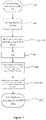
- FIG. 1 shows a process of automated segmentation of anatomical regions
- FIG. 2 shows a more detailed view of the process of automated segmentation of anatomical regions
- FIG. 3 shows a process of automated segmentation of lesion regions
- FIG. 4 shows a more detailed view of a sliding window arrangement.
- DICOM Digital Imaging and Communications in Medicine
- Image data 102 is then extracted from the DICOM file and an image is generated.
- the image then undergoes a pre-processing stage 103 .
- the image is loaded onto a 4D tensor of size [1, width, height, 1].
- the pre-processing stage may comprise windowing the image data 203 to a predetermined windowing level 202 .
- the windowing level defines the range of bit values considered in the image.
- Medical images are conventionally 16-bit images, wherein each pixel is represented as a 16-bit integer ranging from 0 to 2 16 ⁇ 1, i.e. [0, 1, 2, . . . , 65535].
- the information content is very high in these images, and generally comprises more information than what the human eye is capable of detecting.
- a set value for the windowing level is typically included within the DICOM file 202 .
- a sliding window arrangement 301 is used.
- models may be trained on smaller patches sampled from an image.
- prediction/test time i.e. after the model has been trained, a prediction is required for every pixel of the image, but the input to the model can only be a smaller patch owing to conventional hardware constraints. Therefore, the full image can be divided up into smaller patches and fed individually into the FCN 302 .
- the model “slides” over the full images in a sliding window fashion and outputs a prediction for each patch. The outputs are then stitched together to generate an output map.
- the image is then rescaled to a width of 800 pixels and a height of 800 pixels before it is provided to a Fully Convolutional Network (FCN) 100 .
- the image can be rescaled to a larger or smaller size, or even not rescaled at all.
- the rescaled image is then supplied to the FCN 100 .
- the FCN 100 is a type of network architecture that may be used for semantic segmentation tasks. Semantic segmentation is the task where a class is assigned to each pixel in an image.
- the FCN 100 is a convolutional neural network without any fully connected layers.
- a fully connected layer can be expressed as a convolutional layer with 1 ⁇ 1 kernels.
- the network can be applied to an arbitrarily sized image (subject to hardware constraints).
- an FCN is a function mapping: f:X ⁇ Y where X is a tensor of size [Batch size ⁇ Width ⁇ Height ⁇ #channels] with each element being in the set ⁇ 0, . . . , 2 16 ⁇ 1 ⁇ . Y has the shape [Batch size ⁇ Width ⁇ Height ⁇ #classes], each element is between 0 and 1.
- the FCN 100 may comprise any combination of one or more convolutional, hidden representation, activation, and/or pooling layers.
- the activation layer in this embodiment is in the form of a sigmoid activation layer.
- the input is an image of 800 ⁇ 800 pixels
- the FCN 100 is operable to produce an output comprising a probability mask of 800 ⁇ 800 pixels, where each pixel represents a probability of belonging to a class.
- the same FCN 100 may be applied to an image of size 900 ⁇ 900 pixels, and hence produce an output probability mask of 900 ⁇ 900 pixels.
- a different probability mask may be generated for each of a plurality of anatomical regions.
- the FCN 100 is trained to generate such a probability mask by providing a set of input values and associated weights.
- the or each probability mask is then converted to one or more binary masks during a post-processing stage 104 .
- the conversion from a probability mask to binary mask may be through thresholding the probabilities 204 which obtains a binary mask for each target region (for example, anatomical regions). Small areas in the binary mask may be removed 205 . If the area (which may be represented by an identified number of pixels) is smaller than a specific predetermined threshold, then the area may be removed from the binary mask entirely. Similarly, holes in the segmentation itself may be removed. If a segmentation has an area of zeros, entirely surrounded by ones, then the zeros may be set to ones according to a predetermined threshold value for the area.
- the generation of the binary mask is an entirely automated process, and requires no human action other than the input of a data to be analysed.
- Conventional segmentation methods rely on an expert, usually a radiologist, providing a seed region or starting point.
- the method disclosed herein is operable to segment a region without any prior input other than an image.
- the method may also be used to segment lesions.
- the lesions which may be segmented may comprise one or more cancerous growths, masses, abscesses, lacerations, calcifications, and/or other irregularities within biological tissue.
- the FCN 100 may be optimised to perform such segmentation it may be trained using a different, more relevant, dataset.
- the network architecture for segmenting lesions may be in the form of a different embodiment.
- a lesion segmentation tool for mammograms may comprise multiple paths, one of which is operable to analyse a higher-resolution medical image and a different path operable to analyse a lower-resolution image.
- Mammography is a medical imaging modality widely used for breast cancer detection. Mammography makes use of “soft” X-rays to produce detailed images of the internal structure of the human breast—these images are called mammograms and this method is considered to be the gold standard in early detection of breast abnormalities which provide a valid diagnosis of a cancer in a curable phase.
- anatomical structures When analysing mammograms, the reliable identification of anatomical structures is important for visual evaluation and especially for analytic assessment of visual features based on their anatomic location and their relation to anatomic structures, which may have profound implications on the final diagnostic results. In the case that anatomic structures appear distorted they may also indicate the presence of possible malignancies.
- Conventional X-ray is a medical imaging modality widely used for the detection of structural abnormalities related to the air containing structures and bones, as well as those diseases which have an impact on them.
- Conventional X-ray is the most widely used imaging method and makes use of “hard” X-rays to produce detailed images of the internal structure of the lungs and the skeleton. These images are called roentgenograms or simply X-rays.
- anatomical structures When analysing X-ray images, the reliable identification of anatomical structures is important for visual evaluation and especially for analytic assessment of visual features based on their anatomic location and their relation to anatomic structures, which may have profound implications on the final diagnostic results. In the case that anatomic structures appear distorted they may also indicate the presence of possible malignancies.
- Cross-sectional medical imaging modalities are widely used for detection of structural or functional abnormalities and diseases which have a visually identifiable structural impact on the human internal organs.
- the images demonstrate the internal structures in multiple cross-sections of the body. The essence of the most widely used cross-sectional techniques are described below.
- Computed tomography is a widely used imaging method and makes use of “hard” X-rays produced and detected by a specially rotating instrument and the resulted attenuation data (also referred to as raw data) are presented by a computed analytic software producing detailed images of the internal structure of the internal organs.
- the produced sets of images are called CT-scans which may constitute multiple series with different settings and different contrast agent phases to present the internal anatomical structures in cross sections perpendicular to the axis of the human body (or synthesized sections in other angles).
- Magnetic Resonance Imaging is an advanced diagnostic technique which makes use of the effect magnetic field impacts on movements of protons which are the utmost tiniest essential elements of every living tissue.
- the detectors are antennas and the signals are analysed by a computer creating detailed images if the internal structures in any section of the human body.
- MRI can add useful functional information based on signal intensity of generated by the moving protons.
- diagnosis is based on visual evaluation of anatomical structures.
- the reliable assessment especially for analytic assessment, of visual appearance based on their anatomic location and their relation to anatomic structures, may have profound implications on final diagnostic results. In the case that anatomic structures appear distorted they may also indicate the presence of possible malignancies.
- diagnostic radiology methods which include mammography, conventional X-ray, CT, MRI
- identification, localisation (registration), segmentation and classification of abnormalities and/or findings are important interlinked steps in the diagnostic workflow.
- Locality and classification may define and significantly influence diagnoses. Both locality and classification may be informed by segmentation in terms of the exact shape and extent of visual features (i.e. size and location of boundaries, distance from and relation to other features and/or anatomy). Segmentation may also provide important information regarding the change in status of disease (e.g. progression or recession).
- Machine learning is the field of study where a computer or computers learn to perform classes of tasks using the feedback generated from the experience or data gathered that the machine learning process acquires during computer performance of those tasks.
- machine learning can be broadly classed as supervised and unsupervised approaches, although there are particular approaches such as reinforcement learning and semi-supervised learning which have special rules, techniques and/or approaches.
- Supervised machine learning is concerned with a computer learning one or more rules or functions to map between example inputs and desired outputs as predetermined by an operator or programmer, usually where a data set containing the inputs is labelled.
- Unsupervised learning is concerned with determining a structure for input data, for example when performing pattern recognition, and typically uses unlabelled data sets.
- Reinforcement learning is concerned with enabling a computer or computers to interact with a dynamic environment, for example when playing a game or driving a vehicle.
- “semi-supervised” machine learning where a training data set has only been partially labelled.
- unsupervised machine learning there is a range of possible applications such as, for example, the application of computer vision techniques to image processing or video enhancement.
- Unsupervised machine learning is typically applied to solve problems where an unknown data structure might be present in the data. As the data is unlabelled, the machine learning process is required to operate to identify implicit relationships between the data for example by deriving a clustering metric based on internally derived information.
- an unsupervised learning technique can be used to reduce the dimensionality of a data set and attempt to identify and model relationships between clusters in the data set, and can for example generate measures of cluster membership or identify hubs or nodes in or between clusters (for example using a technique referred to as weighted correlation network analysis, which can be applied to high-dimensional data sets, or using k-means clustering to cluster data by a measure of the Euclidean distance between each datum).
- Semi-supervised learning is typically applied to solve problems where there is a partially labelled data set, for example where only a subset of the data is labelled.
- Semi-supervised machine learning makes use of externally provided labels and objective functions as well as any implicit data relationships.
- the machine learning algorithm can be provided with some training data or a set of training examples, in which each example is typically a pair of an input signal/vector and a desired output value, label (or classification) or signal.
- the machine learning algorithm analyses the training data and produces a generalised function that can be used with unseen data sets to produce desired output values or signals for the unseen input vectors/signals.
- the user needs to decide what type of data is to be used as the training data, and to prepare a representative real-world set of data.
- the user must however take care to ensure that the training data contains enough information to accurately predict desired output values without providing too many features (which can result in too many dimensions being considered by the machine learning process during training, and could also mean that the machine learning process does not converge to good solutions for all or specific examples).
- the user must also determine the desired structure of the learned or generalised function, for example whether to use support vector machines or decision trees.
- Machine learning may be performed through the use of one or more of: a non-linear hierarchical algorithm; neural network; convolutional neural network; recurrent neural network; long short-term memory network; multi-dimensional convolutional network; a memory network; fully convolutional network or a gated recurrent network allows a flexible approach when generating the predicted block of visual data.
- a non-linear hierarchical algorithm neural network; convolutional neural network; recurrent neural network; long short-term memory network; multi-dimensional convolutional network; a memory network; fully convolutional network or a gated recurrent network allows a flexible approach when generating the predicted block of visual data.
- the use of an algorithm with a memory unit such as a long short-term memory network (LSTM), a memory network or a gated recurrent network can keep the state of the predicted blocks from motion compensation processes performed on the same original input frame.
- LSTM long short-term memory network
- a gated recurrent network can keep the state of the predicted blocks from
- Developing a machine learning system typically consists of two stages: (1) training and (2) production.
- the parameters of the machine learning model are iteratively changed to optimise a particular learning objective, known as the objective function or the loss.
- the model Once the model is trained, it can be used in production, where the model takes in an input and produces an output using the trained parameters.
- any feature in one aspect of the invention may be applied to other aspects of the invention, in any appropriate combination.
- method aspects may be applied to system aspects, and vice versa.
- any, some and/or all features in one aspect can be applied to any, some and/or all features in any other aspect, in any appropriate combination.
Abstract
Description
f:X→Y
where X is a tensor of size [Batch size×Width×Height×#channels] with each element being in the set {0, . . . , 216−1}. Y has the shape [Batch size×Width×Height×#classes], each element is between 0 and 1.
Claims (20)
Applications Claiming Priority (10)
| Application Number | Priority Date | Filing Date | Title |
|---|---|---|---|
| GBGB1705911.4A GB201705911D0 (en) | 2017-04-12 | 2017-04-12 | Abstracts |
| GB1705911 | 2017-04-12 | ||
| GB1705911.4 | 2017-04-12 | ||
| GB1711560.1 | 2017-07-18 | ||
| GBGB1711560.1A GB201711560D0 (en) | 2017-04-12 | 2017-07-18 | Lesion segmentation |
| GB1711558 | 2017-07-18 | ||
| GB1711558.5 | 2017-07-18 | ||
| GBGB1711558.5A GB201711558D0 (en) | 2017-04-12 | 2017-07-18 | Segmentation |
| GB1711560 | 2017-07-18 | ||
| PCT/GB2018/050980 WO2018189550A1 (en) | 2017-04-12 | 2018-04-12 | Segmentation of anatomical regions and lesions |
Publications (2)
| Publication Number | Publication Date |
|---|---|
| US20200167928A1 US20200167928A1 (en) | 2020-05-28 |
| US11423540B2 true US11423540B2 (en) | 2022-08-23 |
Family
ID=58744677
Family Applications (3)
| Application Number | Title | Priority Date | Filing Date |
|---|---|---|---|
| US16/604,660 Active 2038-04-27 US11423540B2 (en) | 2017-04-12 | 2018-04-12 | Segmentation of anatomical regions and lesions |
| US16/604,662 Active US11423541B2 (en) | 2017-04-12 | 2018-04-12 | Assessment of density in mammography |
| US16/604,664 Active 2038-05-09 US11127137B2 (en) | 2017-04-12 | 2018-04-12 | Malignancy assessment for tumors |
Family Applications After (2)
| Application Number | Title | Priority Date | Filing Date |
|---|---|---|---|
| US16/604,662 Active US11423541B2 (en) | 2017-04-12 | 2018-04-12 | Assessment of density in mammography |
| US16/604,664 Active 2038-05-09 US11127137B2 (en) | 2017-04-12 | 2018-04-12 | Malignancy assessment for tumors |
Country Status (5)
| Country | Link |
|---|---|
| US (3) | US11423540B2 (en) |
| EP (3) | EP3610455A1 (en) |
| JP (1) | JP7279015B2 (en) |
| GB (5) | GB201705911D0 (en) |
| WO (3) | WO2018189551A1 (en) |
Families Citing this family (32)
| Publication number | Priority date | Publication date | Assignee | Title |
|---|---|---|---|---|
| GB201705876D0 (en) | 2017-04-11 | 2017-05-24 | Kheiron Medical Tech Ltd | Recist |
| GB201705911D0 (en) | 2017-04-12 | 2017-05-24 | Kheiron Medical Tech Ltd | Abstracts |
| US11080857B2 (en) * | 2018-04-26 | 2021-08-03 | NeuralSeg Ltd. | Systems and methods for segmenting an image |
| WO2019239154A1 (en) | 2018-06-14 | 2019-12-19 | Kheiron Medical Technologies Ltd | Second reader |
| CN109124669A (en) * | 2018-08-30 | 2019-01-04 | 沈阳柏敖生信生物科技有限公司 | CT data measuring method before a kind of shaping |
| CN109427060A (en) | 2018-10-30 | 2019-03-05 | 腾讯科技(深圳)有限公司 | A kind of method, apparatus, terminal device and the medical system of image identification |
| TWI678709B (en) * | 2018-11-15 | 2019-12-01 | 義守大學 | Disease prediction method through a big database formed by data mining of neural network |
| KR102261473B1 (en) * | 2018-11-30 | 2021-06-07 | 주식회사 딥바이오 | Method for providing diagnosis system using semi-supervised machine learning and diagnosis system using the method |
| WO2020117486A1 (en) * | 2018-12-05 | 2020-06-11 | Verathon Inc. | Implant assessment using ultrasound and optical imaging |
| US20200196987A1 (en) * | 2018-12-20 | 2020-06-25 | General Electric Company | Method and system to manage beamforming parameters based on tissue density |
| CN109785300A (en) * | 2018-12-27 | 2019-05-21 | 华南理工大学 | A kind of cancer medical image processing method, system, device and storage medium |
| US10936160B2 (en) | 2019-01-11 | 2021-03-02 | Google Llc | System, user interface and method for interactive negative explanation of machine-learning localization models in health care applications |
| CN109815869A (en) * | 2019-01-16 | 2019-05-28 | 浙江理工大学 | A kind of finger vein identification method based on the full convolutional network of FCN |
| CN110009600A (en) * | 2019-02-14 | 2019-07-12 | 腾讯科技(深圳)有限公司 | A kind of medical image area filter method, apparatus and storage medium |
| CN110097580B (en) * | 2019-03-18 | 2021-10-19 | 山东师范大学 | Ultrasonic image marker motion tracking method |
| JP7023254B2 (en) * | 2019-03-27 | 2022-02-21 | 富士フイルム株式会社 | Shooting support equipment, methods and programs |
| EP3975846A4 (en) * | 2019-05-30 | 2023-07-12 | Acerar Ltd. | System and method for cognitive training and monitoring |
| BR112022003391A2 (en) * | 2019-08-23 | 2022-05-17 | Subtle Medical Inc | Systems and methods for accurate and rapid positron emission tomography using deep learning |
| US11532399B2 (en) * | 2019-10-15 | 2022-12-20 | Merative Us L.P. | Identifying high tissue density in images using machine learning |
| EP4049238A1 (en) * | 2019-10-25 | 2022-08-31 | Deephealth, Inc. | System and method for analyzing three-dimensional image data |
| US11170503B2 (en) | 2019-10-30 | 2021-11-09 | International Business Machines Corporation | Systems and methods for detection likelihood of malignancy in a medical image |
| US11341635B2 (en) * | 2019-10-31 | 2022-05-24 | Tencent America LLC | Computer aided diagnosis system for detecting tissue lesion on microscopy images based on multi-resolution feature fusion |
| US20210209755A1 (en) * | 2020-01-02 | 2021-07-08 | Nabin K. Mishra | Automatic lesion border selection based on morphology and color features |
| US11508061B2 (en) * | 2020-02-20 | 2022-11-22 | Siemens Healthcare Gmbh | Medical image segmentation with uncertainty estimation |
| US11526694B2 (en) | 2020-03-31 | 2022-12-13 | International Business Machines Corporation | Model training using fully and partially-annotated images |
| US11449716B2 (en) | 2020-03-31 | 2022-09-20 | International Business Machines Corporation | Model training using partially-annotated images |
| US11693919B2 (en) * | 2020-06-22 | 2023-07-04 | Shanghai United Imaging Intelligence Co., Ltd. | Anatomy-aware motion estimation |
| CN112037231A (en) * | 2020-07-30 | 2020-12-04 | 浙江大学 | Medical image liver segmentation post-processing method based on feature classifier |
| US11651499B2 (en) | 2020-09-17 | 2023-05-16 | International Business Machines Corporation | Reducing structural redundancy in automatic image segmentation |
| US11610306B2 (en) | 2020-12-16 | 2023-03-21 | Industrial Technology Research Institute | Medical image analysis method and device |
| EP4016453B1 (en) * | 2020-12-21 | 2023-08-09 | Siemens Healthcare GmbH | Method and system for automated segmentation of biological object parts in mri |
| WO2023200990A1 (en) * | 2022-04-15 | 2023-10-19 | Memorial Sloan-Kettering Cancer Center | Automated methods for determining fibroglandular density on mammograms |
Citations (32)
| Publication number | Priority date | Publication date | Assignee | Title |
|---|---|---|---|---|
| US6075879A (en) | 1993-09-29 | 2000-06-13 | R2 Technology, Inc. | Method and system for computer-aided lesion detection using information from multiple images |
| US6324532B1 (en) * | 1997-02-07 | 2001-11-27 | Sarnoff Corporation | Method and apparatus for training a neural network to detect objects in an image |
| US6463425B2 (en) * | 1995-09-19 | 2002-10-08 | Morphometrix Technologies Inc. | Neural network assisted multi-spectral segmentation system |
| US20050113651A1 (en) | 2003-11-26 | 2005-05-26 | Confirma, Inc. | Apparatus and method for surgical planning and treatment monitoring |
| US20060050958A1 (en) | 2004-09-09 | 2006-03-09 | Kazunori Okada | System and method for volumetric tumor segmentation using joint space-intensity likelihood ratio test |
| JP2006255412A (en) | 2005-03-14 | 2006-09-28 | General Electric Co <Ge> | Method and system for monitoring tumor burden |
| US20060274928A1 (en) | 2005-06-02 | 2006-12-07 | Jeffrey Collins | System and method of computer-aided detection |
| US20070003119A1 (en) | 2005-07-01 | 2007-01-04 | R2 Technology, Inc. | Displaying and navigating computer-aided detection results on a review workstation |
| WO2008050332A2 (en) | 2006-10-25 | 2008-05-02 | Siemens Computer Aided Diagnosis Ltd. | Computer diagnosis of malignancies and false positives |
| WO2008088478A1 (en) | 2006-12-28 | 2008-07-24 | Carestream Health, Inc. | Method for classifying breast tissue density |
| US20110123087A1 (en) | 2009-11-25 | 2011-05-26 | Fujifilm Corporation | Systems and methods for measurement of objects of interest in medical images |
| US20110150313A1 (en) | 2009-12-17 | 2011-06-23 | The Regents Of The University Of California | Method and Apparatus for Quantitative Analysis of Breast Density Morphology Based on MRI |
| US20130343626A1 (en) | 2008-12-22 | 2013-12-26 | The Medipattern Corporation | Method and system of automated detection of lesions in medical images |
| US20140355840A1 (en) | 2013-05-31 | 2014-12-04 | Kathryn Pearson Peyton | Apparatus and Method for Utilizing Mammogram Images for Verification |
| WO2015031641A1 (en) | 2013-08-29 | 2015-03-05 | Mayo Foundation For Medical Education And Research | System and method for boundary classification and automatic polyp detection |
| WO2015077076A1 (en) | 2013-11-19 | 2015-05-28 | VuComp, Inc | Obtaining breast density measurements and classifications |
| CN105931226A (en) | 2016-04-14 | 2016-09-07 | 南京信息工程大学 | Automatic cell detection and segmentation method based on deep learning and using adaptive ellipse fitting |
| CN106096491A (en) | 2016-02-04 | 2016-11-09 | 上海市第人民医院 | The automatic identifying method of the microaneurysm in color fundus image |
| JP2016534709A (en) | 2013-10-28 | 2016-11-10 | モレキュラー デバイシーズ, エルエルシー | Method and system for classifying and identifying individual cells in a microscopic image |
| US20160350946A1 (en) | 2015-05-29 | 2016-12-01 | Erica Lin | Method of forming probability map |
| CN106204587A (en) | 2016-05-27 | 2016-12-07 | 孔德兴 | Multiple organ dividing method based on degree of depth convolutional neural networks and region-competitive model |
| US20170039412A1 (en) | 2013-10-22 | 2017-02-09 | Eyenuk, Inc. | Systems and methods for automated detection of regions of interest in retinal images |
| US9589374B1 (en) * | 2016-08-01 | 2017-03-07 | 12 Sigma Technologies | Computer-aided diagnosis system for medical images using deep convolutional neural networks |
| WO2017210690A1 (en) | 2016-06-03 | 2017-12-07 | Lu Le | Spatial aggregation of holistically-nested convolutional neural networks for automated organ localization and segmentation in 3d medical scans |
| US20180061054A1 (en) * | 2016-08-29 | 2018-03-01 | CephX Technologies Ltd. | Automated Cephalometric Analysis Using Machine Learning |
| US20180129900A1 (en) * | 2016-11-04 | 2018-05-10 | Siemens Healthcare Gmbh | Anonymous and Secure Classification Using a Deep Learning Network |
| US20180165809A1 (en) | 2016-12-02 | 2018-06-14 | Panagiotis Stanitsas | Computer vision for cancerous tissue recognition |
| US20180218495A1 (en) * | 2017-02-02 | 2018-08-02 | International Business Machines Corporation | Systems and methods for automatic detection of architectural distortion in two dimensional mammographic images |
| US20180218497A1 (en) * | 2017-01-27 | 2018-08-02 | Arterys Inc. | Automated segmentation utilizing fully convolutional networks |
| WO2018189541A1 (en) | 2017-04-11 | 2018-10-18 | Kheiron Medical Technologies Ltd | Recist assessment of tumour progression |
| WO2018189550A1 (en) | 2017-04-12 | 2018-10-18 | Kheiron Medical Technologies Ltd | Segmentation of anatomical regions and lesions |
| US10127659B2 (en) * | 2016-11-23 | 2018-11-13 | General Electric Company | Deep learning medical systems and methods for image acquisition |
Family Cites Families (38)
| Publication number | Priority date | Publication date | Assignee | Title |
|---|---|---|---|---|
| WO1996002897A2 (en) | 1994-07-14 | 1996-02-01 | Philips Electronics N.V. | Mass detection in digital x-ray images using multiple threshold levels to discriminate spots |
| US6058322A (en) | 1997-07-25 | 2000-05-02 | Arch Development Corporation | Methods for improving the accuracy in differential diagnosis on radiologic examinations |
| US5999639A (en) | 1997-09-04 | 1999-12-07 | Qualia Computing, Inc. | Method and system for automated detection of clustered microcalcifications from digital mammograms |
| US6574304B1 (en) | 2002-09-13 | 2003-06-03 | Ge Medical Systems Global Technology Company, Llc | Computer aided acquisition of medical images |
| BR0314589A (en) | 2002-09-24 | 2005-08-09 | Eastman Kodak Co | Method and system for visualizing results of a computer aided detection analysis of a digital image and method for identifying abnormalities in a mammogram |
| US7418119B2 (en) | 2002-10-31 | 2008-08-26 | Siemens Computer Aided Diagnosis Ltd. | Display for computer-aided evaluation of medical images and for establishing clinical recommendation therefrom |
| US7490085B2 (en) | 2002-12-18 | 2009-02-10 | Ge Medical Systems Global Technology Company, Llc | Computer-assisted data processing system and method incorporating automated learning |
| US20040228509A1 (en) | 2003-05-15 | 2004-11-18 | Beth Israel Deaconess Medical Center, Inc. | Automatic spacial identification of tissue implanted linear sources using medical imaging |
| EP1636731A2 (en) | 2003-06-25 | 2006-03-22 | Siemens Medical Solutions USA, Inc. | Systems and methods for automated diagnosis and decision support for breast imaging |
| US7529394B2 (en) | 2003-06-27 | 2009-05-05 | Siemens Medical Solutions Usa, Inc. | CAD (computer-aided decision) support for medical imaging using machine learning to adapt CAD process with knowledge collected during routine use of CAD system |
| DE102004051401A1 (en) | 2004-10-21 | 2006-05-24 | Siemens Ag | Method for diagnosis in three-dimensional imaging, in particular in mammography |
| US7646902B2 (en) | 2005-02-08 | 2010-01-12 | Regents Of The University Of Michigan | Computerized detection of breast cancer on digital tomosynthesis mammograms |
| FR2897182A1 (en) | 2006-02-09 | 2007-08-10 | Gen Electric | METHOD FOR PROCESSING TOMOSYNTHESIS PROJECTION IMAGES FOR DETECTION OF RADIOLOGICAL SIGNS |
| GB0602739D0 (en) | 2006-02-10 | 2006-03-22 | Ccbr As | Breast tissue density measure |
| ITRM20060213A1 (en) | 2006-04-13 | 2007-10-14 | Univ Palermo | METHOD OF PROCESSING BIOMEDICAL IMAGES |
| US8203338B2 (en) | 2007-08-06 | 2012-06-19 | Carestream Health, Inc. | Linear structure verification in medical applications |
| JP5264136B2 (en) | 2007-09-27 | 2013-08-14 | キヤノン株式会社 | MEDICAL DIAGNOSIS SUPPORT DEVICE, ITS CONTROL METHOD, COMPUTER PROGRAM, AND STORAGE MEDIUM |
| JP5100285B2 (en) | 2007-09-28 | 2012-12-19 | キヤノン株式会社 | MEDICAL DIAGNOSIS SUPPORT DEVICE, ITS CONTROL METHOD, PROGRAM, AND STORAGE MEDIUM |
| US7942829B2 (en) | 2007-11-06 | 2011-05-17 | Eigen, Inc. | Biopsy planning and display apparatus |
| US20100135562A1 (en) | 2008-11-28 | 2010-06-03 | Siemens Computer Aided Diagnosis Ltd. | Computer-aided detection with enhanced workflow |
| US20120099771A1 (en) | 2010-10-20 | 2012-04-26 | Lao Zhiqiang | Computer aided detection of architectural distortion in mammography |
| JP5875285B2 (en) | 2011-08-16 | 2016-03-02 | キヤノン株式会社 | Medical diagnosis support apparatus, information processing method, and program |
| WO2013027120A2 (en) | 2011-08-22 | 2013-02-28 | Joel Ironstone | Method and system for disease risk management |
| JP5945803B2 (en) | 2012-03-07 | 2016-07-05 | 東芝メディカルシステムズ株式会社 | Medical image interpretation system |
| US8891841B2 (en) | 2012-06-04 | 2014-11-18 | Verizon Patent And Licensing Inc. | Mobile dermatology collection and analysis system |
| US9221195B2 (en) | 2012-06-29 | 2015-12-29 | The Procter & Gamble Company | Methods and apparatuses for consolidating elastic substrates |
| TWI483711B (en) | 2012-07-10 | 2015-05-11 | Univ Nat Taiwan | Tumor detection system and method of breast ultrasound image |
| WO2016057960A1 (en) | 2014-10-10 | 2016-04-14 | Radish Medical Solutions, Inc. | Apparatus, system and method for cloud based diagnostics and image archiving and retrieval |
| US9519966B2 (en) | 2015-04-22 | 2016-12-13 | King Fahd University Of Petroleum And Minerals | Method, system and computer program product for breast density classification using parts-based local features |
| US10269114B2 (en) | 2015-06-12 | 2019-04-23 | International Business Machines Corporation | Methods and systems for automatically scoring diagnoses associated with clinical images |
| US10339650B2 (en) | 2016-01-07 | 2019-07-02 | Koios Medical, Inc. | Method and means of CAD system personalization to reduce intraoperator and interoperator variation |
| US10417788B2 (en) | 2016-09-21 | 2019-09-17 | Realize, Inc. | Anomaly detection in volumetric medical images using sequential convolutional and recurrent neural networks |
| US10223610B1 (en) | 2017-10-15 | 2019-03-05 | International Business Machines Corporation | System and method for detection and classification of findings in images |
| US10916341B2 (en) | 2017-12-15 | 2021-02-09 | International Business Machines Corporation | Automated report generation based on cognitive classification of medical images |
| KR20210005699A (en) | 2018-04-27 | 2021-01-14 | 델피누스 메디컬 테크놀로지스, 인크. | System and method for feature extraction and classification of ultrasound tomography images |
| US10706545B2 (en) | 2018-05-07 | 2020-07-07 | Zebra Medical Vision Ltd. | Systems and methods for analysis of anatomical images |
| WO2019239154A1 (en) | 2018-06-14 | 2019-12-19 | Kheiron Medical Technologies Ltd | Second reader |
| GB2579244A (en) | 2018-11-27 | 2020-06-17 | Kheiron Medical Tech Ltd | Second reader |
-
2017
- 2017-04-12 GB GBGB1705911.4A patent/GB201705911D0/en not_active Ceased
- 2017-07-18 GB GBGB1711558.5A patent/GB201711558D0/en not_active Ceased
- 2017-07-18 GB GBGB1711560.1A patent/GB201711560D0/en not_active Ceased
- 2017-07-18 GB GBGB1711557.7A patent/GB201711557D0/en not_active Ceased
- 2017-07-18 GB GBGB1711559.3A patent/GB201711559D0/en not_active Ceased
-
2018
- 2018-04-12 EP EP18719273.7A patent/EP3610455A1/en active Pending
- 2018-04-12 US US16/604,660 patent/US11423540B2/en active Active
- 2018-04-12 WO PCT/GB2018/050981 patent/WO2018189551A1/en unknown
- 2018-04-12 JP JP2020505540A patent/JP7279015B2/en active Active
- 2018-04-12 EP EP18719272.9A patent/EP3610457A1/en active Pending
- 2018-04-12 US US16/604,662 patent/US11423541B2/en active Active
- 2018-04-12 EP EP18719271.1A patent/EP3610454A1/en active Pending
- 2018-04-12 US US16/604,664 patent/US11127137B2/en active Active
- 2018-04-12 WO PCT/GB2018/050979 patent/WO2018189549A1/en unknown
- 2018-04-12 WO PCT/GB2018/050980 patent/WO2018189550A1/en unknown
Patent Citations (39)
| Publication number | Priority date | Publication date | Assignee | Title |
|---|---|---|---|---|
| US6075879A (en) | 1993-09-29 | 2000-06-13 | R2 Technology, Inc. | Method and system for computer-aided lesion detection using information from multiple images |
| US6463425B2 (en) * | 1995-09-19 | 2002-10-08 | Morphometrix Technologies Inc. | Neural network assisted multi-spectral segmentation system |
| US6324532B1 (en) * | 1997-02-07 | 2001-11-27 | Sarnoff Corporation | Method and apparatus for training a neural network to detect objects in an image |
| US20050113651A1 (en) | 2003-11-26 | 2005-05-26 | Confirma, Inc. | Apparatus and method for surgical planning and treatment monitoring |
| US20060050958A1 (en) | 2004-09-09 | 2006-03-09 | Kazunori Okada | System and method for volumetric tumor segmentation using joint space-intensity likelihood ratio test |
| JP2006255412A (en) | 2005-03-14 | 2006-09-28 | General Electric Co <Ge> | Method and system for monitoring tumor burden |
| JP2008541889A (en) | 2005-06-02 | 2008-11-27 | ザ メディパターン コーポレイション | System and method for computer-aided detection |
| US20060274928A1 (en) | 2005-06-02 | 2006-12-07 | Jeffrey Collins | System and method of computer-aided detection |
| US20070003119A1 (en) | 2005-07-01 | 2007-01-04 | R2 Technology, Inc. | Displaying and navigating computer-aided detection results on a review workstation |
| WO2008050332A2 (en) | 2006-10-25 | 2008-05-02 | Siemens Computer Aided Diagnosis Ltd. | Computer diagnosis of malignancies and false positives |
| WO2008088478A1 (en) | 2006-12-28 | 2008-07-24 | Carestream Health, Inc. | Method for classifying breast tissue density |
| US20130343626A1 (en) | 2008-12-22 | 2013-12-26 | The Medipattern Corporation | Method and system of automated detection of lesions in medical images |
| US20110123087A1 (en) | 2009-11-25 | 2011-05-26 | Fujifilm Corporation | Systems and methods for measurement of objects of interest in medical images |
| US20110150313A1 (en) | 2009-12-17 | 2011-06-23 | The Regents Of The University Of California | Method and Apparatus for Quantitative Analysis of Breast Density Morphology Based on MRI |
| US20140355840A1 (en) | 2013-05-31 | 2014-12-04 | Kathryn Pearson Peyton | Apparatus and Method for Utilizing Mammogram Images for Verification |
| WO2015031641A1 (en) | 2013-08-29 | 2015-03-05 | Mayo Foundation For Medical Education And Research | System and method for boundary classification and automatic polyp detection |
| US20170039412A1 (en) | 2013-10-22 | 2017-02-09 | Eyenuk, Inc. | Systems and methods for automated detection of regions of interest in retinal images |
| JP2016534709A (en) | 2013-10-28 | 2016-11-10 | モレキュラー デバイシーズ, エルエルシー | Method and system for classifying and identifying individual cells in a microscopic image |
| WO2015077076A1 (en) | 2013-11-19 | 2015-05-28 | VuComp, Inc | Obtaining breast density measurements and classifications |
| US20160350946A1 (en) | 2015-05-29 | 2016-12-01 | Erica Lin | Method of forming probability map |
| CN106096491A (en) | 2016-02-04 | 2016-11-09 | 上海市第人民医院 | The automatic identifying method of the microaneurysm in color fundus image |
| CN105931226A (en) | 2016-04-14 | 2016-09-07 | 南京信息工程大学 | Automatic cell detection and segmentation method based on deep learning and using adaptive ellipse fitting |
| CN106204587A (en) | 2016-05-27 | 2016-12-07 | 孔德兴 | Multiple organ dividing method based on degree of depth convolutional neural networks and region-competitive model |
| WO2017210690A1 (en) | 2016-06-03 | 2017-12-07 | Lu Le | Spatial aggregation of holistically-nested convolutional neural networks for automated organ localization and segmentation in 3d medical scans |
| US9589374B1 (en) * | 2016-08-01 | 2017-03-07 | 12 Sigma Technologies | Computer-aided diagnosis system for medical images using deep convolutional neural networks |
| US20180061054A1 (en) * | 2016-08-29 | 2018-03-01 | CephX Technologies Ltd. | Automated Cephalometric Analysis Using Machine Learning |
| US20180129900A1 (en) * | 2016-11-04 | 2018-05-10 | Siemens Healthcare Gmbh | Anonymous and Secure Classification Using a Deep Learning Network |
| US10127659B2 (en) * | 2016-11-23 | 2018-11-13 | General Electric Company | Deep learning medical systems and methods for image acquisition |
| US20180165809A1 (en) | 2016-12-02 | 2018-06-14 | Panagiotis Stanitsas | Computer vision for cancerous tissue recognition |
| US20180218497A1 (en) * | 2017-01-27 | 2018-08-02 | Arterys Inc. | Automated segmentation utilizing fully convolutional networks |
| US20180218495A1 (en) * | 2017-02-02 | 2018-08-02 | International Business Machines Corporation | Systems and methods for automatic detection of architectural distortion in two dimensional mammographic images |
| WO2018189541A1 (en) | 2017-04-11 | 2018-10-18 | Kheiron Medical Technologies Ltd | Recist assessment of tumour progression |
| US20200074634A1 (en) | 2017-04-11 | 2020-03-05 | Kheiron Medical Technologies Ltd | Recist assessment of tumour progression |
| WO2018189550A1 (en) | 2017-04-12 | 2018-10-18 | Kheiron Medical Technologies Ltd | Segmentation of anatomical regions and lesions |
| WO2018189549A1 (en) | 2017-04-12 | 2018-10-18 | Kheiron Medical Technologies Ltd | Assessment of density in mammography |
| WO2018189551A1 (en) | 2017-04-12 | 2018-10-18 | Kheiron Medical Technologies Ltd | Malignancy assessment for tumors |
| US20200074632A1 (en) | 2017-04-12 | 2020-03-05 | Kheiron Medical Technologies Ltd | Assessment of density in mammography |
| US20200342589A1 (en) | 2017-04-12 | 2020-10-29 | Kheiron Medical Technologies Ltd | Malignancy assessment for tumors |
| US11127137B2 (en) | 2017-04-12 | 2021-09-21 | Kheiron Medical Technologies Ltd | Malignancy assessment for tumors |
Non-Patent Citations (30)
| Title |
|---|
| Akselrod-Ballin, et al., "A Region Based Convolutional Network for Tumor Detection and Classification in Breast Mammography," Springer International Publishing AG, 2016, G. Carneiro et al. (Eds ): Labels 2016/DLMIA 2016, LNCS 10008, pp. 197-205. |
| Christ, et al., "Automatic Liver and Tumor Segmentation of CT and MRI Volumes Using Cascaded Fully Convolutional Neural Networks," arXiv: 1702.05970v2 [cs.CV], Feb. 23, 2017. 20 pages. |
| Christ, et al., "Survivalnet: Predicting Patient Survival From Diffusion Weighted Magnetic Resonance Images Using Cascaded Fully Convolutional and 3D Convolutional Neural Networks," arXiv: 1702.05941v1 [cs.CV], Feb. 20, 2017. 6 pages. |
| Combined Search and Examination Report under Sections 17 and 18(3) received for GB Application No. 1711557.7, dated Jan. 18, 2018. 5 pages. |
| Combined Search and Examination Report under Sections 17 and 18(3) received for GB Application No. 1711558.5, dated Dec. 22, 2017. 6 pages. |
| Combined Search and Examination Report under Sections 17 and 18(3) received for GB Application No. 1711559.3 dated Dec. 19, 2017. 7 pages. |
| Communication pursuant to Article 94(3) EPC received for EP Application No. 18719271.1, dated Jun. 21, 2021. 10 pages. |
| Communication pursuant to Article 94(3) EPC received for EP Application No. 18719272.9, dated Jun. 21, 2021. 8 pages. |
| Communication pursuant to Article 94(3) EPC received for EP Application No. 18719273.7, dated Nov. 11, 2021. 9 pages. |
| El Hamdi, et al., "Breast Cancer Diagnosis Using a Hybrid Evolutionary Neural Network Classifier," 18th Mediterranean Conference on Control & Automation, Jun. 23-25, 2010, pp. 1308-1315. |
| Gram, et al., "The Tabar classification of mammographic parenchymal patterns," ELSEVIER, European Journal of Radiology 24 (1997), pp. 131-136. |
| Hou, et al., "Patch-based Convolutional Neural Network for Whole Slide Tissue Image Classification," IEEE Conference On Computer Vision and Pattern Recognition, (CVPR) Jun. 27, 2016. pp. 2424-2433. |
| International Preliminary Report on Patentability received for Patent Application No. PCT/GB2018/050969, dated Oct. 15, 2019. 12 pages. |
| International Preliminary Report on Patentability received for Patent Application No. PCT/GB2018/050979, dated Oct. 15, 2019. 11 pages. |
| International Preliminary Report on Patentability received for Patent Application No. PCT/GB2018/050981, dated Oct. 15, 2019. 15 pages. |
| International Preliminary Report on Patentability received for PCT Application No. PCT/GB2018/050980, dated Oct. 15, 2019. 11 pages. |
| International Search Report and Written Opinion received for Patent Application No. PCT/GB2018/050969, dated Jun. 26, 2018. 20 pages. |
| International Search Report and Written Opinion received for Patent Application No. PCT/GB2018/050979, dated Jul. 9, 2018. 36 pages. |
| International Search Report and Written Opinion received for Patent Application No. PCT/GB2018/050981, dated Jul. 9, 2018. 23 pages. |
| International Search Report and Written Opinion received for PCT Application No. PCT/GB2018/050980, dated Jul. 12, 2018. 34 pages. |
| JP Office Action issued for JP2020-505539, dated Mar. 8, 2022, 5 pages. (including English translation). |
| JP Office Action received for JP Application No. 2020-505540, dated Apr. 19, 2022. 3 pages. |
| Kallenberg et al (NPL "Unsupervised Deep Learning Applied to Mammographic Risk Scoring", IEEE Transactions on Medical Imaging, vol. 35, No. 5, May 2016, hereafter referred to as Kallenberg). * |
| Kallenberg, et al., "Unsupervised Deep Learning Applied to Breast Density Segmentation and Mammographic Risk Scoring," IEEE Transactions on Medical Imaging, vol. 35, No. 5, May 2016. pp. 1322-1331. |
| Long, et al., "Fully Convolutional Networks for Semantic Segmentation," Proc. of IEEE Computer Vision and Pattern Recognition, 201, 3431-3440, https://openaccess.thecvf.com/content_cvpr_2015/papers/Long_Fully_Convolutional_Networks_2015_CVPR_paper.pdf. |
| Menechelli, et al., "Automatic breast tissue density estimation scheme in digital mammography images," Progress in Biomedical Optics and Imaging, SPIE—International Society for Optical Engineering., vol. 10136, Mar. 10, 2017. 12 pages. |
| Ronneberger, et al., "U-Net: Convolutional Networks for Biomedical Image Segmentation," Medical Image Computing and Computer-Assisted Intervention—MICCAI 2015—18th International Conference. Nov. 18, 2015, Springer International Publishing, arXiv: 1505.04597v1, May 18, 2015. 8 pages. |
| Sun, et al., "Enhancing deep convolutional neural network scheme for breast cancer diagnosis with unlabeled data," ELSEVIER—Computerized Medical Imaging and Graphics 57 (2017). 6 pages. |
| Winkel, et al., "Mammographic density and structural features can individually and jointly contribute to breast cancer risk assessment in mammography screening: a case-control study," BMC Cancer (2016) DOI 10.1186/s12885-016-2450-7. pp. 1-12. |
| Yu, et al., "Automated Melanoma Recognition in Dermoscopy Images via Very Deep Residual Networks," IEEE Transactions On Medical Imaging. Dec. 2016. 12 pages. |
Also Published As
| Publication number | Publication date |
|---|---|
| US20200342589A1 (en) | 2020-10-29 |
| JP7279015B2 (en) | 2023-05-22 |
| GB201711560D0 (en) | 2017-08-30 |
| US11423541B2 (en) | 2022-08-23 |
| EP3610455A1 (en) | 2020-02-19 |
| GB201711559D0 (en) | 2017-08-30 |
| WO2018189549A1 (en) | 2018-10-18 |
| WO2018189550A1 (en) | 2018-10-18 |
| GB201711557D0 (en) | 2017-08-30 |
| US11127137B2 (en) | 2021-09-21 |
| US20200074632A1 (en) | 2020-03-05 |
| GB201711558D0 (en) | 2017-08-30 |
| GB201705911D0 (en) | 2017-05-24 |
| EP3610457A1 (en) | 2020-02-19 |
| JP2020516428A (en) | 2020-06-11 |
| WO2018189551A1 (en) | 2018-10-18 |
| US20200167928A1 (en) | 2020-05-28 |
| EP3610454A1 (en) | 2020-02-19 |
Similar Documents
| Publication | Publication Date | Title |
|---|---|---|
| US11423540B2 (en) | Segmentation of anatomical regions and lesions | |
| Noguerol et al. | Strengths, weaknesses, opportunities, and threats analysis of artificial intelligence and machine learning applications in radiology | |
| Yousef et al. | A holistic overview of deep learning approach in medical imaging | |
| US11455723B2 (en) | Second reader suggestion | |
| JP5954769B2 (en) | Medical image processing apparatus, medical image processing method, and abnormality detection program | |
| Rodríguez et al. | Computer aided detection and diagnosis in medical imaging: a review of clinical and educational applications | |
| Tian et al. | Radiomics and Its Clinical Application: Artificial Intelligence and Medical Big Data | |
| Saeed et al. | 3D MRU-Net: A novel mobile residual U-Net deep learning model for spine segmentation using computed tomography images | |
| Sameer et al. | Brain tumor segmentation and classification approach for MR images based on convolutional neural networks | |
| WO2022112731A1 (en) | Decision for double reader | |
| Vasumathy et al. | Improved Automatic Anatomic Location Identification Approach and CBR-Based Treatment Management System for Pediatric Foreign Body Aspiration | |
| GB2574659A (en) | Immediate workup | |
| CN117711576A (en) | Method and system for providing a template data structure for medical reports |
Legal Events
| Date | Code | Title | Description |
|---|---|---|---|
| FEPP | Fee payment procedure |
Free format text: ENTITY STATUS SET TO UNDISCOUNTED (ORIGINAL EVENT CODE: BIG.); ENTITY STATUS OF PATENT OWNER: LARGE ENTITY |
|
| AS | Assignment |
Owner name: KHEIRON MEDICAL TECHNOLOGIES LTD, GREAT BRITAIN Free format text: ASSIGNMENT OF ASSIGNORS INTEREST;ASSIGNORS:HEINDL, ANDREAS;KHARA, GALVIN;YEARSLEY, JOSEPH;AND OTHERS;SIGNING DATES FROM 20200728 TO 20200810;REEL/FRAME:053483/0425 |
|
| STPP | Information on status: patent application and granting procedure in general |
Free format text: DOCKETED NEW CASE - READY FOR EXAMINATION |
|
| STPP | Information on status: patent application and granting procedure in general |
Free format text: NON FINAL ACTION MAILED |
|
| STPP | Information on status: patent application and granting procedure in general |
Free format text: RESPONSE TO NON-FINAL OFFICE ACTION ENTERED AND FORWARDED TO EXAMINER |
|
| AS | Assignment |
Owner name: KHEIRON MEDICAL TECHNOLOGIES LTD, UNITED KINGDOM Free format text: CHANGE OF ADDRESS;ASSIGNOR:KHEIRON MEDICAL TECHNOLOGIES LTD;REEL/FRAME:056911/0544 Effective date: 20210715 |
|
| STPP | Information on status: patent application and granting procedure in general |
Free format text: FINAL REJECTION MAILED |
|
| STPP | Information on status: patent application and granting procedure in general |
Free format text: RESPONSE AFTER FINAL ACTION FORWARDED TO EXAMINER |
|
| STPP | Information on status: patent application and granting procedure in general |
Free format text: ADVISORY ACTION MAILED |
|
| STPP | Information on status: patent application and granting procedure in general |
Free format text: DOCKETED NEW CASE - READY FOR EXAMINATION |
|
| STPP | Information on status: patent application and granting procedure in general |
Free format text: NON FINAL ACTION MAILED |
|
| STPP | Information on status: patent application and granting procedure in general |
Free format text: RESPONSE TO NON-FINAL OFFICE ACTION ENTERED AND FORWARDED TO EXAMINER |
|
| STPP | Information on status: patent application and granting procedure in general |
Free format text: NOTICE OF ALLOWANCE MAILED -- APPLICATION RECEIVED IN OFFICE OF PUBLICATIONS |
|
| STPP | Information on status: patent application and granting procedure in general |
Free format text: AWAITING TC RESP., ISSUE FEE NOT PAID |
|
| STPP | Information on status: patent application and granting procedure in general |
Free format text: NOTICE OF ALLOWANCE MAILED -- APPLICATION RECEIVED IN OFFICE OF PUBLICATIONS |
|
| STPP | Information on status: patent application and granting procedure in general |
Free format text: AWAITING TC RESP, ISSUE FEE PAYMENT VERIFIED |
|
| STCF | Information on status: patent grant |
Free format text: PATENTED CASE |
|
| FEPP | Fee payment procedure |
Free format text: ENTITY STATUS SET TO SMALL (ORIGINAL EVENT CODE: SMAL); ENTITY STATUS OF PATENT OWNER: SMALL ENTITY |