US11351237B2 - CMV-based intra-tumoral cancer therapies - Google Patents
CMV-based intra-tumoral cancer therapies Download PDFInfo
- Publication number
- US11351237B2 US11351237B2 US16/064,906 US201616064906A US11351237B2 US 11351237 B2 US11351237 B2 US 11351237B2 US 201616064906 A US201616064906 A US 201616064906A US 11351237 B2 US11351237 B2 US 11351237B2
- Authority
- US
- United States
- Prior art keywords
- tumor
- cmv
- cells
- mcmv
- infection
- Prior art date
- Legal status (The legal status is an assumption and is not a legal conclusion. Google has not performed a legal analysis and makes no representation as to the accuracy of the status listed.)
- Active, expires
Links
Images


Classifications
-
- A—HUMAN NECESSITIES
- A61—MEDICAL OR VETERINARY SCIENCE; HYGIENE
- A61K—PREPARATIONS FOR MEDICAL, DENTAL OR TOILETRY PURPOSES
- A61K35/00—Medicinal preparations containing materials or reaction products thereof with undetermined constitution
- A61K35/66—Microorganisms or materials therefrom
- A61K35/76—Viruses; Subviral particles; Bacteriophages
- A61K35/768—Oncolytic viruses not provided for in groups A61K35/761 - A61K35/766
-
- A—HUMAN NECESSITIES
- A61—MEDICAL OR VETERINARY SCIENCE; HYGIENE
- A61K—PREPARATIONS FOR MEDICAL, DENTAL OR TOILETRY PURPOSES
- A61K39/00—Medicinal preparations containing antigens or antibodies
- A61K39/0005—Vertebrate antigens
- A61K39/0011—Cancer antigens
-
- A—HUMAN NECESSITIES
- A61—MEDICAL OR VETERINARY SCIENCE; HYGIENE
- A61K—PREPARATIONS FOR MEDICAL, DENTAL OR TOILETRY PURPOSES
- A61K39/00—Medicinal preparations containing antigens or antibodies
- A61K39/0005—Vertebrate antigens
- A61K39/0011—Cancer antigens
- A61K39/00119—Melanoma antigens
- A61K39/001192—Glycoprotein 100 [Gp100]
-
- A—HUMAN NECESSITIES
- A61—MEDICAL OR VETERINARY SCIENCE; HYGIENE
- A61K—PREPARATIONS FOR MEDICAL, DENTAL OR TOILETRY PURPOSES
- A61K39/00—Medicinal preparations containing antigens or antibodies
- A61K39/12—Viral antigens
-
- A—HUMAN NECESSITIES
- A61—MEDICAL OR VETERINARY SCIENCE; HYGIENE
- A61K—PREPARATIONS FOR MEDICAL, DENTAL OR TOILETRY PURPOSES
- A61K39/00—Medicinal preparations containing antigens or antibodies
- A61K39/395—Antibodies; Immunoglobulins; Immune serum, e.g. antilymphocytic serum
- A61K39/39533—Antibodies; Immunoglobulins; Immune serum, e.g. antilymphocytic serum against materials from animals
- A61K39/3955—Antibodies; Immunoglobulins; Immune serum, e.g. antilymphocytic serum against materials from animals against proteinaceous materials, e.g. enzymes, hormones, lymphokines
-
- A—HUMAN NECESSITIES
- A61—MEDICAL OR VETERINARY SCIENCE; HYGIENE
- A61P—SPECIFIC THERAPEUTIC ACTIVITY OF CHEMICAL COMPOUNDS OR MEDICINAL PREPARATIONS
- A61P35/00—Antineoplastic agents
-
- C—CHEMISTRY; METALLURGY
- C12—BIOCHEMISTRY; BEER; SPIRITS; WINE; VINEGAR; MICROBIOLOGY; ENZYMOLOGY; MUTATION OR GENETIC ENGINEERING
- C12N—MICROORGANISMS OR ENZYMES; COMPOSITIONS THEREOF; PROPAGATING, PRESERVING, OR MAINTAINING MICROORGANISMS; MUTATION OR GENETIC ENGINEERING; CULTURE MEDIA
- C12N7/00—Viruses; Bacteriophages; Compositions thereof; Preparation or purification thereof
-
- A—HUMAN NECESSITIES
- A61—MEDICAL OR VETERINARY SCIENCE; HYGIENE
- A61K—PREPARATIONS FOR MEDICAL, DENTAL OR TOILETRY PURPOSES
- A61K39/00—Medicinal preparations containing antigens or antibodies
- A61K2039/51—Medicinal preparations containing antigens or antibodies comprising whole cells, viruses or DNA/RNA
- A61K2039/525—Virus
- A61K2039/5252—Virus inactivated (killed)
-
- A—HUMAN NECESSITIES
- A61—MEDICAL OR VETERINARY SCIENCE; HYGIENE
- A61K—PREPARATIONS FOR MEDICAL, DENTAL OR TOILETRY PURPOSES
- A61K39/00—Medicinal preparations containing antigens or antibodies
- A61K2039/51—Medicinal preparations containing antigens or antibodies comprising whole cells, viruses or DNA/RNA
- A61K2039/525—Virus
- A61K2039/5254—Virus avirulent or attenuated
-
- A—HUMAN NECESSITIES
- A61—MEDICAL OR VETERINARY SCIENCE; HYGIENE
- A61K—PREPARATIONS FOR MEDICAL, DENTAL OR TOILETRY PURPOSES
- A61K39/00—Medicinal preparations containing antigens or antibodies
- A61K2039/51—Medicinal preparations containing antigens or antibodies comprising whole cells, viruses or DNA/RNA
- A61K2039/525—Virus
- A61K2039/5256—Virus expressing foreign proteins
-
- A—HUMAN NECESSITIES
- A61—MEDICAL OR VETERINARY SCIENCE; HYGIENE
- A61K—PREPARATIONS FOR MEDICAL, DENTAL OR TOILETRY PURPOSES
- A61K39/00—Medicinal preparations containing antigens or antibodies
- A61K2039/54—Medicinal preparations containing antigens or antibodies characterised by the route of administration
-
- A—HUMAN NECESSITIES
- A61—MEDICAL OR VETERINARY SCIENCE; HYGIENE
- A61K—PREPARATIONS FOR MEDICAL, DENTAL OR TOILETRY PURPOSES
- A61K39/00—Medicinal preparations containing antigens or antibodies
- A61K2039/57—Medicinal preparations containing antigens or antibodies characterised by the type of response, e.g. Th1, Th2
- A61K2039/572—Medicinal preparations containing antigens or antibodies characterised by the type of response, e.g. Th1, Th2 cytotoxic response
-
- C—CHEMISTRY; METALLURGY
- C12—BIOCHEMISTRY; BEER; SPIRITS; WINE; VINEGAR; MICROBIOLOGY; ENZYMOLOGY; MUTATION OR GENETIC ENGINEERING
- C12N—MICROORGANISMS OR ENZYMES; COMPOSITIONS THEREOF; PROPAGATING, PRESERVING, OR MAINTAINING MICROORGANISMS; MUTATION OR GENETIC ENGINEERING; CULTURE MEDIA
- C12N2710/00—MICROORGANISMS OR ENZYMES; COMPOSITIONS THEREOF; PROPAGATING, PRESERVING, OR MAINTAINING MICROORGANISMS; MUTATION OR GENETIC ENGINEERING; CULTURE MEDIA dsDNA viruses
- C12N2710/00011—Details
- C12N2710/16011—Herpesviridae
- C12N2710/16111—Cytomegalovirus, e.g. human herpesvirus 5
- C12N2710/16134—Use of virus or viral component as vaccine, e.g. live-attenuated or inactivated virus, VLP, viral protein
Definitions
- the present invention is generally related to cancer immunotherapy and methods thereof to promote immune-mediated destruction of cancer through the intratumoral administration of a CMV-based anti-cancer vaccine.
- Cytomegalovirus is a member of the beta subclass of the herpesvirus family. It is a large (containing a 230 kilobase genome), double stranded DNA virus that establishes life-long latent or persistent infection. In developed countries such as the United States, approximately 30% to 70% of the population is infected by CMV with variations dependent on age and socioeconomic factors. In contrast to gamma herpesviruses such as Epstein-Barr Virus and Kaposi's Sarcoma-associated Herpesvirus, CMV is non-transforming and non-oncogenic.
- a live, attenuated CMV vaccine (based on the human CMV Towne strain, which lacks a portion of the CMV genome) has been administered by subcutaneous injection to over 800 subjects in a phase II and III safety and efficacy trials (Arvin et al., Clin. Infect. Dis. 39:233-239, 2004). While this vaccine was found to be completely safe, it was not completely efficacious. More recently, in an attempt to increase its efficacy, some of the missing genes in the Towne-based vaccine strain were replaced. This vaccine has been tested in phase II safety studies, and was found to be safe (Arvin et al., Clin. Infect. Dis. 39:233-239, 2004).
- CMV-based vaccine strategies Despite the apparent safety of live, attenuated CMV vaccines, significant concerns remain with live CMV-based vaccine strategies. Although in healthy individuals CMV infection is usually completely asymptomatic, problems can arise in immunosuppressed individuals, such as AIDS patients, organ transplant recipients, or infants who were infected in utero. Moreover, potential recipients of a CMV-based tumor vaccine may be or become immunodeficient, significantly limiting the utility of a live CMV tumor vaccine. Thus, a continuing need exists for CMV vaccine vector that would be completely safe in immunocompromised individuals.
- oncolytic viruses are based on herpes simplex virus, adenovirus, vaccinia virus, measles virus and reovirus 8 , with the herpes simplex platform (T-VEC) recently completing a phase III clinical trial (NCT00769704), in which a 26% objective response rate and 16% durable response rate were reported in stage IIIb, IIIc, and IV melanoma patients 9,10 .
- T-VEC has been recently approved by the FDA (fda.gov/NewsEvents/Newsroom/PressAnnouncements/ucm469571.htm).
- FDA fda.gov/NewsEvents/Newsroom/PressAnnouncements/ucm469571.htm.
- anti-viral immunity directed against the oncolytic virus can terminate the therapy by clearing the virus, and this is a major limitation for treatment 12 . Therefore, viruses that simulate strong immune responses against the tumor but are difficult to clear may be ideal.
- CMV is a ⁇ -herpesvirus that establishes an asymptomatic, but life-long infection, leading to exceptionally large humoral and cellular immune responses.
- Recent interest in developing a CMV-based vaccine has arisen from its ability to induce enormous populations of CD8 + T cells specific for virally-encoded epitopes, better known as memory inflation 13-21 .
- CMV can be manipulated to express genes of interest 22-24 and such CMV-based vectors have been profoundly protective in a non-human primate model of HIV infection 25,26 .
- CMV-infected monkeys and presumably people
- CMV-specific CD8 + T cells do not show evidence of exhaustion in immune competent people 29 and are able to migrate into almost any tissue in the body 30,31 .
- CMV-based vaccines are in development for clinical trials.
- the prior art fails to describe a CMV-based vaccine that is replication-deficient CMV and capable of generating a robust, long-lasting anti-tumor immune response.
- the prior art fails to describe the potential benefits gained by using the intratumoral route to administer a CMV-based vaccine.
- Embodiments of the present disclosure are related to an intra-lesional administration of a CMV-based vaccine that promotes immune-mediated destruction of cancer through a onetime or repeated intratumoral administration of a recombinant CMV to generate a robust, long-lasting anti-tumor immune response.
- the present invention provides a therapeutic cancer vaccine that will alter the tumor micro-environment through infection of tumor cells, tumor infiltrating immune cells including macrophages and dendritic cells, tumor stromal cells such as tumor-associated fibroblasts and/or surrounding tissue, may directly kill tumor cells, can enhance pre-existing immunity, can alleviate suppressive mechanisms operating in the tumor environment and can promote immunity against cancer antigens to which a person which did not spontaneously make immunity.
- CMV vaccine vectors provides the ability to overcome self-tolerance and to establish asymptomatic latency in a subject. Importantly, viral latency allows for repeated stimulation of an anti-tumor immune response.
- CMV induces strong CD8 + T cell responses in humans, where T cells accumulate overtime, are not exhausted, and can migrate into most tissues, making CMV an attractive cancer vaccine platform. Accordingly, the embodiments described herein identify the ability of murine-CMV (MCMV), expressing a modified gp100 melanoma antigen, to induce anti-tumor immune responses.
- MCMV murine-CMV
- Vaccination with murine CMV (MCMV) expressing prostate-specific antigen (PSA) was able to delay tumor growth and increase survival in a Tramp-PSA model 32 .
- MCMV expressing the tyrosinase-related protein 2 (TRP-2), a common melanoma antigen, induced antibodies that provided prophylactic protection and therapeutic delay in the B16 melanoma model 33 .
- TRP-2 tyrosinase-related protein 2
- systemic infection with MCMV expressing an altered gp100 peptide induced the accumulation of gp100-specific CD8 + T cells in the periphery and reduced the growth of B16F10 cells in the lungs of mice in both prophylactic and therapeutic settings, likely in a T cell dependent manner 34 .
- an MCMV viral vector encoding an altered version of the melanoma peptide gp100 (gp100 S27P ) was generated in order to stimulate gp100-specific responses. Presentation of this altered peptide induces a potent cytotoxic T lymphocyte (CTL) response that can cross-react with the native-gp100 antigen 35 .
- CTL cytotoxic T lymphocyte
- This MCMV-gp100 vaccine induced robust expansion of gp100-specific CD8 + T cells, which migrated into subcutaneously implanted B16F0 tumors, but had little therapeutic efficacy after intraperitoneal (IP) vaccination with or without intradermal (ID) vaccination.
- MCMV could infect and kill multiple tumor cell types in vitro, including melanomas (B16F0, YUMM1.7), colon adenocarcinoma cells (MC38), and prostate cancer cells (TRAMP-C2). Infection of these cells also induced an increase in proteins that promote immune responses such as B7 and MHC.
- analysis of the cells in the tumor environment that were infected revealed that the vast majority of infected cells in the tumor were tumor-associated hematopoietic cells that are likely tumor-associated macrophages.
- a preferred embodiment is directed towards a CMV based vaccine comprising a carrier suitable for injection into a human patient.
- a CMV-based vaccine that promotes immune-mediated destruction of cancer through intratumoral administration of a recombinant CMV to generate a robust, long-lasting anti-tumor immune response comprising a plurality of CMV virus cells, and a pharmaceutically acceptable carrier.
- the CMV is an attenuated-CMV.
- a method of generating an anti-tumor immune response in a patient comprising administering to said patient a human-CMV (HCMV), expressing tumor antigens to said patient.
- HCMV human-CMV
- the CMV-based vaccine is administered intra-tumorally to a patient.
- a method of treating a tumor by direct intra-lesional injection of CMV wherein the intra-lesional injection enhances the pre-existing immune responses by modulating the tumor micro-environment, possibly through infection of tumor-associated macrophages or other cells in the tumor environment, or through the activation of signaling cascades in response to the presence of the virus in the tumor, directly promoting tumor cell destruction by infecting the tumor cells, and generating new or boosted immune responses against the antigens encoded within the viral genome.
- the CMV encoding for a tumor antigen within the viral genome is selected from the group consisting of attenuated, live, virulent, spread defective, replication-incompetent, killed, or combinations thereof. Wherein a killed CMV is utilized, the killed CMV encoding tumor antigens within the viral genome; and may be produced by UV irradiation, or chemical inactivation.
- the tumor is specifically treated by direct intra-lesional injection of CMV is selected from the group consisting of live, virulent, attenuated, spread-defective, replication incompetent, killed CMV, or combinations thereof, that is otherwise free of tumor antigens.
- an essential glycoprotein such as gB, gH, gL or any other protein that controls viral entry into cells or assembly of infectious viral particles.
- a method of treating a tumor by direct intra-lesional injection of live, virulent, attenuated, spread-defective, replication incompetent or killed CMV that either expresses or lacks tumor antigens encoded within its genome and to combine this therapy with alternative routes of vaccination e.g. intravenous, subcutaneous, intramuscular, oral or intranasal.
- a method of treating cancer comprising administering to a patient having cancer, a CMV-based intratumoral injection.
- a method of destroying cancerous cells and promoting anti-tumor immune response comprising administering to a patient having cancerous cells, a CMV-based intratumoral injection.
- the method comprises a onetime or repeated intra-lesional injections is suitable depending on the necessary response and the stability of the patient.
- the injection is systemic and performed with a single, or repeated administration of the vaccine.
- a method comprises two injections, a first injection comprises an intra-lesional injection and the second injection is a systemic injection.
- a particular embodiment is directed towards a therapeutic wherein the CMV virus for therapeutic use retains the pentameric complex consisting of the glycoproteins H and L (gH and gL), along with UL128, UL130 and UL131.
- the therapeutic is given in combination with an anti-PD-L1 therapeutic.
- the anti-PD-L1 therapeutic increases the function of circulating and tumor-localized CD8 + T cells in the patient, or wherein the therapeutic infects tumor associated macrophages, or wherein the therapeutic is combined with an immune checkpoint inhibitor, or wherein the therapeutic is combined with an immune stimulating therapy.
- These therapeutics may be combined with additional tumor therapeutic that may promote tumor cell destruction and/or tumor growth delay.
- a CMV-based composition comprising: CMV viruses, wherein the CMV is selected from the group consisting of a live, virulent, attenuated, spread-defective, replication incompetent, killed CMV, or combinations thereof that is otherwise free of tumor antigens, a pharmaceutically acceptable carrier, and an immunotherapeutic that blocks the PD-1/PD-L1 pathway.
- a CMV composition or the methods described herein can be concomitantly administered with or without systemic vaccination, and an additional immunotherapies, chemotherapies, radiation therapies or other therapies that would be used to promote improved anti-tumor immune responses or tumor cell destruction, wherein said therapies target PD-1, PD-L1, CTLA-4, B7-H3, LAG-3, TIM-3, TIGIT, IDO, OX40, CD27, CD40, and/or CD40L, or an immune modulating therapy, or combinations thereof.
- a method of treating a tumor by direct intra-lesional injection of CMV encoding tumor antigens within the viral genome wherein the intra-lesional injection enhances the pre-existing immune responses by modulating the tumor micro-environment, possibly through infection of tumor-associated macrophages or other cells in the tumor environment, or through the activation of signaling cascades in response to the presence of the virus in the tumor, directly promoting tumor cell destruction by infecting the tumor cells, and generating new or boosted immune responses against the antigens encoded within the viral genome.
- a further preferred embodiment is directed to a method of treating a tumor by direct intra-lesional injection of live, attenuated CMV encoding tumor antigens within the viral genome.
- Tumor antigens can include antigens that are derived from the cellular genome such as mutations, or improperly expressed proteins, or derived from viruses that have induced the tumor. Without being limited to the particular mechanism of action, this is expected to enhance the pre-existing immune responses by modulating the tumor micro-environment, possibly through infection of tumor-associated macrophages or other cells in the tumor environment, or through the activation of signaling cascades in response to the presence of the virus in the tumor, directly promote tumor cell destruction by infecting the tumor cells, and generate new or boosted immune responses against the antigens encoded within the viral genome. Such immune responses may be effective against lesions that have metastasized and are growing at a distant site from the site of injection.
- An alternative embodiment is to treat the tumor by direct intra-lesional injection of live, virulent CMV encoding tumor antigens within the viral genome. Without being limited to the particular mechanism of action, this is expected to enhance the pre-existing immune responses by modulating the tumor micro-environment, possibly through infection of tumor-associated macrophages or other cells in the tumor environment, or through the activation of signaling cascades in response to the presence of the virus in the tumor, directly promote tumor cell destruction by infecting the tumor cells, and generate new or boosted immune responses against the antigens encoded within the viral genome. Such immune responses may be effective against lesions that have metastasized and are growing at a distant site from the site of injection. However, the virulent CMV may promote more tumor destruction or more potently alter the tumor micro-environment, both of which may be desirable in certain clinical situations.
- An alternative embodiment is to treat the tumor by direct intra-lesional injection of live, spread-defective CMV encoding tumor antigens within the viral genome.
- Spread-defective CMV may be generated by deletion of an essential glycoprotein such as gB, gH, gO, gL or any other protein that controls viral entry into cells or virion assembly. In all cases, recombinant viruses would be produced by complementing the missing viral protein in trans, during production of the vaccine.
- this is expected to enhance the pre-existing immune responses by modulating the tumor micro-environment, possibly through infection of tumor-associated macrophages or other cells in the tumor environment, or through the activation of signaling cascades in response to the presence of the virus in the tumor, directly promote tumor cell destruction by infecting the tumor cells, and generate new or boosted immune responses against the antigens encoded within the viral genome.
- Such immune responses may be effective against lesions that have metastasized and are growing at a distant site from the site of injection.
- the inability of the virus to spread from cell-to-cell after the initial infectious cycle would improve the safety profile of this vaccine.
- An alternative embodiment is to treat the tumor by direct intra-lesional injection of live, replication incompetent CMV encoding tumor antigens within the viral genome.
- Replication incompetent CMVs can be generated by deleting viral genes necessary for replicating the viral genome.
- recombinant viruses would be produced by complementing the missing viral protein in trans, during production of the vaccine.
- this is expected to enhance the pre-existing immune responses by modulating the tumor micro-environment, possibly through infection of tumor-associated macrophages or other cells in the tumor environment, or through the activation of signaling cascades in response to the presence of the virus in the tumor, directly promote tumor cell destruction by infecting the tumor cells, and generate new or boosted immune responses against the antigens encoded within the viral genome.
- Such immune responses may be effective against lesions that have metastasized and are growing at a distant site from the site of injection. However, the inability of the virus to replicate would improve the safety profile of this vaccine.
- An alternative embodiment is to treat the tumor by direct intra-lesional injection of killed CMV encoding tumor antigens within the viral genome.
- Killed CMV may be produced by UV irradiation, or chemical inactivation. Without being limited to the particular mechanism of action, this would be expected to modulate the tumor environment, possibly through infection of tumor-associated macrophages or other cells in the tumor environment, or through the activation of signaling cascades in response to the presence of the virus in the tumor, and generate new or boosted anti-tumor immune responses to enhance the pre-existing immune responses by modulating the tumor micro-environment and generate new or boosted immune responses against the antigens encoded within the viral genome.
- Such immune responses may be effective against lesions that have metastasized and are growing at a distant site from the site of injection, but would not infect and directly kill tumor cells. However, the absence of infectious particles would improve the safety profile of this vaccine.
- An alternative embodiment is to treat the tumor by direct intra-lesional injection of live, virulent, attenuated, spread-defective, replication incompetent or killed CMV that is otherwise free of tumor antigens. Without being limited to the particular mechanism of action, this would be expected to have the effects described in prior embodiments, but would not be expected to directly generate new or boosted immune responses to tumor antigens since none are encoded within the viral genome. However, the use of CMV-based vaccines that lack described tumor antigens would make these vaccines generally applicable to any tumor type with accessible lesions—regardless of whether tumor-associated antigens have been defined. The results described herein clearly show that wild-type CMV will work to promote tumor growth delay and clearance, regardless of whether the viral backbone encodes tumor-associated antigens.
- An alternative embodiment is to treat the tumor by direct intra-lesional injection of live, virulent, attenuated, spread-defective, replication incompetent or killed CMV that either expresses or lacks tumor antigens encoded within its genome and to combine this therapy with alternative routes of vaccination (e.g. intravenous, subcutaneous, intramuscular, oral or intranasal).
- the vaccine delivered by alternate route may also be live virulent, attenuated, spread-defective, replication incompetent or killed, and would encode tumor antigens within the viral genome to promote systemic anti-tumor immunity.
- a onetime or repeated intra-lesional injections is suitable depending on the necessary response and the stability of the patient.
- the systemic vaccination described in paragraph 00026 could be performed with a single, or repeated administration of the vaccine.
- the use of a strain of CMV that preferentially targets monocytes and macrophages for infection is preferred due to the preference of the virus for infecting tumor-associated macrophages in the preclinical model.
- the preferred virus for therapeutic use will retain the so-called “pentameric complex” consisting of the glycoproteins H and L (gH and gL), along with UL128, UL130 and UL131. This might exclude some of the previously tested laboratory adapted strains of CMV that have lost one or more of these important genes.
- the infection of tumor-associated macrophages and other tumor-associated cells of the hematopoietic system may be critical for the CMV therapy to modulate the tumor environment and promote anti-tumor immune responses.
- An alternative embodiment would be to use a strain of CMV that preferentially targets other tumor-associated cells, the tumor cells directly, or tumor stroma, depending on the tumor type and its dependence on tumor-associated macrophages for growth. This may have the additional advantage of increasing the safety profile of the therapy, and could conceivably be combined with a vaccine that retains macrophage infectivity.
- the methods described herein provide for a CMV-based intratumoral injections that markedly synergizes with immunotherapies such as antibodies that block the PD-1/PD-L1 pathway.
- immunotherapies such as antibodies that block the PD-1/PD-L1 pathway.
- the IT therapy of a virus that contains or lacks expression of tumor-associated antigens, with or without systemic vaccination may synergize with additional immunotherapies, chemotherapies, radiation therapies or other therapies that would be used to promote improved anti-tumor immune responses or tumor cell destruction.
- therapies blocking immune checkpoints may particularly synergize with intratumoral CMV vaccines. These therapies block the “off” signal received by certain cell, thereby ensuring that T-cells function in the cell.
- the most well-known immune checkpoint blockades target the inhibitory molecules PD-1 (or its ligand PD-L1) and CTLA-4.
- a preferred embodiment would be to combine CMV-based vaccines in any of the embodiments described above, with agents that block PD-1 (including but not limited to Nivolumab/BMS-936558/MDX1106, pidilizumab/CT-011, pembrolizumab/MK-3475, and/or AMP-224), PD-L1 (including, but not limited to BMS-936559/MDX-1105, MEDI4736, MPDL3280A/RG7446 and/or MSB0010718C) and/or CTLA-4 (including, but not limited to Ipilimumab and/or Tremelimumab).
- Additional immune checkpoint inhibitors currently in clinical trials include antibodies and molecules that target B7H3, LAG3, TIM3, TIGIT and IDO and any of these, along with new therapies that target immune suppressive pathways or immune checkpoints, would be a preferred embodiment.
- An alternative embodiment would be to combine the CMV vaccine with immune therapies that promote immune responses such as antibodies that target immune co-stimulators OX40, CD27, CD40 and CD40L. These antibodies, by contrast enhance the response, instead of preventing the block as with the prior compounds.
- immune therapies that promote immune responses such as antibodies that target immune co-stimulators OX40, CD27, CD40 and CD40L. These antibodies, by contrast enhance the response, instead of preventing the block as with the prior compounds.
- both a blocking and promoting therapeutic can be provided in combination with the CMV based vaccine.
- FIGS. 1 A-D depicts the construction and characterization of MCMV-gfp-gp100.
- FIGS. 2 A-D depicts an intraperitoneal and intradermal infection with MCMV-gp100.
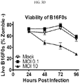
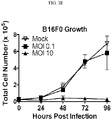
- FIGS. 3 A-E depicts MCMV-gp100 infection of B16F0s in vitro induced cell death and increased immunogenicity.
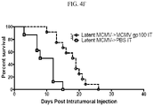
- FIGS. 4 A-F depicts an intratumoral infection of B16F0 tumors with MCMV, which induced tumor growth delay, regression, and improved survival.
- FIGS. 5 A-B depicts intratumoral infection of MC38 colon adenocarcinoma with MCMV.
- FIGS. 6 A-C depicts the infection of tumor-associated macrophages (TAMS) after IT MCMV therapy.
- TAMS tumor-associated macrophages
- FIGS. 7 A-B depicts the survival benefit after MCMV IT therapy without CD8 + T cells or NK cells.
- FIGS. 8 A-E depicts the tumor antigen-specific CD8+ T cells in the tumor, which were PD-1 hi and dysfunctional after MCMV IT infection.
- FIGS. 9 A and B depict the IT MCMV treatment as combined with anti-PD-L1 therapy, which improves B16F0 tumor growth delay and survival.
- FIG. 10 depicts the primary tumor clearance after MCMV IT treatment induces resistance or rejection of secondary tumor challenges.
- FIGS. 11 A-D depict that MCMV-gp100 is able to infect and spread in B16F0s at low and high MOIs.
- FIG. 12 illustrates the impact of MCMV IT on T-cell function.
- IP intraperitoneal
- ID intradermal
- MCMV could infect and kill B16F0s, indicating that MCMV could be killing tumor cells directly.
- most of the infected cells were tumor-associated macrophages suggesting that direct destruction of tumor cells was not the dominant mechanism preventing tumor growth.
- depletion of CD8 + T cells abrogated the therapeutic effect of IT MCMV therapy, demonstrating a need for CD8 + T cells for the success of the therapy.
- IT MCMV infection may alter the tumor microenvironment, either through infection of macrophages or other tumor-associated cells, or through the activation of immune enhancing signaling cascades, to improve anti-tumor immunity.
- PD-L1 blocking antibodies are known to improve anti-tumor T cell responses and the synergy between the two therapies was achieved regardless of whether the virus encoded a tumor antigen.
- the PD-L1 and similar checkpoint inhibitors prevents the inhibition of the T-cells in the body.
- the blockade strategy either prevents the generation of signaling compounds, or prevents the binding of the compounds on the T-Cells, to prevent the down regulation or “off” signal to the T-cells.
- OX40, CD27, and CD40L are on the T-cell and enhance or promote the immune response.
- CD40 works in a similar manner through separate pathways to enhance T-cell function.
- Either a blocking strategy or an enhancement strategy may be suitable to be combined with the CMV vaccine in a concomitant therapeutic so as to treat cancers, as described herein.
- FIGS. 1 A-C depicts the construction and characterization of mcmv-gfp-gp100
- T cells were obtained from the peripheral blood on day 104 post-infection with either MCMV-gp100 or WT-MCMV.
- FIGS. 2 A-D depicts an intraperitoneal and intradermal infection with MCMV-gp100 which induced poor anti-tumor responses.
- Animals received 1 ⁇ 10 5 B16F0s subcutaneously on D0 followed by IP or IP/ID vaccination with MCMV-gp100 or WT-MCMV on D5 post implantation. The data shown is combined from 3 separate experiments.
- NKs NK cells
- Neutro Neutrophil
- Granu Granulocyte
- Macro Macrophage
- Mono Monocyte
- Treg T regulatory cell.
- B) IFN- ⁇ production of CD8 + T cells recovered from tumors at sacrifice and stimulated or not ex vivo with the native gp100 peptide (n 5-9 mice).
- FIGS. 3 A-E depicts the MCMV-gp100 infection of B16F0s in vitro, which induced cell death and increased immunogenicity.
- B16F0s were plated at a concentration of 25,000 cells per well and “spinfected” one day later with MCMV-gp100 at an MOI of 10. Infected cells were identified by GFP expression. Shown is the mean fluorescent intensity of MHC-I (H-2D b and H-2K b ), and MHC-II (I-A/I-E), for infected and uninfected B16F0s in the same wells at D1 and D2 post-infection. Dotted lines at each timepoint represent the background MFI of infected B16F0s not stained with the indicated antibody. Data are representative of 2 independent experiments and each done in triplicate.
- FIGS. 4 A-F depicts an intratumoral infection of B16F0 tumors with MCMV, which induced tumor growth delay, regression, and improved survival.
- MCMV-gp100 IP/ID vaccination was given on D5 post tumor implantation followed by PBS IT on the schedule shown in “A”.
- NKs NK cells
- Neutro Neutrophil
- Granu Granulocyte
- Macro Macrophage
- Mono Monocyte
- Treg T regulatory cell.
- E Tumor growth from the day of IT infection.
- F Kaplan Myer survival curve of different treatment groups. For comparison, data from mice that were naive before tumor implantation is taken from FIG. 4 c.
- FIGS. 5 A-B depicts intratumoral infection of MC38 colon adenocarcinoma with MCMV.
- C57BL/6 mice were subcutaneously implanted with 5 ⁇ 10 5 MC38s and treated with MCMV IT or PBS IT as described in FIG. 4 , when tumors were 20 mm 2 .
- B) Kaplan Meier survival curve of the MCMV IT versus PBS IT treated animals from day of tumor implantation until tumors were above 100 mm 2 . Significance was assessed by a logrank test, p ⁇ 0.05 *.
- FIGS. 6 A-C depicts the infection of tumor-associated macrophages (TAMS) after IT MCMV therapy.
- Mice received IT injections with WT-MCMV or MCMV-gp100 as in FIG. 4 . Tumors were harvested 1 day after the last IT injection and processed for histology. Arrows indicate the same cell in each panel for identification of infected or uninfected cells.
- a and B Immunofluorescence staining of to define infected cells in tumors IT injected with PBS (control) or MCMV. Tumors were stained for DAPI to define the nucleus, CD45.2 to define hematopoietic cells and viral pp89 to define infected cells.
- infected cells are also hematopoietic cells (CD45.2-positive).
- panel A one infected cell is highlighted in the tumors from infected mice.
- panel B one infected cell (arrow pointing down and to the left) and two uninfected cells (arrows pointing up and to the left or down and to the right) are highlighted. The two highlighted uninfected cells are CD45-negative, and thus, non-hematopoietic cells.
- C As in A and B except that tumor sections were stained for pp89 to define infected cells, CD11b to define monocytic cells, and F4/80 to define macrophages, after MCMV IT infection. The data shows that nearly all infected cells were also expressing CD11b and F4/80. Three cells that were positive for all 3 markers are highlighted by the arrows.
- FIGS. 7 A-B depicts the survival benefit after MCMV IT therapy without CD8 + T cells or NK cells.
- B) Kaplan Meier survival curves of the different antibody depletion groups compared to the relevant isotype controls from day of tumor implantation until tumors were above 100 mm 2 . Significance was assessed by a logrank test, p ⁇ 0.05 *.
- FIGS. 8 A-E depicts the tumor antigen-specific CD8+ T cells in the tumor, which were PD-1 hi and dysfunctional when tumors were beginning to grow again after MCMV IT infection.
- Mice received 1 ⁇ 10 4 Pmel-Is one day prior to tumor implantation. Recipients were vaccinated by the IT route when the tumor reached 20 mm 2 . Tumors and spleens were collected D7 after MCMV-gp100 IT infection and assayed.
- FIGS. 9 A-B depicts the IT MCMV treatment combined with anti-PD-L1 therapy, which profoundly improves B16F0 tumor growth delay and survival.
- FIG. 10 shows that the primary tumor clearance after MCMV IT treatment induces resistance or rejection of secondary tumor challenges. Any animal that cleared a primary tumor was re-challenged with 2 ⁇ 10 5 B16F0s in their opposite flank 2-3 weeks after initial tumor clearance. A) Shown is the tumor growth starting from the day of tumor rechallenge. For the sake of clarity and fitting the data to a log scale, individual tumor area lines are spaced out below 1 mm 2 when no nodule was evident. Fractions in each graph represent the number of animals that rejected tumor challenge out of the number of animals tested.
- mice were infected by the IP route with 2 ⁇ 10 5 pfu WT-MCMV or MCMV-gp100 and 2 ⁇ 10 5 B16F0s were implanted subcutaneously 106 days later. Shown is the tumor growth as displayed in FIG. 2 . T cell responses in the blood of these mice, prior to tumor implantation, are shown in FIGS. 1 c and 1 d .
- FIGS. 11 A-D indicates that the MCMV-gp100 is able to infect and spread in B16F0s at low and high MOIs.
- B) Top panel shows representative GFP-expression (infection) of uninfected B16F0s cells (open histogram) versus infected B16F0s (MOI 10, grey histograms) at D2 post infection.
- Bottom panel shows representative Live/Dead stain of uninfected and uninfected cells at D2 post infection.
- D) Representative FACS plots of CD80 and CD86 expression by infected (MOI 10) or uninfected B16F0s at D1 and D2 post “spinfection” with MCMV-gp100. White histogram represents uninfected wells, dark grey histogram represents infected cells from infected wells. All these experiments were repeated twice and done in duplicate or triplicate.
- FIG. 12 shows the function of Pmel-I T cells recovered from tumors (left panel) or spleens (right panel) after MCMV-gp100 vaccination by the intraperitoneal (IP) route (filled triangles) or MCMV-gp100 vaccination by the IT route (open circles). Shown is the frequency of all Pmel-I T cells that produced both IFN- ⁇ and TNF- ⁇ after stimulation in vitro for 5 hours.
- IP intraperitoneal
- IT route open circles
- mice C57BL/6J and Pmel-I T cell transgenic mice (B6.Cg-Thy1 a /Cy Tg(TcraTcrb)8Rest/J) mice were purchased from Jackson Laboratory and bred in house for use in all experiments. Donor and recipient mice were sex-matched for all adoptive transfers. For most experiments, mice were between 6 and 12 weeks old at the time of tumor implantation. For the data shown in FIG. 3 D-F, mice were 6 to 12 weeks old at the time of primary MCMV infection and tumors were implanted 8 or 52 weeks later. For primary tumors, mice were subcutaneously challenged in the shaved right flank with 1 ⁇ 10 5 B16F0s suspended in HBSS (CellGro).
- tumor rechallenge experiments ( FIG. 10 ) animals that had cleared a primary tumor were re-challenged with 2 ⁇ 10 5 B16F0s in the shaved left flank. In all cases, tumor area was calculated by multiplying the length and width (in millimeters) of the tumor as measured with a 6-inch digital caliper (Neiko). Animals were sacrificed when the tumor was growing exponentially and had exceeded ⁇ 100 mm 2 in area, or when the tumors had ulcerated, or the animals had become moribund. The Thomas Jefferson University Institutional Animal Care and Use Committee reviewed and approved all protocols.
- the sequence encoding the altered gp100 S27P peptide (EGPRNQDWL) was fused to the 3′ end of the sequence encoding GFP, upstream of the stop codon, as done previously with SIINFEKL 38 .
- the fusion construct was recombined with MCMV encoded with a bacterial artificial chromosome (BAC, strain MW97.01, hereafter called WT-MCMV 23 ) and targeted to replace the m128 exon (IE2 gene) using established techniques 24 .
- Viral stocks were prepared on M2-10B4 stromal cells as previously described 41 .
- M2-10B4s were infected with lysates at several different titrations, covered with viscous media, incubated for 5 days, and stained with crystal violet for plaque counting.
- FIG. 3 f and FIG. 11 cells were “spinfected” to increase the frequency of infected cells detectible 1 day after infection. Spinfection was accomplished by spinning cells at 800 ⁇ g for 30 minutes after adding virus. Control, uninfected cells were treated in the same way except that no virus was added to the culture.
- M2-10B4s were grown in RPMI (CellGro)+1% PenStrep (Gemini, Benchmark)+10% FBS (Gemini).
- B16F0s were grown in DMEM (CellGro)+1% PenStrep+10% FBS.
- mice For infection of mice without tumors ( FIG. 1 and FIGS. 4 e and f ), animals received 2 ⁇ 10 5 plaque forming units (PFUs) of MCMV-gp100, or MCMV-K181 by the IP route in a single injection of 100 ⁇ l.
- PFUs plaque forming units
- For IP and ID infections of tumor-bearing mice animals received 5 ⁇ 10 5 PFUs of the indicated virus in a single injection of 100 ⁇ l for IP infection and for 25 ⁇ l for ID infection. In all cases, ID infection was performed in the skin next to the tumor implantation site.
- IT infections animals received 5 ⁇ 10 5 PFUs of the indicated virus in 30 ⁇ l volume or 30 ⁇ l of PBS every other day for 3 total injections.
- Spleens were harvested from na ⁇ ve Pmel-I transgenic mice, passed through a 70 ⁇ m cell strainer to form single cell suspensions and washed twice with T cell media (RPMI 1640 [Cellgro] with L-glutamine+10% FBS+1% PenStrep and 5 ⁇ 10 ⁇ 5 M ⁇ -mercaptoethanol [Omnipur, Calbiochem]).
- T cell media RPMI 1640 [Cellgro] with L-glutamine+10% FBS+1% PenStrep and 5 ⁇ 10 ⁇ 5 M ⁇ -mercaptoethanol [Omnipur, Calbiochem]
- Total splenocytes were counted on a Z2 Coulter Particle Count and Size Analyzer (Beckman Coulter) and the sample was assessed for frequency of CD8 + T cells by flow cytometry. Based on these data, total splenocytes were suspended in PBS so that the desired number of CD8 + T cells was present in 100 ⁇ l, which is the volume that
- Spleens were suspended in T cell media and mechanically processed through a 70 ⁇ m nylon filter to achieve a single cell suspension.
- tumor digestion media lx HBSS [Cellgro], 0.1 mg/ml Collagenase A [Worthington], 60 U/ml DNase I [Roche], 52 and minced using the gentleMACSTM Octo Dissociator using C Tubes (Miltenyi Biotec). Minced tumors in digestion media were incubated at 37° C. for 30 minutes with continuous rotation.
- Digested tumors were minced again using the gentleMACSTM Octo Dissociator, then washed twice with T cell media and mechanically filtered through a 70 ⁇ m nylon filter to make a single cell suspension. Lymphocytes were then either directly assessed by flow cytometry or tested for their ability to produce cytokines upon stimulation. For analyses of cytokine production by cells from spleens and tumors, 1-2 ⁇ 10 6 cells were incubated in T cell media in a round bottom 96-well plate 53 for 5 hours at 37° C. in 5% CO 2 .
- the final incubation volume was 100 ⁇ l and contained 1 ⁇ g/ml of the indicated peptide (synthesized by Genemed Synthesis) and 1 ⁇ g/ml brefeldin A (GoldiPlug, BD Biosciences), as well as fluorescently labeled antibody specific for CD107a.
- cells were washed twice with ice-cold FACS buffer (PBS, 0.05% Sodium Azide, 1% FBS) and stained with antibodies specific for surface proteins followed by analyses of intracellular IFN- ⁇ and TNF- ⁇ using the BD Cytofix/Cytoperm kit (BD Biosciences) and following the manufacturer's instructions.
- FIGS. 1C and D ⁇ 150 ⁇ l of peripheral blood was collected from the retro-orbital sinus.
- Red blood cells were lysed for 5 minutes in red blood cell lysis buffer (150 mM NH 4 Cl, 10 mM NaHCO 3 ) and the remaining white blood cells were washed twice and resuspended in T cell media. Approximately 1 ⁇ 5 of the recovered cells were added to individual wells and incubated as above for 3 hours and without the antibodies specific for CD107a.
- Treg staining cells were fixed with FOXP3 Fix/Perm buffer (Biolegend) for 10 minutes on ice and then permeabilized for 15 minutes with FOXP3 Perm buffer (Biolegend) before adding 2.5 ⁇ l of anti-FOXP3 per sample.
- CD3 (clone 500A2), CD4 (clone GK1.5), CD8 ⁇ (clone 53.6.7), CD8 ⁇ (clone YTS156.7.7), PD-1 (clone 29F.1A12), PD-L1 (clone 10F.9G2), H-2D b (clone KH95), H-2K b (clone AF6-88.5), CD80 (16-10A1), CD86 (GL-1), NK1.1 (clone PK136), CD11b (clone ICRF44), GR-1 (clone RB6-8C5), FoxP3 (clone 150D), I-A/I-E (clone M5/114.15.2), IFN- ⁇ (clone XMG1.2), TNF- ⁇ (clone MP6-XT22) and CD107a (clone 1D4B).
- Pmel-I T cells were identified using Thy1.1 (clone OX-7). All antibodies were purchased from Biolegend or BD Biosciences. Stained cells were analyzed using the LSR II flow cytometer (BD Biosciences) and FlowJo Software (TreeStar, Ashland, Oreg., USA).
- MCMV-gp100 infected B16F0 cells were identified by the appearance of GFP expression using a Nikon Eclipse TS100 microscope, Nikon Intensilight CHGF1 illumination system, and Nikon Digital Sight DS-L3 camera controller.
- mice were treated with 300 ⁇ g of anti-CD8a (clone 53-6.72) and/or anti-NK1.1 (clone PK136) every 3 days for a total of 8 treatments, starting 2 days before tumor implantation. Treatment resulted in greater than 90% depletion of target cells (data not shown).
- additional animals were treated with an irrelevant IgG2a antibody (isotype control for anti-NK1.1, clone C1.18.4), or IgG2b antibody (isotype control for anti-CD8a, clone LTF-2) following the same schedule.
- mice were treated with 400 ⁇ g of anti-PD-L1 (clone 10F.9G2) by the IP route on the first day of IT treatment, followed by an additional 200 ⁇ g anti-PD-L1 given every third day by the IP route for a total of 6 treatments.
- additional animals were treated with the IgG2b isotype control clone LTF-2, following the same schedule. All antibodies were purchased from Bio-X-Cell.
- Isolated tumors were frozen in Fisher HealthcareTM Tissue-Plus OCT (Fisher Scientific) and cut into 6-8 ⁇ m sections using a cryostat. Samples were fixed in cold acetone for 10 minutes and rehydrated with Tris-buffered saline (TBS) for 20 minutes, blocked with blocking buffer (TBS+3% BSA and 0.1% Tween-20) for 20 minutes and stained with antibodies specific for CD31 (clone 390), CD45.1/2 (clone A20/104), CD11b (clone M1/70), F4/80 (clone BM8) and/or MCMV pp89 (clone 6/58/1 60 ) in blocking buffer for 1 hour and later co-stained with DAPI (Prolong Gold antifade—Life Technologies).
- the anti-pp89 antibody was purified from hybridoma supernatant using PierceTM Protein A/G Magnetic Beads (Fisher Scientific), concentrated using Amicon Ultra-0.5 or 15 Centrifugal Filter Unit with Ultracel-100 membrane (Millipore), and labeled using Mix-N-Stain CF555 Antibody Labeling Kit (Sigma-aldrich). Anti-pp89 flourophore conjugation was confirmed by staining infected and uninfected M2-10B4s with the labeled antibody (data not shown). Images were generated with an LSM 510 Meta confocal laser scanning microscope (Carl Zeiss), the LSM image browser software (Carl Zeiss), and ImageJ (Fiji).
- Prism Version 6.0d was used for graph creation and some statistical analyses. For statistical significance, *p ⁇ 0.05 **p ⁇ 0.01 ***p ⁇ 0.001 ****p ⁇ 0.0001.
- Tumor growth was analyzed with a mixed-effects linear regression, an extension of ordinary linear regression for repeated measures over time. Heuristically, the model estimates a tumor growth curve for each animal and then appropriately averages these curves to estimate the group's average trajectory. This approach accounts for the within-animal correlation of tumor sizes over time and the potential uneven timing of readings. Tumor size was log-transformed before the analyses and was modeled as a function of time, experimental group, and their interaction. The main aim was to compare growth rates over time across the experimental groups. Results were expressed in terms of the average daily increase of tumor size and the tumor doubling time. We also used Kaplan-Meier survival curves and the logrank test to analyze the time tumors needed to reach 100 mm 2 (overall survival, the approximate tumor size when animals are typically sacrificed).
- a recombinant strain of MCMV was created that expresses GFP fused to an altered version of the gp10025-33 peptide (gp100 S27P ).
- This fusion construct was inserted into the IE2 locus and under the control of the endogenous MCMV IE2 promoter (MCMV-gp100, FIG. 1 a ), a strategy that has been used to stimulate robust T cell responses to recombinant antigens in the MCMV backbone 34-36 .
- the growth of MCMV-gp100 was similar to that of its wild-type counterpart as seen by multi-step in vitro growth curves ( FIG. 1 b ).
- the CMV vaccine can be grown and generated by means known to a person of ordinary skill in the art.
- the viral cells are grown in a tissue culture medium and harvested. After harvest, the cells are purified and diluted in a sterile solution suitable for injection, for example PBS.
- a sterile solution suitable for injection for example PBS.
- suitable excipients, solutions, components, and the like can be provided as known to a person of ordinary skill in the art.
- Remington teaches appropriate formulations and strategies to ensure that the injectable vaccine is isotonic and suitable for injection; whereas Harlow, and Molecular Cloning otherwise teach appropriate steps for cloning or modification of cells for inclusion in the CMV vaccines as described herein, or the antibody therapeutics to be co-administered with the CMV vaccine.
- the vaccine therefore, comprises CMV cells, in one or more of the forms as identified herein, a delivery vehicle, and suitable excipients and components for injection. It is expected that certain impurities will remain, including the culture medium and/or purification compositions as known to a person of ordinary skill in the art.
- mice were subcutaneously implanted in the flank and mice were vaccinated five days later with MCMV-gp100.
- the site of infection or vaccination can influence the migration of CD8 + T cells and subsequent protection 37,38 . Therefore, we vaccinated mice by the IP route alone or in combination with an ID vaccination in the skin adjacent to the tumor.
- vaccination caused increased infiltration of CD4 + T cells, CD8 + T cells, and FoxP3 + regulatory T cells (T REG ), but no increase of NK Cells, Neutrophils, Granulocytes, Macrophages, or Monocytes ( FIG. 2 a ).
- mice were implanted with B16F0s subcutaneously, as above. When tumors were approximately 20 mm 2 ( ⁇ 7-14 days after tumor implantation), they were injected directly with WT-MCMV, MCMV-gp100, or PBS, every other day for 3 treatments ( FIG. 4 a ). For comparison, another group was vaccinated by IP and ID routes as above ( FIG. 2 ), and then given PBS by the IT route. As shown in FIG. 4 b , direct IT infection with MCMV had a marked effect on the growth of established tumors.
- mice treated with PBS IT or MCMV-gp100 IP/ID+PBS IT had an average daily tumor growth rate after the IT injection of 21% and 19% respectively, and the tumor size doubled every 3.6 or 3.7 days respectively ( FIG. 4 b ).
- mice were infected with either WT-MCMV or MCMV-gp100 by the IT route the average daily growth rate post IT injection was reduced to 10% and 8% respectively, and the doubling time was increased to 7.3 and 9.4 days respectively, all of which were significantly slower than the controls ( FIG. 4 b ).
- the presence of the gp100 epitope in the vaccine did relatively little to improve the outcome.
- Pre-existing anti-viral immunity may be able to restrict the efficacy of oncolytic viruses by clearing the virus 11 . More than half of people in the United States and most people in the world are already infected with CMV 24 . Therefore, we tested whether IT infection would delay tumor growth in mice that had been infected with a wild-type strain of MCMV (MCMV-K181) 8 or 52 weeks prior to tumor implantation. Importantly, prior MCMV infection had no significant effect on the survival induced by MCMV-gp100 IT infection ( FIGS. 4 e and f ) or the daily tumor growth rate measured after MCMV-gp100 IT infection (11% vs. 8% for MCMV immune vs. na ⁇ ve animals). Thus, pre-existing MCMV-specific immunity did not limit the therapeutic benefit of MCMV IT infection.
- MCMV Infects Tumor-Associated Macrophages after MCMV IT Infection.
- MCMV could infect and kill B16F0s ( FIG. 3 ) and MC38s (not shown) in vitro, suggesting that it could be acting like an oncolytic virus.
- MCMV can also infect many other cells in the tumor environment including endothelial cells, fibroblasts and macrophages.
- endothelial cells fibroblasts
- macrophages To determine which cells were infected by MCMV after IT inoculation, B16F0 tumors were recovered one day after the last MCMV IT injection. Infected cells were identified histologically by the presence of nuclear-localized viral pp89, an immediate early protein (IE1) expressed by MCMV infected cells shortly after infection 40 .
- IE1 immediate early protein
- the two highlighted uninfected cells are CD45-negative, and thus, non-hematopoietic cells. Further analyses revealed that infected cells also expressed CD11b and F4/80 ( FIG. 6 c ). Three cells that were positive for all 3 markers are highlighted by the arrows. These data suggest that MCMV primarily infected tumor associated macrophages (TAMs) and not tumor cells, suggesting that MCMV was not acting as an oncolytic virus.
- TAMs tumor associated macrophages
- CD8 + T cells and NK cells were depleted before the implantation of B16F0 tumors and throughout the MCMV IT therapy.
- Depletion of CD8 + T cells significantly reduced survival after MCMV-gp100 IT infection, while depletion of NK1.1 alone had no effect ( FIGS. 7 a and b ).
- MCMV-IT therapy depended on CD8 + T cells to prolong survival.
- Tumor-Specific T Cells are Markedly Dysfunctional within the Tumor and PD-L1 Blockade Greatly Enhances Tumor Growth Delay and Regression Induced by MCMV IT Treatment.
- gp100-specific Pmel-I transgenic T cells were used to explore tumor-specific T cells after IT therapy.
- Na ⁇ ve mice were given 10 4 Pmel-I T cells expressing the Thy1.1. congenic marker, and B16F0 cells were implanted 1 day later.
- recipients were IT infected when the tumors reached ⁇ 20 mm 2 .
- Animals were sacrificed 7 days after the initial IT infection and tumor-infiltrating T cells were assessed. With only 10 4 Pmel-I T cells transferred, the donor cells were undetectable in recipients infected with WT-MCMV, with the exception of one animal ( FIG. 8 a and data not shown).
- FIG. 8 a In contrast, IT infection with MCMV-gp100 induced expansion and migration of Pmel-I T cells to the tumor in all mice ( FIG. 8 a ). Notably, these cells expressed high levels of the inhibitory molecule PD-1 3 ( FIGS. 8 b and c ) were dysfunctional for cytokine production and degranulation compared to Pmel-I cells in the spleens of the same animals ( FIG. 8 d ). Interestingly, tumor-specific Pmel-I T cells were slightly more functional in the tumor, and markedly more functional in the spleen after MCMV-IT therapy when compared to MCMV vaccination by the systemic intraperitoneal route ( FIG. 12 ). PD-L1 was also detectable on cells within tumors in slightly higher levels after MCMV IT injections than PBS IT injections ( FIG. 8 e ), although there were no differences in PD-1 expression (data not shown).
- MCMV-IT therapy is improving tumor-specific T cell responses ( FIG. 12 ) and delaying tumor growth in a manner that depends on CD8 + T cells ( FIG. 7 ), and synergizes with PD-L1 checkpoint blockade ( FIG. 9 ).
- MCMV-IT therapy may synergize with the PD-L1 checkpoint blockade by further improving T cell function.
- MCMV-IT therapy would be expected to synergize with other checkpoint blockades that promote T cell function, such as antibodies that target PD-1 and CTLA-4.
- PD-L1 can be expressed by hematopoietic cells in the tumor, including tumor-associated macrophages ( FIG. 8 e ) and thus, it is possible that the PD-L1 blocking antibody synergizes with MCMV-IT therapy through direct interactions with tumor-infiltrating hematopoietic cells and/or by improving T cell function.
- MCMV does not fit the typical definition of an oncolytic virus.
- Oncolytic viruses are typically defined by their ability to replicate rapidly and somewhat selectively in tumor cells, inducing tumor cell death and subsequent anti-tumor and anti-viral immune responses 41,42 . While MCMV could infect and kill B16F0s in culture ( FIGS. 3 and 11 ), it only seemed to infect tumor-associated macrophages (TAMs) in vivo ( FIG. 6 ), suggesting that IT therapy is not working through direct tumor lysis. TAMs are associated with tumor progression by inducing a pro-angiogenic environment and suppressing anti-tumor immune responses 43,44 .
- CMV infects monocytes and macrophages, inducing monocyte migration, tissue entry, and differentiation into macrophages 45,46 .
- CMV infection of macrophages shifts them to an immune stimulatory phenotype by inducing up-regulation of TLRs and increasing Th1 cytokine production 46-48 , subsequently leading to increased T cell proliferation 47 .
- MCMV infection of TAMs could decrease the macrophage production of pro-angiogenic factors, such as VEGF and ARG1, decreasing blood flow to tumors and slowing growth 43,44 . All these possibilities must addressed in future studies as possible mechanisms for MCMV IT therapy.
- MCMV immediate early protein pp89 (IE1), which is expressed early after infection, may have different expression levels in different cell types. Thus it is possible that we only detected a subset of infected cells in the tumor that expressed pp89 at high levels, and that MCMV is still infecting the tumor cells themselves, or the tumor vasculature 49 . This caveat aside, our data suggest that MCMV IT therapy works by altering TAMs and their interaction with tumors.
- CMV ulcerative colitis .
- CMV glycoproteins have been described to activate Toll-like receptor 2 (TLR-2) 50,51 and CMV particles can activate the epidermal growth factor receptor (EGFR) 52-54 , leading to an array of cellular responses.
- TLR-2 Toll-like receptor 2
- EGFR epidermal growth factor receptor
- MCMV is a potent stimulator of NK cells and ⁇ -T cells 39,55 , both of which might have anti-tumor effects. Therefore, infection of tumor associated macrophages may provide a therapeutic benefit, but may not be required for the therapeutic effect or the synergy with additional immune therapies.
- NK cells expressing Ly49H are specifically expanded in response to the viral m157 protein 39 .
- this population was not expanded in the tumors of mice vaccinated with MCMV by the IT route (not shown). Rather, the tumor-infiltrating NK cells were largely Ly49H ⁇ , KLRG-1+, possibly suggesting that tumor-localized NK cells were activated in response to the tumor.
- NK cell depletion had no effect on the MCMV IT therapy ( FIG. 7 ), suggesting NK cells were not important for the therapeutic outcome. Additional experiments will be needed to explore the impact of MCMV on other cells in the tumor and the tumor environment as a whole, after injection of live or inactivated viral particles.
- live, virulent, attenuated or killed virus vaccines may all be effective alone or in combination with immune checkpoint inhibitors.
- MCMV-gp100 vaccine used by Qiu and colleagues expressed a variant of the gp100 antigen that differed by 2 amino acids from the native sequence (gp100 E25K, S27P ), whereas the epitope used in our study differed by only one amino acid (gp100 S27P ), a difference that could, in theory, have a substantial impact on the efficacy or function of gp100-specific T cells. Future work will be required to test these ideas.
- MCMV may have superior therapeutic efficacy for cutaneous melanomas after direct intra-tumoral injections, and that this route of vaccination can synergize with immune checkpoint blockades to clear tumors and induce protection against distal tumors, without virally encoded tumor antigens.
- This study builds on recent data suggesting that CMV may be an effective anti-tumor therapy and suggests that the route of infection and tumor location may be critical factors in defining the efficacy of this platform.
Landscapes
- Health & Medical Sciences (AREA)
- Life Sciences & Earth Sciences (AREA)
- Chemical & Material Sciences (AREA)
- Medicinal Chemistry (AREA)
- General Health & Medical Sciences (AREA)
- Pharmacology & Pharmacy (AREA)
- Animal Behavior & Ethology (AREA)
- Public Health (AREA)
- Veterinary Medicine (AREA)
- Microbiology (AREA)
- Immunology (AREA)
- Mycology (AREA)
- Epidemiology (AREA)
- Oncology (AREA)
- Bioinformatics & Cheminformatics (AREA)
- Virology (AREA)
- Engineering & Computer Science (AREA)
- Organic Chemistry (AREA)
- Zoology (AREA)
- Genetics & Genomics (AREA)
- Wood Science & Technology (AREA)
- General Chemical & Material Sciences (AREA)
- Endocrinology (AREA)
- Chemical Kinetics & Catalysis (AREA)
- Nuclear Medicine, Radiotherapy & Molecular Imaging (AREA)
- Biochemistry (AREA)
- General Engineering & Computer Science (AREA)
- Biotechnology (AREA)
- Biomedical Technology (AREA)
- Medicines Containing Antibodies Or Antigens For Use As Internal Diagnostic Agents (AREA)
- Medicines That Contain Protein Lipid Enzymes And Other Medicines (AREA)
- Medicines Containing Material From Animals Or Micro-Organisms (AREA)
Abstract
Description
- 1 Klebanoff, C. A., Acquavella, N., Yu, Z. & Restifo, N. P. Therapeutic cancer vaccines: are we there yet? Immunol Rev 239, 27-44, doi:10.1111/j.1600-065X.2010.00979.x (2011).
- 2 Schreiber, R. D., Old, L. J. & Smyth, M. J. Cancer immunoediting: integrating immunity's roles in cancer suppression and promotion. Science 331, 1565-1570, doi:10.1126/science.1203486 (2011).
- 3 Wherry, E. J. T cell exhaustion. Nature Immunology 131, 492-499, doi:10.1038/ni.2035 (2011).
- 4 Hailemichael, Y. & Overwijk, W. W. Cancer vaccines: Trafficking of tumor-specific T cells to tumor after therapeutic vaccination. Int J Biochem Cell Biol 53, 46-50, doi:10.1016/j.biocel.2014.04.019 (2014).
- 5 Azimi, F. et al. Tumor-infiltrating lymphocyte grade is an independent predictor of sentinel lymph node status and survival in patients with cutaneous melanoma.
J Clin Oncol 30, 2678-2683, doi:10.1200/JCO.2011.37.8539 (2012). - 6 Singh, M. & Overwijk, W. W. Intratumoral immunotherapy for melanoma. Cancer Immunol Immunother 64, 911-921, doi:10.1007/s00262-015-1727-z (2015).
- 7 Marabelle, A., Kohrt, H., Caux, C. & Levy, R. Intratumoral immunization: a new paradigm for cancer therapy.
Clin Cancer Res 20, 1747-1756, doi:10.1158/1078-0432.CCR-13-2116 (2014). - 8 Miest, T. S. & Cattaneo, R. New viruses for cancer therapy: meeting clinical needs. Nature reviews. Microbiology 12, 23-34, doi:10.1038/nrmicro3140 (2014).
- 9 Andtbacka, R. H. et al. Talimogene Laherparepvec Improves Durable Response Rate in Patients With Advanced Melanoma. J Clin Oncol, doi:10.1200/JCO.2014.58.3377 (2015).
- 10 FDA approves first-of-its-kind product for the treatment of melanoma, fda.gov/NewsEvents/Newsroom/PressAnnouncements/ucm469571.htm (2015).
- 11 Barlett, D. L. et al. Oncolytic viruses as therapeutic cancer vaccines. Molecular Cancer 13 (2013).
- 12 Heo, J. et al. Randomized dose-finding clinical trial of oncolytic immunotherapeutic vaccinia JX-594 in liver cancer. Nat Med 19, 329-336, doi:10.1038/nm.3089 (2013).
- 13 Hemminki, O. et al. Immunological data from cancer patients treated with Ad5/3-E2F-Δ24-GMCSF suggests utility for tumor immunotherapy.
Oncotarget 6, 4467-4481 (2015). - 14 Woller, N., Gurlevik, E., Ureche, C. I., Schumacher, A. & Kuhnel, F. Oncolytic viruses as anticancer vaccines.
Front Oncol 4, 188, doi:10.3389/fonc.2014.00188 (2014). - 15 Holtappels, R., Pahl-Seibert, M.-F., Thomas, D. & Reddehase, M. J. Enrichment of Immediate-Early 1 (m123/pp89) Peptide-Specific CD8 T Cells in a Pulmonary CD62Llo Memory-Effector Cell Pool during Latent Murine Cytomegalovirus Infection of the Lungs. Journal of Virolody 74, 11495-11503 (2000).
- 16 Holtappels, R. et al. Processing and Presentation of Murine Cytomegalovirus pORFm164-Derived Peptide in Fibroblasts in the Face of All Viral Immunosubversive Early Gene Functions. Journal of Virology 76, 6044-6053, doi:10.1128/jvi.76.12.6044-6053.2002 (2002).
- 17 Komatsu, H., Sierro, S., V. Cuero, A. & Klenerman, P. Population analysis of antiviral T cell responses using MEW class I-peptide tetramers. Clinical and Experimental Immunology 134, 9-12, doi:10.1046/j.1365-2249.2003.02266.x (2003).
- 18 Karrer, U. et al. Expansion of Protective CD8+ T-Cell Responses Driven by Recombinant Cytomegaloviruses. Journal of Virology 78, 2255-2264, doi:10.1128/jvi.78.5.2255-2264.2004 (2004).
- 19 Munks, M. W. et al. Genome-Wide Analysis Reveals a Highly Diverse CD8 T Cell Response to Murine Cytomegalovirus. Journal of immunology 176, 3760-3766 (2006).
- 20 Borst, E. M., Benkartek, C. & Messerle, M. Use of bacterial artificial chromosomes in generating targeted mutations in human and mouse cytomegaloviruses. Curr Protoc Immunol. (2007).
- 21 Hansen, S. G. et al. Effector memory T cell responses are associated with protection of rhesus monkeys from mucosal simian immunodeficiency virus challenge.
Nat Med 15, 293-299, doi:10.1038/nm.1935 (2009). - 22 Hansen, S. G. et al. Profound early control of highly pathogenic SIV by an effector memory T-cell vaccine. Nature 473, 523-527, doi:10.1038/nature10003 (2011).
- 23 Hansen, S. G. et al. Immune clearance of highly pathogenic SIV infection. Nature 502, 100-104, doi:10.1038/nature12519 (2013).
- 24 Bate, S. L., Dollard, S. C. & Cannon, M. J. Cytomegalovirus seroprevalence in the United States: the national health and nutrition examination surveys, 1988-2004. Clin Infect
Dis 50, 1439-1447, doi:10.1086/652438 (2010). - 25 Hansen, S. G. et al. Evasion of CD8+ T cells is critical for superinfection by cytomegalovirus. Science 328, 102-106, doi:10.1126/science.1185350 (2010).
- 26 Hertoghs, K. M. et al. Molecular profiling of cytomegalovirus-induced human CD8+ T cell differentiation. J Clin Invest 120, 4077-4090, doi:10.1172/JCI42758 (2010).
- 27 Sierro, S., Rothkopf, R. & Klenerman, P. Evolution of diverse antiviral CD8+ T cell populations after murine cytomegalovirus infection. European journal of
immunology 35, 1113-1123, doi:10.1002/eji.200425534 (2005). - 28 Smith, C. J., Turula, H. & Snyder, C. M. Systemic hematogenous maintenance of memory inflation by MCMV infection.
PLoS Pathog 10, e1004233, doi:10.1371/journal.ppat.1004233 (2014). - 29 Smith, C. J., Caldeira-Dantas, S., Turula, H. & Snyder, C. M. Murine CMV Infection Induces the Continuous Production of Mucosal Resident T Cells. Cell Rep 13, 1137-1148, doi:10.1016/j.celrep.2015.09.076 (2015).
- 30 Klyushnenkova, E. N. et al. A cytomegalovirus-based vaccine expressing a single tumor-specific CD8+ T-cell epitope delays tumor growth in a murine model of prostate cancer.
J Immunother 35, 390-399, doi:10.1097/CJI.0b013e3182585d50 (2012). - 31 Xu, G., Smith, T., Grey, F. & Hill, A. B. Cytomegalovirus-based cancer vaccines expressing TRP2 induce rejection of melanoma in mice. Biochem Biophys Res Commun 437, 287-291, doi:10.1016/j.bbrc.2013.06.068 (2013).
- 32 Qiu, Z. et al. Cytomegalovirus based vaccine expressing a modified tumor antigen induces potent tumor-specific CD8+ T cell response and protects mice from melanoma. Cancer Immunology Research 3, 1-11, doi:10.1158/2326-6066.cir-14-0044 (2015).
- 33 van Stipdonk, M. J. et al. Design of agonistic altered peptides for the robust induction of CTL directed towards H-2db in complex with the melanoma-associated epitope gp100. Cancer Res 69, 7784-7792, doi:10.1158/0008-5472.CAN-09-1724 (2009).
- 34 Dekhtiarenko, I., Jarvis, M. A., Ruzsics, Z. & Cicin-Sain, L. The context of gene expression defines the immunodominance hierarchy of cytomegalovirus antigens. Journal of immunology 190, 3399-3409, doi:10.4049/jimmunol.1203173 (2013).
- 35 Farrington, L. A., Smith, T. A., Grey, F., Hill, A. B. & Snyder, C. M. Competition for antigen at the level of the APC is a major determinant of immunodominance during memory inflation in murine cytomegalovirus infection. Journal of immunology 190, 3410-3416, doi:10.4049/jimmunol.1203151 (2013).
- 36 Turula, H., Smith, C. J., Grey, F., Zurbach, K. A. & Snyder, C. M. Competition between T cells maintains clonal dominance during memory inflation induced by MCMV. European journal of immunology 43, 1252-1263, doi:10.1002/eji.201242940 (2013).
- 37 Wakim, L. M., Jones, C. M., Gebhardt, T., Preston, C. M. & Carbone, F. R. CD8(+) T-cell attenuation of cutaneous herpes simplex virus infection reduces the average viral copy number of the ensuing latent infection. Immunol Cell Biol 86, 666-675, doi:10.1038/icb.2008.47 (2008).
- 38 Liu, L. et al. Epidermal injury and infection during poxvirus immunization is crucial for the generation of highly protective T cell-mediated immunity. Nat Med 16, 224-227, doi:10.1038/nm.2078 (2010).
- 39 Lanier, L. L. Evolutionary struggles between NK cells and viruses. Nature reviews.
Immunology 8, 259-268, doi:10.1038/nri2276 (2008). - 40 Keil, G. M., Ebeling-Keil, A. & Koszinowski, U. H. Immediate-Early Genes of Murine Cytomegalovirus: Location, Transcripts, and Translation Products. Journal of Virology 61, 526-533 (1987).
- 41 Larocca, C. & Schlom, J. Viral vector-based therapeutic cancer vaccines. Cancer J 17, 359-371, doi:10.1097/PPO.0b013e3182325e63 (2011).
- 42 Lichty, B. D., Breitbach, C. J., Stojdl, D. F. & Bell, J. C. Going viral with cancer immunotherapy. Nature reviews. Cancer 14, 559-567, doi:10.1038/nrc3770 (2014).
- 43 Gabrilovich, D. I., Ostrand-Rosenberg, S. & Bronte, V. Coordinated regulation of myeloid cells by tumours. Nature reviews. Immunology 12, 253-268, doi:10.1038/nri3175 (2012).
- 44 Chanmee, T., Ontong, P., Konno, K. & Itano, N. Tumor-associated macrophages as major players in the tumor microenvironment. Cancers (Basel) 6, 1670-1690, doi:10.3390/cancers6031670 (2014).
- 45 Daley-Bauer, L. P., Roback, L. J., Wynn, G. M. & Mocarski, E. S. Cytomegalovirus hijacks CX3CR1(hi) patrolling monocytes as immune-privileged vehicles for dissemination in mice. Cell host &
microbe 15, 351-362, doi:10.1016/j.chom.2014.02.002 (2014). - 46 Smith, M. S., Bentz, G. L., Alexander, J. S. & Yurochko, A. D. Human Cytomegalovirus Induces Monocyte Differentiation and Migration as a Strategy for Dissemination and Persistence. Journal of Virology 78, 4444-4453, doi:10.1128/jvi.78.9.4444-4453.2004 (2004).
- 47 Bayer, C. et al. Human cytomegalovirus infection of M1 and M2 macrophages triggers inflammation and autologous T-cell proliferation. J Virol 87, 67-79, doi:10.1128/JVI.01585-12 (2013).
- 48 Gary Chan1, E. R. B.-S., M. Shane Smith2, Patrick M. Smith1, and Andrew D. Yurochko. Transcriptome Analysis Reveals Human Cytomegalovirus Reprograms Monocyte Differentiation Towards a M1 Macrophage. (2008).
- 49 van de Berg, P. J., Yong, S. L., Remmerswaal, E. B., van Lier, R. A. & ten Berge, I. J. Cytomegalovirus-induced effector T cells cause endothelial cell damage. Clin Vaccine Immunol 19, 772-779, doi:10.1128/CVI.00011-12 (2012).
- 50 Szomolanyi-Tsuda, E., Liang, X., Welsh, R. M., Kurt-Jones, E. A. & Finberg, R. W. Role for TLR2 in NK cell-mediated control of murine cytomegalovirus in vivo.
J Virol 80, 4286-4291, doi:10.1128/JVI.80.9.4286-4291.2006 (2006). - 51 Boehme, K. W., Guerrero, M. & Compton, T. Human Cytomegalovirus Envelope Glycoproteins B and H Are Necessary for TLR2 Activation in Permissive Cells. The Journal of Immunology 177, 7094-7102, doi:10.4049/jimmunol.177.10.7094 (2006).
- 52 Yamazaki, D. et al. WAVE2 is required for directed cell migration and cardiovascular development. Nature 424, 452-456, doi:10.1038/nature01770 (2003).
- 53 Chan, G., Nogalski, M. T. & Yurochko, A. D. Activation of EGFR on monocytes is required for human cytomegalovirus entry and mediates cellular motility. Proc Natl
Acad Sci USA 106, 22369-22374, doi:10.1073/pnas.0908787106 (2009). - 54 Bentz, G. L. & Yurochko, A. D. Human CMV infection of endothelial cells induces an angiogenic response through viral binding to EGF receptor and beta1 and beta3 integrins. Proc Natl
Acad Sci USA 105, 5531-5536, doi:10.1073/pnas.0800037105 (2008). - 55 Sell, S. et al. Control of murine cytomegalovirus infection by gammadelta T cells. PLoS Pathog 11, e1004481, doi:10.1371/journal.ppat.1004481 (2015).
- 56 Nannmark, U. et al. Microvessel Origin and Distribution in Pulmonary Metastases of B16 Melanoma: Implication for Adoptive Immunotherapy’. Cancer Research 55, 4627-4632 (1995).
- 57 Wagner, M., Jonjic, S., Koszinowski, U. H. & Messerle, M. Systematic Excision of Vector Sequences from the BAC-Cloned Herpesvirus Genome during Virus Reconstitution. Journal of Virology 73, 7056-7060 (1999).
- 58 Zurbach, K. A., Moghbeli, T. & Snyder, C. M. Resolving the titer of murine cytomegalovirus by plaque assay using the M2-10B4 cell line and a low viscosity overlay. Virol J 11, 71, doi:10.1186/1743-422X-11-71 (2014).
- 59 Thompson, E. D., Enriquez, H. L., Fu, Y. X. & Engelhard, V. H. Tumor masses support naive T cell infiltration, activation, and differentiation into effectors. The Journal of experimental medicine 207, 1791-1804, doi:10.1084/jem.20092454 (2010).
- 60 Reddehase, M. J., Fibi, M. R., Keil, G. M. & Koszinowski, U. H. Late-Phase Expression of a Murine Cytomegalovirus Immediate-Early Antigen Recognized by Cytolytic T Lymphocytes. Journal of
Virology 60, 1125-1129 (1986).
Claims (14)
Priority Applications (1)
| Application Number | Priority Date | Filing Date | Title |
|---|---|---|---|
| US16/064,906 US11351237B2 (en) | 2015-12-22 | 2016-12-21 | CMV-based intra-tumoral cancer therapies |
Applications Claiming Priority (3)
| Application Number | Priority Date | Filing Date | Title |
|---|---|---|---|
| US201562271167P | 2015-12-22 | 2015-12-22 | |
| US16/064,906 US11351237B2 (en) | 2015-12-22 | 2016-12-21 | CMV-based intra-tumoral cancer therapies |
| PCT/US2016/068084 WO2017112797A1 (en) | 2015-12-22 | 2016-12-21 | Intra-lesional cmv-based cancer vaccines |
Publications (2)
| Publication Number | Publication Date |
|---|---|
| US20200164054A1 US20200164054A1 (en) | 2020-05-28 |
| US11351237B2 true US11351237B2 (en) | 2022-06-07 |
Family
ID=59091173
Family Applications (1)
| Application Number | Title | Priority Date | Filing Date |
|---|---|---|---|
| US16/064,906 Active 2037-10-31 US11351237B2 (en) | 2015-12-22 | 2016-12-21 | CMV-based intra-tumoral cancer therapies |
Country Status (2)
| Country | Link |
|---|---|
| US (1) | US11351237B2 (en) |
| WO (1) | WO2017112797A1 (en) |
Families Citing this family (8)
| Publication number | Priority date | Publication date | Assignee | Title |
|---|---|---|---|---|
| SG11201912480SA (en) | 2017-06-23 | 2020-01-30 | Pathovax Llc | Chimeric virus-like particles and uses thereof as antigen-specific redirectors of immune responses |
| WO2019090304A1 (en) * | 2017-11-06 | 2019-05-09 | The United States Of America, As Represented By The Secretary, Department Of Health And Human Services | Cancer treatment utilizing pre-existing microbial immunity |
| CN113396155A (en) | 2018-12-27 | 2021-09-14 | 维伊木恩股份有限公司 | Conjugated virus-like particles and their use as anti-tumor immune redirection agents |
| CN115916963A (en) | 2020-03-27 | 2023-04-04 | 门德斯有限公司 | Ex vivo use of leukemia-derived modified cells to enhance the efficacy of adoptive cell therapy |
| AU2021302958A1 (en) | 2020-06-30 | 2023-02-16 | Mendus B.V. | Use of leukemia-derived cells in ovarian cancer vaccines |
| IL302190A (en) | 2020-10-19 | 2023-06-01 | Verimmune Inc | Virus-inspired compositions and methods of redirecting preexisting immune responses using the same for treatment of cancer |
| CA3196677A1 (en) * | 2020-11-05 | 2022-05-12 | Erik Hans MANTING | Use of tumor-independent antigens in immunotherapies |
| US12397055B2 (en) | 2021-01-22 | 2025-08-26 | Mendus B.V. | Methods of tumor vaccination |
Citations (8)
| Publication number | Priority date | Publication date | Assignee | Title |
|---|---|---|---|---|
| US20040132988A1 (en) | 2000-08-04 | 2004-07-08 | Johnson David A | Immunoeffector compounds |
| US6884414B1 (en) | 1997-04-30 | 2005-04-26 | Mount Sinai School Of Medicine Of New York University | Recombinant influenza viruses expressing tumor-associated antigens as antitumor agents |
| US20090297555A1 (en) | 2004-06-25 | 2009-12-03 | Medimmune, Llc | Recombinant human cytomegalovirus and vaccines comprising heterologous antigens |
| US20110104101A1 (en) | 2008-03-06 | 2011-05-05 | University Of Medicine And Dentistry Of New Jersey | Immunotherapy for Unresectable Pancreatic Cancer |
| US20120289760A1 (en) | 2010-01-27 | 2012-11-15 | Hill Ann B | Cytomegalovirus-based immunogenic preparations |
| US20130136768A1 (en) | 2010-05-14 | 2013-05-30 | Oregon Health & Science University | Recombinant HCMV and RHCMV vectors and uses thereof |
| US20140377294A1 (en) | 2012-02-02 | 2014-12-25 | Board Of Regents, The University Of Texas System | Adenoviruses expressing heterologous tumor-associated antigens |
| US20150307850A1 (en) | 2012-12-04 | 2015-10-29 | Merck Sharp & Dohme Corp. | Conditional replicating viral vectors |
-
2016
- 2016-12-21 WO PCT/US2016/068084 patent/WO2017112797A1/en not_active Ceased
- 2016-12-21 US US16/064,906 patent/US11351237B2/en active Active
Patent Citations (9)
| Publication number | Priority date | Publication date | Assignee | Title |
|---|---|---|---|---|
| US6884414B1 (en) | 1997-04-30 | 2005-04-26 | Mount Sinai School Of Medicine Of New York University | Recombinant influenza viruses expressing tumor-associated antigens as antitumor agents |
| US8012490B2 (en) | 1997-04-30 | 2011-09-06 | Mount Sinai School Of Medicine | Recombinant influenza viruses expressing tumor-associated antigens as antitumor agents |
| US20040132988A1 (en) | 2000-08-04 | 2004-07-08 | Johnson David A | Immunoeffector compounds |
| US20090297555A1 (en) | 2004-06-25 | 2009-12-03 | Medimmune, Llc | Recombinant human cytomegalovirus and vaccines comprising heterologous antigens |
| US20110104101A1 (en) | 2008-03-06 | 2011-05-05 | University Of Medicine And Dentistry Of New Jersey | Immunotherapy for Unresectable Pancreatic Cancer |
| US20120289760A1 (en) | 2010-01-27 | 2012-11-15 | Hill Ann B | Cytomegalovirus-based immunogenic preparations |
| US20130136768A1 (en) | 2010-05-14 | 2013-05-30 | Oregon Health & Science University | Recombinant HCMV and RHCMV vectors and uses thereof |
| US20140377294A1 (en) | 2012-02-02 | 2014-12-25 | Board Of Regents, The University Of Texas System | Adenoviruses expressing heterologous tumor-associated antigens |
| US20150307850A1 (en) | 2012-12-04 | 2015-10-29 | Merck Sharp & Dohme Corp. | Conditional replicating viral vectors |
Non-Patent Citations (63)
| Title |
|---|
| Andtbacka, R.H., et al., "Talimogene Laherparepvec Improves Durable Response Rate in Patients with Advanced Melanoma", Journal of Clinical Oncology, vol. 33, No. 25, pp. 2780-2788, 2015. |
| Arvin, A.M., et al., "Vaccine Development to Prevent Cytomegalovirus Disease: Report from the National Vaccine Advisory Committee", Clinical Infectious Disease, vol. 39, pp. 233-239, 2004. |
| Azimi, F., et al., "Tumor-infiltrating lymphocyte grade is an independent predictor of sentinel lymph node status and survival in patients with cutaneous melanoma", Journal of Clinical Oncology, vol. 30, No. 21, pp. 2678-2683, 2012. |
| Barlett, D.L., et al., "Oncolytic viruses as therapeutic cancer vaccines", Molecular Cancer, vol. 12, No. 103, 16 pages, 2013. |
| Bate, S.L., et al., "Cytomegalovirus seroprevalence in the United States: the national health and nutrition examination surveys 1988-2004", Clinical Infectious Disease, vol. 50, No. 11, pp. 1439-1447, 2010. |
| Bayer, C., et al., "Human cytomegalovirus infection of M1 and M2 macrophages triggers inflammation and autologous T-cell proliferation", Journal of Virology, vol. 87, No. 1, pp. 67-79, 2013. |
| Bentz, G.L., et al., "Human CMV infection of endothelial cells induces an angiogenic response through viral binding to EGF receptor and beta1 and beta3 integrins", PNAS, vol. 105, No. 14, pp. 5531-5536, 2008. |
| Boehme, K.W., et al., "Human Cytomegalovirus Envelope Glycoproteins B and H are Necessary for TLR2 Activation in Permissive Cells", The Journal of Immunology, vol. 177, pp. 7094-7102, 2006. |
| Borst, E.M., et al., "Use of bacterial artificial chromosomes in generating targeted mutations in human and mouse cytomegaloviruses", Current Protocols in Immunology, vol. 77, No. 1, Chapter 10, pp. 10.32.1-10.32.30, 2007. |
| Chan, G., et al., "Activation of EGFR on monocytes is required for human cytomegalovirus entry and mediates cellular motility", PNAS, vol. 106, No. 52, pp. 22369-22374, 2009. |
| Chan, G., et al., "Transcriptome Analysis Reveals Human Cytomegalovirus Reprograms Monocyte Differentiation toward an M1 Macrophage", The Journal of Immunology, vol. 181, No. 1, pp. 698-711, 2008. |
| Chanmee, T., et al., "Tumor-associated macrophages as major players in the tumor microenvironment", Cancers (Basel), vol. 6, No. 3, pp. 1670-1690, 2014. |
| Daley-Bauer, L.P., et al., Cytomegalovirus hijacks CX3CR1 (hi) patrolling monocytes as immune-privileged vehicles for dissemination in mice, Cell Host Microbe, vol. 15, No. 3, pp. 351-362, 2014. |
| Dekhtiarenko, I., et al., "The context of gene expression defines the immunodominance hierarchy of cytomegalovirus antigens", The Journal of Immunology, vol. 190, pp. 3399-3409, 2013. |
| Farrington, L.A., et al., "Competition for antigen at the level of the APC is a major determinant of immunodominance during memory inflation in murine cytomegalovirus infection", The Journal of Immunology, vol. 190, pp. 3410-3416, 2013. |
| FDA approves first-of-its-kind product for the treatment of melanoma, http://www.fda.gov/NewsEvents/Newsroom/PressAnnouncements/ucm469571.htm, 2015. |
| Gabrilovich, D.I., et al., "Coordinated regulation of myeloid cells by tumours", Nat Rev Immunology, vol. 12, No. 4, pp. 253-268, 2012. |
| Hailemichael, Y., et al., "Cancer vaccines: Trafficking of tumor-specific T cells to tumor after therapeutic vaccination", Int J Biochem Cell Biol, vol. 53, pp. 46-50, 2014. |
| Hansen, S.G., et al., "Effector memory T cell responses are associated with protection of rhesus monkeys from mucosal simian immunodeficiency virus challenge", Nat Med, vol. 15, No. 3, pp. 293-299, 2009. |
| Hansen, S.G., et al., "Evasion of CD8+ T cells is critical for superinfection by cytomegalovirus", Science, vol. 328, No. 5974, pp. 102-106, 2010. |
| Hansen, S.G., et al., "Immune clearance of highly pathogenic SIV infection", Nature, vol. 502, No. 7469, pp. 100-104, 2013. |
| Hansen, S.G., et al., "Profound early control of highly pathogenic SIV by an effector memory T-cell vaccine", Nature, vol. 473, No. 7348, pp. 523-527, 2011. |
| Hemminki, O., et al., "Immunological data from cancer patients treated with Ad5/3-E2F-Δ24-GMCSF suggests utility for tumor immunotherapy", Oncotarget, vol. 6, No. 6, pp. 4467-4481, 2015. |
| Heo, J., et al., "Randomized dose-finding clinical trial of oncolytic immunotherapeutic vaccinia JX-594 in liver cancer", Nat Med, vol. 19, No. 3, pp. 329-336, 2013. |
| Hertoghs, K.M., et al., "Molecular profiling of cytomegalovirus-induced human CD8+ T cell differentiation", J Clin Invest, vol. 120, No. 11, pp. 4077-4090, 2010. |
| Holtappels, R., et al., "Enrichment of Immediate-Early 1 (m123/pp89) Peptide-Specific CD8 T Cells in a Pulmonary CD62L1o Memory-Effector Cell Pool during Latent Murine Cytomegalovirus Infection of the Lungs", Journal of Virology, vol. 74, No. 24, pp. 11495-11503, 2000. |
| Holtappels, R., et al., "Processing and Presentation of Murine Cytomegalovirus pORFm164-Derived Peptide—in Fibroblasts in the Face of All Viral Immunosubversive Early Gene Functions", Journal of Virology, vol. 76, No. 12, pp. 6044-6053, 2002. |
| International Search Report and Written Opinion of PCT/US2016/068084 dated May 4, 2017. |
| Karrer, U., et al., Expansion of Protective CD8+ T-Cell Responses Driven by Recombinant Cytomegaloviruses, Journal of Virology, vol. 78, No. 5, pp. 2255-2264, 2004. |
| Keil, G.M., et al., "Immediate-Early Genes of Murine Cytomegalovirus: Location, Transcripts, and Translation Products", Journal of Virology, vol. 61, No. 2, pp. 526-533, 1987. |
| Klebanoff, C.A., et al., "Therapeutic cancer vaccines: are we there yet?", Immunol Rev, vol. 239, No. 1, pp. 27-44, 2011. |
| Klyushnenkova, E.N., et al., "A cytomegalovirus-based vaccine expressing a single tumor-specific CD8+ T-cell epitope delays tumor growth in a murine model of prostate cancer", J Immunother, vol. 35, No. 5, pp. 390-399, 2012. |
| Komatsu, H., et al., "Population analysis of antiviral T cell responses using MHC class I-peptide tetramers", Clinical and Experimental Immunology, vol. 134, No. 1, pp. 9-12, 2003. |
| Lanier, L.L., "Evolutionary struggles between NK cells and viruses", Nature Reviews Immunology, vol. 8, No. 4, pp. 259-268, 2008. |
| Larocca, C., et al., "Viral vector-based therapeutic cancer vaccines", Cancer J, vol. 17, No. 5, pp. 359-371, 2011. |
| Lichty, B.D., et al., "Going viral with cancer immunotherapy", Nature Reviews Cancer, vol. 14, No. 8, pp. 559-567, 2014. |
| Liu, L., et al., "Epidermal injury and infection during poxvirus immunization is crucial for the generation of highly protective T cell-mediated immunity" Nature Medicine, vol. 16, pp. 224-227, 2010. |
| Lloyd, M.L., et al., "Immunocontraception is Induced in BALB/c Mice Inoculated With Murine Cytomegalovirus Expressing Mouse Zona Pellucida 3", Biology of Reproduction, vol. 68, No. 6, pp. 2024-2032, 2003. |
| Marabelle, A., et al., "Intratumoral immunization: a new paradigm for cancer therapy", Clin Cancer Res, vol. 20, No. 7, pp. 1747-1756, 2004. |
| Miest, T.S., et al., "New viruses for cancer therapy: meeting clinical needs", Nature Reviews Microbiology, vol. 12, No. 1, pp. 23-34, 2014. |
| Munks, M.W., et al., "Genome-Wide Analysis Reveals a Highly Diverse CD8 T Cell Response to Murine Cytomegalovirus", The Journal of Immunology, vol. 176, No. 6, pp. 3760-3766, 2006. |
| Nannmark, U., et al., "Microvessel Origin and Distribution in Pulmonary Metastases of B16 Melanoma: Implication tor Adoptive Immunotherapy", Cancer Research, vol. 55, pp. 4627-4632, 1995. |
| Qiu, Z., et al., "Cytomegalovirus based vaccine expressing a modified tumor antigen induces potent tumor-specific CD8+ T-cell response and protects mice from melanoma", Cancer Immunology Research, vol. 3, No. 5, pp. 536-546, 2015. |
| Reddehase, M.J., et al., "Late-Phase Expression of a Murine Cytomegalovirus Immediate-Early Antigen Recognized by Cytolytic T Lymphocytes", Journal of Virology, vol. 60, No. 3, pp. 1125-1129, 1986. |
| Schreiber, R.D., et al., "Cancer immunoediting: integrating immunity's roles in cancer suppression and promotion", Science, vol. 331, No. 6024, pp. 1565-1570, 2011. |
| Sell, S., et al., "Control of murine cytomegalovirus infection by gammadelta T cells", PLoS Pathog, vol. 11, No. 2, e1004481, 21 pages, 2015. |
| Sierro, S., et al., "Evolution of diverse antiviral CD8+ T cell populations after murine cytomegalovirus infection", European Journal of Immunology, vol. 35, pp. 1113-1123, 2005. |
| Singh, M., et al., "Intratumoral immunotherapy for melanoma", Cancer Immunology Immunotherapy, vol. 64, No. 7. pp. 911-921, 2015. |
| Smith, C.J., et al., "Murine CMV Infection Induces the Continuous Production of Mucosal Resident T Cells", Cell Rep, vol. 13, No. 6, pp. 1137-1148, 2015. |
| Smith, C.J., et al., "Systemic hematogenous maintenance of memory inflation by MCMV infection", PLoS Pathogens, vol. 10, No. 7, e1004233, 18 pages, 2014. |
| Smith, M.S., et al., "Human Cytomegalovirus Induces Monocyte Differentiation and Migration as a Strategy for Dissemination and Persistence", Journal of Virology, vol. 78, No. 9, pp. 4444-4453, 2004. |
| Szomolanyi-Tsuda, E., et al., Role for TLR2 in NK cell-mediated control of murine cytomegalovirus in vivo, J Virol, vol. 80, No. 9, pp. 4286-4291, 2006. |
| Thompson, E.D., et al., "Tumor masses support naive T cell infiltration, activation, and differentiation into effectors", The Journal of Experimental Medicine, vol. 207, No. 8, pp. 1791-1804, 2010. |
| Turula, H., et al., Competition between T cells maintains clonal dominance during memory inflation induced by MCMV, European Journal of Immunology, vol. 43, No. 5, pp. 1252-1263, 2013. |
| Van De Berg, P.J., et al., "Cytomegalovirus-induced effector T cells cause endothelial cell damage", Clin Vaccine Immunol, vol. 19, No. 5, pp. 772-779, 2012. |
| Van Stipdonk, M.J., et al., "Design of agonistic altered peptides for the robust induction of CTL directed towards H-2Db in complex with the melanoma-associated epitope gp100", Cancer Research, vol. 69, No. 19, pp. 7784-7792, 2009. |
| Wagner, M., et al., "Systematic Excision of Vector Sequences from the BAC-Cloned Herpesvirus Genome during Virus Reconstitution", Journal of Virology, vol. 73, No. 8, pp. 7056-7060, 1999. |
| Wakim, L.M., et al., "CD8(+) T-cell attenuation of cutaneous herpes simplex virus infection reduces the average viral copy number of the ensuing latent infection", Immunol Cell Biol, vol. 86, No. 8, pp. 666-675, 2008. |
| Wherry, E.J., "T cell exhaustion", Nature Immunology, vol. 12, No. 6, pp. 492-499, 2011. |
| Woller, N., et al., "Oncolytic viruses as anticancer vaccines", Frontiers in Oncology, vol. 4, Article 188, 13 pages, 2014. |
| Xu, G., et al., "Cytomegalovirus-based cancer vaccines expressing TRP2 induce rejection of melanoma in mice", Biochem Biophys Res Commun, vol. 437, No. 2, pp. 287-291, 2013. |
| Yamazaki, D., et al., "WAVE2 is required for directed cell migration and cardiovascular development", Nature, vol. 424, No. 6947, pp. 452-456, 2003. |
| Zurbach, K.A., et al., "Resolving the titer of murine cytomegalovirus by plaque assay using the M2-10B4 cell line and a low viscosity overlay", Virology Journal, vol. 11, No. 71, 9 pages, 2014. |
Also Published As
| Publication number | Publication date |
|---|---|
| WO2017112797A1 (en) | 2017-06-29 |
| US20200164054A1 (en) | 2020-05-28 |
Similar Documents
| Publication | Publication Date | Title |
|---|---|---|
| US11351237B2 (en) | CMV-based intra-tumoral cancer therapies | |
| US20200171151A1 (en) | Live and in-vivo tumor specific cancer vaccine system developed by co-administration of either at least two or all three of the following components such as tumor cells, an oncolytic virus vector with transgenic expression of gm-csf and an immune checkpoint modulator | |
| JP2022501339A (en) | Recombinant poxvirus for cancer immunotherapy | |
| US20150250837A1 (en) | Oncolytic virus encoding pd-1 binding agents and uses of the same | |
| ES2769128T3 (en) | Improved allogeneic dendritic cells for use in cancer treatment | |
| Erkes et al. | Intratumoral infection with murine cytomegalovirus synergizes with PD-L1 blockade to clear melanoma lesions and induce long-term immunity | |
| US20210236623A1 (en) | T-cell inducing vaccine composition combination and uses thereof | |
| US20150307850A1 (en) | Conditional replicating viral vectors | |
| Schmidt et al. | Live-attenuated lymphocytic choriomeningitis virus-based vaccines for active immunotherapy of HPV16-positive cancer | |
| US20220056481A1 (en) | M2-defective poxvirus | |
| Spaner et al. | Enhanced viral and tumor immunity with intranodal injection of canary pox viruses expressing the melanoma antigen, gp100 | |
| KR20220116191A (en) | Pharmaceutical Use of 4-1BBL Adjuvanted Recombinant Modified Vaccinia Virus Ankara (MVA) | |
| Barr et al. | Therapeutic ISCOMATRIX™ adjuvant vaccine elicits effective anti-tumor immunity in the TRAMP-C1 mouse model of prostate cancer | |
| US9862931B2 (en) | Adenovirus vaccine vectors | |
| WO2023088437A1 (en) | Recombinant armed oncolytic virus composition and use thereof in til adoptive therapy | |
| Komant et al. | Oncolytic vaccinia virus as a precision cancer vaccine platform | |
| Ramirez-Valdez | Developing a personalized heterologous prime boost vaccination strategy for tumor therapy | |
| EP4694899A1 (en) | Methods and materials for treating cancer | |
| Ondondo et al. | The B subunit of Escherichia coli enterotoxin helps control the in vivo growth of solid tumors expressing the Epstein–Barr virus latent membrane protein 2A | |
| JP2023500436A (en) | Vectors for cancer treatment | |
| Erkes | Exploiting Murine Cytomegalovirus as an Anti-Tumor Therapy and Model for Virus-Specific Tumor Infiltrating Lymphocytes | |
| CD et al. | Heterologous arenavirus vector prime-boost overrules self-tolerance for efficient tumor-specific CD8 T cell attack | |
| CA2672784A1 (en) | Anti-tumour immunity induction by a non-replicatmg adenovirus encoding tap-1 |
Legal Events
| Date | Code | Title | Description |
|---|---|---|---|
| FEPP | Fee payment procedure |
Free format text: ENTITY STATUS SET TO UNDISCOUNTED (ORIGINAL EVENT CODE: BIG.); ENTITY STATUS OF PATENT OWNER: SMALL ENTITY |
|
| FEPP | Fee payment procedure |
Free format text: ENTITY STATUS SET TO SMALL (ORIGINAL EVENT CODE: SMAL); ENTITY STATUS OF PATENT OWNER: SMALL ENTITY |
|
| AS | Assignment |
Owner name: THOMAS JEFFERSON UNIVERSITY, PENNSYLVANIA Free format text: ASSIGNMENT OF ASSIGNORS INTEREST;ASSIGNORS:SNYDER, CHRISTOPHER M.;ERKES, DANIEL A.;SIGNING DATES FROM 20190909 TO 20191001;REEL/FRAME:050733/0151 |
|
| STPP | Information on status: patent application and granting procedure in general |
Free format text: RESPONSE TO NON-FINAL OFFICE ACTION ENTERED AND FORWARDED TO EXAMINER |
|
| STPP | Information on status: patent application and granting procedure in general |
Free format text: NON FINAL ACTION MAILED |
|
| STPP | Information on status: patent application and granting procedure in general |
Free format text: RESPONSE TO NON-FINAL OFFICE ACTION ENTERED AND FORWARDED TO EXAMINER |
|
| STPP | Information on status: patent application and granting procedure in general |
Free format text: FINAL REJECTION MAILED |
|
| STPP | Information on status: patent application and granting procedure in general |
Free format text: DOCKETED NEW CASE - READY FOR EXAMINATION |
|
| STPP | Information on status: patent application and granting procedure in general |
Free format text: NOTICE OF ALLOWANCE MAILED -- APPLICATION RECEIVED IN OFFICE OF PUBLICATIONS |
|
| STPP | Information on status: patent application and granting procedure in general |
Free format text: PUBLICATIONS -- ISSUE FEE PAYMENT RECEIVED |
|
| STPP | Information on status: patent application and granting procedure in general |
Free format text: PUBLICATIONS -- ISSUE FEE PAYMENT VERIFIED |
|
| STCF | Information on status: patent grant |
Free format text: PATENTED CASE |
|
| CC | Certificate of correction | ||
| MAFP | Maintenance fee payment |
Free format text: PAYMENT OF MAINTENANCE FEE, 4TH YR, SMALL ENTITY (ORIGINAL EVENT CODE: M2551); ENTITY STATUS OF PATENT OWNER: SMALL ENTITY Year of fee payment: 4 |