US11299750B2 - Cultured transgenic cell allowing growth of norovirus, and use thereof - Google Patents
Cultured transgenic cell allowing growth of norovirus, and use thereof Download PDFInfo
- Publication number
- US11299750B2 US11299750B2 US16/074,648 US201716074648A US11299750B2 US 11299750 B2 US11299750 B2 US 11299750B2 US 201716074648 A US201716074648 A US 201716074648A US 11299750 B2 US11299750 B2 US 11299750B2
- Authority
- US
- United States
- Prior art keywords
- cell
- gene
- mnv
- cultured
- murine
- Prior art date
- Legal status (The legal status is an assumption and is not a legal conclusion. Google has not performed a legal analysis and makes no representation as to the accuracy of the status listed.)
- Active, expires
Links
Images


Classifications
-
- C—CHEMISTRY; METALLURGY
- C12—BIOCHEMISTRY; BEER; SPIRITS; WINE; VINEGAR; MICROBIOLOGY; ENZYMOLOGY; MUTATION OR GENETIC ENGINEERING
- C12N—MICROORGANISMS OR ENZYMES; COMPOSITIONS THEREOF; PROPAGATING, PRESERVING, OR MAINTAINING MICROORGANISMS; MUTATION OR GENETIC ENGINEERING; CULTURE MEDIA
- C12N15/00—Mutation or genetic engineering; DNA or RNA concerning genetic engineering, vectors, e.g. plasmids, or their isolation, preparation or purification; Use of hosts therefor
- C12N15/09—Recombinant DNA-technology
- C12N15/63—Introduction of foreign genetic material using vectors; Vectors; Use of hosts therefor; Regulation of expression
- C12N15/79—Vectors or expression systems specially adapted for eukaryotic hosts
- C12N15/85—Vectors or expression systems specially adapted for eukaryotic hosts for animal cells
- C12N15/86—Viral vectors
-
- G—PHYSICS
- G01—MEASURING; TESTING
- G01N—INVESTIGATING OR ANALYSING MATERIALS BY DETERMINING THEIR CHEMICAL OR PHYSICAL PROPERTIES
- G01N33/00—Investigating or analysing materials by specific methods not covered by groups G01N1/00 - G01N31/00
- G01N33/48—Biological material, e.g. blood, urine; Haemocytometers
- G01N33/50—Chemical analysis of biological material, e.g. blood, urine; Testing involving biospecific ligand binding methods; Immunological testing
- G01N33/5005—Chemical analysis of biological material, e.g. blood, urine; Testing involving biospecific ligand binding methods; Immunological testing involving human or animal cells
- G01N33/5008—Chemical analysis of biological material, e.g. blood, urine; Testing involving biospecific ligand binding methods; Immunological testing involving human or animal cells for testing or evaluating the effect of chemical or biological compounds, e.g. drugs, cosmetics
- G01N33/5082—Supracellular entities, e.g. tissue, organisms
- G01N33/5088—Supracellular entities, e.g. tissue, organisms of vertebrates
-
- C—CHEMISTRY; METALLURGY
- C07—ORGANIC CHEMISTRY
- C07K—PEPTIDES
- C07K14/00—Peptides having more than 20 amino acids; Gastrins; Somatostatins; Melanotropins; Derivatives thereof
- C07K14/005—Peptides having more than 20 amino acids; Gastrins; Somatostatins; Melanotropins; Derivatives thereof from viruses
-
- C—CHEMISTRY; METALLURGY
- C07—ORGANIC CHEMISTRY
- C07K—PEPTIDES
- C07K14/00—Peptides having more than 20 amino acids; Gastrins; Somatostatins; Melanotropins; Derivatives thereof
- C07K14/435—Peptides having more than 20 amino acids; Gastrins; Somatostatins; Melanotropins; Derivatives thereof from animals; from humans
- C07K14/705—Receptors; Cell surface antigens; Cell surface determinants
- C07K14/70503—Immunoglobulin superfamily
-
- C—CHEMISTRY; METALLURGY
- C12—BIOCHEMISTRY; BEER; SPIRITS; WINE; VINEGAR; MICROBIOLOGY; ENZYMOLOGY; MUTATION OR GENETIC ENGINEERING
- C12N—MICROORGANISMS OR ENZYMES; COMPOSITIONS THEREOF; PROPAGATING, PRESERVING, OR MAINTAINING MICROORGANISMS; MUTATION OR GENETIC ENGINEERING; CULTURE MEDIA
- C12N5/00—Undifferentiated human, animal or plant cells, e.g. cell lines; Tissues; Cultivation or maintenance thereof; Culture media therefor
- C12N5/06—Animal cells or tissues; Human cells or tissues
- C12N5/0602—Vertebrate cells
- C12N5/0696—Artificially induced pluripotent stem cells, e.g. iPS
-
- C—CHEMISTRY; METALLURGY
- C12—BIOCHEMISTRY; BEER; SPIRITS; WINE; VINEGAR; MICROBIOLOGY; ENZYMOLOGY; MUTATION OR GENETIC ENGINEERING
- C12N—MICROORGANISMS OR ENZYMES; COMPOSITIONS THEREOF; PROPAGATING, PRESERVING, OR MAINTAINING MICROORGANISMS; MUTATION OR GENETIC ENGINEERING; CULTURE MEDIA
- C12N7/00—Viruses; Bacteriophages; Compositions thereof; Preparation or purification thereof
- C12N7/02—Recovery or purification
-
- C—CHEMISTRY; METALLURGY
- C12—BIOCHEMISTRY; BEER; SPIRITS; WINE; VINEGAR; MICROBIOLOGY; ENZYMOLOGY; MUTATION OR GENETIC ENGINEERING
- C12Q—MEASURING OR TESTING PROCESSES INVOLVING ENZYMES, NUCLEIC ACIDS OR MICROORGANISMS; COMPOSITIONS OR TEST PAPERS THEREFOR; PROCESSES OF PREPARING SUCH COMPOSITIONS; CONDITION-RESPONSIVE CONTROL IN MICROBIOLOGICAL OR ENZYMOLOGICAL PROCESSES
- C12Q1/00—Measuring or testing processes involving enzymes, nucleic acids or microorganisms; Compositions therefor; Processes of preparing such compositions
- C12Q1/02—Measuring or testing processes involving enzymes, nucleic acids or microorganisms; Compositions therefor; Processes of preparing such compositions involving viable microorganisms
-
- G—PHYSICS
- G01—MEASURING; TESTING
- G01N—INVESTIGATING OR ANALYSING MATERIALS BY DETERMINING THEIR CHEMICAL OR PHYSICAL PROPERTIES
- G01N33/00—Investigating or analysing materials by specific methods not covered by groups G01N1/00 - G01N31/00
- G01N33/48—Biological material, e.g. blood, urine; Haemocytometers
- G01N33/50—Chemical analysis of biological material, e.g. blood, urine; Testing involving biospecific ligand binding methods; Immunological testing
-
- G—PHYSICS
- G01—MEASURING; TESTING
- G01N—INVESTIGATING OR ANALYSING MATERIALS BY DETERMINING THEIR CHEMICAL OR PHYSICAL PROPERTIES
- G01N2333/00—Assays involving biological materials from specific organisms or of a specific nature
- G01N2333/005—Assays involving biological materials from specific organisms or of a specific nature from viruses
- G01N2333/08—RNA viruses
Definitions
- the present invention relates to means for growth of a virus, and particularly to means for growth of norovirus.
- Neovirus is a generic term widely used to refer to viruses belonging to the species Norwalk virus of the genus Norovirus in the family Caliciviridae. Although the virus should be called “Norwalk virus,” the term “norovirus” is used herein.
- Norovirus has five gene groups; i.e., gene groups I to V (GI to GV). Among these groups, GI, GII, and GIV correspond to a norovirus that infects human (human norovirus: HuNoV), GIII corresponds to a norovirus that infects cattle, and GV corresponds to a norovirus that infects mouse (murine norovirus: MNV).
- HuNoV causes nonbacterial acute gastroenteritis.
- HuNoV is a virus whose genome is RNA consisting of about 7,500 nucleotides, and does not have an envelope.
- Norovirus is known to have a very strong infectivity, and, even in a dry state, it survives for about eight weeks at 4° C. and for about three to about four weeks at 20° C.
- the virus is orally infected via the feces or vomit of infected persons, or dust derived from the dry matter of the feces or vomit. Attachment of the virus contained in an infected person's feces or vomit to food often causes a large-scale food poisoning incident (i.e., more than 100 patients per incident).
- norovirus is known as one of the causal viruses of food poisoning.
- HuNoV In patients infected with HuNoV, HuNoV generally grows in the duodenum or the upper part of the small intestine, leading to symptoms of infectious gastroenteritis. Although this infection rarely results in death, it causes very severe suffering associated with sudden intense nausea and vomiting, diarrhea, abdominal pain, chill, or fever. No therapy specific to this infection has been established.
- HuNoV HuNoV-sensitive cell lines
- a commonly-used disinfectant such as alcohol is less effective for HuNoV, and, there is only a means for disinfection using sodium hypochlorite, thermal treatment, etc.
- the pathogenic expression mechanism of HuNoV remains unknown, since the infection or growth mechanism of HuNoV has not been elucidated, and there are no small experimental animals which can be infected with HuNoV or in which the virus can be grown.
- Norovirus exhibits a strong species-specificity in the steps of virus adhesion, invasion, and enucleation.
- Non-Patent Document 1 Francisco Borrego, Blood, 2013; 121(11): 1951-1960
- Non-Patent Document 2 Katayama K, Murakami K, Sharp T M, Guix S, Oka T, Takai-Todaka R, Nakanishi A, Crawford S E, Atmar R L, Estes M K. Plasmid-based human norovirus reverse genetics system produces reporter-tagged progeny virus containing infectious genomic RNA. Proc. Natl. Acad. Sci. USA. Sep. 23; 111(38); E4043-52, Epub Sep. 5, 2014.
- MNV murine norovirus
- An object of the present invention is to solve the aforementioned problems in terms of stagnation of research on norovirus by finding a receptor for MNV, which does not have direct infectivity to human or livestock; providing, by use of the receptor, a cultured transgenic cell or a transgenic animal in which MNV can be grown across the barrier of host specificity; and providing a screening method that uses the cultured transgenic cell or the transgenic animal.
- the present inventors have considered that a receptor responsible for infection of natural host cells with MNV might be present on the surfaces of the cells, and have conducted studies on the receptor. On the basis of the results of experiments using MNV and RAW264.7 cells, the present inventors have found that the receptor of interest is a receptor called “CD300F” present on the surfaces of murine cells.
- the present invention provides a cultured transgenic mammalian cell comprising one or more species selected from the entirety or a portion of the murine CD300F gene (also called CLM-1, DigR2, LMIR-3, or MAIR-V) and/or a CD300 family gene having an extracellular domain nucleotide sequence similar to that of the murine CD300F gene (hereinafter the cultured transgenic mammalian cell may be referred to as “the transgenic mammalian cell of the present invention”).
- the present invention also provides a transgenic mammal (except for human) possessing the transgenic mammalian cell of the present invention as its own cell (hereinafter the transgenic mammal may be referred to as “the transgenic mammal of the present invention”).
- the object when the object is a gene, the object is denoted with the term “gene” provided at the end of the name of the object in principle (e.g., “CD300F gene”).
- a polyprotein translated from mRNA of the gene is denoted with the term “protein” (e.g., “CD300F protein”), and a product prepared through natural or artificial molecular modification of the protein (e.g., glycosylation, signal peptide deletion, or methionine residue deletion at the N-terminus of polyprotein) is denoted with the term “molecule” (e.g., “CD300F molecule”).
- protein e.g., “CD300F protein”
- molecule e.g., “CD300F molecule”.
- the nucleotide sequence of the ORF (open reading frame) of the murine CD300F gene is represented by SEQ ID NO: 1 (accession number: AB292061), and the amino acid sequence of the protein encoded by the nucleotide sequence is represented by SEQ ID NO: 2.
- the protein consists of a signal peptide domain having 17 amino acid residues from the N-terminus (left end) (i.e., a portion surrounded by solid lines); an extracellular domain having 181 amino acid residues; a transmembrane domain having 14 amino acid residues (i.e., a gray-shaded portion); and an intracellular domain having 124 amino acid residues (i.e., a thin-letter portion).
- a signal peptide domain having 17 amino acid residues from the N-terminus (left end) (i.e., a portion surrounded by solid lines); an extracellular domain having 181 amino acid residues; a transmembrane domain having 14 amino acid residues (i.e., a gray-shaded portion); and an intracellular domain having 124 amino acid residues (i.e., a thin-letter portion).
- underlined portions correspond to ⁇ -chain structure domains
- bold-letter portions correspond to 3/10 helix domains.
- the nucleotide sequence of the gene can be codon-modified on the basis of the corresponding amino acid sequence.
- the already reported amino acid sequence can be modified into another amino acid sequence, so long as the protein can be determined to function as a norovirus receptor.
- Mouse a mammal into which the murine CD300F gene is introduced (transfected), and an animal from which a cultured cell is derived may be the same or different animal species.
- the murine CD300F gene (CD3001f gene) can be introduced into human-derived cultured cells, to prepare MNV-infected cells, which is closer in human.
- the MNV-infected cells are particularly suitable for use in a screening system.
- CD300F is known as one of the CD300 family that modulate a broad and diverse array of immune cell processes via paired activating and inhibitory receptor functions. CD300F is found in each mammal species, and CD300F in mammals have slightly different structures.
- murine CD300F is known as “CD3001f” (also called CLM-1, DigR2, LMIR-3, or MAIR-V) (Table 2 of Non-Patent Document 1), and the gene coding therefor is located on mouse chromosome 11 ( FIG. 1B of Non-Patent Document 1).
- an IgV-like extracellular domain having two S—S bonds is connected to an intracellular domain having long inhibitory tyrosine-based motifs (ITIMs) and various adaptor molecules via a transmembrane domain.
- ITIMs inhibitory tyrosine-based motifs
- Human and mouse have slightly different intracellular domains in particular (see, for example, the right column on page 1951, the lowermost line of the right column on page 1952 to line 12 of the left column on page 1954, and FIG. 2 in Non-Patent Document 1).
- CD300 family gene having an extracellular domain nucleotide sequence similar to that of the murine CD300F gene can be divided into the following three domains: (1) extracellular domain, (2) transmembrane domain, and (3) intracellular domain as shown in Non-Patent Document 1 or in FIG. 1 .
- FIG. 2 schematically shows the relationship between the cell membrane and the extracellular domain, transmembrane domain, and intracellular domain in the molecular conformation of CD 300 family (cited from http://first.lifesciencedb.jp/archives/6137).
- the extracellular domain relates directly to transfer of norovirus from outside the cell and growth of norovirus.
- a CD300 family gene having such an extracellular domain similar to that of the CD300F gene can be used as a gene to be introduced.
- the CD300 family having an extracellular domain nucleotide sequence similar to that of the murine CD300F gene is CD300a (the gene is also known as LMIR-1, MAIR-1, or CLM-8), CD300b (the gene is also known as LMIR-5, mIREM3, or CLM-7), CD300c (the gene is also known as CLM-6 or CMRF-35A), CD300d (the gene is also known as LMIR-4, MAIR-IV, or CLM-5), CD300e (the gene is also known as CLM-2 or IREM-2), CD300g (the gene is also known as nepmucin or CLM-9), or CD300h (the gene is also known as LMIR-7 or CLM-3).
- Such a gene encoding the murine CD300 family protein can be used as a gene to be introduced.
- CD300d is preferred.
- the murine CD300F gene or the CD300 family gene having an extracellular domain nucleotide sequence similar to that of the murine CD300F gene may be the entirety or a portion of the corresponding CD300F or CD300E-similar family gene.
- “A portion of the gene” preferably contains the entirety or a portion of a gene encoding the extracellular domain mentioned above.
- “A portion of a gene encoding the extracellular domain” preferably contains a gene encoding all the ⁇ -coil structure domains.
- a portion of the gene further contains a gene encoding the transmembrane domain, and extremely preferably, further contains a gene encoding a portion of the intracellular domain having a signaling function.
- a portion of the murine CD300F gene or the CD300 family gene having an extracellular domain nucleotide sequence similar to that of the murine CD300F gene preferably contains a nucleotide sequence corresponding to 150 amino acid residues from the N-terminus of a protein encoded by the gene.
- One or more species can be selected from a gene group including “the entirety or a portion of the murine CD300F gene” and “the entirety or a portion of the CD300 family gene having an extracellular domain nucleotide sequence similar to that of the murine CD300F gene,” and the selected gene(s) can be introduced into a target mammal or cultured mammalian cells described below.
- the mammalian species possessing, as its own gene, one or more species selected from the entirety or a portion of the murine CD300F gene and/or the entirety or a portion of the CD300 family gene having an extracellular domain nucleotide sequence similar to that of the murine CD300F gene.
- the mammalian species is suitable as a norovirus infection model. Specific examples of the mammalian species include mouse, rat, hamster, guinea pig, rabbit, cat, dog, pig, and monkey.
- the CD300F gene or the CD300 family gene of interest can be introduced into such a mammal by use of a vector containing the CD300F gene or the CD300 family gene of interest, followed by expression of the gene.
- the vector include retroviral vectors, such as murine leukemia virus vector and lentiviral vector; modified viral vectors, such as adenoviral vector, adeno-associated virus vector, herpes simplex virus type 1 vector, and HVJ-liposome; and plasmid vector containing a CMV promoter, GAC promoter, EF-1 ⁇ promoter, or SV40 promoter that functions in mammalian cells.
- the cultured mammalian cell into which one or more species selected from the entirety or a portion of the aforementioned CD300F gene and/or the entirety or a portion of the CD300 family gene having an extracellular domain nucleotide sequence similar to that of the CD300F gene are introduced.
- the cultured mammalian cell include cultured human cells, such as HEK293T cell, Caco2 cell, Intestine 407, cultured macrophagic 15310-LN cell, and NALM-6 cell; and cultured mouse cells, such as RAW264.7 cell, NIH3T3 cell, and M1 cell.
- the cultured mammalian cell may be, for example, an organoid derived from a biopsy sample, an immortalized cell, or an iPS cell, besides the cultured cell line mentioned above.
- the gene introduction is performed by any common method.
- the gene introducing method typically involves use of a vector for gene introduction into any of the aforementioned mammals, in which the vector contains one or more species selected from the entirety or a portion of the murine CD300F gene and/or the entirety or a portion of the CD300 family gene having an extracellular domain nucleotide sequence similar to that of the murine CD300F gene.
- retroviral vectors such as murine leukemia virus vector and lentiviral vector
- modified viral vectors such as adenoviral vector, adeno-associated virus vector, herpes simplex virus type 1 vector, and HVJ-liposome
- the gene introduction may be performed by means of, not only, a recombinant virus, but also, a calcium phosphate method, lipofection, a commercially available transfection reagent, microinjection, stamporation, the particle gun method, etc.
- any common method can be used to obtain one or more species selected from the entirety or a portion of the murine CD300F gene and/or the entirety or a portion of the CD300 family gene having an extracellular domain nucleotide sequence similar to that of the murine CD300F gene.
- the corresponding gene region is amplified through a gene amplification process (e.g., PCR), thereby the entirety or a portion of the CD300F gene of interest can be readily prepared. This preparation may be performed in-house or outsourced. Alternatively, if available, a commercially available product may be used.
- the present invention provides a method for producing norovirus, the method comprising infecting the cultured transgenic mammalian cell of the present invention and/or the transgenic mammal of the present invention with murine norovirus (MNV), thereby growing the MNV in the mammalian cell or the mammal (hereinafter the method may be referred to as “the production method of the present invention”).
- MNV murine norovirus
- the present invention provides a method for imparting ability to be infected with murine norovirus (MNV) to a mammalian cell or a mammal (except for human), the method comprising introducing, into the mammalian cell or the mammal (except for human), one or more species selected from the entirety or a portion of the murine CD300F gene and/or the CD300 family gene having an extracellular domain nucleotide sequence similar to that of the murine CD300F gene.
- MNV murine norovirus
- the present invention provides a screening method characterized in that the method comprises infecting the cultured transgenic mammalian cell of the present invention or the transgenic mammal of the present invention with murine norovirus (MNV); bringing a screening target substance into contact with the cultured mammalian cell or the mammal infected with the norovirus; and detecting growth of the norovirus in the cultured mammalian cell or the mammal, to thereby obtain information about the effect of the screening target substance on norovirus.
- MNV murine norovirus
- the effect of the screening target substance is preferably the effect of inactivating norovirus or the effect of inhibiting growth of norovirus.
- the present invention provides a screening method characterized by comprising bringing a screening target substance and murine norovirus (MNV) into contact with the cultured transgenic mammalian cell of the present invention or the transgenic mammal of the present invention, and detecting invasion or growth of the norovirus in the cultured mammalian cell or the mammal, to thereby obtain information about the effect of the screening target substance.
- MNV murine norovirus
- the effect of the screening target substance is preferably the effect of inhibiting invasion of norovirus into cells or the effect of inhibiting growth of norovirus in cells.
- the present invention provides a screening method characterized in that the method comprises immunizing the cultured transgenic mammalian cell of the present invention or the transgenic mammal of the present invention with a screening target vaccine, and detecting the protective effect of the mammalian cell or the mammal against murine norovirus (MNV), to thereby obtain information about the effect of the screening target vaccine.
- a screening target vaccine comprising immunizing the cultured transgenic mammalian cell of the present invention or the transgenic mammal of the present invention with a screening target vaccine, and detecting the protective effect of the mammalian cell or the mammal against murine norovirus (MNV), to thereby obtain information about the effect of the screening target vaccine.
- MNV murine norovirus
- MNV murine norovirus
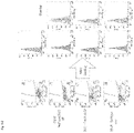
- FIG. 1 A schematic structure of the amino acid sequence and molecular conformation of murine CD300F protein.
- FIG. 2 A schematic diagram showing the relationship between a cell membrane and the conformation of a CD300 family molecule.
- FIG. 3 Micrographic images of different RAW cells treated with MNV.
- FIGS. 4A-4C Charts and graphs showing the results of determination of intracellular MNV molecule expression, the presence or absence of CD300F(1f) molecule expression on the cell surface, and MNV production ability in different RAW cells treated with MNV.
- FIG. 5-1 Results of comparison between the nucleotide sequence of the ORF (open reading frame) of the murine CD300F gene (SEQ ID NO: 1) and the nucleotide sequence of the ORF (open reading frame) of the RAW264.7 cell CD300F gene (SEQ ID NO: 3).
- FIG. 5-2 Results of comparison between the amino acid sequence of murine CD300F protein (SEQ ID NO: 2) and the amino acid sequence of RAW264.7 cell CD300F protein (SEQ ID NO: 4).
- FIG. 6 Results of comparison between the amino acid sequences of murine CD300F protein (SEQ ID NO: 2) and RAW cell CD300F proteins (MuRawCD300F protein (SEQ ID NO: 4) and CD300d protein (SEQ ID NO: 6)).
- the upper part of FIG. 6 shows comparison between the entire amino acid sequences, and the lower part of FIG. 6 shows the conformation of MuRawCD300F molecule (SEQ ID NO: 4).
- FIG. 7-1 Graphs showing the results of FACS for determining the expression of the MuRawCD300F gene transfected into human kidney-derived HEK293T cells.
- FIG. 7-2 Graphs showing the results of FACS for determining the binding of MNV to MuRawCD300F molecule-expressing cells.
- FIG. 7-3 A chart showing the results of western blotting for determining MNV molecule expression in MuRawCD300F molecule-expressing cells.
- FIG. 7-4 A graph showing the results of counting the number of MNV particles in a culture supernatant of MuRawCD300F molecule-expressing cells.
- FIG. 8-1 Alignment of nucleotide sequences of an MuRawCD300F deletion mutant transgene, etc.
- MuRawCD300F SEQ ID NO: 1; MuRaw_d102, SEQ ID NO: 7; MuRaw_d204, SEQ ID NO: 9, MuRaw_d120v, SEQ ID NO: 11; MuRaw_dcpd, SEQ ID NO: 13; and MuRaw_dCterm, SEQ ID NO: 15).
- FIG. 8-2 Alignment of amino acid sequences encoded by an MuRawCD300F deletion mutant transgene, etc. (MuRawCD300F, SEQ ID NO: 2; MuRaw_d102, SEQ ID NO: 8; MuRaw_d204, SEQ ID NO: 10, MuRaw_d120v, SEQ ID NO: 12; MuRaw_dcpd, SEQ ID NO: 14; and MuRaw_dCterm, SEQ ID NO: 16).
- FIG. 9-1 A schematic diagram showing production of mutated murine CD300F molecules (i.e., extracellular domain-mutated molecules).
- FIG. 9-2 Graphs showing the results of FACS of fluorescent-labeled-MNV-treated HEK293T cells transfected with genes encoding the mutated murine CD300F molecules (i.e., extracellular domain-mutated molecules) shown in FIG. 9-1 .
- FIG. 9-3 A chart showing the results of western blotting for determining MNV-RdRp expression in the cells shown in FIG. 9-2 infected with MNV.
- FIG. 9-4 A graph showing the results of counting the number of MNV particles in culture supernatants of the cells shown in FIG. 9-2 infected with MNV.
- FIG. 10 Graphs showing the results of FACS of HEK293T cells transfected with genes encoding mutated murine CD300F molecules (i.e., extracellular domain-mutated molecules).
- FIG. 11 Amino acid sequences of different murine CD300F molecules and the results of western blotting (MuRawCD300F, SEQ ID NO: 2; MuRaw_d102, SEQ ID NO: 8; MuRaw_d204, SEQ ID NO: 10, MuRaw_d120v, SEQ ID NO: 12; MuRaw_dcpd, SEQ ID NO: 14; and MuRaw_dCterm, SEQ ID NO: 16).
- FIG. 12-1 Graphs showing the results of FACS for determining the behaviors of cultured non-murine mammalian cells transfected with the MuRawCD300F gene.
- FIG. 12-2 A chart showing the results of western blotting for determining MNV molecule expression in cultured non-murine mammalian cells transfected with the MuRawCD300F gene and infected with MNV.
- FIG. 12-3 Graphs showing the results of counting the number of MNV particles in culture supernatants of the cultured non-murine mammalian cells transfected with the MuRawCD300F gene and infected with MNV.
- FIG. 3 shows optical micrographs of RAW cells treated with MNV (magnification: ⁇ 10). MNV-non-infected and cultured RAW cells ( FIG. 3A ) were killed by infection with MNV ( FIG. 3B ).
- RAW cells were infected with the recombinant lentivirus of nucleotide sequence library capable of random knockout of the gene encoding the Cas9 enzyme and the mammalian cell gene having a guide RNA sequence recognized by Cas9, and the murine CD300F gene (CD3001f gene) knockout cells were infected with MNV-S7-PP3, to thereby prepare a surviving cell population ( FIG. 3C ).
- the surviving cells are viable cells regardless of MNV infection.
- the MNV-S7-PP3 strain corresponds to MNV that was used for the construction of the MNV reverse genetics system as disclosed in Non-Patent Document 2 and was isolated from the feces of laboratory mice and cultured by use of RAW cells in Japan. This MNV was named “S7.” The S7 strain was subjected to three-passage culture in RAW cells, and then the full nucleotide sequence of the strain was determined. S7 was reported as an infectious clone for the reverse genetics system. The “MNV-S7-PP3” is used for representing the passage number.
- the thus-prepared surviving cell population (i.e., cells survived under MNV infection conditions) has no sensitivity to MNV.
- a primer targeting the guide RNA sequence was designed, and PCR was performed by use of the primer.
- the resultant PCR product contained a partial sequence of the knockout gene bracketed by the guide RNA.
- the nucleotide sequences of all the PCR amplification products were determined by means of a next-generation sequencer (MiSeq, product of Illumina).
- the aforementioned operation was performed twice, and the sequence detected most frequently in both the two operations was found to be that of the CD300F gene.
- the RAW cells in which CD300F molecule expression was knocked out were most frequently observed in the survived cell population.
- the results demonstrate that the CD300F molecule expressed from the CD300F gene is a receptor responsible for MNV infection and growth.
- FIGS. 4A-4C show expression of MNV molecules in different types of RAW cells treated with MNV ( FIG. 4A ); the presence or absence of expression of the CD300F(1f) molecule at cell surfaces ( FIG. 4B ); and the results of measurement of MNV production ability ( FIG. 4C ).
- CD300F (murine CD300F, referred to as “MuCD300F”) expressed in RAW cells was cloned, and the nucleotide sequence (SEQ ID NO: 3) of the gene and the amino acid sequence (SEQ ID NO: 4) encoded by the nucleotide sequence were respectively compared with the known murine CD300F gene (accession number: AB292061 (SEQ ID NO: 1)) and the amino acid sequence (SEQ ID NO: 2) encoded by the gene. As a result, these murine CD300F genes were found to have substantially the same sequence (nucleotide sequence: 97%, amino acid sequence: 98%) ( FIGS.
- RAW cells were treated with (i) monoclonal antibody “Anti-CD300F excd MoAb” to the extracellular domain of the MuCD300F molecule or (ii) monoclonal antibody “Anti-Human CD300F excd MoAb” to the human CD300F molecule, followed by determining whether or not MNV infection was inhibited.
- the number of MNV particles was found to be 10 5 /50 ⁇ L or more in (a) the untreated RAW cells or (c) the RAW cells treated with Anti-HuCD300F excd cell MoAb, whereas the number of MNV particles was found to be only about 10 3 /50 ⁇ L in (b) the RAW cells treated with Anti-MuCD300F excd cell MoAb ( FIG. 4C ).
- the results demonstrate that the efficiency of MNV infection can be reduced to 1/1000 through blocking of the extracellular domain of the MuRawCD300F molecule with the antibody. That is, the results demonstrate that the extracellular domain of the MuRawCD300F molecule is an important moiety for binding to MNV and intracellular uptake of MNV.
- the slight shift observed by means of FACS in the MuCD300F molecule-expressing cells is also thought to be due to a small difference in fluorescence intensity at cell surfaces since the anti-MuCD300F molecule antibody also reacts to the MuCD300d molecule.
- the slight RdRp and VP1 bands observed by means of western blotting in the MuCD300F gene knockout cells are thought to be attributable to, as one of the reasons, the fact that the RAW cells have phagocytosis and are infected with MNV through the phagocytosis even after the MuCD300F gene knockout.
- FIG. 6 shows the results of alignment of the entire amino acid sequences of the CD300d (also called LMIR4) protein and the MuCD300F protein, which proteins belong to the CD300 family, through amino acid sequence homology search.
- the upper part of FIG. 6 shows the results of the alignment. The first sequence in the upper part of FIG.
- the murine CD300d molecule exhibits almost the same conformation as that of the murine CD300F molecule (the lower part of FIG. 6 ), and the CD300d molecule is expected to function as an MNV receptor.
- the highlighted portions (e.g., underlined or surrounded portions) of the structure shown in the lower part of FIG. 6 have the same meanings as defined above in FIG. 1 .
- the aforementioned results demonstrate that MNV infects RAW cells by using the MuCD300F molecule as a receptor, and the MuCD300d molecule may also function as an MNV receptor in MuCD300F gene knockout cells so as to compensate for the MuCD300F molecule. If the MuCD300d molecule functions as a substitute receptor for the MuCD300F molecule, it is possible that the 14th to 120th amino acid residues of the extracellular domain (amino acid sequence homology: 80% or more) serve as an MNV receptor and are related to, for example, binding to MNV.
- MuCD300F gene sequence was obtained from the DDBJ nucleotide database (accession number: AB292061), and the RAW cell CD300F gene was amplified by means of RT-PCR on the basis of this sequence, to thereby clone the gene.
- the MuCD300F gene and the MuRawCD300F gene were compared with each other in terms of nucleotide sequences (SEQ ID NOS: 1 and 3) and amino acid sequences (SEQ ID NOS: 2 and 4). The nucleotide sequence homology was found to be 97% ( FIG. 5-1 ), and the amino acid sequence homology was found to be 98% ( FIG. 5-2 ).
- the MuCD300F molecule and the MuRawCD300F molecule were regarded as the same molecule herein, and the following studies were performed by use of the MuRawCD300F.
- [3]-2 Study on MuRawCD300F Gene Transfection and Expression by Use of Human Kidney-derived Cultured HEK293T Cells
- the MuRawCD300F gene was transfected into MNV-insensitive human kidney-derived cultured HEK293T cells, to thereby examine the MNV sensitivity.
- the MuRawCD300F gene was inserted into the HEK293T cell gene by use of recombinant lentivirus, and puromycin selective culture was performed by use of a puromycin-resistant gene which was simultaneously inserted into the HEK293T cell gene, to thereby prepare MuRawCD300F gene-transfected HEK293T cells.
- the HEK293T cells were treated with MuCD300 excd MoAb and then the cell population shift was examined by means of FACS, to thereby determine whether or not the MuRawCD300F gene was normally expressed as the MuRawCD300F molecule in the MuRawCD300F gene-transfected HEK293T cells, and the MuRawCD300F molecule was migrated to the cell surfaces and the extracellular domain protruded from the cell surfaces.
- the MuRawCD300F gene-transfected cells reacted with the MuCD300 excd MoAb, and the corresponding fluorescence signal was detected.
- the cells were detected as a cell population completely different from untreated HEK293T by means of FACS ( FIG. 7-1 ). Therefore, the MuRawCD300F molecule was found to be normally expressed in the HEK293T cells.
- fluorescent-labeled MNV was added to the MuCD300F molecule-expressing cells and untreated HEK293T cells, to thereby examine whether or not cell populations can be separated by means of FACS.
- the MuCD300F molecule-expressing cells were labeled with fluorescence through binding to the fluorescent-labeled MNV, and were separated as a cell population distinctly different from the untreated HEK293T by means of FACS (the upper right and lower right graphs of FIG. 7-2 ).
- the MuRawCD300F molecule-expressing HEK293T cells were infected with MNV, and, 48 hours thereafter, the resultant cells were collected, followed by determination of intracellular expression of VP1 and RNA-dependent RNA polymerase (RdRp) of MNV by means of western blotting.
- RdRp RNA-dependent RNA polymerase
- the aforementioned results demonstrate that when the MuCD300F gene is transfected into the MNV-insensitive human cultured HEK293T cells, the cells are altered to MNV-sensitive cells through expression of the MuCD300F molecule, and in the resultant cells, MNV can be effectively grown.
- the results also demonstrate that the extracellular domain is important for acquisition of the MNV sensitivity.
- fluorescent-labeled MNV was treated with “MuRaw_d102” gene (with deletion of 34 amino acid residues (corresponding to 102 nucleotides) in the extracellular domain)-transfected HEK293T cells and “MuRAW_dcpd” gene (with deletion of the intracellular domain)-transfected HEK293T cells (schematically shown in FIG. 9-1 ), followed by determination by means of FACS.
- the gene encoding each of the aforementioned MuRawCD300F molecule, MuRaw_d102 molecule, MuRaw_d204 molecule, and MuRaw_dcpd molecule was cloned downstream of the CMV promoter of plasmid vector, and co-transfected with GFP plasmid.
- GFP expression was used as a monitor of transfection, and GFP-expressing cells were sorted. The sorted cells were reacted with an anti-MuCD300F molecule antibody and then reacted with a red fluorescent-labeled secondary antibody that can specifically recognize the anti-MuCD300F molecule antibody, followed by FACS.
- the MuRawCD300F gene when the MuRawCD300F gene is transfected into the HEK293T cells (i.e., MNV-insensitive cells), the HEK293T cells are altered from MNV-insensitive cells to MNV-sensitive cells. This result demonstrates that the MuRawCD300F molecule is a receptor that is directly involved in MNV infection.
- the resultant MuRawCD300F molecule does not function as a receptor.
- a portion important for MNV sensitivity is highly probably the amino acid sequence (34 amino acid residues, encoded by the 102 nucleotides) downstream of the signal peptide of the MuRawCD300F protein. Since the cells transfected with the MuRaw_dcpd gene prepared through deletion of the intracellular domain also do not lose MNV-sensitivity, the extracellular domain is thought to play an important role for MNV sensitivity.
- the HEK293T cells were transfected with the “MuRaw_d120v” molecule, i.e., a CD300F variant molecule with deletion of 40 amino acid residues (from the 130th amino acid residue “I” to the 170th amino acid residue “D”) and mutations of the 129th amino acid residue (from “A” to “G”), the 171th amino acid residue (from “N” to “K”), and the 172th amino acid residue (from “G” to “R”); or the “MuRaw_d120vC term” molecule (SEQ ID NO: 15, amino acid sequence SEQ ID NO: 16) prepared through deletion of the transmembrane domain and intracellular domain of the MuRaw_d120v molecule by deletion of its C-terminal side, to thereby examine a domain involved in MNV sensitivity.
- the “MuRaw_d120v” molecule i.e., a CD300F variant molecule with deletion of 40 amino acid residues (from the
- the “MuRaw_d120v” gene-transfected HEK293T cells exhibit MNV infection as in the case of the full-length MuRawCD300F gene-transfected cells, and MNV-RdRp and VP1 were detected by means of western blotting.
- MNV infection was not observed, and neither MNV-RdRp nor VP1 was detected by means of western blotting ( FIG. 11 ).
- the 34 amino acid residues have three ⁇ -chains (VT, EVSGQ, and LTVQCRY) and a helix (SGW), wherein these ⁇ -chains are in proximity to one another. It was found that MNV does not bind to the CD300F molecule on the cell membrane when these amino acid residues are deleted.
- CD300d also called LMIR4
- CD300d also called LMIR4
- FIG. 5 the CD300d molecule was found to have almost the same conformation as the CD300F molecule. From the results, it became even clearer that the CD300d molecule is also highly likely to function as an MNV receptor.
- the MuRawCD300F gene was transfected into NIH3T3 cells (murine MNV-insensitive cells) or MNV-insensitive cells of different animal species; i.e., African green monkey-derived COS7 cells, hamster-derived CHO cells, or cat-derived CRFK cells, so as to determine whether or not MuCD300F can alter the MNV sensitivity of such animal cells.
- the gene-transfected cells were subjected to puromycin selective culture as in the case of the HEK293T, and prepared as a MuCD300F molecule-expressing cell population. The behavior of the cell population was determined by means of FACS ( FIG. 12-1 ).
- the cells of each species were infected with MNV, and the expression of the MuCD300F molecule, MNV-RdRp or MNV-VP1 was determined by means of western blotting ( FIG. 12-2 ).
Landscapes
- Health & Medical Sciences (AREA)
- Life Sciences & Earth Sciences (AREA)
- Chemical & Material Sciences (AREA)
- Engineering & Computer Science (AREA)
- Organic Chemistry (AREA)
- Biomedical Technology (AREA)
- Genetics & Genomics (AREA)
- Immunology (AREA)
- Zoology (AREA)
- Bioinformatics & Cheminformatics (AREA)
- General Health & Medical Sciences (AREA)
- Biochemistry (AREA)
- Molecular Biology (AREA)
- Wood Science & Technology (AREA)
- Biotechnology (AREA)
- Microbiology (AREA)
- Medicinal Chemistry (AREA)
- Cell Biology (AREA)
- General Engineering & Computer Science (AREA)
- Hematology (AREA)
- Urology & Nephrology (AREA)
- Biophysics (AREA)
- Proteomics, Peptides & Aminoacids (AREA)
- Physics & Mathematics (AREA)
- Virology (AREA)
- Analytical Chemistry (AREA)
- Toxicology (AREA)
- Gastroenterology & Hepatology (AREA)
- Food Science & Technology (AREA)
- Pathology (AREA)
- General Physics & Mathematics (AREA)
- Tropical Medicine & Parasitology (AREA)
- Plant Pathology (AREA)
- Developmental Biology & Embryology (AREA)
- Transplantation (AREA)
- Micro-Organisms Or Cultivation Processes Thereof (AREA)
- Peptides Or Proteins (AREA)
- Measuring Or Testing Involving Enzymes Or Micro-Organisms (AREA)
Abstract
Description
Claims (10)
Applications Claiming Priority (4)
| Application Number | Priority Date | Filing Date | Title |
|---|---|---|---|
| JP2016019315 | 2016-02-03 | ||
| JP2016-019315 | 2016-02-03 | ||
| JPJP2016-019315 | 2016-02-03 | ||
| PCT/JP2017/003539 WO2017135277A1 (en) | 2016-02-03 | 2017-02-01 | Cultured transgenic cell allowing growth of norovirus, and use thereof |
Publications (2)
| Publication Number | Publication Date |
|---|---|
| US20190040415A1 US20190040415A1 (en) | 2019-02-07 |
| US11299750B2 true US11299750B2 (en) | 2022-04-12 |
Family
ID=59499842
Family Applications (1)
| Application Number | Title | Priority Date | Filing Date |
|---|---|---|---|
| US16/074,648 Active 2038-12-09 US11299750B2 (en) | 2016-02-03 | 2017-02-01 | Cultured transgenic cell allowing growth of norovirus, and use thereof |
Country Status (4)
| Country | Link |
|---|---|
| US (1) | US11299750B2 (en) |
| EP (1) | EP3412770A4 (en) |
| JP (1) | JP6966944B2 (en) |
| WO (1) | WO2017135277A1 (en) |
Families Citing this family (2)
| Publication number | Priority date | Publication date | Assignee | Title |
|---|---|---|---|---|
| EP3545000A4 (en) * | 2016-11-22 | 2020-07-08 | DendroCyte BioTech Pty Ltd | Anti-cd300f antibody and uses thereof |
| US20180252712A1 (en) * | 2017-03-01 | 2018-09-06 | Washington University | Receptor for norovirus and uses thereof |
Citations (8)
| Publication number | Priority date | Publication date | Assignee | Title |
|---|---|---|---|---|
| WO2008003748A2 (en) | 2006-07-07 | 2008-01-10 | Novo Nordisk A/S | Cd300lg polypeptides and their use in treating autoimmune diseases |
| US20120128698A1 (en) | 2009-07-07 | 2012-05-24 | Menno Van Lookeren Campagne | Diagnosis and treatment of autoimmune demyelinating diseases |
| WO2013077186A1 (en) | 2011-11-21 | 2013-05-30 | 国立大学法人筑波大学 | Activity modulator, medicinal agent comprising same, use of cd300a gene-deficient mouse, and anti-cd300a antibody |
| WO2014073529A1 (en) | 2012-11-07 | 2014-05-15 | 国立大学法人筑波大学 | MEDICINE COMPRISING MODULATOR OF ACTIVITY OF CD300a-EXPRESSING CELL ASSOCIATED WITH ALLERGIC DISEASE, CD300a GENE KNOCK-OUT MOUSE, AND USE OF MODULATOR OF ACTIVITY OF CD300a-EXPRESSING CELL |
| JP2014095570A (en) | 2012-11-07 | 2014-05-22 | Univ Of Tsukuba | Use of mouse deleted in cd300a gene relevant to celiac disease, use of activity modulator of cd300a expression cell relevant to celiac disease, and medicine containing activity modulator of cd300a expression cell relevant to celiac disease |
| JP2014094898A (en) | 2012-11-07 | 2014-05-22 | Univ Of Tsukuba | MEDICINE COMPRISING ACTIVE MODULATOR OF CD300a-EXPRESSING CELL ASSOCIATED WITH INFLAMMATORY BOWEL DISEASE, CD300a GENE KNOCK-OUT MOUSE, AND USE OF ACTIVE MODULATOR OF CD300a-EXPRESSING CELL |
| WO2015092035A1 (en) | 2013-12-20 | 2015-06-25 | Institut National De La Sante Et De La Recherche Medicale (Inserm) | Cd300a receptors as virus entry cofactors |
| WO2015127158A1 (en) | 2014-02-21 | 2015-08-27 | Regeneron Pharmaceuticals, Inc. | Methods, compositions and kits for cell specific modulation of target antigens |
-
2017
- 2017-02-01 EP EP17747433.5A patent/EP3412770A4/en active Pending
- 2017-02-01 US US16/074,648 patent/US11299750B2/en active Active
- 2017-02-01 JP JP2017565572A patent/JP6966944B2/en active Active
- 2017-02-01 WO PCT/JP2017/003539 patent/WO2017135277A1/en active Application Filing
Patent Citations (11)
| Publication number | Priority date | Publication date | Assignee | Title |
|---|---|---|---|---|
| WO2008003748A2 (en) | 2006-07-07 | 2008-01-10 | Novo Nordisk A/S | Cd300lg polypeptides and their use in treating autoimmune diseases |
| US20120128698A1 (en) | 2009-07-07 | 2012-05-24 | Menno Van Lookeren Campagne | Diagnosis and treatment of autoimmune demyelinating diseases |
| JP2012532873A (en) | 2009-07-07 | 2012-12-20 | ジェネンテック, インコーポレイテッド | Diagnosis and treatment of autoimmune demyelinating diseases |
| WO2013077186A1 (en) | 2011-11-21 | 2013-05-30 | 国立大学法人筑波大学 | Activity modulator, medicinal agent comprising same, use of cd300a gene-deficient mouse, and anti-cd300a antibody |
| US20150047059A1 (en) | 2011-11-21 | 2015-02-12 | University Of Tsukuba | Activity modulator, medicinal agent comprising same, use of cd300a gene-deficient mouse, and anti-cd300a antibody |
| WO2014073529A1 (en) | 2012-11-07 | 2014-05-15 | 国立大学法人筑波大学 | MEDICINE COMPRISING MODULATOR OF ACTIVITY OF CD300a-EXPRESSING CELL ASSOCIATED WITH ALLERGIC DISEASE, CD300a GENE KNOCK-OUT MOUSE, AND USE OF MODULATOR OF ACTIVITY OF CD300a-EXPRESSING CELL |
| JP2014095570A (en) | 2012-11-07 | 2014-05-22 | Univ Of Tsukuba | Use of mouse deleted in cd300a gene relevant to celiac disease, use of activity modulator of cd300a expression cell relevant to celiac disease, and medicine containing activity modulator of cd300a expression cell relevant to celiac disease |
| JP2014094898A (en) | 2012-11-07 | 2014-05-22 | Univ Of Tsukuba | MEDICINE COMPRISING ACTIVE MODULATOR OF CD300a-EXPRESSING CELL ASSOCIATED WITH INFLAMMATORY BOWEL DISEASE, CD300a GENE KNOCK-OUT MOUSE, AND USE OF ACTIVE MODULATOR OF CD300a-EXPRESSING CELL |
| US20150299332A1 (en) | 2012-11-07 | 2015-10-22 | University Of Tsukuba | MEDICAMENT COMPRISING ACTIVITY MODULATOR FOR CD300a-EXPRESSING CELL ASSOCIATED WITH ALLERGIC DISEASE, CD300a GENE-DEFICIENT MOUSE, AND USE OF ACTIVITY MODULATOR FOR CD300a-EXPRESSING CELL |
| WO2015092035A1 (en) | 2013-12-20 | 2015-06-25 | Institut National De La Sante Et De La Recherche Medicale (Inserm) | Cd300a receptors as virus entry cofactors |
| WO2015127158A1 (en) | 2014-02-21 | 2015-08-27 | Regeneron Pharmaceuticals, Inc. | Methods, compositions and kits for cell specific modulation of target antigens |
Non-Patent Citations (13)
| Title |
|---|
| "NTTC clone 929," accessed from https://www.atcc.org/products/all/CCL-1.aspx on May 5, 2021, p. 1. * |
| Choi et al., The Journal of Immunology, 2011, 187: 3483-3487. * |
| Comas-Casellas et al., Journal of Biological Chemistry 287(13): 9682-9693, Mar. 23, 2012. * |
| Communication dated Jul. 4, 2019, from the European Patent Office in counterpart European Application No. 17747433.5. |
| Francisco Borrego, "The CD300 molecules: an emerging family of regulators of the immune system", Blood, Mar. 14, 2013, pp. 1951-1960, vol. 121, No. 11. |
| Georgina J. Clark, et al., "The CD300 family of molecules are evolutionarily significant regulators of leukocyte functions", Trends in Immunology, 2009, pp. 209-217, vol. 30, No. 5. |
| International Search Report for PCT/JP2017/003539 dated May 9, 2017 (PCT/ISA/210). |
| Izawa et al., The Journal of Biological Chemistry, 282(25): 17997-18008, 2007. * |
| K. Izawa, et al., "Sphingomyelin and ceramide are physiological ligands for human LMIR/CD300f, inhibiting FcϵRI-mediated mast ceil activation", J. Allergy Clini. Immunol., Jan. 2014, vol. 133, No. 1, pp. 270-273 and 273.e1-e7. |
| Kei Haga, et al., "Functional receptor molecules CD300If and CD300Id within the CD300 family enable murine noroviruses to infect cells", PNAS, Sep. 28, 2016, pp. E6248-E6255, vol. 113, No. 41. |
| Kei Haga, et al., "Identification of the functional receptor for murine norovirus", The 64th Annual Meeting of the Japanese Society for Virology, Sep. 30, 2016, pp. 208 and W2-6-11, vol. 64. |
| Robert C. Orchard, et al., "Discovery of a proteinaceous cellular receptor for a norovirus", Science, Aug. 26, 2016, pp. 933-936, vol. 353, No. 6302. |
| Written Opinion for PCT/JP2017/003539 dated May 9, 2017 [PCT/ISA/237]. |
Also Published As
| Publication number | Publication date |
|---|---|
| JPWO2017135277A1 (en) | 2019-01-31 |
| WO2017135277A1 (en) | 2017-08-10 |
| US20190040415A1 (en) | 2019-02-07 |
| EP3412770A1 (en) | 2018-12-12 |
| JP6966944B2 (en) | 2021-11-17 |
| EP3412770A4 (en) | 2019-08-07 |
Similar Documents
| Publication | Publication Date | Title |
|---|---|---|
| Lin et al. | The host cell receptors for measles virus and their interaction with the viral hemagglutinin (H) protein | |
| Zhang et al. | Expression of the Mxra8 receptor promotes alphavirus infection and pathogenesis in mice and Drosophila | |
| Holst et al. | Generation of T-cell receptor retrogenic mice | |
| AU2016247892A1 (en) | Humanized SIRPA-IL15 knockin mice and methods of use thereof | |
| Hahn et al. | Rhesus monkey rhadinovirus uses eph family receptors for entry into B cells and endothelial cells but not fibroblasts | |
| Tahara et al. | Multiple Amino Acid Substitutions in Hemagglutinin Are Necessary for Wild-Type MeaslesVirus To Acquire the Ability To Use Receptor CD46 Efficiently | |
| JPH11506907A (en) | Novel peptides and compositions that modulate apoptosis | |
| Hirai et al. | Role of mouse hepatitis virus (MHV) receptor murine CEACAM1 in the resistance of mice to MHV infection: studies of mice with chimeric mCEACAM1a and mCEACAM1b | |
| US11299750B2 (en) | Cultured transgenic cell allowing growth of norovirus, and use thereof | |
| Moss et al. | Genetic diversity of the Caribbean spiny lobster virus, Panulirus argus virus 1 (PaV1), and the discovery of PaV1 in lobster postlarvae | |
| Oldstone et al. | A role for dual viral hits in causation of subacute sclerosing panencephalitis | |
| Chen et al. | A premature stop codon within the tvb receptor gene results in decreased susceptibility to infection by avian leukosis virus subgroups B, D, and E | |
| Li et al. | Naturally occurring frameshift mutations in the tvb receptor gene are responsible for decreased susceptibility of chicken to infection with avian leukosis virus subgroups B, D, and E | |
| Sebastian et al. | An invariant surface patch on the TRIM5α PRYSPRY domain is required for retroviral restriction but dispensable for capsid binding | |
| Morton et al. | Cloning and characterization of equine CD89 and identification of the CD89 gene in chimpanzees and rhesus macaques | |
| JP2019509059A (en) | Non-human animal having mutant kynureninase gene | |
| Velupillai et al. | Wild-derived inbred mice have a novel basis of susceptibility to polyomavirus-induced tumors | |
| Guloglu et al. | The unique region of surrogate light chain component λ5 is a heavy chain-specific regulator of precursor B cell receptor signaling | |
| WO2010116375A1 (en) | Isolated peptides for regulating apoptosis | |
| Maeki et al. | Identification of the human herpesvirus 6A gQ1 domain essential for its functional conformation | |
| WO1991004327A1 (en) | Transgenic animal model for viral infections | |
| WO1998058536A1 (en) | Transgenic rabbits expressing cd4 and chemokine receptor | |
| US20040237128A1 (en) | Transgenic non-human animal having a disruption of at least one allele to the ceacam 1 gene and method of making same | |
| Tibbs et al. | Mice with FVB-derived sequence on chromosome 17 succumb to disseminated virus infection due to aberrant NK cell and T cell responses | |
| Noguchi et al. | Identification and functional analyses of a cell-death inhibitor encoded by guinea pig cytomegalovirus gp38. 1 in cell culture and in animals |
Legal Events
| Date | Code | Title | Description |
|---|---|---|---|
| FEPP | Fee payment procedure |
Free format text: ENTITY STATUS SET TO UNDISCOUNTED (ORIGINAL EVENT CODE: BIG.); ENTITY STATUS OF PATENT OWNER: LARGE ENTITY |
|
| AS | Assignment |
Owner name: NATIONAL CENTER FOR GERIATRICS AND GERONTOLOGY, JAPAN Free format text: ASSIGNMENT OF ASSIGNORS INTEREST;ASSIGNORS:HAGA, KEI;FUJIMOTO, AKIRA;TODAKA, REIKO;AND OTHERS;SIGNING DATES FROM 20180801 TO 20180823;REEL/FRAME:047007/0026 Owner name: JAPAN AS REPRESENTED BY DIRECTOR-GENERAL OF NATIONAL INSTITUTE OF INFECTIOUS DISEASES, JAPAN Free format text: ASSIGNMENT OF ASSIGNORS INTEREST;ASSIGNORS:HAGA, KEI;FUJIMOTO, AKIRA;TODAKA, REIKO;AND OTHERS;SIGNING DATES FROM 20180801 TO 20180823;REEL/FRAME:047007/0026 Owner name: DENKA COMPANY LIMITED, JAPAN Free format text: ASSIGNMENT OF ASSIGNORS INTEREST;ASSIGNORS:HAGA, KEI;FUJIMOTO, AKIRA;TODAKA, REIKO;AND OTHERS;SIGNING DATES FROM 20180801 TO 20180823;REEL/FRAME:047007/0026 Owner name: NATIONAL CENTER FOR GERIATRICS AND GERONTOLOGY, JA Free format text: ASSIGNMENT OF ASSIGNORS INTEREST;ASSIGNORS:HAGA, KEI;FUJIMOTO, AKIRA;TODAKA, REIKO;AND OTHERS;SIGNING DATES FROM 20180801 TO 20180823;REEL/FRAME:047007/0026 Owner name: JAPAN AS REPRESENTED BY DIRECTOR-GENERAL OF NATION Free format text: ASSIGNMENT OF ASSIGNORS INTEREST;ASSIGNORS:HAGA, KEI;FUJIMOTO, AKIRA;TODAKA, REIKO;AND OTHERS;SIGNING DATES FROM 20180801 TO 20180823;REEL/FRAME:047007/0026 |
|
| STPP | Information on status: patent application and granting procedure in general |
Free format text: DOCKETED NEW CASE - READY FOR EXAMINATION |
|
| STPP | Information on status: patent application and granting procedure in general |
Free format text: RESPONSE TO NON-FINAL OFFICE ACTION ENTERED AND FORWARDED TO EXAMINER |
|
| STPP | Information on status: patent application and granting procedure in general |
Free format text: NON FINAL ACTION MAILED |
|
| STPP | Information on status: patent application and granting procedure in general |
Free format text: RESPONSE TO NON-FINAL OFFICE ACTION ENTERED AND FORWARDED TO EXAMINER |
|
| STPP | Information on status: patent application and granting procedure in general |
Free format text: NOTICE OF ALLOWANCE MAILED -- APPLICATION RECEIVED IN OFFICE OF PUBLICATIONS |
|
| STPP | Information on status: patent application and granting procedure in general |
Free format text: PUBLICATIONS -- ISSUE FEE PAYMENT VERIFIED |
|
| STCF | Information on status: patent grant |
Free format text: PATENTED CASE |