CROSS REFERENCE TO RELATED APPLICATIONS
The present application is a divisional of pending U.S. patent application Ser. No. 16/209,255, filed Dec. 4, 2018, which is a divisional of prior U.S. patent application Ser. No. 14/745,247, filed Jun. 19, 2015, now issued a U.S. Pat. No. 10,189,909, which is a divisional of prior U.S. patent application Ser. No. 10/556,509, filed Aug. 23, 2006, now issued as U.S. Pat. No. 9,096,676, priority from the filing date of which is hereby claimed under 35 U.S.C. § 120, which is a 371 National Phase Application of International PCT Application No. PCT/DK04/00338, filed May 12, 2004, now lapsed, which claims priority to Danish Patent Application No. PA 2003 00716, filed May 12, 2003, now lapsed.
STATEMENT REGARDING SEQUENCE LISTING
The sequence listing associated with this application is provided in text format in lieu of a paper copy and is hereby incorporated by reference into the specification. The name of the text file containing the sequence listing is MP_1_0117_US6_SequenceListingasFiled_20210218. K the text file is 86 KB, was created on Feb. 18, 2021; and is being submitted via EFS-Web with the filing of the specification.
FIELD OF INVENTION
The present invention relates to antibodies to MASP-2 and functional equivalents thereof. In particular, the invention relates to MASP-2 antibodies capable of inhibiting the function of MASP-2. Furthermore, the invention relates to methods of producing said antibodies, methods of inhibiting MASP-2 activity as well as to pharmaceutical compositions comprising the MASP-2 antibodies.
BACKGROUND OF INVENTION
The complement system comprises a complex array of enzymes and non-enzymatic proteins of importance to the function of the innate as well as the adaptive immune defense1. Until recently two modes of activation were known, the classical pathway initiated by antibody-antigen complexes and the alternative pathway initiated by certain structures on microbial surfaces. A third, novel antibody-independent pathway of complement activation has been described2. This pathway is initiated when mannan-binding lectin (MBL, first described as mannan-binding protein3, MBP, see Ezekowitz, U.S. Pat. No. 5,270,199) binds to carbohydrates and is known as the MBLectin pathway.
MBL is structural related to the C1q subcomponent of component C1 of complement, and it appears that MBL activates the complement system via an associated serine protease termed MASP4 or p1005, which is similar to the C1 r and C1 s components of the classical pathway. The new complement activation pathway is called the MBLectin pathway. According to the mechanism postulated for this pathway, MBL binds to specific carbohydrate structures found on the surface of a range of microorganisms including bacteria, yeast, parasitic protozoa and viruses5, and its antimicrobial activity results from activation of the terminal, lytic complement pathway components7 or promoting phagocytosis8.
MASPs (MBL-associated serine protease) are serine proteases similar in structure to C1 r and C1 s of the complement pathway. MASP-1 has a histidine loop structure of the type found in trypsin and trypsin-like serine proteases. MASP-1 has been found to be involved in complement activation by MBL. A cDNA clone encoding MASP-1 has been reported that encodes a putative leader peptide of 19 amino acids followed by 680 amino acid residues predicted to form the mature peptide.
MASP-2 (MBL-associated serine protease 2)22 is a serine protease also similar in structure to C1 r and C1 s of the complement pathway. Like these, and contrary to MASP1, it has no histidine loop structure of the type found in trypsin and trypsin-like serine proteases. MASP-2 has been found to be involved in complement activation by MBL.
Antibodies to MASP-2 has been described in the prior art.
WO 02/06460 describes human MASP-2. The document furthermore describes antibodies to MASP-2 raised by immunising rabbits with the N-terminal 19 amino acids of human MASP-2 or chickens with aa 505 to 523 and aa 538 to 556 of human MASP-2.
SUMMARY OF INVENTION
Interestingly, the inventors of the present invention have recognised that inhibition of the MBLectin pathway may be desirable in the treatment of a number of clinical conditions. However, specific inhibitors of the MBLectin pathway are not well characterised in the prior art and therefore an unmet need of specific inhibitors exists.
The present invention discloses that antibodies to the C-terminal part of MASP-2 are capable of inhibiting the activity of MASP-2 more efficiently than antibodies to the N-terminal part of MASP-2. The invention furthermore discloses MASP-2 epitopes, wherein antibodies recognising said epitopes are in particularly useful for inhibiting the activity of MASP-2. Preferred epitopes are describes herein below.
In one aspect the invention relates to an antibody or a functional equivalent thereof specifically recognising and binding at least part of an epitope recognised by one or more reference antibodies selected from the group consisting of
-
- i the monoclonal antibody produced by the hybridoma cell line deposited under the deposition number 03050904;
- ii the monoclonal antibody produced by the hybridoma cell line designated M0545YM029;
- iii the monoclonal antibody produced by the hybridoma cell line designated M0545YM035;
- iv the monoclonal antibody produced by the hybridoma cell line designated M0545YM046; and
- v the monoclonal antibody produced by the hybridoma cell line designated M0545YM048.
Accordingly, it is an objective of the present invention to provide antibodies or functional equivalents thereof specifically recognising and binding an epitope within the C-terminal part of MASP-2 or a functional homologue thereof.
It is furthermore an objective of the present invention to provide isolated polypeptides comprising a C-terminal fragment of MASP-2, said polypeptide being useful for raising antibodies to epitopes within the C-terminus of MASP-2. In particular, the isolated polypeptides from the C-terminal part of MASP-2 or a functional homologue thereof, may be polypeptides comprising or consisting of:
-
- i. the EGF, CUB2, CCP1, CCP2 and serine protease domains; or
- ii. the CUB2, CCP1, CCP2 and serine protease domains; or
- iii. the CCP1 domain; or
- iv. the CCP2 domain; or
the CCP1, and CCP2 domain
It is furthermore an objective of the present invention to provide methods of producing an antibody inhibiting MASP-2 activity, by immunising an animal, preferably a mammal, with isolated polypeptides comprising a C-terminal fragment of MASP-2, said polypeptide being useful for raising antibodies to epitopes within the C-terminus of MASP-2. In particular, the isolated polypeptides from the C-terminal part of MASP-2 or a functional homologue thereof, may be polypeptides comprising or consisting of:
-
- v. the EGF, CUB2, CCP1, CCP2 and serine protease domains; or
- vi. the CUB2, CCP1, CCP2 and serine protease domains; or
- vii. the CCP1 domain and the serine protease domain; or
- viii. the CCP2 domain and the serine protease domain; or
- ix. the CCP1, CCP2 and the serine protease domains
- x. the serine protease domain
It is a further objective of the present invention to provide methods of producing an antibody specifically recognising and binding an epitope within the C-terminal part of MASP-2 or a functional homologue thereof, comprising the step of administering to a mammal the C-terminal part of MASP-2 or a fragment thereof or a functional homologue thereof. Antibodies produced according to the method are also disclosed by the invention.
It is an even further objective of the invention to provide methods of inhibiting the activity of MASP-2 comprising the steps of
-
- 1) Providing a composition comprising MASP-2;
- 2) Providing a MASP-2 antibody according to the invention;
- 3) Incubating said composition with said antibody, thereby inhibiting MASP-2 activity
Inhibition of MASP-2 will lead to inhibition of complement activation, preferably to inhibition of the MBLectin pathway. Accordingly, antibodies inhibiting the activity of MASP-2 may be used to inhibit activation of the MBLectin pathway and accordingly, said antibodies may be useful for treatment of clinical conditions characterised by improper activation of complement, preferably improper activation of the MBLectin pathway.
Hence, the invention also relates to methods of inhibiting the MBLectin pathway, preferably said methods involv the use of antibodies to MASP-2, capable of inhibiting the activity of MASP-2.
It is a still further objective of the present invention to provide pharmaceutical compositions comprising MASP-2 antibodies or functional equivalents thereof recognising an epitope within the C-terminal part of MASP-2 together with pharmaceutically acceptable excipients.
It is yet another objective of the present invention to provide a medicament for treatment of a clinical condition comprising an antibody or a functional equivalent thereof recognising an epitope within the C-terminal part of MASP-2 as an active ingredient.
It is also an objective of the present invention to provide methods of treatment of a clinical condition comprising administering to an individual in need thereof a therapeutically effective dosage of an antibody or a functional equivalent thereof recognising an epitope within the C-terminus of MASP-2.
It is furthermore an objective of the present invention to provide uses of antibodies or functional equivalents thereof recognising an epitope within the C-terminal part of MASP-2, for the preparation of a medicament for the treatment of a clinical condition in an individual in need thereof.
DESCRIPTION OF DRAWINGS
FIG. 1 depicts a schematic representation of MASP-2 indicating the individual domains.
FIG. 2 shows an alignment of the human MASP-1 (SEQ ID NO:15), MASP-2 (SEQ ID NO:14, C1r (SEQ ID NO:16) and C1s (SEQ ID NO:17) sequences indicating the presence of the individual domains in MASP-2. Amino acids conserved in the four proteins are furthermore indicated by asterisk.
FIG. 3 depicts inhibition of C4 deposition in full serum by a MASP-2 antibody.
FIG. 4 depicts inhibition of C4 deposition by different concentrations of a MASP-2 antibody.
FIG. 5 shows a C4 deposition assay. The figure illustrates inhibition of C4 deposition in human serum using different purified anti-MASP-2 antibodies.
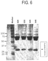
FIG. 6 shows an assembly of Western blots against MASP-2 in human serum using 4 different antibodies. Human serum was loaded on each lane. The figure is assembled from four separate Western blots using the antibodies shown in the lanes above for detection.
FIG. 7 shows the results of a compeptive ELISA for determination of overlapping epitopes.
FIG. 8 illustrates the nucleotide sequence (SEQ ID NO:18) encoding the variable region of the light chain of the NimoAb101 antibody (DWE16140-6cons) (SEQ ID NO:19).
FIG. 9 illustrates the nucleotide sequence (SEQ ID NO:20) encoding the variable region of the heavy chain of NimoAb101 antibody (DWE16140-3consRev) (SEQ ID NO:21).
FIG. 10 shows an alignment between sequences of the heavy chain of the Ni-moAb101 antibody (DWE16140-1con (SEQ ID NO:29), DWE16140-2con (SEQ ID NO:30), DWE16140-3con (SEQ ID NO:31), DWE16140-4con (SEQ ID NO:27), DWE16140-5con (SEQ ID NO:28), and DWE16140-8con (SEQ ID NO:32)) together with homologous sequences P18525 (SEQ ID NO:22), P18526 (SEQ ID NO:23), P18529 (SEQ ID NO:24), P01764 (SEQ ID NO:25), P01783 (SEQ ID NO:26), P01868 (SEQ ID NO:33), P01869 (SEQ ID NO:34), P20759 (SEQ ID NO:35) and P20760 (SEQ ID NO:36) identified by BLAST searches.
FIG. 11 shows an alignment between sequences of the light chain of the NimoAb101 antibody (DWE16140-6con (SEQ ID NO:37), DWE16140-7con (SEQ ID NO:38), DWE16140-9con (SEQ ID NO:39) and DWE16140-10con (SEQ ID NO:40)) together with homologous sequences P01594 (SEQ ID NO: 41), P01595 (SEQ ID NO: 42), P01635 (SEQ ID NO: 43), P01636 (SEQ ID NO: 44), P01637 (SEQ ID NO: 45), P01835 (SEQ ID NO: 46), P01836 (SEQ ID NO: 47), and P01837 (SEQ ID NO: 48) identified by BLAST searches.
| SEQ ID NO: 1 |
Human MASP-2 |
| SEQ ID NO: 2 |
Part of heavy chain of NimoAb101 including |
| |
the variable region |
| SEQ ID NO: 3 |
Part of light chain of NimoAb101 including |
| |
the variable region |
| SEQ ID NO: 4 |
Part of heavy chain of NimoAb101 including |
| |
the variable region |
| SEQ ID NO: 5 |
Part of light chain of NimoAb101 including |
| |
the variable region |
| SEQ ID NO: 6 |
CDR1 of heavy chain (also designated H1) |
| |
of NimoAb101 |
| SEQ ID NO: 7 |
CDR2 of heavy chain (also designated H2) |
| |
of NimoAb101 |
| SEQ ID NO: 8 |
CDR3 of heavy chain (also designated H3) |
| |
of NimoAb101 |
| SEQ ID NO: 9 |
CDR1 of light chain (also designated L1) |
| |
of NimoAb101 |
| SEQ ID NO: 10 |
CDR2 of light chain (also designated L2) |
| |
of NimoAb101 |
| SEQ ID NO: 11 |
CDR3 of light chain (also designated L3) |
| |
of NimoAb101 |
| SEQ ID NO: 12 |
PCR primer |
| SEQ ID NO: 13 |
PCR primer |
| SEQ ID NO: 14 |
MASP-2 (FIG. 2) |
| SEQ ID NO: 15 |
MASP-1 (FIG. 2) |
| SEQ ID NO: 16 |
C1r (FIG. 2) |
| SEQ ID NO: 17 |
C1s (FIG. 2) |
| SEQ ID NO: 18 |
Nucleotide sequence encoding the variable |
| |
region of the light chain of the NimoAb101 |
| |
antibody (DWE16140-6cons, FIG. 8) |
| SEQ ID NO: 19 |
Variable region of the light chain of the |
| |
NimoAb101 antibody (DWE16140-6cons, FIG. 8) |
| SEQ ID NO: 20 |
Nucleotide sequence encoding the variable |
| |
region of the heavy chain of NimoAb101 |
| |
antibody (DWE16140-3consRev, FIG. 9) |
| SEQ ID NO: 21 |
Variable region of the heavy chain of |
| |
NimoAb101 antibody (DWE16140-3consRev, |
| |
FIG. 9) |
| SEQ ID NO: 22 |
P18525 (FIG. 10) |
| SEQ ID NO: 23 |
P18526 (FIG. 10) |
| SEQ ID NO: 24 |
P18529 (FIG. 10) |
| SEQ ID NO: 25 |
P01764 (FIG. 10) |
| SEQ ID NO: 26 |
P01783 (FIG. 10) |
| SEQ ID NO: 27 |
DWE16140-4con (FIG. 10) |
| SEQ ID NO: 28 |
DWE16140-5con (FIG. 10) |
| SEQ ID NO: 29 |
DWE16140-1con (FIG. 10) |
| SEQ ID NO: 30 |
DWE16140-2con (FIG. 10) |
| SEQ ID NO: 31 |
DWE16140-3con (FIG. 10) |
| SEQ ID NO: 32 |
DWE16140-8con (FIG. 10) |
| SEQ ID NO: 33 |
P01868 (FIG. 10) |
| SEQ ID NO: 34 |
P01869 (FIG. 10) |
| SEQ ID NO: 35 |
P20759 (FIG. 10) |
| SEQ ID NO: 36 |
P20760 (FIG. 10) |
| SEQ ID NO: 37 |
DWE16140-6con (FIG. 11) |
| SEQ ID NO: 38 |
DWE16140-7con (FIG. 11) |
| SEQ ID NO: 39 |
DWE16140-9con (FIG. 11) |
| SEQ ID NO: 40 |
DWE16140-10con (FIG. 11) |
| SEQ ID NO: 41 |
P01594 (FIG. 11) |
| SEQ ID NO: 42 |
P01595 (FIG. 11) |
| SEQ ID NO: 43 |
P01635 (FIG. 11) |
| SEQ ID NO: 44 |
P01636 (FIG. 11) |
| SEQ ID NO: 45 |
P01637 (FIG. 11) |
| SEQ ID NO: 46 |
P01835 (FIG. 11) |
| SEQ ID NO: 47 |
P01836 (FIG. 11) |
| SEQ ID NO: 48 |
P01837 (FIG. 11) |
| |
Definitions
The term “C-terminal part of MASP-2” refers to the C-terminus of MASP-2 comprising the EGF, CUB2, CCP1, CCP2 and serine protease domains, wherein the C-terminal part of MASP-2 does not include the CUB1 domain.
The term “epitope” refers to a specific site on a compound, i.e. a protein to which a certain antibody specifically binds. An epitope may be linear, i.e. a peptide or an epitope may be a three dimensional structure.
DETAILED DESCRIPTION OF THE INVENTION
Antibodies
It is one aspect of the present invention to provide antibodies or functional equivalents thereof specifically recognising and binding an epitope within the C-terminal part of MASP-2 or a functional homologue thereof. The epitope may be any of the epitopes mentioned herein below.
The antibody or functional equivalent thereof may be any antibody known in the art, for example a polyclonal or a monoclonal antibody derived from a mammal or a synthetic antibody, such as a single chain antibody or hybrids comprising antibody fragments. Furthermore, the antibody may be mixtures of monoclonal antibodies or artificial polyclonal antibodies. In addition functional equivalents of antibodies may be antibody fragments, in particular epitope binding fragments. Furthermore, antibodies or functional equivalent thereof may be small molecule mimic, micking an antibody. Naturally occurring antibodies are immunoglobulin molecules consisting of heavy and light chains. In preferred embodiments of the invention, the antibody is a monoclonal antibody.
Monoclonal antibodies (Mab's) are antibodies, wherein every antibody molecule are similar and thus recognises the same epitope. Monoclonal antibodies are in general produced by a hybridoma cell line. Methods of making monoclonal antibodies and antibody-synthesizing hybridoma cells are well known to those skilled in the art. Antibody producing hybridomas may for example be prepared by fusion of an antibody producing B lymphocyte with an immortalized B-lymphocyte cell line. Monoclonal antibodies according to the present invention may for example be prepared as described in Antibodies: A Laboratory Manual, By Ed Harlow and David Lane, Cold Spring Harbor Laboratory Press, 1988. Said monoclonal antibodies may be derived from any suitable mammalian species, however frequently the monoclonal antibodies will be rodent antibodies for example murine or rat monoclonal antibodies. It is preferred that the antibodies according to the present invention are monoclonal antibodies or derived from monoclonal antibodies.
Polyclonal antibodies is a mixture of antibody molecules recognising a specific given antigen, hence polyclonal antibodies may recognise different epitopes within said antigen. In general polyclonal antibodies are purified from serum of a mammal, which previously has been immunized with the antigen. Polyclonal antibodies may for example be prepared by any of the methods described in Antibodies: A Laboratory Manual, By Ed Harlow and David Lane, Cold Spring Harbor Laboratory Press, 1988. Polyclonal antibodies may be derived from any suitable mammalian species, for example from mice, rats, rabbits, donkeys, goats, sheeps, cows or camels. The antibody is preferably not derived from a non-mammalian species, i.e. the antibody is for example preferably not a chicken antibody. The antibody may also for example be an artificial polyclonal antibody as for example described in U.S. Pat. Nos. 5,789,208 or 6,335,163, both patent specifications are hereby incorporated by reference into the application in their entirety.
In one embodiment of the invention the antibody is a human antibody, such as a human monoclonal antibody. Human antibodies may be made to human target molecules for example by protein engineering, by selection from synthetic libraries, or by immunization of transgenic mice carrying human antibody genes.
Alternatively, the antibody may be a humanised antibody. Humanised antibodies are in general chimeric antibodies comprising regions derived from a human antibody and regions derived from a non-human antibody, such as a rodent antibody. Humanisation (also called Reshaping or CDR-grafting) is a well-established technique for reducing the immunogenicity of monoclonal antibodies (mAbs) from xenogeneic sources (commonly rodent) and for improving their activation of the human immune system. frameworks in which to graft the rodent CDRs. The term “humanised antibody molecule” (HAM) is used herein to describe a molecule having an antigen binding site derived from an immunoglobulin from a non-human species, whereas some or all of the remaining immunoglobulin-derived parts of the molecule is derived from a human immunoglobulin. The antigen binding site may comprise: either a complete variable domain from the non-human immunoglobulin fused onto one or more human constant domains; or one or more of the complementarity determining regions (CDRs) grafted onto appropriate human framework regions in the variable domain. One method for humanising MAbs related to production of chimeric antibodies in which an antigen binding site comprising the complete variable domains of one antibody are fused to constant domains derived from a second antibody, preferably a human antibody. Methods for carrying out such chimerisation procedures are for example described in EP-A-0 120 694 (Celltech Limited), EP-A-0 125 023 (Genentech Inc.), EP-A-0 171 496 (Res. Dev. Corp. Japan), EP-A-0173494 (Stanford University) and EP-A-0 194 276 (Celltech Limited). A more complex form of humanisation of an antibody involves the re-design of the variable region domain so that the amino acids constituting the non-human antibody binding site are integrated into the framework of a human antibody variable region (Jones et al., 1986).
The antibodies according to the present invention may also be recombinant antibodies. Recombinant antibodies are antibodies or fragments thereof or functional equivalents thereof produced using recombinant technology. For example recombinant antibodies may be produced using a synthetic library or by phage display. Recombinant antibodies may be produced according to any conventional method for example the methods outlined in “Recombinant Antibodies”, Frank Breitling, Stefan Dübel, Jossey-Bass, September 1999.
The antibodies according to the present invention may also be bispecific antibodies, i.e. antibodies specifically recognising two different epitopes. Bispecific antibodies may in general be prepared starting from monoclonal antibodies, or from recombinant antibodies, for example by fusing two hybridoma's in order to combine their specificity, by Chemical crosslinking or using recombinant technologies. Antibodies according to the present invention may also be tri-specific antibodies.
Functional equivalents of antibodies may in one preferred embodiment be a fragment of an antibody, preferably an antigen binding fragment or a variable region. Examples of antibody fragments useful with the present invention include Fab, Fab′, F(ab′)2 and Fv fragments. Papain digestion of antibodies produces two identical antigen binding fragments, called the Fab fragment, each with a single antigen binding site, and a residual “Fc” fragment, so-called for its ability to crystallize readily. Pepsin treatment yields an F(ab′)2 fragment that has two antigen binding fragments which are capable of cross-linking antigen, and a residual other fragment (which is termed pFc′). Additional fragments can include diabodies, linear antibodies, single-chain antibody molecules, and multispecific antibodies formed from antibody fragments. As used herein, “functional fragment” with respect to antibodies, refers to Fv, F(ab) and F(ab′)2 fragments.
Preferred antibody fragments retain some or essential all the ability of an antibody to selectively binding with its antigen or receptor. Some preferred fragments are defined as follows:
- (1) Fab is the fragment that contains a monovalent antigen-binding fragment of an antibody molecule. A Fab fragment can be produced by digestion of whole antibody with the enzyme papain to yield an intact light chain and a portion of one heavy chain.
- (2) Fab′ is the fragment of an antibody molecule and can be obtained by treating whole antibody with pepsin, followed by reduction, to yield an intact light chain and a portion of the heavy chain. Two Fab′ fragments are obtained per antibody molecule. Fab′ fragments differ from Fab fragments by the addition of a few residues at the carboxyl terminus of the heavy chain CH1 domain including one or more cysteines from the antibody hinge region.
- (3) (Fab′)2 is the fragment of an antibody that can be obtained by treating whole antibody with the enzyme pepsin without subsequent reduction. F(ab′)2 is a dimer of two Fab′ fragments held together by two disulfide bonds.
- (4) Fv is the minimum antibody fragment that contains a complete antigen recognition and binding site. This region consists of a dimer of one heavy and one light chain variable domain in a tight, non-covalent association (VH-VL dimer). It is in this configuration that the three CDRs of each variable domain interact to define an antigen binding site on the surface of the VH-VL dimer. Collectively, the six CDRs confer antigen binding specificity to the antibody. However, even a single variable domain (or half of an Fv comprising only three CDRs specific for an antigen) has the ability to recognize and bind antigen, although at a lower affinity than the entire binding site.
In one embodiment of the present invention the antibody is a single chain antibody (“SCA”), defined as a genetically engineered molecule containing the variable region of the light chain, the variable region of the heavy chain, linked by a suitable polypeptide linker as a genetically fused single chain molecule. Such single chain antibodies are also referred to as “single-chain Fv” or “scFv” antibody fragments. Generally, the Fv polypeptide further comprises a polypeptide linker between the VH and VL domains that enables the scFv to form the desired structure for antigen binding.
The antibody may also be selected for useful properties, for example it may be desirable to control serum half life of the antibody. In general, complete antibody molecules have a very long serum persistence, whereas fragments (<60-80 kDa) are filtered very rapidly through the kidney. Glycosylation on complete antibodies in general, prolongs serum persistence. Hence, if long term action of the MASP-2 antibody is desireable, the MASP-2 antibody is preferably a complete antibody, whereas if shorter action of the MASP-2 antibody is desirable, an antibody fragment might be preferred.
In another embodiment of the present invention the functional equivalent of an antibody is a small molecule mimic, mimicking an antibody.
Preferred antibodies within the scope of the present invention are antibodies or functional equivalents thereof capable of inhibiting the function of MASP-2. The activity of MASP-2 may be the serine protease activity of MASP-2, such as serine protease activity to C4 and/or to C2. In particular, antibodies or functional equivalents thereof capable of inhibiting the serine protease activity of MASP-2 are preferred. Even more preferred are antibodies or functional equivalents thereof capable of inhibiting C4 deposition of MBL-MASP-2 complexes. Yet more preferred antibodies according to the present invention are antibodies or functional equivalents thereof capable of inhibiting C4 deposition in full serum. Yet more preferred antibodies or functional equivalents thereof are capable of inhibiting C4 deposition in full serum from individuals with C4 disposition activity. Useful assays for determining C4 deposition are described herein below.
In addition, preferred antibodies, are antibodies or functional equivalents thereof capable of inhibiting C2 deposition of MBL-MASP-2 complexes. Yet more preferred antibodies according to the present invention are antibodies or functional equivalents thereof capable of inhibiting C2 deposition in full serum. Yet more preferred antibodies or functional equivalents thereof are capable of inhibiting C2 deposition in full serum from individuals with C2 disposition activity. Useful assays for determining C2 deposition are described herein below.
Hence, preferred antibodies or functional equivalents thereof are capable of inhibiting C4 and/or C2 deposition in full serum to less than 50%, such as less than 40%, for example less than 30%, such as less than 25%, for example less than 20%, such as less than 15%, for example less than 10%, such as less than 5% of control C4 deposition. Preferably, the antibody is capable of inhibiting C4 deposition in full serum to less than 30%, preferably less than 25%, more preferably less than 20%, even more preferably less than 15%, yet more preferably less than 10%. Alternatively or in addition, preferred antibodies are capable of inhibiting C2 deposition in full serum to less than 30%, preferably less than 25%, more preferably less than 20%, even more preferably less than 15%, yet more preferably less than 10%.
In one very preferred embodiment of the invention the antibody is selected from the group of monoclonal antibodies produced by the hybridoma cell lines deposited under accession number 03050904. Furthermore, the functional equivalents may be fragments, preferably binding fragments of said antibodies.
In one embodiment of the present invention the antibody or functional equivalent thereof comprises specific hypervariable regions, designated CDR. Preferably, the CDRs are CDRs according to the Kabat CDR definition. CDRs or hypervariable regions may for example be identified by sequence alignment to other antibodies. Preferably, the antibody or functional equivalent thereof comprises at least one, more preferably at least 2, even more preferably all three of the following heavy chain CDRs:
-
- 1. H1 of the DWE16140-4con indicated in FIG. 10 (SEQ ID 6);
- 2. H2 of the DWE16140-4con indicated in FIG. 10 (SEQ ID 7);
- 3. H3 of the DWE16140-4con indicated in FIG. 10 (SEQ ID 8)
More preferably, the antibody or functional equivalent thereof comprises a heavy chain comprising or consisting of a sequence which is at least 95%, more preferably at least 98%, even more preferably at least 99% homologous or identical to SEQ ID 4. Yet more preferably, the antibody or functional equivalent thereof comprises a heavy chain comprising or consisting of the sequence set forth in SEQ ID 4.
Even more preferably the antibody or functional equivalent thereof comprises a heavy chain comprising or consisting of a sequence which is at least 95%, yet more preferably at least 98%, even more preferably at least 99% homologous or identical to SEQ ID NO:2. Yet more preferably the heavy chain consists of the sequence DWE16140-4 (SEQ ID NO:27) of FIG. 10.
% homology may be determined as described herein for functional homologues of MASP-2. Most preferably, the antibody or functional equivalent thereof comprises a heavy chain comprising or consisting of the sequence set forth in SEQ ID 2. Preferably, said antibody or functional equivalent thereof is capable of specifically recognising the epitope recognised by the antibody designated NimoAb101.
In another embodiment of the present invention the antibody or functional equivalent thereof comprises specific hypervariable regions, designated CDR. Preferably, the CDRs are CDRs according to the Kabat CDR definition. Preferably, the antibody or functional equivalent thereof comprises at least one, more preferably at least 2, even more preferably all three of the following light chain CDRs:
-
- 4. L1 of the DWE16140-10con indicated in FIG. 11 (SEQ ID 9);
- 5. L2 of the DWE16140-10con indicated in FIG. 11 (SEQ ID 10);
- 6. L3 of the DWE16140-10con indicated in FIG. 11 (SEQ ID 11)
More preferably, the antibody or functional equivalent thereof comprises a light chain comprising or consisting of a sequence which is at least 95%, more preferably at least 98%, even more preferably at least 99% homologous or identical to SEQ ID 5. Yet more preferably, the antibody or functional equivalent thereof comprises a light chain comprising or consisting of the sequence set forth in SEQ ID 5.
Even more preferably, the antibody or functional equivalent thereof comprises a light chain comprising or consisting of a sequence which is at least 95%, yet more preferably at least 98%, even more preferably at least 99% homologous or identical to SEQ ID NO:3. % homology may be determined as described herein for functional homologues of MASP-2. More preferably, the antibody or functional equivalent thereof comprises a light chain comprising or consisting of the sequence set forth in SEQ ID NO:3. Yet more preferably, the light chain consists of the sequence DWE16140-10con (SEQ ID NO:39) of FIG. 11. Preferably, said antibody or functional equivalent thereof is capable of specifically recognizing the epitope recognized by the antibody designated NimoAb101.
In a preferred embodiment the antibody or functional equivalent thereof comprises the CDRs of the heavy chain and the CDRs of the light chain described herein above. More preferably, the antibody or functional equivalent thereof comprises the variable region of the heavy chain described above and the variable region of the light chain described above. Even more preferably, the antibody or functional equivalent thereof comprises the heavy chain described herein above and the light chain described herein above. Thus, in a very preferred embodiment the invention relates to an antibody comprising one or more, preferably at least 2, even more preferably at least 3, yet more preferably at least 4, even more preferably at least 5, yet more preferably all 6 CDRs selected from the group consisting of
-
- 1) CDR1 of the heavy chain of SEQ ID 6;
- 2) CDR2 of the heavy chain of SEQ ID 7;
- 3) CDR3 of the heavy chain of SEQ ID 8;
- 4) CDR1 of the light chain of SEQ ID 9;
- 5) CDR1 of the light chain of SEQ ID 10; and
- 6) CDR1 of the light chain of SEQ ID 11.
or a functional equivalent thereof. This antibody furthermore, preferably is capable of inhibiting MASP-2 activity and/or capable of specifically recognising a MASP-2 epitope as described herein below.
MASP-2 Epitopes and Peptides
The MASP-2 protein comprises of a number of domains namely the CUB1, EGF, CUB2, CCP1, CCP2 and serine protease domains. A schematic presentation of MASP-2 is given in FIG. 1. Position of the individual domains within human MASP-2 is indicated in FIG. 2. It is believed that the domain responsible for association with MBL is situated in the N-terminus, whereas the serine protease domain is responsible for the serine protease activity of MASP-2. Surprisingly, antibodies raised to the C-terminus of MASP-2 are more efficient in inhibiting the activity of MASP-2 in full serum than other antibodies to MASP-2.
The antibodies and functional equivalents thereof according to the present invention specifically recognises an epitope within the C-terminal part of MASP-2. “Specifically recognises” means that the antibody binds to said epitope with significantly higher affinity than to any other molecule or part thereof. Preferably, the antibody only binds said epitope as detected by Western blotting or ELISA. To ensure that the antibody specifically recognises an epitope within a given fragment of MASP-2 said fragment may be used as antigen during generation of said antibody. It is preferred within the present invention, that the MASP-2 antigen used for immunisation is larger than 18 amino acids, for example at least 20, such as at least 25, for example at least 30 amino acids in length. By way of example, if the antibody should recognise an epitope within the CCP1, CCP2 and serine protease domains, a peptide consisting of the CCP1, CCP2 and serine protease domains may be used as antigen during generation of said antibody.
Preferably, the antibody or the functional equivalent thereof specifically recognises an epitope within the C-terminal part of MASP-2, wherein the C-terminal part comprises or even more preferably consists of the EGF, CUB2, CCP1, CCP2 and serine protease domains. In one embodiment of the invention, the C-terminal part of MASP-2 comprises or preferably consists of the CUB2, CCP1, CCP2 and serine protease domains. In another embodiment of the present invention the C-terminal part of MASP-2 comprises or preferably consists of the CCP1, CCP2 and serine protease domains. In yet another embodiment of the invention the C-terminal part of MASP-2 comprises or consists of the CCP2 and serine protease domains. In a preferred embodiment of the invention the C-terminal part of MASP-2 comprises or preferably consists of the serine protease domain.
In a still further embodiment of the invention the antibody specifically recognises and binds an epitope within a MASP-2 fragment that comprises or preferably consists of the CCP1 domain. In yet another embodiment of the invention the antibody specifically recognises and binds an epitope within a MASP-2 fragment that comprises or preferably consists of the CCP2 domain. In yet a further embodiment of the invention the antibody specifically recognises and binds an epitope within a MASP-2 fragment that comprises or preferably consists of the CCP1 and CCP2 domains.
By the term “MASP-2” is meant any MASP-2 molecule known to the person skilled in the art. Said MASP-2 may for example be derived from a mammal, for example MASP-2 may be derived from a human being. In a preferred embodiment of the present invention, MASP-2 is human MASP-2 as identified by SEQ ID 1 or a functional homologue thereof sharing at least 50%, preferably at least 60%, more preferably at least 70%, even more preferably at least 80%, yet more preferably at least 90%, yet even more preferably at least 95% homology or more preferably identity with SEQ ID 1. In a very preferred embodiment of the invention MASP-2 is MASP-2 of SEQ ID 1.
Hence, in a preferred embodiment the antibody specifically recognises and binds an epitope within a MASP-2 fragment that comprises or consists of aa 136 to 686 of SEQ ID 1 or a functional equivalent thereof, hence said fragment comprises the EGF, CUB2, CCP1, CCP2 and serine protease domains of human MASP-2.
In another embodiment of the invention the antibody specifically recognises and binds an epitope within a MASP-2 fragment that comprises or consists of aa 183 to 686 of SEQ ID 1 or a functional equivalent thereof. Said fragment thus comprises the CUB2, CCP1, CCP2 and serine protease domains of human MASP-2.
In yet another embodiment of the invention, the antibody specifically recognises and binds an epitope within a MASP-2 fragment that comprises or consists of aa 293 to 362 of SEQ ID 1 or a functional equivalent thereof. Said fragment comprises the CCP1 domain of human MASP-2.
In a further embodiment of the invention the antibody specifically recognises and binds an epitope within a MASP-2 fragment that comprises or consists of aa 293 to 431 of SEQ ID 1 or a functional homologue thereof. Said fragment comprises the CCP1 and CCP2 domains of human MASP-2.
In a still further embodiment of the present invention said antibody specifically recognises and binds an epitope within a MASP-2 fragment that comprises or consists of aa 363 to 431 of SEQ ID 1 or a functional equivalent thereof. Said fragment comprises the CCP2 domain of human MASP-2.
In an even further embodiment of the present invention the antibody specifically recognises and binds an epitope within a MASP-2 fragment that comprises or consists of aa 293 to 686 of SEQ ID 1 or a functional equivalent thereof. Said fragment comprises the CCP1, CCP2 and serine protease domains of human MASP-2.
In yet a further embodiment of the present invention the antibody specifically recognises and binds an epitope within a MASP-2 fragment that comprises or consists of aa 363 to 686 of SEQ ID 1 or a functional equivalent thereof. Said fragment(=CCP2, serine protease)
In a yet even further embodiment the antibody specifically recognises and binds an epitope within a MASP-2 fragment that comprises or consists of aa 445 to 686 of SEQ ID 1 or a functional equivalent thereof. Said fragment comprises the serine protease domain of human MASP-2.
In one embodiment of the present invention the epitope is not within aa 505 to 523 and aa 538 to 556 of SEQ ID 1.
In one preferred embodiment of the invention the antibody specifically recognises and binds an epitope within a MASP-2 fragment that comprises or preferably consists of aa 363 to 385, such a 370 to 390, for example 380 to 400, such a 390 to 410, for example 400 to 420, such a 410 to 430, for example 420 to 440, such a 430 to 450, for example 440 to 460, such a 450 to 470, for example 460 to 480, such a 470 to 490, for example 480 to 500, such a 490 to 510, for example 500 to 520, such a 510 to 530, for example 520 to 540, such a 530 to 550, for example 540 to 560, such a 550 to 570 for example 560 to 580, such a 570 to 590, for example 580 to 600, such a 590 to 610, for example 600 to 620, such a 610 to 630, for example 620 to 640, such a 630 to 650, for example 640 to 660, such a 650 to 670, for example 660 to 686 of SEQ ID 1, wherein said fragment at the most comprises 100, preferably at the most 80, more preferably at the most 60, even more preferably at the most 40 amino acids.
In another preferred embodiment, the antibody specifically recognises and binds an epitope within a MASP-2 fragment that comprises or preferably consists of aa 400 to 420, such a 410 to 430, for example 420 to 440, such a 430 to 450, for example 440 to 460, such a 450 to 470, for example 460 to 480, such a 470 to 490, for example 480 to 500, such a 490 to 510, for example 500 to 520, such a 510 to 530, for example 520 to 540, such a 530 to 550 of SEQ ID 1, wherein said fragment at the most comprises 100, preferably at the most 80, more preferably at the most 60, even more preferably at the most 40 amino acids.
In yet another preferred embodiment the antibody specifically recognises and binds an epitope within a MASP-2 fragment that comprises or preferably consists of aa 410 to 430, for example 420 to 440, such a 430 to 450, for example 440 to 460 of SEQ ID 1, wherein said fragment at the most comprises 100, preferably at the most 80, more preferably at the most 60, even more preferably at the most 40 amino acids.
In another very preferred embodiment the antibody specifically recognises and binds an epitope within a MASP-2 fragment that comprises or preferably consists of aa 420 to 440 or aa 430 to 450.
In one preferred embodiment of the present invention the antibodies or functional equivalents thereof specifically recognises the epitope recognised by the monoclonal antibody produced by the hybridoma cell line designated M0545YM035.
In another preferred embodiment of the present invention the antibodies or functional equivalents thereof specifically recognises the epitope recognised by the monoclonal antibody produced by the hybridoma cell line designated M0545YM029.
In another preferred embodiment of the present invention the antibodies or functional equivalents thereof specifically recognises the epitope recognised by the monoclonal antibody produced by the hybridoma cell line designated M0545YM046.
In another preferred embodiment of the present invention the antibodies or functional equivalents thereof specifically recognises the epitope recognised by the monoclonal antibody produced by the hybridoma cell line designated M0545YM048.
In one especially preferred embodiment of the present invention the antibodies or functional equivalents thereof specifically recognises the epitope recognised by the monoclonal antibody produced by the hybridoma cell line deposited under the deposition number 03050904.
In particular, the antibodies produced by the hybridoma cell line deposited under the deposition number 03050904 and the hybridoma cell lines designated M0545YM029 and M0545YM035 recognise overlapping epitopes. Thus it is preferred that the antibodies or functional equivalents thereof specifically recognises an epitope or part thereof recognised by one or more selected from the group consisting of:
-
- the monoclonal antibody produced by the hybridoma cell line deposited under the deposition number 03050904;
- the hybridoma cell line designated M0545YM029; and
- the hybridoma cell line designated M0545YM035
According to the present invention, when a given antibody recognises at least part of an epitope recognised by another given antibody, these two antibody are said to recognise the same or overlapping epitopes.
Different assays available to the person skilled in the art may be used to determine whether an antibody (also designated test antibody) recognises the same or an overlapping epitope as a particular monoclonal antibody (also designated reference antibody). Preferably, the assay involves the steps of:
-
- Providing MASP-2 or a fragment thereof comprising the epitope recognised by the reference antibody
- Add the test antibody and the reference antibody to the said MASP-2, wherein either the test antibody or the reference antibody is labelled with a detectable label. Alternatively, both antibodies may be labelled with different detectable labels
- Detecting the presence of the detectable label at MASP-2
- Thereby detecting whether the test antibody may displace the reference antibody
If the reference antibody is displaced, the test antibody recognises the same or an overlapping epitope as the reference antibody. Thus if the reference antibody is labelled with a detectable label, then a low detectable signal at MASP-2 is indicative of displacement of the reference antibody. If the test antibody is labelled with a detectable label, then a high detectable signal at MASP-2 is indicative of displacement of the reference antibody. The MASP-2 fragment may preferably be immobilised on a solid support enabling facile handling. The detectable label may be any directly or indirectly detectable label, such as an enzyme, a radioactive isotope, a heavy metal, a coloured compound or a fluorescent compound. In example 5 in the section “MASP-2 competitive ELISA” herein below one very preferred method of determining whether a test antibody recognises the same or an overlapping epitope as a reference antibody is described. The person skilled in the art may easily adapt said method to the particular antibodies in question.
It is also an object of the present invention to provide isolated MASP-2 polypeptides useful as antigens for generation of MASP-2 antibodies, in particular MASP-2 antibodies capable of inhibiting the activity of MASP-2 in full serum. Said polypeptides may for example be used to immunise an animal in order to generate antibodies to the polypeptides.
Any C-terminal MASP-2 polypeptide may be used with the present invention, preferred polypeptides are however isolated polypeptides comprising or more preferably consisting of the EGF, CUB2, CCP1, CCP2 and serine protease domains of MASP-2. Hence, a very preferred MASP-2 polypeptide according to the invention comprises or even more preferably consists of aa 136 to 686 of SEQ ID 1 or a functional equivalent thereof.
In another embodiment, the isolated MASP-2 polypeptide comprises or preferably consists of the CUB2, CCP1, CCP2 and serine protease domains. Hence, a preferred MASP-2 polypeptide comprises or consists of aa 183 to 686 of SEQ ID 1 or a functional equivalent thereof.
In yet another embodiment of the present invention the isolated MASP-2 polypeptide comprises or preferably consists of the CCP1 domain. Hence, a preferred polypeptide comprises or even more preferably consists of aa 293 to 362 of SEQ ID 1 or a functional equivalent thereof.
In a further embodiment of the invention the isolated MASP-2 polypeptide comprises or preferably consists of the CCP2 domain. For example, the polypeptide may comprise or more preferably consist of aa 363 to 431 of SEQ ID 1 or a functional equivalent thereof.
In a yet further embodiment of the invention the isolated MASP-2 polypeptide comprises or preferably consists of the CCP1 and CCP2 domains. Hence, the polypeptide may comprises or even consist of aa 293 to 431 of SEQ ID 1 or a functional equivalent thereof.
The C-terminal MASP-2 polypeptides are preferably at the most 570 amino acids long, for example the polypeptides may be in the range of 20 to 570 amino acids, such as 30 to 500, for example 50 to 400, such as 100 to 300, for example 150 to 250 amino acids long.
Functional equivalents or functional homologues of MASP-2 polypeptides or fragments comprising a predetermined amino acid sequence, for example a fragment of the amino acid sequence outlined in SEQ ID 1 are defined as polypeptides comprising an amino acid sequence capable of being recognised by an antibody also capable of recognising the predetermined amino acid sequence. The terms “functional equivalent” and “functional homologue” are used interchangeably herein.
Functional homologues according to the present invention comprise polypeptides with an amino acid sequence, which are sharing a homology with the predetermined MASP-2 polypeptide sequences as outlined herein above. For example such polypeptides are at least about 40 percent, such as at least about 50 percent homologous, for example at least about 60 percent homologous, such as at least about 70 percent homologous, for example at least about 75 percent homologous, such as at least about 80 percent homologous, for example at least about 85 percent homologous, such as at least about 90 percent homologous, for example at least 92 percent homologous, such as at least 94 percent homologous, for example at least 95 percent homologous, such as at least 96 percent homologous, for example at least 97 percent homologous, such as at least 98 percent homologous, for example at least 99 percent homologous with the predetermined polypeptide sequences as outlined herein above.
Homology may preferably be calculated by any suitable algorithm or by computerised implementations of such algorithms for example CLUSTAL in the PC/Gene program by Intelligenetics or GAP, BESTFIT, BLAST, FASTA and TFASTA in the Wisconsin Genetics Software Package, Genetics Computer Group (GCG). The homology between amino acid sequences may furthermore be calculated with the aid of well known matrices such as for example any one of BLOSUM 30, BLOSUM 40, BLOSUM 45, BLOSUM 50, BLOSUM 55, BLOSUM 60, BLOSUM 62, BLOSUM 65, BLOSUM 70, BLOSUM 75, BLOSUM 80, BLOSUM 85, and BLOSUM 90.
Functional homologues according to the present invention are preferably polypeptides with an amino acid sequence, which is at least about 50 percent, preferably at least about 60 percent, more preferably at least about 70 percent, even more preferably at least about 75 percent, yet more preferably at least about 80 percent, even more preferably at least about 85 percent, yet more preferably at least about 90 percent, even more preferably at least 95 percent homologous, most preferably at least 98 percent identical with the predetermined MASP-2 polypeptide sequences as outlined herein above.
Functional homologues may comprise an amino acid sequence that comprises at least one substitution of one amino acid for any other amino acid. For example such a substitution may be a conservative amino acid substitution or it may be a non-conservative substitution. Preferably, said substitutions are conservative substitution.
A conservative amino acid substitution is a substitution of one amino acid within a predetermined group of amino acids for another amino acid within the same group, wherein the amino acids within a predetermined groups exhibit similar or substantially similar characteristics. Within the meaning of the term “conservative amino acid substitution” as applied herein, one amino acid may be substituted for another within groups of amino acids characterised by having
- i) polar side chains (Asp, Glu, Lys, Arg, His, Asn, Gln, Ser, Thr, Tyr, and Cys)
- ii) non-polar side chains (Gly, Ala, Val, Leu, Ile, Phe, Trp, Pro, and Met)
- iii) aliphatic side chains (Gly, Ala Val, Leu, Ile)
- iv) cyclic side chains (Phe, Tyr, Trp, His, Pro)
- v) aromatic side chains (Phe, Tyr, Trp)
- vi) acidic side chains (Asp, Glu)
- vii) basic side chains (Lys, Arg, His)
- viii) amide side chains (Asn, Gln)
- ix) hydroxy side chains (Ser, Thr)
- x) sulphur-containing side chains (Cys, Met), and
- xi) amino acids being monoamino-dicarboxylic acids or monoamino-monocarboxylic-monoamidocarboxylic acids (Asp, Glu, Asn, Gln).
The addition or deletion of an amino acid may be an addition or deletion of from 2 to 5 amino acids, such as from 5 to 10 amino acids, for example from 10 to 20 amino acids, such as from 20 to 50 amino acids. However, additions or deletions of more than 50 amino acids, such as additions from 50 to 200 amino acids, are also comprised within the present invention.
In addition to the polypeptide compounds described herein, sterically similar compounds may be formulated to mimic the key portions of the peptide structure and that such compounds may also be used in the same manner as the peptides of the invention. This may be achieved by techniques of modelling and chemical designing known to those of skill in the art. For example, esterification and other alkylations may be employed to modify the amino terminus of, e.g., a di-arginine peptide backbone, to mimic a tetra peptide structure. It will be understood that all such sterically similar constructs fall within the scope of the present invention.
Peptides with N-terminal alkylations and C-terminal esterifications are also encompassed within the present invention. Functional equivalents also comprise glycosylated and covalent or aggregative conjugates, including dimers or unrelated chemical moieties. Such functional equivalents are prepared by linkage of functionalities to groups which are found in fragment including at any one or both of the N- and C-termini, by means known in the art.
Functional equivalents may thus comprise fragments conjugated to aliphatic or acyl esters or amides of the carboxyl terminus, alkylamines or residues containing carboxyl side chains, e.g., conjugates to alkylamines at aspartic acid residues; O-acyl derivatives of hydroxyl group-containing residues and N-acyl derivatives of the amino terminal amino acid or amino-group containing residues, e.g. conjugates with Met-Leu-Phe. Derivatives of the acyl groups are selected from the group of alkyl-moieties (including C3 to C10 normal alkyl), thereby forming alkanoyl species, and carbocyclic or heterocyclic compounds, thereby forming aroyl species. The reactive groups preferably are difunctional compounds known per se for use in cross-linking proteins to insoluble matrices through reactive side groups.
Functional homologues may furthermore be polypeptide encoded by a nucleic acid which is able to hybridise to the complementary strand of a nucleic acid sequence encoding the predetermined MASP-2 polypeptide sequences as outlined herein above under stringent conditions.
Stringent conditions as used herein shall denote stringency as normally applied in connection with Southern blotting and hybridisation as described e.g. by Southern E. M., 1975, J. Mol. Biol. 98:503-517. For such purposes it is routine practise to include steps of prehybridization and hybridization. Such steps are normally performed using solutions containing 6×SSPE, 5% Denhardt's, 0.5% SDS, 50% formamide, 100 μg/ml denaturated salmon testis DNA (incubation for 18 hrs at 42° C.), followed by washings with 2×SSC and 0.5% SDS (at room temperature and at 37° C.), and a washing with 0.1×SSC and 0.5% SDS (incubation at 68° C. for 30 min), as described by Sambrook et al., 1989, in “Molecular Cloning/A Laboratory Manual”, Cold Spring Harbor), which is incorporated herein by reference.
The epitope(s) recognised by a specific antibody may be determined by any conventional method, for example methods involving the use of mass spectrometry. Non-limiting examples of methods of epitope mapping using mass spectrometry include:
- 1. Baerga-Ortiz, A, Hughes, C A, Mandell, J G, Komives, E A: Epitope mapping of a monoclonal antibody against human thrombin by H/D-exchange mass spectrometry reveals selection of a diverse sequence in a highly conserved protein. Protein Sci. 11:1300-1308, 2002
- 2. Hochleitner, E O, Borchers, C, Parker, C, Bienstock, R J, Tomer, K B: Characterization of a discontinuous epitope of the human immunodeficiency virus (HIV) core protein p24 by epitope excision and differential chemical modification followed by mass spectrometric peptide mapping analysis.
- 3. Hochleitner, E O, Gorny, M K, Zolla-Pazner, S, Tomer, K B: Mass spectrometric characterization of a discontinuous epitope of the HIV envelope protein HIV-gp120 recognized by the human monoclonal antibody 1331A. J. Immunol. 164:4156-4161, 2000
- 4. Parker, C E, Tomer, K B: MALDI/MS-based epitope mapping of antigens bound to immobilized antibodies. Mol. Biotechnol. 20:49-62, 2002
- 5. Peter, J F, Tomer, K B: A general strategy for epitope mapping by direct MALDI-TOF mass spectrometry using secondary antibodies and cross-linking. Anal. Chem. 73:4012-4019, 2001
- 6. Van De, W J, Deininger, S O, Macht, M, Przybylski, M, Gershwin, M E: Detection of molecular determinants and epitope mapping using MALDI-TOF mass spectrometry. Clin. Immunol. Immunopathol. 85:229-235, 1997
- 7. Yu, L, Gaskell, S J, Brookman, J L: Epitope mapping of monoclonal antibodies by mass spectrometry: identification of protein antigens in complex biological systems. J. Am. Soc. Mass Spectrom. 9:208-215, 1998
- 8. Zhao, Y, Chalt, B T: Protein epitope mapping by mass spectrometry. Anal. Chem. 66:3723-3726, 1994
Methods of Preparing MASP-2 Antibodies
The antibodies and functional equivalents thereof may be produced by any suitable method known to the person skilled in the art.
One method of producing an antibody specifically recognising and binding an epitope within the C-terminal part of MASP-2 comprises the step of administering to a mammal the C-terminal part of MASP-2 or a fragment thereof or a functional homologue thereof. Said C-terminal part of MASP-2 or a fragment thereof or a functional homologue thereof may be any of the MASP-2 fragments and peptides described herein above. In particular, the MASP-2 fragment may be any of the MASP-2 fragments described herein above, wherein said fragments comprise an epitope. The C-terminal part of MASP-2 or fragment thereof or functional homologue thereof administrated to said mammal is also designated the “MASP-2 antigen” herein.
In one embodiment, the present invention relates to methods of producing an antibody capable of inhibiting the activity of MASP-2, wherein said antibody specifically recognises an epitope within the C-terminal part of MASP-2.
The MASP-2 antigen is preferably at least 18 amino acids in length, more preferably at least 20, even more preferably at least 25 amino acids in length.
The MASP-2 antigen may be administrated to said mammal more than once, such as twice, for example 3 times, such as 3 to 5 times, for example 5 to 10 times, such as 10 to 20 times, for example 20 to 50 times, such as more than 50 times. It is also possible that different MASP-2 antigens are administered to the same mammal, either simultaneously of sequentially in any order.
In general, the MASP-2 antigen will be in an aqueous solution or suspension prior to administration. Furthermore, the MASP-2 antigen may be mixed with one or more other compounds. For example, the MASP-2 antigen may be mixed with one or more suitable adjuvants and/or with one or more carriers.
Adjuvants are any substance whose admixture with an administered antigen increases or otherwise modifies the immune response to said antigen. Adjuvants may for example be selected from the group consisting of AlK(SO4)2, AlNa(SO4)2, AlNH4 (SO4), silica, alum, Al(OH)3, Ca3 (PO4)2, kaolin, carbon, aluminum hydroxide, muramyl dipeptides, N-acetyl-muramyl-L-threonyl-D-isoglutamine (thr-DMP), N-acetyl-nornuramyl-L-alanyl-D-isoglutamine (CGP 11687, also referred to as nor-MDP), N-acetylmuramyul-L-alanyl-D-isoglutaminyl-L-alanine-2-(1′2′-dipalmitoyl-sn-glycero-3-hydroxphosphoryloxy)-ethylamine (CGP 19835A, also referred to as MTP-PE), RIBI (MPL+TDM+CWS) in a 2% squalene/Tween-80® emulsion, lipopolysaccharides and its various derivatives, including lipid A, Freund's Complete Adjuvant (FCA), Freund's Incomplete Adjuvants, Merck Adjuvant 65, polynucleotides (for example, poly IC and poly AU acids), wax D from Mycobacterium, tuberculosis, substances found in Corynebacterium parvum, Bordetella pertussis, and members of the genus Brucella, liposomes or other lipid emulsions, Titermax, ISCOMS, Quil A, ALUN (see U.S. Pat. Nos. 58,767 and 5,554,372), Lipid A derivatives, choleratoxin derivatives, HSP derivatives, LPS derivatives, synthetic peptide matrixes or GMDP, Interleukin 1, Interleukin 2, Montanide ISA-51 and QS-21. Preferred adjuvants to be used with the invention include Freund's Complete Adjuvant (FCA), Freund's Incomplete Adjuvants.
Carriers are scaffold structures, e.g. a polypeptide or a polysaccharide, to which an antigen is capable of being associated. A carrier may be present independently of an adjuvant. The function of a carrier can for example be to increase the molecular weight of in particular MASP-2 antigen in order to increase the immunogenicity, to confer stability, to increase the biological activity, or to increase serum half-life. The carrier may be any suitable carrier known to the person skilled in the art, for example a protein or an antigen presenting cell. A carrier protein could be, but is not limited to keyhole limpet hemocyanin, serum proteins such as transferrin, bovine serum albumin, human serum albumin, thyroglobulin or ovalbumin, immunoglobulins, or hormones, such as insulin or palmitic acid.
The MASP-2 antigen may be administered by any suitable method, for example parenterally, orally or topically. Preferably, however the antigen is administered by injection, for example intramuscular, intradermal, intravenous or subcutaneous injection, more preferably by subcutaneous or intravenous injection.
The mammal may be any suitable mammal. Monoclonal antibodies are frequently prepared using a rodent, for example a mouse or a rat Polyclonal antibodies may be prepared by administering the MASP-2 antigen to any mammal, for example mice, rats, rabbits, donkeys, goats, sheeps, cows or camels. Antibodies according to the invention may also be mixtures of antibodies, such as mixtures of monoclonal antibodies, mixtures of polyclonal antibodies or both. Hence, it is also comprised within the invention that more than one kind of animal may be used.
If the antibody is a monoclonal antibody, antibody producing cells are usually isolated from said mammal subsequent to immunisation. The method may for example comprise the steps of isolating antibody producing cells from said mammal, preparing hybridoma cells from said antibody producing cells, cultivating said hybridomas and isolating antibodies produced by said hybridomas.
For example said cells may be isolated from said mammal 1 day, such as in the range of 2 to 10 days, for example in the range of 10 to 20 days, such as in the range of 20 to 40 days, for example in the range of 1 to 3 months, such as in the range of 3 to 6 months, for example in the range of 6 to 12 months, such as in the range of 12 to 24 months, for example more than 24 months after first administration of the MASP-2 antigen.
The antibody producing cells are in general B-cells and said cells may for example be isolated from said mammal by excising the spleen of said mammal.
Once the antibody producing cells have been isolated from said mammal, the cells may be fused with other cells in order to obtain hybridoma cells. Said cells may for example be cancer cells, such as cells derived from a leukaemia, for example myeloma cells. After fusion said hybridoma cells may be cultivated using standard cultivation protocols. The cultivation medium (supernatant) may be tested for the presence of suitable MASP-2 antibodies and hybridoma cells capable of producing suitable MASP-2 antibodies may be selected and cultivated.
Testing may be performed by any suitable method, for example methods detecting the presence of antibodies capable of associating with the MASP-2 antigen. Such methods include, but are not limited to Western blotting, ELISA (Enzyme-Linked Immunosorbent Assay), dot-blotting or TRIFMA. In addition or alternatively, said cultivation medium may be tested for the presence of MASP-2 antibodies capable of inhibiting MASP-2 activity. Suitable assays to determine MASP-2 activity are described herein below.
Once hybridoma cells capable of producing suitable MASP-2 antibodies have been identified, said cells may be cultivated using any standard protocol and antibodies produced by said cells may be purified. Purification of antibodies may be done using any standard protocol, for example purification using anti-Ig antibodies, protein G or protein A.
If the antibody is a polyclonal antibody, said antibody may for example be purified directly from serum from a mammal, immunised with the MASP-2 antigen. Purification may be done using any standard method, for example purification using anti-Ig antibodies, protein G or protein A.
Methods of preparing monoclonal antibodies, mixtures of monoclonal antibodies or polyclonal antibodies are for example described in Antibodies: A Laboratory Manual, By Ed Harlow and David Lane, Cold Spring Harbor Laboratory Press, 1988.
One non-limiting example of a method to prepare antibodies according to the present invention is described in example 1 herein below.
Depending of the nature of the antibody, several other methods may be employed.
In one embodiment of the invention the antibody may be produced using recombinant methods, for example protein engineering or by screening of libraries. Libraries may be synthetic libraries or libraries comprising natural material. One useful method is phage display. In general phage display, involves screening one or more phage libraries for phages encoding a useful antibody or functional equivalent thereof.
In another embodiment of the present invention, the methods involve use of animals, for example rodents, such as mice genetically engineered to produce chimeric antibodies or antibodies of another species, for example human antibodies. For example, transgenic animals, such as transgenic mice, carrying antibody genes from another species, such as human antibody genes, may be immunised with any pf the above mentioned MASP-2 fragments.
Antibody fragments may be produced by fragmentation of the antibodies according to the invention using any method known to the person skilled in the art. The methods include, but are not limited to digestion with one or more proteases, for example papain or pepsin, as well as reduction or a combination of both.
Inhibiting MASP-2 Activity
The present invention also relates to methods of inhibiting the activity of MASP-2. In particular, the methods may involve the steps of
-
- 1) Providing a composition comprising MASP-2;
- 2) Providing a MASP-2 antibody according to the invention;
- 3) Incubating said composition with said antibody, thereby inhibiting MASP-2 activity
The composition may be any composition comprising MASP-2, for example serum. The MASP-2 antibody is preferably a MASP-2 antibody capable of inhibiting the activity of MASP-2.
Assays to Detect MASP-2 Activity
MASP-2 activity may be determined by any suitable assay. Useful assays include the in particular assays, wherein serine protease activity of MASP-2/MBL complexes are tested. Preferred assays, are assays determining inhibition of C2 and/or C4 deposition.
The assays may involved the steps of preparing a solid surface on which an MBL associating agent is immobilised, binding MBL/MASP-2 complexes to said MBL associating agent and screening for inhibition of MASP-2 catalysed reactions.
The solid surface may be any useful solid surface, for example microtiter wells. The MBL associating agent, may be any compound to which MBL binds with high affinity, for example MBL antibodies, mannan or mannose, preferably however it is mannan. The MBL/MASP-2 complexes may be derived from any suitable source, it may for example be recombinant MBL, recombinant MASP-2 or MBL and/or MASP-2 purified from serum. Recombinant MBL/MASP-2 may be full length MBL/MASP-2 or functional fragments thereof. Furthermore, recombinant MBL/MASP-2 may be attached to one or more other compounds, such as genetic tags. MBL and/or MASP-2 may be derived from any suitable species for example it may be human MBL/MASP-2. In one embodiment of the invention, the MBL/MASP-2 complexes are found in full serum and are not purified prior to performing the assay. Said assays then test inhibition of deposition of substrate, i.e. C4 in full serum. The MASP-2 catalysed reaction is preferably deposition of C2 and/or C4.
The antibody or functional equivalent thereof to be screened for inhibition activity is added to the bound MBL/MASP-2. The antibody may have been purified or it may be for example crude hybridoma cell culture supernatant. Controls without added antibody are preferably also performed. The antibody may be added in concentrations in the range of 1 μg/ml to 500 μg/ml, preferably in the range of 5 μg/ml to 400 μg/ml, more preferably in the range of 10
1 μg/ml to 300 μg/ml, even more preferably in the range of 15 μg/ml to 200 μg/ml, yet more preferably in the range of 20 to 100 μg/ml.
A MASP-2 substrate is added to the MBL/MASP-2 complexes. Preferably, said substrate is either C2 or C4 or a mixture of both. The substrate may be recombinantly produced or a serum derived substrate. The substrate may or may not have been purified prior to use, but preferably it is purified. In order to monitor deposition, the substrate, may be labeled with a detectable label, for example with an enzyme, a radioactive compound, a fluorescent compound, a dye, a heavy metal, a chemiluminescent compound or the like.
It is however preferred that deposition is detected using specific binding agent, such as an antibody, specifically recognising digested substrate. For example, antibodies recognising human complement C4c may be used. Said antibodies may be labelled, by a directly or indirectly detectable label. For example by an enzyme, a radioactive compound, a fluorescent compound, a dye, a heavy metal, a chemiluminescent compound or an affinity compound. Affinity compounds includes for example other antibodies or biotin, streptavidin.
The above mentioned steps may be performed in any useful order, i.e. substrate may be added before or simultaneously to inhibiting antibody, MBL/MASP-2 complexes may be mixed with the substrate and/or the inhibiting antibody prior to immobilisation on a solid surface etc. The steps may also be performed in the order described.
If MBL/MASP-2 complexes, substrate and antibody are mixed prior to immobilisation, then said mixture may be preincubated for a given time, for example preincubation may be in the range of 5 min to 2 hours. In general, MBL/MASP-2, substrate and antibody is premixed, when MBL/MASP-2 complexes are present in serum and have not previously been purified from serum.
In one preferred embodiment of the present invention, the activity of MASP-2 is determined using any of the methods described in examples 2 and 3. In particular, antibodies capable of inhibiting C4 deposition, should preferably be able to inhibit C4 deposition in at least one, preferably both of the methods described in example 2 and 3. Antibodies capable of inhibiting C4 deposition, should more preferably at least be able to inhibit C4 deposition according to the methods described in example 2, whereas antibodies capable of inhibiting C4 deposition in full serum should be capable of inhibiting C4 deposition in full serum as described in example 3.
Pharmaceutical Compositions and Administration Thereof.
In one embodiment the present invention relates to pharmaceutical compositions comprising the antibodies and functional equivalents thereof according to the invention. The invention furthermore relates to medicaments for treatment of a clinical condition comprising the antibody, methods of treatment of a clinical condition comprising administration of said antibody or use of said antibody for preparation of a medicament for treatment of a clinical condition.
The clinical condition may be any of the conditions mentioned herein below. The individual in need of administration of MASP-2 antibodies may be any individual suffering from said condition or at risk of acquiring said clinical condition. Preferably, the individual is a human being.
Treatment may be curative, palliative, ameliorating and/or prophylactic treatment.
The pharmaceutical compositions of the present invention preferably comprise a pharmaceutical effective amount of at least one antibody or functional equivalent thereof specifically recognising an epitope within the C-terminus of MASP-2 (herein above and below designated “MASP-2 antibody”). A pharmaceutical effective amount is an amount of MASP-2 antibody, which in induces the desired response in an individual receiving said pharmaceutical composition.
The pharmaceutically effective amount of the MASP-2 antibody depends on the individual to which it should be administered, in particular on the size of said individual as well as the clinical condition and the specific mode of administration. In general however, in the range of 1 mg to 5000 mg, preferably in the range of 10 mg to 3000 mg, more preferably in the range of 50 mg to 1000 mg, for example in the range of 100 mg to 750 mg, such as in the range of 150 mg to 500 mg, for example in the range of 200 mg to 400 mg, such as in the range of 250 mg to 350 mg, for example around 300 mg MASP-2 antibody should be administered to an adult human being per dose.
The composition of the present invention may be a pharmaceutical composition suitable for parenteral administration. Such compositions preferably, include aqueous and non-aqueous sterile injection solutions which may contain wetting or emulsifying reagents, anti-oxidants, pH buffering agents, bacteriostatic compounds and solutes which render the formulation isotonic with the body fluid, preferably the blood, of the individual; and aqueous and non-aqueous sterile suspensions which may include suspending agents or thickening agents. The pharmaceutical composition may be presented in unit-dose or multi-dose containers, for example, sealed ampoules and vials and may be stored in a freeze-dried condition requiring only the addition of the sterile liquid carrier immediately prior to use.
Preferably, the composition of the present invention comprises one or more suitable pharmaceutical excipients, which could be non-sterile or sterile, for use with cells, tissues or organisms, such as a pharmaceutical excipients suitable for administration to an individual. Such excipients may include, but are not limited to, saline, buffered saline, dextrose, water, glycerol, ethanol and combinations of these excipients in various amounts. The formulation should suit the mode of administration. The invention further relates to pharmaceutical kit of parts comprising one or more containers filled with one or more of the ingredients of the aforementioned compositions of the invention. Examples of non-aqueous excipients are propylene glycol, polyethylene glycol, vegetable oils such as olive oil, and injectable organic esters such as ethyl oleate.
Preferably, the pharmaceutical compositions of the present invention are prepared in a form which is injectable, either as liquid solutions or suspensions; furthermore solid forms suitable for solution in or suspension in liquid prior to injection are also within the scope of the present invention. The preparation may be emulsified or the immunogenic determinant as well as the collectins and/or collectin homologues according to the present invention may be encapsulated in liposomes.
The MASP-2 antibody may be administered alone or in combination with other compounds, either simultaneously or sequentially in any order.
Administration could for example be parenteral injection or infusion, rapid infusion, nasopharyngeal absorption, dermal absorption, and enterally, such as oral administration. Parenteral injection could for example be intravenous, intramuscular, intradermal or subcutaneous injection. Preferably, said administration is parenterally by injection or infusion.
The MASP-2 antibody should be administered as often as required, hence the MASP-2 antibody may be administered more than once, such as at least two times, for example at least 3 times, such as at least 4 times, for example at least 5 times, such as in the range of 1 to 100 times, for example in the range of 1 to 50 times, such as in the range of 1 to 25 times, for example in the range of 1 to 10 times.
Preferably, there is at least 1 day between 2 administrations, such as at least 2 days, for example at least 3 days, such as at least 5 days, for example at least one week, such as at least 2 weeks, for example at least one month, such as at least 6 months, for example at least 1 year, such at least 2 years, for example at least 3 years, such as at least 5 years, for example at least 10 years.
Clinical Conditions
The clinical condition according to the present invention may be any condition, which may be treated curative, ameliorating or prophylactic by administration of MASP-2 antibodies.
The clinical condition may be one preferred embodiment of the present invention be a chronic inflammatory disease. Chronic inflammatory diseases may for example be autoimmune inflammatory conditions.
Autoimmune inflammatory conditions (also designated “autoimmune disorders” herein) may be loosely grouped into those primarily restricted to specific organs or tissues and those that affect the entire body. Examples of organ-specific disorders (with the organ affected) include multiple sclerosis (myelin coating on nerve processes), type I diabetes mellitus (pancreas), Hashimotos thyroiditis (thyroid gland), pernicious anemia (stomach), Addison's disease (adrenal glands), myasthenia gravis (acetylcholine receptors at neuromuscular junction), rheumatoid arthritis (joint lining), uveitis (eye), psoriasis (skin), Guillain-Barre Syndrome (nerve cells) and Grave's disease (thyroid). Systemic autoimmune diseases include systemic lupus erythematosus, glomeronephritis and dermatomyositis.
In one embodiment of the present invention the preferred clinical condition is selected from the group consisting of rheumatoid arthritis and systemic lupus erythermatosis.
Other examples of autoimmune disorders include asthma, eczema, atopical dermatitis, contact dermatitis, other eczematous dermatitides, seborrheic dermatitis, rhinitis, Lichen planus, Pemplugus, bullous Pemphigoid, Epidermolysis bullosa, uritcaris, angioedemas, vasculitides, erythemas, cutaneous eosinophilias, Alopecia areata, atherosclerosis, primary biliary cirrhosis and nephrotic syndrome. Related diseases include intestinal inflammations, such as Coeliac disease, proctitis, eosinophilia gastroenteritis, mastocytosis, inflammatory bowel disease, Chrohn's disease and ulcerative colitis, as well as food-related allergies.
In another embodiment of the present invention the clinical condition is characterised by massive cell loss, for example due to apoptosis or necrosis. Said necrosis or apoptosis may be induced by a number of different factors.
In a preferred embodiment of the invention, the clinical condition is ischemia/reperfusion injury. Ischemia may arise from various different causes, for example after stroke, myocardial infarction, major surgery or organ transplantation. Thus, the clinical condition may be ischemia/reperfusion injury caused by for example stroke, myocardial infarction, major surgery or organ transplantation.
Hence, the clinical condition may be ischemia/reperfusion injury, which is a result of PTCA (percutaneous transluminal coronary angioplasty) or CABG (coronary artery bypass grafting). Furthermore, the clinical condition may be ischemia/reperfusion injury, which is a result of acute myocardial infarction or brain ischemia.
EXAMPLES
Example 1
Monoclonal Anti-MASP-2 Antibody:
Female Wistar rats (8 weeks old) were injected subcutaneous with 3 μg recombinant human MASP-2 CCP1-CCP2-serine protease domain (CCP1/2-SP) (Rossi et al., 2001) emulsified in complete Freund's adjuvant and boosted three times with the 3 μg CCP1/2-SP in incomplete Freund's adjuvant. Three days prior to removal of the spleen the rat was boosted intravenously with 3 μg CCP1/2-SP in saline.
Fusion of a suspension of spleen cells with mouse myeloma cells (X63-Ag8.653) and culture on mouse feeder cells were as described (Liu et al., 2001).
For the detection of anti-MASP-2 antibody in the supernatants, microtiter plates (FluoroNunc, Nunc, Kamstrup, Denmark) were coated with 1 μg mannan (Nakajima and Ballou, 1974) in 100 μl 15 mM Na2CO3, 35 mM NaHCO3, 1.5 mM NaN3, pH 9.6 (coating buffer) overnight at 4° C. Residual protein-binding sites were blocked by 200 μg human serum albumin (HSA) in 200 μl of 10 mM Tris-HCl, 140 mM NaCl, 1.5 mM NaN3, pH 7.4 (TBS) for 1 hour at room temperature.
The wells were washed in TBS containing 0.05% (v/v) Tween 20 and 5 mM CaCl2) (TBS/Tw/Ca2+) followed by incubation overnight at 4° C. with 0.5 μg MBL/MASP preparation in 100 μl TBS/Tw/Ca2+.
After wash, hybridoma supernatants diluted 5 fold in TBS/Tw/Ca2+ were added to the wells and incubated for 2 hours at room temperature. Bound anti-MASP-2 antibodies were detected by adding 50 ng rabbit-anti rat Ig antibody (Dako, Glostrup, Denmark) labelled with europium (Perkin Elmer, Gaithersburg, USA) (Hemmila et al., 1984) in 100 μl TBS/Tw, 25 μM EDTA. After 1 hour at room temperature the wells were washed and bound europium was detected by the addition of 200 μl enhancement solution (Perkin Elmer), and reading the time resolved fluorescence on a DELFIA® fluorometer (Perkin Elmer). Selected positive cultures were cloned twice by limiting dilution and frozen in 60% (v/v) DMEM, 30% (v/v) fetal calf serum, 10% (v/v) DMSO.
An MBL/MASP preparation was separated on SDS-PAGE followed by blotting onto a PVDF membrane. The selected antibodies were tested for recognition of MASP-2 on the blot.
Inhibiting antibodies were identified by screening for inhibition of MASP-2 catalysed C4 deposition as described in example 2 herein below.
Culture supernatant of hybridoma cell lines producing the selected antibodies was centrifuged at 10,000 g for 15 minutes and the supernatant buffed with 10 mM Na2HPO4, 10 mM EDTA, pH 7. One hundred ml supernatant was passed through a 1 ml anti-bovine Ig column (5 mg anti-bovine Ig (Dako) per ml CNBr activated Sepharose 4B beads (Amersham Bioscience, Uppsala, Sweden)) equilibrated in 1.5 mM KH2PO4, 8.1 mM Na2HPO4, 137 mM NaCl, 2.7 mM KCl, pH 7.4 (PBS) with 10 mM EDTA (PBS/EDTA). The effluent was passed onto a 1 ml protein G Sepharose (Amersham Bioscience) column pre-washed with 0.1 M glycine, pH 2.5 and re-equilibrated with PBS/EDTA. The columns were washed with 30 ml PBS/EDTA and eluted with 0.1 M glycine pH 2.5. The eluate was collected in fractions of 0.5 ml into 40 μl 1 M Tris-HCL, pH 8.5.
Example 2
Assay for Inhibition of MASP-2 Catalysed C4 Deposition:
The assay is composed of three steps 1) preparation of mannan coated microtiter wells 2) binding of rMBL and rMASP-2 to mannan coated wells 3) screening for inhibition of MASP-2 catalysed C4 deposition.
1) Preparation of Mannan Coated Microtiter Wells:
96 wells microtiter plates (FluroNunc, Nalgene Nunc Int., Denmark) are coated with mannan (10 mg/L, Sigma Chemical Co., St. Louis, USA) in a coating buffer (Na2CO3: 3.18 g/L; NaHCO3: 5.86 g/L; pH adjusted to 9.6 using HCl) over night at 4° C. Wells are washed twice in TBS (10 mM Tris, 150 mM NaCl, pH adjusted to 7.4 using HCl). Wells are then blocked by incubation for 1 hr at room temperature in a buffer as above except that 1 mg/mL of human albumin is added (State Serum Institute, Copenhagen Denmark). Wells are washed 3 times in TBST+Ca2+ (10 mM Tris, 150 mM NaCl, 10 mM CaCl2); 0.05% Tween 20, pH adjusted to 7.4 using HCl, from now on washing buffer) and are now ready for use.
2) Binding of rMBL and rMASP-2 to Mannan Coated Wells:
0.8 ng/well of recombinant purified human His-tagged MASP-2 and 1 ng/well of recombinant purified human MBL are bound to mannan coated microtiter wells by incubation over night at 4° C. in the above washing buffer except that 1 mg/mL of human albumin is added (State Serum Institute, Copenhagen Denmark). Wells are then washed 3 timers in washing buffer and are ready for use.
3) Screening for Inhibition of MASP-2 Catalysed C4 Deposition:
The substance to be screened for inhibition activity (e.g. hybridoma cell culture supernatant, purified antibody) is added to rMBL/rMASP-2 bound to mannan coated microtiter wells in the above washing buffer except that 1 mg/mL of human albumin is added (State Serum Institute, Copenhagen Denmark) and incubated for 1 hr at room temperature. Wells are washed 3 times in washing buffer and incubated 1.5 hr at 37° C. with purified human complement component C4 (approx. 1.5-2 ng/mL) in a buffer of barbital sodium (5 mM), NaCl (181 mM), CaCl2) (2.5 mM), MgCl2 (1.25 mM), pH 7.4, 1 mg/mL of human albumin (State Serum Institute, Copenhagen Denmark) is added before use. Wells are washed 3 times in washing buffer and 0.89 mg/L biotinylated rabbit anti-human complement component C4c is added (Dako, Denmark, biotinylated according to standard procedures). Wells are incubated for 1 hr at room temperature and washed 3 times in washing buffer. Europium labelled streptavidin (Wallac, Turku, Finland) is added at a concentration of 0.1 mg/L in the above washing buffer except that calcium is omitted and 50 μM EDTA is included. Wells are incubated 1 hr at room temperature and washed 3 times in washing buffer. Wells are developed by adding 100 μL of Delfia Enhancement Solution (Perkin Elmer Wallac, Norton, USA) and incubated on an orbital shaker for 5 min. at room temperature. Wells are counted in a Wallac Victor 2d Multi counter 1420 (Wallac, Turku, Finland).
Inhibition is seen as decreased counts compared to wells where no inhibiting substance has been added.
Example 3
Assay for Inhibition of MASP-2 Catalysed C4 Deposition in Full Serum:
The serum sample to be analysed is diluted 250 times (final concentration) in the above barbital buffer and C4 is added (1.5-2 ng/mL, final concentration). The substance to be screened for inhibition activity (e.g. purified antibody) is added and samples are incubated between 5 min and 2 hours at 37° C. Normally incubation is 15 min. 100 μl is added to mannan coated microtiter wells prepared as described above and incubated 1½ hr at 37° C. Wells are washed 3 times in the above washing buffer and 0.89 mg/L biotinylated rabbit anti-human complement component C4c is added (Dako, Denmark, biotinylated according to standard procedures). Wells are incubated for 1 hr at room temperature and washed 3 times in washing buffer. Europium labelled streptavidin (Wallac, Turku, Finland) is added at a concentration of 0.1 mg/L in the above washing buffer except that calcium is omitted and 50 μM EDTA is included. Wells are incubated 1 hr at room temperature and washed 3 times in washing buffer. Wells are developed by adding 100 μL of Delfia Enhancement Solution (Perkin Elmer Wallac, Norton, USA) and incubated on an orbital shaker for 5 min. at room temperature. Wells are counted in a Wallac Victor 2d Multi counter 1420 (Wallac, Turku, Finland).
FIG. 3 illustrates the Eu3 counts obtained after performing the inhibition assay using either the monoclonal antibody produced by the hybridoma cell line deposited under 03050904, PBS or mannose. The antibody is capable of largely inhibiting C4 deposition in all the serums teste.
FIG. 4 illustrates inhibition of C4 deposition in full serum by the monoclonal antibody produced by the hybridoma cell line deposited under 03050904 as a function of concentration of the antibody.
Example 4
Pharmaceutical Composition
One dosis unit of a pharmaceutical composition according to the invention may comprise 300 mg MASP-2 antibody produced by the hybridoma cell line deposited under deposition number 03050904, purified as described in example 1, and formulated in 10 mM Tris-buffer, pH 7.4 and 140 mM NaCl.
The pharmaceutical composition is filtered through a Planova 75N and a Planova 35N filter to remove any vira, and sterile filtered over a 0.22 μm filter.
This formulation is suitable for parenteral administration to a human adult.
Example 5
Production and Characterisation of Inhibitory Anti-huMASP-2 Murine Monoclonal Antibodies
Immunization
Four mice (6-8 weeks old) were immunized. A total of four injections was administered (50 μg antigen per animal per injection). Mice were injected on day 0 with equal parts (v/v) of complete Freund's adjuvant and 10 μg of His-tagged human MASP2 (N039-76C). Three following boosts were carried out with equal parts (v/v) incomplete Freund's adjuvant and antigen, with three weeks between boosts. The best responding mouse 10 days after the last injection was selected using an ELISA test against MASP-2.
Hybridization, Fusion
Fusions were performed using conventional methodology. The splenic lymphocytes of the best responding animal were fused with the cell line Sp2/O—Ag-14 using PEG (polyethylene glycol), and the resulting hybridomas were seeded in 96-well plates in HAT medium.
Medium was changed 2-3 times before screening. Usually, hybridoma colonies were ready for screening in 3-5 weeks. Supernatants were tested for the presence of inhibitory antibody by C4 deposition assay. The hybridomas were sub-cloned by limiting dilution.
785 primary clones were screened for inhibition activity and 50 inhibiting clones were selected for subcloning. Subclones were screened for inhibition and clones showing the most inhibitory activity were subcloned again. After 2 to 3 subclonings, 4 inhibitory clones of interest remained (see table 1 herein below).
Inhibition of C-4 Deposition Assay
Buffer B1/HSA: a buffer of barbital sodium (5 mM), NaCl (181 mM), CaCl2) (2.5 mM), MgCl2 (1.25 mM), pH 7.4. 1 mg/mL of human albumin (State Serum Institute, Copenhagen Denmark) is added before use.
The assay is composed of three steps 1) preparation of mannan coated microtiter wells 2) Preincubation of antibodies with human serum 3) measurement of MASP-2 catalysed C4 deposition.
Preparation of Mannan Coated Microtiter Wells:
96 wells microtiter plates (FluroNunc, Nalgene Nunc Int., Denmark) are coated with mannan (10 mg/L, Sigma Chemical Co., St. Louis, USA) in a coating buffer (Na2CO3: 3.18 g/L; NaHCO3: 5.86 g/L; pH adjusted to 9.6 using HCl) over night at room temperature. Wells are washed 2 times in TBS (10 mM Tris, 150 mM NaCl, pH=7.4 using HCl). Wells are then blocked by incubation for 1 hr at room temperature in a buffer as above except that 1 mg/mL of human albumin is added (State Serum Institute, Copenhagen Denmark). Wells are then washed 3 times in TBST (10 mM Tris, 150 mM NaCl, 0.05% Tween 20, pH=7.4 using HCl, from) and are ready for use.
Preincubation of Antibodies with Human Serum:
The antibodies to be tested are serial diluted in B1/HSA. 100 μL from each dilution is transferred to a well of a 96-well Nucleon surface plate. 100 μL full human serum diluted ×125 (final 250×) is added to each well together with purified human complement component C4 (approx. 3-4 mg/L). Incubate at 37C 15 min. 100 μL of the antibody-human serum mix is then transferred to the previous made mannan coated plates.
Measurement of MASP-2 Catalysed C4 Deposition:
The mannan coated plates are then incubated 1.5 hr at 37° C. Wells are washed 3 times in washing buffer TBST+Ca(10 mM Tris, 150 mM NaCl, 10 mM CaCl2), 0.05% Tween 20, pH=7.4 using HCl) and 0.89 mg/L biotinylated rabbit anti-human complement component C4c diluted in TBST+Ca is added (Dako, Denmark, biotinylated according to standard procedures). Wells are incubated for 1 hr at room temperature and washed 3 times in washing buffer. Europium labelled streptavidin (Wallac, Turku, Finland) is added at a concentration of 0.1 mg/L in the above washing buffer except that calcium is omitted and 50 μM EDTA is included. Wells are incubated 1 hr at room temperature and washed 3 times in washing buffer. Wells are developed by adding 100 μL of Delfia Enhancement Solution (Perkin Elmer Wallac, Norton, USA) and incubated on an orbital shaker for 5 min. at room temperature. Wells are counted in a Wallac Victor 2d Multi counter 1420 (Wallac, Turku, Finland).
Western Blot
Human serum (0.15 μl/lane) was run on NuPage 4-12% Bis-Tris polyacrylamide gels and transferred to PVDF membrane. The blots were blocked with 0.5% gelatine in incubation buffer (5×: 250 mM Tris; 750 mM NaCl; 25 mM EDTA; 0.5% IGEPAL CA-630. pH 7.4) and incubated with the monoclonal antibodies, followed by HRP-conjugated rabbit anti-mouse IgG (DAKO Po260). The blots were detected with SuperSignal West Pico Chemiluminescent kit (Pierce Inc.)
MASP-2 Competitive ELISA
Coating buffer: PBS (137 mM NaCl, 2.7 mM KCl, 1.5 mM KH2PO4, 8.1 mM Na2HPO4 pH=7.2 using NaOH)
Blocking Buffer: TBS (10 mM Tris, 150 mM NaCl, pH=7.4 using HCl) containing 1 mg/ml HSA
Wash Buffer: TBST+Ca (10 mM Tris, 150 mM NaCl, 10 mM CaCl2), 0.05% Tween 20, pH=7.4 using HCl)
Coating of ELISA Plate:
MASP-2 antigen is diluted to 1 μg/ml in coating buffer [PBS pH 7.2]. 100 μl of MASP-2 coating solution is added to each well of the microtiter plate. The plate is incubated at room temperature 1 hour. The microtiter plate is emptied and each well of the plate is filled with Blocking Buffer and incubated for 1 hour at room temperature. The microtiter plate is emptied and each well is filled with Wash Buffer. The microtiter plate is emptied. This is repeated three times. The wells are filled with wash buffer and stored at 4° C.
The murine antibodies are appropriately diluted (0.2 and 1.0 ug/ml final) with Wash Buffer Biotinylated NimoAb101 is diluted (0.2 ug/ml final) in wash Buffer. The diluted murine Antibodies are added (100 ul/well) to the MASP-2 coated plate. The plate is incubated for 1 hour at room temperature. The microtiter plate is emptied.
The microtiter plate is washed by completely filling the wells with Wash Buffer and emptying. This step is repeated twice for a total of three washes.
Europium labelled streptavidin (Wallac, Turku, Finland) is added at a concentration of 0.1 mg/L in the above washing buffer except that calcium is omitted and 50 μM EDTA is included. Wells are incubated 1 hr at room temperature. The microtiter plate is emptied and the wells are washed three times with Wash Buffer. Wells are developed by adding 100 μL of Delfia Enhancement Solution (Perkin Elmer Wallac, Norton, USA) and incubated on an orbital shaker for 5 min. at room temperature. Wells are counted in a Wallac Victor 2d Multi counter 1420 (Wallac, Turku, Finland).
Results
Table 1 indicates four hybridomas producing inhibitory antibodies identified as outlined above.
| |
TABLE 1 |
| |
|
| |
NimoAb- |
|
trival |
| |
Name |
hybridoma-ID |
name |
| |
|
| |
NimoAb104 |
M0545YM035 |
| |
035 |
| |
NimoAb108 |
M0545YM029 |
| |
029 |
| |
NimoAb109 |
M0545YM046 |
| |
046 |
| |
NimoAb110 |
M0545YM048 |
| |
048 |
| |
|
Potency of the New Antibodies
The four described hybridoma clones were transformed to serum free growth and antibodies were purified from culture supernatant by MEP HyperCel purification. The ability to inhibit lectin pathway in a full human serum was determined by the C4-deposition assay as described above. The results are shown in FIG. 5.
From the data it can be concluded that the antibody (035) is at least 3.9 times more potent than NimoAb101 in inhibition of MASP-2 activity, whereas the antibody (029) is at least 30 times more potent. The control antibody (002) is a non-inhibitory antibody obtained during the screening that most probably binds to the N-terminus of MASP-2. The antibodies (046) and (048) also inhibit, but they are less potent than NimoAb101.
Epitope Mapping
Western Blot:
Westerns were made in order to distinguish between binding of the antibodies to the N terminal A-chain (dimerization and MBL-binding part) or to the C-terminal B-chain (serine protease part of MASP-2). Human serum was run on a reduced SDS-PAGE and immunodetection was done with the four antibodies separate. Full length nonactivated MASP-2 will exhibit a band of 74 kDa, the A-chain and the B-chain exhibit bands of 47 kDa and 27 kDa respectively. FIG. 6 shows the results of the Western blots against MASP-2 in human serum using the different murine antibodies.
It can be concluded from the blot that all four antibodies recognize a 27 kDa band (B-chain), whereas the A-chain (47 kDa) not could be detected. Thus all four antibodies recognize a linear epitope on the B-chain.
Competitive ELISA
With the aim of elucidating whether the antibodies share the same epitope as the rat antibody NimoAb101 we have conducted competitive ELISA. The ELISA was directed toward recombinant His-tagged MASP-2 using a biotinylated NimoAb101 competing against two different concentrations (0.2 and 1.0 μg/ml) of the mouse antibodies.
The results are displayed in FIG. 7. The better an antibody competes with NimoAb101, the lower the EU3+ signal will be. It can thus be concluded that the antibodies 029(NimoAb108) and 035(NimoAb104) compete very well with the NimoAb101 antibody, thus they must share at least part of the epitope.
Example 6
Identification and Cloning of the VH and the VL Region of the Antibody NimoAb101 Expressed in Hybridoma Cells Deposited Under ECACC Accession No. 03050904.
The specificity of antibodies resides in the complementarity determining regions (CDRs) within the variable domains of the heavy- and light chains. In order to characterize the monoclonal antibody Nimoab101 the VH and VL domains variable region were cloned.
Abbreviations Used
VH=the variable region of the heavy chain of Ig; VL=the variable region of the light chain of Ig
CDR=complementarity-determining region; CDR1, CDR2 and CDR3=three regions on either VH or VL, which are numbered from the amino terminus;
FR=framework region; FR3=the third framework region on either VH or VL, numbered from the amino terminus
scFv=single-chain Fv fragment;
cDNA (complementary DNA)=A single-stranded DNA molecule that is complementary in base sequence to an RNA strand.
5′RACE=rapid amplification of cDNA 5′end
Materials and Methods
Isolation of Total RNA and mRNA
Hybridoma cells of the cell line deposited under the ECAAC number 03050904 were dispensed into 4 tubes. Cells were pelleted and the supernatant was removed. Two of the tubes were frozen at −80C
Two of the tubes were used for purification of RNA using GenElute™ Mammalian Total RNA Kit RTN70. RNA concentration was determined spectrophotometrically.
Total RNA was also purified using Total RNA purification with NucleoSpin® RNA II Kit: Cat. No. 740955.20 Macherey-Nagel. The yield was determined spectrophotometrically.
Poly(A) RNA was isolated from total RNA with NucleoTrap® mRNA Kit: Cat. No. 740655 Macherey-Nagel
Purified mRNA was eluted in H2O (RNase-free) and was immediately stored at −80° C.
Construction of cDNA
5′RACE
5′ rapid amplification of cDNA ends (RACE) was carried out using the SMART RACE cDNA amplification kit (Clontech) according to the manufacturer's instructions. GRT was used for reverse transcription (RT), and amplification was performed with G3′.
The gene specific primers used in this approach were:
| |
| Oligo name |
Oligo sequence |
| |
| RACKFOR |
CTCATTCCTGTTGAAGCTCTTGACGA (SEQ ID 12) |
| |
| RACERAG1 |
AGGCTTGCAATCACCTCCACA (SEQ ID 13) |
| |
Plasmid Construction: Cloning of PCR Fragments
Parental Plasmid:
-
- pCR2.1-TOPO (InVitrogen TOPO TA clonings kit)
The purified PCR fragment were cloned into the vector using the Topo reaction.
Sequence Analysis
DNA from plasmid mini-preparation of four recombinant clones of VL and plasmid mini-preparation of six recombinant clones of VH were sequenced of the insert in both directions using standard sequencing primers M13F and M13R. The two respective sequences from each plasmid were assembled to a contig using VNTI contig express. The reliable part of the contig consensus sequences were imported to NTI DNA database files.
The relevant IgG orfs were identified and translated.
Sequence Alignment and Blast Search
Computer Analysis
Sequence analysis was done with VectorNTI from Informax Inc. using the Contig module.
BLAST searches were performed using NCBI home page (http://www.ncbi.nlm.nih.gov) and The European Bioinformatics Institute (EBI) (http://www.ebi.ac.uk/Tools/homology.html).
Protein alignments: All alignments were done with the VectorNTI package from Informax Inc. using the ALIGN module. Multiple alignments for the IgG VL and VH proteins were done and edited with CLUSTALW and GeneDoc.
Results and Discussion
The antibody Nimoab101 was raised against the CCP1-CCP2-SP-subunit of human MASP-2. We amplified the genes of the Fab fragment by PCR of hybridoma mRNA, using primers hybridizing in the constant domains C H1 and CL adapted from the literature (1) and primers hybridizing to an adapter that was incorporated into the the 5′ end of the cDNA genes. The purified VL and VH PCR products were cloned into pCR2.1-TOPO vector and sequenced with M13F and M13R primers. All of the VH sequences and all of the VL sequences were identical. The sequences are shown in FIGS. 8 to 10.
| |
Light Chain |
CDR1: residues 24-34 |
| |
|
CDR2: residues 50-56 |
| |
|
CDR3: residues 89-97 |
| |
Heavy Chain |
CDR1: residues 31-35 |
| |
|
CDR2: residues 50-65 |
| |
|
CDR3: residues 95-102 |
| |
|
Comparison of the sequences with published antibody variable region data indicated that the NimoAb101 VL gene contained a leader sequence of 57 nucleotides encoding leader peptide of 19 amino acid residues, while the NimoAb101 VH gene had a 60 bases long leader sequences that encoded a 20 residues leader peptide.
While the light chain is a typical member of (rat-mouse-human) kappa chain subgroup, the heavy chain differs from heavy chain other in the data base. It has an insertion of 6 amino acids in CDR H3 (after residue 100, Kabat numbering).
REFERENCES
- 1) Protein Sequence and Structure Analysis of Antibody Domains published in the Springer Verlag Laboratory Manual on Antibody Engineering edited by Stefan Duebel and Roland Kontermann
Biological Deposition
The following biological material has been deposited at a depositary institution recognised under the Budapest Treaty.
A hybridoma cell line capable of producing antibodies to MASP-2, capable of inhibiting MASP-2 activity has been deposited under the deposit accession number 03050904 with the EUROPEAN COLLECTION OF CELL CULTURES (ECACC), Salisbury, Wiltshire SP4 0JG, United Kingdom. The cell line is a hybridoma cell line derived from a fusion of rat spleen cells and mouse myeloma cells. This hybridoma cell line is producing a monoclonal antibody, which is herein referred to as NimoAb101 or NimoAb101 N128-71B. The cells were deposited 9 May 2003.
A hybridoma cell line designated M0545YM035S2 (herein also designated M0545YM035) has been deposited with Deutsche Sammlung von Mikroorganismen and Zellkulturen (DSMZ), Mascheroder Weg 1b, D-38124 Braunschweig, Germany under deposit accession number DSMACC2660. The cell line is a hybridoma cell line derived from a fusion of murine spleen cells and mouse myeloma cells. The identification reference is N162-91A-01 to 12. This hybridoma cell line is producing a monoclonal antibody, which is herein referred to as NimoAb104 or “035” or Ab035. The cells were deposited 6 May 2004.
A hybridoma cell line designated M0545YM029S2 (herein also designated M0545YM029) has been deposited with Deutsche Sammlung von Mikroorganismen und Zellkulturen (DSMZ), Mascheroder Weg 1b, D-38124 Braunschweig, Germany_under deposit accession number DSMACC2657. The cell line is a hybridoma cell line derived from a fusion of murine spleen cells and mouse myeloma cells. The identification reference is N162-90C-01 to 12. This hybridoma cell line is producing a monoclonal antibody, which is herein referred to as NimoAb108 or “029” or Ab029. The cells were deposited 6 May 2004.
A hybridoma cell line designated M0545YM046S2 (herein also designated M0545YM046) has been deposited with Deutsche Sammlung von Mikroorganismen und Zellkulturen (DSMZ), Mascheroder Weg 1b, D-38124 Braunschweig, Germany under deposit accession number DSMACC2658. The cell line is a hybridoma cell line derived from a fusion of murine spleen cells and mouse myeloma cells. The identification reference is N162-90D-01 to 12. This hybridoma cell line is producing a monoclonal antibody, which is herein referred to as NimoAb109 or “046” or Ab046. The cells were deposited 6 May 2004.
A hybridoma cell line designated M0545YM048S2 (herein also designated M0545YM048) has been deposited with Deutsche Sammlung von Mikroorganismen und Zellkulturen (DSMZ), Mascheroder Weg 1b, D-38124 Braunschweig, Germany under deposit accession number DSMACC2659. The cell line is a hybridoma cell line derived from a fusion of murine spleen cells and mouse myeloma cells. The identification reference is N162-90E-01 to 12. This hybridoma cell line is producing a monoclonal antibody, which is herein referred to as NimoAb110 or “048” or Ab048. The cells were deposited 6 May 2004.
REFERENCES
- Hemmila, I., Dakubu, S., Mukkala, V. M., Siitari, H. and Lovgren, T. (1984) Europium as a label in time-resolved immunofluorometric assays. Anal Biochem 137, 335-43.
- Jones, P. T., Dear, P. H., Foote, J., Neuberger, M. S., Winter, G., 1986. Replacing the complementarity-determining regions in a human antibody with those from a mouse. Nature, 321, 522-525
- Liu, H., Jensen, L., Hansen, S., Petersen, S. V., Takahashi, K., Ezekowitz, A. B., Hansen, F. D., Jensenius, J. C. and Thiel, S. (2001) Characterization and quantification of mouse mannan-binding lectins (MBL-A and MBL-C) and study of acute phase responses. Scand J Immunol 53, 489-97.
- Nakajima, T. and Ballou, C. E. (1974) Characterization of the carbohydrate fragments obtained from Saccharomyces cerevisiae mannan by alkaline degradation. J Biol Chem 249, 7679-84.
- Rossi, V., Cseh, S., Bally, I., Thielens, N. M., Jensenius, J. C. and Arlaud, G. J. (2001) Substrate specificities of recombinant mannan-binding lectin-associated serine proteases-1 and -2. J Biol Chem 276, 40880-7.
For all deposited microbial organisms mentioned in the present patent application the following applies.
Europe
In respect to those designations in which a European Patent is sought a sample of the deposited
microorganism will be made available until the publication of the mention of the grant of the European patent or until the date on which application has been refused or withdrawn or is deemed to be withdrawn, only by the issue of such a sample to an expert nominated by the person requesting the sample, and approved either i) by the Applicant and/or ii) by the European Patent Office, whichever applies. (Rule 28 (4) EPC).
Canada
The applicant requests that, until either a Canadian patent has been issued on the basis of an application or the application has been refused, or is abandoned and no longer subject to reinstatement, or is withdrawn, the Commissioner of Patents only authorizes the furnishing of a sample of the deposited biological material referred to in the application to an independent expert nominated by the Commissioner, the applicant must, by a written statement, inform the International Bureau accordingly before completion of technical preparations for publication of the international application.
Norway
The applicant hereby requests that, until the application has been laid open to public inspection (by the Norwegian Patent Office), or has been finally decided upon by the Norwegian Patent Office without having been laid open inspection, the furnishing of a sample shall only be effected to an expert in the art. The request to this effect shall be filed by the applicant with the Norwegian Patent Office not later than at the time when the application is made available to the public under Sections 22 and 33(3) of the Norwegian Patents Act. If such a request has been filed by the applicant, any request made by a third party for the furnishing of a sample shall indicate the ‘expert to be used. That expert may be any person entered on the list of recognized experts drawn up by the Norwegian Patent Office or any person approved by the applicant in the individual case.
Australia
The applicant hereby gives notice that the furnishing of a sample of a microorganism shall only be effected prior to the grant of a patent, or prior to the lapsing, refusal or withdrawal of the application, to a person who is a skilled addressee without an interest in the invention (Regulation 3.25(3) of the Australian Patents Regulations).
Finland
The applicant hereby requests that, until the application has been laid open to public inspection (by the National Board of Patents and Regulations), or has been finally decided upon by the National Board of Patents and Registration without having been laid open to public inspection, the furnishing of a sample shall only be effected to an expert in the art.
United Kingdom
The applicant hereby requests that the furnishing of a sample of a microorganism shall only be made available to an expert. The request to this effect must be filed by the applicant with the International Bureau before the completion of the technical preparations for the international publication of the application.
Denmark
The applicant hereby requests that, until the application has been laid open to public inspection (by the Danish Patent Office), or has been finally decided upon by the Danish Patent office without having been laid open to public inspection, the furnishing of a sample shall only be effected to an expert in the art. The request to this effect shall be filed by the applicant with the Danish Patent Office not later that at the time when the application is made available to the public under Sections 22 and 33(3) of the Danish Patents Act. If such a request has been filed by the applicant, any request made by a third party for the furnishing of a sample shall indicate the expert to be used. That expert may be any person entered on a list of recognized experts drawn up by the Danish Patent Office or any person by the applicant in the individual case.
Sweden
The applicant hereby requests that, until the application has been laid open to public inspection (by the Swedish Patent Office), or has been finally decided upon by the Swedish Patent Office without having been laid open to public inspection, the furnishing of a sample shall only be effected to an expert in the art. The request to this effect shall be filed by the applicant with the International Bureau before the expiration of 16 months from the priority date (preferably on the Form PCT/RO/134 reproduced in annex Z of Volume I of the PCT Applicant's Guide). If such a request has been filed by the applicant any request made by a third party for the furnishing of a sample shall indicate the expert to be used. That expert may be any person entered on a list of recognized experts drawn up by the Swedish Patent Office or any person approved by a applicant in the individual case.
Netherlands
The applicant hereby requests that until the date of a grant of a Netherlands patent or until the date on which the application is refused or withdrawn or lapsed, the microorganism shall be made available as provided in the 3 IF(1) of the Patent Rules only by the issue of a sample to an expert. The request to this effect must be furnished by the applicant with the Netherlands Industrial Property Office before the date on which the application is made available to the public under Section 22C or Section 25 of the Patents Act of the Kingdom of the Netherlands, whichever of the two dates occurs earlier.