US11224666B2 - Gadolinium contrast agents, scavenging methods, and scavenging system - Google Patents
Gadolinium contrast agents, scavenging methods, and scavenging system Download PDFInfo
- Publication number
- US11224666B2 US11224666B2 US16/276,169 US201916276169A US11224666B2 US 11224666 B2 US11224666 B2 US 11224666B2 US 201916276169 A US201916276169 A US 201916276169A US 11224666 B2 US11224666 B2 US 11224666B2
- Authority
- US
- United States
- Prior art keywords
- contrast agent
- gadolinium contrast
- gadolinium
- capture
- patient
- Prior art date
- Legal status (The legal status is an assumption and is not a legal conclusion. Google has not performed a legal analysis and makes no representation as to the accuracy of the status listed.)
- Active
Links
- 0 NCCNCCN.O=C(O)CBr.[N-]=[N+]=N[1*]C(Br)C(=O)O.[N-]=[N+]=N[1*]C(C(=O)O)N(CCN(CCN(CC(=O)O)CC(=O)O)CC(=O)O)CC(=O)O.[N-]=[N+]=N[1*]C(NCCNCCN)C(=O)O Chemical compound NCCNCCN.O=C(O)CBr.[N-]=[N+]=N[1*]C(Br)C(=O)O.[N-]=[N+]=N[1*]C(C(=O)O)N(CCN(CCN(CC(=O)O)CC(=O)O)CC(=O)O)CC(=O)O.[N-]=[N+]=N[1*]C(NCCNCCN)C(=O)O 0.000 description 4
- VBVPWSTXADGNMB-UHFFFAOYSA-K O.[Gd+3].[N-]=[N+]=NC1=CC=C(C2(C3=CC=CC=C3)CCC(OP(=O)([O-])OCC(CN(CCN(CC(=O)O)CC(=O)O)CC(=O)[O-])N(CC(=O)[O-])CC(=O)O)CC2)C=C1.[Na+] Chemical compound O.[Gd+3].[N-]=[N+]=NC1=CC=C(C2(C3=CC=CC=C3)CCC(OP(=O)([O-])OCC(CN(CCN(CC(=O)O)CC(=O)O)CC(=O)[O-])N(CC(=O)[O-])CC(=O)O)CC2)C=C1.[Na+] VBVPWSTXADGNMB-UHFFFAOYSA-K 0.000 description 2
- IEGNVYGOXFSJBF-HPSGJVPRSA-P N=[N+]=NCCOCCOCCOCCNC(=O)C1=CC=C(C(=O)[O-])C(C2=C3C=CC(=[NH2+])C=C3OC3=C2C=CC(N)=C3)=C1.[H][C@@]12CCC#CCC[C@]1([H])[C@@H]2CO.[H][C@@]12CCC3=C(CC[C@]1([H])[C@@H]2CO)N(CCOCCOCCOCCNC(=O)C1=CC=C(C(=O)O)C(C2=C4C=CC(=[NH2+])C=C4OC4=C2C=CC(N)=C4)=C1)N=N3 Chemical compound N=[N+]=NCCOCCOCCOCCNC(=O)C1=CC=C(C(=O)[O-])C(C2=C3C=CC(=[NH2+])C=C3OC3=C2C=CC(N)=C3)=C1.[H][C@@]12CCC#CCC[C@]1([H])[C@@H]2CO.[H][C@@]12CCC3=C(CC[C@]1([H])[C@@H]2CO)N(CCOCCOCCOCCNC(=O)C1=CC=C(C(=O)O)C(C2=C4C=CC(=[NH2+])C=C4OC4=C2C=CC(N)=C4)=C1)N=N3 IEGNVYGOXFSJBF-HPSGJVPRSA-P 0.000 description 1
- QUDIJCGEYXAHOS-BZNBAONSSA-O Nc(cc1)cc(O2)c1C(c1cc(C(NCCOCCOCCOCC[n]3nnc(CC4)c3CC[C@H]3[C@@H]4[C@H]3CO)=O)ccc1C(O)=O)=C(C=C1)C2=CC1=[NH2+] Chemical compound Nc(cc1)cc(O2)c1C(c1cc(C(NCCOCCOCCOCC[n]3nnc(CC4)c3CC[C@H]3[C@@H]4[C@H]3CO)=O)ccc1C(O)=O)=C(C=C1)C2=CC1=[NH2+] QUDIJCGEYXAHOS-BZNBAONSSA-O 0.000 description 1
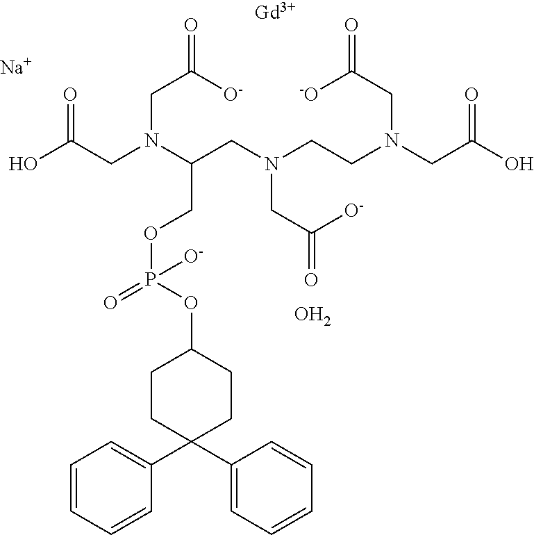
- ZSDLYSDDELEVDD-UHFFFAOYSA-K O.O=C([O-])CN(CCN(CC(=O)O)CC(=O)O)CC(COP(=O)([O-])OC1CCC(C2=CC=CC=C2)(C2=CC=CC=C2)CC1)N(CC(=O)[O-])CC(=O)O.[Gd+3].[Na+] Chemical compound O.O=C([O-])CN(CCN(CC(=O)O)CC(=O)O)CC(COP(=O)([O-])OC1CCC(C2=CC=CC=C2)(C2=CC=CC=C2)CC1)N(CC(=O)[O-])CC(=O)O.[Gd+3].[Na+] ZSDLYSDDELEVDD-UHFFFAOYSA-K 0.000 description 1
Images


Classifications
-
- A—HUMAN NECESSITIES
- A61—MEDICAL OR VETERINARY SCIENCE; HYGIENE
- A61K—PREPARATIONS FOR MEDICAL, DENTAL OR TOILETRY PURPOSES
- A61K49/00—Preparations for testing in vivo
- A61K49/06—Nuclear magnetic resonance [NMR] contrast preparations; Magnetic resonance imaging [MRI] contrast preparations
- A61K49/08—Nuclear magnetic resonance [NMR] contrast preparations; Magnetic resonance imaging [MRI] contrast preparations characterised by the carrier
- A61K49/10—Organic compounds
-
- A—HUMAN NECESSITIES
- A61—MEDICAL OR VETERINARY SCIENCE; HYGIENE
- A61K—PREPARATIONS FOR MEDICAL, DENTAL OR TOILETRY PURPOSES
- A61K47/00—Medicinal preparations characterised by the non-active ingredients used, e.g. carriers or inert additives; Targeting or modifying agents chemically bound to the active ingredient
- A61K47/50—Medicinal preparations characterised by the non-active ingredients used, e.g. carriers or inert additives; Targeting or modifying agents chemically bound to the active ingredient the non-active ingredient being chemically bound to the active ingredient, e.g. polymer-drug conjugates
- A61K47/51—Medicinal preparations characterised by the non-active ingredients used, e.g. carriers or inert additives; Targeting or modifying agents chemically bound to the active ingredient the non-active ingredient being chemically bound to the active ingredient, e.g. polymer-drug conjugates the non-active ingredient being a modifying agent
- A61K47/54—Medicinal preparations characterised by the non-active ingredients used, e.g. carriers or inert additives; Targeting or modifying agents chemically bound to the active ingredient the non-active ingredient being chemically bound to the active ingredient, e.g. polymer-drug conjugates the non-active ingredient being a modifying agent the modifying agent being an organic compound
- A61K47/555—Medicinal preparations characterised by the non-active ingredients used, e.g. carriers or inert additives; Targeting or modifying agents chemically bound to the active ingredient the non-active ingredient being chemically bound to the active ingredient, e.g. polymer-drug conjugates the non-active ingredient being a modifying agent the modifying agent being an organic compound pre-targeting systems involving an organic compound, other than a peptide, protein or antibody, for targeting specific cells
-
- A—HUMAN NECESSITIES
- A61—MEDICAL OR VETERINARY SCIENCE; HYGIENE
- A61K—PREPARATIONS FOR MEDICAL, DENTAL OR TOILETRY PURPOSES
- A61K49/00—Preparations for testing in vivo
- A61K49/06—Nuclear magnetic resonance [NMR] contrast preparations; Magnetic resonance imaging [MRI] contrast preparations
- A61K49/08—Nuclear magnetic resonance [NMR] contrast preparations; Magnetic resonance imaging [MRI] contrast preparations characterised by the carrier
- A61K49/10—Organic compounds
- A61K49/101—Organic compounds the carrier being a complex-forming compound able to form MRI-active complexes with paramagnetic metals
- A61K49/103—Organic compounds the carrier being a complex-forming compound able to form MRI-active complexes with paramagnetic metals the complex-forming compound being acyclic, e.g. DTPA
Definitions
- the present technology generally relates to gadolinium contrast agents, including gadolinium contrast agents modified to be scavenged from a patient, as well as methods and systems for scavenging gadolinium contrast agents from a patient.
- Gadolinium contrast agents are formed of a gadolinium chelate that includes a Gd 3+ cation plus a ligand that typically contains three or four amine groups securing a gadolinium cation.
- the ligand also often includes a plurality of acid or amide groups. The ligand isolates the gadolinium cation, preventing it from being rapidly absorbed by cells, while also influencing and improving imaging contrast properties.
- Gadolinium contrast agents and their byproducts are often excreted unchanged by glomerular filtration.
- studies have shown that gadolinium can accumulate in tissues, such as brain tissue, bone tissue, and kidney tissue. This accumulation in a patient's tissues can be problematic, especially for patients with reduced renal function. Symptoms of gadolinium accumulation include central pain, peripheral pain, headaches, bone pain, skin thickening, clouded mentation, etc.
- Embodiments include functionalized gadolinium contrast agents, system for removing gadolinium contrast agent from a patient, and methods for removing gadolinium contrast agent from a patient
- a functionalized gadolinium contrast agent has a gadolinium cation; and a ligand secured to the gadolinium cation, the ligand comprising a reactive group capable of bonding to a capture molecule on a capture substrate.
- the ligand secured to the gadolinium cation comprises a plurality of amine groups.
- the ligand secured to the the gadolinium cation comprises a plurality of acid groups.
- the reactive group on the ligand comprises an azide group.
- the reactive group on the ligand comprises an azide, alkyne, tetrazine, fluorosydnones, or combinations thereof.
- the capture substrate comprises a strained alkyne.
- the strained alkyne of the capture substrate is selected from the group OCT (cyclooctyne), DIMAC (dimethoxyazacyclooctyne), DIFO (difluorinated cyclooctynes), BCN (bicyclononyne), DIBO (dibenzocyclooctyne), DIFBO (difluorobenzocyclooctyne), DIBAC aza-dibenzocyclooctyne), BARAC (biarylazacyclooctynone), TMTH (3,3,6,6-tetramethyl-thiacycloheptyne) and mixtures thereof.
- OCT cyclooctyne
- DIMAC diimethoxyazacyclooctyne
- DIFO difluorinated cyclooctynes
- BCN bicyclononyne
- DIBO dibenzocyclooctyne
- DIFBO difluorobe
- the functionalized gadolinium contrast agent forms a covalent bond with the capture molecule on the capture substrate when brought in contact with each other.
- the functionalized gadolinium contrast agent and the capture molecule form a tri-azole ring upon reacting.
- a system for removing gadolinium contrast agent from a patient includes a gadolinium contrast agent comprising a gadolinium cation secured to a ligand comprising a reactive group capable of bonding to a capture molecule on a capture substrate; and a capture substrate containing a capture molecule capably of spontaneously forming a bond with the reactive group on the gadolinium contrast agent.
- the reactive group on the ligand comprises an azide group.
- the gadolinium cation secured to the ligand comprises an azide reactive group;
- the capture substrate comprises a polymer containing a strained alkyne; the strained alkyne selected from the group OCT (cyclooctyne), DIMAC (dimethoxyazacyclooctyne), DIFO (difluorinated cyclooctynes), BCN (bicyclononyne), DIBO (dibenzocyclooctyne), DIFBO (difluorobenzocyclooctyne), DIBAC aza-dibenzocyclooctyne), BARAC (biarylazacyclooctynone), TMTH (3,3,6,6-tetramethyl-thiacycloheptyne), and mixtures thereof.
- OCT cyclooctyne
- DIMAC diimethoxyazacyclooctyne
- DIFO difluorinated cyclooct
- the capture substrate comprises a textile, membrane, foam, gel, web, or combination of substrates.
- a method of removing gadolinium contrast agents from a patient includes providing a gadolinium contrast agent comprising a reactive group; providing a removable capture substrate containing a capture molecule that spontaneously bonds to the reactive group of the gadolinium contrast agent; administering the gadolinium contrast agent to a patient; conducting a magnetic resonance imaging procedure; and sequestering the gadolinium contrast agent on the removable capture substrate.
- the removable capture substrate is positioned in a blood vessel upstream of the kidney of the patient during sequestration of the gadolinium contrast agent.
- the gadolinium contrast agent comprises a plurality of amine groups.
- gadolinium contrast agent comprises an azide group.
- the gadolinium contrast agent comprises an azide, alkyne, tetrazine, fluorosydnones, or combinations thereof.
- the capture substrate comprises group OCT (cyclooctyne), DIMAC (dimethoxyazacyclooctyne), DIFO (difluorinated cyclooctynes), BCN (bicyclononyne), DIBO (dibenzocyclooctyne), DIFBO (difluorobenzocyclooctyne), DIBAC aza-dibenzocyclooctyne), BARAC (biarylazacyclooctynone), TMTH (3,3,6,6-tetramethyl-thiacycloheptyne) and mixtures thereof.
- the capture substrate comprises a textile, foam, or web.
- the present disclosure is also directed to a method of removing gadolinium contrast agents from a patient, the method comprising providing a gadolinium contrast agent comprising a reactive group; providing a removable capture substrate containing a capture molecule that spontaneously bonds to the reactive group of the gadolinium contrast agent; administering the gadolinium contrast agent to a patient; conducting a magnetic resonance imaging procedure; and sequestering the gadolinium contrast agent on the removable capture substrate while allowing blood to flow through.
- FIG. 1 shows two magnetic resonance images of a human head, with the left image taken without a gadolinium contrast agent and the right image taken with a gadolinium contrast agent.
- FIG. 2A is a schematic diagram of a gadolinium contrast agent prior to addition of a reactive group.
- FIG. 2B is a schematic diagram of a gadolinium contrast agent after addition of a reactive group.
- FIG. 3 is a schematic diagram of gadolinium contrast agents to which reactive groups have been added, shown with a reactive substrate.
- FIG. 4 is schematic diagram of gadolinium contrast agents to which reactive groups have been added, showing the gadolinium contrast agents sequestered on a reactive substrate.
- FIG. 5 is a schematic diagram showing example locations of gadolinium contrast agent scavenger positioned inside and outside a patient's body.
- FIG. 6 is a perspective view of an article containing a gadolinium contrast agent scavenger configured for the flow of blood through the article to retain the gadolinium contrast agent.
- Gadolinium containing MRI contrast agents are among the most commonly used for enhancement of vessels in MRI angiography.
- the gadolinium dose can be as low as 0.1 mmol per kilogram of body mass. Higher concentrations are often used for finer vasculature.
- gadolinium contrast agents and their byproducts are mostly excreted unchanged by glomerular filtration, studies have shown gadolinium accumulates in tissues, such as brain, bone, and kidneys.
- the ionic radius of Gd +3 is very close to that of the calcium cation Ca +2 .
- gadolinium-based MRI contrast agents typically use some sort of ligand to chelate the free ion and reduce its toxicity. Still, a need exists for assisted removal of gadolinium, in particular gadolinium chelates, from the bloodstream. This need is especially important on patients with diminished kidney function.
- the present disclosure is directed to gadolinium contrast agents that have been modified to provide a functional group that can spontaneously covalently bond to a capture molecule secured to a removable substrate and then removed from a patient.
- the bond between the gadolinium contrast agent and the capture molecule allows for subsequent removal of the gadolinium contrast agent by removal of the substrate containing the capture molecules.
- water-soluble gadolinium-based contrast agents are modified with functional groups so that the contrast agent can selectively, rapidly react with a biorthogonal counterpart capture molecule secured to a substrate, and this substrate can then be removed from the patient (if the substrate is positioned within the patient); or alternatively an external capture element can be used that contains the capture molecule secured to a substrate.
- the gadolinium contrast agents and reactive substrate utilize “click chemistry” to selectively and effectively remove the gadolinium contrast agents from a patient's bloodstream.
- FIG. 1 shows two magnetic resonance images of a human head, with the left image taken without a gadolinium contrast agent and the right image taken with a gadolinium contrast agent.
- the gadolinium contrast agent provides improved imaging, revealing an area of increased blood volume (and thus a potential aneurism) on the right side of the patient's brain.
- FIG. 1 shows two magnetic resonance images of a human head, with the left image taken without a gadolinium contrast agent and the right image taken with a gadolinium contrast agent.
- the gadolinium contrast agent provides improved imaging, revealing an area of increased blood volume (and thus a potential aneurism) on the right
- the gadolinium contrast agent is typically a linear or macrocyclic structure.
- Linear agents have an elongated organic molecular ligand that wraps around the gadolinium ion.
- Macrocyclic agents form a cage-like ligand structure with the ion trapped in a preformed central cavity. Both linear and macrocyclic agents can either be ionic or non-ionic. In the macrocyclic structure, the gadolinium ion is retained in a cavity of the ligand.
- Both linear and macrocyclic gadolinium contrast agents can be produced or modified for improved removal from a patient using click chemistry.
- the gadolinium contrast agent can be modified to add a reactive group capable of bonding to a capture substrate.
- One example modification is to add an azide group to the gadolinium ligand.
- An azide group can be particularly useful because it is small, metabolically stable, and does not naturally exist in cells. Thus, azide groups generally have no competing biological side reactions and thus are particularly selective when binding to an appropriately-selected biorthogonal pair, such as a strained alkyne secured to a substrate.
- FIG. 2A is a schematic diagram of a gadolinium contrast agent 10 prior to addition of a reactive group 12
- FIG. 2B is a schematic of a gadolinium contrast agent 10 after addition of the reactive group 12
- FIG. 2B shows the gadolinium contrast agent 10 with the reactive group 12 , such as an azide, attached.
- the reactive group 12 such as an azide
- An example of a specific linear gadolinium contrast agent to which an azide is attached is reproduced below, showing gadofosvaset with an azide group added to the aromatic ring.
- the gadolinium contrast agent can be initially formed, and then modified to include the reactive group 12 , or the reactive group 12 can simultaneously be added during synthesis of the overall gadolinium contrast agent.
- the inclusion of one or more reactive groups 12 can be done at various stages of creation of the gadolinium contrast agent.
- FIG. 3 a schematic representation is shown of multiple gadolinium contrast agent molecules 10 from FIG. 2 with reactive groups 12 (such azides), along with a capture substrate 14 .
- the capture substrate 14 includes capture molecules 16 , such as strained alkynes, capable of a spontaneous, irreversible reaction with reactive groups 12 .
- the capture molecule 16 can include, for example, OCT (cyclooctyne), DIMAC (dimethoxyazacyclooctyne), DIFO (difluorinated cyclooctynes), BCN (bicyclononyne), DIBO (dibenzocyclooctyne), DIFBO (difluorobenzocyclooctyne), DIBAC aza-dibenzocyclooctyne), BARAC (biarylazacyclooctynone), TMTH (3,3,6,6-tetramethyl-thiacycloheptyne) and mixtures thereof.
- OCT cyclooctyne
- DIMAC diimethoxyazacyclooctyne
- DIFO difluorinated cyclooctynes
- BCN bicyclononyne
- DIBO dibenzocyclooctyne
- DIFBO difluorobenzocyclooc
- FIG. 3 the capture molecules 16 are shown secured to the capture substrate 14 , but without any of the contrast agent molecules 10 yet reacted with the capture molecules 16 .
- FIG. 4 is a schematic of molecules of gadolinium contrast agent 10 to which reactive groups 12 have been added, showing the gadolinium contrast agents 10 sequestered on the capture substrate 14 by way of the reactive groups 12 of the contrast agent 10 having reacted with the capture molecules 16 of the capture substrate 14 .
- the capture substrate 14 can be, for example, a polymeric film, gel, web, fabric, foam, mesh or other material to which the capture molecules 16 have been secured, or a combination of materials. After the capture molecules 16 bind to gadolinium contrast agents 10 , the entire substrate 14 and accompanying secured gadolinium contrast agents 10 can be removed from a patent.
- the substrate 14 is placed within a blood vessel in a patient, such as blood vessels upstream of the kidney (for example, in the left or right renal artery, or the interior vena cava).
- a blood vessel in a patient such as blood vessels upstream of the kidney (for example, in the left or right renal artery, or the interior vena cava).
- the substrate 14 is positioned outside of the patient's body within a housing connected by intravenous catheters to the patient's bloodstream.
- the gadolinium contrast agents are modified to add a reactive group capable of bonding to a capture substrate.
- a reactive group capable of bonding to a capture substrate is modified to add an azide group to the gadolinium ligand.
- the reactive group of the ligand can be, for example, an azide, alkyne, tetrazine, fluorosydnones, or combinations thereof.
- the azide group is particularly appropriate because it is small, metabolically stable, and does not naturally exist in cells. Thus, it has no competing biological side reactions.
- the alkyne is not as small, but it still has the stability and orthogonality useful for selective removal of gadolinium contrast agents.
- Desired properties for the ligand comprising the reactive group and the related capture substrate include strong selectivity, generally biological inertness, generally biological and chemical inertness, favorable kinetics, and reaction biocompatibility. With regard to selectivity, it is desirable that the reaction be selective between functional groups to avoid side reactions with biological compounds. With regard to biological inertness, desirably the reactive group on the contrast ligand should not possess reactivity capable of disrupting the native chemical functionality of the patient. Regarding chemical inertness, the covalent link between the reactive group on the ligand and the capture molecule on the capture substrate should be strong and inert to biological reactions.
- gadofosvaset prior to addition of an azide group
- the reactive group of the gadolinium contrast agent is reactive with groups on the capture substrate.
- the capture substrate can be, for example, a substrate or polymer having exposed strained alkyne functional groups.
- the reactive receptor group can be, for example, OCT (cyclooctyne), DIMAC (dimethoxyazacyclooctyne), DIFO (difluorinated cyclooctynes), BCN (bicyclononyne), DIBO (dibenzocyclooctyne), DIFBO (difluorobenzocyclooctyne), DIBAC aza-dibenzocyclooctyne), BARAC (biarylazacyclooctynone), TMTH (3,3,6,6-tetramethyl-thiacycloheptyne) and mixtures thereof.
- the functionalized gadolinium contrast agent spontaneously forms a covalent bond with the capture substrate.
- the functionalized gadolinium contrast agent and the capture substrate form a tri-azole ring.
- the capture substrate is, for example, porous so that blood and gadolinium contrast agent flows through the substrate so as to remove the gadolinium contrast agent.
- the substrate can be relatively smooth, allowing blood and gadolinium contrast agent to flow along the surface of the substrate until the contrast agent is captured.
- Suitable capture substrates include, for example, polyvinyl alcohol (PVA) to which the capture molecule (such as a moiety containing a strained alkyne) has been secured.
- PVA polyvinyl alcohol
- the modified gadolinium contrast agent is brought in contact with the substrate after the MRI procedure, such as by insertion into a blood vessel or retained in a chamber outside of the body but through which blood is passed.
- An example location of the capture substrate is in the renal artery upstream of a patient's kidney.
- the gadolinium contrast agents described herein can be used as part of a method of removing gadolinium contrast agents from a patient, the method comprising providing a gadolinium contrast agent having a reactive group; providing a removable capture substrate; administering the gadolinium contrast agent to the patient; conducting a magnetic resonance imaging procedure; and sequestering the gadolinium contrast agent on the removable capture substrate.
- the gadolinium contrast agent can be captured and removed as part of a system comprising a gadolinium cation secured (such as by bonding, such as by ionic forces) to a ligand comprising a reactive group capable of bonding to a capture substrate; and a capture substrate.
- the gadolinium cation secured to a ligand can include an azide reactive group; and the capture substrate can include a polymer containing a strained alkyne; the strained alkyne selected from the group OCT (cyclooctyne), DIMAC (dimethoxyazacyclooctyne), DIFO (difluorinated cyclooctynes), BCN (bicyclononyne), DIBO (dibenzocyclooctyne), DIFBO (difluorobenzocyclooctyne), DIBAC aza-dibenzocyclooctyne), BARAC (biarylazacyclooctynone), TMTH (3,3,6,6-tetramethyl-thiacycloheptyne) and mixtures thereof.
- OCT cyclooctyne
- DIMAC diimethoxyazacyclooctyne
- DIFO difluorinated cycloo
- FIG. 5 a schematic diagram of a patient 50 shows example locations 52 A, 52 B, 52 C, 52 D and 52 E for a gadolinium contrast agent scavenger.
- Patient 50 is shown as a human outline, but examples are not so limited and may include any mammalian.
- FIG. 5 is simplified and not drawn to scale, showing body organs and scavenger systems schematically only.
- the locations 52 A to 52 E are shown for illustrative purposes, indicating how the location of the contrast agent scavenger can be varied.
- Locations 52 A, 52 B and 52 C are all internal locations in which the contrast scavenger agent is inserted into a blood vessel, typically a blood vessel upstream of one or both kidneys.
- locations 52 A and 52 B show locations at the right and left renal arteries, respectively, while location 52 C is in the inferior vena cava.
- the gadolinium contrast agent scavenger can be applied in the form of a textile, membrane, foam, gel, web or other substance inserted into the blood vessel and then removed after the medical procedure is completed and adequate contrast agent has been scavenged.
- FIG. 5 also shows schematic representations of two external locations 52 D and 52 E for removing gadolinium contrast agents from the patient 50 .
- Location 52 D is shown on a peripheral body location, with an intravenous catheter 54 D leading to an optional pump 55 D that flows into a sequestering element 57 D and then back into the patient 50 by way of intravenous catheter 56 D.
- the sequestering element 57 D contains a textile, membrane, foam, gel, web or other substance with exposed sequestering agent for binding the gadolinium contrast agent.
- Alternative location 52 E for removing gadolinium contrast agents includes intravenous catheter 54 E leading to sequestering element 57 E and then flow out through return catheter 56 E.
- Location 52 E is depicted without an auxiliary pump, although generally some sort of mechanism is used to apply adequate pressure to return the blood to the patient.
- External positioning of the contrast agent scavenger at location 52 D and 52 E are less invasive than inserting the contrast agent scavenger into locations 52 A, 52 B or 52 C, but is also typically slower to remove the contrast agent and allows initial exposure of the kidneys of higher levels of contrast agent.
- FIG. 6 is a perspective view of gadolinium capture element 60 containing a gadolinium contrast agent scavenger configured for the flow of blood through the element to retain the gadolinium contrast agent such as elements 57 D and 57 E of FIG. 5 .
- the capture element 60 includes a housing 66 , along with an inlet 62 on a first end 64 of the housing 66 for receiving blood containing gadolinium contrast agent, plus an outlet (not shown) opposite the inlet 62 on the second end 68 of the housing, through which the blood exits the housing 66 .
- the capture media contains capture substrate as described herein, such as a capture substrate having exposed strained alkynes available for reacting with functionalized gadolinium contrast agent, such as gadolinium functionalized with an azide group.
- the phrase “configured” describes a system, apparatus, or other structure that is constructed to perform a particular task or adopt particular characteristics.
- the phrase “configured” can be used interchangeably with other similar phrases such as “arranged”, “arranged and configured”, “programmed” “constructed and arranged”, “constructed”, “manufactured and arranged”, and the like.
Landscapes
- Health & Medical Sciences (AREA)
- Life Sciences & Earth Sciences (AREA)
- Veterinary Medicine (AREA)
- Nuclear Medicine, Radiotherapy & Molecular Imaging (AREA)
- Chemical & Material Sciences (AREA)
- Medicinal Chemistry (AREA)
- Epidemiology (AREA)
- Animal Behavior & Ethology (AREA)
- General Health & Medical Sciences (AREA)
- Public Health (AREA)
- Radiology & Medical Imaging (AREA)
- Immunology (AREA)
- Molecular Biology (AREA)
- Engineering & Computer Science (AREA)
- Bioinformatics & Cheminformatics (AREA)
- Pharmacology & Pharmacy (AREA)
- Medicines Containing Antibodies Or Antigens For Use As Internal Diagnostic Agents (AREA)
- Magnetic Resonance Imaging Apparatus (AREA)
Abstract
Description


Under in vitro conditions the reaction proceeded very rapidly and was complete before a nuclear magnetic resonance (NMR) tube containing the reaction constituents could be inserted into the probe.
Claims (6)
Priority Applications (1)
| Application Number | Priority Date | Filing Date | Title |
|---|---|---|---|
| US16/276,169 US11224666B2 (en) | 2018-02-14 | 2019-02-14 | Gadolinium contrast agents, scavenging methods, and scavenging system |
Applications Claiming Priority (2)
| Application Number | Priority Date | Filing Date | Title |
|---|---|---|---|
| US201862630623P | 2018-02-14 | 2018-02-14 | |
| US16/276,169 US11224666B2 (en) | 2018-02-14 | 2019-02-14 | Gadolinium contrast agents, scavenging methods, and scavenging system |
Publications (2)
| Publication Number | Publication Date |
|---|---|
| US20190247523A1 US20190247523A1 (en) | 2019-08-15 |
| US11224666B2 true US11224666B2 (en) | 2022-01-18 |
Family
ID=65529888
Family Applications (1)
| Application Number | Title | Priority Date | Filing Date |
|---|---|---|---|
| US16/276,169 Active US11224666B2 (en) | 2018-02-14 | 2019-02-14 | Gadolinium contrast agents, scavenging methods, and scavenging system |
Country Status (3)
| Country | Link |
|---|---|
| US (1) | US11224666B2 (en) |
| EP (1) | EP3752205A2 (en) |
| WO (1) | WO2019161051A2 (en) |
Citations (4)
| Publication number | Priority date | Publication date | Assignee | Title |
|---|---|---|---|---|
| US20080181847A1 (en) * | 2004-10-07 | 2008-07-31 | Koninklijke Philips Electronics, N.V. | Targeted Imaging and/or Therapy Using the Staudinger Ligation |
| WO2011017690A2 (en) | 2009-08-07 | 2011-02-10 | Northwestern University | Intracellular delivery of contrast agents with functionalized nanoparticles |
| US8545813B2 (en) | 2008-01-25 | 2013-10-01 | Northwestern University | Pre-templated macromolecular architectures with multiple Gd(III) complexes and methods of use as MRI contrast agents |
| US20160346409A1 (en) * | 2014-02-10 | 2016-12-01 | Mcmaster University | Targeted molecular imaging contrast agents |
-
2019
- 2019-02-14 US US16/276,169 patent/US11224666B2/en active Active
- 2019-02-14 EP EP19707637.5A patent/EP3752205A2/en active Pending
- 2019-02-14 WO PCT/US2019/018005 patent/WO2019161051A2/en unknown
Patent Citations (4)
| Publication number | Priority date | Publication date | Assignee | Title |
|---|---|---|---|---|
| US20080181847A1 (en) * | 2004-10-07 | 2008-07-31 | Koninklijke Philips Electronics, N.V. | Targeted Imaging and/or Therapy Using the Staudinger Ligation |
| US8545813B2 (en) | 2008-01-25 | 2013-10-01 | Northwestern University | Pre-templated macromolecular architectures with multiple Gd(III) complexes and methods of use as MRI contrast agents |
| WO2011017690A2 (en) | 2009-08-07 | 2011-02-10 | Northwestern University | Intracellular delivery of contrast agents with functionalized nanoparticles |
| US20160346409A1 (en) * | 2014-02-10 | 2016-12-01 | Mcmaster University | Targeted molecular imaging contrast agents |
Non-Patent Citations (18)
| Title |
|---|
| "Response to Communication Pursuant to Rules 161(1) and 162 EPC," for European Patent Application No. 19707637.5 filed Apr. 1, 2021 (10 pages). |
| Grogna et al., Polym. Chem., 2011, 2, 2316-27. (Year: 2011). * |
| Grogna, Mathurin et al., "Stealth macromolecular platforms for the design of MRI blood pool contrast agents," Polym. Chem. 2011, 2, 2316-27 (12 pages). |
| Hapuarachchige, Sudath et al., "Click Chemistry in the Development of Contrast Agents for Magnetic Resonance Imaging," Topics In Magnetics Resonance Imaging, vol. 25, No. 5, Oct. 1, 2016 pp. 205-213 (20 pages). |
| International Preliminary Report on Patentability for PCT Application No. PCT/US2018/018005 dated Aug. 27, 2020 (9 pages). |
| International Search Report and Written Opinion for PCT Application No. PCT/US2018/018005 dated Oct. 24, 2019 (12 pages). |
| Kobayashi, Hisataka et al., "Activated Clearance of a Biotinylated Macromolecular MRI Contrast Agent from the Blood Pool using an Avidin Chase," Bioconjugate Chemistry, vol. 14, No. 5, Sep. 1, 2003 pp. 1044-1047 (4 pages). |
| Liu, Hui et al., "Ultrafast Click Chemistry with Fluorosydnones," Agnew. Chem. Int. Ed. Engl. 2016, 55(39), 12073-7 (5 pages). |
| Pokorski, Jonathan K. et al., "Functional Virus-Based Polymer-Protein Nanoparticles by Atom Transfer Radical Polymerization," J Am Chem Soc, 2011. 133(24): 9242-5 (11 pages). |
| Ramil, Carlo P. et al., "Bioorthogonal Chemistry: Strategies and Recent Developments," Chem. Commun. 2013, 49(94), 11007-22 (16 pages). |
| Rogosnitzky, Moshe et al., "Gadolinium-based Contrast Agent Toxicity: a Review of Known and Proposed Mechanisms," Biometals (2016) 29:365-376 (12 pages). |
| Santra, Santimukul et al., "Aliphatic Hyperbranched Polyester: A New Building Block in the Construction of Multifunctional Nanoparticles and Nanocomposites," Langmuir 2010, 26(18), 5364-73 (10 pages). |
| Sukerkar, Preeti A. et al., "Synthesis and Biological Evaluation of Water-soluble Progesterone-Conjugated Probes for Magnetic Resonance Imaging of Hormone Related Cancers," Bioconjug Chem, 2011. 22(11): 2304-16 (26 pages). |
| Tan, Mingqian et al., "Peptide-Targeted Nanoglobular Gd-DOTA Monoamide Conjugates for Magnetic Resonance Cancer Molecular Imaging," Biomacromolecules, 2010. 11(3): 754-61 (8 pages). |
| Toppino, Antonio et al., "A carborane-Derivative "Click" Reaction under Heterogeneous Conditions for the Synthesis of a Promising Lipophilic MRI/GdBNCT Agent," Chemistry, 2013. 19(2): 721-8 (8 pages). |
| Vanasschen, Christian et al., "Gadolinium DOTA Chelates Featuring Alkyne Groups Directly Grafted on the Tetraaza Macrocyclic Ring: Synthesis, Relaxation Properties, "Click" Reaction, and High-Relaxivity Micelles," Inorg Chem, 2011. 50(18): 8946-58 (13 pages). |
| Verwilst, Peter et al., "A Modular Approach towards the Synthesis of Target-Specific MRI Contrast Agents," European Journal of Inorganic Chemistry, 2011. 2011(24): 3577-85 (9 pages). |
| Yantasee, W. et al., "Novel sorbents for removal of gadolinium-based contrast agents in sorbent dialysis and hemoperfustion: preventive approaches to nephrogenic systemic fibrosis," Nanomedicine: Nanotechnology, Biology and Medicine, Elsevier, NL, vol. 6, No. 1, Feb. 1, 2010 pp. 1-8 (8 pages). |
Also Published As
| Publication number | Publication date |
|---|---|
| WO2019161051A2 (en) | 2019-08-22 |
| US20190247523A1 (en) | 2019-08-15 |
| EP3752205A2 (en) | 2020-12-23 |
| WO2019161051A3 (en) | 2019-11-14 |
Similar Documents
| Publication | Publication Date | Title |
|---|---|---|
| JPH07503641A (en) | Apparatus and method for extracorporeal processing of blood | |
| US11224666B2 (en) | Gadolinium contrast agents, scavenging methods, and scavenging system | |
| KR20060118515A (en) | Tungsten particles as x-ray contrast agents | |
| US11213596B2 (en) | Radiocontrast agents, scavenging methods, and scavenging system | |
| WO1996040288A1 (en) | Gaseous ultrasound contrast agents and method therefor | |
| US20220249676A1 (en) | Click chemistry capturable platinum-based antineoplastic agents | |
| Ellenberg et al. | Cerebrospinal fluid aluminum levels following deferoxamine | |
| Littlewood | Innovative multispectral optoacoustic tomography strategies for evaluating different aspects of renal function | |
| US20210121644A1 (en) | Devices, systems and methods for improved radiotherapy efficacy | |
| Sideman et al. | Removal of bilirubin from the blood of jaundiced infants | |
| CN118320773A (en) | Contrast agent, preparation device, preparation method and application imaging method thereof | |
| AU2022350500A1 (en) | Urea filtration device comprising nanofiber compositions | |
| RU2304002C2 (en) | Method for biological correction of body homeostasis | |
| Novelli et al. | MOLECULAR ADSORBENT RECYCLING SYSTEM AS PREDICTIVA CRITERIA OF SURVIVAL IN PATIENTS WITH ACUTE LIVER FAILURE: 1904 | |
| Miyazawa et al. | DEVELOPMENT OF A NEW TREATMENT FOR BILE DUCT STENOSIS USING TISSUE ENGINEERING TECHNIQUES–DEVELOPMENT OF A BILE DUCT PATCH MADE OF BIOABSORPABLE POLYMER–: 1903 | |
| Tan et al. | ALBUMIN DIALYSIS IN SEVERE LIVER FAILURE: AN ANTICOAGULANT-FREE APPROACH: 1902 | |
| Cadena et al. | Tensile mechanical properties of porcine small intestinal submucosa scaffolds: effect of orientation and number of layers | |
| AD | PALGO JOURNAL OF MEDIC | |
| Gorelik | Analysis of domestic market of contrasting substances for X-ray diagnosis | |
| Narine et al. | Readily available porcine aortic valve matrices for tissue valve engineering | |
| Lueders et al. | Tissue engineering of heart valves: a new dynamic cell seeding device for tissue engineering of heart valves | |
| Yaguchi et al. | STRUCTURE INFLUENCES CELLULAR COMPATIBILITY IN A MICROPOROUS POLYURETHANE SCAFFOLD | |
| Oku et al. | GENERATION MECHANISMS OF SHUNT MURMURS USING FINITE ELEMENT METHOD | |
| Yambe et al. | BRAIN FUNCTIONAL MONOTORING AND CONTROL DEVICE | |
| Mareels et al. | Modeling Fluid Flow and Oxygen transport in the AMC Bioartificial Liver |
Legal Events
| Date | Code | Title | Description |
|---|---|---|---|
| FEPP | Fee payment procedure |
Free format text: ENTITY STATUS SET TO UNDISCOUNTED (ORIGINAL EVENT CODE: BIG.); ENTITY STATUS OF PATENT OWNER: LARGE ENTITY |
|
| AS | Assignment |
Owner name: BOSTON SCIENTIFIC SCIMED, INC., MINNESOTA Free format text: ASSIGNMENT OF ASSIGNORS INTEREST;ASSIGNORS:DELANEY, JOSEPH THOMAS, JR.;PAGORIA, DOUGLAS DEAN;PENNINGTON, DOUGLAS;AND OTHERS;SIGNING DATES FROM 20180128 TO 20190419;REEL/FRAME:049037/0884 |
|
| STPP | Information on status: patent application and granting procedure in general |
Free format text: NON FINAL ACTION MAILED |
|
| STPP | Information on status: patent application and granting procedure in general |
Free format text: RESPONSE TO NON-FINAL OFFICE ACTION ENTERED AND FORWARDED TO EXAMINER |
|
| STPP | Information on status: patent application and granting procedure in general |
Free format text: NON FINAL ACTION MAILED |
|
| STPP | Information on status: patent application and granting procedure in general |
Free format text: FINAL REJECTION MAILED |
|
| STPP | Information on status: patent application and granting procedure in general |
Free format text: DOCKETED NEW CASE - READY FOR EXAMINATION |
|
| STPP | Information on status: patent application and granting procedure in general |
Free format text: NON FINAL ACTION MAILED |
|
| STPP | Information on status: patent application and granting procedure in general |
Free format text: RESPONSE TO NON-FINAL OFFICE ACTION ENTERED AND FORWARDED TO EXAMINER |
|
| STPP | Information on status: patent application and granting procedure in general |
Free format text: NOTICE OF ALLOWANCE MAILED -- APPLICATION RECEIVED IN OFFICE OF PUBLICATIONS |
|
| STPP | Information on status: patent application and granting procedure in general |
Free format text: PUBLICATIONS -- ISSUE FEE PAYMENT VERIFIED |
|
| STCF | Information on status: patent grant |
Free format text: PATENTED CASE |