US11213595B2 - Antigen responsive antibody-fluorescent dye conjugate and method for fluorescence detection and imaging of target cell using the same - Google Patents
Antigen responsive antibody-fluorescent dye conjugate and method for fluorescence detection and imaging of target cell using the same Download PDFInfo
- Publication number
- US11213595B2 US11213595B2 US16/138,371 US201816138371A US11213595B2 US 11213595 B2 US11213595 B2 US 11213595B2 US 201816138371 A US201816138371 A US 201816138371A US 11213595 B2 US11213595 B2 US 11213595B2
- Authority
- US
- United States
- Prior art keywords
- antibody
- fluorescent dye
- antigen
- atto
- conjugate
- Prior art date
- Legal status (The legal status is an assumption and is not a legal conclusion. Google has not performed a legal analysis and makes no representation as to the accuracy of the status listed.)
- Active
Links
- ZAZXEJNOOJTIDU-UHFFFAOYSA-O CC1=CC2=C(C=C1)N/C(=C/C=C1\CCCC(/C=C/C3=[N+](\CCC[N+](C)(C)C)C4=CC=C(S(=O)(=O)[O-])C=C4C3(C)C)=C1OC1=CC=C(CCC(=O)O)C=C1)C2(C)C.CCC[N+](C)(C)C Chemical compound CC1=CC2=C(C=C1)N/C(=C/C=C1\CCCC(/C=C/C3=[N+](\CCC[N+](C)(C)C)C4=CC=C(S(=O)(=O)[O-])C=C4C3(C)C)=C1OC1=CC=C(CCC(=O)O)C=C1)C2(C)C.CCC[N+](C)(C)C ZAZXEJNOOJTIDU-UHFFFAOYSA-O 0.000 description 2
- ZOACLFGYLKCRNR-UHFFFAOYSA-O C=C(O)CCC/[N+](C)=C1/C=CC2=NC3=C/C=C(N(C)C)/C=C\3SC2=C1 Chemical compound C=C(O)CCC/[N+](C)=C1/C=CC2=NC3=C/C=C(N(C)C)/C=C\3SC2=C1 ZOACLFGYLKCRNR-UHFFFAOYSA-O 0.000 description 1
- RPOXVHANRMFEHN-UHFFFAOYSA-O C=C(O)CCC/[N+](C)=C1/C=CC2=NC3=C/C=C(N(C)C)/C=C\3SC2=C1.C=C(O)CCCN1C2=CC3=C(C=C2C(CS(=O)(=O)[O-])=CC1(C)C)/N=C1/C=C2C(C)=CC(C)(C)[N+](CC)=C2C=C1O3 Chemical compound C=C(O)CCC/[N+](C)=C1/C=CC2=NC3=C/C=C(N(C)C)/C=C\3SC2=C1.C=C(O)CCCN1C2=CC3=C(C=C2C(CS(=O)(=O)[O-])=CC1(C)C)/N=C1/C=C2C(C)=CC(C)(C)[N+](CC)=C2C=C1O3 RPOXVHANRMFEHN-UHFFFAOYSA-O 0.000 description 1
- VBGKTWMFAIFFBV-UHFFFAOYSA-N C=C(O)CCCN1C2=CC3=C(C=C2C(CS(=O)(=O)[O-])=CC1(C)C)/N=C1/C=C2C(C)=CC(C)(C)[N+](CC)=C2C=C1O3 Chemical compound C=C(O)CCCN1C2=CC3=C(C=C2C(CS(=O)(=O)[O-])=CC1(C)C)/N=C1/C=C2C(C)=CC(C)(C)[N+](CC)=C2C=C1O3 VBGKTWMFAIFFBV-UHFFFAOYSA-N 0.000 description 1
- PCSJHWWJXGFGKK-UHFFFAOYSA-N C=C(O)CCCN1C2=CC3=C(C=C2C(CS(=O)(=O)[O-])=CC1(C)C)/N=C1/C=C2CCC[N+](CC)=C2C=C1O3 Chemical compound C=C(O)CCCN1C2=CC3=C(C=C2C(CS(=O)(=O)[O-])=CC1(C)C)/N=C1/C=C2CCC[N+](CC)=C2C=C1O3 PCSJHWWJXGFGKK-UHFFFAOYSA-N 0.000 description 1
- BTRFDXVPJDBKBN-UHFFFAOYSA-N C=C(O)CCCN1C2=CC3=C(C=C2C(CS(=O)(=O)[O-])=CC1(C)C)/N=C1/C=C2CCC[N+](CC)=C2C=C1O3.C=C(O)CCCN1C2=CC3=C(C=C2C(CS(=O)(=O)[O-])CC1(C)C)/N=C1/C=C2CCC[N+](CC)=C2C=C1O3 Chemical compound C=C(O)CCCN1C2=CC3=C(C=C2C(CS(=O)(=O)[O-])=CC1(C)C)/N=C1/C=C2CCC[N+](CC)=C2C=C1O3.C=C(O)CCCN1C2=CC3=C(C=C2C(CS(=O)(=O)[O-])CC1(C)C)/N=C1/C=C2CCC[N+](CC)=C2C=C1O3 BTRFDXVPJDBKBN-UHFFFAOYSA-N 0.000 description 1
- ZFRHDHJBSZHXIX-UHFFFAOYSA-N C=C(O)CCCN1C2=CC3=C(C=C2C(CS(=O)(=O)[O-])CC1(C)C)/N=C1/C=C2CCC[N+](CC)=C2C=C1O3 Chemical compound C=C(O)CCCN1C2=CC3=C(C=C2C(CS(=O)(=O)[O-])CC1(C)C)/N=C1/C=C2CCC[N+](CC)=C2C=C1O3 ZFRHDHJBSZHXIX-UHFFFAOYSA-N 0.000 description 1
- 0 CC*CCCC1=CC2=Nc(cc(C(CS(O)(=O)=O)CC(C)(C)N3CCCCO)c3c3)c3OC2=CC1=I Chemical compound CC*CCCC1=CC2=Nc(cc(C(CS(O)(=O)=O)CC(C)(C)N3CCCCO)c3c3)c3OC2=CC1=I 0.000 description 1
Images


Classifications
-
- A—HUMAN NECESSITIES
- A61—MEDICAL OR VETERINARY SCIENCE; HYGIENE
- A61K—PREPARATIONS FOR MEDICAL, DENTAL OR TOILETRY PURPOSES
- A61K49/00—Preparations for testing in vivo
- A61K49/001—Preparation for luminescence or biological staining
- A61K49/0013—Luminescence
- A61K49/0017—Fluorescence in vivo
- A61K49/005—Fluorescence in vivo characterised by the carrier molecule carrying the fluorescent agent
- A61K49/0058—Antibodies
-
- G—PHYSICS
- G01—MEASURING; TESTING
- G01N—INVESTIGATING OR ANALYSING MATERIALS BY DETERMINING THEIR CHEMICAL OR PHYSICAL PROPERTIES
- G01N33/00—Investigating or analysing materials by specific methods not covered by groups G01N1/00 - G01N31/00
- G01N33/48—Biological material, e.g. blood, urine; Haemocytometers
- G01N33/50—Chemical analysis of biological material, e.g. blood, urine; Testing involving biospecific ligand binding methods; Immunological testing
- G01N33/58—Chemical analysis of biological material, e.g. blood, urine; Testing involving biospecific ligand binding methods; Immunological testing involving labelled substances
- G01N33/582—Chemical analysis of biological material, e.g. blood, urine; Testing involving biospecific ligand binding methods; Immunological testing involving labelled substances with fluorescent label
-
- A—HUMAN NECESSITIES
- A61—MEDICAL OR VETERINARY SCIENCE; HYGIENE
- A61K—PREPARATIONS FOR MEDICAL, DENTAL OR TOILETRY PURPOSES
- A61K49/00—Preparations for testing in vivo
- A61K49/001—Preparation for luminescence or biological staining
- A61K49/0013—Luminescence
- A61K49/0017—Fluorescence in vivo
- A61K49/0019—Fluorescence in vivo characterised by the fluorescent group, e.g. oligomeric, polymeric or dendritic molecules
- A61K49/0021—Fluorescence in vivo characterised by the fluorescent group, e.g. oligomeric, polymeric or dendritic molecules the fluorescent group being a small organic molecule
- A61K49/0028—Oxazine dyes
-
- A—HUMAN NECESSITIES
- A61—MEDICAL OR VETERINARY SCIENCE; HYGIENE
- A61K—PREPARATIONS FOR MEDICAL, DENTAL OR TOILETRY PURPOSES
- A61K49/00—Preparations for testing in vivo
- A61K49/001—Preparation for luminescence or biological staining
- A61K49/0013—Luminescence
- A61K49/0017—Fluorescence in vivo
- A61K49/0019—Fluorescence in vivo characterised by the fluorescent group, e.g. oligomeric, polymeric or dendritic molecules
- A61K49/0021—Fluorescence in vivo characterised by the fluorescent group, e.g. oligomeric, polymeric or dendritic molecules the fluorescent group being a small organic molecule
- A61K49/0039—Coumarin dyes
-
- A—HUMAN NECESSITIES
- A61—MEDICAL OR VETERINARY SCIENCE; HYGIENE
- A61K—PREPARATIONS FOR MEDICAL, DENTAL OR TOILETRY PURPOSES
- A61K49/00—Preparations for testing in vivo
- A61K49/001—Preparation for luminescence or biological staining
- A61K49/0013—Luminescence
- A61K49/0017—Fluorescence in vivo
- A61K49/0019—Fluorescence in vivo characterised by the fluorescent group, e.g. oligomeric, polymeric or dendritic molecules
- A61K49/0021—Fluorescence in vivo characterised by the fluorescent group, e.g. oligomeric, polymeric or dendritic molecules the fluorescent group being a small organic molecule
- A61K49/0041—Xanthene dyes, used in vivo, e.g. administered to a mice, e.g. rhodamines, rose Bengal
- A61K49/0043—Fluorescein, used in vivo
-
- A—HUMAN NECESSITIES
- A61—MEDICAL OR VETERINARY SCIENCE; HYGIENE
- A61K—PREPARATIONS FOR MEDICAL, DENTAL OR TOILETRY PURPOSES
- A61K49/00—Preparations for testing in vivo
- A61K49/06—Nuclear magnetic resonance [NMR] contrast preparations; Magnetic resonance imaging [MRI] contrast preparations
-
- C—CHEMISTRY; METALLURGY
- C07—ORGANIC CHEMISTRY
- C07K—PEPTIDES
- C07K16/00—Immunoglobulins [IG], e.g. monoclonal or polyclonal antibodies
- C07K16/18—Immunoglobulins [IG], e.g. monoclonal or polyclonal antibodies against material from animals or humans
-
- C—CHEMISTRY; METALLURGY
- C07—ORGANIC CHEMISTRY
- C07K—PEPTIDES
- C07K16/00—Immunoglobulins [IG], e.g. monoclonal or polyclonal antibodies
- C07K16/18—Immunoglobulins [IG], e.g. monoclonal or polyclonal antibodies against material from animals or humans
- C07K16/22—Immunoglobulins [IG], e.g. monoclonal or polyclonal antibodies against material from animals or humans against growth factors ; against growth regulators
-
- C—CHEMISTRY; METALLURGY
- C07—ORGANIC CHEMISTRY
- C07K—PEPTIDES
- C07K16/00—Immunoglobulins [IG], e.g. monoclonal or polyclonal antibodies
- C07K16/18—Immunoglobulins [IG], e.g. monoclonal or polyclonal antibodies against material from animals or humans
- C07K16/28—Immunoglobulins [IG], e.g. monoclonal or polyclonal antibodies against material from animals or humans against receptors, cell surface antigens or cell surface determinants
- C07K16/2863—Immunoglobulins [IG], e.g. monoclonal or polyclonal antibodies against material from animals or humans against receptors, cell surface antigens or cell surface determinants against receptors for growth factors, growth regulators
-
- C—CHEMISTRY; METALLURGY
- C07—ORGANIC CHEMISTRY
- C07K—PEPTIDES
- C07K16/00—Immunoglobulins [IG], e.g. monoclonal or polyclonal antibodies
- C07K16/18—Immunoglobulins [IG], e.g. monoclonal or polyclonal antibodies against material from animals or humans
- C07K16/28—Immunoglobulins [IG], e.g. monoclonal or polyclonal antibodies against material from animals or humans against receptors, cell surface antigens or cell surface determinants
- C07K16/2878—Immunoglobulins [IG], e.g. monoclonal or polyclonal antibodies against material from animals or humans against receptors, cell surface antigens or cell surface determinants against the NGF-receptor/TNF-receptor superfamily, e.g. CD27, CD30, CD40, CD95
-
- C—CHEMISTRY; METALLURGY
- C07—ORGANIC CHEMISTRY
- C07K—PEPTIDES
- C07K16/00—Immunoglobulins [IG], e.g. monoclonal or polyclonal antibodies
- C07K16/18—Immunoglobulins [IG], e.g. monoclonal or polyclonal antibodies against material from animals or humans
- C07K16/28—Immunoglobulins [IG], e.g. monoclonal or polyclonal antibodies against material from animals or humans against receptors, cell surface antigens or cell surface determinants
- C07K16/2884—Immunoglobulins [IG], e.g. monoclonal or polyclonal antibodies against material from animals or humans against receptors, cell surface antigens or cell surface determinants against CD44
-
- C—CHEMISTRY; METALLURGY
- C07—ORGANIC CHEMISTRY
- C07K—PEPTIDES
- C07K16/00—Immunoglobulins [IG], e.g. monoclonal or polyclonal antibodies
- C07K16/18—Immunoglobulins [IG], e.g. monoclonal or polyclonal antibodies against material from animals or humans
- C07K16/28—Immunoglobulins [IG], e.g. monoclonal or polyclonal antibodies against material from animals or humans against receptors, cell surface antigens or cell surface determinants
- C07K16/2887—Immunoglobulins [IG], e.g. monoclonal or polyclonal antibodies against material from animals or humans against receptors, cell surface antigens or cell surface determinants against CD20
-
- C—CHEMISTRY; METALLURGY
- C07—ORGANIC CHEMISTRY
- C07K—PEPTIDES
- C07K16/00—Immunoglobulins [IG], e.g. monoclonal or polyclonal antibodies
- C07K16/18—Immunoglobulins [IG], e.g. monoclonal or polyclonal antibodies against material from animals or humans
- C07K16/28—Immunoglobulins [IG], e.g. monoclonal or polyclonal antibodies against material from animals or humans against receptors, cell surface antigens or cell surface determinants
- C07K16/30—Immunoglobulins [IG], e.g. monoclonal or polyclonal antibodies against material from animals or humans against receptors, cell surface antigens or cell surface determinants from tumour cells
- C07K16/3069—Reproductive system, e.g. ovaria, uterus, testes, prostate
-
- C—CHEMISTRY; METALLURGY
- C07—ORGANIC CHEMISTRY
- C07K—PEPTIDES
- C07K16/00—Immunoglobulins [IG], e.g. monoclonal or polyclonal antibodies
- C07K16/18—Immunoglobulins [IG], e.g. monoclonal or polyclonal antibodies against material from animals or humans
- C07K16/28—Immunoglobulins [IG], e.g. monoclonal or polyclonal antibodies against material from animals or humans against receptors, cell surface antigens or cell surface determinants
- C07K16/30—Immunoglobulins [IG], e.g. monoclonal or polyclonal antibodies against material from animals or humans against receptors, cell surface antigens or cell surface determinants from tumour cells
- C07K16/3076—Immunoglobulins [IG], e.g. monoclonal or polyclonal antibodies against material from animals or humans against receptors, cell surface antigens or cell surface determinants from tumour cells against structure-related tumour-associated moieties
- C07K16/3092—Immunoglobulins [IG], e.g. monoclonal or polyclonal antibodies against material from animals or humans against receptors, cell surface antigens or cell surface determinants from tumour cells against structure-related tumour-associated moieties against tumour-associated mucins
-
- C—CHEMISTRY; METALLURGY
- C07—ORGANIC CHEMISTRY
- C07K—PEPTIDES
- C07K16/00—Immunoglobulins [IG], e.g. monoclonal or polyclonal antibodies
- C07K16/18—Immunoglobulins [IG], e.g. monoclonal or polyclonal antibodies against material from animals or humans
- C07K16/32—Immunoglobulins [IG], e.g. monoclonal or polyclonal antibodies against material from animals or humans against translation products of oncogenes
-
- G01N33/57492—
-
- G—PHYSICS
- G01—MEASURING; TESTING
- G01N—INVESTIGATING OR ANALYSING MATERIALS BY DETERMINING THEIR CHEMICAL OR PHYSICAL PROPERTIES
- G01N33/00—Investigating or analysing materials by specific methods not covered by groups G01N1/00 - G01N31/00
- G01N33/48—Biological material, e.g. blood, urine; Haemocytometers
- G01N33/50—Chemical analysis of biological material, e.g. blood, urine; Testing involving biospecific ligand binding methods; Immunological testing
- G01N33/53—Immunoassay; Biospecific binding assay; Materials therefor
- G01N33/575—Immunoassay; Biospecific binding assay; Materials therefor for cancer
- G01N33/5758—Immunoassay; Biospecific binding assay; Materials therefor for cancer involving compounds serving as markers for tumours, cancers or neoplasias, e.g. cellular determinants, receptors, heat shock/stress proteins, A-protein, oligosaccharides or metabolites
- G01N33/5759—Immunoassay; Biospecific binding assay; Materials therefor for cancer involving compounds serving as markers for tumours, cancers or neoplasias, e.g. cellular determinants, receptors, heat shock/stress proteins, A-protein, oligosaccharides or metabolites involving compounds localised on the membrane of tumour or cancer cells
-
- C—CHEMISTRY; METALLURGY
- C07—ORGANIC CHEMISTRY
- C07K—PEPTIDES
- C07K2317/00—Immunoglobulins specific features
- C07K2317/70—Immunoglobulins specific features characterized by effect upon binding to a cell or to an antigen
- C07K2317/77—Internalization into the cell
Definitions
- the present invention relates to production of an antigen responsive antibody-fluorescent dye conjugate and a target cell-specific fluorescence imaging method using the same.
- An antibody is capable of specifically binding to a receptor or antigen present on a surface of a cell with high binding ability.
- various antibody-fluorescent dye conjugates have been developed and used for target cell-specific fluorescence imaging diagnosis.
- Antibody-fluorescent dye conjugates which have been developed in the related art have an advantage of high binding specificity to targeted cancer cells.
- both case of being bound to target cells and case of being near the target cells emit a strong fluorescent signal, and thus it is not possible to distinguish whether the conjugates are bound to the target cells.
- an antibody-fluorescent dye conjugate which is not bound to the target cell has to be removed through a washing process, thereby removing a background signal emitted from an antibody that is not bound to the target cell, which makes it possible to obtain a fluorescent image with high contrast. That is, in an in vitro cell experiment for detecting a fluorescent image of a specific cell, after a cell or a living body tissue is treated with an antibody-fluorescent dye conjugate, a washing process is necessarily performed to remove a conjugate which is not bound to a target cell, and a fluorescent image is obtained, which is troublesome.
- the washing process acts as a major limitation for a case where due to a small amount of cells to be analyzed, loss of cells with washing is concerned, a case where an analysis time is increased due to the washing process in a high-throughput drug screening, a case of suspension cells, or a case of intending to image an interaction between an antibody and a cell in real-time.
- an analysis time is increased due to the washing process in a high-throughput drug screening
- a case of suspension cells or a case of intending to image an interaction between an antibody and a cell in real-time.
- an antibody-fluorescent dye conjugate which is capable of obtaining a fluorescent image with good contrast without performing a washing process.
- a characteristic that both an antibody-fluorescent dye conjugate which is bound to a target cell and the antibody-fluorescent dye conjugate which is not bound to the target cell emit a strong fluorescent signal becomes more problematic in a case where the antibody-fluorescent dye is intravenously administered to a patient in order to diagnose a location of cancer with fluorescence imaging during surgery.
- the antibody-fluorescent dye conjugate having a large molecular weight has a long blood half-life of a few days to a few weeks, and thus keeps a background noise signal high while circulating in blood for a long time.
- a target cell-specific antibody-fluorescent dye conjugate In order to solve these problems, Dr. Hisataka Kobayashi at the National Cancer Institute developed a target cell-specific antibody-fluorescent dye conjugate.
- a plurality of near-infrared fluorescent dyes are bound to one antibody so that distances among the fluorescent dyes are sufficiently short to one another, and a fluorescent signal is not emitted due to quenching by a mechanism of fluorescence resonance energy transfer (FRET) occurring among the fluorescent dyes.
- FRET fluorescence resonance energy transfer
- the target cell-specific antibody-fluorescent dye conjugate binds to a surface of a cancer cell and is introduced into a lysosome in the cancer cell by receptor-mediated endocytosis, the antibody is completely degraded by enzymes in the lysosome and distances among the fluorescent dyes also become long from one another. Thus, a quenching effect due to a FRET action disappears, and, from this point on, a strong fluorescent signal can be emitted. Therefore, using the target cell-specific antibody-fluorescent dye conjugate, targeted cancer cells can be detected as a high tumor-to-background ratio.
- the target cell-specific antibody-fluorescent dye conjugate can emit a strong fluorescent signal only in a case where the conjugate is introduced in the cancer cell and degraded by enzymes in a lysosome.
- it takes a long time during such processes that is, binding ⁇ intracellular migration ⁇ lysosomal degradation.
- a time taken for an antigen present on a surface to migrate into the cancer cell is often 12 hours or longer, and it takes time for the conjugate to be degraded in a lysosome in the cell.
- a fluorescent image has to be obtained after waiting at least one day following administration of the target cell-specific antibody-fluorescent dye conjugate.
- an antibody conjugate cannot migrate to a lysosome.
- the target cell-specific antibody-fluorescent dye conjugate cannot be used for fluorescence imaging diagnosis of such antigens.
- the target cell-specific antibody-fluorescent dye conjugate is not suitable for obtaining an image with high contrast in an ex-vivo or in vitro cell experiment.
- An object of the present invention is to provide an antigen responsive antibody-fluorescent dye conjugate which has high binding specificity to a specific antigen present on a cancer cell surface and in which fluorescence that has been quenched, that is, a quenching effect disappears immediately upon binding to a target antigen present on the cell surface and a strong fluorescent signal is emitted, so that a cancer cell can be imaged and detected through fluorescence imaging within a short time without a washing process.
- another object of the present invention is to provide a composition for diagnosing or detecting cancer through fluorescence imaging, the composition comprising the antibody-fluorescent dye conjugate according to the present invention as an active ingredient.
- Still another object of the present invention is to provide a method for providing information for cancer detection or diagnosis through fluorescence imaging, the method comprising a step of treating a sample with the antibody-fluorescent dye conjugate according to the present invention to obtain a fluorescent signal image.
- the present invention provides an antigen responsive antibody-fluorescent dye conjugate which allows imaging diagnosis of a cell having a target antigen on a surface thereof, the conjugate comprising an antibody covalently labeled with a fluorescent dye, in which the fluorescent dye is quenched by interaction with a residue selected from the group consisting of tryptophan, tyrosine, histidine, and methionine in the antibody, and is dequenched to emit fluorescence in a case where the antibody binds to the antigen present on the surface of the cell.
- the antigen to which the antibody binds may be selected from the group consisting of epidermal growth factor receptor (EGFR, HER1), human epidermal growth factor receptor 2 (HER2), human epidermal growth factor receptor 3 (HER3, ERBB-3), human epidermal growth factor receptor 4 (HER4, ERBB-4), epithelial cell adhesion molecule (EpCam), CD19, CD20, CD22 (Siglec-2), CD30 (TNFRSF1), CD33 (Siglec-3), CD44, CD44v6, CD52, CD56 (NCAM), CD152 (CTLA4), mucin 1 (MUC1), carcinoembryonic antigen (CEA), LEWIS Y, prostate-specific membrane antigen (PSMA), tumor-associated glycoprotein 72 (TAG-72), GD2 ganglioside, GD3 ganglioside, human leukocyte antigen-DR10 (HLA-DR10), insulin-like growth factor 1 receptor (IGF1R), tumor-associated antigen
- EGFR epiderma
- the antibody may be selected from the group consisting of Cetuximab (Erbitux), Panitumumab (Vectibix), Necitumumab (Portrazza), Imgatuzumab, Matuzumab, Nimotuzumab, Futuximab, and Zalutumumab which are antibodies to EGFR; Trastuzumab (Herceptin) and Pertuzumab (Perjeta) which are antibodies to HER2; Duligotumab, Patritumab, and Seribantumab which are antibodies to HER3; Bevacizumab (Avastin) which is an antibody to VEGF-A; Catumaxomab (Removab) and Adecatumumab (MT201) which are antibodies to EpCam; Cixutumumab (IMC-A12), Figitumumab, Ganitumab, Robatumuma, Teprot
- Cetuximab
- the fluorescent dye may be a fluorescent dye having, as a basic skeleton, rhodamine, coumarin, EvoBlue, oxazine, carbopyronine, naphthalene, biphenyl, anthracene, phenanthrene, pyrene, or carbazole, or a derivative of the fluorescent dye.
- the fluorescent dye may be selected from the group consisting of Fluorescein, CR110: Carboxyrhodamine 110: Rhodamine Green (trade name), TAMRA: carboxytetramethylrhodamine: TMR, Carboxyrhodamine 6G: CR6G, BODIPY FL (trade name): 4,4-difluoro-5,7-dimethy1-4-bora-3a,4a-diaza-s-indacene-3-propionic acid, BODIPY 493/503 (trade name): 4,4-difluoro-1,3,5,7-tetramethyl-4-bora-3a, 4a-diaza-s-indacene-8-propionic acid, BODIPY R6G (trade name): 4,4-difluoro-5-(4-phenyl-1,3-butadienyl)-4-bora-3a,4a-diaza-s-indacen
- ATTO680 has a formula
- ATTO700 has a forumla
- ATTO-MB2 has a formula
- ZW800-1 has formula
- the present invention provides an imaging kit for a cell, a tissue, and a tissue section, the kit comprising, as an active ingredient, the antibody-fluorescent dye conjugate according to the present invention.
- the present invention provides a composition for diagnosing or detecting cancer through fluorescence imaging, the composition comprising, as an active ingredient, the antibody-fluorescent dye conjugate according to the present invention.
- the composition may be a formulation for injection or a formulation for spraying.
- the present invention provides a method for providing information for cancer detection or diagnosis through fluorescence imaging, the method comprising a step of treating a sample with the antibody-fluorescent dye conjugate according to the present invention to obtain a fluorescent signal image.
- the present invention provides an antigen responsive antibody-fluorescent dye conjugate in which the antibody used for the antigen responsive antibody-fluorescent dye contains at least one tryptophan (or tyrosine), and the fluorescent dye which is bound to the antibody causes a quenching phenomenon due to interaction with a tryptophan (or tyrosine) residue so that fluorescence emittance is suppressed.
- the fluorescent dye which is bound to the antibody and tryptophan (or tyrosine) is within 1.5 nanometers, fluorescence is quenched due to a photo-induced electron transfer (PET) phenomenon.
- PET photo-induced electron transfer
- the antigen responsive antibody-fluorescent dye conjugate is quenched in a case of being located in a space outside a target cell, and emits a strong fluorescent signal in a case where the antibody-fluorescent dye conjugate is bound to a target antigen (or receptor) present on a surface of a cancer cell, which makes it possible to detect only a targeted cancer cell in real-time from a fluorescent image without performing a washing process.
- the antigen to which the antibody-fluorescent dye conjugate according to the present invention specifically binds may be an antigen which is overexpressed on a surface of a tumor-associated cell or a specific normal cell.
- the antigen responsive antibody-fluorescent dye conjugate according to the present invention is configured such that fluorescence emittance is suppressed in a case of being present in an extracellular region, and a quenching phenomenon disappears immediately upon binding to a targeted antigen and a strong fluorescent signal can be emitted.
- fluorescence emittance is suppressed in a case of being present in an extracellular region, and a quenching phenomenon disappears immediately upon binding to a targeted antigen and a strong fluorescent signal can be emitted.
- FIG. 1 is a schematic diagram for an action mechanism of the antigen responsive antibody-fluorescent dye conjugate according to the present invention.
- the antigen responsive antibody-fluorescent dye conjugate is present in an extracellular region, no fluorescent signal is emitted (turned OFF).
- the fluorescent dye which is bound to the antibody is allowed to emit a strong fluorescent signal (turned ON).
- FIG. 2A is a result showing suppression of a fluorescence intensity with a degree of labeling in the Trastuzumab-Fluorescein conjugate of Example 1 according to the present invention ( ⁇ ex. 494 nm, ⁇ em. 518 nm).
- FIG. 2B is a result showing suppression of a fluorescence intensity with a degree of labeling in the Trastuzumab-Alexa Fluor®488 conjugate of Example 1 according to the present invention ( ⁇ ex. 495 nm, ⁇ em. 519 nm).
- FIG. 2C is a result showing suppression of a fluorescence intensity with a degree of labeling in the Trastuzumab-Alexa Fluro®750 conjugate of Example 1 according to the present invention ( ⁇ ex. 753 nm, ⁇ em. 782 nm).
- FIG. 2D is a result showing suppression of a fluorescence intensity with a degree of labeling in the Trastuzumab-ATTO 655 conjugate of Example 1 according to the present invention ( ⁇ ex. 663 nm, ⁇ em. 684 nm).
- FIG. 2E is a result showing suppression of a fluorescence intensity with a degree of labeling in the Trastuzumab-ATTO MB2 conjugate of Example 1 according to the present invention ( ⁇ ex. 658 nm, ⁇ em. 680 nm).
- FIG. 2F is a result showing suppression of a fluorescence intensity with a degree of labeling in the Trastuzumab-ATTO 700 conjugate of Example 1 according to the present invention ( ⁇ ex. 700 nm, ⁇ em. 719 nm).
- FIG. 2G is a result showing suppression of a fluorescence intensity with a degree of labeling in the Trastuzumab-ICG conjugate of Example 1 according to the present invention ( ⁇ ex. 790 nm, ⁇ em. 800 nm).
- FIG. 2H is a result showing suppression of a fluorescence intensity with a degree of labeling in the Trastuzumab-ZW800-1 conjugate of Example 1 according to the present invention ( ⁇ ex. 770 nm, ⁇ em. 788 nm).
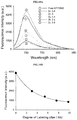
- FIG. 3A is fluorescence spectrum ( ⁇ ex. 620 nm, ⁇ em. 640 to 850 nm) data with a degree of labeling (DL) for the Trastuzumab-ATTO 680 conjugate of Example 1 according to the present invention.
- FIG. 3B is a result showing suppression of a fluorescence intensity with a degree of labeling in the Trastuzumab-ATTO 680 conjugate of Example 1 according to the present invention.
- FIG. 3C is a result comparing absorbance spectra of ATTO 680, the Trastuzumab-ATTO 680 conjugate (sample with a degree of labeling of 3.77) and the denatured Trastuzumab-ATTO 680 conjugate (sample with a degree of labeling of 3.77), at a concentration of 5 uM, of Example 1 according to the present invention.
- FIG. 3D is a result comparing absorbance spectra of ATTO 680, the Trastuzumab-ATTO 680 conjugate (sample with a degree of labeling of 3.77) and the denatured Trastuzumab-ATTO 680 conjugate (sample with a degree of labeling of 3.77), at a concentration of 1 uM, of Example 1 according to the present invention.
- a fluorescence spectrum for dye-free phosphate buffered saline (PBS) is shown.
- FIG. 3E is a result of near-infrared fluorescent image measurement for the Trastuzumab-ATTO 680 (sample with a degree of labeling of 3.77) of Example 1 according to the present invention ( ⁇ ex. 640/25 nm, ⁇ em. 732/37 nm).
- 1 Phosphate buffered aqueous solution (PBS)
- 2 ATTO 680 dye
- 3 Trastuzumab-ATTO 680 conjugate
- 4 denatured Trastuzumab-ATTO 680 conjugate.
- FIG. 4 is analysis data showing whether a fluorescence-quenching effect in the Trastuzumab-ATTO 680 (degree of labeling of 3.77), which is the antibody-fluorescent dye of Example 1 according to the present invention, is stably maintained under a serum condition.
- the conjugates were dispersed in a phosphate buffered saline and a serum culture medium, respectively, and changes in near-infrared fluorescence intensity with time were measured.
- proportions of fluorescence (F) at the respective time points as compared with fluorescence (F 0 ) at 0 hours, it can be seen that a quenching state of the Trastuzumab-ATTO 680 conjugate is stably maintained even in the serum culture medium.
- FIG. 5A is a result obtained by treating HER2-negative control cancer cells (MDA-MB-231) with the Trastuzumab-ATTO 680 conjugate at a concentration of 10 ug/mL for each of 30 minutes and 1 hour, performing washing, and then using a conformal microscope to acquire fluorescent images in Example 1 according to the present invention ( ⁇ ex. 633 nm, ⁇ em. 647 to 754 nm).
- FIG. 5B is a result obtained by treating HER2-overexpressing cancer cells (SK-BR-3) with the Trastuzumab-ATTO 680 conjugate at a concentration of 10 ug/mL for each of 30 minutes and 1 hour, performing washing, and then using a conformal microscope to acquire fluorescent images in Example 1 according to the present invention ( ⁇ ex. 633 nm, ⁇ em. 647 to 754 nm).
- FIG. 5C is a result obtained by treating HER2-overexpressing cancer cells (Calu-3) with the Trastuzumab-ATTO 680 conjugate at a concentration of 10 ug/mL for each of 30 minutes and 1 hour, performing washing, and then using a conformal microscope to acquire fluorescent images in Example 1 according to the present invention ( ⁇ ex. 633 nm, ⁇ em. 647 to 754 nm).
- FIG. 6A is a result obtained by treating HER2-overexpressing cancer cells (SK-BR-3) with the Trastuzumab-ATTO 680 conjugate, and using a conformal microscope at a 1-minute interval, without a washing process, to acquire fluorescent images in Example 1 according to the present invention ( ⁇ ex. 633 nm, ⁇ em. 647 to 754 nm).
- the drawing shows that as the quenched Trastuzumab-ATTO 680 conjugate binds to a HER2 antigen on a cell membrane, a strong fluorescent signal is emitted on the cell membrane. In an extracellular region, a fluorescent signal is hardly detected because the conjugate is quenched.
- FIG. 6B is a result obtained by analyzing changes in intensity with time of a fluorescent signal emitted from a white line site in FIG. 6A , which is represented by a graph.
- FIG. 7 is data of bright-field image, fluorescent image, and merged image of the former two images before and after washing for a SK-BR-3 cell which has been treated with ATTO 680 which is a fluorescent dye, at a concentration of 1 uM in Example 1 according to the present invention.
- ATTO 680 which is a fluorescent dye
- FIG. 7 fluorescent images for a SK-BR-3 cell (untreated control) which has not been treated with the fluorescent dye are shown.
- ATTO 680 untreated control
- FIG. 8 is fluorescent images obtained by treating HER2-overexpressing cancer cells (SK-BR-3) with the Trastuzumab-ATTO 680 conjugate at a concentration of 10 ug/mL for 30 minutes, removing the unbound Trastuzumab-ATTO 680 conjugate through washing, additionally staining lysosomes with Lysotracker after 1 hour, 5 hours, and 24 hours, respectively, and making an observation in Example 1 according to the present invention. It can be seen that endocytosis of the Trastuzumab-ATTO 680 which is bound to a cell membrane is very slow, and most antibody-fluorescent dye conjugates are located on the cell membranes even after 24 hours.
- FIG. 9A is data obtained by intravenously administering the Trastuzumab-ATTO 680 conjugate at a concentration of 50 ug/50 ul to HER2-negative MDA-MB-231 tumor-xenografted mice and HER2-positive Calu-3 tumor-xenografted mice, respectively, and then obtaining near-infrared fluorescent images after 1 hour, 5 hours, and 24 hours in Example 1 according to the present invention.
- a control data obtained by administering phosphate buffered saline (PBS) to normal mice and obtaining images are shown.
- PBS phosphate buffered saline
- FIG. 9B is data represented by analyzing a tumor-to-background ratio of fluorescent signal with an observation time in the images obtained in FIG. 9A .
- the tumor-to-background ratio of fluorescent signal is significantly increased over time.
- FIG. 9C is a result obtained by administering the Trastuzumab-ATTO 680 conjugate, extracting tumor tissues and organs at each time point, and acquiring near-infrared fluorescent images in Example 1 according to the present invention.
- a Calu-3 group which is an HER2-overexpressing cancer tissue, it can be seen that a strong fluorescent signal is emitted in a tumor.
- FIG. 9D is a confocal fluorescent image result obtained by administering the Trastuzumab-ATTO 680 conjugate, obtaining a 7 um-thick frozen tissue section for each tumor tissue obtained at each time point, staining nuclei of cancer cells with a DAPI fluorescent dye, and then taking a photograph in Example 1 according to the present invention.
- a fluorescent signal due to the Trastuzumab-ATTO 680 conjugate was gradually increased over time in cancer tissues, suggesting that a larger amount of the antibody conjugate is bound and accumulates in the cancer tissues as time passes.
- FIG. 10A is fluorescence spectrum ( ⁇ ex. 620 nm, ⁇ em. 640 to 850 nm) data with a degree of labeling for the Cetuximab-ATTO 680 conjugate in Example 2 according to the present invention.
- FIG. 10B is a result obtained by analyzing a fluorescence intensity with a degree of labeling for the Cetuximab-ATTO 680 conjugate in Example 2 according to the present invention.
- FIG. 10C is a result obtained by comparing fluorescence spectra for ATTO 680 the Cetuximab-ATTO 680 and the denatured Cetuximab-ATTO 680 at a concentration of 1 Um in Example 2 according to the present invention.
- FIG. 11A is a result obtained by treating MCF7, which is EGFR-negative cancer cells, with the Cetuximab-ATTO 680 conjugate at a concentration of 10 ug/mL for 30 minutes, 1 hour, and 2 hours, respectively, performing washing, and then observing fluorescent images in Example 2 according to the present invention.
- MCF7 which is EGFR-negative cancer cells
- Cetuximab-ATTO 680 conjugate at a concentration of 10 ug/mL for 30 minutes, 1 hour, and 2 hours, respectively.
- the antibody did not bind to cell surfaces.
- FIG. 11B is a result obtained by treating MDA-MB-468, which is EGFR-positive cancer cells, with the Cetuximab-ATTO 680 conjugate at a concentration of 10 ug/mL for 30 minutes, 1 hour, and 2 hours, respectively, performing washing, and then observing fluorescent images in Example 2 according to the present invention. It can be seen that the Cetuximab-ATTO 680 conjugate specifically binds to the EGFR-positive cancer cells.
- FIG. 12 is fluorescent image data obtained by treating MDA-MB-468, which is EFGR-positive cancer cells, with the Cetuximab-ATTO 680 conjugate at a concentration of 10 ug/mL for 30 minutes, removing the antibody conjugate through washing, and making an observation after 30 minutes, 1 hour, 5 hours, and 24 hours in Example 2 according to the present invention.
- FIG. 13A is fluorescence spectrum ( ⁇ ex. 620 nm, ⁇ em. 640 to 850 nm) data with a degree of labeling for the anti-VEGF-ATTO 680 conjugate in Example 3 according to the present invention.
- FIG. 13B is a result obtained by analyzing a fluorescence intensity with a degree of labeling for the anti-VEGF-ATTO 680 conjugate in Example 3 according to the present invention.
- FIG. 14A is fluorescence spectrum ( ⁇ ex. 620 nm, ⁇ em. 640 to 850 nm) data with a degree of labeling for the anti-Vimentin-ATTO 680 conjugate in Example 4 according to the present invention.
- FIG. 14B is a result obtained by analyzing a fluorescence intensity with a degree of labeling for the anti-Vimentin-ATTO 680 conjugate in Example 4 according to the present invention.
- FIG. 15A is fluorescence spectrum ( ⁇ ex. 620 nm, ⁇ em. 640 to 850 nm) data with a degree of labeling for the anti-CD44 (8E2)-ATTO 680 conjugate in Example 5 according to the present invention.
- FIG. 15B is a result obtained by analyzing a fluorescence intensity with a degree of labeling for the anti-CD44 (8E2)-ATTO 680 conjugate in Example 5 according to the present invention.
- the present invention provides an antibody-fluorescent dye conjugate characterized by comprising an antibody labeled with a fluorescent dye, in which the fluorescent dye is quenched by interaction with a tryptophan (or tyrosine) residue which is an amino acid in the antibody, and is dequenched in a case where a three-dimensional structural change occurs upon antibody-antigen binding, and a method for imaging and diagnosing cancer cells using the same.
- a quenching effect is most effectively caused by tryptophan present in the antibody.
- the quenching effect can be obtained by interaction with a tyrosine, histidine, or methionine residue present in the antibody, and the quenching effect can be, in part, obtained even by aggregation among the fluorescent dyes.
- literature it is known that in a case where tryptophan, tyrosine, histidine, and methionine are dissolved together with increasing concentrations thereof in an aqueous solution in which fluorescent dyes are dissolved, photo-induced electron transfer occurs between the amino acids and the fluorescent dyes so that the fluorescent dyes are quenched.
- the present invention provides an antibody-fluorescent dye conjugate characterized by comprising an antibody labeled with a fluorescent dye, in which the fluorescent dye is quenched by interaction with a tryptophan (or tyrosine, histidine, or methionine) residue in the antibody, and is dequenched in a case where a three-dimensional structural change in the antibody occurs upon antibody-antigen binding so that a distance between the fluorescent dye and tryptophan (or tyrosine, histidine, or methionine) becomes long from each other and distances among the fluorescent dyes also become long from one another.
- a tryptophan or tyrosine, histidine, or methionine
- the antibody used in the present invention is not limited to a specific type, and is characterized by including one or more tryptophan residues or a mixture of two or more tryptophan/tyrosine/histidine/methionine residues so that a quenching effect in the fluorescent dye which is bound to the antibody can be induced, and by binding to an antigen (or receptor) present on a cell membrane surface of a cancer cell.
- tryptophan or tyrosine, histidine, or methionine
- tryptophan or tyrosine, histidine, or methionine
- a quenching effect in the fluorescent dye can be obtained, and a lysine or cysteine residue for binding with the fluorescent dye is contained in the antibody.
- the lysine or cysteine residue for binding with the fluorescent dye is present in a constant region of a light chain or a heavy chain in the antibody, and antibodies characterized in that tryptophan (or tyrosine, histidine, or methionine) is present at a distance within 1.5 nanometers from a location at which the fluorescent dye is bound are preferred.
- the fluorescent dye in the antibody-fluorescent dye conjugate according to the present invention may be labeled at an appropriate location to have the above-mentioned characteristics and to exert functions, and the number of the fluorescent dyes which covalently label the antibody is preferably one or more and may not exceed ten.
- the antibody-fluorescent dye conjugate according to the present invention can be prepared by using a known chemical synthesis method, a gene recombination technique, a method of degrading an antibody molecule by a proteolytic enzyme, and the like.
- the gene recombination technique that allows preparation in a relatively easy manipulation and also in a large quantity is preferably used for preparation.
- an antibody having an amino acid sequence that has the above characteristics is prepared by the gene recombination technique
- a DNA comprising a base sequence that encodes such an amino acid sequence is introduced into a suitable expression vector to produce a recombinant vector, and an antibody having a desired amino acid sequence is allowed to be expressed by an expression system using a bacterial cell, a yeast cell, an insect cell, an animal or plant cell, or the like as a host, or a cell-free translation system.
- the antibody for producing the antibody-fluorescent dye conjugate the above-mentioned conventional product may be obtained and used by synthesis using a conventional method. For example, it is possible to cause Trastuzumab (Herceptin) that specifically recognizes human epidermal growth factor receptor 2 (HER2) to be bound to a fluorescent dye, and to use the conjugate.
- trastuzumab Herceptin
- HER2 human epidermal growth factor receptor 2
- a method of performing labeling with a fluorescent dye is not particularly limited.
- a method of performing labeling, directly or via a cross-linking agent, using a side chain of a lysine or a cysteine residue present in a heavy chain and a light chain of the antibody can be used, but not limited thereto.
- Labeling with the fluorescent dye can be performed after modification reaction of other amino acids.
- the antigen capable of being detected or measured by the antibody-fluorescent dye conjugate according to the present invention is not particularly limited as long as the antigen is an antigen which is specifically recognized by the antibody.
- examples thereof include, in addition to proteins, peptides, saccharide, lipid, glycolipid, and low-molecular compounds, protein modifications such as phosphorylation and methylation, and proteins that have gone through these modifications.
- the antigens associated with cancer cells to which the antibody-fluorescent dye conjugate according to the present invention specifically binds are characterized by including epidermal growth factor receptor (EGFR, HER1), human epidermal growth factor receptor 2 (HER2), human epidermal growth factor receptor 3 (HER3, ERBB-3), human epidermal growth factor receptor 4 (HER4, ERBB-4), epithelial cell adhesion molecule (EpCam), CD19, CD20, CD22 (Siglec-2), CD30 (TNFRSF1), CD33 (Siglec-3), CD44, CD44v6, CD52, CD56 (NCAM), CD152 (CTLA4), mucin 1 (MUC1), carcinoembryonic antigen (CEA), LEWIS Y, prostate-specific membrane antigen (PSMA), tumor-associated glycoprotein 72 (TAG-72), GD2 ganglioside, GD3 ganglioside, human leukocyte antigen-DR10 (HLA-DR10), insulin-like growth factor 1 receptor (I
- antibodies targeting the above antigens can be used.
- monoclonal antibodies developed for clinical use include Cetuximab (Erbitux), Panitumumab (Vectibix), Necitumumab (Portrazza), Imgatuzumab, Matuzumab, Nimotuzumab, Futuximab, and Zalutumumab which are antibodies to an EGFR antigen, Trastuzumab (Herceptin) and Pertuzumab (Perjeta) which are antibodies to a HER2 antigen, Duligotumab, Patritumab, and Seribantumab which are antibodies to a HER3 antigen, Bevacizumab (Avastin) which is an antibody to a VEGF-A antigen, Catumaxomab (Removab) and Adecatumumab (MT201) which are antibodies to an EpCam antigen, Cixut
- Olaratumab which is an antibody to a platelet-derived growth factor receptor alpha (PDFGRA) antigen
- PDFGRA platelet-derived growth factor receptor alpha
- Emibetuzumab which is an antibody to a hepatocyte growth factor receptor (HGFR) antigen
- Etaracizumab (Abegrin) which is an antibody to an Alpha-v beta-3 antigen
- Farletuzumab which is an antibody to a Folate receptor alpha antigen
- Parsatuzumab which is an antibody to an EGF-like domain-containing protein 7 (EGFL7) antigen
- Sibrotuzumab which is an antibody to a Fibroblast activation protein alpha (FAP) antigen
- Girentuximab Rosatuximab (Rencarex) which is an antibody to a Carbonic anhydrase 9 (CA9/CAIX) antigen.
- CA9/CAIX Carbonic anhydrase 9
- the fluorescent dye used for antibody labeling is not particularly limited as long as the fluorescent dye is a fluorescent dye which has a characteristic of being effectively quenched in a case of being located at a distance within 1.5 nm from tryptophan, tyrosine, methionine, or histidine, and which is quenched, in a state of forming a conjugate with an antibody and in the absence of an antigen, by interaction with the above-mentioned amino acids, more specifically by a photo-induced electron transfer mechanism, and is dequenched to restore fluorescence emittance in a case where the antibody-fluorescent dye conjugate is bound to an antigen.
- fluorescent dyes having, as a basic skeleton, rhodamine, coumarin, EvoBlue, oxazine, carbopyronine, naphthalene, biphenyl, anthracene, phenanthrene, pyrene, carbazole, or the like, and derivatives of the fluorescent dyes may be exemplified, and specific examples thereof include Fluorescein, CR110: Carboxyrhodamine 110: Rhodamine Green (trade name), TAMRA: carboxytetramethylrhodamine: TMR, Carboxyrhodamine 6G: CR6G, BODIPY FL (trade name): 4,4-difluoro-5,7-dimethyl-4-bora-3a,4a-diaza-s-indacene-3-propionic acid, BODIPY 493/503 (trade name): 4,4-difluoro-1,3,5,7-t
- kits for imaging diagnosis of a tumor configured to comprise the aforementioned antibody-fluorescent dye conjugate and further comprise reagents or the like which are usually used in this type of immunoassay kit, instruments, instructions, and the like.
- the present invention provides a method for fluorescence imaging and diagnosis, the method comprising a step (a) of bring an antibody-fluorescent dye conjugate into contact with an antigen in a sample, the antibody-fluorescent dye conjugate being characterized by comprising an antibody labeled with a fluorescent dye in which the fluorescent dye is quenched by interaction with a tryptophan or tyrosine residue in the antibody and is dequenched upon antibody-antigen binding; and a step (b) of obtaining fluorescence of the fluorescent dye as an image and performing analysis.
- a method of bringing the antibody-fluorescent dye conjugate according to the present invention into contact with the antigen is not limited to a particular method, and may be performed in a liquid state or may be performed in vivo or ex vivo.
- a liquid-state specimen may be supplied, as it is, for measurement as a measurement sample, or may be used by dilution with a buffer solution or a cell culture medium, concentration, or proper adjustment of a pH, a salt concentration, or the like to the extent that the antigen is not damaged or detection for antigen concentration measurement is not inhibited.
- the liquid-state specimen may be, for example, adherent or suspension cells to be measured, and diagnosis is performed through fluorescence imaging after treatment with the antibody-fluorescent dye conjugate.
- body fluids such as blood and fluid, tissues, and the like in a living body can also be used as a sample to be measured. That is, the antibody-fluorescent dye conjugate of the present invention is administered to an experimental animal or a human so that the antibody-fluorescent dye conjugate of the present invention can be brought into contact with an antigen in a living body.
- the above administration method is not particularly limited, and can be appropriately selected from a parenteral local administration method such as intramuscular injection, intraperitoneal injection, intravenous injection, subcutaneous injection, embedding, application, and spraying, or an oral administration method.
- other drugs or the like may be administered simultaneously with, or before or after administration of the antibody-fluorescent dye conjugate of the present invention.
- Administration of the antibody-fluorescent dye conjugate of the present invention allows a location or migration of an antigen and an antigen amount or a change thereof in a living body to be shown from a fluorescent image and allows qualitative and quantitative analysis, which enables real-time identification of a location of a target cell during surgery and can dramatically enhance accuracy of surgery and therapeutic effects.
- a fluorescent image may be obtained by directly treating tissues or cells taken from a living body with a solution in which the antibody-fluorescent dye conjugate is dispersed, or a fluorescent image may be obtained by producing sections from the tissues or cells and treating the same with a solution in which the antibody-fluorescent dye conjugate is dispersed.
- presence or absence of a target antigen may be diagnosed by treating a tissue or cell microarray obtained from a living body with a solution in which the antibody-fluorescent dye conjugate is dispersed, and obtaining a fluorescent image.
- a reaction condition for bringing the antibody-fluorescent dye conjugate of the present invention into contact with an antigen in a measurement sample is not particularly limited as long as the condition is such that the antibody-fluorescent dye conjugate of the present invention is added to the measurement sample and culturing is performed under a condition which can be generally used for antigen-antibody reaction.
- a fluorescent image can be obtained by setting a temperature condition to, for example, 1° C. to 40° C., and preferably 18° C. to 37° C., and setting a reaction time to, for example immediately to 180 minutes, and preferably 1 to 90 minutes.
- a fluorescent image is obtained by using fluorescence imaging equipment and analysis is performed.
- a fluorescence imaging method for a measurement sample in the present invention is not particularly limited as long as the method can detect fluorescence emitted from the fluorescent dye, and it is sufficient that the method images and/or detects fluorescence emittance from the fluorescent dye by causing the measurement sample after reaction to be irradiated with excitation light.
- Wavelengths of the excitation light to be irradiated and the fluorescence to be measured and/or detected can be appropriately selected depending on a type of the fluorescent dye to be used. For example, in a case where ATTO 680 is used for the fluorescent dye, a combination of excitation light wavelength of 660 nm and fluorescence emission wavelength of 710 nm can be used.
- Example 1 was intended to demonstrate a basic concept and utility of an antigen responsive antibody-fluorescent dye conjugate, by using an antibody that specifically binds to Human Epidermal Growth Factor Receptor 2 (HER2) which is known to be over-expressed on surfaces of cancer cells, and causing various fluorescent dyes to be bound thereto.
- various fluorescent dyes are caused to be bound to Trastuzumab (Herceptin), which is a representative antibody used in clinical practice among antibodies that specifically bind to HER2, and a fluorescence-quenching effect was analyzed.
- trastuzumab Herceptin
- Trastuzumab 0.5 mg, 3.4 nmol
- PBS phosphate buffered saline
- DL degree of labeling
- the antibody conjugate was dissolved in phosphate buffered saline and a UV/Vis absorbance spectrum was measured.
- a concentration of the antibody was calculated using a molar absorbance coefficient (210,000 M ⁇ 1 cm ⁇ 1 ) of Trastuzumab at 280 nm.
- a degree of labeling of the fluorescent dye which is bound to the antibody was analyzed by using a known molar absorbance coefficient value for each dye.
- each of the Trastuzumab-fluorescent dye conjugates was dissolved to a concentration of 1 uM (based on a fluorescent dye concentration), a fluorescence intensity was measured, and decreased fluorescence intensity with a degree of labeling was analyzed. For comparison, fluorescence spectrum and fluorescence intensity for a free fluorescent dye, which is not bound to an antibody, at the same concentration were measured, and comparison with the antibody-fluorescent dye conjugates was performed.
- Trastuzumab-ATTO 680 conjugates (samples with a degree of labeling of 3.77) were treated with phosphate buffered saline and a denaturing buffer solution (phosphate buffered saline containing 1% sodium dodecyl sulfate (SDS) and 1 mM 2-mercaptoethanol), respectively, and absorbances and fluorescence spectra were compared ( ⁇ ex. 620 nm, ⁇ em. 640 to 850 nm).
- SDS sodium dodecyl sulfate
- FIG. 2 The analysis results for quenching characteristics of the synthesized Trastuzumab-fluorescent dye conjugates are shown in FIG. 2 .
- a value at a degree of labeling of 0 is a fluorescence value for a free fluorescent dye at the same concentration.
- FIGS. 3D and 3E in a case where an antibody is labeled with 3.77 ATTO 680 fluorescent dyes, it was analyzed that a fluorescent signal was 7.9 times weaker than a free fluorescent dye (free ATTO 680 control group) at the same concentration. In addition, it was found that in a case where the conjugate is treated with a denaturing buffer solution, fluorescence is restored to an original fluorescence intensity.
- FIG. 3E is a photograph of near-infrared fluorescent image ( ⁇ ex. 640/25 nm, ⁇ em. 732/37 nm) taken using animal fluorescence imaging equipment to visually show quenching characteristic and fluorescence restoration of the antibody-fluorescent dye conjugate.
- a fluorescent dye inherently capable of emitting strong fluorescence becomes quenched (no. 3 tube in FIG. 3E ) after being bound to an antibody, and it can be seen in an intuitive way that fluorescence is restored again (no. 4 tube in FIG. 3E ) in a case of being treated with a denaturing buffer solution so that interaction between the fluorescent dye and amino acids is removed.
- a fluorescent image for phosphate buffered saline (PBS, no. 1 tube in FIG. 3E ) containing no fluorescent dye is also shown, which shows that phosphate buffered saline itself does not emit fluorescence.
- the Trastuzumab-ATTO 680 conjugates (samples with a degree of labeling of 3.77) were dispersed in phosphate buffered saline and a serum solution, respectively, and changes in fluorescence intensity were measured for 24 hours.
- the conjugate stably retains a quenched state for 24 hours under a serum condition (see FIG. 4 ), which means that the Trastuzumab-ATTO 680 conjugate restores fluorescence emittance and emits a strong fluorescent signal only in a case of being bound to an antigen (HER2).
- a cell experiment proceeded which identifies whether the antibody Trastuzumab retains binding specificity with the target antigen HER2 even after the antibody is bound to the fluorescent dye ATTO 680.
- HER2-negative cancer cell line MDA-MB-231 which does not express HER2, and HER2-positive cancer cell lines Calu-3 and SK-BR-3, which overexpress HER2, were purchased from ATCC, USA, and used.
- the MDA-MB-231 cancer cell line, and the SK-BR-3 and Calu-3 cancer cell lines were treated with the Trastuzumab-ATTO 680 at a concentration of 10 mg/mL for each of 30 minutes or 1 hour, respectively.
- the Trastuzumab-ATTO 680 conjugate emits a strong fluorescent signal on a surface of the cancer cell overexpressing HER2, but no fluorescent signal could be detected on a surface of a MDA-MB-231 cell which is a HER2-negative cancer cell. From this, it can be seen that the Trastuzumab-ATTO 680 conjugate can be used to specifically image and diagnose a HER2-overexpressing cancer cell line.
- a cell experiment was intended to verify whether a fluorescent signal in the antibody-fluorescent dye conjugate is turned on immediately upon reaction with an antigen present on a surface of a cancer cell. It was expected that the Trastuzumab-ATTO 680 conjugate does not emit a fluorescent signal in a case of being present on a cell culture medium, and is caused to activate fluorescence emittance and emit a strong fluorescent signal in a case of being bound to the target antigen HER2 present on a surface of a cancer cell.
- the present example was intended to demonstrate a basic concept and utility of the antigen responsive antibody-fluorescent dye conjugate, by taking a fluorescent image every 1 minute without performing washing in a case of obtaining the fluorescent image after treating an antigen-overexpressing cancer cell line with the antibody-fluorescent dye conjugate.
- the HER2 overexpressing cancer cell SK-BR-3 was treated with the Trastuzumab-ATTO 680 conjugate at a concentration of 10 ug/mL, and then fluorescent images of the cancer cell were obtained using a confocal microscope every 1 minute without performing a washing process ( ⁇ ex. 633 nm, ⁇ em. 647 to 754 nm).
- the SK-BR-3 cancer cell was treated with a free fluorescent dye (free ATTO 680) at a concentration of 1 uM, and then confocal fluorescent images before and after performing washing were obtained.
- FIG. 6A changes in fluorescence intensity with time at a location of the white line in FIG. 6A were analyzed ( FIG. 6B ).
- FIG. 6B it was identified that a fluorescence intensity at a surface of the cell on which an antigen is present is reached to the apex within 5 minutes. This indicates that fluorescent signal activation due to binding with a target antigen occurs very rapidly, and the antibody binds to most of antigens on the surface of the cell within 5 minutes.
- results obtained by treating the cancer cell with ATTO 680 which is a free fluorescent dye, at the same concentration, and taking, with fluorescence imaging, states before and after washing are shown in FIG. 7 .
- ATTO 680 which is a free fluorescent dye In a case of being treated with ATTO 680 which is not bound to an antibody, it was found that a strong fluorescent signal was emitted in an extracellular region. After washing, due to removal of all fluorescent dyes, no fluorescent signal was detected in an image. This supports that the result obtained in FIG. 6 A is caused by quenching, and shows that ATTO 680 is neither non-specifically adsorbed on a surface of a cancer cell nor endocytosed into the cancer cell.
- SK-BR-3 which is a HER2-overexpressing cancer cell was treated with the Trastuzumab-ATTO 680 at a concentration of 20 ug/mL for 30 minutes. Thereafter, washing was performed three times to remove antibody conjugates in an extracellular region, and fluorescent images were taken over 24 hours to observe a rate at which the antibody conjugate bound to the surface of the cancer cell is endocytosed into the cancer cell.
- FIG. 8 it can be seen that in a case of SK-BR-3, a considerable number of the antibody-fluorescent dye conjugates which are bound to cell membranes still exist on the surfaces of the cancer cells even after 24 hours.
- This finding shows that even in a case of cancer cells with a rate at which an antigen present on a surface of the cancer cell is circulated into the cancer cell being slow, in order to obtain an image with high contrast, a fluorescent image has to be obtained by using an antibody-fluorescent dye conjugate in which fluorescence is activated at the time of responding to an antigen present on a cell surface.
- each of HER2-positive Calu-3 cancer cells and HER2-negative MDA-MB-231 cancer cells were diluted in 0.1 mL EMEM or RPMI medium, and injected subcutaneously into female athymic nude mice (Balb/c-nu, 5-week old). Then, the mice were measured periodically for tumor cell sizes and used for experiments in a case where tumor sizes reached approximately 190 mm 3 .
- Trastuzumab-ATTO 680 50 ug/50 ul phosphate buffered saline
- mice which are not grafted with tumors were used as another control group, and the same volume of phosphate buffered saline (PBS) was intravenously administered thereto.
- PBS phosphate buffered saline
- a tumor-to-background ratio analyzed in mice grafted with the HER2-positive Calu-3 tumor was as high as 2.5 (at 1 hour), 4.7 (at 5 hours) and 8.2 (at 24 hours), and a tumor-to-background ratio in mice grafted with the HER2-negative MDA-MB-231 tumor was 1.4 at 1 hour and 1.9 at 24 hours.
- FIG. 9C it can be identified again that a fluorescent signal in the Calu-3 tumor is increased with time, and it can be seen that a very high signal is obtained from the tumors as compared with other organs such as the livers. It can be identified that antibodies are accumulated at an insignificant level in the MDA-MB-231 tumors.
- Example 2 in order to further demonstrate that the basic concept and utility of the antigen responsive-type antibody-fluorescent dye conjugate can be applied to various antibodies, as an example of another antibody, Cetuximab (Erbitux, manufactured by Merck Serono) which is an antibody targeting epidermal growth factor receptor (EGFR) was used to carry out a second embodiment.
- Cetuximab Erbitux, manufactured by Merck Serono
- EGFR epidermal growth factor receptor
- Example 2 was intended to further verify characteristics and utility of the antigen responsive antibody-fluorescent dye conjugate, by using only ATTO 680 N-hydroxysuccinimidyl ester (ATTO 680-NHS ester) as a fluorescent dye and causing the Cetuximab and the ATTO 680 to react with each other at various ratios.
- ATTO 680-NHS ester ATTO 680 N-hydroxysuccinimidyl ester
- the Cetuximab and the ATTO 680-NHS ester were mixed at a molar ratio of 1:1 or more and dissolved in phosphate buffered saline (PBS, 10 mM, pH 7.4), and then allowed to react at room temperature for 1 hour. Remaining fluorescent dyes that did not react with the antibodies and byproducts were removed with a PD-10 column, and the resultant was concentrated with an Amicon Ultra-0.5 mL filter (EMD Millipore) and stored at 4° C.
- ETD Millipore Amicon Ultra-0.5
- a degree of labeling (DL) of the fluorescent dye to Cetuximab was measured by the same method as described in Example 1, and a quenching effect with a degree of labeling was analyzed.
- FIGS. 10A and 10B show an extent of quenching with a degree of labeling of the fluorescent dye. As expected, a fluorescence-quenching effect was increased as the degree of labeling was increased, and subsequent experiments proceeded using samples with a degree of labeling of 5.4.
- FIG. 10C is a result obtained by treating the Cetuximab-ATTO 680 conjugate (degree of labeling of 5.4) with phosphate buffered saline (PBS) or a denaturing buffer solution (phosphate buffered saline containing 1% sodium dodecyl sulfate (SDS) and 1 mM 2-mercaptoethanol), respectively, and comparing absorbances and fluorescence spectra ( ⁇ ex. 620 nm, and ⁇ em. 640 to 850 nm).
- PBS phosphate buffered saline
- SDS sodium dodecyl sulfate
- EGFR-negative MCF7 cancer cells and EGFR-positive MDA-MB-468 cancer cell line were treated with the Cetuximab-ATTO 680 conjugate constructed as above at a concentration of 10 ug/mL for 30 minutes, 1 hour, and 2 hours, respectively. Washing was performed to remove antibodies which are not bound to the cells, and then fluorescent images were obtained using a confocal microscope ( ⁇ ex. 633 nm, ⁇ em. 647 to 754 nm).
- the antigen responsive antibody-fluorescent dye conjugate proposed in the present invention are highly suitable rather than using the target cell-specific antibody-fluorescent dye conjugate proposed by Dr. Hisataka Kobayashi.
- Example 3 in order to further demonstrate that the basic concept and utility of the antigen responsive-type antibody-fluorescent dye conjugate can be applied to various antibodies, as an example of another antibody, anti-VEGF (ab46154, manufactured by Abcam) which targets vascular endothelial growth factor (VEGF) was used to carry out a third embodiment.
- anti-VEGF vascular endothelial growth factor
- Example 3 was intended to further verify characteristics and utility of the antigen responsive antibody-fluorescent dye conjugate, by using only ATTO 680 N-hydroxysuccinimidyl ester (ATTO 680-NHS ester) as a fluorescent dye and causing the VEGF antibody and the ATTO 680 to react with each other at various ratios.
- ATTO 680-NHS ester ATTO 680 N-hydroxysuccinimidyl ester
- the VEGF antibody and the ATTO 680-NHS ester were mixed at a molar ratio of 1:1 or more and dissolved in phosphate buffered saline (PBS, 10 mM, pH 7.4), and then allowed to react at room temperature for 1 hour.
- PBS phosphate buffered saline
- a degree of labeling (DL) of the fluorescent dye to the VEGF antibody was measured by the same method as described in Example 1, and a quenching effect with a degree of labeling was analyzed.
- FIGS. 13A and 13B show an extent of quenching with a degree of labeling of the fluorescent dye. As expected, it was found that a fluorescence-quenching effect was increased as the degree of labeling was increased.
- Example 4 as an example of another antibody for further demonstrating that the basic concept and utility of the antigen responsive-type antibody-fluorescent dye conjugate can be applied to various antibodies, anti-Vimentin (ab92547, manufactured by Abcam) which targets Vimentin, which is a cytoskeletal marker expressed in a case where a skeleton of a tumor cell is changed for ease of migration at the time of tumor metastasis, was used to carry out a fourth embodiment.
- Example 4 was intended to further verify characteristics and utility of the antigen responsive antibody-fluorescent dye conjugate, by using only ATTO 680 N-hydroxysuccinimidyl ester (ATTO 680-NHS ester) as a fluorescent dye and causing the Vimentin antibody and the ATTO 680 to react with each other at various ratios.
- ATTO 680-NHS ester ATTO 680 N-hydroxysuccinimidyl ester
- the Vimentin antibody and the ATTO 680-NHS ester were mixed at a molar ratio of 1:1 or more and dissolved in phosphate buffered saline (PBS, 10 mM, pH 7.4), and then allowed to react at room temperature for 1 hour.
- PBS phosphate buffered saline
- a degree of labeling (DL) of the fluorescent dye to the Vimentin antibody was measured by the same method as described in Example 1, and a quenching effect with a degree of labeling was analyzed.
- FIGS. 14A and 14B show an extent of quenching with a degree of labeling of the fluorescent dye. As expected, it was found that a fluorescence-quenching effect was increased as the degree of labeling was increased.
- Example 5 as an example of another antibody for further demonstrating that the basic concept and utility of the antigen responsive-type antibody-fluorescent dye conjugate can be applied to various antibodies, a CD44 antibody (AF3660, manufactured by R&D Systems which targets CD44, which is one of transmembrane proteins involved in cell to cell and cell to substrate communication, was used to carry out a fifth embodiment.
- a CD44 antibody AF3660, manufactured by R&D Systems which targets CD44, which is one of transmembrane proteins involved in cell to cell and cell to substrate communication
- Example 5 was intended to further verify characteristics and utility of the antigen responsive antibody-fluorescent dye conjugate, by using only ATTO 680 N-hydroxysuccinimidyl ester (ATTO 680-NHS ester) as a fluorescent dye and causing the CD44 antibody and the ATTO 680 to react with each other at various ratios.
- ATTO 680-NHS ester ATTO 680 N-hydroxysuccinimidyl ester
- the CD44 antibody and the ATTO 680-NHS ester were mixed at a molar ratio of 1:1 or more and dissolved in phosphate buffered saline (PBS, 10 mM, pH 7.4), and then allowed to react at room temperature for 1 hour.
- PBS phosphate buffered saline
- a degree of labeling (DL) of the fluorescent dye to the CD44 antibody was measured by the same method as described in Example 1, and a quenching effect with a degree of labeling was analyzed.
- FIGS. 15A and 15B show an extent of quenching with a degree of labeling of the fluorescent dye. As expected, it was found that a fluorescence-quenching effect was increased as the degree of labeling was increased.
Landscapes
- Health & Medical Sciences (AREA)
- Chemical & Material Sciences (AREA)
- Life Sciences & Earth Sciences (AREA)
- Immunology (AREA)
- General Health & Medical Sciences (AREA)
- Organic Chemistry (AREA)
- Engineering & Computer Science (AREA)
- Molecular Biology (AREA)
- Biomedical Technology (AREA)
- Biochemistry (AREA)
- Medicinal Chemistry (AREA)
- Biophysics (AREA)
- Proteomics, Peptides & Aminoacids (AREA)
- Genetics & Genomics (AREA)
- Urology & Nephrology (AREA)
- Hematology (AREA)
- Animal Behavior & Ethology (AREA)
- Veterinary Medicine (AREA)
- Public Health (AREA)
- Epidemiology (AREA)
- Cell Biology (AREA)
- Analytical Chemistry (AREA)
- Pathology (AREA)
- General Physics & Mathematics (AREA)
- Biotechnology (AREA)
- Microbiology (AREA)
- Physics & Mathematics (AREA)
- Food Science & Technology (AREA)
- Oncology (AREA)
- Gynecology & Obstetrics (AREA)
- Radiology & Medical Imaging (AREA)
- Pregnancy & Childbirth (AREA)
- Reproductive Health (AREA)
- Nuclear Medicine, Radiotherapy & Molecular Imaging (AREA)
- Investigating, Analyzing Materials By Fluorescence Or Luminescence (AREA)
- Medicines Containing Antibodies Or Antigens For Use As Internal Diagnostic Agents (AREA)
- Hospice & Palliative Care (AREA)
- Investigating Or Analysing Materials By The Use Of Chemical Reactions (AREA)
Abstract
Description

ATTO680 has a formula

ATTO700 has a forumla

ATTO-MB2 has a formula

ZW800-1 has formula

Claims (7)


Applications Claiming Priority (3)
| Application Number | Priority Date | Filing Date | Title |
|---|---|---|---|
| KR10-2016-0037411 | 2016-03-29 | ||
| KR1020160037411A KR101847264B1 (en) | 2016-03-29 | 2016-03-29 | Antigen-responsive antibody-fluorochrome conjugates and method detecting target-cells using fluorescence imaging by thereof |
| PCT/KR2017/002924 WO2017171283A1 (en) | 2016-03-29 | 2017-03-20 | Antigen responsive antibody-fluorescent dye conjugate and method for detecting fluorescent image of target cell using same |
Related Parent Applications (1)
| Application Number | Title | Priority Date | Filing Date |
|---|---|---|---|
| PCT/KR2017/002924 Continuation WO2017171283A1 (en) | 2016-03-29 | 2017-03-20 | Antigen responsive antibody-fluorescent dye conjugate and method for detecting fluorescent image of target cell using same |
Publications (2)
| Publication Number | Publication Date |
|---|---|
| US20190091349A1 US20190091349A1 (en) | 2019-03-28 |
| US11213595B2 true US11213595B2 (en) | 2022-01-04 |
Family
ID=59964904
Family Applications (1)
| Application Number | Title | Priority Date | Filing Date |
|---|---|---|---|
| US16/138,371 Active US11213595B2 (en) | 2016-03-29 | 2018-09-21 | Antigen responsive antibody-fluorescent dye conjugate and method for fluorescence detection and imaging of target cell using the same |
Country Status (5)
| Country | Link |
|---|---|
| US (1) | US11213595B2 (en) |
| EP (1) | EP3438665B1 (en) |
| KR (1) | KR101847264B1 (en) |
| CN (1) | CN109121429A (en) |
| WO (1) | WO2017171283A1 (en) |
Families Citing this family (10)
| Publication number | Priority date | Publication date | Assignee | Title |
|---|---|---|---|---|
| KR102129522B1 (en) | 2018-07-26 | 2020-07-02 | 한국과학기술연구원 | anti-tumor Protien conjugated with lysosomal protease activatable probe for specific tumor cell imgaing and composition for imgae comprising the same |
| JP7637937B2 (en) * | 2020-04-28 | 2025-03-03 | 国立大学法人北海道大学 | Method for obtaining information on target polypeptide and reagent kit |
| CN111690038A (en) * | 2020-06-23 | 2020-09-22 | 南京诺源医疗器械有限公司 | Preparation method of near-infrared fluorescent tracer for diagnosing metastatic lymph nodes |
| CN113063761B (en) * | 2021-03-17 | 2023-02-10 | 北京化工大学 | A fluorescent aptasensor for detecting muc1 mucin and its application method |
| CN113109566A (en) * | 2021-05-24 | 2021-07-13 | 上海理工大学 | Fluorescence immunochromatography joint inspection method for food-borne pathogenic bacteria based on antigen colorization |
| CN115177751B (en) * | 2022-03-16 | 2023-07-04 | 北京药明博锐生物科技有限公司 | Conjugates, methods of making and uses thereof |
| CN115524307A (en) * | 2022-09-23 | 2022-12-27 | 上海纳米技术及应用国家工程研究中心有限公司 | A near-infrared gastric cancer detection kit and preparation method thereof |
| KR102681568B1 (en) | 2022-10-25 | 2024-07-04 | 주식회사 에이티지라이프텍 | Method for Circulating tumor cell detection using cell culture in microfluidic chip and low magnification image analysis |
| CN117186151B (en) * | 2023-08-23 | 2024-05-17 | 中山大学 | A near-infrared zwitterionic cyanine dye and its preparation method and application |
| CN117886938B (en) * | 2023-11-23 | 2025-11-14 | 南方科技大学 | An EGFR fluorescent antibody, its preparation method and application |
Citations (10)
| Publication number | Priority date | Publication date | Assignee | Title |
|---|---|---|---|---|
| US20060280688A1 (en) | 2000-09-19 | 2006-12-14 | Li-Cor, Inc. | Optical fluorescent imaging |
| EP2155786A2 (en) | 2007-06-06 | 2010-02-24 | F. Hoffmann-Roche AG | Composition of a first non-labeled monoclonal antibody binding to a tumor antigen and a non-cross reactive second monoclonal antibody labeled with a nir fluorescence label |
| US20100203554A1 (en) * | 2007-09-10 | 2010-08-12 | Thilo Enderle | Generic kinase/phosphatase assay with single readout |
| US20110237942A1 (en) | 2010-03-25 | 2011-09-29 | Tamotsu Zako | Bioimaging method using near-infrared (nir) fluorescent material |
| US20130039861A1 (en) | 2005-04-06 | 2013-02-14 | Immunomedics, Inc. | Dye Conjugated Peptides for Fluorescent Imaging |
| CN103917872A (en) | 2011-11-02 | 2014-07-09 | 优志旺电机株式会社 | Fluoroimmunoassay method using polypeptide complex containing fluorolabeled antibody-variable region |
| KR20140102968A (en) | 2013-02-15 | 2014-08-25 | 경희대학교 산학협력단 | Novel quantum dot-aptamer conjugate and use thereof |
| KR101452819B1 (en) | 2012-11-01 | 2014-10-23 | 국립암센터 | Redox-responsive polymer-photosensitizer conjugate containing disulfide linker and its composition for fluorescence imaging and photodynamic therapy comprising thereof |
| WO2015170878A1 (en) | 2014-05-08 | 2015-11-12 | 국립암센터 | Conjugate for photodynamic diagnosis or therapy, containing folic acid, and composition for photodynamic diagnosis or therapy, containing same |
| KR20160037411A (en) | 2014-09-28 | 2016-04-06 | 목포대학교산학협력단 | A fermented tea container and a method for preparing there of |
Family Cites Families (2)
| Publication number | Priority date | Publication date | Assignee | Title |
|---|---|---|---|---|
| DE19925402C2 (en) * | 1999-06-02 | 2001-12-20 | Molecular Machines & Ind Gmbh | Screening of target-ligand interactions |
| JP2010507645A (en) * | 2006-10-25 | 2010-03-11 | コーニンクレッカ フィリップス エレクトロニクス エヌ ヴィ | Contrast agent for detecting prostate cancer |
-
2016
- 2016-03-29 KR KR1020160037411A patent/KR101847264B1/en active Active
-
2017
- 2017-03-20 CN CN201780021633.2A patent/CN109121429A/en active Pending
- 2017-03-20 EP EP17775708.5A patent/EP3438665B1/en active Active
- 2017-03-20 WO PCT/KR2017/002924 patent/WO2017171283A1/en not_active Ceased
-
2018
- 2018-09-21 US US16/138,371 patent/US11213595B2/en active Active
Patent Citations (15)
| Publication number | Priority date | Publication date | Assignee | Title |
|---|---|---|---|---|
| US20060280688A1 (en) | 2000-09-19 | 2006-12-14 | Li-Cor, Inc. | Optical fluorescent imaging |
| US20130039861A1 (en) | 2005-04-06 | 2013-02-14 | Immunomedics, Inc. | Dye Conjugated Peptides for Fluorescent Imaging |
| KR101169243B1 (en) | 2007-06-06 | 2012-08-02 | 에프. 호프만-라 로슈 아게 | Composition of a first non-labeled monoclonal antibody binding to a tumor antigen and a non-cross reactive second monoclonal antibody labeled with a nir fluorescence label |
| EP2155786A2 (en) | 2007-06-06 | 2010-02-24 | F. Hoffmann-Roche AG | Composition of a first non-labeled monoclonal antibody binding to a tumor antigen and a non-cross reactive second monoclonal antibody labeled with a nir fluorescence label |
| CN101679510A (en) | 2007-06-06 | 2010-03-24 | 霍夫曼-拉罗奇有限公司 | Composition of a first non-labeled monoclonal antibody binding to a tumor antigen and a non-cross-reactive second monoclonal antibody labeled with a NIR fluorescent label |
| US20100203554A1 (en) * | 2007-09-10 | 2010-08-12 | Thilo Enderle | Generic kinase/phosphatase assay with single readout |
| US20110237942A1 (en) | 2010-03-25 | 2011-09-29 | Tamotsu Zako | Bioimaging method using near-infrared (nir) fluorescent material |
| CN103917872A (en) | 2011-11-02 | 2014-07-09 | 优志旺电机株式会社 | Fluoroimmunoassay method using polypeptide complex containing fluorolabeled antibody-variable region |
| EP2775305A1 (en) | 2011-11-02 | 2014-09-10 | Ushio Denki Kabushiki Kaisha | Fluoroimmunoassay method using polypeptide complex containing fluoro-labeled antibody variable region |
| US20140329228A1 (en) * | 2011-11-02 | 2014-11-06 | Ushio Denki Kabushiki Kaisha | Fluorescence immunoassay using polypeptide complex containing fluoro-labeled antibody variable region |
| KR101603456B1 (en) | 2011-11-02 | 2016-03-14 | 우시오덴키 가부시키가이샤 | Fluoroimmunoassay method using polypeptide complex containing fluorolabeled antibody-variable region |
| KR101452819B1 (en) | 2012-11-01 | 2014-10-23 | 국립암센터 | Redox-responsive polymer-photosensitizer conjugate containing disulfide linker and its composition for fluorescence imaging and photodynamic therapy comprising thereof |
| KR20140102968A (en) | 2013-02-15 | 2014-08-25 | 경희대학교 산학협력단 | Novel quantum dot-aptamer conjugate and use thereof |
| WO2015170878A1 (en) | 2014-05-08 | 2015-11-12 | 국립암센터 | Conjugate for photodynamic diagnosis or therapy, containing folic acid, and composition for photodynamic diagnosis or therapy, containing same |
| KR20160037411A (en) | 2014-09-28 | 2016-04-06 | 목포대학교산학협력단 | A fermented tea container and a method for preparing there of |
Non-Patent Citations (17)
| Title |
|---|
| Abe, R. et al., "Quenchbodies: Quench-Based Anitbody Probes That Show Antigen-Dependent Fluorescence", Journal of the American Chemical Society (Nov. 2011) 39 pages. |
| Alford et al. Fluorescence lifetime imaging of activatable target specific molecular probes. 2010 Contrast Media Mol. Imaging 5: 1-8. (Year: 2010). * |
| Amino acids reference chart. Sigma-Aldrich website, <sigmaaldrich.com/life-science/metabolomics/learning-center/amino-acid-reference-chart.html>. Accessed Sep. 16, 2019. (Year: 2019). * |
| Choi et al., "Targeted zwitterionic near-infrared fluorophores for improved optical imaging", Nature Biotechnology vol. 31, pp. 148-153 (2013), Published: Jan. 6, 2013. |
| European Office Action for application No. 17775708.5 dated Mar. 22, 2021. |
| Even-Desrumeaux, K. et al., "State of the Art in Turmor Antigen and Biomarker Discovery", Cancers (2011) 3, pp. 2554-2596. |
| International Search Report for International Patent Application No. PCT/KR2017/002924, dated Jun. 30, 2017 (2 pages). |
| International Search Report for International Patent Application No. PCT/KR2017/002924, dated Jun. 30, 2017, Korean language (3 pages). |
| Kim, Hyunjin et al., "Antigen-responsive molecular sensor enables real-time tumor-specific imaging", Theranostics, vol. 7, Issue 4, pp. 952-961 (2017). |
| Kobayashi, H. et al., "New Strategies for Fluorescent Probe Design in Medical Diagnostic Imaging", Chem. Rev. (May 12, 2010) 110(5): 2620-2640. |
| Kobayashi, H. et al., "Target-cancel cell specific activatable fluorescence imaging Probes: Rational Design and in vivo Applications", Acc Chem Res. (Feb. 15, 2011) 44(2): 83-90. |
| Nakajima, T. et al., "Targeted, Activatable, In Vivo Fluorescence Imaging of Prostate-specific Membrane Antigen (PSMA)-positive Tumors Using the Quenched Humanized J591 Antibody-ICG Conjugate", Bioconjug Chem. (Aug. 17, 2011) 22(8): 1700-1705. |
| Ogawa et al. In vivo molecular imaging of cancer with a quenching near-infrared fluorescent probe using conjugates of monoclonal antibodies and indocyanine green. 2009 Cancer Res. 69: 1268-1272. (Year: 2009). * |
| Pancuk-Voloshina, N. et al., "Alexa Dyes, a Series of New Fluorescent Dyes that Yield Exceptionally Bright, Photostable Conjugates", The Journal of Histochemistry & Cytochemistry, vol. 47(9): 1179-1188 (1999). |
| Partial Supplementary European Search Report for European Patent Application No. 17775708.5, dated Feb. 14, 2020, 16 pages. |
| Terwisscha van Scheltinga, A. et al., "Introperative Near-Infrared Fluorescence Tumor Imaging with Vascular Endothelial Growth Factor and Human Epidermal Growth Factor Receptor 2 Targeting Antibodies", The Journal of Nuclear Medicine, vol. 52, No. 11 (Nov. 2011) 9 pages. |
| Ueda, H. et al., "From Fluorescence Polarization to Quenchbody: Recent Progress in Fluorescent Reagentless Biosensors Based on Antibody and Other Binding Proteins", Biochimica et Biophysica Acta (BBA)—Proteins & Proteomics (Nov. 2014) 37 pages. |
Also Published As
| Publication number | Publication date |
|---|---|
| CN109121429A (en) | 2019-01-01 |
| KR101847264B1 (en) | 2018-04-10 |
| EP3438665B1 (en) | 2023-12-20 |
| EP3438665A4 (en) | 2020-07-29 |
| WO2017171283A1 (en) | 2017-10-05 |
| US20190091349A1 (en) | 2019-03-28 |
| EP3438665A1 (en) | 2019-02-06 |
| KR20170111601A (en) | 2017-10-12 |
Similar Documents
| Publication | Publication Date | Title |
|---|---|---|
| US11213595B2 (en) | Antigen responsive antibody-fluorescent dye conjugate and method for fluorescence detection and imaging of target cell using the same | |
| AU2018250437B2 (en) | Compositions and Methods for Detecting Protease Activity in Biological Systems | |
| US12133900B2 (en) | Synthesis and composition of amino acid linking groups conjugated to compounds used for the targeted imaging of tumors | |
| Burke et al. | Regulation of epidermal growth factor receptor signaling by endocytosis and intracellular trafficking | |
| CA2961095C (en) | Upar targeting peptide for use in perioperative optical imaging of invasive cancer | |
| EP1679082A1 (en) | Use of cyanine dyes for the diagnosis of proliferative diseases | |
| JP6793122B2 (en) | Intraoperative imaging | |
| JP2016512813A (en) | Synthesis and composition of amino acid linking groups conjugated to compounds used for targeted imaging of tumors | |
| JP6385060B2 (en) | In vivo selection of therapeutically active antibodies | |
| US20130236906A1 (en) | Diagnostic method for the detection of cells ex vivo | |
| US20120226119A1 (en) | Vivo tumor vasculature imaging | |
| JP2010538798A (en) | Methods for imaging and treating organs and tissues | |
| CN108623661A (en) | A kind of bispecific peptide molecule probe of targeted pancreatic cancerous swelling oncocyte and application | |
| Kim et al. | Quenched cetuximab conjugate for fast fluorescence imaging of EGFR-positive lung cancers | |
| JPWO2003010542A1 (en) | Cancer diagnostics | |
| US20250339564A1 (en) | Epithelial cell adhesion molecule (epcam)-fluorescence imaging molecule for the detection of gastrointestinal tumors and cancer positive lymph nodes | |
| US20240201189A1 (en) | Fluorescent probe for use in detection of pancreatic cancer | |
| WO2013045662A1 (en) | Peptide margin imaging agents | |
| KR20250042670A (en) | Composition for predicting cetuximab responsiveness in cancer | |
| HK40052573B (en) | Detection of colonic neoplasia in vivo using near-infrared peptide targeted against overexpressed cmet | |
| HK40052573A (en) | Detection of colonic neoplasia in vivo using near-infrared peptide targeted against overexpressed cmet | |
| CN113454099A (en) | Detection of colon neoplasia in vivo using near infrared peptides targeted to overexpress cMET | |
| HK1178246A (en) | Diagnostic method for the detection of cells ex vivo |
Legal Events
| Date | Code | Title | Description |
|---|---|---|---|
| AS | Assignment |
Owner name: NATIONAL CANCER CENTER, KOREA, REPUBLIC OF Free format text: ASSIGNMENT OF ASSIGNORS INTEREST;ASSIGNORS:CHOI, YONG-DOO;KIM, HYUN-JIN;REEL/FRAME:046941/0003 Effective date: 20180920 |
|
| FEPP | Fee payment procedure |
Free format text: ENTITY STATUS SET TO UNDISCOUNTED (ORIGINAL EVENT CODE: BIG.); ENTITY STATUS OF PATENT OWNER: SMALL ENTITY |
|
| FEPP | Fee payment procedure |
Free format text: ENTITY STATUS SET TO SMALL (ORIGINAL EVENT CODE: SMAL); ENTITY STATUS OF PATENT OWNER: SMALL ENTITY |
|
| STPP | Information on status: patent application and granting procedure in general |
Free format text: DOCKETED NEW CASE - READY FOR EXAMINATION |
|
| STPP | Information on status: patent application and granting procedure in general |
Free format text: NON FINAL ACTION MAILED |
|
| STPP | Information on status: patent application and granting procedure in general |
Free format text: RESPONSE TO NON-FINAL OFFICE ACTION ENTERED AND FORWARDED TO EXAMINER |
|
| STPP | Information on status: patent application and granting procedure in general |
Free format text: FINAL REJECTION MAILED |
|
| STPP | Information on status: patent application and granting procedure in general |
Free format text: DOCKETED NEW CASE - READY FOR EXAMINATION |
|
| STPP | Information on status: patent application and granting procedure in general |
Free format text: NON FINAL ACTION MAILED |
|
| STPP | Information on status: patent application and granting procedure in general |
Free format text: FINAL REJECTION MAILED |
|
| STPP | Information on status: patent application and granting procedure in general |
Free format text: DOCKETED NEW CASE - READY FOR EXAMINATION |
|
| STPP | Information on status: patent application and granting procedure in general |
Free format text: NOTICE OF ALLOWANCE MAILED -- APPLICATION RECEIVED IN OFFICE OF PUBLICATIONS |
|
| STPP | Information on status: patent application and granting procedure in general |
Free format text: AWAITING TC RESP., ISSUE FEE NOT PAID |
|
| STPP | Information on status: patent application and granting procedure in general |
Free format text: AWAITING TC RESP, ISSUE FEE PAYMENT VERIFIED |
|
| STPP | Information on status: patent application and granting procedure in general |
Free format text: AWAITING TC RESP, ISSUE FEE PAYMENT VERIFIED Free format text: PUBLICATIONS -- ISSUE FEE PAYMENT VERIFIED |
|
| STCF | Information on status: patent grant |
Free format text: PATENTED CASE |
|
| MAFP | Maintenance fee payment |
Free format text: PAYMENT OF MAINTENANCE FEE, 4TH YR, SMALL ENTITY (ORIGINAL EVENT CODE: M2551); ENTITY STATUS OF PATENT OWNER: SMALL ENTITY Year of fee payment: 4 |