US11169237B2 - Phase sensitive magnetic resonance angiography - Google Patents
Phase sensitive magnetic resonance angiography Download PDFInfo
- Publication number
- US11169237B2 US11169237B2 US15/762,794 US201615762794A US11169237B2 US 11169237 B2 US11169237 B2 US 11169237B2 US 201615762794 A US201615762794 A US 201615762794A US 11169237 B2 US11169237 B2 US 11169237B2
- Authority
- US
- United States
- Prior art keywords
- image
- flow
- magnetic resonance
- velocity
- phase sensitive
- Prior art date
- Legal status (The legal status is an assumption and is not a legal conclusion. Google has not performed a legal analysis and makes no representation as to the accuracy of the status listed.)
- Active, expires
Links
Images


Classifications
-
- G—PHYSICS
- G01—MEASURING; TESTING
- G01R—MEASURING ELECTRIC VARIABLES; MEASURING MAGNETIC VARIABLES
- G01R33/00—Arrangements or instruments for measuring magnetic variables
- G01R33/20—Arrangements or instruments for measuring magnetic variables involving magnetic resonance
- G01R33/44—Arrangements or instruments for measuring magnetic variables involving magnetic resonance using nuclear magnetic resonance [NMR]
- G01R33/48—NMR imaging systems
- G01R33/54—Signal processing systems, e.g. using pulse sequences ; Generation or control of pulse sequences; Operator console
- G01R33/56—Image enhancement or correction, e.g. subtraction or averaging techniques, e.g. improvement of signal-to-noise ratio and resolution
- G01R33/563—Image enhancement or correction, e.g. subtraction or averaging techniques, e.g. improvement of signal-to-noise ratio and resolution of moving material, e.g. flow contrast angiography
- G01R33/56308—Characterization of motion or flow; Dynamic imaging
- G01R33/56316—Characterization of motion or flow; Dynamic imaging involving phase contrast techniques
-
- A—HUMAN NECESSITIES
- A61—MEDICAL OR VETERINARY SCIENCE; HYGIENE
- A61B—DIAGNOSIS; SURGERY; IDENTIFICATION
- A61B5/00—Measuring for diagnostic purposes; Identification of persons
- A61B5/02—Detecting, measuring or recording pulse, heart rate, blood pressure or blood flow; Combined pulse/heart-rate/blood pressure determination; Evaluating a cardiovascular condition not otherwise provided for, e.g. using combinations of techniques provided for in this group with electrocardiography or electroauscultation; Heart catheters for measuring blood pressure
- A61B5/02007—Evaluating blood vessel condition, e.g. elasticity, compliance
-
- A—HUMAN NECESSITIES
- A61—MEDICAL OR VETERINARY SCIENCE; HYGIENE
- A61B—DIAGNOSIS; SURGERY; IDENTIFICATION
- A61B5/00—Measuring for diagnostic purposes; Identification of persons
- A61B5/02—Detecting, measuring or recording pulse, heart rate, blood pressure or blood flow; Combined pulse/heart-rate/blood pressure determination; Evaluating a cardiovascular condition not otherwise provided for, e.g. using combinations of techniques provided for in this group with electrocardiography or electroauscultation; Heart catheters for measuring blood pressure
- A61B5/026—Measuring blood flow
- A61B5/0263—Measuring blood flow using NMR
-
- A—HUMAN NECESSITIES
- A61—MEDICAL OR VETERINARY SCIENCE; HYGIENE
- A61B—DIAGNOSIS; SURGERY; IDENTIFICATION
- A61B5/00—Measuring for diagnostic purposes; Identification of persons
- A61B5/05—Detecting, measuring or recording for diagnosis by means of electric currents or magnetic fields; Measuring using microwaves or radio waves
- A61B5/055—Detecting, measuring or recording for diagnosis by means of electric currents or magnetic fields; Measuring using microwaves or radio waves involving electronic [EMR] or nuclear [NMR] magnetic resonance, e.g. magnetic resonance imaging
-
- G—PHYSICS
- G01—MEASURING; TESTING
- G01R—MEASURING ELECTRIC VARIABLES; MEASURING MAGNETIC VARIABLES
- G01R33/00—Arrangements or instruments for measuring magnetic variables
- G01R33/20—Arrangements or instruments for measuring magnetic variables involving magnetic resonance
- G01R33/44—Arrangements or instruments for measuring magnetic variables involving magnetic resonance using nuclear magnetic resonance [NMR]
- G01R33/48—NMR imaging systems
- G01R33/54—Signal processing systems, e.g. using pulse sequences ; Generation or control of pulse sequences; Operator console
- G01R33/56—Image enhancement or correction, e.g. subtraction or averaging techniques, e.g. improvement of signal-to-noise ratio and resolution
- G01R33/561—Image enhancement or correction, e.g. subtraction or averaging techniques, e.g. improvement of signal-to-noise ratio and resolution by reduction of the scanning time, i.e. fast acquiring systems, e.g. using echo-planar pulse sequences
- G01R33/5613—Generating steady state signals, e.g. low flip angle sequences [FLASH]
- G01R33/5614—Generating steady state signals, e.g. low flip angle sequences [FLASH] using a fully balanced steady-state free precession [bSSFP] pulse sequence, e.g. trueFISP
-
- G—PHYSICS
- G01—MEASURING; TESTING
- G01R—MEASURING ELECTRIC VARIABLES; MEASURING MAGNETIC VARIABLES
- G01R33/00—Arrangements or instruments for measuring magnetic variables
- G01R33/20—Arrangements or instruments for measuring magnetic variables involving magnetic resonance
- G01R33/44—Arrangements or instruments for measuring magnetic variables involving magnetic resonance using nuclear magnetic resonance [NMR]
- G01R33/48—NMR imaging systems
- G01R33/54—Signal processing systems, e.g. using pulse sequences ; Generation or control of pulse sequences; Operator console
- G01R33/56—Image enhancement or correction, e.g. subtraction or averaging techniques, e.g. improvement of signal-to-noise ratio and resolution
- G01R33/561—Image enhancement or correction, e.g. subtraction or averaging techniques, e.g. improvement of signal-to-noise ratio and resolution by reduction of the scanning time, i.e. fast acquiring systems, e.g. using echo-planar pulse sequences
- G01R33/5615—Echo train techniques involving acquiring plural, differently encoded, echo signals after one RF excitation, e.g. using gradient refocusing in echo planar imaging [EPI], RF refocusing in rapid acquisition with relaxation enhancement [RARE] or using both RF and gradient refocusing in gradient and spin echo imaging [GRASE]
- G01R33/5616—Echo train techniques involving acquiring plural, differently encoded, echo signals after one RF excitation, e.g. using gradient refocusing in echo planar imaging [EPI], RF refocusing in rapid acquisition with relaxation enhancement [RARE] or using both RF and gradient refocusing in gradient and spin echo imaging [GRASE] using gradient refocusing, e.g. EPI
-
- G—PHYSICS
- G01—MEASURING; TESTING
- G01R—MEASURING ELECTRIC VARIABLES; MEASURING MAGNETIC VARIABLES
- G01R33/00—Arrangements or instruments for measuring magnetic variables
- G01R33/20—Arrangements or instruments for measuring magnetic variables involving magnetic resonance
- G01R33/44—Arrangements or instruments for measuring magnetic variables involving magnetic resonance using nuclear magnetic resonance [NMR]
- G01R33/48—NMR imaging systems
- G01R33/54—Signal processing systems, e.g. using pulse sequences ; Generation or control of pulse sequences; Operator console
- G01R33/56—Image enhancement or correction, e.g. subtraction or averaging techniques, e.g. improvement of signal-to-noise ratio and resolution
- G01R33/563—Image enhancement or correction, e.g. subtraction or averaging techniques, e.g. improvement of signal-to-noise ratio and resolution of moving material, e.g. flow contrast angiography
- G01R33/5635—Angiography, e.g. contrast-enhanced angiography [CE-MRA] or time-of-flight angiography [TOF-MRA]
-
- A—HUMAN NECESSITIES
- A61—MEDICAL OR VETERINARY SCIENCE; HYGIENE
- A61B—DIAGNOSIS; SURGERY; IDENTIFICATION
- A61B2576/00—Medical imaging apparatus involving image processing or analysis
- A61B2576/02—Medical imaging apparatus involving image processing or analysis specially adapted for a particular organ or body part
-
- G—PHYSICS
- G16—INFORMATION AND COMMUNICATION TECHNOLOGY [ICT] SPECIALLY ADAPTED FOR SPECIFIC APPLICATION FIELDS
- G16H—HEALTHCARE INFORMATICS, i.e. INFORMATION AND COMMUNICATION TECHNOLOGY [ICT] SPECIALLY ADAPTED FOR THE HANDLING OR PROCESSING OF MEDICAL OR HEALTHCARE DATA
- G16H30/00—ICT specially adapted for the handling or processing of medical images
- G16H30/40—ICT specially adapted for the handling or processing of medical images for processing medical images, e.g. editing
Definitions
- the present invention relates in general to the field of magnetic resonance angiography, and more particularly, to a novel phase sensitive method of magnetic resonance angiography.
- the method includes applying at least one radiofrequency (RF) pulse to a first slice to label the flowing spins passing into a second slice located within the 3D volume adjacent to the first slice and acquiring imaging data from the second slice using a two-dimensional (2D) pulse sequence and sampling k-space using a non-Cartesian sampling pattern.
- the acquisition of the imaging data is repeated to acquire a series of second slices located across the 3D volume to acquire respective sets of imaging data spanning the 3D volume.
- the sets of imaging data are reconstructed into a time-series of image frames depicting the flowing spins passing through the vascular structure.
- the present invention includes a computerized method of detecting fluid flow in a vessel, the method comprising: obtaining at least one non-contrast enhanced magnetic resonance image from a magnetic resonance imager; performing a phase sensitive reconstruction of the at least one non-contrast enhanced magnetic resonance image using a processor; combining the phase sensitive reconstruction with a velocity selective preparation of the non-contrast enhanced magnetic resonance image, to determine using the processor, in a single acquisition, at least one of: a flow direction of a fluid in the vessel, a reduction or elimination of a background signal, body fat, water/fat separation, or differentiation of a fast moving flow signal from a slow moving flow signal in an opposite direction with suppression of the background signal; and storing or displaying at least one of flow direction or flow strength of the fluid flow in the vessel obtained from the single acquisition.
- the method further comprises the step of velocity selective preparation using a magnetization preparation from one or more 90° x -180° y -90° y radio frequency (RF) pulse trains.
- the method further comprises the step of velocity selective preparation using a magnetization preparation using a 90° x -180° y -90° y radio frequency (RF) pulse train with one or more velocity encoding gradients applied between one or more RF pulses of the RF pulse train.
- the method further comprises the step of velocity selective preparation using a process implemented in a 2D balanced steady state free precession (bSSFP) acquisition.
- bSSFP 2D balanced steady state free precession
- the method further comprises the step of obtaining an additional image using 90° x -180° y -90° ⁇ x and without a velocity selective preparation gradient, wherein the additional image is used to determine a phase for the phase sensitive reconstruction.
- the method further comprises the step of using a B1-insensitive rotation (BIR-4) pulse with flip angle (FA) 90° with velocity selective gradients to acquire the velocity sensitive image with high efficiency and another image using BIR-4 pulse with FA 0° and without velocity selective gradients to determine phase for phase sensitive reconstruction.
- the method further comprises the step of obtaining an image of a blood vessel of an animal or a human.
- the method further comprises the step of detecting the moving flow signal in one direction from an arterial flow and the moving flow signal in the opposite direction from a venous flow.
- the method further comprises the step of acquiring a reference image using 90° x -180° y -90° ⁇ x or BIR-4 with flip angle (FA) 0°, and a magnitude image is acquired with velocity selective preparation, and using the reference image and the magnitude image to obtain a phase sensitive image that shows flow direction.
- the method further comprises the step of obtaining the image is defined further as comprising a data acquisition method selected from at least one of: balanced steady state free precession (bSSFP), fast or turbo spin echo (FSE/TSE), or gradient echo.
- bSSFP balanced steady state free precession
- FSE/TSE turbo spin echo
- Yet another embodiment of the present invention includes a computerized method of performing an angiography that separates arterial from venous flow comprising: providing a magnetic resonance imager; obtaining at least one non-contrast enhanced magnetic resonance image of a blood vessel with the magnetic resonance imager; performing a phase sensitive reconstruction of the at least one non-contrast enhanced magnetic resonance image of the blood vessel using a processor; combining the phase sensitive reconstruction with a velocity selective preparation of the non-contrast enhanced magnetic resonance image of the blood vessel, to determine using the processor, in a single acquisition, at least one of: a flow direction of blood in the blood vessel, a reduction or elimination of a background signal caused by venous flow, body fat, water/fat separation, or differentiation of a fast moving arterial blood flow signal from a slow moving venous blood flow signal in an opposite direction with suppression of the background signal; and storing or displaying at least one of flow direction or flow strength of the fluid flow in the vessel obtained from the single acquisition.
- the method further comprises the step of velocity selective preparation using a magnetization preparation from one or more 90° x -180° y -90° y radio frequency (RF) pulse trains.
- the method further comprises the step of velocity selective preparation using a magnetization preparation using a 90° x -180° y -90° y radio frequency (RF) pulse train with one or more velocity encoding gradients applied between one or more RF pulses of the RF pulse train.
- the method further comprises the step of velocity selective preparation using a process implemented in a 2D balanced steady state free precession (bSSFP) acquisition.
- bSSFP 2D balanced steady state free precession
- the method further comprises the step of obtaining an additional image using 90° x -180° y -90° ⁇ x and without a velocity selective preparation gradient, wherein the additional image is used to determine a phase for the phase sensitive reconstruction.
- the method further comprising the step of using a B1-insensitive rotation (BIR-4) pulse with flip angle (FA) 90° with velocity selective gradients to acquire the velocity sensitive image with high efficiency and another image using a BIR-4 pulse with FA 0° and without velocity selective gradients to determine phase for phase sensitive reconstruction.
- the method further comprises the step of obtaining an image of a blood vessel of an animal or a human.
- the method further comprises the step of detecting the fast moving flow signal in a first direction from an arterial flow and the slow moving flow signal in the opposite direction from a venous flow.
- the method further comprises the step of acquiring a reference image using BIR-4 with flip angle (FA) 0°, and a magnitude image is acquired with velocity selective preparation, and using the reference image and the magnitude image to obtain a phase sensitive image that shows flow direction.
- the method further comprises the step of obtaining the image is defined further as comprising a data acquisition method selected from at least one of: balanced steady state free precession (bSSFP), fast or turbo spin echo (FSE/TSE), or gradient echo.
- bSSFP balanced steady state free precession
- FSE/TSE turbo spin echo
- Yet another embodiment of the present invention includes a magnetic resonance imaging apparatus improved by a computerized method comprising: obtaining at least one non-contrast enhanced magnetic resonance image from a magnetic resonance imager; performing a phase sensitive reconstruction of the at least one non-contrast enhanced magnetic resonance image using a processor; combining the phase sensitive reconstruction with a velocity selective preparation of the non-contrast enhanced magnetic resonance image with the processor to determine at least one of a flow direction of a fluid in the vessel, a reduction or elimination of a background signal, or differentiation of a fast moving arterial blood flow signal from a slow moving venous blood flow signal in an opposite direction with suppression of the background signal in a single acquisition; and storing or displaying at least one of flow direction or flow strength of the fluid flow in the vessel obtained from the single acquisition.
- the method further comprises the step of velocity selective preparation using a magnetization preparation using a 90° x -180° y -90° y radio frequency (RF) pulse train with one or more velocity encoding gradients applied between one or more RF pulses of the RF pulse train.
- RF radio frequency
- Yet another embodiment of the present invention includes a non-transitory computer readable medium for determining at least one of flow direction or flow strength of a fluid flow in a blood vessel, comprising instructions stored thereon, that when executed by a computer having a communications interface, one or more databases and one or more processors communicably coupled to the interface and one or more databases, perform the steps comprising: obtaining at least one non-contrast enhanced magnetic resonance image from a magnetic resonance imager; performing a phase sensitive reconstruction of the at least one non-contrast enhanced magnetic resonance image using a processor; combining the phase sensitive reconstruction with a velocity selective preparation of the non-contrast enhanced magnetic resonance image with the processor to determine at least one of a flow direction of a fluid in the vessel, a reduction or elimination of a background signal, or differentiation of a fast moving arterial blood flow signal from a slow moving venous blood flow signal in an opposite direction with suppression of the background signal in a single acquisition; and storing on the computer or in the one or more databases or displaying on the communications interface, at least
- FIG. 1A shows a schematic of the magnetization evolution during velocity selective preparation (VSP) (red arrow represents a static spin; blue and green arrows represent moving spins in opposite directions), a represents the phase accumulation.
- VSP velocity selective preparation
- FIG. 1B is a graph of the pulse sequence that shows the basic velocity selective preparation (VSP) pulse of 90x-180y-90y and velocity encoding gradient, G.
- VSP basic velocity selective preparation
- FIG. 1C shows one method of using the spin behavior with the VSP beginning at the top left and working across the top graphs following the arrows.
- FIGS. 2A and 2B show a static phantom set up with the cylinder in the middle containing agarose gel and the two tubes next to it, carrying static water without any flow, acquired using BIR-4 FA 0° without velocity encoding gradients ( FIG. 2A ) and with VSP using BIR-4 FA 90° with velocity encoding gradients ( FIG. 2B ).
- FIGS. 3A and 3B show the same phantom as above ( FIGS. 2A and 2B , respectively), but with the flow turned on to mimic the arterial flow (towards the phantom) and the venous flow (away from the phantom).
- FIG. 3A was acquired with BIR-4 FA 0° without velocity encoding gradient and
- FIG. 3B was acquired with VSP using BIR-4 based 90° with velocity encoding gradients ( FIG. 3B ).
- FIGS. 4A and 4B show the present invention using the phase sensitive reconstruction, the direction of the flow (see FIG. 1 ) can be determined from FIG. 3B .
- the phase sensitive reconstructed image is shown in ( FIG. 4A ) with the bright signal showing the signal intensity towards the phantom and the dark signal showing the signal away from the phantom, with completely suppressed background signal showing close to zero-signal.
- FIG. 4B shows a color depiction with arterial signal (red) can be differentiated from venous signal (blue).
- FIGS. 5A to 5C show flow phantom images:
- FIG. 5A is a reference image acquired using FA 0° BIR-4;
- FIG. 5B is a magnitude image acquired with VSP
- FIG. 5C is a phase sensitive image depicting flow direction.
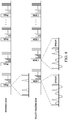
- FIGS. 6A to 6D show the results from a normal volunteer study.
- FIG. 6D is a reference image used for phase sensitive reconstruction.
- FIG. 7A shows a graph of positive direction encoded MSDE, and the resulting graph.
- FIG. 7B shows a graph of negative direction encoded MSDE, and the resulting graph.
- FIG. 8 shows a reference acquisition scan and an interleaved acquisition scan for use with the present invention.
- FIG. 9 summarizes the method of phase sensitive reconstruction of the present invention.
- FIG. 10 shows another example of using the present invention with a phantom 3D acquisition showing the reference image from a coronal, axial and sagittal view captured using the present invention, and then the phase-sensitive inversion recovery (PSIR) reconstructed image.
- PSIR phase-sensitive inversion recovery
- FIG. 11 shows another example of using the present invention of a volunteer with 3D acquisition showing the reference image, the positive direction encoded image, the negative direction encoded image, and the PSIR reconstructed image.
- FIG. 12 shows an example of the use of the BIR4 method with the present invention.
- Magnetic Resonance Angiography is increasingly used in the clinical practice for diagnosis and evaluation of various vascular malformations.
- Majority of the MRA techniques are performed with the administration of an exogenous contrast agent.
- gadolinium based exogenous contrast agents are contraindicated in a subset of populations with impaired renal function (1).
- NCE non-contrast enhanced MRA techniques
- MSDE motion sensitized driven equilibrium
- the present inventors provide a method for using an efficient 90-degree MSDE preparation using an adiabatic BIR-4, which completely eliminates the background signal, and combine this with phase sensitive reconstruction (5) to determine the flow direction and potentially differentiate arterial and venous signal with background signal suppression in a single acquisition.
- the present invention includes one or more of the following advantages: (1) use of a robust MSDE preparation using an adiabatic BIR-4 pulse with 90-degree flip angle, that essentially generates close to zero background signal; (2) use of phase sensitive acquisition and reconstruction to determine the flow direction; (3) the determination of flow direction allows separation of arteries and veins providing diagnostic information for both arteriogram and venogram; and/or (4) the estimation of flow direction overcomes the requirement for the optimization of the MSDE gradients.
- Yet another advantage of the present invention is that it solves the problem of the generation of MR arteriogram and venogram from a single acquisition with directional flow and minimized background signal without the administration of exogenous contrast agent and without the optimization of the motion sensitizing gradients.
- the present invention differs from existing art for at least the following reasons:
- VSP velocity selective preparation
- the 90-degree MSDE preparation was used (4), however, with their approach, the background signal was not eliminated completely, which forced them to acquire two images with different 90-degree MSDE preparation (using opposite gradients), which when subtracted suppressed the background signal.
- our approach uses a more efficient 90-degree MSDE preparation using an adiabatic BIR-4 pulse that essentially achieves complete background signal suppression.
- the MSDE gradients need to be adjusted such that only arterial signal is enhanced, while the venous signal is minimized due to its slower flow.
- there is a potential for venous contamination with this approach if the arterial flow is also decreased in pathology.
- the venous signal with this approach is suppressed without providing any additional diagnostic information.
- the approach of the present invention uses a phase sensitive acquisition and reconstruction such that flow direction is estimated, allowing arterial and venous separation, providing diagnostic information for both arteries and veins in a single acquisition.
- the present invention is a phase sensitive reconstruction combined with velocity selective preparation (VSP) that is used in a method to determine the flow direction and can potentially differentiate arterial and venous signals with background signal suppression in a single acquisition.
- VSP velocity selective preparation
- VSP Velocity selective preparation
- RF radio frequency
- FIG. 1A shows a schematic of the spin behavior experiencing the VSP.
- the static spins (red arrow) accumulate the same amount of phase between the 90° x -180° y and 180° y -90° y and are aligned along the initial orientation (i.e. y-direction).
- the moving spins (blue and green arrows) accumulate phase depending upon their direction and velocity, i.e., arteries and veins accumulate different phase in opposite directions.
- the static spins are oriented along the transverse plane while the moving spins in the opposite direction are oriented along the longitudinal direction (+z and ⁇ z).
- Spoiler gradients applied immediately after the VSP destroy the static signal while preserving the signal from the moving spins.
- a phase sensitive acquisition that follows immediately can measure the signals and the directions of the moving spins.
- FIG. 1B is a graph that shows the basic MSDE pulse consisting 90x-180y-90y and velocity encoding gradient, G.
- FIG. 1C shows one method of using the spin behavior with the VSP beginning at the top left and working across the top graphs following the arrows. With selected gradient, spins (green and blue) moving in opposite directions were tipped back to z-axis while static spin (red) was left in x,y-plane at the end of 90y-RF.
- FIGS. 2A and 2B show a static phantom set up with the cylinder in the middle containing agarose gel and the two tubes next to it, carrying static water.
- the flow was turned off.
- the image in FIG. 2A was acquired with a standard 0-degree MSDE using the BIR-4 pulse.
- the image in FIG. 2B was acquired with a BIR-4 based 90-degree MSDE, essentially suppressing the static phantom signal completely to the background noise level.
- FIGS. 3A and 3B show the same phantom as above ( FIGS. 2A and 2B , respectively), but with the flow turned on to mimic the arterial flow (towards the phantom) and the venous flow (away from the phantom).
- the image in FIG. 3A was acquired with a standard 0-degree MSDE using the BIR-4 pulse showing similar result to FIG. 2A , without the flow.
- the image in FIG. 3B was acquired with a BIR-4 based 90-degree MSDE, preserving the signal in the flow phantom, but suppressing the static phantom signal completely to the background noise level.
- FIGS. 4A and 4B show the phase sensitive reconstruction and the direction of the flow (see FIG. 1 ) can be determined from FIG. 3B .
- the phase sensitive reconstructed image is shown in FIG. 4A with the bright signal showing the signal intensity towards the phantom and the dark signal showing the signal away from the phantom.
- FIG. 4B shows a color depiction in which the arterial signal (red) can be differentiated from venous signal (blue).
- a VSP module was implemented in a two dimensional (2D) balanced steady state free precession (bSSFP) acquisition.
- 2D balanced steady state free precession
- FSE/TSE fast or turbo spin echo
- gradient echo an adiabatic pulse (BIR-4 with flip angle (FA) 90°) based VSP was used. All studies were performed on a 3 T Ingenia scanner (Philips Healthcare, The Netherlands).
- the method was first validated in a flow phantom consisting of tubes with flow in opposite directions submerged in a static water bath ( FIG. 5A ).
- the flow was set to 4 cm/s as measured by phase contrast and the corresponding v enc was set to 15 cm/s.
- An additional image without VSP gradients and BIR-4 FA 0° was acquired to estimate the phase for phase sensitive reconstruction (5).
- the method was tested on the lower legs of 2 normal volunteers with IRB approval and written informed consent.
- FIG. 5B shows the magnitude image of the flow phantom using the proposed VSP, with essentially no background signal.
- phase sensitive reconstruction allowed estimation of the flow direction ( FIG. 5C ).
- the direction of the popliteal arteries was also estimated using the phase sensitive reconstruction in the normal volunteer ( FIG. 6A ).
- the phase sensitive reconstruction appropriately determined the direction of the arterial flow ( FIG. 6B ).
- FIG. 6D is the reference image used for phase sensitive reconstruction.
- FIG. 7A shows a graph of positive direction encoded MSDE, and the resulting graph.
- FIG. 7B shows a graph of negative direction encoded MSDE, and the resulting graph. Together, these two graphs show that by switching the gradient direction, the signal intensity is inverted. As such, the subtraction between positive and negative direction encoded images can minimize the background signal due to B0 and B1 inhomogeneities.
- the present invention can also use an interleaved acquisition approach.
- the top data acquisition graph shows the acquisition of a reference scan, using a T2prep method, having two peaks and an interleaved acquisition.
- the interleaved data acquisition used the MSDE1 and MSDE2 method, as shown in the graph below, the interleaved data acquisition uses both the MSDE methods show in detail in FIGS. 7A and 7B .
- FIG. 9 summarizes the method of phase sensitive reconstruction of the present invention. Beginning on the top left, a positive direction encoded complex image is obtained, below that image is the negative direction encoded complex image, and below that image is the reference image. The calculations and combination of these images is next shown as mathematical formulas, to obtain an adaptive complex phase array image by combining the three images. Next, the data acquired is converted into a real image. In this image, the phase was estimated from reference image. In the subtraction step, the subtraction was made between positive and negative direction encoded complex image to minimize the residual background signal due to B1 and B0 inhomogeneities.
- FIG. 10 shows another example of using the present invention with a phantom 3D acquisition showing the reference image from a coronal, axial and sagittal view captured using the present invention, and then the PSIR reconstructed image.
- FIG. 11 shows another example of using the present invention of a volunteer with 3D acquisition showing the reference image, the positive direction encoded image, the negative direction encoded image, and the PSIR reconstructed image. Subtraction was made between positive and negative direction encoded image to minimize the residual background signal. The artery of the volunteer is shown in the PSIR reconstructed image.
- the present invention uses phase sensitive acquisition and reconstruction combined with velocity selective preparation allowing the separation of flow depending upon the direction with minimal background signal suppression.
- the velocity selective preparation of the present invention enables velocity encoding and background suppression simultaneously. While some background signal in the human studies was not completely suppressed, e.g., fat, the subtraction between the two preparation images minimized the background signal and provided a clear image.
- FIG. 12 shows an example of the use of the BIR4 method with the present invention.
- the background signal particularly of fat
- VSP can be further suppressed with VSP with chemical-shift acquisitions to minimize this signal.
- compositions of the invention can be used to achieve methods of the invention.
- the words “comprising” (and any form of comprising, such as “comprise” and “comprises”), “having” (and any form of having, such as “have” and “has”), “including” (and any form of including, such as “includes” and “include”) or “containing” (and any form of containing, such as “contains” and “contain”) are inclusive or open-ended and do not exclude additional, unrecited elements or method steps.
- “comprising” may be replaced with “consisting essentially of” or “consisting of”.
- the phrase “consisting essentially of” requires the specified integer(s) or steps as well as those that do not materially affect the character or function of the claimed invention.
- the term “consisting” is used to indicate the presence of the recited integer (e.g., a feature, an element, a characteristic, a property, a method/process step or a limitation) or group of integers (e.g., feature(s), element(s), characteristic(s), propertie(s), method/process steps or limitation(s)) only.
- A, B, C, or combinations thereof refers to all permutations and combinations of the listed items preceding the term.
- “A, B, C, or combinations thereof” is intended to include at least one of: A, B, C, AB, AC, BC, or ABC, and if order is important in a particular context, also BA, CA, CB, CBA, BCA, ACB, BAC, or CAB.
- expressly included are combinations that contain repeats of one or more item or term, such as BB, AAA, AB, BBC, AAABCCCC, CBBAAA, CABABB, and so forth.
- BB BB
- AAA AAA
- AB BBC
- AAABCCCCCC CBBAAA
- CABABB CABABB
- words of approximation such as, without limitation, “about”, “substantial” or “substantially” refers to a condition that when so modified is understood to not necessarily be absolute or perfect but would be considered close enough to those of ordinary skill in the art to warrant designating the condition as being present.
- the extent to which the description may vary will depend on how great a change can be instituted and still have one of ordinary skilled in the art recognize the modified feature as still having the required characteristics and capabilities of the unmodified feature.
- a numerical value herein that is modified by a word of approximation such as “about” may vary from the stated value by at least ⁇ 1, 2, 3, 4, 5, 6, 7, 10, 12 or 15%.
- compositions and/or methods disclosed and claimed herein can be made and executed without undue experimentation in light of the present disclosure. While the compositions and methods of this invention have been described in terms of preferred embodiments, it will be apparent to those of skill in the art that variations may be applied to the compositions and/or methods and in the steps or in the sequence of steps of the method described herein without departing from the concept, spirit and scope of the invention. All such similar substitutes and modifications apparent to those skilled in the art are deemed to be within the spirit, scope and concept of the invention as defined by the appended claims.
Landscapes
- Health & Medical Sciences (AREA)
- Physics & Mathematics (AREA)
- Life Sciences & Earth Sciences (AREA)
- Nuclear Medicine, Radiotherapy & Molecular Imaging (AREA)
- General Health & Medical Sciences (AREA)
- Engineering & Computer Science (AREA)
- Radiology & Medical Imaging (AREA)
- High Energy & Nuclear Physics (AREA)
- Signal Processing (AREA)
- Condensed Matter Physics & Semiconductors (AREA)
- General Physics & Mathematics (AREA)
- Heart & Thoracic Surgery (AREA)
- Vascular Medicine (AREA)
- Veterinary Medicine (AREA)
- Public Health (AREA)
- Biophysics (AREA)
- Pathology (AREA)
- Biomedical Technology (AREA)
- Animal Behavior & Ethology (AREA)
- Medical Informatics (AREA)
- Molecular Biology (AREA)
- Surgery (AREA)
- Cardiology (AREA)
- Physiology (AREA)
- Hematology (AREA)
- Magnetic Resonance Imaging Apparatus (AREA)
Abstract
Description
(2) An earlier proposed approach, called velocity selective preparation (VSP), where the 90-degree MSDE preparation was used (4), however, with their approach, the background signal was not eliminated completely, which forced them to acquire two images with different 90-degree MSDE preparation (using opposite gradients), which when subtracted suppressed the background signal. Compared to this prior art, our approach uses a more efficient 90-degree MSDE preparation using an adiabatic BIR-4 pulse that essentially achieves complete background signal suppression.
(3) As with the VSP approach described in (2) above, although the background signal was suppressed after subtraction, the MSDE gradients need to be adjusted such that only arterial signal is enhanced, while the venous signal is minimized due to its slower flow. However, there is a potential for venous contamination with this approach if the arterial flow is also decreased in pathology. Moreover, the venous signal with this approach is suppressed without providing any additional diagnostic information.
- 1. Broome D R. Nephrogenic systemic fibrosis associated with gadolinium based contrast agents: a summary of the medical literature reporting. Eur J Radiol 2008; 66(2):230-234.
- 2. Miyazaki M, Lee V S. Nonenhanced MR angiography. Radiology 2008; 248(1):20-43.
- 3. Fan Z, Sheehan J, Bi X, Liu X, Carr J, Li D. 3D noncontrast MR angiography of the distal lower extremities using flow-sensitive dephasing (FSD)-prepared balanced SSFP. Magn Reson Med 2009; 62(6): 1523-1532.
- 4. Korosec F R, Grist T M, Polzin J A, Weber D M, Mistretta C A. MR angiography using velocity selective preparation pulses and segmented gradient-echo acquisition. Magn Reson Med 1993; 30(6):704-714.
- 5. Kellman P, Arai A E, McVeigh E R, Aletras A H. Phase-sensitive inversion recovery for detecting myocardial infarction using gadolinium-delayed hyperenhancement. Magn Reson Med 2002; 47(2):372-383.
Claims (23)
Priority Applications (1)
| Application Number | Priority Date | Filing Date | Title |
|---|---|---|---|
| US15/762,794 US11169237B2 (en) | 2015-10-05 | 2016-10-05 | Phase sensitive magnetic resonance angiography |
Applications Claiming Priority (3)
| Application Number | Priority Date | Filing Date | Title |
|---|---|---|---|
| US201562237285P | 2015-10-05 | 2015-10-05 | |
| PCT/US2016/055523 WO2017062470A1 (en) | 2015-10-05 | 2016-10-05 | Phase sensitive magnetic resonance angiography |
| US15/762,794 US11169237B2 (en) | 2015-10-05 | 2016-10-05 | Phase sensitive magnetic resonance angiography |
Publications (2)
| Publication Number | Publication Date |
|---|---|
| US20180284209A1 US20180284209A1 (en) | 2018-10-04 |
| US11169237B2 true US11169237B2 (en) | 2021-11-09 |
Family
ID=58488440
Family Applications (1)
| Application Number | Title | Priority Date | Filing Date |
|---|---|---|---|
| US15/762,794 Active 2038-11-10 US11169237B2 (en) | 2015-10-05 | 2016-10-05 | Phase sensitive magnetic resonance angiography |
Country Status (2)
| Country | Link |
|---|---|
| US (1) | US11169237B2 (en) |
| WO (1) | WO2017062470A1 (en) |
Families Citing this family (1)
| Publication number | Priority date | Publication date | Assignee | Title |
|---|---|---|---|---|
| US11169237B2 (en) | 2015-10-05 | 2021-11-09 | Board Of Regents, The University Of Texas System | Phase sensitive magnetic resonance angiography |
Citations (13)
| Publication number | Priority date | Publication date | Assignee | Title |
|---|---|---|---|---|
| JP2000175885A (en) | 1998-12-11 | 2000-06-27 | General Electric Co <Ge> | Method and system for acquisition of preferential arterial and venous images for mr angiography |
| US6192264B1 (en) | 1998-12-28 | 2001-02-20 | General Electric Company | Method and system for MRI venography including arterial and venous discrimination |
| WO2003008989A1 (en) | 2001-07-18 | 2003-01-30 | Koninklijke Philips Electronics Nv | Automatic vessel identification for angiographic screening |
| US20080119721A1 (en) | 2006-11-22 | 2008-05-22 | Kabushiki Kaisha Toshiba | Magnetic resonance imaging apparatus |
| JP2008125891A (en) | 2006-11-22 | 2008-06-05 | Toshiba Corp | Magnetic resonance imaging apparatus |
| WO2011132593A1 (en) | 2010-04-20 | 2011-10-27 | 株式会社 日立メディコ | Magnetic resonance imaging device and blood vessel image imaging method |
| US20120283547A1 (en) * | 2011-04-11 | 2012-11-08 | The Regents Of The University Of California | Magnetic resonance imaging using velocity selective excitation |
| US8410779B2 (en) | 2009-08-05 | 2013-04-02 | Siemens Aktiengesellschaft | Contrast agent-free MR angiography with SSFP sequences |
| US8744551B2 (en) | 2012-07-12 | 2014-06-03 | Northshore University Healthsystem | Method for non-contrast enhanced magnetic resonance angiography |
| US8855743B2 (en) | 2012-05-11 | 2014-10-07 | Kabushiki Kaisha Toshiba | Non-contrast magnetic resonance perfusion imaging |
| US9036320B1 (en) | 2013-12-02 | 2015-05-19 | Elbex Video Ltd. | Mechanical latching relays and hybrid switches with latching relays for use in electrical automation |
| US20150305645A1 (en) * | 2014-04-23 | 2015-10-29 | Kabushiki Kaisha Toshiba | Off-Resonance Correction for Vessel-Selective Pseudo-Continuous Arterial Spin Labeling Imaging |
| WO2017062470A1 (en) | 2015-10-05 | 2017-04-13 | Board Of Regents, The University Of Texas System | Phase sensitive magnetic resonance angiography |
-
2016
- 2016-10-05 US US15/762,794 patent/US11169237B2/en active Active
- 2016-10-05 WO PCT/US2016/055523 patent/WO2017062470A1/en active Application Filing
Patent Citations (14)
| Publication number | Priority date | Publication date | Assignee | Title |
|---|---|---|---|---|
| US6246897B1 (en) | 1998-12-11 | 2001-06-12 | General Electric Company | Method and system for acquistion of preferential arterial and venous images for MR angiography |
| JP2000175885A (en) | 1998-12-11 | 2000-06-27 | General Electric Co <Ge> | Method and system for acquisition of preferential arterial and venous images for mr angiography |
| US6192264B1 (en) | 1998-12-28 | 2001-02-20 | General Electric Company | Method and system for MRI venography including arterial and venous discrimination |
| WO2003008989A1 (en) | 2001-07-18 | 2003-01-30 | Koninklijke Philips Electronics Nv | Automatic vessel identification for angiographic screening |
| US20080119721A1 (en) | 2006-11-22 | 2008-05-22 | Kabushiki Kaisha Toshiba | Magnetic resonance imaging apparatus |
| JP2008125891A (en) | 2006-11-22 | 2008-06-05 | Toshiba Corp | Magnetic resonance imaging apparatus |
| US8410779B2 (en) | 2009-08-05 | 2013-04-02 | Siemens Aktiengesellschaft | Contrast agent-free MR angiography with SSFP sequences |
| WO2011132593A1 (en) | 2010-04-20 | 2011-10-27 | 株式会社 日立メディコ | Magnetic resonance imaging device and blood vessel image imaging method |
| US20120283547A1 (en) * | 2011-04-11 | 2012-11-08 | The Regents Of The University Of California | Magnetic resonance imaging using velocity selective excitation |
| US8855743B2 (en) | 2012-05-11 | 2014-10-07 | Kabushiki Kaisha Toshiba | Non-contrast magnetic resonance perfusion imaging |
| US8744551B2 (en) | 2012-07-12 | 2014-06-03 | Northshore University Healthsystem | Method for non-contrast enhanced magnetic resonance angiography |
| US9036320B1 (en) | 2013-12-02 | 2015-05-19 | Elbex Video Ltd. | Mechanical latching relays and hybrid switches with latching relays for use in electrical automation |
| US20150305645A1 (en) * | 2014-04-23 | 2015-10-29 | Kabushiki Kaisha Toshiba | Off-Resonance Correction for Vessel-Selective Pseudo-Continuous Arterial Spin Labeling Imaging |
| WO2017062470A1 (en) | 2015-10-05 | 2017-04-13 | Board Of Regents, The University Of Texas System | Phase sensitive magnetic resonance angiography |
Non-Patent Citations (4)
| Title |
|---|
| Fan, et al. "3D noncontrast MR angiography of the distal lower extremities using flow-sensitive dephasing (FSD)-prepared balanced SSFP" Magn Reson Med, Dec. 2009; 62(6): 1523-1532. |
| Kellman, et al. "Phase-sensitive inversion recovery for detecting myocardial infarction using gadolinium-delayed hyperenhancement" Magn Reson Med, Feb. 2002; 47(2):372-383. |
| Korean Intellectual Property Office, International Search Report and Written Opinion for PCT/US2016/055523, dated Jan. 10, 2017, 11 pp. |
| Miyazaki, et al. "Nonenhanced MR angiography" Radiology, Jul. 2008; 248(1):20-43. |
Also Published As
| Publication number | Publication date |
|---|---|
| US20180284209A1 (en) | 2018-10-04 |
| WO2017062470A1 (en) | 2017-04-13 |
Similar Documents
| Publication | Publication Date | Title |
|---|---|---|
| US8700127B2 (en) | Motion-attenuated contrast-enhanced cardiac magnetic resonance imaging system and method | |
| Balu et al. | Carotid plaque assessment using fast 3D isotropic resolution black‐blood MRI | |
| Deshmane et al. | Parallel MR imaging | |
| Jung et al. | Spin echo magnetic resonance imaging | |
| Marques et al. | How to choose the right MR sequence for your research question at 7 T and above? | |
| RU2605524C2 (en) | Magnetic resonance imaging with suppression of flow artefacts | |
| Viessmann et al. | T2‐weighted intracranial vessel wall imaging at 7 Tesla using a DANTE‐prepared variable flip angle turbo spin echo readout (DANTE‐SPACE) | |
| Wu et al. | A fully flow‐compensated multiecho susceptibility‐weighted imaging sequence: The effects of acceleration and background field on flow compensation | |
| Nezafat et al. | Coronary MR angiography at 3T: fat suppression versus water-fat separation | |
| Shin et al. | Rapid single‐breath‐hold 3D late gadolinium enhancement cardiac MRI using a stack‐of‐spirals acquisition | |
| US8513945B2 (en) | System, method and computer-accessible medium for providing breath-hold multi-echo fast spin-echo pulse sequence for accurate R2 measurement | |
| Kim et al. | Breathhold multiecho fast spin‐echo pulse sequence for accurate R2 measurement in the heart and liver | |
| Park et al. | Physiological and functional magnetic resonance imaging using balanced steady-state free precession | |
| Kiruluta et al. | Magnetic resonance angiography: physical principles and applications | |
| Koktzoglou et al. | Super‐resolution intracranial quiescent interval slice‐selective magnetic resonance angiography | |
| Weingärtner et al. | Black‐blood native T 1 mapping: Blood signal suppression for reduced partial voluming in the myocardium | |
| US10871537B1 (en) | Systems and methods for background suppression in time-of-flight magnetic resonance angiography | |
| Li et al. | Lung parenchyma transverse relaxation rates at 0.55 T | |
| Javed et al. | Single‐shot EPI for ASL‐CMR | |
| Xu et al. | Single breathhold noncontrast thoracic MRA using highly accelerated parallel imaging with a 32‐element coil array | |
| US11169237B2 (en) | Phase sensitive magnetic resonance angiography | |
| Srinivasan et al. | Free‐breathing 3D whole‐heart black‐blood imaging with motion sensitized driven equilibrium | |
| Obara et al. | Technical advancements in abdominal diffusion-weighted imaging | |
| Darçot et al. | Accelerated and high‐resolution cardiac T 2 mapping through peripheral k‐space sharing | |
| Borreguero et al. | Slice-selective zero echo time imaging of ultra-short T2 tissues based on spin-locking |
Legal Events
| Date | Code | Title | Description |
|---|---|---|---|
| AS | Assignment |
Owner name: BOARD OF REGENTS, THE UNIVERSITY OF TEXAS SYSTEM, TEXAS Free format text: ASSIGNMENT OF ASSIGNORS INTEREST;ASSIGNORS:MADHURANTHAKAM, ANANTH J.;WANG, XINZENG;REEL/FRAME:045328/0663 Effective date: 20151030 Owner name: BOARD OF REGENTS, THE UNIVERSITY OF TEXAS SYSTEM, Free format text: ASSIGNMENT OF ASSIGNORS INTEREST;ASSIGNORS:MADHURANTHAKAM, ANANTH J.;WANG, XINZENG;REEL/FRAME:045328/0663 Effective date: 20151030 |
|
| FEPP | Fee payment procedure |
Free format text: ENTITY STATUS SET TO UNDISCOUNTED (ORIGINAL EVENT CODE: BIG.); ENTITY STATUS OF PATENT OWNER: SMALL ENTITY |
|
| FEPP | Fee payment procedure |
Free format text: ENTITY STATUS SET TO SMALL (ORIGINAL EVENT CODE: SMAL); ENTITY STATUS OF PATENT OWNER: SMALL ENTITY |
|
| STPP | Information on status: patent application and granting procedure in general |
Free format text: DOCKETED NEW CASE - READY FOR EXAMINATION |
|
| STPP | Information on status: patent application and granting procedure in general |
Free format text: NON FINAL ACTION MAILED |
|
| STPP | Information on status: patent application and granting procedure in general |
Free format text: RESPONSE TO NON-FINAL OFFICE ACTION ENTERED AND FORWARDED TO EXAMINER |
|
| STPP | Information on status: patent application and granting procedure in general |
Free format text: NOTICE OF ALLOWANCE MAILED -- APPLICATION RECEIVED IN OFFICE OF PUBLICATIONS |
|
| STPP | Information on status: patent application and granting procedure in general |
Free format text: NOTICE OF ALLOWANCE MAILED -- APPLICATION RECEIVED IN OFFICE OF PUBLICATIONS |
|
| STPP | Information on status: patent application and granting procedure in general |
Free format text: PUBLICATIONS -- ISSUE FEE PAYMENT VERIFIED |
|
| STCF | Information on status: patent grant |
Free format text: PATENTED CASE |