US11071783B2 - HIV-1 neutralizing antibodies and uses thereof - Google Patents
HIV-1 neutralizing antibodies and uses thereof Download PDFInfo
- Publication number
- US11071783B2 US11071783B2 US15/559,314 US201615559314A US11071783B2 US 11071783 B2 US11071783 B2 US 11071783B2 US 201615559314 A US201615559314 A US 201615559314A US 11071783 B2 US11071783 B2 US 11071783B2
- Authority
- US
- United States
- Prior art keywords
- seq
- antibody
- antibodies
- amino acid
- fragment
- Prior art date
- Legal status (The legal status is an assumption and is not a legal conclusion. Google has not performed a legal analysis and makes no representation as to the accuracy of the status listed.)
- Active, expires
Links
- 241000713772 Human immunodeficiency virus 1 Species 0.000 title claims abstract description 100
- 230000003472 neutralizing effect Effects 0.000 title claims abstract description 41
- 238000000034 method Methods 0.000 claims abstract description 40
- 101800001690 Transmembrane protein gp41 Proteins 0.000 claims description 112
- 239000012634 fragment Substances 0.000 claims description 98
- 150000001413 amino acids Chemical group 0.000 claims description 48
- 229940024606 amino acid Drugs 0.000 claims description 46
- QIVBCDIJIAJPQS-UHFFFAOYSA-N Tryptophan Natural products C1=CC=C2C(CC(N)C(O)=O)=CNC2=C1 QIVBCDIJIAJPQS-UHFFFAOYSA-N 0.000 claims description 29
- 241000725303 Human immunodeficiency virus Species 0.000 claims description 27
- 230000014509 gene expression Effects 0.000 claims description 22
- 239000008194 pharmaceutical composition Substances 0.000 claims description 21
- 150000007523 nucleic acids Chemical class 0.000 claims description 19
- 208000031886 HIV Infections Diseases 0.000 claims description 15
- 102000039446 nucleic acids Human genes 0.000 claims description 14
- 108020004707 nucleic acids Proteins 0.000 claims description 14
- 239000013598 vector Substances 0.000 claims description 14
- NFGXHKASABOEEW-UHFFFAOYSA-N 1-methylethyl 11-methoxy-3,7,11-trimethyl-2,4-dodecadienoate Chemical compound COC(C)(C)CCCC(C)CC=CC(C)=CC(=O)OC(C)C NFGXHKASABOEEW-UHFFFAOYSA-N 0.000 claims description 8
- 238000001476 gene delivery Methods 0.000 claims description 2
- 108010047041 Complementarity Determining Regions Proteins 0.000 claims 16
- COLNVLDHVKWLRT-UHFFFAOYSA-N phenylalanine Natural products OC(=O)C(N)CC1=CC=CC=C1 COLNVLDHVKWLRT-UHFFFAOYSA-N 0.000 claims 4
- ROHFNLRQFUQHCH-UHFFFAOYSA-N Leucine Natural products CC(C)CC(N)C(O)=O ROHFNLRQFUQHCH-UHFFFAOYSA-N 0.000 claims 1
- 229960000310 isoleucine Drugs 0.000 claims 1
- AGPKZVBTJJNPAG-UHFFFAOYSA-N isoleucine Natural products CCC(C)C(N)C(O)=O AGPKZVBTJJNPAG-UHFFFAOYSA-N 0.000 claims 1
- 238000006386 neutralization reaction Methods 0.000 description 111
- 108090000765 processed proteins & peptides Proteins 0.000 description 108
- 230000027455 binding Effects 0.000 description 101
- 210000004027 cell Anatomy 0.000 description 75
- 210000002381 plasma Anatomy 0.000 description 74
- 102000004196 processed proteins & peptides Human genes 0.000 description 74
- 210000001806 memory b lymphocyte Anatomy 0.000 description 55
- 108090000623 proteins and genes Proteins 0.000 description 54
- 230000035772 mutation Effects 0.000 description 47
- 238000003556 assay Methods 0.000 description 40
- 239000000203 mixture Substances 0.000 description 34
- 229920001184 polypeptide Polymers 0.000 description 33
- 239000000427 antigen Substances 0.000 description 32
- 230000000694 effects Effects 0.000 description 31
- 108091007433 antigens Proteins 0.000 description 30
- 102000036639 antigens Human genes 0.000 description 30
- 102000004169 proteins and genes Human genes 0.000 description 28
- 235000001014 amino acid Nutrition 0.000 description 26
- 238000004458 analytical method Methods 0.000 description 26
- 235000018102 proteins Nutrition 0.000 description 26
- 241000700605 Viruses Species 0.000 description 23
- 230000003389 potentiating effect Effects 0.000 description 23
- 241001112090 Pseudovirus Species 0.000 description 21
- 210000003719 b-lymphocyte Anatomy 0.000 description 20
- 238000013507 mapping Methods 0.000 description 19
- 125000003275 alpha amino acid group Chemical group 0.000 description 17
- 230000003993 interaction Effects 0.000 description 17
- 210000001744 T-lymphocyte Anatomy 0.000 description 16
- 230000001404 mediated effect Effects 0.000 description 16
- 102100035360 Cerebellar degeneration-related antigen 1 Human genes 0.000 description 15
- 108010021625 Immunoglobulin Fragments Proteins 0.000 description 15
- 102000008394 Immunoglobulin Fragments Human genes 0.000 description 15
- 235000004279 alanine Nutrition 0.000 description 15
- 238000002965 ELISA Methods 0.000 description 14
- 108060003951 Immunoglobulin Proteins 0.000 description 14
- 102000018358 immunoglobulin Human genes 0.000 description 14
- 239000000543 intermediate Substances 0.000 description 14
- 239000000523 sample Substances 0.000 description 14
- 238000006467 substitution reaction Methods 0.000 description 14
- 101100112922 Candida albicans CDR3 gene Proteins 0.000 description 13
- 210000004602 germ cell Anatomy 0.000 description 12
- 238000012163 sequencing technique Methods 0.000 description 12
- 230000008685 targeting Effects 0.000 description 12
- 238000011282 treatment Methods 0.000 description 12
- 239000013078 crystal Substances 0.000 description 11
- QNAYBMKLOCPYGJ-REOHCLBHSA-N L-alanine Chemical compound C[C@H](N)C(O)=O QNAYBMKLOCPYGJ-REOHCLBHSA-N 0.000 description 10
- 239000011324 bead Substances 0.000 description 10
- 239000012528 membrane Substances 0.000 description 10
- WEVYAHXRMPXWCK-UHFFFAOYSA-N Acetonitrile Chemical compound CC#N WEVYAHXRMPXWCK-UHFFFAOYSA-N 0.000 description 9
- RTZKZFJDLAIYFH-UHFFFAOYSA-N Diethyl ether Chemical compound CCOCC RTZKZFJDLAIYFH-UHFFFAOYSA-N 0.000 description 9
- 230000010056 antibody-dependent cellular cytotoxicity Effects 0.000 description 9
- 230000001965 increasing effect Effects 0.000 description 9
- 239000002773 nucleotide Substances 0.000 description 9
- 125000003729 nucleotide group Chemical group 0.000 description 9
- 210000003819 peripheral blood mononuclear cell Anatomy 0.000 description 9
- 239000002243 precursor Substances 0.000 description 9
- 230000002265 prevention Effects 0.000 description 9
- 238000003757 reverse transcription PCR Methods 0.000 description 9
- 230000001225 therapeutic effect Effects 0.000 description 9
- 102000017420 CD3 protein, epsilon/gamma/delta subunit Human genes 0.000 description 8
- 108050005493 CD3 protein, epsilon/gamma/delta subunit Proteins 0.000 description 8
- 102100036011 T-cell surface glycoprotein CD4 Human genes 0.000 description 8
- 238000001042 affinity chromatography Methods 0.000 description 8
- 125000003295 alanine group Chemical group N[C@@H](C)C(=O)* 0.000 description 8
- 238000013459 approach Methods 0.000 description 8
- 239000012636 effector Substances 0.000 description 8
- 238000002955 isolation Methods 0.000 description 8
- 238000007481 next generation sequencing Methods 0.000 description 8
- 230000009257 reactivity Effects 0.000 description 8
- 210000002966 serum Anatomy 0.000 description 8
- 231100000765 toxin Toxicity 0.000 description 8
- 108020004414 DNA Proteins 0.000 description 7
- 238000012286 ELISA Assay Methods 0.000 description 7
- 239000003814 drug Substances 0.000 description 7
- 239000012071 phase Substances 0.000 description 7
- 230000002829 reductive effect Effects 0.000 description 7
- 238000002864 sequence alignment Methods 0.000 description 7
- 238000002198 surface plasmon resonance spectroscopy Methods 0.000 description 7
- 239000003053 toxin Substances 0.000 description 7
- 108700012359 toxins Proteins 0.000 description 7
- 229920000936 Agarose Polymers 0.000 description 6
- 239000000872 buffer Substances 0.000 description 6
- 210000004899 c-terminal region Anatomy 0.000 description 6
- 230000009977 dual effect Effects 0.000 description 6
- 239000000839 emulsion Substances 0.000 description 6
- 238000005516 engineering process Methods 0.000 description 6
- 230000006870 function Effects 0.000 description 6
- 101150026046 iga gene Proteins 0.000 description 6
- 238000003780 insertion Methods 0.000 description 6
- 230000037431 insertion Effects 0.000 description 6
- 230000000670 limiting effect Effects 0.000 description 6
- 238000004519 manufacturing process Methods 0.000 description 6
- BDAGIHXWWSANSR-UHFFFAOYSA-N methanoic acid Natural products OC=O BDAGIHXWWSANSR-UHFFFAOYSA-N 0.000 description 6
- 230000004048 modification Effects 0.000 description 6
- 238000012986 modification Methods 0.000 description 6
- 238000001228 spectrum Methods 0.000 description 6
- 230000003612 virological effect Effects 0.000 description 6
- 108091093088 Amplicon Proteins 0.000 description 5
- 102000053602 DNA Human genes 0.000 description 5
- 102100031513 Fc receptor-like protein 4 Human genes 0.000 description 5
- DHMQDGOQFOQNFH-UHFFFAOYSA-N Glycine Natural products NCC(O)=O DHMQDGOQFOQNFH-UHFFFAOYSA-N 0.000 description 5
- 101000846909 Homo sapiens Fc receptor-like protein 4 Proteins 0.000 description 5
- 101000598100 Homo sapiens Nuclear migration protein nudC Proteins 0.000 description 5
- 108090000144 Human Proteins Proteins 0.000 description 5
- 102000003839 Human Proteins Human genes 0.000 description 5
- 102100036965 Nuclear migration protein nudC Human genes 0.000 description 5
- 102000004389 Ribonucleoproteins Human genes 0.000 description 5
- 108010081734 Ribonucleoproteins Proteins 0.000 description 5
- 229960002685 biotin Drugs 0.000 description 5
- 239000011616 biotin Substances 0.000 description 5
- 238000004422 calculation algorithm Methods 0.000 description 5
- 238000004113 cell culture Methods 0.000 description 5
- 210000004978 chinese hamster ovary cell Anatomy 0.000 description 5
- 238000001514 detection method Methods 0.000 description 5
- 238000011161 development Methods 0.000 description 5
- 230000018109 developmental process Effects 0.000 description 5
- LOKCTEFSRHRXRJ-UHFFFAOYSA-I dipotassium trisodium dihydrogen phosphate hydrogen phosphate dichloride Chemical compound P(=O)(O)(O)[O-].[K+].P(=O)(O)([O-])[O-].[Na+].[Na+].[Cl-].[K+].[Cl-].[Na+] LOKCTEFSRHRXRJ-UHFFFAOYSA-I 0.000 description 5
- 229940079593 drug Drugs 0.000 description 5
- 238000002474 experimental method Methods 0.000 description 5
- 239000000499 gel Substances 0.000 description 5
- 230000005764 inhibitory process Effects 0.000 description 5
- 150000002632 lipids Chemical class 0.000 description 5
- 239000002502 liposome Substances 0.000 description 5
- 238000001294 liquid chromatography-tandem mass spectrometry Methods 0.000 description 5
- 210000004962 mammalian cell Anatomy 0.000 description 5
- 238000004949 mass spectrometry Methods 0.000 description 5
- 239000002953 phosphate buffered saline Substances 0.000 description 5
- 210000004180 plasmocyte Anatomy 0.000 description 5
- 238000000746 purification Methods 0.000 description 5
- 230000009467 reduction Effects 0.000 description 5
- 238000011160 research Methods 0.000 description 5
- 239000013603 viral vector Substances 0.000 description 5
- 206010069754 Acquired gene mutation Diseases 0.000 description 4
- 108010054477 Immunoglobulin Fab Fragments Proteins 0.000 description 4
- 102000001706 Immunoglobulin Fab Fragments Human genes 0.000 description 4
- 108700005091 Immunoglobulin Genes Proteins 0.000 description 4
- CKLJMWTZIZZHCS-REOHCLBHSA-N L-aspartic acid Chemical compound OC(=O)[C@@H](N)CC(O)=O CKLJMWTZIZZHCS-REOHCLBHSA-N 0.000 description 4
- 108091034117 Oligonucleotide Proteins 0.000 description 4
- 102000057297 Pepsin A Human genes 0.000 description 4
- 108090000284 Pepsin A Proteins 0.000 description 4
- 108010076504 Protein Sorting Signals Proteins 0.000 description 4
- RHQDFWAXVIIEBN-UHFFFAOYSA-N Trifluoroethanol Chemical compound OCC(F)(F)F RHQDFWAXVIIEBN-UHFFFAOYSA-N 0.000 description 4
- JLCPHMBAVCMARE-UHFFFAOYSA-N [3-[[3-[[3-[[3-[[3-[[3-[[3-[[3-[[3-[[3-[[3-[[5-(2-amino-6-oxo-1H-purin-9-yl)-3-[[3-[[3-[[3-[[3-[[3-[[5-(2-amino-6-oxo-1H-purin-9-yl)-3-[[5-(2-amino-6-oxo-1H-purin-9-yl)-3-hydroxyoxolan-2-yl]methoxy-hydroxyphosphoryl]oxyoxolan-2-yl]methoxy-hydroxyphosphoryl]oxy-5-(5-methyl-2,4-dioxopyrimidin-1-yl)oxolan-2-yl]methoxy-hydroxyphosphoryl]oxy-5-(6-aminopurin-9-yl)oxolan-2-yl]methoxy-hydroxyphosphoryl]oxy-5-(6-aminopurin-9-yl)oxolan-2-yl]methoxy-hydroxyphosphoryl]oxy-5-(6-aminopurin-9-yl)oxolan-2-yl]methoxy-hydroxyphosphoryl]oxy-5-(6-aminopurin-9-yl)oxolan-2-yl]methoxy-hydroxyphosphoryl]oxyoxolan-2-yl]methoxy-hydroxyphosphoryl]oxy-5-(5-methyl-2,4-dioxopyrimidin-1-yl)oxolan-2-yl]methoxy-hydroxyphosphoryl]oxy-5-(4-amino-2-oxopyrimidin-1-yl)oxolan-2-yl]methoxy-hydroxyphosphoryl]oxy-5-(5-methyl-2,4-dioxopyrimidin-1-yl)oxolan-2-yl]methoxy-hydroxyphosphoryl]oxy-5-(5-methyl-2,4-dioxopyrimidin-1-yl)oxolan-2-yl]methoxy-hydroxyphosphoryl]oxy-5-(6-aminopurin-9-yl)oxolan-2-yl]methoxy-hydroxyphosphoryl]oxy-5-(6-aminopurin-9-yl)oxolan-2-yl]methoxy-hydroxyphosphoryl]oxy-5-(4-amino-2-oxopyrimidin-1-yl)oxolan-2-yl]methoxy-hydroxyphosphoryl]oxy-5-(4-amino-2-oxopyrimidin-1-yl)oxolan-2-yl]methoxy-hydroxyphosphoryl]oxy-5-(4-amino-2-oxopyrimidin-1-yl)oxolan-2-yl]methoxy-hydroxyphosphoryl]oxy-5-(6-aminopurin-9-yl)oxolan-2-yl]methoxy-hydroxyphosphoryl]oxy-5-(4-amino-2-oxopyrimidin-1-yl)oxolan-2-yl]methyl [5-(6-aminopurin-9-yl)-2-(hydroxymethyl)oxolan-3-yl] hydrogen phosphate Polymers Cc1cn(C2CC(OP(O)(=O)OCC3OC(CC3OP(O)(=O)OCC3OC(CC3O)n3cnc4c3nc(N)[nH]c4=O)n3cnc4c3nc(N)[nH]c4=O)C(COP(O)(=O)OC3CC(OC3COP(O)(=O)OC3CC(OC3COP(O)(=O)OC3CC(OC3COP(O)(=O)OC3CC(OC3COP(O)(=O)OC3CC(OC3COP(O)(=O)OC3CC(OC3COP(O)(=O)OC3CC(OC3COP(O)(=O)OC3CC(OC3COP(O)(=O)OC3CC(OC3COP(O)(=O)OC3CC(OC3COP(O)(=O)OC3CC(OC3COP(O)(=O)OC3CC(OC3COP(O)(=O)OC3CC(OC3COP(O)(=O)OC3CC(OC3COP(O)(=O)OC3CC(OC3COP(O)(=O)OC3CC(OC3COP(O)(=O)OC3CC(OC3CO)n3cnc4c(N)ncnc34)n3ccc(N)nc3=O)n3cnc4c(N)ncnc34)n3ccc(N)nc3=O)n3ccc(N)nc3=O)n3ccc(N)nc3=O)n3cnc4c(N)ncnc34)n3cnc4c(N)ncnc34)n3cc(C)c(=O)[nH]c3=O)n3cc(C)c(=O)[nH]c3=O)n3ccc(N)nc3=O)n3cc(C)c(=O)[nH]c3=O)n3cnc4c3nc(N)[nH]c4=O)n3cnc4c(N)ncnc34)n3cnc4c(N)ncnc34)n3cnc4c(N)ncnc34)n3cnc4c(N)ncnc34)O2)c(=O)[nH]c1=O JLCPHMBAVCMARE-UHFFFAOYSA-N 0.000 description 4
- 230000005875 antibody response Effects 0.000 description 4
- 230000015572 biosynthetic process Effects 0.000 description 4
- 238000012512 characterization method Methods 0.000 description 4
- 230000000295 complement effect Effects 0.000 description 4
- 230000003247 decreasing effect Effects 0.000 description 4
- 230000029087 digestion Effects 0.000 description 4
- ZGSPNIOCEDOHGS-UHFFFAOYSA-L disodium [3-[2,3-di(octadeca-9,12-dienoyloxy)propoxy-oxidophosphoryl]oxy-2-hydroxypropyl] 2,3-di(octadeca-9,12-dienoyloxy)propyl phosphate Chemical compound [Na+].[Na+].CCCCCC=CCC=CCCCCCCCC(=O)OCC(OC(=O)CCCCCCCC=CCC=CCCCCC)COP([O-])(=O)OCC(O)COP([O-])(=O)OCC(OC(=O)CCCCCCCC=CCC=CCCCCC)COC(=O)CCCCCCCC=CCC=CCCCCC ZGSPNIOCEDOHGS-UHFFFAOYSA-L 0.000 description 4
- 238000010494 dissociation reaction Methods 0.000 description 4
- 230000005593 dissociations Effects 0.000 description 4
- 238000010828 elution Methods 0.000 description 4
- 238000005734 heterodimerization reaction Methods 0.000 description 4
- 125000001165 hydrophobic group Chemical group 0.000 description 4
- 208000015181 infectious disease Diseases 0.000 description 4
- 230000002401 inhibitory effect Effects 0.000 description 4
- 238000011031 large-scale manufacturing process Methods 0.000 description 4
- KWGKDLIKAYFUFQ-UHFFFAOYSA-M lithium chloride Chemical compound [Li+].[Cl-] KWGKDLIKAYFUFQ-UHFFFAOYSA-M 0.000 description 4
- 235000018977 lysine Nutrition 0.000 description 4
- 238000005259 measurement Methods 0.000 description 4
- 230000003641 microbiacidal effect Effects 0.000 description 4
- 239000000178 monomer Substances 0.000 description 4
- 210000000822 natural killer cell Anatomy 0.000 description 4
- 230000004044 response Effects 0.000 description 4
- 238000012552 review Methods 0.000 description 4
- 230000037439 somatic mutation Effects 0.000 description 4
- 238000012916 structural analysis Methods 0.000 description 4
- 229960005486 vaccine Drugs 0.000 description 4
- OSWFIVFLDKOXQC-UHFFFAOYSA-N 4-(3-methoxyphenyl)aniline Chemical compound COC1=CC=CC(C=2C=CC(N)=CC=2)=C1 OSWFIVFLDKOXQC-UHFFFAOYSA-N 0.000 description 3
- 208000030507 AIDS Diseases 0.000 description 3
- 206010000807 Acute HIV infection Diseases 0.000 description 3
- 102100037334 E3 ubiquitin-protein ligase CHIP Human genes 0.000 description 3
- 101710187668 E3 ubiquitin-protein ligase CHIP Proteins 0.000 description 3
- KCXVZYZYPLLWCC-UHFFFAOYSA-N EDTA Chemical compound OC(=O)CN(CC(O)=O)CCN(CC(O)=O)CC(O)=O KCXVZYZYPLLWCC-UHFFFAOYSA-N 0.000 description 3
- 239000004471 Glycine Substances 0.000 description 3
- 101000917858 Homo sapiens Low affinity immunoglobulin gamma Fc region receptor III-A Proteins 0.000 description 3
- 101000917839 Homo sapiens Low affinity immunoglobulin gamma Fc region receptor III-B Proteins 0.000 description 3
- 102100026120 IgG receptor FcRn large subunit p51 Human genes 0.000 description 3
- 239000000232 Lipid Bilayer Substances 0.000 description 3
- 102100029185 Low affinity immunoglobulin gamma Fc region receptor III-B Human genes 0.000 description 3
- 206010035226 Plasma cell myeloma Diseases 0.000 description 3
- 102100038689 Scm-like with four MBT domains protein 1 Human genes 0.000 description 3
- 101710081186 Scm-like with four MBT domains protein 1 Proteins 0.000 description 3
- 238000012300 Sequence Analysis Methods 0.000 description 3
- 108010090804 Streptavidin Proteins 0.000 description 3
- 239000007983 Tris buffer Substances 0.000 description 3
- 238000009825 accumulation Methods 0.000 description 3
- 230000003321 amplification Effects 0.000 description 3
- 230000003429 anti-cardiolipin effect Effects 0.000 description 3
- 230000000798 anti-retroviral effect Effects 0.000 description 3
- 238000011225 antiretroviral therapy Methods 0.000 description 3
- 235000003704 aspartic acid Nutrition 0.000 description 3
- OQFSQFPPLPISGP-UHFFFAOYSA-N beta-carboxyaspartic acid Natural products OC(=O)C(N)C(C(O)=O)C(O)=O OQFSQFPPLPISGP-UHFFFAOYSA-N 0.000 description 3
- 230000005540 biological transmission Effects 0.000 description 3
- 238000005119 centrifugation Methods 0.000 description 3
- 238000006243 chemical reaction Methods 0.000 description 3
- 210000002825 class switched memory b cell Anatomy 0.000 description 3
- 238000002648 combination therapy Methods 0.000 description 3
- 238000002425 crystallisation Methods 0.000 description 3
- 230000008025 crystallization Effects 0.000 description 3
- 238000006471 dimerization reaction Methods 0.000 description 3
- 235000019253 formic acid Nutrition 0.000 description 3
- 238000009472 formulation Methods 0.000 description 3
- 230000004927 fusion Effects 0.000 description 3
- 150000004676 glycans Chemical class 0.000 description 3
- 230000013595 glycosylation Effects 0.000 description 3
- 238000006206 glycosylation reaction Methods 0.000 description 3
- RAXXELZNTBOGNW-UHFFFAOYSA-N imidazole Natural products C1=CNC=N1 RAXXELZNTBOGNW-UHFFFAOYSA-N 0.000 description 3
- 230000016784 immunoglobulin production Effects 0.000 description 3
- 239000012678 infectious agent Substances 0.000 description 3
- 230000002147 killing effect Effects 0.000 description 3
- YFVGRULMIQXYNE-UHFFFAOYSA-M lithium;dodecyl sulfate Chemical compound [Li+].CCCCCCCCCCCCOS([O-])(=O)=O YFVGRULMIQXYNE-UHFFFAOYSA-M 0.000 description 3
- 210000002809 long lived plasma cell Anatomy 0.000 description 3
- 125000003588 lysine group Chemical class [H]N([H])C([H])([H])C([H])([H])C([H])([H])C([H])([H])C([H])(N([H])[H])C(*)=O 0.000 description 3
- 239000012139 lysis buffer Substances 0.000 description 3
- 238000002493 microarray Methods 0.000 description 3
- 239000002480 mineral oil Substances 0.000 description 3
- 235000010446 mineral oil Nutrition 0.000 description 3
- 238000003032 molecular docking Methods 0.000 description 3
- 201000000050 myeloid neoplasm Diseases 0.000 description 3
- 238000003199 nucleic acid amplification method Methods 0.000 description 3
- 229940111202 pepsin Drugs 0.000 description 3
- 238000010839 reverse transcription Methods 0.000 description 3
- 238000004885 tandem mass spectrometry Methods 0.000 description 3
- 238000012360 testing method Methods 0.000 description 3
- 230000032258 transport Effects 0.000 description 3
- 238000011144 upstream manufacturing Methods 0.000 description 3
- HNSDLXPSAYFUHK-UHFFFAOYSA-N 1,4-bis(2-ethylhexyl) sulfosuccinate Chemical compound CCCCC(CC)COC(=O)CC(S(O)(=O)=O)C(=O)OCC(CC)CCCC HNSDLXPSAYFUHK-UHFFFAOYSA-N 0.000 description 2
- 229940124718 AIDS vaccine Drugs 0.000 description 2
- 102100023056 Adaptin ear-binding coat-associated protein 1 Human genes 0.000 description 2
- 108010032595 Antibody Binding Sites Proteins 0.000 description 2
- DCXYFEDJOCDNAF-UHFFFAOYSA-N Asparagine Natural products OC(=O)C(N)CC(N)=O DCXYFEDJOCDNAF-UHFFFAOYSA-N 0.000 description 2
- 241000283707 Capra Species 0.000 description 2
- CURLTUGMZLYLDI-UHFFFAOYSA-N Carbon dioxide Chemical compound O=C=O CURLTUGMZLYLDI-UHFFFAOYSA-N 0.000 description 2
- 241000702421 Dependoparvovirus Species 0.000 description 2
- 229940033330 HIV vaccine Drugs 0.000 description 2
- 108700010908 HIV-1 proteins Proteins 0.000 description 2
- 229940033332 HIV-1 vaccine Drugs 0.000 description 2
- 108010033040 Histones Proteins 0.000 description 2
- 101000979313 Homo sapiens Adaptin ear-binding coat-associated protein 1 Proteins 0.000 description 2
- 101000611355 Homo sapiens Olfactory receptor 1F1 Proteins 0.000 description 2
- 101000686992 Homo sapiens Protein phosphatase 1 regulatory subunit 1C Proteins 0.000 description 2
- 101000847024 Homo sapiens Tetratricopeptide repeat protein 1 Proteins 0.000 description 2
- 101001022129 Homo sapiens Tyrosine-protein kinase Fyn Proteins 0.000 description 2
- 241000713340 Human immunodeficiency virus 2 Species 0.000 description 2
- 102100034353 Integrase Human genes 0.000 description 2
- KFZMGEQAYNKOFK-UHFFFAOYSA-N Isopropanol Chemical compound CC(C)O KFZMGEQAYNKOFK-UHFFFAOYSA-N 0.000 description 2
- 101150008942 J gene Proteins 0.000 description 2
- QIVBCDIJIAJPQS-VIFPVBQESA-N L-tryptophane Chemical compound C1=CC=C2C(C[C@H](N)C(O)=O)=CNC2=C1 QIVBCDIJIAJPQS-VIFPVBQESA-N 0.000 description 2
- 238000007476 Maximum Likelihood Methods 0.000 description 2
- 108091028043 Nucleic acid sequence Proteins 0.000 description 2
- 102100040769 Olfactory receptor 1F1 Human genes 0.000 description 2
- 238000012408 PCR amplification Methods 0.000 description 2
- 229920002535 Polyethylene Glycol 1500 Polymers 0.000 description 2
- 229920001213 Polysorbate 20 Polymers 0.000 description 2
- 102100024576 Protein phosphatase 1 regulatory subunit 1C Human genes 0.000 description 2
- 108010026552 Proteome Proteins 0.000 description 2
- 240000004808 Saccharomyces cerevisiae Species 0.000 description 2
- VYPSYNLAJGMNEJ-UHFFFAOYSA-N Silicium dioxide Chemical compound O=[Si]=O VYPSYNLAJGMNEJ-UHFFFAOYSA-N 0.000 description 2
- PXIPVTKHYLBLMZ-UHFFFAOYSA-N Sodium azide Chemical compound [Na+].[N-]=[N+]=[N-] PXIPVTKHYLBLMZ-UHFFFAOYSA-N 0.000 description 2
- UIIMBOGNXHQVGW-UHFFFAOYSA-M Sodium bicarbonate Chemical compound [Na+].OC([O-])=O UIIMBOGNXHQVGW-UHFFFAOYSA-M 0.000 description 2
- FAPWRFPIFSIZLT-UHFFFAOYSA-M Sodium chloride Chemical compound [Na+].[Cl-] FAPWRFPIFSIZLT-UHFFFAOYSA-M 0.000 description 2
- 102100021816 Splicing factor 3B subunit 3 Human genes 0.000 description 2
- 101710190370 Splicing factor 3B subunit 3 Proteins 0.000 description 2
- 102100025292 Stress-induced-phosphoprotein 1 Human genes 0.000 description 2
- 101710140918 Stress-induced-phosphoprotein 1 Proteins 0.000 description 2
- 108091008874 T cell receptors Proteins 0.000 description 2
- 102000016266 T-Cell Antigen Receptors Human genes 0.000 description 2
- 102100032841 Tetratricopeptide repeat protein 1 Human genes 0.000 description 2
- 101710120037 Toxin CcdB Proteins 0.000 description 2
- 102000004142 Trypsin Human genes 0.000 description 2
- 108090000631 Trypsin Proteins 0.000 description 2
- 102100035221 Tyrosine-protein kinase Fyn Human genes 0.000 description 2
- 230000010530 Virus Neutralization Effects 0.000 description 2
- 238000002441 X-ray diffraction Methods 0.000 description 2
- 230000004913 activation Effects 0.000 description 2
- 238000001261 affinity purification Methods 0.000 description 2
- 230000004075 alteration Effects 0.000 description 2
- 235000012538 ammonium bicarbonate Nutrition 0.000 description 2
- 235000009582 asparagine Nutrition 0.000 description 2
- 229960001230 asparagine Drugs 0.000 description 2
- 230000001580 bacterial effect Effects 0.000 description 2
- 238000002869 basic local alignment search tool Methods 0.000 description 2
- 230000008901 benefit Effects 0.000 description 2
- 239000012472 biological sample Substances 0.000 description 2
- 210000004369 blood Anatomy 0.000 description 2
- 239000008280 blood Substances 0.000 description 2
- 230000022534 cell killing Effects 0.000 description 2
- 230000008859 change Effects 0.000 description 2
- 230000001684 chronic effect Effects 0.000 description 2
- 238000003776 cleavage reaction Methods 0.000 description 2
- 239000003086 colorant Substances 0.000 description 2
- 239000002299 complementary DNA Substances 0.000 description 2
- 238000000205 computational method Methods 0.000 description 2
- 230000009089 cytolysis Effects 0.000 description 2
- 238000012217 deletion Methods 0.000 description 2
- 230000037430 deletion Effects 0.000 description 2
- 230000001419 dependent effect Effects 0.000 description 2
- 238000013461 design Methods 0.000 description 2
- 238000010790 dilution Methods 0.000 description 2
- 239000012895 dilution Substances 0.000 description 2
- 238000000375 direct analysis in real time Methods 0.000 description 2
- 239000003937 drug carrier Substances 0.000 description 2
- 238000007876 drug discovery Methods 0.000 description 2
- 238000012063 dual-affinity re-targeting Methods 0.000 description 2
- 108010078428 env Gene Products Proteins 0.000 description 2
- 238000001914 filtration Methods 0.000 description 2
- 238000000684 flow cytometry Methods 0.000 description 2
- 108020001507 fusion proteins Proteins 0.000 description 2
- 102000037865 fusion proteins Human genes 0.000 description 2
- 230000036541 health Effects 0.000 description 2
- 239000012642 immune effector Substances 0.000 description 2
- 238000010166 immunofluorescence Methods 0.000 description 2
- 230000002163 immunogen Effects 0.000 description 2
- 229940072221 immunoglobulins Drugs 0.000 description 2
- 229940121354 immunomodulator Drugs 0.000 description 2
- 238000011534 incubation Methods 0.000 description 2
- 230000001939 inductive effect Effects 0.000 description 2
- 206010022000 influenza Diseases 0.000 description 2
- 230000002452 interceptive effect Effects 0.000 description 2
- PGLTVOMIXTUURA-UHFFFAOYSA-N iodoacetamide Chemical compound NC(=O)CI PGLTVOMIXTUURA-UHFFFAOYSA-N 0.000 description 2
- 150000002500 ions Chemical class 0.000 description 2
- 210000002540 macrophage Anatomy 0.000 description 2
- 238000002826 magnetic-activated cell sorting Methods 0.000 description 2
- 239000000463 material Substances 0.000 description 2
- 230000035800 maturation Effects 0.000 description 2
- 230000001785 maturational effect Effects 0.000 description 2
- 210000000214 mouth Anatomy 0.000 description 2
- -1 mutation frequencies Proteins 0.000 description 2
- 230000036438 mutation frequency Effects 0.000 description 2
- 108010068617 neonatal Fc receptor Proteins 0.000 description 2
- 238000007857 nested PCR Methods 0.000 description 2
- 108010087904 neutravidin Proteins 0.000 description 2
- 239000003921 oil Substances 0.000 description 2
- 238000002862 phylogeny inference package Methods 0.000 description 2
- 239000013612 plasmid Substances 0.000 description 2
- BASFCYQUMIYNBI-UHFFFAOYSA-N platinum Chemical compound [Pt] BASFCYQUMIYNBI-UHFFFAOYSA-N 0.000 description 2
- 239000000256 polyoxyethylene sorbitan monolaurate Substances 0.000 description 2
- 235000010486 polyoxyethylene sorbitan monolaurate Nutrition 0.000 description 2
- 230000008569 process Effects 0.000 description 2
- 230000001681 protective effect Effects 0.000 description 2
- 238000000159 protein binding assay Methods 0.000 description 2
- 230000002285 radioactive effect Effects 0.000 description 2
- 231100000205 reproductive and developmental toxicity Toxicity 0.000 description 2
- 230000007017 scission Effects 0.000 description 2
- DAEPDZWVDSPTHF-UHFFFAOYSA-M sodium pyruvate Chemical compound [Na+].CC(=O)C([O-])=O DAEPDZWVDSPTHF-UHFFFAOYSA-M 0.000 description 2
- 239000000243 solution Substances 0.000 description 2
- 241000894007 species Species 0.000 description 2
- 230000009870 specific binding Effects 0.000 description 2
- 238000010186 staining Methods 0.000 description 2
- UCSJYZPVAKXKNQ-HZYVHMACSA-N streptomycin Chemical compound CN[C@H]1[C@H](O)[C@@H](O)[C@H](CO)O[C@H]1O[C@@H]1[C@](C=O)(O)[C@H](C)O[C@H]1O[C@@H]1[C@@H](NC(N)=N)[C@H](O)[C@@H](NC(N)=N)[C@H](O)[C@H]1O UCSJYZPVAKXKNQ-HZYVHMACSA-N 0.000 description 2
- 239000006228 supernatant Substances 0.000 description 2
- VUYXVWGKCKTUMF-UHFFFAOYSA-N tetratriacontaethylene glycol monomethyl ether Chemical compound COCCOCCOCCOCCOCCOCCOCCOCCOCCOCCOCCOCCOCCOCCOCCOCCOCCOCCOCCOCCOCCOCCOCCOCCOCCOCCOCCOCCOCCOCCOCCOCCOCCOCCO VUYXVWGKCKTUMF-UHFFFAOYSA-N 0.000 description 2
- 238000002560 therapeutic procedure Methods 0.000 description 2
- 238000013518 transcription Methods 0.000 description 2
- 230000035897 transcription Effects 0.000 description 2
- 238000001890 transfection Methods 0.000 description 2
- 238000003146 transient transfection Methods 0.000 description 2
- 239000013638 trimer Substances 0.000 description 2
- LENZDBCJOHFCAS-UHFFFAOYSA-N tris Chemical compound OCC(N)(CO)CO LENZDBCJOHFCAS-UHFFFAOYSA-N 0.000 description 2
- 239000012588 trypsin Substances 0.000 description 2
- 210000001215 vagina Anatomy 0.000 description 2
- 108091032973 (ribonucleotides)n+m Proteins 0.000 description 1
- IPVYMXZYXFFDGW-UHFFFAOYSA-N 1-methylpiperidin-4-ol;hydrochloride Chemical compound Cl.CN1CCC(O)CC1 IPVYMXZYXFFDGW-UHFFFAOYSA-N 0.000 description 1
- FWMNVWWHGCHHJJ-SKKKGAJSSA-N 4-amino-1-[(2r)-6-amino-2-[[(2r)-2-[[(2r)-2-[[(2r)-2-amino-3-phenylpropanoyl]amino]-3-phenylpropanoyl]amino]-4-methylpentanoyl]amino]hexanoyl]piperidine-4-carboxylic acid Chemical compound C([C@H](C(=O)N[C@H](CC(C)C)C(=O)N[C@H](CCCCN)C(=O)N1CCC(N)(CC1)C(O)=O)NC(=O)[C@H](N)CC=1C=CC=CC=1)C1=CC=CC=C1 FWMNVWWHGCHHJJ-SKKKGAJSSA-N 0.000 description 1
- JYCQQPHGFMYQCF-UHFFFAOYSA-N 4-tert-Octylphenol monoethoxylate Chemical compound CC(C)(C)CC(C)(C)C1=CC=C(OCCO)C=C1 JYCQQPHGFMYQCF-UHFFFAOYSA-N 0.000 description 1
- 102100031585 ADP-ribosyl cyclase/cyclic ADP-ribose hydrolase 1 Human genes 0.000 description 1
- 239000012117 Alexa Fluor 700 Substances 0.000 description 1
- ATRRKUHOCOJYRX-UHFFFAOYSA-N Ammonium bicarbonate Chemical compound [NH4+].OC([O-])=O ATRRKUHOCOJYRX-UHFFFAOYSA-N 0.000 description 1
- 229910000013 Ammonium bicarbonate Inorganic materials 0.000 description 1
- 108091008875 B cell receptors Proteins 0.000 description 1
- 102100024222 B-lymphocyte antigen CD19 Human genes 0.000 description 1
- 241000894006 Bacteria Species 0.000 description 1
- 108091003079 Bovine Serum Albumin Proteins 0.000 description 1
- 125000001433 C-terminal amino-acid group Chemical group 0.000 description 1
- 102100027207 CD27 antigen Human genes 0.000 description 1
- 101100476210 Caenorhabditis elegans rnt-1 gene Proteins 0.000 description 1
- UXVMQQNJUSDDNG-UHFFFAOYSA-L Calcium chloride Chemical compound [Cl-].[Cl-].[Ca+2] UXVMQQNJUSDDNG-UHFFFAOYSA-L 0.000 description 1
- 102000014914 Carrier Proteins Human genes 0.000 description 1
- 108010019670 Chimeric Antigen Receptors Proteins 0.000 description 1
- 238000011537 Coomassie blue staining Methods 0.000 description 1
- 241000699802 Cricetulus griseus Species 0.000 description 1
- 102000008130 Cyclic AMP-Dependent Protein Kinases Human genes 0.000 description 1
- 108010049894 Cyclic AMP-Dependent Protein Kinases Proteins 0.000 description 1
- 241000701022 Cytomegalovirus Species 0.000 description 1
- 230000004544 DNA amplification Effects 0.000 description 1
- 238000008157 ELISA kit Methods 0.000 description 1
- 241000196324 Embryophyta Species 0.000 description 1
- 102000004190 Enzymes Human genes 0.000 description 1
- 108090000790 Enzymes Proteins 0.000 description 1
- 241000588724 Escherichia coli Species 0.000 description 1
- 108010087819 Fc receptors Proteins 0.000 description 1
- 102000009109 Fc receptors Human genes 0.000 description 1
- 102000004961 Furin Human genes 0.000 description 1
- 108090001126 Furin Proteins 0.000 description 1
- 206010064571 Gene mutation Diseases 0.000 description 1
- 102100035716 Glycophorin-A Human genes 0.000 description 1
- 108700008332 HIV gp41 (665-683) Proteins 0.000 description 1
- 208000037357 HIV infectious disease Diseases 0.000 description 1
- 241000238631 Hexapoda Species 0.000 description 1
- 102100026122 High affinity immunoglobulin gamma Fc receptor I Human genes 0.000 description 1
- 101000777636 Homo sapiens ADP-ribosyl cyclase/cyclic ADP-ribose hydrolase 1 Proteins 0.000 description 1
- 101000980825 Homo sapiens B-lymphocyte antigen CD19 Proteins 0.000 description 1
- 101000914511 Homo sapiens CD27 antigen Proteins 0.000 description 1
- 101001074244 Homo sapiens Glycophorin-A Proteins 0.000 description 1
- 101000913074 Homo sapiens High affinity immunoglobulin gamma Fc receptor I Proteins 0.000 description 1
- 101001138089 Homo sapiens Immunoglobulin kappa variable 1-39 Proteins 0.000 description 1
- 101000917826 Homo sapiens Low affinity immunoglobulin gamma Fc region receptor II-a Proteins 0.000 description 1
- 101000917824 Homo sapiens Low affinity immunoglobulin gamma Fc region receptor II-b Proteins 0.000 description 1
- 101000946889 Homo sapiens Monocyte differentiation antigen CD14 Proteins 0.000 description 1
- 101001064783 Homo sapiens PX domain-containing protein 1 Proteins 0.000 description 1
- 101001095420 Homo sapiens RIIa domain-containing protein 1 Proteins 0.000 description 1
- 108010001336 Horseradish Peroxidase Proteins 0.000 description 1
- 101710177940 IgG receptor FcRn large subunit p51 Proteins 0.000 description 1
- 102000006496 Immunoglobulin Heavy Chains Human genes 0.000 description 1
- 108010019476 Immunoglobulin Heavy Chains Proteins 0.000 description 1
- 102000013463 Immunoglobulin Light Chains Human genes 0.000 description 1
- 108010065825 Immunoglobulin Light Chains Proteins 0.000 description 1
- 102100020910 Immunoglobulin kappa variable 1-39 Human genes 0.000 description 1
- DCXYFEDJOCDNAF-REOHCLBHSA-N L-asparagine Chemical compound OC(=O)[C@@H](N)CC(N)=O DCXYFEDJOCDNAF-REOHCLBHSA-N 0.000 description 1
- WHUUTDBJXJRKMK-VKHMYHEASA-N L-glutamic acid Chemical compound OC(=O)[C@@H](N)CCC(O)=O WHUUTDBJXJRKMK-VKHMYHEASA-N 0.000 description 1
- 102100029204 Low affinity immunoglobulin gamma Fc region receptor II-a Human genes 0.000 description 1
- 239000004472 Lysine Substances 0.000 description 1
- KDXKERNSBIXSRK-UHFFFAOYSA-N Lysine Natural products NCCCCC(N)C(O)=O KDXKERNSBIXSRK-UHFFFAOYSA-N 0.000 description 1
- 241001465754 Metazoa Species 0.000 description 1
- 102100035877 Monocyte differentiation antigen CD14 Human genes 0.000 description 1
- 108010045503 Myeloma Proteins Proteins 0.000 description 1
- 102000005717 Myeloma Proteins Human genes 0.000 description 1
- 206010028980 Neoplasm Diseases 0.000 description 1
- 102000003729 Neprilysin Human genes 0.000 description 1
- 108090000028 Neprilysin Proteins 0.000 description 1
- 102100031888 PX domain-containing protein 1 Human genes 0.000 description 1
- 108090000526 Papain Proteins 0.000 description 1
- 229930182555 Penicillin Natural products 0.000 description 1
- JGSARLDLIJGVTE-MBNYWOFBSA-N Penicillin G Chemical compound N([C@H]1[C@H]2SC([C@@H](N2C1=O)C(O)=O)(C)C)C(=O)CC1=CC=CC=C1 JGSARLDLIJGVTE-MBNYWOFBSA-N 0.000 description 1
- 108091005804 Peptidases Proteins 0.000 description 1
- 102000035195 Peptidases Human genes 0.000 description 1
- 102000003992 Peroxidases Human genes 0.000 description 1
- 208000037581 Persistent Infection Diseases 0.000 description 1
- 108010004729 Phycoerythrin Proteins 0.000 description 1
- 241000404883 Pisa Species 0.000 description 1
- 102100033008 Poly(U)-binding-splicing factor PUF60 Human genes 0.000 description 1
- 229920002565 Polyethylene Glycol 400 Polymers 0.000 description 1
- 229920002594 Polyethylene Glycol 8000 Polymers 0.000 description 1
- 102100037758 RIIa domain-containing protein 1 Human genes 0.000 description 1
- 230000004570 RNA-binding Effects 0.000 description 1
- 239000012980 RPMI-1640 medium Substances 0.000 description 1
- 108020004511 Recombinant DNA Proteins 0.000 description 1
- 102000007056 Recombinant Fusion Proteins Human genes 0.000 description 1
- 108010008281 Recombinant Fusion Proteins Proteins 0.000 description 1
- 239000006146 Roswell Park Memorial Institute medium Substances 0.000 description 1
- 238000010847 SEQUEST Methods 0.000 description 1
- 241000831652 Salinivibrio sharmensis Species 0.000 description 1
- 206010039710 Scleroderma Diseases 0.000 description 1
- 241000239226 Scorpiones Species 0.000 description 1
- 108010003723 Single-Domain Antibodies Proteins 0.000 description 1
- 208000021386 Sjogren Syndrome Diseases 0.000 description 1
- NWGKJDSIEKMTRX-AAZCQSIUSA-N Sorbitan monooleate Chemical compound CCCCCCCC\C=C/CCCCCCCC(=O)OC[C@@H](O)[C@H]1OC[C@H](O)[C@H]1O NWGKJDSIEKMTRX-AAZCQSIUSA-N 0.000 description 1
- 239000013504 Triton X-100 Substances 0.000 description 1
- 229920004890 Triton X-100 Polymers 0.000 description 1
- 108010091281 U1 Small Nuclear Ribonucleoprotein Proteins 0.000 description 1
- 102000018165 U1 Small Nuclear Ribonucleoprotein Human genes 0.000 description 1
- 101150013568 US16 gene Proteins 0.000 description 1
- 102000006275 Ubiquitin-Protein Ligases Human genes 0.000 description 1
- 108010083111 Ubiquitin-Protein Ligases Proteins 0.000 description 1
- 101150117115 V gene Proteins 0.000 description 1
- 241000700618 Vaccinia virus Species 0.000 description 1
- 241000710886 West Nile virus Species 0.000 description 1
- 108010046377 Whey Proteins Proteins 0.000 description 1
- 102000007544 Whey Proteins Human genes 0.000 description 1
- 230000002159 abnormal effect Effects 0.000 description 1
- 230000002378 acidificating effect Effects 0.000 description 1
- 230000003213 activating effect Effects 0.000 description 1
- 238000012867 alanine scanning Methods 0.000 description 1
- 108010004469 allophycocyanin Proteins 0.000 description 1
- 239000001099 ammonium carbonate Substances 0.000 description 1
- 239000012491 analyte Substances 0.000 description 1
- 230000003466 anti-cipated effect Effects 0.000 description 1
- 230000003460 anti-nuclear Effects 0.000 description 1
- 238000011091 antibody purification Methods 0.000 description 1
- 230000030741 antigen processing and presentation Effects 0.000 description 1
- 239000004599 antimicrobial Substances 0.000 description 1
- 230000001640 apoptogenic effect Effects 0.000 description 1
- 239000012736 aqueous medium Substances 0.000 description 1
- 239000008346 aqueous phase Substances 0.000 description 1
- 238000003491 array Methods 0.000 description 1
- 125000000613 asparagine group Chemical group N[C@@H](CC(N)=O)C(=O)* 0.000 description 1
- 229940009098 aspartate Drugs 0.000 description 1
- 230000002238 attenuated effect Effects 0.000 description 1
- 210000000649 b-lymphocyte subset Anatomy 0.000 description 1
- 230000001588 bifunctional effect Effects 0.000 description 1
- 239000011230 binding agent Substances 0.000 description 1
- 108091008324 binding proteins Proteins 0.000 description 1
- 238000007622 bioinformatic analysis Methods 0.000 description 1
- 239000003131 biological toxin Substances 0.000 description 1
- 230000002051 biphasic effect Effects 0.000 description 1
- 210000001185 bone marrow Anatomy 0.000 description 1
- 239000001110 calcium chloride Substances 0.000 description 1
- 229910001628 calcium chloride Inorganic materials 0.000 description 1
- 238000004364 calculation method Methods 0.000 description 1
- 201000011510 cancer Diseases 0.000 description 1
- 230000024245 cell differentiation Effects 0.000 description 1
- 230000007910 cell fusion Effects 0.000 description 1
- 230000006037 cell lysis Effects 0.000 description 1
- 230000001413 cellular effect Effects 0.000 description 1
- 230000036755 cellular response Effects 0.000 description 1
- 210000002230 centromere Anatomy 0.000 description 1
- 239000003153 chemical reaction reagent Substances 0.000 description 1
- 238000002512 chemotherapy Methods 0.000 description 1
- 101150040681 cho1 gene Proteins 0.000 description 1
- 238000011210 chromatographic step Methods 0.000 description 1
- 238000004587 chromatography analysis Methods 0.000 description 1
- 108091036078 conserved sequence Proteins 0.000 description 1
- 239000000356 contaminant Substances 0.000 description 1
- 230000001276 controlling effect Effects 0.000 description 1
- 239000006071 cream Substances 0.000 description 1
- 238000004132 cross linking Methods 0.000 description 1
- 238000002447 crystallographic data Methods 0.000 description 1
- 239000012228 culture supernatant Substances 0.000 description 1
- 210000001151 cytotoxic T lymphocyte Anatomy 0.000 description 1
- 239000000412 dendrimer Substances 0.000 description 1
- 229920000736 dendritic polymer Polymers 0.000 description 1
- 238000009792 diffusion process Methods 0.000 description 1
- 239000003085 diluting agent Substances 0.000 description 1
- 239000000539 dimer Substances 0.000 description 1
- 230000003292 diminished effect Effects 0.000 description 1
- 238000009826 distribution Methods 0.000 description 1
- VHJLVAABSRFDPM-QWWZWVQMSA-N dithiothreitol Chemical compound SC[C@@H](O)[C@H](O)CS VHJLVAABSRFDPM-QWWZWVQMSA-N 0.000 description 1
- 229940059082 douche Drugs 0.000 description 1
- 239000000975 dye Substances 0.000 description 1
- 230000008030 elimination Effects 0.000 description 1
- 238000003379 elimination reaction Methods 0.000 description 1
- RDYMFSUJUZBWLH-UHFFFAOYSA-N endosulfan Chemical compound C12COS(=O)OCC2C2(Cl)C(Cl)=C(Cl)C1(Cl)C2(Cl)Cl RDYMFSUJUZBWLH-UHFFFAOYSA-N 0.000 description 1
- 239000003623 enhancer Substances 0.000 description 1
- 230000002708 enhancing effect Effects 0.000 description 1
- 230000002255 enzymatic effect Effects 0.000 description 1
- 229940088598 enzyme Drugs 0.000 description 1
- 210000002919 epithelial cell Anatomy 0.000 description 1
- 235000020776 essential amino acid Nutrition 0.000 description 1
- 239000003797 essential amino acid Substances 0.000 description 1
- 150000002170 ethers Chemical class 0.000 description 1
- 210000003527 eukaryotic cell Anatomy 0.000 description 1
- 239000013604 expression vector Substances 0.000 description 1
- 238000000605 extraction Methods 0.000 description 1
- 238000001125 extrusion Methods 0.000 description 1
- 239000012091 fetal bovine serum Substances 0.000 description 1
- MHMNJMPURVTYEJ-UHFFFAOYSA-N fluorescein-5-isothiocyanate Chemical compound O1C(=O)C2=CC(N=C=S)=CC=C2C21C1=CC=C(O)C=C1OC1=CC(O)=CC=C21 MHMNJMPURVTYEJ-UHFFFAOYSA-N 0.000 description 1
- 238000001943 fluorescence-activated cell sorting Methods 0.000 description 1
- 230000033581 fucosylation Effects 0.000 description 1
- 238000010230 functional analysis Methods 0.000 description 1
- 238000007499 fusion processing Methods 0.000 description 1
- 230000002068 genetic effect Effects 0.000 description 1
- 210000001280 germinal center Anatomy 0.000 description 1
- 229930195712 glutamate Natural products 0.000 description 1
- ZDXPYRJPNDTMRX-UHFFFAOYSA-N glutamine Natural products OC(=O)C(N)CCC(N)=O ZDXPYRJPNDTMRX-UHFFFAOYSA-N 0.000 description 1
- 125000003630 glycyl group Chemical group [H]N([H])C([H])([H])C(*)=O 0.000 description 1
- PCHJSUWPFVWCPO-UHFFFAOYSA-N gold Chemical compound [Au] PCHJSUWPFVWCPO-UHFFFAOYSA-N 0.000 description 1
- 229910052737 gold Inorganic materials 0.000 description 1
- 239000010931 gold Substances 0.000 description 1
- 210000000224 granular leucocyte Anatomy 0.000 description 1
- 238000003306 harvesting Methods 0.000 description 1
- 210000003128 head Anatomy 0.000 description 1
- 230000000423 heterosexual effect Effects 0.000 description 1
- 238000012165 high-throughput sequencing Methods 0.000 description 1
- 239000000710 homodimer Substances 0.000 description 1
- 230000000652 homosexual effect Effects 0.000 description 1
- 208000033519 human immunodeficiency virus infectious disease Diseases 0.000 description 1
- 210000004408 hybridoma Anatomy 0.000 description 1
- 210000002865 immune cell Anatomy 0.000 description 1
- 230000001900 immune effect Effects 0.000 description 1
- 230000028993 immune response Effects 0.000 description 1
- 210000000987 immune system Anatomy 0.000 description 1
- 230000003053 immunization Effects 0.000 description 1
- 238000002649 immunization Methods 0.000 description 1
- 238000003125 immunofluorescent labeling Methods 0.000 description 1
- 230000002998 immunogenetic effect Effects 0.000 description 1
- 230000005847 immunogenicity Effects 0.000 description 1
- 210000000428 immunological synapse Anatomy 0.000 description 1
- 230000001024 immunotherapeutic effect Effects 0.000 description 1
- 238000009169 immunotherapy Methods 0.000 description 1
- 239000003112 inhibitor Substances 0.000 description 1
- 238000002347 injection Methods 0.000 description 1
- 239000007924 injection Substances 0.000 description 1
- 238000010253 intravenous injection Methods 0.000 description 1
- 238000010983 kinetics study Methods 0.000 description 1
- 238000002386 leaching Methods 0.000 description 1
- 238000012417 linear regression Methods 0.000 description 1
- 239000007788 liquid Substances 0.000 description 1
- 238000004811 liquid chromatography Methods 0.000 description 1
- 238000004895 liquid chromatography mass spectrometry Methods 0.000 description 1
- 230000004807 localization Effects 0.000 description 1
- 238000004020 luminiscence type Methods 0.000 description 1
- 210000004698 lymphocyte Anatomy 0.000 description 1
- 238000001819 mass spectrum Methods 0.000 description 1
- 230000008774 maternal effect Effects 0.000 description 1
- 210000003519 mature b lymphocyte Anatomy 0.000 description 1
- 108020004999 messenger RNA Proteins 0.000 description 1
- 238000010208 microarray analysis Methods 0.000 description 1
- 229940124561 microbicide Drugs 0.000 description 1
- 239000002855 microbicide agent Substances 0.000 description 1
- 230000003278 mimic effect Effects 0.000 description 1
- 210000001616 monocyte Anatomy 0.000 description 1
- 210000005087 mononuclear cell Anatomy 0.000 description 1
- 210000004877 mucosa Anatomy 0.000 description 1
- 210000003205 muscle Anatomy 0.000 description 1
- 238000002703 mutagenesis Methods 0.000 description 1
- 231100000350 mutagenesis Toxicity 0.000 description 1
- 210000000440 neutrophil Anatomy 0.000 description 1
- 239000002674 ointment Substances 0.000 description 1
- 210000001672 ovary Anatomy 0.000 description 1
- 239000006072 paste Substances 0.000 description 1
- 230000037361 pathway Effects 0.000 description 1
- 229940049954 penicillin Drugs 0.000 description 1
- 230000002093 peripheral effect Effects 0.000 description 1
- 108040007629 peroxidase activity proteins Proteins 0.000 description 1
- 230000002688 persistence Effects 0.000 description 1
- 238000002823 phage display Methods 0.000 description 1
- 239000000546 pharmaceutical excipient Substances 0.000 description 1
- 229910052697 platinum Inorganic materials 0.000 description 1
- 108010004107 poly-U binding splicing factor 60KDa Proteins 0.000 description 1
- 229920000642 polymer Polymers 0.000 description 1
- 235000010482 polyoxyethylene sorbitan monooleate Nutrition 0.000 description 1
- 229920000053 polysorbate 80 Polymers 0.000 description 1
- 230000002064 post-exposure prophylaxis Effects 0.000 description 1
- 230000002028 premature Effects 0.000 description 1
- 238000002360 preparation method Methods 0.000 description 1
- 230000003449 preventive effect Effects 0.000 description 1
- 238000011027 product recovery Methods 0.000 description 1
- 230000000069 prophylactic effect Effects 0.000 description 1
- 238000011321 prophylaxis Methods 0.000 description 1
- 235000019833 protease Nutrition 0.000 description 1
- 230000016434 protein splicing Effects 0.000 description 1
- 238000000575 proteomic method Methods 0.000 description 1
- 238000011002 quantification Methods 0.000 description 1
- 239000011535 reaction buffer Substances 0.000 description 1
- 230000008707 rearrangement Effects 0.000 description 1
- 238000003259 recombinant expression Methods 0.000 description 1
- 210000000664 rectum Anatomy 0.000 description 1
- 238000007634 remodeling Methods 0.000 description 1
- 230000009933 reproductive health Effects 0.000 description 1
- 239000011347 resin Substances 0.000 description 1
- 229920005989 resin Polymers 0.000 description 1
- 238000004366 reverse phase liquid chromatography Methods 0.000 description 1
- 230000002441 reversible effect Effects 0.000 description 1
- 229920006395 saturated elastomer Polymers 0.000 description 1
- 230000003248 secreting effect Effects 0.000 description 1
- 230000028327 secretion Effects 0.000 description 1
- 230000035945 sensitivity Effects 0.000 description 1
- 238000000926 separation method Methods 0.000 description 1
- 230000035911 sexual health Effects 0.000 description 1
- 239000000377 silicon dioxide Substances 0.000 description 1
- 235000017557 sodium bicarbonate Nutrition 0.000 description 1
- 229910000030 sodium bicarbonate Inorganic materials 0.000 description 1
- 239000011780 sodium chloride Substances 0.000 description 1
- 238000002415 sodium dodecyl sulfate polyacrylamide gel electrophoresis Methods 0.000 description 1
- 229940054269 sodium pyruvate Drugs 0.000 description 1
- 239000012321 sodium triacetoxyborohydride Substances 0.000 description 1
- 229910000144 sodium(I) superoxide Inorganic materials 0.000 description 1
- 230000000392 somatic effect Effects 0.000 description 1
- 238000001179 sorption measurement Methods 0.000 description 1
- 210000004989 spleen cell Anatomy 0.000 description 1
- 230000006641 stabilisation Effects 0.000 description 1
- 238000011105 stabilization Methods 0.000 description 1
- 230000010473 stable expression Effects 0.000 description 1
- 238000010561 standard procedure Methods 0.000 description 1
- 239000000021 stimulant Substances 0.000 description 1
- 229960005322 streptomycin Drugs 0.000 description 1
- 238000007920 subcutaneous administration Methods 0.000 description 1
- 239000000126 substance Substances 0.000 description 1
- 239000000758 substrate Substances 0.000 description 1
- 230000000153 supplemental effect Effects 0.000 description 1
- 230000004083 survival effect Effects 0.000 description 1
- 229940124597 therapeutic agent Drugs 0.000 description 1
- 210000001519 tissue Anatomy 0.000 description 1
- 238000011200 topical administration Methods 0.000 description 1
- 239000012096 transfection reagent Substances 0.000 description 1
- 230000009466 transformation Effects 0.000 description 1
- 230000001052 transient effect Effects 0.000 description 1
- 210000004881 tumor cell Anatomy 0.000 description 1
- 238000002525 ultrasonication Methods 0.000 description 1
- 241000701161 unidentified adenovirus Species 0.000 description 1
- 241001529453 unidentified herpesvirus Species 0.000 description 1
- 241001430294 unidentified retrovirus Species 0.000 description 1
- 229940124856 vaccine component Drugs 0.000 description 1
- 229940044953 vaginal ring Drugs 0.000 description 1
- 239000006213 vaginal ring Substances 0.000 description 1
- 238000010200 validation analysis Methods 0.000 description 1
- 229910052720 vanadium Inorganic materials 0.000 description 1
- XLYOFNOQVPJJNP-UHFFFAOYSA-N water Substances O XLYOFNOQVPJJNP-UHFFFAOYSA-N 0.000 description 1
- 235000021119 whey protein Nutrition 0.000 description 1
Images


Classifications
-
- A—HUMAN NECESSITIES
- A61—MEDICAL OR VETERINARY SCIENCE; HYGIENE
- A61K—PREPARATIONS FOR MEDICAL, DENTAL OR TOILETRY PURPOSES
- A61K39/00—Medicinal preparations containing antigens or antibodies
- A61K39/395—Antibodies; Immunoglobulins; Immune serum, e.g. antilymphocytic serum
- A61K39/42—Antibodies; Immunoglobulins; Immune serum, e.g. antilymphocytic serum viral
-
- A—HUMAN NECESSITIES
- A61—MEDICAL OR VETERINARY SCIENCE; HYGIENE
- A61K—PREPARATIONS FOR MEDICAL, DENTAL OR TOILETRY PURPOSES
- A61K39/00—Medicinal preparations containing antigens or antibodies
- A61K39/395—Antibodies; Immunoglobulins; Immune serum, e.g. antilymphocytic serum
- A61K39/44—Antibodies bound to carriers
-
- A—HUMAN NECESSITIES
- A61—MEDICAL OR VETERINARY SCIENCE; HYGIENE
- A61P—SPECIFIC THERAPEUTIC ACTIVITY OF CHEMICAL COMPOUNDS OR MEDICINAL PREPARATIONS
- A61P31/00—Antiinfectives, i.e. antibiotics, antiseptics, chemotherapeutics
- A61P31/12—Antivirals
- A61P31/14—Antivirals for RNA viruses
- A61P31/18—Antivirals for RNA viruses for HIV
-
- C—CHEMISTRY; METALLURGY
- C07—ORGANIC CHEMISTRY
- C07K—PEPTIDES
- C07K16/00—Immunoglobulins [IGs], e.g. monoclonal or polyclonal antibodies
- C07K16/08—Immunoglobulins [IGs], e.g. monoclonal or polyclonal antibodies against material from viruses
- C07K16/10—Immunoglobulins [IGs], e.g. monoclonal or polyclonal antibodies against material from viruses from RNA viruses
- C07K16/1036—Retroviridae, e.g. leukemia viruses
- C07K16/1045—Lentiviridae, e.g. HIV, FIV, SIV
-
- A—HUMAN NECESSITIES
- A61—MEDICAL OR VETERINARY SCIENCE; HYGIENE
- A61K—PREPARATIONS FOR MEDICAL, DENTAL OR TOILETRY PURPOSES
- A61K39/00—Medicinal preparations containing antigens or antibodies
- A61K2039/505—Medicinal preparations containing antigens or antibodies comprising antibodies
- A61K2039/507—Comprising a combination of two or more separate antibodies
-
- C—CHEMISTRY; METALLURGY
- C07—ORGANIC CHEMISTRY
- C07K—PEPTIDES
- C07K2299/00—Coordinates from 3D structures of peptides, e.g. proteins or enzymes
-
- C—CHEMISTRY; METALLURGY
- C07—ORGANIC CHEMISTRY
- C07K—PEPTIDES
- C07K2317/00—Immunoglobulins specific features
- C07K2317/20—Immunoglobulins specific features characterized by taxonomic origin
- C07K2317/24—Immunoglobulins specific features characterized by taxonomic origin containing regions, domains or residues from different species, e.g. chimeric, humanized or veneered
-
- C—CHEMISTRY; METALLURGY
- C07—ORGANIC CHEMISTRY
- C07K—PEPTIDES
- C07K2317/00—Immunoglobulins specific features
- C07K2317/50—Immunoglobulins specific features characterized by immunoglobulin fragments
- C07K2317/54—F(ab')2
-
- C—CHEMISTRY; METALLURGY
- C07—ORGANIC CHEMISTRY
- C07K—PEPTIDES
- C07K2317/00—Immunoglobulins specific features
- C07K2317/50—Immunoglobulins specific features characterized by immunoglobulin fragments
- C07K2317/55—Fab or Fab'
-
- C—CHEMISTRY; METALLURGY
- C07—ORGANIC CHEMISTRY
- C07K—PEPTIDES
- C07K2317/00—Immunoglobulins specific features
- C07K2317/50—Immunoglobulins specific features characterized by immunoglobulin fragments
- C07K2317/56—Immunoglobulins specific features characterized by immunoglobulin fragments variable (Fv) region, i.e. VH and/or VL
-
- C—CHEMISTRY; METALLURGY
- C07—ORGANIC CHEMISTRY
- C07K—PEPTIDES
- C07K2317/00—Immunoglobulins specific features
- C07K2317/70—Immunoglobulins specific features characterized by effect upon binding to a cell or to an antigen
- C07K2317/76—Antagonist effect on antigen, e.g. neutralization or inhibition of binding
-
- C—CHEMISTRY; METALLURGY
- C07—ORGANIC CHEMISTRY
- C07K—PEPTIDES
- C07K2317/00—Immunoglobulins specific features
- C07K2317/90—Immunoglobulins specific features characterized by (pharmaco)kinetic aspects or by stability of the immunoglobulin
- C07K2317/92—Affinity (KD), association rate (Ka), dissociation rate (Kd) or EC50 value
Definitions
- This invention was made with government support under Center for HIV/AIDS Vaccine Immunology-Immunogen Design grant UM1-AI100645 from the NIH, NIAID, Division of AIDS. The government has certain rights in the invention.
- the invention relates to the identification of monoclonal HIV-1 neutralizing antibodies, such as, but not limited to, antibodies that bind to the membrane-proximal region of HIV-1 gp41, their recombinant expression and purification and uses.
- mAbs neutralizing monoclonal antibodies
- gp41 bnAbs broadly neutralizing antibodies
- MPER membrane-proximal region
- the invention provides an antibody or fragment thereof with the binding specificity of an MPER antibody as described herein.
- the MPER antibody from FIG. 13 , FIG. 55 , FIG. 56 or FIGS. 30-33 antibodies with mutations in the DH512 or DH511 VH chain.
- combination mutations in the DH512 or DH511 VHCDR3 could include VH_L100dF together with T100aW FIGS. 31 and 32 ); VH_L100dW together with T100aW ( FIGS. 31 and 32 ).
- Non-limiting examples include antibodies comprising VH or VL chains from DH511, DH512, DH512_K3, DH512-L100dF, DH513, DH514, DH515, DH516, DH517, DH518, lineage members.
- the antibody or fragment thereof is fully human and recombinantly produced.
- some of the VH and/VL chains are isolated from human subject who have been naturally infected with HIV.
- the antibody is not naturally occurring.
- the antibody comprises naturally occurring pair of VH and VL chains.
- the antibody comprises naturally occurring pair of VH and VL chains wherein the Fc portion of the antibody is not the natural isotype or portion of the naturally occurring pair of VH and VL chains.
- the antibody is computationally designed, for example based on some naturally isolated VH and VL sequences.
- the antibody is computationally designed, e.g., UCA, Intermediates in the antibody lineages.
- the antibody comprises a non-naturally occurring pairing of VH and VL chains, wherein the VH or VL individually could be isolated from a subject.
- the antibody comprises VH chain or HCDRs of a VH chain of one clonal member, and VL or LCDRs of another clonal member, i.e., a non-naturally occurring antibody comprising sequences derived from natural pairs.
- the antibody or fragment thereof comprises a VH chain that is 90%, 91%, 92%, 93%, 94%, 95%, 96%, 97%, 98%, 99% or 100% identical to the VH chain of antibody DH511, DH512, DH513, DH514, DH515, DH516, DH517, DH518, DH536, DH537, DH491 or DH493, or an antibody from Example 10, 11 or 12.
- the antibody or fragment thereof comprises a VL chain that is 90%, 91%, 92%, 93%, 94%, 95%, 96%, 97%, 98%, 99% or 100% identical to the VL chain of antibody DH511, DH512, DH513, DH514, DH515, DH516, DH517, DH518, DH536, DH537, DH491 or DH493, or an antibody from Example 10, 11 or 12.
- the antibody or fragment thereof comprises a VH chain that is 90%, 91%, 92%, 93%, 94%, 95%, 96%, 97%, 98%, 99% or 100% identical to the VH chain of antibody DH511, DH512, DH513, DH514, DH515, DH516, DH517, DH518, DH536, DH537, DH491 or DH493 and further comprises a VL chain that is 90%, 91%, 92%, 93%, 94%, 95%, 96%, 97%, 98%, 99% or 100% identical to the VL chain of antibody DH511, DH512, DH513, DH514, DH515, DH516, DH517, DH518, DH536, DH537, DH491 or DH493, or an antibody from Example 10, 11 or 12.
- the antibody or fragment thereof comprises a VH which comprises the HCDR1, HCDR2, and HCDR3 of antibody DH511, DH512, DH513, DH514, DH515, DH516, DH517, DH518, DH536, DH537, DH491 or DH493, or an antibody from Example 10, 11 or 12.
- the antibody or fragment thereof comprises a VL which comprises the LCDR1, LCDR2, and LCDR3 of antibody DH511, DH512, DH513, DH514, DH515, DH516, DH517, DH518, DH536, DH537, DH491 or DH493, or an antibody from Example 10, 11 or 12.
- the antibody or fragment thereof comprises a VH which comprises the HCDR1, HCDR2, and HCDR3 of antibody DH511, DH512, DH513, DH514, DH515, DH516, DH517, DH518, DH536, DH537, DH491 or DH493, or an antibody from Example 10, 11 or 12 and further comprises the complementary VL which comprises the LCDR1, LCDR2, LCDR3 of antibody DH511, DH512, DH513, DH514, DH515, DH516, DH517, DH518, DH536, DH537, DH491 or DH493, or an antibody from Example 10, 11 or 12.
- the antibody or fragment thereof comprises VH and VL of antibody DH511, DH512, DH513, DH514, DH515, DH516, DH517, DH518, DH536, DH537, DH491 or DH493, or an antibody from Example 10, 11 or 12.
- the antibody is DH511, DH512, DH513, DH514, DH515, DH516, DH517, DH518, DH536, DH537, DH491 or DH493, or an antibody from Example 10, 11 or 12, e.g. without limitation DH511_5a_ or DH511_5b, DH512_K3.
- the invention provides a pharmaceutical composition comprising anyone of the antibodies of the invention or fragments thereof or any combination thereof.
- the invention provides a pharmaceutical composition comprising anyone of the antibodies of the invention, or a combination thereof.
- the composition comprises an antibody or a fragment thereof which is recombinantly produced in CHO cells.
- the invention provides a pharmaceutical composition comprising a vector comprising a nucleic acid encoding anyone of inventive antibodies or fragments.
- the nucleic acids are optimized for expression in human host cells.
- the vector is suitable for gene delivery and expression.
- Non-limiting examples of such vectors include adenoviral vectors (Ads), adeno associated virus based vectors (AAVs), or a combination thereof.
- compositions further comprise an antibody or a fragment thereof comprising the VH and VL chains of antibody DH540.
- compositions further comprise an antibody or a fragment thereof comprising VH and VL chain of antibody CH557 or DH270 lineage antibody, for example without limitation DH542, DH542-QSA, DH542_L4.
- the invention provides a bispecific antibody which comprises gp41 MPER binding specificity.
- the MPER binding portion of the bispecific antibody comprises VH and/or VL chains, variants or fragments thereof.
- the invention provides methods to treat or prevent HIV-1 infection in a subject comprising administering to the subject the pharmaceutical composition of any one of the preceding claims in a therapeutically effective amount.
- the pharmaceutical composition is administered in a therapeutically effective regimen.
- FIG. 1 shows Neutralization-based Epitope Prediction (NEP) Analysis.
- NEP Neutralization-based epitope prediction analysis.
- the predicted relevant prevalence of antibody clusters [(10 epitopes targeting sites of vulnerability (CD4 binding site, V1/V2, MPER, glycan V3)] is shown as a heat map, with dark color intensity (higher fractional number) corresponding to a stronger neutralization signal.
- Plasma neutralization breadth is shown, and numbers in each row add up to 1.00.
- FIG. 2 shows MPR.03 Hook sequence (SEQ ID NOs: 1-2).
- MPR.03 is a biotinylated peptide containing lysines at both ends for solubility (KKKNEQELLELDKWASLWNWFDITNWLWYIRKKK-biotin) (SEQ ID NO: 463) used to pull out gp41 antibodies from blood memory B cell sorts See Morris L. et al. (2011) PLoS ONE 6(9): e23532.
- FIG. 3 shows a representative CH0210 mper03 sort (sort #1).
- FIG. 4 shows V(D)J Rearrangement of MPER Antibodies Isolated from Four HIV-1 Infected Individuals. * indicates that these mAbs neutralized the tier 1 isolate MN in TZM-bl cells. Mutation refers to VH nucleotide sequence somatic mutation percentages in the variable heavy (VH) immunoglobulin (Ig) genes.
- VH variable heavy
- Ig immunoglobulin
- FIG. 5 shows Neutralization Titers of MPER Antibodies Isolated from Four HIV-1 Infected Individuals using a small panel of HIV-1 isolates in the TZMbl pseudovirus inhibition assay.
- FIG. 6 shows the MPER BnAb DH511 VH Phylogram of the B Cell Clonal Lineage Derived from Subject 0210.
- Antibodies in clone DH511 include the following: DH511, DH512, DH513, DH514, DH515, DH516 and DH520.
- FIG. 7 shows summary results of neutralization of gp41 antibodies against a panel of 30 HIV-1 tier 2 isolates in the TZMbl pseudovirus neutralization assay. Data show that antibodies in the DH511 B cell clonal lineage (DH511-DH516) all neutralize 100% of 30 HIV-1 isolates tested in the TZMbl Env pseudovirus neutralization assay.
- FIG. 8 shows Neutralizing Breadth and Potency of DH512, DH517 and DH518 HIV-1 BnAbs compared to 10E8, VRC01 and a mixture of CHO1 and CH31 bnAbs.
- DH512 neutralizes 100% of HIV strains and is as at least as potent as 10E8.
- FIG. 9 shows Neutralizing Breadth and Potency of various HIV-1 BnAbs that are candidates for being combined with DH512 or other antibodies in FIG. 4 for a potent mixture of bnAbs.
- DH270IA1 is I1 in the DH270 lineage (See FIG. 26 , and U.S. Ser. No. 62/056,568 filed Sep. 28, 214)
- FIG. 10 shows Neutralizing Breadth and Potency of some candidate bnAbs for single or combination use.
- FIG. 11 shows summary of Clone DH511 binding to the indicated peptides (SEQ ID NOs: 3-14) in ELISA.
- Clone DH511 antibodies bind at the C-terminus of the MPER. “+” indicates that antibodies in the Clone DH511 bind to the peptide.
- the summary shows that DH511 clone antibodies do not bind the peptides when D674 is mutated to S674.
- the twelve sequences of the peptides (without the three lysines at the N- and C-end) are shown in SEQ ID NOs: ____ to ____.
- the twelve sequences of the peptides (with the three lysines at the N- and C-end) are shown in SEQ ID NOs: 3 to 14.
- antibody DH511 requires an aspartic acid at amino acid position 674 for binding.
- FIG. 12 shows nucleic acid sequences of antibodies DH511-518, DH536 and 537 (SEQ ID Nos: 15 to 34).
- FIG. 13 shows amino acid sequences of antibodies DH511-518, DH536 and 537. (SEQ ID Nos: 35 to 55)
- FIGS. 14A-B show Alignment of VH ( FIG. 14A ; (SEQ ID Nos: 56-61)) and VL ( FIG. 14B (SEQ ID Nos: 62-67)) Sequences of BnAb DH511 Clonal Lineage.
- Bolded is the sequence of CDR1
- underlined is the sequence of CDR2
- italicized is the sequence of CDR3 of the DH511 VH chain and DH511 VL chain.
- the CDRs of the VH and VL sequences of the other antibodies DH512, DH513, DH514, DH515, and DH516 can be readily determined based on the sequence alignment.
- FIGS. 15A-B show Alignment of VH ( FIG. 15A (SEQ ID Nos: 68-76)) and VL ( FIG. 15B (SEQ ID Nos: 77-85)) sequences of MPER BnAbs.
- Bolded is the sequence of CDR1
- italicized is the sequence of CDR2
- underlined is the sequence of CDR3 of VH or VL of the listed MPER antibodies.
- FIG. 16 shows sequences of MPER alanine mutants (SEQ ID NOs: 86-112) screened in ELISA. All antibodies in the DH51 clone showed weak binding to this peptide set. DH517 (Ab510053) strongly bound to MPER656 peptide and showed decreased binding to several residues (A4, A6-A13, A16-A18, A20, A23, A24, A26) using the ala substituted peptides in table.
- FIG. 17 shows Binding of DH517 (Ab510053) to alanine substituted MPER-26 peptides. The binding studies do not conclusively map the DH517epitope.
- FIG. 18 shows MPER656 variants (SEQ ID NOs: 113-124) screened in ELISA. Residues shown in light blue (underlined) indicate positions that differ from MPER656-biotin.
- FIG. 19 shows Binding of DH511 (Ab510056) to MPER656 variants
- FIG. 20 shows Binding of DH512 (Ab510049) to MPER656 variants
- FIG. 21 shows Binding of DH513 (Ab570022) to MPER656 variants
- FIG. 22 shows Binding of DH514 (Ab570029) to MPER656 variants
- FIG. 23 shows Binding of DH515 (Ab510052) to MPER656 variants
- FIG. 24 shows Binding of DH516 (Ab510048) to MPER656 variants
- FIG. 25 shows Binding of DH518 (Ab570010) to MPER656 variants.
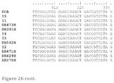
- FIG. 26 shows the amino acids sequences of VH (SEQ ID NOs: 137-148) and VL (SEQ ID NOs: 161-172) chains of antibodies of the DH270 lineage, and nucleic acid sequences (SEQ ID NOs: 125-136 (VH); SEQ ID NOs: 149-160 (VL)) encoding these amino acids. CDRs are highlighted and underlined in the UCA.
- FIG. 27A shows amino acid (SEQ ID Nos: 173 and 174) and nucleic acid sequences (SEQ ID Nos: 175 and 176) of CD4bs antibody CH557.
- FIG. 27B shows amino acid sequences of VH chains of antibodies from CH235 lineage (SEQ ID NOs:177-188).
- FIG. 27C shows amino acid sequences of VL chains of antibodies from CH235 lineage (SEQ ID NOs: 189-198).
- FIG. 28A shows neutralization Breadth and Potency of Plasma and Memory B cell (MBC)-derived MPER bnAbs.
- FIGS. 29A and B show neutralization data from TZM-bl assay (Titer in TZM.bl cells (ug/ml) for DH512_K3 and other chimeric antibodies compared to DH512 and 10E8.
- the data in the first column is historic data when DH512 was run in this panel previously.
- DH512 was run at the same time as DH512_K3 but is listed as Ab510049 in this assay; therefore, data from columns DH512_K3 and AA&AB DH512/Ab510049 should be compared.
- FIG. 30 shows positions in the VHCDR3 chain of DH511 (SEQ ID NO: 471) which could be mutated. Amino acid positions refer to Kabat numbering. Most mutations are to changes to W, but F, L or possibly other substitutions can be tried.
- FIG. 31 shows positions in the VHCDR3 chain of DH512 (SEQ ID NO: 472) which could be mutated.
- Amino acid positions refer to Kabat numbering for the DH512VH chain: QVQLVQSGGGLVKPGGSLTLSCSASGFFFDNSWMGWVRQAPGKGLEWVGRIRRLKDGAT GEYGAAVKDRFTISRDDSRNMLYLHMRTLKTEDSGTYYCTMDEGTPVTRFLEWGYFYYY MAVWGRGTTVIVSS (SEQ ID NO: 469).
- Most mutations are to changes to W, but F, L or possibly other substitutions can also be tried.
- Position V100 can be changed to I.
- Position L100d can be changed to F.
- FIG. 32 shows positions outside of VHCDR3 which could be mutated (SEQ ID NOS 473-478, respectively, in order of appearance). Most mutations are to changes to W, but F, L or possibly other substitutions can also be tried.
- FIG. 33 shows amino acid sequences (SEQ ID NOs: 199-216) of some of the DH512 mutants from FIG. 31 .
- FIG. 34 shows neutralization data for a set of 16 mutations from FIG. 31 .
- DH512 is referred to as DH512 (Ab510049_4A): its heavy chain is H510049_4 and its light chain is K510032
- FIG. 35 shows summary of anti-cardiolipin activity of various antibodies as measured by QUANTA Lite® ACA IgG III kit. Data plotted are representative of 2 independent experiments. mAb were run in duplicate in the second assay. Mean error and standard deviation are shown. Data were consistent between assays. Dotted line indicates positivity cut-off of 0.18. mAbs with OD values above 0.18 are bolded in the figure legend (DH514, DH518-315 HC, DH511-I6-4a through DH511_I1_4A; 4E10).
- FIG. 36 shows a summary of self-reactivity data of MPER antibodies.
- FIG. 37 shows summary results of neutralization data of DH512 and 10E8 against a panel of HIV-1 isolates in the TZMbl pseudovirus neutralization assay. Values represent IC50 in ⁇ g/ml. FIG. 37 also shows the mean IC50 and percent of isolates neutralized at different IC50 values.
- FIG. 38 shows summary results of neutralization data of DH512 and 10E8 against a panel of HIV-1 isolates in the TZMbl pseudovirus neutralization assay. Values represent IC80 in ⁇ g/ml. FIG. 38 also shows the mean IC80 and percent of isolates neutralized at different IC80 values.
- FIG. 39 shows Experimental Overview of Paired VH-VL Sequencing and antibody identification (Example 10).
- V gene repertoire sequencing Identification of individual monoclonal antibodies requires the generation of a sample-specific database of IgG VH sequences constructed by next-generation sequencing of mature B cells isolated from the PBMCs of the donor. Reads are processed bioinformatically to obtain a database of unique VH sequences, which then are clustered into clonotypes according to their CDR3 sequences. The obtained database is used to interpret the MS spectra.
- F(ab)2 purification and proteomic analysis F(ab)2 fragments are prepared from total serum IgG and subjected to antigen-affinity chromatography (monomeric gp120).
- Proteins in the elution and flow-through are denatured and reduced, alkylated, trypsin-digested and analyzed by high resolution LC-MS/MS. Spectra are interpreted with the sample-specific VH database and peptides uniquely associated with a single CDR3 are used to identify full-length VH sequences.
- FIG. 40 shows MPER BnAb DH511 Clonal Lineage Derived from African Individual CH0210 (the heavy chain for DH511_1A is not included).
- FIG. 41 shows Neutralization Activity (IC50) of MPER Antibodies Identified by Paired VH:VL Sequencing Technology (Example 10). Summary data of two independent assays.
- FIG. 42 shows Neutralization Activity (IC80) of MPER Antibodies Identified by Paired VH:VL Sequencing Technology (Example 10). Summary data of two independent assays.
- FIG. 43 shows Nucleotide Alignment of MPER Antibody Heavy Chain Sequences (SEQ ID NOs: 217-229).
- FIG. 44 shows Amino Acid Alignment of MPER Antibody Heavy Chain Sequences (SEQ ID NOs: 230-242).
- FIG. 45 shows Nucleotide Alignment of MPER Antibody Light Chain Sequences (SEQ ID NOs: 243-252).
- FIG. 46 shows Amino Acid Alignment of MPER Antibody Light Chain Sequences (SEQ ID NOs: 253-262).
- FIG. 47 shows Immunogenetic Characteristics of MPER Antibodies—Original Pairings.
- FIG. 48 shows epitope mapping of antibodies of Example 10. Binding to various MPER peptides in an ELISA assay was used to map the epitopes of these MPER antibodies.
- FIG. 49 show epitope mapping of antibodies of Example 10. Binding to various MPER peptides in an ELISA assay was used to map the epitopes of these MPER antibodies.
- FIG. 50 show epitope mapping of antibodies of Example 10. Binding to various MPER peptides in an ELISA assay was used to map the epitopes of these MPER antibodies.
- FIG. 51 show epitope mapping of antibodies of Example 10. Binding to various MPER peptides in an ELISA assay was used to map the epitopes of these MPER antibodies.
- FIG. 52 show epitope mapping of antibodies of Example 10. Binding to various MPER peptides in an ELISA assay was used to map the epitopes of these MPER antibodies.
- FIG. 53 shows Poly/Autoreactivity analysis of DH511_5a.
- Antibody DH511_5a appears to be autoreactive with one protein (NUDC).
- FIG. 54 shows Poly/Autoreactivity analysis of DH511_5b. Antibody DH511_5b appears to be polyreactive.
- FIG. 55 shows Antibody Pairings—Heavy and Light Chain Chimeric Antibodies from Example 11.
- FIG. 56A shows neutralization activity of Heavy and Light Chain Chimeric Antibodies chimeric pairings 1-32 (from FIG. 55 ).
- FIG. 56B shows Neutralization Activity on New Pairings in rows 33-67 (from FIG. 55 ).
- FIG. 56C shows Neutralization Activity on New Pairings in rows 68-91 (from FIG. 55 ).
- FIG. 56D shows that 8 chimeric antibodies were selected for large scale expression and neutralization activity analysis.
- FIG. 57 shows nucleic acid and amino acid sequences of VH and VL sequences of antibodies from Example 10 (SEQ ID NOs: 263-300).
- FIG. 58 shows sequences of DH511_5a and 5b as Fabs (SEQ ID NOs: 301-304).
- FIGS. 59A-F show isolation of MPER-directed broadly neutralizing antibodies.
- FIGS. 60A-E shows structural analysis of the DH511 lineage.
- FIGS. 61A-E shows comparison with other MPER-specific antibodies.
- VH3-15 contacting residues positions that are shared by antibodies DH511.1 and DH511.2 and antibody 10E8 are colored cyan.
- FIGS. 62A-C show standard experimental mapping and neutralization-based epitope prediction analysis to delineate the specificities that mediate plasma neutralization breadth.
- Plasma from donor CH0210 showed potent MPER-directed neutralizing activity against the HIV-2/HIV-1 MPER chimeric pseudovirus C1C. Neutralization titer is reported as median inhibitory dilution (ID50).
- ID50 median inhibitory dilution
- Anti-MPER antibodies were depleted from plasma using MPER peptide-coated magnetic beads. The depleted fraction was tested for neutralization activity against the indicated heterologous viruses. Neutralization was considerably diminished by removal of anti-MPER from both plasmas, indicating that MPER antibodies were largely responsible for neutralization breadth.
- NEP Neutralization-based epitope prediction
- the predicted relative prevalence of antibody clusters [(10 epitopes targeting sites of vulnerability (CD4 binding site, V1/V2, MPER, glycan V3)] is shown as a heat map, with dark color intensity (higher fractional number) corresponding to a stronger neutralization signal. Plasma neutralization breadth is shown, and numbers in each row add up to 1.00. Shown below are the locations on the Env trimer of the epitopes identified by NEP for this donor and confirmed to be targeted by standard experimental mapping methods.
- FIGS. 63A-B show frequency and identity of CDR3 peptides from MPER affinity chromatography.
- FIG. 64 shows Phylogenetic tree of VHDHJH sequences of memory B cell and plasma-derived DH511 clonal lineage members.
- FIGS. 65A and 65B show Epitope mapping by alanine scanning mutagenesis of C-terminal MPER residues. Values listed are mean measurements from two independent experiments. Epitope residues were defined as residues where log AUC relative to wild-type (WT) for alanine mutations was reduced by 50%.
- FIGS. 66A-C show Surface-plasmon resonance analysis of binding of the DH511 clonal lineage to MPR.03 peptide.
- FIG. 66C shows Association (ka) and dissociation (kd) rate constants and binding affinities (Kd) for each Fab.
- FIGS. 67A-C show Surface-plasmon resonance analysis of binding of the DH511 clonal lineage to MPER liposomes (SEQ ID NOs: 321-325).
- FIGS. 68A-C show poly/autoreactivity analysis of MPER bNAbs.
- DH511.1 UCA reacted with ribonucleoprotein
- DH511 I6 reacted with dsDNA.
- PPP1R1C protein phosphatase 1, regulatory (inhibitor) subunit 1C
- FYN FYN oncogene related to SRC, FGR, YES, transcription variant 1 [DH511.1, DH511.3, DH511.6, DH511_I3, DH511_I4]
- NECAP endocytosis associated 1 NECAP1 [DH511.1, DH11.6]
- STAB:BPI fuse-binding protein-interacting repressor, transcription variant 1, mRNA
- STUB 1 STIP1 homology and U-box containing protein 1 [DH511.2, DH511.6]
- STIP1 stress-induced phosphoprotein 1 [DH511_I1, DH511
- FIG. 69 shows ELISA binding of DH511 lineage members to U1 snRNP components.
- the DH511_UCA bound specifically to U1-snRNPA while no binding was observed to the other components. Results shown represent one experiment.
- FIG. 70 shows potential mechanistic differences in binding of 4E10 versus DH511.2/10E8 to MPER liposomes.
- 4E10 bound to MPER656.1 in a biphasic association/dissociation mode and the binding could be fit to a 2-step conformational change model.
- DH512 appears to have a different mechanistic mode and its binding could be fit to a 1:1 Langmuir model.
- FIGS. 71A-C show DH511.2 recognizes a transiently exposed intermediate state of gp41, and the lifetime of DH511.2 epitope exposure is the same as that of 10E8 and 4E10.
- Time course of neutralization of tier 2 HIV-1 isolate B.BG1168 was measured by addition of mAbs to TZM-bl cells pre-incubated with virus. Half-life values were similar among the three antibodies.
- FIG. 72 shows Sequence Comparison of DH511, DH512, and 10E8 HCDR3 Loops (SEQ ID NOs: 326-328). The figure shows that while HCDR3 loops of DH511 and 10E8 lineages are both encoded by D3-3 precursor, substantial differences are observed in their final matured lengths and sequences.
- One conserved sequence motif between DH511/DH512 and 10E8 HCDR3s appears to be a hydrophobic residue doublet at the center of the loop (boxed).
- FIGS. 73A-D shows Structural Comparison of DH511 (A), DH512 (B), and 10E8 (C)HCDR3 Loops. conserveed DH511/DH512 and 10E8 hydrophobic residue doublets at apex of HCDR3 loops are spatially co-localized (D), relative to MPER. Comparison is based on Ca superposition of MPER residues 671-683.
- FIGS. 74A-B shows Comparison of DH511, DH512, and 10E8 HCDR3 Loops.
- the HCDR3 loops of bNabs that target the gp41 MPER have been shown to be critical for their capacity to neutralize the HIV-1 virus, largely through interactions with the viral membrane.
- FIG. 75 shows sequence characteristics of MPER antibodies isolated from memory B cells (SEQ ID NOs: 332-359).
- FIG. 75 corresponds to Supplementary Table 1 as referenced in Example 12.
- FIG. 76 shows neutralization activity of MPER mAbs against a cross-Glade 30 isolate HIV-1 Env-pseudovirus panel (IC50 values).
- FIG. 76 corresponds to Supplementary Table 2a as referenced in Example 12.
- FIG. 77 shows neutralization activity of MPER mAbs against a cross-Glade 30 isolate HIV-1 Env-pseudovirus panel (IC80 values).
- FIG. 77 corresponds to Supplementary Table 2b as referenced in Example 12.
- FIG. 78 shows neutralization activity of DH511.2 against a cross-Glade 199 isolate HIV-1 Env-pseudovirus panel.
- FIG. 78 corresponds to Supplementary Table 3 as referenced in Example 12.
- FIG. 79 shows neutralization activity of DH511.2 against a panel of 200 Glade C HIV-1 primary isolates.
- FIG. 79 corresponds to Supplementary Table 4 as referenced in Example 12.
- FIG. 80 shows neutralization activity of 16 DH511.2 heavy chain mutant antibodies.
- FIG. 80 corresponds to Supplementary Table 27 as referenced in Example 12.
- FIG. 81 shows sequence characteristics and pairing of plasma-derived heavy and light chains identified by mass spectrometry and paired VH-VL next-generation sequencing (SEQ ID NOs: 360-367 and 479-489, respectively, in order of appearance).
- FIG. 81 corresponds to Supplementary Table 6 as referenced in Example 12.
- FIG. 82 shows neutralization activity of 16 plasma mAbs against a 4 indicator HIV-1 Env pseudovirus panel.
- FIG. 82 corresponds to Supplementary Table 7 as referenced in Example 12.
- FIG. 83 shows neutralization activity of plasma mAbs DH511.11P and DH511.12P against a cross-Glade 203 isolate HIV-1 Env-pseudovirus panel.
- FIG. 83 corresponds to Supplementary Table 8 as referenced in Example 12.
- FIG. 84 shows sequences of alanine substituted MPR.03 peptides (SEQ ID NOs: 368-381).
- FIG. 84 corresponds to Supplementary Table 9 as referenced in Example 12.
- FIG. 85 shows sequences of COT6.15 MPER mutant viruses (SEQ ID NOs: 382-403).
- FIG. 85 corresponds to Supplementary Table 10 as referenced in Example 12.
- FIG. 86 shows neutralization Activity against a series of MPER alanine mutant pseudoviruses in the COT6.15 Env background.
- FIG. 86 corresponds to Supplementary Table 11 as referenced in Example 12.
- FIG. 87 shows crystallization peptides (SEQ ID NOs: 404-406).
- FIG. 87 corresponds to Supplementary Table 12 as referenced in Example 12.
- FIG. 88 shows crystallographic data collection and refinement statistics.
- FIG. 88 corresponds to Supplementary Table 13 as referenced in Example 12.
- FIG. 89 shows antibody contact interfaces by CDR loop.
- FIG. 89 corresponds to Supplementary Table 14 as referenced in Example 12.
- FIG. 90 shows bonded and non-bonded contacts DH511.1-MPER. (Non-Kabat numbering).
- FIG. 90 corresponds to Supplementary Table 15 as referenced in Example 12.
- FIG. 91 shows bonded and non-bonded contacts DH511.2-MPER. (Non-Kabat numbering).
- FIG. 91 corresponds to Supplementary Table 16 as referenced in Example 12.
- FIG. 92 shows bonded and non-bonded contacts DH511.11P-MPER.
- FIG. 92 corresponds to Supplementary Table 17 as referenced in Example 12.
- FIG. 93 shows bonded and non-bonded contacts DH511.12P-MPER. (Non-Kabat numbering).
- FIG. 93 corresponds to Supplementary Table 18 as referenced in Example 12.
- FIG. 94 shows neutralization of the DH511 clonal lineage against a panel of 12 global HIV-1 reference strains.
- FIG. 94 corresponds to Supplementary Table 19 as referenced in Example 12.
- FIGS. 95A-C show primers and PCR conditions for paired VH:VL NGS.
- FIG. 95A shows overlap extension oligonucleotides for framework region 1 (5′-3′) (SEQ ID NOs: 407-427).
- FIG. 95B shows overlap extension oligonucleotides for leader peptide (5′-3′) (SEQ ID NOs: 428-441).
- FIG. 95C shows nested constant region oligonucleotides (5′-3′) (SEQ ID NOs: 442-446).
- FIG. 95A corresponds to Supplementary Table 28 as referenced in Example 12.
- FIG. 95B corresponds to Supplementary Table 29 as referenced in Example 12.
- FIG. 95C corresponds to Supplementary Table 30 as referenced in Example 12.
- FIG. 96 shows DH511 clonal lineage membrane insertion scores and HCDR3 analysis (SEQ ID NOs: 447-455).
- the membrane insertion scores can be recalculated to exclude the C in the CDR3.
- HCDR3s score for the .P antibodies will be calculated.
- FIG. 96 corresponds to Supplementary Table 21 as referenced in Example 12.
- FIG. 97 shows cardiolipin reactivity of the DH511 clonal lineage.
- FIG. 97 corresponds to Supplementary Table 22 as referenced in Example 12.
- FIG. 98 shows neutralization activity of 91 chimeric MPER mAbs against the tier 2 HIV-1 isolate B.BG1168.
- FIG. 98 corresponds to Supplementary Table 23 as referenced in Example 12.
- FIG. 99 shows neutralization activity of chimeric mAb DH511.2_K3 against a cross-clade 30 isolate Env-pseudovirus panel.
- FIG. 99 corresponds to Supplementary Table 24 as referenced in Example 12.
- FIGS. 100A-C show primers and PCR conditions for paired VH:VL NGS.
- FIG. 100A shows PCR conditions for isotype specific amplification.
- FIG. 100B shows oligonucleotides for isotype specific amplification (5′-3′) (SEQ ID NOs: 456-462).
- FIG. 100C shows PCR conditions for MiSeq Barcoding.
- FIG. 100A corresponds to Supplementary Table 30 as referenced in Example 12.
- FIG. 100B corresponds to Supplementary Table 31 as referenced in Example 12.
- FIG. 100C corresponds to Supplementary Table 32 as referenced in Example 12.
- HIV envelope antibodies Broadly neutralizing and potent HIV envelope antibodies are now being developed for both prevention of HIV (Rudicell R S et al. J. Virol 88: 12669-82, 2014) and for treatment of HIV infected individuals (Barouch D H, et al. Nature 503: 224-8, 2013; Shingai M et al. Nature 503: 277-80, 2013).
- human recombinant antibodies either alone or in combinations have great prophylactic and therapeutic potential for the prevention and treatment of HIV.
- antibodies that bind with high affinity to Env may be useful in eliminating the latent pool of HIV-infected CD4 T cells and curing HIV, when either used to sensitize HIV expressing target cells with bi specific bnAbs for NK or CD8 T cell killing or when bnAbs are conjugated with toxins or radionucleotides.
- the invention provides fully human antibodies and fragments that specifically bind to and potently neutralize various isolates of HIV-1.
- the antibodies bind to HIV-1 gp41.
- the antibodies of the invention specifically bind the membrane-proximal extracellular region (MPER) of gp41.
- MPER membrane-proximal extracellular region
- the invention provides pharmaceutical compositions including these human antibodies and a pharmaceutically acceptable carrier.
- the invention provides antibodies for passive immunization against HIV/AIDS. Nucleic acids encoding these antibodies, expression cassettes and vectors including these nucleic acids, and isolated cells that express the nucleic acids which encode the antibodies of the invention are also provided.
- the invention provides antibodies which are clonal variants (See e.g., Examples 11, and 12).
- clonal variants are sequences that differ by one or more nucleotides or amino acids, and have a V region with shared mutations compared to the germline, identical VDJ or VJ gene usage, identical the same or similar HCDR3 length, and the same VL and JL usage.
- the germline sequence (unmutated common ancestor “UCA”) is intended to be the sequence coding for the antibody/immunoglobulin (or of any fragment thereof) deprived of mutations, for example somatic mutations.
- Antibodies in a clone that are designate as UCA and/or I are typically not isolated from a biological sample, but are derived computationally based on VH and/or VL sequences isolated from subjects infected with HIV-1.
- compositions including the human antibodies of the invention can be used for any purpose including but not limited to research, diagnostic and therapeutic purposes.
- the human monoclonal antibodies disclosed herein can be used to detect HIV-1 in a biological sample or interfere with the HIV-1 activity, for example to diagnose or treat a subject having an HIV-1 infection and/or AIDS.
- the antibodies can be used to determine HIV-1 titer in a subject.
- the antibodies disclosed herein also can be used to study the biology of the human immunodeficiency virus.
- the antibodies of the invention can be used for therapeutic purposes for treatment or prevention of HIV-1 infection, alone or in combination with other therapeutic modalities, including ART and/or combination with other HIV-1 targeting antibodies, neutralizing antibodies and/or ADCC inducing antibodies.
- the disclosed MPER antibodies specifically bind to a polypeptide disclosed in for example but not limited to FIG. 3 , FIG. 11 , and FIG. 16 , and Example 12.
- the person of ordinary skill in the art will understand that the antibodies of the invention can also bind to gp41MPER residues extending N-terminal or C-terminal to the above sequences.
- residues believed to make contacts with the antibodies of the invention include resides identified in the mapping studies described in for example but not limited to FIGS. 11, 16-15 .
- the antibodies of the invention are expected to make contact with additional gp41 MPER residues.
- the antibodies of the invention are expected to make contact with some of the gp41 MPER residues as previously described for the 10E8 antibody.
- the disclosed antibodies are referred to as 10E8-like antibodies because their binding to the MPER maps to a region similar to the MPER region bound by the 10E8 antibody previously described (See US Pub 20140348785).
- the 10E8 antibody specifically binds the membrane proximal extracellular region (MPER) of gp41 at an epitope that is designated as the 10E8 epitope.
- the crystal structure of the 10E8 antibody was solved in complex with a gp41 peptide (See 20140348785 Example 1), which allowed for detailed analysis of the binding of the 10E8 antibody and gp41, and describe at the atomic level the binding of 10E8 antibody to the 10E8 epitope.
- This epitope and thus the antibodies of this class (10E8-like antibodies), can be distinguished from other antibodies that bound gp41 at other epitopes.
- the 10E8 epitope e.g., KWASLWNWFDITNWLWYIR (SEQ ID NO: 464), extends C-terminal to the 2F5 epitope (although there is some overlap) on the gp41 ectodomain and is distinguished from the 4E10 and Z13E1 epitope by expanding the binding to C-terminal residues previously thought to be inaccessible (e.g. these residues were believed to be buried in the lipid bilayer).
- an MPER antibody of the invention is not the 10E8, 4E10, 2F5 or any other MPER antibody as previously described.
- Some of the difference between certain antibodies of the invention and the 10E8, 4E10 and 2F5 antibodies are demonstrated in FIG. 15 (VH sequence alignment) and FIGS. 6, and 7 (neutralization breadth and potency), and for example but not limited to FIGS. 11, 16-25 (epitope mapping studies), Example 12.
- the inventive antibodies bind an MPER epitope which comprises D674 (See FIG. 11 ).
- the 10E8 antibody See US Pub 20140348785) MPER binding is not sensitive to D674S mutation.
- the DH511 lineage antibodies ( FIG. 6 ) neutralize 100% of isolates whereas 10E8 did not ( FIG. 7 ).
- the antibodies of the invention are expected not to exhibit self-reactivity—they do not bind or bind very weakly to self-antigens, such as human protein.
- self-antigens such as human protein.
- FIGS. 35-36 Example 12
- Various assays to determine poly and autoreactivity are known in the art.
- the neutralization breadth of the inventive antibodies is demonstrated by the diversity of viruses which are neutralized in the TZMbl Env pseudovirus inhibition assay.
- the neutralization breadth and/or binding of the antibodies of the invention can be maintained in the presence of tolerate changes to the epitope. Comparing the sequences of the neutralized viruses, versus viruses that are not neutralized, a skilled artisan can readily determine the % virus changes, including changes in the MPER region and the epitope, which can be tolerated while neutralization and/or binding is maintained.
- sequence identity compare sequence length and determine the % sequence identity and/or changes, including % sequence identity and/or changes in the VH and VL sequences, including % sequence identity and/or changes in the CDRs, as well as the specific positions and types of substitutions which can be tolerated while neutralization potency and breadth is maintained.
- sequence identity is frequently measured in terms of percentage identity (or similarity or homology); the higher the percentage, the more similar the two sequences are.
- homology is frequently measured in terms of percentage identity (or similarity or homology); the higher the percentage, the more similar the two sequences are.
- homology is frequently measured in terms of percentage identity (or similarity or homology); the higher the percentage, the more similar the two sequences are.
- homologs or variants of a polypeptide will possess a relatively high degree of sequence identity when aligned using standard methods.
- NCBI Basic Local Alignment Search Tool (BLAST) (Altschul et al., J. Mol. Biol. 215:403, 1990) is available from several sources, including the National Center for Biotechnology Information (NCBI, Bethesda, Md.) and on the internet, for use in connection with the sequence analysis programs blastp, blastn, blastx, tblastn and tblastx. A description of how to determine sequence identity using this program is available on the NCBI website on the internet.
- Homologs and variants of a VL or a VH of an antibody that specifically binds a polypeptide are typically characterized by possession of at least about 75%, for example at least about 80%, 85%, 90%, 91%, 92%, 93%, 94%, 95%, 96%, 97%, 98% or 99% sequence identity counted over the full length alignment with the amino acid sequence of interest. Proteins with even greater similarity to the reference sequences will show increasing percentage identities when assessed by this method, such as at least 80%, at least 85%, at least 90%, at least 95%, at least 98%, or at least 99% sequence identity.
- homologs and variants When less than the entire sequence is being compared for sequence identity, homologs and variants will typically possess at least 80% sequence identity over short windows of 10-20 amino acids, and may possess sequence identities of at least 85% or at least 90% or 95% depending on their similarity to the reference sequence. Methods for determining sequence identity over such short windows are available at the NCBI website on the internet. One of skill in the art will appreciate that these sequence identity ranges are provided for guidance only; it is entirely possible that strongly significant homologs could be obtained that fall outside of the ranges provided.
- the invention provides antibodies which are 99%, 98%, 97%, 96%, 95%, 94%, 93%, 92%, 91%, 90%, 89%, 88%, 87%, 86%, 85%, 84%, 83%, 82%, 81%, 80% identical to the VH and VL amino acid sequences of the antibodies described herein and still maintain the neutralization breadth, biding and/or potency.
- the invention provides antibodies which are 99%, 98%, 97%, 96%, 95%, 94%, 93%, 92%, 91%, 90%, 89%, 88%, 87%, 86%, 85%, 84%, 83%, 82%, 81%, 80% identical to the CDR1, 2, and/or 3 of VH and CDR1, 2, and/or 3 VL amino acid sequences of the antibodies described herein and still maintain the neutralization breadth, biding and/or potency.
- the invention provides antibodies which can tolerate a larger percent variation in the sequences outside of the VH and/VL sequences of the antibodies.
- the invention provides antibodies which are 99%, 98%, 97%, 96%, 95%, 94%, 93%, 92%, 91%, 90%, 89%, 88%, 87%, 86%, 85%, 84%, 83%, 82%, 81%, 80%, 79%, 78%, 77%, 76%, 75%, 74%, 73%, 72%, 71%, 70%, 69%, 68%, 67%, 66%, 65% identical, wherein the identity is outside of the VH or VL regions, or the CDRs of the VH or VL chains of the antibodies described herein.
- Antibodies exist, for example as intact immunoglobulins and antigen binding variants or fragments e,g. as a number of well characterized produced by digestion with various peptidases. For instance, Fabs, Fvs, scFvs that specifically bind to gp41 or fragments of gp41 would be gp41-specific binding agents. Binding specificity can be determined by any suitable assay in the art, for example but not limited competition binding assays, epitope mapping, etc.
- a scFv protein is a fusion protein in which a light chain variable region of an immunoglobulin and a heavy chain variable region of an immunoglobulin are bound by a linker, while in dsFvs, the chains have been mutated to introduce a disulfide bond to stabilize the association of the chains.
- Provided are also genetically engineered forms such as chimeric antibodies and heteroconjugate antibodies such as bispecific antibodies. See also, Pierce Catalog and Handbook, 1994-1995 (Pierce Chemical Co., Rockford, Ill.); Kuby, Immunology, 3.sup.rd Ed., W.H. Freeman & Co., New York, 1997.
- the invention provides antibody fragments, which have the binding specificity and/or properties of the inventive antibodies.
- Non-limiting examples include: (1) Fab, the fragment which contains a monovalent antigen-binding fragment of an antibody molecule produced by digestion of whole antibody with the enzyme papain to yield an intact light chain and a portion of one heavy chain; (2) Fab′, the fragment of an antibody molecule obtained by treating whole antibody with pepsin, followed by reduction, to yield an intact light chain and a portion of the heavy chain; two Fab′ fragments are obtained per antibody molecule; (3) (Fab′).sub.2, the fragment of the antibody obtained by treating whole antibody with the enzyme pepsin without subsequent reduction; (4) F(ab′).sub.2, a dimer of two Fab′ fragments held together by two disulfide bonds; (5) Fv, a genetically engineered fragment containing the variable region of the light chain and the variable region of the heavy chain expressed as two chains; and (6) single chain antibody (“SCA”), a genetically engineered molecule containing the variable region
- VH refers to the variable region of an immunoglobulin heavy chain, including but not limited to that of an antibody fragment, such as Fv, scFv, dsFv or Fab.
- VL refers to the variable region of an immunoglobulin light chain, including but not limited to that of an Fv, scFv, dsFv or Fab.
- nucleic acids encoding any of the antibodies, or fragment thereof can be expressed in a recombinantly engineered cell such as bacteria, plant, yeast, insect and mammalian cells.
- the nucleic acid sequences include any sequence necessary for expression, including but not limited to a promoter, a leader sequence.
- These antibodies can be expressed as individual VH and/or VL chain, or can be expressed as a fusion protein.
- the antibodies can be expressed by viral vector mediated delivery of genes encoding the antibodies of the invention (See e.g. Yang et al. Viruses 2014, 6, 428-447).
- the VH- and VL-encoding DNA fragments are operatively linked to another fragment encoding a flexible linker, e.g., encoding the amino acid sequence (Gly 4 -Ser) 3 (SEQ ID NO: 470), such that the VH and VL sequences can be expressed as a contiguous single-chain protein, with the VH and VL domains joined by the flexible linker (see, e.g., Bird et al., Science 242:423-426, 1988; Huston et al., Proc. Natl. Acad. Sci. USA 85:5879-5883, 1988; McCafferty et al., Nature 348:552-554, 1990).
- a cleavage site can be included in a linker, such as a furin cleavage site.
- a single chain antibody may be monovalent, if only a single VH and VL are used, bivalent, if two VH and VL are used, or polyvalent, if more than two VH and VL are used.
- Bispecific or polyvalent antibodies may be generated that bind specifically to gp120 and to another molecule, such as gp41.
- E. coli E. coli
- other bacterial hosts yeast
- various higher eukaryotic cells such as the COS, CHO, HeLa and myeloma cell lines.
- the invention provides monoclonal antibodies.
- the monoclonal antibodies are produced by a clone of B-lymphocytes.
- the monoclonal antibody is a recombinant and is produced by a host cell into which the light and heavy chain genes of a single antibody have been transfected. Any suitable cell could be used for transfection and expression of the antibodies of the invention. Suitable cell lines include without limitation 293T cells or CHO cells.
- Monoclonal antibodies are produced by any suitable method known to those of skill in the art.
- monoclonal antibodies are produced by immortalizing B-cell expressing an antibody. Methods for immortalizing B-cells are known in the art, for example but not limited to using EBV transformation, treatment with various stimulants, and/or apoptotic inhibitors (Bonsignori et al. J. Virol. 85: 9998-10009, 2011).
- monoclonal antibodies are produced by making hybrid antibody-forming cells from a fusion of myeloma cells with immune spleen cells to make hybridomas.
- monoclonal antibodies are isolated from a subject, for example but not limited as described in Example 1 (Liao H X et al. J Virol Methods. 2009 June; 158(1-2):171-9). The amino acid and nucleic acid sequences of such monoclonal antibodies can be determined.
- the antibodies described herein, or fragments thereof, may be recombinantly produced in prokaryotic or eukaryotic expression systems. These systems are well described in the art.
- protein therapeutics are produced from mammalian cells.
- the most widely used host mammalian cells are Chinese hamster ovary (CHO) cells and mouse myeloma cells, including NSO and Sp2/0 cells.
- CHO-K1 and CHO pro-3 Two derivatives of the CHO cell line, CHO-K1 and CHO pro-3, gave rise to the two most commonly used cell lines in large scale production, DUKX-X11 and DG44.
- Kim, J., et al. “CHO cells in biotechnology for production of recombinant proteins: current state and further potential,” Appl.
- Other mammalian cell lines for recombinant antibody expression include, but are not limited to, COS, HeLa, HEK293T, U2OS, A549, HT1080, CAD, P19, NIH 3T3, L929, N2a, HEK 293, MCF-7, Y79, SO-Rb50, HepG2, J558L, and BHK. If the aim is large-scale production, the most currently used cells for this application are CHO cells. Guidelines to cell engineering for mAbs production were also reported.
- the invention provides an antibody, or antibody fragment, that is recombinantly produced from a mammalian cell-line, including a CHO cell-line.
- the invention provides a composition comprising an antibody, or antibody fragment, wherein the antibody or antibody fragment was recombinantly produced in a mammalian cell-line, and wherein the antibody or antibody fragment is present in the composition at a concentration of at least 1, 10, 100, 1000 micrograms/mL, or at a concentration of at least 1, 2, 3, 4, 5, 6, 7, 8, 9, 10, 20, 50, or 100 milligrams/mL.
- the antibody composition comprises less than 1, 2, 3, 4, 5, 10, 15, 20, 25, 30, 35, 50, or 100 nanograms of host cell protein (i.e., proteins from the cell-line used to recombinantly produce the antibody)).
- the antibody composition comprises less than 1, 2, 3, 4, 5, 6, 7, 8, 9, 10, 15, 20, or 25 ng of protein A per milligram of antibody or antibody fragment (i.e., protein A is a standard approach for purifying antibodies from recombinant cell culture, but steps should be done to limit the amount of protein A in the composition, as it may be immunogenic).
- protein A is a standard approach for purifying antibodies from recombinant cell culture, but steps should be done to limit the amount of protein A in the composition, as it may be immunogenic.
- U.S. Pat. No. 7,458,704 Reduced protein A leaching during protein A affinity chromatography; which is hereby incorporated-by-reference.
- the antibodies of the invention can be of any isotype.
- the antibodies of the invention can be used as IgG1, IgG2, IgG3, IgG4, whole IgG1 or IgG3s, whole monomeric IgAs, dimeric IgAs, secretory IgAs, IgMs as monomeric, pentameric or other polymer forms of IgM.
- the class of an antibody comprising the VH and VL chains described herein can be specifically switched to a different class of antibody by methods known in the art.
- the nucleic acid encoding the VH and VL can encode an Fc domain (immunoadhesin).
- the Fc domain can be an IgA, IgM or IgG Fc domain.
- the Fc domain can be an optimized Fc domain, as described in U.S. Published Patent Application No. 20100093979, incorporated herein by reference.
- the immunoadhesin is an IgG1 Fc. In one example, the immunoadhesin is an IgG3 Fc.
- the antibodies comprise amino acid alterations, or combinations thereof, for example in the Fc region outside of epitope binding, which alterations can improve their properties.
- Fc modifications are known in the art. Amino acid numbering is according to the EU Index in Kabat.
- the invention contemplates antibodies comprising mutations that affect neonatal Fc receptor (FcRn) binding, antibody half-life, and localization and persistence of antibodies at mucosal sites. See e.g. Ko S Y et al., Nature 514: 642-45, 2014, at FIG. 1 a and citations therein; Kuo, T.
- the antibodies comprise AAAA substitution in and around the Fc region of the antibody that has been reported to enhance ADCC via NK cells (AAA mutations) containing the Fc region aa of S298A as well as E333A and K334A (Shields R I et al JBC, 276: 6591-6604, 2001) and the 4 th A (N434A) is to enhance FcR neonatal mediated transport of the IgG to mucosal sites (Shields R I et al. ibid).
- Other antibody mutations have been reported to improve antibody half-life or function or both and can be incorporated in sequences of the antibodies. These include the DLE set of mutations (Romain G, et al.
- modifications such as but not limited to antibody fucosylation, may affect interaction with Fc receptors (See e.g. Moldt, et al. JVI 86(11): 66189-6196, 2012).
- the antibodies can comprise modifications, for example but not limited to glycosylation, which reduce or eliminate polyreactivity of an antibody. See e.g. Chuang, et al. Protein Science 24: 1019-1030, 2015.
- the antibodies can comprise modifications in the Fc domain such that the Fc domain exhibits, as compared to an unmodified Fc domain enhanced antibody dependent cell mediated cytotoxicity (ADCC); increased binding to Fc.gamma.RIIA or to Fc.gamma RIIIA; decreased binding to Fc.gamma.RIIB; or increased binding to Fc.gamma.RIIB See e.g. US Pub 20140328836.
- ADCC antibody dependent cell mediated cytotoxicity
- antibodies of the invention including but not limited to antibodies comprising a CDR(s) of VH and/or VL chains, or antibody fragments of the inventive antibodies can be used as the HIV-1 binding arm(s) of a bispecific molecule, e.g. DARTS, diabodies, toxin labeled HIV-1 binding molecules.
- a bispecific molecule e.g. DARTS, diabodies, toxin labeled HIV-1 binding molecules.
- either the intact antibody or a fragment thereof can be used.
- Either single chain Fv, bispecific antibody for T cell engagement, or chimeric antigen receptors can be used (Chow et al, Adv. Exp. Biol. Med. 746:121-41 (2012)). That is, in non-limiting embodiments, intact antibody, a Fab fragment, a diabody, or a bispecific whole antibody can be used to inhibit HIV-1 infection in a subject (e.g., a human).
- a bispecific F(ab) 2 can also be used with one arm a targeting molecule like CD3 to deliver it to T cells and the other arm the arm of the native antibody (Chow et al, Adv. Exp. Biol. Med.
- Toxins that can be bound to the antibodies or antibody fragments described herein include unbound antibody, radioisotopes, biological toxins, boronated dendrimers, and immunoliposomes (Chow et al, Adv. Exp. Biol. Med. 746:121-41 (2012)). Toxins (e.g., radionucleotides or other radioactive species) can be conjugated to the antibody or antibody fragment using methods well known in the art (Chow et al, Adv. Exp. Biol. Med. 746:121-41 (2012)).
- the invention also includes variants of the antibodies (and fragments) disclosed herein, including variants that retain the ability to bind to recombinant Env protein, the ability to bind to the surface of virus-infected cells and/or ADCC-mediating properties of the antibodies specifically disclosed, and methods of using same to, for example, reduce HIV-1 infection risk.
- Combinations of the antibodies, or fragments thereof, disclosed herein can also be used in the methods of the invention.
- Antibodies of the invention and fragments thereof can be produced recombinantly using nucleic acids comprising nucleotide sequences encoding VH and VL sequences selected from those shown in the figures and examples.
- the invention provides intact/whole antibodies.
- the invention provides antigen binding fragments thereof. Typically, fragments compete with the intact antibody from which they were derived for specific binding to the target including separate heavy chains, light chains Fab, Fab′, F(ab′).sub.2, F(ab)c, diabodies, Dabs, nanobodies, and Fv. Fragments can be produced by recombinant DNA techniques, or by enzymatic or chemical separation of intact immunoglobulins.
- the invention provides a bispecific antibody.
- a bispecific or bifunctional/dual targeting antibody is an artificial hybrid antibody having two different heavy/light chain pairs and two different binding sites (see, e.g., Romain Rouet & Daniel Christ “Bispecific antibodies with native chain structure” Nature Biotechnology 32, 136-137 (2014); Garber “Bispecific antibodies rise again” Nature Reviews Drug Discovery 13, 799-801 (2014), FIG. 1 a ; Byrne et al. “A tale of two specificities: bispecific antibodies for therapeutic and diagnostic applications” Trends in Biotechnology, Volume 31, Issue 11, November 2013, Pages 621-632 Songsivilai and Lachmann, Clin. Exp.
- the bispecific antibody is a whole antibody of any isotype. In other embodiments it is a bispecific fragment, for example but not limited to F(ab) 2 fragment. In some embodiments, the bispecific antibodies do not include Fc portion, which makes these diabodies relatively small in size and easy to penetrate tissues.
- the bispecific antibodies could include Fc region.
- Fc bearing diabodies for example but not limited to Fc bearing DARTs are heavier, and could bind neonatal Fc receptor, increasing their circulating half-life. See Garber “Bispecific antibodies rise again” Nature Reviews Drug Discovery 13, 799-801 (2014), FIG. 1 a ; See US Pub 20130295121, incorporated by reference in their entirety.
- the invention encompasses diabody molecules comprising an Fc domain or portion thereof (e.g. a CH2 domain, or CH3 domain).
- the Fc domain or portion thereof may be derived from any immunoglobulin isotype or allotype including, but not limited to, IgA, IgD, IgG, IgE and IgM.
- the Fc domain (or portion thereof) is derived from IgG.
- the IgG isotype is IgG1, IgG2, IgG3 or IgG4 or an allotype thereof.
- the diabody molecule comprises an Fc domain, which Fc domain comprises a CH2 domain and CH3 domain independently selected from any immunoglobulin isotype (i.e. an Fc domain comprising the CH2 domain derived from IgG and the CH3 domain derived from IgE, or the CH2 domain derived from IgG1 and the CH3 domain derived from IgG2, etc.).
- the Fc domain may be engineered into a polypeptide chain comprising the diabody molecule of the invention in any position relative to other domains or portions of the polypeptide chain (e.g., the Fc domain, or portion thereof, may be c-terminal to both the VL and VH domains of the polypeptide of the chain; may be n-terminal to both the VL and VH domains; or may be N-terminal to one domain and c-terminal to another (i.e., between two domains of the polypeptide chain)).
- the Fc domain, or portion thereof may be c-terminal to both the VL and VH domains of the polypeptide of the chain; may be n-terminal to both the VL and VH domains; or may be N-terminal to one domain and c-terminal to another (i.e., between two domains of the polypeptide chain)).
- the present invention also encompasses molecules comprising a hinge domain.
- the hinge domain be derived from any immunoglobulin isotype or allotype including IgA, IgD, IgG, IgE and IgM.
- the hinge domain is derived from IgG, wherein the IgG isotype is IgG1, IgG2, IgG3 or IgG4, or an allotype thereof.
- the hinge domain may be engineered into a polypeptide chain comprising the diabody molecule together with an Fc domain such that the diabody molecule comprises a hinge-Fc domain.
- the hinge and Fc domain are independently selected from any immunoglobulin isotype known in the art or exemplified herein.
- a polypeptide chain of the invention comprises a hinge domain, which hinge domain is at the C-terminus of the polypeptide chain, wherein the polypeptide chain does not comprise an Fc domain.
- a polypeptide chain of the invention comprises a hinge-Fc domain, which hinge-Fc domain is at the C-terminus of the polypeptide chain.
- a polypeptide chain of the invention comprises a hinge-Fc domain, which hinge-Fc domain is at the N-terminus of the polypeptide chain.
- the invention encompasses multimers of polypeptide chains, each of which polypeptide chains comprise a VH and VL domain, comprising CDRs as described herein.
- the VL and VH domains comprising each polypeptide chain have the same specificity, and the multimer molecule is bivalent and monospecific.
- the VL and VH domains comprising each polypeptide chain have differing specificity and the multimer is bivalent and bispecific.
- the polypeptide chains in multimers further comprise an Fc domain.
- Fc domains Dimerization of the Fc domains leads to formation of a diabody molecule that exhibits immunoglobulin-like functionality, i.e., Fc mediated function (e.g., Fc-Fc.gamma.R interaction, complement binding, etc.).
- Fc mediated function e.g., Fc-Fc.gamma.R interaction, complement binding, etc.
- diabody molecules of the invention encompass tetramers of polypeptide chains, each of which polypeptide chain comprises a VH and VL domain.
- two polypeptide chains of the tetramer further comprise an Fc domain.
- the tetramer is therefore comprised of two ‘heavier’ polypeptide chains, each comprising a VL, VH and Fc domain, and two ‘lighter’ polypeptide chains, comprising a VL and VH domain. Interaction of a heavier and lighter chain into a bivalent monomer coupled with dimerization of the monomers via the Fc domains of the heavier chains will lead to formation of a tetravalent immunoglobulin-like molecule.
- the monomers are the same, and the tetravalent diabody molecule is monospecific or bispecific. In other aspects the monomers are different, and the tetra valent molecule is bispecific or tetraspecific.
- Formation of a tetraspecific diabody molecule as described supra requires the interaction of four differing polypeptide chains. Such interactions are difficult to achieve with efficiency within a single cell recombinant production system, due to the many variants of potential chain mispairings.
- One solution to increase the probability of mispairings is to engineer “knobs-into-holes” type mutations into the desired polypeptide chain pairs. Such mutations favor heterodimerization over homodimerization.
- an amino acid substitution (preferably a substitution with an amino acid comprising a bulky side group forming a ‘knob’, e.g., tryptophan) can be introduced into the CH2 or CH3 domain such that steric interference will prevent interaction with a similarly mutated domain and will obligate the mutated domain to pair with a domain into which a complementary, or accommodating mutation has been engineered, i.e., ‘the hole’ (e.g., a substitution with glycine).
- Such sets of mutations can be engineered into any pair of polypeptides comprising the diabody molecule, and further, engineered into any portion of the polypeptides chains of the pair.
- the invention also encompasses diabody molecules comprising variant Fc or variant hinge-Fc domains (or portion thereof), which variant Fc domain comprises at least one amino acid modification (e.g. substitution, insertion deletion) relative to a comparable wild-type Fc domain or hinge-Fc domain (or portion thereof).
- Molecules comprising variant Fc domains or hinge-Fc domains (or portion thereof) e.g., antibodies
- the variant phenotype may be expressed as altered serum half-life, altered stability, altered susceptibility to cellular enzymes or altered effector function as assayed in an NK dependent or macrophage dependent assay.
- Fc domain variants identified as altering effector function are known in the art. For example International Application WO04/063351, U.S. Patent Application Publications 2005/0037000 and 2005/0064514.
- the bispecific diabodies of the invention can simultaneously bind two separate and distinct epitopes.
- the epitopes are from the same antigen.
- the epitopes are from different antigens.
- at least one epitope binding site is specific for a determinant expressed on an immune effector cell (e.g. CD3, CD16, CD32, CD64, etc.) which are expressed on T lymphocytes, natural killer (NK) cells or other mononuclear cells.
- an immune effector cell e.g. CD3, CD16, CD32, CD64, etc.
- NK natural killer cells or other mononuclear cells.
- the diabody molecule binds to the effector cell determinant and also activates the effector cell.
- diabody molecules of the invention may exhibit Ig-like functionality independent of whether they further comprise an Fc domain (e.g., as assayed in any effector function assay known in the art or exemplified herein (e.g., ADCC assay).
- Fc domain e.g., as assayed in any effector function assay known in the art or exemplified herein (e.g., ADCC assay).
- Non-limiting examples of bispecific antibodies can also be (1) a dual-variable-domain antibody (DVD-Ig), where each light chain and heavy chain contains two variable domains in tandem through a short peptide linkage (Wu et al., Generation and Characterization of a Dual Variable Domain Immunoglobulin (DVD-Ig.TM.) Molecule, In: Antibody Engineering, Springer Berlin Heidelberg (2010)); (2) a Tandab, which is a fusion of two single chain diabodies resulting in a tetravalent bispecific antibody that has two binding sites for each of the target antigens; (3) a flexibody, which is a combination of scFvs with a diabody resulting in a multivalent molecule; (4) a so called “dock and lock” molecule, based on the “dimerization and docking domain” in Protein Kinase A, which, when applied to Fabs, can yield a trivalent bispecific binding protein consisting of two identical Fab fragments linked to
- Examples of platforms useful for preparing bispecific antibodies include but are not limited to BiTE (Micromet), DART (MacroGenics) (e,g, U.S. Pat. No. 8,795,667; U.S. Publication Nos. 2014-0099318; 2013-0295121; 2010-0174053 and 2009-0060910; European Patent Publication No. EP 2714079; EP 2601216; EP 2376109; EP 2158221 and PCT Publications No.
- BiTE Meromet
- DART MicroGenics
- the bispecific antibody comprises an HIV envelope binding fragment, for example but not limited to an HIV envelope binding fragment from any of the antibodies described herein.
- the bispecific antibody further comprises a second antigen-interaction-site/fragment.
- the bispecific antibody further comprises at least one effector domain.
- the bispecific antibodies engage cells for Antibody-Dependent Cell-mediated Cytotoxicity (ADCC).
- ADCC Antibody-Dependent Cell-mediated Cytotoxicity
- the bispecific antibodies engage natural killer cells, neutrophil polymorphonuclear leukocytes, monocytes and macrophages.
- the bispecific antibodies are T-cell engagers.
- the bispecific antibody comprises an HIV envelope binding fragment and CD3 binding fragment.
- CD3 antibodies are known in the art. See for example U.S. Pat. No. 8,784,821.
- the bispecific antibody comprises an HIV envelope binding fragment and CD16 binding fragment.
- the invention provides antibodies with dual targeting specificity.
- the invention provides bi-specific molecules that are capable of localizing an immune effector cell to an HIV-1 envelope expressing cell, so as facilitate the killing of the HIV-1 envelope expressing cell.
- bispecific antibodies bind with one “arm” to a surface antigen on target cells, and with the second “arm” to an activating, invariant component of the T cell receptor (TCR) complex. The simultaneous binding of such an antibody to both of its targets will force a temporary interaction between target cell and T cell, causing activation of any cytotoxic T cell and subsequent lysis of the target cell.
- the immune response is re-directed to the target cells and is independent of peptide antigen presentation by the target cell or the specificity of the T cell as would be relevant for normal MHC-restricted activation of CTLs.
- CTLs are only activated when a target cell is presenting the bispecific antibody to them, i.e. the immunological synapse is mimicked.
- bispecific antibodies that do not require lymphocyte preconditioning or co-stimulation in order to elicit efficient lysis of target cells.
- BiTE bispecific T cell engager
- DART (dual affinity retargeting) molecules are based on the diabody format but feature a C-terminal disulfide bridge for additional stabilization (Moore et al., Blood 117, 4542-51 (2011)).
- the so-called triomabs which are whole hybrid mouse/rat IgG molecules and also currently being evaluated in clinical trials, represent a larger sized format (reviewed in Seimetz et al., Cancer Treat Rev 36, 458-467 (2010)).
- the invention also contemplates bispecific molecules with enhanced pharmacokinetic properties.
- such molecules are expected to have increased serum half-life.
- these are Fc-bearing DARTs (see supra).
- such bispecific molecules comprise one portion which targets HIV-1 envelope and a second portion which binds a second target.
- the first portion comprises VH and VL sequences, or CDRs from the antibodies described herein.
- the second target could be, for example but not limited to an effector cell.
- the second portion is a T-cell engager.
- the second portion comprises a sequence/paratope which targets CD3.
- the second portion is an antigen-binding region derived from a CD3 antibody, optionally a known CD3 antibody.
- the anti-CD antibody induce T cell-mediated killing.
- the bispecific antibodies are whole antibodies.
- the dual targeting antibodies consist essentially of Fab fragments. In other embodiments, the dual targeting antibodies comprise a heavy chain constant region (CH1. In certain embodiments, the bispecific antibody does not comprise Fc region. In certain embodiments, the bispecific antibodies have improved effector function. In certain embodiments, the bispecific antibodies have improved cell killing activity.
- Various methods and platforms for design of bispecific antibodies are known in the art. See for example US Pub. 20140206846, US Pub. 20140170149, US Pub. 20090060910, US Pub 20130295121, US Pub. 20140099318, US Pub. 20140088295 which contents are herein incorporated by reference in their entirety.
- the invention provides human, humanized and/or chimeric antibodies.
- the invention provides a pharmaceutical composition comprising an antibody of the invention wherein the composition is used for therapeutic purposes such as but not limited to prophylaxis, treatments, prevention, and/or cure.
- the invention provides a pharmaceutical composition comprising an antibody of the invention in combination with any other suitable antibody.
- the pharmaceutical compositions comprise nucleic acids which encode the antibodies of the invention. In certain embodiments, these nucleic acids can be expressed by any suitable vector for expression of antibodies. Non-limiting examples include attenuated viral hosts or vectors or bacterial vectors, recombinant vaccinia virus, adenovirus, adeno-associated virus (AAV), herpes virus, retrovirus, cytomegalovirus or other viral vectors can be used to express the antibody.
- compositions include excipient suitable for a biologic molecule such as the antibodies of the invention.
- the antibodies could be produced in specific cell lines and conditions so as to control glycosylation of the antibody.
- the antibody framework for example, could comprise specific modification so as to increase stability of the antibody.
- the invention provides that the antibodies, and fragments thereof, described herein can be formulated as a composition (e.g., a pharmaceutical composition).
- a composition e.g., a pharmaceutical composition
- Suitable compositions can comprise an inventive antibody (or antibody fragment) dissolved or dispersed in a pharmaceutically acceptable carrier (e.g., an aqueous medium).
- the compositions can be sterile and can be in an injectable form (e.g. but not limited to a form suitable for intravenous injection, intramascular injection).
- the antibodies (and fragments thereof) can also be formulated as a composition appropriate for topical administration to the skin or mucosa.
- Such compositions can take the form of liquids, ointments, creams, gels and pastes.
- the antibodies (and fragments thereof) can also be formulated as a composition appropriate for intranasal administration.
- the antibodies (and fragments thereof) can be formulated so as to be administered as a post-coital douche or with a condom. Standard formulation techniques can be used in preparing suitable compositions.
- the antibody (and fragments thereof), described herein have utility, for example, in settings including but not limited to the following:
- the antibodies described herein (or fragments thereof) in the setting of anticipated known exposure to HIV-1 infection, the antibodies described herein (or fragments thereof) and be administered prophylactically (e.g., IV, topically or intranasally) as a microbiocide,
- the antibodies described herein in the setting of known or suspected exposure, such as occurs in the setting of rape victims, or commercial sex workers, or in any homosexual or heterosexual transmission without condom protection, can be administered as post-exposure prophylaxis, e.g., IV or topically, and
- the antibodies described herein in the setting of Acute HIV infection (AHI), can be administered as a treatment for AHI to control the initial viral load or for the elimination of virus-infected CD4 T cells.
- the antibodies (or antibody fragments) described herein can be administered prior to contact of the subject or the subject's immune system/cells with HIV-1 or within about 48 hours of such contact. Administration within this time frame can maximize inhibition of infection of vulnerable cells of the subject with HIV-1.
- various forms of the antibodies described herein can be administered to chronically or acutely infected HIV patients and used to kill remaining virus infected cells by virtue of these antibodies binding to the surface of virus infected cells and being able to deliver a toxin to these reservoir cells.
- Suitable dose ranges can depend on the antibody (or fragment) and on the nature of the formulation and route of administration. Optimum doses can be determined by one skilled in the art without undue experimentation. For example, doses of antibodies in the range of 1-50 mg/kg of unlabeled or labeled antibody (with toxins or radioactive moieties) can be used. If antibody fragments, with or without toxins are used or antibodies are used that can be targeted to specific CD4 infected T cells, then less antibody can be used (e.g., from 5 mg/kg to 0.01 mg/kg).
- the invention provides use of the antibodies of the invention, including bispecific antibodies, in methods of treating and preventing HIV-1 infection in an individual, comprising administering to said individual a therapeutically effective amount of a composition comprising the antibodies of the invention in a pharmaceutically acceptable form.
- the methods include a composition which includes more than one HIV-1 targeting antibody.
- the HIV-1 targeting antibodies in such combination bind different epitopes on the HIV-1 envelope.
- such combinations of bispecific antibodies targeting more than one HIV-1 epitope provide increased killing of HIV-1 infected cells.
- such combinations of bispecific antibodies targeting more than one HIV-1 epitope provide increased breadth in recognition of different HIV-1 subtypes.
- the composition comprising the antibodies of the invention alone or in any combination can be administered via IM, subcutaneous, or IV delivery, or could be deposited at mucosal sites, such as the oral cavity to prevent maternal to child transmission, the rectal space or the vagina as a microbicide.
- the antibodies can be administered locally in the rectum, vagina, or in the oral cavity, and can be formulated as a microbiocide (Hladik F et al ELIFE Elife. 2015 Feb. 3; 4. doi: 10.7554/eLife.04525; Multipurpose prevention technologies for reproductive and sexual health. Stone A. Reprod Health Matters. 2014 November; 22(44):213-7.
- antibodies can be formulated such that the therapeutic antibody or combination thereof is impregnated on a vaginal ring (Chen Y et al. Drug Des. Devel. Ther 8: 1801-15, 2014;Malcolm R K et al BJOG 121 Suppl 5: 62-9, 2014). Antibodies can be administered alone or with anti-retroviral drugs for a combination microbiocide (Hladik F et al ELIFE Elife. 2015 Feb. 3; 4. doi: 10.7554/eLife.04525)
- the antibodies can be administered in complex with a form of HIV Env, optimally gp120, but also an Env trimer, to enhance Env immunogenicity.
- the antibodies can be delivered by viral vector mediated delivery of genes encoding the antibodies of the invention (See e.g. Yang et al. Viruses 2014, 6, 428-447).
- the antibodies can be administered in viral vector, for example but not limited to adenoassociated viral vector, for expression in muscle and plasma.
- antibodies with different binding specificities are combined for use in pharmaceutical compositions and therapeutic methods.
- CD4 binding site antibodies are combined with V3 antibodies, MPER antibodies and so forth.
- FIGS. 8, 9 and 10 show a selection of potent HIV-1 neutralizing antibodies which can be used in pharmaceutical compositions, and therapeutic methods.
- Non-limiting examples of selections of combinations of certain antibodies include: DH542, DH542_L4, DH542_QSA, DH429 and DH512 (or any of the DH512 variants); DH512 and CH31 (See US Publication20140205607); DH512 (or any of the other DH512 variants) and DH540 (See Example 8, and this antibody will be described elsewhere); DH542, DH542_L4, DH542_QSA, DH429, DH512 and DH540; DH542, DH542_L4, DH542_QSA, DH429 and CH557; CH557 and DH512 (or any of the DH512 variants).
- a combination therapy envisions a composition which combines various antibodies.
- a combination therapy is provided wherein antibodies are administered as individual compositions, for example at different times, by different means, or at administered at different locations.
- a combination therapy is provides wherein a therapeutic antibody or antibodies is combined with other therapeutic means, for example anti-retroviral drug cocktails, or drugs which activate latently infected HIV-1 cells.
- the disclosed antibodies or antigen binding fragments thereof are used to determine whether HIV-1 envelope(s) is a suitable antigen for inclusion in a vaccine composition.
- the antibodies can be used to determine whether an antigen in a vaccine composition including a gp41 immunogen assumes a conformation including an epitope bound by the inventive antibodies or fragments thereof.
- This can be readily determined by a method which includes contacting a sample containing the vaccine, such as a gp120 antigen, with a disclosed antibody or antigen binding fragment under conditions sufficient for formation of an immune complex, and detecting the immune complex, to detect an HIV-1 antigen including an epitope of an inventive antibody in the sample.
- the detection of the immune complex in the sample indicates that vaccine component, such as a HIV-1 Env antigen assumes a conformation capable of binding the antibody or antigen binding fragment.
- Chimeric antibodies which combine a heavy and light chain from different antibodies are typically indicated by the designation of the heavy and light chain of each parent antibody.
- FIG. 1 shows the three HIV infected individual plasma that was evaluated for HIV neutralizing activity and the specificities profiled by the Georgiev algorithm (Georgiev I S et al Science 340: 751-6, 2013). From this analysis we found three subjects (CH0210, CH0536, CH1244) with gp41 bnAb activity ( FIG. 1 ).
- VH and VL genes were selected and made in linear cassettes (essentially as described in Liao H X et al. J. Virol. Methods 158: 171-9, 2009, see for example FIG. 1 , Section 3.3) to produce recombinant monoclonal antibodies by transient transfection in 293T cells.
- Pairs of VH and VL genes as selected above can also be used to produce plasmids for stable expression of recombinant antibodies.
- the plasmids or linear constructs for recombinant antibody expression also comprise AAAA substitution in and around the Fc region of the antibody that has been reported to enhance ADCC via NK cells (AAA mutations) containing the Fc region aa of S298A as well as E333A and K334A (Shields R I et al JBC, 276: 6591-6604, 2001) and the 4 th A (N434A) is to enhance FcR neonatal mediated transport of the IgG to mucosal sites (Shields R I et al. ibid).
- the antibodies of the invention were selected based on a combination of criteria including sequence analyses, and functional analyses including but not limited as neutralization breadth, and potency.
- the antibodies of the invention comprise naturally rearranged VH and VL fragment pairs, wherein the rest of the Ig gene is not naturally occurring with the isolated rearranged VH and VL fragments. In certain embodiments, the antibodies of the invention are recombinantly produced by synthesized VH and VL fragment pairs, wherein the rest of the Ig gene is not naturally occurring with the isolated rearranged VH and VL fragments. In certain embodiments, the antibodies of the invention are recombinantly produced by synthe
- FIG. 4 and Example 12 shows a summary of some of the characteristics of the recombinant MPER antibodies of the invention.
- DH511-DH517 are antibodies with VH and VL chains from individual CH0210.
- DH518 is an antibody with VH and VL chains from individual CH0536.
- DH536 is an antibody with VH and VL chains from individual CH1244.
- CH537 is an antibody with VH and VL chains from individual CH0585.
- DH 511-DH516 antibodies are all members of the same B cell clonal lineage ( FIG. 6 ).
- FIG. 5 shows the neutralizing capacity of these antibodies with all but DH536 and DH537 able to neutralize difficult to neutralized (tier 2) HIV strains B.BG1168, C.CH505, and C.DU172).
- FIG. 6 shows the phylogram of the DH511 clonal lineage.
- TZMbl neutralization assay is a standard way to evaluate antibody breadth and potency. See Montefiori, D. Methods Mol Biol. 2009; 485:395-405; HIV-1 Env-pseudoviruses infection of TZM-bl cells. Exemplary pseudovirus neutralization assays and panels of HIV-1 pseudovirus are described for example, in Li et al., J Virol 79, 10108-10125, 2005, Seaman et al, J. Virol., 84:1439-1452, 2010; Sarzotti-Kelsoe et al., J. Immunol. Methods, 409:131-46, 2014; and WO2011/038290, each of which is incorporated by reference herein. Various HIV-1 isolates, both Tier 1 and Tier 2 viruses can be included in this assay.
- the TZMbl assay was conducted to determine neutralization potency and breadth of the various antibodies of the invention on different HIV-1 pseudoviruses.
- FIG. 7 shows the results of neutralization of 8 of the gp41 antibodies against a panel of 30 HIV tier 2 isolates in the TZMbl pseudovirus neutralization assay.
- the DH511 clonal lineage members all neutralized 100% (30/30) isolates while DH517 neutralized 50% and DH518 neutralized 83%. This in contrast to 10E8 gp41 antibody that only neutralized 29/30 isolates.
- FIG. 8 shows the mean IC50, IC80 and percent of isolates neutralized at an IC50 ⁇ 50 ug/ml and at an IC80 of ⁇ 5 ug/ml (confirm).
- mAb DH512 is equally as potent and slightly more broad in neutralization breadth than the mAb 10E8.
- FIG. 9 shows other mAbs and their breadth and potency.
- FIGS. 37, 38, 28, 56 and 34 , and Figures from Example 12 show neutralization data of various antibodies against various panels of pseudoviruses.
- Binding of antibodies to various MPER peptides in an ELISA assay was used to map the epitopes of the MPER antibodies.
- FIG. 11 shows that Antibody epitopes maps to the C-terminus of gp41 to a similar region where 10E8 binds (Huang J et al. Nature 491 406, 2012; See US Pub 20140348785).
- FIGS. 11, 15-25 show binding of antibodies to MPER peptide variants. These mapping studies show that the antibodies of the invention are 10E8 like Abs.
- DH512 shows the broadest and most potent neutralization among the antibodies tested.
- FIG. 12 shows an alanine substituted gp41 peptide set used to map DH517 mab and
- FIG. 13 shows a summary of ala mutants to which the antibody is sensitive for binding to gp41.
- FIGS. 14 and 15 show the VH and VL sequences of the DH511-DH516 antibodies.
- FIGS. 12-13 show the nucleotide and amino acid sequences of all the certain antibodies of the invention.
- FIGS. 16-25 show that DH517 displayed a unique mapping pattern in that it depends on DKW at the N terminus and several residues at the C terminus important for 10E8 binding and neutralization.
- Clone DH511 mAbs bound strongly to the majority of the MPER656 variants, showing decreased binding to MPER656.2 and MPER656.2dYIK683R-biotin. These data indicate that the asparagine at position 674 is critical for binding, thus providing evidence that these mAbs bind at the C-terminus.
- Kd measurements of antibody binding to HIV-1 envelope, e.g. gp41 or any other suitable peptide for the MPER antibodies will be determined by Surface Plasmon Resonance measurements, for example using Biacore, or any other suitable technology which permits detection of interaction between two molecules in a quantitative way.
- AtheNA Multi-Lyte ANA Plus Test System is one such assay.
- ELISA cardiolipin assay is another assay to measure autoreactivity.
- the stability and properties of the antibodies for example as formulated in a composition for treatment will be tested.
- CH557 is one example of a CD4bs broad neutralizing HIV-1 antibody, from a series of clonal antibodies ( FIG. 28 ) which can be used in combination with the antibodies of the invention.
- Antibodies from DH270 lineage are shown in FIG. 26 .
- I1 DH270IA1
- I2, I4, I3 and UCA in FIG. 26 are not isolated from human subjects but are derived computationally based on VH and VL sequences of other observed antibodies from the clone: DH471, DH429, DH473, DH391 and DH270.
- the VH and VL sequences of DH471, DH429, DH473, DH391 and DH270 are derived from a human subject infected with HIV-1.
- VH and VL sequences of DH471, DH429, DH473, DH391 and DH270 are derived essentially as described in Example 1, except that cell were sorted with a different hook.
- DH542, DH542-QSA, DH542_K3 are non-limiting examples of V3 antibodies, which can be used in combination with the antibodies of the invention.
- the nucleotide and amino acid sequences of the VH and VL of DH542 QSA are shown below.
- DH542 QSA antibody has the VH of DH542 and the VL called DH542-QSA
- DH542-L4 is an antibody that has a VH of DH542 and VL of DH429 ( FIG. 26 )
- DH540 antibody is described in detail in U.S. Ser. No. 62/170,558, filed Jun. 3, 2015.
- FIGS. 37 and 38 show the results of neutralization against a panel of HIV isolates in the TZMbl pseudovirus neutralization assay.
- FIGS. 37 and 38 also show the mean IC50, IC80 and percent of isolates neutralized at different IC50 or IC80 values.
- Additional antibodies were isolated from the individual CH0210 by high-throughput sequencing of the paired human immunoglobulin heavy and light chain repertoire. See FIG. 39 .
- B cells were isolated from PBMCs via negative depletion.
- the heavy and light chain transcripts were co-localized on RNA binding beads, and then physically tied together using overlap extension RT-PCR.
- the paired VH:VL amplicons were then used to generate 3 libraries for sequencing: a heavy chain database, a light chain database, and a paired database.
- F(ab)2 fragments were prepared from total serum IgG and subjected to antigen-affinity chromatography using the MPER peptide. Proteins in the elution and flow-through were denatured and reduced, alkylated, trypsin-digested and analyzed by high resolution LC-MS/MS. Spectra were interpreted with the heavy chain database obtained from next-generation sequencing, and peptides uniquely associated with a single CDR (“informative peptides”) were used to identify full-length VH sequences. Clonotypes are defined as VH sequences having the same germline V and J and at least 85% aa identity in the CDRH3.
- VH and VL genes were selected and made in linear cassettes (essentially as described in Liao H X et al. J. Virol. Methods 158: 171-9, 2009, see for example FIG. 1 , Section 3.3) to produce recombinant monoclonal antibodies by transient transfection in 293T cells. See also Example 1 for variations in the backbone.
- This example describes chimeric antibodies comprising non-natural VH and VL chain pairs. Naturally occurring VH or VL chain are combined in non-natural pairs as described in FIG. 55 , chimeras 1-91.
- Chimeras 1-91 were recombinantly expressed and their neutralization profile was determined in the TZMB1 assay ( FIG. 56 ). Based on neutralization data for chimeras 68-91 as shown in FIG. 56 , three antibodies DH512_K2_4A (VH: H510049_4A (DH512) and VL: DH511_1AVK), DH512_K3_4A (VH: H510049_4A (DH512) and VL: DH511_2AVK) and DH512_K4_4A (VH: H510049_4A (DH512) and VL: DH511_5AVK) antibodies were produced large scale and will be tested for neutralization against a larger panel of viruses (see panels for DU512).
- the invention contemplates antibodies which comprise amino acid changes, or combination of such changes, in the VH chains of antibodies form the DH511 lineage.
- Non-limiting examples of antibodies with mutations are provided in FIGS. 30-33 , or any combination thereof.
- Most mutations are to changes to W, but can also try F, L or possibly other substitutions, e.g. without limitation I, V, A.
- Additional mutations include without limitation the following: T100aF; T100aL; T100aI; T100aV; T100aA; L100dW, or any combination thereof.
- such double mutants T100aW-L100dF; T100aW-L100dW; T100aF-L100dF; T100aL-L100dF; T100aL-L100dW.
- Neutralization data for a subset of these antibodies is provided in FIG. 34 .
- the data show that some of the mutations abrogate neutralization while others enhance potency.
- One candidate, DH512_L100dF_4A is more potent than 10E8 and has similar potency to DH512_K3.
- L100d could be changed to Trp.
- FIGS. 34 and 80 show that single mutant L100dF, and single mutant T100aW have improved neutralization. These single mutants will be tested against a panel of additional viruses (see panel for DH512, DH512_K3).
- combination mutations for example but not limited combination T100aW with L100dF, combination L100dW with T100aW.
- VH chain as contemplated above could be combined with VH chain from DH512, or with VH chain from DH512_K3 (DH511_2AVK).
- HIV-1 envelope gp41 membrane-proximal external region (MPER)-specific memory B cell sorting and next-generation sequencing coupled with mass spectrometry analysis of plasma antibodies, we probed the memory B cell and plasma antibody repertoires of an HIV-1-infected donor with a plasma bnAb signature that mapped to Env gp41 distal MPER.
- MPER membrane-proximal external region
- bnAbs broadly reactive neutralizing antibodies
- MPER envelope gp41 membrane proximal external region
- 10E8 and 4E10 the most broad (1, 2).
- Monoclonal antibody (mAb) 4E10 while extremely broad in neutralization breadth, is not potent, and is highly polyreactive with many non-HIV-1 proteins and autoreactive with the human protein splicing factor 3b subunit 3 (SF3B3) (3) as well as with lipids (4).
- mAb 10E8 is not as polyreactive as 4E10, and is both more broad and potent (1), although it does have a degree of lipid reactivity (5) and is autoreactive with the host protein family of sequence similarity 84 member A (FAM84A) (6).
- HIV-1-specific antibodies In HIV-1 infection, 60% of HIV-1-specific antibodies derive from abnormal B cell subsets, that are either activated or exhausted and express Fc receptor-like-4 (FcRL4) (8, 9). However, many of the antibodies reflected in HIV-1 memory B cells are not expressed in plasma (8). Similarly, many of the memory B cell specificities of antibodies in other settings are also not represented in plasma (10-12). Thus, it is not known if envelope-reactive memory B cells with bnAb B cell receptors are a major source of plasma broad neutralizing activity.
- FcRL4 Fc receptor-like-4
- the DH511 B cell clonal lineage was distinguished by HCDR3 loops of 24 amino acids in DH511.1, DH511.3, and DH511.6, while DH511.2, DH511.4, and DH511.5 antibodies had a one amino acid deletion in the HCDR3, resulting in a length of 23 amino acids (Supplementary Table 1).
- V H and V L somatic mutation rates were 15-22% and 14-18%, respectively.
- the DH511 clonal lineage was derived from the same heavy-chain germ line gene as previously isolated gp41 neutralizing antibody 10E8 (V H 3-15), but utilized a different V L germ line gene (DH511: V K 1-39, 10E8: V L 3-19) (1) (Supplementary Table 1).
- Antibody DH517 derived from a second clonal lineage arising from the same donor, was similarly isolated.
- DH517 utilized V H 4-34 and V L 3-19 germ line genes, was 22.8% and 14.3% mutated, respectively, and had a long HCDR3 comprised of 24 amino acids.
- DH511.1-DH511.6 and DH517 mAbs were assessed for neutralization breadth and potency against a panel of 30 cross-Glade HIV-1 isolates. All six DH511 clonal members neutralized 30 of 30 isolates tested with median 50% inhibitory concentrations (IC 50 ) ranging from 0.7 to 4.2 ⁇ g/ml (Supplementary Table 2a). DH517 had less breadth than DH511 clone antibodies, neutralizing 15 of 30 isolates with a median IC 50 of 5.7 ⁇ g/ml (Supplementary Table 2a).
- MPER-specific plasma antibody repertoire was analyzed using an independent proteomics-based approach for the identification and semi-quantitative determination of antigen-specific antibodies in human serum (15, 16).
- MPER-specific antibodies were isolated from a 2 ml plasma sample by affinity chromatography, processed for proteomics (10) and subjected to liquid chromatography high-resolution tandem mass spectrometry (LC-MS/MS) analysis.
- V H database comprising 98,413 unique high quality sequences was derived from a natively paired V H :V L repertoire from 845,000 peripheral single B cells from total PBMCs (isolated using MACS negative selection: CD2 ⁇ CD14 ⁇ CD16 ⁇ CD43 ⁇ CD235a ⁇ ) (17-19). These V H sequences were then clustered into 4,428 clonotypes, using a cut-off of ⁇ 85% amino acid identity in the HCDR3 region.
- Clonotype IV comprised 95% of the total intensity of HCDR3 peptides detected in the MPER-specific antibody repertoire (i.e. in antibodies eluted following affinity chromatography with immobilized MPR.03 peptide); we noted that detection of HCDR1 and HCDR2 peptides unique to Clonotype IV provided further unambiguous support for the prevalence of these antibodies in the CH0210 plasma ( FIG. 59 f ).
- Clonotype II which included antibodies DH511.2, DH511.4 and DH511.5 isolated by single-cell sorting, and Clonotype III were detected at 4% and 1% relative abundancy, respectively ( FIG. 59 f ).
- All three HCDR3 clonotypes utilized the same VDJ genes (V H 3-15, D H 3-3 and J H 6), displayed similar HCDR3 lengths of 23-24 amino acids and V H gene mutation rates of 15-20% (Supplementary Table 6). Whereas 11 V H DH511 clonal lineage members were found by mass spectrometry (Supplementary Table 6, FIG. 64 ), the phylogram was collapsed to represent the most prevalent members ( FIG. 59 f ).
- Clonotype I ( FIG. 59 g ), that includes DH511.1, DH511.3 and DH511.6, was isolated by memory B cell sorting but was not detected in the plasma; we validated that recombinant DH511.1, DH511.3 and DH511.6 antibodies were readily detectable by mass spectrometry, indicating that their absence from the CH0210 plasma was not a technical artifact.
- the light-chains belonging to these three clonotypes all shared the same V- and J-gene identity (IGKV1-39, IGKJ2) as the light-chains of the DH511 clonal lineage isolated by memory B cell single-cell sorting.
- Six plasma mAbs belonging to the DH511 clonal lineage (designated DH511.7P-DH511.12P), showed potent tier 2 neutralizing activity against a panel of four HIV-1 isolates (Supplementary Table 7), with mAbs DH511.11P and DH511.12P demonstrating the most potent neutralizing activity.
- DH511.11P and DH511.12P were selected for further characterization of their neutralization breadth and potency against a panel of 203 cross-Glade isolates and had slightly more breadth (99.5% of isolates tested) and greater potency than memory B cell-derived DH511.2 but were less potent than 10E8 (median IC 50 : 0.7 ⁇ g/ml for DH511.11P and DH511.12P versus 0.4 ⁇ g/ml for 10E8) (Supplementary Table 8).
- DH511.1-DH511.12P binding was sensitive to alanine mutations at Asn671 gp41 and Trp672 gp41 , but unlike 4E10 and 10E8, was also sensitive to Asp674Ala gp41 , and to a lesser extent Leu679Ala gp41 mutations ( FIG. 63 ).
- Crystal structures of the antigen-binding fragments (Fab) of the DH511.1 antibody in complex with a peptide spanning the full gp41 MPER (residues 656-683) and of the DH511.2 antibody in complex with gp41 peptides spanning residues 662-683 and 670-683 were determined to 2.7 ⁇ , 2.6 ⁇ and 2.2 ⁇ resolution, respectively ( FIG. 60 , FIG. 62 and Supplementary Tables 12 and 13). Both DH511.1 and DH511.2 recognized an alpha-helical conformation of the distal portion of the gp41 MPER (residues 671-683) ( FIG.
- Interactions between DH511.1 and DH511.2 and gp41 MPER were mediated exclusively by their heavy chains, with V H 3-15-encoded regions accounting for 45-50% of the antibody contact interface with gp41, and HCDR3 loops accounting for 50% or more of the remaining interface ( FIGS. 60 b and 61 c , Supplementary Table 14).
- the plasma-derived variants recognized the very same gp41 residues as those recognized in common by DH511.1 and DH511.2, although the respective antibody residues that mediated these contacts with gp41 differed in some cases ( FIG. 60 b , 60 d , 60 e and Supplementary Tables 15-18). While contacts between HCDR1 loop residues of the DH511.11P and DH511.12P and gp41 were largely conserved relative to those of DH511.1 and DH511.2, gp41 contacts mediated by their HCDR2 loops diverged relative to those of DH511.1 and DH511.2 ( FIG. 60 ).
- DH511.11P and DH511.12P were highly homologous to those of DH511.1 and DH511.2.
- the plasma-derived variants examined here recognized a similar conformation of the gp41 MPER as that recognized by memory B-cell derived variants, contacted a similar set of gp41 residues, and did so through modified antibody contacts that did not significantly alter the backbone conformations of their paratopes or common epitope.
- DH511 lineage antibodies We next compared the structures of DH511 lineage antibodies to those of other antibodies that target the distal gp41 MPER ( FIGS. 61 a and 61 b ). Since the DH511 lineage shares a common V H 3-15 heavy chain precursor as the 10E8 lineage, we were especially interested in determining if a structural basis for usage of this precursor to target the MPER could be discerned. As a first step, we compared the directions of approach of DH511 lineage antibodies to the distal MPER helix, relative to those of 10E8 and 4E10.
- V H 3-15-encoded gp41-contacting residues in DH511.1, DH511.2 and 10E8 we compared V H 3-15-encoded gp41-contacting residues in DH511.1, DH511.2 and 10E8.
- the total number of residue interactions that exist between the V H 3-15 regions of three respective antibodies and gp41 (8 for DH511.1, 10 for DH511.2, and 10 for 10E8), five common residue positions were involved interactions with gp41 in all three antibodies: 28, 31, and 33 within the HCDR1 and 52c and 53 within the HCDR2 ( FIGS. 61 c and 60 e ).
- Heavy chain residues 31 and 33 are asparagine and tryptophan in all three antibodies and are un-mutated from the germ-line precursor.
- Residue 53 is aspartate in DH511.1 and DH511.2, as it is in the germ-line precursor, and a chemically similar glutamate in 10E8.
- Residue positions 28 and 52c are somatically mutated from germ-line in all three antibodies, to disparate amino acids ( FIG. 61 e ). While all five residues maintain contact with gp41 in both the DH511.1 and 10E8 lineages, the rotational shift in the orientations of the heavy and light chains between the two lineages results in distinct modes of gp41 recognition ( FIGS. 60 b and 61 e ).
- V H 3-15 encoded gp41-contacting residues in both lineages end up interacting with many of the same gp41 MPER residues, including L669, W670, N671, W672, and F673 ( FIGS. 60 b , 60 e , and 61 e ).
- V H 3-15 germ line encoded residue W33 shown in previous studies to be required for 10E8 recognition of gp41 (1), interacts with gp41 residues W672 and F673 in both the DH511.1 and 10E8 lineages, although from a distinct spatial position in each case ( FIGS. 60 b and 61 e ).
- a maximum likelihood phylogenetic tree was constructed from the VDJ sequences recovered from memory B cell sorting and was used to infer the unmutated common ancestor (UCA) of clone DH511 and six maturational intermediate antibodies ( FIG. 59 b ).
- UCA unmutated common ancestor
- FIG. 59 b A global panel of 12 HIV-1 isolates was used to assess the development of neutralization breadth in the DH511 clonal lineage. None of the isolates were neutralized by the UCA or intermediate (I) 6 antibody that was most closely related to the DH511 UCA. Antibody 12 and later members of the lineage acquired the ability to neutralize 12/12 isolates (Supplementary Table 19).
- DH511 clone acquisition of breadth was associated with the accumulation of somatic mutations, but neutralization potency did not directly correlate with percent V H mutation frequency.
- Analysis of a panel of MPER peptides and MPER peptide liposomes did not reveal constructs that bound to the UCA. Binding to the MPER peptides was acquired at the 15 stage of maturation ( FIGS. 66 and 67 ).
- the DH511 inferred UCA and intermediates I1-I3 and I6 reacted with several autoantigens as measured by ELISA ( FIGS. 68-69 ) and were found to exhibit polyreactivity in a protein microarray against 9,400 human proteins (3) ( FIG. 68 ).
- the mature members of the lineage were not polyreactive by ELISA, although some members demonstrated polyreactivity by microarray analysis (DH511.1, DH511.5, DH511.6, and DH511.12P). All DH511 lineage members lacked reactivity by indirect immunofluorescence human epithelial (HEp-2) cell staining assay.
- HEp-2 indirect immunofluorescence human epithelial
- mature bnAb DH511.2 reacted with the E3 ubiquitin ligase STIP1 Homology and U-Box Containing Protein 1 (STUB1) while both DH511.11P and DH511.12P reacted with nuclear distribution gene C homolog ( A. nidulans ) (NUDC); DH511.12 also reacted with Scm-like with four MBT domains protein 1 (SFMBT1) ( FIG. 68 ).
- lipid reactivity of the DH511 clonal lineage we first determined propensity for lipid membrane binding/insertion of DH511.1-DH511.6 based on HCDR3 hydrophobicity. Three or more Phe or Trp amino acid residues were contained within the HCDR3 sequences of each DH511 clonal lineage member, and several members were found to have at least one Pro, with the exception of DH511.3 and DH511.6.
- a membrane insertion score was calculated based on the Wimley-White hydrophobicity scale, which measures the propensity of amino acids to sit at the interface of the head and tail group in a lipid bilayer. Notably, membrane insertion scores were similar between the most potent neutralizer DH511.2 and 4E10/10E8 but differed from 2F5 (Supplementary Table 21).
- DH511.2_K3 (comprised of the DH511.2 heavy-chain reconstituted with the plasma light-chain of DH511.8P), showed greater potency than 10E8 (Supplementary Table 24).
- DH511.2_K3 neutralization data are shown in FIGS. 28 and 58 .
- FIGS. 30-33 Sixteen HCDR3 mutations of DH511.2 were made ( FIGS. 30-33 ) to determine effect on DH511.2 potency.
- FIG. 34 shows neutralization data for sixteen of these antibodies. Additional mutations will be made, including combinations of mutations, from the mutations listed in FIGS. 30-31 .
- HIV-1 antibody responses In the case of HIV-1 antibody responses, the relationship of the memory B cell and plasma antibody pools is complicated by the damage that HIV-1 inflicts on the B cell lineage with disruption of the germinal center in the earliest stages of infection (31), and the accumulation of FcRL4+ memory B cells in chronic infection (8). Interestingly, HIV-1-specific B cell responses are enriched in the FcRL4+ memory B cell compartment and exhibit many features of premature exhaustion (8). Regarding antibodies that target the Env bnAb epitope at the CD4 binding site, it has been shown that ⁇ 60% of this response is contained within the exhausted FcRL4+ memory B cell compartment, thus preventing their progression to plasma cells and production of secreted antibody (8, 9).
- HIV-1 infected individuals can make productive, albeit subdominant, bnAb responses that progress to plasma cell differentiation and secretion into blood plasma.
- Plasma and peripheral blood mononuclear cells were collected from South African donor CH0210, chronically infected with a Glade C virus for an unknown period at the time of enrollment in the Center for HIV/AIDS Vaccine Immunology (CHAVI) 001 chronic HIV-1 infection cohort (previously described in (33). Informed consent was obtained under clinical protocols approved by the Institutional Review Board of the Duke University Health System and clinical site in South Africa. The DH511 bnAb lineage was isolated from PBMC and plasma collected at 8 weeks post-study enrollment, where the viral load was 5,180 copies/ml and CD4 T cell count was unknown, at which time donor CH0210 had not initiated anti-retroviral therapy (ART).
- ART anti-retroviral therapy
- Donor CH0210 plasma was screened for neutralization breadth utilizing standard experimental mapping and computational methods for epitope prediction (13, 43).
- Anti-MPER bnAb activity was detected using two different assays: plasma neutralization of the HIV-2/HIV-1 MPER chimeric pseudovirus C1C and plasma adsorption with MPER peptide coated magnetic beads, followed by testing of adsorbed plasmas for reduction of neutralization activity as described previously (44).
- An algorithm for Neutralization-based Epitope Prediction (NEP) (13, 43) was used to delineate the specificities mediating breadth against a panel of 21 diverse HIV-1 strains. The resulting linear coefficients on a scale of (0 to 1) from the computational procedure was used to predict the relative prevalence of each of the reference antibody specificities in donor CH0210 plasma.
- fluorescently-labeled MPER peptide tetramer probes were generated using biotinylated MPR.03 peptide (KKKNEQELLELDKWASLWNWFDITNWLWYIRKKK-biotin (SEQ ID NO: 463)) (CPC Scientific Inc., San Jose, Calif.) conjugated to fluorophore-labeled streptavidins, yielding a tetramer with four MPER epitopes for surface Ig cross-linking.
- Aqua blue vital dye (Invitrogen, Carlsbad, Calif.) was used to stain dead cells.
- MPR.03 double positive CD16-CD14-CD3-CD235-CD19+IgD-CD38hi memory B cells were single cell sorted into individual wells of a 96-well plate containing reverse transcription (RT) reaction buffer (5 ⁇ L of 5′ first-strand cDNA buffer, 0.5 ⁇ L of RNaseOUT [Invitrogen, Carlsbad, Calif.], 1.25 ⁇ L of dithiothreitol, 0.0625 ⁇ L Igepal CA-630 [Sigma, St.
- RT reverse transcription
- Immunoglobulin genes were amplified from RNA of isolated cells by reverse transcription-polymerase chain reaction (RT-PCR).
- RT-PCR reverse transcription-polymerase chain reaction
- 10 mM dNTPs New England Biolabs, Ipswich, Mass.
- 3 ⁇ l random hexamers at 150 ng/ml GeneLink, Hawthorne, N.Y.
- 1 ⁇ l SuperScript® III Invitrogen, Carlsbad, Calif.
- IgH, Ig ⁇ , and Ig ⁇ variable region genes were separately amplified from the cDNA by nested PCR, using AmpliTaq Gold® 360 Mastermix (Invitrogen, Carlsbad, Calif.), heavy-chain (45) and light-chain gene-specific primers as previously described (46). PCR amplicons were purified and sequenced, and V H DJ H and V L J L genes, mutation frequencies, and CDR3 lengths were determined using the Clonanalyst software (47). Clonal relatedness and inference of the unmutated common ancestor (UCA) and intermediate antibodies were determined by computational methods as described in (26, 40, 48).
- antibody variable heavy-chain and light-chain genes were de novo synthesized (GenScript, Township, N.J.), cloned into pcDNA3.1 expression vectors containing the constant regions of IgG1 (46), and co-transfected at equal ratios in Expi 293i cells using ExpiFectamine 293 transfection reagents (Thermo Fischer Scientific, Waltham, Mass.) according to the manufacturer's instructions. Culture supernatants were harvested and concentrated after 4-5 days incubation at 37° C. and 8% CO 2 , followed by affinity purification by protein A column (Pierce, Thermo Fisher Scientific, Waltham, Mass.). Antibody purity was evaluated by SDS-Page and Coomassie Blue staining for heavy and light-chains of the appropriate size.
- Binding of transiently transfected supernatants and mAbs to HIV-1 Env proteins and peptides was detected by enzyme-linked immunosorbent assay (ELISA).
- ELISA enzyme-linked immunosorbent assay
- High-binding 384-well plates (Corning, Oneonta, N.Y.) were coated overnight at 4° C. or for 2 hours at room temperature with 2 ⁇ g/ml HIV-1 protein or streptavidin (for detection of binding to biotinylated peptides) in 0.1 M sodium bicarbonate (Sigma Aldrich, St. Louis, Mo.).
- Horseradish peroxidase-conjugated goat anti-human IgG Fc antibody (Jackson ImmunoResearch Laboratories, West Grove, Pa.) was added to each well and incubated for 1 hour, after which plates were washed with PBS/0.1% Tween 20 and developed with SureBlue Reserve TMB One Component Microwell Peroxidase Substrate for 15 minutes (KPL, Gaithersburg, Md.). Development was stopped with 0.1 M HCl, and plates were read at 450 nm. Experiments were performed in duplicate, and results were reported as logarithm area under the curve (Log AUC).
- Neutralization assays were performed using HIV-1 Env pseudoviruses to infect TZM-bl cells as previously described (50, 51). A five-parameter hill slope equation was used to fit neutralization curves by non-linear regression and for determination of maximum percent inhibition (MPI) values. Titers were calculated as 50% or 80% inhibitory concentrations (IC 50 and IC 80 ) and reported as the concentration of antibody causing a 50% or 80% reduction in relative luminescence units compared to virus control wells. Mapping of the MPER residues critical for neutralization was performed using a panel of alanine scanned COT6.15 Env pseudoviruses as described previously (20, 21).
- Antibody binding to a panel of nine autoantigens including Sjogren's syndrome antigen (SSA), SSB, Smith antigen (Sm), ribonucleoprotein (RNP), scleroderma 70 (Scl-70), Jo-1, double-stranded DNA (dsDNA), centromere B (Cent B), and histone, was quantified by ELISA.
- Anti-cardiolipin reactivity was measured using the QUANTA Lite ACA IgG III ELISA kit (Nova Diagnostics, San Diego, Calif.) per the manufacturer's instructions as previously described (52).
- Antibodies were assayed for reactivity to the human epithelial cell line (HEp-2) by indirect immunofluorescence staining using the IFA ANA/Hep-2 Test System (Zeus Scientific, Somerville, N.J.) per the manufacturer's protocol. Antibodies were diluted to 50 ⁇ g/ml and 25 ⁇ g/ml and scored negative or positive (1+ to 4+) at each dilution. Antibodies were also screened for binding to a panel of >9,400 human proteins using a Protoarray microarray (Invitrogen, Carlsbad, Calif.) according to the manufacturer's instructions and as described in (6).
- HEp-2 human epithelial cell line
- the array was blocked and incubated on ice with 2 ⁇ g/ml HIV-1 antibody or the isotype control antibody, human myeloma protein, 151K (Southern Biotech, Birmingham, Ala.) for 90 minutes. Antibody binding was detected with 1 ⁇ g/ml anti-human IgG-Alexa-647 secondary antibody (Invitrogen). Arrays were scanned using a GenePix 4000B scanner (Molecular Devices, Sunnyvale, Calif.) at a wavelength of 635 nm, 10 ⁇ m resolution, using 100% power and 650 gain. The fluorescence intensity of antibody binding was measured with the GenePix Pro 5.0 program (Molecular Devices, Sunnyvale, Calif.).
- DH511.2 neutralization was determined using a post-attachment HIV-1 pseudotyped virus neutralization assay described previously (53). Inhibitory concentrations of DH511.2, 10E8, and 4E10 mAb were added to TZM-bl cells incubated with B.BG1168 virus at different time intervals after infection. Infectivity was measured in relative light units (RLUs).
- Protein G Plus agarose, NeutrAvidin agarose, immobilized pepsin resin and Hypersep SpinTip C18 columns (C18-SpinTips) were acquired from Pierce (Thermo Fisher Scientific, Rockford, Ill.).
- TRIS hydrocholoride (Tris-HCl), ammonium bicarbonate (NH4HCO3), 2,2,2-trifluoroethanol (TFE), dithiothrietol (DTT), and iodoacetamide (TAM) were obtained from Sigma-Aldrich (St. Louis, Mo.).
- LC-MS grade water, acetonitrile (ACN), and formic acid were purchased from EMD (Billerica, Mass.).
- PBMCs Frozen PBMCs (10 million cells in 1 mL) were thawed at 37° C., resuspended in 50 mL of RPMI 1640 (Lonza) supplemented with 10% Fetal Bovine Serum, 1 ⁇ non-essential amino acids, 1 ⁇ sodium pyruvate, 1 ⁇ glutamine, 1 ⁇ penicillin/streptomycin, and 20 U/mL DNAse I, and recovered via centrifugation (300 g for 10 min at 20° C.). The cells were then resuspended in 4 mL of RPMI and allowed to recover at 37° C. for 30 min.
- RPMI 1640 Longza
- the cells were diluted with 10 mL of cold MACS buffer (PBS supplemented with 0.5% BSA and 2 mM EDTA), collected by centrifugation (300 g for 10 min at 4° C.), and depleted of non-B cells using the Human Memory B Cell Isolation Kit with an LD column (Miltenyi Biotec) as per the manufacturer's instructions. This yielded 400,000-500,000 B cells per vial.
- the paired VH and VL sequences were then determined using a custom designed axisymmetric flow focusing device (19) that is comprised of three concentric tubes. Total B cells were suspended in 6 mL of cold PBS and passed through the innermost tube at a rate of 0.5 mL/min.
- Oligo d(T) 25 magnetic beads (1 ⁇ m diameter at a concentration of 45 ⁇ L beads/mL solution; NEB) were washed, subjected to focused ultrasonication (Covaris) to dissociate any aggregates, resuspended in 6 mL of lysis buffer (100 mM Tris-HCl pH 7.5, 500 mM LiCl, 10 mM EDTA, 1% Lithium dodecyl sulfate (LiDS), 5 mM DTT), and passed through the middle tube at a rate of 0.5 mL/min.
- lysis buffer 100 mM Tris-HCl pH 7.5, 500 mM LiCl, 10 mM EDTA, 1% Lithium dodecyl sulfate (LiDS), 5 mM DTT
- the outer tubing contained an oil phase (mineral oil containing 4.5% Span-80, 0.4% Tween-80, and 0.05% Triton X-100; Sigma-Aldrich) flowing at 3 mL/min.
- the cells, beads, and lysis buffer were emulsified as they passed through a custom designed 120 ⁇ m diameter orifice, and were subsequently collected in 2 mL microcentrifuge tubes. Each tube was inverted several times, incubated at 20° C. for 3 minutes, and then placed on ice. Following the collection phase, emulsions were pooled into 50 mL conicals, and centrifuged (4,000 g for 5 min at 4° C.).
- the mineral oil (upper phase) was decanted, and the emulsions (bottom phase) were broken with water-saturated cold diethyl ether (Fischer). Magnetic beads were recovered following a second centrifugation step (4,000 g for 5 min at 4° C.) and resuspended in 1 mL of cold Buffer 1 (100 mM Tris pH 7.5, 500 mM LiCl, 10 mM EDTA, 1% LiDS, 5 mM DTT).
- cold Buffer 1 100 mM Tris pH 7.5, 500 mM LiCl, 10 mM EDTA, 1% LiDS, 5 mM DTT.
- the beads were then serially pelleted using a magnetic rack, and washed with the following buffers: 1 mL lysis buffer, 1 mL Buffer 1, and 0.5 mL Buffer 2 (20 mM Tris pH 7.5, 50 mM KCl, 3 mM MgCl).
- the beads were split into two aliquots, and each was then pelleted one final time and resuspended in an RT-PCR mixture (19) containing VH and VL Framework Region 1 (FR1) linkage primers or VH and VL leader peptide (LP) linkage primers (Supplementary Tables 28 and 29).
- FR1 VH and VL Framework Region 1
- LP VH and VL leader peptide
- the RT-PCR mixtures were then added dropwise to 9 mL of chilled oil phase in an IKA dispersing tube (DT-20, VWR) and emulsified using an emulsion dispersing apparatus (Ultra-Turrax® Tube Drive; IKA) for 5 min.
- the emulsions were aliquoted into 96-well PCR plates (100 uL/well), and subjected to RT-PCR under the following conditions: 30 min at 55° C. followed by 2 min at 94° C.; 4 cycles of 94° C. for 30 s, 50° C. for 30 s, 72° C. for 2 min; 4 cycles of 94° C. for 30 s, 55° C. for 30 s, 72° C. for 2 min; 32 cycles of 94° C. for 30 s, 60° C. for 30 s, 72° C. for 2 min; 72° C. for 7 min; held at 4° C.
- the emulsions were collected in 2 mL microcentrifuge tubes and centrifuged (16000 g for 10 min at 20° C.).
- the mineral oil (upper phase) was decanted, and water-saturated ether was used to break the emulsions.
- the aqueous phase (containing the DNA) was extracted three times by sequentially adding ether, centrifuging the samples (16000 g for 30 s at 20° C.), and removing the upper ether phase. Trace amounts of ether were removed using a SpeedVac for 30 min at 20° C.
- the DNA amplicons were purified using a silica spin column (Zymo-SpinTM I, Zymo Research) according to the manufacturer's instructions, and eluted in 40 ⁇ L H 2 O.
- the two samples were then amplified through a nested PCR (see Supplementary Table 30 for primers) using Platinum Taq (Life Technologies) under the following conditions: (FR1 primer derived sample) 2 min at 94° C., 32 cycles of 94° C. for 30 s, 62° C. for 30 s, 72° C. for 20 s; 72° C. for 7 min; held at 4° C.; (LP primer derived sample) 2 min at 94° C., 27 cycles of 94° C. for 30 s, 62° C.
- amplicons approximately 850 bp in length, were gel purified from 1% agarose using a gel extraction kit (Zymo Research) according to the manufacturer's instructions, and eluted in 20 ⁇ L H 2 O.
- the paired amplicon was subjected to an additional PCR using NEBNext high fidelity polymerase (NEB) to specifically amplify the full VH chain and the full VL chain separately in addition to the paired chains (Note: the paired reads sequence the entire J- and D-regions, and the fragment of the V regions spanning FR2 to CDR3).
- NEB NEBNext high fidelity polymerase
- Each sample was split into 5 reactions and subjected to the following PCR conditions: 30 s at 98° C., X cycles of 98° C. for 10 s, 62° C. for 30 s, 72° C. for Y s; 72° C. for 7 min; held at 4° C.
- Serum IgG from donor 0210 was purified by Protein G Plus agarose affinity chromatography, and F(ab′) 2 fragments were generated by digestion with immobilized pepsin.
- Antigen-specific F(ab′) 2 was isolated by affinity chromatography with the biotinylated MPER peptide coupled to NeutrAvidin agarose and eluted in 100 mM glycine pH 2.7. The collected fractions were neutralized and the protein containing fractions were pooled and prepared for LC-MS/MS as described previously (10).
- protein samples were concentrated and resuspended in 50% (v/v) TFE, 50 mM NH 4 HCO 3 and 2.5 mM DTT and incubated at 55° C. for 45 min. The reduced samples were then alkylated with IAM in the dark, at room temperature for 30 min. The reaction was quenched by addition of DTT and the samples were diluted to 5% TFE and digested with trypsin (trypsin/protein ration of 1:75 at 37° C. for 5 h). The digestion was stopped by addition of formic acid to 1% (v/v).
- the resulting PSMs were filtered with Percolator (Proteome Discoverer 1.4) to control false discovery rates (FDR) to ⁇ 1% and the average mass deviation (AMD) was calculated for all high-confidence PSMs and peptides with an AMD of ⁇ 1.5 ppm were kept for the final dataset.
- Informative peptides, as defined previously (15), were grouped by their CDRH1, 2 or 3 association and for each group the abundances of the corresponding clonotypes were determined by the sum of the extracted-ion chromatograms of the respective precursor ions.
- DH511.1 and DH511.2 fragments of antigen binding were set up in crystallization trials in complex with a panel of gp41 MPER peptides.
- 576 initial conditions from commercially available screens were set up as vapor diffusion sitting drops robotically (TTP Labtech).
- Crystals of DH511 Fab in complex with gp41 MPER peptide 656-683 were obtained in a condition composed of 30% PEG 1500, while those of DH511.2 Fab in complex with peptides MPR.03.DN4 and MPR.03.DN14, were obtained in 30% PEG 1500, 10% Isopropanol, 0.1 M CaCl 2 , 0.1 M Imidazole pH 6.5 and in 20% PEG 8000, 10% PEG 400, 0.5 M NaCl, 0.1 M C 2 H 3 NaO 2 pH 5.5, respectively. Crystal hits were hand optimized and X-ray diffraction data extended to 2.8, 2.65, and 2.2 ⁇ , respectively.
Abstract
Description
| >DH542_HC_nt |
| (SEQ ID NO: 465) |
| CAGGTGCAGCTGGTGCAGTCTGGGGCTCAAATGAAGAACCCTGGGGCCTC |
| AGTGAAGGTCTCCTGCGCGCCTTCTGGATATACCTTCACCGACTTTTACA |
| TACATTGGTTGCGCCAGGCCCCTGGCCAGGGGCTTCAGTGGATGGGATGG |
| ATGAACCCTCAGACTGGTCGCACAAACACTGCACGAAACTTTCAGGGGAG |
| GGTCACCATGACCAGGGACACGTCCATCGGCACAGCCTACATGGAGTTGA |
| GAAGCCTGACATCTGACGACACGGCCATATATTACTGTACGACAGGGGGA |
| TGGATCAGTCTTTACTATGATAGTAGTTATTACCCCAACTTTGACCACTG |
| GGGTCAGGGAACCCTGCTCACCGTCTCCTCAG |
| >DH542_HC_aa |
| (SEQ ID NO: 466) |
| QVQLVQSGAQMKNPGASVKVSCAPSGYTFTDFYIHWLRQAPGQGLQWMGW |
| MNPQTGRTNTARNFQGRVTMTRDTSIGTAYMELRSLTSDDTAIYYCTTGG |
| WISLYYDSSYYPNFDHWGQGTLLTVSS |
| >DH542_LC_nt_corrected (DH542_QSA) |
| (SEQ ID NO: 467) |
| CAGTCTGCCCTGACTCAGCCTGCCTCCGTGTCTGGGTCTCCTGGACAGTC |
| GATCACCATCTCCTGCACTGGAACCAAGTATGATGTTGGGAGTCATGACC |
| TTGTCTCCTGGTACCAACAGTACCCAGGCAAAGTCCCCAAATACATGATT |
| TATGAAGTCAATAAACGGCCCTCAGGAGTTTCTAATCGCTTCTCTGGCTC |
| CAAATCTGGCAACACGGCCTCCCTGACAATCTCTGGGCTCCGGGCTGAGG |
| ACGAGGCTGACTATTATTGCTGTTCATTTGGAGGGAGTGCCACCGTGGTC |
| TGCGGCGGCGGGACCAAGGTGACCGTCCTAg |
| >DH542_LC_aa_corrected (DH542_QSA) |
| (SEQ ID NO: 468) |
| QSALTQPASVSGSPGQSITISCTGTKYDVGSHDLVSWYQQYPGKVPKYMI |
| YEVNKRPSGVSNRFSGSKSGNTASLTISGLRAEDEADYYCCSFGGSATVV |
| CGGGTKVTVL |
| PTID | Ab ID | H ID | K/L ID |
| 704-01-021-0 | DH511_1a_4A | DH511_1AVH_4A | DH511_1AVK |
| 704-01-021-0 | DH511_1b_4A | DH511_1BVH_4A | DH511_1AVK |
| 704-01-021-0 | DH511_2a_4A | DH511_2AVH_4A | DH511_2AVK |
| 704-01-021-0 | DH511_2b_4A | DH511_2BVH_4A | DH511_2AVK |
| 704-01-021-0 | DH511_2c_4A | DH511_2CVH_4A | DH511_2AVK |
| 704-01-021-0 | DH511_3a_4A | DH511_3AVH_4A | DH511_3AVK |
| 704-01-021-0 | DH511_3b_4A | DH511_3AVH_4A | DH511_3BVK |
| 704-01-021-0 | DH511_3c_4A | DH511_3AVH_4A | DH511_3CVK |
| 704-01-021-0 | DH511_4a_4A | DH511_4AVH_4A | DH511_4A4CVK |
| 704-01-021-0 | DH511_4a_4bK_4A | DH511_4AVH_4A | DH511_4BVK |
| 704-01-021-0 | DH511_4b_4aK_4A | DH511_4BVH_4A | DH511_4A4CVK |
| 704-01-021-0 | DH511_4b_4A | DH511_4BVH_4A | DH511_4BVK |
| 704-01-021-0 | DH511_4c_4A | DH511_4CVH_4A | DH511_4A4CVK |
| 704-01-021-0 | DH511_4c_4bK_4A | DH511_4CVH_4A | DH511_4BVK |
| 704-01-021-0 | DH511_5a_4A | DH511_5AVH_4A | DH511_5AVK |
| 704-01-021-0 | DH511_5b_4A | DH511_5BVH_4A | DH511_5AVK |
- 1. Huang J, Ofek G, Laub L, Louder M K, Doria-Rose N A, Longo N S, Imamichi H, Bailer R T, Chakrabarti B, Sharma S K, Alam S M, Wang T, Yang Y, Zhang B, Migueles S A, Wyatt R, Haynes B F, Kwong P D, Mascola J R, Connors M. 2012. Broad and potent neutralization of HIV-1 by a gp41-specific human antibody. Nature 491:406-412.
- 2. Zwick M B, Labrijn A F, Wang M, Spenlehauer C, Saphire E O, Binley J M, Moore J P, Stiegler G, Katinger H, Burton D R, Parren P W. 2001. Broadly neutralizing antibodies targeted to the membrane-proximal external region of human
immunodeficiency virus type 1 glycoprotein gp41. Journal of virology 75:10892-10905. - 3. Yang G, Holl T M, Liu Y, Li Y, Lu X, Nicely N I, Kepler T B, Alam S M, Liao H X, Cain D W, Spicer L, VandeBerg J L, Haynes B F, Kelsoe G. 2013. Identification of autoantigens recognized by the 2F5 and 4E10 broadly neutralizing HIV-1 antibodies. The Journal of experimental medicine 210:241-256.
- 4. Alam S M, McAdams M, Boren D, Rak M, Scearce R M, Gao F, Camacho Z T, Gewirth D, Kelsoe G, Chen P, Haynes B F. 2007. The role of antibody polyspecificity and lipid reactivity in binding of broadly neutralizing anti-HIV-1 envelope human monoclonal antibodies 2F5 and 4E10 to glycoprotein 41 membrane proximal envelope epitopes. Journal of immunology 178:4424-4435.
- 5. Chen J, Frey G, Peng H, Rits-Volloch S, Garrity J, Seaman M S, Chen B. 2014. Mechanism of HIV-1 neutralization by antibodies targeting a membrane-proximal region of gp41. Journal of virology 88:1249-1258.
- 6. Liu M, Yang G, Wiehe K, Nicely N I, Vandergrift N A, Rountree W, Bonsignori M, Alam S M, Gao J, Haynes B F, Kelsoe G. 2015. Polyreactivity and autoreactivity among HIV-1 antibodies. Journal of virology 89:784-798.
- 7. Haynes B F, Gilbert P B, McElrath M J, Zolla-Pazner S, Tomaras G D, Alam S M, Evans D T, Montefiori D C, Karnasuta C, Sutthent R, Liao H X, DeVico A L, Lewis G K, Williams C, Pinter A, Fong Y, Janes H, DeCamp A, Huang Y, Rao M, Billings E, Karasavvas N, Robb M L, Ngauy V, de Souza M S, Paris R, Ferrari G, Bailer R T, Soderberg K A, Andrews C, Berman P W, Frahm N, De Rosa S C, Alpert M D, Yates N L, Shen X, Koup R A, Pitisuttithum P, Kaewkungwal J, Nitayaphan S, Rerks-Ngarm S, Michael N L, Kim J H. 2012. Immune-correlates analysis of an HIV-1 vaccine efficacy trial. The New England journal of medicine 366:1275-1286.
- 8. Moir S, Ho J, Malaspina A, Wang W, DiPoto A C, O'Shea M A, Roby G, Kottilil S, Arthos J, Proschan M A, Chun T W, Fauci A S. 2008. Evidence for HIV-associated B cell exhaustion in a dysfunctional memory B cell compartment in HIV-infected viremic individuals. The Journal of experimental medicine 205:1797-1805.
- 9. Kardava L, Moir S, Shah N, Wang W, Wilson R, Buckner C M, Santich B H, Kim L J, Spurlin E E, Nelson A K, Wheatley A K, Harvey C J, McDermott A B, Wucherpfennig K W, Chun T W, Tsang J S, Li Y, Fauci A S. 2014. Abnormal B cell memory subsets dominate HIV-specific responses in infected individuals. The Journal of clinical investigation 124:3252-3262.
- 10. Boutz D R, Horton A P, Wine Y, Lavinder J J, Georgiou G, Marcotte E M. 2014. Proteomic identification of monoclonal antibodies from serum. Analytical chemistry 86:4758-4766.
- 11. Wrammert J, Koutsonanos D, Li G M, Edupuganti S, Sui J, Morrissey M, McCausland M, Skountzou I, Hornig M, Lipkin W I, Mehta A, Razavi B, Del Rio C, Zheng N Y, Lee J H, Huang M, Ali Z, Kaur K, Andrews S, Amara R R, Wang Y, Das S R, O'Donnell C D, Yewdell J W, Subbarao K, Marasco W A, Mulligan M J, Compans R, Ahmed R, Wilson P C. 2011. Broadly cross-reactive antibodies dominate the human B cell response against 2009 pandemic H1N1 influenza virus infection. The Journal of experimental medicine 208:181-193.
- 12. Purtha W E, Tedder T F, Johnson S, Bhattacharya D, Diamond M S. 2011. Memory B cells, but not long-lived plasma cells, possess antigen specificities for viral escape mutants. The Journal of experimental medicine 208:2599-2606.
- 13. Georgiev I S, Doria-Rose N A, Zhou T, Kwon Y D, Staupe R P, Moquin S, Chuang G Y, Louder M K, Schmidt S D, Altae-Tran H R, Bailer R T, McKee K, Nason M, O'Dell S, Ofek G, Pancera M, Srivatsan S, Shapiro L, Connors M, Migueles S A, Morris L, Nishimura Y, Martin M A, Mascola J R, Kwong P D. 2013. Delineating antibody recognition in polyclonal sera from patterns of HIV-1 isolate neutralization. Science 340:751-756.
- 14. Morris L, Chen X, Alam M, Tomaras G, Zhang R, Marshall D J, Chen B, Parks R, Foulger A, Jaeger F, Donathan M, Bilska M, Gray E S, Abdool Karim S S, Kepler T B, Whitesides J, Montefiori D, Moody M A, Liao H X, Haynes B F. 2011. Isolation of a human anti-HIV gp41 membrane proximal region neutralizing antibody by antigen-specific single B cell sorting. PloS one 6:e23532.
- 15. Lavinder J J, Wine Y, Giesecke C, Ippolito G C, Horton A P, Lungu O I, Hoi K H, DeKosky B J, Murrin E M, Wirth M M, Ellington A D, Dorner T, Marcotte E M, Boutz D R, Georgiou G. 2014. Identification and characterization of the constituent human serum antibodies elicited by vaccination. Proceedings of the National Academy of Sciences of the United States of America 111:2259-2264.
- 16. Wine Y, Horton A P, Ippolito G C, Georgiou G. 2015. Serology in the 21st century: the molecular-level analysis of the serum antibody repertoire. Current opinion in immunology 35:89-97.
- 17. McDaniel J R, DeKosky B J, Tanno H, Ellington A D, Georgiou G. 2016. Ultra-high-throughput sequencing of the immune receptor repertoire from millions of lymphocytes. Nature protocols 11:429-442.
- 18. DeKosky B J, Ippolito G C, Deschner R P, Lavinder J J, Wine Y, Rawlings B M, Varadaraj an N, Giesecke C, Dorner T, Andrews S F, Wilson P C, Hunicke-Smith S P, Willson C G, Ellington A D, Georgiou G. 2013. High-throughput sequencing of the paired human immunoglobulin heavy and light chain repertoire. Nature biotechnology 31:166-169.
- 19. DeKosky B J, Kojima T, Rodin A, Charab W, Ippolito G C, Ellington A D, Georgiou G. 2015. In-depth determination and analysis of the human paired heavy- and light-chain antibody repertoire. Nature medicine 21:86-91.
- 20. Gray E S, Madiga M C, Moore P L, Mlisana K, Abdool Karim S S, Binley J M, Shaw G M, Mascola J R, Morris L. 2009. Broad neutralization of human
immunodeficiency virus type 1 mediated by plasma antibodies against the gp41 membrane proximal external region. Journal of virology 83:11265-11274. - 21. Gray E S, Meyers T, Gray G, Montefiori D C, Morris L. 2006. Insensitivity of paediatric HIV-1 subtype C viruses to broadly neutralising monoclonal antibodies raised against subtype B. PLoS medicine 3:e255.
- 22. Alam S M, Morelli M, Dennison S M, Liao H X, Zhang R, Xia S M, Rits-Volloch S, Sun L, Harrison S C, Haynes B F, Chen B. 2009. Role of HIV membrane in neutralization by two broadly neutralizing antibodies. Proceedings of the National Academy of Sciences of the United States of America 106:20234-20239.
- 23. Alam S M, Liao H X, Dennison S M, Jaeger F, Parks R, Anasti K, Foulger A, Donathan M, Lucas J, Verkoczy L, Nicely N, Tomaras G D, Kelsoe G, Chen B, Kepler T B, Haynes B F. 2011. Differential reactivity of germ line allelic variants of a broadly neutralizing HIV-1 antibody to a gp41 fusion intermediate conformation. Journal of virology 85:11725-11731.
- 24. Shen X, Dennison S M, Liu P, Gao F, Jaeger F, Montefiori D C, Verkoczy L, Haynes B F, Alam S M, Tomaras G D. 2010. Prolonged exposure of the HIV-1 gp41 membrane proximal region with L669S substitution. Proceedings of the National Academy of Sciences of the United States of America 107:5972-5977.
- 25. Frey G, Chen J, Rits-Volloch S, Freeman M M, Zolla-Pazner S, Chen B. 2010. Distinct conformational states of HIV-1 gp41 are recognized by neutralizing and non-neutralizing antibodies. Nature structural & molecular biology 17:1486-1491.
- 26. Liao H X, Lynch R, Zhou T, Gao F, Alam S M, Boyd S D, Fire A Z, Roskin K M, Schramm C A, Zhang Z, Zhu J, Shapiro L, Program N C S, Mullikin J C, Gnanakaran S, Hraber P, Wiehe K, Kelsoe G, Yang G, Xia S M, Montefiori D C, Parks R, Lloyd K E, Scearce R M, Soderberg K A, Cohen M, Kamanga G, Louder M K, Tran L M, Chen Y, Cai F, Chen S, Moquin S, Du X, Joyce M G, Srivatsan S, Zhang B, Zheng A, Shaw G M, Hahn B H, Kepler T B, Korber B T, Kwong P D, Mascola J R, Haynes B F. 2013. Co-evolution of a broadly neutralizing HIV-1 antibody and founder virus. Nature 496:469-476.
- 27. Moody M A, Yates N L, Amos J D, Drinker M S, Eudailey J A, Gurley T C, Marshall D J, Whitesides J F, Chen X, Foulger A, Yu J S, Zhang R, Meyerhoff R R, Parks R, Scull J C, Wang L, Vandergrift N A, Pickeral J, Pollara J, Kelsoe G, Alam S M, Ferrari G, Montefiori D C, Voss G, Liao H X, Tomaras G D, Haynes B F. 2012. HIV-1 gp120 vaccine induces affinity maturation in both new and persistent antibody clonal lineages. Journal of virology 86:7496-7507.
- 28. Cheung W C, Beausoleil S A, Zhang X, Sato S, Schieferl S M, Wieler J S, Beaudet J G, Ramenani R K, Popova L, Comb M J, Rush J, Polakiewicz R D. 2012. A proteomics approach for the identification and cloning of monoclonal antibodies from serum. Nature biotechnology 30:447-452.
- 29. Wine Y, Boutz D R, Lavinder J J, Miklos A E, Hughes R A, Hoi K H, Jung S T, Horton A P, Murrin E M, Ellington A D, Marcotte E M, Georgiou G. 2013. Molecular deconvolution of the monoclonal antibodies that comprise the polyclonal serum response. Proceedings of the National Academy of Sciences of the United States of America 110:2993-2998.
- 30. Lavinder J J, Horton A P, Georgiou G, Ippolito G C. 2015. Next-generation sequencing and protein mass spectrometry for the comprehensive analysis of human cellular and serum antibody repertoires. Current opinion in chemical biology 24:112-120.
- 31. Levesque M C, Moody M A, Hwang K K, Marshall D J, Whitesides J F, Amos J D, Gurley T C, Allgood S, Haynes B B, Vandergrift N A, Plonk S, Parker D C, Cohen M S, Tomaras G D, Goepfert P A, Shaw G M, Schmitz J E, Eron J J, Shaheen N J, Hicks C B, Liao H X, Markowitz M, Kelsoe G, Margolis D M, Haynes B F. 2009. Polyclonal B cell differentiation and loss of gastrointestinal tract germinal centers in the earliest stages of HIV-1 infection. PLoS medicine 6:e1000107.
- 32. Scheid J F, Mouquet H, Feldhahn N, Seaman M S, Velinzon K, Pietzsch J, Ott R G, Anthony R M, Zebroski H, Hurley A, Phogat A, Chakrabarti B, Li Y, Connors M, Pereyra F, Walker B D, Wardemann H, Ho D, Wyatt R T, Mascola J R, Ravetch J V, Nussenzweig M C. 2009. Broad diversity of neutralizing antibodies isolated from memory B cells in HIV-infected individuals. Nature 458:636-640.
- 33. Tomaras G D, Binley J M, Gray E S, Crooks E T, Osawa K, Moore P L, Tumba N, Tong T, Shen X, Yates N L, Decker J, Wibmer C K, Gao F, Alam S M, Easterbrook P, Abdool Karim S, Kamanga G, Crump J A, Cohen M, Shaw G M, Mascola J R, Haynes B F, Montefiori D C, Morris L. 2011. Polyclonal B cell responses to conserved neutralization epitopes in a subset of HIV-1-infected individuals. Journal of virology 85:11502-11519.
- 34. Simek M D, Rida W, Priddy F H, Pung P, Carrow E, Laufer D S, Lehrman J K, Boaz M, Tarragona-Fiol T, Miiro G, Birungi J, Pozniak A, McPhee D A, Manigart O, Karita E, Inwoley A, Jaoko W, Dehovitz J, Bekker L G, Pitisuttithum P, Paris R, Walker L M, Poignard P, Wrin T, Fast P E, Burton D R, Koff W C. 2009. Human
immunodeficiency virus type 1 elite neutralizers: individuals with broad and potent neutralizing activity identified by using a high-throughput neutralization assay together with an analytical selection algorithm. Journal of virology 83:7337-7348. - 35. Walker L M, Phogat S K, Chan-Hui P Y, Wagner D, Phung P, Goss J L, Wrin T, Simek M D, Fling S, Mitcham J L, Lehrman J K, Priddy F H, Olsen O A, Frey S M, Hammond P W, Protocol G P I, Kaminsky S, Zamb T, Moyle M, Koff W C, Poignard P, Burton D R. 2009. Broad and potent neutralizing antibodies from an African donor reveal a new HIV-1 vaccine target. Science 326:285-289.
- 36. Walker L M, Huber M, Doores K J, Falkowska E, Pejchal R, Julien J P, Wang S K, Ramos A, Chan-Hui P Y, Moyle M, Mitcham J L, Hammond P W, Olsen O A, Phung P, Fling S, Wong C H, Phogat S, Wrin T, Simek M D, Protocol G P I, Koff W C, Wilson I A, Burton D R, Poignard P. 2011. Broad neutralization coverage of HIV by multiple highly potent antibodies. Nature 477:466-470.
- 37. Bonsignori M, Hwang K K, Chen X, Tsao C Y, Morris L, Gray E, Marshall D J, Crump J A, Kapiga S H, Sam N E, Sinangil F, Pancera M, Yongping Y, Zhang B, Zhu J, Kwong P D, O'Dell S, Mascola J R, Wu L, Nabel G J, Phogat S, Seaman M S, Whitesides J F, Moody M A, Kelsoe G, Yang X, Sodroski J, Shaw G M, Montefiori D C, Kepler T B, Tomaras G D, Alam S M, Liao H X, Haynes B F. 2011. Analysis of a clonal lineage of HIV-1 envelope V2/V3 conformational epitope-specific broadly neutralizing antibodies and their inferred unmutated common ancestors. Journal of virology 85:9998-10009.
- 38. Bonsignori M, Montefiori D C, Wu X, Chen X, Hwang K K, Tsao C Y, Kozink D M, Parks R J, Tomaras G D, Crump J A, Kapiga S H, Sam N E, Kwong P D, Kepler T B, Liao H X, Mascola J R, Haynes B F. 2012. Two distinct broadly neutralizing antibody specificities of different clonal lineages in a single HIV-1-infected donor: implications for vaccine design. Journal of virology 86:4688-4692.
- 39. Walker L M, Simek M D, Priddy F, Gach J S, Wagner D, Zwick M B, Phogat S K, Poignard P, Burton D R. 2010. A limited number of antibody specificities mediate broad and potent serum neutralization in selected HIV-1 infected individuals. PLoS pathogens 6:e1001028.
- 40. Gao F, Bonsignori M, Liao H X, Kumar A, Xia S M, Lu X, Cai F, Hwang K K, Song H, Zhou T, Lynch R M, Alam S M, Moody M A, Ferrari G, Berrong M, Kelsoe G, Shaw G M, Hahn B H, Montefiori D C, Kamanga G, Cohen M S, Hraber P, Kwong P D, Korber B T, Mascola J R, Kepler T B, Haynes B F. 2014. Cooperation of B cell lineages in induction of HIV-1-broadly neutralizing antibodies. Cell 158:481-491.
- 41. Amanna I J, Carlson N E, Slifka M K. 2007. Duration of humoral immunity to common viral and vaccine antigens. The New England journal of medicine 357:1903-1915.
- 42. Bonsignori M, Moody M A, Parks R J, Holl T M, Kelsoe G, Hicks C B, Vandergrift N, Tomaras G D, Haynes B F. 2009. HIV-1 envelope induces memory B cell responses that correlate with plasma antibody levels after envelope gp120 protein vaccination or HIV-1 infection. Journal of immunology 183:2708-2717.
- 1. Huang J, Ofek G, Laub L, Louder M K, Doria-Rose N A, Longo N S, Imamichi H, Bailer R T, Chakrabarti B, Sharma S K, Alam S M, Wang T, Yang Y, Zhang B, Migueles S A, Wyatt R, Haynes B F, Kwong P D, Mascola J R, Connors M. 2012. Broad and potent neutralization of HIV-1 by a gp41-specific human antibody. Nature 491:406-412.
- 2. Zwick M B, Labrijn A F, Wang M, Spenlehauer C, Saphire E O, Binley J M, Moore J P, Stiegler G, Katinger H, Burton D R, Parren P W. 2001. Broadly neutralizing antibodies targeted to the membrane-proximal external region of human
immunodeficiency virus type 1 glycoprotein gp41. Journal of virology 75:10892-10905. - 3. Yang G, Holl T M, Liu Y, Li Y, Lu X, Nicely N I, Kepler T B, Alam S M, Liao H X, Cain D W, Spicer L, VandeBerg J L, Haynes B F, Kelsoe G. 2013. Identification of autoantigens recognized by the 2F5 and 4E10 broadly neutralizing HIV-1 antibodies. The Journal of experimental medicine 210:241-256.
- 4. Alam S M, McAdams M, Boren D, Rak M, Scearce R M, Gao F, Camacho Z T, Gewirth D, Kelsoe G, Chen P, Haynes B F. 2007. The role of antibody polyspecificity and lipid reactivity in binding of broadly neutralizing anti-HIV-1 envelope human monoclonal antibodies 2F5 and 4E10 to glycoprotein 41 membrane proximal envelope epitopes. Journal of immunology 178:4424-4435.
- 5. Chen J, Frey G, Peng H, Rits-Volloch S, Garrity J, Seaman M S, Chen B. 2014. Mechanism of HIV-1 neutralization by antibodies targeting a membrane-proximal region of gp41. Journal of virology 88:1249-1258.
- 6. Liu M, Yang G, Wiehe K, Nicely N I, Vandergrift N A, Rountree W, Bonsignori M, Alam S M, Gao J, Haynes B F, Kelsoe G. 2015. Polyreactivity and autoreactivity among HIV-1 antibodies. Journal of virology 89:784-798.
- 7. Haynes B F, Gilbert P B, McElrath M J, Zolla-Pazner S, Tomaras G D, Alam S M, Evans D T, Montefiori D C, Karnasuta C, Sutthent R, Liao H X, DeVico A L, Lewis G K, Williams C, Pinter A, Fong Y, Janes H, DeCamp A, Huang Y, Rao M, Billings E, Karasavvas N, Robb M L, Ngauy V, de Souza M S, Paris R, Ferrari G, Bailer R T, Soderberg K A, Andrews C, Berman P W, Frahm N, De Rosa S C, Alpert M D, Yates N L, Shen X, Koup R A, Pitisuttithum P, Kaewkungwal J, Nitayaphan S, Rerks-Ngarm S, Michael N L, Kim J H. 2012. Immune-correlates analysis of an HIV-1 vaccine efficacy trial. The New England journal of medicine 366:1275-1286.
- 8. Moir S, Ho J, Malaspina A, Wang W, DiPoto A C, O'Shea M A, Roby G, Kottilil S, Arthos J, Proschan M A, Chun T W, Fauci A S. 2008. Evidence for HIV-associated B cell exhaustion in a dysfunctional memory B cell compartment in HIV-infected viremic individuals. The Journal of experimental medicine 205:1797-1805.
- 9. Kardava L, Moir S, Shah N, Wang W, Wilson R, Buckner C M, Santich B H, Kim L J, Spurlin E E, Nelson A K, Wheatley A K, Harvey C J, McDermott A B, Wucherpfennig K W, Chun T W, Tsang J S, Li Y, Fauci A S. 2014. Abnormal B cell memory subsets dominate HIV-specific responses in infected individuals. The Journal of clinical investigation 124:3252-3262.
- 10. Boutz D R, Horton A P, Wine Y, Lavinder J J, Georgiou G, Marcotte E M. 2014. Proteomic identification of monoclonal antibodies from serum. Analytical chemistry 86:4758-4766.
- 11. Wrammert J, Koutsonanos D, Li G M, Edupuganti S, Sui J, Morrissey M, McCausland M, Skountzou I, Hornig M, Lipkin W I, Mehta A, Razavi B, Del Rio C, Zheng N Y, Lee J H, Huang M, Ali Z, Kaur K, Andrews S, Amara R R, Wang Y, Das S R, O'Donnell C D, Yewdell J W, Subbarao K, Marasco W A, Mulligan M J, Compans R, Ahmed R, Wilson P C. 2011. Broadly cross-reactive antibodies dominate the human B cell response against 2009 pandemic H1N1 influenza virus infection. The Journal of experimental medicine 208:181-193.
- 12. Purtha W E, Tedder T F, Johnson S, Bhattacharya D, Diamond M S. 2011. Memory B cells, but not long-lived plasma cells, possess antigen specificities for viral escape mutants. The Journal of experimental medicine 208:2599-2606.
- 13. Georgiev I S, Doria-Rose N A, Zhou T, Kwon Y D, Staupe R P, Moquin S, Chuang G Y, Louder M K, Schmidt S D, Altae-Tran H R, Bailer R T, McKee K, Nason M, O'Dell S, Ofek G, Pancera M, Srivatsan S, Shapiro L, Connors M, Migueles S A, Morris L, Nishimura Y, Martin M A, Mascola J R, Kwong P D. 2013. Delineating antibody recognition in polyclonal sera from patterns of HIV-1 isolate neutralization. Science 340:751-756.
- 14. Morris L, Chen X, Alam M, Tomaras G, Zhang R, Marshall D J, Chen B, Parks R, Foulger A, Jaeger F, Donathan M, Bilska M, Gray E S, Abdool Karim S S, Kepler T B, Whitesides J, Montefiori D, Moody M A, Liao H X, Haynes B F. 2011. Isolation of a human anti-HIV gp41 membrane proximal region neutralizing antibody by antigen-specific single B cell sorting. PloS one 6:e23532.
- 15. Lavinder J J, Wine Y, Giesecke C, Ippolito G C, Horton A P, Lungu O I, Hoi K H, DeKosky B J, Murrin E M, Wirth M M, Ellington A D, Dorner T, Marcotte E M, Boutz D R, Georgiou G. 2014. Identification and characterization of the constituent human serum antibodies elicited by vaccination. Proceedings of the National Academy of Sciences of the United States of America 111:2259-2264.
- 16. Wine Y, Horton A P, Ippolito G C, Georgiou G. 2015. Serology in the 21st century: the molecular-level analysis of the serum antibody repertoire. Current opinion in immunology 35:89-97.
- 17. McDaniel J R, DeKosky B J, Tanno H, Ellington A D, Georgiou G. 2016. Ultra-high-throughput sequencing of the immune receptor repertoire from millions of lymphocytes. Nature protocols 11:429-442.
- 18. DeKosky B J, Ippolito G C, Deschner R P, Lavinder J J, Wine Y, Rawlings B M, Varadarajan N, Giesecke C, Dorner T, Andrews S F, Wilson P C, Hunicke-Smith S P, Willson C G, Ellington A D, Georgiou G. 2013. High-throughput sequencing of the paired human immunoglobulin heavy and light chain repertoire. Nature biotechnology 31:166-169.
- 19. DeKosky B J, Kojima T, Rodin A, Charab W, Ippolito G C, Ellington A D, Georgiou G. 2015. In-depth determination and analysis of the human paired heavy- and light-chain antibody repertoire. Nature medicine 21:86-91.
- 20. Gray E S, Madiga M C, Moore P L, Mlisana K, Abdool Karim S S, Binley J M, Shaw G M, Mascola J R, Morris L. 2009. Broad neutralization of human
immunodeficiency virus type 1 mediated by plasma antibodies against the gp41 membrane proximal external region. Journal of virology 83:11265-11274. - 21. Gray E S, Meyers T, Gray G, Montefiori D C, Morris L. 2006. Insensitivity of paediatric HIV-1 subtype C viruses to broadly neutralising monoclonal antibodies raised against subtype B. PLoS medicine 3:e255.
- 22. Alam S M, Morelli M, Dennison S M, Liao H X, Zhang R, Xia S M, Rits-Volloch S, Sun L, Harrison S C, Haynes B F, Chen B. 2009. Role of HIV membrane in neutralization by two broadly neutralizing antibodies. Proceedings of the National Academy of Sciences of the United States of America 106:20234-20239.
- 23. Alam S M, Liao H X, Dennison S M, Jaeger F, Parks R, Anasti K, Foulger A, Donathan M, Lucas J, Verkoczy L, Nicely N, Tomaras G D, Kelsoe G, Chen B, Kepler T B, Haynes B F. 2011. Differential reactivity of germ line allelic variants of a broadly neutralizing HIV-1 antibody to a gp41 fusion intermediate conformation. Journal of virology 85:11725-11731.
- 24. Shen X, Dennison S M, Liu P, Gao F, Jaeger F, Montefiori D C, Verkoczy L, Haynes B F, Alam S M, Tomaras G D. 2010. Prolonged exposure of the HIV-1 gp41 membrane proximal region with L669S substitution. Proceedings of the National Academy of Sciences of the United States of America 107:5972-5977.
- 25. Frey G, Chen J, Rits-Volloch S, Freeman M M, Zolla-Pazner S, Chen B. 2010. Distinct conformational states of HIV-1 gp41 are recognized by neutralizing and non-neutralizing antibodies. Nature structural & molecular biology 17:1486-1491.
- 26. Liao H X, Lynch R, Zhou T, Gao F, Alam S M, Boyd S D, Fire A Z, Roskin K M, Schramm C A, Zhang Z, Zhu J, Shapiro L, Program N C S, Mullikin J C, Gnanakaran S, Hraber P, Wiehe K, Kelsoe G, Yang G, Xia S M, Montefiori D C, Parks R, Lloyd K E, Scearce R M, Soderberg K A, Cohen M, Kamanga G, Louder M K, Tran L M, Chen Y, Cai F, Chen S, Moquin S, Du X, Joyce M G, Srivatsan S, Zhang B, Zheng A, Shaw G M, Hahn B H, Kepler T B, Korber B T, Kwong P D, Mascola J R, Haynes B F. 2013. Co-evolution of a broadly neutralizing HIV-1 antibody and founder virus. Nature 496:469-476.
- 27. Moody M A, Yates N L, Amos J D, Drinker M S, Eudailey J A, Gurley T C, Marshall D J, Whitesides J F, Chen X, Foulger A, Yu J S, Zhang R, Meyerhoff R R, Parks R, Scull J C, Wang L, Vandergrift N A, Pickeral J, Pollara J, Kelsoe G, Alam S M, Ferrari G, Montefiori D C, Voss G, Liao H X, Tomaras G D, Haynes B F. 2012. HIV-1 gp120 vaccine induces affinity maturation in both new and persistent antibody clonal lineages. Journal of virology 86:7496-7507.
- 28. Cheung W C, Beausoleil S A, Zhang X, Sato S, Schieferl S M, Wieler J S, Beaudet J G, Ramenani R K, Popova L, Comb M J, Rush J, Polakiewicz R D. 2012. A proteomics approach for the identification and cloning of monoclonal antibodies from serum. Nature biotechnology 30:447-452.
- 29. Wine Y, Boutz D R, Lavinder J J, Miklos A E, Hughes R A, Hoi K H, Jung S T, Horton A P, Murrin E M, Ellington A D, Marcotte E M, Georgiou G. 2013. Molecular deconvolution of the monoclonal antibodies that comprise the polyclonal serum response. Proceedings of the National Academy of Sciences of the United States of America 110:2993-2998.
- 30. Lavinder J J, Horton A P, Georgiou G, Ippolito G C. 2015. Next-generation sequencing and protein mass spectrometry for the comprehensive analysis of human cellular and serum antibody repertoires. Current opinion in chemical biology 24:112-120.
- 31. Levesque M C, Moody M A, Hwang K K, Marshall D J, Whitesides J F, Amos J D, Gurley T C, Allgood S, Haynes B B, Vandergrift N A, Plonk S, Parker D C, Cohen M S, Tomaras G D, Goepfert P A, Shaw G M, Schmitz J E, Eron J J, Shaheen N J, Hicks C B, Liao H X, Markowitz M, Kelsoe G, Margolis D M, Haynes B F. 2009. Polyclonal B cell differentiation and loss of gastrointestinal tract germinal centers in the earliest stages of HIV-1 infection. PLoS medicine 6:e1000107.
- 32. Scheid J F, Mouquet H, Feldhahn N, Seaman M S, Velinzon K, Pietzsch J, Ott R G, Anthony R M, Zebroski H, Hurley A, Phogat A, Chakrabarti B, Li Y, Connors M, Pereyra F, Walker B D, Wardemann H, Ho D, Wyatt R T, Mascola J R, Ravetch J V, Nussenzweig M C. 2009. Broad diversity of neutralizing antibodies isolated from memory B cells in HIV-infected individuals. Nature 458:636-640.
- 33. Tomaras G D, Binley J M, Gray E S, Crooks E T, Osawa K, Moore P L, Tumba N, Tong T, Shen X, Yates N L, Decker J, Wibmer C K, Gao F, Alam S M, Easterbrook P, Abdool Karim S, Kamanga G, Crump J A, Cohen M, Shaw G M, Mascola J R, Haynes B F, Montefiori D C, Morris L. 2011. Polyclonal B cell responses to conserved neutralization epitopes in a subset of HIV-1-infected individuals. Journal of virology 85:11502-11519.
- 34. Simek M D, Rida W, Priddy F H, Pung P, Carrow E, Laufer D S, Lehrman J K, Boaz M, Tarragona-Fiol T, Miiro G, Birungi J, Pozniak A, McPhee D A, Manigart O, Karita E, Inwoley A, Jaoko W, Dehovitz J, Bekker L G, Pitisuttithum P, Paris R, Walker L M, Poignard P, Wrin T, Fast P E, Burton D R, Koff W C. 2009. Human
immunodeficiency virus type 1 elite neutralizers: individuals with broad and potent neutralizing activity identified by using a high-throughput neutralization assay together with an analytical selection algorithm. Journal of virology 83:7337-7348. - 35. Walker L M, Phogat S K, Chan-Hui P Y, Wagner D, Phung P, Goss J L, Wrin T, Simek M D, Fling S, Mitcham J L, Lehrman J K, Priddy F H, Olsen O A, Frey S M, Hammond P W, Protocol G P I, Kaminsky S, Zamb T, Moyle M, Koff W C, Poignard P, Burton D R. 2009. Broad and potent neutralizing antibodies from an African donor reveal a new HIV-1 vaccine target. Science 326:285-289.
- 36. Walker L M, Huber M, Doores K J, Falkowska E, Pejchal R, Julien J P, Wang S K, Ramos A, Chan-Hui P Y, Moyle M, Mitcham J L, Hammond P W, Olsen O A, Phung P, Fling S, Wong C H, Phogat S, Wrin T, Simek M D, Protocol G P I, Koff W C, Wilson I A, Burton D R, Poignard P. 2011. Broad neutralization coverage of HIV by multiple highly potent antibodies. Nature 477:466-470.
- 37. Bonsignori M, Hwang K K, Chen X, Tsao C Y, Morris L, Gray E, Marshall D J, Crump J A, Kapiga S H, Sam N E, Sinangil F, Pancera M, Yongping Y, Zhang B, Zhu J, Kwong P D, O'Dell S, Mascola J R, Wu L, Nabel G J, Phogat S, Seaman M S, Whitesides J F, Moody M A, Kelsoe G, Yang X, Sodroski J, Shaw G M, Montefiori D C, Kepler T B, Tomaras G D, Alam S M, Liao H X, Haynes B F. 2011. Analysis of a clonal lineage of HIV-1 envelope V2/V3 conformational epitope-specific broadly neutralizing antibodies and their inferred unmutated common ancestors. Journal of virology 85:9998-10009.
- 38. Bonsignori M, Montefiori D C, Wu X, Chen X, Hwang K K, Tsao C Y, Kozink D M, Parks R J, Tomaras G D, Crump J A, Kapiga S H, Sam N E, Kwong P D, Kepler T B, Liao H X, Mascola J R, Haynes B F. 2012. Two distinct broadly neutralizing antibody specificities of different clonal lineages in a single HIV-1-infected donor: implications for vaccine design. Journal of virology 86:4688-4692.
- 39. Walker L M, Simek M D, Priddy F, Gach J S, Wagner D, Zwick M B, Phogat S K, Poignard P, Burton D R. 2010. A limited number of antibody specificities mediate broad and potent serum neutralization in selected HIV-1 infected individuals. PLoS pathogens 6:e1001028.
- 40. Gao F, Bonsignori M, Liao H X, Kumar A, Xia S M, Lu X, Cai F, Hwang K K, Song H, Zhou T, Lynch R M, Alam S M, Moody M A, Ferrari G, Berrong M, Kelsoe G, Shaw G M, Hahn B H, Montefiori D C, Kamanga G, Cohen M S, Hraber P, Kwong P D, Korber B T, Mascola J R, Kepler T B, Haynes B F. 2014. Cooperation of B cell lineages in induction of HIV-1-broadly neutralizing antibodies. Cell 158:481-491.
- 41. Amanna I J, Carlson N E, Slifka M K. 2007. Duration of humoral immunity to common viral and vaccine antigens. The New England journal of medicine 357:1903-1915.
- 42. Bonsignori M, Moody M A, Parks R J, Holl T M, Kelsoe G, Hicks C B, Vandergrift N, Tomaras G D, Haynes B F. 2009. HIV-1 envelope induces memory B cell responses that correlate with plasma antibody levels after envelope gp120 protein vaccination or HIV-1 infection. Journal of immunology 183:2708-2717.
- 43. Chuang G Y, Acharya P, Schmidt S D, Yang Y, Louder M K, Zhou T, Kwon Y D, Pancera M, Bailer R T, Doria-Rose N A, Nussenzweig M C, Mascola J R, Kwong P D, Georgiev I S. 2013. Residue-level prediction of HIV-1 antibody epitopes based on neutralization of diverse viral strains. Journal of virology 87:10047-10058.
- 44. Gray E S, Taylor N, Wycuff D, Moore P L, Tomaras G D, Wibmer C K, Puren A, DeCamp A, Gilbert P B, Wood B, Montefiori D C, Binley J M, Shaw G M, Haynes B F, Mascola J R, Morris L. 2009. Antibody specificities associated with neutralization breadth in plasma from human
immunodeficiency virus type 1 subtype C-infected blood donors. Journal of virology 83:8925-8937. - 45. Tiller T, Meffre E, Yurasov S, Tsuiji M, Nussenzweig M C, Wardemann H. 2008. Efficient generation of monoclonal antibodies from single human B cells by single cell RT-PCR and expression vector cloning. Journal of immunological methods 329:112-124.
- 46. Liao H X, Levesque M C, Nagel A, Dixon A, Zhang R, Walter E, Parks R, Whitesides J, Marshall D J, Hwang K K, Yang Y, Chen X, Gao F, Munshaw S, Kepler T B, Denny T, Moody M A, Haynes B F. 2009. High-throughput isolation of immunoglobulin genes from single human B cells and expression as monoclonal antibodies. Journal of virological methods 158:171-179.
- 47. Kepler T B. 2013. Reconstructing a B-cell clonal lineage. I. Statistical inference of unobserved ancestors. F1000Research 2:103.
- 48. Kepler T B, Munshaw S, Wiehe K, Zhang R, Yu J S, Woods C W, Denny T N, Tomaras G D, Alam S M, Moody M A, Kelsoe G, Liao H X, Haynes B F. 2014. Reconstructing a B-Cell Clonal Lineage. II. Mutation, Selection, and Affinity Maturation. Frontiers in immunology 5:170.
- 49. Felstein J. 2009. PHYLIP (Phylogeny Inference Package) version 3.69. Distributed by the author. Department of Genome Sciences, University of Washington, Seattle.
- 50. Montefiori DC. 2005. Evaluating neutralizing antibodies against HIV, SIV, and SHIV in luciferase reporter gene assays. Current protocols in immunology/edited by John E. Coligan . . . [et al.] Chapter 12:
Unit 12 11. - 51. Seaman M S, Janes H, Hawkins N, Grandpre L E, Devoy C, Giri A, Coffey R T, Harris L, Wood B, Daniels M G, Bhattacharya T, Lapedes A, Polonis V R, McCutchan F E, Gilbert P B, Self S G, Korber B T, Montefiori D C, Mascola J R. 2010. Tiered categorization of a diverse panel of HIV-1 Env pseudoviruses for assessment of neutralizing antibodies. Journal of virology 84:1439-1452.
- 52. Haynes B F, Fleming J, St Clair E W, Katinger H, Stiegler G, Kunert R, Robinson J, Scearce R M, Plonk K, Staats H F, Ortel T L, Liao H X, Alam S M. 2005. Cardiolipin polyspecific autoreactivity in two broadly neutralizing HIV-1 antibodies. Science 308:1906-1908.
- 53. Sun Z Y, Oh K J, Kim M, Yu J, Brusic V, Song L, Qiao Z, Wang J H, Wagner G, Reinherz E L. 2008. HIV-1 broadly neutralizing antibody extracts its epitope from a kinked gp41 ectodomain region on the viral membrane. Immunity 28:52-63.
- 54. Bolotin D A, Poslaysky S, Mitrophanov I, Shugay M, Mamedov I Z, Putintseva E V, Chudakov D M. 2015. MiXCR: software for comprehensive adaptive immunity profiling. Nature methods 12:380-381.
- 55. Magoc T, Salzberg S L. 2011. FLASH: fast length adjustment of short reads to improve genome assemblies. Bioinformatics 27:2957-2963.
- 56. Alamyar E, Duroux P, Lefranc M P, Giudicelli V. 2012. IMGT((R)) tools for the nucleotide analysis of immunoglobulin (IG) and T cell receptor (TR) V-(D)-J repertoires, polymorphisms, and IG mutations: IMGT/V-QUEST and IMGT/HighV-QUEST for NGS. Methods in molecular biology 882:569-604.
- 57. Otwinowski Z, Minor W. 1997. Processing of X-ray diffraction data collected in oscillation mode. Methods Enzymol. 276:307-326.
- 58. Adams P D, Afonine P V, Bunkoczi G, Chen V B, Davis I W, Echols N, Headd J J, Hung L W, Kapral G J, Grosse-Kunstleve R W, McCoy A J, Moriarty N W, Oeffner R, Read R J, Richardson D C, Richardson J S, Terwilliger T C, Zwart P H. 2010. PHENIX: a comprehensive Python-based system for macromolecular structure solution. Acta crystallographica. Section D, Biological crystallography 66:213-221.
- 59. Adams P D, Grosse-Kunstleve R W, Hung L W, Ioerger T R, McCoy A J, Moriarty N W, Read R J, Sacchettini J C, Sauter N K, Terwilliger T C. 2002. PHENIX: building new software for automated crystallographic structure determination. Acta Crystallogr. Sect. D-Biol. Crystallogr. 58:1948-1954.
- 60. Emsley P, Cowtan K. 2004. Coot: model-building tools for molecular graphics. Acta Crystallogr. Sect. D-Biol. Crystallogr. 60:2126-2132.
- 61. Krissinel E, Henrick K. 2007. Inference of macromolecular assemblies from crystalline state. Journal of molecular biology 372:774-797.
- 62. Winn M D, Ballard C C, Cowtan K D, Dodson E J, Emsley P, Evans P R, Keegan R M, Krissinel E B, Leslie A G, McCoy A, McNicholas S J, Murshudov G N, Pannu N S, Potterton E A, Powell H R, Read R J, Vagin A, Wilson K S. 2011. Overview of the CCP4 suite and current developments. Acta crystallographica. Section D, Biological crystallography 67:23 5-242.
Claims (26)
Priority Applications (1)
| Application Number | Priority Date | Filing Date | Title |
|---|---|---|---|
| US15/559,314 US11071783B2 (en) | 2015-03-19 | 2016-03-21 | HIV-1 neutralizing antibodies and uses thereof |
Applications Claiming Priority (8)
| Application Number | Priority Date | Filing Date | Title |
|---|---|---|---|
| US201562135309P | 2015-03-19 | 2015-03-19 | |
| US201562191095P | 2015-07-10 | 2015-07-10 | |
| US201562191054P | 2015-07-10 | 2015-07-10 | |
| US201562222057P | 2015-09-22 | 2015-09-22 | |
| US201562260100P | 2015-11-25 | 2015-11-25 | |
| US201562261233P | 2015-11-30 | 2015-11-30 | |
| US15/559,314 US11071783B2 (en) | 2015-03-19 | 2016-03-21 | HIV-1 neutralizing antibodies and uses thereof |
| PCT/US2016/023488 WO2016149710A2 (en) | 2015-03-19 | 2016-03-21 | Hiv-1 neutralizing antibodies and uses thereof |
Related Parent Applications (1)
| Application Number | Title | Priority Date | Filing Date |
|---|---|---|---|
| PCT/US2016/023488 A-371-Of-International WO2016149710A2 (en) | 2015-03-19 | 2016-03-21 | Hiv-1 neutralizing antibodies and uses thereof |
Related Child Applications (1)
| Application Number | Title | Priority Date | Filing Date |
|---|---|---|---|
| US17/357,725 Continuation US11944681B2 (en) | 2015-03-19 | 2021-06-24 | HIV-1 neutralizing antibodies and uses thereof |
Publications (2)
| Publication Number | Publication Date |
|---|---|
| US20180256719A1 US20180256719A1 (en) | 2018-09-13 |
| US11071783B2 true US11071783B2 (en) | 2021-07-27 |
Family
ID=56919399
Family Applications (1)
| Application Number | Title | Priority Date | Filing Date |
|---|---|---|---|
| US15/559,314 Active 2036-04-25 US11071783B2 (en) | 2015-03-19 | 2016-03-21 | HIV-1 neutralizing antibodies and uses thereof |
Country Status (2)
| Country | Link |
|---|---|
| US (1) | US11071783B2 (en) |
| WO (1) | WO2016149710A2 (en) |
Cited By (1)
| Publication number | Priority date | Publication date | Assignee | Title |
|---|---|---|---|---|
| US20220096632A1 (en) * | 2015-03-19 | 2022-03-31 | Duke University | Hiv-1 neutralizing antibodies and uses thereof |
Families Citing this family (27)
| Publication number | Priority date | Publication date | Assignee | Title |
|---|---|---|---|---|
| WO2016149710A2 (en) | 2015-03-19 | 2016-09-22 | Duke University | Hiv-1 neutralizing antibodies and uses thereof |
| EP4011911A1 (en) * | 2015-11-03 | 2022-06-15 | The United States of America as represented by The Secretary Department of Health and Human Services | Neutralizing antibodies to hiv-1 gp41 and their use |
| US11326182B2 (en) | 2016-04-29 | 2022-05-10 | Voyager Therapeutics, Inc. | Compositions for the treatment of disease |
| US11299751B2 (en) | 2016-04-29 | 2022-04-12 | Voyager Therapeutics, Inc. | Compositions for the treatment of disease |
| BR112020011810A2 (en) | 2017-12-12 | 2020-11-17 | Macrogenics, Inc. | cd16 binding molecule x disease antigen, pharmaceutical composition, use of pharmaceutical composition, and method for treating a disease |
| WO2019143884A1 (en) * | 2018-01-19 | 2019-07-25 | Vanderbilt University | Conserved hiv antibody clonotypes and methods of use |
| MX2020013723A (en) | 2018-07-03 | 2021-03-02 | Gilead Sciences Inc | Antibodies that target hiv gp120 and methods of use. |
| PE20220231A1 (en) | 2019-06-25 | 2022-02-07 | Gilead Sciences Inc | FLT3L-FC FUSION PROTEINS AND METHODS OF USE |
| CN114761041A (en) | 2019-07-16 | 2022-07-15 | 吉利德科学公司 | HIV vaccines and methods of making and using the same |
| US20220257619A1 (en) | 2019-07-18 | 2022-08-18 | Gilead Sciences, Inc. | Long-acting formulations of tenofovir alafenamide |
| WO2021076559A1 (en) * | 2019-10-14 | 2021-04-22 | The Scripps Research Institute | Human broadly neutralizing antibodies against the membrane-proximal external region of hiv env for vaccine design and intervention |
| PE20230376A1 (en) | 2019-12-24 | 2023-03-06 | Carna Biosciences Inc | DIACYL GLYCEROL KINASE MODULATING COMPOUNDS |
| TW202200161A (en) | 2020-03-20 | 2022-01-01 | 美商基利科學股份有限公司 | Prodrugs of 4’-c-substituted-2-halo-2’-deoxyadenosine nucleosides and methods of making and using the same |
| EP4153181A1 (en) | 2020-05-21 | 2023-03-29 | Gilead Sciences, Inc. | Pharmaceutical compositions comprising bictegravir |
| AU2021320236A1 (en) | 2020-08-07 | 2023-04-13 | Gilead Sciences, Inc. | Prodrugs of phosphonamide nucleotide analogues and their pharmaceutical use |
| CR20230101A (en) | 2020-08-25 | 2023-04-28 | Gilead Sciences Inc | Multi-specific antigen binding molecules targeting hiv and methods of use |
| AU2022299051A1 (en) | 2021-06-23 | 2023-12-07 | Gilead Sciences, Inc. | Diacylglyercol kinase modulating compounds |
| CA3222595A1 (en) | 2021-06-23 | 2022-12-29 | Gilead Sciences, Inc. | Diacylglyercol kinase modulating compounds |
| US20230046340A1 (en) | 2021-06-23 | 2023-02-16 | Gilead Sciences, Inc. | Diacylglyercol kinase modulating compounds |
| US11932634B2 (en) | 2021-06-23 | 2024-03-19 | Gilead Sciences, Inc. | Diacylglycerol kinase modulating compounds |
| EP4115900A1 (en) * | 2021-07-05 | 2023-01-11 | Diaccurate | Novel antigens and vaccines |
| US11787825B2 (en) | 2021-12-03 | 2023-10-17 | Gilead Sciences, Inc. | Therapeutic compounds for HIV virus infection |
| WO2023102523A1 (en) | 2021-12-03 | 2023-06-08 | Gilead Sciences, Inc. | Therapeutic compounds for hiv virus infection |
| TW202342448A (en) | 2021-12-03 | 2023-11-01 | 美商基利科學股份有限公司 | Therapeutic compounds for hiv virus infection |
| TW202400165A (en) | 2022-04-06 | 2024-01-01 | 美商基利科學股份有限公司 | Bridged tricyclic carbamoylpyridone compounds and uses thereof |
| US20240034724A1 (en) | 2022-07-01 | 2024-02-01 | Gilead Sciences, Inc. | Therapeutic compounds useful for the prophylactic or therapeutic treatment of an hiv virus infection |
| WO2024015741A1 (en) | 2022-07-12 | 2024-01-18 | Gilead Sciences, Inc. | Hiv immunogenic polypeptides and vaccines and uses thereof |
Citations (30)
| Publication number | Priority date | Publication date | Assignee | Title |
|---|---|---|---|---|
| WO2004063351A2 (en) | 2003-01-09 | 2004-07-29 | Macrogenics, Inc. | IDENTIFICATION AND ENGINEERING OF ANTIBODIES WITH VARIANT Fc REGIONS AND METHODS OF USING SAME |
| US20050064514A1 (en) | 2003-01-09 | 2005-03-24 | Macrogenics, Inc. | Identification and engineering of antibodies with variant Fc regions and methods of using same |
| WO2005111079A2 (en) | 2004-05-14 | 2005-11-24 | The United States Of America As Represented By The Secretary Department Of Health And Human Services | Hiv vaccine immunogens and immunization strategies to elicit broadly-neutralizing anti-hiv-1 antibodies against the membrane proximal domain of hiv gp41 |
| US7458704B2 (en) | 2005-12-14 | 2008-12-02 | Konica Minolta Opto, Inc. | Optical element and illuminating device |
| US20090060910A1 (en) | 2005-04-15 | 2009-03-05 | Macrogenics, Inc. | Covalent diabodies and uses thereof |
| EP2158221A2 (en) | 2007-06-21 | 2010-03-03 | Macrogenics, Inc. | Covalent diabodies and uses thereof |
| US20100093979A1 (en) | 2003-12-22 | 2010-04-15 | Gregory Alan Lazar | Fc Polypeptides With Novel Fc Ligand Binding Sites |
| US20100174053A1 (en) | 2005-04-15 | 2010-07-08 | Macrogenics, Inc. | Covalent diabodies and uses thereof |
| WO2010080538A1 (en) | 2008-12-19 | 2010-07-15 | Macrogenics, Inc. | Covalent diabodies and uses thereof |
| WO2011034582A2 (en) | 2009-09-16 | 2011-03-24 | Duke University | Hiv-1 antibodies |
| WO2011038290A2 (en) | 2009-09-25 | 2011-03-31 | The U. S. A., As Represented By The Secretary, Department Of Health And Human Services | Neutralizing antibodies to hiv-1 and their use |
| US20110081347A1 (en) | 2008-06-04 | 2011-04-07 | Macrogenics, Inc. | Antibodies with Altered Binding to FcRn and Methods of Using Same |
| WO2011046623A2 (en) | 2009-10-16 | 2011-04-21 | Duke University | Hiv-1 antibodies |
| WO2012018687A1 (en) | 2010-08-02 | 2012-02-09 | Macrogenics, Inc. | Covalent diabodies and uses thereof |
| WO2012162068A2 (en) | 2011-05-21 | 2012-11-29 | Macrogenics, Inc. | Deimmunized serum-binding domains and their use for extending serum half-life |
| WO2013070776A1 (en) | 2011-11-07 | 2013-05-16 | The United States Of America, As Represented By The Secretary, Department Of Health & Human Services | Neutralizing gp41 antibodies and their use |
| US20140088295A1 (en) | 2012-09-21 | 2014-03-27 | Regeneron Pharmaceuticals, Inc. | Anti-cd3 antibodies, bispecific antigen-binding molecules that bind cd3 and cd20, and uses thereof |
| US20140099318A1 (en) | 2011-05-21 | 2014-04-10 | Macrogenics, Inc. | CD3-Binding Molecules Capable of Binding to Human and Non-Human CD3 |
| US20140170149A1 (en) | 2011-04-20 | 2014-06-19 | Genmab A/S | Bispecific antibodies against her2 and cd3 |
| US8784821B1 (en) | 2003-05-31 | 2014-07-22 | Amgen Research (Munich) Gmbh | Human-anti-human cd3 binding molecules |
| US20140206846A1 (en) | 2011-05-27 | 2014-07-24 | Roland Beckmann | Dual Targeting |
| US20140205607A1 (en) | 2011-05-09 | 2014-07-24 | Duke University | Focused evolution of hiv-1 neutralizing antibodies revealed by crystal structures and deep sequencing |
| US8795667B2 (en) | 2007-12-19 | 2014-08-05 | Macrogenics, Inc. | Compositions for the prevention and treatment of smallpox |
| WO2014159940A1 (en) | 2013-03-14 | 2014-10-02 | Macrogenics, Inc. | Bispecific molecules that are immunoreactive with immune effector cells that express an activating receptor |
| US20140328836A1 (en) | 2008-04-02 | 2014-11-06 | Macrogenics, Inc. | HER2/neu-Specific Antibodies and Methods of Using Same |
| WO2015021089A1 (en) | 2013-08-09 | 2015-02-12 | Macrogenics, Inc. | Bi-specific monovalent fc diabodies that are capable of binding cd32b and cd79b and uses thereof |
| WO2015026892A1 (en) | 2013-08-23 | 2015-02-26 | Macrogenics, Inc. | Bi-specific monovalent diabodies that are capable of binding cd123 and cd3, and uses therof |
| WO2015026894A2 (en) | 2013-08-23 | 2015-02-26 | Macrogenics, Inc. | Bi-specific monovalent diabodies that are capable of binding to gpa33 and cd3, and uses thereof |
| US20150152183A1 (en) | 2007-07-24 | 2015-06-04 | Xencor, Inc. | Fc VARIANTS WITH ALTERED BINDING TO FcRn |
| WO2016149710A2 (en) | 2015-03-19 | 2016-09-22 | Duke University | Hiv-1 neutralizing antibodies and uses thereof |
-
2016
- 2016-03-21 WO PCT/US2016/023488 patent/WO2016149710A2/en active Application Filing
- 2016-03-21 US US15/559,314 patent/US11071783B2/en active Active
Patent Citations (36)
| Publication number | Priority date | Publication date | Assignee | Title |
|---|---|---|---|---|
| WO2004063351A2 (en) | 2003-01-09 | 2004-07-29 | Macrogenics, Inc. | IDENTIFICATION AND ENGINEERING OF ANTIBODIES WITH VARIANT Fc REGIONS AND METHODS OF USING SAME |
| US20050037000A1 (en) | 2003-01-09 | 2005-02-17 | Macrogenics, Inc. | Identification and engineering of antibodies with variant Fc regions and methods of using same |
| US20050064514A1 (en) | 2003-01-09 | 2005-03-24 | Macrogenics, Inc. | Identification and engineering of antibodies with variant Fc regions and methods of using same |
| US8784821B1 (en) | 2003-05-31 | 2014-07-22 | Amgen Research (Munich) Gmbh | Human-anti-human cd3 binding molecules |
| US20100093979A1 (en) | 2003-12-22 | 2010-04-15 | Gregory Alan Lazar | Fc Polypeptides With Novel Fc Ligand Binding Sites |
| WO2005111079A2 (en) | 2004-05-14 | 2005-11-24 | The United States Of America As Represented By The Secretary Department Of Health And Human Services | Hiv vaccine immunogens and immunization strategies to elicit broadly-neutralizing anti-hiv-1 antibodies against the membrane proximal domain of hiv gp41 |
| US20090060910A1 (en) | 2005-04-15 | 2009-03-05 | Macrogenics, Inc. | Covalent diabodies and uses thereof |
| US20130295121A1 (en) | 2005-04-15 | 2013-11-07 | Macrogenics, Inc. | Covalent Diabodies and Uses Thereof |
| US20100174053A1 (en) | 2005-04-15 | 2010-07-08 | Macrogenics, Inc. | Covalent diabodies and uses thereof |
| US7458704B2 (en) | 2005-12-14 | 2008-12-02 | Konica Minolta Opto, Inc. | Optical element and illuminating device |
| EP2158221A2 (en) | 2007-06-21 | 2010-03-03 | Macrogenics, Inc. | Covalent diabodies and uses thereof |
| US20150152183A1 (en) | 2007-07-24 | 2015-06-04 | Xencor, Inc. | Fc VARIANTS WITH ALTERED BINDING TO FcRn |
| US8795667B2 (en) | 2007-12-19 | 2014-08-05 | Macrogenics, Inc. | Compositions for the prevention and treatment of smallpox |
| US20140328836A1 (en) | 2008-04-02 | 2014-11-06 | Macrogenics, Inc. | HER2/neu-Specific Antibodies and Methods of Using Same |
| US20110081347A1 (en) | 2008-06-04 | 2011-04-07 | Macrogenics, Inc. | Antibodies with Altered Binding to FcRn and Methods of Using Same |
| EP2376109A1 (en) | 2008-12-19 | 2011-10-19 | MacroGenics, Inc. | Covalent diabodies and uses thereof |
| WO2010080538A1 (en) | 2008-12-19 | 2010-07-15 | Macrogenics, Inc. | Covalent diabodies and uses thereof |
| WO2011034582A2 (en) | 2009-09-16 | 2011-03-24 | Duke University | Hiv-1 antibodies |
| WO2011038290A2 (en) | 2009-09-25 | 2011-03-31 | The U. S. A., As Represented By The Secretary, Department Of Health And Human Services | Neutralizing antibodies to hiv-1 and their use |
| WO2011046623A2 (en) | 2009-10-16 | 2011-04-21 | Duke University | Hiv-1 antibodies |
| EP2601216B1 (en) | 2010-08-02 | 2018-01-03 | MacroGenics, Inc. | Covalent diabodies and uses thereof |
| WO2012018687A1 (en) | 2010-08-02 | 2012-02-09 | Macrogenics, Inc. | Covalent diabodies and uses thereof |
| US20140170149A1 (en) | 2011-04-20 | 2014-06-19 | Genmab A/S | Bispecific antibodies against her2 and cd3 |
| US20140205607A1 (en) | 2011-05-09 | 2014-07-24 | Duke University | Focused evolution of hiv-1 neutralizing antibodies revealed by crystal structures and deep sequencing |
| US20140099318A1 (en) | 2011-05-21 | 2014-04-10 | Macrogenics, Inc. | CD3-Binding Molecules Capable of Binding to Human and Non-Human CD3 |
| WO2012162068A2 (en) | 2011-05-21 | 2012-11-29 | Macrogenics, Inc. | Deimmunized serum-binding domains and their use for extending serum half-life |
| EP2714079B1 (en) | 2011-05-21 | 2016-09-14 | MacroGenics, Inc. | Deimmunized serum-binding domains and their use for extending serum half-life |
| US20140206846A1 (en) | 2011-05-27 | 2014-07-24 | Roland Beckmann | Dual Targeting |
| WO2013070776A1 (en) | 2011-11-07 | 2013-05-16 | The United States Of America, As Represented By The Secretary, Department Of Health & Human Services | Neutralizing gp41 antibodies and their use |
| US20140348785A1 (en) * | 2011-11-07 | 2014-11-27 | The United State of America, as represented by the Secretary, Department of Health and Human Service | Neutralizing gp41 antibodies and their use |
| US20140088295A1 (en) | 2012-09-21 | 2014-03-27 | Regeneron Pharmaceuticals, Inc. | Anti-cd3 antibodies, bispecific antigen-binding molecules that bind cd3 and cd20, and uses thereof |
| WO2014159940A1 (en) | 2013-03-14 | 2014-10-02 | Macrogenics, Inc. | Bispecific molecules that are immunoreactive with immune effector cells that express an activating receptor |
| WO2015021089A1 (en) | 2013-08-09 | 2015-02-12 | Macrogenics, Inc. | Bi-specific monovalent fc diabodies that are capable of binding cd32b and cd79b and uses thereof |
| WO2015026894A2 (en) | 2013-08-23 | 2015-02-26 | Macrogenics, Inc. | Bi-specific monovalent diabodies that are capable of binding to gpa33 and cd3, and uses thereof |
| WO2015026892A1 (en) | 2013-08-23 | 2015-02-26 | Macrogenics, Inc. | Bi-specific monovalent diabodies that are capable of binding cd123 and cd3, and uses therof |
| WO2016149710A2 (en) | 2015-03-19 | 2016-09-22 | Duke University | Hiv-1 neutralizing antibodies and uses thereof |
Non-Patent Citations (123)
| Title |
|---|
| Adams, P. D., et al., "PHENIX: A Comprehensive Python-Based System for Macromolecular Structure Solution," Acta Crystallographica, Section D, Biological Crystallography, vol. 66, pp. 213-221, 9 pages (2010). |
| Adams, P. D., et al., "PHENIX: Building New Software for Automated Crystallographic Structure Determination," Acta Crystallographica, Section D, Biological Crystallography, vol. 58, pp. 1948-1954, 7 pages (2002). |
| Alam, S. M., et al., "Differential Reactivity of Germ Line Allelic Variants of a Broadly Neutralizing HIV-1 Antibody to a gp41 Fusion Intermediate Conformation," Journal of Virology, vol. 85, No. 22, pp. 11725-11731 (Nov. 2011). |
| Alam, S. M., et al., "Role of HIV Membrane in Neutralization by Two Broadly Neutralizing Antibodies," Proceedings of the National Academy of Sciences of the United States of America, vol. 106, pp. 20234-20239 (Dec. 1, 2009). |
| Alam, S. M., et al., "The Role of Antibody Polyspecificity and Lipid Reactivity in Binding of Broadly Neutralizing Anti-HIV-1 Envelope Human Monoclonal Antibodies 2F5 and 4E10 to Glycoprotein 41 Membrane Proximal Envelope Epitopes," Journal of Immunology, vol. 178, pp. 4424-4435 (2007). |
| Alam, S.M., "Human Immunodeficiency Virus Type 1 gp41 Antibodies That Mask Membrane Proximal Region Epitopes: Antibody Binding Kinetics, Induction, and Potential for Regulation in Acute Infection," Journal of Virology, vol. 82, No. 1, pp. 115-125 (Jan. 2008). |
| Alamyar, E., et al., "IMGT® Tools for the Nucleotide Analysis of Immunoglobulin (IG) and T Cell Receptor (TR) V-(D)-J Repertoires, Polymorphisms, and IG Mutations: IMGT/V-QUEST and IMGT/HighV-QUEST for NGS," Immunogenetics, Methods in Molecular Biology, vol. 882, pp. 569-604 (Abstract only—2 pages) (Apr. 26, 2012). |
| Altschul, S. F., et al., "Basic Local Alignment Search Tool," Journal of Molecular Biology, vol. 215, pp. 403-410 (May 15, 1990). |
| Altschul, S. F., et al., "Issues in searching molecular sequence databases," Nature Genetics, vol. 6, pp. 119-129 (Feb. 1994). |
| Amanna, I. J., et al., "Duration of humoral immunity to common viral and vaccine antigens," The New England Journal of Medicine, vol. 357, pp. 1903-1915 (2007). |
| Atwell, S., et al., "Stable Heterodimers from Remodeling the Domain Interface of a Homodimer Using a Phage Display Library," Journal of Molecular Biology, vol. 270, pp. 26-35 (Apr. 25, 1997). |
| Barouch, D. H., et al., "Therapeutic Efficacy of Potent Neutralizing HIV-1 Specific Monoclonal Antibodies in SHIV-Infected Rhesus Monkeys," Nature, vol. 503, pp. 224-228, Author Manuscript—24 total pages (Nov. 14, 2013). |
| Bird, R. E., et al., "Single-Chain Antigen-Binding Proteins," Science, vol. 242, pp. 423-426 (Oct. 21, 1988). |
| Bolotin, D. A., et al., "MiXCR: Software for Comprehensive Adaptive Immunity Profiling," Nature Methods, vol. 12, No. 5, pp. 380-381 (May 2015). |
| Bonsignori, M., et al., "Analysis of a Clonal Lineage of HIV-1 Envelope V2/V3 Conformational Epitope-Specific Broadly Neutralizing Antibodies and Their Inferred Unmutated Common Ancestors," Journal of Virology, vol. 85, pp. 9998-10009 (Jul. 18, 2011). |
| Bonsignori, M., et al., "HIV-1 Envelope Induces Memory B Cell Responses that Correlate with Plasma Antibody Levels After Envelope gp 120 Protein Vaccination or HIV-1 Infection," Journal of Immunology, vol. 183, pp. 2708-2717, Author Manuscript—24 total pages (Aug. 15, 2009). |
| Bonsignori, M., et al., "Two Distinct Broadly Neutralizing Antibody Specificities of Different Clonal Lineages in a Single HIV-1-Infected Donor: Implications for Vaccine Design," Journal of Virology, vol. 86, No. 8, pp. 4688-4692 (Apr. 2012). |
| Boutz, D. R., et al., "Proteomic Identification of Monoclonal Antibodies from Serum," Analytical Chemistry, vol. 86, pp. 4758-4766 (2014). |
| Byrne, H., et al., "A Tale of Two Specificities: Bispecific Antibodies for Therapeutic and Diagnostic Applications," Trends in Biotechnology, vol. 31, No. 11, pp. 621-632 (Nov. 2013). |
| Chen, J., et al., "Mechanism of HIV-I neutralization by antibodies targeting a membrane-proximal region of gp4I," Journal of Virology, vol. 88, pp. 1249-1258 (2014). |
| Chen, K. S., et al, "Monoclonal Antibody Therapy for Malignant Glioma," Giloma, Advances in Experimental Medicine and Biology, vol. 746, pp. 121-141, 39 total pages (2012). |
| Chen, Y., et al., "Development of Polyether Urethane Intravaginal Rings for the Sustained Delivery of Hydroxychloroquine," Drug Design, Development and Therapy, vol. 8, pp. 1801-1815 (2014). |
| Cheung, W. C., et al., "A Proteomics Approach for the Identification and Cloning of Monoclonal Antibodies from Serum," Nature Biotechnology, vol. 30, No. 5, pp. 447-452, 8 total pages including "Online Methods" (May 2012). |
| Chuang, G. Y., et al., "Residue-Level Prediction of HIV-1 Antibody Epitopes Based on Neutralization of Diverse Viral Strains," Journal of Virology, vol. 87, pp. 10047-10058 (2013). |
| Chuang, G., et al., "Eliminating Antibody Polyreactivity Through Addition of N-Linked Glycosylation," Protein Science, vol. 24, pp. 1019-1030 (May 12, 2015). |
| Corpet, F., "Multiple Sequence Alignment with Hierarchical Clustering," Nucleic Acids Research, vol. 16, No. 22, pp. 10881-10890 (Nov. 25, 1988). |
| Costa, A. R., et al., "Guidelines to Cell Engineering for Monoclonal Antibody Production," European Journal of Pharmaceutics and Biopharmaceutics, vol. 74, No. 2, pp. 127-138 (Feb. 2010). |
| DeKosky, B. J., et al., "High-Throughput Sequencing of the Paired Human Immunoglobulin Heavy and Light Chain Repertoire," Nature Biotechnology, vol. 31, No. 2, pp. 166-169, Author Manuscript—13 total pages (Feb. 2013). |
| DeKosky, B. J., et al., "In-Depth Determination and Analysis of the Human Paired Heavy- and Light-Chain Antibody Repertoire," Nature Medicine, vol. 21, No. 1, pp. 86-91, 8 total pages (Jan. 1, 2015). |
| Emsley, P., et al., "Coot: Model-Building Tools for Molecular Graphics," Acta Crystallographica Section D, Biology Crystallography, vol. 60, pp. 2126-2132 (2004). |
| Fahrner, R. L., et al., "Industrial Purification of Pharmaceutical Antibodies: Development, Operation, and Validation of Chromatography Processes," Biotechnology & Genetic Engineering Reviews, vol. 18, pp. 301-327 (2001). |
| Felstein, Joseph, PHYLIP (Phylogeny Inference Package) version 3.695, Distributed by the author, Department of Genome Sciences, University of Washington, Seattle, 74 total pages (Apr. 2013). |
| Frey, G., et al., "Distinct Conformational States of HIV-1 gp41 are Recognized by Neutralizing and Non-Neutralizing Antibodies," Nature Structural & Molecular Biology, vol. 17, pp. 1486-1491, Author Manuscript—22 total pages (2010). |
| Gao, F., et al., "Cooperation of B Cell Lineages in Induction of HIV-1-Broadly Neutralizing Antibodies," Cell, vol. 158, No. 3, pp. 481-491, Author Manuscript—15 total pages (Jul. 31, 2014). |
| Garber, K., "Bispecific Antibodies Rise Again," Nature Reviews—Drug Discovery, vol. 13, pp. 799-801 (Nov. 2014). |
| GenBank Accession No. AAY33460.1, last downloaded from <https://www.ncbi.nlm.nih.gov/> on Aug. 31, 2016 (2 total pages). |
| Georgiev, I. S., et al., "Delineating Antibody Recognition in Polyclonal Sera from Patterns of HIV-1 Isolate Neutralization," Science, vol. 340, pp. 751-756 (May 10, 2013). |
| Gray E. S., et al., "Antibody Specificities Associated with Neutralization Breadth in Plasma from Human Immunodeficiency Virus Type 1 subtype C-Infected Blood Donors," Journal of Virology, vol. 83, pp. 8925-8937 (2009). |
| Gray, E. S., et al., "Broad Neutralization of Human Immunodeficiency Virus Type 1 Mediated by Plasma Antibodies against the gp41 Membrane Proximal External Region," Journal of Virology, vol. 83, No. 21, pp. 11265-11274 (Aug. 19, 2009). |
| Gray, E. S., et al., "Insensitivity of Paediatric HIV-1 subtype C Viruses to Broadly Neutralising Monoclonal Antibodies Raised Against Subtype B," Public Library of Science Medicine, vol. 3, pp. 1023-1031, 9 total pages (2006). |
| Haynes, B. F., et al., "Cardiolipin Polyspecific Autoreactivity in Two Broadly Neutralizing HIV-1 Antibodies," Science, vol. 308, pp. 1906-1908 (Jun. 24, 2005). |
| Haynes, B. F., et al., "Immune-Correlates Analysis of an HIV-1 Vaccine Efficacy Trial," The New England Journal of Medicine, vol. 366, pp. 1275-1286 (2012). |
| Higgins, D. G., et al., "CLUSTAL: A Package for Performing Multiple Sequence Alignment on a Microcomputer," Gene, vol. 73, No. 1, pp. 237-244, Abstract Only—1 page (Dec. 15, 1988). |
| Higgins, D. G., et al., "Fast and Sensitive Multiple Sequence Alignments on a Microcomputer," CABIOS, vol. 5, No. 2, pp. 151-153, Abstract only—1 page (Apr. 5, 1989). |
| Hladik, F., et al., "Mucosal Effects of Tenofovir 1% Gel," Elife, vol. 4, 21 pages (2015). |
| Holliger, P., et al., "Specific Killing of Lymphoma Cells by Cytotoxic T-Cells Mediated by a Bispecific Diabody," Protein Engineering, vol. 9, pp. 299-305 (1996). |
| Huang, J., et al., "Broad and Potent Neutralization of HIV-1 by a gp41-Specific Human Antibody," Nature, vol. 491, No. 7424, pp. 406-412, Author Manuscript—22 pages (Nov. 15, 2012). |
| Hulsik, D.L., et al., "A gp41 MPER-specific Llama VHH Requires a Hydrophobic CDR3 for Neutralization but not for Antigen Recognition," PLoS Pathog., vol. 9, Issue 3, e1003202, pp. 1-16 (Mar. 2013). |
| Huston, J. S., et al., "Protein Engineering of Antibody Binding Sites: Recovery of Specific Activity in an Anti-Digoxin Single-Chain Fv Analogue Produced in Escherichia coli," Proceedings of the National Academy of Sciences of the United States of America, vol. 85, pp. 5879-5883 (Aug. 1988). |
| International Search Report and Written Opinion dated Sep. 26, 2016 in PCT/US2016/023488 (13 total pages). |
| Kardava, L., et al., "Abnormal B Cell Memory Subsets Dominate HIV-Specific Responses in Infected Individuals," The Journal of Clinical Investigation, vol. 124, No. 7, pp. 3252-3262 (Jul. 2014). |
| Kepler, T. B., "Reconstructing a B-cell clonal lineage. I. Statistical inference of unobserved ancestors," F1000Research, vol. 2, No. 103, 12 pages (Apr. 3, 2013). |
| Kepler, T. B., et al., "Reconstructing a B-Cell Clonal Lineage. II. Mutation, Selection, and Affinity Maturation," Frontiers in Immunology, vol. 5, No. 170, pp. 1-10 (Apr. 22, 2014). |
| Kim, J. Y., et al., "CHO Cells in Biotechnology for Production of Recombinant Proteins: Current State and Further Potential," Applied Microbiology and Biotechnology, vol. 93, No. 3, pp. 917-930 (Feb. 2012). |
| Kipriyanov, S. M., et al., "Bispecific Tandem Diabody for Tumor Therapy with Improved Antigen Binding and Pharmacokinetics," Journal of Molecular Biology, vol. 293, No. 1 pp. 41-56 (Oct. 15, 1999). |
| Ko, S.-Y., et al., "Enhanced Neonatal Fc Receptor Function Improves Protection Against Primate SHIV Infection," Nature, vol. 514, No. 7524, pp. 642-645, Author Manuscript—22 total pages (Oct. 30, 2014). |
| Kong, R., et al., "Improving Neutralization Potency and Breadth by Combining Broadly Reactive HIV-1 Antibodies Targeting Major Neutralization Epitopes," Journal of Virology, vol. 89, No. 5, pp. 2659-2671 (Mar. 2015). |
| Kostelny, et al., "Formation of a bispecific antibody by the use of leucine zippers," J. Immunol, vol. 148, pp. 1547-1553 (1992). |
| Kramer, R..A, et al., "The human antibody repertoire specific for rabies virus glycoprotein as selected from immune libraries," Eur. J. Immunol., vol. 35, pp. 2131-2145 (2005). |
| Krissinel, E., et al., "Inference of Macromolecular Assemblies from Crystalline State," Journal of Molecular Biology, vol. 372, No. 3, pp. 774-797 (May 13, 2007). |
| Kuo, T., et al., "Neonatal Fe Receptor and IgG-based Therapeutics," mAbs, vol. 3, No. 5, pp. 422-430, 10 total pages (2011). |
| Lavinder, J. J., et al., "Identification and Characterization of the Constituent Human Serum Antibodies Elicited by Vaccination," Proceedings of the National Academy of Sciences of the United States of America, vol. 111, No. 6, pp. 2259-2264 (Feb. 11, 2014). |
| Lavinder, J. J., et al., "Next-Generation Sequencing and Protein Mass Spectrometry for the Comprehensive Analysis of Human Cellular and Serum Antibody Repertoires," Current Opinion in Chemical Biology, vol. 24, pp. 112-120 (Feb. 2015). |
| Levesque, M. C., et al, "Polyclonal B Cell Differentiation and Loss of Gastrointestinal Tract Germinal Centers in the Earliest Stages of HIV-1 Infection," Public Library of Science Medicine, vol. 6, No. 7, pp. 1-19 (Jul. 2009). |
| Li, M., et al., "Human Immunodeficiency Virus Type 1 env Clones from Acute and Early Subtype B Infections for Standardized Assessments of Vaccine-Elicited Neutralizing Antibodies," Journal of Virology, vol. 79, No. 16, pp. 10108-10125 (Aug. 2005). |
| Liao, H.-X., et al., "Co-Evolution of a Broadly Neutralizing HIV-1 Antibody and Founder Virus," Nature, vol. 496, No. 7446, pp. 469-476, Author Manuscript—25 total pages (Apr. 25, 2013). |
| Liao, H.-X., et al., "High-throughput isolation of immunoglobulin genes from single human B cells and expression as monoclonal antibodies," Journal of Virological Methods, vol. 158, Nos. 1-2, pp. 171-179, Author Manuscript—22 total pages (Jun. 2009). |
| Liu, M., et al., "Polyreactivity and Autoreactivity Among HIV-1 Antibodies," Journal of Virology, vol. 89, No. 1, pp. 784-798 (Jan. 2015). |
| Magoc, T., et al., "FLASH: fast length adjustment of short reads to improve genome assemblies," Bioinformatics, vol. 27, No. 21, pp. 2957-2963 (2011). |
| Malcolm, R. K., et al., "Beyond HIV microbicides: multipurpose prevention technology products," Royal College of Obstetricians and Gynaecologists, vol. 121, Suppl. 5, pp. 62-69 (2014). |
| Mascola, et al., "HIV-1 Neutralizing Antibodies: Understanding Nature's Pathways," Immunological Reviews, vol. 254, No. 1, pp. 225-244, Author Manuscript—29 total pages (Jun. 16, 2013). |
| McCafferty, J., et al., "Phage Antibodies: Filamentous Phage Displaying Antibody Variable Domains," Nature, vol. 348, pp. 552-554 (Dec. 6, 1990). |
| McDaniel, J. R., et al., "Ultra-High-Throughput Sequencing of the Immune Receptor Repertoire from Millions of Lymphocytes," Nature Protocols, vol. 11, pp. 429-442, 14 total pages (Feb. 4, 2016). |
| McGuire, A.T., et al., "Diverse Recombinant HIV-1 Envs Fail to Activate B Cells Expressing the Germline B Cell Receptors of the Broadly Neutralizing Anti-HIV-1 Antibodies PG9 and 447-52D," Journal of Virology, vol. 88, No. 5, pp. 2645-2657 (Mar. 2014). |
| Moir, S. et al., "Evidence for HIV-Associated B Cell Exhaustion in a Dysfunctional Memory B Cell Compartment in HIV-infected Viremic Individuals," The Journal of Experimental Medicine, vol. 205, No. 8, pp. 1797-1805 (Aug. 4, 2008). |
| Moldt, B., et al., "A Nonfucosylated Variant of the Anti-HIV-1 Monoclonal Antibody b12 Has Enhanced FcγRIIIa-Mediated Antiviral Activity In Vitro but Does Not Improve Protection Against Mucosal SHIV Challenge in Macaques," Journal of Virology, vol. 86, No. 11, pp. 6189-6196, (Jun. 2012). |
| Montefiori, D. C., "Evaluating Neutralizing Antibodies Against HIV, SIV, and SHIV in Luciferase Reporter Gene Assays," Current Protocols in Immunology, vol. 64, No. 1, Chapter 12, Unit 11, pp. 12.11.1-12.11.17, Abstract Only—1 total page (2005). |
| Montefiori, D. C., "Measuring HIV Neutralization in a Luciferase Reporter Gene Assay," HIV Protocols: Methods in Molecular Biology, vol. 485, pp. 395-405 (2009). |
| Moody, M. A., et al., "HIV-1 gpl20 Vaccine Induces Affinity Maturation in Both New and Persistent Antibody Clonal Lineages," Journal of Virology, vol. 86, No. 14, pp. 7496-7507, 13 total pages (2012). |
| Moore, P., et al., "Application of Dual Affinity Retargeting Molecules to Achieve Optimal Redirected T-cell Killing of B-cell Lymphoma," Blood Journal, vol. 117, No. 17, pp. 4542-4551 (2011). |
| Morris, L., et al., "Isolation of a Human Anti-HIV gp41 Membrane Proximal Region Neutralizing Antibody by Antigen-Specific Single B Cell Sorting," PLoS ONE, vol. 6, No. 9, pp. 1-10 (Sep. 30, 2011). |
| Nagorsen, D., et al., "Immunomodulatory Therapy of Cancer with T Cell-Engaging BiTE Antibody Blinatumomab," Experimental Cell Research, vol. 317, No. 9, pp. 1255-1260 (Mar. 16, 2011). |
| Needleman, S. B., et al., "A General Method Applicable to the Search for Similarities in the Amino Acid Sequence of Two Proteins," Journal of Molecular Biology, vol. 48, No. 3, pp. 443-453 (Mar. 28, 1970). |
| Nelson, J.D., et al., "An Affinity-Enhanced Neutralizing Antibody against the Membrane-Proximal External Region of Human Immunodeficiency Virus Type 1 gp41 Recognizes an Epitope between Those of 2F5 and 4E10," Journal of Virology, vol. 81, No. 8, pp. 4033-4043 (Apr. 2007). |
| Otwinowski, Z., et al."Processing of X-ray Diffraction Data Collected in Oscillation Mode," Methods in Enzymology, vol. 276, pp. 307-326 (1997). |
| Pearson, W. R., et al., "Improved Tools for Biological Sequence Comparison," Proceedings of the National Academy of Sciences of the United States of America, vol. 85, No. 8, pp. 2444-2448 (Apr. 1988). |
| Purtha, W. E., et al., "Memory B Cells, but Not Long-Lived Plasma Cells, Possess Antigen Specificities for Viral Escape Mutants," The Journal of Experimental Medicine, vol. 208, No. 13, pp. 2599-2606 (Dec. 19, 2011). |
| Ridgway, J. B. B., et al., "‘Knobs-Into-Holes’ Engineering of Antibody CH3 Domains for Heavy Chain Heterodimerization," Protein Engineering, vol. 9, No. 7, pp. 617-621 (1996). |
| Robbie, G. J., et al., "A Novel Investigational Fc-Modified Humanized Monoclonal Antibody, Motavizumab-YTE, Has an Extended Half-Life in Healthy Adults," Antimicrobial Agents and Chemotherapy, vol. 57, No. 12, pp. 6147-6153 (Dec. 2013). |
| Romain, G., et al., "Antibody Fc Engineering Improves Frequency and Promotes Kinetic Boosting of Serial Killing Mediated by NK Cells," Blood Journal, vol. 124, No. 22, pp. 3241-3249, (Nov. 20, 2014). |
| Rouet, R., et al., "Bispecific Antibodies with Native Chain Structure," Nature Biotechnology, vol. 32, No. 2, 136-137 (Feb. 2014). |
| Rudicell, R. S., et al., "Enhanced Potency of a Broadly Neutralizing HIV-I Antibody In Vitro Improves Protection against Lentiviral Infection In Vivo," Journal of Virology, vol. 88, No. 21, pp. 12669-12682 (Nov. 2014). |
| Rudikoff et al., PNAS USA, 1982, 79:1979-1983. (Year: 1982). * |
| Sarzotti-Kelsoe, M., et al., "Optimization and Validation of the TZM-bl Assay for Standardized Assessments of Neutralizing Antibodies Against HIV-1," Journal of Immunoligical Methods, vol. 409, pp. 131-146, Author Manuscript—37 total pages (Jul. 2014). |
| Scheid, J. F., et al., "Broad Diversity of Neutralizing Antibodies Isolated from Memory B Cells in HIV-infected Individuals," Nature, vol. 458, No. 7238, pp. 636-640 (Apr. 2, 2009). |
| Seaman, M. S., et al., "Tiered Categorization of a Diverse Panel of HIV-1 Env Pseudoviruses for Assessment of Neutralizing Antibodies," Journal of Virology, vol. 84, No. 3, pp. 1439-1452 (Feb. 2010). |
| Seimetz, D., et al., "Development and approval of the trifunctional antibody catumaxomab (anti-EpCAM×anti-CD3) as a targeted cancer immunotherapy," Cancer Treatment Reviews, vol. 36, No. 6, pp. 458-467 (Oct. 2010). |
| Shen, X., et al., "Prolonged Exposure of the HIV-1 gp41 Membrane Proximal Region with L669S Substitution," Proceedings of the National Academy of Sciences USA, vol. 107, No. 13, pp. 5972-5977 (Mar. 30, 2010). |
| Shields, R. L., et al., "High Resolution Mapping of the Binding Site on Human IgG1 for FcγRI, FcγRII, FcγRIII, and FcRn and Design of IgG1 Variants with Improved Binding to the FcγR," Journal of Biological Chemistry, vol. 276, No. 9, pp. 6591-6604 (Mar. 2, 2001). |
| Shingai, M., et al., "Antibody Mediated Immunotherapy of Macaques Chronically Infected with SHIV Suppresses Viremia," Nature, vol. 503, No. 7475, pp. 277-280, Author Manuscript—21 pages (Nov. 14, 2013). |
| Simek, M. D., et al., "Human Immunodeficiency Virus Type 1 Elite Neutralizers: Individuals with Broad and Potent Neutralizing Activity Identified by Using a High-Throughput Neutralization Assay together with an Analytical Selection Algorithm," Journal of Virology, vol. 83, No. 14, pp. 7337-7348 (Jul. 2009). |
| Smith, T. F., et al., "Comparison of Biosequences," Advances in Applied Mathematics, vol. 2, No. 4, pp. 482-489, (Dec. 1981). |
| Songsivilai, S., "Bispecific Antibody: A Tool for Diagnosis and Treatment of Disease," Clinical and Experimental Immunology, vol. 79, No. 3, pp. 315-321 (Mar. 1990). |
| Stone, A., "Multipurpose Prevention Technologies for Reproductive and Sexual Health," Reproductive Health Matters, vol. 22, No. 44, pp. 213-217 (2014). |
| Sun, Z.-Y. J., et al., "HIV-1 Broadly Neutralizing Antibody Extracts Its Epitope from a Kinked gp41 Ectodomain Region on the Viral Membrane," Immunity, vol. 28, No. 1, pp. 52-63 (Jan. 18, 2008). |
| Tiller, T., et al., "Efficient Generation of Monoclonal Antibodies from Single Human B Cells by Single Cell RT-PCR and Expression Vector Cloning," Journal of Immunological Methods, vol. 329, No. 1-2, pp. 112-124, Author Manuscript—19 total pages (Jan. 1, 2008). |
| Tomaras, G. D., et al., "Polyclonal B Cell Responses to Conserved Neutralization Epitopes in a Subset of HIV-1-Infected Individuals," Journal of Virology, vol. 85, No. 21, pp. 11502-11519 (Nov. 2011). |
| U.S. Appl. No. 62/056,568, filed Sep. 28, 2014 entitled "CH0848 DH270 Ab" (47 total pages). |
| U.S. Appl. No. 62/170,558, filed Jun. 3, 2015 entitled "Neutralizing Antibodies to HIV-1 Env and Their Use" (135 total pages). |
| Walker, L. M., et al., "A Limited Number of Antibody Specificities Mediate Broad and Potent Serum Neutralization in Selected HIV-1 Infected Individuals," Public Library of Science Pathogens, vol. 6, No. 8, pp. 1-14 (Aug. 2010). |
| Walker, L. M., et al., "Broad and Potent Neutralizing Antibodies from an African Donor Reveal a New HIV-1 Vaccine Target," Science, vol. 326, pp. 285-289 (Oct. 19, 2009). |
| Walker, L. M., et al., "Broad Neutralization Coverage of HIV by Multiple Highly Potent Antibodies," Nature, vol. 477, No. 7365, pp. 466-470, Author Manuscript—14 total pages (Sep. 22, 2011). |
| Williams, L., et al., "The Memory B Cell and Serum Antibody Repertoire Share Clonal Lineage Members of HIV-1 gp41 Broadly Neutralizing Antibodies," abstract presented at Keystone 2016 Meeting (1 total page). |
| Wine, Y., et al., "Molecular Deconvolution of the Monoclonal Antibodies that Comprise the Polyclonal Serum Response," Proceedings of the National Academy of Sciences of the United States of America, vol. 110, No. 8, pp. 2993-2998 (Feb. 19, 2013). |
| Wine, Y., et al., "Serology in the 21st century: the molecular-level analysis of the serum antibody repertoire," Current Opin. in Immun., vol. 35, pp. 89-97, Author Manuscript—17 pgs.(Aug. 2015). |
| Winn, M. D., et al., "Overview of the CCP4 suite and current developments. Acta crystallographica," Acta Crystallographica Section D, Biological Crystallography, vol. 67, pp. 235-242 (2011). |
| Wrammert, J., et al., "Broadly Cross-Reactive Antibodies Dominate the Human B Cell Response Against 2009 Pandemic HINI Influenza Virus Infection," The Journal of Experimental Medicine, vol. 208, No. 1, pp. 181-193 (Jan. 17, 2011). |
| Wu, C., et al., "Generation and Characterization of a Dual Variable Domain Immunoglobulin (DVD-Ig™) Molecule," Antibody Engineering, vol. 2, Springer Berlin Heidelberg, pp. 239-250, 23 total pages (2010). |
| Xie, Z., et al., "A New Format of Bispecific Antibody: Highly Efficient Heterodimerization, Expression and Tumor Cell Lysis," Journal of Immunological Methods, vol. 296, Nos. 1-2 pp. 95-101 (Jan. 2005). |
| Yang, G., et al., "Identification of Autoantigens Recognized by the 2F5 and 4E10 Broadly Neutralizing HIV-1 Antibodies," The Journal of Experimental Medicine, vol. 210, No. 2, pp. 241-256 (2013). |
| Yang, L., et al., "Passive Immunization against HIV/AIDS by Antibody Gene Transfer," Viruses, vol. 6, pp. 428-447 (2014). |
| Zalevsky, et al., "Enhanced Antibody Half-Life Improves in vivo Activity," Nature Biotechnology, vol. 28, No. 2, pp. 157-159, Author Manuscript—6 total pages (Feb. 2010). |
| Zwick, M. B., et al., "Broadly Neutralizing Antibodies Targeted to the Membrane-Proximal External Region of Human Immunodeficiency Virus Type 1 Glycoprotein gp41," Journal of virology, vol. 75, No. 22, pp. 10892-10905 (Nov. 2001). |
Cited By (2)
| Publication number | Priority date | Publication date | Assignee | Title |
|---|---|---|---|---|
| US20220096632A1 (en) * | 2015-03-19 | 2022-03-31 | Duke University | Hiv-1 neutralizing antibodies and uses thereof |
| US11944681B2 (en) * | 2015-03-19 | 2024-04-02 | Duke University | HIV-1 neutralizing antibodies and uses thereof |
Also Published As
| Publication number | Publication date |
|---|---|
| WO2016149710A3 (en) | 2016-11-03 |
| US20180256719A1 (en) | 2018-09-13 |
| WO2016149710A2 (en) | 2016-09-22 |
| WO2016149710A8 (en) | 2017-05-11 |
Similar Documents
| Publication | Publication Date | Title |
|---|---|---|
| US11071783B2 (en) | HIV-1 neutralizing antibodies and uses thereof | |
| JP7076722B2 (en) | Widespread neutralization anti-HIV antibody | |
| US20210324053A1 (en) | Neutralizing antibodies to gp120 and their use | |
| US10450368B2 (en) | HIV-1 neutralizing antibodies and uses thereof (CD4bs antibodies) | |
| US11248027B2 (en) | Engineered outer domain (eOD) of HIV GP120, mutants and use thereof | |
| US10344077B2 (en) | HIV-1 neutralizing antibodies and uses thereof (V3 antibodies) | |
| US10421803B2 (en) | Broadly neutralizing glycan-dependent 8ANC195 antibody variants that bind to an epitope spanning both HIV-1 Env subunits | |
| US20150044137A1 (en) | Neutralizing antibodies to hiv-1 and their use | |
| US20120269821A1 (en) | Hiv-1 antibodies | |
| US11067583B2 (en) | Methods of making active antibodies from biological fluids | |
| US20200199204A1 (en) | Broadly Neutralizing Monoclonal Antibodies Against HIV-1 V1V2 Env Region | |
| WO2017011414A1 (en) | Bispecific molecules comprising an hiv-1 envelope targeting arm | |
| Jia et al. | VSV-displayed HIV-1 envelope identifies broadly neutralizing antibodies class-switched to IgG and IgA | |
| EP2707388B1 (en) | Focused evolution of hiv-1 neutralizing antibodies revealed by crystal structures and deep sequencing | |
| WO2018053328A1 (en) | Bispecific molecules comprising an hiv-1 envelope targeting arm | |
| US11944681B2 (en) | HIV-1 neutralizing antibodies and uses thereof | |
| WO2016054023A1 (en) | Hiv-1 antibodies and uses thereof (adcc and bispecific abs) | |
| EP4214237A1 (en) | Antibodies that target hla-e-host peptide complexes and uses thereof | |
| Wang et al. | Human CD4-Binding Site Antibody Elicited by Polyvalent DNA Prime-Protein Boost Vaccine Neutralizes Cross-Clade Tier-2-HIV Strains | |
| WO2023023663A1 (en) | Antibodies that target hla-e-host peptide complexes and uses thereof |
Legal Events
| Date | Code | Title | Description |
|---|---|---|---|
| FEPP | Fee payment procedure |
Free format text: ENTITY STATUS SET TO UNDISCOUNTED (ORIGINAL EVENT CODE: BIG.); ENTITY STATUS OF PATENT OWNER: SMALL ENTITY |
|
| FEPP | Fee payment procedure |
Free format text: ENTITY STATUS SET TO SMALL (ORIGINAL EVENT CODE: SMAL); ENTITY STATUS OF PATENT OWNER: SMALL ENTITY |
|
| STPP | Information on status: patent application and granting procedure in general |
Free format text: DOCKETED NEW CASE - READY FOR EXAMINATION |
|
| STPP | Information on status: patent application and granting procedure in general |
Free format text: NON FINAL ACTION MAILED |
|
| STPP | Information on status: patent application and granting procedure in general |
Free format text: RESPONSE TO NON-FINAL OFFICE ACTION ENTERED AND FORWARDED TO EXAMINER |
|
| AS | Assignment |
Owner name: UNIVERSITY OF MARYLAND, COLLEGE PARK, MARYLAND Free format text: ASSIGNMENT OF ASSIGNORS INTEREST;ASSIGNOR:OFEK, GILAD ADI;REEL/FRAME:050325/0415 Effective date: 20180928 |
|
| STPP | Information on status: patent application and granting procedure in general |
Free format text: NON FINAL ACTION MAILED |
|
| AS | Assignment |
Owner name: DUKE UNIVERSITY, NORTH CAROLINA Free format text: ASSIGNMENT OF ASSIGNORS INTEREST;ASSIGNORS:HAYNES, BARTON F.;LIAO, HUA-XIN;MOODY, M. ANTHONY;AND OTHERS;SIGNING DATES FROM 20160317 TO 20160318;REEL/FRAME:054683/0653 |
|
| STPP | Information on status: patent application and granting procedure in general |
Free format text: RESPONSE TO NON-FINAL OFFICE ACTION ENTERED AND FORWARDED TO EXAMINER |
|
| STPP | Information on status: patent application and granting procedure in general |
Free format text: NOTICE OF ALLOWANCE MAILED -- APPLICATION RECEIVED IN OFFICE OF PUBLICATIONS |
|
| STPP | Information on status: patent application and granting procedure in general |
Free format text: PUBLICATIONS -- ISSUE FEE PAYMENT RECEIVED |
|
| STPP | Information on status: patent application and granting procedure in general |
Free format text: PUBLICATIONS -- ISSUE FEE PAYMENT VERIFIED |
|
| STPP | Information on status: patent application and granting procedure in general |
Free format text: AWAITING TC RESP, ISSUE FEE PAYMENT VERIFIED |
|
| STPP | Information on status: patent application and granting procedure in general |
Free format text: PUBLICATIONS -- ISSUE FEE PAYMENT VERIFIED |
|
| STCF | Information on status: patent grant |
Free format text: PATENTED CASE |
|
| CC | Certificate of correction |