US11039813B2 - Devices and methods for measurement of Vena Cava dimensions, pressure and oxygen saturation - Google Patents
Devices and methods for measurement of Vena Cava dimensions, pressure and oxygen saturation Download PDFInfo
- Publication number
- US11039813B2 US11039813B2 US15/750,100 US201615750100A US11039813B2 US 11039813 B2 US11039813 B2 US 11039813B2 US 201615750100 A US201615750100 A US 201615750100A US 11039813 B2 US11039813 B2 US 11039813B2
- Authority
- US
- United States
- Prior art keywords
- catheter
- distal end
- end region
- ivc
- wall
- Prior art date
- Legal status (The legal status is an assumption and is not a legal conclusion. Google has not performed a legal analysis and makes no representation as to the accuracy of the status listed.)
- Active, expires
Links
Images


Classifications
-
- A—HUMAN NECESSITIES
- A61—MEDICAL OR VETERINARY SCIENCE; HYGIENE
- A61B—DIAGNOSIS; SURGERY; IDENTIFICATION
- A61B8/00—Diagnosis using ultrasonic, sonic or infrasonic waves
- A61B8/44—Constructional features of the ultrasonic, sonic or infrasonic diagnostic device
- A61B8/4444—Constructional features of the ultrasonic, sonic or infrasonic diagnostic device related to the probe
- A61B8/445—Details of catheter construction
-
- A—HUMAN NECESSITIES
- A61—MEDICAL OR VETERINARY SCIENCE; HYGIENE
- A61B—DIAGNOSIS; SURGERY; IDENTIFICATION
- A61B5/00—Measuring for diagnostic purposes; Identification of persons
- A61B5/103—Measuring devices for testing the shape, pattern, colour, size or movement of the body or parts thereof, for diagnostic purposes
- A61B5/107—Measuring physical dimensions, e.g. size of the entire body or parts thereof
- A61B5/1076—Measuring physical dimensions, e.g. size of the entire body or parts thereof for measuring dimensions inside body cavities, e.g. using catheters
-
- A—HUMAN NECESSITIES
- A61—MEDICAL OR VETERINARY SCIENCE; HYGIENE
- A61B—DIAGNOSIS; SURGERY; IDENTIFICATION
- A61B5/00—Measuring for diagnostic purposes; Identification of persons
- A61B5/68—Arrangements of detecting, measuring or recording means, e.g. sensors, in relation to patient
- A61B5/6846—Arrangements of detecting, measuring or recording means, e.g. sensors, in relation to patient specially adapted to be brought in contact with an internal body part, i.e. invasive
- A61B5/6847—Arrangements of detecting, measuring or recording means, e.g. sensors, in relation to patient specially adapted to be brought in contact with an internal body part, i.e. invasive mounted on an invasive device
- A61B5/6852—Catheters
- A61B5/6855—Catheters with a distal curved tip
-
- A—HUMAN NECESSITIES
- A61—MEDICAL OR VETERINARY SCIENCE; HYGIENE
- A61B—DIAGNOSIS; SURGERY; IDENTIFICATION
- A61B5/00—Measuring for diagnostic purposes; Identification of persons
- A61B5/68—Arrangements of detecting, measuring or recording means, e.g. sensors, in relation to patient
- A61B5/6846—Arrangements of detecting, measuring or recording means, e.g. sensors, in relation to patient specially adapted to be brought in contact with an internal body part, i.e. invasive
- A61B5/6847—Arrangements of detecting, measuring or recording means, e.g. sensors, in relation to patient specially adapted to be brought in contact with an internal body part, i.e. invasive mounted on an invasive device
- A61B5/6852—Catheters
- A61B5/6859—Catheters with multiple distal splines
-
- A—HUMAN NECESSITIES
- A61—MEDICAL OR VETERINARY SCIENCE; HYGIENE
- A61B—DIAGNOSIS; SURGERY; IDENTIFICATION
- A61B5/00—Measuring for diagnostic purposes; Identification of persons
- A61B5/68—Arrangements of detecting, measuring or recording means, e.g. sensors, in relation to patient
- A61B5/6846—Arrangements of detecting, measuring or recording means, e.g. sensors, in relation to patient specially adapted to be brought in contact with an internal body part, i.e. invasive
- A61B5/6879—Means for maintaining contact with the body
- A61B5/6882—Anchoring means
-
- A—HUMAN NECESSITIES
- A61—MEDICAL OR VETERINARY SCIENCE; HYGIENE
- A61B—DIAGNOSIS; SURGERY; IDENTIFICATION
- A61B8/00—Diagnosis using ultrasonic, sonic or infrasonic waves
- A61B8/04—Measuring blood pressure
-
- A—HUMAN NECESSITIES
- A61—MEDICAL OR VETERINARY SCIENCE; HYGIENE
- A61B—DIAGNOSIS; SURGERY; IDENTIFICATION
- A61B8/00—Diagnosis using ultrasonic, sonic or infrasonic waves
- A61B8/06—Measuring blood flow
-
- A—HUMAN NECESSITIES
- A61—MEDICAL OR VETERINARY SCIENCE; HYGIENE
- A61B—DIAGNOSIS; SURGERY; IDENTIFICATION
- A61B8/00—Diagnosis using ultrasonic, sonic or infrasonic waves
- A61B8/08—Clinical applications
- A61B8/0891—Clinical applications for diagnosis of blood vessels
-
- A—HUMAN NECESSITIES
- A61—MEDICAL OR VETERINARY SCIENCE; HYGIENE
- A61B—DIAGNOSIS; SURGERY; IDENTIFICATION
- A61B8/00—Diagnosis using ultrasonic, sonic or infrasonic waves
- A61B8/12—Diagnosis using ultrasonic, sonic or infrasonic waves in body cavities or body tracts, e.g. by using catheters
-
- A—HUMAN NECESSITIES
- A61—MEDICAL OR VETERINARY SCIENCE; HYGIENE
- A61B—DIAGNOSIS; SURGERY; IDENTIFICATION
- A61B8/00—Diagnosis using ultrasonic, sonic or infrasonic waves
- A61B8/42—Details of probe positioning or probe attachment to the patient
- A61B8/4272—Details of probe positioning or probe attachment to the patient involving the acoustic interface between the transducer and the tissue
-
- A—HUMAN NECESSITIES
- A61—MEDICAL OR VETERINARY SCIENCE; HYGIENE
- A61B—DIAGNOSIS; SURGERY; IDENTIFICATION
- A61B8/00—Diagnosis using ultrasonic, sonic or infrasonic waves
- A61B8/52—Devices using data or image processing specially adapted for diagnosis using ultrasonic, sonic or infrasonic waves
- A61B8/5215—Devices using data or image processing specially adapted for diagnosis using ultrasonic, sonic or infrasonic waves involving processing of medical diagnostic data
- A61B8/5223—Devices using data or image processing specially adapted for diagnosis using ultrasonic, sonic or infrasonic waves involving processing of medical diagnostic data for extracting a diagnostic or physiological parameter from medical diagnostic data
-
- A—HUMAN NECESSITIES
- A61—MEDICAL OR VETERINARY SCIENCE; HYGIENE
- A61M—DEVICES FOR INTRODUCING MEDIA INTO, OR ONTO, THE BODY; DEVICES FOR TRANSDUCING BODY MEDIA OR FOR TAKING MEDIA FROM THE BODY; DEVICES FOR PRODUCING OR ENDING SLEEP OR STUPOR
- A61M25/00—Catheters; Hollow probes
- A61M25/01—Introducing, guiding, advancing, emplacing or holding catheters
- A61M25/02—Holding devices, e.g. on the body
- A61M25/04—Holding devices, e.g. on the body in the body, e.g. expansible
-
- G—PHYSICS
- G16—INFORMATION AND COMMUNICATION TECHNOLOGY [ICT] SPECIALLY ADAPTED FOR SPECIFIC APPLICATION FIELDS
- G16H—HEALTHCARE INFORMATICS, i.e. INFORMATION AND COMMUNICATION TECHNOLOGY [ICT] SPECIALLY ADAPTED FOR THE HANDLING OR PROCESSING OF MEDICAL OR HEALTHCARE DATA
- G16H50/00—ICT specially adapted for medical diagnosis, medical simulation or medical data mining; ICT specially adapted for detecting, monitoring or modelling epidemics or pandemics
- G16H50/30—ICT specially adapted for medical diagnosis, medical simulation or medical data mining; ICT specially adapted for detecting, monitoring or modelling epidemics or pandemics for calculating health indices; for individual health risk assessment
-
- A—HUMAN NECESSITIES
- A61—MEDICAL OR VETERINARY SCIENCE; HYGIENE
- A61B—DIAGNOSIS; SURGERY; IDENTIFICATION
- A61B8/00—Diagnosis using ultrasonic, sonic or infrasonic waves
- A61B8/08—Clinical applications
- A61B8/0858—Clinical applications involving measuring tissue layers, e.g. skin, interfaces
-
- A—HUMAN NECESSITIES
- A61—MEDICAL OR VETERINARY SCIENCE; HYGIENE
- A61B—DIAGNOSIS; SURGERY; IDENTIFICATION
- A61B8/00—Diagnosis using ultrasonic, sonic or infrasonic waves
- A61B8/44—Constructional features of the ultrasonic, sonic or infrasonic diagnostic device
- A61B8/4477—Constructional features of the ultrasonic, sonic or infrasonic diagnostic device using several separate ultrasound transducers or probes
-
- A—HUMAN NECESSITIES
- A61—MEDICAL OR VETERINARY SCIENCE; HYGIENE
- A61B—DIAGNOSIS; SURGERY; IDENTIFICATION
- A61B8/00—Diagnosis using ultrasonic, sonic or infrasonic waves
- A61B8/48—Diagnostic techniques
- A61B8/488—Diagnostic techniques involving Doppler signals
Definitions
- the present invention generally relates to the field of catheter-based medical diagnostic devices and systems, including those integrated with therapeutic devices.
- the present invention is directed to devices and methods for measurement of vena cava dimensions, pressure, and oxygen saturation for monitoring and treating heart failure related conditions.
- CVP Central Venous Pressure
- volume the diameter, area, shape and/or volume (hereafter referred to as volume) of the Inferior Vena Cava (IVC) or variation in the diameter, area, shape and/or volume of the IVC that occurs with breathing may correlate well with a patient's blood volume, and that monitoring changes in IVC volume could be a useful way to guide hemodynamic therapy.
- devices have not been developed that would enable continuous monitoring of IVC volume over extended periods of hospitalization.
- the measurement of IVC volume along with other important parameters such as CVP and venous oxygen saturation could together provide a much more accurate picture of the patient's volume status to guide therapy. But the continuous measurement of these parameters with separate intravascular devices in the central venous system (the Vena Cavae) is not clinically practical.
- Embodiments of the present disclosure address acute (typically about 1-30 days) management of patients whose parameters of fluid volume, pressure, and/or venous oxygen saturation of the IVC are of interest.
- This can include patients in fluid overload, patients who are hypovolemic, patients in shock, patients at risk of shock, patients with active bleeding, pneumonia, ARDS, cardiogenic shock, sepsis, systemic inflammatory response syndrome (SIRS), pulmonary edema, COPD, acute kidney injury (AKI), acidosis, alkalosis, dialysis patients, preoperative, intraoperative, or postoperative cardiac surgery patients, or any other heart failure or non-heart failure patients in whom circulating fluid volume, pressure, and/or oxygen saturation can be a useful measure.
- the present disclosure is directed to a catheter for monitoring a vascular lumen dimension.
- the catheter includes an elongate catheter body having proximal and distal ends, the distal end configured for placement within a patient's vasculature, a distal end region configured and dimensioned to engage a wall of the vascular lumen to maintain the position of the distal end region with respect to the vascular lumen wall, and at least one detection element configured to detect lumen diameter at a monitoring location disposed in the distal end region of the catheter body.
- the detection element includes an ultrasound transducer.
- the catheter body and distal end region may be configured for placement in the inferior vena cava (IVC) with an anchor element disposed in the distal end region configured to securely position the ultrasound transducer with respect to the IVC wall.
- the anchor element may be disposed at a longitudinal distance from the ultrasound transducer sufficient to isolate the ultrasound transducer from distortions of the vessel caused by the anchoring element.
- an anchor isolation structure is positioned between the ultrasound transducer and the anchor element, the anchor isolation structure including a member having sufficient stiffness to maintain the ultrasound transducer substantially in contact with the IVC wall with the ultrasound transducer oriented substantially in the direction of the IVC wall opposite the transducer.
- the present disclosure is directed to a catheter-based monitoring system, including the catheter embodiment as described herein, including the first and second detection elements as echo-reflective elements.
- a catheter-based monitoring system including the catheter embodiment as described herein, including the first and second detection elements as echo-reflective elements.
- Such a system may further include an external device adapted to communicate with the first and second detection elements and to generate a signal correlated to the distance between the first and second detection elements.
- the present disclosure is directed to a diagnostic and therapeutic system for treating a patient.
- This system includes an embodiment of a catheter-based diagnostic device as described herein, as well as at least one control module is configured to receive a signal indicative of the lumen diameter from the catheter-based diagnostic device and generate therapy control instructions based on the signal and a predetermined control algorithm.
- the system may further include a therapeutic device configured to receive the therapy control instructions from the control module and deliver therapy to the patient based upon instructions.
- the present disclosure is directed to a method of monitoring a dimension of a vascular lumen.
- the method includes inserting a catheter into the vasculature of a patient, the catheter including at least one detection element in a distal end region of the catheter, positioning a distal end region of the catheter with at least one detection element at a monitoring location in the vascular lumen, engaging the wall of the vascular lumen with the catheter to maintain at least one detection element at the monitoring location without distorting the lumen shape at the monitoring location, generating a signal with at least one detection element indicative of lumen diameter at the monitoring location, and determining a lumen dimension based on the signal.
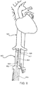
- FIG. 1 schematically illustrates placement of an embodiment disclosed herein in the vascular system of a patient.
- FIG. 2 is a sketch of the distal portion of a catheter with distal arms for sensing IVC diameter according to one embodiment disclosed herein.
- FIG. 3 is a sketch of the distal portion of another catheter with distal arms for sensing IVC diameter according to an alternative embodiment disclosed herein.
- FIG. 4 is a sketch of the distal portion of a catheter with distal arms including ultrasound devices for sensing IVC diameter according to a further alternative embodiment disclosed herein.
- FIG. 4A is a sketch of the distal portion of a catheter with separate distal sensing arms according to a further alternative embodiment disclosed herein.
- FIG. 4B is a sketch of the distal portion of a catheter with a plurality of distal sensing arms according to a further alternative embodiment disclosed herein.
- FIG. 5 is a sketch of the distal portion of an embodiment of a catheter employing ultrasound signals to measure IVC diameter in which the catheter shaft is configured with a bias to position it against a wall of the IVC.
- FIG. 6 is a sketch of the distal portion of another embodiment of a catheter employing ultrasound signals to measure IVC diameter in which the catheter shaft is also configured with a bias as in the embodiment of FIG. 5 , but also includes a further distal portion with separable arms.
- FIG. 7 is a sketch of the distal portion of a further embodiment of a catheter employing ultrasound signals to measure IVC diameter employing a plurality of elements at the distal end position of the ultrasound element centrally within the IVC.
- FIG. 8 is a diagram of a device made in accordance with aspects of the present invention that is disposed within the IVC via a catheter inserted through the superior vena cava.
- FIG. 9 is a diagram of a device made in accordance with aspects of the present invention that is disposed within the IVC via a catheter inserted via an inferior access site and which does not pass through the superior vena cava.
- FIG. 10 is a perspective view of a further alternative embodiment positioned in a partially cross-sectioned portion of the IVC.
- FIG. 11 is a side view of the embodiment shown in FIG. 8 in a partially cross-sectioned IVC.
- FIG. 12 is an end view of the device of FIG. 8 or 11 from an inferior aspect.
- FIG. 13 is an end view of the device of FIG. 8 or 11 from a superior aspect.
- FIG. 14 is a schematic illustration of an exemplary system according to the present disclosure.
- Embodiments of the present disclosure are directed to devices, systems and methods including a catheter placed in the patient's venous system that measures the volume of the Inferior Vena Cava (IVC), Superior Vena Cava (SVC), or other great vessel or branch thereof.
- Vena Cava as generally used herein, unless otherwise specified, may refer to portions of both the IVC and SVC where devices of disclosed embodiments may be placed and may sense.
- devices include an indwelling catheter that can be left in place during a patient's hospitalization in order to continuously monitor venous volume over an extended period.
- catheters according to the present disclosure measure cross-sectional size or diameter of the vessel as a proxy for vessel volume employing active or passive detection elements as described.
- passive detection elements are elements that react in a detectable manner to a signal directed at them, such as by reflection or inducing of current flow.
- Passive detections elements generally do not create their own signal, whereas active detection elements generate or emit a signal that is modulated in a detectable manner based on distances it encounters. Examples of active detection elements include ultrasound transducers, light emitters and electrodes.
- the disclosed catheters are capable of measuring the anterior-posterior diameter of the IVC, in the area caudal to the right atrium and cranial to the renal veins. In addition to measuring this absolute diameter dimension, the catheters may also measure the variation of this diameter over the respiratory cycle, or between different modes of breathing. In another embodiment, the catheters measure the anterior-posterior diameter of the SVC, and measure variation of this diameter over the respiratory cycle, or between different modes of breathing. In some embodiments these volume measurements would be taken in both the SVC and IVC, and in other embodiments only in either the SVC or IVC. Catheters according to the invention may also measure the cross-sectional dimensions of other venous and arterial vessels, cardiovascular and other organs, structures of the digestive, renal, pulmonary, or reproductive systems, and abnormal physiologies such as aneurysms.
- the disclosed catheters measure venous pressure in addition to measuring venous vessel volume.
- the catheters measure central venous oxygen saturation in addition to vessel volume and pressure.
- the catheter can provide central venous pressure data, venous volume data, and venous oxygen saturation data.
- the measurements of pressure, volume, and venous oxygen saturation provide clearer guidance in understanding clinical diagnosis and management in the acute setting, when timing and choice of therapy is crucial.
- Central venous oxygen saturation provides a surrogate measure of oxygen flux, reflecting the balance between oxygen delivery (DO2) and consumption (VO2).
- central venous oxygen saturation may be measured in the SVC, IVC, or both.
- disclosed catheters are capable of measuring oxygen saturation at multiple locations along the catheter, such as proximally in the SVC and distally in the IVC, along with pressure and/or volume at one or more points along the catheter in the great vessel (Vena Cava).
- Vena Cava a great vessel
- disclosed catheters measure the change in venous oxygen between the IVC and SVC without needing to place a device in the heart.
- the kidneys receive a high proportion of cardiac output, but do not consume much oxygen, thus the blood in the IVC has higher oxygen content than that in the SVC.
- the venous oxygen saturation in the IVC may be less than in the SVC, indicating acute (severe) decompensation.
- the difference in IVC and SVC oxygen saturation may therefore provide a useful measurement in cardiac parameters regarding perfusion to organs in critically-ill patients.
- the devices in this disclosure provide a novel diagnostic tool for managing patients in the acute setting.
- Catheters as disclosed herein such as catheter 100 of FIG. 1 , which may include a body 104 , a proximal end 108 , a distal end region 112 , and a distal end 116 , may be placed into the bloodstream via a percutaneous puncture into the subclavian, brachial, or jugular vein.
- the disclosed catheter would then be gently advanced into the IVC, preferably over a flexible guidewire to reduce the risk of vascular trauma or puncture.
- catheter 100 may include a distal region pressure sensor port 120 and a distal region oxygen saturation sensor port 124 , as well as a proximal oxygen saturation sensor port 128 .
- oxygen saturation sensor ports 124 , 128 may be positioned on catheter 100 so as to be located, respectively, in the IVC and SVC when the catheter is properly placed in the vasculature.
- a lumen of the catheter could be used to monitor the vascular pressure at distal end 116 of catheter 100 , so that if the catheter were inadvertently advanced into the right ventricle instead of the IVC, the right ventricular pressure would be detected, and the catheter could be retracted, turned, and advanced into the IVC.
- catheter 100 may need to be specifically oriented in one direction in the IVC, and it may be important to determine how cranial or caudal the sensor is with respect to specific anatomical markers.
- an element of disclosed systems is an external sensor or reflector (not shown) which can be placed on the patient's abdomen during placement. If a reflector is used, catheter 100 can be rotated and advanced or retracted (or the reflector moved around on the patient's abdomen) until a strong signal is reflected to the catheter. Catheter 100 and reflector can then be moved or manipulated until the catheter is in an appropriate location and orientation.
- An external ultrasound system may also be used to confirm placement of a central venous pressure and volume (CVPV) (or central venous pressure, volume and oxygen (CVPVO)) catheter at the correct location in the IVC with correct orientation.
- CVPV central venous pressure and volume
- CVPVO central venous pressure, volume and oxygen
- a catheter like catheter 200 or 300 may be provided with two or more arms 204 , 304 , as shown in FIGS. 2 and 3 , respectively.
- Arms 204 , 304 may be biased radially outwards to lay gently against the IVC walls. This bias may be continuous, or the arms may be held in a collapsed position until a reading is to be taken and then deployed to engage the IVC walls to take the reading.
- this bias will be very gentle, since the pressure in the IVC is typically 5-20 mm HG and any strong pressure will tent the IVC open.
- the separation of arms 204 , 304 could be measured in various ways, such as by using one or more strain gauges, such as strain gauges 208 shown in FIG. 2 , on one or more arms that electronically sense flexure and transmit readings to a processor outside the body (not shown).
- linkages 3 may include one or more linkages like linkages 308 designed and configured to convert radial expansion of the arms to longitudinal motion of a wire 312 extending through the catheter that can be monitored electronically by a detector 314 , which may be located within the distal end as shown or, alternatively, at the proximal end of the catheter.
- FIG. 4 shows a catheter 400 with two arms 204 with simple ultrasound transducers 404 on each arm. In some embodiments, more or fewer than two arms and more or fewer than one transducer per arm may be used. A signal generated by transducer 404 can be sensed by another transducer, and a simple time-of-travel calculation would determine the distance between the arms. A signal may be generated by each transducer 404 in sequence and sensed by each of the others, thereby generating a very reliable map of the relative position of each arm. Alternatively, as shown in FIG.
- two or more electronic emitter/detectors 408 may sense the electrical impedance or capacitance, at one or more frequencies, by inducing or otherwise generating and monitoring an electrical current in order to determine the volume of blood between emitter/detectors 408 or separate emitters and detectors.
- a pair of arms 304 , 412 may have a wishbone shape as shown in FIGS. 3 and 4A , respectively, rather than being joined at their ends as with arms 204 of FIGS. 2 and 4 .
- These designs may be optimized to minimize rotational or longitudinal migration once the catheter has been positioned.
- the arms may have a blade like shape, or flow directing fin elements added) to minimize flow resistance or to create a response to the flow, such as to provide flow-induced biasing of the arms against the vessel wall.
- a lumen of the catheter may be used to apply suction to ports on the arms to facilitate engagement with the IVC wall.
- Catheters in embodiments of systems disclosed herein may also include more than two arms, e.g., two pairs of arms arranged orthogonally to each other so as to measure the vessel in two dimensions.
- disclosed catheters may include a larger plurality of arms, e.g., six or more, distributed around the circumference of the catheter and configured to extend radially like spokes of a wheel when deployed. Such a configuration may eliminate the need to position the catheter rotationally within the vessel.
- the arms may also comprise, as shown in FIG. 4B , a circumferential array of very thin wires or fibers 416 to create a brush-like structure so as to minimize deformation of the vessel.
- each wire/fiber 416 in such an array may comprise an optical fiber through which light may be emitted by emitters 420 and detected by an optical detector 424 on the catheter. Distance may be determined, for example, in accordance with the magnitude of light intensity received by detector 424 .
- an ultrasonic reflector may be placed at the tip of each wire 416 in such an array in place of emitters 420 , with an ultrasound transducer located on the catheter centrally within the array in place of detector 424 to emit and detect an ultrasound signal reflected from the reflector on the tip of each wire.
- each wire 416 may have an ultrasound detector, measuring the time of travel of an ultrasound signal from one or more ultrasound emitters.
- Such an embodiment may be configured to provide a two-dimensional profile of the size and shape of the vessel around its entire circumference.
- FIGS. 4A and 4B also illustrate other features of the present disclosure, which may be utilized in combination with other embodiments disclosed herein as well as the embodiments of FIGS. 4A and 4B .
- each embodiment comprises a concentric, multi-component catheter structure.
- outer sheath 430 defines lumen 432 , which serves as a guide catheter for inner flexible member 434 , which delivers arms 412 .
- Lumen 432 also may be used as a delivery or sampling lumen.
- the embodiment of FIG. 4B includes at least three concentric members, outer sheath 430 , inner delivery catheter member 436 , which itself defines a lumen for delivering detector carrying member 438 .
- FIGS. 5, 6, and 7 show alternative designs for embodiments of methods and systems employing catheters using ultrasound signals to measure IVC diameters.
- catheters 500 and 600 have a built-in bias or curve 504 , 604 , which holds it against the wall of the IVC.
- This curve could be created by inserting a pre-shaped wire into a lumen within the catheter, either permanently or after it has been placed into the IVC. This curve might be helpful during catheter insertion, to make it easier to steer the catheter into the IVC instead of the right ventricle.
- the catheter may comprise a polymeric shaft which is heat set into the desired shape.
- the catheter can be inserted into a sheath or designed with a lumen to receive a stylet which straightens the shaped portion of the catheter.
- one or more ultrasound transducers such as ultrasound transducer 508 located at the distal end of catheter 500 , may emit signals and measure the reflected signal.
- the wall of the IVC is relatively much more echo-reflective than the blood within the IVC, so that this reflected signal can be detected.
- An analysis of the distance measurements from the reflected signal(s) can then be used to determine the diameter of the IVC.
- curve 504 shown in FIG. 5 may be helpful, in some embodiments the catheter alternatively may be constructed without any bias.
- two or more ultrasound transducers may be placed at known intervals around the circumference of the catheter and directed radially outward from the catheter.
- Each transducer is then operated in pulse-echo mode, A-mode, or M-mode to detect the round trip time of flight of the ultrasound pulse to calculate the distance between each transducer and the wall of the IVC.
- the time-of-flight measurements made using the two or more transducers at known intervals around the circumference of the catheter can be used to calculate the shape and size of the IVC. If two transducers positioned 180 degrees apart are used, then the size measurement will represent a diameter of the IVC. If three or more transducers are used, the size measurement can represent a cross-sectional area of the IVC.
- FIG. 6 shows an embodiment of catheter 600 employing an ultrasound transducer 608 positioned against the wall of the IVC, but has a further distal section of the catheter which is biased back to the opposite wall of the IVC so the transducer is held against one wall by proximal and distal catheter segments biased against the other wall.
- a device such as shown in FIG. 6 may have a wish-bone shape in a distal segment 612 , to further center and stabilize the position of transducer 608 in the IVC.
- the wishbone shape may be optimized to interact with the naturally oval shape of the IVC to ensure that the catheter assumes an orientation such that the diameter measurement is anterior-posterior.
- This embodiment may employ a single transducer that may be positioned against the posterior or anterior wall of the IVC, but a catheter with several transducers may also be employed and work well when positioned against any wall in the IVC, and algorithms may be designed to calculate IVC diameter based on the different measurements. For example, in some embodiments, a look-up table and/or correlation calculation may be used to approximate spatial relationships between one or more transducers and/or one or more walls of the IVC.
- the wishbone-shaped catheter may be designed with no curve 604 , so that the wishbone self-orients in a lateral orientation, and at least two transducers 608 are located facing anteriorly and posteriorly, so that in combination they determine the overall anterior-posterior dimension of the IVC.
- a catheter 700 has two or more bowed resilient wire or ribbon elements 704 located in a distal catheter section and extending from the sides of the catheter, to engage the vessel wall and thereby help center the catheter within the IVC.
- these resilient elements may be deployed from the distal end of catheter 700 , and in some embodiments may be separated axially a substantial distance from any ultrasound transducer so as not to distend the vessel where measurements are to be taken. This separation forms an anchor isolation structure 707 .
- catheter 700 has an ultrasound emitter/receiver 708 capable of detecting the distance to the vessel walls at multiple points around the circumference of the catheter.
- the emitter/receiver may comprise several ultrasound transducers, e.g., four or more, distributed around a catheter shaft, each aiming in a different direction radially from the catheter. By measuring the distance to the IVC wall in each direction, the IVC radius, diameter, area, and/or circumference can be calculated.
- wire or ribbon elements 707 may be provided on only one side of the shaft so the shaft/transducer is positioned against one side of the vessel and measures reflection from the opposite side.
- GUI graphical user interface
- the anterior-posterior IVC diameter the variation in anterior-posterior diameter, the ratio of A-P and lateral diameters, the rate of change of these measurements or the change in these measurements over a time period such as the past hour, or other calculated measurements.
- Embodiments of the GUI may have alarms if the IVC A-P diameter becomes too low, or is dropping too quickly. It could also be connected via wires or wirelessly to other therapeutic devices, such as an IV pump infusing diuretics, or an IV pump infusing fluid in a patient in shock, or a dialysis or ultrafiltration machine removing fluid.
- sensors may be placed both along the catheter and near the distal end.
- at least one oxygen saturation sensor such as fiberoptics which emit various wavelengths of light, and measure the blood's relative reflectance of those different wavelengths
- at least one other oxygen saturation sensor is placed in the IVC portion of the catheter and data from these sensors can be used to track changes in venous oxygen saturation in the SVC versus IVC.
- pressure sensors can be placed at one or more locations along the catheter to measure pressures in the IVC, SVC, and/or right atrium. Doppler ultrasound sensors could be used to measure blood flow in the IVC and/or the SVC, giving an effective measurement of overall cardiac output as well as the relative IVC and SVC flow rates.
- a lumen/channel is provided in the catheter that can be used to either deliver drugs and fluids, as well as be used to withdraw blood.
- some embodiments may include a device capable of delivering therapeutic agents (such as drugs or saline) through a lumen, retrieving blood from a lumen, detecting vena cava volume, detecting vena cava pressures, and/or detecting vena cava oxygen saturation, including measuring differences in oxygen saturation of blood in the SVC versus IVC.
- Other embodiments may include multiple lumens that allow blood to be withdrawn from different portions of the vena cava, such as withdrawing blood samples from the SVC and the IVC without the need for repositioning the device. Such withdrawal of samples from different sites can allow for comparison of venous oxygen saturation from the SVC and IVC using external sensors without the need for embedded oxygen sensors directly in the catheter.
- a device 800 made in accordance with this embodiment may comprise four major components or assemblies: an electronics capsule 804 , an anchor element 808 , an anchor isolation structure 812 connecting the electronics capsule and anchor element and a catheter 816 .
- Electronics capsule 804 comprises a sealed housing 820 for containing control, power and other alternative functional modules and provides a self-contained, sealed device. Alternatively one or more of these functions may be provided externally with communication through the catheter.
- Capsule 804 also provides support for a transceiver 824 , which in the case of the illustrated device is a single ultrasound transceiver positioned at the inferior end of the device.
- capsule 804 serves as a housing for transceiver 824 .
- Transceiver 824 may utilize one or more ultrasound crystals to measure IVC diameter by emitting an ultrasound pulse, and then detecting the reflection of that pulse from the opposing wall of the IVC.
- Other modes of detection with ultrasound receivers and/or other transceiver types may be alternatively employed by persons of ordinary skill without departing from the teachings of this disclosure.
- Electronics capsule 804 generally will be provided with the lowest possible profile so as to minimize obstruction of the lumen when positioned in the IVC.
- Anchor element 808 as depicted in this embodiment includes a single anchor wire 828 configured in a generally figure-eight or double helix shape. Alternatively, the same or similar configurations can be provided with two or more wires.
- Anchor wire 828 is pinned to a telescoping deployment member 832 at both its inferior end 836 and superior end 840 .
- the telescoping deployment member includes an inner member 844 , which is secured to electronics capsule 804 , through anchor isolation structure 812 , and an outer member 848 . Relative motion between inner member 844 and outer member 848 moves anchor wire 828 from a collapsed position to a deployed or anchoring position.
- anchor wire 828 is resilient, with shape-memory properties configured to provide a rest state in the deployed configuration.
- device 800 may be delivered to the desired location in the IVC via a conventional guide catheter like catheter 816 or other suitable sheath type delivery device. When position is confirmed as described below, device 800 is ejected from catheter 816 with anchor element 808 self-deploying upon ejection.
- an actuating wire provided through the catheter is connected to the deployment member at the superior end.
- the actuating wire may be permanently attached to facilitate deployment and collapse of the anchor wire, or may releasably be connected using a mechanical release mechanism, for example a screw threaded connection, spring release, hooks or other such means known in the art.
- the actuating wire may be a single or double wire, which may be coaxial or parallel, depending on the mode of actuation.
- movement of the actuating wire effects relative movement of inner and outer telescoping deployment members like inner and outer telescoping deployment members 844 , 848 to deploy anchor wire like anchor wire 828 from the collapsed configuration to the expanded, deployed configuration as explained above.
- the actuating wire is released from a device like device 800 according to its mode of connection to leave the device secured in the IVC via the anchor element.
- anchor isolation structure 812 ensures the desired positioning, which may be approximately 1 ⁇ 2 to 4 times the IVC diameter as indicated above, typically in the range of about 2-6 cm, and in some cases more preferably about 3-5 cm.
- the IVC has a somewhat oval cross section with a minor axis of the oval extending in the anterior-posterior direction and a major axis extending in the lateral-medial direction. It is thus desirable to minimize any effect of device 800 on this natural oval shape at or close to the point of measurement.
- FIGS. 8-13 The shape of the IVC and possible effect of anchor element 808 on the IVC shape is illustrated, in one possible configuration, in FIGS. 8-13 .
- the IVC assumes its more natural oval shape as best seen in FIG. 12 .
- the IVC is forced into a more circular shape as best seen in FIG. 13 .
- anchor element 808 potentially distort the shape of the IVC, it may also stiffen the IVC so as not to be as responsive to varying fluid volumes which may indicate heart failure risk.
- Anchor isolation structure 812 reduces or eliminates such problems as might otherwise be associated within sensing devices positioned in the IVC.
- device 800 In order to achieve accurate measurement with transceiver 824 using an anchor configuration of the type shown in FIGS. 8-13 , device 800 , from deployment member 832 through anchor isolation structure 812 into electronics capsule 804 , should be provided with a stiffness sufficient to maintain the electronics capsule (and transceiver) against the wall (W) of the IVC at one side and yet provide sufficient flexibility (and smoothness) to avoid damage or erosion of the IVC wall by contact with the device over the remaining lifetime of the patient.
- the illustrated device i.e., device 800 , or other device disclosed herein, is positioned with an electronics capsule like electronics capsule 804 , and more specifically an active transceiver (e.g., ultrasound transceiver) like transceiver 824 , against the posterior wall (W) of the IVC so as to measure the distance to the anterior wall.
- an active transceiver e.g., ultrasound transceiver
- This arrangement may offer advantages in accuracy and sensitivity in measurements by measuring along the minor anterior-posterior axis of the oval IVC shape, and by measuring from the posterior wall, such that bony structures lying behind the posterior wall, which may create artifacts or other interference with ultrasound measurements, may be avoided.
- Such positioning may provide for the greatest accuracy in measurement of diameter over the respiratory cycle (e.g., measurement of diameter variability vs. static measurement). While a single ultrasound transceiver, i.e., transceiver 824 , is shown in FIGS. 8-13 , a similar device with more than one ultrasound crystal may be positioned elsewhere in the IVC, for example in the center of the IVC, with two crystals measuring the distance to the anterior and posterior walls simultaneously. Specific requirements for positioning and measurements may be clinically determined based on patient anatomy as determined by the procedure provider, and the device to be implanted, such as device 800 , may be modified according to the teachings contained herein to suit those specific patient requirements.
- devices as disclosed herein may be positioned at any suitable position in the IVC based on clinical assessment.
- the transceiver of the device such as an ultrasound crystal
- the transceiver of the device may be disposed at the cranial end of the device, with the cranial end then positioned in the IVC between the renal veins and the hepatic veins.
- an anchor element like anchor element 808 may be disposed at the opposite, caudal end of the device and thus positioned in the IVC inferior to the renal veins.
- transceiver 824 may be used to assist in confirming placement by slightly rotating electronics capsule 804 so as to effectively scan the opposite IVC walls with an ultrasound sensor of the transceiver to detect placement position relative to the oval IVC cross-sectional shape.
- the device also may be positioned in the SVC.
- a device like device 800 made in accordance with the present disclosure may be positioned within the IVC (or SVC) such that transceiver 824 is located inferior to the end of catheter 816 relative to electronics capsule 804 with anchor element 808 and/or wire 828 located between the catheter and the electronics capsule.
- device 800 is delivered from a superior insertion point such as the jugular vein, using conventional cardiac catheterization techniques.
- a connector wire 852 may be used to deliver and retrieve device 800 .
- Further functions or treatments as well as communication with device 800 may also be provided through catheter 816 as may be devised by persons of ordinary skill in the art for particular clinical applications based on the teachings contained herein.
- inferior insertion may be performed, as illustrated in FIG. 9 .
- a device like device 800 made in accordance with the present disclosure may be positioned within the IVC such that transceiver 824 is located proximal to the end of catheter 816 relative to electronics capsule 804 with anchor element 808 and/or wire 828 located on the opposite end of the electronics capsule from the catheter.
- This embodiment may be delivered via conventional femoral vein cardiac catheterization procedures.
- inferior, femoral vein insertion may be performed using a device like device 800 and other elements as arranged in FIG. 8 .
- electronics capsule 804 , transceiver 824 , anchor element 808 and/or wire 828 , and any other elements of a device made in accordance with the present disclosure may be disposed at any position and in any orientation within the IVC provided that device 800 can provide the functionality described herein.
- additional anchor elements may be provided on electronics capsule 804 , such as one or more barbs 856 .
- barbs 856 are shown in FIG. 10 , they are an optional feature.
- Basic operation of anchor element 808 is described above. As anchor element 808 opens, it shortens and tends to pull back on electronics capsule 804 . Through a linkage between barbs 856 and deployment member 832 , the relative movement of those two parts during deployment of anchor element 808 may be used to deploy the barbs from the back of electronics capsule 804 .
- Anchor element 808 and barbs 856 may be positioned to engage the IVC wall (W) in opposition to one another to reinforce anchoring force and security.
- substantially the same device as device 800 may be alternatively provided without anchor barbs 856 , held in place only by the collapsible/expandable double helix anchor wire 828 of anchor element 808 .
- These anchor structures, as well as others described above, are configured to achieve secure fixation against both longitudinal and rotational movement while preferentially maintaining at least transceiver 824 in the posterior aspect of the IVC, most preferably against the posterior IVC wall.
- the anchor elements described also can be deployed and redeployed multiple times during a placement procedure in order to ensure the most optimum placement of device 800 .
- the shape or configuration of anchor wire 828 also may be adapted for IVC size and shape using different anchor element configurations as will be appreciated by persons of ordinary skill in the art based on the teachings presented herein.
- Catheters according to embodiments described herein also may be used in a patient treatment system to monitor and modulate therapy.
- One such exemplary system 900 may comprise integration of a diagnostic catheter 902 as described above in a closed loop system with an implanted or catheter-based therapeutic device 904 , such as a drug delivery device (drug/diuretic pump) or mechanical device (such as a renal pump, IV infusion pump, dialysis system or ultrafiltration system) to manage blood volume.
- catheter 902 employs passive detection elements such as ultrasound reflective elements
- external ultrasound transducer 906 may be incorporated.
- System 900 may utilize IVC measurements as an input/control metric for the system.
- the diagnostic catheter would detect a high diameter/low collapsibility or low diameter/high collapsibility reading and initiate the therapeutic device to either deliver drug or commence treatment to reduce total blood volume or the impact of the elevated volume, adding fluid to increase blood volume, or initiate or modulate other appropriate therapy.
- the diagnostic catheter could then be used to manage the rate of treatment to reduce the total time of operation of the therapeutic device.
- the diagnostic and therapeutic devices could be linked physically or wirelessly. There could also be power transfer between the two devices, through wired or wireless means.
- diagnostic catheter 902 communicates with and may be controlled by control module 908 .
- Control module 908 may include one or more processors, memory and other components as may be determined by persons of ordinary skill.
- GUI 910 also may be incorporated into control module 908 as previously discussed.
- Control module 908 is provided with a communication and/or power link 912 to therapeutic device control module 914 , which also may include one or more processors, memory and other conventional components configured to control therapeutic device 904 .
- GUI 916 may also be provided.
- the conjunctive phrases in the foregoing examples in which the conjunctive list consists of X, Y, and Z shall each encompass: one or more of X; one or more of Y; one or more of Z; one or more of X and one or more of Y; one or more of Y and one or more of Z; one or more of X and one or more of Z; and one or more of X, one or more of Y and one or more of Z.
Landscapes
- Health & Medical Sciences (AREA)
- Life Sciences & Earth Sciences (AREA)
- Engineering & Computer Science (AREA)
- Public Health (AREA)
- General Health & Medical Sciences (AREA)
- Biomedical Technology (AREA)
- Medical Informatics (AREA)
- Physics & Mathematics (AREA)
- Veterinary Medicine (AREA)
- Biophysics (AREA)
- Heart & Thoracic Surgery (AREA)
- Animal Behavior & Ethology (AREA)
- Pathology (AREA)
- Surgery (AREA)
- Molecular Biology (AREA)
- Radiology & Medical Imaging (AREA)
- Nuclear Medicine, Radiotherapy & Molecular Imaging (AREA)
- Hematology (AREA)
- Vascular Medicine (AREA)
- Computer Vision & Pattern Recognition (AREA)
- Physiology (AREA)
- Oral & Maxillofacial Surgery (AREA)
- Dentistry (AREA)
- Acoustics & Sound (AREA)
- Pulmonology (AREA)
- Anesthesiology (AREA)
- Data Mining & Analysis (AREA)
- Databases & Information Systems (AREA)
- Epidemiology (AREA)
- Primary Health Care (AREA)
- Ultra Sonic Daignosis Equipment (AREA)
- Media Introduction/Drainage Providing Device (AREA)
Abstract
Description
Claims (14)
Priority Applications (1)
| Application Number | Priority Date | Filing Date | Title |
|---|---|---|---|
| US15/750,100 US11039813B2 (en) | 2015-08-03 | 2016-08-03 | Devices and methods for measurement of Vena Cava dimensions, pressure and oxygen saturation |
Applications Claiming Priority (4)
| Application Number | Priority Date | Filing Date | Title |
|---|---|---|---|
| US201562200409P | 2015-08-03 | 2015-08-03 | |
| US201662294986P | 2016-02-12 | 2016-02-12 | |
| PCT/US2016/045385 WO2017024051A1 (en) | 2015-08-03 | 2016-08-03 | Devices and methods for measurement of vena cava dimensions, pressure, and oxygen saturation |
| US15/750,100 US11039813B2 (en) | 2015-08-03 | 2016-08-03 | Devices and methods for measurement of Vena Cava dimensions, pressure and oxygen saturation |
Publications (2)
| Publication Number | Publication Date |
|---|---|
| US20180220992A1 US20180220992A1 (en) | 2018-08-09 |
| US11039813B2 true US11039813B2 (en) | 2021-06-22 |
Family
ID=56799543
Family Applications (1)
| Application Number | Title | Priority Date | Filing Date |
|---|---|---|---|
| US15/750,100 Active 2037-09-29 US11039813B2 (en) | 2015-08-03 | 2016-08-03 | Devices and methods for measurement of Vena Cava dimensions, pressure and oxygen saturation |
Country Status (3)
| Country | Link |
|---|---|
| US (1) | US11039813B2 (en) |
| EP (1) | EP3331426B1 (en) |
| WO (1) | WO2017024051A1 (en) |
Cited By (7)
| Publication number | Priority date | Publication date | Assignee | Title |
|---|---|---|---|---|
| WO2023170632A1 (en) | 2022-03-09 | 2023-09-14 | Foundry Innovation & Research 1, Ltd. | Heart failure diagnostic tools and methods using signal trace analysis |
| US11944495B2 (en) | 2017-05-31 | 2024-04-02 | Foundry Innovation & Research 1, Ltd. | Implantable ultrasonic vascular sensor |
| US12268493B2 (en) | 2016-08-11 | 2025-04-08 | Foundry Innovation & Research 1, Ltd. | Systems and methods for self-directed patient fluid management |
| US12310707B2 (en) | 2016-08-11 | 2025-05-27 | Foundry Innovation & Research 1, Ltd. | Wireless resonant circuit and variable inductance vascular monitoring implants and anchoring structures therefore |
| US12465324B2 (en) | 2015-02-12 | 2025-11-11 | Foundry Innovation & Research 1, Ltd. | Patient fluid management systems and methods employing integrated fluid status sensing |
| US12465233B2 (en) | 2022-07-29 | 2025-11-11 | Foundry Innovation & Research 1, Ltd. | Multistranded conductors adapted to dynamic in vivo environments |
| WO2026058200A1 (en) | 2024-09-13 | 2026-03-19 | Foundry Innovation & Research 1, Ltd. | Methods of determining a heart-failure congestion index for improved speed and accuracy of heart-failure diagnostic and treatment decisions, and related devices and systems |
Families Citing this family (10)
| Publication number | Priority date | Publication date | Assignee | Title |
|---|---|---|---|---|
| AU2016219018B2 (en) | 2015-02-12 | 2020-10-29 | Foundry Innovation & Research 1, Ltd. | Implantable devices and related methods for heart failure monitoring |
| WO2017024051A1 (en) | 2015-08-03 | 2017-02-09 | Foundry Innovation & Research 1, Ltd. | Devices and methods for measurement of vena cava dimensions, pressure, and oxygen saturation |
| US11701018B2 (en) | 2016-08-11 | 2023-07-18 | Foundry Innovation & Research 1, Ltd. | Wireless resonant circuit and variable inductance vascular monitoring implants and anchoring structures therefore |
| EP3705031B1 (en) | 2016-11-29 | 2025-12-10 | Foundry Innovation & Research 1, Ltd. | Wireless resonant circuit and variable inductance vascular implants for monitoring patient vasculature system |
| WO2018220146A1 (en) | 2017-05-31 | 2018-12-06 | Foundry Innovation & Research 1, Ltd. | Implantable sensors for vascular monitoring |
| EP3893962A1 (en) * | 2018-12-12 | 2021-10-20 | Foundry Innovation & Research 1, Ltd. | Dialysis catheters with integrated fluid status sensing and related systems and methods |
| EP3998936B1 (en) | 2019-07-17 | 2025-09-03 | Nxgenport Inc | Implantable venous access port with remote physiological monitoring capabilities |
| US12097339B2 (en) * | 2019-12-31 | 2024-09-24 | Biosense Webster (Israel) Ltd. | System and methods of using a catheter with an anchoring mechanism |
| US12097347B2 (en) * | 2020-10-26 | 2024-09-24 | Medtronic Xomed, Inc. | System and method for a shunt |
| WO2023037017A1 (en) | 2021-09-13 | 2023-03-16 | Foundry Innovation & Research 1, Limited | Vascular imaging and measurement using ultrasound |
Citations (416)
| Publication number | Priority date | Publication date | Assignee | Title |
|---|---|---|---|---|
| US3568661A (en) | 1968-10-02 | 1971-03-09 | Us Health Education & Welfare | Frequency modulated ultrasound technique for measurement of fluid velocity |
| US4142412A (en) | 1976-05-12 | 1979-03-06 | Sutures Inc. | Doppler flow meter and method |
| USRE32361E (en) | 1979-05-14 | 1987-02-24 | Medtronic, Inc. | Implantable telemetry transmission system for analog and digital data |
| US4926875A (en) | 1988-01-25 | 1990-05-22 | Baylor College Of Medicine | Implantable and extractable biological sensor probe |
| US4947852A (en) | 1988-10-05 | 1990-08-14 | Cardiometrics, Inc. | Apparatus and method for continuously measuring volumetric blood flow using multiple transducer and catheter for use therewith |
| EP0399059A1 (en) | 1989-05-22 | 1990-11-28 | Pacesetter AB | Implantable medical device with adjustable sensitivity to detect an event relating to a physiological function, and process for using same |
| US5127404A (en) | 1990-01-22 | 1992-07-07 | Medtronic, Inc. | Telemetry format for implanted medical device |
| US5205292A (en) | 1991-06-03 | 1993-04-27 | Applied Biometric, Inc. | Removable implanted device |
| EP0538885A1 (en) | 1991-10-23 | 1993-04-28 | Aloka Co., Ltd. | Ultrasonic Doppler blood flow monitoring system |
| US5316001A (en) | 1990-09-11 | 1994-05-31 | Ferek Petric Bozidar | Cardiac measurement system for measuring blood flow velocity by use of a sensor implanted inside the heart |
| US5495852A (en) | 1995-01-27 | 1996-03-05 | Boston Heart Foundation | Method and apparatus for estimating diameter of an artery using B-mode ultrasonic images |
| US5630836A (en) | 1995-01-19 | 1997-05-20 | Vascor, Inc. | Transcutaneous energy and information transmission apparatus |
| WO1997042871A1 (en) | 1996-05-10 | 1997-11-20 | Cardiovascular Concepts, Inc. | Lesion diameter measurement catheter and method |
| US5752522A (en) | 1995-05-04 | 1998-05-19 | Cardiovascular Concepts, Inc. | Lesion diameter measurement catheter and method |
| WO1998029030A1 (en) | 1997-01-03 | 1998-07-09 | Biosense Inc. | Pressure-sensing stent |
| WO1998035611A1 (en) | 1997-02-16 | 1998-08-20 | Technion Research And Development Foundation Ltd. | Blood vessel cross-sectional area detector and compliance measurement device and method |
| US5872520A (en) | 1995-10-24 | 1999-02-16 | Siemens Aktiengesellschaft | Identification and/or sensor system |
| US5967986A (en) | 1997-11-25 | 1999-10-19 | Vascusense, Inc. | Endoluminal implant with fluid flow sensing capability |
| US6012457A (en) | 1997-07-08 | 2000-01-11 | The Regents Of The University Of California | Device and method for forming a circumferential conduction block in a pulmonary vein |
| US6015386A (en) | 1998-05-07 | 2000-01-18 | Bpm Devices, Inc. | System including an implantable device and methods of use for determining blood pressure and other blood parameters of a living being |
| US6025725A (en) | 1996-12-05 | 2000-02-15 | Massachusetts Institute Of Technology | Electrically active resonant structures for wireless monitoring and control |
| US6039701A (en) | 1996-09-05 | 2000-03-21 | Ob Inovations, Inc. | Method and apparatus for monitoring cervical diameter |
| US6111520A (en) | 1997-04-18 | 2000-08-29 | Georgia Tech Research Corp. | System and method for the wireless sensing of physical properties |
| US6115636A (en) | 1998-12-22 | 2000-09-05 | Medtronic, Inc. | Telemetry for implantable devices using the body as an antenna |
| US6115633A (en) | 1996-10-28 | 2000-09-05 | Biotronik Mess-Und Therapiegeraete Gmbh & Co. Ingenieurbuero Berlin | Implantable stimulator |
| WO2000055579A2 (en) | 1999-03-16 | 2000-09-21 | Florence Medical Ltd. | A system and method for detection and characterization of stenosis, blood vessels flow and vessel walls properties using vessel geometrical measurements |
| WO2000056210A1 (en) | 1999-03-24 | 2000-09-28 | Noveon Ip Holdings Corp. | Remotely interrogated diagnostic implant device with electrically passive sensor |
| US6164283A (en) | 1997-07-08 | 2000-12-26 | The Regents Of The University Of California | Device and method for forming a circumferential conduction block in a pulmonary vein |
| WO2001012092A1 (en) | 1999-08-14 | 2001-02-22 | The B.F. Goodrich Company | Remotely interrogated diagnostic implant device with electrically passive sensor |
| WO2001013792A1 (en) | 1999-08-24 | 2001-03-01 | Impulse Dynamics N.V. | Apparatus and method for determining a mechanical property of an organ or body cavity by impedance determination |
| US6206835B1 (en) | 1999-03-24 | 2001-03-27 | The B. F. Goodrich Company | Remotely interrogated diagnostic implant device with electrically passive sensor |
| US6231516B1 (en) | 1997-10-14 | 2001-05-15 | Vacusense, Inc. | Endoluminal implant with therapeutic and diagnostic capability |
| US6261233B1 (en) | 1996-01-05 | 2001-07-17 | Sunlight Medical Ltd. | Method and device for a blood velocity determination |
| US6278379B1 (en) | 1998-04-02 | 2001-08-21 | Georgia Tech Research Corporation | System, method, and sensors for sensing physical properties |
| US6287253B1 (en) | 1999-06-25 | 2001-09-11 | Sabolich Research & Development | Pressure ulcer condition sensing and monitoring |
| US6325762B1 (en) | 1996-12-09 | 2001-12-04 | Swee Chuan Tjin | Method and apparatus for continuous cardiac output monitoring |
| US6339816B1 (en) | 1997-08-19 | 2002-01-15 | Siemens Noxdorf Informationssysteme Aktiengesellschaft | Method for improving controllability in data processing system with address translation |
| WO2002015823A2 (en) | 2000-08-23 | 2002-02-28 | Endomed Inc. | Method of manufacturing custom intravascular devices |
| US6354999B1 (en) | 2000-01-14 | 2002-03-12 | Florence Medical Ltd. | System and method for detecting, localizing, and characterizing occlusions and aneurysms in a vessel |
| US6398734B1 (en) | 1997-10-14 | 2002-06-04 | Vascusense, Inc. | Ultrasonic sensors for monitoring the condition of flow through a cardiac valve |
| US6434411B1 (en) | 1996-08-30 | 2002-08-13 | Commissariat A L'energie Atomique | Method for measuring a conductive volume and device for implementing this method |
| US20020120205A1 (en) | 2000-04-28 | 2002-08-29 | Medtronic, Inc. | Ischemic heart disease detection |
| WO2002076289A2 (en) | 2001-03-27 | 2002-10-03 | Kain Aron Z | Wireless system for measuring distension in flexible tubes |
| US6503202B1 (en) | 2000-06-29 | 2003-01-07 | Acuson Corp. | Medical diagnostic ultrasound system and method for flow analysis |
| US20030037591A1 (en) | 2001-07-04 | 2003-02-27 | Sulzer Markets And Technology Ltd. | Vessel prosthesis with a measuring point |
| US20030100940A1 (en) | 2001-11-23 | 2003-05-29 | Mindguard Ltd. | Implantable intraluminal protector device and method of using same for stabilizing atheromas |
| US6574510B2 (en) | 2000-11-30 | 2003-06-03 | Cardiac Pacemakers, Inc. | Telemetry apparatus and method for an implantable medical device |
| WO2003061504A1 (en) | 2002-01-22 | 2003-07-31 | Cardiomems, Inc. | Implantable wireless sensor |
| WO2003061467A1 (en) | 2002-01-22 | 2003-07-31 | Cardiomems, Inc. | Implantable wireless sensor for pressure measurement within the heart |
| US20030158584A1 (en) | 2002-02-19 | 2003-08-21 | Cates Adam W | Chronically-implanted device for sensing and therapy |
| WO2003092495A1 (en) | 2002-05-02 | 2003-11-13 | Gaeltec Limited | Intracavitary impedance measuring probe |
| US6673020B2 (en) | 2000-02-10 | 2004-01-06 | Aloka Co., Ltd. | Ultrasonic diagnostic apparatus |
| WO2004014456A2 (en) | 2002-08-07 | 2004-02-19 | Cardiomems, Inc. | Implantable wireless sensor for blood pressure measurement within an artery |
| US6699186B1 (en) | 2000-03-10 | 2004-03-02 | Remon Medical Technologies Ltd | Methods and apparatus for deploying and implantable biosensor |
| US6738671B2 (en) | 2000-10-26 | 2004-05-18 | Medtronic, Inc. | Externally worn transceiver for use with an implantable medical device |
| US20040106871A1 (en) | 2001-02-23 | 2004-06-03 | Hunyor Stephen Nicholas | Determining the volume of a normal heart and its pathological and treated variants by using dimension sensors |
| US20040116992A1 (en) | 2002-09-26 | 2004-06-17 | Wardle John L. | Cardiovascular anchoring device and method of deploying same |
| US20040140939A1 (en) | 2003-01-22 | 2004-07-22 | Dirk Haller | Belt coil as transmitter/receiver antenna in a transponder apparatus |
| US20040167596A1 (en) | 2003-02-24 | 2004-08-26 | Jacob Richter | Method and apparatus for orientation of an implantable device |
| WO2004073796A2 (en) | 2003-02-19 | 2004-09-02 | Tal Yair | Device and method for regulating blood flow |
| US6802811B1 (en) | 1999-09-17 | 2004-10-12 | Endoluminal Therapeutics, Inc. | Sensing, interrogating, storing, telemetering and responding medical implants |
| US20040215235A1 (en) | 1999-11-16 | 2004-10-28 | Barrx, Inc. | Methods and systems for determining physiologic characteristics for treatment of the esophagus |
| WO2005027998A2 (en) | 2003-09-16 | 2005-03-31 | Cardiomems, Inc. | Implantable wireless sensor |
| US20050148903A1 (en) | 2002-03-05 | 2005-07-07 | Leonidas Diamantopoulos | Catheter |
| US20050154321A1 (en) | 2004-01-13 | 2005-07-14 | Remon Medical Technologies Ltd | Devices for fixing a sendor in a lumen |
| US6926670B2 (en) | 2001-01-22 | 2005-08-09 | Integrated Sensing Systems, Inc. | Wireless MEMS capacitive sensor for physiologic parameter measurement |
| US6972553B2 (en) | 2002-02-14 | 2005-12-06 | The Charles Stark Draper Laboratory, Inc. | Sensor readout circuit |
| US20060056161A1 (en) | 2004-09-10 | 2006-03-16 | Samsung Electronics Co., Ltd. | Flexible device, flexible pressure sensor, and fabrication method thereof |
| WO2006049796A2 (en) | 2004-11-01 | 2006-05-11 | Cardiomems, Inc. | Communicating with an implanted wireless sensor |
| US20060106321A1 (en) | 2003-01-16 | 2006-05-18 | Galil Medical Ltd. | Device, system, and method for detecting, localizing, and characterizing plaque-induced stenosis of a blood vessel |
| US20060122522A1 (en) | 2004-12-03 | 2006-06-08 | Abhi Chavan | Devices and methods for positioning and anchoring implantable sensor devices |
| US7065409B2 (en) | 2002-12-13 | 2006-06-20 | Cardiac Pacemakers, Inc. | Device communications of an implantable medical device and an external system |
| US20060149166A1 (en) * | 2003-01-16 | 2006-07-06 | Galil Medical | Device, system, and Method for detecting and localizing obstruction within a blood vessel |
| US7077812B2 (en) | 2002-11-22 | 2006-07-18 | The Board Regents Of The University System | Apparatus and method for palpographic characterization of vulnerable plaque and other biological tissue |
| US7082330B2 (en) | 2001-10-30 | 2006-07-25 | Medtronic, Inc. | Implantable medical device employing sonomicrometer output signals for detection and measurement of cardiac mechanical function |
| US20060178695A1 (en) | 2005-02-04 | 2006-08-10 | Decant Leonard J Jr | Vascular filter with sensing capability |
| US20060174712A1 (en) | 2005-02-10 | 2006-08-10 | Cardiomems, Inc. | Hermetic chamber with electrical feedthroughs |
| WO2006094273A2 (en) | 2005-03-03 | 2006-09-08 | Cardiomems, Inc. | Apparatus and method for sensor deployment and fixation |
| WO2006096582A1 (en) | 2005-03-04 | 2006-09-14 | Cardiomems, Inc. | Communicating with an implanted wireless sensor |
| WO2006102905A1 (en) | 2005-03-31 | 2006-10-05 | Gregersen Enterprises 2005 Aps | Apparatus and method for a global model of hollow internal organs including the determination of cross-sectional areas and volume in internal hollow organs and wall properties |
| WO2006110798A2 (en) | 2005-04-12 | 2006-10-19 | Cardiomems, Inc. | Electromagnetically coupled hermetic chamber |
| US20060253160A1 (en) | 2003-03-12 | 2006-11-09 | Transoma Medical, Inc. | Devices and methods for detecting and treating inadequate tissue perfusion |
| DE102005035022A1 (en) | 2005-05-19 | 2006-11-23 | Universitätsklinikum Freiburg | Implantable blood pressure sensor |
| US20060271119A1 (en) | 2005-05-25 | 2006-11-30 | Cardiac Pacemakers, Inc. | Closed loop impedance-based cardiac resynchronization therapy systems, devices, and methods |
| US7147604B1 (en) | 2002-08-07 | 2006-12-12 | Cardiomems, Inc. | High Q factor sensor |
| US20060287602A1 (en) | 2005-06-21 | 2006-12-21 | Cardiomems, Inc. | Implantable wireless sensor for in vivo pressure measurement |
| US20060287700A1 (en) | 2005-06-21 | 2006-12-21 | Cardiomems, Inc. | Method and apparatus for delivering an implantable wireless sensor for in vivo pressure measurement |
| WO2007002185A2 (en) | 2005-06-21 | 2007-01-04 | Cardiomems, Inc. | Method of manufacturing implantable wireless sensor for in vivo pressure measurement |
| WO2007008493A1 (en) | 2005-07-08 | 2007-01-18 | Cardiomems, Inc. | Coupling loop, cable assembly and method for positioning coupling loop |
| WO2007028035A2 (en) | 2005-09-01 | 2007-03-08 | Proteus Biomedical, Inc. | Implantable zero-wire communications system |
| US7191013B1 (en) | 2004-11-08 | 2007-03-13 | The United States Of America As Represented By The Administrator Of The National Aeronautics And Space Administration | Hand held device for wireless powering and interrogation of biomems sensors and actuators |
| WO2007035332A1 (en) | 2005-09-15 | 2007-03-29 | Medtronic, Inc. | Implantable co-fired electrical interconnect systems and devices and methods of fabrication therefor |
| US20070088214A1 (en) | 2005-10-14 | 2007-04-19 | Cardiac Pacemakers Inc. | Implantable physiologic monitoring system |
| WO2007047571A2 (en) | 2005-10-14 | 2007-04-26 | Cardiomems, Inc. | Integrated cmos-mems technology for wired implantable sensors |
| WO2007047794A2 (en) | 2005-10-19 | 2007-04-26 | Cardiomems, Inc. | Hermetic chamber with electrical feedthroughs |
| US7225032B2 (en) | 2003-10-02 | 2007-05-29 | Medtronic Inc. | External power source, charger and system for an implantable medical device having thermal characteristics and method therefore |
| WO2007061841A2 (en) | 2005-11-18 | 2007-05-31 | Cardiomems, Inc. | Capacitor electrode formed on surface of integrated circuit chip |
| US20070129637A1 (en) | 2005-01-12 | 2007-06-07 | Remon Medical Technologies Ltd. | Devices For Fixing A Sensor In A Lumen |
| US7233821B2 (en) | 2005-03-31 | 2007-06-19 | Medtronic, Inc. | Method and apparatus for evaluating ventricular performance during isovolumic contraction |
| WO2007106533A1 (en) | 2006-03-14 | 2007-09-20 | Cardiomems, Inc. | Sensor, delivery system, and method of fixation |
| WO2007106490A2 (en) | 2006-03-14 | 2007-09-20 | Cardiomems, Inc. | Communicating with an implanted wireless sensor |
| US7284442B2 (en) | 2005-05-27 | 2007-10-23 | The Cleveland Clinic Foundation | Method and apparatus for in vivo sensing |
| EP1847217A2 (en) | 1999-09-17 | 2007-10-24 | Endoluminal Therapeutics, Inc. | Sensing, interrogating, storing, telemetering and responding medical implants |
| WO2007130628A2 (en) | 2006-05-04 | 2007-11-15 | Cardiomems, Inc. | Implantable wireless sensor for in vivo pressure measurement and continuous output determination |
| US20070274565A1 (en) | 2006-05-23 | 2007-11-29 | Remon Medical Technologies Ltd. | Methods of implanting wireless device within an anatomical cavity during a surgical procedure |
| US20070292090A1 (en) * | 2006-03-23 | 2007-12-20 | Alphonse Gerard A | Low Reflection Lateral Output Fiber Probe |
| US20080015569A1 (en) * | 2005-02-02 | 2008-01-17 | Voyage Medical, Inc. | Methods and apparatus for treatment of atrial fibrillation |
| US20080033527A1 (en) | 2006-07-07 | 2008-02-07 | Anthony Nunez | Methods and systems for monitoring an endoprosthetic implant |
| WO2008031095A1 (en) | 2006-09-08 | 2008-03-13 | Cardiomems, Inc. | Physiological data acquisition and management system for use with an implanted wireless sensor |
| WO2008031011A1 (en) | 2006-09-08 | 2008-03-13 | Cardiomems, Inc. | Antenna cable |
| US20080077016A1 (en) | 2006-09-22 | 2008-03-27 | Integrated Sensing Systems, Inc. | Monitoring system having implantable inductive sensor |
| US20080097227A1 (en) | 2003-01-24 | 2008-04-24 | Zdeblick Mark J | Method and system for remote hemodynamic monitoring |
| WO2008051907A1 (en) | 2006-10-20 | 2008-05-02 | Cardiomems, Inc. | Method and apparatus for measuring pressure inside a fluid system |
| US7367984B2 (en) | 2003-05-07 | 2008-05-06 | Medtronic, Inc. | Methods and apparatus for sizing fresh donor heart valves |
| WO2008066569A2 (en) | 2006-05-17 | 2008-06-05 | Cardiomems, Inc. | Hermetic chamber with electrical feedthroughs |
| US20080177186A1 (en) | 2007-01-18 | 2008-07-24 | Slater Charles R | Methods and Apparatus for Determining a Treatment Volume of a Fluid Treatment Agent for Treating The Interior of a Blood Vessel |
| EP1948007A1 (en) | 2005-11-15 | 2008-07-30 | Remon Medical Technologies Ltd. | Implant device for fixing a sensor in a body lumen |
| US7423496B2 (en) | 2005-11-09 | 2008-09-09 | Boston Scientific Scimed, Inc. | Resonator with adjustable capacitance for medical device |
| US7444878B1 (en) | 2006-10-30 | 2008-11-04 | Northrop Grumman Systems Corporation | Resonant frequency pressure sensor |
| US7452334B2 (en) | 2002-12-16 | 2008-11-18 | The Regents Of The University Of Michigan | Antenna stent device for wireless, intraluminal monitoring |
| US7454244B2 (en) | 2003-02-21 | 2008-11-18 | Electro-Cat, Llc | System and method for measuring cross-sectional areas and pressure gradients in luminal organs |
| US20080294041A1 (en) | 2006-01-25 | 2008-11-27 | Kassab Ghassan S | Devices, Systems and Methods for Determining Sizes of Vessels |
| US20090011117A1 (en) | 2007-07-03 | 2009-01-08 | Endotronix, Inc. | Methods for texturing a surface of an endovascular implant |
| WO2009006610A1 (en) | 2007-07-03 | 2009-01-08 | Endotronix, Inc. | Method and system for monitoring ventricular function of a heart |
| US20090007679A1 (en) | 2007-07-03 | 2009-01-08 | Endotronix, Inc. | Wireless pressure sensor and method for fabricating wireless pressure sensor for integration with an implantable device |
| WO2009006602A1 (en) | 2007-07-03 | 2009-01-08 | Endotronix, Inc. | Wireless pressure sensor and method for fabricating wireless pressure sensor for integration with an implantable device |
| WO2009006615A1 (en) | 2007-07-03 | 2009-01-08 | Endotronix, Inc. | System and method for monitoring ingested medication via rf wireless telemetry |
| US7479112B2 (en) | 2003-08-26 | 2009-01-20 | Cardiac Pacemakers, Inc. | Acoustic physiological sensor |
| US7492144B2 (en) | 2005-09-06 | 2009-02-17 | Cardiomems, Inc. | Preventing false locks in a system that communicates with an implanted wireless sensor |
| WO2009025648A1 (en) | 2007-08-22 | 2009-02-26 | Cardiomems, Inc. | Loosely-coupled oscillator |
| WO2009039174A1 (en) | 2007-09-17 | 2009-03-26 | Endotronix, Inc. | Methods for patterning electronic elements and fabricating molds |
| US20090105799A1 (en) | 2007-10-23 | 2009-04-23 | Flowmedica, Inc. | Renal assessment systems and methods |
| US20090149766A1 (en) | 2007-12-11 | 2009-06-11 | Shuros Allan C | Coronary vein hemodynamic sensor |
| US20090177225A1 (en) | 2007-01-26 | 2009-07-09 | Anthony Nunez | Vascular closure device having sensor |
| US20090189741A1 (en) | 2007-03-15 | 2009-07-30 | Endotronix, Inc. | Wireless sensor reader |
| US20090198293A1 (en) | 2003-12-19 | 2009-08-06 | Lawrence Cauller | Microtransponder Array for Implant |
| EP2091426A2 (en) | 2006-11-30 | 2009-08-26 | Medtronic, Inc. | Novel medical methods and systems incorporating wireless monitoring |
| WO2009111255A1 (en) | 2008-02-29 | 2009-09-11 | Cardiomems, Inc. | Communication system with antenna box amplifier |
| US20090270729A1 (en) | 2008-04-25 | 2009-10-29 | Metronic, Inc. | Implantable Device and Method for Monitoring Venous Diameter |
| US7618363B2 (en) | 2002-08-06 | 2009-11-17 | Cardiomems, Inc. | Hydraulically actuated artificial muscle for ventricular assist |
| US7621876B2 (en) | 2005-03-17 | 2009-11-24 | Ge Medical Systems Information Technologies, Inc. | Continuous, non-invasive technique for determining blood pressure using a transmission line model and transcutaneous ultrasound measurements |
| WO2009146090A1 (en) | 2008-04-01 | 2009-12-03 | Cardiomems, Inc. | Strain monitoring system and apparatus |
| US20090299427A1 (en) | 2008-05-27 | 2009-12-03 | Lili Liu | Coronary vein dimensional sensor and fixation apparatus |
| US7647836B2 (en) | 2005-02-10 | 2010-01-19 | Cardiomems, Inc. | Hermetic chamber with electrical feedthroughs |
| WO2010011612A1 (en) | 2008-07-20 | 2010-01-28 | Cardiomems, Inc. | Physical property sensor with active electronic circuit and wireless power and data transmission |
| US20100056922A1 (en) | 2008-09-02 | 2010-03-04 | Thierry Florent | Method and diagnostic ultrasound apparatus for determining the condition of a person's artery or arteries |
| US20100063375A1 (en) | 2007-01-22 | 2010-03-11 | Kassab Ghassan S | Devices, systems and methods for epicardial cardiac monitoring system |
| US7678135B2 (en) | 2004-06-09 | 2010-03-16 | Usgi Medical, Inc. | Compressible tissue anchor assemblies |
| US20100076398A1 (en) | 2008-09-19 | 2010-03-25 | Scheurer Elizabeth S | Central venous pressure sensor and method to control a fluid or volume overload therapy |
| US20100094328A1 (en) | 2007-06-27 | 2010-04-15 | Flip Technologies Limited | System, device and a method for dilating a stricture in a lumen and for determining the transverse cross-sectional area of a lumen or cavity |
| US20100113939A1 (en) | 2006-10-02 | 2010-05-06 | Hiroshi Mashimo | Smart balloon catheter |
| US20100121398A1 (en) | 2007-04-27 | 2010-05-13 | St. Jude Medical Ab | Implantable medical device and method for monitoring valve movements of a heart |
| US7725160B2 (en) | 2002-08-12 | 2010-05-25 | Boston Scientific Scimed, Inc. | Tunable MRI enhancing device |
| WO2010088279A2 (en) | 2009-01-27 | 2010-08-05 | Cardiomems, Inc. | Hypertension system and method |
| US7778684B2 (en) | 2005-08-08 | 2010-08-17 | Boston Scientific Scimed, Inc. | MRI resonator system with stent implant |
| US7786867B2 (en) | 2002-09-23 | 2010-08-31 | Microstrain, Inc. | Remotely powered and remotely interrogated wireless digital sensor telemetry system |
| US20100222786A1 (en) | 2003-02-21 | 2010-09-02 | Kassab Ghassan S | Devices, systems, and methods for removing targeted lesions from vessels |
| US7812416B2 (en) | 2006-05-22 | 2010-10-12 | Cardiomems, Inc. | Methods and apparatus having an integrated circuit attached to fused silica |
| US20100262206A1 (en) | 2003-01-24 | 2010-10-14 | Mark Zdeblick | Methods and Apparatus for Enhancing Cardiac Pacing |
| WO2010117356A1 (en) | 2009-04-07 | 2010-10-14 | Endotronix, Inc. | Wireless sensor reader |
| US7829363B2 (en) | 2006-05-22 | 2010-11-09 | Cardiomems, Inc. | Method and apparatus for microjoining dissimilar materials |
| US7848813B2 (en) | 2003-09-26 | 2010-12-07 | Medtronic, Inc. | System and method for real-time remote monitoring of implantable medical devices |
| US20100324432A1 (en) | 2007-02-21 | 2010-12-23 | Bjoerling Anders | Method and device to detect eating, to control artificial gastric stimulation |
| WO2011011104A1 (en) | 2009-07-24 | 2011-01-27 | Cardiomems, Inc. | Ventricular shunt system and method |
| US7908002B2 (en) | 2002-09-30 | 2011-03-15 | St. Jude Medical Ab | Heart stimulator detecting atrial arrhythmia by determining wall distension by impedance measuring |
| US7908018B2 (en) | 2006-09-06 | 2011-03-15 | Cardiomems, Inc. | Flexible electrode |
| US7909770B2 (en) | 2006-07-05 | 2011-03-22 | Cardiomems, Inc. | Method for using a wireless pressure sensor to monitor pressure inside the human heart |
| WO2011060359A2 (en) | 2009-11-16 | 2011-05-19 | Cardiomems, Inc. | Selectively actuating wireless, passive implantable sensor |
| US7955269B2 (en) | 2005-10-13 | 2011-06-07 | Cardiac Pacemakers, Inc. | Method and apparatus for pulmonary artery pressure signal isolation |
| US20110144967A1 (en) | 2008-08-12 | 2011-06-16 | Lev Adirovich | System and method for dynamic cardiac analysis, detection, prediction, and response using cardio-physiological mathematical modeling |
| US20110160844A1 (en) | 2008-01-18 | 2011-06-30 | Med Institute Inc. | Intravascular device attachment system having biological material |
| US20110178383A1 (en) | 2008-09-22 | 2011-07-21 | Kassab Ghassan S | Devices and systems for determining fractional flow reserve |
| US20110184301A1 (en) | 2008-09-30 | 2011-07-28 | St. Jude Medical Ab | Heart failure detector |
| US7988719B2 (en) | 2001-02-08 | 2011-08-02 | Boston Scientific Scimed, Inc. | Vascular stent with composite structure for magnetic resonance imaging capabilities |
| US20110201990A1 (en) | 2010-02-17 | 2011-08-18 | Novita Therapeutics, Llc | System and method to increase the overall diameter of veins |
| US8016766B2 (en) | 2007-11-23 | 2011-09-13 | Pulsion Medical Systems Ag | Central venous catheter assembly for measuring physiological data for cardiac output determination and method of determining cardiac output |
| US8021307B2 (en) | 2005-03-03 | 2011-09-20 | Cardiomems, Inc. | Apparatus and method for sensor deployment and fixation |
| US8026729B2 (en) | 2003-09-16 | 2011-09-27 | Cardiomems, Inc. | System and apparatus for in-vivo assessment of relative position of an implant |
| US20110265908A1 (en) | 2010-04-30 | 2011-11-03 | Boston Scientific Scimed, Inc. | Apparatus and method for manufacturing a single wire stent |
| US8060214B2 (en) | 2006-01-05 | 2011-11-15 | Cardiac Pacemakers, Inc. | Implantable medical device with inductive coil configurable for mechanical fixation |
| JP2011234884A (en) | 2010-05-10 | 2011-11-24 | Olympus Corp | Blood pressure sensor system and blood pressure measuring method |
| US8078274B2 (en) | 2003-02-21 | 2011-12-13 | Dtherapeutics, Llc | Device, system and method for measuring cross-sectional areas in luminal organs |
| US20110306867A1 (en) | 2010-06-13 | 2011-12-15 | Venugopal Gopinathan | Methods and Systems for Determining Vascular Bodily Lumen Information and Guiding Medical Devices |
| US8099161B2 (en) | 2003-02-21 | 2012-01-17 | Dtherapeutics, Llc | Systems and methods for determining vessel compliance |
| WO2012015955A1 (en) | 2010-07-27 | 2012-02-02 | Endotronix, Inc. | Pressure sensor, centering anchor, delivery system and method |
| US20120029598A1 (en) | 2010-07-30 | 2012-02-02 | Medtronic, Inc. | Antenna For An Implantable Medical Device |
| WO2012015954A1 (en) | 2010-07-27 | 2012-02-02 | Endotronix, Inc. | Transvascular wireless sensor system |
| WO2012019191A2 (en) | 2010-08-06 | 2012-02-09 | Cardiomems, Inc. | Systems and methods for using physiological information |
| US20120136385A1 (en) | 2010-11-16 | 2012-05-31 | Cully Edward H | Fenestration devices, systems, and methods |
| US8209033B2 (en) | 2007-05-14 | 2012-06-26 | Cardiac Pacemakers, Inc. | Method and apparatus for regulating blood volume using volume receptor stimulation |
| WO2012090206A2 (en) | 2010-12-30 | 2012-07-05 | Vectorious Medical Technologies Ltd. | Method and systems for delivering and deploying a sensory implant in situ |
| US8221405B2 (en) | 2006-02-06 | 2012-07-17 | Coherex Medical, Inc. | Patent foramen ovale closure device and methods for determining RF dose for patent foramen ovale closure |
| US20120203113A1 (en) | 2011-02-03 | 2012-08-09 | Olaf Skerl | Blood flow sensor |
| US20120203090A1 (en) | 2011-02-08 | 2012-08-09 | Pacesetter, Inc. | Systems and methods for tracking stroke volume using hybrid impedance configurations employing a multi-pole implantable cardiac lead |
| WO2012140147A2 (en) | 2011-04-12 | 2012-10-18 | Dublin City University | Processing ultrasound images |
| WO2012145187A1 (en) | 2011-04-20 | 2012-10-26 | Medtronic Vascular Inc. | Implantable medical sensor with a channel formed by tabs for receiving an expandable fixation member |
| US8298147B2 (en) | 2005-06-24 | 2012-10-30 | Volcano Corporation | Three dimensional co-registration for intravascular diagnosis and therapy |
| US8298148B2 (en) | 2005-12-08 | 2012-10-30 | Cardio Art Technologies Ltd | Integrated heart monitoring device and method of using same |
| WO2012149008A2 (en) | 2011-04-25 | 2012-11-01 | Endotronix, Inc. | Wireless sensor reader |
| US20120291788A1 (en) | 2011-05-17 | 2012-11-22 | Medtronic Vascular, Inc. | Implantable Medical Sensor and Anchoring System |
| US20120296222A1 (en) | 2011-05-17 | 2012-11-22 | Medtronic Vascular, Inc. | Implantable Medical Sensor and Anchoring System |
| WO2013003754A1 (en) | 2011-06-30 | 2013-01-03 | Endotronix, Inc. | Implantable sensor enclosure with thin sidewalls |
| US8356399B2 (en) | 2003-08-27 | 2013-01-22 | Microtech Medical Technologies Ltd. | Method for protecting a resonating sensor |
| US20130041251A1 (en) | 2001-02-14 | 2013-02-14 | Advanced Bio Prosthetic Surfaces, Ltd., A Wholly Owned Subsidiary Of Palmaz Scientific, Inc. | In vivo sensor and method of making same |
| US20130041244A1 (en) | 2010-03-05 | 2013-02-14 | Peter Woias | Implantable device for detecting a vessel wall expansion |
| US20130041269A1 (en) | 2011-08-10 | 2013-02-14 | Jeffrey E. Stahmann | Method and apparatus for determination of physiological parameters using cervical impedance |
| US20130060139A1 (en) | 2011-09-01 | 2013-03-07 | Microtech Medical Technologies Ltd. | Method of detecting portal and/or hepatic pressure and a portal hypertension monitoring system |
| US20130073025A1 (en) | 2011-09-16 | 2013-03-21 | Ghassan S. Kassab | Devices and methods for assisting valve function, replacing venous valves, and predicting valve treatment success |
| US20130096409A1 (en) | 2011-10-14 | 2013-04-18 | Acist Medical Systems, Inc. | Device and methods for measuring and treating an anatomical structure |
| US8442639B2 (en) | 2007-02-13 | 2013-05-14 | Cardiac Pacemakers, Inc. | Systems and methods for electrical stimulation of blood vessels |
| US8465452B2 (en) | 2003-02-21 | 2013-06-18 | 3Dt Holdings, Llc | Devices, systems, and methods for removing stenotic lesions from vessels |
| US8465436B2 (en) | 2010-04-27 | 2013-06-18 | Medtronic Vascular, Inc. | Endoluminal implant with locking and centering fixation system |
| US8467854B2 (en) | 2005-04-20 | 2013-06-18 | Scimed Life Systems, Inc. | Neurovascular intervention device |
| US20130178750A1 (en) | 2011-12-23 | 2013-07-11 | Volcano Corporation | Methods and Apparatus for Regulating Blood Pressure |
| US20130178751A1 (en) | 2011-12-21 | 2013-07-11 | Pacesetter, Inc. | Implantable medical device for measuring pressure via an l-c resonant circuit |
| US8493187B2 (en) | 2007-03-15 | 2013-07-23 | Endotronix, Inc. | Wireless sensor reader |
| US8500660B2 (en) | 2008-07-25 | 2013-08-06 | Buchwald O'dea Llc | Device for monitoring size of luminal cavity |
| US8521282B2 (en) | 2006-12-08 | 2013-08-27 | Biotronik Crm Patent Ag | Implantable medical system with acoustic sensor to measure mitral blood flow |
| US8527046B2 (en) | 2000-04-20 | 2013-09-03 | Medtronic, Inc. | MRI-compatible implantable device |
| US20130245469A1 (en) | 2012-03-19 | 2013-09-19 | Cardiomems, Inc. | Pulmonary Arterial Hemodynamic Monitoring for Chronic Obstructive Pulmonary Disease Assessment and Treatment |
| US20130261655A1 (en) | 2012-02-13 | 2013-10-03 | Intervalve, Inc. | Ellipticity Measuring Device |
| US8556929B2 (en) | 2010-01-29 | 2013-10-15 | Covidien Lp | Surgical forceps capable of adjusting seal plate width based on vessel size |
| US20130274705A1 (en) | 2012-04-13 | 2013-10-17 | Medtronic, Inc. | Feedback-based diuretic or natriuretic molecule administration |
| US8570186B2 (en) | 2011-04-25 | 2013-10-29 | Endotronix, Inc. | Wireless sensor reader |
| US20130303915A1 (en) | 2012-04-26 | 2013-11-14 | dBMEDx INC | Ultrasound apparatus and methods to monitor bodily vessels |
| US20130303914A1 (en) | 2012-05-14 | 2013-11-14 | Acist Medical Systems, Inc. | Multiple transducer delivery device and method |
| US20130310820A1 (en) | 2011-02-04 | 2013-11-21 | Juan A. Fernandez | Device and method for improved treatment of body lumens |
| US20130317359A1 (en) | 2012-05-25 | 2013-11-28 | Acist Medical Systems, Inc. | Fluid flow measurement systems and methods |
| US8600517B2 (en) | 2007-11-27 | 2013-12-03 | Peter Forsell | Energy transfer control adapted to a medical device system |
| US20130324866A1 (en) | 2011-02-14 | 2013-12-05 | Vita-Sentry Ltd. | Indications of cross-section of small branched blood vessels |
| US20130331678A1 (en) | 2011-02-17 | 2013-12-12 | Sense A/S | Method of and a system for determining a cardiovascular quantity of a mammal |
| WO2014006471A2 (en) | 2012-07-04 | 2014-01-09 | Vectorious Medical Technologies Ltd | Organ wall retention mechanism for implants |
| US8632469B2 (en) | 2003-02-21 | 2014-01-21 | 3Dt Holdings, Llc | Devices, systems, and methods for mapping organ profiles |
| US8644941B2 (en) | 2005-06-09 | 2014-02-04 | Medtronic, Inc. | Peripheral nerve field stimulation and spinal cord stimulation |
| US20140066738A1 (en) | 2003-02-21 | 2014-03-06 | Ghassan S. Kassab | Systems, devices, and methods for mapping organ profiles |
| US20140088994A1 (en) | 2012-09-21 | 2014-03-27 | CardioMEMS, Inc | Method and system for trend-based patient management |
| US20140084943A1 (en) | 2012-09-21 | 2014-03-27 | Cardiomems, Inc. | Strain monitoring system and apparatus |
| US20140094697A1 (en) | 2011-05-27 | 2014-04-03 | Lightlab Imaging, Inc. | Optical coherence tomography and pressure based systems and methods |
| WO2014054045A1 (en) | 2012-10-03 | 2014-04-10 | Ramot At Tel-Aviv University Ltd. | Parametric electric impedance tomography of the chest |
| US8696584B2 (en) | 2011-10-05 | 2014-04-15 | 3Dt Holdings, Llc | Devices, systems and methods for determining fractional flow reserve in the presence of a catheter |
| US20140107768A1 (en) | 2012-10-12 | 2014-04-17 | St. Jude Medical, Cardiology Division, Inc. | Sizing device and method of positioning a prosthetic heart valve |
| US8706208B2 (en) | 2007-03-24 | 2014-04-22 | Board Of Regents, The University Of Texas System | Passive wireless gastroesophageal sensor |
| US8706209B2 (en) | 2010-02-05 | 2014-04-22 | 3Dt Holdings, Llc | Devices, systems, and methods for measuring parallel tissue conductance, luminal cross-sectional areas, fluid velocity, and/or determining plaque vulnerability using temperature |
| WO2014070316A1 (en) | 2012-09-14 | 2014-05-08 | Endotronix, Inc. | Pressure sensor, anchor, delivery system and method |
| US8728012B2 (en) | 2008-12-19 | 2014-05-20 | St. Jude Medical, Inc. | Apparatus and method for measuring blood vessels |
| WO2014076620A2 (en) | 2012-11-14 | 2014-05-22 | Vectorious Medical Technologies Ltd. | Drift compensation for implanted capacitance-based pressure transducer |
| WO2014081958A1 (en) | 2012-11-21 | 2014-05-30 | Cardiomems, Inc. | Devices, systems, and methods for pulmonary arterial hypertension (pah) assessment and treatment |
| US20140200428A1 (en) | 2003-02-21 | 2014-07-17 | 3Dt Holdings, Llc | Impedance devices and methods of using the same to obtain luminal organ measurements |
| US8784338B2 (en) | 2007-06-22 | 2014-07-22 | Covidien Lp | Electrical means to normalize ablational energy transmission to a luminal tissue surface of varying size |
| US20140236011A1 (en) | 2012-08-31 | 2014-08-21 | General Electric Company | Methods and systems for simultaneous interventional imaging and functional measurements |
| US20140243640A1 (en) | 2011-08-03 | 2014-08-28 | Flip Technologies Limited | Catheter and a system and a method for locating a catheter in a vessel |
| US8827929B2 (en) | 2009-05-28 | 2014-09-09 | Flip Technologies Limited | Method and apparatus for determining the distensibility of a vessel, lumen or a sphincter |
| US20140276121A1 (en) | 2013-03-15 | 2014-09-18 | 3Dt Holdings, Llc | Impedance devices and methods of using the same in connection with a mammalian vasculature |
| US20140266935A1 (en) | 2013-03-15 | 2014-09-18 | Senseonics, Incorporated | Mini flat antenna system |
| US20140276011A1 (en) | 2009-09-23 | 2014-09-18 | Lightlab Imaging, Inc. | Lumen Morphology and Vascular Resistance Measurements Data Collection Systems, Apparatus and Methods |
| WO2014145531A2 (en) | 2013-03-15 | 2014-09-18 | Cardiomems, Inc. | Ambulatory sensing system and associated methods |
| WO2014145712A1 (en) | 2013-03-15 | 2014-09-18 | Cardiomems, Inc. | Methods for the treatment of cardiovascular conditions |
| US20140276110A1 (en) | 2013-03-14 | 2014-09-18 | Volcano Corporation | Imaging guidewire system with flow visualization |
| US20140288459A1 (en) | 2013-03-25 | 2014-09-25 | Cardiomems, Inc. | Ventricular shunt system and method |
| US8855783B2 (en) | 2011-09-09 | 2014-10-07 | Enopace Biomedical Ltd. | Detector-based arterial stimulation |
| WO2014162181A2 (en) | 2013-03-14 | 2014-10-09 | Cardioart Technologies Ltd. | System and method for personalized hemodynamics modeling and monitoring |
| US20140306807A1 (en) | 2007-03-15 | 2014-10-16 | Endotronix, Inc. | Wireless sensor reader |
| US8864666B2 (en) | 2007-10-31 | 2014-10-21 | DePuy Synthes Products, LLC | Wireless flow sensor |
| WO2014170771A1 (en) | 2013-04-18 | 2014-10-23 | Vectorious Medical Technologies Ltd. | Remotely powered sensory implant |
| WO2014179739A1 (en) | 2013-05-03 | 2014-11-06 | Cardiomems, Inc. | Method and system for treating cardiovascular disease |
| US8886301B2 (en) | 2003-02-21 | 2014-11-11 | 3Dt Holdings, Llc | Impedance devices for obtaining conductance measurements within luminal organs |
| US8894582B2 (en) | 2007-01-26 | 2014-11-25 | Endotronix, Inc. | Cardiac pressure monitoring device |
| US20140350348A1 (en) | 2013-05-22 | 2014-11-27 | The Board Of Trustees Of The Leland Stanford Junior University | Passive and wireless pressure sensor |
| US8909351B2 (en) | 2010-02-03 | 2014-12-09 | Medtronic, Inc. | Implantable medical devices and systems having dual frequency inductive telemetry and recharge |
| WO2014197101A2 (en) | 2013-03-15 | 2014-12-11 | Endotronix, Inc. | Pressure sensing implant |
| US8938292B2 (en) | 2008-07-31 | 2015-01-20 | Medtronic, Inc. | Estimating cardiovascular pressure and volume using impedance measurements |
| US20150031966A1 (en) | 2013-07-29 | 2015-01-29 | The Regents Of The University Of Michigan | Evalutating cardiovascular health using intravascular volume |
| US8951219B2 (en) | 2011-04-29 | 2015-02-10 | Medtronic, Inc. | Fluid volume monitoring for patients with renal disease |
| US20150045649A1 (en) | 2012-03-15 | 2015-02-12 | Flip Technologies Limited | Balloon catheter and a system and a method for determining the distance of a site in a human or animal body from a datum location |
| US20150065897A1 (en) | 2013-03-15 | 2015-03-05 | Pacesetter, Inc. | Method and system for determining fluid status based on a dynamic impedance surrogate for central venous pressure |
| US20150088100A1 (en) | 2007-11-01 | 2015-03-26 | C. R. Bard, Inc. | Catheter Assembly Including a Multi-Lumen Configuration |
| WO2015074018A1 (en) | 2013-11-18 | 2015-05-21 | Volcano Corporation | Tracking an intraluminal catheter |
| US9049995B2 (en) | 2012-01-12 | 2015-06-09 | Pacesetter, Inc. | System and method for detecting pulmonary congestion based on stroke volume using an implantable medical device |
| US9060798B2 (en) | 2009-11-16 | 2015-06-23 | Covidien Lp | Surgical forceps capable of adjusting sealing pressure based on vessel size |
| US9066672B2 (en) | 2011-10-27 | 2015-06-30 | 3Dt Holdings, Llc | Single injection methods for obtaining conductance measurements within luminal organs using impedance devices |
| WO2015109028A1 (en) | 2014-01-14 | 2015-07-23 | Kaiser Daniel Walter | Apparatus and methods for optimizing intra cardiac filling pressures, heart rate, and cardiac output |
| US20150216425A1 (en) | 2012-08-10 | 2015-08-06 | Vita-Sentry Ltd. | Estimations of equivalent inner diameter of arterioles |
| US20150223702A1 (en) | 2011-01-28 | 2015-08-13 | Pacesetter, Inc. | Implantable echo doppler flow sensor for monitoring of hemodynamics |
| US20150238121A1 (en) | 2014-02-25 | 2015-08-27 | Medis Associated B.V. | Method and device for determining a geometrical parameter of a blood vessel |
| US20150257732A1 (en) | 2013-03-15 | 2015-09-17 | Stephen Eric Ryan | Distance, diameter and area determining device |
| WO2015157712A2 (en) | 2014-04-11 | 2015-10-15 | Wristspace, Llc | Wrist worn sensor |
| US20150297110A1 (en) | 2014-04-16 | 2015-10-22 | 3Dt Holdings, Llc | Devices, systems, and methods for determining vein geometric and compliance profiles for venous stenting |
| US20150297113A1 (en) | 2012-04-05 | 2015-10-22 | Bard Access Systems, Inc. | Devices and Systems for Navigation and Positioning a Central Venous Catheter Within a Patient |
| US20150297818A1 (en) | 2014-01-14 | 2015-10-22 | Volcano Corporation | Systems and methods for improving an av access site |
| US20150305808A1 (en) | 2014-04-25 | 2015-10-29 | Medtronic Ardian Luxembourg S.A.R.L. | Devices, systems, and methods for monitoring and/or controlling deployment of a neuromodulation element within a body lumen and related technology |
| EP2317912B1 (en) | 2008-09-02 | 2015-11-04 | Innovative In Vivo Sensing, LLC | Biomems sensor and apparatuses and methods thereof |
| US20150313479A1 (en) | 2014-05-02 | 2015-11-05 | Koninklijke Philips N.V | Device, System, and Method for Assessing Intravascular Pressure |
| US20150327786A1 (en) | 2014-05-19 | 2015-11-19 | Qualcomm Incorporated | Method of Calibrating a Blood Pressure Measurement Device |
| US9198706B2 (en) | 2011-05-12 | 2015-12-01 | Cvdevices, Llc | Systems and methods for cryoblation of a tissue |
| US20160000403A1 (en) | 2014-07-03 | 2016-01-07 | Dvx, Llc | Method and Apparatus for Monitoring Cardiac Output |
| WO2016011309A2 (en) | 2014-07-18 | 2016-01-21 | Cardiovascular Systems, Inc. | Methods, devices and systems for sensing, measuring and/or characterizing vessel and/or lesion compliance and/or elastance changes during vascular procedures |
| US20160029995A1 (en) | 2013-03-15 | 2016-02-04 | Nilus Medical Llc | Hemodynamic monitoring device and methods of using same |
| US20160029956A1 (en) | 2011-06-30 | 2016-02-04 | Endotronix, Inc. | Pressure sensing implant |
| WO2016025430A1 (en) | 2014-08-11 | 2016-02-18 | The Board Of Trustees Of The University Of Illinois | Epidermal photonic systems and methods |
| US9289132B2 (en) | 2008-10-07 | 2016-03-22 | Mc10, Inc. | Catheter balloon having stretchable integrated circuitry and sensor array |
| US20160081657A1 (en) | 2014-09-19 | 2016-03-24 | Volcano Corporation | Intravascular device for vessel measurement and associated systems, devices, and methods |
| US20160095535A1 (en) | 2014-10-01 | 2016-04-07 | Medtronic Ardian Luxembourg S.a.r.I. | Systems and Methods for Evaluating Neuromodulation Therapy Via Hemodynamic Responses |
| US9332914B2 (en) | 2004-03-10 | 2016-05-10 | Phil Langston | Coaxial dual lumen pigtail catheter |
| US20160135787A1 (en) | 2014-11-14 | 2016-05-19 | Volcano Corporation | Percutaneous coronary intervention (pci) planning interface and associated devices, systems, and methods |
| US20160135941A1 (en) | 2014-06-18 | 2016-05-19 | Xlumena, Inc. | Biliary stents and methods |
| US20160166232A1 (en) | 2014-12-10 | 2016-06-16 | Volcano Corporation | Devices, systems, and methods for in-stent restenosis prediction |
| WO2016131020A1 (en) | 2015-02-12 | 2016-08-18 | Foundry Innovation & Research 1, Ltd. | Implantable devices and related methods for heart failure monitoring |
| US20160324443A1 (en) | 2011-06-30 | 2016-11-10 | Endotronix, Inc. | Pressure sensing implant |
| WO2016178197A1 (en) | 2015-05-07 | 2016-11-10 | Vectorious Medical Technologies Ltd | Deploying and fixating an implant across an organ wall |
| US20160345930A1 (en) | 2015-05-25 | 2016-12-01 | Seiko Epson Corporation | Blood pressure measurement device and blood pressure measurement method |
| US20160361019A1 (en) * | 2014-02-26 | 2016-12-15 | Sunnybrook Research Institute | Device and method for virtual angiography |
| US9526637B2 (en) | 2011-09-09 | 2016-12-27 | Enopace Biomedical Ltd. | Wireless endovascular stent-based electrodes |
| WO2017024051A1 (en) | 2015-08-03 | 2017-02-09 | Foundry Innovation & Research 1, Ltd. | Devices and methods for measurement of vena cava dimensions, pressure, and oxygen saturation |
| US20170055909A1 (en) | 2015-08-28 | 2017-03-02 | Heraeus Deutschland GmbH & Co. KG | Implantable sensor |
| US9662066B2 (en) | 2012-02-07 | 2017-05-30 | Io Surgical, Llc | Sensor system, implantable sensor and method for remote sensing of a stimulus in vivo |
| US9675315B2 (en) | 2012-04-27 | 2017-06-13 | Medtronic, Inc. | Method and apparatus for cardiac function monitoring |
| WO2017143198A1 (en) | 2016-02-18 | 2017-08-24 | Garmin Switzerland Gmbh | System and method to determine blood pressure |
| US20170238817A1 (en) | 2016-02-18 | 2017-08-24 | Qualcomm Incorporated | Blood pressure estimation based on pulse wave velocity |
| US20170265839A1 (en) * | 2016-03-19 | 2017-09-21 | Makaha Medical, Llc. | Medical systems and methods for density assessment using ultrasound |
| WO2017198867A1 (en) | 2016-05-20 | 2017-11-23 | Koninklijke Philips N.V. | Devices and methods for determining pulse wave velocity based on changes in vessel diameter |
| US20170360312A1 (en) | 2015-04-06 | 2017-12-21 | Thomas Jefferson University | Implantable vital sign sensor |
| WO2017222964A1 (en) | 2016-06-20 | 2017-12-28 | Butterfly Network, Inc. | Universal ultrasound device and related apparatus and methods |
| WO2018013725A1 (en) | 2016-07-12 | 2018-01-18 | Graftworx, Inc. | System and method for measuring blood flow parameters in a blood vessel having an endovascular prosthesis |
| US9878080B2 (en) | 2014-01-14 | 2018-01-30 | CardioFlow Technologies, LLC | Apparatus and methods for optimizing intra cardiac filling pressures, heart rate, and cardiac output |
| WO2018031714A1 (en) | 2016-08-11 | 2018-02-15 | Foundry Innovation & Research 1, Ltd. | Systems and methods for patient fluid management |
| US9901722B2 (en) | 2014-06-01 | 2018-02-27 | White Swell Medical Ltd | System and method for treatment of pulmonary edema |
| US20180064931A1 (en) | 2016-09-06 | 2018-03-08 | Axion Biosystems, Inc. | Devices and methods for repairing damage to a nerve |
| WO2018081314A1 (en) | 2016-10-25 | 2018-05-03 | The Regents Of The University Of Michigan | Estimation of peripheral vascular resistance using a miniature piezoelectric sensor |
| WO2018102435A1 (en) | 2016-11-29 | 2018-06-07 | Foundry Innovation & Research 1, Ltd. | Wireless resonant circuit and variable inductance vascular implants for monitoring patient vasculature and fluid status and systems and methods employing same |
| US9996712B2 (en) | 2015-09-02 | 2018-06-12 | Endotronix, Inc. | Self test device and method for wireless sensor reader |
| EP3359021A1 (en) | 2015-10-08 | 2018-08-15 | Briteseed, LLC | System and method for determining vessel size |
| US20180228951A1 (en) | 2013-03-13 | 2018-08-16 | Magenta Medical Ltd. | Vena-caval occlusion element |
| WO2018150314A1 (en) | 2017-02-15 | 2018-08-23 | St. Jude Medical International Holding S.À R.L | Catheter tip force sensor |
| US20180247095A1 (en) | 2017-02-24 | 2018-08-30 | Endotronix, Inc. | Wireless sensor reader assembly |
| US20180268941A1 (en) | 2013-01-15 | 2018-09-20 | Cathworks Ltd | Vascular flow assessment |
| US20180269931A1 (en) | 2013-04-18 | 2018-09-20 | Vectorious Medical Technologies Ltd. | Low-Power High-Accuracy Clock Harvesting in Inductive Coupling Systems |
| US10080528B2 (en) | 2015-05-19 | 2018-09-25 | Google Llc | Optical central venous pressure measurement |
| US10092247B2 (en) | 2014-09-09 | 2018-10-09 | Heartflow, Inc. | Method and system for quantifying limitations in coronary artery blood flow during physical activity in patients with coronary artery disease |
| US20180289536A1 (en) | 2007-04-05 | 2018-10-11 | Daniel R. BURNETT | Device and method for safe access and automated therapy |
| US20180289488A1 (en) | 2017-04-05 | 2018-10-11 | Medtronic Vascular, Inc. | Sizing catheters, methods of sizing complex anatomies and methods of selecting a prosthesis for implantation |
| WO2018220146A1 (en) | 2017-05-31 | 2018-12-06 | Foundry Innovation & Research 1, Ltd. | Implantable sensors for vascular monitoring |
| WO2018220143A1 (en) | 2017-05-31 | 2018-12-06 | Foundry Innovation And Research 1, Ltd | Implantable ultrasonic vascular sensor |
| US10195441B2 (en) | 2015-05-30 | 2019-02-05 | CardioFlow Technologies, LLC | Apparatus and methods for optimizing intra-cardiac filling pressures through controlled regurgitation |
| US10194808B1 (en) | 2014-12-29 | 2019-02-05 | Verily Life Sciences Llc | Correlated hemodynamic measurements |
| EP3435862A1 (en) | 2016-04-01 | 2019-02-06 | Cardiac Pacemakers, Inc. | Systems and methods for detecting worsening heart failure |
| EP3437000A1 (en) | 2016-04-01 | 2019-02-06 | Cardiac Pacemakers, Inc. | Detection of worsening heart failure |
| EP3435847A1 (en) | 2016-04-01 | 2019-02-06 | Cardiac Pacemakers, Inc. | Alert management for physiological event detection |
| US10201285B2 (en) | 2010-03-30 | 2019-02-12 | Omron Healthcare Co., Ltd. | Blood pressure measurement device including wrapping strength evaluation capabilities |
| US20190046047A1 (en) | 2016-03-04 | 2019-02-14 | Koninklijke Philips N.V. | Apparatus for vessel characterization |
| US10210956B2 (en) | 2012-10-24 | 2019-02-19 | Cathworks Ltd. | Diagnostically useful results in real time |
| US20190053767A1 (en) | 2017-08-16 | 2019-02-21 | Seiko Epson Corporation | Biological analysis device, biological analysis method, and program |
| US20190053720A1 (en) | 2017-08-16 | 2019-02-21 | Seiko Epson Corporation | Biological analysis device, biological analysis method, and program |
| US10219704B2 (en) | 2012-10-24 | 2019-03-05 | Cathworks Ltd. | Automated measurement system and method for coronary artery disease scoring |
| US10219724B2 (en) | 2013-05-02 | 2019-03-05 | VS Medtech, Inc. | Systems and methods for measuring and characterizing interior surfaces of luminal structures |
| EP3448487A2 (en) | 2016-04-29 | 2019-03-06 | Flow Forward Medical, Inc. | Conduit tips and systems and methods for use |
| EP3448330A1 (en) | 2016-04-27 | 2019-03-06 | Radial Medical, Inc. | Adaptive compression therapy systems and methods |
| US20190070348A1 (en) | 2014-05-26 | 2019-03-07 | Neurescue Aps | Device and a method for providing resuscitation or suspended state in cardiac arrest |
| US20190069784A1 (en) | 2012-08-28 | 2019-03-07 | Board Of Trustees Of Michigan State University | Methods And Apparatus For Determining Cuff Blood Pressure |
| US20190069842A1 (en) | 2017-09-07 | 2019-03-07 | Butterfly Network, Inc. | Wrist bound ultrasound-on-a-chip device |
| US20190069851A1 (en) | 2017-09-05 | 2019-03-07 | Medtronic, Inc. | Differentiation of heart failure risk scores for heart failure monitoring |
| US10226203B2 (en) | 2012-08-23 | 2019-03-12 | Volcano Corporation | Device for anatomical lesion length estimation |
| WO2019051108A1 (en) | 2017-09-06 | 2019-03-14 | Marc Zemel | Methods, devices and machine readable programs for cuff-less blood pressure measurement |
| US10236084B2 (en) | 2015-11-10 | 2019-03-19 | Heartflow, Inc. | Systems and methods for anatomical modeling using information obtained from a medical procedure |
| US10231659B2 (en) | 2014-06-06 | 2019-03-19 | Dexcom, Inc. | Fault discrimination and responsive processing based on data and context |
| US20190083030A1 (en) | 2017-09-20 | 2019-03-21 | Cardiac Pacemakers, Inc. | Systems and methods for therapy titration in heart failure |
| US20190082978A1 (en) | 2016-05-20 | 2019-03-21 | Koninklijke Philips N.V. | Determining pulse wave velocity using intravascular pressure measurement and external ultrasound imaging, and associated devices, systems, and methods |
| US10238311B2 (en) | 2007-01-23 | 2019-03-26 | Cvdevices, Llc | Devices, systems, and methods to evaluate cardiovascular function |
| US10240994B1 (en) | 2016-08-26 | 2019-03-26 | W. L. Gore & Associates, Inc. | Wireless cylindrical shell passive LC sensor |
| EP3457924A1 (en) | 2016-05-20 | 2019-03-27 | Koninklijke Philips N.V. | Devices and methods for stratification of patients for renal denervation based on intravascular pressure and cross-sectional lumen measurements |
| US20190090763A1 (en) | 2016-03-23 | 2019-03-28 | Koninklijke Philips N.V. | Blood pressure monitor |
| US20190090760A1 (en) | 2010-07-22 | 2019-03-28 | Masimo Corporation | Non-invasive blood pressure measurement system |
| WO2019063521A1 (en) | 2017-09-29 | 2019-04-04 | Biotronik Ag | Implant with sensor assembly |
| US20190099088A1 (en) | 2016-03-24 | 2019-04-04 | Imperial College Of Science, Technology And Medicine | Method and an apparatus for determining haemodynamic status |
| EP3468462A1 (en) | 2016-06-14 | 2019-04-17 | Koninklijke Philips N.V. | Device and method for non-invasive assessment of maximum arterial compliance |
| US20190110696A1 (en) | 2016-03-31 | 2019-04-18 | Designplex Biomedical, Llc | Vascular monitoring system |
| US10265024B2 (en) | 2014-07-26 | 2019-04-23 | Salutron, Inc. | Sensor system for heart rate measurement per axis of shared orientation |
| WO2019079364A1 (en) | 2017-10-17 | 2019-04-25 | Medtronic, Inc. | Impedance sensing |
| EP3591663A1 (en) | 2018-07-06 | 2020-01-08 | Koninklijke Philips N.V. | Computer aided diagnosis and monitoring of heart failure patients |
| US10537281B2 (en) | 2007-05-23 | 2020-01-21 | Cardiac Pacemakers, Inc. | Decongestive therapy titration for heart failure patients using implantable sensor |
| US20200022589A1 (en) | 2012-12-31 | 2020-01-23 | Tosense, Inc. | Body-worn sensor for characterizing patients with heart failure |
| US20200022588A1 (en) | 2007-03-14 | 2020-01-23 | Cardiac Pacemakers, Inc. | Method and apparatus for management of heart failure hospitalization |
| US10542887B2 (en) | 2011-04-01 | 2020-01-28 | Medtronic, Inc. | Heart failure monitoring |
| WO2020023839A1 (en) | 2018-07-27 | 2020-01-30 | Medtronic, Inc. | Fluid status detection from a cardiac electrical signal and an impedance signal |
| US10548535B2 (en) | 2013-02-19 | 2020-02-04 | Cardiac Pacemakers, Inc. | Method and apparatus for multi-state heart failure decompensation detection |
| US20200037888A1 (en) | 2018-08-03 | 2020-02-06 | Cardiac Pacemakers, Inc. | Ambulatory vasoactivity monitor |
| US10555704B2 (en) | 2017-07-05 | 2020-02-11 | Cardiac Pacemakers, Inc. | Systems and methods for medical alert management |
| US20200046299A1 (en) | 2016-07-06 | 2020-02-13 | Cardiac Pacemakers, Inc. | Readmission risk assessment based on chronobiological rhythms |
| EP3609392A1 (en) | 2017-04-14 | 2020-02-19 | Vanderbilt University | Non-invasive venous waveform analysis for evaluating a subject |
| US10582866B2 (en) | 2018-04-26 | 2020-03-10 | Pacesetter, Inc. | Heart failure progression monitoring based on LV conduction pattern and morphology trends |
| US10588528B2 (en) | 2016-01-05 | 2020-03-17 | Tosense, Inc. | Handheld physiological sensor |
| US10595734B2 (en) | 2013-03-15 | 2020-03-24 | Cardiac Pacemakers, Inc. | Diagnostic and optimization using exercise recovery data |
| US10596381B2 (en) | 2010-12-20 | 2020-03-24 | Cardiac Pacemakers, Inc. | Physiologic response to posture |
| EP3630275A1 (en) | 2017-06-01 | 2020-04-08 | Cardiac Pacemakers, Inc. | Systems and methods for managing heart failure |
| EP3634206A1 (en) | 2017-06-05 | 2020-04-15 | Telemedical Innovations | Multifunctional device for remote monitoring of a patient's condition |
| US10638980B2 (en) | 2015-10-13 | 2020-05-05 | Koninklijke Philips N.V. | System and method for predicting heart failure decompensation |
| EP3654835A1 (en) | 2017-07-19 | 2020-05-27 | Endotronix, Inc. | Physiological monitoring system |
| US20200170515A1 (en) | 2018-12-04 | 2020-06-04 | Cardiac Pacemakers, Inc. | Heart failure monitor using gait information |
| US20200170711A1 (en) | 2018-11-30 | 2020-06-04 | Koninklijke Philips N.V. | Patient lumen system monitoring |
| EP3496808B1 (en) | 2016-08-11 | 2020-06-10 | Cardiac Pacemakers, Inc. | Diastolic endocardial accelerations for heart failure monitoring |
| US20200187865A1 (en) | 2014-07-14 | 2020-06-18 | Medtronic, Inc. | Using biomarker information for heart failure risk computation |
| WO2020121221A1 (en) | 2018-12-12 | 2020-06-18 | Foundry Innovation & Research 1, Ltd. | Dialysis catheters with integrated fluid status sensing and related systems and methods |
| US20200187864A1 (en) | 2018-12-17 | 2020-06-18 | Medtronic, Inc. | Modification of heart failure monitoring algorithm to address false determinations |
| US10687715B2 (en) | 2009-09-15 | 2020-06-23 | Masimo Corporation | Non-invasive intravascular volume index monitor |
| US20200196943A1 (en) | 2018-12-21 | 2020-06-25 | W. L. Gore & Associates, Inc. | Medical treatment system using measurement data from multiple sensors |
| WO2020132678A1 (en) | 2018-12-21 | 2020-06-25 | Shifamed Holdings, Llc | Heart failure monitor |
| WO2020132460A1 (en) | 2018-12-21 | 2020-06-25 | Medtronic, Inc. | Acute heart failure monitoring and treatment |
| US20200196899A1 (en) | 2016-03-30 | 2020-06-25 | Medtronic, Inc. | Atrial arrhythmia episode detection in a cardiac medical device |
| EP2654560B1 (en) | 2010-12-20 | 2020-07-01 | Cardiac Pacemakers, Inc. | Heart failure detection with a sequential classifier |
| EP3326524B1 (en) | 2008-07-25 | 2020-07-01 | ResMed Pty Ltd | Apparatus for detecting and treating heart failure |
| US10702213B2 (en) | 2017-09-05 | 2020-07-07 | Medtronics, Inc. | Differentiation of heart failure risk scores for heart failure monitoring |
| WO2020144075A1 (en) | 2019-01-07 | 2020-07-16 | Koninklijke Philips N.V. | Ultrasound-based closed-loop control of patient therapy |
| EP3367884B1 (en) | 2015-10-29 | 2020-07-22 | Cardiac Pacemakers, Inc. | Prediction of worsening of heart failure |
| EP3681389A1 (en) | 2017-09-15 | 2020-07-22 | Cardiac Pacemakers, Inc. | Direct heart sound measurement using mobile device accelerometers |
| EP3684260A1 (en) | 2017-09-20 | 2020-07-29 | Cardiac Pacemakers, Inc. | Devices and methods for heart sound detection |
| EP3684464A1 (en) | 2017-09-20 | 2020-07-29 | Cardiac Pacemakers, Inc. | Systems for heart failure management |
| WO2020153765A2 (en) | 2019-01-24 | 2020-07-30 | 계명대학교 산학협력단 | System for monitoring chronic heart failure state using chest tissue resistance value and method for using same |
Family Cites Families (2)
| Publication number | Priority date | Publication date | Assignee | Title |
|---|---|---|---|---|
| US4733669A (en) * | 1985-05-24 | 1988-03-29 | Cardiometrics, Inc. | Blood flow measurement catheter |
| WO2012052924A1 (en) * | 2010-10-18 | 2012-04-26 | CardioSonic Ltd. | Separation device for ultrasound element |
-
2016
- 2016-08-03 WO PCT/US2016/045385 patent/WO2017024051A1/en not_active Ceased
- 2016-08-03 US US15/750,100 patent/US11039813B2/en active Active
- 2016-08-03 EP EP16756838.5A patent/EP3331426B1/en active Active
Patent Citations (633)
| Publication number | Priority date | Publication date | Assignee | Title |
|---|---|---|---|---|
| US3568661A (en) | 1968-10-02 | 1971-03-09 | Us Health Education & Welfare | Frequency modulated ultrasound technique for measurement of fluid velocity |
| US4142412A (en) | 1976-05-12 | 1979-03-06 | Sutures Inc. | Doppler flow meter and method |
| USRE32361E (en) | 1979-05-14 | 1987-02-24 | Medtronic, Inc. | Implantable telemetry transmission system for analog and digital data |
| US4926875A (en) | 1988-01-25 | 1990-05-22 | Baylor College Of Medicine | Implantable and extractable biological sensor probe |
| US4947852A (en) | 1988-10-05 | 1990-08-14 | Cardiometrics, Inc. | Apparatus and method for continuously measuring volumetric blood flow using multiple transducer and catheter for use therewith |
| EP0399059A1 (en) | 1989-05-22 | 1990-11-28 | Pacesetter AB | Implantable medical device with adjustable sensitivity to detect an event relating to a physiological function, and process for using same |
| US5127404A (en) | 1990-01-22 | 1992-07-07 | Medtronic, Inc. | Telemetry format for implanted medical device |
| US5316001A (en) | 1990-09-11 | 1994-05-31 | Ferek Petric Bozidar | Cardiac measurement system for measuring blood flow velocity by use of a sensor implanted inside the heart |
| US5205292A (en) | 1991-06-03 | 1993-04-27 | Applied Biometric, Inc. | Removable implanted device |
| EP0538885A1 (en) | 1991-10-23 | 1993-04-28 | Aloka Co., Ltd. | Ultrasonic Doppler blood flow monitoring system |
| US5339816A (en) | 1991-10-23 | 1994-08-23 | Aloka Co., Ltd. | Ultrasonic doppler blood flow monitoring system |
| US5630836A (en) | 1995-01-19 | 1997-05-20 | Vascor, Inc. | Transcutaneous energy and information transmission apparatus |
| US5495852A (en) | 1995-01-27 | 1996-03-05 | Boston Heart Foundation | Method and apparatus for estimating diameter of an artery using B-mode ultrasonic images |
| US6010511A (en) | 1995-05-04 | 2000-01-04 | Murphy; Richard | Lesion diameter measurement catheter and method |
| US5752522A (en) | 1995-05-04 | 1998-05-19 | Cardiovascular Concepts, Inc. | Lesion diameter measurement catheter and method |
| US5902308A (en) | 1995-05-04 | 1999-05-11 | Medtronic, Inc. | Lesion diameter measurement catheter and method |
| US5872520A (en) | 1995-10-24 | 1999-02-16 | Siemens Aktiengesellschaft | Identification and/or sensor system |
| US6261233B1 (en) | 1996-01-05 | 2001-07-17 | Sunlight Medical Ltd. | Method and device for a blood velocity determination |
| WO1997042871A1 (en) | 1996-05-10 | 1997-11-20 | Cardiovascular Concepts, Inc. | Lesion diameter measurement catheter and method |
| EP0897285A1 (en) | 1996-05-10 | 1999-02-24 | Cardiovascular Concepts, Inc. | Lesion diameter measurement catheter and method |
| US6434411B1 (en) | 1996-08-30 | 2002-08-13 | Commissariat A L'energie Atomique | Method for measuring a conductive volume and device for implementing this method |
| US6039701A (en) | 1996-09-05 | 2000-03-21 | Ob Inovations, Inc. | Method and apparatus for monitoring cervical diameter |
| US6115633A (en) | 1996-10-28 | 2000-09-05 | Biotronik Mess-Und Therapiegeraete Gmbh & Co. Ingenieurbuero Berlin | Implantable stimulator |
| US6025725A (en) | 1996-12-05 | 2000-02-15 | Massachusetts Institute Of Technology | Electrically active resonant structures for wireless monitoring and control |
| US6325762B1 (en) | 1996-12-09 | 2001-12-04 | Swee Chuan Tjin | Method and apparatus for continuous cardiac output monitoring |
| US6053873A (en) | 1997-01-03 | 2000-04-25 | Biosense, Inc. | Pressure-sensing stent |
| EP0904009B1 (en) | 1997-01-03 | 2003-09-10 | Biosense, Inc. | Pressure-sensing stent |
| WO1998029030A1 (en) | 1997-01-03 | 1998-07-09 | Biosense Inc. | Pressure-sensing stent |
| WO1998035611A1 (en) | 1997-02-16 | 1998-08-20 | Technion Research And Development Foundation Ltd. | Blood vessel cross-sectional area detector and compliance measurement device and method |
| US6111520A (en) | 1997-04-18 | 2000-08-29 | Georgia Tech Research Corp. | System and method for the wireless sensing of physical properties |
| US6164283A (en) | 1997-07-08 | 2000-12-26 | The Regents Of The University Of California | Device and method for forming a circumferential conduction block in a pulmonary vein |
| US6012457A (en) | 1997-07-08 | 2000-01-11 | The Regents Of The University Of California | Device and method for forming a circumferential conduction block in a pulmonary vein |
| US6339816B1 (en) | 1997-08-19 | 2002-01-15 | Siemens Noxdorf Informationssysteme Aktiengesellschaft | Method for improving controllability in data processing system with address translation |
| US6231516B1 (en) | 1997-10-14 | 2001-05-15 | Vacusense, Inc. | Endoluminal implant with therapeutic and diagnostic capability |
| US6398734B1 (en) | 1997-10-14 | 2002-06-04 | Vascusense, Inc. | Ultrasonic sensors for monitoring the condition of flow through a cardiac valve |
| US5967986A (en) | 1997-11-25 | 1999-10-19 | Vascusense, Inc. | Endoluminal implant with fluid flow sensing capability |
| US6278379B1 (en) | 1998-04-02 | 2001-08-21 | Georgia Tech Research Corporation | System, method, and sensors for sensing physical properties |
| US6015386A (en) | 1998-05-07 | 2000-01-18 | Bpm Devices, Inc. | System including an implantable device and methods of use for determining blood pressure and other blood parameters of a living being |
| US6115636A (en) | 1998-12-22 | 2000-09-05 | Medtronic, Inc. | Telemetry for implantable devices using the body as an antenna |
| WO2000055579A2 (en) | 1999-03-16 | 2000-09-21 | Florence Medical Ltd. | A system and method for detection and characterization of stenosis, blood vessels flow and vessel walls properties using vessel geometrical measurements |
| US6206835B1 (en) | 1999-03-24 | 2001-03-27 | The B. F. Goodrich Company | Remotely interrogated diagnostic implant device with electrically passive sensor |
| WO2000056210A1 (en) | 1999-03-24 | 2000-09-28 | Noveon Ip Holdings Corp. | Remotely interrogated diagnostic implant device with electrically passive sensor |
| EP1162914A1 (en) | 1999-03-24 | 2001-12-19 | Noveon IP Holdings Corp. | Remotely interrogated diagnostic implant device with electrically passive sensor |
| US6287253B1 (en) | 1999-06-25 | 2001-09-11 | Sabolich Research & Development | Pressure ulcer condition sensing and monitoring |
| WO2001012092A1 (en) | 1999-08-14 | 2001-02-22 | The B.F. Goodrich Company | Remotely interrogated diagnostic implant device with electrically passive sensor |
| WO2001013792A1 (en) | 1999-08-24 | 2001-03-01 | Impulse Dynamics N.V. | Apparatus and method for determining a mechanical property of an organ or body cavity by impedance determination |
| EP1847217A2 (en) | 1999-09-17 | 2007-10-24 | Endoluminal Therapeutics, Inc. | Sensing, interrogating, storing, telemetering and responding medical implants |
| US6802811B1 (en) | 1999-09-17 | 2004-10-12 | Endoluminal Therapeutics, Inc. | Sensing, interrogating, storing, telemetering and responding medical implants |
| US20040215235A1 (en) | 1999-11-16 | 2004-10-28 | Barrx, Inc. | Methods and systems for determining physiologic characteristics for treatment of the esophagus |
| US6354999B1 (en) | 2000-01-14 | 2002-03-12 | Florence Medical Ltd. | System and method for detecting, localizing, and characterizing occlusions and aneurysms in a vessel |
| US6673020B2 (en) | 2000-02-10 | 2004-01-06 | Aloka Co., Ltd. | Ultrasonic diagnostic apparatus |
| US6699186B1 (en) | 2000-03-10 | 2004-03-02 | Remon Medical Technologies Ltd | Methods and apparatus for deploying and implantable biosensor |
| US8527046B2 (en) | 2000-04-20 | 2013-09-03 | Medtronic, Inc. | MRI-compatible implantable device |
| US20020120205A1 (en) | 2000-04-28 | 2002-08-29 | Medtronic, Inc. | Ischemic heart disease detection |
| US6503202B1 (en) | 2000-06-29 | 2003-01-07 | Acuson Corp. | Medical diagnostic ultrasound system and method for flow analysis |
| WO2002015823A2 (en) | 2000-08-23 | 2002-02-28 | Endomed Inc. | Method of manufacturing custom intravascular devices |
| EP1311210A2 (en) | 2000-08-23 | 2003-05-21 | Endomed, Inc. | Method of manufacturing custom intravascular devices |
| US6738671B2 (en) | 2000-10-26 | 2004-05-18 | Medtronic, Inc. | Externally worn transceiver for use with an implantable medical device |
| US6574510B2 (en) | 2000-11-30 | 2003-06-03 | Cardiac Pacemakers, Inc. | Telemetry apparatus and method for an implantable medical device |
| US6926670B2 (en) | 2001-01-22 | 2005-08-09 | Integrated Sensing Systems, Inc. | Wireless MEMS capacitive sensor for physiologic parameter measurement |
| US7988719B2 (en) | 2001-02-08 | 2011-08-02 | Boston Scientific Scimed, Inc. | Vascular stent with composite structure for magnetic resonance imaging capabilities |
| US20130041251A1 (en) | 2001-02-14 | 2013-02-14 | Advanced Bio Prosthetic Surfaces, Ltd., A Wholly Owned Subsidiary Of Palmaz Scientific, Inc. | In vivo sensor and method of making same |
| US20040106871A1 (en) | 2001-02-23 | 2004-06-03 | Hunyor Stephen Nicholas | Determining the volume of a normal heart and its pathological and treated variants by using dimension sensors |
| US20040133092A1 (en) | 2001-03-27 | 2004-07-08 | Kain Aron Z. | Wireless system for measuring distension in flexible tubes |
| WO2002076289A2 (en) | 2001-03-27 | 2002-10-03 | Kain Aron Z | Wireless system for measuring distension in flexible tubes |
| US20030037591A1 (en) | 2001-07-04 | 2003-02-27 | Sulzer Markets And Technology Ltd. | Vessel prosthesis with a measuring point |
| US7082330B2 (en) | 2001-10-30 | 2006-07-25 | Medtronic, Inc. | Implantable medical device employing sonomicrometer output signals for detection and measurement of cardiac mechanical function |
| US20030100940A1 (en) | 2001-11-23 | 2003-05-29 | Mindguard Ltd. | Implantable intraluminal protector device and method of using same for stabilizing atheromas |
| WO2003061467A1 (en) | 2002-01-22 | 2003-07-31 | Cardiomems, Inc. | Implantable wireless sensor for pressure measurement within the heart |
| US7481771B2 (en) | 2002-01-22 | 2009-01-27 | Cardiomems, Inc. | Implantable wireless sensor for pressure measurement within the heart |
| US6855115B2 (en) | 2002-01-22 | 2005-02-15 | Cardiomems, Inc. | Implantable wireless sensor for pressure measurement within the heart |
| US7699059B2 (en) | 2002-01-22 | 2010-04-20 | Cardiomems, Inc. | Implantable wireless sensor |
| WO2003061504A1 (en) | 2002-01-22 | 2003-07-31 | Cardiomems, Inc. | Implantable wireless sensor |
| US6972553B2 (en) | 2002-02-14 | 2005-12-06 | The Charles Stark Draper Laboratory, Inc. | Sensor readout circuit |
| US7236821B2 (en) | 2002-02-19 | 2007-06-26 | Cardiac Pacemakers, Inc. | Chronically-implanted device for sensing and therapy |
| US20030158584A1 (en) | 2002-02-19 | 2003-08-21 | Cates Adam W | Chronically-implanted device for sensing and therapy |
| US20050148903A1 (en) | 2002-03-05 | 2005-07-07 | Leonidas Diamantopoulos | Catheter |
| WO2003092495A1 (en) | 2002-05-02 | 2003-11-13 | Gaeltec Limited | Intracavitary impedance measuring probe |
| US7618363B2 (en) | 2002-08-06 | 2009-11-17 | Cardiomems, Inc. | Hydraulically actuated artificial muscle for ventricular assist |
| EP1545303A2 (en) | 2002-08-07 | 2005-06-29 | Cardiomems, Inc. | Implantable wireless sensor for blood pressure measurement within an artery |
| WO2004014456A2 (en) | 2002-08-07 | 2004-02-19 | Cardiomems, Inc. | Implantable wireless sensor for blood pressure measurement within an artery |
| US7147604B1 (en) | 2002-08-07 | 2006-12-12 | Cardiomems, Inc. | High Q factor sensor |
| US7725160B2 (en) | 2002-08-12 | 2010-05-25 | Boston Scientific Scimed, Inc. | Tunable MRI enhancing device |
| US7786867B2 (en) | 2002-09-23 | 2010-08-31 | Microstrain, Inc. | Remotely powered and remotely interrogated wireless digital sensor telemetry system |
| US7149587B2 (en) | 2002-09-26 | 2006-12-12 | Pacesetter, Inc. | Cardiovascular anchoring device and method of deploying same |
| US20040116992A1 (en) | 2002-09-26 | 2004-06-17 | Wardle John L. | Cardiovascular anchoring device and method of deploying same |
| US7908002B2 (en) | 2002-09-30 | 2011-03-15 | St. Jude Medical Ab | Heart stimulator detecting atrial arrhythmia by determining wall distension by impedance measuring |
| US7077812B2 (en) | 2002-11-22 | 2006-07-18 | The Board Regents Of The University System | Apparatus and method for palpographic characterization of vulnerable plaque and other biological tissue |
| US7065409B2 (en) | 2002-12-13 | 2006-06-20 | Cardiac Pacemakers, Inc. | Device communications of an implantable medical device and an external system |
| US7452334B2 (en) | 2002-12-16 | 2008-11-18 | The Regents Of The University Of Michigan | Antenna stent device for wireless, intraluminal monitoring |
| US20060149166A1 (en) * | 2003-01-16 | 2006-07-06 | Galil Medical | Device, system, and Method for detecting and localizing obstruction within a blood vessel |
| US20060106321A1 (en) | 2003-01-16 | 2006-05-18 | Galil Medical Ltd. | Device, system, and method for detecting, localizing, and characterizing plaque-induced stenosis of a blood vessel |
| US20040140939A1 (en) | 2003-01-22 | 2004-07-22 | Dirk Haller | Belt coil as transmitter/receiver antenna in a transponder apparatus |
| US20080097227A1 (en) | 2003-01-24 | 2008-04-24 | Zdeblick Mark J | Method and system for remote hemodynamic monitoring |
| US20100262206A1 (en) | 2003-01-24 | 2010-10-14 | Mark Zdeblick | Methods and Apparatus for Enhancing Cardiac Pacing |
| WO2004073796A2 (en) | 2003-02-19 | 2004-09-02 | Tal Yair | Device and method for regulating blood flow |
| US8465452B2 (en) | 2003-02-21 | 2013-06-18 | 3Dt Holdings, Llc | Devices, systems, and methods for removing stenotic lesions from vessels |
| US10219720B2 (en) | 2003-02-21 | 2019-03-05 | 3Dt Holdings, Llc | Impedance devices for obtaining conductance measurements within luminal organs |
| US8886301B2 (en) | 2003-02-21 | 2014-11-11 | 3Dt Holdings, Llc | Impedance devices for obtaining conductance measurements within luminal organs |
| US8078274B2 (en) | 2003-02-21 | 2011-12-13 | Dtherapeutics, Llc | Device, system and method for measuring cross-sectional areas in luminal organs |
| US9445743B2 (en) | 2003-02-21 | 2016-09-20 | 3Dt Holdings, Llc | Methods for generating luminal organ profiles using impedance |
| US20100222786A1 (en) | 2003-02-21 | 2010-09-02 | Kassab Ghassan S | Devices, systems, and methods for removing targeted lesions from vessels |
| US7454244B2 (en) | 2003-02-21 | 2008-11-18 | Electro-Cat, Llc | System and method for measuring cross-sectional areas and pressure gradients in luminal organs |
| US20130338468A1 (en) | 2003-02-21 | 2013-12-19 | Ghassan S. Kassab | Methods for generating luminal organ profiles using impedance |
| US8632469B2 (en) | 2003-02-21 | 2014-01-21 | 3Dt Holdings, Llc | Devices, systems, and methods for mapping organ profiles |
| US8185194B2 (en) | 2003-02-21 | 2012-05-22 | Dtherapeutics, Llc | Systems and methods for determining phasic cardiac cycle measurements |
| US8114143B2 (en) | 2003-02-21 | 2012-02-14 | Electro-Cat | System and method for measuring cross-sectional areas and pressure gradients in luminal organs |
| US8918169B2 (en) | 2003-02-21 | 2014-12-23 | 3Dt Holdings, Llc | Devices and systems to measure luminal organ parameters using impedance |
| US20140066738A1 (en) | 2003-02-21 | 2014-03-06 | Ghassan S. Kassab | Systems, devices, and methods for mapping organ profiles |
| US8874203B2 (en) | 2003-02-21 | 2014-10-28 | Electro-Cat, Llc | Methods for measuring cross-sectional areas in luminal organs |
| US20150141863A1 (en) | 2003-02-21 | 2015-05-21 | Electro-Cat, Llc | Devices and systems for obtaining impedance data |
| US20150065835A1 (en) | 2003-02-21 | 2015-03-05 | 3Dt Holdings, Llc | Impedance devices for obtaining conductance measurements within luminal organs |
| US8082032B2 (en) | 2003-02-21 | 2011-12-20 | Electro-Cat, Llc | Methods for measuring cross-sectional areas and pressure gradients in luminal organs |
| US20170071501A1 (en) | 2003-02-21 | 2017-03-16 | 3Dt Holdings, Llc | Methods for Generating Luminal Organ Profiles Using Impedance |
| US9289229B2 (en) | 2003-02-21 | 2016-03-22 | 3Dt Holdings, Llc | Devices and methods for removing stenotic lesions from vessels |
| US20140200428A1 (en) | 2003-02-21 | 2014-07-17 | 3Dt Holdings, Llc | Impedance devices and methods of using the same to obtain luminal organ measurements |
| US8099161B2 (en) | 2003-02-21 | 2012-01-17 | Dtherapeutics, Llc | Systems and methods for determining vessel compliance |
| US20040167596A1 (en) | 2003-02-24 | 2004-08-26 | Jacob Richter | Method and apparatus for orientation of an implantable device |
| US20060253160A1 (en) | 2003-03-12 | 2006-11-09 | Transoma Medical, Inc. | Devices and methods for detecting and treating inadequate tissue perfusion |
| US7367984B2 (en) | 2003-05-07 | 2008-05-06 | Medtronic, Inc. | Methods and apparatus for sizing fresh donor heart valves |
| US7479112B2 (en) | 2003-08-26 | 2009-01-20 | Cardiac Pacemakers, Inc. | Acoustic physiological sensor |
| US8356399B2 (en) | 2003-08-27 | 2013-01-22 | Microtech Medical Technologies Ltd. | Method for protecting a resonating sensor |
| US9265428B2 (en) | 2003-09-16 | 2016-02-23 | St. Jude Medical Luxembourg Holdings Ii S.A.R.L. (“Sjm Lux Ii”) | Implantable wireless sensor |
| US20090030291A1 (en) | 2003-09-16 | 2009-01-29 | Cardiomems, Inc. | Implantable Wireless Sensor |
| WO2005027998A2 (en) | 2003-09-16 | 2005-03-31 | Cardiomems, Inc. | Implantable wireless sensor |
| US8026729B2 (en) | 2003-09-16 | 2011-09-27 | Cardiomems, Inc. | System and apparatus for in-vivo assessment of relative position of an implant |
| US8278941B2 (en) | 2003-09-16 | 2012-10-02 | Cardiomems, Inc. | Strain monitoring system and apparatus |
| US8870787B2 (en) | 2003-09-16 | 2014-10-28 | Cardiomems, Inc. | Ventricular shunt system and method |
| US7574792B2 (en) | 2003-09-16 | 2009-08-18 | Cardiomems, Inc. | Method of manufacturing an implantable wireless sensor |
| US8896324B2 (en) | 2003-09-16 | 2014-11-25 | Cardiomems, Inc. | System, apparatus, and method for in-vivo assessment of relative position of an implant |
| EP1677852A2 (en) | 2003-09-16 | 2006-07-12 | Cardiomems, Inc. | Implantable wireless sensor |
| US7848813B2 (en) | 2003-09-26 | 2010-12-07 | Medtronic, Inc. | System and method for real-time remote monitoring of implantable medical devices |
| US7225032B2 (en) | 2003-10-02 | 2007-05-29 | Medtronic Inc. | External power source, charger and system for an implantable medical device having thermal characteristics and method therefore |
| US20090198293A1 (en) | 2003-12-19 | 2009-08-06 | Lawrence Cauller | Microtransponder Array for Implant |
| US20050154321A1 (en) | 2004-01-13 | 2005-07-14 | Remon Medical Technologies Ltd | Devices for fixing a sendor in a lumen |
| US9332914B2 (en) | 2004-03-10 | 2016-05-10 | Phil Langston | Coaxial dual lumen pigtail catheter |
| US7678135B2 (en) | 2004-06-09 | 2010-03-16 | Usgi Medical, Inc. | Compressible tissue anchor assemblies |
| US8107248B2 (en) | 2004-09-10 | 2012-01-31 | Samsung Electronics Co., Ltd. | Flexible device, flexible pressure sensor |
| US20060056161A1 (en) | 2004-09-10 | 2006-03-16 | Samsung Electronics Co., Ltd. | Flexible device, flexible pressure sensor, and fabrication method thereof |
| US7550978B2 (en) | 2004-11-01 | 2009-06-23 | Cardiomems, Inc. | Communicating with an implanted wireless sensor |
| US7498799B2 (en) | 2004-11-01 | 2009-03-03 | Cardiomems, Inc. | Communicating with an implanted wireless sensor |
| US20110105863A1 (en) | 2004-11-01 | 2011-05-05 | Cardiomems, Inc. | Coupling Loop and Method of Positioning Coupling Loop |
| WO2006049796A2 (en) | 2004-11-01 | 2006-05-11 | Cardiomems, Inc. | Communicating with an implanted wireless sensor |
| US7245117B1 (en) | 2004-11-01 | 2007-07-17 | Cardiomems, Inc. | Communicating with implanted wireless sensor |
| US7932732B2 (en) | 2004-11-01 | 2011-04-26 | Cardiomems, Inc. | Preventing a false lock in a phase lock loop |
| US7839153B2 (en) | 2004-11-01 | 2010-11-23 | Cardiomems, Inc. | Communicating with an implanted wireless sensor |
| US7679355B2 (en) | 2004-11-01 | 2010-03-16 | Cardiomems, Inc. | Communicating with an implanted wireless sensor |
| US7466120B2 (en) | 2004-11-01 | 2008-12-16 | Cardiomems, Inc. | Communicating with an implanted wireless sensor |
| US7432723B2 (en) | 2004-11-01 | 2008-10-07 | Cardiomems, Inc. | Coupling loop |
| US7439723B2 (en) | 2004-11-01 | 2008-10-21 | Cardiomems, Inc. | Communicating with an implanted wireless sensor |
| EP1817593B1 (en) | 2004-11-01 | 2015-12-23 | CardioMems, Inc. | Communication with an implanted wireless sensor |
| US8237451B2 (en) | 2004-11-01 | 2012-08-07 | Cardiomems, Inc. | Communicating with an implanted wireless sensor |
| US7936174B2 (en) | 2004-11-01 | 2011-05-03 | Cardiomems, Inc. | Coupling loop |
| US7595647B2 (en) | 2004-11-01 | 2009-09-29 | Cardiomems, Inc. | Cable assembly for a coupling loop |
| US7191013B1 (en) | 2004-11-08 | 2007-03-13 | The United States Of America As Represented By The Administrator Of The National Aeronautics And Space Administration | Hand held device for wireless powering and interrogation of biomems sensors and actuators |
| US20060122522A1 (en) | 2004-12-03 | 2006-06-08 | Abhi Chavan | Devices and methods for positioning and anchoring implantable sensor devices |
| US20070129637A1 (en) | 2005-01-12 | 2007-06-07 | Remon Medical Technologies Ltd. | Devices For Fixing A Sensor In A Lumen |
| US20080015569A1 (en) * | 2005-02-02 | 2008-01-17 | Voyage Medical, Inc. | Methods and apparatus for treatment of atrial fibrillation |
| US8267954B2 (en) | 2005-02-04 | 2012-09-18 | C. R. Bard, Inc. | Vascular filter with sensing capability |
| US20060178695A1 (en) | 2005-02-04 | 2006-08-10 | Decant Leonard J Jr | Vascular filter with sensing capability |
| WO2006086114A2 (en) | 2005-02-10 | 2006-08-17 | Cardiomems, Inc. | Method of manufacturing a hermetic chamber with electrical feedthroughs |
| US7647836B2 (en) | 2005-02-10 | 2010-01-19 | Cardiomems, Inc. | Hermetic chamber with electrical feedthroughs |
| WO2006086113A2 (en) | 2005-02-10 | 2006-08-17 | Cardiomems, Inc. | Hermetic chamber with electrical feedthroughs |
| US7854172B2 (en) | 2005-02-10 | 2010-12-21 | Cardiomems, Inc. | Hermetic chamber with electrical feedthroughs |
| EP1851524A2 (en) | 2005-02-10 | 2007-11-07 | Cardiomems, Inc. | Hermetic chamber with electrical feedthroughs |
| US7662653B2 (en) | 2005-02-10 | 2010-02-16 | Cardiomems, Inc. | Method of manufacturing a hermetic chamber with electrical feedthroughs |
| US20060174712A1 (en) | 2005-02-10 | 2006-08-10 | Cardiomems, Inc. | Hermetic chamber with electrical feedthroughs |
| EP1851791A2 (en) | 2005-02-10 | 2007-11-07 | Cardiomems, Inc. | Method of manufacturing a hermetic chamber with electrical feedthroughs |
| US20060177956A1 (en) | 2005-02-10 | 2006-08-10 | Cardiomems, Inc. | Method of manufacturing a hermetic chamber with electrical feedthroughs |
| WO2006094273A2 (en) | 2005-03-03 | 2006-09-08 | Cardiomems, Inc. | Apparatus and method for sensor deployment and fixation |
| US8021307B2 (en) | 2005-03-03 | 2011-09-20 | Cardiomems, Inc. | Apparatus and method for sensor deployment and fixation |
| US8355777B2 (en) | 2005-03-03 | 2013-01-15 | Cardiomems, Inc. | Apparatus and method for sensor deployment and fixation |
| US8118749B2 (en) | 2005-03-03 | 2012-02-21 | Cardiomems, Inc. | Apparatus and method for sensor deployment and fixation |
| US8353841B2 (en) | 2005-03-03 | 2013-01-15 | Cardiomems, Inc. | Apparatus and method for sensor deployment and fixation |
| EP1868496A2 (en) | 2005-03-03 | 2007-12-26 | CardioMems, Inc. | Apparatus and method for sensor deployment and fixation |
| WO2006096582A1 (en) | 2005-03-04 | 2006-09-14 | Cardiomems, Inc. | Communicating with an implanted wireless sensor |
| EP1876945B1 (en) | 2005-03-04 | 2012-12-05 | CardioMems, Inc. | Communicating with an implanted wireless sensor |
| US7621876B2 (en) | 2005-03-17 | 2009-11-24 | Ge Medical Systems Information Technologies, Inc. | Continuous, non-invasive technique for determining blood pressure using a transmission line model and transcutaneous ultrasound measurements |
| WO2006102905A1 (en) | 2005-03-31 | 2006-10-05 | Gregersen Enterprises 2005 Aps | Apparatus and method for a global model of hollow internal organs including the determination of cross-sectional areas and volume in internal hollow organs and wall properties |
| US7233821B2 (en) | 2005-03-31 | 2007-06-19 | Medtronic, Inc. | Method and apparatus for evaluating ventricular performance during isovolumic contraction |
| US20090062684A1 (en) | 2005-03-31 | 2009-03-05 | Globalreach Holding Aps | Apparatus and method for a global model of hollow internal organs including the determination of cross-sectional areas and volume in internal hollow organs and wall properties |
| US20120016207A1 (en) | 2005-04-12 | 2012-01-19 | Cardiomems | Electromagnetically coupled hermetic chamber |
| EP1871224A2 (en) | 2005-04-12 | 2008-01-02 | CardioMems, Inc. | Electromagnetically coupled hermetic chamber |
| WO2006110798A2 (en) | 2005-04-12 | 2006-10-19 | Cardiomems, Inc. | Electromagnetically coupled hermetic chamber |
| US8025625B2 (en) | 2005-04-12 | 2011-09-27 | Cardiomems, Inc. | Sensor with electromagnetically coupled hermetic pressure reference |
| US8467854B2 (en) | 2005-04-20 | 2013-06-18 | Scimed Life Systems, Inc. | Neurovascular intervention device |
| DE102005035022A1 (en) | 2005-05-19 | 2006-11-23 | Universitätsklinikum Freiburg | Implantable blood pressure sensor |
| US20060271119A1 (en) | 2005-05-25 | 2006-11-30 | Cardiac Pacemakers, Inc. | Closed loop impedance-based cardiac resynchronization therapy systems, devices, and methods |
| US7284442B2 (en) | 2005-05-27 | 2007-10-23 | The Cleveland Clinic Foundation | Method and apparatus for in vivo sensing |
| US9393416B2 (en) | 2005-06-09 | 2016-07-19 | Medtronic, Inc. | Peripheral nerve field stimulation and spinal cord stimulation |
| US8644941B2 (en) | 2005-06-09 | 2014-02-04 | Medtronic, Inc. | Peripheral nerve field stimulation and spinal cord stimulation |
| WO2007002185A2 (en) | 2005-06-21 | 2007-01-04 | Cardiomems, Inc. | Method of manufacturing implantable wireless sensor for in vivo pressure measurement |
| US20060287602A1 (en) | 2005-06-21 | 2006-12-21 | Cardiomems, Inc. | Implantable wireless sensor for in vivo pressure measurement |
| EP1893080A2 (en) | 2005-06-21 | 2008-03-05 | CardioMems, Inc. | Method of manufacturing implantable wireless sensor for in vivo pressure measurement |
| US7621036B2 (en) | 2005-06-21 | 2009-11-24 | Cardiomems, Inc. | Method of manufacturing implantable wireless sensor for in vivo pressure measurement |
| EP1893085A2 (en) | 2005-06-21 | 2008-03-05 | CardioMems, Inc. | Method and apparatus for delivering an implantable wireless sensor for in vivo pressure measurement |
| US20190099087A1 (en) | 2005-06-21 | 2019-04-04 | St. Jude Medicalluxembourg Holdings Ii S.A.R.L. ("Sjm Lux 11") | Wireless sensor for measuring pressure |
| US20060287700A1 (en) | 2005-06-21 | 2006-12-21 | Cardiomems, Inc. | Method and apparatus for delivering an implantable wireless sensor for in vivo pressure measurement |
| WO2007002224A2 (en) | 2005-06-21 | 2007-01-04 | Cardiomems, Inc. | Method and apparatus for delivering an implantable wireless sensor for in vivo pressure measurement |
| WO2007002225A2 (en) | 2005-06-21 | 2007-01-04 | Cardiomems, Inc. | Implantable wireless sensor for in vivo pressure measurement |
| EP1893081A2 (en) | 2005-06-21 | 2008-03-05 | CardioMems, Inc. | Implantable wireless sensor for in vivo pressure measurement |
| US8298147B2 (en) | 2005-06-24 | 2012-10-30 | Volcano Corporation | Three dimensional co-registration for intravascular diagnosis and therapy |
| US20130030295A1 (en) | 2005-06-24 | 2013-01-31 | Volcano Corporation | Three Dimensional Co-Registration for Intravascular Diagnosis and Therapy |
| WO2007008493A1 (en) | 2005-07-08 | 2007-01-18 | Cardiomems, Inc. | Coupling loop, cable assembly and method for positioning coupling loop |
| EP1902529B1 (en) | 2005-07-08 | 2012-06-13 | CardioMems, Inc. | Coupling loop, cable assembly and method for positioning coupling loop |
| US7778684B2 (en) | 2005-08-08 | 2010-08-17 | Boston Scientific Scimed, Inc. | MRI resonator system with stent implant |
| WO2007028035A2 (en) | 2005-09-01 | 2007-03-08 | Proteus Biomedical, Inc. | Implantable zero-wire communications system |
| US20140051965A1 (en) | 2005-09-01 | 2014-02-20 | Proteus Digital Health, Inc. | Implantable Zero-Wire Communications System |
| US7710103B2 (en) | 2005-09-06 | 2010-05-04 | Cardiomems, Inc. | Preventing false locks in a system that communicates with an implanted wireless sensor |
| EP1921983B1 (en) | 2005-09-06 | 2012-01-25 | CardioMems, Inc. | Preventing false locks in a system that communicates with an implanted wireless sensor |
| US7492144B2 (en) | 2005-09-06 | 2009-02-17 | Cardiomems, Inc. | Preventing false locks in a system that communicates with an implanted wireless sensor |
| EP2449960B1 (en) | 2005-09-06 | 2013-10-23 | CardioMems, Inc. | Preventing false locks in a system that communicates with an implanted wireless sensor |
| WO2007035332A1 (en) | 2005-09-15 | 2007-03-29 | Medtronic, Inc. | Implantable co-fired electrical interconnect systems and devices and methods of fabrication therefor |
| US7955269B2 (en) | 2005-10-13 | 2011-06-07 | Cardiac Pacemakers, Inc. | Method and apparatus for pulmonary artery pressure signal isolation |
| WO2007047571A2 (en) | 2005-10-14 | 2007-04-26 | Cardiomems, Inc. | Integrated cmos-mems technology for wired implantable sensors |
| US20070088214A1 (en) | 2005-10-14 | 2007-04-19 | Cardiac Pacemakers Inc. | Implantable physiologic monitoring system |
| US20070158769A1 (en) | 2005-10-14 | 2007-07-12 | Cardiomems, Inc. | Integrated CMOS-MEMS technology for wired implantable sensors |
| US7748277B2 (en) | 2005-10-19 | 2010-07-06 | Cardiomems, Inc. | Hermetic chamber with electrical feedthroughs |
| WO2007047794A2 (en) | 2005-10-19 | 2007-04-26 | Cardiomems, Inc. | Hermetic chamber with electrical feedthroughs |
| US7423496B2 (en) | 2005-11-09 | 2008-09-09 | Boston Scientific Scimed, Inc. | Resonator with adjustable capacitance for medical device |
| EP1948007A1 (en) | 2005-11-15 | 2008-07-30 | Remon Medical Technologies Ltd. | Implant device for fixing a sensor in a body lumen |
| WO2007061841A2 (en) | 2005-11-18 | 2007-05-31 | Cardiomems, Inc. | Capacitor electrode formed on surface of integrated circuit chip |
| US20070199385A1 (en) | 2005-11-18 | 2007-08-30 | Cardiomems, Inc. | Capacitor electrode formed on surface of integrated circuit chip |
| US8298148B2 (en) | 2005-12-08 | 2012-10-30 | Cardio Art Technologies Ltd | Integrated heart monitoring device and method of using same |
| US8060214B2 (en) | 2006-01-05 | 2011-11-15 | Cardiac Pacemakers, Inc. | Implantable medical device with inductive coil configurable for mechanical fixation |
| US20080294041A1 (en) | 2006-01-25 | 2008-11-27 | Kassab Ghassan S | Devices, Systems and Methods for Determining Sizes of Vessels |
| US8221405B2 (en) | 2006-02-06 | 2012-07-17 | Coherex Medical, Inc. | Patent foramen ovale closure device and methods for determining RF dose for patent foramen ovale closure |
| WO2007106533A1 (en) | 2006-03-14 | 2007-09-20 | Cardiomems, Inc. | Sensor, delivery system, and method of fixation |
| WO2007106490A2 (en) | 2006-03-14 | 2007-09-20 | Cardiomems, Inc. | Communicating with an implanted wireless sensor |
| EP1993438A1 (en) | 2006-03-14 | 2008-11-26 | CardioMems, Inc. | Sensor, delivery system, and method of fixation |
| EP1993436B1 (en) | 2006-03-14 | 2014-06-11 | CardioMems, Inc. | Communicating with an implanted wireless sensor |
| US20070292090A1 (en) * | 2006-03-23 | 2007-12-20 | Alphonse Gerard A | Low Reflection Lateral Output Fiber Probe |
| EP2012658A2 (en) | 2006-05-04 | 2009-01-14 | CardioMems, Inc. | Implantable wireless sensor for in vivo pressure measurement and continuous output determination |
| WO2007130628A2 (en) | 2006-05-04 | 2007-11-15 | Cardiomems, Inc. | Implantable wireless sensor for in vivo pressure measurement and continuous output determination |
| US20070282210A1 (en) | 2006-05-04 | 2007-12-06 | Stern David R | Implantable wireless sensor for in vivo pressure measurement and continuous output determination |
| WO2008066569A2 (en) | 2006-05-17 | 2008-06-05 | Cardiomems, Inc. | Hermetic chamber with electrical feedthroughs |
| EP2021757B1 (en) | 2006-05-17 | 2011-04-06 | CardioMems, Inc. | Hermetic chamber with electrical feedthroughs |
| US7829363B2 (en) | 2006-05-22 | 2010-11-09 | Cardiomems, Inc. | Method and apparatus for microjoining dissimilar materials |
| US7812416B2 (en) | 2006-05-22 | 2010-10-12 | Cardiomems, Inc. | Methods and apparatus having an integrated circuit attached to fused silica |
| US20070274565A1 (en) | 2006-05-23 | 2007-11-29 | Remon Medical Technologies Ltd. | Methods of implanting wireless device within an anatomical cavity during a surgical procedure |
| US7909770B2 (en) | 2006-07-05 | 2011-03-22 | Cardiomems, Inc. | Method for using a wireless pressure sensor to monitor pressure inside the human heart |
| EP2046242A2 (en) | 2006-07-07 | 2009-04-15 | Anthony Nunez | Methods and systems for monitoring an endoprosthetic implant |
| US20080033527A1 (en) | 2006-07-07 | 2008-02-07 | Anthony Nunez | Methods and systems for monitoring an endoprosthetic implant |
| US7908018B2 (en) | 2006-09-06 | 2011-03-15 | Cardiomems, Inc. | Flexible electrode |
| EP2060014B1 (en) | 2006-09-08 | 2012-01-25 | CardioMems, Inc. | Physiological data acquisition and management system for use with an implanted wireless sensor |
| WO2008031011A1 (en) | 2006-09-08 | 2008-03-13 | Cardiomems, Inc. | Antenna cable |
| US8665086B2 (en) | 2006-09-08 | 2014-03-04 | Cardiomems, Inc. | Physiological data acquisition and management system for use with an implanted wireless sensor |
| US8111150B2 (en) | 2006-09-08 | 2012-02-07 | Cardiomems, Inc. | Physiological data acquisition and management system for use with an implanted wireless sensor |
| WO2008031095A1 (en) | 2006-09-08 | 2008-03-13 | Cardiomems, Inc. | Physiological data acquisition and management system for use with an implanted wireless sensor |
| US20080077016A1 (en) | 2006-09-22 | 2008-03-27 | Integrated Sensing Systems, Inc. | Monitoring system having implantable inductive sensor |
| US20100113939A1 (en) | 2006-10-02 | 2010-05-06 | Hiroshi Mashimo | Smart balloon catheter |
| EP2076170B1 (en) | 2006-10-20 | 2015-04-01 | CardioMems, Inc. | Apparatus for measuring pressure inside a fluid system |
| WO2008051907A1 (en) | 2006-10-20 | 2008-05-02 | Cardiomems, Inc. | Method and apparatus for measuring pressure inside a fluid system |
| US7966886B2 (en) | 2006-10-20 | 2011-06-28 | Cardiomems, Inc. | Method and apparatus for measuring pressure inside a fluid system |
| US7647831B2 (en) | 2006-10-20 | 2010-01-19 | Cardiomems, Inc. | Method for measuring pressure inside an anatomical fluid system |
| US7444878B1 (en) | 2006-10-30 | 2008-11-04 | Northrop Grumman Systems Corporation | Resonant frequency pressure sensor |
| EP2091426A2 (en) | 2006-11-30 | 2009-08-26 | Medtronic, Inc. | Novel medical methods and systems incorporating wireless monitoring |
| US8521282B2 (en) | 2006-12-08 | 2013-08-27 | Biotronik Crm Patent Ag | Implantable medical system with acoustic sensor to measure mitral blood flow |
| US20080177186A1 (en) | 2007-01-18 | 2008-07-24 | Slater Charles R | Methods and Apparatus for Determining a Treatment Volume of a Fluid Treatment Agent for Treating The Interior of a Blood Vessel |
| US20100063375A1 (en) | 2007-01-22 | 2010-03-11 | Kassab Ghassan S | Devices, systems and methods for epicardial cardiac monitoring system |
| US10238311B2 (en) | 2007-01-23 | 2019-03-26 | Cvdevices, Llc | Devices, systems, and methods to evaluate cardiovascular function |
| US20090177225A1 (en) | 2007-01-26 | 2009-07-09 | Anthony Nunez | Vascular closure device having sensor |
| EP2117423A2 (en) | 2007-01-26 | 2009-11-18 | Endotronix, Inc. | Vascular closure device having sensor |
| US8894582B2 (en) | 2007-01-26 | 2014-11-25 | Endotronix, Inc. | Cardiac pressure monitoring device |
| US8442639B2 (en) | 2007-02-13 | 2013-05-14 | Cardiac Pacemakers, Inc. | Systems and methods for electrical stimulation of blood vessels |
| US20100324432A1 (en) | 2007-02-21 | 2010-12-23 | Bjoerling Anders | Method and device to detect eating, to control artificial gastric stimulation |
| US20200022588A1 (en) | 2007-03-14 | 2020-01-23 | Cardiac Pacemakers, Inc. | Method and apparatus for management of heart failure hospitalization |
| US20130222153A1 (en) | 2007-03-15 | 2013-08-29 | Endotronix, Inc. | Wireless sensor reader |
| US8432265B2 (en) | 2007-03-15 | 2013-04-30 | Endotronix, Inc. | Wireless sensor reader |
| US9721463B2 (en) | 2007-03-15 | 2017-08-01 | Endotronix, Inc. | Wireless sensor reader |
| US9489831B2 (en) | 2007-03-15 | 2016-11-08 | Endotronix, Inc. | Wireless sensor reader |
| US20170055048A1 (en) | 2007-03-15 | 2017-02-23 | Endotronix, Inc. | Wireless sensor reader |
| US20160210846A1 (en) | 2007-03-15 | 2016-07-21 | Endotronix, Inc. | Wireless sensor reader |
| US20140028467A1 (en) | 2007-03-15 | 2014-01-30 | Endotronix, Inc. | Wireless sensor reader |
| US8154389B2 (en) | 2007-03-15 | 2012-04-10 | Endotronix, Inc. | Wireless sensor reader |
| US20140306807A1 (en) | 2007-03-15 | 2014-10-16 | Endotronix, Inc. | Wireless sensor reader |
| US8493187B2 (en) | 2007-03-15 | 2013-07-23 | Endotronix, Inc. | Wireless sensor reader |
| US9305456B2 (en) | 2007-03-15 | 2016-04-05 | Endotronix, Inc. | Wireless sensor reader |
| US20090189741A1 (en) | 2007-03-15 | 2009-07-30 | Endotronix, Inc. | Wireless sensor reader |
| US8706208B2 (en) | 2007-03-24 | 2014-04-22 | Board Of Regents, The University Of Texas System | Passive wireless gastroesophageal sensor |
| US20180289536A1 (en) | 2007-04-05 | 2018-10-11 | Daniel R. BURNETT | Device and method for safe access and automated therapy |
| US20100121398A1 (en) | 2007-04-27 | 2010-05-13 | St. Jude Medical Ab | Implantable medical device and method for monitoring valve movements of a heart |
| US8209033B2 (en) | 2007-05-14 | 2012-06-26 | Cardiac Pacemakers, Inc. | Method and apparatus for regulating blood volume using volume receptor stimulation |
| US10537281B2 (en) | 2007-05-23 | 2020-01-21 | Cardiac Pacemakers, Inc. | Decongestive therapy titration for heart failure patients using implantable sensor |
| US8784338B2 (en) | 2007-06-22 | 2014-07-22 | Covidien Lp | Electrical means to normalize ablational energy transmission to a luminal tissue surface of varying size |
| US20100094328A1 (en) | 2007-06-27 | 2010-04-15 | Flip Technologies Limited | System, device and a method for dilating a stricture in a lumen and for determining the transverse cross-sectional area of a lumen or cavity |
| WO2009006615A1 (en) | 2007-07-03 | 2009-01-08 | Endotronix, Inc. | System and method for monitoring ingested medication via rf wireless telemetry |
| US20090009332A1 (en) | 2007-07-03 | 2009-01-08 | Endotronix, Inc. | System and method for monitoring ingested medication via rf wireless telemetry |
| US20090024042A1 (en) | 2007-07-03 | 2009-01-22 | Endotronix, Inc. | Method and system for monitoring ventricular function of a heart |
| WO2009006602A1 (en) | 2007-07-03 | 2009-01-08 | Endotronix, Inc. | Wireless pressure sensor and method for fabricating wireless pressure sensor for integration with an implantable device |
| US20090007679A1 (en) | 2007-07-03 | 2009-01-08 | Endotronix, Inc. | Wireless pressure sensor and method for fabricating wireless pressure sensor for integration with an implantable device |
| US7677107B2 (en) | 2007-07-03 | 2010-03-16 | Endotronix, Inc. | Wireless pressure sensor and method for fabricating wireless pressure sensor for integration with an implantable device |
| WO2009006608A1 (en) | 2007-07-03 | 2009-01-08 | Endotronix, Inc. | Methods for texturing a surface of an endovascular implant |
| WO2009006610A1 (en) | 2007-07-03 | 2009-01-08 | Endotronix, Inc. | Method and system for monitoring ventricular function of a heart |
| US20090011117A1 (en) | 2007-07-03 | 2009-01-08 | Endotronix, Inc. | Methods for texturing a surface of an endovascular implant |
| US7667547B2 (en) | 2007-08-22 | 2010-02-23 | Cardiomems, Inc. | Loosely-coupled oscillator |
| WO2009025648A1 (en) | 2007-08-22 | 2009-02-26 | Cardiomems, Inc. | Loosely-coupled oscillator |
| WO2009039174A1 (en) | 2007-09-17 | 2009-03-26 | Endotronix, Inc. | Methods for patterning electronic elements and fabricating molds |
| US20090105799A1 (en) | 2007-10-23 | 2009-04-23 | Flowmedica, Inc. | Renal assessment systems and methods |
| US8864666B2 (en) | 2007-10-31 | 2014-10-21 | DePuy Synthes Products, LLC | Wireless flow sensor |
| US20150088100A1 (en) | 2007-11-01 | 2015-03-26 | C. R. Bard, Inc. | Catheter Assembly Including a Multi-Lumen Configuration |
| US8016766B2 (en) | 2007-11-23 | 2011-09-13 | Pulsion Medical Systems Ag | Central venous catheter assembly for measuring physiological data for cardiac output determination and method of determining cardiac output |
| US8600517B2 (en) | 2007-11-27 | 2013-12-03 | Peter Forsell | Energy transfer control adapted to a medical device system |
| US20090149766A1 (en) | 2007-12-11 | 2009-06-11 | Shuros Allan C | Coronary vein hemodynamic sensor |
| US20110160844A1 (en) | 2008-01-18 | 2011-06-30 | Med Institute Inc. | Intravascular device attachment system having biological material |
| US8360984B2 (en) | 2008-01-28 | 2013-01-29 | Cardiomems, Inc. | Hypertension system and method |
| WO2009111255A1 (en) | 2008-02-29 | 2009-09-11 | Cardiomems, Inc. | Communication system with antenna box amplifier |
| US8159348B2 (en) | 2008-02-29 | 2012-04-17 | Cardiomems, Inc. | Communication system with antenna box amplifier |
| EP2268218B1 (en) | 2008-04-01 | 2016-02-10 | CardioMems, Inc. | System and apparatus for in-vivo assessment of relative position of an implant |
| WO2009146090A1 (en) | 2008-04-01 | 2009-12-03 | Cardiomems, Inc. | Strain monitoring system and apparatus |
| EP2265164A1 (en) | 2008-04-01 | 2010-12-29 | CardioMems, Inc. | Strain monitoring system and apparatus |
| WO2009146089A2 (en) | 2008-04-01 | 2009-12-03 | Cardiomems, Inc. | System and apparatus for in-vivo assessment of relative position of an implant |
| US20150297111A1 (en) | 2008-04-04 | 2015-10-22 | 3Dt Holdings, Llc | Methods for generating luminal organ profiles using impedance |
| US20090270729A1 (en) | 2008-04-25 | 2009-10-29 | Metronic, Inc. | Implantable Device and Method for Monitoring Venous Diameter |
| US20150051467A1 (en) | 2008-04-25 | 2015-02-19 | Medtronic, Inc. | Implantable device and method for monitoring venous diameter |
| US8814798B2 (en) | 2008-04-25 | 2014-08-26 | Medtronic, Inc. | Implantable device and method for monitoring venous diameter |
| WO2009131879A1 (en) | 2008-04-25 | 2009-10-29 | Medtronic, Inc. | Implantable device and method for monitoring venous diameter |
| EP2197344A1 (en) | 2008-04-25 | 2010-06-23 | Medtronic, Inc. | Implantable device and method for monitoring venous diameter |
| US20090299427A1 (en) | 2008-05-27 | 2009-12-03 | Lili Liu | Coronary vein dimensional sensor and fixation apparatus |
| US8818507B2 (en) | 2008-05-27 | 2014-08-26 | Cardiac Pacemakers, Inc. | Coronary vein dimensional sensor and fixation apparatus |
| WO2010011612A1 (en) | 2008-07-20 | 2010-01-28 | Cardiomems, Inc. | Physical property sensor with active electronic circuit and wireless power and data transmission |
| EP2330968B1 (en) | 2008-07-20 | 2013-04-03 | CardioMems, Inc. | Physical property sensor with active electronic circuit and wireless power and data transmission |
| US8264240B2 (en) | 2008-07-20 | 2012-09-11 | Cardiomems, Inc. | Physical property sensor with active electronic circuit and wireless power and data transmission |
| US8500660B2 (en) | 2008-07-25 | 2013-08-06 | Buchwald O'dea Llc | Device for monitoring size of luminal cavity |
| EP3326524B1 (en) | 2008-07-25 | 2020-07-01 | ResMed Pty Ltd | Apparatus for detecting and treating heart failure |
| US8938292B2 (en) | 2008-07-31 | 2015-01-20 | Medtronic, Inc. | Estimating cardiovascular pressure and volume using impedance measurements |
| US20110144967A1 (en) | 2008-08-12 | 2011-06-16 | Lev Adirovich | System and method for dynamic cardiac analysis, detection, prediction, and response using cardio-physiological mathematical modeling |
| US20100056922A1 (en) | 2008-09-02 | 2010-03-04 | Thierry Florent | Method and diagnostic ultrasound apparatus for determining the condition of a person's artery or arteries |
| US20160198981A1 (en) | 2008-09-02 | 2016-07-14 | Innovative In Vivo Sensing, Llc | Strain sensor, monitoring system, and method of manufacturing thereof |
| US9326728B2 (en) | 2008-09-02 | 2016-05-03 | Innovative In Vivo Sensing, Llc | BioMEMS sensor and apparatuses and methods therefor |
| EP2317912B1 (en) | 2008-09-02 | 2015-11-04 | Innovative In Vivo Sensing, LLC | Biomems sensor and apparatuses and methods thereof |
| US20100076398A1 (en) | 2008-09-19 | 2010-03-25 | Scheurer Elizabeth S | Central venous pressure sensor and method to control a fluid or volume overload therapy |
| US8613705B2 (en) | 2008-09-19 | 2013-12-24 | Cardiac Pacemakers, Inc. | Central venous pressure sensor and method to control a fluid or volume overload therapy |
| US20110178383A1 (en) | 2008-09-22 | 2011-07-21 | Kassab Ghassan S | Devices and systems for determining fractional flow reserve |
| US9314169B2 (en) | 2008-09-22 | 2016-04-19 | 3Dt Holdings, Llc | Devices, systems, and methods to determine fractional flow reserve |
| US8702613B2 (en) | 2008-09-22 | 2014-04-22 | 3Dt Holdings, Llc | Methods for determining fractional flow reserve |
| US20110184301A1 (en) | 2008-09-30 | 2011-07-28 | St. Jude Medical Ab | Heart failure detector |
| US9289132B2 (en) | 2008-10-07 | 2016-03-22 | Mc10, Inc. | Catheter balloon having stretchable integrated circuitry and sensor array |
| US8728012B2 (en) | 2008-12-19 | 2014-05-20 | St. Jude Medical, Inc. | Apparatus and method for measuring blood vessels |
| EP2391263A2 (en) | 2009-01-27 | 2011-12-07 | CardioMems, Inc. | Hypertension system and method |
| WO2010088279A2 (en) | 2009-01-27 | 2010-08-05 | Cardiomems, Inc. | Hypertension system and method |
| US20130296721A1 (en) | 2009-01-27 | 2013-11-07 | Cardiomems, Inc. | Hypertension System And Method |
| WO2010117597A1 (en) | 2009-04-07 | 2010-10-14 | Endotronix, Inc. | Wireless sensor reader |
| WO2010117356A1 (en) | 2009-04-07 | 2010-10-14 | Endotronix, Inc. | Wireless sensor reader |
| EP2417590B1 (en) | 2009-04-07 | 2017-05-10 | Endotronix, Inc. | Wireless sensor reader |
| US8827929B2 (en) | 2009-05-28 | 2014-09-09 | Flip Technologies Limited | Method and apparatus for determining the distensibility of a vessel, lumen or a sphincter |
| WO2011011104A1 (en) | 2009-07-24 | 2011-01-27 | Cardiomems, Inc. | Ventricular shunt system and method |
| EP2456502B1 (en) | 2009-07-24 | 2016-04-27 | CardioMems, Inc. | Ventricular shunt system |
| US10687715B2 (en) | 2009-09-15 | 2020-06-23 | Masimo Corporation | Non-invasive intravascular volume index monitor |
| US20140276011A1 (en) | 2009-09-23 | 2014-09-18 | Lightlab Imaging, Inc. | Lumen Morphology and Vascular Resistance Measurements Data Collection Systems, Apparatus and Methods |
| US20150282875A1 (en) | 2009-11-16 | 2015-10-08 | Covidien Lp | Surgical forceps capable of adjusting sealing pressure based on vessel size |
| US9060798B2 (en) | 2009-11-16 | 2015-06-23 | Covidien Lp | Surgical forceps capable of adjusting sealing pressure based on vessel size |
| US8669770B2 (en) | 2009-11-16 | 2014-03-11 | Cardiomems, Inc. | Selectively actuating wireless, passive implantable sensor |
| WO2011060359A2 (en) | 2009-11-16 | 2011-05-19 | Cardiomems, Inc. | Selectively actuating wireless, passive implantable sensor |
| US8556929B2 (en) | 2010-01-29 | 2013-10-15 | Covidien Lp | Surgical forceps capable of adjusting seal plate width based on vessel size |
| US8909351B2 (en) | 2010-02-03 | 2014-12-09 | Medtronic, Inc. | Implantable medical devices and systems having dual frequency inductive telemetry and recharge |
| US8706209B2 (en) | 2010-02-05 | 2014-04-22 | 3Dt Holdings, Llc | Devices, systems, and methods for measuring parallel tissue conductance, luminal cross-sectional areas, fluid velocity, and/or determining plaque vulnerability using temperature |
| US9351661B2 (en) | 2010-02-05 | 2016-05-31 | 3Dt Holdings, Llc | Devices, systems, and methods to measure parallel tissue conductance, luminal cross-sectional areas, fluid velocity, and/or determine plaque vulnerability using temperature |
| US10213129B2 (en) | 2010-02-05 | 2019-02-26 | 3Dt Holdings, Llc | Devices, systems, and methods to obtain conductance and temperature data |
| US20110201990A1 (en) | 2010-02-17 | 2011-08-18 | Novita Therapeutics, Llc | System and method to increase the overall diameter of veins |
| US20130041244A1 (en) | 2010-03-05 | 2013-02-14 | Peter Woias | Implantable device for detecting a vessel wall expansion |
| US10201285B2 (en) | 2010-03-30 | 2019-02-12 | Omron Healthcare Co., Ltd. | Blood pressure measurement device including wrapping strength evaluation capabilities |
| US8465436B2 (en) | 2010-04-27 | 2013-06-18 | Medtronic Vascular, Inc. | Endoluminal implant with locking and centering fixation system |
| US20110265908A1 (en) | 2010-04-30 | 2011-11-03 | Boston Scientific Scimed, Inc. | Apparatus and method for manufacturing a single wire stent |
| JP2011234884A (en) | 2010-05-10 | 2011-11-24 | Olympus Corp | Blood pressure sensor system and blood pressure measuring method |
| US8825151B2 (en) | 2010-06-13 | 2014-09-02 | Angiometrix Corporation | Methods and systems for determining vascular bodily lumen information and guiding medical devices |
| US8798712B2 (en) | 2010-06-13 | 2014-08-05 | Angiometrix Corporation | Methods and systems for determining vascular bodily lumen information and guiding medical devices |
| US8374689B2 (en) | 2010-06-13 | 2013-02-12 | Angiometrix Corporation | Methods and systems for determining vascular bodily lumen information and guiding medical devices |
| US20110306867A1 (en) | 2010-06-13 | 2011-12-15 | Venugopal Gopinathan | Methods and Systems for Determining Vascular Bodily Lumen Information and Guiding Medical Devices |
| US20190090760A1 (en) | 2010-07-22 | 2019-03-28 | Masimo Corporation | Non-invasive blood pressure measurement system |
| WO2012015955A1 (en) | 2010-07-27 | 2012-02-02 | Endotronix, Inc. | Pressure sensor, centering anchor, delivery system and method |
| WO2012015954A1 (en) | 2010-07-27 | 2012-02-02 | Endotronix, Inc. | Transvascular wireless sensor system |
| US9333365B2 (en) | 2010-07-30 | 2016-05-10 | Medtronic, Inc. | Antenna for an implantable medical device |
| US20120029598A1 (en) | 2010-07-30 | 2012-02-02 | Medtronic, Inc. | Antenna For An Implantable Medical Device |
| US20150133796A1 (en) | 2010-08-06 | 2015-05-14 | Cardiomems, Inc. | Systems and methods for using physiological information |
| EP2601633A2 (en) | 2010-08-06 | 2013-06-12 | CardioMems, Inc. | Systems and methods for using physiological information |
| WO2012019191A2 (en) | 2010-08-06 | 2012-02-09 | Cardiomems, Inc. | Systems and methods for using physiological information |
| US20120136385A1 (en) | 2010-11-16 | 2012-05-31 | Cully Edward H | Fenestration devices, systems, and methods |
| US10596381B2 (en) | 2010-12-20 | 2020-03-24 | Cardiac Pacemakers, Inc. | Physiologic response to posture |
| EP2654560B1 (en) | 2010-12-20 | 2020-07-01 | Cardiac Pacemakers, Inc. | Heart failure detection with a sequential classifier |
| US20140155768A1 (en) | 2010-12-30 | 2014-06-05 | Vectorious Medical Technologies Ltd. | Method and Systems for Delivering and Deploying a Sensory Implant In Situ |
| WO2012090206A2 (en) | 2010-12-30 | 2012-07-05 | Vectorious Medical Technologies Ltd. | Method and systems for delivering and deploying a sensory implant in situ |
| US20150223702A1 (en) | 2011-01-28 | 2015-08-13 | Pacesetter, Inc. | Implantable echo doppler flow sensor for monitoring of hemodynamics |
| US20120203113A1 (en) | 2011-02-03 | 2012-08-09 | Olaf Skerl | Blood flow sensor |
| US20130310820A1 (en) | 2011-02-04 | 2013-11-21 | Juan A. Fernandez | Device and method for improved treatment of body lumens |
| US20120203090A1 (en) | 2011-02-08 | 2012-08-09 | Pacesetter, Inc. | Systems and methods for tracking stroke volume using hybrid impedance configurations employing a multi-pole implantable cardiac lead |
| US20130324866A1 (en) | 2011-02-14 | 2013-12-05 | Vita-Sentry Ltd. | Indications of cross-section of small branched blood vessels |
| US20130331678A1 (en) | 2011-02-17 | 2013-12-12 | Sense A/S | Method of and a system for determining a cardiovascular quantity of a mammal |
| US9603533B2 (en) | 2011-02-17 | 2017-03-28 | Qualcomm Incorporated | Method of and a system for determining a cardiovascular quantity of a mammal |
| US20200121187A1 (en) | 2011-04-01 | 2020-04-23 | Medtronic, Inc. | Heart failure monitoring |
| US10542887B2 (en) | 2011-04-01 | 2020-01-28 | Medtronic, Inc. | Heart failure monitoring |
| WO2012140147A2 (en) | 2011-04-12 | 2012-10-18 | Dublin City University | Processing ultrasound images |
| WO2012140147A3 (en) | 2011-04-12 | 2013-01-03 | Dublin City University | Processing ultrasound images |
| WO2012145187A1 (en) | 2011-04-20 | 2012-10-26 | Medtronic Vascular Inc. | Implantable medical sensor with a channel formed by tabs for receiving an expandable fixation member |
| EP3057075A1 (en) | 2011-04-25 | 2016-08-17 | Endotronix, Inc. | Wireless sensor reader |
| EP2702578B1 (en) | 2011-04-25 | 2016-08-10 | Endotronix, Inc. | Wireless sensor reader |
| WO2012149008A2 (en) | 2011-04-25 | 2012-11-01 | Endotronix, Inc. | Wireless sensor reader |
| US8570186B2 (en) | 2011-04-25 | 2013-10-29 | Endotronix, Inc. | Wireless sensor reader |
| US9061099B2 (en) | 2011-04-29 | 2015-06-23 | Medtronic, Inc. | Cardiovascular monitoring for fluid removal processes |
| US8951219B2 (en) | 2011-04-29 | 2015-02-10 | Medtronic, Inc. | Fluid volume monitoring for patients with renal disease |
| US9198706B2 (en) | 2011-05-12 | 2015-12-01 | Cvdevices, Llc | Systems and methods for cryoblation of a tissue |
| US20120296222A1 (en) | 2011-05-17 | 2012-11-22 | Medtronic Vascular, Inc. | Implantable Medical Sensor and Anchoring System |
| US20120291788A1 (en) | 2011-05-17 | 2012-11-22 | Medtronic Vascular, Inc. | Implantable Medical Sensor and Anchoring System |
| US20140094697A1 (en) | 2011-05-27 | 2014-04-03 | Lightlab Imaging, Inc. | Optical coherence tomography and pressure based systems and methods |
| US10226218B2 (en) | 2011-06-30 | 2019-03-12 | Endotronix, Inc. | Pressure sensing implant |
| WO2013003754A1 (en) | 2011-06-30 | 2013-01-03 | Endotronix, Inc. | Implantable sensor enclosure with thin sidewalls |
| US20160029956A1 (en) | 2011-06-30 | 2016-02-04 | Endotronix, Inc. | Pressure sensing implant |
| US20200029857A1 (en) | 2011-06-30 | 2020-01-30 | Endotronix, Inc. | Implantable sensor enclosure with thin sidewalls |
| EP2725969A1 (en) | 2011-06-30 | 2014-05-07 | Endotronix, Inc. | Implantable sensor enclosure with thin sidewalls |
| US20140155710A1 (en) | 2011-06-30 | 2014-06-05 | Endotronix, Inc. | Implantable sensor enclosure with thin sidewalls |
| US20160324443A1 (en) | 2011-06-30 | 2016-11-10 | Endotronix, Inc. | Pressure sensing implant |
| US20140243640A1 (en) | 2011-08-03 | 2014-08-28 | Flip Technologies Limited | Catheter and a system and a method for locating a catheter in a vessel |
| US9814395B2 (en) | 2011-08-10 | 2017-11-14 | Cardiac Pacemakers, Inc. | Method and apparatus for determination of physiological parameters using cervical impedance |
| US20130041269A1 (en) | 2011-08-10 | 2013-02-14 | Jeffrey E. Stahmann | Method and apparatus for determination of physiological parameters using cervical impedance |
| US20130060139A1 (en) | 2011-09-01 | 2013-03-07 | Microtech Medical Technologies Ltd. | Method of detecting portal and/or hepatic pressure and a portal hypertension monitoring system |
| US9526637B2 (en) | 2011-09-09 | 2016-12-27 | Enopace Biomedical Ltd. | Wireless endovascular stent-based electrodes |
| US20170065824A1 (en) | 2011-09-09 | 2017-03-09 | Enopace Biomedical Ltd. | Annular antenna |
| US8855783B2 (en) | 2011-09-09 | 2014-10-07 | Enopace Biomedical Ltd. | Detector-based arterial stimulation |
| US20130073025A1 (en) | 2011-09-16 | 2013-03-21 | Ghassan S. Kassab | Devices and methods for assisting valve function, replacing venous valves, and predicting valve treatment success |
| US9332916B2 (en) | 2011-10-05 | 2016-05-10 | 3Dt Holdings, Llc | Systems, devices, and methods for determining fractional flow reserve in the presence of a catheter |
| US8696584B2 (en) | 2011-10-05 | 2014-04-15 | 3Dt Holdings, Llc | Devices, systems and methods for determining fractional flow reserve in the presence of a catheter |
| US20130096409A1 (en) | 2011-10-14 | 2013-04-18 | Acist Medical Systems, Inc. | Device and methods for measuring and treating an anatomical structure |
| US20150297112A1 (en) | 2011-10-27 | 2015-10-22 | 3Dt Holdings, Llc | Single injection methods for obtaining conductance measurements within luminal organs using impedance devices |
| US9066672B2 (en) | 2011-10-27 | 2015-06-30 | 3Dt Holdings, Llc | Single injection methods for obtaining conductance measurements within luminal organs using impedance devices |
| US20130178751A1 (en) | 2011-12-21 | 2013-07-11 | Pacesetter, Inc. | Implantable medical device for measuring pressure via an l-c resonant circuit |
| US20130178750A1 (en) | 2011-12-23 | 2013-07-11 | Volcano Corporation | Methods and Apparatus for Regulating Blood Pressure |
| US9049995B2 (en) | 2012-01-12 | 2015-06-09 | Pacesetter, Inc. | System and method for detecting pulmonary congestion based on stroke volume using an implantable medical device |
| US9662066B2 (en) | 2012-02-07 | 2017-05-30 | Io Surgical, Llc | Sensor system, implantable sensor and method for remote sensing of a stimulus in vivo |
| US20130261655A1 (en) | 2012-02-13 | 2013-10-03 | Intervalve, Inc. | Ellipticity Measuring Device |
| US20150045649A1 (en) | 2012-03-15 | 2015-02-12 | Flip Technologies Limited | Balloon catheter and a system and a method for determining the distance of a site in a human or animal body from a datum location |
| WO2013142387A1 (en) | 2012-03-19 | 2013-09-26 | Cardiomems, Inc. | Pulmonary arterial hemodynamic monitoring for chronic obstructive pulmonary disease assessment and treatment |
| US20130245469A1 (en) | 2012-03-19 | 2013-09-19 | Cardiomems, Inc. | Pulmonary Arterial Hemodynamic Monitoring for Chronic Obstructive Pulmonary Disease Assessment and Treatment |
| US20150297113A1 (en) | 2012-04-05 | 2015-10-22 | Bard Access Systems, Inc. | Devices and Systems for Navigation and Positioning a Central Venous Catheter Within a Patient |
| US20130274705A1 (en) | 2012-04-13 | 2013-10-17 | Medtronic, Inc. | Feedback-based diuretic or natriuretic molecule administration |
| US20130303915A1 (en) | 2012-04-26 | 2013-11-14 | dBMEDx INC | Ultrasound apparatus and methods to monitor bodily vessels |
| US9675315B2 (en) | 2012-04-27 | 2017-06-13 | Medtronic, Inc. | Method and apparatus for cardiac function monitoring |
| US20130303914A1 (en) | 2012-05-14 | 2013-11-14 | Acist Medical Systems, Inc. | Multiple transducer delivery device and method |
| US20130317359A1 (en) | 2012-05-25 | 2013-11-28 | Acist Medical Systems, Inc. | Fluid flow measurement systems and methods |
| WO2014006471A2 (en) | 2012-07-04 | 2014-01-09 | Vectorious Medical Technologies Ltd | Organ wall retention mechanism for implants |
| US20150157268A1 (en) | 2012-07-04 | 2015-06-11 | Vectorious Medical Technologies Ltd | Organ wall retention mechanism for implants |
| US20150216425A1 (en) | 2012-08-10 | 2015-08-06 | Vita-Sentry Ltd. | Estimations of equivalent inner diameter of arterioles |
| US10226203B2 (en) | 2012-08-23 | 2019-03-12 | Volcano Corporation | Device for anatomical lesion length estimation |
| US20190069784A1 (en) | 2012-08-28 | 2019-03-07 | Board Of Trustees Of Michigan State University | Methods And Apparatus For Determining Cuff Blood Pressure |
| US20140236011A1 (en) | 2012-08-31 | 2014-08-21 | General Electric Company | Methods and systems for simultaneous interventional imaging and functional measurements |
| WO2014070316A1 (en) | 2012-09-14 | 2014-05-08 | Endotronix, Inc. | Pressure sensor, anchor, delivery system and method |
| EP2895059A1 (en) | 2012-09-14 | 2015-07-22 | Endotronix, Inc. | Pressure sensor, anchor, delivery system and method |
| US20150208929A1 (en) | 2012-09-14 | 2015-07-30 | Endotronix, Inc. | Pressure sensor, anchor, delivery system and method |
| EP2898470A1 (en) | 2012-09-21 | 2015-07-29 | CardioMems, Inc. | Method and system for trend-based patient management |
| WO2014047528A1 (en) | 2012-09-21 | 2014-03-27 | Cardiomems, Inc. | Method and system for trend-based patient management |
| US20140084943A1 (en) | 2012-09-21 | 2014-03-27 | Cardiomems, Inc. | Strain monitoring system and apparatus |
| US20140088994A1 (en) | 2012-09-21 | 2014-03-27 | CardioMEMS, Inc | Method and system for trend-based patient management |
| WO2014054045A1 (en) | 2012-10-03 | 2014-04-10 | Ramot At Tel-Aviv University Ltd. | Parametric electric impedance tomography of the chest |
| US20140107768A1 (en) | 2012-10-12 | 2014-04-17 | St. Jude Medical, Cardiology Division, Inc. | Sizing device and method of positioning a prosthetic heart valve |
| US10210956B2 (en) | 2012-10-24 | 2019-02-19 | Cathworks Ltd. | Diagnostically useful results in real time |
| US10219704B2 (en) | 2012-10-24 | 2019-03-05 | Cathworks Ltd. | Automated measurement system and method for coronary artery disease scoring |
| US20150282720A1 (en) | 2012-11-14 | 2015-10-08 | Vectorious Medical Technologies Ltd. | Drift compensation for implanted capacitance-based pressure transducer |
| WO2014076620A2 (en) | 2012-11-14 | 2014-05-22 | Vectorious Medical Technologies Ltd. | Drift compensation for implanted capacitance-based pressure transducer |
| WO2014081958A1 (en) | 2012-11-21 | 2014-05-30 | Cardiomems, Inc. | Devices, systems, and methods for pulmonary arterial hypertension (pah) assessment and treatment |
| US20140155769A1 (en) | 2012-11-21 | 2014-06-05 | Cardiomems, Inc. | Devices, Systems, and Methods for Pulmonary Arterial Hypertension (PAH) Assessment and Treatment |
| EP2922465A1 (en) | 2012-11-21 | 2015-09-30 | CardioMems, Inc. | Devices, systems, and methods for pulmonary arterial hypertension (pah) assessment and treatment |
| US20200037892A1 (en) | 2012-12-31 | 2020-02-06 | Tosense, Inc. | Body-worn sensor for characterizing patients with heart failure |
| US20200022589A1 (en) | 2012-12-31 | 2020-01-23 | Tosense, Inc. | Body-worn sensor for characterizing patients with heart failure |
| US20200029829A1 (en) | 2012-12-31 | 2020-01-30 | Tosense, Inc. | Body-worn sensor for characterizing patients with heart failure |
| US20180268941A1 (en) | 2013-01-15 | 2018-09-20 | Cathworks Ltd | Vascular flow assessment |
| US10548535B2 (en) | 2013-02-19 | 2020-02-04 | Cardiac Pacemakers, Inc. | Method and apparatus for multi-state heart failure decompensation detection |
| US20180228951A1 (en) | 2013-03-13 | 2018-08-16 | Magenta Medical Ltd. | Vena-caval occlusion element |
| US20140276110A1 (en) | 2013-03-14 | 2014-09-18 | Volcano Corporation | Imaging guidewire system with flow visualization |
| WO2014162181A2 (en) | 2013-03-14 | 2014-10-09 | Cardioart Technologies Ltd. | System and method for personalized hemodynamics modeling and monitoring |
| US20150065897A1 (en) | 2013-03-15 | 2015-03-05 | Pacesetter, Inc. | Method and system for determining fluid status based on a dynamic impedance surrogate for central venous pressure |
| US10595734B2 (en) | 2013-03-15 | 2020-03-24 | Cardiac Pacemakers, Inc. | Diagnostic and optimization using exercise recovery data |
| WO2014145531A2 (en) | 2013-03-15 | 2014-09-18 | Cardiomems, Inc. | Ambulatory sensing system and associated methods |
| WO2014145712A1 (en) | 2013-03-15 | 2014-09-18 | Cardiomems, Inc. | Methods for the treatment of cardiovascular conditions |
| US10231701B2 (en) | 2013-03-15 | 2019-03-19 | Provisio Medical, Inc. | Distance, diameter and area determining device |
| US20140276121A1 (en) | 2013-03-15 | 2014-09-18 | 3Dt Holdings, Llc | Impedance devices and methods of using the same in connection with a mammalian vasculature |
| EP2967432A2 (en) | 2013-03-15 | 2016-01-20 | Endotronix, Inc. | Pressure sensing implant |
| US20140276191A1 (en) | 2013-03-15 | 2014-09-18 | 3Dt Holdings, Llc | Impedance devices and methods to use the same to obtain luminal organ measurements |
| US20150257732A1 (en) | 2013-03-15 | 2015-09-17 | Stephen Eric Ryan | Distance, diameter and area determining device |
| US20140275861A1 (en) | 2013-03-15 | 2014-09-18 | Cardiomems, Inc. | Ambulatory sensing system and associated methods |
| US20160029995A1 (en) | 2013-03-15 | 2016-02-04 | Nilus Medical Llc | Hemodynamic monitoring device and methods of using same |
| US20140288085A1 (en) | 2013-03-15 | 2014-09-25 | Cardiomems, Inc. | Methods for the Treatment of Cardiovascular Conditions |
| WO2014197101A2 (en) | 2013-03-15 | 2014-12-11 | Endotronix, Inc. | Pressure sensing implant |
| US20140266935A1 (en) | 2013-03-15 | 2014-09-18 | Senseonics, Incorporated | Mini flat antenna system |
| US9675257B2 (en) | 2013-03-15 | 2017-06-13 | 3Dt Holdings, Llc | Impedance devices and methods to use the same to obtain luminal organ measurements |
| US20140288459A1 (en) | 2013-03-25 | 2014-09-25 | Cardiomems, Inc. | Ventricular shunt system and method |
| US20160022216A1 (en) | 2013-04-18 | 2016-01-28 | Vectorious Medical Technologies Ltd. | Remotely powered sensory implant |
| WO2014170771A1 (en) | 2013-04-18 | 2014-10-23 | Vectorious Medical Technologies Ltd. | Remotely powered sensory implant |
| US10105103B2 (en) | 2013-04-18 | 2018-10-23 | Vectorious Medical Technologies Ltd. | Remotely powered sensory implant |
| US20180269931A1 (en) | 2013-04-18 | 2018-09-20 | Vectorious Medical Technologies Ltd. | Low-Power High-Accuracy Clock Harvesting in Inductive Coupling Systems |
| US10219724B2 (en) | 2013-05-02 | 2019-03-05 | VS Medtech, Inc. | Systems and methods for measuring and characterizing interior surfaces of luminal structures |
| WO2014179739A1 (en) | 2013-05-03 | 2014-11-06 | Cardiomems, Inc. | Method and system for treating cardiovascular disease |
| US20140330143A1 (en) | 2013-05-03 | 2014-11-06 | Cardiomems, Inc. | Method and system for treating cardiovascular disease |
| US20140350348A1 (en) | 2013-05-22 | 2014-11-27 | The Board Of Trustees Of The Leland Stanford Junior University | Passive and wireless pressure sensor |
| EP3027109A1 (en) | 2013-07-29 | 2016-06-08 | The Regents of The University of Michigan | Evaluating cardiovascular health using intravascular volume |
| US20150031966A1 (en) | 2013-07-29 | 2015-01-29 | The Regents Of The University Of Michigan | Evalutating cardiovascular health using intravascular volume |
| WO2015074018A1 (en) | 2013-11-18 | 2015-05-21 | Volcano Corporation | Tracking an intraluminal catheter |
| US20150297818A1 (en) | 2014-01-14 | 2015-10-22 | Volcano Corporation | Systems and methods for improving an av access site |
| US9878080B2 (en) | 2014-01-14 | 2018-01-30 | CardioFlow Technologies, LLC | Apparatus and methods for optimizing intra cardiac filling pressures, heart rate, and cardiac output |
| WO2015109028A1 (en) | 2014-01-14 | 2015-07-23 | Kaiser Daniel Walter | Apparatus and methods for optimizing intra cardiac filling pressures, heart rate, and cardiac output |
| US20150238121A1 (en) | 2014-02-25 | 2015-08-27 | Medis Associated B.V. | Method and device for determining a geometrical parameter of a blood vessel |
| US20160361019A1 (en) * | 2014-02-26 | 2016-12-15 | Sunnybrook Research Institute | Device and method for virtual angiography |
| WO2015157712A2 (en) | 2014-04-11 | 2015-10-15 | Wristspace, Llc | Wrist worn sensor |
| US20150297110A1 (en) | 2014-04-16 | 2015-10-22 | 3Dt Holdings, Llc | Devices, systems, and methods for determining vein geometric and compliance profiles for venous stenting |
| US20150305808A1 (en) | 2014-04-25 | 2015-10-29 | Medtronic Ardian Luxembourg S.A.R.L. | Devices, systems, and methods for monitoring and/or controlling deployment of a neuromodulation element within a body lumen and related technology |
| US20150313479A1 (en) | 2014-05-02 | 2015-11-05 | Koninklijke Philips N.V | Device, System, and Method for Assessing Intravascular Pressure |
| US20150327786A1 (en) | 2014-05-19 | 2015-11-19 | Qualcomm Incorporated | Method of Calibrating a Blood Pressure Measurement Device |
| US20190070348A1 (en) | 2014-05-26 | 2019-03-07 | Neurescue Aps | Device and a method for providing resuscitation or suspended state in cardiac arrest |
| US9901722B2 (en) | 2014-06-01 | 2018-02-27 | White Swell Medical Ltd | System and method for treatment of pulmonary edema |
| US10231659B2 (en) | 2014-06-06 | 2019-03-19 | Dexcom, Inc. | Fault discrimination and responsive processing based on data and context |
| US10238323B2 (en) | 2014-06-06 | 2019-03-26 | Dexcom, Inc. | Fault discrimination and responsive processing based on data and context |
| US10238324B2 (en) | 2014-06-06 | 2019-03-26 | Dexcom, Inc. | Fault discrimination and responsive processing based on data and context |
| US10238322B2 (en) | 2014-06-06 | 2019-03-26 | Dexcom, Inc. | Fault discrimination and responsive processing based on data and context |
| US20160135941A1 (en) | 2014-06-18 | 2016-05-19 | Xlumena, Inc. | Biliary stents and methods |
| US20160000403A1 (en) | 2014-07-03 | 2016-01-07 | Dvx, Llc | Method and Apparatus for Monitoring Cardiac Output |
| US20200187865A1 (en) | 2014-07-14 | 2020-06-18 | Medtronic, Inc. | Using biomarker information for heart failure risk computation |
| WO2016011309A2 (en) | 2014-07-18 | 2016-01-21 | Cardiovascular Systems, Inc. | Methods, devices and systems for sensing, measuring and/or characterizing vessel and/or lesion compliance and/or elastance changes during vascular procedures |
| US10265024B2 (en) | 2014-07-26 | 2019-04-23 | Salutron, Inc. | Sensor system for heart rate measurement per axis of shared orientation |
| WO2016025430A1 (en) | 2014-08-11 | 2016-02-18 | The Board Of Trustees Of The University Of Illinois | Epidermal photonic systems and methods |
| US10092247B2 (en) | 2014-09-09 | 2018-10-09 | Heartflow, Inc. | Method and system for quantifying limitations in coronary artery blood flow during physical activity in patients with coronary artery disease |
| US20160081657A1 (en) | 2014-09-19 | 2016-03-24 | Volcano Corporation | Intravascular device for vessel measurement and associated systems, devices, and methods |
| US20160095535A1 (en) | 2014-10-01 | 2016-04-07 | Medtronic Ardian Luxembourg S.a.r.I. | Systems and Methods for Evaluating Neuromodulation Therapy Via Hemodynamic Responses |
| US20160135787A1 (en) | 2014-11-14 | 2016-05-19 | Volcano Corporation | Percutaneous coronary intervention (pci) planning interface and associated devices, systems, and methods |
| US20160166232A1 (en) | 2014-12-10 | 2016-06-16 | Volcano Corporation | Devices, systems, and methods for in-stent restenosis prediction |
| US10194808B1 (en) | 2014-12-29 | 2019-02-05 | Verily Life Sciences Llc | Correlated hemodynamic measurements |
| EP3256043B1 (en) | 2015-02-12 | 2020-03-18 | Foundry Innovation & Research 1, Ltd. | Implantable devices for heart failure monitoring |
| US20190029639A1 (en) | 2015-02-12 | 2019-01-31 | Foundry Innovation & Research 1, Ltd. | Implantable Devices and Related Methods for Heart Failure Monitoring |
| US20180177486A1 (en) | 2015-02-12 | 2018-06-28 | Foundry Innovation & Research 1, Ltd. | Implantable Devices and Related Methods for Heart Failure Monitoring |
| WO2016131020A1 (en) | 2015-02-12 | 2016-08-18 | Foundry Innovation & Research 1, Ltd. | Implantable devices and related methods for heart failure monitoring |
| US20170360312A1 (en) | 2015-04-06 | 2017-12-21 | Thomas Jefferson University | Implantable vital sign sensor |
| WO2016178196A2 (en) | 2015-05-07 | 2016-11-10 | Vectorious Medical Technologies Ltd. | Heart implant with septum gripper |
| WO2016178197A1 (en) | 2015-05-07 | 2016-11-10 | Vectorious Medical Technologies Ltd | Deploying and fixating an implant across an organ wall |
| US10080528B2 (en) | 2015-05-19 | 2018-09-25 | Google Llc | Optical central venous pressure measurement |
| US20160345930A1 (en) | 2015-05-25 | 2016-12-01 | Seiko Epson Corporation | Blood pressure measurement device and blood pressure measurement method |
| US10195441B2 (en) | 2015-05-30 | 2019-02-05 | CardioFlow Technologies, LLC | Apparatus and methods for optimizing intra-cardiac filling pressures through controlled regurgitation |
| US20180220992A1 (en) | 2015-08-03 | 2018-08-09 | Foundry Innovation & Research 1, Ltd. | Devices and Methods for Measurement of Vena Cava Dimensions, Pressure and Oxygen Saturation |
| WO2017024051A1 (en) | 2015-08-03 | 2017-02-09 | Foundry Innovation & Research 1, Ltd. | Devices and methods for measurement of vena cava dimensions, pressure, and oxygen saturation |
| US20170055909A1 (en) | 2015-08-28 | 2017-03-02 | Heraeus Deutschland GmbH & Co. KG | Implantable sensor |
| US9996712B2 (en) | 2015-09-02 | 2018-06-12 | Endotronix, Inc. | Self test device and method for wireless sensor reader |
| US20180293409A1 (en) | 2015-09-02 | 2018-10-11 | Endotronix, Inc. | Self test device and method for wireless sensor reader |
| EP3359021A1 (en) | 2015-10-08 | 2018-08-15 | Briteseed, LLC | System and method for determining vessel size |
| US10638980B2 (en) | 2015-10-13 | 2020-05-05 | Koninklijke Philips N.V. | System and method for predicting heart failure decompensation |
| EP3367884B1 (en) | 2015-10-29 | 2020-07-22 | Cardiac Pacemakers, Inc. | Prediction of worsening of heart failure |
| US10236084B2 (en) | 2015-11-10 | 2019-03-19 | Heartflow, Inc. | Systems and methods for anatomical modeling using information obtained from a medical procedure |
| US10588528B2 (en) | 2016-01-05 | 2020-03-17 | Tosense, Inc. | Handheld physiological sensor |
| US20170238817A1 (en) | 2016-02-18 | 2017-08-24 | Qualcomm Incorporated | Blood pressure estimation based on pulse wave velocity |
| WO2017143198A1 (en) | 2016-02-18 | 2017-08-24 | Garmin Switzerland Gmbh | System and method to determine blood pressure |
| US20190046047A1 (en) | 2016-03-04 | 2019-02-14 | Koninklijke Philips N.V. | Apparatus for vessel characterization |
| US20170265839A1 (en) * | 2016-03-19 | 2017-09-21 | Makaha Medical, Llc. | Medical systems and methods for density assessment using ultrasound |
| US20190090763A1 (en) | 2016-03-23 | 2019-03-28 | Koninklijke Philips N.V. | Blood pressure monitor |
| US20190099088A1 (en) | 2016-03-24 | 2019-04-04 | Imperial College Of Science, Technology And Medicine | Method and an apparatus for determining haemodynamic status |
| US20200196899A1 (en) | 2016-03-30 | 2020-06-25 | Medtronic, Inc. | Atrial arrhythmia episode detection in a cardiac medical device |
| US20190110696A1 (en) | 2016-03-31 | 2019-04-18 | Designplex Biomedical, Llc | Vascular monitoring system |
| US10660577B2 (en) | 2016-04-01 | 2020-05-26 | Cardiac Pacamakers, Inc. | Systems and methods for detecting worsening heart failure |
| US10271797B2 (en) | 2016-04-01 | 2019-04-30 | Cardiac Pacemakers, Inc. | Detection of worsening heart failure |
| EP3437000A1 (en) | 2016-04-01 | 2019-02-06 | Cardiac Pacemakers, Inc. | Detection of worsening heart failure |
| EP3435862A1 (en) | 2016-04-01 | 2019-02-06 | Cardiac Pacemakers, Inc. | Systems and methods for detecting worsening heart failure |
| EP3435847A1 (en) | 2016-04-01 | 2019-02-06 | Cardiac Pacemakers, Inc. | Alert management for physiological event detection |
| EP3448330A1 (en) | 2016-04-27 | 2019-03-06 | Radial Medical, Inc. | Adaptive compression therapy systems and methods |
| EP3448487A2 (en) | 2016-04-29 | 2019-03-06 | Flow Forward Medical, Inc. | Conduit tips and systems and methods for use |
| WO2017198867A1 (en) | 2016-05-20 | 2017-11-23 | Koninklijke Philips N.V. | Devices and methods for determining pulse wave velocity based on changes in vessel diameter |
| US20190090856A1 (en) | 2016-05-20 | 2019-03-28 | Koninklijke Philips N.V. | Devices and methods for stratification of patients for renal denervation based on intravascular pressure and cross-sectional lumen measurements |
| EP3457911A1 (en) | 2016-05-20 | 2019-03-27 | Koninklijke Philips N.V. | Devices and methods for determining pulse wave velocity based on changes in vessel diameter |
| EP3457928A1 (en) | 2016-05-20 | 2019-03-27 | Koninklijke Philips N.V. | Determining pulse wave velocity using intravascular pressure measurement and external ultrasound imaging, and associated devices, systems, and methods |
| EP3457924A1 (en) | 2016-05-20 | 2019-03-27 | Koninklijke Philips N.V. | Devices and methods for stratification of patients for renal denervation based on intravascular pressure and cross-sectional lumen measurements |
| US20190082978A1 (en) | 2016-05-20 | 2019-03-21 | Koninklijke Philips N.V. | Determining pulse wave velocity using intravascular pressure measurement and external ultrasound imaging, and associated devices, systems, and methods |
| EP3468462A1 (en) | 2016-06-14 | 2019-04-17 | Koninklijke Philips N.V. | Device and method for non-invasive assessment of maximum arterial compliance |
| WO2017222964A1 (en) | 2016-06-20 | 2017-12-28 | Butterfly Network, Inc. | Universal ultrasound device and related apparatus and methods |
| US20200046299A1 (en) | 2016-07-06 | 2020-02-13 | Cardiac Pacemakers, Inc. | Readmission risk assessment based on chronobiological rhythms |
| WO2018013725A1 (en) | 2016-07-12 | 2018-01-18 | Graftworx, Inc. | System and method for measuring blood flow parameters in a blood vessel having an endovascular prosthesis |
| WO2018031714A1 (en) | 2016-08-11 | 2018-02-15 | Foundry Innovation & Research 1, Ltd. | Systems and methods for patient fluid management |
| US20190167188A1 (en) | 2016-08-11 | 2019-06-06 | Foundry Innovation & Research 1, Ltd. | Systems and Methods for Patient Fluid Management |
| EP3496808B1 (en) | 2016-08-11 | 2020-06-10 | Cardiac Pacemakers, Inc. | Diastolic endocardial accelerations for heart failure monitoring |
| US10240994B1 (en) | 2016-08-26 | 2019-03-26 | W. L. Gore & Associates, Inc. | Wireless cylindrical shell passive LC sensor |
| US20180064931A1 (en) | 2016-09-06 | 2018-03-08 | Axion Biosystems, Inc. | Devices and methods for repairing damage to a nerve |
| WO2018081314A1 (en) | 2016-10-25 | 2018-05-03 | The Regents Of The University Of Michigan | Estimation of peripheral vascular resistance using a miniature piezoelectric sensor |
| EP3463082A1 (en) | 2016-11-29 | 2019-04-10 | Foundry Innovation & Research 1, Ltd. | Wireless resonant circuit and variable inductance vascular implants for monitoring patient vasculature and fluid status and systems and methods employing same |
| US20190076033A1 (en) | 2016-11-29 | 2019-03-14 | Foundry Innovation & Research 1, Ltd. | Wireless Vascular Monitoring Implants |
| WO2018102435A1 (en) | 2016-11-29 | 2018-06-07 | Foundry Innovation & Research 1, Ltd. | Wireless resonant circuit and variable inductance vascular implants for monitoring patient vasculature and fluid status and systems and methods employing same |
| WO2018150314A1 (en) | 2017-02-15 | 2018-08-23 | St. Jude Medical International Holding S.À R.L | Catheter tip force sensor |
| US20180247095A1 (en) | 2017-02-24 | 2018-08-30 | Endotronix, Inc. | Wireless sensor reader assembly |
| WO2018156930A1 (en) | 2017-02-24 | 2018-08-30 | Endotronix, Inc. | Wireless sensor reader assembly |
| WO2018187582A1 (en) | 2017-04-05 | 2018-10-11 | Medtronic Vascular Inc. | Sizing catheters, methods of sizing anatomies and methods of selecting a prosthesis for implantation |
| US20180289488A1 (en) | 2017-04-05 | 2018-10-11 | Medtronic Vascular, Inc. | Sizing catheters, methods of sizing complex anatomies and methods of selecting a prosthesis for implantation |
| EP3609392A1 (en) | 2017-04-14 | 2020-02-19 | Vanderbilt University | Non-invasive venous waveform analysis for evaluating a subject |
| EP3629921A1 (en) | 2017-05-31 | 2020-04-08 | Foundry Innovation & Research 1, Ltd. | Implantable sensors for vascular monitoring |
| EP3629937A1 (en) | 2017-05-31 | 2020-04-08 | Foundry Innovation & Research 1, Ltd. | Implantable ultrasonic vascular sensor |
| WO2018220143A1 (en) | 2017-05-31 | 2018-12-06 | Foundry Innovation And Research 1, Ltd | Implantable ultrasonic vascular sensor |
| US20200129087A1 (en) | 2017-05-31 | 2020-04-30 | Foundry Innovation & Research 1, Ltd. | Implantable Sensors for Vascular Monitoring |
| WO2018220146A1 (en) | 2017-05-31 | 2018-12-06 | Foundry Innovation & Research 1, Ltd. | Implantable sensors for vascular monitoring |
| EP3630275A1 (en) | 2017-06-01 | 2020-04-08 | Cardiac Pacemakers, Inc. | Systems and methods for managing heart failure |
| EP3634206A1 (en) | 2017-06-05 | 2020-04-15 | Telemedical Innovations | Multifunctional device for remote monitoring of a patient's condition |
| US10555704B2 (en) | 2017-07-05 | 2020-02-11 | Cardiac Pacemakers, Inc. | Systems and methods for medical alert management |
| EP3654835A1 (en) | 2017-07-19 | 2020-05-27 | Endotronix, Inc. | Physiological monitoring system |
| US20190053720A1 (en) | 2017-08-16 | 2019-02-21 | Seiko Epson Corporation | Biological analysis device, biological analysis method, and program |
| US20190053767A1 (en) | 2017-08-16 | 2019-02-21 | Seiko Epson Corporation | Biological analysis device, biological analysis method, and program |
| US20190069851A1 (en) | 2017-09-05 | 2019-03-07 | Medtronic, Inc. | Differentiation of heart failure risk scores for heart failure monitoring |
| US10702213B2 (en) | 2017-09-05 | 2020-07-07 | Medtronics, Inc. | Differentiation of heart failure risk scores for heart failure monitoring |
| EP3678539A1 (en) | 2017-09-05 | 2020-07-15 | Medtronic Inc. | Differentiation of heart failure risk scores for heart failure monitoring |
| WO2019050831A1 (en) | 2017-09-05 | 2019-03-14 | Medtronic, Inc. | Differentiation of heart failure risk scores for heart failure monitoring |
| WO2019051108A1 (en) | 2017-09-06 | 2019-03-14 | Marc Zemel | Methods, devices and machine readable programs for cuff-less blood pressure measurement |
| WO2019051007A1 (en) | 2017-09-07 | 2019-03-14 | Butterfly Network, Inc. | Wrist bound ultrasound-on-a-chip device |
| US20190069842A1 (en) | 2017-09-07 | 2019-03-07 | Butterfly Network, Inc. | Wrist bound ultrasound-on-a-chip device |
| WO2019051007A8 (en) | 2017-09-07 | 2019-04-04 | Butterfly Network, Inc. | Wrist bound ultrasound-on-a-chip device |
| EP3681389A1 (en) | 2017-09-15 | 2020-07-22 | Cardiac Pacemakers, Inc. | Direct heart sound measurement using mobile device accelerometers |
| EP3684464A1 (en) | 2017-09-20 | 2020-07-29 | Cardiac Pacemakers, Inc. | Systems for heart failure management |
| US20190083030A1 (en) | 2017-09-20 | 2019-03-21 | Cardiac Pacemakers, Inc. | Systems and methods for therapy titration in heart failure |
| EP3684260A1 (en) | 2017-09-20 | 2020-07-29 | Cardiac Pacemakers, Inc. | Devices and methods for heart sound detection |
| WO2019063521A1 (en) | 2017-09-29 | 2019-04-04 | Biotronik Ag | Implant with sensor assembly |
| WO2019079364A1 (en) | 2017-10-17 | 2019-04-25 | Medtronic, Inc. | Impedance sensing |
| US10582866B2 (en) | 2018-04-26 | 2020-03-10 | Pacesetter, Inc. | Heart failure progression monitoring based on LV conduction pattern and morphology trends |
| US20200146577A1 (en) | 2018-04-26 | 2020-05-14 | Pacesetter, Inc. | Heart failure progression monitoring based on lv conduction pattern and morphology trends |
| US20200013510A1 (en) | 2018-07-06 | 2020-01-09 | Koninklijke Philips N.V. | Computer aided diagnosis and monitoring of heart failure patients |
| EP3591663A1 (en) | 2018-07-06 | 2020-01-08 | Koninklijke Philips N.V. | Computer aided diagnosis and monitoring of heart failure patients |
| WO2020023839A1 (en) | 2018-07-27 | 2020-01-30 | Medtronic, Inc. | Fluid status detection from a cardiac electrical signal and an impedance signal |
| US20200030612A1 (en) | 2018-07-27 | 2020-01-30 | Medtronic, Inc. | Fluid status detection from a cardiac electrical signal and impedance signal |
| US20200037888A1 (en) | 2018-08-03 | 2020-02-06 | Cardiac Pacemakers, Inc. | Ambulatory vasoactivity monitor |
| US20200170711A1 (en) | 2018-11-30 | 2020-06-04 | Koninklijke Philips N.V. | Patient lumen system monitoring |
| US20200170515A1 (en) | 2018-12-04 | 2020-06-04 | Cardiac Pacemakers, Inc. | Heart failure monitor using gait information |
| WO2020121221A1 (en) | 2018-12-12 | 2020-06-18 | Foundry Innovation & Research 1, Ltd. | Dialysis catheters with integrated fluid status sensing and related systems and methods |
| US20200187864A1 (en) | 2018-12-17 | 2020-06-18 | Medtronic, Inc. | Modification of heart failure monitoring algorithm to address false determinations |
| WO2020131247A1 (en) | 2018-12-17 | 2020-06-25 | Medtronic, Inc. | Modification of heart failure monitoring algorithm to address false determinations |
| WO2020132460A1 (en) | 2018-12-21 | 2020-06-25 | Medtronic, Inc. | Acute heart failure monitoring and treatment |
| WO2020132670A1 (en) | 2018-12-21 | 2020-06-25 | W. L. Gore & Associates, Inc. | Implantable cardiac sensors |
| US20200196948A1 (en) | 2018-12-21 | 2020-06-25 | Medtronic, Inc. | Acute heart failure monitoring and treatment |
| WO2020132668A2 (en) | 2018-12-21 | 2020-06-25 | W. L. Gore & Associates, Inc. | Implantable cardiac sensors |
| WO2020132669A1 (en) | 2018-12-21 | 2020-06-25 | W. L. Gore & Associates, Inc. | Medical treatment system using measurement data from multiple sensors |
| US20200197178A1 (en) | 2018-12-21 | 2020-06-25 | W. L. Gore & Associates, Inc. | Implantable medical device with adjustable blood flow |
| US20200196944A1 (en) | 2018-12-21 | 2020-06-25 | W. L. Gore & Associates, Inc. | Implantable cardiac sensors |
| WO2020132671A1 (en) | 2018-12-21 | 2020-06-25 | W. L. Gore & Associates, Inc. | Implantable medical device with adjustable blood flow |
| US20200196876A1 (en) | 2018-12-21 | 2020-06-25 | W. L. Gore & Associates, Inc. | Implantable cardiac sensors |
| WO2020132678A1 (en) | 2018-12-21 | 2020-06-25 | Shifamed Holdings, Llc | Heart failure monitor |
| US20200196943A1 (en) | 2018-12-21 | 2020-06-25 | W. L. Gore & Associates, Inc. | Medical treatment system using measurement data from multiple sensors |
| WO2020144075A1 (en) | 2019-01-07 | 2020-07-16 | Koninklijke Philips N.V. | Ultrasound-based closed-loop control of patient therapy |
| WO2020153765A2 (en) | 2019-01-24 | 2020-07-30 | 계명대학교 산학협력단 | System for monitoring chronic heart failure state using chest tissue resistance value and method for using same |
Non-Patent Citations (56)
| Title |
|---|
| Abraham et al., "The Role of Implantable Hemodynamic Monitors to Manage Heart Failure," Heart Failure Clin 11 (2015) 183-189. |
| Abraham, William T. et al., "Wireless pulmonary artery haemodynamic monitoring in chronic heart failure: a randomised controlled trial"; www.thelancet.com, vol. 377, Feb. 19, 2011, pp. 658-666. |
| Baan, Jan et al., "Dynamic Local Distensibility of Living Arteries and its relation to Wave Transmission", Biophysical Journal, vol. 14, (1974); pp. 343-362. |
| Bartels, Lambertus et al., "Improved Lumen Visualization in Metallic Vascular Implants by Reducing RF Artifacts", Magnetic Resonance in Medicine 47:171-180 (2002). |
| Bertram, C.D. et al., "Cross-sectional area measurement in collapsed tubes using the transformer principle", Med. & Biol, Eng. & Comput, 1989, 27, 357-364. |
| Blehar, David J. et al., "Identification of congestive heart failure via respiratory variation of inferior vena cava diameter." American Journal of Emergency Medicine (2009) 27, 71-75. |
| Braunschweig, Frieder et al. "Dynamic changes in right ventricular pressures during haemodialysis recorded with an implantable haemodynamic monitor," Nephrol Dial Transplant (2006) 21:176-183. |
| Brennan, J.M., "Handcarried Ultrasound Measurement of the Inferior Vena Cava for Assessment of Intravascular Volume Status in the Outpatient Hemodialysis Clinic"; Clinical Journal of the American Society of Nephrology; pp. 749-753; Jan. 23, 2006. |
| Cannesson et al., "Respiratory Variations in Pulse Oximetry Plethysmographic Waveform Amplitude to Predict Fluid Responsiveness in the Operating Room," Anesthesiology 2007; 106:1105-11. |
| Carbone et al. "Inferior Vena Cava Parameters Predict Re-admission in Ischaemic Heart Failure", European Journal of Clinical Investigations, 2014, 44(4): 341-349. |
| Corl, Keith et al., "Bedside sonographic measurement of the inferior vena cava caval index is a poor predictor of fluid responsiveness in emergency department patients," Emergency Medicine Australasia (2012) 24, 534-539. |
| D. H. Berge!, "The Measurement of Lengths and Dimensions", Cardiovascular Fluid Dynamics, vol. 1. pp. 91-114 (1972). |
| Dupont, Matthias et a., "Impact of Systemic Venous Congestion in Heart Failure", Curr Heart Fail Rep (2011) 8:233-241. |
| Extended European Search Report dated Jul. 3, 2020, in connection with EP20163433.4. |
| Feissel, et al. "The respiratory variation in inferior vena cava diameter as a guide to fluid therapy," Intensive Care Med (2004) 30: 1834-1837. |
| Guiotto, Giovanna et al., "Inferior vena cava collapsibility to guide fluid removal in slow continuous ultrafiltration: a pilot study", Intensive Care Med (2010) 36:696-696. |
| Guntheroth, Warren G., "in Vivo Measurement of Dimensions of Veins with Implications Regarding Control of Venous Return", IEEE Transactions on Bio-Medical Engineering, Oct. 1969; pp. 247-253. |
| Guntheroth, Warren G., et al., "Effect of Respiration on Venous Return and Stroke Volume in Cardiac Tamponade", Circulation Research, vol. XX, Apr. 1967, pp. 381-390. |
| International Search Report and Written Opinion dated Aug. 21, 2018, in connection with PCT/EP2018/064383. |
| International Search Report and Written Opinion dated Aug. 29, 2018, in connection with PCT/EP2018/064386. |
| International Search Report and Written Opinion dated Feb. 27, 2020, in connection with PCT/IB2019/060669 filed Dec. 11, 2019. |
| International Search Report and Written Opinion dated Mar. 27, 2018, in connection with PCT/US2017/063749. |
| International Search Report and Written Opinion dated Mar. 3, 2020, in connection with PCT/US2019/066589 filed Dec. 16, 2019. |
| International Search Report and Written Opinion dated Nov. 4, 2019, in connection with PCT/US2019/034657, filed on May 30, 2019. |
| International Search Report and Written Opinion dated Oct. 19, 2017, in connection with PCT/US2017/046204. |
| International Search Report and Written Opinion dated Oct. 20, 2016, in connection with PCT/US2016/045385 filed Aug. 3, 2016. |
| International Search Report and Written Opinion in connection with PCT/US2016/017902, dated Jul. 27, 2016. |
| Ishizaki, et al. "Measurement of inferior vena cava diameter for evaluation of venous return in subjects on day 10 of a bed-rest experiment," J Appl Physical 96: 2179-2186, 2004. |
| Jacobs, Donald L. et al., "Bedside vena cava filter placement with intravascular ultrasound: A simple, accurate, single venous access method," Technical Note, Journal of Vascular Surgery, vol. 46, No. 6, pp. 1284-1286, Dec. 2007. |
| Karamanoglu, Mustafa et al., "Estimation of cardiac output in patients with congestive heart failure by analysis of right ventricular pressure waveforms," BioMedical Engineering OnLine 2011, 10:36. |
| Kivelitz, Dietmar et al., "A Vascular Stent as an Active Component for Locally Enhanced Magnetic Resonance Imaging", Investigative Radiology, vol. 38, No. 3, 147-152 (2003). |
| Kjellstrom, Barbo et al., "Changes in Right Ventricular Pressures Between Hemodialysis Sessions Recorded by an Implantable Hemodynamic Monitor," The American Journal of Cardiology, 2009, 103:119-123. |
| Marik, Paul E. et al., "Hemodynamic parameters to guide fluid therapy", Annals of Intensive Care 2011, 1:1; http://www.annalsofintensivecare.com/content/1/1/1. |
| Martens, Pieter et al., "Current Approach to Decongestive Therapy in Acute Heart Failure", Curr Heart Fail Rep (2015) 12:367-378. |
| Merchant et al., "Implantable Sensors for Heart Failure," Circulation: Arrhythmia and Electrophysiology. 2010; 3: 657-667. |
| Miller, Joseph B., et al., "Inferior vena cava assessment in the bedside diagnosis of acute heart failure," American Journal of Emergency Medicine (2012) 30, 778-783. |
| Moreno, Augusto et al., "Mechanics of Distension of Dog Veins and Other Very Thin-Walled Tubular Structures", Circulation Research, vol. XXVII, Dec. 1970, pp. 1069-1080. |
| Muller, Laurent et al., "Respiratory variations of inferior vena cava diameter to predict fluid responsiveness in spontaneously breathing patients with acute circulatory failure: need for a cautious use," Critical Care 2012, 16:R188. |
| Nakao, Shoichiro et al., "Effects of Positional Changes on Inferior Vena Caval Size and Dynamics and Correlations with Right-Sided Cardiac Pressure," American Journal of Cardiology (1987; 59:125-132). |
| Plicchi, G. et al., "Pea I and Pea II Based Implantable Haemodynamic Monitor: Pre Clinical Studies in Sheep," Europace (2002) 4, 49-54. |
| Reddy, Reddy R.V., et al., "A Catheter-Tip Probe for Dynamic Cross-Section Area Measurement", pp. 149-158. (1973). |
| Reems, Miryam et al., Central Venous Pressure: Principles, Measurement, and Interpretation, Vetlearn.com, Jan. 2012, Compendium: Continuing Education for Veterinarians, pp. E1-E10. |
| Saha, Narayan M., et al., "Outpatient Use of Focused Cardiac Ultrasound to Assess the Inferior Vena Cava in Patients With Heart Failure," American Journal of Cardiology (2015). |
| Sharma, Arjun D. et al., "Right Ventricular Pressure During Ventricular Arrhythmias in Humans: Potential Implications for Implantable Antitachycardia Devices," JACC vol. 15, No. 3, Mar. 1, 1990, pp. 648-655. |
| Silverberg, Donald et al., "The association between congestive heart failure and chronic renal disease", Curr Opin Nephrol Hypertens 13: 163-170, 2004. |
| Spiliopoulos, Sotirios et la., "Beneficial aspects of real time flow measurements for the management of acute right ventricular heart failure following continuous flow ventricular assist device implantation," Journal of Cardiothoracic Surgery (2012) 7:119. |
| Stegall, H. Fred, "Survey of Dimension Transducers", Chronically Implanted Cardiovascular Instrumentation, pp. 107-115 (1973). |
| Steinhouse, David et al., "Implant Experience with an Implantable Hemodynamic Monitor for the Management of Symptomatic Heart Failure," PACE (Aug. 2005) vol. 28, pp. 747-753. |
| Tafur, Emilio et al., "Simultaneous Pressure, Flow and Diameter of the Vena Cava with Fright and Exercise", Circulation Research, vol. XIX, Jul. 1966., pp. 42-50. |
| Tallaj et al., "Implantable Hemodynamic Monitors," Cardiol Clin 29 (2011) 289-299. |
| Tang et al., "Measuring impedance in congestive heart failure: Current options and clinical applications," American Heart Journal 157 (3) 402-411. |
| Unadkat, Jignesh V., et al. "The Development of a Wireless Implantable Blood Flow Monitor," Ideas and Innovations, American Society of Plastic Surgeons, 136:199 (2015). |
| Vanderheyden, Marc et al., "Continuous Monitoring of Intrathoracic Impedance and Right Ventricular Pressures in Patients With Heart Failure," Circulation Heart Failure (2010) 3:370-377. |
| Voroneanu et. al., "The relationship between chronic volume overload 3 and elevated blood pressure in hemodialysis patients: 4 use of bioimpedance provides a different perspective 5 from echocardiography and biomarker methodologies," Int Urol Nephrol, Sep. 2010; 42(3):789-97. |
| Yamauchi, Hideko et al., "Correlation Between Blood Volume and Pulmonary Artery Catheter Measurements", Department of Surgery and Surgical Critical Care, University of Hawaii, 2005. |
| Zile, Michael R. et al., "Transition From Chronic Compensated to Acute Decompensated Heart Failure," Circulation, American Heart Association (2008) 118:1433-1441. |
Cited By (7)
| Publication number | Priority date | Publication date | Assignee | Title |
|---|---|---|---|---|
| US12465324B2 (en) | 2015-02-12 | 2025-11-11 | Foundry Innovation & Research 1, Ltd. | Patient fluid management systems and methods employing integrated fluid status sensing |
| US12268493B2 (en) | 2016-08-11 | 2025-04-08 | Foundry Innovation & Research 1, Ltd. | Systems and methods for self-directed patient fluid management |
| US12310707B2 (en) | 2016-08-11 | 2025-05-27 | Foundry Innovation & Research 1, Ltd. | Wireless resonant circuit and variable inductance vascular monitoring implants and anchoring structures therefore |
| US11944495B2 (en) | 2017-05-31 | 2024-04-02 | Foundry Innovation & Research 1, Ltd. | Implantable ultrasonic vascular sensor |
| WO2023170632A1 (en) | 2022-03-09 | 2023-09-14 | Foundry Innovation & Research 1, Ltd. | Heart failure diagnostic tools and methods using signal trace analysis |
| US12465233B2 (en) | 2022-07-29 | 2025-11-11 | Foundry Innovation & Research 1, Ltd. | Multistranded conductors adapted to dynamic in vivo environments |
| WO2026058200A1 (en) | 2024-09-13 | 2026-03-19 | Foundry Innovation & Research 1, Ltd. | Methods of determining a heart-failure congestion index for improved speed and accuracy of heart-failure diagnostic and treatment decisions, and related devices and systems |
Also Published As
| Publication number | Publication date |
|---|---|
| EP3331426A1 (en) | 2018-06-13 |
| EP3331426B1 (en) | 2024-07-24 |
| EP3331426C0 (en) | 2024-07-24 |
| WO2017024051A1 (en) | 2017-02-09 |
| US20180220992A1 (en) | 2018-08-09 |
Similar Documents
| Publication | Publication Date | Title |
|---|---|---|
| US11039813B2 (en) | Devices and methods for measurement of Vena Cava dimensions, pressure and oxygen saturation | |
| ES3049402T3 (en) | Implantable cardiac sensors | |
| US20230414124A1 (en) | Implantable Sensors for Vascular Monitoring | |
| US20230022499A1 (en) | Cardiac implant devices with integrated pressure sensing | |
| US12029539B2 (en) | Systems, devices, and methods for mapping organ profiles | |
| US20240157114A1 (en) | Medical product comprising a functional element for the invasive use in a patient's body | |
| US11490829B2 (en) | Systems, devices, and methods for mapping organ profiles | |
| US9549679B2 (en) | Multiple transducer delivery device and method | |
| ES2425388T3 (en) | Apparatus for guiding and positioning an endovascular device | |
| US20150173629A1 (en) | Device, system, and method for assessing intravascular pressure | |
| US20220061804A1 (en) | Dialysis Catheters With Integrated Fluid Status Sensing and Related Systems and Methods | |
| KR20100127815A (en) | Pressure sensitive catheter | |
| JP2024506875A (en) | Shunt sensor implant device | |
| JP3116032B2 (en) | Guidewire blood flow meter | |
| WO2023146961A1 (en) | Vascular sensing devices, systems, and methods | |
| JP2021502205A (en) | Equipment and methods | |
| HK1200300B (en) | Apparatus, system and methods for measuring a blood pressure gradient | |
| HK1200300A1 (en) | Apparatus, system and methods for measuring a blood pressure gradient | |
| HK1249062B (en) | Medical product comprising a functional element for the invasive use in a patient's body |
Legal Events
| Date | Code | Title | Description |
|---|---|---|---|
| AS | Assignment |
Owner name: FOUNDRY INNOVATION & RESEARCH 1, LTD., IRELAND Free format text: ASSIGNMENT OF ASSIGNORS INTEREST;ASSIGNORS:GIFFORD, HANSON S., III;DEEM, MARK E.;MAYSE, MARTIN L.;AND OTHERS;SIGNING DATES FROM 20180131 TO 20180202;REEL/FRAME:044822/0699 |
|
| FEPP | Fee payment procedure |
Free format text: ENTITY STATUS SET TO UNDISCOUNTED (ORIGINAL EVENT CODE: BIG.); ENTITY STATUS OF PATENT OWNER: SMALL ENTITY |
|
| FEPP | Fee payment procedure |
Free format text: ENTITY STATUS SET TO SMALL (ORIGINAL EVENT CODE: SMAL); ENTITY STATUS OF PATENT OWNER: SMALL ENTITY |
|
| STPP | Information on status: patent application and granting procedure in general |
Free format text: DOCKETED NEW CASE - READY FOR EXAMINATION |
|
| STPP | Information on status: patent application and granting procedure in general |
Free format text: NON FINAL ACTION MAILED |
|
| STPP | Information on status: patent application and granting procedure in general |
Free format text: RESPONSE TO NON-FINAL OFFICE ACTION ENTERED AND FORWARDED TO EXAMINER |
|
| STPP | Information on status: patent application and granting procedure in general |
Free format text: NOTICE OF ALLOWANCE MAILED -- APPLICATION RECEIVED IN OFFICE OF PUBLICATIONS |
|
| STPP | Information on status: patent application and granting procedure in general |
Free format text: PUBLICATIONS -- ISSUE FEE PAYMENT RECEIVED |
|
| STPP | Information on status: patent application and granting procedure in general |
Free format text: PUBLICATIONS -- ISSUE FEE PAYMENT VERIFIED |
|
| STCF | Information on status: patent grant |
Free format text: PATENTED CASE |
|
| MAFP | Maintenance fee payment |
Free format text: PAYMENT OF MAINTENANCE FEE, 4TH YR, SMALL ENTITY (ORIGINAL EVENT CODE: M2551); ENTITY STATUS OF PATENT OWNER: SMALL ENTITY Year of fee payment: 4 |