US10832808B2 - Automated selection, arrangement, and processing of key images - Google Patents
Automated selection, arrangement, and processing of key images Download PDFInfo
- Publication number
- US10832808B2 US10832808B2 US15/840,689 US201715840689A US10832808B2 US 10832808 B2 US10832808 B2 US 10832808B2 US 201715840689 A US201715840689 A US 201715840689A US 10832808 B2 US10832808 B2 US 10832808B2
- Authority
- US
- United States
- Prior art keywords
- image
- study
- key
- key image
- automatically
- Prior art date
- Legal status (The legal status is an assumption and is not a legal conclusion. Google has not performed a legal analysis and makes no representation as to the accuracy of the status listed.)
- Active, expires
Links
Images


Classifications
-
- G—PHYSICS
- G16—INFORMATION AND COMMUNICATION TECHNOLOGY [ICT] SPECIALLY ADAPTED FOR SPECIFIC APPLICATION FIELDS
- G16H—HEALTHCARE INFORMATICS, i.e. INFORMATION AND COMMUNICATION TECHNOLOGY [ICT] SPECIALLY ADAPTED FOR THE HANDLING OR PROCESSING OF MEDICAL OR HEALTHCARE DATA
- G16H30/00—ICT specially adapted for the handling or processing of medical images
- G16H30/40—ICT specially adapted for the handling or processing of medical images for processing medical images, e.g. editing
-
- G06K9/66—
-
- G—PHYSICS
- G16—INFORMATION AND COMMUNICATION TECHNOLOGY [ICT] SPECIALLY ADAPTED FOR SPECIFIC APPLICATION FIELDS
- G16H—HEALTHCARE INFORMATICS, i.e. INFORMATION AND COMMUNICATION TECHNOLOGY [ICT] SPECIALLY ADAPTED FOR THE HANDLING OR PROCESSING OF MEDICAL OR HEALTHCARE DATA
- G16H30/00—ICT specially adapted for the handling or processing of medical images
- G16H30/20—ICT specially adapted for the handling or processing of medical images for handling medical images, e.g. DICOM, HL7 or PACS
Definitions
- Embodiments described herein relate to systems and methods for performing image analytics to automatically select, arrange, and process key images as part of a medical image study.
- embodiments described herein improve clinical efficiency and accuracy related to reading and reporting medical images using rules and, in some embodiments, artificial intelligence.
- embodiments described herein assist reading physicians in selecting, arranging, processing, and reporting key images from a current image study and comparison image studies using automated, rules-based actions to expedite the reading and reporting of medical images.
- the invention provides a system for automatically determining a key image for display to a user and/or storage as part of analyzing an image study generated as part of a medical imaging procedure.
- the system includes a memory storing a plurality of image studies, each of the plurality of image studies including a plurality of images; a display device for displaying images; and an electronic processor interacting with the memory and the display device.
- Another embodiment provides a method of automatically determining a key image for display to a user and/or for storage as part of analyzing an image study generated as part of a medical imaging procedure.
- the method includes: determining a first key image within a plurality of images included in a first image study; automatically determining, with an electronic processor, by executing one or more rules associated with one or more of the first key image, a user, a type of the first image study, a modality generating the first image study, an anatomy, a location of the modality, and patient demographics, at least one second key image included in at least one second image study included in a plurality of image studies stored in a memory; and displaying, with the electronic processor via a display device, the second key image with the first key image within a montage template to aid a user in study of the first image study.
- Another embodiment is directed to a non-transitory computer medium including instructions that, when executed as a set of instructions by an electronic processor perform a set of operations.
- the operations determine a first key image within a plurality of images included in a first image study; automatically determine, by executing one or more rules associated with one or more of the first key image, a user, a type of the first image study, a modality generating the first image study, an anatomy, a location of the modality, and patient demographics, at least one second key image included in at least one second image study included in a plurality of image studies stored in a memory, the one or more rules generated using machine learning; and display, via a display device, the second key image with the first key image to aid a user in study of the first image study.
- FIG. 1 illustrates a system for performing image analytics according to one embodiment.
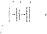
- FIG. 2 is flowchart of a method performed by the system of FIG. 1 for automatically selecting a key image for an image study according to one embodiment.
- FIGS. 3-4 and 6 illustrate graphical user interfaces for selecting and displaying key images for an image study according to various embodiments.
- FIG. 5 is a flowchart of a method performed by the system of FIG. 1 for automatically annotating key images for an image study according to one embodiment.
- FIG. 7 is a block diagram illustrating a montage template including a plurality of sub-containers.
- a plurality of hardware and software based devices, as well as a plurality of different structural components may be utilized to implement the invention.
- embodiments of the invention may include hardware, software, and electronic components or modules that, for purposes of discussion, may be illustrated and described as if the majority of the components were implemented solely in hardware.
- the electronic-based aspects of the invention may be implemented in software (e.g., stored on non-transitory computer-readable medium) executable by one or more processors.
- a plurality of hardware and software based devices, as well as a plurality of different structural components may be utilized to implement the invention.
- mobile device may include one or more electronic processors, one or more memory modules including non-transitory computer-readable medium, one or more input/output interfaces, and various connections (e.g., a system bus) connecting the components.
- FIG. 1 illustrates a system 100 for automatically selecting, arranging, and processing images.
- the system 100 includes a server 102 that includes a plurality of electrical and electronic components that provide power, operational control, and protection of the components within the server 102 .
- the server 102 may include an electronic processor 104 (e.g., a microprocessor, application-specific integrated circuit (ASIC), or another suitable electronic device), a memory 106 (e.g., a non-transitory, computer-readable storage medium), and a communication interface 108 .
- the electronic processor 104 , the memory 106 , and the communication interface 108 communicate over one or more connections or buses.
- FIG. 1 represents one example of a server and embodiments described herein may include a server with additional, fewer, or different components than the server 102 illustrated in FIG. 1 .
- the server 102 performs functionality in addition to the functionality described herein.
- the functionality performed by the server 102 i.e., through execution of instructions by the electronic processor 104
- the memory 106 may include read-only memory (“ROM”), random access memory (“RAM”) (e.g., dynamic RAM (“DRAM”), synchronous DRAM (“SDRAM”), and the like), electrically erasable programmable read-only memory (“EEPROM”), flash memory, a hard disk, a secure digital (“SD”) card, other suitable memory devices, or a combination thereof.
- the electronic processor 104 executes computer-readable instructions (“software”) stored in the memory 106 .
- the software may include firmware, one or more applications, program data, filters, rules, one or more program modules, and other executable instructions.
- the memory 106 stores an image selection application 110 .
- the image selection application 110 is configured to apply rules to automatically select, arrange, and process key images for an image study. It should be understood that the functionality described herein as being performed by the image selection application 110 may be distributed among multiple modules or applications (executed by the server 102 or multiple servers or devices). Also, in some embodiments, the functionality described herein as being performed by the image selection application 110 (or portions there) are performed by one or more software applications executed by other computing devices, such as the user device 120 described below.
- the memory 106 may also store rules applied by the image selection application 110 as described herein. However, in other embodiments, the rules may be stored separate from the application 110 .
- the server 102 acts as a gateway to the one or more image repositories 112 .
- the server 102 may be picture archiving and communication system (“PACS”) server that communicates with one or more image repositories 112 .
- PACS picture archiving and communication system
- the server 102 may be separate from a PACS server and may communicate with a PACS server to access images stored in one or more image repositories.
- the server 102 also communicates with a user device 120 (e.g., a personal computing device, such as but not limited to a laptop computer, a desktop computer, a terminal, a tablet computer, smart phone, a smart watch or other wearable, a smart television, and the like).
- the user device 120 may communicate with the server 102 via the communication network 111 .
- the user device 120 may communicate with the server 102 to access one or more images stored in the one or more image repositories 112 .
- a user may use a browser application executed by the user device 120 to access a web page provided by the server 102 for accessing (viewing) one or more images.
- the user may use a dedicated application executed by the user device 120 (a viewer application) to retrieve images from the image repositories 112 via the server 102 .
- the user device 120 includes similar components as the server 102 , such as an electronic processor 124 , a memory 126 , and a communication interface 128 for communicating with external devices, such as via the communication network 111 .
- the user device 120 also includes at least one output device 132 , such as one more display devices, one or more speakers, and the like configured to provide output to a user, and at least one input device 134 , such as a microphone, a keyboard, a cursor-control, device, a touchscreen, or the like configured to receive input from a user.
- FIG. 2 is a flowchart illustrating a method 300 performed by the server 102 (i.e., the electronic processor 104 executing instructions, such as the image selection application 110 ) for automatically selecting, arranging, and processing medical images according to some embodiments.
- the user device 120 may be configured to execute one or more software applications to perform all or a portion of the functionality described herein as being performed via execution of the image selection application 110 .
- the image selection application 110 performs the functionality described herein in response to various triggering events.
- the image selection application 110 perform the functionality described herein in response to a reviewing or reading physician accessing or viewing a particular image study.
- FIG. 3 shows a graphical user interface (“GUI”) 200 provided on a display device of the user device 120 .
- GUI graphical user interface
- the right tab 210 or right panel shows various images 212 , 214 , 216 , 218 , 220 , 222 , 224 , 226 from an image study available for manual selection by the user.
- the left tab 230 or left panel provided on the graphical user interface 200 includes a column or vertically oriented row of medical images 232 , 234 , 236 from a primary or current image study, along with a column or vertically oriented row of images 242 , 244 , 246 from a previous or prior image study that form a collection of images provided in a montage.
- the primary or current image study includes the most recent medical imaging procedure conducted on a particular patient or an image study needing a diagnosis.
- the electronic processor 104 is configured to determine a first key image (at block 304 ).
- the electronic processor 104 is configured to determine the first key image based on input received from a user selecting a particular image as a key image. For example, using the example GUI 200 illustrated in FIG. 3 , a user may manually select a key image by selecting one of the images 212 , 214 , 216 , 218 , 220 , 222 , 224 and moving the image, or an enlarged portion of the image, to the location of the image 232 in the left tab 230 . Thus, a user manually selects the first key image and positions the first key image within the montage on the left tab 230 .
- the user may manually select the first key image via a mouse click, an audio command via a microphone, a keyboard shortcut, a touchscreen action, or dragging or swiping an image.
- the electronic processor 104 is configured to automatically determine the first key image.
- the electronic processor 104 may be configured to automatically identify particular anatomy in an image, abnormalities in an image, normal findings in an image, or the like and, thus, may be configured to automatically select an image as a key image.
- the electronic processor 104 may be configured to automatically select key images using the image analytics as described in U.S. patent application Ser. Nos. 15/179,506 and 15/179,465, both filed Jun. 10, 2016. The entire content of each of these applications is incorporated by reference herein. Accordingly, it should be understood that, as used in the present application, a “key image” includes an image (or a portion thereof) (i) manually identified as a key image or (ii) automatically determined as a key image using various image analytics techniques and methodologies.
- an image included in a current image study may include an index lesion, defined as a key finding that is representative of the patient's problem or shows a pertinent negative finding.
- an index lesion could be identified because of an action of the reading physician or automatically because the anatomical position matches the location of a previously marked index lesion in the same patient.
- the electronic processor 104 is configured to automatically select another key image (e.g., the best matching comparison image that also contains the same index lesion) as described below.
- the electronic processor 104 is configured to automatically determine a second image based on one or more rules (at block 308 ).
- the rules may consider characteristics of the first key image, the exam type, the modality type, patient demographics, user findings, or the like.
- the rules may specify that when the first key image is selected from a magnetic resonance (“MR”) image (“MRI”) study and the initial diagnosis (provided by the user or automatically using image analytics) is “normal,” a predetermined set of images (of particular anatomy, with particular image characteristics or positions, or the like) should be automatically included in the set of key images.
- the rules may use metadata for an image or image study (e.g., DICOM header data), patient data, clinical data, and the like to automatically select the second key image.
- the second key image may be included in the same image study as the first key image or a different image study.
- the second key image is included in a prior comparison image study.
- the second key image may include a key image or nonkey image from a comparison image study.
- the second key image may be an image within the comparison study identified by the electronic processor 104 (regardless of whether the image was identified as a key image in the comparison image study) as being relevant, such as by analyzing and interpreting a diagnosis or finding (as recorded in a structured report for the comparison image study) for the comparison image study or by anatomically matching to a location of a key image in the current study.
- the first key image may be included in the comparison image study and the second key image may also be included in the comparison image study, another comparison image study, or a current image study being reviewed by a user.
- the electronic processor 104 is also configured to automatically generate text or labels for the key images.
- FIG. 3 illustrates descriptive text for an image, such as the date of the image study including the key image (e.g., “Jun. 30, 2017”).
- the electronic processor 104 is also configured to automatically generate text for a report (a structured report) associated with an image study based on the selection of key images.
- the electronic processor 104 may be configured to automatically generate text based on what images were compared, what anatomy was reviewed, measurements in images, or the like. This text can be displayed to a user for review, editing (as needed), and approval.
- the user may indicate (by selecting a button or other selection mechanism or issuing an audio or verbal command) when all of the key images have been selected (and annotated as needed), which may trigger the electronic processor 104 to generate text for the report.
- the rules described above are predefined for one or multiple users.
- the rules may also be manually configurable or changeable by particular users.
- the rules may be initially created or modified using machine learning.
- Machine learning generally refers to the ability of a computer program to learn without being explicitly programmed.
- a computer program e.g., a learning engine
- a model e.g., one or more algorithms
- Supervised learning involves presenting a computer program with example inputs and their desired (e.g., actual) outputs.
- the computer program is configured to learn a general rule (e.g., a model) that maps the inputs to the outputs.
- the computer program may be configured to perform deep machine learning using various types of methods and mechanisms.
- the computer program may perform deep machine learning using decision tree learning, association rule learning, artificial neural networks, inductive logic programming, support vector machines, clustering, Bayesian networks, reinforcement learning, representation learning, similarity and metric learning, sparse dictionary learning, and genetic algorithms.
- a computer program may ingest, parse, and understand data and progressively refine models for data analytics.
- a learning engine (executed by the server 102 or a separate computing device) may be configured to receive example inputs and outputs (“training information”) that allows the learning engine to automatically determine the rules described above.
- the training information includes information regarding what images were selected as key images for previously-reviewed image study, what images were annotated, a diagnosis for the image study or individual images, or the like.
- machine learning techniques as described in U.S. patent application Ser. Nos. 15/179,506 and 15/179,465 (incorporated by reference herein) may be used to automatically create or modify the rules described herein for automatically selecting key images. User interaction with selected key images may also be used as feedback to such a learning engine to further refine the rules.
- FIG. 6 shows the montage template 430 completed with medical images 432 , 434 , 436 , 438 , 440 , 442 , 444 , 446 , 448 provided in display sub-containers on a display device for multiple components of the back of the patient.
- the right tab 410 is a first tab
- the left tab defining the montage template 430 is a second tab.
- FIG. 7 schematically illustrates the configuration of a montage template 470 for a MRI of a knee of a patient.
- the montage template 470 may be disposed in, for instance, the left tab shown in FIG. 6 in one embodiment.
- the electronic processor 104 is configured to display a set of medical images corresponding to an image study on the at least one display device (at block 504 in FIG. 5 ) as shown by the images in the right tab 410 in FIG. 6 .
- the electronic processor 104 is also configured to display a montage template (at block 506 in FIG. 5 ) as shown by the montage templates 430 and 470 as illustrated in FIGS. 6 and 7 .
- the electronic processor 104 is configured to automatically select the montage template, such as based on a type of the image study (a modality type, procedure type, or the like), patient demographics, an anatomy, key images selected for the image study, user preferences, and the like.
- the electronic processor 104 is also configured to determine a key image included in the image study (at block 508 ).
- key images may be determined manually, automatically, or a combination thereof.
- each key image may be positioned within the montage template and, again, this positioning may be performed manually or automatically by the electronic processor 104 .
- the electronic processor 104 is configured to automatically annotate the key image (at block 510 ) and display the key image with the annotation within the montage template (at block 512 ).
- each montage template may include one or more pre-labeled sub-containers that specify required or recommended images. For example, as illustrated in FIG.
- a montage template for a lumbar spine MRI may include a sagittal ACL sub-container 474 , a sagittal PCL sub-container 476 , a sagittal medial meniscus sub-container 478 , a sagittal lateral meniscus sub-container 482 , a coronal sub-container 484 , an axial patella sub-container 486 , a sagittal lateral meniscus sub-container 488 , and a sagittal medial meniscus 492 sub-container.
- the electronic processor 104 is configured to automatically label each image (anatomy, positions), which eliminates the need for manual labeling, which can create delay and can introduce human errors.
- one or more sub-containers within a montage template may be associated with particular automated functionality.
- the electronic processor 104 is also configured to automatically label other images in an image study based on the labels automatically added to key images positioned within a montage template (e.g., based on an image's position in a series of images with respect to a key image).
- the electronic processor 104 may be configured to automatically select another key image that includes a corresponding image from a comparison image study.
- the electronic processor 104 may also be configured automatically analyze an image or multiple images to perform various types of analyses.
- the electronic processor 104 may be configured to compare and describe index lesions, identify anomalies, compare findings or anatomical locations, determine progressions, take measurements, add one or more graphical annotations (“stiations”) to an image, or the like. For example, an image from a brain MM showing an index nodular metastasis in the left occipital lobe may be added to a montage and the electronic processor 104 may be configured to automatically compare and describe index lesions, automatically add a brain MM image from the most recent comparison image study, and analyze and reports the progression or regression of the lesion.
- the results of such analysis may be provided as text (e.g., for inclusion in a structured report), a table, or the like.
- the electronic processor 104 may be configured to generate text based on the analysis and display the text to a user for review, editing, and approval.
- the electronic processor 104 may be configured to create a table of findings and analyze the table to determine disease changes, such as by comparing images using one or standard methodologies, such as RECIST 1.1 rules. Such analysis may be reported to the user and, optionally, added to a structured report.
- Particular sub-containers may also be designated as required or optional, and the electronic processor 104 may be configured to automatically prompt a user for a key image for such sub-containers and may be configured to prevent the user from submitting or saving a report or finding for an image study until all required key images have been added to the montage.
- Different processing may be associated with different sub-containers of a montage template and may also differ depending on the key image positioned within a particular sub-container (or key images positioned in other sub-containers of the montage template).
- a user via the GUI
- the processing functionalities may be configured to be customized for particular users or groups of users.
- the processing for one or more sub-containers may be based on findings or other input from a user and, thus, may be dynamically updated based on user interaction.
- the processing functionality associated with particular montage template may be automatically generated or modified using artificial intelligence as described above for the rules for selecting key images.
- a learning engine may be configured to automatically learn data patterns associated with labels or actions taken by a user to define processing for a particular sub-container.
- a learning engine may also be configured to consider processing performed when a previous exam was read, such as a comparison image study. For example, under the appropriate circumstances, when an image is added to a montage, the electronic processor 104 may attempt to segment and measure the volume of anomalies if this was the processing performed when the comparison exam was read and reported.
- the electronic processor 104 may be configured to detect aortic abnormalities or other specific abnormalities that were assessed on the prior image study or clinical report. Also, feedback from a user regarding automatically-generated text could be provided as part of a closed feedback loop to help the system 100 learn the proper behaviors for processing key images.
- the labels associated a montage template may also be used automatically learn anatomy based on user actions. For example, labeled images may be used as training data for a learning engine.
- embodiments described herein provide, among other things, methods and systems for automatically selecting, arranging, and processing key images for a medical image study.
- various rules may be applied by the systems and methods to quickly and effectively process image studies that may include hundreds or thousands of images without requiring or minimizing user input or interaction.
- Machine learning techniques may be used to establish or modify such rules, which further improve the efficiency and effectiveness of the systems and methods.
Landscapes
- Health & Medical Sciences (AREA)
- Nuclear Medicine, Radiotherapy & Molecular Imaging (AREA)
- Radiology & Medical Imaging (AREA)
- Engineering & Computer Science (AREA)
- Epidemiology (AREA)
- General Health & Medical Sciences (AREA)
- Medical Informatics (AREA)
- Primary Health Care (AREA)
- Public Health (AREA)
- Medical Treatment And Welfare Office Work (AREA)
- Measuring And Recording Apparatus For Diagnosis (AREA)
Abstract
Description
Claims (16)
Priority Applications (1)
| Application Number | Priority Date | Filing Date | Title |
|---|---|---|---|
| US15/840,689 US10832808B2 (en) | 2017-12-13 | 2017-12-13 | Automated selection, arrangement, and processing of key images |
Applications Claiming Priority (1)
| Application Number | Priority Date | Filing Date | Title |
|---|---|---|---|
| US15/840,689 US10832808B2 (en) | 2017-12-13 | 2017-12-13 | Automated selection, arrangement, and processing of key images |
Publications (2)
| Publication Number | Publication Date |
|---|---|
| US20190180863A1 US20190180863A1 (en) | 2019-06-13 |
| US10832808B2 true US10832808B2 (en) | 2020-11-10 |
Family
ID=66697179
Family Applications (1)
| Application Number | Title | Priority Date | Filing Date |
|---|---|---|---|
| US15/840,689 Active 2038-01-16 US10832808B2 (en) | 2017-12-13 | 2017-12-13 | Automated selection, arrangement, and processing of key images |
Country Status (1)
| Country | Link |
|---|---|
| US (1) | US10832808B2 (en) |
Families Citing this family (1)
| Publication number | Priority date | Publication date | Assignee | Title |
|---|---|---|---|---|
| US20210398653A1 (en) * | 2020-06-17 | 2021-12-23 | Fovia, Inc. | Key image updating multiple stacks |
Citations (116)
| Publication number | Priority date | Publication date | Assignee | Title |
|---|---|---|---|---|
| US6090044A (en) | 1997-12-10 | 2000-07-18 | Bishop; Jeffrey B. | System for diagnosing medical conditions using a neural network |
| US6574304B1 (en) | 2002-09-13 | 2003-06-03 | Ge Medical Systems Global Technology Company, Llc | Computer aided acquisition of medical images |
| US20030147465A1 (en) * | 2001-12-20 | 2003-08-07 | Canon Kabushiki Kaisha | Method of automatic production of image presentations |
| US20040147840A1 (en) | 2002-11-08 | 2004-07-29 | Bhavani Duggirala | Computer aided diagnostic assistance for medical imaging |
| US6819790B2 (en) | 2002-04-12 | 2004-11-16 | The University Of Chicago | Massive training artificial neural network (MTANN) for detecting abnormalities in medical images |
| US6836558B2 (en) | 2000-03-28 | 2004-12-28 | Arch Development Corporation | Method, system and computer readable medium for identifying chest radiographs using image mapping and template matching techniques |
| US20050010098A1 (en) | 2003-04-11 | 2005-01-13 | Sigmund Frigstad | Method and apparatus for knowledge based diagnostic imaging |
| US20050010445A1 (en) | 2003-06-27 | 2005-01-13 | Arun Krishnan | CAD (computer-aided decision) support for medical imaging using machine learning to adapt CAD process with knowledge collected during routine use of CAD system |
| US20050021375A1 (en) | 2002-04-04 | 2005-01-27 | Satoshi Shimizu | Cooperative diagnosis system |
| US20050049497A1 (en) * | 2003-06-25 | 2005-03-03 | Sriram Krishnan | Systems and methods for automated diagnosis and decision support for breast imaging |
| US20050113960A1 (en) | 2003-11-26 | 2005-05-26 | Karau Kelly L. | Methods and systems for computer aided targeting |
| US20050231416A1 (en) | 2004-04-14 | 2005-10-20 | Rowe Richard L | Relational millimeter-wave interrogating |
| US20050251013A1 (en) | 2004-03-23 | 2005-11-10 | Sriram Krishnan | Systems and methods providing automated decision support for medical imaging |
| US20050255434A1 (en) | 2004-02-27 | 2005-11-17 | University Of Florida Research Foundation, Inc. | Interactive virtual characters for training including medical diagnosis training |
| US20060110018A1 (en) | 2004-11-22 | 2006-05-25 | Shoupu Chen | Automatic abnormal tissue detection in MRI images |
| US20060159325A1 (en) | 2005-01-18 | 2006-07-20 | Trestle Corporation | System and method for review in studies including toxicity and risk assessment studies |
| US20060228015A1 (en) | 2005-04-08 | 2006-10-12 | 361° Systems, Inc. | System and method for detection and display of diseases and abnormalities using confidence imaging |
| US7130457B2 (en) | 2001-07-17 | 2006-10-31 | Accuimage Diagnostics Corp. | Systems and graphical user interface for analyzing body images |
| US20060274928A1 (en) | 2005-06-02 | 2006-12-07 | Jeffrey Collins | System and method of computer-aided detection |
| US20070036402A1 (en) | 2005-07-22 | 2007-02-15 | Cahill Nathan D | Abnormality detection in medical images |
| US20070047786A1 (en) | 2005-08-25 | 2007-03-01 | Lenovo (Singapore) Pte. Ltd. | System and method for creating robust training data from MRI images |
| US20070078679A1 (en) | 2005-10-04 | 2007-04-05 | Greg Rose | After-hours radiology system |
| US20070118399A1 (en) | 2005-11-22 | 2007-05-24 | Avinash Gopal B | System and method for integrated learning and understanding of healthcare informatics |
| US20070118055A1 (en) | 2005-11-04 | 2007-05-24 | Smith & Nephew, Inc. | Systems and methods for facilitating surgical procedures involving custom medical implants |
| US20070272747A1 (en) | 2006-05-25 | 2007-11-29 | Woods Sherrod A | Method and system for managing inventories of orthopaedic implants |
| US20080046286A1 (en) | 2005-09-16 | 2008-02-21 | Halsted Mark J | Computer implemented healthcare monitoring, notifying and/or scheduling system |
| US20080126982A1 (en) * | 2006-09-18 | 2008-05-29 | Agfa Inc. | Imaging history display system and method |
| US20080163070A1 (en) * | 2007-01-03 | 2008-07-03 | General Electric Company | Method and system for automating a user interface |
| US20080226147A1 (en) | 2007-03-16 | 2008-09-18 | Sti Medical Systems, Llc | Method to provide automated quality feedback to imaging devices to achieve standardized imaging data |
| US7428323B2 (en) | 2000-11-14 | 2008-09-23 | Yitzchak Hillman | Method and system for automatic diagnosis of possible brain disease |
| US20090080731A1 (en) | 2007-09-26 | 2009-03-26 | Siemens Medical Solutions Usa, Inc. | System and Method for Multiple-Instance Learning for Computer Aided Diagnosis |
| US20090092300A1 (en) | 2007-10-03 | 2009-04-09 | Siemens Medical Solutions Usa, Inc. | System and Method for Lesion Detection Using Locally Adjustable Priors |
| US20090274384A1 (en) * | 2007-10-31 | 2009-11-05 | Mckesson Information Solutions Llc | Methods, computer program products, apparatuses, and systems to accommodate decision support and reference case management for diagnostic imaging |
| US20090299977A1 (en) * | 2008-05-28 | 2009-12-03 | Siemens Medical Solutions Usa, Inc. | Method for Automatic Labeling of Unstructured Data Fragments From Electronic Medical Records |
| US20090326989A1 (en) | 2005-03-23 | 2009-12-31 | Schmitt Brett A | Interactive information management system and method |
| US20100042422A1 (en) | 2008-08-15 | 2010-02-18 | Adam Summers | System and method for computing and displaying a score with an associated visual quality indicator |
| US20100082692A1 (en) * | 2008-09-24 | 2010-04-01 | Akinola Akinyemi | Method and apparatus for classification of coronary artery image data |
| US7761345B1 (en) | 1998-04-21 | 2010-07-20 | Socrates Holding GmbH | Decision aid |
| US7788040B2 (en) | 2003-12-19 | 2010-08-31 | Siemens Medical Solutions Usa, Inc. | System for managing healthcare data including genomic and other patient specific information |
| US20100312734A1 (en) | 2005-10-07 | 2010-12-09 | Bernard Widrow | System and method for cognitive memory and auto-associative neural network based pattern recognition |
| US7949167B2 (en) | 2008-06-12 | 2011-05-24 | Siemens Medical Solutions Usa, Inc. | Automatic learning of image features to predict disease |
| US20110123079A1 (en) | 2009-11-24 | 2011-05-26 | Greg Gustafson | Mammography information system |
| US8021045B2 (en) | 2008-10-27 | 2011-09-20 | Carestream Health, Inc. | Integrated portable digital X-ray imaging system |
| US20110228995A1 (en) * | 2008-11-28 | 2011-09-22 | Fujifilm Medical Systems Usa, Inc. | System and Method for Propagation of Spine Labeling |
| US20110301447A1 (en) | 2010-06-07 | 2011-12-08 | Sti Medical Systems, Llc | Versatile video interpretation, visualization, and management system |
| US20120001853A1 (en) * | 2010-07-01 | 2012-01-05 | Sho Tanaka | Medical image display apparatus and medical image management apparatus |
| US8107700B2 (en) | 2007-11-21 | 2012-01-31 | Merge Cad Inc. | System and method for efficient workflow in reading medical image data |
| US20120054652A1 (en) | 2010-08-27 | 2012-03-01 | Canon Kabushiki Kaisha | Diagnosis support apparatus, diagnosis support system, diagnosis support control method, and computer-readable memory |
| US20120088981A1 (en) | 2010-10-07 | 2012-04-12 | Siemens Medical Solutions Usa, Inc. | Matching of Regions of Interest Across Multiple Views |
| US8199985B2 (en) | 2006-03-24 | 2012-06-12 | Exini Diagnostics Aktiebolag | Automatic interpretation of 3-D medicine images of the brain and methods for producing intermediate results |
| US20120172700A1 (en) | 2010-05-21 | 2012-07-05 | Siemens Medical Solutions Usa, Inc. | Systems and Methods for Viewing and Analyzing Anatomical Structures |
| US20120189176A1 (en) | 2010-11-26 | 2012-07-26 | Giger Maryellen L | Method, system, software and medium for advanced intelligent image analysis and display of medical images and information |
| US20120237109A1 (en) | 2011-03-14 | 2012-09-20 | University Of Warwick | Histology analysis |
| US20120250961A1 (en) * | 2011-03-30 | 2012-10-04 | Fujifilm Corporation | Medical report generation apparatus, method and program |
| US20120283574A1 (en) | 2011-05-06 | 2012-11-08 | Park Sun Young | Diagnosis Support System Providing Guidance to a User by Automated Retrieval of Similar Cancer Images with User Feedback |
| US20120310399A1 (en) | 2011-06-06 | 2012-12-06 | Biomet Manufacturing Corp. | Pre-operative planning and manufacturing method for orthopedic procedure |
| US8340437B2 (en) * | 2007-05-29 | 2012-12-25 | University Of Iowa Research Foundation | Methods and systems for determining optimal features for classifying patterns or objects in images |
| US20120328178A1 (en) | 2010-06-25 | 2012-12-27 | Cireca Theranostics, Llc | Method for analyzing biological specimens by spectral imaging |
| US8345940B2 (en) | 2005-10-25 | 2013-01-01 | Bracco Imaging S.P.A. | Method and system for automatic processing and evaluation of images, particularly diagnostic images |
| US20130090554A1 (en) | 2010-06-24 | 2013-04-11 | Uc-Care Ltd. | Focused prostate cancer treatment system and method |
| US20130149682A1 (en) | 2011-12-09 | 2013-06-13 | Medicolegal Consultants International, LLC | Methods and systems for simulation based medical education |
| US8478698B1 (en) | 2010-03-17 | 2013-07-02 | James Mah | Methods and systems for employing artificial intelligence in automated orthodontic diagnosis and treatment planning |
| US20130204115A1 (en) | 2010-06-01 | 2013-08-08 | Synarc Inc. | Computer based analysis of mri images |
| US20130290225A1 (en) | 2010-07-23 | 2013-10-31 | General Electric Company | Systems and methods for selecting and analyzing particles in a biological tissue |
| US8583450B2 (en) | 2004-07-29 | 2013-11-12 | Ims Health Incorporated | Doctor performance evaluation tool for consumers |
| US20130304751A1 (en) | 2012-05-14 | 2013-11-14 | Sony Corporation | Information processing apparatus, information processing method, and information processing program |
| US20130314434A1 (en) | 2012-05-25 | 2013-11-28 | PicMonkey Inc. | System and method for image collage editing |
| US20140010432A1 (en) | 2011-03-29 | 2014-01-09 | Koninklijke Philips N.V. | Image acquisition and/or image related parameter recommender |
| US8687867B1 (en) | 2008-09-16 | 2014-04-01 | Icad, Inc. | Computer-aided detection and classification of suspicious masses in breast imagery |
| US20140121487A1 (en) | 2012-10-26 | 2014-05-01 | Pixie Scientific, Llc | Health diagnostic systems and methods |
| US8727989B2 (en) | 2009-06-30 | 2014-05-20 | Kabushiki Kaisha Toshiba | Automatic diagnosis support apparatus, ultrasonic diagnosis apparatus, and automatic diagnosis support method |
| US20140155763A1 (en) | 2012-12-03 | 2014-06-05 | Ben F. Bruce | Medical analysis and diagnostic system |
| US20140161337A1 (en) | 2012-12-06 | 2014-06-12 | Siemens Medical Solutions Usa, Inc. | Adaptive Anatomical Region Prediction |
| US20140185888A1 (en) | 2013-01-03 | 2014-07-03 | Siemens Aktiengesellschaft | Method and system for lesion candidate detection |
| US20140219526A1 (en) | 2013-02-05 | 2014-08-07 | Children's National Medical Center | Device and method for classifying a condition based on image analysis |
| US20140218552A1 (en) * | 2013-02-01 | 2014-08-07 | Htc Corporation | Electronic device and image composition method thereof |
| US20140218397A1 (en) | 2013-02-04 | 2014-08-07 | Mckesson Financial Holdings | Method and apparatus for providing virtual device planning |
| US20140244309A1 (en) | 2011-11-08 | 2014-08-28 | Revon Systems, Llc | Systems and methods for assembling electronic medical records |
| US20140257854A1 (en) | 2011-09-08 | 2014-09-11 | Radlogics, Inc. | Methods and Systems for Analyzing and Reporting Medical Images |
| US20140279807A1 (en) | 2013-03-14 | 2014-09-18 | Siemens Aktiengesellschaft | Rules-based management system and method for processing medical information |
| US20140314292A1 (en) | 2011-08-22 | 2014-10-23 | Siemens Corporation | Method and system for integrated radiological and pathological information for diagnosis, therapy selection, and monitoring |
| US20140313222A1 (en) | 2011-06-27 | 2014-10-23 | Koninklijke Philips Electronics N.V. | Anatomical tagging of findings in image data of serial studies |
| US8879813B1 (en) | 2013-10-22 | 2014-11-04 | Eyenuk, Inc. | Systems and methods for automated interest region detection in retinal images |
| US20140375671A1 (en) | 2011-11-28 | 2014-12-25 | University Of Chicago | Method, system, software and medium for advanced image-based arrays for analysis and display of biomedical information |
| US20150065803A1 (en) | 2013-09-05 | 2015-03-05 | Erik Scott DOUGLAS | Apparatuses and methods for mobile imaging and analysis |
| US20150072371A1 (en) | 2012-05-24 | 2015-03-12 | Nec Corporation | Pathological diagnosis results assessment system, pathological diagnosis results assessment method, and pathological diagnosis results assessment device |
| US20150091778A1 (en) * | 2013-09-30 | 2015-04-02 | Toshiba Medical Systems Corporation | Medical image display system and method |
| US20150103170A1 (en) | 2013-10-11 | 2015-04-16 | Ccc Information Services | Image capturing and automatic labeling system |
| US20150205917A1 (en) | 2012-08-22 | 2015-07-23 | Koninklijke Philips N.V. | Automatic detection and retrieval of prior annotations relevant for an imaging study for efficient viewing and reporting |
| US9092727B1 (en) * | 2011-08-11 | 2015-07-28 | D.R. Systems, Inc. | Exam type mapping |
| US9089303B2 (en) | 2009-05-29 | 2015-07-28 | Lubax, Inc. | Automated assessment of skin lesions using image library |
| US20150235365A1 (en) * | 2012-10-01 | 2015-08-20 | Koninklijke Philips N.V. | Multi-study medical image navigation |
| US20150230876A1 (en) | 2012-03-30 | 2015-08-20 | Sandance Technology Llc | Systems and Methods for Determining Suitability of a Mechanical Implant for a Medical Procedure |
| US20150262014A1 (en) * | 2014-03-11 | 2015-09-17 | Kabushiki Kaisha Toshiba | Image interpretation report creating apparatus and image interpretation report creating system |
| US20150287192A1 (en) | 2012-12-20 | 2015-10-08 | Olympus Corporation | Image processing device, electronic device, endoscope apparatus, information storage device, and image processing method |
| US20150302317A1 (en) | 2014-04-22 | 2015-10-22 | Microsoft Corporation | Non-greedy machine learning for high accuracy |
| US20150320365A1 (en) | 2014-05-07 | 2015-11-12 | Lifetrack Medical Systems, Inc. | Characterizing States of Subject |
| US20150325018A1 (en) * | 2014-05-09 | 2015-11-12 | General Electric Company | Standalone annotations of axial-view spine images |
| US20150332111A1 (en) | 2014-05-15 | 2015-11-19 | International Business Machines Corporation | Automatic generation of semantic description of visual findings in medical images |
| US20150331995A1 (en) | 2014-05-14 | 2015-11-19 | Tiecheng Zhao | Evolving contextual clinical data engine for medical data processing |
| US20160005106A1 (en) | 2013-02-07 | 2016-01-07 | Crisalix Sa | 3d platform for aesthetic simulation |
| US9245337B2 (en) | 2002-10-15 | 2016-01-26 | Definiens Ag | Context driven image mining to generate image-based biomarkers |
| US20160041733A1 (en) * | 2013-04-11 | 2016-02-11 | Koninklijke Philips N.V. | Enabling a user to study image data |
| US20160275138A1 (en) | 2015-03-17 | 2016-09-22 | Avrohom C. Rutenberg | Method and apparatus for ranking and dynamically displaying information |
| US20160283489A1 (en) | 2006-12-20 | 2016-09-29 | Victor Jr David Uy | System and method for categorically scoring electronic documents |
| US20160292155A1 (en) * | 2013-11-28 | 2016-10-06 | Agfa Healthcare | A system and method to pre-fetch comparison medical studies |
| US20160350919A1 (en) * | 2015-06-01 | 2016-12-01 | Virtual Radiologic Corporation | Medical evaluation machine learning workflows and processes |
| US20160350480A1 (en) * | 2015-05-26 | 2016-12-01 | Virtual Radiologic Corporation | Radiology workflow coordination techniques |
| US20160364862A1 (en) | 2015-06-12 | 2016-12-15 | Merge Healthcare Incorporated | Methods and Systems for Performing Image Analytics Using Graphical Reporting Associated with Clinical Images |
| US20170039321A1 (en) * | 2015-04-30 | 2017-02-09 | D.R. Systems, Inc. | Database systems and interactive user interfaces for dynamic interaction with, and sorting of, digital medical image data |
| US20170091937A1 (en) | 2014-06-10 | 2017-03-30 | Ventana Medical Systems, Inc. | Methods and systems for assessing risk of breast cancer recurrence |
| US20170169192A1 (en) * | 2014-02-20 | 2017-06-15 | Koninklijke Philips N.V. | Inserting structured content in itemized reports |
| US20170262584A1 (en) * | 2014-09-10 | 2017-09-14 | Benoît GALLIX | Method for automatically generating representations of imaging data and interactive visual imaging reports (ivir) |
| US20180144421A1 (en) | 2016-11-21 | 2018-05-24 | Velites Consulting Group, LLC | System and Methods for Complaint Evaluation |
| US20180260949A1 (en) * | 2017-03-09 | 2018-09-13 | Kevin Augustus Kreeger | Automatic key frame detection |
| US10127662B1 (en) * | 2014-08-11 | 2018-11-13 | D.R. Systems, Inc. | Systems and user interfaces for automated generation of matching 2D series of medical images and efficient annotation of matching 2D medical images |
-
2017
- 2017-12-13 US US15/840,689 patent/US10832808B2/en active Active
Patent Citations (135)
| Publication number | Priority date | Publication date | Assignee | Title |
|---|---|---|---|---|
| US6090044A (en) | 1997-12-10 | 2000-07-18 | Bishop; Jeffrey B. | System for diagnosing medical conditions using a neural network |
| US7761345B1 (en) | 1998-04-21 | 2010-07-20 | Socrates Holding GmbH | Decision aid |
| US6836558B2 (en) | 2000-03-28 | 2004-12-28 | Arch Development Corporation | Method, system and computer readable medium for identifying chest radiographs using image mapping and template matching techniques |
| US7428323B2 (en) | 2000-11-14 | 2008-09-23 | Yitzchak Hillman | Method and system for automatic diagnosis of possible brain disease |
| US7130457B2 (en) | 2001-07-17 | 2006-10-31 | Accuimage Diagnostics Corp. | Systems and graphical user interface for analyzing body images |
| US20030147465A1 (en) * | 2001-12-20 | 2003-08-07 | Canon Kabushiki Kaisha | Method of automatic production of image presentations |
| US20050021375A1 (en) | 2002-04-04 | 2005-01-27 | Satoshi Shimizu | Cooperative diagnosis system |
| US6819790B2 (en) | 2002-04-12 | 2004-11-16 | The University Of Chicago | Massive training artificial neural network (MTANN) for detecting abnormalities in medical images |
| US6687329B1 (en) * | 2002-09-13 | 2004-02-03 | Ge Medical Systems Global Technology Company, Llc | Computer aided acquisition of medical images |
| US6574304B1 (en) | 2002-09-13 | 2003-06-03 | Ge Medical Systems Global Technology Company, Llc | Computer aided acquisition of medical images |
| US9245337B2 (en) | 2002-10-15 | 2016-01-26 | Definiens Ag | Context driven image mining to generate image-based biomarkers |
| US20040147840A1 (en) | 2002-11-08 | 2004-07-29 | Bhavani Duggirala | Computer aided diagnostic assistance for medical imaging |
| US20050010098A1 (en) | 2003-04-11 | 2005-01-13 | Sigmund Frigstad | Method and apparatus for knowledge based diagnostic imaging |
| US20050049497A1 (en) * | 2003-06-25 | 2005-03-03 | Sriram Krishnan | Systems and methods for automated diagnosis and decision support for breast imaging |
| US20100121178A1 (en) | 2003-06-25 | 2010-05-13 | Sriram Krishnan | Systems and Methods for Automated Diagnosis and Decision Support for Breast Imaging |
| US7640051B2 (en) | 2003-06-25 | 2009-12-29 | Siemens Medical Solutions Usa, Inc. | Systems and methods for automated diagnosis and decision support for breast imaging |
| US20050010445A1 (en) | 2003-06-27 | 2005-01-13 | Arun Krishnan | CAD (computer-aided decision) support for medical imaging using machine learning to adapt CAD process with knowledge collected during routine use of CAD system |
| US7529394B2 (en) | 2003-06-27 | 2009-05-05 | Siemens Medical Solutions Usa, Inc. | CAD (computer-aided decision) support for medical imaging using machine learning to adapt CAD process with knowledge collected during routine use of CAD system |
| US20050113960A1 (en) | 2003-11-26 | 2005-05-26 | Karau Kelly L. | Methods and systems for computer aided targeting |
| US7788040B2 (en) | 2003-12-19 | 2010-08-31 | Siemens Medical Solutions Usa, Inc. | System for managing healthcare data including genomic and other patient specific information |
| US20050255434A1 (en) | 2004-02-27 | 2005-11-17 | University Of Florida Research Foundation, Inc. | Interactive virtual characters for training including medical diagnosis training |
| US20050251013A1 (en) | 2004-03-23 | 2005-11-10 | Sriram Krishnan | Systems and methods providing automated decision support for medical imaging |
| US7672491B2 (en) | 2004-03-23 | 2010-03-02 | Siemens Medical Solutions Usa, Inc. | Systems and methods providing automated decision support and medical imaging |
| US20050231416A1 (en) | 2004-04-14 | 2005-10-20 | Rowe Richard L | Relational millimeter-wave interrogating |
| US8583450B2 (en) | 2004-07-29 | 2013-11-12 | Ims Health Incorporated | Doctor performance evaluation tool for consumers |
| US20060110018A1 (en) | 2004-11-22 | 2006-05-25 | Shoupu Chen | Automatic abnormal tissue detection in MRI images |
| US20060159325A1 (en) | 2005-01-18 | 2006-07-20 | Trestle Corporation | System and method for review in studies including toxicity and risk assessment studies |
| US20090326989A1 (en) | 2005-03-23 | 2009-12-31 | Schmitt Brett A | Interactive information management system and method |
| US20060228015A1 (en) | 2005-04-08 | 2006-10-12 | 361° Systems, Inc. | System and method for detection and display of diseases and abnormalities using confidence imaging |
| US20060274928A1 (en) | 2005-06-02 | 2006-12-07 | Jeffrey Collins | System and method of computer-aided detection |
| US20070036402A1 (en) | 2005-07-22 | 2007-02-15 | Cahill Nathan D | Abnormality detection in medical images |
| US20070047786A1 (en) | 2005-08-25 | 2007-03-01 | Lenovo (Singapore) Pte. Ltd. | System and method for creating robust training data from MRI images |
| US20080046286A1 (en) | 2005-09-16 | 2008-02-21 | Halsted Mark J | Computer implemented healthcare monitoring, notifying and/or scheduling system |
| US20070078679A1 (en) | 2005-10-04 | 2007-04-05 | Greg Rose | After-hours radiology system |
| US20100312734A1 (en) | 2005-10-07 | 2010-12-09 | Bernard Widrow | System and method for cognitive memory and auto-associative neural network based pattern recognition |
| US8345940B2 (en) | 2005-10-25 | 2013-01-01 | Bracco Imaging S.P.A. | Method and system for automatic processing and evaluation of images, particularly diagnostic images |
| US20070118055A1 (en) | 2005-11-04 | 2007-05-24 | Smith & Nephew, Inc. | Systems and methods for facilitating surgical procedures involving custom medical implants |
| US20070118399A1 (en) | 2005-11-22 | 2007-05-24 | Avinash Gopal B | System and method for integrated learning and understanding of healthcare informatics |
| US8199985B2 (en) | 2006-03-24 | 2012-06-12 | Exini Diagnostics Aktiebolag | Automatic interpretation of 3-D medicine images of the brain and methods for producing intermediate results |
| US20070272747A1 (en) | 2006-05-25 | 2007-11-29 | Woods Sherrod A | Method and system for managing inventories of orthopaedic implants |
| US20080126982A1 (en) * | 2006-09-18 | 2008-05-29 | Agfa Inc. | Imaging history display system and method |
| US20160283489A1 (en) | 2006-12-20 | 2016-09-29 | Victor Jr David Uy | System and method for categorically scoring electronic documents |
| US20080163070A1 (en) * | 2007-01-03 | 2008-07-03 | General Electric Company | Method and system for automating a user interface |
| US20080226147A1 (en) | 2007-03-16 | 2008-09-18 | Sti Medical Systems, Llc | Method to provide automated quality feedback to imaging devices to achieve standardized imaging data |
| US8340437B2 (en) * | 2007-05-29 | 2012-12-25 | University Of Iowa Research Foundation | Methods and systems for determining optimal features for classifying patterns or objects in images |
| US20090080731A1 (en) | 2007-09-26 | 2009-03-26 | Siemens Medical Solutions Usa, Inc. | System and Method for Multiple-Instance Learning for Computer Aided Diagnosis |
| US20090092300A1 (en) | 2007-10-03 | 2009-04-09 | Siemens Medical Solutions Usa, Inc. | System and Method for Lesion Detection Using Locally Adjustable Priors |
| US20090274384A1 (en) * | 2007-10-31 | 2009-11-05 | Mckesson Information Solutions Llc | Methods, computer program products, apparatuses, and systems to accommodate decision support and reference case management for diagnostic imaging |
| US8107700B2 (en) | 2007-11-21 | 2012-01-31 | Merge Cad Inc. | System and method for efficient workflow in reading medical image data |
| US20090299977A1 (en) * | 2008-05-28 | 2009-12-03 | Siemens Medical Solutions Usa, Inc. | Method for Automatic Labeling of Unstructured Data Fragments From Electronic Medical Records |
| US7949167B2 (en) | 2008-06-12 | 2011-05-24 | Siemens Medical Solutions Usa, Inc. | Automatic learning of image features to predict disease |
| US20100042422A1 (en) | 2008-08-15 | 2010-02-18 | Adam Summers | System and method for computing and displaying a score with an associated visual quality indicator |
| US8687867B1 (en) | 2008-09-16 | 2014-04-01 | Icad, Inc. | Computer-aided detection and classification of suspicious masses in breast imagery |
| US20100082692A1 (en) * | 2008-09-24 | 2010-04-01 | Akinola Akinyemi | Method and apparatus for classification of coronary artery image data |
| US8021045B2 (en) | 2008-10-27 | 2011-09-20 | Carestream Health, Inc. | Integrated portable digital X-ray imaging system |
| US20110228995A1 (en) * | 2008-11-28 | 2011-09-22 | Fujifilm Medical Systems Usa, Inc. | System and Method for Propagation of Spine Labeling |
| US9089303B2 (en) | 2009-05-29 | 2015-07-28 | Lubax, Inc. | Automated assessment of skin lesions using image library |
| US8727989B2 (en) | 2009-06-30 | 2014-05-20 | Kabushiki Kaisha Toshiba | Automatic diagnosis support apparatus, ultrasonic diagnosis apparatus, and automatic diagnosis support method |
| US20110123079A1 (en) | 2009-11-24 | 2011-05-26 | Greg Gustafson | Mammography information system |
| US8478698B1 (en) | 2010-03-17 | 2013-07-02 | James Mah | Methods and systems for employing artificial intelligence in automated orthodontic diagnosis and treatment planning |
| US20120172700A1 (en) | 2010-05-21 | 2012-07-05 | Siemens Medical Solutions Usa, Inc. | Systems and Methods for Viewing and Analyzing Anatomical Structures |
| US20130204115A1 (en) | 2010-06-01 | 2013-08-08 | Synarc Inc. | Computer based analysis of mri images |
| US20110301447A1 (en) | 2010-06-07 | 2011-12-08 | Sti Medical Systems, Llc | Versatile video interpretation, visualization, and management system |
| US20130090554A1 (en) | 2010-06-24 | 2013-04-11 | Uc-Care Ltd. | Focused prostate cancer treatment system and method |
| US20120328178A1 (en) | 2010-06-25 | 2012-12-27 | Cireca Theranostics, Llc | Method for analyzing biological specimens by spectral imaging |
| US20120001853A1 (en) * | 2010-07-01 | 2012-01-05 | Sho Tanaka | Medical image display apparatus and medical image management apparatus |
| US20130290225A1 (en) | 2010-07-23 | 2013-10-31 | General Electric Company | Systems and methods for selecting and analyzing particles in a biological tissue |
| US20120054652A1 (en) | 2010-08-27 | 2012-03-01 | Canon Kabushiki Kaisha | Diagnosis support apparatus, diagnosis support system, diagnosis support control method, and computer-readable memory |
| US20120088981A1 (en) | 2010-10-07 | 2012-04-12 | Siemens Medical Solutions Usa, Inc. | Matching of Regions of Interest Across Multiple Views |
| US20120189176A1 (en) | 2010-11-26 | 2012-07-26 | Giger Maryellen L | Method, system, software and medium for advanced intelligent image analysis and display of medical images and information |
| US20120237109A1 (en) | 2011-03-14 | 2012-09-20 | University Of Warwick | Histology analysis |
| US20140010432A1 (en) | 2011-03-29 | 2014-01-09 | Koninklijke Philips N.V. | Image acquisition and/or image related parameter recommender |
| US20120250961A1 (en) * | 2011-03-30 | 2012-10-04 | Fujifilm Corporation | Medical report generation apparatus, method and program |
| US20120283574A1 (en) | 2011-05-06 | 2012-11-08 | Park Sun Young | Diagnosis Support System Providing Guidance to a User by Automated Retrieval of Similar Cancer Images with User Feedback |
| US20120310399A1 (en) | 2011-06-06 | 2012-12-06 | Biomet Manufacturing Corp. | Pre-operative planning and manufacturing method for orthopedic procedure |
| US20140313222A1 (en) | 2011-06-27 | 2014-10-23 | Koninklijke Philips Electronics N.V. | Anatomical tagging of findings in image data of serial studies |
| US9092727B1 (en) * | 2011-08-11 | 2015-07-28 | D.R. Systems, Inc. | Exam type mapping |
| US20140314292A1 (en) | 2011-08-22 | 2014-10-23 | Siemens Corporation | Method and system for integrated radiological and pathological information for diagnosis, therapy selection, and monitoring |
| US20140257854A1 (en) | 2011-09-08 | 2014-09-11 | Radlogics, Inc. | Methods and Systems for Analyzing and Reporting Medical Images |
| US20140244309A1 (en) | 2011-11-08 | 2014-08-28 | Revon Systems, Llc | Systems and methods for assembling electronic medical records |
| US20140375671A1 (en) | 2011-11-28 | 2014-12-25 | University Of Chicago | Method, system, software and medium for advanced image-based arrays for analysis and display of biomedical information |
| US20130149682A1 (en) | 2011-12-09 | 2013-06-13 | Medicolegal Consultants International, LLC | Methods and systems for simulation based medical education |
| US20150230876A1 (en) | 2012-03-30 | 2015-08-20 | Sandance Technology Llc | Systems and Methods for Determining Suitability of a Mechanical Implant for a Medical Procedure |
| US20130304751A1 (en) | 2012-05-14 | 2013-11-14 | Sony Corporation | Information processing apparatus, information processing method, and information processing program |
| US20150072371A1 (en) | 2012-05-24 | 2015-03-12 | Nec Corporation | Pathological diagnosis results assessment system, pathological diagnosis results assessment method, and pathological diagnosis results assessment device |
| US20130314434A1 (en) | 2012-05-25 | 2013-11-28 | PicMonkey Inc. | System and method for image collage editing |
| US20150205917A1 (en) | 2012-08-22 | 2015-07-23 | Koninklijke Philips N.V. | Automatic detection and retrieval of prior annotations relevant for an imaging study for efficient viewing and reporting |
| US20150235365A1 (en) * | 2012-10-01 | 2015-08-20 | Koninklijke Philips N.V. | Multi-study medical image navigation |
| US20140121487A1 (en) | 2012-10-26 | 2014-05-01 | Pixie Scientific, Llc | Health diagnostic systems and methods |
| US20140155763A1 (en) | 2012-12-03 | 2014-06-05 | Ben F. Bruce | Medical analysis and diagnostic system |
| US20140161337A1 (en) | 2012-12-06 | 2014-06-12 | Siemens Medical Solutions Usa, Inc. | Adaptive Anatomical Region Prediction |
| US20150287192A1 (en) | 2012-12-20 | 2015-10-08 | Olympus Corporation | Image processing device, electronic device, endoscope apparatus, information storage device, and image processing method |
| US20140185888A1 (en) | 2013-01-03 | 2014-07-03 | Siemens Aktiengesellschaft | Method and system for lesion candidate detection |
| US20140218552A1 (en) * | 2013-02-01 | 2014-08-07 | Htc Corporation | Electronic device and image composition method thereof |
| US20140218397A1 (en) | 2013-02-04 | 2014-08-07 | Mckesson Financial Holdings | Method and apparatus for providing virtual device planning |
| US20140219526A1 (en) | 2013-02-05 | 2014-08-07 | Children's National Medical Center | Device and method for classifying a condition based on image analysis |
| US20160005106A1 (en) | 2013-02-07 | 2016-01-07 | Crisalix Sa | 3d platform for aesthetic simulation |
| US20140279807A1 (en) | 2013-03-14 | 2014-09-18 | Siemens Aktiengesellschaft | Rules-based management system and method for processing medical information |
| US20160041733A1 (en) * | 2013-04-11 | 2016-02-11 | Koninklijke Philips N.V. | Enabling a user to study image data |
| US20150065803A1 (en) | 2013-09-05 | 2015-03-05 | Erik Scott DOUGLAS | Apparatuses and methods for mobile imaging and analysis |
| US20150091778A1 (en) * | 2013-09-30 | 2015-04-02 | Toshiba Medical Systems Corporation | Medical image display system and method |
| US20150103170A1 (en) | 2013-10-11 | 2015-04-16 | Ccc Information Services | Image capturing and automatic labeling system |
| US8879813B1 (en) | 2013-10-22 | 2014-11-04 | Eyenuk, Inc. | Systems and methods for automated interest region detection in retinal images |
| US20160292155A1 (en) * | 2013-11-28 | 2016-10-06 | Agfa Healthcare | A system and method to pre-fetch comparison medical studies |
| US20170169192A1 (en) * | 2014-02-20 | 2017-06-15 | Koninklijke Philips N.V. | Inserting structured content in itemized reports |
| US20150262014A1 (en) * | 2014-03-11 | 2015-09-17 | Kabushiki Kaisha Toshiba | Image interpretation report creating apparatus and image interpretation report creating system |
| US20150302317A1 (en) | 2014-04-22 | 2015-10-22 | Microsoft Corporation | Non-greedy machine learning for high accuracy |
| US20150320365A1 (en) | 2014-05-07 | 2015-11-12 | Lifetrack Medical Systems, Inc. | Characterizing States of Subject |
| US20150325018A1 (en) * | 2014-05-09 | 2015-11-12 | General Electric Company | Standalone annotations of axial-view spine images |
| US20150331995A1 (en) | 2014-05-14 | 2015-11-19 | Tiecheng Zhao | Evolving contextual clinical data engine for medical data processing |
| US20150332111A1 (en) | 2014-05-15 | 2015-11-19 | International Business Machines Corporation | Automatic generation of semantic description of visual findings in medical images |
| US20170091937A1 (en) | 2014-06-10 | 2017-03-30 | Ventana Medical Systems, Inc. | Methods and systems for assessing risk of breast cancer recurrence |
| US10127662B1 (en) * | 2014-08-11 | 2018-11-13 | D.R. Systems, Inc. | Systems and user interfaces for automated generation of matching 2D series of medical images and efficient annotation of matching 2D medical images |
| US20170262584A1 (en) * | 2014-09-10 | 2017-09-14 | Benoît GALLIX | Method for automatically generating representations of imaging data and interactive visual imaging reports (ivir) |
| US20160275138A1 (en) | 2015-03-17 | 2016-09-22 | Avrohom C. Rutenberg | Method and apparatus for ranking and dynamically displaying information |
| US20170039321A1 (en) * | 2015-04-30 | 2017-02-09 | D.R. Systems, Inc. | Database systems and interactive user interfaces for dynamic interaction with, and sorting of, digital medical image data |
| US20160350480A1 (en) * | 2015-05-26 | 2016-12-01 | Virtual Radiologic Corporation | Radiology workflow coordination techniques |
| US20160350919A1 (en) * | 2015-06-01 | 2016-12-01 | Virtual Radiologic Corporation | Medical evaluation machine learning workflows and processes |
| US20160364857A1 (en) * | 2015-06-12 | 2016-12-15 | Merge Healthcare Incorporated | Methods and Systems for Automatically Determining Image Characteristics Serving as a Basis for a Diagnosis Associated with an Image Study Type |
| US10275877B2 (en) | 2015-06-12 | 2019-04-30 | International Business Machines Corporation | Methods and systems for automatically determining diagnosis discrepancies for clinical images |
| US20160364526A1 (en) | 2015-06-12 | 2016-12-15 | Merge Healthcare Incorporated | Methods and Systems for Automatically Analyzing Clinical Images Using Models Developed Using Machine Learning Based on Graphical Reporting |
| US20160361121A1 (en) | 2015-06-12 | 2016-12-15 | Merge Healthcare Incorporated | Methods and Systems for Automatically Selecting an Implant for a Patient |
| US20160364630A1 (en) | 2015-06-12 | 2016-12-15 | Merge Healthcare Incorporated | Methods and Systems for Automatically Mapping Biopsy Locations to Pathology Results |
| US20160364528A1 (en) | 2015-06-12 | 2016-12-15 | Merge Healthcare Incorporated | Methods and Systems for Automatically Determining a Clinical Image or Portion Thereof for Display to a Diagnosing Physician |
| US20160364539A1 (en) | 2015-06-12 | 2016-12-15 | Merge Healthcare Incorporated | Methods and Systems for Automatically Determining Diagnosis Discrepancies for Clinical Images |
| US20160364527A1 (en) | 2015-06-12 | 2016-12-15 | Merge Healthcare Incorporated | Methods and Systems for Automatically Analyzing Clinical Images and Determining when Additional Imaging May Aid a Diagnosis |
| US20160361025A1 (en) | 2015-06-12 | 2016-12-15 | Merge Healthcare Incorporated | Methods and Systems for Automatically Scoring Diagnoses associated with Clinical Images |
| US10332251B2 (en) | 2015-06-12 | 2019-06-25 | Merge Healthcare Incorporated | Methods and systems for automatically mapping biopsy locations to pathology results |
| US20160364862A1 (en) | 2015-06-12 | 2016-12-15 | Merge Healthcare Incorporated | Methods and Systems for Performing Image Analytics Using Graphical Reporting Associated with Clinical Images |
| US20160364631A1 (en) | 2015-06-12 | 2016-12-15 | Merge Healthcare Incorporated | Methods and Systems for Automatically Analyzing Clinical Images Using Rules and Image Analytics |
| US10269114B2 (en) | 2015-06-12 | 2019-04-23 | International Business Machines Corporation | Methods and systems for automatically scoring diagnoses associated with clinical images |
| US10311566B2 (en) | 2015-06-12 | 2019-06-04 | International Business Machines Corporation | Methods and systems for automatically determining image characteristics serving as a basis for a diagnosis associated with an image study type |
| US10275876B2 (en) | 2015-06-12 | 2019-04-30 | International Business Machines Corporation | Methods and systems for automatically selecting an implant for a patient |
| US20180144421A1 (en) | 2016-11-21 | 2018-05-24 | Velites Consulting Group, LLC | System and Methods for Complaint Evaluation |
| US20180260949A1 (en) * | 2017-03-09 | 2018-09-13 | Kevin Augustus Kreeger | Automatic key frame detection |
Non-Patent Citations (73)
| Title |
|---|
| Advisory Action from the U.S. Patent and Trademark Office for U.S. Appl. No. 15/179,452 dated May 28, 2019 (3 pages). |
| Advisory Action from the U.S. Patent and Trademark Office for U.S. Appl. No. 15/840,744 dated Oct. 11, 2019 (3 pages). |
| Anonymously; "Method of Providing Translucent Annotations in Medical Images"; http://ip.com/IPCOM/000152706D; May 10, 2007. |
| Applicant-Initiated Interview Summary from the U.S. Patent and Trademark Office for U.S. Appl. No. 15/179,452 dated May 28, 2019 (3 pages). |
| Applicant-Initiated Interview Summary from the U.S. Patent and Trademark Office for U.S. Appl. No. 15/840,744 dated May 23, 2019 (3 pages). |
| Binder et al., "Application of an artificial neural network in epiluminescene microscopy pattern analysis of pigmented skin lesions: a pilot study", Bristish Journal of Dermatology, (1994), vol. 130, pp. 460-465. |
| Carlson et al., "Pancreatic cystic neoplasms: the role and sensitivity of needle aspiration and biopsy", Abdom Imaging, (1998), vol. 23, pp. 387-393, American Roentgen Ray Society, Washington D.C. |
| Chen et al., "An Automatic Diagnostic System for CT Liver Image Classification", IEEE Transactions on Biomedical Engineering, Jun. 6, 1998, pp. 783-794, vol. 45, No. 6. |
| Corrected Notice of Allowability from the U.S. Patent and Trademark Office for U.S. Appl. No. 15/179,674 dated Jan. 17, 2019 (7 pages). |
| Corrected Notice of Allowability from the U.S. Patent and Trademark Office for U.S. Appl. No. 15/179,674 dated Mar. 25, 2019 (11 pages). |
| Corrected Notice of Allowability from the U.S. Patent and Trademark Office for U.S. Appl. No. 15/179,681 dated Jan. 17, 2019 (7 pages). |
| Corrected Notice of Allowability from the U.S. Patent and Trademark Office for U.S. Appl. No. 15/179,681 dated Mar. 25, 2019 (11 pages). |
| Doi, K., "Computer-aided diagnosis in medical imaging: Historical review, current status and future potential", Computerized Medical Imaging and Graphics, vol. 31, 2007, pp. 198-211. |
| Examiner Answer to Appeal to Appeal Brief from the U.S. Patent and Trademark Office for U.S. Appl. No. 15/179,409 dated Mar. 5, 2020 (15 pages). |
| Examiner Answer to Appeal to Appeal Brief from the U.S. Patent and Trademark Office for U.S. Appl. No. 15/840,744 dated May 15, 2020 (14 pages). |
| Filed Dec. 13, 2017, U.S. Appl. No. 15/840,689. |
| Filed Dec. 13, 2017, U.S. Appl. No. 15/840,744. |
| Filed Jun. 10, 2016, U.S. Appl. No. 15/179,409, US2016/0364862. |
| Filed Jun. 10, 2016, U.S. Appl. No. 15/179,434, US2016/0364526. |
| Filed Jun. 10, 2016, U.S. Appl. No. 15/179,448, US2016/0364857. |
| Filed Jun. 10, 2016, U.S. Appl. No. 15/179,452, US2016/0364527. |
| Filed Jun. 10, 2016, U.S. Appl. No. 15/179,457, US2016/0361121. |
| Filed Jun. 10, 2016, U.S. Appl. No. 15/179,465, US2016/0364528. |
| Filed Jun. 10, 2016, U.S. Appl. No. 15/179,501, US2016/0364630. |
| Filed Jun. 10, 2016, U.S. Appl. No. 15/179,506, US2016/0364631. |
| Filed Jun. 10, 2016, U.S. Appl. No. 15/179,674, US2016/0364539. |
| Filed Jun. 10, 2016, U.S. Appl. No. 15/179,681, US2016/0361025. |
| Final Office Action from the U.S. Patent and Trademark Office for U.S. Appl. No. 15/179,409 dated Jun. 13, 2019 (46 pages). |
| Final Office Action from the U.S. Patent and Trademark Office for U.S. Appl. No. 15/179,434 dated Oct. 11, 2018 (27 pages). |
| Final Office Action from the U.S. Patent and Trademark Office for U.S. Appl. No. 15/179,452 dated Mar. 8, 2019 (13 pages). |
| Final Office Action from the U.S. Patent and Trademark Office for U.S. Appl. No. 15/179,465 dated Feb. 28, 2018 (32 pages). |
| Final Office Action from the U.S. Patent and Trademark Office for U.S. Appl. No. 15/179,681 dated Feb. 28, 2018 (23 pages). |
| Final Office Action from the U.S. Patent Office for U.S. Appl. No. 15/179,448 dated May 2, 2018 (15 pages). |
| Final Office Action from the U.S. Patent Office for U.S. Appl. No. 15/179,457 dated Apr. 30, 2018 (15 pages). |
| Final Office Action from the U.S. Patent Office for U.S. Appl. No. 15/179,501 dated Apr. 9, 2018 (15 pages). |
| Final Office Action from the U.S. Patent Office for U.S. Appl. No. 15/179,506 dated Jun. 8, 2018 (11 pages). |
| Goldbaum et al., "Automated Diagnosis and Image Understanding with Object Extraction, Object Classification, and Inferencing in Retinal Images", Department of Ophthalmology and Department of Engineering and Computer Science, 1996, 4 pages, University of California, La Jolla, CA, USA. |
| IPCOM000191498D; "Methods and Systems for Medical Image Analysis"; http://ip.com/IPCOM/000191498D; Jan. 6, 2010. |
| Kim, N. et al., "An Engineering View on Megatrends in Radiology: Digitization to Quantitative Tools of Medicine", Korean Journal of Radiology, Mar.-Apr. 2013, vol. 14, No. 2, pp. 139-153. |
| Lavrenko, JJV. et al.; "Automatic Image Annotation and Retrieval Using Cross-Media Relevance Models"; SIGIR'03; Jul. 28-Aug. 1, 2003. |
| Non-Final Office Action from the U.S. Patent and Trademark Office for U.S. Appl. No. 15/179,409 dated Dec. 13, 2018 (47 pages). |
| Non-Final Office Action from the U.S. Patent and Trademark Office for U.S. Appl. No. 15/179,434 dated Mar. 12, 2018 (12 pages). |
| Non-Final Office Action from the U.S. Patent and Trademark Office for U.S. Appl. No. 15/179,452 dated Oct. 19, 2018 (17 pages). |
| Non-Final Office Action from the U.S. Patent and Trademark Office for U.S. Appl. No. 15/179,674 dated Jul. 11, 2018 (14 pages). |
| Non-Final Office Action from the U.S. Patent and Trademark Office for U.S. Appl. No. 15/179,681 dated Jul. 9, 2018 (29 pages). |
| Non-Final Office Action from the U.S. Patent and Trademark Office for U.S. Appl. No. 15/840,744 dated Mar. 5, 2019 (16 pages). |
| Non-Final Office Action from the U.S. Patent Office for U.S. Appl. No. 15/179,409 dated Jun. 15, 2018 (11 pages). |
| Notice of Allowance from the U.S. Patent and Trademark Office for U.S. Appl. No. 15/179,434 dated Dec. 28, 2018 (9 pages). |
| Notice of Allowance from the U.S. Patent and Trademark Office for U.S. Appl. No. 15/179,448 dated Jan. 23, 2019 (9 pages). |
| Notice of Allowance from the U.S. Patent and Trademark Office for U.S. Appl. No. 15/179,457 dated Dec. 14, 2018 (8 pages). |
| Notice of Allowance from the U.S. Patent and Trademark Office for U.S. Appl. No. 15/179,465 dated Jul. 25, 2018 (15 pages). |
| Notice of Allowance from the U.S. Patent and Trademark Office for U.S. Appl. No. 15/179,501 dated Feb. 8, 2019 (9 pages). |
| Notice of Allowance from the U.S. Patent and Trademark Office for U.S. Appl. No. 15/179,506 dated Mar. 12, 2019 (7 pages). |
| Notice of Allowance from the U.S. Patent and Trademark Office for U.S. Appl. No. 15/179,674 dated Oct. 18, 2018 (8 pages). |
| Notice of Allowance from the U.S. Patent and Trademark Office for U.S. Appl. No. 15/179,681 dated Oct. 30, 2018 (14 pages). |
| Office Action from the U.S. Patent and Trademark Office for U.S. Appl. No. 15/179,506 dated Jan. 11, 2018 (10 pages). |
| Office Action from the US Patent and Trademark Office for U.S. Appl. No. 15/179,448 dated Oct. 31, 2017 (14 pages). |
| Office Action from the US Patent and Trademark Office for U.S. Appl. No. 15/179,457 dated Nov. 17, 2017 (15 pages). |
| Office Action from the US Patent and Trademark Office for U.S. Appl. No. 15/179,465 dated Oct. 13, 2017 (31 pages). |
| Office Action from the US Patent and Trademark Office for U.S. Appl. No. 15/179,501 dated Oct. 10, 2017 (14 pages). |
| Office Action from the US Patent and Trademark Office for U.S. Appl. No. 15/179,674 dated Oct. 30, 2017 (30 pages). |
| Office Action from the US Patent and Trademark Office for U.S. Appl. No. 15/179,681 dated Oct. 16, 2017 (30 pages). |
| Piccolo et al., "Dermoscopic diagnosis by a trained clinician vs. a clinician with minimal dermoscopy training vs. computer-aided diagnosis of 341 pigmented skin lesions: a comparative study", Bristish Journal of Dermatology, (2002), vol. 147, pp. 481-486, Bristish Association of Dermatologists. |
| Scott et al., "Telemedical Diagnosis of Retinopathy of Prematurity: Intraphysician Agreement between Ophthalmoscopic Examination and Image-Based Interpretation", Opthamology, (Jul. 2008), vol. 115, No. 7. |
| Supplemental Notice of Allowability from the U.S. Patent and Trademark Office for U.S. Appl. No. 15/1,43 dated Feb. 1, 2019 (5 pages). |
| Supplemental Notice of Allowability from the U.S. Patent and Trademark Office for U.S. Appl. No. 15/179,448 dated Apr. 1, 2019 (7 pages). |
| Supplemental Notice of Allowability from the U.S. Patent and Trademark Office for U.S. Appl. No. 15/179,448 dated Feb. 27, 2019 (4 pages). |
| Supplemental Notice of Allowability from the U.S. Patent and Trademark Office for U.S. Appl. No. 15/179,448 dated Mar. 13, 2019 (7 pages). |
| Supplemental Notice of Allowability from the U.S. Patent and Trademark Office for U.S. Appl. No. 15/179,457 dated Apr. 1, 2019 (7 pages). |
| Supplemental Notice of Allowability from the U.S. Patent and Trademark Office for U.S. Appl. No. 15/179,457 dated Feb. 1, 2019 (8 pages). |
| Supplemental Notice of Allowability from the U.S. Patent and Trademark Office for U.S. Appl. No. 15/179,501 dated Mar. 27, 2019 (15 pages). |
| Teng. C., "Managing DICOM Image Metadata with Desktop Operating Systems Native User Interface", 22nd IEEE International Symposium on Computer-Based Medical Systems, 2009, (5 pages). |
| Wang, L. et al.; "Automatic Image Annotation and Retrieval Using Subspace Clustering Algorithm"; MMDB'04; Nov. 13, 2004. |
Also Published As
| Publication number | Publication date |
|---|---|
| US20190180863A1 (en) | 2019-06-13 |
Similar Documents
| Publication | Publication Date | Title |
|---|---|---|
| US12073342B2 (en) | Informatics platform for integrated clinical care | |
| US10037407B2 (en) | Structured finding objects for integration of third party applications in the image interpretation workflow | |
| JP5317716B2 (en) | Information processing apparatus and information processing method | |
| JP6907831B2 (en) | Context-based patient similarity methods and equipment | |
| JP2023503610A (en) | Co-registration of medical scan images and method of use therewith | |
| US20140006926A1 (en) | Systems and methods for natural language processing to provide smart links in radiology reports | |
| US20200126648A1 (en) | Holistic patient radiology viewer | |
| US10521908B2 (en) | User interface for displaying simulated anatomical photographs | |
| US20180092696A1 (en) | Contextual creation of report content for radiology reporting | |
| JP5677521B2 (en) | Information processing apparatus, information processing method, program, and storage medium | |
| US9798857B2 (en) | Method and apparatus for providing an integrated display of clinical data | |
| US9292655B2 (en) | Method and computing system for providing an interface between an imaging system and a reporting system | |
| US10235360B2 (en) | Generation of pictorial reporting diagrams of lesions in anatomical structures | |
| US10438351B2 (en) | Generating simulated photographic anatomical slices | |
| EP3475860B1 (en) | System and architecture for seamless workflow integration and orchestration of clinical intelligence | |
| WO2015031296A1 (en) | System and method for implementing clinical decision support for medical imaging analysis | |
| US10614570B2 (en) | Medical image exam navigation using simulated anatomical photographs | |
| US20190180864A1 (en) | Method and system for selecting and arranging images with a montage template | |
| US10832808B2 (en) | Automated selection, arrangement, and processing of key images | |
| US20160092635A1 (en) | Apparatus and method for supporting medical examination based on personalized checklist | |
| KR20240033342A (en) | System and method for computing medical data | |
| US20170287177A1 (en) | Methods and apparatuses for formatting interface data | |
| WO2021028018A1 (en) | System and method for reporting on medical images | |
| US12224047B2 (en) | Systems and methods of radiology report processing and display | |
| JP6719259B2 (en) | Medical report creation support device, control method thereof, and program |
Legal Events
| Date | Code | Title | Description |
|---|---|---|---|
| FEPP | Fee payment procedure |
Free format text: ENTITY STATUS SET TO UNDISCOUNTED (ORIGINAL EVENT CODE: BIG.); ENTITY STATUS OF PATENT OWNER: LARGE ENTITY |
|
| AS | Assignment |
Owner name: INTERNATIONAL BUSINESS MACHINES CORPORATION, NEW Y Free format text: ASSIGNMENT OF ASSIGNORS INTEREST;ASSIGNORS:REICHER, MURRAY A.;THOMPSON, JAMES G.;REEL/FRAME:044393/0718 Effective date: 20171205 Owner name: INTERNATIONAL BUSINESS MACHINES CORPORATION, NEW YORK Free format text: ASSIGNMENT OF ASSIGNORS INTEREST;ASSIGNORS:REICHER, MURRAY A.;THOMPSON, JAMES G.;REEL/FRAME:044393/0718 Effective date: 20171205 |
|
| STPP | Information on status: patent application and granting procedure in general |
Free format text: NON FINAL ACTION MAILED |
|
| STPP | Information on status: patent application and granting procedure in general |
Free format text: FINAL REJECTION MAILED |
|
| STPP | Information on status: patent application and granting procedure in general |
Free format text: NON FINAL ACTION MAILED |
|
| STPP | Information on status: patent application and granting procedure in general |
Free format text: RESPONSE TO NON-FINAL OFFICE ACTION ENTERED AND FORWARDED TO EXAMINER |
|
| STPP | Information on status: patent application and granting procedure in general |
Free format text: FINAL REJECTION MAILED |
|
| STCV | Information on status: appeal procedure |
Free format text: NOTICE OF APPEAL FILED |
|
| STCV | Information on status: appeal procedure |
Free format text: APPEAL BRIEF (OR SUPPLEMENTAL BRIEF) ENTERED AND FORWARDED TO EXAMINER |
|
| STPP | Information on status: patent application and granting procedure in general |
Free format text: NON FINAL ACTION MAILED |
|
| STPP | Information on status: patent application and granting procedure in general |
Free format text: NOTICE OF ALLOWANCE MAILED -- APPLICATION RECEIVED IN OFFICE OF PUBLICATIONS |
|
| STPP | Information on status: patent application and granting procedure in general |
Free format text: PUBLICATIONS -- ISSUE FEE PAYMENT RECEIVED |
|
| STCF | Information on status: patent grant |
Free format text: PATENTED CASE |
|
| AS | Assignment |
Owner name: MERATIVE US L.P., MICHIGAN Free format text: ASSIGNMENT OF ASSIGNORS INTEREST;ASSIGNOR:INTERNATIONAL BUSINESS MACHINES CORPORATION;REEL/FRAME:061496/0752 Effective date: 20220630 |
|
| MAFP | Maintenance fee payment |
Free format text: PAYMENT OF MAINTENANCE FEE, 4TH YEAR, LARGE ENTITY (ORIGINAL EVENT CODE: M1551); ENTITY STATUS OF PATENT OWNER: LARGE ENTITY Year of fee payment: 4 |
|
| AS | Assignment |
Owner name: TCG SENIOR FUNDING L.L.C., AS COLLATERAL AGENT, NEW YORK Free format text: SECURITY INTEREST;ASSIGNORS:MERATIVE US L.P.;MERGE HEALTHCARE INCORPORATED;REEL/FRAME:072808/0442 Effective date: 20250930 |