US10799583B2 - Treatment for bone diseases - Google Patents
Treatment for bone diseases Download PDFInfo
- Publication number
- US10799583B2 US10799583B2 US15/887,299 US201815887299A US10799583B2 US 10799583 B2 US10799583 B2 US 10799583B2 US 201815887299 A US201815887299 A US 201815887299A US 10799583 B2 US10799583 B2 US 10799583B2
- Authority
- US
- United States
- Prior art keywords
- seq
- cdr
- nos
- sequences
- sclerostin antibody
- Prior art date
- Legal status (The legal status is an assumption and is not a legal conclusion. Google has not performed a legal analysis and makes no representation as to the accuracy of the status listed.)
- Active
Links
- 238000011282 treatment Methods 0.000 title claims abstract description 50
- 208000020084 Bone disease Diseases 0.000 title claims abstract description 27
- 238000000034 method Methods 0.000 claims description 80
- 102000019307 Sclerostin Human genes 0.000 claims description 43
- 108050006698 Sclerostin Proteins 0.000 claims description 43
- 208000001132 Osteoporosis Diseases 0.000 claims description 20
- 208000029725 Metabolic bone disease Diseases 0.000 claims description 18
- 229940122361 Bisphosphonate Drugs 0.000 claims description 16
- 150000004663 bisphosphonates Chemical group 0.000 claims description 15
- 239000007924 injection Substances 0.000 claims description 6
- 238000002347 injection Methods 0.000 claims description 6
- 206010049088 Osteopenia Diseases 0.000 claims description 5
- 230000000123 anti-resoprtive effect Effects 0.000 claims description 4
- 229960001251 denosumab Drugs 0.000 claims description 3
- 230000004044 response Effects 0.000 abstract description 97
- 210000000988 bone and bone Anatomy 0.000 description 88
- 102100024952 Protein CBFA2T1 Human genes 0.000 description 62
- 241000699670 Mus sp. Species 0.000 description 49
- 230000002829 reductive effect Effects 0.000 description 35
- 229910052500 inorganic mineral Inorganic materials 0.000 description 33
- 239000011707 mineral Substances 0.000 description 33
- 206010065687 Bone loss Diseases 0.000 description 30
- 108090000765 processed proteins & peptides Proteins 0.000 description 29
- 230000011164 ossification Effects 0.000 description 23
- 102000004196 processed proteins & peptides Human genes 0.000 description 23
- 239000003550 marker Substances 0.000 description 22
- 229920001184 polypeptide Polymers 0.000 description 22
- 241001465754 Metazoa Species 0.000 description 20
- 125000003275 alpha amino acid group Chemical group 0.000 description 20
- 208000037265 diseases, disorders, signs and symptoms Diseases 0.000 description 17
- 238000003556 assay Methods 0.000 description 16
- 238000012544 monitoring process Methods 0.000 description 16
- 108010047041 Complementarity Determining Regions Proteins 0.000 description 15
- 238000012360 testing method Methods 0.000 description 15
- 230000037118 bone strength Effects 0.000 description 14
- 210000004027 cell Anatomy 0.000 description 13
- 239000003814 drug Substances 0.000 description 13
- 208000035475 disorder Diseases 0.000 description 12
- 230000000694 effects Effects 0.000 description 12
- 230000003442 weekly effect Effects 0.000 description 10
- 229940078581 Bone resorption inhibitor Drugs 0.000 description 9
- 150000001413 amino acids Chemical class 0.000 description 9
- 238000002474 experimental method Methods 0.000 description 9
- 230000003472 neutralizing effect Effects 0.000 description 9
- 238000005259 measurement Methods 0.000 description 8
- 238000002560 therapeutic procedure Methods 0.000 description 8
- 230000033558 biomineral tissue development Effects 0.000 description 7
- 230000024279 bone resorption Effects 0.000 description 7
- 239000010432 diamond Substances 0.000 description 7
- 230000002441 reversible effect Effects 0.000 description 7
- 208000006386 Bone Resorption Diseases 0.000 description 6
- OYPRJOBELJOOCE-UHFFFAOYSA-N Calcium Chemical compound [Ca] OYPRJOBELJOOCE-UHFFFAOYSA-N 0.000 description 6
- 239000011575 calcium Substances 0.000 description 6
- 229910052791 calcium Inorganic materials 0.000 description 6
- 239000012634 fragment Substances 0.000 description 6
- 239000002953 phosphate buffered saline Substances 0.000 description 6
- 208000010392 Bone Fractures Diseases 0.000 description 5
- 206010017076 Fracture Diseases 0.000 description 5
- 101000711796 Homo sapiens Sclerostin Proteins 0.000 description 5
- 108010021625 Immunoglobulin Fragments Proteins 0.000 description 5
- 102000008394 Immunoglobulin Fragments Human genes 0.000 description 5
- FAPWRFPIFSIZLT-UHFFFAOYSA-M Sodium chloride Chemical compound [Na+].[Cl-] FAPWRFPIFSIZLT-UHFFFAOYSA-M 0.000 description 5
- 230000032683 aging Effects 0.000 description 5
- 210000004369 blood Anatomy 0.000 description 5
- 239000008280 blood Substances 0.000 description 5
- 230000007423 decrease Effects 0.000 description 5
- 201000010099 disease Diseases 0.000 description 5
- 102000058171 human SOST Human genes 0.000 description 5
- 238000004519 manufacturing process Methods 0.000 description 5
- 239000011780 sodium chloride Substances 0.000 description 5
- 238000007920 subcutaneous administration Methods 0.000 description 5
- 102000002260 Alkaline Phosphatase Human genes 0.000 description 4
- 108020004774 Alkaline Phosphatase Proteins 0.000 description 4
- 102000004067 Osteocalcin Human genes 0.000 description 4
- 108090000573 Osteocalcin Proteins 0.000 description 4
- 206010043087 Tachyphylaxis Diseases 0.000 description 4
- 238000013459 approach Methods 0.000 description 4
- 230000008901 benefit Effects 0.000 description 4
- 238000000423 cell based assay Methods 0.000 description 4
- 108010049937 collagen type I trimeric cross-linked peptide Proteins 0.000 description 4
- 230000003247 decreasing effect Effects 0.000 description 4
- 230000008021 deposition Effects 0.000 description 4
- 229910003460 diamond Inorganic materials 0.000 description 4
- 230000006872 improvement Effects 0.000 description 4
- 230000006698 induction Effects 0.000 description 4
- 239000008194 pharmaceutical composition Substances 0.000 description 4
- 230000009467 reduction Effects 0.000 description 4
- 210000002966 serum Anatomy 0.000 description 4
- 229940124597 therapeutic agent Drugs 0.000 description 4
- FWMNVWWHGCHHJJ-SKKKGAJSSA-N 4-amino-1-[(2r)-6-amino-2-[[(2r)-2-[[(2r)-2-[[(2r)-2-amino-3-phenylpropanoyl]amino]-3-phenylpropanoyl]amino]-4-methylpentanoyl]amino]hexanoyl]piperidine-4-carboxylic acid Chemical compound C([C@H](C(=O)N[C@H](CC(C)C)C(=O)N[C@H](CCCCN)C(=O)N1CCC(N)(CC1)C(O)=O)NC(=O)[C@H](N)CC=1C=CC=CC=1)C1=CC=CC=C1 FWMNVWWHGCHHJJ-SKKKGAJSSA-N 0.000 description 3
- 238000008940 Alkaline Phosphatase assay kit Methods 0.000 description 3
- 102000055006 Calcitonin Human genes 0.000 description 3
- 108060001064 Calcitonin Proteins 0.000 description 3
- 108010022452 Collagen Type I Proteins 0.000 description 3
- 102000012422 Collagen Type I Human genes 0.000 description 3
- 241000282412 Homo Species 0.000 description 3
- LOJFGJZQOKTUBR-XAQOOIOESA-N NC(N)=NCCC[C@@H](C(O)=O)NC(=O)CNC(=O)CNC(=O)[C@H](CC(O)=O)NC(=O)[C@@H](NC(=O)[C@@H](NC(=O)[C@H](CCCCN)NC(=O)[C@@H](N)CCC(O)=O)C)CC1=CN=CN1 Chemical compound NC(N)=NCCC[C@@H](C(O)=O)NC(=O)CNC(=O)CNC(=O)[C@H](CC(O)=O)NC(=O)[C@@H](NC(=O)[C@@H](NC(=O)[C@H](CCCCN)NC(=O)[C@@H](N)CCC(O)=O)C)CC1=CN=CN1 LOJFGJZQOKTUBR-XAQOOIOESA-N 0.000 description 3
- 108091028043 Nucleic acid sequence Proteins 0.000 description 3
- 108090000445 Parathyroid hormone Proteins 0.000 description 3
- 230000037182 bone density Effects 0.000 description 3
- BBBFJLBPOGFECG-VJVYQDLKSA-N calcitonin Chemical compound N([C@H](C(=O)N[C@@H](CC(C)C)C(=O)NCC(=O)N[C@@H](CCCCN)C(=O)N[C@@H](CC(C)C)C(=O)N[C@@H](CO)C(=O)N[C@@H](CCC(N)=O)C(=O)N[C@@H](CCC(O)=O)C(=O)N[C@@H](CC(C)C)C(=O)N[C@@H](CC=1NC=NC=1)C(=O)N[C@@H](CCCCN)C(=O)N[C@@H](CC(C)C)C(=O)N[C@@H](CCC(N)=O)C(=O)N[C@@H]([C@@H](C)O)C(=O)N[C@@H](CC=1C=CC(O)=CC=1)C(=O)N1[C@@H](CCC1)C(=O)N[C@@H](CCCNC(N)=N)C(=O)N[C@@H]([C@@H](C)O)C(=O)N[C@@H](CC(N)=O)C(=O)N[C@@H]([C@@H](C)O)C(=O)NCC(=O)N[C@@H](CO)C(=O)NCC(=O)N[C@@H]([C@@H](C)O)C(=O)N1[C@@H](CCC1)C(N)=O)C(C)C)C(=O)[C@@H]1CSSC[C@H](N)C(=O)N[C@@H](CO)C(=O)N[C@@H](CC(N)=O)C(=O)N[C@@H](CC(C)C)C(=O)N[C@@H](CO)C(=O)N[C@@H]([C@@H](C)O)C(=O)N1 BBBFJLBPOGFECG-VJVYQDLKSA-N 0.000 description 3
- 229960004015 calcitonin Drugs 0.000 description 3
- 229910000389 calcium phosphate Inorganic materials 0.000 description 3
- 239000001506 calcium phosphate Substances 0.000 description 3
- 235000011010 calcium phosphates Nutrition 0.000 description 3
- 239000003795 chemical substances by application Substances 0.000 description 3
- 238000002512 chemotherapy Methods 0.000 description 3
- 150000001875 compounds Chemical class 0.000 description 3
- 238000011161 development Methods 0.000 description 3
- 230000018109 developmental process Effects 0.000 description 3
- 230000003292 diminished effect Effects 0.000 description 3
- 230000035876 healing Effects 0.000 description 3
- 239000000463 material Substances 0.000 description 3
- QORWJWZARLRLPR-UHFFFAOYSA-H tricalcium bis(phosphate) Chemical group [Ca+2].[Ca+2].[Ca+2].[O-]P([O-])([O-])=O.[O-]P([O-])([O-])=O QORWJWZARLRLPR-UHFFFAOYSA-H 0.000 description 3
- 238000002604 ultrasonography Methods 0.000 description 3
- ZAHDXEIQWWLQQL-IHRRRGAJSA-N Deoxypyridinoline Chemical compound OC(=O)[C@@H](N)CCCC[N+]1=CC(O)=C(C[C@H](N)C([O-])=O)C(CC[C@H](N)C(O)=O)=C1 ZAHDXEIQWWLQQL-IHRRRGAJSA-N 0.000 description 2
- MPBVHIBUJCELCL-UHFFFAOYSA-N Ibandronate Chemical compound CCCCCN(C)CCC(O)(P(O)(O)=O)P(O)(O)=O MPBVHIBUJCELCL-UHFFFAOYSA-N 0.000 description 2
- 208000027414 Legg-Calve-Perthes disease Diseases 0.000 description 2
- 206010028980 Neoplasm Diseases 0.000 description 2
- 102000003982 Parathyroid hormone Human genes 0.000 description 2
- 108010003723 Single-Domain Antibodies Proteins 0.000 description 2
- 102000007591 Tartrate-Resistant Acid Phosphatase Human genes 0.000 description 2
- 108010032050 Tartrate-Resistant Acid Phosphatase Proteins 0.000 description 2
- 108010049264 Teriparatide Proteins 0.000 description 2
- 102000013814 Wnt Human genes 0.000 description 2
- 108050003627 Wnt Proteins 0.000 description 2
- 239000011230 binding agent Substances 0.000 description 2
- 239000002617 bone density conservation agent Substances 0.000 description 2
- 230000018678 bone mineralization Effects 0.000 description 2
- 210000004899 c-terminal region Anatomy 0.000 description 2
- 230000015556 catabolic process Effects 0.000 description 2
- 238000002591 computed tomography Methods 0.000 description 2
- 239000006185 dispersion Substances 0.000 description 2
- 210000003054 facial bone Anatomy 0.000 description 2
- 238000010353 genetic engineering Methods 0.000 description 2
- 230000036541 health Effects 0.000 description 2
- 238000010348 incorporation Methods 0.000 description 2
- 239000003112 inhibitor Substances 0.000 description 2
- 230000001788 irregular Effects 0.000 description 2
- 238000007726 management method Methods 0.000 description 2
- -1 nitrogen containing bisphosphonate Chemical class 0.000 description 2
- 230000008520 organization Effects 0.000 description 2
- 210000002997 osteoclast Anatomy 0.000 description 2
- 229960001319 parathyroid hormone Drugs 0.000 description 2
- 239000000199 parathyroid hormone Substances 0.000 description 2
- 108091033319 polynucleotide Proteins 0.000 description 2
- 102000040430 polynucleotide Human genes 0.000 description 2
- 239000002157 polynucleotide Substances 0.000 description 2
- 230000003389 potentiating effect Effects 0.000 description 2
- 230000002265 prevention Effects 0.000 description 2
- 230000002035 prolonged effect Effects 0.000 description 2
- 108090000623 proteins and genes Proteins 0.000 description 2
- 102000004169 proteins and genes Human genes 0.000 description 2
- 230000005855 radiation Effects 0.000 description 2
- 208000007056 sickle cell anemia Diseases 0.000 description 2
- 210000003625 skull Anatomy 0.000 description 2
- 239000000243 solution Substances 0.000 description 2
- 238000002198 surface plasmon resonance spectroscopy Methods 0.000 description 2
- OGBMKVWORPGQRR-UMXFMPSGSA-N teriparatide Chemical compound C([C@H](NC(=O)[C@H](CCSC)NC(=O)[C@H](CC(C)C)NC(=O)[C@H](CCC(N)=O)NC(=O)[C@@H](NC(=O)[C@H](CCC(O)=O)NC(=O)[C@H](CO)NC(=O)[C@@H](NC(=O)[C@@H](N)CO)C(C)C)[C@@H](C)CC)C(=O)N[C@@H](CC(N)=O)C(=O)N[C@@H](CC(C)C)C(=O)NCC(=O)N[C@@H](CCCCN)C(=O)N[C@@H](CC=1N=CNC=1)C(=O)N[C@@H](CC(C)C)C(=O)N[C@@H](CC(N)=O)C(=O)N[C@@H](CO)C(=O)N[C@@H](CCSC)C(=O)N[C@@H](CCC(O)=O)C(=O)N[C@@H](CCCNC(N)=N)C(=O)N[C@@H](C(C)C)C(=O)N[C@@H](CCC(O)=O)C(=O)N[C@@H](CC=1C2=CC=CC=C2NC=1)C(=O)N[C@@H](CC(C)C)C(=O)N[C@@H](CCCNC(N)=N)C(=O)N[C@@H](CCCCN)C(=O)N[C@@H](CCCCN)C(=O)N[C@@H](CC(C)C)C(=O)N[C@@H](CCC(N)=O)C(=O)N[C@@H](CC(O)=O)C(=O)N[C@@H](C(C)C)C(=O)N[C@@H](CC=1N=CNC=1)C(=O)N[C@@H](CC(N)=O)C(=O)N[C@@H](CC=1C=CC=CC=1)C(O)=O)C1=CNC=N1 OGBMKVWORPGQRR-UMXFMPSGSA-N 0.000 description 2
- 238000011870 unpaired t-test Methods 0.000 description 2
- 210000000707 wrist Anatomy 0.000 description 2
- XRASPMIURGNCCH-UHFFFAOYSA-N zoledronic acid Chemical compound OP(=O)(O)C(P(O)(O)=O)(O)CN1C=CN=C1 XRASPMIURGNCCH-UHFFFAOYSA-N 0.000 description 2
- 229960004276 zoledronic acid Drugs 0.000 description 2
- FWIVDMJALNEADT-SFTDATJTSA-N (2s)-n-(1-cyanocyclopropyl)-4-fluoro-4-methyl-2-[[(1s)-2,2,2-trifluoro-1-[4-(4-methylsulfonylphenyl)phenyl]ethyl]amino]pentanamide Chemical compound C1=CC([C@H](N[C@@H](CC(C)(F)C)C(=O)NC2(CC2)C#N)C(F)(F)F)=CC=C1C1=CC=C(S(C)(=O)=O)C=C1 FWIVDMJALNEADT-SFTDATJTSA-N 0.000 description 1
- 206010000599 Acromegaly Diseases 0.000 description 1
- 208000007848 Alcoholism Diseases 0.000 description 1
- OGSPWJRAVKPPFI-UHFFFAOYSA-N Alendronic Acid Chemical compound NCCCC(O)(P(O)(O)=O)P(O)(O)=O OGSPWJRAVKPPFI-UHFFFAOYSA-N 0.000 description 1
- 201000000736 Amenorrhea Diseases 0.000 description 1
- 206010001928 Amenorrhoea Diseases 0.000 description 1
- 206010002556 Ankylosing Spondylitis Diseases 0.000 description 1
- 108090000672 Annexin A5 Proteins 0.000 description 1
- 208000000103 Anorexia Nervosa Diseases 0.000 description 1
- 241000972773 Aulopiformes Species 0.000 description 1
- 108010049955 Bone Morphogenetic Protein 4 Proteins 0.000 description 1
- 102100024506 Bone morphogenetic protein 2 Human genes 0.000 description 1
- 102100024505 Bone morphogenetic protein 4 Human genes 0.000 description 1
- 201000003274 CINCA syndrome Diseases 0.000 description 1
- 206010006956 Calcium deficiency Diseases 0.000 description 1
- 102000004171 Cathepsin K Human genes 0.000 description 1
- 108090000625 Cathepsin K Proteins 0.000 description 1
- 206010008723 Chondrodystrophy Diseases 0.000 description 1
- 208000017667 Chronic Disease Diseases 0.000 description 1
- 208000013725 Chronic Kidney Disease-Mineral and Bone disease Diseases 0.000 description 1
- 201000000304 Cleidocranial dysplasia Diseases 0.000 description 1
- 206010009900 Colitis ulcerative Diseases 0.000 description 1
- 102100036213 Collagen alpha-2(I) chain Human genes 0.000 description 1
- 208000013586 Complex regional pain syndrome type 1 Diseases 0.000 description 1
- 208000011231 Crohn disease Diseases 0.000 description 1
- 201000003883 Cystic fibrosis Diseases 0.000 description 1
- 201000010374 Down Syndrome Diseases 0.000 description 1
- 238000002965 ELISA Methods 0.000 description 1
- 238000012286 ELISA Assay Methods 0.000 description 1
- 208000000088 Enchondromatosis Diseases 0.000 description 1
- DBVJJBKOTRCVKF-UHFFFAOYSA-N Etidronic acid Chemical compound OP(=O)(O)C(O)(C)P(O)(O)=O DBVJJBKOTRCVKF-UHFFFAOYSA-N 0.000 description 1
- 208000024720 Fabry Disease Diseases 0.000 description 1
- 108010008177 Fd immunoglobulins Proteins 0.000 description 1
- 102000016359 Fibronectins Human genes 0.000 description 1
- 108010067306 Fibronectins Proteins 0.000 description 1
- RCPOVANIIKXVTB-YPPRVYOWSA-N Galactosylhydroxylysine Chemical compound NCCCC[C@@H](C(O)=O)N(O)C1O[C@H](CO)[C@H](O)[C@H](O)[C@H]1O RCPOVANIIKXVTB-YPPRVYOWSA-N 0.000 description 1
- 208000015872 Gaucher disease Diseases 0.000 description 1
- 108010051696 Growth Hormone Proteins 0.000 description 1
- HTTJABKRGRZYRN-UHFFFAOYSA-N Heparin Chemical compound OC1C(NC(=O)C)C(O)OC(COS(O)(=O)=O)C1OC1C(OS(O)(=O)=O)C(O)C(OC2C(C(OS(O)(=O)=O)C(OC3C(C(O)C(O)C(O3)C(O)=O)OS(O)(=O)=O)C(CO)O2)NS(O)(=O)=O)C(C(O)=O)O1 HTTJABKRGRZYRN-UHFFFAOYSA-N 0.000 description 1
- 208000002972 Hepatolenticular Degeneration Diseases 0.000 description 1
- 206010020100 Hip fracture Diseases 0.000 description 1
- 101000762366 Homo sapiens Bone morphogenetic protein 2 Proteins 0.000 description 1
- 101000875067 Homo sapiens Collagen alpha-2(I) chain Proteins 0.000 description 1
- PMMYEEVYMWASQN-DMTCNVIQSA-N Hydroxyproline Chemical compound O[C@H]1CN[C@H](C(O)=O)C1 PMMYEEVYMWASQN-DMTCNVIQSA-N 0.000 description 1
- 201000002980 Hyperparathyroidism Diseases 0.000 description 1
- 206010020707 Hyperparathyroidism primary Diseases 0.000 description 1
- 206010020850 Hyperthyroidism Diseases 0.000 description 1
- 206010058359 Hypogonadism Diseases 0.000 description 1
- 108060003951 Immunoglobulin Proteins 0.000 description 1
- 108010091135 Immunoglobulin Fc Fragments Proteins 0.000 description 1
- 108010067060 Immunoglobulin Variable Region Proteins 0.000 description 1
- 208000022559 Inflammatory bowel disease Diseases 0.000 description 1
- 208000017670 Juvenile Paget disease Diseases 0.000 description 1
- 208000017924 Klinefelter Syndrome Diseases 0.000 description 1
- 206010024229 Leprosy Diseases 0.000 description 1
- 208000002720 Malnutrition Diseases 0.000 description 1
- 241000124008 Mammalia Species 0.000 description 1
- 208000001826 Marfan syndrome Diseases 0.000 description 1
- 208000008948 Menkes Kinky Hair Syndrome Diseases 0.000 description 1
- 208000012583 Menkes disease Diseases 0.000 description 1
- 208000002678 Mucopolysaccharidoses Diseases 0.000 description 1
- 208000034578 Multiple myelomas Diseases 0.000 description 1
- 241001529936 Murinae Species 0.000 description 1
- 241000699666 Mus <mouse, genus> Species 0.000 description 1
- 108091007491 NSP3 Papain-like protease domains Proteins 0.000 description 1
- 208000009905 Neurofibromatoses Diseases 0.000 description 1
- 208000035175 Oligomenorrhea Diseases 0.000 description 1
- 206010030295 Oligomenorrhoea Diseases 0.000 description 1
- 206010031243 Osteogenesis imperfecta Diseases 0.000 description 1
- 206010031252 Osteomyelitis Diseases 0.000 description 1
- 206010031264 Osteonecrosis Diseases 0.000 description 1
- 208000013612 Parathyroid disease Diseases 0.000 description 1
- 102100036893 Parathyroid hormone Human genes 0.000 description 1
- 208000010067 Pituitary ACTH Hypersecretion Diseases 0.000 description 1
- 208000020627 Pituitary-dependent Cushing syndrome Diseases 0.000 description 1
- 206010035226 Plasma cell myeloma Diseases 0.000 description 1
- 201000000981 Primary Hyperparathyroidism Diseases 0.000 description 1
- 108010050808 Procollagen Proteins 0.000 description 1
- 108010029485 Protein Isoforms Proteins 0.000 description 1
- 102000001708 Protein Isoforms Human genes 0.000 description 1
- 208000002607 Pseudarthrosis Diseases 0.000 description 1
- LCYXYLLJXMAEMT-SAXRGWBVSA-N Pyridinoline Chemical compound OC(=O)[C@@H](N)CCC1=C[N+](C[C@H](O)CC[C@H](N)C([O-])=O)=CC(O)=C1C[C@H](N)C(O)=O LCYXYLLJXMAEMT-SAXRGWBVSA-N 0.000 description 1
- 102000014128 RANK Ligand Human genes 0.000 description 1
- 108010025832 RANK Ligand Proteins 0.000 description 1
- 201000001947 Reflex Sympathetic Dystrophy Diseases 0.000 description 1
- 108700008625 Reporter Genes Proteins 0.000 description 1
- IIDJRNMFWXDHID-UHFFFAOYSA-N Risedronic acid Chemical compound OP(=O)(O)C(P(O)(O)=O)(O)CC1=CC=CN=C1 IIDJRNMFWXDHID-UHFFFAOYSA-N 0.000 description 1
- 208000005770 Secondary Hyperparathyroidism Diseases 0.000 description 1
- 102100038803 Somatotropin Human genes 0.000 description 1
- 208000002903 Thalassemia Diseases 0.000 description 1
- 208000024799 Thyroid disease Diseases 0.000 description 1
- DKJJVAGXPKPDRL-UHFFFAOYSA-N Tiludronic acid Chemical compound OP(O)(=O)C(P(O)(O)=O)SC1=CC=C(Cl)C=C1 DKJJVAGXPKPDRL-UHFFFAOYSA-N 0.000 description 1
- 208000026928 Turner syndrome Diseases 0.000 description 1
- 201000006704 Ulcerative Colitis Diseases 0.000 description 1
- 206010047623 Vitamin C deficiency Diseases 0.000 description 1
- 229930003316 Vitamin D Natural products 0.000 description 1
- QYSXJUFSXHHAJI-XFEUOLMDSA-N Vitamin D3 Natural products C1(/[C@@H]2CC[C@@H]([C@]2(CCC1)C)[C@H](C)CCCC(C)C)=C/C=C1\C[C@@H](O)CCC1=C QYSXJUFSXHHAJI-XFEUOLMDSA-N 0.000 description 1
- 208000018839 Wilson disease Diseases 0.000 description 1
- 208000008321 Winchester syndrome Diseases 0.000 description 1
- 102000052547 Wnt-1 Human genes 0.000 description 1
- 108700020987 Wnt-1 Proteins 0.000 description 1
- 208000027418 Wounds and injury Diseases 0.000 description 1
- UGEPSJNLORCRBO-UHFFFAOYSA-N [3-(dimethylamino)-1-hydroxy-1-phosphonopropyl]phosphonic acid Chemical compound CN(C)CCC(O)(P(O)(O)=O)P(O)(O)=O UGEPSJNLORCRBO-UHFFFAOYSA-N 0.000 description 1
- 230000002159 abnormal effect Effects 0.000 description 1
- 238000010521 absorption reaction Methods 0.000 description 1
- 208000008919 achondroplasia Diseases 0.000 description 1
- 208000022567 adolescent idiopathic scoliosis Diseases 0.000 description 1
- 201000007930 alcohol dependence Diseases 0.000 description 1
- 229940062527 alendronate Drugs 0.000 description 1
- 231100000540 amenorrhea Toxicity 0.000 description 1
- 229940124325 anabolic agent Drugs 0.000 description 1
- 239000003263 anabolic agent Substances 0.000 description 1
- 208000007502 anemia Diseases 0.000 description 1
- 210000000628 antibody-producing cell Anatomy 0.000 description 1
- 239000001961 anticonvulsive agent Substances 0.000 description 1
- 229960003965 antiepileptics Drugs 0.000 description 1
- 239000000427 antigen Substances 0.000 description 1
- 108091007433 antigens Proteins 0.000 description 1
- 102000036639 antigens Human genes 0.000 description 1
- 239000007864 aqueous solution Substances 0.000 description 1
- 206010003246 arthritis Diseases 0.000 description 1
- MCGDSOGUHLTADD-UHFFFAOYSA-N arzoxifene Chemical compound C1=CC(OC)=CC=C1C1=C(OC=2C=CC(OCCN3CCCCC3)=CC=2)C2=CC=C(O)C=C2S1 MCGDSOGUHLTADD-UHFFFAOYSA-N 0.000 description 1
- 229950005529 arzoxifene Drugs 0.000 description 1
- 229960000817 bazedoxifene Drugs 0.000 description 1
- UCJGJABZCDBEDK-UHFFFAOYSA-N bazedoxifene Chemical compound C=1C=C(OCCN2CCCCCC2)C=CC=1CN1C2=CC=C(O)C=C2C(C)=C1C1=CC=C(O)C=C1 UCJGJABZCDBEDK-UHFFFAOYSA-N 0.000 description 1
- 230000009286 beneficial effect Effects 0.000 description 1
- 230000033228 biological regulation Effects 0.000 description 1
- 230000015572 biosynthetic process Effects 0.000 description 1
- 230000037396 body weight Effects 0.000 description 1
- 208000015322 bone marrow disease Diseases 0.000 description 1
- 229940028101 boniva Drugs 0.000 description 1
- 201000011510 cancer Diseases 0.000 description 1
- 230000008859 change Effects 0.000 description 1
- 239000003153 chemical reaction reagent Substances 0.000 description 1
- 230000001684 chronic effect Effects 0.000 description 1
- ACSIXWWBWUQEHA-UHFFFAOYSA-N clodronic acid Chemical compound OP(O)(=O)C(Cl)(Cl)P(O)(O)=O ACSIXWWBWUQEHA-UHFFFAOYSA-N 0.000 description 1
- 206010009887 colitis Diseases 0.000 description 1
- 238000002316 cosmetic surgery Methods 0.000 description 1
- 208000022993 cryopyrin-associated periodic syndrome Diseases 0.000 description 1
- 230000001351 cycling effect Effects 0.000 description 1
- 230000006378 damage Effects 0.000 description 1
- 238000006731 degradation reaction Methods 0.000 description 1
- 230000003111 delayed effect Effects 0.000 description 1
- 230000006866 deterioration Effects 0.000 description 1
- 230000001627 detrimental effect Effects 0.000 description 1
- 206010012601 diabetes mellitus Diseases 0.000 description 1
- 238000003745 diagnosis Methods 0.000 description 1
- 239000003085 diluting agent Substances 0.000 description 1
- 230000003467 diminishing effect Effects 0.000 description 1
- PMMYEEVYMWASQN-UHFFFAOYSA-N dl-hydroxyproline Natural products OC1C[NH2+]C(C([O-])=O)C1 PMMYEEVYMWASQN-UHFFFAOYSA-N 0.000 description 1
- 231100000673 dose–response relationship Toxicity 0.000 description 1
- 229940079593 drug Drugs 0.000 description 1
- 230000009977 dual effect Effects 0.000 description 1
- 230000008030 elimination Effects 0.000 description 1
- 238000003379 elimination reaction Methods 0.000 description 1
- 238000005516 engineering process Methods 0.000 description 1
- 206010015037 epilepsy Diseases 0.000 description 1
- 229940011871 estrogen Drugs 0.000 description 1
- 239000000262 estrogen Substances 0.000 description 1
- 239000002834 estrogen receptor modulator Substances 0.000 description 1
- 229940009626 etidronate Drugs 0.000 description 1
- 230000001815 facial effect Effects 0.000 description 1
- 201000010103 fibrous dysplasia Diseases 0.000 description 1
- 210000003811 finger Anatomy 0.000 description 1
- 239000012530 fluid Substances 0.000 description 1
- 210000000245 forearm Anatomy 0.000 description 1
- 229940053641 forteo Drugs 0.000 description 1
- 238000002695 general anesthesia Methods 0.000 description 1
- 239000003862 glucocorticoid Substances 0.000 description 1
- 244000144993 groups of animals Species 0.000 description 1
- 239000000122 growth hormone Substances 0.000 description 1
- 210000000474 heel Anatomy 0.000 description 1
- 229960002897 heparin Drugs 0.000 description 1
- 229920000669 heparin Polymers 0.000 description 1
- 229940088597 hormone Drugs 0.000 description 1
- 239000005556 hormone Substances 0.000 description 1
- 229960002591 hydroxyproline Drugs 0.000 description 1
- 208000011111 hypophosphatemic rickets Diseases 0.000 description 1
- 229960005236 ibandronic acid Drugs 0.000 description 1
- 230000028993 immune response Effects 0.000 description 1
- 102000018358 immunoglobulin Human genes 0.000 description 1
- 239000007943 implant Substances 0.000 description 1
- 238000010874 in vitro model Methods 0.000 description 1
- 238000005462 in vivo assay Methods 0.000 description 1
- 230000004968 inflammatory condition Effects 0.000 description 1
- 230000002757 inflammatory effect Effects 0.000 description 1
- 239000004615 ingredient Substances 0.000 description 1
- 230000002401 inhibitory effect Effects 0.000 description 1
- 208000014674 injury Diseases 0.000 description 1
- 230000003993 interaction Effects 0.000 description 1
- 208000024884 ischemic bone disease Diseases 0.000 description 1
- 230000000366 juvenile effect Effects 0.000 description 1
- 210000003734 kidney Anatomy 0.000 description 1
- GXESHMAMLJKROZ-IAPPQJPRSA-N lasofoxifene Chemical compound C1([C@@H]2[C@@H](C3=CC=C(C=C3CC2)O)C=2C=CC(OCCN3CCCC3)=CC=2)=CC=CC=C1 GXESHMAMLJKROZ-IAPPQJPRSA-N 0.000 description 1
- 229960002367 lasofoxifene Drugs 0.000 description 1
- 230000003902 lesion Effects 0.000 description 1
- 208000019423 liver disease Diseases 0.000 description 1
- 238000011866 long-term treatment Methods 0.000 description 1
- 238000002595 magnetic resonance imaging Methods 0.000 description 1
- 230000001071 malnutrition Effects 0.000 description 1
- 235000000824 malnutrition Nutrition 0.000 description 1
- 208000008585 mastocytosis Diseases 0.000 description 1
- 230000001404 mediated effect Effects 0.000 description 1
- 201000000022 melorheostosis Diseases 0.000 description 1
- 108020004999 messenger RNA Proteins 0.000 description 1
- 230000001617 migratory effect Effects 0.000 description 1
- 239000000203 mixture Substances 0.000 description 1
- 238000012986 modification Methods 0.000 description 1
- 230000004048 modification Effects 0.000 description 1
- 206010028093 mucopolysaccharidosis Diseases 0.000 description 1
- 229950010733 neridronic acid Drugs 0.000 description 1
- PUUSSSIBPPTKTP-UHFFFAOYSA-N neridronic acid Chemical compound NCCCCCC(O)(P(O)(O)=O)P(O)(O)=O PUUSSSIBPPTKTP-UHFFFAOYSA-N 0.000 description 1
- 201000004931 neurofibromatosis Diseases 0.000 description 1
- 239000002773 nucleotide Substances 0.000 description 1
- 125000003729 nucleotide group Chemical group 0.000 description 1
- 208000015380 nutritional deficiency disease Diseases 0.000 description 1
- 229950009755 odanacatib Drugs 0.000 description 1
- 210000000056 organ Anatomy 0.000 description 1
- 230000000399 orthopedic effect Effects 0.000 description 1
- 201000008482 osteoarthritis Diseases 0.000 description 1
- 210000000963 osteoblast Anatomy 0.000 description 1
- 208000005368 osteomalacia Diseases 0.000 description 1
- 208000029985 osteonecrosis of the jaw Diseases 0.000 description 1
- 208000002865 osteopetrosis Diseases 0.000 description 1
- 206010031281 osteopoikilosis Diseases 0.000 description 1
- 239000005022 packaging material Substances 0.000 description 1
- 208000028169 periodontal disease Diseases 0.000 description 1
- 230000002093 peripheral effect Effects 0.000 description 1
- 239000000546 pharmaceutical excipient Substances 0.000 description 1
- 230000036470 plasma concentration Effects 0.000 description 1
- 238000003752 polymerase chain reaction Methods 0.000 description 1
- 208000001685 postmenopausal osteoporosis Diseases 0.000 description 1
- 239000000843 powder Substances 0.000 description 1
- 230000035935 pregnancy Effects 0.000 description 1
- 238000002360 preparation method Methods 0.000 description 1
- 229940092597 prolia Drugs 0.000 description 1
- 238000000159 protein binding assay Methods 0.000 description 1
- 238000012207 quantitative assay Methods 0.000 description 1
- 238000002601 radiography Methods 0.000 description 1
- 229960004622 raloxifene Drugs 0.000 description 1
- GZUITABIAKMVPG-UHFFFAOYSA-N raloxifene Chemical compound C1=CC(O)=CC=C1C1=C(C(=O)C=2C=CC(OCCN3CCCCC3)=CC=2)C2=CC=C(O)C=C2S1 GZUITABIAKMVPG-UHFFFAOYSA-N 0.000 description 1
- 201000006409 renal osteodystrophy Diseases 0.000 description 1
- 230000008439 repair process Effects 0.000 description 1
- 230000004043 responsiveness Effects 0.000 description 1
- 206010039073 rheumatoid arthritis Diseases 0.000 description 1
- 229960000759 risedronic acid Drugs 0.000 description 1
- 235000019515 salmon Nutrition 0.000 description 1
- 230000002784 sclerotic effect Effects 0.000 description 1
- 208000010233 scurvy Diseases 0.000 description 1
- 239000000333 selective estrogen receptor modulator Substances 0.000 description 1
- 230000011664 signaling Effects 0.000 description 1
- 229940112726 skelid Drugs 0.000 description 1
- 150000003431 steroids Chemical class 0.000 description 1
- 150000003438 strontium compounds Chemical class 0.000 description 1
- 229940079488 strontium ranelate Drugs 0.000 description 1
- XXUZFRDUEGQHOV-UHFFFAOYSA-J strontium ranelate Chemical compound [Sr+2].[Sr+2].[O-]C(=O)CN(CC([O-])=O)C=1SC(C([O-])=O)=C(CC([O-])=O)C=1C#N XXUZFRDUEGQHOV-UHFFFAOYSA-J 0.000 description 1
- 238000006467 substitution reaction Methods 0.000 description 1
- 238000001356 surgical procedure Methods 0.000 description 1
- 208000011580 syndromic disease Diseases 0.000 description 1
- 238000003786 synthesis reaction Methods 0.000 description 1
- 201000000596 systemic lupus erythematosus Diseases 0.000 description 1
- 230000002129 tachyphylactic effect Effects 0.000 description 1
- 229960005460 teriparatide Drugs 0.000 description 1
- 230000001225 therapeutic effect Effects 0.000 description 1
- WZDGZWOAQTVYBX-XOINTXKNSA-N tibolone Chemical compound C([C@@H]12)C[C@]3(C)[C@@](C#C)(O)CC[C@H]3[C@@H]1[C@H](C)CC1=C2CCC(=O)C1 WZDGZWOAQTVYBX-XOINTXKNSA-N 0.000 description 1
- 229960001023 tibolone Drugs 0.000 description 1
- 210000001519 tissue Anatomy 0.000 description 1
- FGMPLJWBKKVCDB-UHFFFAOYSA-N trans-L-hydroxy-proline Natural products ON1CCCC1C(O)=O FGMPLJWBKKVCDB-UHFFFAOYSA-N 0.000 description 1
- 230000001960 triggered effect Effects 0.000 description 1
- 230000002485 urinary effect Effects 0.000 description 1
- 210000002700 urine Anatomy 0.000 description 1
- 235000019166 vitamin D Nutrition 0.000 description 1
- 239000011710 vitamin D Substances 0.000 description 1
- 150000003710 vitamin D derivatives Chemical class 0.000 description 1
- 229940046008 vitamin d Drugs 0.000 description 1
- XLYOFNOQVPJJNP-UHFFFAOYSA-N water Substances O XLYOFNOQVPJJNP-UHFFFAOYSA-N 0.000 description 1
Images


Classifications
-
- A—HUMAN NECESSITIES
- A61—MEDICAL OR VETERINARY SCIENCE; HYGIENE
- A61K—PREPARATIONS FOR MEDICAL, DENTAL OR TOILETRY PURPOSES
- A61K39/00—Medicinal preparations containing antigens or antibodies
- A61K39/395—Antibodies; Immunoglobulins; Immune serum, e.g. antilymphocytic serum
- A61K39/39533—Antibodies; Immunoglobulins; Immune serum, e.g. antilymphocytic serum against materials from animals
- A61K39/3955—Antibodies; Immunoglobulins; Immune serum, e.g. antilymphocytic serum against materials from animals against proteinaceous materials, e.g. enzymes, hormones, lymphokines
-
- A—HUMAN NECESSITIES
- A61—MEDICAL OR VETERINARY SCIENCE; HYGIENE
- A61K—PREPARATIONS FOR MEDICAL, DENTAL OR TOILETRY PURPOSES
- A61K33/00—Medicinal preparations containing inorganic active ingredients
- A61K33/42—Phosphorus; Compounds thereof
-
- A—HUMAN NECESSITIES
- A61—MEDICAL OR VETERINARY SCIENCE; HYGIENE
- A61K—PREPARATIONS FOR MEDICAL, DENTAL OR TOILETRY PURPOSES
- A61K45/00—Medicinal preparations containing active ingredients not provided for in groups A61K31/00 - A61K41/00
- A61K45/06—Mixtures of active ingredients without chemical characterisation, e.g. antiphlogistics and cardiaca
-
- A—HUMAN NECESSITIES
- A61—MEDICAL OR VETERINARY SCIENCE; HYGIENE
- A61P—SPECIFIC THERAPEUTIC ACTIVITY OF CHEMICAL COMPOUNDS OR MEDICINAL PREPARATIONS
- A61P1/00—Drugs for disorders of the alimentary tract or the digestive system
- A61P1/02—Stomatological preparations, e.g. drugs for caries, aphtae, periodontitis
-
- A—HUMAN NECESSITIES
- A61—MEDICAL OR VETERINARY SCIENCE; HYGIENE
- A61P—SPECIFIC THERAPEUTIC ACTIVITY OF CHEMICAL COMPOUNDS OR MEDICINAL PREPARATIONS
- A61P1/00—Drugs for disorders of the alimentary tract or the digestive system
- A61P1/04—Drugs for disorders of the alimentary tract or the digestive system for ulcers, gastritis or reflux esophagitis, e.g. antacids, inhibitors of acid secretion, mucosal protectants
-
- A—HUMAN NECESSITIES
- A61—MEDICAL OR VETERINARY SCIENCE; HYGIENE
- A61P—SPECIFIC THERAPEUTIC ACTIVITY OF CHEMICAL COMPOUNDS OR MEDICINAL PREPARATIONS
- A61P1/00—Drugs for disorders of the alimentary tract or the digestive system
- A61P1/14—Prodigestives, e.g. acids, enzymes, appetite stimulants, antidyspeptics, tonics, antiflatulents
-
- A—HUMAN NECESSITIES
- A61—MEDICAL OR VETERINARY SCIENCE; HYGIENE
- A61P—SPECIFIC THERAPEUTIC ACTIVITY OF CHEMICAL COMPOUNDS OR MEDICINAL PREPARATIONS
- A61P1/00—Drugs for disorders of the alimentary tract or the digestive system
- A61P1/16—Drugs for disorders of the alimentary tract or the digestive system for liver or gallbladder disorders, e.g. hepatoprotective agents, cholagogues, litholytics
-
- A—HUMAN NECESSITIES
- A61—MEDICAL OR VETERINARY SCIENCE; HYGIENE
- A61P—SPECIFIC THERAPEUTIC ACTIVITY OF CHEMICAL COMPOUNDS OR MEDICINAL PREPARATIONS
- A61P15/00—Drugs for genital or sexual disorders; Contraceptives
-
- A—HUMAN NECESSITIES
- A61—MEDICAL OR VETERINARY SCIENCE; HYGIENE
- A61P—SPECIFIC THERAPEUTIC ACTIVITY OF CHEMICAL COMPOUNDS OR MEDICINAL PREPARATIONS
- A61P15/00—Drugs for genital or sexual disorders; Contraceptives
- A61P15/12—Drugs for genital or sexual disorders; Contraceptives for climacteric disorders
-
- A—HUMAN NECESSITIES
- A61—MEDICAL OR VETERINARY SCIENCE; HYGIENE
- A61P—SPECIFIC THERAPEUTIC ACTIVITY OF CHEMICAL COMPOUNDS OR MEDICINAL PREPARATIONS
- A61P19/00—Drugs for skeletal disorders
- A61P19/02—Drugs for skeletal disorders for joint disorders, e.g. arthritis, arthrosis
-
- A—HUMAN NECESSITIES
- A61—MEDICAL OR VETERINARY SCIENCE; HYGIENE
- A61P—SPECIFIC THERAPEUTIC ACTIVITY OF CHEMICAL COMPOUNDS OR MEDICINAL PREPARATIONS
- A61P19/00—Drugs for skeletal disorders
- A61P19/08—Drugs for skeletal disorders for bone diseases, e.g. rachitism, Paget's disease
-
- A—HUMAN NECESSITIES
- A61—MEDICAL OR VETERINARY SCIENCE; HYGIENE
- A61P—SPECIFIC THERAPEUTIC ACTIVITY OF CHEMICAL COMPOUNDS OR MEDICINAL PREPARATIONS
- A61P19/00—Drugs for skeletal disorders
- A61P19/08—Drugs for skeletal disorders for bone diseases, e.g. rachitism, Paget's disease
- A61P19/10—Drugs for skeletal disorders for bone diseases, e.g. rachitism, Paget's disease for osteoporosis
-
- A—HUMAN NECESSITIES
- A61—MEDICAL OR VETERINARY SCIENCE; HYGIENE
- A61P—SPECIFIC THERAPEUTIC ACTIVITY OF CHEMICAL COMPOUNDS OR MEDICINAL PREPARATIONS
- A61P25/00—Drugs for disorders of the nervous system
-
- A—HUMAN NECESSITIES
- A61—MEDICAL OR VETERINARY SCIENCE; HYGIENE
- A61P—SPECIFIC THERAPEUTIC ACTIVITY OF CHEMICAL COMPOUNDS OR MEDICINAL PREPARATIONS
- A61P25/00—Drugs for disorders of the nervous system
- A61P25/02—Drugs for disorders of the nervous system for peripheral neuropathies
-
- A—HUMAN NECESSITIES
- A61—MEDICAL OR VETERINARY SCIENCE; HYGIENE
- A61P—SPECIFIC THERAPEUTIC ACTIVITY OF CHEMICAL COMPOUNDS OR MEDICINAL PREPARATIONS
- A61P25/00—Drugs for disorders of the nervous system
- A61P25/30—Drugs for disorders of the nervous system for treating abuse or dependence
- A61P25/32—Alcohol-abuse
-
- A—HUMAN NECESSITIES
- A61—MEDICAL OR VETERINARY SCIENCE; HYGIENE
- A61P—SPECIFIC THERAPEUTIC ACTIVITY OF CHEMICAL COMPOUNDS OR MEDICINAL PREPARATIONS
- A61P29/00—Non-central analgesic, antipyretic or antiinflammatory agents, e.g. antirheumatic agents; Non-steroidal antiinflammatory drugs [NSAID]
-
- A—HUMAN NECESSITIES
- A61—MEDICAL OR VETERINARY SCIENCE; HYGIENE
- A61P—SPECIFIC THERAPEUTIC ACTIVITY OF CHEMICAL COMPOUNDS OR MEDICINAL PREPARATIONS
- A61P3/00—Drugs for disorders of the metabolism
-
- A—HUMAN NECESSITIES
- A61—MEDICAL OR VETERINARY SCIENCE; HYGIENE
- A61P—SPECIFIC THERAPEUTIC ACTIVITY OF CHEMICAL COMPOUNDS OR MEDICINAL PREPARATIONS
- A61P3/00—Drugs for disorders of the metabolism
- A61P3/02—Nutrients, e.g. vitamins, minerals
-
- A—HUMAN NECESSITIES
- A61—MEDICAL OR VETERINARY SCIENCE; HYGIENE
- A61P—SPECIFIC THERAPEUTIC ACTIVITY OF CHEMICAL COMPOUNDS OR MEDICINAL PREPARATIONS
- A61P3/00—Drugs for disorders of the metabolism
- A61P3/08—Drugs for disorders of the metabolism for glucose homeostasis
- A61P3/10—Drugs for disorders of the metabolism for glucose homeostasis for hyperglycaemia, e.g. antidiabetics
-
- A—HUMAN NECESSITIES
- A61—MEDICAL OR VETERINARY SCIENCE; HYGIENE
- A61P—SPECIFIC THERAPEUTIC ACTIVITY OF CHEMICAL COMPOUNDS OR MEDICINAL PREPARATIONS
- A61P31/00—Antiinfectives, i.e. antibiotics, antiseptics, chemotherapeutics
- A61P31/04—Antibacterial agents
- A61P31/08—Antibacterial agents for leprosy
-
- A—HUMAN NECESSITIES
- A61—MEDICAL OR VETERINARY SCIENCE; HYGIENE
- A61P—SPECIFIC THERAPEUTIC ACTIVITY OF CHEMICAL COMPOUNDS OR MEDICINAL PREPARATIONS
- A61P35/00—Antineoplastic agents
-
- A—HUMAN NECESSITIES
- A61—MEDICAL OR VETERINARY SCIENCE; HYGIENE
- A61P—SPECIFIC THERAPEUTIC ACTIVITY OF CHEMICAL COMPOUNDS OR MEDICINAL PREPARATIONS
- A61P37/00—Drugs for immunological or allergic disorders
- A61P37/02—Immunomodulators
-
- A—HUMAN NECESSITIES
- A61—MEDICAL OR VETERINARY SCIENCE; HYGIENE
- A61P—SPECIFIC THERAPEUTIC ACTIVITY OF CHEMICAL COMPOUNDS OR MEDICINAL PREPARATIONS
- A61P43/00—Drugs for specific purposes, not provided for in groups A61P1/00-A61P41/00
-
- A—HUMAN NECESSITIES
- A61—MEDICAL OR VETERINARY SCIENCE; HYGIENE
- A61P—SPECIFIC THERAPEUTIC ACTIVITY OF CHEMICAL COMPOUNDS OR MEDICINAL PREPARATIONS
- A61P5/00—Drugs for disorders of the endocrine system
- A61P5/18—Drugs for disorders of the endocrine system of the parathyroid hormones
-
- A—HUMAN NECESSITIES
- A61—MEDICAL OR VETERINARY SCIENCE; HYGIENE
- A61P—SPECIFIC THERAPEUTIC ACTIVITY OF CHEMICAL COMPOUNDS OR MEDICINAL PREPARATIONS
- A61P7/00—Drugs for disorders of the blood or the extracellular fluid
- A61P7/06—Antianaemics
-
- C—CHEMISTRY; METALLURGY
- C07—ORGANIC CHEMISTRY
- C07K—PEPTIDES
- C07K16/00—Immunoglobulins [IGs], e.g. monoclonal or polyclonal antibodies
- C07K16/18—Immunoglobulins [IGs], e.g. monoclonal or polyclonal antibodies against material from animals or humans
- C07K16/22—Immunoglobulins [IGs], e.g. monoclonal or polyclonal antibodies against material from animals or humans against growth factors ; against growth regulators
-
- A—HUMAN NECESSITIES
- A61—MEDICAL OR VETERINARY SCIENCE; HYGIENE
- A61K—PREPARATIONS FOR MEDICAL, DENTAL OR TOILETRY PURPOSES
- A61K39/00—Medicinal preparations containing antigens or antibodies
- A61K2039/505—Medicinal preparations containing antigens or antibodies comprising antibodies
-
- C—CHEMISTRY; METALLURGY
- C07—ORGANIC CHEMISTRY
- C07K—PEPTIDES
- C07K16/00—Immunoglobulins [IGs], e.g. monoclonal or polyclonal antibodies
- C07K16/18—Immunoglobulins [IGs], e.g. monoclonal or polyclonal antibodies against material from animals or humans
-
- C—CHEMISTRY; METALLURGY
- C07—ORGANIC CHEMISTRY
- C07K—PEPTIDES
- C07K2317/00—Immunoglobulins specific features
- C07K2317/90—Immunoglobulins specific features characterized by (pharmaco)kinetic aspects or by stability of the immunoglobulin
- C07K2317/94—Stability, e.g. half-life, pH, temperature or enzyme-resistance
Definitions
- the invention generally relates to methods of treating bone disorders using anti-sclerostin antibodies.
- Osteoporosis is a debilitating disease in humans and is characterized by marked decreases in skeletal bone mass and mineral density, structural deterioration of bone, including degradation of bone microarchitecture and corresponding increases in bone fragility (i.e., decreases in bone strength), and susceptibility to fracture in afflicted individuals.
- Osteoporosis in humans is generally preceded by clinical osteopenia (bone mineral density that is greater than one standard deviation but less than 2.5 standard deviations below the mean value for young adult bone), a condition found in approximately 25 million people in the United States.
- osteoporosis defined as bone mineral content greater than 2.5 standard deviations below that of mature young adult bone.
- the frequency of osteoporosis in the human population increases with age.
- osteoporosis is predominant in women who, in the United States, comprise 80% of the osteoporosis patient pool.
- the increased fragility and susceptibility to fracture of skeletal bone in the aged is aggravated by the greater risk of accidental falls in this population.
- Fractured hips, wrists, and vertebrae are among the most common injuries associated with osteoporosis. Hip fractures in particular are extremely uncomfortable and expensive for the patient, and for women, correlate with high rates of mortality and morbidity.
- Antibodies against sclerostin may be used to treat bone disorders, as they both promote bone formation and inhibit bone resorption. After multiple doses of anti-sclerostin antibody are administered, resistance to the antibody may though develop, where the response to the antibody is diminished and is lower than the “na ⁇ ve” response seen when the anti-sclerostin antibody is administered for the first time to a subject. Such resistance may reduce the efficacy of treatment, particularly for subjects who have chronic conditions that require long term treatment.
- the method may be, in some instances, combined with monitoring for resistance to the antibody, such as by monitoring the response seen, to help optimize when best to give the patient the dosing holiday.
- the patient may be treated with a different therapy for the bone disorder in the dosing holiday for the anti-sclerostin antibody.
- the subject may be administered bisphosphonates during the dosing holiday. That has the further advantage that it means the subject is not treated continuously with the other therapy. For instance, it may be beneficial for subjects to have a break from treatment with another therapeutic, such as bisphosphonates, and cycling between antibody and bisphosphonate treatment also helps avoid continuous treatment with bisphosphonates.
- the different therapy may be an anti-resorptive which is not a bisphosphonate, including any of those discussed herein.
- the present invention provides a method for treating a bone disorder associated with at least one of low bone formation, low bone mineral density, low bone mineral content, low bone mass, low bone quality and low bone strength in a mammalian subject, which method comprises:
- the invention further provides a method for treating a bone disorder associated with at least one of low bone formation, low bone mineral density, low bone mineral content, low bone mass, low bone quality and low bone strength in a mammalian subject, which method comprises:
- the invention also provides a method for treating a bone disorder associated with at least one of low bone formation, low bone mineral density, low bone mineral content, low bone mass, low bone quality and low bone strength in a mammalian subject, which method comprises:
- the invention also provides a method for treating a bone disorder associated with at least one of low bone formation, low bone mineral density, low bone mineral content, low bone mass, low bone quality and low bone strength in a mammalian subject, which method comprises:
- the invention further provides an anti-sclerostin antibody for use in a method of treating a bone disorder associated with at least one of low bone formation, low bone mineral density, low bone mineral content, low bone mass, low bone quality and low bone strength in a mammalian subject, which method comprises:
- an anti-sclerostin antibody for use in a method for treating a bone disorder associated with at least one of low bone formation, low bone mineral density, low bone mineral content, low bone mass, low bone quality and low bone strength in a mammalian subject, which method comprises:
- an anti-sclerostin antibody for use in a method of treating a bone disorder associated with at least one of low bone formation, low bone mineral density, low bone mineral content, low bone mass, low bone quality and low bone strength in a mammalian subject, which method comprises:
- the invention also provides for the use of an anti-sclerostin antibody in the manufacture of a medicament for use in treating a bone disorder associated with at least one of low bone formation, low bone mineral density, low bone mineral content, low bone mass, low bone quality and low bone strength in a mammalian subject, where the medicament is to be administered in a method comprising:
- the invention provides for the use of an anti-sclerostin antibody in the manufacture of a medicament for use in treating a bone disorder associated with at least one of low bone formation, low bone mineral density, low bone mineral content, low bone mass, low bone quality and low bone strength in a mammalian subject, where the medicament is to be administered in a method comprising:
- the invention further provides for the use of an anti-sclerostin antibody in the manufacture of a medicament for use in treating a bone disorder associated with at least one of low bone formation, low bone mineral density, low bone mineral content, low bone mass, low bone quality and low bone strength in a mammalian subject, where the medicament is to be administered in a method comprising:
- the invention also provides for the use of an anti-sclerostin antibody in the manufacture of a medicament for use in treating a bone disorder associated with at least one of low bone formation, low bone mineral density, low bone mineral content, low bone mass, low bone quality and low bone strength in a mammalian subject, where the medicament is to be administered in a method comprising:
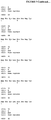
- FIG. 1 shows the results of a preliminary experiment to measure the kinetics of P1NP response after subcutaneous administration of anti-sclerostin antibody to help gauge when best to measure P1NP levels in the subsequent experiments. Circulating P1NP levels for two mice dosed subcutaneously with 10 mg/kg on day 0 are shown (square and diamond symbols) along with the level of anti-sclerostin antibody in a similarly dosed mouse (triangular symbols).
- FIG. 2 shows how P1NP response to anti-sclerostin antibodies falls after multiple doses of antibody.
- P1NP levels are shown for group A mice administered saline alone (diamond symbols), group B mice (square symbols) administered subcutaneously 10 mg/kg anti-sclerostin antibody at the time points indicated at the bottom of the graph and group C mice (triangular symbols) administered saline, except for a single dose of antibody at the time-point depicted.
- the statistics use an unpaired T test (two tailed) looking at difference of absolute values at day of test.
- the group B mice were administered antibody doses at days 0, 7, 14, 21 and 28, with the single doses for the group C subgroups administered at either day 14 or 28.
- FIG. 3 shows that the decline in plasma P1NP levels after multiple doses of antibody is not due to more rapid elimination of the antibody in the multiple dosed group.
- the level of antibody is shown for the repeat dose group B and single dose group C immediately before a dose of 10 mg/kg of antibody given subcutaneously on day 28 and the level four days after the dose of antibody at day 32 (mean and SEM levels).
- FIG. 4 shows the kinetics of P1NP induction are the same in mice given a single dose or multiple doses of anti-sclerostin antibody, only the magnitude of the P1NP induction is different between the two groups.
- the P1NP levels for series 1 (diamond symbols) and series 2 (square symbols) are shown where the mice were given a single dose of anti-sclerostin antibody, as well as the levels for the series 3 mice (triangular symbols) given multiple doses of anti-sclerostin antibody.
- the time of the administration of the antibody is shown by the arrows under the graph.
- FIG. 5 shows P1NP levels in groups A, B and subgroups of group C given a single dose of antibody at the depicted time point.
- the symbols are the same as indicated above for FIG. 2 .
- the dosing schedule for group B is shown by arrows at the bottom of the graph and corresponds to doses at days 0, 7, 14, 21 and 28 and days 84, 91, 98, 105, 112 119 and 176 (no baseline sample was taken on day 119). Hence, there were dosing holidays between days 28 and 84 and also days 119 and 176.
- the single doses for the group C subgroups were at days 14, 28, 84, 119 and 176.
- the statistics use an unpaired T test (two tailed) looking at difference of absolute values at day of test.
- FIG. 6 shows bone mineral density in group A mice (circular symbols) and group B mice (square symbols). The dosing schedule for the group B animals is shown at the bottom of the graph.
- FIG. 7 is a chart listing amino acid sequences and sequence identifiers for amino acid sequences of various anti-sclerostin antibodies described herein.
- the sequence identifiers refer to amino acid sequences provided in the Sequence Listing submitted herewith.
- the amino acid sequences also are set forth in U.S. Patent Publication No. 20070110747, hereby incorporated by reference.
- FIG. 8 is a listing of amino acid sequences and nucleotide sequences set forth in International Patent Publication No. WO 2008/115732, referred to herein.
- FIG. 9 is a listing of amino acid sequences and nucleotide sequences set forth in International Patent Publication No. WO 2009/047356, referred to herein.
- FIG. 10 is a listing of amino acid sequences and nucleotide sequences set forth in International Patent Publication No. WO 2010/130830, referred to herein.
- FIG. 11A is a graph illustrating P1NP levels in mice receiving five weekly doses of anti-sclerostin antibody followed by a single dose of anti-sclerostin antibody after a two week (administration on day 42), four week (administration on day 56), or six week (administration on day 70) holiday (triangle symbols) and age-matched mice receiving a single dose of anti-sclerostin antibody on day 28, day 42, day 46, or day 70 (square symbols).
- P1NP levels (ng/ml) are depicted on the y-axis, and day of the study is depicted on the x-axis.
- 11B is a bar graph illustrating total bone mineral density (BMD; g/cm2; y-axis) measured at various time points (days, x-axis). Bars denoted “#” correspond to measurements taken in mice receiving five weekly doses of anti-sclerostin antibody (fifth dose administered on day 28) followed by a single dose of anti-sclerostin antibody on days 42, 56, or 70 (corresponding to a two week, four week, or six week holiday, respectively). Bars denoted “+” correspond to measurements taken in mice receiving a single dose of anti-sclerostin antibody on days 28, 42, 56, or 70 of the study.
- BMD total bone mineral density
- the subject Prior to being given a dosing holiday, the subject will have been administered at least one dose of anti-sclerostin antibody. Typically, the subject will have been given a plurality of doses prior to the holiday. For instance, the subject may have been given a batch of at least two doses of the anti-sclerostin antibody prior to the dosing holiday. Preferably, the subject may have been administered three, four, five, or at least those numbers of doses of antibody before being given a dosing holiday. The administration of such a batch of doses may form part of the invention.
- the subject may be given a batch of two, three, four, five, six, seven, eight, nine or more doses prior to the dosing holiday, or at least that number of doses. In some instances, the subject may have been given a batch of ten, eleven, twelve, thirteen, fourteen, fifteen, sixteen or more doses of the antibody. In some instances the subject may have been given a batch of ten or less, nine or less, eight or less, seven or less, six or less or five or less doses, where the number of doses given is at least two, preferably at least three and more preferably at least four doses. It may be that the subject is given a batch of from two to sixteen doses, such as from two to fourteen doses or from two to twelve doses.
- the subject may have been given a batch of from two to seven, from two to six, from two to five, or from two to four doses prior to the holiday.
- the number of doses may be from three to eight, seven, six, five or four doses.
- the number of doses in the batch may be from four to eight, seven, six, or five doses.
- the subject may have been given twelve doses of the antibody.
- the subject will have been administered, or is administered, a batch of doses, where the overall time period for the batch is at least two, three, four, five, six, seven, eight, nine, ten, eleven or twelve months or, for example, at least about 4, 8, 12, 16, 20 or 24 weeks in length.
- the overall duration of a batch of doses may be about six months, twelve months (i.e., one year) or eighteen months.
- the interval between individual doses in a batch may be about two weeks. In other cases, the interval may be longer. For instance, the interval may be about a month, 2 months, 3 months, 4 months, 5 months, 6 months or longer. In some cases, the interval between doses in a batch may be about every two, three, four, five, six, seven, or eight weeks. In some cases, the interval between doses in a batch may be about from one week to six months, from two weeks to four months, from three weeks to six weeks, or from four to five weeks. In one preferred instance, the interval between doses may be about a month or may be about four weeks.
- the interval may be about 7 days, a week, 2 weeks, 3 weeks, four weeks, 1 month, 5 weeks, 6 weeks, 7 weeks, 2 months, 8 weeks, 9 weeks, 10 weeks, 11 weeks, 12 weeks or 3 months.
- the interval between doses in a batch may be about a day, two days, three days, four days, five days, six days, seven days or longer.
- the doses may be given once, twice, three, four, five, six or seven times a week.
- the doses in a batch may be administered every week, two weeks, four weeks, six weeks or eight weeks, or about such intervals.
- the intervals between doses in a batch may be, for instance, monthly, two monthly or three monthly, or about those intervals.
- individual doses of antibody may be given more than once a week, such as two, three or four times a week.
- doses may be administered in some cases every two, three, four, five, six, seven or eight days to the subject.
- the invention may entail administering any of the above specified batches of dosages, for instance as part of the method of the invention.
- the subject may have been known to have been administered such a number of doses, but the administration of the batch of the doses does not form part of the method, rather the subject is simply given a dosing holiday prior to being administered a further batch of doses.
- the number of doses given to the subject is such that a drop of the response of the subject to the antibody is seen for at least one of the doses given in a batch, for instance for the last dose prior to the holiday being initiated.
- the dosing holiday may begin when the subject first shows a reduction in the response to the antibody. In some instances, the dosing holiday may begin after one, two or three doses showing a reduced effect. For instance, in some cases the dosing holiday may be started where a subsequent dose shows a reduced effect in comparison to the response seen with the first dose of the antibody given to the subject. In some cases, it may be that the average response seen for at least two doses is reduced in comparison to that seen for two earlier doses, particularly the first two doses.
- the subject may be actively monitored to determine the best time for the dosing holiday, in other cases the subject is not monitored for resistance.
- the dosing holiday may be initiated when the response seen for a dose of antibody falls below 90%, 80%, 70%, 60%, 50%, 40%, 30% or less than the response seen with an earlier dose, such as for the first dose.
- the dosing holiday may be initiated when the response to a dose is below such a percentage in comparison to what would be expected for a na ⁇ ve subject with the same disorder, such as an age and gender matched subject.
- the drop in response may be at least 5%, 15%, 20%, 25%, 30%, 40%, 50% or more.
- the parameter used to gauge how much the response is reduced is any of those mentioned herein.
- the response may be that defined by reference to P1NP levels, though any of the markers discussed herein may be employed.
- the response may be that gauged using change in bone mineral density (BMD).
- BMD bone mineral density
- the rate, or amount, of bone formation, the rate, or amount, of bone resorption, or any combination thereof may also be used as a parameter to define the response to the antibody. It may be that the anti-sclerostin antibody still brings about an increase in BMD, but the increase is less than that for a na ⁇ ve subject. Hence, a reduced response may be one with a smaller increase than would be expected for a na ⁇ ve subject, including for any of the markers discussed herein.
- a dosing holiday is a time period where no anti-sclerostin antibody is administered to a subject.
- Such a dosing holiday may help reduce, reverse or prevent the reduced response to an anti-sclerostin antibody seen in subjects given a plurality of doses of the antibody and hence help improve the efficiency of treatment of bone disorders with anti-sclerostin antibodies.
- the dosing holiday will result in reversal or reduction of the reduced response displayed by the subject to the anti-sclerostin antibody. Hence, the subject may display a higher response to the antibody than prior to the dosing holiday.
- the subject may, for instance, display a response to the anti-sclerostin antibody which is closer to the “na ⁇ ve” response to the antibody when the subject was first administered the anti-sclerostin antibody. For at least about 50%, 60%, 70%, 80%, 90%, 95%, or 99% of the na ⁇ ve response or even about 100% of the na ⁇ ve response.
- the dosing holiday will result in a higher response to the anti-sclerostin antibody as measured by a bone marker, such as a marker of bone resorption and/or formation, including any of those mentioned herein, particularly P1NP.
- the administration of a batch of doses, followed by a dosing holiday and then administration of at least one dose of antibody means that the dosing regimen followed is one of irregular dosing.
- the treatment may be characterized by irregular dosing, such as over the treatment period as a whole.
- the length of a dosing holiday may vary.
- a dosing holiday will be typically longer in length than the interval between individual doses in a batch, for instance the interval between doses in a batch of doses known to have been administered to the subject or administered to the subject as part of the invention.
- the dosing holiday may be any of the above specified lengths as long as the interval between doses in the preceding batch is shorter.
- the dosing holiday may be any of at least 4, 5, 6, 7, 8, 19, 10, 11 or 12 weeks or about such duration. It may be the dosing holiday is at least 14, 16, 18, 20, 22, 24, 26, 28, 30, 35, 40, 45 or 50 weeks in length or may be of about such duration. In some instances, the dosing holiday may be from about four weeks to 52 weeks, for example from six weeks to 24 weeks, in some cases from eight weeks to 12 weeks. In some instances of the invention, the dosing holiday may be about two months, three months, four months, five months, six months, seven months, eight months, nine months, ten months, eleven months or twelve months or at least those time periods. In some cases the dosing holiday may be about, or at least, eighteen months in length.
- the dosing holiday may be about four weeks, six weeks, eight weeks, ten weeks, or twelve weeks longer than the interval between doses in a batch of doses.
- the dosing holiday may be equivalent to the total duration of a batch of doses, such as any of those specified herein, or in other instances it may be equivalent to the overall duration of a batch of doses, plus an additional two, four, six, eight, twelve or more weeks in length.
- the dosing holiday is at least two, three, four, five, six, seven, eight, nine or ten weeks longer than the interval between two doses in the preceding batch, or the dosing interval may be of such length. In some cases the dosing holiday may be such a length longer than the average interval for three, four, five, six, seven or more doses in a batch or, for example, than the average interval between all of the doses in a batch.
- the total length of the dosing holiday may be, for example, four, five, six, seven, eight, nine, ten or more weeks.
- the dosing holiday may be one, two, three, four, five or six months in length and in some cases may be at least a year, or eighteen months in length.
- the dosing holiday may be from a month to a year, such as from two to six months in length. In some cases, the dosing holiday may be from four to sixteen weeks, for instance, from six to twelve weeks, for example from eight to ten weeks in length. In other instances, the dosing holiday may be about from six to eighteen months, for instance about a year. In some cases the dosing holiday may be about twice, three times, four times, five times, six times, seven times, eight times, nine times or more in duration than the interval between doses in a batch administered to the subject. In some instances, where a different treatment is administered during the dosing holiday, the duration of the dosing holiday may be the normal duration for a course of a different treatment for the disorder to be administered in the dosing holiday.
- the subject is given more than one dosing holiday.
- the subject is given at least two doses of the antibody and may, for instance, benefit from a further dosing holiday.
- the administration of at least two doses of the antibody, followed by a dosing holiday may be referred to as a cycle and in some instances, one, two, three, four, five, six, seven, eight, nine, ten or more such cycles may be used.
- the overall total treatment period may be at least six months, nine months, a year, eighteen months, twenty-four months, or more.
- the overall treatment is at least 4, 8, 12, 16, 20, 24, 28, 32, 36, 40, 44, 48 or 52 weeks, or longer, or about such periods.
- the approach of batches of doses combined with dosing holidays is continued as long as the treatment lasts.
- it may simply be that a set regimen of batch doses alternating with dosing holidays is administered. For instance, any combination of those batches and dosing holidays specified herein, for example for two, three, four, five, six or more cycles of a batch of doses followed by a dosing holiday may be administered.
- any of the batches of doses specified herein may be combined with any of the dosing holidays specified herein, as long as the dosing holiday is longer than the interval between doses in a batch.
- a batch of doses administered at daily, weekly, fortnightly, four weekly, six weekly or eight weekly intervals may be combined with a dosing holiday of at least six weeks, at least eight weeks, at least twelve weeks, at least 16 weeks, at least 20 weeks or at least 24 weeks, where the dosing holiday is longer than the interval between batches.
- the doses in the batch may be given at about monthly or two monthly intervals and may be combined with a dosing holiday of at least three, four, five, six, eight, ten, twelve or more months in length.
- the batch of doses may comprise three to fourteen doses at daily, weekly, fortnightly, four weekly or six weekly intervals, combined with a dosing holiday of at least six, eight, ten, twelve, fourteen or more weeks in length, where the dosing holiday is longer than the interval between the doses in the batch.
- a batch of monthly doses is combined with a dosing holiday of at least two, three, four, five, six, twelve or more months in length. In some instances, it may be that the doses in the batch are given about every four weeks.
- the interval between earlier doses will not be known and the subject will simply be one who is displaying a reduced response to the anti-sclerostin antibody in comparison to what would be expected for the subject.
- the length of the dosing holiday given is simply one of the above time periods without reference to the time between administration of earlier doses or the response to earlier doses.
- the dosing holiday may be six weeks, eight weeks, twelve weeks, sixteen weeks, twenty weeks, twenty four weeks or more in length or any of the other possible lengths referred to.
- the subject may have been identified as one showing resistance to anti-sclerostin antibody, for example, even though the precise regimen previously administered is not known.
- the antibody may be that they have been administered the antibody for at least about two, three, four, five, six or more months in length and hence be identified as a candidate for a dosing holiday. In some cases they may have been administered the antibody for at least about nine, twelve or eighteen months in length and hence be identified as a candidate for a dosing holiday.
- the subject may be displaying reduced or diminishing therapy from the existing therapy.
- a fixed regimen of batch dosing and dosing holiday may be applied in some instances including any of those specified herein. It may be the fix regimen is designed with reference to age, gender, weight, the nature of the disorder, the severity of the disorder and so on.
- the response is the response as defined by a bone marker, for instance a bone formation and/or bone resorption marker, particularly any of those referred to herein.
- a bone marker for instance a bone formation and/or bone resorption marker, particularly any of those referred to herein.
- whether or not a response can be considered reduced may be, in some instances, defined by whether the response of the bone marker to administration of the anti-sclerostin antibody is reduced.
- whether a dosing holiday may be said to prevent, or reverse, resistance to an anti-sclerostin antibody may be defined by the response of a bone marker and, for instance, the level of that marker.
- the response to the antibody is defined by P1NP level, particularly serum P1NP level.
- the response of the subject to a dose of anti-sclerostin antibody is measured to help gauge whether the subject is displaying resistance to the anti-sclerostin antibody.
- Any suitable means of measuring the response to the anti-sclerostin antibody may be employed.
- the level of a bone marker may be measured, in particular a marker of bone formation and/or mineralization may be measured in the subject. Markers of bone resorption may also be employed.
- the invention itself does not entail measurement, or monitoring, of the response, but the response in question is that defined by a bone marker, such as, the level of any of the bone markers referred to herein.
- Markers indicative of bone resorption (or osteoclast activity) which may be used include, for example, C-telopeptide (e.g., C-terminal telopeptide of type 1 collagen (CTX) or serum cross-linked C-telopeptide), N-telopeptide (N-terminal telopeptide of type 1 collagen (NTX)), deoxypyridinoline (DPD), pyridinoline, urinary hydroxyproline, galactosyl hydroxylysine, and tartrate-resistant acid phosphatase (e.g., serum tartrate-resistant acid phosphatase isoform 5b).
- C-telopeptide e.g., C-terminal telopeptide of type 1 collagen (CTX) or serum cross-linked C-telopeptide
- N-telopeptide N-terminal telopeptide of type 1 collagen (NTX)
- DPD deoxypyridinoline
- pyridinoline pyridinoline
- Bone formation/mineralization markers which may be used include, but are not limited to, bone-specific alkaline phosphatase (BSAP), peptides released from N- and C-terminal extension of type I procollagen (PINP, PICP), and osteocalcin (OstCa).
- BSAP bone-specific alkaline phosphatase
- PINP type I procollagen
- PICP peptides released from N- and C-terminal extension of type I procollagen
- osteocalcin OstCa
- kits are commercially-available to detect and quantify markers in clinical samples, such as urine and blood.
- the marker used is selected from the serum level of C-telopeptide of type I collagen (CTX), bone-specific alkaline phosphatase (BSAP), osteocalcin (OstCa), and/or N-terminal extension of procollagen type 1 (P1NP).
- CTX C-telopeptide of type I collagen
- BSAP bone-specific alkaline
- the response in question may be defined by reference to bone mineral density (BMD) or bone mineral content (BMC).
- BMD bone mineral density
- BMC bone mineral content
- the reduced response is a reduced rate of increase of BMD and/or BMC following administration of the antibody.
- administration of the antibody still results in an increase in bone formation and/or a reduction of bone absorption, for example in terms of BMD/BMC, but at a reduced rate compared to a na ⁇ ve subject.
- the use of a dosing holiday may mean the subject again displays the same size of increase in such parameters as a na ⁇ ve subject, or at least closer to a na ⁇ ve subject.
- Bone mineral density may be, for instance, measured using techniques, such as, single- and dual-energy X-ray absorptometry, ultrasound, computed tomography, radiography, and magnetic resonance imaging. The amount of bone mass may also be calculated from body weights or by using other methods (see Guinness-Hey, Metab. Bone Dis. Relat. Res., 5:177-181 (1984)). In humans, bone mineral density may be, for instance, determined clinically using dual x-ray absorptiometry (DXA) of, for example, the hip and spine.
- DXA dual x-ray absorptiometry
- Other techniques include quantitative computed tomography (QCT), ultrasonography, single-energy x-ray absorptiometry (SXA), and radiographic absorptiometry.
- QCT quantitative computed tomography
- SXA single-energy x-ray absorptiometry
- radiographic absorptiometry Common central skeletal sites for measurement include the spine and hip; peripheral sites include the forearm, finger, wrist and heel.
- BMD techniques typically involve the use of x-rays and are based on the principle that attenuation of the radiation depends on thickness and composition of the tissues in the radiation path. All techniques may employ the comparison of results to a normative database or control subject.
- the bone mineral density (BMD) of the subject is compared to the peak density of a 30-year old healthy adult (i.e., a “young adult”), creating the so-called “T-score.”
- a patient's BMD also may be compared to an “age-matched” bone density (see, e.g., World Health Organization Scientific Group on the Prevention and Management of Osteoporosis, “Prevention and management of osteoporosis: report of a WHO scientific group.” WHO Technical Report Series; 921, Geneva, Switzerland (2000)).
- the difference between a patient's BMD and that of a healthy, young adult is conventionally referred to in terms of the multiple of a “standard deviation,” which typically equals about 10% to about 12% decrease in bone density.
- the World Health Organization proposed four diagnostic categories based on BMD T-scores.
- a BMD value within 1 standard deviation of the young adult reference mean (T-score > ⁇ 1) is “normal.”
- Low bone mass (osteopenia) is indicated by a BMD value more than 1 standard deviation below the young adult mean, but less than 2.5 standard deviations (T-score ⁇ 1 and > ⁇ 2.5).
- a T-score of more than 2.5 standard deviations below the norm supports a diagnosis of osteoporosis. If a patient additionally suffers from one or more fragility fractures, the patient qualifies as having severe osteoporosis.
- the invention may entail calculating the T-score for the subject, for instance, in response to a dose of anti-sclerostin antibody and determining whether there is a reduced improvement in the T-score following administration of a dose of the anti-sclerostin antibody.
- the decision as to when to initiate the dosing holiday may therefore be based on assessing the response of the subject to a dose of the anti-sclerostin antibody and determining whether the response is lower than expected.
- the dosing holiday may be, for instance, initiated when monitoring shows a reduced response to a dose, or two consecutive doses, particularly in comparison to earlier doses, such as the first dose, or in comparison to the average response seen for the doses in the batch.
- the dosing holiday may be, for instance, begun, when the positive results seen with the treatment plateau or begin to tail-off for the batch of doses administered. It may be that the dosing holiday is administered when administration of the antibody results in a smaller increase of the particular parameter or marker than would be expected. For instance, when the response is less than 90%, 80%, 70%, 60%, 50%, 40%, 30%, 25% or less than that which would be expected from the equivalent na ⁇ ve subject or displayed to an earlier dose by the same subject.
- the subject may be administered a test dose of anti-sclerostin to gauge their response to the antibody.
- a test dose of anti-sclerostin to gauge their response to the antibody.
- the test dose may be given, the response measured, and, if considered reduced, the subject will be given a dosing holiday. If the subject does not display a reduced response, they may be given further doses of anti-sclerostin antibody.
- the subject is administered at least two, three, four, five, or six doses and their response measured.
- a subject is monitored continuously, for example after each dose of anti-sclerostin antibody. It may be that the subject is monitored, for instance, about once a month, once about every two months, once about every three months, once about every four months, once about every six months or about once a year. It may be that the subject is monitored immediately before a dose is administered and then, for example, about one, two, three, four, five or six weeks later. It may be, for example, that the response seen for at least one, two, three, four, five or more doses is monitored. In some cases, the dosing holiday may be initiated when the monitoring shows the response is below an expected level for the subject or below a set cut-off value.
- the dosing holiday may be initiated when the response for a dose is less than the response seen for an earlier dose, such as that for the first dose, or for the first dose in a batch of doses.
- the size of the response as a percentage of the response to an earlier dose may be determined, particularly the first dose, and it may be that when the percentage value falls to, or below, one of the percentage values specified herein the dosing holiday is begun.
- the response to a dose may be measured without any reference to earlier doses and simply the fact it falls below an expected value means a dosing holiday is initiated.
- the method of the invention may comprise: (a) administering a dose of anti-sclerostin antibody to a subject who has previously been administered anti-sclerostin antibody; (b) measuring the response to the dose; and (c) assessing whether the dose is lower than that expected. If the response is lower than that expected, for instance below a threshold, then a dosing holiday may be given.
- the method may optionally comprise administering at least one further dose of the anti-sclerostin antibody to the subject and measuring until a reduced response is seen, then giving a dosing holiday. In some instances, it may be that the subject has already been administered at least three, four, five or six doses before the test dose.
- a dosing holiday may include the administration of one or more test doses of anti-sclerostin antibody, where the test dose is used to determine if the resistance displayed to the antibody has diminished or been eliminated.
- the test dose is used to determine whether to terminate the dosing holiday and again begin treatment with the anti-sclerostin antibody or continue the dosing holiday.
- the subject may be given a dosing holiday and the end of the dosing holiday may be defined by when the subject displays an increased response to a test dose of anti-sclerostin antibody or, for instance, the subject displays resistance below a defined threshold, such as any of those mentioned herein.
- the subject may be given a set pattern of a batch of at least two doses, followed by a set dosing holiday, without monitoring to determine when to initiate the dosing holiday.
- Such fixed batches of doses and dosing holidays may be, for example, based on the disorder to be treated, age, gender and weight of the subject.
- it may be that a suitable cycle of a batch of doses and dosing holiday is determined on a particular cycle by monitoring and then adhered to on subsequent cycles to the same regimen.
- a bone formation and/or resorption marker including any of those referred to herein, particularly P1NP levels. It may be that the level of the marker is measured before administration of the dose, then, for instance, four, five, six, seven, eight, ten, eleven or twelve days after administration of the test dose.
- any of the parameters referred to herein are measured during a course of treatment to determine if they are less than expected or show a slower, or less marked, increase in the marker. It may be that the subject is assessed clinically to determine whether the effect of the treatment is less and so that a dosing holiday may be of benefit. It may be that the subject has regular checks, such as about monthly, three monthly, four monthly, six monthly or yearly intervals and such checks entail checking or measuring the effect of the anti-sclerostin antibody and/or dosing holiday, for instance to decide whether to commence a dosing holiday.
- the invention may be administered to a patient group thought to be displaying resistance to anti-sclerostin antibodies, or thought likely to display such resistance to the antibody. In some cases, the invention may be applied to a patient group displaying a higher than average resistance to anti-sclerostin antibodies. It may be that such a patient population is identified using monitoring, such any of the monitoring means discussed herein, particularly any of the markers discussed herein. Identification of such patients may entail administration of a test dose as described herein, followed by measurement of the response and assessment of whether the response seen is less than expected. It may be though that the patients are identified due to the fact that they have been receiving anti-sclerostin antibodies and the improvement initially seen has diminished.
- the invention may be applied to a subject who has been administered anti-sclerostin antibodies, but the treatment has been discontinued, particularly where the treatment has been discontinued because the subject is displaying a reduced response to the anti-sclerostin antibodies.
- any of the methods of the invention may comprise first assessing whether a subject is one displaying resistance to anti-sclerostin antibodies and then applying the invention if the subject does display such resistance. Hence, if such resistance is displayed, the invention may then entail allowing the subject a dosing holiday to reverse or reduce that resistance.
- markers may display diurnal variation, i.e., display variation in their level during the day.
- a marker may be measured at a specific time, or time period, in the day.
- a particular marker is measured more than once, it may be that the marker is measured each time at, or approximately at, the same time in the day, or in about a one hour, two hour, or three hour window.
- the marker may be consistently measured in the morning or consistently in the afternoon.
- any of the markers measured herein may be measured in such a manner, particularly including those known to show diurnal variation.
- P1NP levels may be measured in such a manner.
- the value is measured at a time in the day, or time period, consistent with the standard or expected value.
- the amount of anti-sclerostin antibody administered as an individual dose to the subject may, for instance, comprise at least about 70 mg of the anti-sclerostin antibody.
- the amount of anti-sclerostin antibody administered is at least about 120 mg (e.g., 180 mg) or at least about 140 mg, e.g., at least about 210 mg anti-sclerostin antibody.
- the amount of anti-sclerostin antibody administered may be, for instance, no more than about 350 mg anti-sclerostin antibody, e.g., no more than about 280 mg anti-sclerostin antibody (e.g., 270 mg), no more than about 210 mg of anti-sclerostin antibody, no more than about 140 mg anti-sclerostin antibody, or no more than about 120 mg anti-sclerostin antibody (e.g., about 120 mg of antibody).
- a single administration or dose of anti-sclerostin comprises, for example, no more than about 350 mg of the antibody.
- the subject is administered a dose of anti-sclerostin antibody in an amount of about 70 mg to about 350 mg, such as about 70 mg to about 280 mg, or about 120 mg to about 350 mg, or about 140 mg to about 350 mg, or about 210 mg to about 350 mg, or about 280 mg to about 350 mg.
- a single dose of anti-sclerostin antibody comprises about 70 mg to about 210 mg of anti-sclerostin antibody, such as about 70 mg to about 120 mg (e.g., about 70 mg) anti-sclerostin antibody, or about 70 mg to about 140 mg of anti-sclerostin antibody, or about 120 mg to about 210 mg anti-sclerostin antibody, or about 120 mg to about 140 mg of anti-sclerostin antibody.
- a single dose of anti-sclerostin antibody comprises about 140 mg to about 210 mg (e.g., about 140 mg or about 210 mg) of anti-sclerostin antibody.
- the dose administered is between about 0.1 to about 20 mg/kg, or about 0.1 to about 12 mg/kg, or about 0.5 to about 12 mg/kg, or about 1 to about 10 mg/kg, or about 1 to about 8 mg/kg, or about 2 to about 8 mg/kg, or about 3 to about 8 mg/kg.
- a dose is about 1 mg/kg to about 10 mg/kg (e.g., about 2 mg/kg or about 9 mg/kg), about 1 mg/kg to about 3 mg/kg, or about 3 mg/kg to about 8 mg/kg (e.g., about 4 mg/kg, 5 mg/kg, 6 mg/kg, or 7 mg/kg).
- the dose is calculated based on a per weight basis specifically for that subject.
- a low dose is employed.
- a lower dose may be employed where an interval between doses of less than two weeks, such as less than one week, in particular any of the time periods specified herein shorter than a week, is employed.
- the dose may be any of the above specified doses of 2 mg/kg or under.
- the dose may be about, or under, 1 mg/kg, 0.75 mg/kg, 0.5 mg/kg, 0.25 mg/kg or 0.1 mg/kg.
- a fixed dose is administered, such as any of about 1 to about 50 mg, about 1 to about 25 mg, about 1 to about 10 mg, about 1 to about 5 mg or about 1 to about 3 mg.
- a fixed dose of from about 2 to about 5 mg, about 2 to about 7 mg or about 3 to about 8 mg may be employed.
- Reference herein to a single dose may include multiple contemporaneous injections be administered to achieve delivery of the dose. For instance, several injections within the space of an hour, day or week.
- the subject will be administered a plurality of doses of anti-sclerostin antibody and in particular a batch of doses.
- all of the doses within a batch will be approximately the same amount, or actually the same amount.
- the doses administered in the different batches will be the same.
- the dose may vary between different batches. For instance, it may be that the dose is varied according to how the patient is responding to the treatment.
- antibody refers to an intact antibody, or a binding fragment thereof.
- An antibody may comprise a complete antibody (immunoglobulin) molecule (including polyclonal, monoclonal, chimeric, humanized, and/or human versions having full length heavy and/or light chains), or comprise an antigen binding fragment thereof.
- Antibody fragments include F(ab′) 2 , Fab, Fab′, Fv, Fc, and Fd fragments, and can be incorporated into single domain antibodies (e.g., nanobodies), single-chain antibodies, maxibodies, minibodies, intrabodies, diabodies, triabodies, tetrabodies, v-NAR and bis-scFv (see, e.g., Hollinger and Hudson, Nature Biotechnology, 23(9):1126-1136 (2005)).
- Antibody polypeptides, including fibronectin polypeptide monobodies also are disclosed in U.S. Pat. No. 6,703,199. Other antibody polypeptides are disclosed in U.S. Patent Publication No. 20050238646.
- antibody fragment may be any synthetic or genetically engineered protein.
- antibody fragments include isolated fragments consisting of the light chain variable region, “Fv” fragments consisting of the variable regions of the heavy and light chains, and recombinant single chain polypeptide molecules in which light and heavy variable regions are connected by a peptide linker (scFv proteins).
- the antibody may be any class of antibody, but in a preferred instance the antibody is an IgG antibody.
- CDRs complementarity determining regions
- Another form of an antibody fragment is a peptide comprising one or more complementarity determining regions (CDRs) of an antibody.
- CDRs also termed “minimal recognition units” or “hypervariable region” can be obtained by constructing polynucleotides that encode the CDR of interest.
- Such polynucleotides are prepared, for example, by using the polymerase chain reaction to synthesize the variable region using mRNA of antibody-producing cells as a template (see, for example, Larrick et al., Methods: A Companion to Methods in Enzymology, 2:106 (1991); Courtenay-Luck, “Genetic Manipulation of Monoclonal Antibodies,” in Monoclonal Antibodies Production, Engineering and Clinical Application , Ritter et al.
- Anti-sclerostin antibodies may, for instance, bind to sclerostin of SEQ ID NO: 1, or a naturally occurring variant thereof, with an affinity (Kd) of less than or equal to 1 ⁇ 10 ⁇ 7 M, less than or equal to 1 ⁇ 10 ⁇ 8 M, less than or equal to 1 ⁇ 10 ⁇ 9 M, less than or equal to 1 ⁇ 10 ⁇ 10 M, less than or equal to 1 ⁇ 10 ⁇ 11 M, or less than or equal to 1 ⁇ 10 ⁇ 12 M.
- affinity is determined using a variety of techniques, an example of which is an affinity ELISA assay.
- affinity is determined by a BIAcore assay (a surface plasmon resonance assay).
- affinity is determined by a kinetic method.
- affinity is determined by an equilibrium/solution method.
- U.S. Patent Publication No. 20070110747 contains additional description of affinity assays suitable for determining the affinity (Kd) of an antibody for sclerostin.
- Anti-sclerostin antibodies for use in the inventive method preferably modulate sclerostin function in the cell-based assay described in U.S. Patent Publication No. 20070110747 and/or the in vivo assay described in U.S. Patent Publication No. 20070110747 and/or bind to one or more of the epitopes described in U.S. Patent Publication No. 20070110747 and/or cross-block the binding of one of the antibodies described in U.S. Patent Publication No. 20070110747 and/or are cross-blocked from binding sclerostin by one of the antibodies described in U.S. Patent Publication No. 20070110747 (incorporated by reference in its entirety and for its description of assays for characterizing an anti-sclerostin antibody).
- the anti-sclerostin antibody binds to a sclerostin polypeptide comprising the amino acid sequence set forth in SEQ ID NO: 1 of the Sequence Listing and binds the sequence of SEQ ID NO: 6 (C4GPARLLPNAIGRGKWWRPSGPDFRC5; corresponding to amino acids 86-111 of SEQ ID NO: 1).
- the anti-sclerostin antibody binds to a sclerostin polypeptide comprising the amino acid sequence set forth in SEQ ID NO: 1 and binds the sequence of at least one of SEQ ID NO: 2 (DVSEYSC1RELHFTR; corresponding to amino acids 51-64 of SEQ ID NO: 1), SEQ ID NO: 3 (SAKPVTELVC3SGQC4GPAR; corresponding to amino acids 73-90 of SEQ ID NO: 1), SEQ ID NO: 4 (WWRPSGPDFRCSIPDRYR; corresponding to amino acids 101-117 of SEQ ID NO: 1), SEQ ID NO: 5 (LVASC7KC8KRLTR; corresponding to amino acids 138-149 of SEQ ID NO: 1), SEQ ID NO: 70 (SAKPVTELVC3SGQC4; corresponding to amino acids 73-86 of SEQ ID NO: 1), SEQ ID NO: 71 (LVASC7KC8; corresponding to amino acids 138-144 of
- the anti-sclerostin antibody binds a subregion of sclerostin of SEQ ID NO: 1 comprising SEQ ID NOs: 2-5 (and/or SEQ ID NOs: 70-73), optionally in its native three-dimensional conformation.
- the anti-sclerostin antibody binds a peptide consisting of one or more of SEQ ID NO: 2, SEQ ID NO: 3, SEQ ID NO: 4, SEQ ID NO: 5, SEQ ID NO: 6, SEQ ID NO: 70, SEQ ID NO: 71, SEQ ID NO: 72, or SEQ ID NO: 73 of the Sequence Listing (e.g., a peptide consisting of SEQ ID NO: 2, SEQ ID NO: 3, SEQ ID NO: 4, and SEQ ID NO: 5 or a peptide consisting of SEQ ID NO: 70, SEQ ID NO: 71, SEQ ID NO: 72, and SEQ ID NO: 73).
- the anti-sclerostin antibody is capable of neutralizing human sclerostin in a MC3T3 cell-based mineralization assay when there is less than a 6-fold excess of moles of sclerostin binding sites per well as compared to the number of moles of sclerostin per well.
- Mineralization by osteoblast-lineage cells in culture, either primary cells or cell lines, is used as an in vitro model of bone formation.
- An exemplary cell-based mineralization assay is described in U.S. Patent Publication No. 20070110747 at, e.g., Example 8 (hereby incorporated by reference).
- MC3T3-E1 cells Sudo et al., J.
- subclones of the original cell line can form mineral in culture upon growth in the presence of differentiating agents.
- Such subclones include MC3T3-E1-BF (Smith et al., J. Biol. Chem., 275:19992-20001 (2000)).
- sclerostin can inhibit one or more of the sequence of events leading up to and including mineral deposition (i.e., sclerostin inhibits mineralization).
- Anti-sclerostin antibodies that are able to neutralize sclerostin's inhibitory activity allow for mineralization of the culture in the presence of sclerostin such that there is a statistically significant increase in, e.g., deposition of calcium phosphate (measured as calcium) as compared to the amount of calcium measured in the sclerostin-only (i.e., no antibody) treatment group.
- the amount of sclerostin used in the assay desirably is the minimum amount of sclerostin that causes at least a 70%, statistically significant, reduction in deposition of calcium phosphate (measured as calcium) in the sclerostin-only group, as compared to the amount of calcium measured in the no sclerostin group.
- An anti-sclerostin neutralizing antibody is defined as one that causes a statistically significant increase in deposition of calcium phosphate (measured as calcium) as compared to the amount of calcium measured in the sclerostin-only (i.e., no antibody) treatment group.
- the amount of anti-sclerostin antibody used in the assay needs to be such that there is an excess of moles of sclerostin binding sites per well as compared to the number of moles of sclerostin per well.
- the fold excess that may be required can be 24, 18, 12, 6, 3, or 1.5, and one of skill is familiar with the routine practice of testing more than one concentration of binding agent (antibody).
- a very potent anti-sclerostin neutralizing antibody will neutralize sclerostin when there is less than a 6-fold excess of moles of sclerostin binding sites per well as compared to the number of moles of sclerostin per well.
- a less potent anti-sclerostin neutralizing antibody will neutralize sclerostin only at a 12, 18 or 24 fold excess.
- the anti-sclerostin antibody optionally has an IC 50 of 100 nM or less, or 75 nM or less, or 50 nM or less, or 25 nM or less for neutralizing human sclerostin in a cell-based assay, such as a bone specific alkaline phosphatase assay, e.g., the bone specific alkaline phosphatase assay described in International Patent Publication No. WO 2008/115732 and U.S. Pat. No. 7,744,874 (incorporated herein by reference in its entirety for its description of cell-based assays and anti-sclerostin antibodies).
- a bone specific alkaline phosphatase assay e.g., the bone specific alkaline phosphatase assay described in International Patent Publication No. WO 2008/115732 and U.S. Pat. No. 7,744,874 (incorporated herein by reference in its entirety for its description of cell-based assays and anti-sclerostin antibodies).
- the bone specific alkaline phosphatase assay is predicated on the ability of sclerostin to decrease BMP-4 and Wnt3a-stimulated alkaline phosphatase levels in the multipotential murine cell line, C2C12.
- a neutralizing anti-sclerostin antibody mediates a dose-dependent increase of alkaline phosphatase activity in this assay.
- the anti-sclerostin antibody has an IC 50 of 100 nM or less (e.g., 75 nM or less, or 50 nM or less) for neutralizing human sclerostin in a cell-based Wnt signalling assay in HEK293 cell lines, such as the Wnt assay involving Wnt1-mediated induction of STF reporter gene described in, e.g., International Patent Publication No. WO 2009/047356 (incorporated by reference for its discussion of anti-sclerostin antibodies and cell-based assays).
- IC 50 100 nM or less (e.g., 75 nM or less, or 50 nM or less) for neutralizing human sclerostin in a cell-based Wnt signalling assay in HEK293 cell lines, such as the Wnt assay involving Wnt1-mediated induction of STF reporter gene described in, e.g., International Patent Publication No. WO 2009/047356 (incorporated by reference
- the anti-sclerostin antibody has an IC 50 of 500 nM or less (e.g., 250 nM or less, 150 nM or less, 100 nM or less, or 50 nM or less) for neutralizing human sclerostin in a BMP2-induced mineralization assay in MC3T3 cells, such as the mineralization assay described in, e.g., International Patent Publication No. WO 2009/047356.
- anti-sclerostin antibodies suitable for use in the context of the invention are described in U.S. Patent Publication Nos. 20070110747 and 20070072797, which are hereby incorporated by reference.
- the anti-sclerostin antibody cross-blocks the binding of at least one of antibodies Ab-A, Ab-B, Ab-C, Ab-D, Ab-1, Ab-2, Ab-3, Ab-4, Ab-5, Ab-6, Ab-7, Ab-8, Ab-9, Ab-10, Ab-11, Ab-12, Ab-13, Ab-14, Ab-15, Ab-16, Ab-17, Ab-18, Ab-19, Ab-20, Ab-21, Ab-22, Ab-23, or Ab-24 (all of which are described in U.S. Patent Publication No.
- the anti-sclerostin antibody is cross-blocked from binding to sclerostin by at least one of antibodies Ab-A, Ab-B, Ab-C, Ab-D, Ab-1, Ab-2, Ab-3, Ab-4, Ab-5, Ab-6, Ab-7, Ab-8, Ab-9, Ab-10, Ab-11, Ab-12, Ab-13, Ab-14, Ab-15, Ab-16, Ab-17, Ab-18, Ab-19, Ab-20, Ab-21, Ab-22, Ab-23, or Ab-24 (all of which are described in U.S. Patent Publication No. 20070110747).
- cross-block means the ability of an antibody to interfere with the binding of other antibodies to sclerostin.
- the extent to which an antibody is able to interfere with the binding of another to sclerostin, and therefore whether it can be said to cross-block, can be determined using competition binding assays.
- a cross-blocking antibody or fragment thereof reduces sclerostin binding of a reference antibody between about 40% and about 100%, such as about 60% and about 100%, specifically between 70% and 100%, and more specifically between 80% and 100%.
- a particularly suitable quantitative assay for detecting cross-blocking uses a Biacore machine which measures the extent of interactions using surface plasmon resonance technology.
- Another suitable quantitative cross-blocking assay uses an ELISA-based approach to measure competition between antibodies in terms of their binding to sclerostin.
- suitable anti-sclerostin antibodies and fragments thereof include antibodies and antibody fragments having one or more of CDR-H1, CDR-H2, CDR-H3, CDR-L1, CDR-L2 and CDR-L3 specifically disclosed in U.S. Patent Publication No. 20070110747. At least one of the regions of CDR-H1, CDR-H2, CDR-H3, CDR-L1, CDR-L2, and CDR-L3 may have at least one amino acid substitution, provided that the antibody retains the binding specificity of the non-substituted CDR.
- the anti-sclerostin antibody is Ab-A, Ab-B, Ab-C, Ab-D, Ab-1, Ab-2, Ab-3, Ab-4, Ab-5, Ab-6, Ab-7, Ab-8, Ab-9, Ab-10, Ab-11, Ab-12, Ab-13, Ab-14, Ab-15, Ab-16, Ab-17, Ab-18, Ab-19, Ab-20, Ab-21, Ab-22, Ab-23, or Ab-24 of U.S. Patent Publication No. 20070110747.
- the anti-sclerostin antibody can comprise at least one CDR sequence having at least 75% identity (e.g., 100% identity) to a CDR selected from SEQ ID NOs: 39, 40, 41, 42, 43, 44, 45, 46, 47, 48, 49, 50, 51, 52, 53, 54, 55, 56, 57, 58, 59, 60, 61, 62, 78, 79, 80, 81, 99, 100, 101, 102, 103, 104, 105, 106, 107, 108, 109, 110, 111, 112, 113, 114, 115, 116, 237, 238, 239, 240, 241, 242, 243, 244, 245, 246, 247, 248, 249, 250, 251, 252, 253, 254, 255, 256, 257, 258, 259, 260, 261, 262, 263, 264, 265, 266, 267, 268, 269, 270, 271, 272, 273, 274, 275,
- the anti-sclerostin antibody comprises at least one CDR sequence having at least 75% identity to a CDR selected from SEQ ID NOs: 245, 246, 247, 78, 79, 80, 269, 270, 271, 239, 240, and 241, all of which is provided in the Sequence Listing and described in U.S. Patent Publication No. 20070110747.
- the anti-sclerostin antibody can comprise: a) CDR sequences of SEQ ID NOs:54, 55, and 56 and CDR sequences of SEQ ID NOs:51, 52, and 53; b) CDR sequences of SEQ ID NOs:60, 61, and 62 and CDR sequences of SEQ ID NOs:57, 58, and 59; c) CDR sequences of SEQ ID NOs:48, 49, and 50 and CDR sequences of SEQ ID NOs:45, 46, and 47; d) CDR sequences of SEQ ID NOs:42, 43, and 44 and CDR sequences of SEQ ID NOs:39, 40, and 41; e) CDR sequences of SEQ ID NOs:275, 276, and 277 and CDR sequences of SEQ ID NOs:287, 288, and 289; f) CDR sequences of SEQ ID NOs:278, 279, and 280 and CDR sequences of SEQ ID NOs:290,
- the anti-sclerostin antibody also can comprise at least one CDR sequence having at least 75% identity (e.g., 100% identical) to a CDR selected from CDR-H1, CDR-H2, CDR-H3, CDR-L1, CDR-L2, and CDR-L3 wherein CDR-H1 has the sequence given in SEQ ID NO: 245, CDR-H2 has the sequence given in SEQ ID NO: 246, CDR-H3 has the sequence given in SEQ ID NO: 247, CDR-L1 has the sequence given in SEQ ID NO: 78, CDR-L2 has the sequence given in SEQ ID NO: 79 and CDR-L3 has the sequence given in SEQ ID NO: 80, all of which is provided in the Sequence Listing and described in U.S.
- the anti-sclerostin antibody in various aspects, comprises two of the CDRs or six of the CDRs.
- the anti-sclerostin antibody comprises heavy chains comprising SEQ ID NO: 378 and light chains comprising SEQ ID NO 376 provided in the Sequence Listing.
- the anti-sclerostin antibody also can comprise at least one CDR sequence having at least 75% identity (e.g., 100% identical) to a CDR selected from CDR-H1, CDR-H2, CDR-H3, CDR-L1, CDR-L2, and CDR-L3 wherein CDR-H1 has the sequence given in SEQ ID NO: 269, CDR-H2 has the sequence given in SEQ ID NO: 270, CDR-H3 has the sequence given in SEQ ID NO: 271, CDR-L1 has the sequence given in SEQ ID NO: 239, CDR-L2 has the sequence given in SEQ ID NO: 240 and CDR-L3 has the sequence given in SEQ ID NO 241, all of which is provided in the Sequence Listing and described in U.S.
- the anti-sclerostin antibody in various aspects, comprises two of the CDRs or six of the CDRs.
- the anti-sclerostin antibody comprises heavy chains comprising SEQ ID NO: 366 and light chains comprising SEQ ID NO 364 provided in the Sequence Listing.
- the anti-sclerostin antibody can have a heavy chain comprising CDR's H1, H2, and H3 and comprising a polypeptide having the sequence provided in SEQ ID NO: 137 or a variant thereof in which said CDR's are at least 75% identical (e.g., 100% identical) to SEQ ID NO: 245, 246, and 247, respectively, and a light chain comprising CDR's L1, L2 and L3 and comprising a polypeptide having the sequence provided in SEQ ID NO: 133 or a variant thereof in which said CDR's are at least 75% identical (e.g., 100% identical) to SEQ ID NO: 78, 79, and 80, respectively (as described in U.S. Patent Publication No. 20070110747).
- the anti-sclerostin antibody may have a heavy chain comprising CDR's H1, H2, and H3 and comprising a polypeptide having the sequence provided in SEQ ID NO: 145 or 392 or a variant thereof in which said CDR's are at least 75% identical (e.g., 100% identical) to SEQ ID NO: 245, 246, and 247, respectively, and a light chain comprising CDR's L1, L2, and L3 and comprising a polypeptide having the sequence provided in SEQ ID NO: 141 or a variant thereof in which said CDR's are at least 75% identical (e.g., 100% identical) to SEQ ID NO: 78, 79, and 80, respectively (as described in U.S. Patent Publication No. 20070110747).
- the anti-sclerostin antibody may have a heavy chain comprising CDR's H1, H2, and H3 and comprising a polypeptide having the sequence provided in SEQ ID NO: 335, 331, 345, or 396 (provided in the Sequence Listing) or a variant of any of the foregoing in which said CDR's are at least 75% (e.g., 100% identical) identical to SEQ ID NO: 269, 270, and 271, respectively, and a light chain comprising CDR's L1, L2, and L3 and comprising a polypeptide having the sequence provided in SEQ ID NO: 334 or 341 (provided in the Sequence Listing) or a variant of any of the foregoing in which said CDR's are at least 75% identical (e.g., 100% identical) to SEQ ID NO: 239, 240, and 241, respectively (as described in U.S.
- Heavy and light chain sequences are contemplated (e.g., heavy chains comprising SEQ ID NO: 335 and light chains comprising SEQ ID NO: 334; heavy chains comprising SEQ ID NO: 331 and light chains comprising SEQ ID NO: 334 or 341; and heavy chains comprising SEQ ID NO: 345 or 396 and light chains comprising SEQ ID NO: 341).
- the anti-sclerostin antibody has a heavy chain comprising a polypeptide having the sequence provided in SEQ ID NO:137, and a light chain comprising a polypeptide having the sequence provided in SEQ ID NO:133; a heavy chain comprising a polypeptide having the sequence provided in SEQ ID NO:145 or 392, and a light chain comprising a polypeptide having the sequence provided in SEQ ID NO: 141; a heavy chain comprising a polypeptide having the sequence provided in SEQ ID NO:335, and a light chain comprising a polypeptide having the sequence provided in SEQ ID NO:334; a heavy chain comprising a polypeptide having the sequence provided in SEQ ID NO:331, and a light chain comprising a polypeptide having the sequence provided in SEQ ID NO:341; or a heavy chain comprising a polypeptide having the sequence provided in SEQ ID NO:345 or 396, and a light chain comprising a polypeptide having the sequence provided in SEQ ID NO:133
- anti-sclerostin antibodies also include, but are not limited to, the anti-sclerostin antibodies disclosed in International Patent Publication Nos. WO 2008/092894, WO 2008/115732, WO 2009/056634, WO 2009/047356, WO 2010/100200, WO 2010/100179, WO 2010/115932, and WO 2010/130830 (each of which is incorporated by reference herein in its entirety), such as an anti-sclerostin antibody comprising CDRs of SEQ ID NOs: 20-25 of International Patent Publication No. WO 2008/115732 ( FIG. 8 ), an anti-sclerostin antibody comprising CDRs of SEQ ID NOs: 26-31 of International Patent Publication No.
- WO 2008/115732 an anti-sclerostin antibody comprising CDRs of SEQ ID NOs: 32-37 of International Patent Publication No. WO 2008/115732 ( FIG. 8 ), an anti-sclerostin antibody comprising CDRs of SEQ ID NOs: 4, 15, 26, 37, 48, and 59 of International Patent Publication No. WO 2009/047356 ( FIG. 9 ), or an anti-sclerostin antibody comprising the amino acid sequence of at least one of SEQ ID NOs: 135-143, 153-161, or 171-179 of International Patent Publication No. WO 2010/130830 ( FIG. 10 ).
- the antibody employed comprises CDRs of SEQ ID NOs: 20-25 of International Patent Publication No. WO 2008/115732 ( FIG. 8 ), CDRs of SEQ ID NOs: 26-31 of International Patent Publication No. WO 2008/115732 ( FIG. 8 ), or CDRs of SEQ ID NOs: 32-37 of International Patent Publication No. WO 2008/115732 ( FIG. 8 ).
- the antibody comprises CDRs of SEQ ID NOs: 4, 15, 26, 37, 48, and 59 of International Patent Publication No. WO 2009/047356 ( FIG. 9 ).
- the antibody comprises the amino acid sequence of at least one of SEQ ID NOs: 135-143, 153-161, or 171-179 of International Patent Publication No. WO 2010/130830 ( FIG. 10 ).
- the disclosure of WO 2008/115732, WO 2009/047356 and WO 2010/130830 is incorporated herein in its entirety, including specifically the referenced CDR sequences and description of antibodies comprising the CDR sequences.
- the antibody employed may be an antibody capable of cross-blocking any of those antibodies specified herein and in particular an antibody that cross-blocks any of Ab-13, Ab-C and Ab-D referred to herein.
- the anti-sclerostin antibody optionally cross-blocks the binding of a second antibody to sclerostin of SEQ ID NO: 1 or is cross-blocked from binding to sclerostin of SEQ ID NO: 1 by the second antibody, wherein the second antibody comprises light chains comprising the amino acid sequence set forth in SEQ ID NO: 205 and heavy chains comprising the amino acid sequence set forth in SEQ ID NO: 209; light chains comprising the amino acid sequence set forth in SEQ ID NO: 15 and heavy chains comprising the amino acid sequence set forth in SEQ ID NO: 19; or light chains comprising the amino acid sequence set forth in SEQ ID NO: 7 and heavy chains comprising the amino acid sequence set forth in SEQ ID NO: 11 (provided in the Sequence Listing).
- the subject may be administered an additional agent to treat their bone disorder.
- the subject may be, for instance, treated with any other therapy for treating bone disorders.
- the additional treatment may be at the same time, overlapping with, or alternating with, the anti-sclerostin antibody treatment of the invention.
- the subject may be administered vitamin D.
- the second therapeutic agent is given in the dosing holiday for the anti-sclerostin antibody.
- the dosing holiday allows resistance to the anti-sclerostin antibody to diminish
- the subject is treated with the second therapeutic agent in the dosing holiday. Alternating treatments in that way may, in some instances, helps avoid possible detrimental effects associated with giving the other treatment for an extended, unbroken, period.
- the other therapeutic agent may be a bone resorption inhibitor.
- the bone resorption inhibitor is a bisphosphonate, particularly a nitrogen containing bisphosphonate.
- bisphosphonates include, but are not limited to, Alendronate, bonefos ciodronate, etidronate, ibandronic acid, olpadronate, neridronate, risedronate sodium, skelid, and zoledronic acid.
- the bisphosphonate is zoledronic acid.
- Bisphosphonates which may be employed include, for instance, ActonelTM, AclastaTM/ReclastTM, Boniva/BonvivaTM, FosamaxTM, and ZometaTM.
- An advantage of alternating between the anti-sclerostin antibody and bisphosphonate is that it may help avoid possible side effects arising from the subject being treated with bisphosphonates for a prolonged period. Hence, alternating helps avoid such side-effects, whilst also addressing the problem of resistance developing to the antibody.
- Selected estrogen receptor modulators may be employed as bone resorption inhibitors, for instance, arzoxifene, apeledoxifene, FC 1271, lasofoxifene, raloxifene, and Tibolone are examples of suitable SERMs.
- Other bone resorption inhibitors which may be used include estrogen and calcitonin, with examples of calcitonin including salmon calcitonins, such as MiacalcinTM.
- Strontium compounds may be employed as the bone resorption inhibitor and in one particular instance the compound is strontium ranelate.
- the additional treatment administered may be PTH, in particular recombinant parathyroid hormone releasing peptide.
- the bone resorption inhibitor is a RANKL inhibitor, such as an anti-RANKL antibody.
- the bone resorption inhibitor employed may be denosumab.
- the anti-resorptive employed is not a bisphosphonate.
- agents which may be employed include PROLIA®, calcitonin, and cathepsin K inhibitors (e.g., odanacatib).
- the second therapeutic agent is an anabolic agent, such as parathyroid hormone or analogs thereof (e.g., teriparatide (FORTEO®).
- anabolic agent such as parathyroid hormone or analogs thereof (e.g., teriparatide (FORTEO®).
- a bone resorption inhibitor may be administered at the same time, or approximately the same time, as the antibody, or so the two therapies overlap. It may be that the bone resorption inhibitor is given to help prolong further the effect of the anti-sclerostin antibody by reducing the breakdown of bone that the antibody has stimulated and in particular where the compound is a bisphosphonate.
- the invention is typically used to treat or help prevent a bone disorder.
- the invention may be, for example, employed to increase at least one of bone formation, bone mineral density, bone mineral content, bone mass, bone quality and bone strength.
- the disorder to be treated via the invention is a bone disorder associated with at least one of low bone formation, low bone mineral density, low bone mineral content, low bone mass, low bone quality and low bone strength in a mammalian subject.
- the disorder may be a bone-related disorder associated with abnormal osteoblast or osteoclast activity.
- disorders associated with bone loss which may be treated include achondroplasia, cleidocranial dysostosis, enchondromatosis, fibrous dysplasia, Gaucher's Disease, hypophosphatemic rickets, Marfan's syndrome, multiple hereditary exotoses, neurofibromatosis, osteogenesis imperfecta, osteopetrosis, osteopoikilosis, sclerotic lesions, pseudoarthrosis, pyogenic osteomyelitis, periodontal disease, anti-epileptic drug induced bone loss, primary and secondary hyperparathyroidism, familial hyperparathyroidism syndromes, weightlessness induced bone loss, osteoporosis in men, postmenopausal bone loss, osteoarthritis, renal osteodystrophy, infiltrative disorders of bone, oral bone loss, osteonecrosis of the jaw, juvenile Paget's disease, melorheosto
- Bone loss, decreased bone mineral density, decreased bone volume, and/or decreased bone mineral content associated with these disorders may be treated in the context of the invention.
- the subject to be treated may be pregnant.
- the invention may be employed to help in pregnancy-related bone loss.
- the invention may be used to slow, or reverse, bone loss in general.
- the condition to be treated is not bone fracture.
- the condition to be treated is osteoporosis or osteopenia.
- the subject to be treated is a postmenopausal woman, for instance, one with osteoporosis, particularly such a subject who is at increased, or high risk, for fracture, or has failed or is intolerant to other available osteoporosis therapy.
- the invention may be employed in improving the outcome in a mammal undergoing one or more of an orthopedic procedure, dental procedure, implant surgery, joint replacement, bone grafting, bone cosmetic surgery and bone repair such as fracture healing, nonunion healing, delayed union healing and facial reconstruction.
- a pharmaceutical composition comprising the anti-sclerostin antibody subcutaneously, parenterally, intravenously, intramuscularly, or even intraperitoneally.
- a pharmaceutical composition comprising the anti-sclerostin antibody subcutaneously, parenterally, intravenously, intramuscularly, or even intraperitoneally.
- Such approaches are well known to the skilled artisan, some of which are further described, for example, in U.S. Pat. Nos. 5,543,158; 5,641,515; and 5,399,363.
- the anti-sclerostin antibody is administered subcutaneously.
- physiologically-acceptable (e.g., pharmaceutical) forms suitable for use include sterile aqueous solutions or dispersions and sterile powders for the extemporaneous preparation of sterile injectable solutions or dispersions (for example, see U.S. Pat. No. 5,466,468).
- the form must be sterile and is desirably fluid to the extent that easy syringability exists (i.e., is not excessively viscous so as to prevent passage through a syringe).
- a pharmaceutical composition comprising the anti-sclerostin antibody may be placed within containers (e.g., vials or syringes), along with packaging material that provides instructions regarding the use of such pharmaceutical compositions.
- such instructions will include a tangible expression describing the reagent concentration, as well as within certain embodiments, relative amounts of excipient ingredients or diluents (e.g., water, saline or PBS) that may be necessary to reconstitute the pharmaceutical composition.
- excipient ingredients or diluents e.g., water, saline or PBS
- the two may be formulated or packaged together, optionally with instructions setting out a method of the invention.
- mice obtained from Charles River UK (8-10 weeks of age at the start of the experiments) were maintained and studied in a manner in compliance with UK Home Office regulations.
- Scl-AbI The antibody employed in the present Examples was Scl-AbI (Eddleston et al, 2009, J Bone Miner Res, 24:1662-71—which is incorporated herein in its entirety). Scl-AbI was dosed at 10 mg/kg subcutaneously (control animals received PBS) at the time points showed in the Figures. Blood samples (tail tip bleeds) were taken mid-morning where indicated and frozen at ⁇ 20° C. until assayed. At certain time points terminal blood samples were removed from euthanized animals to provide larger blood samples for assay.
- P1NP was measured using a kit supplied Immuno Diagnostic Systems (catalogue no. AC-33F1) according to the manufacturers recommended method.
- mice were anesthetized by isofluorane inhalation. After being placed under general anesthesia, the mice were scanned on a Lunar PIXImus (GE Medical Systems) at the times shown.
- FIG. 1 shows the results of the preliminary experiment. P1NP levels for two mice dosed subcutaneously with 10 mg/kg of anti-sclerostin on day 0 are shown (square and diamond symbols). The kinetics of Scl-AbI following a single subcutaneous dose of 10 mg/kg are also shown in FIG. 1 (triangular symbols).
- FIG. 2 shows the P1NP levels in the different groups of animals.
- the results for group A (diamond symbols), group B (square symbols) and the subgroup of group C receiving the antibody at day 14 or 28 (triangular symbols) are shown in FIG. 2 .
- FIG. 2 illustrates a number of points.
- P1NP levels in the PBS treated group (group A) fall with time. As the rate of bone synthesis would be expected to fall with age in the control group, that result was not unexpected.
- the P1NP levels in group B mice (the group receiving multiple doses of Scl-AbI) at day 18 and day 32 (both time points are 4 days after receiving a dose of Scl-Ab) are significantly lower than the levels in mice from group C dosed with Scl-AbI (for the first time) at the same time points. This indicates that the P1NP response in mice receiving multiple doses of Scl-Ab (group B) is blunted compared with the response seen in age-matched mice dosed with Scl-Ab for the first time.
- FIG. 3 shows the results obtained and that the plasma levels of Scl-Ab on days 28 and 32 are not significantly different in mice from group B and group C, suggesting that the decreased P1NP response in the group B mice is not due to reduced exposure to Scl-Ab (as might happen due to rapid clearance if mice in group B mounted an immune response to Scl-Ab).
- FIG. 4 The kinetics of P1NP induction was compared, and is depicted in FIG. 4 , for mice receiving a single dose of sclerostin at either day 0 or 35 (series 1—diamonds and series 2—squares), with mice receiving multiple doses (series 3—triangles).
- FIG. 4 shows that although the P1NP response is lower in mice receiving multiple doses of Scl-Ab, the peak of the response still occurs around day 4 and so it is not the kinetics of the P1NP response to the anti-sclerostin antibody which are changed, only the magnitude.
- the experiment shown in FIG. 2 was continued and the data from the whole experiment is shown in FIG. 5 .
- the results for group A are shown as diamond symbols, those for group B as square symbols and the single dose subgroups of group C as triangular symbols.
- the arrows at the bottom of the graph show the time of dosing for the group B animals. It can be seen from FIG. 5 that in group A animals (dosed only with PBS) P1NP levels fall until day 84 after which they reach a fairly stable plateau until the end of the experiment. After the dose of Scl-Ab at day 28 mice in group B were put on a dosing holiday with no further dosing of Scl-Ab until day 84.
- mice in group B received doses of Scl-Ab (10 mg/kg s.c) at days 91, 98, 105, 112 and 119.
- a sub-group of mice from pool C also received Scl-Ab on day 119.
- FIG. 5 shows that the P1NP levels in the group B mice were significantly lower than those in the age-matched mice receiving Scl-Ab for the first time.
- mice in group B were given a second dosing holiday until day 176 when they were dosed again with Scl-Ab (10 mg/kg s.c).
- a subgroup of aged-matched animals from pool C were dosed with Scl-Ab (10 mg/kg s.c) for the first time.
- Measurement of circulating P1NP levels on day 180 show that there is no significant difference in the PINP levels in the two groups indicating that a second dosing holiday again reversed the P1NP tachyphylaxis in the animals receiving multiple doses of Scl-Ab.
- FIG. 6 shows the results obtained and illustrates that following multiple doses of Scl-Ab the BMD in group B animals increased significantly compared to animals in group A (receiving only saline). BMD declined when Scl-Ab dosing was stopped (after day 28) but again increased when dosing was re-started at day 84 after the dosing holiday.
- mice were given five weekly doses of Scl-Ab and a further dose after a two week, four week, or six week holiday (Group 1).
- subjects in Group 1 received a dose of Scl-Ab on days 0, 7, 14, 21, and 28 of the study, and a subsequent dose on one of days 42, 56, or 70.
- age-matched subjects, previously treated only with saline were administered a single dose of Scl-Ab on day 28, 42, 56, or 70 (Group 2).
- Group 2 mice received only one dose of Scl-Ab
- Group 1 mice received multiple doses, optionally with a holiday before the final dose.
- P1NP levels were measured the day of antibody administration and one week after, and the responses of each group was compared ( FIG. 11A ). While P1NP levels increased in response to antibody administration in all groups, the increase in P1NP levels in subjects receiving a first dose of Scl-Ab on days 28 and 42 (Group 2) was greater than the increase in P1NP levels in mice previously treated with antibody (Group 1). Among Group 1 subjects, Scl-Ab administration triggered a greater increase in P1NP in subjects administered the dose after a two week holiday (day 42) compared to subjects receiving the dose at the end of the five week regimen (day 28). As illustrated in FIG. 11A , full responsiveness to the Scl-Ab appeared to return after four weeks without dosing (i.e., a four week holiday).
- FIG. 11B shows the BMD in these mice; bars denoted “#” correspond to Group 1 subjects and bars denoted “+” correspond to Group 2 subjects.
Abstract
Description
-
- (a) administering a batch of at least two doses of anti-sclerostin antibody to a subject in need of such treatment;
- (b) then allowing the subject a dosing holiday, which is greater in length than the time interval been two successive doses in the batch of (a); and
- (c) administering to the subject at least one further dose of anti-sclerostin antibody after the dosing holiday of (b).
-
- (a) administering at least one dose of anti-sclerostin antibody to the subject and monitoring the subject to identify whether the subject shows a reduced response to a dose of the anti-sclerostin antibody; and
- (b) if such a reduced response is identified, allowing the subject a dosing holiday which is at least four weeks in length.
-
- (a) administering a batch of at least two doses of anti-sclerostin antibodies to a subject in need of such treatment and monitoring the subject to identify when the subject shows a reduced response to a dose of the anti-sclerostin antibody;
- (b) when such a reduced response is identified, allowing the subject a dosing holiday which is greater in length than the time interval been two successive doses in the batch of (a); and
- (c) administering to the subject at least one further dose of anti-sclerostin antibody after the dosing holiday of (b).
-
- (a) administering to a subject in need of such treatment a batch of at least two doses of anti-sclerostin antibody;
- (b) then allowing the subject a dosing holiday which is greater in length than the interval been two successive doses in the batch of (a), where during that interval the subject is administered a different treatment for the bone disorder; and
- (c) after the dosing holiday of (b) administering to the subject at least one further dose of anti-sclerostin antibody.
-
- (a) administering a batch of at least two doses of anti-sclerostin antibody to a subject in need of such treatment;
- (b) then allowing the subject a dosing holiday, which is greater in length than the time interval been two successive doses in the batch of (a); and
- (c) administering to the subject at least one further dose of anti-sclerostin antibody after the dosing holiday of (b).
-
- (a) administering at least one dose of anti-sclerostin antibody to a subject in need of such treatment and monitoring the subject to identify whether the subject shows a reduced response to a dose of the anti-sclerostin antibody; and
- (b) if such a reduced response is identified, allowing the subject a dosing holiday which is at least four weeks in length.
-
- (a) administering a batch of at least two doses of anti-sclerostin antibody to a subject in need of such treatment and monitoring the subject to identify when the subject shows a reduced response to a dose of the anti-sclerostin antibody;
- (b) when such a reduced response is identified, allowing the subject a dosing holiday which is greater in length than the time interval been two successive doses in the batch of (a); and
- (c) administering to the subject at least one further dose of anti-sclerostin antibody after the dosing holiday of (b).
-
- (a) administering to a subject in need of such treatment a batch of at least two doses of anti-sclerostin antibody;
- (b) then allowing the subject a dosing holiday which is greater in length than the interval been two successive doses in the batch of (a), where during that interval the subject is administered a different treatment for the bone disorder; and
- (c) after the dosing holiday of (b) administering to the subject at least one further dose of anti-sclerostin antibody.
Claims (34)
Priority Applications (2)
| Application Number | Priority Date | Filing Date | Title |
|---|---|---|---|
| US15/887,299 US10799583B2 (en) | 2012-07-05 | 2018-02-02 | Treatment for bone diseases |
| US17/020,108 US11896667B2 (en) | 2012-07-05 | 2020-09-14 | Treatment for bone diseases |
Applications Claiming Priority (4)
| Application Number | Priority Date | Filing Date | Title |
|---|---|---|---|
| US201261668210P | 2012-07-05 | 2012-07-05 | |
| US201361782072P | 2013-03-14 | 2013-03-14 | |
| US13/934,433 US9925260B2 (en) | 2012-07-05 | 2013-07-03 | Treatment for bone diseases |
| US15/887,299 US10799583B2 (en) | 2012-07-05 | 2018-02-02 | Treatment for bone diseases |
Related Parent Applications (1)
| Application Number | Title | Priority Date | Filing Date |
|---|---|---|---|
| US13/934,433 Continuation US9925260B2 (en) | 2012-07-05 | 2013-07-03 | Treatment for bone diseases |
Related Child Applications (1)
| Application Number | Title | Priority Date | Filing Date |
|---|---|---|---|
| US17/020,108 Continuation US11896667B2 (en) | 2012-07-05 | 2020-09-14 | Treatment for bone diseases |
Publications (2)
| Publication Number | Publication Date |
|---|---|
| US20180169235A1 US20180169235A1 (en) | 2018-06-21 |
| US10799583B2 true US10799583B2 (en) | 2020-10-13 |
Family
ID=48745968
Family Applications (3)
| Application Number | Title | Priority Date | Filing Date |
|---|---|---|---|
| US13/934,433 Active 2033-10-05 US9925260B2 (en) | 2012-07-05 | 2013-07-03 | Treatment for bone diseases |
| US15/887,299 Active US10799583B2 (en) | 2012-07-05 | 2018-02-02 | Treatment for bone diseases |
| US17/020,108 Active 2034-07-25 US11896667B2 (en) | 2012-07-05 | 2020-09-14 | Treatment for bone diseases |
Family Applications Before (1)
| Application Number | Title | Priority Date | Filing Date |
|---|---|---|---|
| US13/934,433 Active 2033-10-05 US9925260B2 (en) | 2012-07-05 | 2013-07-03 | Treatment for bone diseases |
Family Applications After (1)
| Application Number | Title | Priority Date | Filing Date |
|---|---|---|---|
| US17/020,108 Active 2034-07-25 US11896667B2 (en) | 2012-07-05 | 2020-09-14 | Treatment for bone diseases |
Country Status (7)
| Country | Link |
|---|---|
| US (3) | US9925260B2 (en) |
| EP (2) | EP3626267A1 (en) |
| JP (3) | JP6770312B2 (en) |
| CN (1) | CN104619342A (en) |
| AU (1) | AU2013285488B2 (en) |
| HK (1) | HK1204452A1 (en) |
| WO (1) | WO2014006100A1 (en) |
Families Citing this family (8)
| Publication number | Priority date | Publication date | Assignee | Title |
|---|---|---|---|---|
| MA41142A (en) * | 2014-12-12 | 2017-10-17 | Amgen Inc | ANTI-SCLEROSTINE ANTIBODIES AND THE USE OF THEM TO TREAT BONE CONDITIONS AS PART OF THE TREATMENT PROTOCOL |
| JP6755513B2 (en) | 2015-03-13 | 2020-09-16 | ジエンス ヘンルイ メデイシンカンパニー リミテッドJiangsu Hengrui Medicine Co.,Ltd. | Anti-sclerostin antibody, its antigen-binding fragment and its pharmaceutical use |
| CN110214021A (en) * | 2016-08-08 | 2019-09-06 | 安进公司 | Improve the method for connective tissue attachment using anti-hardened proteins antibody |
| KR102550414B1 (en) * | 2016-11-03 | 2023-07-04 | 삼성전자주식회사 | Method of manufacturing semiconductor device |
| WO2018115879A1 (en) | 2016-12-21 | 2018-06-28 | Mereo Biopharma 3 Limited | Use of anti-sclerostin antibodies in the treatment of osteogenesis imperfecta |
| JP2021532376A (en) * | 2018-07-10 | 2021-11-25 | アカデミア シニカAcademia Sinica | Biomarkers and targets for the diagnosis, prognosis and treatment of ankylosing spondylitis |
| RU2720827C1 (en) * | 2019-09-27 | 2020-05-13 | Федеральное государственное бюджетное учреждение "Национальный медицинский исследовательский центр гематологии" Министерства здравоохранения Российской Федерации (ФГБУ "НМИЦ гематологии" Минздрава России) | Method for determining the degree of manifestation of pathological bone marrow infiltration in gaucher disease |
| CN111632147A (en) * | 2020-06-07 | 2020-09-08 | 重庆医科大学 | Application of bone cell Wnt activator in preparing medicine for accelerating fracture healing, preventing and treating bone nonunion and no movement or weightlessness bone loss |
Citations (96)
| Publication number | Priority date | Publication date | Assignee | Title |
|---|---|---|---|---|
| US4331647A (en) | 1980-03-03 | 1982-05-25 | Goldenberg Milton David | Tumor localization and therapy with labeled antibody fragments specific to tumor-associated markers |
| US4376110A (en) | 1980-08-04 | 1983-03-08 | Hybritech, Incorporated | Immunometric assays using monoclonal antibodies |
| US4411993A (en) | 1981-04-29 | 1983-10-25 | Steven Gillis | Hybridoma antibody which inhibits interleukin 2 activity |
| US4427115A (en) | 1981-10-19 | 1984-01-24 | Laipply Thomas C | One piece alcohol preparation device |
| US4543439A (en) | 1982-12-13 | 1985-09-24 | Massachusetts Institute Of Technology | Production and use of monoclonal antibodies to phosphotyrosine-containing proteins |
| USRE32011E (en) | 1981-12-14 | 1985-10-22 | Scripps Clinic And Research Foundation | Ultrapurification of factor VIII using monoclonal antibodies |
| US4837440A (en) | 1984-05-11 | 1989-06-06 | Matter & Siegmann Ag | Method and apparatus for characterization of aerosols |
| US4902614A (en) | 1984-12-03 | 1990-02-20 | Teijin Limited | Monoclonal antibody to human protein C |
| WO1991013152A1 (en) | 1990-02-27 | 1991-09-05 | Ludwig Institute For Cancer Research | SUBSTANTIALLY PURE TRANSFORMING GROWTH FACTOR-β1-BINDING PROTEIN AND NUCLEOTIDE SEQUENCES EXPRESSING THE BINDING PROTEIN |
| US5070108A (en) | 1990-10-12 | 1991-12-03 | Trustees Of The University Of Pennsylvania | Methods of treating osteoporosis, increasing bone mineral content and preventing the occurrence of compression fractures in a mammal |
| WO1992001047A1 (en) | 1990-07-10 | 1992-01-23 | Cambridge Antibody Technology Limited | Methods for producing members of specific binding pairs |
| WO1992002551A1 (en) | 1990-08-02 | 1992-02-20 | B.R. Centre Limited | Methods for the production of proteins with a desired function |
| WO1992006693A1 (en) | 1990-10-22 | 1992-04-30 | Fox Chase Cancer Center | Dna construct for providing rna therapy |
| JPH04141095A (en) | 1990-10-02 | 1992-05-14 | Chemo Sero Therapeut Res Inst | Hiv-resistant recombined modified antibody and method for preparing modified antibody |
| US5145684A (en) | 1991-01-25 | 1992-09-08 | Sterling Drug Inc. | Surface modified drug nanoparticles |
| US5223409A (en) | 1988-09-02 | 1993-06-29 | Protein Engineering Corp. | Directed evolution of novel binding proteins |
| US5399363A (en) | 1991-01-25 | 1995-03-21 | Eastman Kodak Company | Surface modified anticancer nanoparticles |
| US5453492A (en) | 1993-07-28 | 1995-09-26 | La Jolla Cancer Research Foundation | 60 kDa transforming growth factor-β-binding protein and its use to detect or purify TGF-β |
| WO1995030003A2 (en) | 1994-04-29 | 1995-11-09 | Creative Biomolecules, Inc. | Morphogenic protein-specific cell surface receptors and uses therefor |
| US5466468A (en) | 1990-04-03 | 1995-11-14 | Ciba-Geigy Corporation | Parenterally administrable liposome formulation comprising synthetic lipids |
| WO1996004375A1 (en) | 1994-08-01 | 1996-02-15 | Gsf-Forschungszentrum Für Umwelt Und Gesundheit, Gmbh | Retrovirus-induced osteoclast modulation for osteoporosis therapy |
| US5543158A (en) | 1993-07-23 | 1996-08-06 | Massachusetts Institute Of Technology | Biodegradable injectable nanoparticles |
| US5552157A (en) | 1990-08-27 | 1996-09-03 | Kabushiki Kaisha Vitamin Kenkyusya | Liposome for entrapping gene, liposomal preparation and process for the manufacture of the preparation |
| US5565213A (en) | 1990-07-26 | 1996-10-15 | Taisho Pharmaceutical Co., Ltd. | Stable liposome aqueous suspension |
| US5567434A (en) | 1989-03-31 | 1996-10-22 | The Regents Of The University Of California | Preparation of liposome and lipid complex compositions |
| US5571714A (en) | 1988-12-22 | 1996-11-05 | Celtrix Pharmaceuticals, Inc. | Monoclonal antibodies which bind both transforming growth factors β1 and β2 and methods of use |
| US5641515A (en) | 1995-04-04 | 1997-06-24 | Elan Corporation, Plc | Controlled release biodegradable nanoparticles containing insulin |
| US5698426A (en) | 1990-09-28 | 1997-12-16 | Ixsys, Incorporated | Surface expression libraries of heteromeric receptors |
| US5738868A (en) | 1995-07-18 | 1998-04-14 | Lipogenics Ltd. | Liposome compositions and kits therefor |
| WO1998021335A1 (en) | 1996-11-14 | 1998-05-22 | Genetics Institute, Inc. | Chordin compositions |
| US5780263A (en) | 1995-06-05 | 1998-07-14 | Human Genome Sciences, Inc. | Human CCN-like growth factor |
| US5795587A (en) | 1995-01-23 | 1998-08-18 | University Of Pittsburgh | Stable lipid-comprising drug delivery complexes and methods for their production |
| US5795965A (en) | 1991-04-25 | 1998-08-18 | Chugai Seiyaku Kabushiki Kaisha | Reshaped human to human interleukin-6 receptor |
| US5811238A (en) | 1994-02-17 | 1998-09-22 | Affymax Technologies N.V. | Methods for generating polynucleotides having desired characteristics by iterative selection and recombination |
| US5837458A (en) | 1994-02-17 | 1998-11-17 | Maxygen, Inc. | Methods and compositions for cellular and metabolic engineering |
| WO1999003996A1 (en) | 1997-07-17 | 1999-01-28 | Regeneron Pharmaceuticals, Inc. | Modified dorsal tissue affecting factor and compositions |
| WO1999006554A2 (en) | 1997-08-01 | 1999-02-11 | Genset | 5' ESTs FOR SECRETED PROTEINS EXPRESSED IN MUSCLE AND OTHER MESODERMAL TISSUES |
| US5877397A (en) | 1990-08-29 | 1999-03-02 | Genpharm International Inc. | Transgenic non-human animals capable of producing heterologous antibodies of various isotypes |
| WO1999015556A1 (en) | 1997-09-26 | 1999-04-01 | Millennium Pharmaceuticals, Inc. | Huchordin and uses thereof |
| US6015801A (en) | 1997-07-22 | 2000-01-18 | Merck & Co., Inc. | Method for inhibiting bone resorption |
| US6054561A (en) | 1984-02-08 | 2000-04-25 | Chiron Corporation | Antigen-binding sites of antibody molecules specific for cancer antigens |
| US6057421A (en) | 1994-11-30 | 2000-05-02 | Immpheron, Inc. | Variable heavy and light chain regions of murine monoclonal antibody 1F7 |
| WO2000032773A1 (en) | 1998-11-27 | 2000-06-08 | Darwin Discovery Ltd. | Compositions and methods for increasing bone mineralization |
| WO2000044777A1 (en) | 1999-01-29 | 2000-08-03 | Imclone Systems Incorporated | Antibodies specific to kdr and uses thereof |
| US6117911A (en) | 1997-04-11 | 2000-09-12 | Neorx Corporation | Compounds and therapies for the prevention of vascular and non-vascular pathologies |
| US6133426A (en) | 1997-02-21 | 2000-10-17 | Genentech, Inc. | Humanized anti-IL-8 monoclonal antibodies |
| WO2000075317A2 (en) | 1999-06-09 | 2000-12-14 | Genentech, Inc. | Compositions and methods for the treatment of tumor |
| US6180370B1 (en) | 1988-12-28 | 2001-01-30 | Protein Design Labs, Inc. | Humanized immunoglobulins and methods of making the same |
| US6207153B1 (en) | 1996-05-22 | 2001-03-27 | Viventia Biotech, Inc. | Antigen binding fragments that specifically detect cancer cells, nucleotides encoding the fragments, and use thereof for the prophylaxis and detection of cancers |
| WO2001064885A1 (en) | 2000-03-02 | 2001-09-07 | Amgen, Inc. | Chordin-like-2 molecules and uses thereof |
| WO2001092308A2 (en) | 2000-06-01 | 2001-12-06 | Amgen, Inc. | Cystine-knot polypeptides: cloaked-2 molecules and uses thereof |
| WO2001098491A2 (en) | 2000-06-19 | 2001-12-27 | F. Hoffmann-La Roche Ag | Osteolevin gene polymorphisms |
| WO2002024888A2 (en) | 2000-09-01 | 2002-03-28 | Genentech, Inc. | Secreted and transmembrane polypeptides and nucleic acids encoding the same |
| WO2002030463A2 (en) | 2000-10-12 | 2002-04-18 | Genentech, Inc. | Reduced-viscosity concentrated protein formulations |
| WO2003050513A2 (en) | 2001-12-06 | 2003-06-19 | Biocontrol Systems, Inc. | Sample collection and testing system |
| US20030186915A1 (en) | 2002-02-11 | 2003-10-02 | Yang Pan | Regulatory polynucleotides and uses thereof |
| WO2003087763A2 (en) | 2002-04-03 | 2003-10-23 | Celltech R & D, Inc. | Association of polymorphisms in the sost gene region with bone mineral density |
| US20030229041A1 (en) | 2002-03-01 | 2003-12-11 | Sutherland May S. Kung | Methods to increase or decrease bone density |
| WO2003106657A2 (en) | 2002-06-14 | 2003-12-24 | Stowers Institute For Medical Research | Wise/sost nucleic acid sequences and amino acid sequences |
| US20040009535A1 (en) | 1998-11-27 | 2004-01-15 | Celltech R&D, Inc. | Compositions and methods for increasing bone mineralization |
| US6703199B1 (en) | 1997-06-12 | 2004-03-09 | Research Corporation Technologies, Inc. | Artificial antibody polypeptides |
| US20040141875A1 (en) | 2003-01-15 | 2004-07-22 | Rajiv Doshi | System and method for treating microorganisms within motor vehicle heating, ventilation, and air conditioning units |
| WO2004082608A2 (en) | 2003-03-14 | 2004-09-30 | Celltech R & D, Inc. | Ligands for tgf-beta binding proteins and uses thereof |
| US6806055B2 (en) | 1993-06-07 | 2004-10-19 | Genentech, Inc. | HIV envelopolype peptides |
| WO2004094477A1 (en) | 2003-04-23 | 2004-11-04 | Centro De Inmunologia Molecular | Recombinant antibodies and fragments recognising ganglioside n-glycolyl-gm3 and use thereof in the diagnosis and treatment of tumours |
| US6815201B2 (en) | 1997-09-08 | 2004-11-09 | The Public Health Research Institute Of The City Of New York, Inc. | HIV-1 gp120 V1/V2 domain epitopes capable of generating neutralizing antibodies |
| US6818748B2 (en) | 2001-03-13 | 2004-11-16 | Her Majesty The Queen In Right Of Canada, As Represented By The Minister Of National Defence | Cloning, expression, sequencing, and functional enhancement of monoclonal ScFv antibody against Venezuelan equine encephalitis virus (VEE) |
| WO2004098491A2 (en) | 2002-11-01 | 2004-11-18 | The Government Of The United States Of America As Represented By The Secretary Of The Department Of Health And Human Services | METHOD OF PREVENTING INFECTIONS FROM BIOTERRORISM AGENTS WITH IMMUNOSTIMULATORY CpG OLIGONUCLEOTIDES |
| US20050014650A1 (en) | 2001-09-17 | 2005-01-20 | Thomas Seitz | Delta 1-pyrrolines used as pesticides |
| WO2005014650A2 (en) | 2003-06-16 | 2005-02-17 | Celltech R & D, Inc. | Antibodies specific for sclerostin and methods for increasing bone mineralization |
| US20050238646A1 (en) | 2001-01-17 | 2005-10-27 | Trubion Pharmaceuticals, Inc. | Binding domain-immunoglobulin fusion proteins |
| WO2005115356A2 (en) | 2004-05-25 | 2005-12-08 | Microlin, L.C. | Anti-microbial handle system |
| WO2006015373A2 (en) | 2004-08-04 | 2006-02-09 | Amgen Inc | Antibodies to dkk-1 |
| WO2006065746A2 (en) | 2004-12-16 | 2006-06-22 | Genentech, Inc. | Methods of treating ige-mediated disorders comprising the administration of high concentration anti-ige antibody formulations |
| WO2006102070A2 (en) | 2005-03-18 | 2006-09-28 | Enzo Biochem, Inc. | Sclerostin and the inhibition of wnt signaling and bone formation |
| WO2006119062A2 (en) | 2005-05-03 | 2006-11-09 | Ucb Pharma S.A. | Sclerostin epitopes |
| WO2006119107A2 (en) | 2005-05-03 | 2006-11-09 | Ucb Pharma S.A. | Sclerostin binding agents |
| WO2007080129A1 (en) | 2006-01-13 | 2007-07-19 | A Chan Holding B.V. | Treatment and diagnosis of abnormal bone density with an inhibitor of the glypican-sclerostin interaction |
| US20070292444A1 (en) | 2003-06-16 | 2007-12-20 | Stowers Institute For Medical Research | Peptides for treatment and diagnosis of bone diseases |
| WO2008061013A2 (en) | 2006-11-10 | 2008-05-22 | Amgen Inc. | Antibody-based diagnostics and therapeutics |
| WO2008092894A1 (en) | 2007-02-02 | 2008-08-07 | Novartis Ag | Modulators of sclerostin binding partners for treating bone-related disorders |
| WO2008115732A2 (en) | 2007-03-20 | 2008-09-25 | Eli Lilly And Company | Anti-sclerostin antibodies |
| WO2008133722A2 (en) | 2006-11-10 | 2008-11-06 | Ucb Pharma S.A. | Anti human sclerostin antibodies |
| US20090074763A1 (en) * | 2007-09-17 | 2009-03-19 | Amgen Inc. | Method for inhibiting bone resorption |
| WO2009047356A1 (en) | 2007-10-12 | 2009-04-16 | Novartis Ag | Compositions and methods for use for antibodies against sclerostin |
| WO2009056634A2 (en) | 2007-11-02 | 2009-05-07 | Novartis Ag | Molecules and methods for modulating low-density-lipoprotein receptor-related protein 6 (lrp6) |
| WO2009079471A1 (en) | 2007-12-14 | 2009-06-25 | Amgen Inc. | Method for treating bone fracture with anti-sclerostin antibodies |
| WO2009131553A2 (en) | 2006-12-29 | 2009-10-29 | Osteogenex Inc. | Methods of altering bone growth by administration of sost or wise antagonist or agonist |
| WO2009149189A2 (en) | 2008-06-03 | 2009-12-10 | Abbott Laboratories | Dual variable domain immunoglobulins and uses thereof |
| US7642238B2 (en) | 2002-12-05 | 2010-01-05 | Shaughnessy John D | Molecular determinants of myeloma bone disease and uses thereof |
| WO2010100179A2 (en) | 2009-03-05 | 2010-09-10 | Novartis Ag | Self-forming gel system for sustained drug delivery |
| WO2010100200A2 (en) | 2009-03-05 | 2010-09-10 | Novartis Ag | Lyophilised antibody formulation |
| WO2010115932A1 (en) | 2009-04-08 | 2010-10-14 | Novartis Ag | Combination for the treatment of bone loss |
| WO2010130830A2 (en) | 2009-05-15 | 2010-11-18 | Ablynx N.V. | Amino acid sequences directed against sclerostin and polypeptides comprising the same for the treatment of bone diseases and disorders |
| WO2012028683A1 (en) | 2010-09-02 | 2012-03-08 | Novartis Ag | Antibody gel system for sustained drug delivery |
| WO2012058393A2 (en) | 2010-10-27 | 2012-05-03 | Amgen Inc. | Dkk1 antibodies and methods of use |
Family Cites Families (1)
| Publication number | Priority date | Publication date | Assignee | Title |
|---|---|---|---|---|
| US330330A (en) * | 1885-11-10 | Geoegb hutchins howe |
-
2013
- 2013-07-03 CN CN201380046487.0A patent/CN104619342A/en active Pending
- 2013-07-03 EP EP19205429.4A patent/EP3626267A1/en active Pending
- 2013-07-03 AU AU2013285488A patent/AU2013285488B2/en active Active
- 2013-07-03 EP EP13733322.5A patent/EP2869844B2/en active Active
- 2013-07-03 JP JP2015519211A patent/JP6770312B2/en active Active
- 2013-07-03 US US13/934,433 patent/US9925260B2/en active Active
- 2013-07-03 WO PCT/EP2013/064052 patent/WO2014006100A1/en active Application Filing
-
2015
- 2015-05-27 HK HK15105022.4A patent/HK1204452A1/en unknown
-
2018
- 2018-02-02 US US15/887,299 patent/US10799583B2/en active Active
-
2019
- 2019-04-11 JP JP2019075787A patent/JP6878492B2/en active Active
-
2020
- 2020-09-14 US US17/020,108 patent/US11896667B2/en active Active
-
2021
- 2021-04-28 JP JP2021075463A patent/JP7350028B2/en active Active
Patent Citations (137)
| Publication number | Priority date | Publication date | Assignee | Title |
|---|---|---|---|---|
| US4331647A (en) | 1980-03-03 | 1982-05-25 | Goldenberg Milton David | Tumor localization and therapy with labeled antibody fragments specific to tumor-associated markers |
| US4376110A (en) | 1980-08-04 | 1983-03-08 | Hybritech, Incorporated | Immunometric assays using monoclonal antibodies |
| US4411993A (en) | 1981-04-29 | 1983-10-25 | Steven Gillis | Hybridoma antibody which inhibits interleukin 2 activity |
| US4427115A (en) | 1981-10-19 | 1984-01-24 | Laipply Thomas C | One piece alcohol preparation device |
| USRE32011E (en) | 1981-12-14 | 1985-10-22 | Scripps Clinic And Research Foundation | Ultrapurification of factor VIII using monoclonal antibodies |
| US4543439A (en) | 1982-12-13 | 1985-09-24 | Massachusetts Institute Of Technology | Production and use of monoclonal antibodies to phosphotyrosine-containing proteins |
| US6054561A (en) | 1984-02-08 | 2000-04-25 | Chiron Corporation | Antigen-binding sites of antibody molecules specific for cancer antigens |
| US4837440A (en) | 1984-05-11 | 1989-06-06 | Matter & Siegmann Ag | Method and apparatus for characterization of aerosols |
| US4902614A (en) | 1984-12-03 | 1990-02-20 | Teijin Limited | Monoclonal antibody to human protein C |
| US5223409A (en) | 1988-09-02 | 1993-06-29 | Protein Engineering Corp. | Directed evolution of novel binding proteins |
| US5571714A (en) | 1988-12-22 | 1996-11-05 | Celtrix Pharmaceuticals, Inc. | Monoclonal antibodies which bind both transforming growth factors β1 and β2 and methods of use |
| US6180370B1 (en) | 1988-12-28 | 2001-01-30 | Protein Design Labs, Inc. | Humanized immunoglobulins and methods of making the same |
| US5567434A (en) | 1989-03-31 | 1996-10-22 | The Regents Of The University Of California | Preparation of liposome and lipid complex compositions |
| WO1991013152A1 (en) | 1990-02-27 | 1991-09-05 | Ludwig Institute For Cancer Research | SUBSTANTIALLY PURE TRANSFORMING GROWTH FACTOR-β1-BINDING PROTEIN AND NUCLEOTIDE SEQUENCES EXPRESSING THE BINDING PROTEIN |
| US5466468A (en) | 1990-04-03 | 1995-11-14 | Ciba-Geigy Corporation | Parenterally administrable liposome formulation comprising synthetic lipids |
| WO1992001047A1 (en) | 1990-07-10 | 1992-01-23 | Cambridge Antibody Technology Limited | Methods for producing members of specific binding pairs |
| US5565213A (en) | 1990-07-26 | 1996-10-15 | Taisho Pharmaceutical Co., Ltd. | Stable liposome aqueous suspension |
| WO1992002551A1 (en) | 1990-08-02 | 1992-02-20 | B.R. Centre Limited | Methods for the production of proteins with a desired function |
| US5627052A (en) | 1990-08-02 | 1997-05-06 | B.R. Centre, Ltd. | Methods for the production of proteins with a desired function |
| US5552157A (en) | 1990-08-27 | 1996-09-03 | Kabushiki Kaisha Vitamin Kenkyusya | Liposome for entrapping gene, liposomal preparation and process for the manufacture of the preparation |
| US5877397A (en) | 1990-08-29 | 1999-03-02 | Genpharm International Inc. | Transgenic non-human animals capable of producing heterologous antibodies of various isotypes |
| US5698426A (en) | 1990-09-28 | 1997-12-16 | Ixsys, Incorporated | Surface expression libraries of heteromeric receptors |
| JPH04141095A (en) | 1990-10-02 | 1992-05-14 | Chemo Sero Therapeut Res Inst | Hiv-resistant recombined modified antibody and method for preparing modified antibody |
| US5070108A (en) | 1990-10-12 | 1991-12-03 | Trustees Of The University Of Pennsylvania | Methods of treating osteoporosis, increasing bone mineral content and preventing the occurrence of compression fractures in a mammal |
| WO1992006693A1 (en) | 1990-10-22 | 1992-04-30 | Fox Chase Cancer Center | Dna construct for providing rna therapy |
| US5399363A (en) | 1991-01-25 | 1995-03-21 | Eastman Kodak Company | Surface modified anticancer nanoparticles |
| US5145684A (en) | 1991-01-25 | 1992-09-08 | Sterling Drug Inc. | Surface modified drug nanoparticles |
| US5795965A (en) | 1991-04-25 | 1998-08-18 | Chugai Seiyaku Kabushiki Kaisha | Reshaped human to human interleukin-6 receptor |
| US6806055B2 (en) | 1993-06-07 | 2004-10-19 | Genentech, Inc. | HIV envelopolype peptides |
| US5543158A (en) | 1993-07-23 | 1996-08-06 | Massachusetts Institute Of Technology | Biodegradable injectable nanoparticles |
| US5453492A (en) | 1993-07-28 | 1995-09-26 | La Jolla Cancer Research Foundation | 60 kDa transforming growth factor-β-binding protein and its use to detect or purify TGF-β |
| US5811238A (en) | 1994-02-17 | 1998-09-22 | Affymax Technologies N.V. | Methods for generating polynucleotides having desired characteristics by iterative selection and recombination |
| US5837458A (en) | 1994-02-17 | 1998-11-17 | Maxygen, Inc. | Methods and compositions for cellular and metabolic engineering |
| US5830721A (en) | 1994-02-17 | 1998-11-03 | Affymax Technologies N.V. | DNA mutagenesis by random fragmentation and reassembly |
| WO1995030003A2 (en) | 1994-04-29 | 1995-11-09 | Creative Biomolecules, Inc. | Morphogenic protein-specific cell surface receptors and uses therefor |
| WO1996004375A1 (en) | 1994-08-01 | 1996-02-15 | Gsf-Forschungszentrum Für Umwelt Und Gesundheit, Gmbh | Retrovirus-induced osteoclast modulation for osteoporosis therapy |
| US6057421A (en) | 1994-11-30 | 2000-05-02 | Immpheron, Inc. | Variable heavy and light chain regions of murine monoclonal antibody 1F7 |
| US5795587A (en) | 1995-01-23 | 1998-08-18 | University Of Pittsburgh | Stable lipid-comprising drug delivery complexes and methods for their production |
| US5641515A (en) | 1995-04-04 | 1997-06-24 | Elan Corporation, Plc | Controlled release biodegradable nanoparticles containing insulin |
| US5780263A (en) | 1995-06-05 | 1998-07-14 | Human Genome Sciences, Inc. | Human CCN-like growth factor |
| US5738868A (en) | 1995-07-18 | 1998-04-14 | Lipogenics Ltd. | Liposome compositions and kits therefor |
| US6207153B1 (en) | 1996-05-22 | 2001-03-27 | Viventia Biotech, Inc. | Antigen binding fragments that specifically detect cancer cells, nucleotides encoding the fragments, and use thereof for the prophylaxis and detection of cancers |
| WO1998021335A1 (en) | 1996-11-14 | 1998-05-22 | Genetics Institute, Inc. | Chordin compositions |
| US6133426A (en) | 1997-02-21 | 2000-10-17 | Genentech, Inc. | Humanized anti-IL-8 monoclonal antibodies |
| US6117911A (en) | 1997-04-11 | 2000-09-12 | Neorx Corporation | Compounds and therapies for the prevention of vascular and non-vascular pathologies |
| US6703199B1 (en) | 1997-06-12 | 2004-03-09 | Research Corporation Technologies, Inc. | Artificial antibody polypeptides |
| WO1999003996A1 (en) | 1997-07-17 | 1999-01-28 | Regeneron Pharmaceuticals, Inc. | Modified dorsal tissue affecting factor and compositions |
| US6015801A (en) | 1997-07-22 | 2000-01-18 | Merck & Co., Inc. | Method for inhibiting bone resorption |
| WO1999006554A2 (en) | 1997-08-01 | 1999-02-11 | Genset | 5' ESTs FOR SECRETED PROTEINS EXPRESSED IN MUSCLE AND OTHER MESODERMAL TISSUES |
| US6815201B2 (en) | 1997-09-08 | 2004-11-09 | The Public Health Research Institute Of The City Of New York, Inc. | HIV-1 gp120 V1/V2 domain epitopes capable of generating neutralizing antibodies |
| WO1999015556A1 (en) | 1997-09-26 | 1999-04-01 | Millennium Pharmaceuticals, Inc. | Huchordin and uses thereof |
| US20040058321A1 (en) | 1998-11-27 | 2004-03-25 | Darwin Discovery Ltd. | Compositions and methods for increasing bone mineralization |
| US6803453B1 (en) | 1998-11-27 | 2004-10-12 | Darwin Discovery Ltd. | Antibodies associated with alterations in bone density |
| US20060233801A1 (en) | 1998-11-27 | 2006-10-19 | Brunkow Mary E | Compositions and methods for increasing bone mineralization |
| US20110150866A1 (en) | 1998-11-27 | 2011-06-23 | Darwin Discovery Limited | Compositions and Methods for Increasing Bone Mineralization |
| US7192583B2 (en) | 1998-11-27 | 2007-03-20 | Darwin Discovery Ltd. | Methods for modulating bone mineralization |
| US6395511B1 (en) | 1998-11-27 | 2002-05-28 | Darwin Discovery, Ltd. | Nucleic acids encoding a novel family of TGF-β binding proteins from humans |
| US6489445B1 (en) | 1998-11-27 | 2002-12-03 | Celltech R&D, Inc. | Polypeptides associated with alterations in bone density |
| US6495736B1 (en) | 1998-11-27 | 2002-12-17 | Darwin Discovery, Ltd. | Compositions and methods for increasing bone mineralization |
| WO2000032773A1 (en) | 1998-11-27 | 2000-06-08 | Darwin Discovery Ltd. | Compositions and methods for increasing bone mineralization |
| US20030166247A1 (en) | 1998-11-27 | 2003-09-04 | Darwin Discovery Ltd. | Compositions and methods for increasing bone mineralization |
| US7758858B2 (en) | 1998-11-27 | 2010-07-20 | Darwin Discovery Ltd. | Antibodies associated with alterations in bone density |
| US20080182788A1 (en) | 1998-11-27 | 2008-07-31 | Darwin Discovery Limited | Compositions and Methods for Increasing Bone Mineralization |
| US20040158045A1 (en) | 1998-11-27 | 2004-08-12 | Darwin Discovery Ltd. | Antibodies associated with alterations in bone density |
| US7572899B2 (en) | 1998-11-27 | 2009-08-11 | Ucb Sa | Compositions and methods for increasing bone mineralization |
| US20040009535A1 (en) | 1998-11-27 | 2004-01-15 | Celltech R&D, Inc. | Compositions and methods for increasing bone mineralization |
| US20080234219A1 (en) | 1998-11-27 | 2008-09-25 | Darwin Discovery Limited | Compositions and Methods for Increasing Bone Mineralization |
| US7578999B2 (en) | 1998-11-27 | 2009-08-25 | Celltech R & D, Inc. | Antibodies specific for sclerostin |
| WO2000044777A1 (en) | 1999-01-29 | 2000-08-03 | Imclone Systems Incorporated | Antibodies specific to kdr and uses thereof |
| WO2000075317A2 (en) | 1999-06-09 | 2000-12-14 | Genentech, Inc. | Compositions and methods for the treatment of tumor |
| WO2001064885A1 (en) | 2000-03-02 | 2001-09-07 | Amgen, Inc. | Chordin-like-2 molecules and uses thereof |
| US20040146888A1 (en) | 2000-06-01 | 2004-07-29 | Paszty Christopher J. | Cystine-knot polypeptides: cloaked-2 molecules and uses thereof |
| WO2001092308A2 (en) | 2000-06-01 | 2001-12-06 | Amgen, Inc. | Cystine-knot polypeptides: cloaked-2 molecules and uses thereof |
| WO2001098491A2 (en) | 2000-06-19 | 2001-12-27 | F. Hoffmann-La Roche Ag | Osteolevin gene polymorphisms |
| WO2002024888A2 (en) | 2000-09-01 | 2002-03-28 | Genentech, Inc. | Secreted and transmembrane polypeptides and nucleic acids encoding the same |
| WO2002030463A2 (en) | 2000-10-12 | 2002-04-18 | Genentech, Inc. | Reduced-viscosity concentrated protein formulations |
| US20050238646A1 (en) | 2001-01-17 | 2005-10-27 | Trubion Pharmaceuticals, Inc. | Binding domain-immunoglobulin fusion proteins |
| US6818748B2 (en) | 2001-03-13 | 2004-11-16 | Her Majesty The Queen In Right Of Canada, As Represented By The Minister Of National Defence | Cloning, expression, sequencing, and functional enhancement of monoclonal ScFv antibody against Venezuelan equine encephalitis virus (VEE) |
| US20050014650A1 (en) | 2001-09-17 | 2005-01-20 | Thomas Seitz | Delta 1-pyrrolines used as pesticides |
| WO2003050513A2 (en) | 2001-12-06 | 2003-06-19 | Biocontrol Systems, Inc. | Sample collection and testing system |
| US20030186915A1 (en) | 2002-02-11 | 2003-10-02 | Yang Pan | Regulatory polynucleotides and uses thereof |
| US20030229041A1 (en) | 2002-03-01 | 2003-12-11 | Sutherland May S. Kung | Methods to increase or decrease bone density |
| WO2003087763A2 (en) | 2002-04-03 | 2003-10-23 | Celltech R & D, Inc. | Association of polymorphisms in the sost gene region with bone mineral density |
| WO2003106657A2 (en) | 2002-06-14 | 2003-12-24 | Stowers Institute For Medical Research | Wise/sost nucleic acid sequences and amino acid sequences |
| US20040023356A1 (en) | 2002-06-14 | 2004-02-05 | Robb Krumlauf | Wise/Sost nucleic acid sequences and amino acid sequences |
| WO2004098491A2 (en) | 2002-11-01 | 2004-11-18 | The Government Of The United States Of America As Represented By The Secretary Of The Department Of Health And Human Services | METHOD OF PREVENTING INFECTIONS FROM BIOTERRORISM AGENTS WITH IMMUNOSTIMULATORY CpG OLIGONUCLEOTIDES |
| US7642238B2 (en) | 2002-12-05 | 2010-01-05 | Shaughnessy John D | Molecular determinants of myeloma bone disease and uses thereof |
| US20040141875A1 (en) | 2003-01-15 | 2004-07-22 | Rajiv Doshi | System and method for treating microorganisms within motor vehicle heating, ventilation, and air conditioning units |
| WO2004082608A2 (en) | 2003-03-14 | 2004-09-30 | Celltech R & D, Inc. | Ligands for tgf-beta binding proteins and uses thereof |
| US20050085418A1 (en) | 2003-03-14 | 2005-04-21 | Celltech R & D Ltd. | Ligands for TGF-beta binding proteins and uses thereof |
| US7226902B2 (en) | 2003-03-14 | 2007-06-05 | Celltech R&D, Inc. | Ligands for TGF-beta binding proteins and uses thereof |
| WO2004094477A1 (en) | 2003-04-23 | 2004-11-04 | Centro De Inmunologia Molecular | Recombinant antibodies and fragments recognising ganglioside n-glycolyl-gm3 and use thereof in the diagnosis and treatment of tumours |
| WO2005003158A2 (en) | 2003-06-16 | 2005-01-13 | Celltech R & D, Inc. | Compositions and methods for increasing bone mineralization |
| US7868134B2 (en) | 2003-06-16 | 2011-01-11 | Ucb Manufacturing, Inc. | Immunogenic peptides derived from sclerostin |
| US20050106683A1 (en) | 2003-06-16 | 2005-05-19 | Celltech R & D Limited | Antibodies specific for sclerostin and methods for increasing bone mineralization |
| WO2005014650A2 (en) | 2003-06-16 | 2005-02-17 | Celltech R & D, Inc. | Antibodies specific for sclerostin and methods for increasing bone mineralization |
| US20100151524A1 (en) | 2003-06-16 | 2010-06-17 | Celltech R & D, Inc. | Compositions and Methods for Increasing Bone Mineralization |
| US20070292444A1 (en) | 2003-06-16 | 2007-12-20 | Stowers Institute For Medical Research | Peptides for treatment and diagnosis of bone diseases |
| US7381409B2 (en) | 2003-06-16 | 2008-06-03 | Celltech R&D, Inc. | Antibodies specific for sclerostin and methods of screening and use therefor |
| US20090117118A1 (en) | 2003-06-16 | 2009-05-07 | Celltech R & D, Inc. | Antibodies specific for sclerostin and methods for increasing bone mineralization |
| WO2005115356A2 (en) | 2004-05-25 | 2005-12-08 | Microlin, L.C. | Anti-microbial handle system |
| WO2006015373A2 (en) | 2004-08-04 | 2006-02-09 | Amgen Inc | Antibodies to dkk-1 |
| WO2006065746A2 (en) | 2004-12-16 | 2006-06-22 | Genentech, Inc. | Methods of treating ige-mediated disorders comprising the administration of high concentration anti-ige antibody formulations |
| WO2006102070A2 (en) | 2005-03-18 | 2006-09-28 | Enzo Biochem, Inc. | Sclerostin and the inhibition of wnt signaling and bone formation |
| US20070072797A1 (en) | 2005-05-03 | 2007-03-29 | Ucb S.A. | Epitopes |
| WO2006119107A2 (en) | 2005-05-03 | 2006-11-09 | Ucb Pharma S.A. | Sclerostin binding agents |
| US8003108B2 (en) | 2005-05-03 | 2011-08-23 | Amgen Inc. | Sclerostin epitopes |
| WO2006119062A2 (en) | 2005-05-03 | 2006-11-09 | Ucb Pharma S.A. | Sclerostin epitopes |
| US20070110747A1 (en) | 2005-05-03 | 2007-05-17 | Ucb S.A. | Binding agents |
| US20110097342A1 (en) | 2005-05-03 | 2011-04-28 | Amgen Inc. | Binding agents |
| US7872106B2 (en) | 2005-05-03 | 2011-01-18 | Amgen Inc. | Sclerostin-binding antibodies |
| US20090304713A1 (en) | 2005-05-03 | 2009-12-10 | Amgen Inc. | Binding agents |
| US7592429B2 (en) | 2005-05-03 | 2009-09-22 | Ucb Sa | Sclerostin-binding antibody |
| WO2007080129A1 (en) | 2006-01-13 | 2007-07-19 | A Chan Holding B.V. | Treatment and diagnosis of abnormal bone density with an inhibitor of the glypican-sclerostin interaction |
| WO2008133722A2 (en) | 2006-11-10 | 2008-11-06 | Ucb Pharma S.A. | Anti human sclerostin antibodies |
| WO2008061013A2 (en) | 2006-11-10 | 2008-05-22 | Amgen Inc. | Antibody-based diagnostics and therapeutics |
| US20100015665A1 (en) | 2006-11-10 | 2010-01-21 | Ucb Pharma S.A. | Antibodies and diagnostics |
| US20100036091A1 (en) | 2006-11-10 | 2010-02-11 | Amgen Inc. | Antibody-based diagnostics and therapeutics |
| WO2009131553A2 (en) | 2006-12-29 | 2009-10-29 | Osteogenex Inc. | Methods of altering bone growth by administration of sost or wise antagonist or agonist |
| US8178099B2 (en) | 2006-12-29 | 2012-05-15 | Osteogenex Inc. | Methods of altering bone growth by administration of sost or wise antagonist or agonist |
| WO2008092894A1 (en) | 2007-02-02 | 2008-08-07 | Novartis Ag | Modulators of sclerostin binding partners for treating bone-related disorders |
| US7744874B2 (en) | 2007-03-20 | 2010-06-29 | Eli Lilly And Company | Anti-sclerostin antibodies |
| WO2008115732A2 (en) | 2007-03-20 | 2008-09-25 | Eli Lilly And Company | Anti-sclerostin antibodies |
| WO2009039175A2 (en) | 2007-09-17 | 2009-03-26 | Amgen Inc. | Method for inhibiting bone resorption |
| US8017120B2 (en) | 2007-09-17 | 2011-09-13 | Amgen Inc. | Method for inhibiting bone resorption |
| US20090074763A1 (en) * | 2007-09-17 | 2009-03-19 | Amgen Inc. | Method for inhibiting bone resorption |
| WO2009047356A1 (en) | 2007-10-12 | 2009-04-16 | Novartis Ag | Compositions and methods for use for antibodies against sclerostin |
| WO2009056634A2 (en) | 2007-11-02 | 2009-05-07 | Novartis Ag | Molecules and methods for modulating low-density-lipoprotein receptor-related protein 6 (lrp6) |
| US20110044978A1 (en) | 2007-12-14 | 2011-02-24 | Amgen Inc. | Method for treating bone fracture |
| WO2009079471A1 (en) | 2007-12-14 | 2009-06-25 | Amgen Inc. | Method for treating bone fracture with anti-sclerostin antibodies |
| WO2009149189A2 (en) | 2008-06-03 | 2009-12-10 | Abbott Laboratories | Dual variable domain immunoglobulins and uses thereof |
| WO2010100200A2 (en) | 2009-03-05 | 2010-09-10 | Novartis Ag | Lyophilised antibody formulation |
| WO2010100179A2 (en) | 2009-03-05 | 2010-09-10 | Novartis Ag | Self-forming gel system for sustained drug delivery |
| WO2010115932A1 (en) | 2009-04-08 | 2010-10-14 | Novartis Ag | Combination for the treatment of bone loss |
| WO2010130830A2 (en) | 2009-05-15 | 2010-11-18 | Ablynx N.V. | Amino acid sequences directed against sclerostin and polypeptides comprising the same for the treatment of bone diseases and disorders |
| WO2012028683A1 (en) | 2010-09-02 | 2012-03-08 | Novartis Ag | Antibody gel system for sustained drug delivery |
| WO2012058393A2 (en) | 2010-10-27 | 2012-05-03 | Amgen Inc. | Dkk1 antibodies and methods of use |
Non-Patent Citations (424)
| Title |
|---|
| A diagram of a relevant part of the human genome (D64), citation in Appeal, European Patent No. 1133558, dated Apr. 15, 2010. |
| Abbas et. al. (Eds.), Cellular and Molecular Immunology, Third Edition. Section II, p. 54 (1997). |
| Alberts et. al. (Eds.), Molecular Biology of the Cell, Third Edition, Chapter 23, p. 1212 (1994). |
| Albertsen et. al., A physical map and candidate genes in the BRCA1 region on chromosome 17q12-21. Nat. Genet., 7:472-9 (1994). |
| Alting-Mees et. al., Monoclonal antibody expression libraries: A rapid alternative to hybridomas. Strat. Molec. Biol., 3:1-9 (1990). |
| Alves et. al., Sclerosteosis: A marker of Dutch ancestry? Rev. Bras. Genet., 4:825-34 (1982). |
| Andersson et. al., Molecular genetics and pathophysiology of 17β-hydroxysteriod dehydrogenase 3 deficiency. J. Clin. Endrocrinol. Metab., 81(1): 130-6 (1996). |
| Angal et. al., A single amino acid substitution abolishes the heterogeneity of chimeric mouse/human (IgG4) antibody. Mol. Immunol., 30(1):105-8 (1993). |
| Annex EW6 to Declaration filed in connection with the European Patent EP 1133558 Opposition, Expert opinion of Prof. Dr. Edgar Wingender, dated Sep. 24. 2009. |
| Annex regarding the purported relevance of gene/peptides mentioned by Professor Arnett. |
| Anonymous. Amgen presents denosumab and sclerostin antibody data at American Society for Bone and Mineral Research Annual Meeting. Amgen Media Press Release. <www.amgen.com/media/media_pr_detail.jsp?releaseID-907028> (2006). |
| Anonymous. UCB on track. UXB News <http://hugin.info/133973/R/1176122/233395.pdf> (2007). |
| Arnett et. al., Effect of pH on bone resorption by rat osteoclasts in vitro. Endocrinol., 119(1): 119-124 (1986). |
| Attana Application Example, cited in Opposition against European Patent No. 1721979 by Opponent: Novartis AG, dated Jun. 15, 2011. |
| Avsian-Kretchmer et. al.. Comparative genomic analysis of the eight-membered ring cystine knot-containing bone morphogenetic protein antagonists. Molec. Endocrinol., 18(1):1-12 (2004). |
| Babcook et. al., A novel strategy for generating monoclonal antibodies from single, isolated lymphocytes producing antibodies of defined specificities. Proc. Natl. Acad. Sci. USA, 93:7843-8 (1996). |
| Baines et. al., Purification of immunoglobulin G (IgG). Meth. Molec. Biol., 10:79-104 (1992). |
| Balemans et. al., Extracellular regulation of BMP signaling in vertebrates: A cocktail of modulators. Dev. Biol., 250:231-50 (2002). |
| Balemans et. al., Increased bone density in sclerosteosis is due to the deficiency of a novel secreted protein (SOST). Hum. Mol. Genet., 10:537-43 (2001). |
| Balemans et. al., Localization of the gene for sclerosteosis to the van Buchem disease-gene region on chromosome 17q12-q21. Am. J. Hum. Genet., 64:1661-9 (1999). |
| Balint et. al., Antibody engineering by parsimonious mutagenesis. Gene, 137(1):109-18 (1993). |
| Bateman et. al., Granulins: The structure and function of an emerging family of growth factors. J. Endocrinol., 158: 145-51 (1998). |
| Baxevanis (Ed.) et. al., Bioinformatics: A practical guide to the analysis of genes and proteins, John Wiley & Sons, Inc. p. 234 (1998). |
| Beighton et. al., Heterozygous manifestations in the heritable disorders of the skeleton. Pediatr. Radiol., 27: 397-401 (1997). |
| Beighton et. al., The clinical features of sclerosteosis. Clin. Genet., 25:175-81 (1984). |
| Beighton et. al., The syndromic status of sclerosteosis and van Buchem disease. Ann. Intern. Med., 84:393-7 (1976). |
| Bellows et. al., Parathyroid hormone reversibly suppresses the differentiation of osteoprogenitor cells in functional osteoblasts. Endocrinol., 127(6): 3111-6 (1990). |
| Bendayan, Possibilities of false immunocytochemical results generated by the use of monoclonal antibodies: The example of the anti-proinsulin antibody. J. Histochem. Cytochem., 43(9):881-6 (1995). |
| Bendig, Humanization of rodent monoclonal antibodies by CDR grafting. Methods, 8:83-93 (1995). |
| Bergfeld et. al., Release of ATP from human erythrocytes in response to a brief period of hypoxia and hypercapnia. Cardiovascular Res., 26: 40-7 (1992). |
| Berman et. al., The protein data bank. Acta. Cryst, 58(1):899-907 (2002). |
| Bigger versions of Figures from Declaration of Professor Teresa Attwood, citation in Appeal. European Patent No. 1133558, dated Apr. 13, 2010. |
| Bird et. al., Single-chain antigen-binding proteins. Science, 242:423-6 (1988). |
| Birren et. al., EMBL sequence database accession No. AC003098.2, Nov. 14, 1997. |
| Bishop (Ed.), Guide to Human Genome Computing, Second Edition, Academic Press, Chapter 1: Introduction to human genome computer via the world wide web, pp. 1-14 (2003). |
| Black et. al., A somatic cell hybrid map of the long arm of human chromosome 17, containing the familial breast cancer ILocus (BRCAI). Am. J. Hum. Genet., 52:702-10 (1993). |
| Blum et. al., Study plan for German students in the summer of 1998, University Bioinformatik lecture announcement (1998). |
| Boden et. al., Glucocorticoid-induced differentiation of fetal rat calvarial osteoblasts is mediated by bone morphogenetic protein-6. Endocrinology, 138(7):2820-8 (1997). |
| Boerner et. al., Production of antigen-specific human monoclonal antibodies from in vitro-primed human splenocytes. J. Immunol., 147:86-95 (1991). |
| Bonaldo et. al., EMBL Sequence Database Accession No. AI113131, Sep. 4, 1998. |
| Bonaldo et. al., Normalization and subtraction: Two approaches to facilitate gene discovery. Genome Res., 6(9):791-806 (1996). |
| Bondestam, Ligands & Signaling Components of the Transforming Growth Factor, Helsinki University Biomedical Dissertations (2002). |
| Bone et al., Effects of denosumab on bone mineral density and bone turnover in postmenopausal women. J. Clin. Endocrinol. Metab. 93: 2149-57 (2008). |
| Bork et. al., Go hunting in sequence databases by watch out for the traps. Trends Genet., 12: 425-7 (1996). |
| Bos et. al., Ras ongogenes in human cancer: A review. Cancer Res., 49: 4682-9 (1989). |
| Bost et. al., Antibodies against a peptide sequence within the HIV envelope protein crossreacts with human interleukin-2. Immunol. Invest., 17(6&7):577-86 (1988). |
| Bostrom et. al., Ligand and signaling components of the transforming growth factor β family. J. Orth. Res., 13:357-67 (1995). |
| Bottcher et. al., NCBI Sequence database accession No. NM_004329, Aug. 2, 2009. |
| Bouffard et. al., A physical map of human chromosome 7: An integrated YAC contig map with average STS spacing of 79 kb. Genome Res., 7: 673-92 (1997). |
| Bowie et. al., A method to identify protein sequences that fold into a known three-dimensional structure. Science, 253:164-70 (1991). |
| Bowie et. al., Deciphering the message in protein sequences: Tolerance to amino acid substitutions. Science, 247(4948):1306-10 (1990). |
| Bradley et. al., Modifying the mouse: Design and desire. Bio/Technology, 10:534-9 (1992). |
| Brandao-Burch et. al., Acidosis inhibits bone formation by osteoblasts in vitro by preventing mineralization. Calcif. Tissue Int., 77: 167-74 (2005). |
| Brenner et. al., Population statistics of protein structures: Lessons from structural classifications. Curr. Op. Struct. Biol., 7(3):369-76 (1997). |
| Brommage et al., High-throughput screening of mouse gene knockouts identifies established and novel skeletal phenotypes, Bone Res., 14034 (2014). |
| Brown, Hybridization analysis of DNA blots, Current Protocols in Protein Science, 13:A.4H.1-A.4H.9 (1990). |
| Brown, Hybridization analysis of DNA blots, Current Protocols in Protein Science, 2.10.1-2.10.16 (2000). |
| Bruggemann et. al., Production of human antibody repertoires in transgenic mice. Curr. Opin. Biotechnol., 8:455-8 (1997). |
| Brunkow et. al., Bone dysplasia sclerosteosis results from loss of the SOST gene product. A novel cysteine knot-containing protein. Am. J. Hum. Genet., 68:577-89 (2001). |
| Burton et. al., Human antibodies from combinatorial libraries. Adv. Immunol., 57:191-280 (1994). |
| Butcher et. al., Increased salt concentration reversibly destabilizes p53 quaternary structure and sequence-specific DNA binding. Biochem. J., 298: 513-6 (1994). |
| Byrne et. al., CD4+CD45RBHi T cell transfer induced colitis in mice is accompanied by osteopenia which is treatable with recombinant human osteoprotegerin. Gut., 54:78-86 (2005). |
| Campbell et. al., Totipotency or multipotentiality of cultured cells: Applications and progress. Theriogenology, 47:63-72 (1997). |
| Caverzasio et. al., Characteristics and regulation of Pi transport in osteogenic cells for bone metabolism. Kindey Int., 49: 975-80 (1996). |
| Chan et. al., A new paradigm in the treatment of osteoporosis: Wnt pathway proteins and their antagonists. Curr. Opin. Invest. Drugs, 8:293-8 (2007). |
| Chandran et. al., Recent trends in drug delivery systems: Liposomal drug delivery system-Preparation and characterization. Indian J. Exp. Biol., 35(8):801-9 (1997). |
| Chandran et. al., Recent trends in drug delivery systems: Liposomal drug delivery system—Preparation and characterization. Indian J. Exp. Biol., 35(8):801-9 (1997). |
| Charlier et. al., A pore mutation in a novel KQT-like potassium channel gene in an idiopathic epilepsy family. Nat. Genet., 18:53-5 (1998). |
| Chenu et. al., Glutamate receptors are expressed by bone cells and are involved in bone resorption. Bone, 22(4): 295-9 (1998). |
| Chou et. al., Empirical predication of protein conformation. Ann. Rev. Biochem., 47:251-76 (1979). |
| Chou et. al., Prediction of the secondary structure of proteins from their amino acid sequence. Adv. Enzymol. Relat. Areas Mol. Biol., 47:145-8 (1978). |
| Clark, Antibody humanization: A case of the ‘Emperor's New Clothes’?. Immunology Today, 21(8):397-402 (2000). |
| Clark, Antibody humanization: A case of the 'Emperor's New Clothes'?. Immunology Today, 21(8):397-402 (2000). |
| Cogan et. al., NCBI Sequence Database Accession No. NM_033346, Jul. 19, 2005. |
| Collins, Identifying human disease genes by positional cloning. The Harvey Lectures, Series 86:149-64 (1992). |
| Collins, Positional cloning moves from perditional to traditional. Nat. Genet., 9:347-50 (1995). |
| Colman, Effects of amino acid sequence changes on antibody-antigen interactions. Biomolec. Res. Inst., 55:33-6 (1994). |
| Communication from the European Patent Office providing an "Observation by a Third Party according to Article 115 EPC" submitted in connection with the Opposition to European Patent No. 1 133 558, dated Dec. 3, 2008. |
| Cook et. al., Structural basis for a functional antagonist in the transforming growth factor β superfamily. J. Biol. Chem., 280(48):40177-86 (2005). |
| Cormier, Markers of bone metabolism. Curr. Opin. in Rheu., 7:243-8 (1995). |
| Cosman et al., Roosozumab Treatment in Postmenopausal Women with Osteoporosis, N. Engl. J. Med., 375(16):1532-4 (2016). |
| Couvreur et. al., Polyalkylcyanoacrylates as colloidal drug carriers. Crit. Rev. Ther. Drug Carrier Syst., 5(1):1-20 (1988). |
| Craig et. al., Sclerostin binds and regulates the activity of cysteine rich protein 61. Biochem. Biophys. Res. Commun., 293(1): 36-40 (2010). |
| Craig et. al., Sclerostin-erbB-3 interactions: Modulation of erbB-3 activity by sclerostin. Biochem. Biophys. Res. Commun., 402: 421-4 (2010). |
| Crameri et. al., DNA shuffling of a family of genes from diverse species accelerates directed evolution. Nature, 391:288-91 (1998). |
| Dall'Acqua et. al., Antibody humanization by framework shuffling. Methods, 36(1):43-60 (2005). |
| Davies, et. al., Affinity improvement of single antibody VH domains: Residues in all three hypervariable regions affect antigen binding. Immunotechnology, 2(3): 169-79 (1996). |
| de Jong et. al., Evolution of the α-crystallin/small heat-shock protein family. Mol. Biol. Evol., 10(1): 103-26 (1993). |
| Dean et. al., Matrix vesicles produced by osteoblast-like cells in culture become significantly enriched in proteoglycan-degrading metalloproteinases after addition of β-glycerophosphate and ascorbic acid. Calcif. Tissue, 54: 399-408 (1994). |
| Declaration filed in connection with the European Patent EP 1133558 Opposition, Expert opinion of Dr. Auristela Freire de Paes Alves. Ph.D., dated Sep. 9, 2009. |
| Declaration filed in connection with the European Patent EP 1133558 Opposition, Expert opinion of Dr. Walter Sebald, dated Sep. 24, 2009. |
| Declaration filed in connection with the European Patent EP 1133558 Opposition, Expert opinion of Prof. Dr. Edgar Wingender, dated Sep. 24, 2009. |
| Declaration filed in connection with the European Patent EP 1133558 Opposition, Expert opinion of Prof. Dr. Thomas Muller, dated Sep. 23, 2009. |
| Declaration of Alistair J. Henry, citation in Appeal, European Patent No. 1133558, dated Apr. 2, 2010. |
| Declaration of Dr. Martyn Robinson, including C.V., and Figure A with experimental results, filed on EP 1373322.5 dated Apr. 8, 2019. |
| Declaration of Dr. Martyn Robinson, submitted in Opposition to European Patent No. 1133558. |
| Declaration of Dr. Mary E. Brunkow, submitted in Opposition to European Patent No. 1133558. |
| Declaration of Dr. Raymond Dalgleish dated Dec. 8, 2011, citation in Appeal, European Patent No. 1133558. |
| Declaration of Prof. Edgar Wingender filed in connection with that Opposition regarding European Patent EP 1133558 B1, dated Mar. 10, 2011. |
| Declaration of Professor Teresa Attwood, citation in Appeal, European Patent No. 1133558, dated Apr. 13. 2010. |
| Declaration of Tim Arnett, citation in Appeal, European Patent No. 1133558, dated Apr. 2, 2010. |
| Delmas et. al., The use of biochemical markers of bone turnover in osteoporosis. Osteoporosis International, Suppl. 6:S2-17 (2000). |
| Diagram of the candidate interval, citation by Propriator in Opposition against European Patent No. 1721979 on Feb. 20, 2012. |
| Ducy et. al., 5-HT and bone biology. Curr. Opin. Pharmacol., 11: 34-8 (2011). |
| Ducy et. al., Genetic control of cell differentiation in the skeleton. Curr. Opin. Cell Biol., 10: 614-9 (1998). |
| Durham et. al., Alterations in insulin-like growth factor (IGF)-dependent IGF-binding protein-4 proteolysis in transformed osteoblastic cells. Endocrinology, 136(4):1374-80 (1995). |
| Ebara et. al., Mechanism for the action of bone morphogenetic proteins and regulation of their activity. Spine, 27(165):S10-5 (2002). |
| Eddleston et al., A short treatment with an antibody to sclerostin can inhibit bone loss in an ongoing model of colitis., J. Bone Miner. Res., 24:1662-71 (2009). |
| Eli Lilly Statement of Grounds of Appeal, Opposition to European Patent Application No. 1133558 B1, dated Sep. 28, 2009. |
| Eli Lilly, Biacore experiment comparison results, Setup assay to measure BMP binding to captured SOST, referenced on p. 41 of reference C193, dated Sep. 28. 2009. |
| Epstein et. al., Endocrine function in sclerosteosis. S. Afr. Med. J., 55:1105-10 (1979). |
| European Patent Office Communication, Opposition to European Patent No. 1133558, dated Nov. 4, 2008. |
| European Patent Office, "Opinion of the Enlarged Board of Appeal dated Dec. 1992 G1/92", available from [http://documents.epo.org/projects/babylon/eponet.nsf/0/907016FA57B46FD0C12572C8006CD2E2/$File/g920001.pdf], cited Jun. 15. 2011. |
| Expert Opinion from Dr. Catalina Lopez-Correa, submitted in Opposition to European Patent No. 1133558, dated Mar. 6, 2009. |
| Expert opinion of Professor Dr.-Ing Ulrich Vollrath; citation in Appeal of European Patent No. 1133558, dated Apr. 12, 2005. |
| Extract from Sigma Aldrich catalogue, cited in Opposition against European Patent No. 1721979 by Opponent: Laudens, dated Jun. 15, 2011. |
| Eyre et. al., Characterization of aromatase and 17β-hydroxysteroid dehydrogenase expression in rat osteoblastic cells. J. Bone Miner. Res., 13(6): 996-1004 (1998). |
| Foroghian et al., Investigation of the role of neutralizing antibodies against bevacizumab as mediators of tachyphylaxis, Acta. Ophthalmologica, 89(2): e206-207 (2011). |
| Foster et. al., Establishment of interference in osteoblasts by an osteopetrosis-inducing Avian Leukosis virus. Virology, 205: 376-8 (1994). |
| Fouser et. al., Feedback regulation of collagen gene expression: A Trojan horse approach. Proc. Natl. Acad. Sci. USA, 88: 10158-62 (1991). |
| Frost et. al., On the rat model of human osteopenias and osteoporoses. Bone and Mineral, 18:227-36 (1992). |
| Fujiwara et. al., GenBank Sequence Database Accession No. D79813, Feb. 9, 1996. |
| Gardner et. al., Bone mineral density in sclerosteosis; Affected individuals and gene carriers. J. Clin. Endocrinol. Metab., 90(12): 6392-5 (2005). |
| Gavarini et. al., Opposite effects of PSD-95 and MPP3 PDZ proteins on serotonin 5-hydroxytryptamine2C receptor desensitization and membrane stability. Molec. Biol., 17: 4619-31 (2006). |
| Gazzerro et. al., Bone morphogenetic proteins induce the expression of noggin which limits their activity in cultured rat osteoblasts. J. Clin. Invest., 102(12):2106-14 (1998). |
| Gazzerro et. al., Potential drug targets within bone morphogenetic protein signaling pathways. Curr. Opin. Pharmacol., 7: 325-3 (2007). |
| Geissler et la., Male pseudohermaphroditism caused by mutations of testicular 17β-hydroxysteroid hehydrogenase 3. Nat. Genetics, 7: 34-9 (1994). |
| Gencic et. al., Conservative amino acid substitution in the myelin proteolipid protein of Jimpymsd mice. J. Neurosci., 10(1):117-24 (1990). |
| Getts, Have we overestimated the benefit of human(ized) antibodies?, mAbs, 2(6):682-94 (2010). |
| Geysen et. al., Cognitive features of continuous antigenic determinants. J. Molec. Recog., 1(1):32-41 (1988). |
| Gitelman et. al., VGR-1/BMP-6 induces osteoblastic differentiation of pluripotential mesenchymal cells. Cell Growth & Differentiation, 6:827-36 (1995). |
| Glasky et. al., Stability of specific immunoglobulin secretion by EBV-transformed lymphoblastoid cells and human-murine heterohybridomas. Hybridoma, 8:377-89 (1989). |
| Goel et al., J. Immunol., 2004, vol. 173(12):7358-7367. * |
| Gowen et. al., Actions of recombinant human γ-interferon and tumor necrosis factor α on the proliferation and osteoblastic characteristics of human trabecular bone cells in vitro. Arthritis Rheumatism, 31(12): 1500-7 (1988). |
| Graner et. al., Splice variants of the Drosophila PS2 integrins differentially interact with RGD-containing fragments of the extracellular proteins tiggrin, Ten-m and D-laminin α2. J. Biol. Chem., 273(29): 18235-41 (1998). |
| Green et al., Cytosolic pH regulation in osteoblasts. J. Gen. Physiol., 95: 121-45 (1990). |
| Green et. al., Antigen-specific human monoclonal antibodies from mice engineered with human Ig heavy and light chain YACs. Nat. Genet., 7:13 (1994). |
| Greene et. al., Screening Recombinant DNA Libraries. Current Protocols in Molecular Biology, Ch. 6(1) (1990). |
| Gribskov et. al., Profile analysis. Meth. Enzym., 183:146-59 (1990). |
| Gribskov et. al., Profile analysis: Detection of distantly related proteins. Proc. Nat. Acad. Sci. USA, 84(13):4355-8 (1987). |
| Groeneveld et. al., Bone morphogenetic proteins in human bone regeneration. Eur. J. Endocrinol., 142:9-21 (2000). |
| Gronthos et. al., Integrin expression and function on human osteoblast-like cells. J Bone Miner. Res., 12(8): 1189-97 (1997). |
| Groppe et. al., Structural basis of BMP signalling inhibition by the cystine knot protein noggin. Nature. 420:636-42 (2002). |
| Guinness-Hey, Increased trabecular bone mass in rats treated with human synthetic parathyroid hormone. Metab. Bone Dis. Relat. Res., 5:177-81 (1984). |
| Hardling et al., The immunogenicity of humanized and fully human antibodies: Residual immunogenicity resides in the CDR regions, mAbs, 2(3):256-65 (2010). |
| Harlow et. al., Antibodies: A Laboratory Manual, Cold Spring Harbor Laboratory, 141-157 (1988). |
| Harris, Processing of C-terminal lysine and arginine residues of proteins isolated from mammalian cell culture. J. Chromatogr., 705:129-34 (1995). |
| Hart et. al., Crystal structure of the human TβR2 ectodomain-TGF-β3 complex. Nat. Struc. Biol., 9(3):203-8 (2002). |
| Hay et. al., ATCC Cell Line and Hybridomas, American Type Culture Collection. 8th Ed., pp. 149, 258, 428 (1994). |
| He et. al., High-throughput dynamic light scattering method for measuring viscosity of concentrated protein solutions. Anal. Biochem., 399(1): 141-3 (2010). |
| Heinecke et. al., Receptor oligomerization and beyond: A case study in bone morphogenetic proteins, BMC Biol., 7: 59 (2009). |
| Hill et. al., Multiple extracellular signals promote osteoblast survival and apoptosis. Endocrinology, 138(9):3849-58 (1997). |
| Hillier et. al., EMBL Sequence Database Accession No. AA393939, May 19, 1997. |
| Hillier et. al., GenBank Sequence Database Accession No. AA393768. Apr. 24, 1997. |
| Hillier et. al., Generation and analysis of 280,000 human expressed sequence tags. Genome Res., 6: 807-28 (1996). |
| Hilliker et. al., Truncation of the amino terminus of PTH alters its anabolic activity on bone in vivo. Bone, 19(5): 469-77 (1996). |
| Hirschhorn, Letter to the editor: Dominance and homozygosity in man. Am. J. Med. Genetics, 18: 541 (1984). |
| Hock et. al., Perspective: Osteoblast apoptosis and bone turnover. J. Bone Miner. Res., 16(6):975-84 (2001). |
| Hoffman et. al., BMP Signaling Pathways in Cartilage and Bone Formation, Crit. Rev. Eukaryotic Gene Exp., 11(1-3):23-45 (2001). |
| Hoggard et. al., Localization of leptin receptor mRNA splice variants in murine peripheral tissues by RT-PCR and in situ hybridization. Biochem. Biophys. Res. Commun., 232: 383-7 (1997). |
| Holdsworth et al., Dampening of the bone formation response following repeat dosing with sclerostin antibody in mice is associated with up-regulation of Wnt antagonists. Bone, 107:93-103 (2018). |
| Hollinger et. al., Engineered antibody fragments and the rise of single domains. Nat. Biotech., 23(9):1126-36 (2005). |
| Holm et. al., Protein folds and families: Sequence and structure alignments. Nucl. Acid Res., 27(1):244-7 (1999). |
| Holt, et. al., Domain antibodies: Proteins for therapy. Trends Biotechnol., 21(11):484-90 (2003). |
| Hoogenboom et. al., By-passing immunisation: Human antibodies from synthetic repertoires of germline VH gene segmens rearranged in vitro. J. Molec. Biol., 227:381-8 (1992). |
| Hoogewerf et. al., Glycosaminoglycans mediate cell surface oligomerization of chemokines. Biochemistry, 36: 13570-8 (1997). |
| Horton et. al., Arg-Gly-Asp (RGD) peptides and the anti-vitronectin receptor antibody 23C6 inhibit dentine resorption and cell spreading by osteoclasts. Exp. Cell Res., 195: 368-75 (1991). |
| Hsu et. al.,The Xenopus dorsalizing factor gremlin indentified a novel family of secreted proteins that antagonize BMP activities. Molec. Cell, 1:673-83 (1998). |
| Hufner et. al., Evidence for an osteoblast-activating factor in a patient with peripheral T-cell lymphoma and osteosclerosis. Klin. Wochenscher., 67: 402-7 (1989). |
| Hulley et. al., Inhibition of mitogen-activated protein kinase activity and proliferation of an early osteoblast cell line (MBA 15.4) by dexamethasone: Role of protein phosphatases. Endocrinol., 139(5): 2423-31 (1998). |
| Huse et. al., Generation of a large combinatorial library of the immunoglobulin repertoire in phage lambda. Science, 246:1275-81 (1989). |
| Hwang et. al., Use of human germline genes in a CDR homoloy-based approach to antibody humanization. Methods, 36(1):35-42 (2005). |
| Ide et. al., GenBank Sequence Database Accession No. BAA19765, Feb. 7, 1999. |
| Ide et. al., GenBank Sequence Datacase Accession No. D89675, Feb. 7, 1999. |
| Iemura et. al., Direct binding of follistatin to a complex of bone-morphogenetic protein and its receptor inhibits ventral and epidermal cell fates in early Xenopus embryo. Proc. Natl. Acad. Sci. USA, 95:9337-42 (1998). |
| Innis et. al.. Evolutionary trace analysis of TGF-B and related growth factors: Implications for stie-directed mutagenesis. Protein Engineering, 13(12):839-47 (2000). |
| International Search Report and Written Opinion of the European Patent Office, PCT/EP2013/064052, dated Oct. 23, 2013. |
| Jakobovits et. al., Production of antigen-specific human antibodies from mice engineered with human heavy and light chain YACsa. Ann. N.Y. Acad. Sci., 764:525-35 (1995). |
| Jee et. al., Overview: Animal models of osteopenia and osteoporosis. J. Musculoskel. Neuron. Interact., 1:193-207 (2001). |
| Jilka et. al., Increased bone formation by prevention of osteoblast apoptosis with parathyroid hormone. J. Clin. Invest., 104:439-46 (1999). |
| Jilka et. al., Osteoblast programmed cell death (apoptosis): Modulation by growth factors and cytokines. J. Bone Miner. Res., 13(5): 793-802 (1998). |
| Jones, Progress in protein structure predication. Curr. Opin. Struct. Biol., 7(3):377-387 (1997). |
| Kabat et. al., Sequences of proteins of immunological interest, U.S. Department of Health and Human Services, NIH, USA (1987) (Table of Contents). |
| Kalu, The ovariectomized rat model of postmenopausal bone loss. Bone and Mineral, 15:175-92 (1991). |
| Kang et. al., Linkage of recognition and replication functions by assembling combinatorial antibody Fab libraries along phage surfaces. Proc. Natl. Acad. Sci. USA, 88:4363-6 (1991). |
| Katagiri et. al., the non-osteogenic mouse pluripotent cell line, C3H10T1/2, is induced to differentiate into osteoblastic cells by recombinant human bone morphogenetic protein-2. Biochem. Biophys. Res. Comm., 172(1):295-9 (1990). |
| Kawabata et. al., Signal transduction by bone morphogenetic proteins. Cytokine and Growth Factor Reviews, 9(1):49-61 (1998). |
| Ke et al., Sclerostin and Dickkopf-1 as therapeutic targets in bone disease. Endocrine Rev. 33(5): 747-83 (2012). |
| Keller et. al., Molecular recognition of BMP-2 and BMP receptor IA. Nat. Struct. Mol. Biol., 11(5):481-488 (2004). |
| Kendler et al., OR08-1: Retreatment of Romosozumab after 12 months of Placebo Demonstrates Similar BMD Efficacy Compared with Initial Romosozumab Treatment, Presented at Endocrine Society Meeting in Orlando, Florida (2017). |
| Khalil, TGF-β: From latent to active. Microbes and Infection, 1(15):1255-63 (1999). |
| Khosla et. al., Concise review for primary-care physicians. Treatment pptions for osteoporosis. Mayo Clin. Proc., 70:978-82 (1995). |
| Kirsch et. al., BMP-2 antagonists emerge from alterations in the low-affinity binding epitope for receptor BMPR-II, EMBO J., 19(13): 3314-24 (2000). |
| Kohler et. al., Continuous cultures of fused cells secreting antibody of predefined specificity. Nature, 256:495 (1975). |
| Koli et. al., Latency, activation, and binding proteins of TGF-. Microscopy Res. Tech., 52:354-62 (2001). |
| Kopylov et al., The efficacy of shortening the dosing interval to once every six weeks in Chron's patients losing resposing to maintenance does of infliximab, Alimentary Pharmacol, 33(3):349-57 (2010). |
| Koreth et. al., Microsatellites and PCR genomic analysis. J. Pathology, 178:239-48 (1996). |
| Kramer et. al., The gapped duplex DNA approach to oligonucleotide-directed mutation construction. Nuc. Acids Res., 12:9441 (1984). |
| Krause et. al., Distinct modes of inhibition by sclerostin on bone morphogenetic protein and Wnt signaling pathways. J. Biol. Chem., 285(53): 41614-26 (2010). |
| Kunkel et. al., Rapid and efficient site-specific mutagenesis without phenoypic selection. Meth. Enzymol., 154:367-82 (1987). |
| Kunkel, Rapid and efficient site-specific mutagenesis without phenotypic selection. Proc. Natl. Acad. Sci. USA, 82:488-92 (1985). |
| Kurahashi et. al., Regions of genomic instability on 22q11 and 11q23 as the etiology for the recurrent constitutional t (11;22). Hum. Molec. Genet., 9: 1665-70 (2000). |
| Kusu et. al., Sclerostin is a novel secreted osteoclast-dervied bone morphogenetic protein antagonist with unique ligand specificity. J. Biol. Chem., 278:24113-7 (2003). |
| Labat et. al., Retroviral expression in mononuclear blood cells isolated from a patient with osteopetrosis (Albers-Schonberg disease). J. Bone Miner. Res., 5(5): 425-35 (1989). |
| Labat, A new approach to the study of the origin of genetic disease: Retroviral etiology of osteopetrosis. Biomed. Pharmacother., 45: 23-7 (1991). |
| Lasic, Novel applications of liposomes. Trends Biotechnol., 16(7):307-21 (1998). |
| Latham, The biochemical and cellular characterization of sclerostin, The causative gene for sclerostenosis. Calcified Tissue International, 70(4):244 (2002). |
| Leppert et. al., Benign familial neonatal epilepsy with mutations in two potassium channel genes. Curr. Opin. Neurol., 12: 143-7 (1999). |
| Lewiecki et al., A Phase 3 Randomized Placebo-controlled Trial to evaluate Efficacy and Safety of Romosozumab I Men with Osteoporosis, J. Clin. Endocrinol. Met., (2018). |
| Lewiecki et al., RANK ligand inhibition with denosumab for the management of osteoporosis, Exp. Opin. Biol. Ther. 6(10):1041-50 (2006). |
| Lewiecki et. al., Sclerostin monoclonal antibody therapy with AMG 785: A potential treatment for osteoporosis. Exp. Opin. Biol. Ther., 11(1): 117-27 (2011). |
| Li et. al., Sclerostin binds to LRP5/6 and antagonizes canonical Wnt signaling. J. Biol. Chem., 280: 19883-7 (2005). |
| Li et. al., Treatment with an anti-sclerostin antibody directly stimulates bone formation in a dose-dependent manner in ovariectomized rats with established osteopenia. J. Bone Min. Res., 22(Suppl. S1): S65 (2007). |
| Lian et. al., Bone Formation: Osteoblast Lineage Cells, Growth Factors, Matrix Proteins, and the Mineralization Process, Primer on the Metabolic Bone Diseases and Disorders of Mineral Metabolism, 4th Edition, 14-29 (1999). |
| Lierop et. al., Van Buchem disease: Clinical, biochemical and densitometric features of patients and disease carriers. J. Bone Miner. Res. Accepted Article (2012). |
| Lin et al., Sclerostin Mediates Bo9ne Response to Mechanical Unloading Through Antagonizing Wnt/B-Catenin Signaling, J. Bone. Min Res., 24(10):1651-61 (2009). |
| Liu et. al., GenBank Sequence Database Accession No. U25110, Feb. 2, 1996. |
| Liu et. al., Human type II receptor for bone morphogenic proteins (BMPs): Extension of the two-kinase receptor model to the BMPs. Molec. Cell. Biol., 15(7):3479-86 (1995). |
| Lloyd et al., Protein Engineering, Design & Selection, 2009, vol. 22(3):159-168. * |
| Lonberg et. al., Antigen-specific human antibodies from mice comprising four distinct genetic modifications. Nature, 368:856 (1994). |
| Loots et. al., Genomic deletion of a long-range bone enhancer misregulates sclerostin in Van Buchem disease. Genome Res., 15: 928-35 (2005). |
| Low et. al., Mimicking somatic hypermutation: Affinity maturation of antibodies displayed on bacteriophage using a bacterial mutator strain. J. Mol. Biol., 250:350-68 (1996). |
| Lowik et. al., Wnt signaling is involved in the inhibitory action of sclerostin on BMP-stimulated bone formation. J. Musculoskeleton Neuronal Interact. 6: 357 (2006). |
| Luckman et. al., Heterocycle-containing bisphosphonates cause apoptosis and inhibit bone resorption by preventing protein prenylation: Evidence from structure-activity relationships in J774 macrophages. J. Bone Miner. Res., 13(11): 1668-78 (1998). |
| Luckman et. al., Nitrogen-containing bisphosphonates inhibit the mevalonate pathway and prevent post-translational prenylation of GTP-binding proteins, including Ras. J. Bone Miner. Res., 13(4): 581-9 (1998). |
| Malone et. al., Bone anabolism achieved by reducing sclerostin bioavailability with an anti-sclerostin antibody. 37th International Sun Valley Workshop on Skeletal Tissue Biology. Aug. 5-8, 2007. |
| Mango et. al., Carboxy-terminal truncation activates glp-1 protein to specify vulval fates in Caenorhabditis elegans. Lett. Nature, 352: 811-15 (1991). |
| Margalit et. al., Comparative analysis of structurally defined herparin binding sequences reveals a distinct spatial distribution of basic residues. J. Biol. Chem., 268 (26): 19228-31 (1993). |
| Margalit, Liposome-mediated drug targeting in topical and regional therapies. Crit. Rev. Ther. Drug Carrier Syst., 12(2-3):233-61 (1995). |
| Marks et. al., By-passing immunization: Building high affinity human antibodies by chain shuffling. Bio/Technology, 10:779-83 (1992). |
| Matthews et. al., Adenovirus protein-protein interactions: Hexon and protein VI. J. Gen. Virol., 75: 3365-74 (1994). |
| Mayer et. al., Differentiation of osteogenetic cells: Systems and regulators, Z. Orthop., 130: 276-84 (1992)-Abstract Only. |
| Mayer et. al., Differentiation of osteogenetic cells: Systems and regulators, Z. Orthop., 130: 276-84 (1992)—Abstract Only. |
| McClung et al., Effects of 24 Months of Treatment with Romosozumab Followed by 12 Months of Denosumab or Placebo in Postmenopausal Women with Low Bone Mineral Density: A Randomized , Double-Blind, Phase 2, Parallel Group Study, J. Bone Min. Res.,1-11 (2018). |
| McClung et. al., Inhibition of sclerostin with AMG 785 in postmenopausal women with low bone mineral density: Phase 2 trial results-Abstract presented at the 2012 meeting of the American Society for Bone and Mineral Reasearch (2012). |
| McClung et. al., Inhibition of sclerostin with AMG 785 in postmenopausal women with low bone mineral density: Phase 2 trial results—Abstract presented at the 2012 meeting of the American Society for Bone and Mineral Reasearch (2012). |
| Memorandum C, Munich Diplomatic Conference, Sep. 1 to Oct. 6, 1973. |
| Miller et al.. A Review of the efficacy and safety of denosumab in postmenopausal women with osteoporosis, Therapeut. Adv. Musc. Dis., 3(6):271-82 (2011). |
| Minabe-Saegusa et. al., Genbank Sequence Database Accession No. AB011030. Jun. 23, 1998. |
| Minutes of the oral proceedings before the opposition division for Opposition against European Patent No. 1721979, dated May 10, 2013. |
| Miyazono et. al., Divergence and convergence of TGF-β/BMP signaling. J. Cell. Physiol., 187:265-76 (2001). |
| Miyazono et. al., TGF-β signaling by Smad proteins. Adv. Immunology, 75:115-57 (2000). |
| Morais et. al., In vitro biomineralization by osteoblast-like cells I. Retardation of tissue mineralization by metal salts. Biomaterials, 19: 13-21 (1998). |
| Mori et. al., A novel amino acid substitution a the receptor-binding site on the hemaglutinin of H3N2 influenza A viruses isolated from 6 cases with acute encephalopathy during 1997-1998 season in Tokyo. Arch. Virol., 144: 147-55 (1999). |
| Morrison et. al., ATP is a potent stimulator of the activiation and formation of rodent osteoclasts. J. Physiol., 511.2: 495-500 (1998). |
| Mosekilde et. al., Assessing bone quality-Animcal models in preclinincal osteoporosis research. Bone, 17 (4): 343S-52S (1995). |
| Mosekilde et. al., Assessing bone quality—Animcal models in preclinincal osteoporosis research. Bone, 17 (4): 343S-52S (1995). |
| Moult, The current state of the art in protein structure predicion. Curr. Opin. Biotech., 7(4):422-7 (1996). |
| Moustafa et al., Mechanical loading-related changes in osteocyte sclerostin expression in mice are more closely associated with the subsequent osteogenic response than the peak strains engendered, Osteoporos. Int. 23:1255-34 (2012). |
| Mullins et. al., Perspectives series: Molecular medicine in genetically engineered animals; Transgenesis in the rat and larger mammals. J. Clin. Invest., 97(7):1557-60 (1996). |
| Muntoni et. al., A mutation in the dystrophin gene selectively affecting dystrophin expression in the heart. J. Clin. Invest., 96: 693-9 (1995). |
| Nagaraja et. al., X chromosome map at 75-kb STS resolution, revealing extremes of recombination and GC content. Genome Res., 7: 210-22 (1997). |
| Nakase et. al., Transient and localized expression of bone morphogenetic protein 4 messenger RNA during fracture healing. J. Bone Miner. Res., 9(5):651-9 (1994). |
| Nelson, Positional cloning reaches maturity. Curr. Opin. Genet. Devel., 5:298-303 (1995). |
| Nickel et. al., The crystal structure of the BMP-2: BMPR-1A complex and the generation of BMP-2 antagonists. J. Bone Joint Surg., 83-A:S1-7-S1-14 (2001). |
| Nicolas et. al., An age-related decrease in the concentration of insulin-like growth factor binding protein-5 in human cortical bone. Calcif. Tissue Int., 57:206-12 (1995). |
| Nifuji et. al., Coordinated expression of noggin and bone morphogenetic proteins (BMPs) during early skeletogenesi and induction of noggin expression by BMP-7. J. Bone Miner. Res., 14(12):2057-66 (1999). |
| Nisonoff et. al., Separation of univalent fragments from the bivalent rabbit antidody molecule by reduction of disulfide bonds. Arch. Biochem. Biophys., 89:230-44 (1960). |
| Niu et. al., Sclerostin inhibition leads to increased periosteal and endocortical bone formation as well as decreased cortical porosity in aged ovariectomized rats. J. Bone Min. Res., 22(Suppl. S1) S65 (2007). |
| Nordsletten et. al., The neuronal regulation of fracture healing. Acta Orthop Scand., 65(3): 299-304 (1994). |
| Notice of Opposition against European Patent No. 1133558, Opponent: Eli Lilly and Company, dated May 31, 2007. |
| Notice of Opposition against European Patent No. 1721979, Opponent: Eli Lilly & Company, dated Jun. 15, 2011. |
| Notice of Opposition against European Patent No. 1721979, Opponent: Laudens, dated Jun. 15, 2011. |
| Notice of Opposition against European Patent No. 1721979, Opponent: Novartis AG, dated Jun. 15, 2011. |
| Notice of Opposition to European Patent No. 1 133 558, dated May 29, 2007. |
| Nygren et. al., Scaffolds for engineering novel binding sites in proteins. Curr. Opin. Struct. Biol., 7:463-9 (1997). |
| Observations of Opponent: Laudens in response to summons to oral proceedings in Opposition against European Patent No. 1721979, dated Feb. 25, 2013. |
| Oelgeschlager et. al., The evolutionarily conserved BMP-binding protein twisted gastrulation promotes BMP signalling. Nature, 405:757-63 (2000). |
| OMIM #607625, Niemann-pick disease, type C2 (2007). |
| Ominsky et al., Two doses of sclerostin antibody in cynomolgus monkeys increases bone formation, bone mineral density, and bone strength, J. Bone Min. Res., 25(5):948-59 (2010). |
| Ominsky, et. al., Sclerostin monoclonal antibody treatment increases bone strength in aged osteopenic ovariectomozed rats. J. Bone Min. Res., 21(1): S44 PRES1161 (2006). Abstract. |
| Opposition Decision for Opposition against European Patent No. 1721979, dated Aug. 2, 2013. |
| Opposition Statement of May 20, 2007 filed by Opponent 2 (Eli Lilly) against European Patent No. 1133558. |
| Oreffo et. al., Human bone marrow osteoprogenitors express estrogen receptor-alpha and bone morphogenetic proteins 2 and 4 mRNA during osteoblastic differentiation. J. Cell. Biochem., 75:382-92 (1999). |
| Orriss et al., Purinergic signaling and bone remodeling. Curr. Opin. Pharmacol., 10:322-30 (2010). |
| Oshima et. al., TGF-13 receoptor type II deficiency results in defects of yolk Sac hematopoiesis and vasculogenesis. Dev. Biol., 179:297-302 (1996). |
| Padhi et al., Single-dose placebo-controlled, randomized study of AMG 785, a sclerostin monoclonal antibody, J. Bone. Min. Res., 26(1):19-26 (2011). |
| Padhi et. al., OC35-Effects of anti-sclerostin monoclonal antibody in healthy postmenopausal women. Osteoporosis Int., 19: Suppl. 1: S19 (2008). |
| Padhi et. al., OC35—Effects of anti-sclerostin monoclonal antibody in healthy postmenopausal women. Osteoporosis Int., 19: Suppl. 1: S19 (2008). |
| Padhi et. al.. Anti-sclerostin antibody increases markers of bone formation in healthy postmenopausal women. J. Bone Min. Res., 22: S37 (2007). |
| Padlan et. al., Structure of an antibody-antigen complex; Crystal structure of the HyHEL-10 Feb-lysozyme complex. Proc. Natl. Acad. Sci. USA, 86:5938-42 (1989). |
| Palokangas et. al., Endocytic pathway from the basal plasma membrane to the ruffled border membrane in bone-resorbing osteoclasts. J. Cell Sci., 110: 1767-80 (1997). |
| Pandey et. al., Nucleotide sequence database: A gold mine for biologists. TIBS., 24: 276-80 (1999). |
| Papapoulos et. al., Targeting sclerostin as potential treatment of osteoporosis. Ann. Rheum. Dis., 70(Suppl. 1): I119-22 (2011). |
| Patel et. al., Current and potential future drug treatments for osteoporosis. Ann. Rheumatic Dis., 55: 700-14 (1996). |
| Patten et. al., Applications of DNA shuffling to pharmaceuticals and vaccines. Curr. Opin. Biotechnol., 8:724-33 (1997). |
| Pearson et. al., Effective protein sequence comparison. Chapter 15, pp. 227-258 (1996). |
| Piao et. al., The proximal promotor region of the gene encoding human 17β-hydroxysteroid dehydrogenase type 1 contains GATA, AP-2, and Sp1 response elements: Analysis of promotor function in choriocarcinoma cells. Endrocrinol., 138(8): 3417-25 (1997). |
| Piccolo et. al., The head inducer Cerberus is a multifunctional antagonist of nodal, BMP and Wnt signals. Nature, 397: 707-10 (1999). |
| Piek et. al., Specificity, diversity, and regulation of TGF-β superfamily signaling. FASEB J., 13:2105-24 (1999). |
| Pietromonaco et. al., Protein kinase C-Θ phosphorylation of moesin in the actin-binding sequence. J. Biol. Chem., 273:7594-603 (1998). |
| Pignatti et. al., Tracking disease genes by reverse genetics. J. Psychiar. Res., 26(4):287-98 (1992). |
| Pittenger et. al., Multilineage potential of adult human mesenchymal stem cells. Science, 284:143-7 (1999). |
| Pluckthun et. al., Expression of functional anitbody Fv and Fab fragments in Escherichia coli. Meth. Enzymol., 178:497-515 (1989). |
| Pockwinse et. al., Expression of cell growth and bone specific genes at single cell resolution during development of bone tissue-like organization in primary osteoblast cultures. J. Cell. Biol., 49:310-23 (1992). |
| Poole et. al., Sclerostin is a delayed secreted product of osteocytes that inhibit bone formation. FESEB J., 19: 1842-4 (2005). |
| Porter, The hydrolysis of rabbit γ-globulin and antibodies with crystalline papain. Biochem. J., 73:119-26 (1959). |
| Proprietor's Response to Opponent's Statement of Grounds of Appeal, European Patent No. 1133558, dated Apr. 15, 2010. |
| Proprietor's Response to Oppositions against European Patent No. 1721979, UCB Pharma S.A., dated Feb. 20, 2012. |
| Proprietor's Written submission in preparation for oral proceedings in Opposition against European Patent No. 1721979, Proprietor: UCB Pharma S.A., dated Feb. 25, 2013. |
| Quintanar-Guerrero et. al., Preparation techniques and mechanisms of formation of biodegradable nanoparticles from preformed polymers. Drug Dev. Ind. Pharm., 24(12):1113-28 (1998). |
| Rachner et. al., Osteoporosis: Now and the future. Lancet, 377(9773): 1276-87 (2011). |
| Rawadi et. al., BMP-2 controls alkaline phosphatase expression and osteoblast mineralization by a Wnt autocrine loop. J. Bone Min. Res., 18: 1842-53 (2003). |
| Reb, Antikorpergegen sclerostin, Medical Tribune, 39:12 (2007). |
| Reddi et. al., The Escherichia coli chaperonin 60 (groEL) is a potent stimulator of osteoclast formation. J. Bone Miner. Res., 13(8): 1260-6 (1998). |
| Reddi, Interplay between bone morphogenetic proteins and cognate binding proteins in bone and cartilage development: Noggin, chordin and DAN. Arthritis Res., 3(1):1-5 (2000). |
| Response to Proprietor's brief of Apr. 15, 2010, European Patent Opposition, EP-1133558 B1, dated Mar. 18, 2011. |
| Riggs, Overview of osteoporosis. West J. Med., 154:63-77 (1991). |
| Ritter et al., Serological analysis of human anti-human antibody responses in colon cancer patients treated with repeated doses of humanized monoclonal antibody A33, Can. Res., 61:6851-9 (2001). |
| RnD Systems catalogue excerpt, cited in Opposition against European Patent No. 1721979 by Opponent: Novartis AG dated Jun. 15, 2011. |
| Roberts et. al., Essential functional interactions of SAFA, a Saccharomyces cerevisiae complex of Spt, Ada, and Gcn5 proteins, with the Snf/Swi and Srb/Mediator complexes. Genetics, 147: 451-65 (1997). |
| Robinson et. al., The sclerostin antibody project. Hum. Antibodies, 16: 36 (2007). |
| Roitt et la., Roitt's Essential Immunology, 9th Edition, pp. 90-91 (1997). |
| Rosenzweig et. al., Cloning and characterization of a human type II receptor for bone morphogenetic proteins. Proc. Natl. Acad. Sci. USA, 92:7632-7636 (1995). |
| Rosenzweig et. al., GenBank Sequence Database Accession No. CAA88759, Oct. 7, 2008. |
| Rosenzweig et. al., GenBank Sequence Database Accession No. Z48923, Oct. 7, 2008. |
| Rudikoff, et. al., Single amino acid substitution altering antigen-binding specificity. Proc. Natl. Acad. Sci. USA, 79:1979-83 (1982). |
| Ruppert et. al., Human bone morphogenetic protein 2 contains a heparin-binding site which modifies biological activity. Eur. J. Biochem., 237: 295-302 (1996). |
| Sada et. al., Adsorption equilibirum in immuno-affinity chromatography with polyclonal and monoclonal antibodies. Biotechnol. Bioengin., 28 (1986). Abstract. |
| Sali et. al.. Comparative protein modeling by satisfaction of spatial restraints. J. Mol. Biol., 234(3):779-815 (1993). |
| Sambrook et. al., Synthetic oligonucleotide probes, molecular cloning-A Laboratory Manual, Ch.11:11.1-11.19 and 11.58-11.61 (1989). |
| Sambrook et. al., Synthetic oligonucleotide probes, molecular cloning—A Laboratory Manual, Ch.11:11.1-11.19 and 11.58-11.61 (1989). |
| Sanger et. al., DNA sequencing with chain-terminating inhibitors. Proc. Natl. Acad. Sci. USA, 74:5463-7 (1997). |
| Sastry et. al., Cloning of the immunological repertoire in Escherichia coli for generation of monoclonal catalytic antibodies: Construction of a heavy chain variable region-specific cDNA library. Proc. Natl. Acad. Sci. USA, 86:5728-32 (1989). |
| Scatchard et. al., The attractions of proteins for small molecules and ions. Ann. N.Y. Acad. Sci., 51:660-72 (1949). |
| Scheufler et. al., Crystal structure of human bone morphogenetic protein-2 at 2.7 A resolution. J. Mol. Biol., 287(1):101-15 (1999). |
| Schlebusch et. al., Production of a single-chain fragment of the murine anti-idiotypic antibody ACA125 as phage-displayed and soluble antibody by recombinant phage antibody technique. Hybridoma, 16:47-52 (1997). |
| Schlunegger et. al., Refined crystal structure of human transforming growth factor β2 at 1.95 A Resolution. J. Mol. Biol., 231(2):445-458 (1993). |
| Schmidt et. al., Retrovirus-induced osteopetrosis in mice: Effects of viral infection on osteogenic differentiation in skeletoblast cell cultures. Am. J. Pathol., 129(3): 503-10 (1987). |
| Schmitt et. al., Bone morphogenetic proteins: An update on basic biology and clinical relevance. J. Orth. Res., 17:269-78 (1999). |
| Schwappacher et. al., NCBI Sequence Database Accession No. NM_001204, Aug. 16, 2009. |
| Scully et. al., BRCA1 is a component of the RNA polymerase II holoenzyme. Proc. Natl. Acad. Sci. USA, 94: 5605-10 (1997). |
| Second declaration of Martyn Robinson, citation in Appeal, European Patent No. 1133558, dated Apr. 15. 2010. |
| Serra et. al., Expression of a truncated, kinase-defective TGF-β type II receoptor in mouse skeletal tissue promotes terminal chondrocyte differentiation and osteoarthritis. J. Cell. Biol., 139(2):541-52 (1997). |
| Sigmund, Viewpoint: Are studies in genetically altered mice out of control? Arterioscler. Thromb. Vasc. Biol., 20:1425-9 (2000). |
| Silverman et. al., Sclerostin, J. Osteoporosis, 2010: 1-3 (2010). |
| Sippl et. al., Threading thrills and threats. Structure, 4(1):15-19 (1996). |
| Siris, Clinical Review: Paget's disease of bone. J. Bone Miner. Res., 13(7): 1061-5 (1998). |
| Sivakumar et. al., New insights into extracellular matrix assembly and reorganization from dynamic imaging of extracellular matrix proteins in living osteoblasts. J. Cell. Sci., 119(7):1350-60 (2006). |
| Skiple Skjerpen et. al., Binding of FGF-1 variants to protein kinase CK2 correlates with mitogenicity. EMBO J., 21(15): 4058-69 (2002). |
| Slater et. al., Involvement of platelets in stimulating osteogenic activity. J. Orthopaedic Res., 13: 655-63 (1995). |
| Smith et. al., Glucocorticoids inhibit development stage-specific osteoblast cell cycle. J. Biol. Chem., 275:19992-20001 (2000). |
| Smith. TGF β inhibitors, new and unexpected requirements in vertebrate development. TIG, 15(1):3-5 (1999). |
| Sohocki et. al., A range of clinical phenotypes associated with mutations in CRX, a photoreceptor transcription-factor gene. Am. J. Hum. Genet., 63: 1307-15 (1998). |
| Spranger, International classification of osteochondrodysplasias, Eur. J. Pediatr., 151: 407-15 (1992). |
| Staehling-Hampton et. al., A 52-kb defection in the SOST-MEOX1 intergenic region on 17g12-q21 is associated with van Buchem disease in the Dutch population. Am. J. Med. Gen., 110: 144-52 (2002). |
| Stanley et. al., DAN is a secreted glycopeotein related to Xenopus cerberus. Mech. Dev., 77: 173-84 (1998). |
| Statement of Grounds of Appeal to Decision of Opposition against European Patent No. 1133558, dated Sep. 28, 2009. |
| Stenmark et. al., Distinct structural elements of rab5 define its functional specificity. EMBO J., 13(3): 575-83 (1994). |
| Strachan et. al. (Eds.), Diagram from text book entitled Human Molecular Genetics, 2nd Edition (1999). |
| Strachan et. al. (Eds.), Human Molecular Genetics, 1st Edition, p. 420 (1996). |
| Strachan et. al., (Eds.), Human Molecular Genetics, 2nd Edition, Figure 15.4 (1999). |
| Submission in response to oral proceedings in Opposition against European Patent No. 1721979, Opponent: Eli Lilly, dated Apr. 24, 2013. |
| Sudo et. al., In vitro differentiation and calcification in a new clonal osteogenic cell line derived from newborn mouse calvaria. J. Cell Biol., 96:191-8 (1983). |
| Summons to attend oral proceedings for Opposition against European Patent No. 1133558, dated Nov. 4, 2008. |
| Summons to attend oral proceedings in Opposition against European Patent No. 1721979, dated Nov. 12, 2012. |
| Sutherland et. al., Sclerostin romotes the apoptosis of human osteoblastic cells: A novel regulation of bone formation. Bone, 35:828-35 (2004). |
| Suzawa et. al., Extracellular matrix-associated bone morphogenetic proteins are essential for differentiation of murine osteoblastic cells in vitro. Endocrinology, 140:2125-33 (1999). |
| Sverdlov et. al.. Perpetually mobile footprints of ancient infections in human genome. FEBS Lett., 428: 1-6 (1998). |
| Sylatron label, cited in Opposition against European Patent No. 1721979 by Opponent: Novartis AG, dated Jun. 15, 2011. |
| Takakura, Drug delivery systems in gene therapy. Nippon Rinsho, 56(3):691-5 (1998) (Abstract Only). |
| Takeda et. al., GenBank Sequence Database Accession No. AAB33865, May 27, 1995. |
| Takeda et. al., GenBank Sequence Database Accession No. D38082, dated Dec. 27, 2006. |
| Takeda et. al., GenBank Sequence Database Accession No. S75359, May 27, 1995. |
| Takeda et. al., NCBI Sequence Database Accession No. NM_030849, Feb. 11, 2009. |
| Takeda, Expression of serine/threonine kinase receptors during ectopic bone formation induced by bone morphogenetic protein (BMP). Kokubyo Gakkai Zasshi, 61(4):512-26 (1994). |
| Tam et. al., TGF-β receptor expression on human keratinocytes: A 150 kDa GPI-anchored TGF-β1 binding protein forms a heteromeric complex with type I and type II receptors. J. Cellular Biochem., 70:573-56 (1998). |
| Taylor et al., Differential time-dependent transcriptional changes in the osteoblast lineage in cortical bone associated with sclerostin antibody treatment in ovariectomized rats, Bone Report., 8:95-103 (2018). |
| Taylor et. al., Human immunoglobulin transgenes undergo rearrangement, somatic mutation and class switching in mice that lack endogenous IgM. Int. Immun., 6:579 (1994). |
| The Merck Manual-Second Home Edition, Ch. 61:1-3 (2005). |
| Thompson et. al., Affinity maturation of a high-affinity human monoclonal antibody against the third hypervariable loop of human immunodeficiency virus: Use of phage display to improve affinity and broaden strain reactivity. J. Mol. Biol., 256:7-88 (1996). |
| Thornton et. al., Prediction of progress at last. Nature, 354:105-6 (1991). |
| Tjaderhane et. al., A high sucrose diet decreases the mechanical strength of bones in growing rats. J. Nutr., 128: 1807-10 (1998). |
| Tuncay et. al., Oxygen tension regulates osteoblast function. Am. J. Orthod. Dentofac. Orthop., 105: 457-63 (1994). |
| UCB and Amgen announce positive phase 2 results of CDP7851/AMG785 in patients with post menopausal osteoporosis (PMO), dated Apr. 21, 2011-Citation in Opposition against European Patent No. 1721979. |
| UCB and Amgen announce positive phase 2 results of CDP7851/AMG785 in patients with post menopausal osteoporosis (PMO), dated Apr. 21, 2011—Citation in Opposition against European Patent No. 1721979. |
| Uitterlinden et. al., Relation of alleles of the collagen type lα1 gene to bone density and the risk of osteoporotic fractures in postmenopausal women. New Engl. J. Med., 338: 1016-21 (1998). |
| Utting et al., Hypoxia stimulates osteoclast formation from human peripheral blood. Cell Biochem. Funct., 28:374-80 (2010). |
| Valero et. al., Quaternary structure of casein kinase 2. J. Biol. Chem., 27(14): 8345-52 (1995). |
| Van Assche et al., Management of loss of response to anti-TNF drugs. Change the dose or change the drug? J. Crohns and Colitis, 2(4):348-51 (2008). |
| van Bezooijen et. al., Sclerostin is an osteocyte-expressed negative regulator of bone formation, but not a classical BMP antagonist. J. Exp. Med., 199: 805-14 (2004). |
| van Bezooijen et. al., SOST/sclerostin, an osteocyte-derived negative regulator of bone formation, Cytokine Growth Factor Rev., 16: 319-27 (2005). |
| van Bezooijen et. al., Wnt but not BMP signaling is involved in the inhibitory action of sclerostin on BMP-stimulated bone formation. J. Bone. Miner. Res., 22:19-28 (2007). |
| Van Hul et. al., Van Buchem Disease (hyperostosis corticalis generalisata) maps to chromosome 17q12-a21. Am. J. Hum. Genet., 2:391-9 (1998). |
| Van Lierop et al., Serum Dickkopf 1 Levels in Sclerostin Deficiency, J. Clin. Endocrinol. Metab., 99(2):E252 (2014). |
| Vanier et. al., Recent advances in elucidating Niemann-Pick C disease. Brain Pathol., 8: 163-74 (1998). |
| Veverka et. al., Characterization of the structural features and interactions of sclerostin. J. Biol. Chem., 284(16): 10890-900 (2009). |
| Viter et. al., Analysis of antigenic structure of potato virus M Ukrainian strains. Biopolimery I Kletka, Naukova Dumka, Kiev K, UK, 16: 312-9 (2000). |
| Von Bubnoff et. al., Intracellular BMP signaling regulation in vertebrates: Pathway or network? Dev. Biol., 239:1-14 (2001). |
| Wall Transgenic livestock: Progress and prospects for the future. Theriogenology, 45:57-68 (1996). |
| Wang et. al., IFP 35 forms complexes with B-ATF, a member of the AP1 family of transcription factors. Biochem. Biophys. Res. Commun., 229: 316-22 (1996). |
| Wang, Bone morphogenetic proteins (BMPs): Therapeutic potential in healing bony defects. TIBTECH, 11:379-83 (1993). |
| Warmington et. al., Sclerostin antagonism in adult rodents, via monoclonal antibody mediated blockade, increases bone mineral density and implicates sclerostia as a key regulator of bone mass during adulthood. J. Bone Min. Res., 19:S56-7 (2004). |
| Warmington et. al., Sclerostin monoclonal antibody treatment of osteoporotic rats completely reverses one year of overiectomy-induced systemic bone loss, J. Bone Min. Res., 20:S22 (2005). |
| Wehmeyer et al., Sci. Transl. Med., 2016, vol. 8, Issue 330 330ra35, p. 1-11. * |
| Wijenayaka et al., Sclerostin stimulates osteocyte support of osteoclast activity by a RANKL-dependent pathway, PLoS One, 6(10):e25900 (2011). |
| Winder et al., On the nature of tachyphylasis and related phenomena, Fed. Proceedings, 7(1):136 (1948). |
| Winkler et. al., Noggin and sclerostin bone morphogenetic protein antagonists form a mutually inhibitory complex. J. Biol. Chem., 279(35): 36296-8 (2004). |
| Winkler et. al., Osteocyte control of bone formation via sclerostin, a novel BMP antagonist. EMBO J. 22: 6267-76 (2003). |
| Winkler et. al., Sclerostin inhibition of Wnt-3a-induced C3H10T1/2 cell differentiation is indirect and mediated by bone morphogenetic proteins. J. Biol. Chem. 280: 2498-502 (2005). |
| Winter et. al., Making antibodies by phase display technology. Annu. Rev. Immunol., 12:433-55 (1994). |
| Wolff et. al., Monoclonal antibody homodimers: Enhanced antitumor activity in nude mice. Cancer Res., 53:2560-5 (1993). |
| Wollenberger et. al. (Eds.), Analytische Biochemie, Chapter 3, pp. 47-49 (2003). |
| World Health Organization Scientific Group on the Prevention and Management of Osteoporosis, "Prevention and management of osteoporosis: report of a WHO scientific group." WHO Technical Report Series, 921 (2000). |
| Written submission in response to summons to oral proceedings in Opposition against European Patent No. 1721979, Opponent: Eli Lilly Company, dated Feb. 25. 2013. |
| Written submission in response to summons to oral proceedings in Opposition against European Patent No. 1721979, Opponent: Norvartis AG, dated Feb. 25, 2013. |
| Written submission of Eli Lilly & Company to European Patent Office, Opposition to European Patent No. 1133558. dated May 29, 2007. |
| Written Submission of Eli Lilly & Company, Opposition to European Patent No. 1133558, dated Mar. 9, 2009. |
| Written submission of UCB S.A., Proprietor's Preliminary Response to the Opponents submission of Mar. 9, 2009, Opposition to European Patent No. 1133558, dated Mar. 20, 2009. |
| Written submission of UCB S.A., Proprietor's Response to Opposition against European Patent No. 1133558. dated Mar. 14, 2008. |
| Written submission-Observation by a Third Party According to Art.115 EPC, Opposition to European Patent No. 1133558, dated Nov. 25, 2008. |
| Written submission—Observation by a Third Party According to Art.115 EPC, Opposition to European Patent No. 1133558, dated Nov. 25, 2008. |
| Yanagita et. al., USAG-1: A bone morphogenetic protein antagonist abundantly expressed in the kidney. Biochem. Biophys. Res. Comm.,316: 490-550 (2004). |
| Yang et. al., CDR walking mutagenesis for the affinity maturation of a potent human Anti-HIV-1 antibody into the picomolar range. J. Mol. Biol., 254:392-403 (1995). |
| Yates et. al.. Inhibition of bone resorption by inorganic phosphate in mediated by both reduced osteoclast formation and decreased activity of mature osteoclasts. J. Bone Miner. Res., 6(5): 476-8 (1990). |
| Yerges et. al., NCBI Sequence Database Accession No. NM_001203, Jul. 12, 2009. |
| Yerges et. al., NCBI Sequence Database Accession No. NP_001194, Jul. 12, 2009. |
| Yoshida et. al., Osteoinduction capability of recombinant human bone morphogenetic protein-2 in intramuscular and subcutaneous sites: An experimental study. J. Cranio-Maxillofac. Surg., 26: 112-5 (1998). |
| Zambaux et. al., Influence of experimental parameters on the characteristics of poly(lactic acid) nanoparticles prepared by a double emulsion method. J. Controlled Rel., 50(1-3):31-40 (1998). |
| Zhang et. al., Humanization of an anti-human TNF-8 antibody by variable region resurfacing with the aid of molecular modeling. Molec. Immunol., 42(12):1445-51 (2005). |
| Zimmerman et. al., The spemann organizer signal noggin binds and inactives bone morphogenetic protein 4. Cell, 86(4):599-606 (1996). |
| Zlotogora et. al., Dominance and homozygosity, Am. J. Med. Genet., 68: 412-6 (1997). |
| zur Muhlen et. al., Solid lipid nanoparticles (SLN) for controlled drug delivery-Drug release and release mechanism. Eur. J. Pharm. Biopharm., 45(2):149-55 (1998). |
| zur Muhlen et. al., Solid lipid nanoparticles (SLN) for controlled drug delivery—Drug release and release mechanism. Eur. J. Pharm. Biopharm., 45(2):149-55 (1998). |
Also Published As
| Publication number | Publication date |
|---|---|
| JP2019142908A (en) | 2019-08-29 |
| EP3626267A1 (en) | 2020-03-25 |
| AU2013285488B2 (en) | 2018-03-22 |
| JP2021113222A (en) | 2021-08-05 |
| EP2869844B2 (en) | 2023-06-21 |
| US20210060160A1 (en) | 2021-03-04 |
| US20140030268A1 (en) | 2014-01-30 |
| US11896667B2 (en) | 2024-02-13 |
| AU2013285488A1 (en) | 2015-02-19 |
| US20180169235A1 (en) | 2018-06-21 |
| CN104619342A (en) | 2015-05-13 |
| HK1204452A1 (en) | 2015-11-20 |
| US9925260B2 (en) | 2018-03-27 |
| EP2869844B1 (en) | 2019-12-18 |
| JP7350028B2 (en) | 2023-09-25 |
| EP2869844A1 (en) | 2015-05-13 |
| JP6770312B2 (en) | 2020-10-14 |
| JP2015524386A (en) | 2015-08-24 |
| JP6878492B2 (en) | 2021-05-26 |
| WO2014006100A1 (en) | 2014-01-09 |
Similar Documents
| Publication | Publication Date | Title |
|---|---|---|
| US11896667B2 (en) | Treatment for bone diseases | |
| US9913900B2 (en) | Method of treating alvelor bone loss through the use of anti-sclerostin antibodies | |
| US20200115444A1 (en) | Methods for treating bone gap defects | |
| US11851483B2 (en) | Anti-sclerostin antibodies and their use to treat bone disorders as part of a regimen | |
| EP3404041B1 (en) | Method for treating osteoporosis | |
| WO2014118705A1 (en) | Methods of treating chronic kidney disease-mineral and bone disorder using sclerostin antagonists |
Legal Events
| Date | Code | Title | Description |
|---|---|---|---|
| FEPP | Fee payment procedure |
Free format text: ENTITY STATUS SET TO UNDISCOUNTED (ORIGINAL EVENT CODE: BIG.); ENTITY STATUS OF PATENT OWNER: LARGE ENTITY |
|
| STPP | Information on status: patent application and granting procedure in general |
Free format text: NON FINAL ACTION MAILED |
|
| STPP | Information on status: patent application and granting procedure in general |
Free format text: RESPONSE TO NON-FINAL OFFICE ACTION ENTERED AND FORWARDED TO EXAMINER |
|
| STPP | Information on status: patent application and granting procedure in general |
Free format text: FINAL REJECTION MAILED |
|
| STPP | Information on status: patent application and granting procedure in general |
Free format text: RESPONSE AFTER FINAL ACTION FORWARDED TO EXAMINER |
|
| STPP | Information on status: patent application and granting procedure in general |
Free format text: NOTICE OF ALLOWANCE MAILED -- APPLICATION RECEIVED IN OFFICE OF PUBLICATIONS |
|
| STPP | Information on status: patent application and granting procedure in general |
Free format text: PUBLICATIONS -- ISSUE FEE PAYMENT VERIFIED |
|
| STCF | Information on status: patent grant |
Free format text: PATENTED CASE |
|
| MAFP | Maintenance fee payment |
Free format text: PAYMENT OF MAINTENANCE FEE, 4TH YEAR, LARGE ENTITY (ORIGINAL EVENT CODE: M1551); ENTITY STATUS OF PATENT OWNER: LARGE ENTITY Year of fee payment: 4 |