US10769780B2 - Collateral flow modelling for non-invasive fractional flow reserve (FFR) - Google Patents
Collateral flow modelling for non-invasive fractional flow reserve (FFR) Download PDFInfo
- Publication number
- US10769780B2 US10769780B2 US15/768,162 US201615768162A US10769780B2 US 10769780 B2 US10769780 B2 US 10769780B2 US 201615768162 A US201615768162 A US 201615768162A US 10769780 B2 US10769780 B2 US 10769780B2
- Authority
- US
- United States
- Prior art keywords
- flow
- coronary vessel
- boundary condition
- collateral flow
- collateral
- Prior art date
- Legal status (The legal status is an assumption and is not a legal conclusion. Google has not performed a legal analysis and makes no representation as to the accuracy of the status listed.)
- Active, expires
Links
- 210000004351 coronary vessel Anatomy 0.000 claims abstract description 64
- 238000000034 method Methods 0.000 claims abstract description 31
- 230000006870 function Effects 0.000 claims description 25
- 238000013459 approach Methods 0.000 claims description 15
- 238000002586 coronary angiography Methods 0.000 claims description 11
- 238000002591 computed tomography Methods 0.000 claims description 7
- 230000004044 response Effects 0.000 claims description 6
- 210000005166 vasculature Anatomy 0.000 claims description 2
- 208000031481 Pathologic Constriction Diseases 0.000 description 10
- 208000037804 stenosis Diseases 0.000 description 9
- 230000036262 stenosis Effects 0.000 description 9
- 238000003384 imaging method Methods 0.000 description 7
- 201000000057 Coronary Stenosis Diseases 0.000 description 6
- 210000002565 arteriole Anatomy 0.000 description 6
- 230000017531 blood circulation Effects 0.000 description 5
- 208000029078 coronary artery disease Diseases 0.000 description 5
- 238000005259 measurement Methods 0.000 description 5
- 230000005855 radiation Effects 0.000 description 5
- 230000036772 blood pressure Effects 0.000 description 4
- 238000004422 calculation algorithm Methods 0.000 description 4
- 239000012530 fluid Substances 0.000 description 4
- 208000028867 ischemia Diseases 0.000 description 4
- 238000004458 analytical method Methods 0.000 description 3
- 210000001367 artery Anatomy 0.000 description 3
- 238000003745 diagnosis Methods 0.000 description 3
- 230000000004 hemodynamic effect Effects 0.000 description 3
- 238000004088 simulation Methods 0.000 description 3
- 230000003044 adaptive effect Effects 0.000 description 2
- 230000004075 alteration Effects 0.000 description 2
- 230000010455 autoregulation Effects 0.000 description 2
- 239000008280 blood Substances 0.000 description 2
- 210000004369 blood Anatomy 0.000 description 2
- 238000010968 computed tomography angiography Methods 0.000 description 2
- 239000000284 extract Substances 0.000 description 2
- 238000000605 extraction Methods 0.000 description 2
- 230000000302 ischemic effect Effects 0.000 description 2
- 230000003902 lesion Effects 0.000 description 2
- 238000010801 machine learning Methods 0.000 description 2
- 238000002595 magnetic resonance imaging Methods 0.000 description 2
- 230000007246 mechanism Effects 0.000 description 2
- 238000012986 modification Methods 0.000 description 2
- 230000004048 modification Effects 0.000 description 2
- 230000001105 regulatory effect Effects 0.000 description 2
- 230000011218 segmentation Effects 0.000 description 2
- 238000012549 training Methods 0.000 description 2
- 238000002583 angiography Methods 0.000 description 1
- 210000000709 aorta Anatomy 0.000 description 1
- 210000001765 aortic valve Anatomy 0.000 description 1
- 230000008901 benefit Effects 0.000 description 1
- 238000009530 blood pressure measurement Methods 0.000 description 1
- 238000009534 blood test Methods 0.000 description 1
- 210000004204 blood vessel Anatomy 0.000 description 1
- 238000004364 calculation method Methods 0.000 description 1
- 239000002872 contrast media Substances 0.000 description 1
- 238000001514 detection method Methods 0.000 description 1
- 201000010099 disease Diseases 0.000 description 1
- 208000037265 diseases, disorders, signs and symptoms Diseases 0.000 description 1
- 230000000694 effects Effects 0.000 description 1
- 238000011156 evaluation Methods 0.000 description 1
- 230000000544 hyperemic effect Effects 0.000 description 1
- 208000031225 myocardial ischemia Diseases 0.000 description 1
- 210000004165 myocardium Anatomy 0.000 description 1
- 230000000414 obstructive effect Effects 0.000 description 1
- 238000005457 optimization Methods 0.000 description 1
- 230000000737 periodic effect Effects 0.000 description 1
- 230000002093 peripheral effect Effects 0.000 description 1
- 238000012545 processing Methods 0.000 description 1
- 238000012360 testing method Methods 0.000 description 1
- 210000003462 vein Anatomy 0.000 description 1
Images


Classifications
-
- G—PHYSICS
- G06—COMPUTING; CALCULATING OR COUNTING
- G06T—IMAGE DATA PROCESSING OR GENERATION, IN GENERAL
- G06T7/00—Image analysis
- G06T7/0002—Inspection of images, e.g. flaw detection
- G06T7/0012—Biomedical image inspection
-
- A—HUMAN NECESSITIES
- A61—MEDICAL OR VETERINARY SCIENCE; HYGIENE
- A61B—DIAGNOSIS; SURGERY; IDENTIFICATION
- A61B5/00—Measuring for diagnostic purposes; Identification of persons
- A61B5/02—Detecting, measuring or recording pulse, heart rate, blood pressure or blood flow; Combined pulse/heart-rate/blood pressure determination; Evaluating a cardiovascular condition not otherwise provided for, e.g. using combinations of techniques provided for in this group with electrocardiography or electroauscultation; Heart catheters for measuring blood pressure
- A61B5/02007—Evaluating blood vessel condition, e.g. elasticity, compliance
-
- A—HUMAN NECESSITIES
- A61—MEDICAL OR VETERINARY SCIENCE; HYGIENE
- A61B—DIAGNOSIS; SURGERY; IDENTIFICATION
- A61B5/00—Measuring for diagnostic purposes; Identification of persons
- A61B5/02—Detecting, measuring or recording pulse, heart rate, blood pressure or blood flow; Combined pulse/heart-rate/blood pressure determination; Evaluating a cardiovascular condition not otherwise provided for, e.g. using combinations of techniques provided for in this group with electrocardiography or electroauscultation; Heart catheters for measuring blood pressure
- A61B5/02028—Determining haemodynamic parameters not otherwise provided for, e.g. cardiac contractility or left ventricular ejection fraction
-
- A—HUMAN NECESSITIES
- A61—MEDICAL OR VETERINARY SCIENCE; HYGIENE
- A61B—DIAGNOSIS; SURGERY; IDENTIFICATION
- A61B5/00—Measuring for diagnostic purposes; Identification of persons
- A61B5/02—Detecting, measuring or recording pulse, heart rate, blood pressure or blood flow; Combined pulse/heart-rate/blood pressure determination; Evaluating a cardiovascular condition not otherwise provided for, e.g. using combinations of techniques provided for in this group with electrocardiography or electroauscultation; Heart catheters for measuring blood pressure
- A61B5/026—Measuring blood flow
- A61B5/0285—Measuring or recording phase velocity of blood waves
-
- A—HUMAN NECESSITIES
- A61—MEDICAL OR VETERINARY SCIENCE; HYGIENE
- A61B—DIAGNOSIS; SURGERY; IDENTIFICATION
- A61B6/00—Apparatus for radiation diagnosis, e.g. combined with radiation therapy equipment
- A61B6/02—Devices for diagnosis sequentially in different planes; Stereoscopic radiation diagnosis
- A61B6/03—Computerised tomographs
- A61B6/032—Transmission computed tomography [CT]
-
- A—HUMAN NECESSITIES
- A61—MEDICAL OR VETERINARY SCIENCE; HYGIENE
- A61B—DIAGNOSIS; SURGERY; IDENTIFICATION
- A61B6/00—Apparatus for radiation diagnosis, e.g. combined with radiation therapy equipment
- A61B6/48—Diagnostic techniques
- A61B6/481—Diagnostic techniques involving the use of contrast agents
-
- A—HUMAN NECESSITIES
- A61—MEDICAL OR VETERINARY SCIENCE; HYGIENE
- A61B—DIAGNOSIS; SURGERY; IDENTIFICATION
- A61B6/00—Apparatus for radiation diagnosis, e.g. combined with radiation therapy equipment
- A61B6/48—Diagnostic techniques
- A61B6/486—Diagnostic techniques involving generating temporal series of image data
-
- A—HUMAN NECESSITIES
- A61—MEDICAL OR VETERINARY SCIENCE; HYGIENE
- A61B—DIAGNOSIS; SURGERY; IDENTIFICATION
- A61B6/00—Apparatus for radiation diagnosis, e.g. combined with radiation therapy equipment
- A61B6/50—Clinical applications
- A61B6/503—Clinical applications involving diagnosis of heart
-
- A—HUMAN NECESSITIES
- A61—MEDICAL OR VETERINARY SCIENCE; HYGIENE
- A61B—DIAGNOSIS; SURGERY; IDENTIFICATION
- A61B6/00—Apparatus for radiation diagnosis, e.g. combined with radiation therapy equipment
- A61B6/50—Clinical applications
- A61B6/504—Clinical applications involving diagnosis of blood vessels, e.g. by angiography
-
- A—HUMAN NECESSITIES
- A61—MEDICAL OR VETERINARY SCIENCE; HYGIENE
- A61B—DIAGNOSIS; SURGERY; IDENTIFICATION
- A61B6/00—Apparatus for radiation diagnosis, e.g. combined with radiation therapy equipment
- A61B6/50—Clinical applications
- A61B6/507—Clinical applications involving determination of haemodynamic parameters, e.g. perfusion CT
-
- A—HUMAN NECESSITIES
- A61—MEDICAL OR VETERINARY SCIENCE; HYGIENE
- A61B—DIAGNOSIS; SURGERY; IDENTIFICATION
- A61B6/00—Apparatus for radiation diagnosis, e.g. combined with radiation therapy equipment
- A61B6/52—Devices using data or image processing specially adapted for radiation diagnosis
- A61B6/5211—Devices using data or image processing specially adapted for radiation diagnosis involving processing of medical diagnostic data
- A61B6/5217—Devices using data or image processing specially adapted for radiation diagnosis involving processing of medical diagnostic data extracting a diagnostic or physiological parameter from medical diagnostic data
-
- G—PHYSICS
- G06—COMPUTING; CALCULATING OR COUNTING
- G06T—IMAGE DATA PROCESSING OR GENERATION, IN GENERAL
- G06T15/00—3D [Three Dimensional] image rendering
- G06T15/08—Volume rendering
-
- G—PHYSICS
- G06—COMPUTING; CALCULATING OR COUNTING
- G06T—IMAGE DATA PROCESSING OR GENERATION, IN GENERAL
- G06T7/00—Image analysis
- G06T7/10—Segmentation; Edge detection
- G06T7/11—Region-based segmentation
-
- G—PHYSICS
- G16—INFORMATION AND COMMUNICATION TECHNOLOGY [ICT] SPECIALLY ADAPTED FOR SPECIFIC APPLICATION FIELDS
- G16H—HEALTHCARE INFORMATICS, i.e. INFORMATION AND COMMUNICATION TECHNOLOGY [ICT] SPECIALLY ADAPTED FOR THE HANDLING OR PROCESSING OF MEDICAL OR HEALTHCARE DATA
- G16H30/00—ICT specially adapted for the handling or processing of medical images
- G16H30/40—ICT specially adapted for the handling or processing of medical images for processing medical images, e.g. editing
-
- G—PHYSICS
- G16—INFORMATION AND COMMUNICATION TECHNOLOGY [ICT] SPECIALLY ADAPTED FOR SPECIFIC APPLICATION FIELDS
- G16H—HEALTHCARE INFORMATICS, i.e. INFORMATION AND COMMUNICATION TECHNOLOGY [ICT] SPECIALLY ADAPTED FOR THE HANDLING OR PROCESSING OF MEDICAL OR HEALTHCARE DATA
- G16H50/00—ICT specially adapted for medical diagnosis, medical simulation or medical data mining; ICT specially adapted for detecting, monitoring or modelling epidemics or pandemics
- G16H50/30—ICT specially adapted for medical diagnosis, medical simulation or medical data mining; ICT specially adapted for detecting, monitoring or modelling epidemics or pandemics for calculating health indices; for individual health risk assessment
-
- G—PHYSICS
- G16—INFORMATION AND COMMUNICATION TECHNOLOGY [ICT] SPECIALLY ADAPTED FOR SPECIFIC APPLICATION FIELDS
- G16H—HEALTHCARE INFORMATICS, i.e. INFORMATION AND COMMUNICATION TECHNOLOGY [ICT] SPECIALLY ADAPTED FOR THE HANDLING OR PROCESSING OF MEDICAL OR HEALTHCARE DATA
- G16H50/00—ICT specially adapted for medical diagnosis, medical simulation or medical data mining; ICT specially adapted for detecting, monitoring or modelling epidemics or pandemics
- G16H50/50—ICT specially adapted for medical diagnosis, medical simulation or medical data mining; ICT specially adapted for detecting, monitoring or modelling epidemics or pandemics for simulation or modelling of medical disorders
-
- G—PHYSICS
- G06—COMPUTING; CALCULATING OR COUNTING
- G06T—IMAGE DATA PROCESSING OR GENERATION, IN GENERAL
- G06T19/00—Manipulating 3D models or images for computer graphics
- G06T19/20—Editing of 3D images, e.g. changing shapes or colours, aligning objects or positioning parts
-
- G—PHYSICS
- G06—COMPUTING; CALCULATING OR COUNTING
- G06T—IMAGE DATA PROCESSING OR GENERATION, IN GENERAL
- G06T2207/00—Indexing scheme for image analysis or image enhancement
- G06T2207/10—Image acquisition modality
- G06T2207/10072—Tomographic images
- G06T2207/10081—Computed x-ray tomography [CT]
-
- G—PHYSICS
- G06—COMPUTING; CALCULATING OR COUNTING
- G06T—IMAGE DATA PROCESSING OR GENERATION, IN GENERAL
- G06T2207/00—Indexing scheme for image analysis or image enhancement
- G06T2207/30—Subject of image; Context of image processing
- G06T2207/30004—Biomedical image processing
- G06T2207/30048—Heart; Cardiac
-
- G—PHYSICS
- G06—COMPUTING; CALCULATING OR COUNTING
- G06T—IMAGE DATA PROCESSING OR GENERATION, IN GENERAL
- G06T2207/00—Indexing scheme for image analysis or image enhancement
- G06T2207/30—Subject of image; Context of image processing
- G06T2207/30004—Biomedical image processing
- G06T2207/30101—Blood vessel; Artery; Vein; Vascular
- G06T2207/30104—Vascular flow; Blood flow; Perfusion
Definitions
- the following generally relates to non-invasive fractional flow reserve (FFR) and more particularly to collateral flow modelling for non-invasive FFR, and is described with particular application to computed tomography (CT).
- CT computed tomography
- the following is also amenable to other imaging modalities including X-ray, magnetic resonance imaging (MRI), and/or other imaging modalities.
- FFR is an index of the functional severity of a coronary stenosis that is calculated from pressure measurements made during coronary arteriography and is defined as the distal blood pressure (behind a stenosis) relative to the proximal pressure (close to the Ostium) under hyperemic conditions (i.e. the ratio between the pressure after a lesion and the normal pressure).
- FFR expresses the maximal flow down a vessel in the presence of a stenosis compared to the maximal flow in the hypothetical absence of the stenosis.
- An FFR value is an absolute number between 0 and 1, where a value 0.50 indicates that a given stenosis causes a 50% drop in blood pressure, and facilitates diagnosis of the extent of a stenosis.
- Computed Tomography Coronary Angiography is a non-invasive technique for the evaluation of coronary artery disease (CAD).
- CAD coronary artery disease
- the high negative predictive value in CAD detection positions CCTA as a non-invasive technique to rule out CAD in symptomatic patients with low to intermediate pre-test probability of disease.
- the literature indicates CCTA is limited in assessing hemodynamic significance of coronary lesions. Assessing hemodynamic significance from CCTA requires accurate segmentation of the coronaries to generate the three-dimensional model for flow simulations and a boundary conditions model that models the interface with the non-imaged vasculature. While many automatic and semi-automatic tools are available for generating the 3D model of the coronary tree from the CCTA data, accurate modelling of the boundary condition remains a significant challenge.
- FFR-CT has the potential to improve CCTA specificity by adding hemodynamic significance assessment from CCTA data. It is known how to couple analytic models such as resistance, impedance and the Windkessel model to the boundaries of the truncated computational domain. However, these methods use constant parameters based on empirical measurements. In practice, there is a large variability between measurements of different individuals. Moreover, several studies show that the capillaries' resistance is auto-regulated to account for presence of stenosis in the parent coronary. An adaptive resistance FFR-CT model that accounts for auto-regulated changes in the capillaries' resistance due to the presence of coronary stenosis is described in PCT application PCT/EP2015/064168, filed on Jun. 24, 2015, and entitled “Apparatus for determining a fractional flow reserve value,” which is incorporated by reference herein in its entirety.
- Collateral flow is an auto-regulation mechanism used by the body to prevent ischemia in case of coronary stenosis by creating new arterioles that support collateral blood flow to the potentially ischemic region.
- the literature indicates that even in the absence of obstructive coronary artery disease or in entirely normal hearts, there has been collateral flow to a briefly occluded coronary artery sufficient to prevent ECG signs of myocardial ischemia in 20-25% of the population studied.
- due to the small diameter of the collateral arterioles CCTA cannot directly depict the presence of collateral arterioles that support collateral blood flow.
- currently used boundary conditions models do not account for the presence of collateral flow. Unfortunately, this may cause inaccurate estimation of the FFR values.
- a method in one aspect, includes obtaining volumetric image data that includes a coronary vessel of a subject. The method further includes identifying the coronary vessel in the volumetric image data. The method further includes identifying a presence of a collateral flow for the identified coronary vessel. The method further includes determining a boundary condition of the collateral flow. The method further includes constructing a boundary condition parametric model that includes a term that represents the boundary condition of the collateral flow. The method further includes determining a fractional flow reserve index for the coronary vessel with the boundary condition parametric model.
- a system in another aspect, includes an imager and a console.
- the image is configured to scan a subject and generate volumetric coronary angiography image data.
- the console is configured to control the imager.
- the console includes a memory with instructions for computing a fractional flow reserve and a processor configured to execute the instructions.
- the instructions when executed by the processor, causes the processor to: segment a coronary vessel in the volumetric coronary angiography image data, determine whether a collateral flow for the coronary vessel is present, determine a boundary condition of the collateral flow in response to determining a presence of the collateral flow, wherein the boundary condition of the collateral flow models a resistance of the collateral flow, obtain a boundary condition parametric model for the coronary vessel, adapt the boundary condition parametric model with the boundary condition of the collateral flow, and compute a fractional flow reserve index for the coronary vessel with the boundary condition parametric model.
- a non-transitory computer readable medium is encoded with computer executable instructions which when executed by a processor cause the processor to: locate a coronary vessel in volumetric image data, determine a presence of a collateral flow for the coronary vessel, determine a resistance boundary condition of the collateral flow, construct a boundary condition parametric model that includes the resistance boundary condition of the collateral flow and a resistance of an outlet of the coronary vessel, and determine a fractional flow reserve index for the coronary vessel with the boundary condition parametric model.
- the invention may take form in various components and arrangements of components, and in various steps and arrangements of steps.
- the drawings are only for purposes of illustrating the preferred embodiments and are not to be construed as limiting the invention.
- FIG. 1 schematically illustrates a system with a fractional flow reserve determiner.
- FIG. 2 illustrates an example of the fractional flow reserve determiner.
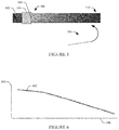
- FIG. 3 shows an example vessel with antegrade collateral flow
- FIG. 4 shows a plot of a polynomial function fitted to voxel intensity values and as a function of distance along a coronary vessel from the ostium for antegrade collateral flow;
- FIG. 5 shows an example vessel with retrograde collateral flow
- FIG. 6 shows a plot of a polynomial function fitted to voxel intensity values and as a function of distance along a coronary vessel from the ostium for retrograde collateral flow
- FIG. 7 illustrates an example method for determining fractional flow reserve, taking into account collateral flow.
- FIG. 1 schematically illustrates an imaging system 100 such as a computed tomography (CT) scanner.
- CT computed tomography
- the imaging system 100 is configured at least for coronary CT angiography (CCTA) scans or procedures.
- the imaging system 100 additionally or alternatively includes an X-ray scanner, magnetic resonance imaging (MRI) scanner, and/or other scanner configured coronary CT angiography (CCTA) and/or other scans.
- CCTA coronary CT angiography
- a stationary gantry 102 rotatably supports a rotating gantry 104 , which is configured to rotate around an examination region 106 .
- a subject support 108 supports an object or subject in the examination region 106 .
- a radiation source 110 such as an x-ray tube, is supported by the rotating gantry 104 , rotates with the rotating gantry 104 , and is configured to emit radiation that traverses the examination region 106 .
- a radiation sensitive detector array 112 subtends an angular arc opposite the radiation source 110 across the examination region 106 , and is configured to detect radiation traversing the examination region 106 and generate a signal (projection data) indicative thereof.
- a reconstructor 114 is configured to reconstruct the projection, generating volumetric image data indicative of a scanned portion of a subject or object located in the examination region 106 .
- the subject is administered (e.g., intravenously, etc.) a radio-opaque contrast agent and the resulting volumetric image data is CCTA image data that visualizes the inside (lumen) of blood vessels such as arteries, veins, etc. of a coronary vessel and/or other vessel.
- a computing system serves as a console 116 for the imaging system 100 .
- the console 116 includes at least one processor 118 (e.g., a microprocessor, a central processing unit, etc.) that executes at least one computer readable instruction stored in a computer readable storage medium (“memory”) 120 , which excludes transitory medium and includes physical memory and/or other non-transitory medium.
- processor 118 e.g., a microprocessor, a central processing unit, etc.
- memory computer readable storage medium
- the at least one computer readable instruction includes an acquisition module 122 and an FFR determiner 124 .
- the FFR determiner 124 is stored in a memory of and executed with a processor of a computing system different from the console 116 such as a workstation, computer, etc.
- An output device(s) includes a human readable output device such as a display monitor 126
- an input device(s) 128 includes a mouse, keyboard, touchscreen, etc.
- the data acquisition module 122 includes instructions for performing a scan and reconstructing CCTA image data.
- the FFR determiner 124 includes instructions for determining an FFR from CCTA image data. As described in greater detail below, in one instance this includes detecting whether a collateral flow for a coronary vessel is present from the CCTA image data based on indirect analysis of voxel intensity (e.g., Hounsfield unit (HU) values) along the coronary tree.
- voxel intensity e.g., Hounsfield unit (HU) values
- the FFR determiner 124 employs an algorithm that includes an adaptive boundary condition model that accounts for a presence of a collateral flow and/or adapts a boundary condition model as such.
- FIG. 2 illustrates an example of the FFR determiner 124 .
- a tissue of interest identifier 202 identifies tissue of interest in the volumetric image data.
- the tissue of interest can be identified by a user, a default tissue of interest, etc.
- the tissue of interest identifier 202 can employ automatic and/or manual approaches to identify the tissue of interest.
- the tissue of interest is a vessel such as a coronary vessel and/or other vessel.
- a collateral flow identifier 204 is configured to identify whether there is collateral flow for the tissue of interest.
- collateral flow is an auto-regulation mechanism used by the body to prevent ischemia in case of coronary stenosis by creating new arterioles that support collateral blood flow to the potentially ischemic region.
- Collateral flow can be characterized by magnitude (e.g., poorly developed vs well-developed collateral arterioles) and direction (e.g., retrograde vs antegrade) from the coronary ostium, which is the opening of coronary arteries at root of aorta, superior to the aortic valve.
- the collateral flow identifier 204 assesses collateral flow magnitude and/or direction indirectly from the CCTA image data by analyzing changes in the voxel intensity (e.g., in HUs) along the coronary vessel as a function of a distance from the ostium. For this, in one instance, the collateral flow identifier 204 automatically extracts a vessel centerline and the lumen. This can be achieved through known and/or other approaches. Next, a machine-learning approach characterizes the magnitude and the direction of the collateral flow based on the centerline and the lumen.
- the collateral flow identifier 204 estimates average HUs in equidistant points along a centerline I(c). For this, the collateral flow identifier 204 first fits a second order polynomial function to the measurements.
- a suitable fitting of a second order polynomial function I p (c) based on a least-square approach is shown in EQUATION 1:
- the collateral flow identifier 204 estimates the magnitude and the direction of the collateral flow by analyzing the polynomial function coefficients and its derivatives respectively to determine the magnitude and the direction.
- FIGS. 3, 4, 5 and 6 Examples of antegrade and retrograde collateral flow and corresponding polynomial functions are shown in connection with FIGS. 3, 4, 5 and 6 .
- FIG. 3 shows an example vessel 302 , a total occlusion 303 , an ostium 304 , and antegrade collateral flow 306 , where flow is greater in a darker gray region 308 proximal the ostium 304 and flow is lower in a lighter gray region 310 distal the ostium 304 .
- FIG. 4 shows an example of a plot 402 of a polynomial function for FIG. 3 that indicates a presence of the antegrade collateral flow.
- a first axis 404 represents intensity (e.g., in HU) and a second axis 406 represents distance from the Ostium (e.g., in mm).
- the concave shape of the plot 402 indicates antegrade flow.
- FIG. 5 shows an example vessel 502 , a total occlusion 503 , an ostium 504 , and retrograde collateral flow 504 , where flow is lower in a lighter gray region 508 proximal the ostium 304 and flow is greater in a darker gray region 510 distal the ostium.
- FIG. 6 shows an example of a plot 602 of a polynomial function for FIG. 5 that indicates a presence of the retrograde collateral flow.
- a first axis 604 represents intensity (e.g., in HU) and a second axis 606 represents distance from the Ostium (e.g., in mm).
- the convex shape of the plot 602 indicates retrograde flow.
- plots 402 and 602 would be horizontal lines, and not concave nor convex, as shown respectively in FIGS. 4 and 6 , and the flow would be the same from the regions 308 and 508 respectively to the regions 310 and 510 (the same shade of gray), unlike that in FIGS. 3 and 5 .
- a geometry extractor 206 extracts geometrical information from the identified tissue of interest.
- the geometry extractor 206 can employ automatic and/or manual approaches to extract the geometrical information.
- the extraction may include employing a segmentation algorithm. From this and/or other extraction, an effective diameter at the ostium d o and/or other tissue of interest geometry can be determined.
- a parameter determiner 208 determines a parameter based on input subject data.
- the parameter determiner 208 can determine an inlet flow-rate Q o (i.e., flow rate at the ostium). This can be achieved based on subject data such as weight, body mass index (BMI), gender, age, blood test results, anatomical imaging data (e.g., myocardium mass and estimated stroke-volume), and/or subject data.
- subject data such as weight, body mass index (BMI), gender, age, blood test results, anatomical imaging data (e.g., myocardium mass and estimated stroke-volume), and/or subject data.
- a boundary condition estimator 210 estimates boundary conditions such as flow, pressure, resistance, etc., taking into account collateral flow, if collateral flow is identified by the collateral flow identifier 204 .
- the boundary condition estimate is a resistance and is combined with a resistance boundary condition of the coronary vessel.
- a flow rate Q can be determined at the outlet of a coronary vessel as a function of the coronary outlet cross sectional area, as shown in EQUATION 2:
- d 2 ⁇ csa ⁇ , EQUATION ⁇ ⁇ 3 where csa is a cross-sectional area of the vessel at the outlet.
- R 1 R 2 ( d 1 d 2 ) - 7 3 EQUATION ⁇ ⁇ 5 where R 1 is a resistance of one of the two branches and R 2 is a resistance of the other of the two branches. In general, these resistances are dominated by peripheral microvascular arteries with diameters lower than 0.2 mm.
- the influence of stenosis on the resistance can be formulated as summation of the normal outlet resistance and additional resistance component which can be modeled as linearly related to the normal resistance as shown in EQUATION 6.
- R s R 0 + ⁇ R 0 , EQUATION 6
- R s is the assigned outlet resistance adjusted from normal outlet resistance to account for the presence of stenosis
- R 0 is the normal resistance at the outlet
- E is represents an amount of stenosis in the coronary vessel of interest.
- the normal outlet resistance and the parameter £ can be found by optimizing an error function over a training set with invasive FFR measurements as the ground-truth.
- the influence of the collateral flow can be integrated into this model.
- this includes a virtual addition of arterioles to the three-dimensional geometrical model of the coronary tree in the amount and direction of the collateral flow.
- this includes an adjustment of the coronaries outlets' resistance values to reflect the presence of collateral flow.
- R c represents adjusted resistance accounting for the collateral flow
- R s is the coronary outlet resistance computed using a suitable boundary conditions model (e.g., as described in patent application PCT/EP2015/064168)
- F c represents a collateral flow as determined by the collateral flow identifier 204 and/or otherwise
- a represents a weighting parameter.
- the parameter ⁇ is a free parameter, which can be optimized experimentally, e.g., by optimizing an error function over a training dataset with invasive FFR measurements as the ground-truth. For example, this can be achieved by optimizing the error function shown in EQUATION 8:
- ⁇ ⁇ argmin ⁇ ⁇ ⁇ ⁇ C ⁇ TrainingCases ⁇ ( FFR — ⁇ CT ⁇ ( C , F c ) - FFR — ⁇ GT ⁇ ( C ) ) 2 .
- ⁇ circumflex over ( ⁇ ) ⁇ is the optimal value for the parameter ⁇ found through the optimization
- C is the coronary model of one patient from the database (TrainingCases)
- F c represents a collateral flow as determined by the collateral flow identifier
- FFR_CT (C, F c ) is the FFR value calculated based on the inputs C, F c with a given value for the parameter ⁇
- FFR_GT(C) is the invasively measured FFR value for this patient.
- Patent application PCT/EP2015/064168 describes some of the features that related to the microvascular resistance, including the coronary outlet cross-sectional area, presence of coronary stenosis, heart size, ejection fraction, among others.
- Different parametric relationships can be considered, including weighted linear sum or weighted non-linear sum of the different effects.
- An FFR determiner 214 determines an FFR based on the boundary conditions. In one instance, this includes simulating the flow of blood within the coronary artery system based on a representation of the coronary artery system and the boundary conditions and then determining the FFR value based on the simulated flow of the blood.
- the FFR determiner 214 can employ zero-dimensional, one-dimensional, etc. algorithms that solve fluid dynamics problems. For example, in one instance, the FFR determiner 214 utilizes a computational fluid dynamic (CFD) simulation using partial-differential-equations.
- CFD is a fluid mechanics approach that uses numerical methods and/or algorithms to solve and analyze problems that involve fluid flows. The calculations are performed with surfaces defined by boundary conditions.
- FIG. 7 illustrates an example method for determining an FFR.
- a coronary angiography scan of a subject is performed.
- volumetric coronary angiography imag data is generated from the data acquired during the scan.
- the volumetric coronary angiography imag data is processed to locate a coronary vessel represented in the volumetric coronary angiography imag data.
- a presence of a collateral flow for the identified coronary vessel is identified with the volumetric coronary angiography imag data, as described herein and/or otherwise.
- a boundary condition is estimated for the collateral flow, as described herein and/or otherwise.
- a boundary condition parametric model that includes takes into account the boundary condition of the collateral flow is constructed, as described herein and/or otherwise.
- a fractional flow reserve index is determined for the coronary vessel with the boundary condition parametric model.
- the above may be implemented by way of computer readable instructions, encoded or embedded on non-transitory computer readable storage medium, which, when executed by a computer processor(s), cause the processor(s) to carry out the described acts. Additionally or alternatively, at least one of the computer readable instructions is carried by a signal, carrier wave or other transitory medium.
Priority Applications (1)
| Application Number | Priority Date | Filing Date | Title |
|---|---|---|---|
| US15/768,162 US10769780B2 (en) | 2015-11-05 | 2016-10-17 | Collateral flow modelling for non-invasive fractional flow reserve (FFR) |
Applications Claiming Priority (3)
| Application Number | Priority Date | Filing Date | Title |
|---|---|---|---|
| US201562251417P | 2015-11-05 | 2015-11-05 | |
| US15/768,162 US10769780B2 (en) | 2015-11-05 | 2016-10-17 | Collateral flow modelling for non-invasive fractional flow reserve (FFR) |
| PCT/EP2016/074852 WO2017076620A1 (en) | 2015-11-05 | 2016-10-17 | Collateral flow modelling for non-invasive fractional flow reserve (ffr) |
Publications (2)
| Publication Number | Publication Date |
|---|---|
| US20180315184A1 US20180315184A1 (en) | 2018-11-01 |
| US10769780B2 true US10769780B2 (en) | 2020-09-08 |
Family
ID=57184425
Family Applications (1)
| Application Number | Title | Priority Date | Filing Date |
|---|---|---|---|
| US15/768,162 Active 2037-05-09 US10769780B2 (en) | 2015-11-05 | 2016-10-17 | Collateral flow modelling for non-invasive fractional flow reserve (FFR) |
Country Status (5)
| Country | Link |
|---|---|
| US (1) | US10769780B2 (ja) |
| EP (1) | EP3370615B1 (ja) |
| JP (1) | JP6484760B2 (ja) |
| CN (1) | CN108348206B (ja) |
| WO (1) | WO2017076620A1 (ja) |
Cited By (2)
| Publication number | Priority date | Publication date | Assignee | Title |
|---|---|---|---|---|
| US11523744B2 (en) | 2017-03-31 | 2022-12-13 | Koninklijke Philips N.V. | Interaction monitoring of non-invasive imaging based FFR |
| US11633118B2 (en) | 2017-06-30 | 2023-04-25 | Koninklijke Philips N.V. | Machine learning spectral FFR-CT |
Families Citing this family (9)
| Publication number | Priority date | Publication date | Assignee | Title |
|---|---|---|---|---|
| WO2018050806A1 (en) | 2016-09-16 | 2018-03-22 | Koninklijke Philips N.V. | Apparatus and method for determining a fractional flow reserve |
| EP3378398A1 (en) | 2017-03-24 | 2018-09-26 | Koninklijke Philips N.V. | Myocardial ct perfusion image synthesis |
| EP3453326A1 (en) * | 2017-09-08 | 2019-03-13 | Koninklijke Philips N.V. | Registration and comparison of measured and simulated intracoronary pullback curves |
| CN111227821B (zh) | 2018-11-28 | 2022-02-11 | 苏州润迈德医疗科技有限公司 | 基于心肌血流量和ct图像的微循环阻力指数计算方法 |
| CN111227822B (zh) * | 2018-11-28 | 2022-02-11 | 苏州润迈德医疗科技有限公司 | 基于心肌血流量和ct图像的冠状动脉血流储备分数计算方法 |
| CN110598288B (zh) * | 2019-08-30 | 2020-08-07 | 上海杏脉信息科技有限公司 | 一种用于冠脉三维模型的边界条件处理方法和装置 |
| US11145057B2 (en) * | 2019-11-05 | 2021-10-12 | Siemens Healthcare Gmbh | Assessment of collateral coronary arteries |
| EP3819909A1 (en) * | 2019-11-05 | 2021-05-12 | Siemens Healthcare GmbH | Assessment of collateral coronary arteries |
| CN110664392B (zh) * | 2019-12-04 | 2020-03-27 | 深圳先进技术研究院 | 一种血流速度计算方法、装置、终端设备及存储介质 |
Citations (34)
| Publication number | Priority date | Publication date | Assignee | Title |
|---|---|---|---|---|
| WO2000072037A1 (en) | 1999-05-21 | 2000-11-30 | Nycomed Imaging As | Method of magnetic resonance imaging |
| WO2004025572A1 (en) | 2002-09-16 | 2004-03-25 | Tayside Flow Technologies Limited | A method of analysing fluid flow in a conduit |
| WO2006061814A1 (en) | 2004-12-08 | 2006-06-15 | Paieon Inc. | Method and apparatus for finding the tubular organs blood velocity and flow and related parameters |
| WO2006061815A1 (en) | 2004-12-08 | 2006-06-15 | Paieon Inc. | Method and apparatus for blood vessel parameter determinations |
| US20070078352A1 (en) | 2005-09-30 | 2007-04-05 | Radi Medical System Ab | Method for determining the blood flow in a coronary artery |
| DE102008014792B3 (de) | 2008-03-18 | 2009-06-18 | Siemens Aktiengesellschaft | Verfahren und Vorrichtung zur Simulation eines Blutflusses in einem Gefäßabschnitt |
| WO2010022762A2 (en) | 2008-08-25 | 2010-03-04 | ETH Zürich | Method, system and device for enhancing flow field data |
| US20100125197A1 (en) | 2008-11-18 | 2010-05-20 | Fishel Robert S | Method and apparatus for addressing vascular stenotic lesions |
| US20100130878A1 (en) | 2008-11-24 | 2010-05-27 | General Electric Company | Systems, apparatus and processes for automated blood flow assessment of vasculature |
| US20100241404A1 (en) | 2009-03-17 | 2010-09-23 | Taylor Charles A | Patient-specific hemodynamics of the cardio vascular system |
| US20110211742A1 (en) | 2008-09-30 | 2011-09-01 | Koninklijke Philips Electronics N.V. | Perfusion imaging |
| US20110307231A1 (en) | 2010-06-09 | 2011-12-15 | Jens Kirchner | Method and arrangement for creating an individualized, computer-aided model of a system, and a corresponding computer program and a corresponding machine-readable storage medium |
| US20120022843A1 (en) | 2010-07-21 | 2012-01-26 | Razvan Ioan Ionasec | Method and System for Comprehensive Patient-Specific Modeling of the Heart |
| US20120041739A1 (en) | 2010-08-12 | 2012-02-16 | Heartflow, Inc. | Method and System for Patient-Specific Modeling of Blood Flow |
| US20120041325A1 (en) | 2007-03-14 | 2012-02-16 | Ramesh Wariar | Method and apparatus for management of heart failure hospitalization |
| US20120053918A1 (en) | 2010-08-12 | 2012-03-01 | Heartflow, Inc. | Method and system for patient-specific modeling of blood flow |
| US20120072190A1 (en) | 2010-09-16 | 2012-03-22 | Siemens Corporation | Method and System for Non-Invasive Assessment of Coronary Artery Disease |
| US20120121151A1 (en) | 2010-11-12 | 2012-05-17 | Siemens Aktiengesellschaft | Device And Computed Tomography Scanner For Determining And Visualizing The Perfusion Of The Myocardial Muscle |
| US8200466B2 (en) | 2008-07-21 | 2012-06-12 | The Board Of Trustees Of The Leland Stanford Junior University | Method for tuning patient-specific cardiovascular simulations |
| US20120238888A1 (en) | 2009-12-10 | 2012-09-20 | Koninklijke Philips Electronics N.V. | Collateral blood flow assessment |
| US20120243761A1 (en) | 2011-03-21 | 2012-09-27 | Senzig Robert F | System and method for estimating vascular flow using ct imaging |
| US20120296199A1 (en) | 2011-03-21 | 2012-11-22 | New York University | Apparatus and Method of Non-Contrast Magnetic Resonance Angiography of Abdominal and Pelvic Arteries |
| US20130132054A1 (en) | 2011-11-10 | 2013-05-23 | Puneet Sharma | Method and System for Multi-Scale Anatomical and Functional Modeling of Coronary Circulation |
| US20140114618A1 (en) | 2012-10-19 | 2014-04-24 | Timothy A. Fonte | Systems and methods for numerically evaluating vasculature |
| WO2014072861A2 (en) | 2012-11-06 | 2014-05-15 | Koninklijke Philips N.V. | Fractional flow reserve (ffr) index |
| WO2014088103A1 (ja) | 2012-12-07 | 2014-06-12 | 株式会社 東芝 | 血管解析装置、医用画像診断装置、及び血管解析方法 |
| US20140200867A1 (en) | 2013-01-15 | 2014-07-17 | Cathworks Ltd | Vascular flow assessment |
| US20150051888A1 (en) | 2012-03-15 | 2015-02-19 | Siemens Corporation | Framework for personalization of coronary flow computations during rest and hyperemia |
| US20150112191A1 (en) | 2013-10-22 | 2015-04-23 | Koninklijke Philips Electronics N.V. | Fractional flow reserve (ffr) index with adaptive boundary condition parameters |
| US20150265162A1 (en) | 2012-10-24 | 2015-09-24 | Cathworks Ltd. | Automated measurement system and method for coronary artery disease scoring |
| US20150339847A1 (en) * | 2012-10-24 | 2015-11-26 | Cath Works Ltd. | Creating a vascular tree model |
| WO2016001017A1 (en) | 2014-06-30 | 2016-01-07 | Koninklijke Philips N.V. | Apparatus for determining a fractional flow reserve value |
| US9339200B2 (en) * | 2014-03-31 | 2016-05-17 | Heartflow, Inc. | Systems and methods for determining blood flow characteristics using flow ratio |
| EP3062248A1 (en) | 2015-02-27 | 2016-08-31 | Pie Medical Imaging BV | Method and apparatus for quantitative flow analysis |
Family Cites Families (7)
| Publication number | Priority date | Publication date | Assignee | Title |
|---|---|---|---|---|
| JPH0889501A (ja) * | 1994-09-21 | 1996-04-09 | Hitachi Medical Corp | 医用画像診断装置 |
| WO2008046197A1 (en) * | 2006-10-13 | 2008-04-24 | Calgary Scientific Inc. | Visualization of volumetric medical imaging data |
| EP2104920B1 (en) * | 2007-01-11 | 2015-03-04 | Intellectual Property MVM B.V. | The measurement of functional microcirculatory geometry and velocity distributions using automated image analysis |
| EP2621343A1 (en) * | 2010-09-30 | 2013-08-07 | Koninklijke Philips Electronics N.V. | Detection of bifurcations using traceable imaging device and imaging tool |
| CN103300820A (zh) * | 2012-03-13 | 2013-09-18 | 西门子公司 | 用于冠状动脉狭窄的非侵入性功能评估的方法和系统 |
| CN103839249B (zh) * | 2012-11-23 | 2017-02-08 | 上海联影医疗科技有限公司 | Ct肝灌注的图像后处理方法和ct肝灌注方法 |
| WO2018050806A1 (en) * | 2016-09-16 | 2018-03-22 | Koninklijke Philips N.V. | Apparatus and method for determining a fractional flow reserve |
-
2016
- 2016-10-17 WO PCT/EP2016/074852 patent/WO2017076620A1/en active Application Filing
- 2016-10-17 JP JP2018521572A patent/JP6484760B2/ja active Active
- 2016-10-17 CN CN201680064744.7A patent/CN108348206B/zh active Active
- 2016-10-17 US US15/768,162 patent/US10769780B2/en active Active
- 2016-10-17 EP EP16784840.7A patent/EP3370615B1/en active Active
Patent Citations (46)
| Publication number | Priority date | Publication date | Assignee | Title |
|---|---|---|---|---|
| WO2000072037A1 (en) | 1999-05-21 | 2000-11-30 | Nycomed Imaging As | Method of magnetic resonance imaging |
| WO2004025572A1 (en) | 2002-09-16 | 2004-03-25 | Tayside Flow Technologies Limited | A method of analysing fluid flow in a conduit |
| WO2006061814A1 (en) | 2004-12-08 | 2006-06-15 | Paieon Inc. | Method and apparatus for finding the tubular organs blood velocity and flow and related parameters |
| WO2006061815A1 (en) | 2004-12-08 | 2006-06-15 | Paieon Inc. | Method and apparatus for blood vessel parameter determinations |
| US20070078352A1 (en) | 2005-09-30 | 2007-04-05 | Radi Medical System Ab | Method for determining the blood flow in a coronary artery |
| US20100286537A1 (en) | 2005-09-30 | 2010-11-11 | Radi Medical Systems Ab | System for determining the blood flow in a coronary artery |
| US20120041325A1 (en) | 2007-03-14 | 2012-02-16 | Ramesh Wariar | Method and apparatus for management of heart failure hospitalization |
| DE102008014792B3 (de) | 2008-03-18 | 2009-06-18 | Siemens Aktiengesellschaft | Verfahren und Vorrichtung zur Simulation eines Blutflusses in einem Gefäßabschnitt |
| US8200466B2 (en) | 2008-07-21 | 2012-06-12 | The Board Of Trustees Of The Leland Stanford Junior University | Method for tuning patient-specific cardiovascular simulations |
| WO2010022762A2 (en) | 2008-08-25 | 2010-03-04 | ETH Zürich | Method, system and device for enhancing flow field data |
| US20110211742A1 (en) | 2008-09-30 | 2011-09-01 | Koninklijke Philips Electronics N.V. | Perfusion imaging |
| US20100125197A1 (en) | 2008-11-18 | 2010-05-20 | Fishel Robert S | Method and apparatus for addressing vascular stenotic lesions |
| US20100130878A1 (en) | 2008-11-24 | 2010-05-27 | General Electric Company | Systems, apparatus and processes for automated blood flow assessment of vasculature |
| US20100241404A1 (en) | 2009-03-17 | 2010-09-23 | Taylor Charles A | Patient-specific hemodynamics of the cardio vascular system |
| US20120238888A1 (en) | 2009-12-10 | 2012-09-20 | Koninklijke Philips Electronics N.V. | Collateral blood flow assessment |
| US20110307231A1 (en) | 2010-06-09 | 2011-12-15 | Jens Kirchner | Method and arrangement for creating an individualized, computer-aided model of a system, and a corresponding computer program and a corresponding machine-readable storage medium |
| US20120022843A1 (en) | 2010-07-21 | 2012-01-26 | Razvan Ioan Ionasec | Method and System for Comprehensive Patient-Specific Modeling of the Heart |
| US20120041318A1 (en) | 2010-08-12 | 2012-02-16 | Heartflow, Inc. | Method and system for patient-specific modeling of blood flow |
| US20120053919A1 (en) | 2010-08-12 | 2012-03-01 | Heartflow, Inc. | Method and system for patient-specific modeling of blood flow |
| US20120041319A1 (en) | 2010-08-12 | 2012-02-16 | Heartflow, Inc. | Method and system for patient-specific modeling of blood flow |
| US20120041322A1 (en) | 2010-08-12 | 2012-02-16 | Heartflow, Inc. | Method and system for patient-specific modeling of blood flow |
| US20120041323A1 (en) | 2010-08-12 | 2012-02-16 | Heartflow, Inc. | Method and system for patient-specific modeling of blood flow |
| US20120041324A1 (en) | 2010-08-12 | 2012-02-16 | Heartflow, Inc. | Method and system for patient-specific modeling of blood flow |
| US20120053918A1 (en) | 2010-08-12 | 2012-03-01 | Heartflow, Inc. | Method and system for patient-specific modeling of blood flow |
| US20120041320A1 (en) | 2010-08-12 | 2012-02-16 | Heartflow, Inc. | Method and system for patient-specific modeling of blood flow |
| US20120059246A1 (en) | 2010-08-12 | 2012-03-08 | Heartflow, Inc. | Method and system for patient-specific modeling of blood flow |
| US20120041739A1 (en) | 2010-08-12 | 2012-02-16 | Heartflow, Inc. | Method and System for Patient-Specific Modeling of Blood Flow |
| US8157742B2 (en) | 2010-08-12 | 2012-04-17 | Heartflow, Inc. | Method and system for patient-specific modeling of blood flow |
| US8249815B2 (en) | 2010-08-12 | 2012-08-21 | Heartflow, Inc. | Method and system for patient-specific modeling of blood flow |
| US20120041321A1 (en) | 2010-08-12 | 2012-02-16 | Heartflow, Inc. | Method and system for patient-specific modeling of blood flow |
| US20120072190A1 (en) | 2010-09-16 | 2012-03-22 | Siemens Corporation | Method and System for Non-Invasive Assessment of Coronary Artery Disease |
| US20120121151A1 (en) | 2010-11-12 | 2012-05-17 | Siemens Aktiengesellschaft | Device And Computed Tomography Scanner For Determining And Visualizing The Perfusion Of The Myocardial Muscle |
| US20120243761A1 (en) | 2011-03-21 | 2012-09-27 | Senzig Robert F | System and method for estimating vascular flow using ct imaging |
| US20120296199A1 (en) | 2011-03-21 | 2012-11-22 | New York University | Apparatus and Method of Non-Contrast Magnetic Resonance Angiography of Abdominal and Pelvic Arteries |
| US20130132054A1 (en) | 2011-11-10 | 2013-05-23 | Puneet Sharma | Method and System for Multi-Scale Anatomical and Functional Modeling of Coronary Circulation |
| US20150051888A1 (en) | 2012-03-15 | 2015-02-19 | Siemens Corporation | Framework for personalization of coronary flow computations during rest and hyperemia |
| US20140114618A1 (en) | 2012-10-19 | 2014-04-24 | Timothy A. Fonte | Systems and methods for numerically evaluating vasculature |
| US20150339847A1 (en) * | 2012-10-24 | 2015-11-26 | Cath Works Ltd. | Creating a vascular tree model |
| US20150265162A1 (en) | 2012-10-24 | 2015-09-24 | Cathworks Ltd. | Automated measurement system and method for coronary artery disease scoring |
| WO2014072861A2 (en) | 2012-11-06 | 2014-05-15 | Koninklijke Philips N.V. | Fractional flow reserve (ffr) index |
| WO2014088103A1 (ja) | 2012-12-07 | 2014-06-12 | 株式会社 東芝 | 血管解析装置、医用画像診断装置、及び血管解析方法 |
| US20140200867A1 (en) | 2013-01-15 | 2014-07-17 | Cathworks Ltd | Vascular flow assessment |
| US20150112191A1 (en) | 2013-10-22 | 2015-04-23 | Koninklijke Philips Electronics N.V. | Fractional flow reserve (ffr) index with adaptive boundary condition parameters |
| US9339200B2 (en) * | 2014-03-31 | 2016-05-17 | Heartflow, Inc. | Systems and methods for determining blood flow characteristics using flow ratio |
| WO2016001017A1 (en) | 2014-06-30 | 2016-01-07 | Koninklijke Philips N.V. | Apparatus for determining a fractional flow reserve value |
| EP3062248A1 (en) | 2015-02-27 | 2016-08-31 | Pie Medical Imaging BV | Method and apparatus for quantitative flow analysis |
Non-Patent Citations (15)
Cited By (2)
| Publication number | Priority date | Publication date | Assignee | Title |
|---|---|---|---|---|
| US11523744B2 (en) | 2017-03-31 | 2022-12-13 | Koninklijke Philips N.V. | Interaction monitoring of non-invasive imaging based FFR |
| US11633118B2 (en) | 2017-06-30 | 2023-04-25 | Koninklijke Philips N.V. | Machine learning spectral FFR-CT |
Also Published As
| Publication number | Publication date |
|---|---|
| EP3370615B1 (en) | 2020-04-15 |
| JP6484760B2 (ja) | 2019-03-13 |
| JP2018537157A (ja) | 2018-12-20 |
| US20180315184A1 (en) | 2018-11-01 |
| EP3370615A1 (en) | 2018-09-12 |
| WO2017076620A1 (en) | 2017-05-11 |
| CN108348206A (zh) | 2018-07-31 |
| CN108348206B (zh) | 2022-07-29 |
Similar Documents
| Publication | Publication Date | Title |
|---|---|---|
| US10769780B2 (en) | Collateral flow modelling for non-invasive fractional flow reserve (FFR) | |
| JP6553099B2 (ja) | 血流予備量比値を算出するための機器 | |
| US10595806B2 (en) | Fractional flow reserve (FFR) index with adaptive boundary condition parameters | |
| EP2916735B1 (en) | Fractional flow reserve (ffr) index | |
| JP6255473B2 (ja) | シミュレーションの正確度および性能に対する画質評価 | |
| US11083377B2 (en) | Method and apparatus for quantitative hemodynamic flow analysis | |
| EP3244790B1 (en) | Instantaneous wave-free ratio (ifr) computer tomography (ct) | |
| US11039804B2 (en) | Apparatus and method for determining a fractional flow reserve | |
| EP3923810A1 (en) | Prediction of coronary microvascular dysfunction from coronary computed tomography | |
| JP7426824B2 (ja) | 非侵襲的イメージングベースのffrのインタラクションモニタリング | |
| EP3667618A1 (en) | Deep partial-angle coronary restoration |
Legal Events
| Date | Code | Title | Description |
|---|---|---|---|
| AS | Assignment |
Owner name: KONINKLIJKE PHILIPS N.V., NETHERLANDS Free format text: ASSIGNMENT OF ASSIGNORS INTEREST;ASSIGNORS:FREIMAN, MORDECHAY PINCHAS;GOSHEN, LIRAN;SIGNING DATES FROM 20180304 TO 20180308;REEL/FRAME:045533/0542 |
|
| FEPP | Fee payment procedure |
Free format text: ENTITY STATUS SET TO UNDISCOUNTED (ORIGINAL EVENT CODE: BIG.); ENTITY STATUS OF PATENT OWNER: LARGE ENTITY |
|
| STPP | Information on status: patent application and granting procedure in general |
Free format text: APPLICATION UNDERGOING PREEXAM PROCESSING |
|
| STPP | Information on status: patent application and granting procedure in general |
Free format text: DOCKETED NEW CASE - READY FOR EXAMINATION |
|
| STPP | Information on status: patent application and granting procedure in general |
Free format text: NON FINAL ACTION MAILED |
|
| STPP | Information on status: patent application and granting procedure in general |
Free format text: NOTICE OF ALLOWANCE MAILED -- APPLICATION RECEIVED IN OFFICE OF PUBLICATIONS |
|
| STCF | Information on status: patent grant |
Free format text: PATENTED CASE |
|
| MAFP | Maintenance fee payment |
Free format text: PAYMENT OF MAINTENANCE FEE, 4TH YEAR, LARGE ENTITY (ORIGINAL EVENT CODE: M1551); ENTITY STATUS OF PATENT OWNER: LARGE ENTITY Year of fee payment: 4 |