US10648979B2 - Molecular probes for detecting gram-negative bacteria in vitro and in vivo - Google Patents
Molecular probes for detecting gram-negative bacteria in vitro and in vivo Download PDFInfo
- Publication number
- US10648979B2 US10648979B2 US15/526,414 US201515526414A US10648979B2 US 10648979 B2 US10648979 B2 US 10648979B2 US 201515526414 A US201515526414 A US 201515526414A US 10648979 B2 US10648979 B2 US 10648979B2
- Authority
- US
- United States
- Prior art keywords
- bacteria
- probe
- nbd
- fluorophore
- target area
- Prior art date
- Legal status (The legal status is an assumption and is not a legal conclusion. Google has not performed a legal analysis and makes no representation as to the accuracy of the status listed.)
- Active
Links
- 241000894006 Bacteria Species 0.000 title claims abstract description 238
- 238000000338 in vitro Methods 0.000 title claims description 18
- 238000001727 in vivo Methods 0.000 title claims description 10
- 239000003068 molecular probe Substances 0.000 title description 3
- 239000000523 sample Substances 0.000 claims abstract description 268
- 238000000034 method Methods 0.000 claims abstract description 91
- 230000027455 binding Effects 0.000 claims abstract description 73
- 241000192125 Firmicutes Species 0.000 claims abstract description 22
- 210000004072 lung Anatomy 0.000 claims description 75
- 241000233866 Fungi Species 0.000 claims description 45
- 108010040201 Polymyxins Proteins 0.000 claims description 37
- 108010092136 ribosomal protein S30 Proteins 0.000 claims description 26
- 150000001413 amino acids Chemical class 0.000 claims description 24
- 239000012634 fragment Substances 0.000 claims description 20
- 238000003384 imaging method Methods 0.000 claims description 19
- 238000011065 in-situ storage Methods 0.000 claims description 17
- 210000004962 mammalian cell Anatomy 0.000 claims description 17
- 239000003795 chemical substances by application Substances 0.000 claims description 16
- -1 5-(dimethylamino)naphthalene-1-sulfonyl Chemical group 0.000 claims description 14
- 238000001514 detection method Methods 0.000 claims description 14
- 230000002209 hydrophobic effect Effects 0.000 claims description 14
- 238000006467 substitution reaction Methods 0.000 claims description 12
- 230000003115 biocidal effect Effects 0.000 claims description 11
- 210000000170 cell membrane Anatomy 0.000 claims description 11
- 230000001524 infective effect Effects 0.000 claims description 10
- RAXXELZNTBOGNW-UHFFFAOYSA-N imidazole Substances C1=CNC=N1 RAXXELZNTBOGNW-UHFFFAOYSA-N 0.000 claims description 7
- 229940121375 antifungal agent Drugs 0.000 claims description 6
- 150000002430 hydrocarbons Chemical class 0.000 claims description 6
- 229940107698 malachite green Drugs 0.000 claims description 5
- FDZZZRQASAIRJF-UHFFFAOYSA-M malachite green Chemical compound [Cl-].C1=CC(N(C)C)=CC=C1C(C=1C=CC=CC=1)=C1C=CC(=[N+](C)C)C=C1 FDZZZRQASAIRJF-UHFFFAOYSA-M 0.000 claims description 5
- 238000006862 quantum yield reaction Methods 0.000 claims description 5
- 230000000843 anti-fungal effect Effects 0.000 claims description 4
- BBEAQIROQSPTKN-UHFFFAOYSA-N pyrene Chemical compound C1=CC=C2C=CC3=CC=CC4=CC=C1C2=C43 BBEAQIROQSPTKN-UHFFFAOYSA-N 0.000 claims description 4
- 238000001228 spectrum Methods 0.000 claims description 4
- GPOPHQSTNHUENT-UHFFFAOYSA-N 6-Methyl caprylic acid Chemical group CCC(C)CCCCC(O)=O GPOPHQSTNHUENT-UHFFFAOYSA-N 0.000 claims description 3
- OEOIWYCWCDBOPA-UHFFFAOYSA-N 6-methyl-heptanoic acid Chemical group CC(C)CCCCC(O)=O OEOIWYCWCDBOPA-UHFFFAOYSA-N 0.000 claims description 3
- 239000004215 Carbon black (E152) Substances 0.000 claims description 3
- 238000001574 biopsy Methods 0.000 claims description 3
- 229930195733 hydrocarbon Natural products 0.000 claims description 3
- 239000007788 liquid Substances 0.000 claims description 3
- 229930182817 methionine Natural products 0.000 claims description 3
- 108700018363 polymyxin B(1) Proteins 0.000 claims description 3
- WQVJHHACXVLGBL-GOVYWFKWSA-N polymyxin B1 Polymers N1C(=O)[C@H](CCN)NC(=O)[C@@H](NC(=O)[C@H](CCN)NC(=O)[C@H]([C@@H](C)O)NC(=O)[C@H](CCN)NC(=O)CCCC[C@H](C)CC)CCNC(=O)[C@H]([C@@H](C)O)NC(=O)[C@H](CCN)NC(=O)[C@H](CCN)NC(=O)[C@H](CC(C)C)NC(=O)[C@H]1CC1=CC=CC=C1 WQVJHHACXVLGBL-GOVYWFKWSA-N 0.000 claims description 3
- 108010089148 polymyxin B2 Proteins 0.000 claims description 3
- SGPYLFWAQBAXCZ-RUDZPDEXSA-N polymyxin B2 Polymers N1C(=O)[C@H](CCN)NC(=O)[C@@H](NC(=O)[C@H](CCN)NC(=O)[C@H]([C@@H](C)O)NC(=O)[C@H](CCN)NC(=O)CCCCC(C)C)CCNC(=O)[C@H]([C@@H](C)O)NC(=O)[C@H](CCN)NC(=O)[C@H](CCN)NC(=O)[C@H](CC(C)C)NC(=O)[C@H]1CC1=CC=CC=C1 SGPYLFWAQBAXCZ-RUDZPDEXSA-N 0.000 claims description 3
- 125000005504 styryl group Chemical group 0.000 claims description 3
- MPPQGYCZBNURDG-UHFFFAOYSA-N 2-propionyl-6-dimethylaminonaphthalene Chemical compound C1=C(N(C)C)C=CC2=CC(C(=O)CC)=CC=C21 MPPQGYCZBNURDG-UHFFFAOYSA-N 0.000 claims description 2
- SMBSZJBWYCGCJP-UHFFFAOYSA-N 3-(diethylamino)chromen-2-one Chemical compound C1=CC=C2OC(=O)C(N(CC)CC)=CC2=C1 SMBSZJBWYCGCJP-UHFFFAOYSA-N 0.000 claims description 2
- NBBJYMSMWIIQGU-UHFFFAOYSA-N Propionic aldehyde Chemical compound CCC=O NBBJYMSMWIIQGU-UHFFFAOYSA-N 0.000 claims description 2
- 239000003429 antifungal agent Substances 0.000 claims description 2
- 210000001124 body fluid Anatomy 0.000 claims description 2
- 238000004113 cell culture Methods 0.000 claims description 2
- 125000001295 dansyl group Chemical group [H]C1=C([H])C(N(C([H])([H])[H])C([H])([H])[H])=C2C([H])=C([H])C([H])=C(C2=C1[H])S(*)(=O)=O 0.000 claims description 2
- GVEPBJHOBDJJJI-UHFFFAOYSA-N fluoranthrene Natural products C1=CC(C2=CC=CC=C22)=C3C2=CC=CC3=C1 GVEPBJHOBDJJJI-UHFFFAOYSA-N 0.000 claims description 2
- 210000004994 reproductive system Anatomy 0.000 claims description 2
- 241000894007 species Species 0.000 claims 2
- 239000002253 acid Substances 0.000 claims 1
- 239000007943 implant Substances 0.000 claims 1
- 238000001356 surgical procedure Methods 0.000 claims 1
- 210000004102 animal cell Anatomy 0.000 abstract description 10
- 230000014670 detection of bacterium Effects 0.000 abstract description 7
- 238000002372 labelling Methods 0.000 description 69
- OKKJLVBELUTLKV-UHFFFAOYSA-N Methanol Chemical compound OC OKKJLVBELUTLKV-UHFFFAOYSA-N 0.000 description 54
- 230000001580 bacterial effect Effects 0.000 description 51
- LOKCTEFSRHRXRJ-UHFFFAOYSA-I dipotassium trisodium dihydrogen phosphate hydrogen phosphate dichloride Chemical compound P(=O)(O)(O)[O-].[K+].P(=O)(O)([O-])[O-].[Na+].[Na+].[Cl-].[K+].[Cl-].[Na+] LOKCTEFSRHRXRJ-UHFFFAOYSA-I 0.000 description 34
- 210000000440 neutrophil Anatomy 0.000 description 34
- 239000002953 phosphate buffered saline Substances 0.000 description 34
- 108090000765 processed proteins & peptides Proteins 0.000 description 33
- 102000004190 Enzymes Human genes 0.000 description 31
- 108090000790 Enzymes Proteins 0.000 description 31
- 229940088598 enzyme Drugs 0.000 description 31
- 229920005989 resin Polymers 0.000 description 31
- 239000011347 resin Substances 0.000 description 31
- 210000004027 cell Anatomy 0.000 description 30
- 150000001875 compounds Chemical class 0.000 description 29
- 210000001519 tissue Anatomy 0.000 description 26
- 229940024606 amino acid Drugs 0.000 description 25
- 239000000243 solution Substances 0.000 description 25
- JGFZNNIVVJXRND-UHFFFAOYSA-N N,N-Diisopropylethylamine (DIPEA) Chemical compound CCN(C(C)C)C(C)C JGFZNNIVVJXRND-UHFFFAOYSA-N 0.000 description 23
- RTZKZFJDLAIYFH-UHFFFAOYSA-N Diethyl ether Chemical compound CCOCC RTZKZFJDLAIYFH-UHFFFAOYSA-N 0.000 description 22
- 206010061218 Inflammation Diseases 0.000 description 22
- 230000004054 inflammatory process Effects 0.000 description 22
- 102000016387 Pancreatic elastase Human genes 0.000 description 21
- 108010067372 Pancreatic elastase Proteins 0.000 description 21
- 238000003776 cleavage reaction Methods 0.000 description 19
- 230000007017 scission Effects 0.000 description 19
- 238000002474 experimental method Methods 0.000 description 18
- 239000004094 surface-active agent Substances 0.000 description 17
- 238000004458 analytical method Methods 0.000 description 16
- 239000000203 mixture Substances 0.000 description 16
- 230000015556 catabolic process Effects 0.000 description 15
- CEQFOVLGLXCDCX-WUKNDPDISA-N methyl red Chemical compound C1=CC(N(C)C)=CC=C1\N=N\C1=CC=CC=C1C(O)=O CEQFOVLGLXCDCX-WUKNDPDISA-N 0.000 description 14
- 239000011324 bead Substances 0.000 description 13
- 230000002285 radioactive effect Effects 0.000 description 13
- IAZDPXIOMUYVGZ-UHFFFAOYSA-N Dimethylsulphoxide Chemical compound CS(C)=O IAZDPXIOMUYVGZ-UHFFFAOYSA-N 0.000 description 12
- 238000005859 coupling reaction Methods 0.000 description 12
- 238000006731 degradation reaction Methods 0.000 description 12
- 239000000975 dye Substances 0.000 description 11
- 206010018910 Haemolysis Diseases 0.000 description 10
- 101000851058 Homo sapiens Neutrophil elastase Proteins 0.000 description 10
- 206010069351 acute lung injury Diseases 0.000 description 10
- 239000000470 constituent Substances 0.000 description 10
- 230000008588 hemolysis Effects 0.000 description 10
- 102000052502 human ELANE Human genes 0.000 description 10
- 238000001840 matrix-assisted laser desorption--ionisation time-of-flight mass spectrometry Methods 0.000 description 10
- 230000003287 optical effect Effects 0.000 description 10
- 229910001868 water Inorganic materials 0.000 description 10
- FAPWRFPIFSIZLT-UHFFFAOYSA-M Sodium chloride Chemical compound [Na+].[Cl-] FAPWRFPIFSIZLT-UHFFFAOYSA-M 0.000 description 9
- 230000015572 biosynthetic process Effects 0.000 description 9
- 239000011541 reaction mixture Substances 0.000 description 9
- 238000003786 synthesis reaction Methods 0.000 description 9
- 238000012360 testing method Methods 0.000 description 9
- XLYOFNOQVPJJNP-UHFFFAOYSA-N water Chemical compound O XLYOFNOQVPJJNP-UHFFFAOYSA-N 0.000 description 9
- 125000003088 (fluoren-9-ylmethoxy)carbonyl group Chemical group 0.000 description 8
- 206010035664 Pneumonia Diseases 0.000 description 8
- 208000009470 Ventilator-Associated Pneumonia Diseases 0.000 description 8
- 238000005119 centrifugation Methods 0.000 description 8
- 125000004122 cyclic group Chemical group 0.000 description 8
- GNBHRKFJIUUOQI-UHFFFAOYSA-N fluorescein Chemical compound O1C(=O)C2=CC=CC=C2C21C1=CC=C(O)C=C1OC1=CC(O)=CC=C21 GNBHRKFJIUUOQI-UHFFFAOYSA-N 0.000 description 8
- 238000002866 fluorescence resonance energy transfer Methods 0.000 description 8
- 238000004128 high performance liquid chromatography Methods 0.000 description 8
- BDAGIHXWWSANSR-UHFFFAOYSA-N methanoic acid Natural products OC=O BDAGIHXWWSANSR-UHFFFAOYSA-N 0.000 description 8
- 238000012545 processing Methods 0.000 description 8
- 238000003556 assay Methods 0.000 description 7
- 238000010511 deprotection reaction Methods 0.000 description 7
- 230000014759 maintenance of location Effects 0.000 description 7
- 229960003085 meticillin Drugs 0.000 description 7
- FEMOMIGRRWSMCU-UHFFFAOYSA-N ninhydrin Chemical compound C1=CC=C2C(=O)C(O)(O)C(=O)C2=C1 FEMOMIGRRWSMCU-UHFFFAOYSA-N 0.000 description 7
- 238000010791 quenching Methods 0.000 description 7
- 239000007787 solid Substances 0.000 description 7
- BPYKTIZUTYGOLE-IFADSCNNSA-N Bilirubin Chemical compound N1C(=O)C(C)=C(C=C)\C1=C\C1=C(C)C(CCC(O)=O)=C(CC2=C(C(C)=C(\C=C/3C(=C(C=C)C(=O)N\3)C)N2)CCC(O)=O)N1 BPYKTIZUTYGOLE-IFADSCNNSA-N 0.000 description 6
- 238000004252 FT/ICR mass spectrometry Methods 0.000 description 6
- RJQXTJLFIWVMTO-TYNCELHUSA-N Methicillin Chemical compound COC1=CC=CC(OC)=C1C(=O)N[C@@H]1C(=O)N2[C@@H](C(O)=O)C(C)(C)S[C@@H]21 RJQXTJLFIWVMTO-TYNCELHUSA-N 0.000 description 6
- NQRYJNQNLNOLGT-UHFFFAOYSA-N Piperidine Chemical compound C1CCNCC1 NQRYJNQNLNOLGT-UHFFFAOYSA-N 0.000 description 6
- 239000003242 anti bacterial agent Substances 0.000 description 6
- 230000029586 bacterial cell surface binding Effects 0.000 description 6
- 238000004624 confocal microscopy Methods 0.000 description 6
- 230000008878 coupling Effects 0.000 description 6
- 238000010168 coupling process Methods 0.000 description 6
- 150000002513 isocyanates Chemical class 0.000 description 6
- 239000013641 positive control Substances 0.000 description 6
- 239000011780 sodium chloride Substances 0.000 description 6
- 239000000725 suspension Substances 0.000 description 6
- 238000005406 washing Methods 0.000 description 6
- 150000008574 D-amino acids Chemical class 0.000 description 5
- 241000588724 Escherichia coli Species 0.000 description 5
- KPFBUSLHFFWMAI-HYRPPVSQSA-N [(8r,9s,10r,13s,14s,17r)-17-acetyl-6-formyl-3-methoxy-10,13-dimethyl-1,2,7,8,9,11,12,14,15,16-decahydrocyclopenta[a]phenanthren-17-yl] acetate Chemical compound C1C[C@@H]2[C@](CCC(OC)=C3)(C)C3=C(C=O)C[C@H]2[C@@H]2CC[C@](OC(C)=O)(C(C)=O)[C@]21C KPFBUSLHFFWMAI-HYRPPVSQSA-N 0.000 description 5
- 230000000694 effects Effects 0.000 description 5
- 230000005284 excitation Effects 0.000 description 5
- 238000000684 flow cytometry Methods 0.000 description 5
- 239000012530 fluid Substances 0.000 description 5
- 235000019253 formic acid Nutrition 0.000 description 5
- 208000015181 infectious disease Diseases 0.000 description 5
- 238000002953 preparative HPLC Methods 0.000 description 5
- 239000000047 product Substances 0.000 description 5
- 230000000717 retained effect Effects 0.000 description 5
- 241000588626 Acinetobacter baumannii Species 0.000 description 4
- 229920000936 Agarose Polymers 0.000 description 4
- 229940122858 Elastase inhibitor Drugs 0.000 description 4
- 241000606768 Haemophilus influenzae Species 0.000 description 4
- 241000699670 Mus sp. Species 0.000 description 4
- 206010029803 Nosocomial infection Diseases 0.000 description 4
- 239000004365 Protease Substances 0.000 description 4
- 241000122973 Stenotrophomonas maltophilia Species 0.000 description 4
- 230000008859 change Effects 0.000 description 4
- 238000006243 chemical reaction Methods 0.000 description 4
- DDRJAANPRJIHGJ-UHFFFAOYSA-N creatinine Chemical compound CN1CC(=O)NC1=N DDRJAANPRJIHGJ-UHFFFAOYSA-N 0.000 description 4
- 230000001419 dependent effect Effects 0.000 description 4
- 238000003745 diagnosis Methods 0.000 description 4
- 239000003602 elastase inhibitor Substances 0.000 description 4
- 238000000295 emission spectrum Methods 0.000 description 4
- 210000003743 erythrocyte Anatomy 0.000 description 4
- 238000001914 filtration Methods 0.000 description 4
- 230000002538 fungal effect Effects 0.000 description 4
- 239000000178 monomer Substances 0.000 description 4
- 230000003647 oxidation Effects 0.000 description 4
- 238000007254 oxidation reaction Methods 0.000 description 4
- 238000002360 preparation method Methods 0.000 description 4
- 238000000746 purification Methods 0.000 description 4
- 230000002829 reductive effect Effects 0.000 description 4
- 239000002904 solvent Substances 0.000 description 4
- 125000006850 spacer group Chemical group 0.000 description 4
- QWXZOFZKSQXPDC-NSHDSACASA-N (2s)-2-(9h-fluoren-9-ylmethoxycarbonylamino)propanoic acid Chemical compound C1=CC=C2C(COC(=O)N[C@@H](C)C(O)=O)C3=CC=CC=C3C2=C1 QWXZOFZKSQXPDC-NSHDSACASA-N 0.000 description 3
- QGKMIGUHVLGJBR-UHFFFAOYSA-M (4z)-1-(3-methylbutyl)-4-[[1-(3-methylbutyl)quinolin-1-ium-4-yl]methylidene]quinoline;iodide Chemical compound [I-].C12=CC=CC=C2N(CCC(C)C)C=CC1=CC1=CC=[N+](CCC(C)C)C2=CC=CC=C12 QGKMIGUHVLGJBR-UHFFFAOYSA-M 0.000 description 3
- KILNVBDSWZSGLL-KXQOOQHDSA-N 1,2-dihexadecanoyl-sn-glycero-3-phosphocholine Chemical compound CCCCCCCCCCCCCCCC(=O)OC[C@H](COP([O-])(=O)OCC[N+](C)(C)C)OC(=O)CCCCCCCCCCCCCCC KILNVBDSWZSGLL-KXQOOQHDSA-N 0.000 description 3
- UPMGJEMWPQOACJ-UHFFFAOYSA-N 2-[4-[(2,4-dimethoxyphenyl)-(9h-fluoren-9-ylmethoxycarbonylamino)methyl]phenoxy]acetic acid Chemical compound COC1=CC(OC)=CC=C1C(C=1C=CC(OCC(O)=O)=CC=1)NC(=O)OCC1C2=CC=CC=C2C2=CC=CC=C21 UPMGJEMWPQOACJ-UHFFFAOYSA-N 0.000 description 3
- OSWFIVFLDKOXQC-UHFFFAOYSA-N 4-(3-methoxyphenyl)aniline Chemical compound COC1=CC=CC(C=2C=CC(N)=CC=2)=C1 OSWFIVFLDKOXQC-UHFFFAOYSA-N 0.000 description 3
- 208000035143 Bacterial infection Diseases 0.000 description 3
- 239000006144 Dulbecco’s modified Eagle's medium Substances 0.000 description 3
- 241001465754 Metazoa Species 0.000 description 3
- 102000035195 Peptidases Human genes 0.000 description 3
- 108091005804 Peptidases Proteins 0.000 description 3
- 241000589517 Pseudomonas aeruginosa Species 0.000 description 3
- 241000193998 Streptococcus pneumoniae Species 0.000 description 3
- YXFVVABEGXRONW-UHFFFAOYSA-N Toluene Chemical compound CC1=CC=CC=C1 YXFVVABEGXRONW-UHFFFAOYSA-N 0.000 description 3
- 238000002835 absorbance Methods 0.000 description 3
- 230000009471 action Effects 0.000 description 3
- 239000000654 additive Substances 0.000 description 3
- 230000000996 additive effect Effects 0.000 description 3
- 230000002411 adverse Effects 0.000 description 3
- 150000001408 amides Chemical class 0.000 description 3
- 229940088710 antibiotic agent Drugs 0.000 description 3
- 238000013459 approach Methods 0.000 description 3
- 208000022362 bacterial infectious disease Diseases 0.000 description 3
- 238000012512 characterization method Methods 0.000 description 3
- KRKNYBCHXYNGOX-UHFFFAOYSA-N citric acid Chemical compound OC(=O)CC(O)(C(O)=O)CC(O)=O KRKNYBCHXYNGOX-UHFFFAOYSA-N 0.000 description 3
- 229960003964 deoxycholic acid Drugs 0.000 description 3
- 239000003480 eluent Substances 0.000 description 3
- 239000000835 fiber Substances 0.000 description 3
- 239000005090 green fluorescent protein Substances 0.000 description 3
- 238000003780 insertion Methods 0.000 description 3
- 230000037431 insertion Effects 0.000 description 3
- 239000012948 isocyanate Substances 0.000 description 3
- 238000011068 loading method Methods 0.000 description 3
- 210000004379 membrane Anatomy 0.000 description 3
- 239000012528 membrane Substances 0.000 description 3
- 230000004048 modification Effects 0.000 description 3
- 238000012986 modification Methods 0.000 description 3
- 108020004707 nucleic acids Proteins 0.000 description 3
- 102000039446 nucleic acids Human genes 0.000 description 3
- 150000007523 nucleic acids Chemical class 0.000 description 3
- 239000012071 phase Substances 0.000 description 3
- 229920000642 polymer Polymers 0.000 description 3
- 239000002244 precipitate Substances 0.000 description 3
- 125000006239 protecting group Chemical group 0.000 description 3
- 238000011002 quantification Methods 0.000 description 3
- 230000009467 reduction Effects 0.000 description 3
- FHHPUSMSKHSNKW-SMOYURAASA-M sodium deoxycholate Chemical compound [Na+].C([C@H]1CC2)[C@H](O)CC[C@]1(C)[C@@H]1[C@@H]2[C@@H]2CC[C@H]([C@@H](CCC([O-])=O)C)[C@@]2(C)[C@@H](O)C1 FHHPUSMSKHSNKW-SMOYURAASA-M 0.000 description 3
- 229940031000 streptococcus pneumoniae Drugs 0.000 description 3
- 238000002560 therapeutic procedure Methods 0.000 description 3
- 238000012800 visualization Methods 0.000 description 3
- ZPGDWQNBZYOZTI-SFHVURJKSA-N (2s)-1-(9h-fluoren-9-ylmethoxycarbonyl)pyrrolidine-2-carboxylic acid Chemical compound OC(=O)[C@@H]1CCCN1C(=O)OCC1C2=CC=CC=C2C2=CC=CC=C21 ZPGDWQNBZYOZTI-SFHVURJKSA-N 0.000 description 2
- UGNIYGNGCNXHTR-SFHVURJKSA-N (2s)-2-(9h-fluoren-9-ylmethoxycarbonylamino)-3-methylbutanoic acid Chemical compound C1=CC=C2C(COC(=O)N[C@@H](C(C)C)C(O)=O)C3=CC=CC=C3C2=C1 UGNIYGNGCNXHTR-SFHVURJKSA-N 0.000 description 2
- FDKXTQMXEQVLRF-ZHACJKMWSA-N (E)-dacarbazine Chemical compound CN(C)\N=N\c1[nH]cnc1C(N)=O FDKXTQMXEQVLRF-ZHACJKMWSA-N 0.000 description 2
- LMGXXCLZRPXYIK-UHFFFAOYSA-N 2-amino-2-(2-ethoxyethoxy)acetic acid Chemical compound CCOCCOC(N)C(O)=O LMGXXCLZRPXYIK-UHFFFAOYSA-N 0.000 description 2
- VHYFNPMBLIVWCW-UHFFFAOYSA-N 4-Dimethylaminopyridine Chemical compound CN(C)C1=CC=NC=C1 VHYFNPMBLIVWCW-UHFFFAOYSA-N 0.000 description 2
- WEVYAHXRMPXWCK-UHFFFAOYSA-N Acetonitrile Chemical compound CC#N WEVYAHXRMPXWCK-UHFFFAOYSA-N 0.000 description 2
- 229920001817 Agar Polymers 0.000 description 2
- 108010088751 Albumins Proteins 0.000 description 2
- 102000009027 Albumins Human genes 0.000 description 2
- QGZKDVFQNNGYKY-UHFFFAOYSA-N Ammonia Chemical compound N QGZKDVFQNNGYKY-UHFFFAOYSA-N 0.000 description 2
- IJGRMHOSHXDMSA-UHFFFAOYSA-N Atomic nitrogen Chemical compound N#N IJGRMHOSHXDMSA-UHFFFAOYSA-N 0.000 description 2
- 108091003079 Bovine Serum Albumin Proteins 0.000 description 2
- HEDRZPFGACZZDS-UHFFFAOYSA-N Chloroform Chemical compound ClC(Cl)Cl HEDRZPFGACZZDS-UHFFFAOYSA-N 0.000 description 2
- OAKJQQAXSVQMHS-UHFFFAOYSA-N Hydrazine Chemical compound NN OAKJQQAXSVQMHS-UHFFFAOYSA-N 0.000 description 2
- AVXURJPOCDRRFD-UHFFFAOYSA-N Hydroxylamine Chemical compound ON AVXURJPOCDRRFD-UHFFFAOYSA-N 0.000 description 2
- 241000588747 Klebsiella pneumoniae Species 0.000 description 2
- LRQKBLKVPFOOQJ-YFKPBYRVSA-N L-norleucine Chemical compound CCCC[C@H]([NH3+])C([O-])=O LRQKBLKVPFOOQJ-YFKPBYRVSA-N 0.000 description 2
- 108010028275 Leukocyte Elastase Proteins 0.000 description 2
- 102000016799 Leukocyte elastase Human genes 0.000 description 2
- 102000002274 Matrix Metalloproteinases Human genes 0.000 description 2
- 108010000684 Matrix Metalloproteinases Proteins 0.000 description 2
- SBKRTALNRRAOJP-BWSIXKJUSA-N N-[(2S)-4-amino-1-[[(2S,3R)-1-[[(2S)-4-amino-1-oxo-1-[[(3S,6S,9S,12S,15R,18R,21S)-6,9,18-tris(2-aminoethyl)-15-benzyl-3-[(1R)-1-hydroxyethyl]-12-(2-methylpropyl)-2,5,8,11,14,17,20-heptaoxo-1,4,7,10,13,16,19-heptazacyclotricos-21-yl]amino]butan-2-yl]amino]-3-hydroxy-1-oxobutan-2-yl]amino]-1-oxobutan-2-yl]-6-methylheptanamide (6S)-N-[(2S)-4-amino-1-[[(2S,3R)-1-[[(2S)-4-amino-1-oxo-1-[[(3S,6S,9S,12S,15R,18R,21S)-6,9,18-tris(2-aminoethyl)-15-benzyl-3-[(1R)-1-hydroxyethyl]-12-(2-methylpropyl)-2,5,8,11,14,17,20-heptaoxo-1,4,7,10,13,16,19-heptazacyclotricos-21-yl]amino]butan-2-yl]amino]-3-hydroxy-1-oxobutan-2-yl]amino]-1-oxobutan-2-yl]-6-methyloctanamide sulfuric acid Polymers OS(O)(=O)=O.CC(C)CCCCC(=O)N[C@@H](CCN)C(=O)N[C@@H]([C@@H](C)O)C(=O)N[C@@H](CCN)C(=O)N[C@H]1CCNC(=O)[C@@H](NC(=O)[C@H](CCN)NC(=O)[C@H](CCN)NC(=O)[C@H](CC(C)C)NC(=O)[C@@H](Cc2ccccc2)NC(=O)[C@@H](CCN)NC1=O)[C@@H](C)O.CC[C@H](C)CCCCC(=O)N[C@@H](CCN)C(=O)N[C@@H]([C@@H](C)O)C(=O)N[C@@H](CCN)C(=O)N[C@H]1CCNC(=O)[C@@H](NC(=O)[C@H](CCN)NC(=O)[C@H](CCN)NC(=O)[C@H](CC(C)C)NC(=O)[C@@H](Cc2ccccc2)NC(=O)[C@@H](CCN)NC1=O)[C@@H](C)O SBKRTALNRRAOJP-BWSIXKJUSA-N 0.000 description 2
- 229910017912 NH2OH Inorganic materials 0.000 description 2
- 108090000526 Papain Proteins 0.000 description 2
- JGSARLDLIJGVTE-MBNYWOFBSA-N Penicillin G Chemical compound N([C@H]1[C@H]2SC([C@@H](N2C1=O)C(O)=O)(C)C)C(=O)CC1=CC=CC=C1 JGSARLDLIJGVTE-MBNYWOFBSA-N 0.000 description 2
- 108010093965 Polymyxin B Proteins 0.000 description 2
- 241000283984 Rodentia Species 0.000 description 2
- VYPSYNLAJGMNEJ-UHFFFAOYSA-N Silicium dioxide Chemical compound O=[Si]=O VYPSYNLAJGMNEJ-UHFFFAOYSA-N 0.000 description 2
- UIIMBOGNXHQVGW-UHFFFAOYSA-M Sodium bicarbonate Chemical compound [Na+].OC([O-])=O UIIMBOGNXHQVGW-UHFFFAOYSA-M 0.000 description 2
- 241000191967 Staphylococcus aureus Species 0.000 description 2
- ZMANZCXQSJIPKH-UHFFFAOYSA-N Triethylamine Chemical compound CCN(CC)CC ZMANZCXQSJIPKH-UHFFFAOYSA-N 0.000 description 2
- 238000010521 absorption reaction Methods 0.000 description 2
- PBCJIPOGFJYBJE-UHFFFAOYSA-N acetonitrile;hydrate Chemical compound O.CC#N PBCJIPOGFJYBJE-UHFFFAOYSA-N 0.000 description 2
- 239000008272 agar Substances 0.000 description 2
- 150000001412 amines Chemical class 0.000 description 2
- RDOXTESZEPMUJZ-UHFFFAOYSA-N anisole Chemical compound COC1=CC=CC=C1 RDOXTESZEPMUJZ-UHFFFAOYSA-N 0.000 description 2
- 230000000903 blocking effect Effects 0.000 description 2
- 239000006161 blood agar Substances 0.000 description 2
- 238000013276 bronchoscopy Methods 0.000 description 2
- 125000003178 carboxy group Chemical group [H]OC(*)=O 0.000 description 2
- 239000003153 chemical reaction reagent Substances 0.000 description 2
- 238000003501 co-culture Methods 0.000 description 2
- 238000010226 confocal imaging Methods 0.000 description 2
- 238000001218 confocal laser scanning microscopy Methods 0.000 description 2
- 238000000942 confocal micrograph Methods 0.000 description 2
- 229940109239 creatinine Drugs 0.000 description 2
- 239000012043 crude product Substances 0.000 description 2
- 108010082025 cyan fluorescent protein Proteins 0.000 description 2
- KJOZJSGOIJQCGA-UHFFFAOYSA-N dichloromethane;2,2,2-trifluoroacetic acid Chemical compound ClCCl.OC(=O)C(F)(F)F KJOZJSGOIJQCGA-UHFFFAOYSA-N 0.000 description 2
- 238000010790 dilution Methods 0.000 description 2
- 239000012895 dilution Substances 0.000 description 2
- 238000002330 electrospray ionisation mass spectrometry Methods 0.000 description 2
- 230000002121 endocytic effect Effects 0.000 description 2
- 238000005516 engineering process Methods 0.000 description 2
- 210000002919 epithelial cell Anatomy 0.000 description 2
- 238000000105 evaporative light scattering detection Methods 0.000 description 2
- 238000000695 excitation spectrum Methods 0.000 description 2
- 239000012091 fetal bovine serum Substances 0.000 description 2
- 239000007850 fluorescent dye Substances 0.000 description 2
- 238000001215 fluorescent labelling Methods 0.000 description 2
- 239000011521 glass Substances 0.000 description 2
- 230000036541 health Effects 0.000 description 2
- IPCSVZSSVZVIGE-UHFFFAOYSA-N hexadecanoic acid Chemical compound CCCCCCCCCCCCCCCC(O)=O IPCSVZSSVZVIGE-UHFFFAOYSA-N 0.000 description 2
- 150000002460 imidazoles Chemical class 0.000 description 2
- 238000011835 investigation Methods 0.000 description 2
- 230000000670 limiting effect Effects 0.000 description 2
- 150000002632 lipids Chemical class 0.000 description 2
- 229940066294 lung surfactant Drugs 0.000 description 2
- 239000003580 lung surfactant Substances 0.000 description 2
- 239000000463 material Substances 0.000 description 2
- 239000013642 negative control Substances 0.000 description 2
- 229920002113 octoxynol Polymers 0.000 description 2
- 235000019834 papain Nutrition 0.000 description 2
- 229940055729 papain Drugs 0.000 description 2
- 229920000151 polyglycol Polymers 0.000 description 2
- 239000010695 polyglycol Substances 0.000 description 2
- 229960003548 polymyxin b sulfate Drugs 0.000 description 2
- 229920005990 polystyrene resin Polymers 0.000 description 2
- 238000002600 positron emission tomography Methods 0.000 description 2
- 238000007781 pre-processing Methods 0.000 description 2
- 235000019419 proteases Nutrition 0.000 description 2
- 230000017854 proteolysis Effects 0.000 description 2
- 230000006337 proteolytic cleavage Effects 0.000 description 2
- LVTJOONKWUXEFR-FZRMHRINSA-N protoneodioscin Natural products O(C[C@@H](CC[C@]1(O)[C@H](C)[C@@H]2[C@]3(C)[C@H]([C@H]4[C@@H]([C@]5(C)C(=CC4)C[C@@H](O[C@@H]4[C@H](O[C@H]6[C@@H](O)[C@@H](O)[C@@H](O)[C@H](C)O6)[C@@H](O)[C@H](O[C@H]6[C@@H](O)[C@@H](O)[C@@H](O)[C@H](C)O6)[C@H](CO)O4)CC5)CC3)C[C@@H]2O1)C)[C@H]1[C@H](O)[C@H](O)[C@H](O)[C@@H](CO)O1 LVTJOONKWUXEFR-FZRMHRINSA-N 0.000 description 2
- 230000002685 pulmonary effect Effects 0.000 description 2
- 239000003642 reactive oxygen metabolite Substances 0.000 description 2
- PYWVYCXTNDRMGF-UHFFFAOYSA-N rhodamine B Chemical compound [Cl-].C=12C=CC(=[N+](CC)CC)C=C2OC2=CC(N(CC)CC)=CC=C2C=1C1=CC=CC=C1C(O)=O PYWVYCXTNDRMGF-UHFFFAOYSA-N 0.000 description 2
- 229920006395 saturated elastomer Polymers 0.000 description 2
- 229930195734 saturated hydrocarbon Natural products 0.000 description 2
- 230000035945 sensitivity Effects 0.000 description 2
- 238000013207 serial dilution Methods 0.000 description 2
- BTGNGJJLZOIYID-UHFFFAOYSA-N sivelestat Chemical compound C1=CC(OC(=O)C(C)(C)C)=CC=C1S(=O)(=O)NC1=CC=CC=C1C(=O)NCC(O)=O BTGNGJJLZOIYID-UHFFFAOYSA-N 0.000 description 2
- 229950009343 sivelestat Drugs 0.000 description 2
- 239000011734 sodium Substances 0.000 description 2
- 239000007790 solid phase Substances 0.000 description 2
- 238000007619 statistical method Methods 0.000 description 2
- 238000003756 stirring Methods 0.000 description 2
- UCSJYZPVAKXKNQ-HZYVHMACSA-N streptomycin Chemical compound CN[C@H]1[C@H](O)[C@@H](O)[C@H](CO)O[C@H]1O[C@@H]1[C@](C=O)(O)[C@H](C)O[C@H]1O[C@@H]1[C@@H](NC(N)=N)[C@H](O)[C@@H](NC(N)=N)[C@H](O)[C@H]1O UCSJYZPVAKXKNQ-HZYVHMACSA-N 0.000 description 2
- WGTODYJZXSJIAG-UHFFFAOYSA-N tetramethylrhodamine chloride Chemical compound [Cl-].C=12C=CC(N(C)C)=CC2=[O+]C2=CC(N(C)C)=CC=C2C=1C1=CC=CC=C1C(O)=O WGTODYJZXSJIAG-UHFFFAOYSA-N 0.000 description 2
- 231100000419 toxicity Toxicity 0.000 description 2
- 230000001988 toxicity Effects 0.000 description 2
- 231100000155 toxicity by organ Toxicity 0.000 description 2
- 230000007675 toxicity by organ Effects 0.000 description 2
- 210000003437 trachea Anatomy 0.000 description 2
- 238000012546 transfer Methods 0.000 description 2
- 229930195735 unsaturated hydrocarbon Natural products 0.000 description 2
- 108091005957 yellow fluorescent proteins Proteins 0.000 description 2
- IFHWRFGYQKSNKN-UHFFFAOYSA-N (2,5-dioxopyrrolidin-1-yl) 3-[[4-[5-(4-methoxyphenyl)-1,3-oxazol-2-yl]pyridin-1-ium-1-yl]methyl]benzoate Chemical compound C1=CC(OC)=CC=C1C1=CN=C(C=2C=C[N+](CC=3C=C(C=CC=3)C(=O)ON3C(CCC3=O)=O)=CC=2)O1 IFHWRFGYQKSNKN-UHFFFAOYSA-N 0.000 description 1
- JQWAHKMIYCERGA-UHFFFAOYSA-N (2-nonanoyloxy-3-octadeca-9,12-dienoyloxypropoxy)-[2-(trimethylazaniumyl)ethyl]phosphinate Chemical compound CCCCCCCCC(=O)OC(COP([O-])(=O)CC[N+](C)(C)C)COC(=O)CCCCCCCC=CCC=CCCCCC JQWAHKMIYCERGA-UHFFFAOYSA-N 0.000 description 1
- JAUKCFULLJFBFN-VWLOTQADSA-N (2s)-2-(9h-fluoren-9-ylmethoxycarbonylamino)-3-[4-[(2-methylpropan-2-yl)oxy]phenyl]propanoic acid Chemical compound C1=CC(OC(C)(C)C)=CC=C1C[C@@H](C(O)=O)NC(=O)OCC1C2=CC=CC=C2C2=CC=CC=C21 JAUKCFULLJFBFN-VWLOTQADSA-N 0.000 description 1
- KJYAFJQCGPUXJY-UMSFTDKQSA-N (2s)-2-(9h-fluoren-9-ylmethoxycarbonylamino)-4-oxo-4-(tritylamino)butanoic acid Chemical compound C([C@@H](C(=O)O)NC(=O)OCC1C2=CC=CC=C2C2=CC=CC=C21)C(=O)NC(C=1C=CC=CC=1)(C=1C=CC=CC=1)C1=CC=CC=C1 KJYAFJQCGPUXJY-UMSFTDKQSA-N 0.000 description 1
- WDGICUODAOGOMO-DHUJRADRSA-N (2s)-2-(9h-fluoren-9-ylmethoxycarbonylamino)-5-oxo-5-(tritylamino)pentanoic acid Chemical compound C([C@@H](C(=O)O)NC(=O)OCC1C2=CC=CC=C2C2=CC=CC=C21)CC(=O)NC(C=1C=CC=CC=1)(C=1C=CC=CC=1)C1=CC=CC=C1 WDGICUODAOGOMO-DHUJRADRSA-N 0.000 description 1
- UMRUUWFGLGNQLI-QFIPXVFZSA-N (2s)-2-(9h-fluoren-9-ylmethoxycarbonylamino)-6-[(2-methylpropan-2-yl)oxycarbonylamino]hexanoic acid Chemical compound C1=CC=C2C(COC(=O)N[C@@H](CCCCNC(=O)OC(C)(C)C)C(O)=O)C3=CC=CC=C3C2=C1 UMRUUWFGLGNQLI-QFIPXVFZSA-N 0.000 description 1
- VCFCFPNRQDANPN-IBGZPJMESA-N (2s)-2-(9h-fluoren-9-ylmethoxycarbonylamino)hexanoic acid Chemical compound C1=CC=C2C(COC(=O)N[C@@H](CCCC)C(O)=O)C3=CC=CC=C3C2=C1 VCFCFPNRQDANPN-IBGZPJMESA-N 0.000 description 1
- LZOLWEQBVPVDPR-VLIAUNLRSA-N (2s,3r)-2-(9h-fluoren-9-ylmethoxycarbonylamino)-3-[(2-methylpropan-2-yl)oxy]butanoic acid Chemical compound C1=CC=C2C(COC(=O)N[C@@H]([C@H](OC(C)(C)C)C)C(O)=O)C3=CC=CC=C3C2=C1 LZOLWEQBVPVDPR-VLIAUNLRSA-N 0.000 description 1
- BDNKZNFMNDZQMI-UHFFFAOYSA-N 1,3-diisopropylcarbodiimide Chemical compound CC(C)N=C=NC(C)C BDNKZNFMNDZQMI-UHFFFAOYSA-N 0.000 description 1
- RYHBNJHYFVUHQT-UHFFFAOYSA-N 1,4-Dioxane Chemical compound C1COCCO1 RYHBNJHYFVUHQT-UHFFFAOYSA-N 0.000 description 1
- 238000005160 1H NMR spectroscopy Methods 0.000 description 1
- GIAFURWZWWWBQT-UHFFFAOYSA-N 2-(2-aminoethoxy)ethanol Chemical compound NCCOCCO GIAFURWZWWWBQT-UHFFFAOYSA-N 0.000 description 1
- NDKDFTQNXLHCGO-UHFFFAOYSA-N 2-(9h-fluoren-9-ylmethoxycarbonylamino)acetic acid Chemical compound C1=CC=C2C(COC(=O)NCC(=O)O)C3=CC=CC=C3C2=C1 NDKDFTQNXLHCGO-UHFFFAOYSA-N 0.000 description 1
- KZDCMKVLEYCGQX-UDPGNSCCSA-N 2-(diethylamino)ethyl 4-aminobenzoate;(2s,5r,6r)-3,3-dimethyl-7-oxo-6-[(2-phenylacetyl)amino]-4-thia-1-azabicyclo[3.2.0]heptane-2-carboxylic acid;hydrate Chemical compound O.CCN(CC)CCOC(=O)C1=CC=C(N)C=C1.N([C@H]1[C@H]2SC([C@@H](N2C1=O)C(O)=O)(C)C)C(=O)CC1=CC=CC=C1 KZDCMKVLEYCGQX-UDPGNSCCSA-N 0.000 description 1
- ZAPTZHDIVAYRQU-UHFFFAOYSA-N 2-(dimethylaminodiazenyl)benzenesulfonic acid Chemical compound CN(C)N=NC1=CC=CC=C1S(O)(=O)=O ZAPTZHDIVAYRQU-UHFFFAOYSA-N 0.000 description 1
- RUVRGYVESPRHSZ-UHFFFAOYSA-N 2-[2-(2-azaniumylethoxy)ethoxy]acetate Chemical compound NCCOCCOCC(O)=O RUVRGYVESPRHSZ-UHFFFAOYSA-N 0.000 description 1
- UDGUGZTYGWUUSG-UHFFFAOYSA-N 4-[4-[[2,5-dimethoxy-4-[(4-nitrophenyl)diazenyl]phenyl]diazenyl]-n-methylanilino]butanoic acid Chemical compound COC=1C=C(N=NC=2C=CC(=CC=2)N(C)CCCC(O)=O)C(OC)=CC=1N=NC1=CC=C([N+]([O-])=O)C=C1 UDGUGZTYGWUUSG-UHFFFAOYSA-N 0.000 description 1
- LKDMKWNDBAVNQZ-UHFFFAOYSA-N 4-[[1-[[1-[2-[[1-(4-nitroanilino)-1-oxo-3-phenylpropan-2-yl]carbamoyl]pyrrolidin-1-yl]-1-oxopropan-2-yl]amino]-1-oxopropan-2-yl]amino]-4-oxobutanoic acid Chemical compound OC(=O)CCC(=O)NC(C)C(=O)NC(C)C(=O)N1CCCC1C(=O)NC(C(=O)NC=1C=CC(=CC=1)[N+]([O-])=O)CC1=CC=CC=C1 LKDMKWNDBAVNQZ-UHFFFAOYSA-N 0.000 description 1
- IGHBXJSNZCFXNK-UHFFFAOYSA-N 4-chloro-7-nitrobenzofurazan Chemical compound [O-][N+](=O)C1=CC=C(Cl)C2=NON=C12 IGHBXJSNZCFXNK-UHFFFAOYSA-N 0.000 description 1
- 229960000549 4-dimethylaminophenol Drugs 0.000 description 1
- ZCVAGTPWBAZXAL-UHFFFAOYSA-N 4-nitro-2,1,3-benzoxadiazole Chemical compound [O-][N+](=O)C1=CC=CC2=NON=C12 ZCVAGTPWBAZXAL-UHFFFAOYSA-N 0.000 description 1
- WPAALELGEWGQEG-UHFFFAOYSA-N 5-[bis[(2-methylpropan-2-yl)oxycarbonylamino]methylideneamino]-2-(9h-fluoren-9-ylmethoxycarbonylamino)pentanoic acid Chemical compound C1=CC=C2C(COC(=O)NC(CCCN=C(NC(=O)OC(C)(C)C)NC(=O)OC(C)(C)C)C(O)=O)C3=CC=CC=C3C2=C1 WPAALELGEWGQEG-UHFFFAOYSA-N 0.000 description 1
- NLHHRLWOUZZQLW-UHFFFAOYSA-N Acrylonitrile Chemical compound C=CC#N NLHHRLWOUZZQLW-UHFFFAOYSA-N 0.000 description 1
- 208000010507 Adenocarcinoma of Lung Diseases 0.000 description 1
- 108700042778 Antimicrobial Peptides Proteins 0.000 description 1
- 102000044503 Antimicrobial Peptides Human genes 0.000 description 1
- 241000758250 Aspergillus fumigatus A1163 Species 0.000 description 1
- 241000283690 Bos taurus Species 0.000 description 1
- AESYHVCKZPWMAL-YHHCMNLHSA-N C.C.CC(C)(C)OC(=O)ON.CC(C)CCCCC(=O)C[C@H](CCN)C(=O)N[C@H](C(=O)N[C@@H](CCN)C(=O)NC1CCNC(=O)[C@@H]([C@H](C)O)CC(=O)[C@H](CCN)NC(=O)[C@H](CCN)NC(=O)[C@H](CC(C)C)CC(=O)[C@@H](CC2=CC=CC=C2)NC(=O)[C@@H](CCN)NC1=O)[C@@H](C)O.CC(C)C[C@@H]1CC(=O)[C@@H](CC2=CC=CC=C2)NC(=O)[C@@H](CCN)NC(=O)C(NC(=O)[C@H](CCN)NC(=O)[C@@H](N)[C@@H](C)O)CCNC(=O)[C@@H]([C@H](C)O)CC(=O)[C@H](CCN)NC(=O)[C@H](CCN)NC1=O.CC[C@H](C)CCCCC(=O)C[C@H](CCN)C(=O)N[C@H](C(=O)N[C@@H](CCN)C(=O)NC1CCNC(=O)[C@@H]([C@H](C)O)CC(=O)[C@H](CCN)NC(=O)[C@H](CCN)NC(=O)[C@H](CC(C)C)CC(=O)[C@@H](CC2=CC=CC=C2)NC(=O)[C@@H](CCN)NC1=O)[C@@H](C)O Chemical compound C.C.CC(C)(C)OC(=O)ON.CC(C)CCCCC(=O)C[C@H](CCN)C(=O)N[C@H](C(=O)N[C@@H](CCN)C(=O)NC1CCNC(=O)[C@@H]([C@H](C)O)CC(=O)[C@H](CCN)NC(=O)[C@H](CCN)NC(=O)[C@H](CC(C)C)CC(=O)[C@@H](CC2=CC=CC=C2)NC(=O)[C@@H](CCN)NC1=O)[C@@H](C)O.CC(C)C[C@@H]1CC(=O)[C@@H](CC2=CC=CC=C2)NC(=O)[C@@H](CCN)NC(=O)C(NC(=O)[C@H](CCN)NC(=O)[C@@H](N)[C@@H](C)O)CCNC(=O)[C@@H]([C@H](C)O)CC(=O)[C@H](CCN)NC(=O)[C@H](CCN)NC1=O.CC[C@H](C)CCCCC(=O)C[C@H](CCN)C(=O)N[C@H](C(=O)N[C@@H](CCN)C(=O)NC1CCNC(=O)[C@@H]([C@H](C)O)CC(=O)[C@H](CCN)NC(=O)[C@H](CCN)NC(=O)[C@H](CC(C)C)CC(=O)[C@@H](CC2=CC=CC=C2)NC(=O)[C@@H](CCN)NC1=O)[C@@H](C)O AESYHVCKZPWMAL-YHHCMNLHSA-N 0.000 description 1
- ARUVUAYIZCDSEL-UHFFFAOYSA-N C=CC#N.CC(C)(C)OC(=O)CC(COCCCN)(COCCCN)COCCCN.CCCCOCC(COCCCC)(COCCCC)CC(=O)OC(C)(C)C.CCCCOCC(COCCCC)(COCCCC)N=C=O.N#CCCOCC(N)(COCCC#N)COCCC#N.NC(CO)(CO)CO Chemical compound C=CC#N.CC(C)(C)OC(=O)CC(COCCCN)(COCCCN)COCCCN.CCCCOCC(COCCCC)(COCCCC)CC(=O)OC(C)(C)C.CCCCOCC(COCCCC)(COCCCC)N=C=O.N#CCCOCC(N)(COCCC#N)COCCC#N.NC(CO)(CO)CO ARUVUAYIZCDSEL-UHFFFAOYSA-N 0.000 description 1
- NBPSVSFJDGKIFZ-QWSMETGHSA-N CC(C)C[C@@H]1CC(=O)[C@@H](CC2=CC=CC=C2)NC(=O)[C@@H](CCN)NC(=O)C(NC(=O)[C@H](CCN)NC(=O)[C@@H](NC(=O)COCCOCCNC2=CC=C([N+](=O)[O-])C3=NON=C23)[C@@H](C)O)CCNC(=O)[C@@H]([C@H](C)O)CC(=O)[C@H](CCN)NC(=O)[C@H](CCN)NC1=O.CCC[C@H](NC(=O)[C@@H](NC(=O)COCCOCCNC1=CC=C([N+](=O)[O-])C2=NON=C12)[C@@H](C)O)C(=O)NC1CCNC(=O)[C@@H]([C@H](C)O)CC(=O)[C@H](CCNC(=O)OC(C)(C)C)NC(=O)[C@H](CCNC(=O)OC(C)(C)C)NC(=O)[C@H](CC(C)C)CC(=O)[C@@H](CC2=CC=CC=C2)NC(=O)[C@@H](CCC)NC1=O Chemical compound CC(C)C[C@@H]1CC(=O)[C@@H](CC2=CC=CC=C2)NC(=O)[C@@H](CCN)NC(=O)C(NC(=O)[C@H](CCN)NC(=O)[C@@H](NC(=O)COCCOCCNC2=CC=C([N+](=O)[O-])C3=NON=C23)[C@@H](C)O)CCNC(=O)[C@@H]([C@H](C)O)CC(=O)[C@H](CCN)NC(=O)[C@H](CCN)NC1=O.CCC[C@H](NC(=O)[C@@H](NC(=O)COCCOCCNC1=CC=C([N+](=O)[O-])C2=NON=C12)[C@@H](C)O)C(=O)NC1CCNC(=O)[C@@H]([C@H](C)O)CC(=O)[C@H](CCNC(=O)OC(C)(C)C)NC(=O)[C@H](CCNC(=O)OC(C)(C)C)NC(=O)[C@H](CC(C)C)CC(=O)[C@@H](CC2=CC=CC=C2)NC(=O)[C@@H](CCC)NC1=O NBPSVSFJDGKIFZ-QWSMETGHSA-N 0.000 description 1
- 0 CC(C)C[C@H]1CC(=O)[C@@H](CC2=CC=CC=C2)NC(=O)[C@@H](CCN)NC(=O)C(NC(=O)[C@H](CCN)NC(=O)[C@@H](NC(=O)COCCOCCCC(=O)C(CCCCNC2=CC=C([N+](=O)[O-])C3=NON=C23)NC(=O)C(CC(=O)C2CCCN2C(=O)C(C)CC(=O)C(C)NC(=O)C2=CC3=C(C=C2)C(=O)OC32C3=C(C=C(N(C)C)C=C3)OC3=C2C=CC(N(C)C)=C3)C(C)C)[C@@H](C)O)CCNC(=O)[C@@H]([C@H](C)O)CC(=O)[C@H](CCN)NC(=O)[C@H](CCN)NC1=O.CN(C)C1=CC2=C(C=C1)C1(OC(=O)C3=C1C=C(C(=O)N**P[V]C(=O)NC(CCCCNC1=CC=C([N+](=O)[O-])C4=NON=C14)C(=O)CCCOCCOCC(=O)O)C=C3)C1=C(C=C(N(C)C)C=C1)O2 Chemical compound CC(C)C[C@H]1CC(=O)[C@@H](CC2=CC=CC=C2)NC(=O)[C@@H](CCN)NC(=O)C(NC(=O)[C@H](CCN)NC(=O)[C@@H](NC(=O)COCCOCCCC(=O)C(CCCCNC2=CC=C([N+](=O)[O-])C3=NON=C23)NC(=O)C(CC(=O)C2CCCN2C(=O)C(C)CC(=O)C(C)NC(=O)C2=CC3=C(C=C2)C(=O)OC32C3=C(C=C(N(C)C)C=C3)OC3=C2C=CC(N(C)C)=C3)C(C)C)[C@@H](C)O)CCNC(=O)[C@@H]([C@H](C)O)CC(=O)[C@H](CCN)NC(=O)[C@H](CCN)NC1=O.CN(C)C1=CC2=C(C=C1)C1(OC(=O)C3=C1C=C(C(=O)N**P[V]C(=O)NC(CCCCNC1=CC=C([N+](=O)[O-])C4=NON=C14)C(=O)CCCOCCOCC(=O)O)C=C3)C1=C(C=C(N(C)C)C=C1)O2 0.000 description 1
- QJXHTVNUKBBDIV-UHFFFAOYSA-N CC.CC.CC Chemical compound CC.CC.CC QJXHTVNUKBBDIV-UHFFFAOYSA-N 0.000 description 1
- JOKKUQJZBNUMRH-UHFFFAOYSA-N CC.CC.CC(CC(=O)C(C)NC(=O)C1=CC=C2C(=O)OC3(C2=C1)C1=C(C=C(N(C)C)C=C1)OC1=C3C=CC(N(C)C)=C1)C(=O)N1CCCC1C(=O)CC(C(=O)NC(CCCCNC1=CC=C([N+](=O)[O-])C2=NON=C12)C(=O)CCCOCCOCC(=O)O)C(C)C.CSCCC(CC(=O)C(CCCNC(=N)N)NC(=O)C(CCCNC(=N)N)CC(=O)C(CCCCN)NC(=O)C(C)CC(=O)C(CCCNC(=N)N)NC(=O)CCC(=O)C(NC(=O)COCCOCCCC(=O)C(CCCCNC1=CC=C([N+](=O)[O-])C2=NON=C12)NC(=O)C(CC(=O)C1CCCN1C(=O)C(C)CC(=O)C(C)NC(=O)C1=CC=C2C(=O)OC3(C2=C1)C1=C(C=C(N(C)C)C=C1)OC1=C3C=CC(N(C)C)=C1)C(C)C)C(C)O)C(=O)NC(CCC(N)=O)C(=O)CC(CC1=CC=C(O)C=C1)C(=O)NC(CC(N)=O)C(=O)CC(CCCNC(=N)N)C(=O)NC(CCCNC(=N)N)C(N)=O Chemical compound CC.CC.CC(CC(=O)C(C)NC(=O)C1=CC=C2C(=O)OC3(C2=C1)C1=C(C=C(N(C)C)C=C1)OC1=C3C=CC(N(C)C)=C1)C(=O)N1CCCC1C(=O)CC(C(=O)NC(CCCCNC1=CC=C([N+](=O)[O-])C2=NON=C12)C(=O)CCCOCCOCC(=O)O)C(C)C.CSCCC(CC(=O)C(CCCNC(=N)N)NC(=O)C(CCCNC(=N)N)CC(=O)C(CCCCN)NC(=O)C(C)CC(=O)C(CCCNC(=N)N)NC(=O)CCC(=O)C(NC(=O)COCCOCCCC(=O)C(CCCCNC1=CC=C([N+](=O)[O-])C2=NON=C12)NC(=O)C(CC(=O)C1CCCN1C(=O)C(C)CC(=O)C(C)NC(=O)C1=CC=C2C(=O)OC3(C2=C1)C1=C(C=C(N(C)C)C=C1)OC1=C3C=CC(N(C)C)=C1)C(C)C)C(C)O)C(=O)NC(CCC(N)=O)C(=O)CC(CC1=CC=C(O)C=C1)C(=O)NC(CC(N)=O)C(=O)CC(CCCNC(=N)N)C(=O)NC(CCCNC(=N)N)C(N)=O JOKKUQJZBNUMRH-UHFFFAOYSA-N 0.000 description 1
- RXZFIDGDSKJJCR-QMOSRRLRSA-N CC.CC.CC(CC(=O)C(C)NC(=O)C1=CC=CC=C1/N=N/C1=CC=C(N(C)C)C=C1)C(=O)N1CCCC1C(=O)CC(C(=O)NC(CCCCNC1=CC=C([N+](=O)[O-])C2=NON=C12)C(=O)CCCOCCOCC(=O)O)C(C)C.CSCCC(CC(=O)C(CCCNC(=N)N)NC(=O)C(CCCNC(=N)N)CC(=O)C(CCCCN)NC(=O)C(C)CC(=O)C(CCCNC(=N)N)NC(=O)CCC(=O)C(NC(=O)COCCOCCCC(=O)C(CCCCNC1=CC=C([N+](=O)[O-])C2=NON=C12)NC(=O)C(CC(=O)C1CCCN1C(=O)C(C)CC(=O)C(C)NC(=O)C1=CC=CC=C1/N=N/C1=CC=C(N(C)C)C=C1)C(C)C)C(C)O)C(=O)NC(CCC(N)=O)C(=O)CC(CC1=CC=C(O)C=C1)C(=O)NC(CC(N)=O)C(=O)CC(CCCNC(=N)N)C(=O)NC(CCCNC(=N)N)C(N)=O Chemical compound CC.CC.CC(CC(=O)C(C)NC(=O)C1=CC=CC=C1/N=N/C1=CC=C(N(C)C)C=C1)C(=O)N1CCCC1C(=O)CC(C(=O)NC(CCCCNC1=CC=C([N+](=O)[O-])C2=NON=C12)C(=O)CCCOCCOCC(=O)O)C(C)C.CSCCC(CC(=O)C(CCCNC(=N)N)NC(=O)C(CCCNC(=N)N)CC(=O)C(CCCCN)NC(=O)C(C)CC(=O)C(CCCNC(=N)N)NC(=O)CCC(=O)C(NC(=O)COCCOCCCC(=O)C(CCCCNC1=CC=C([N+](=O)[O-])C2=NON=C12)NC(=O)C(CC(=O)C1CCCN1C(=O)C(C)CC(=O)C(C)NC(=O)C1=CC=CC=C1/N=N/C1=CC=C(N(C)C)C=C1)C(C)C)C(C)O)C(=O)NC(CCC(N)=O)C(=O)CC(CC1=CC=C(O)C=C1)C(=O)NC(CC(N)=O)C(=O)CC(CCCNC(=N)N)C(=O)NC(CCCNC(=N)N)C(N)=O RXZFIDGDSKJJCR-QMOSRRLRSA-N 0.000 description 1
- POIMHUWXMHBNCB-UHFFFAOYSA-N CC.CCCCOCC(COCCCC)(COCCCC)CC(=O)CC.CCNC(C)=O.CCNC(C)=O Chemical compound CC.CCCCOCC(COCCCC)(COCCCC)CC(=O)CC.CCNC(C)=O.CCNC(C)=O POIMHUWXMHBNCB-UHFFFAOYSA-N 0.000 description 1
- MSBSQRCILZZPHY-HROODFNKSA-N CCC[C@H](NC(=O)[C@@H](N)[C@@H](C)O)C(=O)NC1CCNC(=O)[C@@H]([C@H](C)O)CC(=O)[C@H](CCNC(=O)OC(C)(C)C)NC(=O)[C@H](CCNC(=O)OC(C)(C)C)NC(=O)[C@H](CC(C)C)CC(=O)[C@@H](CC2=CC=CC=C2)NC(=O)[C@@H](CCC)NC1=O.O=C(COCCOCCNC1=CC=C([N+](=O)[O-])C2=NON=C12)ON1C(=O)CCC1=O Chemical compound CCC[C@H](NC(=O)[C@@H](N)[C@@H](C)O)C(=O)NC1CCNC(=O)[C@@H]([C@H](C)O)CC(=O)[C@H](CCNC(=O)OC(C)(C)C)NC(=O)[C@H](CCNC(=O)OC(C)(C)C)NC(=O)[C@H](CC(C)C)CC(=O)[C@@H](CC2=CC=CC=C2)NC(=O)[C@@H](CCC)NC1=O.O=C(COCCOCCNC1=CC=C([N+](=O)[O-])C2=NON=C12)ON1C(=O)CCC1=O MSBSQRCILZZPHY-HROODFNKSA-N 0.000 description 1
- 108010076667 Caspases Proteins 0.000 description 1
- 102000011727 Caspases Human genes 0.000 description 1
- 108090000617 Cathepsin G Proteins 0.000 description 1
- 102000004173 Cathepsin G Human genes 0.000 description 1
- KDXKERNSBIXSRK-RXMQYKEDSA-N D-lysine group Chemical group N[C@H](CCCCN)C(=O)O KDXKERNSBIXSRK-RXMQYKEDSA-N 0.000 description 1
- 108020004414 DNA Proteins 0.000 description 1
- 229920002307 Dextran Polymers 0.000 description 1
- KCXVZYZYPLLWCC-UHFFFAOYSA-N EDTA Chemical compound OC(=O)CN(CC(O)=O)CCN(CC(O)=O)CC(O)=O KCXVZYZYPLLWCC-UHFFFAOYSA-N 0.000 description 1
- 102000016942 Elastin Human genes 0.000 description 1
- 108010014258 Elastin Proteins 0.000 description 1
- 241000283073 Equus caballus Species 0.000 description 1
- 241001360526 Escherichia coli ATCC 25922 Species 0.000 description 1
- 102000016359 Fibronectins Human genes 0.000 description 1
- 108010067306 Fibronectins Proteins 0.000 description 1
- 230000005526 G1 to G0 transition Effects 0.000 description 1
- 108010043121 Green Fluorescent Proteins Proteins 0.000 description 1
- 102000004144 Green Fluorescent Proteins Human genes 0.000 description 1
- 101000588045 Homo sapiens Kunitz-type protease inhibitor 1 Proteins 0.000 description 1
- 101000661807 Homo sapiens Suppressor of tumorigenicity 14 protein Proteins 0.000 description 1
- MHAJPDPJQMAIIY-UHFFFAOYSA-N Hydrogen peroxide Chemical compound OO MHAJPDPJQMAIIY-UHFFFAOYSA-N 0.000 description 1
- 206010061598 Immunodeficiency Diseases 0.000 description 1
- 238000012404 In vitro experiment Methods 0.000 description 1
- ZDXPYRJPNDTMRX-VKHMYHEASA-N L-glutamine Chemical compound OC(=O)[C@@H](N)CCC(N)=O ZDXPYRJPNDTMRX-VKHMYHEASA-N 0.000 description 1
- 229930182816 L-glutamine Natural products 0.000 description 1
- FFEARJCKVFRZRR-BYPYZUCNSA-N L-methionine Chemical compound CSCC[C@H](N)C(O)=O FFEARJCKVFRZRR-BYPYZUCNSA-N 0.000 description 1
- 108010015302 Matrix metalloproteinase-9 Proteins 0.000 description 1
- 102100030412 Matrix metalloproteinase-9 Human genes 0.000 description 1
- 241000699666 Mus <mouse, genus> Species 0.000 description 1
- NQTADLQHYWFPDB-UHFFFAOYSA-N N-Hydroxysuccinimide Chemical compound ON1C(=O)CCC1=O NQTADLQHYWFPDB-UHFFFAOYSA-N 0.000 description 1
- NTWVQPHTOUKMDI-YFKPBYRVSA-N N-Methyl-arginine Chemical compound CN[C@H](C(O)=O)CCCN=C(N)N NTWVQPHTOUKMDI-YFKPBYRVSA-N 0.000 description 1
- RHTXEKBMIKRGMM-UHFFFAOYSA-N NC(=O)CC(COCCCCC(=O)COCCOCCNC(=O)CCC(=O)COCCOCCNC1=CC=C([N+](=O)[O-])C2=NON=C12)(COCCCCC(=O)COCCOCCNC(=O)CCC(=O)COCCOCCNC1=CC=C([N+](=O)[O-])C2=NON=C12)COCCCCC(=O)COCCOCCNC(=O)CCC(=O)COCCOCCNC1=CC=C([N+](=O)[O-])C2=NON=C12 Chemical compound NC(=O)CC(COCCCCC(=O)COCCOCCNC(=O)CCC(=O)COCCOCCNC1=CC=C([N+](=O)[O-])C2=NON=C12)(COCCCCC(=O)COCCOCCNC(=O)CCC(=O)COCCOCCNC1=CC=C([N+](=O)[O-])C2=NON=C12)COCCCCC(=O)COCCOCCNC(=O)CCC(=O)COCCOCCNC1=CC=C([N+](=O)[O-])C2=NON=C12 RHTXEKBMIKRGMM-UHFFFAOYSA-N 0.000 description 1
- 239000007832 Na2SO4 Substances 0.000 description 1
- ROYKXTITCPWUED-UHFFFAOYSA-N O=C(CCCCCNC1=CC=C([N+](=O)[O-])C2=NON=C12)CC1CC1 Chemical compound O=C(CCCCCNC1=CC=C([N+](=O)[O-])C2=NON=C12)CC1CC1 ROYKXTITCPWUED-UHFFFAOYSA-N 0.000 description 1
- 235000021314 Palmitic acid Nutrition 0.000 description 1
- 239000004721 Polyphenylene oxide Substances 0.000 description 1
- 241000589516 Pseudomonas Species 0.000 description 1
- 229910019020 PtO2 Inorganic materials 0.000 description 1
- 206010037660 Pyrexia Diseases 0.000 description 1
- PMZURENOXWZQFD-UHFFFAOYSA-L Sodium Sulfate Chemical compound [Na+].[Na+].[O-]S([O-])(=O)=O PMZURENOXWZQFD-UHFFFAOYSA-L 0.000 description 1
- NWGKJDSIEKMTRX-AAZCQSIUSA-N Sorbitan monooleate Chemical compound CCCCCCCC\C=C/CCCCCCCC(=O)OC[C@@H](O)[C@H]1OC[C@H](O)[C@H]1O NWGKJDSIEKMTRX-AAZCQSIUSA-N 0.000 description 1
- 241000130810 Streptococcus pneumoniae D39 Species 0.000 description 1
- OUUQCZGPVNCOIJ-UHFFFAOYSA-M Superoxide Chemical compound [O-][O] OUUQCZGPVNCOIJ-UHFFFAOYSA-M 0.000 description 1
- 102100037942 Suppressor of tumorigenicity 14 protein Human genes 0.000 description 1
- 208000001871 Tachycardia Diseases 0.000 description 1
- 229920004890 Triton X-100 Polymers 0.000 description 1
- 239000013504 Triton X-100 Substances 0.000 description 1
- ATBOMIWRCZXYSZ-XZBBILGWSA-N [1-[2,3-dihydroxypropoxy(hydroxy)phosphoryl]oxy-3-hexadecanoyloxypropan-2-yl] (9e,12e)-octadeca-9,12-dienoate Chemical compound CCCCCCCCCCCCCCCC(=O)OCC(COP(O)(=O)OCC(O)CO)OC(=O)CCCCCCC\C=C\C\C=C\CCCCC ATBOMIWRCZXYSZ-XZBBILGWSA-N 0.000 description 1
- CDZHZLQKNAKKEC-UHFFFAOYSA-N [bis(hydroxymethylamino)methylamino]methanol Chemical compound OCNC(NCO)NCO CDZHZLQKNAKKEC-UHFFFAOYSA-N 0.000 description 1
- 230000004913 activation Effects 0.000 description 1
- YKIOKAURTKXMSB-UHFFFAOYSA-N adams's catalyst Chemical compound O=[Pt]=O YKIOKAURTKXMSB-UHFFFAOYSA-N 0.000 description 1
- 230000002776 aggregation Effects 0.000 description 1
- 238000004220 aggregation Methods 0.000 description 1
- 238000013019 agitation Methods 0.000 description 1
- 210000001552 airway epithelial cell Anatomy 0.000 description 1
- 108010087049 alanyl-alanyl-prolyl-valine Proteins 0.000 description 1
- AWUCVROLDVIAJX-UHFFFAOYSA-N alpha-glycerophosphate Natural products OCC(O)COP(O)(O)=O AWUCVROLDVIAJX-UHFFFAOYSA-N 0.000 description 1
- 125000003277 amino group Chemical group 0.000 description 1
- 229910021529 ammonia Inorganic materials 0.000 description 1
- 230000003444 anaesthetic effect Effects 0.000 description 1
- 238000000540 analysis of variance Methods 0.000 description 1
- 230000000845 anti-microbial effect Effects 0.000 description 1
- 239000012298 atmosphere Substances 0.000 description 1
- QVGXLLKOCUKJST-UHFFFAOYSA-N atomic oxygen Chemical compound [O] QVGXLLKOCUKJST-UHFFFAOYSA-N 0.000 description 1
- CBHOOMGKXCMKIR-UHFFFAOYSA-N azane;methanol Chemical compound N.OC CBHOOMGKXCMKIR-UHFFFAOYSA-N 0.000 description 1
- 230000004888 barrier function Effects 0.000 description 1
- 238000004166 bioassay Methods 0.000 description 1
- 238000010170 biological method Methods 0.000 description 1
- 210000004369 blood Anatomy 0.000 description 1
- 239000008280 blood Substances 0.000 description 1
- 238000009835 boiling Methods 0.000 description 1
- 150000001642 boronic acid derivatives Chemical class 0.000 description 1
- 239000012267 brine Substances 0.000 description 1
- 239000000872 buffer Substances 0.000 description 1
- 239000007975 buffered saline Substances 0.000 description 1
- 230000000981 bystander Effects 0.000 description 1
- DEGAKNSWVGKMLS-UHFFFAOYSA-N calcein Chemical compound O1C(=O)C2=CC=CC=C2C21C1=CC(CN(CC(O)=O)CC(O)=O)=C(O)C=C1OC1=C2C=C(CN(CC(O)=O)CC(=O)O)C(O)=C1 DEGAKNSWVGKMLS-UHFFFAOYSA-N 0.000 description 1
- 239000003710 calcium ionophore Substances 0.000 description 1
- 239000013553 cell monolayer Substances 0.000 description 1
- 239000006285 cell suspension Substances 0.000 description 1
- 108091092356 cellular DNA Proteins 0.000 description 1
- 230000007541 cellular toxicity Effects 0.000 description 1
- 239000007330 chocolate agar Substances 0.000 description 1
- 238000004587 chromatography analysis Methods 0.000 description 1
- 230000001332 colony forming effect Effects 0.000 description 1
- 230000000052 comparative effect Effects 0.000 description 1
- 230000000295 complement effect Effects 0.000 description 1
- 230000001010 compromised effect Effects 0.000 description 1
- 238000011109 contamination Methods 0.000 description 1
- 239000013058 crude material Substances 0.000 description 1
- 238000012136 culture method Methods 0.000 description 1
- 238000010908 decantation Methods 0.000 description 1
- 230000003247 decreasing effect Effects 0.000 description 1
- 239000008367 deionised water Substances 0.000 description 1
- 229910021641 deionized water Inorganic materials 0.000 description 1
- 230000003111 delayed effect Effects 0.000 description 1
- 238000003795 desorption Methods 0.000 description 1
- 230000010244 detection of fungus Effects 0.000 description 1
- 238000002405 diagnostic procedure Methods 0.000 description 1
- 238000010586 diagram Methods 0.000 description 1
- 210000002249 digestive system Anatomy 0.000 description 1
- 238000009826 distribution Methods 0.000 description 1
- 229920002549 elastin Polymers 0.000 description 1
- 230000012202 endocytosis Effects 0.000 description 1
- 230000007613 environmental effect Effects 0.000 description 1
- 229940023064 escherichia coli Drugs 0.000 description 1
- 150000002148 esters Chemical class 0.000 description 1
- LCFXLZAXGXOXAP-QPJJXVBHSA-N ethyl (2e)-2-cyano-2-hydroxyiminoacetate Chemical compound CCOC(=O)C(=N\O)\C#N LCFXLZAXGXOXAP-QPJJXVBHSA-N 0.000 description 1
- CJAONIOAQZUHPN-KKLWWLSJSA-N ethyl 12-[[2-[(2r,3r)-3-[2-[(12-ethoxy-12-oxododecyl)-methylamino]-2-oxoethoxy]butan-2-yl]oxyacetyl]-methylamino]dodecanoate Chemical compound CCOC(=O)CCCCCCCCCCCN(C)C(=O)CO[C@H](C)[C@@H](C)OCC(=O)N(C)CCCCCCCCCCCC(=O)OCC CJAONIOAQZUHPN-KKLWWLSJSA-N 0.000 description 1
- 238000011156 evaluation Methods 0.000 description 1
- 238000000605 extraction Methods 0.000 description 1
- 238000011049 filling Methods 0.000 description 1
- 239000000706 filtrate Substances 0.000 description 1
- 238000003818 flash chromatography Methods 0.000 description 1
- 238000002292 fluorescence lifetime imaging microscopy Methods 0.000 description 1
- 238000002073 fluorescence micrograph Methods 0.000 description 1
- 238000001943 fluorescence-activated cell sorting Methods 0.000 description 1
- 125000000524 functional group Chemical group 0.000 description 1
- PCHJSUWPFVWCPO-UHFFFAOYSA-N gold Chemical compound [Au] PCHJSUWPFVWCPO-UHFFFAOYSA-N 0.000 description 1
- 210000005205 gut mucosa Anatomy 0.000 description 1
- 229940047650 haemophilus influenzae Drugs 0.000 description 1
- 238000003306 harvesting Methods 0.000 description 1
- 210000005260 human cell Anatomy 0.000 description 1
- 238000007327 hydrogenolysis reaction Methods 0.000 description 1
- 125000002887 hydroxy group Chemical group [H]O* 0.000 description 1
- 238000010191 image analysis Methods 0.000 description 1
- 210000000987 immune system Anatomy 0.000 description 1
- 238000011534 incubation Methods 0.000 description 1
- 210000004969 inflammatory cell Anatomy 0.000 description 1
- 230000028709 inflammatory response Effects 0.000 description 1
- 239000000543 intermediate Substances 0.000 description 1
- 229940045505 klebsiella pneumoniae Drugs 0.000 description 1
- 239000010410 layer Substances 0.000 description 1
- 210000005246 left atrium Anatomy 0.000 description 1
- 210000005240 left ventricle Anatomy 0.000 description 1
- 201000005249 lung adenocarcinoma Diseases 0.000 description 1
- 238000010801 machine learning Methods 0.000 description 1
- 210000002540 macrophage Anatomy 0.000 description 1
- 238000004949 mass spectrometry Methods 0.000 description 1
- 239000011159 matrix material Substances 0.000 description 1
- 230000007246 mechanism Effects 0.000 description 1
- 239000002609 medium Substances 0.000 description 1
- 230000002503 metabolic effect Effects 0.000 description 1
- 125000001360 methionine group Chemical group N[C@@H](CCSC)C(=O)* 0.000 description 1
- UZKWTJUDCOPSNM-UHFFFAOYSA-N methoxybenzene Substances CCCCOC=C UZKWTJUDCOPSNM-UHFFFAOYSA-N 0.000 description 1
- 125000000250 methylamino group Chemical group [H]N(*)C([H])([H])[H] 0.000 description 1
- 230000000813 microbial effect Effects 0.000 description 1
- 244000005700 microbiome Species 0.000 description 1
- 238000000386 microscopy Methods 0.000 description 1
- 230000003278 mimic effect Effects 0.000 description 1
- 239000002480 mineral oil Substances 0.000 description 1
- 235000010446 mineral oil Nutrition 0.000 description 1
- 210000001616 monocyte Anatomy 0.000 description 1
- WQEPLUUGTLDZJY-UHFFFAOYSA-N n-Pentadecanoic acid Natural products CCCCCCCCCCCCCCC(O)=O WQEPLUUGTLDZJY-UHFFFAOYSA-N 0.000 description 1
- 125000002560 nitrile group Chemical group 0.000 description 1
- 229910052757 nitrogen Inorganic materials 0.000 description 1
- 231100000956 nontoxicity Toxicity 0.000 description 1
- 229960002378 oftasceine Drugs 0.000 description 1
- 239000003921 oil Substances 0.000 description 1
- 238000012634 optical imaging Methods 0.000 description 1
- 210000000056 organ Anatomy 0.000 description 1
- 239000012044 organic layer Substances 0.000 description 1
- 239000001301 oxygen Substances 0.000 description 1
- 229910052760 oxygen Inorganic materials 0.000 description 1
- 239000002245 particle Substances 0.000 description 1
- 244000052769 pathogen Species 0.000 description 1
- 230000001991 pathophysiological effect Effects 0.000 description 1
- 239000008188 pellet Substances 0.000 description 1
- 229940056360 penicillin g Drugs 0.000 description 1
- 238000010647 peptide synthesis reaction Methods 0.000 description 1
- 230000010412 perfusion Effects 0.000 description 1
- 210000005259 peripheral blood Anatomy 0.000 description 1
- 239000011886 peripheral blood Substances 0.000 description 1
- 230000002085 persistent effect Effects 0.000 description 1
- 210000001539 phagocyte Anatomy 0.000 description 1
- 229960005190 phenylalanine Drugs 0.000 description 1
- NMHMNPHRMNGLLB-UHFFFAOYSA-N phloretic acid Chemical compound OC(=O)CCC1=CC=C(O)C=C1 NMHMNPHRMNGLLB-UHFFFAOYSA-N 0.000 description 1
- 230000003114 pinocytic effect Effects 0.000 description 1
- 229920000570 polyether Polymers 0.000 description 1
- 239000003910 polypeptide antibiotic agent Substances 0.000 description 1
- 239000002243 precursor Substances 0.000 description 1
- 125000002924 primary amino group Chemical group [H]N([H])* 0.000 description 1
- 230000008569 process Effects 0.000 description 1
- 235000019833 protease Nutrition 0.000 description 1
- 210000001147 pulmonary artery Anatomy 0.000 description 1
- 230000005855 radiation Effects 0.000 description 1
- 238000011084 recovery Methods 0.000 description 1
- 230000007115 recruitment Effects 0.000 description 1
- 239000013643 reference control Substances 0.000 description 1
- 238000011160 research Methods 0.000 description 1
- 238000004007 reversed phase HPLC Methods 0.000 description 1
- 238000004062 sedimentation Methods 0.000 description 1
- 239000004065 semiconductor Substances 0.000 description 1
- 238000000926 separation method Methods 0.000 description 1
- 238000010898 silica gel chromatography Methods 0.000 description 1
- 239000000377 silicon dioxide Substances 0.000 description 1
- 229910000030 sodium bicarbonate Inorganic materials 0.000 description 1
- 229910052938 sodium sulfate Inorganic materials 0.000 description 1
- HPALAKNZSZLMCH-UHFFFAOYSA-M sodium;chloride;hydrate Chemical compound O.[Na+].[Cl-] HPALAKNZSZLMCH-UHFFFAOYSA-M 0.000 description 1
- 238000005507 spraying Methods 0.000 description 1
- 230000007480 spreading Effects 0.000 description 1
- 238000003892 spreading Methods 0.000 description 1
- 238000010561 standard procedure Methods 0.000 description 1
- 238000003860 storage Methods 0.000 description 1
- 229960005322 streptomycin Drugs 0.000 description 1
- 238000005556 structure-activity relationship Methods 0.000 description 1
- 239000006228 supernatant Substances 0.000 description 1
- 230000006794 tachycardia Effects 0.000 description 1
- 230000008685 targeting Effects 0.000 description 1
- QQWYQAQQADNEIC-RVDMUPIBSA-N tert-butyl [(z)-[cyano(phenyl)methylidene]amino] carbonate Chemical compound CC(C)(C)OC(=O)O\N=C(/C#N)C1=CC=CC=C1 QQWYQAQQADNEIC-RVDMUPIBSA-N 0.000 description 1
- HNKJADCVZUBCPG-UHFFFAOYSA-N thioanisole Chemical compound CSC1=CC=CC=C1 HNKJADCVZUBCPG-UHFFFAOYSA-N 0.000 description 1
- 210000000115 thoracic cavity Anatomy 0.000 description 1
- 239000006150 trypticase soy agar Substances 0.000 description 1
- 229920001664 tyloxapol Polymers 0.000 description 1
- MDYZKJNTKZIUSK-UHFFFAOYSA-N tyloxapol Chemical compound O=C.C1CO1.CC(C)(C)CC(C)(C)C1=CC=C(O)C=C1 MDYZKJNTKZIUSK-UHFFFAOYSA-N 0.000 description 1
- 229960004224 tyloxapol Drugs 0.000 description 1
- 238000002604 ultrasonography Methods 0.000 description 1
- 238000011870 unpaired t-test Methods 0.000 description 1
- 238000010200 validation analysis Methods 0.000 description 1
- 239000004474 valine Substances 0.000 description 1
- 238000009423 ventilation Methods 0.000 description 1
- 239000003039 volatile agent Substances 0.000 description 1
- 238000003260 vortexing Methods 0.000 description 1
Images


Classifications
-
- G—PHYSICS
- G01—MEASURING; TESTING
- G01N—INVESTIGATING OR ANALYSING MATERIALS BY DETERMINING THEIR CHEMICAL OR PHYSICAL PROPERTIES
- G01N33/00—Investigating or analysing materials by specific methods not covered by groups G01N1/00 - G01N31/00
- G01N33/48—Biological material, e.g. blood, urine; Haemocytometers
- G01N33/50—Chemical analysis of biological material, e.g. blood, urine; Testing involving biospecific ligand binding methods; Immunological testing
- G01N33/53—Immunoassay; Biospecific binding assay; Materials therefor
- G01N33/569—Immunoassay; Biospecific binding assay; Materials therefor for microorganisms, e.g. protozoa, bacteria, viruses
- G01N33/56911—Bacteria
-
- A—HUMAN NECESSITIES
- A61—MEDICAL OR VETERINARY SCIENCE; HYGIENE
- A61K—PREPARATIONS FOR MEDICAL, DENTAL OR TOILETRY PURPOSES
- A61K49/00—Preparations for testing in vivo
- A61K49/001—Preparation for luminescence or biological staining
- A61K49/0013—Luminescence
- A61K49/0017—Fluorescence in vivo
- A61K49/0019—Fluorescence in vivo characterised by the fluorescent group, e.g. oligomeric, polymeric or dendritic molecules
- A61K49/0021—Fluorescence in vivo characterised by the fluorescent group, e.g. oligomeric, polymeric or dendritic molecules the fluorescent group being a small organic molecule
-
- A—HUMAN NECESSITIES
- A61—MEDICAL OR VETERINARY SCIENCE; HYGIENE
- A61K—PREPARATIONS FOR MEDICAL, DENTAL OR TOILETRY PURPOSES
- A61K49/00—Preparations for testing in vivo
- A61K49/001—Preparation for luminescence or biological staining
- A61K49/0013—Luminescence
- A61K49/0017—Fluorescence in vivo
- A61K49/005—Fluorescence in vivo characterised by the carrier molecule carrying the fluorescent agent
- A61K49/0056—Peptides, proteins, polyamino acids
-
- C—CHEMISTRY; METALLURGY
- C12—BIOCHEMISTRY; BEER; SPIRITS; WINE; VINEGAR; MICROBIOLOGY; ENZYMOLOGY; MUTATION OR GENETIC ENGINEERING
- C12Q—MEASURING OR TESTING PROCESSES INVOLVING ENZYMES, NUCLEIC ACIDS OR MICROORGANISMS; COMPOSITIONS OR TEST PAPERS THEREFOR; PROCESSES OF PREPARING SUCH COMPOSITIONS; CONDITION-RESPONSIVE CONTROL IN MICROBIOLOGICAL OR ENZYMOLOGICAL PROCESSES
- C12Q1/00—Measuring or testing processes involving enzymes, nucleic acids or microorganisms; Compositions therefor; Processes of preparing such compositions
- C12Q1/02—Measuring or testing processes involving enzymes, nucleic acids or microorganisms; Compositions therefor; Processes of preparing such compositions involving viable microorganisms
- C12Q1/04—Determining presence or kind of microorganism; Use of selective media for testing antibiotics or bacteriocides; Compositions containing a chemical indicator therefor
-
- G—PHYSICS
- G01—MEASURING; TESTING
- G01N—INVESTIGATING OR ANALYSING MATERIALS BY DETERMINING THEIR CHEMICAL OR PHYSICAL PROPERTIES
- G01N33/00—Investigating or analysing materials by specific methods not covered by groups G01N1/00 - G01N31/00
- G01N33/48—Biological material, e.g. blood, urine; Haemocytometers
- G01N33/50—Chemical analysis of biological material, e.g. blood, urine; Testing involving biospecific ligand binding methods; Immunological testing
- G01N33/53—Immunoassay; Biospecific binding assay; Materials therefor
- G01N33/531—Production of immunochemical test materials
- G01N33/532—Production of labelled immunochemicals
Definitions
- the invention relates to the field of molecular probes, more specifically to molecular probes for the detection of micro-organisms such as bacteria and fungi.
- HAI Hospital acquired infections
- VAP ventilator associated pneumonia
- Imaging technologies can allow the use of microbial specific tracers and when combined with imaging modalities such as positron emission tomography (PET), have the ability to delineate infective from sterile sites.
- PET positron emission tomography
- This approach is not be applicable for some patient groups, such as intensive care cohorts where a point of care diagnostic test would be required, and the administration of radioactive agents can be problematic and restrictive. These also involve radiation and are not readily applicable to imaging outside hospital settings.
- optical probes to allow direct visualisation of a target area of a patient, through the use of an endoscope.
- WO 2003/079015 in the name of Visen Medical, Inc. discloses optical imaging probes for identifying and characterising normal and diseased tissues with regards to altered metabolic activity.
- WO 2012/136958 in the name of the present applicant discloses branched dyes to allow visualisation of cells in vivo by an increase in fluorescence when the dye is internalised by specific cell types.
- an object of the present invention is to provide improved imaging/sensing methods suitable for rapid and accurate point of care diagnosis.
- a method of detecting bacteria and/or fungi in a target area comprising the steps:
- bacteria are not labelled by the first probe, there is no bacteria within the target area, and it is not necessary to carry our steps (5) to (8). If bacteria is labelled by the first probe in the target area, carrying out steps (5) to (8) determines whether that bacteria is gram-negative or gram-positive.
- bacteria that has been determined to be labelled by the first probe in step (4) and is then determined to have been labelled by the second probe in step (8) is gram-negative bacteria.
- Bacteria that has been determined to be labelled by the first probe in step (4) and is then determined not to have been labelled by the second probe (step (8)) is gram-positive bacteria.
- the method of the invention allows the determination of the presence of bacteria in the target area, and if bacteria is present in the target area, whether that bacteria is gram-negative or gram-positive. In addition, the method allows the determination of the presence of fungi in the target area.
- the first probe will label all bacteria in the target area, and the second probe will label either the gram-positive or gram-negative bacteria, thereby identifying both gram of bacteria either directly (by labelling them) or indirectly (by not labelling them).
- the target area may be a portion of tissue within a patient, and the method may be carried out in vivo.
- the target area may be a portion of the lung of a patient, and the method may be carried out using the working channel of a bronchoscope to both deliver the probe to the target area, to deliver light to the target area, and to detect fluorescence from the target area.
- individual instruments may be used to deliver the probe to the target area, to deliver light to the target area and to detect fluorescence from the target area.
- fluorescence may be detected from the tissue of a patient using optical emission microscopy (OEM), such as fibered confocal fluorescence microscopy (FCFM).
- OEM optical emission microscopy
- FCFM fibered confocal fluorescence microscopy
- the target area could be on the skin of a patient, in joints, in the circulatory system, epithelial linings such as but not limited to the digestive system or the reproductive system, or any area accessible by intraoperative procedure in a human, and the probes may be delivered and/or observed via an endoscope, or by direct spraying, for example.
- the target area may be a portion of a cell culture, a tissue sample such as a biopsy sample, or a liquid sample such as a bodily fluid sample, and the method may be carried out in vitro.
- the patient is a human patient.
- the patient may be a non-human animal such as equine, ovine, bovine, or rodent, for example.
- inflammation of the lungs for example, is sterile, bacterial or fungal, and if bacterial, whether that bacteria is gram-positive or gram-negative, or a mixture of both.
- standard methods such as PET scans to investigate the inflammation for patients that cannot be safely moved, especially patients that are being ventilated.
- clinicians often do not have sufficient information to confidently diagnose the cause of the inflammation and to prescribe a suitable corrective course of action. For example, if the inflammation is sterile, giving the patient antibiotics will not help, and may have adverse side effects.
- the bacteria is gram-negative and antibiotics are given to the patient that are only effective against gram-positive bacteria or vice versa, the antibiotics will not clear the bacteria, and may have adverse and harmful side effects.
- the method of the invention may be carried out by a clinician in situ, at the point of care, and determines whether bacteria and/or fungi is present in a target area, such as within the lungs, or a portion of the lungs of the patient, and whether any bacteria is gram-negative or gram-positive. Accordingly, the method of the invention advantageously provides the clinician with the information they need to confidently determine the cause of inflammation, and to determine the best course of action, such as giving an appropriate antibiotic or antifungal to the patient, for example.
- the method may be carried out to determine the efficacy of the antibiotic. For example, a reduction in the number of bacteria, or the absence of bacteria in the target area typically indicates that the course of treatment is effectively clearing the bacterial infection. Similarly, if fungi is detected in the target area and an appropriate antifungal agent is given to the patient, the method may be carried out to determine the efficacy of the antifungal.
- the first probe comprises at least one first probe element.
- the first probe may comprise at least two first probe elements.
- the first probe may comprise at least three first probe elements.
- the or each first probe element may comprise a first label and a first binding moiety.
- the second probe comprises at least one second probe element.
- the second probe may comprise at least two second probe elements.
- the second probe may comprise at least three second probe elements.
- the or each second probe element may comprise a second label and a second binding moiety.
- the first and/or second label may be a fluorophore, and the first and/or second probes may be adapted to fluorescently label bacteria and/or fungi.
- the first and/or second label may be a radioactive label.
- the radioactive label may comprise a radionucleotide.
- the radioactive label may comprise one or more of 18 F, 64 Cu, 68 Ga, 99 Tc, 111 In, 123 I, 124 I, 90 Y, 177 Lu, 11 C, 14 C, 3 H, 32 P, 33 P, 186 Re, 188 Re, or 86 Zr.
- the first and/or second label may be a magnetic resonance label or core.
- the magnetic resonance label or core may be Fe, Mn, or Gd, for example.
- the first and or second probe may comprise a secondary label.
- the first probe may comprise a fluorophore and radioactive label, or the first probe may comprise a fluorophore and a secondary fluorophore etc. Further variations and combinations will be appreciated by the person skilled in the art.
- the first probe comprises a first fluorophore and the second probe comprises a second fluorophore.
- the first and/or second fluorophore may be any suitable fluorophore, such as fluorescein, or a derivative thereof such as fluorescein, a cyanine based fluorophore, a rhodamine based fluorophore, or a boron-dipyrromethene (BODIPY) based fluorophore.
- the first and/or second fluorophore may emit light in the red, near-infrared or infrared range of the spectrum.
- the first and/or second fluorophore may be Cy7 or similar fluorophore.
- Infrared light may pass through the majority of tissue to allow the whole body, or a large portion of the body, to be imaged.
- absorption of the infrared light may lead to thermal expansion that is detectable via photoacoustic imaging, thereby allowing infective agents present to be detected indirectly.
- the first and/or second fluorophore is an environmentally sensitive fluorophore, such that the intensity/quantum yield of fluorescence of the first and/or second fluorophore depends on the surroundings of the first and/or second fluorophore.
- the quantum yield or intensity of the first and/or second fluorophore may be different in a free aqueous environment than when the first and/or second fluorophore is in a hydrophobic environment, such as within a cell membrane.
- the quantum yield or intensity of fluorescence of the first and/or second fluorophore is higher in a hydrophobic environment, such as within a cell membrane.
- the intensity of fluorescence emitted by the first and/or second fluorophore increases when the first and/or second fluorophore is within a cell membrane, such as when the first and/or second probe element is bound within a cell membrane of bacteria, for example. Therefore, the first and/or second fluorophore may be particularly effective when used to fluorescently label cells via their cell membranes in the method of the invention.
- the first and/or second fluorophore may be 7-nitrobenz-2-oxa-1,3-diazole (NBD), malachite green, a styryl-based dye, Cascade Yellow, prodan (aka 1-Propanone, 1-(6-(dimethylamino)-2-naphthalenyl), dansyl (aka. 5-(dimethylamino)naphthalene-1-sulfonyl), Dapoxyl, PyM PO (aka.
- the first and/or second fluorophore is a NBD moiety.
- NBD has been found by the inventors to be a particularly suitable fluorophore for the probe of the invention, providing strong fluorescence when the probe is bound to bacteria or at least some fungi, and a low background fluorescence, allowing clear and reliable labelling of bacteria and at least some fungi compared to other fluorophores tested.
- the fluorophore of one or more of the probe elements within the plurality of probe elements may have a long fluorescent lifetime and the long fluorescent lifetime of the fluorophore allows the detection of the probe over background autofluorescence.
- the fluorescence of the fluorophore may have a lifetime significantly longer than the autofluorescence of the background of a target area such that the fluorescence from the fluorophore is readily distinguishable from the fluorescence of the background.
- the fluorophore is azadioxatriangulene (ADOTA) dye or diazaoxatriangulene (DAOTA), or derivatives thereof.
- ADOTA azadioxatriangulene
- DAOTA diazaoxatriangulene
- Fluorescence from the first and/or second fluorophore may be imaged directly by the fluorescence emitted from the first and/or second fluorophore being directed onto a detecting device, such as a charge-coupled device (CCD) or a complementary metal-oxide-semiconductor (CMOS) device, for example. Fluorescence from the first and/or second fluorophore may be imaged indirectly. For example, the fluorescence may be converted into acoustic waves by using photoacoustic imaging. Photoacoustic imaging may allow high-resolution images of the target area to be generated.
- CCD charge-coupled device
- CMOS complementary metal-oxide-semiconductor
- the whole or substantially the whole, of the body of the patient may be imaged.
- the first and/or second fluorophore emit light in the visible range of wavelengths ( ⁇ 390 nm-700 nm)
- a specific target area of the patient may be imaged, and the light may be delivered to and received from the target area via a fibre optic, for example.
- the method may comprise the step of observing the target area using photoaccoustic ultrasound to determine the identity of the infective agent (bacteria or fungi) detected in the target area.
- the method may comprise the step of observing the target area using photoaccoustic instruments to identify microbes within the target area, and the first and second probes may be used to determine the identity of those microbes using direct fluorescent detection.
- the method may comprise the step of observing the target area under white or fluorescent light to determine the morphology of any infective agent (bacteria or fungi) identified in the target area.
- the method may comprise the step of observing the target area under white or fluorescent light to identify microbes within the target area, and the first and second probes may be used to determine the identity of those microbes.
- bacteria is identified by white or fluorescent light or auto-fluorescence, carrying out steps (5) to (8) determines whether that bacteria is gram-negative or gram-positive.
- the first binding moiety may selectively bind to at least some bacteria, and not bind to animal cells, such as mammalian cells, for example, or does so weakly. Accordingly, the first binding moiety may be a first bacteria binding moiety. The first bacteria binding moiety may bind to substantially all bacteria but not to animal cells, or does so weakly.
- the second binding moiety may selectively bind to at least some bacteria, and not bind to animal cells, such as mammalian cells, for example, or does so weakly. Accordingly, the second binding moiety may be a second bacteria binding moiety.
- the second bacteria binding moiety may selectively bind to substantially gram-negative bacteria, but not bind to gram-positive bacteria, or animal cells such as mammalian cells, or does so weakly.
- the second bacteria binding moiety may selectively bind to substantially gram-positive bacteria, but not bind to gram-negative bacteria, or animal cells such as mammalian cells, or does so weakly.
- the first and/or second binding moiety may selectively bind to fungi, and not bind to animal cells, such as mammalian cells, for example. Accordingly, the first and/or second binding moiety may be a first and/or second fungi binding moiety and may bind to fungi hyphae.
- the first and/or second binding moiety may selectively bind to at least some bacteria and to at least some fungi, and not bind to animal cells such as mammalian cells. Accordingly, the first and/or second probe may allow the detection of fungi and/or at least some bacteria in a target area.
- the binding moiety may be adapted to bind to fungal hyphae of A. fumigatus.
- the first binding moiety may be a ubiquicidin moiety, such as the full length ubiquicidin (SEQ ID NO.1) or a fragment or variant thereof.
- the ubiquicidin moiety is capable of selectively binding to bacteria over mammalian cells.
- the first binding moiety may be a fragment of ubiquicidin comprising at least 10 consecutive amino acids, or at least 12 consecutive amino acids of ubiquicidin.
- the first binding moiety may be the ubiquicidin fragment of amino acids 29 to 41 (UBI 29-41 , SEQ ID NO.2).
- the first binding moiety may be a ubiquicidin moiety comprising one or more substitutions.
- the or each substitution may be a conservative substitution, and have little or preferably, no effect on the bacteria binding properties of the ubiquicidin moiety.
- the or each substitution may provide increased stability to the ubiquicidin moiety against degradation or oxidation.
- the binding moiety may be UBI 29-41 comprising a substitution of a norleucine amino acid for the original methionine amino acid (UBI 29-41Nle , SEQ ID NO.3).
- Ubiquicidin is a mammalian anti-microbial peptide present in airway epithelial cells, gut mucosa and in macrophages. Ubiquicidin binds specifically to the cell membrane of prokaryotes such as bacteria and does not bind to mammalian cells.
- the second binding moiety may selectively bind to gram-positive bacteria and may not substantially bind to gram-negative bacteria.
- the second binding moiety selectively binds to gram-negative bacteria and does not substantially bind to gram-positive bacteria.
- the second probe may be randomly distributed in a target area that does not comprise gram-negative bacteria, and be localised to the cell membranes of any gram-negative bacteria present in the target area, thereby labelling the gram-negative bacteria. Therefore, the second probe may be adapted to selectively indicate the presence, or absence, of gram-negative bacteria in a target area.
- the second binding moiety may be a polymyxin moiety, such as full length polymyxin (SEQ ID NO.4), or a fragment or variant thereof.
- Polymyxin binds selectively to gram-negative bacteria, and therefore, a bacterial binding moiety comprising a polymyxin moiety will selectively bind the respective probe element to gram negative bacteria only, and thereby allow detection of any gram negative bacteria within the target area.
- the second binding moiety may be a fragment of polymyxin comprising at least 6 consecutive amino acids, or at least 8 consecutive amino acids of polymyxin.
- the second binding moiety may be a polymyxin moiety comprising one or more substitutions. The or each substitution may be a conservative substitution, and have little or preferably, no effect on the gram-negative bacteria binding properties of the polymyxin moiety. The or each substitution may provide increased stability to the polymyxin moiety against degradation or oxidation.
- the method may comprise the step of washing the target area or otherwise removing the first probe from the target area before the second probe is contacted to the target area. Therefore, contamination of target area by the first probe when determining whether bacteria in the target area is gram-negative or gram-positive is prevented or at least minimised.
- the first fluorophore and the second fluorophore may be different.
- the first fluorophore has an emission peak that is significantly different than the emission peak of the second fluorophore. (i.e. the fluorescence of the first fluorophore is at a different wavelength to the fluorescence of the second fluorophore and therefore, it is possible to selectively detect fluorescence from the first fluorophore and fluorescence from the second fluorophore).
- the first fluorophore may be NBD and the second fluorophore may be TAMRA.
- the step of delivering the second probe to the target area may be carried out at the same time as the step of delivering the first probe to the target area, and the target area may be illuminated with light of a suitable wavelength or suitable wavelengths to excite the first and second fluorophores, such that it is possible to determine whether bacteria has been labelled by the first probe and whether that bacteria has been labelled by the second probe at the same time. Therefore, it is possible to detect bacteria and/or fungi and determine whether any detected bacteria is gram-negative or gram-positive in a single procedure.
- the fluorescence of the fluorophore is detectable directly or indirectly above the background or, where present, auto-fluorescence in the target area.
- the autofluorescence of the indigenous cells or tissue within the target area may have a shorter fluorescent lifetime than the fluorophore of the first probe.
- the autofluorescence of the indigenous cells or tissue within the target area may reduce over time at a faster rate than that of the fluorophore of the probe. Accordingly, fluorescence observed in the target area that reduces more slowly over time may be indicative of the first probe, and fluorescence observed in the target area that reduces more quickly over time may be indicative of autofluorescence.
- the fluorophore may be azadioxatriangulene (ADOTA) or diazaoxatriangulene (DAOTA), or a derivative thereof.
- first and/or second probe comprise at least two probe elements
- the first and/or second probe may comprise a core and each of the at least two probe elements may be connected to the core.
- the core we refer to a common moiety that joins the plurality of probe elements to form a single unit. Accordingly, the core could be a single atom, or comprise a functional group, a saturated or unsaturated hydrocarbon chain or a polyglygol (linear, branched, or cyclical), a peptide sequence, or a polymer.
- each probe element of the at least two probe elements comprises a fluorophore and a binding moiety.
- a first probe comprising three first probe elements comprises three first fluorophores and three first binding moieties.
- a first and/or second probe comprising at least two fluorophores may be expected to be brighter per probe, thereby allowing the same signal to be obtained from fewer probes. Accordingly, a lower dose of first and/or second probes may be expected to be required to be applied to the target area to allow bacteria to be reliably detected.
- the at least two fluorophores may self-quench, such as a first fluorophore dissipating the energy absorbed by a second fluorophore non-radiatively as heat. Therefore, a first or second probe comprising at least two fluorophores may or may not be fluorescent, and indeed may be expected by the person skilled in the art to have poor fluorescence due to self-quenching.
- a first probe comprising a plurality of NBD fluorophores shows little fluorescence in an aqueous environment, but fluoresces brightly when at least partially embedded within a cell membrane.
- Optical probes known in the art are typically poorly retained in a target area in vivo where challenging conditions can lead to degradation or oxidation of the probes, and/or protease breakdown, and as a result, the probes are unable to remain bound to their target to allow reliable detection of the target.
- optical probes used for imaging within the lung where there are high surfactant concentrations typically do not allow their targets to be detected. Further challenges within the lung include “washing off” of probes by the circulating fluid therein.
- first and/or second probes comprising a plurality of are more stable to oxidation, degradation and protease activity than probes known in the art, and that such probes allow reliable detection of bacteria in vivo, even in challenging conditions such as those found in the lung using the method of the invention.
- the first and/or second probe may comprise a quencher connected to the at least one first/and or second probe element by a cleavable linker.
- the or each first and/or second fluorophore may be substantially fluorescently quenched by the quencher when the at least one first and/or second probe element is connected to the quencher by the cleavable linker; wherein the at least one first and/or second probe element is separated from the quencher when the cleavable linker is cleaved.
- the cleavable linker comprises an enzyme cleavable peptide sequence, and the linker is cleaved when a cleaving enzyme cleaves the enzyme cleavable peptide sequence.
- cleavage of the enzyme cleavable peptide sequence typically corresponds to cleavage of the linker, and thereby cleavage of the quencher from the at least one probe element of the first and/or second probe.
- the term “cleavage of the linker” refers to cleavage of the enzyme cleavable peptide sequence unless stated otherwise.
- Optical probes known in the art typically are either fluorescent at all times (so-called “always on” fluorophores), and it is the location of these probes that is determined, or the probes change their fluorescence when their environment is changed, be that the removal of or separation from a quencher (for fluorescence resonance energy transfer, or FRET, based probes, for example), or being internalised by a cell.
- a quencher for fluorescence resonance energy transfer, or FRET, based probes, for example
- the first and/or second probes comprise one fluorophore, or comprise at least two fluorophores that do not self-quench
- the first and/or second probes comprising a quencher and a cleavable linker advantageously change their fluorescence due to whether the cleavable linker has been cleaved by a cleaving agent, such as a cleaving enzyme, and the location of the first and/or second probes when observed can indicate the presence of bacteria and/or fungi.
- a cleaving agent such as a cleaving enzyme
- the method of the invention may determine whether (a) the cleaving agent is present, indicated by an increase in fluorescence of the first probe, or a change in colour of fluorescence of the first probe, (b) whether bacteria and/or fungi is present, indicated by the labelling of the bacteria and/or fungi with the first probe, and if bacteria is present, (c) whether that bacteria is gram-negative or gram-positive depending on whether or not it is labelled by the second probe.
- the cleaving agent may be produced or expressed by the indigenous cells within the target area.
- the cleaving agent may be produced or expressed by additional cells produced by the patient that have migrated to the target area, such as neutrophils, for example.
- the cleaving agent may be produced or expressed by an infective cell in the target area, such as bacteria or fungi, for example.
- the cleaving agent is a cleaving enzyme.
- the cleaving enzyme is elastase and cleavage of the enzyme cleavable peptide sequence is indicative of the presence of elastase.
- the elastase is neutrophil elastase and the elastase is produced or expressed by neutrophils.
- the elastase is the active form of the enzyme capable of proteolytic cleavage.
- Neutrophils typically target sites within the body that are undergoing an inflammatory response, either pathophysiological or part of normal function.
- the presence of neutrophils in a target area within a patient is indicative that the tissue or a portion of the tissue within the target area is inflamed.
- the cleaving enzyme is human neutrophil elastase (HNE). Therefore, fluorescence, or an increase in fluorescence of the first and/or second fluorophore resulting from cleavage of the linker is indicative of the presence of neutrophil elastase, thereby indicative of the presence of neutrophils in the target area, and therefore is indicative of inflammation of the tissue within the target area.
- the method of the invention is adapted to determine whether the tissue within a target area is inflamed.
- the method of the invention is a method of determining whether (a) tissue is inflamed, (b) whether bacteria and/or fungi is present, and (c) whether any bacteria present is gram-positive or gram-negative, that may be carried out in vivo or in vitro. Accordingly, the method may provide information to allow a health care professional to determine the appropriate treatment to clear the inflammation.
- the cleavable linker is dimensioned such that the quencher is sufficiently close to the or each first and/or second fluorophore to quench the or each first and/or second fluorophore.
- the quencher is less than 10 nm away from the or each first and/or second fluorophore.
- the quencher is less than 5 nm away from the or each first and/or second fluorophore.
- the enzyme cleavable peptide linker is a peptide sequence that is a or the cleavage site of the cleaving enzyme.
- the or each cleavage site of the cleaving enzyme comprises multiple amino acids.
- the enzyme cleavable peptide sequence comprises the amino acid sequence A-A-P-V (i.e. alanine-alanine-proline-valine, or Ala-Ala-Pro-Val) or E-E-I-Nle-R-R.
- the enzyme cleavable peptide sequence may comprise one or more additional amino acids either side of the sequence AAPV in positions x and/or y, such as xAAPVy, xAAPV, or AAPVy, for example.
- the cleaving enzyme may be a matrix metalloproteinase (MMP), such as MMP-9 and the enzyme cleavable peptide sequence may comprise G-P-K-G-L-K-G.
- MMP matrix metalloproteinase
- the cleaving enzyme may be proteinase 3 and the enzyme cleavable peptide sequence may comprise V-A-D-C-A-D-Y.
- the cleaving enzyme may be a caspase and the enzyme cleavable peptide sequence may comprise D-E-V-D.
- the cleavable linker may be cleaved by a reactive oxygen species, such as superoxide (O 2 ⁇ ) or hydrogen peroxide (H 2 O 2 ), generated by the presence of microbes, or inflammation processes such as activated neutrophils.
- a reactive oxygen species may be a cleaving agent.
- the linker may be a modified boronic acid based linker, such as that described in J. Am. Chem. Soc, 2014, 874, Roger Y. Tsien.
- the or each first and/or second fluorophore of the or each first and/or second probe element may be any fluorophore that may form a FRET pair with a suitable quencher.
- the fluorophore and the quencher are chosen as a pair to ensure that they have appropriate excitation and emission spectra for the transfer of energy from the fluorophore to the quencher (i.e. they form a FRET pair).
- typical fluorophore/quencher pairs include Cy3/Cy5, Cy3/QSY21, fluorescein/tetramethylrhodamine, fluorescein/methyl red, cyan fluorescent protein (CFP)/yellow fluorescent protein (YFP), etc.
- FRET pairs may be readily identified by the skilled person.
- the quencher may be a dark quencher.
- a dark quencher is a moiety that is able to accept energy from an excited fluorophore and dissipate that energy non-radiatively, typically as heat or acoustic energy, for example. Therefore, when the fluorophore/quencher pair are sufficiently close together and irradiated with a wavelength of light that is within the excitation spectra of the fluorophore, the quencher dissipates the energy absorbed from the light by the fluorophore in a non-radiative manner and no fluorescence is observed. In this way, the dark quencher suppresses the fluorescence of the fluorophore.
- the quencher may be methyl red, dimethylaminoazobenzenesulfonic acid (DABSYL), Iowa black FQ or Iowa black RQ (Integrated DNA Technologies, Inc. Iowa, USA), BHQ1, BHQ2 or BHQ3, for example.
- DBSYL dimethylaminoazobenzenesulfonic acid
- Iowa black FQ Iowa black RQ (Integrated DNA Technologies, Inc. Iowa, USA)
- BHQ1, BHQ2 or BHQ3 for example.
- the quencher may be a fluorescent quencher.
- a fluorescent quencher is a moiety that is able to accept energy from an excited fluorophore and to radiate that energy. Accordingly, fluorescent quenchers are typically fluorophores, and emit light at a different wavelength to that emitted by the fluorophore from which they have accepted energy. In this way, the change in colour of fluorescence is indicative of the relative proximity of the quencher to the fluorophore.
- the quencher may be rhodamine or a derivative thereof, such as carboxytetramethylrhodamine (TAMRA) for example, fluorescein, or a derivative thereof, a cyanine fluorophore or BODIPY fluorophores.
- TAMRA carboxytetramethylrhodamine
- the quencher may be adapted to label neutrophils.
- the method of the invention may be adapted to determine the presence of a cleaving enzyme, such as elastase, to determine the presence of neutrophils, to label any bacteria and/or fungi that may be present, and to determine whether the bacteria is gram-positive or gram-negative.
- the fluorescent quencher may be taken up by endocytosis by neutrophils that are activated by the presence of bacteria or by stimuli such as calcium ionophore, for example. Accordingly, the fluorescent quencher may selectively label neutrophils within the target area.
- fluorescent quenchers such as TAMRA
- TAMRA fluorescent quenchers
- the inventors speculate that this observed difference in uptake of the fluorescent quencher may be due to the higher endocytic and pinocytic activity in highly phagocytic cells such as neutrophils.
- the quencher is preferably a moiety that is able to accept energy from NBD to form a FRET pair with the NBD.
- the quencher is methyl red or tetramethylrhodamine or a derivative thereof.
- the quencher may be carboxytetramethylrhodamine (TAMRA).
- TAMRA carboxytetramethylrhodamine
- the method is a method of determining whether tissue in the target area is inflamed, and whether inflammation of the tissue is sterile or infective
- the first probe comprises a quencher and a cleavable linker. Accordingly, the method may detect inflammation and whether that inflammation is infective/caused by bacteria and/or fungi present in the target area, before the second probe is delivered to the target area.
- the binding moiety of the first and/or second probe may be at the distal end of the probe element from the linker.
- the bacterial binding moiety may be at the proximal end of the probe element, adjacent to the linker.
- the fluorophore may be connected to the linker via the binding moiety. The fluorophore may be directly connected to the linker.
- the binding moiety may bind to bacteria and/or fungi prior to cleavage of the cleavable linker. Alternatively, the binding moiety may bind to bacteria and/or fungi after cleavage of the cleavable linker.
- the core connects each probe element to the linker.
- the cleavable linker e.g. the enzyme cleavable peptide sequence of the linker is cleaved by a cleaving enzyme
- the probe is split into the quencher, and the at least two probe elements, and the core, and the at least two probe elements remain connected to the core.
- first and/or second probe comprising at least two probe elements, corresponds to a probe comprising at least two fluorophores and at least two binding moieties.
- a probe for use in the method of the first aspect of the invention comprising a label and a binding moiety.
- the label may be a fluorophore
- the probe may be adapted to fluorescently label bacteria and/or fungi.
- the label may be a radioactive label.
- the radioactive label may comprise a radionucleotide.
- the radioactive label may comprise one or more of 18 F, 64 Cu, 68 Ga, 99 Tc, 111 In, 123 I, 124 I, 90 Y, 177 Lu, 11 C, 14 C, 3 H, 32 P, 33 P, 186 Re, 188 Re, or 86 Zr.
- the label may be a magnetic resonance label or core.
- the magnetic resonance label or core may be Fe, Mn, or Gd, for example.
- the probe may comprise a secondary label.
- the probe may comprise a fluorophore and radioactive label, or the probe may comprise a fluorophore and a secondary fluorophore etc. Further variations and combinations will be appreciated by the person skilled in the art.
- the fluorophore is an environmentally sensitive fluorophore.
- the fluorophore is NBD.
- the binding moiety is a ubiquicidin moiety, such as full length ubiquicidin (SEQ ID NO.1) or a fragment or variant thereof.
- the ubiquicidin moiety is a ubiquicidin fragment comprising amino acids 29 to 41 of full length ubiquicidin (UBI 29-41 , SEQ ID NO.2).
- the ubiquicidin moiety comprises an amino acid substitution.
- the ubiquicidin moiety comprises the amino acid substitution of the methionine residue of the ubiquicidin moiety to norleucine (UBI Nle , SEQ ID NO.3).
- the fluorophore may be connected to the binding moiety directly.
- the fluorophore may be connected to the binding moiety by a spacer.
- the spacer may be a hydrocarbon chain, an ether, a polymer, a polyethylglycol (PEG), a poly glycol or similar.
- the spacer may be a peptide. In embodiments where the spacer is a peptide, the peptide may be 1-10 amino acids in length, 1-20 amino acids in length, or 1-30 amino acids in length.
- the probe comprises a core and a plurality of binding moieties connected to the core.
- the probe comprises at least three binding moieties.
- the probe comprises a core, three fluorophores and three binding moieties, and each of the three binding moieties comprises UBI Nle and each of the three fluorophores is NBD.
- a probe for use in the method of the first aspect of the invention comprising a label and a binding moiety adapted to bind to gram-negative bacteria or gram-positive bacteria, and to substantially not bind to animal cells or gram-positive bacteria or gram-negative bacteria respectively.
- the label may be a fluorophore
- the probe may be adapted to fluorescently label bacteria and/or fungi.
- the label may be a radioactive label.
- the radioactive label may comprise a radionucleotide.
- the radioactive label may comprise one or more of 18 F, 64 Cu, 68 Ga, 99 Tc, 111 In, 123 I, 124 I, 90 Y, 177 Lu, 11 C, 14 C, 3 H, 32 P, 33 P, 186 Re, 188 Re, or 86 Zr.
- the label may be a magnetic resonance label or core.
- the magnetic resonance label or core may be Fe, Mn, or Gd, for example.
- the probe may comprise a secondary label.
- the probe may comprise a fluorophore and radioactive label, or the probe may comprise a fluorophore and a secondary fluorophore etc. Further variations and combinations will be appreciated by the person skilled in the art.
- the fluorophore is an environmentally sensitive fluorophore.
- the fluorophore is NBD.
- the binding moiety is adapted to bind to gram-negative bacteria, and to substantially not bind to animal cells or gram-positive bacteria.
- the binding moiety is a polymyxin moiety, such as full length polymyxin (SEQ ID NO.4) or a fragment or variant thereof.
- the polymyxin moiety is typically a truncated polymyxin where at least a portion of the hydrocarbon tail of the polymyxin has been removed.
- the polymyxin moiety may be derived from polymyxin B1 and the 6-methyloctanoic acid group may have been removed, or the polymyxin may be derived from polymyxin B2 and the 6-methylheptanoic acid may have been removed.
- the fluorophore may be connected to the binding moiety directly.
- the fluorophore may be connected to the binding moiety by a linker.
- the linker may be a saturated or unsaturated hydrocarbon chain, an ether, a polymer, a polyethylglycol (PEG), a poly glycol, a poly ether or similar.
- the linker may be a peptide. In embodiments where the linker is a peptide, the peptide may be 1-10 amino acids in length, 1-20 amino acids in length, or 1-30 amino acids in length.
- the probe comprises a polymyxin moiety and NBD.
- the probe of the second aspect is suitable to be used as the first probe of the method of the first aspect
- the probe of the third aspect is suitable to be used as the second probe of the method of the first aspect.
- the invention extends in a fourth aspect to the use of the probes of the second aspect of the invention and the probes of the third aspect in the method of the first aspect of the invention.
- the probe of the second aspect comprises a core and three binding moieties and three NBD fluorophores, and each of the three binding moieties comprises UBI Nle , and the probe of the third aspect comprises a polymyxin moiety and NBD.
- Preferred and optional features of the first aspect of the invention are preferred and optional features of the probes of the second, third and fourth aspects.
- FIG. 1 A flow diagram showing an example method of the invention
- FIG. 2A Elastase dependant bacterial labelling scheme.
- the fluorophore on the bacterial binding component is quenched by a dark quencher meaning that no fluorescence is seen upon binding to bacteria.
- Cleavage of the elastase specific sequence AAPV liberates the bacterial binding component (PV-NBD-UBI) from the dark quencher methyl red (MR).
- FIG. 2B Elastase dependant bacterial labelling in vitro. Methicillin sensitive Staphylococcus aureus (MSSA), counterstained with FM-464(red), co-incubated with isolated human neutrophils demonstrating bacterial labelling in the presence of neutrophils (green), which is inhibited in the presence of an elastase inhibitor.
- MSSA Methicillin sensitive Staphylococcus aureus
- FIG. 3A Elastase dependant bacterial labelling scheme.
- the fluorophore on the bacterial binding component is quenched by a different fluorophore of a longer wavelength meaning that no fluorescence is seen upon binding to bacteria.
- Cleavage of the elastase specific sequence AAPV liberates the bacterial binding component (PV-NBD-UBI) from the quencher (TAMRA).
- TAMRA reports the presence of activated neutrophils by endocytic uptake.
- FIG. 3B Elastase dependant bacterial labelling in vitro.
- Pseudomonas aueriginosa (PA) counterstained with PKH 660 (purple) was incubated with TAMRA-AAPV-NBD-PMX and demonstrated no labelling in the presence of unactivated neutrophils (top panel), bacterial labelling (shown in green) in an elastase dependent manner and labelling of activated neutrophils (shown in red) in an elastase independent manner.
- PA Pseudomonas aueriginosa
- FIG. 3C Elastase dependent fluorescence increase of TAMRA following cleavage.
- Spectrophotometer data to demonstrate the unexpected observation of an increase in fluorescence of TAMRA-AA (cleaved compound) compared to TAMRA-AAPV-NBD-PMX in an elastase dependent manner, and inhibited with an elastase inhibitor (ex 525, Em 580).
- FIG. 4 Fluorophore choice impacts on ability to image labelled bacteria with Improved signal to noise for NBD constructs over Fluorescein (FAM).
- MSSA Bacteria
- FM-464 shown in red
- FAM-UBI or NBD-UBI demonstrating an improved signal to noise ratio with NBD which allowed the majority of bacteria to be detected (shown in green). This was compared with FAM-UBI which detected a minor subset of clumped bacteria.
- FIG. 5 FAM-PMX does not allow sufficient signal to noise to detect bacteria in vitro.
- FIG. 6 Emission spectra of probes confirms increase in fluorescence in hydrophobic environments.
- the fluorescence of both probes greatly increases under hydrophobic conditions as would be present upon binding to the bacterial membrane target.
- FIG. 7 NBD-UBI dend labels bacteria selectively over mammalian cells in vitro: A) Example images of a clinically relevant bacterial panel with NBD-UBI dend (5 ⁇ M; green) counterstained with the fluorescent generic cellular DNA dye Syto-82 (red) imaged by laser scanning confocal microscopy. B) Quantification of bacterial panel labelling NBD-UBI dend 5 ⁇ M (optimal concentration) where every bacteria in the panel is brighter when compared with MSSA and NBD-UBI Nle 10 ⁇ M (optimal concentration) C) Flow cytometry data with unstained bacteria (red), NBD-UBI Nle 15 ⁇ M (blue) and NBD-UBI dend 5 ⁇ M labelled bacteria (orange).
- FIG. 8 NBD-PMX selectively labels Gram-negative bacteria in vitro.
- Gram-positive bacteria (bounded by red box) display minimal/no labelling compared with Gram-negative bacteria.
- FIG. 9 Ex vivo model of bacterial infection.
- MSSA Bacteria
- FIG. 10 Representative FCFM images of bacteria in the distal lung demonstrating a distinctive punctate pattern.
- A) Instillation of PKH labelled S. aureus , Calcein labelled S. aureus or GFP-expressing strain of S. aureus , generate a punctate pattern of fluorescence when segments are imaged using FCFM. Images used to generate positive control for subsequent in situ labelling experiments. Representative images shown, n 3 for all experiments.
- FIG. 11 NBD-UBI fails to label bacteria in the ovine lung. There was no consistent labelling of bacteria in the ovine lung confirmed by counterstaining bacterial and imaging on a spectrally distinct imaging system.
- FIG. 12 NBD-UBI dend detects bacteria in situ in the distal ovine lung. Representative FCFM images of NBD-UBI dend showing that no punctate signal is seen in control segments whereas the distinctive punctate signal described in FIG. 10 above is seen above background fluorescence in segments instilled with bacteria. Furthermore, agarose beads alone show no fluorescence (control beads) but when beads are coated with bacteria a signal is seen when labelled in situ in the ovine lung. Representative images shown, n ⁇ 3 for all experiments.
- FIG. 13 NBD-PMX selectively labels Gram-negative bacteria in situ in the ovine lung.
- FIG. 14 NBD-UBI dend and NBD-PMX are resistant to ‘wash off’ allowing detection of bacteria in the ovine lung.
- FIG. 15 NBD-UBI dend retains fluorescence when wash performed.
- FIG. 16 NBD-UBI dend is stable in ALI BALF.
- Mass spectrometry MALDI-TOF analysis demonstrated stability of NBD-UBI Nle in the presence of saline (arrow indicates correct peak seen at mass of 1949) but degradation in the presence of ALI lavage (no peak seen at mass 1949 but arrows show predictable degradation compounds) when co-incubated for 30 minutes.
- NBD-UBI dend remains stable when assessed by FTMS (data shown represents a theoretical plot and an experimental plot demonstrating the peaks correspond indicating presence of compound).
- NBD-PMX is stable in ALI BALF.
- MALDI-TOF analysis demonstrated stability of NBD-PMX in the presence of saline or ALI lavage (arrows indicates correct peaks seen at mass of 1291-1293) when co-incubated for 30 minutes.
- FIG. 17 NBD-UBI dend retains higher fluorescence on bacteria in ovine lavage than eqimolar equivalent of linear.
- B) In contrast, PA3284 imaged in the presence or absence of ovine lavage with NBD-PMX (1 ⁇ M) demonstrates no significant reduction of fluorescence intensity. Representative images shown, n 3.
- FIG. 18 NBD-UBI dend and NBD-PMX successfully image bacteria in a surfactant-rich environment. Ratio of bacterial fluorescence intensity:surfactant fluorescent intensity quantified from confocal microscopy images. For equimolar NBD concentrations NBD-UBI dend and NBD-PMX have significantly higher bacterial: surfactant intensity than NBD-UBI nle .
- FIG. 19 Image Processing Algorithms delineate ‘positive’ and ‘negative’ frames from FCFM data
- the current algorithm detect minimal punctate signal in negative frames F).
- FIG. 20 Automated Image Processing of entire FCFM videos (up to 3500 frames) demonstrates detection of bacteria.
- Analysis of NBD-UBI dend videos with thresholding at 80 spots per frame and an arbitrary cut-off of 10% enables objective detection of bacteria using a combination of Laplacian of Gaussian (LoG) and Difference of Gaussians (DoG) positive frames.
- Analysis of NBD-PMX videos with thresholding at 80 spots per frame and an arbitrary cut-off of 10% enables objective detection of) Gram status using a combination of LoG and DoG positive frames.
- FIG. 21 Bacteria detected in the presence of human lung tissue autofluorescence.
- NBD The fluorescent lifetime of NBD is significantly longer than that of the autofluorescence of the indigenous cells of the lung, measured by laser excitation at 488 nm and time resolved single photon counting spectrometer, and this can be used to identify the probes in vivo and increase signal to noise of sensing and imaging.
- FIG. 22 No haemolysis seen with NBD-UBI dend or NBD-PMX up to 100 ⁇ M.
- FIG. 23 A) NBD-PMX shows no organ toxicity in a 2 week single instillation model in mice. Representative histology images ( ⁇ 100) for NBD-PMX compared to PBS control animals at 48 hours and 14 days. No differences from control were seen in any group (blindly scored by a consultant histopathologist).
- NBD-UBI dend shows no organ toxicity in a 2 week single instillation model in mice. Representative histology images ( ⁇ 100) for NBD-UBI dend compared to PBS control animals at 48 hours and 14 days. No differences from control were seen in any group (blindly scored by a consultant histopathologist).
- FIG. 24 Fluorescence Life Time Imaging easily distinguishes lung tissue from bacteria. Left panel shows confocal at 488 nm excitation of lung and bacteria (dots) in lung tissue. FLIM (right panel) shows clear differences in imaging of lung tissue (green) and bacteria (blue). Imaging performed using NBD-UBI dend and S. aureus on human lung tissue.
- binding moieties comprising a polymyxin moiety are given the code “PMX”, and binding moieties comprising a ubiquicidin moiety are given the code “UBI”.
- PMX polymyxin
- UBI ubiquicidin moiety
- embodiments of the invention that comprise a plurality of probe elements comprising the NBD and the modified ubiquicidin fragment UBI Nle (“Dendron Probes”) is referred to as NBD-UBI dend .
- a microdose of 500 ⁇ L of a first probe (comprising 10 ⁇ g of probe) is delivered to a target area within the lungs of a patient using a bronchoscope.
- the first probe comprises three ubiquicidin fragments (UBI Nle , acting as binding moieties) and three NBD fluorophores, and therefore is adapted to bind to bacteria and/or fungi specifically, rather than the native cells within the lungs of the patient.
- Light of an appropriate wavelength (for example, around the excitation maximum of NBD, 488 nm) is delivered to the target area via a fiber-optic within the bronchoscope.
- Light from the target area is collected by a second fiber-optic within the bronchoscope and incident to a charge-coupled device (CCD) via a confocal optical arrangement or widefield arrangement. Images of the target area produced by the CCD are analysed to determine whether bacteria and/or fungi have been labelled by the first probe.
- CCD charge-coupled device
- bacteria and/or fungi are clearly visible (i.e. labelled by the first probe), it is determined that bacteria and/or fungi is present in the target area and therefore, the clinician may determine that an antibiotic and/or antifungal should be given to the patient.
- a bolus of saline buffer is delivered to the target area to wash away the first probe.
- a microdose of 500 ⁇ L of a second probe (comprising 10 ⁇ g of probe) is delivered to the target area.
- the second probe comprises a polymyxin moiety (PMX), (acting as a binding moiety) and an NBD fluorophore, and is therefore adapted to selectively bind to gram-negative bacteria specifically, rather than gram-positive bacteria or the native cells within the lungs of the patient.
- CMOS detectors for example, CMOS detectors, for example, and the method is not limited to the use of CCD detectors.
- bacteria are clearly visible (i.e. labelled by the second probe), it is determined that the bacteria is gram-negative bacteria and therefore, the clinician may determine that an antibiotic against gram-negative bacteria should be administered. If no bacteria are clearly visible, it is determined that the bacteria is gram-positive and therefore, the clinician may determine that an antibiotic against gram-positive bacteria should be administered.
- the clinician may confidently determine the cause of any inflammation in the lungs of the patient, whether that be sterile inflammation, caused by a single infective agent, or by a mixture of infective agents. Therefore, the clinician may decide the appropriate course of action to take, without any requirement to significantly move the patient, or to remove any ventilator the patient may be using, as a bronchoscope may be fed to the lung of the patient via the ventilator.
- a microdose of 500 ⁇ L of a first probe (comprising of 20 ⁇ g probe) comprising a TAMRA moiety (acting as a fluorescent quencher), a linker comprising the peptide sequence AAPV, an NBD fluorophore, and a ubuquicidin moiety (UBI Nle , acting as a binding moiety) is delivered to a target area of the lung of a patient and illuminated as per the first example method.
- Fluorescence of the TAMRA only is indicative that no HNE is present in the target area, and that therefore the tissue within the target area is not inflamed. Fluorescence of TAMRA and NBD with no labeling of bacteria and/or fungi by the NBD is indicative of the presence of HNE (the linker has been cleaved by HNE to allow the NBD to fluoresce). Fluorescent labelling of human neutrophils by TAMRA is indicative of the presence of human neutrophils within the target area, and fluorescent labelling of bacteria and/or fungi by NBD is indicative of bacteria and/or fungi within the target area.
- the method allows inflammation to be detected, including sterile inflammation, bacterial inflammation, and fungal inflammation.
- the second probe of the first example method may be used to determine whether that bacteria is gram-negative or gram-positive, thereby allowing the determination of whether inflammation is present, whether the inflammation is caused by bacteria and/or fungi, and if the inflammation is caused by bacteria, whether that bacteria is gram-negative or gram-positive.
- a microdose of a first probe according to either the first or second example method and a microdose of a second probe comprising a polymyxin moiety and a malachite green fluorophore may be delivered at the same time.
- the second probe may comprise a cyanine dye such as cy3 or cy5, for example.
- the fluorescent quencher TAMRA
- the fluorescent quencher can be analysed at once, and the presence of bacteria and/or fungi and, if bacteria is detected, whether that bacteria is gram-negative or gram-positive can be determined simultaneously.
- a first example of elastase sensitive probes (shown schematically in FIG. 2A ) comprises methyl red (acting as a dark quencher) connected via the peptide sequence AAPV (acting as the linker) to 7-nitrobenz-2-oxa-1,3-diazole (NBD) and the ubiquicidin fragment UBI 29-41 (together acting as the probe element). Accordingly, the NBD fluorescence is quenched by the methyl red and no fluorescent signal will be observed (i.e. the methyl red and NBD are acting as a FRET pair) and therefore, whether or not the probe is bound to any bacteria that may be present in the target area via UBI 29-41 , no signal is observed using confocal microscopy.
- HNE human neutrophil elastase
- FIG. 2B shows confocal images for a sample comprising the probe and that has been incubated with a co-culture of bacteria and neutrophils. These images confirm the ability of the probe to be cleaved by neutrophil derived HNE and to label bacteria. When the elastase inhibitor sivelestat was added to the medium, no labelling was observed as the probe remains uncleaved (and therefore the NBD is quenched by the methyl red).
- the probe comprises carboxytetramethylrhodamine (TAMRA, acting as a fluorescent quencher) connected to NBD and polymyxin (acting as the probe element) via the peptide sequence AAPV (acting as the linker).
- TAMRA carboxytetramethylrhodamine
- polymyxin acting as the probe element
- AAPV acting as the linker
- the bacteria are labelled by the probe element due to the fluorescence of NBD ( FIG. 3A ), whereas the neutrophils, once activated, are labelled by the TAMRA moiety ( FIG. 3B ). Furthermore, we demonstrate the fluorescence of the cleaved TAMRA compound increases in an elastase dependent manner and are inhibited with an elastase inhibitor such as sivelestat (“S”, FIG. 3C ).
- NBD-UBI Nle For the reporting of bacteria we synthesised a probe comprising the bacterial probe element only, and substituting the methionine of norleucine, “NBD-UBI Nle ”.
- This probe was compared to the same bacterial detecting moiety with another ‘always on’ fluorophore, fluorescein (FAM) and showed an improved signal-to-noise on live benchtop confocal microscopy for the NBD reported ( FIG. 4 ). Furthermore, it was confirmed that the labelling is specific to bacteria and not cell membranes in general. For example, FIG. 4 shows that isolated human neutrophils were not labelled by the NBD-UBI Nle probe, whilst the bacteria were labelled by the NBD-UBI Nle probe.
- a probe comprising a core and three probe elements connected to the core (a “three branch” probe) was prepared (NBD-UBI dend ), each probe element comprising a NBD-UBI Nle moiety.
- NBD-UBI dend demonstrated the same fluorescence increase as linear when using eqimolar concentrations of NBD despite the fact that there were three copies of NBD per probe.
- a panel of bacteria which represent >70% of VAP causing pathogens (Chastre J et al. Am J Respir Crit Care Med. 2002 Apr. 1; 165(7):867-903) (Gram-negative: P. aeruginosa (two strains), A. baumannii, S. maltophilia, K. pneumoniae, E. coli and H. influenzae . Gram-positive: Methicillin Resistant S. aureus (MRSA), Methicillin Sensitive S. aureus (MSSA) and S. pneumoniae ) (strain list in Table 3 below) were labelled with NBD-UBI dend . Labelling was observed with variable intensity ( FIGS. 7A and B).
- NBD-PMX incubated with bacteria demonstrated significantly higher fluorescence on Gram-negative bacteria ( P. aeruginosa, A. baumannii, S. maltophilia, K. pneumoniae, E. coli and H. influenzae ) than Gram-positive bacteria (MRSA, MSSA and S. pneumoniae ) (p ⁇ 0.05) on confocal analysis ( FIGS. 8A and B), which was confirmed by flow cytometry ( FIG. 7C ). Furthermore, there was no labelling of human neutrophils or A549 cells. ( FIGS. 8D and E).
- NBD-UBI dend and NBD-PMX were assessed for in situ specificity and sensitivity in an ex vivo ovine model of bacterial infection ( FIG. 9 ).
- the instillation of PKH fluorescent dye-labelled bacteria into the distal ovine lung and imaged with FCFM reveals a characteristic and distinctive pattern of punctate fluorescence in each field of view ( FIG. 10A ).
- FIG. 10A We observed identical patterns with Calcein-labelled bacteria and Green Fluorescent Protein-expressing S. aureus which were instilled into the lung and imaged with FCFM ( FIG. 10A ). This pattern is reproduced identically by instilling bacteria “pre-labelled” with NBD-UBI dend and NBD-PMX into lung segments.
- NBD-UBI dend -labelled bacteria retain labelling upon probe ‘wash-off’, as is seen for NBD-PMX.
- NBD-PMX When instilled into the ovine lung, bacteria pre-labelled with NBD-UBI Nle are undetectable by FCFM whereas bacteria pre-labelled with NBD-UBI dend or NBD-PMX are readily visualised ( FIG. 14 ). As such, this resistance to ‘wash-off’ represents a surrogate indicator of probe-bacterial affinity which appears to be an absolute requirement for distal lung in situ labelling.
- NBD-UBI Nle , NBD-UBI dend and NBD-PMX were incubated for 30 minutes with BALF ( FIG. 16 ).
- NBD-UBI dend and NBD-PMX demonstrate stability with peaks corresponding exactly to the predicted theoretical spectra of the intact probes readily identified in both saline and BALF incubated samples. Predicted spectra of breakdown products were also not seen.
- NBD fluorescent reporter
- a suspension of surfactant constituents in buffered saline was prepared (20 mg/ml, 65% dipalmitoylphosphatidylcholine, 30% phosphatidylglycerol, 5% palmitic acid with 1 mg/ml tyloxapol as a spreading agent) and incubated with and without A549 epithelial cell monolayer.
- NBD-UBI dend and NBD-PMX both possess selectivity for bacterial labelling over lung surfactant constituents.
- NBD-UBI dend has significantly improved bacterial selectivity over linear NBD-UBI Nle in the presence of surfactant constituents ( FIG. 18 ).
- both NBD-UBI dend and NBD-PMX preferentially increase in fluorescence on bacteria over purified surfactant constituents, in keeping with their ability to image bacteria in the distal lung.
- FIG. 19 shows the stepwise processing for single positive and negative frames that were initially chosen to develop the model, based on two image processing algorithms (The Laplacian of Gaussian (LoG) and the Difference of Gaussians (DoG)) commonly used to detect bright spots in fluorescence microscopy images in an objective computational manner. Spots are enhanced in an image by convolving with a LoG or DoG filter. Pre-processing is necessary to improve the accuracy by removing the influence of noise in the image. First, background noise is estimated by dividing an image into small windows (100 per frame) and then calculating the standard deviation of the pixel intensities. High-pass filtering is used to decrease low frequency noise. Thresholding the resulting image at 3 ⁇ standard deviation removes background noise and keeps only the higher pixel intensity values. Finally, spots are detected with LoG or DoG.
- LoG The Laplacian of Gaussian
- DoG Difference of Gaussians
- bacteria were incubated with human lung tissue and imaged using FCFM and on benchtop confocal ( FIG. 21 ) demonstrating that bacteria can be detected, once labelled, above the level of autofluorescence.
- a cyclic variant of NBD-UBI Nle has also been assessed as well as further variants of this compound incorporating N-methylated amino acids at selected positions identified by MALDI analysis of the cyclic variant of NBD-UBI Nle as remaining susceptible to proteolytic cleavage.
- each compound was assessed in a control lung segment (instilled with 2 ml PBS) or a bacterial segment (instilled with 2 ml of 2 optical density of bacteria). Following bacterial instillation, the compound was administered to the segment of interest and this was imaged by probe based confocal laser endomicroscopy (FCFM).
- FCFM probe based confocal laser endomicroscopy
- the alternative fluorophores were inferior to NBD for this application.
- the Malachite Green variant gave a much lower labelling intensity on the bacteria and although the Styryl-dye compounds exhibited an increased intensity of labelling on bacteria these compounds had a decrease in selectivity over mammalian cells, most likely due to their propensity of the dyes themselves to enter lipophilic membranes overcoming the targeting of the ubiquicidin moiety. Consequently, the Styryl-dye variants exhibited greatly increased off-target labelling in the ex vivo lung and no bacterial signal was observed.
- the D-amino acid variants as well as the variants with PEG blocking groups at the ends of the peptide sequence exhibited reduced labelling in vitro and no labelling ex vivo.
- the linear UBI variants which retained function in vitro none of these were able to image bacteria in the lung.
- the retention of labelling upon removal of probe solution was investigated in vitro by confocal. With all of the linear UBI variants labelling was lost completely upon wash-off.
- the NBD-UBI dend construct as well as giving an increased bacterial signal at equimolar concentrations, retained labelling upon wash-off. As predicted, when bacteria pre-labelled with these compounds were instilled into the ex vivo lung the bacteria were successfully imaged.
- N-methyl amino acids were incorporated at these sites.
- these variants no longer exhibited the increased labelling intensity seen for the cyclic variant of NBD-UBI Nle and no longer retained labelling upon wash-off.
- Variants with single N-methyl arginine insertions at the position that was hypothesised to be the primary site of degradation showed reduced labelling compared with the original cyclic variant of NBD-UBI Nle but still higher intensity compared with the linear NBD-UBI Nle .
- This variant had improved retention of labelling upon wash-off but the retained labelling was lower than that of the cyclic variant of NBD-UBI Nle and this compound did not image bacteria in the ex vivo lung.
- NBD-PMX and NBD-UBI dend were assessed for direct red cell toxicity by a haemolysis assay and demonstrated no red cell haemolysis up to 100 ⁇ M ( FIG. 22 ).
- the data shows no toxicity for NBD-PMX (Table 1 and FIG. 23 ) or NBD-UBI dend (Table 2 and FIG. 23 ).
- MR-AAPV-K(NBD)-PEG-OH (AL3-74) fragment was synthesised on solid-phase employing Fmoc-strategy, with standard amino acid coupling cycles (2 ⁇ 30 min at rt) with DIC and oxyma in peptide grade DMF at ⁇ 0.1 mM reagent concentration. Fmoc deprotection steps were done in 20% piperidine in DMF (2 ⁇ 30 min). Between each step, the resin was washed with DMF, DCM and MeOH.
- Ubi 29-41 sequence was synthesised on Rink-amide ChemMatrix resin (loading 1 mmol/g) using an Fmoc strategy above.
- AL3-74 0.055 mmol
- DMF 0.6 mL
- HBTU 0.055 mmol
- DIPEA 0.22 mmol
- the resin was swollen with DCM and the probe was deprotected and cleaved off the resin with TFA/thioanisole/EDT/anisole (90:5:3:2) (3 h).
- the product AL3-79 was purified by preparative HPLC with detection at 490 nm and gradient of H 2 O-ACN with 0.1% formic acid as an eluent.
- MALDI-TOF MS 2719.4, >95% HPLC purity.
- TAMRA-AAPV-NBD-UBI 29-41 AL3-88 (Maldi-TOF MS 2881.5, >95% HPLC purity) was synthesised in similar manner expect fragment TAMRA-AAPV-K(NBD)-PEG-OH (AL3-75) was coupled to the N-terminus of UBI-based peptide on resin AL3-68.
- Tamra-AAPV-K(MR)-PEG-OH 3A fragment was synthesised on solid-phase employing Fmoc-strategy, with standard amino acid coupling cycles (2 ⁇ 30 min at rt) with DIC and oxyma in peptide grade DMF at ⁇ 0.1 mM reagent concentration. Fmoc deprotection steps were done in 20% piperidine in DMF (2 ⁇ 30 min). Between each step, the resin was washed with DMF, DCM and MeOH.
- the resin was treated with NDB-Cl (3 eq) and DIPEA (6 eq) in DMF (2 ⁇ 45 min). After Fmoc deprotection, 5(6)-carboxyTamra was coupled to the N-terminus as described above. After washing, the fragment was cleaved off the resin with TFA-TIS-H 2 O (95:2.5:2.5) (30 min) and precipitated with ether. 3A ESI-MS 1044.4 and 1066.4.
- Monomer (5) was synthesised in six steps 1 as shown in Scheme 1.
- Monomer (5) was prepared by the 1,4 addition of the hydroxy groups of 1,1,1-tris(hydroxymethyl)amino-methane onto acrylonitrile, followed by amino group protection (Boc). Hydrogenolysis of the nitrile groups with PtO 2 /H 2 gave (3) which was treated with DdeOH to give the tris-Dde protected amine (4). Following removal of the Boc protecting group, the isocyanate (5) was prepared following the procedure of Knölker. 2
- the probe was synthesised on a ChemMatrix resin derivatized with an Fmoc-Rink Amide type linker (Scheme 2).
- the linker (6) was loaded with monomer (5) to give the tri-branched scaffold (7).
- the appropriate Fmoc-Amino acids were coupled sequentially followed by the attachment of 4-PEG-7-nitrobenzofurazan N-hydroxysuccinimide ester (NBD-PEG-NHS) and cleaved from the resin using TFA/TIS/DCM (90/5/5).
- the resin (45 mg), pre-swollen in DCM, was treated with a cleavage cocktail of TFA/TIS/DCM (90/5/5, 300 ⁇ L) for 2.5 h. The solution was drained and the resin was washed with the cleavage cocktail and the solution was removed in vacuo. The crude material was dissolved in a minimum amount of cleavage cocktail (50 ⁇ L) and added to ice-cold ether (7.5 mL). The precipitated solid (22 mg) was collected by centrifugation and the solvent removed by decantation and the precipitate was washed with cold ether (3 ⁇ 5 mL). The precipitate was then purified by preparative reverse phase HPLC and the required fractions were pooled and lyophilized to afford NBD-UBI dend .
- TFA/TIS/DCM 90/5/5, 300 ⁇ L
- the NBD-PMX probe was synthesised from its precursor Polymyxin B sulfate in four steps (Scheme 6). The probe is novel and its intermediates were synthesised using reported methods 1 with moderate modifications. The fluorophore is incorporated as an amide coupling between the NHS ester of the NBD-peg and the tetra-boc polymyxin compound C. NBD-PMX is obtained after the TFA cleavage and HPLC purification.
- N-(4-Nitrobenz-2-oxa-1,3-diazol-7-yl)amino-3,6-dioxaoctanoic Acid NBD-PEG 2 -OH: 2
- a Poroshell 120 SB-C18, 2.7 ⁇ m, 4.6 ⁇ 50 mm column was used with a diode array detector.
- prep-HPLC method Discovery C18 reverse-phase column (5 cm ⁇ 4.6 mm, 5 ⁇ m) with a flow rate of 1 mL/min and eluting with H 2 O/MeOH/HCOOH (95/5/0.05) to H2O/MeOH/HCOOH (5/95/0.05), over 6 min, holding at 95% MeOH for 4 min, with detection at 254 and 495 nm and by ELSD.
- Solubility Fully soluble in water.
- Bacteria used in assays include Pseudomonas aeruginosa (PA01-reference strain and J3284-clinical isolate from VAP patient), Acinetobacter baumannii, Stenotrophomonas maltophilia, Staphylococcus aureus (Inc. methicillin-resistant S. aureus (MRSA), methicillin-sensitive S. aureus (MSSA)), Klebsiella pneumoniae, Escherichia coli, Haemophilus influenzae and Streptococcus pneumoniae .
- Pseudomonas aeruginosa PA01-reference strain and J3284-clinical isolate from VAP patient
- Acinetobacter baumannii Stenotrophomonas maltophilia
- Staphylococcus aureus Inc. methicillin-resistant S. aureus (MRSA), methicillin-sensitive S. aureus (MSSA)
- Klebsiella pneumoniae Escherichi
- Bacteria Strain Original Source Gram- P. aeruginosa ATCC 47085 ATCC negative (PA01) bacteria P. aeruginosa J3284 Clinical Isolate* A. baumannii J3433 Clinical Isolate* S. maltophilia J3270 Clinical Isolate* K. pneumoniae ATCC BAA1706 ATCC E. coli ATCC 25922 ATCC H. influenzae Clinical Isolate Clinical Isolate* Gram- Methicillin ATCC25923 ATCC positive Resistant S. aureus bacteria (MRSA) Methicillin Sensitive ATCC 252 ATCC S. aureus (MSSA) S. pneumoniae D39 NCTC 7466 Health protection agency culture collection GFP fluorescent RN6390-Gfp- Nottingham S aureus EryR University (Gift from Professor Phil Hill) *Gifts from Professor John Govan, University of Edinburgh.
- MRSA Methicillin Sensitive ATCC 252 ATCC S. aureus
- MSSA Methicillin Sensitive AT
- the culture was then centrifuged at 4000 rpm for 5 minutes and pellet resuspended in phosphate buffered saline (PBS). Following three washes this was reconstituted to 0.5 OD 595 nm for confocal assays, 0.1 OD 595 nm for flow cytometry or 2 OD 595 nm for ovine ex vivo lung experiments (unless otherwise stated).
- PBS phosphate buffered saline
- DPPC 1,2-Dipalmitoyl-sn-glycero-3-phosphocholine
- PG L- ⁇ -Phosphatidyl-DL-glycerol sodium salt
- Bacteria were grown to midlog phase in 400 ml TSB, pelleted by centrifugation and resuspended in 2 ml PBS. This was mixed with 18 ml molten tryptic-soy agar (50° C.) and injected rapidly into vortexing mineral oil +0.01% Span 80, pre-warmed to 50° C. This was then rapidly cooled to 4° C. whilst continuing to vortex to allow the beads to set.
- Bacterial agar beads were pelleted by centrifugation (20 minutes, 3000 g) and washed in 0.5% sodium deoxycholate (SDC) in PBS (20 minutes, 3000 g), followed by 0.25% SDC (20 minutes, 3000 g) in PBS, washed in PBS (10 minutes, 3000 g) and 3 ⁇ PBS (5 minutes, 200 g). Beads were resuspended at 50% v/v in PBS for instillation.
- SDC sodium deoxycholate
- Neutrophils were isolated from the peripheral blood of healthy human volunteers dextran sedimentation followed by centrifugation through discontinuous plasma-Percoll gradients.
- A549 cells were grown in Dulbecco's modified Eagle's medium (DMEM) supplemented with 10% fetal bovine serum (FBS), 2 mM L-glutamine, 100 units/mL penicillin G and 100 ⁇ g/mL streptomycin to 80% confluence. Cells were dispersed with Trypsin-EDTA and seeded onto glass coverslips or 8 well confocal imaging chambers and grown to confluence in the presence of DMEM.
- DMEM Dulbecco's modified Eagle's medium
- FBS fetal bovine serum
- FBS fetal bovine serum
- penicillin G 100 units/mL penicillin G
- streptomycin 100 ⁇ g/mL streptomycin
- Bacteria were prepared and counterstained with Syto 82 nucleic acid stain (Invitrogen, Calif., USA) in a shaking heat block at 37° C. and 350 rpm for 20 minutes. They were co-incubated with the probe at required concentration in a sealed POC mini chamber, or 8 well confocal chamber. When required the glass coverslip for the POC chamber was coated in fibronectin (for neutrophil experiments) or poly-d-lysine (for bacteria and cell lines) and incubated with cells with one hour at 37° C. to allow adherence prior to bacterial innoculation. Analysis was with ImageJ. Briefly, the Syto channel was automatically thresholded (Huang) and an ROI generated from this. The mean fluorescence intensity on the probe channel within this ROI was quantified. Data presented represents the mean of three separate fields of view.
- Bacteria were prepared and counterstained with Syto 82 nucleic acid stain (Invitrogen, Calif., USA) in a shaking heat block at 37° C. and 350 rpm for 20 minutes. Bacteria were washed in PBS ⁇ 3, and probe (50 ⁇ l) added in 50 ul OD 595 1 od bacteria. This was diluted to 500 uL and analysed using BD FACS Calibur using FL-1 and FL-2 channels, with 10,000 events. Analysis was with FlowJo software following gating on the FL-2 channel.
- the lungs were then transferred to the Queen's Medical Research Institute and the trachea was intubated with an 8.0 endotracheal tube immediately following clamp release.
- the lungs were placed in a neonatal incubator with an ambient temperature of 37° C. and humidity of 65% and ventilated using a Pressure Controlled Ventilator (Breas Vivo PV 403). Ventilator setting was adjusted to aid maximal parenchymal recruitment and aiming to achieve tidal volume >1 litre.
- bronchoscopy was undertaken and individual segments were identified and instilled with 2 ml of bacteria or PBS control. Following instillation a separate sheath (ERBE) was introduced and the probe instilled.
- ERBE separate sheath
- the pCLE fibre was passed down the working channel and the segment was imaged.
- the bronchoscope was wedged and 20 ml of 0.9% NaCl instilled and carefully withdrawn with lavage yields of 40-50%.
- Control segments were anatomically distinct and/or in the contralateral lung. The bronchoscope was decontaminated between each segment imaged.
- Erythrocytes were isolated from freshly drawn, anticoagulated human blood and resuspended to 20 vol % in PBS (pH 7.4).
- PBS pH 7.4
- 100 ⁇ l of erythrocyte suspension was added to 100 ⁇ l of NLLP solution in PBS (prepared by 1:2 serial dilutions) or 100 ⁇ l of PBS in the case of negative controls.
- One-hundred percent haemolysis wells contained 100 ⁇ l of red cell suspension with 100 ⁇ l of 0.2 vol % Triton X-100. The plate was incubated for 1 h at 37° C., and then each well was diluted with 150 ⁇ l of PBS.
- the plate was then centrifuged at 1,200 g for 15 min, 100 ⁇ l of the supernatant from each well was transferred to a fresh microtiter plate, and A350 was measured. Percentage of haemolysis was determined as (A ⁇ A0)/(Atotal ⁇ A0) ⁇ 100, where A is the absorbance of the test well, A0 the absorbance of the negative controls, and Atotal the absorbance of 100% haemolysis wells, all at 350 nm on a Biotek plate reader.
- Probe was added to saline or pooled BALF from patients with ALI incubated for 30 minutes.
- a ZipTip C-18, 0.2 ⁇ L
- MeCN with 0.1% TFA as an additive
- 20 ⁇ L of H2O was washed.
- the ZipTip was loaded with the sample, washed and eluted into 5 ⁇ L of 80% aq. MeCN (with 0.1% TFA as an additive).
- the sample was analysed by MALDI-TOF (PerSeptive Biosystems Voyager DETMSTR MALDI-TOF mass spectrometer (Applied Biosystems, Foster City, Calif.)).
Landscapes
- Health & Medical Sciences (AREA)
- Life Sciences & Earth Sciences (AREA)
- Engineering & Computer Science (AREA)
- Immunology (AREA)
- Chemical & Material Sciences (AREA)
- Biomedical Technology (AREA)
- General Health & Medical Sciences (AREA)
- Molecular Biology (AREA)
- Organic Chemistry (AREA)
- Proteomics, Peptides & Aminoacids (AREA)
- Hematology (AREA)
- Urology & Nephrology (AREA)
- Biotechnology (AREA)
- Microbiology (AREA)
- Physics & Mathematics (AREA)
- Analytical Chemistry (AREA)
- Biochemistry (AREA)
- Zoology (AREA)
- Veterinary Medicine (AREA)
- Public Health (AREA)
- Wood Science & Technology (AREA)
- Animal Behavior & Ethology (AREA)
- Epidemiology (AREA)
- Medicinal Chemistry (AREA)
- General Physics & Mathematics (AREA)
- Cell Biology (AREA)
- Food Science & Technology (AREA)
- Pathology (AREA)
- Toxicology (AREA)
- Biophysics (AREA)
- Bioinformatics & Cheminformatics (AREA)
- General Engineering & Computer Science (AREA)
- Genetics & Genomics (AREA)
- Virology (AREA)
- Tropical Medicine & Parasitology (AREA)
- Investigating, Analyzing Materials By Fluorescence Or Luminescence (AREA)
- Measuring Or Testing Involving Enzymes Or Micro-Organisms (AREA)
- Peptides Or Proteins (AREA)
Abstract
Description
-
- (1) providing a first probe adapted to label bacteria and/or fungi;
- (2) delivering the first probe to the target area;
- (3) illuminating the target area with an appropriate wavelength of light to excite the first probe;
- (4) determining whether bacteria and/or fungi has been labelled by the first probe in the target area;
- if bacteria is determined to have been labelled by the first probe in the target area the method further comprises the steps:
- (5) providing a second probe adapted to label gram-negative or gram-positive bacteria only;
- (6) delivering the second probe to the target area;
- (7) illuminating the target area with an appropriate wavelength of light to excite the second probe;
- (8) determining whether the second probe has labelled bacteria in the target area;
- wherein, bacteria labelled with the second probe in the target are identified as gram-negative or gram-positive bacteria.

| TABLE 1 | ||
| NBD- |
||
| 48 |
14 days |
| PBS Control | NBD-PMX | PBS Control | NBD-PMX | ||||
| Cytospin (% of | 97.1/0.3/2.6 ± | 96.1/0/3.9 ± | ns | 100/0/0 ± | 93.0/0/2.3 ± | ns |
| Mononuclear | 2.9/0.3/2.6 | 3.9/0/3.9 | 0/0/0 | 2.3/0/2.3 | ||
| cells/ | ||||||
| Neutrophils/Red | ||||||
| Blood Cells) | ||||||
| BALF (Cells/ul) | 243.1 ± 80.5 | 509.0 ± 270.2 | ns | 478.4 ± 88.4 | 542.0 ± 84.7 | ns |
| PenH value | 0.7 ± 0.1 | 0.5 ± 0.05 | ns | 0.4 ± 0.03 | 0.5 ± 0.07 | ns |
| Creatinine (u/l) | 35.3 ± 28.9 | 16.3 ± 8.8 | ns | 8.7 ± 1.8 | 9.7 ± 0.3 | ns |
| Bilirubin (u/l) | 2.4 ± 0.4 | 1.2 ± 0.4 | ns | 2.4 ± 0.3 | 1.2 ± 0.4 | ns |
| ALT (u/l) | 43.0 ± 14.0 | 36.0 ± 9.0 | ns | 32.3 ± 7.9 | 40.3 ± 4.8 | ns |
| ALP (u/l) | 100.0 ± 18.7 | 107.7 ± 17.3 | ns | 89.0 ± 8.1 | 126.3 ± 8.5 | ns |
| Albumin (u/l) | 24.7 ± 0.3 | 23.7 ± 0.3 | ns | 24.0 ± 0.6 | 25.3 ± 0.7 | ns |
| ns = not significant. | ||||||
| TABLE 2 | |||
| NBD- |
|||
| 48 |
14 days |
| PBS Control | NBD-UBIdend | PBS Control | NBD-UBIdend | |||
| Cytospin (% of | 96.5/0.7/0 ± | 96.4/3.6/0 ± | ns | 100/0/0 ± | 100/0/0 ± | ns |
| Mononuclear | 0.7/0.67/0 | 2.0/2.0/0 | 0/0/0 | 0/0/0 | ||
| cells/ | ||||||
| Neutrophils/Red | ||||||
| Blood Cells) | ||||||
| BALF (Cells/ul) | 354.9 ± 66.6 | 477.1 ± 150.6 | ns | 361.1 ± 38.6 | 342.7 ± 6.5 | ns |
| PenH value | 0.5 ± 0.04 | 0.5 ± 0.06 | ns | 0.38 ± 0.03 | 0.5 ± 0.04 | ns |
| Creatinine (u/l) | 8.0 ± 0.6 | 8.3 ± 0.7 | ns | 12.7 ± 1.7 | 9.0 ± 1.5 | ns |
| Bilirubin (u/l) | 2 ± 0 | 2 ± 0 | n/a* | 2 ± 0 | 2 ± 0 | n/a* |
| ALT (u/l) | 33.3 ± 3.9 | 24.0 ± 1.2 | ns | 39.3 ± 10.8 | 28.3 ± 1.9 | ns |
| ALP (u/l) | 120.0 ± 6.2 | 222.3 ± 80.3 | ns | 178.3 ± 27.4 | 227.0 ± 27.1 | ns |
| Albumin (u/l) | 21.0 ± 1.0 | 22.3 ± 1.8 | ns | 24.3 ± 0.7 | 25.0 ± 1.2 | ns |
| ns = not significant. | ||||||
| *Bilirubin for these time points was below 2 for all animals. | ||||||

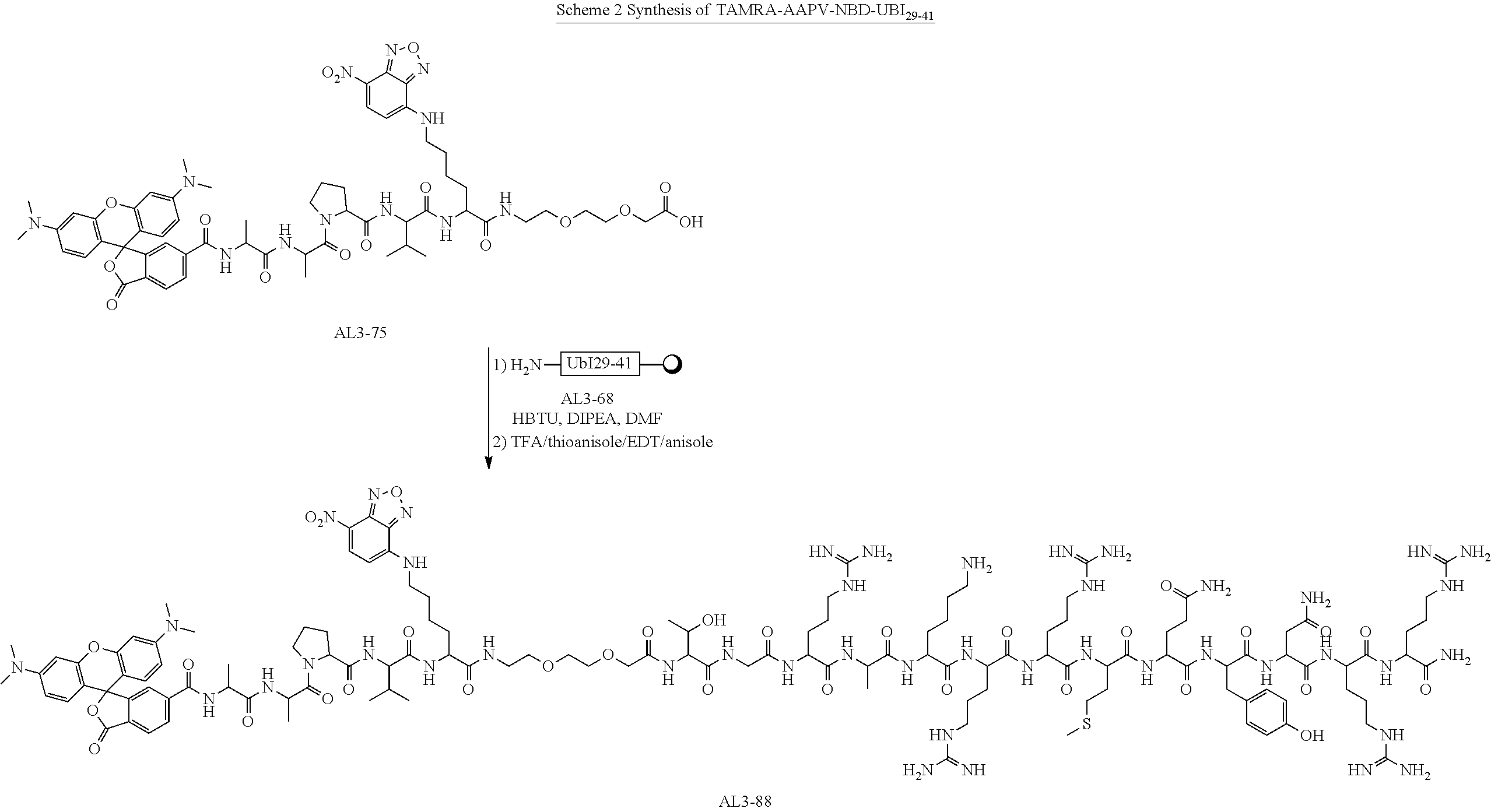


| TABLE 3 |
| Bacteria, strain reference and original source used in experiments. |
| Bacteria | Strain | Original Source | ||
| Gram- | P. aeruginosa | ATCC 47085 | ATCC |
| negative | (PA01) | ||
| bacteria | P. aeruginosa | J3284 | Clinical Isolate* |
| A. baumannii | J3433 | Clinical Isolate* | |
| S. maltophilia | J3270 | Clinical Isolate* | |
| K. pneumoniae | ATCC BAA1706 | ATCC | |
| E. coli | ATCC 25922 | ATCC | |
| H. influenzae | Clinical Isolate | Clinical Isolate* | |
| Gram- | Methicillin | ATCC25923 | ATCC |
| positive | Resistant S. aureus | ||
| bacteria | (MRSA) | ||
| Methicillin Sensitive | ATCC 252 | ATCC | |
| S. aureus (MSSA) | |||
| S. pneumoniae | D39 NCTC 7466 | Health protection | |
| agency culture | |||
| collection | |||
| GFP fluorescent | RN6390-Gfp- | Nottingham | |
| S aureus | EryR | University (Gift from | |
| Professor Phil Hill) | |||
| *Gifts from Professor John Govan, University of Edinburgh. | |||
- 1. M. Ternon, J. J. Diaz-Mochon, A. Belsom, M. Bradley, Tetrahedron, 2004, 60, 8721
- 2. H. J. Knölker, T. Braxmeier, G. Schlechtingen, Angew. Chem. Int. Ed., 1995, 34, 2497
- 3. E. Kaiser, R. L. Colescott, C. D. Bossinger and P. I. Cook, Analytical Biochemistry, 1970, 34, 595-598.
Listing of Relevant Sequences

Claims (22)
Applications Claiming Priority (3)
| Application Number | Priority Date | Filing Date | Title |
|---|---|---|---|
| GBGB1420222.0A GB201420222D0 (en) | 2014-11-13 | 2014-11-13 | Molecular probes and methods using the same |
| GB1420222.0 | 2014-11-13 | ||
| PCT/GB2015/053454 WO2016075483A1 (en) | 2014-11-13 | 2015-11-13 | Molecular probes for detecting gram-negative bacteria in vitro and in vivo |
Publications (2)
| Publication Number | Publication Date |
|---|---|
| US20180231548A1 US20180231548A1 (en) | 2018-08-16 |
| US10648979B2 true US10648979B2 (en) | 2020-05-12 |
Family
ID=52248326
Family Applications (1)
| Application Number | Title | Priority Date | Filing Date |
|---|---|---|---|
| US15/526,414 Active US10648979B2 (en) | 2014-11-13 | 2015-11-13 | Molecular probes for detecting gram-negative bacteria in vitro and in vivo |
Country Status (7)
| Country | Link |
|---|---|
| US (1) | US10648979B2 (en) |
| EP (1) | EP3218013B1 (en) |
| JP (1) | JP6754531B2 (en) |
| CN (1) | CN107106707B (en) |
| CA (1) | CA3003964A1 (en) |
| GB (1) | GB201420222D0 (en) |
| WO (1) | WO2016075483A1 (en) |
Families Citing this family (5)
| Publication number | Priority date | Publication date | Assignee | Title |
|---|---|---|---|---|
| FR3088652A1 (en) * | 2018-11-21 | 2020-05-22 | Nanotracks Diagnostics | In vitro detection of the presence of at least one microbial pathogen in a biological sample |
| CN109557303B (en) * | 2018-12-26 | 2021-11-02 | 陕西师范大学 | Polymyxin B magnetic bead, preparation method and application thereof |
| CN110559450A (en) * | 2019-07-22 | 2019-12-13 | 复旦大学附属华山医院 | water-soluble near-infrared two-region fluorescent tracing developer for joint bacterial infection |
| JP7406789B2 (en) * | 2019-10-30 | 2023-12-28 | 国立大学法人広島大学 | Methods for isolating bacterial membrane vesicles, isolation kits, and methods for their removal |
| CN111334291B (en) * | 2020-03-24 | 2023-02-28 | 天津市口腔医院 | Aggregation-induced emission fluorescence turn-on probe and preparation method and application thereof |
Citations (16)
| Publication number | Priority date | Publication date | Assignee | Title |
|---|---|---|---|---|
| US6051395A (en) | 1998-08-25 | 2000-04-18 | Biometric Imaging, Inc. | Method and compound for detecting low levels of microorganisms |
| WO2003014743A2 (en) | 2001-08-03 | 2003-02-20 | Amersham Biosciences Uk Limited | Use of dendrimers and poly-branched molecules to enhance signal in fluorescent assay systems |
| US20030079015A1 (en) | 2001-05-09 | 2003-04-24 | Dotclick Corporation | Method, apparatus and program product providing business processes using media identification and tracking of associated user preferences |
| US20040265295A1 (en) | 2003-06-25 | 2004-12-30 | Anderson Richard A. | Methods of preventing or treating cell-migration mediated conditions or diseases |
| US6884776B1 (en) | 1997-05-29 | 2005-04-26 | Rijksuniversiteit Leiden | Antimicrobial peptides derived from ubiquicidine |
| US20050214221A1 (en) | 2002-03-11 | 2005-09-29 | Visen Medical, Inc. | Optical imaging probes |
| WO2007008080A2 (en) | 2005-07-11 | 2007-01-18 | Ge Healthcare As | Optical imaging contrast agents |
| US20070026433A1 (en) | 2001-03-09 | 2007-02-01 | Hildebrand William H | Epitope testing using soluble HLA |
| US20070065840A1 (en) | 2005-03-23 | 2007-03-22 | Irena Naguibneva | Novel oligonucleotide compositions and probe sequences useful for detection and analysis of microRNAS and their target mRNAS |
| US20100233743A1 (en) | 2006-08-15 | 2010-09-16 | Asko Uri | Bisubstrate fluorescent probe binding to protein kinases |
| US20120252137A1 (en) | 2011-04-01 | 2012-10-04 | Novitsky Thomas James | Fluorescence Polarization Assay For Bacterial Endotoxin |
| WO2012136958A2 (en) | 2011-04-08 | 2012-10-11 | The University Court Of The University Of Edinburgh | Optical imaging probes |
| WO2013033436A1 (en) | 2011-09-01 | 2013-03-07 | University Of Iowa Research Foundation | Oligonucleotide-based probes for detection of bacterial nucleases |
| WO2014093031A1 (en) | 2012-12-12 | 2014-06-19 | The Regents Of The University Of Michigan | Bacteria targeting nanoparticles and related methods of use |
| US20140296137A1 (en) | 2013-04-01 | 2014-10-02 | Los Alamos National Security, Llc | Methods and compositions for controlling rotifers |
| US20160002619A1 (en) | 2013-02-27 | 2016-01-07 | Cornell University | Labeled glutaminase proteins, isolated glutaminase protein mutants, methods of use, and kit |
Family Cites Families (2)
| Publication number | Priority date | Publication date | Assignee | Title |
|---|---|---|---|---|
| CN103079592B (en) * | 2010-07-01 | 2015-10-21 | 浦项工科大学校产学协力团 | Use from the microbubble treatment of cell and the method for cancer diagnosis |
| US20150191763A1 (en) * | 2012-04-21 | 2015-07-09 | Indiana University and Technology Corporation | Compositions for in situ labeling of bacterial cell walls with fluorophores and methods of use thereof |
-
2014
- 2014-11-13 GB GBGB1420222.0A patent/GB201420222D0/en not_active Ceased
-
2015
- 2015-11-13 CA CA3003964A patent/CA3003964A1/en not_active Abandoned
- 2015-11-13 CN CN201580073090.XA patent/CN107106707B/en active Active
- 2015-11-13 WO PCT/GB2015/053454 patent/WO2016075483A1/en active Application Filing
- 2015-11-13 JP JP2017525861A patent/JP6754531B2/en active Active
- 2015-11-13 US US15/526,414 patent/US10648979B2/en active Active
- 2015-11-13 EP EP15804564.1A patent/EP3218013B1/en active Active
Patent Citations (17)
| Publication number | Priority date | Publication date | Assignee | Title |
|---|---|---|---|---|
| US6884776B1 (en) | 1997-05-29 | 2005-04-26 | Rijksuniversiteit Leiden | Antimicrobial peptides derived from ubiquicidine |
| US6051395A (en) | 1998-08-25 | 2000-04-18 | Biometric Imaging, Inc. | Method and compound for detecting low levels of microorganisms |
| US20070026433A1 (en) | 2001-03-09 | 2007-02-01 | Hildebrand William H | Epitope testing using soluble HLA |
| US20030079015A1 (en) | 2001-05-09 | 2003-04-24 | Dotclick Corporation | Method, apparatus and program product providing business processes using media identification and tracking of associated user preferences |
| WO2003014743A2 (en) | 2001-08-03 | 2003-02-20 | Amersham Biosciences Uk Limited | Use of dendrimers and poly-branched molecules to enhance signal in fluorescent assay systems |
| US20050214221A1 (en) | 2002-03-11 | 2005-09-29 | Visen Medical, Inc. | Optical imaging probes |
| US20110171136A1 (en) | 2002-03-11 | 2011-07-14 | Poss Kirtland G | Optical imaging probes |
| US20040265295A1 (en) | 2003-06-25 | 2004-12-30 | Anderson Richard A. | Methods of preventing or treating cell-migration mediated conditions or diseases |
| US20070065840A1 (en) | 2005-03-23 | 2007-03-22 | Irena Naguibneva | Novel oligonucleotide compositions and probe sequences useful for detection and analysis of microRNAS and their target mRNAS |
| WO2007008080A2 (en) | 2005-07-11 | 2007-01-18 | Ge Healthcare As | Optical imaging contrast agents |
| US20100233743A1 (en) | 2006-08-15 | 2010-09-16 | Asko Uri | Bisubstrate fluorescent probe binding to protein kinases |
| US20120252137A1 (en) | 2011-04-01 | 2012-10-04 | Novitsky Thomas James | Fluorescence Polarization Assay For Bacterial Endotoxin |
| WO2012136958A2 (en) | 2011-04-08 | 2012-10-11 | The University Court Of The University Of Edinburgh | Optical imaging probes |
| WO2013033436A1 (en) | 2011-09-01 | 2013-03-07 | University Of Iowa Research Foundation | Oligonucleotide-based probes for detection of bacterial nucleases |
| WO2014093031A1 (en) | 2012-12-12 | 2014-06-19 | The Regents Of The University Of Michigan | Bacteria targeting nanoparticles and related methods of use |
| US20160002619A1 (en) | 2013-02-27 | 2016-01-07 | Cornell University | Labeled glutaminase proteins, isolated glutaminase protein mutants, methods of use, and kit |
| US20140296137A1 (en) | 2013-04-01 | 2014-10-02 | Los Alamos National Security, Llc | Methods and compositions for controlling rotifers |
Non-Patent Citations (35)
| Title |
|---|
| Ahsan R. Akram et al. "A labelled-ubiquicidin antimicrobial peptide for immediate in situ optical detection of live bacteria in human alveolar lung tissue"; Jan. 1, 2015, Chemical Science; vol. 6, No. 12; pp. 6971-6979; XP055247541. |
| Ahsan R. Akram, et al.; A labelled-ubiquicidin antimicrobial peptide for immediate in situ optical detection of live bacteria in human alveolar lung tissue; Jan. 1, 2015; Chemical Science; vol. 6 pp. 6971-6979. |
| AHSAN R. AKRAM, NICOLAOS AVLONITIS, ANNAMARIA LILIENKAMPF, ANA M. PEREZ-LOPEZ, NEIL MCDONALD, SUNAY V. CHANKESHWARA, EMMA SCHOLEFI: "A labelled-ubiquicidin antimicrobial peptide for immediate in situ optical detection of live bacteria in human alveolar lung tissue", CHEMICAL SCIENCE, ROYAL SOCIETY OF CHEMISTRY, UNITED KINGDOM, vol. 6, no. 12, 1 January 2015 (2015-01-01), United Kingdom, pages 6971 - 6979, XP055247541, ISSN: 2041-6520, DOI: 10.1039/C5SC00960J |
| Akhtar et al. "Antimicrobial Peptide 99mTc-Ubiquicidin 29-41 as Human Infection-Imaging Agent: Clinical Trial" The Journal of Nuclear Medicine vol. 46 No. 4 Apr. 2005. (Year: 2005). * |
| AR Akram et al. "T4: Optically detectable antimicrobial petitdes enable the immediate detection of bacteria and fungi in the lung" THROAX, vol. 70 No. Suppl. 3 Dec. 1, 2015, pp. A2-A3 XP008178907. |
| AR AKRAM, N AVLONITIS, M VENDRELL, S CHANKESHWARA, N MCDONALD, T ASLAM, E SCHOLEFIELD, T WALSH, C HASLETT, M BRADLEY, K DHALIWAL: "T4: Optically detectable antimicrobial peptides enable the immediate detection of bacteria and fungi in the lung", THORAX, BMJ PUBLISHING GROUP, GB, vol. 70, no. Suppl. 3, T4, 1 December 2015 (2015-12-01) - 4 December 2012 (2012-12-04), GB, pages A2 - A3, XP008178907, ISSN: 0040-6376, DOI: 10.1136/thoraxjnl-2015-207770.4 |
| Cavrini et al. "Molecular detection of Treponema denticola and Porphyromonas gingivalis in carotid and aortic atheromatous plaques by FISH: report of two cases," Journal of Medical Microbiology (2005), 54, 93-96. (Year: 2005). * |
| Cerny "Studies on the Aminopeptidase Test for the Distinction of Gram-Negative from Gram-Positive Bacteria," European J. Appl. Microbiol. Biotechnol. 5,113-122 (1978). (Year: 1978). * |
| Cuicui Liu, et al.; Noninvasive Optical Imaging of Staphylococcus aureus Invection in Vivo using an Antimicrobial Peptide Fragment Based Near-Infrared Fluorescent Probes; Jul. 25, 2013; Journal of Innovative Optical Health Science-vol. 6. No. 3 (2013) 1350026 (9 pages); XP-002753983. |
| Denis et al. "Probing the Penetration of Antimicrobial Polymyxin Lipopeptides into Gram-Negative Bacteria," Bioconjugate Chem. 2014, 25, 750-760. (Year: 2014). * |
| DERIS Z Z, SWARBRICK J D, ROBERTS K D, AZAD M A, AKTER J, HORNE A S, NATION R L, ROGERS K L, THOMPSON P E, VELKOV T, LI J.: "Probing the penetration of antimicrobial polymyxin lipopeptides into gram-negative bacteria.", BIOCONJUGATE CHEMISTRY, AMERICAN CHEMICAL SOCIETY, US, vol. 25, no. 4, 16 April 2014 (2014-04-16), US, pages 750 - 760, XP002754088, ISSN: 1043-1802, DOI: 10.1021/bc500094d |
| Deris Zakuan et al. "Probing the Penetration of Antimicrobial Polymyxin Lipopeptides into Gram-Negative Bacteria" Apr. 2014; Bioconjugate Chemistry; vol. 25, No. 4, pp. 750-760, XP002754088. |
| DiVittorio et al. "Zinc(II) Coordination Complexesas Membrane-Active Fluorescent Probes and Antibiotics," ChemBioChem 2008, 9, 286-293. (Year: 2008). * |
| E. Kaiser, R. L. Colescott, C. D. Bossinger and P. I. Cook, Color Test for Detection of Free Terminal Amino Groups in the Solid-Phase Synthesis of Peptides, Analytical Biochemistry, 1970, 34, 595-598, Armour Pharmaceutical Company, Kankakee, Illinois. |
| El-Kosasy A M et al. "Screening and optimization of the reaction of polymyxin B sulphate with NBD-C1 for the synchronous spectrofluorimetric determination of polymyxin B sulphate in human plasma"; May 1, 2005, Journal of Fluorescence May 1, 2015 Springer New York LLS USA; pp. 695-705, XP002753986. |
| EL-KOSASY A M, ABDEL-AZIZ O, MAGDY N, EL ZAHAR N M: "Screening and Optimization of the Reaction of Polymyxin B Sulphate with NBD-Cl for the Synchronous Spectrofluorimetric Determination of Polymyxin B Sulphate in Human Plasma.", JOURNAL OF FLUORESCENCE, PLENUM PUBLISHING, NEW YORK, US, vol. 25, no. 3, 1 May 2015 (2015-05-01), NEW YORK, US, pages 695 - 705, XP002753986, ISSN: 1053-0509, DOI: 10.1007/S10895-015-1555-8 |
| H. J. Knölker, T. Braxmeier, G. Schlechtingen, A Novel Method for the Synthesis of Isocyanates Under Mild Conditions, Angew. Chem. Int. Ed., 1995, 34, 2497-2500, England. |
| Hoibly "Recent advances in the treatment of Pseudomonas aeruginosa infections in cystic fibrosis," BMC Medicine 2011, 9:32. (Year: 2011). * |
| International Search Report, dated Nov. 13, 2015, PCT/GB2015/053455. |
| LEEVY W M, LAMBERT T N, JOHNSON J R, MORRIS J, SMITH B D: "Quantum dot probes for bacteria distinguish Escherichia coli mutants and permit in vivo imaging.", CHEMICAL COMMUNICATIONS, ROYAL SOCIETY OF CHEMISTRY, vol. 28, no. 20, 28 May 2008 (2008-05-28), pages 2331 - 2333, XP002754090, ISSN: 1359-7345, DOI: 10.1039/b803590c |
| Leevy W Matthew et al. "Quantum dot probes for bacteria distinguish Escherichia coli mutants and permit in vivo imaging" May 28, 2008; Chemical Communications (Cambridge England); No. 20, pp. 2331-2333, XP002754090. |
| LIU C , GU Y: "NONINVASIVE OPTICAL IMAGING OF STAPHYLOCOCCUS AUREUS INFECTION IN VIVO USING AN ANTIMICROBIAL PEPTIDE FRAGMENT BASED NEAR-INFRARED FLUORESCENT PROBES", JOURNAL OF INNOVATIVE OPTICAL HEALTH SCIENCES : JIOHS, WORLD SCIENTIFIC PUBLISHING, SINGAPORE, vol. 6, no. 3, 25 July 2013 (2013-07-25), Singapore, pages 1350026 - 1350026-8, XP002753983, ISSN: 1793-5458, DOI: 10.1142/S1793545813500260 |
| M. Ternon, J. Jose Diaz-Mochon, A. Belsom and M. Bradley, Tetrahedron, Science Direct, 2004, 60, 8721-8728, School of Chemistry, University of Southampton, Highfield, Southampton SO17 1BJ, UK. |
| Michael A. MacDonald, Ladislav Jankovic, Khalid Shahzad, Michael Burcher, King C.P. Li, Acoustic fingerprints of dye-labeled protein submicrosphere photoacoustic contrast agents, Journal of Biomedical Optics, May/Jun. 2009, vol. 14 (3). |
| Nicolaos Avlonitis,et al; Highly specific, multi-branched fluorescent reporters reporters for analysis of human neutrophil elastase; Organic & Biomolecular Chemistry; 2013; vol. 11, No. 26; Jul. pp. 4414-4418. |
| PAMELA T. WONG, SHENGZHUANG TANG, KENNY TANG, ALEXA COULTER, JHINDAN MUKHERJEE, KRISTINA GAM, JAMES R. BAKER, SEOK KI CHOI: "Supporting information - A lipopolysaccharide binding heteromultivalent dendrimer nanoplatform for Gram negative cell targeting", JOURNAL OF MATERIALS CHEMISTRY B, ROYAL SOCIETY OF CHEMISTRY, GB, 14 February 2015 (2015-02-14), GB, pages S1 - S22, XP002754621, ISSN: 2050-750X, Retrieved from the Internet <URL:http://www.rsc.org/suppdata/tb/c4/c4tb01690d/c4tb01690d1.pdf> |
| Roger D. Pechous, et al; Early Host Cell Targets of Yersinia pestis during Primary Pneumonic Plague; PLOS Pthogens—www.plospathogens.org; Oct. 2013—vol. 9; Issue 10; e1003679. |
| SHUZHANG XIAO, LICA ABU-ESBA, SERHAN TURKYILMAZ, ALEXANDER G. WHITE, BRADLEY D. SMITH: "Multivalent Dendritic Molecules as Broad Spectrum Bacteria Agglutination Agents", THERANOSTICS, IVYSPRING INTERNATIONAL PUBLISHER, AU, vol. 3, no. 9, 1 January 2013 (2013-01-01), AU, pages 658 - 666, XP002754164, ISSN: 1838-7640, DOI: 10.7150/thno.6811 |
| Sorensen et al. "Azadioxatriangulenium (ADOTA+): A long fluorescence lifetime fluorophore for large biomolecule binding assay," Method Appl Fluoresc. 2013 ; 1(2): 025001. (Year: 2013). * |
| Warsen A E et al. "Simultaneous discrimination between 15 fish pathogens by using 16S ribosomal DNA PCR and DNA microarrays" Jul. 2004; Applied and Environmental Microbiology July 2004 American Society for Microbiology US; vol. 70, No. 7, pp. 4261-4221, XP002754089. |
| WARSEN A E, KRUG M J, LAFRENTZ S, STANEK D R, LOGE F J, CALL D R: "Simultaneous discrimination between 15 fish pathogens by using 16S ribosomal DNA PCR and DNA microarrays.", APPLIED AND ENVIRONMENTAL MICROBIOLOGY, AMERICAN SOCIETY FOR MICROBIOLOGY, US, vol. 70, no. 7, 1 July 2004 (2004-07-01), US, pages 4216 - 4221, XP002754089, ISSN: 0099-2240, DOI: 10.1128/AEM.70.7.4216-4221.2004 |
| Wellinghausen et al. "Superiority of Molecular Techniques for Identification of Gram-Negative, Oxidase-Positive Rods, Including Morphologically Nontypical Pseudomonas aeruginosa, from Patients with Cystic Fibrosis," Journal of Clinical Microbiology, Aug. 2005, p. 4070-4075. (Year: 2005). * |
| Wong P T et al., "A lipopolysaccharide binding heteromultivalent dendrimer nanoplatform for Gram negative Cell targeting"; Feb. 14, 2015; Journal of Materials Chemistry; vol. 3. No. 6 pp. 1149-1156. |
| Wong P T et al.; "Supporting information to A lipopolysaccharide binding heteromultivalent dendrimer naoplatform for Gram negative cell targeting"; Feb. 14, 2015; Journal of Materials Chemistry B; pp. S1-S22, XP002754621. |
| Xiao Shuzhang et al. "Multivalent Dendritic Molecules as Broad Spectrum Bacteria Agglutination Agents" Aug. 10, 2013; Theranostics; vol. 3 No. 9 pp. 658-666, XP0002754164. |
Also Published As
| Publication number | Publication date |
|---|---|
| GB201420222D0 (en) | 2014-12-31 |
| WO2016075483A1 (en) | 2016-05-19 |
| CN107106707B (en) | 2021-07-23 |
| CA3003964A1 (en) | 2016-05-19 |
| EP3218013B1 (en) | 2024-04-17 |
| US20180231548A1 (en) | 2018-08-16 |
| JP2018500884A (en) | 2018-01-18 |
| CN107106707A (en) | 2017-08-29 |
| EP3218013A1 (en) | 2017-09-20 |
| JP6754531B2 (en) | 2020-09-16 |
Similar Documents
| Publication | Publication Date | Title |
|---|---|---|
| US10648979B2 (en) | Molecular probes for detecting gram-negative bacteria in vitro and in vivo | |
| Akram et al. | A labelled-ubiquicidin antimicrobial peptide for immediate in situ optical detection of live bacteria in human alveolar lung tissue | |
| US9763577B2 (en) | Imaging agent for detection of diseased cells | |
| Urano et al. | Rapid cancer detection by topically spraying a γ-glutamyltranspeptidase–activated fluorescent probe | |
| EP3270977B1 (en) | Optical probes for matrix metalloproteinases | |
| US8841085B2 (en) | Nanoparticle sensor for measuring protease activity and method for manufacturing the same | |
| JP5696947B2 (en) | Use of bacterial beta-lactamase in in vitro diagnostics and in vivo imaging, diagnostics and therapeutics | |
| Welling et al. | Fluorescent imaging of bacterial infections and recent advances made with multimodal radiopharmaceuticals | |
| JP2013528386A (en) | In vitro diagnostics and the use of bacterial β-lactamases for in vivo imaging, diagnosis and therapy | |
| WO2016075481A1 (en) | Fret molecular probes with cleavable linkers for detecting bacteria and/or fungi in vitro and in vivo | |
| US10520504B2 (en) | Fluorescent polybranched probes for detecting bacteria and/or fungi in vitro and in vivo | |
| WO2016151297A1 (en) | Optical probe for thrombin | |
| CN104837507A (en) | Methods to detect a fungal cell | |
| US20130164221A1 (en) | Use of bacterial beta-lactamase for in vitro diagnostics and in vivo imaging, diagnostics and therapeutics | |
| US8247529B2 (en) | Neoplasia targeting peptides and methods of using the same | |
| US20120328515A1 (en) | In vivo detection of apoptosis | |
| WO2024031003A2 (en) | Grail-1 peptide products and methods | |
| KR20210054280A (en) | Dual recognition two-photon fluorescent probe and uses thereof |
Legal Events
| Date | Code | Title | Description |
|---|---|---|---|
| AS | Assignment |
Owner name: THE UNIVERSITY COURT OF THE UNIVERSITY OF EDINBURGH, GREAT BRITAIN Free format text: ASSIGNMENT OF ASSIGNORS INTEREST;ASSIGNOR:LILIENKAMPF, ANNAMARIA HELENA;REEL/FRAME:044091/0708 Effective date: 20170801 Owner name: THE UNIVERSITY COURT OF THE UNIVERSITY OF EDINBURGH, GREAT BRITAIN Free format text: ASSIGNMENT OF ASSIGNORS INTEREST;ASSIGNOR:HASLETT, CHRISTOPHER;REEL/FRAME:044091/0186 Effective date: 20170807 Owner name: THE UNIVERSITY COURT OF THE UNIVERSITY OF EDINBURGH, GREAT BRITAIN Free format text: ASSIGNMENT OF ASSIGNORS INTEREST;ASSIGNORS:BRADLEY, MARK;ESCOBAR, MARC VENDRELL;MCDONALD, NEIL ALEXANDER;AND OTHERS;SIGNING DATES FROM 20170726 TO 20170804;REEL/FRAME:044091/0098 Owner name: THE UNIVERSITY COURT OF THE UNIVERSITY OF EDINBURGH, GREAT BRITAIN Free format text: ASSIGNMENT OF ASSIGNORS INTEREST;ASSIGNOR:AVLONITIS, NIKOLAOS;REEL/FRAME:044091/0573 Effective date: 20170808 Owner name: THE UNIVERSITY COURT OF THE UNIVERSITY OF EDINBURG Free format text: ASSIGNMENT OF ASSIGNORS INTEREST;ASSIGNORS:BRADLEY, MARK;ESCOBAR, MARC VENDRELL;MCDONALD, NEIL ALEXANDER;AND OTHERS;SIGNING DATES FROM 20170726 TO 20170804;REEL/FRAME:044091/0098 Owner name: THE UNIVERSITY COURT OF THE UNIVERSITY OF EDINBURG Free format text: ASSIGNMENT OF ASSIGNORS INTEREST;ASSIGNOR:LILIENKAMPF, ANNAMARIA HELENA;REEL/FRAME:044091/0708 Effective date: 20170801 Owner name: THE UNIVERSITY COURT OF THE UNIVERSITY OF EDINBURG Free format text: ASSIGNMENT OF ASSIGNORS INTEREST;ASSIGNOR:AVLONITIS, NIKOLAOS;REEL/FRAME:044091/0573 Effective date: 20170808 Owner name: THE UNIVERSITY COURT OF THE UNIVERSITY OF EDINBURG Free format text: ASSIGNMENT OF ASSIGNORS INTEREST;ASSIGNOR:HASLETT, CHRISTOPHER;REEL/FRAME:044091/0186 Effective date: 20170807 |
|
| STPP | Information on status: patent application and granting procedure in general |
Free format text: RESPONSE TO NON-FINAL OFFICE ACTION ENTERED AND FORWARDED TO EXAMINER |
|
| STPP | Information on status: patent application and granting procedure in general |
Free format text: NON FINAL ACTION MAILED |
|
| STPP | Information on status: patent application and granting procedure in general |
Free format text: RESPONSE TO NON-FINAL OFFICE ACTION ENTERED AND FORWARDED TO EXAMINER |
|
| STPP | Information on status: patent application and granting procedure in general |
Free format text: NOTICE OF ALLOWANCE MAILED -- APPLICATION RECEIVED IN OFFICE OF PUBLICATIONS |
|
| STPP | Information on status: patent application and granting procedure in general |
Free format text: AWAITING TC RESP., ISSUE FEE NOT PAID |
|
| STPP | Information on status: patent application and granting procedure in general |
Free format text: NOTICE OF ALLOWANCE MAILED -- APPLICATION RECEIVED IN OFFICE OF PUBLICATIONS |
|
| STCF | Information on status: patent grant |
Free format text: PATENTED CASE |
|
| MAFP | Maintenance fee payment |
Free format text: PAYMENT OF MAINTENANCE FEE, 4TH YR, SMALL ENTITY (ORIGINAL EVENT CODE: M2551); ENTITY STATUS OF PATENT OWNER: SMALL ENTITY Year of fee payment: 4 |