US10568963B2 - Multifunctional nanoplatforms for fluorescence imaging and photodynamic therapy developed by post-loading photosensitizer and fluorophore to polyacrylamide nanoparticles - Google Patents
Multifunctional nanoplatforms for fluorescence imaging and photodynamic therapy developed by post-loading photosensitizer and fluorophore to polyacrylamide nanoparticles Download PDFInfo
- Publication number
- US10568963B2 US10568963B2 US13/566,411 US201213566411A US10568963B2 US 10568963 B2 US10568963 B2 US 10568963B2 US 201213566411 A US201213566411 A US 201213566411A US 10568963 B2 US10568963 B2 US 10568963B2
- Authority
- US
- United States
- Prior art keywords
- nanoparticles
- composition
- photosensitizer
- alkyl
- tumor
- Prior art date
- Legal status (The legal status is an assumption and is not a legal conclusion. Google has not performed a legal analysis and makes no representation as to the accuracy of the status listed.)
- Active
Links
- 239000002105 nanoparticle Substances 0.000 title claims abstract description 220
- 239000003504 photosensitizing agent Substances 0.000 title claims abstract description 82
- 229920002401 polyacrylamide Polymers 0.000 title claims description 103
- 238000002428 photodynamic therapy Methods 0.000 title claims description 58
- 238000011068 loading method Methods 0.000 title description 19
- 238000000799 fluorescence microscopy Methods 0.000 title description 16
- 239000000203 mixture Substances 0.000 claims abstract description 61
- 239000012216 imaging agent Substances 0.000 claims abstract description 31
- 206010028980 Neoplasm Diseases 0.000 claims description 189
- 125000000217 alkyl group Chemical group 0.000 claims description 44
- 229910052739 hydrogen Inorganic materials 0.000 claims description 38
- 239000001257 hydrogen Substances 0.000 claims description 36
- 125000003118 aryl group Chemical group 0.000 claims description 24
- 150000002431 hydrogen Chemical class 0.000 claims description 24
- 125000003342 alkenyl group Chemical group 0.000 claims description 23
- 125000000304 alkynyl group Chemical group 0.000 claims description 22
- 125000000753 cycloalkyl group Chemical group 0.000 claims description 22
- 150000001875 compounds Chemical class 0.000 claims description 17
- 108090000765 processed proteins & peptides Proteins 0.000 claims description 16
- 230000008685 targeting Effects 0.000 claims description 14
- 125000002496 methyl group Chemical group [H]C([H])([H])* 0.000 claims description 12
- 238000002835 absorbance Methods 0.000 claims description 11
- -1 3,5-bis(trifluoromethyl)-benzyl Chemical group 0.000 claims description 10
- UFHFLCQGNIYNRP-UHFFFAOYSA-N Hydrogen Chemical compound [H][H] UFHFLCQGNIYNRP-UHFFFAOYSA-N 0.000 claims description 8
- 125000001072 heteroaryl group Chemical group 0.000 claims description 8
- 230000015572 biosynthetic process Effects 0.000 claims description 7
- OVBPIULPVIDEAO-LBPRGKRZSA-N folic acid Chemical compound C=1N=C2NC(N)=NC(=O)C2=NC=1CNC1=CC=C(C(=O)N[C@@H](CCC(O)=O)C(O)=O)C=C1 OVBPIULPVIDEAO-LBPRGKRZSA-N 0.000 claims description 5
- 239000000017 hydrogel Substances 0.000 claims description 5
- 125000000539 amino acid group Chemical group 0.000 claims description 4
- 125000001188 haloalkyl group Chemical group 0.000 claims description 4
- 125000005843 halogen group Chemical group 0.000 claims description 4
- 125000004435 hydrogen atom Chemical group [H]* 0.000 claims description 4
- 125000001424 substituent group Chemical group 0.000 claims description 4
- FDKRLXBXYZKWRZ-UWJYYQICSA-N 3-[(21S,22S)-16-ethenyl-11-ethyl-4-hydroxy-12,17,21,26-tetramethyl-7,23,24,25-tetrazahexacyclo[18.2.1.15,8.110,13.115,18.02,6]hexacosa-1,4,6,8(26),9,11,13(25),14,16,18(24),19-undecaen-22-yl]propanoic acid Chemical group CCC1=C(C2=NC1=CC3=C(C4=C(CC(=C5[C@H]([C@@H](C(=CC6=NC(=C2)C(=C6C)C=C)N5)C)CCC(=O)O)C4=N3)O)C)C FDKRLXBXYZKWRZ-UWJYYQICSA-N 0.000 claims description 3
- 239000011724 folic acid Substances 0.000 claims description 3
- 235000019152 folic acid Nutrition 0.000 claims description 3
- 125000002947 alkylene group Chemical group 0.000 claims description 2
- 125000001797 benzyl group Chemical group [H]C1=C([H])C([H])=C(C([H])=C1[H])C([H])([H])* 0.000 claims description 2
- 125000000484 butyl group Chemical group [H]C([*])([H])C([H])([H])C([H])([H])C([H])([H])[H] 0.000 claims description 2
- 125000001153 fluoro group Chemical group F* 0.000 claims description 2
- 125000003187 heptyl group Chemical group [H]C([*])([H])C([H])([H])C([H])([H])C([H])([H])C([H])([H])C([H])([H])C([H])([H])[H] 0.000 claims description 2
- 102000004196 processed proteins & peptides Human genes 0.000 claims description 2
- 125000000391 vinyl group Chemical group [H]C([*])=C([H])[H] 0.000 claims description 2
- ANRHNWWPFJCPAZ-UHFFFAOYSA-M thionine Chemical group [Cl-].C1=CC(N)=CC2=[S+]C3=CC(N)=CC=C3N=C21 ANRHNWWPFJCPAZ-UHFFFAOYSA-M 0.000 claims 9
- OVBPIULPVIDEAO-UHFFFAOYSA-N N-Pteroyl-L-glutaminsaeure Natural products C=1N=C2NC(N)=NC(=O)C2=NC=1CNC1=CC=C(C(=O)NC(CCC(O)=O)C(O)=O)C=C1 OVBPIULPVIDEAO-UHFFFAOYSA-N 0.000 claims 2
- 150000001720 carbohydrates Chemical class 0.000 claims 2
- 235000014633 carbohydrates Nutrition 0.000 claims 2
- 229960000304 folic acid Drugs 0.000 claims 2
- 238000000034 method Methods 0.000 abstract description 16
- 239000004793 Polystyrene Substances 0.000 description 81
- PUUBADHCONCMPA-USOGPTGWSA-N 3-[(21S,22S)-11-ethyl-16-(1-hexoxyethyl)-4-hydroxy-12,17,21,26-tetramethyl-7,23,24,25-tetrazahexacyclo[18.2.1.15,8.110,13.115,18.02,6]hexacosa-1,4,6,8(26),9,11,13(25),14,16,18(24),19-undecaen-22-yl]propanoic acid Chemical compound CCCCCCOC(C)C1=C(C2=NC1=CC3=NC(=CC4=C(C5=C(CC(=C6[C@H]([C@@H](C(=C2)N6)C)CCC(=O)O)C5=N4)O)C)C(=C3C)CC)C PUUBADHCONCMPA-USOGPTGWSA-N 0.000 description 60
- OHOQEZWSNFNUSY-UHFFFAOYSA-N Cy3-bifunctional dye zwitterion Chemical compound O=C1CCC(=O)N1OC(=O)CCCCCN1C2=CC=C(S(O)(=O)=O)C=C2C(C)(C)C1=CC=CC(C(C1=CC(=CC=C11)S([O-])(=O)=O)(C)C)=[N+]1CCCCCC(=O)ON1C(=O)CCC1=O OHOQEZWSNFNUSY-UHFFFAOYSA-N 0.000 description 55
- 238000003384 imaging method Methods 0.000 description 50
- 239000000975 dye Substances 0.000 description 49
- QGKMIGUHVLGJBR-UHFFFAOYSA-M (4z)-1-(3-methylbutyl)-4-[[1-(3-methylbutyl)quinolin-1-ium-4-yl]methylidene]quinoline;iodide Chemical compound [I-].C12=CC=CC=C2N(CCC(C)C)C=CC1=CC1=CC=[N+](CCC(C)C)C2=CC=CC=C12 QGKMIGUHVLGJBR-UHFFFAOYSA-M 0.000 description 38
- 238000009472 formulation Methods 0.000 description 34
- MYMOFIZGZYHOMD-UHFFFAOYSA-N Dioxygen Chemical compound O=O MYMOFIZGZYHOMD-UHFFFAOYSA-N 0.000 description 32
- 238000001727 in vivo Methods 0.000 description 30
- 241000699670 Mus sp. Species 0.000 description 27
- 239000003795 chemical substances by application Substances 0.000 description 26
- 230000005284 excitation Effects 0.000 description 26
- 238000011725 BALB/c mouse Methods 0.000 description 24
- 210000004027 cell Anatomy 0.000 description 24
- LFQSCWFLJHTTHZ-UHFFFAOYSA-N Ethanol Chemical compound CCO LFQSCWFLJHTTHZ-UHFFFAOYSA-N 0.000 description 23
- 239000000243 solution Substances 0.000 description 23
- XLYOFNOQVPJJNP-UHFFFAOYSA-N water Substances O XLYOFNOQVPJJNP-UHFFFAOYSA-N 0.000 description 22
- 238000002347 injection Methods 0.000 description 21
- 239000007924 injection Substances 0.000 description 21
- 102000008100 Human Serum Albumin Human genes 0.000 description 19
- 108091006905 Human Serum Albumin Proteins 0.000 description 19
- 238000013459 approach Methods 0.000 description 17
- 235000010482 polyoxyethylene sorbitan monooleate Nutrition 0.000 description 15
- 229920000053 polysorbate 80 Polymers 0.000 description 15
- 210000001519 tissue Anatomy 0.000 description 15
- 238000011282 treatment Methods 0.000 description 15
- IAZDPXIOMUYVGZ-UHFFFAOYSA-N Dimethylsulphoxide Chemical compound CS(C)=O IAZDPXIOMUYVGZ-UHFFFAOYSA-N 0.000 description 14
- 238000001514 detection method Methods 0.000 description 13
- 239000003814 drug Substances 0.000 description 13
- 210000004072 lung Anatomy 0.000 description 12
- VLKZOEOYAKHREP-UHFFFAOYSA-N n-Hexane Chemical compound CCCCCC VLKZOEOYAKHREP-UHFFFAOYSA-N 0.000 description 12
- 241000699666 Mus <mouse, genus> Species 0.000 description 11
- 238000012879 PET imaging Methods 0.000 description 11
- 238000002866 fluorescence resonance energy transfer Methods 0.000 description 11
- ZYHGRNUNHLNLTR-UHFFFAOYSA-M (2e)-3-ethyl-2-[(e)-3-(1-ethylquinolin-1-ium-2-yl)prop-2-enylidene]-1,3-benzoxazole;iodide Chemical compound [I-].C1=CC=CC2=[N+](CC)C(C=CC=C3N(C4=CC=CC=C4O3)CC)=CC=C21 ZYHGRNUNHLNLTR-UHFFFAOYSA-M 0.000 description 10
- 230000006378 damage Effects 0.000 description 10
- 239000000706 filtrate Substances 0.000 description 10
- 230000014759 maintenance of location Effects 0.000 description 10
- 238000004519 manufacturing process Methods 0.000 description 10
- 238000002600 positron emission tomography Methods 0.000 description 10
- 238000012546 transfer Methods 0.000 description 10
- SZXKSDXHODZTFS-UHFFFAOYSA-N 4-[4,5-bis[4-(dimethylamino)phenyl]-1H-imidazol-2-yl]-2,6-dimethoxyphenol Chemical compound COC1=C(O)C(OC)=CC(C=2NC(=C(N=2)C=2C=CC(=CC=2)N(C)C)C=2C=CC(=CC=2)N(C)C)=C1 SZXKSDXHODZTFS-UHFFFAOYSA-N 0.000 description 9
- 241001465754 Metazoa Species 0.000 description 9
- 210000001072 colon Anatomy 0.000 description 9
- 238000002296 dynamic light scattering Methods 0.000 description 9
- 210000000056 organ Anatomy 0.000 description 9
- 239000002245 particle Substances 0.000 description 9
- 238000011287 therapeutic dose Methods 0.000 description 9
- 230000001225 therapeutic effect Effects 0.000 description 9
- 238000002560 therapeutic procedure Methods 0.000 description 9
- 230000008901 benefit Effects 0.000 description 8
- 229940079593 drug Drugs 0.000 description 8
- 235000019441 ethanol Nutrition 0.000 description 8
- 210000004185 liver Anatomy 0.000 description 8
- 230000004044 response Effects 0.000 description 8
- 230000000717 retained effect Effects 0.000 description 8
- 210000004881 tumor cell Anatomy 0.000 description 8
- 208000003174 Brain Neoplasms Diseases 0.000 description 7
- 210000004556 brain Anatomy 0.000 description 7
- 244000309466 calf Species 0.000 description 7
- 230000000052 comparative effect Effects 0.000 description 7
- 201000010099 disease Diseases 0.000 description 7
- 208000037265 diseases, disorders, signs and symptoms Diseases 0.000 description 7
- 238000009826 distribution Methods 0.000 description 7
- 230000000694 effects Effects 0.000 description 7
- 230000002209 hydrophobic effect Effects 0.000 description 7
- 238000010253 intravenous injection Methods 0.000 description 7
- 230000007774 longterm Effects 0.000 description 7
- 238000005259 measurement Methods 0.000 description 7
- 210000002966 serum Anatomy 0.000 description 7
- 210000000952 spleen Anatomy 0.000 description 7
- 102100021010 Nucleolin Human genes 0.000 description 6
- 238000010521 absorption reaction Methods 0.000 description 6
- ROOXNKNUYICQNP-UHFFFAOYSA-N ammonium persulfate Chemical compound [NH4+].[NH4+].[O-]S(=O)(=O)OOS([O-])(=O)=O ROOXNKNUYICQNP-UHFFFAOYSA-N 0.000 description 6
- 201000011510 cancer Diseases 0.000 description 6
- 230000002708 enhancing effect Effects 0.000 description 6
- 108010044762 nucleolin Proteins 0.000 description 6
- 238000012634 optical imaging Methods 0.000 description 6
- 102000005962 receptors Human genes 0.000 description 6
- 108020003175 receptors Proteins 0.000 description 6
- 230000002829 reductive effect Effects 0.000 description 6
- 238000004626 scanning electron microscopy Methods 0.000 description 6
- 208000026310 Breast neoplasm Diseases 0.000 description 5
- 208000032612 Glial tumor Diseases 0.000 description 5
- 206010018338 Glioma Diseases 0.000 description 5
- 206010027476 Metastases Diseases 0.000 description 5
- 206010061902 Pancreatic neoplasm Diseases 0.000 description 5
- 210000000481 breast Anatomy 0.000 description 5
- 238000011161 development Methods 0.000 description 5
- 230000018109 developmental process Effects 0.000 description 5
- 238000002073 fluorescence micrograph Methods 0.000 description 5
- 238000000338 in vitro Methods 0.000 description 5
- 239000003446 ligand Substances 0.000 description 5
- 239000000463 material Substances 0.000 description 5
- 229920001223 polyethylene glycol Polymers 0.000 description 5
- 150000004032 porphyrins Chemical class 0.000 description 5
- 238000007920 subcutaneous administration Methods 0.000 description 5
- ICLZNGAELWYHKL-CAPFRKAQSA-N (E)-3-[5-[5-[4-(N-phenylanilino)phenyl]thiophen-2-yl]thiophen-2-yl]prop-2-enoic acid Chemical compound OC(=O)\C=C\c1ccc(s1)-c1ccc(s1)-c1ccc(cc1)N(c1ccccc1)c1ccccc1 ICLZNGAELWYHKL-CAPFRKAQSA-N 0.000 description 4
- XKRFYHLGVUSROY-UHFFFAOYSA-N Argon Chemical compound [Ar] XKRFYHLGVUSROY-UHFFFAOYSA-N 0.000 description 4
- 229920002125 Sokalan® Polymers 0.000 description 4
- 230000001588 bifunctional effect Effects 0.000 description 4
- 238000006065 biodegradation reaction Methods 0.000 description 4
- 235000018417 cysteine Nutrition 0.000 description 4
- XUJNEKJLAYXESH-UHFFFAOYSA-N cysteine Natural products SCC(N)C(O)=O XUJNEKJLAYXESH-UHFFFAOYSA-N 0.000 description 4
- 230000007423 decrease Effects 0.000 description 4
- 238000002474 experimental method Methods 0.000 description 4
- 238000001914 filtration Methods 0.000 description 4
- 238000002189 fluorescence spectrum Methods 0.000 description 4
- 230000001965 increasing effect Effects 0.000 description 4
- 208000037841 lung tumor Diseases 0.000 description 4
- 230000036210 malignancy Effects 0.000 description 4
- 230000003211 malignant effect Effects 0.000 description 4
- 230000009401 metastasis Effects 0.000 description 4
- 231100000252 nontoxic Toxicity 0.000 description 4
- 230000003000 nontoxic effect Effects 0.000 description 4
- 230000003287 optical effect Effects 0.000 description 4
- 230000035699 permeability Effects 0.000 description 4
- 238000006862 quantum yield reaction Methods 0.000 description 4
- 230000005855 radiation Effects 0.000 description 4
- 239000012465 retentate Substances 0.000 description 4
- 229940124597 therapeutic agent Drugs 0.000 description 4
- 231100000155 toxicity by organ Toxicity 0.000 description 4
- 230000007675 toxicity by organ Effects 0.000 description 4
- NHBKXEKEPDILRR-UHFFFAOYSA-N 2,3-bis(butanoylsulfanyl)propyl butanoate Chemical compound CCCC(=O)OCC(SC(=O)CCC)CSC(=O)CCC NHBKXEKEPDILRR-UHFFFAOYSA-N 0.000 description 3
- 206010006187 Breast cancer Diseases 0.000 description 3
- PVXGCBZIVFCMJK-NMWXTPPCSA-N CCCCCCOC(C)C1=C2/C=C3\N=C(/C=C4\NC5=C(CC(=O)C5=C4C)C4=N/C(=C\C(=C1C)N2)[C@@H](C)[C@@H]4CCC(=O)O)C(CC)=C3C Chemical compound CCCCCCOC(C)C1=C2/C=C3\N=C(/C=C4\NC5=C(CC(=O)C5=C4C)C4=N/C(=C\C(=C1C)N2)[C@@H](C)[C@@H]4CCC(=O)O)C(CC)=C3C PVXGCBZIVFCMJK-NMWXTPPCSA-N 0.000 description 3
- WQZGKKKJIJFFOK-GASJEMHNSA-N Glucose Natural products OC[C@H]1OC(O)[C@H](O)[C@@H](O)[C@@H]1O WQZGKKKJIJFFOK-GASJEMHNSA-N 0.000 description 3
- 102000001554 Hemoglobins Human genes 0.000 description 3
- 108010054147 Hemoglobins Proteins 0.000 description 3
- 238000009825 accumulation Methods 0.000 description 3
- 230000003213 activating effect Effects 0.000 description 3
- 230000002776 aggregation Effects 0.000 description 3
- 238000004220 aggregation Methods 0.000 description 3
- 229910001870 ammonium persulfate Inorganic materials 0.000 description 3
- 239000007864 aqueous solution Substances 0.000 description 3
- 229940127089 cytotoxic agent Drugs 0.000 description 3
- 238000003745 diagnosis Methods 0.000 description 3
- 238000010586 diagram Methods 0.000 description 3
- 230000004807 localization Effects 0.000 description 3
- 230000001404 mediated effect Effects 0.000 description 3
- 239000012528 membrane Substances 0.000 description 3
- 230000004048 modification Effects 0.000 description 3
- 238000012986 modification Methods 0.000 description 3
- 239000013307 optical fiber Substances 0.000 description 3
- 201000002528 pancreatic cancer Diseases 0.000 description 3
- 238000005325 percolation Methods 0.000 description 3
- 230000002165 photosensitisation Effects 0.000 description 3
- 238000006116 polymerization reaction Methods 0.000 description 3
- 238000002360 preparation method Methods 0.000 description 3
- 230000008569 process Effects 0.000 description 3
- 239000004627 regenerated cellulose Substances 0.000 description 3
- 239000000523 sample Substances 0.000 description 3
- 230000003595 spectral effect Effects 0.000 description 3
- 238000003860 storage Methods 0.000 description 3
- 239000000126 substance Substances 0.000 description 3
- 230000008961 swelling Effects 0.000 description 3
- 238000003786 synthesis reaction Methods 0.000 description 3
- ZODNDDPVCIAZIQ-UHFFFAOYSA-N (2-hydroxy-3-prop-2-enoyloxypropyl) 2-methylprop-2-enoate Chemical compound CC(=C)C(=O)OCC(O)COC(=O)C=C ZODNDDPVCIAZIQ-UHFFFAOYSA-N 0.000 description 2
- 0 *C1([6*])[7*]C(*)([8*])/C2=C3/N=C(/C=C4\N/C(=C\C5=N/C(=C\C6=C([5*])C1=C2N6)C(*)([4*])C5(*)[3*])C(*)([2*])C4(*)[1*])C([10*])([H])C3([9*])[H] Chemical compound *C1([6*])[7*]C(*)([8*])/C2=C3/N=C(/C=C4\N/C(=C\C5=N/C(=C\C6=C([5*])C1=C2N6)C(*)([4*])C5(*)[3*])C(*)([2*])C4(*)[1*])C([10*])([H])C3([9*])[H] 0.000 description 2
- NIXVAPHNPNMUIX-UHFFFAOYSA-N 6-amino-2-methylhex-2-enamide Chemical compound NC(=O)C(C)=CCCCN NIXVAPHNPNMUIX-UHFFFAOYSA-N 0.000 description 2
- HRPVXLWXLXDGHG-UHFFFAOYSA-N Acrylamide Chemical compound NC(=O)C=C HRPVXLWXLXDGHG-UHFFFAOYSA-N 0.000 description 2
- 206010048962 Brain oedema Diseases 0.000 description 2
- MHAJPDPJQMAIIY-UHFFFAOYSA-N Hydrogen peroxide Chemical compound OO MHAJPDPJQMAIIY-UHFFFAOYSA-N 0.000 description 2
- KWYHDKDOAIKMQN-UHFFFAOYSA-N N,N,N',N'-tetramethylethylenediamine Chemical compound CN(C)CCN(C)C KWYHDKDOAIKMQN-UHFFFAOYSA-N 0.000 description 2
- VYPSYNLAJGMNEJ-UHFFFAOYSA-N Silicium dioxide Chemical compound O=[Si]=O VYPSYNLAJGMNEJ-UHFFFAOYSA-N 0.000 description 2
- WPMWEFXCIYCJSA-UHFFFAOYSA-N Tetraethylene glycol monododecyl ether Chemical compound CCCCCCCCCCCCOCCOCCOCCOCCO WPMWEFXCIYCJSA-UHFFFAOYSA-N 0.000 description 2
- 238000002679 ablation Methods 0.000 description 2
- 238000000862 absorption spectrum Methods 0.000 description 2
- 230000004913 activation Effects 0.000 description 2
- XAGFODPZIPBFFR-UHFFFAOYSA-N aluminium Chemical compound [Al] XAGFODPZIPBFFR-UHFFFAOYSA-N 0.000 description 2
- 229910052782 aluminium Inorganic materials 0.000 description 2
- 150000001412 amines Chemical class 0.000 description 2
- 238000010171 animal model Methods 0.000 description 2
- 230000000259 anti-tumor effect Effects 0.000 description 2
- 229910052786 argon Inorganic materials 0.000 description 2
- 238000003556 assay Methods 0.000 description 2
- 150000004036 bacteriochlorins Chemical class 0.000 description 2
- 230000027455 binding Effects 0.000 description 2
- 230000008033 biological extinction Effects 0.000 description 2
- 229910052796 boron Inorganic materials 0.000 description 2
- 208000006752 brain edema Diseases 0.000 description 2
- 230000020411 cell activation Effects 0.000 description 2
- 125000001309 chloro group Chemical group Cl* 0.000 description 2
- ATNHDLDRLWWWCB-AENOIHSZSA-M chlorophyll a Chemical compound C1([C@@H](C(=O)OC)C(=O)C2=C3C)=C2N2C3=CC(C(CC)=C3C)=[N+]4C3=CC3=C(C=C)C(C)=C5N3[Mg-2]42[N+]2=C1[C@@H](CCC(=O)OC\C=C(/C)CCC[C@H](C)CCC[C@H](C)CCCC(C)C)[C@H](C)C2=C5 ATNHDLDRLWWWCB-AENOIHSZSA-M 0.000 description 2
- 229930002868 chlorophyll a Natural products 0.000 description 2
- 238000001218 confocal laser scanning microscopy Methods 0.000 description 2
- 230000021615 conjugation Effects 0.000 description 2
- 238000005859 coupling reaction Methods 0.000 description 2
- 239000002254 cytotoxic agent Substances 0.000 description 2
- 231100000599 cytotoxic agent Toxicity 0.000 description 2
- 230000003247 decreasing effect Effects 0.000 description 2
- 239000008121 dextrose Substances 0.000 description 2
- 229910001882 dioxygen Inorganic materials 0.000 description 2
- 238000012632 fluorescent imaging Methods 0.000 description 2
- 229910052731 fluorine Inorganic materials 0.000 description 2
- 238000011503 in vivo imaging Methods 0.000 description 2
- 238000010348 incorporation Methods 0.000 description 2
- 238000002386 leaching Methods 0.000 description 2
- 230000031700 light absorption Effects 0.000 description 2
- 210000003712 lysosome Anatomy 0.000 description 2
- 230000001868 lysosomic effect Effects 0.000 description 2
- 239000011159 matrix material Substances 0.000 description 2
- 206010061289 metastatic neoplasm Diseases 0.000 description 2
- 230000005012 migration Effects 0.000 description 2
- 238000013508 migration Methods 0.000 description 2
- 210000003470 mitochondria Anatomy 0.000 description 2
- 230000035515 penetration Effects 0.000 description 2
- 239000002953 phosphate buffered saline Substances 0.000 description 2
- 239000011148 porous material Substances 0.000 description 2
- 239000000047 product Substances 0.000 description 2
- 238000010791 quenching Methods 0.000 description 2
- 230000000171 quenching effect Effects 0.000 description 2
- 230000002285 radioactive effect Effects 0.000 description 2
- 238000011160 research Methods 0.000 description 2
- 238000002165 resonance energy transfer Methods 0.000 description 2
- 238000005316 response function Methods 0.000 description 2
- 238000009738 saturating Methods 0.000 description 2
- 238000013341 scale-up Methods 0.000 description 2
- 238000001878 scanning electron micrograph Methods 0.000 description 2
- 230000000153 supplemental effect Effects 0.000 description 2
- 239000004094 surface-active agent Substances 0.000 description 2
- 231100000331 toxic Toxicity 0.000 description 2
- 230000002588 toxic effect Effects 0.000 description 2
- 231100000419 toxicity Toxicity 0.000 description 2
- 230000001988 toxicity Effects 0.000 description 2
- 238000000108 ultra-filtration Methods 0.000 description 2
- 238000005406 washing Methods 0.000 description 2
- OVIKCIXFYVKSDD-UHFFFAOYSA-M (2e)-3-ethyl-2-[(1-ethylquinolin-1-ium-4-yl)methylidene]-1,3-benzothiazole;iodide Chemical compound [I-].C1=CC=C2C(/C=C3/N(C4=CC=CC=C4S3)CC)=CC=[N+](CC)C2=C1 OVIKCIXFYVKSDD-UHFFFAOYSA-M 0.000 description 1
- CMCBDXRRFKYBDG-UHFFFAOYSA-N 1-dodecoxydodecane Chemical compound CCCCCCCCCCCCOCCCCCCCCCCCC CMCBDXRRFKYBDG-UHFFFAOYSA-N 0.000 description 1
- BPIUIOXAFBGMNB-UHFFFAOYSA-N 1-hexoxyhexane Chemical class CCCCCCOCCCCCC BPIUIOXAFBGMNB-UHFFFAOYSA-N 0.000 description 1
- 238000010176 18-FDG-positron emission tomography Methods 0.000 description 1
- ZCXUVYAZINUVJD-AHXZWLDOSA-N 2-deoxy-2-((18)F)fluoro-alpha-D-glucose Chemical compound OC[C@H]1O[C@H](O)[C@H]([18F])[C@@H](O)[C@@H]1O ZCXUVYAZINUVJD-AHXZWLDOSA-N 0.000 description 1
- 108010070075 Bacteriochlorophyll A Proteins 0.000 description 1
- 208000023514 Barrett esophagus Diseases 0.000 description 1
- 208000023665 Barrett oesophagus Diseases 0.000 description 1
- ZOXJGFHDIHLPTG-UHFFFAOYSA-N Boron Chemical compound [B] ZOXJGFHDIHLPTG-UHFFFAOYSA-N 0.000 description 1
- 238000011735 C3H mouse Methods 0.000 description 1
- IRYWBOGAZDJZGU-UHFFFAOYSA-M CC1(C)C2=C3C=CC=CC3=CC=C2[N+](CCCCS(=O)(=O)[O-])=C1/C=C/C1=C(SC2=CC=C(N)C=C2)/C(=C/C=C2/N(CCCCS(=O)(=O)O[Na])C3=C(C4=C(C=CC=C4)C=C3)C2(C)C)CCC1 Chemical compound CC1(C)C2=C3C=CC=CC3=CC=C2[N+](CCCCS(=O)(=O)[O-])=C1/C=C/C1=C(SC2=CC=C(N)C=C2)/C(=C/C=C2/N(CCCCS(=O)(=O)O[Na])C3=C(C4=C(C=CC=C4)C=C3)C2(C)C)CCC1 IRYWBOGAZDJZGU-UHFFFAOYSA-M 0.000 description 1
- OYPRJOBELJOOCE-UHFFFAOYSA-N Calcium Chemical compound [Ca] OYPRJOBELJOOCE-UHFFFAOYSA-N 0.000 description 1
- 201000009030 Carcinoma Diseases 0.000 description 1
- 206010058314 Dysplasia Diseases 0.000 description 1
- 208000000461 Esophageal Neoplasms Diseases 0.000 description 1
- 229910000530 Gallium indium arsenide Inorganic materials 0.000 description 1
- 206010021143 Hypoxia Diseases 0.000 description 1
- 102000008394 Immunoglobulin Fragments Human genes 0.000 description 1
- 108010021625 Immunoglobulin Fragments Proteins 0.000 description 1
- YQEZLKZALYSWHR-UHFFFAOYSA-N Ketamine Chemical compound C=1C=CC=C(Cl)C=1C1(NC)CCCCC1=O YQEZLKZALYSWHR-UHFFFAOYSA-N 0.000 description 1
- 206010058467 Lung neoplasm malignant Diseases 0.000 description 1
- FYYHWMGAXLPEAU-UHFFFAOYSA-N Magnesium Chemical compound [Mg] FYYHWMGAXLPEAU-UHFFFAOYSA-N 0.000 description 1
- 206010027458 Metastases to lung Diseases 0.000 description 1
- VVQNEPGJFQJSBK-UHFFFAOYSA-N Methyl methacrylate Chemical compound COC(=O)C(C)=C VVQNEPGJFQJSBK-UHFFFAOYSA-N 0.000 description 1
- 241001529936 Murinae Species 0.000 description 1
- QPCDCPDFJACHGM-UHFFFAOYSA-N N,N-bis{2-[bis(carboxymethyl)amino]ethyl}glycine Chemical compound OC(=O)CN(CC(O)=O)CCN(CC(=O)O)CCN(CC(O)=O)CC(O)=O QPCDCPDFJACHGM-UHFFFAOYSA-N 0.000 description 1
- 206010030155 Oesophageal carcinoma Diseases 0.000 description 1
- 102000035195 Peptidases Human genes 0.000 description 1
- 108091005804 Peptidases Proteins 0.000 description 1
- 206010034972 Photosensitivity reaction Diseases 0.000 description 1
- 229920005372 Plexiglas® Polymers 0.000 description 1
- 239000002202 Polyethylene glycol Substances 0.000 description 1
- 238000004617 QSAR study Methods 0.000 description 1
- 238000000692 Student's t-test Methods 0.000 description 1
- 206010066901 Treatment failure Diseases 0.000 description 1
- 238000011481 absorbance measurement Methods 0.000 description 1
- 208000009621 actinic keratosis Diseases 0.000 description 1
- 230000003044 adaptive effect Effects 0.000 description 1
- 238000011226 adjuvant chemotherapy Methods 0.000 description 1
- 238000011353 adjuvant radiotherapy Methods 0.000 description 1
- 206010064930 age-related macular degeneration Diseases 0.000 description 1
- 239000004411 aluminium Substances 0.000 description 1
- 238000004458 analytical method Methods 0.000 description 1
- 230000002491 angiogenic effect Effects 0.000 description 1
- 230000003466 anti-cipated effect Effects 0.000 description 1
- 239000000427 antigen Substances 0.000 description 1
- 102000036639 antigens Human genes 0.000 description 1
- 108091007433 antigens Proteins 0.000 description 1
- 239000002246 antineoplastic agent Substances 0.000 description 1
- 230000006907 apoptotic process Effects 0.000 description 1
- 238000000149 argon plasma sintering Methods 0.000 description 1
- QVGXLLKOCUKJST-UHFFFAOYSA-N atomic oxygen Chemical compound [O] QVGXLLKOCUKJST-UHFFFAOYSA-N 0.000 description 1
- ZSERVQBSOBTXFV-DHHJBRQQSA-M bacteriochlorophyll a Chemical compound C1([C@H](C(=O)OC)C(=O)C2=C3C)=C2N2C3=CC([C@@H](CC)[C@@H]3C)=[N+]4C3=CC3=C(C(C)=O)C(C)=C5N3[Mg]42[N+]2=C1[C@@H](CCC(=O)OC\C=C(/C)CCC[C@H](C)CCC[C@H](C)CCCC(C)C)[C@H](C)C2=C5 ZSERVQBSOBTXFV-DHHJBRQQSA-M 0.000 description 1
- 235000013405 beer Nutrition 0.000 description 1
- 230000000975 bioactive effect Effects 0.000 description 1
- 230000003851 biochemical process Effects 0.000 description 1
- 229920002988 biodegradable polymer Polymers 0.000 description 1
- 239000004621 biodegradable polymer Substances 0.000 description 1
- 230000005540 biological transmission Effects 0.000 description 1
- 238000005415 bioluminescence Methods 0.000 description 1
- 230000029918 bioluminescence Effects 0.000 description 1
- 239000005388 borosilicate glass Substances 0.000 description 1
- 229910052791 calcium Inorganic materials 0.000 description 1
- 239000011575 calcium Substances 0.000 description 1
- 230000003915 cell function Effects 0.000 description 1
- 238000005119 centrifugation Methods 0.000 description 1
- 238000012512 characterization method Methods 0.000 description 1
- 238000006243 chemical reaction Methods 0.000 description 1
- 239000003153 chemical reaction reagent Substances 0.000 description 1
- 238000002512 chemotherapy Methods 0.000 description 1
- 230000001427 coherent effect Effects 0.000 description 1
- 230000000295 complement effect Effects 0.000 description 1
- 230000008878 coupling Effects 0.000 description 1
- 238000010168 coupling process Methods 0.000 description 1
- 238000004132 cross linking Methods 0.000 description 1
- 210000000805 cytoplasm Anatomy 0.000 description 1
- 239000000412 dendrimer Substances 0.000 description 1
- 229920000736 dendritic polymer Polymers 0.000 description 1
- 238000009792 diffusion process Methods 0.000 description 1
- 230000003467 diminishing effect Effects 0.000 description 1
- LOKCTEFSRHRXRJ-UHFFFAOYSA-I dipotassium trisodium dihydrogen phosphate hydrogen phosphate dichloride Chemical compound P(=O)(O)(O)[O-].[K+].P(=O)(O)([O-])[O-].[Na+].[Na+].[Cl-].[K+].[Cl-].[Na+] LOKCTEFSRHRXRJ-UHFFFAOYSA-I 0.000 description 1
- 239000006185 dispersion Substances 0.000 description 1
- 229960000878 docusate sodium Drugs 0.000 description 1
- 230000009977 dual effect Effects 0.000 description 1
- 230000002526 effect on cardiovascular system Effects 0.000 description 1
- 238000001663 electronic absorption spectrum Methods 0.000 description 1
- 238000005538 encapsulation Methods 0.000 description 1
- 238000002389 environmental scanning electron microscopy Methods 0.000 description 1
- 230000008029 eradication Effects 0.000 description 1
- 201000004101 esophageal cancer Diseases 0.000 description 1
- 239000000835 fiber Substances 0.000 description 1
- 238000001506 fluorescence spectroscopy Methods 0.000 description 1
- 229940014144 folate Drugs 0.000 description 1
- 230000006870 function Effects 0.000 description 1
- 125000000524 functional group Chemical group 0.000 description 1
- 230000014509 gene expression Effects 0.000 description 1
- 239000011521 glass Substances 0.000 description 1
- PCHJSUWPFVWCPO-UHFFFAOYSA-N gold Chemical compound [Au] PCHJSUWPFVWCPO-UHFFFAOYSA-N 0.000 description 1
- 239000010931 gold Substances 0.000 description 1
- 229910052737 gold Inorganic materials 0.000 description 1
- 230000036541 health Effects 0.000 description 1
- 230000007954 hypoxia Effects 0.000 description 1
- 238000005286 illumination Methods 0.000 description 1
- 230000028993 immune response Effects 0.000 description 1
- 210000000987 immune system Anatomy 0.000 description 1
- 238000002513 implantation Methods 0.000 description 1
- 230000028709 inflammatory response Effects 0.000 description 1
- 230000002401 inhibitory effect Effects 0.000 description 1
- 238000011850 initial investigation Methods 0.000 description 1
- 239000000543 intermediate Substances 0.000 description 1
- 230000003834 intracellular effect Effects 0.000 description 1
- XMBWDFGMSWQBCA-RNFDNDRNSA-M iodine-131(1-) Chemical compound [131I-] XMBWDFGMSWQBCA-RNFDNDRNSA-M 0.000 description 1
- 229960003299 ketamine Drugs 0.000 description 1
- 230000002147 killing effect Effects 0.000 description 1
- 238000002372 labelling Methods 0.000 description 1
- 231100000053 low toxicity Toxicity 0.000 description 1
- 201000005202 lung cancer Diseases 0.000 description 1
- 208000020816 lung neoplasm Diseases 0.000 description 1
- 230000001926 lymphatic effect Effects 0.000 description 1
- 235000018977 lysine Nutrition 0.000 description 1
- 125000003588 lysine group Chemical class [H]N([H])C([H])([H])C([H])([H])C([H])([H])C([H])([H])C([H])(N([H])[H])C(*)=O 0.000 description 1
- 208000002780 macular degeneration Diseases 0.000 description 1
- 229910052749 magnesium Inorganic materials 0.000 description 1
- 239000011777 magnesium Substances 0.000 description 1
- 239000003550 marker Substances 0.000 description 1
- 230000002503 metabolic effect Effects 0.000 description 1
- 239000000693 micelle Substances 0.000 description 1
- 230000003278 mimic effect Effects 0.000 description 1
- 238000002156 mixing Methods 0.000 description 1
- 239000000178 monomer Substances 0.000 description 1
- 238000007040 multi-step synthesis reaction Methods 0.000 description 1
- UNEXJVCWJSHFNN-UHFFFAOYSA-N n,n,n',n'-tetraethylmethanediamine Chemical compound CCN(CC)CN(CC)CC UNEXJVCWJSHFNN-UHFFFAOYSA-N 0.000 description 1
- GUAQVFRUPZBRJQ-UHFFFAOYSA-N n-(3-aminopropyl)-2-methylprop-2-enamide Chemical compound CC(=C)C(=O)NCCCN GUAQVFRUPZBRJQ-UHFFFAOYSA-N 0.000 description 1
- 230000017074 necrotic cell death Effects 0.000 description 1
- 230000001338 necrotic effect Effects 0.000 description 1
- 230000000683 nonmetastatic effect Effects 0.000 description 1
- 230000009871 nonspecific binding Effects 0.000 description 1
- 230000000414 obstructive effect Effects 0.000 description 1
- ZQPPMHVWECSIRJ-KTKRTIGZSA-N oleic acid group Chemical group C(CCCCCCC\C=C/CCCCCCCC)(=O)O ZQPPMHVWECSIRJ-KTKRTIGZSA-N 0.000 description 1
- 230000000771 oncological effect Effects 0.000 description 1
- 238000011275 oncology therapy Methods 0.000 description 1
- 239000001301 oxygen Substances 0.000 description 1
- 229910052760 oxygen Inorganic materials 0.000 description 1
- 238000009521 phase II clinical trial Methods 0.000 description 1
- 238000004735 phosphorescence spectroscopy Methods 0.000 description 1
- 230000002186 photoactivation Effects 0.000 description 1
- 239000000906 photoactive agent Substances 0.000 description 1
- 230000000243 photosynthetic effect Effects 0.000 description 1
- 238000001126 phototherapy Methods 0.000 description 1
- 208000007578 phototoxic dermatitis Diseases 0.000 description 1
- 231100000018 phototoxicity Toxicity 0.000 description 1
- 229920000570 polyether Polymers 0.000 description 1
- 229920002223 polystyrene Polymers 0.000 description 1
- 229940024999 proteolytic enzymes for treatment of wounds and ulcers Drugs 0.000 description 1
- 238000010926 purge Methods 0.000 description 1
- 238000000746 purification Methods 0.000 description 1
- 238000001959 radiotherapy Methods 0.000 description 1
- 239000011541 reaction mixture Substances 0.000 description 1
- 238000006479 redox reaction Methods 0.000 description 1
- 238000002271 resection Methods 0.000 description 1
- 230000002441 reversible effect Effects 0.000 description 1
- PYWVYCXTNDRMGF-UHFFFAOYSA-N rhodamine B Chemical group [Cl-].C=12C=CC(=[N+](CC)CC)C=C2OC2=CC(N(CC)CC)=CC=C2C=1C1=CC=CC=C1C(O)=O PYWVYCXTNDRMGF-UHFFFAOYSA-N 0.000 description 1
- 238000002390 rotary evaporation Methods 0.000 description 1
- 238000012216 screening Methods 0.000 description 1
- 239000000377 silicon dioxide Substances 0.000 description 1
- 150000003384 small molecules Chemical class 0.000 description 1
- APSBXTVYXVQYAB-UHFFFAOYSA-M sodium docusate Chemical compound [Na+].CCCCC(CC)COC(=O)CC(S([O-])(=O)=O)C(=O)OCC(CC)CCCC APSBXTVYXVQYAB-UHFFFAOYSA-M 0.000 description 1
- 238000000527 sonication Methods 0.000 description 1
- 150000003431 steroids Chemical class 0.000 description 1
- 238000003756 stirring Methods 0.000 description 1
- 210000004895 subcellular structure Anatomy 0.000 description 1
- 230000004083 survival effect Effects 0.000 description 1
- 231100000057 systemic toxicity Toxicity 0.000 description 1
- 230000004614 tumor growth Effects 0.000 description 1
- 230000002792 vascular Effects 0.000 description 1
- 210000005166 vasculature Anatomy 0.000 description 1
- 239000003981 vehicle Substances 0.000 description 1
- 210000003462 vein Anatomy 0.000 description 1
- BPICBUSOMSTKRF-UHFFFAOYSA-N xylazine Chemical compound CC1=CC=CC(C)=C1NC1=NCCCS1 BPICBUSOMSTKRF-UHFFFAOYSA-N 0.000 description 1
- 229960001600 xylazine Drugs 0.000 description 1
Images


Classifications
-
- A—HUMAN NECESSITIES
- A61—MEDICAL OR VETERINARY SCIENCE; HYGIENE
- A61K—PREPARATIONS FOR MEDICAL, DENTAL OR TOILETRY PURPOSES
- A61K41/00—Medicinal preparations obtained by treating materials with wave energy or particle radiation ; Therapies using these preparations
- A61K41/0057—Photodynamic therapy with a photosensitizer, i.e. agent able to produce reactive oxygen species upon exposure to light or radiation, e.g. UV or visible light; photocleavage of nucleic acids with an agent
- A61K41/0071—PDT with porphyrins having exactly 20 ring atoms, i.e. based on the non-expanded tetrapyrrolic ring system, e.g. bacteriochlorin, chlorin-e6, or phthalocyanines
-
- A—HUMAN NECESSITIES
- A61—MEDICAL OR VETERINARY SCIENCE; HYGIENE
- A61K—PREPARATIONS FOR MEDICAL, DENTAL OR TOILETRY PURPOSES
- A61K47/00—Medicinal preparations characterised by the non-active ingredients used, e.g. carriers or inert additives; Targeting or modifying agents chemically bound to the active ingredient
- A61K47/50—Medicinal preparations characterised by the non-active ingredients used, e.g. carriers or inert additives; Targeting or modifying agents chemically bound to the active ingredient the non-active ingredient being chemically bound to the active ingredient, e.g. polymer-drug conjugates
- A61K47/69—Medicinal preparations characterised by the non-active ingredients used, e.g. carriers or inert additives; Targeting or modifying agents chemically bound to the active ingredient the non-active ingredient being chemically bound to the active ingredient, e.g. polymer-drug conjugates the conjugate being characterised by physical or galenical forms, e.g. emulsion, particle, inclusion complex, stent or kit
- A61K47/6921—Medicinal preparations characterised by the non-active ingredients used, e.g. carriers or inert additives; Targeting or modifying agents chemically bound to the active ingredient the non-active ingredient being chemically bound to the active ingredient, e.g. polymer-drug conjugates the conjugate being characterised by physical or galenical forms, e.g. emulsion, particle, inclusion complex, stent or kit the form being a particulate, a powder, an adsorbate, a bead or a sphere
- A61K47/6927—Medicinal preparations characterised by the non-active ingredients used, e.g. carriers or inert additives; Targeting or modifying agents chemically bound to the active ingredient the non-active ingredient being chemically bound to the active ingredient, e.g. polymer-drug conjugates the conjugate being characterised by physical or galenical forms, e.g. emulsion, particle, inclusion complex, stent or kit the form being a particulate, a powder, an adsorbate, a bead or a sphere the form being a solid microparticle having no hollow or gas-filled cores
- A61K47/6929—Medicinal preparations characterised by the non-active ingredients used, e.g. carriers or inert additives; Targeting or modifying agents chemically bound to the active ingredient the non-active ingredient being chemically bound to the active ingredient, e.g. polymer-drug conjugates the conjugate being characterised by physical or galenical forms, e.g. emulsion, particle, inclusion complex, stent or kit the form being a particulate, a powder, an adsorbate, a bead or a sphere the form being a solid microparticle having no hollow or gas-filled cores the form being a nanoparticle, e.g. an immuno-nanoparticle
- A61K47/6931—Medicinal preparations characterised by the non-active ingredients used, e.g. carriers or inert additives; Targeting or modifying agents chemically bound to the active ingredient the non-active ingredient being chemically bound to the active ingredient, e.g. polymer-drug conjugates the conjugate being characterised by physical or galenical forms, e.g. emulsion, particle, inclusion complex, stent or kit the form being a particulate, a powder, an adsorbate, a bead or a sphere the form being a solid microparticle having no hollow or gas-filled cores the form being a nanoparticle, e.g. an immuno-nanoparticle the material constituting the nanoparticle being a polymer
- A61K47/6933—Medicinal preparations characterised by the non-active ingredients used, e.g. carriers or inert additives; Targeting or modifying agents chemically bound to the active ingredient the non-active ingredient being chemically bound to the active ingredient, e.g. polymer-drug conjugates the conjugate being characterised by physical or galenical forms, e.g. emulsion, particle, inclusion complex, stent or kit the form being a particulate, a powder, an adsorbate, a bead or a sphere the form being a solid microparticle having no hollow or gas-filled cores the form being a nanoparticle, e.g. an immuno-nanoparticle the material constituting the nanoparticle being a polymer the polymer being obtained by reactions only involving carbon to carbon, e.g. poly(meth)acrylate, polystyrene, polyvinylpyrrolidone or polyvinylalcohol
-
- A—HUMAN NECESSITIES
- A61—MEDICAL OR VETERINARY SCIENCE; HYGIENE
- A61K—PREPARATIONS FOR MEDICAL, DENTAL OR TOILETRY PURPOSES
- A61K49/00—Preparations for testing in vivo
- A61K49/0002—General or multifunctional contrast agents, e.g. chelated agents
-
- A—HUMAN NECESSITIES
- A61—MEDICAL OR VETERINARY SCIENCE; HYGIENE
- A61K—PREPARATIONS FOR MEDICAL, DENTAL OR TOILETRY PURPOSES
- A61K49/00—Preparations for testing in vivo
- A61K49/001—Preparation for luminescence or biological staining
- A61K49/0013—Luminescence
- A61K49/0017—Fluorescence in vivo
- A61K49/0019—Fluorescence in vivo characterised by the fluorescent group, e.g. oligomeric, polymeric or dendritic molecules
- A61K49/0021—Fluorescence in vivo characterised by the fluorescent group, e.g. oligomeric, polymeric or dendritic molecules the fluorescent group being a small organic molecule
- A61K49/0032—Methine dyes, e.g. cyanine dyes
-
- A—HUMAN NECESSITIES
- A61—MEDICAL OR VETERINARY SCIENCE; HYGIENE
- A61K—PREPARATIONS FOR MEDICAL, DENTAL OR TOILETRY PURPOSES
- A61K49/00—Preparations for testing in vivo
- A61K49/001—Preparation for luminescence or biological staining
- A61K49/0013—Luminescence
- A61K49/0017—Fluorescence in vivo
- A61K49/005—Fluorescence in vivo characterised by the carrier molecule carrying the fluorescent agent
- A61K49/0052—Small organic molecules
-
- A—HUMAN NECESSITIES
- A61—MEDICAL OR VETERINARY SCIENCE; HYGIENE
- A61K—PREPARATIONS FOR MEDICAL, DENTAL OR TOILETRY PURPOSES
- A61K49/00—Preparations for testing in vivo
- A61K49/001—Preparation for luminescence or biological staining
- A61K49/0063—Preparation for luminescence or biological staining characterised by a special physical or galenical form, e.g. emulsions, microspheres
- A61K49/0069—Preparation for luminescence or biological staining characterised by a special physical or galenical form, e.g. emulsions, microspheres the agent being in a particular physical galenical form
- A61K49/0089—Particulate, powder, adsorbate, bead, sphere
- A61K49/0091—Microparticle, microcapsule, microbubble, microsphere, microbead, i.e. having a size or diameter higher or equal to 1 micrometer
- A61K49/0093—Nanoparticle, nanocapsule, nanobubble, nanosphere, nanobead, i.e. having a size or diameter smaller than 1 micrometer, e.g. polymeric nanoparticle
-
- A—HUMAN NECESSITIES
- A61—MEDICAL OR VETERINARY SCIENCE; HYGIENE
- A61K—PREPARATIONS FOR MEDICAL, DENTAL OR TOILETRY PURPOSES
- A61K51/00—Preparations containing radioactive substances for use in therapy or testing in vivo
- A61K51/02—Preparations containing radioactive substances for use in therapy or testing in vivo characterised by the carrier, i.e. characterised by the agent or material covalently linked or complexing the radioactive nucleus
- A61K51/04—Organic compounds
- A61K51/041—Heterocyclic compounds
- A61K51/044—Heterocyclic compounds having nitrogen as a ring hetero atom, e.g. guanethidine, rifamycins
- A61K51/0446—Heterocyclic compounds having nitrogen as a ring hetero atom, e.g. guanethidine, rifamycins having five-membered rings with one nitrogen as the only ring hetero atom, e.g. sulpiride, succinimide, tolmetin, buflomedil
- A61K51/0451—Heterocyclic compounds having nitrogen as a ring hetero atom, e.g. guanethidine, rifamycins having five-membered rings with one nitrogen as the only ring hetero atom, e.g. sulpiride, succinimide, tolmetin, buflomedil having four such rings, e.g. phorphine derivatives, bilirubin, biliverdine
-
- A—HUMAN NECESSITIES
- A61—MEDICAL OR VETERINARY SCIENCE; HYGIENE
- A61K—PREPARATIONS FOR MEDICAL, DENTAL OR TOILETRY PURPOSES
- A61K51/00—Preparations containing radioactive substances for use in therapy or testing in vivo
- A61K51/12—Preparations containing radioactive substances for use in therapy or testing in vivo characterised by a special physical form, e.g. emulsion, microcapsules, liposomes, characterized by a special physical form, e.g. emulsions, dispersions, microcapsules
- A61K51/1241—Preparations containing radioactive substances for use in therapy or testing in vivo characterised by a special physical form, e.g. emulsion, microcapsules, liposomes, characterized by a special physical form, e.g. emulsions, dispersions, microcapsules particles, powders, lyophilizates, adsorbates, e.g. polymers or resins for adsorption or ion-exchange resins
- A61K51/1244—Preparations containing radioactive substances for use in therapy or testing in vivo characterised by a special physical form, e.g. emulsion, microcapsules, liposomes, characterized by a special physical form, e.g. emulsions, dispersions, microcapsules particles, powders, lyophilizates, adsorbates, e.g. polymers or resins for adsorption or ion-exchange resins microparticles or nanoparticles, e.g. polymeric nanoparticles
-
- B—PERFORMING OPERATIONS; TRANSPORTING
- B82—NANOTECHNOLOGY
- B82Y—SPECIFIC USES OR APPLICATIONS OF NANOSTRUCTURES; MEASUREMENT OR ANALYSIS OF NANOSTRUCTURES; MANUFACTURE OR TREATMENT OF NANOSTRUCTURES
- B82Y15/00—Nanotechnology for interacting, sensing or actuating, e.g. quantum dots as markers in protein assays or molecular motors
Definitions
- Photodynamic therapy is a clinically effective and still evolving locally selective therapy for cancers.
- the utility of PDT has been demonstrated with various photosensitizers for multiple types of disease. It is FDA approved for early and late stage lung cancer, obstructive esophageal cancer, high-grade dysplasia associated with Barrett's esophagus, age-related macular degeneration and actinic keratoses.
- PDT employs tumor localizing photosensitizers that produce reactive singlet oxygen upon absorption of light which is believed to be responsible for the destruction of the tumor.
- Optical imaging includes measurement of absorption of endogenous molecules (e.g. hemoglobin) or administered dyes, detection of bioluminescence in preclinical models, and detection of fluorescence from endogenous fluorophores or from targeted exogenous molecules.
- endogenous molecules e.g. hemoglobin
- Fluorescence imaging is a non-invasive and non-ionizing imaging technique that requires only nanomoles of fluorophores for contrast enhancement.
- the NIR spectral range ( ⁇ 650-950 nm) is known as the “biological window” for optical imaging since light absorption due to water, deoxygenated hemoglobin and oxygenated hemoglobin is minimized in this region, as well as tissue autofluorescence and light scattering.
- Fluorescence the emission of absorbed light at a longer wavelength, can be highly sensitive: a typical cyanine dye with a lifetime of 0.6 nsec can emit up to 1032 photons/second/mole. A sensitive optical detector can image ⁇ 103 photons/second. Thus even with low excitation power, low levels of fluorescent molecular beacons can be detected. A challenge is to deliver the dyes selectively and in high enough concentration to detect small tumors. Use of ICG alone to image hypervascular or “leaky” angiogenic vessels around tumors has been disappointing, due to its limited intrinsic tumor selectivity.
- Photosensitizers generally fluoresce and their fluorescence properties in vivo has been exploited for the detection of early-stage cancers in the lung, bladder and other sites. For treatment of early disease or for deep seated tumors the fluorescence can be used to guide the activating light.
- photosensitizer are not optimal fluorophores for tumor detection for several reasons: (i) They have low fluorescence quantum yields (especially the long wavelength photosensitizers related to bacteriochlorins).
- Efficient photosensitizer tend to have lower fluorescence efficiency (quantum yield) than compounds designed to be fluorophores, such as cyanine dyes because the excited singlet state energy emitted as fluorescence is instead transferred to the triplet state and then to molecular oxygen.
- fluorophores such as cyanine dyes
- They have small Stokes shifts.
- Porphyrin-based photosensitizer have a relatively small difference between the long wavelength absorption band and the fluorescence wavelength (Stokes shift), which makes it technically difficult to separate the fluorescence from the excitation wavelength.
- Most photosensitizers have relatively short fluorescent wavelengths, ⁇ 800 nm, which are not optimal for detection deep in tissues.
- PET Positron emission tomography
- PET is a technique that permits non-invasive use of radioisotope labeled molecular imaging probes to image and assay biochemical processes at the level of cellular function in living subjects20.
- PET predominately has been used as a metabolic marker, without specific targeting to malignancies.
- radiolabeled peptide ligands to target malignancies.
- PET is important in clinical care and is a critical component in biomedical research, supporting a wide range of applications, including studies of tumor hypoxia, apoptosis and angiogenesis21.
- a long circulation time may be desirable, as it can increase delivery of the agent into tumors.
- HPPH and the iodobenzyl pheophorbide-a have plasma half lives ⁇ 25 h.
- the long radiological half life of 124 I is well matched to the pheophorbides; it permits sequential imaging with time for clearance from normal tissue. Labeling techniques with radioiodine are well defined with good yield and radiochemical purity22.
- 124 I complex decay scheme which results in only 25% abundance of positron (compared with 100% positron emission of 18F)
- in vivo quantitative imaging with 124 I labeled antibodies has been successfully carried out under realistic conditions using a PET/CT scanner A variety of biomolecules have been labeled with 124 I.
- Photodynamic therapy a relatively new modality for the treatment of a variety of oncological, cardiovascular, dermatological and ophthalmic diseases, is based on the preferential localization of photosensitizing molecules, (photosensitizers, PS) in target tissues.
- the PS Upon light activation, the PS produces reactive singlet oxygen 5 which damages tumor cells and neovasculature, and also initiates antitumor inflammatory and immune responses.
- We and others have developed relatively tumor-avid PS which selectively accumulate in tumor, and these molecules have been used to carry optical, PET and MR imaging agents to the tumor sites.
- the tumor selectivity of current PS is not always adequate. Approaches that link PS to antibody fragments or receptor ligands have been disappointing because the number of required PS/cell generally is greater than the number of antigen or receptor binding sites. Conversely, the imaging agent carrying capacity of the individual PS molecules is limited.
- HPPH developed in our laboratory and currently under Phase I/II clinical trials, when conjugated with certain cyanine dyes can be used for both fluorescence imaging and photodynamic therapy.
- the conjugate showed potential tumor imaging and PDT efficacy, but compared to the imaging dose the required therapeutic dose was 8-fold higher.
- Increasing the number of HPPH moieties in synthetic photosensitizer-cyanine dye (PS-CD) conjugates did not minimize the therapeutic dose.
- Nanoscience is being developed in conjunction with advanced medical science for further precision in diagnosis and treatment.
- Nanoplatforms and nanovectors that deliver a therapeutic or imaging agent for biomedical applications show promise for cancer diagnosis and therapy.
- Therapeutic examples include nanoparticles containing PDT agents, folate receptor-targeted, boron containing dendrimers for neutron capture and nanoparticle-directed thermal therapy.
- Nanoparticles have had disadvantages when considered for use in photodynamic therapy (PDT).
- PDT photodynamic therapy
- certain nanoparticles have no relatively large knowledge base on cancer imaging, PDT, chemical sensing, stability and biodegradation.
- (2) have in in-vivo toxicity.
- (3) Have short plasma circulation time without surface modification and unstable or uncontrollable biodegradation and bioelimination rates (4)
- (5) have additional limitations including relative difficulty in incorporating hydrophobic compounds, leaching of small hydrophilic components unless they are “anchored”, and unknown limitation on bulk tumor permeability because of hydrogel swelling.
- nanoparticles are uniquely promising in that (i) their hydrophilicity and charge can be altered; (ii) they possess enormous surface area which can be modified with functional groups possessing a diverse array of chemical and biochemical properties, including tumor-selective ligands; (iii) owing to their sub-cellular and sub-micron size, they can penetrate deep into tissues and are generally taken up efficiently by cells.
- the present invention relates to polyacrylic acid (PAA) nanoparticles containing a photosensitizer and an imaging enhancing agent.
- PAA polyacrylic acid
- PAA nanoparticles have core matrixes that can readily incorporate molecular or small nanoparticle payloads, and can be prepared in 10-150 nm sizes, with good control of size distributions.
- the surfaces of nanoparticles can be readily functionalized, to permit attachment of targeting ligands, and both are stable to singlet oxygen (1O2) produced during photodynamic therapy (PDT).
- PDT photodynamic therapy
- poly(acrylic acid) nanoparticles have the advantages of (1) A relatively large knowledge base on cancer imaging, PDT, chemical sensing, stability and biodegradation. (2) No known in-vivo toxicity. (3) Long plasma circulation time without surface modification, but with biodegradation and bioelimination rates controllable via the type and amount of selective cross-linking (introduced during polymerization inside reverse micelles). (4) Scale-up to 400 g material has been demonstrated, as well as storage stability over extended periods. Limitations have included relative difficulty in incorporating hydrophobic compounds, leaching of small hydrophilic components unless they are “anchored”, and unknown limitation on bulk tumor permeability because of hydrogel swelling.
- photosensitizers have several very desirable properties as therapeutic agents deliverable by PAA nanoparticles.
- PDT provides dual selectivity in that the photosensitizer is inactive in the absence of light and is innocuous without photoactivation.
- the photosensitizer contained by the nanoparticle can be locally activated at the site of disease.
- PDT effects are due to production of singlet oxygen, which, in accordance with the compounds and methods of the invention, can readily diffuse from the pores of the nanoparticle.
- nanoparticle platforms also provide significant advantages for PDT: (1) High levels of imaging agents can be combined with the photosensitizer in the nanoparticle permitting a “see and treat” approach, with fluorescence image guided placement of optical fibers to direct the photoactivating light to large or subsurface tumors, or to early non clinically evident disease. (2) It is possible to add targeting moieties, such as cRGD or F3 peptide to the nanoparticle so as to increase the selective delivery of the photosensitizer. (3) The nanoparticle can carry large numbers of photosensitizers, and their surface can be modified to provide the desired hydrophilicity for optimal plasma pharmacokinetics. Thus, they can deliver high levels of photosensitizer to tumors, reducing the amount of light necessary for tumor cure.
- targeting moieties such as cRGD or F3 peptide
- the photosensitizer is preferably a tetrapyrollic photosensitizer having the structural formula:
- R 1 and R 2 are each independently substituted or unsubstituted alkyl, substituted or unsubstituted alkenyl, —C(O)R a or —COOR a or —CH(CH 3 )(OR a ) or —CH(CH 3 )(O(CH 2 ) n XR a ) where R a is hydrogen, substituted or unsubstituted alkyl, substituted or unsubstituted, alkenyl, substituted or unsubstituted alkynyl, or substituted or unsubstituted cycloalkyl; where R 2 may be —CH ⁇ CH 2 , —CH(OR 20 )CH 3 , —C(O)Me, —C( ⁇ NR 21 )CH 3 or —CH(NHR 21 )CH 3 ;
- X is an aryl or heteroaryl group
- n is an integer of 0 to 6;
- R 20 is methyl, butyl, heptyl, docecyl or 3,5-bis(trifluoromethyl)-benzyl
- R 21 is 3,5,-bis(trifluoromethyl)benzyl
- R 1a and R 2a are each independently hydrogen or substituted or unsubstituted alkyl, or together form a covalent bond;
- R 3 and R 4 are each independently hydrogen or substituted or unsubstituted alkyl
- R 3a and R 4a are each independently hydrogen or substituted or unsubstituted alkyl, or together form a covalent bond;
- R 5 is hydrogen or substituted or unsubstituted alkyl
- R 6 and R 6a are each independently hydrogen or substituted or unsubstituted alkyl, or together form ⁇ O;
- R 7 is a covalent bond, alkylene, azaalkyl, or azaaraalkyl or ⁇ NR 20 where R 20 in ⁇ NR 20 is 3,5-bis(tri-fluoromethyl)benzyl or —CH 2 X—R 1 or —YR 1 where Y is an aryl or heteroaryl group;
- R 8 and R 8a are each independently hydrogen or substituted or unsubstituted alkyl or together form ⁇ O;
- R 9 and R 10 are each independently hydrogen, or substituted or unsubstituted alkyl and R 9 may be —CH 2 CH 2 COOR 2 where R 2 is an alkyl group that may optionally substituted with one or more fluorine atoms;
- each of R 1 -R 10 when substituted, is substituted with one or more substituents each independently selected from Q, where Q is alkyl, haloalkyl, halo, photosensitizereudohalo, or —COOR b where R b is hydrogen, alkyl, alkenyl, alkynyl, cycloalkyl, aryl, heteroaryl, araalkyl, or OR c where R c is hydrogen, alkyl, alkenyl, alkynyl, cycloalkyl, or aryl or CONR d R e where R d and R e and each independently hydrogen, alkyl, alkenyl, alkynyl, cycloalkyl, or aryl, or NR f R g where R f and R g are each independently hydrogen, alkyl, alkenyl, alkynyl, cycloalkyl, or aryl, or ⁇ NR h where R h is hydrogen
- each Q is independently unsubstituted or is substituted with one or more substituents each independently selected from Q 1 , where Q 1 is alkyl, haloalkyl, halo, photosensitizereudohalo, or —COOR b where R b is hydrogen, alkyl, alkenyl, alkynyl, cycloalkyl, aryl, heteroaryl, araalkyl, or OR c where R c is hydrogen, alkyl, alkenyl, alkynyl, cycloalkyl, or aryl or CONR d R e where R d and R e are each independently hydrogen, alkyl, alkenyl, alkynyl, cycloalkyl, or aryl, or NR f R g where R f and R g are each independently hydrogen, alkyl, alkenyl, alkynyl, cycloalkyl, or aryl, or ⁇ NR h where R h is
- the photosensitizer may be conjugated with an image enhancing agent prior to incorporation into the nanoparticle, after incorporation into the nanoparticle or the photosensitizer and/or image enhancing agent may chemically bound to the nano particle and/or one or more of the photosensitizer and image enhancing agent may be physically bound to the nanoparticle.
- Imaging enhancing agents may be for essentially any imaging process, e.g. Examples of such imaging enhancing agents are discussed in the background of the invention previously discussed and in the list of references incorporated by reference herein as background art.
- PAT biodegradable polyacrylamide
- a formulation containing the photosensitizer, HPPH [3-(1′-hexyloxyethyl)pyropheophorbide-a], and the cyanine dye in a ratio of 2:1 minimized the undesirable quenching of the HPPH electronic excitation energy due to energy migration within the nanoparticles and/or Förster (fluorescence) resonance energy transfer (FRET) between HPPH and cyanine dye.
- FRET Förster resonance energy transfer
- FIG. 1A shows the structural formula of HPPH-CD (cyanine dye) conjugate used as a photosensitizer and imaging agent.
- FIG. 1B is a graph showing in vivo photosensitizing efficacy of HPPH-CD conjugate 1 in C3H mice bearing RIF tumors (10 mice/group) at variable drug doses. The tumors were exposed to light (135 J/cm2/75 mW/cm2) at 24 h post-injection.
- FIG. 1C shows a scanned image showing localization of the conjugate 1 in a live mouse 24 h after injection (drug dose 0.3 ⁇ mole/kg).
- FIG. 2 shows whole body images of BALB/c mice bearing Colon26 tumors with PAA NPs formulations (HPPH and cyanine dye (CD) were post-loaded in 2 to 1 ratio).
- the CD concentration was kept constant (0.3 ⁇ mol/kg) at the images were obtained at variable time points.
- FIG. 3 is a graph showing in vivo PDT efficacy of HPPH and CD post loaded in a ratio of 2:1 and 4:1 in PAA and ORMOSIL NPs. Note: HPPH dose: 0.47 ⁇ mol/kg in PAA NPs and 0.78 ⁇ mol/kg in ORMOSIL NPs.
- FIG. 4 Slow release of HPPH and CD from PAA NPs (post loaded in 2:1 ratio) after several washes with 1% HSA.
- FIG. 5A is a diagram showing structure of PAA nanoparticles (PAA NP's)
- FIG. 5B shows comparative in vivo imaging at variable time points of BALB/c mice bearing Colon 26 tumors with HPPH-CD conjugate 1 and CD-conjugated with PAA NPs/post; -loaded with HPPH.
- the NPs were more tumor specific.
- FIG. 6 shows a series of scans wherein Panel 1 (4T1 tumors): Primary (PT) and metastasized tumors (MT) dissected and Panel 2 (4T1 tumors): PET imaging of the dissected primary and metastasized tumors.
- Panel 3 (BALB/C mouse bearing 4T1 tumor): Whole body PET imaging. The tumor metastasis in lung was clearly observed.
- Panel 4 The position of the lung is shown by the transmission scan using 57Co source in mice with no lung metastasis.
- Panel 5 (BALB/C mouse bearing Colo-26 (non-metastatic tumor): Whole body imaging by PET. A high accumulation of the 124I-photosensitizer in tumor is clearly observed without any significant accumulation in lungs (injected dose: 100 ⁇ Ci).
- T Tumor
- PT Primary tumor
- MT Metastatic tumor.
- FIG. 7 In vivo biodistribution of 18F-FDG (100 ⁇ Ci, half-life 2 h) at 110 min and 124I-PS 2 (100 ⁇ Ci, half-life 4.2 d) at 48 h in BALB/c mice bearing Colon 26 tumor (3 mice/group). Tumor-uptake was similar for both agents. However, the higher uptake of FDG over 124I-PS 2 in normal organs is clearly evident.
- FIG. 8A shows in vivo comparative in vivo PET imaging (72 h post injection) and biodistribution (24 h, 48 h and 72 h postinjection) of 124I-labeled photosensitizer 2 without PAA nanoparticles in BALB/c mice bearing Colon26 tumors (see the text). (Biodistribution of PET imaging agent 2: No PAA, with PAA).
- FIG. 8B shows in vivo comparative in vivo PET imaging (72 h post injection) and biodistribution (24 h, 48 h and 72 h postinjection) of 124I-labeled photosensitizer 2 with PAA nanoparticles in BALB/c mice bearing Colon26 tumors (see the text). (Biodistribution of PET imaging agent 2: No PAA, with PAA).
- FIG. 8C shows biodistribution of PET imaging agent 2, no PAA and with PAA.
- FIG. 10 Fluorescence (left) & Live/dead cell assay (right) of HPPH conjugated PAA NPs+ or ⁇ F3-Cys peptide incubated for 15 min with MDA-MB-435 cells.
- FIG. 11 Confocal images showing the target-specificity of F3-Cys peptide in 9 L Glioma tumor cells. Left: F3-Cys PEG Rhodamine-PAA NPs (9 L cells). Right: PEG Rhodamine-PAA NPs (9 L Cells)
- FIG. 12 In vivo biodistribution of 14C-labeled HPPH, and 14C-labeled HPPH post-loaded into PAA NPs in BALB/c mice bearing Colon26 tumors.
- 14C-labeled PS (3.8 ⁇ Ci/0.2 mL) were administered to 12 mice/group. At 24, 48, 72 h after injection, three mice/time-point were sacrificed. The organs of interest were removed and the radioactivity was measured. The raw data were converted to counts/gram of tissue.
- FIG. 13A shows In vivo biodistribution of iodinated photosensitizer at 24, 48 and 72 h post injection
- FIG. 13B shows In vivo biodistribution of iodinated photosensitizer using variable sizes of PAA NPs at 24, 48 and 72 h post injection 531-ME Post-Loaded into 30 nm PAA Nanoparticles.
- FIG. 13C shows In vivo biodistribution of iodinated photosensitizer using variable sizes of PAA NPs at 24, 48 and 72 h post injection 531-ME Post-Loaded into 150 nm PAA Nanoparticles.
- FIG. 14 shows the structural formula of HPPH.
- FIG. 15 is a diagram of Multifunctional PAA Nanoparticles.
- FIG. 17A shows structural formulas for HPPH at 1, IR820 cyanine dye (CD) at 2 and cyanine dye at 3 where the chloro group of IR820 is replaced with p-aminothiol.
- FIG. 18A shows whole body fluorescence images of BALB/c mice bearing Colon-26 tumors.
- Control mouse (A, E, I, and M), cyanine dye 2 (B-D), nanoconstruct 4 (F-H), cyanine dye 3 (J-L), nanoconstruct 5 (N-P), 24, 48, and 72 h post i.v. injection.
- FIG. 18B shows fluorescence intensity values for left, cyanine dye 2 and construct 4 and right, cyanine dye 3 and construct 5.
- FIG. 19 shows whole body fluorescence images of a control mouse (A), nanoconstructs/formulation 5 (B), 7 (C & D), 8 (E & F), 9 (G & H) and 10 (I & J) in BALB/c mice bearing Colon-26 tumors.
- A-J the excitation wavelength was 782 nm.
- Images A, B, C, E, G, & I and D, F, H, & J were taken 24 and 48 hours post i.v. injection, respectively.
- FIG. 20 shows whole body FRET images of a control mouse (A), nanoconstructs/formulation 5 (B), 7 (C), 8 (D), 9 (E) and 10 (F) in BALB/c mice bearing Colon-26 tumors.
- A-F the excitation wavelength was 665 nm. Images A-F were taken 24 hours post i.v. injection.
- FIG. 21B shows fluorescent intensity for constructs 5, 6, 7, and 8 at 532 nm excitation.
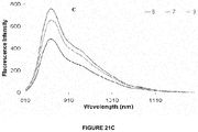
- FIG. 21C shows fluorescent intensity for nanoconstructs 5, 7, and 8. Fluorescence was more intense for nanoconstructs 7 and 8 than for pure CD nanoconstruct 5, even if difference in absorption at 785 nm was minimal.
- FIG. 21D shows detector response for constructs 5, 6, 7, and 8 illustrating singlet oxygen decay rates.
- FIG. 22A shows tumor response versus time for untreated control, PS1 (HPPH), and nanoconstructs 6, 7, and 8.
- FIG. 22B shows tumor response versus time for untreated control, nanoformulation 9 and nanoformulation 10.
- FIG. 23A shows an SEM for nanoconstruct 7 (representative of all groups).
- FIG. 23B shows a DLS particle size distribution for Blank PAA NPs.
- the mean diameter is 33.5 nm.
- FIG. 23C shows a particle size distribution for nanoconstruct 6.
- the mean diameter is 32.5 nm
- FIG. 23D shows a particle size distribution for nanoconstruct 5.
- the mean diameter is 35.2 nm.
- FIGS. 24A-24L show Release/Retention Profiles of PS 1 ( 24 A, 24 C, 24 E, 24 G, & 24 I) and/or cyanine dye 3 ( 24 B, 24 D, 24 F, 24 H, & 24 J) from nanoconstructs 5, 6, 7, 8, 9, and 10 in a 1% Human Serum Albumin (HSA) solution.
- FIGS. 24K and 24L show Release Profiles of PS 1 and cyanine dye 3 from nanoconstruct 7 in a 25% Bovine Calf Serum (BCS) solution at 37° C.
- BCS Bovine Calf Serum
- Photosensitizers generally fluorescence and their fluorescence properties in vivo has been exploited for the detection of early-stage cancers in the lung, bladder and other sites. For treatment of early disease or for deep seated tumors the fluorescence can be used to guide the activating light.
- photosensitizers are not optimal fluorophores for tumor detection for several reasons: (i) They have low fluorescence quantum yields (especially the long wavelength photosensitizers related to bacteriochlorins).
- Efficient photosensitizer tend to have lower fluorescence efficiency (quantum yield) than compounds designed to be fluorophores, such as cyanine dyes because the excited singlet state energy emitted as fluorescence is instead transferred to the triplet state and then to molecular oxygen.
- fluorophores such as cyanine dyes
- They have small Stokes shifts.
- Porphyrin-based photosensitizer have a relatively small difference between the long wavelength absorption band and the fluorescence wavelength (Stokes shift), which makes it technically difficult to separate the fluorescence from the excitation wavelength.
- Most photosensitizer have relatively short fluorescent wavelengths, ⁇ 800 nm, which are not optimal for detection deep in tissues.
- tumor-avid photosensitizer(s) e.g., HPPH
- NIR absorbing fluorophore(s) non-tumor specific cyanine dyes
- HPPH was used as a vehicle to deliver the imaging agent to tumor.
- the limitation of this approach was that the conjugate exhibited significantly different dose requirements for the two modalities.
- the imaging dose was approximately 10-fold lower than the phototherapeutic dose ( FIGS.
- nanovector platforms that can deliver tumor-avid therapeutic photosensitizers that only become active (and toxic) when illuminated by specific wavelengths of light, and, in addition, carry one or more imaging agents; these nano-platforms thus could enable both diagnosis and image guided therapy.
- biodegradable polymer based nanoparticles avoids multi-step synthesis and has numerous advantages including the ability to create water soluble formulations with desired pharmacokinetic properties, capable of delivering a high payload of the multiple agents (therapeutic PS and imaging agents) to tumors, increased photostability of photoactive agents and fluorophores, and the ability to modify the surface of the NP for conjugation to a variety of biomolecules.
- NPs and other macromolecular objects can passively target the tumor interstitium, via the “Enhanced Permeability and Retention” (EPR) effect due to the leaky vascular system in tumors. 21a,b
- EPR Enhanced Permeability and Retention
- the poor lymphatic drainage system in tumors causes fluid retention in the tumor interstitial space, which helps to retain polymeric nanoparticles and other macromolecular objects in the tumor compared to normal tissue. 21a,b
- NPs are a promising means for delivering therapeutic and other molecular agents to tumors.
- NPs could deliver a high payload of the drug to tumor
- a PAA-based nanoconstructs for delivering both the near-infrared (NIR) cyanine dye (CD) fluorophore and the red-light absorbing photosensitizer HPPH.
- the release of the desired imaging and therapeutic agents may also be controlled by creating a nanoparticle that is pH or temperature sensitive, or by modifying the pores of the NP matrix
- NIR near-infrared
- CD cyanine dye
- HPPH and the cyanine dye were post-loaded in variable ratios (HPPH to CD: 1:1; 2:1; 3:1 and 4:1 molar concentrations).
- HPPH was postloaded to PAA nanoparticles first. Free HPPH was removed by spin filtration and then cyanine dye was postloaded. It was spin-filtered again, washed several times with 1% bovine calf serum and the concentration was measured.
- the 2:1 formulations produce the best tumor imaging and long-term tumor cure in BALB/c mice bearing Colon26 tumors.
- This formulation contained in a single dose the therapeutic dose of HPPH (0.47 ⁇ mol/kg) and the imaging dose of Cyanine dye (0.27 mol/kg), which were similar to the components used alone for tumor imaging and therapy, but with much more tumor selectivity (skin to tumor ratio of HPPH was 4:1 instead of 2:1 without nanoparticles). Under similar treatment parameters the Ormosil nanoparticles showed a significantly reduced response (imaging and PDT, not shown).
- the stability of the drugs in PAA nanoparticle was established by repeated washing with aqueous bovine calf serum through Amicon centrifugal filter units with a 100 KDa or larger cut off membrane and drug in the filtrate was measured spectrophotometrically.
- FIGS. 2-4 The comparative in vivo PDT efficacy of the ORMOSIL and PAA formulations, their tumor imaging potential and stability (in vitro release kinetics) is shown in FIGS. 2-4 , which clearly illustrate the advantages of PAA nanoparticles in reducing the therapeutic dose by almost 8-fold without diminishing the tumor-imaging potential and also avoiding the Tween-80 formulation required for the HPPH-CD conjugate 1.
- the HPPH CD conjugate 1 was post-loaded to PAA nanoparticles, which certainly enhanced the tumor imaging, but the therapeutic dose was still 10-fold higher (similar to the HPPH CD conjugate, FIG. 5B ).
- the cyanine dye was conjugated peripherally to the PAA nanoparticles first and then HPPH was post loaded. Again, compared to HPPH-CD conjugate 1, the PAA formulation showed enhanced tumor-specificity (imaging) ( FIG. 5B ).
- a photosensitizer with increased selectivity and longer wavelength could be a more suitable candidate for brain and deeply seated tumors (especially breast, brain and lung).
- the evolution of light sources and delivery systems is also critical to the progression of photodynamic therapy (PDT) in the medical field.
- PDT photodynamic therapy
- Chang et al reported an effective radius of tumor cell kill in 22 glioma patients of 8 mm compared with the 1.5 cm depth of necrosis noted by Pierria with the intracavitary illumination method. It is believed that tumor resection is important so that the numbers of tumor cells remaining to treat are minimized. With stereotactic implantation of fibers for interstitial PDT there is no cavity to accommodate swelling and a considerable volume of necrotic tumor which causes cerebral edema. However, cerebral edema can be readily controlled with steroid therapy. Compared to chemotherapy and radiotherapy, patients with brain tumors treated with PDT have definitely shown long-term survival, whereas glioma patients treated with adjuvant chemotherapy or radiotherapy do not seem to show additional benefits. as On the basis of our preliminary data, the ⁇ v ⁇ 3 targeted nanoparticles may improve tumor-selectivity and PDT outcome.
- PAA nanoparticles decreased the liver uptake of the 124I-photosensitizer (PET imaging agent) and enhanced the tumor-specificity.
- PET imaging agent 124I-photosensitizer
- Our initial investigation with an 124I-labeled photosensitizer 2 indicates its in vivo PDT efficacy and capability of detecting tumors104-106 (RIF, Colon26, U87, GL261, pancreatic tumor xenograft)) and tumor metastases (BALB/c mice bearing orthotopic 4T1 (breast) tumors) ( FIG. 6 ).
- 18F FDG photosensitizer 2 showed enhanced contrast in most of the tumors including those where 18F FDG-PET provides limited imaging potential (e.g., brain, lung and pancreatic tumors).
- FIG. 7 For comparative biodistribution. This is the first report showing the utility of porphyrin-based compounds as a “BIFUNCTIONAL AGENT” for imaging breast tumor and tumor metastasis. Similar to most nanoparticles, PAA nanoparticles accumulate in liver and spleen. Their clearance rate from most organs is significantly faster than Ormosil nanoparticle and they do not show long-term organ toxicity. Even tumor-avid porphyrinbased photosensitizer exhibit high uptake in liver and spleen, but are non-toxic until exposed to light. The photosensitizer clears from the system quickly (days) without organ toxicity.
- radioactive photosensitizer such as the 124I-labeled analog 2 (superior to 18F-FDG in PET-imaging of lung, brain, breast and pancreas tumors) with a T1 ⁇ 2 of 4.2 days could cause radiation damage to normal organs.
- radioactive photosensitizer such as the 124I-labeled analog 2 (superior to 18F-FDG in PET-imaging of lung, brain, breast and pancreas tumors) with a T1 ⁇ 2 of 4.2 days could cause radiation damage to normal organs.
- 124I-imaging agent Based on the observation of high uptake of PAA nanoparticles in liver and spleen (below) we postulated that saturating the organs with the non-toxic PAA nanoparticles before injecting the PET agent might reduce uptake and radiation damage by 124I-imaging agent.
- PAA nanoparticles were first injected (i.v.) into mice bearing Colon26 tumors followed 24 h later by i.v
- PAA nanoparticles made a remarkable difference in tumor contrast with brain, lung and pancreatic tumors). See FIG. 7 for comparative biodistribution.
- PAA nanoparticles can be targeted to nucleolin with F3-Cys:
- F3-targeted nanoparticles were prepared using two kinds of F3 peptides: F3 peptide conjugated to nanoparticle via one of the 8 lysines available in its sequence and F3-Cys peptide conjugated to nanoparticle via cysteine. Cysteine capped nanoparticles served as non-targeted control. Three 25 mg batches of each type of nanoparticle contained: 2.6, 5.1 and 7.7 mg F3, (A3-A5) respectively; 2.7, 5.3 and 8 mg F3-Cys (B3-B5) respectively, and 0.29, 0.58 and 0.87 mg Cys (C3-C5) respectively.
- the fluorescence intensity from PAA nanoparticle incubated in vitro with nucleolin positive MDA-MB-435 cells is shown in FIG. 9 .
- the F3-Cys conjugated nanoparticles show considerably higher binding efficiency than non-targeted nanoparticles, while F3 conjugated nanoparticles do not. Conjugation via a cysteine link preserves the specificity of F3 peptide for nucleolin. In addition excess cysteine on the nanoparticles helphotosensitizer to minimize the non-specific binding. Additional experiments (not shown) suggested that the amount of F3-Cys peptide (5.3 mg/25 mg nanoparticle) used for B4 nanoparticles was optimal.
- F3-mediated specificity is retained in the presence of conjugated HPPH.
- F3 targeted nanoparticles did targeted nanoparticles did not, indicating that F3-mediated specificity is retained in the presence of conjugated HPPH.
- F3 targeted nanoparticles did not accumulate in the nucleus.
- Cell internalization of F3-targeted nanoparticles was confirmed by fluorescence confocal microscopy.
- F3 targeted HPPH conjugated PAA nanoparticle specifically bound to MDA-MB-435 cells (expressing nucleolin) while non-targeted nanoparticles did not, indicating that F3-mediated specificity is retained in the presence of conjugated HPPH.
- F3 targeted nanoparticles did not accumulate in the nucleus.
- Cell internalization of F3-targeted nanoparticles was confirmed by fluorescence confocal microscopy.
- F3-Cys Shows Target-Specificity in 9 L Glioma Cells:
- F3-cys PEG on PAA nanoparticles Similar to F3-cys, a pegylated form of F3-Cys PEG on PAA nanoparticles also showed remarkable target-specificity in 9 L rat glioma cells which also expresses nucleolin, FIG. 11 . (Note: HPPH is replaced with a Rhodamine moiety).
- This invention shows the utility of porphyrin-based compounds in a “bifunctional agent” for imaging breast tumor and tumor metastasis. Similar to most nanoparticles, PAA nanoparticle accumulate in liver and spleen. Their clearance rate from most organs is significantly faster than Ormosil nanoparticle and they do not show long-term organ toxicity. Even tumor-avid porphyrin based photosensitizer exhibit high uptake in liver and spleen, but are non-toxic until exposed to light. The photosensitizer clear from the system quickly (days) without organ toxicity.
- radioactive photosensitizer such as the 124I-labeled analog 2 (superior to 18F-FDG in PET-imaging of lung, brain, breast and pancreas tumors) with a T1 ⁇ 2 of 4.2 days could cause radiation damage to normal organs.
- radioactive photosensitizer such as the 124I-labeled analog 2 (superior to 18F-FDG in PET-imaging of lung, brain, breast and pancreas tumors) with a T1 ⁇ 2 of 4.2 days could cause radiation damage to normal organs.
- 124I-imaging agent Based on the observation of high uptake of PAA nanoparticles in liver and spleen (below) we postulated that saturating the organs with the non-toxic PAA nanoparticles before injecting the PET agent might reduce uptake and radiation damage by 124I-imaging agent.
- PAA nanoparticles were first injected (i.v.) into mice bearing Colon26 tumors followed 24 h later by i.v
- PAA nanoparticles makes a remarkable difference in tumor contrast with significantly reduced uptake in spleen and liver and improved tumor-uptake/contrast at 24, 48 and 72 h post injection (3 mice/group Similar studies (tumor-imaging and PDT efficacy) in which the labeled photosensitizer is post-loaded to variable sizes. Similar studies (tumor-imaging and PDT efficacy) in which the labeled photosensitizer is post-loaded to variable sizes PAA nanoparticles are currently in progress.
- PAA nanoparticles were prepared by following our previous with slight modifications (See e.g. Multifunctional ORMOSIL and PAA nanoparticles, Gupta et al., Photodynamic Therapy Back to the Future. Edited by Kessel, David H. Proceedings of the SPIE, Volume 7380 (2009), pp. 73805H-73805H-12 (2009).
- Nanoparticles were syringe filtered with a 0.2 ⁇ m regenerated cellulose syringe filter.
- Nanoformulation 9 and 10 were created by mixing nanoconstruct 1 and 3 such that the molar ratio of 1 to 3 was 2:1 and 4:1, respectively.
- the NPs are stored at 4° C. until further use. For details see “Supporting Material Information”.
- Post-Loading of the photosensitizer 1 and the cyanine dye 3 to blank AFPAA to create nanoconstructs 7 and 8 Upon measuring the concentration of PS 1 in nanoconstruct 6, cyanine dye 3 in DMSO (20 mM) was added such that the molar ratio of PS 1 to cyanine dye 3 was either 2:1 or 4:1. Once cyanine dye 3 was added, the procedure is the same as for post-loading, PS 1 or cyanine dye 2 and 3. For detailed procedure see the “Supporting Material Information”.
- the NPs in the retentate were reconstituted to the original volume with 1% HSA and re-centrifuge filtered (filtrate 2) and measured spectrophotometrically. The amount of 1 and/or 3 retained by the NP was confirmed by measuring the absorbance of the retentate upon reconstitution to the original volume with 1% HSA. If the sum total of all filtrates and the retentate is less than 90% of the stock value for either chromophore then ethanol is added to the centrifuge filter to measure what had adsorbed to the filter. These measurements were taken immediately post-addition of the nanoconstructs in a 1% HSA solution, 4 and 24 hours post addition of the nanoconstructs in the 1% HSA solution.
- mice 3 mice/group bearing subcutaneous Colon 26 tumors on the right shoulder were injected i. v. (tail-vein) with either cyanine dyes or nanoconstructs/formulations.
- cyanine dyes or nanoconstructs/formulations For a detailed description of the groups of mice imaged along with the dose, see the “Supporting Material Information”.
- the absorbance measurements were performed on a Varian Cary-50 Bio UV-Visible spectrophotometer.
- the concentrations of the NP formulations were measured in ethanol utilizing 47,500, 200,000 and 207,455 L mol ⁇ 1 cm ⁇ 1 as the respective molar extinction coefficients of 1, 2, and 3.
- a SPEX 270M spectrometer (Jobin Yvon) was used for acquisition of fluorescence emission spectra in the far red and NIR spectral ranges, utilizing the first output port equipped with an InGaAs photodetector (Electrooptical Systems Inc., USA).
- a diode-pumped solid-state laser (Verdi, Coherent) at 532 nm was the excitation source.
- Generation of singlet oxygen ( 1 O 2 ) was detected by its phosphorescence emission peaked at 1270 nm.
- the decays of this emission were acquired using the Infinium oscilloscope (Hewlett-Packard) coupled to the output of the Hamamatsu IR-PMT which is attached to the second output port of the SPEX 270M spectrometer. Nanoconstructs 5-8 in polystyrene cuvettes were placed in front of the entrance to the spectrometer. The emission signal was collected at 90-degrees relative to the exciting laser beam with the use of additional long-pass filters (a 950LP filter and/or a 538AELP filter) to attenuate the scattered light and fluorescence from the samples. A second harmonic (532 nm) from the nanosecond pulsed Nd:YAG laser (Lotis TII, Belarus) operating at 20 Hz was used as the excitation source for time-resolved measurements.
- mice Eight- to twelve-week-old BALB/cAnNCr mice (Jackson Laboratory, Bar Harbor, Me.) were inoculated subcutaneously (s.c.) with 1 ⁇ 10 6 Colon 26 cells. When tumors reached 40-70 mm 3 , mice were injected i.v. (tail vein) with PS 1 (formulated in 1% Tween 80/D5W) or PAA nanoconstructs/formulations 6-10 suspended in water and further diluted in D5W. 24 hours post i.v.
- PS 1 formulated in 1% Tween 80/D5W
- PAA nanoconstructs/formulations 6-10 suspended in water and further diluted in D5W. 24 hours post i.v.
- mice (BALB/c mice bearing Colon 26 tumors, 10 mice/group) were restrained in plexiglass holders and tumors were irradiated at 665 nm with a fluence and fluence rate of 135 J/cm 2 at 75 mW/cm 2 , respectively, using a pumped argon-dye laser.
- the growth of tumors was measured two to three times per week and the mice were monitored for a total of 60 days post PDT treatment. When the tumor regrowth was >400 mm 3 , the mice were euthanized according to the guidelines of the institute approved animal protocol.
- FIGS. 17A and 17B Post-loading of amine functionalized PAA NPs with IR-820, cyanine dye 2, nanoconstruct 4; cyanine dye 3, nanoconstruct 5; HPPH, photosensitizer 1: nanoconstruct 6; photosensitizer 1 and cyanine dye 3 at a 2:1 molar ratio, nanoconstruct 7; and HPPH 1 and cyanine dye 3 at a 4:1 molar ratio, nanoconstruct 8.
- Nanoformulations 9 and 10 are nanoconstruct 6 and 5 mixed such that the molar ratio of 1 to 3 is 2:1 and 4:1, respectively.
- DLS dynamic light scattering
- SEM scanning electron microscopy
- porous nanoparticles are advantageous since release of PS from the NP is not required for the singlet oxygen to diffuse into the tumor cells. However, if the release profile is rapid the NP may not be able to efficiently deliver a high payload of the desired agent to tumor. Therefore, we investigated the release profiles of the photosensitizer 1 and the cyanine dye 3 from nanoconstructs 5-10, respectively, by incubating them in 1% human serum albumin (HSA) at variable time points. The release profiles are summarized in the supplemental section. The release of PS 1 in nanoconstruct 6 showed a two-phase release, where an increase in the release was seen in the first four hours, which subsequently decreased during the following 20 hours.
- HSA human serum albumin
- nanoconstruct 6 Compared to nanoconstructs 7-10, nanoconstruct 6 showed the highest retention of PS 1 (HPPH) over a 24 hour time period with approximately 87% being retained. When comparing the percentage of PS 1 retained (at the initial time point, time zero), upon addition of 1% HSA, the nanoconstruct 8 and 10 showed the highest retention ( ⁇ 84%) of the PS.
- the nanoformulation 7 which provided the best whole-body fluorescence imaging and PDT response, was also subjected for the release of both the chromophores in 25% bovine calf serum (BCS, 37° C.) at 4, 8, 12, and 24 hours post-addition.
- FIG. 18A shows whole body fluorescence images of BALB/c mice bearing Colon-26 tumors.
- Control mouse A, E, I, and M
- cyanine dye 2 B-D
- nanoconstruct 4 F-H
- cyanine dye 3 J-L
- nanoconstruct 5 N-P
- the water insoluble fluorophore 2 was formulated in a 1% Tween-80/5% dextrose solution and was injected i.v. at a dose of 0.3 ⁇ mole/kg.
- the mice were imaged at 24, 48, and 72 hours post-injection ( FIG. 18A at B-D). Due to the rather poor tumor localization of 2, we post-loaded it into PAA NPs formulation 4 and compared their tumor uptake and fluorescence imaging abilities.
- FIG. 18B clearly indicate much improved tumor selectivity of the PAA NP formulation 4 over the free fluorophore 2.
- modified cyanine dye 3 and the corresponding nanoconstruct 5 were also imaged ( FIG.
- nanoconstructs 7 and 8 in which the PS and CD were post-loaded in a ratio of 2 to 1 and 4 to 1 on excitation of the cyanine dye at 782 nm gave fluorescence at 866 and 870 nm respectively.
- both nanoconstructs on in vivo excitation at 665 nm produced a significant fluorescence beyond 860 nm, which can be explained by the phenomenon known as the Förster (Fluorescence) Resonance Energy Transfer (FRET), or by the more general phenomena of energy migration, or excitation percolation, followed by energy trapping, analogous to the energy transport and funneling process in photosynthetic antenna.
- FRET Förster Resonance Energy Transfer
- FIG. 19 shows whole body fluorescence images of a control mouse (A), nanoconstructs/formulation 5 (B), 7 (C & D), 8 (E & F), 9 (G & H) and 10 (I & J) in BALB/c mice bearing Colon-26 tumors.
- the excitation wavelength was 782 nm.
- Images A, B, C, E, G, & I and D, F, H, & J were taken 24 and 48 hours post i.v. injection, respectively
- FIG. 20 shows whole body FRET images of a control mouse (A), nanoconstructs/formulation 5 (B), 7 (C), 8 (D), 9 (E) and 10 (F) in BALB/c mice bearing Colon-26 tumors.
- the excitation wavelength was 665 nm. Images A-F were taken 24 hours post i.v. injection.
- the fluorescence spectrum in FIG. 21B shows the difference in fluorescence intensity for 6, 7, and 8, which resulted from the different efficiency of the PS ⁇ CD energy transfer in these nanoconstructs.
- This energy transfer caused a decrease in intensity of the PS fluorescence along with an increase in CD fluorescence intensity.
- the energy transfer was strongest for nanoconstruct 8 since the fluorescence spectrum displayed the least intense fluorescence band from PS moiety ( ⁇ max ⁇ 670 nm) and the most intense fluorescence band from the CD moiety ( ⁇ max ⁇ 870 nm) upon excitation at 532 nm ( FIG. 21A ).
- HPPH derived from chlorophyll-a
- HPPH is an effective PDT agent with low skin phototoxicity and, in BALB/c mice bearing Colon 26 tumors, a complete PDT response of 40% was observed at a dose of 0.47 moles/kg on exposing the tumors with light at 665 nm, delivered at a fluence and fluence-rate of 135 J/cm 2 and 75 mW/cm 2 24 hours post-injection.
- the PDT response (no tumor regrowth) for HPPH 1 was 40%, 40%, 60%, 30%, 40%, and 30%, respectively.
- PAA NPs provide a great platform for post-loading because of the porous nature of the polyacrylamide-based hydrogels, wherein the hydrophobic part of the molecule may interact preferentially.
- Tween-80 plays an important role in efficient retention of the compounds within the NPs. Its presence in an aqueous solution apparently causes formation of a micellar layer on the nanoconstruct surface whereby the polyethers form the outer hydrophobic layer and the oleic acid forms the inner, more hydrophobic layer of the construct.
- NIR fluorophores cyanine dyes in general have shown great potential for fluorescence imaging.
- IR-820 a near-infrared (NIR) cyanine dye
- NIR-820 a near-infrared (NIR) cyanine dye
- excitation and emission in the NIR range beyond 750 nm which allows for deeper tissue penetration of activating light; however, its tumor uptake is known to be low.
- hexane was removed by rotary evaporation and the particles were precipitated by addition of ethanol (50 ml).
- the surfactant and residual monomers were washed away from the particles with ethanol (150 ml) followed by washing with water (100 ml) 5 times each in an Amicon ultra-filtration cell equipped with a Biomax 500 kDa cutoff membrane.
- the concentrated nanoparticles were lyophilized for two days for storage, and reconstituted by suspending in water before use.
- the dynamic light scattering measurements were performed on a Nicomp 370 Submicron Particle Sizer (Nicomp, Santa Barbara, Calif.).
- the NP solution was placed in a borosilicate glass capillary tube, and diluted with water to an intensity reading of 300 kHz. The readings were performed in triplicate with each run set for 5 minutes.
- the volume weighted Gaussian or Nicomp's proprietary analysis mode was utilized to determine the mean hydrodynamic diameter.
- the morphology and size of the dried NP were characterized by scanning electron microscope (SEM).
- SEM scanning electron microscope
- the NP solution of 0.2 mg/ml was prepared in water and a drop of the NP solution was placed on the SEM aluminium specimen mount (aluminum) and dried gradually at room temperature. The sample was then sputter coated with gold and the SEM images were taken on the Philips ESEM XL30.
- the concentration of 1, 2, and 3 was measured spectrophotometrically in ethyl alcohol according to the Beer's-Lambert law using 47,500, 200,000, and 207,455 (L mol- ⁇ 1 cm ⁇ 1 ) as the molar extinction coefficient for 1, 2, and 3, respectively, after they were syringe filtered with a 0.2 ⁇ m regenerated cellulose syringe filter. If scattering was present in the absorbance spectra, the NPs were centrifuged in a microfuge membrane-filter (Nanosep 100K Omega) at 14,000 RPM for 10 minutes. The filtrate was used to calculate the concentration of 1, 2, or 3. To create nanoconstrust 9 and 10, nanoconstruct 6 and nanoconstrust 3 are mixed together such that the molar ratio of PS 1 and cyanine dye 3 is 2-1 and 4-1, respectively. The NPs are stored at 4° C. until further use.
- the lyophilized AFPAA NPs were dissolved in 1% Tween-80 to a final concentration of 10 mg of NPs/1 ml of 1% Tween-80.
- the hydrodynamic diameter of the blank PAA NPs were measured by DLS prior to use to ensure the NPs are of appropriate size. If aggregation is present, the hydrodynamic diameter may increase; therefore the NPs were sonicated in a water bath sonicator for several minutes to disperse the aggregation. Prior to post-loading, 1 and 3 were dissolved in DMSO to prepare 20 mM solutions.
- NP solution For 2 ml of NP solution, 20 or 40 ⁇ l of 1 was post-loaded as the first step of making nanoconstructs 7 and 8, respectively, and excess reagents were removed by centrifuge filtration as before until no PS 1 is detected in the filtrate. The concentration of PS 1 was measured spectrophotometrically. If scattering is observed (determined spectrophotometrically), the NPs in ethanol were centrifuged in a microfuge membrane-filter at 14,000 RPM for 10 minutes to remove PS 1 from the NPs. The filtrate was used for calculating the concentration of the PS. The concentration of the PS 1 and 3 in nanoconstructs 7 and 8 was 200 and 400 ⁇ M respectively.
- the first group of BALB/c mice were imaged with cyanine dye 2 and the nanoconstruct 4 post loaded with cyanine dye 2 (dose: 0.3 ⁇ moles/kg).
- the second group of mice was imaged with cyanine dye 3 and nanoconstruct 5 (dose: 0.3 ⁇ moles/kg).
- the third and fourth groups of mice were imaged with nanoconstruct 7 containing the cyanine dye 3 post-loaded (dose: 0.216 moles/kg) and the PS 1 post-loaded (dose: 0.47 ⁇ moles/kg) and the nanoconstruct 8 containing the cyanine dye 3 post-loaded (dose: 0.108 ⁇ moles/kg) and PS 1 post-loaded (dose: 0.47 ⁇ moles/kg).
- mice were imaged post with nanoconstruct 9 containing the cyanine dye 3 post-loaded (dose: 0.236 ⁇ moles/kg) and the PS 1 postloaded (dose: 0.47 ⁇ moles/kg), and the nanoconstruct 10 contained 3 post-loaded (dose: 0.127 ⁇ moles/kg) and the PS 1 post-loaded (dose: 0.47 ⁇ moles/kg).
- mice 3 mice/group bearing subcutaneous Colon 26 tumors on the right shoulder were injected i. v. (tail-vein) with either cyanine dyes or nanoconstructs/formulations.
- mice were anesthetized with ketamine/xylazine by intraperitonial injection and imaged with a monochrome scientific grade CCD camera CRI (Nuance, Woburn, Mass.).
- the CCD camera was employed in the mono mode and a 782 nm BWF light source (B&W-Tek, Newark, Del.) continuous wave laser was used for excitation.
- the fluorescence emission was collected with an 800 and 830 nm long pass filters in series.
- FRET fluorescence resonance energy transfer
- ET energy transfer
- FIGS. 23A-23D show an SEM for nanoconstruct 7 (representative of all groups) and FIG. 23 B shows a DLS particle size distribution for Blank PAA NPs, FIG. 23C shows a particle size distribution for nanoconstruct 6, and FIG. 23D shows a particle size distribution for nanoconstruct 5 in Tween-80/water (concentration of Tween-80 is ⁇ 1%).
- the mean diameter is 33.5 nm, 32.5 nm, and 35.2 nm for FIG. 23B , FIG. 23C , and FIG. 23D , respectively.
- the mean hydrodynamic diameter as determined by DLS is similar for all nanoconstruct/formulations.
- FIGS. 24A-24L show Release/Retention Profiles of PS 1 ( 24 A, 24 C, 24 E, 24 G, & 24 I) and/or cyanine dye 3 ( 24 B, 24 D, 24 F, 24 H, & 24 J) from nanoconstructs 5, 6, 7, 8, 9, and 10 in a 1% Human Serum Albumin (HSA) solution.
- HSA Human Serum Albumin
- the release/retention was measured immediately upon addition of the various nanoconstucts in 1% HSA (*), 4, and 24 hours post-addition of the nanoconstructs in a 1% HSA solution. Each experiment was done in triplicate with the symbols representing the mean. The error bars are standard error of the mean.
- 24K and 24L show Release Profiles of PS 1 and cyanine dye 3 from nanoconstruct 7 in a 25% Bovine Calf Serum (BCS) solution at 37° C. The release was measured 4, 8, 12, and 24 hours post-addition of nanoconstruct 7 in 25% BCS. Each experiment was done in triplicate with the symbols representing the mean. The error bars are standard error of the mean.
- BCS Bovine Calf Serum
Abstract
Description

or a pharmaceutically acceptable derivative thereof, wherein:
Claims (28)


Priority Applications (2)
| Application Number | Priority Date | Filing Date | Title |
|---|---|---|---|
| US13/566,411 US10568963B2 (en) | 2009-10-21 | 2012-08-03 | Multifunctional nanoplatforms for fluorescence imaging and photodynamic therapy developed by post-loading photosensitizer and fluorophore to polyacrylamide nanoparticles |
| PCT/US2013/053347 WO2014022740A1 (en) | 2012-08-03 | 2013-08-02 | Multifunctional nanoplatforms for fluorescence imaging and photodynamic therapy developed by post-loading photosensitizer and fluorophore to polyacrylamide nanoparticles |
Applications Claiming Priority (3)
| Application Number | Priority Date | Filing Date | Title |
|---|---|---|---|
| US27952209P | 2009-10-21 | 2009-10-21 | |
| US12/909,573 US8562944B2 (en) | 2009-10-21 | 2010-10-21 | PAA nanoparticles for enhancement of tumor imaging |
| US13/566,411 US10568963B2 (en) | 2009-10-21 | 2012-08-03 | Multifunctional nanoplatforms for fluorescence imaging and photodynamic therapy developed by post-loading photosensitizer and fluorophore to polyacrylamide nanoparticles |
Related Parent Applications (1)
| Application Number | Title | Priority Date | Filing Date |
|---|---|---|---|
| US12/909,573 Continuation-In-Part US8562944B2 (en) | 2009-10-21 | 2010-10-21 | PAA nanoparticles for enhancement of tumor imaging |
Publications (2)
| Publication Number | Publication Date |
|---|---|
| US20130202525A1 US20130202525A1 (en) | 2013-08-08 |
| US10568963B2 true US10568963B2 (en) | 2020-02-25 |
Family
ID=48903066
Family Applications (1)
| Application Number | Title | Priority Date | Filing Date |
|---|---|---|---|
| US13/566,411 Active US10568963B2 (en) | 2009-10-21 | 2012-08-03 | Multifunctional nanoplatforms for fluorescence imaging and photodynamic therapy developed by post-loading photosensitizer and fluorophore to polyacrylamide nanoparticles |
Country Status (1)
| Country | Link |
|---|---|
| US (1) | US10568963B2 (en) |
Families Citing this family (4)
| Publication number | Priority date | Publication date | Assignee | Title |
|---|---|---|---|---|
| KR20200028474A (en) * | 2017-07-24 | 2020-03-16 | 퀀텀-에스아이 인코포레이티드 | Optical rejection photon structures |
| CN111426672B (en) * | 2020-05-27 | 2021-12-21 | 北京化工大学 | Method for rapid preparation of multi-dimensional fluorescent nanoparticles and in-situ quantitative characterization of dynamic dispersion state of multi-dimensional fluorescent nanoparticles |
| CN112546219B (en) * | 2020-12-07 | 2021-12-10 | 上海交通大学 | Nanotherapeutic structures and methods of making the same |
| CN114099716B (en) * | 2021-11-30 | 2023-04-07 | 上海交通大学医学院附属第九人民医院 | Bimodal imaging compositions and their use in lung nodule localization |
Citations (5)
| Publication number | Priority date | Publication date | Assignee | Title |
|---|---|---|---|---|
| US20050196343A1 (en) | 2004-02-27 | 2005-09-08 | Molecular Therapeutics, Inc. | Degradable nanoparticles |
| US20090043090A1 (en) * | 2004-07-16 | 2009-02-12 | Health Research, Inc. | Adduct of fluorescent dye and tumor avid tetrapyrrole |
| WO2009105209A1 (en) * | 2008-02-19 | 2009-08-27 | Health Research, Inc. | Silica nanoparticles postloaded with photosensitizers for drug delivery in photodynamic therapy |
| US20110022129A1 (en) | 2007-11-05 | 2011-01-27 | Prud Homme Robert K | Nanoparticles for photodynamic therapy |
| US20110091373A1 (en) | 2009-10-21 | 2011-04-21 | Health Research, Inc. | Paa nanoparticles for enhancement of tumor imaging |
-
2012
- 2012-08-03 US US13/566,411 patent/US10568963B2/en active Active
Patent Citations (5)
| Publication number | Priority date | Publication date | Assignee | Title |
|---|---|---|---|---|
| US20050196343A1 (en) | 2004-02-27 | 2005-09-08 | Molecular Therapeutics, Inc. | Degradable nanoparticles |
| US20090043090A1 (en) * | 2004-07-16 | 2009-02-12 | Health Research, Inc. | Adduct of fluorescent dye and tumor avid tetrapyrrole |
| US20110022129A1 (en) | 2007-11-05 | 2011-01-27 | Prud Homme Robert K | Nanoparticles for photodynamic therapy |
| WO2009105209A1 (en) * | 2008-02-19 | 2009-08-27 | Health Research, Inc. | Silica nanoparticles postloaded with photosensitizers for drug delivery in photodynamic therapy |
| US20110091373A1 (en) | 2009-10-21 | 2011-04-21 | Health Research, Inc. | Paa nanoparticles for enhancement of tumor imaging |
Non-Patent Citations (10)
| Title |
|---|
| Daubresse, Catherine, et al., "Enzyme Immobilization in Nanoparticles Produced by Inverse Microemulsion Polymerization", Journal ofColloid and Interface Science 168, pp. 222-229, Copyright 1994. |
| G. RAMACHANDRA REDDY, MAHAVEER S. BHOJANI, PATRICK MCCONVILLE, JONATHAN MOODY, BRADFORD A. MOFFAT, DANIEL E. HALL, GWANGSEONG KIM,: "Vascular targeted nanoparticles for imaging and treatment of brain tumors", CLINICAL CANCER RESEARCH, AMERICAN ASSOCIATION FOR CANCER RESEARCH, US, vol. 12, no. 22, 15 November 2006 (2006-11-15), US, pages 6677 - 6686, XP002643835, ISSN: 1078-0432, DOI: 10.1158/1078-0432.CCR-06-0946 |
| Gupta et al., "Multifunctional ORMOSIL and PAA Nanoparticles", Photodynamic Therapy: Back To the Future, Edited by Kessel, David H., Proceedings of the SPIE, vol. 7380 (2009), pp. 73805H-73805H-12 (2009). |
| Huang (Rev. Prog. Clin. PDT, Technol. Cancer. Res. Treat. 2005, p. 283. * |
| Koo, Photonic explorers based on multifunctional nanoplatforms for biosensing and photodynamic therapy, Applied Optics, 46/10, pp. 1924-1930, 2007. |
| Kopelman, Raoul, et al. "Multifunctional Nanoparticle Platforms for in Vivo MRI Enhancement and Photodynamic Therapy of a Rat Brain Cancer", Journal of Magnetism and Magnetic Materials 293, pp. 404-410, Copyright 2005. |
| Photos. Clin. PDT, Photodia. Photodyna. Thera. 2004, p. 27-42. * |
| Reddy, G.R. et al., "Vascular targeted nonoparticles for imaging and treatment of brain tumors", Clinical Cancer Research Nov. 15, 2006 US LNKD-DOI:10.1158/1078-0432.CCR-06-0946, vol. 12, No. 22, Nov. 15, 2006, pp. 6677-6686, XP002643835. |
| Ross, B. et al. "Photonic and magnetic nonexplorers for biomedicaluse: from subcellar imaging to cancer diagnostics and therapy", University of Michigan, Ann arbor, MI 48109; Molecular Therapeutics, Inc., Ann Arbor, MI 48109; Text Center for Molecular Imaging Research, Massachusetts General Hospital, Charlestown, MA 02129. |
| Shouyan Wang, et al. "Novel Methods to Incorporate Photosensitizers into Nanocarriers for Cancer Treatment by Photodynamic Therapy"; Lasers in Surgery and Medicine 43:686-695 (2011);Department of Medicine, University of Michigan, Ann Arbor Michigan, 48109; PDT Center and Department of Dermatology; Roswell Park Cancer Institute; Buffalo, New York, 14263. |
Also Published As
| Publication number | Publication date |
|---|---|
| US20130202525A1 (en) | 2013-08-08 |
Similar Documents
| Publication | Publication Date | Title |
|---|---|---|
| Gupta et al. | Multifunctional nanoplatforms for fluorescence imaging and photodynamic therapy developed by post-loading photosensitizer and fluorophore to polyacrylamide nanoparticles | |
| EP2437653B1 (en) | Paa nanoparticles for enhancement of tumor imaging | |
| Son et al. | Light-responsive nanomedicine for biophotonic imaging and targeted therapy | |
| Huang et al. | Emerging applications of porphyrins in photomedicine | |
| Lim et al. | Nanophotosensitizers toward advanced photodynamic therapy of Cancer | |
| Li et al. | Sequential protein-responsive nanophotosensitizer complex for enhancing tumor-specific therapy | |
| Jeong et al. | Photosensitizer-conjugated human serum albumin nanoparticles for effective photodynamic therapy | |
| Taratula et al. | A multifunctional theranostic platform based on phthalocyanine-loaded dendrimer for image-guided drug delivery and photodynamic therapy | |
| Lin et al. | Nano-photosensitizers for enhanced photodynamic therapy | |
| Shinde et al. | Enhanced permeability and retention effect: A key facilitator for solid tumor targeting by nanoparticles | |
| US20110288234A1 (en) | Silica nanoparticles postloaded with photosensitizers for drug delivery in photodynamic therapy | |
| Kumar et al. | Near-infrared phosphorescent polymeric nanomicelles: efficient optical probes for tumor imaging and detection | |
| Jiang et al. | Diketopyrrolopyrrole: An emerging phototherapy agent in fighting cancer | |
| US20210052731A1 (en) | Inorganic nanophotosensitizers and methods of making and using same | |
| US10568963B2 (en) | Multifunctional nanoplatforms for fluorescence imaging and photodynamic therapy developed by post-loading photosensitizer and fluorophore to polyacrylamide nanoparticles | |
| Silindir‐Gunay et al. | Near‐infrared imaging of diseases: A nanocarrier approach | |
| Zhao et al. | The substituted zinc (II) phthalocyanines using “sulfur bridge” as the linkages. Synthesis, red-shifted spectroscopic properties and structure-inherent targeted photodynamic activities | |
| Yan et al. | Near-infrared emissive AIE nanoparticles for biomedical applications: From the perspective of different nanocarriers | |
| US8906343B2 (en) | PAA nanoplatforms containing fluorophores and targeted moieties covalently linked and photosensitizer post-loaded | |
| WO2014022740A1 (en) | Multifunctional nanoplatforms for fluorescence imaging and photodynamic therapy developed by post-loading photosensitizer and fluorophore to polyacrylamide nanoparticles | |
| Akhter et al. | Porphyrin-Based Nanomaterials for Cancer Nanotheranostics | |
| Marko et al. | Multifunctional Agents for Cancer-Imaging and Photodynamic Therapy: Impact of Polyacrylamide-Based Nanoplatforms | |
| Moussaron et al. | Dual imaging and photodynamic therapy anticancer theranostic nanoparticles | |
| KR102464624B1 (en) | Photo-reactive drug delivery system for combination chemotherapy | |
| Gupta | Biodegradable Polyacrylamide-Based Theranostic Platforms for Tumor-Imaging and Photodynamic Therapy |
Legal Events
| Date | Code | Title | Description |
|---|---|---|---|
| AS | Assignment |
Owner name: THE RESEARCH FOUNDATION OF STATE UNIVERSITY OF NEW YORK, NEW YORK Free format text: ASSIGNMENT OF ASSIGNORS INTEREST;ASSIGNORS:PANDEY, RAVINDRA K.;GUPTA, ANURAG;SAJJAD, MUNNAWWAR;SIGNING DATES FROM 20130604 TO 20130626;REEL/FRAME:046503/0766 Owner name: REGENTS OF THE UNIVERSITY OF MICHIGAN, MICHIGAN Free format text: ASSIGNMENT OF ASSIGNORS INTEREST;ASSIGNOR:KOPELMAN, RAOUL;REEL/FRAME:046503/0668 Effective date: 20130514 Owner name: THE RESEARCH FOUNDATION OF STATE UNIVERSITY OF NEW Free format text: ASSIGNMENT OF ASSIGNORS INTEREST;ASSIGNORS:PANDEY, RAVINDRA K.;GUPTA, ANURAG;SAJJAD, MUNNAWWAR;SIGNING DATES FROM 20130604 TO 20130626;REEL/FRAME:046503/0766 Owner name: HEALTH RESEARCH, INC., NEW YORK Free format text: ASSIGNMENT OF ASSIGNORS INTEREST;ASSIGNORS:PANDEY, RAVINDRA K.;GUPTA, ANURAG;SAJJAD, MUNNAWWAR;SIGNING DATES FROM 20130604 TO 20130626;REEL/FRAME:046503/0766 |
|
| STCC | Information on status: application revival |
Free format text: WITHDRAWN ABANDONMENT, AWAITING EXAMINER ACTION |
|
| FEPP | Fee payment procedure |
Free format text: PETITION RELATED TO MAINTENANCE FEES GRANTED (ORIGINAL EVENT CODE: PTGR); ENTITY STATUS OF PATENT OWNER: SMALL ENTITY |
|
| STPP | Information on status: patent application and granting procedure in general |
Free format text: NON FINAL ACTION MAILED |
|
| STPP | Information on status: patent application and granting procedure in general |
Free format text: RESPONSE TO NON-FINAL OFFICE ACTION ENTERED AND FORWARDED TO EXAMINER |
|
| STPP | Information on status: patent application and granting procedure in general |
Free format text: FINAL REJECTION MAILED |
|
| STPP | Information on status: patent application and granting procedure in general |
Free format text: RESPONSE AFTER FINAL ACTION FORWARDED TO EXAMINER |
|
| STPP | Information on status: patent application and granting procedure in general |
Free format text: NOTICE OF ALLOWANCE MAILED -- APPLICATION RECEIVED IN OFFICE OF PUBLICATIONS |
|
| STPP | Information on status: patent application and granting procedure in general |
Free format text: NOTICE OF ALLOWANCE MAILED -- APPLICATION RECEIVED IN OFFICE OF PUBLICATIONS |
|
| STPP | Information on status: patent application and granting procedure in general |
Free format text: PUBLICATIONS -- ISSUE FEE PAYMENT VERIFIED |
|
| STCF | Information on status: patent grant |
Free format text: PATENTED CASE |
|
| MAFP | Maintenance fee payment |
Free format text: PAYMENT OF MAINTENANCE FEE, 4TH YR, SMALL ENTITY (ORIGINAL EVENT CODE: M2551); ENTITY STATUS OF PATENT OWNER: SMALL ENTITY Year of fee payment: 4 |