US10562863B2 - Heterobifunctional linker - Google Patents
Heterobifunctional linker Download PDFInfo
- Publication number
- US10562863B2 US10562863B2 US15/713,285 US201715713285A US10562863B2 US 10562863 B2 US10562863 B2 US 10562863B2 US 201715713285 A US201715713285 A US 201715713285A US 10562863 B2 US10562863 B2 US 10562863B2
- Authority
- US
- United States
- Prior art keywords
- linker
- analyte
- aunp
- aunps
- compound
- Prior art date
- Legal status (The legal status is an assumption and is not a legal conclusion. Google has not performed a legal analysis and makes no representation as to the accuracy of the status listed.)
- Active
Links
- 0 *C(C)NCCC1=CN(CCOCCOCCOCCS)N=N1 Chemical compound *C(C)NCCC1=CN(CCOCCOCCOCCS)N=N1 0.000 description 8
- GDLNTMWKALNICD-UHFFFAOYSA-N C=CCCC1=CN(CCOCCOCCOCCSC(C)=O)N=N1 Chemical compound C=CCCC1=CN(CCOCCOCCOCCSC(C)=O)N=N1 GDLNTMWKALNICD-UHFFFAOYSA-N 0.000 description 1
- MQIUVEIQWYOVGD-UHFFFAOYSA-N C=CCOCCOCCOCCSC(C)=O Chemical compound C=CCOCCOCCOCCSC(C)=O MQIUVEIQWYOVGD-UHFFFAOYSA-N 0.000 description 1
Images


Classifications
-
- C—CHEMISTRY; METALLURGY
- C07—ORGANIC CHEMISTRY
- C07D—HETEROCYCLIC COMPOUNDS
- C07D249/00—Heterocyclic compounds containing five-membered rings having three nitrogen atoms as the only ring hetero atoms
- C07D249/02—Heterocyclic compounds containing five-membered rings having three nitrogen atoms as the only ring hetero atoms not condensed with other rings
- C07D249/04—1,2,3-Triazoles; Hydrogenated 1,2,3-triazoles
Definitions
- the present disclosure relates generally to heterobifunctional linkers.
- analytes e.g., metabolites
- NMR and mass spectrometers typically require access to large and expensive instrumentation, such as NMR and mass spectrometers.
- Some molecules can be detected via enzymatic assays while others, like vitamin D or certain pesticides, can be detected through immunological methods using chemo-conjugated proteins.
- these low-cost assays are very compound specific, and can take years of trial-and-error to develop.
- Metabolomics is a relatively new branch of 'omics science that involves the comprehensive characterization of large numbers of metabolites in cells, tissues and biofluids. Over the past 10 years, interest in metabolomics has grown tremendously. This is because it offers biologists and other life scientists a rapid and effective means to chemically phenotype organisms. It also offers physicians and clinicians a very quick and efficient route to discover or test for disease biomarkers. 1-4 Most metabolomic assays involve the use of classical separation technologies such as liquid chromatography or gas chromatography. These are normally coupled with high-end chemical detection instruments such as NMR or mass spectrometers to identify and quantify metabolites from biological samples.
- a of water soluble heterobifunctional linker (L1) comprising:
- TEG-“Clicked”-Alcohol (9) by reaction of azidetetraethylene glycol thioacetate (8) with 4-pentyn-1-ol to form 1,4-disubstitued-1,2,3-triazole;
- an AuNP-L1-analyte conjugate of Formula I comprising: reacting a heterobifunctional linker (L1) with the free amine function of an analyte and conjugating AuNP to the thiol group of the L1, to form the AuNP-L1-analyte conjugate.
- said analyte is an amino acid or a polypeptide.
- the amino acid is glutamate or histidine.
- said polypeptide is carnosine.
- the AuNP is a stabilized colloid AuNP.
- the stabilized colloid AuNP is produced by trisodium citrate (Na 3 C 6 H 5 O 7 ) reduction of chloroauric acid (HAuCl4) to form gold nanoparticles
- the method further comprising a analyte reagent on the surface of the AuNPs.
- said second analyte comprises an amino acid or polypeptide.
- said amino acid is Leucine or Histidine.
- said polypeptide is carnosine.
- a method of selecting an antibody specific for an analyte comprising: screening a library comprising antibodies for binding of an antibody within said library to a reagent-L1 conjugate.
- said library comprises a library of single chain antibodies (scFv).
- said analyte is an amino acid.
- a method for detecting an analyte in a sample comprising:
- said metabolite binding analyte comprises a protein.
- said AuNP-L1-analyte conjugate comprises an amino acid.
- an system for detecting a analyte in a sample comprising: a substrate comprising a reagent binding analyte, and an AuNP-L1-analyte conjugate,
- a water soluble heterobifunctional linker having the structure of Formula III: A-B-C (III) wherein, A is a thiol group; B is a PEG linker moiety, C is an aldehyde group.
- said thiol group is suitable for covalent attachment to a label.
- said aldehyde group is suitable for covalent attachment to a primary amine group.
- aldehyde group is connected to the PEG linker moiety through a triazole unit.
- said primary amine group is a primary amine group on an amino acid.
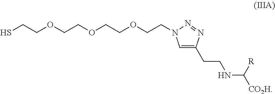
- the water soluble heterobifunctional linker (L1) is a compound of formula IIIa:
- the water soluble heterobifunctional linker (L1) is a compound of formula IIIb:
- FIG. 1 depicts the chemical structure of Linker 1 (L1) and derivatives thereof. Metabolites may be conjugated through amino groups amination (left) or substitution (right). Exchange reactions on terminal thioester can attach the linker to AuNP
- FIG. 2A depicts synthesis of linker-1 (L1) in 6 steps from tetraethylene glycol, yield a Linker L1, used for further conjugations;
- FIG. 2B depicts synthesis of Linker-2 in 3 steps from tetraethylene glycol;
- FIG. 2C depicts reductive animation.
- FIG. 3A-G depicts confirmation of L1 synthesis was demonstrated by Nuclear Magnetic Resonance (NMR) showing chemical shifts that confirm its structure by detection of either H1 or C13 nuclei.
- NMR Nuclear Magnetic Resonance
- 1 H NMR was done using a Varian Inova 600 MHz spectrometer.
- 13 C NMR was done using a Varian VNMRS two-channel 600 MHz Spectrometer, which possess direct and indirect detection of 13 C nuclei.
- FIG. 3B Compound 10(Glutamate-Linker 1) ( 1 H) (CD 3 OD);
- FIG. 3C Compound 10 (Glutamate-Linker 1) ( 13 C) (CD 3 OD);
- 3D Compound 11 (Carnosine-Linker 1) ( 1 H) (CD 3 OD)); FIG. 3E Compound 11 (Carnosine-Linker 1) ( 13 C) (CD 3 OD); FIG. 3F 12 (Histidine-Linker 1) (1H) (CD 3 OD); FIG. 3G 12 (Histidine-Linker 1) ( 13 C) (CD 3 OD).
- FIG. 4 is a general scheme for conjugated AuNPs.
- Gold citrate nanoparticles are modified with glutamate L1, before a second addition is done with carnosine, histidine, and leucine L1.
- FIG. 5 is a UV-Vis spectra of functionalized AuNPs. UV spectra were obtained after AuNP conjugation.
- FIG. 6A-K depicts DLS— FIG. 6A Au-citrate FIG. 6B 1 (Au-Glu-1) FIG. 6C 2 (Au-Glu-2) FIG. 6D 4 (Carn-1-Au-Glu-1) FIG. 6E 6 (Carn-2-Au-Glu-1) FIG. 6F 8 (Carn-1-Au-Glu-2) FIG. 6G 10 (Carn-2-Au-Glu-2) FIG. 6H 3 (His-1-Au-Glu-1) FIG. 6I 5(His-2-Au-Glu-1) FIG. 6J 7 (His-1-Au-Glu-2 FIG. 6K 9 (His-2Au-Glu-2).
- FIG. 7A-K depicts SEM images of conjugated AuNP: FIG. 7A Au-citrate FIG. 7B 1 (Au-Glu-1) FIG. 7C 2 (Au-Glu-2) FIG. 7D 4 (Carn-1-Au-Glu-1) FIG. 7E 6 (Carn-2-Au-Glu-1) FIG. 7F 8 (Carn-1-Au-Glu-2) FIG. 7G 10 (Carn-2-Au-Glu-2) FIG. 7H 3 (His-1-Au-Glu-1) FIG. 7I 5 (His-2-Au-Glu-1) FIG. 7J 7 (His-1-Au-Glu-2 FIG. 7K 9 (His-2Au-Glu-2).
- FIG. 8A-B Synthesis of 5-IAF conjugated metabolites.
- Ion positive configuration used, shows compound as peak with +1 mass (820 m/z for A and 806 m/z for FIG. 8B .
- FIG. 9 depicts Monoclonal Phage ELISA. Phages selected against Carnosine were used to detect binding against Carnosine-L1 on ELISA plates. Wells A2, A4 and A6 are negative controls; wells H2, H4 and H6 are positive control (Ubiquitin). All other wells are clones obtained through Phage Display.
- FIG. 10 depicts Lateral Flow Assays using Leucine-AuNP. Strips were loaded with Leu-AuNP solution and buffer (A); 0.5 mg/ml free Leu (B); 2 mg/ml free Leu (C) and 1M Urea (D). Test band (T) detects Leu-AuNP and Control band (C) detects Glutamate (present in our AuNP) and is not affected by the presence of other metabolites.
- FIG. 11 depicts Specificity of Branched-chain Aminoacid Lateral Flow Assay.
- FIG. 12 shows UV-Vis Spectra of double conjugated AuNPs with modified aminoacids-linker-1.
- FIG. 13 shows UV-Vis Spectra of double conjugated AuNPs-Linker-2-Glutamate Using modified aminoacids with both linker-1.
- FIG. 14 shows UV-Vis Spectra of double conjugated AuNPs-Linker-2-Glutamate Using modified aminoacids with both linker-2.
- metabolite conjugates that can be used for the selection of compound-specific antibodies or binding proteins and for the detection of metabolite-protein binding events using a wide range of detection technologies.
- a water-soluble, heterobifunctional linker with a thiol group at one end and an aldehyde group on the other end.
- the thiol group can be used to covalently couple to a gold nanoparticle (AuNP) or a fluorophore, while the aldehyde group can be used to link to a primary amine group (e.g. amino acids).
- AuNP gold nanoparticle
- a fluorophore e.g. amino acids
- described herein is a heterobifunctional reagent/linker that replaces the need for protein conjugation to connect an analyte to a signal generating molecule. In one aspect, described herein is a heterobifunctional reagent/linker that replaces the need for connecting the protein to the signal generating molecule with connecting the analyte to the signal generating molecule.
- a heterobifunctional linker covalently binds to, for example, an analyte, such as small molecule of interest, at one end, while at the same time binding to a signal generating molecule (including but not limited to a gold nanoparticle, a fluorophore, an amplifying enzyme) or a column substrate (e.g., for affinity purification of an analyte-specific binding protein) at the other.
- a signal generating molecule including but not limited to a gold nanoparticle, a fluorophore, an amplifying enzyme
- a column substrate e.g., for affinity purification of an analyte-specific binding protein
- the heterobifunctional linker is water-soluble, flexible, and customizable (for different linking needs).
- the heterobifunctional linker is compatible with linking different types of analytes (including but not limited to metabolites, peptides, proteins, carbohydrates, lipids) to different signal generating molecules (including but not limited to a gold nanoparticle, a fluorophore, an amplifying enzyme, a quantum dot, a radioactive label) or different column substrates (column matrices, polymers, other materials surfaces, such as gold) for different applications.
- analytes including but not limited to metabolites, peptides, proteins, carbohydrates, lipids
- signal generating molecules including but not limited to a gold nanoparticle, a fluorophore, an amplifying enzyme, a quantum dot, a radioactive label
- column substrates column substrates (column matrices, polymers, other materials surfaces, such as gold) for different applications.
- the molecule has been shown to both simplify and shorten the assay development time for analyte detection assays while at the same time being amenable to a wide range of detection technologies.
- analyte detection assays and analyte detection reagents such as small molecule detection assays and small molecule detection analytes.
- the design and synthesis of the heterobifunctional linker is described, and we demonstrate the ability of the linker to covalently couple to multiple reagents, thereby creating reagents that can be easily conjugated to other substrates.
- suitable gold nanoparticles (AuNPs) for conjugated analytes “decoration”, and demonstrate the ability of the conjugated analytes to be coupled to the designed gold nanoparticles (AuNPs).
- the surface-conjugated analyte was used to prepare suitable antibodies via phage panning and demonstrate how metabolites can be detected using reagents binding proteins and the prepared reagents-linker-AuNPs using a lateral flow assay.
- the ability to prepare gold-labelled metabolites provides detection methods including absorbance, fluorescence, surface plasm on resonance, surface-enhanced Raman spectroscopy and electrical impedance.
- TEG-“Clicked”-Alcohol (9) by reaction of azide tetraethylene glycol thioacetate (8) with 4-pentyn-1-ol to form 1,4-disubstitued-1,2,3-triazole;
- an AuNP-L1-analyte conjugate of Formula (I), AuNP-L1-analyte (I) comprising, reacting a heterobifunctional linker (L1) with the free amine function of a label and conjugating AuNP to the thiol group of the L1, to form the AuNP-L1-analyte conjugate.
- an label-analyte conjugate of Formula (II), label-L1-analyte (II) comprising, reacting a heterobifunctional linker (L1) with the free amine function of an analyte and conjugating label to the thiol group of the L1, to form the label-L1-analyte conjugate.
- a water soluble heterobifunctional linker having the structure of Formula III: A-B-C (III) wherein, A is a thiol group; B is a PEG linker moiety; C is an aldehyde group.
- PEG polyethylene glycol
- PEG refers to native PEG as well as derivatives thereof.
- PEG refers to ethylene glycol, diethylene glycol, triethylene glycol, tetraethylene glycol, pentaethylene glycol, hexaethylene glycol, and oligoethylene glycol and derivatives of these compounds.
- PEG refers to polymers and oligomers from ethylene oxide.
- PEG is represented by the structure X—(CH 2 CH 2 O) n —Y.
- X includes but is not limited to OH and Y includes but is not limited to H, and n is larger than 0, preferably larger than 1, more preferably larger than 2.
- L1 is a compound of the following formula (IIIa)
- L1 is a compound of the following formula (IIIb)
- label refers to a substance which is incorporated into a compound, such as L1, and is readily detected.
- the label is a detectable label.
- detectable label refers to a label which is observable using analytical techniques including, but not limited to, fluorescence, chemiluminescence, electron-spin resonance, ultraviolet/visible absorbance spectroscopy, mass spectrometry, nuclear magnetic resonance, magnetic resonance, electrochemical and electrical methods, including but not limited to impedance measurements.
- label includes, but is not limited to, a substance, such as a chemical moiety or protein which is incorporated into a compound and is readily detected.
- the label can be directly detectable (fluorophore) or indirectly detectable (hapten or enzyme).
- Such labels include, but are not limited to, radiolabels that can be measured with radiation-counting devices; pigments, dyes or other chromogens that can be visually observed or measured with a spectrophotometer; spin labels that can be measured with a spin label analyzer; and fluorescent labels (fluorophores), where the output signal is generated by the excitation of a suitable molecular adduct and that can be visualized by excitation with light that is absorbed by the dye or can be measured with standard fluorometers or imaging systems, for example.
- radiolabels that can be measured with radiation-counting devices
- pigments, dyes or other chromogens that can be visually observed or measured with a spectrophotometer
- spin labels that can be measured with a spin label analyzer
- fluorescent labels fluorophores
- the label can be a chemiluminescent substance, where the output signal is generated by chemical modification of the signal compound; a metal-containing substance; or an enzyme, where there occurs an enzyme-dependent secondary generation of signal, such as the formation of a colored product from a colorless substrate.
- the term label can also refer to a “tag” or hapten that can bind selectively to a conjugated molecule such that the conjugated molecule, when added subsequently along with a substrate, is used to generate a detectable signal.
- tags are known by those of skill in the art and include, but are not limited to, particles, fluorophores, haptens, enzymes and their colorimetric, fluorogenic and chemiluminescent substrates and other labels.
- detectable labels include, but are not limited to, a chemiluminescent group, a chromophore, a dye, a fluorophore, a radiolabel, metals, metal nanoparticles, colloidal metal, nano particle colloidal metal, core-shell nanoparticles, such as nanoparticles comprising a dielectric coated with metal.
- a chemiluminescent group a chromophore, a dye, a fluorophore, a radiolabel
- metals metal nanoparticles, colloidal metal, nano particle colloidal metal, core-shell nanoparticles, such as nanoparticles comprising a dielectric coated with metal.
- the metal is selected from gold, silver, platinum and palladium. More preferably the metal is gold.
- the label is biotin or tag peptides.
- chemiluminescent group refers to a group which emits light as a result of a chemical reaction without the addition of heat.
- chromophore refers to a molecule which absorbs light of visible wavelengths, UV wavelengths or IR wavelengths.
- die refers to a soluble, coloring substance which contains a chromophore.
- fluorophore refers to a composition that is inherently fluorescent or demonstrates a change in fluorescence upon binding to a biological compound or metal ion, i.e., fluorogenic. Fluorophores may contain substitutents that alter the solubility, spectral properties or physical properties of the fluorophore.
- fluorophores include, but are not limited to coumarin, cyanine, benzofuran, a quinoline, a quinazolinone, an indole, a benzazole, a borapolyazaindacene and xanthenes including fluoroscein, rhodamine and rhodol as well as semiconductor nanocrystals and other fluorophores.
- the label is a radioactive nuclide (e.g., 125 I, 3 H, 14 C, 32 P).
- the label is gold, for example, gold clusters, colloidal gold, or core shell particles, or core shell nanoparticles wherein the shell consists of gold.
- analyte refers to a substance, compound or component.
- the analyte is a substance, compound, or component whose presence or absence in a sample is to be detected.
- analyte of interest means any molecule, or aggregate of molecules. Also included are fragments of any molecule found in a sample.
- An analyte of interest can be an organic compound, an organometallic compound, or an inorganic compound.
- the analyte includes, but is not limited to a metabolite, an amino acid, a herbicide, a pesticide, an environmental pollutant, an analyte, a veterinary drug, a drug, a drug of abuse, and/or a small molecule.
- the analyte includes, but is not limited to a nucleic acid (e.g., DNA, RNA), an antigen, a receptor, a receptor ligand, or a peptide, a lipoprotein, a glycoprotein, a ribo- or deoxyribonucleoprotein, a polysaccharide, a lipopolysaccharide, a lipid, a fatty acid, a vitamin, a pharmaceutical compound (e.g., tranquilizers, barbiturates, opiates, alcohols, tricyclic antidepressants, benzodiazepines, anti-virals, anti-fungals, steroids, cardiac glycosides, or a metabolite of any of the preceding), a hormone, a growth factor, an enzyme, a coenzyme, an apoenzyme, haptens, lechtins, a substrate, a cellular metabolite, a cellular component or organelle (e.g., a membrane), e
- analog of the analyte of interest means a substance that competes with the analyte of interest for binding to a specific binding partner.
- An analog of the analyte of interest can be a known amount of the analyte of interest itself that is added to compete for binding to a specific binding partner with analyte of interest present in a sample.
- metabolite refers to a derivative of a compound which is formed when the compound is metabolized.
- active metabolite refers to a biologically active derivative of a compound that is formed when the compound is metabolized.
- metabolic refers to the sum of the processes (including, but not limited to, hydrolysis reactions and reactions catalyzed by enzymes) by which a particular substance is changed by an organism. Thus, enzymes may produce specific structural alterations to a compound.
- amino acid refers to a group or compound that consists of an amino group, a carboxyl group, an H atom and a distinctive R group (or side chain).
- Amino acid includes, ⁇ -amino acids, ⁇ -amino acids, ⁇ -amino acids, and ⁇ -amino acids.
- ⁇ -Amino acids consists of an amino group, a carboxyl group, a H atom and a distinctive R group which is bonded to the ⁇ -carbon atom.
- Amino acid includes natural amino acids, unnatural amino acids, amino acid analogs, amino acid mimics, and the like.
- unnatural refers to a group or compound that is not present in or produced by nature.
- An “unnatural” or “non-natural” group or compound is typically produced by human intervention.
- An “unnatural” or “non-natural” group or compound is artificial.
- amino acid refers to one of the naturally occurring twenty amino acids (i.e. ⁇ -amino acids), as shown below.
- Amino acids consist of an amino group, a carboxyl group, an H atom and a distinctive R group (or side chain), all of which are bonded to an ⁇ -carbon atom.
- R group or side chain
- amino acids contain a chiral center, and therefore may exist as either of two optically active enantiomers, the D- and the L-.
- Naturally occurring acids are found as their L-derivatives.
- the amino acid is an “unnatural amino acid”, “non-natural amino acid”, “amino acid analog”, “amino acid mimic”. “Unnatural amino acid”, “non-natural amino acid”, “amino acid analog”, “amino acid mimic” and the like, as used herein, refer to an amino acid that is not one of the 20 natural amino acids. These terms refer to amino acids wherein the fundamental amino acid molecule has been modified in some way. Such modifications include, though are not limited to side chain variations; substitutions on, or alterations to, the amino-CH-carboxyl backbone; D-enantiomers; combinations thereof and the like.
- amino acids which occur naturally but are not naturally incorporated into a growing polypeptide chain also include, but are not limited to, amino acids which do not occur naturally and may be obtained synthetically or may be obtained by modification of natural, naturally occurring or non-natural amino acids.
- the amino acid is glutamate, or histidine.
- small molecule refers to a chemical agent including, but not limited to a compound, a chemical compound, a composition, a pharmaceutical composition, nucleobases, nucleosides, polynucleotides, polynucleotide analogs, aptamers, nucleotides, nucleotide analogs, organic or inorganic compounds (i.e., including heteroorganic and organometallic compounds), and salts, esters, carbohydrates, and other pharmaceutically acceptable forms of such compounds.
- a chemical agent including, but not limited to a compound, a chemical compound, a composition, a pharmaceutical composition, nucleobases, nucleosides, polynucleotides, polynucleotide analogs, aptamers, nucleotides, nucleotide analogs, organic or inorganic compounds (i.e., including heteroorganic and organometallic compounds), and salts, esters, carbohydrates, and other pharmaceutically acceptable forms of such compounds.
- polypeptide refers to a polymer of amino acids.
- protein and “polypeptide” are used interchangeably herein.
- a peptide is a relatively short polypeptide, typically between about 2 and 60 amino acids in length.
- Polypeptides typically contain amino acids such as the 20 L-amino acids that are most commonly found in proteins. However, other amino acids and/or amino acid analogs known in the art can be used.
- polypeptides may be modified, for example, by the addition of a chemical entity such as a carbohydrate group, a phosphate group, a fatty acid group, a linker for conjugation, functionalization, etc.
- a chemical entity such as a carbohydrate group, a phosphate group, a fatty acid group, a linker for conjugation, functionalization, etc.
- a polypeptide that has a nonpolypeptide moiety covalently or noncovalently associated therewith is still considered a “polypeptide”.
- Polypeptides may be purified from natural sources, produced using recombinant DNA technology, synthesized through chemical means such as conventional solid phase peptide synthesis, etc.
- polypeptide sequence or “amino acid sequence” as used herein can refer to the polypeptide material itself and/or to the sequence information (i.e., the succession of letters or three letter codes used as abbreviations for amino acid names) that biochemically characterizes a polypeptide.
- sequence information i.e., the succession of letters or three letter codes used as abbreviations for amino acid names
- a polypeptide sequence presented herein is presented in an N-terminal to C-terminal direction unless otherwise indicated.
- derivative refers to peptides which have been chemically modified, for example by ubiquitination, labeling, pegylation (derivatization with polyethylene glycol) or addition of other molecules.
- a molecule is also a “derivative” of another molecule when it contains additional chemical moieties not normally a part of the molecule. Such moieties can improve the molecule's solubility, absorption, biological half-life, etc. The moieties can alternatively decrease the toxicity of the molecule, or eliminate or attenuate an undesirable side effect of the molecule, etc.
- the polypeptide is carnosine.
- the metabolite is a metabolite in Table 1.
- a method of selecting an antibody specific for a analyte comprising: screening a library comprising antibodies for binding of an antibody within said library to a analyte-L1 conjugate.
- an immunoglobulin (antibody) structural unit comprises a tetramer. Each tetramer is composed of two identical pairs of polypeptide chains, each pair having one “light” (about 25 kDa) and one “heavy” chain (about 50-70 kDa).
- each chain defines a variable region of about 100 to 110 or more amino acids that is primarily responsible for antigen recognition.
- VL variable light chain
- VH variable heavy chain
- the heavy-chain constant domains that correspond to the different classes of immunoglobulins are termed “alpha,” “delta,” “epsilon,” “gamma” and “mu” respectively.
- alpha alpha
- delta delta
- epsilon gamma
- mu The subunit structures and three-dimensional configurations of different classes of immunoglobulins are well known.
- the antibody is a monoclonal antibody.
- the antibodies are humanized, chimeric, human, or otherwise-human-suitable antibodies.
- Antibodies also includes any fragment or derivative of antibodies.
- Antibody fragments include, but are not limited to Fab, F(ab′)2, and Fv antibody fragments.
- epitopic determinants refers to an antigenic determinant on an antigen to which the paratope of an antibody binds. Epitopic determinants usually consist of chemically active surface groupings of molecules (e.g., amino acid or sugar residues) and usually have specific three dimensional structural characteristics as well as specific charge characteristics.
- the antibody library is a library of single chain antibodies (scFv).
- the term “specifically binds to” means that an antibody can bind preferably in a competitive binding assay to the binding partner.
- a “human-suitable” antibody refers to any antibody, derivatized antibody, or antibody fragment that can be safely used in humans for, e.g. the therapeutic methods described herein.
- Human-suitable antibodies include all types of humanized, chimeric, or fully human antibodies, or any antibodies in which at least a portion of the antibodies is derived from humans or otherwise modified so as to avoid the immune response that is generally provoked when native non-human antibodies are used.
- a lateral flow device In some aspects there is provided a lateral flow device(s).
- a diagnostic test(s) and/or device for detecting an analyte in a sample In one aspect, there is provided a diagnostic test(s) and/or device for detecting an analyte in a sample.
- the devices, systems and methods described herein may be used for measuring analyte levels in a sample obtained from a subject.
- the diagnostic test is in the form of a lateral flow device (LFD).
- the LFD is for use in point-of-care diagnostics.
- a lateral flow assay device for the analysis of sample may comprise (i) a housing, and (ii) a flow path.
- sample or “test sample” as used herein refers to a biological sample.
- Samples from biological sources i.e. biological samples
- biological sources usually comprise a plurality of analytes, such as metabolites.
- Bio samples may be obtained from a subject.
- subject may refer to an animal, and can include, for example, domesticated animals, such as cats, dogs, etc., livestock (e.g., cattle, horses, pigs, sheep, goats, etc.), laboratory animals (e.g., mouse, rabbit, rat, guinea pig, etc.), mammals, non-human mammals, primates, non-human primates, rodents, birds, reptiles, amphibians, fish, and any other animal.
- livestock e.g., cattle, horses, pigs, sheep, goats, etc.
- laboratory animals e.g., mouse, rabbit, rat, guinea pig, etc.
- mammals non-human mammals, primates, non-human primates, rodents, birds, reptiles, amphibians, fish, and any other animal.
- the subject is a human.
- Biological samples from a subject include, but are not limited to bodily fluids.
- bodily fluid refers to any fluid found in the body of which a sample can be taken for analysis.
- bodily fluids include blood, plasma, serum, lymph, sudor, saliva, tears, sperm, vaginal fluid, feces, urine or cerebrospinal fluid.
- Biological samples from a subject also includes samples derived, e.g., by biopsy, from cells, tissues or organs. This also encompasses samples comprising subcellular compartments or organelles, such as the mitochondria, Golgi network or peroxisomes. Biological samples also encompass gaseous samples, such as volatiles of an organism. Biological samples may be derived from a subject.
- the biological sample is a plant sample.
- plant sample refers to a whole plant or a part of a plant. This term is seen to include, but is not limited to, a locus of a plant, a cell of a plant, a tissue of a plant, an explant, seeds of a plant, or portions of a seeds of a plant. This term further contemplates a plant in the form of a suspension culture or a tissue culture including, but not limited to, a culture of calli, protoplasts, embryos, organs, organelles, etc.
- Biological samples may be pre-treated before use.
- Pre-treatment may include treatments required to release or separate the compounds or to remove excessive material or waste. Suitable techniques comprise centrifugation, extraction, fractioning, purification and/or enrichment of compounds.
- other pre-treatments are carried out in order to provide the compounds in a form or concentration suitable for compound analysis. For example, if gas-chromatography coupled mass spectrometry is used in the method of the present invention, it will be required to derivatize the compounds prior to the said gas chromatography. Suitable and necessary pre-treatments depend on the means used for carrying out the method of the invention and are well known to the person skilled in the art.
- the flow path of the LFD (e.g. a chromatographic strip), in some examples, is provided by a carrier, through which the test substance or body fluid can flow by capillary action.
- the carrier is a porous carrier, for example a nitrocellulose or nylon membrane. In other examples, sections or all of the carrier may be non-porous.
- the flow path will typically have a reagent-detection zone comprising a detection zone where a visible signal reveals the presence (or absence) of the reagent of interest.
- the test substance can be introduced into the LFD and flows through to the detection zone.
- the sample for example a bodily fluid
- the sample is allowed to permeate through the sheet, strip or other material from one side or end to another.
- Reagent detection may be based on competitive or non-competitive (e.g., sandwich) assays.
- reagent detection is based on a competitive assay.
- the detection zone contains regions of immobile analyte-protein and/or derivatives. These bind and immobilize any of the labelled binding partners not already bound by the analyte in the sample, producing, for example, a coloured line or stripe. In this case the amount of label bound in the detection zone (and so the intensity of the coloured stripe) will be inversely proportional to the amount of analyte in the sample.
- the analyte In use, if the analyte is present in the sample, it will bind to the labelled binding partners.
- the intensity of the colour may be directly proportional to the amount of analyte.
- the detection zone comprises permanently immobilised unlabelled specific binding analyte for the same analyte. The relative positioning of the labelled binding partner and detection zone being such that a body fluid sample applied to the device can pick up labelled binding partner and thereafter permeate into the detection zone. The amount of bound label can be detected as a visible signal in the detection zone.
- a labelled analyte or analyte analogue may alternatively be provided and this is detected using immobilized specific binding partner (e. g. immobilized protein specific for the analyte) in the detection zone.
- immobilized specific binding partner e. g. immobilized protein specific for the analyte
- a labelled analyte or analyte analogue is provided along with a specific binding partner (e.g. a protein specific for the analyte).
- a specific binding partner e.g. a protein specific for the analyte.
- the resulting mixture is conveyed to the detection zone presenting immobilized binding partner of the analyte or analyte analogue.
- the label in the LFD may be quantifiable by conventional means or as described herein.
- a system for detecting a analyte in a sample comprising: a substrate comprising a reagent binding analyte, and an AuNP-L1-analyte conjugate,
- kits preferably contains the composition.
- kit preferably contains instructions for the use thereof.
- linker 1 (L1) was produced in six steps starting from tetraethylene glycol ( FIG. 2 A-C).
- the Cu(I)—catalyzed azide-alkyne Huigsen 1,3-dipolar cycloaddition (CuAAC) or so-called “click” reaction was implemented between an azidepolyethylenglycol and 4-pentyn-1-ol to form the 1,4-disubstitued-1,2,3-triazole 18-20 ( FIG. 2 ).
- This reaction has been recognized as a very simple and effective chemistry for bioconjugation.
- the experimental set up included the addition of HCl to neutralize the reaction, whereupon the thioacetate was deprotected and the glutamate conjugate of L1 was obtained as a free thiol.
- the carnosine conjugate of L1 was synthesized yielding compound 11 as a free thiol with 62% yield.
- Reacting histidine with L1 gave compound 12 with 63% yield, and finally reacting leucine with L1 gave compound 13 with 69% yield.
- the yields of the pure L1-conjugates were between 60-70% with little dependence on the structure or water solubility of the target metabolite.
- Gold citrate nanoparticles are known to be very stable, consisting of a negatively charged shell that electrostatically stabilizes the positive gold surface.
- forming a covalent bond between a negative thiol and the gold surface is quite challenging as this negative driving force can cause aggregation.
- neutral pH addition reactions are typically used to link gold citrate nanoparticles with other molecules.
- FIG. 4 is a general scheme for conjugated AuNPs.
- Gold citrate nanoparticles are modified with glutamate L1, before a second addition is done with carnosine, histidine, and leucine L1.
- the red shift is seen as evidence that modification of the gold surface has taken place, as the particle size has increased due to the addition of the L1-glutamate conjugates to the gold surface.
- the drop in absorbance is due to the subsequent washings that are done to remove unwanted organic material and salts after surface modification, which decreased the concentration of the solution.
- the UV-Vis spectrum shows a slight red shift in wavelength for the product 3 (Glu-AuNP-His) with an absorbance maximum of 525 nm compared with 523 nm for 1(Glu-AuNP).
- This red shift suggests that the double-conjugation process is leading to an even larger nanoparticle or at least a particle with a larger average diameter than either Glu-AuNP or citrate-AuNP.
- the resulting product 2 (Glu-AuNP-Carn) exhibits a slight red shift in its absorbance maximum. As we can see in Table 2, an absorbance at 526 nm is seen for 2 (compared to 523 for 1). The same shift was also observed for the L1-Leu conjugate and its product (Glu-AuNP-Leu).
- the Glu-AuNP particles have a diameter of 30.7 nm.
- This increase in the hydrodynamic diameter is due to the addition of longer PEG-metabolite moieties on the GNPs as well as through the increased stability of the particles (which means they are not transiently breaking apart, which would lead to a smaller average hydrodynamic diameter).
- This high stability and tight packing can be seen in the SEM images ( FIG. 7A-K ). This tight packing is thought to be due to a phenomenon called inter-nanoparticle bridging 32 and is only possible with highly polar organic groups being prevalent on the gold surface and in solution.
- L1-conjugated metabolites could be used to prepare stable, water-soluble, decorated AuNPs.
- L1-conjugated metabolites could be used to prepare metabolite-specific antibodies.
- L1-conjugated carnosine (as an example) to determine if this reagent could be used to create a metabolite-specific scFv for carnosine.
- L1-carnosine was first immobilized on maleimide-coated multiwall strips. This was achieved by generating a thioether bond between maleimide and the terminal thiol in the linker.
- metabolite binding antibodies can be used for metabolite detection it is also possible to use metabolite-specific binding proteins.
- periplasmic binding proteins PBPs
- PBPs periplasmic binding proteins
- Metabolomics is making great advances that help improve diagnosis and understanding of disease; however, metabolomics still relies on complex, expensive and resource-consuming analytical methods.
- the linkers introduced in this work present a simple and innovative tool to help design new methods for metabolomics research.
- a linker was successfully created through “click” chemistry, that is water soluble and that can be conjugated to different molecules by way of a terminal aldehyde. This allows the conjugation to amino acids and any amine-containing metabolite (like carnitine and carnosine).
- the modified Glutamate with both linkers presents increased stability compared to the modified carnosine and Histidine that resulted in instant aggregation when added to the AuNP-citrate.
- the stability of conjugated AuNPs with Glutamate can be used in double conjugation and produced two different metabolites on the surface of the AuNPs.
- These mixed-layer AuNP-metabolites have an exciting future for universal tags that can selectively bind target proteins, antibodies, or aptamers.
- These linkers can be used to modify a number of metabolites using the same approach established herein.
- linker-conjugated metabolites we showed two different uses for linker-conjugated metabolites: we used the linker to immobilize carnosine by means of its thiol terminus and use the immobilized metabolite to screen for specific antibodies using antibody phage display methods. This allowed us to obtain a pool of candidate antibodies against a small molecule, circumventing usual problems found when trying to raise polyclonal or monoclonal antibodies: having to conjugate the metabolite to carrier protein with the concomitant lack of homogeneity, variable titres and heterogeneous affinities and the risk of not having an immune response if the target molecule is similar to hosts' molecules (which is mostly the case with metabolites).
- the ability to easily prepare gold-labeled metabolites permits detection including absorbance, fluorescence, surface plasm on resonance, surface-enhanced Raman spectroscopy 39 and electrical impedance.
- Chloroauric acid HuCl 4 .3H 2 O
- trisodium citrate TAG
- tetraethylene glycol TAG
- p-toluene sulfonate TS-CI
- sodium azide potassium thioacetate
- 4-dimethylamino pyridine DMAP
- sodium hydroxide triethylamine (Et3N)
- copper II sulfatepentahydrate
- benzoic acid sodium ascorbate, oxalylchloride, sodium borohydride (NaBH 4 ), carnosine, histidine, glutamate were purchased from Sigma Aldrich.
- FF80 Hi Flow nitrocellulose membranes for Lateral Flow Assay development were obtained from GE Healthcare LifeSciences.
- TAG Tetraethylene glycol
- 2M sodium hydroxide 46 mL, 9 mmol
- para-toluenesulfonyl chloride 11 g, 5.8 mmol
- Oxalyl Chloride (0.85 mL, 10 mmol) was dissolved in 125 mL DCM and cooled to ⁇ 78° C. To this cooled solution dimethylsulfoxide (1.53 mL, 22 mmol) was added drop-wise in 15 mL of DCM and stirred for 30 minutes. To this solution was added the alcohol 9 (3.24 g, 9 mmol) in 15 mL of DCM drop-wise over 10 minutes, and stirred for a further 1 hour at ⁇ 78° C. Triethylamine (6.8 mL, 48 mmol) was then added drop-wise over 5 minutes and the solution was allowed to stir for a further 1.5 hours at ⁇ 78° C. and 3 hours at room temperature.
- 13 C NMR was done using a Varian Innova two-channel 600 MHz spectrometer. 13 C NMR was done using both direct and indirect detection of 13 C nuclei.
- UV Visible Spectroscopy UV-VIS.
- UV-Vis spectra were recorded at room temperature with an Agilent 8453 instrument in the 400-620 nm range using 1-cm path length quartz cuvettes and 1.5 nm bandwidth.
- the UV-VIS spectrum of AuNP-citrate shows an intense resonance band, centered at 522 nm, in the spectrum of AuNP-citrate.
- modified metabolites we see a shift in the resonance band, between 523-526 nm.
- Particle Size Distribution was measured by Dynamic Light Scattering (DLS). Measurements were obtained with a Malvern Zetasizer Nano-ZS instrument with temperature control. Each sample was recorded at 25° C. ⁇ 1° C., in triplicate; each measurement was the average of 12 data sets acquired for 20 seconds each. Hydrodynamic diameters have been calculated using the internal software analysis from the DLS intensity-weighted particle size distribution.
- DLS Dynamic Light Scattering
- the synthesis of ⁇ 20 nm colloidal AuNPs was done using trisodium citrate (Na 3 C 6 H 5 O 7 ) reduction of chloroauric acid (HAuCl4) to form gold nanoparticles.
- Trisodium citrate and chloroauric acid stock solutions were made using ultrapure water.
- Production of 20 nm gold nanoparticles was done by placing 800 mL of (0.01% w/w) solution in a 1 L round-bottom flask and stirred for 20 minutes at reflux. Next 24 mL of (1% w/w) solution of trisodium citrate was added and stirred for a further 25 minutes.
- the solution Upon addition of the citrate the solution turned from yellow to clear to red. The reaction was allowed to cool to room temperature and stored overnight at 4° C. The gold citrate solution was filtered into brown glass bottles to protect from visible light and remove any solid aggregates, and then stored in the refrigerator at 4° C. After 2 months the gold citrate solution began forming solid aggregates, at this point, the solution was disposed of.
- the synthesis of L1-conjugated metabolites to gold was done in a 20 mL vial at room temperature under nitrogen flow. Starting with 5 mL of AuNP citrate solution ( ⁇ 0.25 mg of gold) 500 ⁇ L of the modified metabolite solution (0.8 mM in water) and stirred for 8 hours, the solution was placed in 50 mL centrifuge tubes along with 15 mL of ultrapure water and centrifuged at 10000 RPM. The supernatant was then removed and the gold colloid was re-suspended 20 mL of ultrapure water and centrifuged again.
- the gold conjugate pellets were then re-suspended in 6 mL of ultra-pure water, yielding a lighter red solution than previously seen before modification.
- the AuNP-metabolite conjugates were then placed in 15 mL disposable centrifuge tubes and stored at 4° C. until further analysis.
- Phage display libraries were prepared following manufacturer's instructions according to published methods 46 .
- Linker-coupled metabolites were immobilized on maleimide-coated plates by incubating metabolite solution in binding buffer overnight at 4° C. after 2 washes. Following incubation, 3 more washes were done, followed by a 1 hour incubation with cysteine solution (10 ug/mL) in binding buffer, to block free maleimide groups.
- cysteine solution (10 ug/mL) in binding buffer
- ScFv libraries where prepared in a BSA 2% solution in PBS buffer to a titre of 1012, then incubated on metabolite-coated plates for 1 hour at room temp with gentle rocking. Afterwards, washed 10-20 times with PBS (increasing number of washes with each round of panning) and bound scFv-harboring phages were eluted incubating with 1 mg/mL trypsin for 1 hour. Phages encoding selected antibodies were used to infect susceptible TG1 E. coli cells. Infected cells were plated in TYE-Ampicillin plates and recovered phage titres were estimated by plating serial dilutions to confirm that expected amount of phage was selected.
- Colonies from each selection round were used to produce the library phage that was used in the following round.
- individual clones were used in ELISAs to detect specific binders, as described by manufacturers.
- ELISAs were performed in standard 96-well plates, using L1-carnosine, and then blocking with 2% Bovine Serum Albumin (BSA), or just Blocking with BSA to detect nonspecific binding. Strong binding scFv-phages were detected with a peroxidase-coupled anti M13 antibody and color development was measured at 405 nm using a Thermomax Plate reader (Molecular Devices).
- a ubiquitin positive control phage was provided by the manufacturer, bovine ubiquitin (Sigma) in PBS (90 ⁇ g/ml) was used as directed in the manual as a positive control.
- Periplasmic binding protein sequences from E. coli were obtained from databases (uniprot.org) and synthetic gene-coding DNA was ordered from DNA 2.0 cloned into pET15b expression vector (Novagen, EMD-Millipore). Constructs were transformed into E. coli BL21 for protein expression.
- the recombinant branched-chain amino acid binding protein LivF and the glutamate/aspartate binding protein Gltl were expressed and purified above 95% purity using Ni-NTA agarose matrix according to the manufacturer's descriptions (Qiagen, The Expressionist). Briefly, 1 L cultures of bacteria harboring LivF or Gltl coding genes in pET15b plasmids were harvested after overnight induction at 30° C.
- the periplasmic fraction was extracted by osmotic shock 47 and the resulting fluid (which contained the recombinant protein) was equilibrated with phosphate buffer (50 mM pH 7.5), NaCl (150 mM) and imidazole (10 mM) to match the requirements for NTI-Agarose nickel affinity chromatography columns (Qiagen).
- the protein solutions were loaded, washed with 50 mM imidazole for 20 column volumes and eluted with 250 mM imidazole (5 column volumes), then buffer was exchanged using dialysis with 3 solvent exchanges in phosphate buffer (50 mM pH 7.5) for at least 4 hours each time.
- the protein was quantified both by UV absorbance by 280 nm and assessed for purity and quantity using SDS-PAGE with a sample loading of 10 ug.
- the SDS-PAGE was used to check for impurities of up to 1%.
- lug of each protein from a 0.5 ug/ul solution in 50 mM phosphate buffer, pH 7.5
- FF80 nitrocellulose strips as a 0.5 cm line.
- 20 ul of a suspension of metabolite-coated AuNPs (3 mg/mL) was loaded onto the end of the membrane strips and allowed to flow through via capillary action. Bands showing the captured gold nanoparticles were documented with an electronic flatbed scanner (Canon Canoscan 5600).
- Linker-2 has been synthesized in three steps, starting from tetraethylene glycol and it is shorter than linker-1 and doesn't contain a triazole cycle ( FIG. 2B ).
- Linker-2 tetraethylene glycol was tosylated and then treated by potassium thioacetate to introduce the thiol group and then the primary alcohol was oxidized to the aldehyde in quantitative yield by using Swern oxidation ( FIG. 2B ).
- FIG. 12 shows UV-Vis Spectra of double conjugated AuNPs with modified aminoacids-linker-1.
- the UV-Vis spectra shows a slight shift of the absorbance curve (3 to 7 nm), which demonstrates the double conjugation of the AuNPs and it occurs with just a small increase in AuNP size.
- linker-2 which doesn't contain a triazole ring.
- linker-2 we then modified all thirteen aminoacids with the new linker (linker-2) using reductive amination reaction between the primary amine of aminoacids and the aldehyde moiety of the linker. Except for Carnosine, Lysine and Ornithine, they were attached from the primary amine of the alkyl chain.
- the UV-vis spectra presented in FIG. 13 shows that when adding modified aminoacids with linker-1 to AuNPs stabilized with Glutamate-linker-2, we obtained exactly the same results as previously obtained with AuNPs stabilized with Glutamate-linker-1. Lysine-linker-1, Ornithine-linker-1 and Carnosine-linker-1 result in aggregation and after only few minutes of stirring the AuNPs precipitate in solution. However, Adding Aminoaacids-linker-2 to AuNPs stabilized with Glutamate-linker-2, surprisingly all thirteen modified amino acids were conjugated efficiently and no aggregation or AuNPs precipitation was observed. (Table 4)
- AuNPs-Glu-Linker 2 Metabolite Linker-1 Linker-2 Leucine ⁇ ⁇ Isoleucine ⁇ ⁇ Histidine ⁇ ⁇ Carnosine X ⁇ Phenylalainine ⁇ ⁇ Tyrosine ⁇ ⁇ Valine ⁇ ⁇ Glutamine ⁇ ⁇ Aminoadipic Acid ⁇ ⁇ Lysine X ⁇ Ornithine X ⁇ Alanine ⁇ ⁇ Checkmark means Stable dispersion. X means aggregation.
- FIG. 14 shows UV-Vis Spectra of double conjugated AuNPs-Linker-2-Glutamate using modified aminoacids with both linker-2
Abstract
Description
AuNP-L1-analyte (I)
comprising:
reacting a heterobifunctional linker (L1) with the free amine function of an analyte and conjugating AuNP to the thiol group of the L1, to form the AuNP-L1-analyte conjugate.
label-L1-analyte (II)
comprising:
A-B-C (III)
wherein,
A is a thiol group;
B is a PEG linker moiety,
C is an aldehyde group.

AuNP-L1-analyte (I)
comprising, reacting a heterobifunctional linker (L1) with the free amine function of a label and conjugating AuNP to the thiol group of the L1, to form the AuNP-L1-analyte conjugate.
label-L1-analyte (II)
comprising, reacting a heterobifunctional linker (L1) with the free amine function of an analyte and conjugating label to the thiol group of the L1, to form the label-L1-analyte conjugate.
A-B-C (III)
wherein, A is a thiol group; B is a PEG linker moiety; C is an aldehyde group.


| Normal Concentration | Normal Concentration | |
| in Blood | in Urine | |
| Metabolite | (μM) | (μmol/mmol creatinine) |
| Asymmetric | 0.41-0.79 | 2.50-3.34 |
| Dimethylarginine | ||
| (ADMA) | ||
| Aldosterone | 0.000008-0.000044 | 0.006-0.014 |
| Aminoadipic acid | 0.0-5.0 | 3.4-11.2 |
| Beta-Hydroxybutyrate | 40-80 | 23.6-41.0 |
| Betaine | 20.0-144.0 | 6.4-92.7 |
| Billirubin | 5.0-21.0 | 0.0019-0.21 |
| Carnosine | 5.54-7.54 | 0.8-6.2 |
| Choline | 8.7-12.5 | 1.4-6.1 |
| Creatinine | 50.0-80.0 | 800-1100 |
| Estradiol | 0.0-0.00018 | 0.00034-0.00084 |
| (male) | (female) | |
| Folate | 0.011-0.036 | 0.000013-0.0026 |
| Formate | 23.9-219.5 | 8.55-32.23 |
| Glucose | 4070-4810 | 11.98-39.62 |
| Glutamate | 44.0-76.0 | 3.3-18.4 |
| Glutamine | 581-709 | 9.0-33.0 |
| Glycerol | 34.0-52.0 | 0.12-0.73 |
| Homocysteine | 7.0-11.0 | 0.48-3.42 |
| HPHPA | (unknown) | 0.00-90.0 |
| Indoxylsulfate | 9.8-18.2 | 14.48-25.0 |
| Lactate | 600-2300 | 0.0-0.25 |
| Leucine | 127.0-187.0 | 1.5-4.5 |
| Neopterin | 0.0109-0.0191 | 0.13-0.29 |
| Phenylalanine | 56.0-74.0 | 2.63-6.37 |
| Pyruvate | 38.0-88.0 | 0.54-8.67 |
| Taurine | 102.0-222.0 | 21.1-105.0 |
| Testosterone | 0.009-0.03472 | 0.88-1.26 |
| (male) | (male) | |
| 0.00052-0.00243 | 0.0000-0.0002 | |
| (female) | (female) | |
| TMAO | 17.4-58.2 | 0.00-151.0 |
| Tyrosine | 57.0-87.0 | 4.3-13.3 |
| Uric Acid | 242.0-362.0 | 119.0-294.0 |
| Vitamin D | 0.063-0.221 | 0 |
| TABLE 2 |
| Wavelength values for the functionalized AuNPs. After |
| conjugation, UV spectra were obtained and Maximum values |
| shifts analyzed to confirm reaction had taken place. |
| Compound | Wavelength (nm) | ||
| Au-Citrate | 522 | ||
| 1 Au-Glu-L1 | 523 | ||
| 2 Au-Glu-L1-Carn-L1 | 526 | ||
| 3 Au-Glu-L1-His-L1 | 525 | ||
| 4 Au-Glu-L1-Leu-L1 | 525 | ||
| TABLE 3 |
| Monoclonal Phage ELISA. Net Absorbance values (Carnosine ELISA |
| minus BSA Elisa values) from clones as FIG. 9. Wells A2, A4 and A6 |
| are negative controls; wells H2, H4 and H6 are positive control |
| (Ubiquitin). All other wells are clones obtained through Phage Display. |
| Libraries: Domain Antibody (Dab), Tomlinson I and J. |
| Library: |
| DAb | Toml. | Toml. J |
| 1 | 2 | 3 | 4 | 5 | 6 | |
| A | 0.118 | 0.091 | 0.097 | 0.121 | 0.034 | 0.022 |
| B | 0.169 | 0.132 | 0.114 | 0.124 | 0.023 | 0.081 |
| C | 0.183 | 0.148 | 0.119 | 0.13 | 0.066 | 0.081 |
| D | 0.125 | 0.147 | 0.115 | 0.154 | 0.111 | 0.075 |
| E | 0.159 | 0.147 | 0.115 | 1.402 | 0.161 | 0.116 |
| F | 0.17 | 0.134 | 0.274 | 0.179 | 0.079 | 0.123 |
| G | 0.148 | 0.036 | 0.136 | 0.131 | 0.07 | 0.1 |
| H | 0.141 | 0.363 | 0.156 | 0.33 | 0.074 | 0.281 |
- (1) Koulman, A.; Lane, G. A.; Harrison, S. J.; Volmer, D. A. From Differentiating Metabolites to Biomarkers. Anal. Bioanal. Chem.2009, 394, 663-670.
- (2) Fonteh, A. N.; Harrington, R. J.; Tsai, A.; Liao, P.; Harrington, M. G. Free Amino Acid and Dipeptide Changes in the Body Fluids from Alzheimer's Disease Subjects. Amino Acids 2007, 32, 213-224.
- (3) Alzheimer; Association. 2014 Alzheimer's Disease Facts and Figures Includes a Special Report on Women and Alzheimer's Disease.
- (4) RACHAKONDA, V.; PAN, T. H.; LE, W. D. Biomarkers of Neurodegenerative Disorders: How Good Are They? Cell Res.2004, 14, 349-358.
- (5) Fracchiolla, N. S.; Artuso, S.; Cortelezzi, A. Biosensors in Clinical Practice: Focus on Oncohematology. Sensors (Basel).2013, 13, 6423-6447.
- (6) Wang, J.; Chatrathi, M. P.; Ibañez, A. Glucose Biochip: Dual Analyte Response in Connection to Two Pre-Column Enzymatic Reactions. Analyst 2001, 126, 1203-1206.
- (7) Bergmeyer, H. U.; Gawehn, K. Methods of
Enzymatic Analysis Volume 2; Verlag Chemie, 2012. - (8) Volkov, A.; Mauk, M.; Corstjens, P.; Niedbala, R. S. Rapid Prototyping of Lateral Flow Assays. Methods Mol. Bio1.2009, 504, 217-235.
- (9) Liu, C.; Jia, Q.; Yang, C.; Qiao, R.; Jing, L.; Wang, L.; Xu, C.; Gao, M. Lateral Flow lmmunochromatographic Assay for Sensitive Pesticide Detection by Using Fe3O4 Nanoparticle Aggregates as Color Reagents. Anal. Chem.2011, 83, 6778-6784.
- (10) Ngom, B.; Guo, Y.; Wang, X.; Bi, D. Development and Application of Lateral Flow Test Strip Technology for Detection of Infectious Agents and Chemical Contaminants: A Review. Anal. Bioanal. Chem.2010, 397, 1113-1135.
- (11) Zhang, G. P.; Wang, X. N.; Yang, J. F.; Yang, Y. Y.; Xing, G. X.; Li, Q. M.; Zhao, D.; Chai, S. J.; Guo, J. Q. Development of an lmmunochromatographic Lateral Flow Test Strip for Detection of Beta-Adrenergic Agonist Clenbuterol Residues. J. Immunol. Methods 2006, 312, 27-33.
- (12) Tang, D.; Sauceda, J. C.; Lin, Z.; Ott, S.; Basova, E.; Goryacheva, 1.; Biselli, S.; Lin, J.; Niessner, R.; Knopp, D. Magnetic Nanogold Microspheres-Based Lateral-Flow Immunodipstick for Rapid Detection of Aflatoxin B2 in Food. Biosens. Bioelectron.2009, 25, 514-518.
- (13) Taranova, N. A.; Byzova, N. A.; Zaiko, V. V.; Starovoitova, T. A.; Vengerov, Y. Y.; Zherdev, A. V.; Dzantiev, B. B. Integration of Lateral Flow and Microarray Technologies for Multiplex Immunoassay: Application to the Determination of Drugs of Abuse. Microchim. Acta 2013, 180, 1165-1172.
- (14) Singh, K. V; Kaur, J.; Varshney, G. C.; Raje, M.; Suri, C. R. Synthesis and Characterization of Hapten-Protein Conjugates for Antibody Production against Small Molecules. Bioconjug. Chem.15, 168-173.
- (15) Posthuma-Trumpie, G. A.; Korf, J.; van Amerongen, A. Lateral Flow (Immuno)assay: Its Strengths, Weaknesses, Opportunities and Threats. A Literature Survey. Anal. Bioanal. Chem.2009, 393, 569-582.
- (16) Rosen, S. Market Trends in Lateral Flow Immunoassays. In Lateral Flow Immunoassay; Humana Press: Totowa, N.J., 2009; pp. 1-15.
- (17) Smet, M.; Dehaen, W. Synthesis of Crown Ethers Containing a Rubicene Moiety.
Molecules 2000, 5, 620-628. - (18) Ojea-Jiménez, I.; Garcia-Fernandez, L.; Lorenzo, J.; Puntes, V. F. Facile Preparation of Cationic Gold Nanoparticle-Bioconjugates for Cell Penetration and Nuclear Targeting.
ACS Nano 2012, 6, 7692-7702. - (19) Kolb, H. C.; Finn, M. G.; Sharpless, K. B. Click Chemistry: Diverse Chemical Function from a Few Good Reactions. Angew. Chem. Int. Ed. Engl.2001, 40, 2004-2021.
- (20) Boisselier, E.; Salmon, L.; Ruiz, J.; Astruc, D. How to Very Efficiently Functionalize Gold Nanoparticles by " click" Chemistry. Chem. Commun. (Camb). 2008, 5788-5790.
- (21) Shukla, R.; Bansal, V.; Chaudhary, M.; Basu, A.; Bhonde, R. R.; Sastry, M. Biocompatibility of Gold Nanoparticles and Their Endocytotic Fate inside the Cellular Compartment: A Microscopic Overview. Langmuir 2005, 21, 10644-10654.
- (22) Abdel-Magid, A. F.; Carson, K. G.; Harris, B. D.; Maryanoff, C. A.; Shah, R. D. Reductive Amination of Aldehydes and Ketones with Sodium Triacetoxyborohydride. Studies on Direct and Indirect Reductive Amination Procedures (1). J. Org. Chem.1996, 61, 3849-3862.
- (23) Saha, K.; Agasti, S. S.; Kim, C.; Li, X.; Rotello, V. M. Gold Nanoparticles in Chemical and Biological Sensing. Chem. Rev.2012, 112, 2739-2779.
- (24) Jain, K. K. Current Status of Molecular Biosensors. Med. Device Technol.2003, 14, 10-15.
- (25) Agasti, S. S.; Rana, S.; Park, M.-H.; Kim, C. K.; You, C.-C.; Rotello, V. M. Nanoparticles for Detection and Diagnosis. Adv. Drug Deliv. Rev.2010, 62, 316-328.
- (26) Zhao, P.; Li, N.; Salmon, L.; Liu, N.; Ruiz, J.; Astruc, D. How a Simple clicked”
PEGylated - (27) Lund, T.; Callaghan, M. F.; Williams, P.; Turmaine, M.; Bachmann, C.; Rademacher, T.; Roitt, I. M.; Bayford, R. The Influence of Ligand Organization on the Rate of Uptake of Gold Nanoparticles by Colorectal Cancer Cells. Biomaterials 2011, 32, 9776-9784.
- (28) Schulz, F.; Vossmeyer, T.; Bastús, N. G.; Weller, H. Effect of the Spacer Structure on the Stability of Gold Nanoparticles Functionalized with Monodentate Thiolated Poly(ethylene Glycol) Ligands. Langmuir 2013, 29, 9897-9908.
- (29) Turkevich, J.; Stevenson, P. C.; Hillier, J. A Study of the Nucleation and Growth Processes in the Synthesis of Colloidal Gold. Discuss. Faraday Soc.1951, 11, 55.
- (30) Zakaria, H. M.; Shah, A.; Konieczny, M.; Hoffmann, J. A.; Nijdam, A. J.; Reeves, M. E. Small Molecule- and Amino Acid-Induced Aggregation of Gold Nanoparticles. Langmuir 2013, 29, 7661-7673.
- (31) Gold Nanoparticles: Properties and Applications I Sigma-Aldrich http://www.sigmaaldrich.com/materials-science/nanomaterials/gold-nanoparticles.html.
- (32) Zhong, Z.; Patskovskyy, S.; Bouvrette, P.; Luong, J. H. T.; Gedanken, A. The Surface Chemistry of Au Colloids and Their Interactions with Functional Amino Acids. J. Phys. Chem. B2004, 108, 4046-4052.
- (33) Bishop, J. E.; Squier, T. C.; Bigelow, D. J.; Inesi, G. (lodoacetamido)fluorescein Labels a Pair of Proximal Cysteines on the Ca2+-ATPase of Sarcoplasmic Reticulum. Biochemistryl 988, 27, 5233-5240.
- (34) Tobergte, D. R.; Curtis, S. Lateral Flow Immunoassay; Wong, R.; Tse, H., Eds.; 2013; Vol. 53.
- (35) Adams, M. D.; Wagner, L. M.; Graddis, T. J.; Landick, R.; Antonucci, T. K.; Gibson, A. L.; Oxender, D. L. Nucleotide Sequence and Genetic Characterization Reveal Six Essential Genes for the LIV-I and LS Transport Systems of Escherichia Coli. J. Biol. Chem.1990, 265, 11436-11443.
- (36) Willis, R. C.; Furlong, C. E. Interactions of a Glutamate-Aspartate Binding Protein with the Glutamate Transport System of Escherichia Coli. J. Biol. Chem.1975, 250, 2581-2586.
- (37) Herrou, J.; Crosson, S. Myo-Inositol and D-Ribose Ligand Discrimination in an ABC Periplasmic Binding Protein. J. Bacteriol. 2013, 195, 2379-2388.
- (38) Burrage, L. C.; Nagamani, S. C. S.; Campeau, P. M.; Lee, B. H. Branched-Chain Amino Acid Metabolism: From Rare Mendelian Diseases to More Common Disorders. Hum. Mol. Genet.2014, 23, R1-8.
- (39) Hwang, J.; Lee, S.; Choo, J.; Liu, C.; Jia, Q.; Yang, C.; Qiao, R.; Jing, L.; Wang, L.; Xu, C.; et al. Application of a SERS-Based Lateral Flow Immunoassay Strip for the Rapid and Sensitive Detection of Staphylococcal
Enterotoxin B. Nanoscale 2016, 8, 11418-11425. - (40) Carrio, A.; Sampedro, C.; Sanchez-Lopez, J. L.; Pimienta, M.; Campoy, P. Automated Low-Cost Smartphone-Based Lateral Flow Saliva Test Reader for Drugs-of-Abuse Detection. Sensors (Basel).2015, 15, 29569-29593.
- (41) Cadle, B. A.; Rasmus, K. C.; Varela, J. A.; Leverich, L. S.; O'Neill, C. E.; Bachtell, R. K.; Cooper, D. C. Cellular Phone-Based Image Acquisition and Quantitative Ratiometric Method for Detecting Cocaine and Benzoylecgonine for Biological and Forensic Applications. Subst.
Abuse 2010, 4, 21-33. - (42) Spyrou, E. M.; Kalogianni, D. P.; Tragoulias, S. S.; loannou, P. C.; Christopoulos, T. K. Digital Camera and Smartphone as Detectors in Paper-Based Chemiluminometric Genotyping of Single Nucleotide Polymorphisms. Anal. Bioanal. Chem.2016.
- (43) Global Observatory for eHealth series; World Health Organization. mHealth New Horizons for Health through Mobile Technologies.
- (44) Lai, J. J.; Stayton, P. S. Improving Lateral-Flow Immunoassay (LFIA) Diagnostics via Biomarker Enrichment for mHealth. Methods Mol. Bio1.2015, 1256, 71-84.
- (45) Baranov, D.; Kadnikova, E. N.; Daniel, M. C.; Astruc, D.; Liz-Marzan, L. M.; Stewart, M. E.; Anderton, C. R.; Thompson, L. B.; Maria, J.; Gray, S. K.; et al. Synthesis and Characterization of Azidoalkyl-Functionalized Gold Nanoparticles as Scaffolds for “click”-Chemistry Derivatization. J. Mater. Chem.2011, 21, 6152.
- (46) Lee, C. M.; lorno, N.; Sierro, F.; Christ, D. Selection of Human Antibody Fragments by Phage Display.
Nat Protoc 2007, 2, 3001-3008. - (47) Bowden, G. A.; Georgiou, G. Folding and Aggregation of Beta-Lactamase in the Periplasmic Space of Escherichia Coli. J. Biol. Chem.1990, 265, 16760-16766.
| TABLE 4 |
| Double Conjugated AuNPs using stabilized |
| AuNPs with Glutamate-linker-2. |
| AuNPs-Glu- |
| Metabolite | Linker-1 | Linker-2 | ||
| Leucine | ✓ | ✓ | ||
| Isoleucine | ✓ | ✓ | ||
| Histidine | ✓ | ✓ | ||
| Carnosine | X | ✓ | ||
| Phenylalainine | ✓ | ✓ | ||
| Tyrosine | ✓ | ✓ | ||
| Valine | ✓ | ✓ | ||
| Glutamine | ✓ | ✓ | ||
| Aminoadipic Acid | ✓ | ✓ | ||
| Lysine | X | ✓ | ||
| Ornithine | X | ✓ | ||
| Alanine | ✓ | ✓ | ||
| Checkmark means Stable dispersion. | ||||
| X means aggregation. | ||||
Claims (4)


Priority Applications (1)
| Application Number | Priority Date | Filing Date | Title |
|---|---|---|---|
| US15/713,285 US10562863B2 (en) | 2016-09-23 | 2017-09-22 | Heterobifunctional linker |
Applications Claiming Priority (5)
| Application Number | Priority Date | Filing Date | Title |
|---|---|---|---|
| US201662399114P | 2016-09-23 | 2016-09-23 | |
| CA2943103A CA2943103C (en) | 2016-09-23 | 2016-09-23 | Heterobifunctional linker |
| CA2943103 | 2016-09-23 | ||
| CA2,943,103 | 2016-09-23 | ||
| US15/713,285 US10562863B2 (en) | 2016-09-23 | 2017-09-22 | Heterobifunctional linker |
Publications (2)
| Publication Number | Publication Date |
|---|---|
| US20180086724A1 US20180086724A1 (en) | 2018-03-29 |
| US10562863B2 true US10562863B2 (en) | 2020-02-18 |
Family
ID=61685172
Family Applications (1)
| Application Number | Title | Priority Date | Filing Date |
|---|---|---|---|
| US15/713,285 Active US10562863B2 (en) | 2016-09-23 | 2017-09-22 | Heterobifunctional linker |
Country Status (2)
| Country | Link |
|---|---|
| US (1) | US10562863B2 (en) |
| CA (1) | CA2943103C (en) |
Family Cites Families (2)
| Publication number | Priority date | Publication date | Assignee | Title |
|---|---|---|---|---|
| US552855A (en) * | 1896-01-07 | Water-elevator | ||
| US1221294A (en) * | 1916-12-15 | 1917-04-03 | Michael J Davis | Egg-candling device. |
-
2016
- 2016-09-23 CA CA2943103A patent/CA2943103C/en active Active
-
2017
- 2017-09-22 US US15/713,285 patent/US10562863B2/en active Active
Non-Patent Citations (50)
Also Published As
| Publication number | Publication date |
|---|---|
| US20180086724A1 (en) | 2018-03-29 |
| CA2943103A1 (en) | 2018-03-23 |
| CA2943103C (en) | 2023-09-05 |
Similar Documents
| Publication | Publication Date | Title |
|---|---|---|
| Ren et al. | Immunochromatographic assay for ultrasensitive detection of aflatoxin B1 in maize by highly luminescent quantum dot beads | |
| Zhang et al. | Immunoassay technology: Research progress in microcystin-LR detection in water samples | |
| Xu et al. | Screening of specific binding peptides using phage-display techniques and their biosensing applications | |
| JP6604938B2 (en) | Substrates for covalently anchoring proteins to functional groups or solid surfaces | |
| Wagh et al. | Lessons in organic fluorescent probe discovery | |
| Xiong et al. | Ultrasensitive direct competitive FLISA using highly luminescent quantum dot beads for tuning affinity of competing antigens to antibodies | |
| GB2474456A (en) | Dendrimer functionalised nanoparticle label | |
| Kim et al. | Magnetic bead-quantum dot assay for detection of a biomarker for traumatic brain injury | |
| AU2016215049A1 (en) | Antigen-coupled immunoreagents | |
| JP2018518484A (en) | A cell-permeable, cytocompatible, and cleavable linker for covalently tethering functional elements | |
| EP1062511A1 (en) | Immunoassays involving surface enhanced raman scattering | |
| JP7327898B2 (en) | Bioconjugates of heterocyclic compounds | |
| JP2016505528A (en) | Picte-Spengler ligation for chemical modification of proteins | |
| EP2931305A1 (en) | Compositions and methods for capture of cellular targets of bioactive agents | |
| Zuo et al. | Dual-modal immunosensor made with the multifunction nanobody for fluorescent/colorimetric sensitive detection of aflatoxin B1 in maize | |
| WO2016073833A1 (en) | High-affinity immunopolymers | |
| Zhang et al. | Novel magnetic nanobeads-based fluoroimmunoassays for zearalenone detection in cereals using protein G as the recognition linker | |
| Mirón-Mérida et al. | Aptamer-based detection of fumonisin B1: A critical review | |
| KR20210019767A (en) | Magnetic-Optical Composite Nanoparticles | |
| Ji et al. | Increased sensitivity in antigen detection with fluorescent latex nanosphere− IgG antibody conjugates | |
| Wang et al. | M13 phage: a versatile building block for a highly specific analysis platform | |
| Tassa et al. | On-chip bioorthogonal chemistry enables immobilization of in situ modified nanoparticles and small molecules for label-free monitoring of protein binding and reaction kinetics | |
| Zeng et al. | Use of carbon nanotubes as a solid support to establish quantitative (centrifugation) and qualitative (filtration) immunoassays to detect gentamicin contamination in commercial milk | |
| Kong et al. | Single-stranded DNA binding protein coupled aptasensor with carbon-gold nanoparticle amplification for marine toxins detection assisted by a miniaturized absorbance reader | |
| Shin et al. | Use of multiple peptide-based SERS probes binding to different epitopes on a protein biomarker to improve detection sensitivity |
Legal Events
| Date | Code | Title | Description |
|---|---|---|---|
| FEPP | Fee payment procedure |
Free format text: ENTITY STATUS SET TO UNDISCOUNTED (ORIGINAL EVENT CODE: BIG.); ENTITY STATUS OF PATENT OWNER: SMALL ENTITY |
|
| FEPP | Fee payment procedure |
Free format text: ENTITY STATUS SET TO SMALL (ORIGINAL EVENT CODE: SMAL); ENTITY STATUS OF PATENT OWNER: SMALL ENTITY |
|
| AS | Assignment |
Owner name: THE GOVERNORS OF THE UNIVERSITY OF ALBERTA, CANADA Free format text: ASSIGNMENT OF ASSIGNORS INTEREST;ASSIGNOR:AZYAT, KHALID;REEL/FRAME:044052/0927 Effective date: 20170302 Owner name: THE GOVERNORS OF THE UNIVERSITY OF ALBERTA, CANADA Free format text: ASSIGNMENT OF ASSIGNORS INTEREST;ASSIGNOR:WISHART, DAVID SCOTT;REEL/FRAME:044052/0961 Effective date: 20170306 Owner name: THE GOVERNORS OF THE UNIVERSITY OF ALBERTA, CANADA Free format text: ASSIGNMENT OF ASSIGNORS INTEREST;ASSIGNOR:GOLEC, DANIEL;REEL/FRAME:044392/0077 Effective date: 20170313 |
|
| STPP | Information on status: patent application and granting procedure in general |
Free format text: RESPONSE TO NON-FINAL OFFICE ACTION ENTERED AND FORWARDED TO EXAMINER |
|
| STPP | Information on status: patent application and granting procedure in general |
Free format text: FINAL REJECTION MAILED |
|
| STPP | Information on status: patent application and granting procedure in general |
Free format text: RESPONSE AFTER FINAL ACTION FORWARDED TO EXAMINER |
|
| STPP | Information on status: patent application and granting procedure in general |
Free format text: NOTICE OF ALLOWANCE MAILED -- APPLICATION RECEIVED IN OFFICE OF PUBLICATIONS |
|
| STPP | Information on status: patent application and granting procedure in general |
Free format text: AWAITING TC RESP, ISSUE FEE PAYMENT VERIFIED |
|
| STPP | Information on status: patent application and granting procedure in general |
Free format text: PUBLICATIONS -- ISSUE FEE PAYMENT VERIFIED |
|
| STCF | Information on status: patent grant |
Free format text: PATENTED CASE |
|
| MAFP | Maintenance fee payment |
Free format text: PAYMENT OF MAINTENANCE FEE, 4TH YR, SMALL ENTITY (ORIGINAL EVENT CODE: M2551); ENTITY STATUS OF PATENT OWNER: SMALL ENTITY Year of fee payment: 4 |