US10500204B2 - Vitamin receptor drug delivery conjugates for treating inflammation - Google Patents
Vitamin receptor drug delivery conjugates for treating inflammation Download PDFInfo
- Publication number
- US10500204B2 US10500204B2 US15/811,600 US201715811600A US10500204B2 US 10500204 B2 US10500204 B2 US 10500204B2 US 201715811600 A US201715811600 A US 201715811600A US 10500204 B2 US10500204 B2 US 10500204B2
- Authority
- US
- United States
- Prior art keywords
- linker
- folate
- compound
- animals
- pharmaceutically acceptable
- Prior art date
- Legal status (The legal status is an assumption and is not a legal conclusion. Google has not performed a legal analysis and makes no representation as to the accuracy of the status listed.)
- Expired - Fee Related
Links
- 102000035029 vitamin receptors Human genes 0.000 title claims description 17
- 108091005463 vitamin receptors Proteins 0.000 title claims description 17
- 238000012377 drug delivery Methods 0.000 title abstract description 91
- 206010061218 Inflammation Diseases 0.000 title description 38
- 230000004054 inflammatory process Effects 0.000 title description 38
- 150000001875 compounds Chemical class 0.000 claims abstract description 63
- OVBPIULPVIDEAO-LBPRGKRZSA-N folic acid Chemical compound C=1N=C2NC(N)=NC(=O)C2=NC=1CNC1=CC=C(C(=O)N[C@@H](CCC(O)=O)C(O)=O)C=C1 OVBPIULPVIDEAO-LBPRGKRZSA-N 0.000 claims description 237
- 239000011724 folic acid Substances 0.000 claims description 173
- 235000019152 folic acid Nutrition 0.000 claims description 173
- 230000027455 binding Effects 0.000 claims description 101
- 229940014144 folate Drugs 0.000 claims description 99
- 239000003446 ligand Substances 0.000 claims description 65
- 150000001413 amino acids Chemical class 0.000 claims description 51
- 229940024606 amino acid Drugs 0.000 claims description 49
- 235000001014 amino acid Nutrition 0.000 claims description 49
- 229910052739 hydrogen Inorganic materials 0.000 claims description 49
- 239000001257 hydrogen Substances 0.000 claims description 47
- 239000008194 pharmaceutical composition Substances 0.000 claims description 42
- 125000000217 alkyl group Chemical group 0.000 claims description 41
- 150000003839 salts Chemical class 0.000 claims description 38
- 125000003710 aryl alkyl group Chemical group 0.000 claims description 25
- 229910052799 carbon Inorganic materials 0.000 claims description 22
- 125000000753 cycloalkyl group Chemical group 0.000 claims description 14
- 235000013922 glutamic acid Nutrition 0.000 claims description 12
- 239000004220 glutamic acid Substances 0.000 claims description 12
- CKLJMWTZIZZHCS-REOHCLBHSA-N L-aspartic acid Chemical compound OC(=O)[C@@H](N)CC(O)=O CKLJMWTZIZZHCS-REOHCLBHSA-N 0.000 claims description 11
- 235000003704 aspartic acid Nutrition 0.000 claims description 11
- 239000012634 fragment Substances 0.000 claims description 11
- 125000004435 hydrogen atom Chemical group [H]* 0.000 claims description 11
- WHUUTDBJXJRKMK-UHFFFAOYSA-N Glutamic acid Natural products OC(=O)C(N)CCC(O)=O WHUUTDBJXJRKMK-UHFFFAOYSA-N 0.000 claims description 10
- WHUUTDBJXJRKMK-VKHMYHEASA-N L-glutamic acid Chemical compound OC(=O)[C@@H](N)CCC(O)=O WHUUTDBJXJRKMK-VKHMYHEASA-N 0.000 claims description 10
- 125000004432 carbon atom Chemical group C* 0.000 claims description 10
- 235000018417 cysteine Nutrition 0.000 claims description 10
- 239000004475 Arginine Substances 0.000 claims description 9
- XUJNEKJLAYXESH-REOHCLBHSA-N L-Cysteine Chemical compound SC[C@H](N)C(O)=O XUJNEKJLAYXESH-REOHCLBHSA-N 0.000 claims description 9
- ODKSFYDXXFIFQN-BYPYZUCNSA-P L-argininium(2+) Chemical compound NC(=[NH2+])NCCC[C@H]([NH3+])C(O)=O ODKSFYDXXFIFQN-BYPYZUCNSA-P 0.000 claims description 9
- KDXKERNSBIXSRK-YFKPBYRVSA-N L-lysine Chemical compound NCCCC[C@H](N)C(O)=O KDXKERNSBIXSRK-YFKPBYRVSA-N 0.000 claims description 9
- ODKSFYDXXFIFQN-UHFFFAOYSA-N arginine Natural products OC(=O)C(N)CCCNC(N)=N ODKSFYDXXFIFQN-UHFFFAOYSA-N 0.000 claims description 9
- 235000009697 arginine Nutrition 0.000 claims description 9
- OQFSQFPPLPISGP-UHFFFAOYSA-N beta-carboxyaspartic acid Natural products OC(=O)C(N)C(C(O)=O)C(O)=O OQFSQFPPLPISGP-UHFFFAOYSA-N 0.000 claims description 9
- XUJNEKJLAYXESH-UHFFFAOYSA-N cysteine Natural products SCC(N)C(O)=O XUJNEKJLAYXESH-UHFFFAOYSA-N 0.000 claims description 9
- 125000002887 hydroxy group Chemical group [H]O* 0.000 claims description 9
- KDXKERNSBIXSRK-UHFFFAOYSA-N Lysine Natural products NCCCCC(N)C(O)=O KDXKERNSBIXSRK-UHFFFAOYSA-N 0.000 claims description 8
- 239000004472 Lysine Substances 0.000 claims description 8
- 235000018977 lysine Nutrition 0.000 claims description 8
- 239000000546 pharmaceutical excipient Substances 0.000 claims description 8
- MTCFGRXMJLQNBG-REOHCLBHSA-N (2S)-2-Amino-3-hydroxypropansäure Chemical compound OC[C@H](N)C(O)=O MTCFGRXMJLQNBG-REOHCLBHSA-N 0.000 claims description 7
- AYFVYJQAPQTCCC-GBXIJSLDSA-N L-threonine Chemical compound C[C@@H](O)[C@H](N)C(O)=O AYFVYJQAPQTCCC-GBXIJSLDSA-N 0.000 claims description 7
- MTCFGRXMJLQNBG-UHFFFAOYSA-N Serine Natural products OCC(N)C(O)=O MTCFGRXMJLQNBG-UHFFFAOYSA-N 0.000 claims description 7
- AYFVYJQAPQTCCC-UHFFFAOYSA-N Threonine Natural products CC(O)C(N)C(O)=O AYFVYJQAPQTCCC-UHFFFAOYSA-N 0.000 claims description 7
- 239000004473 Threonine Substances 0.000 claims description 7
- 239000000969 carrier Substances 0.000 claims description 7
- 235000004400 serine Nutrition 0.000 claims description 7
- DCXYFEDJOCDNAF-UHFFFAOYSA-N Asparagine Natural products OC(=O)C(N)CC(N)=O DCXYFEDJOCDNAF-UHFFFAOYSA-N 0.000 claims description 5
- AHLPHDHHMVZTML-BYPYZUCNSA-N L-Ornithine Chemical compound NCCC[C@H](N)C(O)=O AHLPHDHHMVZTML-BYPYZUCNSA-N 0.000 claims description 5
- DCXYFEDJOCDNAF-REOHCLBHSA-N L-asparagine Chemical compound OC(=O)[C@@H](N)CC(N)=O DCXYFEDJOCDNAF-REOHCLBHSA-N 0.000 claims description 5
- ZDXPYRJPNDTMRX-VKHMYHEASA-N L-glutamine Chemical compound OC(=O)[C@@H](N)CCC(N)=O ZDXPYRJPNDTMRX-VKHMYHEASA-N 0.000 claims description 5
- AHLPHDHHMVZTML-UHFFFAOYSA-N Orn-delta-NH2 Natural products NCCCC(N)C(O)=O AHLPHDHHMVZTML-UHFFFAOYSA-N 0.000 claims description 5
- UTJLXEIPEHZYQJ-UHFFFAOYSA-N Ornithine Natural products OC(=O)C(C)CCCN UTJLXEIPEHZYQJ-UHFFFAOYSA-N 0.000 claims description 5
- 235000009582 asparagine Nutrition 0.000 claims description 5
- 229960001230 asparagine Drugs 0.000 claims description 5
- 239000003246 corticosteroid Substances 0.000 claims description 5
- 125000002228 disulfide group Chemical group 0.000 claims description 5
- ZDXPYRJPNDTMRX-UHFFFAOYSA-N glutamine Natural products OC(=O)C(N)CCC(N)=O ZDXPYRJPNDTMRX-UHFFFAOYSA-N 0.000 claims description 5
- 235000004554 glutamine Nutrition 0.000 claims description 5
- 229960003104 ornithine Drugs 0.000 claims description 5
- 125000002837 carbocyclic group Chemical group 0.000 claims description 3
- 239000003085 diluting agent Substances 0.000 claims description 3
- 210000004027 cell Anatomy 0.000 abstract description 155
- 238000000034 method Methods 0.000 abstract description 86
- 239000000203 mixture Substances 0.000 abstract description 68
- 230000001717 pathogenic effect Effects 0.000 abstract description 43
- 208000037265 diseases, disorders, signs and symptoms Diseases 0.000 abstract description 32
- 201000010099 disease Diseases 0.000 abstract description 30
- 210000004969 inflammatory cell Anatomy 0.000 abstract description 19
- 241001465754 Metazoa Species 0.000 description 238
- 125000005647 linker group Chemical group 0.000 description 218
- 239000003814 drug Substances 0.000 description 174
- 229940079593 drug Drugs 0.000 description 125
- 238000011282 treatment Methods 0.000 description 122
- 241000700159 Rattus Species 0.000 description 115
- 206010003246 arthritis Diseases 0.000 description 114
- FBOZXECLQNJBKD-ZDUSSCGKSA-N L-methotrexate Chemical compound C=1N=C2N=C(N)N=C(N)C2=NC=1CN(C)C1=CC=C(C(=O)N[C@@H](CCC(O)=O)C(O)=O)C=C1 FBOZXECLQNJBKD-ZDUSSCGKSA-N 0.000 description 109
- 229960003896 aminopterin Drugs 0.000 description 108
- 229960000485 methotrexate Drugs 0.000 description 108
- TVZGACDUOSZQKY-LBPRGKRZSA-N 4-aminofolic acid Chemical compound C1=NC2=NC(N)=NC(N)=C2N=C1CNC1=CC=C(C(=O)N[C@@H](CCC(O)=O)C(O)=O)C=C1 TVZGACDUOSZQKY-LBPRGKRZSA-N 0.000 description 106
- -1 cycloheteroalkyl Chemical group 0.000 description 106
- 208000009386 Experimental Arthritis Diseases 0.000 description 89
- 229960000304 folic acid Drugs 0.000 description 74
- OVBPIULPVIDEAO-UHFFFAOYSA-N N-Pteroyl-L-glutaminsaeure Natural products C=1N=C2NC(N)=NC(=O)C2=NC=1CNC1=CC=C(C(=O)NC(CCC(O)=O)C(O)=O)C=C1 OVBPIULPVIDEAO-UHFFFAOYSA-N 0.000 description 71
- 102000005962 receptors Human genes 0.000 description 58
- 108020003175 receptors Proteins 0.000 description 58
- 229960005167 everolimus Drugs 0.000 description 56
- HKVAMNSJSFKALM-GKUWKFKPSA-N Everolimus Chemical compound C1C[C@@H](OCCO)[C@H](OC)C[C@@H]1C[C@@H](C)[C@H]1OC(=O)[C@@H]2CCCCN2C(=O)C(=O)[C@](O)(O2)[C@H](C)CC[C@H]2C[C@H](OC)/C(C)=C/C=C/C=C/[C@@H](C)C[C@@H](C)C(=O)[C@H](OC)[C@H](O)/C(C)=C/[C@@H](C)C(=O)C1 HKVAMNSJSFKALM-GKUWKFKPSA-N 0.000 description 55
- 210000002683 foot Anatomy 0.000 description 51
- 229940124597 therapeutic agent Drugs 0.000 description 44
- 238000007920 subcutaneous administration Methods 0.000 description 41
- 230000002917 arthritic effect Effects 0.000 description 39
- 230000000694 effects Effects 0.000 description 37
- 102000006815 folate receptor Human genes 0.000 description 37
- 108020005243 folate receptor Proteins 0.000 description 37
- 229940088594 vitamin Drugs 0.000 description 36
- 229930003231 vitamin Natural products 0.000 description 36
- 235000013343 vitamin Nutrition 0.000 description 36
- 239000011782 vitamin Substances 0.000 description 36
- 150000002431 hydrogen Chemical class 0.000 description 35
- 210000002540 macrophage Anatomy 0.000 description 33
- JGFZNNIVVJXRND-UHFFFAOYSA-N N,N-Diisopropylethylamine (DIPEA) Chemical compound CCN(C(C)C)C(C)C JGFZNNIVVJXRND-UHFFFAOYSA-N 0.000 description 32
- 210000000952 spleen Anatomy 0.000 description 32
- 108010008165 Etanercept Proteins 0.000 description 31
- 239000002609 medium Substances 0.000 description 31
- 125000005843 halogen group Chemical group 0.000 description 28
- 150000003722 vitamin derivatives Chemical class 0.000 description 27
- 230000037396 body weight Effects 0.000 description 25
- 229960000403 etanercept Drugs 0.000 description 25
- 229940002612 prodrug Drugs 0.000 description 25
- 239000000651 prodrug Substances 0.000 description 25
- XKRFYHLGVUSROY-UHFFFAOYSA-N Argon Chemical compound [Ar] XKRFYHLGVUSROY-UHFFFAOYSA-N 0.000 description 24
- 125000003118 aryl group Chemical group 0.000 description 24
- 125000002915 carbonyl group Chemical group [*:2]C([*:1])=O 0.000 description 24
- 239000000243 solution Substances 0.000 description 24
- RTZKZFJDLAIYFH-UHFFFAOYSA-N Diethyl ether Chemical compound CCOCC RTZKZFJDLAIYFH-UHFFFAOYSA-N 0.000 description 22
- 125000005842 heteroatom Chemical group 0.000 description 22
- 230000006698 induction Effects 0.000 description 22
- IJGRMHOSHXDMSA-UHFFFAOYSA-N Atomic nitrogen Chemical compound N#N IJGRMHOSHXDMSA-UHFFFAOYSA-N 0.000 description 21
- OKKJLVBELUTLKV-UHFFFAOYSA-N Methanol Chemical compound OC OKKJLVBELUTLKV-UHFFFAOYSA-N 0.000 description 21
- 108090000765 processed proteins & peptides Proteins 0.000 description 21
- 230000003432 anti-folate effect Effects 0.000 description 20
- 229940127074 antifolate Drugs 0.000 description 20
- 230000008859 change Effects 0.000 description 20
- 238000003776 cleavage reaction Methods 0.000 description 20
- 239000004052 folic acid antagonist Substances 0.000 description 20
- 230000007017 scission Effects 0.000 description 20
- 230000008961 swelling Effects 0.000 description 20
- 150000002224 folic acids Chemical class 0.000 description 19
- 125000004404 heteroalkyl group Chemical group 0.000 description 19
- 125000001072 heteroaryl group Chemical group 0.000 description 19
- 229910052760 oxygen Inorganic materials 0.000 description 19
- 125000001424 substituent group Chemical group 0.000 description 19
- XLYOFNOQVPJJNP-UHFFFAOYSA-N water Substances O XLYOFNOQVPJJNP-UHFFFAOYSA-N 0.000 description 19
- 0 **O[C@@]12O[C@@H](CC[C@H]1C)C[C@H](OC)/C(C)=C/C=C/C=C/[C@@H](C)C[C@@H](C)C(=O)[C@H](OC)[C@H](O*B)/C(C)=C/[C@@H](C)C(=O)C[C@@H]([C@H](C)C[C@@H]1CC[C@@H]([Y]*)[C@H](OC)C1)OC(=O)C1CCCCN1C(=O)C2=O Chemical compound **O[C@@]12O[C@@H](CC[C@H]1C)C[C@H](OC)/C(C)=C/C=C/C=C/[C@@H](C)C[C@@H](C)C(=O)[C@H](OC)[C@H](O*B)/C(C)=C/[C@@H](C)C(=O)C[C@@H]([C@H](C)C[C@@H]1CC[C@@H]([Y]*)[C@H](OC)C1)OC(=O)C1CCCCN1C(=O)C2=O 0.000 description 18
- IAZDPXIOMUYVGZ-UHFFFAOYSA-N Dimethylsulphoxide Chemical compound CS(C)=O IAZDPXIOMUYVGZ-UHFFFAOYSA-N 0.000 description 18
- DTQVDTLACAAQTR-UHFFFAOYSA-N Trifluoroacetic acid Chemical class OC(=O)C(F)(F)F DTQVDTLACAAQTR-UHFFFAOYSA-N 0.000 description 18
- 239000007924 injection Substances 0.000 description 18
- 238000002347 injection Methods 0.000 description 18
- 230000005764 inhibitory process Effects 0.000 description 17
- 108050006400 Cyclin Proteins 0.000 description 16
- 102000009339 Proliferating Cell Nuclear Antigen Human genes 0.000 description 16
- 239000003153 chemical reaction reagent Substances 0.000 description 16
- 231100000682 maximum tolerated dose Toxicity 0.000 description 16
- 230000007246 mechanism Effects 0.000 description 16
- 229910052757 nitrogen Inorganic materials 0.000 description 16
- 229910052717 sulfur Inorganic materials 0.000 description 16
- 125000004642 (C1-C12) alkoxy group Chemical group 0.000 description 15
- 238000006243 chemical reaction Methods 0.000 description 15
- 230000008878 coupling Effects 0.000 description 15
- 238000010168 coupling process Methods 0.000 description 15
- 238000005859 coupling reaction Methods 0.000 description 15
- 230000007423 decrease Effects 0.000 description 15
- 238000011534 incubation Methods 0.000 description 15
- 239000000543 intermediate Substances 0.000 description 15
- OKTJSMMVPCPJKN-UHFFFAOYSA-N Carbon Chemical compound [C] OKTJSMMVPCPJKN-UHFFFAOYSA-N 0.000 description 14
- 230000003247 decreasing effect Effects 0.000 description 14
- 230000008030 elimination Effects 0.000 description 14
- 238000003379 elimination reaction Methods 0.000 description 14
- 125000002924 primary amino group Chemical group [H]N([H])* 0.000 description 14
- 206010046851 Uveitis Diseases 0.000 description 13
- 238000004458 analytical method Methods 0.000 description 13
- 125000004429 atom Chemical group 0.000 description 13
- QVGXLLKOCUKJST-UHFFFAOYSA-N atomic oxygen Chemical compound [O] QVGXLLKOCUKJST-UHFFFAOYSA-N 0.000 description 13
- 150000001720 carbohydrates Chemical class 0.000 description 13
- 208000027866 inflammatory disease Diseases 0.000 description 13
- 239000003112 inhibitor Substances 0.000 description 13
- 239000001301 oxygen Substances 0.000 description 13
- 229920005989 resin Polymers 0.000 description 13
- 239000011347 resin Substances 0.000 description 13
- 125000004400 (C1-C12) alkyl group Chemical group 0.000 description 12
- CURLTUGMZLYLDI-UHFFFAOYSA-N Carbon dioxide Chemical compound O=C=O CURLTUGMZLYLDI-UHFFFAOYSA-N 0.000 description 12
- 229910052786 argon Inorganic materials 0.000 description 12
- 150000002148 esters Chemical class 0.000 description 12
- 125000000524 functional group Chemical group 0.000 description 12
- 239000000047 product Substances 0.000 description 12
- 239000007787 solid Substances 0.000 description 12
- 102000004127 Cytokines Human genes 0.000 description 11
- 108090000695 Cytokines Proteins 0.000 description 11
- YMWUJEATGCHHMB-UHFFFAOYSA-N Dichloromethane Chemical compound ClCCl YMWUJEATGCHHMB-UHFFFAOYSA-N 0.000 description 11
- NINIDFKCEFEMDL-UHFFFAOYSA-N Sulfur Chemical compound [S] NINIDFKCEFEMDL-UHFFFAOYSA-N 0.000 description 11
- 239000002253 acid Substances 0.000 description 11
- 239000002585 base Substances 0.000 description 11
- 235000014633 carbohydrates Nutrition 0.000 description 11
- 238000009472 formulation Methods 0.000 description 11
- 238000001727 in vivo Methods 0.000 description 11
- 235000018102 proteins Nutrition 0.000 description 11
- 102000004169 proteins and genes Human genes 0.000 description 11
- 108090000623 proteins and genes Proteins 0.000 description 11
- 239000011593 sulfur Substances 0.000 description 11
- 230000008685 targeting Effects 0.000 description 11
- 231100000419 toxicity Toxicity 0.000 description 11
- 230000001988 toxicity Effects 0.000 description 11
- 206010003497 Asphyxia Diseases 0.000 description 10
- 102100024746 Dihydrofolate reductase Human genes 0.000 description 10
- XEKOWRVHYACXOJ-UHFFFAOYSA-N Ethyl acetate Chemical compound CCOC(C)=O XEKOWRVHYACXOJ-UHFFFAOYSA-N 0.000 description 10
- 206010030113 Oedema Diseases 0.000 description 10
- 102000013530 TOR Serine-Threonine Kinases Human genes 0.000 description 10
- 108010065917 TOR Serine-Threonine Kinases Proteins 0.000 description 10
- 108060008682 Tumor Necrosis Factor Proteins 0.000 description 10
- 239000002671 adjuvant Substances 0.000 description 10
- 150000003862 amino acid derivatives Chemical class 0.000 description 10
- 230000015572 biosynthetic process Effects 0.000 description 10
- 108020001096 dihydrofolate reductase Proteins 0.000 description 10
- 231100000673 dose–response relationship Toxicity 0.000 description 10
- 238000004128 high performance liquid chromatography Methods 0.000 description 10
- 230000001404 mediated effect Effects 0.000 description 10
- 238000002360 preparation method Methods 0.000 description 10
- 239000002904 solvent Substances 0.000 description 10
- 230000009885 systemic effect Effects 0.000 description 10
- 238000001262 western blot Methods 0.000 description 10
- MZOFCQQQCNRIBI-VMXHOPILSA-N (3s)-4-[[(2s)-1-[[(2s)-1-[[(1s)-1-carboxy-2-hydroxyethyl]amino]-4-methyl-1-oxopentan-2-yl]amino]-5-(diaminomethylideneamino)-1-oxopentan-2-yl]amino]-3-[[2-[[(2s)-2,6-diaminohexanoyl]amino]acetyl]amino]-4-oxobutanoic acid Chemical compound OC[C@@H](C(O)=O)NC(=O)[C@H](CC(C)C)NC(=O)[C@H](CCCN=C(N)N)NC(=O)[C@H](CC(O)=O)NC(=O)CNC(=O)[C@@H](N)CCCCN MZOFCQQQCNRIBI-VMXHOPILSA-N 0.000 description 9
- WEVYAHXRMPXWCK-UHFFFAOYSA-N Acetonitrile Chemical compound CC#N WEVYAHXRMPXWCK-UHFFFAOYSA-N 0.000 description 9
- LFQSCWFLJHTTHZ-UHFFFAOYSA-N Ethanol Chemical compound CCO LFQSCWFLJHTTHZ-UHFFFAOYSA-N 0.000 description 9
- UFHFLCQGNIYNRP-UHFFFAOYSA-N Hydrogen Chemical compound [H][H] UFHFLCQGNIYNRP-UHFFFAOYSA-N 0.000 description 9
- 102000000852 Tumor Necrosis Factor-alpha Human genes 0.000 description 9
- 125000002252 acyl group Chemical group 0.000 description 9
- 125000003342 alkenyl group Chemical group 0.000 description 9
- 150000001408 amides Chemical class 0.000 description 9
- 125000004103 aminoalkyl group Chemical group 0.000 description 9
- 210000003423 ankle Anatomy 0.000 description 9
- 201000004982 autoimmune uveitis Diseases 0.000 description 9
- 210000004369 blood Anatomy 0.000 description 9
- 239000008280 blood Substances 0.000 description 9
- 125000003178 carboxy group Chemical group [H]OC(*)=O 0.000 description 9
- 239000003795 chemical substances by application Substances 0.000 description 9
- 230000002950 deficient Effects 0.000 description 9
- 235000005911 diet Nutrition 0.000 description 9
- 230000037213 diet Effects 0.000 description 9
- 238000011694 lewis rat Methods 0.000 description 9
- 230000008569 process Effects 0.000 description 9
- 125000006850 spacer group Chemical group 0.000 description 9
- QTBSBXVTEAMEQO-UHFFFAOYSA-N acetic acid Substances CC(O)=O QTBSBXVTEAMEQO-UHFFFAOYSA-N 0.000 description 8
- 230000002354 daily effect Effects 0.000 description 8
- 238000013467 fragmentation Methods 0.000 description 8
- 238000006062 fragmentation reaction Methods 0.000 description 8
- 125000004446 heteroarylalkyl group Chemical group 0.000 description 8
- 230000002401 inhibitory effect Effects 0.000 description 8
- 210000004185 liver Anatomy 0.000 description 8
- 210000001616 monocyte Anatomy 0.000 description 8
- SYSQUGFVNFXIIT-UHFFFAOYSA-N n-[4-(1,3-benzoxazol-2-yl)phenyl]-4-nitrobenzenesulfonamide Chemical class C1=CC([N+](=O)[O-])=CC=C1S(=O)(=O)NC1=CC=C(C=2OC3=CC=CC=C3N=2)C=C1 SYSQUGFVNFXIIT-UHFFFAOYSA-N 0.000 description 8
- 238000010647 peptide synthesis reaction Methods 0.000 description 8
- 239000012453 solvate Substances 0.000 description 8
- 125000003107 substituted aryl group Chemical group 0.000 description 8
- CWERGRDVMFNCDR-UHFFFAOYSA-M thioglycolate(1-) Chemical compound [O-]C(=O)CS CWERGRDVMFNCDR-UHFFFAOYSA-M 0.000 description 8
- 125000004169 (C1-C6) alkyl group Chemical group 0.000 description 7
- IAKHMKGGTNLKSZ-INIZCTEOSA-N (S)-colchicine Chemical compound C1([C@@H](NC(C)=O)CC2)=CC(=O)C(OC)=CC=C1C1=C2C=C(OC)C(OC)=C1OC IAKHMKGGTNLKSZ-INIZCTEOSA-N 0.000 description 7
- 208000006386 Bone Resorption Diseases 0.000 description 7
- 241000283690 Bos taurus Species 0.000 description 7
- 102100037850 Interferon gamma Human genes 0.000 description 7
- 108010074328 Interferon-gamma Proteins 0.000 description 7
- UZQNQSBTMNOVOV-ZDUSSCGKSA-N NC1=NC2=C(N=C(CNC3=CC=C(C(=O)N[C@@H](CCC=O)C(=O)O)C=C3)C=N2)C(=O)N1 Chemical compound NC1=NC2=C(N=C(CNC3=CC=C(C(=O)N[C@@H](CCC=O)C(=O)O)C=C3)C=N2)C(=O)N1 UZQNQSBTMNOVOV-ZDUSSCGKSA-N 0.000 description 7
- 125000003545 alkoxy group Chemical group 0.000 description 7
- 238000003556 assay Methods 0.000 description 7
- 230000024279 bone resorption Effects 0.000 description 7
- 230000006378 damage Effects 0.000 description 7
- 239000002552 dosage form Substances 0.000 description 7
- 239000003107 drug analog Substances 0.000 description 7
- 239000003118 drug derivative Substances 0.000 description 7
- 239000012737 fresh medium Substances 0.000 description 7
- 229960003646 lysine Drugs 0.000 description 7
- 238000007911 parenteral administration Methods 0.000 description 7
- 102000004196 processed proteins & peptides Human genes 0.000 description 7
- 230000004044 response Effects 0.000 description 7
- 230000001225 therapeutic effect Effects 0.000 description 7
- 210000001519 tissue Anatomy 0.000 description 7
- OTKXCALUHMPIGM-FQEVSTJZSA-N (2s)-2-(9h-fluoren-9-ylmethoxycarbonylamino)-5-[(2-methylpropan-2-yl)oxy]-5-oxopentanoic acid Chemical compound C1=CC=C2C(COC(=O)N[C@@H](CCC(=O)OC(C)(C)C)C(O)=O)C3=CC=CC=C3C2=C1 OTKXCALUHMPIGM-FQEVSTJZSA-N 0.000 description 6
- 125000003088 (fluoren-9-ylmethoxy)carbonyl group Chemical group 0.000 description 6
- 238000005160 1H NMR spectroscopy Methods 0.000 description 6
- VHYFNPMBLIVWCW-UHFFFAOYSA-N 4-Dimethylaminopyridine Chemical compound CN(C)C1=CC=NC=C1 VHYFNPMBLIVWCW-UHFFFAOYSA-N 0.000 description 6
- HEDRZPFGACZZDS-UHFFFAOYSA-N Chloroform Chemical compound ClC(Cl)Cl HEDRZPFGACZZDS-UHFFFAOYSA-N 0.000 description 6
- AOJJSUZBOXZQNB-TZSSRYMLSA-N Doxorubicin Chemical compound O([C@H]1C[C@@](O)(CC=2C(O)=C3C(=O)C=4C=CC=C(C=4C(=O)C3=C(O)C=21)OC)C(=O)CO)[C@H]1C[C@H](N)[C@H](O)[C@H](C)O1 AOJJSUZBOXZQNB-TZSSRYMLSA-N 0.000 description 6
- WSFSSNUMVMOOMR-UHFFFAOYSA-N Formaldehyde Chemical compound O=C WSFSSNUMVMOOMR-UHFFFAOYSA-N 0.000 description 6
- OAKJQQAXSVQMHS-UHFFFAOYSA-N Hydrazine Chemical compound NN OAKJQQAXSVQMHS-UHFFFAOYSA-N 0.000 description 6
- KFZMGEQAYNKOFK-UHFFFAOYSA-N Isopropanol Chemical compound CC(C)O KFZMGEQAYNKOFK-UHFFFAOYSA-N 0.000 description 6
- CSNNHWWHGAXBCP-UHFFFAOYSA-L Magnesium sulfate Chemical compound [Mg+2].[O-][S+2]([O-])([O-])[O-] CSNNHWWHGAXBCP-UHFFFAOYSA-L 0.000 description 6
- NQRYJNQNLNOLGT-UHFFFAOYSA-N Piperidine Chemical compound C1CCNCC1 NQRYJNQNLNOLGT-UHFFFAOYSA-N 0.000 description 6
- DNIAPMSPPWPWGF-UHFFFAOYSA-N Propylene glycol Chemical compound CC(O)CO DNIAPMSPPWPWGF-UHFFFAOYSA-N 0.000 description 6
- 125000000278 alkyl amino alkyl group Chemical group 0.000 description 6
- 125000002947 alkylene group Chemical group 0.000 description 6
- 230000002456 anti-arthritic effect Effects 0.000 description 6
- 125000004181 carboxyalkyl group Chemical group 0.000 description 6
- 230000022131 cell cycle Effects 0.000 description 6
- 125000004093 cyano group Chemical group *C#N 0.000 description 6
- 230000016396 cytokine production Effects 0.000 description 6
- 229940073621 enbrel Drugs 0.000 description 6
- 238000003384 imaging method Methods 0.000 description 6
- 238000001990 intravenous administration Methods 0.000 description 6
- 230000007935 neutral effect Effects 0.000 description 6
- 230000004962 physiological condition Effects 0.000 description 6
- 230000036470 plasma concentration Effects 0.000 description 6
- 150000003254 radicals Chemical class 0.000 description 6
- 239000011541 reaction mixture Substances 0.000 description 6
- 230000009467 reduction Effects 0.000 description 6
- 108010004034 stable plasma protein solution Proteins 0.000 description 6
- RCINICONZNJXQF-MZXODVADSA-N taxol Chemical compound O([C@@H]1[C@@]2(C[C@@H](C(C)=C(C2(C)C)[C@H](C([C@]2(C)[C@@H](O)C[C@H]3OC[C@]3([C@H]21)OC(C)=O)=O)OC(=O)C)OC(=O)[C@H](O)[C@@H](NC(=O)C=1C=CC=CC=1)C=1C=CC=CC=1)O)C(=O)C1=CC=CC=C1 RCINICONZNJXQF-MZXODVADSA-N 0.000 description 6
- 229930184737 tubulysin Natural products 0.000 description 6
- USFZMSVCRYTOJT-UHFFFAOYSA-N Ammonium acetate Chemical compound N.CC(O)=O USFZMSVCRYTOJT-UHFFFAOYSA-N 0.000 description 5
- KXDHJXZQYSOELW-UHFFFAOYSA-M Carbamate Chemical compound NC([O-])=O KXDHJXZQYSOELW-UHFFFAOYSA-M 0.000 description 5
- KXDHJXZQYSOELW-UHFFFAOYSA-N Carbamic acid Chemical group NC(O)=O KXDHJXZQYSOELW-UHFFFAOYSA-N 0.000 description 5
- 102000004190 Enzymes Human genes 0.000 description 5
- 108090000790 Enzymes Proteins 0.000 description 5
- 229930012538 Paclitaxel Natural products 0.000 description 5
- 241001111421 Pannus Species 0.000 description 5
- VYPSYNLAJGMNEJ-UHFFFAOYSA-N Silicium dioxide Chemical compound O=[Si]=O VYPSYNLAJGMNEJ-UHFFFAOYSA-N 0.000 description 5
- QAOWNCQODCNURD-UHFFFAOYSA-N Sulfuric acid Chemical class OS(O)(=O)=O QAOWNCQODCNURD-UHFFFAOYSA-N 0.000 description 5
- 230000001028 anti-proliverative effect Effects 0.000 description 5
- 210000001742 aqueous humor Anatomy 0.000 description 5
- 210000000988 bone and bone Anatomy 0.000 description 5
- 239000007859 condensation product Substances 0.000 description 5
- 125000004985 dialkyl amino alkyl group Chemical group 0.000 description 5
- 125000004663 dialkyl amino group Chemical group 0.000 description 5
- 235000019439 ethyl acetate Nutrition 0.000 description 5
- 125000001188 haloalkyl group Chemical group 0.000 description 5
- 125000002768 hydroxyalkyl group Chemical group 0.000 description 5
- 210000001503 joint Anatomy 0.000 description 5
- QJGQUHMNIGDVPM-UHFFFAOYSA-N nitrogen group Chemical group [N] QJGQUHMNIGDVPM-UHFFFAOYSA-N 0.000 description 5
- 239000012044 organic layer Substances 0.000 description 5
- 229960001592 paclitaxel Drugs 0.000 description 5
- 239000002953 phosphate buffered saline Substances 0.000 description 5
- 230000002035 prolonged effect Effects 0.000 description 5
- 125000006239 protecting group Chemical group 0.000 description 5
- 230000002829 reductive effect Effects 0.000 description 5
- 239000007858 starting material Substances 0.000 description 5
- 239000000725 suspension Substances 0.000 description 5
- ZGYICYBLPGRURT-UHFFFAOYSA-N tri(propan-2-yl)silicon Chemical compound CC(C)[Si](C(C)C)C(C)C ZGYICYBLPGRURT-UHFFFAOYSA-N 0.000 description 5
- 239000003981 vehicle Substances 0.000 description 5
- YBJHBAHKTGYVGT-ZKWXMUAHSA-N (+)-Biotin Chemical compound N1C(=O)N[C@@H]2[C@H](CCCCC(=O)O)SC[C@@H]21 YBJHBAHKTGYVGT-ZKWXMUAHSA-N 0.000 description 4
- MFUGDMCMTPUQPX-KUIZGTAOSA-N (2s)-2-[[4-[(2-amino-4-oxo-1h-pteridin-6-yl)methylamino]benzoyl]amino]-5-[[(2s)-1-[[(2s)-4-carboxy-1-[[(2s)-1-[[(2s)-4-carboxy-1-[[(2s)-1-[[(1r)-1-carboxy-2-sulfanylethyl]amino]-1,5-dioxo-5-[[(2s,3r,4r,5r)-2,3,4,5,6-pentahydroxyhexyl]amino]pentan-2-yl]ami Chemical compound C=1N=C2NC(N)=NC(=O)C2=NC=1CNC1=CC=C(C(=O)N[C@@H](CCC(=O)N[C@@H](CCC(=O)NC[C@H](O)[C@@H](O)[C@H](O)[C@H](O)CO)C(=O)N[C@@H](CCC(O)=O)C(=O)N[C@@H](CCC(=O)NC[C@H](O)[C@@H](O)[C@H](O)[C@H](O)CO)C(=O)N[C@@H](CCC(O)=O)C(=O)N[C@@H](CCC(=O)NC[C@H](O)[C@@H](O)[C@H](O)[C@H](O)CO)C(=O)N[C@@H](CS)C(O)=O)C(O)=O)C=C1 MFUGDMCMTPUQPX-KUIZGTAOSA-N 0.000 description 4
- DLKUYSQUHXBYPB-NSSHGSRYSA-N (2s,4r)-4-[[2-[(1r,3r)-1-acetyloxy-4-methyl-3-[3-methylbutanoyloxymethyl-[(2s,3s)-3-methyl-2-[[(2r)-1-methylpiperidine-2-carbonyl]amino]pentanoyl]amino]pentyl]-1,3-thiazole-4-carbonyl]amino]-2-methyl-5-(4-methylphenyl)pentanoic acid Chemical compound N([C@@H]([C@@H](C)CC)C(=O)N(COC(=O)CC(C)C)[C@H](C[C@@H](OC(C)=O)C=1SC=C(N=1)C(=O)N[C@H](C[C@H](C)C(O)=O)CC=1C=CC(C)=CC=1)C(C)C)C(=O)[C@H]1CCCCN1C DLKUYSQUHXBYPB-NSSHGSRYSA-N 0.000 description 4
- GOPWHXPXSPIIQZ-FQEVSTJZSA-N (4s)-4-(9h-fluoren-9-ylmethoxycarbonylamino)-5-[(2-methylpropan-2-yl)oxy]-5-oxopentanoic acid Chemical compound C1=CC=C2C(COC(=O)N[C@@H](CCC(O)=O)C(=O)OC(C)(C)C)C3=CC=CC=C3C2=C1 GOPWHXPXSPIIQZ-FQEVSTJZSA-N 0.000 description 4
- JOAQINSXLLMRCV-UHFFFAOYSA-N 4-{[(2-amino-4-hydroxypteridin-6-yl)methyl]amino}benzoic acid Chemical compound C1=NC2=NC(N)=NC(O)=C2N=C1CNC1=CC=C(C(O)=O)C=C1 JOAQINSXLLMRCV-UHFFFAOYSA-N 0.000 description 4
- 239000005695 Ammonium acetate Substances 0.000 description 4
- CIWBSHSKHKDKBQ-JLAZNSOCSA-N Ascorbic acid Chemical compound OC[C@H](O)[C@H]1OC(=O)C(O)=C1O CIWBSHSKHKDKBQ-JLAZNSOCSA-N 0.000 description 4
- 102000004506 Blood Proteins Human genes 0.000 description 4
- 108010017384 Blood Proteins Proteins 0.000 description 4
- 208000031648 Body Weight Changes Diseases 0.000 description 4
- 206010007710 Cartilage injury Diseases 0.000 description 4
- 102000000503 Collagen Type II Human genes 0.000 description 4
- 108010041390 Collagen Type II Proteins 0.000 description 4
- 102000003903 Cyclin-dependent kinases Human genes 0.000 description 4
- 108090000266 Cyclin-dependent kinases Proteins 0.000 description 4
- IAZDPXIOMUYVGZ-WFGJKAKNSA-N Dimethyl sulfoxide Chemical compound [2H]C([2H])([2H])S(=O)C([2H])([2H])[2H] IAZDPXIOMUYVGZ-WFGJKAKNSA-N 0.000 description 4
- IAYPIBMASNFSPL-UHFFFAOYSA-N Ethylene oxide Chemical compound C1CO1 IAYPIBMASNFSPL-UHFFFAOYSA-N 0.000 description 4
- 101000883515 Homo sapiens Chitinase-3-like protein 1 Proteins 0.000 description 4
- 102000029749 Microtubule Human genes 0.000 description 4
- 108091022875 Microtubule Proteins 0.000 description 4
- 239000002202 Polyethylene glycol Substances 0.000 description 4
- AUNGANRZJHBGPY-SCRDCRAPSA-N Riboflavin Chemical compound OC[C@@H](O)[C@@H](O)[C@@H](O)CN1C=2C=C(C)C(C)=CC=2N=C2C1=NC(=O)NC2=O AUNGANRZJHBGPY-SCRDCRAPSA-N 0.000 description 4
- 241000283984 Rodentia Species 0.000 description 4
- 230000018199 S phase Effects 0.000 description 4
- 238000010521 absorption reaction Methods 0.000 description 4
- 125000004183 alkoxy alkyl group Chemical group 0.000 description 4
- 125000006350 alkyl thio alkyl group Chemical group 0.000 description 4
- 125000003277 amino group Chemical group 0.000 description 4
- 229940043376 ammonium acetate Drugs 0.000 description 4
- 235000019257 ammonium acetate Nutrition 0.000 description 4
- 210000002159 anterior chamber Anatomy 0.000 description 4
- 239000007864 aqueous solution Substances 0.000 description 4
- 125000001797 benzyl group Chemical group [H]C1=C([H])C([H])=C(C([H])=C1[H])C([H])([H])* 0.000 description 4
- 230000004579 body weight change Effects 0.000 description 4
- 239000000872 buffer Substances 0.000 description 4
- 230000001413 cellular effect Effects 0.000 description 4
- 125000004122 cyclic group Chemical group 0.000 description 4
- 230000001419 dependent effect Effects 0.000 description 4
- 238000010511 deprotection reaction Methods 0.000 description 4
- 238000011161 development Methods 0.000 description 4
- 235000014113 dietary fatty acids Nutrition 0.000 description 4
- 239000006185 dispersion Substances 0.000 description 4
- 238000009826 distribution Methods 0.000 description 4
- 239000003937 drug carrier Substances 0.000 description 4
- 239000000194 fatty acid Substances 0.000 description 4
- 229930195729 fatty acid Natural products 0.000 description 4
- 150000004665 fatty acids Chemical class 0.000 description 4
- 208000024908 graft versus host disease Diseases 0.000 description 4
- 239000001963 growth medium Substances 0.000 description 4
- 229910052736 halogen Inorganic materials 0.000 description 4
- 125000000623 heterocyclic group Chemical group 0.000 description 4
- 102000054350 human CHI3L1 Human genes 0.000 description 4
- 230000028993 immune response Effects 0.000 description 4
- 238000002649 immunization Methods 0.000 description 4
- 230000003053 immunization Effects 0.000 description 4
- 210000003734 kidney Anatomy 0.000 description 4
- 239000007788 liquid Substances 0.000 description 4
- 238000004519 manufacturing process Methods 0.000 description 4
- 230000010534 mechanism of action Effects 0.000 description 4
- 230000002503 metabolic effect Effects 0.000 description 4
- 230000004060 metabolic process Effects 0.000 description 4
- 239000002207 metabolite Substances 0.000 description 4
- 210000004688 microtubule Anatomy 0.000 description 4
- 231100000782 microtubule inhibitor Toxicity 0.000 description 4
- 125000000449 nitro group Chemical group [O-][N+](*)=O 0.000 description 4
- 230000036961 partial effect Effects 0.000 description 4
- 229910052698 phosphorus Inorganic materials 0.000 description 4
- 229920001223 polyethylene glycol Polymers 0.000 description 4
- 239000000843 powder Substances 0.000 description 4
- 238000000746 purification Methods 0.000 description 4
- 230000011514 reflex Effects 0.000 description 4
- 206010039073 rheumatoid arthritis Diseases 0.000 description 4
- 201000000306 sarcoidosis Diseases 0.000 description 4
- 230000019491 signal transduction Effects 0.000 description 4
- 229960002920 sorbitol Drugs 0.000 description 4
- 239000000126 substance Substances 0.000 description 4
- FPGGTKZVZWFYPV-UHFFFAOYSA-M tetrabutylammonium fluoride Chemical compound [F-].CCCC[N+](CCCC)(CCCC)CCCC FPGGTKZVZWFYPV-UHFFFAOYSA-M 0.000 description 4
- 150000007944 thiolates Chemical class 0.000 description 4
- 230000006433 tumor necrosis factor production Effects 0.000 description 4
- 230000035899 viability Effects 0.000 description 4
- 239000003643 water by type Substances 0.000 description 4
- 230000003442 weekly effect Effects 0.000 description 4
- 239000012130 whole-cell lysate Substances 0.000 description 4
- 210000000707 wrist Anatomy 0.000 description 4
- VXGGBPQPMISJCA-STQMWFEESA-N (2s)-2-[[(2s)-2-(9h-fluoren-9-ylmethoxycarbonylamino)propanoyl]amino]propanoic acid Chemical compound C1=CC=C2C(COC(=O)N[C@@H](C)C(=O)N[C@@H](C)C(O)=O)C3=CC=CC=C3C2=C1 VXGGBPQPMISJCA-STQMWFEESA-N 0.000 description 3
- 125000006273 (C1-C3) alkyl group Chemical group 0.000 description 3
- 125000006274 (C1-C3)alkoxy group Chemical group 0.000 description 3
- DHBXNPKRAUYBTH-UHFFFAOYSA-N 1,1-ethanedithiol Chemical compound CC(S)S DHBXNPKRAUYBTH-UHFFFAOYSA-N 0.000 description 3
- 102100033714 40S ribosomal protein S6 Human genes 0.000 description 3
- STQGQHZAVUOBTE-UHFFFAOYSA-N 7-Cyan-hept-2t-en-4,6-diinsaeure Natural products C1=2C(O)=C3C(=O)C=4C(OC)=CC=CC=4C(=O)C3=C(O)C=2CC(O)(C(C)=O)CC1OC1CC(N)C(O)C(C)O1 STQGQHZAVUOBTE-UHFFFAOYSA-N 0.000 description 3
- CSCPPACGZOOCGX-UHFFFAOYSA-N Acetone Chemical compound CC(C)=O CSCPPACGZOOCGX-UHFFFAOYSA-N 0.000 description 3
- 201000001320 Atherosclerosis Diseases 0.000 description 3
- 208000023275 Autoimmune disease Diseases 0.000 description 3
- 206010051728 Bone erosion Diseases 0.000 description 3
- BVKZGUZCCUSVTD-UHFFFAOYSA-L Carbonate Chemical compound [O-]C([O-])=O BVKZGUZCCUSVTD-UHFFFAOYSA-L 0.000 description 3
- 108010012236 Chemokines Proteins 0.000 description 3
- 102000019034 Chemokines Human genes 0.000 description 3
- 206010009900 Colitis ulcerative Diseases 0.000 description 3
- 102000008186 Collagen Human genes 0.000 description 3
- 108010035532 Collagen Proteins 0.000 description 3
- 208000011231 Crohn disease Diseases 0.000 description 3
- 206010011715 Cyclitis Diseases 0.000 description 3
- BWGNESOTFCXPMA-UHFFFAOYSA-N Dihydrogen disulfide Chemical compound SS BWGNESOTFCXPMA-UHFFFAOYSA-N 0.000 description 3
- 208000001640 Fibromyalgia Diseases 0.000 description 3
- 102000010451 Folate receptor alpha Human genes 0.000 description 3
- 108050001931 Folate receptor alpha Proteins 0.000 description 3
- 102000010449 Folate receptor beta Human genes 0.000 description 3
- 108050001930 Folate receptor beta Proteins 0.000 description 3
- 206010018364 Glomerulonephritis Diseases 0.000 description 3
- PEDCQBHIVMGVHV-UHFFFAOYSA-N Glycerine Chemical compound OCC(O)CO PEDCQBHIVMGVHV-UHFFFAOYSA-N 0.000 description 3
- 101000656896 Homo sapiens 40S ribosomal protein S6 Proteins 0.000 description 3
- ZDZOTLJHXYCWBA-VCVYQWHSSA-N N-debenzoyl-N-(tert-butoxycarbonyl)-10-deacetyltaxol Chemical compound O([C@H]1[C@H]2[C@@](C([C@H](O)C3=C(C)[C@@H](OC(=O)[C@H](O)[C@@H](NC(=O)OC(C)(C)C)C=4C=CC=CC=4)C[C@]1(O)C3(C)C)=O)(C)[C@@H](O)C[C@H]1OC[C@]12OC(=O)C)C(=O)C1=CC=CC=C1 ZDZOTLJHXYCWBA-VCVYQWHSSA-N 0.000 description 3
- UBCBGJKYEPRCHR-ZXRUKQPZSA-N N=C1NC(N)=NC2=C1N=C(CCC1=CC=C(C(=O)C[C@@H](CCC(=O)NCC(=O)OCCSSC[C@H](NC(=O)[C@H](CCC(=O)NC[C@H](O)[C@@H](O)[C@H](O)[C@H](O)CO)CC(=O)[C@H](CCC(=O)O)NC(=O)[C@H](CCC(=O)NC[C@H](O)[C@@H](O)[C@H](O)[C@H](O)CO)CC(=O)[C@H](CCC(=O)O)NC(=O)[C@H](CCC(=O)NC[C@H](O)[C@@H](O)[C@H](O)[C@H](O)CO)CC(=O)CC[C@H](NC(=O)C3=CC=C(NCC4=NC5=C(N=C4)N=C(N)NC5=O)C=C3)C(=O)O)C(=O)O)C(=O)O)C=C1)C=N2 Chemical compound N=C1NC(N)=NC2=C1N=C(CCC1=CC=C(C(=O)C[C@@H](CCC(=O)NCC(=O)OCCSSC[C@H](NC(=O)[C@H](CCC(=O)NC[C@H](O)[C@@H](O)[C@H](O)[C@H](O)CO)CC(=O)[C@H](CCC(=O)O)NC(=O)[C@H](CCC(=O)NC[C@H](O)[C@@H](O)[C@H](O)[C@H](O)CO)CC(=O)[C@H](CCC(=O)O)NC(=O)[C@H](CCC(=O)NC[C@H](O)[C@@H](O)[C@H](O)[C@H](O)CO)CC(=O)CC[C@H](NC(=O)C3=CC=C(NCC4=NC5=C(N=C4)N=C(N)NC5=O)C=C3)C(=O)O)C(=O)O)C(=O)O)C=C1)C=N2 UBCBGJKYEPRCHR-ZXRUKQPZSA-N 0.000 description 3
- 206010031252 Osteomyelitis Diseases 0.000 description 3
- 108010081690 Pertussis Toxin Proteins 0.000 description 3
- OAICVXFJPJFONN-UHFFFAOYSA-N Phosphorus Chemical compound [P] OAICVXFJPJFONN-UHFFFAOYSA-N 0.000 description 3
- 201000004681 Psoriasis Diseases 0.000 description 3
- 206010038934 Retinopathy proliferative Diseases 0.000 description 3
- 208000021386 Sjogren Syndrome Diseases 0.000 description 3
- FAPWRFPIFSIZLT-UHFFFAOYSA-M Sodium chloride Chemical compound [Na+].[Cl-] FAPWRFPIFSIZLT-UHFFFAOYSA-M 0.000 description 3
- HEMHJVSKTPXQMS-UHFFFAOYSA-M Sodium hydroxide Chemical compound [OH-].[Na+] HEMHJVSKTPXQMS-UHFFFAOYSA-M 0.000 description 3
- 201000009594 Systemic Scleroderma Diseases 0.000 description 3
- 206010042953 Systemic sclerosis Diseases 0.000 description 3
- 206010052779 Transplant rejections Diseases 0.000 description 3
- 201000006704 Ulcerative Colitis Diseases 0.000 description 3
- JXLYSJRDGCGARV-WWYNWVTFSA-N Vinblastine Natural products O=C(O[C@H]1[C@](O)(C(=O)OC)[C@@H]2N(C)c3c(cc(c(OC)c3)[C@]3(C(=O)OC)c4[nH]c5c(c4CCN4C[C@](O)(CC)C[C@H](C3)C4)cccc5)[C@@]32[C@H]2[C@@]1(CC)C=CCN2CC3)C JXLYSJRDGCGARV-WWYNWVTFSA-N 0.000 description 3
- 241000863480 Vinca Species 0.000 description 3
- 229940122803 Vinca alkaloid Drugs 0.000 description 3
- HDECFDWAOBMOOR-SVBHZMOLSA-N [H][C@@]12CC[C@@H](C)[C@@](O)(O1)C(=O)C(=O)N1CCCC[C@@]1([H])C(=O)O[C@H]([C@H](C)C[C@]1([H])CC[C@@H](OCCOC(=O)OCCSSCCC(=O)NC[C@H](NC(=O)[C@H](CCC(=O)NC[C@H](O)[C@@H](O)[C@H](O)[C@H](O)CO)CC(=O)[C@H](CCC(=O)O)NC(=O)[C@H](CCC(=O)NC[C@H](O)[C@@H](O)[C@H](O)[C@H](O)CO)CC(=O)[C@H](CCC(=O)O)NC(=O)[C@H](CCC(=O)NC[C@H](O)[C@@H](O)[C@H](O)[C@H](O)CO)CC(=O)CC[C@H](NC(=O)C3=CC=C(NCC4=NC5=C(N=C4)N=C(N)NC5=O)C=C3)C(=O)O)C(=O)C[C@@H](CSSCCOC(=O)NCC(=O)CC[C@H](NC(=O)C3=CC=C(NCC4=NC5=C(N=C4)N=C(N)NC5=N)C=C3)C(=O)O)C(=O)O)[C@H](C)C1)CC(=O)[C@H](C)/C=C(\C)[C@@H](O)[C@@H](OC)C(=O)[C@H](C)C[C@H](C)/C=C/C=C/C=C(\C)[C@@H](OC)C2 Chemical compound [H][C@@]12CC[C@@H](C)[C@@](O)(O1)C(=O)C(=O)N1CCCC[C@@]1([H])C(=O)O[C@H]([C@H](C)C[C@]1([H])CC[C@@H](OCCOC(=O)OCCSSCCC(=O)NC[C@H](NC(=O)[C@H](CCC(=O)NC[C@H](O)[C@@H](O)[C@H](O)[C@H](O)CO)CC(=O)[C@H](CCC(=O)O)NC(=O)[C@H](CCC(=O)NC[C@H](O)[C@@H](O)[C@H](O)[C@H](O)CO)CC(=O)[C@H](CCC(=O)O)NC(=O)[C@H](CCC(=O)NC[C@H](O)[C@@H](O)[C@H](O)[C@H](O)CO)CC(=O)CC[C@H](NC(=O)C3=CC=C(NCC4=NC5=C(N=C4)N=C(N)NC5=O)C=C3)C(=O)O)C(=O)C[C@@H](CSSCCOC(=O)NCC(=O)CC[C@H](NC(=O)C3=CC=C(NCC4=NC5=C(N=C4)N=C(N)NC5=N)C=C3)C(=O)O)C(=O)O)[C@H](C)C1)CC(=O)[C@H](C)/C=C(\C)[C@@H](O)[C@@H](OC)C(=O)[C@H](C)C[C@H](C)/C=C/C=C/C=C(\C)[C@@H](OC)C2 HDECFDWAOBMOOR-SVBHZMOLSA-N 0.000 description 3
- 230000009471 action Effects 0.000 description 3
- 230000004913 activation Effects 0.000 description 3
- 125000004442 acylamino group Chemical group 0.000 description 3
- 125000004390 alkyl sulfonyl group Chemical group 0.000 description 3
- 229940100198 alkylating agent Drugs 0.000 description 3
- 239000002168 alkylating agent Substances 0.000 description 3
- 150000001412 amines Chemical class 0.000 description 3
- 239000002260 anti-inflammatory agent Substances 0.000 description 3
- 239000007900 aqueous suspension Substances 0.000 description 3
- 125000006615 aromatic heterocyclic group Chemical group 0.000 description 3
- 125000003435 aroyl group Chemical group 0.000 description 3
- 239000012267 brine Substances 0.000 description 3
- BVKZGUZCCUSVTD-UHFFFAOYSA-N carbonic acid Chemical compound OC(O)=O BVKZGUZCCUSVTD-UHFFFAOYSA-N 0.000 description 3
- 150000001735 carboxylic acids Chemical class 0.000 description 3
- 239000013592 cell lysate Substances 0.000 description 3
- 230000003833 cell viability Effects 0.000 description 3
- 208000037976 chronic inflammation Diseases 0.000 description 3
- 230000006020 chronic inflammation Effects 0.000 description 3
- 230000010405 clearance mechanism Effects 0.000 description 3
- 229960001338 colchicine Drugs 0.000 description 3
- 229920001436 collagen Polymers 0.000 description 3
- STQGQHZAVUOBTE-VGBVRHCVSA-N daunorubicin Chemical compound O([C@H]1C[C@@](O)(CC=2C(O)=C3C(=O)C=4C=CC=C(C=4C(=O)C3=C(O)C=21)OC)C(C)=O)[C@H]1C[C@H](N)[C@H](O)[C@H](C)O1 STQGQHZAVUOBTE-VGBVRHCVSA-N 0.000 description 3
- 206010012601 diabetes mellitus Diseases 0.000 description 3
- 239000002612 dispersion medium Substances 0.000 description 3
- 229960004679 doxorubicin Drugs 0.000 description 3
- 230000012202 endocytosis Effects 0.000 description 3
- 229930013356 epothilone Natural products 0.000 description 3
- CQDGTJPVBWZJAZ-UHFFFAOYSA-M ethyl carbonate Chemical compound CCOC([O-])=O CQDGTJPVBWZJAZ-UHFFFAOYSA-M 0.000 description 3
- 238000001943 fluorescence-activated cell sorting Methods 0.000 description 3
- QTQAWLPCGQOSGP-GBTDJJJQSA-N geldanamycin Chemical compound N1C(=O)\C(C)=C/C=C\[C@@H](OC)[C@H](OC(N)=O)\C(C)=C/[C@@H](C)[C@@H](O)[C@H](OC)C[C@@H](C)CC2=C(OC)C(=O)C=C1C2=O QTQAWLPCGQOSGP-GBTDJJJQSA-N 0.000 description 3
- 229950006191 gluconic acid Drugs 0.000 description 3
- 150000007857 hydrazones Chemical class 0.000 description 3
- 210000000987 immune system Anatomy 0.000 description 3
- 238000000338 in vitro Methods 0.000 description 3
- 239000004615 ingredient Substances 0.000 description 3
- 230000003993 interaction Effects 0.000 description 3
- 230000003834 intracellular effect Effects 0.000 description 3
- 150000002500 ions Chemical class 0.000 description 3
- 231100001231 less toxic Toxicity 0.000 description 3
- 239000002502 liposome Substances 0.000 description 3
- 239000012669 liquid formulation Substances 0.000 description 3
- 238000011068 loading method Methods 0.000 description 3
- 206010025135 lupus erythematosus Diseases 0.000 description 3
- 229940124302 mTOR inhibitor Drugs 0.000 description 3
- 229910052943 magnesium sulfate Inorganic materials 0.000 description 3
- 239000003628 mammalian target of rapamycin inhibitor Substances 0.000 description 3
- 201000006417 multiple sclerosis Diseases 0.000 description 3
- 210000000056 organ Anatomy 0.000 description 3
- 201000008482 osteoarthritis Diseases 0.000 description 3
- 244000052769 pathogen Species 0.000 description 3
- 239000011574 phosphorus Substances 0.000 description 3
- 230000035755 proliferation Effects 0.000 description 3
- 230000005588 protonation Effects 0.000 description 3
- 208000005069 pulmonary fibrosis Diseases 0.000 description 3
- 210000001747 pupil Anatomy 0.000 description 3
- ZAHRKKWIAAJSAO-UHFFFAOYSA-N rapamycin Natural products COCC(O)C(=C/C(C)C(=O)CC(OC(=O)C1CCCCN1C(=O)C(=O)C2(O)OC(CC(OC)C(=CC=CC=CC(C)CC(C)C(=O)C)C)CCC2C)C(C)CC3CCC(O)C(C3)OC)C ZAHRKKWIAAJSAO-UHFFFAOYSA-N 0.000 description 3
- 208000037803 restenosis Diseases 0.000 description 3
- BTIHMVBBUGXLCJ-OAHLLOKOSA-N seliciclib Chemical compound C=12N=CN(C(C)C)C2=NC(N[C@@H](CO)CC)=NC=1NCC1=CC=CC=C1 BTIHMVBBUGXLCJ-OAHLLOKOSA-N 0.000 description 3
- 238000013207 serial dilution Methods 0.000 description 3
- 210000002966 serum Anatomy 0.000 description 3
- 239000000741 silica gel Substances 0.000 description 3
- 229910002027 silica gel Inorganic materials 0.000 description 3
- 229910052710 silicon Inorganic materials 0.000 description 3
- QFJCIRLUMZQUOT-HPLJOQBZSA-N sirolimus Chemical compound C1C[C@@H](O)[C@H](OC)C[C@@H]1C[C@@H](C)[C@H]1OC(=O)[C@@H]2CCCCN2C(=O)C(=O)[C@](O)(O2)[C@H](C)CC[C@H]2C[C@H](OC)/C(C)=C/C=C/C=C/[C@@H](C)C[C@@H](C)C(=O)[C@H](OC)[C@H](O)/C(C)=C/[C@@H](C)C(=O)C1 QFJCIRLUMZQUOT-HPLJOQBZSA-N 0.000 description 3
- 229960002930 sirolimus Drugs 0.000 description 3
- HPALAKNZSZLMCH-UHFFFAOYSA-M sodium;chloride;hydrate Chemical compound O.[Na+].[Cl-] HPALAKNZSZLMCH-UHFFFAOYSA-M 0.000 description 3
- 210000004872 soft tissue Anatomy 0.000 description 3
- 239000007929 subcutaneous injection Substances 0.000 description 3
- 238000010254 subcutaneous injection Methods 0.000 description 3
- 235000000346 sugar Nutrition 0.000 description 3
- 150000003871 sulfonates Chemical class 0.000 description 3
- 239000000375 suspending agent Substances 0.000 description 3
- 238000003786 synthesis reaction Methods 0.000 description 3
- 125000000999 tert-butyl group Chemical group [H]C([H])([H])C(*)(C([H])([H])[H])C([H])([H])[H] 0.000 description 3
- 238000012360 testing method Methods 0.000 description 3
- 238000002560 therapeutic procedure Methods 0.000 description 3
- 150000003573 thiols Chemical class 0.000 description 3
- 125000004055 thiomethyl group Chemical group [H]SC([H])([H])* 0.000 description 3
- 229960003048 vinblastine Drugs 0.000 description 3
- JXLYSJRDGCGARV-XQKSVPLYSA-N vincaleukoblastine Chemical compound C([C@@H](C[C@]1(C(=O)OC)C=2C(=CC3=C([C@]45[C@H]([C@@]([C@H](OC(C)=O)[C@]6(CC)C=CCN([C@H]56)CC4)(O)C(=O)OC)N3C)C=2)OC)C[C@@](C2)(O)CC)N2CCC2=C1NC1=CC=CC=C21 JXLYSJRDGCGARV-XQKSVPLYSA-N 0.000 description 3
- OGWKCGZFUXNPDA-XQKSVPLYSA-N vincristine Chemical compound C([N@]1C[C@@H](C[C@]2(C(=O)OC)C=3C(=CC4=C([C@]56[C@H]([C@@]([C@H](OC(C)=O)[C@]7(CC)C=CCN([C@H]67)CC5)(O)C(=O)OC)N4C=O)C=3)OC)C[C@@](C1)(O)CC)CC1=C2NC2=CC=CC=C12 OGWKCGZFUXNPDA-XQKSVPLYSA-N 0.000 description 3
- OGWKCGZFUXNPDA-UHFFFAOYSA-N vincristine Natural products C1C(CC)(O)CC(CC2(C(=O)OC)C=3C(=CC4=C(C56C(C(C(OC(C)=O)C7(CC)C=CCN(C67)CC5)(O)C(=O)OC)N4C=O)C=3)OC)CN1CCC1=C2NC2=CC=CC=C12 OGWKCGZFUXNPDA-UHFFFAOYSA-N 0.000 description 3
- 229960004528 vincristine Drugs 0.000 description 3
- LNAZSHAWQACDHT-XIYTZBAFSA-N (2r,3r,4s,5r,6s)-4,5-dimethoxy-2-(methoxymethyl)-3-[(2s,3r,4s,5r,6r)-3,4,5-trimethoxy-6-(methoxymethyl)oxan-2-yl]oxy-6-[(2r,3r,4s,5r,6r)-4,5,6-trimethoxy-2-(methoxymethyl)oxan-3-yl]oxyoxane Chemical compound CO[C@@H]1[C@@H](OC)[C@H](OC)[C@@H](COC)O[C@H]1O[C@H]1[C@H](OC)[C@@H](OC)[C@H](O[C@H]2[C@@H]([C@@H](OC)[C@H](OC)O[C@@H]2COC)OC)O[C@@H]1COC LNAZSHAWQACDHT-XIYTZBAFSA-N 0.000 description 2
- GHOKWGTUZJEAQD-ZETCQYMHSA-N (D)-(+)-Pantothenic acid Chemical compound OCC(C)(C)[C@@H](O)C(=O)NCCC(O)=O GHOKWGTUZJEAQD-ZETCQYMHSA-N 0.000 description 2
- JFLSOKIMYBSASW-UHFFFAOYSA-N 1-chloro-2-[chloro(diphenyl)methyl]benzene Chemical compound ClC1=CC=CC=C1C(Cl)(C=1C=CC=CC=1)C1=CC=CC=C1 JFLSOKIMYBSASW-UHFFFAOYSA-N 0.000 description 2
- IIZPXYDJLKNOIY-JXPKJXOSSA-N 1-palmitoyl-2-arachidonoyl-sn-glycero-3-phosphocholine Chemical compound CCCCCCCCCCCCCCCC(=O)OC[C@H](COP([O-])(=O)OCC[N+](C)(C)C)OC(=O)CCC\C=C/C\C=C/C\C=C/C\C=C/CCCCC IIZPXYDJLKNOIY-JXPKJXOSSA-N 0.000 description 2
- AHDSRXYHVZECER-UHFFFAOYSA-N 2,4,6-tris[(dimethylamino)methyl]phenol Chemical compound CN(C)CC1=CC(CN(C)C)=C(O)C(CN(C)C)=C1 AHDSRXYHVZECER-UHFFFAOYSA-N 0.000 description 2
- FWDXLZJTNWZKHQ-UHFFFAOYSA-N 2-(pyridin-2-yldisulfanyl)ethyl hydrogen carbonate Chemical compound OC(=O)OCCSSC1=CC=CC=N1 FWDXLZJTNWZKHQ-UHFFFAOYSA-N 0.000 description 2
- JMTMSDXUXJISAY-UHFFFAOYSA-N 2H-benzotriazol-4-ol Chemical compound OC1=CC=CC2=C1N=NN2 JMTMSDXUXJISAY-UHFFFAOYSA-N 0.000 description 2
- IUTPJBLLJJNPAJ-UHFFFAOYSA-N 3-(2,5-dioxopyrrol-1-yl)propanoic acid Chemical compound OC(=O)CCN1C(=O)C=CC1=O IUTPJBLLJJNPAJ-UHFFFAOYSA-N 0.000 description 2
- CFBVWCHTNQHZLT-UHFFFAOYSA-N 4-methoxy-5-[3-(2-methoxy-4-nitro-5-sulfophenyl)-5-(phenylcarbamoyl)tetrazol-3-ium-2-yl]-2-nitrobenzenesulfonate Chemical compound COC1=CC([N+]([O-])=O)=C(S([O-])(=O)=O)C=C1N1[N+](C=2C(=CC(=C(C=2)S(O)(=O)=O)[N+]([O-])=O)OC)=NC(C(=O)NC=2C=CC=CC=2)=N1 CFBVWCHTNQHZLT-UHFFFAOYSA-N 0.000 description 2
- 102100033350 ATP-dependent translocase ABCB1 Human genes 0.000 description 2
- 244000215068 Acacia senegal Species 0.000 description 2
- 235000006491 Acacia senegal Nutrition 0.000 description 2
- 241000416162 Astragalus gummifer Species 0.000 description 2
- QDCDWTFNNXBXPX-UHFFFAOYSA-N CC(C)(S)COC=O Chemical compound CC(C)(S)COC=O QDCDWTFNNXBXPX-UHFFFAOYSA-N 0.000 description 2
- 108700012434 CCL3 Proteins 0.000 description 2
- 102000000013 Chemokine CCL3 Human genes 0.000 description 2
- 229930188224 Cryptophycin Natural products 0.000 description 2
- AUNGANRZJHBGPY-UHFFFAOYSA-N D-Lyxoflavin Natural products OCC(O)C(O)C(O)CN1C=2C=C(C)C(C)=CC=2N=C2C1=NC(=O)NC2=O AUNGANRZJHBGPY-UHFFFAOYSA-N 0.000 description 2
- 238000008157 ELISA kit Methods 0.000 description 2
- 241000283086 Equidae Species 0.000 description 2
- JRZJKWGQFNTSRN-UHFFFAOYSA-N Geldanamycin Natural products C1C(C)CC(OC)C(O)C(C)C=C(C)C(OC(N)=O)C(OC)CCC=C(C)C(=O)NC2=CC(=O)C(OC)=C1C2=O JRZJKWGQFNTSRN-UHFFFAOYSA-N 0.000 description 2
- DHMQDGOQFOQNFH-UHFFFAOYSA-N Glycine Chemical compound NCC(O)=O DHMQDGOQFOQNFH-UHFFFAOYSA-N 0.000 description 2
- 229920000084 Gum arabic Polymers 0.000 description 2
- 102000002812 Heat-Shock Proteins Human genes 0.000 description 2
- 108010004889 Heat-Shock Proteins Proteins 0.000 description 2
- 241000282412 Homo Species 0.000 description 2
- VEXZGXHMUGYJMC-UHFFFAOYSA-N Hydrochloric acid Chemical compound Cl VEXZGXHMUGYJMC-UHFFFAOYSA-N 0.000 description 2
- 206010062016 Immunosuppression Diseases 0.000 description 2
- 108090000174 Interleukin-10 Proteins 0.000 description 2
- 206010022557 Intermediate uveitis Diseases 0.000 description 2
- 206010022941 Iridocyclitis Diseases 0.000 description 2
- 108010092694 L-Selectin Proteins 0.000 description 2
- 102000016551 L-selectin Human genes 0.000 description 2
- 229930126263 Maytansine Natural products 0.000 description 2
- 108010047230 Member 1 Subfamily B ATP Binding Cassette Transporter Proteins 0.000 description 2
- 229930192392 Mitomycin Natural products 0.000 description 2
- 241001529936 Murinae Species 0.000 description 2
- BAQMYDQNMFBZNA-UHFFFAOYSA-N N-biotinyl-L-lysine Natural products N1C(=O)NC2C(CCCCC(=O)NCCCCC(N)C(O)=O)SCC21 BAQMYDQNMFBZNA-UHFFFAOYSA-N 0.000 description 2
- 239000007832 Na2SO4 Substances 0.000 description 2
- PVNIIMVLHYAWGP-UHFFFAOYSA-N Niacin Chemical compound OC(=O)C1=CC=CN=C1 PVNIIMVLHYAWGP-UHFFFAOYSA-N 0.000 description 2
- 108010038807 Oligopeptides Proteins 0.000 description 2
- 102000015636 Oligopeptides Human genes 0.000 description 2
- 241000283973 Oryctolagus cuniculus Species 0.000 description 2
- 208000003076 Osteolysis Diseases 0.000 description 2
- 102000038030 PI3Ks Human genes 0.000 description 2
- 108091007960 PI3Ks Proteins 0.000 description 2
- 229910019142 PO4 Inorganic materials 0.000 description 2
- 208000004788 Pars Planitis Diseases 0.000 description 2
- 229920003171 Poly (ethylene oxide) Polymers 0.000 description 2
- 239000004721 Polyphenylene oxide Substances 0.000 description 2
- 102000006010 Protein Disulfide-Isomerase Human genes 0.000 description 2
- 108010029485 Protein Isoforms Proteins 0.000 description 2
- 102000001708 Protein Isoforms Human genes 0.000 description 2
- 229940123573 Protein synthesis inhibitor Drugs 0.000 description 2
- RADKZDMFGJYCBB-UHFFFAOYSA-N Pyridoxal Chemical compound CC1=NC=C(CO)C(C=O)=C1O RADKZDMFGJYCBB-UHFFFAOYSA-N 0.000 description 2
- OWPCHSCAPHNHAV-UHFFFAOYSA-N Rhizoxin Natural products C1C(O)C2(C)OC2C=CC(C)C(OC(=O)C2)CC2CC2OC2C(=O)OC1C(C)C(OC)C(C)=CC=CC(C)=CC1=COC(C)=N1 OWPCHSCAPHNHAV-UHFFFAOYSA-N 0.000 description 2
- 108010000605 Ribosomal Proteins Proteins 0.000 description 2
- 102000002278 Ribosomal Proteins Human genes 0.000 description 2
- BUGBHKTXTAQXES-UHFFFAOYSA-N Selenium Chemical compound [Se] BUGBHKTXTAQXES-UHFFFAOYSA-N 0.000 description 2
- PMZURENOXWZQFD-UHFFFAOYSA-L Sodium Sulfate Chemical compound [Na+].[Na+].[O-]S([O-])(=O)=O PMZURENOXWZQFD-UHFFFAOYSA-L 0.000 description 2
- UIIMBOGNXHQVGW-UHFFFAOYSA-M Sodium bicarbonate Chemical compound [Na+].OC([O-])=O UIIMBOGNXHQVGW-UHFFFAOYSA-M 0.000 description 2
- 206010041660 Splenomegaly Diseases 0.000 description 2
- QAOWNCQODCNURD-UHFFFAOYSA-L Sulfate Chemical compound [O-]S([O-])(=O)=O QAOWNCQODCNURD-UHFFFAOYSA-L 0.000 description 2
- 101150057615 Syn gene Proteins 0.000 description 2
- JZRWCGZRTZMZEH-UHFFFAOYSA-N Thiamine Natural products CC1=C(CCO)SC=[N+]1CC1=CN=C(C)N=C1N JZRWCGZRTZMZEH-UHFFFAOYSA-N 0.000 description 2
- 229920001615 Tragacanth Polymers 0.000 description 2
- 102000004243 Tubulin Human genes 0.000 description 2
- 108090000704 Tubulin Proteins 0.000 description 2
- XSQUKJJJFZCRTK-UHFFFAOYSA-N Urea Chemical compound NC(N)=O XSQUKJJJFZCRTK-UHFFFAOYSA-N 0.000 description 2
- 102000005789 Vascular Endothelial Growth Factors Human genes 0.000 description 2
- 108010019530 Vascular Endothelial Growth Factors Proteins 0.000 description 2
- DDJCVQOYWHVAKO-BIDBPPNQSA-N [H][C@@]12CC[C@@H](C)[C@@](O)(O1)C(=O)C(=O)N1CCCC[C@@]1([H])C(=O)O[C@H]([C@H](C)C[C@]1([H])CC[C@@H](OCCOC(=O)CCCSSCCC(=O)NC[C@H](NC(=O)[C@H](CCC(=O)NC[C@H](O)[C@@H](O)[C@H](O)[C@H](O)CO)CC(=O)[C@H](CCC(=O)O)NC(=O)[C@H](CCC(=O)NC[C@H](O)[C@@H](O)[C@H](O)[C@H](O)CO)CC(=O)[C@H](CCC(=O)O)NC(=O)[C@H](CCC(=O)NC[C@H](O)[C@@H](O)[C@H](O)[C@H](O)CO)CC(=O)CC[C@H](NC(=O)C3=CC=C(NCC4=NC5=C(N=C4)N=C(N)NC5=O)C=C3)C(=O)O)C(=O)C[C@@H](CSSCCOC(=O)NCC(=O)CC[C@H](NC(=O)C3=CC=C(NCC4=NC5=C(N=C4)N=C(N)NC5=N)C=C3)C(=O)O)C(=O)O)[C@H](C)C1)CC(=O)[C@H](C)/C=C(\C)[C@@H](O)[C@@H](OC)C(=O)[C@H](C)C[C@H](C)/C=C/C=C/C=C(\C)[C@@H](OC)C2 Chemical compound [H][C@@]12CC[C@@H](C)[C@@](O)(O1)C(=O)C(=O)N1CCCC[C@@]1([H])C(=O)O[C@H]([C@H](C)C[C@]1([H])CC[C@@H](OCCOC(=O)CCCSSCCC(=O)NC[C@H](NC(=O)[C@H](CCC(=O)NC[C@H](O)[C@@H](O)[C@H](O)[C@H](O)CO)CC(=O)[C@H](CCC(=O)O)NC(=O)[C@H](CCC(=O)NC[C@H](O)[C@@H](O)[C@H](O)[C@H](O)CO)CC(=O)[C@H](CCC(=O)O)NC(=O)[C@H](CCC(=O)NC[C@H](O)[C@@H](O)[C@H](O)[C@H](O)CO)CC(=O)CC[C@H](NC(=O)C3=CC=C(NCC4=NC5=C(N=C4)N=C(N)NC5=O)C=C3)C(=O)O)C(=O)C[C@@H](CSSCCOC(=O)NCC(=O)CC[C@H](NC(=O)C3=CC=C(NCC4=NC5=C(N=C4)N=C(N)NC5=N)C=C3)C(=O)O)C(=O)O)[C@H](C)C1)CC(=O)[C@H](C)/C=C(\C)[C@@H](O)[C@@H](OC)C(=O)[C@H](C)C[C@H](C)/C=C/C=C/C=C(\C)[C@@H](OC)C2 DDJCVQOYWHVAKO-BIDBPPNQSA-N 0.000 description 2
- XJUSOIVFNMYWPV-JICDVOADSA-N [H][C@@]12CC[C@@H](C)[C@@](O)(O1)C(=O)C(=O)N1CCCC[C@@]1([H])C(=O)O[C@H]([C@H](C)C[C@]1([H])CC[C@@H](OCCOC(=O)OCCSSC[C@H](NC(=O)[C@H](CCC(=O)NC[C@H](O)[C@@H](O)[C@H](O)[C@H](O)CO)CC(=O)[C@H](CCC(=O)O)NC(=O)[C@H](CCC(=O)NC[C@H](O)[C@@H](O)[C@H](O)[C@H](O)CO)CC(=O)[C@H](CCC(=O)O)NC(=O)[C@H](CCC(=O)NC[C@H](O)[C@@H](O)[C@H](O)[C@H](O)CO)CC(=O)CC[C@H](NC(=O)C3=CC=C(NCC4=NC5=C(C=C(N)NC5=O)N=C4)C=C3)C(=O)O)C(=O)O)[C@H](C)C1)CC(=O)[C@H](C)/C=C(\C)[C@@H](O)[C@@H](OC)C(=O)[C@H](C)C[C@H](C)/C=C/C=C/C=C(\C)[C@@H](OC)C2 Chemical compound [H][C@@]12CC[C@@H](C)[C@@](O)(O1)C(=O)C(=O)N1CCCC[C@@]1([H])C(=O)O[C@H]([C@H](C)C[C@]1([H])CC[C@@H](OCCOC(=O)OCCSSC[C@H](NC(=O)[C@H](CCC(=O)NC[C@H](O)[C@@H](O)[C@H](O)[C@H](O)CO)CC(=O)[C@H](CCC(=O)O)NC(=O)[C@H](CCC(=O)NC[C@H](O)[C@@H](O)[C@H](O)[C@H](O)CO)CC(=O)[C@H](CCC(=O)O)NC(=O)[C@H](CCC(=O)NC[C@H](O)[C@@H](O)[C@H](O)[C@H](O)CO)CC(=O)CC[C@H](NC(=O)C3=CC=C(NCC4=NC5=C(C=C(N)NC5=O)N=C4)C=C3)C(=O)O)C(=O)O)[C@H](C)C1)CC(=O)[C@H](C)/C=C(\C)[C@@H](O)[C@@H](OC)C(=O)[C@H](C)C[C@H](C)/C=C/C=C/C=C(\C)[C@@H](OC)C2 XJUSOIVFNMYWPV-JICDVOADSA-N 0.000 description 2
- 238000002835 absorbance Methods 0.000 description 2
- 235000010489 acacia gum Nutrition 0.000 description 2
- 150000001241 acetals Chemical class 0.000 description 2
- 150000007513 acids Chemical class 0.000 description 2
- 125000004423 acyloxy group Chemical group 0.000 description 2
- 125000005041 acyloxyalkyl group Chemical group 0.000 description 2
- 229930013930 alkaloid Natural products 0.000 description 2
- 125000005205 alkoxycarbonyloxyalkyl group Chemical group 0.000 description 2
- 125000003282 alkyl amino group Chemical group 0.000 description 2
- 125000005094 alkyl carbonyl amino alkyl group Chemical group 0.000 description 2
- 125000003806 alkyl carbonyl amino group Chemical group 0.000 description 2
- 125000005599 alkyl carboxylate group Chemical group 0.000 description 2
- 125000006356 alkylene carbonyl group Chemical group 0.000 description 2
- XAGFODPZIPBFFR-UHFFFAOYSA-N aluminium Chemical compound [Al] XAGFODPZIPBFFR-UHFFFAOYSA-N 0.000 description 2
- 229910052782 aluminium Inorganic materials 0.000 description 2
- 238000010171 animal model Methods 0.000 description 2
- 201000004612 anterior uveitis Diseases 0.000 description 2
- 229940121363 anti-inflammatory agent Drugs 0.000 description 2
- 239000003146 anticoagulant agent Substances 0.000 description 2
- 229940127219 anticoagulant drug Drugs 0.000 description 2
- 239000000427 antigen Substances 0.000 description 2
- 229940045988 antineoplastic drug protein kinase inhibitors Drugs 0.000 description 2
- 235000010323 ascorbic acid Nutrition 0.000 description 2
- 229960005070 ascorbic acid Drugs 0.000 description 2
- 239000011668 ascorbic acid Substances 0.000 description 2
- 229940009098 aspartate Drugs 0.000 description 2
- 150000001510 aspartic acids Chemical class 0.000 description 2
- 238000003149 assay kit Methods 0.000 description 2
- 125000004541 benzoxazolyl group Chemical group O1C(=NC2=C1C=CC=C2)* 0.000 description 2
- 208000036815 beta tubulin Diseases 0.000 description 2
- 210000000941 bile Anatomy 0.000 description 2
- BAQMYDQNMFBZNA-MNXVOIDGSA-N biocytin Chemical compound N1C(=O)N[C@@H]2[C@H](CCCCC(=O)NCCCC[C@H](N)C(O)=O)SC[C@@H]21 BAQMYDQNMFBZNA-MNXVOIDGSA-N 0.000 description 2
- 229960002685 biotin Drugs 0.000 description 2
- 235000020958 biotin Nutrition 0.000 description 2
- 239000011616 biotin Substances 0.000 description 2
- 108010053098 biotin receptor Proteins 0.000 description 2
- KCSKCIQYNAOBNQ-YBSFLMRUSA-N biotin sulfoxide Chemical compound N1C(=O)N[C@H]2CS(=O)[C@@H](CCCCC(=O)O)[C@H]21 KCSKCIQYNAOBNQ-YBSFLMRUSA-N 0.000 description 2
- 150000001615 biotins Chemical class 0.000 description 2
- 210000004204 blood vessel Anatomy 0.000 description 2
- 230000005587 bubbling Effects 0.000 description 2
- VSJKWCGYPAHWDS-FQEVSTJZSA-N camptothecin Chemical compound C1=CC=C2C=C(CN3C4=CC5=C(C3=O)COC(=O)[C@]5(O)CC)C4=NC2=C1 VSJKWCGYPAHWDS-FQEVSTJZSA-N 0.000 description 2
- 235000013877 carbamide Nutrition 0.000 description 2
- 150000001721 carbon Chemical group 0.000 description 2
- 150000004649 carbonic acid derivatives Chemical class 0.000 description 2
- 150000007942 carboxylates Chemical group 0.000 description 2
- 150000001732 carboxylic acid derivatives Chemical class 0.000 description 2
- GKWYINOZGDHWRA-UHFFFAOYSA-N catharanthine Natural products C1C(CC)(O)CC(CC2C(=O)OC)CN1CCC1=C2NC2=CC=CC=C12 GKWYINOZGDHWRA-UHFFFAOYSA-N 0.000 description 2
- 239000006143 cell culture medium Substances 0.000 description 2
- 230000005754 cellular signaling Effects 0.000 description 2
- 238000005119 centrifugation Methods 0.000 description 2
- 210000004240 ciliary body Anatomy 0.000 description 2
- 238000000576 coating method Methods 0.000 description 2
- AGVAZMGAQJOSFJ-WZHZPDAFSA-M cobalt(2+);[(2r,3s,4r,5s)-5-(5,6-dimethylbenzimidazol-1-yl)-4-hydroxy-2-(hydroxymethyl)oxolan-3-yl] [(2r)-1-[3-[(1r,2r,3r,4z,7s,9z,12s,13s,14z,17s,18s,19r)-2,13,18-tris(2-amino-2-oxoethyl)-7,12,17-tris(3-amino-3-oxopropyl)-3,5,8,8,13,15,18,19-octamethyl-2 Chemical compound [Co+2].N#[C-].[N-]([C@@H]1[C@H](CC(N)=O)[C@@]2(C)CCC(=O)NC[C@@H](C)OP(O)(=O)O[C@H]3[C@H]([C@H](O[C@@H]3CO)N3C4=CC(C)=C(C)C=C4N=C3)O)\C2=C(C)/C([C@H](C\2(C)C)CCC(N)=O)=N/C/2=C\C([C@H]([C@@]/2(CC(N)=O)C)CCC(N)=O)=N\C\2=C(C)/C2=N[C@]1(C)[C@@](C)(CC(N)=O)[C@@H]2CCC(N)=O AGVAZMGAQJOSFJ-WZHZPDAFSA-M 0.000 description 2
- 239000003086 colorant Substances 0.000 description 2
- 230000002153 concerted effect Effects 0.000 description 2
- 230000021615 conjugation Effects 0.000 description 2
- 230000001276 controlling effect Effects 0.000 description 2
- 108010006226 cryptophycin Proteins 0.000 description 2
- PSNOPSMXOBPNNV-VVCTWANISA-N cryptophycin 1 Chemical compound C1=C(Cl)C(OC)=CC=C1C[C@@H]1C(=O)NC[C@@H](C)C(=O)O[C@@H](CC(C)C)C(=O)O[C@H]([C@H](C)[C@@H]2[C@H](O2)C=2C=CC=CC=2)C/C=C/C(=O)N1 PSNOPSMXOBPNNV-VVCTWANISA-N 0.000 description 2
- PSNOPSMXOBPNNV-UHFFFAOYSA-N cryptophycin-327 Natural products C1=C(Cl)C(OC)=CC=C1CC1C(=O)NCC(C)C(=O)OC(CC(C)C)C(=O)OC(C(C)C2C(O2)C=2C=CC=CC=2)CC=CC(=O)N1 PSNOPSMXOBPNNV-UHFFFAOYSA-N 0.000 description 2
- 239000012228 culture supernatant Substances 0.000 description 2
- 125000000000 cycloalkoxy group Chemical group 0.000 description 2
- 125000002993 cycloalkylene group Chemical group 0.000 description 2
- 229960000975 daunorubicin Drugs 0.000 description 2
- 239000002270 dispersing agent Substances 0.000 description 2
- 238000006073 displacement reaction Methods 0.000 description 2
- 238000010494 dissociation reaction Methods 0.000 description 2
- 230000005593 dissociations Effects 0.000 description 2
- 239000003534 dna topoisomerase inhibitor Substances 0.000 description 2
- 229960003668 docetaxel Drugs 0.000 description 2
- 229930188854 dolastatin Natural products 0.000 description 2
- 230000003828 downregulation Effects 0.000 description 2
- 229920001971 elastomer Polymers 0.000 description 2
- 238000002330 electrospray ionisation mass spectrometry Methods 0.000 description 2
- 239000003995 emulsifying agent Substances 0.000 description 2
- 210000001163 endosome Anatomy 0.000 description 2
- 150000003883 epothilone derivatives Chemical class 0.000 description 2
- 125000001495 ethyl group Chemical group [H]C([H])([H])C([H])([H])* 0.000 description 2
- 230000001747 exhibiting effect Effects 0.000 description 2
- 210000003414 extremity Anatomy 0.000 description 2
- 238000000684 flow cytometry Methods 0.000 description 2
- 230000002496 gastric effect Effects 0.000 description 2
- 239000000499 gel Substances 0.000 description 2
- 239000011521 glass Substances 0.000 description 2
- 150000002306 glutamic acid derivatives Chemical class 0.000 description 2
- 150000002307 glutamic acids Chemical class 0.000 description 2
- RWSXRVCMGQZWBV-WDSKDSINSA-N glutathione Chemical compound OC(=O)[C@@H](N)CCC(=O)N[C@@H](CS)C(=O)NCC(O)=O RWSXRVCMGQZWBV-WDSKDSINSA-N 0.000 description 2
- 125000006769 halocycloalkoxy group Chemical group 0.000 description 2
- 125000005347 halocycloalkyl group Chemical group 0.000 description 2
- 239000012456 homogeneous solution Substances 0.000 description 2
- 150000002429 hydrazines Chemical class 0.000 description 2
- XMBWDFGMSWQBCA-UHFFFAOYSA-N hydrogen iodide Chemical compound I XMBWDFGMSWQBCA-UHFFFAOYSA-N 0.000 description 2
- 125000004029 hydroxymethyl group Chemical group [H]OC([H])([H])* 0.000 description 2
- 150000002466 imines Chemical class 0.000 description 2
- 230000001506 immunosuppresive effect Effects 0.000 description 2
- 230000006872 improvement Effects 0.000 description 2
- 230000002757 inflammatory effect Effects 0.000 description 2
- 238000011081 inoculation Methods 0.000 description 2
- 201000004614 iritis Diseases 0.000 description 2
- 239000007951 isotonicity adjuster Substances 0.000 description 2
- 210000004731 jugular vein Anatomy 0.000 description 2
- 235000010445 lecithin Nutrition 0.000 description 2
- 239000000787 lecithin Substances 0.000 description 2
- 229940067606 lecithin Drugs 0.000 description 2
- 229940059904 light mineral oil Drugs 0.000 description 2
- 150000002632 lipids Chemical class 0.000 description 2
- 238000004895 liquid chromatography mass spectrometry Methods 0.000 description 2
- 208000029791 lytic metastatic bone lesion Diseases 0.000 description 2
- 238000012423 maintenance Methods 0.000 description 2
- WKPWGQKGSOKKOO-RSFHAFMBSA-N maytansine Chemical compound CO[C@@H]([C@@]1(O)C[C@](OC(=O)N1)([C@H]([C@@H]1O[C@@]1(C)[C@@H](OC(=O)[C@H](C)N(C)C(C)=O)CC(=O)N1C)C)[H])\C=C\C=C(C)\CC2=CC(OC)=C(Cl)C1=C2 WKPWGQKGSOKKOO-RSFHAFMBSA-N 0.000 description 2
- 238000005259 measurement Methods 0.000 description 2
- NIQQIJXGUZVEBB-UHFFFAOYSA-N methanol;propan-2-one Chemical compound OC.CC(C)=O NIQQIJXGUZVEBB-UHFFFAOYSA-N 0.000 description 2
- 229920000609 methyl cellulose Polymers 0.000 description 2
- 125000002496 methyl group Chemical group [H]C([H])([H])* 0.000 description 2
- 235000010981 methylcellulose Nutrition 0.000 description 2
- 239000001923 methylcellulose Substances 0.000 description 2
- 150000002772 monosaccharides Chemical class 0.000 description 2
- OWIUPIRUAQMTTK-UHFFFAOYSA-M n-aminocarbamate Chemical compound NNC([O-])=O OWIUPIRUAQMTTK-UHFFFAOYSA-M 0.000 description 2
- 125000001624 naphthyl group Chemical group 0.000 description 2
- 235000001968 nicotinic acid Nutrition 0.000 description 2
- 239000011664 nicotinic acid Substances 0.000 description 2
- 125000004433 nitrogen atom Chemical group N* 0.000 description 2
- IQZPDFORWZTSKT-UHFFFAOYSA-N nitrosulphonic acid Chemical class OS(=O)(=O)[N+]([O-])=O IQZPDFORWZTSKT-UHFFFAOYSA-N 0.000 description 2
- 231100000252 nontoxic Toxicity 0.000 description 2
- 230000003000 nontoxic effect Effects 0.000 description 2
- 239000012038 nucleophile Substances 0.000 description 2
- 230000000269 nucleophilic effect Effects 0.000 description 2
- 239000003921 oil Substances 0.000 description 2
- 125000004430 oxygen atom Chemical group O* 0.000 description 2
- 239000006201 parenteral dosage form Substances 0.000 description 2
- 230000008506 pathogenesis Effects 0.000 description 2
- 125000001151 peptidyl group Chemical group 0.000 description 2
- 102000013415 peroxidase activity proteins Human genes 0.000 description 2
- 108040007629 peroxidase activity proteins Proteins 0.000 description 2
- 239000003208 petroleum Substances 0.000 description 2
- 230000003285 pharmacodynamic effect Effects 0.000 description 2
- 125000001997 phenyl group Chemical group [H]C1=C([H])C([H])=C(*)C([H])=C1[H] 0.000 description 2
- NBIIXXVUZAFLBC-UHFFFAOYSA-K phosphate Chemical compound [O-]P([O-])([O-])=O NBIIXXVUZAFLBC-UHFFFAOYSA-K 0.000 description 2
- 239000010452 phosphate Substances 0.000 description 2
- 230000026731 phosphorylation Effects 0.000 description 2
- 238000006366 phosphorylation reaction Methods 0.000 description 2
- 231100000614 poison Toxicity 0.000 description 2
- 239000002574 poison Substances 0.000 description 2
- 125000003367 polycyclic group Chemical group 0.000 description 2
- 229920000570 polyether Polymers 0.000 description 2
- 239000000244 polyoxyethylene sorbitan monooleate Substances 0.000 description 2
- 235000010482 polyoxyethylene sorbitan monooleate Nutrition 0.000 description 2
- 229920000053 polysorbate 80 Polymers 0.000 description 2
- 230000003389 potentiating effect Effects 0.000 description 2
- 239000002244 precipitate Substances 0.000 description 2
- 239000003755 preservative agent Substances 0.000 description 2
- 230000002250 progressing effect Effects 0.000 description 2
- XJMOSONTPMZWPB-UHFFFAOYSA-M propidium iodide Chemical compound [I-].[I-].C12=CC(N)=CC=C2C2=CC=C(N)C=C2[N+](CCC[N+](C)(CC)CC)=C1C1=CC=CC=C1 XJMOSONTPMZWPB-UHFFFAOYSA-M 0.000 description 2
- 150000003180 prostaglandins Chemical class 0.000 description 2
- 108020003519 protein disulfide isomerase Proteins 0.000 description 2
- 239000003909 protein kinase inhibitor Substances 0.000 description 2
- 239000000007 protein synthesis inhibitor Substances 0.000 description 2
- 125000004076 pyridyl group Chemical group 0.000 description 2
- 125000000714 pyrimidinyl group Chemical group 0.000 description 2
- 230000010837 receptor-mediated endocytosis Effects 0.000 description 2
- 229910052702 rhenium Inorganic materials 0.000 description 2
- WUAPFZMCVAUBPE-UHFFFAOYSA-N rhenium atom Chemical compound [Re] WUAPFZMCVAUBPE-UHFFFAOYSA-N 0.000 description 2
- OWPCHSCAPHNHAV-LMONGJCWSA-N rhizoxin Chemical compound C/C([C@H](OC)[C@@H](C)[C@@H]1C[C@H](O)[C@]2(C)O[C@@H]2/C=C/[C@@H](C)[C@]2([H])OC(=O)C[C@@](C2)(C[C@@H]2O[C@H]2C(=O)O1)[H])=C\C=C\C(\C)=C\C1=COC(C)=N1 OWPCHSCAPHNHAV-LMONGJCWSA-N 0.000 description 2
- 229960002477 riboflavin Drugs 0.000 description 2
- 235000019192 riboflavin Nutrition 0.000 description 2
- 239000002151 riboflavin Substances 0.000 description 2
- 229910052711 selenium Inorganic materials 0.000 description 2
- 239000011669 selenium Substances 0.000 description 2
- 230000011664 signaling Effects 0.000 description 2
- 239000010703 silicon Substances 0.000 description 2
- 229910052938 sodium sulfate Inorganic materials 0.000 description 2
- 230000002269 spontaneous effect Effects 0.000 description 2
- 239000000021 stimulant Substances 0.000 description 2
- 238000003756 stirring Methods 0.000 description 2
- 239000000758 substrate Substances 0.000 description 2
- 150000008163 sugars Chemical class 0.000 description 2
- 229910021653 sulphate ion Inorganic materials 0.000 description 2
- 230000009469 supplementation Effects 0.000 description 2
- 208000024891 symptom Diseases 0.000 description 2
- 230000001360 synchronised effect Effects 0.000 description 2
- 238000010189 synthetic method Methods 0.000 description 2
- DKACXUFSLUYRFU-UHFFFAOYSA-N tert-butyl n-aminocarbamate Chemical compound CC(C)(C)OC(=O)NN DKACXUFSLUYRFU-UHFFFAOYSA-N 0.000 description 2
- 231100001274 therapeutic index Toxicity 0.000 description 2
- 229960003495 thiamine Drugs 0.000 description 2
- 235000019157 thiamine Nutrition 0.000 description 2
- 239000011721 thiamine Substances 0.000 description 2
- KYMBYSLLVAOCFI-UHFFFAOYSA-N thiamine Chemical compound CC1=C(CCO)SCN1CC1=CN=C(C)N=C1N KYMBYSLLVAOCFI-UHFFFAOYSA-N 0.000 description 2
- VOVUARRWDCVURC-UHFFFAOYSA-N thiirane Chemical compound C1CS1 VOVUARRWDCVURC-UHFFFAOYSA-N 0.000 description 2
- 229940071127 thioglycolate Drugs 0.000 description 2
- 229940044693 topoisomerase inhibitor Drugs 0.000 description 2
- 231100000440 toxicity profile Toxicity 0.000 description 2
- 239000003053 toxin Substances 0.000 description 2
- 231100000765 toxin Toxicity 0.000 description 2
- 108700012359 toxins Proteins 0.000 description 2
- VZCYOOQTPOCHFL-UHFFFAOYSA-N trans-butenedioic acid Natural products OC(=O)C=CC(O)=O VZCYOOQTPOCHFL-UHFFFAOYSA-N 0.000 description 2
- 238000013518 transcription Methods 0.000 description 2
- 230000035897 transcription Effects 0.000 description 2
- QAEDZJGFFMLHHQ-UHFFFAOYSA-N trifluoroacetic anhydride Chemical compound FC(F)(F)C(=O)OC(=O)C(F)(F)F QAEDZJGFFMLHHQ-UHFFFAOYSA-N 0.000 description 2
- 230000000007 visual effect Effects 0.000 description 2
- 239000011715 vitamin B12 Substances 0.000 description 2
- 239000000080 wetting agent Substances 0.000 description 2
- NNJPGOLRFBJNIW-HNNXBMFYSA-N (-)-demecolcine Chemical compound C1=C(OC)C(=O)C=C2[C@@H](NC)CCC3=CC(OC)=C(OC)C(OC)=C3C2=C1 NNJPGOLRFBJNIW-HNNXBMFYSA-N 0.000 description 1
- DLMYFMLKORXJPO-FQEVSTJZSA-N (2R)-2-amino-3-[(triphenylmethyl)thio]propanoic acid Chemical compound C=1C=CC=CC=1C(C=1C=CC=CC=1)(SC[C@H](N)C(O)=O)C1=CC=CC=C1 DLMYFMLKORXJPO-FQEVSTJZSA-N 0.000 description 1
- TYEIDAYBPNPVII-NFJMKROFSA-N (2r)-2-amino-3-sulfanylbutanoic acid Chemical compound CC(S)[C@H](N)C(O)=O TYEIDAYBPNPVII-NFJMKROFSA-N 0.000 description 1
- RKGPQAGUQDVJHG-MHORFTMASA-N (2r)-5-[[(2s)-2-amino-3-[[(2s)-3-carboxy-1-[[(1r)-1-carboxy-2-sulfanylethyl]amino]-1-oxopropan-2-yl]amino]-3-oxopropyl]amino]-2-[[4-[(2-amino-4-oxo-1h-pteridin-6-yl)methylamino]benzoyl]amino]-5-oxopentanoic acid Chemical compound C1=CC(C(=O)N[C@H](CCC(=O)NC[C@H](N)C(=O)N[C@@H](CC(O)=O)C(=O)N[C@@H](CS)C(O)=O)C(O)=O)=CC=C1NCC1=CN=C(NC(N)=NC2=O)C2=N1 RKGPQAGUQDVJHG-MHORFTMASA-N 0.000 description 1
- MCEHFIXEKNKSRW-LBPRGKRZSA-N (2s)-2-[[3,5-dichloro-4-[(2,4-diaminopteridin-6-yl)methyl-methylamino]benzoyl]amino]pentanedioic acid Chemical compound C=1N=C2N=C(N)N=C(N)C2=NC=1CN(C)C1=C(Cl)C=C(C(=O)N[C@@H](CCC(O)=O)C(O)=O)C=C1Cl MCEHFIXEKNKSRW-LBPRGKRZSA-N 0.000 description 1
- HONKEGXLWUDTCF-YFKPBYRVSA-N (2s)-2-amino-2-methyl-4-phosphonobutanoic acid Chemical compound OC(=O)[C@](N)(C)CCP(O)(O)=O HONKEGXLWUDTCF-YFKPBYRVSA-N 0.000 description 1
- HNICLNKVURBTKV-NDEPHWFRSA-N (2s)-5-[[amino-[(2,2,4,6,7-pentamethyl-3h-1-benzofuran-5-yl)sulfonylamino]methylidene]amino]-2-(9h-fluoren-9-ylmethoxycarbonylamino)pentanoic acid Chemical compound C12=CC=CC=C2C2=CC=CC=C2C1COC(=O)N[C@H](C(O)=O)CCCN=C(N)NS(=O)(=O)C1=C(C)C(C)=C2OC(C)(C)CC2=C1C HNICLNKVURBTKV-NDEPHWFRSA-N 0.000 description 1
- PJRSUKFWFKUDTH-JWDJOUOUSA-N (2s)-6-amino-2-[[2-[[(2s)-2-[[(2s,3s)-2-[[(2s)-2-[[2-[[(2s)-2-[[(2s)-6-amino-2-[[(2s)-2-[[(2s)-2-[[(2s)-2-[(2-aminoacetyl)amino]-4-methylsulfanylbutanoyl]amino]propanoyl]amino]-3-hydroxypropanoyl]amino]hexanoyl]amino]propanoyl]amino]acetyl]amino]propanoyl Chemical compound CSCC[C@H](NC(=O)CN)C(=O)N[C@@H](C)C(=O)N[C@@H](CO)C(=O)N[C@@H](CCCCN)C(=O)N[C@@H](C)C(=O)NCC(=O)N[C@@H](C)C(=O)N[C@@H]([C@@H](C)CC)C(=O)N[C@@H](C)C(=O)NCC(=O)N[C@@H](CCCCN)C(=O)N[C@@H]([C@@H](C)CC)C(=O)N[C@@H](C)C(=O)N[C@@H](CCCCN)C(=O)N[C@@H](C(C)C)C(=O)N[C@@H](C)C(=O)N[C@@H](CC(C)C)C(=O)N[C@@H](CCCCN)C(=O)N[C@@H](C)C(=O)N[C@@H](CC(C)C)C(N)=O PJRSUKFWFKUDTH-JWDJOUOUSA-N 0.000 description 1
- VRYALKFFQXWPIH-PBXRRBTRSA-N (3r,4s,5r)-3,4,5,6-tetrahydroxyhexanal Chemical compound OC[C@@H](O)[C@@H](O)[C@H](O)CC=O VRYALKFFQXWPIH-PBXRRBTRSA-N 0.000 description 1
- 125000004178 (C1-C4) alkyl group Chemical group 0.000 description 1
- 125000004191 (C1-C6) alkoxy group Chemical group 0.000 description 1
- 125000004890 (C1-C6) alkylamino group Chemical group 0.000 description 1
- 125000004737 (C1-C6) haloalkoxy group Chemical group 0.000 description 1
- 125000000171 (C1-C6) haloalkyl group Chemical group 0.000 description 1
- 125000005913 (C3-C6) cycloalkyl group Chemical group 0.000 description 1
- 125000006552 (C3-C8) cycloalkyl group Chemical group 0.000 description 1
- DNIAPMSPPWPWGF-GSVOUGTGSA-N (R)-(-)-Propylene glycol Chemical compound C[C@@H](O)CO DNIAPMSPPWPWGF-GSVOUGTGSA-N 0.000 description 1
- PHIQHXFUZVPYII-ZCFIWIBFSA-N (R)-carnitine Chemical compound C[N+](C)(C)C[C@H](O)CC([O-])=O PHIQHXFUZVPYII-ZCFIWIBFSA-N 0.000 description 1
- ZORQXIQZAOLNGE-UHFFFAOYSA-N 1,1-difluorocyclohexane Chemical compound FC1(F)CCCCC1 ZORQXIQZAOLNGE-UHFFFAOYSA-N 0.000 description 1
- JLPULHDHAOZNQI-ZTIMHPMXSA-N 1-hexadecanoyl-2-(9Z,12Z-octadecadienoyl)-sn-glycero-3-phosphocholine Chemical compound CCCCCCCCCCCCCCCC(=O)OC[C@H](COP([O-])(=O)OCC[N+](C)(C)C)OC(=O)CCCCCCC\C=C/C\C=C/CCCCC JLPULHDHAOZNQI-ZTIMHPMXSA-N 0.000 description 1
- IXPNQXFRVYWDDI-UHFFFAOYSA-N 1-methyl-2,4-dioxo-1,3-diazinane-5-carboximidamide Chemical compound CN1CC(C(N)=N)C(=O)NC1=O IXPNQXFRVYWDDI-UHFFFAOYSA-N 0.000 description 1
- HEWZVZIVELJPQZ-UHFFFAOYSA-N 2,2-dimethoxypropane Chemical compound COC(C)(C)OC HEWZVZIVELJPQZ-UHFFFAOYSA-N 0.000 description 1
- UKTNDLHXQNQKBH-UHFFFAOYSA-N 2,3-dihydro-1h-indole;1h-indole Chemical compound C1=CC=C2NCCC2=C1.C1=CC=C2NC=CC2=C1 UKTNDLHXQNQKBH-UHFFFAOYSA-N 0.000 description 1
- YGTUPRIZNBMOFV-UHFFFAOYSA-N 2-(4-hydroxybenzoyl)benzoic acid Chemical compound OC(=O)C1=CC=CC=C1C(=O)C1=CC=C(O)C=C1 YGTUPRIZNBMOFV-UHFFFAOYSA-N 0.000 description 1
- HZAXFHJVJLSVMW-UHFFFAOYSA-N 2-Aminoethan-1-ol Chemical compound NCCO HZAXFHJVJLSVMW-UHFFFAOYSA-N 0.000 description 1
- GOJUJUVQIVIZAV-UHFFFAOYSA-N 2-amino-4,6-dichloropyrimidine-5-carbaldehyde Chemical group NC1=NC(Cl)=C(C=O)C(Cl)=N1 GOJUJUVQIVIZAV-UHFFFAOYSA-N 0.000 description 1
- BOEUHAUGJSOEDZ-UHFFFAOYSA-N 2-amino-5,6,7,8-tetrahydro-1h-pteridin-4-one Chemical class N1CCNC2=C1C(=O)N=C(N)N2 BOEUHAUGJSOEDZ-UHFFFAOYSA-N 0.000 description 1
- NEAQRZUHTPSBBM-UHFFFAOYSA-N 2-hydroxy-3,3-dimethyl-7-nitro-4h-isoquinolin-1-one Chemical class C1=C([N+]([O-])=O)C=C2C(=O)N(O)C(C)(C)CC2=C1 NEAQRZUHTPSBBM-UHFFFAOYSA-N 0.000 description 1
- 125000000094 2-phenylethyl group Chemical group [H]C1=C([H])C([H])=C(C([H])=C1[H])C([H])([H])C([H])([H])* 0.000 description 1
- NDMPLJNOPCLANR-UHFFFAOYSA-N 3,4-dihydroxy-15-(4-hydroxy-18-methoxycarbonyl-5,18-seco-ibogamin-18-yl)-16-methoxy-1-methyl-6,7-didehydro-aspidospermidine-3-carboxylic acid methyl ester Natural products C1C(CC)(O)CC(CC2(C(=O)OC)C=3C(=CC4=C(C56C(C(C(O)C7(CC)C=CCN(C67)CC5)(O)C(=O)OC)N4C)C=3)OC)CN1CCC1=C2NC2=CC=CC=C12 NDMPLJNOPCLANR-UHFFFAOYSA-N 0.000 description 1
- OSYSFOFKQDNPGP-UHFFFAOYSA-N 4-[(2,4-diaminopteridin-6-yl)methylamino]benzoic acid Chemical compound C1=NC2=NC(N)=NC(N)=C2N=C1CNC1=CC=C(C(O)=O)C=C1 OSYSFOFKQDNPGP-UHFFFAOYSA-N 0.000 description 1
- FJKROLUGYXJWQN-UHFFFAOYSA-M 4-hydroxybenzoate Chemical compound OC1=CC=C(C([O-])=O)C=C1 FJKROLUGYXJWQN-UHFFFAOYSA-M 0.000 description 1
- MSTNYGQPCMXVAQ-KIYNQFGBSA-N 5,6,7,8-tetrahydrofolic acid Chemical class N1C=2C(=O)NC(N)=NC=2NCC1CNC1=CC=C(C(=O)N[C@@H](CCC(O)=O)C(O)=O)C=C1 MSTNYGQPCMXVAQ-KIYNQFGBSA-N 0.000 description 1
- LGZKGOGODCLQHG-CYBMUJFWSA-N 5-[(2r)-2-hydroxy-2-(3,4,5-trimethoxyphenyl)ethyl]-2-methoxyphenol Chemical compound C1=C(O)C(OC)=CC=C1C[C@@H](O)C1=CC(OC)=C(OC)C(OC)=C1 LGZKGOGODCLQHG-CYBMUJFWSA-N 0.000 description 1
- VVIAGPKUTFNRDU-UHFFFAOYSA-N 6S-folinic acid Natural products C1NC=2NC(N)=NC(=O)C=2N(C=O)C1CNC1=CC=C(C(=O)NC(CCC(O)=O)C(O)=O)C=C1 VVIAGPKUTFNRDU-UHFFFAOYSA-N 0.000 description 1
- 108010013238 70-kDa Ribosomal Protein S6 Kinases Proteins 0.000 description 1
- HBAQYPYDRFILMT-UHFFFAOYSA-N 8-[3-(1-cyclopropylpyrazol-4-yl)-1H-pyrazolo[4,3-d]pyrimidin-5-yl]-3-methyl-3,8-diazabicyclo[3.2.1]octan-2-one Chemical class C1(CC1)N1N=CC(=C1)C1=NNC2=C1N=C(N=C2)N1C2C(N(CC1CC2)C)=O HBAQYPYDRFILMT-UHFFFAOYSA-N 0.000 description 1
- QTBSBXVTEAMEQO-UHFFFAOYSA-M Acetate Chemical compound CC([O-])=O QTBSBXVTEAMEQO-UHFFFAOYSA-M 0.000 description 1
- 108010088751 Albumins Proteins 0.000 description 1
- 102000009027 Albumins Human genes 0.000 description 1
- NMKUAEKKJQYLHK-UHFFFAOYSA-N Allocolchicine Natural products CC(=O)NC1CCC2=CC(OC)=C(OC)C(OC)=C2C2=CC=C(C(=O)OC)C=C21 NMKUAEKKJQYLHK-UHFFFAOYSA-N 0.000 description 1
- 101800002638 Alpha-amanitin Proteins 0.000 description 1
- 108010027164 Amanitins Proteins 0.000 description 1
- 102000004092 Amidohydrolases Human genes 0.000 description 1
- 108090000531 Amidohydrolases Proteins 0.000 description 1
- QGZKDVFQNNGYKY-UHFFFAOYSA-N Ammonia Chemical compound N QGZKDVFQNNGYKY-UHFFFAOYSA-N 0.000 description 1
- NLXLAEXVIDQMFP-UHFFFAOYSA-N Ammonia chloride Chemical compound [NH4+].[Cl-] NLXLAEXVIDQMFP-UHFFFAOYSA-N 0.000 description 1
- VHUUQVKOLVNVRT-UHFFFAOYSA-N Ammonium hydroxide Chemical compound [NH4+].[OH-] VHUUQVKOLVNVRT-UHFFFAOYSA-N 0.000 description 1
- 206010002556 Ankylosing Spondylitis Diseases 0.000 description 1
- 108010083590 Apoproteins Proteins 0.000 description 1
- 102000006410 Apoproteins Human genes 0.000 description 1
- BVKZGUZCCUSVTD-UHFFFAOYSA-M Bicarbonate Chemical compound OC([O-])=O BVKZGUZCCUSVTD-UHFFFAOYSA-M 0.000 description 1
- 108010006654 Bleomycin Proteins 0.000 description 1
- BTBUEUYNUDRHOZ-UHFFFAOYSA-N Borate Chemical compound [O-]B([O-])[O-] BTBUEUYNUDRHOZ-UHFFFAOYSA-N 0.000 description 1
- 108091003079 Bovine Serum Albumin Proteins 0.000 description 1
- CPELXLSAUQHCOX-UHFFFAOYSA-M Bromide Chemical compound [Br-] CPELXLSAUQHCOX-UHFFFAOYSA-M 0.000 description 1
- IRQXZTBHNKVIRL-GOTQHHPNSA-N Bruceantin Chemical compound CC1=C(O)C(=O)C[C@]2(C)[C@@H]([C@@H](O)[C@@H]3O)[C@@]45CO[C@@]3(C(=O)OC)[C@@H]5[C@@H](OC(=O)\C=C(/C)C(C)C)C(=O)O[C@@H]4C[C@H]21 IRQXZTBHNKVIRL-GOTQHHPNSA-N 0.000 description 1
- VOVIALXJUBGFJZ-KWVAZRHASA-N Budesonide Chemical compound C1CC2=CC(=O)C=C[C@]2(C)[C@@H]2[C@@H]1[C@@H]1C[C@H]3OC(CCC)O[C@@]3(C(=O)CO)[C@@]1(C)C[C@@H]2O VOVIALXJUBGFJZ-KWVAZRHASA-N 0.000 description 1
- COVZYZSDYWQREU-UHFFFAOYSA-N Busulfan Chemical compound CS(=O)(=O)OCCCCOS(C)(=O)=O COVZYZSDYWQREU-UHFFFAOYSA-N 0.000 description 1
- 102100039398 C-X-C motif chemokine 2 Human genes 0.000 description 1
- YORSICUGSZBGDN-XJLRNALRSA-N C.CC(=O)C[C@@H](CCC(=O)NC[C@H](O)[C@@H](O)[C@H](O)[C@H](O)CO)C(=O)N[C@@H](CNC(=O)CCSCC1C2=C(C=CC=C2)C2=C1C=CC=C2)C(=O)C[C@@H](CSSCCOC(=O)CC[C@H](NC(=O)C1=CC=C(NCC2=NC3=C(N=C2)N=C(N)NC3=N)C=C1)C(=O)O)C(=O)O.C[C@H](CCC(=O)O)NC(=O)[C@H](CCC(=O)NC[C@H](O)[C@@H](O)[C@H](O)[C@H](O)CO)CC(=O)[C@H](CCC(=O)O)NC(=O)[C@H](CCC(=O)NC[C@H](O)[C@@H](O)[C@H](O)[C@H](O)CO)CC(=O)CC[C@H](NC(=O)C1=CC=C(NCC2=NC3=C(N=C2)N=C(N)NC3=O)C=C1)C(=O)O Chemical compound C.CC(=O)C[C@@H](CCC(=O)NC[C@H](O)[C@@H](O)[C@H](O)[C@H](O)CO)C(=O)N[C@@H](CNC(=O)CCSCC1C2=C(C=CC=C2)C2=C1C=CC=C2)C(=O)C[C@@H](CSSCCOC(=O)CC[C@H](NC(=O)C1=CC=C(NCC2=NC3=C(N=C2)N=C(N)NC3=N)C=C1)C(=O)O)C(=O)O.C[C@H](CCC(=O)O)NC(=O)[C@H](CCC(=O)NC[C@H](O)[C@@H](O)[C@H](O)[C@H](O)CO)CC(=O)[C@H](CCC(=O)O)NC(=O)[C@H](CCC(=O)NC[C@H](O)[C@@H](O)[C@H](O)[C@H](O)CO)CC(=O)CC[C@H](NC(=O)C1=CC=C(NCC2=NC3=C(N=C2)N=C(N)NC3=O)C=C1)C(=O)O YORSICUGSZBGDN-XJLRNALRSA-N 0.000 description 1
- PSCXKXYCFBRHSB-OUXCOMJFSA-N C.NC1=NC2=C(/N=C(CCC3=CC=C(C(=O)C[C@@H](CCC(=O)N[C@@H](CCC(=O)O)C(=O)C[C@@H](CCC(=O)NC[C@H](O)[C@@H](O)[C@H](O)[C@H](O)CO)C(=O)N[C@@H](CCC(=O)O)C(=O)C[C@@H](CCC(=O)NC[C@H](O)[C@@H](O)[C@H](O)[C@H](O)CO)C(=O)N[C@@H](CCC(=O)O)C(=O)C[C@@H](CCC(=O)NC[C@H](O)[C@@H](O)[C@H](O)[C@H](O)CO)C(=O)N[C@@H](CCC(=O)O)C(=O)C[C@@H](CNC(=O)CCSCC4C5=C(C=CC=C5)C5=C4C=CC=C5)C(=O)N[C@@H](CS)C(=O)O)C(=O)O)C=C3)\C=N/2)C(=O)N1 Chemical compound C.NC1=NC2=C(/N=C(CCC3=CC=C(C(=O)C[C@@H](CCC(=O)N[C@@H](CCC(=O)O)C(=O)C[C@@H](CCC(=O)NC[C@H](O)[C@@H](O)[C@H](O)[C@H](O)CO)C(=O)N[C@@H](CCC(=O)O)C(=O)C[C@@H](CCC(=O)NC[C@H](O)[C@@H](O)[C@H](O)[C@H](O)CO)C(=O)N[C@@H](CCC(=O)O)C(=O)C[C@@H](CCC(=O)NC[C@H](O)[C@@H](O)[C@H](O)[C@H](O)CO)C(=O)N[C@@H](CCC(=O)O)C(=O)C[C@@H](CNC(=O)CCSCC4C5=C(C=CC=C5)C5=C4C=CC=C5)C(=O)N[C@@H](CS)C(=O)O)C(=O)O)C=C3)\C=N/2)C(=O)N1 PSCXKXYCFBRHSB-OUXCOMJFSA-N 0.000 description 1
- LLDUAFFRYFLKOO-GHCQTJAMSA-N C.NC1=NC2=C(N=C(CNC3=CC=C(C(=O)N[C@@H](CCC(=O)C[C@@H](CCC(=O)NC[C@H](O)[C@@H](O)[C@H](O)[C@H](O)CO)C(=O)N[C@@H](CCC(=O)O)C(=O)C[C@@H](CCC(=O)NC[C@H](O)[C@@H](O)[C@H](O)[C@H](O)CO)C(=O)N[C@@H](CCC(=O)O)C(=O)C[C@@H](CCC(=O)NC[C@H](O)[C@@H](O)[C@H](O)[C@H](O)CO)C(=O)N[C@@H](CNC(=O)CCSCC4C5=C(C=CC=C5)C5=C4C=CC=C5)C(=O)C[C@@H](CS)C(=O)O)C(=O)O)C=C3)C=N2)C(=O)N1 Chemical compound C.NC1=NC2=C(N=C(CNC3=CC=C(C(=O)N[C@@H](CCC(=O)C[C@@H](CCC(=O)NC[C@H](O)[C@@H](O)[C@H](O)[C@H](O)CO)C(=O)N[C@@H](CCC(=O)O)C(=O)C[C@@H](CCC(=O)NC[C@H](O)[C@@H](O)[C@H](O)[C@H](O)CO)C(=O)N[C@@H](CCC(=O)O)C(=O)C[C@@H](CCC(=O)NC[C@H](O)[C@@H](O)[C@H](O)[C@H](O)CO)C(=O)N[C@@H](CNC(=O)CCSCC4C5=C(C=CC=C5)C5=C4C=CC=C5)C(=O)C[C@@H](CS)C(=O)O)C(=O)O)C=C3)C=N2)C(=O)N1 LLDUAFFRYFLKOO-GHCQTJAMSA-N 0.000 description 1
- AXACITIZLOUWLG-LMUYVCRDSA-N C.NC1=NC2=C(N=C(CNC3=CC=C(C(=O)N[C@@H](CCC(=O)C[C@@H](CCC(=O)NC[C@H](O)[C@@H](O)[C@H](O)[C@H](O)CO)C(=O)N[C@@H](CCC(=O)O)C(=O)C[C@@H](CCC(=O)NC[C@H](O)[C@@H](O)[C@H](O)[C@H](O)CO)C(=O)N[C@@H](CCC(=O)O)C(=O)C[C@@H](CCC(=O)NC[C@H](O)[C@@H](O)[C@H](O)[C@H](O)CO)C(=O)N[C@@H](CNC(=O)CCSCC4C5=C(C=CC=C5)C5=C4C=CC=C5)C(=O)C[C@@H](CSSC4=CC=CC=N4)C(=O)O)C(=O)O)C=C3)C=N2)C(=O)N1 Chemical compound C.NC1=NC2=C(N=C(CNC3=CC=C(C(=O)N[C@@H](CCC(=O)C[C@@H](CCC(=O)NC[C@H](O)[C@@H](O)[C@H](O)[C@H](O)CO)C(=O)N[C@@H](CCC(=O)O)C(=O)C[C@@H](CCC(=O)NC[C@H](O)[C@@H](O)[C@H](O)[C@H](O)CO)C(=O)N[C@@H](CCC(=O)O)C(=O)C[C@@H](CCC(=O)NC[C@H](O)[C@@H](O)[C@H](O)[C@H](O)CO)C(=O)N[C@@H](CNC(=O)CCSCC4C5=C(C=CC=C5)C5=C4C=CC=C5)C(=O)C[C@@H](CSSC4=CC=CC=N4)C(=O)O)C(=O)O)C=C3)C=N2)C(=O)N1 AXACITIZLOUWLG-LMUYVCRDSA-N 0.000 description 1
- WTKGIEXITHAYCJ-UEWDXFNNSA-N C1=CC(C(=O)N[C@@H](CC(O)C(O)=O)C(O)=O)=CC=C1NCC1=CN=C(N=CNC2=O)C2=N1 Chemical compound C1=CC(C(=O)N[C@@H](CC(O)C(O)=O)C(O)=O)=CC=C1NCC1=CN=C(N=CNC2=O)C2=N1 WTKGIEXITHAYCJ-UEWDXFNNSA-N 0.000 description 1
- PCKSFNIJAIJVNU-UHFFFAOYSA-N C1CS1.C1CS1.C=C1C=CC(=S)C=C1.C=C1C=CC(=S)C=C1.CN.CN.CNC(=O)OCCSSC.CO.CO.COC(=O)OCC1=CC=C(SSC)C=C1.COC(=O)OCC1=CC=C(SSC)C=C1.COC(=O)OCCSSC.CSC.CSC.CSC.CSC.O=C=O.O=C=O.O=C=O.O=C=O.[CH3-].[CH3-].[CH3-].[CH3-] Chemical compound C1CS1.C1CS1.C=C1C=CC(=S)C=C1.C=C1C=CC(=S)C=C1.CN.CN.CNC(=O)OCCSSC.CO.CO.COC(=O)OCC1=CC=C(SSC)C=C1.COC(=O)OCC1=CC=C(SSC)C=C1.COC(=O)OCCSSC.CSC.CSC.CSC.CSC.O=C=O.O=C=O.O=C=O.O=C=O.[CH3-].[CH3-].[CH3-].[CH3-] PCKSFNIJAIJVNU-UHFFFAOYSA-N 0.000 description 1
- 125000000882 C2-C6 alkenyl group Chemical group 0.000 description 1
- 125000003601 C2-C6 alkynyl group Chemical group 0.000 description 1
- NPHHGBHVGFGZMQ-BNPRDIBUSA-N CC(=O)N[C@@H](CCC(=O)NC[C@H](O)[C@@H](O)[C@H](O)[C@H](O)CO)C(=O)C[C@@H](CCC(=O)O)C(=O)N[C@@H](CCC(=O)NC[C@H](O)[C@@H](O)[C@H](O)[C@H](O)CO)C(=O)C[C@@H](CCC(=O)O)C(=O)N[C@@H](CCC(=O)NC[C@H](O)[C@@H](O)[C@H](O)[C@H](O)CO)C(=O)C[C@@H](CS)C(=O)O Chemical compound CC(=O)N[C@@H](CCC(=O)NC[C@H](O)[C@@H](O)[C@H](O)[C@H](O)CO)C(=O)C[C@@H](CCC(=O)O)C(=O)N[C@@H](CCC(=O)NC[C@H](O)[C@@H](O)[C@H](O)[C@H](O)CO)C(=O)C[C@@H](CCC(=O)O)C(=O)N[C@@H](CCC(=O)NC[C@H](O)[C@@H](O)[C@H](O)[C@H](O)CO)C(=O)C[C@@H](CS)C(=O)O NPHHGBHVGFGZMQ-BNPRDIBUSA-N 0.000 description 1
- YLCQROLKKPJVKF-JIWQFHIFSA-M CC(C)(C)OC(=O)NCC(=O)OCCSSC1=CC=CC=N1.CC(C)(C)OC(=O)NN.CC(C)(C)OC(=O)[C@H](CCC(=O)NCC(=O)OCCSSC1=CC=CC=N1)CC(=O)C1=CC=C(CCC2=NC3=C(N=C2)N=C(N)NC3=N)C=C1.CC(C)(C)OC(=O)[C@H](CCC(=O)NCC(=O)OCCSSC1=CC=CC=N1)NC(=O)OCC1C2=C(C=CC=C2)C2=C1C=CC=C2.CC(C)(C)OC(=O)[C@H](CCC(=O)O)NC(=O)OCC1C2=C(C=CC=C2)C2=C1C=CC=C2.N=C1NC(N)=NC2=C1N=C(CCC1=CC=C(C(=O)C[C@@H](CCC(=O)NCC(=O)OCCSSC3=CC=CC=N3)C(=O)O)C=C1)C=N2.N=C1NC(N)=NC2=C1N=C(CCC1=CC=C(C(=O)[Na]O)C=C1)C=N2.O=C(CNC(=O)C(F)(F)F)OCCSSC1=CC=CC=N1.O=C(OCCSSC1=CC=CC=N1)ON1N=NC2=C1C=CC=C2 Chemical compound CC(C)(C)OC(=O)NCC(=O)OCCSSC1=CC=CC=N1.CC(C)(C)OC(=O)NN.CC(C)(C)OC(=O)[C@H](CCC(=O)NCC(=O)OCCSSC1=CC=CC=N1)CC(=O)C1=CC=C(CCC2=NC3=C(N=C2)N=C(N)NC3=N)C=C1.CC(C)(C)OC(=O)[C@H](CCC(=O)NCC(=O)OCCSSC1=CC=CC=N1)NC(=O)OCC1C2=C(C=CC=C2)C2=C1C=CC=C2.CC(C)(C)OC(=O)[C@H](CCC(=O)O)NC(=O)OCC1C2=C(C=CC=C2)C2=C1C=CC=C2.N=C1NC(N)=NC2=C1N=C(CCC1=CC=C(C(=O)C[C@@H](CCC(=O)NCC(=O)OCCSSC3=CC=CC=N3)C(=O)O)C=C1)C=N2.N=C1NC(N)=NC2=C1N=C(CCC1=CC=C(C(=O)[Na]O)C=C1)C=N2.O=C(CNC(=O)C(F)(F)F)OCCSSC1=CC=CC=N1.O=C(OCCSSC1=CC=CC=N1)ON1N=NC2=C1C=CC=C2 YLCQROLKKPJVKF-JIWQFHIFSA-M 0.000 description 1
- MHWLYURTRSHNNX-FNCVBFRFSA-N CC1(C)OC[C@H]([C@H]2OC(C)(C)O[C@@H]2[C@@H](O)C(N)=O)O1 Chemical compound CC1(C)OC[C@H]([C@H]2OC(C)(C)O[C@@H]2[C@@H](O)C(N)=O)O1 MHWLYURTRSHNNX-FNCVBFRFSA-N 0.000 description 1
- SSFRCCZZWJQLMW-SGIHWFKDSA-N CC1(C)OC[C@H]([C@H]2OC(C)(C)O[C@@H]2[C@@H](O)CN)O1 Chemical compound CC1(C)OC[C@H]([C@H]2OC(C)(C)O[C@@H]2[C@@H](O)CN)O1 SSFRCCZZWJQLMW-SGIHWFKDSA-N 0.000 description 1
- JSAVZROSGQYNTB-KMVUUTAWSA-N CNC(=O)[C@H](CCC(=O)NC[C@H](O)[C@@H](O)[C@H](O)[C@H](O)CO)CC(=O)[C@H](CCC(=O)O)NC(=O)[C@H](CCC(=O)NC[C@H](O)[C@@H](O)[C@H](O)[C@H](O)CO)CC(=O)CC[C@H](NC(=O)C1=CC=C(NCC2=NC3=C(N=C2)N=C(N)NC3=O)C=C1)C(=O)O.C[C@@H](CCC(=O)O)C(=O)C[C@@H](CCC(=O)NC[C@H](O)[C@@H](O)[C@H](O)[C@H](O)CO)C(=O)N[C@@H](CSSCCOC(=O)CNC(=O)CC[C@H](CC(=O)C1=CC=C(CCC2=NC3=C(N=C2)N=C(N)NC3=N)C=C1)C(=O)O)C(=O)O Chemical compound CNC(=O)[C@H](CCC(=O)NC[C@H](O)[C@@H](O)[C@H](O)[C@H](O)CO)CC(=O)[C@H](CCC(=O)O)NC(=O)[C@H](CCC(=O)NC[C@H](O)[C@@H](O)[C@H](O)[C@H](O)CO)CC(=O)CC[C@H](NC(=O)C1=CC=C(NCC2=NC3=C(N=C2)N=C(N)NC3=O)C=C1)C(=O)O.C[C@@H](CCC(=O)O)C(=O)C[C@@H](CCC(=O)NC[C@H](O)[C@@H](O)[C@H](O)[C@H](O)CO)C(=O)N[C@@H](CSSCCOC(=O)CNC(=O)CC[C@H](CC(=O)C1=CC=C(CCC2=NC3=C(N=C2)N=C(N)NC3=N)C=C1)C(=O)O)C(=O)O JSAVZROSGQYNTB-KMVUUTAWSA-N 0.000 description 1
- CHXJGIPSUNBIQE-ZYUZMQFOSA-N COC(=O)[C@H](O)[C@H]1OC(C)(C)O[C@@H]1[C@H]1COC(C)(C)O1 Chemical compound COC(=O)[C@H](O)[C@H]1OC(C)(C)O[C@@H]1[C@H]1COC(C)(C)O1 CHXJGIPSUNBIQE-ZYUZMQFOSA-N 0.000 description 1
- UOUSIBKXMCVIME-GUROVPQISA-N C[C@@H](CNC(=O)CC[C@H](NC(=O)OCC1C2=CC=CC=C2C2=C1C=CC=C2)C(=O)O)[C@H]1OC(C)(C)O[C@@H]1[C@H]1COC(C)(C)O1 Chemical compound C[C@@H](CNC(=O)CC[C@H](NC(=O)OCC1C2=CC=CC=C2C2=C1C=CC=C2)C(=O)O)[C@H]1OC(C)(C)O[C@@H]1[C@H]1COC(C)(C)O1 UOUSIBKXMCVIME-GUROVPQISA-N 0.000 description 1
- NGVHYGPDRSIPGI-SDDRHHMPSA-N C[C@@H]([C@H](C(OC)=O)O)OC(C)(C)CC[C@@H]1OC(C)(C)OC1 Chemical compound C[C@@H]([C@H](C(OC)=O)O)OC(C)(C)CC[C@@H]1OC(C)(C)OC1 NGVHYGPDRSIPGI-SDDRHHMPSA-N 0.000 description 1
- WASNRYHWSFUKDL-LLVKDONJSA-N C[C@H](CCC=O)NC(c(cc1)ccc1NCc(cn1)nc2c1N=C(N)NC2=O)=O Chemical compound C[C@H](CCC=O)NC(c(cc1)ccc1NCc(cn1)nc2c1N=C(N)NC2=O)=O WASNRYHWSFUKDL-LLVKDONJSA-N 0.000 description 1
- OYPRJOBELJOOCE-UHFFFAOYSA-N Calcium Chemical compound [Ca] OYPRJOBELJOOCE-UHFFFAOYSA-N 0.000 description 1
- 241001264766 Callistemon Species 0.000 description 1
- KLWPJMFMVPTNCC-UHFFFAOYSA-N Camptothecin Natural products CCC1(O)C(=O)OCC2=C1C=C3C4Nc5ccccc5C=C4CN3C2=O KLWPJMFMVPTNCC-UHFFFAOYSA-N 0.000 description 1
- 241000282472 Canis lupus familiaris Species 0.000 description 1
- 241000283707 Capra Species 0.000 description 1
- 108010078791 Carrier Proteins Proteins 0.000 description 1
- 102000000844 Cell Surface Receptors Human genes 0.000 description 1
- 108010001857 Cell Surface Receptors Proteins 0.000 description 1
- 241000282693 Cercopithecidae Species 0.000 description 1
- 241000283153 Cetacea Species 0.000 description 1
- GHOKWGTUZJEAQD-UHFFFAOYSA-N Chick antidermatitis factor Natural products OCC(C)(C)C(O)C(=O)NCCC(O)=O GHOKWGTUZJEAQD-UHFFFAOYSA-N 0.000 description 1
- VEXZGXHMUGYJMC-UHFFFAOYSA-M Chloride anion Chemical compound [Cl-] VEXZGXHMUGYJMC-UHFFFAOYSA-M 0.000 description 1
- 208000002691 Choroiditis Diseases 0.000 description 1
- KRKNYBCHXYNGOX-UHFFFAOYSA-K Citrate Chemical compound [O-]C(=O)CC(O)(CC([O-])=O)C([O-])=O KRKNYBCHXYNGOX-UHFFFAOYSA-K 0.000 description 1
- KPOGHNVWQNNMRQ-UHFFFAOYSA-N Cl.NNC(=O)OCCSSC1=CC=CC=N1.O=C(OCCSSC1=CC=CC=N1)ON1N=NC2=C1C=CC=C2 Chemical compound Cl.NNC(=O)OCCSSC1=CC=CC=N1.O=C(OCCSSC1=CC=CC=N1)ON1N=NC2=C1C=CC=C2 KPOGHNVWQNNMRQ-UHFFFAOYSA-N 0.000 description 1
- 241001125840 Coryphaenidae Species 0.000 description 1
- 241000699800 Cricetinae Species 0.000 description 1
- LUEYTMPPCOCKBX-KWYHTCOPSA-N Curacin A Chemical compound C=CC[C@H](OC)CC\C(C)=C\C=C\CC\C=C/[C@@H]1CSC([C@H]2[C@H](C2)C)=N1 LUEYTMPPCOCKBX-KWYHTCOPSA-N 0.000 description 1
- LUEYTMPPCOCKBX-UHFFFAOYSA-N Curacin A Natural products C=CCC(OC)CCC(C)=CC=CCCC=CC1CSC(C2C(C2)C)=N1 LUEYTMPPCOCKBX-UHFFFAOYSA-N 0.000 description 1
- CMSMOCZEIVJLDB-UHFFFAOYSA-N Cyclophosphamide Chemical compound ClCCN(CCCl)P1(=O)NCCCO1 CMSMOCZEIVJLDB-UHFFFAOYSA-N 0.000 description 1
- UHDGCWIWMRVCDJ-CCXZUQQUSA-N Cytarabine Chemical compound O=C1N=C(N)C=CN1[C@H]1[C@@H](O)[C@H](O)[C@@H](CO)O1 UHDGCWIWMRVCDJ-CCXZUQQUSA-N 0.000 description 1
- FBPFZTCFMRRESA-FSIIMWSLSA-N D-Glucitol Natural products OC[C@H](O)[C@H](O)[C@@H](O)[C@H](O)CO FBPFZTCFMRRESA-FSIIMWSLSA-N 0.000 description 1
- FBPFZTCFMRRESA-KVTDHHQDSA-N D-Mannitol Chemical compound OC[C@@H](O)[C@@H](O)[C@H](O)[C@H](O)CO FBPFZTCFMRRESA-KVTDHHQDSA-N 0.000 description 1
- DSLZVSRJTYRBFB-LLEIAEIESA-N D-glucaric acid Chemical compound OC(=O)[C@@H](O)[C@@H](O)[C@H](O)[C@@H](O)C(O)=O DSLZVSRJTYRBFB-LLEIAEIESA-N 0.000 description 1
- FBPFZTCFMRRESA-JGWLITMVSA-N D-glucitol Chemical compound OC[C@H](O)[C@@H](O)[C@H](O)[C@H](O)CO FBPFZTCFMRRESA-JGWLITMVSA-N 0.000 description 1
- RGHNJXZEOKUKBD-SQOUGZDYSA-M D-gluconate Chemical compound OC[C@@H](O)[C@@H](O)[C@H](O)[C@@H](O)C([O-])=O RGHNJXZEOKUKBD-SQOUGZDYSA-M 0.000 description 1
- AEMOLEFTQBMNLQ-AQKNRBDQSA-N D-glucopyranuronic acid Chemical compound OC1O[C@H](C(O)=O)[C@@H](O)[C@H](O)[C@H]1O AEMOLEFTQBMNLQ-AQKNRBDQSA-N 0.000 description 1
- VVNCNSJFMMFHPL-VKHMYHEASA-N D-penicillamine Chemical compound CC(C)(S)[C@@H](N)C(O)=O VVNCNSJFMMFHPL-VKHMYHEASA-N 0.000 description 1
- 230000007118 DNA alkylation Effects 0.000 description 1
- 108010092160 Dactinomycin Proteins 0.000 description 1
- ZBNZXTGUTAYRHI-UHFFFAOYSA-N Dasatinib Chemical compound C=1C(N2CCN(CCO)CC2)=NC(C)=NC=1NC(S1)=NC=C1C(=O)NC1=C(C)C=CC=C1Cl ZBNZXTGUTAYRHI-UHFFFAOYSA-N 0.000 description 1
- WEAHRLBPCANXCN-UHFFFAOYSA-N Daunomycin Natural products CCC1(O)CC(OC2CC(N)C(O)C(C)O2)c3cc4C(=O)c5c(OC)cccc5C(=O)c4c(O)c3C1 WEAHRLBPCANXCN-UHFFFAOYSA-N 0.000 description 1
- NNJPGOLRFBJNIW-UHFFFAOYSA-N Demecolcine Natural products C1=C(OC)C(=O)C=C2C(NC)CCC3=CC(OC)=C(OC)C(OC)=C3C2=C1 NNJPGOLRFBJNIW-UHFFFAOYSA-N 0.000 description 1
- FEWJPZIEWOKRBE-JCYAYHJZSA-N Dextrotartaric acid Chemical compound OC(=O)[C@H](O)[C@@H](O)C(O)=O FEWJPZIEWOKRBE-JCYAYHJZSA-N 0.000 description 1
- 206010012735 Diarrhoea Diseases 0.000 description 1
- KYHUYMLIVQFXRI-SJPGYWQQSA-N Didemnin B Chemical compound CN([C@H](CC(C)C)C(=O)N[C@@H]1C(=O)N[C@@H]([C@H](CC(=O)O[C@H](C(=O)[C@H](C)C(=O)N[C@@H](CC(C)C)C(=O)N2CCC[C@H]2C(=O)N(C)[C@@H](CC=2C=CC(OC)=CC=2)C(=O)O[C@@H]1C)C(C)C)O)[C@@H](C)CC)C(=O)[C@@H]1CCCN1C(=O)[C@H](C)O KYHUYMLIVQFXRI-SJPGYWQQSA-N 0.000 description 1
- OFDNQWIFNXBECV-UHFFFAOYSA-N Dolastatin 10 Natural products CC(C)C(N(C)C)C(=O)NC(C(C)C)C(=O)N(C)C(C(C)CC)C(OC)CC(=O)N1CCCC1C(OC)C(C)C(=O)NC(C=1SC=CN=1)CC1=CC=CC=C1 OFDNQWIFNXBECV-UHFFFAOYSA-N 0.000 description 1
- 206010013911 Dysgeusia Diseases 0.000 description 1
- 241000196324 Embryophyta Species 0.000 description 1
- 108090000371 Esterases Proteins 0.000 description 1
- 241000282326 Felis catus Species 0.000 description 1
- 238000012413 Fluorescence activated cell sorting analysis Methods 0.000 description 1
- MPJKWIXIYCLVCU-UHFFFAOYSA-N Folinic acid Natural products NC1=NC2=C(N(C=O)C(CNc3ccc(cc3)C(=O)NC(CCC(=O)O)CC(=O)O)CN2)C(=O)N1 MPJKWIXIYCLVCU-UHFFFAOYSA-N 0.000 description 1
- BDAGIHXWWSANSR-UHFFFAOYSA-M Formate Chemical compound [O-]C=O BDAGIHXWWSANSR-UHFFFAOYSA-M 0.000 description 1
- VZCYOOQTPOCHFL-OWOJBTEDSA-N Fumaric acid Chemical compound OC(=O)\C=C\C(O)=O VZCYOOQTPOCHFL-OWOJBTEDSA-N 0.000 description 1
- 102000002702 GPI-Linked Proteins Human genes 0.000 description 1
- 108010043685 GPI-Linked Proteins Proteins 0.000 description 1
- 108010010803 Gelatin Proteins 0.000 description 1
- 241000282818 Giraffidae Species 0.000 description 1
- WQZGKKKJIJFFOK-GASJEMHNSA-N Glucose Natural products OC[C@H]1OC(O)[C@H](O)[C@@H](O)[C@@H]1O WQZGKKKJIJFFOK-GASJEMHNSA-N 0.000 description 1
- 108010024636 Glutathione Proteins 0.000 description 1
- 239000004471 Glycine Substances 0.000 description 1
- 102000003886 Glycoproteins Human genes 0.000 description 1
- 108090000288 Glycoproteins Proteins 0.000 description 1
- 241000282575 Gorilla Species 0.000 description 1
- 208000009329 Graft vs Host Disease Diseases 0.000 description 1
- ZBLLGPUWGCOJNG-UHFFFAOYSA-N Halichondrin B Natural products CC1CC2(CC(C)C3OC4(CC5OC6C(CC5O4)OC7CC8OC9CCC%10OC(CC(C(C9)C8=C)C%11%12CC%13OC%14C(OC%15CCC(CC(=O)OC7C6C)OC%15C%14O%11)C%13O%12)CC%10=C)CC3O2)OC%16OC(CC1%16)C(O)CC(O)CO ZBLLGPUWGCOJNG-UHFFFAOYSA-N 0.000 description 1
- SQUHHTBVTRBESD-UHFFFAOYSA-N Hexa-Ac-myo-Inositol Natural products CC(=O)OC1C(OC(C)=O)C(OC(C)=O)C(OC(C)=O)C(OC(C)=O)C1OC(C)=O SQUHHTBVTRBESD-UHFFFAOYSA-N 0.000 description 1
- 229940122957 Histamine H2 receptor antagonist Drugs 0.000 description 1
- 101000889128 Homo sapiens C-X-C motif chemokine 2 Proteins 0.000 description 1
- 101000616438 Homo sapiens Microtubule-associated protein 4 Proteins 0.000 description 1
- CPELXLSAUQHCOX-UHFFFAOYSA-N Hydrogen bromide Chemical compound Br CPELXLSAUQHCOX-UHFFFAOYSA-N 0.000 description 1
- VSNHCAURESNICA-UHFFFAOYSA-N Hydroxyurea Chemical compound NC(=O)NO VSNHCAURESNICA-UHFFFAOYSA-N 0.000 description 1
- DGAQECJNVWCQMB-PUAWFVPOSA-M Ilexoside XXIX Chemical compound C[C@@H]1CC[C@@]2(CC[C@@]3(C(=CC[C@H]4[C@]3(CC[C@@H]5[C@@]4(CC[C@@H](C5(C)C)OS(=O)(=O)[O-])C)C)[C@@H]2[C@]1(C)O)C)C(=O)O[C@H]6[C@@H]([C@H]([C@@H]([C@H](O6)CO)O)O)O.[Na+] DGAQECJNVWCQMB-PUAWFVPOSA-M 0.000 description 1
- 108090001005 Interleukin-6 Proteins 0.000 description 1
- 208000012659 Joint disease Diseases 0.000 description 1
- 206010023204 Joint dislocation Diseases 0.000 description 1
- FFEARJCKVFRZRR-BYPYZUCNSA-N L-methionine Chemical compound CSCC[C@H](N)C(O)=O FFEARJCKVFRZRR-BYPYZUCNSA-N 0.000 description 1
- LRQKBLKVPFOOQJ-YFKPBYRVSA-N L-norleucine Chemical compound CCCC[C@H]([NH3+])C([O-])=O LRQKBLKVPFOOQJ-YFKPBYRVSA-N 0.000 description 1
- 125000002842 L-seryl group Chemical group O=C([*])[C@](N([H])[H])([H])C([H])([H])O[H] 0.000 description 1
- 239000002067 L01XE06 - Dasatinib Substances 0.000 description 1
- JVTAAEKCZFNVCJ-UHFFFAOYSA-M Lactate Chemical compound CC(O)C([O-])=O JVTAAEKCZFNVCJ-UHFFFAOYSA-M 0.000 description 1
- LPGWZGMPDKDHEP-HLTPFJCJSA-N Leurosine Chemical compound C([C@]1([C@@H]2O1)CC)N(CCC=1C3=CC=CC=C3NC=11)C[C@H]2C[C@]1(C(=O)OC)C1=CC([C@]23[C@H]([C@@]([C@H](OC(C)=O)[C@]4(CC)C=CCN([C@H]34)CC2)(O)C(=O)OC)N2C)=C2C=C1OC LPGWZGMPDKDHEP-HLTPFJCJSA-N 0.000 description 1
- LPGWZGMPDKDHEP-GKWAKPNHSA-N Leurosine Natural products O=C(O[C@H]1[C@](O)(C(=O)OC)[C@@H]2N(C)c3c(cc(c(OC)c3)[C@]3(C(=O)OC)c4[nH]c5c(c4CCN4C[C@]6(CC)O[C@@H]6[C@H](C3)C4)cccc5)[C@@]32[C@H]2[C@@]1(CC)C=CCN2CC3)C LPGWZGMPDKDHEP-GKWAKPNHSA-N 0.000 description 1
- 239000002879 Lewis base Substances 0.000 description 1
- 229910010084 LiAlH4 Inorganic materials 0.000 description 1
- FYYHWMGAXLPEAU-UHFFFAOYSA-N Magnesium Chemical compound [Mg] FYYHWMGAXLPEAU-UHFFFAOYSA-N 0.000 description 1
- OFOBLEOULBTSOW-UHFFFAOYSA-L Malonate Chemical compound [O-]C(=O)CC([O-])=O OFOBLEOULBTSOW-UHFFFAOYSA-L 0.000 description 1
- 229930195725 Mannitol Natural products 0.000 description 1
- 108010052285 Membrane Proteins Proteins 0.000 description 1
- AFVFQIVMOAPDHO-UHFFFAOYSA-N Methanesulfonic acid Chemical compound CS(O)(=O)=O AFVFQIVMOAPDHO-UHFFFAOYSA-N 0.000 description 1
- HLIXOCXUWGDBNP-ZDUSSCGKSA-N Methopterine Chemical compound C=1N=C2NC(N)=NC(=O)C2=NC=1CN(C)C1=CC=C(C(=O)N[C@@H](CCC(O)=O)C(O)=O)C=C1 HLIXOCXUWGDBNP-ZDUSSCGKSA-N 0.000 description 1
- 229940122255 Microtubule inhibitor Drugs 0.000 description 1
- 102100021794 Microtubule-associated protein 4 Human genes 0.000 description 1
- 241000699670 Mus sp. Species 0.000 description 1
- 241001049988 Mycobacterium tuberculosis H37Ra Species 0.000 description 1
- QJZRFPJCWMNVAV-HHHXNRCGSA-N N-(3-aminopropyl)-N-[(1R)-1-[7-chloro-4-oxo-3-(phenylmethyl)-2-quinazolinyl]-2-methylpropyl]-4-methylbenzamide Chemical compound NCCCN([C@H](C(C)C)C=1N(C(=O)C2=CC=C(Cl)C=C2N=1)CC=1C=CC=CC=1)C(=O)C1=CC=C(C)C=C1 QJZRFPJCWMNVAV-HHHXNRCGSA-N 0.000 description 1
- MBBZMMPHUWSWHV-BDVNFPICSA-N N-methylglucamine Chemical compound CNC[C@H](O)[C@@H](O)[C@H](O)[C@H](O)CO MBBZMMPHUWSWHV-BDVNFPICSA-N 0.000 description 1
- ZPHFFBPIGHHBTR-UHFFFAOYSA-N N=1C2=CC=C3C=CC=CC3=C2C(=O)C=1C1CC1 Chemical compound N=1C2=CC=C3C=CC=CC3=C2C(=O)C=1C1CC1 ZPHFFBPIGHHBTR-UHFFFAOYSA-N 0.000 description 1
- WUTCVXVNQFGYRW-ZPZQYZBVSA-N N=C(N)NCCC[C@H](NC(=O)[C@H](CCC(=O)NC[C@H](O)[C@@H](O)[C@H](O)[C@H](O)CO)CC(=O)[C@H](CCC(=O)O)NC(=O)[C@H](CCC(=O)NC[C@H](O)[C@@H](O)[C@H](O)[C@H](O)CO)CC(=O)CC[C@H](NC(=O)C1=CC=C(NCC2=NC3=C(N=C2)N=C(N)NC3=O)C=C1)C(=O)O)C(=O)C[C@@H](CCC(=O)NC[C@H](O)[C@@H](O)[C@H](O)[C@H](O)CO)C(=O)N[C@@H](CCC(=O)O)C(=O)C[C@@H](CCC(=O)NC[C@H](O)[C@@H](O)[C@H](O)[C@H](O)CO)C(=O)N[C@@H](CS)C(=O)O Chemical compound N=C(N)NCCC[C@H](NC(=O)[C@H](CCC(=O)NC[C@H](O)[C@@H](O)[C@H](O)[C@H](O)CO)CC(=O)[C@H](CCC(=O)O)NC(=O)[C@H](CCC(=O)NC[C@H](O)[C@@H](O)[C@H](O)[C@H](O)CO)CC(=O)CC[C@H](NC(=O)C1=CC=C(NCC2=NC3=C(N=C2)N=C(N)NC3=O)C=C1)C(=O)O)C(=O)C[C@@H](CCC(=O)NC[C@H](O)[C@@H](O)[C@H](O)[C@H](O)CO)C(=O)N[C@@H](CCC(=O)O)C(=O)C[C@@H](CCC(=O)NC[C@H](O)[C@@H](O)[C@H](O)[C@H](O)CO)C(=O)N[C@@H](CS)C(=O)O WUTCVXVNQFGYRW-ZPZQYZBVSA-N 0.000 description 1
- YUHWUGAJOHELKY-QNPSMZCVSA-N NC1=NC(N)=C2N=C(CCC3=CC=C(C(=O)C[C@@H](CCC(=O)OCCSSC[C@H](CC(=O)[C@H](CCC(=O)NC[C@H](O)[C@@H](O)[C@H](O)[C@H](O)CO)NC(=O)[C@H](CCC(=O)O)CC(=O)[C@H](CSSCCOC(=O)CC[C@H](NC(=O)C4=CC=C(NCC5=NC6=C(N)N=C(N)N=C6N=C5)C=C4)C(=O)O)NC(=O)[C@H](CCC(=O)NC[C@H](O)[C@@H](O)[C@H](O)[C@H](O)CO)CC(=O)[C@H](CCC(=O)O)NC(=O)[C@H](CCC(=O)NC[C@H](O)[C@@H](O)[C@H](O)[C@H](O)CO)CC(=O)CCC(NC(=O)C4=CC=C(NCC5=NC6=C(N=C5)N=C(N)NC6=O)C=C4)C(=O)O)C(=O)O)C(=O)O)C=C3)C=NC2=N1 Chemical compound NC1=NC(N)=C2N=C(CCC3=CC=C(C(=O)C[C@@H](CCC(=O)OCCSSC[C@H](CC(=O)[C@H](CCC(=O)NC[C@H](O)[C@@H](O)[C@H](O)[C@H](O)CO)NC(=O)[C@H](CCC(=O)O)CC(=O)[C@H](CSSCCOC(=O)CC[C@H](NC(=O)C4=CC=C(NCC5=NC6=C(N)N=C(N)N=C6N=C5)C=C4)C(=O)O)NC(=O)[C@H](CCC(=O)NC[C@H](O)[C@@H](O)[C@H](O)[C@H](O)CO)CC(=O)[C@H](CCC(=O)O)NC(=O)[C@H](CCC(=O)NC[C@H](O)[C@@H](O)[C@H](O)[C@H](O)CO)CC(=O)CCC(NC(=O)C4=CC=C(NCC5=NC6=C(N=C5)N=C(N)NC6=O)C=C4)C(=O)O)C(=O)O)C(=O)O)C=C3)C=NC2=N1 YUHWUGAJOHELKY-QNPSMZCVSA-N 0.000 description 1
- NQMMIELYWGFFJY-IBFGNFMOSA-N NC1=NC2=C(N=C(CCC3=CC=C(C(=O)C[C@@H](CCC(=O)OCCSSC[C@H](CC(=O)[C@H](CCC(=O)NC[C@H](O)[C@@H](O)[C@H](O)[C@H](O)CO)NC(=O)[C@H](CCC(=O)O)CC(=O)[C@H](CCC(=O)NC[C@H](O)[C@@H](O)[C@H](O)[C@H](O)CO)NC(=O)[C@H](CCC(=O)O)CC(=O)[C@H](CCC(=O)NC[C@H](O)[C@@H](O)[C@H](O)[C@H](O)CO)NC(=O)CC[C@H](CC(=O)C4=CC=C(CCC5=NC6=C(N=C5)N=C(N)NC6=O)C=C4)C(=O)O)C(=O)O)C(=O)O)C=C3)C=N2)C(N)=N1 Chemical compound NC1=NC2=C(N=C(CCC3=CC=C(C(=O)C[C@@H](CCC(=O)OCCSSC[C@H](CC(=O)[C@H](CCC(=O)NC[C@H](O)[C@@H](O)[C@H](O)[C@H](O)CO)NC(=O)[C@H](CCC(=O)O)CC(=O)[C@H](CCC(=O)NC[C@H](O)[C@@H](O)[C@H](O)[C@H](O)CO)NC(=O)[C@H](CCC(=O)O)CC(=O)[C@H](CCC(=O)NC[C@H](O)[C@@H](O)[C@H](O)[C@H](O)CO)NC(=O)CC[C@H](CC(=O)C4=CC=C(CCC5=NC6=C(N=C5)N=C(N)NC6=O)C=C4)C(=O)O)C(=O)O)C(=O)O)C=C3)C=N2)C(N)=N1 NQMMIELYWGFFJY-IBFGNFMOSA-N 0.000 description 1
- KCCGWAINUUMYGJ-CQKVRZEDSA-N NC1=NC2=C(N=C(CCC3=CC=C(C(=O)C[C@@H](CCCCCC(=O)OCCSSC[C@H](NC(=O)[C@H](CCC(=O)NC[C@H](O)[C@@H](O)[C@H](O)[C@H](O)CO)CC(=O)[C@H](CCC(=O)O)NC(=O)[C@H](CCC(=O)NC[C@H](O)[C@@H](O)[C@H](O)[C@H](O)CO)CC(=O)[C@H](CCC(=O)O)NC(=O)[C@H](CCC(=O)NC[C@H](O)[C@@H](O)[C@H](O)[C@H](O)CO)CC(=O)CC[C@H](NC(=O)C4=CC=C(NCC5=NC6=C(N=C5)N=C(N)NC6=O)C=C4)C(=O)O)C(=O)O)C(=O)O)C=C3)C=N2)C(N)=N1 Chemical compound NC1=NC2=C(N=C(CCC3=CC=C(C(=O)C[C@@H](CCCCCC(=O)OCCSSC[C@H](NC(=O)[C@H](CCC(=O)NC[C@H](O)[C@@H](O)[C@H](O)[C@H](O)CO)CC(=O)[C@H](CCC(=O)O)NC(=O)[C@H](CCC(=O)NC[C@H](O)[C@@H](O)[C@H](O)[C@H](O)CO)CC(=O)[C@H](CCC(=O)O)NC(=O)[C@H](CCC(=O)NC[C@H](O)[C@@H](O)[C@H](O)[C@H](O)CO)CC(=O)CC[C@H](NC(=O)C4=CC=C(NCC5=NC6=C(N=C5)N=C(N)NC6=O)C=C4)C(=O)O)C(=O)O)C(=O)O)C=C3)C=N2)C(N)=N1 KCCGWAINUUMYGJ-CQKVRZEDSA-N 0.000 description 1
- MNIMSCZVDFUHAY-JEWNKKSYSA-N NC1=NC2=C(N=C(CNC3=CC=C(C(=O)N[C@@H](CCC(=O)C[C@@H](CCC(=O)NC[C@H](O)[C@@H](O)[C@H](O)C(=O)O)C(=O)N[C@@H](CCC(=O)NC[C@H](O)[C@@H](O)[C@H](O)C(=O)O)C(=O)C[C@@H](CCC(=O)NC[C@H](O)[C@@H](O)[C@H](O)C(=O)O)C(=O)N[C@@H](CS)C(=O)O)C(=O)O)C=C3)C=N2)C(=O)N1 Chemical compound NC1=NC2=C(N=C(CNC3=CC=C(C(=O)N[C@@H](CCC(=O)C[C@@H](CCC(=O)NC[C@H](O)[C@@H](O)[C@H](O)C(=O)O)C(=O)N[C@@H](CCC(=O)NC[C@H](O)[C@@H](O)[C@H](O)C(=O)O)C(=O)C[C@@H](CCC(=O)NC[C@H](O)[C@@H](O)[C@H](O)C(=O)O)C(=O)N[C@@H](CS)C(=O)O)C(=O)O)C=C3)C=N2)C(=O)N1 MNIMSCZVDFUHAY-JEWNKKSYSA-N 0.000 description 1
- GEDSYSAGHULWGB-RTYLKTITSA-N NC1=NC2=C(N=C(CNC3=CC=C(C(=O)N[C@@H](CCC(=O)C[C@@H](CCC(=O)NC[C@H](O)[C@@H](O)[C@H](O)[C@H](O)CO)C(=O)N[C@@H](CCC(=O)O)C(=O)C[C@@H](CCC(=O)NC[C@H](O)[C@@H](O)[C@H](O)[C@H](O)CO)C(=O)N[C@@H](CCC(=O)NC[C@H](O)[C@@H](O)[C@H](O)[C@H](O)CO)C(=O)C[C@@H](CCC(=O)O)C(=O)N[C@@H](CCC(=O)NC[C@H](O)[C@@H](O)[C@H](O)[C@H](O)CO)C(=O)C[C@@H](CS)C(=O)O)C(=O)O)C=C3)C=N2)C(=O)N1 Chemical compound NC1=NC2=C(N=C(CNC3=CC=C(C(=O)N[C@@H](CCC(=O)C[C@@H](CCC(=O)NC[C@H](O)[C@@H](O)[C@H](O)[C@H](O)CO)C(=O)N[C@@H](CCC(=O)O)C(=O)C[C@@H](CCC(=O)NC[C@H](O)[C@@H](O)[C@H](O)[C@H](O)CO)C(=O)N[C@@H](CCC(=O)NC[C@H](O)[C@@H](O)[C@H](O)[C@H](O)CO)C(=O)C[C@@H](CCC(=O)O)C(=O)N[C@@H](CCC(=O)NC[C@H](O)[C@@H](O)[C@H](O)[C@H](O)CO)C(=O)C[C@@H](CS)C(=O)O)C(=O)O)C=C3)C=N2)C(=O)N1 GEDSYSAGHULWGB-RTYLKTITSA-N 0.000 description 1
- HCTQJRCVMDJKKB-UOSJLUALSA-N NC1=NC2=C(N=C(CNC3=CC=C(C(=O)N[C@@H](CCC(=O)C[C@@H](CCC(=O)NC[C@H](O)[C@@H](O)[C@H](O)[C@H](O)CO)C(=O)N[C@@H](CCC(=O)O)C(=O)C[C@@H](CCC(=O)NC[C@H](O)[C@@H](O)[C@H](O)[C@H](O)CO)C(=O)N[C@@H](CCC(=O)O)C(=O)C[C@@H](CCC(=O)NC[C@H](O)[C@@H](O)[C@H](O)[C@H](O)CO)C(=O)NC[C@H](N)C(=O)N[C@@H](CC(=O)O)C(=O)C[C@@H](CS)C(=O)O)C(=O)O)C=C3)C=N2)C(=O)N1 Chemical compound NC1=NC2=C(N=C(CNC3=CC=C(C(=O)N[C@@H](CCC(=O)C[C@@H](CCC(=O)NC[C@H](O)[C@@H](O)[C@H](O)[C@H](O)CO)C(=O)N[C@@H](CCC(=O)O)C(=O)C[C@@H](CCC(=O)NC[C@H](O)[C@@H](O)[C@H](O)[C@H](O)CO)C(=O)N[C@@H](CCC(=O)O)C(=O)C[C@@H](CCC(=O)NC[C@H](O)[C@@H](O)[C@H](O)[C@H](O)CO)C(=O)NC[C@H](N)C(=O)N[C@@H](CC(=O)O)C(=O)C[C@@H](CS)C(=O)O)C(=O)O)C=C3)C=N2)C(=O)N1 HCTQJRCVMDJKKB-UOSJLUALSA-N 0.000 description 1
- MSBFGTCOSXTWDY-SMCTZVFCSA-N NC1=NC2=C(N=C(CNC3=CC=C(C(=O)N[C@@H](CCC(=O)C[C@@H](CCC(=O)NC[C@H](O)[C@@H](O)[C@H](O)[C@H](O)CO)C(=O)N[C@@H](CCC(=O)O)C(=O)C[C@@H](CCC(=O)NC[C@H](O)[C@@H](O)[C@H](O)[C@H](O)CO)C(=O)N[C@@H](CCC(=O)O)C(=O)C[C@@H](CCC(=O)NC[C@H](O)[C@@H](O)[C@H](O)[C@H](O)CO)C(=O)N[C@@H](CS)C(=O)O)C(=O)O)C=C3)C=N2)C(=O)N1 Chemical compound NC1=NC2=C(N=C(CNC3=CC=C(C(=O)N[C@@H](CCC(=O)C[C@@H](CCC(=O)NC[C@H](O)[C@@H](O)[C@H](O)[C@H](O)CO)C(=O)N[C@@H](CCC(=O)O)C(=O)C[C@@H](CCC(=O)NC[C@H](O)[C@@H](O)[C@H](O)[C@H](O)CO)C(=O)N[C@@H](CCC(=O)O)C(=O)C[C@@H](CCC(=O)NC[C@H](O)[C@@H](O)[C@H](O)[C@H](O)CO)C(=O)N[C@@H](CS)C(=O)O)C(=O)O)C=C3)C=N2)C(=O)N1 MSBFGTCOSXTWDY-SMCTZVFCSA-N 0.000 description 1
- OELCXMKVDYDRJG-XRFFTAHOSA-N NC1=NC2=C(N=C(CNC3=CC=C(C(=O)N[C@@H](CCC(=O)C[C@@H](CCC(=O)NC[C@H](O)[C@@H](O)[C@H](O)[C@H](O)CO)C(=O)N[C@@H](CCC(=O)O)C(=O)C[C@@H](CCC(=O)NC[C@H](O)[C@@H](O)[C@H](O)[C@H](O)CO)C(=O)N[C@@H](CS)C(=O)C[C@@H](CCC(=O)O)C(=O)N[C@@H](CCC(=O)NC[C@H](O)[C@@H](O)[C@H](O)[C@H](O)CO)C(=O)C[C@@H](CS)C(=O)O)C(=O)O)C=C3)C=N2)C(=O)N1 Chemical compound NC1=NC2=C(N=C(CNC3=CC=C(C(=O)N[C@@H](CCC(=O)C[C@@H](CCC(=O)NC[C@H](O)[C@@H](O)[C@H](O)[C@H](O)CO)C(=O)N[C@@H](CCC(=O)O)C(=O)C[C@@H](CCC(=O)NC[C@H](O)[C@@H](O)[C@H](O)[C@H](O)CO)C(=O)N[C@@H](CS)C(=O)C[C@@H](CCC(=O)O)C(=O)N[C@@H](CCC(=O)NC[C@H](O)[C@@H](O)[C@H](O)[C@H](O)CO)C(=O)C[C@@H](CS)C(=O)O)C(=O)O)C=C3)C=N2)C(=O)N1 OELCXMKVDYDRJG-XRFFTAHOSA-N 0.000 description 1
- ZHXNHLJPDSZDQU-PPPMSBAISA-N NC1=NC2=C(N=C(CNC3=CC=C(C(=O)N[C@@H](CCC(=O)C[C@@H](CCC(=O)NC[C@H](O)[C@@H](O)[C@H](O)[C@H](O)CO)C(=O)N[C@@H](CCC(=O)O)C(=O)C[C@@H](CCC(=O)NC[C@H](O)[C@@H](O)[C@H](O)[C@H](O)CO)C(=O)N[C@@H](CS)C(=O)O)C(=O)O)C=C3)C=N2)C(=O)N1 Chemical compound NC1=NC2=C(N=C(CNC3=CC=C(C(=O)N[C@@H](CCC(=O)C[C@@H](CCC(=O)NC[C@H](O)[C@@H](O)[C@H](O)[C@H](O)CO)C(=O)N[C@@H](CCC(=O)O)C(=O)C[C@@H](CCC(=O)NC[C@H](O)[C@@H](O)[C@H](O)[C@H](O)CO)C(=O)N[C@@H](CS)C(=O)O)C(=O)O)C=C3)C=N2)C(=O)N1 ZHXNHLJPDSZDQU-PPPMSBAISA-N 0.000 description 1
- CWUMZQQTFKPBIH-XUBWCICESA-N NC1=NC2=C(N=C(CNC3=CC=C(C(=O)N[C@@H](CCC(=O)C[C@@H](CCC(=O)NC[C@H](O)[C@@H](O)[C@H](O)[C@H](O)CO)C(=O)N[C@@H](CS)C(=O)C[C@@H](CCC(=O)O)C(=O)N[C@@H](CCC(=O)NC[C@H](O)[C@@H](O)[C@H](O)[C@H](O)CO)C(=O)C[C@@H](CS)C(=O)N[C@@H](CCC(=O)O)C(=O)C[C@@H](CCC(=O)NC[C@H](O)[C@@H](O)[C@H](O)[C@H](O)CO)C(=O)N[C@@H](CS)C(=O)C[C@@H](CCC(=O)O)C(=O)N[C@@H](CCC(=O)NC[C@H](O)[C@@H](O)[C@H](O)[C@H](O)CO)C(=O)C[C@@H](CS)C(=O)O)C(=O)O)C=C3)C=N2)C(=O)N1 Chemical compound NC1=NC2=C(N=C(CNC3=CC=C(C(=O)N[C@@H](CCC(=O)C[C@@H](CCC(=O)NC[C@H](O)[C@@H](O)[C@H](O)[C@H](O)CO)C(=O)N[C@@H](CS)C(=O)C[C@@H](CCC(=O)O)C(=O)N[C@@H](CCC(=O)NC[C@H](O)[C@@H](O)[C@H](O)[C@H](O)CO)C(=O)C[C@@H](CS)C(=O)N[C@@H](CCC(=O)O)C(=O)C[C@@H](CCC(=O)NC[C@H](O)[C@@H](O)[C@H](O)[C@H](O)CO)C(=O)N[C@@H](CS)C(=O)C[C@@H](CCC(=O)O)C(=O)N[C@@H](CCC(=O)NC[C@H](O)[C@@H](O)[C@H](O)[C@H](O)CO)C(=O)C[C@@H](CS)C(=O)O)C(=O)O)C=C3)C=N2)C(=O)N1 CWUMZQQTFKPBIH-XUBWCICESA-N 0.000 description 1
- POGGQKRAHAQPKU-JEBQAFNWSA-N NC1=NC2=C(N=C(CNC3=CC=C(C(=O)N[C@@H](CCC(=O)C[C@H](CC(=O)O)C(=O)N[C@H](CC(=O)O)C(=O)O)C(=O)O)C=C3)C=N2)C(=O)N1 Chemical compound NC1=NC2=C(N=C(CNC3=CC=C(C(=O)N[C@@H](CCC(=O)C[C@H](CC(=O)O)C(=O)N[C@H](CC(=O)O)C(=O)O)C(=O)O)C=C3)C=N2)C(=O)N1 POGGQKRAHAQPKU-JEBQAFNWSA-N 0.000 description 1
- FNLWDAMCRKYNCW-BNRXYKJISA-N NC1=NC2=C(N=C(CNC3=CC=C(C(=O)N[C@@H](CCSSC[C@H](NC(=O)[C@H](CCC(=O)NC[C@H](O)[C@@H](O)[C@H](O)[C@H](O)CO)CC(=O)[C@H](CCC(=O)O)NC(=O)[C@H](CCC(=O)NC[C@H](O)[C@@H](O)[C@H](O)[C@H](O)CO)CC(=O)[C@H](CCC(=O)O)NC(=O)[C@H](CCC(=O)NC[C@H](O)[C@@H](O)[C@H](O)[C@H](O)CO)CC(=O)CC[C@H](NC(=O)C4=CC=C(NCC5=NC6=C(N=C5)N=C(N)NC6=O)C=C4)C(=O)O)C(=O)O)C(=O)O)C=C3)C=N2)C(N)=N1 Chemical compound NC1=NC2=C(N=C(CNC3=CC=C(C(=O)N[C@@H](CCSSC[C@H](NC(=O)[C@H](CCC(=O)NC[C@H](O)[C@@H](O)[C@H](O)[C@H](O)CO)CC(=O)[C@H](CCC(=O)O)NC(=O)[C@H](CCC(=O)NC[C@H](O)[C@@H](O)[C@H](O)[C@H](O)CO)CC(=O)[C@H](CCC(=O)O)NC(=O)[C@H](CCC(=O)NC[C@H](O)[C@@H](O)[C@H](O)[C@H](O)CO)CC(=O)CC[C@H](NC(=O)C4=CC=C(NCC5=NC6=C(N=C5)N=C(N)NC6=O)C=C4)C(=O)O)C(=O)O)C(=O)O)C=C3)C=N2)C(N)=N1 FNLWDAMCRKYNCW-BNRXYKJISA-N 0.000 description 1
- POGGQKRAHAQPKU-FSPWUOQZSA-N NC1=NC2=C(N=C(CNC3=CC=C(C(=O)N[C@H](CCC(=O)C[C@H](CC(=O)O)C(=O)N[C@H](CC(=O)O)C(=O)O)C(=O)O)C=C3)C=N2)C(=O)N1 Chemical compound NC1=NC2=C(N=C(CNC3=CC=C(C(=O)N[C@H](CCC(=O)C[C@H](CC(=O)O)C(=O)N[C@H](CC(=O)O)C(=O)O)C(=O)O)C=C3)C=N2)C(=O)N1 POGGQKRAHAQPKU-FSPWUOQZSA-N 0.000 description 1
- 229910002651 NO3 Inorganic materials 0.000 description 1
- IRQXZTBHNKVIRL-UHFFFAOYSA-N NSC 165563 Natural products CC1=C(O)C(=O)CC2(C)C(C(O)C3O)C45COC3(C(=O)OC)C5C(OC(=O)C=C(C)C(C)C)C(=O)OC4CC21 IRQXZTBHNKVIRL-UHFFFAOYSA-N 0.000 description 1
- NHNBFGGVMKEFGY-UHFFFAOYSA-N Nitrate Chemical compound [O-][N+]([O-])=O NHNBFGGVMKEFGY-UHFFFAOYSA-N 0.000 description 1
- KYRVNWMVYQXFEU-UHFFFAOYSA-N Nocodazole Chemical compound C1=C2NC(NC(=O)OC)=NC2=CC=C1C(=O)C1=CC=CS1 KYRVNWMVYQXFEU-UHFFFAOYSA-N 0.000 description 1
- 102000007999 Nuclear Proteins Human genes 0.000 description 1
- 108010089610 Nuclear Proteins Proteins 0.000 description 1
- MSHZHSPISPJWHW-PVDLLORBSA-N O-(chloroacetylcarbamoyl)fumagillol Chemical compound C([C@H]([C@H]([C@@H]1[C@]2(C)[C@H](O2)CC=C(C)C)OC)OC(=O)NC(=O)CCl)C[C@@]21CO2 MSHZHSPISPJWHW-PVDLLORBSA-N 0.000 description 1
- DUMCMCWECFJBOF-BYYJMECFSA-N O=C(O)CC[C@H](NC(=O)[C@H](CCC(=O)NC[C@H](O)[C@@H](O)[C@H](O)[C@H](O)CO)CC(=O)CCSCC1C2=C(C=CC=C2)C2=C1C=CC=C2)C(=O)C[C@@H](CCC(=O)NC[C@H](O)[C@@H](O)[C@H](O)[C@H](O)CO)C(=O)N[C@@H](CCC(=O)O)C(=O)C[C@@H](CCC(=O)NC[C@H](O)[C@@H](O)[C@H](O)[C@H](O)CO)C(=O)N[C@@H](CS)C(=O)O Chemical compound O=C(O)CC[C@H](NC(=O)[C@H](CCC(=O)NC[C@H](O)[C@@H](O)[C@H](O)[C@H](O)CO)CC(=O)CCSCC1C2=C(C=CC=C2)C2=C1C=CC=C2)C(=O)C[C@@H](CCC(=O)NC[C@H](O)[C@@H](O)[C@H](O)[C@H](O)CO)C(=O)N[C@@H](CCC(=O)O)C(=O)C[C@@H](CCC(=O)NC[C@H](O)[C@@H](O)[C@H](O)[C@H](O)CO)C(=O)N[C@@H](CS)C(=O)O DUMCMCWECFJBOF-BYYJMECFSA-N 0.000 description 1
- CTBLLQVMCRKZMG-BHIUOZDMSA-N O=C=O.[H]C[C@H](NC)C(=O)C[C@@H](CC(=O)NC[C@H](O)[C@@H](O)[C@H](O)[C@H](O)CO)C(=O)N[C@@H](CSC)C(=O)O Chemical compound O=C=O.[H]C[C@H](NC)C(=O)C[C@@H](CC(=O)NC[C@H](O)[C@@H](O)[C@H](O)[C@H](O)CO)C(=O)N[C@@H](CSC)C(=O)O CTBLLQVMCRKZMG-BHIUOZDMSA-N 0.000 description 1
- 208000022873 Ocular disease Diseases 0.000 description 1
- MUBZPKHOEPUJKR-UHFFFAOYSA-N Oxalic acid Chemical compound OC(=O)C(O)=O MUBZPKHOEPUJKR-UHFFFAOYSA-N 0.000 description 1
- 208000025174 PANDAS Diseases 0.000 description 1
- 208000021155 Paediatric autoimmune neuropsychiatric disorders associated with streptococcal infection Diseases 0.000 description 1
- 208000002193 Pain Diseases 0.000 description 1
- 241000282579 Pan Species 0.000 description 1
- 108020002230 Pancreatic Ribonuclease Proteins 0.000 description 1
- 102000005891 Pancreatic ribonuclease Human genes 0.000 description 1
- 240000000220 Panda oleosa Species 0.000 description 1
- 235000016496 Panda oleosa Nutrition 0.000 description 1
- 241000282320 Panthera leo Species 0.000 description 1
- 241000282373 Panthera pardus Species 0.000 description 1
- 241000282376 Panthera tigris Species 0.000 description 1
- 241001494479 Pecora Species 0.000 description 1
- FAFRRYBYQKPKSY-AJSRVUJESA-N Phomopsin A Chemical compound OC(=O)/C=C(C(O)=O)/NC(=O)C(=C(C)/CC)\NC(=O)[C@@H]1C=CCN1C(=O)[C@@H](NC(=O)[C@@H](NC(=O)[C@@H](NC)[C@H]1O)C(C)=C)[C@](CC)(C)OC2=CC1=CC(Cl)=C2O FAFRRYBYQKPKSY-AJSRVUJESA-N 0.000 description 1
- NBIIXXVUZAFLBC-UHFFFAOYSA-L Phosphate ion(2-) Chemical compound OP([O-])([O-])=O NBIIXXVUZAFLBC-UHFFFAOYSA-L 0.000 description 1
- 108700019535 Phosphoprotein Phosphatases Proteins 0.000 description 1
- 102000045595 Phosphoprotein Phosphatases Human genes 0.000 description 1
- ABLZXFCXXLZCGV-UHFFFAOYSA-N Phosphorous acid Chemical class OP(O)=O ABLZXFCXXLZCGV-UHFFFAOYSA-N 0.000 description 1
- 108091000080 Phosphotransferase Proteins 0.000 description 1
- 229920002732 Polyanhydride Polymers 0.000 description 1
- 229920000954 Polyglycolide Polymers 0.000 description 1
- 229920001710 Polyorthoester Polymers 0.000 description 1
- 208000003971 Posterior uveitis Diseases 0.000 description 1
- ZLMJMSJWJFRBEC-UHFFFAOYSA-N Potassium Chemical compound [K] ZLMJMSJWJFRBEC-UHFFFAOYSA-N 0.000 description 1
- 241000283080 Proboscidea <mammal> Species 0.000 description 1
- 101000947192 Rattus norvegicus C-X-C motif chemokine 2 Proteins 0.000 description 1
- 108050009454 Reduced Folate Carrier Proteins 0.000 description 1
- 102000002114 Reduced Folate Carrier Human genes 0.000 description 1
- RXGJTYFDKOHJHK-UHFFFAOYSA-N S-deoxo-amaninamide Natural products CCC(C)C1NC(=O)CNC(=O)C2Cc3c(SCC(NC(=O)CNC1=O)C(=O)NC(CC(=O)N)C(=O)N4CC(O)CC4C(=O)NC(C(C)C(O)CO)C(=O)N2)[nH]c5ccccc35 RXGJTYFDKOHJHK-UHFFFAOYSA-N 0.000 description 1
- XUIMIQQOPSSXEZ-UHFFFAOYSA-N Silicon Chemical compound [Si] XUIMIQQOPSSXEZ-UHFFFAOYSA-N 0.000 description 1
- 241000282887 Suidae Species 0.000 description 1
- 101710137302 Surface antigen S Proteins 0.000 description 1
- BXFOFFBJRFZBQZ-QYWOHJEZSA-N T-2 toxin Chemical compound C([C@@]12[C@]3(C)[C@H](OC(C)=O)[C@@H](O)[C@H]1O[C@H]1[C@]3(COC(C)=O)C[C@@H](C(=C1)C)OC(=O)CC(C)C)O2 BXFOFFBJRFZBQZ-QYWOHJEZSA-N 0.000 description 1
- BXFOFFBJRFZBQZ-UHFFFAOYSA-N T2 Toxin Natural products C1=C(C)C(OC(=O)CC(C)C)CC2(COC(C)=O)C1OC1C(O)C(OC(C)=O)C2(C)C11CO1 BXFOFFBJRFZBQZ-UHFFFAOYSA-N 0.000 description 1
- 102100040247 Tumor necrosis factor Human genes 0.000 description 1
- 241000282458 Ursus sp. Species 0.000 description 1
- QRLBQXQEGMBXFM-JHIHKWCFSA-N Ustiloxin Chemical compound O[C@H]1[C@H](NC)C(=O)N[C@@H](C(C)C)C(=O)N[C@H](C(=O)NCC(O)=O)[C@](CC)(C)OC2=CC1=C([S@](=O)C[C@@H](O)C[C@H](N)C(O)=O)C=C2O QRLBQXQEGMBXFM-JHIHKWCFSA-N 0.000 description 1
- WVTGEXAIVZDLCR-UHFFFAOYSA-N Vindoline Natural products CC1C2CN3CCCC14CCC5Nc6ccccc6C25C34 WVTGEXAIVZDLCR-UHFFFAOYSA-N 0.000 description 1
- 239000003875 Wang resin Substances 0.000 description 1
- WERKSKAQRVDLDW-ANOHMWSOSA-N [(2s,3r,4r,5r)-2,3,4,5,6-pentahydroxyhexyl] (z)-octadec-9-enoate Chemical compound CCCCCCCC\C=C/CCCCCCCC(=O)OC[C@H](O)[C@@H](O)[C@H](O)[C@H](O)CO WERKSKAQRVDLDW-ANOHMWSOSA-N 0.000 description 1
- MZVQCMJNVPIDEA-UHFFFAOYSA-N [CH2]CN(CC)CC Chemical group [CH2]CN(CC)CC MZVQCMJNVPIDEA-UHFFFAOYSA-N 0.000 description 1
- CQDJQBPSWUXHLR-RKMYJGSXSA-N [H]N(CC1=NC2=C(N=C1)N=C(N)NC2=O)C1=CC=C(C(=O)C[C@H](CCC(=O)NC[C@H](N)C(=O)N[C@@H](CC(=O)O)C(=O)C[C@@H](CS)C(=O)O)C(=O)O)C=C1.[H]N(CC1=NC2=C(N=C1)N=C(N)NC2=O)C1=CC=C(C(=O)C[C@H](CCC(=O)O)C(=O)NC[C@H](N)C(=O)N[C@@H](CC(=O)O)C(=O)C[C@@H](CS)C(=O)O)C=C1 Chemical compound [H]N(CC1=NC2=C(N=C1)N=C(N)NC2=O)C1=CC=C(C(=O)C[C@H](CCC(=O)NC[C@H](N)C(=O)N[C@@H](CC(=O)O)C(=O)C[C@@H](CS)C(=O)O)C(=O)O)C=C1.[H]N(CC1=NC2=C(N=C1)N=C(N)NC2=O)C1=CC=C(C(=O)C[C@H](CCC(=O)O)C(=O)NC[C@H](N)C(=O)N[C@@H](CC(=O)O)C(=O)C[C@@H](CS)C(=O)O)C=C1 CQDJQBPSWUXHLR-RKMYJGSXSA-N 0.000 description 1
- CWKQRFIZCCAVEI-NUSSTVDCSA-N [H][C@@]1(C[C@@H](C)[C@@H]2CC(=O)[C@H](C)/C=C(\C)[C@@H](O)[C@@H](OC)C(=O)[C@H](C)C[C@H](C)/C=C/C=C/C=C(\C)[C@@H](OC)C[C@]3([H])CC[C@@H](C)[C@@](O)(O3)C(=O)C(=O)N3CCCC[C@@]3([H])C(=O)O2)CC[C@@H](OCCOC(=O)OCCSSC)[C@H](OC)C1.[H][C@@]1(C[C@@H](C)[C@@H]2CC(=O)[C@H](C)/C=C(\C)[C@@H](O)[C@@H](OC)C(=O)[C@H](C)C[C@H](C)/C=C/C=C/C=C(\C)[C@@H](OC)C[C@]3([H])CC[C@@H](C)[C@@](O)(O3)C(=O)C(=O)N3CCCC[C@@]3([H])C(=O)O2)CC[C@@H](OCCOC(=O)OCCSSC[C@H](NC(=O)[C@H](CCC(=O)NC[C@H](O)[C@@H](O)[C@H](O)[C@H](O)CO)CC(=O)[C@H](CCC(=O)O)NC(=O)[C@H](CCC(=O)NC[C@H](O)[C@@H](O)[C@H](O)[C@H](O)CO)CC(=O)[C@H](CCC(=O)O)NC(=O)[C@H](CCC(=O)NC[C@H](O)[C@@H](O)[C@H](O)[C@H](O)CO)CC(=O)CC[C@H](NC(=O)C2=CC=C(NCC3=NC4=C(N=C3)N=C(N)NC4=O)C=C2)C(=O)O)C(=O)O)[C@H](OC)C1 Chemical compound [H][C@@]1(C[C@@H](C)[C@@H]2CC(=O)[C@H](C)/C=C(\C)[C@@H](O)[C@@H](OC)C(=O)[C@H](C)C[C@H](C)/C=C/C=C/C=C(\C)[C@@H](OC)C[C@]3([H])CC[C@@H](C)[C@@](O)(O3)C(=O)C(=O)N3CCCC[C@@]3([H])C(=O)O2)CC[C@@H](OCCOC(=O)OCCSSC)[C@H](OC)C1.[H][C@@]1(C[C@@H](C)[C@@H]2CC(=O)[C@H](C)/C=C(\C)[C@@H](O)[C@@H](OC)C(=O)[C@H](C)C[C@H](C)/C=C/C=C/C=C(\C)[C@@H](OC)C[C@]3([H])CC[C@@H](C)[C@@](O)(O3)C(=O)C(=O)N3CCCC[C@@]3([H])C(=O)O2)CC[C@@H](OCCOC(=O)OCCSSC[C@H](NC(=O)[C@H](CCC(=O)NC[C@H](O)[C@@H](O)[C@H](O)[C@H](O)CO)CC(=O)[C@H](CCC(=O)O)NC(=O)[C@H](CCC(=O)NC[C@H](O)[C@@H](O)[C@H](O)[C@H](O)CO)CC(=O)[C@H](CCC(=O)O)NC(=O)[C@H](CCC(=O)NC[C@H](O)[C@@H](O)[C@H](O)[C@H](O)CO)CC(=O)CC[C@H](NC(=O)C2=CC=C(NCC3=NC4=C(N=C3)N=C(N)NC4=O)C=C2)C(=O)O)C(=O)O)[C@H](OC)C1 CWKQRFIZCCAVEI-NUSSTVDCSA-N 0.000 description 1
- CCJSQDZCNIZWPR-FKKDYUAZSA-N [H][C@@]1(C[C@@H](C)[C@@H]2CC(=O)[C@H](C)/C=C(\C)[C@@H](O)[C@@H](OC)C(=O)[C@H](C)C[C@H](C)/C=C/C=C/C=C(\C)[C@@H](OC)C[C@]3([H])CC[C@@H](C)[C@@](O)(O3)C(=O)C(=O)N3CCCC[C@@]3([H])C(=O)O2)CC[C@@H](OCCOC(=O)OCCSSC[C@H](CC(=O)[C@H](CNC(=O)CCSC(=O)OCC2C3=C(C=CC=C3)C3=C2C=CC=C3)NC(=O)[C@H](CCC(=O)NC[C@H](O)[C@@H](O)[C@H](O)[C@H](O)CO)CC(=O)[C@H](CCC(=O)O)NC(=O)[C@H](CCC(=O)NC[C@H](O)[C@@H](O)[C@H](O)[C@H](O)CO)CC(=O)[C@H](CCC(=O)O)NC(=O)[C@H](CCC(=O)NC[C@H](O)[C@@H](O)[C@H](O)[C@H](O)CO)CC(=O)CC[C@H](NC(=O)C2=CC=C(NCC3=NC4=C(N=C3)N=C(N)NC4=O)C=C2)C(=O)O)C(=O)O)[C@H](OC)C1 Chemical compound [H][C@@]1(C[C@@H](C)[C@@H]2CC(=O)[C@H](C)/C=C(\C)[C@@H](O)[C@@H](OC)C(=O)[C@H](C)C[C@H](C)/C=C/C=C/C=C(\C)[C@@H](OC)C[C@]3([H])CC[C@@H](C)[C@@](O)(O3)C(=O)C(=O)N3CCCC[C@@]3([H])C(=O)O2)CC[C@@H](OCCOC(=O)OCCSSC[C@H](CC(=O)[C@H](CNC(=O)CCSC(=O)OCC2C3=C(C=CC=C3)C3=C2C=CC=C3)NC(=O)[C@H](CCC(=O)NC[C@H](O)[C@@H](O)[C@H](O)[C@H](O)CO)CC(=O)[C@H](CCC(=O)O)NC(=O)[C@H](CCC(=O)NC[C@H](O)[C@@H](O)[C@H](O)[C@H](O)CO)CC(=O)[C@H](CCC(=O)O)NC(=O)[C@H](CCC(=O)NC[C@H](O)[C@@H](O)[C@H](O)[C@H](O)CO)CC(=O)CC[C@H](NC(=O)C2=CC=C(NCC3=NC4=C(N=C3)N=C(N)NC4=O)C=C2)C(=O)O)C(=O)O)[C@H](OC)C1 CCJSQDZCNIZWPR-FKKDYUAZSA-N 0.000 description 1
- DSWAMBTWSQRMIS-KFHREYDLSA-N [H][C@@]12CC[C@@H](C)[C@@](O)(O1)C(=O)C(=O)N1CCCC[C@@]1([H])C(=O)O[C@H]([C@H](C)C[C@]1([H])CC[C@@H](OCCOC(=O)CCCSSCCC(=O)NC[C@H](NC(=O)[C@H](CCC(=O)NC[C@H](O)[C@@H](O)[C@H](O)[C@H](O)CO)CC(=O)[C@H](CCC(=O)O)NC(=O)[C@H](CCC(=O)NC[C@H](O)[C@@H](O)[C@H](O)[C@H](O)CO)CC(=O)[C@H](CCC(=O)O)NC(=O)[C@H](CCC(=O)NC[C@H](O)[C@@H](O)[C@H](O)[C@H](O)CO)CC(=O)CC[C@H](NC(=O)C3=CC=C(NCC4=NC5=C(N=C4)N=C(N)NC5=O)C=C3)C(=O)O)C(=O)C[C@@H](CSSCCOC(=O)NCC(=O)CC[C@H](NC(=O)C3=CC=C(NC(=O)C4=CN=C5N=C(N)NC(=N)C5=N4)C=C3)C(=O)O)C(=O)O)[C@H](C)C1)CC(=O)[C@H](C)/C=C(\C)[C@@H](O)[C@@H](OC)C(=O)[C@H](C)C[C@H](C)/C=C/C=C/C=C(\C)[C@@H](OC)C2 Chemical compound [H][C@@]12CC[C@@H](C)[C@@](O)(O1)C(=O)C(=O)N1CCCC[C@@]1([H])C(=O)O[C@H]([C@H](C)C[C@]1([H])CC[C@@H](OCCOC(=O)CCCSSCCC(=O)NC[C@H](NC(=O)[C@H](CCC(=O)NC[C@H](O)[C@@H](O)[C@H](O)[C@H](O)CO)CC(=O)[C@H](CCC(=O)O)NC(=O)[C@H](CCC(=O)NC[C@H](O)[C@@H](O)[C@H](O)[C@H](O)CO)CC(=O)[C@H](CCC(=O)O)NC(=O)[C@H](CCC(=O)NC[C@H](O)[C@@H](O)[C@H](O)[C@H](O)CO)CC(=O)CC[C@H](NC(=O)C3=CC=C(NCC4=NC5=C(N=C4)N=C(N)NC5=O)C=C3)C(=O)O)C(=O)C[C@@H](CSSCCOC(=O)NCC(=O)CC[C@H](NC(=O)C3=CC=C(NC(=O)C4=CN=C5N=C(N)NC(=N)C5=N4)C=C3)C(=O)O)C(=O)O)[C@H](C)C1)CC(=O)[C@H](C)/C=C(\C)[C@@H](O)[C@@H](OC)C(=O)[C@H](C)C[C@H](C)/C=C/C=C/C=C(\C)[C@@H](OC)C2 DSWAMBTWSQRMIS-KFHREYDLSA-N 0.000 description 1
- 230000002159 abnormal effect Effects 0.000 description 1
- 239000003070 absorption delaying agent Substances 0.000 description 1
- RUDNHCHNENLLKM-UHFFFAOYSA-N ac1mj1v6 Chemical compound O=C1NC(CC(O)=O)C(=O)N2CC(O)CC2C(=O)NC(C(C)C(O)CO)C(=O)NC(C2)C(=O)NCC(=O)NC(C(C)CC)C(=O)NCC(=O)NC1CSC1=C2C2=CC=C(O)C=C2N1 RUDNHCHNENLLKM-UHFFFAOYSA-N 0.000 description 1
- DHKHKXVYLBGOIT-UHFFFAOYSA-N acetaldehyde Diethyl Acetal Natural products CCOC(C)OCC DHKHKXVYLBGOIT-UHFFFAOYSA-N 0.000 description 1
- DPXJVFZANSGRMM-UHFFFAOYSA-N acetic acid;2,3,4,5,6-pentahydroxyhexanal;sodium Chemical compound [Na].CC(O)=O.OCC(O)C(O)C(O)C(O)C=O DPXJVFZANSGRMM-UHFFFAOYSA-N 0.000 description 1
- 238000007171 acid catalysis Methods 0.000 description 1
- 238000005903 acid hydrolysis reaction Methods 0.000 description 1
- 229930183665 actinomycin Natural products 0.000 description 1
- 230000003213 activating effect Effects 0.000 description 1
- 235000004279 alanine Nutrition 0.000 description 1
- 150000001298 alcohols Chemical class 0.000 description 1
- 150000001299 aldehydes Chemical class 0.000 description 1
- 150000001335 aliphatic alkanes Chemical class 0.000 description 1
- 150000001336 alkenes Chemical class 0.000 description 1
- 125000004450 alkenylene group Chemical group 0.000 description 1
- 150000001345 alkine derivatives Chemical class 0.000 description 1
- 125000005262 alkoxyamine group Chemical group 0.000 description 1
- 125000004453 alkoxycarbonyl group Chemical group 0.000 description 1
- 125000005907 alkyl ester group Chemical group 0.000 description 1
- 150000001356 alkyl thiols Chemical class 0.000 description 1
- 125000000304 alkynyl group Chemical group 0.000 description 1
- 125000004419 alkynylene group Chemical group 0.000 description 1
- 239000004007 alpha amanitin Substances 0.000 description 1
- PMMURAAUARKVCB-UHFFFAOYSA-N alpha-D-ara-dHexp Natural products OCC1OC(O)CC(O)C1O PMMURAAUARKVCB-UHFFFAOYSA-N 0.000 description 1
- CIORWBWIBBPXCG-SXZCQOKQSA-N alpha-amanitin Chemical compound O=C1N[C@@H](CC(N)=O)C(=O)N2C[C@H](O)C[C@H]2C(=O)N[C@@H]([C@@H](C)[C@@H](O)CO)C(=O)N[C@@H](C2)C(=O)NCC(=O)N[C@@H]([C@@H](C)CC)C(=O)NCC(=O)N[C@H]1C[S@@](=O)C1=C2C2=CC=C(O)C=C2N1 CIORWBWIBBPXCG-SXZCQOKQSA-N 0.000 description 1
- CIORWBWIBBPXCG-UHFFFAOYSA-N alpha-amanitin Natural products O=C1NC(CC(N)=O)C(=O)N2CC(O)CC2C(=O)NC(C(C)C(O)CO)C(=O)NC(C2)C(=O)NCC(=O)NC(C(C)CC)C(=O)NCC(=O)NC1CS(=O)C1=C2C2=CC=C(O)C=C2N1 CIORWBWIBBPXCG-UHFFFAOYSA-N 0.000 description 1
- 150000001371 alpha-amino acids Chemical class 0.000 description 1
- MDFFNEOEWAXZRQ-UHFFFAOYSA-N aminyl Chemical compound [NH2] MDFFNEOEWAXZRQ-UHFFFAOYSA-N 0.000 description 1
- 229940035676 analgesics Drugs 0.000 description 1
- 239000003098 androgen Substances 0.000 description 1
- 229940030486 androgens Drugs 0.000 description 1
- 239000000730 antalgic agent Substances 0.000 description 1
- 230000003288 anthiarrhythmic effect Effects 0.000 description 1
- 229940045799 anthracyclines and related substance Drugs 0.000 description 1
- 239000003242 anti bacterial agent Substances 0.000 description 1
- 230000002280 anti-androgenic effect Effects 0.000 description 1
- 230000003257 anti-anginal effect Effects 0.000 description 1
- 230000001088 anti-asthma Effects 0.000 description 1
- 230000000844 anti-bacterial effect Effects 0.000 description 1
- 230000003466 anti-cipated effect Effects 0.000 description 1
- 230000001430 anti-depressive effect Effects 0.000 description 1
- 230000003474 anti-emetic effect Effects 0.000 description 1
- 229940046836 anti-estrogen Drugs 0.000 description 1
- 230000001833 anti-estrogenic effect Effects 0.000 description 1
- 230000003110 anti-inflammatory effect Effects 0.000 description 1
- 230000003561 anti-manic effect Effects 0.000 description 1
- 230000000340 anti-metabolite Effects 0.000 description 1
- 230000002921 anti-spasmodic effect Effects 0.000 description 1
- 239000000051 antiandrogen Substances 0.000 description 1
- 229940030495 antiandrogen sex hormone and modulator of the genital system Drugs 0.000 description 1
- 229940124345 antianginal agent Drugs 0.000 description 1
- 239000003416 antiarrhythmic agent Substances 0.000 description 1
- 239000000924 antiasthmatic agent Substances 0.000 description 1
- 229940125681 anticonvulsant agent Drugs 0.000 description 1
- 239000001961 anticonvulsive agent Substances 0.000 description 1
- 239000000935 antidepressant agent Substances 0.000 description 1
- 229940005513 antidepressants Drugs 0.000 description 1
- 239000002111 antiemetic agent Substances 0.000 description 1
- 229940125683 antiemetic agent Drugs 0.000 description 1
- 239000003429 antifungal agent Substances 0.000 description 1
- 229940121375 antifungal agent Drugs 0.000 description 1
- 102000036639 antigens Human genes 0.000 description 1
- 108091007433 antigens Proteins 0.000 description 1
- 229940125715 antihistaminic agent Drugs 0.000 description 1
- 239000000739 antihistaminic agent Substances 0.000 description 1
- 239000002220 antihypertensive agent Substances 0.000 description 1
- 229940030600 antihypertensive agent Drugs 0.000 description 1
- 239000000228 antimanic agent Substances 0.000 description 1
- 229940100197 antimetabolite Drugs 0.000 description 1
- 239000002256 antimetabolite Substances 0.000 description 1
- 229940045687 antimetabolites folic acid analogs Drugs 0.000 description 1
- 239000002579 antinauseant Substances 0.000 description 1
- 229940045695 antineooplastic colchicine derivative Drugs 0.000 description 1
- 239000002246 antineoplastic agent Substances 0.000 description 1
- 229940045985 antineoplastic platinum compound Drugs 0.000 description 1
- 229940124575 antispasmodic agent Drugs 0.000 description 1
- 239000003434 antitussive agent Substances 0.000 description 1
- 229940124584 antitussives Drugs 0.000 description 1
- 238000013459 approach Methods 0.000 description 1
- 239000012300 argon atmosphere Substances 0.000 description 1
- 125000005333 aroyloxy group Chemical group 0.000 description 1
- 150000005840 aryl radicals Chemical class 0.000 description 1
- 125000000732 arylene group Chemical group 0.000 description 1
- 210000003567 ascitic fluid Anatomy 0.000 description 1
- VSRXQHXAPYXROS-UHFFFAOYSA-N azanide;cyclobutane-1,1-dicarboxylic acid;platinum(2+) Chemical compound [NH2-].[NH2-].[Pt+2].OC(=O)C1(C(O)=O)CCC1 VSRXQHXAPYXROS-UHFFFAOYSA-N 0.000 description 1
- FFBZKUHRIXKOSY-UHFFFAOYSA-N aziridine-1-carboxamide Chemical compound NC(=O)N1CC1 FFBZKUHRIXKOSY-UHFFFAOYSA-N 0.000 description 1
- 230000004888 barrier function Effects 0.000 description 1
- JUHORIMYRDESRB-UHFFFAOYSA-N benzathine Chemical compound C=1C=CC=CC=1CNCCNCC1=CC=CC=C1 JUHORIMYRDESRB-UHFFFAOYSA-N 0.000 description 1
- SRSXLGNVWSONIS-UHFFFAOYSA-M benzenesulfonate Chemical compound [O-]S(=O)(=O)C1=CC=CC=C1 SRSXLGNVWSONIS-UHFFFAOYSA-M 0.000 description 1
- 125000003785 benzimidazolyl group Chemical group N1=C(NC2=C1C=CC=C2)* 0.000 description 1
- 125000004604 benzisothiazolyl group Chemical group S1N=C(C2=C1C=CC=C2)* 0.000 description 1
- 125000004603 benzisoxazolyl group Chemical group O1N=C(C2=C1C=CC=C2)* 0.000 description 1
- WPYMKLBDIGXBTP-UHFFFAOYSA-N benzoic acid Chemical compound OC(=O)C1=CC=CC=C1 WPYMKLBDIGXBTP-UHFFFAOYSA-N 0.000 description 1
- 125000001164 benzothiazolyl group Chemical group S1C(=NC2=C1C=CC=C2)* 0.000 description 1
- KRPACOYLOTZHII-UHFFFAOYSA-N benzotriazol-1-yl 2-(pyridin-2-yldisulfanyl)ethyl carbonate Chemical compound N1=NC2=CC=CC=C2N1OC(=O)OCCSSC1=CC=CC=N1 KRPACOYLOTZHII-UHFFFAOYSA-N 0.000 description 1
- 239000002876 beta blocker Substances 0.000 description 1
- 229940097320 beta blocking agent Drugs 0.000 description 1
- 150000001576 beta-amino acids Chemical class 0.000 description 1
- 238000007068 beta-elimination reaction Methods 0.000 description 1
- 230000003115 biocidal effect Effects 0.000 description 1
- 229920000249 biocompatible polymer Polymers 0.000 description 1
- 230000004071 biological effect Effects 0.000 description 1
- 230000001370 bioreducing effect Effects 0.000 description 1
- QAOWNCQODCNURD-UHFFFAOYSA-M bisulphate group Chemical group S([O-])(O)(=O)=O QAOWNCQODCNURD-UHFFFAOYSA-M 0.000 description 1
- 229960001561 bleomycin Drugs 0.000 description 1
- OYVAGSVQBOHSSS-UAPAGMARSA-O bleomycin A2 Chemical compound N([C@H](C(=O)N[C@H](C)[C@@H](O)[C@H](C)C(=O)N[C@@H]([C@H](O)C)C(=O)NCCC=1SC=C(N=1)C=1SC=C(N=1)C(=O)NCCC[S+](C)C)[C@@H](O[C@H]1[C@H]([C@@H](O)[C@H](O)[C@H](CO)O1)O[C@@H]1[C@H]([C@@H](OC(N)=O)[C@H](O)[C@@H](CO)O1)O)C=1N=CNC=1)C(=O)C1=NC([C@H](CC(N)=O)NC[C@H](N)C(N)=O)=NC(N)=C1C OYVAGSVQBOHSSS-UAPAGMARSA-O 0.000 description 1
- 239000002981 blocking agent Substances 0.000 description 1
- 238000004820 blood count Methods 0.000 description 1
- 238000010504 bond cleavage reaction Methods 0.000 description 1
- 238000006664 bond formation reaction Methods 0.000 description 1
- 210000001185 bone marrow Anatomy 0.000 description 1
- GXJABQQUPOEUTA-RDJZCZTQSA-N bortezomib Chemical compound C([C@@H](C(=O)N[C@@H](CC(C)C)B(O)O)NC(=O)C=1N=CC=NC=1)C1=CC=CC=C1 GXJABQQUPOEUTA-RDJZCZTQSA-N 0.000 description 1
- 229960001467 bortezomib Drugs 0.000 description 1
- 229940124630 bronchodilator Drugs 0.000 description 1
- 239000000168 bronchodilator agent Substances 0.000 description 1
- IRQXZTBHNKVIRL-AYXPYFKUSA-N bruceantin Natural products CC1=C(O)C(=O)C[C@]2(C)[C@@H]([C@@H](O)[C@@H]3O)[C@@]45CO[C@@]3(C(=O)OC)[C@@H]5[C@@H](OC(=O)C=C(C)C(C)C)C(=O)O[C@@H]4C[C@H]21 IRQXZTBHNKVIRL-AYXPYFKUSA-N 0.000 description 1
- 229960004436 budesonide Drugs 0.000 description 1
- 239000006172 buffering agent Substances 0.000 description 1
- DQXBYHZEEUGOBF-UHFFFAOYSA-N but-3-enoic acid;ethene Chemical compound C=C.OC(=O)CC=C DQXBYHZEEUGOBF-UHFFFAOYSA-N 0.000 description 1
- 239000011575 calcium Substances 0.000 description 1
- 229910052791 calcium Inorganic materials 0.000 description 1
- KVUAALJSMIVURS-ZEDZUCNESA-L calcium folinate Chemical compound [Ca+2].C1NC=2NC(N)=NC(=O)C=2N(C=O)C1CNC1=CC=C(C(=O)N[C@@H](CCC([O-])=O)C([O-])=O)C=C1 KVUAALJSMIVURS-ZEDZUCNESA-L 0.000 description 1
- MIOPJNTWMNEORI-UHFFFAOYSA-N camphorsulfonic acid Chemical compound C1CC2(CS(O)(=O)=O)C(=O)CC1C2(C)C MIOPJNTWMNEORI-UHFFFAOYSA-N 0.000 description 1
- 229940127093 camptothecin Drugs 0.000 description 1
- 239000004202 carbamide Substances 0.000 description 1
- 125000003917 carbamoyl group Chemical group [H]N([H])C(*)=O 0.000 description 1
- 235000011089 carbon dioxide Nutrition 0.000 description 1
- 229960004562 carboplatin Drugs 0.000 description 1
- 239000001768 carboxy methyl cellulose Substances 0.000 description 1
- 125000002843 carboxylic acid group Chemical group 0.000 description 1
- 229940097217 cardiac glycoside Drugs 0.000 description 1
- 239000002368 cardiac glycoside Substances 0.000 description 1
- 239000002327 cardiovascular agent Substances 0.000 description 1
- 229940125692 cardiovascular agent Drugs 0.000 description 1
- 229960004203 carnitine Drugs 0.000 description 1
- 230000003197 catalytic effect Effects 0.000 description 1
- CMKFQVZJOWHHDV-DYHNYNMBSA-N catharanthine Chemical compound C([C@@H]1C=C([C@@H]2[C@@]3(C1)C(=O)OC)CC)N2CCC1=C3NC2=CC=CC=C12 CMKFQVZJOWHHDV-DYHNYNMBSA-N 0.000 description 1
- 230000004663 cell proliferation Effects 0.000 description 1
- 230000007248 cellular mechanism Effects 0.000 description 1
- 230000033077 cellular process Effects 0.000 description 1
- 239000003576 central nervous system agent Substances 0.000 description 1
- 229940125693 central nervous system agent Drugs 0.000 description 1
- 230000007073 chemical hydrolysis Effects 0.000 description 1
- 210000004978 chinese hamster ovary cell Anatomy 0.000 description 1
- 229960004630 chlorambucil Drugs 0.000 description 1
- JCKYGMPEJWAADB-UHFFFAOYSA-N chlorambucil Chemical compound OC(=O)CCCC1=CC=C(N(CCCl)CCCl)C=C1 JCKYGMPEJWAADB-UHFFFAOYSA-N 0.000 description 1
- OEYIOHPDSNJKLS-UHFFFAOYSA-N choline Chemical compound C[N+](C)(C)CCO OEYIOHPDSNJKLS-UHFFFAOYSA-N 0.000 description 1
- 229960001231 choline Drugs 0.000 description 1
- 210000002987 choroid plexus Anatomy 0.000 description 1
- 238000004587 chromatography analysis Methods 0.000 description 1
- DQLATGHUWYMOKM-UHFFFAOYSA-L cisplatin Chemical compound N[Pt](N)(Cl)Cl DQLATGHUWYMOKM-UHFFFAOYSA-L 0.000 description 1
- 229960004316 cisplatin Drugs 0.000 description 1
- 229940121657 clinical drug Drugs 0.000 description 1
- 239000011248 coating agent Substances 0.000 description 1
- 239000005515 coenzyme Substances 0.000 description 1
- 229910052681 coesite Inorganic materials 0.000 description 1
- LGZKGOGODCLQHG-UHFFFAOYSA-N combretastatin Natural products C1=C(O)C(OC)=CC=C1CC(O)C1=CC(OC)=C(OC)C(OC)=C1 LGZKGOGODCLQHG-UHFFFAOYSA-N 0.000 description 1
- 238000012875 competitive assay Methods 0.000 description 1
- 230000002860 competitive effect Effects 0.000 description 1
- 238000010668 complexation reaction Methods 0.000 description 1
- 239000012141 concentrate Substances 0.000 description 1
- 230000008602 contraction Effects 0.000 description 1
- 238000013270 controlled release Methods 0.000 description 1
- 229920001577 copolymer Polymers 0.000 description 1
- 229960001334 corticosteroids Drugs 0.000 description 1
- 239000007822 coupling agent Substances 0.000 description 1
- 229910052906 cristobalite Inorganic materials 0.000 description 1
- 150000004292 cyclic ethers Chemical class 0.000 description 1
- 150000001924 cycloalkanes Chemical class 0.000 description 1
- 229960004397 cyclophosphamide Drugs 0.000 description 1
- 150000001944 cysteine derivatives Chemical class 0.000 description 1
- 150000001945 cysteines Chemical class 0.000 description 1
- 230000001086 cytosolic effect Effects 0.000 description 1
- 231100000433 cytotoxic Toxicity 0.000 description 1
- 229940127089 cytotoxic agent Drugs 0.000 description 1
- 230000001472 cytotoxic effect Effects 0.000 description 1
- 229960002448 dasatinib Drugs 0.000 description 1
- NDMPLJNOPCLANR-PETVRERISA-N deacetylvinblastine Chemical compound C([C@@H](C[C@]1(C(=O)OC)C=2C(=CC3=C([C@]45[C@H]([C@@]([C@H](O)[C@]6(CC)C=CCN([C@H]56)CC4)(O)C(=O)OC)N3C)C=2)OC)C[C@@](C2)(O)CC)N2CCC2=C1NC1=CC=CC=C21 NDMPLJNOPCLANR-PETVRERISA-N 0.000 description 1
- PPRGGNQLPSVURC-ZVTSDNJWSA-N deacetylvinblastine hydrazide Chemical compound C([C@H](C[C@]1(C(=O)OC)C=2C(=CC3=C([C@]45[C@H]([C@@]([C@H](O)[C@]6(CC)C=CCN([C@H]56)CC4)(O)C(=O)NN)N3C)C=2)OC)C[C@@](C2)(O)CC)N2CCC2=C1NC1=CC=CC=C21 PPRGGNQLPSVURC-ZVTSDNJWSA-N 0.000 description 1
- 230000009849 deactivation Effects 0.000 description 1
- 239000000850 decongestant Substances 0.000 description 1
- 229940124581 decongestants Drugs 0.000 description 1
- 230000007123 defense Effects 0.000 description 1
- 230000003412 degenerative effect Effects 0.000 description 1
- 230000001934 delay Effects 0.000 description 1
- 230000003111 delayed effect Effects 0.000 description 1
- 238000001739 density measurement Methods 0.000 description 1
- 230000005595 deprotonation Effects 0.000 description 1
- 238000010537 deprotonation reaction Methods 0.000 description 1
- 239000000032 diagnostic agent Substances 0.000 description 1
- 229940039227 diagnostic agent Drugs 0.000 description 1
- KYHUYMLIVQFXRI-UHFFFAOYSA-N didemnin B Natural products CC1OC(=O)C(CC=2C=CC(OC)=CC=2)N(C)C(=O)C2CCCN2C(=O)C(CC(C)C)NC(=O)C(C)C(=O)C(C(C)C)OC(=O)CC(O)C(C(C)CC)NC(=O)C1NC(=O)C(CC(C)C)N(C)C(=O)C1CCCN1C(=O)C(C)O KYHUYMLIVQFXRI-UHFFFAOYSA-N 0.000 description 1
- 108010061297 didemnins Proteins 0.000 description 1
- ZBCBWPMODOFKDW-UHFFFAOYSA-N diethanolamine Chemical compound OCCNCCO ZBCBWPMODOFKDW-UHFFFAOYSA-N 0.000 description 1
- HPNMFZURTQLUMO-UHFFFAOYSA-N diethylamine Chemical compound CCNCC HPNMFZURTQLUMO-UHFFFAOYSA-N 0.000 description 1
- 125000006264 diethylaminomethyl group Chemical group [H]C([H])([H])C([H])([H])N(C([H])([H])*)C([H])([H])C([H])([H])[H] 0.000 description 1
- NBIIXXVUZAFLBC-UHFFFAOYSA-M dihydrogenphosphate Chemical compound OP(O)([O-])=O NBIIXXVUZAFLBC-UHFFFAOYSA-M 0.000 description 1
- 125000006222 dimethylaminomethyl group Chemical group [H]C([H])([H])N(C([H])([H])[H])C([H])([H])* 0.000 description 1
- 229950010286 diolamine Drugs 0.000 description 1
- LOKCTEFSRHRXRJ-UHFFFAOYSA-I dipotassium trisodium dihydrogen phosphate hydrogen phosphate dichloride Chemical compound P(=O)(O)(O)[O-].[K+].P(=O)(O)([O-])[O-].[Na+].[Na+].[Cl-].[K+].[Cl-].[Na+] LOKCTEFSRHRXRJ-UHFFFAOYSA-I 0.000 description 1
- 230000009429 distress Effects 0.000 description 1
- 150000002019 disulfides Chemical class 0.000 description 1
- 239000002934 diuretic Substances 0.000 description 1
- 229940030606 diuretics Drugs 0.000 description 1
- VSJKWCGYPAHWDS-UHFFFAOYSA-N dl-camptothecin Natural products C1=CC=C2C=C(CN3C4=CC5=C(C3=O)COC(=O)C5(O)CC)C4=NC2=C1 VSJKWCGYPAHWDS-UHFFFAOYSA-N 0.000 description 1
- AMRJKAQTDDKMCE-UHFFFAOYSA-N dolastatin Chemical compound CC(C)C(N(C)C)C(=O)NC(C(C)C)C(=O)N(C)C(C(C)C)C(OC)CC(=O)N1CCCC1C(OC)C(C)C(=O)NC(C=1SC=CN=1)CC1=CC=CC=C1 AMRJKAQTDDKMCE-UHFFFAOYSA-N 0.000 description 1
- OFDNQWIFNXBECV-VFSYNPLYSA-N dolastatin 10 Chemical compound CC(C)[C@H](N(C)C)C(=O)N[C@@H](C(C)C)C(=O)N(C)[C@@H]([C@@H](C)CC)[C@H](OC)CC(=O)N1CCC[C@H]1[C@H](OC)[C@@H](C)C(=O)N[C@H](C=1SC=CN=1)CC1=CC=CC=C1 OFDNQWIFNXBECV-VFSYNPLYSA-N 0.000 description 1
- 108010045524 dolastatin 10 Proteins 0.000 description 1
- 231100000371 dose-limiting toxicity Toxicity 0.000 description 1
- 239000002532 enzyme inhibitor Substances 0.000 description 1
- YJGVMLPVUAXIQN-UHFFFAOYSA-N epipodophyllotoxin Natural products COC1=C(OC)C(OC)=CC(C2C3=CC=4OCOC=4C=C3C(O)C3C2C(OC3)=O)=C1 YJGVMLPVUAXIQN-UHFFFAOYSA-N 0.000 description 1
- HESCAJZNRMSMJG-KKQRBIROSA-N epothilone A Chemical class C/C([C@@H]1C[C@@H]2O[C@@H]2CCC[C@@H]([C@@H]([C@@H](C)C(=O)C(C)(C)[C@@H](O)CC(=O)O1)O)C)=C\C1=CSC(C)=N1 HESCAJZNRMSMJG-KKQRBIROSA-N 0.000 description 1
- 230000003628 erosive effect Effects 0.000 description 1
- FRPJXPJMRWBBIH-RBRWEJTLSA-N estramustine Chemical compound ClCCN(CCCl)C(=O)OC1=CC=C2[C@H]3CC[C@](C)([C@H](CC4)O)[C@@H]4[C@@H]3CCC2=C1 FRPJXPJMRWBBIH-RBRWEJTLSA-N 0.000 description 1
- 229960001842 estramustine Drugs 0.000 description 1
- 229940011871 estrogen Drugs 0.000 description 1
- 239000000262 estrogen Substances 0.000 description 1
- 239000000328 estrogen antagonist Substances 0.000 description 1
- CCIVGXIOQKPBKL-UHFFFAOYSA-M ethanesulfonate Chemical compound CCS([O-])(=O)=O CCIVGXIOQKPBKL-UHFFFAOYSA-M 0.000 description 1
- 125000001033 ether group Chemical group 0.000 description 1
- 150000002170 ethers Chemical class 0.000 description 1
- 150000002171 ethylene diamines Chemical class 0.000 description 1
- 239000005038 ethylene vinyl acetate Substances 0.000 description 1
- 238000001704 evaporation Methods 0.000 description 1
- 230000008020 evaporation Effects 0.000 description 1
- 230000003203 everyday effect Effects 0.000 description 1
- 208000001936 exophthalmos Diseases 0.000 description 1
- 239000003172 expectorant agent Substances 0.000 description 1
- 238000002474 experimental method Methods 0.000 description 1
- 239000012091 fetal bovine serum Substances 0.000 description 1
- 239000000706 filtrate Substances 0.000 description 1
- 238000003818 flash chromatography Methods 0.000 description 1
- 125000003929 folic acid group Chemical group 0.000 description 1
- 235000008191 folinic acid Nutrition 0.000 description 1
- 239000011672 folinic acid Substances 0.000 description 1
- 229940044170 formate Drugs 0.000 description 1
- 238000004108 freeze drying Methods 0.000 description 1
- 229940050411 fumarate Drugs 0.000 description 1
- 239000008273 gelatin Substances 0.000 description 1
- 229920000159 gelatin Polymers 0.000 description 1
- 235000019322 gelatine Nutrition 0.000 description 1
- 235000011852 gelatine desserts Nutrition 0.000 description 1
- 229960001731 gluceptate Drugs 0.000 description 1
- KWMLJOLKUYYJFJ-VFUOTHLCSA-N glucoheptonic acid Chemical compound OC[C@@H](O)[C@@H](O)[C@H](O)[C@@H](O)[C@@H](O)C(O)=O KWMLJOLKUYYJFJ-VFUOTHLCSA-N 0.000 description 1
- 229940050410 gluconate Drugs 0.000 description 1
- 229960003681 gluconolactone Drugs 0.000 description 1
- 239000008103 glucose Substances 0.000 description 1
- 229940097042 glucuronate Drugs 0.000 description 1
- 229930182480 glucuronide Natural products 0.000 description 1
- 150000008134 glucuronides Chemical class 0.000 description 1
- 229960003180 glutathione Drugs 0.000 description 1
- 150000004676 glycans Chemical class 0.000 description 1
- 229960002449 glycine Drugs 0.000 description 1
- 150000002334 glycols Chemical class 0.000 description 1
- 229930182470 glycoside Natural products 0.000 description 1
- 239000008187 granular material Substances 0.000 description 1
- FXNFULJVOQMBCW-VZBLNRDYSA-N halichondrin b Chemical compound O([C@@H]1[C@@H](C)[C@@H]2O[C@@H]3C[C@@]4(O[C@H]5[C@@H](C)C[C@@]6(C[C@@H]([C@@H]7O[C@@H](C[C@@H]7O6)[C@@H](O)C[C@@H](O)CO)C)O[C@H]5C4)O[C@@H]3C[C@@H]2O[C@H]1C[C@@H]1C(=C)[C@H](C)C[C@@H](O1)CC[C@H]1C(=C)C[C@@H](O1)CC1)C(=O)C[C@H](O2)CC[C@H]3[C@H]2[C@H](O2)[C@@H]4O[C@@H]5C[C@@]21O[C@@H]5[C@@H]4O3 FXNFULJVOQMBCW-VZBLNRDYSA-N 0.000 description 1
- 125000004994 halo alkoxy alkyl group Chemical group 0.000 description 1
- 125000004438 haloalkoxy group Chemical group 0.000 description 1
- 150000002367 halogens Chemical class 0.000 description 1
- 230000002489 hematologic effect Effects 0.000 description 1
- 230000002440 hepatic effect Effects 0.000 description 1
- 230000010224 hepatic metabolism Effects 0.000 description 1
- 125000005549 heteroarylene group Chemical group 0.000 description 1
- IPCSVZSSVZVIGE-UHFFFAOYSA-M hexadecanoate Chemical compound CCCCCCCCCCCCCCCC([O-])=O IPCSVZSSVZVIGE-UHFFFAOYSA-M 0.000 description 1
- FBPFZTCFMRRESA-UHFFFAOYSA-N hexane-1,2,3,4,5,6-hexol Chemical compound OCC(O)C(O)C(O)C(O)CO FBPFZTCFMRRESA-UHFFFAOYSA-N 0.000 description 1
- 229950000177 hibenzate Drugs 0.000 description 1
- 210000000548 hind-foot Anatomy 0.000 description 1
- 229940088597 hormone Drugs 0.000 description 1
- 239000005556 hormone Substances 0.000 description 1
- 238000006460 hydrolysis reaction Methods 0.000 description 1
- 230000003301 hydrolyzing effect Effects 0.000 description 1
- 229960001330 hydroxycarbamide Drugs 0.000 description 1
- 150000002443 hydroxylamines Chemical class 0.000 description 1
- 239000001866 hydroxypropyl methyl cellulose Substances 0.000 description 1
- 235000010979 hydroxypropyl methyl cellulose Nutrition 0.000 description 1
- UFVKGYZPFZQRLF-UHFFFAOYSA-N hydroxypropyl methyl cellulose Chemical compound OC1C(O)C(OC)OC(CO)C1OC1C(O)C(O)C(OC2C(C(O)C(OC3C(C(O)C(O)C(CO)O3)O)C(CO)O2)O)C(CO)O1 UFVKGYZPFZQRLF-UHFFFAOYSA-N 0.000 description 1
- 229920003088 hydroxypropyl methyl cellulose Polymers 0.000 description 1
- 239000012216 imaging agent Substances 0.000 description 1
- 125000002883 imidazolyl group Chemical group 0.000 description 1
- 150000003949 imides Chemical class 0.000 description 1
- 239000007943 implant Substances 0.000 description 1
- 238000011065 in-situ storage Methods 0.000 description 1
- 238000010348 incorporation Methods 0.000 description 1
- 125000004129 indan-1-yl group Chemical group [H]C1=C([H])C([H])=C2C(=C1[H])C([H])([H])C([H])([H])C2([H])* 0.000 description 1
- 125000001041 indolyl group Chemical group 0.000 description 1
- 208000015181 infectious disease Diseases 0.000 description 1
- 238000001802 infusion Methods 0.000 description 1
- 239000007972 injectable composition Substances 0.000 description 1
- 229910052500 inorganic mineral Inorganic materials 0.000 description 1
- CDAISMWEOUEBRE-GPIVLXJGSA-N inositol Chemical compound O[C@H]1[C@H](O)[C@@H](O)[C@H](O)[C@H](O)[C@@H]1O CDAISMWEOUEBRE-GPIVLXJGSA-N 0.000 description 1
- 229960000367 inositol Drugs 0.000 description 1
- 150000004001 inositols Chemical class 0.000 description 1
- 210000000936 intestine Anatomy 0.000 description 1
- 238000001361 intraarterial administration Methods 0.000 description 1
- 238000000185 intracerebroventricular administration Methods 0.000 description 1
- 238000007917 intracranial administration Methods 0.000 description 1
- 238000007918 intramuscular administration Methods 0.000 description 1
- 238000007912 intraperitoneal administration Methods 0.000 description 1
- 239000007928 intraperitoneal injection Substances 0.000 description 1
- 238000007913 intrathecal administration Methods 0.000 description 1
- 230000002601 intratumoral effect Effects 0.000 description 1
- 238000007915 intraurethral administration Methods 0.000 description 1
- UWKQSNNFCGGAFS-XIFFEERXSA-N irinotecan Chemical compound C1=C2C(CC)=C3CN(C(C4=C([C@@](C(=O)OC4)(O)CC)C=4)=O)C=4C3=NC2=CC=C1OC(=O)N(CC1)CCC1N1CCCCC1 UWKQSNNFCGGAFS-XIFFEERXSA-N 0.000 description 1
- 229960004768 irinotecan Drugs 0.000 description 1
- SUMDYPCJJOFFON-UHFFFAOYSA-N isethionic acid Chemical compound OCCS(O)(=O)=O SUMDYPCJJOFFON-UHFFFAOYSA-N 0.000 description 1
- 125000001786 isothiazolyl group Chemical group 0.000 description 1
- GCHPUFAZSONQIV-UHFFFAOYSA-N isovaline Chemical compound CCC(C)(N)C(O)=O GCHPUFAZSONQIV-UHFFFAOYSA-N 0.000 description 1
- 125000000842 isoxazolyl group Chemical group 0.000 description 1
- 229950007344 ispinesib Drugs 0.000 description 1
- 125000000686 lactone group Chemical group 0.000 description 1
- 239000010410 layer Substances 0.000 description 1
- 230000003902 lesion Effects 0.000 description 1
- 231100000518 lethal Toxicity 0.000 description 1
- 230000001665 lethal effect Effects 0.000 description 1
- 229960001691 leucovorin Drugs 0.000 description 1
- 210000000265 leukocyte Anatomy 0.000 description 1
- 150000007527 lewis bases Chemical class 0.000 description 1
- 230000000670 limiting effect Effects 0.000 description 1
- 238000012417 linear regression Methods 0.000 description 1
- 235000019136 lipoic acid Nutrition 0.000 description 1
- AGBQKNBQESQNJD-UHFFFAOYSA-N lipoic acid Chemical compound OC(=O)CCCCC1CCSS1 AGBQKNBQESQNJD-UHFFFAOYSA-N 0.000 description 1
- 238000001294 liquid chromatography-tandem mass spectrometry Methods 0.000 description 1
- 239000012280 lithium aluminium hydride Substances 0.000 description 1
- 239000006166 lysate Substances 0.000 description 1
- 230000002934 lysing effect Effects 0.000 description 1
- 239000011777 magnesium Substances 0.000 description 1
- 229910052749 magnesium Inorganic materials 0.000 description 1
- 235000019341 magnesium sulphate Nutrition 0.000 description 1
- 229940091250 magnesium supplement Drugs 0.000 description 1
- 229940049920 malate Drugs 0.000 description 1
- VZCYOOQTPOCHFL-UPHRSURJSA-N maleic acid Chemical compound OC(=O)\C=C/C(O)=O VZCYOOQTPOCHFL-UPHRSURJSA-N 0.000 description 1
- BJEPYKJPYRNKOW-UHFFFAOYSA-N malic acid Chemical compound OC(=O)C(O)CC(O)=O BJEPYKJPYRNKOW-UHFFFAOYSA-N 0.000 description 1
- 239000000594 mannitol Substances 0.000 description 1
- 235000010355 mannitol Nutrition 0.000 description 1
- 239000000463 material Substances 0.000 description 1
- 229960003194 meglumine Drugs 0.000 description 1
- 229930182817 methionine Natural products 0.000 description 1
- 235000006109 methionine Nutrition 0.000 description 1
- NMKUAEKKJQYLHK-KRWDZBQOSA-N methyl (7s)-7-acetamido-1,2,3-trimethoxy-6,7-dihydro-5h-dibenzo[5,3-b:1',2'-e][7]annulene-9-carboxylate Chemical compound CC(=O)N[C@H]1CCC2=CC(OC)=C(OC)C(OC)=C2C2=CC=C(C(=O)OC)C=C21 NMKUAEKKJQYLHK-KRWDZBQOSA-N 0.000 description 1
- 150000004702 methyl esters Chemical class 0.000 description 1
- JZMJDSHXVKJFKW-UHFFFAOYSA-M methyl sulfate(1-) Chemical compound COS([O-])(=O)=O JZMJDSHXVKJFKW-UHFFFAOYSA-M 0.000 description 1
- 125000001570 methylene group Chemical group [H]C([H])([*:1])[*:2] 0.000 description 1
- 239000004530 micro-emulsion Substances 0.000 description 1
- 239000011707 mineral Substances 0.000 description 1
- 235000010755 mineral Nutrition 0.000 description 1
- 230000011278 mitosis Effects 0.000 description 1
- 238000002156 mixing Methods 0.000 description 1
- 125000002950 monocyclic group Chemical group 0.000 description 1
- 125000002757 morpholinyl group Chemical group 0.000 description 1
- 230000000510 mucolytic effect Effects 0.000 description 1
- 229940066491 mucolytics Drugs 0.000 description 1
- 210000003205 muscle Anatomy 0.000 description 1
- 229940035363 muscle relaxants Drugs 0.000 description 1
- 239000003158 myorelaxant agent Substances 0.000 description 1
- CMWYAOXYQATXSI-UHFFFAOYSA-N n,n-dimethylformamide;piperidine Chemical compound CN(C)C=O.C1CCNCC1 CMWYAOXYQATXSI-UHFFFAOYSA-N 0.000 description 1
- CMEGANPVAXDBPL-INIZCTEOSA-N n-[(7s)-1,2,3-trimethoxy-10-methylsulfanyl-9-oxo-6,7-dihydro-5h-benzo[a]heptalen-7-yl]acetamide Chemical compound C1([C@@H](NC(C)=O)CC2)=CC(=O)C(SC)=CC=C1C1=C2C=C(OC)C(OC)=C1OC CMEGANPVAXDBPL-INIZCTEOSA-N 0.000 description 1
- 125000004123 n-propyl group Chemical group [H]C([H])([H])C([H])([H])C([H])([H])* 0.000 description 1
- 125000005487 naphthalate group Chemical group 0.000 description 1
- 230000035407 negative regulation of cell proliferation Effects 0.000 description 1
- 229960003512 nicotinic acid Drugs 0.000 description 1
- 229950006344 nocodazole Drugs 0.000 description 1
- 231100000065 noncytotoxic Toxicity 0.000 description 1
- 230000002020 noncytotoxic effect Effects 0.000 description 1
- 230000009871 nonspecific binding Effects 0.000 description 1
- 235000020939 nutritional additive Nutrition 0.000 description 1
- QIQXTHQIDYTFRH-UHFFFAOYSA-N octadecanoic acid Chemical compound CCCCCCCCCCCCCCCCCC(O)=O QIQXTHQIDYTFRH-UHFFFAOYSA-N 0.000 description 1
- 229950004864 olamine Drugs 0.000 description 1
- 229920001542 oligosaccharide Polymers 0.000 description 1
- 150000002482 oligosaccharides Chemical class 0.000 description 1
- 238000005457 optimization Methods 0.000 description 1
- 229940126701 oral medication Drugs 0.000 description 1
- 210000003463 organelle Anatomy 0.000 description 1
- PXQPEWDEAKTCGB-UHFFFAOYSA-N orotic acid Chemical compound OC(=O)C1=CC(=O)NC(=O)N1 PXQPEWDEAKTCGB-UHFFFAOYSA-N 0.000 description 1
- 230000003204 osmotic effect Effects 0.000 description 1
- 230000011164 ossification Effects 0.000 description 1
- 125000001715 oxadiazolyl group Chemical group 0.000 description 1
- 125000002971 oxazolyl group Chemical group 0.000 description 1
- 230000003647 oxidation Effects 0.000 description 1
- 238000007254 oxidation reaction Methods 0.000 description 1
- 229940094443 oxytocics prostaglandins Drugs 0.000 description 1
- 229940055726 pantothenic acid Drugs 0.000 description 1
- 235000019161 pantothenic acid Nutrition 0.000 description 1
- 239000011713 pantothenic acid Substances 0.000 description 1
- 201000007407 panuveitis Diseases 0.000 description 1
- 239000002245 particle Substances 0.000 description 1
- 230000007310 pathophysiology Effects 0.000 description 1
- 230000037361 pathway Effects 0.000 description 1
- 235000019371 penicillin G benzathine Nutrition 0.000 description 1
- 108010021753 peptide-Gly-Leu-amide Proteins 0.000 description 1
- 239000000816 peptidomimetic Substances 0.000 description 1
- 210000003200 peritoneal cavity Anatomy 0.000 description 1
- 230000000144 pharmacologic effect Effects 0.000 description 1
- CWNWFRXFZISUQX-UHFFFAOYSA-N phomopsin A Natural products O1C(CC)CCCCC(=O)C2=C1C=C(O)C=C2CC(=O)OCC CWNWFRXFZISUQX-UHFFFAOYSA-N 0.000 description 1
- 239000008363 phosphate buffer Substances 0.000 description 1
- 150000003904 phospholipids Chemical class 0.000 description 1
- 125000002743 phosphorus functional group Chemical group 0.000 description 1
- 102000020233 phosphotransferase Human genes 0.000 description 1
- 125000005633 phthalidyl group Chemical group 0.000 description 1
- 150000004885 piperazines Chemical class 0.000 description 1
- 125000004193 piperazinyl group Chemical group 0.000 description 1
- 150000003053 piperidines Chemical class 0.000 description 1
- 125000003386 piperidinyl group Chemical group 0.000 description 1
- 210000002826 placenta Anatomy 0.000 description 1
- 150000003058 platinum compounds Chemical class 0.000 description 1
- YJGVMLPVUAXIQN-XVVDYKMHSA-N podophyllotoxin Chemical compound COC1=C(OC)C(OC)=CC([C@@H]2C3=CC=4OCOC=4C=C3[C@H](O)[C@@H]3[C@@H]2C(OC3)=O)=C1 YJGVMLPVUAXIQN-XVVDYKMHSA-N 0.000 description 1
- 229960001237 podophyllotoxin Drugs 0.000 description 1
- YVCVYCSAAZQOJI-UHFFFAOYSA-N podophyllotoxin Natural products COC1=C(O)C(OC)=CC(C2C3=CC=4OCOC=4C=C3C(O)C3C2C(OC3)=O)=C1 YVCVYCSAAZQOJI-UHFFFAOYSA-N 0.000 description 1
- 229920001200 poly(ethylene-vinyl acetate) Polymers 0.000 description 1
- 229920000747 poly(lactic acid) Polymers 0.000 description 1
- 229920000768 polyamine Polymers 0.000 description 1
- 239000004633 polyglycolic acid Substances 0.000 description 1
- 239000004626 polylactic acid Substances 0.000 description 1
- 229920000642 polymer Polymers 0.000 description 1
- 229920005862 polyol Polymers 0.000 description 1
- 150000003077 polyols Chemical class 0.000 description 1
- 229920001451 polypropylene glycol Polymers 0.000 description 1
- 229920001282 polysaccharide Polymers 0.000 description 1
- 239000005017 polysaccharide Substances 0.000 description 1
- 239000001267 polyvinylpyrrolidone Substances 0.000 description 1
- 229920000036 polyvinylpyrrolidone Polymers 0.000 description 1
- 235000013855 polyvinylpyrrolidone Nutrition 0.000 description 1
- 239000011591 potassium Substances 0.000 description 1
- 229960003975 potassium Drugs 0.000 description 1
- 229910052700 potassium Inorganic materials 0.000 description 1
- 239000002243 precursor Substances 0.000 description 1
- XOFYZVNMUHMLCC-ZPOLXVRWSA-N prednisone Chemical compound O=C1C=C[C@]2(C)[C@H]3C(=O)C[C@](C)([C@@](CC4)(O)C(=O)CO)[C@@H]4[C@@H]3CCC2=C1 XOFYZVNMUHMLCC-ZPOLXVRWSA-N 0.000 description 1
- 229960004618 prednisone Drugs 0.000 description 1
- 238000002953 preparative HPLC Methods 0.000 description 1
- 229930010796 primary metabolite Natural products 0.000 description 1
- 230000000770 proinflammatory effect Effects 0.000 description 1
- 230000002062 proliferating effect Effects 0.000 description 1
- 238000012342 propidium iodide staining Methods 0.000 description 1
- QQONPFPTGQHPMA-UHFFFAOYSA-N propylene Natural products CC=C QQONPFPTGQHPMA-UHFFFAOYSA-N 0.000 description 1
- 229940001470 psychoactive drug Drugs 0.000 description 1
- 239000004089 psychotropic agent Substances 0.000 description 1
- 230000000506 psychotropic effect Effects 0.000 description 1
- 150000003195 pteridines Chemical class 0.000 description 1
- 150000003212 purines Chemical class 0.000 description 1
- PMXCMJLOPOFPBT-HNNXBMFYSA-N purvalanol A Chemical compound C=12N=CN(C(C)C)C2=NC(N[C@@H](CO)C(C)C)=NC=1NC1=CC=CC(Cl)=C1 PMXCMJLOPOFPBT-HNNXBMFYSA-N 0.000 description 1
- 125000003373 pyrazinyl group Chemical group 0.000 description 1
- 125000003226 pyrazolyl group Chemical group 0.000 description 1
- AOJFQRQNPXYVLM-UHFFFAOYSA-N pyridine hydrochloride Substances [Cl-].C1=CC=[NH+]C=C1 AOJFQRQNPXYVLM-UHFFFAOYSA-N 0.000 description 1
- 229960003581 pyridoxal Drugs 0.000 description 1
- 235000008164 pyridoxal Nutrition 0.000 description 1
- 239000011674 pyridoxal Substances 0.000 description 1
- 150000003230 pyrimidines Chemical class 0.000 description 1
- 125000000719 pyrrolidinyl group Chemical group 0.000 description 1
- 125000002294 quinazolinyl group Chemical group N1=C(N=CC2=CC=CC=C12)* 0.000 description 1
- 125000002943 quinolinyl group Chemical group N1=C(C=CC2=CC=CC=C12)* 0.000 description 1
- 125000001567 quinoxalinyl group Chemical group N1=C(C=NC2=CC=CC=C12)* 0.000 description 1
- 125000004621 quinuclidinyl group Chemical group N12C(CC(CC1)CC2)* 0.000 description 1
- 238000001959 radiotherapy Methods 0.000 description 1
- 238000010992 reflux Methods 0.000 description 1
- 230000001105 regulatory effect Effects 0.000 description 1
- 230000000246 remedial effect Effects 0.000 description 1
- 230000000284 resting effect Effects 0.000 description 1
- 230000002441 reversible effect Effects 0.000 description 1
- 210000003705 ribosome Anatomy 0.000 description 1
- 238000007363 ring formation reaction Methods 0.000 description 1
- 229920006395 saturated elastomer Polymers 0.000 description 1
- CDAISMWEOUEBRE-UHFFFAOYSA-N scyllo-inosotol Natural products OC1C(O)C(O)C(O)C(O)C1O CDAISMWEOUEBRE-UHFFFAOYSA-N 0.000 description 1
- 229950000055 seliciclib Drugs 0.000 description 1
- 150000003349 semicarbazides Chemical class 0.000 description 1
- 150000007659 semicarbazones Chemical class 0.000 description 1
- 239000000377 silicon dioxide Substances 0.000 description 1
- 125000004469 siloxy group Chemical group [SiH3]O* 0.000 description 1
- 150000003384 small molecules Chemical class 0.000 description 1
- 239000011734 sodium Substances 0.000 description 1
- 229940083542 sodium Drugs 0.000 description 1
- 235000015424 sodium Nutrition 0.000 description 1
- 229910052708 sodium Inorganic materials 0.000 description 1
- 235000010413 sodium alginate Nutrition 0.000 description 1
- 239000000661 sodium alginate Substances 0.000 description 1
- 229940005550 sodium alginate Drugs 0.000 description 1
- 229910000030 sodium bicarbonate Inorganic materials 0.000 description 1
- 235000019812 sodium carboxymethyl cellulose Nutrition 0.000 description 1
- 229920001027 sodium carboxymethylcellulose Polymers 0.000 description 1
- 239000011780 sodium chloride Substances 0.000 description 1
- 239000011343 solid material Substances 0.000 description 1
- 239000001593 sorbitan monooleate Substances 0.000 description 1
- 235000011069 sorbitan monooleate Nutrition 0.000 description 1
- 229940035049 sorbitan monooleate Drugs 0.000 description 1
- 239000000600 sorbitol Substances 0.000 description 1
- 229940083466 soybean lecithin Drugs 0.000 description 1
- 230000003393 splenic effect Effects 0.000 description 1
- 230000006641 stabilisation Effects 0.000 description 1
- 238000011105 stabilization Methods 0.000 description 1
- 239000003381 stabilizer Substances 0.000 description 1
- 238000010561 standard procedure Methods 0.000 description 1
- 230000001954 sterilising effect Effects 0.000 description 1
- 238000004659 sterilization and disinfection Methods 0.000 description 1
- 229930002534 steroid glycoside Natural products 0.000 description 1
- 150000008143 steroidal glycosides Chemical class 0.000 description 1
- 229910052682 stishovite Inorganic materials 0.000 description 1
- 230000035882 stress Effects 0.000 description 1
- 125000000547 substituted alkyl group Chemical group 0.000 description 1
- KDYFGRWQOYBRFD-UHFFFAOYSA-L succinate(2-) Chemical compound [O-]C(=O)CCC([O-])=O KDYFGRWQOYBRFD-UHFFFAOYSA-L 0.000 description 1
- KZNICNPSHKQLFF-UHFFFAOYSA-N succinimide Chemical class O=C1CCC(=O)N1 KZNICNPSHKQLFF-UHFFFAOYSA-N 0.000 description 1
- 150000005846 sugar alcohols Polymers 0.000 description 1
- 125000004963 sulfonylalkyl group Chemical group 0.000 description 1
- 239000004094 surface-active agent Substances 0.000 description 1
- 239000006188 syrup Substances 0.000 description 1
- 235000020357 syrup Nutrition 0.000 description 1
- 231100000057 systemic toxicity Toxicity 0.000 description 1
- 229940095064 tartrate Drugs 0.000 description 1
- 150000004579 taxol derivatives Chemical class 0.000 description 1
- 229940063683 taxotere Drugs 0.000 description 1
- NRUKOCRGYNPUPR-QBPJDGROSA-N teniposide Chemical compound COC1=C(O)C(OC)=CC([C@@H]2C3=CC=4OCOC=4C=C3[C@@H](O[C@H]3[C@@H]([C@@H](O)[C@@H]4O[C@@H](OC[C@H]4O3)C=3SC=CC=3)O)[C@@H]3[C@@H]2C(OC3)=O)=C1 NRUKOCRGYNPUPR-QBPJDGROSA-N 0.000 description 1
- 229960001278 teniposide Drugs 0.000 description 1
- IMCGHZIGRANKHV-AJNGGQMLSA-N tert-butyl (3s,5s)-2-oxo-5-[(2s,4s)-5-oxo-4-propan-2-yloxolan-2-yl]-3-propan-2-ylpyrrolidine-1-carboxylate Chemical compound O1C(=O)[C@H](C(C)C)C[C@H]1[C@H]1N(C(=O)OC(C)(C)C)C(=O)[C@H](C(C)C)C1 IMCGHZIGRANKHV-AJNGGQMLSA-N 0.000 description 1
- 125000005931 tert-butyloxycarbonyl group Chemical group [H]C([H])([H])C(OC(*)=O)(C([H])([H])[H])C([H])([H])[H] 0.000 description 1
- 239000005460 tetrahydrofolate Substances 0.000 description 1
- 125000003718 tetrahydrofuranyl group Chemical group 0.000 description 1
- 125000001412 tetrahydropyranyl group Chemical group 0.000 description 1
- 125000005247 tetrazinyl group Chemical group N1=NN=NC(=C1)* 0.000 description 1
- WROMPOXWARCANT-UHFFFAOYSA-N tfa trifluoroacetic acid Chemical compound OC(=O)C(F)(F)F.OC(=O)C(F)(F)F WROMPOXWARCANT-UHFFFAOYSA-N 0.000 description 1
- 229940126585 therapeutic drug Drugs 0.000 description 1
- 125000001113 thiadiazolyl group Chemical group 0.000 description 1
- 125000000335 thiazolyl group Chemical group 0.000 description 1
- 125000001544 thienyl group Chemical group 0.000 description 1
- 229960002663 thioctic acid Drugs 0.000 description 1
- 210000001541 thymus gland Anatomy 0.000 description 1
- JOXIMZWYDAKGHI-UHFFFAOYSA-M toluene-4-sulfonate Chemical compound CC1=CC=C(S([O-])(=O)=O)C=C1 JOXIMZWYDAKGHI-UHFFFAOYSA-M 0.000 description 1
- 231100000331 toxic Toxicity 0.000 description 1
- 230000002588 toxic effect Effects 0.000 description 1
- 239000003204 tranquilizing agent Substances 0.000 description 1
- 230000002936 tranquilizing effect Effects 0.000 description 1
- 230000009466 transformation Effects 0.000 description 1
- 238000000844 transformation Methods 0.000 description 1
- 230000032895 transmembrane transport Effects 0.000 description 1
- YDJXDYKQMRNUSA-UHFFFAOYSA-N tri(propan-2-yl)silane Chemical compound CC(C)[SiH](C(C)C)C(C)C YDJXDYKQMRNUSA-UHFFFAOYSA-N 0.000 description 1
- 125000004306 triazinyl group Chemical group 0.000 description 1
- 125000001425 triazolyl group Chemical group 0.000 description 1
- 229910052905 tridymite Inorganic materials 0.000 description 1
- LENZDBCJOHFCAS-UHFFFAOYSA-N tris Chemical compound OCC(N)(CO)CO LENZDBCJOHFCAS-UHFFFAOYSA-N 0.000 description 1
- 229960000281 trometamol Drugs 0.000 description 1
- 201000008827 tuberculosis Diseases 0.000 description 1
- 239000005483 tyrosine kinase inhibitor Substances 0.000 description 1
- 229940121358 tyrosine kinase inhibitor Drugs 0.000 description 1
- 150000004917 tyrosine kinase inhibitor derivatives Chemical class 0.000 description 1
- 150000003672 ureas Chemical class 0.000 description 1
- JOYRKODLDBILNP-UHFFFAOYSA-N urethane group Chemical group NC(=O)OCC JOYRKODLDBILNP-UHFFFAOYSA-N 0.000 description 1
- 150000003673 urethanes Chemical class 0.000 description 1
- 229930195258 ustiloxin Natural products 0.000 description 1
- QRLBQXQEGMBXFM-UHFFFAOYSA-N ustiloxin A Natural products OC1C(NC)C(=O)NC(C(C)C)C(=O)NC(C(=O)NCC(O)=O)C(CC)(C)OC2=CC1=C(S(=O)CC(O)CC(N)C(O)=O)C=C2O QRLBQXQEGMBXFM-UHFFFAOYSA-N 0.000 description 1
- 238000001291 vacuum drying Methods 0.000 description 1
- 238000009777 vacuum freeze-drying Methods 0.000 description 1
- 230000002792 vascular Effects 0.000 description 1
- 229940124549 vasodilator Drugs 0.000 description 1
- 239000003071 vasodilator agent Substances 0.000 description 1
- 239000013598 vector Substances 0.000 description 1
- OOQPJZVJXMBXDG-BWXJFNBNSA-N vinblastinoic acid Chemical compound C([C@H](C[C@]1(C(=O)OC)C=2C(=CC3=C(C45[C@H]([C@@](C(OC(C)=O)[C@]6(CC)C=CCN([C@H]56)CC4)(O)C(O)=O)N3C)C=2)OC)C[C@@](C2)(O)CC)N2CCC2=C1NC1=CC=CC=C21 OOQPJZVJXMBXDG-BWXJFNBNSA-N 0.000 description 1
- UGGWPQSBPIFKDZ-KOTLKJBCSA-N vindesine Chemical compound C([C@@H](C[C@]1(C(=O)OC)C=2C(=CC3=C([C@]45[C@H]([C@@]([C@H](O)[C@]6(CC)C=CCN([C@H]56)CC4)(O)C(N)=O)N3C)C=2)OC)C[C@@](C2)(O)CC)N2CCC2=C1N=C1[C]2C=CC=C1 UGGWPQSBPIFKDZ-KOTLKJBCSA-N 0.000 description 1
- 229960004355 vindesine Drugs 0.000 description 1
- CXBGOBGJHGGWIE-IYJDUVQVSA-N vindoline Chemical compound CN([C@H]1[C@](O)([C@@H]2OC(C)=O)C(=O)OC)C3=CC(OC)=CC=C3[C@]11CCN3CC=C[C@]2(CC)[C@@H]13 CXBGOBGJHGGWIE-IYJDUVQVSA-N 0.000 description 1
- GBABOYUKABKIAF-GHYRFKGUSA-N vinorelbine Chemical compound C1N(CC=2C3=CC=CC=C3NC=22)CC(CC)=C[C@H]1C[C@]2(C(=O)OC)C1=CC([C@]23[C@H]([C@]([C@H](OC(C)=O)[C@]4(CC)C=CCN([C@H]34)CC2)(O)C(=O)OC)N2C)=C2C=C1OC GBABOYUKABKIAF-GHYRFKGUSA-N 0.000 description 1
- 229960002066 vinorelbine Drugs 0.000 description 1
- 210000001835 viscera Anatomy 0.000 description 1
- 235000019155 vitamin A Nutrition 0.000 description 1
- 239000011719 vitamin A Substances 0.000 description 1
- 239000011720 vitamin B Substances 0.000 description 1
- 235000019156 vitamin B Nutrition 0.000 description 1
- 235000019166 vitamin D Nutrition 0.000 description 1
- 239000011710 vitamin D Substances 0.000 description 1
- 235000019165 vitamin E Nutrition 0.000 description 1
- 239000011709 vitamin E Substances 0.000 description 1
- 235000019168 vitamin K Nutrition 0.000 description 1
- 239000011712 vitamin K Substances 0.000 description 1
- 230000004580 weight loss Effects 0.000 description 1
- 150000003751 zinc Chemical class 0.000 description 1
- 229960005502 α-amanitin Drugs 0.000 description 1
Images


Classifications
-
- A—HUMAN NECESSITIES
- A61—MEDICAL OR VETERINARY SCIENCE; HYGIENE
- A61K—PREPARATIONS FOR MEDICAL, DENTAL OR TOILETRY PURPOSES
- A61K31/00—Medicinal preparations containing organic active ingredients
- A61K31/33—Heterocyclic compounds
- A61K31/395—Heterocyclic compounds having nitrogen as a ring hetero atom, e.g. guanethidine or rifamycins
- A61K31/495—Heterocyclic compounds having nitrogen as a ring hetero atom, e.g. guanethidine or rifamycins having six-membered rings with two or more nitrogen atoms as the only ring heteroatoms, e.g. piperazine or tetrazines
- A61K31/505—Pyrimidines; Hydrogenated pyrimidines, e.g. trimethoprim
- A61K31/519—Pyrimidines; Hydrogenated pyrimidines, e.g. trimethoprim ortho- or peri-condensed with heterocyclic rings
-
- A—HUMAN NECESSITIES
- A61—MEDICAL OR VETERINARY SCIENCE; HYGIENE
- A61K—PREPARATIONS FOR MEDICAL, DENTAL OR TOILETRY PURPOSES
- A61K31/00—Medicinal preparations containing organic active ingredients
- A61K31/33—Heterocyclic compounds
- A61K31/395—Heterocyclic compounds having nitrogen as a ring hetero atom, e.g. guanethidine or rifamycins
- A61K31/435—Heterocyclic compounds having nitrogen as a ring hetero atom, e.g. guanethidine or rifamycins having six-membered rings with one nitrogen as the only ring hetero atom
- A61K31/4353—Heterocyclic compounds having nitrogen as a ring hetero atom, e.g. guanethidine or rifamycins having six-membered rings with one nitrogen as the only ring hetero atom ortho- or peri-condensed with heterocyclic ring systems
- A61K31/436—Heterocyclic compounds having nitrogen as a ring hetero atom, e.g. guanethidine or rifamycins having six-membered rings with one nitrogen as the only ring hetero atom ortho- or peri-condensed with heterocyclic ring systems the heterocyclic ring system containing a six-membered ring having oxygen as a ring hetero atom, e.g. rapamycin
-
- A—HUMAN NECESSITIES
- A61—MEDICAL OR VETERINARY SCIENCE; HYGIENE
- A61K—PREPARATIONS FOR MEDICAL, DENTAL OR TOILETRY PURPOSES
- A61K47/00—Medicinal preparations characterised by the non-active ingredients used, e.g. carriers or inert additives; Targeting or modifying agents chemically bound to the active ingredient
- A61K47/50—Medicinal preparations characterised by the non-active ingredients used, e.g. carriers or inert additives; Targeting or modifying agents chemically bound to the active ingredient the non-active ingredient being chemically bound to the active ingredient, e.g. polymer-drug conjugates
- A61K47/51—Medicinal preparations characterised by the non-active ingredients used, e.g. carriers or inert additives; Targeting or modifying agents chemically bound to the active ingredient the non-active ingredient being chemically bound to the active ingredient, e.g. polymer-drug conjugates the non-active ingredient being a modifying agent
- A61K47/54—Medicinal preparations characterised by the non-active ingredients used, e.g. carriers or inert additives; Targeting or modifying agents chemically bound to the active ingredient the non-active ingredient being chemically bound to the active ingredient, e.g. polymer-drug conjugates the non-active ingredient being a modifying agent the modifying agent being an organic compound
- A61K47/545—Heterocyclic compounds
-
- A—HUMAN NECESSITIES
- A61—MEDICAL OR VETERINARY SCIENCE; HYGIENE
- A61K—PREPARATIONS FOR MEDICAL, DENTAL OR TOILETRY PURPOSES
- A61K47/00—Medicinal preparations characterised by the non-active ingredients used, e.g. carriers or inert additives; Targeting or modifying agents chemically bound to the active ingredient
- A61K47/50—Medicinal preparations characterised by the non-active ingredients used, e.g. carriers or inert additives; Targeting or modifying agents chemically bound to the active ingredient the non-active ingredient being chemically bound to the active ingredient, e.g. polymer-drug conjugates
- A61K47/51—Medicinal preparations characterised by the non-active ingredients used, e.g. carriers or inert additives; Targeting or modifying agents chemically bound to the active ingredient the non-active ingredient being chemically bound to the active ingredient, e.g. polymer-drug conjugates the non-active ingredient being a modifying agent
- A61K47/54—Medicinal preparations characterised by the non-active ingredients used, e.g. carriers or inert additives; Targeting or modifying agents chemically bound to the active ingredient the non-active ingredient being chemically bound to the active ingredient, e.g. polymer-drug conjugates the non-active ingredient being a modifying agent the modifying agent being an organic compound
- A61K47/55—Medicinal preparations characterised by the non-active ingredients used, e.g. carriers or inert additives; Targeting or modifying agents chemically bound to the active ingredient the non-active ingredient being chemically bound to the active ingredient, e.g. polymer-drug conjugates the non-active ingredient being a modifying agent the modifying agent being an organic compound the modifying agent being also a pharmacologically or therapeutically active agent, i.e. the entire conjugate being a codrug, i.e. a dimer, oligomer or polymer of pharmacologically or therapeutically active compounds
- A61K47/551—Medicinal preparations characterised by the non-active ingredients used, e.g. carriers or inert additives; Targeting or modifying agents chemically bound to the active ingredient the non-active ingredient being chemically bound to the active ingredient, e.g. polymer-drug conjugates the non-active ingredient being a modifying agent the modifying agent being an organic compound the modifying agent being also a pharmacologically or therapeutically active agent, i.e. the entire conjugate being a codrug, i.e. a dimer, oligomer or polymer of pharmacologically or therapeutically active compounds one of the codrug's components being a vitamin, e.g. niacinamide, vitamin B3, cobalamin, vitamin B12, folate, vitamin A or retinoic acid
-
- A—HUMAN NECESSITIES
- A61—MEDICAL OR VETERINARY SCIENCE; HYGIENE
- A61P—SPECIFIC THERAPEUTIC ACTIVITY OF CHEMICAL COMPOUNDS OR MEDICINAL PREPARATIONS
- A61P9/00—Drugs for disorders of the cardiovascular system
- A61P9/10—Drugs for disorders of the cardiovascular system for treating ischaemic or atherosclerotic diseases, e.g. antianginal drugs, coronary vasodilators, drugs for myocardial infarction, retinopathy, cerebrovascula insufficiency, renal arteriosclerosis
-
- C—CHEMISTRY; METALLURGY
- C07—ORGANIC CHEMISTRY
- C07K—PEPTIDES
- C07K7/00—Peptides having 5 to 20 amino acids in a fully defined sequence; Derivatives thereof
- C07K7/04—Linear peptides containing only normal peptide links
- C07K7/06—Linear peptides containing only normal peptide links having 5 to 11 amino acids
Definitions
- the present invention relates to compositions and methods for use in targeted drug delivery. More particularly, the invention is directed to cell-surface receptor binding drug delivery conjugates containing hydrophilic spacer linkers for use in treating disease states caused by pathogenic cell populations and to methods and pharmaceutical compositions that use and include such conjugates.
- the mammalian immune system provides a means for the recognition and elimination of foreign pathogens. While the immune system normally provides a line of defense against foreign pathogens, there are many instances where the immune response itself is involved in the progression of disease. Exemplary of diseases caused or worsened by the host's own immune response are autoimmune diseases and other diseases in which the immune response contributes to pathogenesis. For example, macrophages are generally the first cells to encounter foreign pathogens, and accordingly, they play an important role in the immune response, but activated macrophages can also contribute to the pathophysiology of disease in some instances.
- the folate receptor is a 38 KD GPI-anchored protein that binds the vitamin folic acid with high affinity ( ⁇ 1 nM). Following receptor binding, rapid endocytosis delivers the vitamin into the cell, where it is unloaded in an endosomal compartment at low pH. Importantly, covalent conjugation of small molecules, proteins, and even liposomes to folic acid does not block the vitamin's ability to bind the folate receptor, and therefore, folate-drug conjugates can readily be delivered to and can enter cells by receptor-mediated endocytosis.
- folate receptor ⁇ the nonepithelial isoform of the folate receptor
- activated macrophages i.e., activated macrophages
- Folate receptors of the ⁇ isoform are also found on activated monocytes.
- the present invention relates to the development of vitamin-targeted therapeutics, such as folate-targeted therapeutics, to treat inflammation.
- the folate conjugates described herein can be used to treat inflammatory diseases by targeting inflammatory cells that overexpress the folate receptor.
- conjugates of compounds are described that include a hydrophilic spacer linker.
- conjugates of compounds are described that include both a hydrophilic spacer linker and a targeting ligand.
- Illustrative of such conjugates are compounds of the following formula described herein B-L-A wherein B is a receptor binding ligand that binds to a target cell receptor, L is a linker that comprises one or more hydrophilic spacer linkers, and A is a therapeutic agent (e.g. a drug) that is desirably delivered to the cell.
- the linker L does not include a releasable linker. In another variation, the linker L includes a releasable linker. In another embodiment, at least one of the hydrophilic spacer linkers is formed from or includes at least one carbohydrate. In one variation, the carbohydrate forms part of the linker chain connecting B and A. In another variation, the carbohydrate forms part of a side chain attached to the linker chain connecting B and A. In one variation, the linker is a polyvalent linker. In another variation, the linker is a bivalent linker.
- more than one receptor binding ligand B may be attached to the linkers described herein. It is further appreciated that more than one therapeutic agent A may be attached to the linkers described herein.
- Such multi-ligand and/or multi-drug conjugates are also described herein, where the linker (L) comprises a hydrophilic spacer linker.
- compounds are described herein that have reduced uptake by the liver and are less likely to be cleared by the liver.
- such compounds are preferentially cleared by the renal processes as compared to hepatic processes.
- the therapeutic agent or therapeutic agents A include therapeutic drugs and any other compound that is desirably or advantageously delivered to a cell by targeting a cell receptor.
- Illustrative drugs include cytotoxic drugs, anti-inflammatory agents, and the like.
- the cells that may be targeted with the therapeutic agents A include cells that cause inflammation, such as activated monocytes, activated macrophages, and other inflammatory cells.
- the targeting of the cell is accomplished by the appropriate selection of a receptor binding ligand B. It is appreciated that selective or specific targeting of a cell in vivo may be accomplished by selecting a receptor that is preferentially expressed or overexpressed by the target cell.
- the target cell preferentially expresses or overexpresses a vitamin receptor, such as a folate receptor.
- conjugates described herein are included in pharmaceutical compositions in amounts effective to treat disease states associated with pathogenic populations of cells, such as cells associated with inflammation.
- conjugates described herein, and pharmaceutical compositions containing them are used in methods for treating diseases and disease states associated with pathogenic populations of cells, such as cells associated with inflammation.
- a method for treating a patient with an inflammatory disease comprising the step of administering to the patient a composition comprising a drug delivery conjugate of the formula BL(A 1 )(A 2 ) m or a pharmaceutically acceptable salt, isomer, mixture of isomers, crystalline form, non crystalline form, hydrate, or solvate thereof; wherein
- n 0 or 1
- B is a folate
- L is a linker that comprises one or more hydrophilic spacer linkers
- a 1 is an antifolate
- a 2 has the formula
- Y A is OR C or OCH 2 CH 2 OR C ;
- R A , R B , or R C is a bond connected to L;
- R A , R B , and R C are independently selected in each case from the group consisting of hydrogen, optionally substituted heteroalkyl, prodrug forming group, and C(O)R D , where R D is in each instance independently selected from the group consisting of hydrogen, and alkyl, alkenyl, heteroalkyl, cycloalkyl, cycloheteroalkyl, aryl, arylalkyl, heteroaryl, and heteroarylalkyl, each of which is optionally substituted is described.
- a pharmaceutical composition comprising a drug delivery conjugate of the formula BL(A 1 )(A 2 ) m or a pharmaceutically acceptable salt, isomer, mixture of isomers, crystalline form, non crystalline form, hydrate, or solvate thereof; wherein
- the antifolate can be aminopterin, or an analog, derivative, or conjugate thereof.
- a method for treating a patient with an inflammatory disease comprising the step of administering to the patient a composition comprising a drug delivery conjugate of the formula B-L-A 3 or a pharmaceutically acceptable salt, isomer, mixture of isomers, crystalline form, non crystalline form, hydrate, or solvate thereof; wherein
- B is a folate
- L is a linker that comprises one or more hydrophilic spacer linkers
- a 3 has the formula
- Y A is OR C or OCH 2 CH 2 OR C ;
- R A , R B , or R C is a bond connected to L;
- R A , R B , and R C are independently selected in each case from the group consisting of hydrogen, optionally substituted heteroalkyl, prodrug forming group, and C(O)R D , where R D is in each instance independently selected from the group consisting of hydrogen, and alkyl, alkenyl, heteroalkyl, cycloalkyl, cycloheteroalkyl, aryl, arylalkyl, heteroaryl, and heteroarylalkyl, each of which is optionally substituted is described.
- a pharmaceutical composition comprising a drug delivery conjugate of the formula B-L-A 3 or a pharmaceutically acceptable salt, isomer, mixture of isomers, crystalline form, non crystalline form, hydrate, or solvate thereof;
- kits comprising a sterile vial, the composition of any one of the preceding embodiments, and instructions for use describing use of the composition for treating a patient with an inflammatory disease is described.
- kits comprising a sterile vial, a composition comprising the compound as a lyophilized solid of any one of the preceding embodiments, and instructions describing use of the composition for treating a patient with an inflammatory disease, wherein the vial is an amber glass vial with a rubber stopper and an aluminum tear-off seal.
- FIG. 1 Measurement of DHFR activity in the lysate of RAW264.7 cells after no treatment, treatment with EC0746 (1 hour treatment followed by 24 hours in treatment-free medium), treatment with EC0746 plus excess folic acid (EC0746/FA, 1 hour treatment followed by 24 hours in treatment-free medium), treatment with aminopterin (AMT, 24 hour treatment), treatment with methotrexate (MXT, 24 hour treatment), and treatment with folic acid (FA, 10 ⁇ M).
- FIG. 2A Viability of RAW264.7 cells, measured using the XTT assay, treated with EC0746 (EC 50 0.33 nM, 2 hour treatment followed by 72 hours in treatment-free medium) and treated with EC0746 and excess folic acid (EC0746/FA, 2 hour treatment followed by 72 hours in treatment-free medium).
- FIG. 2B Inhibition of LPS-stimulated TNF- ⁇ production in RAW264.7 cells treated with EC0746 (EC 50 1.43 nM, 2 hour treatment followed by 72 hours in treatment-free medium) and treated with EC0746 and excess folic acid (EC0746/FA, 2 hour treatment followed by 72 hours in treatment-free medium).
- FIG. 3 Inhibition of LPS-stimulated cytokine production (LPS (5 ug/mL), IFN- ⁇ (100 ng/mL) for 24 hours) in thioglycolate-elicited macrophages.
- LPS LPS-stimulated cytokine production
- IFN- ⁇ 100 ng/mL
- FIG. 3 Inhibition of LPS-stimulated cytokine production (LPS (5 ug/mL), IFN- ⁇ (100 ng/mL) for 24 hours) in thioglycolate-elicited macrophages.
- FIG. 4A Arthritis scores for a) untreated animals with adjuvant induced arthritis and b) animals treated with EC0746 (500 nmol/kg).
- FIG. 4B Percentage change in body weight measured for a) untreated animals with adjuvant induced arthritis and b) animals treated using EC0746 (500 nmol/kg).
- FIG. 5A Paw weights measured after the end of the treatment period for healthy animals, untreated animals with adjuvant induced arthritis and b) animals treated with EC0746 (500 nmol/kg).
- FIG. 5B Singleen weights measured after the end of the treatment period for healthy animals, untreated animals with adjuvant induced arthritis and b) animals treated with EC0746 (500 nmol/kg).
- FIG. 6A Photographs of the hind paws of a healthy control animal, an untreated animal with adjuvant induced arthritis, and an animal treated with EC0746 (500 nmol/kg);
- FIG. 6 B X-rays of the hind paws of a healthy control animal, an untreated animal with adjuvant induced arthritis, and an animal treated with EC0746 (500 nmol/kg).
- FIG. 7A Average arthritis scores measured during the treatment period for a) untreated animals with adjuvant induced arthritis, b) animals treated bi-weekly with EC0746, 300 nmole/kg; c) with methotrexate, 300 nmole/kg; and d) healthy untreated animals. Treatments administered on the days indicated with the arrows.
- FIG. 7B Percentage weight change measured during the treatment period for a) untreated animals with adjuvant induced arthritis, b) animals treated bi-weekly with EC0746, 300 nmole/kg; c) animals treated bi-weekly with methotrexate, 300 nmole/kg; and d) healthy untreated animals.
- FIG. 8A Paw weights measured at the end of the treatment period for healthy untreated animals, untreated arthritic animals, animals treated bi-weekly with EC0746 (300 nmole/kg), and animals treated bi-weekly with methotrexate (300 nmole/kg).
- FIG. 8B Summary weights measured at the end of the treatment period for healthy untreated animals, untreated arthritic animals, animals treated bi-weekly with EC0746 (300 nmole/kg), and animals treated bi-weekly with methotrexate (300 nmole/kg).
- FIG. 9 Viability of RAW264.7 macrophage cells treated in media containing the test compound or compounds for 2 hours at the concentration shown in the graph, followed by 72 hours in fresh medium without the test compound(s); a) EC0746; b) EC0746+excess folic acid; c) EC0808 (a D-aminopterin diastereoisomer of EC0746); and EC0808+excess folic acid.
- FIG. 10 Arthritis score measured for a) untreated arthritic animals; b) animals treated bi-weekly with methotrexate (500 nmole/kg), and c) animals treated bi-weekly with EC0808 (500 nmole/kg).
- FIG. 11A Arthritis score measured for a) untreated arthritic animals; b) animals treated bi-weekly with EC0746 (500 nmole/kg), c) animals treated bi-weekly with EC0746 (500 nmole/kg)+300-fold excess of Re-EC0589), and d) healthy untreated animals.
- FIG. 11B The percentage body weight change measured for the same animals used to obtain the arthritis scores in FIG. 11A .
- FIG. 11C Articlehritis score measured for a) healthy animals, b) untreated arthritic animals; c) animals treated bi-weekly with EC0746 (250 nmole/kg), d) animals treated bi-weekly with EC0746 (250 nmole/kg)+500-fold excess of EC0923), and e) animals treated biweekly with EC0923 alone.
- FIG. 12A Arthritis score measured for a) untreated arthritic animals; b) animals treated bi-weekly with methotrexate (500 nmole/kg), c) animals treated bi-weekly with methotrexate (500 nmole/kg)+300-fold excess of Re-EC20), and d) arthritic animals treated with Re-EC20.
- FIG. 12B The percentage body weight change measured for the same animals used to obtain the arthritis scores in FIG. 12A .
- FIG. 13A The average arthritis score measured using a two-dosage level treatment schedule, a 500 nmole/kg dose was administered on days 10 and 15 post-arthritis induction and a 100 nmole/kg dose was administered on day 11-14 and 16-19.
- a) untreated arthritic animals collagen-induced arthritis, rats induced treatment with 500 ⁇ g Type II collagen/Freund's complete adjuvant at day 0 followed by 500 ⁇ g Type II collagen/Freund's incomplete adjuvant at day 7, b) animals treated with EC0746, and c) animals treated with methotrexate.
- FIG. 13B The percentage weight change measured for the animals used to obtain the arthritis scores shown in FIG. 13A .
- FIG. 14A Plasma concentrations measured after a single subcutaneous dose of EC0746 (500 nmole/kg).
- FIG. 14B The C max for EC0746 of 321 nmole/L is reached at 30 minutes post injection.
- the C m ax for free drug (aminopterin+aminopterin hydrazide) of 34 nmole/L is reached at 60 minutes.
- C max for free drug is 9.6% of the total dose.
- the Area Under Curve value for EC0746 is 32.5 nmole-min/mL, the Area Under Curve value for free drug is 7.3 nmole-min/mL (18% of the total).
- FIG. 15 Plasma concentrations measured after a single subcutaneous dose of aminopterin (500 nmole/kg). The maximum plasma concentration was measured at 30 minutes post administration.
- FIG. 16A Percentage body weight change measured for animals after subcutaneous administration of the indicated dose of aminopterin biweekly for 2 weeks a) control, 0 nmole/kg, b) 100 nmole/kg, and c) 50 nmole/kg.
- FIG. 16B Percentage body weight change measured for animals after subcutaneous administration of the indicated dose of treatment compound biweekly for 2 weeks a) control, no treatment, b) 100 nmole/kg aminopterin, and c) 50 nmole/kg aminopterin, d) 500 nmole/kg EC0746, or e) 2000 nmole/kg EC0746.
- FIG. 17A Viability of RAW264.7 cells, measured using the XTT assay, treated with EC0932 (2 hour treatment followed by 72 hours in treatment-free medium) and treated with EC0932 and excess folic acid (EC0932/FA, 2 hour treatment followed by 72 hours in treatment-free medium).
- FIG. 17B Inhibition of LPS-stimulated TNF-a production in RAW264.7 cells treated with EC0932 (2 hour treatment followed by 72 hours in treatment-free medium) and treated with EC0932 and excess folic acid (EC0932/FA, 2 hour treatment followed by 72 hours in treatment-free medium).
- FIG. 18A A Western blot showing inhibition of mTOR signaling in LPS/IFN- ⁇ stimulated RAW264.7 cells. Comparison between unstimulated cells, untreated stimulated cells, stimulated cells treated with an mTOR inhibitor (everolimus), stimulated cells treated with an antifolate (aminopterin), stimulated cells treated with EC0932, stimulated cells treated with EC0932 plus excess competitor (EC0823), and stimulated cells treated excess competitor (EC0823) alone. Treatments with 100 nM compound in cell medium for 2 hours followed by fresh untreated medium.
- FIG. 18B measurement treatment response on pRPS6.
- FIG. 19A Arthritis score measured for a) untreated animals with induced arthritis, b) healthy untreated animals, c) animals treated with EC0932 (250 nmole/kg, biweekly on indicated days), and d) animals treated with EC0932 and a 500-fold excess of EC0923 (250 nmole/kg, biweekly, on indicated days).
- FIG. 19B Percentage change in body weight measured for the animals used to obtain the arthritis scores shown in FIG.
- FIG. 20A Paw weights measured after the completion of the treatment and observation period used to obtain the data in FIG. 19 for a) untreated animals with induced arthritis, b) healthy untreated animals, c) animals treated with EC0932 (250 nmole/kg, biweekly on indicated days), and d) animals treated with EC0932 and a 500-fold excess of EC0923 (250 nmole/kg, biweekly, on indicated days).
- FIG. 20B Single weights measured after the completion of the treatment and observation period used to obtain the data in FIG.
- FIG. 21A Arthritis score measured for a) untreated animals with induced arthritis, b) healthy untreated animals, c) animals treated with EC0894 (500 nmole/kg, biweekly), and d) animals treated with EC0828 (250 nmole/kg, biweekly).
- FIG. 21B Percentage change in body weight measured for the animals used to obtain the arthritis scores shown in FIG. 21A ; a) untreated animals with induced arthritis, b) healthy untreated animals, c) animals treated with EC0894 (500 nmole/kg, biweekly), and d) animals treated with EC0828 (250 nmole/kg, biweekly).
- FIG. 22A Paw weight measured after completion of the treatment and observation period used to obtain the data in FIG. 21 for a) untreated animals with induced arthritis, b) healthy untreated animals, c) animals treated with EC0894 (500 nmole/kg, biweekly), and d) animals treated with EC0828 (250 nmole/kg, biweekly).
- FIG. 22B Singleen weight measured after completion of the treatment and observation period used to obtain the data in FIG. 21 for a) untreated animals with induced arthritis, b) healthy untreated animals, c) animals treated with EC0894 (500 nmole/kg, biweekly), and d) animals treated with EC0828 (250 nmole/kg, biweekly).
- FIG. 23A Arthritis score measured for a) untreated animals with induced arthritis, b) healthy untreated animals, c) animals treated with EC0565 (500 nmole/kg, biweekly), and d) animals treated with EC0828 (250 nmole/kg, biweekly).
- FIG. 23B Percentage change in body weight measured for the animals used to obtain the arthritis scores shown in FIG. 23A ; a) untreated animals with induced arthritis, b) healthy untreated animals, c) animals treated with EC0894 (500 nmole/kg, biweekly), and d) animals treated with EC0828 (250 nmole/kg, biweekly).
- FIG. 24A Paw weight measured after completion of the treatment and observation period used to obtain the data in FIG. 23 for a) untreated animals with induced arthritis, b) healthy untreated animals, c) animals treated with EC0565 (500 nmole/kg, biweekly), and d) animals treated with EC0828 (250 nmole/kg, biweekly).
- FIG. 24B Singleen weight measured after completion of the treatment and observation period used to obtain the data in FIG. 23 for a) untreated animals with induced arthritis, b) healthy untreated animals, c) animals treated with EC0894 (500 nmole/kg, biweekly), and d) animals treated with EC0828 (250 nmole/kg, biweekly).
- FIGS. 25A-D Relative affinities of EC0746 ( FIG. 25A ), aminopterin (AMT) ( FIG. 25B ), methotrexate (MTX) ( FIG. 25C ), and EC0932 ( FIG. 25D ), compared to folic acid (FA), set as 1, to folate receptors (FR- ⁇ ) of KB cells.
- FIG. 25A Relative affinities of EC0746
- AMT aminopterin
- MTX methotrexate
- FIG. 25C folic acid
- EC0932 FIG. 25D
- 25E In vitro inhibition of DHFR in FR-positive RAW264.7 cells: Untreated control; EC0746 (2 h pulse of 100 nm, followed by 22 h “chase” in drug free medium); EC0746 with excess FA (2 h pulse of 100 nm EC0746 with 100-fold excess folic acid (folate competition), followed by 22 h “chase”); AMT (2 h pulse of 100 nm, followed by 22 h “chase” in drug free medium); MTX (2 h pulse of 100 nm, followed by 22 h “chase” in drug free medium); FA alone (2 h pulse of 10 ⁇ m, followed by 22 h “chase” in drug free medium).
- FIG. 25F Relative affinities of EC0746, aminopterin (AMT), and methotrexate (MTX) compared to folic acid (FA), set as 1, to folate receptors (FR- ⁇ ) of CHO cells.
- folic acid (relative affinity set to 1.0)
- EC0746 (relative affinity 0.270)
- c. aminopterin (relative affinity 0.004)
- methotrexate (relative affinity 0.005).
- FIG. 26A Viability of RAW264.7 cells, measured using the XTT assay, LPS (100 ng/mL) added at 4 h before end of incubation to stimulate cytokine production, treated with EC0746 (EC 50 about 0.3 nM, 2 hour treatment followed by 70 h “chase” in drug-free medium) and
- FIG. 26B TNF- ⁇ production from cells treated as in part a, upon LPS exposure (ED 50 about 1.6 nM).
- FIG. 26F Cell cycle distribution for Untreated, EC0746 treated and EC0746/FA treated cells.
- 26G Western blot analysis of cell-cycle distribution (d) and PCNA expression (e) on whole cell lysates using a PCNA-specific monoclonal antibody for Untreated (UTC), EC0746-treated (EC0746-FA) and EC0746/FA (EC0746+FA)-treated cells.
- UTC Untreated
- EC0746-FA EC0746-treated
- EC0746/FA EC0746+FA
- FIGS. 26H , I Anti-proliferative Effect on RAW264.7 cells: FIG. 26H —Viability of RAW264.7 cells, measured using the XTT assay, LPS (100 ng/mL) added at 4 h before end of incubation to stimulate cytokine production, 2 hour treatment followed by 70 h “chase” in drug-free medium). Comparison of EC0746, EC0746 with excess folic acid (EC0746/FA), aminopterin (AMT), and methotrexate (MTX). FIG. 26I —TNF- ⁇ production from cells treated as in part a upon LPS exposure. Comparison of EC0746, EC0746 with excess folic acid (EC0746/FA), aminopterin (AMT), and methotrexate (MTX).
- FIGS. 27A , B, C EC0746 Modulation of Cytokine Responses in Thioglycollate-elicited Macrophages (TG-macs).
- FIGS. 27A , B Rat cytokine antibody array and plotted results for cytokines/chemokines ( FIG.
- FIGS. 28A , B, C Amelioration of Systemic Inflammation in Rats with Adjuvant Induced Arthritis (AIA).
- FIG. 28A Arthritis score with time in days since first treatment
- FIG. 28B % Change in body weight with time in days since first treatment
- FIG. 28C % Increase in weight of Paws and Spleen for Healthy, Arthritic (onset), EC0746-treated (onset), Arthritic (established) and EC0746-treated (established) AIA rats.
- FIGS. 29A , B, C, D, E, F Anti-arthritic activity in Rats with Adjuvant Induced Arthritis (AIA).
- FIG. 29A Arthritis score with time in days since first treatment
- FIG. 29B % Change in body weight with time in days since first treatment
- FIG. 29C % Increase in weight of Paws
- FIG. 29D % Increase in weight of Spleen
- FIG. 29E radiograph of hind paw
- FIG. 29A , B, C, D, E, F Anti-arthritic activity in Rats with Adjuvant Induced Arthritis (AIA).
- FIG. 29A Arthritis score with time in days since first treatment
- FIG. 29B % Change in body weight with time in days since first treatment
- FIG. 29C % Increase in weight of Paws
- FIG. 29D % Increase in weight of Spleen
- FIG. 29E radiograph of hind paw
- 29F Radiographic score for Healthy (where indicated), Arthritic Control (untreated), and animals treated with EC0746, EC0746+Competitor, Competitor alone, methotrexate (MTX), or Enbrel (etanercept) in AIA rats, with EC0923 as the folate-containing competitor.
- FIGS. 30A , B, C, D Histology of Rats with Adjuvant Induced Arthritis (AIA).
- FIG. 30A Histology scores for Inflammation, Pannus, Cartlige damage and Bone Resorption
- FIG. 30B Sud histology scores
- FIG. 30C Mean paw thickness (rpm)
- FIG. 30D images from paws for Healthy (where indicated), Arthritic Control (untreated), and animals treated with EC0746, EC0746+Competitor, Competitor alone, methotrexate (MTX), or Enbrel in AIA rats, with EC0923 as the folate-containing competitor.
- MTX methotrexate
- FIGS. 31A , B, C Anti-arthritic activity in Rats with Adjuvant Induced Arthritis (AIA).
- FIG. 31A Arthritis score with time in days since first treatment
- FIG. 31B % Change in body weight with time in days since arthritis induction
- FIG. 31C % Increase in weight of Paws and Spleen for Healthy (where indicated), Arthritic Control (untreated), and animals treated with methotrexate (MTX) or MTX+Competitor in AIA rats, with EC0923 as the folate-containing competitor.
- MTX methotrexate
- MTX+Competitor in AIA rats, with EC0923 as the folate-containing competitor.
- FIGS. 32A , B Effects of potential EC0746 Metabolites on RAW264.7 cells.
- the effects of the potential EC0746 metabolites aminopterin (AMT) and AMT hydrazide (EC0470) in 72 h incubations of RAW264.7 macrophages are shown for: FIG. 32A —Cell proliferation in the XTT assay, and FIG. 32B —LPS-stimulated TNF- ⁇ production.
- FIGS. 33A , B, C Pharmacokinetics of EC0746 and potential metabolites aminopterin (AMT) and AMT hydrazide (EC0470) in Rats.
- FIG. 33A Plasma concentrations (nmol/L) of EC0746, AMT and AMT hydrazide following single subcutaneous EC0746 (500 nmol/kg) administration.
- FIG. 33B Plasma concentrations (nmol/L) of AMT following single subcutaneous AMT (500 nmol/kg) administration.
- FIG. 33C Pharmacokinetic analysis of the results of FIGS. 33A , B.
- FIG. 34 An animal model for autoimmune disease uveitis. Rats were immunized with a bovine S antigen peptide emulsified with Freund's incomplete adjuvant containing M. Tuberculosi and boosted with pertussis toxin.
- FIG. 35 Uveitis total scores (both eyes) for animals treated with 500 nmol/kg EC0746 every other day starting on day 7 after EAU induction (open circles) or from untreated animals (closed circles).
- FIGS. 36A , B, C Representative photographs of rat eyes were taken on day 15.
- FIG. 36A A photograph of an eye from an untreated animal with experimental autoimmune uveitis (EAU).
- FIG. 36B A photograph of an eye from an animal with EAU treated with EC0746 every other day starting on day 7 after EAU induction.
- FIG. 36C A photograph of an eye from a healthy rat.
- FIG. 37 The effect of EC0746 treatment on protein levels in the aqueous humor.
- the protein levels (mg/mL) at day 19 in the aqueous humor in the anterior portion of the eye are shown for the left and right eye of each tested animal.
- Animals 1-5 were untreated after induction of the EAU.
- Animals 6-9 were treated with EC0746 every other day starting on day 7 after EAU induction.
- On the far right of the chart the total of the protein levels in the aqueous humor samples pooled from both eyes of an untreated, healthy animal.
- FIG. 39 compares the arthritis score of animals treated with biweekly injections of 500 nmol/kg of EC0565 (d) and biweekly injections of 500 nmol/kg of unconjugated everolimus (c) with healthy controls (a) and untreated animals (b).
- FIG. 40 shows the percentage weight change observed for the same animals used to generate the data shown in FIG. 39 .
- FIG. 41 shows the paw weight data (localized disease) for the animals at the end of the treatment period for each of the treatment groups described in FIGS. 39 and 40 .
- FIG. 42 shows the spleen weight data (systemic disease) for the animals at the end of the treatment period for each of the treatment groups described in FIGS. 39 and 40 .
- FIG. 43 shows the radiographic analysis of soft tissue and bone damage in the hind paws of animals treated with EC0565 (500 nmol/kg), Everolimus (500 nmol/kg), methotrexate (MTX, 190 nmol/kg), untreated animals with adjuvant induced arthritis, and untreated, healthy control animals.
- FIG. 44A compares the arthritis score of animals treated with biweekly injections of 500 nmol/kg of EC0565 (d) and biweekly injections of 500 nmol/kg of unconjugated everolimus (c) with healthy controls (b) and untreated animals (a).
- FIG. 44B shows the percentage weight change observed for the treatment groups shown in FIG. 44A .
- FIG. 44C shows the paw weight data (localized disease) for the animals at the end of the treatment period for each of the treatment groups described in FIGS. 44A , B.
- FIG. 44D shows the spleen weight data (systemic disease) for the animals at the end of the treatment period for each of the treatment groups described in FIGS. 44A , B.
- FIG. 46A Arthritis scores for AIA rats. a) untreated animals; b) healthy animals; c) EC0565, subcutaneously (s.c.), 500 nmol/kg, tiw; d) everolimus, oral, 500 nmol/kg, tiw; e) etanercept, s.c. 10 mg/kg, q3d; and e) methotrexate (MTX), oral, 250 nmol/kg, biw).
- s.c. subcutaneously
- d everolimus
- oral 500 nmol/kg, tiw
- e etanercept
- MTX methotrexate
- FIG. 46B Weight change for AIA rats. a) untreated animals; b) healthy animals; c) EC0565, subcutaneously (s.c.), 500 nmol/kg, tiw; d) everolimus, oral, 500 nmol/kg, tiw; e) etanercept, s.c. 10 mg/kg, q3d; and e) methotrexate (MTX), oral, 250 nmol/kg, biw).
- s.c. subcutaneously
- d everolimus
- oral 500 nmol/kg, tiw
- e etanercept
- MTX methotrexate
- FIG. 46C Paw Weights (a measure of swelling) for AIA rats. healthy animals; untreated controls; EC0565, subcutaneously (s.c.), 500 nmol/kg, tiw; everolimus, oral, 500 nmol/kg, tiw; etanercept, s.c. 10 mg/kg, q3d; and methotrexate (MTX), oral, 250 nmol/kg, biw).
- s.c. subcutaneously
- everolimus oral
- etanercept s.c. 10 mg/kg, q3d
- MTX methotrexate
- FIG. 46D Spleen Weights for AIA rats. Healthy animals; untreated controls; EC0565, subcutaneously (s.c.), 500 nmol/kg, tiw; everolimus, oral, 500 nmol/kg, tiw; Enbrel (etanercept), s.c. 10 mg/kg, q3d; and methotrexate (MTX), oral, 250 nmol/kg, biw).
- FIG. 47 Radiographic analysis of hind paws of AIA rats. Healthy animals; untreated controls; EC0565, subcutaneously (s.c.), 500 nmol/kg, tiw; everolimus, oral, 500 nmol/kg, tiw; Enbrel (etanercept), s.c. 10 mg/kg, q3d; and methotrexate (MTX), oral, 250 nmol/kg, biw.
- FIG. 48A Histological study of AIA rats. Scores for inflammation, pannus formation, cartilage damage, and bone resorption are shown for untreated animals (untreated control); everolimus, oral, 500 nmol/kg, tiw; methotrexate (MTX), oral, 250 nmol/kg, biw; EC0565, subcutaneously (s.c.), 500 nmol/kg, tiw; and etanercept, s.c. 10 mg/kg, q3d.
- FIG. 48B The sum of the scores shown in panel a for each treatment.
- FIG. 48C The measured paw thickness for each treatment shown in FIG. 48A .
- FIG. 49 shows representative photomicrographs (16 ⁇ ) of the ankle closest to the mean summed score for each treatment group.
- FIG. 50A shows the amount of paw edema for healthy or AIA rats. Healthy rats, no induces arthritis; arthritis (untreated AIA rats); treated with EC0565 100 nmol/kg/dose, twice/week; 500 nmol/kg/dose, twice/week; 1000 nmol/kg, twice/week; and 1000 nmol/kg/dose, once/week.
- FIG. 50B shows the change in spleen weight of for healthy or AIA rats. Healthy rats, no induces arthritis; arthritis (untreated AIA rats); treated with EC0565 100 nmol/kg/dose, twice/week; 500 nmol/kg/dose, twice/week; 1000 nmol/kg, twice/week; and 1000 nmol/kg/dose, once/week.
- FIG. 51A shows the average arthritis score for treated rats with collagen-induced arthritis (CIA).
- CIA collagen-induced arthritis
- FIG. 51B shows the average weight change for animals in treat as in FIG. 51A .
- FIG. 52A shows the plasma concentration of EC0565 and everolimus over time after a 2 mmol/kg intravenous dose of EC0565.
- FIG. 52B shows the plasma concentration of EC0565 and everolimus over time after a 2 mmol/kg subcutaneous dose of EC0565.
- FIG. 52C shows a comparison of the plasma concentration of EC0565 given subcutaneously or intravenously.
- FIG. 53A shows the effect on Proliferating Cell Nuclear Antigen (PCNA) in synchronized FR-positive murine macrophage-like RAW264.7 cells treated with media as measured by Western blot analysis on whole cell lysates using a monoclonal antibody specific for PCNA, a) EC0565 (1 nM, 10 nM, 100 nM, and 1000 nM); b) EC0565 (1 nM, 10 nM, 100 nM, and 1000 nM) in the presence of XS EC17 (a folate receptor binding competitor); c) EC17 alone (1 nM, 10 nM, 100 nM, and 1000 nM); and d) everolimus (1 nM, 10 nM, 100 nM, and 1000 nM).
- PCNA Proliferating Cell Nuclear Antigen
- FIG. 53B shows the pixel density measurements for the images shown in FIG. 53A a) EC0565 (1 nM, 10 nM, 100 nM, and 1000 nM); b) EC0565 (1 nM, 10 nM, 100 nM, and 1000 nM) in the presence of XS EC17 (a folate receptor binding competitor); c) EC17 alone (1 nM, 10 nM, 100 nM, and 1000 nM); and d) everolimus (1 nM, 10 nM, 100 nM, and 1000 nM).
- FIG. 53C shows a plot of percent control of PCNA data shown in FIG. 53B for EC0565 (1 nM, 10 nM, 100 nM, and 1000 nM); EC0565 (1 nM, 10 nM, 100 nM, and 1000 nM) in the presence of XS EC17 and for everolimus (1 nM, 10 nM, 100 nM, and 1000 nM).
- Drug delivery conjugates consisting of a receptor binding ligand (B), a linker (L) comprising one or more hydrophilic spacer linkers, and a therapeutic agent (A), e.g. a drug, that is desirably delivered to a cell.
- the receptor binding ligand (B) is covalently attached to the linker (L), and the therapeutic agent (A), or an analog or derivative thereof, is also covalently attached to the linker (L).
- the therapeutic agent (A) includes analogs and derivatives thereof that are attached to the linker (L).
- the linker (L) comprises one or more spacer linkers and/or releasable linkers, and combinations thereof, in any order.
- releasable linkers, and optional spacer linkers are covalently bonded to each other to form the linker.
- a releasable linker is directly attached to the therapeutic agent (A), or analog or derivative thereof.
- a releasable linker is directly attached to the receptor binding ligand (B).
- either or both the receptor binding ligand (B) and the therapeutic agent (A), or analog or derivative thereof is attached to a releasable linker through one or more spacer linkers.
- each of the receptor binding ligand (B) and the therapeutic agent (A), or analog or derivative thereof is attached to a releasable linker, each of which may be directly attached to each other, or covalently attached through one or more spacer linkers.
- the arrangement of the receptor binding ligand (B), and the therapeutic agent (A), or analog or derivative thereof, and the various releasable and optional spacer linkers may be varied widely.
- the receptor binding ligand (B), and the therapeutic agent (A), or analog or derivative thereof, and the various releasable and optional spacer linkers are attached to each other through heteroatoms, such as nitrogen, oxygen, sulfur, phosphorus, silicon, and the like.
- the heteroatoms, excluding oxygen may be in various states of oxidation, such as N(OH), S(O), S(O) 2 , P(O), P(O) 2 , P(O) 3 , and the like.
- the heteroatoms may be grouped to form divalent radicals, such as for example hydroxylamines, hydrazines, hydrazones, sulfonates, phosphinates, phosphonates, and the like, including radicals of the formulae —(NHR 1 NHR 2 )—, —SO—, —(SO 2 )—, and —N(R 3 )O—, wherein R 1 , R 2 , and R 3 are each independently selected from hydrogen, alkyl, aryl, arylalkyl, substituted aryl, substituted arylalkyl, heteroaryl, substituted heteroaryl, and alkoxyalkyl.
- divalent radicals such as for example hydroxylamines, hydrazines, hydrazones, sulfonates, phosphinates, phosphonates, and the like, including radicals of the formulae —(NHR 1 NHR 2 )—, —SO—, —(SO 2
- the linker (L) is a polyvalent linker.
- more than one receptor binding ligand (B) is attached to the polyvalent linker.
- more than one therapeutic agent (A) is attached to the polyvalent linker.
- more than one receptor binding ligand (B) and more than one therapeutic agent (A) is attached to the polyvalent linker.
- the receptor binding ligand (B) is a vitamin receptor binding ligand such as a vitamin, or an analog or a derivative thereof, capable of binding to vitamin receptors.
- the receptor binding ligand (B) is a vitamin, or analog or derivative thereof, attached to a releasable linker which is attached to the drug through a linker (L) that is formed from one or more spacer linkers and/or releasable linkers and/or hydrophilic spacer linkers.
- both the therapeutic agent (A) and the vitamin, or analog or derivative thereof can each be attached to spacer linkers, where the spacer linkers are attached to each other through one or more releasable linkers.
- both the therapeutic agent (A) and the vitamin, or analog or derivative thereof can each be attached to one or more releasable linkers, where the releasable linkers are attached to each other or through a spacer linker.
- Each of these radicals may be connected through existing or additional heteroatoms on the receptor binding ligand (B), therapeutic agent (A), or releasable, hydrophilic spacer, or additional spacer linker.
- the binding site for the receptor binding ligand (B) can include receptors for any binding ligand (B), or a derivative or analog thereof, capable of specifically binding to a receptor wherein the receptor or other protein is uniquely expressed, overexpressed, or preferentially expressed by a population of pathogenic cells.
- a surface-presented protein uniquely expressed, overexpressed, or preferentially expressed by the pathogenic cells is typically a receptor that is either not present or present at lower concentrations on non-pathogenic cells providing a means for selective elimination of the pathogenic cells.
- the drug delivery conjugates may be capable of high affinity binding to receptors on activated macrophages, monocytes, or other inflammatory cells.
- the high affinity binding can be inherent to the binding ligand or the binding affinity can be enhanced by the use of a chemically modified ligand (e.g., an analog or a derivative of a vitamin).
- the drug delivery conjugates described herein can be formed from, for example, a wide variety of vitamins or receptor-binding vitamin analogs/derivatives, linkers, and drugs.
- the drug delivery conjugates described herein are capable of selectively targeting a population of pathogenic cells in the host animal due to preferential expression of a receptor for the binding ligand, such as a vitamin, accessible for ligand binding, on the pathogenic cells.
- Illustrative vitamin moieties that can be used as the receptor binding ligand (B) include carnitine, inositol, lipoic acid, pyridoxal, ascorbic acid, niacin, pantothenic acid, folic acid, riboflavin, thiamine, biotin, vitamin B 12 , other water soluble vitamins, the B vitamins, and the lipid soluble vitamins A, D, E and K. These vitamins, and their receptor-binding analogs and derivatives, constitute an illustrative receptor binding ligand (B) that can be coupled with the therapeutic agent (A) drug by a linker (L) to form a drug delivery conjugate as described herein.
- a linker (L) to form a drug delivery conjugate as described herein.
- vitamin is understood to include vitamin analogs and/or derivatives, unless otherwise indicated.
- pteroic acid which is a derivative of folate
- biotin analogs such as biocytin, biotin sulfoxide, oxybiotin and other biotin receptor-binding compounds, and the like, are considered to be vitamins, vitamin analogs, and vitamin derivatives.
- vitamin analogs or derivatives as described herein refer to vitamins that incorporate an heteroatom through which the vitamin analog or derivative is covalently bound to the linker (L).
- Illustrative vitamin moieties include folic acid, biotin, riboflavin, thiamine, vitamin B 12 , and receptor-binding analogs and derivatives of these vitamin molecules, and other related vitamin receptor binding molecules.
- the cell receptor is a folate receptor
- the receptor binding ligand (B) is a folate receptor binding ligand.
- B is a folate, such as folic acid, or an analog or derivative of folic acid that binds to folic acid receptors. It is to be understood as used herein, that the term folate is used both individually and collectively to refer to folic acid itself, and/or to such analogs and derivatives of folic acid that are capable of binding to folate receptors.
- Illustrative embodiments of folate analogs and/or derivatives include folinic acid, pteropolyglutamic acid, and folate receptor-binding pteridines such as tetrahydropterins, dihydrofolates, tetrahydrofolates, and their deaza and dideaza analogs.
- the terms “deaza” and “dideaza” analogs refer to the art-recognized analogs having a carbon atom substituted for one or two nitrogen atoms in the naturally occurring folic acid structure, or analog or derivative thereof.
- the deaza analogs include the 1-deaza, 3-deaza, 5-deaza, 8-deaza, and 10-deaza analogs of folate.
- the dideaza analogs include, for example, 1,5-dideaza, 5,10-dideaza, 8,10-dideaza, and 5,8-dideaza analogs of folate.
- Other folates useful as complex forming ligands include the folate receptor-binding analogs aminopterin, amethopterin (methotrexate), N 10 -methylfolate, 2-deamino-hydroxyfolate, deaza analogs such as 1-deazamethopterin or 3-deazamethopterin, and 3′,5′-dichloro-4-amino-4-deoxy-N 10 -methylpteroylglutamic acid (dichloromethotrexate).
- folic acid analogs and/or derivatives are conventionally termed folates, reflecting their ability to bind with folate-receptors, and such ligands when conjugated with exogenous molecules are effective to enhance transmembrane transport, such as via folate-mediated endocytosis as described herein.
- X and Y are each-independently selected from the group consisting of halo, R 2 , OR 2 , SR 3 , and NR 4 R 5 ;
- U, V, and W represent divalent moieties each independently selected from the group consisting of —(R 6a )C ⁇ , —N ⁇ , —(R 6a )C(R 7a )—, and —N(R 4a )—;
- Q is selected from the group consisting of C and CH;
- T is selected from the group consisting of S, O, N, and —C ⁇ C—;
- C 1 and C 2 are each independently selected from the group consisting of oxygen, sulfur, —C(Z)—, —C(Z)O—, —OC(Z)—, —N(R 4b )—, —C(Z)N(R 4b )—, —N(R 4b )C(Z)—, —OC(Z)N(R 4b )—, —N(R 4b )C(Z)O—, —N(R 4b )C(Z)N(R 5b )—, —S(O)—, —S(O) 2 —, —N(R 4a )S(O) 2 —, —C(R 6b )(R 7b )—, —N(C ⁇ CH)—, —N(CH 2 C ⁇ CH)—, C 1 -C 12 alkylene, and C 1 -C 12 alkyeneoxy, where Z is oxygen or sulfur;
- R 1 is selected-from the group consisting of hydrogen, halo, C 1 -C 12 alkyl, and C 1 -C 12 alkoxy;
- R 2 , R 3 , R 4 , R 4a , R 4b , R 5 , R 5b , R 6b , and R 7b are each independently selected from the group consisting of hydrogen, halo, C 1 -C 12 alkyl, C 1 -C 12 alkoxy, C 1 -C 12 alkanoyl, C 1 -C 12 alkenyl, C 1 -C 12 alkynyl, (C 1 -C 12 alkoxy)carbonyl, and (C 1 -C 12 alkylamino)carbonyl;
- R 6 and R 7 are each independently selected from the group consisting of hydrogen, halo, C 1 -C 12 alkyl, and C 1 -C 12 alkoxy; or, R 6 and R 7 are taken together to form a carbonyl group;
- R 6a and R 7a are each independently selected from the group consisting of hydrogen, halo, C 1 -C 12 alkyl, and C 1 -C 12 alkoxy; or R 6a and R 7a are taken together to form a carbonyl group; and
- p, r, s, and t are each independently either 0 or 1.
- folate refers both individually to folic acid used in forming a drug delivery conjugate, or alternatively to a folate analog or derivative thereof that is capable of binding to folate receptors.
- folate analogs when s is 1, t is 0, and when s is 0, t is 1.
- r is 1, and C 2 of the folate analog is covalently linked to a naturally occurring amino acid at its alpha-amino group through an amide bond.
- Illustrative amino acids include aspartic acid, glutamic acid, lysine, cysteine, and the like.
- the vitamin can be a folate which includes a nitrogen
- the spacer linkers can be alkylenecarbonyl, cycloalkylenecarbonyl, carbonylalkylcarbonyl, 1-alkylenesuccinimid-3-yl, 1-(carbonylalkyl)succinimid-3-yl, wherein each of the spacer linkers is optionally substituted with a substituent X 1 , and the spacer linker is bonded to the folate nitrogen to form an imide or an alkylamide.
- the substituents X 1 can be alkyl, hydroxyalkyl, amino, aminoalkyl, alkylaminoalkyl, dialkylaminoalkyl, sulfhydrylalkyl, alkylthioalkyl, aryl, substituted aryl, arylalkyl, substituted arylalkyl, carboxy, carboxyalkyl, guanidinoalkyl, R 4 -carbonyl, R 5 -carbonylalkyl, R 6 -acylamino, and R 7 -acylaminoalkyl, wherein R 4 and R 5 are each independently selected from amino acids, amino acid derivatives, and peptides, and wherein R 6 and R 7 are each independently selected from amino acids, amino acid derivatives, and peptides.
- vitamin analogs and/or derivatives also include analogs and derivatives of biotin such as biocytin, biotin sulfoxide, oxybiotin and other biotin receptor-binding compounds, and the like. It is appreciated that analogs and derivatives of the other vitamins described herein are also contemplated herein.
- vitamins that can be used as the receptor binding ligand (B) in the drug delivery conjugates described herein include those that bind to vitamin receptors expressed specifically on activated macrophages or activated monocytes, such as the folate receptor, which binds folate, or an analog or derivative thereof as described herein.
- the linker L includes one or more hydrophilic spacer linkers.
- Illustrative hydrophilic linkers are described in WO2009/002993 and U.S. patent application Ser. No. 12/660,712, the disclosure of which is incorporated by reference herein in its entirety.
- other optional spacer linkers and/or releasable linkers may be included in L. It is appreciated that additional spacer linkers may be included when predetermined lengths are selected for separating receptor binding ligand (B) from therapeutic agent (A). It is also appreciated that in certain configurations, releasable linkers may be included.
- the drug delivery conjugates may be used to deliver therapeutic agents (A) (e.g. drugs) for treating inflammation.
- the therapeutic agent (A) is desirably released from the conjugate.
- the conjugate may bind to a folate receptor. Once bound, the conjugate often undergoes the process of endocytosis, and the conjugate is delivered to the interior of the cell. Cellular mechanisms may biologically degrade the conjugate to release the drug “payload” and release the folate compound.
- the conjugates B-L-A described herein also include the following general formulae: B-L S -L H -A B-L H -L S -A B-L S -L H -L S -A B-L R -L H -A B-L H -L R -A B-L R -L H -L R -A B-L S -L R -L H -L R -A B-L S -L R -L H -L S -A B-L R -L S -L H -L R -A B-L H -L S -L H -L R -A B-L H -L S -L H -L R -A where B, L, and A are as described herein, and L R is a releasable linker section, L S is a spacer linker section, and L H is a hydrophilic linker section of linker L.
- the arrangement and/or orientation of the various hydrophilic linkers may be in a linear or branched fashion, or both.
- the hydrophilic linkers may form the backbone of the linker (L) forming the conjugate between the folate and the drug (i.e. therapeutic agent (A)).
- the hydrophilic portion of the linker (L) may be pendant to or attached to the backbone of the chain of atoms connecting the receptor binding ligand B to the therapeutic agent A. In this latter arrangement, the hydrophilic portion may be proximal or distal to the backbone chain of atoms.
- the linker (L) is more or less linear, and the hydrophilic groups are arranged largely in a series to form a chain-like linker in the conjugate. Said another way, the hydrophilic groups form some or all of the backbone of the linker (L) in this linear embodiment.
- the linker (L) is branched with hydrophilic groups.
- the hydrophilic groups may be proximal to the backbone or distal to the backbone.
- the linker (L) is more spherical or cylindrical in shape.
- the linker (L) is shaped like a bottle-brush.
- the backbone of the linker (L) is formed by a linear series of amides
- the hydrophilic portion of the linker (L) is formed by a parallel arrangement of branching side chains, such as by connecting monosaccharides, sulfonates, and the like, and derivatives and analogs thereof.
- the linker (L) may be neutral or ionizable under certain conditions, such as physiological conditions encountered in vivo.
- the linker (L) may deprotonate to form a negative ion, or alternatively become protonated to form a positive ion. It is appreciated that more than one deprotonation or protonation event may occur.
- the same linker (L) may deprotonate and protonate to form inner salts or zwitterionic compounds.
- the hydrophilic spacer linkers are neutral, i.e. under physiological conditions, the linkers do not significantly protonate nor deprotonate.
- the hydrophilic spacer linkers may be protonated to carry one or more positive charges. It is understood that the protonation capability is condition dependent. In one aspect, the conditions are physiological conditions, and the linker (L) is protonated in vivo.
- the hydrophilic spacer linkers include both regions that are neutral and regions that may be protonated to carry one or more positive charges. In another embodiment, the hydrophilic spacer linkers include both regions that may be deprotonated to carry one or more negative charges and regions that may be protonated to carry one or more positive charges. It is understood that in this latter embodiment that zwitterions or inner salts may be formed.
- the regions of the linkers (L) that may be deprotonated to carry a negative charge include carboxylic acids, such as aspartic acid, glutamic acid, and longer chain carboxylic acid groups, and sulfuric acid esters, such as alkyl esters of sulfuric acid.
- the regions of the linkers (L) that may be protonated to carry a positive charge include amino groups, such as polyaminoalkylenes including ethylene diamines, propylene diamines, butylene diamines and the like, and/or heterocycles including pyrollidines, piperidines, piperazines, and other amino groups, each of which is optionally substituted.
- the regions of the hydrophilic spacer linkers that are neutral include poly hydroxyl groups, such as sugars, carbohydrates, saccharides, inositols, and the like, and/or polyether groups, such as polyoxyalkylene groups including polyoxyethylene, polyoxypropylene, and the like.
- the hydrophilic spacer linkers described herein are formed primarily from carbon, hydrogen, and oxygen, and have a carbon/oxygen ratio of about 3:1 or less, or of about 2:1 or less.
- the hydrophilic linkers described herein include a plurality of ether functional groups.
- the hydrophilic linkers described herein include a plurality of hydroxyl functional groups.
- Illustrative fragments that may be used to form such linkers include polyhydroxyl compounds such as carbohydrates, polyether compounds such as polyethylene glycol (PEG) units, and acid groups such as carboxyl and alkyl sulfuric acids.
- oligoamide spacers, and the like may also be included in the linker (L).
- Illustrative carbohydrate spacers include saccharopeptides as described herein that include both a peptide feature and sugar feature; glucuronides, which may be incorporated via [2+3] Huisgen cyclization, also known as click chemistry; ⁇ -alkyl glycosides, such as of 2-deoxyhexapyranoses (2-deoxyglucose, 2-deoxyglucuronide, and the like), and ⁇ -alkyl mannopyranosides.
- the hydrophilic spacer linkers described herein include a plurality of hydroxyl functional groups, such as linkers (L) that incorporate monosaccharides, oligosaccharides, polysaccharides, and the like. It is to be understood that the polyhydroxyl containing spacer linkers comprises a plurality of —(CROH)— groups, where R is hydrogen or alkyl.
- hydrophilic spacer linkers include one or more of the following fragments:
- R is H, alkyl, cycloalkyl, or arylalkyl; m is an independently selected integer from 1 to about 3; n is an integer from 1 to about 6, p is an integer from 1 to about 5, and r is an integer selected from 1 to about 3. In one variation, the integer n is 3 or 4. In another variation, the integer p is 3 or 4. In another variation, the integer r is 1.
- the hydrophilic spacer linkers described herein are formed primarily from carbon, hydrogen, and nitrogen, and have a carbon/nitrogen ratio of about 3:1 or less, or of about 2:1 or less.
- the hydrophilic linkers described herein include a plurality of amino functional groups.
- linkers L there are also included in some cases additional spacer linkers Ls, and/or additional releasable linkers L R .
- Those spacer linker and releasable linkers also may include asymmetric carbon atoms.
- the stereochemical configurations shown herein are merely illustrative, and other stereochemical configurations are contemplated.
- open positions, such as (*) atoms are locations for attachment of the receptor binding ligand (B) or the therapeutic agent (A) to be delivered.
- Intervening linkers include other spacer linkers and/or releasable linkers. Illustrative additional spacer linkers and releasable linkers that are included in the conjugate described herein are described in U.S. Pat. No. 7,601,332, the disclosure of which is incorporated herein by reference.
- the hydrophilic spacer linker comprises one or more carbohydrate containing or polyhydroxyl group containing linkers. In another embodiment, the hydrophilic spacer linker comprises at least three carbohydrate containing or polyhydroxyl group containing linkers. In another embodiment, the hydrophilic spacer linker comprises one or more carbohydrate containing or polyhydroxyl group containing linkers, and one or more aspartic acids. In another embodiment, the hydrophilic spacer linker comprises one or more carbohydrate containing or polyhydroxyl group containing linkers, and one or more glutamic acids.
- the hydrophilic spacer linker comprises one or more carbohydrate containing or polyhydroxyl group containing linkers, one or more glutamic acids, one or more aspartic acids, and one or more beta amino alanines.
- the hydrophilic spacer linker also includes one or more cysteines.
- the hydrophilic spacer linker also includes at least one arginine.
- the hydrophilic spacer linker also includes at least one arginine.
- Illustrative spacer linkers include carbonyl, thionocarbonyl, alkylene, cycloalkylene, alkylenecycloalkyl, alkylenecarbonyl, cycloalkylenecarbonyl, carbonylalkylcarbonyl, 1-alkylenesuccinimid-3-yl, 1-(carbonylalkyl)succinimid-3-yl, alkylenesulfoxyl, sulfonylalkyl, alkylenesulfoxylalkyl, alkylenesulfonylalkyl, carbonyltetrahydro-2H-pyranyl, carbonyltetrahydrofuranyl, 1-(carbonyltetrahydro-2H-pyranyl)succinimid-3-yl, and 1-(carbonyltetrahydrofuranyl)succinimid-3-yl, wherein each of said spacer linkers is optionally substituted with one or more substituents X 1 as defined herein.
- Illustrative releasable linkers include methylene, 1-alkoxyalkylene, 1-alkoxycycloalkylene, 1-alkoxyalkylenecarbonyl, 1-alkoxycycloalkylenecarbonyl, carbonylarylcarbonyl, carbonyl(carboxyaryl)carbonyl, carbonyl(biscarboxyaryl)carbonyl, haloalkylenecarbonyl, alkylene(dialkylsilyl), alkylene(alkylarylsilyl), alkylene(diarylsilyl), (dialkylsilyl)aryl, (alkylarylsilyl)aryl, (diarylsilyl)aryl, oxycarbonyloxy, oxycarbonyloxyalkyl, sulfonyloxy, oxysulfonylalkyl, iminoalkylidenyl, carbonylalkylideniminyl, iminocycloalkylidenyl, carbonylcycloal
- the substituents X 2 can be alkyl, alkoxy, alkoxyalkyl, hydroxy, hydroxyalkyl, amino, aminoalkyl, alkylaminoalkyl, dialkylaminoalkyl, halo, haloalkyl, sulfhydrylalkyl, alkylthioalkyl, aryl, substituted aryl, arylalkyl, substituted arylalkyl, heteroaryl, substituted heteroaryl, carboxy, carboxyalkyl, alkyl carboxylate, alkyl alkanoate, guanidinoalkyl, R 4 -carbonyl, R 5 -carbonylalkyl, R 6 -acylamino, and R 7 -acylaminoalkyl, wherein R 4 and R 5 are each independently selected from amino acids, amino acid derivatives, and peptides, and wherein R 6 and R 7 are each independently selected from amino acids, amino acid derivatives
- the substituents X 1 can be alkyl, alkoxy, alkoxyalkyl, hydroxy, hydroxyalkyl, amino, aminoalkyl, alkylaminoalkyl, dialkylaminoalkyl, halo, haloalkyl, sulfhydrylalkyl, alkylthioalkyl, aryl, substituted aryl, arylalkyl, substituted arylalkyl, heteroaryl, substituted heteroaryl, carboxy, carboxyalkyl, alkyl carboxylate, alkyl alkanoate, guanidinoalkyl, R 4 -carbonyl, R 5 -carbonylalkyl, R 6 -acylamino, and R 7 -acylaminoalkyl, wherein R 4 and R 5 are each independently selected from the group consisting of an amino acid, an amino acid derivative, and a peptide, and wherein R 6 and R 7 are each independently selected from
- the substituents X 1 can be alkyl, hydroxyalkyl, amino, aminoalkyl, alkylaminoalkyl, dialkylaminoalkyl, sulfhydrylalkyl, alkylthioalkyl, aryl, substituted aryl, arylalkyl, substituted arylalkyl, carboxy, carboxyalkyl, guanidinoalkyl, R 4 -carbonyl, R 5 -carbonylalkyl, R 6 -acylamino, and R 7 -acylaminoalkyl, wherein R 4 and R 5 are each independently selected from amino acids, amino acid derivatives, and peptides, and wherein R 6 and R 7 are each independently selected from amino acids, amino acid derivatives, and peptides.
- cycloalkylene refers to a bivalent chain of carbon atoms, a portion of which forms a ring, such as cycloprop-1,1-diyl, cycloprop-1,2-diyl, cyclohex-1,4-diyl, 3-ethylcyclopent-1,2-diyl, 1-methylenecyclohex-4-yl, and the like.
- aryl refers to an aromatic mono or polycyclic ring of carbon atoms, such as phenyl, naphthyl, and the like. In addition, aryl may also include heteroaryl.
- substituted aryl refers to the replacement of one or more hydrogen atoms, generally on carbon, with a corresponding number of substituents, such as halo, hydroxy, amino, alkyl or dialkylamino, alkoxy, alkylsulfonyl, cyano, nitro, and the like.
- heteroaryl refers to an aromatic mono or polycyclic ring of carbon atoms and at least one heteroatom selected from nitrogen, oxygen, and sulfur, such as pyridinyl, pyrimidinyl, indolyl, benzoxazolyl, and the like.
- substituted heteroaryl refers to the replacement of one or more hydrogen atoms, generally on carbon, with a corresponding number of substituents, such as halo, hydroxy, amino, alkyl or dialkylamino, alkoxy, alkylsulfonyl, cyano, nitro, and the like.
- optionally substituted as used herein refers to the replacement of one or more hydrogen atoms, generally on carbon, with a corresponding number of substituents, such as halo, hydroxy, amino, alkyl or dialkylamino, alkoxy, alkylsulfonyl, cyano, nitro, and the like.
- substituents such as halo, hydroxy, amino, alkyl or dialkylamino, alkoxy, alkylsulfonyl, cyano, nitro, and the like.
- two hydrogens on the same carbon, on adjacent carbons, or nearby carbons may be replaced with a bivalent substituent to form the corresponding cyclic structure.
- amino acid refers generally to aminoalkylcarboxylate, where the alkyl radical is optionally substituted, such as with alkyl, hydroxy alkyl, sulfhydrylalkyl, aminoalkyl, carboxyalkyl, and the like, including groups corresponding to the naturally occurring amino acids, such as serine, cysteine, methionine, aspartic acid, glutamic acid, and the like. It is to be understood that such amino acids may be of a single stereochemistry or a particular mixture of stereochemisties, including racemic mixtures.
- amino acid refers to beta, gamma, and longer amino acids, such as amino acids of the formula: —N(R)—(CR′R′′) q —C(O)— where R is hydrogen, alkyl, acyl, or a suitable nitrogen protecting group, R′ and R′′ are hydrogen or a substituent, each of which is independently selected in each occurrence, and q is an integer such as 1, 2, 3, 4, or 5.
- R′ and/or R′′ independently correspond to, but are not limited to, hydrogen or the side chains present on naturally occurring amino acids, such as methyl, benzyl, hydroxymethyl, thiomethyl, carboxyl, carboxylmethyl, guanidinopropyl, and the like, and derivatives and protected derivatives thereof.
- the above described formula includes all stereoisomeric variations.
- the amino acid may be selected from asparagine, aspartic acid, cysteine, glutamic acid, lysine, glutamine, arginine, serine, ornithine, threonine, and the like.
- the drug i.e., therapeutic agents (A)
- an analog or a derivative thereof includes an alkylthiol nucleophile.
- antifolate refers to a compound that interferes with the metabolism of folic acid and its derivatives in cellular processes.
- alkoxyalkyl referring to methyloxymethyl, ethyloxyethyl, and the like
- haloalkoxyalkyl referring to trifluoromethyloxyethyl, 1,2-difluoro-2-chloroeth-1-yloxypropyl, and the like
- arylalkyl referring to benzyl, phenethyl, ⁇ -methylbenzyl, and the like, and others.
- amino acid derivative refers generally to an optionally substituted aminoalkylcarboxylate, where the amino group and/or the carboxylate group are each optionally substituted, such as with alkyl, carboxylalkyl, alkylamino, and the like, or optionally protected.
- the optionally substituted intervening divalent alkyl fragment may include additional groups, such as protecting groups, and the like.
- peptide refers generally to a series of amino acids and/or amino acid analogs and derivatives covalently linked one to the other by amide bonds.
- releasable linker refers to a linker (L) that includes at least one bond that can be broken under physiological conditions (e.g., a pH-labile, acid-labile, oxidatively-labile, or enzyme-labile bond). It should be appreciated that such physiological conditions resulting in bond breaking include standard chemical hydrolysis reactions that occur, for example, at physiological pH, or as a result of compartmentalization into a cellular organelle such as an endosome having a lower pH than cytosolic pH.
- physiological conditions e.g., a pH-labile, acid-labile, oxidatively-labile, or enzyme-labile bond.
- the cleavable bond or bonds may be present in the interior of a cleavable linker and/or at one or both ends of a cleavable linker. It is appreciated that the lability of the cleavable bond may be adjusted by including functional groups or fragments within the linker L that are able to assist or facilitate such bond breakage, also termed anchimeric assistance. In addition, it is appreciated that additional functional groups or fragments may be included within the linker L that are able to assist or facilitate additional fragmentation of the conjugates after bond breaking of the releasable linker.
- the lability of the cleavable bond can be adjusted by, for example, substitutional changes at or near the cleavable bond, such as including alpha branching adjacent to a cleavable disulfide bond, increasing the hydrophobicity of substituents on silicon in a moiety having a silicon-oxygen bond that may be hydrolyzed, homologating alkoxy groups that form part of a ketal or acetal that may be hydrolyzed, and the like.
- a cleavable bond can connect two adjacent atoms within the releasable linker and/or connect other linkers (L) or B and/or A, as described herein, at either or both ends of the releasable linker.
- linkers L
- B linkers
- A as described herein
- the releasable linker is separated from the other moiety.
- another moiety such as an additional heteroatom, additional spacer linker, another releasable linker, the therapeutic agent A, or analog or derivative thereof, or the receptor binding ligand B, or analog or derivative thereof, following breakage of the bond.
- each of the additional spacer and releasable linkers are bivalent. It should be further understood that the connectivity between each of the various additional spacer and releasable linkers themselves, and between the various additional spacer and releasable linkers and A and/or B, as defined herein, may occur at any atom found in the various additional spacer or releasable linkers.
- the linker (L) comprises a releasable linker, an additional spacer linker, and a releasable linker taken together to form dithioalkylcarbonylhydrazide, where the hydrazide forms a hydrazide with the therapeutic agent A, or analog or derivative thereof.
- the linker (L) comprises a plurality of additional spacer linkers selected from the group consisting of the naturally occurring amino acids and stereoisomers thereof.
- the linker (L) comprises a releasable linker, an additional spacer linker, and a releasable linker taken together to form 2-dithioalkyloxycarbonyl, where the carbonyl forms a carbonate with the agent A, or analog or derivative thereof.
- the linker (L) comprises a releasable linker, an additional spacer linker, and a releasable linker taken together to form 2-dithioalkyloxycarbonylhydrazide.
- the linker (L) comprises a releasable linker, an additional spacer linker, and a releasable linker taken together to form 2-dithioalkylaminocarbonyl, where the carbonyl forms a carbamate with the therapeutic agent A, or analog or derivative thereof.
- the linker (L) comprises a releasable linker, an additional spacer linker, and a releasable linker taken together to form 2-dithioalkylaminocarbonyl, where the carbonyl forms a carbamate with the therapeutic agent A, or analog or derivative thereof, and the alkyl is ethyl.
- the linker (L) comprises a releasable linker, an additional spacer linker, and a releasable linker taken together to form 2-dithioarylalkyloxycarbonyl, where the carbonyl forms a carbamate or a carbamoylaziridine with the therapeutic agent A, or analog or derivative thereof.
- the releasable and spacer linkers may be arranged in such a way that subsequent to the cleavage of a bond in the linker (L), released functional groups chemically assist the breakage or cleavage of additional bonds, also termed anchimeric assisted cleavage or breakage.
- Illustrative mechanisms for cleavage of the linkers described herein include the following 1,4 and 1,6 fragmentation mechanisms
- X is an exogenous or endogenous nucleophile, glutathione, or bioreducing agent, and the like, and either of Z or Z′ is the vitamin (e.g. folate), or analog or derivative thereof, or the drug, or analog or derivative thereof, or a vitamin (e.g. folate) or drug in conjunction with other portions of the linker (L).
- Z or Z′ is the vitamin (e.g. folate), or analog or derivative thereof, or the drug, or analog or derivative thereof, or a vitamin (e.g. folate) or drug in conjunction with other portions of the linker (L).
- the bond cleavage may also occur by acid-catalyzed elimination of the carbamate moiety, which may be anchimerically assisted by the stabilization provided by either the aryl group of the beta sulfur or disulfide illustrated in the above examples.
- the releasable linker is the carbamate moiety.
- the fragmentation may be initiated by a nucleophilic attack on the disulfide group, causing cleavage to form a thiolate.
- the thiolate may intermolecularly displace a carbonic acid or carbamic acid moiety and form the corresponding thiacyclopropane.
- the resulting phenyl thiolate may further fragment to release a carbonic acid or carbamic acid moiety by forming a resonance stabilized intermediate.
- the releasable nature of the illustrative linkers described herein may be realized by whatever mechanism may be relevant to the chemical, metabolic, physiological, or biological conditions present.
- the fragmentation mechanisms are depicted as concerted mechanisms, any number of discrete steps may take place to effect the ultimate fragmentation of the linker (L) to the final products shown.
- the fragmentation may be initiated by a nucleophilic attack on the disulfide group, causing cleavage to form a thiolate.
- the thiolate may intermolecularly displace a carbonic acid or carbamic acid moiety and form the corresponding thiacyclopropane.
- the releasable nature of the illustrative linkers (L) described herein may be realized by whatever mechanism may be relevant to the chemical, metabolic, physiological, or biological conditions present.
- acid catalysis such as in an endosome, may also initiate the cleavage via protonation of the urethane group.
- acid-catalyzed elimination of the carbamate leads to the release of CO 2 and the nitrogen-containing moiety attached to Z, and the formation of a benzyl cation, which may be trapped by water, or any other Lewis base, as is similarly described herein.
- linkers (L) described herein are compounds of the following formulae
- n is an integer selected from 1 to about 4;
- R a and R b are each independently selected from the group consisting of hydrogen and alkyl, including lower alkyl such as C 1 -C 4 alkyl that are optionally branched; or R a and R b are taken together with the attached carbon atom to form a carbocyclic ring;
- R is an optionally substituted alkyl group, an optionally substituted acyl group, or a suitably selected nitrogen protecting group; and (*) indicates points of attachment for the drug, folate, other linkers (L), or other parts of the conjugate.
- Another illustrative mechanism involves an arrangement of the releasable and spacer linkers in such a way that subsequent to the cleavage of a bond in the linker (L), released functional groups chemically assist the breakage or cleavage of additional bonds, also termed anchimeric assisted cleavage or breakage.
- the linker (L) includes one or more amino acids. In one variation, the linker (L) includes a single amino acid. In another variation, the linker (L) includes a peptide having from 2 to about 50, 2 to about 30, or 2 to about 20 amino acids. In another variation, the linker (L) includes a peptide having from about 4 to about 8 amino acids. Such amino acids are illustratively selected from the naturally occurring amino acids, or stereoisomers thereof.
- the amino acid may also be any other amino acid, such as any amino acid having the general formula: —N(R)—(CR′R′′) q —C(O)— where R is hydrogen, alkyl, acyl, or a suitable nitrogen protecting group, R′ and R′′ are hydrogen or a substituent, each of which is independently selected in each occurrence, and q is an integer such as 1, 2, 3, 4, or 5.
- R′ and/or R′′ independently correspond to, but are not limited to, hydrogen or the side chains present on naturally occurring amino acids, such as methyl, benzyl, hydroxymethyl, thiomethyl, carboxyl, carboxylmethyl, guanidinopropyl, and the like, and derivatives and protected derivatives thereof.
- the amino acid may be selected from asparagine, aspartic acid, cysteine, glutamic acid, lysine, glutamine, arginine, serine, ornithine, threonine, and the like.
- the releasable linker includes at least 2 amino acids selected from asparagine, aspartic acid, cysteine, glutamic acid, lysine, glutamine, arginine, serine, ornithine, and threonine.
- the releasable linker includes between 2 and about 5 amino acids selected from asparagine, aspartic acid, cysteine, glutamic acid, lysine, glutamine, arginine, serine, omithine, and threonine.
- the releasable linker includes a tripeptide, tetrapeptide, pentapeptide, or hexapeptide consisting of amino acids selected from aspartic acid, cysteine, glutamic acid, lysine, arginine, and ornithine, and combinations thereof.
- a method for treating a patient with an inflammatory disease comprising the step of administering to the patient a composition comprising a drug delivery conjugate of the formula BL(A 1 )(A 2 ) m or a pharmaceutically acceptable salt, isomer, mixture of isomers, crystalline form, non crystalline form, hydrate, or solvate thereof; wherein
- n 0 or 1
- B is a folate
- L is a linker that comprises one or more hydrophilic spacer linkers
- a 1 is an antifolate
- a 2 has the formula
- Y A is OR C or OCH 2 CH 2 OR C ;
- R A , R B , or R C is a bond connected to L;
- R A , R B , and R C are independently selected in each case from the group consisting of hydrogen, optionally substituted heteroalkyl, prodrug foming group, and C(O)R D , where R D is in each instance independently selected from the group consisting of hydrogen, and alkyl, alkenyl, heteroalkyl, cycloalkyl, cycloheteroalkyl, aryl, arylalkyl, heteroaryl, and heteroarylalkyl, each of which is optionally substituted is described.
- a pharmaceutical composition comprising a drug delivery conjugate of the formula BL(A 1 )(A 2 ) m or a pharmaceutically acceptable salt, isomer, mixture of isomers, crystalline form, non crystalline form, hydrate, or solvate thereof; wherein
- n 0 or 1
- B is a folate
- L is a linker that comprises one or more hydrophilic spacer linkers
- a 1 is an antifolate
- a 2 has the formula
- Y A is OR C or OCH 2 CH 2 OR C ;
- R A , R B , or R C is a bond connected to L;
- R A , R B , and R C are independently selected in each case from the group consisting of hydrogen, optionally substituted heteroalkyl, prodrug forming group, and C(O)R D , where R D is in each instance independently selected from the group consisting of hydrogen, and alkyl, alkenyl, heteroalkyl, cycloalkyl, cycloheteroalkyl, aryl, arylalkyl, heteroaryl, and heteroarylalkyl, each of which is optionally substituted is described.
- B, L, A 1 , and A 2 in the conjugate BLA 1 (A 2 ) m are connected as shown in the following formula:
- X and Y are each-independently selected from the group consisting of halo, R 2 , OR 2 , SR 3 , and NR 4 R 5 ;
- U, V, and W represent divalent moieties each independently selected from the group consisting of —(R 6a )C ⁇ , —N ⁇ , —(R 6a )C(R 7a )—, and —N(R 4a )—;
- Q is selected from the group consisting of C and CH;
- T is selected from the group consisting of S, O, N, and —C ⁇ C—;
- C 1 and C 2 are each independently selected from the group consisting of oxygen, sulfur, —C(Z)—, —C(Z)O—, —OC(Z)—, —N(R 4b )—, —C(Z)N(R 4b )—, —N(R 4b )C(Z)—, —OC(Z)N(R 4b )—, —N(R 4b )C(Z)O—, —N(R 4b )C(Z)N(R 5b )—, —S(O)—, —S(O) 2 —, —N(R 4a )S(O) 2 —, —C(R 6b )(R 7b )—, —N(C ⁇ CH)—, —N(CH 2 C ⁇ CH)—, C 1 -C 12 alkylene, and C 1 -C 12 alkyeneoxy, where Z is oxygen or sulfur;
- R 1 is selected-from the group consisting of hydrogen, halo, C 1 -C 12 alkyl, and C 1 -C 12 alkoxy;
- R 2 , R 3 , R 4 , R 4a , R 4b , R 5 , R 5b , R 6b , and R 7b are each independently selected from the group consisting of hydrogen, halo, C 1 -C 12 alkyl, C 1 -C 12 alkoxy, C 1 -C 12 alkanoyl, C 1 -C 12 alkenyl, C 1 -C 12 alkynyl, (C 1 -C 12 alkoxy)carbonyl, and (C 1 -C 12 alkylamino)carbonyl;
- R 6 and R 7 are each independently selected from the group consisting of hydrogen, halo, C 1 -C 12 alkyl, and C 1 -C 12 alkoxy; or, R 6 and R 7 are taken together to form a carbonyl group;
- R 6a and R 7a are each independently selected from the group consisting of hydrogen, halo, C 1 -C 12 alkyl, and C 1 -C 12 alkoxy; or R 6a and R 7a are taken together to form a carbonyl group; and
- p, r, s and t are each independently either 0 or 1 is described.
- composition further comprises one or more carriers, diluents, or excipients, or a combination thereof is described.
- compositions in another embodiment, the method or pharmaceutical composition of any one of the preceding embodiments wherein the composition is in a dosage form adapted for parenteral administration is described.
- the method of any one of the preceding embodiments wherein the dose of the drug delivery conjugate is in the range of 1 to 5 ⁇ g/kg is described.
- the method of any one of the preceding embodiments wherein the dose of the drug delivery conjugate is in the range of 1 to 3 ⁇ g/kg is described.
- arthritis including rheumatoid arthritis and osteoarthritis, glomerulonephritis, proliferative retinopathy, restenosis, ulcerative colitis, Crohn's disease, fibromyalgia, ps
- kits comprising a sterile vial, the composition or compound of any one of the preceding embodiments, and instructions for use describing use of the composition for treating a patient with an inflammatory disease is described.
- kits comprising a sterile vial, a composition comprising the compound as a lyophilized solid of any one of the preceding embodiments, and instructions describing use of the composition for treating a patient with an inflammatory disease, wherein the vial is an amber glass vial with a rubber stopper and an aluminum tear-off seal.
- kits of the preceding embodiment wherein the folate is of the formula
- kits of any one of the preceding embodiments wherein the antifolate is aminopterin hydrazide is described.
- kits of any one of the preceding embodiments wherein in the conjugate m 1 is 1; R A and R B are hydrogen; Y A is OCH 2 CH 2 OR C ; and R C is a bond connected to L is described.
- kit of any one of the preceding embodiments wherein the drug delivery conjugate is of the formula
- kit of any one of the preceding embodiments wherein the composition or compound is in the form of a reconstitutable lyophlizate is described.
- kit of any one of the preceding embodiments wherein the dose of the drug delivery conjugate is in the range of 1 to 5 ⁇ g/kg is described.
- kit of any one of the preceding embodiments wherein the dose of the drug delivery conjugate is in the range of 1 to 3 ⁇ g/kg is described.
- kit of any one of the preceding embodiments wherein the purity of the drug delivery conjugate is at least 98% is described.
- kits wherein the compound of any of the preceding compound embodiments is lyophilized and is in the kit along with a composition for reconstituting the lyophilizate.
- one of A is a derivative or analog of rapamycin.
- Illustrative examples of derivatives or analogs of rapamycin are disclosed in U.S. Pat. Nos. 4,316,885, 4,650,803, 5,100,883, 5,118,677, 5,118,678, 5,120,842, 5,130,307, 5,138,051, 5,151,413, 5,169,851, 5,194,447, 5,221,670, 5,233,036, 5,258,389, 5,260,300, 5,302,584, 5,362,718, 5,378,696, 5,385,908, 5,385,909, 5,385,910, 5,389,639, 5,391,730, 5,463,048, and 5,491,231.
- the disclosure of each of the foregoing documents is incorporated by reference herein in its entirety.
- one of A is a derivative of everolimus.
- a method for treating a patient with an inflammatory disease comprising the step of administering to the patient a composition comprising a drug delivery conjugate of the formula B-L-A 3 or a pharmaceutically acceptable salt, isomer, mixture of isomers, crystalline form, non crystalline form, hydrate, or solvate thereof; wherein
- B is a folate
- L is a linker that comprises one or more hydrophilic spacer linkers
- a 3 has the formula
- Y A is OR C or OCH 2 CH 2 OR C ;
- R A , R B , or R C is a bond connected to L;
- R A , R B , and R C are independently selected in each case from the group consisting of hydrogen, optionally substituted heteroalkyl, prodrug forming group, and C(O)R D , where R D is in each instance independently selected from the group consisting of hydrogen, and alkyl, alkenyl, heteroalkyl, cycloalkyl, cycloheteroalkyl, aryl, arylalkyl, heteroaryl, and heteroarylalkyl, each of which is optionally substituted is described.
- a pharmaceutical composition comprising a drug delivery conjugate of the formula B-L-A 3 or a pharmaceutically acceptable salt, isomer, mixture of isomers, crystalline form, non crystalline form, hydrate, or solvate thereof;
- B is a folate
- L is a linker that comprises one or more hydrophilic spacer linkers
- a 3 has the formula
- Y A is OR C or OCH 2 CH 2 OR C ;
- R A , R B , or R C is a bond connected to L;
- R A , R B , and R C are independently selected in each case from the group consisting of hydrogen, optionally substituted heteroalkyl, prodrug forming group, and C(O)R D , where R D is in each instance independently selected from the group consisting of hydrogen, and alkyl, alkenyl, heteroalkyl, cycloalkyl, cycloheteroalkyl, aryl, arylalkyl, heteroaryl, and heteroarylalkyl, each of which is optionally substituted is described.
- Y A is OR C or OCH 2 CH 2 OR C ;
- R A , R B , or R C is a bond connected to L;
- R A , R B , and R C are independently selected in each case from the group consisting of hydrogen, optionally substituted heteroalkyl, and C(O)R D , where R D is in each instance independently selected from the group consisting of hydrogen, and alkyl, alkenyl, heteroalkyl, cycloalkyl, cycloheteroalkyl, aryl, arylalkyl, heteroaryl, and heteroarylalkyl, each of which is optionally substituted is described.
- X and Y are each-independently selected from the group consisting of halo, R 2 , OR 2 , SR 3 , and NR 4 R 5 ;
- U, V, and W represent divalent moieties each independently selected from the group consisting of —(R 6a )C ⁇ , —N ⁇ , —(R 6a )C(R 7a )—, and —N(R 4a )—;
- Q is selected from the group consisting of C and CH;
- T is selected from the group consisting of S, O, N, and —C ⁇ C—;
- a 1 and A 2 are each independently selected from the group consisting of oxygen, sulfur, —C(Z)—, —C(Z)O—, —OC(Z)—, —N(R 4b )—, —C(Z)N(R 4b )—, —N(R 4b )C(Z)—, —OC(Z)N(R 4b )—, —N(R 4b )C(Z)O—, —N(R 4b )C(Z)N(R 5b )—, —S(O)—, —S(O) 2 —, —N(R 4a )S(O) 2 —, —C(R 6b )(R 7b )—, —N(C ⁇ CH)—, —N(CH 2 C ⁇ CH)—, C 1 -C 12 alkylene, and C 1 -C 12 alkyeneoxy, where Z is oxygen or sulfur;
- R 1 is selected-from the group consisting of hydrogen, halo, C 1 -C 12 alkyl, and C 1 -C 12 alkoxy;
- R 2 , R 3 , R 4 , R 4a , R 4b , R 5 , R 5b , R 6b , and R 7b are each independently selected from the group consisting of hydrogen, halo, C 1 -C 12 alkyl, C 1 -C 12 alkoxy, C 1 -C 12 alkanoyl, C 1 -C 12 alkenyl, C 1 -C 12 alkynyl, (C 1 -C 12 alkoxy)carbonyl, and (C 1 -C 12 alkylamino)carbonyl;
- R 6 and R 7 are each independently selected from the group consisting of hydrogen, halo, C 1 -C 12 alkyl, and C 1 -C 12 alkoxy; or, R 6 and R 7 are taken together to form a carbonyl group;
- R 6a and R 7a are each independently selected from the group consisting of hydrogen, halo, C 1 -C 12 alkyl, and C 1 -C 12 alkoxy; or R 6a and R 7a are taken together to form a carbonyl group; and
- n, p, r, s and t are each independently either 0 or 1 is described.
- composition further comprises one or more carriers, diluents, or excipients, or a combination thereof is described.
- compositions in another embodiment, the method or pharmaceutical composition of any one of the preceding embodiments wherein the composition is in a dosage form adapted for parenteral administration is described.
- the method or pharmaceutical composition of any one of the preceding embodiments wherein the dose of the drug delivery conjugate is in the range of 1 to 5 ⁇ g/kg is described.
- the method or pharmaceutical composition of any one of the preceding embodiments wherein the dose of the drug delivery conjugate is in the range of 1 to 3 ⁇ g/kg is described.
- arthritis including rheumatoid arthritis and osteoarthritis, glomerulonephritis, proliferative retinopathy, restenosis, ulcerative colitis, Crohn's disease, fibromyalgia, psoriasis and other inflammations of the skin, osteomyelitis, Sjögren
- kits comprising a sterile vial, the composition described in any of the preceding embodiments; and instructions for use describing use of the composition for treating a patient with an inflammatory disease is described.
- kits of any one of the preceding embodiments wherein the folate is of the formula
- kits of any one of the preceding embodiments wherein R A and R B are hydrogen; Y A is OCH 2 CH 2 OR C ; and R C is a bond connected to L is described.
- kit of any one of the preceding embodiments wherein the drug delivery conjugate is of the formula
- kit of any one of the preceding embodiments wherein the composition is in the form of a reconstitutable lyophlizate is described.
- kit of any one of the preceding embodiments wherein the dose of the drug delivery conjugate is in the range of 1 to 5 ⁇ g/kg is described.
- kit of any one of the preceding embodiments wherein the dose of the drug delivery conjugate is in the range of 1 to 3 ⁇ g/kg is described.
- kit of any one of the preceding embodiments wherein the purity of the drug delivery conjugate is at least 98% is described.
- the conjugates or compounds described herein may contain one or more chiral centers, or may otherwise be capable of existing as multiple stereoisomers. It is to be understood that in one embodiment, the invention described herein is not limited to any particular stereochemical requirement, and that the conjugates, compounds, and compositions, methods, uses, and medicaments that include them may be optically pure, or may be any of a variety of stereoisomeric mixtures, including racemic and other mixtures of enantiomers, other mixtures of diastereomers, and the like. It is also to be understood that such mixtures of stereoisomers may include a single stereochemical configuration at one or more chiral centers, while including mixtures of stereochemical configuration at one or more other chiral centers.
- the compounds or conjugates described herein may be include geometric centers, such as cis, trans, E, and Z double bonds. It is to be understood that in another embodiment, the invention described herein is not limited to any particular geometric isomer requirement, and that the conjugates, compounds, and compositions, methods, uses, and medicaments that include them may be pure, or may be any of a variety of geometic isomer mixtures. It is also to be understood that such mixtures of geometric isomers may include a single configuration at one or more double bonds, while including mixtures of geometry at one or more other double bonds.
- alkyl includes a chain of carbon atoms, which is optionally branched. It is to be understood that alkyl is advantageously of limited length, including C 1 -C 24 , C 1 -C 12 , C 1 -C 8 , C 1 -C 6 , and C 1 -C 4 . It is appreciated herein that shorter alkyl groups add less lipophilicity to the conjugate and accordingly will have different pharmacokinetic behavior.
- cycloalkyl includes a chain of carbon atoms, which is optionally branched, and where at least a portion of the chain is cyclic.
- a chain forming cycloalkyl is advantageously of limited length, including C 3 -C 24 , C 3 -C 12 , C 3 -C 8 , C 3 -C 6 , and C 3 -C 4 . It is appreciated herein that shorter alkyl groups add less lipophilicity to the conjugate and accordingly will have different pharmacokinetic behavior.
- heteroalkyl includes a chain of atoms that includes both carbon and at least one heteroatom, and is optionally branched.
- Illustrative heteroatoms include nitrogen, oxygen, and sulfur. In certain variations, illustrative heteroatoms also include phosphorus, and selenium.
- heterocyclyl including heterocycle includes a chain of atoms that includes both carbon and at least one heteroatom, and is optionally branched, where at least a portion of the chain is cyclic.
- Illustrative heteroatoms include nitrogen, oxygen, and sulfur. In certain variations, illustrative heteroatoms also include phosphorus, and selenium.
- Illustrative heteocycles include, but are not limited to, tetrahydrofuryl, pyrrolidinyl, tetrahydropyranyl, piperidinyl, morpholinyl, piperazinyl, homopiperazinyl, quinuclidinyl, and the like.
- amino includes the group NH 2 , alkylamino, and dialkylamino, where the two alkyl groups in dialkylamino may be the same or different, i.e. alkylalkylamino.
- amino includes methylamino, ethylamino, dimethylamino, methylethylamino, and the like.
- amino modifies or is modified by another term, such as aminoalkyl, or acylamino the above variations of the term amino are included therein.
- aminoalkyl includes H 2 N-alkyl, methylaminoalkyl, ethylaminoalkyl, dimethylaminoalkyl, methylethylaminoalkyl, and the like.
- acylamino includes acylmethylamino, acylethylamino, and the like.
- the term “optionally substituted amino” includes derivatives of amino as described herein, such as, but not limited to, acylamino, urea, and carbamate, and the like.
- aryl includes monocyclic and polycyclic aromatic carbocyclic and aromatic heterocyclic groups, each of which may be optionally substituted.
- heteroaryl includes aromatic heterocyclic groups, each of which may be optionally substituted.
- Illustrative carbocyclic aromatic groups described herein include, but are not limited to, phenyl, naphthyl, and the like.
- heterocyclic aromatic groups include, but are not limited to, pyridinyl, pyrimidinyl, pyrazinyl, triazinyl, tetrazinyl, quinolinyl, quinazolinyl, quinoxalinyl, thienyl, pyrazolyl, imidazolyl, oxazolyl, thiazolyl, isoxazolyl, isothiazolyl, oxadiazolyl, thiadiazolyl, triazolyl, benzimidazolyl, benzoxazolyl, benzthiazolyl, benzisoxazolyl, benzisothiazolyl, and the like.
- optionally substituted includes the replacement of hydrogen atoms with other functional groups on the radical that is optionally substituted.
- Such other functional groups illustratively include, but are not limited to, amino, hydroxyl, halo, thiol, alkyl, haloalkyl, heteroalkyl, aryl, arylalkyl, arylheteroalkyl, nitro, sulfonic acids and derivatives thereof, carboxylic acids and derivatives thereof, and the like.
- optionally substituted aryl includes the replacement of hydrogen atoms with other functional groups on the aryl that is optionally substituted.
- Such other functional groups illustratively include, but are not limited to, amino, hydroxyl, halo, thiol, alkyl, haloalkyl, heteroalkyl, aryl, arylalkyl, arylheteroalkyl, nitro, sulfonic acids and derivatives thereof, carboxylic acids and derivatives thereof, and the like.
- Illustrative substituents include, but are not limited to, a radical —(CH 2 ) m Z, where m is an integer from 0-6 and Z is selected from halogen, hydroxy, alkanoyloxy, including C 1 -C 6 alkanoyloxy, optionally substituted aroyloxy, alkyl, including C 1 -C 6 alkyl, alkoxy, including C 1 -C 6 alkoxy, cycloalkyl, including C 3 -C 8 cycloalkyl, cycloalkoxy, including C 3 -C 8 cycloalkoxy, alkenyl, including C 2 -C 6 alkenyl, alkynyl, including C 2 -C 6 alkynyl, haloalkyl, including C 1 -C 6 haloalkyl, haloalkoxy, including C 1 -C 6 haloalkoxy, halocycloalkyl, including C 3 -C 8 halocycloal
- prodrug generally refers to any conjugate that when administered to a biological system generates a biologically active conjugate as a result of one or more spontaneous chemical reaction(s), enzyme-catalyzed chemical reaction(s), and/or metabolic chemical reaction(s), or a combination thereof.
- the prodrug is typically acted upon by an enzyme (such as esterases, amidases, phosphatases, and the like), simple biological chemistry, or other process in vivo to liberate or regenerate the more pharmacologically active drug. This activation may occur through the action of an endogenous host enzyme or a non-endogenous enzyme that is administered to the host preceding, following, or during administration of the prodrug.
- prodrug use is described in U.S. Pat. No. 5,627,165; and Pathalk et al., Enzymic protecting group techniques in organic synthesis, Stereosel. Biocatal. 775-797 (2000). It is appreciated that the prodrug is advantageously converted to the original drug as soon as the goal, such as targeted delivery, safety, stability, and the like is achieved, followed by the subsequent rapid elimination of the released remains of the group forming the prodrug.
- Prodrugs may be prepared from the conjugate described herein by attaching groups, referred to as prodrug forming groups, that ultimately cleave in vivo to one or more functional groups present on the conjugate, such as —OH—, —SH, —CO 2 H, —NR 2 .
- Illustrative prodrugs include but are not limited to carboxylate esters where the group is alkyl, aryl, aralkyl, acyloxyalkyl, alkoxycarbonyloxyalkyl as well as esters of hydroxyl, thiol and amines where the group attached is an acyl group, an alkoxycarbonyl, aminocarbonyl, phosphate or sulfate.
- esters also referred to as active esters, include but are not limited to 1-indanyl, N-oxysuccinimide; acyloxyalkyl groups such as acetoxymethyl, pivaloyloxymethyl, ⁇ -acetoxyethyl, ⁇ -pivaloyloxyethyl, 1-(cyclohexylcarbonyloxy)prop-1-yl, (1-aminoethyl)carbonyloxymethyl, and the like; alkoxycarbonyloxyalkyl groups, such as ethoxycarbonyloxymethyl, ⁇ -ethoxycarbonyloxyethyl, ⁇ -ethoxycarbonyloxyethyl, and the like; dialkylaminoalkyl groups, including di-lower alkylamino alkyl groups, such as dimethylaminomethyl, dimethylaminoethyl, diethylaminomethyl, diethylaminoethyl, and the like; 2-(alk
- Further illustrative prodrugs contain a chemical moiety, such as an amide or phosphorus group functioning to increase solubility and/or stability of the conjugates described herein.
- Further illustrative prodrugs for amino groups include, but are not limited to, (C 3 -C 20 )alkanoyl; halo-(C 3 -C 20 )alkanoyl; (C 3 -C 20 )alkenoyl; (C 4 -C 7 )cycloalkanoyl; (C 3 -C 6 )-cycloalkyl(C 2 -C 16 )alkanoyl; optionally substituted aroyl, such as unsubstituted aroyl or aroyl substituted by 1 to 3 substituents selected from the group consisting of halogen, cyano, trifluoromethanesulphonyloxy, (C 1 -C 3 )alkyl and (C 1 -C 3 )alkoxy, each of which
- prodrugs themselves may not possess significant biological activity, but instead undergo one or more spontaneous chemical reaction(s), enzyme-catalyzed chemical reaction(s), and/or metabolic chemical reaction(s), or a combination thereof after administration in vivo to produce the conjugate described herein that is biologically active or is a precursor of the biologically active conjugate.
- the prodrug is biologically active.
- prodrugs may often serve to improve drug efficacy or safety through improved oral bioavailability, pharmacodynamic half-life, and the like.
- Prodrugs also refer to derivatives of the conjugates described herein that include groups that simply mask undesirable drug properties or improve drug delivery.
- one or more conjugates described herein may exhibit an undesirable property that is advantageously blocked or minimized may become pharmacological, pharmaceutical, or pharmacokinetic barriers in clinical drug application, such as low oral drug absorption, lack of site specificity, chemical instability, toxicity, and poor patient acceptance (bad taste, odor, pain at injection site, and the like), and others. It is appreciated herein that a prodrug, or other strategy using reversible derivatives, can be useful in the optimization of the clinical application of a drug.
- hydrophilic linkers may alter the stability, metabolism and tissue distribution of the conjugates. For example, it is understood that in certain situations, carbohydrate-protein interactions are weaker than peptide-protein interactions. Thus, it is appreciated that in various embodiments described herein, the conjugates may lead to lower binding of serum proteins. These and other physicochemical differences between the conjugates described herein and others already reported may include enhanced targeting to target cells and improved, i.e. more selective or differentially selective biodistribution profiles. The increased cyctotoxicity may be a natural consequence of the decreased serum protein binding or the better or differential biodistribution (i.e. less drug is wasted in non-specific compartments). This is especially true for the use of hydrophilic but neutral spacers. Without being bound by theory it is also suggested that the hydrophilic spacer linkers described herein may decrease toxicity that might be due at least in part to non-specific binding interactions.
- the drug is linked to a hydrophilic spacer linker, directly or indirectly, to accomplish the goal of decreasing liver clearance. It has been found herein that the attachment of hydrophilic groups, either releasable or not, and more specifically hydrophilic neutral groups, increases renal-specific delivery.
- liver clearance of folate-drug conjugates possessing disulfide linkers and peptidic spacers retain residual and sometimes substantial unfavorable toxicity profiles.
- Including the hydrophilic spacers described herein also introduced vectors for kidney-specific delivery. It is therefore appreciated that including such linkers in the drug delivery conjugates may decrease overall liver uptake and consequentially decrease overall toxicity.
- toxicity at MTD may be caused by non-specific liver clearance, leading to metabolism and release of free drug into bile and then the intestine. The local toxicity as well as systemic toxicity (due to re-absorption) might then occur.
- hydrophilic linkers in the conjugates described herein, it is believed that clearance through the kidney may preferentially occur, thus decreasing and/or avoiding concomitant liver metabolism based toxicity. Accordingly, measuring total bile clearance of the drug component from a series of drug-folate conjugates, may be used to predict which agent would be the least toxic.
- the conjugates described herein may be used to deliver therapeutic agents A (e.g. drugs) to cells in a selective or specific manner. In one aspect of such delivery, unwanted clearance mechanisms may also be avoided.
- therapeutic agents A e.g. drugs
- the hydrophilic spacer linkers described herein when used to form conjugates of receptor binding ligands B and therapeutic agents A can decrease the amount of clearance by the liver. It has further been discovered that these hydrophilic spacer linkers tend to favor clearance along renal pathways, such as the kidney. It has further been discovered that the conjugates described herein exhibit lower toxicity than the parent therapeutic agents A by themselves when administered in the same way. Without being bound by theory, it is suggested that the lower toxicity arises from the observed decrease in liver clearance mechanism in favor of renal clearance mechanisms.
- the linker (L) can be a polyvalent linker.
- the polyvalent linkers may connect the folate receptor binding ligand B to the two or more therapeutic agents A (e.g. drug) in a variety of structural configurations
- one of the therapeutic agents is aminopterin or aminopterin hydrazide. If an additional drug is included in the conjugate, it can be a drug of formula I or a different drug.
- Y A is OR C or OCH 2 CH 2 OR C ;
- R A , R B , or R C is a bond connected to L;
- R A , R B , and R C are independently selected in each case from the group consisting of hydrogen, optionally substituted heteroalkyl, prodrug forming group, and C(O)R D , where R D is in each instance independently selected from the group consisting of hydrogen, and alkyl, alkenyl, heteroalkyl, cycloalkyl, cycloheteroalkyl, aryl, arylalkyl, heteroaryl, and heteroarylalkyl, each of which is optionally substituted.
- the second drug is a drug different than formula I
- the second drug can be selected based on activity against one or more populations of pathogenic cells, such as inflammatory cells, with a particular mechanism of action.
- Illustrative mechanisms of action include alkylating agents, microtubule inhibitors, including those that stabilize and/or destabilize microtubule formation, including beta-tubulin agents, cyclin dependent kinase (CDK) inhibitors, topoisomerase inhibitors, protein synthesis inhibitors, protein kinase inhibitors, including inhibitors of Ras, Raf, PKC, PI3K, and like inhibitors, transcription inhibitor, antifolates, heat shock protein blockers, and the like.
- alkylating agents including those that stabilize and/or destabilize microtubule formation, including beta-tubulin agents, cyclin dependent kinase (CDK) inhibitors, topoisomerase inhibitors, protein synthesis inhibitors, protein kinase inhibitors, including inhibitors of Ras, Raf, PKC, PI3K, and like inhibitors, transcription inhibitor, antifolates, heat shock protein blockers, and the like.
- beta-tubulin agents including those that stabilize and/or destabilize
- Illustrative alkylating agents include, but are not limited to, mitomycins CBI, and the like.
- Illustrative cyclin dependent kinase (CDK) inhibitors include, but are not limited to, CYC202, seliciclib, R-roscovitine, AGM-1470, and the like.
- Illustrative topoisomerase inhibitors include, but are not limited to, doxorubicin, other anthracyclines, and the like.
- Illustrative protein synthesis inhibitors include, but are not limited to, bruceantin, and the like.
- Illustrative protein kinase inhibitors including inhibitors of Ras, Raf, PKC, PI3K, and like inhibitors, include but are not limited to L-779,450, R115777, and the like.
- Illustrative transcription inhibitors include, but are not limited to, ⁇ -amanatin, actinomycin, and the like.
- Illustrative antifolates include, but are not limited to, methotrexate, aminopterin, and the like.
- Illustrative heat shock protein blockers include, but are not limited to, geldanamycin, and the like.
- Illustrative microtubule inhibitors include ⁇ -tubulin agents, microtubule poisons, and the like.
- Illustrative microtubule poisons that bind to selected receptors include, but are not limited to, inhibitors binding to the vinca binding site such as arenastatin, dolastatin, halichondrin B, maytansine, phomopsin A, rhizoxin, ustiloxin, vinblastine, vincristine, and the like, stabilizers binding to the taxol binding site such as discodermalide, epothilone, taxol, paclitaxol, and the like, inhibitors binding to the colchicine binding site such as, colchicine, combretastatin, curacin A, podophyllotoxin, steganacine, and the like, and others binding to undefined sites such as cryptophycin, tubulysins, and the like.
- tubulsyin is a naturally occurring tubulysin. In another embodiment, the tubulsyin is a synthetic or semi-synthetic tubulysin. Additional tubulysins that may be included in the conjugates described herein are described in PCT international application serial No. PCT/US2008/056824, the disclosure of which is incorporated herein by reference.
- At least one of the drugs is an antifolate.
- the antifolate is aminopterin.
- the antifolate is aminopterin hydrazide.
- the second drug can be a DNA alkylation agent.
- the second drug can be a microtubule inhibitor.
- the second drug is a P-glycoprotein (PGP) inhibitor.
- PGP P-glycoprotein
- the second drug is a drug having formula I.
- the second drug is a vinca alkaloid, or an analog or derivative thereof.
- Vinca alklaloids described herein include all members of the vinca indole-dihydroindole family of alkaloids, such as but not limited to vindesine, vinblastine, vincristine, catharanthine, vindoline, leurosine, vinorelbine, vinblastinoic acid, and the like, and analogs and derivatives thereof.
- the drug delivery conjugates can be used to treat disease states characterized by the presence of a pathogenic cell population, such as inflammatory cells, in the host wherein the members of the pathogenic cell population have an accessible binding site for the folate, or analog or derivative thereof, wherein the binding site is uniquely expressed, overexpressed, or preferentially expressed by the pathogenic cells.
- the selective elimination of the pathogenic cells is mediated by the binding of the drug delivery conjugate to a receptor (e.g., a folate receptor when the conjugate is folate targeted), which is uniquely expressed, overexpressed, or preferentially expressed by the pathogenic cells.
- a receptor e.g., a folate receptor
- uniquely expressed, overexpressed, or preferentially expressed by the pathogenic cells is not present or present at lower concentrations on non-pathogenic cells providing a means for selective elimination of the pathogenic cells.
- vitamin receptors such as the high-affinity folate receptor
- activated macrophages and activated monocytes are overexpressed activated macrophages and activated monocytes.
- the drug delivery conjugates described herein can be used to treat a variety of inflammatory cell types that preferentially express folate receptors, and, thus, have surface accessible binding sites for ligands, such as folate, or folate analogs or derivatives.
- methods are described herein for targeting the conjugates to maximize targeting of the pathogenic cells for elimination.
- the invention further contemplates the use of combinations of drug delivery conjugates to maximize targeting of the pathogenic cells, such as inflammatory cells, for elimination.
- pathogenic cells such as inflammatory cells
- “elimination”, “eliminated”, and “eliminating” a population of cells mean completely eliminating a population of cells, eliminating some cells, or reducing the symptoms of disease caused by the cells, such as inflammatory cells.
- the drug delivery conjugates described herein can be used for both human clinical medicine and veterinary applications.
- the host animal harboring the population of pathogenic cells and treated with the drug delivery conjugates e.g., a folate conjugate
- the drug delivery conjugates can be human or, in the case of veterinary applications, can be a laboratory, agricultural, domestic, or wild animal.
- the methods described herein can be applied to host animals including, but not limited to, humans, laboratory animals such rodents (e.g., mice, rats, hamsters, etc.), rabbits, monkeys, chimpanzees, domestic animals such as dogs, cats, and rabbits, agricultural animals such as cows, horses, pigs, sheep, goats, and wild animals in captivity such as bears, pandas, lions, tigers, leopards, elephants, zebras, giraffes, gorillas, dolphins, and whales.
- rodents e.g., mice, rats, hamsters, etc.
- rabbits, monkeys, chimpanzees domestic animals
- domestic animals such as dogs, cats
- rabbits agricultural animals
- cows, horses, pigs, sheep, goats and wild animals in captivity
- the methods are applicable to populations of pathogenic cells that cause inflammation.
- activated macrophages or activated monocytes capable of causing a disease state, such as inflammation can be eliminated because they uniquely express, preferentially express, or overexpress folate receptors, or receptors that bind analogs or derivatives of folate.
- the pathogenic cells can be inflammatory cells that are pathogenic under some circumstances such as cells of the immune system that are responsible for graft versus host disease, but not pathogenic under other circumstances.
- the drug delivery conjugates can be internalized into the targeted pathogenic cells upon binding of the binding ligand moiety (e.g. folate) to a receptor, transporter, or other surface-presented protein that specifically binds the ligand and which is preferentially expressed on the pathogenic cells.
- the binding ligand moiety e.g. folate
- a receptor, transporter, or other surface-presented protein that specifically binds the ligand and which is preferentially expressed on the pathogenic cells.
- Such internalization can occur, for example, through receptor-mediated endocytosis.
- the drug delivery conjugate contains a releasable linker, the B moiety and the drug can dissociate intracellularly and the drug can act on its intracellular target.
- the B (e.g. folate) can bind to the pathogenic cell placing the drug in close association with the surface of the pathogenic cell.
- the drug can then be released by cleavage of the releasable linker.
- the drug can be released by a protein disulfide isomerase if the releasable linker is a disulfide group.
- the drug can then be taken up by the pathogenic cell to which the drug delivery conjugate is bound, or the drug can be taken up by another pathogenic cell in close proximity thereto.
- the drug could be released by a protein disulfide isomerase inside the cell where the releasable linker is a disulfide group.
- the drug may also be released by a hydrolytic mechanism, such as acid-catalyzed hydrolysis, as described above for certain beta elimination mechanisms, or by an anchimerically assisted cleavage through an oxonium ion or lactonium ion producing mechanism.
- a hydrolytic mechanism such as acid-catalyzed hydrolysis, as described above for certain beta elimination mechanisms, or by an anchimerically assisted cleavage through an oxonium ion or lactonium ion producing mechanism.
- the selection of the releasable linker or linkers will dictate the mechanism by which the drug is released from the conjugate. It is appreciated that such a selection can be pre-defined by the conditions wherein the drug conjugate will be used.
- the drug delivery conjugates can be internalized into the targeted cells upon binding, and the receptor binding ligand (B) and the drug can remain associated intracellularly with the drug exhibiting its effects without dissociation from the vitamin moiety.
- the drug delivery conjugate can act through a mechanism independent of cellular folate receptors.
- the drug delivery conjugates can bind to soluble folate receptors present in the serum or to serum proteins, such as albumin, resulting in prolonged circulation of the conjugates relative to the unconjugated drug, and in increased activity of the conjugates towards the pathogenic cell population, such as inflammatory cells, relative to the unconjugated drug.
- the linker (L) does not comprise a releasable linker, and B is folate
- the folate moiety of the drug delivery conjugate can bind to the pathogenic cell placing the drug on the surface of the pathogenic cell to target the pathogenic cell for attack by other molecules capable of binding to the drug.
- the drug delivery conjugates can be internalized into the targeted cells upon binding, and the vitamin moiety and the drug can remain associated intracellularly with the drug exhibiting its effects without dissociation from the folate.
- the drug delivery conjugates described herein can comprise a receptor binding ligand (B) (e.g. a folate), a linker (L), a drug, and, optionally, heteroatom linkers to link the receptor binding ligand (B) and the drug to the linker (L).
- the linker (L) can comprise a spacer linker, a releasable (i.e., cleavable) linker, and an heteroatom linker, or combinations thereof.
- the drug is aminopterin.
- a second drug may be present.
- Suitable second drugs can include, but are not limited to: peptides, oligopeptides, retro-inverso oligopeptides, proteins, protein analogs in which at least one non-peptide linkage replaces a peptide linkage, apoproteins, glycoproteins, enzymes, coenzymes, enzyme inhibitors, amino acids and their derivatives, receptors and other membrane proteins; antigens and antibodies thereto; haptens and antibodies thereto; hormones, lipids, phospholipids, liposomes; toxins; analgesics; bronchodilators; beta-blockers; antihypertensive agents; cardiovascular agents including antiarrhythmics, cardiac glycosides, antianginals and vasodilators; central nervous system agents including stimulants, psychotropics, antimanics, and depressants; antihistamines; tranquilizers; anti-depressants; H-2 antagonists; anti
- colchicine colchicine derivatives; allocolchicine; thiocolchicine; trityl cysteine; Halicondrin B; dolastatins such as dolastatin 10; amanitins such as ⁇ -amanitin; camptothecin; irinotecan; and other camptothecin derivatives thereof; maytansines; geldanamycin and geldanamycin derivatives; estramustine; nocodazole; MAP4; colcemid; inflammatory and proinflammatory agents; peptide and peptidomimetic signal transduction inhibitors; and any other art-recognized drug or toxin.
- the second drug can be selected from a vinca alkaloid, such as DAVLBH, a cryptophycin, bortezomib, thiobortezomib, a tubulysin, aminopterin, rapamycin, paclitaxel, docetaxel, doxorubicin, daunorubicin, everolimus, ⁇ -amanatin, verucarin, didemnin B, geldanomycin, purvalanol A, ispinesib, budesonide, dasatinib, an epothilone, a maytansine, and a tyrosine kinase inhibitor, including analogs and derivatives of the foregoing.
- a vinca alkaloid such as DAVLBH, a cryptophycin, bortezomib, thiobortezomib, a tubulysin, aminopterin, rapamycin, paclitaxe
- the therapeutic agents (A) are the same and are antifolate compounds. In one variation, the therapeutic agents (A) (e.g. drugs) are the same and are aminopterin hydrazide. In another variation, the therapeutic agents (A) (e.g. drugs) are different, but at least one of the therapeutic agents (A) is an antifolate.
- the drugs for use in the methods described herein remain stable in serum for at least 4 hours.
- the drugs have an IC 50 in the nanomolar range, and, in another embodiment, the drugs are water soluble. If the drug is not water soluble, the linker (L) can be derivatized to enhance water solubility.
- the term “drug” also means any of the drug analogs or derivatives described hereinabove. It should be appreciated that in accordance with this invention, a drug analog or derivative can mean a drug that incorporates a heteroatom through which the drug analog or derivative is covalently bound to the linker (L).
- the drug delivery conjugates can comprise a receptor binding ligand (B) (e.g. a folate), a linker (L), a drug, and, optionally, heteroatom linkers to link the receptor binding ligand (B) and the drug to the linker (L).
- a folate analog or derivative can mean a folate that incorporates a heteroatom through which the folate analog or derivative is covalently bound to the linker (L).
- the folate can be covalently bound to the linker (L) through a heteroatom linker, or a vitamin analog or derivative (i.e., incorporating an heteroatom) can be directly bound to the linker (L).
- a drug analog or derivative is a drug
- a drug analog or derivative can mean a drug that incorporates an heteroatom through which the drug analog or derivative is covalently bound to the linker (L).
- the drug can be covalently bound to the linker (L) through an heteroatom linker, or a drug analog or derivative (i.e., incorporating an heteroatom) can be directly bound to the linker (L).
- the linker (L) can comprise a spacer linker, a releasable (i.e., cleavable) linker, and a heteroatom linker to link the spacer linker to the releasable linker in conjugates containing both of these types of linkers.
- the linker can be a bivalent linker.
- any manner of forming a conjugate between the linker (L) and the folate or analog or derivative thereof, between the linker (L) and the drug, or analog or derivative thereof, including any intervening heteroatom linkers can be utilized.
- any art-recognized method of forming a conjugate between the spacer linker, the releasable linker, and the heteroatom linker to form the bivalent linker can be used.
- the conjugate can be formed by direct conjugation of any of these molecules, for example, through complexation, or through hydrogen, ionic, or covalent bonds. Covalent bonding can occur, for example, through the formation of amide, ester, disulfide, or imino bonds between acid, aldehyde, hydroxy, amino, sulfhydryl, or hydrazo groups.
- the (L) linker includes a chain of atoms selected from C, N, O, S, Si, and P that covalently connects the receptor binding ligand (B), the hydrophilic linker, and/or the therapeutic agent (A).
- the linker (L) may have a wide variety of lengths, such as in the range from about 2 to about 100 atoms.
- the atoms used in forming the linker (L) may be combined in all chemically relevant ways, such as chains of carbon atoms forming alkylene, alkenylene, and alkynylene groups, and the like; chains of carbon and oxygen atoms forming ethers, polyoxyalkylene groups, or when combined with carbonyl groups forming esters and carbonates, and the like; chains of carbon and nitrogen atoms forming amines, imines, polyamines, hydrazines, hydrazones, or when combined with carbonyl groups forming amides, ureas, semicarbazides, carbazides, and the like; chains of carbon, nitrogen, and oxygen atoms forming alkoxyamines, alkoxylamines, or when combined with carbonyl groups forming urethanes, amino acids, acyloxylamines, hydroxamic acids, and the like; and many others.
- the atoms forming the chain in each of the foregoing illustrative embodiments may be either saturated or unsaturated, such that for example, alkanes, alkenes, alkynes, imines, and the like may be radicals that are included in the linker (L).
- the atoms forming the linker (L) may also be cyclized upon each other to form divalent cyclic structures that form the linker, including cyclo alkanes, cyclic ethers, cyclic amines, arylenes, heteroarylenes, and the like in the linker (L).
- the linker (L) may be bivalent.
- compositions comprising an amount of a drug delivery conjugate effective to eliminate a population of pathogenic cells, such as inflammatory cells, in a host animal when administered in one or more doses are described.
- the drug delivery conjugate is preferably administered to the host animal parenterally, e.g., intradermally, subcutaneously, intramuscularly, intraperitoneally, intravenously, or intrathecally.
- the drug delivery conjugate can be administered to the host animal by other medically useful processes, such as orally, and any effective dose and suitable therapeutic dosage form, including prolonged release dosage forms, can be used.
- parenteral dosage forms include aqueous solutions of the conjugates, in an isotonic saline, 5% glucose or other well-known pharmaceutically acceptable liquid carriers such as liquid alcohols, glycols, esters, and amides.
- the parenteral dosage form can be in the form of a reconstitutable lyophilizate comprising the dose of the drug delivery conjugate.
- any of a number of prolonged release dosage forms known in the art can be administered such as, for example, the biodegradable carbohydrate matrices described in U.S. Pat. Nos. 4,713,249; 5,266,333; and 5,417,982, the disclosures of which are incorporated herein by reference, or, alternatively, a slow pump (e.g., an osmotic pump) can be used.
- a slow pump e.g., an osmotic pump
- At least one additional composition comprising a therapeutic factor can be administered to the host in combination or as an adjuvant to the above-detailed methodology, to enhance the drug delivery conjugate-mediated elimination of the population of pathogenic cells, or more than one additional therapeutic factor can be administered.
- the therapeutic factor can be selected from a chemotherapeutic agent, or another therapeutic factor capable of complementing the efficacy of the administered drug delivery conjugate.
- therapeutically effective combinations of these factors can be used.
- the patient can be treated with conjugates with different vitamins (e.g. folates), but the same drug in a co-dosing protocol.
- the patient can be treated with conjugates comprising the same receptor binding ligand (B) (e.g. a folate) linked to different drugs, or various receptor binding ligands (B) linked to various drugs.
- conjugates with the same or different vitamins (e.g. folates), and the same or different drugs comprising multiple vitamins and multiple drugs as part of the same drug delivery conjugate could be used.
- the unitary daily dosage of the drug delivery conjugate can vary significantly depending on the host condition, the specific inflammatory disease state being treated, the molecular weight of the conjugate, its route of administration and tissue distribution, and the possibility of co-usage of other therapeutic treatments such as radiation therapy.
- the effective amount to be administered to a patient is based on body surface area, patient weight, and physician assessment of patient condition.
- effective doses can range, for example, from about 1 ng/kg to about 10 mg/kg, from about 100 ng to about 1 mg, from about 1 ⁇ g/kg to about 500 ⁇ g/kg, from about 1 ⁇ g/kg to about 100 ⁇ g/kg, from about 1 ⁇ g/kg to about 50 ⁇ g/kg, and from about 1 ⁇ g/kg to about 10 ⁇ g/kg.
- the reference to kg is kg of patient body weight.
- any effective regimen for administering the drug delivery conjugates can be used.
- the drug delivery conjugates can be administered as single doses, or can be divided and administered as a multiple-dose daily regimen.
- a staggered regimen for example, one to three days per week can be used as an alternative to daily treatment, and such intermittent or staggered daily regimen is considered to be equivalent to every day treatment and within the scope of the methods described herein.
- the patient is treated with multiple injections of the drug delivery conjugate to eliminate the population of pathogenic cells, such as inflammatory cells.
- the patient is injected multiple times (preferably about 2 up to about 50 times) with the drug delivery conjugate, for example, at 12-72 hour intervals or at 48-72 hour intervals.
- additional injections of the drug delivery conjugate can be administered to the patient at an interval of days or months after the initial injections(s) and the additional injections prevent recurrence of the disease state caused by the pathogenic cells, such as inflammatory cells.
- folates, or analogs or derivatives thereof that can be used in the drug delivery conjugates include those that bind to folate receptors expressed specifically on activated macrophages or activated monocytes.
- the folate-linked conjugates for example, can be used to kill or suppress the activity of activated macrophages or activated monocytes that cause disease states in the patient.
- Such conjugates when administered to a patient suffering from inflammation, work to concentrate and associate the conjugated drug in the population of inflammatory cells to kill the inflammatory cells or suppress their function. Elimination, reduction, or deactivation of the inflammatory cell population works to stop or reduce the pathogenesis characteristic of the disease state being treated.
- Exemplary of inflammatory diseases include arthritis, including rheumatoid arthritis and osteoarthritis, glomerulonephritis, proliferative retinopathy, restenosis, ulcerative colitis, Crohn's disease, fibromyalgia, psoriasis and other inflammations of the skin, inflammations of the eye, including uveitis and autoimmune uveitis, osteomyelitis, Sjögren's syndrome, multiple sclerosis, diabetes, atherosclerosis, pulmonary fibrosis, lupus erythematosus, sarcoidosis, systemic sclerosis, organ transplant rejection (GVHD) and chronic inflammations is described
- Administration of the drug delivery conjugate is typically continued until symptoms of the disease state are reduced or eliminated.
- uveitis generally refers to an intraocular inflammatory disease including crizotitis, cyclitis, panuveits, posterior uveitis and anterior uveitis.
- Iritis is inflammation of the iris.
- Cyclitis is inflammation of the ciliary body.
- Panuveitis refers to inflammation of the entire uveal (vascular) layer of the eye.
- Intermediate uveitis also called peripheral uveitis, is centered in the area immediately behind the iris and lens in the region of the ciliary body and pars plana, and is also termed “cyclitis” and “pars planitis.”
- Autoimmune uveitis may occur as a component of an autoimmune disorder (such as rheumatoid arthritis, Bechet's disease, ankylosing spondylitis, sarcoidosis, and the like), as an isolated immune mediated ocular disorder (such as pars planitis or iridocyclitis, and the like), as a disease unassociated with known etiologies, and following certain systemic diseases which cause antibody-antigen complexes to be deposited in the uveal tissues.
- an autoimmune disorder such as rheumatoid arthritis, Bechet's disease, ankylosing spondylitis, sarcoidosis, and the like
- an isolated immune mediated ocular disorder such as pars planitis or iridocyclitis, and the like
- the drug delivery conjugates administered to kill inflammatory cells or suppress their function can be administered parenterally to the animal or patient suffering from the disease state, for example, intradermally, subcutaneously, intramuscularly, intraperitoneally, or intravenously in combination with a pharmaceutically acceptable carrier.
- the drug delivery conjugates can be administered to the animal or patient by other medically useful procedures and effective doses can be administered in standard or prolonged release dosage forms.
- the therapeutic method can be used alone or in combination with other therapeutic methods recognized for treatment of inflammation.
- pharmaceutically acceptable salts of the conjugates described herein can be used.
- Pharmaceutically acceptable salts of the conjugates described herein include the acid addition and base salts thereof.
- Suitable acid addition salts are formed from acids which form non-toxic salts.
- Illustrative examples include the acetate, aspartate, benzoate, besylate, bicarbonate/carbonate, bisulphate/sulphate, borate, camsylate, citrate, edisylate, esylate, formate, fumarate, gluceptate, gluconate, glucuronate, hexafluorophosphate, hibenzate, hydrochloride/chloride, hydrobromide/bromide, hydroiodide/iodide, isethionate, lactate, malate, maleate, malonate, mesylate, methylsulphate, naphthylate, 2-napsylate, nicotinate, nitrate, orotate, oxalate, palmitate, pamoate, phosphate/hydrogen phosphate/dihydrogen phosphate, saccharate, stearate, succinate, tartrate, tos
- Suitable base salts of the conjugates described herein are formed from bases which form non-toxic salts.
- Illustrative examples include the arginine, benzathine, calcium, choline, diethylamine, diolamine, glycine, lysine, magnesium, meglumine, olamine, potassium, sodium, tromethamine and zinc salts.
- Hemi-salts of acids and bases may also be formed, for example, hemi-sulphate and hemi-calcium salts.
- the conjugates described herein may be administered alone or in combination with one or more other conjugates described herein or in combination with one or more other drugs (or as any combination thereof).
- the conjugates described herein may be administered as a formulation in association with one or more pharmaceutically acceptable carriers.
- the carriers can be excipients.
- the choice of carrier will to a large extent depend on factors such as the particular mode of administration, the effect of the carrier on solubility and stability, and the nature of the dosage form.
- Pharmaceutical compositions suitable for the delivery of conjugates described herein and methods for their preparation will be readily apparent to those skilled in the art. Such compositions and methods for their preparation may be found, for example, in Remington: The Science & Practice of Pharmacy, 21th Edition (Lippincott Williams & Wilkins, 2005), incorporated herein by reference.
- a pharmaceutically acceptable carrier includes any and all solvents, dispersion media, coatings, antibacterial and antifungal agents, isotonic and absorption delaying agents, and the like, and combinations thereof, that are physiologically compatible.
- the carrier is suitable for parenteral administration.
- Pharmaceutically acceptable carriers include sterile aqueous solutions or dispersions and sterile powders for the extemporaneous preparation of sterile injectable solutions or dispersions.
- liquid formulations may include suspensions and solutions.
- Such formulations may comprise a carrier, for example, water, ethanol, polyethylene glycol, propylene glycol, methylcellulose or a suitable oil, and one or more emulsifying agents and/or suspending agents.
- Liquid formulations may also be prepared by the reconstitution of a solid, for example, from a sachet.
- an aqueous suspension may contain the conjugates described herein in admixture with appropriate excipients.
- excipients are suspending agents, for example, sodium carboxymethylcellulose, methylcellulose, hydroxypropylmethylcellulose, sodium alginate, polyvinylpyrrolidone, gum tragacanth and gum acacia; dispersing or wetting agents which may be a naturally-occurring phosphatide, for example, lecithin; a condensation product of an alkylene oxide with a fatty acid, for example, polyoxyethylene stearate; a condensation product of ethylene oxide with a long chain aliphatic alcohol, for example, heptadecaethyleneoxycetanol; a condensation product of ethylene oxide with a partial ester derived from fatty acids and a hexitol such as polyoxyethylene sorbitol monooleate; or a condensation product of ethylene oxide with a partial ester derived from fatty acids and hexitol anhydr
- dispersible powders and granules suitable for preparation of an aqueous suspension by the addition of water provide the conjugate in admixture with a dispersing or wetting agent, suspending agent and one or more preservatives. Additional excipients, for example, coloring agents, may also be present.
- Suitable emulsifying agents may be naturally-occurring gums, for example, gum acacia or gum tragacanth; naturally-occurring phosphatides, for example, soybean lecithin; and esters including partial esters derived from fatty acids and hexitol anhydrides, for example, sorbitan mono-oleate, and condensation products of the said partial esters with ethylene oxide, for example, polyoxyethylene sorbitan monooleate.
- isotonic agents for example, sugars, polyalcohols such as mannitol, sorbitol, or sodium chloride can be included in the composition.
- Prolonged absorption of the injectable compositions can be brought about by including in the composition an agent which delays absorption, for example, monostearate salts and gelatin.
- a conjugate as described herein may be administered directly into the blood stream, into muscle, or into an internal organ.
- suitable routes for such parenteral administration include intravenous, intraarterial, intraperitoneal, intrathecal, epidural, intracerebroventricular, intraurethral, intrasternal, intracranial, intratumoral, intramuscular and subcutaneous delivery.
- Suitable means for parenteral administration include needle (including microneedle) injectors, needle-free injectors and infusion techniques.
- parenteral formulations are typically aqueous solutions which may contain carriers or excipients such as salts, carbohydrates and buffering agents (preferably at a pH of from 3 to 9), but, for some applications, they may be more suitably formulated as a sterile non-aqueous solution or as a dried form to be used in conjunction with a suitable vehicle such as sterile, pyrogen-free water.
- a suitable vehicle such as sterile, pyrogen-free water.
- any of the liquid formulations described herein may be adapted for parenteral administration of the conjugates described herein.
- the preparation of parenteral formulations under sterile conditions for example, by lyophilization under sterile conditions, may readily be accomplished using standard pharmaceutical techniques well known to those skilled in the art.
- the solubility of a conjugate used in the preparation of a parenteral formulation may be increased by the use of appropriate formulation techniques, such as the incorporation of solubility-enhancing agents.
- formulations for parenteral administration may be formulated to be for immediate and/or modified release.
- conjugates of the invention may be administered in a time release formulation, for example in a composition which includes a slow release polymer.
- the conjugates can be prepared with carriers that will protect the conjugates against rapid release, such as a controlled release formulation, including implants and microencapsulated delivery systems.
- Biodegradable, biocompatible polymers can be used, such as ethylene vinyl acetate, polyanhydrides, polyglycolic acid, collagen, polyorthoesters, polylactic acid and polylactic, polyglycolic copolymers (PGLA). Methods for the preparation of such formulations are generally known to those skilled in the art.
- the conjugates described herein or compositions comprising the conjugates may be continuously administered, where appropriate.
- sterile injectable solutions can be prepared by incorporating the conjugate in the required amount in an appropriate solvent with one or a combination of ingredients described above, as required, followed by filtered sterilization.
- dispersions are prepared by incorporating the conjugate into a sterile vehicle which contains a dispersion medium and any additional ingredients from those described above.
- the preferred methods of preparation are vacuum drying and freeze-drying which yields a powder of the conjugate plus any additional desired ingredient from a previously sterile-filtered solution thereof.
- the composition can be formulated as a solution, microemulsion, liposome, or other ordered structure suitable to high drug concentration.
- the carrier can be a solvent or dispersion medium containing, for example, water, ethanol, polyol (for example, glycerol, propylene glycol, and liquid polyethylene glycol, and the like), and suitable mixtures thereof.
- the proper fluidity can be maintained, for example, by the use of a coating such as lecithin, by the maintenance of the required particle size in the case of dispersion and by the use of surfactants.
- compositions described herein comprise a drug delivery conjugate having a purity of at least 90%.
- the drug delivery conjugate has a purity of at least 95%.
- the drug delivery conjugate has a purity of at least 96%.
- the drug delivery conjugate has a purity of at least 97%.
- the drug delivery conjugate has a purity of at least 98%.
- the drug delivery conjugate has a purity of at least 99%.
- the drug delivery conjugates described herein can be prepared by art-recognized synthetic methods.
- the synthetic methods are chosen depending upon the selection of the optionally addition heteroatoms or the heteroatoms that are already present on the spacer linkers, releasable linkers, the drug, and/or or the receptor binding ligand (B).
- the relevant bond forming reactions are described in Richard C. Larock, “Comprehensive Organic Transformations, a guide to functional group preparations,” VCH Publishers, Inc. New York (1989), and in Theodora E. Greene & Peter G. M. Wuts, “Protective Groups ion Organic Synthesis,” 2d edition, John Wiley & Sons, Inc. New York (1991), the disclosures of which are incorporated herein by reference.
- folate-containing peptidyl fragment Pte-Glu-(AA) n -NH(CHR 2 )CO 2 H (3) can be prepared by a polymer-supported sequential approach using standard methods, such as the Fmoc-strategy on an acid-sensitive Fmoc-AA-Wang resin (1), as shown in Scheme 1.
- R 1 is Fmoc
- R 2 is the desired appropriately-protected amino acid side chain
- DIPEA is diisopropylethylamine.
- Standard coupling procedures such as PyBOP and others described herein or known in the art are used, where the coupling agent is illustratively applied as the activating reagent to ensure efficient coupling.
- Fmoc protecting groups are removed after each coupling step under standard conditions, such as upon treatment with piperidine, tetrabutylammonium fluoride (TBAF), and the like.
- amino acid building blocks such as Fmoc-Glu-OtBu, N 10 -TFA-Pte-OH, and the like, are used, as described in Scheme 1, and represented in step (b) by Fmoc-AA-OH.
- AA refers to any amino acid starting material that is appropriatedly protected.
- amino acid as used herein is intended to refer to any reagent having both an amine and a carboxylic acid functional group separated by one or more carbons, and includes the naturally occurring alpha and beta amino acids, as well as amino acid derivatives and analogs of these amino acids.
- amino acids having side chains that are protected such as protected serine, threonine, cysteine, aspartate, and the like may also be used in the folate-peptide synthesis described herein.
- gamma, delta, or longer homologous amino acids may also be included as starting materials in the folate-peptide synthesis described herein.
- amino acid analogs having homologous side chains, or alternate branching structures, such as norleucine, isovaline, ⁇ -methyl threonine, ⁇ -methyl cysteine, ⁇ , ⁇ -dimethyl cysteine, and the like, may also be included as starting materials in the folate-peptide synthesis described herein.
- step (a) & (b) The coupling sequence (steps (a) & (b)) involving Fmoc-AA-OH is performed “n” times to prepare solid-support peptide 2, where n is an integer and may equal 0 to about 100.
- step (a) the remaining Fmoc group is removed (step (a)), and the peptide is sequentially coupled to a glutamate derivative (step (c)), deprotected, and coupled to TFA-protected pteroic acid (step (d)).
- step (e) the peptide is cleaved from the polymeric support upon treatment with trifluoroacetic acid, ethanedithiol, and triisopropylsilane.
- reaction conditions result in the simultaneous removal of the t-Bu, t-Boc, and Trt protecting groups that may form part of the appropriately-protected amino acid side chain.
- the TFA protecting group is removed upon treatment with base (step (f)) to provide the folate-containing peptidyl fragment 3.
- the intermediates may be coupled with any additional hydrophilic spacer linkers, other spacer linkers, releasable linkers, or the therapeutic agent A.
- additional hydrophilic spacer linkers, other spacer linkers, or releasable linkers may be inserted between the receptor binding ligand B and the indicated hydrophilic spacer linkers.
- the left-to-right arrangement of the bivalent hydrophilic spacer linkers is not limiting, and accordingly, the therapeutic agent A, the receptor binding ligand B, additional hydrophilic spacer linkers, other spacer linkers, and/or releasable linkers may be attached to either end of the hydrophilic spacer linkers described herein.
- AIA Adjuvant-Induced Arthritis
- the adjuvant-induced arthritis (AIA) was induced by intradermal inoculation (at the base of tail) of 0.5 mg of heat-killed Mycobacteria butyricum (BD Diagnostic Systems, Sparks, Md.) in 100 ⁇ L light mineral oil (Sigma).
- a total score for each rat is calculated by summarizing the scores for each of the four paws, giving a maximum score of 16 for each rat.
- RAW264.7 cells were treated with vehicle (medium), EC0746 (100 nM) without or with 100-fold excess free folate, aminopterin (AMT, 100 nM), methotrexate (MTX, 100 nM), or excess free folate alone (10 ⁇ M).
- AMT aminopterin
- MTX methotrexate
- excess free folate alone 10 ⁇ M.
- the drug-containing media were replaced with fresh medium and the cells were allowed to incubate further for 24 h.
- the cells were lysed and the DHFR activity in cell lysates was measured using a commercial DHFR assay kit (Sigma-Aldrich, Saint Louis, Mo.). See FIG. 1 .
- RAW264.7 cells in 96-well plates were treated with vehicle (culture medium) or 10-fold serial dilutions of EC0746 without or with 100-fold excess free folate. After 2 h incubation, the drug-containing media were replaced and the cells were allowed to incubate further for 70 h. Four hours prior to the end of incubation, LPS was added to the treated cells at a final concentration of 100 ng/mL. 100 ⁇ L of the culture supernatants were collected for TNF- ⁇ analysis using a commercial ELISA kit. See FIG. 2B .
- the cell viability was assessed by adding XTT (2,3-bis(2-methoxy-4-nitro-5-sulfo-phenyl)-2H-tetrazolium-5-carboxanilide) to the remaining medium for an additional 4 h following the manufacturer's instructions (Roche Applied Science, Indianapolis, Ind.) See FIG. 2A . Both results were expressed as % absorbance (minus background) relative to untreated control in triplicates. The results demonstrated that EC0746 inhibited the viability of RAW264.7 cells and the ability of these cells to produce TNF- ⁇ in response to LPS.
- thioglycollate-elicited macrophages female Lewis rats were dosed once intraperitoneally with an aged thioglycollate medium (20 ml/kg) and euthanized 3 days later. The peritoneal cavity of the animals was lavaged with 60-70 ml of ice-cold PBS buffer to collect peritoneal extrudate. Thioglycollate-elicited macrophages in the peritoneal fluids were obtained after a red cell lysing step and a 2-hour adherence in cell culture medium containing 1% heat-inactivated fetal bovine serum.
- Rat thioglycollate-elicited macrophages were treated with medium only, methotrexate (100 nM), aminopterin (100 nM), EC0746 (100 nM) without or with 100-fold excess free folate (10 ⁇ M), or excess free folate alone (10 ⁇ M).
- the drug-containing media were removed after 2 h incubation and the cells were allowed to incubate further for an additional 70 h in fresh medium. Twenty-four hours prior to the end of incubation, LPS (5 ⁇ g/mL) and IFN- ⁇ (100 ng/mL) were added to the above cells to stimulate cytokine production.
- Cytokines (TNF- ⁇ , IL-1 ⁇ , IL-6, IL-10, MIP-1 ⁇ , etc.) released into the cell culture medium were measured using a rat cytokine array assay kit (R&D Systems, Minneapolis, Minn.). See FIG. 3 .
- Rats with adjuvant arthritis were treated subcutaneously with EC0746 (500 nmol/kg) on days 10, 13, 17, and 20 post arthritis induction.
- the animals in the healthy and arthritis control groups were left untreated.
- the arthritis scores and animal body weights were recorded three times a week (see FIGS. 4A , B).
- the rats were euthanized by CO 2 asphyxiation and processed for paw and spleen weights.
- the results showed that EC0746-treatment rats had significantly less arthritis score, paw weight (i.e. paw swelling), and spleen weights (see FIGS. 5A , B).
- the overall reduction of local and systemic inflammation in the EC0746-treated rats rendered these animals better body weight compared to animals in the untreated arthritis control group.
- Rats with adjuvant arthritis were treated subcutaneously with EC0746 (500 nmol/kg) on days 10, 13, 17, and 20 post arthritis induction. The animals in the healthy and arthritis control groups were left untreated. At the completion of study, rat hind paws were photographed and the animals were euthanized by CO 2 asphyxiation. X-ray radiographic images of hind paws were taken immediately using a Kodak Imaging Station In Vivo FX (Carestream Molecular Imaging, New Haven, Conn.). The representative X-ray images of hind paws were shown for a healthy rat and EC0746-treated or untreated arthritic rats. Compared to severe bone erosion seen with the untreated arthritis control animal, EC0746 treatment starting at the on-set of arthritis development (day 10) effectively halted paw swelling/inflammation and prevented bone erosion. See FIGS. 6A , B.
- Rats with adjuvant arthritis were treated subcutaneously with EC0746 (300 nmol/kg) or methotrexate (MTX, 300 nmol/kg) on days 10, 13, 17, and 20 post arthritis induction.
- the animals in the healthy and arthritis control groups were left untreated.
- the arthritis score and animal body weight were recorded three times a week (see FIGS. 7A , B).
- the rats were euthanized by CO 2 asphyxiation and processed for paw and spleen weights.
- the results showed that biweekly subcutaneously dosed EC0746 and methotrexate were similarly active in reducing local (paw) and systemic (spleen) inflammation in these arthritic animals. See FIGS. 8A , B.
- Rats with adjuvant arthritis were treated subcutaneously with EC0746 (500 nmol/kg) or MTX (500 nmol/kg) in the absence or presence of 300-fold excess of Re-EC0589 (150 ⁇ mol/kg, Rhenium complex of EC0589) or Re-EC20 (150 ⁇ mol/kg, Rhenium complex of EC20), respectively, on days 10, 13, 17, and 20 post arthritis induction.
- the arthritis score and animal body weight were recorded three times a week.
- Both EC0589 and Re-EC20 served as a FR-binding competitor.
- the animals in the arthritis control group were left untreated.
- the results showed that the anti-arthritis activity of EC0746 but not MTX could be partially blocked by excess amount of a FR-binding competitor.
- the collagen-induced arthritis was induced in female Lewis rats on folate-deficient diet (Harlan Teklad, Indianapolis, Ind.). On Day 0, rats were immunized with 500 ⁇ g of bovine collagen Type II (Chondrex, Redmond, Wash.) formulated with Freund's complete adjuvant. A booster immunization was given on Day 7 with 250 ⁇ g of the bovine collagen formulated with Freund's incomplete adjuvant.
- rats were distributed evenly (according to the arthritis score) across the control and treatment groups.
- the CIA rats were given ten consecutive subcutaneous doses of EC0746 and methotrexate on days 10-19.
- mice Female Lewis rats with rounded tip jugular vein catheters (Harlan) were fed regular rodent diet and used in this study. The animals were given a single subcutaneous dose of EC0746 at 500 nmol/kg.
- Whole blood samples 300 ⁇ l were collected from the animals at the following time points: 1 min, 10 min, 30 min, 1h, 2h, 3h, 4h, and 8h after injection. The blood samples were placed into anti-coagulant tubes containing 1.7 mg/mL of K3-EDTA and 0.35 mg/mL of N-Maleoyl-beta-alanine (0.35 mg/mL). Plasma samples were obtained by centrifugation for 3 min at ⁇ 2,000 g and stored at ⁇ 80° C.
- the amount of EC0746 and its released base drugs (EC0470 & aminopterin) were determined by HPLC using the EC0746 injection solution as the standard (see FIG. 14 ). The result showed that approximately 18% of free drug exposure/release (EC0470 & aminopterin) was detected in the plasma after a single subcutaneous dose of EC0746. However, the Tmax of EC0746 was observed at ⁇ 30 min while EC0470 and aminopterin showed a delayed Tmax at ⁇ 1 h. See FIG. 14 . A similar method was used to determine the plasma pharmacokinetics of aminopterin (see FIG. 15 ).
- Healthy rats were administered a subcutaneous injection of the indicated dose of aminopterin or EC0746 biweekly for 2 weeks; control animals, no treatment, 100 nmole/kg aminopterin, 50 nmole/kg aminopterin, 500 nmole/kg EC0746, or 2000 nmole/kg EC0746.
- the animals were weighed daily. See FIGS. 16A , B.
- a dose of 0.1 ⁇ mol/kg of aminopterin in folate deficient rats is above the MTD; therefore, the projected MTD of EC0746 would be ⁇ 0.5 ⁇ mol/kg based solely on ⁇ 20% free drug release shown in the previous Example.
- the MTD of EC0746 is actually 2.0 ⁇ mol/kg, which is equivalent to 0.4 ⁇ mol/kg, or ⁇ 8 ⁇ higher than the MTD for free aminopterin. On a molar basis for total aminopterin, the MTD is 40 ⁇ higher.
- the therapeutic index (MTD for healthy animals/ED 50 for treatment of adjuvant-induced arthristis in female Lewis rats) for EC746 is about 9.5 (2000/210).
- RAW264.7 cells in 96-well plates were treated with vehicle (culture medium) or 10-fold serial dilutions of EC0932 without or with 100-fold excess free folate.
- the drug-containing medium was replaced after 2 h treatment and the cells were allowed to incubate further in standard medium for 70 h.
- LPS was added to the treated cells at a final concentration of 100 ng/mL. 100 ⁇ L of the culture supernatants were collected for TNF- ⁇ analysis using a commercial ELISA kit (see FIG. 17B ).
- the cell viability was assessed by adding XTT (2,3-bis(2-methoxy-4-nitro-5-sulfo-phenyl)-2H-tetrazolium-5-carboxanilide) to the remaining medium for an additional 4 h following the manufacturer's instructions (Roche Applied Science, Indianapolis, Ind.). See FIG. 17A . Both results were expressed as % absorbance (minus background) relative to untreated control in triplicates. The results demonstrated that EC0932 inhibited the viability of RAW264.7 cells and their ability to produce TNF- ⁇ in response to LPS.
- RAW264.7 cells were treated with medium only (UTC), 100 nM everolimus (mTOR inhibitor), 100 nM aminopterin (antifolate), 100 ⁇ M EC0823 (C, a FR-binding competitor), or 100 nM EC0932 without or with 100 ⁇ M of EC0823.
- the drug-containing media were removed after 2 h and the cells were allowed to incubate for 4 h in fresh medium. Afterwards, the cell lysates were collected and subjected to Western blot analysis for phosphorylation of S6 ribosomal protein (p-RPS6), a downstream target in the mTOR signaling pathway.
- p-RPS6 S6 ribosomal protein
- Rats with adjuvant arthritis were treated subcutaneously with EC0932 (250 nmol/kg) in the absence or presence of 500-fold excess of EC0923 (125 ⁇ mol/kg) on days 10, 13, 17, and 20 post arthritis induction.
- the animals in the healthy and arthritis control groups were left untreated.
- the arthritis scores and animal body weights were recorded three times a week (see FIGS. 19A , B).
- the rats were euthanized by CO 2 asphyxiation and processed for paw and spleen weights (see FIGS. 20A , B).
- the results showed that the anti-arthritis activity of EC0932 was blocked by EC0923, a folate competitor with similar affinity as free folic acid.
- EC0746 is a High-Affinity FR-Specific DHFR Inhibitor
- EC0746 The FR-binding affinity of EC0746 was directly compared to that of aminopterin (AMT) and methotrexate (MTX) in a competitive binding assay using KB cells as the source of FR. 3 H-folic acid was used as the competitive ligand and the relative affinity of folate itself was set to 1. As shown in FIGS. 25A-D , EC0746 displayed a much higher affinity to KB cells (FR- ⁇ positive) than AMT and MTX with relative affinity values of 0.50, 0.004, and 0.018 respectively. To demonstrate FR-specific activities in-vitro, we first tested the ability of EC0746 to inhibit DHFR, an intracellular target involved in cellular division.
- AMT aminopterin
- MTX methotrexate
- FR-positive RAW264.7 cells were given a 2-h pulse of 100 nM EC0746 without or with 100-fold excess folic acid (folate competition) followed by a 22-h “chase” in a drug-free medium.
- EC0746 inhibited DHFR activity in RAW264.7 cells to a similar degree as AMT and MTX, but excess folic acid completely abolished its inhibitory effect. Since excess folic acid alone (10 ⁇ M) did not have any impact, our data suggested that EC0746 may be similar to AMT and MTX with regard to DHFR inhibition but its activity is FR-specific.
- RAW264.7 cells ( ⁇ 40% confluency) were given a 2 h treatment of serial dilutions of EC0746 without or with excess folic acid and followed by a 70-h “chase” in drug-free medium.
- LPS 100 ng/ml was added to the culture medium to stimulate cytokine production.
- EC0746 showed a dose-dependent inhibition of cell proliferation (ED 50 ⁇ 0.3 nM); however, the maximum effect was ⁇ 50% when compared to the untreated cells.
- the EC0746-treated RAW264.7 cells produced less TNF- ⁇ upon LPS exposure (ED 50 ⁇ 1.6 nM) ( FIG. 26B ).
- the anti-proliferative and anti-TNF activities of EC0746 were 100% competable with excess folic acid.
- RAW264.7 cells that had “survived” the EC0746 treatment at ⁇ 1 nM concentrations showed no sign of additional growth when redispersed in fresh medium for 72 h.
- DHFR is an S-phase enzyme that increases during the S-phase of mitosis
- RAW264.7 cells pre-treated with EC0746 were stained with propidium iodide and analyzed for the status of cell cycle and the expression of proliferating cell nuclear antigen (PCNA).
- FACS flow cytometric analysis
- the cells were recovered, fixed in 70% ethanol/PBS solution, and resuspended in FACS buffer (PBS supplemented with 1% BSA). After a brief treatment with RNase A (Roche Molecular Biochemicals), the cells were stained with 50 ⁇ g/ml of propidium iodide (Invitrogen, Carlsbad, Calif.) before FACS analysis.
- Western blot analysis was carried out on whole cell lysates using a monoclonal antibody specific for PCNA (PC10, Cell Signaling, Danvers, Mass.).
- rat TG-macs are responsive to inflammation stimuli in-vitro, we examined the ability of EC0746 to block cytokine response after exposure to LPS and IFN- ⁇ , two signals required for full activation of macrophages.
- rat TG-macs were treated with 100 nM of EC0746 following our standard condition of 2 h pulse plus a 70-h chase and without or with folate competition.
- LPS 5 ⁇ g/mL
- IFN- ⁇ 100 ng/mL
- EC0746 inhibited a range of cytokines/chemokines, 11 of which showed a significant FR-specific inhibition (P ⁇ 0.05, EC0746 versus EC0746 plus folic acid), including IL-1 ⁇ , IL-1 ⁇ a, MIP-1 ⁇ , TNF- ⁇ , VEGF, CINCs, sICAM, LIX, L-selectin, and MIG.
- FR-specific inhibition P ⁇ 0.05, EC0746 versus EC0746 plus folic acid
- AIA rat adjuvant induced arthritis
- EC0746 As measured by the semiquantitative visual scoring system, EC0746 was found to be very effective in rats with low arthritis at the first day of treatment ( FIG. 28A ). In rats with more established AIA, the same treatment improved, although to a lesser degree, the overall severity of the joint disease. While the untreated arthritic controls lost approximately 14-16% of their original weight, the animals treated with EC0746 from the early-onset maintained a good body weight ( FIG. 28B ). On the other hand, EC0746 did not stop the animals with more established arthritis at the time of treatment from losing weight, but to a lesser extent in relation to the untreated arthritic controls. When the percent increase in paw/spleen weights was evaluated ( FIG.
- EC0746 was administered to rats with developing AIA (day 10) at 25-500 nmol/kg/dose (biweekly) or 1000 nmol/kg/dose (once weekly).
- day 10 day 10
- biweekly EC0746 dosed for 2 weeks displayed a linear dose response in inhibiting paw edema from 25 to 250 nmol/kg/dose with an R-squared value of 0.997, and no statistical differences between 250 and 500 nmol/kg/dose were seen.
- EC0746 When dosed once weekly for 2 weeks, EC0746 at 1000 nmol/kg/dose remained to be highly effective, but this schedule did not control the fast progressing AIA to the same degree as the biweekly regimen at 250-500 nmol/kg/dose. Overall, EC0746 was found very effective against AIA and capable of controlling local (joints) and systemic (spleen) inflammation and halting the progression of arthritis.
- Splenomagly is defined as % increase in spleen weight relative to the spleen weights of healthy rats. 3 On Day 24 relative to body weight on the first day of treatment. *Linear regression analysis showed an R-squared value of 0.997. ⁇ Calculated based on arthritis scores on Day 24 (paw weights were not obtained).
- FIGS. 29A , B, C, D, E, F, EC0746 alone was very effective, but its activity could be nearly completely blocked by the presence of excess EC0923. This included all parameters assessed: arthritis score ( FIG. 29A ), change in body weight ( FIG. 29B ), and percent increases in paw and spleen weights ( FIGS. 29C , D) (see P values in the figure legend).
- EC0923 treatment alone did not have any impact on the development or severity of the disease compared to the untreated arthritic controls ( FIGS. 29A , B, C, D, E, F).
- Radiographic analysis confirmed severe periarticular soft tissue swelling, joint space narrowing, bone erosion, periostal new bone formation, and osteolysis in arthritic control animals and the animals treated with EC0923 ( FIGS. 29E , F).
- the mean sum of histology scores (out of a maximum score of 20) was ⁇ 15.3 and 14.2 for the arthritic control and EC0923-treated animals, respectively.
- the corresponding dorsal to ventral paw thickness was ⁇ 9.8 mm and ⁇ 9.4 mm, compared ⁇ 5.8 mm for the healthy animals ( FIG. 30C ).
- 3 of 5 animals treated with EC0746 had no lesions ( FIG. 30D ), resulting in 88-100% decreases in individually scored parameters ( FIG. 30A ) and an overall 94% decrease in summed scores when compared to untreated arthritic controls ( FIG. 30B ).
- the dorsal to ventral thickness of EC0746-treated paws was also significantly decreased by 94% ( FIG. 30C ).
- the animals treated with EC0746 plus the 500-fold excess EC0923 did have significantly decreased inflammation scores (24%), but all other scored parameters were non-significantly decreased (9-21%, FIGS. 30A , B).
- the dorsal to ventral paw thickness in EC0746/EC0923-treated paws was also significantly decreased by 22% from that of the arthritic control animals ( FIG. 30C ), suggesting a small non FR-targeted effect.
- MTX treatment 250 nmol/kg/dose, 4 dose ⁇ 2 weeks also significantly improved the development and severity of arthritis (Table 1, above, FIGS. 31A , B, C).
- the anti-arthritic activity of MTX was not significantly blocked by excess EC0923 in arthritis score ( FIG. 31A ) and percent increases in paw and spleen weights ( FIG. 31B ).
- the animals treated with MTX plus the folate competitor did actually lose more body weight compared to the animals treated with MTX ( FIG. 31C ).
- our data suggested that EC0746 and MTX are different from each other in terms of treating active inflammation via FR-targeted and non-targeted mechanisms of action, respectively.
- MTX methotrexate
- etanercept are part of the current standard of care for RA
- EC0746 against both drugs in rats with adjuvant arthritis using clinically relevant dosing routes.
- the animals were treated with subcutaneous EC0746 (250 nmo/kg) and oral MTX (250 nmol/kg) on days 10, 13, 17, and 20 post arthritis induction.
- Etanercept was given subcutaneously at 10 mg/kg once every 3 days starting on day 10.
- rats were euthanized by CO 2 asphyxiation and processed for paw weight (cut at the hairline) and spleen weight.
- the differences in total paw weight between arthritic rats after treatment and that of healthy rats from the same experiment were used to verify the extent of paw edema.
- the removed hind paws were immersion-fixed in 10% buffered formalin and subjected to radiographic and/or histopathological analyses.
- X-ray radiographic images of the arthritic hind paws were taken using a Kodak Imaging Station In Vivo FX system (Carestream Molecular Imaging, New Haven, Conn.).
- FIGS. 29A , B, C, D, E, F and Table 1, above subcutaneously administered EC0746 (250 nmol/kg/dose, 4 doses ⁇ 2 weeks) was found more efficacious than oral MTX on an equimolar basis in most clinical and radiographic parameters assessed: arthritis score ( FIG. 29A ), change in body weight ( FIG. 29B ), percent inhibition in paw edema (Table 1, above, calculated from FIG. 29C ) and radiographic score ( FIGS. 29E , F).
- FIGS. 29E , F radiographic score
- MTD maximum-tolerated-dose levels
- the MTDs of EC0746, MTX and AMT were determined to be 2000, 1000, and 50 nmol/kg, respectively.
- the main toxicities of EC0746 were similar to those of AMT and MTX, including manifested gastrointestinal distress (diarrhea), swollen muzzle, immunosuppression (bone marrow, thymus), low white-blood-cell count, low platelet count, and infections. While immunosuppression is the dose-limiting toxicity of all three of these compounds, EC0746 at its MTD dose in rats showed less gastrointestinal-associated toxicities than AMT and MTX.
- EC0746 was approximately 40-fold less toxic than AMT and 2-fold less toxic than MTX on an equimolar basis in these folate-deficient animals; however, its toxicity profile at above MTD levels was not dissimilar from that of AMT and MTX.
- EC0746 is bioavailable after subcutaneous administration in rats and has a serum protein binding of ⁇ 46%. Both AMT and AMT hydrazide are anticipated metabolites because EC0746 contains a hydrazide/disulfide-based releasable linker. Notably, AMT hydrazide and AMT are equally potent on RAW264.7 cells by inhibiting cell proliferation ( FIG. 32A ) and LPS-stimulated TNF- ⁇ production ( FIG. 32B ). Thus, the plasma concentrations of EC0746 and two primary metabolites, AMT and AMT hydrazide, were determined by LC/MS/MS after a single subcutaneous EC0746 administration. As shown in FIG.
- EAU Experimental autoimmune uveitis
- FIG. 34 shows images the eyes of an animal (upper right) with severe uveitis on its right eye (bottom) and a healthy eye (upper right).
- Animals treated according to the preceding method to induce EAU were randomized and distributed into two groups: (1) the untreated experimental autoimmune uveitis control group and (2) the EC0746 treated experimental autoimmune uveitis group.
- the animals in the experimental autoimmune uveitis control group were untreated.
- the animals in the EC0746 treatment group were given subcutaneous doses of EC0746 at a dosage of 500 nmol/kg every other day starting on day 7 after EAU induction.
- the uveitis score and animal body weight were recorded for each animal on days 7-9 and 11-15, see FIG. 35 (the uveitis score, calculated as described in the preceding example, is shown).
- the animals were euthanized and the aqueous humor samples were collected from the anterior chamber for total protein analysis (see FIG. 37 ). Increased protein levels in aqueous humor are symptomatic of ocular inflammation.
- AIA Adjuvant-Induced Arthritis
- the adjuvant-induced arthritis (AIA) was induced by intradermal inoculation (at the base of tail) of 0.5 mg of heat-killed Mycobacteria butyricum (BD Diagnostic Systems, Sparks, Md.) in 100 ⁇ L light mineral oil (Sigma).
- a total score for each rat is calculated by summarizing the scores for each of the four paws, giving a maximum score of 16 for each rat.
- EC0565 treatment resulted in down-regulation of p-RPS6 at nanomolar concentrations in all macrophages tested (see FIGS. 38A-C ).
- EC0565 appeared to be less potent than everolimus (see FIG. 38B ), but its inhibitory effect was dose dependent (see FIG. 38C ) and mediated by the FR (see FIGS. 38A-C ).
- the presence of excess EC17 (see FIG. 38A , a folate-containing ligand) or free folic acid (see FIGS. 38B , C) reversed the effect EC0565 on these cells.
- AIA rats were treated subcutaneously (twice a week) with EC0565 (500 nmol/kg) and everolimus (500 nmol/kg) on days 10, 13, 17, and 20.
- the animals in the healthy and arthritis control groups were left untreated.
- the arthritis scores (see FIG. 39 ) and animal body weights (see FIG. 40 ) were recorded three times a week.
- the rats were euthanized by CO 2 asphyxiation and processed for paw and spleen weights (see FIGS. 41 and 42 , respectively).
- EC0565 was shown to be as active or more active as everolimus against adjuvant-induced arthritis.
- AIA rats were treated subcutaneously (twice a week) with EC0565 (500 nmol/kg), everolimus (500 nmol/kg), and methotrexate (190 nmol/kg) on days 10, 13, 17, and 20.
- the animals in the healthy and arthritis control groups were left untreated.
- the rats were euthanized by CO 2 asphyxiation and the hind paws were fixed in 10% PBS-buffered formalin and subjected to radiographic analysis. All radiographs were evaluated by a board-certified radiologist without knowledge of the assignment of treatment groups.
- Anti-Arthritis Activity of EC0565 could be Partially Blocked by A Folate Competitor
- the AIA rats were treated subcutaneously with EC0565 (500 nmol/kg) in the absence (c) or presence (d) of 500-fold excess of EC0923 (250 ⁇ mol/kg) on days 10, 13, and 17.
- the animals in the healthy (b) and arthritis control (a) groups were left untreated.
- the arthritis scores (see FIG. 44A ) and animal body weights (see FIG. 44B ) were recorded three times a week.
- the rats were euthanized by CO 2 asphyxiation and processed for paw (see FIG. 44C ) and spleen (see FIG. 44D ) weights.
- the anti-arthritis activity of EC0565 was partially blocked by EC0923, a folate competitor with similar affinity as free folic acid.
- Methotrexate (MTX) and Etanercept (Enbrel®) are part of the current standard of care for RA.
- Treatment with EC0565 was compared to treatment with its base drug everolimus, MTX, and etanercept in rats with adjuvant arthritis using clinically relevant dosing routes.
- AIA rats were treated 3 times a week with subcutaneous EC0565 (500 nmo/kg) and oral everolimus (500 nmol/kg) on days 10, 12, 14, 17, 19, and 21 post arthritis induction.
- Biweekly oral MTX (250 nmol/kg) was given on days 10, 13, 17, and 20.
- Etanercept was given subcutaneously at 10 mg/kg once every 3 days starting on day 10.
- rats were euthanized by CO 2 asphyxiation and processed for paw weight (cut at the hairline) and spleen weight.
- the removed hind paws were immersion-fixed in 10% buffered formalin and subjected to radiographic and histopathological analyses.
- subcutaneously administered EC0565 (500 nmol/kg/dose, 6 doses in 2 weeks) was more efficacious than oral everolimus on an equimolar basis in the following clinical parameters assessed: arthritis score, percent change in body weight, and paw weight.
- EC0565 and everolimus-treated arthritic animals had significantly decreased spleen weight compared to arthritic controls.
- the difference between treatment with EC0565 and treatment with erverolimus on spleen weight was not statistically different.
- EC0565 was found to be more effective than oral MTX and subcutaneous etanercept in all parameters assessed except for the effect on spleen weight.
- FIG. 47 revealed that EC0565-treated rats had minimal tissue and bone damage in their hind paws when compared to arthritis control, oral everolimus-treated, etanercept-treated, and oral MTX treated animals.
- FIG. 48A Histological grading of arthritis compared to the untreated controls showed oral everolimus-treated animals had significant reductions (44-61%) in all scored parameters (i.e. ankle inflammation, bone resorption, pannus formation, and cartilage damage) ( FIG. 48A ). There was a 52% significant decrease in the summed histological score ( FIG. 48B ). Dorsal to ventral paw thickness was significantly decreased by 44% ( FIG. 48C ). While etanercept was not as effective as oral MTX, animals treated with etanercept also had significant reductions in inflammation (42%) and bone resorption (55%), which contributed to a significant 43% decrease in the summed score ( FIG.
- EC0565 was administered to rats with developing AIA (day 10) at 100, 500, and 1000 nmol/kg/dose (biweekly) or 1000 nmol/kg/dose (once weekly).
- days 10) At completion of the study (day 24), rats were euthanized by CO 2 asphyxiation and processed for paw weight (cut at the hairline) and spleen weight.
- the differences in total paw weight between arthritic rats after treatment and that of healthy rats were used as a measure of the extent of paw edema ( FIG. 50A ).
- FIG. 50B the differences in spleen weight between arthritic rats after treatment and that of healthy rats were used as a measure of the extent of splenomegaly.
- EC0565 dosed for 2 weeks displayed a linear dose response in inhibiting paw edema from 100 to 1000 nmol/kg/dose with an R-squared value of 0.862.
- biweekly EC0565 treatment displayed a linear dose response in inhibiting splenomegaly from 0 to 500 nmol/kg/dose with an R-squared value of 0.909 and no statistical difference between 500 and 1000 nmol/kg was seen.
- EC0565 at 1000 nmol/kg/dose remained highly effective, but this schedule did not control the fast progressing AIA to the same degree as the biweekly regimen at 500-1000 nmol/kg/dose ( FIGS. 50A , B).
- EC0565 was found very effective against AIA and capable of controlling local (joints) and systemic (spleen) inflammation and halting the progression of arthritis.
- Collagen-induced arthritis was induced in female Lewis rats on folate-deficient diet (Harlan Teklad, Indianapolis, Ind.). On Day 0, rats were immunized with 500 ⁇ g of bovine collagen Type II (Chondrex, Redmond, Wash.) formulated with Freund's complete adjuvant. A booster immunization was given on day 7 with 250 lag of the bovine collagen formulated with Freund's incomplete adjuvant.
- rats were distributed evenly (according to the arthritis score) across the control and treatment groups.
- the CIA rats were given subcutaneous doses of EC0565 and everolimus at 1000 nmol/kg 3 times a week.
- the animals in the arthritis control group were left untreated.
- the arthritis score and animal body weight were recorded daily during weekdays.
- EC0565 was also effective in rats with collagen-induced arthritis.
- EC0565 displays a improved water solubility at >1 mM in phosphate-buffered saline (pH 7.4).
- the bioavailability of EC0565 after subcutaneous injection was measured.
- Female Lewis rats with rounded tip jugular vein catheters (Harlan) were fed regular rodent diet. The treated animals were given a single intravenous or subcutaneous dose of EC0565 at 2 ⁇ mol/kg.
- FIG. 52A For intravenous administration ( FIG. 52A ), whole blood samples (300 ⁇ l) were collected from the animals at 1 min, 3 min, 7 min, 15 min, 30 min, 1h, 2h, 4h, and 8h post injection.
- FIG. 52B whole blood samples (300 ⁇ l) were collected from the animals at 1 min, 10 min, 30 min, 1h, 2h, 3h, 4h, 8h, and 12h post injection.
- the blood samples were placed into anti-coagulant tubes containing 1.7 mg/mL of K3-EDTA and 0.35 mg/mL of N-maleoyl-beta-alanine (0.35 mg/mL).
- Plasma samples were obtained by centrifugation for 3 min at ⁇ 2,000 g and stored at ⁇ 80° C.
- the amount of EC0565 and its released base drug (everolimus) were determined by HPLC using the EC0565 injection solution as the standard.
- the results showed that approximately 18% and 17% of free drug exposure/release were detected in the plasma after a single intravenous or subcutaneous dose of EC0565, respectively ( FIGS. 52A , B).
- the Tmax for EC0565 and everolimus after subcutaneous injection were observed at ⁇ 1 h ( FIG. 52B ).
- the bioavailablity of EC0565 after subcutaneous administration was calculated to be ⁇ 128% ( FIG. 52C ).
- PCNA Proliferating Cell Nuclear Antigen
- the inhibitory activity of EC0565 at 1 nM was 100% blocked by the presence of excess EC17, while EC17 alone was benign ( FIGS. 53B-C ). As the EC0565 concentration was increased from 10, 100, to 1000 nM, EC0565 showed both FR-specific and non FR-specific anti-PCNA effects. This data indicates that EC0565 reduces PCNA activity in RAW264.7 cells in a FR-dependent manner, especially at lower concentrations.
- EC0491 Tetra-Saccharoglutamate-Bis- ⁇ Glu-Folate Spacer EC0491.
- EC0491 was synthesized by SPPS in eight steps according to the general peptide synthesis procedure described herein starting from H-Cys(4-methoxytrityl)-2-chlorotrityl-Resin, and the following SPPS reagents:
- EC0479 was synthesized by SPPS according to the general peptide synthesis procedure described herein starting from H-Cys(4-methoxytrityl)-2-chlorotrityl-Resin, and the following SPPS reagents:
- Coupling steps In a peptide synthesis vessel add the resin, add the amino acid solution, DIPEA, and PyBOP. Bubble argon for 1 hr. and wash 3 ⁇ with DMF and IPA. Use 20% piperidine in DMF for Fmoc deprotection, 3 ⁇ (10 min), before each amino acid coupling. Continue to complete all 9 coupling steps. At the end treat the resin with 2% hydrazine in DMF 3 ⁇ (5 min) to cleave TFA protecting group on Pteroic acid, wash the resin with DMF (3 ⁇ ), IPA (3 ⁇ ), MeOH (3 ⁇ ), and bubble the resin with argon for 30 min.
- Cleavage step Reagent: 92.5% TFA, 2.5% H 2 O, 2.5% triisopropylsilane, 2.5% ethanedithiol.
- EC0808 is an isomer of EC0746 having the opposite configuration at the stereogenic carbon indicated with the arrow.
- Everolimus (2′-pyridyldisulfanyl)ethyl carbonate (EC0564).
- everolimus 130 mg, 0.136 mmol
- 2-[Benzotriazole-1-yl-(oxycarbonyloxy)-ethyldisulfanyl]-pyridine 104.4 mg, 0.271 mmol
- DMAP 49.85 mg, 0.41 mmol
- reaction mixture was diluted with CH 2 Cl 2 and washed with sat. NH 4 Cl.
- organic layer was dried over Na 2 SO 4 and concentrated to yield everolimus (2′-pyridyldisulfanyl)ethyl carbonate, EC0564.
- Everolimus-EC0488 conjugate (EC0565).
- folate linker (EC0488, 104 mg, 0.06 mmol) was dissolved in 2.0 mL of DMSO, and 0.13 mL of DIPEA (20 equiv) were added.
- the everolimus carbonate derivative (EC0564, 38 mg, 1.0 eq) in 1.0 mL of DMSO was added quickly to the above solution.
Abstract
Description
B-L-A
wherein B is a receptor binding ligand that binds to a target cell receptor, L is a linker that comprises one or more hydrophilic spacer linkers, and A is a therapeutic agent (e.g. a drug) that is desirably delivered to the cell.
BL(A1)(A2)m
or a pharmaceutically acceptable salt, isomer, mixture of isomers, crystalline form, non crystalline form, hydrate, or solvate thereof; wherein

wherein
BL(A1)(A2)m
or a pharmaceutically acceptable salt, isomer, mixture of isomers, crystalline form, non crystalline form, hydrate, or solvate thereof; wherein
B-L-A3
or a pharmaceutically acceptable salt, isomer, mixture of isomers, crystalline form, non crystalline form, hydrate, or solvate thereof; wherein

wherein
B-L-A3
or a pharmaceutically acceptable salt, isomer, mixture of isomers, crystalline form, non crystalline form, hydrate, or solvate thereof; wherein

wherein X and Y are each-independently selected from the group consisting of halo, R2, OR2, SR3, and NR4R5;
B-LS-LH-A
B-LH-LS-A
B-LS-LH-LS-A
B-LR-LH-A
B-LH-LR-A
B-LR-LH-LR-A
B-LS-LR-LH-A
B-LR-LH-LS-A
B-LR-LS-LH-LR-A
B-LH-LS-LH-LR-A
where B, L, and A are as described herein, and LR is a releasable linker section, LS is a spacer linker section, and LH is a hydrophilic linker section of linker L. It is to be understood that the foregoing formulae are merely illustrative, and that other arrangements of the hydrophilic spacer linker sections, releasable linker sections, and spacer linker sections are to be included herein. In addition, it is to be understood that additional conjugates are contemplated that include a plurality hydrophilic spacer linkers, and/or a plurality of releasable linkers, and/or a plurality of spacer linkers.


wherein R is H, alkyl, cycloalkyl, or arylalkyl; m is an independently selected integer from 1 to about 3; n is an integer from 1 to about 6, p is an integer from 1 to about 5, and r is an integer selected from 1 to about 3. In one variation, the integer n is 3 or 4. In another variation, the integer p is 3 or 4. In another variation, the integer r is 1.
—N(R)—(CR′R″)q—C(O)—
where R is hydrogen, alkyl, acyl, or a suitable nitrogen protecting group, R′ and R″ are hydrogen or a substituent, each of which is independently selected in each occurrence, and q is an integer such as 1, 2, 3, 4, or 5. Illustratively, R′ and/or R″ independently correspond to, but are not limited to, hydrogen or the side chains present on naturally occurring amino acids, such as methyl, benzyl, hydroxymethyl, thiomethyl, carboxyl, carboxylmethyl, guanidinopropyl, and the like, and derivatives and protected derivatives thereof. The above described formula includes all stereoisomeric variations. For example, the amino acid may be selected from asparagine, aspartic acid, cysteine, glutamic acid, lysine, glutamine, arginine, serine, ornithine, threonine, and the like. In another illustrative aspect of the vitamin drug delivery conjugate intermediate described herein, the drug (i.e., therapeutic agents (A)), or an analog or a derivative thereof, includes an alkylthiol nucleophile.

where X is an exogenous or endogenous nucleophile, glutathione, or bioreducing agent, and the like, and either of Z or Z′ is the vitamin (e.g. folate), or analog or derivative thereof, or the drug, or analog or derivative thereof, or a vitamin (e.g. folate) or drug in conjunction with other portions of the linker (L). It is to be understood that although the above fragmentation mechanisms are depicted as concerted mechanisms, any number of discrete steps may take place to effect the ultimate fragmentation of the linker (L) to the final products shown. For example, it is appreciated that the bond cleavage may also occur by acid-catalyzed elimination of the carbamate moiety, which may be anchimerically assisted by the stabilization provided by either the aryl group of the beta sulfur or disulfide illustrated in the above examples. In those variations of this embodiment, the releasable linker is the carbamate moiety. Alternatively, the fragmentation may be initiated by a nucleophilic attack on the disulfide group, causing cleavage to form a thiolate. The thiolate may intermolecularly displace a carbonic acid or carbamic acid moiety and form the corresponding thiacyclopropane. In the case of the benzyl-containing linkers, following an illustrative breaking of the disulfide bond, the resulting phenyl thiolate may further fragment to release a carbonic acid or carbamic acid moiety by forming a resonance stabilized intermediate. In any of these cases, the releasable nature of the illustrative linkers described herein may be realized by whatever mechanism may be relevant to the chemical, metabolic, physiological, or biological conditions present.

where n is an integer selected from 1 to about 4; Ra and Rb are each independently selected from the group consisting of hydrogen and alkyl, including lower alkyl such as C1-C4 alkyl that are optionally branched; or Ra and Rb are taken together with the attached carbon atom to form a carbocyclic ring; R is an optionally substituted alkyl group, an optionally substituted acyl group, or a suitably selected nitrogen protecting group; and (*) indicates points of attachment for the drug, folate, other linkers (L), or other parts of the conjugate.
Another illustrative mechanism involves an arrangement of the releasable and spacer linkers in such a way that subsequent to the cleavage of a bond in the linker (L), released functional groups chemically assist the breakage or cleavage of additional bonds, also termed anchimeric assisted cleavage or breakage.
—N(R)—(CR′R″)q—C(O)—
where R is hydrogen, alkyl, acyl, or a suitable nitrogen protecting group, R′ and R″ are hydrogen or a substituent, each of which is independently selected in each occurrence, and q is an integer such as 1, 2, 3, 4, or 5. Illustratively, R′ and/or R″ independently correspond to, but are not limited to, hydrogen or the side chains present on naturally occurring amino acids, such as methyl, benzyl, hydroxymethyl, thiomethyl, carboxyl, carboxylmethyl, guanidinopropyl, and the like, and derivatives and protected derivatives thereof. The above described formula includes all stereoisomeric variations. For example, the amino acid may be selected from asparagine, aspartic acid, cysteine, glutamic acid, lysine, glutamine, arginine, serine, ornithine, threonine, and the like. In one variation, the releasable linker includes at least 2 amino acids selected from asparagine, aspartic acid, cysteine, glutamic acid, lysine, glutamine, arginine, serine, ornithine, and threonine. In another variation, the releasable linker includes between 2 and about 5 amino acids selected from asparagine, aspartic acid, cysteine, glutamic acid, lysine, glutamine, arginine, serine, omithine, and threonine. In another variation, the releasable linker includes a tripeptide, tetrapeptide, pentapeptide, or hexapeptide consisting of amino acids selected from aspartic acid, cysteine, glutamic acid, lysine, arginine, and ornithine, and combinations thereof.
BL(A1)(A2)m
or a pharmaceutically acceptable salt, isomer, mixture of isomers, crystalline form, non crystalline form, hydrate, or solvate thereof; wherein

wherein
BL(A1)(A2)m
or a pharmaceutically acceptable salt, isomer, mixture of isomers, crystalline form, non crystalline form, hydrate, or solvate thereof; wherein

wherein


wherein * indicates the point of attachment to the linker;

wherein * indicates the point of attachment to the linker is described.

is described.


is described.

wherein * indicates the point of attachment to the linker is described.


is described.
B-L-A3
or a pharmaceutically acceptable salt, isomer, mixture of isomers, crystalline form, non crystalline form, hydrate, or solvate thereof; wherein

wherein
B-L-A3
or a pharmaceutically acceptable salt, isomer, mixture of isomers, crystalline form, non crystalline form, hydrate, or solvate thereof; wherein

wherein


wherein * indicates the point of attachment to the linker;

wherein * indicates the point of attachment to the linker is described.

is described.

wherein * indicates the point of attachment to the linker is described.

is described.

wherein
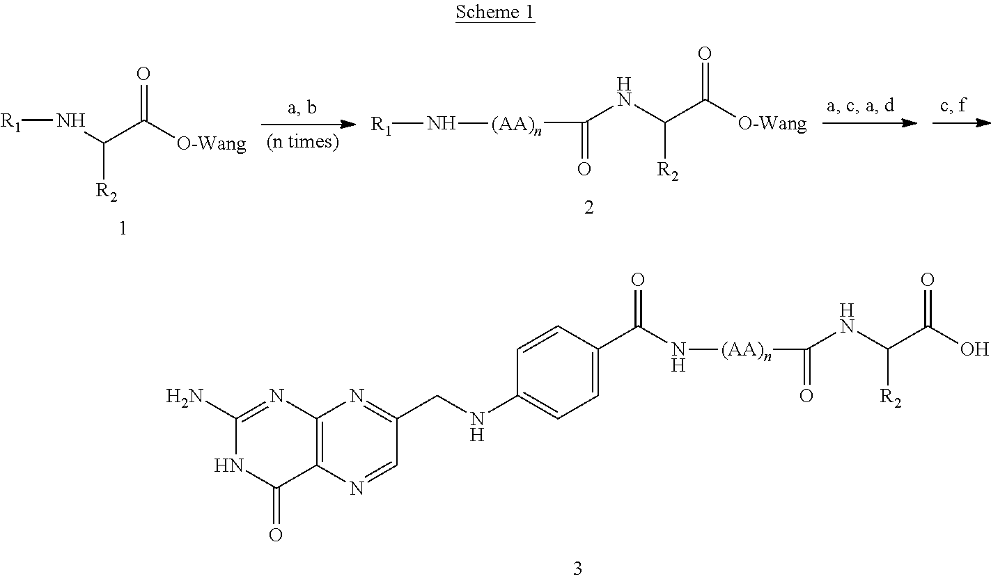
(a) 20% piperidine/DMF; (b) Fmoc-AA-OH, PyBop, DIPEA, DMF; (c) Fmoc-Glu(O-t-Bu)-OH, PyBop, DIPEA, DMF; (d) 1. N10(TFA)-Pte-OH; PyBop, DIPEA, DMSO; (e) TFAA, (CH2SH)2, i-Pr3SiH; (f) NH4OH, pH 10.3.


| TABLE 1 | |||||
| % | |||||
| Dose | Inhibition | ||||
| (nmol/ | in paw | % Weight | |||
| Treatment | kg) | Frequency | edema1 | Splenomagly2 | loss3 |
| Control | — | — | 0 | 117 ± 22 | −16 ± 1 |
| |
25* | |
0 ± 25* | 73 ± 8 | −17 ± 1 |
| (sc) | 100* | |
35 ± 11* | 52 ± 13 | −14 ± 1 |
| 250* | biw | 91 ± 4* | 25 ± 6 | −0.5 ± 3 | |
| 500 | biw | 91 ± 9 | 37 ± 7 | 0.4 ± 4 | |
| 1000 | |
72 ± 12 | 39 ± 5 | −7 ± 3 | |
| |
250 | |
70 ± 5 | 42 ± 17 | −14 ± 2 |
| (oral) | 1650 | qw | 47 ± 10† | — | −10 ± 2 |
| MTX (sc) | 250 | biw | 78 ± 10 | 24 ± 3 | −2 ± 4 |
| 500 | biw | 88 ± 7 | 20 ± 4 | −2 ± 7 | |
| |
10 mg/ | q3d | 46 ± 9 | 42 ± 7 | −15 ± 2 |
| (sc) | kg | ||||
| 1% Inhibition in paw edema is calculated based on paw weight on Day 24: 100 × (Arthritic control - Treated)/(Arthritic control - Healthy). | |||||
| 2Splenomagly is defined as % increase in spleen weight relative to the spleen weights of healthy rats. | |||||
| 3On |
|||||
| *Linear regression analysis showed an R-squared value of 0.997. | |||||
| †Calculated based on arthritis scores on Day 24 (paw weights were not obtained). | |||||


| Reagents | Mmol | equivalent | MW | Amount |
| H-Cys(4-methoxytrityl)- | 0.1 | 0.167 g | ||
| 2-chlorotrityl-Resin | ||||
| (loading 0.56 mmol/g) | ||||
| EC0475 | 0.13 | 1.3 | 612.67 | 0.080 g |
| Fmoc-Glu(OtBu)-OH | 0.2 | 2 | 425.5 | 0.085 g |
| EC0475 | 0.13 | 1.3 | 612.67 | 0.080 g |
| EC0475 | 0.13 | 1.3 | 612.67 | 0.080 g |
| Fmoc-Glu(OtBu)-OH | 0.2 | 2 | 425.5 | 0.085 g |
| EC0475 | 0.13 | 1.3 | 612.67 | 0.080 g |
| Fmoc-Glu-OtBu | 0.2 | 2 | 425.5 | 0.085 g |
| N10TFA-Pteroic | 0.2 | 2 | 408 | 0.105 g |
| Acid•TFA | ||||
| (dissolve in 10 ml | ||||
| DMSO) | ||||
| DIPEA | 0.4 | 4 | 129.25 | 0.070 mL |
| (d = 0.742) | ||||
| PyBOP | 0.2 | 2 | 520 | 0.104 g |
The Coupling steps, Cleavage step, and Cleavage Reagent were identical to those described above. HPLC Purification step: Column:

| Reagents | mmol | equivalent | MW | Amount |
| H-Cys(4-methoxytrityl)-2-chlorotrityl- | 0.094 | 0.16 g | ||
| Resin (loading 0.6 mmol/g) | ||||
| EC0475 | 0.13 | 1.4 | 612.67 | 0.082 g |
| Fmoc-Glu(OtBu)-OH | 0.19 | 2.0 | 425.47 | 0.080 g |
| EC0475 | 0.13 | 1.4 | 612.67 | 0.082 g |
| Fmoc-Arg(Pbf)-OH | 0.19 | 2.0 | 648.77 | 0.12 g |
| EC0475 | 0.13 | 1.4 | 612.67 | 0.082 g |
| Fmoc-Glu(OtBu)-OH | 0.19 | 2.0 | 425.47 | 0.080 g |
| EC0475 | 0.13 | 1.4 | 612.67 | 0.082 g |
| Fmoc-Glu-OtBu | 0.19 | 2.0 | 425.47 | 0.080 g |
| N10TFA-Pteroic Acid | 0.16 | 1.7 | 408.29 | 0.066 g |
| (dissolve in 10 ml DMSO) | ||||
| DIPEA | 2.0 eq of AA | 41 μL or 49 μL | ||
| PyBOP | 1.0 eq of AA | 122 mg or 147 mg | ||

| Reagents | mmol | equivalent | MW | amount |
| H-Cys(4-methoxytrityl)- | 0.10 | 0.17 g | ||
| 2-chlorotrityl-Resin | ||||
| (loading 0.6 mmol/g) | ||||
| EC0475 | 0.13 | 1.3 | 612.67 | 0.082 g |
| Fmoc-Glu(OtBu)-OH | 0.19 | 1.9 | 425.47 | 0.080 g |
| EC0475 | 0.13 | 1.3 | 612.67 | 0.082 g |
| Fmoc-Glu(OtBu)-OH | 0.19 | 1.9 | 425.47 | 0.080 g |
| EC0475 | 0.13 | 1.3 | 612.67 | 0.082 g |
| Fmoc-Glu-OtBu | 0.19 | 1.9 | 425.47 | 0.080 g |
| N10TFA-Pteroic Acid | 0.16 | 1.6 | 408.29 | 0.066 g |
| (dissolve in 10 ml DMSO) | ||||
| DIPEA | 2.0 eq of AA | |||
| PyBOP | 1.0 eq of AA | |||


Claims (20)
BLA


BLA


Priority Applications (2)
| Application Number | Priority Date | Filing Date | Title |
|---|---|---|---|
| US15/811,600 US10500204B2 (en) | 2007-06-25 | 2017-11-13 | Vitamin receptor drug delivery conjugates for treating inflammation |
| US16/682,200 US20200155555A1 (en) | 2007-06-25 | 2019-11-13 | Vitamin receptor drug delivery conjugates for treating inflammation |
Applications Claiming Priority (14)
| Application Number | Priority Date | Filing Date | Title |
|---|---|---|---|
| US94609207P | 2007-06-25 | 2007-06-25 | |
| US3618608P | 2008-03-13 | 2008-03-13 | |
| PCT/US2008/068093 WO2009002993A1 (en) | 2007-06-25 | 2008-06-25 | Conjugates containing hydrophilic spacer linkers |
| US28995209P | 2009-12-23 | 2009-12-23 | |
| US66671209A | 2009-12-24 | 2009-12-24 | |
| US29110309P | 2009-12-30 | 2009-12-30 | |
| US35103210P | 2010-06-03 | 2010-06-03 | |
| US37483010P | 2010-08-18 | 2010-08-18 | |
| US38678510P | 2010-09-27 | 2010-09-27 | |
| US39123010P | 2010-10-08 | 2010-10-08 | |
| PCT/US2010/061897 WO2011079227A1 (en) | 2009-12-23 | 2010-12-22 | Vitamin receptor drug delivery conjugates for treating inflammation |
| US201213518291A | 2012-06-21 | 2012-06-21 | |
| US14/671,790 US9877965B2 (en) | 2007-06-25 | 2015-03-27 | Vitamin receptor drug delivery conjugates for treating inflammation |
| US15/811,600 US10500204B2 (en) | 2007-06-25 | 2017-11-13 | Vitamin receptor drug delivery conjugates for treating inflammation |
Related Parent Applications (1)
| Application Number | Title | Priority Date | Filing Date |
|---|---|---|---|
| US14/671,790 Continuation US9877965B2 (en) | 2007-06-25 | 2015-03-27 | Vitamin receptor drug delivery conjugates for treating inflammation |
Related Child Applications (1)
| Application Number | Title | Priority Date | Filing Date |
|---|---|---|---|
| US16/682,200 Continuation US20200155555A1 (en) | 2007-06-25 | 2019-11-13 | Vitamin receptor drug delivery conjugates for treating inflammation |
Publications (2)
| Publication Number | Publication Date |
|---|---|
| US20180221372A1 US20180221372A1 (en) | 2018-08-09 |
| US10500204B2 true US10500204B2 (en) | 2019-12-10 |
Family
ID=54354404
Family Applications (3)
| Application Number | Title | Priority Date | Filing Date |
|---|---|---|---|
| US14/671,790 Active US9877965B2 (en) | 2007-06-25 | 2015-03-27 | Vitamin receptor drug delivery conjugates for treating inflammation |
| US15/811,600 Expired - Fee Related US10500204B2 (en) | 2007-06-25 | 2017-11-13 | Vitamin receptor drug delivery conjugates for treating inflammation |
| US16/682,200 Abandoned US20200155555A1 (en) | 2007-06-25 | 2019-11-13 | Vitamin receptor drug delivery conjugates for treating inflammation |
Family Applications Before (1)
| Application Number | Title | Priority Date | Filing Date |
|---|---|---|---|
| US14/671,790 Active US9877965B2 (en) | 2007-06-25 | 2015-03-27 | Vitamin receptor drug delivery conjugates for treating inflammation |
Family Applications After (1)
| Application Number | Title | Priority Date | Filing Date |
|---|---|---|---|
| US16/682,200 Abandoned US20200155555A1 (en) | 2007-06-25 | 2019-11-13 | Vitamin receptor drug delivery conjugates for treating inflammation |
Country Status (1)
| Country | Link |
|---|---|
| US (3) | US9877965B2 (en) |
Cited By (1)
| Publication number | Priority date | Publication date | Assignee | Title |
|---|---|---|---|---|
| US20190105399A1 (en) * | 2003-01-27 | 2019-04-11 | Endocyte, Inc. | Vitamin-Receptor Binding Drug Delivery Conjugates |
Families Citing this family (8)
| Publication number | Priority date | Publication date | Assignee | Title |
|---|---|---|---|---|
| US8288557B2 (en) | 2004-07-23 | 2012-10-16 | Endocyte, Inc. | Bivalent linkers and conjugates thereof |
| NZ599239A (en) | 2007-03-14 | 2013-10-25 | Endocyte Inc | Binding ligand linked drug delivery conjugates of tubulysins |
| US9877965B2 (en) | 2007-06-25 | 2018-01-30 | Endocyte, Inc. | Vitamin receptor drug delivery conjugates for treating inflammation |
| AU2008268432B2 (en) | 2007-06-25 | 2015-01-15 | Endocyte, Inc. | Conjugates containing hydrophilic spacer linkers |
| WO2013126797A1 (en) | 2012-02-24 | 2013-08-29 | Purdue Research Foundation | Cholecystokinin b receptor targeting for imaging and therapy |
| EP2908818A4 (en) | 2012-10-16 | 2016-07-13 | Endocyte Inc | Drug delivery conjugates containing unnatural amino acids and methods for using |
| AU2017238246B2 (en) * | 2016-03-24 | 2021-04-01 | The Administrators Of The Tulane Educational Fund | Conjugates of tacrolimus, their compositions, and their uses |
| US20190223771A1 (en) * | 2018-01-23 | 2019-07-25 | Medtronic Minimed, Inc. | Implantable polymer surfaces exhibiting reduced in vivo inflammatory responses |
Citations (228)
| Publication number | Priority date | Publication date | Assignee | Title |
|---|---|---|---|---|
| US2515483A (en) | 1946-08-10 | 1950-07-18 | Merck & Co Inc | Diacylated pteroic acid and process for preparing same |
| US2816110A (en) | 1956-11-23 | 1957-12-10 | Merck & Co Inc | Methods for the production of substituted pteridines |
| US3387001A (en) | 1964-10-19 | 1968-06-04 | Lilly Co Eli | Novel aminoacyl esters of desacetyl vincaleukoblastine |
| US3392173A (en) | 1964-03-09 | 1968-07-09 | Lilly Co Eli | Novel acyl derivatives of desacetyl-vincaleukoblastine and processes for their preparation |
| US3632622A (en) | 1969-04-01 | 1972-01-04 | Chevron Res | Polyhaloalkylpolythioalkyl sulfite esters |
| US3641109A (en) | 1968-09-04 | 1972-02-08 | Carl D Emerson | Alkyl and aryl esters of polyhalo-dithio alcohols |
| US4166810A (en) | 1978-04-20 | 1979-09-04 | Eli Lilly And Company | Derivatives of 4-desacetyl VLB C-3 carboxyhydrazide |
| US4203898A (en) | 1977-08-29 | 1980-05-20 | Eli Lilly And Company | Amide derivatives of VLB, leurosidine, leurocristine and related dimeric alkaloids |
| US4316885A (en) | 1980-08-25 | 1982-02-23 | Ayerst, Mckenna And Harrison, Inc. | Acyl derivatives of rapamycin |
| US4337339A (en) | 1979-04-30 | 1982-06-29 | Baker Instruments Corp. | Process for preparation of folic acid derivatives |
| EP0116208A1 (en) | 1982-12-07 | 1984-08-22 | Kyowa Hakko Kogyo Co., Ltd. | Mitomycin analogues |
| JPS59175493A (en) | 1983-03-25 | 1984-10-04 | Kyowa Hakko Kogyo Co Ltd | Mitomycin derivative and its preparation |
| EP0163550A2 (en) | 1984-06-01 | 1985-12-04 | Kyowa Hakko Kogyo Kabushiki Kaisha | Mitomycin derivatives, preparation thereof and pharmaceutical compositions containing them |
| WO1985005554A1 (en) | 1984-05-31 | 1985-12-19 | Amersham International Plc | Nmr contrast agents |
| US4639456A (en) | 1980-06-10 | 1987-01-27 | Omnichem S.A. | Vinblastin-23-oyl amino acid derivatives |
| US4650803A (en) | 1985-12-06 | 1987-03-17 | University Of Kansas | Prodrugs of rapamycin |
| EP0247792A2 (en) | 1986-05-27 | 1987-12-02 | Eli Lilly And Company | Immunoglobulin conjugates |
| US4713249A (en) | 1981-11-12 | 1987-12-15 | Schroeder Ulf | Crystallized carbohydrate matrix for biologically active substances, a process of preparing said matrix, and the use thereof |
| WO1988001622A1 (en) | 1986-08-29 | 1988-03-10 | Kyowa Hakko Kogyo Kabusiki Kaisha | Mitomycin derivatives |
| US4801688A (en) | 1986-05-27 | 1989-01-31 | Eli Lilly And Company | Hydrazone immunoglobulin conjugates |
| US4866180A (en) | 1984-02-24 | 1989-09-12 | Bristol-Myers Company | Amino disulfide thiol exchange products |
| US4870162A (en) | 1983-04-29 | 1989-09-26 | Omnichem | Conjugates of vinblastine, a process for their preparation and their use in therapy |
| EP0354728A2 (en) | 1988-08-08 | 1990-02-14 | Eli Lilly And Company | Cytotoxic drug conjugates |
| WO1990012096A1 (en) | 1989-04-03 | 1990-10-18 | Purdue Research Foundation | Method for enhancing transmembrane transport of exogenous molecules |
| WO1991007418A1 (en) | 1989-11-13 | 1991-05-30 | Xoma Corporation | Chimeric mouse-human a10 antibody with specificity to a human tumor cell antigen |
| US5094849A (en) | 1988-08-08 | 1992-03-10 | Eli Lilly And Company | Cytotoxic antibody conjugates of hydrazide derivatized vinca analogs via simple organic linkers |
| US5100883A (en) | 1991-04-08 | 1992-03-31 | American Home Products Corporation | Fluorinated esters of rapamycin |
| US5118677A (en) | 1991-05-20 | 1992-06-02 | American Home Products Corporation | Amide esters of rapamycin |
| US5118678A (en) | 1991-04-17 | 1992-06-02 | American Home Products Corporation | Carbamates of rapamycin |
| US5120842A (en) | 1991-04-01 | 1992-06-09 | American Home Products Corporation | Silyl ethers of rapamycin |
| US5130307A (en) | 1990-09-28 | 1992-07-14 | American Home Products Corporation | Aminoesters of rapamycin |
| US5138051A (en) | 1991-08-07 | 1992-08-11 | American Home Products Corporation | Rapamycin analogs as immunosuppressants and antifungals |
| US5140104A (en) | 1982-03-09 | 1992-08-18 | Cytogen Corporation | Amine derivatives of folic acid analogs |
| US5151413A (en) | 1991-11-06 | 1992-09-29 | American Home Products Corporation | Rapamycin acetals as immunosuppressant and antifungal agents |
| US5169851A (en) | 1991-08-07 | 1992-12-08 | American Home Products Corporation | Rapamycin analog as immunosuppressants and antifungals |
| US5194447A (en) | 1992-02-18 | 1993-03-16 | American Home Products Corporation | Sulfonylcarbamates of rapamycin |
| US5221670A (en) | 1990-09-19 | 1993-06-22 | American Home Products Corporation | Rapamycin esters |
| US5233036A (en) | 1990-10-16 | 1993-08-03 | American Home Products Corporation | Rapamycin alkoxyesters |
| US5258389A (en) | 1992-11-09 | 1993-11-02 | Merck & Co., Inc. | O-aryl, O-alkyl, O-alkenyl and O-alkynylrapamycin derivatives |
| US5260300A (en) | 1992-11-19 | 1993-11-09 | American Home Products Corporation | Rapamycin carbonate esters as immuno-suppressant agents |
| US5266333A (en) | 1985-03-06 | 1993-11-30 | American Cyanamid Company | Water dispersible and water soluble carbohydrate polymer compositions for parenteral administration of growth hormone |
| US5302584A (en) | 1992-10-13 | 1994-04-12 | American Home Products Corporation | Carbamates of rapamycin |
| US5362718A (en) | 1994-04-18 | 1994-11-08 | American Home Products Corporation | Rapamycin hydroxyesters |
| US5378696A (en) | 1990-09-19 | 1995-01-03 | American Home Products Corporation | Rapamycin esters |
| US5385908A (en) | 1993-11-22 | 1995-01-31 | American Home Products Corporation | Hindered esters of rapamycin |
| US5385910A (en) | 1993-11-22 | 1995-01-31 | American Home Products Corporation | Gem-distributed esters of rapamycin |
| US5385909A (en) | 1993-11-22 | 1995-01-31 | American Home Products Corporation | Heterocyclic esters of rapamycin |
| US5389639A (en) | 1993-12-29 | 1995-02-14 | American Home Products Company | Amino alkanoic esters of rapamycin |
| US5391730A (en) | 1993-10-08 | 1995-02-21 | American Home Products Corporation | Phosphorylcarbamates of rapamycin and oxime derivatives thereof |
| US5417982A (en) | 1994-02-17 | 1995-05-23 | Modi; Pankaj | Controlled release of drugs or hormones in biodegradable polymer microspheres |
| WO1995015335A2 (en) | 1993-11-22 | 1995-06-08 | Neorx Corporation | Pretargeting methods and compounds |
| US5463048A (en) | 1994-06-14 | 1995-10-31 | American Home Products Corporation | Rapamycin amidino carbamates |
| US5491231A (en) | 1994-11-28 | 1996-02-13 | American Home Products Corporation | Hindered N-oxide esters of rapamycin |
| US5547668A (en) | 1995-05-05 | 1996-08-20 | The Board Of Trustees Of The University Of Illinois | Conjugates of folate anti-effector cell antibodies |
| US5552545A (en) | 1991-12-20 | 1996-09-03 | Eli Lilly And Company | 5-deaza-10-oxo-and 5-deaza-10-thio-5,6,7,8-tetrahydrofolic acids |
| US5562907A (en) | 1993-05-14 | 1996-10-08 | Arnon; Stephen S. | Method to prevent side-effects and insensitivity to the therapeutic uses of toxins |
| WO1996036367A1 (en) | 1995-05-16 | 1996-11-21 | Purdue Research Foundation | Composition and method for tumor imaging |
| US5583020A (en) | 1992-11-24 | 1996-12-10 | Ribozyme Pharmaceuticals, Inc. | Permeability enhancers for negatively charged polynucleotides |
| US5627165A (en) | 1990-06-13 | 1997-05-06 | Drug Innovation & Design, Inc. | Phosphorous prodrugs and therapeutic delivery systems using same |
| US5672486A (en) | 1990-08-29 | 1997-09-30 | Centre Hospitalier Regional De Nantes | Protein polyligands joined to a stable protein core |
| WO1998008382A1 (en) | 1996-08-30 | 1998-03-05 | Eli Lilly And Company | Nonclassical pyrrolo[2,3-d]pyrimidine antifolates |
| WO1998008859A1 (en) | 1996-08-27 | 1998-03-05 | University Of Utah Research Foundation | Bioconjugates and delivery of bioactive agents |
| WO1998010651A1 (en) | 1996-09-12 | 1998-03-19 | Merck & Co., Inc. | Conjugates useful in the treatment of prostate cancer |
| WO1999020626A1 (en) | 1997-10-17 | 1999-04-29 | Purdue Research Foundation | Folic acid derivatives |
| WO1999061055A1 (en) | 1998-05-22 | 1999-12-02 | The Board Of Trustees Of The Leland Stanford Junior University | Bifunctional molecules and therapies based thereon |
| US5998603A (en) | 1994-09-29 | 1999-12-07 | Isis Pharmaceuticals, Inc. | 4'-desmethyl nucleoside analogs, and oligomers thereof |
| US6004555A (en) | 1992-03-05 | 1999-12-21 | Board Of Regents, The University Of Texas System | Methods for the specific coagulation of vasculature |
| US6030941A (en) | 1996-05-01 | 2000-02-29 | Avi Biopharma, Inc. | Polymer composition for delivering substances in living organisms |
| US6056973A (en) | 1996-10-11 | 2000-05-02 | Sequus Pharmaceuticals, Inc. | Therapeutic liposome composition and method of preparation |
| US6077499A (en) | 1996-05-03 | 2000-06-20 | Immunomedics, Inc. | Targeted combination immunotherapy of cancer |
| WO2000035422A2 (en) | 1998-12-18 | 2000-06-22 | Hadasit Medical Research Services & Development Ltd. | Method of administering a compound to multi-drug resistant cells |
| US6093382A (en) | 1998-05-16 | 2000-07-25 | Bracco Research Usa Inc. | Metal complexes derivatized with folate for use in diagnostic and therapeutic applications |
| CA2372841A1 (en) | 1999-05-04 | 2000-11-09 | Gregory John Russell-Jones | Amplification of folate-mediated targeting to tumor cells using polymers |
| CA2376175A1 (en) | 1999-06-02 | 2000-12-14 | Biotech Australia Pty Limited | Vitamin directed dual targeting therapy |
| US6171859B1 (en) | 1994-03-30 | 2001-01-09 | Mitokor | Method of targeting conjugate molecules to mitochondria |
| US6171614B1 (en) | 1996-10-15 | 2001-01-09 | Emory University | Synthesis of glycophospholipid and peptide-phospholipid conjugates and uses thereof |
| US6177404B1 (en) | 1996-10-15 | 2001-01-23 | Merck & Co., Inc. | Conjugates useful in the treatment of benign prostatic hyperplasia |
| US6184042B1 (en) | 1996-05-24 | 2001-02-06 | Boehringer Mannheim Gmbh | Method for reducing hook effect in an immunoassay |
| WO2001013957A2 (en) | 1999-08-24 | 2001-03-01 | Cellgate, Inc. | Enhancing drug delivery across and into epithelial tissues using oligo arginine moieties |
| US6207157B1 (en) | 1996-04-23 | 2001-03-27 | The United States Of America As Represented By The Department Of Health And Human Services | Conjugate vaccine for nontypeable Haemophilus influenzae |
| WO2001028592A1 (en) | 1999-10-15 | 2001-04-26 | Mayo Foundation For Medical Education And Research | Cobalamin conjugates useful as imaging agents and as antitumor agents |
| US6291684B1 (en) | 1999-03-29 | 2001-09-18 | Bristol-Myers Squibb Company | Process for the preparation of aziridinyl epothilones from oxiranyl epothilones |
| US6290929B1 (en) | 2000-07-28 | 2001-09-18 | The Procter & Gamble Company | Cancer treatment |
| WO2001074382A1 (en) | 2000-03-31 | 2001-10-11 | Purdue Research Foundation | Method of treatment using ligand-immunogen conjugates |
| WO2001082975A2 (en) | 2000-04-25 | 2001-11-08 | Washington University | Membrane-permeant peptide complexes for medical imaging |
| US6335434B1 (en) | 1998-06-16 | 2002-01-01 | Isis Pharmaceuticals, Inc., | Nucleosidic and non-nucleosidic folate conjugates |
| US6340701B1 (en) | 1999-11-24 | 2002-01-22 | Immunogen Inc | Cytotoxic agents comprising taxanes and their therapeutic use |
| US6342244B1 (en) | 1999-04-23 | 2002-01-29 | Alza Corporation | Releasable linkage and compositions containing same |
| US6344452B1 (en) | 1998-06-05 | 2002-02-05 | Zeria Pharmaceuticals Co., Ltd. | 1,5-benzodiazepine derivatives |
| US6399638B1 (en) | 1998-04-21 | 2002-06-04 | Bristol-Myers Squibb Company | 12,13-modified epothilone derivatives |
| US6399626B1 (en) | 2000-10-02 | 2002-06-04 | Wyeth | Hydroxyesters of 7-desmethylrapamycin |
| US6399625B1 (en) | 2000-09-27 | 2002-06-04 | Wyeth | 1-oxorapamycins |
| WO2002059272A2 (en) | 2001-01-24 | 2002-08-01 | The Trustees Of Columbia University In The City Of New York | Covalent chemical inducers of protein dimerization and their uses in high throughput binding screens |
| US6432973B1 (en) | 2000-09-19 | 2002-08-13 | Wyeth | Water soluble rapamycin esters |
| US6440991B1 (en) | 2000-10-02 | 2002-08-27 | Wyeth | Ethers of 7-desmethlrapamycin |
| US20020151088A1 (en) | 1993-04-23 | 2002-10-17 | Molnar-Kimber Katherine L. | Rapamycin conjugates |
| WO2002085908A1 (en) | 2001-04-24 | 2002-10-31 | Purdue Research Foundation | Folate mimetics and folate-receptor binding conjugates thereof |
| WO2002087424A2 (en) | 2001-05-02 | 2002-11-07 | Purdue Research Foundation | Treatment and diagnosis of macrophage mediated disease |
| WO2002098868A1 (en) | 2001-06-01 | 2002-12-12 | Bristol-Myers Squibb Company | Epothilone derivatives |
| US6511986B2 (en) | 2000-08-11 | 2003-01-28 | Wyeth | Method of treating estrogen receptor positive carcinoma |
| US20030022831A1 (en) | 1999-08-24 | 2003-01-30 | Cellgate, Inc., A Delaware Corporation | Compositions and methods for enhancing drug delivery across and into ocular tissues |
| US6541612B2 (en) | 1993-04-23 | 2003-04-01 | Wyeth | Monoclonal antibodies obtained using rapamycin position 27 conjugates as an immunogen |
| US6548505B1 (en) | 1995-07-28 | 2003-04-15 | Peter Maccallum Cancer Institute | Radioprotectors |
| US20030086900A1 (en) | 2001-09-28 | 2003-05-08 | Low Philip S. | Method of treatment using ligand-immunogen conjugates |
| WO2003050295A2 (en) | 2001-12-12 | 2003-06-19 | Conforma Therapeutics Corporation | Assays and implements for determining and modulating hsp90 binding activity |
| US6596757B1 (en) | 2002-05-14 | 2003-07-22 | Immunogen Inc. | Cytotoxic agents comprising polyethylene glycol-containing taxanes and their therapeutic use |
| US20030162234A1 (en) | 2002-02-07 | 2003-08-28 | Jallad Karim N. | Folate targeted enhanced tumor and folate receptor positive tissue optical imaging technology |
| US6617333B2 (en) | 2001-08-07 | 2003-09-09 | Wyeth | Antineoplastic combinations comprising |
| US20030194409A1 (en) | 2002-01-17 | 2003-10-16 | Rothman James E. | Conjugate heat shock protein-binding peptides |
| WO2003092742A1 (en) | 2002-05-06 | 2003-11-13 | Endocyte, Inc. | Vitamin-targeted imaging agents |
| WO2003097647A1 (en) | 2002-05-15 | 2003-11-27 | Endocyte, Inc. | Vitamin-mitomycin conjugates |
| US6670355B2 (en) | 2000-06-16 | 2003-12-30 | Wyeth | Method of treating cardiovascular disease |
| US6677357B2 (en) | 2001-08-22 | 2004-01-13 | Wyeth | Rapamycin 29-enols |
| WO2004005326A2 (en) | 2002-07-09 | 2004-01-15 | Morphochem Aktiengellschaft Fu | Tubulysin conjugates |
| WO2004005327A1 (en) | 2002-07-09 | 2004-01-15 | Morphochem Ag Komb Chemie | Novel tubulysin analogues |
| US6680330B2 (en) | 2001-08-22 | 2004-01-20 | Wyeth | Rapamycin dialdehydes |
| US20040018203A1 (en) | 2001-06-08 | 2004-01-29 | Ira Pastan | Pegylation of linkers improves antitumor activity and reduces toxicity of immunoconjugates |
| WO2004010957A2 (en) | 2002-07-31 | 2004-02-05 | Seattle Genetics, Inc. | Drug conjugates and their use for treating cancer, an autoimmune disease or an infectious disease |
| WO2004012735A2 (en) | 2002-07-31 | 2004-02-12 | Schering Ag | New effector conjugates, process for their production and their pharmaceutical use |
| US20040047917A1 (en) | 2002-09-06 | 2004-03-11 | Stephen Wilson | Drug delivery and targeting with vitamin B12 conjugates |
| WO2004022099A2 (en) | 2002-09-06 | 2004-03-18 | Insert Therapeutics, Inc. | Cyclodextrin-based polymers for delivering the therapeutic agents covalently bound thereto |
| US6713607B2 (en) | 1994-03-08 | 2004-03-30 | Wyeth | Effector proteins of Rapamycin |
| WO2004037210A2 (en) | 2002-10-24 | 2004-05-06 | Research Corporation Technologies | Functional mri agents for cancer imaging |
| WO2004046170A2 (en) | 2002-11-21 | 2004-06-03 | Gesellschaft Fuer Biotechnologische Forschung Mbh (Gbf) | Tubulysins, method for producing the same and tubulysin preparations |
| US6747022B2 (en) | 1999-12-02 | 2004-06-08 | Zeria Pharmaceutical Co., Ltd. | Calcium salts of 1,5-benzodiazepine derivatives, process for producing the salts and drugs containing the same |
| WO2004054622A1 (en) | 2002-12-13 | 2004-07-01 | Immunomedics, Inc. | Immunoconjugates with an intracellularly-cleavable linkage |
| WO2004069159A2 (en) | 2003-01-27 | 2004-08-19 | Endocyte, Inc. | Vitamin receptor binding drug delivery conjugates |
| US6821731B2 (en) | 2000-11-28 | 2004-11-23 | Wyeth | Expression analysis of FKBP nucleic acids and polypeptides useful in the diagnosis of prostate cancer |
| US20050026068A1 (en) | 2001-11-01 | 2005-02-03 | Evangelos Gogolides | Polycarbocyclic derivatives for modification of resist, optical and etch resistance properties |
| US20050107325A1 (en) | 2003-04-17 | 2005-05-19 | Muthiah Manoharan | Modified iRNA agents |
| US6915855B2 (en) | 2002-05-02 | 2005-07-12 | Halliburton Energy Services, Inc. | Wellbore junction drifting apparatus and associated method |
| WO2005074901A2 (en) | 2004-01-30 | 2005-08-18 | Schering Ag | New effector conjugates, process for their production and their pharmaceutical use |
| US6958153B1 (en) | 1997-11-07 | 2005-10-25 | Wyeth | Skin penetration enhancing components |
| US20050239739A1 (en) | 2001-05-18 | 2005-10-27 | Sirna Therapeutics, Inc. | Conjugates and compositions for cellular delivery |
| WO2005112919A2 (en) | 2004-05-19 | 2005-12-01 | Medarex, Inc. | Self-immolative linkers and drug conjugates |
| WO2005115912A1 (en) | 2004-05-25 | 2005-12-08 | Matsushita Electric Industrial Co., Ltd. | Hydrogen production apparatus and fuel cell system using the same |
| US20060019911A1 (en) | 1999-08-09 | 2006-01-26 | The General Hospital Corporation | Drug-carrier complexes and methods of use thereof |
| WO2006012527A1 (en) | 2004-07-23 | 2006-02-02 | Endocyte, Inc. | Bivalent linkers and conjugates thereof |
| US20060058266A1 (en) | 2004-08-10 | 2006-03-16 | Muthiah Manoharan | Chemically modified oligonucleotides |
| US7019014B2 (en) | 2003-05-12 | 2006-03-28 | Wyeth Holdings Corporation | Process for producing anticancer agent LL-D45042 |
| US7029674B2 (en) | 2001-04-02 | 2006-04-18 | Wyeth | Methods for downmodulating immune cells using an antibody to PD-1 |
| WO2006042146A2 (en) | 2004-10-07 | 2006-04-20 | Emory University | Multifunctional nanoparticles conjugates and their use |
| US20060105975A1 (en) | 2004-04-19 | 2006-05-18 | Shannon Pendergrast | Aptamer-mediated intracellular delivery of therapeutic oligonucleotides |
| US7060709B2 (en) | 2003-02-06 | 2006-06-13 | Wyeth | Method of treating hepatic fibrosis |
| US7060797B2 (en) | 2002-11-21 | 2006-06-13 | Wyeth | Composition and method for treating lupus nephritis |
| US7067111B1 (en) | 1999-10-25 | 2006-06-27 | Board Of Regents, University Of Texas System | Ethylenedicysteine (EC)-drug conjugates, compositions and methods for tissue specific disease imaging |
| US7074804B2 (en) | 2003-07-16 | 2006-07-11 | Wyeth | CCI-779 Isomer C |
| WO2006089007A2 (en) | 2005-02-16 | 2006-08-24 | Insert Therapeutics, Inc. | Cyclodextrin-based polymers containing photocleavable linkers for drug delivery |
| US7105328B2 (en) | 2001-04-02 | 2006-09-12 | Dana-Farber Cancer Institute | Methods for screening for compounds that modulate pd-1 signaling |
| WO2006101845A2 (en) | 2005-03-16 | 2006-09-28 | Endocyte, Inc. | Synthesis and purification of pteroic acid and conjugates thereof |
| US20060217360A1 (en) | 2002-09-05 | 2006-09-28 | Gerhard Hoefle | Tubulysin biosynthesis gene |
| WO2006105141A1 (en) | 2005-03-30 | 2006-10-05 | Purdue Research Foundation | Method for cancer prognosis using cellular folate vitamin receptor quantification |
| US20060222653A1 (en) | 2004-11-12 | 2006-10-05 | Xencor, Inc. | Antibodies operably linked to selected chemoattractants |
| US7122361B2 (en) | 2002-10-10 | 2006-10-17 | Wyeth | Compositions employing a novel human kinase |
| US7153957B2 (en) | 2003-08-07 | 2006-12-26 | Wyeth | Regioselective synthesis of CCI-779 |
| WO2007002222A2 (en) | 2005-06-20 | 2007-01-04 | Psma Development Company, Llc | Psma antibody-drug conjugates |
| US20070009434A1 (en) | 2005-07-05 | 2007-01-11 | Low Philip S | Imaging and therapeutic method using monocytes |
| WO2007022493A2 (en) | 2005-08-19 | 2007-02-22 | Endocyte, Inc. | Ligand conjugates of vinca alkaloids, analogs, and derivatives |
| WO2007022494A2 (en) | 2005-08-19 | 2007-02-22 | Endocyte, Inc. | Multi-drug ligand conjugates |
| WO2007022512A2 (en) | 2005-08-19 | 2007-02-22 | Neose Technologies, Inc. | Glycopegylated factor vii and factor viia |
| US20070134332A1 (en) | 2005-11-21 | 2007-06-14 | Medivas, Llc | Polymer particles for delivery of macromolecules and methods of use |
| US7238368B2 (en) | 1999-04-23 | 2007-07-03 | Alza Corporation | Releasable linkage and compositions containing same |
| US20070275904A1 (en) | 2006-05-25 | 2007-11-29 | Bristol-Myers Squibb Company | Conjugates of aziridinyl-epothilone analogs and pharmaceutical compositions comprising same |
| US20080096893A1 (en) | 2005-03-11 | 2008-04-24 | Syntrix Biosystems, Inc. | Aminopterin dosage forms |
| WO2008057437A2 (en) | 2006-11-03 | 2008-05-15 | Purdue Research Foundation | Ex vivo flow cytometry method and device |
| WO2008101231A2 (en) | 2007-02-16 | 2008-08-21 | Endocyte, Inc. | Methods and compositions for treating and diagnosing kidney disease |
| WO2008112873A2 (en) | 2007-03-14 | 2008-09-18 | Endocyte, Inc. | Binding ligand linked drug delivery conjugates of tubulysins |
| WO2009002993A1 (en) | 2007-06-25 | 2008-12-31 | Endocyte, Inc. | Conjugates containing hydrophilic spacer linkers |
| WO2009026177A1 (en) | 2007-08-17 | 2009-02-26 | Purdue Research Foundation | Psma binding ligand-linker conjugates and methods for using |
| WO2009055562A1 (en) | 2007-10-25 | 2009-04-30 | Endocyte, Inc. | Tubulysins and processes for preparing |
| WO2009117531A1 (en) | 2008-03-18 | 2009-09-24 | Seattle Genetics, Inc. | Auristatin drug linker conjugates |
| US20090247614A1 (en) | 2007-12-04 | 2009-10-01 | Alnylam Pharmaceuticals, Inc. | Folate Conjugates |
| US20100040669A1 (en) | 2008-08-12 | 2010-02-18 | Higuchi John W | Non-Invasive Ocular Delivery of Rapamycin |
| WO2010033733A1 (en) | 2008-09-17 | 2010-03-25 | Endocyte, Inc. | Folate receptor binding conjugates of antifolates |
| US20100074863A1 (en) | 2008-09-17 | 2010-03-25 | Yat Sun Or | Anti-infective pyrrolidine derivatives and analogs |
| WO2010045598A2 (en) | 2008-10-17 | 2010-04-22 | Purdue Research Foundation | Psma binding ligand-linker conjugates and methods for using |
| US20100129314A1 (en) | 2008-04-30 | 2010-05-27 | Immunogen Inc. | Potent conjugates and hydrophilic linkers |
| US20100144647A1 (en) | 2007-02-16 | 2010-06-10 | Ktb Tumorforschungsgesellschaft Mbh | Dual Acting Prodrugs |
| US20110027274A1 (en) | 2009-08-03 | 2011-02-03 | Medarex, Inc. | Antiproliferative compounds, conjugates thereof, methods therefor, and uses thereof |
| WO2011069116A1 (en) | 2009-12-04 | 2011-06-09 | Endocyte, Inc. | Binding ligand linked drug delivery conjugates of tubulysins |
| US20110166319A1 (en) | 2005-02-11 | 2011-07-07 | Immunogen, Inc. | Process for preparing purified drug conjugates |
| WO2011106639A1 (en) | 2010-02-25 | 2011-09-01 | Purdue Research Foundation | Psma binding ligand-linker conjugates and methods for using |
| US20110245295A1 (en) | 2008-09-25 | 2011-10-06 | Universitat Des Saarlandes | Bioactive pre-tubulysins and use thereof |
| WO2011130598A1 (en) | 2010-04-15 | 2011-10-20 | Spirogen Limited | Pyrrolobenzodiazepines and conjugates thereof |
| US20120022245A1 (en) | 2008-10-17 | 2012-01-26 | Purdue Research Foundation | Folate targeting of nucleotides |
| WO2012019123A1 (en) | 2010-08-06 | 2012-02-09 | Endocyte, Inc. | Processes for preparing tubulysins |
| WO2012047525A2 (en) | 2010-09-27 | 2012-04-12 | Endocyte, Inc. | Folate conjugates for treating inflammation of the eye |
| US20120122893A1 (en) | 2010-11-12 | 2012-05-17 | Joseph Anand Reddy | Methods of treating cancer |
| US20120129779A1 (en) | 2007-05-10 | 2012-05-24 | R&D-Biopharmaceuticals Gmbh | Tubulysine derivatives |
| WO2012090104A1 (en) | 2010-12-31 | 2012-07-05 | Kareus Therapeutics, Sa | Methods and compositions for designing novel conjugate therapeutics |
| WO2012112687A1 (en) | 2011-02-15 | 2012-08-23 | Immunogen, Inc. | Methods of preparation of conjugates |
| US20120252739A1 (en) | 2009-11-12 | 2012-10-04 | R&D Biopharmaceuticals Gmbh | Tubulin Inhibitors |
| US20120252738A1 (en) | 2009-11-12 | 2012-10-04 | R & D Biopharmaceuticals Gmbh | Tubulin Inhibitors |
| US20120258905A1 (en) | 2009-12-23 | 2012-10-11 | Endocyte, Inc. | Vitamin receptor drug delivery conjugates for treating inflammation |
| US20120259100A1 (en) | 2011-03-29 | 2012-10-11 | Immunogen, Inc. | Process for manufacturing conjugates of improved homogeneity |
| US8349901B2 (en) | 2004-08-26 | 2013-01-08 | Piramal Enterprises Limited and Apparao Satyam | Prodrugs containing novel bio-cleavable linkers |
| US20130029900A1 (en) | 2011-06-21 | 2013-01-31 | Immunogen, Inc. | Novel maytansinoid derivatives with peptide linker and conjugates thereof |
| US8383122B2 (en) | 2005-08-24 | 2013-02-26 | Immunogen, Inc. | Process for preparing purified drug conjugates |
| US8476451B2 (en) | 2007-07-20 | 2013-07-02 | The Regents Of The University Of California | Tubulysin D analogues |
| US8497365B2 (en) | 2007-01-24 | 2013-07-30 | Mark E. Davis | Cyclodextrin-based polymers for therapeutics delivery |
| US20130217638A1 (en) | 2010-07-19 | 2013-08-22 | Leibniz-Institut fur Pflanzenbiochemie | Tubulysin Analogues |
| US20130224228A1 (en) | 2011-12-05 | 2013-08-29 | Igenica, Inc. | Antibody-Drug Conjugates and Related Compounds, Compositions, and Methods |
| WO2013126797A1 (en) | 2012-02-24 | 2013-08-29 | Purdue Research Foundation | Cholecystokinin b receptor targeting for imaging and therapy |
| WO2013130776A1 (en) | 2012-02-29 | 2013-09-06 | Purdue Research Foundation | Folate receptor alpha binding ligands |
| US20130252904A1 (en) | 2012-02-29 | 2013-09-26 | Endocyte, Inc. | Compositions and methods for treating cancer |
| WO2013149185A1 (en) | 2012-03-29 | 2013-10-03 | Endocyte, Inc. | Processes for preparing tubulysin derivatives and conjugates thereof |
| US8580820B2 (en) | 2009-07-22 | 2013-11-12 | Kemtech S.R.L. | Tubulysin compounds with high cytotoxicity, pharmaceutical compositions thereof, and method of use thereof |
| WO2013170272A2 (en) | 2012-05-11 | 2013-11-14 | Alexander Krantz | Site-specific labeling and targeted delivery of proteins for the treatment of cancer |
| WO2013173392A1 (en) | 2012-05-15 | 2013-11-21 | Concortis Biosystems, Corp | Drug-conjugates, conjugation methods, and uses thereof |
| WO2014009774A1 (en) | 2012-07-12 | 2014-01-16 | Hangzhou Dac Biotech Co., Ltd | Conjugates of cell binding molecules with cytotoxic agents |
| WO2014040752A1 (en) | 2012-09-17 | 2014-03-20 | Ontochem Gmbh | Receptor ligand linked cytotoxic molecules |
| US20140107316A1 (en) | 2012-10-16 | 2014-04-17 | Endocyte, Inc. | Drug delivery conjugates containing unnatural amino acids and methods for using |
| WO2014062697A2 (en) | 2012-10-16 | 2014-04-24 | Endocyte, Inc. | Drug delivery conjugates containing unnatural amino acids and methods for using |
| WO2014078484A1 (en) | 2012-11-15 | 2014-05-22 | Endocyte, Inc. | Conjugates for treating diseases caused by psma expressing cells |
| WO2014080251A1 (en) | 2012-11-24 | 2014-05-30 | Hangzhou Dac Biotech Co., Ltd. | Hydrophilic linkers and their uses for conjugation of drugs to cell binding molecules |
| US20140154702A1 (en) | 2012-11-30 | 2014-06-05 | Endocyte, Inc. | Methods For Treating Cancer Using Combination Therapies |
| US20140193437A1 (en) | 2012-12-21 | 2014-07-10 | Bioalliance C.V. | Hydrophilic self-immolative linkers and conjugates thereof |
| US20140227295A1 (en) | 2013-02-14 | 2014-08-14 | Bristol-Myers Squibb Company | Tubulysin compounds, methods of making and use |
| US20140249315A1 (en) | 2013-03-01 | 2014-09-04 | Endocyte, Inc. | Processes for preparing tubulysins |
| US20140309406A1 (en) | 2011-03-29 | 2014-10-16 | Immunogen, Inc. | Preparation of maytansinoid antibody conjugates by a one-step process |
| US9061995B2 (en) | 2008-04-30 | 2015-06-23 | Immunogen, Inc. | Cross-linkers and their uses |
| WO2015106599A1 (en) | 2014-01-20 | 2015-07-23 | Brightgene Bio-Medical Technology Co., Ltd. | Conjugates and compositions for drug delivery |
| US20150284416A1 (en) | 2015-06-16 | 2015-10-08 | Suzhou M-Conj Biotech Co., Ltd | Novel linkers for conjugation of cell-binding molecules |
| US20150314015A1 (en) | 2007-06-25 | 2015-11-05 | Endocyte, Inc. | Vitamin receptor drug delivery conjugates for treating inflammation |
| US9295731B2 (en) | 2013-04-01 | 2016-03-29 | Mark Quang Nguyen | Cleavable drug conjugates, compositions thereof and methods of use |
| US20160144050A1 (en) | 2014-11-20 | 2016-05-26 | Sang-Yoon Kim | Prodrugs activated by caspase |
| WO2016147031A2 (en) | 2015-03-19 | 2016-09-22 | Hangzhou Dac Biotech Co., Ltd | Novel hydrophilic linkers and ligand-drug conjugates thereof |
| US20170290878A1 (en) | 2014-09-25 | 2017-10-12 | Endocyte, Inc. | Methods of treating cancer with tubulysin conjugates |
-
2015
- 2015-03-27 US US14/671,790 patent/US9877965B2/en active Active
-
2017
- 2017-11-13 US US15/811,600 patent/US10500204B2/en not_active Expired - Fee Related
-
2019
- 2019-11-13 US US16/682,200 patent/US20200155555A1/en not_active Abandoned
Patent Citations (315)
| Publication number | Priority date | Publication date | Assignee | Title |
|---|---|---|---|---|
| US2515483A (en) | 1946-08-10 | 1950-07-18 | Merck & Co Inc | Diacylated pteroic acid and process for preparing same |
| US2816110A (en) | 1956-11-23 | 1957-12-10 | Merck & Co Inc | Methods for the production of substituted pteridines |
| US3392173A (en) | 1964-03-09 | 1968-07-09 | Lilly Co Eli | Novel acyl derivatives of desacetyl-vincaleukoblastine and processes for their preparation |
| US3387001A (en) | 1964-10-19 | 1968-06-04 | Lilly Co Eli | Novel aminoacyl esters of desacetyl vincaleukoblastine |
| US3641109A (en) | 1968-09-04 | 1972-02-08 | Carl D Emerson | Alkyl and aryl esters of polyhalo-dithio alcohols |
| US3632622A (en) | 1969-04-01 | 1972-01-04 | Chevron Res | Polyhaloalkylpolythioalkyl sulfite esters |
| US4203898A (en) | 1977-08-29 | 1980-05-20 | Eli Lilly And Company | Amide derivatives of VLB, leurosidine, leurocristine and related dimeric alkaloids |
| US4166810A (en) | 1978-04-20 | 1979-09-04 | Eli Lilly And Company | Derivatives of 4-desacetyl VLB C-3 carboxyhydrazide |
| US4337339A (en) | 1979-04-30 | 1982-06-29 | Baker Instruments Corp. | Process for preparation of folic acid derivatives |
| US4639456A (en) | 1980-06-10 | 1987-01-27 | Omnichem S.A. | Vinblastin-23-oyl amino acid derivatives |
| US4316885A (en) | 1980-08-25 | 1982-02-23 | Ayerst, Mckenna And Harrison, Inc. | Acyl derivatives of rapamycin |
| US4713249A (en) | 1981-11-12 | 1987-12-15 | Schroeder Ulf | Crystallized carbohydrate matrix for biologically active substances, a process of preparing said matrix, and the use thereof |
| US5140104A (en) | 1982-03-09 | 1992-08-18 | Cytogen Corporation | Amine derivatives of folic acid analogs |
| EP0116208A1 (en) | 1982-12-07 | 1984-08-22 | Kyowa Hakko Kogyo Co., Ltd. | Mitomycin analogues |
| JPS59175493A (en) | 1983-03-25 | 1984-10-04 | Kyowa Hakko Kogyo Co Ltd | Mitomycin derivative and its preparation |
| US4870162A (en) | 1983-04-29 | 1989-09-26 | Omnichem | Conjugates of vinblastine, a process for their preparation and their use in therapy |
| US4866180A (en) | 1984-02-24 | 1989-09-12 | Bristol-Myers Company | Amino disulfide thiol exchange products |
| WO1985005554A1 (en) | 1984-05-31 | 1985-12-19 | Amersham International Plc | Nmr contrast agents |
| JPS60255789A (en) | 1984-06-01 | 1985-12-17 | Kyowa Hakko Kogyo Co Ltd | Mitomycin derivative, its preparation, and antitumor agent |
| US4691024A (en) | 1984-06-01 | 1987-09-01 | Kyowa Hakko Kogyo Kabushiki Kaisha | New mitomycin derivatives, preparation thereof and pharmaceutical compositions containing them |
| EP0163550A2 (en) | 1984-06-01 | 1985-12-04 | Kyowa Hakko Kogyo Kabushiki Kaisha | Mitomycin derivatives, preparation thereof and pharmaceutical compositions containing them |
| US5266333A (en) | 1985-03-06 | 1993-11-30 | American Cyanamid Company | Water dispersible and water soluble carbohydrate polymer compositions for parenteral administration of growth hormone |
| US4650803A (en) | 1985-12-06 | 1987-03-17 | University Of Kansas | Prodrugs of rapamycin |
| US4801688A (en) | 1986-05-27 | 1989-01-31 | Eli Lilly And Company | Hydrazone immunoglobulin conjugates |
| EP0247792A2 (en) | 1986-05-27 | 1987-12-02 | Eli Lilly And Company | Immunoglobulin conjugates |
| EP0280741A1 (en) | 1986-08-29 | 1988-09-07 | Kyowa Hakko Kogyo Kabushiki Kaisha | Mitomycin derivatives |
| WO1988001622A1 (en) | 1986-08-29 | 1988-03-10 | Kyowa Hakko Kogyo Kabusiki Kaisha | Mitomycin derivatives |
| EP0354728A2 (en) | 1988-08-08 | 1990-02-14 | Eli Lilly And Company | Cytotoxic drug conjugates |
| US5006652A (en) | 1988-08-08 | 1991-04-09 | Eli Lilly And Company | Intermediates for antibody-vinca drug conjugates |
| US5094849A (en) | 1988-08-08 | 1992-03-10 | Eli Lilly And Company | Cytotoxic antibody conjugates of hydrazide derivatized vinca analogs via simple organic linkers |
| US5416016A (en) | 1989-04-03 | 1995-05-16 | Purdue Research Foundation | Method for enhancing transmembrane transport of exogenous molecules |
| US5108921A (en) | 1989-04-03 | 1992-04-28 | Purdue Research Foundation | Method for enhanced transmembrane transport of exogenous molecules |
| US5688488A (en) | 1989-04-03 | 1997-11-18 | Purdue Research Foundation | Composition and method for tumor imaging |
| IL93983A (en) | 1989-04-03 | 1997-02-18 | Purdue Research Foundation | Method for enhanced transmembrane transport of exogenous molecules |
| WO1990012096A1 (en) | 1989-04-03 | 1990-10-18 | Purdue Research Foundation | Method for enhancing transmembrane transport of exogenous molecules |
| US5635382A (en) | 1989-04-03 | 1997-06-03 | Purdue Research Foundation | Method for enhancing transmembrane transport of exogenous molecules |
| WO1991007418A1 (en) | 1989-11-13 | 1991-05-30 | Xoma Corporation | Chimeric mouse-human a10 antibody with specificity to a human tumor cell antigen |
| US5627165A (en) | 1990-06-13 | 1997-05-06 | Drug Innovation & Design, Inc. | Phosphorous prodrugs and therapeutic delivery systems using same |
| US5672486A (en) | 1990-08-29 | 1997-09-30 | Centre Hospitalier Regional De Nantes | Protein polyligands joined to a stable protein core |
| US5221670A (en) | 1990-09-19 | 1993-06-22 | American Home Products Corporation | Rapamycin esters |
| US5378696A (en) | 1990-09-19 | 1995-01-03 | American Home Products Corporation | Rapamycin esters |
| US5130307A (en) | 1990-09-28 | 1992-07-14 | American Home Products Corporation | Aminoesters of rapamycin |
| US5233036A (en) | 1990-10-16 | 1993-08-03 | American Home Products Corporation | Rapamycin alkoxyesters |
| US5120842A (en) | 1991-04-01 | 1992-06-09 | American Home Products Corporation | Silyl ethers of rapamycin |
| US5120842B1 (en) | 1991-04-01 | 1993-07-06 | A Failli Amedeo | |
| US5100883A (en) | 1991-04-08 | 1992-03-31 | American Home Products Corporation | Fluorinated esters of rapamycin |
| US5118678A (en) | 1991-04-17 | 1992-06-02 | American Home Products Corporation | Carbamates of rapamycin |
| US5118677A (en) | 1991-05-20 | 1992-06-02 | American Home Products Corporation | Amide esters of rapamycin |
| US5169851A (en) | 1991-08-07 | 1992-12-08 | American Home Products Corporation | Rapamycin analog as immunosuppressants and antifungals |
| US5138051A (en) | 1991-08-07 | 1992-08-11 | American Home Products Corporation | Rapamycin analogs as immunosuppressants and antifungals |
| US5151413A (en) | 1991-11-06 | 1992-09-29 | American Home Products Corporation | Rapamycin acetals as immunosuppressant and antifungal agents |
| US5552545A (en) | 1991-12-20 | 1996-09-03 | Eli Lilly And Company | 5-deaza-10-oxo-and 5-deaza-10-thio-5,6,7,8-tetrahydrofolic acids |
| US5194447A (en) | 1992-02-18 | 1993-03-16 | American Home Products Corporation | Sulfonylcarbamates of rapamycin |
| US6004555A (en) | 1992-03-05 | 1999-12-21 | Board Of Regents, The University Of Texas System | Methods for the specific coagulation of vasculature |
| US5302584A (en) | 1992-10-13 | 1994-04-12 | American Home Products Corporation | Carbamates of rapamycin |
| US5258389A (en) | 1992-11-09 | 1993-11-02 | Merck & Co., Inc. | O-aryl, O-alkyl, O-alkenyl and O-alkynylrapamycin derivatives |
| US5260300A (en) | 1992-11-19 | 1993-11-09 | American Home Products Corporation | Rapamycin carbonate esters as immuno-suppressant agents |
| US5583020A (en) | 1992-11-24 | 1996-12-10 | Ribozyme Pharmaceuticals, Inc. | Permeability enhancers for negatively charged polynucleotides |
| US20020151088A1 (en) | 1993-04-23 | 2002-10-17 | Molnar-Kimber Katherine L. | Rapamycin conjugates |
| US6541612B2 (en) | 1993-04-23 | 2003-04-01 | Wyeth | Monoclonal antibodies obtained using rapamycin position 27 conjugates as an immunogen |
| US7279562B2 (en) | 1993-04-23 | 2007-10-09 | Wyeth | Rapamycin conjugates |
| US5562907A (en) | 1993-05-14 | 1996-10-08 | Arnon; Stephen S. | Method to prevent side-effects and insensitivity to the therapeutic uses of toxins |
| US5391730A (en) | 1993-10-08 | 1995-02-21 | American Home Products Corporation | Phosphorylcarbamates of rapamycin and oxime derivatives thereof |
| US5385908A (en) | 1993-11-22 | 1995-01-31 | American Home Products Corporation | Hindered esters of rapamycin |
| US5385910A (en) | 1993-11-22 | 1995-01-31 | American Home Products Corporation | Gem-distributed esters of rapamycin |
| US5385909A (en) | 1993-11-22 | 1995-01-31 | American Home Products Corporation | Heterocyclic esters of rapamycin |
| WO1995015335A2 (en) | 1993-11-22 | 1995-06-08 | Neorx Corporation | Pretargeting methods and compounds |
| US5389639A (en) | 1993-12-29 | 1995-02-14 | American Home Products Company | Amino alkanoic esters of rapamycin |
| US5417982A (en) | 1994-02-17 | 1995-05-23 | Modi; Pankaj | Controlled release of drugs or hormones in biodegradable polymer microspheres |
| US6713607B2 (en) | 1994-03-08 | 2004-03-30 | Wyeth | Effector proteins of Rapamycin |
| US6171859B1 (en) | 1994-03-30 | 2001-01-09 | Mitokor | Method of targeting conjugate molecules to mitochondria |
| US5362718A (en) | 1994-04-18 | 1994-11-08 | American Home Products Corporation | Rapamycin hydroxyesters |
| US5463048A (en) | 1994-06-14 | 1995-10-31 | American Home Products Corporation | Rapamycin amidino carbamates |
| US5998603A (en) | 1994-09-29 | 1999-12-07 | Isis Pharmaceuticals, Inc. | 4'-desmethyl nucleoside analogs, and oligomers thereof |
| US5491231A (en) | 1994-11-28 | 1996-02-13 | American Home Products Corporation | Hindered N-oxide esters of rapamycin |
| US5547668A (en) | 1995-05-05 | 1996-08-20 | The Board Of Trustees Of The University Of Illinois | Conjugates of folate anti-effector cell antibodies |
| WO1996036367A1 (en) | 1995-05-16 | 1996-11-21 | Purdue Research Foundation | Composition and method for tumor imaging |
| US6548505B1 (en) | 1995-07-28 | 2003-04-15 | Peter Maccallum Cancer Institute | Radioprotectors |
| US6207157B1 (en) | 1996-04-23 | 2001-03-27 | The United States Of America As Represented By The Department Of Health And Human Services | Conjugate vaccine for nontypeable Haemophilus influenzae |
| US6030941A (en) | 1996-05-01 | 2000-02-29 | Avi Biopharma, Inc. | Polymer composition for delivering substances in living organisms |
| US6077499A (en) | 1996-05-03 | 2000-06-20 | Immunomedics, Inc. | Targeted combination immunotherapy of cancer |
| US6184042B1 (en) | 1996-05-24 | 2001-02-06 | Boehringer Mannheim Gmbh | Method for reducing hook effect in an immunoassay |
| WO1998008859A1 (en) | 1996-08-27 | 1998-03-05 | University Of Utah Research Foundation | Bioconjugates and delivery of bioactive agents |
| US6315978B1 (en) | 1996-08-27 | 2001-11-13 | University Of Utah, Research Foundation | Bioconjugates and delivery of bioactive agents |
| WO1998008382A1 (en) | 1996-08-30 | 1998-03-05 | Eli Lilly And Company | Nonclassical pyrrolo[2,3-d]pyrimidine antifolates |
| WO1998010651A1 (en) | 1996-09-12 | 1998-03-19 | Merck & Co., Inc. | Conjugates useful in the treatment of prostate cancer |
| US6056973A (en) | 1996-10-11 | 2000-05-02 | Sequus Pharmaceuticals, Inc. | Therapeutic liposome composition and method of preparation |
| US6171614B1 (en) | 1996-10-15 | 2001-01-09 | Emory University | Synthesis of glycophospholipid and peptide-phospholipid conjugates and uses thereof |
| US6177404B1 (en) | 1996-10-15 | 2001-01-23 | Merck & Co., Inc. | Conjugates useful in the treatment of benign prostatic hyperplasia |
| US6291673B1 (en) | 1997-10-17 | 2001-09-18 | Purdue Research Foundation | Folic acid derivatives |
| WO1999020626A1 (en) | 1997-10-17 | 1999-04-29 | Purdue Research Foundation | Folic acid derivatives |
| US6958153B1 (en) | 1997-11-07 | 2005-10-25 | Wyeth | Skin penetration enhancing components |
| US6399638B1 (en) | 1998-04-21 | 2002-06-04 | Bristol-Myers Squibb Company | 12,13-modified epothilone derivatives |
| US6093382A (en) | 1998-05-16 | 2000-07-25 | Bracco Research Usa Inc. | Metal complexes derivatized with folate for use in diagnostic and therapeutic applications |
| WO1999061055A1 (en) | 1998-05-22 | 1999-12-02 | The Board Of Trustees Of The Leland Stanford Junior University | Bifunctional molecules and therapies based thereon |
| US6344452B1 (en) | 1998-06-05 | 2002-02-05 | Zeria Pharmaceuticals Co., Ltd. | 1,5-benzodiazepine derivatives |
| US6335434B1 (en) | 1998-06-16 | 2002-01-01 | Isis Pharmaceuticals, Inc., | Nucleosidic and non-nucleosidic folate conjugates |
| WO2000035422A2 (en) | 1998-12-18 | 2000-06-22 | Hadasit Medical Research Services & Development Ltd. | Method of administering a compound to multi-drug resistant cells |
| US6291684B1 (en) | 1999-03-29 | 2001-09-18 | Bristol-Myers Squibb Company | Process for the preparation of aziridinyl epothilones from oxiranyl epothilones |
| US6342244B1 (en) | 1999-04-23 | 2002-01-29 | Alza Corporation | Releasable linkage and compositions containing same |
| US6365179B1 (en) | 1999-04-23 | 2002-04-02 | Alza Corporation | Conjugate having a cleavable linkage for use in a liposome |
| US7238368B2 (en) | 1999-04-23 | 2007-07-03 | Alza Corporation | Releasable linkage and compositions containing same |
| CA2372841A1 (en) | 1999-05-04 | 2000-11-09 | Gregory John Russell-Jones | Amplification of folate-mediated targeting to tumor cells using polymers |
| WO2000066091A1 (en) | 1999-05-04 | 2000-11-09 | Biotech Australia Pty Limited | Amplification of folate-mediated targeting to tumor cells using polymers |
| WO2000074721A1 (en) | 1999-06-02 | 2000-12-14 | Biotech Australia Pty Limited | Vitamin directed dual targeting therapy |
| CA2376175A1 (en) | 1999-06-02 | 2000-12-14 | Biotech Australia Pty Limited | Vitamin directed dual targeting therapy |
| US20060019911A1 (en) | 1999-08-09 | 2006-01-26 | The General Hospital Corporation | Drug-carrier complexes and methods of use thereof |
| WO2001013957A2 (en) | 1999-08-24 | 2001-03-01 | Cellgate, Inc. | Enhancing drug delivery across and into epithelial tissues using oligo arginine moieties |
| US20030022831A1 (en) | 1999-08-24 | 2003-01-30 | Cellgate, Inc., A Delaware Corporation | Compositions and methods for enhancing drug delivery across and into ocular tissues |
| US20050004010A1 (en) | 1999-10-15 | 2005-01-06 | Mayo Foundation For Medical Education | Cobalamin conjugates useful as imaging agents and as antitumor agents |
| WO2001028592A1 (en) | 1999-10-15 | 2001-04-26 | Mayo Foundation For Medical Education And Research | Cobalamin conjugates useful as imaging agents and as antitumor agents |
| US7067111B1 (en) | 1999-10-25 | 2006-06-27 | Board Of Regents, University Of Texas System | Ethylenedicysteine (EC)-drug conjugates, compositions and methods for tissue specific disease imaging |
| US6340701B1 (en) | 1999-11-24 | 2002-01-22 | Immunogen Inc | Cytotoxic agents comprising taxanes and their therapeutic use |
| US6747022B2 (en) | 1999-12-02 | 2004-06-08 | Zeria Pharmaceutical Co., Ltd. | Calcium salts of 1,5-benzodiazepine derivatives, process for producing the salts and drugs containing the same |
| WO2001074382A1 (en) | 2000-03-31 | 2001-10-11 | Purdue Research Foundation | Method of treatment using ligand-immunogen conjugates |
| US7033594B2 (en) | 2000-03-31 | 2006-04-25 | Purdue Research Foundation | Method of treatment using ligand-immunogen conjugates |
| US20010031252A1 (en) | 2000-03-31 | 2001-10-18 | Low Philip Stewart | Method of treatment using ligand-immunogen conjugates |
| WO2001082975A2 (en) | 2000-04-25 | 2001-11-08 | Washington University | Membrane-permeant peptide complexes for medical imaging |
| US6670355B2 (en) | 2000-06-16 | 2003-12-30 | Wyeth | Method of treating cardiovascular disease |
| US6290929B1 (en) | 2000-07-28 | 2001-09-18 | The Procter & Gamble Company | Cancer treatment |
| US6511986B2 (en) | 2000-08-11 | 2003-01-28 | Wyeth | Method of treating estrogen receptor positive carcinoma |
| US6432973B1 (en) | 2000-09-19 | 2002-08-13 | Wyeth | Water soluble rapamycin esters |
| US6399625B1 (en) | 2000-09-27 | 2002-06-04 | Wyeth | 1-oxorapamycins |
| US6440991B1 (en) | 2000-10-02 | 2002-08-27 | Wyeth | Ethers of 7-desmethlrapamycin |
| US6399626B1 (en) | 2000-10-02 | 2002-06-04 | Wyeth | Hydroxyesters of 7-desmethylrapamycin |
| US6821731B2 (en) | 2000-11-28 | 2004-11-23 | Wyeth | Expression analysis of FKBP nucleic acids and polypeptides useful in the diagnosis of prostate cancer |
| WO2002059272A2 (en) | 2001-01-24 | 2002-08-01 | The Trustees Of Columbia University In The City Of New York | Covalent chemical inducers of protein dimerization and their uses in high throughput binding screens |
| US7105328B2 (en) | 2001-04-02 | 2006-09-12 | Dana-Farber Cancer Institute | Methods for screening for compounds that modulate pd-1 signaling |
| US7029674B2 (en) | 2001-04-02 | 2006-04-18 | Wyeth | Methods for downmodulating immune cells using an antibody to PD-1 |
| WO2002085908A1 (en) | 2001-04-24 | 2002-10-31 | Purdue Research Foundation | Folate mimetics and folate-receptor binding conjugates thereof |
| US20050227985A9 (en) | 2001-04-24 | 2005-10-13 | Green Mark A | Folate mimetics and folate-receptor binding conjugates thereof |
| US20040242582A1 (en) | 2001-04-24 | 2004-12-02 | Green Mark A | Folate mimetics and folate-receptor binding conjugates thereof |
| US8470822B2 (en) | 2001-04-24 | 2013-06-25 | Purdue Research Foundation | Folate mimetics and folate-receptor binding conjugates thereof |
| US7875612B2 (en) | 2001-04-24 | 2011-01-25 | Purdue Research Foundation | Folate mimetics and folate-receptor binding conjugates thereof |
| WO2002087424A2 (en) | 2001-05-02 | 2002-11-07 | Purdue Research Foundation | Treatment and diagnosis of macrophage mediated disease |
| US20050239739A1 (en) | 2001-05-18 | 2005-10-27 | Sirna Therapeutics, Inc. | Conjugates and compositions for cellular delivery |
| WO2002098868A1 (en) | 2001-06-01 | 2002-12-12 | Bristol-Myers Squibb Company | Epothilone derivatives |
| US6800653B2 (en) | 2001-06-01 | 2004-10-05 | Bristol-Myers Squibb Compnay | Epothilone derivatives |
| US20040018203A1 (en) | 2001-06-08 | 2004-01-29 | Ira Pastan | Pegylation of linkers improves antitumor activity and reduces toxicity of immunoconjugates |
| US6617333B2 (en) | 2001-08-07 | 2003-09-09 | Wyeth | Antineoplastic combinations comprising |
| US6680330B2 (en) | 2001-08-22 | 2004-01-20 | Wyeth | Rapamycin dialdehydes |
| US6677357B2 (en) | 2001-08-22 | 2004-01-13 | Wyeth | Rapamycin 29-enols |
| US20030086900A1 (en) | 2001-09-28 | 2003-05-08 | Low Philip S. | Method of treatment using ligand-immunogen conjugates |
| US20050026068A1 (en) | 2001-11-01 | 2005-02-03 | Evangelos Gogolides | Polycarbocyclic derivatives for modification of resist, optical and etch resistance properties |
| WO2003050295A2 (en) | 2001-12-12 | 2003-06-19 | Conforma Therapeutics Corporation | Assays and implements for determining and modulating hsp90 binding activity |
| US20030194409A1 (en) | 2002-01-17 | 2003-10-16 | Rothman James E. | Conjugate heat shock protein-binding peptides |
| US20030162234A1 (en) | 2002-02-07 | 2003-08-28 | Jallad Karim N. | Folate targeted enhanced tumor and folate receptor positive tissue optical imaging technology |
| US6915855B2 (en) | 2002-05-02 | 2005-07-12 | Halliburton Energy Services, Inc. | Wellbore junction drifting apparatus and associated method |
| US7128893B2 (en) | 2002-05-06 | 2006-10-31 | Endocyte, Inc. | Vitamin-targeted imaging agents |
| WO2003092742A1 (en) | 2002-05-06 | 2003-11-13 | Endocyte, Inc. | Vitamin-targeted imaging agents |
| US20040033195A1 (en) | 2002-05-06 | 2004-02-19 | Leamon Christopher P. | Vitamin-targeted imaging agents |
| US6596757B1 (en) | 2002-05-14 | 2003-07-22 | Immunogen Inc. | Cytotoxic agents comprising polyethylene glycol-containing taxanes and their therapeutic use |
| WO2003097647A1 (en) | 2002-05-15 | 2003-11-27 | Endocyte, Inc. | Vitamin-mitomycin conjugates |
| US20050165227A1 (en) | 2002-05-15 | 2005-07-28 | Vlahov Iontcho R. | Vitamin-mitomycin conjugates |
| US7910594B2 (en) | 2002-05-15 | 2011-03-22 | Endocyte, Inc. | Vitamin-mitomycin conjugates |
| WO2004005327A1 (en) | 2002-07-09 | 2004-01-15 | Morphochem Ag Komb Chemie | Novel tubulysin analogues |
| WO2004005326A2 (en) | 2002-07-09 | 2004-01-15 | Morphochem Aktiengellschaft Fu | Tubulysin conjugates |
| US20050239713A1 (en) | 2002-07-09 | 2005-10-27 | R&D-Biopharmaceuticals Am | Novel tubulysin analogues |
| US20050249740A1 (en) | 2002-07-09 | 2005-11-10 | R & D Biopharmaceuticals Gmbh | Tubulysin conjugates |
| US7776814B2 (en) | 2002-07-09 | 2010-08-17 | R&D-Biopharmaceuticals Gmbh | Tubulysin conjugates |
| US7816377B2 (en) | 2002-07-09 | 2010-10-19 | R&D-Biopharmaceuticals Gmbh | Tubulysin analogues |
| WO2004010957A2 (en) | 2002-07-31 | 2004-02-05 | Seattle Genetics, Inc. | Drug conjugates and their use for treating cancer, an autoimmune disease or an infectious disease |
| WO2004012735A2 (en) | 2002-07-31 | 2004-02-12 | Schering Ag | New effector conjugates, process for their production and their pharmaceutical use |
| US20060217360A1 (en) | 2002-09-05 | 2006-09-28 | Gerhard Hoefle | Tubulysin biosynthesis gene |
| WO2004022099A2 (en) | 2002-09-06 | 2004-03-18 | Insert Therapeutics, Inc. | Cyclodextrin-based polymers for delivering the therapeutic agents covalently bound thereto |
| US20040047917A1 (en) | 2002-09-06 | 2004-03-11 | Stephen Wilson | Drug delivery and targeting with vitamin B12 conjugates |
| US7122361B2 (en) | 2002-10-10 | 2006-10-17 | Wyeth | Compositions employing a novel human kinase |
| WO2004037210A2 (en) | 2002-10-24 | 2004-05-06 | Research Corporation Technologies | Functional mri agents for cancer imaging |
| US20060128754A1 (en) | 2002-11-21 | 2006-06-15 | Gerhard Hoefle | Tubulysins, method for producing the same and tubulysin preparations |
| US7754885B2 (en) | 2002-11-21 | 2010-07-13 | Helmholtz-Zentrum Fuer Infektionsforschung Gmbh | Tubulysins, method for producing the same and tubulysin preparations |
| US7060797B2 (en) | 2002-11-21 | 2006-06-13 | Wyeth | Composition and method for treating lupus nephritis |
| WO2004046170A2 (en) | 2002-11-21 | 2004-06-03 | Gesellschaft Fuer Biotechnologische Forschung Mbh (Gbf) | Tubulysins, method for producing the same and tubulysin preparations |
| WO2004054622A1 (en) | 2002-12-13 | 2004-07-01 | Immunomedics, Inc. | Immunoconjugates with an intracellularly-cleavable linkage |
| US20100004276A1 (en) | 2003-01-27 | 2010-01-07 | Vlahov Iontcho R | Vitamin receptor binding drug delivery conjugates |
| US8105568B2 (en) | 2003-01-27 | 2012-01-31 | Endocyte, Inc. | Vitamin receptor binding drug delivery conjugates |
| US20050002942A1 (en) | 2003-01-27 | 2005-01-06 | Vlahov Iontcho R. | Vitamin receptor binding drug delivery conjugates |
| US20160220694A1 (en) | 2003-01-27 | 2016-08-04 | Endocyte, Inc. | Vitamin-Receptor Binding Drug Delivery Conjugates |
| WO2004069159A2 (en) | 2003-01-27 | 2004-08-19 | Endocyte, Inc. | Vitamin receptor binding drug delivery conjugates |
| US20120065149A1 (en) | 2003-01-27 | 2012-03-15 | Vlahov Iontcho R | Vitamin receptor binding drug delivery conjugates |
| US20140058063A1 (en) | 2003-01-27 | 2014-02-27 | Endocyte, Inc. | Vitamin receptor binding drug delivery conjugates |
| US7601332B2 (en) | 2003-01-27 | 2009-10-13 | Endocyte, Inc. | Vitamin receptor binding drug delivery conjugates |
| US7060709B2 (en) | 2003-02-06 | 2006-06-13 | Wyeth | Method of treating hepatic fibrosis |
| US20050107325A1 (en) | 2003-04-17 | 2005-05-19 | Muthiah Manoharan | Modified iRNA agents |
| US7019014B2 (en) | 2003-05-12 | 2006-03-28 | Wyeth Holdings Corporation | Process for producing anticancer agent LL-D45042 |
| US7074804B2 (en) | 2003-07-16 | 2006-07-11 | Wyeth | CCI-779 Isomer C |
| US7153957B2 (en) | 2003-08-07 | 2006-12-26 | Wyeth | Regioselective synthesis of CCI-779 |
| WO2005074901A2 (en) | 2004-01-30 | 2005-08-18 | Schering Ag | New effector conjugates, process for their production and their pharmaceutical use |
| US20060105975A1 (en) | 2004-04-19 | 2006-05-18 | Shannon Pendergrast | Aptamer-mediated intracellular delivery of therapeutic oligonucleotides |
| WO2005112919A2 (en) | 2004-05-19 | 2005-12-01 | Medarex, Inc. | Self-immolative linkers and drug conjugates |
| WO2005115912A1 (en) | 2004-05-25 | 2005-12-08 | Matsushita Electric Industrial Co., Ltd. | Hydrogen production apparatus and fuel cell system using the same |
| US9550734B2 (en) | 2004-07-23 | 2017-01-24 | Endocyte, Inc. | Bivalent linkers and conjugates thereof |
| US8288557B2 (en) | 2004-07-23 | 2012-10-16 | Endocyte, Inc. | Bivalent linkers and conjugates thereof |
| US20090203889A1 (en) | 2004-07-23 | 2009-08-13 | Endocyte, Inc. | Bivalent linkers and conjugates thereof |
| US9090563B2 (en) | 2004-07-23 | 2015-07-28 | Endocyte, Inc. | Bivalent linkers and conjugates thereof |
| US20160002167A1 (en) | 2004-07-23 | 2016-01-07 | Endocyte, Inc. | Bivalent linkers and conjugates thereof |
| WO2006012527A1 (en) | 2004-07-23 | 2006-02-02 | Endocyte, Inc. | Bivalent linkers and conjugates thereof |
| US7582744B2 (en) | 2004-08-10 | 2009-09-01 | Alnylam Pharmaceuticals, Inc. | Chemically modified oligonucleotides |
| US20060058266A1 (en) | 2004-08-10 | 2006-03-16 | Muthiah Manoharan | Chemically modified oligonucleotides |
| US8349901B2 (en) | 2004-08-26 | 2013-01-08 | Piramal Enterprises Limited and Apparao Satyam | Prodrugs containing novel bio-cleavable linkers |
| WO2006042146A2 (en) | 2004-10-07 | 2006-04-20 | Emory University | Multifunctional nanoparticles conjugates and their use |
| US20060222653A1 (en) | 2004-11-12 | 2006-10-05 | Xencor, Inc. | Antibodies operably linked to selected chemoattractants |
| US20110166319A1 (en) | 2005-02-11 | 2011-07-07 | Immunogen, Inc. | Process for preparing purified drug conjugates |
| WO2006089007A2 (en) | 2005-02-16 | 2006-08-24 | Insert Therapeutics, Inc. | Cyclodextrin-based polymers containing photocleavable linkers for drug delivery |
| US20080096893A1 (en) | 2005-03-11 | 2008-04-24 | Syntrix Biosystems, Inc. | Aminopterin dosage forms |
| US8044200B2 (en) | 2005-03-16 | 2011-10-25 | Endocyte, Inc. | Synthesis and purification of pteroic acid and conjugates thereof |
| US20080207625A1 (en) | 2005-03-16 | 2008-08-28 | Endocyte, Inc. | Synthesis and Purification of Pteroic Acid and Conjugates Thereof |
| WO2006101845A2 (en) | 2005-03-16 | 2006-09-28 | Endocyte, Inc. | Synthesis and purification of pteroic acid and conjugates thereof |
| WO2006105141A1 (en) | 2005-03-30 | 2006-10-05 | Purdue Research Foundation | Method for cancer prognosis using cellular folate vitamin receptor quantification |
| WO2007002222A2 (en) | 2005-06-20 | 2007-01-04 | Psma Development Company, Llc | Psma antibody-drug conjugates |
| US20070009434A1 (en) | 2005-07-05 | 2007-01-11 | Low Philip S | Imaging and therapeutic method using monocytes |
| US20130184435A1 (en) | 2005-08-19 | 2013-07-18 | Endocyte, Inc. | Multi-drug ligand conjugates |
| US20080248052A1 (en) | 2005-08-19 | 2008-10-09 | Iontcho Radoslavov Vlahov | Multi-Drug Ligand Conjugates |
| WO2007022512A2 (en) | 2005-08-19 | 2007-02-22 | Neose Technologies, Inc. | Glycopegylated factor vii and factor viia |
| US20140058064A1 (en) | 2005-08-19 | 2014-02-27 | Endocyte, Inc. | Multi-drug ligand conjugates |
| US20080280937A1 (en) | 2005-08-19 | 2008-11-13 | Christopher Paul Leamon | Ligand Conjugates of Vinca Alkaloids, Analogs, and Derivatives |
| WO2007022494A2 (en) | 2005-08-19 | 2007-02-22 | Endocyte, Inc. | Multi-drug ligand conjugates |
| WO2007022493A2 (en) | 2005-08-19 | 2007-02-22 | Endocyte, Inc. | Ligand conjugates of vinca alkaloids, analogs, and derivatives |
| US8465724B2 (en) | 2005-08-19 | 2013-06-18 | Endocyte, Inc. | Multi-drug ligand conjugates |
| US8383122B2 (en) | 2005-08-24 | 2013-02-26 | Immunogen, Inc. | Process for preparing purified drug conjugates |
| US20130281678A1 (en) | 2005-08-24 | 2013-10-24 | Immunogen, Inc. | Process for preparing purified drug conjugates |
| US20070134332A1 (en) | 2005-11-21 | 2007-06-14 | Medivas, Llc | Polymer particles for delivery of macromolecules and methods of use |
| WO2007140298A1 (en) | 2006-05-25 | 2007-12-06 | Bristol-Myers Squibb Company | Conjugates of aziridinyl-epothilone analogs and pharmaceutical compositions comprising same |
| US20070275904A1 (en) | 2006-05-25 | 2007-11-29 | Bristol-Myers Squibb Company | Conjugates of aziridinyl-epothilone analogs and pharmaceutical compositions comprising same |
| WO2008057437A2 (en) | 2006-11-03 | 2008-05-15 | Purdue Research Foundation | Ex vivo flow cytometry method and device |
| US8497365B2 (en) | 2007-01-24 | 2013-07-30 | Mark E. Davis | Cyclodextrin-based polymers for therapeutics delivery |
| US20100144647A1 (en) | 2007-02-16 | 2010-06-10 | Ktb Tumorforschungsgesellschaft Mbh | Dual Acting Prodrugs |
| US20100104626A1 (en) | 2007-02-16 | 2010-04-29 | Endocyte, Inc. | Methods and compositions for treating and diagnosing kidney disease |
| WO2008101231A2 (en) | 2007-02-16 | 2008-08-21 | Endocyte, Inc. | Methods and compositions for treating and diagnosing kidney disease |
| US8765096B2 (en) | 2007-02-16 | 2014-07-01 | Endocyte, Inc | Methods and compositions for treating and diagnosing kidney disease |
| US20100048490A1 (en) | 2007-03-14 | 2010-02-25 | Iontcho Radoslavov Vlahov | Binding ligand linked drug delivery conjugates of tubulysins |
| WO2008112873A2 (en) | 2007-03-14 | 2008-09-18 | Endocyte, Inc. | Binding ligand linked drug delivery conjugates of tubulysins |
| US20140066594A1 (en) | 2007-03-14 | 2014-03-06 | Endocyte, Inc. | Binding ligand linked drug delivery conjugates of tubulysins |
| US20120129779A1 (en) | 2007-05-10 | 2012-05-24 | R&D-Biopharmaceuticals Gmbh | Tubulysine derivatives |
| US9138484B2 (en) | 2007-06-25 | 2015-09-22 | Endocyte, Inc. | Conjugates containing hydrophilic spacer linkers |
| US20100323973A1 (en) | 2007-06-25 | 2010-12-23 | Endoctye, Inc. | Conjugates containing hydrophilic spacer linkers |
| US20170151340A1 (en) | 2007-06-25 | 2017-06-01 | Endocyte, Inc. | Conjugates Containing Hydrophilic Spacer Linkers |
| WO2009002993A1 (en) | 2007-06-25 | 2008-12-31 | Endocyte, Inc. | Conjugates containing hydrophilic spacer linkers |
| US20160168183A1 (en) | 2007-06-25 | 2016-06-16 | Endocyte, Inc. | Conjugates Containing Hydrophilic Spacer Linkers |
| US20140073761A1 (en) | 2007-06-25 | 2014-03-13 | Endocyte, Inc. | Conjugates containing hydrophilic spacer linkers |
| US20150314015A1 (en) | 2007-06-25 | 2015-11-05 | Endocyte, Inc. | Vitamin receptor drug delivery conjugates for treating inflammation |
| US8476451B2 (en) | 2007-07-20 | 2013-07-02 | The Regents Of The University Of California | Tubulysin D analogues |
| WO2009026177A1 (en) | 2007-08-17 | 2009-02-26 | Purdue Research Foundation | Psma binding ligand-linker conjugates and methods for using |
| US20140073763A1 (en) | 2007-08-17 | 2014-03-13 | Purdue Research Foundation | Psma binding ligand-linker conjugates and methods for using |
| US20100240701A1 (en) | 2007-10-25 | 2010-09-23 | Endocyte, Inc. | Tubulysins and processes for preparing |
| WO2009055562A1 (en) | 2007-10-25 | 2009-04-30 | Endocyte, Inc. | Tubulysins and processes for preparing |
| US20160108085A1 (en) | 2007-10-25 | 2016-04-21 | Endocyte, Inc. | Tubulysins and processes for preparing |
| US20090247614A1 (en) | 2007-12-04 | 2009-10-01 | Alnylam Pharmaceuticals, Inc. | Folate Conjugates |
| WO2009117531A1 (en) | 2008-03-18 | 2009-09-24 | Seattle Genetics, Inc. | Auristatin drug linker conjugates |
| US9061995B2 (en) | 2008-04-30 | 2015-06-23 | Immunogen, Inc. | Cross-linkers and their uses |
| US20100129314A1 (en) | 2008-04-30 | 2010-05-27 | Immunogen Inc. | Potent conjugates and hydrophilic linkers |
| US20100040669A1 (en) | 2008-08-12 | 2010-02-18 | Higuchi John W | Non-Invasive Ocular Delivery of Rapamycin |
| US8546425B2 (en) | 2008-09-17 | 2013-10-01 | Purdue Research Foundation | Folate receptor binding conjugates of antifolates |
| US20100074863A1 (en) | 2008-09-17 | 2010-03-25 | Yat Sun Or | Anti-infective pyrrolidine derivatives and analogs |
| US9192682B2 (en) | 2008-09-17 | 2015-11-24 | Endocyte, Inc. | Folate receptor binding conjugates of antifolates |
| US20110172254A1 (en) | 2008-09-17 | 2011-07-14 | Endocyte, Inc. | Folate receptor binding conjugates of antifolates |
| WO2010033733A1 (en) | 2008-09-17 | 2010-03-25 | Endocyte, Inc. | Folate receptor binding conjugates of antifolates |
| US20140213760A1 (en) | 2008-09-17 | 2014-07-31 | Endocyte, Inc. | Folate Receptor Binding Conjugates of Antifolates |
| US20110245295A1 (en) | 2008-09-25 | 2011-10-06 | Universitat Des Saarlandes | Bioactive pre-tubulysins and use thereof |
| US20120022245A1 (en) | 2008-10-17 | 2012-01-26 | Purdue Research Foundation | Folate targeting of nucleotides |
| WO2010045598A2 (en) | 2008-10-17 | 2010-04-22 | Purdue Research Foundation | Psma binding ligand-linker conjugates and methods for using |
| US8580820B2 (en) | 2009-07-22 | 2013-11-12 | Kemtech S.R.L. | Tubulysin compounds with high cytotoxicity, pharmaceutical compositions thereof, and method of use thereof |
| US20110027274A1 (en) | 2009-08-03 | 2011-02-03 | Medarex, Inc. | Antiproliferative compounds, conjugates thereof, methods therefor, and uses thereof |
| US8802632B2 (en) | 2009-08-03 | 2014-08-12 | Medarex, L.L.C. | Antiproliferative compounds, conjugates thereof, methods therefor, and uses thereof |
| US20140323690A1 (en) | 2009-08-03 | 2014-10-30 | Medarex, L.L.C. | Antiproliferative compounds, conjugates thereof, methods therefor, and uses thereof |
| US8394922B2 (en) | 2009-08-03 | 2013-03-12 | Medarex, Inc. | Antiproliferative compounds, conjugates thereof, methods therefor, and uses thereof |
| US20120252739A1 (en) | 2009-11-12 | 2012-10-04 | R&D Biopharmaceuticals Gmbh | Tubulin Inhibitors |
| US20120252738A1 (en) | 2009-11-12 | 2012-10-04 | R & D Biopharmaceuticals Gmbh | Tubulin Inhibitors |
| US20130116195A1 (en) | 2009-12-04 | 2013-05-09 | Christopher Paul Leamon | Binding ligand linked drug delivery conjugates of tubulysins |
| WO2011069116A1 (en) | 2009-12-04 | 2011-06-09 | Endocyte, Inc. | Binding ligand linked drug delivery conjugates of tubulysins |
| US20120258905A1 (en) | 2009-12-23 | 2012-10-11 | Endocyte, Inc. | Vitamin receptor drug delivery conjugates for treating inflammation |
| WO2011106639A1 (en) | 2010-02-25 | 2011-09-01 | Purdue Research Foundation | Psma binding ligand-linker conjugates and methods for using |
| US20120322741A1 (en) | 2010-02-25 | 2012-12-20 | Purdue Research Foundation | Psma binding ligand-linker conjugates and methods for using |
| WO2011130598A1 (en) | 2010-04-15 | 2011-10-20 | Spirogen Limited | Pyrrolobenzodiazepines and conjugates thereof |
| US20130217638A1 (en) | 2010-07-19 | 2013-08-22 | Leibniz-Institut fur Pflanzenbiochemie | Tubulysin Analogues |
| WO2012019123A1 (en) | 2010-08-06 | 2012-02-09 | Endocyte, Inc. | Processes for preparing tubulysins |
| US20130137139A1 (en) | 2010-08-06 | 2013-05-30 | Endocyte, Inc. | Processes for preparing tubulysins |
| US8889880B2 (en) | 2010-08-06 | 2014-11-18 | Endocyte, Inc. | Processes for preparing tubulysins |
| US20130203680A1 (en) | 2010-09-27 | 2013-08-08 | Endocyte, Inc. | Folate conjugates for treating inflammation of the eye |
| WO2012047525A2 (en) | 2010-09-27 | 2012-04-12 | Endocyte, Inc. | Folate conjugates for treating inflammation of the eye |
| US20120122893A1 (en) | 2010-11-12 | 2012-05-17 | Joseph Anand Reddy | Methods of treating cancer |
| WO2012090104A1 (en) | 2010-12-31 | 2012-07-05 | Kareus Therapeutics, Sa | Methods and compositions for designing novel conjugate therapeutics |
| WO2012112687A1 (en) | 2011-02-15 | 2012-08-23 | Immunogen, Inc. | Methods of preparation of conjugates |
| US20120259100A1 (en) | 2011-03-29 | 2012-10-11 | Immunogen, Inc. | Process for manufacturing conjugates of improved homogeneity |
| US20140309406A1 (en) | 2011-03-29 | 2014-10-16 | Immunogen, Inc. | Preparation of maytansinoid antibody conjugates by a one-step process |
| US20130029900A1 (en) | 2011-06-21 | 2013-01-31 | Immunogen, Inc. | Novel maytansinoid derivatives with peptide linker and conjugates thereof |
| US20130224228A1 (en) | 2011-12-05 | 2013-08-29 | Igenica, Inc. | Antibody-Drug Conjugates and Related Compounds, Compositions, and Methods |
| WO2013126797A1 (en) | 2012-02-24 | 2013-08-29 | Purdue Research Foundation | Cholecystokinin b receptor targeting for imaging and therapy |
| WO2013130776A1 (en) | 2012-02-29 | 2013-09-06 | Purdue Research Foundation | Folate receptor alpha binding ligands |
| US20130252904A1 (en) | 2012-02-29 | 2013-09-26 | Endocyte, Inc. | Compositions and methods for treating cancer |
| WO2013149185A1 (en) | 2012-03-29 | 2013-10-03 | Endocyte, Inc. | Processes for preparing tubulysin derivatives and conjugates thereof |
| US20140080175A1 (en) | 2012-03-29 | 2014-03-20 | Endocyte, Inc. | Processes for preparing tubulysin derivatives and conjugates thereof |
| WO2013170272A2 (en) | 2012-05-11 | 2013-11-14 | Alexander Krantz | Site-specific labeling and targeted delivery of proteins for the treatment of cancer |
| WO2013173392A1 (en) | 2012-05-15 | 2013-11-21 | Concortis Biosystems, Corp | Drug-conjugates, conjugation methods, and uses thereof |
| WO2013173393A1 (en) | 2012-05-15 | 2013-11-21 | Concortis Biosystems, Corp | Drug-conjugates, conjugation methods, and uses thereof |
| WO2014009774A1 (en) | 2012-07-12 | 2014-01-16 | Hangzhou Dac Biotech Co., Ltd | Conjugates of cell binding molecules with cytotoxic agents |
| WO2014040752A1 (en) | 2012-09-17 | 2014-03-20 | Ontochem Gmbh | Receptor ligand linked cytotoxic molecules |
| US9662402B2 (en) | 2012-10-16 | 2017-05-30 | Endocyte, Inc. | Drug delivery conjugates containing unnatural amino acids and methods for using |
| US20150258203A1 (en) | 2012-10-16 | 2015-09-17 | Endocyte, Inc. | Drug delivery conjugates containing unnatural amino acids and methods for using |
| US20140107316A1 (en) | 2012-10-16 | 2014-04-17 | Endocyte, Inc. | Drug delivery conjugates containing unnatural amino acids and methods for using |
| WO2014062697A2 (en) | 2012-10-16 | 2014-04-24 | Endocyte, Inc. | Drug delivery conjugates containing unnatural amino acids and methods for using |
| WO2014078484A1 (en) | 2012-11-15 | 2014-05-22 | Endocyte, Inc. | Conjugates for treating diseases caused by psma expressing cells |
| US20150250896A1 (en) | 2012-11-24 | 2015-09-10 | Hangzhou Dac Biotech Co., Ltd. | Hydrophilic linkers and their uses for conjugation of drugs to a cell binding molecules |
| WO2014080251A1 (en) | 2012-11-24 | 2014-05-30 | Hangzhou Dac Biotech Co., Ltd. | Hydrophilic linkers and their uses for conjugation of drugs to cell binding molecules |
| US20140154702A1 (en) | 2012-11-30 | 2014-06-05 | Endocyte, Inc. | Methods For Treating Cancer Using Combination Therapies |
| US20140193437A1 (en) | 2012-12-21 | 2014-07-10 | Bioalliance C.V. | Hydrophilic self-immolative linkers and conjugates thereof |
| US20140227298A1 (en) | 2013-02-14 | 2014-08-14 | Bristol-Myers Squibb Company | Tubulysin compounds, methods of making and use |
| US20140227295A1 (en) | 2013-02-14 | 2014-08-14 | Bristol-Myers Squibb Company | Tubulysin compounds, methods of making and use |
| US20140249315A1 (en) | 2013-03-01 | 2014-09-04 | Endocyte, Inc. | Processes for preparing tubulysins |
| US20160129027A1 (en) | 2013-04-01 | 2016-05-12 | Mark Quang Nguyen | Cleavable drug conjugates, compositions thereof and methods of use |
| US9295731B2 (en) | 2013-04-01 | 2016-03-29 | Mark Quang Nguyen | Cleavable drug conjugates, compositions thereof and methods of use |
| WO2015106599A1 (en) | 2014-01-20 | 2015-07-23 | Brightgene Bio-Medical Technology Co., Ltd. | Conjugates and compositions for drug delivery |
| US20170290878A1 (en) | 2014-09-25 | 2017-10-12 | Endocyte, Inc. | Methods of treating cancer with tubulysin conjugates |
| US20160144050A1 (en) | 2014-11-20 | 2016-05-26 | Sang-Yoon Kim | Prodrugs activated by caspase |
| WO2016147031A2 (en) | 2015-03-19 | 2016-09-22 | Hangzhou Dac Biotech Co., Ltd | Novel hydrophilic linkers and ligand-drug conjugates thereof |
| US20150284416A1 (en) | 2015-06-16 | 2015-10-08 | Suzhou M-Conj Biotech Co., Ltd | Novel linkers for conjugation of cell-binding molecules |
Non-Patent Citations (415)
| Title |
|---|
| Achilefu et al. "A New Method for the Synthesis of Tri-tert-butyl Diethylenetriaminepentaacetic Acid Its Derivatives" J. Org. Chem. 2000; 65:1562-1565. |
| Adessi, C. et al., "Converting a Peptide into a Drug: Strategies to Improve Stability and Bioavailability," Current Medicinal Chemistry, 2002, 9, 963-978. |
| Agoston E.S. et al., "Vitamin D Analogs as Anti-Carcinogenic Agents," Anti-Cancer Agents in Medicinal Chemistry, 2006; 6(1): 53-71. |
| Anderson et al., "Potocytosis: Sequestration and transport of small molecules by caveolae," Science, 1992; 255: 410-411. |
| Angier et al., Science, 1946, 103: 667-669. |
| Angier, R. B., et al., "Pteroic Acid Analogs Containing Arsenic," J. American Chem. Soc., vol. 76, 1954, pp. 902-904. |
| Antony A.C., "Folate receptors," Annu Rev Nutr, 1996; 16: 501-21. |
| Antony A.C., "The biological chemistry of folate receptors," Blood, 1992; 79(11):2807-2820. |
| Antony A.C., et al., "Studies of the Role of a Particulate Folate-binding Protein in the Uptake of 5-Methyltetrahydrofolate by Cultured Human KB Cells," J. Biological Chem., 1985; 260(28):14911-7. |
| Antony, "Folate receptors: reflections on a personal odysssey and a perspective on unfolding truth," Advanced Drug Delivery Reviews, 2004; vol. 56: 1059-66. |
| Archer M.C. et al., "Separation of Folic Acid Derivatives and Pterins by High-Performance Liquid Chromatography," Methods in Enzymology, 1980; 66: pp: 452-459. |
| Arya et al., "Design and Synthesis of Analogs of Vitamin E: Antiproliferative Activity Against Human Breast Adenocarcinoma Cells," Bioorganic & Medicinal Chemistry Letters, 1998; vol. 8, pp. 2433-2438. |
| Atkinson SF et al., "Conjugation of folate via gelonin carbohydrate residues retains ribosomal-inactivating properties of the toxin and permits targeting to folate receptor positive cells," Journal of Biological Chemistry, 2001; 276(30):27930-27935. |
| Attur, M. et al., "Differential anti-inflammatory effects of immunosuppressive drugs: Cyclosporin, rapamycin and FK-506 on inducible nitric oxide synthase, nitric oxide, cyclooxygenase-2 and PGE2 production," Inflamm. res. 2000, 49, 020-026. |
| Ayers W.A., "Effect of Vitamin B12 and Analogs on the Respiration of a Marine Bacterium," Archives of Biochemistry and Biophysics, 1962, vol. 96, pp. 210-215. |
| Barnett C.J. et al., "Structure-Activity Relationships of Dimeric Catharanthus Alkaloids. 1. Deacetylvinblastine Amide (Vindesine) Sulfate," J Med. Chem. 21: 88-96 (1978). |
| Bartels R. et al., "Determination of pteroic acid by high-performance thin-layer chromatography: Contribution to the investigation of 7,8-dihydropteroate synthase," Journal of Chromatography A, 1994; vol. 659(1): 185-189. |
| Bavetsias, V. et al., "Design and synthesis of Cyclopenta[g]quinazoline-based antifolates as inhibitors of thymidylate synthase and potential antitumor agents," J Med Chem, 2000; 43(10): 1910-1926. |
| Bavetsias, V., et al., "The design and synthesis of water-soluble analogues of CB30865, a quinazolin-4-one-based antitumor agent," J Med Chem, 2002; 45(17): 3692-3702. |
| Beaumont et al, Design of ester prodrugs to enhance oral absorption of poorly permeable compounds: Challenges to the Discovery Scientist, Current Drug Metbolism, 2003, 4, 461-485. |
| Beevers, Christopher S., et al., "Curcumin Inhibits the Mammalian Target of Rapainycin-Mediated Signaling Pathways in Cancer Cells", 2006; Int. Journal Cancer; Vo. 119; pp. 757-764. |
| Beil,L. "Is your breakfast giving you cancer," Prevention, updated Mar. 29, 2010, available via internet at http://www.nbcnews.com/id/35874922/ns/health-diet_and_nutrition/t/your-breakfast-giving-you-cancer/#.V40IrflVj21. |
| Beleen et al., "Derivatives of methanopterin, a coenzyme involved in methanogensis" Eur. J. Biochem. 139, 359-365 (1984). |
| Berendsen, A Glimpse of the Holy Grail?, Science, 1998, 282, pp. 642-643. |
| Berna M.J, Jensen R.T. Role of CCK/gastrin receptors in gastrointestinal/metabolic diseases and results of human studies using gastrin/CCK receptor agonists/antagonists in these diseases. Curf. Top Med Chem. 7:1211-1231, 2007. |
| Birinberg E. M. et al., "Synthesis and antimetabolic activity of pyrimidine analogs of folic and pteroic acids," Pharmaceutical Chemistry Journal, 1969; 3(6): pp. 331-333. |
| Bock et al., "Sulfonamide structure-activity relationships in a cell-free system. 2. Proof for the formation of a sulfonamide-containing folate analog," Journal of Medical Chemistry, 17: 23-28 (1974). |
| Boger, D.L. et al., "An improved synthesis of 1,2,9,9a-tetrahydrocyclopropa[c]benz[e]indol-4-one (CBI): a simplified analog of the CC-1065 alkylation subunit," J. Org. Chem., 1992; 57: 2873-2876. |
| Boothe, J. H., et al., "Pteroic Acid Derivatives. II. Pteroyl-γ-glutamylglutamic Acid and Pteroyl-γ-glutamyl-γ-glutamylglutamic Acid," J. American Chem. Soc., vol. 70, 1948, pp. 1099-1102. |
| Bradley et al, Limits of Cooperativity in a Structurally Modular Protein: Response of the Notch Ankyrin Domain to Analogous Alanine Substitutions in Each Repeat, J. Mol. Biol, 2002, 324, pp. 373-386. |
| Brand SJ, Fuller PJ. Journal Biol. Chem. 263 5341-5347 1988. |
| Brown, Nicole E., et al., "Delayed Cystogenesis and Increased Ciliogenesis Associated with th Re-Expression of Polaris in Tg737 Mutant Mice", 2003, Kidney International, vol. 63, pp. 1220-1229. |
| Bukanov Nikolay, O. et al., "Long-Lasting Arrest of Murine Polycystic Kidney Disease With CDK Inhibitor Roscovitine", Dec. 14, 2006; Nature; vol. 444; pp. 949-952. |
| Campbell et al., "Folate-binding protein is a marker for ovarian cancer," Cancer Res., 1991; 51: 5329-5338. |
| Caplin et al. Expression and processing of gastrin in pancreatic adenocarcinoma. Br J Surg. 87: 1035-1040,2000. |
| Carl et al. "A novel connector linkage applicable in prodrug design" J. Med. Chem. 1981;24(5):479-480. |
| Cerner Multum, Inc., Drugs.com, Folic Acid, http://www.drugs.com/folic_acid.html?printable=1, 1996-2014, Version: 5.01, Revision Date Oct. 15, 2009, pp. 1-4. |
| Chae et al, Recombinant Expression, Isotope labeling and purification of the Vitamin D Receptor Binding Peptide, Bull. Korean Chem Soc. 2011, 32, pp. 4337-4340. |
| Chao et al., Constitutively active CCK2 Receptor splice variant increases srcdependent HIF-1alpha expression and tumor growth. Oncogene 26, 1013-1019,2007. |
| Chao et al., Src Regulates constitutive internalization and rapid resensitization of a Cholecystokinin 2 receptor splice variant. Journal of Biological Chemistry 280 (39)33368-33373, 2005. |
| Cho et al., "Single-chain Fv/folate conjugates mediate efficient lysis of folate-receptor-positive tumor cells," Bioconjug. Chem. 8(3): 338-346 (1997). |
| Christensen et al., "Membrane receptors for endocytosis in the renal proximal tubule," Int. Rev. Cytol., 1998; 180: 237-284. |
| Christoper Leamon et al., "Folate Receptor specific anti-tumor activity of EC0305, a folate-tubulysin conjugate," AACR Annual Meeting, 2007, 67, 9, (abstract only). |
| Churlaud C. et al., "Novel 4-(Trimethylsilyl)aminoalkanes and 4-(Trimethylsilyl)aminoalk-2-enes, via a 1,5-Hydride Shift, in the Reaction of α-Unsaturated Silanes with Aminomethylbenzotriazoles," Organomettalics, 1999; 18(21): 4270-4274. |
| Citro G. et al., "Inhibition of leukemia cell proliferation by folic acid-polylysine-mediated introduction of c-myb antisense oligodeoxynucelotides into HL-60 cells," Br. J Cancer, 1994; 69: 463-467. |
| Citro G. et al., "Inhibition of leukemia cell proliferation by folic acid—polylysine-mediated introduction of c-myb antisense oligodeoxynucelotides into HL-60 cells," Br. J Cancer, 1994; 69: 463-467. |
| Coney et al. "Cloning of a tumor-associated antigent: MOv18 and MOv19 antibodies recognize a folate-binding protein" Cancer Res. 1991;51(22):6125-32. |
| Conrad et al, Journal of Medicinal Chemistry, 1979, 22(4): 391-400. |
| Cope A.C. et al., "Thermal Rearrangement of Allyl-type Sulfoxides, Sulfones and Sulfinates," J. Am. Chem. Soc., 1950; 72; 59-67. |
| Cosulich D.B. et al., "Analogs of Pteroylglutamic Acid. I. N10-Alkylpteroic Acid and Derivatives," JACS, 1948, 70 (5), pp. 1922-1926. |
| Crapatureanu et al. "Molecular necklaces. Cross-linking hemoglobin with reagents containing covalently attached ligands" Bioconjugate Chemistry, 1999;10(6):1058-67. Abstract Only. |
| Currutto G. A chemical method for the preparation of novel 1,5 benzodiazepines acting as CCK-B Antagonists in high enantiomeric purity. Tetrahedron 53 (21) 7347-7364 1997. |
| Darnell, Ed. Molecular Cell Biology W. H. Freeman, San Francisco 1990;326-333. |
| Definition of "Adrenocortical hormone", The Free Dictionary, available online at: https://web.archive.org/web/2110929170055/https://medical-dictionary.thefreedictionary.com/adrenocorticoid, capture on Sep. 29, 2011. |
| Definition of derivative and analog, from http://cancerweb.ncl.ac.uk/cgi-omd?query=derivative and http://cancerweb.ncl.ac.uk/cgi-bin/omd?query=analog, pp. 1-5, accessed Jul. 7, 2005. |
| Definition of moiety, from http://dictionary.reference.com/browse/moiety, pp. 1-3, accessed Aug. 26, 2010. |
| DeNardo, Gerald. "When is Too Much Too Much and Yet Not Enough? Alas, a Plethora of Opportunities but Where's the Beef?" J. of Nuclear Medicine 2000; 41(3):470-3. |
| DeVita, Jr., Vincent et al (eds); Biologic Therapy of Cancer; 2nd ed., J.B. Lippincott Company; 1995. |
| Domling A. et al., "Myxobacterial Epothilones and Tubulysins as Promising Anticancer Agents," Molecular Diversity, 2005; 9: 141-147. |
| Dong, H. et al., "Self-assembled, redox-sensitive, H-shaped pegylated methotrexate conjugates with high drug-carrying capability for intracellular drug delivery," Med. CHem. Commun., 2013, 5, 147-152. |
| Dorwald, F. Z., "Side Reactions in Organic Synthesis: A Guide to Successful Synthesis Design," Wiley-VCH, Weinheim, 2005, p. ix of preface. |
| Douglas J.T. et al., "Targeted Gene Delivery by Tropism-Modified Adenoviral Vectors," Nat. Biotechnol., 1996, vol. 14, pp. 1574-1578. |
| Dube D et al., "Preparation and Tumor Cell Uptake of Poly(N-isopropylacrylamide) Folate Conjugates"; Bioconjugate Chem, 2002; 13: 685-692. |
| Dufresne et al., Cholecystokinin and gastrin receptors. Physiol Rev. 86:805-847,2006. |
| Dyson G., May P. "The Chemistry of Synthetic Pharmaceutical Substances", translation from English M.:-"The World", 1964, pp. 12-19. |
| Eichman, J.D. et al., "The Use of PAMAM Dendrimers in the Efficient Transfer of Genetic Material Into Cells", Jul. 2000, PSTT, vol. 3, No. 7, pp. 232-245. |
| Elnakat et al., "Distribution, functionality and gene regulation of folate receptor isoforms: implications in targeted therapy," Advanced Drug Delivery Reviews, 2004; vol. 56:1067-84. |
| Endocyte: Endocyte Enrolls First Patient in Phase 1 Study for the Small Molecule Drug Conjugate EC1456, a Folate-Targeted Tubulysin Conjugate in Advanced Solid Tumors. Dec. 2013. [Retrieved on May 6, 2015). |
| Ettmayer et al, Lessons learned from marketed and investigational prodrugs, J. Med Chem, 2004, 47(10), pp. 2393-2404. |
| European Search Report prepared for corresponding European Application Serial No. 08841521.1, dated Jul. 18, 2011. |
| Evans et al., "Synthessis of biotin conjugates of the antifungal compound cymoxanil," Pest Manag Sci, 2002; 58: 392-396. |
| Feist et al., "Modeling methanogenesis with a genome-scale metabolic reconstruction of Methanosarcina barkeri" Molecular Systems Biology (2006), p. 1-14. |
| Ferrand A., Timothy C. Wang. Gastrin and Cancer. Cancer Letters 238(1): 15-29,2006. |
| Foong, L.Y. et al., "Development of a Novel Thiol Reagent for Probing Ion Channel Structure: Studies in a Model System," Biochemistry, 1997, vol. 36, pp. 1343-1348. |
| Forgac, "Structure and function of vacuolar class of ATP-driven pumps" Physiological Rev. 1989; 69(3), 765-795. |
| Frankel AE., "Immunotoxin therapy of cancer," Oncology, 1993; 7(5): 69-78. |
| Gabizon et al., "Tumor cell targeting of liposome-entrapped drugs with phospholipid-anchored folic acid-PEG Conjugates," Advanced Drug Delivery Reviews, 2004; vol. 56: 1177-92. |
| Gabizon et al., Clin Cancer Res, 9:6551-59 (2003). |
| Gangjee et al., "The effect of 5-alkyl modification on the biological activity of pyrrolo[2,3-d]pyrimidine containing classical and nonclassical antifolates as inhibitors of dihydrofolate reductase and as antitumor and/or antiopportunistic infection agents," J Med Chem., 2008; 51(15):4589-4600. |
| Garin-Chesa et al., "Trophoblast and ovarian cancer antigen LK26. Sensitivity and specificity in immunopathology and molecular identification as a folate-binding protein," Am. J. Pathol. 142(2): 557-562 (1993). |
| Garrett et al. "Synthesis and characterisation of polyamine-poly(ethylene glycol) constructs for DNA binding and gene delivery" Bioorganic & Medicinal Chemistry, 2000; 8(7):1779-1797. Abstract Only. |
| GE Healthcare, Instructions 71-7104-00 AD (1993). |
| Gibbs, DD et al., "BGC 945, a novel tumor-selective thymidylate synthase inhibitor targeted to alpha-folate receptor-overexpressing tumors," Cancer Res, 2005; 65(24): 11721-11728. |
| Golub, T.R., et al., "Molecular Classification of Cancer: Class Discovery and Class Prediction by Gene Expression Monitoring," Science, 1999, vol. 286, 531-537 (Oct. 15, 1999). |
| Gottschalk S. et al., "Folate receptor mediated DNA delivery into tumor cells: potosomal disruption results in enhanced gene expression," Gene Therapy, 1994; 1(3): 185-191. |
| Grabowska et al., "Pre-clinical evaluation of a new orally-active CCK-2R antagonist, Z-360, in gastrointestinal cancer models," Regulatory Peptides, 14646-57 (2008). |
| Grahame et al., "A single operon-encoded form of the acetyl-CoA decarbonylase/synthase multienzyme complex responsible for synthesis and cleavage of acetyl-CoA in Methanosarcina thermophile" Arch Microbiol (2005) 184: 32-40. |
| Grahame, David A., "Catalysis of Acetyl-CoA Cleavage and Tetrahydrosarcinapterin Methylation by a Carbson Monoxide Dehydrogenase-Corrinoid Enzyme Complex" The Journal of Biological Chemistry, vol. |
| Greene T.E. et al., "Protective Groups in Organic Synthesis," 2d edition, John Wiley & Sons, Inc. New York (1991). |
| Griesser UJ, "The Importance of Solvents," in Polymorphism in the Pharmaceutical Industry, Hilfiker ed., 2006; p. 211-230. |
| Hanck A.B. et al., "Dexpanthenol (Ro 01-4709) in the treatment of constipation," Acta Vitaminol Enzymol, 1982; vol. 4 (1-2), pp. 87-97 (abstract only). |
| Harikumar et al, J. Med. Chem., vol. 53, No. 7, pp. 2836-2842. (Year 2010). |
| Harrison JG et al., A convenient synthetic route to oligonucleotide conjugates,: Bioorganic & Medicinal Chemistry Letters, 1997; 7(8): 1041-1046. |
| Harvison, P.J. et al., "Synthesis and Biological Activity of Novel Folic Acid Analogues: Pteroyl-S-alkylhomocysteine Sulfoximines," Journal of Medicinal Chemistry, 1992, vol. 35, pp. 1227-1233. |
| Hay, Nissim, et al., "Upstream and Downstream of mTOR", 2004, Genes & Development, vol. 18, No. 16, pp. 1926-1945. |
| Hellmich et al., Human colorectal cancers express a constitutively active Cholecystokinin-BI gastrin receptor that stimulates cell growth. J. Biol. Chern 275,32122-32128,2000. |
| Henderson et al. "Mediated uptake of folate by a high-affinity binding protein in sublines of L1210 cells adapted to nanomolar concentrations of folate" J. Membrane Biol., 1988;101:247-258. |
| Henderson, E.A. et al., Targeting the alpha-folate receptor with cyclopenta[g]quinazoline-based inhibitors of thymidylate synthase, Bioorg Med Chem, 2006; 14(14): 5020-5042. |
| Henne, et al., "Synthesis and activity of a folate peptide camptothecin prodrug," ScienceDirect, Bioorganic & Medical Chemistry Letters 16 (2006) 5350-5355. |
| Ho R. I. et al., "A simple radioassay for dihydrofolate synthetase activity in Escherichia coli and its application to an inhibition study of new pteroate analogs," Anal. Biochem., 1976, 73(2), pp. 493-500. |
| Hofer, Ann. Pharmacother., 2003, 37, 1457-64. (Year: 2003). * |
| Hofer, Kristi N., "Oral Budesonide in the Management of Crohn's Disease", Ann. Pharmacother., 2003, 37, 1457-64. |
| Hofland et al., "Folate-targeted gene transfer in vivo," Mol Ther 5(6): 739-744 (2002). |
| Hofle, G. et al., "Semisynthesis and Degradation of the Tubulin Inhibitors Epothilone and Tubulysin", 2003, Pure Appl. Chem., vol. 75, Nos. 2-3, pp. 167-178. |
| Holladay et al., "Riboflavin-mediated delivery of a macromolecule into cultured human cells," Biochim Biophys Acta, 1426(1): 195-204 (1999). |
| Holm, J. et al., "Folate receptors in malignant and benign tissues of human female genital tract," BioSci. Rep., 17(4): 415-427 (1997). |
| Holm, J. et al., "High-affinity folate binding in human choroid plexus. Characterization of radioligand binding, immunoreactivity, molecular heterogeneity and hydrophobic domain of the binding protein," Biochem J., 280(1): 267-271 (1991). |
| Horvat S. et al, "Tumor-cell-targeted methionine-enkephalin analogues containing unnatural amino acids: design, synthesis, and in vitro antitumor activity" J. Med. Chem. 49(11), 3136-3142 2006, abstract. |
| Hosomi A. et al., "Affinity for a-tocopherol transfer protein as a determinant of the biological activities of vitamin E analogs," Federation of European Biochemical Societies Letters, 1997, vol. 409, pp. 105-108. |
| Houlihan, C. M. et al., "Preparation and Purification of Pteroic Acid from Folic Acid," Analytical Biochemistry, 1972, vol. 46, pp. 1-6. |
| Huang et al. "Design, syntheses and sequence selective DNA cleavage of functional models of bleomycin-II. 1,2 -trans-di substituted cyclopropane units as novel linkers" Bioorganic & Medicinal Chemistry, 1995:3(6):647-57. Abstract Only. |
| Hur et al., Expression of gastrin and its receptor in human gastric cancer tissues. 2, Feb. J Cancer Res Clin Oncol. , vol. 132, pp. 85-91, 2006. |
| Hynes et al., "Quinazolines as inhibitors of dihydrofolate reductase. 4. Classical analogues of folic and isofolic acids", Journal of Medical Chemistry, 1977; 20: 588-591. |
| Hyo-Kyung Han, Targeted prodrug design to optimize drug delivery, AAPS Pharmsci 2000, 2(10), article 6, p. 1-11. |
| International Search Report for PCT/US2013/027463, dated Jun. 27, 2013. |
| Jackman, A. L. et al., "Antifolates targeted specifically to the folate receptor," Adv Drug Deliv Rev, 2004; 56(8): 1111-1125. |
| Jansen et al. "The Reduced Folate/Methotrexate Carrier and a Membrane-Associated Folate Binding Protein as Transport Routes for Novel Antifolates: Structure-Activity Relationship" Chemistry and Biology of Pteridines and Folates New York, 1992;767-770. |
| Jansen et al., "Identification of a Membrane-Associated Folate-Binding Protein in Human Leukemic CCRF-CEM Cells with Transport-Related Methotrexate Resistance" in Cancer Res., 1989, 49, 2455-2459. |
| Jansen, "Receptor- and Carrier-Mediated Transport Systems for Folates and Antifolates," Antifolate Drugs in Cancer Therapy, Jackman, Ed., Humana Press Inc, Totowa NJ (1999): 293-321. |
| Jones T.R. et al., "A potent antitumour quinazoline inhibitor of thymidylate synthetase: synthesis, biological properties and therapeutic results in mice," Eur, J Cancer, 1981; 17(1):11-9. |
| Jones T.R. et al., "Quinazoline antifolates inhibiting thymidylate synthase: variation of the amino acid," J Med Chem, 1986; 29(6):1114-8. |
| Jung K.H. et al., "Intramolecular o-glycoside bond formation," Chem. Rev., 2000, 100, 4423-42. |
| Kagechika H et al., "Synthetic Retinoids: Recent Developments Concerning Structure and Clinical Utility," Journal of Medicinal Chemistry, 2005; vol. 48, No. 19, pp. 5875-5883. |
| Kamao M. et al., "Determination of Plasma Vitamin K by High Performance Liquid Chromatography with Fluorescence Detection Using Vitamin K Analogs as Internal Standards," Journal of Chromatography B, 2005; vol. 816, pp. 41-48. |
| Kamen et al., "A review of folate receptor alpha cycling and 5-methyltetrahydrofolate accumulation with an emphasis on cell models in vitro," Advanced Drug Delivery Reviews, 2004; vol. 56:1085-97. |
| Kamen et al., "Delivery of folates to the cytoplasm fo MA104 cells is mediated by a surface receptor that recycles," J. Biol. Chem., 263: 13602-13609 (1988). |
| Kamen et al., "The folate receptor works in tandem with a probenecid-sensitive carrier in MA104 cells in vitro," J. Clin. Invest., 87(4): 1442-1449 (1991). |
| Kamen, B. A. et al., "Receptor-mediated folate accumulation is regulated by the cellular folate content," Proc. Natl. Acad. Sci. USA, 83: 5983-5987 (1986). |
| Kandiko C.T. et al., "Inhibition of Rat Brain Pyruvate Dehydrogenase by Thiamine Analogs," Biochemical Pharmacology, 1988; vol. 37, No. 22, pp. 4375-4380. |
| Kane et al., "The influence of extracellular folate concentration on methotrexate uptake by human KB cells. Partial characterization of a membrane-associated methotrexate binding protein," J. Biol. Chem., 261: 44-49 (1986). |
| Kaneko, Takushi, "New Hydrazone Derivatives of Adiramycin and their Immunoconjugates-A Correlation between Acid Stability and Cytotoxicity", Bioconj. Chem., vol. 2, No. 3, pp. 131-141 (May 1, 1991). |
| Kaneko, Takushi, "New Hydrazone Derivatives of Adiramycin and their Immunoconjugates—A Correlation between Acid Stability and Cytotoxicity", Bioconj. Chem., vol. 2, No. 3, pp. 131-141 (May 1, 1991). |
| Kawasaki, D. et al., Effect of Z-360, a novel orally active CCK-2/gastrin receptor antagonist on tumor growth in human pancreatic adenocarcinoma cell lines in vivo and mode of action determinations in citro, Cancer Chemotherapy and Pharmacology, 2008, vol. 61, No. 5, pp. 883-892. |
| Ke CY et al., "Folate-Receptor-Targeting of In-111 Using Pteroic Acid Conjugates of Benzyl-DTPA and Benzyl-DOTA," J. Nucl. Med., 2004; 45(5):457P. |
| Ke et al. "Targeting the Tumor-Associated Folate Receptor with a I″ IN-DTPA Conjugate of Pteroic Acid" Abstract No. 427. 48'h Annual Meeting of the Society of Nuclear Medicine Toronto, Canada, Jun. 26, 2001, available May 4, 2001; 1 pg. |
| Ke et al., "Folate-receptor-targeted radionuclide imaging agents," Advanced Drug Delivery Reviews, 2004; vol. 56: 1143-60. |
| Kemp et al. "New Protective Groups for Peptide Synthesis-I The Bic Group Base and Solvent Lability of the 5-B enzi soxazolymethyl eneoxycarbonyl amino function" Tet. Lett. 1975;52:4625-4628. |
| Kennedy, Michael D., et al., "Evaluation of Folate Conjugate Uptake and Transport by the Choroid Plexus of Mice", (May 2003), vol. 20, No. 5, pp. 714-719. |
| Kilonda et al., "The Synthesis of 7-carbonyl homolgues of 1-deoxynojirimycin," Tetrahedron Letters (1994) 35(48): 9047-9050. |
| Kim et al., "Synthesis and biological activity of 10-thia-10-deaza analogs of folic acid, pteroic acid, and related compounds", Journal of Medical Chemistry, 18: 776-780 (1975). |
| Kobayashi et al., "Z-360, a novel Cholecystokinin-2/gastIin receptor antagonist, inhibits Gemcitabine induced expression of the vascular endothelial growth factor gene in human pancreatic cancer cells," Biol.Pharm. Bull. 33 (2) 216-222 (2010). |
| Korner et al., "CCK(2) receptor splice variant with intron 4 retention in human gastrointestinal and lung tumours," Apr. 2010, J Cell Mol Med. , vol. 14, pp. 933-943. |
| Kranz et al., "Conjugates of folate and anti-T-cell-receptor antibodies specifically target folate-receptor-positive tumor cells for lysis," Proc. Natl. Acad Sci. USA, 1995; 92(20), pp. 9057-9061. |
| Kumar H.P. et al., "Folate transport in Lactobacillus salivarius. Characterization of the transport mechanism and purification and properties of the binding component," J. Biol. Chem., 1987; 262(15):7171-7179. |
| Kutsky RJ. Handbook of Vitamins, Minerals, and Hormones, 2nd Edition. New York: Van Nostrand Reinhold: 1981;263-277. |
| Ladino et al., "Folate-maytansinoids: target-selective drugs of low molecular weight," Int. J. Cancer, 73(6): 859 864 (1997). |
| Lambooy J. P., "Riboflavin Analogs Utilized for Metabolism by a Lactobacillus Casei Mutant," Int. J. Biochem., vol. 16, No. 2, 1984, pp. 231-234. |
| Landuer W. et al., "The Interaction in Teratogenic Activity of the Two Niacin Analogs 3-acetylpyridine and 6-aminonicotinamide," J Exp Zool, 151(3):253-258 (1962). |
| Langone, J.J., et al., "Radioimmunoassays for the Vinca Alkaloids, Vinblastine and Vincristine", 1979, Analytical Biochemistry, No. 95, pp. 214-221. |
| Larock R.C., "Comprehensive Organic Transformations, a guide to functional group preparations," VCH Publishers, Inc. New York (1989). |
| Lauffer et al., A practical synthesis of (S) 3-tert-Butoxycarbonylamino-2-oxo-2,3,4,5-tetrahydro-1,5-benzodiazepine-1-acetic acid methyl ester as a conformationally restricted dipeptido-Mimetic for caspase-1 (ICE) inhibitors. Biorganic and Medicinal Chern.Lett. 12(8) 1225-1227, 2002. |
| Leamon Christopher P., "Aspects of Folate-Mediated Drug Delivery . . . Beyond Purdue" PowerPoint Presentation presented at Purdue University on May 4, 1999, (22 pages). |
| Leamon CP et al, "Cytotoxicity of folate-Pseudomonas exotoxin conjugates toward tumor cells. Contribution of translocation domain," J. Biol. Chem. 268(33): 24847-24854 (1993). |
| Leamon CP et al., "Comparative Preclinical Activity of the Folate-targeted Vinca Alkaloid Conjugates EC140 and EC145," Int J Cancer, 2007; 121(7):1585-92. |
| Leamon CP et al., "Cytotoxicity of momordin-folate conjugates in cultured human cells," J. Biol. Chem., 1992; 267(35): 24966-24971. |
| Leamon CP et al., "Delivery of macromolecules into living cells: a method that exploits folate receptor endocytosis," Proc. Natl. Acad. Sci. USA 88(13): 5572-5573 (1991). |
| Leamon CP et al., "Folate-mediated targeting: from diagnostics to drug and gene delivery," Drug Discovery Today 6: 44-51 (2001). |
| Leamon CP et al., "Folate-targeted chemotherapy," Adv Drug Deliv Rev, 2004;56(8): 1127-41. |
| Leamon CP et al., "Membrane folate-binding proteins are responsible for folate-protein conjugate endocytosis into cultured cells," Biochem. J. 291: 855-860 (1993). |
| Leamon CP et al., "Selective targeting of malignant cells with cytotoxin-folate conjugates," J. Drug Target. 2(2): 101-112 (1994). |
| Leamon CP et al., "Synthesis and biological evaluation of EC140: A novel folate-targeted vinca alkaloid conjugate," Bioconjug Chem, 2006;17(5):1226-32. |
| Leamon CP et al., "Synthesis and Biological Evaluation of EC20: A New Folate-Derived, (99m)Tc-Based Radiopharmaceutical," Bioconjug. Chem. 13(6): 1200-1210 (2002). |
| Leamon CP et al., "Synthesis and biological evaluation of EC72: a new folate-targeted chemotherapeutic," Bioconjug Chem., 2005; 16(4):803-11. |
| Leamon et al., "Folate-mediated drug delivery: effect of alternative conjugation chemistry," J. Drug Target 7(3): 157-169 (1999). |
| Leamon, Christopher P., et al., "Folate-Liposome-Mediated Antisense Oligodeoxynucleotide Targeting to Cancer Cells: Evaluation in Vitro and in Vivo", 2003, Bioconjugate Chemistry, vol. 14, No. 4, pp. 738-747. |
| Leamon, et al., "Patient selection and targeted treatment in the management of platinum-resistant ovarian cancer," Pharmacogenomics and Personalized Medicine, 6:113-125 (2013). |
| Lee et al, "Measurement of Endosome pH Following Folate Receptor-Mediated Endocytosis," Biochim. Biophys. Acta 1312(3): 237-242 (1996). |
| Lee et al. "Prolonged circulating lives of single-chain Fv proteins conjugated with polyethylene glycol: a comparison of conjugation chemistries and compounds" Bioconjugate Chemistry, 1999;10(6):973-81. Abstract Only. |
| Lee et al., "Synthesis and evaluation of taxol-folic acid conjugates as targeted antineoplastics," Bioorg Med Chem. 10(7): 2397-2414, (2002). |
| Lee et al., "The Human Cholecystokinin-BI Gastrin Receptor," Journal of Biological Chemistry 268 (11) 8164-8169, 1993. |
| Lee JW et al., "Reduction of azides to primary amines in substrates bearing labile ester functionality. Synthesis of a PEG-solubilized, "Y"-shaped iminodiacetic acid reagent for preparation of folate-tethered drugs," Organic Letters, 1999; 1(2):179-181. |
| Lee W.W. et al., "Folic acid antagonists. Methotrexate analogs containing spurious amino acids. Dichlorohomofolic acid," Journal of Medical Chemistry, 17: 326-330 (1974). |
| Lee, Francis Y. F., et al., "BMS-247550: A Novel Epothilone Analog With a Mode of Action Similar to Paclitaxel But Possessing Superior Antitumor Efficacy," Clin Cancer Res, 2001, No. 7, pp. 1429-1437. |
| Lee, R. J. and Huang, L., "Folate-Targeted, Anionic Liposome-Entrapped Polylysine-Condensed Dna for Tumor Cell-Specific Gene Transfer," J. Biol. Chem. 271(14): 8481-8487 (1996). |
| Lee, R. J. and Low, P. S, "Delivery of liposomes into cultured KB cells via folate receptor-mediated endocytosis," J. Biol. Chem. 269(5): 3198-3204 (1994). |
| Lee, R. J. and Low, P. S., "Folate-mediated tumor cell targeting of liposome-entrapped doxorubicin in vitro," Biochim. Biophys. Acta 1233: 134-144 (1995). |
| Lemon, Julia, et al., "Conversion of Pterolyglutamic Acid to Pteroic Acid by Bacterial Degradation," Archives of Biochemistry, 1948; vol. 19, pp. 311-316. |
| Levy, Carl C., et al. "The Enzymatic Hydrolysis of Methotrexate and Folic Acid", 1967, The Journal of Biological Chemistry, vol. 242, No. 12, pp. 2933-2938. |
| Lewis et al., "Receptor-mediated folate uptake is positively regulated by disruption of actin cytoskeleton," Cancer Res. 58(14): 2952-2956 (1998). |
| Li et al, "Targeted delivery of antisense oligodeoxynucleotides by LPDII," J. Liposome Res. 7(1): 63 (1997). |
| Li et al. "Local concentration of folate binding protein GP38 in sections of human ovarian carcinoma by concentration of in vitro quantitative autoradiography." J. Nucl. Med. 1996; 37:665-672. |
| Linder et al., In vitro & in vivo studies with a-and y-isomers of 99'Tc-oxa folate show uptake of both isomers in folate receipt (+) KB Cell Lines J. Nuclear Med. 2000;41(5):470 Suppl. |
| Lindstedt, E.W. et al.; "Anti-tnf-alpha therapy for sight threatening uveitis." Br. J. Opthalmol. (2005) 89 p. 533-536. |
| Liu et al., "Targeted Drug Delivery to Chemoresistant Cells: Folic Acid Derivatization of FdUMP[10] Enhances Cytotoxicity Toward 5-FU-Resistant Human Colorectal Tumor Cells," J. Org. Chem. 66: 5655-5663 (2001). |
| Lonsdale D, "A Review of the Biochemistry, Metabolism and Clinical Benefits of Thiamin(e) and Its Derivatives," publication, Advance Access Publication, vol. 3, Feb. 2006, pp. 49-59. |
| Lopes et al., "Acyloxymethyl as a drug protecting group. Part 5.1 Kinetics and mechanism of the hydrolysis of tertiary N-acyloxymethylsulfonamides," J. Chem. Soc., Perkin Trans. 2 pp. 431-439 (1999). |
| Lorusso, P. M. et al., "Phase I Study of Folate Conjugate EC145 (Vintafolide) in Patients with Refractory Solid Tumors," J. Clinical Oncology, 2012, 30, No. 32, 4011-4016. |
| Low P.S. et al., "Folate Receptor-Targeted Drugs for Cancer and Inflammatory Diseases," Adv Drug Deliv Rev, 2004;56(8): 1055-1058. |
| Lu et al., "Folate receptor-targeted immunotherapy of cancer: mechanism and therapeutic potential," Advanced Drug Delivery Reviews, 2004; vol. 56: 1161-76. |
| Lu et al., "Folate-targeted enzyme prodrug cancer therapy utilizing penicillin-V amidase and a doxorubicin prodrug," J. Drug Target, 7(1): 43-53 (1999). |
| Lu, J. Y. and Low, P. S., "Folate targeting of haptens to cancer cell surfaces mediates immunotherapy of syngeneic murine tumors," Cancer Immunl Immunother, 51: 153-162 (2002). |
| Lu, J. Y. and Low, P. S., "Folate-mediated delivery of macromolecular anticancer therapeutic agents," Adv. Drug Del Rev, 2002; 54(5): 675-693. |
| Luo et al., "Efficient syntheses of pyrofolic acid and pteroyl azide, reagents for the production of carboxyl-differentiated derivatives of folic acid," J. Am. Chem. Soc., 119: 10004-10013 (1997). |
| Machine Translation of WO 2004/005326, Jan. 15, 2004, pp. 1-5. |
| Mack D.O. et al., "The Carboxylation Activity of Vitamin K Analogs with Substitutions at Position 2, 3, or 5," Journal of Biological Chemistry, 1979; vol. 254, pp. 2656-2664. |
| Mancuso A.J. et al., "Activated Dimethyl Sulfoxide: Useful Reagents for Synthesis," Synthesis, 1981, pp. 165-184. |
| Mangel, Andreas: GMP news, 2002, www.gmp-compliance.ord/eca_news_159.html, downloaded Mar. 19, 2014. |
| March, Advanced Organic Chemistry, 1992, John Wiley & Sons, 4th Ed., pp. 362-363, 816, 885, 896. |
| Mashkovskiy M.D. Drugs, Moscow, New wave, 2001, vol. I, p. 11. |
| Mathais et al., "Receptor-mediated targeting of 67Ga-deferoxamine-folate to folate-receptor-positive human KB tumor xenografts," Nucl Med Biol, 26(1): 23-25 (1999). |
| Mathais et al., "Synthesis of [(99m)Tc]DTPA-folate and its evaluation as a folate-receptor-targeted radiopharmaceutical," Bioconjug Chem, 11(2): 253-257 (2000). |
| Mathias et al., "Indium-111-DTPA-Folate as a potential folate-receptor-targeted radiopharmaceutical," J. Nucl. Med, 39(9): 1579-1585 (1998). |
| Mathias et al., "Tumor-Selective Radiopharmaceutical Targeting Via Receptor-Mediated Endocytosis of Gallium-67-Deferoxamine-Folate," J. Nucl. Med, 37(6): 1003-1008 (1996). |
| Mathias, C. J., "A kit formulation for preparation of [(111)In]In-DTPA-folate, a folate-receptor-targeted radiopharmaceutical," Nucl. Med Biol., 25(6): 585-587 (1998). |
| Matsui et al., "Studies on mitomycins. III. The synthesis and properties of mitomycin derivatives," J Antibiot, 21: 189-198 (1968). |
| Matulic-Adamic J et al., "An efficient synthesis of the ribozyme-folate conjugate," Tetrahedron Letters, 2002; 43(25):4439-4441. |
| McAlinden TP et al., "Synthesis and Biological Evaluation of a Fluorescent Analogue of Folic Acid," Biochemistry, 1991; 30: 5674-81. |
| McHugh M et al., "Demonstration of a High Affinity Folate Binder in Human Cell Membranes and Its Characterization in Cultured Human KB Cells," J Biol Chem, 1979; 254(22):11312-8. |
| Mehvar R "Dextrans for targeted and sustained delivery of therapeutic and imaging agents" [Review] Journal of Controlled Release, 2000;69(1):1-25. Abstract Only. |
| Melani et al., "Targeting of interleukin 2 to human ovarian carcinoma by fusion with a single-chain Fv of antifolate receptor antibody," Cancer Res. 58(18): 4146-4154 (1998). |
| Melby, E.L. et al., "Entry of Protein Toxins in Polarized Epithelial Cells"; Cancer Research, 1993; 53: 1755-1760. |
| Mezzanzanica et al. "Human T-lymphocytes targeted against an established human ovarian carcinoma with a bispecific F(ab′)2 antibody prolong host survival in a murine xenograft model" Cancer Res. 1991; 51:5716-5721. |
| Miller L.J. Does the Human Pancreas have a type A or B personality? Gastroenterology 111, 1767-1770,1996. |
| Miotti et al., "Characterization of Human Ovarian Carcinoma-Associated Antigens Defined by Novel Monoclonal Antibodies with Tumor-Restricted Specificity" Int. J. Cancer, 1987;39:297-303. |
| Mislick et al., "Transfection of folate-polylysine DNA complexes: evidence for lysosomal delivery," Bioconjug. Chem., 6(5): 512-515 (1995). |
| Mock D.M. et al., "Urinary Biotin Analogs Increase in Humans During Chronic Supplementation: the Analogs are Biotin Metabolites," Am J Physiol Endocrinol Metab, 1997; 272: E83-E85. |
| Monstein et al., "Cholecystokinin A and Cholecystokinin-B/Gastrin receptor mRNA expression in the gastrointestinal tract and pancreas of the rat and human. A polymerase chain reaction study," Scand J Gastroenterol 31383-390, 1996. |
| Morshed et al., "Folate transport proteins mediate the bidirectional transport of 5-methyltetrahydrofolate in cultured human proximal tubule cells," J. Nutr., 127(6): 1137-1147 (1997). |
| Muller, Prodrug approaches for Enhancing the Bioavailability of Drugs with Low Solubility, Chemistry & Biodiversity, 2009, 6, pp. 2071-2083. |
| Na, Wang, and Kohn, "7-N-(Mercaptoalkylmitomycins: Implications of Cyclization for Drug Function," J Am Chem Soc 124:4666-77 (2002). |
| Nair et al., "Folate analogs altered in the C9-N10 bridge region. 14. 11-Oxahomofolic acid, a potential antitumor agent", Journal of Medical Chemistry, 23: 59-65 (1980). |
| Nair et al., "Folate analogs altered in the C9-N10 bridge region. 18. Synthesis and antitumor evaluation of 11-oxahomoaminopterin and related compounds," Journal of Medical Chemistry, 24: 1068-1073 (1981). |
| Nair et al., "Folate analogs altered in the C9-N10 bridge region: 11-thiohomofolic acid," Journal of Medical Chemistry, 22: 850-855 (1979). |
| Nair et al., "Folate analogs altered in the C9-N10 bridge region: N10-tosylisohomofolic acid and N10-tosylisohomoaminopterin," Journal of Medical Chemistry, 21: 673-677 (1978). |
| Nair et al., "Folate analogs. 20. Synthesis and antifolate activity of 1′,2′,3′,4′,5′,6′-hexahydrohomofolic acid," Journal of Medical Chemistry, 26: 135-140 (1983). |
| Nair et al., "Folate analogs. 21. Synthesis and antifolate and antitumor activities of N10-(cyanomethyl)-5,8-dideazafolic acid," Journal of Medical Chemistry, 26: 605-607 (1983). |
| Nair et al., "Folate analogs. 22. Synthesis and biological evaluation of two analogs of dihydrofolic acid possessing a 7,8-dihydro-8-oxapterin ring system," Journal of Medical Chemistry, 26: 1164-1168 (1983). |
| Nair et al., "Folate analogues altered in the C9-N10 bridge region. 10-Oxafolic acid and 10-oxaaminopterin," Journal of Medical Chemistry, 19: 825-829 (1976). |
| Nauta, Jeroen, et al., "Renal and Biliary Abnormalities in a New Murine Model of Autosomal Recessive Polycystic Kidney Disease", 1993, Pediatr, Nephrol. No. 7, pp. 163-172. |
| Neuss, N. et al., "Vinca Alkaloids. XXX (1). Chemistry of the Deoxyvinblastines (Deoxy-VLB), Leurosine (VLR), and Pleurosine, Dimeric Alkaloids From Vinca," Tetrahedron Letters, No. 7, pp. 783-787 (1968). |
| Neuzil J. et al., "Vitamin E Analogs: A New Class of Multiple Action Agents with Anti-Neoplastic and Anti-Atherogenic Activity," Apoptosis, 2002; vol. 7, pp. 179-187. |
| Ngo et al, Computational Complexity, Protein Structure protection, and the Levinthal Paradox, 1994, pp. 491-497. |
| Nielsen P. et al., "Phosphates of Riboflavin and Riboflavin Analogs: A Reinvestigation by High-Performance Liquid Chromatography," Analytical Biochemistry, vol. 130, 1983, pp. 359-368. |
| Nimmo-Smith R.H. et al., "Some Effects of 2-deaminopteroylglutamic Acid upon Bacterial Growth," J. Gen. Microbial., 1953; 9: 536-544. |
| NIOSH List of antineoplastic and Other Hazardous Drugs in Healthcare settings 2010, pp. 1-16, published Sep. 20, 2010. |
| Nishikawa, Yuji et al., "Growth Inhibition of Hepatoma Cells Induced by Vitamin K and Its Analogs," Journal of Biological Chemistry, 1995; vol. 270, No. 47, pp. 28304-28310. |
| Nomura, Makoto et al., "Development of an Efficient Intermediate a-[2-(Trimethylsily1)ethoxy]-2-N-[2-(trimethylsily1)ethoxycarbony1]folic Acid, for the Synthesis of Folate (y)-Conjugates, and Its Application to the Synthesis of Folate-Nucleoside Congugates," Journal of Organic Chemistry, 2000, vol. 65, pp. 5016-5021. |
| Nosaka K.et al., "Separate Determination of Anticoccidial Thiamine Analogs by High-performance Liquid Chromatography," ActaA Vitaminol. Et Enzymol, 1984, vol. 6 (2), pp. 137-142. |
| Oatis et al., "Synthesis of quinazoline analogues of folic acid modified at position 10," Journal of Medical Chemistry, 20: 1393-1396 (1977). |
| Olsnes S. et al., "Immunotoxins-entry into cells and mechanisms of action," Immunology Today, 1989; vol. 10, No. 9, pp. 291-295. |
| Olszewska-Pazdrak et al., Agonist-independent activation of Src tyrosine kinase by a cholecystokinin-2 (CCK2) receptor splice variant39, Sep. 24, 2004, J Biol Chern., vol. 279, pp. 40400-40404. |
| Paranjpe, et al., "Tumor-targeted bioconjugate based delivery of camptothecin: design, synthesis and in vitro evaluation," ScienceDirect Journal of Controlled Release 100 (2004) 275-292. |
| Parker et al., "Folate receptor expression in carcinomas and normal tissues determined by a quantitative radioligand binding assay," Analytical Biochemistry, 2005; 335:284-293. |
| Pastan et al, eds. "The Pathways of Endocytosis" Endocytosis, Plenum Press, New York 1985;1-40. |
| Pathalk et al., Enzymic protecting group techniques in organic synthesis, Stereosel. Biocatal. 775-797 (2000). |
| Patrick et al., "Folate Receptors as Potential Therapeutic Targets in Choroid Plexus Tumors of Sv40 Transgenic Mice," J. Neurooncol,. 32(2): 111-123 (1997). |
| Patrick et al., "Intracerebral bispecific ligand-antibody conjugate increases survival of animals bearing endogenously arising brain tumors," Int. J Cancer, 78(4): 470-79 (1998). |
| Patterson et al., "Expedient synthesis of N-Methyl tubulysin analogues with high cytotoxicity," Journal of Organic Chemistry, 2008; 73:4365-4369. |
| Paulos CM et al., "Ligand Binding and Kinetics of Folate Receptor Recycling in Vivo: Impact on Receptor-Mediated Drug Delivery," Molecular Pharmacology, 2004; 66:1406-1414. |
| Paulos et al., "Folate receptor-mediated targeting of therapeutic and imaging agents to activated macrophages in rheumatoid arthritis," Advanced Drug Delivery Reviews, 2004; vol. 56: 1205-17. |
| PCT International Search Report for PCT/US2015/051941 dated Dec. 18, 2015. |
| PCT International Search Report prepared for PCT/US2016/030150 dated Apr. 29, 2016. |
| PCT International Search Report/Written Opinion for PCT/US2008/056824, completed Jul. 24, 2009. |
| PCT International Search Report/Written Opinion for PCT/US2009/061049, completed Mar. 15, 2010. |
| PCT International Search Report/Written Opinion prepared for PCT/US2010/061897, dated Mar. 11, 2011. |
| PCT International Search Report/Written Opinion prepared for PCT/US2013/065079, dated May 1, 2014. |
| PCT International Search Report/Written Opinion prepared for PCT/US2015/020397, dated Jun. 18, 2015. |
| PCT International Search Report/Written Opinion prepared for PCT/US2015/25790, dated Jul. 7, 2015. |
| Peltier, Hillary M., et al., "The Total Synthesis of Tubulysin D," 2006, J. Am. Chem. Soc., No. 128, pp. 16018-19. |
| Peterson et al. "Enzymatic cleavage of peptide-linked radiolabels from immunoconjugates" Bioconjugate Chemistry, 1999;10(4):553-7. Abstract Only. |
| Piontek, Klaus B., et al. "A Functional Floxed Allele of Pkd1 that Can Be Conditionally Inactivated In Vivo", J. Am. Soc. Nephrol. vol. 15, pp. 3035-3043. |
| Pizzorno et al. "5,10-Dideazatetrahydrofolic acid (DDATHF) transport in CCRFCEM and MA104 cell lines." J. Biol, Chem., 1993; 268(2):247-258. |
| Pizzorno G., et al., "Intracellular metabolism of 5,10-dideazatetrahydrofolic acid in human leukemia cell lines," Molecular Pharmacology, 1991, 39 (1), pp. 85-89. |
| Plante et al., "Polyglutamyl and polylysyl derivatives of the lysine analogues of folic acid and homofolic acid," Journal of Medical Chemistry, 19: 1295-1299 (1976). |
| Politis I. et al., "The Effect of Various Vitamin E Derivatives on the Urokinase-Plasminogen Activator System of Ovine Macrophages and Neutrophils," British Journal of Nutrition, vol. 89, 2003, pp. 259-265. |
| Polyak et al., "Introduction of spacer peptides N-terminal to a cleavage recognition motif in recombinant fusion proteins can improve site-specific cleavage," Protein Eng., 1997; 10(6):615-9. |
| Pouvreau, Isabelle et al.: "Effect of macrophage depletion by liposomes containing dichloromethylene-diphosphonate on endotoxin induce uveitis." J. Neuroimmun. (1998) 86 p. 171-181. |
| Prabhu V. et al., "Arabidopsis dihydropteroate synthase: general properties and inhibition by reaction product and sulfonamides," Phytochem., 1997; 45(1): 23-27. |
| Prasad et al., "Functional coupling between a bafilomycin A1-sensitive proton pump and a probenecid-sensitive folate transporter in human placental choriocarcinoma cells," Biochim. Biophys. Acta, 1994; 1222(2): 309. |
| Pratt, A.G. et al. "The Hydrolysis of Mono-, Di, and Triglutamate Derivatives of Folic Acid With Bacterial Enzymes," The Journal of Biological Chemistry, 1968, vol. 243, No. 24, pp: 6367-6372. |
| Principles of Ion Exchange Chromatography, http://www.separations.us.tosohbioscience.com/ServiceSupport/TechSupport/ResourceCenter/PrinciplesofChromatography/IonExchange, downloaded Dec. 23, 2009. |
| Punj, V. et al., "Effect of Vitamin D Analog (1α Hydroxy D5) Immunoconjugated to Her-2 Antibody on Breast Cancer," Int. J. Cancer, 2004; 108: 922-929. |
| Putnam et al., "Polymer conjugates with anticancer activity", Advances in Polymer Science 1995, 122, 55-123. |
| Raemakers-Franken et al., "Characterization of two pterin derivatives isolated from Methanoculleus thermophilicum" Eur. J. Biochem. 200, 783-787 (1991). |
| Raghavan B et al., "Cytotoxic Simplified Tubulysin Analogues," J. Med. Chem., 2008; 51(6), pp. 1530-1533. |
| Ranasinghe, M. G. et al.; "A Facile Synthesis of Unsymmetrical Thiolsulfonates via Sulfonylation of Mercaptans," Synthetic Communications, 1988; 18(3), pp. 227-232. |
| Rao et al., Journal of Medicinal Chemistry, 1985, 28:1079-1088. |
| Reddy et al., "In vivo structural activity and optimization studies of folate-tubulysin conjugates," Molecular pharmaceutics 2009, 6, 1518-25. |
| Reddy et al., "Optimization of folate-conjugated liposomal vectors for folate receptor-mediated gene therapy," J. Pharm. Sci, 88(11): 1112-1118 (1999). |
| Reddy et al., "Preclinical evaluation of (99m)Tc-EC20 for imaging folate receptor-positive tumors," J Nucl Med. May 2004;45(5):857-66. |
| Reddy et al., "Preclinical evaluation of EC145, a folate-vinca alkaloid conjugate," Cancer Res., 2007; 67:4434-42. |
| Reddy et al., "Retargeting of viral vectors to the folate receptor endocytic pathway," J Control Release, 74(1-3): 77-82 (2001). |
| Reddy, J. A., Low, P. S., "Folate-Mediated Targeting of Therapeutic and Imaging Agents to Cancers," Crit. Rev. Ther. Drug Carrier Syst., vol. 15, No. 6, 1998, pp. 587-627. |
| Regueiro-Ren et al., "Synthesis and Biological Activity of Novel Epothilone Aziridines," Organic Letters, 2001; vol. 3, No. 17: 2693-96. |
| Remington: The Science & Practice of Pharmacy, 21th Edition (Lippincott Williams & Wilkins, 2005). |
| Remy et al., Proceedings of the National Academy of Sciences of the United States of America, 1999, vol. 96, No. 10, pp. 5394-5399. |
| Renz P. et al., "Synthesis of 4-Aza-5, 6-diethylbenzimidazole and Biosynthetic Preparation of 4- and 7-Aza-5, 6-dimethylbenzimidazolylcobamide," Z. Naturforsch, 1997, vol. 52c, pp. 287-291. |
| Reubi et al., "Cholecystokinin(CCK)-A and CCK-B/gastrin receptors in human tumors," Apr. 1, ,CancerRes., vol. 57, pp. 1377-1386, 1997. |
| Reubi et al., "Localization of Cholecystokinin A and Cholecystokinin-BI Gastrin receptors in the human stomach," Gastroenterology (1997) 112:1197-1205. |
| Reubi et al., Unexpected high incidence of cholecystokinin B/gastrin receptors in human medullary thyroid carcinomas. Int. J. Cancer vol. 67, pp. 644-647,1996. |
| Rijnboutt et al., "Endocytosis of GPI-linked membrane folate receptor-alpha," J. Cell Biol., 132(1-2): 35-47 (1996). |
| Robert Laplanche, et al.,"Physiologically Based Pharmacokinetic (PBPK) Modeling of Everolimus (RAD001) in Rats Involving Non-Linear Tissue Uptake," Journal of Pharmacokinetics and Pharmacodynamics, 2007, vol. 34, No. 3, 373-400. |
| Roberts et al., "Folic acid analogs. Modifications in the benzene-ring region. 1. 2′- and 3′-Azafolic acids," Journal of Medical Chemistry, 14: 125-130 (1971). |
| Roberts et al., "Folic acid analogs. Modifications in the benzene-ring region. 2. Thiazole analogs," Journal of Medical Chemistry, 15: 1310-1312 (1972). |
| Roberts et al., "Folic acid analogs. Modifications in the benzene-ring region. 3. Neohomofolic and neobishomofolic acids. An improved synthesis of folic acid and its analogs," Journal of Medical Chemistry, 16: 697-699 (1973). |
| Roberts et al., "Folic acid analogs. Modifications in the benzene-ring region. 4. 3′-Ethyl- and 3′- isopropylfolic acids," Journal of Medical Chemistry, 17: 219-222 (1974). |
| Rosario Herranz. Cholecystokinin antagonists: Pharmacological and therapeutic potential. Medicinal Research reviews 23 (5) 559-605, 2003. |
| Rose W.C., "Taxol-Based Combination Chemotherapy and Other In Vivo Preclinical Antitumor Studies," J Natl Cancer Inst Monogr, 1993, No. 15, pp. 47-53. |
| Ross et al., "Differential regulation of folate receptor isoforms in normal and malignant tissues in vivo and in established cell lines. Physiologic and clinical implications," Cancer, 73(9): 2432-2443, (1994). |
| Rothberg et al, "Cholesterol controls the clustering of the glycophospholipid-anchored membrane receptor for 5-methyltetrahydrofolate," J. Cell Biol., 111(6): 2931-2938 (1990). |
| Rothberg et al., "The glycophospholipid-linked folate receptor internalizes folate without entering the clathrin-coated pit endocytic pathway," J. Cell Biol., 110(3): 637-649 (1990). |
| Rothenberg et al. "Further observations on the folate-binding factor in some leukemic cells" J. Clin. Invest. 1971; 50(3):719-726. |
| Roy et al., "Folate-mediated targeting of T cells to tumors," Advanced Drug Delivery Reviews, 2004; vol. 56: 1219-31. |
| Roy et al., "Targeting T cells against brain tumors with a bispecific ligand-antibody conjugate," Int. J. Cancer 76(5): 761-66 (1998). |
| Rudinger, peptide Hormones, JA Parsons, Ed., 1976, pp. 1-7. |
| Sabharanjak et al "Folate receptor endocytosis and trafficking," Advanced Drug Delivery Reviews, 2004; vol. 56: 1099-1109. |
| Sadasivan et al., "The complete amino acid sequence of a human folate binding protein from KB cells determined from the cDNA," J. Biol. Chem., 1989; 264: 5806-5811. |
| Saillan-Barreau et al., Evidence for a functional role of the Cholecystokinin-B/gastrin receptor in the human fetal and adult pancreas. Diabetes 48 2015-2021, 1999. |
| Sanderson et al., "In vivo drug-linker stability of an anti-CD30 dipeptide-linked auristatin immunoconjugate," Clinical Cancer Research, 2005; 11:843-852. |
| Sargent D.R. et al., "Antimetabolites of Pantothenic Acid, Ureido-and Carbamoyl-Derivatives," Texas Reports on Biology and Medicine, 1975, vol. 33, No. 3, pp. 433-443. |
| Sargent D.R. et al., "Antimetabolites of Pantothenic Acid, Ureido—and Carbamoyl-Derivatives," Texas Reports on Biology and Medicine, 1975, vol. 33, No. 3, pp. 433-443. |
| Sasse F. et al., "Tubulysins, New Cytostatic Peptides from Myxobacteria Acting on Microtubuli Production, Isolation, Physico-Chemical and Biological Properties," The Journal of Antibiotics, 2000; vol. 53, No. 9, pp. 879-885. |
| Scott J.M, "Preparation and Purification of Pteroic Acid from Pteroylglutamic Acid (Folic Acid)," Methods in Enzymology, 1980, vol. 66, pp. 657-660. |
| Search Report for Taiwan Patent Application No. 093101735, dated Jul. 14, 2007, 1 page. |
| Selhub et al. "Renal folate adsorption and the kidney folate binding protein I. Urinary Clearance studies" Am. J Physiol. 252:F750-F756. |
| Selhub et al. "Renal folate adsorption and the kidney folate binding protein II. Microinfusion studies" Am. J. Physiol. 252:F757-F760. |
| Selhub et al. "The folate binding protein of rat kidney. Purification, properties, and cellular distribution" J. Biol. Chem. 1984;259(10):6601-6606. |
| Semb J. et al., "Pteroic Acid Derivatives. V. Pteroyl-α-glutamyl-α-glutamylglutamic Acid, Pteroyl-γ- glutamyl-α-glutamylglutamic Acid, Pteroyl-α-glutamyl-γ-glutamylglutamic Acid," JACS, 1949; 71 (7): 2310-2315. |
| Senter et al., "Development of a Drug-Release Strategy Based on the Reductive Fragmentation of Benzyl Carbamate Disulfides," J Org. Chem., 55: 2975-2978 (1990). |
| Sethi et al., CCK-A and CCK-B receptors are expressed in small cell lung cancer lines and mediate Ca2+ mobilization and clonal growth. Cancer Res., vol. 53, pp. 5208-5213, 1993. |
| Shealy Y.F., "Synthesis and Evaluation of Some New Retinoids for Cancer Chemoprevention," Preventive Medicine, 1989, vol. 18, pp. 624-645. |
| Shillingford, Jonathan M., et al., "The mTOR Pathway is Regulated by Polycystin-1, and its Inhibition Reverses Renal Cystogenesis in Polycyctic Kidney Disease", Apr. 4, 2006, PNAS. vol. 103, No. 14, pp. 5466-5471. |
| Shimizu M. et al., "Synthesis and biological activities of new 1alpha, 25-dihydroxy-19-norvitamin D3 analogs with modifications in both the A-ring and the side chain," Bioorganic & Medicinal Chemistry, 2006; 14(12): 4277-94. |
| Shimizu, Kazui, et al., "Novel vitamin D3 antipsoriatic antedrugs: 16-En-22-oxa-1a,25-(OH)2D3 analogs," Bioorganic & Medicinal Chemistry, 2006;14: 1838-1850. |
| Shoup T.M. et al., "Synthesis of Fluorine-18-Labeled Biotin Derivatives: Biodistribution and Infection Localization," J. Nucl. Med., 1994; 35: 1685-1690. |
| SIGMA, 2004, pp. 1-2. |
| Sirotnak. "Obligate genetic expression in tumor cells of a fetal membrane property mediating "Folate" transport: biological significance and implications for improved therapy of human cancer" Cancer Res., 1985;45(9):3992-4000. |
| Skinner W.A. et al., "Structure-Activity Relations in the Vitamin E Series. II. Derivatives of α- Tocopherol Substituted at the 5-Methyl Group," J. Med Chem., 1969; 12 (1): 64-66. |
| Smart et al., "Clustered folate receptors deliver 5-methyltetrahydrofolate to cytoplasm of MA104 cells," J. Cell Biol., 134(5): 1169-1177 (1996). |
| Smart et al., "Protein kinase C activators inhibit receptor-mediated potocytosis by preventing internalization of caveolae," J. Cell Biol., 124(3): 307-313 (1994). |
| Smith et al., Characterization of the CCK-C (cancer) receptor in human pancreatic cancer. 2002, Int. J. Mol. Med., vol. 10, pp. 689-694. |
| Smith, "Quantitative analysis of gastrin mRNA and peptide in normal and cancerous human pancreas," International Journal of Molecular Medicine 2(3) 309-315 1998. |
| Speckamp, et al.; "New Developments in the Chemistry of N-Acyliminium Ions and Related Intermediates" Tetrahedron 2000 vol. 56(24) 3817-3856. |
| Spry C. et al., "A Class of Pantothenic Acid Analogs Inhibits Plasmodium Falciparum Pantothenate Kinase and Represses the Proliferation of Malaria Parasites," Antimicrobial Agents and Chemotherapy, 2005; 49(11): 4649-4657. |
| Stein et al. "Normal tissue reactivity of four anti-tumor monoclonal antibodies of clinical interest" Int. J. Cancer 1991;47(2):163-169. |
| Steinberg, G. et al., "Synthesis and Evaluation of Pteroic Acid-Conjugated Nitroheterocyclic Phosphoramidates as Folate Receptor-Targeted Alkylating Agents," J. Med. Chem. 44: 69-73 (2001). |
| Steinmetz, H. et al., "Isolation, Crystal and Solution Structure Determination, and Biosynthesis of Tubulysins-Powerful Inhibitors of Tubulin Polymerization from Microbacteria", Angew. Chem. Int. Ed., 2004, No. 43, pp. 4888-4892. |
| Sun et al., "Blockade of cholecystokin -2 receptor and cyclooxygenase 2 synergistically induces cell apoptosis and inhibits the proliferation of human gastric cancer cells in vitro," Cancer Letters 263 (2) 302-311 2008. |
| Takahata Y. et al., "Synthesis, Properties and Microbiological Activity of Hydrophobic Derivatives of Vitamin B12," J. Nutr. Sci. Vitaminol., 1995, vol. 41, pp. 515-526. |
| Takasu, H. et al., "c-Fos protein as a target of anti-osteoclastogenic action of vitamin D, and synthesis of new analogs," The Journal of Clinical Investigation, 2006; vol. 116, No. 2, pp. 528-535. |
| Takeda, K. et al., "A Synthesis of a New Type of Alkoxycarbonylating Reagents from 1,1-Bis[6- (trifluoromethyl)benzotriazolyl] Carbonate (BTBC) and Their Reactions," Sythesis, 1987; 6: 557-560. |
| Tanaka et al. "Preparation and characterization of a disulfide-linked bioconjugate of annexin V with the B-chain of urokinase: an improved fibrinolytic agent targeted to phospholipid-containing thrombi" Biochemistry, 1996;35(3):922-9. Abstract Only. |
| Tang C, Biemond I. Lamers CB. Cholecystokinin receptors in human pancreas and gall bladder muscle: a comparative study. Gastroenterology 111,1621-1626, 1996. |
| Temple et al., "Synthesis of pseudo cofactor analogs as potential inhibitors of the folate enzymes," Journal of Medical Chemistry, 25: 161-166 (1982). |
| Testa B, Prodrug Research: Futile or Fertile?, Biochem Pharm, 2004, 68, pp. 2097-2106. |
| Thaden et al. "Photoaffinity behavior of a conjugate of oligonucleoside methylphosphonate, rhodamine, and psoralen in the presence of complementary oligonucleotides" Bioconjugate Chemistry, 1993;4(5):386-94. Abstract Only. |
| The Free Dictionary, available online at: https://web.archive.org/web/20110929170055/https://medical-dictionary.thefreedictionary.com/adrenocorticoid, captured on Sep. 29, 2011. (Year: 2011). * |
| Theti, D. S. et al., "Selective delivery of CB300638, a cyclopenta[g]quinazoline-based thymidylate synthase inhibitor into human tumor cell lines overexpressing the alpha-isoform of the folate receptor," Cancer Res, 2003; 63(13): 3612-3618. |
| Todisco et al., Molecular Mechanisms for the antiapoptotic actions of gastrin. Am J Physiol Gastrointest Liver Physiol280: G298-G307, 2001. |
| Toffoli et al. "Expression of folate binding protein as a prognostic factor for response to platinum-containing chemotherapy and survival in human ovarian cancer" Int. J. Cancer (Pred. Oncol) 1998; 79:121-126. |
| Toffoli et al., "Overexpression of folate binding protein in ovarian cancers," Int. J. Cancer 74(2): 193-198 (1997). |
| Toraya T. et al., "Immobilized Derivatives of Vitamin B12 Coenzyme and Its Analogs," Methods in Enzymology, vol. 67, pp. 57-66. |
| Toraya T. et al., "The Synthesis of Several Immobilized Derivatives of Vitamin B12 Coenzyme and Their Use as Affinity Adsorbents for a Study of Interactions of Diol Dehydrase with the Coenzyme," The Journal of Biological Chemistry, 1990; vol. 255, No. 8, pp. 3520-3525. |
| Trachewsky D., "Antihypertensive Effect of Riboflavin Analogs in Rats with Mineralocorticoid-Induced Hypertension," Hypertension, 1981; vol. 3, No. 1, pp. 75-80. |
| Truneh A. et al., "Temperature-sensitive differential affinity of TRAIL for its receptors. DR5 is the highest affinity receptor," J Biol Chem, 2000; 275(30):23319-25. |
| Turek et al., "Endocytosis of folate-protein conjugates: ultrastructural localization in KB cells," J. Cell Sci. 106: 423-430 (1993). |
| Turk et al., "Characterization of a novel pH-sensitive peptide that enhances drug release from folate-targeted liposomes at endosomal pHs," Biochim Biophys Acta, 1559(1): 56-68 (2002). |
| U.S. Appl. No. 60/590,580, filed Jul. 23, 2004, Vlahov et al. |
| U.S. Appl. No. 60/808,367, filed May 25, 2006, Vite et al. |
| U.S. Appl. No. 60/946,092, filed Jun. 25, 2007, Vlahov et al. |
| U.S. Appl. No. 60/982,595, filed Nov. 25, 2007, Vlahov et al. |
| U.S. Appl. No. 61/036,176, filed Mar. 13, 2002, Vlahov et al. |
| U.S. Appl. No. 61/036,186, filed Mar. 13, 2008, Vlahov et al. |
| Ueda M. et al., "Effect of Vitamin B12 Derivatives on Urinary Excretion of Methylmalonic Acid in Liver Diseases," Acta Med. Okayama, 1970; vol. 24, pp. 365-372. |
| Umemoto et al., "Molecular design of methotrexate-antibody conjugates for targeted cancer treatment", Journal of Bioactive and Compatible Polymers, 1992, 7(2), 191-219. |
| University of Maryland Medical Center (UMMC), Vitamin B9 (folic acid), 2014, http://umm.edu/health/medical/altmed/supplement/vitamin-b9-folic-acid, pp. 1-10. |
| Varma, R. et al., "GPI-anchored proteins are organized in submicron domains at the surface," Nature, 394(6695): 798-801 (1998). |
| Verwey, J., "Mitomycin C-Induced Renal Toxicity, a Dose-Dependent Side Effect?," Eur J Cancer Clin Onco, 1987; vol. 23, No. 2, pp. 195-199. |
| Vesely D.L. et al., "Biotin Analogs Activate Guanylate Cyclase," Molecular and Cellular Biochemistry, 1984; vol. 60, pp. 109-114. |
| Vlahov et al., "Carbohydrate-Based Synthetic Approach to Control Toxicity Profiles of Folate-Drug Conjugates," Journal of Organic Chemistry 75: 3685-3691 (2010). |
| Vlahov I. et al., "An assembly concept for the consecutive introduction of unsymmetrical disulfide bonds: synthesis of a releasable multidrug conjugate of folic acid," 2007, J. Org Chem, 72, 5968-5972. |
| Vlahov I. et al., "Design and regioselective synthesis of a new generation of targeted therapeutics. Part 3: Folate conjugates of aminopterin hydrazide for the treatment of inflammation," 2011, Bioorg. Med. Chem, Lett., 21, 1202-1205. |
| Vlahov I.R. et al., "Design and regioselective synthesis of a new generation of targeted chemotherapeutics. Part 1: EC145, a folic acid conjugate of desacetylvinblastine monohydrazide," Bioorg Med Chem Lett, 2006; 16(19):5093-6. |
| Vlahov, I. et al., "Design and regioselective synthesis of a new generation of targeted chemotherapeutics. Part II: Folic acid conjugates of tubulysins and their hydrazides," Bioorg. Med. Chem. Lett., 2008, 18(16), 4558-4561. |
| Voct et al, Biochemistry, John Wiley & Sons Inc., 1995, pp. 235-241. |
| Vogel et al., "Peptide-Mediated Release of Folate-Targeted Liposome Contents From Endosomal Compartments," J. Am. Chem. Soc., 1996; 118(7): 1581-1586. |
| Vyas D. et al., "A practical synthesis of mitomycin A and its analogs," J Org Chem, 1986; 31:4307-4309. |
| Wang et al., "Delivery of antisense oligodeoxyribonucleotides against the human epidermal growth factor receptor into cultured KB cells with liposomes conjugated to folate via polyethylene glycol," Proc. Natl. Acad. Sci. USA, 92(8): 3318-3322 (1995). |
| Wang et al., "Design and synthesis of [111In]DTPA-folate for use as a tumor-targeted radiopharmaceutical," Bioconjug Chem., 8(5): 673-679 (1997). |
| Wang et al., "Synthesis, purification, and tumor cell uptake of 67Ga-deferoxamine folate, a potential radiopharmaceutical for tumor imaging," Bioconj. Chem., 1996; 7(1): 56-62. |
| Wang S. et al., "Folate-mediated targeting of antineoplastic drugs, imaging agents, and nucleic acids to cancer cells," J. Control Rel, 1998; 53(1-3): 39-48. |
| Wang, L. et al., "Synthesis, biological, and antitumor activity of a highly potent 6-substituted pyrrolo[2,3-d]pyrimidine thienoyl antifolate inhibitor with proton-coupled folate transporter and folate receptor selectivity over the reduced folate carrier that inhibits β-glycinamide ribonucleotide formyltransferase," 2011, J. Med. Chem., 54, 7150-7164. |
| Wang, Xiu-Fang et al., "Vitamin E Analogs Trigger Apoptosis in HER2/erbB2-Overexpressing Breast Cancer Cells by Signaling via the Mitochondrial Pathway," Biochemical and Biophysical Research Communication, 2005; vol. 326, pp. 282-289. |
| Water, from http://www.biology-ionline.org/dictionary/Water, pp. 103, accessed Apr. 24, 2014. |
| Weinberg et al., Cholecystokinin A and B receptors are differentially expressed in normal and pancreatic adenocarcinoma. J. Clinical Investigation 100,597-603, 1997. |
| Weinstein, "Commentary: Three Decades of Folic Acid Antagonists in Dermatology," Arch Dermatol, Jun. 1983, vol. 119, 525-527. |
| Weinstock et al., "Folic acid analogs. II. p-{[(2,6-Diamino-8-purinyl)methyl]amino}-benzoyl-L-glutamic acid and related compounds," Journal of Medical Chemistry, 13: 995-997 (1970). |
| Weitman et al., "Cellular localization of the folate receptor: potential role in drug toxicity and folate homeostasis," Cancer Res., 1992; 52(23): 6708-6711. |
| Weitman et al., "Distribution of the folate receptor GP38 in normal and malignant cell lines and tissues," Cancer Res., 1992; 52(12): 3396-3401. |
| Westerhof et al. "Membrane transport of natural folates and antifolate compounds in murine L1210 Leukemia cells: role of carrier-and receptor mediated transport systems" Cancer Res. 1991;51:5507-5513. |
| Westerhof G.R. et al., "Carrier- and Receptor-Mediated Transport of Folate Antagonists Targeting Folate-Dependent Enzymes: Correlates of Molecular-Structure and Biological Activity," Molecular Pharmacology, 1995, 48, pp. 459-471. |
| Westerhof GR et al., "A photoaffinity analogue of folic acid as a probe for the identification and function of a membrane folate binding protein (mFBP) in human CCRF-CEM leukemia cells," Proceedings of the American Association for Cancer Research, 1991; 32:328. |
| Weygand, et al., Chemishe. Berichte (1950) 83, 460-467. |
| Wiener et al., "Targeting dendrimer-chelates to tumors and tumor cells expressing the high-affinity folate receptor," Invest. Radiol. 32(12): 748-54 (1997). |
| Wikipedia, Analog (Chemistry), http://en.wikipedia.org/wiki/Analog_(chemistry), downloaded Dec. 16, 2009. |
| Wikipedia, Complex (Chemistry), http://en.wikipedia.org/wiki/Complex_(chemistry), downloaded Dec. 23, 2009. |
| Wikipedia, Conjugate, http://en.wikipedia.org/wiki/Conjugate, downloaded Dec. 17, 2009. |
| Wikipedia, Derivative (Chemistry), http://en.wikipedia.org/wiki/Derivative_(chemistry), downloaded Dec. 16, 2009. |
| Wikipedia, Folic acid, http://en.wikipedia.org/wiki/Folic_acid, downloaded Apr. 7, 2009. |
| Wikipedia, Functional analog, http://en.wikipedia.org/wiki/Functional_analog, downloaded Apr. 7, 2009. |
| Wikipedia, List of Purification Methods in Chemistry, http://en.wikipedia.org/wiki/List_of_purification_methods_in_chemistry, downloaded Dec. 16, 2009. |
| Wikipedia, Solution, http://en.wikipedia.org/wiki/Solution, downloaded Dec. 17, 2009. |
| Wikipedia, Structural analog, http://en.wikipedia.org/wiki/Structural_analog, downloaded Apr. 7, 2009. |
| Wikipedia, Wikipedia, Drug, 2018, pp. 1-8, recovered from http://en.wikipedia.org/wiki/Drug on Apr. 3, 2018 (Year: 2018). |
| Williams et al. "Renal tubular transport of folic acid and methotrexate in the monkey" Am. J. Physiol 1982; 242(5):F484-490. |
| Wolf et al., Journal of the American Chemical Society, 1947, 69: 2753-2759. |
| Wu et al., "Enhancing the enantioselectivity of candida lipase catalyzed ester hydrolysis via noncovalent enzyme modification," Journal of American Chemical Society, 1990; 112:1990-1995. |
| Wu M. et al., "Clustering of GPI-anchored folate receptor independent of both cross-linking and association with caveolin," J. Membr. Biol. 159(2): 137-147 (1997). |
| Yang, et al., "Characterization of the pH of Folate Receptor-Containing Endosomes and the Rate of Hydrolysis of Internalized Acid-Labile Folate-Drug Conjugates," JPET 321: 462-468, 2007. |
| Yashveer Singh et al, Recent trends in targeted anticancer prodrug and conjugate design, Curr Med Chem, 2008, 15(18): 1802-1826. |
| Zaragoza, D., Side Reactions in Organic Synthesis, 2005, Wiley-VCH Verlag GmbH & Co. KGaA, Weinheim, Preface, p. 9. |
| Zhao et al., "Tumor-selective targeted delivery of genes and antisense oligodeoxyribonucleotides via the folate receptor," Advanced Drug Delivery Reviews, 2004; vol. 56: 1193-1204. |
| Zhou et al., Blocking gastrin and CCK-B autocrine loop affects cell proliferation and apoptosis in vitro. Molecular and Cellular Biochemistry 343 (1-2) 133-141 (2010). |
| Zimmer H. et al., "Potential anticancer agents V., Synthesis of folic acid and pteroic acid analogs derived of caffeine and theophylline," Arzneimittelforschung, 1966, 16(4), pp. 541-545. |
| Zimmerman, J., "Folic acid transport in organ-cultured mucosa of human intestine. Evidence for distinct carriers," Gastroenterol. 99(4): 964-972 (1990). |
Cited By (1)
| Publication number | Priority date | Publication date | Assignee | Title |
|---|---|---|---|---|
| US20190105399A1 (en) * | 2003-01-27 | 2019-04-11 | Endocyte, Inc. | Vitamin-Receptor Binding Drug Delivery Conjugates |
Also Published As
| Publication number | Publication date |
|---|---|
| US20170319585A9 (en) | 2017-11-09 |
| US9877965B2 (en) | 2018-01-30 |
| US20180221372A1 (en) | 2018-08-09 |
| US20200155555A1 (en) | 2020-05-21 |
| US20150314015A1 (en) | 2015-11-05 |
Similar Documents
| Publication | Publication Date | Title |
|---|---|---|
| US10500204B2 (en) | Vitamin receptor drug delivery conjugates for treating inflammation | |
| US20120258905A1 (en) | Vitamin receptor drug delivery conjugates for treating inflammation | |
| US20130203680A1 (en) | Folate conjugates for treating inflammation of the eye | |
| US20210024581A1 (en) | Conjugates containing hydrophilic spacer linkers | |
| US9555139B2 (en) | Binding ligand linked drug delivery conjugates of tubulysins | |
| US9662402B2 (en) | Drug delivery conjugates containing unnatural amino acids and methods for using | |
| US8546425B2 (en) | Folate receptor binding conjugates of antifolates | |
| US20130116195A1 (en) | Binding ligand linked drug delivery conjugates of tubulysins | |
| EP2374480A2 (en) | Mutli-drug ligand conjugates | |
| AU2013203147A1 (en) | Binding ligand linked drug delivery conjugates of tubulysins |
Legal Events
| Date | Code | Title | Description |
|---|---|---|---|
| FEPP | Fee payment procedure |
Free format text: ENTITY STATUS SET TO UNDISCOUNTED (ORIGINAL EVENT CODE: BIG.); ENTITY STATUS OF PATENT OWNER: LARGE ENTITY |
|
| FEPP | Fee payment procedure |
Free format text: ENTITY STATUS SET TO SMALL (ORIGINAL EVENT CODE: SMAL); ENTITY STATUS OF PATENT OWNER: LARGE ENTITY |
|
| STPP | Information on status: patent application and granting procedure in general |
Free format text: NON FINAL ACTION MAILED |
|
| STPP | Information on status: patent application and granting procedure in general |
Free format text: NOTICE OF ALLOWANCE MAILED -- APPLICATION RECEIVED IN OFFICE OF PUBLICATIONS |
|
| FEPP | Fee payment procedure |
Free format text: ENTITY STATUS SET TO UNDISCOUNTED (ORIGINAL EVENT CODE: BIG.); ENTITY STATUS OF PATENT OWNER: LARGE ENTITY |
|
| STPP | Information on status: patent application and granting procedure in general |
Free format text: PUBLICATIONS -- ISSUE FEE PAYMENT VERIFIED |
|
| STCF | Information on status: patent grant |
Free format text: PATENTED CASE |
|
| FEPP | Fee payment procedure |
Free format text: MAINTENANCE FEE REMINDER MAILED (ORIGINAL EVENT CODE: REM.); ENTITY STATUS OF PATENT OWNER: LARGE ENTITY |
|
| LAPS | Lapse for failure to pay maintenance fees |
Free format text: PATENT EXPIRED FOR FAILURE TO PAY MAINTENANCE FEES (ORIGINAL EVENT CODE: EXP.); ENTITY STATUS OF PATENT OWNER: LARGE ENTITY |
|
| STCH | Information on status: patent discontinuation |
Free format text: PATENT EXPIRED DUE TO NONPAYMENT OF MAINTENANCE FEES UNDER 37 CFR 1.362 |
|
| FP | Lapsed due to failure to pay maintenance fee |
Effective date: 20231210 |