US10314574B2 - Adhesive-coated sutures - Google Patents
Adhesive-coated sutures Download PDFInfo
- Publication number
- US10314574B2 US10314574B2 US14/940,541 US201514940541A US10314574B2 US 10314574 B2 US10314574 B2 US 10314574B2 US 201514940541 A US201514940541 A US 201514940541A US 10314574 B2 US10314574 B2 US 10314574B2
- Authority
- US
- United States
- Prior art keywords
- suture
- adhesive
- binding domain
- coated
- tendon
- Prior art date
- Legal status (The legal status is an assumption and is not a legal conclusion. Google has not performed a legal analysis and makes no representation as to the accuracy of the status listed.)
- Active, expires
Links
- 230000001070 adhesive effect Effects 0.000 title claims abstract description 208
- 239000000853 adhesive Substances 0.000 title claims abstract description 204
- 230000008439 repair process Effects 0.000 claims abstract description 113
- OKKJLVBELUTLKV-UHFFFAOYSA-N Methanol Chemical compound OC OKKJLVBELUTLKV-UHFFFAOYSA-N 0.000 claims description 38
- 238000000034 method Methods 0.000 claims description 26
- 238000000576 coating method Methods 0.000 claims description 24
- 239000011248 coating agent Substances 0.000 claims description 15
- 102000008186 Collagen Human genes 0.000 claims description 13
- 108010035532 Collagen Proteins 0.000 claims description 13
- 229920001436 collagen Polymers 0.000 claims description 13
- 230000003213 activating effect Effects 0.000 claims description 8
- 102000016942 Elastin Human genes 0.000 claims description 7
- 108010014258 Elastin Proteins 0.000 claims description 7
- 229920002549 elastin Polymers 0.000 claims description 7
- 239000012790 adhesive layer Substances 0.000 claims description 6
- 239000011241 protective layer Substances 0.000 claims description 6
- 102000004169 proteins and genes Human genes 0.000 claims description 6
- 108090000623 proteins and genes Proteins 0.000 claims description 6
- 239000000227 bioadhesive Substances 0.000 claims description 5
- 102000006495 integrins Human genes 0.000 claims description 5
- 108010044426 integrins Proteins 0.000 claims description 5
- 230000005855 radiation Effects 0.000 claims description 3
- 230000002708 enhancing effect Effects 0.000 abstract description 4
- 210000002435 tendon Anatomy 0.000 description 86
- 210000001519 tissue Anatomy 0.000 description 50
- 239000000463 material Substances 0.000 description 40
- 238000012546 transfer Methods 0.000 description 35
- 238000012360 testing method Methods 0.000 description 20
- 229920001651 Cyanoacrylate Polymers 0.000 description 15
- 239000000499 gel Substances 0.000 description 15
- MWCLLHOVUTZFKS-UHFFFAOYSA-N Methyl cyanoacrylate Chemical compound COC(=O)C(=C)C#N MWCLLHOVUTZFKS-UHFFFAOYSA-N 0.000 description 12
- GPTONYMQFTZPKC-UHFFFAOYSA-N sulfamethoxydiazine Chemical compound N1=CC(OC)=CN=C1NS(=O)(=O)C1=CC=C(N)C=C1 GPTONYMQFTZPKC-UHFFFAOYSA-N 0.000 description 10
- 238000010586 diagram Methods 0.000 description 9
- 230000006872 improvement Effects 0.000 description 9
- 239000000243 solution Substances 0.000 description 9
- 229920001971 elastomer Polymers 0.000 description 8
- 241000282465 Canis Species 0.000 description 7
- 238000005520 cutting process Methods 0.000 description 7
- 238000009795 derivation Methods 0.000 description 7
- 238000005516 engineering process Methods 0.000 description 7
- 238000013459 approach Methods 0.000 description 6
- 238000006073 displacement reaction Methods 0.000 description 6
- 238000002474 experimental method Methods 0.000 description 6
- 230000015572 biosynthetic process Effects 0.000 description 5
- 210000000988 bone and bone Anatomy 0.000 description 5
- 210000003041 ligament Anatomy 0.000 description 5
- 208000034693 Laceration Diseases 0.000 description 4
- 238000004458 analytical method Methods 0.000 description 4
- 239000004568 cement Substances 0.000 description 4
- 238000013461 design Methods 0.000 description 4
- 238000011161 development Methods 0.000 description 4
- 230000018109 developmental process Effects 0.000 description 4
- 238000009826 distribution Methods 0.000 description 4
- 230000006870 function Effects 0.000 description 4
- 230000035876 healing Effects 0.000 description 4
- 239000000017 hydrogel Substances 0.000 description 4
- 230000001965 increasing effect Effects 0.000 description 4
- 239000003356 suture material Substances 0.000 description 4
- 239000012620 biological material Substances 0.000 description 3
- 235000013351 cheese Nutrition 0.000 description 3
- NLCKLZIHJQEMCU-UHFFFAOYSA-N cyano prop-2-enoate Chemical class C=CC(=O)OC#N NLCKLZIHJQEMCU-UHFFFAOYSA-N 0.000 description 3
- LOKCTEFSRHRXRJ-UHFFFAOYSA-I dipotassium trisodium dihydrogen phosphate hydrogen phosphate dichloride Chemical compound P(=O)(O)(O)[O-].[K+].P(=O)(O)([O-])[O-].[Na+].[Na+].[Cl-].[K+].[Cl-].[Na+] LOKCTEFSRHRXRJ-UHFFFAOYSA-I 0.000 description 3
- 230000000694 effects Effects 0.000 description 3
- 230000036541 health Effects 0.000 description 3
- 239000002953 phosphate buffered saline Substances 0.000 description 3
- 229920001084 poly(chloroprene) Polymers 0.000 description 3
- 238000007586 pull-out test Methods 0.000 description 3
- 210000000513 rotator cuff Anatomy 0.000 description 3
- 239000000523 sample Substances 0.000 description 3
- 238000001356 surgical procedure Methods 0.000 description 3
- 230000017423 tissue regeneration Effects 0.000 description 3
- 239000004830 Super Glue Substances 0.000 description 2
- 239000013590 bulk material Substances 0.000 description 2
- 230000008859 change Effects 0.000 description 2
- JBKVHLHDHHXQEQ-UHFFFAOYSA-N epsilon-caprolactam Chemical compound O=C1CCCCCN1 JBKVHLHDHHXQEQ-UHFFFAOYSA-N 0.000 description 2
- FGBJXOREULPLGL-UHFFFAOYSA-N ethyl cyanoacrylate Chemical compound CCOC(=O)C(=C)C#N FGBJXOREULPLGL-UHFFFAOYSA-N 0.000 description 2
- 210000000548 hind-foot Anatomy 0.000 description 2
- 239000010410 layer Substances 0.000 description 2
- 230000007246 mechanism Effects 0.000 description 2
- 230000004044 response Effects 0.000 description 2
- 231100000241 scar Toxicity 0.000 description 2
- 238000009958 sewing Methods 0.000 description 2
- 238000005728 strengthening Methods 0.000 description 2
- 239000000126 substance Substances 0.000 description 2
- 208000037816 tissue injury Diseases 0.000 description 2
- 239000004380 Cholic acid Substances 0.000 description 1
- 244000043261 Hevea brasiliensis Species 0.000 description 1
- 108010001336 Horseradish Peroxidase Proteins 0.000 description 1
- 239000004677 Nylon Substances 0.000 description 1
- 230000001464 adherent effect Effects 0.000 description 1
- 230000003592 biomimetic effect Effects 0.000 description 1
- 238000004364 calculation method Methods 0.000 description 1
- 238000007796 conventional method Methods 0.000 description 1
- 230000003247 decreasing effect Effects 0.000 description 1
- 239000000806 elastomer Substances 0.000 description 1
- 239000003292 glue Substances 0.000 description 1
- 229920006247 high-performance elastomer Polymers 0.000 description 1
- 230000003993 interaction Effects 0.000 description 1
- 239000000155 melt Substances 0.000 description 1
- 229920003052 natural elastomer Polymers 0.000 description 1
- 229920001194 natural rubber Polymers 0.000 description 1
- 229920001778 nylon Polymers 0.000 description 1
- 238000005457 optimization Methods 0.000 description 1
- 230000000737 periodic effect Effects 0.000 description 1
- 230000002093 peripheral effect Effects 0.000 description 1
- 230000000704 physical effect Effects 0.000 description 1
- 229920003225 polyurethane elastomer Polymers 0.000 description 1
- 230000008092 positive effect Effects 0.000 description 1
- 230000009979 protective mechanism Effects 0.000 description 1
- 238000011160 research Methods 0.000 description 1
- 238000003860 storage Methods 0.000 description 1
- 239000000758 substrate Substances 0.000 description 1
- 238000004154 testing of material Methods 0.000 description 1
- 239000003106 tissue adhesive Substances 0.000 description 1
- 238000011282 treatment Methods 0.000 description 1
- 230000037314 wound repair Effects 0.000 description 1
Images


Classifications
-
- A—HUMAN NECESSITIES
- A61—MEDICAL OR VETERINARY SCIENCE; HYGIENE
- A61B—DIAGNOSIS; SURGERY; IDENTIFICATION
- A61B17/00—Surgical instruments, devices or methods
- A61B17/04—Surgical instruments, devices or methods for suturing wounds; Holders or packages for needles or suture materials
- A61B17/06—Needles ; Sutures; Needle-suture combinations; Holders or packages for needles or suture materials
- A61B17/06166—Sutures
-
- A—HUMAN NECESSITIES
- A61—MEDICAL OR VETERINARY SCIENCE; HYGIENE
- A61B—DIAGNOSIS; SURGERY; IDENTIFICATION
- A61B17/00—Surgical instruments, devices or methods
- A61B17/11—Surgical instruments, devices or methods for performing anastomosis; Buttons for anastomosis
- A61B17/1146—Surgical instruments, devices or methods for performing anastomosis; Buttons for anastomosis of tendons
-
- A—HUMAN NECESSITIES
- A61—MEDICAL OR VETERINARY SCIENCE; HYGIENE
- A61B—DIAGNOSIS; SURGERY; IDENTIFICATION
- A61B17/00—Surgical instruments, devices or methods
- A61B2017/00831—Material properties
- A61B2017/00884—Material properties enhancing wound closure
-
- A—HUMAN NECESSITIES
- A61—MEDICAL OR VETERINARY SCIENCE; HYGIENE
- A61B—DIAGNOSIS; SURGERY; IDENTIFICATION
- A61B17/00—Surgical instruments, devices or methods
- A61B2017/00831—Material properties
- A61B2017/00951—Material properties adhesive
Definitions
- the field of the disclosure relates generally to sutures and, more specifically, to enhancing suture repair mechanics using adhesives.
- an adhesive-coated suture is disclosed.
- a method of surgical repair comprises: coating a suture with an adhesive, the coating comprising a protective layer; applying the suture to a part of a body, removing or activating the protective layer, and binding the suture to the part of the body.
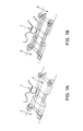
- FIG. 1A is an exemplary embodiment of a suture repair technique without an adhesive coating on a suture.
- FIG. 1B is an exemplary embodiment of a suture repair technique in accordance with the present disclosure.
- FIGS. 2A-2C are exemplary embodiments of an adhesive-coated suture assembly in accordance with the present disclosure.
- FIGS. 3A-3D are exemplary embodiments of the shear stress vs. position along the length of a suture in accordance with the present disclosure.
- FIGS. 4A and 4B are exemplary embodiments of various loads carried by adhesive-coated sutures in accordance with the present disclosure.
- FIG. 5A is an exemplary embodiment of a graphical depiction of suture load in accordance with the present disclosure.
- FIG. 5B is an exemplary embodiment of a graphical depiction of suture load carried by assembly vs. length in accordance with the present disclosure.
- FIG. 6 is an exemplary embodiment of maximum loads resisted by single suture strands in accordance with the present disclosure.
- FIG. 7 is an exemplary embodiment of tendon repair load tolerance with and without adhesive in accordance with the present disclosure.
- FIGS. 8A-8C are exemplary embodiments of graphical depictions of nominal shear stress vs. nominal shear strain of adhesive-coated sutures in accordance with the present disclosure.
- suture refers to the core strand of suture; the term “adhesive” refers to the adhesive layer; the terms “assembly” and “adhesive-coated suture” refer to the combination of suture with adhesive surrounding it; and the term “repair” refers to the complete tissue repair, including several strands of adhesive-coated suture used.
- the present disclosure is directed to a new approach to suturing technology.
- Conventional sutures have a relatively large surface area passing through the tendon that is currently not utilized for load transfer. By enabling this surface area to interact mechanically with the surrounding tissue, peak stresses can be reduced at points where the suture bends through tissue (e.g., anchor points).
- the present disclosure is directed to a modified suture with an adsorbed, ionically bound, or covalently bound adhesive that binds collagen along the suture's length, thereby reducing stress concentrations and better distributing load. This better load distribution improves load tolerance of repaired tendons.
- Improved mechanical repair techniques improve patient outcomes by strengthening repairs, thereby decreasing the chance of repair rupture and failure.
- This disclosure is especially useful and immediately applicable to orthopaedic repairs (e.g., tendon and ligament repair) which demand strong biomechanical resilience to accommodate activities of daily living without risking rupture, though it is also likely useful for other surgical repairs.
- orthopaedic repairs e.g., tendon and ligament repair
- repair site elongation and rupture remain problematic after flexor tendon repairs even with modern suturing and rehabilitation protocols.
- Rotator cuff repairs which require reattachment of materials with disparate mechanical properties (tendon and bone), have alarmingly high failure rates.
- the suture includes an adhesive having properties optimized for load transfer.
- the suture includes adhesives having a stiffness that is modified with microstructural features such as, but not limited to, periodic appendages emanating from the suture body. These appendages can serve to further optimize the stiffness and load transfer properties of the adhesive.
- FIG. 4B a contour map is shown of maximum load transferred between a single-stranded, adhesive-coated suture within tissue, calculated from a wide array of theoretical adhesive shear moduli and adhesive failure shear stresses (i.e., strengths) given properties described in the models.
- Maximum theoretical load transfer occurred with an infinitely compliant and infinitely strong adhesive, toward the upper left corner of this contour plot.
- Current flexor tendon repairs carry approximately 10 N per suture strand, so relevant adhesive coatings would have failure loads around or above this level.
- Adhesive mechanical properties that are not expected to improve load transfer are located in the lower portion of the graph, from the 10 6 failure shear stress toward the 10 10 shear modulus.
- the suture strand itself breaks above approximately 15.5 N for Supramid 4-0 or 23.5 N for Supramid 3-0 suture, so adhesive failure loads above this level would no longer limit load transfer (upper portion from the 10 7 shear stress to the 10 10 shear modulus).
- the shear modulus and failure shear stress are related for a given real material, so not all theoretical combinations are realistic.
- the adhesive-coated repair strength could be limited by the bulk material properties of an adhesive (overlaid here for several materials tested) or the adhesive binding strength at the tissue interface.
- cyanoacrylate-coated sutures were used to perform a clinically relevant flexor digitorum tendon repair in cadaver tissue. The repair performed with adhesive-coated suture had significantly higher strength compared to the standard repair without adhesive. Notably, cyanoacrylate provides strong adhesion with high stiffness and brittle behavior, and is therefore not an ideal adhesive for enhancing suture repair.
- the improvement in repair properties in a clinically relevant setting, using a non-ideal adhesive demonstrates the potential for this approach to improve outcomes for treatments requiring suture fixation.
- the present disclosure is further directed to a strongly adherent, compliant adhesive within the optimal design space described by the shear lag model.
- Sutures are an age-old technology and have been used for wound repair for years. While there have been many improvements in suture materials and intricate knot tying techniques, the core technology of simply sewing tissues together remains a crude mechanical solution. Sutures are typically in pure tension along most of their length, with this tension transferred to the tissue only at anchor points (shown in FIG. 1A ). High stress concentrations at these anchor points can lead to sutures breaking or cutting through the surrounding tissue, similar to a wire cutting through cheese. This limits the maximum force that can be transferred across the repair. While current suturing techniques are sufficient to hold many surgical repairs together, musculoskeletal repairs (e.g., tendon and ligament repair) typically demand strong biomechanical resilience to accommodate activities of daily living without risking rupture.
- musculoskeletal repairs e.g., tendon and ligament repair
- the present disclosure is directed to a new approach to augment standard suturing technology.
- Conventional sutures have a relatively large surface area passing through the tendon that is currently not utilized for load transfer. This improvement in load transfer results in an improvement in overall repair construct mechanical properties.
- a shear lag model of suture within a cylindrical tissue e.g., a tendon
- desirable adhesive mechanical properties were identified to improve load transfer across a repair site.
- Biomechanical tests with suture coated adhesives were conducted to validate the model and experimentally assess the ability to improve load transfer.
- a mechanical model such as a shear lag model, to determine stresses along an adhesive-coated suture's length, which allows one to predict the maximum amount of load that an adhesive-coated suture could transfer between two sides of a repair can be used in certain embodiments of the present disclosure.
- This maximum load is a function of adhesive, tissue, and suture material and geometrical properties. This allows one to plot isoclines for the amount of load transferred on a domain map of theoretical adhesive mechanical properties, specifically, shear modulus (G a ) vs. failure shear stress ( ⁇ fail ). Therefore, this domain map of adhesive mechanical properties shows the desirable adhesive properties needed to improve load transferred across suture-based repairs.
- Loctite 4903 shows promising results that would be valuable clinically.
- Adhesives with optimized mechanical and chemical properties were developed to further increase load transfer and improve clinical repairs for tendon, ligament, and other tissue injuries, as an initial application of the technology. These adhesives are specifically designed to meet the compliant yet strong criteria and can enable substantially stronger suture repairs.
- the present disclosure is directed to novel adhesive technologies that apply existing or novel adhesives in a novel way and/or apply novel structural methods to modify the stiffness of an adhesive layer.
- the present disclosure is also directed to a protein-based bioadhesive with desirable mechanical properties by using a suture-binding domain linked to a compliant domain, which can be linked in series to a tissue-binding domain.
- a silk-binding domain could be used for binding silk sutures, an elastin moiety or a protein sequence with similar mechanical properties could provide the desired compliance properties, and a collagen-binding sequence such as an integrin could be used to specifically bind the surrounding tissue.
- Silk-HRP Gels (silk hydrogels cross-linked with H 2 O 2 in the presence of horseradish peroxidase) represent an attractive bulk adhesive material for use with sutures because they are biocompatible, degradable, and have tunable mechanical properties.
- the present disclosure is directed to various protective mechanisms to only activate the adhesive when appropriate. This can be done in several ways.
- the present disclosure is also directed to coating adhesives in a protective layer that melts away based on either temperature or an activating stimulus such as near infrared radiation. Once the protective layer is melted or removed, the adhesive would be uncovered for binding the surrounding tissue material.
- FIGS. 1A and 1B include diagrams illustrating example suture repair techniques including a typical suture repair and an example adhesive-coated suture repair. This system can also be seen in FIGS. 2A-2C .
- the typical suture repair is an 8-stranded Winters-Gelberman suture repair technique for human flexor digitorum profundus tendon repair. Some known suturing techniques generate stress concentrations at anchor points where the suture bends within tissue.
- the adhesive-coated suture repair enables distributing that load transfer along the entire length of the suture, reducing peak stresses and enhancing overall repair construct mechanics.
- a suture 1 is used to repair a tendon 3 .
- the technique shown in FIG. 1A is a conventional technique, while the technique shown in FIG. 1B is in accordance with the present disclosure wherein the suture 1 is coated with an adhesive 2 and used to repair the tendon 3 .
- FIGS. 2A-2C include diagrams illustrating another example adhesive-coated suture assembly within a cylindrical tissue (e.g., tendon) used to derive shear lag analysis.
- a shear lag model was developed to predict the load sharing between the sutures and a repaired tendon by estimating the variation of the shear stress in the adhesive layer, ⁇ , as a function of the position, x, along a suture, as diagrammed in FIGS. 2A-2C :
- ⁇ ⁇ _ ⁇ s ⁇ L ⁇ ⁇ ⁇ sinh ⁇ ( ⁇ s ⁇ L ) ⁇ ⁇ ( ⁇ - 1 ) ⁇ cosh ⁇ ( ⁇ s ⁇ ( x - L ) ) - ( P k P s ⁇ ⁇ - 1 ) ⁇ cosh ⁇ ( ⁇ s ⁇ x ) ⁇
- ⁇ is the average shear stress
- L is the suture length
- P k is the load in the suture at the anchor point (i.e., at the knot, at distance L along the tendon)
- ⁇ and ⁇ s relate to the geometry and material properties:
- derivation of the example model follows shear lag solution for a double lap joint.
- a free body diagram of the adhesive suture model system can also be used.
- FIGS. 2A-2C include a free body diagram showing a two-dimensional axisymmetric model of adhesive-coated suture within a cylindrical tendon tissue (top, FIG. 2A ). Simultaneously analyzing a section of the repair (bottom left, FIG. 2B ) and each component independently (i.e., suture, adhesive, and tendon; bottom right, FIG. 2C ) allows derivation of a shear lag model to estimate shear stress within the adhesive.
- the example model reduces to a one-dimensional set of equations along the x-axis.
- ⁇ s (x) and ⁇ t (x) are suture and tendon normal stresses at position x, respectively (assumed to not vary with radius r), r t is the tendon radius, t a is the adhesive thickness, and r t * relates geometric properties.
- ⁇ s (x) and ⁇ t (x) are suture and tendon normal strains at position x, respectively (assumed to not vary with radius r)
- ⁇ a (x) is adhesive shear strain at position x
- E s and E t are suture and tendon Young's moduli, respectively
- G a is adhesive shear modulus
- u s (x) and u t (x) are suture and tendon displacement in the x direction at position x, respectively. Note that this derivation neglects adhesive deformation and neglects normal stresses transferred in the adhesive layer.
- the boundary conditions reduce to:
- ⁇ s ⁇ ( x ) ( P s ⁇ ⁇ ⁇ r s 2 - C s ⁇ s 2 ) ⁇ cosh ⁇ ( ⁇ s ⁇ x ) + sinh ⁇ ( ⁇ s ⁇ x ) sinh ⁇ ( ⁇ s ⁇ L ) ⁇ ( P k ⁇ ⁇ ⁇ r s 2 - C s ⁇ s 2 + ( C s ⁇ s 2 - P s ⁇ ⁇ ⁇ r s 2 ) ⁇ cosh ⁇ ( ⁇ s ⁇ L ) ) + C s ⁇ s 2 ( 1.18 . )
- Shear stress ⁇ (x) at position x can be determined by taking the derivative of normal stress in the suture with respect to position x and applied to the equilibrium equation for the suture (inner adherend, 1.3):
- ⁇ ⁇ _ ⁇ s ⁇ L ⁇ ⁇ ⁇ sinh ⁇ ( ⁇ s ⁇ L ) ⁇ ⁇ ( ⁇ - 1 ) ⁇ cosh ⁇ ( ⁇ s ⁇ ( x - L ) ) - ( P k P s ⁇ ⁇ - 1 ) ⁇ cosh ⁇ ( ⁇ s ⁇ x ) ⁇ ( 1.20 . )
- ⁇ relates to the geometry and material properties:
- adherends are not balanced with current Supramid surgical suture and tendon.
- adherends tendon and suture
- the peak stress is 8.5 fold lower (assuming geometry and material properties used in FIG. 3B ).
- the shear lag relationship (1.20) can be rearranged to solve for the load transferred across the interface:
- FIGS. 3A-3D include diagrams illustrating shear stress vs. position along the length of a suture.
- FIG. 3A and zoomed version ( FIG. 3C ) comparing adhesive shear moduli for realistic suture and tendon properties demonstrates that more compliant adhesives have a lower peak shear stress since compliant adhesives distribute loads over a longer distance than stiffer adhesives.
- the maximum transferable load is governed by two asymptotic limits:
- Rubber cement and rubber/gasket adhesives have shear moduli between about 0.5 to 5 MPa.
- Polyfilament caprolactam 4-0 suture (Supramid, S. Jackson, Inc., Alexandria, Va.) was passed through cadaveric canine hindpaw flexor digitorum profundus tendons using a French eye needle (canine tissues were taken post-mortem from an unrelated project). The tendon was first dissected away from surrounding tissue and a complete laceration was made in Zone II perpendicular to the tendon. Suture was passed from the side of the tendon about 8 to 12 mm from the laceration interface toward the laceration interface, and the suture was pulled through the tendon so only a single suture strand remained within the tendon.
- Control repairs without adhesive were compared to repairs with Loctite 4903-coated suture.
- Loctite 4903 was chosen based on results of single suture pullout tests described above.
- Biomechanical testing was conducted for sample sutures. Samples were brought to 37° C. prior to biomechanical testing. For single suture strand pullout tests, any suture and adhesive outside of the lateral tendon was first removed. Samples were then tested in uniaxial tension on a materials testing frame (ElectroPuls E1000; Instron Corp., Candon, Mass.) at 0.3 mm/s. The length of exposed tendon was approximately 15.0 mm for all samples, and the gauge length between the tendon and suture grips was 8.5 cm for all samples. Pullout (or failure) force of single adhesive-coated suture strands within tendon tissue were determined from the force-elongation curves.
- FIGS. 4A, 5A, and 5B illustrate results of shear lag model analysis of example adhesive coating sutures.
- FIG. 4A includes a contour map illustrating maximum load transferred across the repair by an adhesive-coated suture, calculated from a wide array of theoretical adhesive shear moduli and adhesive failure shear stresses (i.e., strengths) given properties described in the methods. Maximum load transfer occurred with an infinitely compliant and infinitely strong adhesive, toward the upper left corner of this contour plot. Current flexor tendon repairs carry approximately 10 N per suture strand, so relevant adhesive coatings would have failure loads above this level. Adhesive mechanical properties that do not improve load transfer are located, for example, in the lower portion of FIG. 4A .
- suture strand itself breaks above approximately 15.5 N for Supramid 4-0 or 23.5 N for Supramid 3-0 suture, so adhesive failure loads above this level would not further improve load transfer (located, for example, in the upper portion).
- load transfer located, for example, in the upper portion.
- shear modulus and failure shear stress are related for a given real material, so not all theoretical combinations are realistic.
- FIGS. 5A and 5B include diagrams illustrating increasing suture length increases maximum load carried by assembly, i.e., load causing adhesive to fail, only until a point. Above a transitional suture length, load capacity is governed by an asymptote independent of suture length. Note that axes are logarithmic scale. Current suture length used in flexor tendon repair is 12 mm into each tendon end.
- shear lag modeling predicted that adhesive coatings on sutures would improve load transfer compared to conventional sutures for a certain range of properties (in between 10 6 and 10 7 shear stress as shown in FIG. 4A ).
- Mechanically desirable adhesives would be compliant in shear while maintaining high binding and shear strengths. Compliant adhesives allow greater deformation, thereby distributing loads over a larger length than stiff adhesives (shown in FIG. 3A ). This distribution reduces stress concentrations at the suture anchor points, leading to an adhesive-coated suture assembly that carries greater load before failure.
- Shear lag modeling also predicted that maximum load transfer would increase with increasing adhesive-coated suture length.
- varying the ratio of suture length to L intersect demonstrates that adhesive-coated sutures approach the limit for maximum load transferred when the suture length, L, is about 2 to 3 times L intersect (shown in FIG. 5A ).
- the length of suture used is limited surgically by the particular tissue being repaired.
- Suture length of 13 mm was used in the model to make results relevant to flexor digitorum profundus tendon repair (shown in FIG. 5B ).
- FIGS. 6 and 7 illustrate the maximum load resisted by various example sutures.
- FIG. 6 includes a diagram illustrating the maximum loads resisted by each suture for single suture strands coated with nothing (traditional suture), Elmer's rubber cement, 3M rubber and gasket adhesive 1300 (neoprene), Loctite Quicktite (cyanoacrylate), or Loctite 4903 (flexible cyanoacrylate).
- FIG. 7 includes a diagram illustrating load creating a 2 mm gap and maximum load for a cadaveric canine flexor digitorum profundus tendon repair using standard clinical surgical technique (8 stranded repair with 4-0 Supramid suture) compared with the same repair style where suture was coated with Loctite 4903 (cyanoacrylate adhesive).
- the middle line within the box represents the median, the outer edges denote the 25% and 75% samples, and the whiskers extend to the extreme data points.
- Outliers are denoted by (+).
- Overbars denote statistically significant differences (p ⁇ 0.05).
- Table 1 includes resilience, stiffness, and strain at 20 N applied force for a cadaveric canine flexor digitorum profundus tendon repair using standard clinical surgical technique (8 stranded repair with 4-0 Supramid suture) compared with the same repair style where suture was coated with Loctite 4903 (cyanoacrylate adhesive).
- the modified resilience shown in Table 1 is calculated from a force-strain curve.
- Adhesives have been used for decades in surgical repairs to replace or augment suture for closing the skin and other tissues, including tendon. Cyanoacrylates are particularly prevalent medical adhesives, but many other adhesive types have also been used throughout the body. However, in all but one case, the adhesives have only been applied directly at or around the interface between adjoined tissues.
- the present disclosure is directed to load distribution improvement using an adhesive-coating along a suture's length, leading to improved load tolerance of repaired tissues. It has been demonstrated that adhesive-soaked sutures have the potential to improve load tolerance of meniscal repairs by almost 30% compared to either suture only or adhesive at the interface, only.
- the present disclosure is directed to a shear lag model to predict the ability of adhesive-coated sutures to improve load transfer and to identify desirable adhesive mechanical properties.
- bio-adhesives Some biological materials, e.g., those based on elastin, could also be valuable for creating bio-adhesives.
- shear strength used in this model may be limited by either bulk failure within the “adhesive” material or interfacial failure between the adhesive and adherends (i.e., suture and tendon). Therefore, both the bulk adhesive mechanical properties and the strength of adhesion are crucial factors for a successful adhesive.
- This shear lag model describes the importance of adhesive mechanical properties for creating a successful adhesive-coated suture; however, most currently used adhesives are not designed for this purpose. Specifically engineering an adhesive material to tightly bind suture and surrounding tissue while maintaining compliance to shear stress could lead to substantially improved adhesive-coated sutures.
- an adhesive-coated suture is inert for storage and surgical handling before it is placed into the body.
- Several potential approaches to generate adhesive coatings may be used that only activate when in place within tissue.
- adhesives that are compliant in shear facilitate greater load transfer across the adhesive-coated suture repair before rupture by lowering stress concentrations.
- This is directly analogous to biomimetic mechanisms of stress transfer at the enthesis using a compliant zone.
- Models that optimize the modulus of an interfacial zone between tendon and bone for reducing stress concentrations produce a dip in modulus (compliant zone) prior to stiffening between the two dissimilar materials. Regions of compliance between dissimilar materials can absorb more energy and act as a toughening mechanism for the interface.
- a collagen-binding adhesive that directly attaches to the suture via a small compliant layer (i.e., linker molecule such as an elastin moiety) in between the suture and the collagen-binding domain should better distribute load to minimize stress concentrations, enabling more effective load transfer across the repair.
- a small compliant layer i.e., linker molecule such as an elastin moiety
- Models appropriate for optimization of adhesive layer mechanical properties such as the shear lag models described herein, relied on several simplifying assumptions.
- the simplifying assumptions are as follows. First, the system is assumed to be one dimensional; only forces and stresses along the long axis of the tendon/suture are considered. This results in shear stresses that are unbalanced. Second, the deformation of the adhesive is not considered. This assumption is reasonable for stiff adhesives, but becomes an issue for highly compliant adhesives. Third, the stress in the suture and the tissue is assumed to be independent of radial position. This may be inaccurate, as the adhesive attaches only to the outside of the suture.
- Loctite 4903 shows promising results that would be valuable clinically.
- the present disclosure is directed to adhesives with optimized mechanical and chemical properties that further increase load transfer and improve clinical repairs for tendon, ligament, and other tissue injuries.
- Silk-HRP hydrogels were created in accordance with the present disclosure. These Silk-HRP hydrogels were placed into methanol (MeOH) at concentrations of 10%, 50%, 70%, and 100% for 90 minutes to increase crystallinity in the silk. The gels were then placed into phosphate buffered saline overnight to rehydrate. Methanol-treated Silk-HRP gels represent one possible adhesive with attractiveness in use with sutures in tendon repair due to their biocompatibility, biodegradability, and tunable mechanical properties which allow one to obtain material properties that place the adhesive in the critical zone we have identified.
- MeOH methanol
- the lap shear test is a commonly used method for testing material properties of adhesives.
- a double lap shear test was used. In this test, a single, movable PVC platen is sandwiched between two fixed platens. A slab of sample material with thickness (t) is glued with cyanoacrylate to both a fixed platen and the movable platen such that both sides of the movable platen are glued to a slab of a sample of the material being tested. In this way, two samples of material are tested simultaneously.
- the movable platen is pulled between and parallel to the fixed platens at a constant rate of 0.1 mm/sec, and the force (F) required to maintain this pulling rate is recorded.
- This force can be converted to a nominal shear stress ( ⁇ ) by dividing by the contact area between the material and the movable platen.
- the nominal shear strain ( ⁇ ) is calculated by dividing the displacement of the movable platen (u) by the average change in the thickness (t) between the two material samples.
- the adhesive shear modulus Ga is the nominal shear stress divided by the nominal shear strain.
- the adhesive failure shear stress ⁇ fail is the maximum shear stress achieved in the double lap shear test experiments.
- the Silk-HRP gels showed bulk mechanical properties that were sub-optimal such that they would fail as adhesives at shear stresses that were in the 1-10 kPa range, which is well below the ideal for optimal repair strength increase.
- Silk-HRP gels were treated with various concentrations of methanol (10%, 50%, 70%, and 100%) for 90 minutes to increase crystallinity in the silk. Results from experiments performed on Silk-HRP gels treated with MeOH (10%, 50% and 100%, respectively) are shown in FIGS. 8A (10%), 8 B (50%) and 8 C (100%).
Landscapes
- Health & Medical Sciences (AREA)
- Life Sciences & Earth Sciences (AREA)
- Surgery (AREA)
- Heart & Thoracic Surgery (AREA)
- Engineering & Computer Science (AREA)
- Biomedical Technology (AREA)
- Nuclear Medicine, Radiotherapy & Molecular Imaging (AREA)
- Medical Informatics (AREA)
- Molecular Biology (AREA)
- Animal Behavior & Ethology (AREA)
- General Health & Medical Sciences (AREA)
- Public Health (AREA)
- Veterinary Medicine (AREA)
- Materials For Medical Uses (AREA)
- Surgical Instruments (AREA)
Abstract
Description
where
where Ga is the adhesive shear modulus, ta is the adhesive thickness, rt is the tendon radius, rs is the suture radius, and Et and Es are tendon and suture Young's modulus, respectively.
where Ps is the load in the suture at the interface (position x=0), rs is the suture radius, and L is the suture length.
where σs(x) and σt(x) are suture and tendon normal stresses at position x, respectively (assumed to not vary with radius r), rt is the tendon radius, ta is the adhesive thickness, and rt* relates geometric properties.
Strain-Displacement Equations:
where εs(x) and εt(x) are suture and tendon normal strains at position x, respectively (assumed to not vary with radius r), γa(x) is adhesive shear strain at position x, Es and Et are suture and tendon Young's moduli, respectively, Ga is adhesive shear modulus, and us(x) and ut(x) are suture and tendon displacement in the x direction at position x, respectively. Note that this derivation neglects adhesive deformation and neglects normal stresses transferred in the adhesive layer.
where λs and Cs relate to the geometry and material properties:
∫∫0 Aσs(r,x)dA=∫ 0 2π∫0 r
where A is the cross sectional area of the suture.
P(x)=2π∫0 r
Ps is defined as the load in the suture at the interface (position x=0) and Pk is defined as the load in the suture at the anchor point or knot (distance L into the tendon).
where ξ relates to the geometry and material properties:
E s r s 2 =E t(r t 2−(r s +t a)2)=E t r* t 2 ≈E t r t 2(if r t >>r s +t a) (1.22.)
where τfail is the failure shear stress of the adhesive.
| TABLE 1 | |||
| Resilience | Stiffness | Strain at 20 N | |
| Repair with Loctite | 9.12 ± 2.46 N | 27.2 ± 4.4 N/mm | 8.00 ± 1.36% |
| 4903-coated suture | |||
| Control repair (no | 7.39 ± 2.22 N | 24.0 ± 7.0 N/mm | 8.81 ± 2.91% |
| adhesive) | |||
| p-value | 0.108 | 0.251 | 0.438 |
Claims (18)
Priority Applications (2)
| Application Number | Priority Date | Filing Date | Title |
|---|---|---|---|
| US14/940,541 US10314574B2 (en) | 2014-11-14 | 2015-11-13 | Adhesive-coated sutures |
| US15/455,792 US10631973B2 (en) | 2014-11-14 | 2017-03-10 | Compostions and methods for tissue repair |
Applications Claiming Priority (2)
| Application Number | Priority Date | Filing Date | Title |
|---|---|---|---|
| US201462079965P | 2014-11-14 | 2014-11-14 | |
| US14/940,541 US10314574B2 (en) | 2014-11-14 | 2015-11-13 | Adhesive-coated sutures |
Related Child Applications (1)
| Application Number | Title | Priority Date | Filing Date |
|---|---|---|---|
| US15/455,792 Continuation-In-Part US10631973B2 (en) | 2014-11-14 | 2017-03-10 | Compostions and methods for tissue repair |
Publications (2)
| Publication Number | Publication Date |
|---|---|
| US20160135809A1 US20160135809A1 (en) | 2016-05-19 |
| US10314574B2 true US10314574B2 (en) | 2019-06-11 |
Family
ID=55960658
Family Applications (1)
| Application Number | Title | Priority Date | Filing Date |
|---|---|---|---|
| US14/940,541 Active 2036-07-27 US10314574B2 (en) | 2014-11-14 | 2015-11-13 | Adhesive-coated sutures |
Country Status (1)
| Country | Link |
|---|---|
| US (1) | US10314574B2 (en) |
Families Citing this family (1)
| Publication number | Priority date | Publication date | Assignee | Title |
|---|---|---|---|---|
| US20230355228A1 (en) * | 2022-05-03 | 2023-11-09 | Arthrex, Inc. | Suture-to-button assemblies for performing surgical procedures |
Citations (6)
| Publication number | Priority date | Publication date | Assignee | Title |
|---|---|---|---|---|
| US6264675B1 (en) | 2000-02-04 | 2001-07-24 | Gregory R. Brotz | Single suture structure |
| US6478809B1 (en) * | 2000-02-04 | 2002-11-12 | Gregory R. Brotz | Suture and method of use |
| US20090036611A1 (en) * | 2006-04-25 | 2009-02-05 | Jonathan James Wilker | Cross-Linkable Polymeric Compositions |
| US20090177228A1 (en) * | 2004-07-01 | 2009-07-09 | Optovent Ab | Coated suture thread and production thereof |
| US8142475B2 (en) | 2004-10-18 | 2012-03-27 | Tyco Healthcare Group Lp | Adhesive suture structure and methods of using the same |
| US9115289B2 (en) * | 2009-02-06 | 2015-08-25 | Kensey Nash Corporation | Multibranched bioadhesive compounds and synthetic methods therefor |
-
2015
- 2015-11-13 US US14/940,541 patent/US10314574B2/en active Active
Patent Citations (6)
| Publication number | Priority date | Publication date | Assignee | Title |
|---|---|---|---|---|
| US6264675B1 (en) | 2000-02-04 | 2001-07-24 | Gregory R. Brotz | Single suture structure |
| US6478809B1 (en) * | 2000-02-04 | 2002-11-12 | Gregory R. Brotz | Suture and method of use |
| US20090177228A1 (en) * | 2004-07-01 | 2009-07-09 | Optovent Ab | Coated suture thread and production thereof |
| US8142475B2 (en) | 2004-10-18 | 2012-03-27 | Tyco Healthcare Group Lp | Adhesive suture structure and methods of using the same |
| US20090036611A1 (en) * | 2006-04-25 | 2009-02-05 | Jonathan James Wilker | Cross-Linkable Polymeric Compositions |
| US9115289B2 (en) * | 2009-02-06 | 2015-08-25 | Kensey Nash Corporation | Multibranched bioadhesive compounds and synthetic methods therefor |
Non-Patent Citations (3)
| Title |
|---|
| Bouten et al., "The chemistry of tissue adhesive materials," Progress in Polymer Science, 39, (2014), pp. 1375-1405. |
| Inoue et al., Effectiveness and biocompatibility of a novel biological adhesive application for repair of meniscal tear on the avascular zone, Sci. Technol. Adv. Mater. 13, (2012) 064219, pp. 1-5. |
| Jordan Raphel, Andreina Parisi-Amon, and Sarah Heilshorn, Photoreactive elastin-like proteins for use as versatile bioactive materials and surface coatings, (Oct. 7, 2012), NIH Public Access, 1-15 (Year: 2012). * |
Also Published As
| Publication number | Publication date |
|---|---|
| US20160135809A1 (en) | 2016-05-19 |
Similar Documents
| Publication | Publication Date | Title |
|---|---|---|
| Gao et al. | Hydrogel–mesh composite for wound closure | |
| Amadio | Friction of the gliding surface: implications for tendon surgery and rehabilitation | |
| Linderman et al. | Shear lag sutures: Improved suture repair through the use of adhesives | |
| Peltz et al. | Influence of locking stitch size in a four-strand cross-locked cruciate flexor tendon repair | |
| US20130345747A1 (en) | Biological suture anchor with suture eyelet | |
| Lekic et al. | Suture materials, needles, and methods of skin closure: what every hand surgeon should know | |
| Azadani et al. | Mechanical properties of surgical glues used in aortic root replacement | |
| Muffly et al. | Minimum number of throws needed for knot security | |
| Krebs et al. | Wound closure techniques for total knee arthroplasty: an evidence-based review of the literature | |
| Rosso et al. | Comparison of all-inside meniscal repair devices with matched inside-out suture repair | |
| Huffard et al. | Achilles tendon repair: Achillon system vs. Krackow suture: an anatomic in vitro biomechanical study | |
| EA200800252A1 (en) | PRODUCT FOR SURGICAL RESTORATION OF TISSUES BASED ON FIBERS FROM ULTRA-HIGH-MOLECULAR POLYETHYLENE | |
| Elliot et al. | IFSSH flexor tendon committee report | |
| US10314574B2 (en) | Adhesive-coated sutures | |
| Kallinowski et al. | Assessing the GRIP of ventral hernia repair: how to securely fasten DIS classified meshes | |
| US20030093119A1 (en) | Eyelet reinforcement at the tissue-suture interface | |
| Dona et al. | Optimizing biomechanical performance of the 4-strand cruciate flexor tendon repair | |
| Haslach et al. | The influence of medial substructures on rupture in bovine aortas | |
| US10631973B2 (en) | Compostions and methods for tissue repair | |
| Trocchia et al. | A re-exploration of the use of barbed sutures in flexor tendon repairs | |
| Schneppendahl et al. | Initial stability of two different adhesives compared to suture repair for acute Achilles tendon rupture—a biomechanical evaluation | |
| Borchiellini et al. | Development and characterization of biological sutures made of cell-assembled extracellular matrix | |
| Zobitz et al. | Comparison of mechanical properties of various suture repair techniques in a partially lacerated tendon | |
| Pascual et al. | Host tissue response by the expression of collagen to cyanoacrylate adhesives used in implant fixation for abdominal hernia repair | |
| Joyce et al. | Assessment of the uniaxial experimental parameters utilised for the mechanical testing of bovine pericardium |
Legal Events
| Date | Code | Title | Description |
|---|---|---|---|
| AS | Assignment |
Owner name: WASHINGTON UNIVERSITY, MISSOURI Free format text: ASSIGNMENT OF ASSIGNORS INTEREST;ASSIGNORS:LINDERMAN, STEPHEN;GENIN, GUY M.;THOMOPOULOS, STAVROS;SIGNING DATES FROM 20151216 TO 20151229;REEL/FRAME:037381/0662 |
|
| AS | Assignment |
Owner name: NATIONAL INSTITUTES OF HEALTH (NIH), U.S. DEPT. OF Free format text: CONFIRMATORY LICENSE;ASSIGNOR:WASHINGTON UNIVERSITY;REEL/FRAME:041886/0481 Effective date: 20170223 |
|
| STPP | Information on status: patent application and granting procedure in general |
Free format text: NOTICE OF ALLOWANCE MAILED -- APPLICATION RECEIVED IN OFFICE OF PUBLICATIONS |
|
| STPP | Information on status: patent application and granting procedure in general |
Free format text: PUBLICATIONS -- ISSUE FEE PAYMENT VERIFIED |
|
| STCF | Information on status: patent grant |
Free format text: PATENTED CASE |
|
| CC | Certificate of correction | ||
| MAFP | Maintenance fee payment |
Free format text: PAYMENT OF MAINTENANCE FEE, 4TH YR, SMALL ENTITY (ORIGINAL EVENT CODE: M2551); ENTITY STATUS OF PATENT OWNER: SMALL ENTITY Year of fee payment: 4 |