US10300022B2 - Nanoparticle delivery compositions - Google Patents
Nanoparticle delivery compositions Download PDFInfo
- Publication number
- US10300022B2 US10300022B2 US14/175,422 US201414175422A US10300022B2 US 10300022 B2 US10300022 B2 US 10300022B2 US 201414175422 A US201414175422 A US 201414175422A US 10300022 B2 US10300022 B2 US 10300022B2
- Authority
- US
- United States
- Prior art keywords
- nanoparticles
- cns
- disorder
- core
- nanoparticle
- Prior art date
- Legal status (The legal status is an assumption and is not a legal conclusion. Google has not performed a legal analysis and makes no representation as to the accuracy of the status listed.)
- Expired - Fee Related, expires
Links
Images


Classifications
-
- A—HUMAN NECESSITIES
- A61—MEDICAL OR VETERINARY SCIENCE; HYGIENE
- A61K—PREPARATIONS FOR MEDICAL, DENTAL OR TOILETRY PURPOSES
- A61K9/00—Medicinal preparations characterised by special physical form
- A61K9/48—Preparations in capsules, e.g. of gelatin, of chocolate
- A61K9/50—Microcapsules having a gas, liquid or semi-solid filling; Solid microparticles or pellets surrounded by a distinct coating layer, e.g. coated microspheres, coated drug crystals
- A61K9/51—Nanocapsules; Nanoparticles
- A61K9/5107—Excipients; Inactive ingredients
- A61K9/5123—Organic compounds, e.g. fats, sugars
-
- A—HUMAN NECESSITIES
- A61—MEDICAL OR VETERINARY SCIENCE; HYGIENE
- A61K—PREPARATIONS FOR MEDICAL, DENTAL OR TOILETRY PURPOSES
- A61K38/00—Medicinal preparations containing peptides
- A61K38/16—Peptides having more than 20 amino acids; Gastrins; Somatostatins; Melanotropins; Derivatives thereof
- A61K38/17—Peptides having more than 20 amino acids; Gastrins; Somatostatins; Melanotropins; Derivatives thereof from animals; from humans
- A61K38/1703—Peptides having more than 20 amino acids; Gastrins; Somatostatins; Melanotropins; Derivatives thereof from animals; from humans from vertebrates
- A61K38/1709—Peptides having more than 20 amino acids; Gastrins; Somatostatins; Melanotropins; Derivatives thereof from animals; from humans from vertebrates from mammals
-
- A—HUMAN NECESSITIES
- A61—MEDICAL OR VETERINARY SCIENCE; HYGIENE
- A61K—PREPARATIONS FOR MEDICAL, DENTAL OR TOILETRY PURPOSES
- A61K39/00—Medicinal preparations containing antigens or antibodies
- A61K39/395—Antibodies; Immunoglobulins; Immune serum, e.g. antilymphocytic serum
- A61K39/39533—Antibodies; Immunoglobulins; Immune serum, e.g. antilymphocytic serum against materials from animals
- A61K39/3955—Antibodies; Immunoglobulins; Immune serum, e.g. antilymphocytic serum against materials from animals against proteinaceous materials, e.g. enzymes, hormones, lymphokines
-
- A—HUMAN NECESSITIES
- A61—MEDICAL OR VETERINARY SCIENCE; HYGIENE
- A61K—PREPARATIONS FOR MEDICAL, DENTAL OR TOILETRY PURPOSES
- A61K49/00—Preparations for testing in vivo
- A61K49/06—Nuclear magnetic resonance [NMR] contrast preparations; Magnetic resonance imaging [MRI] contrast preparations
- A61K49/18—Nuclear magnetic resonance [NMR] contrast preparations; Magnetic resonance imaging [MRI] contrast preparations characterised by a special physical form, e.g. emulsions, microcapsules, liposomes
- A61K49/1818—Nuclear magnetic resonance [NMR] contrast preparations; Magnetic resonance imaging [MRI] contrast preparations characterised by a special physical form, e.g. emulsions, microcapsules, liposomes particles, e.g. uncoated or non-functionalised microparticles or nanoparticles
- A61K49/1821—Nuclear magnetic resonance [NMR] contrast preparations; Magnetic resonance imaging [MRI] contrast preparations characterised by a special physical form, e.g. emulsions, microcapsules, liposomes particles, e.g. uncoated or non-functionalised microparticles or nanoparticles coated or functionalised microparticles or nanoparticles
- A61K49/1824—Nuclear magnetic resonance [NMR] contrast preparations; Magnetic resonance imaging [MRI] contrast preparations characterised by a special physical form, e.g. emulsions, microcapsules, liposomes particles, e.g. uncoated or non-functionalised microparticles or nanoparticles coated or functionalised microparticles or nanoparticles coated or functionalised nanoparticles
-
- A—HUMAN NECESSITIES
- A61—MEDICAL OR VETERINARY SCIENCE; HYGIENE
- A61K—PREPARATIONS FOR MEDICAL, DENTAL OR TOILETRY PURPOSES
- A61K9/00—Medicinal preparations characterised by special physical form
- A61K9/0002—Galenical forms characterised by the drug release technique; Application systems commanded by energy
- A61K9/0009—Galenical forms characterised by the drug release technique; Application systems commanded by energy involving or responsive to electricity, magnetism or acoustic waves; Galenical aspects of sonophoresis, iontophoresis, electroporation or electroosmosis
-
- A—HUMAN NECESSITIES
- A61—MEDICAL OR VETERINARY SCIENCE; HYGIENE
- A61K—PREPARATIONS FOR MEDICAL, DENTAL OR TOILETRY PURPOSES
- A61K9/00—Medicinal preparations characterised by special physical form
- A61K9/48—Preparations in capsules, e.g. of gelatin, of chocolate
- A61K9/50—Microcapsules having a gas, liquid or semi-solid filling; Solid microparticles or pellets surrounded by a distinct coating layer, e.g. coated microspheres, coated drug crystals
- A61K9/51—Nanocapsules; Nanoparticles
- A61K9/5107—Excipients; Inactive ingredients
- A61K9/5115—Inorganic compounds
-
- A—HUMAN NECESSITIES
- A61—MEDICAL OR VETERINARY SCIENCE; HYGIENE
- A61K—PREPARATIONS FOR MEDICAL, DENTAL OR TOILETRY PURPOSES
- A61K9/00—Medicinal preparations characterised by special physical form
- A61K9/48—Preparations in capsules, e.g. of gelatin, of chocolate
- A61K9/50—Microcapsules having a gas, liquid or semi-solid filling; Solid microparticles or pellets surrounded by a distinct coating layer, e.g. coated microspheres, coated drug crystals
- A61K9/51—Nanocapsules; Nanoparticles
- A61K9/5192—Processes
-
- A—HUMAN NECESSITIES
- A61—MEDICAL OR VETERINARY SCIENCE; HYGIENE
- A61P—SPECIFIC THERAPEUTIC ACTIVITY OF CHEMICAL COMPOUNDS OR MEDICINAL PREPARATIONS
- A61P25/00—Drugs for disorders of the nervous system
-
- A—HUMAN NECESSITIES
- A61—MEDICAL OR VETERINARY SCIENCE; HYGIENE
- A61P—SPECIFIC THERAPEUTIC ACTIVITY OF CHEMICAL COMPOUNDS OR MEDICINAL PREPARATIONS
- A61P25/00—Drugs for disorders of the nervous system
- A61P25/28—Drugs for disorders of the nervous system for treating neurodegenerative disorders of the central nervous system, e.g. nootropic agents, cognition enhancers, drugs for treating Alzheimer's disease or other forms of dementia
-
- B—PERFORMING OPERATIONS; TRANSPORTING
- B82—NANOTECHNOLOGY
- B82Y—SPECIFIC USES OR APPLICATIONS OF NANOSTRUCTURES; MEASUREMENT OR ANALYSIS OF NANOSTRUCTURES; MANUFACTURE OR TREATMENT OF NANOSTRUCTURES
- B82Y5/00—Nanobiotechnology or nanomedicine, e.g. protein engineering or drug delivery
-
- Y—GENERAL TAGGING OF NEW TECHNOLOGICAL DEVELOPMENTS; GENERAL TAGGING OF CROSS-SECTIONAL TECHNOLOGIES SPANNING OVER SEVERAL SECTIONS OF THE IPC; TECHNICAL SUBJECTS COVERED BY FORMER USPC CROSS-REFERENCE ART COLLECTIONS [XRACs] AND DIGESTS
- Y10—TECHNICAL SUBJECTS COVERED BY FORMER USPC
- Y10S—TECHNICAL SUBJECTS COVERED BY FORMER USPC CROSS-REFERENCE ART COLLECTIONS [XRACs] AND DIGESTS
- Y10S977/00—Nanotechnology
- Y10S977/70—Nanostructure
- Y10S977/773—Nanoparticle, i.e. structure having three dimensions of 100 nm or less
-
- Y—GENERAL TAGGING OF NEW TECHNOLOGICAL DEVELOPMENTS; GENERAL TAGGING OF CROSS-SECTIONAL TECHNOLOGIES SPANNING OVER SEVERAL SECTIONS OF THE IPC; TECHNICAL SUBJECTS COVERED BY FORMER USPC CROSS-REFERENCE ART COLLECTIONS [XRACs] AND DIGESTS
- Y10—TECHNICAL SUBJECTS COVERED BY FORMER USPC
- Y10S—TECHNICAL SUBJECTS COVERED BY FORMER USPC CROSS-REFERENCE ART COLLECTIONS [XRACs] AND DIGESTS
- Y10S977/00—Nanotechnology
- Y10S977/70—Nanostructure
- Y10S977/788—Of specified organic or carbon-based composition
-
- Y—GENERAL TAGGING OF NEW TECHNOLOGICAL DEVELOPMENTS; GENERAL TAGGING OF CROSS-SECTIONAL TECHNOLOGIES SPANNING OVER SEVERAL SECTIONS OF THE IPC; TECHNICAL SUBJECTS COVERED BY FORMER USPC CROSS-REFERENCE ART COLLECTIONS [XRACs] AND DIGESTS
- Y10—TECHNICAL SUBJECTS COVERED BY FORMER USPC
- Y10S—TECHNICAL SUBJECTS COVERED BY FORMER USPC CROSS-REFERENCE ART COLLECTIONS [XRACs] AND DIGESTS
- Y10S977/00—Nanotechnology
- Y10S977/70—Nanostructure
- Y10S977/81—Of specified metal or metal alloy composition
-
- Y—GENERAL TAGGING OF NEW TECHNOLOGICAL DEVELOPMENTS; GENERAL TAGGING OF CROSS-SECTIONAL TECHNOLOGIES SPANNING OVER SEVERAL SECTIONS OF THE IPC; TECHNICAL SUBJECTS COVERED BY FORMER USPC CROSS-REFERENCE ART COLLECTIONS [XRACs] AND DIGESTS
- Y10—TECHNICAL SUBJECTS COVERED BY FORMER USPC
- Y10S—TECHNICAL SUBJECTS COVERED BY FORMER USPC CROSS-REFERENCE ART COLLECTIONS [XRACs] AND DIGESTS
- Y10S977/00—Nanotechnology
- Y10S977/902—Specified use of nanostructure
- Y10S977/904—Specified use of nanostructure for medical, immunological, body treatment, or diagnosis
- Y10S977/906—Drug delivery
-
- Y—GENERAL TAGGING OF NEW TECHNOLOGICAL DEVELOPMENTS; GENERAL TAGGING OF CROSS-SECTIONAL TECHNOLOGIES SPANNING OVER SEVERAL SECTIONS OF THE IPC; TECHNICAL SUBJECTS COVERED BY FORMER USPC CROSS-REFERENCE ART COLLECTIONS [XRACs] AND DIGESTS
- Y10—TECHNICAL SUBJECTS COVERED BY FORMER USPC
- Y10S—TECHNICAL SUBJECTS COVERED BY FORMER USPC CROSS-REFERENCE ART COLLECTIONS [XRACs] AND DIGESTS
- Y10S977/00—Nanotechnology
- Y10S977/902—Specified use of nanostructure
- Y10S977/904—Specified use of nanostructure for medical, immunological, body treatment, or diagnosis
- Y10S977/927—Diagnostic contrast agent
- Y10S977/93—MRI contrast agent
Definitions
- the present invention relates to substances and compositions useful for delivery of agents to the central nervous system (CNS), in particular the delivery of biologically active agents across the blood-brain barrier (BBB).
- CNS central nervous system
- BBB blood-brain barrier
- Substances, compositions and methods disclosed herein find use in the therapeutic and/or prophylactic treatment of disorders of the CNS, for imaging, targeting, repairing and studying the interaction of biologically active agents with, cells of the CNS.
- CNS central nervous system
- More than 95% of potentially useful drugs are prevented from entering the CNS due to the protective function of the blood-brain barrier, formed by microvascular endothelium and astrocytes.
- the key elements of the barrier are continuous tight-junctions between endothelial cells, which prevent molecules from diffusing into the brain, and ABC-transporters that actively pump xenobiotics out of the brain (1, 2).
- endothelial barrier 3-6.
- nanoparticles as a carrier (7).
- Biologically interesting nano- and micro-particles ranging from 1 nm to 500 nm have been made from materials such as polymers, lipids and metals, including gold.
- Gold nanoparticles have the advantage of easy production and chemical stability, and they have been recently used in nanomedicine for both diagnosis and therapy (8).
- the gold core is inert but it does interact with biological material and can have biological effects.
- a variety of sizes and surface modifications have been investigated which affect the specific behaviour of the nanoparticles (9-11).
- the transport into a cell is a property which can vary significantly depending on size and surface coating (12).
- Small-sized gold nanoparticles are able to enter cells via an endocytic pathway (13, 14) although the mechanism of the transport is not exactly known. It is thought that gold nanoparticles do not enter the nucleus (15) unless the cell is apoptotic. In contrast, they are often trapped in vesicles (16-19) which can cause a problem for targeted drug/gene delivery into the cell and tissues in general.
- the present invention addresses these and other needs.
- the present invention relates to nanoparticle delivery systems for use in targeting biologically active or imaging agents to the central nervous system.
- the present inventors have found that nanoparticles, as defined herein, cross the endothelium and enter astrocytes. Moreover, the nanoparticles exhibit some selectivity for human brain endothelium, e.g. vs. non-brain endothelium.
- Biologically active agents may be coupled to nanoparticles, e.g. by means of covalent attachment via a linker, or reversibly bound to nanoparticles, e.g. by stably but reversibly binding to a nanoparticle corona. The agents are then delivered by the nanoparticles as “cargo” across the blood brain barrier to cells of the central nervous system, e.g. for therapeutic treatment of disorders of the central nervous system (CNS) or for imaging the CNS.
- CNS central nervous system
- the present invention provides a nanoparticle composition for use in a method of delivering at least one agent to the central nervous system (CNS) of a mammalian subject, said composition comprising:
- the composition is for use in a method of treatment of a CNS disorder of the subject.
- the composition is for use in a diagnostic or prognostic method of imaging of the CNS of the subject.
- Said method may be a method carried out on the body of the subject (in vivo).
- the present invention provides a method for delivering at least one agent to the central nervous system (CNS) of a mammalian subject, said method comprising administering a composition to the subject, said composition comprising:
- the method is a method of treatment of a CNS disorder of the subject.
- the method is a diagnostic or prognostic method of imaging of the CNS of the subject.
- Said method may be a method carried out on the body of the subject (in vivo).
- the present invention provides use of a composition in the preparation of a medicament to be delivered to the central nervous system (CNS) of a mammalian subject, said composition comprising:
- the medicament is for the treatment of a CNS disorder of a mammalian subject.
- the medicament is for diagnostic or prognostic imaging of the CNS of the subject.
- Said diagnostic or prognostic imaging of the CNS of the subject may be carried out on the body of the subject (in vivo).
- the composition is administered, or is for administration, via a non-central route, whereby said at least one agent is delivered across the blood-brain barrier to the CNS by association with said nanoparticle.
- the composition may be administered, or for administration, other than by intracerebral, intrathecal or epidural route.
- Suitable routes of administration include enteral (e.g. solid or liquid composition for ingestion); buccal; sublabial; sublingual; by inhalation; via a mucosal membrane; urogenital; rectal; dermal; and intradermal, intramuscular, intravenous, intraperitoneal, and subcutaneous injection or infusion.
- the subject has an impaired or “leaky” blood-brain barrier.
- the subject may be suffering from a condition, such as a brain tumour or an infection, that renders the blood-brain barrier more permeable than would be the case in the absence of the condition.
- the subject has a substantially functional blood-brain barrier (i.e. not leaky or impaired).
- the subject may be free from a condition that renders the blood-brain barrier more permeable than would be considered normal for the subject's species and age.
- the present inventors believe that the nanoparticles defined herein are capable of crossing a healthy blood-brain barrier and thereby delivering at least one agent to the CNS of the subject. This to be contrasted with the rather less challenging delivery of agents to the CNS of a subject suffering from a condition that renders the blood-brain barrier more permeable than would be the case in the absence of the condition.
- the subject is a human.
- the at least one agent for delivery to the CNS may be selected according to the desired biological (e.g. therapeutic, prophylactic, diagnostic or prognostic) effect to be achieved for the subject.
- the subject may have a CNS condition and the at least one agent may be therapeutically effective against said CNS condition.
- agents are contemplated for use in accordance with the present invention. Delivery of small molecule drugs, nucleic acids (e.g. vectors, RNAi), peptides (e.g.
- PP pancreatic polypeptide
- PTT peptide tyrosine tyrosine
- neuropeptide Y oxytocin, vasopressin, GnRH, TRH, CRH, GHRH/somatostatin, FSH, LH, TSH, CGA, prolactin, ClIP, ACTH, MSH, enorphins, lipotropin, GH, calcitonin, PTH, inhibin, relaxin, hCG, HPL, glucagons, insulin, somatostatin, melatonin, thymosin, thmulin, gastrin, ghrelin, thymopoietin, CCK, GIP secretin, motin VIP, enteroglucagon, IGF-1, IGF-2, leptin, adiponectin, resistin Osteocalcin, renin,
- a CNS disorder may be selected from the group consisting of: neoplasms (including brain tumours such as glioma, astrocytoma, primary brain tumours, secondary brain tumours as a result of metastasis of a primary tumour from elsewhere to the CNS); neurodegenerative disease (including Alzheimer's disease, multiple sclerosis, Parkinson's disease and Huntingdon's disease); stroke (ischaemic and haemorrhagic); neurological disorders (including epilepsy); infection (including viral, bacterial or parasitic encephalitis); immune disorders of the CNS (including autoimmune disorders); psychiatric disorders (including schizophrenia, depression and anxiety); genetic abnormalities (including inborn errors of metabolism); traumatic brain injury; coma; and developmental and learning disorders.
- neoplasms including brain tumours such as glioma, astrocytoma, primary brain tumours, secondary brain tumours as a result of metastasis of a primary tumour from elsewhere to the CNS
- neurodegenerative disease including Alzheimer'
- the present invention provides an in vitro screening method for identifying agents that are capable of being delivered across the blood-brain barrier to the central nervous system of a mammalian subject by association with a nanoparticle, said method comprising:
- the present invention provides an in vivo screening method for identifying agents that are capable of being delivered across the blood-brain barrier to the central nervous system (CNS) of a mammalian subject by association with a nanoparticle, said method comprising:
- the at least one agent is be coupled to the nanoparticle, e.g. by covalent attachment (whether direct or via a linker) to the core of the nanoparticle.
- the at least one agent is reversibly (e.g. non-covalently) bound to the corona of the nanoparticle.
- the at least one agent may be incorporated into the structure of the nanoparticle.
- the agent comprises a radionuclide (e.g. for targeting a brain tumour)
- the radionuclide may be present within the core of the nanoparticle.
- the nanoparticles as defined herein although small, have a significant surface area and are in many cases readily able to carry a cargo comprising a large number of agents and/or a mixture of different agents. Accordingly, in some cases in accordance with the present invention the nanoparticle has associated with it two or more (such as 2, 3, 4, 5, 6, 7, 8, 9, 10, 20, 50, 100 or more) entities of said agent (e.g. two or more molecules of a particular drug, two or more molecules of a particular nucleic acid or peptide or protein).
- two or more such as 2, 3, 4, 5, 6, 7, 8, 9, 10, 20, 50, 100 or more
- entities of said agent e.g. two or more molecules of a particular drug, two or more molecules of a particular nucleic acid or peptide or protein.
- the at least one agent comprises two or more (such as 2, 3, 4, 5, 6, 7, 8, 9, 10 or more) different species of agent attached to different nanoparticles in the composition or attached to a common nanoparticle (a multi-functional nanoparticle).
- the different species of agent may exhibit co-operative behaviour or synergy in their biological effects.
- a particular example is the combination of two drugs for treatment of a specific CNS disorder where the two drugs act co-operatively.
- the ligands of the nanoparticle may be covalently linked to the core of the nanoparticle via a linker, such as a C2-C15 alkyl (e.g. C2, C3, C4, C5, C6, C7, C8, C9, C10, C11, C12, C13, C14 or C15, whether straight or branched-chain) and/or C2-C15 glycol (e.g. C2, C3, C4, C5, C6, C7, C8, C9, C10, C11, C12, C13, C14 or C15), e.g., a thioethyl group or a thiopropyl group.
- a linker such as a C2-C15 alkyl (e.g. C2, C3, C4, C5, C6, C7, C8, C9, C10, C11, C12, C13, C14 or C15, whether straight or branched-chain) and/or C2-C15 glycol (e.g. C2, C3, C
- the ligands of the nanoparticle are covalently linked to the core via a sulphur-containing group, an amino-containing group, a phosphate-containing group or an oxygen-containing group.
- the ligands comprise a carbohydrate which is a monosaccharide or a disaccharide.
- said carbohydrate moiety may comprise glucose, alpha galactose, mannose, fucose, maltose, lactose, galactosamine and/or N-acetylglucosamine.
- said ligands comprise 2′-thioethyl- ⁇ -D-glucopyranoside or 2′-thioethyl- ⁇ -D-glucopyranoside covalently attached to the core via the thiol sulphur atom.
- said ligands comprise glutathione alone or in conjunction with other species of ligand, e.g., combinations of glutathione and carbohydrate ligands and/or insulin (including glucose-containing ligands) are specifically contemplated herein.
- the nanoparticle comprises at least 1, 2, 3, 4, 5, 6, 7, 8, 9, 10, at least 20, at least 30, at least 40 or at least 50 carbohydrate-containing ligands, insulin-containing ligands and/or glutathione ligands.
- the diameter of the core of the nanoparticle is in the range 1 nm to 5 nm.
- the diameter of the nanoparticle including its ligands is in the range 3 nm to 20 nm, optionally 4 nm to 15 nm or 4 nm to 5 nm.
- the core comprises a metal selected from the group consisting of: Au, Ag, Cu, Pt, Pd, Fe, Co, Gd and Zn, or any combination thereof.
- the core is magnetic
- the core comprises a semiconductor.
- the semiconductor may in some cases be selected from the group consisting of: cadmium selenide, cadmium sulphide, cadmium tellurium and zinc sulphide.
- the core is capable of acting as a quantum dot.
- FIG. 2 shows graphs of the rate of transcytosis of 5 nm glucose-coated gold nanoparticles across a) hCMEC/D3 cells, b) a human bone marrow endothelial cell line (BMEC), and primary cultures of c) human brain endothelium or d) coronary artery endothelium.
- the values show the number of nanoparticles per cell, located between the basal plasma membrane and the basal lamina after application to the apical surface. Values show mean ⁇ SEM from at least 50 different cells, and two separate cultures. Note that the scale of the y-axis is expanded for the two non-brain endothelial cell types.
- e) shows a bar chart of the number of nanoparticles per micron (mean ⁇ SEM) in 80 nm sections from the four different cell types.
- FIG. 3 shows a) an electron micrograph of hCMEC/D3 cell 3 hours after application of glucose-NPs to the apical surface.
- the nanoparticles cross to the basal plasma membrane and are also seen in the cytosol and vesicles. Only one nanoparticle is detected in the intercellular junction (black arrow). Nanoparticles are also present in the pore in the supporting membrane (white arrow).
- BEC primary brain endothelium
- CoAEC coronary artery endothelium
- FIG. 4 shows representative experiment of the effect of antibiotics on transcytosis of glucose-nanoparticles across hCMEC/D3 cells. Data is expressed as the number of nanoparticles located at the basal membrane compared with untreated cells (control). Values are the mean ⁇ SEM of >50 cells. ANOVA indicates no significant difference between treatments.
- FIG. 5 shows the location of nanoparticles in hCMEC/D3 cells at 8 hours after application following incubation at 37° C. or 30° C.
- U.M. Upper membrane
- Cyt. cytoplasmic
- Ves. vesicular
- L.M. lower membrane.
- the values are the mean ⁇ SEM from at least 50 TEM images from a representative experiment.
- FIG. 6 shows a comparison of the rate of transcytosis of 30 nm colloidal gold (Au30), 4 nm glucose-coated nanoparticles (Glu) and 4 nm glutathione-coated nanoparticles (Gln) 22 hours after application to hCMEC/D3 cells. Values represent mean ⁇ SEM of the number of nanoparticles located beneath the basal plasma membrane or in the cytosol, based on at least 50 TEM images. Data was analysed by ANOVA (P ⁇ 0.01 for the basal membrane), followed by a two-tailed t-test. * P ⁇ 0.05, *** P ⁇ 0.001.
- FIG. 7 shows a) TEM of primary human astrocytes in a 3D collagen gel 8 hours after application of nanoparticles to the gel surface. Nanoparticles are visible both in the gel matrix and the astrocytes (arrows). b) TEM of astrocyte/endothelial coculture 8 hours after application of glucose-coated nanoparticles to the endothelial surface. Nanoparticles are detected both in the endothelium and the astrocyte (arrows). Small tears in the gel matrix are sometimes produced during the sectioning, by the presence of nanoparticles (white arrow).
- FIG. 8 shows the number of nanoparticles detected below the basal membrane at 8 hours plotted for each nanoparticle ligand species as a percentage of the control designated the C2-glucose-1;
- FIG. 9 shows a TEM of galactosamine-NPs in which it can be seen that many nanoparticles are bound to filters
- FIG. 10 shows glucose-C2 nanoparticles in ⁇ g for transendothelial transfer analysed by spectroscopy top (lightly shaded) and bottom (dark shading);
- FIG. 11 shows transfer of nanoparticles measured in nanoparticles per micron for control (not enzyme pre-treated) and enzyme pre-treatment with heparinise, chondroitinase or neuraminidase measured at the basal membrane 8 hours after application of the nanoparticles;
- FIG. 12 shows a TEM depicting insulin-coated nanoparticles (8 insulins and Zinc) taken up by hCMEC/D3 cells. 2 ng/cm2 of nanoparticles were applied for 3 hours;
- FIG. 13 shows insulin-coated nanoparticles transfer across hCMEC/D3 cells plotted against time for vesicles, cytosol and junctions;
- FIG. 14 shows TEM of insulin-coated nanoparticles at A) time zero and B) 30 mins with dark staining indicating nanoparticles
- FIG. 15 shows A) the percentage of astrocytes positive with nanoparticles at 1, 3 and 8 hours; and B) the average distance of nanoparticles from the endothelium in microns (maximum distance shown in inset figure) at 1, 3 and 8 hours;
- FIG. 16 shows gold nanoparticle uptake for various nanoparticle corona compositions (C2-glucose, insulin or galactosamine coatings) in A) 2D astrocyte culture and B) 3D co-cultured astrocytes. The uptake into cytosol, vesicles and nucleus are shown, measured in nanoparticles micron of insert and per cell, respectively;
- FIG. 17 shows the location of nanoparticles on filters (edge or middle) for endothelium and astrocytes. The number of nanoparticles in both endothelium and astrocytes is shown for edge and middle;
- FIG. 18 shows results investigating nanoparticles in astrocyte/D3 co-culture.
- TEM transmission electron microscopy
- FIG. 19 shows results investigating the time course of uptake of glucose-NPs.
- FIG. 20 shows A) number of cells per mm plotted against days in culture; and 8) the number of nanoparticles per micron at lower membrane, cytosol and vesicles plotted against days in culture.
- nanoparticle refers to a particle having a nanomeric scale, and is not intended to convey any specific shape limitation.
- nanoparticle encompasses nanospheres, nanotubes, nanoboxes, nanoclusters, nanorods and the like.
- the nanoparticles and/or nanoparticle cores contemplated herein have a generally polyhedral or spherical geometry.
- Nanoparticles comprising a plurality of carbohydrate-containing ligands have been described in, for example, WO 2002/032404, WO 2004/108165, WO 2005/116226, WO 2006/037979, WO 2007/015105, WO 2007/122388, WO 2005/091704 (the entire contents of each of which is expressly incorporated herein by reference) and such nanoparticles may find use in accordance with the present invention.
- gold-coated nanoparticles comprising a magnetic core of iron oxide ferrites having the formula XFe 2 O 4 , where X ⁇ Fe, Mn or Co
- organic compounds e.g. via a thiol-gold bond
- corona refers to a layer or coating, which may partially or completely cover the exposed surface of the nanoparticle core.
- the corona includes a plurality of ligands which generally include at least one carbohydrate moiety, one surfactant moiety and/or one glutathione moiety.
- the corona may be considered to be an organic layer that surrounds or partially surrounds the metallic core.
- the corona provides and/or participates in passivating the core of the nanoparticle.
- the corona may include a sufficiently complete coating layer substantially to stabilise the semiconductor or metal-containing core.
- certain nanoparticles having cores e.g., that include a metal oxide-containing inner core coated with a noble metal may include a corona that only partially coats the core surface.
- the corona facilitates solubility, such as water solubility, of the nanoparticles of the present invention.
- Nanoparticles are small particles, e.g. clusters of metal or semiconductor atoms, that can be used as a substrate for immobilising ligands.
- the nanoparticles have cores having mean diameters between 0.5 and 50 nm, more preferably between 0.5 and 10 nm, more preferably between 0.5 and 5 nm, more preferably between 0.5 and 3 nm and still more preferably between 0.5 and 2.5 nm.
- the overall mean diameter of the particles is between 2.0 and 20 nm, more preferably between 3 and 10 nm and most preferably between 4 and 5 nm.
- the mean diameter can be measured using techniques well known in the art such as transmission electron microscopy.
- the core material can be a metal or semiconductor and may be formed of more than one type of atom.
- the core material is a metal selected from Au, Fe or Cu.
- Nanoparticle cores may also be formed from alloys including Au/Fe, Au/Cu, Au/Gd, Au/Fe/Cu, Au/Fe/Gd and Au/Fe/Cu/Gd, and may be used in the present invention.
- Preferred core materials are Au and Fe, with the most preferred material being Au.
- the cores of the nanoparticles preferably comprise between about 100 and 500 atoms (e.g. gold atoms) to provide core diameters in the nanometer range.
- NMR active atoms include Mn +2 , Eu +2 , Cu +2 , V +2 , Co +2 , Fe +2 , Fe +3 and lanthanides +3 , or the quantum dots described elsewhere in this application.
- Nanoparticle cores comprising semiconductor compounds can be detected as nanometer scale semiconductor crystals are capable of acting as quantum dots, that is they can absorb light thereby exciting electrons in the materials to higher energy levels, subsequently releasing photons of light at frequencies characteristic of the material.
- An example of a semiconductor core material is cadmium selenide, cadmium sulphide, cadmium tellurium.
- the zinc compounds such as zinc sulphide.
- the core of the nanoparticles may be magnetic and comprise magnetic metal atoms, optionally in combination with passive metal atoms.
- the passive metal may be gold, platinum, silver or copper, and the magnetic metal may be iron or gadolinium.
- the passive metal is gold and the magnetic metal is iron.
- the ratio of passive metal atoms to magnetic metal atoms in the core is between about 5:0.1 and about 2:5. More preferably, the ratio is between about 5:0.1 and about 5:1.
- the term “passive metals” refers to metals which do not show magnetic properties and are chemically stable to oxidation.
- the passive metals may be diamagnetic or superparamagnetic. Preferably, such nanoparticles are superparamagnetic.
- nanoparticles which have cores comprising a paramagnetic metal include those comprising Mn +2 , Eu +2 , Cu +2 , V +2 , Co +2 , Ni +2 , Fe +2 , Fe +3 and lanthanides +3 .
- magnFe spinel ferrite
- CoFe cobalt ferrite
- MnFe spinel ferrite
- CoFe cobalt ferrite
- Examples of the self-assembly attachment chemistry for producing such nanoparticles is given in Biotechnol. Prog., 19:1095-100 (2003), J. Am. Chem. Soc. 125:9828-33 (2003), J. Colloid Interface Sci. 255:293-8 (2002).
- the nanoparticle or its ligand comprises a detectable label.
- the label may be an element of the core of the nanoparticle or the ligand.
- the label may be detectable because of an intrinsic property of that element of the nanoparticle or by being linked, conjugated or associated with a further moiety that is detectable.
- Preferred examples of labels include a label which is a fluorescent group, a radionuclide, a magnetic label or a dye. Fluorescent groups include fluorescein, rhodamine or tetramethyl rhodamine, Texas-Red, Cy3, Cy5, etc., and may be detected by excitation of the fluorescent label and detection of the emitted light using Raman scattering spectroscopy (Y. C. Cao, R. Jin, C. A. Mirkin, Science 2002, 297: 1536-1539).
- the nanoparticles may comprise a radionuclide for use in detecting the nanoparticle using the radioactivity emitted by the radionuclide, e.g. by using PET, SPECT, or for therapy, i.e. for killing target cells.
- radionuclides commonly used in the art that could be readily adapted for use in the present invention include 99m Tc, which exists in a variety of oxidation states although the most stable is TcO 4 ⁇ ; 32 P or 33 P; 57 Co; 59 Fe; 67 Cu which is often used as Cu 2+ salts; 67 Ga which is commonly used a Ga 3+ salt, e.g.
- the nanoparticles of the present invention can be detected using a number of techniques well known in the art using a label associated with the nanoparticle as indicated above or by employing a property of them.
- These methods of detecting nanoparticles can range from detecting the aggregation that results when the nanoparticles bind to another species, e.g. by simple visual inspection or by using light scattering (transmittance of a solution containing the nanoparticles), to using sophisticated techniques such as transmission electron microscopy (TEM) or atomic force microscopy (AFM) to visualise the nanoparticles.
- TEM transmission electron microscopy
- AFM atomic force microscopy
- a further method of detecting metal particles is to employ plasmon resonance that is the excitation of electrons at the surface of a metal, usually caused by optical radiation.
- the phenomenon of surface plasmon resonance (SPR) exists at the interface of a metal (such as Ag or Au) and a dielectric material such as air or water.
- SPR surface plasmon resonance
- a further advantage of SPR is that it can be used to monitor real time interactions.
- the nanoparticles include or are doped with atoms which are NMR active, then this technique can be used to detect the particles, both in vitro or in vivo, using techniques well known in the art.
- Nanoparticles can also be detected using a system based on quantitative signal amplification using the nanoparticle-promoted reduction of silver (I). Fluorescence spectroscopy can be used if the nanoparticles include ligands as fluorescent probes. Also, isotopic labelling of the carbohydrate can be used to facilitate their detection.
- agents are envisaged for delivery to the CNS using the products and methods of the present invention. Specifically contemplated are both: (i) agents that are known to enter the CNS, which may benefit from enhanced penetration of the BBB provided by the nanoparticles of the present invention (e.g. so that a lower dose may be administered while retaining therapeutic or imaging activity); and (ii) agents that have hitherto not been known to enter the CNS to an effective degree, which may provide new classes of therapeutic and imaging agents for targeting the brain (e.g. to expand available treatment modalities and diagnostic possibilities).
- agents that find use in accordance with the present invention include:
- Hypnotics & Anxiolytics as referenced in the British National Formulary sub section 4.1 (the entire contents of which are expressly incorporated herein by reference) including but not limited to: loprazolam, lormetazepam, temazepam; zaleplon, zolpidem, zopiclone; clomethiazole; promethazine; melatonin; buspirone; Antipsychotics as referenced in the British National Formulary sub section 4.2 (the entire contents of which are expressly incorporated herein by reference) including but not limited to: chlorpromazine hydrochloride, haloperidol, perphenazine, prochlorperazine maleate or mesilate, promazine hydrochloride, trifluoperazine; clozapine, Olanzapine, quetiapine, risperidone.
- Antimania medicines as referenced in the British National Formulary sub section 4.2 (the entire contents of which are expressly incorporated herein by reference) including but not limited to: carbamazepine and sodium valproate.
- Antidepressants as referenced in the British National Formulary sub section 4.3 (the entire contents of which are expressly incorporated herein by reference) including but not limited to: amitriptyline hydrochloride, clomipramine hydrochloride, imipramine hydrochloride; mianserin hydrochloride; phenelzine, moclobemide; citalopram, fluoxetine, sertraline; agomelatine, flupentixol, tryptophan, venlafaxine.
- ADHD attention deficit hyperactivity disorder
- Medicines used for analgesia as referenced in the British National Formulary sub section 4.7 (the entire contents of which are expressly incorporated herein by reference) including but not limited to: nefopam hydrochloride; buprenorphine; diamorphine hydrochloride, fentanyl, meptazinol, tramadol hydrochloride; capsaicin; tolfenamic acid, zolmitriptan, pizotifen, clonidine.
- classes of agent for delivery to the CNS in accordance with the present invention include: cytokines and nucleic acids, such as vectors for gene therapy.
- the nanoparticles and compositions of the invention may be administered to patients by any number of different routes, including enteral or parenteral routes.
- Parenteral administration includes administration by the following routes: intravenous, cutaneous or subcutaneous, nasal, intramuscular, intraocular, transepithelial, intraperitoneal and topical (including dermal, ocular, rectal, nasal, inhalation and aerosol), and rectal systemic routes.
- Administration be performed e.g. by injection, or ballistically using a delivery gun to accelerate their transdermal passage through the outer layer of the epidermis.
- the nanoparticles may also be delivered in aerosols. This is made possible by the small size of the nanoparticles.
- the exceptionally small size of the nanoparticles of the present invention is a great advantage for delivery to cells and tissues, as they can be taken up by cells even when linked to targeting or therapeutic molecules.
- the nanoparticles may penetrate the brain endothelium and be internalised by cells such as astrocytes, their “cargo” of attached or associated agent(s) released, e.g., for interaction with CNS targets, such as glial or neuronal receptors, gene expression targets.
- compositions may be in the forms of solid or liquid compositions.
- Such compositions will generally comprise a carrier of some sort, for example a solid carrier or a liquid carrier such as water, petroleum, animal or vegetable oils, mineral oil or synthetic oil.
- Physiological saline solution, or glycols such as ethylene glycol, propylene glycol or polyethylene glycol may be included.
- Such compositions and preparations generally contain at least 0.1 wt % of the compound.
- the pharmaceutical composition may comprise a permeation enhancer.
- the permeation enhancer may, in some cases, be selected from an alkyl-D-maltoside and lysalbinic acid.
- the alkyl-D-maltoside may be selected from the group consisting of: hexyl- ⁇ -D-maltoside, octyl- ⁇ -D-maltoside, nonyl- ⁇ -D-maltoside, decyl- ⁇ -D-maltoside, undecyl- ⁇ -D-maltoside, dodecyl- ⁇ -D-maltoside, tridecyl- ⁇ -D-maltoside, tetradecyl- ⁇ -D-maltoside and hexadecyl- ⁇ -D-maltoside.
- said alkyl-D-maltoside may comprise or consist of dodecyl- ⁇ -D-maltoside or tetradecyl- ⁇ -D-maltoside.
- the active ingredient will be in the form of a parenterally acceptable aqueous solution which is pyrogen-free and has suitable pH, isotonicity and stability.
- a parenterally acceptable aqueous solution which is pyrogen-free and has suitable pH, isotonicity and stability.
- suitable solutions using, for example, solutions of the compounds or a derivative thereof, e.g. in physiological saline, a dispersion prepared with glycerol, liquid polyethylene glycol or oils.
- compositions can comprise one or more of a pharmaceutically acceptable excipient, carrier, buffer, stabiliser, isotonicising agent, preservative or anti-oxidant or other materials well known to those skilled in the art. Such materials should be non-toxic and should not interfere with the efficacy of the active ingredient.
- a pharmaceutically acceptable excipient e.g. intravenously, orally or parenterally.
- Liquid pharmaceutical compositions are typically formulated to have a pH between about 3.0 and 9.0, more preferably between about 4.5 and 8.5 and still more preferably between about 5.0 and 8.0.
- the pH of a composition can be maintained by the use of a buffer such as acetate, citrate, phosphate, succinate, Tris or histidine, typically employed in the range from about 1 mM to 50 mM.
- the pH of compositions can otherwise be adjusted by using physiologically acceptable acids or bases.
- Preservatives are generally included in pharmaceutical compositions to retard microbial growth, extending the shelf life of the compositions and allowing multiple use packaging.
- preservatives include phenol, meta-cresol, benzyl alcohol, para-hydroxybenzoic acid and its esters, methyl paraben, propyl paraben, benzalconium chloride and benzethonium chloride.
- Preservatives are typically employed in the range of about 0.1 to 1.0% (w/v).
- the pharmaceutically compositions are given to an individual in a prophylactically effective amount or a therapeutically effective amount (as the case may be, although prophylaxis may be considered therapy), this being sufficient to show benefit to the individual. Typically, this will be to cause a therapeutically useful activity providing benefit to the individual.
- the actual amount of the compounds administered, and rate and time-course of administration, will depend on the nature and severity of the condition being treated. Prescription of treatment, e.g. decisions on dosage etc, is within the responsibility of general practitioners and other medical doctors, and typically takes account of the disorder to be treated, the condition of the individual patient, the site of delivery, the method of administration and other factors known to practitioners.
- compositions are preferably administered to patients in dosages of between about 0.01 and 100 mg of active compound per kg of body weight, and more preferably between about 0.5 and 10 mg/kg of body weight.
- treatment includes any measure taken by the physician to alleviate the effect of the tumour on a patient.
- effective treatment will also include any measures capable of achieving partial remission of the tumour as well as a slowing down in the rate of growth of a tumour including metastases. Such measures can be effective in prolonging and/or enhancing the quality of life and relieving the symptoms of the disease.
- neoplasms including brain tumours such as glioma, astrocytoma, primary brain tumours, secondary brain tumours as a result of metastasis of a primary tumour to the CNS
- neurodegenerative disease including Alzheimer's disease, multiple sclerosis, Parkinson's disease and Huntingdon's disease
- stroke ischaemic and haemorrhagic
- neurological including epilepsy
- infection including viral, bacterial or parasitic encephalitis
- immune disorders of the CNS including autoimmune disorders
- psychiatric disorders including schizophrenia, depression and anxiety
- genetic abnormalities including inborn errors of metabolism
- traumatic brain injury coma
- developmental and learning disorders include, without limitation, central nervous system disorders selected from the group consisting of: neoplasms (including brain tumours such as glioma, astrocytoma, primary brain tumours, secondary brain tumours as a result of metastasis of a primary tumour to the CNS); neurodegenerative disease (including Alzheimer's disease, multiple
- Gold nanoparticles having a corona of glucose ligands or glutathione ligands were synthesised essentially as described previously (22), the entire contents of which is expressly incorporated herein by reference.
- Oxidized ligand either glutathione (Fluka 49741) or beta-2-mercaptoethoxy-glucose (synthesized in house), were dissolved in 9:1 methanol:water and gold III chloride (Sigma-Aldrich, Poole, UK) added. The organic ligands were used at a fourfold molar excess relative to the gold. The solution was then mixed for 5 min gently on a flat-bed shaker. The nanoparticles were produced by reduction following the rapid addition of a 20 fold molar excess relative to the gold, of freshly made 1 M sodium borohydride (Sigma-Aldrich, Poole, UK) under vigorous vortexing.
- the samples were vortexed for a total of 30 s followed by a further 1 h gentle mixing on the flat bed shaker.
- initial purification was by bench centrifugation, supernatant removal and dispersion of the nanoparticle pellet in water. Further purification was achieved by 4 water washes in 10 kDa vivaspin centrifugation devices (GE Healthcare).
- the gold concentration of all nanoparticle preparations was determined by a simple colorimetric assay.
- Example 2 Delivery of Nanoparticles Across an Endothelial Blood-Brain Barrier Model to Astrocytes
- the present inventors considered how selectivity for the CNS can be achieved. Since brain endothelium has a number of specific receptors and transporters which allow influx of nutrients into the brain, their ligands have been exploited in attempts to develop CNS specific nanoparticles (20). For example, nanoparticles coated with ApoE (targeting the LDL receptor) or OX26 antibody (targeting the transferrin receptor) have both been used in CNS drug delivery (16, 17). Another potential target is the glucose receptor (Glut-1), which is selectively expressed on brain endothelium and is also present on astrocytes (21).
- Glut-1 glucose receptor
- nanoparticles that can cross brain endothelium and enter the underlying astrocytes.
- the nanoparticles have a 2 nm gold core and 5 nm surface coating (22), a size which is considerably smaller than that used in related studies (16). These nanoparticles were chosen to enhance targeting for brain endothelium and astrocytes and to minimise endosomal uptake.
- Primary human brain microvessel endothelium was obtained from surgical resection, undertaken to treat epilepsy, with the informed consent of the patient.
- the cells were isolated from a small area of unaffected tissue at the tip of the temporal lobe, by collagenase/dispase digestion and isolation on BSA and percoll gradients as previously described (26).
- the cells were cultured (passage-1) on collagen-coated flasks or tissue culture inserts (Costar) in EBM-2 MV medium (Lonza) supplemented with 2.5% foetal bovine serum, hydrocortisone, VEGF, epidermal growth factor (EGF), insulin-like growth factor I (IGF-I), human fibroblast growth factor (FGF), ascorbic acid and gentamicin sulphate according to the manufacturer's formulation (Lonza), and penicillin/streptomycin (Invitrogen).
- Three-dimensional (3D) collagen gels were set up in a pre-warmed 24 well plate, with 450 ⁇ l of collagen mixture per well.
- the mixture contained 40% of rat tail collagen type I (5 mg/ml, dissolved in 0.6% acetic acid, First Link), 40% water, 10% of 10 times concentrated MEM and 10% suspension of human astrocytes (1.2 ⁇ 10 6 /ml passage 2-4).
- the gel was neutralized with sodium hydroxide immediately before the cell suspension was added. The gelation took ⁇ 10 min, then astrocyte medium (Sciencell) was added over the gel. Gels were cultured for 3 days before the nanoparticles were applied. In some cases, the astrocyte-gels were compressed using absorbers (TAP Biosystems) to approximately 10% of their original volume, before use.
- the astrocyte-containing gels were compressed for 15 min after 2 hours of incubation and were then cultured for 24 h in astrocyte medium before being overlaid with hCMEC/D3 cells at a cell density of 50000 cells/cm 2 .
- the gels were washed ⁇ 3 in PBS and fixed in 2.5% glutaraldehyde in phosphate buffer for at least 1 hour. They were further processed for TEM, as described below for inserts.
- Gold nanoparticles (2 nm core) were synthesised by Midatech Ltd as described previously (22). In this study we used nanoparticles coated with glucose or glutathione. The glucose-coated nanoparticles have a diameter of ⁇ 4 nm and a mean molecular mass of ⁇ 27 kDa.
- 12-well collagen-coated inserts (Corning Costar) were seeded with 40000 cells/well and incubated for 2 or 3 days to reach confluency. The cells were then washed (HBSS) and gold nanoparticles (2 ng) were added to the fresh culture medium (0.5 ml) in the upper chamber. The cells were then incubated for 0-22 hrs at 37° C. Cells were washed three times in cold PBS to remove any loosely attached nanoparticles on the apical surface and were then fixed in 2.5% glutaraldehyde in phosphate buffer for at least 1 hour.
- Silver enhancement 45 min, Aurion, UK was used to help to visualise the nanoparticles.
- Post-fixation was carried out with 1% (w/v) osmium tetroxide in phosphate buffer for 1 hour and the filters were then washed in phosphate buffer for 10 min.
- the filters were taken out of the insert and randomly cut into 2 segments of 3-5 mm ⁇ 2 mm. These segments were progressively dehydrated in 30-100% ethanol and finally embedded in Epon. Ultrathin sectioning was done with a Diatome diamond knife producing sections of 70-80 nm thickness, which were then loaded on copper grids coated with Pioloform.
- the grids were counterstained with uranyl acetate for 35 min, washed three times, immersed in lead citrate for 7 min and washed three times.
- the grids were observed on a transmission electron microscope JEM-1400 operated at an acceleration voltage of 80 kV using magnification of ⁇ 5000.
- Data points are based on a measurement of at least 50 cells from each experimental treatment or time-point (2 technical replicates with 25 images per replicate). Each experiment was done 2-4 times and the figures show data from a representative experiment. The data is expressed either as nanoparticles per micron of plasma membrane or nanoparticles/cell, as appropriate. Note that the figures on the graphs refer to an 80 nm thick section of the cell, and estimates of the total number of nanoparticles/cell are made by calculation based on the area of the monolayers and the numbers of cells.
- astrocytes in coculture with hCMEC/D3 cells in 3D gels in each evaluated section of the gel, all astrocytes were counted, with a minimum sample size of 50 cells containing nanoparticles (>240 cells). The distance of each astrocyte from the basal membrane of the endothelium was also measured.
- the nanoparticles were applied to the apical surface of endothelial cell monolayers.
- the cells were examined by silver-enhanced TEM after culture for 0-22 hours.
- the initial experiments were carried out with primary human brain endothelium (passage-1) or the brain endothelial cell line hCMEC/D3.
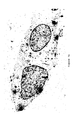
- the results showed that at 3-8 h, large numbers of nanoparticles were located between the basal plasma membrane and the collagen matrix on the supporting membrane ( FIG. 1 ).
- the nanoparticles were also present in the cytosol, but there were very few particles in vesicles or the nucleus or in intercellular junctions.
- the presence of nanoparticles in the cytosol and their absence from intercellular junctions suggested that they were directly crossing the cells by transcytosis, and were not reaching the basal membrane by paracellular movement.
- nanoparticles were moving across the cells experiments were carried out for 3 hours using hCMEC/D3 cells in the presence of antibiotics that interfere with endocytosis and/or vesicular transport—cytochalasin-B (glucose transport) chlorpromazine (clathrin-coated vesicles), nocodazole (microtubules), cytochalasin-D (microfilaments) and nystatin (caveolae and lipid rafts).
- cytochalasin-B glucose transport
- chlorpromazine clathrin-coated vesicles
- nocodazole microtubules
- cytochalasin-D microfilaments
- nystatin caveolae and lipid rafts
- Glucose-coated particles transcytosed more effectively than glutathione-coated nanoparticles and both 4 nm nanoparticles were more effective than the 30 nm colloidal nanoparticles. This result suggests that the characteristics of the nanoparticle, including its size, ligand and charge, all contribute towards the effectiveness of the transfer.
- the ultimate aim of the project was to determine whether the nanoparticles could act as a carrier across the blood-brain barrier and target glial cells.
- the nanoparticles accumulated between the basal plasma membrane of the endothelium and the membrane of the insert.
- the nanoparticles were also seen moving through the pores (220 nm) in the filters (see FIG. 3 a ), which indicated that they could be released by the endothelium and potentially enter the interstitial spaces.
- astrocytes containing nanoparticles are positioned at different depths from the endothelial monolayer and it was possible to detect the spread of nanoparticles to deeper astrocytes over 1-3 hours, although the numbers of particles detected per cell was similar at all times (Table 1).
- the thickness of the compressed gels is 40-60 ⁇ m. Therefore the observation that the median distance of nanoparticles from the endothelium at 3 hours was 16.7 ⁇ m, suggests that the nanoparticles can permeate the entire gel depth by this time.
- Gold nanoparticles have considerable potential as carriers of therapeutic agents across the blood-brain barrier, as they are not immunogenic and smaller nanoparticles (3-5 nm) are not cytotoxic except at high doses (27, 28, 29).
- 4 nm glucose-coated gold nanoparticles can cross brain endothelium with no detectable damage to the endothelial cells (33).
- glucose-coated nanoparticles were selected because of their potential to bind to the glucose receptor, Glut-1, on brain endothelium and astrocytes.
- transcytosis was not blocked by antibiotics that interfere with transport ( FIG. 4 ), and altering the concentration of glucose in the medium also had no effect on transcytosis (data not shown).
- transcytosis is not dependent on the glucose transporter system, and the physical configuration of the glucose coating makes it unlikely that it could engage the glut-1 receptor.
- the initial attachment to the endothelium depends on the biophysical properties of the nanoparticles and the cells.
- the glycocalyx of brain endothelium is highly sialylated, and quite different from endothelium in other tissues (30), which could explain the selective uptake by brain endothelium.
- the size and composition of the nanoparticles is also important. We found that 30 nm nanoparticles and glutathione-coated 4 nm gold nanoparticles were both significantly less efficient at crossing the endothelium ( FIG. 6 ).
- nanoparticles were rarely seen in the nuclei of the endothelium, but common in the nuclei of astrocytes, either in single cell cultures or cocultures ( FIG. 7 ). It is possible that changes in the surface coating of the nanoparticles occur during the extended period of the coculture, or as the particles cross the endothelium, which means that they subsequently tend to localise to the astrocyte nucleus. Currently the reason for this difference in subcellular localisation is obscure. Regardless of the mechanism, it is important that the nanoparticles are not trapped in the endothelium, if they are to be used to deliver a therapeutic cargo to cells of the CNS.
- transcytosed nanoparticles The number of transcytosed nanoparticles is also an important consideration. Our calculations suggest that >70,000 nanoparticles cross each endothelial cell and several hundred accumulate in each astrocyte. They therefore have the potential to carry an effective dose of a toxic agent, a receptor agonist or a gene to the target cells, if the process can be made to occur at a similar level in vivo. In short, 4 nm glucose-coated gold nanoparticles are selective for brain endothelium and they have great potential for delivery of therapeutic agents to target cells in the CNS.
- Agents including small molecule drugs, labels and/or biological agents such as peptides or nucleic acids may be coupled to the nanoparticles as defined herein using essentially any suitable technique. Agents may be covalently linked to the core of the nanoparticle or may form a binding interaction with the corona of the nanoparticle.
- a label e.g. an MRI contrast agent, such as a lanthanide may be complexed by carbohydrate groups present as ligands attached to the nanoparticle core (see, for example, Example 3 of WO 2004/108165, the entire contents of which are expressly incorporated herein by reference).
- one or more peptides or proteins may be non-covalently bound to the corona of the nanoparticle (see, e.g., Example 3 of WO 2011/154711, the entire contents of which are expressly incorporated herein by reference).
- a nucleic acid such as an siRNA or a segment of DNA or RNA may be covalently linked to the core of the nanoparticle via thiol derivatisation of the nucleic acid at the 3′ or 5′ terminus of the nucleic acid strand (see, for example, WO 2005/116226, in particular the examples thereof, the entire contents of which are expressly incorporated herein by reference).
- Delivery of agents to the CNS utilising nanoparticles as defined herein may be assessed using a model of the blood brain barrier as described in detail in Example 2.
- assessment of successful delivery of the agent of interest to a CNS cell may involve physically identifying the presence of the agent (optionally together with the nanoparticle) in the target cell and/or performing a functional assay of the effect of said agent on the target cell.
- a suitable functional assay may comprise assessment of expression of said gene in the target CNS cell.
- one or more controls such as nanoparticles in the absence of the agent and/or agent in the absence of the nanoparticles will be contacted to the blood brain barrier model system thereby providing a reference against which the presence and/or effect of the nanoparticle having a cargo of the agent of choice is assessed.
- Example 4 Nanoparticle Coatings and Transfer Across the Brain Endothelium
- nanoparticles having a corona comprising one or more of the following ligand species were synthesised essentially as described in Example 1:
- the transfer rate of nanoparticles was found to vary according to the composition of the nanoparticle corona (i.e. the coating) and variation was seen between batches of nanoparticles (see FIG. 8 ).
- the number of nanoparticles detected below the basal membrane at 8 hours is plotted for each nanoparticle ligand species as a percentage of the control designated the C2-glucose-1 (i.e. a first batch of nanoparticles having a glucose ligand corona with a C2 linker). It is clear that C11-glucose and a third batch of glucose (C2-glucose-3) exhibit greater than control transfer rate.
- galactosamine-NPs i.e. nanoparticles having a corona of galactosamine ligands
- top bottom Well 1 41.6 ug 19.3 g Applied 53.28 ug Well 2 42.96 21.3 to the top, 8 hr Well 3 41.96 incubation
- glycocalyx was then investigated. Removal of glycocalyx was achieved by subjecting enthothelium cells to enzyme pre-treatment with heparinise, chondroitinase or neuraminidase and then measuring the number of nanoparticles detected per micron at the basal membrane 8 hours after application of the nanoparticles (see FIG. 11 ). It is evident that removal of glycocalyx inhibits transport of the nanoparticles, with the effect of chondroitinase pre-treatment being statistically significant (see ***).
- FIG. 12 shows insulin-coated nanoparticles (8 insulins and Zinc) taken up by hCMEC/D3 cells. 2 ng/cm 2 of nanoparticles were applied for 3 hours. It was found that insulin-coated nanoparticles transfer rapidly across hCMEC/D3 cells, primarily by vesicles (see FIGS. 13 and 14A and 14B ).
- FIG. 15A shows the percentage of astrocytes positive with nanoparticles at 1, 3 and 8 hours.
- FIG. 15B shows the average distance of nanoparticles from the endothelium in microns (maximum distance shown in inset figure) at 1, 3 and 8 hours.
- Gold nanoparticle uptake for various nanoparticle corona compositions was investigated in 2D astrocyte culture and 3D co-cultured astrocytes (see FIGS. 16A and 16B , respectively). The uptake into cytosol, vesicles and nucleus are shown, measured in nanoparticles per cell.
- FIG. 17 shows the location of nanoparticles on filters (edge or middle) for endothelium and astrocytes.
- the number of nanoparticles in both endothelium and astrocytes was higher in the middle of the filters than at the edges, but this difference was not found to be statistically significant (p>0.05).
- FIG. 18 shows results investigating nanoparticles in astrocyte/D3 co-culture.
- TEM transmission electron microscopy
- FIG. 19 shows results investigating the time course of uptake of glucose-NPs.
- Glucose-coated nanoparticles were found to be selective for brain endothelium. Up to 70,000 nanoparticles cross each endothelial cell in an 8 hour period. Nanoparticles move at 10-20 ⁇ m per hour, with more than 400 nanoparticles reaching each astrocyte.
- Galactosamine and glutathione nanoparticles also cross efficiently. Movement of glucose-coated-NPs is primarily across the cytosol. Removal of glycocalyx or reduction in temperature reduces cytosolic transfer.
- Insulin-coated NPs appear to use rapid vesicular transcytosis across the endothelium. They can also be taken up by astrocytes. Without wishing to be bound by any particular theory, the present inventors contemplate that the ability to delivery insulin to the CNS via nanoparticles, as described herein, will have significant medical potential because the avoidance of unwanted hypoglycemia (e.g. due to peripheral effects of insulin) may be diminished or prevented.
Abstract
Description
-
- (a) a nanoparticle comprising:
- (i) a core comprising a metal and/or a semiconductor;
- (ii) a corona comprising a plurality of ligands covalently linked to the core, wherein said ligands comprise a carbohydrate, insulin and/or a glutathione; and
- (b) the at least one agent to be delivered to the CNS.
- (a) a nanoparticle comprising:
-
- (a) a nanoparticle comprising:
- (i) a core comprising a metal and/or a semiconductor;
- (ii) a corona comprising a plurality of ligands covalently linked to the core, wherein said ligands comprise a carbohydrate, insulin and/or a glutathione; and
- (b) at least one agent to be delivered to the CNS.
- (a) a nanoparticle comprising:
-
- providing a cell culture endothelium, optionally co-cultured with astrocytes;
- contacting the endothelium with a nanoparticle having associated with it at least one candidate agent; and
- identifying whether the candidate agent is delivered across the endothelium by the nanoparticle,
wherein said nanoparticle comprises: - (i) a core comprising a metal and/or a semiconductor; and
- (ii) a corona comprising a plurality of ligands covalently linked to the core, wherein said ligands comprise a carbohydrate, insulin and/or a glutathione.
-
- administering to a non-human mammalian test subject via a non-central route of administration a composition comprising a nanoparticle having associated with it at least one candidate agent; and
- identifying whether the candidate agent is delivered across the blood-brain barrier to the CNS of said test subject,
wherein said nanoparticle comprises: - (i) a core comprising a metal and/or a semiconductor; and
- (ii) a corona comprising a plurality of ligands covalently linked to the core, wherein said ligands comprise a carbohydrate, insulin and/or a glutathione.
| TABLE 1 |
| Accumulation of nanoparticles in astrocytes in coculture |
| Time1 | Cells2 | % Positive cells3 | Distance4 | Particles/ |
| 1 hour | 411 | 7.4 ± 2.0 | 10.6 ± 1.6 | 3.53 ± 0.41 |
| 3 hours | 308 | 15.9 ± 1.0 | 16.7 ± 2.6 | 4.16 ± 0.46 |
| 8 hours | 240 | 19.5 ± 0.6 | 15.5 ± 1.4 | 3.75 ± 1.15 |
| 1Time after application of nanoparticles to the apical surface of the endothelium. | ||||
| 2Total number of astrocytes observed. | ||||
| 3Percentage of astrocytes with intracellular nanoparticles. | ||||
| 4The distance in μm of each astrocyte containing nanoparticles from the basal surface of the endothelium. | ||||
| 5Number of nanoparticles observed in cells containing nanoparticles. | ||||
-
- To determine whether gold nanoparticles with various surface coatings cross human brain endothelium.
- To determine if transport can be selective for brain endothelium.
- To investigate potential mechanisms of transfer and optimise the delivery system.
- To determine whether nanoparticles can target glial cells after transfer across the endothelium.
-
- C2-glucose
- C5-glucose
- C11-glucose
- Maltose
- Lactose
- Galactose
- Galactosamine
- Glutathione
| | bottom | ||||
| Well | |||||
| 1 | 41.6 ug | 19.3 g | Applied 53.28 | ||
| Well | |||||
| 2 | 42.96 | 21.3 | to the top, 8 | ||
| Well | |||||
| 3 | 41.96 | incubation | |||
- 1. Wolburg H, Lippoldt A, (2001) Tight junctions of the blood-brain barrier: development, composition and regulation. Vasc. Pharm. 38: 323-337.
- 2. Sarkadi B, Homolya L, Szakács G, Váradi A (2006) Human multi-drug resistance ABCB and ABCG transporters: Participation in a chemoimmunity defence system. Physiol Rev 86: 1179-1236.
- 3. Manfredsson F P and Mandel R J (2010) Development of gene therapy for neurological disorders. Discovery Medicine 9: 204-211.
- 4. Baker D and Hankey D J (2003) Gene therapy in autoimmune demyelinating diseases of the central nervous system. Gene therapy 10: 844-853.
- 5. Sloane E et al. (2009) Anti-inflammatory cytokine gene therapy decreases sensory and motor dysfunction in experimental Multiple Sclerosis. Brain Behav Immun. 23: 92-100.
- 6. Deverman B E and Patterson P H (2012) Exogenous Leukemia Inhibitory Factor Stimulates Oligodendrocyte Progenitor Cell Proliferation and Enhances Hippocampal Remyelination. J. Neurosci 32: 2100-2109.
- 7. Patel T et al (2012) Polymeric nanoparticles for drug delivery to the central nervous system. Advanced. Drug Delivery Revs 64: 701-705.
- 8. Kanwar et al. (2012) Nanoparticles in the treatment and diagnosis of neurological disorders: untamed dragon with the fire power to heal. Nanomedicine: nanotechnology, Biology and Medicine 8: 399-414.
- 9. Sonavane G, Tomoda K, Makino K (2008) Biodistribution of colloidal gold nanoparticles after intravenous administration: Effect of particle size. Colloids and Surfaces Biointerfaces 66: 274-280.
- 10. Chen Y S, Hung Y C, Liau I, Huang S (2009) Assessment of the In Vivo Toxicity of Gold Nanoparticles. Nanoscale Res Lett 4:858-864.
- 11. Etame, A B, Smith C A, Chan W C W, Rutka J T (2011) Design and potential application of PEGylated gold nanoparticles with size-dependent permeation through brain microvasculature. Nanomedicine: NBM 7:992-1000
- 12. Gao H J, Shi W D, Freund L B (2005). Mechanics of receptor-mediated endocytosis. Proc Natl Acad Sci USA 102: 9469-9474.
- 13. Zhang S, Li J, Lukotrafitis G, Bao G, Suresh S (2009) Size-dependent endocytosis of nanoparticles. Adv. Mater. 21: 419-424.
- 14. Shan Y, Ma S, Nie L, Shang X, Hao X, Tang Z, Wang H (2011). Size-dependent endocytosis of single gold nanoparticles. Chem Commun 47: 8091-8093.
- 15. Alkilany A M, Murphy C J (2010) Toxicity and cellular uptake of gold nanoparticles: what we have learned so far? J Nanopart Res 12: 2313-2333
- 16. Zensi A, Begley D, Pontikis C, Legros C, Mihoreanu L, Rachel C, Kreuter J (2010) Human serum albumin nanoparticles modified with apolipoprotein A-I cross the blood-brain barrier and enter the rodent brain. Journal of Drug Targeting 10: 842-848.
- 17. Chen L, Yokel R A, Hennig B, Toborek M (2008) Manufactured aluminium oxide nanoparticles decrease expression of tight junction proteins in brain vasculature. J Neuroimmune Pharmacol 3: 286-295.
- 18. Georgieva J V et al. (2011) Surface characteristics of nanoparticles determine their intracellular fate in processing by human blood-brain barrier endothelial cells in vitro. Molecular Therapy 19: 318-325.
- 19. Chithrani B D, Ghazani A A, Chan W C (2006) Determining the size and shape dependence of gold nanoparticles uptake by mammalian cells. Nano Lett 6: 662-668.
- 20. Wang Y Y et al (2009) Receptor mediated therapeutic transport across the blood brain barrier. Immunotherapy, Vol. 1, No. 6: 983-993.
- 21. Morgello S et al (1995) The human blood brain barrier transporter (GLUT1) is a glucose transporter of gray matter astrocytes. Glia 14: 43-54.
- 22. Lund T, Callaghan M F, Williams P, Turmaine M, Bachmann C, Rademacher T, Roitt I M, Bayford R (2011) The influence of ligand organization on the rate of uptake of gold nanoparticles by colorectal cancer cells. Biomaterials 32: 9776-9784.
- 23. East E, Golding J P Phillips J B (2009) A versatile 3D culture model facilitates monitoring of astrocytes undergoing reactive gliosis. J Tissue Eng. Regen. Med. 8: 634-646.
- 24. East E, Golding J P, Phillips J B (2012) Engineering an integrated cellular interface in three-Dimensional hydrogel cultures permits monitoring of reciprocal astrocyte and neuronal responses. Tissue Eng Part C Methods. Epub ahead of print PMID:22235832.
- 25. Weksler B B, Subileau E A, Perriere N, Charneau P, Holloway K, Leveque M, Tricoire-Leignel H, Nicotra A, Bourdoulous S, Turowski P, Male D K, Roux F, Greenwood J, Romero I A, Couraud P-O (2005) Blood brain barrier specific properties of a human adult brain endothelial cell line, Faseb J. 19: 1872-1874.
- 26. Male D K (1995) Brain Endothelium. In “Neural Cell Culture” Edited Cohen and Wilkin, for The Practical Approach Series, IRL Press, Oxford, UK.
- 27. de la Fuente J M, Berry C C (2005) Tat peptide as an efficient molecule to translocate gold nanoparticles into the cell nucleus. Bioconjug Chem. 16:1176-80.
- 28. Male K B, Lachance B, Hrapovic S, Sunahara G, Luong J H (2008) Assessment of cytotoxicity of quantum dots and gold nanoparticles using cell-based impedance spectroscopy. Anal Chem. 80:5487-93.
- 29. Gannon C J, Patra C R, Bhattacharya R, Mukherjee P, Curley S A (2008) Intracellular gold nanoparticles enhance non-invasive radiofrequency thermal destruction of human gastrointestinal cancer cells. J. Nanobiotechnology 6:2.
- 30. Santos W L C, Rahman J, Klein N and Male D K (1996) Control of lymphocyte adhesion to brain endothelium: ICAM-1, VCAM-1 and negative charge. J. Neuroimmunol, 66, 125-134
- 31. Gu Y J, Cheng J, Lin C C, Lam Y W, Cheng S H, Wong W T (2009) Nuclear penetration of surface functionalized gold nanoparticles. Toxicol Appl Pharmacol. 237:196-204.
- 32. Cho E C, Zhang Q, Xia Y (2011) The effect of sedimentation and diffusion on cellular uptake of gold nanoparticles. Nat. Nanotechnol. 6:385-91.
- 33. Gromnicova R, et al. (2013) Glucose-Coated Gold Nanoparticles Transfer across Human Brain Endothelium and Enter Astrocytes In Vitro. PLoS ONE 8(12): e81043, pp. 1-10.
Claims (3)
Applications Claiming Priority (2)
| Application Number | Priority Date | Filing Date | Title |
|---|---|---|---|
| GBGB1302427.8A GB201302427D0 (en) | 2013-02-12 | 2013-02-12 | Nanoparticle delivery compositions |
| GB1302427.8 | 2013-02-12 |
Publications (2)
| Publication Number | Publication Date |
|---|---|
| US20140227186A1 US20140227186A1 (en) | 2014-08-14 |
| US10300022B2 true US10300022B2 (en) | 2019-05-28 |
Family
ID=47998969
Family Applications (1)
| Application Number | Title | Priority Date | Filing Date |
|---|---|---|---|
| US14/175,422 Expired - Fee Related US10300022B2 (en) | 2013-02-12 | 2014-02-07 | Nanoparticle delivery compositions |
Country Status (9)
| Country | Link |
|---|---|
| US (1) | US10300022B2 (en) |
| EP (1) | EP2956119B1 (en) |
| JP (1) | JP6373281B2 (en) |
| CN (1) | CN105073097A (en) |
| AU (1) | AU2014217594B2 (en) |
| CA (1) | CA2900628C (en) |
| ES (1) | ES2749116T3 (en) |
| GB (1) | GB201302427D0 (en) |
| WO (1) | WO2014125256A1 (en) |
Cited By (1)
| Publication number | Priority date | Publication date | Assignee | Title |
|---|---|---|---|---|
| US11179474B1 (en) * | 2015-07-24 | 2021-11-23 | Midatech Limited | Nanoparticle-based liver-targeting therapy and imaging |
Families Citing this family (17)
| Publication number | Priority date | Publication date | Assignee | Title |
|---|---|---|---|---|
| GB201303787D0 (en) * | 2013-03-04 | 2013-04-17 | Midatech Ltd | Nanoparticle peptide compositions |
| US10688125B2 (en) | 2014-12-23 | 2020-06-23 | Midatech Ltd. | Nanoparticles and their use in cancer therapy |
| GB201506112D0 (en) * | 2015-04-10 | 2015-05-27 | Midatech Ltd And Inst Nationale De La Sant� Et De Al Rech Medicale And University College Car | Nanoparticle-based antigen specific immunotherapy |
| WO2017060916A1 (en) * | 2015-10-09 | 2017-04-13 | NOUSHAD Javed Md | Metallic nanoparticle alone and/or in combination as novel agent for the treatment of uncontrolled electric conductance related disorders and/or seizure, epilepsy & convulsions. |
| KR101745796B1 (en) * | 2015-10-26 | 2017-06-09 | 동국대학교 산학협력단 | Method of direct lineage reprogramming for generating neurons using electromagnetic-induced metalilic nanoparticle |
| US10285934B1 (en) | 2015-11-01 | 2019-05-14 | Battelle Memorial Institute | Administration of a drug through the blood brain barrier using stimuli-responsive nanoparticles |
| CN105796540A (en) * | 2016-03-18 | 2016-07-27 | 沈阳药科大学 | Application of tolfenamic acid to preparation of medicine for treating Huntington's disease |
| US10188749B2 (en) | 2016-04-14 | 2019-01-29 | Fred Hutchinson Cancer Research Center | Compositions and methods to program therapeutic cells using targeted nucleic acid nanocarriers |
| EP3463316A4 (en) * | 2016-06-03 | 2020-05-27 | Stemgenics, Inc. | Functionalized nanoparticles for the intracellular delivery of biologically active molecules and methods for their manufacture and use |
| EP3565535A4 (en) | 2017-01-05 | 2020-12-30 | Fred Hutchinson Cancer Research Center | Systems and methods to improve vaccine efficacy |
| GB201701745D0 (en) * | 2017-02-02 | 2017-03-22 | Midatech Ltd | Nanoparticle-based liver-targeting therapy and imaging |
| KR20200109325A (en) * | 2018-01-18 | 2020-09-22 | 프레드 헛친슨 켄서 리서치 센터 | Alteration of the inflammatory state of immune cells in vivo by regulating the cell activation state |
| CN109939243B (en) * | 2019-01-16 | 2022-03-15 | 武汉广行科学研究有限公司 | Copper cluster, thymine modified hyaluronic acid and poly-copper cluster, and preparation method and application thereof |
| JP7372978B2 (en) * | 2019-01-16 | 2023-11-01 | 深▲セン▼深見医薬科技有限公司 | Copper nanoclusters, hyaluronic acid and poly(copper nanoclusters) modified with thymine, their production methods, and their applications |
| CN111504961B (en) * | 2020-03-31 | 2023-05-02 | 南昌大学 | Fluorescent phytic acid detection method based on glutathione gold nanoclusters |
| EP4203975A4 (en) * | 2020-11-27 | 2024-01-03 | Shenzhen Profound View Pharmaceutical Tech Co Ltd | Gold clusters, compositions, and methods for treatment of cerebral ischemic stroke |
| CN113209046B (en) * | 2021-05-08 | 2022-09-09 | 中国人民解放军陆军军医大学 | CoSe @ BSA nanoparticle pharmaceutical composition and preparation method and application thereof |
Citations (17)
| Publication number | Priority date | Publication date | Assignee | Title |
|---|---|---|---|---|
| WO2002032404A2 (en) | 2000-10-16 | 2002-04-25 | Consejo Superior De Investigaciones Cientificas | Nanoparticles |
| WO2004108165A2 (en) | 2003-06-09 | 2004-12-16 | Consejo Superior De Investigaciones Cientificas | Magnetic nanoparticles linked to a lingand |
| WO2005091704A2 (en) | 2004-03-25 | 2005-10-06 | Consejo Superior De Investigaciones Científicas | Magnetic nanoparticles of noble metals |
| WO2005116226A2 (en) | 2004-05-24 | 2005-12-08 | Midatech Ltd | Nanoparticles comprising rna ligands |
| WO2006037979A2 (en) | 2004-10-01 | 2006-04-13 | Midatech Limited | Nanoparticles comprising antigens and adjuvants and immunogenic structure |
| WO2006102377A2 (en) | 2005-03-21 | 2006-09-28 | The Regents Of The University Of California | Functionalized magnetic nanoparticles and methods of use thereof |
| WO2007015105A2 (en) | 2005-08-04 | 2007-02-08 | Thomas William Rademacher | Nanoparticles comprising antibacterial ligands |
| US20070141133A1 (en) | 2005-12-19 | 2007-06-21 | Industrial Technology Research Institute | Glutathione based delivery system |
| WO2007122388A2 (en) | 2006-04-13 | 2007-11-01 | Midatech Limited | Nanoparticles containing three various ligands for providing immune responses against infectious agents |
| US20080226917A1 (en) * | 2007-02-20 | 2008-09-18 | Research Foundation Of State University Of New York | Core-shell nanoparticles with multiple cores and a method for fabricating them |
| US20100034735A1 (en) * | 2008-08-06 | 2010-02-11 | Jie Chen | Targeted nanoparticles for cancer diagnosis and treatment |
| US20100260686A1 (en) * | 2009-04-09 | 2010-10-14 | Washington, University Of | Nanoparticles for brain tumor imaging |
| EP2305310A1 (en) | 2009-09-25 | 2011-04-06 | Asociación Centro de Investigación Cooperativa en Biomateriales - CIC biomaGUNE | Gold -coated magnetic glyconanoparticles functionalised with proteins for use as diagnostic and therapeutic agents |
| WO2011154711A1 (en) | 2010-06-10 | 2011-12-15 | Midatech Limited | Peptide-carrying nanoparticles |
| US20120009260A1 (en) * | 2010-06-10 | 2012-01-12 | Schobel Alexander M | Nanoparticle film delivery systems |
| US20120301535A1 (en) | 2010-06-10 | 2012-11-29 | Phillip Williams | Combination peptide-nanoparticles and delivery systems incorporating same |
| US20130344125A1 (en) * | 2010-11-26 | 2013-12-26 | Thiresen Govender | Drug delivery device |
-
2013
- 2013-02-12 GB GBGB1302427.8A patent/GB201302427D0/en not_active Ceased
-
2014
- 2014-02-07 AU AU2014217594A patent/AU2014217594B2/en not_active Ceased
- 2014-02-07 EP EP14708315.8A patent/EP2956119B1/en active Active
- 2014-02-07 JP JP2015556568A patent/JP6373281B2/en not_active Expired - Fee Related
- 2014-02-07 CN CN201480019125.7A patent/CN105073097A/en active Pending
- 2014-02-07 CA CA2900628A patent/CA2900628C/en not_active Expired - Fee Related
- 2014-02-07 US US14/175,422 patent/US10300022B2/en not_active Expired - Fee Related
- 2014-02-07 ES ES14708315T patent/ES2749116T3/en active Active
- 2014-02-07 WO PCT/GB2014/050372 patent/WO2014125256A1/en active Application Filing
Patent Citations (18)
| Publication number | Priority date | Publication date | Assignee | Title |
|---|---|---|---|---|
| WO2002032404A2 (en) | 2000-10-16 | 2002-04-25 | Consejo Superior De Investigaciones Cientificas | Nanoparticles |
| WO2004108165A2 (en) | 2003-06-09 | 2004-12-16 | Consejo Superior De Investigaciones Cientificas | Magnetic nanoparticles linked to a lingand |
| WO2005091704A2 (en) | 2004-03-25 | 2005-10-06 | Consejo Superior De Investigaciones Científicas | Magnetic nanoparticles of noble metals |
| WO2005116226A2 (en) | 2004-05-24 | 2005-12-08 | Midatech Ltd | Nanoparticles comprising rna ligands |
| WO2006037979A2 (en) | 2004-10-01 | 2006-04-13 | Midatech Limited | Nanoparticles comprising antigens and adjuvants and immunogenic structure |
| JP2008533203A (en) | 2005-03-21 | 2008-08-21 | ザ リージェンツ オブ ザ ユニバーシティ オブ カリフォルニア | Functionalized magnetic nanoparticles and uses thereof |
| WO2006102377A2 (en) | 2005-03-21 | 2006-09-28 | The Regents Of The University Of California | Functionalized magnetic nanoparticles and methods of use thereof |
| WO2007015105A2 (en) | 2005-08-04 | 2007-02-08 | Thomas William Rademacher | Nanoparticles comprising antibacterial ligands |
| US20070141133A1 (en) | 2005-12-19 | 2007-06-21 | Industrial Technology Research Institute | Glutathione based delivery system |
| WO2007122388A2 (en) | 2006-04-13 | 2007-11-01 | Midatech Limited | Nanoparticles containing three various ligands for providing immune responses against infectious agents |
| US20080226917A1 (en) * | 2007-02-20 | 2008-09-18 | Research Foundation Of State University Of New York | Core-shell nanoparticles with multiple cores and a method for fabricating them |
| US20100034735A1 (en) * | 2008-08-06 | 2010-02-11 | Jie Chen | Targeted nanoparticles for cancer diagnosis and treatment |
| US20100260686A1 (en) * | 2009-04-09 | 2010-10-14 | Washington, University Of | Nanoparticles for brain tumor imaging |
| EP2305310A1 (en) | 2009-09-25 | 2011-04-06 | Asociación Centro de Investigación Cooperativa en Biomateriales - CIC biomaGUNE | Gold -coated magnetic glyconanoparticles functionalised with proteins for use as diagnostic and therapeutic agents |
| WO2011154711A1 (en) | 2010-06-10 | 2011-12-15 | Midatech Limited | Peptide-carrying nanoparticles |
| US20120009260A1 (en) * | 2010-06-10 | 2012-01-12 | Schobel Alexander M | Nanoparticle film delivery systems |
| US20120301535A1 (en) | 2010-06-10 | 2012-11-29 | Phillip Williams | Combination peptide-nanoparticles and delivery systems incorporating same |
| US20130344125A1 (en) * | 2010-11-26 | 2013-12-26 | Thiresen Govender | Drug delivery device |
Non-Patent Citations (46)
| Title |
|---|
| Alkilany, Alaaldin M. et al., "Toxicity and cellular uptake of gold nanoparticles: what we have learned so far?", J. Nanopart. Res., 12: 2313-2333 (2010). |
| Baker, David et al., "Gene therapy in autoimmune, demyelinating disease of the central nervous system", Gene Therapy, 10: 844-853 (2003). |
| Cao, YunWei Charles et al., "Nanoparticles with Raman Spectroscopic Fingerprints for DNA and RNA Detection", Science, 297: 1536-1540 (2002). |
| Chen, Lei et al., "Manufactured Aluminum Oxide Nanoparticles Decreae Expression of Tight Junction Proteins in Brain Vasculature", J. Neuroimmune Pharmacol., 3: 286-295 (2008). |
| Chen, Yu-Shiun et al., "Assessment of the In Vivo Toxicity of Gold Nanoparticles", Nanoscale Res. Lett, 4: 858-864 (2009). |
| Chithrani, B. Devika et al., "Determining the Size and Shape Dependence of Gold Nanoparticle Uptake into Mammalian Cells", Nano Letters, 6(4): 662-668 (2006). |
| Cho, Eun Chul et al., "The effect of sedimentation and diffusion on cellular uptake of gold nanoparticles", Nature Nanotechnology, 6: 385-391 (2011). |
| De la Fuente, Jesus M. et al., "Tat Peptide as an Efficient Molecule to Translocate Gold Nanoparticles into the Cell Nucleus", Bioconjugate Chem., 16: 1176-1180 (2005). |
| Deverman, Benjamin E. et al., "Exogenous Leukemia Inhibitory Factor Stimulates Oligodendrocyte Progenitor Cell Proliferation and Enhances Hippocampal Remyelination", The Journal of Neuroscience, 32(6): 2100-2109 (2012). |
| Dos Santos, Washington L.C. et al., "Control of lymphocyte adhesion to brain and aortic endothelium: ICAM-1, VCAM-1 and negative charge", Journal of Neuroimmunology, 66: 125-134 (1996). |
| East, Emma et al., "A versatile 3D culture model facilitates monitoring of astrocytes undergoing reactive gliosis", J. Tissue Eng. Regen. Med., 3: 634-646 (2009). |
| East, Emma et al., "Engineering an Integrated Cellular Interface in Three-Dimensional Hydrogel Cultures Permits Monitoring of Reciprocal Astrocyte and Neuronal Responses", Tissue Engineering: Part C, 18(7): 526-537 (2012). |
| English translation of Office Action, dated Feb. 6, 2018, issued in Chinese Application No. 2014800191257. |
| Etame, Arnold B. et al., "Design and potential application of PEGylated gold nanoparticles with size-dependent permeation through brain microvasculature", Nanomedicine: Nanotechnology, Biology, and Medicine, 7: 992-1000 (2011). |
| Gannon, Christopher J. et al., "Intracellular gold nanoparticles enhance non-invasive radiofrequency thermal destruction of human gastrointestinal cancer cells", Journal of Nanobiotechnology, 6: 2 (2008). |
| Gao, Huajian et al., "Mechanics of receptor-mediated endocytosis", PNAS, 102(27): 9469-9474 (2005). |
| Georgieva, Julia V. et al., "Surface Characteristics of Nanoparticles Determine Their Intracellular Fate in and Processing by Human Blood-Brain Barrier Endothelial Cells In Vitro", Molecular Therapy, 19(2): 318-325 (2011). |
| Gromnicova, Radka et al., "Glucose-Coated Gold Nanoparticles Transfer across Human Brain Endothelium and Enter Astrocytes in Vitro", PLOS One, 8(12): e81043 (2013). |
| Gu, Yan-Juan et al., "Nuclear penetration of surface functionalized gold nanoparticles", Toxicology and Applied Pharmacology, 237: 196-204 (2009). |
| Huang, Shih-Hung et al., "Direct Binding and Characterization of Lipase onto Magnetic Nanoparticles", Biotechnol. Prog., 19: 1095-1100 (2003). |
| Invitation to Pay Additional Fees and, Where Applicable, Protest Fee with Annex to Form PCT/ISA/206-Communication Relating to the Results of the Partial International Search dated Apr. 23, 2014 in related International Application No. PCT/GB2014/050372. |
| Invitation to Pay Additional Fees and, Where Applicable, Protest Fee with Annex to Form PCT/ISA/206—Communication Relating to the Results of the Partial International Search dated Apr. 23, 2014 in related International Application No. PCT/GB2014/050372. |
| Kanwar, Jagat et al., "Nanoparticles in the treatment and diagnosis of neurological disorders: untamed dragon with fire power to heal", Nanomedicine: Nanotechnology, Biology, and Medicine, 8: 399-414 (2012). |
| Lund, Torben et al., "The influence of ligand organization on the rate of uptake of gold nanoparticles by colorectal cancer cells", Biomaterials, 32: 9776-9784 (2011). |
| Male, David, "Brain endothelium", Natural Cell Culture, eds. Cohen & Wilkin, pp. 121-130 (1995). |
| Male, Keith B. et al., "Assessment of Cytotoxicity of Quantum Dots and Gold Nanoparticles Using Cell-Based Impedance Spectroscopy", Anal. Chem., 80(14): 5487-5493 (2008). |
| Manfredsson, Fredric P. et al., "Development of Gene Therapy for Neurological Disorders", Discovery Medicine, 9(46): 204-211 (2010). |
| Morgello, Susan et al., "The Human Blood0Brain Barrier Glucose Transporter (GLUT1) is a Glucose Transporter of Gray Matter Astrocytes", GLIA, 14: 43-54 (1995). |
| Neveu, S. et al., "Size-Selective Chemical Synthesis of Tartrate Stabilized Cobalt Ferrite Ionic Magnetic Fluid", J. Colloid and Interface Science, 255: 293-298 (2002). |
| Office Action, dated Feb. 6, 2018, issued in Chinese Application No. 2014800191257. |
| Office Action, dated Nov. 28, 2017, issued in corresponding Japanese Application No. 2015-556568. |
| Patel, Toral et al., "Polymeric nanoparticles for drug delivery to the central nervous system", Advanced Drug Delivery Reviews, 64: 701-705 (2012). |
| Rice, Peter et al., "EMBOSS: The European Molecular Biology Open Software Suite", Trends in Genetics, 16(6): 276-77 (2000). |
| Sarkadi, Balazs et al., "Human Multidrug Resistance ABCB and ABCG Transporters: Participation in a Chemoimmunity Defense System", Physiol. Rev., 86: 1179-1236 (2006). |
| Shan, Yuping et al., "Size-dependent endocytosis of single gold nanoparticles", Chem. Commun., 47: 8091-8093 (2011). |
| Sharma et al., Langmuir 2012, 28, 15958-15965. * |
| Sloane, Evan et al., "Anti-inflammatory cytokine gene therapy decreases sensory and motor dysfunction in experimental Multiple Sclereosis: MOG-EAE behavorial and anatomical symptom treatment with cytokine gene therapy", Brain, Behavior, and Immunity, 23: 92-100 (2009). |
| Sonavane, Ganeshchandra et al., "Biodistribution of colloidal gold nanoparticles after intravenous administration: Effect of particle size", Colloids and Surfaces B: Biointerfaces, 66: 274-280 (2008). |
| Vestal, Christy R. et al., "Effects of Surface Coordination Chemistry on the Magnetic Properties of MnFe2O4 Spinel Ferrite Nanoparticles", J. Am. Chem. Soc., 125: 9828-9833 (2003). |
| Wang, Yun-Yan et al., "Receptor-mediated therapeutic transport across the blood-brain barrier", Immunotherapy, 1(6): 983-993 (2009). |
| Weksler, B.B. et al., "Blood-brain barrier-specific properties of a human adult brain endothelial cell line", The FASEB Journal, 19: 1872-1875 (2005). |
| Wolburg, Hartwig et al., "Tight junctions of the blood-brain barrier: Development, composition and regulation", Vascular Pharmacology, 38: 323-337 (2002). |
| Zensi, Anja et al., "Human serum albumin nanoparticles modified with apolipoprotein A-I cross the blood-brain barrier and enter the rodent brain", Journal of Drug Targeting, 18(10): 842-848 (2010). |
| Zhang, Sulin et al., "Size-Dependent Endocytosis of Nanoparticles", Adv. Mater., 21: 419-424 (2009). |
| Zhu, Jun et al., Development of a AU nanoparticles templates for PET in vivo imaging: 18F labeled 3 nm water-soluble AuNP cross the blood brain barrier, Abstracts of Papers American Chemical Society, 244, p. 485 (2012). |
| Zhu, Jun, "Development of a Au nanoparticles template for PET in vivo imaging: 18F labeled 3nm water-soluble AuNP cross the blood brain barrier", Abstracts of Papers of the American Chemical Society, vol. 244 (2012). |
Cited By (1)
| Publication number | Priority date | Publication date | Assignee | Title |
|---|---|---|---|---|
| US11179474B1 (en) * | 2015-07-24 | 2021-11-23 | Midatech Limited | Nanoparticle-based liver-targeting therapy and imaging |
Also Published As
| Publication number | Publication date |
|---|---|
| GB201302427D0 (en) | 2013-03-27 |
| CA2900628C (en) | 2020-07-14 |
| CA2900628A1 (en) | 2014-08-21 |
| JP6373281B2 (en) | 2018-08-15 |
| AU2014217594B2 (en) | 2018-06-21 |
| EP2956119B1 (en) | 2019-08-14 |
| AU2014217594A1 (en) | 2015-08-20 |
| CN105073097A (en) | 2015-11-18 |
| EP2956119A1 (en) | 2015-12-23 |
| WO2014125256A1 (en) | 2014-08-21 |
| JP2016507549A (en) | 2016-03-10 |
| ES2749116T3 (en) | 2020-03-19 |
| US20140227186A1 (en) | 2014-08-14 |
Similar Documents
| Publication | Publication Date | Title |
|---|---|---|
| US10300022B2 (en) | Nanoparticle delivery compositions | |
| de Almeida et al. | Understanding nanoparticle endocytosis to improve targeting strategies in nanomedicine | |
| Huda et al. | Potential use of exosomes as diagnostic biomarkers and in targeted drug delivery: progress in clinical and preclinical applications | |
| Cupaioli et al. | Engineered nanoparticles. How brain friendly is this new guest? | |
| Kou et al. | The endocytosis and intracellular fate of nanomedicines: Implication for rational design | |
| Gan et al. | Transferrin-conjugated nanoparticles of poly (lactide)-D-α-tocopheryl polyethylene glycol succinate diblock copolymer for targeted drug delivery across the blood–brain barrier | |
| Zhang et al. | Interactions of nanomaterials and biological systems: Implications to personalized nanomedicine | |
| Peynshaert et al. | Exploiting intrinsic nanoparticle toxicity: the pros and cons of nanoparticle-induced autophagy in biomedical research | |
| Murthy | Nanoparticles in modern medicine: state of the art and future challenges | |
| Leonard et al. | Screening of budesonide nanoformulations for treatment of inflammatory bowel disease in an inflamed 3D cell-culture model | |
| US20140248365A1 (en) | Nanoparticle peptide compositions | |
| Oppong-Damoah et al. | Nanoparticle encapsulation increases the brain penetrance and duration of action of intranasal oxytocin | |
| Chavda et al. | Exosome nanovesicles: a potential carrier for therapeutic delivery | |
| Ling et al. | The potential benefits of nanotechnology in treating Alzheimer’s disease | |
| WO2006044660A2 (en) | Functionalized solid lipid nanoparticles and methods of making and using same | |
| Song et al. | Mechanisms of transcellular transport of wheat germ agglutinin-functionalized polymeric nanoparticles in Caco-2 cells | |
| US20100254914A1 (en) | Nanoworms for in vivo tumor targeting | |
| US20180177725A1 (en) | Cell membrane-formed nanoscale vesicles and methods of using thereof | |
| Shaikh et al. | Bleomycin loaded exosomes enhanced antitumor therapeutic efficacy and reduced toxicity | |
| US20220031630A1 (en) | Nanoparticle formulations and methods of their use | |
| Tang et al. | Nanovector assembled from natural egg yolk lipids for tumor-targeted delivery of therapeutics | |
| Lenders et al. | Modulation of engineered nanomaterial interactions with organ barriers for enhanced drug transport | |
| CA2908661A1 (en) | Nanoparticle peptide compositions | |
| US20150216940A1 (en) | Nanoparticle glucagon compositions | |
| Jiang et al. | New insights into nanosystems for non-small-cell lung cancer: diagnosis and treatment |
Legal Events
| Date | Code | Title | Description |
|---|---|---|---|
| AS | Assignment |
Owner name: MIDCAP FINANCIAL TRUST, AS AGENT, MARYLAND Free format text: SECURITY INTEREST;ASSIGNOR:MIDATECH PHARMA PLC;REEL/FRAME:044970/0892 Effective date: 20171229 |
|
| AS | Assignment |
Owner name: MIDATECH PHARMA PLC, UNITED KINGDOM Free format text: RELEASE BY SECURED PARTY;ASSIGNOR:MIDCAP FINANCIAL TRUST, AS AGENT;REEL/FRAME:047420/0300 Effective date: 20181031 |
|
| STPP | Information on status: patent application and granting procedure in general |
Free format text: NOTICE OF ALLOWANCE MAILED -- APPLICATION RECEIVED IN OFFICE OF PUBLICATIONS |
|
| AS | Assignment |
Owner name: MIDATECH LTD., UNITED KINGDOM Free format text: ASSIGNMENT OF ASSIGNORS INTEREST;ASSIGNORS:RADEMACHER, THOMAS;MALE, DAVID K.;SIGNING DATES FROM 20150914 TO 20151208;REEL/FRAME:048433/0856 |
|
| STPP | Information on status: patent application and granting procedure in general |
Free format text: PUBLICATIONS -- ISSUE FEE PAYMENT RECEIVED |
|
| STPP | Information on status: patent application and granting procedure in general |
Free format text: PUBLICATIONS -- ISSUE FEE PAYMENT VERIFIED |
|
| STCF | Information on status: patent grant |
Free format text: PATENTED CASE |
|
| FEPP | Fee payment procedure |
Free format text: ENTITY STATUS SET TO UNDISCOUNTED (ORIGINAL EVENT CODE: BIG.); ENTITY STATUS OF PATENT OWNER: LARGE ENTITY |
|
| FEPP | Fee payment procedure |
Free format text: MAINTENANCE FEE REMINDER MAILED (ORIGINAL EVENT CODE: REM.); ENTITY STATUS OF PATENT OWNER: LARGE ENTITY |
|
| LAPS | Lapse for failure to pay maintenance fees |
Free format text: PATENT EXPIRED FOR FAILURE TO PAY MAINTENANCE FEES (ORIGINAL EVENT CODE: EXP.); ENTITY STATUS OF PATENT OWNER: LARGE ENTITY |
|
| STCH | Information on status: patent discontinuation |
Free format text: PATENT EXPIRED DUE TO NONPAYMENT OF MAINTENANCE FEES UNDER 37 CFR 1.362 |
|
| FP | Lapsed due to failure to pay maintenance fee |
Effective date: 20230528 |