US10172898B2 - Stem cell/endothelial progenitor cell mobilization by nutraceutical formulations - Google Patents
Stem cell/endothelial progenitor cell mobilization by nutraceutical formulations Download PDFInfo
- Publication number
- US10172898B2 US10172898B2 US12/963,631 US96363110A US10172898B2 US 10172898 B2 US10172898 B2 US 10172898B2 US 96363110 A US96363110 A US 96363110A US 10172898 B2 US10172898 B2 US 10172898B2
- Authority
- US
- United States
- Prior art keywords
- cells
- endothelial
- mobilization
- epc
- extract
- Prior art date
- Legal status (The legal status is an assumption and is not a legal conclusion. Google has not performed a legal analysis and makes no representation as to the accuracy of the status listed.)
- Active, expires
Links
- 210000000130 stem cell Anatomy 0.000 title claims abstract description 79
- 230000003511 endothelial effect Effects 0.000 title claims abstract description 46
- 239000000203 mixture Substances 0.000 title abstract description 29
- 238000009472 formulation Methods 0.000 title abstract description 8
- 239000002417 nutraceutical Substances 0.000 title description 3
- 235000021436 nutraceutical agent Nutrition 0.000 title description 3
- 239000000284 extract Substances 0.000 claims abstract description 51
- 241001061264 Astragalus Species 0.000 claims abstract description 18
- 229920002498 Beta-glucan Polymers 0.000 claims abstract description 18
- AFSDNFLWKVMVRB-UHFFFAOYSA-N Ellagic acid Chemical compound OC1=C(O)C(OC2=O)=C3C4=C2C=C(O)C(O)=C4OC(=O)C3=C1 AFSDNFLWKVMVRB-UHFFFAOYSA-N 0.000 claims abstract description 18
- ATJXMQHAMYVHRX-CPCISQLKSA-N Ellagic acid Natural products OC1=C(O)[C@H]2OC(=O)c3cc(O)c(O)c4OC(=O)C(=C1)[C@H]2c34 ATJXMQHAMYVHRX-CPCISQLKSA-N 0.000 claims abstract description 18
- 229920002079 Ellagic acid Polymers 0.000 claims abstract description 18
- 241000186840 Lactobacillus fermentum Species 0.000 claims abstract description 18
- 244000241838 Lycium barbarum Species 0.000 claims abstract description 18
- 235000015459 Lycium barbarum Nutrition 0.000 claims abstract description 18
- QYSXJUFSXHHAJI-XFEUOLMDSA-N Vitamin D3 Natural products C1(/[C@@H]2CC[C@@H]([C@]2(CCC1)C)[C@H](C)CCCC(C)C)=C/C=C1\C[C@@H](O)CCC1=C QYSXJUFSXHHAJI-XFEUOLMDSA-N 0.000 claims abstract description 18
- 235000006533 astragalus Nutrition 0.000 claims abstract description 18
- 229960002852 ellagic acid Drugs 0.000 claims abstract description 18
- 235000004132 ellagic acid Nutrition 0.000 claims abstract description 18
- 229940012969 lactobacillus fermentum Drugs 0.000 claims abstract description 18
- FAARLWTXUUQFSN-UHFFFAOYSA-N methylellagic acid Natural products O1C(=O)C2=CC(O)=C(O)C3=C2C2=C1C(OC)=C(O)C=C2C(=O)O3 FAARLWTXUUQFSN-UHFFFAOYSA-N 0.000 claims abstract description 18
- 210000004233 talus Anatomy 0.000 claims abstract description 18
- QYSXJUFSXHHAJI-YRZJJWOYSA-N vitamin D3 Chemical compound C1(/[C@@H]2CC[C@@H]([C@]2(CCC1)C)[C@H](C)CCCC(C)C)=C\C=C1\C[C@@H](O)CCC1=C QYSXJUFSXHHAJI-YRZJJWOYSA-N 0.000 claims abstract description 18
- 235000005282 vitamin D3 Nutrition 0.000 claims abstract description 18
- 239000011647 vitamin D3 Substances 0.000 claims abstract description 18
- 229940021056 vitamin d3 Drugs 0.000 claims abstract description 18
- 241000894006 Bacteria Species 0.000 claims abstract description 17
- 235000020687 goji berry extract Nutrition 0.000 claims abstract description 17
- 244000269722 Thea sinensis Species 0.000 claims abstract description 16
- 238000000855 fermentation Methods 0.000 claims abstract description 16
- 230000004151 fermentation Effects 0.000 claims abstract description 16
- 235000009569 green tea Nutrition 0.000 claims abstract description 16
- 235000015872 dietary supplement Nutrition 0.000 claims abstract description 10
- 230000009469 supplementation Effects 0.000 claims description 38
- 238000000034 method Methods 0.000 claims description 32
- 230000000694 effects Effects 0.000 claims description 18
- 230000004044 response Effects 0.000 claims description 16
- 230000003412 degenerative effect Effects 0.000 claims description 7
- 230000007812 deficiency Effects 0.000 claims description 5
- 241000124008 Mammalia Species 0.000 claims description 2
- 230000003190 augmentative effect Effects 0.000 claims description 2
- 230000003078 antioxidant effect Effects 0.000 abstract description 7
- 239000003963 antioxidant agent Substances 0.000 abstract description 6
- 210000003958 hematopoietic stem cell Anatomy 0.000 abstract description 3
- 235000015097 nutrients Nutrition 0.000 abstract 1
- 210000004027 cell Anatomy 0.000 description 82
- 102100031573 Hematopoietic progenitor cell antigen CD34 Human genes 0.000 description 31
- 101000777663 Homo sapiens Hematopoietic progenitor cell antigen CD34 Proteins 0.000 description 31
- 101000610551 Homo sapiens Prominin-1 Proteins 0.000 description 21
- 102100040120 Prominin-1 Human genes 0.000 description 21
- 230000004087 circulation Effects 0.000 description 20
- 230000001965 increasing effect Effects 0.000 description 18
- 238000003556 assay Methods 0.000 description 15
- 230000006378 damage Effects 0.000 description 15
- 210000003038 endothelium Anatomy 0.000 description 15
- 210000002889 endothelial cell Anatomy 0.000 description 14
- 208000027418 Wounds and injury Diseases 0.000 description 13
- 239000003102 growth factor Substances 0.000 description 13
- 208000014674 injury Diseases 0.000 description 13
- 108091035539 telomere Proteins 0.000 description 13
- 102000055501 telomere Human genes 0.000 description 13
- 210000003411 telomere Anatomy 0.000 description 13
- 210000001185 bone marrow Anatomy 0.000 description 12
- 206010061216 Infarction Diseases 0.000 description 11
- 102100033177 Vascular endothelial growth factor receptor 2 Human genes 0.000 description 10
- 208000006011 Stroke Diseases 0.000 description 9
- 230000036541 health Effects 0.000 description 9
- 230000007574 infarction Effects 0.000 description 9
- 230000002757 inflammatory effect Effects 0.000 description 9
- 230000009467 reduction Effects 0.000 description 9
- 102000004127 Cytokines Human genes 0.000 description 8
- 108090000695 Cytokines Proteins 0.000 description 8
- 108010053099 Vascular Endothelial Growth Factor Receptor-2 Proteins 0.000 description 8
- 102000005789 Vascular Endothelial Growth Factors Human genes 0.000 description 8
- 108010019530 Vascular Endothelial Growth Factors Proteins 0.000 description 8
- 230000032683 aging Effects 0.000 description 8
- 230000008569 process Effects 0.000 description 8
- 230000002829 reductive effect Effects 0.000 description 8
- 230000009758 senescence Effects 0.000 description 8
- 230000035882 stress Effects 0.000 description 8
- 201000001320 Atherosclerosis Diseases 0.000 description 7
- 108060008682 Tumor Necrosis Factor Proteins 0.000 description 7
- 102000000852 Tumor Necrosis Factor-alpha Human genes 0.000 description 7
- 238000009826 distribution Methods 0.000 description 7
- 208000028867 ischemia Diseases 0.000 description 7
- 210000003819 peripheral blood mononuclear cell Anatomy 0.000 description 7
- 230000001225 therapeutic effect Effects 0.000 description 7
- 210000001519 tissue Anatomy 0.000 description 7
- MZOFCQQQCNRIBI-VMXHOPILSA-N (3s)-4-[[(2s)-1-[[(2s)-1-[[(1s)-1-carboxy-2-hydroxyethyl]amino]-4-methyl-1-oxopentan-2-yl]amino]-5-(diaminomethylideneamino)-1-oxopentan-2-yl]amino]-3-[[2-[[(2s)-2,6-diaminohexanoyl]amino]acetyl]amino]-4-oxobutanoic acid Chemical compound OC[C@@H](C(O)=O)NC(=O)[C@H](CC(C)C)NC(=O)[C@H](CCCN=C(N)N)NC(=O)[C@H](CC(O)=O)NC(=O)CNC(=O)[C@@H](N)CCCCN MZOFCQQQCNRIBI-VMXHOPILSA-N 0.000 description 6
- 108010017080 Granulocyte Colony-Stimulating Factor Proteins 0.000 description 6
- 102000004269 Granulocyte Colony-Stimulating Factor Human genes 0.000 description 6
- 206010040047 Sepsis Diseases 0.000 description 6
- 102100021669 Stromal cell-derived factor 1 Human genes 0.000 description 6
- 101710088580 Stromal cell-derived factor 1 Proteins 0.000 description 6
- 210000004204 blood vessel Anatomy 0.000 description 6
- 230000007423 decrease Effects 0.000 description 6
- 208000037265 diseases, disorders, signs and symptoms Diseases 0.000 description 6
- 238000004519 manufacturing process Methods 0.000 description 6
- 230000035755 proliferation Effects 0.000 description 6
- 238000004904 shortening Methods 0.000 description 6
- 108010074051 C-Reactive Protein Proteins 0.000 description 5
- 102100032752 C-reactive protein Human genes 0.000 description 5
- 206010061218 Inflammation Diseases 0.000 description 5
- 238000004458 analytical method Methods 0.000 description 5
- 235000006708 antioxidants Nutrition 0.000 description 5
- 230000008901 benefit Effects 0.000 description 5
- 210000004369 blood Anatomy 0.000 description 5
- 239000008280 blood Substances 0.000 description 5
- 230000000747 cardiac effect Effects 0.000 description 5
- 230000003247 decreasing effect Effects 0.000 description 5
- 230000014509 gene expression Effects 0.000 description 5
- 238000005259 measurement Methods 0.000 description 5
- 230000036542 oxidative stress Effects 0.000 description 5
- 230000009885 systemic effect Effects 0.000 description 5
- 210000005166 vasculature Anatomy 0.000 description 5
- 206010001052 Acute respiratory distress syndrome Diseases 0.000 description 4
- 206010048554 Endothelial dysfunction Diseases 0.000 description 4
- 206010019280 Heart failures Diseases 0.000 description 4
- 241000699670 Mus sp. Species 0.000 description 4
- 102100024616 Platelet endothelial cell adhesion molecule Human genes 0.000 description 4
- 230000033115 angiogenesis Effects 0.000 description 4
- 210000001367 artery Anatomy 0.000 description 4
- QVGXLLKOCUKJST-UHFFFAOYSA-N atomic oxygen Chemical compound [O] QVGXLLKOCUKJST-UHFFFAOYSA-N 0.000 description 4
- 238000012512 characterization method Methods 0.000 description 4
- 239000003795 chemical substances by application Substances 0.000 description 4
- 208000029078 coronary artery disease Diseases 0.000 description 4
- 230000002596 correlated effect Effects 0.000 description 4
- 201000010099 disease Diseases 0.000 description 4
- 230000008694 endothelial dysfunction Effects 0.000 description 4
- 230000008753 endothelial function Effects 0.000 description 4
- 238000002474 experimental method Methods 0.000 description 4
- 230000006870 function Effects 0.000 description 4
- 230000007166 healthy aging Effects 0.000 description 4
- 238000000338 in vitro Methods 0.000 description 4
- 230000004968 inflammatory condition Effects 0.000 description 4
- 230000004054 inflammatory process Effects 0.000 description 4
- 230000001404 mediated effect Effects 0.000 description 4
- 229910052760 oxygen Inorganic materials 0.000 description 4
- 239000001301 oxygen Substances 0.000 description 4
- 239000002243 precursor Substances 0.000 description 4
- 230000001172 regenerating effect Effects 0.000 description 4
- 102100031650 C-X-C chemokine receptor type 4 Human genes 0.000 description 3
- 108010017213 Granulocyte-Macrophage Colony-Stimulating Factor Proteins 0.000 description 3
- 102100039620 Granulocyte-macrophage colony-stimulating factor Human genes 0.000 description 3
- 102100032742 Histone-lysine N-methyltransferase SETD2 Human genes 0.000 description 3
- 101000922348 Homo sapiens C-X-C chemokine receptor type 4 Proteins 0.000 description 3
- 101000654725 Homo sapiens Histone-lysine N-methyltransferase SETD2 Proteins 0.000 description 3
- OKKJLVBELUTLKV-UHFFFAOYSA-N Methanol Chemical compound OC OKKJLVBELUTLKV-UHFFFAOYSA-N 0.000 description 3
- 208000013616 Respiratory Distress Syndrome Diseases 0.000 description 3
- 230000001154 acute effect Effects 0.000 description 3
- 201000000028 adult respiratory distress syndrome Diseases 0.000 description 3
- 230000002411 adverse Effects 0.000 description 3
- 238000013459 approach Methods 0.000 description 3
- 230000003416 augmentation Effects 0.000 description 3
- 230000017531 blood circulation Effects 0.000 description 3
- 239000002771 cell marker Substances 0.000 description 3
- 230000004663 cell proliferation Effects 0.000 description 3
- 239000002975 chemoattractant Substances 0.000 description 3
- 230000001419 dependent effect Effects 0.000 description 3
- 238000011161 development Methods 0.000 description 3
- 230000018109 developmental process Effects 0.000 description 3
- 230000010339 dilation Effects 0.000 description 3
- 238000000684 flow cytometry Methods 0.000 description 3
- 230000006872 improvement Effects 0.000 description 3
- 230000000302 ischemic effect Effects 0.000 description 3
- 210000002540 macrophage Anatomy 0.000 description 3
- 210000005087 mononuclear cell Anatomy 0.000 description 3
- 239000000047 product Substances 0.000 description 3
- 230000002062 proliferating effect Effects 0.000 description 3
- 108090000623 proteins and genes Proteins 0.000 description 3
- 230000002685 pulmonary effect Effects 0.000 description 3
- 230000008929 regeneration Effects 0.000 description 3
- 238000011069 regeneration method Methods 0.000 description 3
- 230000008439 repair process Effects 0.000 description 3
- 230000004043 responsiveness Effects 0.000 description 3
- 206010039073 rheumatoid arthritis Diseases 0.000 description 3
- 230000004936 stimulating effect Effects 0.000 description 3
- 230000008093 supporting effect Effects 0.000 description 3
- 230000004083 survival effect Effects 0.000 description 3
- 238000012360 testing method Methods 0.000 description 3
- 238000002560 therapeutic procedure Methods 0.000 description 3
- 238000011282 treatment Methods 0.000 description 3
- 230000007306 turnover Effects 0.000 description 3
- 230000003827 upregulation Effects 0.000 description 3
- NALREUIWICQLPS-UHFFFAOYSA-N 7-imino-n,n-dimethylphenothiazin-3-amine;hydrochloride Chemical compound [Cl-].C1=C(N)C=C2SC3=CC(=[N+](C)C)C=CC3=NC2=C1 NALREUIWICQLPS-UHFFFAOYSA-N 0.000 description 2
- 244000085413 Aphanizomenon flos aquae Species 0.000 description 2
- 235000013781 Aphanizomenon flos aquae Nutrition 0.000 description 2
- 102000013918 Apolipoproteins E Human genes 0.000 description 2
- 108010025628 Apolipoproteins E Proteins 0.000 description 2
- 108010081589 Becaplermin Proteins 0.000 description 2
- 208000011231 Crohn disease Diseases 0.000 description 2
- 102100023471 E-selectin Human genes 0.000 description 2
- 102100037362 Fibronectin Human genes 0.000 description 2
- 108010067306 Fibronectins Proteins 0.000 description 2
- 206010019663 Hepatic failure Diseases 0.000 description 2
- 206010019668 Hepatic fibrosis Diseases 0.000 description 2
- 101000738771 Homo sapiens Receptor-type tyrosine-protein phosphatase C Proteins 0.000 description 2
- 101000851007 Homo sapiens Vascular endothelial growth factor receptor 2 Proteins 0.000 description 2
- 206010021143 Hypoxia Diseases 0.000 description 2
- 102000000589 Interleukin-1 Human genes 0.000 description 2
- 108010002352 Interleukin-1 Proteins 0.000 description 2
- 102000004889 Interleukin-6 Human genes 0.000 description 2
- 108090001005 Interleukin-6 Proteins 0.000 description 2
- MWUXSHHQAYIFBG-UHFFFAOYSA-N Nitric oxide Chemical compound O=[N] MWUXSHHQAYIFBG-UHFFFAOYSA-N 0.000 description 2
- SNIOPGDIGTZGOP-UHFFFAOYSA-N Nitroglycerin Chemical compound [O-][N+](=O)OCC(O[N+]([O-])=O)CO[N+]([O-])=O SNIOPGDIGTZGOP-UHFFFAOYSA-N 0.000 description 2
- 108090000445 Parathyroid hormone Proteins 0.000 description 2
- 102000003982 Parathyroid hormone Human genes 0.000 description 2
- 102100037422 Receptor-type tyrosine-protein phosphatase C Human genes 0.000 description 2
- 208000001647 Renal Insufficiency Diseases 0.000 description 2
- 108091023040 Transcription factor Proteins 0.000 description 2
- 102000040945 Transcription factor Human genes 0.000 description 2
- BMQYVXCPAOLZOK-UHFFFAOYSA-N Trihydroxypropylpterisin Natural products OCC(O)C(O)C1=CN=C2NC(N)=NC(=O)C2=N1 BMQYVXCPAOLZOK-UHFFFAOYSA-N 0.000 description 2
- 206010069351 acute lung injury Diseases 0.000 description 2
- 230000001464 adherent effect Effects 0.000 description 2
- 229940054349 aphanizomenon flos-aquae Drugs 0.000 description 2
- 230000009286 beneficial effect Effects 0.000 description 2
- 238000005415 bioluminescence Methods 0.000 description 2
- 230000029918 bioluminescence Effects 0.000 description 2
- 239000000090 biomarker Substances 0.000 description 2
- 230000001413 cellular effect Effects 0.000 description 2
- HVYWMOMLDIMFJA-DPAQBDIFSA-N cholesterol Chemical compound C1C=C2C[C@@H](O)CC[C@]2(C)[C@@H]2[C@@H]1[C@@H]1CC[C@H]([C@H](C)CCCC(C)C)[C@@]1(C)CC2 HVYWMOMLDIMFJA-DPAQBDIFSA-N 0.000 description 2
- 230000001684 chronic effect Effects 0.000 description 2
- 208000037976 chronic inflammation Diseases 0.000 description 2
- 230000006020 chronic inflammation Effects 0.000 description 2
- 238000010293 colony formation assay Methods 0.000 description 2
- 230000001332 colony forming effect Effects 0.000 description 2
- 150000001875 compounds Chemical class 0.000 description 2
- 208000035475 disorder Diseases 0.000 description 2
- 239000003814 drug Substances 0.000 description 2
- 229960003711 glyceryl trinitrate Drugs 0.000 description 2
- 235000020688 green tea extract Nutrition 0.000 description 2
- 230000003394 haemopoietic effect Effects 0.000 description 2
- 206010020718 hyperplasia Diseases 0.000 description 2
- 230000007954 hypoxia Effects 0.000 description 2
- 210000003090 iliac artery Anatomy 0.000 description 2
- 230000001939 inductive effect Effects 0.000 description 2
- 208000015181 infectious disease Diseases 0.000 description 2
- 208000027866 inflammatory disease Diseases 0.000 description 2
- 230000000977 initiatory effect Effects 0.000 description 2
- 201000006370 kidney failure Diseases 0.000 description 2
- 208000007903 liver failure Diseases 0.000 description 2
- 231100000835 liver failure Toxicity 0.000 description 2
- 230000007774 longterm Effects 0.000 description 2
- 230000003340 mental effect Effects 0.000 description 2
- 230000001483 mobilizing effect Effects 0.000 description 2
- 208000010125 myocardial infarction Diseases 0.000 description 2
- BMQYVXCPAOLZOK-XINAWCOVSA-N neopterin Chemical compound OC[C@@H](O)[C@@H](O)C1=CN=C2NC(N)=NC(=O)C2=N1 BMQYVXCPAOLZOK-XINAWCOVSA-N 0.000 description 2
- 210000000056 organ Anatomy 0.000 description 2
- 239000000199 parathyroid hormone Substances 0.000 description 2
- 229960001319 parathyroid hormone Drugs 0.000 description 2
- 230000007170 pathology Effects 0.000 description 2
- 230000007505 plaque formation Effects 0.000 description 2
- 238000004321 preservation Methods 0.000 description 2
- 238000011555 rabbit model Methods 0.000 description 2
- 238000007634 remodeling Methods 0.000 description 2
- 230000008458 response to injury Effects 0.000 description 2
- 239000013589 supplement Substances 0.000 description 2
- VZGDMQKNWNREIO-UHFFFAOYSA-N tetrachloromethane Chemical compound ClC(Cl)(Cl)Cl VZGDMQKNWNREIO-UHFFFAOYSA-N 0.000 description 2
- 230000007838 tissue remodeling Effects 0.000 description 2
- 230000001052 transient effect Effects 0.000 description 2
- 230000002792 vascular Effects 0.000 description 2
- 230000035899 viability Effects 0.000 description 2
- 102100029470 Apolipoprotein E Human genes 0.000 description 1
- 101710095339 Apolipoprotein E Proteins 0.000 description 1
- 101100339431 Arabidopsis thaliana HMGB2 gene Proteins 0.000 description 1
- SMDOOINVMJSDPS-UHFFFAOYSA-N Astragaloside Natural products C1=C(O)C(OC)=CC(C2=C(C(=O)C3=C(O)C=C(O)C=C3O2)OC2C(C(OC3C(C(O)C(O)C(CO)O3)O)C(O)C(CO)O2)O)=C1 SMDOOINVMJSDPS-UHFFFAOYSA-N 0.000 description 1
- 206010003594 Ataxia telangiectasia Diseases 0.000 description 1
- XUKUURHRXDUEBC-KAYWLYCHSA-N Atorvastatin Chemical compound C=1C=CC=CC=1C1=C(C=2C=CC(F)=CC=2)N(CC[C@@H](O)C[C@@H](O)CC(O)=O)C(C(C)C)=C1C(=O)NC1=CC=CC=C1 XUKUURHRXDUEBC-KAYWLYCHSA-N 0.000 description 1
- XUKUURHRXDUEBC-UHFFFAOYSA-N Atorvastatin Natural products C=1C=CC=CC=1C1=C(C=2C=CC(F)=CC=2)N(CCC(O)CC(O)CC(O)=O)C(C(C)C)=C1C(=O)NC1=CC=CC=C1 XUKUURHRXDUEBC-UHFFFAOYSA-N 0.000 description 1
- 201000004569 Blindness Diseases 0.000 description 1
- 108010061299 CXCR4 Receptors Proteins 0.000 description 1
- 102000012000 CXCR4 Receptors Human genes 0.000 description 1
- 101100289995 Caenorhabditis elegans mac-1 gene Proteins 0.000 description 1
- 208000005623 Carcinogenesis Diseases 0.000 description 1
- 102000016289 Cell Adhesion Molecules Human genes 0.000 description 1
- 108010067225 Cell Adhesion Molecules Proteins 0.000 description 1
- 102000019034 Chemokines Human genes 0.000 description 1
- 108010012236 Chemokines Proteins 0.000 description 1
- 206010068051 Chimerism Diseases 0.000 description 1
- 208000017667 Chronic Disease Diseases 0.000 description 1
- 102000012422 Collagen Type I Human genes 0.000 description 1
- 108010022452 Collagen Type I Proteins 0.000 description 1
- 208000034656 Contusions Diseases 0.000 description 1
- 241001464430 Cyanobacterium Species 0.000 description 1
- 108020004414 DNA Proteins 0.000 description 1
- 229920004934 Dacron® Polymers 0.000 description 1
- 108010024212 E-Selectin Proteins 0.000 description 1
- 102100031941 Enhancer of polycomb homolog 2 Human genes 0.000 description 1
- 101710186833 Enhancer of polycomb homolog 2 Proteins 0.000 description 1
- 108010092408 Eosinophil Peroxidase Proteins 0.000 description 1
- 102100028471 Eosinophil peroxidase Human genes 0.000 description 1
- 208000010228 Erectile Dysfunction Diseases 0.000 description 1
- 206010016654 Fibrosis Diseases 0.000 description 1
- 108010010803 Gelatin Proteins 0.000 description 1
- WQZGKKKJIJFFOK-GASJEMHNSA-N Glucose Natural products OC[C@H]1OC(O)[C@H](O)[C@@H](O)[C@@H]1O WQZGKKKJIJFFOK-GASJEMHNSA-N 0.000 description 1
- 108700010013 HMGB1 Proteins 0.000 description 1
- 101150021904 HMGB1 gene Proteins 0.000 description 1
- 102100037907 High mobility group protein B1 Human genes 0.000 description 1
- 241000282412 Homo Species 0.000 description 1
- 101000622123 Homo sapiens E-selectin Proteins 0.000 description 1
- 101000599951 Homo sapiens Insulin-like growth factor I Proteins 0.000 description 1
- 101001018097 Homo sapiens L-selectin Proteins 0.000 description 1
- 101000946889 Homo sapiens Monocyte differentiation antigen CD14 Proteins 0.000 description 1
- 206010020565 Hyperaemia Diseases 0.000 description 1
- 206010020772 Hypertension Diseases 0.000 description 1
- 102100037852 Insulin-like growth factor I Human genes 0.000 description 1
- 102100022297 Integrin alpha-X Human genes 0.000 description 1
- 102000008070 Interferon-gamma Human genes 0.000 description 1
- 108010074328 Interferon-gamma Proteins 0.000 description 1
- 102000003810 Interleukin-18 Human genes 0.000 description 1
- 108090000171 Interleukin-18 Proteins 0.000 description 1
- 108010002386 Interleukin-3 Proteins 0.000 description 1
- 102000000646 Interleukin-3 Human genes 0.000 description 1
- 208000032382 Ischaemic stroke Diseases 0.000 description 1
- 102100020880 Kit ligand Human genes 0.000 description 1
- 101710177504 Kit ligand Proteins 0.000 description 1
- 108010092694 L-Selectin Proteins 0.000 description 1
- 102100033467 L-selectin Human genes 0.000 description 1
- 108090001030 Lipoproteins Proteins 0.000 description 1
- 102000004895 Lipoproteins Human genes 0.000 description 1
- 241001465754 Metazoa Species 0.000 description 1
- 102100035877 Monocyte differentiation antigen CD14 Human genes 0.000 description 1
- 241000699666 Mus <mouse, genus> Species 0.000 description 1
- 101001055320 Myxine glutinosa Insulin-like growth factor Proteins 0.000 description 1
- 206010028851 Necrosis Diseases 0.000 description 1
- 208000034827 Neointima Diseases 0.000 description 1
- 206010029113 Neovascularisation Diseases 0.000 description 1
- 208000012902 Nervous system disease Diseases 0.000 description 1
- 102100028452 Nitric oxide synthase, endothelial Human genes 0.000 description 1
- 206010035664 Pneumonia Diseases 0.000 description 1
- 206010063493 Premature ageing Diseases 0.000 description 1
- 208000032038 Premature aging Diseases 0.000 description 1
- 101800004937 Protein C Proteins 0.000 description 1
- 102000017975 Protein C Human genes 0.000 description 1
- 206010037423 Pulmonary oedema Diseases 0.000 description 1
- 206010061481 Renal injury Diseases 0.000 description 1
- 208000004756 Respiratory Insufficiency Diseases 0.000 description 1
- 101800001700 Saposin-D Proteins 0.000 description 1
- 208000007718 Stable Angina Diseases 0.000 description 1
- 208000007536 Thrombosis Diseases 0.000 description 1
- 108010041865 Ulex europaeus lectins Proteins 0.000 description 1
- 108091008605 VEGF receptors Proteins 0.000 description 1
- 102000009484 Vascular Endothelial Growth Factor Receptors Human genes 0.000 description 1
- 208000024248 Vascular System injury Diseases 0.000 description 1
- 208000012339 Vascular injury Diseases 0.000 description 1
- 208000000208 Wet Macular Degeneration Diseases 0.000 description 1
- 230000001594 aberrant effect Effects 0.000 description 1
- 230000002159 abnormal effect Effects 0.000 description 1
- 238000009825 accumulation Methods 0.000 description 1
- 108010022164 acetyl-LDL Proteins 0.000 description 1
- 230000009692 acute damage Effects 0.000 description 1
- 206010000891 acute myocardial infarction Diseases 0.000 description 1
- 210000004504 adult stem cell Anatomy 0.000 description 1
- 206010064930 age-related macular degeneration Diseases 0.000 description 1
- 230000004075 alteration Effects 0.000 description 1
- 206010002026 amyotrophic lateral sclerosis Diseases 0.000 description 1
- 230000006427 angiogenic response Effects 0.000 description 1
- 239000005557 antagonist Substances 0.000 description 1
- 239000003524 antilipemic agent Substances 0.000 description 1
- QMNWISYXSJWHRY-XWJCTJPOSA-N astragaloside Chemical class O1[C@H](C(C)(O)C)CC[C@]1(C)[C@@H]1[C@@]2(C)CC[C@]34C[C@]4(CC[C@H](O[C@H]4[C@@H]([C@@H](O)[C@H](O)CO4)O)C4(C)C)C4[C@@H](O[C@H]4[C@@H]([C@@H](O)[C@H](O)[C@@H](CO)O4)O)CC3[C@]2(C)C[C@@H]1O QMNWISYXSJWHRY-XWJCTJPOSA-N 0.000 description 1
- 229960005370 atorvastatin Drugs 0.000 description 1
- 230000001363 autoimmune Effects 0.000 description 1
- 230000004888 barrier function Effects 0.000 description 1
- 239000002981 blocking agent Substances 0.000 description 1
- 210000001601 blood-air barrier Anatomy 0.000 description 1
- 210000002798 bone marrow cell Anatomy 0.000 description 1
- 210000002302 brachial artery Anatomy 0.000 description 1
- 210000004556 brain Anatomy 0.000 description 1
- 238000004364 calculation method Methods 0.000 description 1
- 230000036952 cancer formation Effects 0.000 description 1
- 231100000504 carcinogenesis Toxicity 0.000 description 1
- 230000015556 catabolic process Effects 0.000 description 1
- 230000005779 cell damage Effects 0.000 description 1
- 230000032823 cell division Effects 0.000 description 1
- 230000006727 cell loss Effects 0.000 description 1
- 230000010094 cellular senescence Effects 0.000 description 1
- 206010008118 cerebral infarction Diseases 0.000 description 1
- 208000026106 cerebrovascular disease Diseases 0.000 description 1
- 238000006243 chemical reaction Methods 0.000 description 1
- 230000031902 chemoattractant activity Effects 0.000 description 1
- 230000003399 chemotactic effect Effects 0.000 description 1
- 235000012000 cholesterol Nutrition 0.000 description 1
- 210000000349 chromosome Anatomy 0.000 description 1
- 231100000876 cognitive deterioration Toxicity 0.000 description 1
- 208000010877 cognitive disease Diseases 0.000 description 1
- 230000005757 colony formation Effects 0.000 description 1
- 230000001276 controlling effect Effects 0.000 description 1
- 230000009519 contusion Effects 0.000 description 1
- 230000001186 cumulative effect Effects 0.000 description 1
- 230000001351 cycling effect Effects 0.000 description 1
- 230000034994 death Effects 0.000 description 1
- 210000004443 dendritic cell Anatomy 0.000 description 1
- 230000001627 detrimental effect Effects 0.000 description 1
- 206010012601 diabetes mellitus Diseases 0.000 description 1
- 235000005911 diet Nutrition 0.000 description 1
- 230000037213 diet Effects 0.000 description 1
- 230000003292 diminished effect Effects 0.000 description 1
- 208000009190 disseminated intravascular coagulation Diseases 0.000 description 1
- 229940079593 drug Drugs 0.000 description 1
- 235000013399 edible fruits Nutrition 0.000 description 1
- 230000002708 enhancing effect Effects 0.000 description 1
- 230000003628 erosive effect Effects 0.000 description 1
- 210000003743 erythrocyte Anatomy 0.000 description 1
- 210000001105 femoral artery Anatomy 0.000 description 1
- 230000004761 fibrosis Effects 0.000 description 1
- 210000004905 finger nail Anatomy 0.000 description 1
- 239000012530 fluid Substances 0.000 description 1
- 235000013305 food Nutrition 0.000 description 1
- 229920000159 gelatin Polymers 0.000 description 1
- 239000008273 gelatin Substances 0.000 description 1
- 235000019322 gelatine Nutrition 0.000 description 1
- 235000011852 gelatine desserts Nutrition 0.000 description 1
- 239000008103 glucose Substances 0.000 description 1
- 229940094952 green tea extract Drugs 0.000 description 1
- 230000035876 healing Effects 0.000 description 1
- 210000002064 heart cell Anatomy 0.000 description 1
- 210000003566 hemangioblast Anatomy 0.000 description 1
- 102000051200 human SELL Human genes 0.000 description 1
- 230000004046 hyporesponsiveness Effects 0.000 description 1
- 230000002519 immonomodulatory effect Effects 0.000 description 1
- 210000002865 immune cell Anatomy 0.000 description 1
- 238000003125 immunofluorescent labeling Methods 0.000 description 1
- 230000001771 impaired effect Effects 0.000 description 1
- 201000001881 impotence Diseases 0.000 description 1
- 230000001976 improved effect Effects 0.000 description 1
- 238000001727 in vivo Methods 0.000 description 1
- 238000011534 incubation Methods 0.000 description 1
- 239000000411 inducer Substances 0.000 description 1
- 230000006698 induction Effects 0.000 description 1
- 210000004969 inflammatory cell Anatomy 0.000 description 1
- 230000006749 inflammatory damage Effects 0.000 description 1
- 230000008798 inflammatory stress Effects 0.000 description 1
- 239000004615 ingredient Substances 0.000 description 1
- 230000002401 inhibitory effect Effects 0.000 description 1
- 238000011850 initial investigation Methods 0.000 description 1
- 238000002347 injection Methods 0.000 description 1
- 239000007924 injection Substances 0.000 description 1
- 230000010354 integration Effects 0.000 description 1
- 229960003130 interferon gamma Drugs 0.000 description 1
- 238000002955 isolation Methods 0.000 description 1
- 239000003446 ligand Substances 0.000 description 1
- 150000002632 lipids Chemical class 0.000 description 1
- 210000003141 lower extremity Anatomy 0.000 description 1
- 210000004072 lung Anatomy 0.000 description 1
- 210000001349 mammary artery Anatomy 0.000 description 1
- 230000007246 mechanism Effects 0.000 description 1
- 239000002207 metabolite Substances 0.000 description 1
- 210000003657 middle cerebral artery Anatomy 0.000 description 1
- 238000012986 modification Methods 0.000 description 1
- 230000004048 modification Effects 0.000 description 1
- 210000001616 monocyte Anatomy 0.000 description 1
- 238000010172 mouse model Methods 0.000 description 1
- 238000010202 multivariate logistic regression analysis Methods 0.000 description 1
- 210000004165 myocardium Anatomy 0.000 description 1
- 230000017074 necrotic cell death Effects 0.000 description 1
- 230000008692 neointimal formation Effects 0.000 description 1
- 239000002858 neurotransmitter agent Substances 0.000 description 1
- 230000001453 nonthrombogenic effect Effects 0.000 description 1
- 230000005937 nuclear translocation Effects 0.000 description 1
- 210000004248 oligodendroglia Anatomy 0.000 description 1
- 230000003647 oxidation Effects 0.000 description 1
- 238000007254 oxidation reaction Methods 0.000 description 1
- 230000001590 oxidative effect Effects 0.000 description 1
- 230000001575 pathological effect Effects 0.000 description 1
- 230000037361 pathway Effects 0.000 description 1
- 230000004962 physiological condition Effects 0.000 description 1
- 239000004033 plastic Substances 0.000 description 1
- 239000005020 polyethylene terephthalate Substances 0.000 description 1
- 150000008442 polyphenolic compounds Chemical class 0.000 description 1
- 235000013824 polyphenols Nutrition 0.000 description 1
- 230000008092 positive effect Effects 0.000 description 1
- 230000000750 progressive effect Effects 0.000 description 1
- 230000001737 promoting effect Effects 0.000 description 1
- 230000004224 protection Effects 0.000 description 1
- 230000001681 protective effect Effects 0.000 description 1
- 230000009979 protective mechanism Effects 0.000 description 1
- 229960000856 protein c Drugs 0.000 description 1
- 235000018102 proteins Nutrition 0.000 description 1
- 102000004169 proteins and genes Human genes 0.000 description 1
- 208000005333 pulmonary edema Diseases 0.000 description 1
- 238000004451 qualitative analysis Methods 0.000 description 1
- 238000011002 quantification Methods 0.000 description 1
- 238000004445 quantitative analysis Methods 0.000 description 1
- 230000005855 radiation Effects 0.000 description 1
- 208000022064 reactive hyperemia Diseases 0.000 description 1
- 238000011084 recovery Methods 0.000 description 1
- 230000000306 recurrent effect Effects 0.000 description 1
- 230000009719 regenerative response Effects 0.000 description 1
- 230000001105 regulatory effect Effects 0.000 description 1
- 230000003716 rejuvenation Effects 0.000 description 1
- 230000004648 relaxation of smooth muscle Effects 0.000 description 1
- 230000008263 repair mechanism Effects 0.000 description 1
- 230000008943 replicative senescence Effects 0.000 description 1
- 201000004193 respiratory failure Diseases 0.000 description 1
- 230000028617 response to DNA damage stimulus Effects 0.000 description 1
- 230000003938 response to stress Effects 0.000 description 1
- 210000001525 retina Anatomy 0.000 description 1
- 238000012954 risk control Methods 0.000 description 1
- 210000003079 salivary gland Anatomy 0.000 description 1
- 230000028327 secretion Effects 0.000 description 1
- 238000004062 sedimentation Methods 0.000 description 1
- 230000035945 sensitivity Effects 0.000 description 1
- 210000002966 serum Anatomy 0.000 description 1
- 150000003384 small molecules Chemical class 0.000 description 1
- 230000000391 smoking effect Effects 0.000 description 1
- 210000002460 smooth muscle Anatomy 0.000 description 1
- 230000016160 smooth muscle contraction Effects 0.000 description 1
- 210000001082 somatic cell Anatomy 0.000 description 1
- 238000010561 standard procedure Methods 0.000 description 1
- 238000007619 statistical method Methods 0.000 description 1
- 238000009168 stem cell therapy Methods 0.000 description 1
- 238000009580 stem-cell therapy Methods 0.000 description 1
- 230000000638 stimulation Effects 0.000 description 1
- 210000001042 thoracic artery Anatomy 0.000 description 1
- 208000037816 tissue injury Diseases 0.000 description 1
- 230000017423 tissue regeneration Effects 0.000 description 1
- 231100000419 toxicity Toxicity 0.000 description 1
- 230000001988 toxicity Effects 0.000 description 1
- 238000001890 transfection Methods 0.000 description 1
- 238000002054 transplantation Methods 0.000 description 1
- UFTFJSFQGQCHQW-UHFFFAOYSA-N triformin Chemical compound O=COCC(OC=O)COC=O UFTFJSFQGQCHQW-UHFFFAOYSA-N 0.000 description 1
- 238000002604 ultrasonography Methods 0.000 description 1
- 230000003966 vascular damage Effects 0.000 description 1
- 230000006711 vascular endothelial growth factor production Effects 0.000 description 1
- 230000002227 vasoactive effect Effects 0.000 description 1
- 235000013311 vegetables Nutrition 0.000 description 1
Images


Classifications
-
- A—HUMAN NECESSITIES
- A61—MEDICAL OR VETERINARY SCIENCE; HYGIENE
- A61K—PREPARATIONS FOR MEDICAL, DENTAL OR TOILETRY PURPOSES
- A61K36/00—Medicinal preparations of undetermined constitution containing material from algae, lichens, fungi or plants, or derivatives thereof, e.g. traditional herbal medicines
- A61K36/18—Magnoliophyta (angiosperms)
- A61K36/185—Magnoliopsida (dicotyledons)
- A61K36/48—Fabaceae or Leguminosae (Pea or Legume family); Caesalpiniaceae; Mimosaceae; Papilionaceae
- A61K36/481—Astragalus (milkvetch)
-
- A—HUMAN NECESSITIES
- A61—MEDICAL OR VETERINARY SCIENCE; HYGIENE
- A61K—PREPARATIONS FOR MEDICAL, DENTAL OR TOILETRY PURPOSES
- A61K31/00—Medicinal preparations containing organic active ingredients
- A61K31/33—Heterocyclic compounds
- A61K31/335—Heterocyclic compounds having oxygen as the only ring hetero atom, e.g. fungichromin
- A61K31/365—Lactones
- A61K31/366—Lactones having six-membered rings, e.g. delta-lactones
-
- A—HUMAN NECESSITIES
- A61—MEDICAL OR VETERINARY SCIENCE; HYGIENE
- A61K—PREPARATIONS FOR MEDICAL, DENTAL OR TOILETRY PURPOSES
- A61K31/00—Medicinal preparations containing organic active ingredients
- A61K31/59—Compounds containing 9, 10- seco- cyclopenta[a]hydrophenanthrene ring systems
- A61K31/593—9,10-Secocholestane derivatives, e.g. cholecalciferol, i.e. vitamin D3
-
- A—HUMAN NECESSITIES
- A61—MEDICAL OR VETERINARY SCIENCE; HYGIENE
- A61K—PREPARATIONS FOR MEDICAL, DENTAL OR TOILETRY PURPOSES
- A61K31/00—Medicinal preparations containing organic active ingredients
- A61K31/70—Carbohydrates; Sugars; Derivatives thereof
- A61K31/715—Polysaccharides, i.e. having more than five saccharide radicals attached to each other by glycosidic linkages; Derivatives thereof, e.g. ethers, esters
- A61K31/716—Glucans
-
- A—HUMAN NECESSITIES
- A61—MEDICAL OR VETERINARY SCIENCE; HYGIENE
- A61K—PREPARATIONS FOR MEDICAL, DENTAL OR TOILETRY PURPOSES
- A61K36/00—Medicinal preparations of undetermined constitution containing material from algae, lichens, fungi or plants, or derivatives thereof, e.g. traditional herbal medicines
- A61K36/18—Magnoliophyta (angiosperms)
- A61K36/185—Magnoliopsida (dicotyledons)
- A61K36/81—Solanaceae (Potato family), e.g. tobacco, nightshade, tomato, belladonna, capsicum or jimsonweed
- A61K36/815—Lycium (desert-thorn)
-
- A—HUMAN NECESSITIES
- A61—MEDICAL OR VETERINARY SCIENCE; HYGIENE
- A61K—PREPARATIONS FOR MEDICAL, DENTAL OR TOILETRY PURPOSES
- A61K36/00—Medicinal preparations of undetermined constitution containing material from algae, lichens, fungi or plants, or derivatives thereof, e.g. traditional herbal medicines
- A61K36/18—Magnoliophyta (angiosperms)
- A61K36/185—Magnoliopsida (dicotyledons)
- A61K36/82—Theaceae (Tea family), e.g. camellia
-
- A—HUMAN NECESSITIES
- A61—MEDICAL OR VETERINARY SCIENCE; HYGIENE
- A61P—SPECIFIC THERAPEUTIC ACTIVITY OF CHEMICAL COMPOUNDS OR MEDICINAL PREPARATIONS
- A61P43/00—Drugs for specific purposes, not provided for in groups A61P1/00-A61P41/00
-
- A—HUMAN NECESSITIES
- A61—MEDICAL OR VETERINARY SCIENCE; HYGIENE
- A61K—PREPARATIONS FOR MEDICAL, DENTAL OR TOILETRY PURPOSES
- A61K2300/00—Mixtures or combinations of active ingredients, wherein at least one active ingredient is fully defined in groups A61K31/00 - A61K41/00
Definitions
- the invention pertains to the area of nutraceutical products. More specifically, the invention provides novel compositions useful for properties of altering stem/progenitor cell distribution. Furthermore, the invention relates to means of eliciting therapeutic effects through mobilization of stem/progenitor cells from compartments of the body.
- Stem cell therapy can broadly be divided into approaches aimed at de novo regeneration of injured organs, or into the use of the stem cells to accelerate endogenous healing processes. It appears that adult stem cells are stored in reservoirs ready to meet the body's need subsequent to injury. For example, in conditions as diverse as renal injury [1], myocardial infarct [2], stroke [3], irradiation [4], and acoustic damage [5], injured tissue has been reported to cause upregulation of the chemokine stromal derived factor (SDF)-1, which causes mobilization and attraction of bone marrow derived stem/progenitor cells.
- SDF chemokine stromal derived factor
- compositions comprising ellagic acid, vitamin D3, beta 1,3 glucan and a ferment of the bacterium, Lactobacillus fermentum , with an extract of green tea, extract of goji berries, and extract of the root of astragalus added prior to fermentation.
- teachings herein are directed to methods of mobilizing endothelia progenitor cells comprising administration of a sufficient dose of a composition comprised of ellagic acid, vitamin D3, beta 1,3 glucan and a ferment of the bacterium, Lactobacillus fermentum , with an extract of green tea, extract of goji berries, and extract of the root of astragalus added prior to fermentation.
- compositions comprised of ellagic acid, vitamin D3, beta 1,3 glucan and a ferment of the bacterium, Lactobacillus fermentum , with an extract of green tea, extract of goji berries, and extract of the root of astragalus added prior to fermentation.
- the composition can be formulated as a neutraceutical, dietary supplement or wholistic formulation for oral administration.
- Further embodiments are directed to methods of treating a disorder associated with reduced levels of circulating stem/progenitor cells by administration of a sufficient concentration of a sufficient dose of a composition comprised of ellagic acid, vitamin D3, beta 1,3 glucan and a ferment of the bacterium, Lactobacillus fermentum , with an extract of green tea, extract of goji berries, and extract of the root of astragalus added prior to fermentation.
- a composition comprised of ellagic acid, vitamin D3, beta 1,3 glucan and a ferment of the bacterium, Lactobacillus fermentum , with an extract of green tea, extract of goji berries, and extract of the root of astragalus added prior to fermentation.
- Additional methods can involve increasing endothelial health, as assessed by the flow mediated dilation assay, comprising administration of a sufficient dose of a composition comprised of ellagic acid, vitamin D3, beta 1,3 glucan and a ferment of the bacterium, Lactobacillus fermentum , with an extract of green tea, extract of goji berries, and extract of the root of astragalus added prior to fermentation.
- a composition comprised of ellagic acid, vitamin D3, beta 1,3 glucan and a ferment of the bacterium, Lactobacillus fermentum , with an extract of green tea, extract of goji berries, and extract of the root of astragalus added prior to fermentation.
- Still further embodiments include methods of preventing a degenerative condition in a mammal through the steps of: a) identifying a deficiency in numbers and/or activity of circulating endothelial progenitor cells; b) administering a nutritional supplement capable of augmenting circulating levels of endothelial progenitor cells based on deficiency identified; c) re-assessing circulating endothelial progenitor cell numbers; and d) further adjusting dose of said nutritional supplement based on response to supplementation.
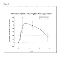
- FIG. 1 is a graph depicting the mobilization of CD133+ cells during Stem-Kine supplementation.
- FIG. 2 is a graph depicting the distribution of the percentage of stem/progenitor cell mobilization.
- FIG. 3 is a graph showing the percentage of KDR/CD-34-positive over the course of days.
- FIG. 4 is an image showing CHU-Hill colonies of EPCs stained by Giemsa stain.
- FIG. 5 is a graph depicting the number of EPCs measured by colony-formation assay over days.
- FIGS. 6A and 6B depict a comparison between cells grown in wells with growth factor ( 6 A) and without growth factor ( 6 B).
- FIG. 7 is a graph depicting the average value of ATP ratios in wells with stimulated and non-stimulated cells.
- a formulation of ellagic acid; vitamin D3; beta 1,3 glucan; a ferment of the bacterium, Lactobacillus fermentum was prepared which was mixed with green tea extract, extract of goji berries, and extract of the root of astragalus , and subsequently fermented with Lactobacillus fermentum in a manner similar to that used for production of the food supplement 1 mm-Kine, as described in part in U.S. Pat. No. 6,827,940.
- the formulation was developed as a nutritional supplement useful for immune stimulation and anti-oxidant activity.
- Rationale for antioxidant activity was based on the known properties of the individual ingredient. Specifically, ellagic acid is a polyphenol antioxidant found in numerous vegetables and fruits; vitamin D3 has antioxidant and immune stimulating activities; beta 1,3 glucan stimulates macrophages and hence is immune modulatory. Green tea extracts and some components of goji berries are known to possess antioxidant characteristics. Additionally, astragalosides and other molecules found in the root of astragalus are known antioxidants that can prevent cellular damage secondary to oxidation.
- the current invention teaches methods of inducing mobilization of stem cells using various compounds currently found in the food supply.
- stem cell mobilization may be produced using inexpensive means that are substantially free of adverse effects that are associated with currently used approaches for stem cell mobilization.
- the invention comprises a composition of ellagic acid, vitamin D3, beta 1,3 glucan and a ferment of the bacterium, Lactobacillus fermentum , with an extract of green tea, extract of goji berries, and extract of the root of astragalus added prior to fermentation.
- Said composition is capable of augment number of hematopoietic and endothelial progenitor cells in circulation.
- the invention provides means of maintaining endothelial health through administration of a composition of ellagic acid, vitamin D3, beta 1,3 glucan and a ferment of the bacterium, Lactobacillus fermentum , with an extract of green tea, extract of goji berries, and extract of the root of astragalus added prior to fermentation.
- the endothelium plays several functions essential for life, including: a) acting as an anticoagulated barrier between the blood stream and interior of the blood vessels; b) allowing for selective transmigration of cells into and out of the blood stream; c) regulating blood flow through controlling smooth muscle contraction/relaxation; and d) participating in tissue remodeling [19].
- Health of the endothelium can be quantified using several methods, including assessment of the physical and mechanical features of the vessel wall, assaying for production of systemic biomarkers released by the endothelium, and quantification of ability of blood vessels to dilate in response to increased flow [29].
- FMD flow mediated dilation
- the difference in dilatation response serves as a means of quantifying one aspect of endothelial health [30, 31].
- This assay has been used to show endothelial dysfunction in conditions such as healthy aging [32-34], as well as various diverse inflammatory states including renal failure [35], rheumatoid arthritis [36], Crohn's Disease [37], diabetes [38], heart failure [39], and Alzheimer's [40].
- endothelial health is quantified based on methods described above and known in the art.
- Knowledge of endothelial health is used to adjust dosage of administration for the composition of ellagic acid, vitamin D3, beta 1,3 glucan and a ferment of the bacterium, Lactobacillus fermentum , with an extract of green tea, extract of goji berries, and extract of the root of astragalus added prior to fermentation.
- endothelial dysfunction As part of age and disease associated endothelial dysfunction is the reduced ability of the host to generate new blood vessel [44]. This is believed to be due, at least in part, to reduction of ischemia inducible elements such as the HIF-1 alpha transcription factor which through induction of SDF-1 and VEGF secretion play a critical role in ability of endothelium to migrate and form new capillaries in ischemic tissues [45, 46]. Accordingly, if one were to understand the causes of endothelial dysfunction and develop methods of inhibiting these causes or stimulating regeneration of the endothelium, then progression of many diseases, as well as possible increase in healthy longevity may be achieved.
- the composition of ellagic acid, vitamin D3, beta 1,3 glucan and a ferment of the bacterium, Lactobacillus fermentum , with an extract of green tea, extract of goji berries, and extract of the root of astragalus added prior to fermentation is provided to decrease changes associated with endothelial aging.
- Concentration of the composition may be modified based on biological markers or endpoints associated with aging. These include ability of circulating EPC to migrate towards chemotactic gradients such as SDF-1, or hypoxia-responsiveness of HIF-1.
- endothelial cells are believed to originate from a precursor cell, the hemangioblast, which is capable of giving rise to both hematopoietic and endothelial cells [47].
- the endothelium was viewed as a fixed structure with relatively little self renewal, however in the last two decades this concept has fundamentally been altered.
- the current hypothesis is that the endothelium is constantly undergoing self renewal, especially in response to stress.
- a key component of endothelial turnover appears to be the existence of circulating endothelial progenitor (EPC) cells that appear to be involved in repair and angiogenesis of ischemic tissues.
- EPC circulating endothelial progenitor
- CD34 cells expressing the markers VEGF-receptor 2, CD133, and CXCR-4 receptor, with migrational ability to VEGF and SDF-1 has been a more refined EPC definition [52].
- EPC definition e.g., CD34+, VEGFR2+, CD133+, as well as CD34+, VEGFR2+, CD133 ⁇ have been reported to act as EPC [53]. More recent studies suggest that the subpopulation lacking CD133 and CD45 are precursor EPC [54].
- Apolipoprotein E knockout mice are genetically predisposed to development of atherosclerosis due to inability to impaired catabolism of triglyceride-rich lipoproteins.
- ApoE KO mice are lethally irradiated and reconstituted with labeled bone marrow stem cells, it was found that areas of the vasculature with high endothelial turnover, which were the areas of elevated levels of sheer stress, had incorporated the majority of new endothelial cells derived from the bone marrow EPC [57].
- the invention teaches the use of the composition of ellagic acid, vitamin D3, beta 1,3 glucan and a ferment of the bacterium, Lactobacillus fermentum , with an extract of green tea, extract of goji berries, and extract of the root of astragalus added prior to fermentation, in the rejuvenation of blood vessels by modulation of the circulating EPC compartment.
- Tissue injury and hypoxia are known to generate chemoattractants that potentially are responsible for mobilization of EPC.
- Reduction in oxygen tension occurs as a result of numerous injuries including stroke, infarction, or contusion.
- Oxygen tension is biologically detected by the transcription factor HIF-1 alpha, which upon derepression undergoes nuclear translocation. This event causes upregulated expression of a plethora of angiogenesis promoting cytokines and chemoattractants [61], such as stromal derived factor (SDF)-1 and VEGF [62, 63].
- SDF stromal derived factor
- HMBG1 a nuclear factor that has direct chemoattractant activity on mesoangioblasts, a type of EPC [64, 65]. It has been demonstrated that this systemic release of chemoattractant cytokines after vascular injury or infarct is associated with mobilization of endogenous bone marrow cells and EPC [66].
- ARDS acute respiratory distress syndrome
- circulating EPC may be capable of restoring injured lung endothelium.
- significant chimerism 37-42%) of pulmonary endothelial cells occurs in female recipients of male bone marrow transplants [77].
- chimerism 37-42%) of pulmonary endothelial cells occurs in female recipients of male bone marrow transplants [77].
- Sepsis is a major cause of ARDS and is associated with acute systemic inflammation and vascular damage.
- Septic patients have elevated levels of injury associated signals and EPC mobilizers such as HMGB1 [81], SDF-1 [82], and VEGF [83].
- Significant pathology of sepsis is associated with vascular leak and disseminated intravascular coagulation [84].
- the importance of the vasculature in sepsis can perhaps be supported by the finding that the only drug to have an impact on survival, Activated Protein C, acts primarily through endothelial protection [85].
- Septic patients are known to have increased circulating EPC as compared to controls.
- the invention teaches the use of the composition of ellagic acid, vitamin D3, beta 1,3 glucan and a ferment of the bacterium, Lactobacillus fermentum , with an extract of green tea, extract of goji berries, and extract of the root of astragalus added prior to fermentation, as a means of enhancing regenerative responses to injury.
- EPC administration has been shown to: decrease balloon injury induced neointimal hyperplasia [93], b) suppress carbon tetrachloride induced hepatic fibrosis [94, 95], and inhibit post cardiac infarct remodeling [96].
- definition of EPC was variable, or in some cases a confounding effect of coadministered cells with regenerative potential may be present.
- EPC play a beneficial role in supporting tissue regeneration As discussed below, many degenerative conditions, including healthy aging, are associated with a low-grade inflammation. There appears to be a causative link between this inflammation and reduction in EPC function.
- Inflammatory conditions present with features, which although not the rule, appear to have commonalities.
- CRP C-reactive protein
- erythrocyte sedimentation rate C-reactive protein
- cytokines such as TNF-alpha and IL-18
- organ degenerative conditions such as heart failure [97, 98], kidney failure [99, 100], and liver failure [101, 102]
- autoimmune conditions such as rheumatoid arthritis [103] and Crohn's Disease [104], to healthy aging [105, 106].
- neopterin a metabolite that increases systemically with healthy aging [107], and its concentration positively correlates with cognitive deterioration in various age-related conditions such as Alzheimer's [108].
- Neopterin is largely secreted by macrophages, which also produce inflammatory mediators such as TNF-alpha, IL-1, and IL-6, all of which are associated with chronic inflammation of aging [109].
- these cytokines are known to upregulate CRP, which also is associated with aging [110]. While there is no direct evidence that inflammatory markers actively cause shorted lifespan in humans, strong indirect evidence of their detrimental activities exists.
- TNF-alpha Another important inflammatory mediator found elevated in numerous degenerative conditions is the cytokine TNF-alpha.
- TNF-alpha is known to inhibit proliferation of repair cells in the body, such as oligodendrocytes in the brain [113], and suppress activity of endogenous stem cell pools [114, 115].
- TNF-alpha decreases EPC viability, an effect that can be overcome, at least in part by antioxidant treatment [116].
- Administration of TNF-alpha blocking agents has been demonstrated to restore both circulating EPC, as well as endothelial function in patients with inflammatory diseases such as rheumatoid arthritis [36, 117, 118].
- the invention teaches the use of the composition of ellagic acid, vitamin D3, beta 1,3 glucan and a ferment of the bacterium, Lactobacillus fermentum , with an extract of green tea, extract of goji berries, and extract of the root of astragalus added prior to fermentation, as a means of derepres sing circulating EPC in conditions of chronic inflammation.
- telomere shortening problem Every time cells divide the ends of the chromosomes called “telomeres” (complexes of tandem TTAGGGG repeats of DNA and proteins), are not completely replicated, thus they progressively get shorter [119]. Once telomeres reach a critical limit p53, p21, and p16 pathways are activated as a DNA damage response reaction instructing the cell to exit cell cycling.
- the cells start expressing inflammatory cytokines such as IL-1 [120, 121], upregulation of adhesion molecules that attract inflammatory cells such as monocytes [122, 123], and morphologically take a flattened, elongated appearance.
- inflammatory cytokines such as IL-1 [120, 121]
- upregulation of adhesion molecules that attract inflammatory cells such as monocytes [122, 123]
- morphologically take a flattened, elongated appearance.
- the process of cellular senescence caused in response to telomere shortening is believed to be a type of protective mechanism that cells have to prevented carcinogenesis [124].
- telomere length and age has been made [125]
- disorders of premature aging such as ataxia telangiectasia are characterized by accelerated telomere shortening [126].
- telomeres are shorter in arteries associated with higher blood flow and sheer stress (like the iliac artery) as compared to arteries of lower stress such as the mammary artery [127].
- telomere shortening of EPC is the difference between replicative senescence, which results from high need for differentiated endothelial cells, and stress induced senescence, in which inflammatory mediators can directly lead to telomere shortening.
- smoking associated oxidative stress has been linked to stress induced senescence in clinical studies [131], whereas other studies have implicated inflammatory agents such as interferon gamma [132], TNF-alpha [133], and oxidative mediators as inducers of stress induced senescence [134].
- the invention teaches the use of the composition of ellagic acid, vitamin D3, beta 1,3 glucan and a ferment of the bacterium, Lactobacillus fermentum , with an extract of green tea, extract of goji berries, and extract of the root of astragalus added prior to fermentation, as a means of preventing exhaustion of circulating EPC.
- Peripheral blood mononuclear cells were isolated by Ficoll-Hypaque. Cells were stained by CD133, CD34 and CD45 antibodies and the level of markers' expression was measured according to the procedure described in methods. Flow-cytomentric analysis of the samples for different periods before and during supplementation demonstrated that number of CD133-positive and CD34-positive cells in circulation was increased during supplementation. Data of the percentage of CD133 + and CD34 + cells selected from PE bright /FITC dim/negative cells of peripheral blood mononuclear cell population at different time before and during intervention are presented in Table 1.
- Results shown in table are averaged values for all subjects of the percentage of CD133 and CD34 positive cells, the average values of these percentages normalized on the level of CD133 or CD34 positive cells before supplementation and calculated percentage of stem cell mobilization.
- the average percentage (mean ⁇ SD) of CD133+/CD45 ⁇ cells was 0.012 ⁇ 0.008 before supplementation and 0.02 ⁇ 0.01 after two days and seven days of supplementation.
- mobilization of CD133 cells in circulation during Stem-Kine supplementation reached peak value between 48 hours and 7 days of supplementation.
- Eight subjects had peak of mobilized stem cells on the 2nd day, for seven subjects the maximum amount of CD133-posirtive cells was measured on 7 th day, and three subjects demonstrated maximum percentage of CD133 mobilization on 14 th day.
- Difference of the level of CD133 cells in circulation was statistically significant for two and seven days of Stem-Kine supplementation (p ⁇ 0.02).
- Average percentage of CD34-positive cells for all subjects was 0.062% ⁇ 0.029% (range 0.015%-0.1%) before supplementation and the maximum average percentage was 0.077% ⁇ 0.037% (range 0.019%-0.145%) during supplementation.
- the distribution of the level of improvement of CD34-positive cells in circulation is shown in FIG. 2 .
- the individual values of response differed for different subjects and the maximum percentage of CD34 mobilized cells was 60% with interquartile range 10%-98%. Difference between average percentages of CD34+ cells in circulation was not significant for first days of supplementation and one-sided p-value was 0.05 for 7 days and 14 days of supplementation.
- Percentage of KDR+/CD34+ cells averaged for all subjects was increased from 0.12% ⁇ 0.02% before supplementation to the maximum level (0.16%-0.2%) ⁇ 0.02% on the seventh day of supplementation.
- Example of the distribution of the maximum percentage of EPCs' mobilization by Stem-Kine supplementation for all subjects and for one method of cell selection is shown in FIG. 2 .
- the level of mobilization of EPCs in circulation differed for different subjects with interquartile distance 50%-240% and average value 140% for one method of EPCs' selection and interquartile distance 80%-260% with average value 220% for second method of EPCs selections. Difference of the level of EPCs in circulation was statistically significant for two and seven days of Stem-Kine supplementation (p ⁇ 0.02). Practically in all subjects, the maximum of the endothelial cell mobilization was measured on 7th day of supplementation (except for three subjects with maximum values on 14 th days and one subject with maximum value on 2 nd day).
- EPCs EPCs
- mononuclear cells are cultured on plastic covered by fibronectin, type 1 collagen or gelatin with either adherent or non-adherent fraction considered to contain the EPCs.
- the medium formulation, concentration of serum, growth factors utilized (VEGF, IGF, FGF-b) and times in culture vary between researchers.
- VEGF, IGF, FGF-b growth factors utilized
- Our chosen method of the growing colonies of endothelial progenitor cells is described in methods. Separated PBMCs were seeded in 24-well fibronectin-coated plates with concentration 1 M per wells for 5 days. After 5 days in culture, colonies were fixed by methanol and stained by Giemsa stain. The example of the images of the colonies is shown in FIG. 4 .
- Colonies were counted by AlphaEase software and by microscope. Analysis of the colonies from different subjects demonstrated that morphology of the colonies varied among different donors in term of size of colonies or the number of elongated sprouts at the periphery, but qualitative analysis was made only for number of colonies.
- HALO assay was applied that is based on the classical colony-forming assay procedure.
- we used HALO-SC2 assay in which stem cells and progenitor cells are stimulated with EPO, GM-CSF, G-CSF, IL-3, IL-6 and SCF. Cells were plated with and without addition of growth factors.
- the level of ATP was measured by bioluminescence assay in wells with stimulated and not stimulated cells after 5 days of exposure to growth factors. Average values of ATP in cells grown with added growth factors were normalized on the average level of ATP in cells in wells without growth factors.
- the level of proliferation is defined by the presence of progenitor and stem cells in population of plated cells. During incubation, these cells are stimulated by growth factors to proliferate and divide. The higher number of stem cells and progenitor cells in PBMCs, the higher level of cell proliferation and higher level of ATP measured by bio-luminescence assay in wells with growth factors. Base on this assumption, we considered that the ratio of ATP in wells with stimulated cells to ATP in wells with non-stimulated cells could characterize the population of stem and progenitor cells in cell population, and to measure the effect of Stem-Kine on the mobilization of stem and progenitor cells in blood. Data presented in FIG. 7 demonstrate the averaged for all subjects ratio (mean ⁇ SE) of ATP in wells with stimulated cells to ATP in wells with non-stimulated cells for different periods before and during supplementation with Stem-Kine.
- Ratio of the average ATP was increased after 24 hrs of supplementation from pre-intervention level of 2.16 ⁇ 0.0.44 to 2.57 ⁇ 0.47. After 48 hrs and 7 days of supplementation, the ratio was decreased to 2.36 ⁇ 0.5 and 2.35 ⁇ 0.5. Statistical analysis showed significant difference of the mean ratio over pre-intervention level for 48 hrs (p ⁇ 0.02). Distribution of the maximum values of cell proliferation is shown in FIG. 2 (interquartile range 25%-114%, average 73%).
Landscapes
- Health & Medical Sciences (AREA)
- Natural Medicines & Medicinal Plants (AREA)
- Life Sciences & Earth Sciences (AREA)
- Pharmacology & Pharmacy (AREA)
- Chemical & Material Sciences (AREA)
- Veterinary Medicine (AREA)
- Public Health (AREA)
- General Health & Medical Sciences (AREA)
- Medicinal Chemistry (AREA)
- Animal Behavior & Ethology (AREA)
- Epidemiology (AREA)
- Engineering & Computer Science (AREA)
- Biotechnology (AREA)
- Mycology (AREA)
- Microbiology (AREA)
- Medical Informatics (AREA)
- Botany (AREA)
- Alternative & Traditional Medicine (AREA)
- Molecular Biology (AREA)
- Chemical Kinetics & Catalysis (AREA)
- General Chemical & Material Sciences (AREA)
- Nuclear Medicine, Radiotherapy & Molecular Imaging (AREA)
- Organic Chemistry (AREA)
- Bioinformatics & Cheminformatics (AREA)
- Pharmaceuticals Containing Other Organic And Inorganic Compounds (AREA)
- Medicines Containing Material From Animals Or Micro-Organisms (AREA)
Abstract
Compositions of matter, uses, and formulations of food supplements/nutrients capable of eliciting mobilization of various stem/progenitor cells, including hematopoietic stem cells and endothelial progenitor cells are disclosed. In one embodiment a formulation contains a mixture of ellagic acid, vitamin D3, beta 1,3 glucan and a ferment of the bacterium, Lactobacillus fermentum, with an extract of green tea, extract of goji berries, and extract of the root of astragalus added prior to fermentation. Said formulation, originally developed as an antioxidant/immune stimulator was found to have the unexpected property of eliciting stem/progenitor cell mobilization.
Description
This application claims priority to Provisional Application Ser. No. 61/285,171 filed on Dec. 9, 2009, which is expressly incorporated by reference in its entirety
The invention pertains to the area of nutraceutical products. More specifically, the invention provides novel compositions useful for properties of altering stem/progenitor cell distribution. Furthermore, the invention relates to means of eliciting therapeutic effects through mobilization of stem/progenitor cells from compartments of the body.
Stem cell therapy can broadly be divided into approaches aimed at de novo regeneration of injured organs, or into the use of the stem cells to accelerate endogenous healing processes. It appears that adult stem cells are stored in reservoirs ready to meet the body's need subsequent to injury. For example, in conditions as diverse as renal injury [1], myocardial infarct [2], stroke [3], irradiation [4], and acoustic damage [5], injured tissue has been reported to cause upregulation of the chemokine stromal derived factor (SDF)-1, which causes mobilization and attraction of bone marrow derived stem/progenitor cells.
Augmentation or de novo initiation of the mobilization process has been performed therapeutically using cytokines such as G-CSF [6], GM-CSF [7], and Parathyroid Hormone [8]. Recently small molecule antagonists of CXCR4 have entered clinical use [9]. Therapeutic benefits of mobilization have been seen in models of: a) radiation induced salivary gland damage [10, 11]; b) cardiac infarct [12]; c) stroke [13]. Clinically, mobilization therapy has been attempted in conditions such as ALS [14], heart failure [15, 16], and liver failure [17].
Unfortunately, agents used in the mobilization of stem cells/EPC such as G-CSF, GM-CSF, and Parathyroid Hormone are expensive and can not be continually administered for long periods of time. Thus there is a need for agents which induce augmentation of circulating stem cell/EPC levels that can be administered chronically, without adverse effects, and is relatively inexpensive. Jensen et al reported an extract from the edible cyanobacterium Aphanizomenon flos-aquae (AFA) enriched for a novel ligand for human CD62L (L-selectin), which is currently sold commercially under the name StemEnhance [18]. Although this compound is relatively innocuous from a toxicity perspective, mobilization appears to be mediated in a non-specific manner. In healthy volunteers a transient, 18% increase in numbers of circulating CD34+ stem cells was noted that maximized 1 hour after consumption. Given this relatively insignificant increase and transient nature of mobilization, as well as the fact that mechanistically mobilization is associated with decrease in CXCR4, which would block homing of stem cells to target tissue, novel methods of mobilization stem/progenitor cells are needed that are useful for long-term administration.
Teachings herein are directed to compositions comprising ellagic acid, vitamin D3, beta 1,3 glucan and a ferment of the bacterium, Lactobacillus fermentum, with an extract of green tea, extract of goji berries, and extract of the root of astragalus added prior to fermentation.
Additionally the teachings herein are directed to methods of mobilizing endothelia progenitor cells comprising administration of a sufficient dose of a composition comprised of ellagic acid, vitamin D3, beta 1,3 glucan and a ferment of the bacterium, Lactobacillus fermentum, with an extract of green tea, extract of goji berries, and extract of the root of astragalus added prior to fermentation.
Further embodiments are directed to: a method of mobilizing endothelia progenitor cells comprising administration of a sufficient dose of a composition comprised of ellagic acid, vitamin D3, beta 1,3 glucan and a ferment of the bacterium, Lactobacillus fermentum, with an extract of green tea, extract of goji berries, and extract of the root of astragalus added prior to fermentation. The endothelial progenitor cells can be positive for expression of KDR and CD34. The endothelial progenitor cells can also be capable of forming endothelial cells in tissue culture.
Further embodiments are directed to methods of decreasing oxidative stress in a patient comprising administration of a sufficient dose of a composition comprised of ellagic acid, vitamin D3, beta 1,3 glucan and a ferment of the bacterium, Lactobacillus fermentum, with an extract of green tea, extract of goji berries, and extract of the root of astragalus added prior to fermentation. The composition can be formulated as a neutraceutical, dietary supplement or wholistic formulation for oral administration.
Further embodiments are directed to methods of treating a disorder associated with reduced levels of circulating stem/progenitor cells by administration of a sufficient concentration of a sufficient dose of a composition comprised of ellagic acid, vitamin D3, beta 1,3 glucan and a ferment of the bacterium, Lactobacillus fermentum, with an extract of green tea, extract of goji berries, and extract of the root of astragalus added prior to fermentation.
Additional methods can involve increasing endothelial health, as assessed by the flow mediated dilation assay, comprising administration of a sufficient dose of a composition comprised of ellagic acid, vitamin D3, beta 1,3 glucan and a ferment of the bacterium, Lactobacillus fermentum, with an extract of green tea, extract of goji berries, and extract of the root of astragalus added prior to fermentation.
Still further embodiments include methods of preventing a degenerative condition in a mammal through the steps of: a) identifying a deficiency in numbers and/or activity of circulating endothelial progenitor cells; b) administering a nutritional supplement capable of augmenting circulating levels of endothelial progenitor cells based on deficiency identified; c) re-assessing circulating endothelial progenitor cell numbers; and d) further adjusting dose of said nutritional supplement based on response to supplementation.
It will be appreciated that the drawings are not necessarily to scale, with emphasis instead being placed on illustrating the various aspects and features of embodiments of the invention, in which:
Embodiments of the present invention are described below. It is, however, expressly noted that the present invention is not limited to these embodiments, but rather the intention is that modifications that are apparent to the person skilled in the art and equivalents thereof are also included.
A formulation of ellagic acid; vitamin D3; beta 1,3 glucan; a ferment of the bacterium, Lactobacillus fermentum was prepared which was mixed with green tea extract, extract of goji berries, and extract of the root of astragalus, and subsequently fermented with Lactobacillus fermentum in a manner similar to that used for production of the food supplement 1 mm-Kine, as described in part in U.S. Pat. No. 6,827,940. The formulation was developed as a nutritional supplement useful for immune stimulation and anti-oxidant activity.
Rationale for antioxidant activity was based on the known properties of the individual ingredient. Specifically, ellagic acid is a polyphenol antioxidant found in numerous vegetables and fruits; vitamin D3 has antioxidant and immune stimulating activities; beta 1,3 glucan stimulates macrophages and hence is immune modulatory. Green tea extracts and some components of goji berries are known to possess antioxidant characteristics. Additionally, astragalosides and other molecules found in the root of astragalus are known antioxidants that can prevent cellular damage secondary to oxidation.
In testing the product in healthy volunteers, benefit in various health conditions was observed. Specifically, increased level of energy, improvement of skin conditions around fingernails, better sleep, and increased mental acuity was reported. These properties prompted us to test effects of the composition on stem cell and endothelial progenitor cell (EPC) mobilization. The data generated produced the unexpected finding of a profound mobilization effect associated with intake of this food supplement.
The current invention teaches methods of inducing mobilization of stem cells using various compounds currently found in the food supply. Through this “nutraceutical” approach, stem cell mobilization may be produced using inexpensive means that are substantially free of adverse effects that are associated with currently used approaches for stem cell mobilization.
In one embodiment the invention comprises a composition of ellagic acid, vitamin D3, beta 1,3 glucan and a ferment of the bacterium, Lactobacillus fermentum, with an extract of green tea, extract of goji berries, and extract of the root of astragalus added prior to fermentation. Said composition is capable of augment number of hematopoietic and endothelial progenitor cells in circulation.
In one embodiment, the invention provides means of maintaining endothelial health through administration of a composition of ellagic acid, vitamin D3, beta 1,3 glucan and a ferment of the bacterium, Lactobacillus fermentum, with an extract of green tea, extract of goji berries, and extract of the root of astragalus added prior to fermentation. The endothelium plays several functions essential for life, including: a) acting as an anticoagulated barrier between the blood stream and interior of the blood vessels; b) allowing for selective transmigration of cells into and out of the blood stream; c) regulating blood flow through controlling smooth muscle contraction/relaxation; and d) participating in tissue remodeling [19]. A key hallmark of the aging process and perhaps one of the causative factors of health decline associated with aging appears to be loss of endothelial function. Whether as a result of oxidative stress, inflammatory stress, or senescence, deficiencies in the ability of the endothelium to respond to physiological cues can alter the ability to think [20], procreate [21], see [22], and breathe [23]. Specifically, minute alterations in the ability of endothelium to respond to neurotransmitter induced nitric oxide causes profound inability to perform even simple mental functions [24, 25]. Small increases in angiogenesis in the retina as a result of injury or glucose are associated with wet macular degeneration blindness [26]. Atherosclerosis of the penile vasculature is a major cause of erectile dysfunction [27]. The pulmonary endothelium's sensitivity to insult can cause hypertension and associated progression to decreased oxygen delivery [28].
Health of the endothelium can be quantified using several methods, including assessment of the physical and mechanical features of the vessel wall, assaying for production of systemic biomarkers released by the endothelium, and quantification of ability of blood vessels to dilate in response to increased flow [29]. Of these, one of the most commonly used assays for endothelium function is the flow mediated dilation (FMD) assay. This procedure usually involves high resolution ultrasound assessment of the diameter of the superficial femoral and brachial arteries in response to reactive hyperemia induced by a cuff. The extent of dilatation response induced by the restoration of flow is compared to dilatation induced by sublingual glyceryl trinitrate. Since the dilatation induced by flow is dependent on the endothelium acting as a mechanotransducer and the dilatation induced by glyceryl trinitrate is based on smooth muscle responses, the difference in dilatation response serves as a means of quantifying one aspect of endothelial health [30, 31]. This assay has been used to show endothelial dysfunction in conditions such as healthy aging [32-34], as well as various diverse inflammatory states including renal failure [35], rheumatoid arthritis [36], Crohn's Disease [37], diabetes [38], heart failure [39], and Alzheimer's [40]. Although it is not clear whether reduction in FMD score is causative or an effect of other properties of endothelial dysfunction, it has been associated with: a) increased tendency towards thrombosis, in part by increased vWF levels [41], b) abnormal responses to injury, such as neointimal proliferation and subsequent atherosclerosis [42], and c) increased proclivity towards inflammation by basal upregulation of leukocyte adhesion molecules [43]. In one embodiment of the invention, endothelial health is quantified based on methods described above and known in the art. Knowledge of endothelial health is used to adjust dosage of administration for the composition of ellagic acid, vitamin D3, beta 1,3 glucan and a ferment of the bacterium, Lactobacillus fermentum, with an extract of green tea, extract of goji berries, and extract of the root of astragalus added prior to fermentation.
As part of age and disease associated endothelial dysfunction is the reduced ability of the host to generate new blood vessel [44]. This is believed to be due, at least in part, to reduction of ischemia inducible elements such as the HIF-1 alpha transcription factor which through induction of SDF-1 and VEGF secretion play a critical role in ability of endothelium to migrate and form new capillaries in ischemic tissues [45, 46]. Accordingly, if one were to understand the causes of endothelial dysfunction and develop methods of inhibiting these causes or stimulating regeneration of the endothelium, then progression of many diseases, as well as possible increase in healthy longevity may be achieved. Accordingly, in one embodiment, the composition of ellagic acid, vitamin D3, beta 1,3 glucan and a ferment of the bacterium, Lactobacillus fermentum, with an extract of green tea, extract of goji berries, and extract of the root of astragalus added prior to fermentation, is provided to decrease changes associated with endothelial aging. Concentration of the composition may be modified based on biological markers or endpoints associated with aging. These include ability of circulating EPC to migrate towards chemotactic gradients such as SDF-1, or hypoxia-responsiveness of HIF-1.
During development endothelial cells are believed to originate from a precursor cell, the hemangioblast, which is capable of giving rise to both hematopoietic and endothelial cells [47]. Classically the endothelium was viewed as a fixed structure with relatively little self renewal, however in the last two decades this concept has fundamentally been altered. The current hypothesis is that the endothelium is constantly undergoing self renewal, especially in response to stress. A key component of endothelial turnover appears to be the existence of circulating endothelial progenitor (EPC) cells that appear to be involved in repair and angiogenesis of ischemic tissues. An early study in 1963 hinted at the existence of such circulating EPC after observations of endothelial-like cells, that were non-thrombogenic and morphologically appeared similar to endothelium, were observed covering a Dacron graft that was tethered to the thoracic artery of a pig [48]. The molecular characterization of the EPC is usually credited to a 1997 paper by Asahara et al. in which human bone marrow derived VEGR-2 positive, CD34 positive monocyte-like cells were described as having ability to differentiate into endothelial cells in vitro and in vivo based on expression of CD31, eNOS, and E-selectin [49]. These studies were expanded into hindlimb ischemia in mouse and rabbit models in which increased circulation of EPC in response to ischemic insult was observed [50]. Furthermore, these studies demonstrated that cytokine-induced augmentation of EPC mobilization elicited a therapeutic angiogenic response. Using irradiated chimeric systems, it was demonstrated that ischemia-mobilized EPC derive from the bone marrow, and that these cells participate both in sprouting of pre-existing blood vessels as well as the initiation of de novo blood vessel production [51]. Subsequent to the initial phenotypic characterization by Asahara et al [49], more detailed descriptions of the human EPC were reported. For example, CD34 cells expressing the markers VEGF-receptor 2, CD133, and CXCR-4 receptor, with migrational ability to VEGF and SDF-1 has been a more refined EPC definition [52]. However there is still some controversy as to the precise phenotype of the EPC, since the term implies only ability to differentiate into endothelium. For example, both CD34+, VEGFR2+, CD133+, as well as CD34+, VEGFR2+, CD133− have been reported to act as EPC [53]. More recent studies suggest that the subpopulation lacking CD133 and CD45 are precursor EPC [54]. Other phenotypes have been ascribed to cells with EPC activity, one study demonstrated monocyte-like cells that expressing CD14, Mac-1 and the dendritic cell marker CD11c have EPC activity based on uptake of acetylated LDL and binding to the ulex-lectin [55, 56].
While the initial investigations into the biology of EPC focused around acute ischemia, it appears that in chronic conditions circulating EPC may play a role in endothelial turnover. Apolipoprotein E knockout (ApoE KO) mice are genetically predisposed to development of atherosclerosis due to inability to impaired catabolism of triglyceride-rich lipoproteins. When these mice are lethally irradiated and reconstituted with labeled bone marrow stem cells, it was found that areas of the vasculature with high endothelial turnover, which were the areas of elevated levels of sheer stress, had incorporated the majority of new endothelial cells derived from the bone marrow EPC [57]. The possibility that endogenous bone marrow derived EPC possess such a regenerative function was also tested in a therapeutic setting. Atherosclerosis is believed to initiate from endothelial injury with a proliferative neointimal response that leads to formation of plaques. When bone marrow derived EPC are administered subsequent to wire injury, a substantial reduction in neointima formation was observed [58]. The argument can obviously made that wire injury of an artery does not resemble the physiological conditions associated with plaque development. To address this, Wassmann et al [59], used ApoE KO mice that were fed a high cholesterol diet and observed reduction in endothelial function as assessed by the flow mediated dilation assay. When EPC were administered from wild-type mice restoration of endothelial responsiveness was observed.
In the context of aging, Edelman's group performed a series of interesting experiments in which 3 month old syngeneic cardiac grafts were heterotopically implanted into 18 month old recipients. Loss of graft viability, associated with poor neovascularization, was observed subsequent to transplanting, as well as subsequent to administration of 18 month old bone marrow mononuclear cells. In contrast, when 3 month old bone marrow mononuclear cells were implanted, grafts survived. Antibody depletion experiments demonstrated bone marrow derived PDGF-BB was essential in integration of the young heart cells with the old recipient vasculature [60]. These experiments suggest that young EPC or EPC-like cells have ability to integrate and interact with older vasculature. What would be interesting is to determine whether EPC could be “revitalized” ex vivo by culture conditions or transfection with therapeutic genes such as PDGF-BB.
Accordingly, in one embodiment the invention teaches the use of the composition of ellagic acid, vitamin D3, beta 1,3 glucan and a ferment of the bacterium, Lactobacillus fermentum, with an extract of green tea, extract of goji berries, and extract of the root of astragalus added prior to fermentation, in the rejuvenation of blood vessels by modulation of the circulating EPC compartment.
Tissue injury and hypoxia are known to generate chemoattractants that potentially are responsible for mobilization of EPC. Reduction in oxygen tension occurs as a result of numerous injuries including stroke, infarction, or contusion. Oxygen tension is biologically detected by the transcription factor HIF-1 alpha, which upon derepression undergoes nuclear translocation. This event causes upregulated expression of a plethora of angiogenesis promoting cytokines and chemoattractants [61], such as stromal derived factor (SDF)-1 and VEGF [62, 63]. On the other hand, tissue necrosis causes release of “danger signals” such as HMBG1, a nuclear factor that has direct chemoattractant activity on mesoangioblasts, a type of EPC [64, 65]. It has been demonstrated that this systemic release of chemoattractant cytokines after vascular injury or infarct is associated with mobilization of endogenous bone marrow cells and EPC [66].
Myocardial infarction has been widely studied in the area of regenerative medicine in which cellular and molecular aspects of host response post-injury are relatively well defined. EPC mobilization after acute ischemia has been demonstrated in several cardiac infarct studies. This was first reported by Shintani et al who observed increased numbers of CD34 positive cells in 16 post infarct patients on day 7 as compared to controls. The rise in CD34 cells correlated with ability to differentiate into cells morphologically resembling endothelium and expressing endothelial markers KDR and CD31. Supporting the concept that response to injury stimulates EPC mobilization, a rise in systemic VEGF levels was correlated with increased EPC numbers [63]. A subsequent study demonstrated a similar rise in circulating EPC post infarct. Blood was drawn from 56 patients having a recent infarct (<12 hours), 39 patients with stable angina, and 20 healthy controls. Elevated levels of cells expressing CD34/CXCR4+ and CD34/CD117+ and c-met+ were observed only in the infarct patients which were highest at the first blood draw. In this study the mobilized cells not only expressed endothelial markers, but also myocytic and cardiac genes [67]. The increase in circulating EPC at early timepoints post infarction has been observed by other groups, and correlated with elevations in systemic VEGF and SDF-1 [68, 69].
In the case of cerebral infarction studies support the concept that not only are EPC mobilized in response to ischemia, but also that the extent of mobilization may be associated with recovery. In a trial of 48 patients suffering primary ischemic stroke, mobilization of EPC was observed in the first week in comparison to control patients. EPC were defined as cells capable of producing endothelial colony forming units. A correlation between improved outcome at 3 months and extend of EPC mobilization was observed based on the NIHSS and Rankin score [70]. In a similar study, Dunac et al reported on circulating CD34 levels of 25 patients with acute stroke for 14 days. A correlation between improvement on the Rankin scale and increased circulating CD34 cells was reported [71]. Noteworthy was that the level of CD34 mobilization was similar to that observed in patients treated with the mobilize G-CSF. In a larger study, Yip et al examined EPC levels in 138 consecutive patients with acute stroke and compared them to 20 healthy volunteers and in 40 at-risk control subjects [72]. Three EPC phenotypes were assessed by flow cytometry at 48 hours after stroke: a) CD31/CD34, b) CD62E/CD34, and c) KDR/CD34. Diminished levels of all three EPC subsets in circulation was predictive of severe neurological impairment NIHSS >/=12, while suppressed levels of circulating CD31/34 cells was correlated with combined major adverse clinical outcomes as defined by recurrent stroke, any cause of death, or NIHSS >/=12. Increased levels of the KDR/CD34 phenotype cells was strongly associated with NIHSS >or=4 on day 21. Although these studies do not directly demonstrate a therapeutic effect of the mobilized EPC, animal studies in the middle cerebral artery ligation stroke model have demonstrated positive effects subsequent to EPC administration [73, 74], an effect which appears to be at least partially dependent on VEGF production from the EPC [75].
Another ischemia-associated tissue insult is acute respiratory distress syndrome (ARDS), in which respiratory failure often occurs as a result of disruption of the alveolar—capillary membrane, which causes accumulation of proteinaceous pulmonary edema fluid and lack of oxygen uptake ability [76]. In this condition there has been some speculation that circulating EPC may be capable of restoring injured lung endothelium. For example, it is known that significant chimerism (37-42%) of pulmonary endothelial cells occurs in female recipients of male bone marrow transplants [77]. Furthermore, in patients with pneumonia infection there is a correlation between infection and circulating EPC, with higher numbers of EPC being indicative of reduced fibrosis [78]. The possibility that EPC are mobilized during ARDS and may be associated with benefit was examined in a study of 45 patients with acute lung injury in which a correlation between patients having higher number of cells capable of forming endothelial colonies in vitro and survival was made. Specifically, the patients with a colony count of >or =35 had a mortality of approximately 30%, compared to patients with less than 35 colonies, which had a mortality of 61%. The correlation was significant after multivariable analysis correcting for age, sex, and severity of illness [79]. From an interventional perspective, transplantation of EPC into a rabbit model of acute lung injury resulted in reduction of leukocytic infiltrates and preservation of pulmonary cellular integrity [80].
Sepsis is a major cause of ARDS and is associated with acute systemic inflammation and vascular damage. Septic patients have elevated levels of injury associated signals and EPC mobilizers such as HMGB1 [81], SDF-1 [82], and VEGF [83]. Significant pathology of sepsis is associated with vascular leak and disseminated intravascular coagulation [84]. The importance of the vasculature in sepsis can perhaps be supported by the finding that the only drug to have an impact on survival, Activated Protein C, acts primarily through endothelial protection [85]. Septic patients are known to have increased circulating EPC as compared to controls. Becchi et al observed a correlation between VEGF and SDF-1 levels with a 4-fold rise in circulating EPC in septic patients as compared to healthy controls [82]. A correlation between EPC levels and survival after sepsis was reported in a study of 32 septic patients, 15 ICU patients, and 15 controls. Of the 8 patients who succumbed to sepsis by 28 days, as compared to 24 survivors, a significantly reduced EPC number in non-survivors was reported [86].
It appears that in conditions of acute injury, elevation of EPC in circulation occurs. Although studies in stroke [70-72], ARDS [79], and sepsis [86] seem to correlate outcome with extend of mobilization, work remains to be performed in assessing whether it is the EPC component that is responsible for benefits or other confounding variables. Taking into account the possibility that EPC may act as an endogenous repair mechanism, we will discuss data in chronic degenerative conditions in which circulating EPC appear to be suppressed.
Accordingly, in one embodiment, the invention teaches the use of the composition of ellagic acid, vitamin D3, beta 1,3 glucan and a ferment of the bacterium, Lactobacillus fermentum, with an extract of green tea, extract of goji berries, and extract of the root of astragalus added prior to fermentation, as a means of enhancing regenerative responses to injury.
There is need for angiogenesis and tissue remodeling in the context of various chronic inflammatory conditions. However in many situations it is the aberrant reparative processes that actually contribute to the pathology of disease. Examples of this include: the process of neointimal hyperplasia and subsequent plaque formation in response to injury to the vascular wall [87], the process of hepatic fibrosis as opposed to functional regeneration [88], or the post-infarct pathological remodeling of the myocardium which results in progressive heart failure [89]. In all of these situations it appears that not only the lack of regenerative cells, but also the lack of EPC is present. Conceptually, the need for reparative cells to heal the ongoing damage may have been so overwhelming that it leads to exhaustion of EPC numbers and eventual reduction in protective effect. Supporting this concept are observations of lower number of circulating EPC in inflammatory diseases, which may be the result of exhaustion. Additionally, the reduced telomeric length of EPC in patients with coronary artery disease [90], as well as reduction of telomere length in the EPC precursors that are found in the bone marrow [91, 92] suggests that exhaustion in response to long-term demand may be occurring. If the reparatory demands of the injury indeed lead to depletion of EPC progenitors, then administration of progenitors should have therapeutic effects.
Several experiments have shown that administration of EPC have beneficial effects in the disease process. For example, EPC administration has been shown to: decrease balloon injury induced neointimal hyperplasia [93], b) suppress carbon tetrachloride induced hepatic fibrosis [94, 95], and inhibit post cardiac infarct remodeling [96]. One caveat of these studies is that definition of EPC was variable, or in some cases a confounding effect of coadministered cells with regenerative potential may be present. However, overall, there does appear to be an indication that EPC play a beneficial role in supporting tissue regeneration. As discussed below, many degenerative conditions, including healthy aging, are associated with a low-grade inflammation. There appears to be a causative link between this inflammation and reduction in EPC function.
Inflammatory conditions present with features, which although not the rule, appear to have commonalities. For example, increases in inflammatory markers such as C-reactive protein (CRP), erythrocyte sedimentation rate, and cytokines such as TNF-alpha and IL-18 have been described in diverse conditions ranging from organ degenerative conditions such as heart failure [97, 98], kidney failure [99, 100], and liver failure [101, 102] to autoimmune conditions such as rheumatoid arthritis [103] and Crohn's Disease [104], to healthy aging [105, 106]. Other markers of inflammation include products of immune cells such as neopterin, a metabolite that increases systemically with healthy aging [107], and its concentration positively correlates with cognitive deterioration in various age-related conditions such as Alzheimer's [108]. Neopterin is largely secreted by macrophages, which also produce inflammatory mediators such as TNF-alpha, IL-1, and IL-6, all of which are associated with chronic inflammation of aging [109]. Interestingly, these cytokines are known to upregulate CRP, which also is associated with aging [110]. While there is no direct evidence that inflammatory markers actively cause shorted lifespan in humans, strong indirect evidence of their detrimental activities exists. For example, direct injection of recombinant CRP in healthy volunteers induces atherothrombotic endothelial changes, similar to those observed in aging [111]. In vitro administration of CRP to endothelial cells decreases responsiveness to vasoactive factors, resembling the human age-associated condition of endothelial hyporesponsiveness [112].
Another important inflammatory mediator found elevated in numerous degenerative conditions is the cytokine TNF-alpha. Made by numerous cells, but primarily macrophages, TNF-alpha is known to inhibit proliferation of repair cells in the body, such as oligodendrocytes in the brain [113], and suppress activity of endogenous stem cell pools [114, 115]. TNF-alpha decreases EPC viability, an effect that can be overcome, at least in part by antioxidant treatment [116]. Administration of TNF-alpha blocking agents has been demonstrated to restore both circulating EPC, as well as endothelial function in patients with inflammatory diseases such as rheumatoid arthritis [36, 117, 118].
It appears that numerous degenerative conditions are associated with production of inflammatory mediators, which directly suppress EPC production or activity. This may be one of the reasons for findings of reduced EPC and FMD indices in patients with diverse inflammatory conditions. In addition to the direct effects, the increased demand for de novo EPC production in inflammatory conditions would theoretically lead to exhaustion of EPC precursors cells by virtue of telomere shortening.
Accordingly, in one embodiment, the invention teaches the use of the composition of ellagic acid, vitamin D3, beta 1,3 glucan and a ferment of the bacterium, Lactobacillus fermentum, with an extract of green tea, extract of goji berries, and extract of the root of astragalus added prior to fermentation, as a means of derepres sing circulating EPC in conditions of chronic inflammation.
On average somatic cells can divide approximately 50 times, after which they undergo senescence, die or become cancerous. This limited proliferative ability is dependent on the telomere shortening problem. Every time cells divide the ends of the chromosomes called “telomeres” (complexes of tandem TTAGGGG repeats of DNA and proteins), are not completely replicated, thus they progressively get shorter [119]. Once telomeres reach a critical limit p53, p21, and p16 pathways are activated as a DNA damage response reaction instructing the cell to exit cell cycling. Associated with the process of senescence, the cells start expressing inflammatory cytokines such as IL-1 [120, 121], upregulation of adhesion molecules that attract inflammatory cells such as monocytes [122, 123], and morphologically take a flattened, elongated appearance. Physiologically, the process of cellular senescence caused in response to telomere shortening is believed to be a type of protective mechanism that cells have to prevented carcinogenesis [124]. At a whole organism level the association between telomere length and age has been made [125], as well, disorders of premature aging such as ataxia telangiectasia are characterized by accelerated telomere shortening [126].
The importance of this limited proliferative ability becomes apparent in our discussion of EPC. In general there is a need for continual endothelial cell replacement from EPC. Because the endothelial cells are exposed to enormous continual sheer stress of blood flow, mechanisms of repair and proliferation after injury need to exist. Theoretically, the more sheer stress on a particular artery, the more cell division would be required to compensate for cell loss. Indeed this appears to be the case. For example, telomeres are shorter in arteries associated with higher blood flow and sheer stress (like the iliac artery) as compared to arteries of lower stress such as the mammary artery [127]. The theory that senescence may be associated with atherosclerosis is supported since the iliac artery, which is associated with higher proliferation of endothelial cells and is also at a higher risk of atherosclerosis, thus prompting some investigators to propose atherosclerosis being associated with endothelial senescence [128, 129].
In an interesting intervention study Satoh et al examined 100 patients with coronary artery disease and 25 control patients. Telomere lengths were reduced in EPC of coronary artery disease patients as compared to controls. Lipid lowering therapy using agents such as atorvastatin has previously been shown to reduced oxidative stress and increase circulating EPC. Therapy with lipid lowering agents in this study resulted in preservation of telomeric length, presumably by decreasing the amount of de novo EPC produced, as well as oxidative stress leading to telomere erosion [130]. One important consideration when discussing telomere shortening of EPC is the difference between replicative senescence, which results from high need for differentiated endothelial cells, and stress induced senescence, in which inflammatory mediators can directly lead to telomere shortening. For example, smoking associated oxidative stress has been linked to stress induced senescence in clinical studies [131], whereas other studies have implicated inflammatory agents such as interferon gamma [132], TNF-alpha [133], and oxidative mediators as inducers of stress induced senescence [134].
Accordingly, in one embodiment, the invention teaches the use of the composition of ellagic acid, vitamin D3, beta 1,3 glucan and a ferment of the bacterium, Lactobacillus fermentum, with an extract of green tea, extract of goji berries, and extract of the root of astragalus added prior to fermentation, as a means of preventing exhaustion of circulating EPC.
Peripheral blood mononuclear cells were isolated by Ficoll-Hypaque. Cells were stained by CD133, CD34 and CD45 antibodies and the level of markers' expression was measured according to the procedure described in methods. Flow-cytomentric analysis of the samples for different periods before and during supplementation demonstrated that number of CD133-positive and CD34-positive cells in circulation was increased during supplementation. Data of the percentage of CD133+ and CD34+ cells selected from PEbright/FITCdim/negative cells of peripheral blood mononuclear cell population at different time before and during intervention are presented in Table 1. Results shown in table are averaged values for all subjects of the percentage of CD133 and CD34 positive cells, the average values of these percentages normalized on the level of CD133 or CD34 positive cells before supplementation and calculated percentage of stem cell mobilization. The average percentage (mean±SD) of CD133+/CD45− cells was 0.012±0.008 before supplementation and 0.02±0.01 after two days and seven days of supplementation. We found that for the most of the subjects, mobilization of CD133 cells in circulation during Stem-Kine supplementation reached peak value between 48 hours and 7 days of supplementation. Eight subjects had peak of mobilized stem cells on the 2nd day, for seven subjects the maximum amount of CD133-posirtive cells was measured on 7th day, and three subjects demonstrated maximum percentage of CD133 mobilization on 14th day. Difference of the level of CD133 cells in circulation was statistically significant for two and seven days of Stem-Kine supplementation (p<0.02).
| TABLE I | |
| Average values of CD133-positive cells, ratios of | Average values of CD34-positive cells, ratios of |
| pre to post measurements and percentage of | pre to post measurements and percentage of |
| stem/progenitor cell mobilization | stem/progenitor cell mobilization |
| Average | % of | Average | % of | ||||||||
| date | Conc. % | SE | ratio | SE | mobilization | Conc. % | SE | | SE | mobilization | |
| 0 | 0.012 | 0.002 | 1.00 | 0.062 | 0.007 | 1.00 | ||||
| 1 | 0.014 | 0.002 | 1.38 | 0.19 | 37.6 | 0.069 | 0.007 | 1.20 | 0.12 | 19.8 |
| 2 | 0.020 | 0.003 | 1.99 | 0.42 | 98.8 | 0.071 | 0.009 | 1.17 | 0.13 | 17.3 |
| 7 | 0.020 | 0.002 | 1.97 | 0.24 | 96.6 | 0.072 | 0.008 | 1.25 | 0.15 | 25.0 |
| 14 | 0.012 | 0.001 | 1.33 | 0.21 | 32.6 | 0.077 | 0.009 | 1.29 | 0.16 | 28.5 |
As the number of stem cells in circulation depends on age and state of health, to exclude these factors and to find effect of supplement on stem cell mobilization, we normalized values measured during supplementation on values measured before supplementation. Mobilization of CD133-positive cells in circulation for different periods of time during supplementation was averaged for 18 subjects studied and the cumulative amount of CD133+ stem cells (average ratio of the percentage of CD133-positive cells in circulation before and during supplementation) is shown in FIG. 1 . Measurements of the percentage of CD133-positive and CD34− positive cells in PBMCs indicated heterogeneity in the level of mobilized stem cells among different subjects. Distributions of the maximum values of stem cell mobilization for different subjects are shown in FIG. 2 . Data presented in FIG. 2 show that interquartile range for CD133 mobilization was 50%-180% with the average maximum level of CD133-positive cell mobilization equals 120%.
Average percentage of CD34-positive cells for all subjects was 0.062%±0.029% (range 0.015%-0.1%) before supplementation and the maximum average percentage was 0.077%±0.037% (range 0.019%-0.145%) during supplementation. The distribution of the level of improvement of CD34-positive cells in circulation (percentage of maximum mobilization) is shown in FIG. 2 . The individual values of response differed for different subjects and the maximum percentage of CD34 mobilized cells was 60% with interquartile range 10%-98%. Difference between average percentages of CD34+ cells in circulation was not significant for first days of supplementation and one-sided p-value was 0.05 for 7 days and 14 days of supplementation.
One of the methods used in our study to characterize endothelial progenitor cells was immunofluorescent staining and measurements of endothelial progenitor markers by flow cytometry. Phenotype characterization of EPCs was confirmed by the expression of endothelial cell marker KDR and stem cell marker CD34. Cells positive for both markers were counted as endothelial progenitor cells. The level of EPCs was measured for each person at five different time points, normalized on the level of EPCs before supplementation and used for calculations of the level of mobilization of these cells in circulation. The number of EPCs in circulation was changed during supplementation and Stem-Kine supplementation influenced the concentration of endothelial progenitor cells.
The results of analysis demonstrated that number of endothelial progenitor cells was increased during Stem-Kine supplementation. Dependence of the average level of endothelial progenitor cells on time during supplementation for three different methods of EPCs' selection is shown in FIG. 3 . The values of the average percentages of KDR+/CD34+ cells in circulation for different periods before and during supplementation, average ratio of percentage of EPCs before and during supplementation and average level of endothelial progenitor cell mobilization measured by several methods of positive cell selection are shown in Table 2.
| TABLE II | |
| Endothelial progenitor cells selected in gate | Endothelial progenitor cells selected in gate |
| “PE-bright/FITC-bright” cells | “PE-bright/SS-low” from FITC-bright population |
| % of | % of | ||||||||||
| date | average | SE | ratio | SE | mobilization | average | SE | | SE | mobilization | |
| 0 | 0.113 | 0.017 | 1.00 | 0.112 | 0.009 | 1.00 | 0.00 | |||
| 1 | 0.102 | 0.011 | 1.27 | 0.18 | 27 | 0.122 | 0.012 | 1.04 | 0.06 | 4 |
| 2 | 0.139 | 0.020 | 1.50 | 0.14 | 50 | 0.147 | 0.013 | 1.36 | 0.12 | 35 |
| 7 | 0.163 | 0.021 | 1.99 | 0.18 | 99 | 0.175 | 0.016 | 1.74 | 0.21 | 74 |
| 14 | 0.134 | 0.012 | 1.78 | 0.27 | 78 | 0.145 | 0.010 | 1.39 | 0.12 | 39 |
Percentage of KDR+/CD34+ cells averaged for all subjects was increased from 0.12%±0.02% before supplementation to the maximum level (0.16%-0.2%)±0.02% on the seventh day of supplementation. Example of the distribution of the maximum percentage of EPCs' mobilization by Stem-Kine supplementation for all subjects and for one method of cell selection is shown in FIG. 2 . The level of mobilization of EPCs in circulation differed for different subjects with interquartile distance 50%-240% and average value 140% for one method of EPCs' selection and interquartile distance 80%-260% with average value 220% for second method of EPCs selections. Difference of the level of EPCs in circulation was statistically significant for two and seven days of Stem-Kine supplementation (p<0.02). Practically in all subjects, the maximum of the endothelial cell mobilization was measured on 7th day of supplementation (except for three subjects with maximum values on 14th days and one subject with maximum value on 2nd day).
The stimulatory effect of Stem-Kine on endothelial progenitor cells was conformed by colony formation assay. We performed second test to measure EPCs, as many of the markers that distinguish endothelial progenitor cells are found on other cells populations (hematopoietic cells and differentiated endothelial cells). Therefore, we decided to support our flow-cytometry data by measuring colonies formed by EPCs (CFU-Hill assay).
There are no standard procedure for the isolation and in vitro culture of EPCs. Most commonly, mononuclear cells are cultured on plastic covered by fibronectin, type 1 collagen or gelatin with either adherent or non-adherent fraction considered to contain the EPCs. The medium formulation, concentration of serum, growth factors utilized (VEGF, IGF, FGF-b) and times in culture vary between researchers. Our chosen method of the growing colonies of endothelial progenitor cells is described in methods. Separated PBMCs were seeded in 24-well fibronectin-coated plates with concentration 1 M per wells for 5 days. After 5 days in culture, colonies were fixed by methanol and stained by Giemsa stain. The example of the images of the colonies is shown in FIG. 4 .
Colonies were counted by AlphaEase software and by microscope. Analysis of the colonies from different subjects demonstrated that morphology of the colonies varied among different donors in term of size of colonies or the number of elongated sprouts at the periphery, but qualitative analysis was made only for number of colonies.
The broad range of variations between individuals was measured by colony growing assay. Number of colonies grown from 1M PBMCs ranged from 9 to 80 for different subjects. To find dependence in developed colonies on supplementation, for each subject number of colonies for different time points during supplementation were normalized on number of colonies before treatment. Data presented in FIG. 5 represent the relative number of the measured colonies for different periods before and during supplementation (rhombs—number of colonies counted by microscope, squares—number of colonies measured by AlphaEase software).
In comparison with the pre-intervention level, there was increase in the levels of colony formation after 24 hrs and 48 hrs of supplementation (p<0.05). Distribution of the maximum percentage of EPC mobilization is shown in FIG. 1 (interquartile range 25%-114%, average maximum value 72%).
For the analysis of the level of progenitor cells in PBMCs, HALO assay was applied that is based on the classical colony-forming assay procedure. In our study, we used HALO-SC2 assay, in which stem cells and progenitor cells are stimulated with EPO, GM-CSF, G-CSF, IL-3, IL-6 and SCF. Cells were plated with and without addition of growth factors. The level of ATP was measured by bioluminescence assay in wells with stimulated and not stimulated cells after 5 days of exposure to growth factors. Average values of ATP in cells grown with added growth factors were normalized on the average level of ATP in cells in wells without growth factors.
We found that the level of cell proliferation measured by this assay is sensitive to the number of plated cells. For higher number of plated cells, coefficient of proliferation or the ratio of ATP in wells with stimulated and non-stimulated cells was decreased with the inverse coefficient of correlation R=0.7. As the variability in the plated cell number caused the variability in the results, cells were plated with the same concentration ˜20000 cells per well for different periods of measurements.
Addition of growth factors made possible cells to proliferate and differentiate in response to growth factors. This process is shown in FIG. 6 for cells grown for 5 days in medium with growth factors (a) and without growth factors (b).
The level of proliferation is defined by the presence of progenitor and stem cells in population of plated cells. During incubation, these cells are stimulated by growth factors to proliferate and divide. The higher number of stem cells and progenitor cells in PBMCs, the higher level of cell proliferation and higher level of ATP measured by bio-luminescence assay in wells with growth factors. Base on this assumption, we considered that the ratio of ATP in wells with stimulated cells to ATP in wells with non-stimulated cells could characterize the population of stem and progenitor cells in cell population, and to measure the effect of Stem-Kine on the mobilization of stem and progenitor cells in blood. Data presented in FIG. 7 demonstrate the averaged for all subjects ratio (mean±SE) of ATP in wells with stimulated cells to ATP in wells with non-stimulated cells for different periods before and during supplementation with Stem-Kine.
Ratio of the average ATP was increased after 24 hrs of supplementation from pre-intervention level of 2.16±0.0.44 to 2.57±0.47. After 48 hrs and 7 days of supplementation, the ratio was decreased to 2.36±0.5 and 2.35±0.5. Statistical analysis showed significant difference of the mean ratio over pre-intervention level for 48 hrs (p<0.02). Distribution of the maximum values of cell proliferation is shown in FIG. 2 (interquartile range 25%-114%, average 73%).
Each of the references provided herein, including those listed below, are expressly incorporated by reference herein in their entireties.
- 1. Park, H. C., et al., Post-obstructive regeneration of the kidney is derailed, when surge in renal stem cells during the course of unilateral ureteral obstruction is halted. Am J Physiol Renal Physiol, 2009.
- 2. Penn, M. S., Importance of the SDF-1:CXCR4 axis in myocardial repair. Circ Res, 2009. 104(10): p. 1133-5.
- 3. Wang, Y., Y. Deng, and G. Q. Zhou, SDF-1alpha/CXCR4-mediated migration of systemically transplanted bone marrow stromal cells towards ischemic brain lesion in a rat model. Brain Res, 2008. 1195: p. 104-12.
- 4. Allan, D. S., et al., Mobilization of circulating vascular progenitors in cancer patients receiving external beam radiation in response to tissue injury. Int J Radiat Oncol Biol Phys, 2009. 75(1): p. 220-4.
- 5. Tan, B. T., M. M. Lee, and R. Ruan, Bone-marrow-derived cells that home to acoustic deafened cochlea preserved their hematopoietic identity. J Comp Neurol, 2008. 509(2): p. 167-79.
- 6. Kobbe, G., et al., Pegfilgrastim for PBSC mobilization and autologous haematopoietic SCT. Bone Marrow Transplant, 2009. 43(9): p. 669-77.
- 7. Subramaniyam, V., et al., Bone marrow mobilization with granulocyte macrophage colony-stimulating factor improves endothelial dysfunction and exercise capacity in patients with peripheral arterial disease. Am Heart J, 2009. 158(1): p. 53-60 el.
- 8. Ballen, K. K., et al., Phase I trial of parathyroid hormone to facilitate stem cell mobilization. Biol Blood Marrow Transplant, 2007. 13(7): p. 838-43.
- 9. Plerixafor: AMD 3100, AMD3100, JM 3100, SDZ SID 791. Drugs R D, 2007. 8(2): p. 113-9.
- 10. Lombaert, I. M., et al., Cytokine treatment improves parenchymal and vascular damage of salivary glands after irradiation. Clin Cancer Res, 2008. 14(23): p. 7741-50.
- 11. Lombaert, I. M., et al., Mobilization of bone marrow stem cells by granulocyte colony-stimulating factor ameliorates radiation-induced damage to salivary glands. Clin Cancer Res, 2006. 12(6): p. 1804-12.
- 12. Brunner, S., et al., G-CSF treatment after myocardial infarction: impact on bone marrow-derived vs cardiac progenitor cells. Exp Hematol, 2008. 36(6): p. 695-702.
- 13. Hokari, M., et al., Synergistic effects of granulocyte-colony stimulating factor on bone marrow stromal cell transplantation for mice cerebral infarct. Cytokine, 2009. 46(2): p. 260-6.
- 14. Tarella, C., et al., Consistent bone marrow-derived cell mobilization following repeated short courses of granulocyte-colony-stimulating factor in patients with amyotrophic lateral sclerosis: results from a multicenter prospective trial. Cytotherapy, 2009.
- 15. Leone, A. M., et al., Usefulness of granulocyte colony-stimulating factor in patients with a large anterior wall acute myocardial infarction to prevent left ventricular remodeling (the rigenera study). Am J Cardiol, 2007. 100(3): p. 397-403.
- 16. Wang, Y., et al., Effect of mobilization of bone marrow stem cells by granulocyte colony stimulating factor on clinical symptoms, left ventricular perfusion and function in patients with severe chronic ischemic heart disease. Int J Cardiol, 2005. 100(3): p. 477-83.
- 17. Gaia, S., et al., Feasibility and safety of G-CSF administration to induce bone marrow-derived cells mobilization in patients with end stage liver disease. J Hepatol, 2006. 45(1): p. 13-9.
- 18. Jensen, G. S., et al., Mobilization of human CD34+ CD133+ and CD34+ CD133(−) stem cells in vivo by consumption of an extract from Aphanizomenon flos-aquae-related to modulation of CXCR4 expression by an L-selectin ligand? Cardiovasc Revasc Med, 2007. 8(3): p. 189-202.
- 19. Herrmann, J. and A. Lerman, The Endothelium—the Cardiovascular Health Barometer. Herz, 2008. 33(5): p. 343-353.
- 20. Hamel, E., Perivascular nerves and the regulation of cerebrovascular tone. J Appl Physiol, 2006. 100(3): p. 1059-64.
- 21. Saenz de Tejada, I., et al., Pathophysiology of erectile dysfunction. J Sex Med, 2005. 2(1): p. 26-39.
- 22. Provis, J. M., et al., Anatomy and development of the macula: specialisation and the vulnerability to macular degeneration. Clin Exp Optom, 2005. 88(5): p. 269-81.
- 23. Izikki, M., et al., Role for dysregulated endothelium-derived FGF2 signaling in progression of pulmonary hypertension. Rev Mal Respir, 2008. 25(9): p. 1192.
- 24. Pautler, E. L., The possible role and treatment of deficient microcirculation regulation in age-associated memory impairment. Med Hypotheses, 1994. 42(6): p. 363-6.
- 25. McCarron, R. M., et al., Endothelial-mediated regulation of cerebral microcirculation. J Physiol Pharmacol, 2006. 57 Suppl 11: p. 133-44.
- 26. Nowak, J. Z., Age-related macular degeneration (AMD): pathogenesis and therapy. Pharmacol Rep, 2006. 58(3): p. 353-63.
- 27. Chai, S. J., E. Barrett-Connor, and A. Gamst, Small-vessel lower extremity arterial disease and erectile dysfunction: The Rancho Bernardo study. Atherosclerosis, 2009. 203(2): p. 620-5.
- 28. Tuder, R. M. and J. H. Yun, Vascular endothelial growth factor of the lung: friend or foe. Curr Opin Pharmacol, 2008. 8(3): p. 255-60.
- 29. Kelm, M., Flow-mediated dilatation in human circulation: diagnostic and therapeutic aspects. Am J Physiol Heart Circ Physiol, 2002. 282(1): p. H1-5.
- 30. Celermajer, D. S., et al., Non-invasive detection of endothelial dysfunction in children and adults at risk of atherosclerosis. Lancet, 1992. 340(8828): p. 1111-5.
- 31. Palmer, R. M., A. G. Ferrige, and S. Moncada, Nitric oxide release accounts for the biological activity of endothelium-derived relaxing factor. Nature, 1987. 327(6122): p. 524-6.
- 32. Ahlers, B. A., et al., An age-related decline in endothelial function is not associated with alterations in L-arginine transport in humans. J Hypertens, 2004. 22(2): p. 321-7.
- 33. Taddei, S., et al., Aging and endothelial function in normotensive subjects and patients with essential hypertension. Circulation, 1995. 91(7): p. 1981-7.
- 34. Andrawis, N., D. S. Jones, and D. R. Abernethy, Aging is associated with endothelial dysfunction in the human forearm vasculature. J Am Geriatr Soc, 2000. 48(2): p. 193-8.
- 35. Ghiadoni, L., et al., Endothelial dysfunction and oxidative stress in chronic renal failure. J Nephrol, 2004. 17(4): p. 512-9.
- 36. Bilsborough, W., et al., Anti-tumour necrosis factor-alpha therapy over conventional therapy improves endothelial function in adults with rheumatoid arthritis. Rheumatol Int, 2006. 26(12): p. 1125-31.
- 37. Roifman, I., et al., Evidence of endothelial dysfunction in patients with inflammatory bowel disease. Clin Gastroenterol Hepatol, 2009. 7(2): p. 175-82.
- 38. Hurks, R., et al., Early endothelial dysfunction in
young type 1 diabetics. Eur J Vasc Endovasc Surg, 2009. 37(5): p. 611-5. - 39. Crisby, M., et al., Circulating levels of autoantibodies to oxidized low-density lipoprotein and C-reactive protein levels correlate with endothelial function in resistance arteries in men with coronary heart disease. Heart Vessels, 2009. 24(2): p. 90-5.
- 40. Dede, D. S., et al., Assessment of endothelial function in Alzheimer's disease: is Alzheimer's disease a vascular disease? J Am Geriatr Soc, 2007. 55(10): p. 1613-7.
- 41. Chong, A. Y., et al., Endothelial dysfunction and damage in congestive heart failure: relation of flow-mediated dilation to circulating endothelial cells, plasma indexes of endothelial damage, and brain natriuretic peptide. Circulation, 2004. 110(13): p. 1794-8.
- 42. Poredos, P., Endothelial dysfunction in the pathogenesis of atherosclerosis. Int Angiol, 2002. 21(2): p. 109-16.
- 43. Listi, F., et al., PECAM-1/CD31 in infarction and longevity. Ann N Y Acad Sci, 2007. 1100: p. 132-9.
- 44. Ballard, V. L. and J. M. Edelberg, Targets for regulating angiogenesis in the ageing endothelium. Expert Opin Ther Targets, 2007. 11(11): p. 1385-99.
- 45. Lu, C., et al., Effect of age on vascularization during fracture repair. J Orthop Res, 2008. 26(10): p. 1384-9.
- 46. Rivard, A., et al., Age-dependent defect in vascular endothelial growth factor expression is associated with reduced hypoxia-
inducible factor 1 activity. J Biol Chem, 2000. 275(38): p. 29643-7. - 47. Basak, G. W., et al., Human embryonic stem cells hemangioblast express HLA-antigens. J Transl Med, 2009. 7: p. 27.
- 48. Stump, M. M., et al., Endothelium Grown from Circulating Blood on Isolated Intravascular Dacron Hub. Am J Pathol, 1963. 43: p. 361-7.
- 49. Asahara, T., et al., Isolation of putative progenitor endothelial cells for angiogenesis. Science, 1997. 275(5302): p. 964-7.
- 50. Takahashi, T., et al., Ischemia-and cytokine-induced mobilization of bone marrow-derived endothelial progenitor cells for neovascularization. Nat Med, 1999. 5(4): p. 434-8.
- 51. Asahara, T., et al., Bone marrow origin of endothelial progenitor cells responsible for postnatal vasculogenesis in physiological and pathological neovascularization. Circ Res, 1999. 85(3): p. 221-8.
- 52. Peichev, M., et al., Expression of VEGFR-2 and AC133 by circulating human CD34(+) cells identifies a population of functional endothelial precursors. Blood, 2000. 95(3): p. 952-8.
- 53. Korbling, M., et al., Recombinant human granulocyte-colony-stimulating factor-mobilized and apheresis-collected endothelial progenitor cells: a novel blood cell component for therapeutic vasculogenesis. Transfusion, 2006. 46(10): p. 1795-802.
- 54. Timmermans, F., et al., Endothelial outgrowth cells are not derived from CD133+ cells or CD45+ hematopoietic precursors. Arterioscler Thromb Vasc Biol, 2007. 27(7): p. 1572-9.
- 55. Rehman, J., et al., Peripheral blood “endothelial progenitor cells” are derived from monocyte/macrophages and secrete angiogenic growth factors. Circulation, 2003. 107(8): p. 1164-9.
- 56. Rohde, E., et al., Blood monocytes mimic endothelial progenitor cells. Stem Cells, 2006. 24(2): p. 357-67.
- 57. Foteinos, G., et al., Rapid endothelial turnover in atherosclerosis prone areas coincides with stem cell repair in apolipoprotein E-deficient mice. Circulation, 2008. 117(14): p. 1856-63.
- 58. Werner, N., et al., Intravenous transfusion of endothelial progenitor cells reduces neointima formation after vascular injury. Circ Res, 2003. 93(2): p. e17-24.
- 59. Wassmann, S., et al., Improvement of endothelial function by systemic transfusion of vascular progenitor cells. Circ Res, 2006. 99(8): p. e74-83.
- 60. Edelberg, J. M., et al., Young adult bone marrow-derived endothelial precursor cells restore aging-impaired cardiac angiogenic function. Circ Res, 2002. 90(10): p. E89-93.
- 61. Ceradini, D. J. and G. C. Gurtner, Homing to hypoxia: HIF-1 as a mediator of progenitor cell recruitment to injured tissue. Trends Cardiovasc Med, 2005. 15(2): p. 57-63.
- 62. Schomig, K., et al., Interleukin-8 is associated with circulating CD133+ progenitor cells in acute myocardial infarction. Eur Heart J, 2006. 27(9): p. 1032-7.
- 63. Shintani, S., et al., Mobilization of endothelial progenitor cells in patients with acute myocardial infarction. Circulation, 2001. 103(23): p. 2776-9.
- 64. Andrassy, M., et al., High-mobility group box-1 in ischemia-reperfusion injury of the heart. Circulation, 2008. 117(25): p. 3216-26.
- 65. Palumbo, R., et al., Cells migrating to sites of tissue damage in response to the danger signal HMGB1 require NF-kappaB activation. J Cell Biol, 2007. 179(1): p. 33-40.
- 66. Gill, M., et al., Vascular trauma induces rapid but transient mobilization of VEGFR2(+)AC133(+) endothelial precursor cells. Circ Res, 2001. 88(2): p. 167-74.
- 67. Wojakowski, W., et al., Mobilization of CD34/CXCR4+, CD34/CD117+, c-met+ stem cells, and mononuclear cells expressing early cardiac, muscle, and endothelial markers into peripheral blood in patients with acute myocardial infarction. Circulation, 2004. 110(20): p. 3213-20.
- 68. Massa, M., et al., Increased circulating hematopoietic and endothelial progenitor cells in the early phase of acute myocardial infarction. Blood, 2005. 105(1): p. 199-206.
- 69. Chang, L. T., et al., Role of stromal cell-derived factor-1 alpha, level and value of circulating interleukin-10 and endothelial progenitor cells in patients with acute myocardial infarction undergoing primary coronary angioplasty. Circ J, 2009. 73(6): p. 1097-104.
- 70. Sobrino, T., et al., The increase of circulating endothelial progenitor cells after acute ischemic stroke is associated with good outcome. Stroke, 2007. 38(10): p. 2759-64.
- 71. Dunac, A., et al., Neurological and functional recovery in human stroke are associated with peripheral blood CD34+ cell mobilization. J Neurol, 2007. 254(3): p. 327-32.
- 72. Yip, H. K., et al., Level and value of circulating endothelial progenitor cells in patients after acute ischemic stroke. Stroke, 2008. 39(1): p. 69-74.
- 73. Wu, J., et al., Intravenously administered bone marrow cells migrate to damaged brain tissue and improve neural function in ischemic rats. Cell Transplant, 2008. 16(10): p. 993-1005.
- 74. Chen, Z. Z., et al., Beneficial effect of autologous transplantation of bone marrow stromal cells and endothelial progenitor cells on cerebral ischemia in rabbits. Neurosci Lett, 2008. 445(1): p. 36-41.
- 75. Deng, Y. B., et al., Intravenously administered BMSCs reduce neuronal apoptosis and promote neuronal proliferation through the release of VEGF after stroke in rats. Neurol Res, 2009.
- 76. Ware, L. B. and M. A. Matthay, The acute respiratory distress syndrome. N Engl J Med, 2000. 342(18): p. 1334-49.
- 77. Suratt, B. T., et al., Human pulmonary chimerism after hematopoietic stem cell transplantation. Am J Respir Crit. Care Med, 2003. 168(3): p. 318-22.
- 78. Yamada, M., et al., Increased circulating endothelial progenitor cells in patients with bacterial pneumonia: evidence that bone marrow derived cells contribute to lung repair. Thorax, 2005. 60(5): p. 410-3.
- 79. Burnham, E. L., et al., Increased circulating endothelial progenitor cells are associated with survival in acute lung injury. Am J Respir Crit. Care Med, 2005. 172(7): p. 854-60.
- 80. Lam, C. F., et al., Autologous transplantation of endothelial progenitor cells attenuates acute lung injury in rabbits. Anesthesiology, 2008. 108(3): p. 392-401.
- 81. Hatada, T., et al., Plasma concentrations and importance of High Mobility Group Box protein in the prognosis of organ failure in patients with disseminated intravascular coagulation. Thromb Haemost, 2005. 94(5): p. 975-9.
- 82. Becchi, C., et al., The increase of endothelial progenitor cells in the peripheral blood: a new parameter for detecting onset and severity of sepsis. Int J Immunopathol Pharmacol, 2008. 21(3): p. 697-705.
- 83. Liu, Y., S. D. Song, and H. X. Wang, [A clinical study of the serum vascular endothelial growth factor in patients with severe sepsis]. Zhongguo Wei Zhong Bing Ji Jiu Yi Xue, 2009. 21(3): p. 172-4.
- 84. Matsuda, N. and Y. Hattori, Vascular biology in sepsis: pathophysiological and therapeutic significance of vascular dysfunction. J Smooth Muscle Res, 2007. 43(4): p. 117-37.
- 85. Regnault, V. and B. Levy, Recombinant activated protein C in sepsis: endothelium protection or endothelium therapy? Crit. Care, 2007. 11(1): p. 103.
- 86. Rafat, N., et al., Increased circulating endothelial progenitor cells in septic patients: correlation with survival. Crit. Care Med, 2007. 35(7): p. 1677-84.
- 87. Hristov, M., et al., Adult progenitor cells in vascular remodeling during atherosclerosis. Biol Chem, 2008. 389(7): p. 837-44.
- 88. Zhao, Q., et al., Stem/progenitor cells in liver injury repair and regeneration. Biol Cell, 2009. 101(10): p. 557-71.
- 89. Sun, Y., Myocardial repair/remodelling following infarction: roles of local factors. Cardiovasc Res, 2009. 81(3): p. 482-90.
- 90. Ogami, M., et al., Telomere shortening in human coronary artery diseases. Arterioscler Thromb Vasc Biol, 2004. 24(3): p. 546-50.
- 91. Goldschmidt-Clermont, P. J., Loss of bone marrow-derived vascular progenitor cells leads to inflammation and atherosclerosis. Am Heart J, 2003. 146(4 Suppl): p. S5-12.
- 92. Spyridopoulos, I., et al., Telomere gap between granulocytes and lymphocytes is a determinant for hematopoetic progenitor cell impairment in patients with previous myocardial infarction. Arterioscler Thromb Vasc Biol, 2008. 28(5): p. 968-74.
- 93. Griese, D. P., et al., Isolation and transplantation of autologous circulating endothelial cells into denuded vessels and prosthetic grafts: implications for cell-based vascular therapy. Circulation, 2003. 108(21): p. 2710-5.
- 94. Liu, F., et al., Transplanted endothelial progenitor cells ameliorate carbon tetrachloride-induced liver cirrhosis in rats. Liver Transpl, 2009. 15(9): p. 1092-100.
- 95. Nakamura, T., et al., Significance and therapeutic potential of endothelial progenitor cell transplantation in a cirrhotic liver rat model. Gastroenterology, 2007. 133(1): p. 91-107 el.
- 96. Xin, Z., et al., Different biological properties of circulating and bone marrow endothelial progenitor cells in acute myocardial infarction rats. Thorac Cardiovasc Surg, 2008. 56(8): p. 441-8.
- 97. Vila, V., et al., Inflammation, endothelial dysfunction and angiogenesis markers in chronic heart failure patients. Int J Cardiol, 2008. 130(2): p. 276-7.
- 98. von Haehling, S., et al., Inflammatory biomarkers in heart failure revisited: much more than innocent bystanders. Heart Fail Clin, 2009. 5(4): p. 549-60.
- 99. Stenvinkel, P., Inflammation in end-stage renal disease—a fire that burns within. Contrib Nephrol, 2005. 149: p. 185-99.
- 100. Porazko, T., et al., IL-18 is involved in vascular injury in end-stage renal disease patients. Nephrol Dial Transplant, 2009. 24(2): p. 589-96.
- 101. Nakae, H., et al., Involvement of IL-18 and soluble fas in patients with postoperative hepatic failure. Eur Surg Res, 2003. 35(2): p. 61-6.
- 102. Yumoto, E., et al., Serum gamma-interferon-inducing factor (IL-18) and IL-10 levels in patients with acute hepatitis and fulminant hepatic failure. J Gastroenterol Hepatol, 2002. 17(3): p. 285-94.
- 103. Petrovic-Rackov, L. and N. Pejnovic, Clinical significance of IL-18, IL-15, IL-12 and TNF-alpha measurement in rheumatoid arthritis. Clin Rheumatol, 2006. 25(4): p. 448-52.
- 104. Leach, S. T., et al., Local and systemic interleukin-18 and interleukin-18-binding protein in children with inflammatory bowel disease. Inflamm Bowel Dis, 2008. 14(1): p. 68-74.
- 105. Miles, E. A., et al., Age-related increases in circulating inflammatory markers in men are independent of BMI, blood pressure and blood lipid concentrations. Atherosclerosis, 2008. 196(1): p. 298-305.
- 106. Krabbe, K. S., M. Pedersen, and H. Bruunsgaard, Inflammatory mediators in the elderly. Exp Gerontol, 2004. 39(5): p. 687-99.
- 107. Svoboda, P., et al., Neopterin, a marker of immune response, and 8-hydroxy-2′-deoxyguanosine, a marker of oxidative stress, correlate at high age as determined by automated simultaneous high-performance liquid chromatography analysis of human urine. Anal Biochem, 2008. 383(2): p. 236-42.
- 108. Blasko, I., et al., Cognitive deterioration in Alzheimer's disease is accompanied by increase of plasma neopterin. J Psychiatr Res, 2007. 41(8): p. 694-701.
- 109. Capri, M., et al., The genetics of human longevity. Ann N Y Acad Sci, 2006. 1067: p. 252-63.
- 110. Ventura, E., et al., Homocysteine and inflammation as main determinants of oxidative stress in the elderly. Free Radic Biol Med, 2008.
- 111. van Leuven, S. I., et al., ApoAI-phosphatidylcholine infusion neutralizes the atherothrombotic effects of C-reactive protein in humans. J Thromb Haemost, 2008.
- 112. Nagaoka, T., et al., C-reactive protein inhibits endothelium-dependent nitric oxide-mediated dilation of retinal arterioles via enhanced superoxide production. Invest Ophthalmol V is Sci, 2008. 49(5): p. 2053-60.
- 113. Butovsky, O., et al., Induction and blockage of oligodendrogenesis by differently activated microglia in an animal model of multiple sclerosis. J Clin Invest, 2006. 116(4): p. 905-15.
- 114. Pickering, M. and J. J. O'Connor, Pro-inflammatory cytokines and their effects in the dentate gyrus. Prog Brain Res, 2007. 163: p. 339-54.
- 115. Pluchino, S., et al., Persistent inflammation alters the function of the endogenous brain stem cell compartment. Brain, 2008. 131(Pt 10): p. 2564-78.
- 116. Fiorito, C., et al., Antioxidants increase number of progenitor endothelial cells through multiple gene expression pathways. Free Radic Res, 2008. 42(8): p. 754-62.
- 117. Ablin, J. N., et al., Effect of anti-TNFalpha treatment on circulating endothelial progenitor cells (EPCs) in rheumatoid arthritis. Life Sci, 2006. 79(25): p. 2364-9.
- 118. Bosello, S., et al., TNF-alpha blockade induces a reversible but transient effect on endothelial dysfunction in patients with long-standing severe rheumatoid arthritis. Clin Rheumatol, 2008. 27(7): p. 833-9.
- 119. Harley, C. B., A. B. Futcher, and C. W. Greider, Telomeres shorten during ageing of human fibroblasts. Nature, 1990. 345(6274): p. 458-60.
- 120. Maier, J. A., et al., Extension of the life-span of human endothelial cells by an interleukin-1 alpha antisense oligomer. Science, 1990. 249(4976): p. 1570-4.
- 121. Schnabl, B., et al., Replicative senescence of activated human hepatic stellate cells is accompanied by a pronounced inflammatory but less fibrogenic phenotype. Hepatology, 2003. 37(3): p. 653-64.
- 122. Maier, J. A., M. Statuto, and G. Ragnotti, Senescence stimulates U937-endothelial cell interactions. Exp Cell Res, 1993. 208(1): p. 270-4.
- 123. Shelton, D. N., et al., Microarray analysis of replicative senescence. Curr Biol, 1999. 9(17): p. 939-45.
- 124. Parkinson, E. K., et al., Replicative senescence as a barrier to human cancer. Biochem Soc Trans, 2000. 28(2): p. 226-33.
- 125. Satoh, H., et al., Telomere shortening in peripheral blood cells was related with aging but not with white blood cell count. Jpn J Hum Genet, 1996. 41(4): p. 413-7.
- 126. Metcalfe, J. A., et al., Accelerated telomere shortening in ataxia telangiectasia. Nat Genet, 1996. 13(3): p. 350-3.
- 127. Chang, E. and C. B. Harley, Telomere length and replicative aging in human vascular tissues. Proc Natl Acad Sci USA, 1995. 92(24): p. 11190-4.
- 128. Caplan, B. A. and C. J. Schwartz, Increased endothelial cell turnover in areas of in vivo Evans Blue uptake in the pig aorta. Atherosclerosis, 1973. 17(3): p. 401-17.
- 129. Erusalimsky, J. D. and D. J. Kurz, Cellular senescence in vivo: its relevance in ageing and cardiovascular disease. Exp Gerontol, 2005. 40(8-9): p. 634-42.
- 130. Satoh, M., et al., Effect of intensive lipid-lowering therapy on telomere erosion in endothelial progenitor cells obtained from patients with coronary artery disease. Clin Sci (Lond), 2009. 116(11): p. 827-35.
- 131. Farhat, N., et al., Stress-induced senescence predominates in endothelial cells isolated from atherosclerotic chronic smokers. Can J Physiol Pharmacol, 2008. 86(11): p. 761-9.
- 132. Kim, K. S., et al., Interferon-gamma induces cellular senescence through p53-dependent DNA damage signaling in human endothelial cells. Mech Ageing Dev, 2009. 130(3): p. 179-88.
- 133. Mezzano, D., et al., Inflammation, not hyperhomocysteinemia, is related to oxidative stress and hemostatic and endothelial dysfunction in uremia. Kidney Int, 2001. 60(5): p. 1844-50.
- 134. Satoh, M., et al., Association between oxidative DNA damage and telomere shortening in circulating endothelial progenitor cells obtained from metabolic syndrome patients with coronary artery disease. Atherosclerosis, 2008. 198(2): p. 347-53.
- 135. Phillips, T., et al., A dietary supplement attenuates IL-6 and CRP after eccentric exercise in untrained males. Med Sci Sports Exerc, 2003. 35(12): p. 2032-7.
- 136. Regensteiner, J. G., et al., Oral L-arginine and vitamins E and C improve endothelial function in women with
type 2 diabetes. Vasc Med, 2003. 8(3): p. 169-75. - 137. Kalani, R., et al., Effects of caloric restriction and exercise on age-related, chronic inflammation assessed by C-reactive protein and interleukin-6. J Gerontol A Biol Sci Med Sci, 2006. 61(3): p. 211-7.
- 138. Colbert, L. H., et al., Physical activity, exercise, and inflammatory markers in older adults: findings from the Health, Aging and Body Composition Study. J Am Geriatr Soc, 2004. 52(7): p. 1098-104.
- 139. Thijssen, D. H., et al., Vascular adaptations to 8-week cycling training in older men. Acta Physiol (Oxf), 2007. 190(3): p. 221-8.
- 140. Abidov, M., et al., Effect of Blueberin on fasting glucose, C-reactive protein and plasma aminotransferases, in female volunteers with diabetes type 2: double-blind, placebo controlled clinical study. Georgian Med News, 2006(141): p. 66-72.
- 141. Alexopoulos, N., et al., The acute effect of green tea consumption on endothelial function in healthy individuals. Eur J Cardiovasc Prey Rehabil, 2008. 15(3): p. 300-5.
- 142. Ridker, P. M., et al., Rosuvastatin to prevent vascular events in men and women with elevated C-reactive protein. N Engl J Med, 2008. 359(21): p. 2195-207.
- 143. Spiel, A. O., et al., Simvastatin and rosuvastatin mobilize Endothelial Progenitor Cells but do not prevent their acute decrease during systemic inflammation. Thromb Res, 2008. 123(1): p. 108-13.
- 144. Thum, T., et al., Suppression of endothelial progenitor cells in human coronary artery disease by the endogenous nitric oxide synthase inhibitor asymmetric dimethylarginine. J Am Coll Cardiol, 2005. 46(9): p. 1693-701.
- 145. Singh, S., et al., Stem cells improve left ventricular function in acute myocardial infarction. Clin Cardiol, 2009. 32(4): p. 176-80.
- 146. Martin-Rendon, E., et al., Autologous bone marrow stem cells to treat acute myocardial infarction: a systematic review. Eur Heart J, 2008. 29(15): p. 1807-18.
- 147. Abdel-Latif, A., et al., Adult bone marrow-derived cells for cardiac repair: a systematic review and meta-analysis. Arch Intern Med, 2007. 167(10): p. 989-97.
- 148. Rota, M., et al., Bone marrow cells adopt the cardiomyogenic fate in vivo. Proc Natl Acad Sci USA, 2007. 104(45): p. 17783-8.
- 149. Kajstura, J., et al., Bone marrow cells differentiate in cardiac cell lineages after infarction independently of cell fusion. Circ Res, 2005. 96(1): p. 127-37.
- 150. Psaltis, P. J., et al., Concise review: mesenchymal stromal cells: potential for cardiovascular repair. Stem Cells, 2008. 26(9): p. 2201-10.
- 151. Norol, F., et al., GFP-transduced CD34+ and Lin− CD34− hematopoietic stem cells did not adopt a cardiac phenotype in a nonhuman primate model of myocardial infarct. Exp Hematol, 2007. 35(4): p. 653-61.
- 152. Devanesan, A. J., et al., Endothelial progenitor cells as a therapeutic option in peripheral arterial disease. Eur J Vasc Endovasc Surg, 2009. 38(4): p. 475-81.
- 153. Jujo, K., M. Ii, and D. W. Losordo, Endothelial progenitor cells in neovascularization of infarcted myocardium. J Mol Cell Cardiol, 2008. 45(4): p. 530-44.
- 154. Jin, F., et al., Degradation of BM SDF-1 by MMP-9: the role in G-CSF-induced hematopoietic stem/progenitor cell mobilization. Bone Marrow Transplant, 2008. 42(9): p. 581-8.
- 155. Carion, A., et al., Stromal-derived
factor 1 and matrix metalloproteinase 9 levels in bone marrow and peripheral blood of patients mobilized by granulocyte colony-stimulating factor and chemotherapy. Relationship with mobilizing capacity of haematopoietic progenitor cells. Br J Haematol, 2003. 122(6): p. 918-26. - 156. Ratajczak, M. Z., et al., A pivotal role of activation of complement cascade (CC) in mobilization of hematopoietic stem/progenitor cells (HSPC). Adv Exp Med Biol, 2008. 632: p. 47-60.
- 157. Lee, H. M., et al., Impaired mobilization of hematopoietic stem/progenitor cells in CS-deficient mice supports the pivotal involvement of innate immunity in this process and reveals novel promobilization effects of granulocytes. Leukemia, 2009.
- 158. Pitchford, S. C., et al., Differential mobilization of subsets of progenitor cells from the bone marrow. Cell Stem Cell, 2009. 4(1): p. 62-72.
- 159. Ince, H., et al., Prevention of left ventricular remodeling with granulocyte colony-stimulating factor after acute myocardial infarction: final 1-year results of the Front-Integrated Revascularization and Stem Cell Liberation in Evolving Acute Myocardial Infarction by Granulocyte Colony-Stimulating Factor (FIRSTLINE-AMI) Trial. Circulation, 2005. 112(9 Suppl): p. 173-80.
- 160. Zohlnhofer, D., et al., Stem cell mobilization by granulocyte colony-stimulating factor in patients with acute myocardial infarction: a randomized controlled trial. JAMA, 2006. 295(9): p. 1003-10.
- 161. Hill, J. M. and J. Bartunek, The end of granulocyte colony-stimulating factor in acute myocardial infarction? Reaping the benefits beyond cytokine mobilization. Circulation, 2006. 113(16): p. 1926-8.
- 162. Suzuki, K., et al., Effect of granulocyte colony-stimulating factor treatment at a low dose but for a long duration in patients with coronary heart disease. Circ J, 2006. 70(4): p. 430-7.
- 163. Napoli, R., et al., Acute effects of growth hormone on vascular function in human subjects. J Clin Endocrinol Metab, 2003. 88(6): p. 2817-20.
- 164. Napoli, R., et al., Growth hormone corrects vascular dysfunction in patients with chronic heart failure. J Am Coll Cardiol, 2002. 39(1): p. 90-5.
- 165. Devin, J. K., et al., Low-dose growth hormone administration mobilizes endothelial progenitor cells in healthy adults. Growth Horm IGF Res, 2008. 18(3): p. 253-63.
- 166. Thum, T., et al., Age-dependent impairment of endothelial progenitor cells is corrected by growth-hormone-mediated increase of insulin-like growth-factor-1. Circ Res, 2007. 100(3): p. 434-43.
Claims (1)
1. A method of treating a degenerative condition in a mammal through the steps of: a) identifying a deficiency in numbers and/or activity of circulating endothelial progenitor cells; b) administering a nutritional supplement capable of augmenting circulating levels of endothelial progenitor cells based on deficiency identified said nutritional supplement comprising ellagic acid, vitamin D3, beta 1,3 glucan and a ferment of the bacterium, Lactobacillus fermentum, with an extract of green tea, extract of goji berries, and extract of the root of astragalus added prior to fermentation; c) re-assessing circulating endothelial progenitor cell numbers; and d) further adjusting dose of said nutritional supplement based on response to supplementation.
Priority Applications (1)
| Application Number | Priority Date | Filing Date | Title |
|---|---|---|---|
| US12/963,631 US10172898B2 (en) | 2009-12-09 | 2010-12-09 | Stem cell/endothelial progenitor cell mobilization by nutraceutical formulations |
Applications Claiming Priority (2)
| Application Number | Priority Date | Filing Date | Title |
|---|---|---|---|
| US28517109P | 2009-12-09 | 2009-12-09 | |
| US12/963,631 US10172898B2 (en) | 2009-12-09 | 2010-12-09 | Stem cell/endothelial progenitor cell mobilization by nutraceutical formulations |
Publications (2)
| Publication Number | Publication Date |
|---|---|
| US20110135759A1 US20110135759A1 (en) | 2011-06-09 |
| US10172898B2 true US10172898B2 (en) | 2019-01-08 |
Family
ID=44082273
Family Applications (1)
| Application Number | Title | Priority Date | Filing Date |
|---|---|---|---|
| US12/963,631 Active 2035-04-24 US10172898B2 (en) | 2009-12-09 | 2010-12-09 | Stem cell/endothelial progenitor cell mobilization by nutraceutical formulations |
Country Status (1)
| Country | Link |
|---|---|
| US (1) | US10172898B2 (en) |
Families Citing this family (4)
| Publication number | Priority date | Publication date | Assignee | Title |
|---|---|---|---|---|
| US20130052234A1 (en) * | 2011-08-25 | 2013-02-28 | Purebrands LLC | Edible strips |
| US20130252257A1 (en) * | 2011-09-16 | 2013-09-26 | Creative Medical Health, Inc. | Quantification of circulating stem and progenitor cells as a means of assessing efficacy of naturopathic interventions |
| CN107266545B (en) * | 2017-07-12 | 2020-10-02 | 山西省中医药研究院 | Method for extracting agglutinin protein from Astragalus membranaceus seeds and application thereof |
| CN116121107B (en) * | 2022-10-21 | 2024-11-01 | 海南大学 | Lactobacillus fermentum HNU312,312 and application thereof in preparation of lipid-lowering drugs |
Citations (1)
| Publication number | Priority date | Publication date | Assignee | Title |
|---|---|---|---|---|
| US20060182724A1 (en) * | 2005-02-15 | 2006-08-17 | Riordan Neil H | Method for expansion of stem cells |
-
2010
- 2010-12-09 US US12/963,631 patent/US10172898B2/en active Active
Patent Citations (1)
| Publication number | Priority date | Publication date | Assignee | Title |
|---|---|---|---|---|
| US20060182724A1 (en) * | 2005-02-15 | 2006-08-17 | Riordan Neil H | Method for expansion of stem cells |
Also Published As
| Publication number | Publication date |
|---|---|
| US20110135759A1 (en) | 2011-06-09 |
Similar Documents
| Publication | Publication Date | Title |
|---|---|---|
| Markov et al. | RETRACTED ARTICLE: Mesenchymal stem/stromal cells as a valuable source for the treatment of immune-mediated disorders | |
| Forcina et al. | An overview about the biology of skeletal muscle satellite cells | |
| Blau et al. | The central role of muscle stem cells in regenerative failure with aging | |
| Nosalski et al. | Perivascular adipose tissue inflammation in vascular disease | |
| Fadini et al. | Concise review: perspectives and clinical implications of bone marrow and circulating stem cell defects in diabetes | |
| Shyu et al. | Stromal cell-derived factor-1α promotes neuroprotection, angiogenesis, and mobilization/homing of bone marrow-derived cells in stroke rats | |
| Liang et al. | Paracrine mechanisms of mesenchymal stem cell-based therapy: current status and perspectives | |
| Fadini et al. | Endothelial progenitor cells in the natural history of atherosclerosis | |
| Walter et al. | Endothelial progenitor cells: regulation and contribution to adult neovascularization | |
| Weil et al. | Mesenchymal stem cells enhance the viability and proliferation of human fetal intestinal epithelial cells following hypoxic injury via paracrine mechanisms | |
| Melgar-Lesmes et al. | Monocyte-endothelial cell interactions in the regulation of vascular sprouting and liver regeneration in mouse | |
| Mikirova et al. | Circulating endothelial progenitor cells: a new approach to anti-aging medicine? | |
| Yano et al. | Intracardiac fibroblasts, but not bone marrow derived cells, are the origin of myofibroblasts in myocardial infarct repair | |
| Drela et al. | Endothelial progenitor cells in diabetic foot syndrome | |
| US10172898B2 (en) | Stem cell/endothelial progenitor cell mobilization by nutraceutical formulations | |
| Gao et al. | Bone marrow-derived cells in ocular neovascularization: contribution and mechanisms | |
| JP2016138092A (en) | Mesenchymal stem cell for treating hepatic disease | |
| Gao et al. | Adipose-derived mesenchymal stem cells promote liver regeneration and suppress rejection in small-for-size liver allograft | |
| Landers‐Ramos et al. | Exercise and cardiovascular progenitor cells | |
| Thijssen et al. | The role of endothelial progenitor and cardiac stem cells in the cardiovascular adaptations to age and exercise. | |
| Lam | Critical roles of reactive oxygen species in age‐related impairment in ischemia‐induced neovascularization by regulating stem and progenitor cell function | |
| Bettinetti‐Luque et al. | Adipose tissue as a therapeutic target for vascular damage in Alzheimer's disease | |
| Lin et al. | New insights into the role of adipocytes in pancreatic cancer progression: paving the way towards novel therapeutic targets | |
| Wang et al. | Notch-RBP-J signaling is required by bone marrow stromal cells for the treatment of acute graft versus host disease | |
| KR102097191B1 (en) | Composition for enforcing the priming effect of stem cell comprising histone deacetylase inhibitor and priming factors |
Legal Events
| Date | Code | Title | Description |
|---|---|---|---|
| STCF | Information on status: patent grant |
Free format text: PATENTED CASE |
|
| MAFP | Maintenance fee payment |
Free format text: PAYMENT OF MAINTENANCE FEE, 4TH YR, SMALL ENTITY (ORIGINAL EVENT CODE: M2551); ENTITY STATUS OF PATENT OWNER: SMALL ENTITY Year of fee payment: 4 |