US10156575B2 - Methods and algorithms for aiding in the detection of cancer - Google Patents
Methods and algorithms for aiding in the detection of cancer Download PDFInfo
- Publication number
- US10156575B2 US10156575B2 US15/483,218 US201715483218A US10156575B2 US 10156575 B2 US10156575 B2 US 10156575B2 US 201715483218 A US201715483218 A US 201715483218A US 10156575 B2 US10156575 B2 US 10156575B2
- Authority
- US
- United States
- Prior art keywords
- risk
- cancer
- lung cancer
- score
- mir
- Prior art date
- Legal status (The legal status is an assumption and is not a legal conclusion. Google has not performed a legal analysis and makes no representation as to the accuracy of the status listed.)
- Active - Reinstated
Links
Images


Classifications
-
- G—PHYSICS
- G01—MEASURING; TESTING
- G01N—INVESTIGATING OR ANALYSING MATERIALS BY DETERMINING THEIR CHEMICAL OR PHYSICAL PROPERTIES
- G01N33/00—Investigating or analysing materials by specific methods not covered by groups G01N1/00 - G01N31/00
- G01N33/48—Biological material, e.g. blood, urine; Haemocytometers
- G01N33/50—Chemical analysis of biological material, e.g. blood, urine; Testing involving biospecific ligand binding methods; Immunological testing
- G01N33/68—Chemical analysis of biological material, e.g. blood, urine; Testing involving biospecific ligand binding methods; Immunological testing involving proteins, peptides or amino acids
- G01N33/6893—Chemical analysis of biological material, e.g. blood, urine; Testing involving biospecific ligand binding methods; Immunological testing involving proteins, peptides or amino acids related to diseases not provided for elsewhere
-
- G—PHYSICS
- G01—MEASURING; TESTING
- G01N—INVESTIGATING OR ANALYSING MATERIALS BY DETERMINING THEIR CHEMICAL OR PHYSICAL PROPERTIES
- G01N33/00—Investigating or analysing materials by specific methods not covered by groups G01N1/00 - G01N31/00
- G01N33/48—Biological material, e.g. blood, urine; Haemocytometers
- G01N33/50—Chemical analysis of biological material, e.g. blood, urine; Testing involving biospecific ligand binding methods; Immunological testing
- G01N33/53—Immunoassay; Biospecific binding assay; Materials therefor
- G01N33/574—Immunoassay; Biospecific binding assay; Materials therefor for cancer
- G01N33/57484—Immunoassay; Biospecific binding assay; Materials therefor for cancer involving compounds serving as markers for tumor, cancer, neoplasia, e.g. cellular determinants, receptors, heat shock/stress proteins, A-protein, oligosaccharides, metabolites
-
- G—PHYSICS
- G01—MEASURING; TESTING
- G01N—INVESTIGATING OR ANALYSING MATERIALS BY DETERMINING THEIR CHEMICAL OR PHYSICAL PROPERTIES
- G01N2800/00—Detection or diagnosis of diseases
- G01N2800/50—Determining the risk of developing a disease
-
- G—PHYSICS
- G01—MEASURING; TESTING
- G01N—INVESTIGATING OR ANALYSING MATERIALS BY DETERMINING THEIR CHEMICAL OR PHYSICAL PROPERTIES
- G01N2800/00—Detection or diagnosis of diseases
- G01N2800/60—Complex ways of combining multiple protein biomarkers for diagnosis
Definitions
- the disclosure relates to methods and algorithms for quantifying an increased risk for the presence of cancer in an asymptomatic human subject.
- Cancer detection poses significant technical challenges as compared to detecting infections since cancer cells, unlike viruses and bacteria, are biologically similar to and hard to distinguish from normal, healthy cells. For this reason tests used for the early detection of cancer often suffer from higher numbers of false positives and false negatives than comparable tests for viral or bacterial infections or for tests that measure genetic, enzymatic or hormonal abnormalities. This often causes confusion among healthcare practitioners and their patients leading in some cases to unnecessary, expensive, and invasive follow-on testing while in other cases a complete disregard for follow-up testing resulting in cancers detected too late for useful intervention.
- the risk of having a particular cancer can be defined in a way that allows a physician the ability to prioritize and target those higher risk patients in need of follow-up testing from those at lower risk.
- Such an approach would not only save lives and costs, but allows for a more personalized approach to screening and identifies those patients most likely to benefit from expensive and invasive follow-on testing.
- Primary care providers in particular typically see a high volume of patients per day and the demands of healthcare cost containment has dramatically shortened the amount of time they can spend with each patient. Accordingly they often lack sufficient time to take in depth family and lifestyle histories, to counsel patients on healthy lifestyles, or to follow-up with patients who have been recommended testing beyond that which is provided in their office practice.
- Lung cancer is by far the leading cause of cancer deaths in North America and most of the world killing more people than the next three most lethal cancers combined, namely breast, prostate, and colorectal cancer. Lung cancer results in over 156,000 deaths per year in the United States alone (American Cancer Society. Cancer Facts & Figures 2011. Atlanta: American Cancer Society; 2011). Tobacco use has been identified as a primary causal factor for lung cancer and is thought to account for some 90% of cases. Thus, individuals over 50 years of age with a smoking history of greater than 20 pack-years have a 1 in 7 lifetime risk of developing the disease. Lung cancer is a relatively silent disease displaying few if any specific symptoms until it reaches the later more advanced stages.
- CT scanning is an important tool for the early detection of lung cancer, more than two years after the NLST results were announced, very few patients at high risk for lung cancer due to smoking history have initiated a program of annual CT scans. This reluctance to undergo yearly CT scans is likely due to a number of factors including costs, perceived risks of radiation exposure especially by serial CT scans, the inconvenience or burden to asymptomatic patients of scheduling a separate diagnostics procedure at a radiology center, as well as concerns by physicians that the very high false positive rates of CT scanning as a standalone test will result in a significant number of unnecessary follow up diagnostic tests and invasive procedures.
- the present invention relates generally to non-invasive methods and tests to help assess the likelihood that a patient has cancer relative to a wider patient population as a first step to determine whether that patient should be followed up with additional, more invasive cancer testing. It has now been discovered that by use of retrospect clinical samples (cancer and control) and a panel of biomarkers for cancer, asymptomatic patients can now have their risk for the presence of cancer quantified in terms of an increase over the population. It is now possible to produce meaningful information for physicians in at-risk, but asymptomatic, patient population groups that can be used to inform further screening procedures.
- the invention includes, for example, a blood test for assessing the likelihood that a patient has lung cancer relative to a population of individuals of a similar age range and smoking history.
- a blood test for assessing the likelihood that a patient has lung cancer relative to a population of individuals of a similar age range and smoking history.
- several biomarkers are analyzed from the patient's fluid sample, e.g., a blood sample, which leads to a composite score that is then compared to a database of composite scores from a wider population of patients known to have lung cancer as well as non-cancer controls.
- physicians and other healthcare practitioners, their patients, and health insurance companies can better determine which patients are most likely to benefit from follow-on testing including CT screening.
- Such a method reduces the costs, anxiety, and radiation exposure associated with having lower risk patients undergo CT scans while helping to ensure that patients at higher risks of having lung cancer undergo CT scanning in hopes of detecting their tumor at an
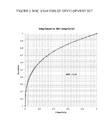
- FIG. 1 shows an example of a Risk Categorization Table for lung cancer.
- the inflection point between having a risk greater than the observed risk of smokers of 2% occurs with an aggregate MoM score of 9.
- an aggregate score of 9 or less that patient has a risk of lung cancer no greater than does any other heavy smoker not yet diagnosed.
- a MoM score greater than 9 indicates a greater risk of cancer or a higher likelihood of cancer as compared to the smoking population.
- FIG. 2 shows a table of the distribution of patient samples analyzed, including patients with all stages of cancer, at risk populations and various other control groups including those with non-cancerous lung disorders and other cancers.
- FIG. 3 shows a receiver operator characteristic (ROC) curve analysis of all lung cancer vs. all non-cancer samples yielded an area under the curve (AUC) of 0.76.
- ROC receiver operator characteristic
- FIG. 4 shows, in table form, the statistical validation using a cohort of 322 samples obtained with the specific intent of early detection in the high risk population.
- FIG. 5 shows the ROC curve analysis for the cohort of 322 samples with an AUC of 0.73.
- FIG. 6 shows the linearity of one of the tumor markers in a spike and recovery assay.
- FIG. 7 shows the biomarker precision and repeatability from a clinical bridging study in table form.
- FIG. 8 shows results from a blinded retrospective study using the six lung cancer biomarker panel.
- FIG. 9 shows, in table form, results from the lung cancer assay for at-risk subjects re-categorizing the patients risk for the presence of lung cancer.
- FIG. 10 shows results from the lung cancer assay for at-risk subjects re-categorizing the patient's risk, based on a range of composite scores, for the presence of lung cancer.
- the present invention provides a risk categorization of a population used to determine a quantified risk level for the presence of a cancer in an asymptomatic human subject.
- the method is preferably used as part of a blood test that measures multiple biomarkers in the blood.
- the risk categorization is herein referred to a risk categorization table.
- the term “table” is used in its broadest sense to refer to a grouping of data into a format providing for ease of interpretation or presentation, this includes, but is not limited to a computer program, software application, table, sliding template (e.g., pinwheel), spreadsheet, etc.
- the risk categorization table is a grouping of stratified human subject populations.
- each stratification of human subjects is based on analysis of retrospective clinical samples from subjects having a cancer wherein the actual incidence of cancer, herein referred to as the “positive predictive score” is determined for each stratified grouping.
- the analysis of retrospective clinical samples refers to measurement of markers in those samples, including normalization of values and summing those values to generate a risk score for each sample.
- the positive predictive score is then converted to a multiplier indicating increased likelihood of having the cancer by dividing the positive predictive score by the reported incidence of cancer in the cohort of the population subject to stratification, (e.g. human subjects 50 years or older).
- Each grouping is given a risk categorization indicator, including, but not limited to, low risk, intermediate-low risk, intermediate risk, intermediate-high risk and highest risk.
- each category of the risk categorization table comprises 1) a multiplier indicating increased likelihood of having the cancer, 2) a risk identifier and 3) a range of composite scores.
- the basis for the stratification of a cohort of a population of human subjects is based on 1) an identification of a certain cancer and 2) biomarkers that are associated with the cancer. In other words, a cohort shares the same cancer risk factors. Validation of the biomarkers to be used in the present methods is provided by analyzing retrospective cancer samples along with age matched normal (non-cancer) samples.
- risk categorization table including methods for normalizing biomarker data, is provided in more detail below along with a specific example for lung cancer.
- the present invention further provides an algorithm for analyzing a panel of biomarkers for a cancer and quantifying a human subject's increased risk (or in certain circumstances decreased risk) for the presence of the cancer in an asymptomatic human subject relative to a population.
- the term “increased risk” refers to an increase for the presence of the cancer as compared to the known prevalence of that particular cancer across the population cohort.
- the present methods are based on the generation of a risk categorization table for a certain cancer; wherein there is no intended limitation on when this table is generated just that when utilized the quantified risk is at the time of testing.
- the present method and risk categorization table is based, at least in part, on 1) the identification and clustering of a set of proteins and/or resulting autoantibodies to those proteins that can serve as markers for the presence of a cancer, 2) normalization and summing of the markers measured to generate a composite score; and, 3) determination of threshold values used to divide patients into groups with varying degrees of risk for the presence of cancer in which the likelihood of an asymptomatic human subject having a quantified increased risk for the presence of the cancer is determined.
- the algorithm yields a numerical risk score for each patient tested, which can be used by physicians to make treatment decisions concerning the therapy of cancer patients or, importantly, to further inform screening procedures to better predicted and diagnose early stage cancer in asymptomatic patients.
- the term “or” is used to refer to a nonexclusive or, such that “A or B” includes “A but not B,” “B but not A,” and “A and B,” unless otherwise indicated.
- the term “about” is used to refer to an amount that is approximately, nearly, almost, or in the vicinity of being equal to or is equal to a stated amount, e.g., the state amount plus/minus about 5%, about 4%, about 3%, about 2% or about 1%.
- asymptomatic refers to a patient or human subject that has not previously been diagnosed with the same cancer that their risk of having is now being quantified and categorized.
- human subjects may shows signs such as coughing, fatigue, pain, etc., but had not been previously diagnosed with lung cancer but are now undergoing screening to categorize their increased risk for the presence of cancer and for the present methods are still considered “asymptomatic”.
- the term “AUC” refers to the Area Under the Curve, for example, of a ROC Curve. That value can assess the merit of a test on a given sample population with a value of 1 representing a good test ranging down to 0.5 which means the test is providing a random response in classifying test subjects. Since the range of the AUC is only 0.5 to 1.0, a small change in AUC has greater significance than a similar change in a metric that ranges for 0 to 1 or 0 to 100%. When the % change in the AUC is given, it will be calculated based on the fact that the full range of the metric is 0.5 to 1.0.
- a variety of statistics packages can calculate AUC for an ROC curve, such as, JMPTM or Analyse-ItTM.
- AUC can be used to compare the accuracy of the classification algorithm across the complete data range.
- Classification algorithms with greater AUC have, by definition, a greater capacity to classify unknowns correctly between the two groups of interest (disease and no disease).
- the classification algorithm maybe as simple as the measure of a single molecule or as complex as the measure and integration of multiple molecules.
- biological sample and “test sample” refer to all biological fluids and excretions isolated from any given subject.
- samples include, but are not limited to, blood, blood serum, blood plasma, urine, tears, saliva, sweat, biopsy, ascites, cerebrospinal fluid, milk, lymph, bronchial and other lavage samples, or tissue extract samples.
- blood, serum, plasma and bronchial lavage or other liquid samples are convenient test samples for use in the context of the present methods.
- cancer and “cancerous” refer to or describe the physiological condition in mammals that is typically characterized by unregulated cell growth.
- examples of cancer include but are not limited to, lung cancer, breast cancer, colon cancer, prostate cancer, hepatocellular cancer, gastric cancer, pancreatic cancer, cervical cancer, ovarian cancer, liver cancer, bladder cancer, cancer of the urinary tract, thyroid cancer, renal cancer, carcinoma, melanoma, and brain cancer.
- cancer risk factors refers to biological or environmental influences that are known risks associated with a particular cancer. These cancer risk factors include, but are not limited to, a family history of cancer (e.g. breast cancer), age, weight, sex, history of smoking tobacco, exposure to asbestos, exposure to radiation, etc. It is understood that these cancer risk factors, either individually or a combination thereof, contribute to selecting a cohort of the population used to develop a Risk Categorization Table and that this same cohort is then tested using the present methods and algorithm to determine their increased risk for the presence of cancer as compared to the known prevalence of cancer across the cohort. In certain embodiments, cancer risk factors for lung cancer are a human subject aged 50 years or older with a history of smoking tobacco.
- the term “cohort” refers to a group or segment of human subjects with shared factors or influences, such as age, family history, cancer risk factors, environmental influences, etc.
- a “cohort” refers to a group of human subjects with shared cancer risk factors; this is also referred to herein as a “disease cohort”.
- a “cohort” refers to a normal population group matched, for example by age, to the cancer risk cohort; also referred to herein as a “normal cohort”.
- the term “composite score” refers to a summation of the normalized values for the predetermined markers measured in the sample from the human subject.
- the normalized values are reported as a multiple of median (MoM) values and those MoM values are then summed to provide a composite score for each human subjected tested.
- MoM median
- the “composite score” is used to determine the “risk score” for each human subject tested wherein the multiplier indicating increased likelihood of having the cancer for the stratified grouping becomes the “risk score”. See, FIG. 1 .
- the “cohort score” is also referred to herein as the “test score”.
- decision tree refers to a classifier with a flow-chart-like tree structure employed for classification. Decision trees consist of repeated splits of a data set into subsets. Each split consists of a simple rule applied to one variable, e.g., “if value of ‘variable 1’ larger than ‘threshold 1’; then go left, else go right”. Accordingly, the given feature space is partitioned into a set of rectangles with each rectangle assigned to one class.
- differentially expressed gene As used herein, the terms “differentially expressed gene,” “differential gene expression” and their synonyms, which are used interchangeably, are used in the broadest sense and refers to a gene and/or resulting protein whose expression is activated to a higher or lower level in a subject suffering from a disease, specifically cancer, such as lung cancer, relative to its expression in a normal or control subject. The terms also include genes whose expression is activated to a higher or lower level at different stages of the same disease. It is also understood that a differentially expressed gene may be either activated or inhibited at the nucleic acid level or protein level, or may be subject to alternative splicing to result in a different polypeptide product.
- Differential gene expression may include a comparison of expression between two or more genes or their gene products (e,g, proteins), or a comparison of the ratios of the expression between two or more genes or their gene products, or even a comparison of two differently processed products of the same gene, which differ between normal subjects and subjects suffering from a disease, specifically cancer, or between various stages of the same disease.
- Differential expression includes both quantitative, as well as qualitative, differences in the temporal or cellular expression pattern in a gene or its expression products among, for example, normal and diseased cells, or among cells which have undergone different disease events or disease stages.
- gene expression profiling is used in the broadest sense, and includes methods of quantification of mRNA and/or protein levels in a biological sample.
- the term “increased risk” refers to an increase in the risk level, for a human subject after testing, for the presence of a cancer relative to a population's known prevalence of a particular cancer before testing.
- a human subject's risk for cancer before testing may be 2% (based on the understood prevalence of cancer in the population), but after testing (based on the measure of biomarkers) their risk for the presence of cancer may be 30% or alternatively reported as an increase of 15 times compared to the cohort.
- the algorithm for calculating the 30% risk of having the cancer and the increased risk of 15 times the cohort population is provided in more detail below.
- a human subjects risk for cancer before testing may be 2% (based on the understood prevalence of cancer in the population), but after testing (based on the measure of biomarkers) their risk for the presence of cancer may be 1% or alternatively reported as an increase of 0.5 times compared to the cohort.
- “increased risk” refers to a change in risk level relative to a population before testing.
- the term “decreased risk” refers to a decrease in the risk level, for a human subject after testing, for the presence of a cancer relative to a population's known prevalence of a particular cancer before testing. In this instance, “decreased risk” refers to a change in risk level relative to a population before testing.
- lung cancer refers to a cancer state associated with the pulmonary system of any given subject.
- lung cancers include, but are not limited to, adenocarcinoma, epidermoid carcinoma, squamous cell carcinoma, large cell carcinoma, small cell carcinoma, non-small cell carcinoma, and bronchoalveolar carcinoma.
- lung cancers may be at different stages, as well as varying degrees of grading. Methods for determining the stage of a lung cancer or its degree of grading are well known to those skilled in the art.
- markers refer to molecules that can be evaluated in a sample and are associated with a physical condition.
- a markers include expressed genes or their products (e.g. proteins) or autoantibodies to those proteins that can be detected from a human samples, such as blood, serum, solid tissue, and the like, that, that is associated with a physical or disease condition.
- biomarkers include, but are not limited to, biomolecules comprising nucleotides, amino acids, sugars, fatty acids, steroids, metabolites, polypeptides, proteins (such as, but not limited to, antigens and antibodies), carbohydrates, lipids, hormones, antibodies, regions of interest which serve as surrogates for biological molecules, combinations thereof (e.g., glycoproteins, ribonucleoproteins, lipoproteins) and any complexes involving any such biomolecules, such as, but not limited to, a complex formed between an antigen and an autoantibody that binds to an available epitope on said antigen.
- biomolecules comprising nucleotides, amino acids, sugars, fatty acids, steroids, metabolites, polypeptides, proteins (such as, but not limited to, antigens and antibodies), carbohydrates, lipids, hormones, antibodies, regions of interest which serve as surrogates for biological molecules, combinations thereof (e.g., glycoproteins, ribonucleoproteins, lip
- biomarker can also refer to a portion of a polypeptide (parent) sequence that comprises at least 5 consecutive amino acid residues, preferably at least 10 consecutive amino acid residues, more preferably at least 15 consecutive amino acid residues, and retains a biological activity and/or some functional characteristics of the parent polypeptide, e.g. antigenicity or structural domain characteristics.
- the present markers refer to both tumor antigens present on or in cancerous cells or those that have been shed from the cancerous cells into bodily fluids such as blood or serum.
- the present markers as used herein, also refer to autoantibodies produced by the body to those tumor antigens.
- a “marker” as used herein refers to both tumor antigens and autoantibodies that are capable of being detected in serum of a human subject. It is also understood in the present methods that use of the markers in a panel may each contribute equally to the composite score or certain biomarkers may be weighted wherein the markers in a panel contribute a different weight or amount to the final composite score.
- the term “multiplier indicating increased likelihood of having the cancer” refers to a numerical value of the risk categorization table and assigned to a patient sample after testing quantifying that patients increased risk, above the cohort population, for the presence of a cancer.
- the “multiplier indicating increased likelihood of having the cancer” becomes the “risk score” for each human subject tested. See, FIG. 1 .
- the terms “multiple of median” or “MoM” refers to a measure of how far an individual test result deviates from the median.
- a predetermined marker is measured in a sample from an asymptomatic subject and the value is normalized as a multiple of median score.
- normalization when used in conjunction with measurement of biomarkers across samples and time, refer to mathematical methods where the intention is that these normalized values allow the comparison of corresponding normalized values from different datasets in a way that eliminates or minimizes differences and gross influences.
- multiple of median is used as the normalization methodology for the present methods.
- the terms “panel of markers”, “panel of biomarkers” and their synonyms, which are used interchangeably, refer to more than one marker that can be detected from a human sample that together, are associated with the presence of a particular cancer.
- the presence of the biomarkers are not individually quantified as an absolute value to indicate the presence of a cancer, but the measured values are normalized and the normalized value is summed to provide a composite score.
- each marker in the panel may be given the weight of 1, or some other value that is either a fraction of 1 or a multiple of 1, depending on the contribution of the marker to the cancer being screened and the overall composition of the panel.
- pathology of (tumor) cancer includes all phenomena that compromise the well-being of the patient. This includes, without limitation, abnormal or uncontrollable cell growth, metastasis, interference with the normal functioning of neighboring cells, release of cytokines or other secretory products at abnormal levels, suppression or aggravation of inflammatory or immunological response, neoplasia, premalignancy, malignancy, invasion of surrounding or distant tissues or organs, such as lymph nodes, etc.
- the term “known prevalence of cancer” refers to a prevalence of a cancer in a population before the human subject is tested using the present methods. This known prevalence of cancer, can be a prevalence reported in the literature based on retrospective data or an algorithm applied to that prevalence where in the algorithm takes into account factors such as age and more immediate and relevant history. In this instance, a known prevalence of cancer in a cohort refers to a risk of having cancer prior to being tested by the present methods.
- a positive predictive score As used herein, the term “a positive predictive score,” “a positive predictive value,” or “PPV” refers to the likelihood that a score within a certain range on a biomarker test is a true positive result. It is defined as the number of true positive results divided by the number of total positive results. True positive results can be calculated by multiplying the test Sensitivity times the Prevalence of disease in the test population. False positives can be calculated by multiplying (1 minus the Specificity) times (1 ⁇ the prevalence of disease in the test population). Total positive results equal True Positives plus False Positives.
- the term “risk score” refers to a single numerical value that indicates an asymptomatic human subject's increased risk for the presence of a cancer as compared to the known prevalence of cancer in the disease cohort.
- the composite score as calculated for a human subject and correlated to a multiplier indicating increased likelihood of having the cancer, wherein the composite score is correlated based on the range of composite scores for each stratified grouping in the risk categorization table. In this way the composite score is converted to a risk score based on the multiplier indicating increased likelihood of having the cancer for the grouping that is the best match for the composite score. See, FIG. 1 .
- ROC curve Receiveiver Operating Characteristic Curve
- ROC curves can be generated for a single feature as well as for other single outputs, for example, a combination of two or more features that are combined (such as, added, subtracted, multiplied etc.) to provide a single combined value which can be plotted in a ROC curve.
- the ROC curve is a plot of the true positive rate (sensitivity) of a test against the false positive rate (1-specificity) of the test. ROC curves provide another means to quickly screen a data set.
- screening refers to a strategy used in a population to identify an unrecognized cancer in asymptomatic subjects, for example those without signs or symptoms of the cancer.
- a cohort of the population e.g. smokers aged 50 or older
- a particular cancer e.g. lung cancer
- the term “subject” refers to an animal, preferably a mammal, including a human or non-human.
- patient and “human subject” may be used interchangeably herein.
- tumor refers to all neoplastic cell growth and proliferation, whether malignant or benign, and all pre-cancerous and cancerous cells and tissues.
- the phrase “Weighted Scoring Method” refers to a method that involves converting the measurement of one biomarker that is identified and quantified in a test sample into one of many potential scores.
- a ROC curve can be used to standardize the scoring between different markers by enabling the use of a weighted score based on the inverse of the false positive % defined from the ROC curve.
- the weighted score can be calculated by multiplying the AUC by a factor for a marker and then dividing by the false positive % based on a ROC curve.
- the weighting provides higher scores for biomarkers with a low false positive rate (thereby having higher specificity) for the population of interest.
- the weighting paradigm can comprise electing levels of false positivity (1-specificity) below which the test will result in an increased score.
- markers with high specificity can be given a greater score or a greater range of scores than markers that are less specific.
- Foundation for assessing the parameters for weighing can be obtained by determining presence of a marker in a population of patients with lung cancer and in normal individuals.
- the information (data) obtained from all the samples are used to generate a ROC curve and to create an AUC for each biomarker.
- a number of predetermined cutoffs and a weighted score are assigned to each biomarker based on the % specificity. That calculus provides a stratification of aggregate scores, and those scores can be used to define ranges that correlate to arbitrary risk categories of whether one has a higher or lower risk of having lung cancer.
- the number of categories can be a design choice or may be driven by the data.
- provided herein are methods for quantifying the risk level of an asymptomatic patient relative to a population.
- the risk level is increased as compared to the population.
- the risk level is decreased as compared to the population.
- the asymptomatic patients that, after testing, have a quantified increased risk for the presence of cancer relative to the population are those that a physician may select for follow-on testing and it is important to not only know that their risk is increased, relative to the population, but that their risk is quantified.
- the method of determining a quantified increased risk for the presence of a cancer in an asymptomatic human subject comprises 1) measuring a panel of markers in a sample from the human subject; 2) determining a normalized value of each marker in a sample from a human subject; 3) summing the normalized value to obtain a composite score for the human subject; 4) quantifying the increased risk for the presence of the cancer for the human subject as a risk score, wherein the composite score is matched to a risk category of a grouping of stratified human subject populations wherein each risk category comprises a multiplier indicating increased likelihood of having the cancer correlated to a range of composite scores; and, 5) providing a risk score for the human subject, whereby the quantified increased risk for the presence of a cancer in an asymptomatic human subject has been determined.
- One or more steps of the method described herein can be performed manually or can be completely or partially automated (for example, one or more steps of the method can be performed by a computer program or algorithm. If the method were to be performed via computer program or algorithm, then the performance of the method would further necessitate the use of the appropriate hardware, such as input, memory, processing, display and output devices, etc). Methods for automating one or more steps of the method would be well within the skill of those in the art.
- the present invention contemplates specific use computer, which may be a general purpose computer, configured to perform the steps of the method described herein.
- the method, or portions of the method may be further embodied in a computer readable medium capable of being executed in a computer environment.
- Such computer readable medium may be a specific storage device, such as a disk, or a location on a server, physical or virtual, the may be accessed by a computer for performing the required steps of the method.
- the first step in the present method is measuring a panel of markers from an asymptomatic human subject.
- gene expression e.g. mRNA
- resulting gene products e.g. polypeptides or proteins
- the method of interest is not limited to any one assay format or to any particular set of markers that comprise a panel.
- PCT International Pat. Pub. No. WO 2009/006323; US Pub. No. 2012/0071334; US Pat. Pub. No. 2008/0160546; US Pat. Pub. No. 2008/0133141; US Pat. Pub. No. 2007/0178504 (each herein incorporated by reference) teaches a multiplex lung cancer assay using beads as the solid phase and fluorescence or color as the reporter in an immunoassay format. Hence, the degree of fluorescence or color can be provided in the form of a qualitative score as compared to an actual quantitative value of reporter presence and amount.
- the presence and quantification of one or more antigens or antibodies in a test sample can be determined using one or more immunoassays that are known in the art.
- Immunoassays typically comprise: (a) providing an antibody (or antigen) that specifically binds to the biomarker (namely, an antigen or an antibody); (b) contacting a test sample with the antibody or antigen; and (c) detecting the presence of a complex of the antibody bound to the antigen in the test sample or a complex of the antigen bound to the antibody in the test sample.
- Well known immunological binding assays include, for example, an enzyme linked immunosorbent assay (ELISA), which is also known as a “sandwich assay”, an enzyme immunoassay (EIA), a radioimmunoassay (RIA), a fluoroimmunoassay (FIA), a chemiluminescent immunoassay (CLIA) a counting immunoassay (CIA), a filter media enzyme immunoassay (MEIA), a fluorescence-linked immunosorbent assay (FLISA), agglutination immunoassays and multiplex fluorescent immunoassays (such as the Luminex Lab MAP), immunohistochemistry, etc.
- ELISA enzyme linked immunosorbent assay
- EIA enzyme immunoassay
- RIA radioimmunoassay
- FFIA fluoroimmunoassay
- CLIA chemiluminescent immunoassay
- FLISA fluorescence-linked immunosorbent assay
- the immunoassay can be used to determine a test amount of an antigen in a sample from a subject.
- a test amount of an antigen in a sample can be detected using the immunoassay methods described above. If an antigen is present in the sample, it will form an antibody-antigen complex with an antibody that specifically binds the antigen under suitable incubation conditions described above. The amount of an antibody-antigen complex can be determined by comparing the measured value to a standard or control.
- the AUC for the antigen can then be calculated using techniques known, such as, but not limited to, a ROC analysis.
- gene expression of markers is measured in a sample from a human subject.
- markers e.g. mRNA
- gene expression profiling methods for use with paraffin-embedded tissue include quantitative reverse transcriptase polymerase chain reaction (qRT-PCR), however, other technology platforms, including mass spectroscopy and DNA microarrays can also be used. These methods include, but are not limited to, PCR, Microarrays, Serial Analysis of Gene Expression (SAGE), and Gene Expression Analysis by Massively Parallel Signature Sequencing (MPSS).
- the sample from human subject is a tissue section such as from a biopsy.
- the sample from the human subject is a bodily fluid such as blood, serum, plasma or a part or fraction thereof.
- the sample is a blood or serum and the markers are proteins measured there from.
- the sample is a tissue section and the markers are mRNA expressed therein. Many other combinations of sample forms from the human subjects and the form of the markers are contemplated.
- a panel of markers needs to be selected for a particular cancer being screened.
- Many markers are known for cancers and a known panel can be selected, or as was done by the present Applicants, a panel can be selected based on measurement of individual markers in retrospective clinical samples wherein a panel is generated based on empirical data for a desired cancer.
- biomarkers examples include molecules detectable, for example, in a body fluid sample, such as, antibodies, antigens, small molecules, proteins, hormones, genes and so on.
- a panel of markers is selected based on their association with lung cancer.
- the panel of markers is selected from anti-p53, anti-NY-ESO-1, anti-ras, anti-Neu, anti-MAPKAPK3, cytokeratin 8, cytokeratin 19, cytokeratin 18, CEA, CA125, CA15-3, CA19-9, Cyfra 21-1, serum amyloid A, proGRP and ⁇ 1 -anti-trypsin (US 20120071334; US 20080160546; US 20080133141; US 20070178504 (each herein incorporated by reference)).
- proteins have more recently been identified as possible biomarkers for the occurrence of lung cancer, for example the proteins CEA, RBP4, hAAT, SCCA [Patz, E. F., et al., Panel of Serum Biomarkers for the Diagnosis of Lung Cancer. Journal of Clinical Oncology, 2007. 25(35): p. 5578-5583]; the proteins IL6, IL-8 and CRP [Pine, S. R., et al., Increased Levels of Circulating Interleukin 6, Interleukin 8, C-Reactive Protein, and Risk of Lung Cancer. Journal of the National Cancer Institute, 2011. 103(14): p.
- Novel ligands that bind to circulating, lung-cancer associated proteins which are possible biomarkers include nucleic acid aptamers to bind cadherin-1, CD30 ligand, endostatin, HSP90 ⁇ , LRIG3, MIP-4, pleiotrophin, PRKCI, RGM-C, SCF-sR, sL-selectin, and YES [Ostroff, R. M., et al., Unlocking Biomarker Discovery: Large Scale Application of Aptamer Proteomic Technology for Early Detection of Lung Cancer. PLoS ONE, 2010. 5(12): p.
- Autoantibodies that are proposed to be circulating markers for lung cancer include p53, NY-ESO-1, CAGE, GBU4-5, Annexin 1, and SOX2 [Lam, S., et al., EarlyCDT-Lung: An Immunobiomarker Test as an Aid to Early Detection of Lung Cancer. Cancer Prevention Research, 2011. 4(7): p. 1126-1134.] and IMPDH, phosphoglycerate mutase, ubiquillin, Annexin I, Annexin II, and heat shock protein 70-9B (HSP70-9B) [Farlow, E.
- Micro-RNAs that are proposed to be circulating markers for lung cancer include miR-21, miR-126, miR-210, miR-486-5p [Shen, J., et al., Plasma microRNAs as potential biomarkers for non-small-cell lung cancer. Lab Invest, 2011. 91(4): p. 579-587.]; miR-15a, miR-15b, miR-27b, miR-142-3p, miR-301 [Hennessey, P. T., et al., Serum microRNA Biomarkers for Detection of Non-Small Cell Lung Cancer. PLoS ONE, 2012. 7(2): p.
- let-7b, let-7c, let-7d, let-7e miR-10a, miR-10b, miR-130b, miR-132, miR-133b, miR-139, miR-143, miR-152, miR-155, miR-15b, miR-17-5p, miR-193, miR-194, miR-195, miR-196b, miR-199a*, miR-19b, miR-202, miR-204, miR-205, miR-206, miR-20b, miR-21, miR-210, miR-214, miR-221, miR-27a, miR-27b, miR-296, miR-29a, miR-301, miR-324-3p, miR-324-5p, miR-339, miR-346, miR-365, miR-378, miR-422a, miR-432, miR-485-3p, miR-496, miR-497, miR-505, miR-518b,
- hsa-let-7a, hsa-let-7b, hsa-let-7d hsa-miR-103, hsa-miR-126, hsa-miR-133b, hsa-miR-139-5p, hsa-miR-140-5p, hsa-miR-142-3p, hsa-miR-142-5p, hsa-miR-148a, hsa-miR-148b, hsa-miR-17, hsa-miR-191, hsa-miR-22, hsa-miR-223, hsa-miR-26a, hsa-miR-26b, hsa-miR-28-5p, hsa-miR-29a, hsa-miR-30b, hsa-miR-30c,
- miR-190b miR-630, miR-942, and miR-1284 [Patnaik, S. K., et al., MicroRNA Expression Profiles of Whole Blood in Lung Adenocarcinoma. PLoS ONE, 2012. 7(9): p. e46045.].
- a panel of markers for lung cancer is selected from CEA (GenBank Accession CAE75559), CA125 (UniProtKB/Swiss-Prot: Q8WXI7.2), Cyfra 21-1 (NCBI Reference Sequence: NP_008850.1), anti-NY-ESO-1 (antigen NCBI Reference Sequence: NP_001318.1), anti-p53 (antigen GenBank: BAC16799.1) and anti-MAPKAPK3 (antigen NCBI Reference Sequence: NP_001230855.1), the first three are tumor marker proteins and the last three are autoantibodies.
- a panel of markers comprises circulating markers associated with colorectal cancer (CRC); those include the microRNA miR-92 [Ng, E. K. O., et al., Differential expression of microRNAs in plasma of patients with colorectal cancer: a potential marker for colorectal cancer screening. Gut, 2009. 58(10): p. 1375-1381.]; aberrantly methylated SEPT9 DNA [deVos, T., et al., Circulating Methylated SEPT9 DNA in Plasma Is a Biomarker for Colorectal Cancer. Clinical Chemistry, 2009. 55(7): p. 1337-1346.]
- CRC colorectal cancer
- a panel of markers comprises markers associated with a cancer selected from bile duct cancer, bone cancer, pancreatic cancer, cervical cancer, colon cancer, colorectal cancer, gallbladder cancer, liver or hepatocellular cancer, ovarian cancer, testicular cancer, lobular carcinoma, prostate cancer, and skin cancer or melanoma.
- a panel of markers comprises markers associated with breast cancer.
- a panel can comprise any number of markers as a design choice, seeking, for example, to maximize specificity or sensitivity of the assay.
- an assay of interest may ask for presence of at least one of two or more biomarkers, three or more biomarkers, four or more biomarkers, five or more biomarkers, six or more biomarkers, seven or more biomarkers, eight biomarkers or more as a design choice.
- the panel of biomarkers may comprise at least two, at least three, at least four, at least five, at least six, at least seven, at least eight, at least nine or at least ten or more different markers. In one embodiment, the panel of biomarkers comprises about two to ten different markers. In another embodiment, the panel of biomarkers comprises about four to eight different markers. In yet another embodiment, the panel of markers comprises about six different markers.
- a sample is committed to the assay and the results can be a range of numbers reflecting the presence and level of presence of each of the biomarkers of the panel in the sample.
- each marker in the panel is measured and normalized wherein none of the markers are given any specific weight. In this instance each marker has a weight of 1.
- the choice of the markers may be based on the understanding that each marker, when measured and normalized, contributed unequally to determine the likelihood of the presence of the cancer.
- a particular marker in the panel can either be weighted as a fraction of 1 (for example if the relative contribution is low), a multiple of 1 (for example if the relative contribution is high) or as 1 (for example when the relative contribution is neutral compared to the other markers in the panel).
- the present methods further comprising weighting the normalized values prior to summation of the normalized values to obtain a composite score.
- Decision tree is a data handling approach where a series of simple dichotomous decisions guide through a classification to yield such a desired binary outcome. Hence, samples are partitioned based on whether values thereof are above or below calculated thresholds.
- a model for scoring multiple biomarkers which attempts to employ a decision tree logic was developed by Mor et al., PNAS, 102(21):7677-7682 (2005), wherein an optimal cutoff value is obtained and assigns a value of 0 (not likely to have cancer) or 1 (likely to have cancer) for a marker. Then, scores of individual biomarkers are combined for a final score of each sample and the higher the final score, the higher the probability of disease.
- That technique provides a binary result favored by physicians and patients. While distribution of data is not an assumption which contributes to simplicity of the model, that the model reduces information to a 1 or 0 score results in a loss of quantitative information, for example, diminishes the role of a more predictive marker and elevates the role of a less predictive marker.
- the collection of markers in a multiplex assay may comprise varying levels of value or predictability in diagnosing disease.
- the impact of any one marker on the ultimate determination may be weighted based on the aggregated data obtained in screening populations and correlating with actual pathology to provide a more discriminating or effective diagnostic assay.
- An alternative approach is to find an intermediate ground by expanding the qualitative transformation of quantitative data into multiple categories as compared to only a binary classification scheme.
- One embodiment is directed to a method for assessing risk of lung cancer.
- a research effort to identify panels of biomarkers that included a survey of known tumor protein biomarkers coupled with a discovery project for novel lung cancer specific biomarkers was previously conducted (PCT Publ. No. 2009/006323, incorporated herein by reference).
- This work indicates although a combination of markers can be used to increase sensitivity of testing for cancer without greatly affecting the specificity of the test.
- markers were tested and analyzed in a way that is often very different from the standard methods.
- This effort culminated in the establishment of a panel of six biomarkers that in the aggregate yield significant sensitivity and specificity for the early detection of lung cancer using the present methods.
- Applicants provide a new method and algorithm that can be utilized to identify smokers at the highest levels of risk for follow-up testing by CT scanning.
- the lung cancer biomarker panel comprises a series of three tumor marker proteins and three autoantibodies.
- Tumor markers are proteins released by the cancer itself into the patient's serum. Since the presence of these proteins or their increased expression is directly related to the cancer cells they tend to be specific to cancer, however they may often be found in more than one type of cancer. Furthermore, because they are derived directly from the tumor, their levels will depend on the size of the tumor. This can make them less sensitive for the detection of early stage cancers.
- Autoantibodies are a function of the patient's immune response to the abnormal cancerous cells. Because the immune system amplifies its response even to a small amount of antigen, autoantibodies may be detected more easily in the early stage patient than proteins released by the cancer itself.
- the tumor markers incorporated into the present methods for lung cancer comprise CEA, CA-125 and Cyfra 21-1. All three of these markers have been extensively studied by others and are currently in clinical use for monitoring of other cancers. While none of these markers have fared well as a stand-alone marker for the early detection of lung cancer, two important points must be iterated; 1) these markers are not measured by the present method in the same way they have been measured in the past for other indications, and 2) these markers are not deployed as stand-alone markers but rather are incorporated as a part of an integrated panel of markers for re-stratification of patient risk.
- results in the present methods for lung cancer are not based on an absolute serum level, but on an increase in level as compared to the median levels in matched control patients.
- individual marker values as a total serum concentration are not measured; instead these three markers are incorporated in a composite score that has value only in re-categorizing patient risk for the presence of lung cancer.
- three autoantibodies are utilized in the present lung cancer test, wherein the autoantibodies comprise anti-p53, anti-NY-ESO-1 and anti-MAPKAPK3.
- the autoantibodies comprise anti-p53, anti-NY-ESO-1 and anti-MAPKAPK3.
- most autoantibodies are only found in a limited number of patients.
- These three autoantibodies are among those most commonly found in lung cancer, although each on its own has a rather limited distribution as members of an integrated biomarker panel because they do contribute to the overall sensitivity of the test.
- p53 is a well-known tumor suppressor protein that is often mutated in cancer. Such mutations may be enough to break natural immune tolerance to the protein and thus the source of anti-p53 antibodies.
- NY-ESO-1 has been characterized as a tumor specific marker and thus autoantibodies against this protein may represent a way to measure the levels of a tumor marker in early stage disease via immune amplification.
- MAPKAPK3 is a kinase protein that can be activated by several oncogenic pathways and thus may be more commonly up-regulated in lung cancer leading to the development of autoantibodies targeted against it.
- the method for determining a quantified increased risk for the presence of a lung cancer in an asymptomatic human subject comprises: 1) measuring a panel of markers in sample from a human subject that is at least 50 years of age or older and has a history of smoking tobacco; 2) determining a normalized score for each marker; 3) summing the normalized score to obtain a composite score for the human subject, 4) quantifying the increased risk for the presence of the lung cancer for the human subject as a risk score, wherein the composite score is matched to a risk category of a grouping of stratified human subject populations wherein each risk category comprises a multiplier indicating increased likelihood of having the lung cancer correlated to a range of composite scores; and, 5) providing a risk score for the human subject, whereby the quantified increased risk for the presence of the lung cancer in an asymptomatic human subject has been determined.
- the step of normalizing comprises determining the multiple of median (MoM) score for each marker.
- MoM median
- the MoM score is the subsequently summed to obtained a composite score.
- the disease cohort e.g. a human subject that is at least 50 years of age or older and has a history of smoking tobacco
- the disease cohort is independently determined and in this instance is well understood to be the “at risk” group for developing lung cancer.
- This present method and algorithm re-categorizes those at-risk patients into risk categories quantifying their true increased risk for the presence of lung cancer over the disease cohort.
- kits for assessing the likelihood that a patient has lung cancer relative to a population comprising the steps of: obtaining a sample from the patient; measuring the levels of multiple biomarkers in the sample; calculating a composite score from the biomarker measurements; comparing the patient composite score to the composite scores of persons known to be at a high and a low risk for lung cancer; and determining the level of risk of the patient for having lung cancer relative to the population.
- an asymptomatic patient's cancer risk level, relative to a population is determined.
- the determination may comprise quantifying the risk level relative to a population.
- the multiple biomarkers comprise two or more, three or more, four or more, five or more or six or more biomarkers.
- the multiple biomarkers comprise six markers selected from CEA, CA125, Cyfra 21-1, Pro-GRP, anti-NY-ESO-1, anti-p53, anti-Cyclin E2 and anti-MAPKAPK3.
- obtaining a composite score may further comprise normalizing the measured biomarker values and summing the normalized values to generate a composite score.
- the value obtained from measuring the marker in the sample is normalized.
- the methodology used to normalize the values of the measured biomarkers provided that the same methodology is used for testing a human subject sample as was used to generate the Risk Categorization Table.
- US Publ. No. 2008/0133141 (herein incorporated by reference) teaches statistical methodology for handling and interpreting data from a multiplex assay.
- the amount of any one marker thus can be compared to a predetermined cutoff distinguishing positive from negative for that marker as determined from a control population study of patients with cancer and suitably matched normal controls to yield a score for each marker based on said comparison; and then combining the scores for each marker to obtain a composite score for the marker(s) in the sample.
- the predetermined cutoffs can be based on ROC curves and the score for each marker can be calculated based on the specificity of the marker. Then, the total score can be compared to a predetermined total score to transform that total score to a qualitative determination of the likelihood or risk of having lung cancer.
- Another method for score transformation or normalization is, for example, applying the multiple of median (MoM) method of data integration.
- MoM median value of each biomarker
- the median value of each biomarker is used to normalize all measurements of that specific biomarker, for example, as provided in Kutteh et al. (Obstet. Gynecol. 84:811-815, 1994) and Palomaki et al. (Clin. Chem. Lab. Med.) 39:1137-1145, 2001).
- any measured biomarker level is divided by the median value of the cancer group, resulting in a MoM value.
- the MoM values can be combined (namely, summed or added) for each biomarker in the panel resulting in a panel MoM value or aggregate MoM score for each sample.
- the sample size of the cancer population and the normals for determining the median can be increased to yield more accurate population data.
- normalization comprises determining a multiple of median (MoM) score for each biomarker measured.
- the normalized value for each biomarker is summed to provide a composite score for each subject.
- this method comprises summing the MoM score to obtain a composite score.
- the composite score is derived by measuring the levels of each of all markers used in a panel for a particular cancer in arbitrary units and comparing these levels to the median levels found in previous validation studies.
- the cancer is lung cancer and the panel comprises the six markers disclosed above wherein this method generates six initial scores representing the multiple of the median (MoM) for each marker for a given patient. These initial scores are summed to yield the final composite score.
- the markers are measured and those resulting values normalized and then summed to obtain a composite score.
- normalizing the measured biomarker values comprises determining the multiple of median (MoM) score.
- the present method further comprises weighting the normalized values before summing to obtain a composite score.
- the next step of the present method comprises quantifying the increased risk for the presence of the cancer for the human subject as a risk score, wherein the composite score is matched to a risk category of a grouping of stratified human subject populations wherein each risk category comprises a multiplier indicating increased likelihood of having the cancer correlated to a range of composite scores.
- This quantification step is based on the predetermined grouping of a stratified cohort of human subjects.
- the grouping of a stratified population of human subjects, or stratification of a disease cohort is in the form of a risk categorization table.
- the selection of the disease cohort, the cohort of human subjects that share cancer risk factors are well understood by those skilled in the art of cancer research.
- the cohort may share an age category and smoking history. However, it is understood that the cohort, and the resulting stratification, may be more multidimensional and take into account further environmental or biological factors (e.g. epidemiological factors).
- the grouping of a stratified human subject population used to determine a quantified increased risk for the presence of a cancer in an asymptomatic human subject comprises: at least three risk categories, wherein each risk category comprises 1) a multiplier indicating increased likelihood of having the cancer, 2) a risk identifier and 3) a range of composite scores.
- each risk category comprises 1) a multiplier indicating increased likelihood of having the cancer, 2) a risk identifier and 3) a range of composite scores.
- an individual risk score is generated by summing the normalized values determined from a panel of markers for the cancer to obtain a composite that is correlated to a risk category of the risk categorization table.
- the normalized values are determined as multiple of median (MoM) scores.
- the risk identifier is a label given to a specific group to provide context for the range of risk scores and the multiplier indicating increased likelihood of having the cancer in each grouping.
- the risk identifier is selected from low risk, intermediate-low risk, intermediate risk, intermediate-high risk and highest risk. These risk identifiers are not intended to be limiting, but may include other labels are dictated by the data used to generate the table and/or further refine the context of the data.
- the multiplier indicating increased likelihood of having the cancer is a numerical value, such as 13.4; 5.0; 2.1; 0.7; and 0.4. This value is empirically derived and will change depending on the data, cohort of the subject population, type of cancer, biomarkers, etc. and so on.
- the multiplier indicating increased likelihood of having the cancer is a numerical value selected from 2, 3, 4, 5, 6, 7, 8, 9, 10, 11, 12, 13, 14, 15, 16, 17, 181, 19 20, 21, 22, 23, 24, 25, 26, 27, 28, 29, and 30, and so on, or some fraction thereof.
- the value indicates the increased risk, over the normal prevalence of cancer in the cohort population that formed the basis for the stratification, for the human subject at the time of testing.
- the human subject is from the same disease cohort as the one used to generate the risk categorization table.
- a disease cohort may be a human subject aged 50 years or older with a history of smoking tobacco.
- a risk score of 13.4 if a patient receives a risk score of 13.4, then that human subject has a 13.4 times increased risk for the presence of the cancer relative to the population.
- this multiplier value is empirically determined and in the present instance is done using retrospective clinical samples.
- stratification of human subjects is based on analysis of retrospective clinical samples from subjects having a cancer wherein the actual incidence of cancer, or the positive predictive score, is determined for each stratified grouping. The specifics of this are detailed below and in the example section.
- a positive predictive score can be determined, when retrospective samples with a known medical history are used, for each stratified grouping. This actual incidence of cancer in each of these groups is then divided by the reported incidence of cancer across the population of human subjects. For example, if the positive predictive score for one of the groupings from the stratified population of human subjects was 27%, this value would then be divided by the actual incidence of cancer across the cohort of the population that was stratified (e.g. 2%) to yield a multiplier of 13.5. In this scenario, the multiplier indicating increased likelihood of having the cancer is 13.5 and a subject tested that had a composite score matched to this category would have a risk factor of 13.5. In other words, at the time of testing, that human subject would be 13.5 times more likely to have the presence of cancer than the general population in that particular cohort.
- a bead immunoassay was used to screen selected patients with lung cancer and normal individuals for the presence of a panel of three autoantibody and three antigen markers associated with lung cancer in a blinded study.
- the assay employed fluorescence reporters and the degree of fluorescence was machine reported as a mean fluorescence intensity with a value ranging from 0 (lowest) to 5 (highest).
- the values obtained for each marker in the lung cancer population provided a median value, which then was used to determine the MoM value for experimental samples.
- the plotted ROC curve has an AUC of 0.738.
- the specificity was 80% and the sensitivity was 59%.
- the aggregate scores from the lung cancer patients and the normal cohort stratified into five ranges.
- the specificity and sensitivity of each range was determined, where the sensitivity represented the number of cancer patients with scores in any one range divided by the total number of cancer patients, 134.
- the specificity was the number of the normal cohort with a score in that range subtracted from 121, divided by 121.
- That stratification enabled a data transformation into a more qualitative risk categorization providing a greater degree of information for subsequent choices in light of the costs of lung cancer confirmation, for example a CAT scan or a PET scan, as well as patient compliance.
- lung cancer incidence in the at risk population of heavy smokers is about 2%, that percentage was used as the cutoff point between likelihood of cancer and not, meaning, at that level the individual had an even chance of having cancer, that is, 1.
- Positive predictive values were determined using the disease prevalence of 2% and then that positive predictive value was divided by two to yield another risk value interpreted as the likelihood of having lung cancer as a multiple of that of the normal population risk, which can be considered as 1 or even chances, or as a 2% risk based on population studies.
- the resulting risk categorization table is provided in FIG. 1 .
- the third component of each risk category of the Risk Categorization Table is a range of composite scores.
- these composite scores were generated from normalizing the data from the panel of measured biomarkers and then summing the individual values from each marker per sample. These composite scores were then grouped to provide a range and drove the stratification of the population. The specifics of this methodology are detailed below in the Example section.
- the physician and patient then can assess whether follow-up is required, necessary or recommended based on whether there is a greater risk that is just slightly above that of any smoker, i.e., 2%, or is higher because of a greater composite score, which may be deserving of greater consideration by the patient.
- the physician and patient will be the beneficiary of a quantitative value with foundation in the prevalence of cancer amongst smokers which provides improved resolution on the risk of cancer in light of the biomarker assay.
- a patient with a composite biomarker score of 20 or greater has a 13-fold greater likelihood of having lung cancer than any other heavy smoker, See FIG. 1 .
- That 13.4 ⁇ multiplier translates to an overall risk of about 27% of having lung cancer. That is, while all heavy smokers have a 1 in 50 chance of having lung cancer prior to testing, with a composite score or 20 or more after testing, that individual has a 1 in 4 chance of having lung cancer. Therefore, that person should consider a follow-up to visualize whether any cancer is present.
- the method for determining a quantified increased risk for the presence of lung cancer in an asymptomatic human subject comprises: 1) measuring a level of CEA, CA125, Cyfra 21-1, anti-NY-ESO-1, anti-p53 and anti-MAPKAPK3 in a serum sample from the human subject, wherein the human subject is at least 50 years of age or older and has a history of smoking tobacco; 2) determining a normalized score for each marker; 3) summing the normalized score to obtain a composite score for the human subject, 4) quantifying the increased risk for the presence of the lung cancer for the human subject as a risk score, wherein the composite score is matched to one of at least three risk categories of a grouping of a stratified human subject population wherein each risk category comprises a multiplier indicating increased likelihood of having the lung cancer correlated to a range of composite scores; and, 5) providing a risk score for the human subject, whereby the quantified increased risk for the presence of the lung cancer in an asymptomatic human subject
- the step of normalizing comprises determining the multiple of median (MoM) score for each marker. In this instance, the MoM score is then subsequently summed to obtain a composite score.
- MoM median
- the risk score is provided in a report.
- the report may comprise one or more of the following: patient information, a Risk Categorization Table, a risk score, a test score, a composite score, identification of the risk category for the patient, an explanation of the Risk Categorization Table and the resulting test score, list of biomarkers tested, description of the disease cohort, and so on.
- lung cancer is only one of many cancer types that can benefit from the present invention.
- Primary care healthcare practitioners who may include physicians specializing in internal medicine or family practice as well as physician assistants and nurse practitioners, are among the users of the methodology disclosed herein. These primary care providers typically see a large volume of patients each day and many of these patients are at risk for lung cancer due to smoking history, age, and other lifestyle factors. In 2012 about 18% of the U.S. population was current smokers and many more were former smokers with a lung cancer risk profile above that of never smokers.
- a blood sample from patients with a heavy smoking history (e.g. having smoked at least a pack of cigarettes per day for 20 years or more) is sent to a laboratory qualified to test the sample using a panel of biomarkers with adequate sensitivity and specific for early stage lung cancer.
- biomarkers are herein included in the above disclosure and the following examples.
- other suitable bodily fluids such as a sputum or saliva might also be utilized.
- a biomarker composite score for that patient is then generated using the technique described in the present disclosure.
- the patient's risk of having lung cancer as compared to others having a comparable smoking history and age range, can then be calculated using a table such as the one show in FIG. 1 .
- a table such as the one show in FIG. 1 .
- other means of calculation may be employed including those which utilize a computer program.
- a software application compatible with mobile devices e.g. a tablet or smart phone
- the physician or healthcare practitioner has a risk score for the patient (i.e. the likelihood that that patient has lung cancer relative to a population of others with comparable epidemiological factors) they can recommend, in particular, that those at a higher risk be followed up with other tests such as CT scanning.
- a risk score for the patient i.e. the likelihood that that patient has lung cancer relative to a population of others with comparable epidemiological factors
- CT scanning i.e. the likelihood that that patient has lung cancer relative to a population of others with comparable epidemiological factors
- One or more biomarkers, one or more reagents for testing the biomarkers, cancer risk factor parameters, a Risk Categorization Table, algorithm for calculating a risk score, and any combinations thereof are amenable to the formation of kits (such as panels) for use in performing the present methods.
- the kit can comprise (a) reagents containing at least one antibody for quantifying one or more antigens in a test sample, wherein said antigens comprise one or more of: cytokeratin 8, cytokeratin 19, cytokeratin 18, CEA, CA125, CA15-3, SCC, CA19-9, proGRP, Cyfra 21-1, serum amyloid A, alpha-1-anti-trypsin and apolipoprotein CIII; (b) reagents containing one or more antigens for quantifying at least one antibody in a test sample; wherein said antibodies comprise one or more of: anti-p53, anti-TMP21, anti-NPC1L1C-domain, anti-TMOD1, anti-CAMK1, anti-RGS1, anti-PACSIN1, anti-RCV1, anti-MAPKAPK3, anti-NY-ESO-1 and anti-Cyclin E2; and (c) one or more algorithms or computer programs for performing the steps of normalizing the amount of each antigen and/
- the reagents included in the kit for quantifying one or more regions of interest may include an adsorbent which binds and retains at least one region of interest contained in a panel, solid supports (such as beads) to be used in connection with said absorbents, one or more detectable labels, etc.
- the adsorbent can be any of many adsorbents used in analytical chemistry and immunochemistry, including metal chelates, cationic groups, anionic groups, hydrophobic groups, antigens and antibodies.
- the kit comprises the necessary reagents to quantify at least one of the following antigens, cytokeratin 19, cytokeratin 18, CA 19-9, CEA, CA-15-3, CA125, SCC, Cyfra 21-1, serum amyloid A, and ProGRP.
- the kit comprises the necessary reagents to quantify at least one of the following antibodies anti-p53, anti-TMP21, anti-NPC1L1C-domain, anti-TMOD1, anti-CAMK1, anti-RGS1, anti-PACSIN1, anti-RCV1, anti-MAPKAPK3, anti-NY-ESO-1 and anti-Cyclin E2.
- the kit further comprises one or more algorithms or computer programs for performing some or all the steps of the method described herein.
- the kit may further comprise an apparatus configured with a computer program to receive the values from the evaluation of markers in a sample and making the required calculations to determine a composite score and compare it to a grouping of stratified population comprising multiple risk categories (e.g. a Risk Categorization Table) and provide a Risk Score.
- risk categories e.g. a Risk Categorization Table
- the present invention further provides for an apparatus for assessing a subject's risk level for the presence of cancer and correlating with an increase or decrease of the presence of cancer after testing relative to a population.
- the apparatus comprises a computer program or software application to receive the values from the evaluation of markers in a sample and make the required calculations to determine a composite score and compare it to a grouping of stratified population comprising multiple risk categories (e.g. a Risk Categorization Table) and provide a Risk Score.
- risk categories e.g. a Risk Categorization Table
- the apparatus can take one of a variety of forms, for example, the correlation and means of matching can be provided as a computer program in any format known to a person of ordinary skill in the art that allows the method to be implemented in a handheld device, a tablet, or any other type of computer or electronic device, the apparatus can be a computer software product, an application for a handheld device, a handheld device configured to performed the method, or it can be a world-wide-web (WWW) page or other network accessible location, or it can be a computing device.
- WWW world-wide-web
- the apparatus can be a simple functional representation of the correlation such as a nomogram provided on a card, or wheel, that is readily portable and simple to use.
- the apparatus can be in the form of a laminated card or wheel.
- the correlation can be a graphic representation, which, in some embodiments, is stored in a database or memory, such as a random access memory, read-only memory, disk, virtual memory or processor.
- a database or memory such as a random access memory, read-only memory, disk, virtual memory or processor.
- Other suitable representations, pictures, depictions or exemplifications known in the art may also be used.
- the apparatus may further comprise a storage means for storing the correlation or nomogram, an input means that allows the input into the apparatus of the identical set of factors determined for a subject, and a display means for displaying the status of the subject in terms of the particular medical condition.
- the storage means can be, for example, random access memory, read-only memory, a disk, virtual memory, a database, or a processor.
- the input means can be, for example, a keypad, a keyboard, stored data, a touch screen, a voice-activated system, a downloadable program, downloadable data, a digital interface, a hand-held device, or an infrared signal device.
- the display means can be, for example, a computer monitor, a cathode ray tube (CRT), a digital screen, a light-emitting diode (LED), a liquid crystal display (LCD), an X-ray, a compressed digitized image, a video image, or a hand-held device.
- the apparatus can further comprise a database, wherein the database stores the correlation of factors and is accessible to the user.
- the apparatus is a computing device, for example, in the form of a computer or hand-held device that includes a processing unit, memory, and storage.
- the computing device can include, or have access to a computing environment that comprises a variety of computer-readable media, such as volatile memory and non-volatile memory, removable storage and/or non-removable storage.
- Computer storage includes, for example, RAM, ROM, EPROM & EEPROM, flash memory or other memory technologies, CD ROM, Digital Versatile Disks (DVD) or other optical disk storage, magnetic cassettes, magnetic tape, magnetic disk storage or other magnetic storage devices, or other medium known in the art to be capable of storing computer-readable instructions.
- the computing device can also include or have access to a computing environment that comprises input, output, and/or a communication connection.
- the input can be one or several devices, such as a keyboard, mouse, touch screen, or stylus.
- the output can also be one or several devices, such as a video display, a printer, an audio output device, a touch stimulation output device, or a screen reading output device.
- the computing device can be configured to operate in a networked environment using a communication connection to connect to one or more remote computers.
- the communication connection can be, for example, a Local Area Network (LAN), a Wide Area Network (WAN) or other networks and can operate over a wired network, wireless radio frequency network, and/or an infrared network.
- LAN Local Area Network
- WAN Wide Area Network
- FIG. 3 shows a receiver operator characteristic (ROC) curve analysis of all lung cancer vs. all non-cancer samples yielded an area under the curve (AUC) of 0.76. Choosing a cutoff of 10.7 shows a specificity of 80% yields a sensitivity of 64%. The data was further analyzed as a function of tumor stage using the same cutoff and yielding 75% sensitivity in late stage disease and 59% sensitivity for early stage disease indicating that the test has a higher sensitivity for later stage disease.
- ROC receiver operator characteristic
- non-cancerous lung disorders including asthma, COPD, emphysema, fibrosis and pneumonia
- noncancerous lung disorders including asthma, COPD, emphysema, fibrosis and pneumonia
- Analysis of the scores of three other cancer types demonstrated that these other cancers can yield a higher score more often than non-cancerous conditions, but that the range and median of scores is more similar to that found in the normal population.
- FIG. 6 presents the linearity of one of the tumor markers in a spike and recovery assay. All 6 biomarkers, those disclosed above, are detected in the linear range and have r 2 values of >0.9. Precision and repeatability were determined by testing 3 samples twice a day for 5 days each for two independent operators. An additional 5 days of data were collected for a single operator. All testing was performed in duplicate.
- FIG. 7 displays representative data for 3 of the markers.
- the Applicants a blinded retrospective study of the present methods using a total of 255 patients including 134 confirmed diagnoses of lung cancer and 121 age-matched>20 pack-year smokers as controls.
- the study group included two cohorts of patients collected at two separate cancer centers; one in the Northeastern United States and one in the Southwest. Cancer patients were a mix (50:50) of early and late stage. All six biomarkers were tested and analyzed to yield the composite score.
- the data is presented as a box plot in FIG. 8 .
- a ROC curve analysis yielded an AUC of 0.74 and applying a cutoff to hold the specificity at 80% yields a sensitivity of 59% FIGS. 4B & C). This data is in good agreement with the previous studies ( FIGS. 2-5 ).
- biomarker panel and methods are intended for use as an aid to determine which high-risk patients need to be directed to appropriate non-invasive diagnostic follow-up, especially chest CT scan for patients at high-risk for lung cancer. More specifically, it is intended for asymptomatic individuals ⁇ 50 years of age with a history of tobacco use of ⁇ 20 pack-years and who are either current or former smokers. This test is not indicated for individuals who have had a previous diagnosis of cancer, or who currently have symptoms indicative of lung cancer, or who are already enrolled in and complying with an annual CT screening program. The test is not for use to render a diagnosis of lung cancer; a definitive diagnosis of lung cancer can only be rendered histologically and/or cytologically.
- the test generates a risk score based on the levels of 6 biomarkers in patient serum. This score is an indicator of the level of risk for each patient of currently having lung cancer relative to others with a comparable smoking history.
- Applicants have herein developed a risk categorization tool based on test experience resulting from retrospective studies performed during the development of the test ( FIG. 1 ). This table indicates the likelihood that a patient in a given score range has cancer at the time of testing. Likelihoods are based on a known prevalence of lung cancer of 1.5-2.0% in the at-risk population (>50 years of age, >20 pack-years smoking history).
- the result of the test informs the physician to determine whether the risk that a patient has cancer warrants that he/she should be followed up with chest CT scan. The decision for follow up is also based on specific factors associated with the individual patient (overall health, family history, insurance, interest level in early detection, etc.)
- An expanded table ( FIG. 9 ) indicates the sensitivity, specificity, accuracy, positive and negative predictive values obtained from the present methods when all patients with a score above the given value are proscribed for further follow-up.
- the table further indicates the number of cancers detected and the number of false positive results generated out of a patient base of 1000 high-risk individuals when the given cutoff is used. Note, for example, that if all individuals with an Intermediate Risk or higher are sent to CT, 10 out of 20 cancers (50%) will be detected early while only 137 out of 1000 (14%) of asymptomatic, heavy smokers over age 50 will need to be subjected to CT scanning Using this cutoff would substantially reduce false positives associated with CT screening but could miss as many as half of early cancers if the blood test is given only once.
- the present risk categorization table and method for lung cancer is a multiplex immunoassay that determines the risk of the presence of lung cancer in asymptomatic individuals who are greater than 50 years of age and current or former smokers with greater than or equal to a twenty pack-year smoking history (less than or equal to 15 years since last use of tobacco for former smokers).
- the test analyzes six biomarkers that give a composite score that categorizes patients into lung cancer risk categories based on empiric data from retrospective clinical studies. It is intended that results of this test are to be used in conjunction with other clinical data to determine the appropriate diagnostic follow up.
- Example 5 Validation of the Algorithm to Determine in Asymptomatic Human Subjects a Quantified Increased Risk for the Presence of Lung Cancer
- a multiplex diagnostic platform is an automated comprehensive system capable of isolating the target analyte (protein antigen or autoantibody), performing the test, and displaying the interpretation of the multiplex test result.
- target analyte protein antigen or autoantibody
- MFI Median Flourescence Intensity
- Our panel of biomarkers includes 3 autoantibodies (p53 (Pierce RP-39232), NY-ESO-1 (Pierce RP-39227), and Mapkapk3 (Genway 10-782-55070)) and 3 tumor markers (CA125, CEA and CYFRA 21-1). These three autoantibody markers as well as the protein CEA marker (anti-CEA, Abcam ab4451) are produced in-house using the Luminex beads/plateform technology. Commercially available reagents for CA125 and Cyfra 21-1 (Millipore HCCBP1MAG-58K-02) are used.
- protein is coupled to Luminex beads.
- the beads (with 3 unique color signatures each with a single biomarker protein) are then incubated with the patient serum (capture of the specific autoantibody). After incubation and washing steps the bead/antibody complex is exposed to the fluorescent labeled anti-human reporter antibody (Thermo, PAI-86078). The complex is then washed again and then placed in the Luminex instrument. The color signature distinguishes the biomarker being measured and the median fluorescence intensity of the reporter indicates the amount of the autoantibody of interest.
- NY-ESO-1 is coupled to Luminex bead, region 35 (Luminex, MC10035), p53 is coupled to Luminex bead, region 43 (Luminex, MC10043) and MapkapK3 is coupled to Luminex bead, region 45 (Luminex, MC10043)
- an antibody to the protein of interest is coupled to a surface-Luminex bead.
- the bead is then incubated with the patient serum.
- the protein of interest binds to the antibody coated bead (capture).
- a second antibody is incubated with the capture antibody-protein complex.
- the detection antibody is labeled with a fluorescent tag.
- the complex or “sandwich” capture antibody-protein-detection antibody
- the color signature of the Luminex bead indicates the analyte being measured and the Median Flourescent Intensity (MFI) measures the amount of protein biomarker present in the sample.
- MFI Median Flourescent Intensity
- the two assays have different incubation times etc, so for this reason two separate multiplex assays are performed.
- the data is combined and the output placed into our data analysis sheet/calculator.
- the values from each of the markers is normalized by calculating the multiple of median (MoM) score for each individual marker and then the sum of all six MoM scores are correlated to a risk category of the Risk Categorization Table. This risk score is provided in a report to the physician for their use.
- MoM multiple of median
- a table of data was constructed by performing the multi-analyte test on a cohort comprising 121 control non-cancer subjects and 134 lung cancer subjects. For each subject of the study, in one row of a spreadsheet program (Microsoft EXCEL) the sum of the MOMs for the six markers was aligned with the clinical condition, i.e. cancer or non-cancer, such that all subjects of the same condition were in contiguous rows.
- a spreadsheet program Microsoft EXCEL
- the second step was to select a specific number of risk categories that are considered to be clinically relevant to the relative need to perform follow-up procedures. In this example it was decided to use five risk categories.
- each of five risk categories was assigned a range of MOM scores, in which increasing MOM score ranges would be associated with higher risk categorizations. Five ranges were defined by selecting the 5 pairs of specific cutoffs, which were: “>20” (highest risk); 14 ⁇ score ⁇ 20 (Intermediate-High Risk); 9 ⁇ score ⁇ 14 (Intermediate Risk); 6 ⁇ score ⁇ 9 (Intermediate-Low Risk); and score ⁇ 6 (Low Risk).
- the positive predictive value, or precision rate is the proportion of positive test results that are true positives (such as correct diagnoses).
- SP is the specificity which is defined by the formula (negative tests, disease absent)/[(negative tests, disease absent)+(positive tests, disease absent)]
- SE is the sensitivity which is defined by the formula (positive tests, disease present)/[(positive tests, disease present)+(negative tests, disease present)]
- the prevalence (PR) is an estimate of the frequency of occurrence of the disease in the population of individuals who are to be screened for the disease, as restricted by known risk factors for the disease (i.e.
- a chest CT scan was performed 4 days later that showed a 5.4 cm mass at the hilum of the right lung and a PET scan performed the same day was positive consistent with the presence of lung cancer.
- Patient has been referred to an oncologist for further evaluation.
- results from these 250 patient samples demonstrates in a real world clinical setting that the method and algorithm according to the present invention assists in categorizing some patients as lower risks and others as higher risk.
Landscapes
- Health & Medical Sciences (AREA)
- Life Sciences & Earth Sciences (AREA)
- Engineering & Computer Science (AREA)
- Immunology (AREA)
- Urology & Nephrology (AREA)
- Molecular Biology (AREA)
- Chemical & Material Sciences (AREA)
- Biomedical Technology (AREA)
- Hematology (AREA)
- Cell Biology (AREA)
- Medicinal Chemistry (AREA)
- Analytical Chemistry (AREA)
- Biotechnology (AREA)
- Pathology (AREA)
- Food Science & Technology (AREA)
- General Physics & Mathematics (AREA)
- Physics & Mathematics (AREA)
- Microbiology (AREA)
- Biochemistry (AREA)
- General Health & Medical Sciences (AREA)
- Oncology (AREA)
- Hospice & Palliative Care (AREA)
- Proteomics, Peptides & Aminoacids (AREA)
- Investigating Or Analysing Biological Materials (AREA)
- Measuring Or Testing Involving Enzymes Or Micro-Organisms (AREA)
Abstract
Description
Weighted Score=(AUCx×factor)/(1−% specificityx)
wherein x is the marker; the, “factor,” is a real number (such as 0, 1, 2, 3, 4, 5, 6, 7, 8, 9, 10, 11, 12, 13, 14, 15, 16, 17, 18, 19, 20, 21, 22, 23, 24, 25 and so on) throughout the panel; and the, “specificity,” is a chosen value that does not exceed 95%. Multiplication of a factor for the panel allows the user to scale the weighted score. Hence, the measurement of one marker can be converted into as many or as few scores as desired.
PPV=SE*PR/((SE*PR)+(1−SP)*(1−PR))
In which PPV is the Positive Predictive value;
SP is the specificity which is defined by the formula (negative tests, disease absent)/[(negative tests, disease absent)+(positive tests, disease absent)];
SE is the sensitivity which is defined by the formula (positive tests, disease present)/[(positive tests, disease present)+(negative tests, disease present)];
and the prevalence (PR) is an estimate of the frequency of occurrence of the disease in the population of individuals who are to be screened for the disease, as restricted by known risk factors for the disease (i.e. for lung cancer, the known major epidemiological risk factors include age, gender, smoking intensity, and possibly time since cessation of tobacco use). See,
Furthermore for each risk category, to calculate the post-test risk of having cancer given a positive blood test, the positive predictive value was then divided by the prevalence of lung cancer or pretest epidemiologic risk of having lung cancer as obtained from epidemiological studies. In those calculations, the final calculation is a dimensionless number because it is a ratio of two decimal fractions i.e. positive predictive value divided by the subject's lung cancer prevalence given an age range and a range of behavioral smoking intensity such as may be mitigated by recent smoking cessation. This number is the subject's absolute, post-test, fold risk of having lung cancer, as compounded of both their estimated personal risk factors and the result of their blood test result.
| TABLE A |
| Data and result of lung cancer test for the patient with Composite Score of 120. |
| Cancer Biomarkers | Autoantibodies |
| CEA | CA125 | Cyfra | NY-ESO-1 | p53 | MAPKAK3 | ||
| Average Value | 1927.5 | 106.8 | 73.5 | 14.5 | 16.5 | 14.2 |
| Average Background | 17.0 | 17.3 | 34.0 | 20.0 | 7.3 | 12.7 |
| Average value − | 1910.5 | 89.4 | 39.5 | −5.5 | 9.2 | 1.5 |
| Background (Av − Bkg) | ||||||
| Median | 19 | 10 | 4 | 33 | 83 | 41 |
| MOM (Av − | 100.6 | 8.9 | 9.9 | −0.1 | 0.1 | 0.0 |
| Bkg/Median) | ||||||
| SUM | 119.4 | |||||
Claims (14)
Priority Applications (3)
| Application Number | Priority Date | Filing Date | Title |
|---|---|---|---|
| US15/483,218 US10156575B2 (en) | 2011-12-18 | 2017-04-10 | Methods and algorithms for aiding in the detection of cancer |
| US16/186,953 US11733249B2 (en) | 2011-12-18 | 2018-11-12 | Methods and algorithms for aiding in the detection of cancer |
| US18/451,792 US20230393150A1 (en) | 2011-12-18 | 2023-08-17 | Methods and algorithms for aiding in the detection of cancer |
Applications Claiming Priority (3)
| Application Number | Priority Date | Filing Date | Title |
|---|---|---|---|
| US201161577083P | 2011-12-18 | 2011-12-18 | |
| US13/718,457 US9753043B2 (en) | 2011-12-18 | 2012-12-18 | Methods and algorithms for aiding in the detection of cancer |
| US15/483,218 US10156575B2 (en) | 2011-12-18 | 2017-04-10 | Methods and algorithms for aiding in the detection of cancer |
Related Parent Applications (1)
| Application Number | Title | Priority Date | Filing Date |
|---|---|---|---|
| US13/718,457 Division US9753043B2 (en) | 2011-12-18 | 2012-12-18 | Methods and algorithms for aiding in the detection of cancer |
Related Child Applications (1)
| Application Number | Title | Priority Date | Filing Date |
|---|---|---|---|
| US16/186,953 Continuation US11733249B2 (en) | 2011-12-18 | 2018-11-12 | Methods and algorithms for aiding in the detection of cancer |
Publications (2)
| Publication Number | Publication Date |
|---|---|
| US20170276689A1 US20170276689A1 (en) | 2017-09-28 |
| US10156575B2 true US10156575B2 (en) | 2018-12-18 |
Family
ID=48653025
Family Applications (4)
| Application Number | Title | Priority Date | Filing Date |
|---|---|---|---|
| US13/718,457 Active US9753043B2 (en) | 2011-12-18 | 2012-12-18 | Methods and algorithms for aiding in the detection of cancer |
| US15/483,218 Active - Reinstated US10156575B2 (en) | 2011-12-18 | 2017-04-10 | Methods and algorithms for aiding in the detection of cancer |
| US16/186,953 Active 2034-11-11 US11733249B2 (en) | 2011-12-18 | 2018-11-12 | Methods and algorithms for aiding in the detection of cancer |
| US18/451,792 Pending US20230393150A1 (en) | 2011-12-18 | 2023-08-17 | Methods and algorithms for aiding in the detection of cancer |
Family Applications Before (1)
| Application Number | Title | Priority Date | Filing Date |
|---|---|---|---|
| US13/718,457 Active US9753043B2 (en) | 2011-12-18 | 2012-12-18 | Methods and algorithms for aiding in the detection of cancer |
Family Applications After (2)
| Application Number | Title | Priority Date | Filing Date |
|---|---|---|---|
| US16/186,953 Active 2034-11-11 US11733249B2 (en) | 2011-12-18 | 2018-11-12 | Methods and algorithms for aiding in the detection of cancer |
| US18/451,792 Pending US20230393150A1 (en) | 2011-12-18 | 2023-08-17 | Methods and algorithms for aiding in the detection of cancer |
Country Status (2)
| Country | Link |
|---|---|
| US (4) | US9753043B2 (en) |
| CA (1) | CA2799163A1 (en) |
Families Citing this family (24)
| Publication number | Priority date | Publication date | Assignee | Title |
|---|---|---|---|---|
| US9447471B2 (en) * | 2011-12-29 | 2016-09-20 | Quest Diagnostics Investments Incorporated | Microrna profiling for diagnosis of dysplastic nevi and melanoma |
| WO2014186036A1 (en) | 2013-03-14 | 2014-11-20 | Allegro Diagnostics Corp. | Methods for evaluating copd status |
| US11976329B2 (en) | 2013-03-15 | 2024-05-07 | Veracyte, Inc. | Methods and systems for detecting usual interstitial pneumonia |
| JP2016520228A (en) * | 2013-05-14 | 2016-07-11 | ザ リージェンツ オブ ザ ユニバーシティ オブ カリフォルニア | Contextual awareness prediction in medical systems |
| US20150072890A1 (en) * | 2013-09-11 | 2015-03-12 | 20/20 Gene Systems, Inc. | Methods and compositions for aiding in the detection of lung cancer |
| WO2015050921A1 (en) * | 2013-10-01 | 2015-04-09 | The Regents Of The University Of Michigan | Algorithms to identify patients with hepatocellular carcinoma |
| FI3117216T3 (en) | 2014-03-11 | 2023-04-27 | Phadia Ab | Method for detecting a solid tumor cancer |
| US20160130656A1 (en) * | 2014-07-14 | 2016-05-12 | Allegro Diagnostics Corp. | Methods for evaluating lung cancer status |
| US20160019555A1 (en) * | 2014-07-15 | 2016-01-21 | Boles Thomas | Automated system for rating employee screening practices and corporate management |
| US20160110516A1 (en) * | 2014-10-20 | 2016-04-21 | Liwei Ma | Dynamic monitoring early cancer risk |
| CN114606309A (en) | 2014-11-05 | 2022-06-10 | 威拉赛特公司 | Diagnostic system and method using machine learning and high-dimensional transcription data |
| WO2016094330A2 (en) * | 2014-12-08 | 2016-06-16 | 20/20 Genesystems, Inc | Methods and machine learning systems for predicting the liklihood or risk of having cancer |
| US9858665B2 (en) * | 2015-04-03 | 2018-01-02 | Regents Of The University Of Minnesota | Medical imaging device rendering predictive prostate cancer visualizations using quantitative multiparametric MRI models |
| ES2792055T3 (en) * | 2015-12-17 | 2020-11-06 | Hoffmann La Roche | Methods of Detecting a Recurrence of Lung Adenocarcinoma Based on Human Epididymal Protein 4 (HE 4) Marker and Related Uses |
| US20190131016A1 (en) * | 2016-04-01 | 2019-05-02 | 20/20 Genesystems Inc. | Methods and compositions for aiding in distinguishing between benign and maligannt radiographically apparent pulmonary nodules |
| US20180173847A1 (en) * | 2016-12-16 | 2018-06-21 | Jang-Jih Lu | Establishing a machine learning model for cancer anticipation and a method of detecting cancer by using multiple tumor markers in the machine learning model for cancer anticipation |
| AU2018219247A1 (en) * | 2017-02-08 | 2019-07-25 | Silbiotech, Inc. | Methods for determining breast cancer risk |
| CN117368475A (en) * | 2017-02-09 | 2024-01-09 | 得克萨斯大学体系董事会 | Method for detecting and treating lung cancer |
| KR102616819B1 (en) * | 2017-03-30 | 2023-12-21 | 프로가스트린 에 캔서스 에스.에이 알.엘. | Compositions and methods for treating lung cancer |
| WO2019033013A1 (en) * | 2017-08-11 | 2019-02-14 | Eric Meittunen | Primary care patient panel management |
| WO2020006547A1 (en) | 2018-06-30 | 2020-01-02 | 20/20 Genesystems, Inc | Cancer classifier models, machine learning systems and methods of use |
| US11633146B2 (en) | 2019-01-04 | 2023-04-25 | Regents Of The University Of Minnesota | Automated co-registration of prostate MRI data |
| US11631171B2 (en) | 2019-01-10 | 2023-04-18 | Regents Of The University Of Minnesota | Automated detection and annotation of prostate cancer on histopathology slides |
| CN111172285A (en) * | 2020-02-26 | 2020-05-19 | 江南大学附属医院 | miRNA group for early diagnosis and/or prognosis monitoring of pancreatic cancer and application thereof |
Citations (5)
| Publication number | Priority date | Publication date | Assignee | Title |
|---|---|---|---|---|
| US20030174873A1 (en) * | 2002-02-08 | 2003-09-18 | University Of Chicago | Method and system for risk-modulated diagnosis of disease |
| US20080133141A1 (en) * | 2005-12-22 | 2008-06-05 | Frost Stephen J | Weighted Scoring Methods and Use Thereof in Screening |
| US20090176228A1 (en) * | 2007-11-19 | 2009-07-09 | Celera Corporation | Lung cancer markers, and uses thereof |
| US20090298097A1 (en) * | 2007-07-17 | 2009-12-03 | Harris Pamela J | Methods for the diagnosis of lung cancer |
| US20120071334A1 (en) * | 2007-06-29 | 2012-03-22 | Abbott Laboratories | Methods And Marker Combinations For Screening For Predisposition To Lung Cancer |
Family Cites Families (6)
| Publication number | Priority date | Publication date | Assignee | Title |
|---|---|---|---|---|
| EP1888786A4 (en) * | 2005-05-27 | 2009-12-30 | Wayne John Cancer Inst | Use of free circulating dna for diagnosis, prognosis, and treatment of cancer |
| EP3153860A3 (en) | 2005-12-22 | 2017-06-14 | Abbott Molecular Inc. | Methods and marker combinations for screening for predisposition to lung cancer |
| EP2336353A1 (en) | 2009-12-17 | 2011-06-22 | febit holding GmbH | miRNA fingerprints in the diagnosis of diseases |
| CN102725632A (en) | 2009-08-28 | 2012-10-10 | 奥斯瑞根公司 | MiRNA biomarkers of lung disease |
| AU2011223789A1 (en) | 2010-03-01 | 2012-09-20 | Caris Life Sciences Switzerland Holdings Gmbh | Biomarkers for theranostics |
| US20120046185A1 (en) * | 2010-08-06 | 2012-02-23 | Vermillion, Inc. | Panel of biomarkers for ovarian cancer |
-
2012
- 2012-12-18 US US13/718,457 patent/US9753043B2/en active Active
- 2012-12-18 CA CA2799163A patent/CA2799163A1/en active Pending
-
2017
- 2017-04-10 US US15/483,218 patent/US10156575B2/en active Active - Reinstated
-
2018
- 2018-11-12 US US16/186,953 patent/US11733249B2/en active Active
-
2023
- 2023-08-17 US US18/451,792 patent/US20230393150A1/en active Pending
Patent Citations (5)
| Publication number | Priority date | Publication date | Assignee | Title |
|---|---|---|---|---|
| US20030174873A1 (en) * | 2002-02-08 | 2003-09-18 | University Of Chicago | Method and system for risk-modulated diagnosis of disease |
| US20080133141A1 (en) * | 2005-12-22 | 2008-06-05 | Frost Stephen J | Weighted Scoring Methods and Use Thereof in Screening |
| US20120071334A1 (en) * | 2007-06-29 | 2012-03-22 | Abbott Laboratories | Methods And Marker Combinations For Screening For Predisposition To Lung Cancer |
| US20090298097A1 (en) * | 2007-07-17 | 2009-12-03 | Harris Pamela J | Methods for the diagnosis of lung cancer |
| US20090176228A1 (en) * | 2007-11-19 | 2009-07-09 | Celera Corporation | Lung cancer markers, and uses thereof |
Also Published As
| Publication number | Publication date |
|---|---|
| US9753043B2 (en) | 2017-09-05 |
| US20230393150A1 (en) | 2023-12-07 |
| US20130196868A1 (en) | 2013-08-01 |
| US11733249B2 (en) | 2023-08-22 |
| US20190178895A1 (en) | 2019-06-13 |
| CA2799163A1 (en) | 2013-06-18 |
| US20170276689A1 (en) | 2017-09-28 |
Similar Documents
| Publication | Publication Date | Title |
|---|---|---|
| US20230393150A1 (en) | Methods and algorithms for aiding in the detection of cancer | |
| US20210256323A1 (en) | Methods and compositions for aiding in distinguishing between benign and maligannt radiographically apparent pulmonary nodules | |
| US20240112811A1 (en) | Methods and machine learning systems for predicting the likelihood or risk of having cancer | |
| US20210348237A1 (en) | Methods and compositions for aiding in the detection of lung cancer | |
| AU2015249113B2 (en) | Lung cancer biomarkers and uses thereof | |
| AU2011289284B2 (en) | Pancreatic cancer biomarkers and uses thereof | |
| EP2362942B1 (en) | Biomarkers | |
| AU2011378427B8 (en) | Lung cancer biomarkers and uses thereof | |
| WO2011031344A1 (en) | Cancer biomarkers and uses thereof | |
| JP2011515666A (en) | DNA repair protein associated with triple negative breast cancer and use thereof | |
| US20240159753A1 (en) | Methods for the detection and treatment of lung cancer | |
| Kammer et al. | Noninvasive biomarkers for lung cancer diagnosis, where do we stand? | |
| US9683264B2 (en) | Circulating miRNAs as early detection marker and prognostic marker | |
| Wang et al. | Tumor-associated exosomal miRNA biomarkers to differentiate metastatic vs. nonmetastatic non-small cell lung cancer | |
| Li et al. | A 12-chemokine gene signature is associated with the enhanced immunogram scores and is relevant for precision immunotherapy | |
| US20160047000A1 (en) | Methods and systems for treatment of ovarian cancer | |
| Zhou et al. | Circulating small extracellular vesicles microRNAs plus CA-125 for treatment stratification in advanced ovarian cancer | |
| US20230273211A1 (en) | Method of diagnosing breast cancer | |
| Deng et al. | Proteomics-based Model for Predicting the Risk of Brain Metastasis in Patients with Resected Lung Adenocarcinoma carrying the EGFR Mutation | |
| CA3233138A1 (en) | Lung cancer prediction and uses thereof | |
| JP2023548110A (en) | Ovarian cancer detection and treatment methods | |
| CN116529603A (en) | Methods for detecting and treating lung cancer |
Legal Events
| Date | Code | Title | Description |
|---|---|---|---|
| AS | Assignment |
Owner name: 20/20 GENESYSTEMS, INC., MARYLAND Free format text: ASSIGNMENT OF ASSIGNORS INTEREST;ASSIGNORS:SHORE, RONALD;LEBOWITZ, MICHAEL;SIGNING DATES FROM 20121211 TO 20160414;REEL/FRAME:047475/0094 |
|
| STCF | Information on status: patent grant |
Free format text: PATENTED CASE |
|
| FEPP | Fee payment procedure |
Free format text: MAINTENANCE FEE REMINDER MAILED (ORIGINAL EVENT CODE: REM.); ENTITY STATUS OF PATENT OWNER: SMALL ENTITY |
|
| LAPS | Lapse for failure to pay maintenance fees |
Free format text: PATENT EXPIRED FOR FAILURE TO PAY MAINTENANCE FEES (ORIGINAL EVENT CODE: EXP.); ENTITY STATUS OF PATENT OWNER: SMALL ENTITY |
|
| PRDP | Patent reinstated due to the acceptance of a late maintenance fee |
Effective date: 20230125 |
|
| FEPP | Fee payment procedure |
Free format text: PETITION RELATED TO MAINTENANCE FEES FILED (ORIGINAL EVENT CODE: PMFP); ENTITY STATUS OF PATENT OWNER: SMALL ENTITY Free format text: PETITION RELATED TO MAINTENANCE FEES GRANTED (ORIGINAL EVENT CODE: PMFG); ENTITY STATUS OF PATENT OWNER: SMALL ENTITY Free format text: SURCHARGE, PETITION TO ACCEPT PYMT AFTER EXP, UNINTENTIONAL. (ORIGINAL EVENT CODE: M2558); ENTITY STATUS OF PATENT OWNER: SMALL ENTITY |
|
| MAFP | Maintenance fee payment |
Free format text: PAYMENT OF MAINTENANCE FEE, 4TH YR, SMALL ENTITY (ORIGINAL EVENT CODE: M2551); ENTITY STATUS OF PATENT OWNER: SMALL ENTITY Year of fee payment: 4 |
|
| STCF | Information on status: patent grant |
Free format text: PATENTED CASE |
|
| FP | Lapsed due to failure to pay maintenance fee |
Effective date: 20221218 |