US10124078B2 - Lipid-based nanoparticles - Google Patents
Lipid-based nanoparticles Download PDFInfo
- Publication number
- US10124078B2 US10124078B2 US15/797,816 US201715797816A US10124078B2 US 10124078 B2 US10124078 B2 US 10124078B2 US 201715797816 A US201715797816 A US 201715797816A US 10124078 B2 US10124078 B2 US 10124078B2
- Authority
- US
- United States
- Prior art keywords
- phospholipid
- polymer
- liposomal composition
- conjugate
- aromatic compound
- Prior art date
- Legal status (The legal status is an assumption and is not a legal conclusion. Google has not performed a legal analysis and makes no representation as to the accuracy of the status listed.)
- Expired - Fee Related
Links
- 0 *C.*C.*C.C1=CC=C(C2=CC=C(C3=N/C4=CC=CC=C4/N=C\3)C=C2)C=C1.[1*]C.[2*]C Chemical compound *C.*C.*C.C1=CC=C(C2=CC=C(C3=N/C4=CC=CC=C4/N=C\3)C=C2)C=C1.[1*]C.[2*]C 0.000 description 15
- SGTIZISLLLQCLS-CXKPDERJSA-N C1=CC=C(/C=C/C2=CC=C(/C=C/C3=CC=CC=C3)C=C2)C=C1.CC.CC.CCC(=O)NCCOCCOCCOC Chemical compound C1=CC=C(/C=C/C2=CC=C(/C=C/C3=CC=CC=C3)C=C2)C=C1.CC.CC.CCC(=O)NCCOCCOCCOC SGTIZISLLLQCLS-CXKPDERJSA-N 0.000 description 9
- JVALOEQTAFSROI-CXKPDERJSA-N C1=CC=C(/C=C/C2=CC=C(/C=C/C3=CC=CC=C3)C=C2)C=C1.CC.CC.CCCCC(=O)NCCOCCOCCOC Chemical compound C1=CC=C(/C=C/C2=CC=C(/C=C/C3=CC=CC=C3)C=C2)C=C1.CC.CC.CCCCC(=O)NCCOCCOCCOC JVALOEQTAFSROI-CXKPDERJSA-N 0.000 description 5
- FUSKISLGOAOQRI-WRNPMPSQSA-M CCCCCCCCCCCCCCCCCC(=O)OCC(COP(=O)(O[Na])OCCCC(=O)COCCOCC(=O)NCCOCCOCCOC1=CC=C(/C=C/C2=CC=C(/C=C/C3=CC=C(O)C=C3)C=C2C)C=C1)OC(=O)CCCCCCCCCCCCCCCCC Chemical compound CCCCCCCCCCCCCCCCCC(=O)OCC(COP(=O)(O[Na])OCCCC(=O)COCCOCC(=O)NCCOCCOCCOC1=CC=C(/C=C/C2=CC=C(/C=C/C3=CC=C(O)C=C3)C=C2C)C=C1)OC(=O)CCCCCCCCCCCCCCCCC FUSKISLGOAOQRI-WRNPMPSQSA-M 0.000 description 4
- GVRWGMWUXKCYQL-YYAHKJLISA-M CC(=O)OCC(COP(=O)(O[Na])OCCCC(=O)COCCOCC(=O)NCCOCCOCCOC1=CC=C(/C=C/C2=CC=C(/C=C/C3=CC=C(O)C=C3)C=C2C)C=C1)OC(C)=O Chemical compound CC(=O)OCC(COP(=O)(O[Na])OCCCC(=O)COCCOCC(=O)NCCOCCOCCOC1=CC=C(/C=C/C2=CC=C(/C=C/C3=CC=C(O)C=C3)C=C2C)C=C1)OC(C)=O GVRWGMWUXKCYQL-YYAHKJLISA-M 0.000 description 3
- RIFDORSXSFJBMG-CXKPDERJSA-N C1=CC=C(/C=C/C2=CC=C(/C=C/C3=CC=CC=C3)C=C2)C=C1.CC.CCC(=O)NCCOCCOCCOC.CO Chemical compound C1=CC=C(/C=C/C2=CC=C(/C=C/C3=CC=CC=C3)C=C2)C=C1.CC.CCC(=O)NCCOCCOCCOC.CO RIFDORSXSFJBMG-CXKPDERJSA-N 0.000 description 2
- SWTPDZUPQAEORB-UHFFFAOYSA-N CC1=CC=C2OCC(C3=CC=C(C4=CC=CC(N(C)C)=C4)C=C3)=NC2=C1 Chemical compound CC1=CC=C2OCC(C3=CC=C(C4=CC=CC(N(C)C)=C4)C=C3)=NC2=C1 SWTPDZUPQAEORB-UHFFFAOYSA-N 0.000 description 2
- KZIUREIAXVXHFS-LVICEBGESA-N CCC(=O)NCCOCCOCCOC1=CC=C(/C=C/C2=CC=C(/C=C/C3=CC=C(C)C=C3)C=C2C)C=C1 Chemical compound CCC(=O)NCCOCCOCCOC1=CC=C(/C=C/C2=CC=C(/C=C/C3=CC=C(C)C=C3)C=C2C)C=C1 KZIUREIAXVXHFS-LVICEBGESA-N 0.000 description 2
- SXNPSFFOMKKUOM-QJGMIGDSSA-L CCCCCCCCCCCCCCCCCC(=O)OCC(COP(=O)(O[Na])OCCCC(=O)COCCOCC(=O)NCCOCCOCCOC1=CC=C(/C=C/C2=CC=C(/C=C/C3=CC=C(O)C=C3)C=C2C)C=C1)OC(=O)CCCCCCCCCCCCCCCCC.CCCCCCCCCCCCCCCCCC(=O)OCC(COP(=O)(O[Na])OCCNC(=O)OCCOCC(=O)NCCOCCOCCOC1=CC=C(/C=C/C2=CC=C(/C=C/C3=CC=C(O)C=C3)C=C2C)C=C1)OC(=O)CCCCCCCCCCCCCCCCC Chemical compound CCCCCCCCCCCCCCCCCC(=O)OCC(COP(=O)(O[Na])OCCCC(=O)COCCOCC(=O)NCCOCCOCCOC1=CC=C(/C=C/C2=CC=C(/C=C/C3=CC=C(O)C=C3)C=C2C)C=C1)OC(=O)CCCCCCCCCCCCCCCCC.CCCCCCCCCCCCCCCCCC(=O)OCC(COP(=O)(O[Na])OCCNC(=O)OCCOCC(=O)NCCOCCOCCOC1=CC=C(/C=C/C2=CC=C(/C=C/C3=CC=C(O)C=C3)C=C2C)C=C1)OC(=O)CCCCCCCCCCCCCCCCC SXNPSFFOMKKUOM-QJGMIGDSSA-L 0.000 description 2
- YUYSXQFPTIKDPN-VFQGMZJZSA-N C1=CC=C(/C=C/C2=CC=C(/C=C/C3=CC=CC=C3)C=C2)C=C1.C1=CC=C(/C=C/C2=CC=C(/C=C/C3=CC=CC=C3)C=C2)C=C1.C1=CC=C(/C=C/C2=CC=C(/C=C/C3=CC=CC=C3)C=C2)C=C1.CC.CC.CC.CC.CC.CCCCC(=O)NCCOCCOCCOC.CCCCC(=O)NCCOCCOCCOC.CCCCC(=O)NCCOCCOCCOC.CCCCC(=O)NCCOCCOCCOC1=CC=C(/C=C/C2=CC=C(/C=C/C3=CC=C(C)C=C3)C=C2C)C=C1.CCCCC(=O)NCCOCCOCCOC1=CC=C(/C=C/C2=CC=C(/C=C/C3=CC=C(O)C=C3)C=C2C)C=C1.CO Chemical compound C1=CC=C(/C=C/C2=CC=C(/C=C/C3=CC=CC=C3)C=C2)C=C1.C1=CC=C(/C=C/C2=CC=C(/C=C/C3=CC=CC=C3)C=C2)C=C1.C1=CC=C(/C=C/C2=CC=C(/C=C/C3=CC=CC=C3)C=C2)C=C1.CC.CC.CC.CC.CC.CCCCC(=O)NCCOCCOCCOC.CCCCC(=O)NCCOCCOCCOC.CCCCC(=O)NCCOCCOCCOC.CCCCC(=O)NCCOCCOCCOC1=CC=C(/C=C/C2=CC=C(/C=C/C3=CC=C(C)C=C3)C=C2C)C=C1.CCCCC(=O)NCCOCCOCCOC1=CC=C(/C=C/C2=CC=C(/C=C/C3=CC=C(O)C=C3)C=C2C)C=C1.CO YUYSXQFPTIKDPN-VFQGMZJZSA-N 0.000 description 1
- LLILYYJJJIVNIW-BPSJQPAOSA-N C1=CC=C(/C=C/C2=CC=C(/C=C/C3=CC=CC=C3)C=C2)C=C1.CC.CC.CCCCC(=O)NCCOCCOCCC.COC Chemical compound C1=CC=C(/C=C/C2=CC=C(/C=C/C3=CC=CC=C3)C=C2)C=C1.CC.CC.CCCCC(=O)NCCOCCOCCC.COC LLILYYJJJIVNIW-BPSJQPAOSA-N 0.000 description 1
- DQJPPDCMVPDCIY-UHFFFAOYSA-N C1=CC=C2/N=C(C3=CC=C(C4=CC=C5OCOC5=C4)C=C3)\C=N/C2=C1 Chemical compound C1=CC=C2/N=C(C3=CC=C(C4=CC=C5OCOC5=C4)C=C3)\C=N/C2=C1 DQJPPDCMVPDCIY-UHFFFAOYSA-N 0.000 description 1
- RQRGRKBRSLBTPN-XDRINUEBSA-L CC(=O)OCC(COP(=O)(O[Na])OCCCC(=O)COCCOCC(=O)NCCOCCOCCOC1=CC=C(/C=C/C2=CC=C(/C=C/C3=CC=C(O)C=C3)C=C2C)C=C1)OC(C)=O.CC(=O)OCC(COP(=O)(O[Na])OCCNC(=O)OCCOCC(=O)NCCOCCOCCOC1=CC=C(/C=C/C2=CC=C(/C=C/C3=CC=C(O)C=C3)C=C2C)C=C1)OC(C)=O Chemical compound CC(=O)OCC(COP(=O)(O[Na])OCCCC(=O)COCCOCC(=O)NCCOCCOCCOC1=CC=C(/C=C/C2=CC=C(/C=C/C3=CC=C(O)C=C3)C=C2C)C=C1)OC(C)=O.CC(=O)OCC(COP(=O)(O[Na])OCCNC(=O)OCCOCC(=O)NCCOCCOCCOC1=CC=C(/C=C/C2=CC=C(/C=C/C3=CC=C(O)C=C3)C=C2C)C=C1)OC(C)=O RQRGRKBRSLBTPN-XDRINUEBSA-L 0.000 description 1
- DUTNGBIRJAVXLF-KHVHPYDTSA-N CC1=CC(/C=C/C2=CC=C(O)C=C2)=CC=C1/C=C/C1=CC=C(O)C=C1 Chemical compound CC1=CC(/C=C/C2=CC=C(O)C=C2)=CC=C1/C=C/C1=CC=C(O)C=C1 DUTNGBIRJAVXLF-KHVHPYDTSA-N 0.000 description 1
- CAZGVAIABPDNJE-UHFFFAOYSA-N CC1=CC=C2OCC(C3=CC=C(C4=CC=C5OCOC5=C4)C=C3)=NC2=C1 Chemical compound CC1=CC=C2OCC(C3=CC=C(C4=CC=C5OCOC5=C4)C=C3)=NC2=C1 CAZGVAIABPDNJE-UHFFFAOYSA-N 0.000 description 1
- OUSXXPVWZZSQEJ-UHFFFAOYSA-N CC1=NC=CC=C1C1=CC=C(C2=N/C3=CC=CC=C3/N=C\2)C=C1 Chemical compound CC1=NC=CC=C1C1=CC=C(C2=N/C3=CC=CC=C3/N=C\2)C=C1 OUSXXPVWZZSQEJ-UHFFFAOYSA-N 0.000 description 1
- ZFYOMFTZWRQSOV-UHFFFAOYSA-N CCC(=O)NCCOCCOCC1OC2=C(C=C(C3=CC=C(C4=N/C5=C(C=CC=C5)/N=C\4)C=C3)C=C2)O1 Chemical compound CCC(=O)NCCOCCOCC1OC2=C(C=C(C3=CC=C(C4=N/C5=C(C=CC=C5)/N=C\4)C=C3)C=C2)O1 ZFYOMFTZWRQSOV-UHFFFAOYSA-N 0.000 description 1
- JMSJMLHTYZXTLT-UHFFFAOYSA-N CCC(=O)NCCOCCOCCN(C)C1=CC=CC(C2=CC=C(C3=NC4=C(C=CC=C4)N=C3)C=C2)=C1 Chemical compound CCC(=O)NCCOCCOCCN(C)C1=CC=CC(C2=CC=C(C3=NC4=C(C=CC=C4)N=C3)C=C2)=C1 JMSJMLHTYZXTLT-UHFFFAOYSA-N 0.000 description 1
- VGBRQJVDTAJLHT-UHFFFAOYSA-N CCC(=O)NCCOCCOCCOC1=C(C2=CC=C(C3=NC4=C(C=CC=C4)N=C3)C=C2)C=CC=N1 Chemical compound CCC(=O)NCCOCCOCCOC1=C(C2=CC=C(C3=NC4=C(C=CC=C4)N=C3)C=C2)C=CC=N1 VGBRQJVDTAJLHT-UHFFFAOYSA-N 0.000 description 1
- FYLPAWVLAGMYIT-UHFFFAOYSA-N CCC(=O)NCCOCCOCCOC1=NC=C(C2=CC=C(C3=NC4=C(C=CC=C4)N=C3)C=C2)C=C1 Chemical compound CCC(=O)NCCOCCOCCOC1=NC=C(C2=CC=C(C3=NC4=C(C=CC=C4)N=C3)C=C2)C=C1 FYLPAWVLAGMYIT-UHFFFAOYSA-N 0.000 description 1
- DGZPGTLBVHJGNN-UHFFFAOYSA-N CCCCC(=O)NCCOCCOCC1OC2=C(C=C(C3=CC=C(C4=NC5=C(C=CC(C)=C5)OC4)C=C3)C=C2)O1 Chemical compound CCCCC(=O)NCCOCCOCC1OC2=C(C=C(C3=CC=C(C4=NC5=C(C=CC(C)=C5)OC4)C=C3)C=C2)O1 DGZPGTLBVHJGNN-UHFFFAOYSA-N 0.000 description 1
- VCBUVBUQCUFPCO-UHFFFAOYSA-N CCCCC(=O)NCCOCCOCCN(C)C1=CC=CC(C2=CC=C(C3=NC4=C(C=CC(C)=C4)OC3)C=C2)=C1 Chemical compound CCCCC(=O)NCCOCCOCCN(C)C1=CC=CC(C2=CC=C(C3=NC4=C(C=CC(C)=C4)OC3)C=C2)=C1 VCBUVBUQCUFPCO-UHFFFAOYSA-N 0.000 description 1
- NQWCXLSQWNPQRM-AETJLUGGSA-N CCCCC(=O)NCCOCCOCCOC1=CC=C(/C=C/C2=CC=C(/C=C/C3=CC=C(C)C=C3)C=C2C)C=C1 Chemical compound CCCCC(=O)NCCOCCOCCOC1=CC=C(/C=C/C2=CC=C(/C=C/C3=CC=C(C)C=C3)C=C2C)C=C1 NQWCXLSQWNPQRM-AETJLUGGSA-N 0.000 description 1
- YYOZAAAKVKTNLG-UHFFFAOYSA-N CCCCC(=O)NCCOCCOCCOC1=NC=C(C2=CC=C(C3=NC4=C(C=CC(C)=C4)OC3)C=C2)C(OC)=N1 Chemical compound CCCCC(=O)NCCOCCOCCOC1=NC=C(C2=CC=C(C3=NC4=C(C=CC(C)=C4)OC3)C=C2)C(OC)=N1 YYOZAAAKVKTNLG-UHFFFAOYSA-N 0.000 description 1
- XBFVWQVRMWCLND-UHFFFAOYSA-N CCCCC(=O)NCCOCCOCCOC1=NC=C(C2=CC=C(C3=NC4=C(C=CC(C)=C4)OC3)C=C2)C=C1 Chemical compound CCCCC(=O)NCCOCCOCCOC1=NC=C(C2=CC=C(C3=NC4=C(C=CC(C)=C4)OC3)C=C2)C=C1 XBFVWQVRMWCLND-UHFFFAOYSA-N 0.000 description 1
- JGXHJKFTDVJNQX-UHFFFAOYSA-N CCCCC(=O)NCCOCCOCCOC1=NC=C(C2=CC=C(C3=NC4=C(C=CC=C4)N=C3)C=C2)C(OC)=N1 Chemical compound CCCCC(=O)NCCOCCOCCOC1=NC=C(C2=CC=C(C3=NC4=C(C=CC=C4)N=C3)C=C2)C(OC)=N1 JGXHJKFTDVJNQX-UHFFFAOYSA-N 0.000 description 1
- VYRWYECKYZRZLX-UHFFFAOYSA-L CCCCCCCCCCCCCCCCCC(=O)OCC(COP(=O)(O[Na])OCCCC(=O)COCCOCC(=O)NCCOCCOCC1OC2=C(C=C(C3=CC=C(C4=N/C5=C(C=CC=C5)/N=C\4)C=C3)C=C2)O1)OC(=O)CCCCCCCCCCCCCCCCC.CCCCCCCCCCCCCCCCCC(=O)OCC(COP(=O)(O[Na])OCCNC(=O)OCCOCC(=O)NCCOCCOCC1OC2=C(C=C(C3=CC=C(C4=N/C5=C(C=CC=C5)/N=C\4)C=C3)C=C2)O1)OC(=O)CCCCCCCCCCCCCCCCC Chemical compound CCCCCCCCCCCCCCCCCC(=O)OCC(COP(=O)(O[Na])OCCCC(=O)COCCOCC(=O)NCCOCCOCC1OC2=C(C=C(C3=CC=C(C4=N/C5=C(C=CC=C5)/N=C\4)C=C3)C=C2)O1)OC(=O)CCCCCCCCCCCCCCCCC.CCCCCCCCCCCCCCCCCC(=O)OCC(COP(=O)(O[Na])OCCNC(=O)OCCOCC(=O)NCCOCCOCC1OC2=C(C=C(C3=CC=C(C4=N/C5=C(C=CC=C5)/N=C\4)C=C3)C=C2)O1)OC(=O)CCCCCCCCCCCCCCCCC VYRWYECKYZRZLX-UHFFFAOYSA-L 0.000 description 1
- TUQKNQMIVXYSBL-UHFFFAOYSA-L CCCCCCCCCCCCCCCCCC(=O)OCC(COP(=O)(O[Na])OCCCC(=O)COCCOCC(=O)NCCOCCOCC1OC2=C(C=C(C3=CC=C(C4=NC5=C(C=CC(C)=C5)OC4)C=C3)C=C2)O1)OC(=O)CCCCCCCCCCCCCCCCC.CCCCCCCCCCCCCCCCCC(=O)OCC(COP(=O)(O[Na])OCCNC(=O)OCCOCC(=O)NCCOCCOCC1OC2=C(C=C(C3=CC=C(C4=NC5=C(C=CC(C)=C5)OC4)C=C3)C=C2)O1)OC(=O)CCCCCCCCCCCCCCCCC Chemical compound CCCCCCCCCCCCCCCCCC(=O)OCC(COP(=O)(O[Na])OCCCC(=O)COCCOCC(=O)NCCOCCOCC1OC2=C(C=C(C3=CC=C(C4=NC5=C(C=CC(C)=C5)OC4)C=C3)C=C2)O1)OC(=O)CCCCCCCCCCCCCCCCC.CCCCCCCCCCCCCCCCCC(=O)OCC(COP(=O)(O[Na])OCCNC(=O)OCCOCC(=O)NCCOCCOCC1OC2=C(C=C(C3=CC=C(C4=NC5=C(C=CC(C)=C5)OC4)C=C3)C=C2)O1)OC(=O)CCCCCCCCCCCCCCCCC TUQKNQMIVXYSBL-UHFFFAOYSA-L 0.000 description 1
- BLYBIQMDHIXXKA-UHFFFAOYSA-L CCCCCCCCCCCCCCCCCC(=O)OCC(COP(=O)(O[Na])OCCCC(=O)COCCOCC(=O)NCCOCCOCCN(C)C1=CC=CC(C2=CC=C(C3=NC4=C(C=CC(C)=C4)OC3)C=C2)=C1)OC(=O)CCCCCCCCCCCCCCCCC.CCCCCCCCCCCCCCCCCC(=O)OCC(COP(=O)(O[Na])OCCNC(=O)OCCOCC(=O)NCCOCCOCCN(C)C1=CC=CC(C2=CC=C(C3=NC4=C(C=CC(C)=C4)OC3)C=C2)=C1)OC(=O)CCCCCCCCCCCCCCCCC Chemical compound CCCCCCCCCCCCCCCCCC(=O)OCC(COP(=O)(O[Na])OCCCC(=O)COCCOCC(=O)NCCOCCOCCN(C)C1=CC=CC(C2=CC=C(C3=NC4=C(C=CC(C)=C4)OC3)C=C2)=C1)OC(=O)CCCCCCCCCCCCCCCCC.CCCCCCCCCCCCCCCCCC(=O)OCC(COP(=O)(O[Na])OCCNC(=O)OCCOCC(=O)NCCOCCOCCN(C)C1=CC=CC(C2=CC=C(C3=NC4=C(C=CC(C)=C4)OC3)C=C2)=C1)OC(=O)CCCCCCCCCCCCCCCCC BLYBIQMDHIXXKA-UHFFFAOYSA-L 0.000 description 1
- KFGIXULQZPAXJT-UHFFFAOYSA-L CCCCCCCCCCCCCCCCCC(=O)OCC(COP(=O)(O[Na])OCCCC(=O)COCCOCC(=O)NCCOCCOCCN(C)C1=CC=CC(C2=CC=C(C3=NC4=C(C=CC=C4)N=C3)C=C2)=C1)OC(=O)CCCCCCCCCCCCCCCCC.CCCCCCCCCCCCCCCCCC(=O)OCC(COP(=O)(O[Na])OCCNC(=O)OCCOCC(=O)NCCOCCOCCN(C)C1=CC=CC(C2=CC=C(C3=NC4=C(C=CC=C4)N=C3)C=C2)=C1)OC(=O)CCCCCCCCCCCCCCCCC Chemical compound CCCCCCCCCCCCCCCCCC(=O)OCC(COP(=O)(O[Na])OCCCC(=O)COCCOCC(=O)NCCOCCOCCN(C)C1=CC=CC(C2=CC=C(C3=NC4=C(C=CC=C4)N=C3)C=C2)=C1)OC(=O)CCCCCCCCCCCCCCCCC.CCCCCCCCCCCCCCCCCC(=O)OCC(COP(=O)(O[Na])OCCNC(=O)OCCOCC(=O)NCCOCCOCCN(C)C1=CC=CC(C2=CC=C(C3=NC4=C(C=CC=C4)N=C3)C=C2)=C1)OC(=O)CCCCCCCCCCCCCCCCC KFGIXULQZPAXJT-UHFFFAOYSA-L 0.000 description 1
- FCUXPGVCHVNHNX-UHFFFAOYSA-L CCCCCCCCCCCCCCCCCC(=O)OCC(COP(=O)(O[Na])OCCCC(=O)COCCOCC(=O)NCCOCCOCCOC1=C(C2=CC=C(C3=NC4=C(C=CC=C4)N=C3)C=C2)C=CC=N1)OC(=O)CCCCCCCCCCCCCCCCC.CCCCCCCCCCCCCCCCCC(=O)OCC(COP(=O)(O[Na])OCCNC(=O)OCCOCC(=O)NCCOCCOCCOC1=C(C2=CC=C(C3=NC4=C(C=CC=C4)N=C3)C=C2)C=CC=N1)OC(=O)CCCCCCCCCCCCCCCCC Chemical compound CCCCCCCCCCCCCCCCCC(=O)OCC(COP(=O)(O[Na])OCCCC(=O)COCCOCC(=O)NCCOCCOCCOC1=C(C2=CC=C(C3=NC4=C(C=CC=C4)N=C3)C=C2)C=CC=N1)OC(=O)CCCCCCCCCCCCCCCCC.CCCCCCCCCCCCCCCCCC(=O)OCC(COP(=O)(O[Na])OCCNC(=O)OCCOCC(=O)NCCOCCOCCOC1=C(C2=CC=C(C3=NC4=C(C=CC=C4)N=C3)C=C2)C=CC=N1)OC(=O)CCCCCCCCCCCCCCCCC FCUXPGVCHVNHNX-UHFFFAOYSA-L 0.000 description 1
- WHXDBHZFRMIDLI-UHFFFAOYSA-L CCCCCCCCCCCCCCCCCC(=O)OCC(COP(=O)(O[Na])OCCCC(=O)COCCOCC(=O)NCCOCCOCCOC1=NC=C(C2=CC=C(C3=NC4=C(C=CC(C)=C4)OC3)C=C2)C=C1)OC(=O)CCCCCCCCCCCCCCCCC.CCCCCCCCCCCCCCCCCC(=O)OCC(COP(=O)(O[Na])OCCNC(=O)OCCOCC(=O)NCCOCCOCCOC1=NC=C(C2=CC=C(C3=NC4=C(C=CC(C)=C4)OC3)C=C2)C=C1)OC(=O)CCCCCCCCCCCCCCCCC Chemical compound CCCCCCCCCCCCCCCCCC(=O)OCC(COP(=O)(O[Na])OCCCC(=O)COCCOCC(=O)NCCOCCOCCOC1=NC=C(C2=CC=C(C3=NC4=C(C=CC(C)=C4)OC3)C=C2)C=C1)OC(=O)CCCCCCCCCCCCCCCCC.CCCCCCCCCCCCCCCCCC(=O)OCC(COP(=O)(O[Na])OCCNC(=O)OCCOCC(=O)NCCOCCOCCOC1=NC=C(C2=CC=C(C3=NC4=C(C=CC(C)=C4)OC3)C=C2)C=C1)OC(=O)CCCCCCCCCCCCCCCCC WHXDBHZFRMIDLI-UHFFFAOYSA-L 0.000 description 1
- RIGDCXCVBGHIAQ-UHFFFAOYSA-L CCCCCCCCCCCCCCCCCC(=O)OCC(COP(=O)(O[Na])OCCCC(=O)COCCOCC(=O)NCCOCCOCCOC1=NC=C(C2=CC=C(C3=NC4=C(C=CC=C4)N=C3)C=C2)C(OC)=N1)OC(=O)CCCCCCCCCCCCCCCCC.CCCCCCCCCCCCCCCCCC(=O)OCC(COP(=O)(O[Na])OCCNC(=O)OCCOCC(=O)NCCOCCOCCOC1=NC=C(C2=CC=C(C3=NC4=C(C=CC=C4)N=C3)C=C2)C(OC)=N1)OC(=O)CCCCCCCCCCCCCCCCC Chemical compound CCCCCCCCCCCCCCCCCC(=O)OCC(COP(=O)(O[Na])OCCCC(=O)COCCOCC(=O)NCCOCCOCCOC1=NC=C(C2=CC=C(C3=NC4=C(C=CC=C4)N=C3)C=C2)C(OC)=N1)OC(=O)CCCCCCCCCCCCCCCCC.CCCCCCCCCCCCCCCCCC(=O)OCC(COP(=O)(O[Na])OCCNC(=O)OCCOCC(=O)NCCOCCOCCOC1=NC=C(C2=CC=C(C3=NC4=C(C=CC=C4)N=C3)C=C2)C(OC)=N1)OC(=O)CCCCCCCCCCCCCCCCC RIGDCXCVBGHIAQ-UHFFFAOYSA-L 0.000 description 1
- DOJCKIDJYUCAQN-UHFFFAOYSA-L CCCCCCCCCCCCCCCCCC(=O)OCC(COP(=O)(O[Na])OCCCC(=O)COCCOCC(=O)NCCOCCOCCOC1=NC=C(C2=CC=C(C3=NC4=C(C=CC=C4)N=C3)C=C2)C=C1)OC(=O)CCCCCCCCCCCCCCCCC.CCCCCCCCCCCCCCCCCC(=O)OCC(COP(=O)(O[Na])OCCNC(=O)OCCOCC(=O)NCCOCCOCCOC1=NC=C(C2=CC=C(C3=NC4=C(C=CC=C4)N=C3)C=C2)C=C1)OC(=O)CCCCCCCCCCCCCCCCC Chemical compound CCCCCCCCCCCCCCCCCC(=O)OCC(COP(=O)(O[Na])OCCCC(=O)COCCOCC(=O)NCCOCCOCCOC1=NC=C(C2=CC=C(C3=NC4=C(C=CC=C4)N=C3)C=C2)C=C1)OC(=O)CCCCCCCCCCCCCCCCC.CCCCCCCCCCCCCCCCCC(=O)OCC(COP(=O)(O[Na])OCCNC(=O)OCCOCC(=O)NCCOCCOCCOC1=NC=C(C2=CC=C(C3=NC4=C(C=CC=C4)N=C3)C=C2)C=C1)OC(=O)CCCCCCCCCCCCCCCCC DOJCKIDJYUCAQN-UHFFFAOYSA-L 0.000 description 1
- GPZOWDFYPLXGPD-DRRCVILRSA-M CCCCCCCCCCCCCCCCCC(=O)OCC(COP(=O)(O[Na])OCCNC(=O)OCCOCC(=O)NCCOCCOCCOC1=CC=C(/C=C/C2=CC=C(/C=C/C3=CC=C(O)C=C3)C=C2C)C=C1)OC(=O)CCCCCCCCCCCCCCCCC Chemical compound CCCCCCCCCCCCCCCCCC(=O)OCC(COP(=O)(O[Na])OCCNC(=O)OCCOCC(=O)NCCOCCOCCOC1=CC=C(/C=C/C2=CC=C(/C=C/C3=CC=C(O)C=C3)C=C2C)C=C1)OC(=O)CCCCCCCCCCCCCCCCC GPZOWDFYPLXGPD-DRRCVILRSA-M 0.000 description 1
- GVPJZVASFZPBSJ-UHFFFAOYSA-N CN(C)C1=CC(C2=CC=C(C3=N/C4=CC=CC=C4/N=C\3)C=C2)=CC=C1 Chemical compound CN(C)C1=CC(C2=CC=C(C3=N/C4=CC=CC=C4/N=C\3)C=C2)=CC=C1 GVPJZVASFZPBSJ-UHFFFAOYSA-N 0.000 description 1
- JHMCZEJMSWEMQG-UHFFFAOYSA-L COC1=NC(OCCOCCOCCNC(=O)COCCOC(=O)NCCOP(=O)(O[Na])OCC(COC(C)=O)OC(C)=O)=NC=C1C1=CC=C(C2=NC3=C(C=CC(C)=C3)OC2)C=C1.COC1=NC(OCCOCCOCCNC(=O)COCCOCC(=O)CCCOP(=O)(O[Na])OCC(COC(C)=O)OC(C)=O)=NC=C1C1=CC=C(C2=NC3=C(C=CC(C)=C3)OC2)C=C1 Chemical compound COC1=NC(OCCOCCOCCNC(=O)COCCOC(=O)NCCOP(=O)(O[Na])OCC(COC(C)=O)OC(C)=O)=NC=C1C1=CC=C(C2=NC3=C(C=CC(C)=C3)OC2)C=C1.COC1=NC(OCCOCCOCCNC(=O)COCCOCC(=O)CCCOP(=O)(O[Na])OCC(COC(C)=O)OC(C)=O)=NC=C1C1=CC=C(C2=NC3=C(C=CC(C)=C3)OC2)C=C1 JHMCZEJMSWEMQG-UHFFFAOYSA-L 0.000 description 1
- ILSNSCNQZDHQEA-UHFFFAOYSA-N COC1=NC=C(C2=CC=C(C3=N/C4=CC=CC=C4/N=C\3)C=C2)C(C)=N1 Chemical compound COC1=NC=C(C2=CC=C(C3=N/C4=CC=CC=C4/N=C\3)C=C2)C(C)=N1 ILSNSCNQZDHQEA-UHFFFAOYSA-N 0.000 description 1
- GCKYIBSHFOCETQ-UHFFFAOYSA-N COC1=NC=C(C2=CC=C(C3=N/C4=CC=CC=C4/N=C\3)C=C2)C=C1 Chemical compound COC1=NC=C(C2=CC=C(C3=N/C4=CC=CC=C4/N=C\3)C=C2)C=C1 GCKYIBSHFOCETQ-UHFFFAOYSA-N 0.000 description 1
- KCSYZKRFHNOMSI-UHFFFAOYSA-N COC1=NC=C(C2=CC=C(C3=NC4=CC(C)=CC=C4OC3)C=C2)C(C)=N1 Chemical compound COC1=NC=C(C2=CC=C(C3=NC4=CC(C)=CC=C4OC3)C=C2)C(C)=N1 KCSYZKRFHNOMSI-UHFFFAOYSA-N 0.000 description 1
- BOPDYQIYPBTGAM-UHFFFAOYSA-N COC1=NC=C(C2=CC=C(C3=NC4=CC(C)=CC=C4OC3)C=C2)C=C1 Chemical compound COC1=NC=C(C2=CC=C(C3=NC4=CC(C)=CC=C4OC3)C=C2)C=C1 BOPDYQIYPBTGAM-UHFFFAOYSA-N 0.000 description 1
Images


Classifications
-
- A—HUMAN NECESSITIES
- A61—MEDICAL OR VETERINARY SCIENCE; HYGIENE
- A61K—PREPARATIONS FOR MEDICAL, DENTAL OR TOILETRY PURPOSES
- A61K49/00—Preparations for testing in vivo
- A61K49/06—Nuclear magnetic resonance [NMR] contrast preparations; Magnetic resonance imaging [MRI] contrast preparations
- A61K49/18—Nuclear magnetic resonance [NMR] contrast preparations; Magnetic resonance imaging [MRI] contrast preparations characterised by a special physical form, e.g. emulsions, microcapsules, liposomes
- A61K49/1806—Suspensions, emulsions, colloids, dispersions
- A61K49/1812—Suspensions, emulsions, colloids, dispersions liposomes, polymersomes, e.g. immunoliposomes
-
- A—HUMAN NECESSITIES
- A61—MEDICAL OR VETERINARY SCIENCE; HYGIENE
- A61K—PREPARATIONS FOR MEDICAL, DENTAL OR TOILETRY PURPOSES
- A61K9/00—Medicinal preparations characterised by special physical form
- A61K9/10—Dispersions; Emulsions
- A61K9/127—Liposomes
- A61K9/1271—Non-conventional liposomes, e.g. PEGylated liposomes, liposomes coated with polymers
- A61K9/1273—Polymersomes; Liposomes with polymerisable or polymerised bilayer-forming substances
-
- C—CHEMISTRY; METALLURGY
- C08—ORGANIC MACROMOLECULAR COMPOUNDS; THEIR PREPARATION OR CHEMICAL WORKING-UP; COMPOSITIONS BASED THEREON
- C08G—MACROMOLECULAR COMPOUNDS OBTAINED OTHERWISE THAN BY REACTIONS ONLY INVOLVING UNSATURATED CARBON-TO-CARBON BONDS
- C08G65/00—Macromolecular compounds obtained by reactions forming an ether link in the main chain of the macromolecule
- C08G65/02—Macromolecular compounds obtained by reactions forming an ether link in the main chain of the macromolecule from cyclic ethers by opening of the heterocyclic ring
- C08G65/32—Polymers modified by chemical after-treatment
- C08G65/329—Polymers modified by chemical after-treatment with organic compounds
- C08G65/335—Polymers modified by chemical after-treatment with organic compounds containing phosphorus
- C08G65/3353—Polymers modified by chemical after-treatment with organic compounds containing phosphorus containing oxygen in addition to phosphorus
Definitions
- AD Alzheimer's disease
- AD is a neurodegenerative illness characterized by memory loss and other cognitive deficits.
- AD is the most common form of dementia and affects one in every eight people over the age of 65 and one in every two over the age of 85.
- AD is the sixth leading cause of death in the United States.
- Over 5.5 million Americans suffer from AD, with an estimated annual cost of $200 billion USD.
- By 2050 it is projected that AD will affect over 20 million Americans at an annual price tag of $1.1 Trillion USD (in 2011 dollars).
- USD $600 billion
- AD Alzheimer's disease
- compositions and methods suitable for in vivo imaging of intracranial A ⁇ plaque deposits for diagnostic purposes and to monitor the effectiveness of therapies targeted at preventing A ⁇ plaque deposits.
- Current approaches suffer from one or more of a myriad of drawbacks, including invasiveness, lack of specificity of the imaging agents for A ⁇ deposits, unsuitable resolution, the inability of the imaging agents to cross the blood-brain barrier (“BBB”) effectively, a tendency on the part of the imaging agents to induce an unsuitably high pro-inflammatory response in the vicinity of the A ⁇ deposits, and unsuitable cytotoxicity.
- BBB blood-brain barrier
- compositions and methods that are suitable for in vivo imaging of intracranial A ⁇ plaque deposits, but that do not suffer from one or more of the drawbacks of current approaches.
- compositions and methods suitable to treat or aid treatment or prophylaxis of AD are suitable to treat or aid treatment or prophylaxis of AD.
- a liposomal composition may include a membrane.
- the membrane may include a first phospholipid.
- the membrane may include cholesterol.
- the membrane may include a second phospholipid.
- the second phospholipid may be derivatized with a polymer.
- the membrane may include a third phospholipid.
- the third phospholipid may be a phospholipid-polymer-aromatic compound conjugate.
- the phospholipid-polymer-aromatic compound conjugate may be represented by:
- PL may be a phospholipid.
- AL may be an aliphatic linkage.
- PEG may be a polyethylene glycol polymer.
- the membrane may include a nonradioactive magnetic resonance imaging (MRI) contrast enhancing agent at least one of encapsulated by or bound to the membrane.
- MRI magnetic resonance imaging
- a method for imaging amyloid deposits in a patient may include introducing into the patient a detectable quantity of a liposomal composition.
- the liposomal composition may include a membrane.
- the membrane may include a first phospholipid.
- the membrane may include cholesterol.
- the membrane may include a second phospholipid.
- the second phospholipid may be derivatized with a polymer.
- the membrane may include a third phospholipid.
- the third phospholipid may be a phospholipid-polymer-aromatic compound conjugate.
- the phospholipid-polymer-aromatic compound conjugate may be represented by:
- PL may be a phospholipid.
- AL may be an aliphatic linkage.
- PEG may be a polyethylene glycol polymer.
- the membrane may include a nonradioactive magnetic resonance imaging (MRI) contrast enhancing agent at least one of encapsulated by or bound to the membrane.
- the method may include allowing sufficient time for the liposomal composition to be associated with one or more amyloid deposits.
- the method may include detecting the liposomal composition associated with the one or more amyloid deposits.
- a phospholipid-polymer-aromatic compound conjugate is provided.
- the phospholipid-polymer-aromatic compound conjugate may be represented by:
- PL may be a phospholipid.
- AL may be an aliphatic linkage.
- PEG may be a polyethylene glycol polymer.
- FIG. 1 illustrates an example schematic for the synthesis of the phospholipid-hydrophilic polymer-aromatic ligand conjugate, DSPE-AL-PEG n -methoxy-XO4 (“Me-XO4”).
- TEM transmission electron microscope
- FIG. 1B illustrates further example TEM images of liposomal DSPE-AL-PEG MW-3400 -methoxy-XO4.
- FIG. 2A illustrates the binding affinity of Me-XO4-labeled liposomes to synthetic A ⁇ (1-40) fibrils.
- FIG. 2B illustrates example results of the competition between Me-XO4-labeled liposomes, Chrysamine G (“CG”), and free methoxy-XO4 ligand, for binding sites on synthetic A ⁇ (1-40) fibrils.
- CG Chrysamine G
- FIG. 3 illustrates example results of ex vivo staining of mouse brain tissue with Me-XO4-labeled liposomes.
- FIG. 4 illustrates example results of the competition between Me-XO4-labeled liposomes and CG for binding sites on A ⁇ plaque deposits on mouse brain tissue, ex vivo.
- FIG. 5 illustrates example results of in vivo staining of mouse brain tissue with Me-XO4-labeled liposomes, and with free methoxy-XO4 ligand.
- FIG. 6 illustrates an optical reconstruction of example confocal microscope images from a sagittal section of a mouse brain injected with Me-XO4-labeled liposomes.
- FIG. 7 illustrates an example comparison of inflammatory potential between free methoxy-XO4 ligand and the Me-XO4 conjugate.
- FIG. 8 illustrates an example comparison of cellular toxicity between free methoxy-XO4 ligand and the Me-XO4 conjugate.
- FIG. 9A illustrates a comparison of cellular toxicity between DSPE-AL-PEG-3-fluoro-4-aminomethylphenyl boronic acid conjugate and the free ligand, 3-fluoro-4-aminomethylphenyl boronic acid.
- FIG. 9B illustrates a comparison of inflammatory potential between DSPE-AL-PEG-4-aminopyrimidine boronic acid conjugate and the free ligand, 4-aminopyrimidine boronic acid.
- a liposomal composition may include a membrane.
- the membrane may include a first phospholipid.
- the membrane may include cholesterol.
- the membrane may include a second phospholipid.
- the second phospholipid may be derivatized with a polymer.
- the membrane may include a third phospholipid.
- the third phospholipid may be a phospholipid-polymer-aromatic compound conjugate.
- the phospholipid-polymer-aromatic compound conjugate may be represented by PL-AL-PEG-aromatic compound, wherein PL may be a phospholipid, AL may be an aliphatic linkage, PEG may be a polyethylene glycol polymer, and the aromatic compound may be represented by one of Formulas I, II, III, IV, V, VI, and VII as described herein.
- the membrane may include a nonradioactive magnetic resonance imaging (MRI) contrast enhancing agent at least one of encapsulated by or bound to the membrane.
- MRI magnetic resonance imaging
- a liposomal composition may include a membrane.
- the membrane may include a first phospholipid.
- the membrane may include cholesterol.
- the membrane may include a second phospholipid.
- the second phospholipid may be derivatized with a polymer.
- the membrane may include a third phospholipid.
- the third phospholipid may be a phospholipid-polymer-aromatic compound conjugate.
- the phospholipid-polymer-aromatic compound conjugate may be represented by:
- PL may be a phospholipid.
- AL may be an aliphatic linkage.
- PEG may be a polyethylene glycol polymer.
- the membrane may include a nonradioactive magnetic resonance imaging (MRI) contrast enhancing agent at least one of encapsulated by or bound to the membrane.
- MRI magnetic resonance imaging
- an “aliphatic linkage” represented by AL includes any aliphatic group useful for linking between a phospholipid and a PEG polymer.
- Such aliphatic linkages may include, for example, C 2 -C 10 alkylene groups, which may include heteroatoms via one or more moieties such as amides, carbamates, and the like.
- moieties such as amides, carbamates, and the like.
- the aliphatic linkage AL —CH 2 CH 2 NH(C ⁇ O)CH 2 O—, includes an amide moiety. Further, for example, in the compound below:
- the aliphatic linkage AL —CH 2 CH 2 NH(C ⁇ O)O— includes a carbamate moiety.
- AL may include aliphatic linkages derived from dicarboxylic acids, such as succinic acid, and may include two amides, two carbamates, an amide and a carbamate, and the like.
- Such aliphatic linkages are known in the art for linking between a phospholipid and a PEG polymer, and may be found, for example, in commercial sources of phospholipid-PEG compounds, and functionalized phospholipid-PEG conjugation precursors, which may be represented as PL-AL-PEG-NH 2 , PL-AL-PEG-CO 2 H, and the like. It should be noted that it is common in the art and in commercial sources to refer to such compounds in abbreviated form without reference to the aliphatic linkage, where the presence of the aliphatic linkage is implied. For example, 1,2-distearoyl-sn-glycero-3-phosphoethanolamine-N-[methoxy(polyethylene glycol)-2000] CAS No.
- aliphatic linkages AL are specifically recited for the compounds found in the claims.
- AL may include a carbamate or an amide.
- the liposomes, methods, and conjugates described herein may include phospholipid-polymer-aromatic compound conjugates wherein AL includes a carbamate, an amide, or a mixture of such conjugates.
- the phospholipid-polymer-aromatic compound conjugate may include a compound represented by:
- n represents a degree of polymerization of the polyethylene glycol polymer, and may be about 10 to about 100, about 30 to about 80, or about 30 to about 60.
- the phospholipid-polymer-aromatic compound conjugate may include a compound represented by:
- n may be about 10 to about 100, about 30 to about 80, or about 30 to about 60.
- the nonradioactive MRI contrast enhancing agent may include gadolinium.
- the liposomal composition may include DPPC as the first phospholipid.
- the liposomal composition may include the cholesterol.
- the liposomal composition may include DSPE-AL-mPEG-2000 as the second phospholipid.
- the liposomal composition may include the third phospholipid including a compound represented by:
- n may be about 10 to about 100, about 30 to about 80, or about 30 to about 60.
- the nonradioactive magnetic resonance imaging (MRI) contrast enhancing agent may include Gd-DTPA-BSA.
- a method for imaging amyloid deposits in a patient may include introducing into the patient a detectable quantity of a liposomal composition.
- the liposomal composition may include a membrane.
- the membrane may include a first phospholipid.
- the membrane may include cholesterol.
- the membrane may include a second phospholipid.
- the second phospholipid may be derivatized with a polymer.
- the membrane may include a third phospholipid.
- the third phospholipid may be a phospholipid-polymer-aromatic compound conjugate.
- the phospholipid-polymer-aromatic compound conjugate may be represented by PL-AL-PEG-aromatic compound, wherein PL may be a phospholipid, AL may be an aliphatic linkage, PEG may be a polyethylene glycol polymer, and the aromatic compound may be represented by one of Formulas I, II, III, IV, V, VI, and VII as described herein.
- the membrane may include a nonradioactive magnetic resonance imaging (MRI) contrast enhancing agent at least one of encapsulated by or bound to the membrane.
- the method may include allowing sufficient time for the liposomal composition to be associated with one or more amyloid deposits.
- the method may include detecting the liposomal composition associated with the one or more amyloid deposits.
- a method for imaging amyloid deposits in a patient may include introducing into the patient a detectable quantity of a liposomal composition.
- the liposomal composition may include a membrane.
- the membrane may include a first phospholipid.
- the membrane may include cholesterol.
- the membrane may include a second phospholipid.
- the second phospholipid may be derivatized with a polymer.
- the membrane may include a third phospholipid.
- the third phospholipid may be a phospholipid-polymer-aromatic compound conjugate.
- the phospholipid-polymer-aromatic compound conjugate may be represented by:
- PL may be a phospholipid.
- AL may be an aliphatic linkage.
- PEG may be a polyethylene glycol polymer.
- the membrane may include a nonradioactive magnetic resonance imaging (MRI) contrast enhancing agent at least one of encapsulated by or bound to the membrane.
- the method may include allowing sufficient time for the liposomal composition to be associated with one or more amyloid deposits.
- the method may include detecting the liposomal composition associated with the one or more amyloid deposits.
- the nonradioactive MRI contrast enhancing agent may include gadolinium.
- the method may include detecting using magnetic resonance imaging.
- the phospholipid-polymer-aromatic compound conjugate may include a compound represented by:
- n represents a degree of polymerization of the polyethylene glycol polymer, and may be about 10 to about 100, about 30 to about 80, or about 30 to about 60.
- the phospholipid-polymer-aromatic compound conjugate may include a compound represented by:
- n represents a degree of polymerization of the polyethylene glycol polymer, and may be about 10 to about 100, about 30 to about 80, or about 30 to about 60.
- the phospholipid-polymer-aromatic compound conjugate may include a compound represented by:
- n represents a degree of polymerization of the polyethylene glycol polymer, and may be about 10 to about 100, about 30 to about 80, or about 30 to about 60.
- the phospholipid-polymer-aromatic compound conjugate may include a compound represented by:
- n may be about 10 to about 100, about 30 to about 80, or about 30 to about 60.
- a phospholipid-polymer-aromatic compound conjugate is provided.
- the phospholipid-polymer-aromatic compound conjugate may be represented by PL-AL-PEG-aromatic compound, wherein PL may be a phospholipid, AL may be an aliphatic linkage, PEG may be a polyethylene glycol polymer, and the aromatic compound may be represented by one of Formulas I, II, III, IV, V, VI, and VII as described herein.
- a phospholipid-polymer-aromatic compound conjugate is provided.
- the phospholipid-polymer-aromatic compound conjugate may be represented by:
- AL may include one or more of: an amide linkage and a carbamate linkage.
- the phospholipid-polymer-aromatic compound conjugate may include a compound represented by:
- n represents a degree of polymerization of the polyethylene glycol polymer, and may be about 10 to about 100, about 30 to about 80, or about 30 to about 60.
- the phospholipid-polymer-aromatic compound conjugate may include a compound represented by:
- n represents a degree of polymerization of the polyethylene glycol polymer, and may be about 10 to about 100, about 30 to about 80, or about 30 to about 60.
- the phospholipid-polymer-aromatic compound conjugate may include a compound represented by:
- n represents a degree of polymerization of the polyethylene glycol polymer, and may be about 10 to about 100, about 30 to about 80, or about 30 to about 60.
- the phospholipid-polymer-aromatic compound conjugate may include a compound represented by one or more of:
- n may be about 10 to about 100, about 30 to about 80, or about 30 to about 60.
- a compound of Formula I or a pharmaceutically acceptable salt or prodrug thereof, is provided:
- R, R 1 , R 2 , R 1 ′, R 2 ′ H, F, Cl, Br, I, alkyl, aryl, OH, O-alkyl, O-aryl, NH 2 , NH-alkyl, N-dialkyl, carboxyl, sulfonyl, carbamoyl, or glycosyl.
- the aromatic heterocycle of Formula I may be conjugated with a hydrophilic polymer, e.g., polyethylene glycol (“PEG”) and the like, and a phospholipid, e.g., 1,2-dipalmitoyl-sn-glycero-3-phosphocholine (“DPPC”), 1,2-distearoyl-sn-glycero-3-phosphoethanolamine (“DSPE”), 1,2-distearoyl-sn-glycero-3-phosphocholine (“DSPC”), 1,2-Dipalmitoyl-sn-glycero-3-phosphoethanolamine (“DPPE”), and the like, to form a phospholipid-hydrophilic polymer—Formula I ligand conjugate.
- the phospholipid-hydrophilic polymer—Formula I ligand conjugate may be incorporated into a liposomal composition.
- a method for imaging amyloid deposits in a patient comprising:
- a liposomal composition comprising a phospholipid-hydrophilic polymer—Formula I ligand conjugate
- a compound of Formula II or a pharmaceutically acceptable salt or prodrug thereof, is provided:
- R, R 1 , R 2 , R 1 ′, R 2 ′ H, F, Cl, Br, I, alkyl, aryl, OH, O-alkyl, O-aryl, NH 2 , NH-alkyl, N-dialkyl, carboxyl, sulfonyl, carbamoyl, or glycosyl.
- the aromatic heterocycle of Formula II may be conjugated with a hydrophilic polymer, e.g., PEG and the like, and a phospholipid, e.g., DPPC, DSPE, DSPC, DPPE, and the like, to form a phospholipid-hydrophilic polymer—Formula II ligand conjugate.
- a hydrophilic polymer e.g., PEG and the like
- a phospholipid e.g., DPPC, DSPE, DSPC, DPPE, and the like
- the phospholipid-hydrophilic polymer—Formula II ligand conjugate may be incorporated into a liposomal composition.
- a method for imaging amyloid deposits in a patient comprising:
- a liposomal composition comprising a phospholipid-hydrophilic polymer—Formula II ligand conjugate
- a compound of Formula III or a pharmaceutically acceptable salt or prodrug thereof, is provided:
- R, R 1 , R 2 , R 1 ′, R 2 ′ H, F, Cl, Br, I, alkyl, aryl, OH, O-alkyl, O-aryl, NH 2 , NH-alkyl, N-dialkyl, carboxyl, sulfonyl, carbamoyl, or glycosyl.
- the aromatic heterocycle of Formula III may be conjugated with a hydrophilic polymer, e.g., PEG and the like, and a phospholipid, e.g., DPPC, DSPE, DSPC, DPPE, and the like, to form a phospholipid-hydrophilic polymer—Formula III ligand conjugate.
- a hydrophilic polymer e.g., PEG and the like
- a phospholipid e.g., DPPC, DSPE, DSPC, DPPE, and the like
- the phospholipid-hydrophilic polymer—Formula III ligand conjugate may be incorporated into a liposomal composition.
- a method for imaging amyloid deposits in a patient comprising:
- a liposomal composition comprising a phospholipid-hydrophilic polymer—Formula III ligand conjugate
- a compound of Formula IV or a pharmaceutically acceptable salt or prodrug thereof, is provided:
- R, R 1 , R 2 , R 1 ′, R 2 ′ H, F, Cl, Br, I, alkyl, aryl, OH, O-alkyl, O-aryl, NH 2 , NH-alkyl, N-dialkyl, carboxyl, sulfonyl, carbamoyl, or glycosyl.
- the aromatic heterocycle of Formula IV may be conjugated with a hydrophilic polymer, e.g., PEG and the like, and a phospholipid, e.g., DPPC, DSPE, DSPC, DPPE, and the like, to form a phospholipid-hydrophilic polymer—Formula IV ligand conjugate.
- a hydrophilic polymer e.g., PEG and the like
- a phospholipid e.g., DPPC, DSPE, DSPC, DPPE, and the like
- the phospholipid-hydrophilic polymer—Formula IV ligand conjugate may be incorporated into a liposomal composition.
- a method for imaging amyloid deposits in a patient comprising:
- a liposomal composition comprising a phospholipid-hydrophilic polymer—Formula IV ligand conjugate;
- a compound of Formula V or a pharmaceutically acceptable salt or prodrug thereof, is provided:
- R, R 1 , R 2 , R 1 ′, R 2 ′ H, F, Cl, Br, I, alkyl, aryl, OH, O-alkyl, O-aryl, NH 2 , NH-alkyl, N-dialkyl, carboxyl, sulfonyl, carbamoyl, or glycosyl.
- the aromatic heterocycle of Formula V may be conjugated with a hydrophilic polymer, e.g., PEG and the like, and a phospholipid, e.g., DPPC, DSPE, DSPC, DPPE, and the like, to form a phospholipid-hydrophilic polymer—Formula V ligand conjugate.
- a hydrophilic polymer e.g., PEG and the like
- a phospholipid e.g., DPPC, DSPE, DSPC, DPPE, and the like
- the phospholipid-hydrophilic polymer—Formula V ligand conjugate may be incorporated into a liposomal composition.
- a method for imaging amyloid deposits in a patient comprising:
- a liposomal composition comprising a phospholipid-hydrophilic polymer—Formula V ligand conjugate;
- a compound of Formula VI or a pharmaceutically acceptable salt or prodrug thereof, is provided:
- R, R 1 , R 2 , R 1 ′, R 2 ′ H, F, Cl, Br, I, alkyl, aryl, OH, O-alkyl, O-aryl, NH 2 , NH-alkyl, N-dialkyl, carboxyl, sulfonyl, carbamoyl, or glycosyl.
- the aromatic heterocycle of Formula VI may be conjugated with a hydrophilic polymer, e.g., PEG and the like, and a phospholipid, e.g., DPPC, DSPE, DSPC, DPPE, and the like, to form a phospholipid-hydrophilic polymer—Formula VI ligand conjugate.
- a hydrophilic polymer e.g., PEG and the like
- a phospholipid e.g., DPPC, DSPE, DSPC, DPPE, and the like
- the phospholipid-hydrophilic polymer—Formula VI ligand conjugate may be incorporated into a liposomal composition.
- a method for imaging amyloid deposits in a patient comprising:
- a liposomal composition comprising a phospholipid-hydrophilic polymer—Formula VI ligand conjugate
- a compound of Formula VII or a pharmaceutically acceptable salt or prodrug thereof, is provided:
- the aromatic compound of Formula VII may be conjugated with a hydrophilic polymer, e.g., PEG and the like, and a phospholipid, e.g., DPPC, DSPE, DSPC, DPPE, and the like, to form a phospholipid-hydrophilic polymer—Formula VII ligand conjugate.
- a hydrophilic polymer e.g., PEG and the like
- a phospholipid e.g., DPPC, DSPE, DSPC, DPPE, and the like
- the phospholipid-hydrophilic polymer—Formula VII ligand conjugate may be incorporated into a liposomal composition.
- a method for imaging amyloid deposits in a patient comprising:
- a liposomal composition comprising a phospholipid-hydrophilic polymer—Formula VII ligand conjugate;
- a liposomal composition comprising:
- cholesterol or another stabilizing excipient, such as another sterol or a fatty acid;
- a conjugate comprising an aromatic compound having any one of Formulas I-VII, such as a conjugate in a form of a phospholipid-hydrophilic polymer-aromatic conjugate as described herein.
- the liposomal composition comprises:
- Gd-DTPA-BSA gadolinium salt
- n represents a degree of polymerization of the polyethylene glycol polymer, and may be about 10 to about 100, about 30 to about 80, or about 30 to about 60.
- a method for imaging amyloid deposits in a patient comprising:
- a detectable quantity of a liposomal composition comprising a phospholipid; cholesterol, or another stabilizing excipient, such as another sterol or a fatty acid; a nonradioactive gadolinium-containing contrast enhancing agent; a phospholipid which is derivatized with a polymer; and a conjugate comprising an aromatic compound having any one of Formulas I-VII, such as a conjugate in a form of a phospholipid-hydrophilic polymer-aromatic compound conjugate as described herein;
- the detecting comprises detecting by fluorescence imaging (FI). In another embodiment, the detecting comprises detecting by magnetic resonance imaging (MRI). In one embodiment, the detecting comprises detecting by SPECT imaging and/or PET imaging, and the non-radioactive contrast enhancing agent is replaced with a radioactive contrast enhancing agent, comprising for example those agents deemed appropriate for use with SPECT imaging and/or PET imaging in the National Institute of Health's Molecular Imaging and Contrast Agent Database (“MICAD”).
- MICAD National Institute of Health's Molecular Imaging and Contrast Agent Database
- the detecting comprises detecting by FI. In one embodiment, the detecting comprises detecting by SPECT imaging and/or PET imaging, and the non-radioactive contrast enhancing agent is replaced with a radioactive contrast enhancing agent, comprising for example those agents deemed appropriate for use with SPECT imaging and/or PET imaging in the National Institute of Health's Molecular Imaging and Contrast Agent Database (“MICAD”).
- MICAD National Institute of Health's Molecular Imaging and Contrast Agent Database
- a compound of Formula I or a pharmaceutically acceptable salt or prodrug thereof, is provided:
- R, R 1 , R 2 , R 1 ′, R 2 ′ H, F, Cl, Br, I, alkyl, aryl, OH, O-alkyl, O-aryl, NH 2 , NH-alkyl, N-dialkyl, carboxyl, sulfonyl, carbamoyl, or glycosyl.
- R H
- R 1 H
- R 2 H
- R 1 ′ and R 2 ′ together form the linkage —O—CH 2 —O— to form a 1,3-benzodioxole.
- R H
- R 1 H
- R 2 H
- R 1 ′ and R 2 ′ together form the linkage —O—CH 2 —O— to form a 1,3-benzodioxole.
- the aromatic heterocycle of Formula I may be conjugated with a hydrophilic polymer, e.g., PEG (having, e.g., a molecular weight ranging from 500-10,000 Da) and the like, and a phospholipid, e.g., DPPC, DSPE, DSPC, DPPE, and the like, to form a phospholipid-hydrophilic polymer—Formula I ligand conjugate.
- a hydrophilic polymer e.g., PEG (having, e.g., a molecular weight ranging from 500-10,000 Da) and the like
- a phospholipid e.g., DPPC, DSPE, DSPC, DPPE, and the like
- the phospholipid-hydrophilic polymer—Formula I ligand conjugate comprises a compound represented by:
- n may be about 10 to about 100, about 30 to about 80, or about 30 to about 60.
- the phospholipid-hydrophilic polymer—Formula I ligand conjugate comprises a compound represented by:
- n may be about 10 to about 100, about 30 to about 80, or about 30 to about 60.
- the phospholipid-hydrophilic polymer—Formula I ligand conjugate may be incorporated into a liposomal composition.
- a method for imaging amyloid deposits in a patient comprising:
- a liposomal composition comprising a phospholipid-hydrophilic polymer—Formula I ligand conjugate
- the detecting comprises detecting by FI. In another embodiment, the detecting comprises detecting by MR imaging. In one embodiment, the detecting comprises detecting by SPECT imaging and/or PET imaging, and the non-radioactive contrast enhancing agent is replaced with a radioactive contrast enhancing agent, comprising for example those agents deemed appropriate for use with SPECT imaging and/or PET imaging in the National Institute of Health's Molecular Imaging and Contrast Agent Database (“MICAD”).
- MICAD National Institute of Health's Molecular Imaging and Contrast Agent Database
- a compound of Formula II or a pharmaceutically acceptable salt or prodrug thereof, is provided:
- R, R 1 , R 2 , R 1 ′, R 2 ′ H, F, Cl, Br, I, alkyl, aryl, OH, O-alkyl, O-aryl, NH 2 , NH-alkyl, N-dialkyl, carboxyl, sulfonyl, carbamoyl, or glycosyl.
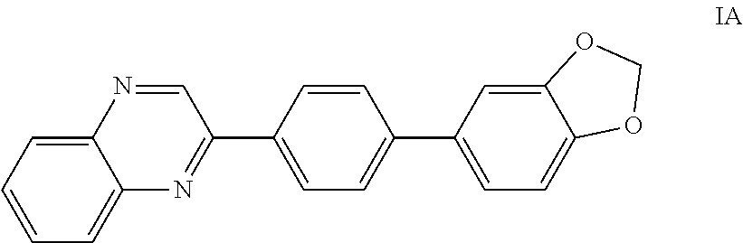
- a compound of Formula II is the 1,4-quinoxaline phenyl pyridinyl compound IIA:
- Another example of a compound of Formula II is the 1,4-quinoxaline phenyl pyridinyl compound IIB:
- the aromatic heterocycle of Formula II may be conjugated with a hydrophilic polymer, e.g., PEG (having, e.g., a molecular weight ranging from 500-10,000 Da) and the like, and a phospholipid, e.g., DPPC, DSPE, DSPC, DPPE, and the like, to form a phospholipid-hydrophilic polymer—Formula II ligand conjugate.
- a hydrophilic polymer e.g., PEG (having, e.g., a molecular weight ranging from 500-10,000 Da) and the like
- a phospholipid e.g., DPPC, DSPE, DSPC, DPPE, and the like
- the phospholipid-hydrophilic polymer—Formula II ligand conjugate comprises a compound represented by:
- n may be about 10 to about 100, about 30 to about 80, or about 30 to about 60.
- the phospholipid-hydrophilic polymer—Formula II ligand conjugate comprises a compound represented by:
- n may be about 10 to about 100, about 30 to about 80, or about 30 to about 60.
- the phospholipid-hydrophilic polymer—Formula II ligand conjugate may be incorporated into a liposomal composition.
- a method for imaging amyloid deposits in a patient comprising:
- a liposomal composition comprising a phospholipid-hydrophilic polymer—Formula II ligand conjugate
- the detecting comprises detecting by FI. In another embodiment, the detecting comprises detecting by MR imaging. In one embodiment, the detecting comprises detecting by SPECT imaging and/or PET imaging, and the non-radioactive contrast enhancing agent is replaced with a radioactive contrast enhancing agent, comprising for example those agents deemed appropriate for use with SPECT imaging and/or PET imaging in the National Institute of Health's Molecular Imaging and Contrast Agent Database (“MICAD”).
- MICAD National Institute of Health's Molecular Imaging and Contrast Agent Database
- a compound of Formula III or a pharmaceutically acceptable salt or prodrug thereof, is provided:
- R, R 1 , R 2 , R 1 ′, R 2 ′ H, F, Cl, Br, I, alkyl, aryl, OH, O-alkyl, O-aryl, NH 2 , NH-alkyl, N-dialkyl, carboxyl, sulfonyl, carbamoyl, or glycosyl.
- R is the 1,4-quinoxaline phenyl pyrimidinyl compound IIIA:
- the aromatic heterocycle of Formula III may be conjugated with a hydrophilic polymer, e.g., PEG (having, e.g., a molecular weight ranging from 500-10,000 Da) and the like, and a phospholipid, e.g., DPPC, DSPE, DSPC, DPPE, and the like, to form a phospholipid-hydrophilic polymer—Formula III ligand conjugate.
- a hydrophilic polymer e.g., PEG (having, e.g., a molecular weight ranging from 500-10,000 Da) and the like
- a phospholipid e.g., DPPC, DSPE, DSPC, DPPE, and the like
- the phospholipid-hydrophilic polymer—Formula III ligand conjugate comprises a compound represented by:
- n may be about 10 to about 100, about 30 to about 80, or about 30 to about 60.
- the phospholipid-hydrophilic polymer—Formula III ligand conjugate may be incorporated into a liposomal composition.
- a method for imaging amyloid deposit in a patient comprising:
- a liposomal composition comprising a phospholipid-hydrophilic polymer—Formula III ligand conjugate
- the detecting comprises detecting by FI. In another embodiment, the detecting comprises detecting by MR imaging. In one embodiment, the detecting comprises detecting by SPECT imaging and/or PET imaging, and the non-radioactive contrast enhancing agent is replaced with a radioactive contrast enhancing agent, comprising for example those agents deemed appropriate for use with SPECT imaging and/or PET imaging in the National Institute of Health's Molecular Imaging and Contrast Agent Database (“MICAD”).
- MICAD National Institute of Health's Molecular Imaging and Contrast Agent Database
- a compound of Formula IV or a pharmaceutically acceptable salt or prodrug thereof, is provided:
- R, R 1 , R 2 , R 1 ′, R 2 ′ H, F, Cl, Br, I, alkyl, aryl, OH, O-alkyl, O-aryl, NH 2 , NH-alkyl, N-dialkyl, carboxyl, sulfonyl, carbamoyl, or glycosyl.
- the aromatic heterocycle of Formula IV may be conjugated with a hydrophilic polymer, e.g., PEG (having, e.g., a molecular weight ranging from 500-10,000 Da) and the like, and a phospholipid, e.g., DPPC, DSPE, DSPC, DPPE, and the like, to form a phospholipid-hydrophilic polymer—Formula IV ligand conjugate.
- a hydrophilic polymer e.g., PEG (having, e.g., a molecular weight ranging from 500-10,000 Da) and the like
- a phospholipid e.g., DPPC, DSPE, DSPC, DPPE, and the like
- the phospholipid-hydrophilic polymer—Formula IV ligand conjugate comprises a compound represented by:
- n may be about 10 to about 100, about 30 to about 80, or about 30 to about 60.
- the phospholipid-hydrophilic polymer—Formula IV ligand conjugate comprises a compound represented by:
- n may be about 10 to about 100, about 30 to about 80, or about 30 to about 60.
- the phospholipid-hydrophilic polymer—Formula IV ligand conjugate may be incorporated into a liposomal composition.
- a method for imaging amyloid deposits in a patient comprising:
- a liposomal composition comprising a phospholipid-hydrophilic polymer—Formula IV ligand conjugate;
- the detecting comprises detecting by FI. In another embodiment, the detecting comprises detecting by MR imaging. In one embodiment, the detecting comprises detecting by SPECT imaging and/or PET imaging, and the non-radioactive contrast enhancing agent is replaced with a radioactive contrast enhancing agent, comprising for example those agents deemed appropriate for use with SPECT imaging and/or PET imaging in the National Institute of Health's Molecular Imaging and Contrast Agent Database (“MICAD”).
- MICAD National Institute of Health's Molecular Imaging and Contrast Agent Database
- a compound of Formula V or a pharmaceutically acceptable salt or prodrug thereof, is provided:
- R, R 1 , R 2 , R 1 ′, R 2 ′ H, F, Cl, Br, I, alkyl, aryl, OH, O-alkyl, O-aryl, NH 2 , NH-alkyl, N-dialkyl, carboxyl, sulfonyl, carbamoyl, or glycosyl.
- a compound of Formula V is the 1,4-benzoxazine phenyl pyridinyl compound VA:
- the aromatic heterocycle of Formula V may be conjugated with a hydrophilic polymer, e.g., PEG (having, e.g., a molecular weight ranging from 500-10,000 Da) and the like, and a phospholipid, e.g., DPPC, DSPE, DSPC, DPPE, and the like, to form a phospholipid-hydrophilic polymer—Formula V ligand conjugate.
- a hydrophilic polymer e.g., PEG (having, e.g., a molecular weight ranging from 500-10,000 Da) and the like
- a phospholipid e.g., DPPC, DSPE, DSPC, DPPE, and the like
- the phospholipid-hydrophilic polymer—Formula V ligand conjugate comprises a compound represented by:
- n may be about 10 to about 100, about 30 to about 80, or about 30 to about 60.
- the phospholipid-hydrophilic polymer—Formula V ligand conjugate may be incorporated into a liposomal composition.
- a method for imaging amyloid deposits in a patient comprising:
- a liposomal composition comprising a phospholipid-hydrophilic polymer—Formula V ligand conjugate;
- the detecting comprises detecting by FI. In another embodiment, the detecting comprises detecting by MR imaging. In one embodiment, the detecting comprises detecting by SPECT imaging and/or PET imaging, and the non-radioactive contrast enhancing agent is replaced with a radioactive contrast enhancing agent, comprising for example those agents deemed appropriate for use with SPECT imaging and/or PET imaging in the National Institute of Health's Molecular Imaging and Contrast Agent Database (“MICAD”).
- MICAD National Institute of Health's Molecular Imaging and Contrast Agent Database
- a compound of Formula VI or a pharmaceutically acceptable salt or prodrug thereof, is provided:
- R, R 1 , R 2 , R 1 ′, R 2 ′ H, F, Cl, Br, I, alkyl, aryl, OH, O-alkyl, O-aryl, NH 2 , NH-alkyl, N-dialkyl, carboxyl, sulfonyl, carbamoyl, or glycosyl.
- a compound of Formula VI is the 1,4-quinoxaline phenyl pyrimidinyl compound VIA:
- the aromatic heterocycle of Formula VI may be conjugated with a hydrophilic polymer, e.g., PEG (having, e.g., a molecular weight ranging from 500-10,000 Da) and the like, and a phospholipid, e.g., DPPC, DSPE, DSPC, DPPE, and the like, to form a phospholipid-hydrophilic polymer—Formula VI ligand conjugate.
- a hydrophilic polymer e.g., PEG (having, e.g., a molecular weight ranging from 500-10,000 Da) and the like
- a phospholipid e.g., DPPC, DSPE, DSPC, DPPE, and the like
- the phospholipid-hydrophilic polymer—Formula VI ligand conjugate comprises a compound represented by:
- n may be about 10 to about 100, about 30 to about 80, or about 30 to about 60.
- the phospholipid-hydrophilic polymer—Formula VI ligand conjugate may be incorporated into a liposomal composition.
- a method for imaging amyloid deposits in a patient comprising:
- a liposomal composition comprising a phospholipid-hydrophilic polymer—Formula VI ligand conjugate
- the detecting comprises detecting by FI. In another embodiment, the detecting comprises detecting by MR imaging. In one embodiment, the detecting comprises detecting by SPECT imaging and/or PET imaging, and the non-radioactive contrast enhancing agent is replaced with a radioactive contrast enhancing agent, comprising for example those agents deemed appropriate for use with SPECT imaging and/or PET imaging in the National Institute of Health's Molecular Imaging and Contrast Agent Database (“MICAD”).
- MICAD National Institute of Health's Molecular Imaging and Contrast Agent Database
- a compound of Formula VII or a pharmaceutically acceptable salt or prodrug thereof, is provided:
- R OMe
- R 1 H
- R 2 O-alkyl
- R 1 ′ OH
- R 2 ′ H.
- VILA divinyl benzene compound
- the aromatic compound of Formula VII may be conjugated with a hydrophilic polymer, e.g., PEG (having, e.g., a molecular weight ranging from 500-10,000 Da) and the like, and a phospholipid, e.g., DPPC, DSPE, DSPC, DPPE, and the like, to form a phospholipid-hydrophilic polymer—Formula VII ligand conjugate represented by:
- n represents a degree of polymerization of the polyethylene glycol polymer, and may be about 10 to about 100, about 30 to about 80, or about 30 to about 60.
- the methoxy-XO4 ligand may be conjugated with PEG and DSPE to form the DSPE-AL-PEG n -Methoxy-XO4 conjugate shown as “1” in FIG. 1 (and sometimes referred to hereinafter as “Me-XO4”), which includes one or more of:
- n may be about 10 to about 100, about 30 to about 80, or about 30 to about 60.
- the phospholipid-hydrophilic polymer—Formula VII ligand conjugate e.g., Me-XO4, and even more particularly, DPSE-AL-PEG 3400 -Methoxy-XO4 (where 3400 signifies the molecular weight of the polyethyelene glycol), may be incorporated into a liposomal composition.
- a method for imaging amyloid deposits in a patient comprising:
- a liposomal composition comprising a phospholipid-hydrophilic polymer—Formula VII ligand conjugate;
- the detecting comprises detecting by FI. In another embodiment, the detecting comprises detecting by MR imaging. In one embodiment, the detecting comprises detecting by SPECT imaging and/or PET imaging, and the non-radioactive contrast enhancing agent is replaced with a radioactive contrast enhancing agent, comprising for example those agents deemed appropriate for use with SPECT imaging and/or PET imaging in the National Institute of Health's Molecular Imaging and Contrast Agent Database (“MICAD”).
- MICAD National Institute of Health's Molecular Imaging and Contrast Agent Database
- one or more alternative amyloid ligands may be conjugated with a hydrophilic polymer, e.g., PEG (having, e.g., a molecular weight ranging from 500-10,000 Da) and the like, and a phospholipid, e.g., DPPC, DSPE, DSPC, DPPE, and the like, to form a phospholipid-hydrophilic polymer-amyloid ligand conjugate.
- the phospholipid-hydrophilic polymer-amyloid ligand conjugate may be incorporated into a liposomal composition.
- a method for imaging amyloid lesions in a patient comprising:
- a detectable quantity of a liposomal composition comprising a phospholipid-hydrophilic polymer-amyloid ligand conjugate
- the liposomal compositions described herein may further enable delivery of therapeutic molecules to amyloid lesions, thus enabling treatment of the lesions.
- a liposomal composition comprising: a phospholipid; cholesterol, or another stabilizing excipient, such as another sterol or a fatty acid; a nonradioactive gadolinium-containing contrast enhancing agent; a phospholipid which is derivatized with a polymer; and a conjugate comprising an aromatic compound having any one of Formulas I-VII, such as a conjugate in a form of a phospholipid-hydrophilic polymer-aromatic conjugate as described herein.
- the liposomal composition comprises: DPPC; cholesterol; Gd-DTPA-BSA; DSPE-AL-mPEG-2000; and DSPE-AL-PEG n -methoxy-XO4, where n represents a degree of polymerization of the polyethylene glycol polymer, and may be about 10 to about 100, about 30 to about 80, or about 30 to about 60.
- a method for imaging amyloid deposits in a patient comprising:
- a detectable quantity of a liposomal composition comprising a phospholipid; cholesterol, or another stabilizing excipient, such as another sterol or a fatty acid; a nonradioactive gadolinium-containing contrast enhancing agent; a phospholipid which is derivatized with a polymer; and a conjugate comprising an aromatic compound having any one of Formulas I-VII, such as a conjugate in a form of a phospholipid-hydrophilic polymer-aromatic compound conjugate as described herein;
- the detecting comprises detecting by FI. In another embodiment, the detecting comprises detecting by MRI.
- hydrophilic paramagnetic chelates such as GdDTPA, GdDOTA, GdHPDO3A, GdDTPA-BMA, and GdDTPA-BSA are known MRI contrast agents. See U.S. Pat. No. 5,676,928 issued to Klaveness et al., which is incorporated by reference herein in its entirety.
- the detecting comprises detecting by SPECT imaging and/or PET imaging, and the non-radioactive contrast enhancing agent is replaced with a radioactive contrast enhancing agent, comprising for example those agents deemed appropriate for use with SPECT imaging and/or PET imaging in the National Institute of Health's Molecular Imaging and Contrast Agent Database (“MICAD”).
- MICAD National Institute of Health's Molecular Imaging and Contrast Agent Database
- Suitable phospholipids may include those disclosed herein, and may further include those disclosed in U.S. Pat. No. 7,785,568 issued to Annapragada et al., which is incorporated by reference herein in its entirety.
- Suitable polymer derivatized phospholipids may include those disclosed herein, and may further include those disclosed in U.S. Pat. No. 7,785,568.
- the detecting comprises detecting by FI. In one embodiment, the detecting comprises detecting by SPECT imaging and/or PET imaging, and the non-radioactive contrast enhancing agent is replaced with a radioactive contrast enhancing agent, comprising for example those agents deemed appropriate for use with SPECT imaging and/or PET imaging in the National Institute of Health's Molecular Imaging and Contrast Agent Database (“MICAD”). Any other suitable type of imaging methodology known by those skilled in the art is contemplated, including, but not limited to, PET imaging.
- MICAD National Institute of Health's Molecular Imaging and Contrast Agent Database
- the DSPE-AL-PEG MW 3400 -Methoxy-XO4 conjugate, 1, was synthesized as the targeting species ( FIG. 1 ) and later incorporated into liposomal formulations.
- the synthesis of compound 14 was achieved via a series of Takai, Suzuki, and Julia-Kocienski olefination reactions.
- the Boc-protected 3-unit PEG linker precursor bromide 3 was prepared from the corresponding commercially available alcohol 2 in good yield.
- Intermediate 7, the sulfone for the Julia-Kocienski olefination step was also prepared in excellent yields from 4-hydroxybenzaldehyde.
- 4-hydroxybenzaldehyde was also separately subjected to the standard Takai protocol to afford vinyl iodide 9. Reaction of 9 with commercially available boronic acid 10 under Suzuki conditions afforded compound 11 in good yield.
- the linker moiety was installed quantitatively to give aldehyde 12, which was exposed to sulfone 7 under optimized Julia-Kocienski conditions to obtain the desired E,E-isomer 13 in 69% yield after column chromatography purification.
- Global deprotection of the MOM and Boc groups with HCl gave the 14 as the hydrochloride salt.
- linkers such as “DSPE-PEG-COOH” and “DSPE-PEG-NH 2 ” include, for example, Biochempeg Scientific Inc., Watertown, Mass., and Laysan Bio, Arab, Ala. In catalog descriptions, such linkers are shown with structures depicting specific aliphatic linkages between the phospholipid and PEG moieties. During an initial series of experiments, the catalog structures for PL-AL-PEG linkers provided by such commercial sources were assumed correct, as well as catalog structures for commercially available PL-AL-PEG-NH 2 and PL-AL-PEG-CO 2 H compounds used in constructing the liposomes described herein.
- a lipid mixture (50 mM) comprising DPPC, cholesterol, Gd-DTPA-BSA, DSPE-AL-mPEG-2000, Me-XO4, and Rhodamine-DHPE (for optical detection) in about a 32.4:40:25:2:0.5:0.1 molar ratio, respectively, was employed.
- Other ratios are contemplated, including a lipid mixture comprising DPPC, cholesterol, Gd-DTPA-BSA, DSPE-AL-mPEG-2000, and Me-XO4 in about a 32.5:40:25:2:0.5 molar ratio.
- the DSPE-AL-mPEG-2000 may be replaced altogether with the Me-XO4 conjugate, for a DPPC, cholesterol, Gd-DTPA-BSA, Me-XO4 conjugate ratio of 32.5:40:25:2.5.
- the upper limit on the PEG-bearing molecule may be about 15-25%, and the lower limit on cholesterol may be about 15-20%.
- the concentration of Me-XO4 ligand in the particles was determined using a fluorescence standard curve generated for Me-XO4 ligand to be 26 ⁇ M.
- the above protocol results in roughly equal distribution of the targeting ligand between the inner and the outer faces of the lipid bilayer. This implies that for each reported concentration of Me-XO4 ligand in the nanoparticles, approximately 50% of the total Me-XO4 ligands are available for binding.
- Me-XO4 ligand is highly intrinsically fluorescent and so are the nanoparticles bearing Me-XO4 ligand. This property was used as a reporter on the locations of nanoparticles in the course of all of the experiments.
- Me-XO4-labeled liposomes of Example 2 and Me-XO4 ligand stock solutions were diluted with 10 mM Tris-HCl, pH 7.4, to 500 nM.
- a small volume of the 100 ⁇ M A ⁇ stock solution was added to the test compounds to achieve a final fibril concentration of 20 ⁇ M.
- the binding mixture was incubated at RT for 1 h and then centrifuged for 20 min at 16,400 rpm to separate the fibrils. The precipitate was washed twice with tris-HCl.
- the fluorescence was measured in a SpectraMax 384 plate (Molecular Devices, Inc., Sunnyvale, Calif.) reader, using excitation and emission wavelengths of 368 nm and 450 nm, respectively.
- FIG. 2A illustrates the binding affinity of the Gd-containing Me-XO4-labeled liposomes of Example 2 to synthetic A ⁇ (1-40) aggregates.
- FIG. 2B illustrates the ability of Gd-containing Me-XO4-labeled liposomes to compete for binding sites with free Me-XO4 ligand.
- the ability of the Gd-containing Me-XO4-labeled liposomes of Example 2 to bind A ⁇ plaques was assessed by staining brain sections from APP/PSEN1 transgenic mouse line.
- the mice were engineered to progressively develop cortical and hippocampal plaques in an age-related manner similar to that observed in human AD pathology.
- Saggital sections (30 ⁇ m thick) from euthanized non-transgenic (control), 5, and 7 month old APP/PSEN1 mice were incubated in a 3 mM solution of the liposomes (concentration of Me-XO4 in the solution was 1 ⁇ M) for 2 h, at RT. This was followed by extensive washing with PBS to remove unbound liposomes.
- the stained tissues were mounted with VECTASHIELD® mounting media (Vector Laboratories, Inc., Burlingame, Calif.) to reduce background fluorescence and viewed under a confocal microscope.
- mice The Gd-containing Me-XO4-labeled liposomes were administered to 5 and 7 month old APP/PSEN1 mice by tail vain injection. 48 h following injection, the mice were euthanized and their brains sectioned for confocal light microscopic studies. Identical mice were injected with molecular/free methoxy-XO4 ligand to serve as a positive control, and with untargeted liposomes and saline as negative controls.
- FIG. 6 Optical reconstruction of images on a slide from one of the nanoparticle treated mice in this study ( FIG. 6 ) show localization of fluorescence predominantly in the cortex (top-most four arrows, in section A) and hippocampus (bottom-most four arrows, in section B).
- the Gd-containing Me-XO4 liposomes penetrated the BBB barrier and pervasively migrated through the brain.
- LPS lipopolysaccharides
- the inflammatory potential of Me-XO4 was compared to free (i.e., unconjugated) methoxy-XO4 ligand, LPS, and an untreated control (“UTC”).
- UTC untreated control
- Translocation of NF-kb from cytosol to the nucleus is an early event in the inflammatory reaction.
- NF-kb moves from the cytoplasm to the nucleus and induces gene transcription. Therefore translocation of NF-kB is a widely used marker for inflammation.
- the protocol is outlined below.
- HeLa cells were plated in each of 96 well plates and allowed to stand overnight. The cells were treated with the different concentrations of the test compounds for 2 h in a 37° C. incubator. The positive controls were treated with 1 mg/mL LPS. At the end of the incubation period, the medium was aspirated and the cells were washed with PBS. The cells were treated with 4% paraformaldehyde (to fix the cells) for 10 min at RT. The cells were washed twice with ice cold PBS. The cells were incubated with 0.25% Triton-X-100 in PBS for 10 min at RT, and were again washed three times (5 min each wash) with PBS.
- the cells were incubated with 1% BSA in PBS-T (PBS with 0.1% Tween-20) for 45 min at RT. At the end of the incubation period, the cells were further incubated with primary antibody against NF-kBat 1:50 dilution in PBST for 1 h at RT. The cells were again washed with PBS three times (5 min each). The cells were incubated with the secondary antibody in PBST for 1 h at RT, and were again washed three times (5 min each) with PBS. 100 ⁇ l of DAPI (1 ⁇ g/mL) was placed in each well and kept at 4° C. until further analysis.
- PBS-T PBS with 0.1% Tween-20
- the cells were scanned on a Cell Lab IC-100 image cytometer (Beckman-Coulter, Fullerton, Calif.). The data were further analyzed using CyteSeer software (Vala Sciences, San Diego, Calif.), and represented as Pearson's Correlation coefficient (PCC) of the protein intensity present over the nuclear mask.
- CyteSeer software Vala Sciences, San Diego, Calif.
- Me-XO4 was found to be less inflammatory than free methoxy-XO4 ligand at all but the highest concentrations (500 nM) tested. The results are depicted in FIG. 7 . Thus, it is a particular teaching of at least one embodiment herein that the conjugated and/or liposomal amyloid binding ligand is less (or at least not more) inflammatory than the free ligand.
- the cytotoxicity of Me-XO4 was compared to free (i.e., not conjugated) methoxy-XO4 ligand and an untreated control.
- the toxicity of the test compounds was evaluated using standard MTT assays. 15,000 HeLa cells were plated in each of 96 well plates and allowed to stand overnight. The cells were treated with three different concentrations of the test compounds for 2 h in a 37° C. incubator. The positive controls were treated with 1 mg/mL LPS. At the end of the incubation period, a MTS cell toxicity assay kit (CELLTITER 96® AQueous Assay kit, Promega, Madison Wis.) was used according to the manufacturer's protocol.
- CELLTITER 96® AQueous Assay kit Promega, Madison Wis.
- Me-XO4 was found to be less cytotoxic than free methoxy-XO4 ligand. The results are depicted in FIG. 8 . Thus, it is a particular teaching of at least one embodiment herein that the conjugated and/or liposomal amyloid binding ligand is less (or at least not more) toxic than the free ligand.
- FIG. 9A illustrates the surviving fraction of cells.
- the conjugated ligand is significantly more cytotoxic than the free ligand.
- FIG. 9B depicts the PCC between the nuclear and cytoplasmic fractions of NF K B molecule in HeLa cells.
- the conjugated ligand is significantly more inflammatory than the free ligand.
Abstract
Description

PL may be a phospholipid. AL may be an aliphatic linkage. PEG may be a polyethylene glycol polymer. The membrane may include a nonradioactive magnetic resonance imaging (MRI) contrast enhancing agent at least one of encapsulated by or bound to the membrane.

PL may be a phospholipid. AL may be an aliphatic linkage. PEG may be a polyethylene glycol polymer. The membrane may include a nonradioactive magnetic resonance imaging (MRI) contrast enhancing agent at least one of encapsulated by or bound to the membrane. The method may include allowing sufficient time for the liposomal composition to be associated with one or more amyloid deposits. The method may include detecting the liposomal composition associated with the one or more amyloid deposits.

PL may be a phospholipid. AL may be an aliphatic linkage. PEG may be a polyethylene glycol polymer.

PL may be a phospholipid. AL may be an aliphatic linkage. PEG may be a polyethylene glycol polymer. The membrane may include a nonradioactive magnetic resonance imaging (MRI) contrast enhancing agent at least one of encapsulated by or bound to the membrane.

the aliphatic linkage AL, —CH2CH2NH(C═O)CH2O—, includes an amide moiety. Further, for example, in the compound below:

the aliphatic linkage AL, —CH2CH2NH(C═O)O— includes a carbamate moiety. AL may include aliphatic linkages derived from dicarboxylic acids, such as succinic acid, and may include two amides, two carbamates, an amide and a carbamate, and the like.

wherein n represents a degree of polymerization of the polyethylene glycol polymer, and may be about 10 to about 100, about 30 to about 80, or about 30 to about 60.

wherein n may be about 10 to about 100, about 30 to about 80, or about 30 to about 60.

wherein n may be about 10 to about 100, about 30 to about 80, or about 30 to about 60. The nonradioactive magnetic resonance imaging (MRI) contrast enhancing agent may include Gd-DTPA-BSA.

PL may be a phospholipid. AL may be an aliphatic linkage. PEG may be a polyethylene glycol polymer. The membrane may include a nonradioactive magnetic resonance imaging (MRI) contrast enhancing agent at least one of encapsulated by or bound to the membrane. The method may include allowing sufficient time for the liposomal composition to be associated with one or more amyloid deposits. The method may include detecting the liposomal composition associated with the one or more amyloid deposits.

wherein n represents a degree of polymerization of the polyethylene glycol polymer, and may be about 10 to about 100, about 30 to about 80, or about 30 to about 60.

wherein n represents a degree of polymerization of the polyethylene glycol polymer, and may be about 10 to about 100, about 30 to about 80, or about 30 to about 60.

wherein n represents a degree of polymerization of the polyethylene glycol polymer, and may be about 10 to about 100, about 30 to about 80, or about 30 to about 60.

wherein n may be about 10 to about 100, about 30 to about 80, or about 30 to about 60.


wherein n represents a degree of polymerization of the polyethylene glycol polymer, and may be about 10 to about 100, about 30 to about 80, or about 30 to about 60.

wherein n represents a degree of polymerization of the polyethylene glycol polymer, and may be about 10 to about 100, about 30 to about 80, or about 30 to about 60.

wherein n represents a degree of polymerization of the polyethylene glycol polymer, and may be about 10 to about 100, about 30 to about 80, or about 30 to about 60.

wherein n may be about 10 to about 100, about 30 to about 80, or about 30 to about 60.

wherein R, R1, R2, R1′, R2′=H, F, Cl, Br, I, alkyl, aryl, OH, O-alkyl, O-aryl, NH2, NH-alkyl, N-dialkyl, carboxyl, sulfonyl, carbamoyl, or glycosyl.

wherein R, R1, R2, R1′, R2′=H, F, Cl, Br, I, alkyl, aryl, OH, O-alkyl, O-aryl, NH2, NH-alkyl, N-dialkyl, carboxyl, sulfonyl, carbamoyl, or glycosyl.

wherein R, R1, R2, R1′, R2′=H, F, Cl, Br, I, alkyl, aryl, OH, O-alkyl, O-aryl, NH2, NH-alkyl, N-dialkyl, carboxyl, sulfonyl, carbamoyl, or glycosyl.

wherein R, R1, R2, R1′, R2′=H, F, Cl, Br, I, alkyl, aryl, OH, O-alkyl, O-aryl, NH2, NH-alkyl, N-dialkyl, carboxyl, sulfonyl, carbamoyl, or glycosyl.

wherein R, R1, R2, R1′, R2′=H, F, Cl, Br, I, alkyl, aryl, OH, O-alkyl, O-aryl, NH2, NH-alkyl, N-dialkyl, carboxyl, sulfonyl, carbamoyl, or glycosyl.

wherein R, R1, R2, R1′, R2′=H, F, Cl, Br, I, alkyl, aryl, OH, O-alkyl, O-aryl, NH2, NH-alkyl, N-dialkyl, carboxyl, sulfonyl, carbamoyl, or glycosyl.

wherein R, R1, R2, R1′, R2′=H, F, Cl, Br, I, alkyl, aryl, OH, O-alkyl, O-aryl, NH2, NH-alkyl, N-dialkyl, carboxyl, sulfonyl, carbamoyl, or glycosyl; and wherein a, b, c, d, e=C, N, O, or S.

wherein R, R1, R2, R1′, R2′=H, F, Cl, Br, I, alkyl, aryl, OH, O-alkyl, O-aryl, NH2, NH-alkyl, N-dialkyl, carboxyl, sulfonyl, carbamoyl, or glycosyl.


wherein n may be about 10 to about 100, about 30 to about 80, or about 30 to about 60.


wherein n may be about 10 to about 100, about 30 to about 80, or about 30 to about 60.

wherein R, R1, R2, R1′, R2′=H, F, Cl, Br, I, alkyl, aryl, OH, O-alkyl, O-aryl, NH2, NH-alkyl, N-dialkyl, carboxyl, sulfonyl, carbamoyl, or glycosyl.


wherein n may be about 10 to about 100, about 30 to about 80, or about 30 to about 60.

wherein R, R1, R2, R1′, R2′=H, F, Cl, Br, I, alkyl, aryl, OH, O-alkyl, O-aryl, NH2, NH-alkyl, N-dialkyl, carboxyl, sulfonyl, carbamoyl, or glycosyl.


wherein n may be about 10 to about 100, about 30 to about 80, or about 30 to about 60.


wherein n may be about 10 to about 100, about 30 to about 80, or about 30 to about 60.

wherein R, R1, R2, R1′, R2′=H, F, Cl, Br, I, alkyl, aryl, OH, O-alkyl, O-aryl, NH2, NH-alkyl, N-dialkyl, carboxyl, sulfonyl, carbamoyl, or glycosyl, and a, b, c, d, e=C, N, O, or S.


wherein n represents a degree of polymerization of the polyethylene glycol polymer, and may be about 10 to about 100, about 30 to about 80, or about 30 to about 60.

wherein n may be about 10 to about 100, about 30 to about 80, or about 30 to about 60.
Claims (20)


Priority Applications (1)
| Application Number | Priority Date | Filing Date | Title |
|---|---|---|---|
| US15/797,816 US10124078B2 (en) | 2011-04-06 | 2017-10-30 | Lipid-based nanoparticles |
Applications Claiming Priority (3)
| Application Number | Priority Date | Filing Date | Title |
|---|---|---|---|
| US201161472605P | 2011-04-06 | 2011-04-06 | |
| US13/441,816 US9801957B2 (en) | 2011-04-06 | 2012-04-06 | Lipid-based nanoparticles |
| US15/797,816 US10124078B2 (en) | 2011-04-06 | 2017-10-30 | Lipid-based nanoparticles |
Related Parent Applications (1)
| Application Number | Title | Priority Date | Filing Date |
|---|---|---|---|
| US13/441,816 Continuation-In-Part US9801957B2 (en) | 2011-04-06 | 2012-04-06 | Lipid-based nanoparticles |
Publications (2)
| Publication Number | Publication Date |
|---|---|
| US20180043039A1 US20180043039A1 (en) | 2018-02-15 |
| US10124078B2 true US10124078B2 (en) | 2018-11-13 |
Family
ID=61160632
Family Applications (1)
| Application Number | Title | Priority Date | Filing Date |
|---|---|---|---|
| US15/797,816 Expired - Fee Related US10124078B2 (en) | 2011-04-06 | 2017-10-30 | Lipid-based nanoparticles |
Country Status (1)
| Country | Link |
|---|---|
| US (1) | US10124078B2 (en) |
Families Citing this family (1)
| Publication number | Priority date | Publication date | Assignee | Title |
|---|---|---|---|---|
| CN115279424A (en) * | 2020-02-12 | 2022-11-01 | 德克萨斯儿童医院 | Targeted contrast agents for alpha-synuclein deposition MRI |
-
2017
- 2017-10-30 US US15/797,816 patent/US10124078B2/en not_active Expired - Fee Related
Non-Patent Citations (1)
| Title |
|---|
| Klunk et al., Imaging Abeta Plaques in Living Transgenic Mice with Multiphoton Microscopy and Methoxy-X04, a Systemically Administered Congo Red Derivative, Sep. 2002, Journal of Neuropathology and Experimenta Neurology, vol. 61, No. 9, pp. 797-805 (Year: 2002). * |
Also Published As
| Publication number | Publication date |
|---|---|
| US20180043039A1 (en) | 2018-02-15 |
Similar Documents
| Publication | Publication Date | Title |
|---|---|---|
| US9801957B2 (en) | Lipid-based nanoparticles | |
| US20200179540A1 (en) | Mri imaging of amyloid plaque using liposomes | |
| Costa et al. | Functionalised carbon nanotubes enhance brain delivery of amyloid-targeting Pittsburgh compound B (PiB)-derived ligands | |
| US10124078B2 (en) | Lipid-based nanoparticles | |
| US20230022136A1 (en) | Diffusivity contrast agents for medical imaging |
Legal Events
| Date | Code | Title | Description |
|---|---|---|---|
| FEPP | Fee payment procedure |
Free format text: ENTITY STATUS SET TO UNDISCOUNTED (ORIGINAL EVENT CODE: BIG.); ENTITY STATUS OF PATENT OWNER: SMALL ENTITY |
|
| FEPP | Fee payment procedure |
Free format text: ENTITY STATUS SET TO SMALL (ORIGINAL EVENT CODE: SMAL); ENTITY STATUS OF PATENT OWNER: SMALL ENTITY |
|
| STCF | Information on status: patent grant |
Free format text: PATENTED CASE |
|
| FEPP | Fee payment procedure |
Free format text: MAINTENANCE FEE REMINDER MAILED (ORIGINAL EVENT CODE: REM.); ENTITY STATUS OF PATENT OWNER: SMALL ENTITY |
|
| LAPS | Lapse for failure to pay maintenance fees |
Free format text: PATENT EXPIRED FOR FAILURE TO PAY MAINTENANCE FEES (ORIGINAL EVENT CODE: EXP.); ENTITY STATUS OF PATENT OWNER: SMALL ENTITY |
|
| STCH | Information on status: patent discontinuation |
Free format text: PATENT EXPIRED DUE TO NONPAYMENT OF MAINTENANCE FEES UNDER 37 CFR 1.362 |
|
| FP | Lapsed due to failure to pay maintenance fee |
Effective date: 20221113 |