US10087238B2 - Method of preventing myocardial reperfusion injury in reperfusion therapy by administering intravenous immunoglobulin - Google Patents
Method of preventing myocardial reperfusion injury in reperfusion therapy by administering intravenous immunoglobulin Download PDFInfo
- Publication number
- US10087238B2 US10087238B2 US15/439,807 US201715439807A US10087238B2 US 10087238 B2 US10087238 B2 US 10087238B2 US 201715439807 A US201715439807 A US 201715439807A US 10087238 B2 US10087238 B2 US 10087238B2
- Authority
- US
- United States
- Prior art keywords
- reperfusion
- treatment
- patient
- therapy
- ischemia
- Prior art date
- Legal status (The legal status is an assumption and is not a legal conclusion. Google has not performed a legal analysis and makes no representation as to the accuracy of the status listed.)
- Active
Links
Images


Classifications
-
- C—CHEMISTRY; METALLURGY
- C07—ORGANIC CHEMISTRY
- C07K—PEPTIDES
- C07K16/00—Immunoglobulins [IGs], e.g. monoclonal or polyclonal antibodies
- C07K16/06—Immunoglobulins [IGs], e.g. monoclonal or polyclonal antibodies from serum
-
- C—CHEMISTRY; METALLURGY
- C07—ORGANIC CHEMISTRY
- C07K—PEPTIDES
- C07K16/00—Immunoglobulins [IGs], e.g. monoclonal or polyclonal antibodies
-
- A—HUMAN NECESSITIES
- A61—MEDICAL OR VETERINARY SCIENCE; HYGIENE
- A61K—PREPARATIONS FOR MEDICAL, DENTAL OR TOILETRY PURPOSES
- A61K39/00—Medicinal preparations containing antigens or antibodies
- A61K39/395—Antibodies; Immunoglobulins; Immune serum, e.g. antilymphocytic serum
-
- A—HUMAN NECESSITIES
- A61—MEDICAL OR VETERINARY SCIENCE; HYGIENE
- A61K—PREPARATIONS FOR MEDICAL, DENTAL OR TOILETRY PURPOSES
- A61K9/00—Medicinal preparations characterised by special physical form
- A61K9/0012—Galenical forms characterised by the site of application
- A61K9/0019—Injectable compositions; Intramuscular, intravenous, arterial, subcutaneous administration; Compositions to be administered through the skin in an invasive manner
-
- A—HUMAN NECESSITIES
- A61—MEDICAL OR VETERINARY SCIENCE; HYGIENE
- A61P—SPECIFIC THERAPEUTIC ACTIVITY OF CHEMICAL COMPOUNDS OR MEDICINAL PREPARATIONS
- A61P9/00—Drugs for disorders of the cardiovascular system
- A61P9/10—Drugs for disorders of the cardiovascular system for treating ischaemic or atherosclerotic diseases, e.g. antianginal drugs, coronary vasodilators, drugs for myocardial infarction, retinopathy, cerebrovascula insufficiency, renal arteriosclerosis
-
- A—HUMAN NECESSITIES
- A61—MEDICAL OR VETERINARY SCIENCE; HYGIENE
- A61K—PREPARATIONS FOR MEDICAL, DENTAL OR TOILETRY PURPOSES
- A61K39/00—Medicinal preparations containing antigens or antibodies
- A61K2039/505—Medicinal preparations containing antigens or antibodies comprising antibodies
-
- C—CHEMISTRY; METALLURGY
- C07—ORGANIC CHEMISTRY
- C07K—PEPTIDES
- C07K16/00—Immunoglobulins [IGs], e.g. monoclonal or polyclonal antibodies
- C07K16/18—Immunoglobulins [IGs], e.g. monoclonal or polyclonal antibodies against material from animals or humans
Definitions
- the present invention relates to ischemia reperfusion therapy in a patient, and particularly to a method of preventing reperfusion injury by treatment with intravenous immunoglobulin (IVIG).
- IVIG intravenous immunoglobulin
- Myocardial ischemia is a definite result of the reduction of blood flow and inadequate oxygen supply. Ischemia can induce various clinical conditions ranging from inflammation to myocardial damage and organ failure.
- Reperfusion is the traditional method for cardiac protection from ischemia.
- Reperfusion therapy is a medical treatment to restore blood flow, either through or around, blocked arteries.
- Thrombolytic therapy, coronary angioplasty, and coronary artery bypass surgery are examples of known reperfusion techniques.
- reperfusion itself can amplify or accelerate cell death, producing reperfusion-induced injury.
- Heart transplant patients are also susceptible to reperfusion injury due to the particular type of reperfusion performed with transplanted hearts.
- Reperfusion of ischemic tissues is often associated with microvascular injury, particularly due to increased permeability of capillaries and arterioles that lead to an increase of diffusion and fluid filtration across the tissues.
- Activated endothelial cells produce more reactive oxygen species but less nitric oxide following reperfusion, and the imbalance results in a subsequent inflammatory response.
- the inflammatory response is partially responsible for the damage of reperfusion injury.
- White blood cells carried to the area by the newly returning blood, release a host of inflammatory factors such as interleukins as well as free radicals in response to tissue damage.
- the restored blood flow reintroduces oxygen within cells that damages cellular proteins, DNA, and the plasma membrane. Damage to the cell's membrane may in turn cause the release of more free radicals.
- Such reactive species may also act indirectly in redox signaling to turn on apoptosis.
- White blood cells may also bind to the endothelium of small capillaries, obstructing them and leading to more ischemia.
- therapeutic hypothermia is one of the most prevalent.
- therapeutic hypothermia which involves the usage of highly specialized equipment, a variety of new drugs and compounds are being tested for treatment of reperfusion injury, including hydrogen sulfide treatment, cyclosporine, TRO40303, stem cell therapy, superoxide dismutase, and metformin.
- the method of preventing reperfusion injury in reperfusion therapy is a medical technique for prevention of reperfusion injury in a patient where reperfusion therapy is necessary, such as following myocardial infarction (MI) or heat transplant in the patient.
- the patient is treated with intravenous immunoglobulin (IVIG) prior to an ischemic insult (IVIG pre-treatment) or, alternatively, following the ischemic insult (IVIG post-treatment), with the IVIG providing cardio-protective effects to the patient for preventing reperfusion injury.
- IVIG intravenous immunoglobulin
- IVIG pre-treatment ischemic insult
- IVIG post-treatment ischemic insult
- the treatment with the IVIG may be used with any suitable type of reperfusion therapy, such as thrombolytic therapy, coronary angioplasty or coronary artery bypass surgery, for example.
- the patient is given from about 0.5 g to about 2.0 g of the intravenous immunoglobulin per kilogram of patient body weight.
- This dosage may be a one-time dosage or may be delivered multiple times, dependent upon the particular situation.
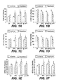
- FIG. 1A is a graph comparing maximum developed pressure (Pmax) of the left ventricle function of a control sample group against a pacing post-conditioning (PPC) group, a group treated with the method of preventing reperfusion injury in reperfusion therapy with a high dose (1.0 g/kg) of immunoglobulin treatment (IgH) post-ischemia, and a group treated with the method of preventing reperfusion injury in reperfusion therapy with a low dose (0.6 g/kg) of immunoglobulin treatment (IgH) post-ischemic.
- Pmax maximum developed pressure
- PPC pacing post-conditioning
- FIG. 1B is a graph comparing left ventricular end diastolic pressure (LVEDP) of the left ventricle function of a control sample group against a pacing post-conditioning (PPC) group, a group treated with the method of preventing reperfusion injury in reperfusion therapy with a high dose (1.0 g/kg) of immunoglobulin treatment (IgH) post-ischemia, and a group treated with the method of preventing reperfusion injury in reperfusion therapy with a low dose (0.6 g/kg) of immunoglobulin treatment (IgH) post-ischemia.
- LEDP left ventricular end diastolic pressure
- PPC pacing post-conditioning
- FIG. 1C is a graph comparing coronary flow (CF) of the coronary-vascular-dynamics of a control sample group against a pacing post-conditioning (PPC) group, a group treated with the method of preventing reperfusion injury in reperfusion therapy with a high dose (1.0 g/kg) of immunoglobulin treatment (IgH) post-ischemia, and a group treated with the method of preventing reperfusion injury in reperfusion therapy with a low dose (0.6 g/kg) of immunoglobulin treatment (IgH) post-ischemia.
- CF coronary flow
- PPC pacing post-conditioning
- FIG. 1D is a graph comparing coronary vascular resistance (CVR) of the coronary-vascular-dynamics of a control sample group against a pacing post-conditioning (PPC) group, a group treated with the method of preventing reperfusion injury in reperfusion therapy with a high dose (1.0 g/kg) of immunoglobulin treatment (IgH) post-ischemia, and a group treated with the method of preventing reperfusion injury in reperfusion therapy with a low dose (0.6 g/kg) of immunoglobulin treatment (IgH) post-ischemia.
- CVR coronary vascular resistance
- PPC pacing post-conditioning
- FIG. 1E is a graph comparing left ventricle (LV) contractility (+dP/dt) of a control sample group against a pacing post-conditioning (PPC) group, a group treated with the method of preventing reperfusion injury in reperfusion therapy with a high dose (1.0 g/kg) of immunoglobulin treatment (IgH) post-ischemia, and a group treated with the method of preventing reperfusion injury in reperfusion therapy with a low dose (0.6 g/kg) of immunoglobulin treatment (IgH) post-ischemia.
- LV left ventricle
- IgH immunoglobulin treatment
- FIG. 1F is a graph comparing LV contractility ( ⁇ dP/dt) of a control sample group against a pacing post-conditioning (PPC) group, a group treated with the method of preventing reperfusion injury in reperfusion therapy with a high dose (1.0 g/kg) of immunoglobulin treatment (IgH) post-ischemia, and a group treated with the method of preventing reperfusion injury in reperfusion therapy with a low dose (0.6 g/kg) of immunoglobulin treatment (IgH) post-ischemia.
- PPC pacing post-conditioning
- FIG. 2A is a graph comparing maximum developed pressure (Pmax) of the left ventricle function of a control sample group against a pacing post-conditioning (PPC) group, a group treated with the method of preventing reperfusion injury in reperfusion therapy with a high dose (1.0 g/kg) of immunoglobulin treatment (IgH) pre-ischemia, and a group treated with the method of preventing reperfusion injury in reperfusion therapy with a low dose (0.6 g/kg) of immunoglobulin treatment (IgH) pre-ischemia.
- Pmax maximum developed pressure
- PPC pacing post-conditioning
- FIG. 2B is a graph comparing left ventricular end diastolic pressure (LVEDP) of the left ventricle function of a control sample group against a pacing post-conditioning (PPC) group, a group treated with the method of preventing reperfusion injury in reperfusion therapy with a high dose (1.0 g/kg) of immunoglobulin treatment (IgH) pre-ischemia, and a group treated with the method of preventing reperfusion injury in reperfusion therapy with a low dose (0.6 g/kg) of immunoglobulin treatment (IgH) pre-ischemia.
- LEDP left ventricular end diastolic pressure
- PPC pacing post-conditioning
- FIG. 2C is a graph comparing coronary flow (CF) of the coronary-vascular-dynamics of a control sample group against a pacing post-conditioning (PPC) group, a group treated with the method of preventing reperfusion injury in reperfusion therapy with a high dose (1.0 g/kg) of immunoglobulin treatment (IgH) pre-ischemia, and a group treated with the method of preventing reperfusion injury in reperfusion therapy with a low dose (0.6 g/kg) of immunoglobulin treatment (IgH) pre-ischemia.
- CF coronary flow
- PPC pacing post-conditioning
- FIG. 2D is a graph comparing coronary vascular resistance (CVR) of the coronary-vascular-dynamics of a control sample group against a pacing post-conditioning (PPC) group, a group treated with the method of preventing reperfusion injury in reperfusion therapy with a high dose (1.0 g/kg) of immunoglobulin treatment (IgH) pre-ischemia, and a group treated with the method of preventing reperfusion injury in reperfusion therapy with a low dose (0.6 g/kg) of immunoglobulin treatment (IgH) pre-ischemia.
- CVR coronary vascular resistance
- PPC pacing post-conditioning
- FIG. 2E is a graph comparing left ventricle (LV) contractility (+dP/dt) of a control sample group against a pacing post-conditioning (PPC) group, a group treated with the method of preventing reperfusion injury in reperfusion therapy with a high dose (1.0 g/kg) of immunoglobulin treatment (IgH) pre-ischemia, and a group treated with the method of preventing reperfusion injury in reperfusion therapy with a low dose (0.6 g/kg) of immunoglobulin treatment (IgH) pre-ischemia.
- LV left ventricle
- IgH immunoglobulin treatment
- FIG. 2F is a graph comparing LV contractility ( ⁇ dP/dt) of a control sample group against a pacing post-conditioning (PPC) group, a group treated with the method of preventing reperfusion injury in reperfusion therapy with a high dose (1.0 g/kg) of immunoglobulin treatment (IgH) pre-ischemia, and a group treated with the method of preventing reperfusion injury in reperfusion therapy with a low dose (0.6 g/kg) of immunoglobulin treatment (IgH) pre-ischemia.
- PPC pacing post-conditioning
- FIG. 3A is a graph comparing maximum developed pressure (Pmax) of the left ventricle function of a control sample group against a pacing post-conditioning (PPC) group, a group treated with the method of preventing reperfusion injury in reperfusion therapy with a high dose (1.0 g/kg) of immunoglobulin treatment (IgH) for combined pre-ischemia treatment and post-ischemia treatment, and a group treated with the method of preventing reperfusion injury in reperfusion therapy with a low dose (0.6 g/kg) of immunoglobulin treatment (IgH) for combined pre-ischemia treatment and post-ischemia treatment.
- Pmax maximum developed pressure
- FIG. 3B is a graph comparing left ventricular end diastolic pressure (LVEDP) of the left ventricle function of a control sample group against a pacing post-conditioning (PPC) group, a group treated with the method of preventing reperfusion injury in reperfusion therapy with a high dose (1.0 g/kg) of immunoglobulin treatment (IgH) for combined pre-ischemia treatment and post-ischemia treatment, and a group treated with the method of preventing reperfusion injury in reperfusion therapy with a low dose (0.6 g/kg) of immunoglobulin treatment (IgH) for combined pre-ischemia treatment and post-ischemia treatment.
- LEDP left ventricular end diastolic pressure
- PPC pacing post-conditioning
- FIG. 3C is a graph comparing coronary flow (CF) of the coronary-vascular-dynamics of a control sample group against a pacing post-conditioning (PPC) group, a group treated with the method of preventing reperfusion injury in reperfusion therapy with a high dose (1.0 g/kg) of immunoglobulin treatment (IgH) for combined pre-ischemia treatment and post-ischemia treatment, and a group treated with the method of preventing reperfusion injury in reperfusion therapy with a low dose (0.6 g/kg) of immunoglobulin treatment (IgH) for combined pre-ischemia treatment and post-ischemia treatment.
- CF coronary flow
- PPC pacing post-conditioning
- FIG. 3D is a graph comparing coronary vascular resistance (CVR) of the coronary-vascular-dynamics of a control sample group against a pacing post-conditioning (PPC) group, a group treated with the method of preventing reperfusion injury in reperfusion therapy with a high dose (1.0 g/kg) of immunoglobulin treatment (IgH) for combined pre-ischemia treatment and post-ischemia treatment, and a group treated with the method of preventing reperfusion injury in reperfusion therapy with a low dose (0.6 g/kg) of immunoglobulin treatment (IgH) for combined pre-ischemia treatment and post-ischemia treatment.
- CVR coronary vascular resistance
- PPC pacing post-conditioning
- FIG. 3E is a graph comparing left ventricle (LV) contractility (+dP/dt) of a control sample group against a pacing post-conditioning (PPC) group, a group treated with the method of preventing reperfusion injury in reperfusion therapy with a high dose (1.0 g/kg) of immunoglobulin treatment (IgH) for combined pre-ischemia treatment and post-ischemia treatment, and a group treated with the method of preventing reperfusion injury in reperfusion therapy with a low dose (0.6 g/kg) of immunoglobulin treatment (IgH) for combined pre-ischemia treatment and post-ischemia treatment.
- LV left ventricle
- PPC pacing post-conditioning
- FIG. 3F is a graph comparing LV contractility ( ⁇ dP/dt) of a control sample group against a pacing post-conditioning (PPC) group, a group treated with the method of preventing reperfusion injury in reperfusion therapy with a high dose (1.0 g/kg) of immunoglobulin treatment (IgH) for combined pre-ischemia treatment and post-ischemia treatment, and a group treated with the method of preventing reperfusion injury in reperfusion therapy with a low dose (0.6 g/kg) of immunoglobulin treatment (IgH) for combined pre-ischemia treatment and post-ischemia treatment.
- FIG. 4A is a graph comparing infarct size of a control sample group against a pacing post-conditioning (PPC) group, a group treated with the method of preventing reperfusion injury in reperfusion therapy with a high dose (1.0 g/kg) of immunoglobulin treatment (IgH) for post-ischemia treatment, and a group treated with the method of preventing reperfusion injury in reperfusion therapy with a low dose (0.6 g/kg) of immunoglobulin treatment (IgH) for post-ischemia treatment.
- PPC pacing post-conditioning
- FIG. 4B is a graph comparing infarct size of a control sample group against a pacing post-conditioning (PPC) group, a group treated with the method of preventing reperfusion injury in reperfusion therapy with a high dose (1.0 g/kg) of immunoglobulin treatment (IgH) for post-ischemia treatment, a group treated with the method of preventing reperfusion injury in reperfusion therapy with a low dose (0.6 g/kg) of immunoglobulin treatment (IgH) for post-ischemia treatment, a group treated with the method of preventing reperfusion injury in reperfusion therapy with a high dose (1.0 g/kg) of immunoglobulin treatment (IgH) for combined pre-ischemia and post-ischemia treatment, and a group treated with the method of preventing reperfusion injury in reperfusion therapy with a low dose (0.6 g/kg) of immunoglobulin treatment (IgH) for combined pre-ischemia and post-ischemia treatment.
- PPC pacing post
- FIG. 5 is a graph comparing mytocyte apoptosis of a control sample group against a pacing post-conditioning (PPC) group, a group treated with the method of preventing reperfusion injury in reperfusion therapy with a high dose (1.0 g/kg) of immunoglobulin treatment (IgH) for post-ischemia treatment, a group treated with the method of preventing reperfusion injury in reperfusion therapy with a low dose (0.6 g/kg) of immunoglobulin treatment (IgH) for post-ischemia treatment, a group treated with the method of preventing reperfusion injury in reperfusion therapy with a high dose (1.0 g/kg) of immunoglobulin treatment (IgH) for combined pre-ischemia and post-ischemia treatment, and a group treated with the method of preventing reperfusion injury in reperfusion therapy with a low dose (0.6 g/kg) of immunoglobulin treatment (IgH) for combined pre-ischemia and post-ischemia treatment.
- PPC
- the method of preventing reperfusion injury in reperfusion therapy can be used for prevention of reperfusion injury in a patient where reperfusion therapy is necessary, such as following an ischemic event.
- the ischemic event can include myocardial ischemia or heat transplant, for example.
- the patient is treated with intravenous immunoglobulin (IVIG) prior to the ischemic event (IVIG pre-treatment) or, alternatively, following the regional myocardial ischemia reperfusion (IVIG post-treatment), with the IVIG providing cardio-protective effects to the patient for preventing reperfusion injury.
- IVIG intravenous immunoglobulin
- the treatment with the IVIG may be used with any suitable type of reperfusion therapy, such as thrombolytic therapy, coronary angioplasty, coronary artery bypass surgery or following heart transplantation, for example.
- the patient is given from about 0.5 g to about 2.0 g of the IVIG per kilogram of patient body weight.
- This dosage may be a one-time dosage or may be delivered multiple times, dependent upon the particular situation.
- male Wistar rats weighing between 250 g and 300 g, were subjected to regional ischemia and reperfusion.
- the rats were anesthetized with intraperitoneal injection of sodium pentobarbital (60 mg/kg) and anticoagulated with heparin (1000 U/kg body weight) through the femoral vein and attached to a modified Langendorff setup for the perfused rat heart.
- the hearts were perfused retrogradely with freshly prepared Krebs-Hensleit solution.
- the buffer was gassed with a mixture of oxygen (95%) and CO 2 (5%), with a pH of 7.35-7.45 at a temperature of 37.0 ⁇ 0.5° C.
- the perfusion pressure (PP) was kept constant at 50 mm Hg throughout the experimental procedure in all of the protocols described herein. PP was measured immediately downstream from the flow probe in a branch of the aortic cannula using a Statham Physiological Pressure Transducer P23db, manufactured by Gould Inc. of Illinois.
- Constant PP was ensured electronically by means of the perfusion assembly (Module PPCM, Type 671, manufactured by Hugo Sachs Elektronik of Germany). This system permits an accurate adjustment of PP between 5 and 150 mm Hg with an accuracy of ⁇ 1 mm Hg.
- the temperature of the perfusion medium in the perfusion system was maintained at 37° C. by circulating temperature controlled water. This temperature is the baseline control perfusion temperature for all protocols described below.
- Myocardial temperature was monitored by a needle thermistor probe (TH-5 Thermalert Monitoring Thermometer, manufactured by Physitemp® Instruments, Inc. of New Jersey) inserted at the apex of the heart for each protocol of the study.
- the heart was instrumented with pacing electrodes on the right atrial (RA) appendage and posterior basal LV wall.
- the left descending coronary artery was encircled by a snare at approximately 0.5 cm below the atrioventricular groove and a deflated balloon catheter was positioned between the heart and the suture over the coronary artery.
- the aorta was cannulated, allowing retrograde perfusion of the heart with a preload of 6 mm Hg. Throughout the experiment, afterload was kept constant at 80 mm Hg.
- the studies described below investigated the role of immunoglobulins pre- and post-treatments, or a combination thereof, on the heart protection against ischemia reperfusion I/R injury ( FIG. 1 ).
- LV left ventricle
- RA right atrial
- group D the hearts were infused intravenously with either IgL or IgH two hrs before sacrifice and heart isolation.
- the hearts were isolated from untreated rats.
- the ischemia was produced by 30 minutes of coronary artery occlusion (CAO), followed by 30 minutes of reperfusion.
- CAO coronary artery occlusion
- pre-treatment was performed after the stabilization period of the heart, followed by the index ischemia, followed by 30 minutes of reperfusion.
- post-treatment studies the hearts were subjected to 30 minutes of CAO, followed by 30 minutes of reperfusion. All IVIG post-treatments were performed five minutes before the start of reperfusion, and PPC was performed at the beginning of reperfusion.
- Coronary flow was measured continuously by an electromagnetic flow probe attached to the inflow of the aortic cannula. The probe was attached to a flow meter which was interfaced to a personal computer. The CF in ml/min was continuously monitored by timed collection of coronary effluent as well as computed. The coronary vascular resistance (CVR) was computed every ten seconds along with the hemodynamic data by an on-line data acquisition program (Isoheart software version 1.524-S, sold by Hugo-Sachs Electronik of Germany). Left ventricular Pmax and left ventricular end diastolic pressure (LVEDP) were measured by placing and securing a water-filled latex balloon into the left ventricular cavity.
- CVR coronary vascular resistance
- the balloon was attached to a pressure transducer and DC-bridge amplifier (DC-BA) of the pressure module (DC-BA type 660, manufactured by Hugo-Sachs Electronik of Germany) and interfaced to a personal computer for on-line monitoring of left ventricular pressure and its derivatives.
- DC-BA pressure transducer and DC-bridge amplifier
- Left ventricular Pmax was derived from the online acquisition of left ventricular systolic pressure (LVSP) by a Max-Min module. This module converts the output from the DC-BA to Pmax by subtracting LVEDP from the maximal LVSP.
- Infarct size was determined by triphenyltetrazolium chlorile (TTC). The hearts were collected after 30 minutes of reperfusion and stored overnight at ⁇ 20° C. The next day, the hearts were cut into 4-5 pieces from apex to base, incubated in TTC (1%) solution in isotonic pH 7.4 phosphate buffer and fixed with formaldehyde (4%). Images were taken using a camera. Red and pale unstained areas of every slice were indicated manually on the image and corresponding areas were quantified using ImageJ, a software package provided by the United States National Institutes of Health. The ratio of infarct size to LV area was calculated and expressed as a percentage for each heart.
- TTC triphenyltetrazolium chlorile
- TUNEL terminal deoxnucleotidyl transferase mediated UTP nick end labeling
- Tissue sections were brought to room temperature, kept in PBS for five minutes, in Triton X-100 solution (0.1% in PBS) for eight minutes and washed twice with PBS.
- 50 ⁇ l of TUNEL reaction mixture 50 ⁇ l of enzyme solution formed from terminal deoxynucleotidyl transferase from calf thymus in storage buffer, and 450 ⁇ l of labeling solution formed from nucleotide in reaction buffer was added onto the section until the whole tissue was covered.
- the negative control section was covered only with 50 ⁇ l of labeling solution.
- the slides were incubated in a humidified chamber for one hour at 37° C., rinsed three times in PBS and freshly prepared 4′ 6-diamindino-2-phenylindole (DAPI) (1:4000) was added onto the tissue section for one minute and the sections were mounted in Vectashield®, manufactured by Vector Laboratories, Inc. of California. The edges of the cover slips were sealed by nail polish.
- the slides were examined in a confocal microscope and selected areas were photographed. Ten fields/hearts were examined and TUNEL positive myocytes nuclei were counted such that the mean values were obtained. A Roche® In situ Cell Death Detection Kit, Fluorescein was used for this purpose.
- FIGS. 1A-1F are graphs comparing results for post-ischemia treatment for a control sample group (Ctr) with a pacing post-conditioning (PPC) group, a group treated with a high dose (1.0 g/kg) of immunoglobulin treatment (IgH), and with a group treated with a low dose (0.6 g/kg) of immunoglobulin treatment (IgH).
- Results are shown for left ventricle function (Pmax) in FIG. 1A , left ventricle function (LVEDP) in FIG. 1B , coronary-vascular-dynamics (CF) in FIG. 1C , coronary-vascular-dynamics (CVR) in FIG. 1D , LV contractility (+dP/dt) in FIG.
- FIG. 1E post-ischemic recovery after PPC and IVIG treatment protocols.
- the data was computed at 30 minutes following reperfusion and expressed as mean ⁇ SEM. Similar to pacing post-conditioning (PPC), both low and high immunoglobulin treatment protocols given post-ischemia showed cardio-protective effects.
- PPC and immunoglobulin infusion at reperfusion both IgL and IgH showed cardio-protective effects by means of LV function, coronary-vascular-dynamics and LV contractility.
- P ⁇ 0.05 compared to respective controls and P ⁇ 0.01 compared to ischemic period for PPC.
- P ⁇ 0.01 compared to ischemic period for IVIG treatments. There are no statistical differences when PPC, IgL and IgH were compared with each other.
- “R” represents reperfusion.
- FIGS. 2A-2F compare results is a graph comparing results for pre-ischemia treatment for a control sample group (Ctr) against a pacing post-conditioning (PPC) group, a group treated with a high dose (1.0 g/kg) of immunoglobulin treatment (IgH), and a group treated with a low dose (0.6 g/kg) of immunoglobulin treatment (IgH).
- Results are shown for left ventricle function (Pmax) in FIG. 2A , left ventricle function (LVEDP) in FIG. 2B , coronary-vascular-dynamics (CF) in FIG. 2C , coronary-vascular-dynamics (CVR) in FIG.
- Pmax left ventricle function
- LEDP left ventricle function
- CF coronary-vascular-dynamics
- CVR coronary-vascular-dynamics
- FIGS. 3A-3F show similar data, but for combined pre-ischemia treatment and post-ischemia treatment with the IVIG.
- Combined immunoglobulin (both IgL and IgH) pre- and post-treatment infusion showed no cardio-protective effects by means of LV function, coronary-vascular-dynamics and LV contractility. This is in contrast to either pre- or post-treatment given alone.
- the pretreatment protocol was performed using IVIG infusion in the buffer.
- P ⁇ 0.01 compared to respective controls
- P ⁇ 0.01 compared to ischemic period for IVIG at reperfusion IgH/R and IgL/R
- IVIG administration given as infusion at reperfusion or pre-treatment either in the buffer during the stabilization period 15 minutes before the index ischemia or infusion intravenously within two hours before sacrificing the rats and heart isolation, either in low doses (0.6 g/kg) or high doses (1 g/kg), resulted in a marked decrease in myocytes apoptosis. This effect was less prominent when IVIG was given as combined pre- and post-treatment.
- FIG. 5 illustrates the IVIG effects on myocytes apoptosis. As shown, IVIG resulted in a decrease of myocytes apoptosis.
Abstract
The method of preventing reperfusion injury in reperfusion therapy is a medical technique for prevention of reperfusion injury in a patient where reperfusion therapy is necessary, such as following myocardial infarction (MI) or heat transplant in the patient. The patient is treated with intravenous immunoglobulin (IVIG) prior to an ischemic event or, alternatively, following the ischemic event, with the intravenous immunoglobulin providing cardio-protective effects to the patient for preventing reperfusion injury. The treatment with the IVIG may be used with any suitable type of reperfusion therapy, such as thrombolytic therapy, coronary angioplasty or coronary artery bypass surgery.
Description
The present invention relates to ischemia reperfusion therapy in a patient, and particularly to a method of preventing reperfusion injury by treatment with intravenous immunoglobulin (IVIG).
Myocardial ischemia is a definite result of the reduction of blood flow and inadequate oxygen supply. Ischemia can induce various clinical conditions ranging from inflammation to myocardial damage and organ failure. Reperfusion is the traditional method for cardiac protection from ischemia. Reperfusion therapy is a medical treatment to restore blood flow, either through or around, blocked arteries. Thrombolytic therapy, coronary angioplasty, and coronary artery bypass surgery are examples of known reperfusion techniques. However, reperfusion itself can amplify or accelerate cell death, producing reperfusion-induced injury. Heart transplant patients are also susceptible to reperfusion injury due to the particular type of reperfusion performed with transplanted hearts.
Reperfusion of ischemic tissues is often associated with microvascular injury, particularly due to increased permeability of capillaries and arterioles that lead to an increase of diffusion and fluid filtration across the tissues. Activated endothelial cells produce more reactive oxygen species but less nitric oxide following reperfusion, and the imbalance results in a subsequent inflammatory response. The inflammatory response is partially responsible for the damage of reperfusion injury. White blood cells, carried to the area by the newly returning blood, release a host of inflammatory factors such as interleukins as well as free radicals in response to tissue damage. The restored blood flow reintroduces oxygen within cells that damages cellular proteins, DNA, and the plasma membrane. Damage to the cell's membrane may in turn cause the release of more free radicals. Such reactive species may also act indirectly in redox signaling to turn on apoptosis. White blood cells may also bind to the endothelium of small capillaries, obstructing them and leading to more ischemia.
With regard to therapies for treating reperfusion injury, therapeutic hypothermia is one of the most prevalent. In addition to therapeutic hypothermia, which involves the usage of highly specialized equipment, a variety of new drugs and compounds are being tested for treatment of reperfusion injury, including hydrogen sulfide treatment, cyclosporine, TRO40303, stem cell therapy, superoxide dismutase, and metformin. Each of these, however, is still under investigation, and each are uncommon treatments which may involve difficulty in obtaining the necessary materials, as well as incurring large costs. Thus, a method of preventing reperfusion injury in reperfusion therapy solving the aforementioned problems is desired.
The method of preventing reperfusion injury in reperfusion therapy is a medical technique for prevention of reperfusion injury in a patient where reperfusion therapy is necessary, such as following myocardial infarction (MI) or heat transplant in the patient. The patient is treated with intravenous immunoglobulin (IVIG) prior to an ischemic insult (IVIG pre-treatment) or, alternatively, following the ischemic insult (IVIG post-treatment), with the IVIG providing cardio-protective effects to the patient for preventing reperfusion injury. The treatment with the IVIG may be used with any suitable type of reperfusion therapy, such as thrombolytic therapy, coronary angioplasty or coronary artery bypass surgery, for example.
In either pre- or post-treatment of the patient with the intravenous immunoglobulin, the patient is given from about 0.5 g to about 2.0 g of the intravenous immunoglobulin per kilogram of patient body weight. This dosage may be a one-time dosage or may be delivered multiple times, dependent upon the particular situation.
These and other features of the present invention will become readily apparent upon further review of the following specification and drawings.
Similar reference characters denote corresponding features consistently throughout the attached drawings.
The method of preventing reperfusion injury in reperfusion therapy can be used for prevention of reperfusion injury in a patient where reperfusion therapy is necessary, such as following an ischemic event. The ischemic event can include myocardial ischemia or heat transplant, for example. The patient is treated with intravenous immunoglobulin (IVIG) prior to the ischemic event (IVIG pre-treatment) or, alternatively, following the regional myocardial ischemia reperfusion (IVIG post-treatment), with the IVIG providing cardio-protective effects to the patient for preventing reperfusion injury. The treatment with the IVIG may be used with any suitable type of reperfusion therapy, such as thrombolytic therapy, coronary angioplasty, coronary artery bypass surgery or following heart transplantation, for example.
In either pre- or post-treatment of the patient with the IVIG, the patient is given from about 0.5 g to about 2.0 g of the IVIG per kilogram of patient body weight. This dosage may be a one-time dosage or may be delivered multiple times, dependent upon the particular situation. In order to investigate the effectiveness of immunoglobulins in the protection of the heart against ischemia reperfusion injury, male Wistar rats, weighing between 250 g and 300 g, were subjected to regional ischemia and reperfusion. The rats were anesthetized with intraperitoneal injection of sodium pentobarbital (60 mg/kg) and anticoagulated with heparin (1000 U/kg body weight) through the femoral vein and attached to a modified Langendorff setup for the perfused rat heart. The hearts were perfused retrogradely with freshly prepared Krebs-Hensleit solution. The buffer was gassed with a mixture of oxygen (95%) and CO2 (5%), with a pH of 7.35-7.45 at a temperature of 37.0±0.5° C. The perfusion pressure (PP) was kept constant at 50 mm Hg throughout the experimental procedure in all of the protocols described herein. PP was measured immediately downstream from the flow probe in a branch of the aortic cannula using a Statham Physiological Pressure Transducer P23db, manufactured by Gould Inc. of Illinois.
Constant PP was ensured electronically by means of the perfusion assembly (Module PPCM, Type 671, manufactured by Hugo Sachs Elektronik of Germany). This system permits an accurate adjustment of PP between 5 and 150 mm Hg with an accuracy of ±1 mm Hg. The temperature of the perfusion medium in the perfusion system was maintained at 37° C. by circulating temperature controlled water. This temperature is the baseline control perfusion temperature for all protocols described below. Myocardial temperature was monitored by a needle thermistor probe (TH-5 Thermalert Monitoring Thermometer, manufactured by Physitemp® Instruments, Inc. of New Jersey) inserted at the apex of the heart for each protocol of the study.
The heart was instrumented with pacing electrodes on the right atrial (RA) appendage and posterior basal LV wall. The left descending coronary artery was encircled by a snare at approximately 0.5 cm below the atrioventricular groove and a deflated balloon catheter was positioned between the heart and the suture over the coronary artery. The aorta was cannulated, allowing retrograde perfusion of the heart with a preload of 6 mm Hg. Throughout the experiment, afterload was kept constant at 80 mm Hg.
The studies described below investigated the role of immunoglobulins pre- and post-treatments, or a combination thereof, on the heart protection against ischemia reperfusion I/R injury (FIG. 1 ). The pre-treatment protocol was subdivided into five groups: In the first group (group A), the control hearts (n=6) were subjected to I/R without any further treatment. In the second group (group B), the hearts (n=6) were subjected to FR and PPC; i.e., in group B, the hearts received the PPC protocol with three cycles of 30 seconds of left ventricle (LV) pacing alternated with 30 seconds of right atrial (RA) pacing. In the third group (group C), either low dose immunoglobulin (IgL) (0.6 g/kg) or high dose immunoglobulins (IgH) (1 g/kg) (n=6 each) were infused in the buffer during the stabilization period at 15 minutes before the index ischemia. In the fourth and fifth groups (group D) (n=6 each), the hearts were infused intravenously with either IgL or IgH two hrs before sacrifice and heart isolation.
For the post-treatment experiments, the hearts were isolated from untreated rats. The hearts in treatment group E were either treated with IgL or IgH (n=6 each) five minutes before reperfusion and the treatment was continued for ten minutes after the start of reperfusion. The hearts in a combined treatment group F were treated with either IgL or IgH in the buffer 15 minutes before ischemia and five minutes before reperfusion, and the treatment was continued for ten minutes thereafter (n=6).
In all hearts, the ischemia was produced by 30 minutes of coronary artery occlusion (CAO), followed by 30 minutes of reperfusion. In the pre-treatment groups, pre-treatment was performed after the stabilization period of the heart, followed by the index ischemia, followed by 30 minutes of reperfusion. In the post-treatment studies, the hearts were subjected to 30 minutes of CAO, followed by 30 minutes of reperfusion. All IVIG post-treatments were performed five minutes before the start of reperfusion, and PPC was performed at the beginning of reperfusion.
Coronary flow (CF) was measured continuously by an electromagnetic flow probe attached to the inflow of the aortic cannula. The probe was attached to a flow meter which was interfaced to a personal computer. The CF in ml/min was continuously monitored by timed collection of coronary effluent as well as computed. The coronary vascular resistance (CVR) was computed every ten seconds along with the hemodynamic data by an on-line data acquisition program (Isoheart software version 1.524-S, sold by Hugo-Sachs Electronik of Germany). Left ventricular Pmax and left ventricular end diastolic pressure (LVEDP) were measured by placing and securing a water-filled latex balloon into the left ventricular cavity. The balloon was attached to a pressure transducer and DC-bridge amplifier (DC-BA) of the pressure module (DC-BA type 660, manufactured by Hugo-Sachs Electronik of Germany) and interfaced to a personal computer for on-line monitoring of left ventricular pressure and its derivatives. Left ventricular Pmax was derived from the online acquisition of left ventricular systolic pressure (LVSP) by a Max-Min module. This module converts the output from the DC-BA to Pmax by subtracting LVEDP from the maximal LVSP.
Infarct size was determined by triphenyltetrazolium chlorile (TTC). The hearts were collected after 30 minutes of reperfusion and stored overnight at −20° C. The next day, the hearts were cut into 4-5 pieces from apex to base, incubated in TTC (1%) solution in isotonic pH 7.4 phosphate buffer and fixed with formaldehyde (4%). Images were taken using a camera. Red and pale unstained areas of every slice were indicated manually on the image and corresponding areas were quantified using ImageJ, a software package provided by the United States National Institutes of Health. The ratio of infarct size to LV area was calculated and expressed as a percentage for each heart.
For an assessment of apoptosis, a terminal deoxnucleotidyl transferase mediated UTP nick end labeling (TUNEL) assay was used. Three micrometer thick paraffin sections were de-waxed by submerging in xylene, rehydrated by passing through descending grades of alcohol (100%-70% alcohol), washed with distilled water and phosphate buffer saline (PBS), immersed in antigen retrieval solution (sodium citrate buffer formed as a mixture of 13.5 ml of 21 g of citric acid monohydrate in 1 liter of distilled water and 61.5 ml of 29.4 g of sodium citrate dehydrate in 1 liter of distilled water, made up to 750 ml with distilled water) for five minutes in a microwave oven at medium power. Tissue sections were brought to room temperature, kept in PBS for five minutes, in Triton X-100 solution (0.1% in PBS) for eight minutes and washed twice with PBS. 50 μl of TUNEL reaction mixture (50 μl of enzyme solution formed from terminal deoxynucleotidyl transferase from calf thymus in storage buffer, and 450 μl of labeling solution formed from nucleotide in reaction buffer) was added onto the section until the whole tissue was covered.
The negative control section was covered only with 50 μl of labeling solution. The slides were incubated in a humidified chamber for one hour at 37° C., rinsed three times in PBS and freshly prepared 4′ 6-diamindino-2-phenylindole (DAPI) (1:4000) was added onto the tissue section for one minute and the sections were mounted in Vectashield®, manufactured by Vector Laboratories, Inc. of California. The edges of the cover slips were sealed by nail polish. The slides were examined in a confocal microscope and selected areas were photographed. Ten fields/hearts were examined and TUNEL positive myocytes nuclei were counted such that the mean values were obtained. A Roche® In situ Cell Death Detection Kit, Fluorescein was used for this purpose.
All samples were immediately frozen in liquid nitrogen and subsequently stored at −80° C. All of the data collected are presented as mean±SE. One-way analysis of variance (ANOVA) for repeated measures within each group and between the groups was performed on absolute values, even when presented as % of baseline. Only if this analysis showed a significant difference, post hoc analysis was used for further comparison. P<0.05 is considered statistically significant for the below data.
Similarly, FIGS. 2A-2F compare results is a graph comparing results for pre-ischemia treatment for a control sample group (Ctr) against a pacing post-conditioning (PPC) group, a group treated with a high dose (1.0 g/kg) of immunoglobulin treatment (IgH), and a group treated with a low dose (0.6 g/kg) of immunoglobulin treatment (IgH). Results are shown for left ventricle function (Pmax) in FIG. 2A , left ventricle function (LVEDP) in FIG. 2B , coronary-vascular-dynamics (CF) in FIG. 2C , coronary-vascular-dynamics (CVR) in FIG. 2D , LV contractility (+dP/dt) in FIG. 2E , and LV contractility (−dP/dt) in FIG. 2F . The data was collected for IVIG pre-treatment infusion either in the buffer during the stabilization period 15 minutes before the index ischemia, or infusion intravenously within two hours before sacrificing the rats and heart isolation. The data clearly show cardio-protective effects by means of LV function, coronary-vascular-dynamics and LV contractility. Here, P<0.01 compared to the ischemic period for PPC. Additionally, P<0.001 compared to ischemic period. There are no statistical differences when IgL and IgH were compared with each other. In FIGS. 2A-2F , “IV” represents intravenous, and “P” represents pretreatment.
Additionally, immunoglobulin infusion at reperfusion, both at the low (0.6 g/kg) and high doses (1 g/kg), showed significant reduction in infarct size compared to the control group but comparable to the PPC group (see FIG. 4A ). Similar effects were found when IVIG (IgL at 0.6 g/kg or IgH at 1 g/kg) was given during the pre-treatment protocol either in the buffer 15 minutes before the index ischemia or infusion intravenously within two hours before sacrificing the rats and heart isolation (FIG. 4B ). However, combined pre- and post-treatment with IVIG showed no differences in the infarct size compared to the control group (FIG. 4B ). One can see in FIG. 4A that PPC and IVIG at reperfusion decreased infarct size. As can be seen in FIG. 4B , IVIG pre-treatment decreased infarct size while combined pre- and post-treatment with IVIG showed no differences in the infarct size compared to the control group. Here, P<0.001 compared to respective controls.
With regard to apoptosis, IVIG administration given as infusion at reperfusion or pre-treatment, either in the buffer during the stabilization period 15 minutes before the index ischemia or infusion intravenously within two hours before sacrificing the rats and heart isolation, either in low doses (0.6 g/kg) or high doses (1 g/kg), resulted in a marked decrease in myocytes apoptosis. This effect was less prominent when IVIG was given as combined pre- and post-treatment. FIG. 5 illustrates the IVIG effects on myocytes apoptosis. As shown, IVIG resulted in a decrease of myocytes apoptosis.
It is to be understood that the present invention is not limited to the embodiments described above, but encompasses any and all embodiments within the scope of the following claims.
Claims (5)
1. A method of alleviating reperfusion injury in reperfusion therapy, comprising the steps of:
performing regional myocardial ischemia reperfusion on a patient; and
treating the patient with an effective amount of intravenous immunoglobulin after an ischemic event, wherein the intravenous immunoglobulin has a cardio-protective effect to alleviate reperfusion injury in the patient, wherein the effective amount of the intravenous immunoglobulin comprises treating the patient with from 0.6 g to 1.0 g of the intravenous immunoglobulin per kilogram of body weight of the patient.
2. The method of preventing reperfusion injury in reperfusion therapy as recited in claim 1 , wherein the step of performing regional myocardial ischemia reperfusion on the patient comprises performing thrombolytic therapy on the patient.
3. The method of preventing reperfusion injury in reperfusion therapy as recited in claim 1 , wherein the step of performing regional myocardial ischemia reperfusion on the patient comprises performing coronary angioplasty on the patient.
4. The method of preventing reperfusion injury in reperfusion therapy as recited in claim 1 , wherein the step of performing regional myocardial ischemia reperfusion on the patient comprises performing coronary artery bypass surgery on the patient.
5. The method of preventing reperfusion injury in reperfusion therapy as recited in claim 1 , wherein the step of performing regional myocardial ischemia reperfusion on the patient comprises performing post-heart transplantation reperfusion on the patient.
Priority Applications (1)
| Application Number | Priority Date | Filing Date | Title |
|---|---|---|---|
| US15/439,807 US10087238B2 (en) | 2017-02-22 | 2017-02-22 | Method of preventing myocardial reperfusion injury in reperfusion therapy by administering intravenous immunoglobulin |
Applications Claiming Priority (1)
| Application Number | Priority Date | Filing Date | Title |
|---|---|---|---|
| US15/439,807 US10087238B2 (en) | 2017-02-22 | 2017-02-22 | Method of preventing myocardial reperfusion injury in reperfusion therapy by administering intravenous immunoglobulin |
Publications (2)
| Publication Number | Publication Date |
|---|---|
| US20180237500A1 US20180237500A1 (en) | 2018-08-23 |
| US10087238B2 true US10087238B2 (en) | 2018-10-02 |
Family
ID=63166538
Family Applications (1)
| Application Number | Title | Priority Date | Filing Date |
|---|---|---|---|
| US15/439,807 Active US10087238B2 (en) | 2017-02-22 | 2017-02-22 | Method of preventing myocardial reperfusion injury in reperfusion therapy by administering intravenous immunoglobulin |
Country Status (1)
| Country | Link |
|---|---|
| US (1) | US10087238B2 (en) |
Citations (3)
| Publication number | Priority date | Publication date | Assignee | Title |
|---|---|---|---|---|
| WO2001066124A2 (en) | 2000-03-10 | 2001-09-13 | Medinnova Sf | Composition for the treatment of heart failure |
| US20030170241A1 (en) | 2000-03-10 | 2003-09-11 | Pal Aukrust | Composition for the treatment of heart failure |
| US20130273052A1 (en) | 2010-10-01 | 2013-10-17 | Alexion Pharmaceuticals, Inc. | Polypeptides that bind to human complement component c5 |
-
2017
- 2017-02-22 US US15/439,807 patent/US10087238B2/en active Active
Patent Citations (3)
| Publication number | Priority date | Publication date | Assignee | Title |
|---|---|---|---|---|
| WO2001066124A2 (en) | 2000-03-10 | 2001-09-13 | Medinnova Sf | Composition for the treatment of heart failure |
| US20030170241A1 (en) | 2000-03-10 | 2003-09-11 | Pal Aukrust | Composition for the treatment of heart failure |
| US20130273052A1 (en) | 2010-10-01 | 2013-10-17 | Alexion Pharmaceuticals, Inc. | Polypeptides that bind to human complement component c5 |
Non-Patent Citations (3)
| Title |
|---|
| Araszkiewicz et al, 2013. Postep Kardiol Inter, 9, 3(33): 275-281. * |
| Ueno et al (2007. Circulation. 116:II_99). * |
| Ueno, "Effect of intravenous administration of polyclonal immunoglobulin G on myocardial ischemia reperfusion injury", Fiscal 2009 results report, available at https://kaken.nii.ac.jp/report/KAKENHI-PROJECT-20790547/207905472009jisseki/, 1 page as printed; English translation attached. * |
Also Published As
| Publication number | Publication date |
|---|---|
| US20180237500A1 (en) | 2018-08-23 |
Similar Documents
| Publication | Publication Date | Title |
|---|---|---|
| Rizzoni et al. | Structural alterations in subcutaneous small arteries of normotensive and hypertensive patients with non–insulin-dependent diabetes mellitus | |
| Vaziri et al. | Role of nitric oxide resistance in erythropoietin-induced hypertension in rats with chronic renal failure | |
| Kume et al. | Pioglitazone attenuates inflammatory atrial fibrosis and vulnerability to atrial fibrillation induced by pressure overload in rats | |
| Sezai et al. | Effects of pulsatile cpb on interleukin‐8 and endothelin‐1 levels | |
| Li et al. | Ginsenoside Rb1 attenuates cardiomyocyte apoptosis induced by myocardial ischemia reperfusion injury through mTOR signal pathway | |
| Nai et al. | Luteolin exerts cardioprotective effects through improving sarcoplasmic reticulum Ca 2+-ATPase activity in rats during ischemia/reperfusion in vivo | |
| Hollenberg et al. | Nitric oxide synthase inhibition reverses arteriolar hyporesponsiveness to endothelin-1 in septic rats | |
| Han et al. | Antiapoptotic effect of recombinant HMGB1 A-box protein via regulation of microRNA-21 in myocardial ischemia-reperfusion injury model in rats | |
| Bacaksiz et al. | Does pantoprazole protect against reperfusion injury following myocardial ischemia in rats? | |
| Zhang et al. | MiR-181a affects myocardial ischemiareperfusion injury in rats via regulating akt signaling pathway. | |
| Zhang et al. | The effects of phosphodiesterase-5 inhibitor sildenafil against post-resuscitation myocardial and intestinal microcirculatory dysfunction by attenuating apoptosis and regulating microRNAs expression: essential role of nitric oxide syntheses signaling | |
| Hökenek et al. | An evaluation of renal functions in pulsatile and non-pulsatile cardiopulmonary bypass in the elderly | |
| Peng et al. | Phosphodiesterase-4 Inhibitor Roflumilast Attenuates Pulmonary Air Emboli–Induced Lung Injury | |
| Szabó et al. | Catalytic peroxynitrite decomposition improves reperfusion injury after heart transplantation | |
| US10087238B2 (en) | Method of preventing myocardial reperfusion injury in reperfusion therapy by administering intravenous immunoglobulin | |
| Semenas et al. | Adrenaline increases blood‐brain‐barrier permeability after haemorrhagic cardiac arrest in immature pigs | |
| Guo et al. | miR-451 on myocardial ischemia-reperfusion in rats by regulating AMPK signaling pathway | |
| Pétrault et al. | Pharmacological neutropenia prevents endothelial dysfunction but not smooth muscle functions impairment induced by middle cerebral artery occlusion | |
| Krasniqi et al. | Stone heart syndrome after prolonged cardioplegia induced cardiac arrest in open-heart surgery–a pilot study on pigs | |
| Zhang et al. | Temperature variability does not attenuate the beneficial effects of therapeutic hypothermia on cellular apoptosis and endoplasmic reticulum stress in the cerebral cortex of a swine cardiac arrest model | |
| Chen et al. | The cardioprotective effect of hypoxic and ischemic preconditioning in dogs with myocardial ischemia–reperfusion injury using a double-bypass model | |
| Machens et al. | C3a levels and occurrence of subdermal vascular thrombosis are age-related in deep second-degree burn wounds | |
| Dehaj et al. | Delayed protective effects of hyperoxia against cardiac arrhythmias and infarction in anesthetized rats | |
| Zhang et al. | Myocardial protection effects of dexmedetomidine priming on cardiopulmonary bypass surgery for children with congenital heart disease | |
| Kudo et al. | Cilostazol suppresses neointimal hyperplasia in canine vein grafts |
Legal Events
| Date | Code | Title | Description |
|---|---|---|---|
| AS | Assignment |
Owner name: KUWAIT UNIVERSITY, KUWAIT Free format text: ASSIGNMENT OF ASSIGNORS INTEREST;ASSIGNORS:AL-HERZ, WALEED EBRAHIM, DR.;BABIKER, FAWZI ABDALLA, DR.;REEL/FRAME:041783/0913 Effective date: 20170212 |
|
| STCF | Information on status: patent grant |
Free format text: PATENTED CASE |
|
| MAFP | Maintenance fee payment |
Free format text: PAYMENT OF MAINTENANCE FEE, 4TH YR, SMALL ENTITY (ORIGINAL EVENT CODE: M2551); ENTITY STATUS OF PATENT OWNER: SMALL ENTITY Year of fee payment: 4 |