RU92964U1 - CELL BIOCHIP - Google Patents
CELL BIOCHIP Download PDFInfo
- Publication number
- RU92964U1 RU92964U1 RU2009144004/22U RU2009144004U RU92964U1 RU 92964 U1 RU92964 U1 RU 92964U1 RU 2009144004/22 U RU2009144004/22 U RU 2009144004/22U RU 2009144004 U RU2009144004 U RU 2009144004U RU 92964 U1 RU92964 U1 RU 92964U1
- Authority
- RU
- Russia
- Prior art keywords
- frame
- substrate
- biochip according
- biochip
- removable
- Prior art date
Links
Abstract
1. Биочип для исследования клеток, содержащий подложку, имеющую рабочую область, в которой расположены тестовые участки с иммобилизованными молекулами веществ, способных связываться с поверхностными молекулами клеток, отличающийся тем, что снабжен съемной рамкой с отверстиями, герметично фиксируемой на поверхности подложки. ! 2. Биочип по п.1, отличающийся тем, что тестовые участки выполнены в виде полос. ! 3. Биочип по п.1, отличающийся тем, что отверстия в рамке выполнены в виде линейных прорезей, при этом при установке рамки на подложку образуются каналы. ! 4. Биочип по п.1, отличающийся тем, что отверстия рамки заполнены гелем или иной субстанцией, в которой содержатся вещества, необходимые для выполнения дополнительных исследований клеток. ! 5. Биочип по п.1, отличающийся тем, что рамка имеет покрытие, обеспечивающие герметизацию ее соединения с подложкой. ! 6. Биочип по п.1, отличающийся тем, что снабжен бортиком, ограничивающим по периметру рабочую область. ! 7. Биочип по п.6, отличающийся тем, что бортик выполнен съемным. ! 8. Биочип по п.1, отличающийся тем, что снабжен съемной крышкой, закрывающей рабочую область. ! 9. Биочип по п.1, отличающийся тем, что подложка имеет участки, выступающие за пределы рабочей области. ! 10. Биочип по п.1, отличающийся тем, что снабжен приспособлением, прижимающим рамку к подложке. 1. A biochip for cell research, containing a substrate having a working area in which test sites with immobilized molecules of substances are located that are able to bind to surface molecules of cells, characterized in that it is equipped with a removable frame with holes that are hermetically fixed on the surface of the substrate. ! 2. The biochip according to claim 1, characterized in that the test sections are made in the form of strips. ! 3. The biochip according to claim 1, characterized in that the holes in the frame are made in the form of linear slots, while channels are formed when the frame is installed on the substrate. ! 4. The biochip according to claim 1, characterized in that the openings of the frame are filled with a gel or other substance that contains substances necessary for additional cell research. ! 5. The biochip according to claim 1, characterized in that the frame has a coating that provides sealing of its connection with the substrate. ! 6. The biochip according to claim 1, characterized in that it is equipped with a side that limits the perimeter of the work area. ! 7. The biochip according to claim 6, characterized in that the rim is removable. ! 8. The biochip according to claim 1, characterized in that it is equipped with a removable cover that covers the work area. ! 9. The biochip according to claim 1, characterized in that the substrate has sections protruding outside the work area. ! 10. The biochip according to claim 1, characterized in that it is equipped with a device pressing the frame to the substrate.
Description
Полезная модель относится к области медицины, но также может быть использована в ветеринарии, биологии и криминалистике.The utility model relates to medicine, but can also be used in veterinary medicine, biology, and forensics.
Известен биочип для определения поверхностных антигенов клеток (см. Belov L., Huang P., Barber N., Mulligan S.P., Christopherson R.I. Identification of repertoires of surface antigens on leukemias using an antibody microarray // Proteomics. -2003. -Vol.3. -P.2147 -2154.), представляющий пластинку из нитроцеллюлозы, на которой в строго определенных участках (пятнах) иммобилизованы антитела, каждое из которых специфично к определенному клеточному поверхностному антигену.A biochip is known for determining surface cell antigens (see Belov L., Huang P., Barber N., Mulligan SP, Christopherson RI Identification of repertoires of surface antigens on leukemias using an antibody microarray // Proteomics. -2003. -Vol.3. . -P.2147 -2154.), Representing a nitrocellulose plate on which antibodies are immobilized in strictly defined areas (spots), each of which is specific for a specific cellular surface antigen.
Известен биочип для определения поверхностных антигенов клеток (см. А.В.Шишкин, И.И.Шмырев, С.А.Кузнецова, Н.Г.Овчинина, А.А.Бутылин, Ф.И.Атауллаханов, А.И. Воробьев «Иммунологические биочипы для параллельного определения поверхностных антигенов и морфологического исследования клеток», Биологические мембраны, 2008, том 25, №4, с 277-284), взятый в качестве прототипа, представляющий собой твердую прозрачную пластиковую пластинку, на которой в строго определенных участках (пятнах) иммобилизованы антитела, каждое из которых специфично к определенному клеточному поверхностному антигену.A well-known biochip for determining surface cell antigens (see A.V. Shishkin, I.I. Shmyrev, S.A. Kuznetsova, N.G. Ovchinina, A.A. Butylin, F.I. Ataullakhanov, A.I. Vorobyov “Immunological biochips for parallel determination of surface antigens and morphological study of cells”, Biological membranes, 2008, volume 25, No. 4, pp. 277-284), taken as a prototype, which is a solid transparent plastic plate on which in strictly defined areas (spots) antibodies are immobilized, each of which is specific to a specific cell surface antigen.
Недостатком известных биочипов является то, что для проведения дополнительных исследований связавшихся клеток требуется большой расход реактивов, кроме того, на одном биочипе не может быть параллельно выполнено несколько разных дополнительных исследований клеток, связавшихся в одном участке с иммобилизованными антителами или в разных участках. Недостатком аналога также является использование непрозрачной положки, что делает невозможным исследование биочипа с помощью микроскопии в проходящем свете.A disadvantage of the known biochips is that for additional studies of bound cells, a large consumption of reagents is required, in addition, several different additional studies of cells bound in the same area with immobilized antibodies or in different areas cannot be performed on the same biochip. The disadvantage of the analogue is the use of an opaque position, which makes it impossible to study the biochip using microscopy in transmitted light.
Задачей заявленной полезной модели является создание возможности параллельного выполнения нескольких разных дополнительных исследований клеток и сокращение расхода исследуемого материала и реактивов.The objective of the claimed utility model is to create the possibility of parallel execution of several different additional studies of cells and reduce the consumption of the test material and reagents.
Поставленная задача решается за счет того, что согласно полезной модели, биочип для исследования клеток, содержащий подложку, имеющую рабочую область, в которой расположены тестовые участки с иммобилизованными молекулами веществ, способных связываться с поверхностными молекулами клеток, снабжен съемной рамкой с отверстиями, герметично фиксируемой на поверхности подложки.The problem is solved due to the fact that, according to a utility model, the biochip for cell research, containing a substrate having a working area, in which test sections with immobilized molecules of substances are located, capable of binding to surface cell molecules, is equipped with a removable frame with holes that are hermetically fixed on surface of the substrate.
Тестовые участки, выполнены в виде полос.Test sites are made in the form of strips.
Отверстия в рамке выполнены в виде линейных прорезей, при этом при установке рамки на подложку образуются каналы.The holes in the frame are made in the form of linear slots, and channels are formed when the frame is installed on the substrate.
Отверстия рамки заполнены гелем или иной субстанцией, в которой содержатся вещества, необходимые для выполнения дополнительных исследований клеток.The holes of the frame are filled with a gel or other substance, which contains the substances necessary for additional cell research.
Рамка имеет покрытие, обеспечивающие герметизацию ее соединения с подложкой.The frame has a coating to seal its connection with the substrate.
Биочип снабжен бортиком, ограничивающим по периметру рабочую область.The biochip is equipped with a side that limits the working area around the perimeter.
Бортик выполнен съемным.The side is removable.
Биочип снабжен съемной крышкой, закрывающей рабочую область.The biochip is equipped with a removable cover that covers the work area.
Подложка имеет участки, выступающие за пределы рабочей областиThe backing has areas that extend beyond the work area.
Биочип снабжен приспособлением, прижимающим рамку к подложке.The biochip is equipped with a device that presses the frame to the substrate.
Использование заявленной полезной модели позволяет параллельно выполнять на одном и том же биочипе несколько различных дополнительных исследований клеток, связавшихся с биочипом. При этом сокращается расход используемых реактивов. Кроме того, сокращается расход исследуемого материала.The use of the claimed utility model allows parallel execution on the same biochip of several different additional studies of cells associated with the biochip. This reduces the consumption of reagents used. In addition, the consumption of the test material is reduced.
Наличие съемного бортика, ограничивающего рабочую область биочипа позволяет получить общую емкость, на дне которой располагаются тестовые фоновые и контрольные участки. Это делает более удобным проведение начальных стадий исследования (инкубацию биочипа блокирующим раствором, инкубацию с клеточной суспензией, отмывку, и, если это необходимо, обработку растворами реагентов, которой должны подвергаться все связавшиеся клетки).The presence of a removable side limiting the working area of the biochip allows you to get a total capacity, at the bottom of which are the test background and control sections. This makes it more convenient to carry out the initial stages of the study (incubation of the biochip with a blocking solution, incubation with a cell suspension, washing, and, if necessary, treatment with reagent solutions, to which all bound cells must be subjected).
Наличие рамки с отверстиями (прорезями), герметично прижимаемой к подложке позволяет получить несколько емкостей (каналов), дно которых образовано различными участками поверхности биочипа. При этом при последующих этапах проведения анализа в каждой такой емкости (канале) клетки подвергаются воздействию различных сред или реагентов, которыми заполняются эти емкости (каналы).The presence of a frame with holes (slots), tightly pressed to the substrate, allows you to get several containers (channels), the bottom of which is formed by different parts of the surface of the biochip. Moreover, in the subsequent stages of the analysis in each such container (channel), the cells are exposed to various media or reagents with which these containers (channels) are filled.
Наличие прижимающего приспособления повышает надежность герметизации соединения рамки с поверхностью биочипа, что исключает смешивание между собой содержимого разных каналов.The presence of a pressing device increases the reliability of sealing the connection of the frame with the surface of the biochip, which eliminates mixing between the contents of different channels.
Благодаря наличию крышки исключается изменение концентрации растворов и высыхание биочипа вследствие испарения жидкости, предотвращается разбрызгивание жидкости при выполнении анализа.Due to the presence of the lid, a change in the concentration of solutions and the drying of the biochip due to evaporation of the liquid are eliminated, and liquid spatter during analysis is prevented.
Описание полезной модели поясняется чертежом, на котором изображено:The description of the utility model is illustrated in the drawing, which shows:
на фиг.1 представлен вид сверху биочипа со снятой рамкой и надетой крышкой.figure 1 presents a top view of the biochip with the frame removed and the cover on.
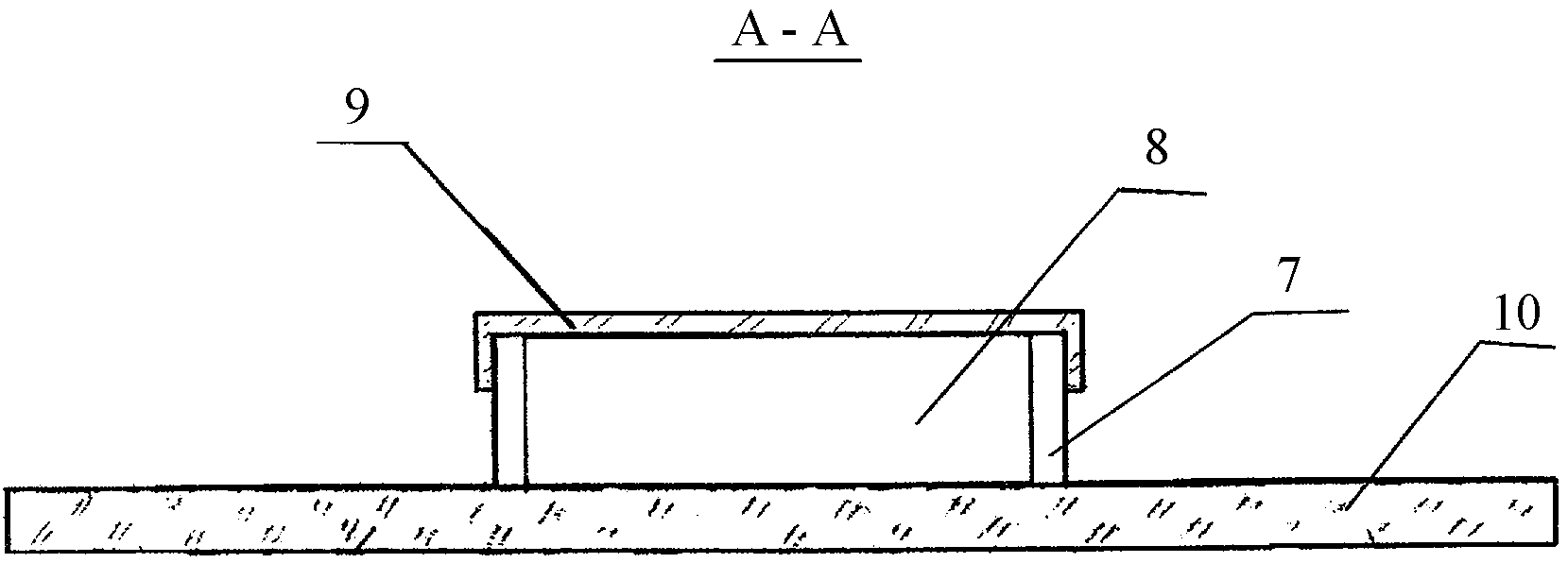
на фиг.2 представлен разрез А-А на фиг.1figure 2 presents a section aa in figure 1
на фиг.3 представлен вид биочипа сверху со вставленной рамкой и снятой крышкойfigure 3 presents a top view of the biochip with the frame inserted and the cover removed
на фиг.4 представлен разрез В-В на фиг.3figure 4 presents a section bb in figure 3
Биочип представляет собой прозрачную подложку 1 (фиг.1), имеющую рабочую область 2, содержащую тестовые участки 3 с иммобилизованными антителами или иными веществами, способными связываться с поверхностными молекулами клеток. Данные участки выполнены в виде параллельных полос. На подложке биочипа имеется реперный участок 4, указывающий на порядок считывания результата.The biochip is a transparent substrate 1 (Fig. 1) having a work area 2 containing test sections 3 with immobilized antibodies or other substances capable of binding to surface cell molecules. These sections are made in the form of parallel stripes. On the substrate of the biochip there is a reference section 4, indicating the reading order of the result.
На подложке также присутствует контрольный участок 5 (например, участок положительного или отрицательного контроля). Тестовые участки 3 отделены друг от друга и от участков 4 и 5 фоновыми участками 6, не содержащими молекул иммобилизованных антител или молекул иных веществ, способных связываться с поверхностными молекулами клеток. Рабочая область 2 отделена от остальной поверхности подложки бортиком 7, который выполнен съемным или отламывающися. Рабочая область 2 и бортик 7 образуют емкость биочипа 8 (фиг.2), которая закрывается сверху съемной крышкой 9, предотвращающей испарение жидкости, а также ее выливание или разбрызгивание. Крышка 9 устанавливается на бортик 7 биочипа. Подложка 1 имеет выступающие участки 10, обеспечивающие ее удобное расположение на предметном столике микроскопа, либо ее закрепление в ином вспомогательном устройстве. В емкость 8 вставляется съемная рамка 11 (фиг.3) с отверстиями (прорезями) 12. Отверстия 12 выполнены в виде параллельных линейных прорезей. (Но возможна конструкция с отверстиями 12 другой формы). При этом при установке рамки 11 на поверхность подложки образуются каналы (емкости) 13 (фиг.4). Отверстия (прорези) 12 (фиг.3) могут быть пустыми, но могут быть исходно заполнены веществами, необходимым для дополнительного исследования клеток, например, гелем, содержащим флуоресцентно меченные полинуклеотидные зонды или меченные антитела или питательную среду для роста клеток. Отверстия 12 выполнены сквозными, что позволяет вводить в них нужные растворы в процессе выполнения анализа. Но в некоторых случаях может быть целесообразным исполнение рамки 11 с глухими отверстиями (прорезями) 12 (например в тех случаях, если они должны быть заполнены твердой питательной средой или гелем, содержащим реагенты). Рамка 11 (фиг.4) снизу имеет герметизирующее эластичное покрытие 14 и герметично прижата к поверхности подложки 1 с помощью съемной струбцины 15.A control portion 5 is also present on the substrate (e.g., a positive or negative control portion). Test sections 3 are separated from each other and from sections 4 and 5 by background sections 6 that do not contain immobilized antibody molecules or molecules of other substances that can bind to surface cell molecules. The working area 2 is separated from the rest of the surface of the substrate by the bead 7, which is removable or breakable. The working area 2 and the side 7 form the capacity of the biochip 8 (figure 2), which is closed on top by a removable cover 9, which prevents evaporation of the liquid, as well as its pouring or spraying. The cover 9 is mounted on the side 7 of the biochip. The substrate 1 has protruding sections 10, ensuring its convenient location on the stage of the microscope, or its fixing in another auxiliary device. A removable frame 11 (FIG. 3) with holes (slots) 12 is inserted into the container 8. The holes 12 are made in the form of parallel linear slots. (But a design with holes 12 of a different shape is possible). Moreover, when installing the frame 11 on the surface of the substrate, channels (capacities) 13 are formed (Fig. 4). The holes (slots) 12 (FIG. 3) may be empty, but may be initially filled with substances necessary for additional cell research, for example, a gel containing fluorescently labeled polynucleotide probes or labeled antibodies or culture medium for cell growth. The holes 12 are made through, which allows you to enter the necessary solutions into them during the analysis. But in some cases, it may be appropriate to design the frame 11 with blind holes (slots) 12 (for example, in those cases where they must be filled with a solid nutrient medium or gel containing reagents). The frame 11 (figure 4) below has a sealing elastic coating 14 and is tightly pressed to the surface of the substrate 1 using a removable clamp 15.
Биочип работает следующим образом.The biochip works as follows.
В емкость 8 биочипа, пипеткой наливают жидкость, которая не растекается за пределы рабочей области 2 благодаря бортику 7. Емкость 8 при необходимости закрывают крышкой 9 предотвращающей испарение или разбрызгивание жидкости. Осуществляют инкубацию биочипа с данной жидкостью, которую после этого сливают. В процессе анализа последовательно используются различные жидкости. Последовательность выполнения манипуляций может быть следующей: обработка блокирующим раствором, отмывка детергентом, инкубация с исследуемым образцом клеточной суспензии, отмывка буферным раствором для устранения не связавшихся с антителами клеток, обработка веществом, повышающим прочность связывания оставшихся клеток. Затем из емкости 8 сливают жидкость и вставляют в нее рамку 11, которую герметично закрепляют, прижимая с помощью съемной струбцины 15 или иного приспособления. Рамку закрепляют таким образом, чтобы отверстия (прорези) 12 располагались поперек областей 3 и 5 в которых связались клетки. Каждый тестовый участок 3 подложки 1 пересекает несколько прорезей 12. В результате образуется несколько каналов 13, на дне каждого из которых находится по одному фрагменту каждого из тестовых участков 3 и фрагмент контрольного участка 5. В каналы 13 микропипеткой вводят вещества, необходимые для проведения дополнительных исследований клеток. При этом в разные каналы 13 могут быть введены реагенты для выполнения разных исследований. В некоторых случаях каналы 13 могут быть изначально заполнены субстанцией, например, гелем, содержащим необходимые реагенты. Каналы 13 также могут быть заполнены питательными средами, необходимыми для роста клеток, или любыми иными веществами. Выполняют манипуляции, необходимых для осуществления дополнительных исследований. После этого рамку снимают, убирают бортик, а биочип подвергают микроскопическому или иному исследованию.In a container 8 of the biochip, a liquid is poured with a pipette, which does not flow outside the working area 2 due to the side 7. The container 8 is, if necessary, closed with a lid 9 to prevent evaporation or splashing of the liquid. Biochip is incubated with this liquid, which is then drained. In the analysis process, various fluids are used sequentially. The sequence of manipulations can be as follows: treatment with a blocking solution, washing with a detergent, incubation with a test sample of a cell suspension, washing with a buffer solution to eliminate cells not bound to antibodies, treatment with a substance that increases the binding strength of the remaining cells. Then, liquid is drained from the container 8 and a frame 11 is inserted into it, which is hermetically fixed, pressing with the help of a removable clamp 15 or other device. The frame is fixed so that the holes (slots) 12 are located across the areas 3 and 5 in which the cells are connected. Each test section 3 of the substrate 1 intersects several slots 12. As a result, several channels 13 are formed, at the bottom of each of which there is one fragment of each of the test sections 3 and a fragment of the control section 5. The substances necessary for additional studies are introduced into the channels 13 using a micropipette cells. In this case, reagents can be introduced into different channels 13 to perform various studies. In some cases, the channels 13 may be initially filled with a substance, for example, a gel containing the necessary reagents. Channels 13 can also be filled with nutrient media necessary for cell growth, or any other substances. Perform the manipulations necessary for additional research. After that, the frame is removed, the side is removed, and the biochip is subjected to microscopic or other research.
Claims (10)
Priority Applications (1)
| Application Number | Priority Date | Filing Date | Title |
|---|---|---|---|
| RU2009144004/22U RU92964U1 (en) | 2009-11-30 | 2009-11-30 | CELL BIOCHIP |
Applications Claiming Priority (1)
| Application Number | Priority Date | Filing Date | Title |
|---|---|---|---|
| RU2009144004/22U RU92964U1 (en) | 2009-11-30 | 2009-11-30 | CELL BIOCHIP |
Publications (1)
| Publication Number | Publication Date |
|---|---|
| RU92964U1 true RU92964U1 (en) | 2010-04-10 |
Family
ID=42671515
Family Applications (1)
| Application Number | Title | Priority Date | Filing Date |
|---|---|---|---|
| RU2009144004/22U RU92964U1 (en) | 2009-11-30 | 2009-11-30 | CELL BIOCHIP |
Country Status (1)
| Country | Link |
|---|---|
| RU (1) | RU92964U1 (en) |
-
2009
- 2009-11-30 RU RU2009144004/22U patent/RU92964U1/en not_active IP Right Cessation
Similar Documents
| Publication | Publication Date | Title |
|---|---|---|
| US8652421B2 (en) | Immunoassay product and process | |
| US9891218B2 (en) | Immunoassay product and process | |
| US9751084B2 (en) | Biological culture assembly | |
| US20060051253A1 (en) | Device for staining and hybridization reactions | |
| JP4705000B2 (en) | Products and processes for immunoassays | |
| US20070009389A1 (en) | Slide deposition chamber | |
| US11213826B2 (en) | Cellular cassettes for the collection, storage, and analysis of biological samples | |
| RU103003U1 (en) | DEVICE FOR ANALYSIS USING THE BIOCHIP | |
| RU92964U1 (en) | CELL BIOCHIP | |
| RU86091U1 (en) | BIOCHIP | |
| RU97372U1 (en) | DEVICE FOR INCUBATION AND WASHING OF BIOCHIPS | |
| RU90213U1 (en) | BIOCHIP PLATFORM | |
| RU106618U1 (en) | TEST SYSTEM FOR CELL RESEARCH | |
| RU106378U1 (en) | DEVICE FOR ANALYSIS WITH APPLICATION OF THE BIOCHIP | |
| RU89893U1 (en) | CELL BIOCHIP | |
| RU91339U1 (en) | DEVICE FOR INCUBATION AND WASHING THE BIOCHIP | |
| RU87424U1 (en) | DEVICE FOR INCUBATION AND WASHING THE BIOCHIP | |
| EP3969175A1 (en) | Analytical device and reaction chamber | |
| RU87165U1 (en) | DEVICE FOR INCUBATION OF A BIOCHIP WITH CELL SUSPENSION AND ITS WASHING |
Legal Events
| Date | Code | Title | Description |
|---|---|---|---|
| MM1K | Utility model has become invalid (non-payment of fees) |
Effective date: 20151201 |