RU2777103C2 - Method for producing particles for specific targeting of cells - Google Patents
Method for producing particles for specific targeting of cells Download PDFInfo
- Publication number
- RU2777103C2 RU2777103C2 RU2020134602A RU2020134602A RU2777103C2 RU 2777103 C2 RU2777103 C2 RU 2777103C2 RU 2020134602 A RU2020134602 A RU 2020134602A RU 2020134602 A RU2020134602 A RU 2020134602A RU 2777103 C2 RU2777103 C2 RU 2777103C2
- Authority
- RU
- Russia
- Prior art keywords
- particles
- acid
- hematite
- receptor
- hematite particles
- Prior art date
Links
- 239000002245 particle Substances 0.000 title claims abstract description 104
- 238000004519 manufacturing process Methods 0.000 title claims abstract description 5
- 230000008685 targeting Effects 0.000 title description 8
- 238000000034 method Methods 0.000 claims abstract description 93
- 229910052595 hematite Inorganic materials 0.000 claims abstract description 84
- 239000011019 hematite Substances 0.000 claims abstract description 84
- LIKBJVNGSGBSGK-UHFFFAOYSA-N iron(3+);oxygen(2-) Chemical compound [O-2].[O-2].[O-2].[Fe+3].[Fe+3] LIKBJVNGSGBSGK-UHFFFAOYSA-N 0.000 claims abstract description 84
- 239000002253 acid Substances 0.000 claims abstract description 60
- 239000000203 mixture Substances 0.000 claims abstract description 26
- 229920000642 polymer Polymers 0.000 claims abstract description 24
- 239000000243 solution Substances 0.000 claims abstract description 24
- 238000002156 mixing Methods 0.000 claims abstract description 20
- 239000007864 aqueous solution Substances 0.000 claims abstract description 8
- 230000027455 binding Effects 0.000 claims abstract description 8
- 239000000725 suspension Substances 0.000 claims abstract description 6
- 230000021615 conjugation Effects 0.000 claims abstract description 5
- 238000001179 sorption measurement Methods 0.000 claims abstract description 4
- QAOWNCQODCNURD-UHFFFAOYSA-N Sulfuric acid Chemical compound OS(O)(=O)=O QAOWNCQODCNURD-UHFFFAOYSA-N 0.000 claims description 37
- 238000011534 incubation Methods 0.000 claims description 26
- 102000005962 receptors Human genes 0.000 claims description 26
- 108020003175 receptors Proteins 0.000 claims description 26
- 229920002125 Sokalan® Polymers 0.000 claims description 23
- 229920002873 Polyethylenimine Polymers 0.000 claims description 16
- 239000004584 polyacrylic acid Substances 0.000 claims description 16
- VEXZGXHMUGYJMC-UHFFFAOYSA-N Hydrochloric acid Chemical compound Cl VEXZGXHMUGYJMC-UHFFFAOYSA-N 0.000 claims description 14
- NBIIXXVUZAFLBC-UHFFFAOYSA-N Phosphoric acid Chemical compound OP(O)(O)=O NBIIXXVUZAFLBC-UHFFFAOYSA-N 0.000 claims description 14
- 102000004169 proteins and genes Human genes 0.000 claims description 13
- 108090000623 proteins and genes Proteins 0.000 claims description 13
- QTBSBXVTEAMEQO-UHFFFAOYSA-N Acetic acid Chemical compound CC(O)=O QTBSBXVTEAMEQO-UHFFFAOYSA-N 0.000 claims description 12
- BDAGIHXWWSANSR-UHFFFAOYSA-N formic acid Substances OC=O BDAGIHXWWSANSR-UHFFFAOYSA-N 0.000 claims description 11
- 229920002307 Dextran Polymers 0.000 claims description 9
- GRYLNZFGIOXLOG-UHFFFAOYSA-N Nitric acid Chemical compound O[N+]([O-])=O GRYLNZFGIOXLOG-UHFFFAOYSA-N 0.000 claims description 7
- OSWFIVFLDKOXQC-UHFFFAOYSA-N 4-(3-methoxyphenyl)aniline Chemical compound COC1=CC=CC(C=2C=CC(N)=CC=2)=C1 OSWFIVFLDKOXQC-UHFFFAOYSA-N 0.000 claims description 6
- 229910052500 inorganic mineral Inorganic materials 0.000 claims description 6
- 239000011707 mineral Substances 0.000 claims description 6
- 150000007524 organic acids Chemical class 0.000 claims description 6
- 125000003178 carboxy group Chemical group [H]OC(*)=O 0.000 claims description 5
- YBJHBAHKTGYVGT-ZKWXMUAHSA-N (+)-Biotin Chemical compound N1C(=O)N[C@@H]2[C@H](CCCCC(=O)O)SC[C@@H]21 YBJHBAHKTGYVGT-ZKWXMUAHSA-N 0.000 claims description 4
- 229920000805 Polyaspartic acid Polymers 0.000 claims description 4
- 108010039918 Polylysine Proteins 0.000 claims description 4
- 108010064470 polyaspartate Proteins 0.000 claims description 4
- 229920000656 polylysine Polymers 0.000 claims description 4
- 108090000765 processed proteins & peptides Proteins 0.000 claims description 4
- 150000001718 carbodiimides Chemical class 0.000 claims description 3
- 150000001875 compounds Chemical class 0.000 claims description 3
- 108020004707 nucleic acids Proteins 0.000 claims description 3
- 150000007523 nucleic acids Chemical group 0.000 claims description 3
- 102000039446 nucleic acids Human genes 0.000 claims description 3
- 230000001225 therapeutic effect Effects 0.000 claims description 3
- 108091023037 Aptamer Proteins 0.000 claims description 2
- 108090001008 Avidin Proteins 0.000 claims description 2
- 229920002134 Carboxymethyl cellulose Polymers 0.000 claims description 2
- 229920001661 Chitosan Polymers 0.000 claims description 2
- OVBPIULPVIDEAO-LBPRGKRZSA-N Folic acid Natural products C=1N=C2NC(N)=NC(=O)C2=NC=1CNC1=CC=C(C(=O)N[C@@H](CCC(O)=O)C(O)=O)C=C1 OVBPIULPVIDEAO-LBPRGKRZSA-N 0.000 claims description 2
- 102000008192 Lactoglobulins Human genes 0.000 claims description 2
- 108010060630 Lactoglobulins Proteins 0.000 claims description 2
- 108090001090 Lectins Proteins 0.000 claims description 2
- 102000004856 Lectins Human genes 0.000 claims description 2
- OVBPIULPVIDEAO-UHFFFAOYSA-N N-Pteroyl-L-glutaminsaeure Natural products C=1N=C2NC(N)=NC(=O)C2=NC=1CNC1=CC=C(C(=O)NC(CCC(O)=O)C(O)=O)C=C1 OVBPIULPVIDEAO-UHFFFAOYSA-N 0.000 claims description 2
- 108010090804 Streptavidin Proteins 0.000 claims description 2
- 102000004338 Transferrin Human genes 0.000 claims description 2
- 108090000901 Transferrin Proteins 0.000 claims description 2
- 229960002685 biotin Drugs 0.000 claims description 2
- 235000020958 biotin Nutrition 0.000 claims description 2
- 239000011616 biotin Substances 0.000 claims description 2
- 239000001768 carboxy methyl cellulose Substances 0.000 claims description 2
- 235000010948 carboxy methyl cellulose Nutrition 0.000 claims description 2
- 239000008112 carboxymethyl-cellulose Substances 0.000 claims description 2
- 239000000824 cytostatic agent Substances 0.000 claims description 2
- 230000001085 cytostatic effect Effects 0.000 claims description 2
- 231100000433 cytotoxic Toxicity 0.000 claims description 2
- 230000001472 cytotoxic effect Effects 0.000 claims description 2
- 229960000304 folic acid Drugs 0.000 claims description 2
- 235000019152 folic acid Nutrition 0.000 claims description 2
- 239000011724 folic acid Substances 0.000 claims description 2
- 125000003929 folic acid group Chemical group 0.000 claims description 2
- 239000002523 lectin Substances 0.000 claims description 2
- 230000005291 magnetic effect Effects 0.000 claims description 2
- 239000011976 maleic acid Substances 0.000 claims description 2
- 108010087904 neutravidin Proteins 0.000 claims description 2
- 229920000172 poly(styrenesulfonic acid) Polymers 0.000 claims description 2
- 229920001184 polypeptide Polymers 0.000 claims description 2
- 229940005642 polystyrene sulfonic acid Drugs 0.000 claims description 2
- 229920002689 polyvinyl acetate Polymers 0.000 claims description 2
- 239000011118 polyvinyl acetate Substances 0.000 claims description 2
- 229920000036 polyvinylpyrrolidone Polymers 0.000 claims description 2
- 239000001267 polyvinylpyrrolidone Substances 0.000 claims description 2
- 235000013855 polyvinylpyrrolidone Nutrition 0.000 claims description 2
- 102000004196 processed proteins & peptides Human genes 0.000 claims description 2
- 230000002285 radioactive effect Effects 0.000 claims description 2
- 150000003384 small molecules Chemical group 0.000 claims description 2
- 238000012546 transfer Methods 0.000 claims description 2
- 239000012581 transferrin Substances 0.000 claims description 2
- OFOBLEOULBTSOW-UHFFFAOYSA-N Propanedioic acid Natural products OC(=O)CC(O)=O OFOBLEOULBTSOW-UHFFFAOYSA-N 0.000 claims 1
- VZCYOOQTPOCHFL-UHFFFAOYSA-N trans-butenedioic acid Natural products OC(=O)C=CC(O)=O VZCYOOQTPOCHFL-UHFFFAOYSA-N 0.000 claims 1
- 239000002105 nanoparticle Substances 0.000 abstract description 73
- 230000015572 biosynthetic process Effects 0.000 abstract description 28
- 238000003786 synthesis reaction Methods 0.000 abstract description 20
- 239000003153 chemical reaction reagent Substances 0.000 abstract description 4
- 231100000252 nontoxic Toxicity 0.000 abstract description 2
- 230000003000 nontoxic effect Effects 0.000 abstract description 2
- 239000000126 substance Substances 0.000 abstract description 2
- 230000000694 effects Effects 0.000 abstract 1
- 210000004027 cell Anatomy 0.000 description 28
- 238000006243 chemical reaction Methods 0.000 description 27
- UQSXHKLRYXJYBZ-UHFFFAOYSA-N iron oxide Inorganic materials [Fe]=O UQSXHKLRYXJYBZ-UHFFFAOYSA-N 0.000 description 16
- 210000003743 erythrocyte Anatomy 0.000 description 14
- 239000002953 phosphate buffered saline Substances 0.000 description 14
- 239000012901 Milli-Q water Substances 0.000 description 12
- LOKCTEFSRHRXRJ-UHFFFAOYSA-I dipotassium trisodium dihydrogen phosphate hydrogen phosphate dichloride Chemical compound P(=O)(O)(O)[O-].[K+].P(=O)(O)([O-])[O-].[Na+].[Na+].[Cl-].[K+].[Cl-].[Na+] LOKCTEFSRHRXRJ-UHFFFAOYSA-I 0.000 description 12
- 238000004458 analytical method Methods 0.000 description 10
- RTZKZFJDLAIYFH-UHFFFAOYSA-N Diethyl ether Chemical compound CCOCC RTZKZFJDLAIYFH-UHFFFAOYSA-N 0.000 description 9
- 206010028980 Neoplasm Diseases 0.000 description 9
- 150000007513 acids Chemical class 0.000 description 9
- 235000013980 iron oxide Nutrition 0.000 description 9
- 108091003079 Bovine Serum Albumin Proteins 0.000 description 8
- LFQSCWFLJHTTHZ-UHFFFAOYSA-N Ethanol Chemical compound CCO LFQSCWFLJHTTHZ-UHFFFAOYSA-N 0.000 description 8
- 101000941029 Homo sapiens Endoplasmic reticulum junction formation protein lunapark Proteins 0.000 description 8
- 101000991410 Homo sapiens Nucleolar and spindle-associated protein 1 Proteins 0.000 description 8
- 102100030991 Nucleolar and spindle-associated protein 1 Human genes 0.000 description 8
- 229940098773 bovine serum albumin Drugs 0.000 description 8
- 238000005119 centrifugation Methods 0.000 description 8
- 238000004566 IR spectroscopy Methods 0.000 description 7
- 108060003951 Immunoglobulin Proteins 0.000 description 7
- 201000011510 cancer Diseases 0.000 description 7
- 102000018358 immunoglobulin Human genes 0.000 description 7
- 230000003993 interaction Effects 0.000 description 7
- 239000002244 precipitate Substances 0.000 description 7
- 229910000147 aluminium phosphate Inorganic materials 0.000 description 6
- 239000011449 brick Substances 0.000 description 6
- 238000011161 development Methods 0.000 description 6
- 239000003814 drug Substances 0.000 description 6
- 238000002474 experimental method Methods 0.000 description 6
- 238000000684 flow cytometry Methods 0.000 description 6
- 229910052598 goethite Inorganic materials 0.000 description 6
- AEIXRCIKZIZYPM-UHFFFAOYSA-M hydroxy(oxo)iron Chemical compound [O][Fe]O AEIXRCIKZIZYPM-UHFFFAOYSA-M 0.000 description 6
- 229910017604 nitric acid Inorganic materials 0.000 description 6
- 238000001228 spectrum Methods 0.000 description 6
- 238000003756 stirring Methods 0.000 description 6
- 239000003795 chemical substances by application Substances 0.000 description 5
- 238000004090 dissolution Methods 0.000 description 5
- 238000009826 distribution Methods 0.000 description 5
- 238000012377 drug delivery Methods 0.000 description 5
- 239000007850 fluorescent dye Substances 0.000 description 5
- 235000019253 formic acid Nutrition 0.000 description 5
- 235000010755 mineral Nutrition 0.000 description 5
- 239000000523 sample Substances 0.000 description 5
- 238000003325 tomography Methods 0.000 description 5
- 238000001890 transfection Methods 0.000 description 5
- QGZKDVFQNNGYKY-UHFFFAOYSA-N Ammonia Chemical compound N QGZKDVFQNNGYKY-UHFFFAOYSA-N 0.000 description 4
- 241000283707 Capra Species 0.000 description 4
- 101150029707 ERBB2 gene Proteins 0.000 description 4
- 239000007987 MES buffer Substances 0.000 description 4
- YWEUIGNSBFLMFL-UHFFFAOYSA-N diphosphonate Chemical compound O=P(=O)OP(=O)=O YWEUIGNSBFLMFL-UHFFFAOYSA-N 0.000 description 4
- 238000003384 imaging method Methods 0.000 description 4
- 238000003018 immunoassay Methods 0.000 description 4
- 238000002595 magnetic resonance imaging Methods 0.000 description 4
- 239000000463 material Substances 0.000 description 4
- 230000003287 optical effect Effects 0.000 description 4
- 210000000056 organ Anatomy 0.000 description 4
- DLYUQMMRRRQYAE-UHFFFAOYSA-N phosphorus pentoxide Inorganic materials O1P(O2)(=O)OP3(=O)OP1(=O)OP2(=O)O3 DLYUQMMRRRQYAE-UHFFFAOYSA-N 0.000 description 4
- 238000004627 transmission electron microscopy Methods 0.000 description 4
- 238000005406 washing Methods 0.000 description 4
- XLYOFNOQVPJJNP-UHFFFAOYSA-N water Substances O XLYOFNOQVPJJNP-UHFFFAOYSA-N 0.000 description 4
- 108091034117 Oligonucleotide Proteins 0.000 description 3
- 239000004793 Polystyrene Substances 0.000 description 3
- 235000011054 acetic acid Nutrition 0.000 description 3
- 239000011248 coating agent Substances 0.000 description 3
- 238000000576 coating method Methods 0.000 description 3
- 238000002296 dynamic light scattering Methods 0.000 description 3
- 230000002068 genetic effect Effects 0.000 description 3
- 229940072221 immunoglobulins Drugs 0.000 description 3
- 238000000338 in vitro Methods 0.000 description 3
- RBTARNINKXHZNM-UHFFFAOYSA-K iron trichloride Chemical compound Cl[Fe](Cl)Cl RBTARNINKXHZNM-UHFFFAOYSA-K 0.000 description 3
- VBMVTYDPPZVILR-UHFFFAOYSA-N iron(2+);oxygen(2-) Chemical class [O-2].[Fe+2] VBMVTYDPPZVILR-UHFFFAOYSA-N 0.000 description 3
- 210000004185 liver Anatomy 0.000 description 3
- 239000013642 negative control Substances 0.000 description 3
- 235000005985 organic acids Nutrition 0.000 description 3
- 229920002223 polystyrene Polymers 0.000 description 3
- 239000012460 protein solution Substances 0.000 description 3
- -1 Μn ~ 5100) Polymers 0.000 description 3
- HBZBAMXERPYTFS-SECBINFHSA-N (4S)-2-(6,7-dihydro-5H-pyrrolo[3,2-f][1,3]benzothiazol-2-yl)-4,5-dihydro-1,3-thiazole-4-carboxylic acid Chemical compound OC(=O)[C@H]1CSC(=N1)c1nc2cc3CCNc3cc2s1 HBZBAMXERPYTFS-SECBINFHSA-N 0.000 description 2
- YMHOBZXQZVXHBM-UHFFFAOYSA-N 2,5-dimethoxy-4-bromophenethylamine Chemical compound COC1=CC(CCN)=C(OC)C=C1Br YMHOBZXQZVXHBM-UHFFFAOYSA-N 0.000 description 2
- VTLYFUHAOXGGBS-UHFFFAOYSA-N Fe3+ Chemical compound [Fe+3] VTLYFUHAOXGGBS-UHFFFAOYSA-N 0.000 description 2
- 108090000331 Firefly luciferases Proteins 0.000 description 2
- 101150066002 GFP gene Proteins 0.000 description 2
- 229910021578 Iron(III) chloride Inorganic materials 0.000 description 2
- 108060001084 Luciferase Proteins 0.000 description 2
- 241000699666 Mus <mouse, genus> Species 0.000 description 2
- 241000699670 Mus sp. Species 0.000 description 2
- 102100030086 Receptor tyrosine-protein kinase erbB-2 Human genes 0.000 description 2
- 241000545067 Venus Species 0.000 description 2
- 230000002378 acidificating effect Effects 0.000 description 2
- 229910021529 ammonia Inorganic materials 0.000 description 2
- 239000000074 antisense oligonucleotide Substances 0.000 description 2
- 238000012230 antisense oligonucleotides Methods 0.000 description 2
- 230000000903 blocking effect Effects 0.000 description 2
- 210000004369 blood Anatomy 0.000 description 2
- 239000008280 blood Substances 0.000 description 2
- 239000000872 buffer Substances 0.000 description 2
- 210000004978 chinese hamster ovary cell Anatomy 0.000 description 2
- 239000000084 colloidal system Substances 0.000 description 2
- 230000000875 corresponding effect Effects 0.000 description 2
- 239000013078 crystal Substances 0.000 description 2
- 238000002524 electron diffraction data Methods 0.000 description 2
- 238000002149 energy-dispersive X-ray emission spectroscopy Methods 0.000 description 2
- 238000005516 engineering process Methods 0.000 description 2
- 238000007306 functionalization reaction Methods 0.000 description 2
- 229940022353 herceptin Drugs 0.000 description 2
- 230000007062 hydrolysis Effects 0.000 description 2
- 238000006460 hydrolysis reaction Methods 0.000 description 2
- XEEYBQQBJWHFJM-UHFFFAOYSA-N iron Substances [Fe] XEEYBQQBJWHFJM-UHFFFAOYSA-N 0.000 description 2
- 231100000053 low toxicity Toxicity 0.000 description 2
- 239000012218 nanoagent Substances 0.000 description 2
- 239000002159 nanocrystal Substances 0.000 description 2
- 239000002086 nanomaterial Substances 0.000 description 2
- 229940031182 nanoparticles iron oxide Drugs 0.000 description 2
- 238000004626 scanning electron microscopy Methods 0.000 description 2
- 159000000000 sodium salts Chemical class 0.000 description 2
- RPENMORRBUTCPR-UHFFFAOYSA-M sodium;1-hydroxy-2,5-dioxopyrrolidine-3-sulfonate Chemical compound [Na+].ON1C(=O)CC(S([O-])(=O)=O)C1=O RPENMORRBUTCPR-UHFFFAOYSA-M 0.000 description 2
- 239000007787 solid Substances 0.000 description 2
- 229940124597 therapeutic agent Drugs 0.000 description 2
- 229960000575 trastuzumab Drugs 0.000 description 2
- 229910000859 α-Fe Inorganic materials 0.000 description 2
- 238000011725 BALB/c mouse Methods 0.000 description 1
- 102000001301 EGF receptor Human genes 0.000 description 1
- 108060006698 EGF receptor Proteins 0.000 description 1
- 102000001554 Hemoglobins Human genes 0.000 description 1
- 108010054147 Hemoglobins Proteins 0.000 description 1
- 101001012157 Homo sapiens Receptor tyrosine-protein kinase erbB-2 Proteins 0.000 description 1
- DGAQECJNVWCQMB-PUAWFVPOSA-M Ilexoside XXIX Chemical compound C[C@@H]1CC[C@@]2(CC[C@@]3(C(=CC[C@H]4[C@]3(CC[C@@H]5[C@@]4(CC[C@@H](C5(C)C)OS(=O)(=O)[O-])C)C)[C@@H]2[C@]1(C)O)C)C(=O)O[C@H]6[C@@H]([C@H]([C@@H]([C@H](O6)CO)O)O)O.[Na+] DGAQECJNVWCQMB-PUAWFVPOSA-M 0.000 description 1
- 102000008394 Immunoglobulin Fragments Human genes 0.000 description 1
- 108010021625 Immunoglobulin Fragments Proteins 0.000 description 1
- 206010027476 Metastases Diseases 0.000 description 1
- 239000004698 Polyethylene Substances 0.000 description 1
- FAPWRFPIFSIZLT-UHFFFAOYSA-M Sodium chloride Chemical compound [Na+].[Cl-] FAPWRFPIFSIZLT-UHFFFAOYSA-M 0.000 description 1
- 238000000692 Student's t-test Methods 0.000 description 1
- 238000002441 X-ray diffraction Methods 0.000 description 1
- 238000009825 accumulation Methods 0.000 description 1
- IZQZNLBFNMTRMF-UHFFFAOYSA-N acetic acid;phosphoric acid Chemical compound CC(O)=O.OP(O)(O)=O IZQZNLBFNMTRMF-UHFFFAOYSA-N 0.000 description 1
- 150000001243 acetic acids Chemical class 0.000 description 1
- 230000002776 aggregation Effects 0.000 description 1
- 238000004220 aggregation Methods 0.000 description 1
- 125000003277 amino group Chemical group 0.000 description 1
- 208000007502 anemia Diseases 0.000 description 1
- 239000000427 antigen Substances 0.000 description 1
- 108091007433 antigens Proteins 0.000 description 1
- 102000036639 antigens Human genes 0.000 description 1
- 238000013459 approach Methods 0.000 description 1
- 238000005102 attenuated total reflection Methods 0.000 description 1
- 230000008614 cellular interaction Effects 0.000 description 1
- 230000008859 change Effects 0.000 description 1
- 238000001311 chemical methods and process Methods 0.000 description 1
- 238000002512 chemotherapy Methods 0.000 description 1
- 238000012875 competitive assay Methods 0.000 description 1
- 239000013068 control sample Substances 0.000 description 1
- 238000013270 controlled release Methods 0.000 description 1
- 230000002596 correlated effect Effects 0.000 description 1
- 238000004163 cytometry Methods 0.000 description 1
- 229940124447 delivery agent Drugs 0.000 description 1
- 238000003745 diagnosis Methods 0.000 description 1
- XPPKVPWEQAFLFU-UHFFFAOYSA-N diphosphoric acid Chemical compound OP(O)(=O)OP(O)(O)=O XPPKVPWEQAFLFU-UHFFFAOYSA-N 0.000 description 1
- 201000010099 disease Diseases 0.000 description 1
- 208000037265 diseases, disorders, signs and symptoms Diseases 0.000 description 1
- 229940079593 drug Drugs 0.000 description 1
- 239000000975 dye Substances 0.000 description 1
- 238000002003 electron diffraction Methods 0.000 description 1
- 230000002121 endocytic effect Effects 0.000 description 1
- 230000000925 erythroid effect Effects 0.000 description 1
- 230000005284 excitation Effects 0.000 description 1
- 229940032296 ferric chloride Drugs 0.000 description 1
- 229940044631 ferric chloride hexahydrate Drugs 0.000 description 1
- 239000011790 ferrous sulphate Substances 0.000 description 1
- 235000003891 ferrous sulphate Nutrition 0.000 description 1
- 238000002073 fluorescence micrograph Methods 0.000 description 1
- 238000000799 fluorescence microscopy Methods 0.000 description 1
- 238000001215 fluorescent labelling Methods 0.000 description 1
- 238000001027 hydrothermal synthesis Methods 0.000 description 1
- 239000012535 impurity Substances 0.000 description 1
- 238000002329 infrared spectrum Methods 0.000 description 1
- 238000002347 injection Methods 0.000 description 1
- 239000007924 injection Substances 0.000 description 1
- 235000014413 iron hydroxide Nutrition 0.000 description 1
- NQXWGWZJXJUMQB-UHFFFAOYSA-K iron trichloride hexahydrate Chemical compound O.O.O.O.O.O.[Cl-].Cl[Fe+]Cl NQXWGWZJXJUMQB-UHFFFAOYSA-K 0.000 description 1
- BAUYGSIQEAFULO-UHFFFAOYSA-L iron(2+) sulfate (anhydrous) Chemical compound [Fe+2].[O-]S([O-])(=O)=O BAUYGSIQEAFULO-UHFFFAOYSA-L 0.000 description 1
- RUTXIHLAWFEWGM-UHFFFAOYSA-H iron(3+) sulfate Chemical compound [Fe+3].[Fe+3].[O-]S([O-])(=O)=O.[O-]S([O-])(=O)=O.[O-]S([O-])(=O)=O RUTXIHLAWFEWGM-UHFFFAOYSA-H 0.000 description 1
- 229910000359 iron(II) sulfate Inorganic materials 0.000 description 1
- 229910000360 iron(III) sulfate Inorganic materials 0.000 description 1
- NCNCGGDMXMBVIA-UHFFFAOYSA-L iron(ii) hydroxide Chemical compound [OH-].[OH-].[Fe+2] NCNCGGDMXMBVIA-UHFFFAOYSA-L 0.000 description 1
- 210000002540 macrophage Anatomy 0.000 description 1
- 239000006249 magnetic particle Substances 0.000 description 1
- 239000003550 marker Substances 0.000 description 1
- 230000007246 mechanism Effects 0.000 description 1
- 230000009401 metastasis Effects 0.000 description 1
- 239000011859 microparticle Substances 0.000 description 1
- 150000007522 mineralic acids Chemical class 0.000 description 1
- 230000004048 modification Effects 0.000 description 1
- 238000012986 modification Methods 0.000 description 1
- 238000012634 optical imaging Methods 0.000 description 1
- 229920000620 organic polymer Polymers 0.000 description 1
- 230000005298 paramagnetic effect Effects 0.000 description 1
- 238000002428 photodynamic therapy Methods 0.000 description 1
- 229920000573 polyethylene Polymers 0.000 description 1
- 229940005657 pyrophosphoric acid Drugs 0.000 description 1
- 239000011541 reaction mixture Substances 0.000 description 1
- 230000035484 reaction time Effects 0.000 description 1
- 239000013074 reference sample Substances 0.000 description 1
- 150000003839 salts Chemical class 0.000 description 1
- 238000013341 scale-up Methods 0.000 description 1
- 238000001878 scanning electron micrograph Methods 0.000 description 1
- 239000011734 sodium Substances 0.000 description 1
- 229910052708 sodium Inorganic materials 0.000 description 1
- 239000011780 sodium chloride Substances 0.000 description 1
- 230000009870 specific binding Effects 0.000 description 1
- 230000006641 stabilisation Effects 0.000 description 1
- 238000011105 stabilization Methods 0.000 description 1
- 238000001308 synthesis method Methods 0.000 description 1
- 230000002194 synthesizing effect Effects 0.000 description 1
- 238000012353 t test Methods 0.000 description 1
- 238000002560 therapeutic procedure Methods 0.000 description 1
- 231100000167 toxic agent Toxicity 0.000 description 1
- 239000003440 toxic substance Substances 0.000 description 1
- 231100000419 toxicity Toxicity 0.000 description 1
- 230000001988 toxicity Effects 0.000 description 1
- 230000009466 transformation Effects 0.000 description 1
- 229910052723 transition metal Inorganic materials 0.000 description 1
- 150000003624 transition metals Chemical class 0.000 description 1
Images




Abstract
Description
Область техники, к которой относится изобретениеThe field of technology to which the invention belongs
Изобретение относится к области биомедицины и наномедицины, в том числе к способам получения (синтеза) наночастиц на основе гематита, способных специфически распознавать и связываться с клетками-мишенями.The invention relates to the field of biomedicine and nanomedicine, including methods for obtaining (synthesis) of hematite-based nanoparticles capable of specifically recognizing and binding to target cells.
Уровень техникиState of the art
В последнее время наблюдается активный рост использования нанотехнологий в биологии и медицине, а также других областях науки и техники. При лечении и диагностики (а также тераностики) заболеваний наночастицы имеют ряд преимуществ перед обычными молекулярными лекарствами, особенно таких как возможности создания более сложной структуры, позволяющей осуществлять контролируемое высвобождение активного компонента только в случае необходимости, возможности добавления различных механизмов для его адресной доставки и др. Наибольший интерес представляют частицы, обладающие свойствами, позволяющими детектировать их неинвазивно при помощи широко используемых в медицине аналитических методов таких как магнитно-резонансная томография (МРТ) и при этом обладающими низкой токсичностью. Ввиду этого одними из самых перспективных материалов для создания наночастиц являются оксиды железа, которые в зависимости от структуры могут обладать как Т1 так и Т2 контрастирующими свойствами в МРТ а также обладают самой низкой токсичностью среди переходных металлов. Одними из наиболее интересных наночастиц являются частицы гематита, как самого стабильного оксида железа. Они обладают высокой химической стабильностью, хорошими МРТ-контрастирующими свойствами, способностью деградировать в тканях организма естественным путем, в том числе осуществляя терапевтический эффект в отношении анемии (введение таких частиц, увеличивает уровень гемоглобина в крови).Recently, there has been an active growth in the use of nanotechnologies in biology and medicine, as well as in other fields of science and technology. In the treatment and diagnosis (as well as theranostics) of diseases, nanoparticles have a number of advantages over conventional molecular drugs, such as the possibility of creating a more complex structure that allows controlled release of the active component only when necessary, the possibility of adding various mechanisms for its targeted delivery, etc. Of greatest interest are particles that have properties that allow them to be detected non-invasively using analytical methods widely used in medicine, such as magnetic resonance imaging (MRI), and at the same time have low toxicity. In view of this, one of the most promising materials for creating nanoparticles are iron oxides, which, depending on the structure, can have both T1 and T2 contrast properties in MRI and also have the lowest toxicity among transition metals. One of the most interesting nanoparticles are particles of hematite, as the most stable iron oxide. They have high chemical stability, good MRI-contrasting properties, the ability to degrade naturally in body tissues, including having a therapeutic effect on anemia (the introduction of such particles increases the level of hemoglobin in the blood).
Поэтому одной из важных задач современной медицины является разработка подходов для создания эффективных методик синтеза наночастиц гематита, обладающих следующими характеристиками:Therefore, one of the important tasks of modern medicine is the development of approaches for creating effective methods for the synthesis of hematite nanoparticles with the following characteristics:
1) Малое время проведения реакции,1) Short reaction time,
2) Легкодоступные реагенты,2) Easily accessible reagents,
3) Возможность масштабирования синтеза без изменения параметров синтезируемых частиц таких как выход, размер и распределение по размерам,3) The possibility of scaling the synthesis without changing the parameters of the synthesized particles such as yield, size and size distribution,
4) Эффективная иммобилизация рецепторов, обеспечивающих высокоспецифичное связывание с клетками-мишенями.4) Efficient immobilization of receptors providing highly specific binding to target cells.
Для решения данной проблемы известен близкий способ (US 20130251624 А1), в котором частицы гематита получают гидротермальным гидролизом раствора хлорида железа(III)To solve this problem, a similar method is known (US 20130251624 A1), in which hematite particles are obtained by hydrothermal hydrolysis of an iron (III) chloride solution
Недостатки этого способа заключаются в том, что:The disadvantages of this method are that:
1) Реакция проводится при повышенной температуре (от 90°С до 250°С).1) The reaction is carried out at an elevated temperature (from 90°C to 250°C).
2) Реакция проводится в течение продолжительного времени (более суток).2) The reaction is carried out for a long time (more than a day).
3) Реакция проводится при повышенном давлении.3) The reaction is carried out at elevated pressure.
Известен наиболее близкий способ (Nanomaterials 2020, 10, 323; doi:10.3390/nanol0020323), в котором частицы гематита получают из ферригидрита путем медленной трансформации в водных растворах.The closest method is known (Nanomaterials 2020, 10, 323; doi: 10.3390/nanol0020323), in which hematite particles are obtained from ferrihydrite by slow transformation in aqueous solutions.
Недостатки этого метода состоят в том, что:The disadvantages of this method are that:
1) Реакция проходит в течение нескольких месяцев.1) The reaction takes place within a few months.
2) Реакционные условия не предполагают масштабирования.2) Reactive conditions do not involve scaling.
Таким образом, требуемый технический результат состоит в создании эффективного метода синтеза нетоксичных наночастиц оксида железа - гематита, функционализированных специфическими рецепторными молекулами, позволяющий получать частицы контролируемого размера с использованием легко доступных реагентов, и обладающий возможностью легкой масштабируемости условий синтеза.Thus, the required technical result is to create an effective method for the synthesis of non-toxic nanoparticles of iron oxide - hematite, functionalized with specific receptor molecules, which makes it possible to obtain particles of a controlled size using readily available reagents, and with the possibility of easy scalability of synthesis conditions.
Описание изобретенияDescription of the invention
Для достижения указанного технического результата предложен способ получения частиц гематита для специфического таргетинга (таргетной доставки к) клеток, включающий в себя, по крайней мере: i) смешение частиц ферригидрита с раствором по крайней мере одной кислоты и инкубацию полученной смеси при температуре не выше +4 градусов Цельсия с целью формирования частиц гематита, причем концентрация упомянутой кислоты после смешения с упомянутыми частицами ферригидрита составляет не менее 50% от концентрации концентрированной кислоты, ii) перевод упомянутых частиц гематита в водный раствор с рН более 2 с дальнейшим добавлением к суспензии упомянутых частиц гематита раствора полимера для его сорбции на поверхность частиц; iii) конъюгацию специфичного к упомянутым клеткам рецептора с упомянутыми молекулами полимера на поверхности упомянутых частиц.To achieve this technical result, a method is proposed for obtaining hematite particles for specific targeting (targeted delivery to) cells, including at least: i) mixing ferrihydrite particles with a solution of at least one acid and incubating the resulting mixture at a temperature not higher than +4 degrees Celsius in order to form hematite particles, wherein the concentration of said acid after mixing with said ferrihydrite particles is at least 50% of the concentration of concentrated acid, ii) transfer of said hematite particles into an aqueous solution with a pH of more than 2 with further addition to the suspension of said hematite particles of the solution polymer for its sorption on the surface of the particles; iii) conjugation of a receptor specific to said cells with said polymer molecules on the surface of said particles.
Кроме того, способ, в котором упомянутый полимер принадлежит группе декстран, карбоксиметилдекстран, полиэтиленимин, полилизин, полиаспарагиновая кислота, полиакриловая кислота.In addition, a method in which said polymer belongs to the group of dextran, carboxymethyl dextran, polyethyleneimine, polylysine, polyaspartic acid, polyacrylic acid.
Кроме того, способ, в котором упомянутый полимер включает в себя карбоксильные группы и упомянутую конъюгацию проводят карбодиимидным способом.In addition, a method in which said polymer includes carboxyl groups and said conjugation is carried out by a carbodiimide method.
Кроме того, способ, в котором упомянутый рецептор является пептидом, полипептидом или белком.Further, a method wherein said receptor is a peptide, polypeptide or protein.
Кроме того, способ, в котором упомянутый рецептор является нуклеиновой кислотой. Кроме того, способ, в котором упомянутый рецептор является аптамером.Further, a method wherein said receptor is a nucleic acid. In addition, a method wherein said receptor is an aptamer.
Кроме того, способ, в котором упомянутый рецептор является белком из группы: трансферрин, лактоглобулин, авидин, стрептавидин, нейтравидин, лектин.In addition, a method in which said receptor is a protein from the group: transferrin, lactoglobulin, avidin, streptavidin, neutravidin, lectin.
Кроме того, способ, в котором упомянутый рецептор является антителом.Further, a method wherein said receptor is an antibody.
Кроме того, способ, в котором упомянутый рецептор является моноклональным антителом.Further, a method wherein said receptor is a monoclonal antibody.
Кроме того, способ, в котором упомянутый рецептор является фрагментом антитела.Further, a method wherein said receptor is an antibody fragment.
Кроме того, способ, в котором упомянутый рецептор является антителом против эритроцитов.Further, a method wherein said receptor is an anti-erythrocyte antibody.
Кроме того, способ, в котором упомянутый рецептор является антителом против эритроцитов, причем введение упомянутой частицы характеризуется более длительным временем циркуляции в кровотоке, чем для таких же антител без упомянутых конъюгированных антител.In addition, a method in which said receptor is an antibody against erythrocytes, wherein the administration of said particle is characterized by a longer circulation time in the bloodstream than for the same antibodies without said conjugated antibodies.
Кроме того, способ, в котором упомянутый рецептор является низкомолекулярным рецептором.Further, a method wherein said receptor is a small molecule receptor.
Кроме того, способ, в котором упомянутый рецептор является фолиевой кислотой или ее аналогом.Furthermore, a method wherein said receptor is folic acid or an analogue thereof.
Кроме того, способ, в котором упомянутый рецептор является биотином или его аналогом.Furthermore, a method wherein said receptor is biotin or an analogue thereof.
Кроме того, способ, включающий в себя, по крайней мере: смешение частиц ферригидрита с раствором по крайней мере одной кислоты и инкубацию полученной смеси при температуре не выше +4 градусов Цельсия, причем концентрация упомянутой кислоты после смешения с упомянутыми частицами ферригидрита составляет не менее 50% от концентрации концентрированной кислоты.In addition, the method includes at least: mixing of ferrihydrite particles with a solution of at least one acid and incubation of the resulting mixture at a temperature not exceeding +4 degrees Celsius, and the concentration of said acid after mixing with said ferrihydrite particles is at least 50 % of concentrated acid concentration.
Кроме того, способ, в котором концентрация упомянутой кислоты после смешения с упомянутыми частицами ферригидрита составляет не менее 70% от концентрации концентрированной кислоты.Further, a method wherein the concentration of said acid after mixing with said ferrihydrite particles is at least 70% of the concentrated acid concentration.
Кроме того, способ, в котором концентрация упомянутой кислоты после смешения с упомянутыми частицами ферригидрита составляет не менее 90% от концентрации концентрированной кислоты.In addition, a method in which the concentration of said acid after mixing with said ferrihydrite particles is at least 90% of the concentrated acid concentration.
Кроме того, способ, в котором упомянутую инкубацию осуществляют не более 10 минут.In addition, a method in which said incubation is carried out for no more than 10 minutes.
Кроме того, способ, в котором упомянутую инкубацию осуществляют не более 1 часа.In addition, a method in which said incubation is carried out for no more than 1 hour.
Кроме того, способ, в котором упомянутую инкубацию осуществляют не более 24 часов.Further, a method wherein said incubation is carried out for no more than 24 hours.
Кроме того, способ, в котором упомянутая кислота является сильной минеральной кислотой.Further, a method wherein said acid is a strong mineral acid.
Кроме того, способ, в котором упомянутая кислота является сильной минеральной кислотой из группы: серная, азотная, соляная.In addition, a method in which said acid is a strong mineral acid from the group: sulfuric, nitric, hydrochloric.
Кроме того, способ, в котором упомянутая кислота является органической кислотой.Further, a method wherein said acid is an organic acid.
Кроме того, способ, в котором упомянутая кислота является органической кислотой из группы: муравьиная, уксусная.In addition, a method in which said acid is an organic acid from the group: formic, acetic.
Кроме того, способ, в котором упомянутую инкубацию проводят при температуре от 0.5 до 1 градуса Цельсия.In addition, a method in which said incubation is carried out at a temperature of from 0.5 to 1 degree Celsius.
Кроме того, способ, в котором упомянутую инкубацию проводят при температуре 1.5-2.5 градуса Цельсия.In addition, a method in which said incubation is carried out at a temperature of 1.5-2.5 degrees Celsius.
Кроме того, способ, который кроме того включает шаг синтеза упомянутого ферригидрита, который в свою очередь включает в себя, по крайней мере:In addition, the method, which further includes the step of synthesizing said ferrihydrite, which in turn includes at least:
Кроме того, способ, в котором упомянутые частицы гематита имеют средний размер менее 100 нм.Further, a method wherein said hematite particles have an average size of less than 100 nm.
Кроме того, способ, в котором упомянутые частицы гематита имеют средний размер менее 200 нм.Further, a method wherein said hematite particles have an average size of less than 200 nm.
Кроме того, способ, в котором упомянутые частицы гематита имеют средний размер менее 1000 нм.Further, a method wherein said hematite particles have an average size of less than 1000 nm.
Кроме того, способ, в котором упомянутые частицы гематита имеют средний гидродинамический размер менее 100 нм.Further, a method wherein said hematite particles have an average hydrodynamic size of less than 100 nm.
Кроме того, способ, в котором упомянутые частицы гематита имеют средний гидродинамический размер менее 200 нм.Further, a method wherein said hematite particles have an average hydrodynamic size of less than 200 nm.
Кроме того, способ, в котором упомянутые частицы гематита имеют средний гидродинамический размер менее 1000 нм.Further, a method wherein said hematite particles have an average hydrodynamic size of less than 1000 nm.
Кроме того, способ, в котором на упомянутые частицы гематита дополнительно сорбируют полимер.In addition, a method in which a polymer is additionally sorbed onto said hematite particles.
Кроме того, способ, в котором на упомянутые частицы гематита коллоидно-стабильны.In addition, a method in which said hematite particles are colloidally stable.
Кроме того, способ, в котором на упомянутые частицы гематита имеют индекс полидисперсности менее 0.05.In addition, a method in which said hematite particles have a polydispersity index of less than 0.05.
Кроме того, способ, в котором на упомянутые частицы гематита имеют индекс полидисперсности менее 0.1.In addition, a method in which said hematite particles have a polydispersity index of less than 0.1.
Кроме того, способ, в котором на упомянутые частицы гематита имеют индекс полидисперсности менее 0.2.In addition, a method in which said hematite particles have a polydispersity index of less than 0.2.
Кроме того, способ, в котором на упомянутые частицы гематита дополнительно сорбируют полимер из группы: декстран, карбоксиметилдекстран, карбоксиметилцелюлюза, разветвленный полиэтиленимин, линейный полиэтиленимин, поливинилпирролидон, поливинилацетат, хитозан, полилизин, полиаспарагиновая кислота, полиакриловая кислота, полиоктадецен-альт-малеиновая кислота, полистиролсульфоновая кислота.In addition, a method in which a polymer from the group of dextran, carboxymethyl dextran, carboxymethyl cellulose, branched polyethyleneimine, linear polyethyleneimine, polyvinylpyrrolidone, polyvinyl acetate, chitosan, polylysine, polyaspartic acid, polyacrylic acid, polyoctadecene-alto-maleic acid, is additionally adsorbed onto the said hematite particles, polystyrenesulfonic acid.
Кроме того, способ, в котором упомянутые частицы гематита дополнительно конъюгируют с меткой из группы: флуоресцентную, магнитную, МРТ-контрастирующую, рентгентоконтрастирующую, люминесцентную, радиоактивную.In addition, a method in which said hematite particles are additionally conjugated with a label from the group: fluorescent, magnetic, MRI-contrasting, radiopaque, luminescent, radioactive.
Кроме того, способ, в котором упомянутые частицы гематита дополнительно включают в себя цитостатические, цитотоксические или терапевтические соединения.Furthermore, the method wherein said hematite particles further comprise cytostatic, cytotoxic or therapeutic compounds.
Кроме того, способ получения частиц гематита, включающий в себя, по крайней мере:In addition, a method for producing particles of hematite, including at least:
i) смешение частиц ферригидрита с раствором по крайней мере одной концентрированной кислоты, выбранной из группы: серная, азотная, соляная, фосфорная, уксусная, муравьиная,i) mixing particles of ferrihydrite with a solution of at least one concentrated acid selected from the group: sulfuric, nitric, hydrochloric, phosphoric, acetic, formic,
ii) инкубацию полученной смеси при температуре не выше +4 градусов Цельсия, причем:ii) incubation of the resulting mixture at a temperature not exceeding +4 degrees Celsius, and:
в случае, когда упомянутая кислота является серной кислотой, концентрация упомянутой кислоты после смешения с упомянутыми частицами ферригидрита составляет не менее 90 массовых процентов, а время упомянутой инкубации составляет не более 30 минут,in the case where said acid is sulfuric acid, the concentration of said acid after mixing with said ferrihydrite particles is not less than 90 mass percent, and the time of said incubation is not more than 30 minutes,
в случае, когда упомянутая кислота является азотной кислотой, концентрация упомянутой кислоты после смешения с упомянутыми частицами ферригидрита составляет не менее 90 массовых процентов, а время упомянутой инкубации составляет не более 30 минут,in the case where said acid is nitric acid, the concentration of said acid after mixing with said ferrihydrite particles is not less than 90 mass percent, and the time of said incubation is not more than 30 minutes,
в случае, когда упомянутая кислота является соляной кислотой, концентрация упомянутой кислоты после смешения с упомянутыми частицами ферригидрита составляет не менее 90 массовых процентов, а время упомянутой инкубации составляет не более 30 минут,in the case where said acid is hydrochloric acid, the concentration of said acid after mixing with said ferrihydrite particles is not less than 90 mass percent, and the time of said incubation is not more than 30 minutes,
в случае, когда упомянутая кислота является фосфорной кислотой, концентрация упомянутой кислоты после смешения с упомянутыми частицами ферригидрита составляет не менее 90 массовых процентов, а время упомянутой инкубации составляет не более 30 минут,in the case where said acid is phosphoric acid, the concentration of said acid after mixing with said ferrihydrite particles is not less than 90 mass percent, and the time of said incubation is not more than 30 minutes,
в случае, когда упомянутая кислота является уксусной кислотой, концентрация упомянутой кислоты после смешения с упомянутыми частицами ферригидрита составляет не менее 90 массовых процентов, время упомянутой инкубации составляет не более 24 часов, а упомянутая инкубация проводится при температуре не выше +20 градусов Цельсия,in the case when said acid is acetic acid, the concentration of said acid after mixing with said ferrihydrite particles is not less than 90 mass percent, the time of said incubation is not more than 24 hours, and said incubation is carried out at a temperature not higher than +20 degrees Celsius,
в случае, когда упомянутая кислота является муравьиной кислотой, концентрация упомянутой кислоты после смешения с упомянутыми частицами ферригидрита составляет не менее 90 массовых процентов, время упомянутой инкубации составляет не более 24 часов, а упомянутая инкубация проводится при температуре не выше +20 градусов Цельсия.in the case when said acid is formic acid, the concentration of said acid after mixing with said ferrihydrite particles is at least 90 mass percent, said incubation time is not more than 24 hours, and said incubation is carried out at a temperature not higher than +20 degrees Celsius.
Кроме того, способ получения (синтеза) частиц гематита, включающий в себя, по крайней мере: смешение частиц ферригидрита с раствором по крайней мере одной концентрированной кислоты (90 массовых процентов и выше для серной кислоты, 60 массовых процентов и выше для азотной кислоты, 30 массовых процентов и выше для соляной кислоты, 80 массовых процентов и выше для фосфорной кислоты 90 массовых процентов и выше для уксусной и муравьиной кислоты) и инкубацию полученной смеси при температуре не выше +4 градусов Цельсия в течение не более 10 минут при использовании минеральных кислот, и при температуре не выше +20 градусов Цельсия и не более 24 часов при использовании органических кислот причем эффективная концентрация упомянутой кислоты после смешения с упомянутыми частицами ферригидрита составляет не менее 70% от концентрации исходной концентрированной кислоты.In addition, a method for obtaining (synthesis) of hematite particles, including at least: mixing ferrihydrite particles with a solution of at least one concentrated acid (90 mass percent or more for sulfuric acid, 60 mass percent or more for nitric acid, 30 mass percent and above for hydrochloric acid, 80 mass percent and above for phosphoric acid, 90 mass percent and above for acetic and formic acid) and incubation of the resulting mixture at a temperature not exceeding +4 degrees Celsius for no more than 10 minutes when using mineral acids, and at a temperature not higher than +20 degrees Celsius and not more than 24 hours when using organic acids, moreover, the effective concentration of said acid after mixing with said particles of ferrihydrite is not less than 70% of the concentration of the original concentrated acid.
Характеристики и преимущества способов согласно данному изобретению будут далее проиллюстрированы в следующем подробном описании. Хотя изготовление и использование различных вариантов настоящего изобретения подробно обсуждается ниже, следует понимать, что настоящее изобретение обеспечивает множество применимых изобретательских концепций, которые могут быть воплощены в широком спектре конкретных контекстов. Конкретные варианты осуществления, обсуждаемые здесь, просто иллюстрируют различные способы создания и использования изобретения и не ограничивают объем изобретения. Все содержание приведенных здесь (ранее и далее) включено в настоящее описание посредством ссылки во всей их полноте для всех целей.The characteristics and advantages of the methods according to this invention will be further illustrated in the following detailed description. Although the manufacture and use of various embodiments of the present invention is discussed in detail below, it should be understood that the present invention provides a variety of applicable inventive concepts that can be embodied in a wide range of specific contexts. The specific embodiments discussed herein merely illustrate various ways of making and using the invention and do not limit the scope of the invention. All contents herein (previously and hereinafter) are hereby incorporated by reference in their entirety for all purposes.
Следует отметить, что во всей заявке альтернативы записаны в группах Маркуша, например, каждое положение терапевтического агента, может содержать более одного возможного терапевтического агента. В частности, предполагается, что каждый член группы Маркуша следует рассматривать отдельно, тем самым, содержащий другой вариант осуществления, и группа Маркуша не должна читаться как единое целое. Кроме того, все цитируемые ссылки включены в настоящее описание посредством ссылки.It should be noted that throughout the application, alternatives are written in Markush groups, for example, each position of a therapeutic agent may contain more than one possible therapeutic agent. In particular, it is intended that each member of the Markush group be considered separately, thereby containing a different embodiment, and the Markush group should not be read as a whole. In addition, all cited references are incorporated into the present description by reference.
Подробное описание воплощений изобретения.Detailed description of embodiments of the invention.
Разработка новых способов получения наноразмерных материалов с четко определенной формой, узким распределением по размерам и высокой стабильностью имеет огромное значение для такой быстро развивающейся области науки, как нанотехнологии. Мы обнаружили необычное взаимодействие между аморфным ферригидритом и концентрированной серной или другими минеральными и органическими кислотами. Вместо ожидаемого растворения ферригидрита мы наблюдали образование новых кирпично-красных наночастиц (НЧ) гематита, обладающих узким распределением по размерам. Согласно данным сканирующей (СЭМ) и просвечивающей электронной микроскопии (ПЭМ) (в том числе в режиме дифракционного контраста), рентгенофазового анализа (РФА), инфракрасной спектроскопии (ИКС) и энергодисперсионной рентгеновской спектроскопии проведение реакции с различными кислотами производит к образованию схожих наночастиц. Раскрытая здесь реакция гидроксида железа с концентрированными кислотами идет по другому пути, что открывает новые возможности для синтеза устойчивых к воздействию кислот наночастиц оксида железа. Описанный ниже новый способ синтеза может быть полезен для разработки наноматериалов на основе оксида железа для таких требовательных к специфичности приложений, как наносенсоры, визуализация и терапия. Частицы легко модифицируются полимерами, флуоресцентными метками, антителами, и нуклеиновыми кислотами. Примеры применения наночастиц: i) специфический таргетинг эритроцитов для создания системы доставки лекарств на базе эритроцитов (red blood cell-hitchhiking); ii) таргетинг раковых клеток in vitro; iii) инфракрасная биовизуализация ex vivo.The development of new methods for obtaining nanoscale materials with a well-defined shape, narrow size distribution and high stability is of great importance for such a rapidly developing field of science as nanotechnology. We have discovered an unusual interaction between amorphous ferrihydrite and concentrated sulfuric or other mineral and organic acids. Instead of the expected dissolution of ferrihydrite, we observed the formation of new brick-red hematite nanoparticles (NPs) with a narrow size distribution. According to the data of scanning (SEM) and transmission electron microscopy (TEM) (including in the diffraction contrast mode), X-ray phase analysis (XRD), infrared spectroscopy (IRS), and energy-dispersive X-ray spectroscopy, the reaction with various acids leads to the formation of similar nanoparticles. The reaction of iron hydroxide with concentrated acids disclosed here follows a different route, which opens up new possibilities for the synthesis of acid-resistant iron oxide nanoparticles. The new synthesis method described below may be useful for the development of iron oxide nanomaterials for specific applications such as nanosensors, imaging, and therapy. The particles are easily modified by polymers, fluorescent labels, antibodies, and nucleic acids. Examples of the use of nanoparticles: i) specific targeting of erythrocytes to create a drug delivery system based on erythrocytes (red blood cell-hitchhiking); ii) targeting cancer cells in vitro; iii) ex vivo infrared bioimaging.
Реакция свежеосажденного ферригидрита с разбавленной серной кислотой является важным примером проявления основности железа(III) [Schwertmann, U. et al. Schwertmann, U., Cornell, R.M., Eds.; Wiley: Weinheim, Germany, 2000]. Более того, эта реакция представляет интерес для ученых, работающих в области так называемой планетной химии, поскольку предполагается, что подобные реакции могут происходить на Марсе во время столкновения с астероидами [Dehouck, Ε. et al. J. Geophys. Res. Planets, 2017], а также могут протекать на Венере [Sill, G.T. American Geophysical Union: Washington, DC, USA, 1976]. Обычно считается, что эта реакция заканчивается быстрым растворением осадка с образованием полностью прозрачного раствора сульфата железа(III). С другой стороны, сообщалось, что реакция ферригидрита с разбавленными кислотами может длиться месяцами или даже годами и приводит к образованию микро- и наночастиц гематита и гетита [Schwertmann, U. et al. Schwertmann, U., Cornell, R.M., Eds.; Wiley: Weinheim, Germany, 2000; Schwertmann, U. et al. Clays Clay Miner., 1983; Waychunas, G.A. et al. J. Nanoparticle Res., 2005; Burleson, D.J. et al. Langmuir, 2006; Cudennec, Y. et al. J. Solid State Chem., 2006; Das, S. et al. Environ. Sci. Technol., 2011; Jiang, Z. et al. Sci. Rep., 2016]. Кроме того, известно, что получить наночастицы гематита можно с помощью принудительного гидролиза солей железа(III) [Raming, Т.P. et al. J. Colloid Interface Sci., 2002], гидротермальным методом [Wang, S.B. et al. J. Phys. Chem. C, 2007] и методами зеленой химии [Basavegowda, N. et al. Adv. Nat. Sci. Nanosci. Nanotechnol., 2017]. Недавно авторы работы [Lunin, A.V. et al. J. Colloid Interface Sci., 2019] сообщили о быстром и простом синтезе наночастиц водного оксида железа путем индуцированного азотной кислотой превращения ферригидрита. Из-за в целом низкой токсичности наночастиц гематита [Lunin, A.V. et al. RSC Adv., 2020] такие материалы перспективны для различных областей биомедицины. Более того, наночастицы с контролируемой формой и узким распределением по размерам могут быть использованы в качестве строительных блоков для самых требовательных и передовых приложений на сегодняшний день, таких как разработка «умных» агентов для диагностики in vitro [Shevchenko, K.G. et al. Biosens. Bioelectron., 2017; Cherkasov, V.R. et al. ACS Nano, 2020] и доставки лекарств [Sokolov, I.L. et al. Biochim. Biophys. Acta Gen. Subj., 2017; Tregubov, A.A. et al. Chem. Rev., 2018; Zelepukin, I.V. et al. Nanoscale, 2019]. В связи с этим мы попытались провести этот синтез более тщательно и обнаружили необычную реакцию между концентрированными кислотами и ферригидритом. Эта реакция вела к образованию почти монодисперсных наночастиц гематита вместо их полного растворения.The reaction of freshly precipitated ferrihydrite with dilute sulfuric acid is an important example of the basicity of iron(III) [Schwertmann, U. et al. Schwertmann, U., Cornell, R.M., Eds.; Wiley: Weinheim, Germany, 2000]. Moreover, this reaction is of interest to scientists working in the field of so-called planetary chemistry, since it is assumed that similar reactions can occur on Mars during a collision with asteroids [Dehouck, Ε. et al. J. Geophys. Res. Planets, 2017], and can also flow on Venus [Sill, G.T. American Geophysical Union: Washington, DC, USA, 1976]. It is usually believed that this reaction ends with the rapid dissolution of the precipitate with the formation of a completely transparent solution of iron(III) sulfate. On the other hand, it has been reported that the reaction of ferrihydrite with dilute acids can last for months or even years and leads to the formation of micro- and nanoparticles of hematite and goethite [Schwertmann, U. et al. Schwertmann, U., Cornell, R.M., Eds.; Wiley: Weinheim, Germany, 2000; Schwertmann, U. et al. Clays Clay Miner., 1983; Waychunas, G.A. et al. J. Nanoparticle Res., 2005; Burleson, D.J. et al. Langmuir, 2006; Cudennec, Y. et al. J. Solid State Chem., 2006; Das, S. et al. Environ. sci. Technol., 2011; Jiang, Z. et al. sci. Rep., 2016]. In addition, it is known that hematite nanoparticles can be obtained by forced hydrolysis of iron(III) salts [Raming, T.P. et al. J. Colloid Interface Sci., 2002], hydrothermal method [Wang, S.B. et al. J Phys. Chem. C, 2007] and green chemistry methods [Basavegowda, N. et al. Adv. Nat. sci. Nanosci. Nanotechnol., 2017]. Recently, the authors of the work [Lunin, A.V. et al. J. Colloid Interface Sci., 2019] reported a fast and simple synthesis of hydrous iron oxide nanoparticles by nitric acid-induced ferrihydrite conversion. Due to the generally low toxicity of hematite nanoparticles [Lunin, A.V. et al. RSC Adv., 2020] such materials are promising for various fields of biomedicine. Moreover, nanoparticles with controlled shape and narrow size distribution can be used as building blocks for today's most demanding and advanced applications, such as the development of smart agents for in vitro diagnostics [Shevchenko, K.G. et al. Biosens. Bioelectron., 2017; Cherkasov, V.R. et al. ACS Nano, 2020] and drug delivery [Sokolov, I.L. et al. biochim. Biophys. Acta Gen. Subj., 2017; Tregubov, A.A. et al. Chem. Rev., 2018; Zelepukin, I.V. et al. Nanoscale, 2019]. In this regard, we tried to carry out this synthesis more carefully and found an unusual reaction between concentrated acids and ferrihydrite. This reaction led to the formation of almost monodisperse hematite nanoparticles instead of their complete dissolution.
Нас заинтересовала идея о том, что в сильнокислой среде возможно образование гематита и других оксидов железа вместо их полного растворения. Поэтому была проведена попытка изучения свойств ферригидрита в условиях, похожих на условия на поверхности Венеры, ранней Земли и Марса во время интенсивных столкновений с астероидами. В качестве критических факторов в нашей упрощенной модельной системе были использованы низкие температуры и серная кислота, которая в избытке присутствует в данных условиях. Для моделирования природных условий на Марсе было изучено взаимодействие между ферригидритом и 35%-ным раствором серной кислоты при типичной для поверхности этой планеты температуре (-60°С). Ферригидрит был синтезирован реакцией между водными растворами хлорида железа и аммиака; ферригидритная паста на водной основе была приготовлена центрифугированием суспензии ферригидрита.We were interested in the idea that hematite and other iron oxides could form in a strongly acidic environment instead of completely dissolving them. Therefore, an attempt was made to study the properties of ferrihydrite under conditions similar to those on the surface of Venus, the early Earth, and Mars during intense asteroid impacts. Low temperatures and sulfuric acid, which is abundant under these conditions, were used as critical factors in our simplified model system. To simulate natural conditions on Mars, the interaction between ferrihydrite and a 35% solution of sulfuric acid was studied at a temperature typical for the surface of this planet (-60°C). Ferrihydrite was synthesized by a reaction between aqueous solutions of ferric chloride and ammonia; an aqueous ferrihydrite paste was prepared by centrifuging a suspension of ferrihydrite.
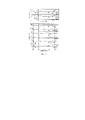
После добавления этой пасты в раствор серной кислоты цвет смеси сразу становился кирпично-красным. Мы отбирали образцы для анализа во время реакции, и, к удивлению, обнаружили, что несколько капель раствора, оставшихся на пипетке, оставались мутными и кирпично-красными в течение, по меньшей мере, нескольких часов при комнатной температуре. Только через два дня смесь превратилась в полностью прозрачный раствор, что указывало на образование сульфата железа. Поэтому мы решили провести аналогичную реакцию с концентрированной серной кислотой (98 мас.%) при более высокой температуре (0-4°С). Реакцию проводили следующим образом: серную кислоту охлаждали на ледяной бане, затем при интенсивном перемешивании к ней добавляли ферригидритную пасту, перемешивали в течение 5 мин, останавливали реакцию резким выливанием смеси в лед. Полученную смесь дважды центрифугировали и промывали водой. Для анализа образца использовали рентгенофазовый анализ. Рентгенограмма, приведенная на Фиг. 1, полностью соответствует фазе чистого гематита α-Fe2O3 (карточка JCPDS: №00-033-0664) без значительных примесей других фаз. Парамагнитная природа полученных частиц была подтверждена методом количественного определения магнитных частиц (MPQ-цитометрия) [Nikitin, М.Р. et al. Nanoscale, 2018], который выявил отсутствие ферро- и суперпарамагнитных фракций на уровне 0,1 нг/мл.After adding this paste to a solution of sulfuric acid, the color of the mixture immediately became brick red. We took samples for analysis during the reaction and, surprisingly, found that a few drops of the solution remaining on the pipette remained cloudy and brick red for at least several hours at room temperature. Only after two days did the mixture turn into a completely clear solution, indicating the formation of ferrous sulfate. Therefore, we decided to carry out a similar reaction with concentrated sulfuric acid (98 wt.%) at a higher temperature (0-4°C). The reaction was carried out as follows: sulfuric acid was cooled in an ice bath, then ferrihydrite paste was added to it with vigorous stirring, stirred for 5 min, and the reaction was stopped by abruptly pouring the mixture into ice. The resulting mixture was centrifuged twice and washed with water. The sample was analyzed by X-ray phase analysis. The radiograph shown in Fig. 1, fully corresponds to the phase of pure hematite α-Fe 2 O 3 (card JCPDS: No. 00-033-0664) without significant impurities of other phases. The paramagnetic nature of the obtained particles was confirmed by the method of quantitative determination of magnetic particles (MPQ cytometry) [Nikitin, M.R. et al. Nanoscale, 2018], which revealed the absence of ferro- and superparamagnetic fractions at the level of 0.1 ng/ml.
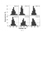
Методом сканирующей электронной микроскопии установлено, что осадок состоит из почти что кубических частиц размером примерно 50 нм (Фиг. 2). Дальнейший анализ с помощью просвечивающей электронной микроскопии (ПЭМ) выявил более сложные формы частиц, которые отличались от кубической и были похожи на описанные ранее в литературе нанокристаллы гематита (Фиг. 2) [Echigo, Т. et al. Am. Mineral., 2013]. Анализ методом энергодисперсионной рентгеновской спектроскопии показал, что наночастицы (НЧ) не содержат никаких следов других элементов. Атомное соотношение Fe:O в образце составляет примерно 1:2,8, что ниже ожидаемого для гематита из-за низкой точности метода при анализе легких элементов (z<11). Таким образом, составы стандартного образца гематита, приготовленного по литературной методике [Schwertmann, U. et al. Schwertmann, U., Cornell, R.M., Eds.; Wiley: Weinheim, Germany, 2000] и приготовленного нами, показали примерно одинаковое соотношение Fe:O.Using scanning electron microscopy, it was found that the precipitate consists of almost cubic particles with a size of approximately 50 nm (Fig. 2). Further analysis using transmission electron microscopy (TEM) revealed more complex particle shapes that differed from the cubic and were similar to previously described hematite nanocrystals in the literature (Fig. 2) [Echigo, T. et al. Am. Mineral., 2013]. Analysis by energy dispersive X-ray spectroscopy showed that nanoparticles (NPs) do not contain any traces of other elements. The atomic ratio of Fe:O in the sample is approximately 1:2.8, which is lower than expected for hematite due to the low accuracy of the method in the analysis of light elements (z<11). Thus, the compositions of a standard sample of hematite prepared according to the literature method [Schwertmann, U. et al. Schwertmann, U., Cornell, R.M., Eds.; Wiley: Weinheim, Germany, 2000] and prepared by us showed approximately the same ratio of Fe:O.
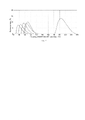
Кристаллическую структуру частиц определяли с использованием анализа электронной дифракции на выбранной площади (режим дифракционного контраста в ПЭМ), результаты показаны на Фиг. 2. Здесь видны яркие кольца, соответствующие большому количеству нанокристаллов гематита, и точечные рефлексы, которые относятся к отдельным частицам гетита. Некоторые из основных параметров решетки гематита или гетита совпадают. Единственный способ отличить эти фазы на электронограммах - это наличие кольца с параметром решетки 0,369 нм, соответствующим плоскости (012) гематита. Это кольцо расположено рядом с центральным пятном луча и несколькими одиночными пятнами со слабой интенсивностью, полученными от плоскостей (101) и (201) гетита (с межплоскостным расстоянием 0,416 и 0,336 нм, соответственно). Таким образом, НЧ состоят из смеси гематита (α-Fe2O3) и следов гетита (α-FeO (ОН)). ИК-спектр полученных НЧ содержит пики, которые идентичны пикам в спектре стандартного образца гематита (Фиг. 3), а также содержит пики при 905 см-1 и 811 см-1, характерные для гидратированных разновидности оксида железа, такие как гетит [RRUFF ID: R050142.1]. Поскольку ИК-спектроскопия в режиме отражения показывает в основном природу поверхности наночастиц и не зависит от кристаллической структуры анализируемого соединения, а также, принимая во внимание результаты РФА, можно предположить, что наночастицы состоят из ядра гематита с тонким слоем в основном аморфного гидратированного оксида железа на поверхности.The crystal structure of the particles was determined using an electron diffraction analysis over a selected area (TEM diffraction contrast mode), the results are shown in FIG. 2. Bright rings are visible here, corresponding to a large number of hematite nanocrystals, and point reflections, which belong to individual goethite particles. Some of the basic lattice parameters of hematite or goethite are the same. The only way to distinguish these phases on electron diffraction patterns is the presence of a ring with a lattice parameter of 0.369 nm corresponding to the (012) hematite plane. This ring is located near the central spot of the beam and several single spots of low intensity obtained from the (101) and (201) planes of goethite (with an interplanar spacing of 0.416 and 0.336 nm, respectively). Thus, NPs consist of a mixture of hematite (α-Fe 2 O 3 ) and traces of goethite (α-FeO (OH)). The IR spectrum of the obtained NPs contains peaks that are identical to the peaks in the spectrum of the hematite standard sample (Fig. 3), and also contains peaks at 905 cm -1 and 811 cm -1 characteristic of hydrated varieties of iron oxide, such as goethite [RRUFF ID : R050142.1]. Since IR spectroscopy in the reflection mode shows mainly the nature of the surface of nanoparticles and does not depend on the crystal structure of the analyzed compound, and also, taking into account the XPA results, it can be assumed that the nanoparticles consist of a hematite core with a thin layer of mainly amorphous hydrated iron oxide on surfaces.
Выход в реакции составил около 5%, поэтому мы изучили возможность его увеличения. Мы протестировали для этой реакции множество концентрированных кислот, чтобы сравнить их способность образовывать наночастицы гематита. В случае концентрированной азотной кислоты аналогичные НЧ образовывались с гораздо меньшим выходом, при этом осадок оставался стабильным в кислоте в течение, по крайней мере, пары дней.The reaction yield was about 5%, so we explored the possibility of increasing it. We tested many concentrated acids for this reaction to compare their ability to form hematite nanoparticles. In the case of concentrated nitric acid, similar NPs were formed in a much lower yield, while the precipitate remained stable in acid for at least a couple of days.
Соляная кислота дает все тот же осадок гематита, который, однако полностью растворяется менее чем за 30 мин. Органические кислоты, такие как чистая муравьиная и ледяная уксусная кислоты, также образовывали небольшие количества НЧ гематита (≈1%). Однако для завершения реакции, за которым следили по изменению цвета от темно-коричневого до кирпично-красного, требовалось 24-часовое перемешивание при комнатной температуре. С другой стороны, концентрированная фосфорная кислота дала результат, аналогичный серной кислоте, с таким же выходом 5%.Hydrochloric acid still gives the same precipitate of hematite, which, however, completely dissolves in less than 30 minutes. Organic acids such as pure formic and glacial acetic acids also formed small amounts of hematite NPs (≈1%). However, 24 hours of stirring at room temperature was required to complete the reaction, followed by a color change from dark brown to brick red. On the other hand, concentrated phosphoric acid gave a similar result to sulfuric acid with the same 5% yield.
Предполагалось, что образование наночастиц гематита зависит не только от силы кислоты, но и от ее дегидратирующей способности. Поэтому при использовании «100% фосфорной кислоты» (известной как равновесная смесь фосфорной кислоты, пирофосфорной кислоты и воды) выход НЧ был почти вдвое выше (9%). Такой же результат (10%) был получен при использовании смеси серной кислоты и пятиокиси фосфора. Таким образом, сильные кислотные дегидратирующие агенты способны превращать ферригидрит в менее гидратированные оксиды железа, а не растворять их. Поскольку ферригидритная паста, использованная в экспериментах, содержала воду, которая могла привести к разбавлению кислоты и снижению ее дегидратирующей способности, в одном из экспериментов воду заменили этанолом, что, однако не привело к увеличению выхода НЧ. Наконец, был проведен эксперимент по масштабированию методики с использованием примерно 10-кратных количеств исходных реагентов и смеси серной кислоты и пятиокиси фосфора. Были получены те же НЧ с выходом 9,7% (0,51 г).It was assumed that the formation of hematite nanoparticles depends not only on the strength of the acid, but also on its dehydrating ability. Therefore, when using "100% phosphoric acid" (known as an equilibrium mixture of phosphoric acid, pyrophosphoric acid and water), the NP yield was almost twice as high (9%). The same result (10%) was obtained using a mixture of sulfuric acid and phosphorus pentoxide. Thus, strong acidic dehydrating agents are able to convert ferrihydrite to less hydrated iron oxides rather than dissolve them. Since the ferrihydrite paste used in the experiments contained water, which could dilute the acid and reduce its dehydrating ability, in one of the experiments water was replaced by ethanol, which, however, did not lead to an increase in the yield of NPs. Finally, an experiment was performed to scale up the procedure using approximately 10-fold amounts of the starting reagents and a mixture of sulfuric acid and phosphorus pentoxide. The same NPs were obtained with a yield of 9.7% (0.51 g).
Чтобы оценить потенциал полученных наночастиц гематита для биомедицинских применений, поверхность наночастиц была покрыта несколькими видами полимеров для облегчения последующего биоконъюгирования. Полимеры с карбоксильными группами, такие как натриевая соль карбоксиметилдекстрана (КМД, Μn ~ 15000), натриевая соль полиакриловой кислоты (ПАК, Μn ~ 5100), а также полиэтиленимин (ПЭИ, Μn ~ 25000), содержащий аминогруппы, были иммобилизованы на поверхности частиц с помощью простой процедуры инкубации с избытком полимеров в горячих водных растворах. Гидродинамические радиусы полученных таким образом НЧ, определенные методом динамического рассеяния света (ДРС), составили 65±9 нм, 62±8 нм и 76±8 нм для ГНЧ@КМД, ГНЧ@ПАК и ГНЧ@ПЭИ, соответственно. Радиусы оказались немного меньше по сравнению с 90±8 нм для непокрытых ГНЧ, что говорит о более высокой стабилизации НЧ полимерами и, как следствие, более низкой агрегации. Присутствие органического покрытия было также подтверждено с помощью ИК-спектроскопии, результаты показаны на Фиг.3. В случае покрытия КМД и ПАК характерные полосы карбоксильных групп КМД (1618 см-1 и 1560 см-1) и ПАК (1556 см-1 и 1601 см-1) уширены и немного сдвинуты из-за наличия неэквивалентных групп, так как только часть карбоксильных групп скоординирована с поверхностью оксида железа. Аналогичным образом, более сложная картина наблюдалась для ПЭИ после координации с поверхностью НЧ.To evaluate the potential of the obtained hematite nanoparticles for biomedical applications, the surface of the nanoparticles was coated with several types of polymers to facilitate subsequent bioconjugation. Polymers with carboxyl groups, such as sodium carboxymethyldextran (CMD, Μn ~ 15000), sodium salt of polyacrylic acid (PAA, Μn ~ 5100), and polyethyleneimine (PEI, Μn ~ 25000) containing amino groups, were immobilized on the surface of particles with using a simple incubation procedure with excess polymers in hot aqueous solutions. The hydrodynamic radii of the NPs thus obtained, determined by the dynamic light scattering (DLS) method, were 65±9 nm, 62±8 nm, and 76±8 nm for the HFO@CMD, HFO@PAA, and HFO@PEI, respectively. The radii turned out to be slightly smaller compared to 90 ± 8 nm for uncoated HNPs, which indicates a higher stabilization of NPs by polymers and, as a result, lower aggregation. The presence of an organic coating was also confirmed by IR spectroscopy, the results are shown in Fig.3. In the case of the CMD and PAA coating, the characteristic bands of the carboxyl groups of CMD (1618 cm -1 and 1560 cm -1 ) and PAA (1556 cm -1 and 1601 cm -1 ) are broadened and slightly shifted due to the presence of nonequivalent groups, since only a part carboxyl groups is coordinated with the iron oxide surface. Similarly, a more complex picture was observed for PEI after coordination with the NP surface.
Возможность функционализации наночастиц с помощью антител была исследована методом проточной цитометрии и флуоресцентного иммуноанализа, как показано на Фиг.4. Эритроциты являются удобной моделью для разработки агентов для доставки лекарств, поскольку эти клетки лишены эндоцитарного аппарата; таким образом, на анализ взаимодействия мембрана-ГНЧ не влияет поглощение [Shang, L. et al. J. Nanobiotechnol., 2014]. Установлено, что наночастицы, модифицированные положительно заряженным ПЭИ, имеют тенденцию неспецифически связываться с клетками, в то время как частицы, покрытые КМД и ПАК не проявляют подобных свойств. Поэтому ГНЧ, покрытые КМД и ПАК, были сконъюгированы карбодиимидным методом [Hermanson, G.T. Academic Press: New York, NY, USA, 2008] с моноклональным антителом TER-119 (TER), которое распознает эритроид-специфические антигены TER-119. Полученные ГНЧ@КМД@TER и ГНЧ@ПАК@TER были помечены флуоресцентным красителем Су3 путем обработки НЧ н-гидроксисульфосукцинимидным эфиром Су3 (Су3-sulfo-NHS). Эффективность покрытия частиц антителами и флуоресцентными метками оценивали с помощью прямого флуоресцентного иммуноанализа [Luppa, Р.В. et al. Clin. Chim. Acta, 2001]. В этом эксперименте козий антимышиный иммуноглобулин (Ig), общий Ig человека и бычий сывороточный альбумин (БСА) адсорбировались на полистирольной поверхности 96-луночного планшета с последующим добавлением ГНЧ@КМД@TER-Су3 и ГНЧ@ПАК@TER-Cy3. После тщательной промывки эффективность флуоресцентного мечения оценивали по флуоресцентному сигналу (Фиг. 4), что подтвердило успешную модификацию обоих типов ГНЧ, которые специфически связывались только с козьим антимышиным Ig. Для оценки неспецифического взаимодействия ГНЧ ГНЧ@КМД@TER-Су3 и ГНЧ@ПАК@TER-Су3 вместе с ГНЧ, сконъюгированными с несвязывающими эритроциты антителами в качестве отрицательного контроля, использовали для изучения с помощью метода проточной цитометрии с визуализацией (Фиг. 4). Результаты продемонстрировали более высокую специфичность ГНЧ, модифицированных ПАК, к эритроцитам, чем ГНЧ, модифицированные КМД. Кроме того, специфичность TER-конъюгированных ГНЧ была подтверждена в конкурентном анализе с использованием свободных TER антител вместе с ГНЧ в растворе во время инкубации с клетками. Высокая эффективность связывания TER-конъюгированных НЧ с эритроцитами также подтверждается изображениями, полученными методом визуализирующей проточной цитометрии (Фиг. 4). Таким образом, TER-модифицированные ГНЧ являются перспективными кандидатами для успешной доставки лекарств методом RBC-hitchhiking при лечении рака [Sokolov, I.L. et al. Biochim. Biophys. Acta Gen. Subj., 2017].The possibility of functionalization of nanoparticles with antibodies was investigated by flow cytometry and fluorescent immunoassay, as shown in Fig.4. RBCs are a convenient model for the development of drug delivery agents because these cells lack an endocytic apparatus; thus, the analysis of the membrane-HNP interaction is not affected by uptake [Shang, L. et al. J. Nanobiotechnol., 2014]. It has been established that nanoparticles modified with positively charged PEI tend to nonspecifically bind to cells, while particles coated with CMD and PAA do not exhibit such properties. Therefore, HNPs coated with CMD and PAA were conjugated by the carbodiimide method [Hermanson, G.T. Academic Press: New York, NY, USA, 2008] with the TER-119 monoclonal antibody (TER), which recognizes TER-119 erythroid-specific antigens. The obtained HNP@CMD@TER and HNP@PAA@TER were labeled with Cy3 fluorescent dye by treating nanoparticles with Cy3 n-hydroxysulfosuccinimide ester (Cy3-sulfo-NHS). The efficiency of particle coating with antibodies and fluorescent labels was evaluated using a direct fluorescent immunoassay [Luppa, R.V. et al. Clin. Chim. Acta, 2001]. In this experiment, goat anti-mouse immunoglobulin (Ig), total human Ig and bovine serum albumin (BSA) were adsorbed onto the polystyrene surface of a 96-well plate followed by the addition of HNP@CMD@TER-Cy3 and HNP@PAA@TER-Cy3. After thorough washing, the efficiency of fluorescent labeling was evaluated by the fluorescent signal (FIG. 4), which confirmed the successful modification of both types of LNPs, which specifically bind only to goat anti-mouse Ig. To assess the non-specific interaction of HNPs, HNP@CMD@TER-Cy3 and HNP@PAA@TER-Cy3, together with LNP conjugated with non-erythrocyte-binding antibodies as a negative control, were used for study by imaging flow cytometry (FIG. 4). The results demonstrated a higher specificity of PAA-modified HNPs for erythrocytes than CMD-modified HNPs. In addition, the specificity of TER-conjugated HNPs was confirmed in a competitive assay using free TER antibodies together with HNPs in solution during cell incubation. The high binding efficiency of TER-conjugated NPs to erythrocytes is also confirmed by images obtained by imaging flow cytometry (Fig. 4). Thus, TER-modified HNPs are promising candidates for successful drug delivery by RBC-hitchhiking in cancer treatment [Sokolov, I.L. et al. biochim. Biophys. Acta Gen. Subj., 2017].
Далее была продемонстрирована высокая специфичность полученных агентов для таргетинга раковых клеток (Фиг. 4). В качестве модели для изучения таргетинга мы выбрали клетки ВТ-474, оверэкспрессирующие рецептор эпидермального фактора роста HER2/neu, очень важный клинический маркер рака, который оверэкспрессируется на многих типах злокачественных опухолей у человека [Yan, Μ. et al. Cancer Metastasis Rev., 2015]. Клетки CHO использовали в качестве HER2/neu-отрицательного контроля. Мы использовали антитело к HER2/neu трастузумаб, которое было сконъюгировано с ГНЧs@ПАК и помечено Су3. Как видно из фигуры, наночастицы, сконъюгированные с трастузумабом, специфически связываются с клетками ВТ-474. Таким образом, НЧ показали высокую специфичность к рецептору HER2/neu и могут быть использованы для таргетинга раковых клеток. Соответственно, у нас есть возможность разработать наноагенты для различных применений (например, для биосенсорики или удаления токсичных агентов) посредством функционализации соответствующими антителами.Further, the high specificity of the obtained agents for targeting cancer cells was demonstrated (Fig. 4). As a targeting model, we chose BT-474 cells, which overexpress the epidermal growth factor receptor HER2/neu, a very important clinical marker of cancer that is overexpressed in many types of human cancers [Yan, M. et al. Cancer Metastasis Rev., 2015]. CHO cells were used as HER2/neu negative controls. We used the anti-HER2/neu antibody trastuzumab, which was conjugated to HNPs@PAK and labeled with Cy3. As can be seen from the figure, the trastuzumab-conjugated nanoparticles specifically bind to BT-474 cells. Thus, NPs showed high specificity for the HER2/neu receptor and can be used to target cancer cells. Accordingly, we have the opportunity to develop nanoagents for various applications (for example, for biosensors or the removal of toxic agents) through functionalization with appropriate antibodies.
Также мы сконъюгировали ГНЧ@ ПАК с общим человеческим иммуноглобулином IgG и пометили конъюгат с помощью Cy7.5-sulfo-NHS (ГНЧ@ПАК @IgG-Cy7.5) и изучили его биораспределение. НЧ вводили мышам, и после 40-минутной инкубации извлеченные органы мыши исследовали с использованием инфракрасной оптической визуализации (Фиг. 5). Флуоресцентный анализ показал, что почти все НЧ накапливались в печени. Этот результат можно объяснить существованием ассоциированной с печенью субпопуляции макрофагов, которая поглощает НЧ. Этот факт позволяет сделать вывод, что НЧ являются подходящим агентом для адресной доставки лекарств благодаря своему незначительному неспецифическому накоплению НЧ почти во всех органах (кроме печени). Дальнейшие исследования будут сосредоточены на рецептор-специфичной доставке НЧ в опухоли и на адсорбции агентов для химиотерапии или фотодинамической терапии на поверхности НЧ.We also conjugated HNP@PAK with total human IgG and labeled the conjugate with Cy7.5-sulfo-NHS (HLN@PAK@IgG-Cy7.5) and studied its biodistribution. NPs were administered to mice, and after a 40 minute incubation, the extracted mouse organs were examined using infrared optical imaging (FIG. 5). Fluorescent analysis showed that almost all NPs accumulated in the liver. This result can be explained by the existence of a liver-associated macrophage subpopulation that engulfs NPs. This fact allows us to conclude that NPs are a suitable agent for targeted drug delivery due to their insignificant nonspecific accumulation of NPs in almost all organs (except the liver). Further studies will focus on the receptor-specific delivery of NPs to tumors and adsorption of agents for chemotherapy or photodynamic therapy on the surface of NPs.
Удивительно, но ожидаемое полное растворение гидратированного оксида с образованием растворимых солей произошло только в небольшом количестве случаев. Реакция оказалась более сложной, она протекает только с частичным растворением с параллельным образованием наночастиц гематита. Выход НЧ возрастает с увеличением дегидратирующей способности смеси кислот. Реакцию можно легко масштабировать для получения значительных количеств наночастиц с выходом до 10%. В нашей работе полученные наночастицы были модифицированы различными органическими полимерами и использованы для успешного таргетинга in vitro раковых клеток после биомодификации. Таким образом, описанные НЧ являются перспективным материалом для разработки функционализированных наноагентов для различных биомедицинских приложений.Surprisingly, the expected complete dissolution of the hydrated oxide to form soluble salts occurred only in a small number of cases. The reaction turned out to be more complex; it proceeds only with partial dissolution with the parallel formation of hematite nanoparticles. The yield of NPs increases with an increase in the dehydrating capacity of the mixture of acids. The reaction can be easily scaled up to obtain significant amounts of nanoparticles with a yield of up to 10%. In our work, the resulting nanoparticles were modified with various organic polymers and used for successful in vitro targeting of cancer cells after biomodification. Thus, the described NPs are a promising material for the development of functionalized nanoagents for various biomedical applications.
Предложенный способ предпочтительно реализовывать при использовании высокого содержания кислоты, это позволяет существенно сократить время синтеза частиц гематита. Предпочтительно достигать концентрации кислоты в смеси (комбинации) с частицами ферригидрита, более 30%, предпочтительно более 40%, предпочтительно более 50%, предпочтительно более 60%, предпочтительно более 70%, предпочтительно более 80%, предпочтительно более 90%, предпочтительно более 95% от концентрации концентрированной кислоты. Под концентрированной кислотой понимают стандартную в области техники максимальную концентрацию кислоты, например:The proposed method is preferably implemented using a high acid content, which can significantly reduce the time of synthesis of hematite particles. It is preferable to achieve an acid concentration in the mixture (combination) with ferrihydrite particles of more than 30%, preferably more than 40%, preferably more than 50%, preferably more than 60%, preferably more than 70%, preferably more than 80%, preferably more than 90%, preferably more than 95 % of concentrated acid concentration. By concentrated acid is meant the maximum acid concentration standard in the art, for example:
В частности, в одних воплощениях изобретения финальная концентрация кислот (в моль/л) (по крайней мере не менее): Серная кислота 18.3, Азотная кислота 15.7, Соляная кислота (40 мас.%) 13.1, Фосфорная кислота 14.6, Уксусная кислота (ледяная) 17.4, Муравьиная кислота 26.5.In particular, in some embodiments of the invention, the final concentration of acids (in mol / l) (at least not less than): Sulfuric acid 18.3, Nitric acid 15.7, Hydrochloric acid (40 wt.%) 13.1, Phosphoric acid 14.6, Acetic acid (glacial ) 17.4, Formic acid 26.5.
Работа предлагаемого способа иллюстрируется чертежами фиг. 1-7.The operation of the proposed method is illustrated in Fig. 1-7.
Список фигур чертежей.List of drawing shapes.
Фиг. 1 - Рентгенограмма полученных наночастиц и эталонный образец.Fig. 1 - X-ray pattern of the obtained nanoparticles and a reference sample.
Фиг. 2 - Фото образцов НЧ, полученных с помощью сканирующей электронной микроскопии; просвечивающей электронной микроскопии; в режиме дифракционного контраста (электронограмма на выбранных участках наночастиц).Fig. 2 - Photo of NP samples obtained using scanning electron microscopy; transmission electron microscopy; in the diffraction contrast mode (electron diffraction pattern on selected areas of nanoparticles).
Фиг. 3 - Спектры, полученные методом ИК-спектроскопии при ослабленном полном отражении. Сравнение спектра наночастиц гематита (ГНЧ) со спектром стандартного образца; спектры ГНЧ с полимерным покрытием в диапазоне 770-3200 см-1 в сравнении со спектрами чистых полимеров.Fig. 3 - Spectra obtained by IR spectroscopy with attenuated total reflection. Comparison of the spectrum of hematite nanoparticles (HNP) with the spectrum of a standard sample; spectra of polymer-coated HNPs in the range 770-3200 cm-1 in comparison with the spectra of pure polymers.
Фиг. 4. Флуоресцентный иммуноанализ и визуализирующая проточная цитометрия. Связывание ГНЧ@ПАК@TER-Су3 и ГНЧ@КМД@TER-Су3, соответственно, с общим иммуноглобулином человека (IgG), бычьим сывороточным альбумином (БСА) и антителами козы к иммуноглобулинам крысы, адсорбированных на полистироле; данные представлены как среднее ± стандартное отклонение (n=3). Изучение методом визуализирующий проточной цитометрии взаимодействия между ГНЧ@ПАК@TER-Су3 и ГНЧ@КМД@TER-Су3 с эритроцитами; в качестве контроля была произведена инкубация клеток с частицами в смеси, содержащей моноклональные TER-119 антитела (TER), кроме того, была изучена специфичность частиц, несущих неспецифичные к клеткам антитела (КА - контрольные антитела); проточно-цитометрический анализ взаимодействия клеток ВТ-474 (HER2/neu-положительные) и СНО (HER2/neu-отрицательные) с ГНЧ@ПАК@Трастузумаб-Су3. Изображения взаимодействия между эритроцитами и двумя типами наночастиц, покрытых натриевой солью полиакриловой кислоты (ПАК), конъюгированных с эритроцит-связывающими и эритроцит-несвязывающими антителами (в светлом поле, Су3-канал и канал бокового рассеяния). Масштаб - 10 мкм. Статистическая значимость была рассчитана с использованием непарного одностороннего t-критерия (* p<0,05; ** p<0,01; *** p<0,001; n.s. р>0,05).Fig. 4. Fluorescence immunoassay and imaging flow cytometry. Binding of HNP@PAK@TER-Cy3 and HNP@CMD@TER-Cy3, respectively, to total human immunoglobulin (IgG), bovine serum albumin (BSA) and goat antibodies to rat immunoglobulins adsorbed on polystyrene; data are presented as mean ± standard deviation (n=3). Study by visualizing flow cytometry of the interaction between HNP@PAK@TER-Cy3 and HNP@CMD@TER-Cy3 with erythrocytes; as a control, cells were incubated with particles in a mixture containing monoclonal TER-119 antibodies (TER), in addition, the specificity of particles carrying non-cell-specific antibodies (KA - control antibodies) was studied; flow cytometric analysis of the interaction of BT-474 (HER2/neu-positive) and CHO (HER2/neu-negative) cells with HNP@PAK@Trastuzumab-Cy3. Images of the interaction between erythrocytes and two types of polyacrylic acid (PAA) sodium salt-coated nanoparticles conjugated with erythrocyte-binding and erythrocyte-non-binding antibodies (bright field, Cy3 channel and side scatter channel). Scale - 10 µm. Statistical significance was calculated using an unpaired one-tailed t-test (*p<0.05; **p<0.01; ***p<0.001; n.s. p>0.05).
Фиг. 5. Флуоресцентное ex vivo изображение органов мыши, которой были вколоты флуоресцентно меченные частицы гематита.Fig. 5. Ex vivo fluorescence image of the organs of a mouse injected with fluorescently labeled hematite particles.
Фиг. 6. Распределение по размерам НЧ, синтезированных с использованием различных кислот слева направо сверху вниз: серная кислота; азотная кислота; уксусная кислота; фосфорная кислота; соляная кислота; смесь серной кислоты и пятиокиси фосфора (размеры определены с использованием изображений СЭМ; размер каждой НЧ рассчитывался как среднее от ее максимального и минимального размера. Значения представлены как среднее ± стандартное отклонение (n=100); изображения обрабатывались с помощью программы Fiji/ImageJ).Fig. Fig. 6. Size distribution of NPs synthesized using various acids from left to right from top to bottom: sulfuric acid; Nitric acid; acetic acid; phosphoric acid; hydrochloric acid; a mixture of sulfuric acid and phosphorus pentoxide (sizes determined using SEM images; the size of each NP was calculated as the average of its maximum and minimum sizes. Values are presented as the mean ± standard deviation (n=100); images were processed using the Fiji/ImageJ program).
Фиг. 7. Гидродинамические радиусы синтезированных наночастиц согласно данным ДРС-анализа слева направо: ГНЧ@ПАК, ГНЧ@КМД, ГНЧ@ПЭИ, ГНЧ@ПАК@TER, ГНЧ, ГНЧ@КМД@TER.Fig. Fig. 7. Hydrodynamic radii of the synthesized nanoparticles according to the DLS analysis data from left to right: LNP@PAA, LNP@CMD, LNP@PEI, LNP@PAA@TER, LNP, LNP@CMD@TER.
ПРИМЕРЫEXAMPLES
Варианты реализации изобретения разнообразны. Приведем различные примеры. Нижеприведенные примеры даны в качестве иллюстрации данного изобретения и не ограничивают его применения.Embodiments of the invention are varied. We give various examples. The following examples are given by way of illustration of the present invention and do not limit its application.
Пример 1. Синтез ферригидритаExample 1 Synthesis of ferrihydrite
100 мл 0,33 Μ водного раствора гексагидрата хлорида железа (17,8 г твердого FeCl3⋅6Н2О) смешивали с 25 мл водного раствора аммиака (25 мас.%). Реакционную смесь перемешивали при 90°С в течение 2 ч, центрифугировали при 1000 × g в течение 1 мин, промывали 3 раза водой Milli-Q с последующим центрифугированием при 1000 × g с получением ферригидритной пасты (концентрация сухого содержимого составляла 20 мг/мл), которая затем использовалась в синтезе наночастиц гематита.100 ml of a 0.33 Μ aqueous solution of ferric chloride hexahydrate (17.8 g of solid FeCl 3 ⋅ 6H 2 O) was mixed with 25 ml of an aqueous solution of ammonia (25 wt.%). The reaction mixture was stirred at 90°C for 2 h, centrifuged at 1000×g for 1 min, washed 3 times with Milli-Q water, followed by centrifugation at 1000×g to obtain a ferrihydrite paste (dry content concentration was 20 mg/mL) , which was then used in the synthesis of hematite nanoparticles.
Пример 2. Синтез наночастиц гематита с использованием серной кислотыExample 2. Synthesis of hematite nanoparticles using sulfuric acid
10 мл концентрированной серной кислоты охлаждали на ледяной бане в стакане объемом 50 мл с Х-образной мешалкой, затем при интенсивном перемешивании добавляли 3 мл пасты ферригидрита (концентрация сухого содержимого составляла 20 мг/мл), через 5 минут реакцию останавливали, добавляя 100 мл льда. Объем ферригидритной пасты измеряли с помощью шприца с открытым горлом (диаметром 1 см). Центрифугирование при 2300 × g с последующим повторным промыванием холодной водой Milli-Q привело к получению осадка кирпично-красного цвета, который промывали этанолом, диэтиловым эфиром и сушили в течение ночи при комнатной температуре.10 ml of concentrated sulfuric acid was cooled in an ice bath in a 50 ml beaker with an X-shaped stirrer, then 3 ml of ferrihydrite paste was added with vigorous stirring (dry content concentration was 20 mg/ml), after 5 minutes the reaction was stopped by adding 100 ml of ice . The volume of the ferrihydrite paste was measured using an open-neck syringe (1 cm in diameter). Centrifugation at 2300×g followed by repeated washing with cold Milli-Q water resulted in a brick red precipitate, which was washed with ethanol, diethyl ether and dried overnight at room temperature.
Пример 3. Синтез наночастиц гематита с использованием смеси серной кислоты и пентоксида фосфора.Example 3. Synthesis of hematite nanoparticles using a mixture of sulfuric acid and phosphorus pentoxide.
Смесь серной кислоты и Р4О10 получали растворением 5 г Р4О10 в 10 мл 98%-ной серной кислоты. Полученную смесь охлаждали на ледяной бане в стакане объемом 50 мл с Х-образной мешалкой, затем при интенсивном перемешивании добавляли 3 мл пасты ферригидрита (концентрация сухого содержимого составляла 20 мг/мл), через 5 минут реакцию останавливали, добавляя 100 мл льда. Объем ферригидритной пасты измеряли с помощью шприца с открытым горлом (диаметром 1 см). Центрифугирование при 2300 × g с последующим повторным промыванием холодной водой Milli-Q привело к получению осадка кирпично-красного цвета, который промывали этанолом, диэтиловым эфиром и сушили в течение ночи при комнатной температуре.A mixture of sulfuric acid and P 4 O 10 was prepared by dissolving 5 g of P 4 O 10 in 10 ml of 98% sulfuric acid. The resulting mixture was cooled in an ice bath in a 50 ml beaker with an X-shaped stirrer, then 3 ml of ferrihydrite paste (dry content concentration was 20 mg/ml) was added with vigorous stirring, after 5 minutes the reaction was stopped by adding 100 ml of ice. The volume of the ferrihydrite paste was measured using an open-neck syringe (1 cm in diameter). Centrifugation at 2300×g followed by repeated washing with cold Milli-Q water resulted in a brick red precipitate, which was washed with ethanol, diethyl ether and dried overnight at room temperature.
Пример 4. Синтез наночастиц гематита с использованием муравьиной кислоты.Example 4. Synthesis of hematite nanoparticles using formic acid.
10 мл муравьиной кислоты охлаждали на ледяной бане в стакане объемом 50 мл с Х-образной мешалкой, затем при интенсивном перемешивании добавляли 3 мл пасты ферригидрита (концентрация сухого содержимого составляла 20 мг/мл), через 24 часа перемешивания при комнатной температуре реакцию останавливали, добавляя 100 мл льда. Объем ферригидритной пасты измеряли с помощью шприца с открытым горлом (диаметром 1 см). Центрифугирование при 2300 × g с последующим повторным промыванием холодной водой Milli-Q привело к получению осадка кирпично-красного цвета, который промывали этанолом, диэтиловым эфиром и сушили в течение ночи при комнатной температуре.10 ml of formic acid was cooled in an ice bath in a 50 ml beaker with an X-shaped stirrer, then 3 ml of ferrihydrite paste was added with vigorous stirring (dry content concentration was 20 mg/ml), after 24 hours of stirring at room temperature, the reaction was stopped by adding 100 ml ice. The volume of the ferrihydrite paste was measured using an open-neck syringe (1 cm in diameter). Centrifugation at 2300×g followed by repeated washing with cold Milli-Q water resulted in a brick red precipitate, which was washed with ethanol, diethyl ether and dried overnight at room temperature.
Пример 5. Синтез наночастиц гематита покрытых полиэтиленимином.Example 5. Synthesis of hematite nanoparticles coated with polyethyleneimine.
10 мг сухих наночастиц гематита обрабатывали ультразвуком с 1 мл 0,1 Μ HCl, промывали водой Milli-Q с последующим трехкратным центрифугированием и смешивали с 2 мл 20 мас.% раствора полиэтиленимина (Μn ~ 25000, >99% чистоты). Смесь инкубировали в течение 5 ч при 95°С, промывали водой Milli-Q с последующим трехкратным центрифугированием и ресуспендировали в воде Milli-Q для с получением раствора модифицированных частиц. Наличие полимера на поверхности проверяли с помощью инфракрасной спектроскопии.10 mg of dry hematite nanoparticles were sonicated with 1 ml of 0.1 Μ HCl, washed with Milli-Q water followed by three centrifugations, and mixed with 2 ml of 20 wt% polyethyleneimine solution (Μn ~ 25,000, >99% pure). The mixture was incubated for 5 hours at 95° C., washed with Milli-Q water followed by centrifugation three times, and resuspended in Milli-Q water to obtain a modified particle solution. The presence of the polymer on the surface was checked using infrared spectroscopy.
Пример 6. Синтез наночастиц гематита покрытых полиакриловой кислотой.Example 6. Synthesis of hematite nanoparticles coated with polyacrylic acid.
10 мг сухих наночастиц гематита обрабатывали ультразвуком с 1 мл 0,1 Μ HCl, промывали водой Milli-Q с последующим трехкратным центрифугированием и смешивали с 2 мл 20 мас. % раствора полиакриловой кислоты (Μn ~ 5100). Смесь инкубировали в течение 5 ч при 95°С, промывали водой Milli-Q с последующим трехкратным центрифугированием и ресуспендировали в воде Milli-Q для с получением раствора модифицированных частиц. Наличие полимера на поверхности проверяли с помощью инфракрасной спектроскопии.10 mg of dry hematite nanoparticles were sonicated with 1 ml of 0.1 Μ HCl, washed with Milli-Q water followed by three centrifugations, and mixed with 2 ml of 20 wt. % solution of polyacrylic acid (Μn ~ 5100). The mixture was incubated for 5 hours at 95° C., washed with Milli-Q water followed by centrifugation three times, and resuspended in Milli-Q water to obtain a modified particle solution. The presence of the polymer on the surface was checked using infrared spectroscopy.
Пример 7. Синтез специфичных наночастиц гематита покрытых полиакриловой кислотой и флуоресцентно-меченными белками.Example 7. Synthesis of specific hematite nanoparticles coated with polyacrylic acid and fluorescently labeled proteins.
Белки конъюгировали с наночастицами, покрытыми полиакриловой кислотой следующим образом 600 мкг покрытых полиакриловой кислотой ГНЧ инкубировали в 40 мкл буфера MES (рН=5,0) в течение 30 мин с 7 мг ЭДК и 3,5 мг sulfo-NHS. Затем ГНЧ центрифугировали и промывали MES. Наконец, ГНЧ промывали водой Milli-Q и смешивали с 40 мкл раствора белка 1 мг/мл в фосфатно-солевом буфере. Смесь инкубировали в течение ночи при комнатной температуре. Наконец, ГНЧ центрифугировали и трижды промывали фосфатно-солевым буфером. Чтобы прикрепить флуоресцентную метку Су3 к НЧ, 600 мкг ГНЧ, сконъюгированных с белком, диспергировали в 50 мкл 0,1% раствора БСА в фосфатно-солевом буфере. Раствор смешивали с 17 мкг Су3-сульфо-NHS в 50 мкл фосфатно-солевого буфера и инкубировали в течение 4 ч при комнатной температуре с получением целевых флуоресцентно-меченных частиц с белком на поверхности.Proteins were conjugated to polyacrylic acid-coated nanoparticles as follows: 600 µg of polyacrylic acid-coated HNPs were incubated in 40 µl of MES buffer (pH=5.0) for 30 min with 7 mg of EDA and 3.5 mg of sulfo-NHS. The HNPs were then centrifuged and washed with MES. Finally, HNPs were washed with Milli-Q water and mixed with 40 µl of a 1 mg/ml protein solution in phosphate buffered saline. The mixture was incubated overnight at room temperature. Finally, the HNPs were centrifuged and washed three times with phosphate-buffered saline. To attach the Cy3 fluorescent label to NPs, 600 μg of protein-conjugated HNPs were dispersed in 50 μl of 0.1% BSA in PBS. The solution was mixed with 17 μg of Cy3-sulfo-NHS in 50 μl of phosphate-buffered saline and incubated for 4 h at room temperature to obtain the target fluorescently labeled particles with protein on the surface.
Пример 8. Синтез специфичных наночастиц гематита покрытых карбоксиметилдекстраном (КМД) и флуоресцентно-меченными белками.Example 8. Synthesis of specific hematite nanoparticles coated with carboxymethyl dextran (CMD) and fluorescently labeled proteins.
Белки конъюгировали с наночастицами, покрытыми КМД следующим образом 600 мкг покрытых полиакриловой кислотой ГНЧ инкубировали в 40 мкл буфера MES (рН=5,0) в течение 30 мин с 7 мг ЭДК и 3,5 мг sulfo-NHS. Затем ГНЧ центрифугировали и промывали MES. Наконец, ГНЧ промывали водой Milli-Q и смешивали с 40 мкл раствора белка 1 мг/мл в фосфатно-солевом буфере. Смесь инкубировали в течение ночи при комнатной температуре. Наконец, ГНЧ центрифугировали и трижды промывали фосфатно-солевым буфером. Чтобы прикрепить флуоресцентную метку Су3 к НЧ, 600 мкг ГНЧ, сконъюгированных с белком, диспергировали в 50 мкл 0,1% раствора БСА в фосфатно-солевом буфере. Раствор смешивали с 17 мкг Су3-сульфо-NHS в 50 мкл фосфатно-солевого буфера и инкубировали в течение 4 ч при комнатной температуре с получением целевых флуоресцентно-меченных частиц с белком на поверхности.Proteins were conjugated to CMD-coated nanoparticles as follows: 600 µg of polyacrylic acid-coated HNPs were incubated in 40 µl of MES buffer (pH=5.0) for 30 min with 7 mg of EDA and 3.5 mg of sulfo-NHS. The HNPs were then centrifuged and washed with MES. Finally, HNPs were washed with Milli-Q water and mixed with 40 µl of a 1 mg/ml protein solution in phosphate buffered saline. The mixture was incubated overnight at room temperature. Finally, the HNPs were centrifuged and washed three times with phosphate-buffered saline. To attach the Cy3 fluorescent label to NPs, 600 μg of protein-conjugated HNPs were dispersed in 50 μl of 0.1% BSA in PBS. The solution was mixed with 17 μg of Cy3-sulfo-NHS in 50 μl of phosphate-buffered saline and incubated for 4 h at room temperature to obtain the target fluorescently labeled particles with protein on the surface.
Пример 9. Частицы гематита для специфического таргетинга клеток-мишеней.Example 9 Hematite Particles for Specific Targeting of Target Cells.
Свежесобранные эукариотические клетки СНО (0,25 миллиона), ВТ-474 (0,25 миллиона) и эритроциты (конечная концентрация 0,5%) инкубировали в 50 мкл фосфатно-солевого буфера с 3 мкг покрытых полимером ГНЧ или 10 мкг ГНЧ, покрытых конъюгированным с антителом (TER-119 против эритроцитов или Герцептин против ВТ-474), в течение 10 мин и дважды промывали фосфатно-солевым буфером. Флуоресцентное окрашивание клеток не использовали, чтобы минимально повлиять на взаимодействия клетки с наночастицами. Эритроциты изучали с помощью проточного цитометра ImageStream X Mark II (Luminex Corporation, Остин, Техас, США) с использованием 488-нм (50 мВт) лазера для возбуждения флуоресценции. Герцептин использовали в качестве отрицательного контроля в экспериментах с эритроцитами. Клетки СНО и ВТ-474 исследовали на проточном цитометре CellStream (Luminex Corporation) с использованием лазера 561 нм (1 мВт). Для ImageStream мы использовали рекомендованную производителем стратегию гейтирования.Freshly harvested eukaryotic CHO cells (0.25 million), BT-474 (0.25 million), and erythrocytes (final concentration 0.5%) were incubated in 50 μl of phosphate-buffered saline with 3 μg of polymer-coated HNPs or 10 μg of HNPs coated with conjugated with an antibody (TER-119 against erythrocytes or Herceptin against BT-474) for 10 min and washed twice with phosphate-buffered saline. Fluorescent staining of cells was not used in order to minimally affect cell interactions with nanoparticles. Erythrocytes were examined using an ImageStream X Mark II flow cytometer (Luminex Corporation, Austin, TX, USA) using a 488 nm (50 mW) laser to excite fluorescence. Herceptin was used as a negative control in experiments with erythrocytes. CHO and BT-474 cells were examined on a CellStream flow cytometer (Luminex Corporation) using a 561 nm (1 mW) laser. For ImageStream, we used the manufacturer's recommended gating strategy.
Пример 10. Флуоресцентный иммуноанализ.Example 10 Fluorescence immunoassay.
60 мкл раствора белка (антитела против иммуноглобулина человека, иммуноглобулины крысы), с концентрацией 10 мкг/мл в фосфатно-солевом буфере помещали в лунку 96-луночного полистирольного планшета и инкубировали в течение 8 ч при 4°С. Затем раствор заменяли 60 мкл 1% раствора БСА в фосфатно-солевом буфере (блокирующий буфер). Через 30 мин при комнатной температуре раствор удаляли, а лунки промывали 100 мкл фосфатно-солевого буфера. После этого в лунки добавляли 60 мкл блокирующего буфера с различными концентрациями ГНЧ, конъюгированных с Су3-мечеными белками (TER-119, общий иммуноглобулин человека, БСА или антитела козы к иммуноглобулинам крысы), и инкубировали в течение 30 мин при комнатной температуре. Наконец, лунки пять раз промывали 100 мкл фосфатно-солевого буфера, и флуоресценцию, вызванную Су3, измеряли с помощью ридера для микропланшетов ClarioStar (BMG LABTECH, Дарем, Северная Каролина, США). Частицы демонстрировали высокую специфичность как показано на Фиг. 4.60 μl of a protein solution (antibodies against human immunoglobulin, rat immunoglobulins) at a concentration of 10 μg/ml in phosphate-buffered saline was placed in a well of a 96-well polystyrene plate and incubated for 8 hours at 4°C. The solution was then replaced with 60 μl of 1% BSA in phosphate buffered saline (blocking buffer). After 30 min at room temperature, the solution was removed and the wells were washed with 100 µl of phosphate-buffered saline. After that, 60 μl of blocking buffer with various concentrations of HNPs conjugated with Cy3-labeled proteins (TER-119, total human immunoglobulin, BSA, or goat antibodies to rat immunoglobulins) was added to the wells and incubated for 30 min at room temperature. Finally, the wells were washed five times with 100 μl of phosphate-buffered saline and Cy3-induced fluorescence was measured using a ClarioStar microplate reader (BMG LABTECH, Durham, NC, USA). The particles showed high specificity as shown in FIG. four.
Пример 11. Флуоресцентная оптическая томография с использованием флуоресцентно-меченных частиц гематитаExample 11 Fluorescent Optical Tomography Using Fluorescently Labeled Hematite Particles
Флуоресцентную оптическую томографию выполняли на системе биовизуализации VerumSight (Abisense, Россия) с использованием источника света 750±20 нм для возбуждения и фильтра 800+ нм. 2 мг ГНЧ@ПАК сконъюгировали с общим человеческим IgG, а затем пометили 50 мкг красителя Cy7,5-sulfo-NHS, как описано в Примере выше. Каждая введенная доза содержала 500 мкг НЧ в 300 мкл физиологического раствора. Для эксперимента использовали самок мышей BALB/c (около 20 г). Через 40 мин после инъекции мышей умерщвляли и регистрировали флуоресценцию частиц в экстрагированных органах. Кроме того, проводили МРТ-томографию для детекции биораспределения частиц. Результаты оптической томографии коррелировали с результатами МРТ-томографии.Fluorescent optical tomography was performed on the VerumSight bioimaging system (Abisense, Russia) using a 750 ± 20 nm light source for excitation and an 800+ nm filter. 2 mg HNP@PAK was conjugated with total human IgG and then labeled with 50 μg Cy7,5-sulfo-NHS dye as described in Example above. Each administered dose contained 500 μg of NPs in 300 μl of saline. Female BALB/c mice (about 20 g) were used for the experiment. Mice were sacrificed 40 min after the injection, and the fluorescence of particles in the extracted organs was recorded. In addition, MRI tomography was performed to detect the biodistribution of the particles. The results of optical tomography correlated with the results of MRI tomography.
Пример 12. Доставка генетических конструкций в клетки с использованием трансфецирующих частиц на основе гематита: доставка гена GFP.Example 12 Delivery of Genetic Constructs to Cells Using Hematite-Based Transfection Particles: Delivery of the GFP Gene.
4.6 мкг частиц гематита, покрытых полиэтиленимином в Milli-Q смешивали с 1 мкг ДНК в Milli-Q содержащей ген GFP в 0.5 мл клеточной среды и затем смешивали с 40 тыс клеток HEK 293, инкубировали в течение суток, затем наблюдали эффективность трансфекции с помощью оптической флуоресцентной микроскопии. Наблюдали эффективность трансфекции не менее 40%.4.6 μg of hematite particles coated with polyethyleneimine in Milli-Q were mixed with 1 μg of DNA in Milli-Q containing the GFP gene in 0.5 ml of cell medium and then mixed with 40 thousand HEK 293 cells, incubated for a day, then transfection efficiency was observed using optical fluorescence microscopy. A transfection efficiency of at least 40% was observed.
Пример 13. Доставка генетических конструкций в клетки с использованием трансфецирующих частиц на основе гематита: доставка гена люциферазы светлячка.Example 13 Delivery of Genetic Constructs to Cells Using Hematite-Based Transfection Particles: Delivery of the Firefly Luciferase Gene.
4.6 мкг частиц гематита, покрытых полиэтил енимином в Milli-Q смешивали с 1 мкг ДНК в Milli-Q содержащей ген Firefly Luciferase в 0.5 мл клеточной среды и затем смешивали с 40 тыс клеток HEK 293, инкубировали в течение суток, затем добавляли субстрат люциферазы и регистрировали люминесцентный сигнал, превышавший шум не менее, чем в 100 раз.4.6 μg of hematite particles coated with polyethylene enimine in Milli-Q were mixed with 1 μg of DNA in Milli-Q containing the Firefly Luciferase gene in 0.5 ml of cell medium and then mixed with 40 thousand HEK 293 cells, incubated for a day, then the luciferase substrate was added and a luminescent signal was recorded that exceeded the noise by at least 100 times.
Пример 14. Доставка генетических конструкций в клетки с использованием трансфецирующих частиц на основе гематита: доставка антисенс олигонуклеотида.Example 14 Delivery of Genetic Constructs to Cells Using Hematite-Based Transfection Particles: Antisense Oligonucleotide Delivery.
4.6 мкг частиц гематита, покрытых полиэтиленимином в Milli-Q смешивали с 1 мкг антисенс олигонуклеотида против гена люциферазы в Milli-Q в 0.5 мл клеточной среды и затем смешивали с клетками HEK 293, стабильно экспрессировавшими ген люциферазы. Затем после инкубации, добавляли субстрат люциферазы и регистрировали люминесцентный сигнал, который оказывался на 20% ниже, чем в контрольном образце, в который добавляли частицы инкубированные с неспецифическим олигонуклеотидом.4.6 µg of hematite particles coated with polyethyleneimine in Milli-Q were mixed with 1 µg of antisense oligonucleotide against the luciferase gene in Milli-Q in 0.5 ml of cell medium and then mixed with HEK 293 cells stably expressing the luciferase gene. Then, after incubation, a luciferase substrate was added and a luminescent signal was recorded, which turned out to be 20% lower than in the control sample, to which particles incubated with a non-specific oligonucleotide were added.
Claims (33)
Priority Applications (1)
| Application Number | Priority Date | Filing Date | Title |
|---|---|---|---|
| RU2020134602A RU2777103C2 (en) | 2020-10-21 | Method for producing particles for specific targeting of cells |
Applications Claiming Priority (1)
| Application Number | Priority Date | Filing Date | Title |
|---|---|---|---|
| RU2020134602A RU2777103C2 (en) | 2020-10-21 | Method for producing particles for specific targeting of cells |
Publications (3)
| Publication Number | Publication Date |
|---|---|
| RU2020134602A3 RU2020134602A3 (en) | 2022-04-26 |
| RU2020134602A RU2020134602A (en) | 2022-04-26 |
| RU2777103C2 true RU2777103C2 (en) | 2022-08-01 |
Family
ID=
Non-Patent Citations (1)
| Title |
|---|
| MACERA L. ET AL., Nano-sized Fe(III) oxide particles starting from an innovative and eco-friendly synthesis method, Nanomaterials, 14.02.2020, 10, 323; doi:10.3390/nano10020323. ТУРАНСКАЯ С.П. и др., Взаимодействие магнитных наночастиц с клетками, Поверхность, 2013, 5(20), 227-246. МАССАЛИМОВ И.А. и др., Сорбционные свойства нанодисперсного гематита, Башкирский химический журнал, 2013, Т.20, No. 4, 76-78. ОЛТАРЖЕВСКАЯ Н.Д., Методы доставки лекарств при лечении онкологических заболеваний, Biomedical Chemistry: Research and Methods 2019, 2(1), e00089. DOI: 10.18097/bmcrm00089. * |
Similar Documents
| Publication | Publication Date | Title |
|---|---|---|
| KR101043251B1 (en) | Magnetic Resonance Imaging Agents Contain Zinc-Containing Metal Oxide Magnetic Nanoparticles | |
| Villanueva et al. | The influence of surface functionalization on the enhanced internalization of magnetic nanoparticles in cancer cells | |
| JP5709292B2 (en) | Nano-functional silica particles and method for producing the same | |
| JP6960696B2 (en) | Magnetic-optical composite nanostructures | |
| Tregubov et al. | Magnetic hybrid magnetite/metal organic framework nanoparticles: facile preparation, post-synthetic biofunctionalization and tracking in vivo with magnetic methods | |
| Haghighi et al. | Antibody conjugated onto surface modified magnetic nanoparticles for separation of HER2+ breast cancer cells | |
| Kinoshita et al. | Functionalization of magnetic gold/iron-oxide composite nanoparticles with oligonucleotides and magnetic separation of specific target | |
| KR20070058358A (en) | Magnetic resonance imaging agent containing manganese oxide nanoparticles | |
| EP0645048A1 (en) | Preparation of controlled size inorganic particles for use in separations, as magnetic molecular switches, and as inorganic liposomes for medical applications | |
| Lunin et al. | Synthesis of highly-specific stable nanocrystalline goethite-like hydrous ferric oxide nanoparticles for biomedical applications by simple precipitation method | |
| Brown et al. | Intracellular in situ labeling of TiO2 nanoparticles for fluorescence microscopy detection | |
| Rashid et al. | Effective surface modification of MnFe2O4@ SiO2@ PMIDA magnetic nanoparticles for rapid and high-density antibody immobilization | |
| Ahmad et al. | Unique properties of surface-functionalized nanoparticles for bio-application: functionalization mechanisms and importance in application. Nanomaterials. 2022; 12: 1333 | |
| CN111195512B (en) | Protein A coated magnetic nanocluster, preparation method and application of protein A coated magnetic nanocluster in aspect of capturing antibody | |
| Rashid et al. | Surface modification and bioconjugation of anti-CD4 monoclonal antibody to magnetic nanoparticles as a highly efficient affinity adsorbent for positive selection of peripheral blood T CD4+ lymphocytes | |
| Khamroyeva et al. | Design and Application of Responsive and Smart Gold Nanoparticles Distributed on L-histidine Supported on Fe3O4 (Au-LH-Fe3O4) for Advanced Biomedical Diagnostics of Breast Cancer Cells | |
| CN109701029B (en) | Protein-mediated nanocrystal self-assembled aggregates and preparation methods thereof | |
| Rezaeipoor et al. | Fc-directed antibody conjugation of magnetic nanoparticles for enhanced molecular targeting | |
| Oghabian et al. | Detectability of Her2 positive tumors using monoclonal antibody conjugated iron oxide nanoparticles in MRI | |
| RU2770641C1 (en) | Method of producing hematite particles using strong mineral acids | |
| RU2780664C2 (en) | Method for obtaining particles based on hematite for the delivery of genetic constructs into a cell | |
| K Sharma et al. | Advances in multifunctional magnetic nanoparticles | |
| KR101233439B1 (en) | Stimuli sensitive magnetic nanocomposites using pyrene conjugated polymer and contrast compositions | |
| Rudakovskaya et al. | Сore–shell magnetite–gold nanoparticles: Preparing and functionalization by chymotrypsin | |
| Zhou et al. | A general approach for providing nanoparticles water-dispersibility by grinding with poly (ethylene glycol) |