RU2737943C1 - Universal biosensor system for analyte detection - Google Patents
Universal biosensor system for analyte detection Download PDFInfo
- Publication number
- RU2737943C1 RU2737943C1 RU2019119811A RU2019119811A RU2737943C1 RU 2737943 C1 RU2737943 C1 RU 2737943C1 RU 2019119811 A RU2019119811 A RU 2019119811A RU 2019119811 A RU2019119811 A RU 2019119811A RU 2737943 C1 RU2737943 C1 RU 2737943C1
- Authority
- RU
- Russia
- Prior art keywords
- cell
- signal
- binding
- living biological
- determinant
- Prior art date
Links
- 239000012491 analyte Substances 0.000 title claims abstract description 164
- 238000001514 detection method Methods 0.000 title claims abstract description 26
- 230000027455 binding Effects 0.000 claims abstract description 125
- 238000009739 binding Methods 0.000 claims abstract description 123
- 230000008054 signal transmission Effects 0.000 claims abstract description 14
- 239000000126 substance Substances 0.000 claims abstract description 13
- 108091023037 Aptamer Proteins 0.000 claims abstract description 5
- 210000004027 cell Anatomy 0.000 claims description 344
- 230000011664 signaling Effects 0.000 claims description 98
- 108090000623 proteins and genes Proteins 0.000 claims description 92
- 102000004169 proteins and genes Human genes 0.000 claims description 87
- 230000019491 signal transduction Effects 0.000 claims description 72
- 102000005962 receptors Human genes 0.000 claims description 47
- 108020003175 receptors Proteins 0.000 claims description 47
- -1 cAMP Chemical compound 0.000 claims description 21
- 230000001717 pathogenic effect Effects 0.000 claims description 21
- 239000011575 calcium Substances 0.000 claims description 20
- OYPRJOBELJOOCE-UHFFFAOYSA-N Calcium Chemical compound [Ca] OYPRJOBELJOOCE-UHFFFAOYSA-N 0.000 claims description 19
- 230000004913 activation Effects 0.000 claims description 19
- 229910052791 calcium Inorganic materials 0.000 claims description 19
- 230000008859 change Effects 0.000 claims description 19
- 108090000765 processed proteins & peptides Proteins 0.000 claims description 18
- 230000003213 activating effect Effects 0.000 claims description 16
- 230000037361 pathway Effects 0.000 claims description 16
- 241000894006 Bacteria Species 0.000 claims description 15
- 210000001744 T-lymphocyte Anatomy 0.000 claims description 15
- 239000012634 fragment Substances 0.000 claims description 13
- 108700012359 toxins Proteins 0.000 claims description 13
- 239000003053 toxin Substances 0.000 claims description 12
- 231100000765 toxin Toxicity 0.000 claims description 12
- MHAJPDPJQMAIIY-UHFFFAOYSA-N Hydrogen peroxide Chemical compound OO MHAJPDPJQMAIIY-UHFFFAOYSA-N 0.000 claims description 10
- 210000004962 mammalian cell Anatomy 0.000 claims description 10
- 229910052751 metal Inorganic materials 0.000 claims description 10
- 239000002184 metal Substances 0.000 claims description 10
- 102000004190 Enzymes Human genes 0.000 claims description 9
- 108090000790 Enzymes Proteins 0.000 claims description 9
- 241000238631 Hexapoda Species 0.000 claims description 9
- 108091008874 T cell receptors Proteins 0.000 claims description 9
- 102000016266 T-Cell Antigen Receptors Human genes 0.000 claims description 9
- 108091006047 fluorescent proteins Proteins 0.000 claims description 9
- 102000034287 fluorescent proteins Human genes 0.000 claims description 9
- YBJHBAHKTGYVGT-ZKWXMUAHSA-N (+)-Biotin Chemical compound N1C(=O)N[C@@H]2[C@H](CCCCC(=O)O)SC[C@@H]21 YBJHBAHKTGYVGT-ZKWXMUAHSA-N 0.000 claims description 8
- 108090001008 Avidin Proteins 0.000 claims description 8
- MWUXSHHQAYIFBG-UHFFFAOYSA-N Nitric oxide Chemical compound O=[N] MWUXSHHQAYIFBG-UHFFFAOYSA-N 0.000 claims description 8
- 210000002540 macrophage Anatomy 0.000 claims description 8
- 108010038196 saccharide-binding proteins Proteins 0.000 claims description 8
- OUYCCCASQSFEME-QMMMGPOBSA-N L-tyrosine Chemical compound OC(=O)[C@@H](N)CC1=CC=C(O)C=C1 OUYCCCASQSFEME-QMMMGPOBSA-N 0.000 claims description 7
- 150000001982 diacylglycerols Chemical class 0.000 claims description 7
- 229940079593 drug Drugs 0.000 claims description 7
- 239000003814 drug Substances 0.000 claims description 7
- 244000005700 microbiome Species 0.000 claims description 7
- OUYCCCASQSFEME-UHFFFAOYSA-N tyrosine Natural products OC(=O)C(N)CC1=CC=C(O)C=C1 OUYCCCASQSFEME-UHFFFAOYSA-N 0.000 claims description 7
- 210000003651 basophil Anatomy 0.000 claims description 6
- 210000003979 eosinophil Anatomy 0.000 claims description 6
- 210000003527 eukaryotic cell Anatomy 0.000 claims description 6
- 230000002401 inhibitory effect Effects 0.000 claims description 6
- YDNKGFDKKRUKPY-JHOUSYSJSA-N C16 ceramide Natural products CCCCCCCCCCCCCCCC(=O)N[C@@H](CO)[C@H](O)C=CCCCCCCCCCCCCC YDNKGFDKKRUKPY-JHOUSYSJSA-N 0.000 claims description 5
- UGFAIRIUMAVXCW-UHFFFAOYSA-N Carbon monoxide Chemical compound [O+]#[C-] UGFAIRIUMAVXCW-UHFFFAOYSA-N 0.000 claims description 5
- RWSOTUBLDIXVET-UHFFFAOYSA-N Dihydrogen sulfide Chemical compound S RWSOTUBLDIXVET-UHFFFAOYSA-N 0.000 claims description 5
- CRJGESKKUOMBCT-VQTJNVASSA-N N-acetylsphinganine Chemical compound CCCCCCCCCCCCCCC[C@@H](O)[C@H](CO)NC(C)=O CRJGESKKUOMBCT-VQTJNVASSA-N 0.000 claims description 5
- OUUQCZGPVNCOIJ-UHFFFAOYSA-M Superoxide Chemical compound [O-][O] OUUQCZGPVNCOIJ-UHFFFAOYSA-M 0.000 claims description 5
- 244000052616 bacterial pathogen Species 0.000 claims description 5
- 229910002091 carbon monoxide Inorganic materials 0.000 claims description 5
- 229940106189 ceramide Drugs 0.000 claims description 5
- ZVEQCJWYRWKARO-UHFFFAOYSA-N ceramide Natural products CCCCCCCCCCCCCCC(O)C(=O)NC(CO)C(O)C=CCCC=C(C)CCCCCCCCC ZVEQCJWYRWKARO-UHFFFAOYSA-N 0.000 claims description 5
- 210000000285 follicular dendritic cell Anatomy 0.000 claims description 5
- 229910000037 hydrogen sulfide Inorganic materials 0.000 claims description 5
- 239000002207 metabolite Substances 0.000 claims description 5
- 210000000822 natural killer cell Anatomy 0.000 claims description 5
- 210000000440 neutrophil Anatomy 0.000 claims description 5
- VVGIYYKRAMHVLU-UHFFFAOYSA-N newbouldiamide Natural products CCCCCCCCCCCCCCCCCCCC(O)C(O)C(O)C(CO)NC(=O)CCCCCCCCCCCCCCCCC VVGIYYKRAMHVLU-UHFFFAOYSA-N 0.000 claims description 5
- 150000003905 phosphatidylinositols Chemical class 0.000 claims description 5
- 210000001236 prokaryotic cell Anatomy 0.000 claims description 5
- ZOOGRGPOEVQQDX-UUOKFMHZSA-N 3',5'-cyclic GMP Chemical compound C([C@H]1O2)OP(O)(=O)O[C@H]1[C@@H](O)[C@@H]2N1C(N=C(NC2=O)N)=C2N=C1 ZOOGRGPOEVQQDX-UUOKFMHZSA-N 0.000 claims description 4
- 102000004856 Lectins Human genes 0.000 claims description 4
- 108090001090 Lectins Proteins 0.000 claims description 4
- 210000004102 animal cell Anatomy 0.000 claims description 4
- 230000002238 attenuated effect Effects 0.000 claims description 4
- 229960002685 biotin Drugs 0.000 claims description 4
- 235000020958 biotin Nutrition 0.000 claims description 4
- 239000011616 biotin Substances 0.000 claims description 4
- 125000004122 cyclic group Chemical group 0.000 claims description 4
- 239000002523 lectin Substances 0.000 claims description 4
- 230000004576 lipid-binding Effects 0.000 claims description 4
- 239000002122 magnetic nanoparticle Substances 0.000 claims description 4
- 210000001616 monocyte Anatomy 0.000 claims description 4
- 210000002864 mononuclear phagocyte Anatomy 0.000 claims description 4
- 239000002096 quantum dot Substances 0.000 claims description 4
- 150000003384 small molecules Chemical class 0.000 claims description 4
- 210000005253 yeast cell Anatomy 0.000 claims description 4
- FTEDXVNDVHYDQW-UHFFFAOYSA-N BAPTA Chemical compound OC(=O)CN(CC(O)=O)C1=CC=CC=C1OCCOC1=CC=CC=C1N(CC(O)=O)CC(O)=O FTEDXVNDVHYDQW-UHFFFAOYSA-N 0.000 claims description 3
- 241000255581 Drosophila <fruit fly, genus> Species 0.000 claims description 3
- INULNSAIIZKOQE-YOSAUDMPSA-N [(3r,4ar,10ar)-6-methoxy-1-methyl-3,4,4a,5,10,10a-hexahydro-2h-benzo[g]quinolin-3-yl]-[4-(4-nitrophenyl)piperazin-1-yl]methanone Chemical compound O=C([C@@H]1C[C@H]2[C@H](N(C1)C)CC=1C=CC=C(C=1C2)OC)N(CC1)CCN1C1=CC=C([N+]([O-])=O)C=C1 INULNSAIIZKOQE-YOSAUDMPSA-N 0.000 claims description 3
- APERIXFHHNDFQV-UHFFFAOYSA-N [2-[2-[2-[bis(carboxymethyl)amino]-5-methylphenoxy]ethoxy]-4-[3,6-bis(dimethylamino)xanthen-9-ylidene]cyclohexa-2,5-dien-1-ylidene]-bis(carboxymethyl)azanium;chloride Chemical compound [Cl-].C12=CC=C(N(C)C)C=C2OC2=CC(N(C)C)=CC=C2C1=C(C=1)C=CC(=[N+](CC(O)=O)CC(O)=O)C=1OCCOC1=CC(C)=CC=C1N(CC(O)=O)CC(O)=O APERIXFHHNDFQV-UHFFFAOYSA-N 0.000 claims description 3
- 230000009286 beneficial effect Effects 0.000 claims description 3
- 239000000090 biomarker Substances 0.000 claims description 3
- 239000003795 chemical substances by application Substances 0.000 claims description 3
- 210000004978 chinese hamster ovary cell Anatomy 0.000 claims description 3
- YFHXZQPUBCBNIP-UHFFFAOYSA-N fura-2 Chemical compound CC1=CC=C(N(CC(O)=O)CC(O)=O)C(OCCOC=2C(=CC=3OC(=CC=3C=2)C=2OC(=CN=2)C(O)=O)N(CC(O)=O)CC(O)=O)=C1 YFHXZQPUBCBNIP-UHFFFAOYSA-N 0.000 claims description 3
- PNDZEEPOYCVIIY-UHFFFAOYSA-N indo-1 Chemical compound CC1=CC=C(N(CC(O)=O)CC(O)=O)C(OCCOC=2C(=CC=C(C=2)C=2N=C3[CH]C(=CC=C3C=2)C(O)=O)N(CC(O)=O)CC(O)=O)=C1 PNDZEEPOYCVIIY-UHFFFAOYSA-N 0.000 claims description 3
- 108010089433 obelin Proteins 0.000 claims description 3
- 108010089402 phialidin Proteins 0.000 claims description 3
- XAGUNWDMROKIFJ-UHFFFAOYSA-J tetrapotassium;2-[2-[[8-[bis(carboxylatomethyl)amino]-6-methoxyquinolin-2-yl]methoxy]-n-(carboxylatomethyl)-4-methylanilino]acetate Chemical compound [K+].[K+].[K+].[K+].C1=CC2=CC(OC)=CC(N(CC([O-])=O)CC([O-])=O)=C2N=C1COC1=CC(C)=CC=C1N(CC([O-])=O)CC([O-])=O XAGUNWDMROKIFJ-UHFFFAOYSA-J 0.000 claims description 3
- 239000002575 chemical warfare agent Substances 0.000 claims description 2
- 230000003071 parasitic effect Effects 0.000 claims description 2
- 231100000614 poison Toxicity 0.000 claims description 2
- 239000002574 poison Substances 0.000 claims description 2
- 239000000592 Artificial Cell Substances 0.000 claims 6
- 102000003688 G-Protein-Coupled Receptors Human genes 0.000 claims 3
- 108090000045 G-Protein-Coupled Receptors Proteins 0.000 claims 3
- 230000010282 redox signaling Effects 0.000 claims 3
- OZLGRUXZXMRXGP-UHFFFAOYSA-N Fluo-3 Chemical compound CC1=CC=C(N(CC(O)=O)CC(O)=O)C(OCCOC=2C(=CC=C(C=2)C2=C3C=C(Cl)C(=O)C=C3OC3=CC(O)=C(Cl)C=C32)N(CC(O)=O)CC(O)=O)=C1 OZLGRUXZXMRXGP-UHFFFAOYSA-N 0.000 claims 2
- 108010043121 Green Fluorescent Proteins Proteins 0.000 claims 2
- 102000004144 Green Fluorescent Proteins Human genes 0.000 claims 2
- 239000005090 green fluorescent protein Substances 0.000 claims 2
- 230000000968 intestinal effect Effects 0.000 claims 2
- 239000005556 hormone Substances 0.000 claims 1
- 229940088597 hormone Drugs 0.000 claims 1
- 230000000694 effects Effects 0.000 abstract description 3
- 238000005474 detonation Methods 0.000 abstract 1
- 235000018102 proteins Nutrition 0.000 description 82
- 238000006243 chemical reaction Methods 0.000 description 52
- 239000000427 antigen Substances 0.000 description 50
- 108091007433 antigens Proteins 0.000 description 50
- 102000036639 antigens Human genes 0.000 description 50
- 239000000523 sample Substances 0.000 description 40
- 238000012360 testing method Methods 0.000 description 32
- 102000009109 Fc receptors Human genes 0.000 description 30
- 108010087819 Fc receptors Proteins 0.000 description 30
- YHIPILPTUVMWQT-UHFFFAOYSA-N Oplophorus luciferin Chemical compound C1=CC(O)=CC=C1CC(C(N1C=C(N2)C=3C=CC(O)=CC=3)=O)=NC1=C2CC1=CC=CC=C1 YHIPILPTUVMWQT-UHFFFAOYSA-N 0.000 description 30
- 108010000239 Aequorin Proteins 0.000 description 28
- 241000700605 Viruses Species 0.000 description 27
- 239000013566 allergen Substances 0.000 description 26
- 206010028980 Neoplasm Diseases 0.000 description 22
- 244000052769 pathogen Species 0.000 description 21
- 108060003951 Immunoglobulin Proteins 0.000 description 20
- 239000003153 chemical reaction reagent Substances 0.000 description 20
- 102000018358 immunoglobulin Human genes 0.000 description 20
- 230000004044 response Effects 0.000 description 20
- 150000001413 amino acids Chemical class 0.000 description 18
- 230000007246 mechanism Effects 0.000 description 18
- 238000000034 method Methods 0.000 description 18
- 108010073816 IgE Receptors Proteins 0.000 description 17
- 102000009438 IgE Receptors Human genes 0.000 description 17
- 108700010039 chimeric receptor Proteins 0.000 description 17
- 210000003630 histaminocyte Anatomy 0.000 description 17
- 239000003446 ligand Substances 0.000 description 17
- 208000023275 Autoimmune disease Diseases 0.000 description 16
- 241000028466 Escherichia coli O111 Species 0.000 description 16
- 238000003556 assay Methods 0.000 description 15
- 210000003719 b-lymphocyte Anatomy 0.000 description 15
- 239000012678 infectious agent Substances 0.000 description 15
- 108020001507 fusion proteins Proteins 0.000 description 13
- 230000001965 increasing effect Effects 0.000 description 13
- 238000002156 mixing Methods 0.000 description 13
- 201000000596 systemic lupus erythematosus Diseases 0.000 description 13
- 241001333951 Escherichia coli O157 Species 0.000 description 12
- 239000012190 activator Substances 0.000 description 12
- 238000004166 bioassay Methods 0.000 description 12
- 239000012530 fluid Substances 0.000 description 12
- 102000037865 fusion proteins Human genes 0.000 description 12
- 108020001756 ligand binding domains Proteins 0.000 description 12
- 239000000428 dust Substances 0.000 description 11
- 239000007943 implant Substances 0.000 description 11
- 230000003834 intracellular effect Effects 0.000 description 11
- 239000013641 positive control Substances 0.000 description 11
- 241000196324 Embryophyta Species 0.000 description 10
- DHMQDGOQFOQNFH-UHFFFAOYSA-N Glycine Chemical compound NCC(O)=O DHMQDGOQFOQNFH-UHFFFAOYSA-N 0.000 description 10
- 230000015271 coagulation Effects 0.000 description 10
- 238000005345 coagulation Methods 0.000 description 10
- 210000000172 cytosol Anatomy 0.000 description 10
- 102000014914 Carrier Proteins Human genes 0.000 description 9
- 241000282414 Homo sapiens Species 0.000 description 9
- 108091028043 Nucleic acid sequence Proteins 0.000 description 9
- 235000001014 amino acid Nutrition 0.000 description 9
- 238000007836 assay cartridge Methods 0.000 description 9
- 108091008324 binding proteins Proteins 0.000 description 9
- 235000013305 food Nutrition 0.000 description 9
- 241000588724 Escherichia coli Species 0.000 description 8
- 230000002776 aggregation Effects 0.000 description 8
- 238000004220 aggregation Methods 0.000 description 8
- 239000011230 binding agent Substances 0.000 description 8
- 230000031018 biological processes and functions Effects 0.000 description 8
- 230000001086 cytosolic effect Effects 0.000 description 8
- 230000006870 function Effects 0.000 description 8
- 239000000463 material Substances 0.000 description 8
- 239000002609 medium Substances 0.000 description 8
- 230000008569 process Effects 0.000 description 8
- 206010039073 rheumatoid arthritis Diseases 0.000 description 8
- 102100029185 Low affinity immunoglobulin gamma Fc region receptor III-B Human genes 0.000 description 7
- 108010090804 Streptavidin Proteins 0.000 description 7
- 230000001580 bacterial effect Effects 0.000 description 7
- 230000014509 gene expression Effects 0.000 description 7
- 230000003993 interaction Effects 0.000 description 7
- 238000011068 loading method Methods 0.000 description 7
- 108020004707 nucleic acids Proteins 0.000 description 7
- 102000039446 nucleic acids Human genes 0.000 description 7
- 150000007523 nucleic acids Chemical class 0.000 description 7
- 239000000047 product Substances 0.000 description 7
- 108091008598 receptor tyrosine kinases Proteins 0.000 description 7
- 208000011580 syndromic disease Diseases 0.000 description 7
- WEVYNIUIFUYDGI-UHFFFAOYSA-N 3-[6-[4-(trifluoromethoxy)anilino]-4-pyrimidinyl]benzamide Chemical compound NC(=O)C1=CC=CC(C=2N=CN=C(NC=3C=CC(OC(F)(F)F)=CC=3)C=2)=C1 WEVYNIUIFUYDGI-UHFFFAOYSA-N 0.000 description 6
- QRLVDLBMBULFAL-UHFFFAOYSA-N Digitonin Natural products CC1CCC2(OC1)OC3C(O)C4C5CCC6CC(OC7OC(CO)C(OC8OC(CO)C(O)C(OC9OCC(O)C(O)C9OC%10OC(CO)C(O)C(OC%11OC(CO)C(O)C(O)C%11O)C%10O)C8O)C(O)C7O)C(O)CC6(C)C5CCC4(C)C3C2C QRLVDLBMBULFAL-UHFFFAOYSA-N 0.000 description 6
- 101000917858 Homo sapiens Low affinity immunoglobulin gamma Fc region receptor III-A Proteins 0.000 description 6
- 101000917839 Homo sapiens Low affinity immunoglobulin gamma Fc region receptor III-B Proteins 0.000 description 6
- 108010073807 IgG Receptors Proteins 0.000 description 6
- 102000009490 IgG Receptors Human genes 0.000 description 6
- 241000607768 Shigella Species 0.000 description 6
- 201000009594 Systemic Scleroderma Diseases 0.000 description 6
- 206010042953 Systemic sclerosis Diseases 0.000 description 6
- 241000700647 Variola virus Species 0.000 description 6
- 230000029586 bacterial cell surface binding Effects 0.000 description 6
- UVYVLBIGDKGWPX-KUAJCENISA-N digitonin Chemical compound O([C@@H]1[C@@H]([C@]2(CC[C@@H]3[C@@]4(C)C[C@@H](O)[C@H](O[C@H]5[C@@H]([C@@H](O)[C@@H](O[C@H]6[C@@H]([C@@H](O[C@H]7[C@@H]([C@@H](O)[C@H](O)CO7)O)[C@H](O)[C@@H](CO)O6)O[C@H]6[C@@H]([C@@H](O[C@H]7[C@@H]([C@@H](O)[C@H](O)[C@@H](CO)O7)O)[C@@H](O)[C@@H](CO)O6)O)[C@@H](CO)O5)O)C[C@@H]4CC[C@H]3[C@@H]2[C@@H]1O)C)[C@@H]1C)[C@]11CC[C@@H](C)CO1 UVYVLBIGDKGWPX-KUAJCENISA-N 0.000 description 6
- UVYVLBIGDKGWPX-UHFFFAOYSA-N digitonine Natural products CC1C(C2(CCC3C4(C)CC(O)C(OC5C(C(O)C(OC6C(C(OC7C(C(O)C(O)CO7)O)C(O)C(CO)O6)OC6C(C(OC7C(C(O)C(O)C(CO)O7)O)C(O)C(CO)O6)O)C(CO)O5)O)CC4CCC3C2C2O)C)C2OC11CCC(C)CO1 UVYVLBIGDKGWPX-UHFFFAOYSA-N 0.000 description 6
- 229940088598 enzyme Drugs 0.000 description 6
- 238000001943 fluorescence-activated cell sorting Methods 0.000 description 6
- NOESYZHRGYRDHS-UHFFFAOYSA-N insulin Chemical compound N1C(=O)C(NC(=O)C(CCC(N)=O)NC(=O)C(CCC(O)=O)NC(=O)C(C(C)C)NC(=O)C(NC(=O)CN)C(C)CC)CSSCC(C(NC(CO)C(=O)NC(CC(C)C)C(=O)NC(CC=2C=CC(O)=CC=2)C(=O)NC(CCC(N)=O)C(=O)NC(CC(C)C)C(=O)NC(CCC(O)=O)C(=O)NC(CC(N)=O)C(=O)NC(CC=2C=CC(O)=CC=2)C(=O)NC(CSSCC(NC(=O)C(C(C)C)NC(=O)C(CC(C)C)NC(=O)C(CC=2C=CC(O)=CC=2)NC(=O)C(CC(C)C)NC(=O)C(C)NC(=O)C(CCC(O)=O)NC(=O)C(C(C)C)NC(=O)C(CC(C)C)NC(=O)C(CC=2NC=NC=2)NC(=O)C(CO)NC(=O)CNC2=O)C(=O)NCC(=O)NC(CCC(O)=O)C(=O)NC(CCCNC(N)=N)C(=O)NCC(=O)NC(CC=3C=CC=CC=3)C(=O)NC(CC=3C=CC=CC=3)C(=O)NC(CC=3C=CC(O)=CC=3)C(=O)NC(C(C)O)C(=O)N3C(CCC3)C(=O)NC(CCCCN)C(=O)NC(C)C(O)=O)C(=O)NC(CC(N)=O)C(O)=O)=O)NC(=O)C(C(C)CC)NC(=O)C(CO)NC(=O)C(C(C)O)NC(=O)C1CSSCC2NC(=O)C(CC(C)C)NC(=O)C(NC(=O)C(CCC(N)=O)NC(=O)C(CC(N)=O)NC(=O)C(NC(=O)C(N)CC=1C=CC=CC=1)C(C)C)CC1=CN=CN1 NOESYZHRGYRDHS-UHFFFAOYSA-N 0.000 description 6
- 239000004816 latex Substances 0.000 description 6
- 229920000126 latex Polymers 0.000 description 6
- 210000004698 lymphocyte Anatomy 0.000 description 6
- 230000035772 mutation Effects 0.000 description 6
- 239000000376 reactant Substances 0.000 description 6
- 239000006228 supernatant Substances 0.000 description 6
- 241000283690 Bos taurus Species 0.000 description 5
- 241000193155 Clostridium botulinum Species 0.000 description 5
- 239000004471 Glycine Substances 0.000 description 5
- 101000878602 Homo sapiens Immunoglobulin alpha Fc receptor Proteins 0.000 description 5
- 102100038005 Immunoglobulin alpha Fc receptor Human genes 0.000 description 5
- 241001465754 Metazoa Species 0.000 description 5
- 241000607142 Salmonella Species 0.000 description 5
- MTCFGRXMJLQNBG-UHFFFAOYSA-N Serine Natural products OCC(N)C(O)=O MTCFGRXMJLQNBG-UHFFFAOYSA-N 0.000 description 5
- 208000001203 Smallpox Diseases 0.000 description 5
- 108700019146 Transgenes Proteins 0.000 description 5
- 241000607479 Yersinia pestis Species 0.000 description 5
- 238000004458 analytical method Methods 0.000 description 5
- 230000003851 biochemical process Effects 0.000 description 5
- 230000008512 biological response Effects 0.000 description 5
- 230000015572 biosynthetic process Effects 0.000 description 5
- 201000011510 cancer Diseases 0.000 description 5
- 230000001413 cellular effect Effects 0.000 description 5
- 239000011248 coating agent Substances 0.000 description 5
- 238000000576 coating method Methods 0.000 description 5
- 210000000987 immune system Anatomy 0.000 description 5
- 229920006008 lipopolysaccharide Polymers 0.000 description 5
- 102000027426 receptor tyrosine kinases Human genes 0.000 description 5
- 229960005486 vaccine Drugs 0.000 description 5
- IJGRMHOSHXDMSA-UHFFFAOYSA-N Atomic nitrogen Chemical compound N#N IJGRMHOSHXDMSA-UHFFFAOYSA-N 0.000 description 4
- 206010003827 Autoimmune hepatitis Diseases 0.000 description 4
- 102100027205 B-cell antigen receptor complex-associated protein alpha chain Human genes 0.000 description 4
- 101710095183 B-cell antigen receptor complex-associated protein alpha chain Proteins 0.000 description 4
- 241000193738 Bacillus anthracis Species 0.000 description 4
- 241001136175 Burkholderia pseudomallei Species 0.000 description 4
- 102000017420 CD3 protein, epsilon/gamma/delta subunit Human genes 0.000 description 4
- 108050005493 CD3 protein, epsilon/gamma/delta subunit Proteins 0.000 description 4
- 241000283707 Capra Species 0.000 description 4
- 241000193468 Clostridium perfringens Species 0.000 description 4
- 102000008186 Collagen Human genes 0.000 description 4
- 108010035532 Collagen Proteins 0.000 description 4
- 108020004414 DNA Proteins 0.000 description 4
- 208000030836 Hashimoto thyroiditis Diseases 0.000 description 4
- 241000725303 Human immunodeficiency virus Species 0.000 description 4
- 241000710842 Japanese encephalitis virus Species 0.000 description 4
- 244000046052 Phaseolus vulgaris Species 0.000 description 4
- 241000219000 Populus Species 0.000 description 4
- 208000021386 Sjogren Syndrome Diseases 0.000 description 4
- 101710120037 Toxin CcdB Proteins 0.000 description 4
- 206010067584 Type 1 diabetes mellitus Diseases 0.000 description 4
- 241000710959 Venezuelan equine encephalitis virus Species 0.000 description 4
- 241000607626 Vibrio cholerae Species 0.000 description 4
- 241000710772 Yellow fever virus Species 0.000 description 4
- 241000209149 Zea Species 0.000 description 4
- 229920001436 collagen Polymers 0.000 description 4
- 230000000139 costimulatory effect Effects 0.000 description 4
- 230000006378 damage Effects 0.000 description 4
- 239000002158 endotoxin Substances 0.000 description 4
- 238000005516 engineering process Methods 0.000 description 4
- 238000010353 genetic engineering Methods 0.000 description 4
- 210000004408 hybridoma Anatomy 0.000 description 4
- 210000000265 leukocyte Anatomy 0.000 description 4
- 239000007788 liquid Substances 0.000 description 4
- 230000003211 malignant effect Effects 0.000 description 4
- 230000004048 modification Effects 0.000 description 4
- 239000013642 negative control Substances 0.000 description 4
- 244000045947 parasite Species 0.000 description 4
- 239000008188 pellet Substances 0.000 description 4
- 229920001184 polypeptide Polymers 0.000 description 4
- 102000004196 processed proteins & peptides Human genes 0.000 description 4
- 239000013598 vector Substances 0.000 description 4
- 244000052613 viral pathogen Species 0.000 description 4
- 239000011534 wash buffer Substances 0.000 description 4
- 229940051021 yellow-fever virus Drugs 0.000 description 4
- 108010088751 Albumins Proteins 0.000 description 3
- 102000009027 Albumins Human genes 0.000 description 3
- 244000105624 Arachis hypogaea Species 0.000 description 3
- 244000075850 Avena orientalis Species 0.000 description 3
- 235000007319 Avena orientalis Nutrition 0.000 description 3
- 108091008875 B cell receptors Proteins 0.000 description 3
- 241000193830 Bacillus <bacterium> Species 0.000 description 3
- 208000008439 Biliary Liver Cirrhosis Diseases 0.000 description 3
- 108030001720 Bontoxilysin Proteins 0.000 description 3
- 241001148111 Brucella suis Species 0.000 description 3
- 241001453380 Burkholderia Species 0.000 description 3
- 108091006146 Channels Proteins 0.000 description 3
- 241001647378 Chlamydia psittaci Species 0.000 description 3
- 241000223936 Cryptosporidium parvum Species 0.000 description 3
- 241000256113 Culicidae Species 0.000 description 3
- 102000004127 Cytokines Human genes 0.000 description 3
- 108090000695 Cytokines Proteins 0.000 description 3
- IAZDPXIOMUYVGZ-UHFFFAOYSA-N Dimethylsulphoxide Chemical compound CS(C)=O IAZDPXIOMUYVGZ-UHFFFAOYSA-N 0.000 description 3
- 241000710945 Eastern equine encephalitis virus Species 0.000 description 3
- 241000589602 Francisella tularensis Species 0.000 description 3
- 241000287828 Gallus gallus Species 0.000 description 3
- 235000010469 Glycine max Nutrition 0.000 description 3
- 102000003886 Glycoproteins Human genes 0.000 description 3
- 108090000288 Glycoproteins Proteins 0.000 description 3
- 208000015023 Graves' disease Diseases 0.000 description 3
- 102100026122 High affinity immunoglobulin gamma Fc receptor I Human genes 0.000 description 3
- 241000598436 Human T-cell lymphotropic virus Species 0.000 description 3
- 102000004877 Insulin Human genes 0.000 description 3
- 108090001061 Insulin Proteins 0.000 description 3
- 241000721662 Juniperus Species 0.000 description 3
- 241000721668 Juniperus ashei Species 0.000 description 3
- 241000588748 Klebsiella Species 0.000 description 3
- 241000186781 Listeria Species 0.000 description 3
- 241000712899 Lymphocytic choriomeningitis mammarenavirus Species 0.000 description 3
- 241000712898 Machupo mammarenavirus Species 0.000 description 3
- 241000235395 Mucor Species 0.000 description 3
- 240000007817 Olea europaea Species 0.000 description 3
- 241000150452 Orthohantavirus Species 0.000 description 3
- 241001494479 Pecora Species 0.000 description 3
- 241000219833 Phaseolus Species 0.000 description 3
- 235000010627 Phaseolus vulgaris Nutrition 0.000 description 3
- 239000012979 RPMI medium Substances 0.000 description 3
- 241000606726 Rickettsia typhi Species 0.000 description 3
- 239000006146 Roswell Park Memorial Institute medium Substances 0.000 description 3
- 206010039710 Scleroderma Diseases 0.000 description 3
- 241000191967 Staphylococcus aureus Species 0.000 description 3
- 241000194017 Streptococcus Species 0.000 description 3
- 241000244157 Taenia solium Species 0.000 description 3
- 241000711975 Vesicular stomatitis virus Species 0.000 description 3
- MMWCIQZXVOZEGG-HOZKJCLWSA-N [(1S,2R,3S,4S,5R,6S)-2,3,5-trihydroxy-4,6-diphosphonooxycyclohexyl] dihydrogen phosphate Chemical compound O[C@H]1[C@@H](O)[C@H](OP(O)(O)=O)[C@@H](OP(O)(O)=O)[C@H](O)[C@H]1OP(O)(O)=O MMWCIQZXVOZEGG-HOZKJCLWSA-N 0.000 description 3
- 235000004279 alanine Nutrition 0.000 description 3
- 230000003321 amplification Effects 0.000 description 3
- 230000000890 antigenic effect Effects 0.000 description 3
- 230000008238 biochemical pathway Effects 0.000 description 3
- 239000012620 biological material Substances 0.000 description 3
- 238000010170 biological method Methods 0.000 description 3
- 239000012472 biological sample Substances 0.000 description 3
- 238000010367 cloning Methods 0.000 description 3
- 239000002299 complementary DNA Substances 0.000 description 3
- 238000010276 construction Methods 0.000 description 3
- 230000008878 coupling Effects 0.000 description 3
- 238000010168 coupling process Methods 0.000 description 3
- 238000005859 coupling reaction Methods 0.000 description 3
- 238000013461 design Methods 0.000 description 3
- 238000004520 electroporation Methods 0.000 description 3
- 206010014599 encephalitis Diseases 0.000 description 3
- 239000012645 endogenous antigen Substances 0.000 description 3
- 229940118764 francisella tularensis Drugs 0.000 description 3
- 230000004927 fusion Effects 0.000 description 3
- 238000001476 gene delivery Methods 0.000 description 3
- 230000012010 growth Effects 0.000 description 3
- 239000001963 growth medium Substances 0.000 description 3
- 238000003780 insertion Methods 0.000 description 3
- 230000037431 insertion Effects 0.000 description 3
- 229940125396 insulin Drugs 0.000 description 3
- 230000033001 locomotion Effects 0.000 description 3
- 238000004519 manufacturing process Methods 0.000 description 3
- 239000000203 mixture Substances 0.000 description 3
- 238000012986 modification Methods 0.000 description 3
- 201000006417 multiple sclerosis Diseases 0.000 description 3
- 238000003199 nucleic acid amplification method Methods 0.000 description 3
- 235000004252 protein component Nutrition 0.000 description 3
- 238000012216 screening Methods 0.000 description 3
- 125000006850 spacer group Chemical group 0.000 description 3
- 238000003786 synthesis reaction Methods 0.000 description 3
- 210000001519 tissue Anatomy 0.000 description 3
- 210000004881 tumor cell Anatomy 0.000 description 3
- 241000712461 unidentified influenza virus Species 0.000 description 3
- 201000000627 variola minor Diseases 0.000 description 3
- 208000014016 variola minor infection Diseases 0.000 description 3
- 231100000611 venom Toxicity 0.000 description 3
- 238000005406 washing Methods 0.000 description 3
- 108091006112 ATPases Proteins 0.000 description 2
- 102000007469 Actins Human genes 0.000 description 2
- 108010085238 Actins Proteins 0.000 description 2
- 208000026872 Addison Disease Diseases 0.000 description 2
- 102000057290 Adenosine Triphosphatases Human genes 0.000 description 2
- 241000710929 Alphavirus Species 0.000 description 2
- QGZKDVFQNNGYKY-UHFFFAOYSA-N Ammonia Chemical compound N QGZKDVFQNNGYKY-UHFFFAOYSA-N 0.000 description 2
- 244000144725 Amygdalus communis Species 0.000 description 2
- 241000498253 Ancylostoma duodenale Species 0.000 description 2
- 241001465677 Ancylostomatoidea Species 0.000 description 2
- 235000015849 Apium graveolens Dulce Group Nutrition 0.000 description 2
- 235000010591 Appio Nutrition 0.000 description 2
- 235000010777 Arachis hypogaea Nutrition 0.000 description 2
- 241000712892 Arenaviridae Species 0.000 description 2
- 235000003261 Artemisia vulgaris Nutrition 0.000 description 2
- 240000006891 Artemisia vulgaris Species 0.000 description 2
- 241000244185 Ascaris lumbricoides Species 0.000 description 2
- 241001225321 Aspergillus fumigatus Species 0.000 description 2
- 241000219305 Atriplex Species 0.000 description 2
- 241000711404 Avian avulavirus 1 Species 0.000 description 2
- 208000010839 B-cell chronic lymphocytic leukemia Diseases 0.000 description 2
- 102100024222 B-lymphocyte antigen CD19 Human genes 0.000 description 2
- 108010077805 Bacterial Proteins Proteins 0.000 description 2
- 208000023328 Basedow disease Diseases 0.000 description 2
- 102100030802 Beta-2-glycoprotein 1 Human genes 0.000 description 2
- 208000033222 Biliary cirrhosis primary Diseases 0.000 description 2
- 241000266355 Bipolaris tetramera Species 0.000 description 2
- 241000589969 Borreliella burgdorferi Species 0.000 description 2
- 235000011299 Brassica oleracea var botrytis Nutrition 0.000 description 2
- 240000003259 Brassica oleracea var. botrytis Species 0.000 description 2
- 241000589562 Brucella Species 0.000 description 2
- 241001148106 Brucella melitensis Species 0.000 description 2
- 241000722910 Burkholderia mallei Species 0.000 description 2
- 102100025222 CD63 antigen Human genes 0.000 description 2
- 108090000312 Calcium Channels Proteins 0.000 description 2
- 102000003922 Calcium Channels Human genes 0.000 description 2
- BHPQYMZQTOCNFJ-UHFFFAOYSA-N Calcium cation Chemical compound [Ca+2] BHPQYMZQTOCNFJ-UHFFFAOYSA-N 0.000 description 2
- 241000589875 Campylobacter jejuni Species 0.000 description 2
- 102100039510 Cancer/testis antigen 2 Human genes 0.000 description 2
- 241000222122 Candida albicans Species 0.000 description 2
- 244000068645 Carya illinoensis Species 0.000 description 2
- 235000009025 Carya illinoensis Nutrition 0.000 description 2
- 102000000844 Cell Surface Receptors Human genes 0.000 description 2
- 108010001857 Cell Surface Receptors Proteins 0.000 description 2
- 241000282693 Cercopithecidae Species 0.000 description 2
- 244000281762 Chenopodium ambrosioides Species 0.000 description 2
- 102000011022 Chorionic Gonadotropin Human genes 0.000 description 2
- 108010062540 Chorionic Gonadotropin Proteins 0.000 description 2
- 235000005976 Citrus sinensis Nutrition 0.000 description 2
- 240000002319 Citrus sinensis Species 0.000 description 2
- 229910000684 Cobalt-chrome Inorganic materials 0.000 description 2
- 241000223205 Coccidioides immitis Species 0.000 description 2
- 208000015943 Coeliac disease Diseases 0.000 description 2
- 241000711573 Coronaviridae Species 0.000 description 2
- 240000009226 Corylus americana Species 0.000 description 2
- 235000001543 Corylus americana Nutrition 0.000 description 2
- 241000606678 Coxiella burnetii Species 0.000 description 2
- 208000011231 Crohn disease Diseases 0.000 description 2
- 108010001237 Cytochrome P-450 CYP2D6 Proteins 0.000 description 2
- 102100021704 Cytochrome P450 2D6 Human genes 0.000 description 2
- 208000001840 Dandruff Diseases 0.000 description 2
- 238000002965 ELISA Methods 0.000 description 2
- 241001115402 Ebolavirus Species 0.000 description 2
- 241000508725 Elymus repens Species 0.000 description 2
- 241000991587 Enterovirus C Species 0.000 description 2
- 102100031940 Epithelial cell adhesion molecule Human genes 0.000 description 2
- 108050004280 Epsilon toxin Proteins 0.000 description 2
- 244000166124 Eucalyptus globulus Species 0.000 description 2
- 108700024394 Exon Proteins 0.000 description 2
- 241000239183 Filaria Species 0.000 description 2
- 241000710781 Flaviviridae Species 0.000 description 2
- 241000233866 Fungi Species 0.000 description 2
- 108091006027 G proteins Proteins 0.000 description 2
- 102000030782 GTP binding Human genes 0.000 description 2
- 108091000058 GTP-Binding Proteins 0.000 description 2
- 208000034951 Genetic Translocation Diseases 0.000 description 2
- 208000007465 Giant cell arteritis Diseases 0.000 description 2
- 241000224467 Giardia intestinalis Species 0.000 description 2
- 102000018899 Glutamate Receptors Human genes 0.000 description 2
- 108010027915 Glutamate Receptors Proteins 0.000 description 2
- 102000008214 Glutamate decarboxylase Human genes 0.000 description 2
- 108091022930 Glutamate decarboxylase Proteins 0.000 description 2
- 244000068988 Glycine max Species 0.000 description 2
- 108010052778 Golgi Matrix Proteins Proteins 0.000 description 2
- 102000018884 Golgi Matrix Proteins Human genes 0.000 description 2
- 208000024869 Goodpasture syndrome Diseases 0.000 description 2
- 206010072579 Granulomatosis with polyangiitis Diseases 0.000 description 2
- 208000035895 Guillain-Barré syndrome Diseases 0.000 description 2
- 240000004153 Hibiscus sabdariffa Species 0.000 description 2
- 101000980825 Homo sapiens B-lymphocyte antigen CD19 Proteins 0.000 description 2
- 101000934368 Homo sapiens CD63 antigen Proteins 0.000 description 2
- 101000889345 Homo sapiens Cancer/testis antigen 2 Proteins 0.000 description 2
- 101000920667 Homo sapiens Epithelial cell adhesion molecule Proteins 0.000 description 2
- 101100334515 Homo sapiens FCGR3A gene Proteins 0.000 description 2
- 101000913074 Homo sapiens High affinity immunoglobulin gamma Fc receptor I Proteins 0.000 description 2
- 101000878605 Homo sapiens Low affinity immunoglobulin epsilon Fc receptor Proteins 0.000 description 2
- 101000917826 Homo sapiens Low affinity immunoglobulin gamma Fc region receptor II-a Proteins 0.000 description 2
- 101000917824 Homo sapiens Low affinity immunoglobulin gamma Fc region receptor II-b Proteins 0.000 description 2
- 101000946860 Homo sapiens T-cell surface glycoprotein CD3 epsilon chain Proteins 0.000 description 2
- 240000005979 Hordeum vulgare Species 0.000 description 2
- 235000007340 Hordeum vulgare Nutrition 0.000 description 2
- 108010001336 Horseradish Peroxidase Proteins 0.000 description 2
- 241000189522 Iva Species 0.000 description 2
- 235000013740 Juglans nigra Nutrition 0.000 description 2
- 244000184861 Juglans nigra Species 0.000 description 2
- 235000009496 Juglans regia Nutrition 0.000 description 2
- 240000007049 Juglans regia Species 0.000 description 2
- 206010059176 Juvenile idiopathic arthritis Diseases 0.000 description 2
- QNAYBMKLOCPYGJ-REOHCLBHSA-N L-alanine Chemical compound C[C@H](N)C(O)=O QNAYBMKLOCPYGJ-REOHCLBHSA-N 0.000 description 2
- 235000003228 Lactuca sativa Nutrition 0.000 description 2
- 240000008415 Lactuca sativa Species 0.000 description 2
- 241000712902 Lassa mammarenavirus Species 0.000 description 2
- 241000222722 Leishmania <genus> Species 0.000 description 2
- 241000186779 Listeria monocytogenes Species 0.000 description 2
- 102100038007 Low affinity immunoglobulin epsilon Fc receptor Human genes 0.000 description 2
- 102100029204 Low affinity immunoglobulin gamma Fc region receptor II-a Human genes 0.000 description 2
- 102100029205 Low affinity immunoglobulin gamma Fc region receptor II-b Human genes 0.000 description 2
- 102100029193 Low affinity immunoglobulin gamma Fc region receptor III-A Human genes 0.000 description 2
- 102000006830 Luminescent Proteins Human genes 0.000 description 2
- 108010047357 Luminescent Proteins Proteins 0.000 description 2
- 208000025205 Mantle-Cell Lymphoma Diseases 0.000 description 2
- 241000712079 Measles morbillivirus Species 0.000 description 2
- 102100028389 Melanoma antigen recognized by T-cells 1 Human genes 0.000 description 2
- 241000288147 Meleagris gallopavo Species 0.000 description 2
- 102000018697 Membrane Proteins Human genes 0.000 description 2
- 108010052285 Membrane Proteins Proteins 0.000 description 2
- 108010057081 Merozoite Surface Protein 1 Proteins 0.000 description 2
- 206010049567 Miller Fisher syndrome Diseases 0.000 description 2
- 208000034578 Multiple myelomas Diseases 0.000 description 2
- 241000699666 Mus <mouse, genus> Species 0.000 description 2
- 240000008790 Musa x paradisiaca Species 0.000 description 2
- 206010028424 Myasthenic syndrome Diseases 0.000 description 2
- 241000204031 Mycoplasma Species 0.000 description 2
- 231100000678 Mycotoxin Toxicity 0.000 description 2
- 108010013731 Myelin-Associated Glycoprotein Proteins 0.000 description 2
- 102000002233 Myelin-Oligodendrocyte Glycoprotein Human genes 0.000 description 2
- 108010000123 Myelin-Oligodendrocyte Glycoprotein Proteins 0.000 description 2
- 102100021831 Myelin-associated glycoprotein Human genes 0.000 description 2
- 102000003505 Myosin Human genes 0.000 description 2
- 108060008487 Myosin Proteins 0.000 description 2
- 241000498270 Necator americanus Species 0.000 description 2
- 241000187678 Nocardia asteroides Species 0.000 description 2
- 235000002725 Olea europaea Nutrition 0.000 description 2
- 241001631646 Papillomaviridae Species 0.000 description 2
- 208000002606 Paramyxoviridae Infections Diseases 0.000 description 2
- 206010034277 Pemphigoid Diseases 0.000 description 2
- 229930182555 Penicillin Natural products 0.000 description 2
- JGSARLDLIJGVTE-MBNYWOFBSA-N Penicillin G Chemical compound N([C@H]1[C@H]2SC([C@@H](N2C1=O)C(O)=O)(C)C)C(=O)CC1=CC=CC=C1 JGSARLDLIJGVTE-MBNYWOFBSA-N 0.000 description 2
- 241000746983 Phleum pratense Species 0.000 description 2
- 240000009188 Phyllostachys vivax Species 0.000 description 2
- 235000010582 Pisum sativum Nutrition 0.000 description 2
- 240000004713 Pisum sativum Species 0.000 description 2
- 206010035226 Plasma cell myeloma Diseases 0.000 description 2
- 241000224016 Plasmodium Species 0.000 description 2
- 241000223960 Plasmodium falciparum Species 0.000 description 2
- 208000012654 Primary biliary cholangitis Diseases 0.000 description 2
- 241000589516 Pseudomonas Species 0.000 description 2
- 235000009137 Quercus alba Nutrition 0.000 description 2
- 244000274906 Quercus alba Species 0.000 description 2
- 241000711798 Rabies lyssavirus Species 0.000 description 2
- 102100034091 Receptor-type tyrosine-protein phosphatase-like N Human genes 0.000 description 2
- 108010039491 Ricin Proteins 0.000 description 2
- 241000606697 Rickettsia prowazekii Species 0.000 description 2
- 241000606695 Rickettsia rickettsii Species 0.000 description 2
- 241000124033 Salix Species 0.000 description 2
- 241000607356 Salmonella enterica subsp. arizonae Species 0.000 description 2
- 241000293869 Salmonella enterica subsp. enterica serovar Typhimurium Species 0.000 description 2
- 235000007238 Secale cereale Nutrition 0.000 description 2
- 244000082988 Secale cereale Species 0.000 description 2
- 244000000231 Sesamum indicum Species 0.000 description 2
- 235000003434 Sesamum indicum Nutrition 0.000 description 2
- 241000607764 Shigella dysenteriae Species 0.000 description 2
- 241000713311 Simian immunodeficiency virus Species 0.000 description 2
- 235000009337 Spinacia oleracea Nutrition 0.000 description 2
- 244000300264 Spinacia oleracea Species 0.000 description 2
- 108010015330 Steroid 17-alpha-Hydroxylase Proteins 0.000 description 2
- 102000001854 Steroid 17-alpha-Hydroxylase Human genes 0.000 description 2
- NINIDFKCEFEMDL-UHFFFAOYSA-N Sulfur Chemical compound [S] NINIDFKCEFEMDL-UHFFFAOYSA-N 0.000 description 2
- 102000019197 Superoxide Dismutase Human genes 0.000 description 2
- 108010012715 Superoxide dismutase Proteins 0.000 description 2
- 241000282898 Sus scrofa Species 0.000 description 2
- 102100035794 T-cell surface glycoprotein CD3 epsilon chain Human genes 0.000 description 2
- 208000004006 Tick-borne encephalitis Diseases 0.000 description 2
- 241000223997 Toxoplasma gondii Species 0.000 description 2
- 235000021307 Triticum Nutrition 0.000 description 2
- 244000098338 Triticum aestivum Species 0.000 description 2
- 241000186064 Trueperella pyogenes Species 0.000 description 2
- 235000001547 Ulmus pumila Nutrition 0.000 description 2
- 244000058281 Ulmus pumila Species 0.000 description 2
- 244000274883 Urtica dioica Species 0.000 description 2
- 235000009108 Urtica dioica Nutrition 0.000 description 2
- 241000607598 Vibrio Species 0.000 description 2
- 241000607272 Vibrio parahaemolyticus Species 0.000 description 2
- 206010047505 Visceral leishmaniasis Diseases 0.000 description 2
- 241000607734 Yersinia <bacteria> Species 0.000 description 2
- 235000005824 Zea mays ssp. parviglumis Nutrition 0.000 description 2
- 235000002017 Zea mays subsp mays Nutrition 0.000 description 2
- 210000005006 adaptive immune system Anatomy 0.000 description 2
- 239000000654 additive Substances 0.000 description 2
- 210000001789 adipocyte Anatomy 0.000 description 2
- 101150078331 ama-1 gene Proteins 0.000 description 2
- 239000003242 anti bacterial agent Substances 0.000 description 2
- 229940088710 antibiotic agent Drugs 0.000 description 2
- 230000010056 antibody-dependent cellular cytotoxicity Effects 0.000 description 2
- 230000003305 autocrine Effects 0.000 description 2
- 230000001363 autoimmune Effects 0.000 description 2
- 229940065181 bacillus anthracis Drugs 0.000 description 2
- 230000008901 benefit Effects 0.000 description 2
- 108010023562 beta 2-Glycoprotein I Proteins 0.000 description 2
- 239000005082 bioluminescent agent Substances 0.000 description 2
- 230000000903 blocking effect Effects 0.000 description 2
- 231100001103 botulinum neurotoxin Toxicity 0.000 description 2
- 239000000872 buffer Substances 0.000 description 2
- 235000006263 bur ragweed Nutrition 0.000 description 2
- 229910001424 calcium ion Inorganic materials 0.000 description 2
- 229940095731 candida albicans Drugs 0.000 description 2
- 150000001720 carbohydrates Chemical class 0.000 description 2
- 235000014633 carbohydrates Nutrition 0.000 description 2
- 238000004113 cell culture Methods 0.000 description 2
- 230000010261 cell growth Effects 0.000 description 2
- 235000013339 cereals Nutrition 0.000 description 2
- 208000032852 chronic lymphocytic leukemia Diseases 0.000 description 2
- 208000025302 chronic primary adrenal insufficiency Diseases 0.000 description 2
- 239000010952 cobalt-chrome Substances 0.000 description 2
- 239000000356 contaminant Substances 0.000 description 2
- 235000005822 corn Nutrition 0.000 description 2
- 201000001981 dermatomyositis Diseases 0.000 description 2
- 238000009792 diffusion process Methods 0.000 description 2
- 206010013023 diphtheria Diseases 0.000 description 2
- 201000010099 disease Diseases 0.000 description 2
- 208000037265 diseases, disorders, signs and symptoms Diseases 0.000 description 2
- 210000002472 endoplasmic reticulum Anatomy 0.000 description 2
- 102000052116 epidermal growth factor receptor activity proteins Human genes 0.000 description 2
- 108700015053 epidermal growth factor receptor activity proteins Proteins 0.000 description 2
- 210000000981 epithelium Anatomy 0.000 description 2
- 210000001723 extracellular space Anatomy 0.000 description 2
- 244000078673 foodborn pathogen Species 0.000 description 2
- 239000012737 fresh medium Substances 0.000 description 2
- 230000002068 genetic effect Effects 0.000 description 2
- 230000013595 glycosylation Effects 0.000 description 2
- 238000006206 glycosylation reaction Methods 0.000 description 2
- 208000002672 hepatitis B Diseases 0.000 description 2
- 206010073071 hepatocellular carcinoma Diseases 0.000 description 2
- 231100000844 hepatocellular carcinoma Toxicity 0.000 description 2
- 229940084986 human chorionic gonadotropin Drugs 0.000 description 2
- 210000002865 immune cell Anatomy 0.000 description 2
- 230000002163 immunogen Effects 0.000 description 2
- 238000011534 incubation Methods 0.000 description 2
- 208000015181 infectious disease Diseases 0.000 description 2
- 230000002458 infectious effect Effects 0.000 description 2
- 206010022000 influenza Diseases 0.000 description 2
- 150000002500 ions Chemical class 0.000 description 2
- 150000002632 lipids Chemical class 0.000 description 2
- 150000002634 lipophilic molecules Chemical class 0.000 description 2
- 230000007774 longterm Effects 0.000 description 2
- 201000007924 marginal zone B-cell lymphoma Diseases 0.000 description 2
- 208000021937 marginal zone lymphoma Diseases 0.000 description 2
- 235000013372 meat Nutrition 0.000 description 2
- 239000012528 membrane Substances 0.000 description 2
- 244000000010 microbial pathogen Species 0.000 description 2
- 235000013336 milk Nutrition 0.000 description 2
- 239000008267 milk Substances 0.000 description 2
- 210000004080 milk Anatomy 0.000 description 2
- 201000000626 mucocutaneous leishmaniasis Diseases 0.000 description 2
- 238000007837 multiplex assay Methods 0.000 description 2
- 239000002636 mycotoxin Substances 0.000 description 2
- YOHYSYJDKVYCJI-UHFFFAOYSA-N n-[3-[[6-[3-(trifluoromethyl)anilino]pyrimidin-4-yl]amino]phenyl]cyclopropanecarboxamide Chemical compound FC(F)(F)C1=CC=CC(NC=2N=CN=C(NC=3C=C(NC(=O)C4CC4)C=CC=3)C=2)=C1 YOHYSYJDKVYCJI-UHFFFAOYSA-N 0.000 description 2
- 230000002276 neurotropic effect Effects 0.000 description 2
- 229910052757 nitrogen Inorganic materials 0.000 description 2
- 235000014571 nuts Nutrition 0.000 description 2
- 230000003287 optical effect Effects 0.000 description 2
- 230000000399 orthopedic effect Effects 0.000 description 2
- 230000002018 overexpression Effects 0.000 description 2
- 108010071584 oxidized low density lipoprotein Proteins 0.000 description 2
- 235000020232 peanut Nutrition 0.000 description 2
- 229940049954 penicillin Drugs 0.000 description 2
- 229920003229 poly(methyl methacrylate) Polymers 0.000 description 2
- 239000004926 polymethyl methacrylate Substances 0.000 description 2
- 208000005987 polymyositis Diseases 0.000 description 2
- 108091033319 polynucleotide Proteins 0.000 description 2
- 102000040430 polynucleotide Human genes 0.000 description 2
- 239000002157 polynucleotide Substances 0.000 description 2
- 230000004481 post-translational protein modification Effects 0.000 description 2
- 230000012743 protein tagging Effects 0.000 description 2
- 210000003705 ribosome Anatomy 0.000 description 2
- RPQXVSUAYFXFJA-HGRQIUPRSA-N saxitoxin Chemical compound NC(=O)OC[C@@H]1N=C(N)N2CCC(O)(O)[C@@]22N=C(N)N[C@@H]12 RPQXVSUAYFXFJA-HGRQIUPRSA-N 0.000 description 2
- RPQXVSUAYFXFJA-UHFFFAOYSA-N saxitoxin hydrate Natural products NC(=O)OCC1N=C(N)N2CCC(O)(O)C22NC(N)=NC12 RPQXVSUAYFXFJA-UHFFFAOYSA-N 0.000 description 2
- 235000014102 seafood Nutrition 0.000 description 2
- 230000000405 serological effect Effects 0.000 description 2
- 235000015170 shellfish Nutrition 0.000 description 2
- 241000894007 species Species 0.000 description 2
- 238000003860 storage Methods 0.000 description 2
- UCSJYZPVAKXKNQ-HZYVHMACSA-N streptomycin Chemical compound CN[C@H]1[C@H](O)[C@@H](O)[C@H](CO)O[C@H]1O[C@@H]1[C@](C=O)(O)[C@H](C)O[C@H]1O[C@@H]1[C@@H](NC(N)=N)[C@H](O)[C@@H](NC(N)=N)[C@H](O)[C@H]1O UCSJYZPVAKXKNQ-HZYVHMACSA-N 0.000 description 2
- 229910052717 sulfur Inorganic materials 0.000 description 2
- 239000011593 sulfur Substances 0.000 description 2
- 206010043207 temporal arteritis Diseases 0.000 description 2
- CFMYXEVWODSLAX-QOZOJKKESA-N tetrodotoxin Chemical compound O([C@@]([C@H]1O)(O)O[C@H]2[C@@]3(O)CO)[C@H]3[C@@H](O)[C@]11[C@H]2[C@@H](O)N=C(N)N1 CFMYXEVWODSLAX-QOZOJKKESA-N 0.000 description 2
- 229950010357 tetrodotoxin Drugs 0.000 description 2
- CFMYXEVWODSLAX-UHFFFAOYSA-N tetrodotoxin Natural products C12C(O)NC(=N)NC2(C2O)C(O)C3C(CO)(O)C1OC2(O)O3 CFMYXEVWODSLAX-UHFFFAOYSA-N 0.000 description 2
- 238000012546 transfer Methods 0.000 description 2
- 201000008827 tuberculosis Diseases 0.000 description 2
- 241000701161 unidentified adenovirus Species 0.000 description 2
- VBEQCZHXXJYVRD-GACYYNSASA-N uroanthelone Chemical compound C([C@@H](C(=O)N[C@H](C(=O)N[C@@H](CS)C(=O)N[C@@H](CC(N)=O)C(=O)N[C@@H](CS)C(=O)N[C@H](C(=O)N[C@@H]([C@@H](C)CC)C(=O)NCC(=O)N[C@@H](CC=1C=CC(O)=CC=1)C(=O)N[C@@H](CO)C(=O)NCC(=O)N[C@@H](CC(O)=O)C(=O)N[C@@H](CCCNC(N)=N)C(=O)N[C@@H](CS)C(=O)N[C@@H](CCC(N)=O)C(=O)N[C@@H]([C@@H](C)O)C(=O)N[C@@H](CCCNC(N)=N)C(=O)N[C@@H](CC(O)=O)C(=O)N[C@@H](CC(C)C)C(=O)N[C@@H](CCCNC(N)=N)C(=O)N[C@@H](CC=1C2=CC=CC=C2NC=1)C(=O)N[C@@H](CC=1C2=CC=CC=C2NC=1)C(=O)N[C@@H](CCC(O)=O)C(=O)N[C@@H](CC(C)C)C(=O)N[C@@H](CCCNC(N)=N)C(O)=O)C(C)C)[C@@H](C)O)NC(=O)[C@H](CO)NC(=O)[C@H](CC(O)=O)NC(=O)[C@H](CC(C)C)NC(=O)[C@H](CO)NC(=O)[C@H](CCC(O)=O)NC(=O)[C@@H](NC(=O)[C@H](CC=1NC=NC=1)NC(=O)[C@H](CCSC)NC(=O)[C@H](CS)NC(=O)[C@@H](NC(=O)CNC(=O)CNC(=O)[C@H](CC(N)=O)NC(=O)[C@H](CC(C)C)NC(=O)[C@H](CS)NC(=O)[C@H](CC=1C=CC(O)=CC=1)NC(=O)CNC(=O)[C@H](CC(O)=O)NC(=O)[C@H](CC=1C=CC(O)=CC=1)NC(=O)[C@H](CO)NC(=O)[C@H](CO)NC(=O)[C@H]1N(CCC1)C(=O)[C@H](CS)NC(=O)CNC(=O)[C@H]1N(CCC1)C(=O)[C@H](CC=1C=CC(O)=CC=1)NC(=O)[C@H](CO)NC(=O)[C@@H](N)CC(N)=O)C(C)C)[C@@H](C)CC)C1=CC=C(O)C=C1 VBEQCZHXXJYVRD-GACYYNSASA-N 0.000 description 2
- 235000013311 vegetables Nutrition 0.000 description 2
- 239000002435 venom Substances 0.000 description 2
- 210000001048 venom Anatomy 0.000 description 2
- 230000035899 viability Effects 0.000 description 2
- 229940118696 vibrio cholerae Drugs 0.000 description 2
- 230000003612 virological effect Effects 0.000 description 2
- MTCFGRXMJLQNBG-REOHCLBHSA-N (2S)-2-Amino-3-hydroxypropansäure Chemical compound OC[C@H](N)C(O)=O MTCFGRXMJLQNBG-REOHCLBHSA-N 0.000 description 1
- KISWVXRQTGLFGD-UHFFFAOYSA-N 2-[[2-[[6-amino-2-[[2-[[2-[[5-amino-2-[[2-[[1-[2-[[6-amino-2-[(2,5-diamino-5-oxopentanoyl)amino]hexanoyl]amino]-5-(diaminomethylideneamino)pentanoyl]pyrrolidine-2-carbonyl]amino]-3-hydroxypropanoyl]amino]-5-oxopentanoyl]amino]-5-(diaminomethylideneamino)p Chemical compound C1CCN(C(=O)C(CCCN=C(N)N)NC(=O)C(CCCCN)NC(=O)C(N)CCC(N)=O)C1C(=O)NC(CO)C(=O)NC(CCC(N)=O)C(=O)NC(CCCN=C(N)N)C(=O)NC(CO)C(=O)NC(CCCCN)C(=O)NC(C(=O)NC(CC(C)C)C(O)=O)CC1=CC=C(O)C=C1 KISWVXRQTGLFGD-UHFFFAOYSA-N 0.000 description 1
- LKKMLIBUAXYLOY-UHFFFAOYSA-N 3-Amino-1-methyl-5H-pyrido[4,3-b]indole Chemical compound N1C2=CC=CC=C2C2=C1C=C(N)N=C2C LKKMLIBUAXYLOY-UHFFFAOYSA-N 0.000 description 1
- HVCOBJNICQPDBP-UHFFFAOYSA-N 3-[3-[3,5-dihydroxy-6-methyl-4-(3,4,5-trihydroxy-6-methyloxan-2-yl)oxyoxan-2-yl]oxydecanoyloxy]decanoic acid;hydrate Chemical compound O.OC1C(OC(CC(=O)OC(CCCCCCC)CC(O)=O)CCCCCCC)OC(C)C(O)C1OC1C(O)C(O)C(O)C(C)O1 HVCOBJNICQPDBP-UHFFFAOYSA-N 0.000 description 1
- FWMNVWWHGCHHJJ-SKKKGAJSSA-N 4-amino-1-[(2r)-6-amino-2-[[(2r)-2-[[(2r)-2-[[(2r)-2-amino-3-phenylpropanoyl]amino]-3-phenylpropanoyl]amino]-4-methylpentanoyl]amino]hexanoyl]piperidine-4-carboxylic acid Chemical compound C([C@H](C(=O)N[C@H](CC(C)C)C(=O)N[C@H](CCCCN)C(=O)N1CCC(N)(CC1)C(O)=O)NC(=O)[C@H](N)CC=1C=CC=CC=1)C1=CC=CC=C1 FWMNVWWHGCHHJJ-SKKKGAJSSA-N 0.000 description 1
- 108010068327 4-hydroxyphenylpyruvate dioxygenase Proteins 0.000 description 1
- 102100030310 5,6-dihydroxyindole-2-carboxylic acid oxidase Human genes 0.000 description 1
- 101710163881 5,6-dihydroxyindole-2-carboxylic acid oxidase Proteins 0.000 description 1
- 108010066676 Abrin Proteins 0.000 description 1
- 241000224422 Acanthamoeba Species 0.000 description 1
- 206010000349 Acanthosis Diseases 0.000 description 1
- 241000934064 Acarus siro Species 0.000 description 1
- 235000006799 Acer macrophyllum Nutrition 0.000 description 1
- 240000005056 Acer macrophyllum Species 0.000 description 1
- 235000004422 Acer negundo Nutrition 0.000 description 1
- 244000046151 Acer negundo Species 0.000 description 1
- 240000004731 Acer pseudoplatanus Species 0.000 description 1
- 235000002754 Acer pseudoplatanus Nutrition 0.000 description 1
- 235000004476 Acer rubrum Nutrition 0.000 description 1
- 240000004144 Acer rubrum Species 0.000 description 1
- 244000046139 Acer saccharum Species 0.000 description 1
- 235000004421 Acer saccharum Nutrition 0.000 description 1
- 241000588626 Acinetobacter baumannii Species 0.000 description 1
- 241000588624 Acinetobacter calcoaceticus Species 0.000 description 1
- 241000606750 Actinobacillus Species 0.000 description 1
- 241000186041 Actinomyces israelii Species 0.000 description 1
- 241000251468 Actinopterygii Species 0.000 description 1
- 102100021305 Acyl-CoA:lysophosphatidylglycerol acyltransferase 1 Human genes 0.000 description 1
- 241000701242 Adenoviridae Species 0.000 description 1
- 208000006696 Adrenocorticotropic hormone deficiency Diseases 0.000 description 1
- 241000242764 Aequorea victoria Species 0.000 description 1
- 241000157282 Aesculus Species 0.000 description 1
- 241000120516 African horse sickness virus Species 0.000 description 1
- 241000701386 African swine fever virus Species 0.000 description 1
- 101001091417 Agaricus bisporus Polyphenol oxidase 1 Proteins 0.000 description 1
- 235000004570 Agaricus campestris Nutrition 0.000 description 1
- 240000007440 Agaricus campestris Species 0.000 description 1
- 240000005611 Agrostis gigantea Species 0.000 description 1
- 241001222053 Akabane virus Species 0.000 description 1
- 241001492267 Alcelaphine gammaherpesvirus 1 Species 0.000 description 1
- 102000002260 Alkaline Phosphatase Human genes 0.000 description 1
- 108020004774 Alkaline Phosphatase Proteins 0.000 description 1
- 241000275073 Allenrolfea occidentalis Species 0.000 description 1
- 241000234282 Allium Species 0.000 description 1
- 240000002234 Allium sativum Species 0.000 description 1
- 241000219498 Alnus glutinosa Species 0.000 description 1
- 241000219497 Alnus incana Species 0.000 description 1
- 241001564397 Alnus rhombifolia Species 0.000 description 1
- 241001564395 Alnus rubra Species 0.000 description 1
- 102100023635 Alpha-fetoprotein Human genes 0.000 description 1
- 102100026882 Alpha-synuclein Human genes 0.000 description 1
- 241000223602 Alternaria alternata Species 0.000 description 1
- 244000300297 Amaranthus hybridus Species 0.000 description 1
- 235000013480 Amaranthus spinosus Nutrition 0.000 description 1
- 244000237958 Amaranthus spinosus Species 0.000 description 1
- 244000036975 Ambrosia artemisiifolia Species 0.000 description 1
- 235000003133 Ambrosia artemisiifolia Nutrition 0.000 description 1
- 235000003129 Ambrosia artemisiifolia var elatior Nutrition 0.000 description 1
- 241001172992 Ambrosia bidentata Species 0.000 description 1
- 235000009051 Ambrosia paniculata var. peruviana Nutrition 0.000 description 1
- 241001149224 Ambrosia psilostachya Species 0.000 description 1
- 241000085397 Ambrosia salsola Species 0.000 description 1
- 235000004910 Ambrosia salsola Nutrition 0.000 description 1
- 241000208841 Ambrosia trifida Species 0.000 description 1
- 102000052866 Amino Acyl-tRNA Synthetases Human genes 0.000 description 1
- 108700028939 Amino Acyl-tRNA Synthetases Proteins 0.000 description 1
- 235000011437 Amygdalus communis Nutrition 0.000 description 1
- 235000011446 Amygdalus persica Nutrition 0.000 description 1
- 102000013455 Amyloid beta-Peptides Human genes 0.000 description 1
- 108010090849 Amyloid beta-Peptides Proteins 0.000 description 1
- 101800001718 Amyloid-beta protein Proteins 0.000 description 1
- 241000693997 Anacardium Species 0.000 description 1
- 235000001271 Anacardium Nutrition 0.000 description 1
- 244000099147 Ananas comosus Species 0.000 description 1
- 235000007119 Ananas comosus Nutrition 0.000 description 1
- 102000052587 Anaphase-Promoting Complex-Cyclosome Apc3 Subunit Human genes 0.000 description 1
- 108700004606 Anaphase-Promoting Complex-Cyclosome Apc3 Subunit Proteins 0.000 description 1
- 241000520197 Ancylostoma ceylanicum Species 0.000 description 1
- 108010049777 Ankyrins Proteins 0.000 description 1
- 102000008102 Ankyrins Human genes 0.000 description 1
- 102000004149 Annexin A2 Human genes 0.000 description 1
- 108090000668 Annexin A2 Proteins 0.000 description 1
- 241000272808 Anser Species 0.000 description 1
- 235000014251 Anthoxanthum odoratum Nutrition 0.000 description 1
- 240000004178 Anthoxanthum odoratum Species 0.000 description 1
- 101710145634 Antigen 1 Proteins 0.000 description 1
- 241000256844 Apis mellifera Species 0.000 description 1
- 240000007087 Apium graveolens Species 0.000 description 1
- 244000101724 Apium graveolens Dulce Group Species 0.000 description 1
- 244000153885 Appio Species 0.000 description 1
- 101100288313 Arabidopsis thaliana KTI4 gene Proteins 0.000 description 1
- 101000719121 Arabidopsis thaliana Protein MEI2-like 1 Proteins 0.000 description 1
- 235000017060 Arachis glabrata Nutrition 0.000 description 1
- 235000018262 Arachis monticola Nutrition 0.000 description 1
- 240000005528 Arctium lappa Species 0.000 description 1
- 241000712891 Arenavirus Species 0.000 description 1
- 102000003823 Aromatic-L-amino-acid decarboxylases Human genes 0.000 description 1
- 108090000121 Aromatic-L-amino-acid decarboxylases Proteins 0.000 description 1
- 235000003097 Artemisia absinthium Nutrition 0.000 description 1
- 241001670243 Artemisia californica Species 0.000 description 1
- 235000015706 Artemisia californica Nutrition 0.000 description 1
- 235000017731 Artemisia dracunculus ssp. dracunculus Nutrition 0.000 description 1
- 235000016140 Artemisia frigida Nutrition 0.000 description 1
- 240000004573 Artemisia frigida Species 0.000 description 1
- 235000004355 Artemisia lactiflora Nutrition 0.000 description 1
- 235000015763 Artemisia ludoviciana Nutrition 0.000 description 1
- 244000267790 Artemisia ludoviciana Species 0.000 description 1
- 241000544343 Artemisia tridentata Species 0.000 description 1
- 235000002657 Artemisia tridentata Nutrition 0.000 description 1
- 206010003402 Arthropod sting Diseases 0.000 description 1
- 241000244186 Ascaris Species 0.000 description 1
- 102000005427 Asialoglycoprotein Receptor Human genes 0.000 description 1
- 241000228197 Aspergillus flavus Species 0.000 description 1
- 241000132177 Aspergillus glaucus Species 0.000 description 1
- 241000351920 Aspergillus nidulans Species 0.000 description 1
- 241000228245 Aspergillus niger Species 0.000 description 1
- 206010003591 Ataxia Diseases 0.000 description 1
- 201000001320 Atherosclerosis Diseases 0.000 description 1
- 206010003645 Atopy Diseases 0.000 description 1
- 241000692156 Atriplex polycarpa Species 0.000 description 1
- 208000031212 Autoimmune polyendocrinopathy Diseases 0.000 description 1
- 206010050245 Autoimmune thrombocytopenia Diseases 0.000 description 1
- 206010061666 Autonomic neuropathy Diseases 0.000 description 1
- 102100035526 B melanoma antigen 1 Human genes 0.000 description 1
- 102000019260 B-Cell Antigen Receptors Human genes 0.000 description 1
- 108010012919 B-Cell Antigen Receptors Proteins 0.000 description 1
- 101710166261 B-cell antigen receptor complex-associated protein beta chain Proteins 0.000 description 1
- 102100027203 B-cell antigen receptor complex-associated protein beta chain Human genes 0.000 description 1
- 208000003950 B-cell lymphoma Diseases 0.000 description 1
- 208000032568 B-cell prolymphocytic leukaemia Diseases 0.000 description 1
- 102100022005 B-lymphocyte antigen CD20 Human genes 0.000 description 1
- 241000223836 Babesia Species 0.000 description 1
- 241001455947 Babesia divergens Species 0.000 description 1
- 241000132016 Baccharis Species 0.000 description 1
- 241000189524 Baccharis halimifolia Species 0.000 description 1
- 241000606124 Bacteroides fragilis Species 0.000 description 1
- 241001518086 Bartonella henselae Species 0.000 description 1
- 241000606108 Bartonella quintana Species 0.000 description 1
- 241001464955 Bartonella vinsonii Species 0.000 description 1
- 101000653197 Beet necrotic yellow vein virus (isolate Japan/S) Movement protein TGB3 Proteins 0.000 description 1
- 235000012284 Bertholletia excelsa Nutrition 0.000 description 1
- 244000205479 Bertholletia excelsa Species 0.000 description 1
- 235000021533 Beta vulgaris Nutrition 0.000 description 1
- 241000335053 Beta vulgaris Species 0.000 description 1
- 108060000903 Beta-catenin Proteins 0.000 description 1
- 102000015735 Beta-catenin Human genes 0.000 description 1
- 235000003932 Betula Nutrition 0.000 description 1
- 241000219429 Betula Species 0.000 description 1
- 235000018185 Betula X alpestris Nutrition 0.000 description 1
- 235000018212 Betula X uliginosa Nutrition 0.000 description 1
- 235000010921 Betula lenta Nutrition 0.000 description 1
- 240000001746 Betula lenta Species 0.000 description 1
- 235000009131 Betula nigra Nutrition 0.000 description 1
- 244000276440 Betula nigra Species 0.000 description 1
- 235000009109 Betula pendula Nutrition 0.000 description 1
- 241000219430 Betula pendula Species 0.000 description 1
- 235000010928 Betula populifolia Nutrition 0.000 description 1
- 244000089654 Betula populifolia Species 0.000 description 1
- 241000219495 Betulaceae Species 0.000 description 1
- 206010004659 Biliary cirrhosis Diseases 0.000 description 1
- 241000190150 Bipolaris sorokiniana Species 0.000 description 1
- 241000283726 Bison Species 0.000 description 1
- 241000228405 Blastomyces dermatitidis Species 0.000 description 1
- 241000238659 Blatta Species 0.000 description 1
- 241000238657 Blattella germanica Species 0.000 description 1
- 241001674044 Blattodea Species 0.000 description 1
- 241000123966 Blomia tropicalis Species 0.000 description 1
- 102000004506 Blood Proteins Human genes 0.000 description 1
- 108010017384 Blood Proteins Proteins 0.000 description 1
- 241000120506 Bluetongue virus Species 0.000 description 1
- 241001136816 Bombus <genus> Species 0.000 description 1
- 101001125487 Bombyx mori Phenoloxidase subunit 1 Proteins 0.000 description 1
- 241000588807 Bordetella Species 0.000 description 1
- 241000588832 Bordetella pertussis Species 0.000 description 1
- 241000180135 Borrelia recurrentis Species 0.000 description 1
- 241000123650 Botrytis cinerea Species 0.000 description 1
- 108700031361 Brachyury Proteins 0.000 description 1
- 235000011331 Brassica Nutrition 0.000 description 1
- 241000219198 Brassica Species 0.000 description 1
- 235000011301 Brassica oleracea var capitata Nutrition 0.000 description 1
- 244000178937 Brassica oleracea var. capitata Species 0.000 description 1
- 235000010570 Brassica rapa var. rapa Nutrition 0.000 description 1
- 241001604477 Brassica rapa var. rapa Species 0.000 description 1
- 241000209200 Bromus Species 0.000 description 1
- 241000705930 Broussonetia papyrifera Species 0.000 description 1
- 241000589567 Brucella abortus Species 0.000 description 1
- 241001509299 Brucella canis Species 0.000 description 1
- 241000439541 Brucella microti Species 0.000 description 1
- 241000244038 Brugia malayi Species 0.000 description 1
- 241000143302 Brugia timori Species 0.000 description 1
- 241001493154 Bunyamwera virus Species 0.000 description 1
- 102100021935 C-C motif chemokine 26 Human genes 0.000 description 1
- 101150108242 CDC27 gene Proteins 0.000 description 1
- 101100005789 Caenorhabditis elegans cdk-4 gene Proteins 0.000 description 1
- 101100314454 Caenorhabditis elegans tra-1 gene Proteins 0.000 description 1
- 241000238097 Callinectes sapidus Species 0.000 description 1
- 241000191796 Calyptosphaeria tropica Species 0.000 description 1
- 241001164374 Calyx Species 0.000 description 1
- 241001137864 Camelpox virus Species 0.000 description 1
- 241000282836 Camelus dromedarius Species 0.000 description 1
- 241001491934 Camponotus pennsylvanicus Species 0.000 description 1
- 241000589876 Campylobacter Species 0.000 description 1
- 241000589877 Campylobacter coli Species 0.000 description 1
- 241000589874 Campylobacter fetus Species 0.000 description 1
- 102100025570 Cancer/testis antigen 1 Human genes 0.000 description 1
- 101100112922 Candida albicans CDR3 gene Proteins 0.000 description 1
- 241000393548 Candidae Species 0.000 description 1
- 241000282465 Canis Species 0.000 description 1
- 241000283705 Capra hircus Species 0.000 description 1
- 240000004160 Capsicum annuum Species 0.000 description 1
- 235000008534 Capsicum annuum var annuum Nutrition 0.000 description 1
- 101710167800 Capsid assembly scaffolding protein Proteins 0.000 description 1
- 101710167917 Carbonic anhydrase 2 Proteins 0.000 description 1
- 102100024633 Carbonic anhydrase 2 Human genes 0.000 description 1
- 241000726768 Carpinus Species 0.000 description 1
- 240000006035 Carya ovata Species 0.000 description 1
- 235000018242 Carya ovata Nutrition 0.000 description 1
- 235000014076 Carya tomentosa Nutrition 0.000 description 1
- 102100026548 Caspase-8 Human genes 0.000 description 1
- 108090000538 Caspase-8 Proteins 0.000 description 1
- 241000219503 Casuarina equisetifolia Species 0.000 description 1
- 241000700199 Cavia porcellus Species 0.000 description 1
- 240000001817 Cereus hexagonus Species 0.000 description 1
- 241000242722 Cestoda Species 0.000 description 1
- 241001515917 Chaetomium globosum Species 0.000 description 1
- 240000006122 Chenopodium album Species 0.000 description 1
- 235000009344 Chenopodium album Nutrition 0.000 description 1
- 235000000509 Chenopodium ambrosioides Nutrition 0.000 description 1
- 244000098897 Chenopodium botrys Species 0.000 description 1
- 235000005490 Chenopodium botrys Nutrition 0.000 description 1
- 241000606161 Chlamydia Species 0.000 description 1
- 241000606153 Chlamydia trachomatis Species 0.000 description 1
- 206010008609 Cholangitis sclerosing Diseases 0.000 description 1
- 108010084976 Cholesterol Side-Chain Cleavage Enzyme Proteins 0.000 description 1
- 108010009685 Cholinergic Receptors Proteins 0.000 description 1
- 102100039361 Chondrosarcoma-associated gene 2/3 protein Human genes 0.000 description 1
- 241000934016 Chortoglyphus Species 0.000 description 1
- 206010008909 Chronic Hepatitis Diseases 0.000 description 1
- 244000035851 Chrysanthemum leucanthemum Species 0.000 description 1
- 235000008495 Chrysanthemum leucanthemum Nutrition 0.000 description 1
- 235000021512 Cinnamomum verum Nutrition 0.000 description 1
- 244000223760 Cinnamomum zeylanicum Species 0.000 description 1
- 235000012840 Citrullus vulgaris Nutrition 0.000 description 1
- 244000270200 Citrullus vulgaris Species 0.000 description 1
- 235000005979 Citrus limon Nutrition 0.000 description 1
- 244000131522 Citrus pyriformis Species 0.000 description 1
- 235000009088 Citrus pyriformis Nutrition 0.000 description 1
- 235000000882 Citrus x paradisi Nutrition 0.000 description 1
- 240000000560 Citrus x paradisi Species 0.000 description 1
- 241000033335 Cladophialophora bantiana Species 0.000 description 1
- 241000222290 Cladosporium Species 0.000 description 1
- 241001149956 Cladosporium herbarum Species 0.000 description 1
- 108010044226 Class 8 Receptor-Like Protein Tyrosine Phosphatases Proteins 0.000 description 1
- 208000001726 Classical Swine Fever Diseases 0.000 description 1
- 101100532584 Clostridium perfringens (strain 13 / Type A) sspC1 gene Proteins 0.000 description 1
- 241001454694 Clupeiformes Species 0.000 description 1
- 102100022641 Coagulation factor IX Human genes 0.000 description 1
- 102100023804 Coagulation factor VII Human genes 0.000 description 1
- 241000224483 Coccidia Species 0.000 description 1
- 241000223203 Coccidioides Species 0.000 description 1
- 241001522757 Coccidioides posadasii Species 0.000 description 1
- 235000013162 Cocos nucifera Nutrition 0.000 description 1
- 244000060011 Cocos nucifera Species 0.000 description 1
- 241000723377 Coffea Species 0.000 description 1
- 102100032368 Coiled-coil domain-containing protein 110 Human genes 0.000 description 1
- 101710136373 Cold shock-like protein CspC Proteins 0.000 description 1
- 206010009900 Colitis ulcerative Diseases 0.000 description 1
- 241000702669 Coltivirus Species 0.000 description 1
- 241000272203 Columba Species 0.000 description 1
- 108700040183 Complement C1 Inhibitor Proteins 0.000 description 1
- 102000055157 Complement C1 Inhibitor Human genes 0.000 description 1
- 102100025680 Complement decay-accelerating factor Human genes 0.000 description 1
- 206010056370 Congestive cardiomyopathy Diseases 0.000 description 1
- 102100024342 Contactin-2 Human genes 0.000 description 1
- 239000000055 Corticotropin-Releasing Hormone Substances 0.000 description 1
- 235000007466 Corylus avellana Nutrition 0.000 description 1
- 241000186216 Corynebacterium Species 0.000 description 1
- 241000186225 Corynebacterium pseudotuberculosis Species 0.000 description 1
- 241000186246 Corynebacterium renale Species 0.000 description 1
- 241001445332 Coxiella <snail> Species 0.000 description 1
- 241000709687 Coxsackievirus Species 0.000 description 1
- 241000237501 Crassostrea Species 0.000 description 1
- 241000150230 Crimean-Congo hemorrhagic fever orthonairovirus Species 0.000 description 1
- 201000007336 Cryptococcosis Diseases 0.000 description 1
- 241000221204 Cryptococcus neoformans Species 0.000 description 1
- 240000005109 Cryptomeria japonica Species 0.000 description 1
- 241000223935 Cryptosporidium Species 0.000 description 1
- 244000241257 Cucumis melo Species 0.000 description 1
- 235000009842 Cucumis melo Nutrition 0.000 description 1
- 235000009849 Cucumis sativus Nutrition 0.000 description 1
- 240000008067 Cucumis sativus Species 0.000 description 1
- 235000003954 Cucurbita pepo var melopepo Nutrition 0.000 description 1
- 235000009364 Cucurbita pepo var ovifera Nutrition 0.000 description 1
- 244000008210 Cucurbita pepo var. ovifera Species 0.000 description 1
- 241000723198 Cupressus Species 0.000 description 1
- 244000301850 Cupressus sempervirens Species 0.000 description 1
- 108010060385 Cyclin B1 Proteins 0.000 description 1
- 244000052363 Cynodon dactylon Species 0.000 description 1
- 102000012192 Cystatin C Human genes 0.000 description 1
- 108010061642 Cystatin C Proteins 0.000 description 1
- 201000003883 Cystic fibrosis Diseases 0.000 description 1
- 241000701022 Cytomegalovirus Species 0.000 description 1
- 102000004163 DNA-directed RNA polymerases Human genes 0.000 description 1
- 108090000626 DNA-directed RNA polymerases Proteins 0.000 description 1
- 241000215396 Dactylaria Species 0.000 description 1
- 240000004585 Dactylis glomerata Species 0.000 description 1
- 244000000626 Daucus carota Species 0.000 description 1
- 235000002767 Daucus carota Nutrition 0.000 description 1
- 208000016192 Demyelinating disease Diseases 0.000 description 1
- 241000725619 Dengue virus Species 0.000 description 1
- 241000710827 Dengue virus 1 Species 0.000 description 1
- 241000710815 Dengue virus 2 Species 0.000 description 1
- 241000710872 Dengue virus 3 Species 0.000 description 1
- 241000710844 Dengue virus 4 Species 0.000 description 1
- 102100040606 Dermatan-sulfate epimerase Human genes 0.000 description 1
- 101710127030 Dermatan-sulfate epimerase Proteins 0.000 description 1
- 206010012438 Dermatitis atopic Diseases 0.000 description 1
- 241000238713 Dermatophagoides farinae Species 0.000 description 1
- 241000238740 Dermatophagoides pteronyssinus Species 0.000 description 1
- 241000187845 Dermatophilus congolensis Species 0.000 description 1
- 108700022150 Designed Ankyrin Repeat Proteins Proteins 0.000 description 1
- 102100036912 Desmin Human genes 0.000 description 1
- 108010044052 Desmin Proteins 0.000 description 1
- 108010045579 Desmoglein 1 Proteins 0.000 description 1
- 102000007577 Desmoglein 3 Human genes 0.000 description 1
- 108010032035 Desmoglein 3 Proteins 0.000 description 1
- 102100034579 Desmoglein-1 Human genes 0.000 description 1
- 102100038199 Desmoplakin Human genes 0.000 description 1
- 108091000074 Desmoplakin Proteins 0.000 description 1
- AUGQEEXBDZWUJY-ZLJUKNTDSA-N Diacetoxyscirpenol Chemical compound C([C@]12[C@]3(C)[C@H](OC(C)=O)[C@@H](O)[C@H]1O[C@@H]1C=C(C)CC[C@@]13COC(=O)C)O2 AUGQEEXBDZWUJY-ZLJUKNTDSA-N 0.000 description 1
- AUGQEEXBDZWUJY-UHFFFAOYSA-N Diacetoxyscirpenol Natural products CC(=O)OCC12CCC(C)=CC1OC1C(O)C(OC(C)=O)C2(C)C11CO1 AUGQEEXBDZWUJY-UHFFFAOYSA-N 0.000 description 1
- 101100216227 Dictyostelium discoideum anapc3 gene Proteins 0.000 description 1
- 201000003066 Diffuse Scleroderma Diseases 0.000 description 1
- 102000009093 Dihydrolipoyllysine-residue acetyltransferase Human genes 0.000 description 1
- 108010073112 Dihydrolipoyllysine-residue acetyltransferase Proteins 0.000 description 1
- 201000010046 Dilated cardiomyopathy Diseases 0.000 description 1
- 241000256866 Dolichovespula Species 0.000 description 1
- 241000256867 Dolichovespula arenaria Species 0.000 description 1
- 241000256868 Dolichovespula maculata Species 0.000 description 1
- 239000006144 Dulbecco’s modified Eagle's medium Substances 0.000 description 1
- 102100032249 Dystonin Human genes 0.000 description 1
- 108010013976 Dystonin Proteins 0.000 description 1
- 102100035078 ETS-related transcription factor Elf-2 Human genes 0.000 description 1
- 206010014096 Echinococciasis Diseases 0.000 description 1
- 208000009366 Echinococcosis Diseases 0.000 description 1
- 241000244163 Echinococcus multilocularis Species 0.000 description 1
- 241000244165 Echinococcus vogeli Species 0.000 description 1
- 241001466953 Echovirus Species 0.000 description 1
- 241000607471 Edwardsiella tarda Species 0.000 description 1
- 241000606675 Ehrlichia ruminantium Species 0.000 description 1
- 241000744304 Elymus Species 0.000 description 1
- 208000032163 Emerging Communicable disease Diseases 0.000 description 1
- 241000224432 Entamoeba histolytica Species 0.000 description 1
- 241000588921 Enterobacteriaceae Species 0.000 description 1
- 241000194033 Enterococcus Species 0.000 description 1
- 241000709661 Enterovirus Species 0.000 description 1
- 241001414834 Ephemeroptera Species 0.000 description 1
- 101800003838 Epidermal growth factor Proteins 0.000 description 1
- 108010066687 Epithelial Cell Adhesion Molecule Proteins 0.000 description 1
- 102000018651 Epithelial Cell Adhesion Molecule Human genes 0.000 description 1
- 241000283073 Equus caballus Species 0.000 description 1
- 206010015146 Erysipeloid Diseases 0.000 description 1
- 241000620209 Escherichia coli DH5[alpha] Species 0.000 description 1
- 241001646719 Escherichia coli O157:H7 Species 0.000 description 1
- 241000623884 Escherichia coli O26 Species 0.000 description 1
- 241000495778 Escherichia faecalis Species 0.000 description 1
- 241000238739 Euroglyphus Species 0.000 description 1
- 244000259073 European red raspberry Species 0.000 description 1
- 235000015153 European red raspberry Nutrition 0.000 description 1
- 208000010201 Exanthema Diseases 0.000 description 1
- 241000223682 Exophiala Species 0.000 description 1
- 108050001049 Extracellular proteins Proteins 0.000 description 1
- 108010076282 Factor IX Proteins 0.000 description 1
- 108010014172 Factor V Proteins 0.000 description 1
- 108010023321 Factor VII Proteins 0.000 description 1
- 108010054218 Factor VIII Proteins 0.000 description 1
- 102000001690 Factor VIII Human genes 0.000 description 1
- 108010014173 Factor X Proteins 0.000 description 1
- 108010074864 Factor XI Proteins 0.000 description 1
- 108010080865 Factor XII Proteins 0.000 description 1
- 102000000429 Factor XII Human genes 0.000 description 1
- 235000009419 Fagopyrum esculentum Nutrition 0.000 description 1
- 240000008620 Fagopyrum esculentum Species 0.000 description 1
- 244000222296 Fagus americana Species 0.000 description 1
- 235000018241 Fagus americana Nutrition 0.000 description 1
- 241000282326 Felis catus Species 0.000 description 1
- 102100028075 Fibroblast growth factor 6 Human genes 0.000 description 1
- 108090000382 Fibroblast growth factor 6 Proteins 0.000 description 1
- 102000002090 Fibronectin type III Human genes 0.000 description 1
- 108050009401 Fibronectin type III Proteins 0.000 description 1
- 108010067306 Fibronectins Proteins 0.000 description 1
- 102000016359 Fibronectins Human genes 0.000 description 1
- 241000711950 Filoviridae Species 0.000 description 1
- 241000123208 Fistulina hepatica Species 0.000 description 1
- 241000710831 Flavivirus Species 0.000 description 1
- 241000122864 Fonsecaea pedrosoi Species 0.000 description 1
- 241000947653 Fontinalis Species 0.000 description 1
- 241000710198 Foot-and-mouth disease virus Species 0.000 description 1
- 244000153517 Fragaria chiloensis Species 0.000 description 1
- 235000012652 Fragaria chiloensis Nutrition 0.000 description 1
- 241001536358 Fraxinus Species 0.000 description 1
- 241000565353 Fraxinus latifolia Species 0.000 description 1
- 241000565356 Fraxinus pennsylvanica Species 0.000 description 1
- 241000565362 Fraxinus velutina Species 0.000 description 1
- 241000605952 Fusobacterium necrophorum Species 0.000 description 1
- 102100032340 G2/mitotic-specific cyclin-B1 Human genes 0.000 description 1
- 241000276438 Gadus morhua Species 0.000 description 1
- 241000224466 Giardia Species 0.000 description 1
- 206010018364 Glomerulonephritis Diseases 0.000 description 1
- 102100031132 Glucose-6-phosphate isomerase Human genes 0.000 description 1
- 108010070600 Glucose-6-phosphate isomerase Proteins 0.000 description 1
- 229930186217 Glycolipid Natural products 0.000 description 1
- 102000028180 Glycophorins Human genes 0.000 description 1
- 108091005250 Glycophorins Proteins 0.000 description 1
- 241001112691 Goatpox virus Species 0.000 description 1
- 102100038393 Granzyme H Human genes 0.000 description 1
- 208000003807 Graves Disease Diseases 0.000 description 1
- 108010051696 Growth Hormone Proteins 0.000 description 1
- 241000190708 Guanarito mammarenavirus Species 0.000 description 1
- 241000606768 Haemophilus influenzae Species 0.000 description 1
- 206010061192 Haemorrhagic fever Diseases 0.000 description 1
- 239000012981 Hank's balanced salt solution Substances 0.000 description 1
- 241000150562 Hantaan orthohantavirus Species 0.000 description 1
- 102000002812 Heat-Shock Proteins Human genes 0.000 description 1
- 108010004889 Heat-Shock Proteins Proteins 0.000 description 1
- 235000003222 Helianthus annuus Nutrition 0.000 description 1
- 244000020551 Helianthus annuus Species 0.000 description 1
- 241000590002 Helicobacter pylori Species 0.000 description 1
- 241000592938 Helminthosporium solani Species 0.000 description 1
- 241000893570 Hendra henipavirus Species 0.000 description 1
- 241000700739 Hepadnaviridae Species 0.000 description 1
- 241000700721 Hepatitis B virus Species 0.000 description 1
- 208000005176 Hepatitis C Diseases 0.000 description 1
- 208000005331 Hepatitis D Diseases 0.000 description 1
- 206010019755 Hepatitis chronic active Diseases 0.000 description 1
- 102100028721 Hermansky-Pudlak syndrome 5 protein Human genes 0.000 description 1
- 208000009889 Herpes Simplex Diseases 0.000 description 1
- 208000007514 Herpes zoster Diseases 0.000 description 1
- 241000700586 Herpesviridae Species 0.000 description 1
- 235000001018 Hibiscus sabdariffa Nutrition 0.000 description 1
- 101710182312 High affinity immunoglobulin gamma Fc receptor I Proteins 0.000 description 1
- 241000269910 Hippoglossus Species 0.000 description 1
- 208000013260 Hirata disease Diseases 0.000 description 1
- 108010033040 Histones Proteins 0.000 description 1
- 208000017604 Hodgkin disease Diseases 0.000 description 1
- 208000021519 Hodgkin lymphoma Diseases 0.000 description 1
- 208000010747 Hodgkins lymphoma Diseases 0.000 description 1
- 241000238071 Homarus americanus Species 0.000 description 1
- 241000282412 Homo Species 0.000 description 1
- 101001042227 Homo sapiens Acyl-CoA:lysophosphatidylglycerol acyltransferase 1 Proteins 0.000 description 1
- 101000874316 Homo sapiens B melanoma antigen 1 Proteins 0.000 description 1
- 101000897405 Homo sapiens B-lymphocyte antigen CD20 Proteins 0.000 description 1
- 101000897493 Homo sapiens C-C motif chemokine 26 Proteins 0.000 description 1
- 101000856237 Homo sapiens Cancer/testis antigen 1 Proteins 0.000 description 1
- 101000914324 Homo sapiens Carcinoembryonic antigen-related cell adhesion molecule 5 Proteins 0.000 description 1
- 101000914321 Homo sapiens Carcinoembryonic antigen-related cell adhesion molecule 7 Proteins 0.000 description 1
- 101000745414 Homo sapiens Chondrosarcoma-associated gene 2/3 protein Proteins 0.000 description 1
- 101000868824 Homo sapiens Coiled-coil domain-containing protein 110 Proteins 0.000 description 1
- 101000856022 Homo sapiens Complement decay-accelerating factor Proteins 0.000 description 1
- 101000909516 Homo sapiens Contactin-2 Proteins 0.000 description 1
- 101000980919 Homo sapiens Cyclin-dependent kinase 4 inhibitor B Proteins 0.000 description 1
- 101000954709 Homo sapiens Doublecortin domain-containing protein 2 Proteins 0.000 description 1
- 101001057612 Homo sapiens Dual specificity protein phosphatase 5 Proteins 0.000 description 1
- 101000877377 Homo sapiens ETS-related transcription factor Elf-2 Proteins 0.000 description 1
- 101100334524 Homo sapiens FCGR3B gene Proteins 0.000 description 1
- 101000985516 Homo sapiens Hermansky-Pudlak syndrome 5 protein Proteins 0.000 description 1
- 101000614481 Homo sapiens Kidney-associated antigen 1 Proteins 0.000 description 1
- 101000971605 Homo sapiens Kita-kyushu lung cancer antigen 1 Proteins 0.000 description 1
- 101001034314 Homo sapiens Lactadherin Proteins 0.000 description 1
- 101001051093 Homo sapiens Low-density lipoprotein receptor Proteins 0.000 description 1
- 101001134060 Homo sapiens Melanocyte-stimulating hormone receptor Proteins 0.000 description 1
- 101000578784 Homo sapiens Melanoma antigen recognized by T-cells 1 Proteins 0.000 description 1
- 101001057156 Homo sapiens Melanoma-associated antigen C2 Proteins 0.000 description 1
- 101001133056 Homo sapiens Mucin-1 Proteins 0.000 description 1
- 101001133081 Homo sapiens Mucin-2 Proteins 0.000 description 1
- 101000721712 Homo sapiens NTF2-related export protein 1 Proteins 0.000 description 1
- 101000617725 Homo sapiens Pregnancy-specific beta-1-glycoprotein 2 Proteins 0.000 description 1
- 101001109419 Homo sapiens RNA-binding protein NOB1 Proteins 0.000 description 1
- 101000857677 Homo sapiens Runt-related transcription factor 1 Proteins 0.000 description 1
- 101100095550 Homo sapiens SENP7 gene Proteins 0.000 description 1
- 101000821981 Homo sapiens Sarcoma antigen 1 Proteins 0.000 description 1
- 101000665137 Homo sapiens Scm-like with four MBT domains protein 1 Proteins 0.000 description 1
- 101000704196 Homo sapiens Spectrin beta chain, non-erythrocytic 4 Proteins 0.000 description 1
- 101000861263 Homo sapiens Steroid 21-hydroxylase Proteins 0.000 description 1
- 101000648075 Homo sapiens Trafficking protein particle complex subunit 1 Proteins 0.000 description 1
- 101000664703 Homo sapiens Transcription factor SOX-10 Proteins 0.000 description 1
- 101000611183 Homo sapiens Tumor necrosis factor Proteins 0.000 description 1
- 101000851376 Homo sapiens Tumor necrosis factor receptor superfamily member 8 Proteins 0.000 description 1
- 241000701041 Human betaherpesvirus 7 Species 0.000 description 1
- 241000701044 Human gammaherpesvirus 4 Species 0.000 description 1
- 241000701027 Human herpesvirus 6 Species 0.000 description 1
- 241000713772 Human immunodeficiency virus 1 Species 0.000 description 1
- 101000954493 Human papillomavirus type 16 Protein E6 Proteins 0.000 description 1
- 101000767631 Human papillomavirus type 16 Protein E7 Proteins 0.000 description 1
- 241000484121 Human parvovirus Species 0.000 description 1
- 241000829111 Human polyomavirus 1 Species 0.000 description 1
- 241000617996 Human rotavirus Species 0.000 description 1
- 235000008694 Humulus lupulus Nutrition 0.000 description 1
- 244000025221 Humulus lupulus Species 0.000 description 1
- 241000244166 Hymenolepis diminuta Species 0.000 description 1
- 241001464384 Hymenolepis nana Species 0.000 description 1
- 241000257303 Hymenoptera Species 0.000 description 1
- 206010020751 Hypersensitivity Diseases 0.000 description 1
- 208000000038 Hypoparathyroidism Diseases 0.000 description 1
- XQFRJNBWHJMXHO-RRKCRQDMSA-N IDUR Chemical compound C1[C@H](O)[C@@H](CO)O[C@H]1N1C(=O)NC(=O)C(I)=C1 XQFRJNBWHJMXHO-RRKCRQDMSA-N 0.000 description 1
- 101710123134 Ice-binding protein Proteins 0.000 description 1
- 101710082837 Ice-structuring protein Proteins 0.000 description 1
- 241000252498 Ictalurus punctatus Species 0.000 description 1
- 206010061598 Immunodeficiency Diseases 0.000 description 1
- 208000029462 Immunodeficiency disease Diseases 0.000 description 1
- 102000001706 Immunoglobulin Fab Fragments Human genes 0.000 description 1
- 108010054477 Immunoglobulin Fab Fragments Proteins 0.000 description 1
- 206010022489 Insulin Resistance Diseases 0.000 description 1
- 206010022472 Insulin autoimmune syndrome Diseases 0.000 description 1
- 102100025390 Integrin beta-2 Human genes 0.000 description 1
- 102000014150 Interferons Human genes 0.000 description 1
- 108010050904 Interferons Proteins 0.000 description 1
- 102100020881 Interleukin-1 alpha Human genes 0.000 description 1
- 108010082786 Interleukin-1alpha Proteins 0.000 description 1
- 102000015696 Interleukins Human genes 0.000 description 1
- 108010063738 Interleukins Proteins 0.000 description 1
- 108010036012 Iodide peroxidase Proteins 0.000 description 1
- 235000002678 Ipomoea batatas Nutrition 0.000 description 1
- 244000017020 Ipomoea batatas Species 0.000 description 1
- 241000567229 Isospora Species 0.000 description 1
- 241000229754 Iva xanthiifolia Species 0.000 description 1
- 241000701460 JC polyomavirus Species 0.000 description 1
- 235000014053 Juglans californica Nutrition 0.000 description 1
- 244000026839 Juglans californica Species 0.000 description 1
- 241000712890 Junin mammarenavirus Species 0.000 description 1
- 241000189148 Juniperus occidentalis Species 0.000 description 1
- 235000014556 Juniperus scopulorum Nutrition 0.000 description 1
- 244000197239 Juniperus scopulorum Species 0.000 description 1
- 102000011782 Keratins Human genes 0.000 description 1
- 108010076876 Keratins Proteins 0.000 description 1
- 102100021533 Kita-kyushu lung cancer antigen 1 Human genes 0.000 description 1
- 241000588749 Klebsiella oxytoca Species 0.000 description 1
- 241000110847 Kochia Species 0.000 description 1
- 208000003140 Kyasanur forest disease Diseases 0.000 description 1
- 102100031413 L-dopachrome tautomerase Human genes 0.000 description 1
- 101710093778 L-dopachrome tautomerase Proteins 0.000 description 1
- COLNVLDHVKWLRT-QMMMGPOBSA-N L-phenylalanine Chemical compound OC(=O)[C@@H](N)CC1=CC=CC=C1 COLNVLDHVKWLRT-QMMMGPOBSA-N 0.000 description 1
- KZSNJWFQEVHDMF-BYPYZUCNSA-N L-valine Chemical compound CC(C)[C@H](N)C(O)=O KZSNJWFQEVHDMF-BYPYZUCNSA-N 0.000 description 1
- 102100039648 Lactadherin Human genes 0.000 description 1
- 235000019687 Lamb Nutrition 0.000 description 1
- 201000010743 Lambert-Eaton myasthenic syndrome Diseases 0.000 description 1
- 240000003483 Leersia hexandra Species 0.000 description 1
- 241000589248 Legionella Species 0.000 description 1
- 241000589242 Legionella pneumophila Species 0.000 description 1
- 208000007764 Legionnaires' Disease Diseases 0.000 description 1
- 241000222740 Leishmania braziliensis Species 0.000 description 1
- 241000222727 Leishmania donovani Species 0.000 description 1
- 208000004554 Leishmaniasis Diseases 0.000 description 1
- 241001510164 Lepidoglyphus destructor Species 0.000 description 1
- 241000589929 Leptospira interrogans Species 0.000 description 1
- 241000534594 Leucadendron Species 0.000 description 1
- 241000511731 Leymus Species 0.000 description 1
- 108090000364 Ligases Proteins 0.000 description 1
- 102000003960 Ligases Human genes 0.000 description 1
- 241000735235 Ligustrum vulgare Species 0.000 description 1
- 102000019298 Lipocalin Human genes 0.000 description 1
- 108050006654 Lipocalin Proteins 0.000 description 1
- 108090001030 Lipoproteins Proteins 0.000 description 1
- 102000004895 Lipoproteins Human genes 0.000 description 1
- 241000208682 Liquidambar Species 0.000 description 1
- 235000006552 Liquidambar styraciflua Nutrition 0.000 description 1
- 241000255640 Loa loa Species 0.000 description 1
- 208000000185 Localized scleroderma Diseases 0.000 description 1
- 241000209082 Lolium Species 0.000 description 1
- 240000004296 Lolium perenne Species 0.000 description 1
- 102100024640 Low-density lipoprotein receptor Human genes 0.000 description 1
- 108060001084 Luciferase Proteins 0.000 description 1
- 239000005089 Luciferase Substances 0.000 description 1
- 241000227653 Lycopersicon Species 0.000 description 1
- 235000002262 Lycopersicon Nutrition 0.000 description 1
- 235000007688 Lycopersicon esculentum Nutrition 0.000 description 1
- 108010064548 Lymphocyte Function-Associated Antigen-1 Proteins 0.000 description 1
- 208000031422 Lymphocytic Chronic B-Cell Leukemia Diseases 0.000 description 1
- 108010010995 MART-1 Antigen Proteins 0.000 description 1
- 241000701076 Macacine alphaherpesvirus 1 Species 0.000 description 1
- 235000011430 Malus pumila Nutrition 0.000 description 1
- 244000070406 Malus silvestris Species 0.000 description 1
- 235000015103 Malus silvestris Nutrition 0.000 description 1
- 241000124008 Mammalia Species 0.000 description 1
- 235000014826 Mangifera indica Nutrition 0.000 description 1
- 240000007228 Mangifera indica Species 0.000 description 1
- 241001115401 Marburgvirus Species 0.000 description 1
- 101000606125 Margaritifera margaritifera Tyrosinase-like protein 1 Proteins 0.000 description 1
- 101710085938 Matrix protein Proteins 0.000 description 1
- 235000010624 Medicago sativa Nutrition 0.000 description 1
- 240000004658 Medicago sativa Species 0.000 description 1
- 241000378544 Melaleuca quinquenervia Species 0.000 description 1
- 102100034216 Melanocyte-stimulating hormone receptor Human genes 0.000 description 1
- 102100027252 Melanoma-associated antigen C2 Human genes 0.000 description 1
- 101710127721 Membrane protein Proteins 0.000 description 1
- 241001643857 Menangle virus Species 0.000 description 1
- 201000009906 Meningitis Diseases 0.000 description 1
- 241000237546 Mercenaria mercenaria Species 0.000 description 1
- 241000699684 Meriones unguiculatus Species 0.000 description 1
- 108010063312 Metalloproteins Proteins 0.000 description 1
- 102000010750 Metalloproteins Human genes 0.000 description 1
- 241001125875 Micropterus Species 0.000 description 1
- 241000243190 Microsporidia Species 0.000 description 1
- 241001480037 Microsporum Species 0.000 description 1
- 241000893980 Microsporum canis Species 0.000 description 1
- 102100035971 Molybdopterin molybdenumtransferase Human genes 0.000 description 1
- 241000700627 Monkeypox virus Species 0.000 description 1
- 241000588621 Moraxella Species 0.000 description 1
- 206010027982 Morphoea Diseases 0.000 description 1
- 101000952180 Morus alba Mulatexin Proteins 0.000 description 1
- 102100034256 Mucin-1 Human genes 0.000 description 1
- 102100034263 Mucin-2 Human genes 0.000 description 1
- 108010063954 Mucins Proteins 0.000 description 1
- 102000015728 Mucins Human genes 0.000 description 1
- 208000005647 Mumps Diseases 0.000 description 1
- 241000711386 Mumps virus Species 0.000 description 1
- 241001529936 Murinae Species 0.000 description 1
- 241000699660 Mus musculus Species 0.000 description 1
- 101001062862 Mus musculus Fatty acid-binding protein, adipocyte Proteins 0.000 description 1
- 235000003805 Musa ABB Group Nutrition 0.000 description 1
- 235000018290 Musa x paradisiaca Nutrition 0.000 description 1
- 241000257159 Musca domestica Species 0.000 description 1
- 102000008934 Muscle Proteins Human genes 0.000 description 1
- 108010074084 Muscle Proteins Proteins 0.000 description 1
- 241000186359 Mycobacterium Species 0.000 description 1
- 241000187474 Mycobacterium asiaticum Species 0.000 description 1
- 241000187482 Mycobacterium avium subsp. paratuberculosis Species 0.000 description 1
- 241000186366 Mycobacterium bovis Species 0.000 description 1
- 241001467552 Mycobacterium bovis BCG Species 0.000 description 1
- 241000187478 Mycobacterium chelonae Species 0.000 description 1
- 241000186365 Mycobacterium fortuitum Species 0.000 description 1
- 241000186363 Mycobacterium kansasii Species 0.000 description 1
- 241000186362 Mycobacterium leprae Species 0.000 description 1
- 241000187493 Mycobacterium malmoense Species 0.000 description 1
- 241000187492 Mycobacterium marinum Species 0.000 description 1
- 241000187490 Mycobacterium scrofulaceum Species 0.000 description 1
- 241000187489 Mycobacterium simiae Species 0.000 description 1
- 241000187496 Mycobacterium szulgai Species 0.000 description 1
- 241000187917 Mycobacterium ulcerans Species 0.000 description 1
- 241000187494 Mycobacterium xenopi Species 0.000 description 1
- 241000582733 Mycogone perniciosa Species 0.000 description 1
- 241000202936 Mycoplasma mycoides Species 0.000 description 1
- 102000047918 Myelin Basic Human genes 0.000 description 1
- 101710107068 Myelin basic protein Proteins 0.000 description 1
- 102000003896 Myeloperoxidases Human genes 0.000 description 1
- 108090000235 Myeloperoxidases Proteins 0.000 description 1
- 201000002481 Myositis Diseases 0.000 description 1
- 235000009421 Myristica fragrans Nutrition 0.000 description 1
- 244000270834 Myristica fragrans Species 0.000 description 1
- OVRNDRQMDRJTHS-FMDGEEDCSA-N N-acetyl-beta-D-glucosamine Chemical compound CC(=O)N[C@H]1[C@H](O)O[C@H](CO)[C@@H](O)[C@@H]1O OVRNDRQMDRJTHS-FMDGEEDCSA-N 0.000 description 1
- 241000224436 Naegleria Species 0.000 description 1
- 208000002454 Nasopharyngeal Carcinoma Diseases 0.000 description 1
- 206010061306 Nasopharyngeal cancer Diseases 0.000 description 1
- 241000588652 Neisseria gonorrhoeae Species 0.000 description 1
- 241000244206 Nematoda Species 0.000 description 1
- 208000003521 Nervous System Paraneoplastic Syndromes Diseases 0.000 description 1
- 102000005348 Neuraminidase Human genes 0.000 description 1
- 108010006232 Neuraminidase Proteins 0.000 description 1
- 241000221962 Neurospora intermedia Species 0.000 description 1
- 101710138657 Neurotoxin Proteins 0.000 description 1
- 102000019315 Nicotinic acetylcholine receptors Human genes 0.000 description 1
- 108050006807 Nicotinic acetylcholine receptors Proteins 0.000 description 1
- 241000368696 Nigrospora oryzae Species 0.000 description 1
- 241000526636 Nipah henipavirus Species 0.000 description 1
- 241001503696 Nocardia brasiliensis Species 0.000 description 1
- 241000187679 Nocardia otitidiscaviarum Species 0.000 description 1
- 241001503640 Nocardia transvalensis Species 0.000 description 1
- 208000015914 Non-Hodgkin lymphomas Diseases 0.000 description 1
- 241001263478 Norovirus Species 0.000 description 1
- 241000714209 Norwalk virus Species 0.000 description 1
- 108010047956 Nucleosomes Proteins 0.000 description 1
- 208000004422 Ocular Paraneoplastic Syndromes Diseases 0.000 description 1
- 108091034117 Oligonucleotide Proteins 0.000 description 1
- 208000011448 Omsk hemorrhagic fever Diseases 0.000 description 1
- 241000243981 Onchocerca Species 0.000 description 1
- 241000243985 Onchocerca volvulus Species 0.000 description 1
- 241000702259 Orbivirus Species 0.000 description 1
- 241000606693 Orientia tsutsugamushi Species 0.000 description 1
- 241000713112 Orthobunyavirus Species 0.000 description 1
- 241000712464 Orthomyxoviridae Species 0.000 description 1
- 241000702244 Orthoreovirus Species 0.000 description 1
- 241000283973 Oryctolagus cuniculus Species 0.000 description 1
- 235000007164 Oryza sativa Nutrition 0.000 description 1
- 240000007594 Oryza sativa Species 0.000 description 1
- 108700006640 OspA Proteins 0.000 description 1
- 102000036673 PRAME Human genes 0.000 description 1
- 108060006580 PRAME Proteins 0.000 description 1
- 102100034640 PWWP domain-containing DNA repair factor 3A Human genes 0.000 description 1
- 108050007154 PWWP domain-containing DNA repair factor 3A Proteins 0.000 description 1
- 241001537205 Paracoccidioides Species 0.000 description 1
- 241000711504 Paramyxoviridae Species 0.000 description 1
- 206010069587 Paraneoplastic encephalomyelitis Diseases 0.000 description 1
- 241000701945 Parvoviridae Species 0.000 description 1
- 241001330451 Paspalum notatum Species 0.000 description 1
- 241001268653 Paspalum ovale Species 0.000 description 1
- 241000606856 Pasteurella multocida Species 0.000 description 1
- 101000621505 Peanut clump virus (isolate 87/TGTA2) Suppressor of RNA silencing Proteins 0.000 description 1
- 241000237513 Pecten Species 0.000 description 1
- 208000008223 Pemphigoid Gestationis Diseases 0.000 description 1
- 201000011152 Pemphigus Diseases 0.000 description 1
- 241000721454 Pemphigus Species 0.000 description 1
- 241000238554 Penaeus sp. Species 0.000 description 1
- 108091005804 Peptidases Proteins 0.000 description 1
- 102000035195 Peptidases Human genes 0.000 description 1
- 241000150350 Peribunyaviridae Species 0.000 description 1
- 241000238675 Periplaneta americana Species 0.000 description 1
- 208000031845 Pernicious anaemia Diseases 0.000 description 1
- 244000081757 Phalaris arundinacea Species 0.000 description 1
- 241000975369 Phoma betae Species 0.000 description 1
- 241001066584 Phoma neerlandica Species 0.000 description 1
- 108010089430 Phosphoproteins Proteins 0.000 description 1
- 102000007982 Phosphoproteins Human genes 0.000 description 1
- 102100021768 Phosphoserine aminotransferase Human genes 0.000 description 1
- 108091000080 Phosphotransferase Proteins 0.000 description 1
- 241000709664 Picornaviridae Species 0.000 description 1
- 235000005018 Pinus echinata Nutrition 0.000 description 1
- 241001236219 Pinus echinata Species 0.000 description 1
- 235000011334 Pinus elliottii Nutrition 0.000 description 1
- 241000142776 Pinus elliottii Species 0.000 description 1
- 241000218617 Pinus monticola Species 0.000 description 1
- 235000017339 Pinus palustris Nutrition 0.000 description 1
- 241000204936 Pinus palustris Species 0.000 description 1
- 235000013267 Pinus ponderosa Nutrition 0.000 description 1
- 241000555277 Pinus ponderosa Species 0.000 description 1
- 235000008578 Pinus strobus Nutrition 0.000 description 1
- 240000007320 Pinus strobus Species 0.000 description 1
- 235000008566 Pinus taeda Nutrition 0.000 description 1
- 241000218679 Pinus taeda Species 0.000 description 1
- 235000005103 Pinus virginiana Nutrition 0.000 description 1
- 241001236196 Pinus virginiana Species 0.000 description 1
- 235000008184 Piper nigrum Nutrition 0.000 description 1
- 244000203593 Piper nigrum Species 0.000 description 1
- 206010035148 Plague Diseases 0.000 description 1
- 241001127637 Plantago Species 0.000 description 1
- 235000015266 Plantago major Nutrition 0.000 description 1
- 241000223821 Plasmodium malariae Species 0.000 description 1
- 206010035501 Plasmodium malariae infection Diseases 0.000 description 1
- 206010035502 Plasmodium ovale infection Diseases 0.000 description 1
- 241000209466 Platanus Species 0.000 description 1
- 235000006485 Platanus occidentalis Nutrition 0.000 description 1
- 241001231452 Platanus x hispanica Species 0.000 description 1
- 241000269968 Platichthys Species 0.000 description 1
- 241000233872 Pneumocystis carinii Species 0.000 description 1
- 206010035664 Pneumonia Diseases 0.000 description 1
- 241000209049 Poa pratensis Species 0.000 description 1
- 241000256835 Polistes Species 0.000 description 1
- 208000007048 Polymyalgia Rheumatica Diseases 0.000 description 1
- 206010036105 Polyneuropathy Diseases 0.000 description 1
- 241001505332 Polyomavirus sp. Species 0.000 description 1
- 241000168036 Populus alba Species 0.000 description 1
- 241000218978 Populus deltoides Species 0.000 description 1
- 241001175666 Populus fremontii Species 0.000 description 1
- 241000218982 Populus nigra Species 0.000 description 1
- 241000700625 Poxviridae Species 0.000 description 1
- 101800001821 Precursor of protein E3/E2 Proteins 0.000 description 1
- 108091000054 Prion Proteins 0.000 description 1
- 102000029797 Prion Human genes 0.000 description 1
- 102100033237 Pro-epidermal growth factor Human genes 0.000 description 1
- 101710130420 Probable capsid assembly scaffolding protein Proteins 0.000 description 1
- 208000035416 Prolymphocytic B-Cell Leukemia Diseases 0.000 description 1
- 240000001184 Prosopis glandulosa Species 0.000 description 1
- 235000004642 Prosopis glandulosa Nutrition 0.000 description 1
- 108010072866 Prostate-Specific Antigen Proteins 0.000 description 1
- 108700039882 Protein Glutamine gamma Glutamyltransferase 2 Proteins 0.000 description 1
- 102000001253 Protein Kinase Human genes 0.000 description 1
- 108090000412 Protein-Tyrosine Kinases Proteins 0.000 description 1
- 102000004022 Protein-Tyrosine Kinases Human genes 0.000 description 1
- 102100038095 Protein-glutamine gamma-glutamyltransferase 2 Human genes 0.000 description 1
- 108010026552 Proteome Proteins 0.000 description 1
- 241000588770 Proteus mirabilis Species 0.000 description 1
- 108010094028 Prothrombin Proteins 0.000 description 1
- 235000009827 Prunus armeniaca Nutrition 0.000 description 1
- 244000018633 Prunus armeniaca Species 0.000 description 1
- 240000005809 Prunus persica Species 0.000 description 1
- 235000012602 Prunus sp Nutrition 0.000 description 1
- 241000287531 Psittacidae Species 0.000 description 1
- 206010037549 Purpura Diseases 0.000 description 1
- 241001672981 Purpura Species 0.000 description 1
- 206010037660 Pyrexia Diseases 0.000 description 1
- 235000014443 Pyrus communis Nutrition 0.000 description 1
- 240000001987 Pyrus communis Species 0.000 description 1
- 241000219492 Quercus Species 0.000 description 1
- 235000013386 Quercus agrifolia Nutrition 0.000 description 1
- 240000003783 Quercus agrifolia Species 0.000 description 1
- 241000414338 Quercus dumosa Species 0.000 description 1
- 244000040384 Quercus garryana Species 0.000 description 1
- 235000008916 Quercus garryana Nutrition 0.000 description 1
- 235000016979 Quercus ilex Nutrition 0.000 description 1
- 240000004127 Quercus ilex Species 0.000 description 1
- 235000013400 Quercus lobata Nutrition 0.000 description 1
- 240000001749 Quercus lobata Species 0.000 description 1
- 241000050850 Quercus nigra Species 0.000 description 1
- 240000004885 Quercus rubra Species 0.000 description 1
- 235000009135 Quercus rubra Nutrition 0.000 description 1
- 241001473774 Quercus stellata Species 0.000 description 1
- 241000593917 Quercus virginiana Species 0.000 description 1
- 238000002123 RNA extraction Methods 0.000 description 1
- 102100022491 RNA-binding protein NOB1 Human genes 0.000 description 1
- 102100022851 Rab5 GDP/GTP exchange factor Human genes 0.000 description 1
- 206010071141 Rasmussen encephalitis Diseases 0.000 description 1
- 208000004160 Rasmussen subacute encephalitis Diseases 0.000 description 1
- 241000700159 Rattus Species 0.000 description 1
- 241000700157 Rattus norvegicus Species 0.000 description 1
- 208000003782 Raynaud disease Diseases 0.000 description 1
- 208000012322 Raynaud phenomenon Diseases 0.000 description 1
- 102000018210 Recoverin Human genes 0.000 description 1
- 108010076570 Recoverin Proteins 0.000 description 1
- 241000702247 Reoviridae Species 0.000 description 1
- 101710203837 Replication-associated protein Proteins 0.000 description 1
- 241000725643 Respiratory syncytial virus Species 0.000 description 1
- 241000711931 Rhabdoviridae Species 0.000 description 1
- 240000005384 Rhizopus oryzae Species 0.000 description 1
- 235000013752 Rhizopus oryzae Nutrition 0.000 description 1
- 241000235546 Rhizopus stolonifer Species 0.000 description 1
- 241000158504 Rhodococcus hoagii Species 0.000 description 1
- 241000223254 Rhodotorula mucilaginosa Species 0.000 description 1
- 240000000528 Ricinus communis Species 0.000 description 1
- 235000004443 Ricinus communis Nutrition 0.000 description 1
- 241000606723 Rickettsia akari Species 0.000 description 1
- 241000606720 Rickettsia australis Species 0.000 description 1
- 241000606716 Rickettsia canadensis Species 0.000 description 1
- 241000606699 Rickettsia conorii Species 0.000 description 1
- 208000000705 Rift Valley Fever Diseases 0.000 description 1
- 241001493421 Robinia <trematode> Species 0.000 description 1
- 241000702670 Rotavirus Species 0.000 description 1
- 241000710799 Rubella virus Species 0.000 description 1
- 241000710801 Rubivirus Species 0.000 description 1
- 244000058270 Rubus allegheniensis Species 0.000 description 1
- 235000003982 Rubus allegheniensis Nutrition 0.000 description 1
- 241000698291 Rugosa Species 0.000 description 1
- 235000005291 Rumex acetosa Nutrition 0.000 description 1
- 102100025373 Runt-related transcription factor 1 Human genes 0.000 description 1
- 102100031770 SH2B adapter protein 1 Human genes 0.000 description 1
- 108050003189 SH2B adapter protein 1 Proteins 0.000 description 1
- 102000000395 SH3 domains Human genes 0.000 description 1
- 108050008861 SH3 domains Proteins 0.000 description 1
- 101150098865 SSP2 gene Proteins 0.000 description 1
- 241000736032 Sabia <angiosperm> Species 0.000 description 1
- 240000004808 Saccharomyces cerevisiae Species 0.000 description 1
- 235000014680 Saccharomyces cerevisiae Nutrition 0.000 description 1
- 241001278109 Salix discolor Species 0.000 description 1
- 241001278079 Salix nigra Species 0.000 description 1
- 241000277289 Salmo salar Species 0.000 description 1
- 241001354013 Salmonella enterica subsp. enterica serovar Enteritidis Species 0.000 description 1
- 241000531795 Salmonella enterica subsp. enterica serovar Paratyphi A Species 0.000 description 1
- 241000293871 Salmonella enterica subsp. enterica serovar Typhi Species 0.000 description 1
- 244000124765 Salsola kali Species 0.000 description 1
- 235000007658 Salsola kali Nutrition 0.000 description 1
- 241000277295 Salvelinus Species 0.000 description 1
- 241001473628 Sarcocystis suihominis Species 0.000 description 1
- 102100021466 Sarcoma antigen 1 Human genes 0.000 description 1
- 241000228417 Sarocladium strictum Species 0.000 description 1
- 101710204410 Scaffold protein Proteins 0.000 description 1
- 244000099523 Schinus terebinthifolius Species 0.000 description 1
- 235000004120 Schinus terebinthifolius Nutrition 0.000 description 1
- 241000242678 Schistosoma Species 0.000 description 1
- 241000242683 Schistosoma haematobium Species 0.000 description 1
- 241000242687 Schistosoma intercalatum Species 0.000 description 1
- 241000242680 Schistosoma mansoni Species 0.000 description 1
- 241001520868 Schistosoma mekongi Species 0.000 description 1
- 102100038689 Scm-like with four MBT domains protein 1 Human genes 0.000 description 1
- 241000736062 Scomber scombrus Species 0.000 description 1
- 241000825258 Scopulariopsis brevicaulis Species 0.000 description 1
- 241000710961 Semliki Forest virus Species 0.000 description 1
- 102100031406 Sentrin-specific protease 7 Human genes 0.000 description 1
- 238000012300 Sequence Analysis Methods 0.000 description 1
- 102100020814 Sequestosome-1 Human genes 0.000 description 1
- 241000287219 Serinus canaria Species 0.000 description 1
- 241001674251 Serpula lacrymans Species 0.000 description 1
- 108010071390 Serum Albumin Proteins 0.000 description 1
- 102000007562 Serum Albumin Human genes 0.000 description 1
- 241000190103 Setosphaeria rostrata Species 0.000 description 1
- 241000700665 Sheeppox virus Species 0.000 description 1
- 108010079723 Shiga Toxin Proteins 0.000 description 1
- 241000607766 Shigella boydii Species 0.000 description 1
- 241000607762 Shigella flexneri Species 0.000 description 1
- 241000607760 Shigella sonnei Species 0.000 description 1
- 101710173693 Short transient receptor potential channel 1 Proteins 0.000 description 1
- 101710173694 Short transient receptor potential channel 2 Proteins 0.000 description 1
- 241000700584 Simplexvirus Species 0.000 description 1
- 206010041067 Small cell lung cancer Diseases 0.000 description 1
- 241001428894 Small ruminant morbillivirus Species 0.000 description 1
- 102100020886 Sodium/iodide cotransporter Human genes 0.000 description 1
- 240000003768 Solanum lycopersicum Species 0.000 description 1
- 235000002595 Solanum tuberosum Nutrition 0.000 description 1
- 244000061456 Solanum tuberosum Species 0.000 description 1
- 241000736128 Solenopsis invicta Species 0.000 description 1
- 241001415041 Solenopsis richteri Species 0.000 description 1
- 102100038803 Somatotropin Human genes 0.000 description 1
- 244000046101 Sophora japonica Species 0.000 description 1
- 240000006394 Sorghum bicolor Species 0.000 description 1
- 235000011684 Sorghum saccharatum Nutrition 0.000 description 1
- 102100031882 Spectrin beta chain, non-erythrocytic 4 Human genes 0.000 description 1
- 241000651448 Spironucleus meleagridis Species 0.000 description 1
- 241001149963 Sporothrix schenckii Species 0.000 description 1
- 101710192036 Sporozoite surface protein 2 Proteins 0.000 description 1
- 101710185775 Squamous cell carcinoma antigen recognized by T-cells 3 Proteins 0.000 description 1
- 102100035748 Squamous cell carcinoma antigen recognized by T-cells 3 Human genes 0.000 description 1
- 241000710888 St. Louis encephalitis virus Species 0.000 description 1
- 101000582398 Staphylococcus aureus Replication initiation protein Proteins 0.000 description 1
- 101100096298 Staphylococcus aureus spa gene Proteins 0.000 description 1
- 241000514831 Stemphylium botryosum Species 0.000 description 1
- 241000143503 Stemphylium solani Species 0.000 description 1
- 108010011732 Steroid 21-Hydroxylase Proteins 0.000 description 1
- 102000014169 Steroid 21-Hydroxylase Human genes 0.000 description 1
- 102100027545 Steroid 21-hydroxylase Human genes 0.000 description 1
- 241001478880 Streptobacillus moniliformis Species 0.000 description 1
- 108700011201 Streptococcus IgG Fc-binding Proteins 0.000 description 1
- 241000193996 Streptococcus pyogenes Species 0.000 description 1
- 241000244177 Strongyloides stercoralis Species 0.000 description 1
- 101710137302 Surface antigen S Proteins 0.000 description 1
- 108700005078 Synthetic Genes Proteins 0.000 description 1
- 208000004732 Systemic Vasculitis Diseases 0.000 description 1
- 208000018359 Systemic autoimmune disease Diseases 0.000 description 1
- 101150033985 TPI gene Proteins 0.000 description 1
- 101150032817 TPI1 gene Proteins 0.000 description 1
- 241001523006 Talaromyces marneffei Species 0.000 description 1
- 240000001949 Taraxacum officinale Species 0.000 description 1
- 235000006754 Taraxacum officinale Nutrition 0.000 description 1
- 235000005187 Taraxacum officinale ssp. officinale Nutrition 0.000 description 1
- 241001138405 Taxodium distichum Species 0.000 description 1
- 208000024313 Testicular Neoplasms Diseases 0.000 description 1
- 206010057644 Testis cancer Diseases 0.000 description 1
- 208000003217 Tetany Diseases 0.000 description 1
- 244000299461 Theobroma cacao Species 0.000 description 1
- 235000009470 Theobroma cacao Nutrition 0.000 description 1
- 108090000190 Thrombin Proteins 0.000 description 1
- 208000031981 Thrombocytopenic Idiopathic Purpura Diseases 0.000 description 1
- 241000269845 Thunnus sp. Species 0.000 description 1
- 102100027188 Thyroid peroxidase Human genes 0.000 description 1
- 102000011923 Thyrotropin Human genes 0.000 description 1
- 108010061174 Thyrotropin Proteins 0.000 description 1
- 229910001069 Ti alloy Inorganic materials 0.000 description 1
- 241000710771 Tick-borne encephalitis virus Species 0.000 description 1
- 206010043841 Tick-borne fever Diseases 0.000 description 1
- 240000007313 Tilia cordata Species 0.000 description 1
- RTAQQCXQSZGOHL-UHFFFAOYSA-N Titanium Chemical compound [Ti] RTAQQCXQSZGOHL-UHFFFAOYSA-N 0.000 description 1
- 241000710924 Togaviridae Species 0.000 description 1
- 241000244031 Toxocara Species 0.000 description 1
- 241000244030 Toxocara canis Species 0.000 description 1
- 241000223996 Toxoplasma Species 0.000 description 1
- 102100025256 Trafficking protein particle complex subunit 1 Human genes 0.000 description 1
- 102100038808 Transcription factor SOX-10 Human genes 0.000 description 1
- 101800001690 Transmembrane protein gp41 Proteins 0.000 description 1
- 241000589884 Treponema pallidum Species 0.000 description 1
- 241000243777 Trichinella spiralis Species 0.000 description 1
- 241000223260 Trichoderma harzianum Species 0.000 description 1
- 241000224527 Trichomonas vaginalis Species 0.000 description 1
- 241000223238 Trichophyton Species 0.000 description 1
- 241001045770 Trichophyton mentagrophytes Species 0.000 description 1
- 241000223229 Trichophyton rubrum Species 0.000 description 1
- 241000215410 Trichothecium roseum Species 0.000 description 1
- 241001489145 Trichuris trichiura Species 0.000 description 1
- 235000015724 Trifolium pratense Nutrition 0.000 description 1
- 240000002913 Trifolium pratense Species 0.000 description 1
- 108700015934 Triose-phosphate isomerases Proteins 0.000 description 1
- 102100033598 Triosephosphate isomerase Human genes 0.000 description 1
- LVTKHGUGBGNBPL-UHFFFAOYSA-N Trp-P-1 Chemical compound N1C2=CC=CC=C2C2=C1C(C)=C(N)N=C2C LVTKHGUGBGNBPL-UHFFFAOYSA-N 0.000 description 1
- 241000223104 Trypanosoma Species 0.000 description 1
- 241000223105 Trypanosoma brucei Species 0.000 description 1
- 241000224557 Trypanosoma brucei brucei Species 0.000 description 1
- 241001442399 Trypanosoma brucei gambiense Species 0.000 description 1
- 241001442397 Trypanosoma brucei rhodesiense Species 0.000 description 1
- 102000005506 Tryptophan Hydroxylase Human genes 0.000 description 1
- 108010031944 Tryptophan Hydroxylase Proteins 0.000 description 1
- 102000004243 Tubulin Human genes 0.000 description 1
- 108090000704 Tubulin Proteins 0.000 description 1
- 102100040247 Tumor necrosis factor Human genes 0.000 description 1
- 102100036857 Tumor necrosis factor receptor superfamily member 8 Human genes 0.000 description 1
- 208000035896 Twin-reversed arterial perfusion sequence Diseases 0.000 description 1
- 102000014384 Type C Phospholipases Human genes 0.000 description 1
- 108010079194 Type C Phospholipases Proteins 0.000 description 1
- 102000007537 Type II DNA Topoisomerases Human genes 0.000 description 1
- 108010046308 Type II DNA Topoisomerases Proteins 0.000 description 1
- 101710107540 Type-2 ice-structuring protein Proteins 0.000 description 1
- 241000611866 Tyrophagus putrescentiae Species 0.000 description 1
- 108060008724 Tyrosinase Proteins 0.000 description 1
- 102000003425 Tyrosinase Human genes 0.000 description 1
- 101710155955 U4/U6.U5 tri-snRNP-associated protein 1 Proteins 0.000 description 1
- 102100027244 U4/U6.U5 tri-snRNP-associated protein 1 Human genes 0.000 description 1
- 201000006704 Ulcerative Colitis Diseases 0.000 description 1
- 241001149163 Ulmus americana Species 0.000 description 1
- 241001473769 Ulmus crassifolia Species 0.000 description 1
- 229920010741 Ultra High Molecular Weight Polyethylene (UHMWPE) Polymers 0.000 description 1
- 241000221566 Ustilago Species 0.000 description 1
- 241000514371 Ustilago avenae Species 0.000 description 1
- 241000237690 Ustilago cruenta Species 0.000 description 1
- 241000893447 Ustilago cynodontis Species 0.000 description 1
- 241000007070 Ustilago nuda Species 0.000 description 1
- 241000233791 Ustilago tritici Species 0.000 description 1
- 235000012545 Vaccinium macrocarpon Nutrition 0.000 description 1
- 240000001717 Vaccinium macrocarpon Species 0.000 description 1
- 235000012429 Vaccinium sp Nutrition 0.000 description 1
- KZSNJWFQEVHDMF-UHFFFAOYSA-N Valine Natural products CC(C)C(N)C(O)=O KZSNJWFQEVHDMF-UHFFFAOYSA-N 0.000 description 1
- 244000290333 Vanilla fragrans Species 0.000 description 1
- 235000009499 Vanilla fragrans Nutrition 0.000 description 1
- 241000870995 Variola Species 0.000 description 1
- 206010047115 Vasculitis Diseases 0.000 description 1
- 241000256862 Vespa crabro Species 0.000 description 1
- 241000256838 Vespula Species 0.000 description 1
- 241000607265 Vibrio vulnificus Species 0.000 description 1
- 244000042314 Vigna unguiculata Species 0.000 description 1
- 235000010722 Vigna unguiculata Nutrition 0.000 description 1
- 102100035071 Vimentin Human genes 0.000 description 1
- 108010065472 Vimentin Proteins 0.000 description 1
- 206010047642 Vitiligo Diseases 0.000 description 1
- 235000009390 Vitis sp Nutrition 0.000 description 1
- 241000219096 Vitis sp. Species 0.000 description 1
- 241000710951 Western equine encephalitis virus Species 0.000 description 1
- 241000244005 Wuchereria bancrofti Species 0.000 description 1
- 244000067505 Xanthium strumarium Species 0.000 description 1
- 241000223673 Xylohypha Species 0.000 description 1
- 241000710764 Yellow fever virus 17D Species 0.000 description 1
- 241000607447 Yersinia enterocolitica Species 0.000 description 1
- 235000006886 Zingiber officinale Nutrition 0.000 description 1
- 244000273928 Zingiber officinale Species 0.000 description 1
- JLCPHMBAVCMARE-UHFFFAOYSA-N [3-[[3-[[3-[[3-[[3-[[3-[[3-[[3-[[3-[[3-[[3-[[5-(2-amino-6-oxo-1H-purin-9-yl)-3-[[3-[[3-[[3-[[3-[[3-[[5-(2-amino-6-oxo-1H-purin-9-yl)-3-[[5-(2-amino-6-oxo-1H-purin-9-yl)-3-hydroxyoxolan-2-yl]methoxy-hydroxyphosphoryl]oxyoxolan-2-yl]methoxy-hydroxyphosphoryl]oxy-5-(5-methyl-2,4-dioxopyrimidin-1-yl)oxolan-2-yl]methoxy-hydroxyphosphoryl]oxy-5-(6-aminopurin-9-yl)oxolan-2-yl]methoxy-hydroxyphosphoryl]oxy-5-(6-aminopurin-9-yl)oxolan-2-yl]methoxy-hydroxyphosphoryl]oxy-5-(6-aminopurin-9-yl)oxolan-2-yl]methoxy-hydroxyphosphoryl]oxy-5-(6-aminopurin-9-yl)oxolan-2-yl]methoxy-hydroxyphosphoryl]oxyoxolan-2-yl]methoxy-hydroxyphosphoryl]oxy-5-(5-methyl-2,4-dioxopyrimidin-1-yl)oxolan-2-yl]methoxy-hydroxyphosphoryl]oxy-5-(4-amino-2-oxopyrimidin-1-yl)oxolan-2-yl]methoxy-hydroxyphosphoryl]oxy-5-(5-methyl-2,4-dioxopyrimidin-1-yl)oxolan-2-yl]methoxy-hydroxyphosphoryl]oxy-5-(5-methyl-2,4-dioxopyrimidin-1-yl)oxolan-2-yl]methoxy-hydroxyphosphoryl]oxy-5-(6-aminopurin-9-yl)oxolan-2-yl]methoxy-hydroxyphosphoryl]oxy-5-(6-aminopurin-9-yl)oxolan-2-yl]methoxy-hydroxyphosphoryl]oxy-5-(4-amino-2-oxopyrimidin-1-yl)oxolan-2-yl]methoxy-hydroxyphosphoryl]oxy-5-(4-amino-2-oxopyrimidin-1-yl)oxolan-2-yl]methoxy-hydroxyphosphoryl]oxy-5-(4-amino-2-oxopyrimidin-1-yl)oxolan-2-yl]methoxy-hydroxyphosphoryl]oxy-5-(6-aminopurin-9-yl)oxolan-2-yl]methoxy-hydroxyphosphoryl]oxy-5-(4-amino-2-oxopyrimidin-1-yl)oxolan-2-yl]methyl [5-(6-aminopurin-9-yl)-2-(hydroxymethyl)oxolan-3-yl] hydrogen phosphate Polymers Cc1cn(C2CC(OP(O)(=O)OCC3OC(CC3OP(O)(=O)OCC3OC(CC3O)n3cnc4c3nc(N)[nH]c4=O)n3cnc4c3nc(N)[nH]c4=O)C(COP(O)(=O)OC3CC(OC3COP(O)(=O)OC3CC(OC3COP(O)(=O)OC3CC(OC3COP(O)(=O)OC3CC(OC3COP(O)(=O)OC3CC(OC3COP(O)(=O)OC3CC(OC3COP(O)(=O)OC3CC(OC3COP(O)(=O)OC3CC(OC3COP(O)(=O)OC3CC(OC3COP(O)(=O)OC3CC(OC3COP(O)(=O)OC3CC(OC3COP(O)(=O)OC3CC(OC3COP(O)(=O)OC3CC(OC3COP(O)(=O)OC3CC(OC3COP(O)(=O)OC3CC(OC3COP(O)(=O)OC3CC(OC3COP(O)(=O)OC3CC(OC3CO)n3cnc4c(N)ncnc34)n3ccc(N)nc3=O)n3cnc4c(N)ncnc34)n3ccc(N)nc3=O)n3ccc(N)nc3=O)n3ccc(N)nc3=O)n3cnc4c(N)ncnc34)n3cnc4c(N)ncnc34)n3cc(C)c(=O)[nH]c3=O)n3cc(C)c(=O)[nH]c3=O)n3ccc(N)nc3=O)n3cc(C)c(=O)[nH]c3=O)n3cnc4c3nc(N)[nH]c4=O)n3cnc4c(N)ncnc34)n3cnc4c(N)ncnc34)n3cnc4c(N)ncnc34)n3cnc4c(N)ncnc34)O2)c(=O)[nH]c1=O JLCPHMBAVCMARE-UHFFFAOYSA-N 0.000 description 1
- WAIPAZQMEIHHTJ-UHFFFAOYSA-N [Cr].[Co] Chemical compound [Cr].[Co] WAIPAZQMEIHHTJ-UHFFFAOYSA-N 0.000 description 1
- 102000034337 acetylcholine receptors Human genes 0.000 description 1
- 239000002253 acid Substances 0.000 description 1
- XECAHXYUAAWDEL-UHFFFAOYSA-N acrylonitrile butadiene styrene Chemical compound C=CC=C.C=CC#N.C=CC1=CC=CC=C1 XECAHXYUAAWDEL-UHFFFAOYSA-N 0.000 description 1
- 239000004676 acrylonitrile butadiene styrene Substances 0.000 description 1
- 229920000122 acrylonitrile butadiene styrene Polymers 0.000 description 1
- 230000000996 additive effect Effects 0.000 description 1
- 108700010877 adenoviridae proteins Proteins 0.000 description 1
- GFFGJBXGBJISGV-UHFFFAOYSA-N adenyl group Chemical class N1=CN=C2N=CNC2=C1N GFFGJBXGBJISGV-UHFFFAOYSA-N 0.000 description 1
- 238000001042 affinity chromatography Methods 0.000 description 1
- 238000013019 agitation Methods 0.000 description 1
- 230000002009 allergenic effect Effects 0.000 description 1
- 208000026935 allergic disease Diseases 0.000 description 1
- 230000007815 allergy Effects 0.000 description 1
- 229910045601 alloy Inorganic materials 0.000 description 1
- 239000000956 alloy Substances 0.000 description 1
- 235000020224 almond Nutrition 0.000 description 1
- 108090000185 alpha-Synuclein Proteins 0.000 description 1
- 125000000266 alpha-aminoacyl group Chemical group 0.000 description 1
- 229910052782 aluminium Inorganic materials 0.000 description 1
- XAGFODPZIPBFFR-UHFFFAOYSA-N aluminium Chemical compound [Al] XAGFODPZIPBFFR-UHFFFAOYSA-N 0.000 description 1
- 150000001412 amines Chemical class 0.000 description 1
- 229910021529 ammonia Inorganic materials 0.000 description 1
- 102000004111 amphiphysin Human genes 0.000 description 1
- 108090000686 amphiphysin Proteins 0.000 description 1
- 235000021120 animal protein Nutrition 0.000 description 1
- 235000003484 annual ragweed Nutrition 0.000 description 1
- 239000001138 artemisia absinthium Substances 0.000 description 1
- 206010003246 arthritis Diseases 0.000 description 1
- 108010006523 asialoglycoprotein receptor Proteins 0.000 description 1
- 229940091771 aspergillus fumigatus Drugs 0.000 description 1
- 239000012911 assay medium Substances 0.000 description 1
- 201000008937 atopic dermatitis Diseases 0.000 description 1
- 201000005000 autoimmune gastritis Diseases 0.000 description 1
- 201000011385 autoimmune polyendocrine syndrome Diseases 0.000 description 1
- 201000008680 babesiosis Diseases 0.000 description 1
- 210000004666 bacterial spore Anatomy 0.000 description 1
- 229940092524 bartonella henselae Drugs 0.000 description 1
- 210000000227 basophil cell of anterior lobe of hypophysis Anatomy 0.000 description 1
- 239000011324 bead Substances 0.000 description 1
- 235000015278 beef Nutrition 0.000 description 1
- 102000012740 beta Adrenergic Receptors Human genes 0.000 description 1
- 108010079452 beta Adrenergic Receptors Proteins 0.000 description 1
- 238000011953 bioanalysis Methods 0.000 description 1
- 239000003462 bioceramic Substances 0.000 description 1
- 239000005312 bioglass Substances 0.000 description 1
- 239000003124 biologic agent Substances 0.000 description 1
- 238000005415 bioluminescence Methods 0.000 description 1
- 230000029918 bioluminescence Effects 0.000 description 1
- 210000004369 blood Anatomy 0.000 description 1
- 239000008280 blood Substances 0.000 description 1
- 210000001124 body fluid Anatomy 0.000 description 1
- 239000010839 body fluid Substances 0.000 description 1
- 229940053031 botulinum toxin Drugs 0.000 description 1
- 230000005587 bubbling Effects 0.000 description 1
- 208000000594 bullous pemphigoid Diseases 0.000 description 1
- 244000013966 bur ragweed Species 0.000 description 1
- 229940074375 burkholderia mallei Drugs 0.000 description 1
- 210000004899 c-terminal region Anatomy 0.000 description 1
- 208000014361 cancer-associated retinopathy Diseases 0.000 description 1
- 229910052799 carbon Inorganic materials 0.000 description 1
- 125000003178 carboxy group Chemical group [H]OC(*)=O 0.000 description 1
- OFZCIYFFPZCNJE-UHFFFAOYSA-N carisoprodol Chemical compound NC(=O)OCC(C)(CCC)COC(=O)NC(C)C OFZCIYFFPZCNJE-UHFFFAOYSA-N 0.000 description 1
- 239000005018 casein Substances 0.000 description 1
- BECPQYXYKAMYBN-UHFFFAOYSA-N casein, tech. Chemical compound NCCCCC(C(O)=O)N=C(O)C(CC(O)=O)N=C(O)C(CCC(O)=N)N=C(O)C(CC(C)C)N=C(O)C(CCC(O)=O)N=C(O)C(CC(O)=O)N=C(O)C(CCC(O)=O)N=C(O)C(C(C)O)N=C(O)C(CCC(O)=N)N=C(O)C(CCC(O)=N)N=C(O)C(CCC(O)=N)N=C(O)C(CCC(O)=O)N=C(O)C(CCC(O)=O)N=C(O)C(COP(O)(O)=O)N=C(O)C(CCC(O)=N)N=C(O)C(N)CC1=CC=CC=C1 BECPQYXYKAMYBN-UHFFFAOYSA-N 0.000 description 1
- 235000021240 caseins Nutrition 0.000 description 1
- 230000015556 catabolic process Effects 0.000 description 1
- 239000006285 cell suspension Substances 0.000 description 1
- 210000002421 cell wall Anatomy 0.000 description 1
- 230000007969 cellular immunity Effects 0.000 description 1
- 239000004568 cement Substances 0.000 description 1
- 238000005119 centrifugation Methods 0.000 description 1
- 210000002230 centromere Anatomy 0.000 description 1
- 230000002490 cerebral effect Effects 0.000 description 1
- 210000000349 chromosome Anatomy 0.000 description 1
- 208000030949 chronic idiopathic urticaria Diseases 0.000 description 1
- 206010072757 chronic spontaneous urticaria Diseases 0.000 description 1
- 208000024376 chronic urticaria Diseases 0.000 description 1
- 238000003776 cleavage reaction Methods 0.000 description 1
- AGVAZMGAQJOSFJ-WZHZPDAFSA-M cobalt(2+);[(2r,3s,4r,5s)-5-(5,6-dimethylbenzimidazol-1-yl)-4-hydroxy-2-(hydroxymethyl)oxolan-3-yl] [(2r)-1-[3-[(1r,2r,3r,4z,7s,9z,12s,13s,14z,17s,18s,19r)-2,13,18-tris(2-amino-2-oxoethyl)-7,12,17-tris(3-amino-3-oxopropyl)-3,5,8,8,13,15,18,19-octamethyl-2 Chemical compound [Co+2].N#[C-].[N-]([C@@H]1[C@H](CC(N)=O)[C@@]2(C)CCC(=O)NC[C@@H](C)OP(O)(=O)O[C@H]3[C@H]([C@H](O[C@@H]3CO)N3C4=CC(C)=C(C)C=C4N=C3)O)\C2=C(C)/C([C@H](C\2(C)C)CCC(N)=O)=N/C/2=C\C([C@H]([C@@]/2(CC(N)=O)C)CCC(N)=O)=N\C\2=C(C)/C2=N[C@]1(C)[C@@](C)(CC(N)=O)[C@@H]2CCC(N)=O AGVAZMGAQJOSFJ-WZHZPDAFSA-M 0.000 description 1
- 235000003488 common ragweed Nutrition 0.000 description 1
- 230000002860 competitive effect Effects 0.000 description 1
- 210000002808 connective tissue Anatomy 0.000 description 1
- 238000011109 contamination Methods 0.000 description 1
- 239000003433 contraceptive agent Substances 0.000 description 1
- 230000002254 contraceptive effect Effects 0.000 description 1
- 230000001276 controlling effect Effects 0.000 description 1
- 210000004087 cornea Anatomy 0.000 description 1
- 208000029078 coronary artery disease Diseases 0.000 description 1
- IDLFZVILOHSSID-OVLDLUHVSA-N corticotropin Chemical compound C([C@@H](C(=O)N[C@@H](CO)C(=O)N[C@@H](CCSC)C(=O)N[C@@H](CCC(O)=O)C(=O)N[C@@H](CC=1NC=NC=1)C(=O)N[C@@H](CC=1C=CC=CC=1)C(=O)N[C@@H](CCCNC(N)=N)C(=O)N[C@@H](CC=1C2=CC=CC=C2NC=1)C(=O)NCC(=O)N[C@@H](CCCCN)C(=O)N1[C@@H](CCC1)C(=O)N[C@@H](C(C)C)C(=O)NCC(=O)N[C@@H](CCCCN)C(=O)N[C@@H](CCCCN)C(=O)N[C@@H](CCCNC(N)=N)C(=O)N[C@@H](CCCNC(N)=N)C(=O)N1[C@@H](CCC1)C(=O)N[C@@H](C(C)C)C(=O)N[C@@H](CCCCN)C(=O)N[C@@H](C(C)C)C(=O)N[C@@H](CC=1C=CC(O)=CC=1)C(=O)N1[C@@H](CCC1)C(=O)N[C@@H](CC(N)=O)C(=O)NCC(=O)N[C@@H](C)C(=O)N[C@@H](CCC(O)=O)C(=O)N[C@@H](CC(O)=O)C(=O)N[C@@H](CCC(O)=O)C(=O)N[C@@H](CO)C(=O)N[C@@H](C)C(=O)N[C@@H](CCC(O)=O)C(=O)N[C@@H](C)C(=O)N[C@@H](CC=1C=CC=CC=1)C(=O)N1[C@@H](CCC1)C(=O)N[C@@H](CC(C)C)C(=O)N[C@@H](CCC(O)=O)C(=O)N[C@@H](CC=1C=CC=CC=1)C(O)=O)NC(=O)[C@@H](N)CO)C1=CC=C(O)C=C1 IDLFZVILOHSSID-OVLDLUHVSA-N 0.000 description 1
- 229960000258 corticotropin Drugs 0.000 description 1
- 235000004634 cranberry Nutrition 0.000 description 1
- 238000004132 cross linking Methods 0.000 description 1
- 238000012258 culturing Methods 0.000 description 1
- 210000000805 cytoplasm Anatomy 0.000 description 1
- 235000013365 dairy product Nutrition 0.000 description 1
- 230000034994 death Effects 0.000 description 1
- 230000003247 decreasing effect Effects 0.000 description 1
- 230000007812 deficiency Effects 0.000 description 1
- 238000006731 degradation reaction Methods 0.000 description 1
- 210000004443 dendritic cell Anatomy 0.000 description 1
- 239000004053 dental implant Substances 0.000 description 1
- 230000001419 dependent effect Effects 0.000 description 1
- 230000008021 deposition Effects 0.000 description 1
- 230000002074 deregulated effect Effects 0.000 description 1
- 210000005045 desmin Anatomy 0.000 description 1
- 238000011161 development Methods 0.000 description 1
- 238000000502 dialysis Methods 0.000 description 1
- 231100000676 disease causative agent Toxicity 0.000 description 1
- ZGSPNIOCEDOHGS-UHFFFAOYSA-L disodium [3-[2,3-di(octadeca-9,12-dienoyloxy)propoxy-oxidophosphoryl]oxy-2-hydroxypropyl] 2,3-di(octadeca-9,12-dienoyloxy)propyl phosphate Chemical compound [Na+].[Na+].CCCCCC=CCC=CCCCCCCCC(=O)OCC(OC(=O)CCCCCCCC=CCC=CCCCCC)COP([O-])(=O)OCC(O)COP([O-])(=O)OCC(OC(=O)CCCCCCCC=CCC=CCCCCC)COC(=O)CCCCCCCC=CCC=CCCCCC ZGSPNIOCEDOHGS-UHFFFAOYSA-L 0.000 description 1
- 238000010494 dissociation reaction Methods 0.000 description 1
- 230000005593 dissociations Effects 0.000 description 1
- 239000002359 drug metabolite Substances 0.000 description 1
- 208000001848 dysentery Diseases 0.000 description 1
- 235000013399 edible fruits Nutrition 0.000 description 1
- 239000012636 effector Substances 0.000 description 1
- 235000013601 eggs Nutrition 0.000 description 1
- 201000002491 encephalomyelitis Diseases 0.000 description 1
- 230000002708 enhancing effect Effects 0.000 description 1
- 229940007078 entamoeba histolytica Drugs 0.000 description 1
- 230000000369 enteropathogenic effect Effects 0.000 description 1
- 230000000688 enterotoxigenic effect Effects 0.000 description 1
- 230000007613 environmental effect Effects 0.000 description 1
- 239000003344 environmental pollutant Substances 0.000 description 1
- 229940116977 epidermal growth factor Drugs 0.000 description 1
- 108010087914 epidermal growth factor receptor VIII Proteins 0.000 description 1
- HQPMKSGTIOYHJT-UHFFFAOYSA-N ethane-1,2-diol;propane-1,2-diol Chemical compound OCCO.CC(O)CO HQPMKSGTIOYHJT-UHFFFAOYSA-N 0.000 description 1
- 201000005884 exanthem Diseases 0.000 description 1
- 238000002474 experimental method Methods 0.000 description 1
- 229960004222 factor ix Drugs 0.000 description 1
- 229940012413 factor vii Drugs 0.000 description 1
- 229960000301 factor viii Drugs 0.000 description 1
- 208000006275 fascioliasis Diseases 0.000 description 1
- 102000005525 fibrillarin Human genes 0.000 description 1
- 108020002231 fibrillarin Proteins 0.000 description 1
- MHMNJMPURVTYEJ-UHFFFAOYSA-N fluorescein-5-isothiocyanate Chemical group O1C(=O)C2=CC(N=C=S)=CC=C2C21C1=CC=C(O)C=C1OC1=CC(O)=CC=C21 MHMNJMPURVTYEJ-UHFFFAOYSA-N 0.000 description 1
- 229940014144 folate Drugs 0.000 description 1
- OVBPIULPVIDEAO-LBPRGKRZSA-N folic acid Chemical compound C=1N=C2NC(N)=NC(=O)C2=NC=1CNC1=CC=C(C(=O)N[C@@H](CCC(O)=O)C(O)=O)C=C1 OVBPIULPVIDEAO-LBPRGKRZSA-N 0.000 description 1
- 235000019152 folic acid Nutrition 0.000 description 1
- 239000011724 folic acid Substances 0.000 description 1
- 201000003444 follicular lymphoma Diseases 0.000 description 1
- 238000009472 formulation Methods 0.000 description 1
- 239000012595 freezing medium Substances 0.000 description 1
- 235000011389 fruit/vegetable juice Nutrition 0.000 description 1
- 244000053095 fungal pathogen Species 0.000 description 1
- ZZUFCTLCJUWOSV-UHFFFAOYSA-N furosemide Chemical compound C1=C(Cl)C(S(=O)(=O)N)=CC(C(O)=O)=C1NCC1=CC=CO1 ZZUFCTLCJUWOSV-UHFFFAOYSA-N 0.000 description 1
- LRBQNJMCXXYXIU-QWKBTXIPSA-N gallotannic acid Chemical compound OC1=C(O)C(O)=CC(C(=O)OC=2C(=C(O)C=C(C=2)C(=O)OC[C@H]2[C@@H]([C@@H](OC(=O)C=3C=C(OC(=O)C=4C=C(O)C(O)=C(O)C=4)C(O)=C(O)C=3)[C@H](OC(=O)C=3C=C(OC(=O)C=4C=C(O)C(O)=C(O)C=4)C(O)=C(O)C=3)[C@@H](OC(=O)C=3C=C(OC(=O)C=4C=C(O)C(O)=C(O)C=4)C(O)=C(O)C=3)O2)OC(=O)C=2C=C(OC(=O)C=3C=C(O)C(O)=C(O)C=3)C(O)=C(O)C=2)O)=C1 LRBQNJMCXXYXIU-QWKBTXIPSA-N 0.000 description 1
- 150000002270 gangliosides Chemical class 0.000 description 1
- 235000004611 garlic Nutrition 0.000 description 1
- 230000002496 gastric effect Effects 0.000 description 1
- 210000001035 gastrointestinal tract Anatomy 0.000 description 1
- 238000002523 gelfiltration Methods 0.000 description 1
- 238000012239 gene modification Methods 0.000 description 1
- 230000007274 generation of a signal involved in cell-cell signaling Effects 0.000 description 1
- 108010024999 gephyrin Proteins 0.000 description 1
- 229940085435 giardia lamblia Drugs 0.000 description 1
- 235000008397 ginger Nutrition 0.000 description 1
- 150000004676 glycans Chemical class 0.000 description 1
- 210000003714 granulocyte Anatomy 0.000 description 1
- 239000000122 growth hormone Substances 0.000 description 1
- 244000005709 gut microbiome Species 0.000 description 1
- 210000004209 hair Anatomy 0.000 description 1
- 108010093301 halistaurin Proteins 0.000 description 1
- 208000025750 heavy chain disease Diseases 0.000 description 1
- 229940037467 helicobacter pylori Drugs 0.000 description 1
- 244000000013 helminth Species 0.000 description 1
- 210000000301 hemidesmosome Anatomy 0.000 description 1
- 208000005252 hepatitis A Diseases 0.000 description 1
- 201000010284 hepatitis E Diseases 0.000 description 1
- HNDVDQJCIGZPNO-UHFFFAOYSA-N histidine Natural products OC(=O)C(N)CC1=CN=CN1 HNDVDQJCIGZPNO-UHFFFAOYSA-N 0.000 description 1
- 125000000487 histidyl group Chemical group [H]N([H])C(C(=O)O*)C([H])([H])C1=C([H])N([H])C([H])=N1 0.000 description 1
- 235000010181 horse chestnut Nutrition 0.000 description 1
- 102000043381 human DUSP5 Human genes 0.000 description 1
- 210000005260 human cell Anatomy 0.000 description 1
- 235000020256 human milk Nutrition 0.000 description 1
- 210000004251 human milk Anatomy 0.000 description 1
- 230000004727 humoral immunity Effects 0.000 description 1
- 230000002209 hydrophobic effect Effects 0.000 description 1
- 229910052588 hydroxylapatite Inorganic materials 0.000 description 1
- 230000002218 hypoglycaemic effect Effects 0.000 description 1
- 208000026278 immune system disease Diseases 0.000 description 1
- 230000003053 immunization Effects 0.000 description 1
- 230000007813 immunodeficiency Effects 0.000 description 1
- 102000028557 immunoglobulin binding proteins Human genes 0.000 description 1
- 108091009323 immunoglobulin binding proteins Proteins 0.000 description 1
- 230000016784 immunoglobulin production Effects 0.000 description 1
- 229940072221 immunoglobulins Drugs 0.000 description 1
- 230000001771 impaired effect Effects 0.000 description 1
- 230000000415 inactivating effect Effects 0.000 description 1
- 230000006698 induction Effects 0.000 description 1
- 238000002347 injection Methods 0.000 description 1
- 239000007924 injection Substances 0.000 description 1
- 239000002919 insect venom Substances 0.000 description 1
- 238000009434 installation Methods 0.000 description 1
- 230000010354 integration Effects 0.000 description 1
- 230000002452 interceptive effect Effects 0.000 description 1
- 229940047124 interferons Drugs 0.000 description 1
- 229940047122 interleukins Drugs 0.000 description 1
- 229940029329 intrinsic factor Drugs 0.000 description 1
- 230000001788 irregular Effects 0.000 description 1
- 230000002427 irreversible effect Effects 0.000 description 1
- 238000002372 labelling Methods 0.000 description 1
- 235000021374 legumes Nutrition 0.000 description 1
- 230000000670 limiting effect Effects 0.000 description 1
- 239000001886 liquidambar orientalis Substances 0.000 description 1
- 210000004185 liver Anatomy 0.000 description 1
- 208000019423 liver disease Diseases 0.000 description 1
- 239000003589 local anesthetic agent Substances 0.000 description 1
- 238000004020 luminiscence type Methods 0.000 description 1
- 230000001926 lymphatic effect Effects 0.000 description 1
- 230000036210 malignancy Effects 0.000 description 1
- 239000003550 marker Substances 0.000 description 1
- 230000001404 mediated effect Effects 0.000 description 1
- 210000003936 merozoite Anatomy 0.000 description 1
- 230000002503 metabolic effect Effects 0.000 description 1
- 229910021645 metal ion Inorganic materials 0.000 description 1
- 150000002739 metals Chemical class 0.000 description 1
- VYQNWZOUAUKGHI-UHFFFAOYSA-N monobenzone Chemical compound C1=CC(O)=CC=C1OCC1=CC=CC=C1 VYQNWZOUAUKGHI-UHFFFAOYSA-N 0.000 description 1
- 239000000178 monomer Substances 0.000 description 1
- 210000002161 motor neuron Anatomy 0.000 description 1
- 208000010805 mumps infectious disease Diseases 0.000 description 1
- 206010028417 myasthenia gravis Diseases 0.000 description 1
- 210000003643 myeloid progenitor cell Anatomy 0.000 description 1
- 229950006780 n-acetylglucosamine Drugs 0.000 description 1
- 201000011216 nasopharynx carcinoma Diseases 0.000 description 1
- 230000002956 necrotizing effect Effects 0.000 description 1
- 238000007857 nested PCR Methods 0.000 description 1
- 230000001537 neural effect Effects 0.000 description 1
- 230000001703 neuroimmune Effects 0.000 description 1
- 201000001119 neuropathy Diseases 0.000 description 1
- 230000007823 neuropathy Effects 0.000 description 1
- 239000002581 neurotoxin Substances 0.000 description 1
- 231100000618 neurotoxin Toxicity 0.000 description 1
- 239000002777 nucleoside Substances 0.000 description 1
- 125000003835 nucleoside group Chemical group 0.000 description 1
- 210000001623 nucleosome Anatomy 0.000 description 1
- 239000002773 nucleotide Substances 0.000 description 1
- 125000003729 nucleotide group Chemical group 0.000 description 1
- 208000028780 ocular motility disease Diseases 0.000 description 1
- 239000013307 optical fiber Substances 0.000 description 1
- 210000000056 organ Anatomy 0.000 description 1
- 210000003463 organelle Anatomy 0.000 description 1
- 201000008968 osteosarcoma Diseases 0.000 description 1
- 101800002664 p62 Proteins 0.000 description 1
- 206010057056 paraneoplastic pemphigus Diseases 0.000 description 1
- 230000036961 partial effect Effects 0.000 description 1
- 229940051027 pasteurella multocida Drugs 0.000 description 1
- 201000001976 pemphigus vulgaris Diseases 0.000 description 1
- XYJRXVWERLGGKC-UHFFFAOYSA-D pentacalcium;hydroxide;triphosphate Chemical compound [OH-].[Ca+2].[Ca+2].[Ca+2].[Ca+2].[Ca+2].[O-]P([O-])([O-])=O.[O-]P([O-])([O-])=O.[O-]P([O-])([O-])=O XYJRXVWERLGGKC-UHFFFAOYSA-D 0.000 description 1
- 239000000813 peptide hormone Substances 0.000 description 1
- 244000043623 perennial rye grass Species 0.000 description 1
- 208000033808 peripheral neuropathy Diseases 0.000 description 1
- 230000035699 permeability Effects 0.000 description 1
- COLNVLDHVKWLRT-UHFFFAOYSA-N phenylalanine Natural products OC(=O)C(N)CC1=CC=CC=C1 COLNVLDHVKWLRT-UHFFFAOYSA-N 0.000 description 1
- 102000020233 phosphotransferase Human genes 0.000 description 1
- 208000010626 plasma cell neoplasm Diseases 0.000 description 1
- 239000013612 plasmid Substances 0.000 description 1
- 239000013600 plasmid vector Substances 0.000 description 1
- 229920001993 poloxamer 188 Polymers 0.000 description 1
- 229920000642 polymer Polymers 0.000 description 1
- 102000054765 polymorphisms of proteins Human genes 0.000 description 1
- 230000007824 polyneuropathy Effects 0.000 description 1
- 229920001282 polysaccharide Polymers 0.000 description 1
- 239000005017 polysaccharide Substances 0.000 description 1
- 229920001296 polysiloxane Polymers 0.000 description 1
- 235000015277 pork Nutrition 0.000 description 1
- 244000144977 poultry Species 0.000 description 1
- 235000013594 poultry meat Nutrition 0.000 description 1
- 235000013613 poultry product Nutrition 0.000 description 1
- 239000003755 preservative agent Substances 0.000 description 1
- 230000002335 preservative effect Effects 0.000 description 1
- 230000002265 prevention Effects 0.000 description 1
- 125000002924 primary amino group Chemical group [H]N([H])* 0.000 description 1
- 208000012934 primary antiphospholipid syndrome Diseases 0.000 description 1
- 239000006041 probiotic Substances 0.000 description 1
- 230000000529 probiotic effect Effects 0.000 description 1
- 235000018291 probiotics Nutrition 0.000 description 1
- 238000012545 processing Methods 0.000 description 1
- 201000008171 proliferative glomerulonephritis Diseases 0.000 description 1
- 230000002035 prolonged effect Effects 0.000 description 1
- 235000019833 protease Nutrition 0.000 description 1
- 108020001580 protein domains Proteins 0.000 description 1
- 108060006633 protein kinase Proteins 0.000 description 1
- 231100000654 protein toxin Toxicity 0.000 description 1
- 235000009736 ragweed Nutrition 0.000 description 1
- 206010037844 rash Diseases 0.000 description 1
- 230000009257 reactivity Effects 0.000 description 1
- 235000013526 red clover Nutrition 0.000 description 1
- 230000000241 respiratory effect Effects 0.000 description 1
- 238000010839 reverse transcription Methods 0.000 description 1
- 201000003068 rheumatic fever Diseases 0.000 description 1
- 201000005404 rubella Diseases 0.000 description 1
- 235000002020 sage Nutrition 0.000 description 1
- 150000003873 salicylate salts Chemical class 0.000 description 1
- 150000003839 salts Chemical class 0.000 description 1
- 230000037390 scarring Effects 0.000 description 1
- 230000007017 scission Effects 0.000 description 1
- 208000010157 sclerosing cholangitis Diseases 0.000 description 1
- 230000028327 secretion Effects 0.000 description 1
- 230000035945 sensitivity Effects 0.000 description 1
- 238000012163 sequencing technique Methods 0.000 description 1
- 239000003001 serine protease inhibitor Substances 0.000 description 1
- 210000002966 serum Anatomy 0.000 description 1
- 235000003513 sheep sorrel Nutrition 0.000 description 1
- 108091006024 signal transducing proteins Proteins 0.000 description 1
- 102000034285 signal transducing proteins Human genes 0.000 description 1
- 210000003491 skin Anatomy 0.000 description 1
- 231100000046 skin rash Toxicity 0.000 description 1
- 208000000587 small cell lung carcinoma Diseases 0.000 description 1
- 108010013351 sodium-iodide symporter Proteins 0.000 description 1
- 235000006245 southern ragweed Nutrition 0.000 description 1
- 238000004611 spectroscopical analysis Methods 0.000 description 1
- 210000000952 spleen Anatomy 0.000 description 1
- 210000004215 spore Anatomy 0.000 description 1
- 239000003381 stabilizer Substances 0.000 description 1
- 238000010186 staining Methods 0.000 description 1
- 238000010561 standard procedure Methods 0.000 description 1
- 239000007858 starting material Substances 0.000 description 1
- 230000004936 stimulating effect Effects 0.000 description 1
- 229960005322 streptomycin Drugs 0.000 description 1
- 238000006467 substitution reaction Methods 0.000 description 1
- KDYFGRWQOYBRFD-UHFFFAOYSA-N succinic acid Chemical compound OC(=O)CCC(O)=O KDYFGRWQOYBRFD-UHFFFAOYSA-N 0.000 description 1
- 229940124530 sulfonamide Drugs 0.000 description 1
- 150000003456 sulfonamides Chemical class 0.000 description 1
- 235000001508 sulfur Nutrition 0.000 description 1
- 102000003137 synaptotagmin Human genes 0.000 description 1
- 108060008004 synaptotagmin Proteins 0.000 description 1
- 230000002194 synthesizing effect Effects 0.000 description 1
- 230000009897 systematic effect Effects 0.000 description 1
- 230000009885 systemic effect Effects 0.000 description 1
- 102000013498 tau Proteins Human genes 0.000 description 1
- 108010026424 tau Proteins Proteins 0.000 description 1
- 201000003120 testicular cancer Diseases 0.000 description 1
- 230000001225 therapeutic effect Effects 0.000 description 1
- 229960004072 thrombin Drugs 0.000 description 1
- 210000001685 thyroid gland Anatomy 0.000 description 1
- 229960000874 thyrotropin Drugs 0.000 description 1
- 230000001748 thyrotropin Effects 0.000 description 1
- 229910052719 titanium Inorganic materials 0.000 description 1
- 239000010936 titanium Substances 0.000 description 1
- 108091006108 transcriptional coactivators Proteins 0.000 description 1
- 238000001890 transfection Methods 0.000 description 1
- 230000001131 transforming effect Effects 0.000 description 1
- 230000009261 transgenic effect Effects 0.000 description 1
- 239000012780 transparent material Substances 0.000 description 1
- 229940096911 trichinella spiralis Drugs 0.000 description 1
- 208000001072 type 2 diabetes mellitus Diseases 0.000 description 1
- 241001529453 unidentified herpesvirus Species 0.000 description 1
- 241001430294 unidentified retrovirus Species 0.000 description 1
- 238000010200 validation analysis Methods 0.000 description 1
- 239000004474 valine Substances 0.000 description 1
- 201000006266 variola major Diseases 0.000 description 1
- 210000005048 vimentin Anatomy 0.000 description 1
- 239000013603 viral vector Substances 0.000 description 1
- 239000002578 wasp venom Substances 0.000 description 1
- XLYOFNOQVPJJNP-UHFFFAOYSA-N water Substances O XLYOFNOQVPJJNP-UHFFFAOYSA-N 0.000 description 1
- 210000002268 wool Anatomy 0.000 description 1
- 239000001841 zingiber officinale Substances 0.000 description 1
- DGVVWUTYPXICAM-UHFFFAOYSA-N β‐Mercaptoethanol Chemical compound OCCS DGVVWUTYPXICAM-UHFFFAOYSA-N 0.000 description 1
Images


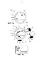
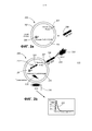
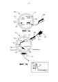
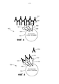
Classifications
-
- G—PHYSICS
- G01—MEASURING; TESTING
- G01N—INVESTIGATING OR ANALYSING MATERIALS BY DETERMINING THEIR CHEMICAL OR PHYSICAL PROPERTIES
- G01N33/00—Investigating or analysing materials by specific methods not covered by groups G01N1/00 - G01N31/00
- G01N33/48—Biological material, e.g. blood, urine; Haemocytometers
- G01N33/50—Chemical analysis of biological material, e.g. blood, urine; Testing involving biospecific ligand binding methods; Immunological testing
- G01N33/53—Immunoassay; Biospecific binding assay; Materials therefor
- G01N33/543—Immunoassay; Biospecific binding assay; Materials therefor with an insoluble carrier for immobilising immunochemicals
- G01N33/554—Immunoassay; Biospecific binding assay; Materials therefor with an insoluble carrier for immobilising immunochemicals the carrier being a biological cell or cell fragment, e.g. bacteria, yeast cells
-
- C—CHEMISTRY; METALLURGY
- C12—BIOCHEMISTRY; BEER; SPIRITS; WINE; VINEGAR; MICROBIOLOGY; ENZYMOLOGY; MUTATION OR GENETIC ENGINEERING
- C12Q—MEASURING OR TESTING PROCESSES INVOLVING ENZYMES, NUCLEIC ACIDS OR MICROORGANISMS; COMPOSITIONS OR TEST PAPERS THEREFOR; PROCESSES OF PREPARING SUCH COMPOSITIONS; CONDITION-RESPONSIVE CONTROL IN MICROBIOLOGICAL OR ENZYMOLOGICAL PROCESSES
- C12Q1/00—Measuring or testing processes involving enzymes, nucleic acids or microorganisms; Compositions therefor; Processes of preparing such compositions
- C12Q1/68—Measuring or testing processes involving enzymes, nucleic acids or microorganisms; Compositions therefor; Processes of preparing such compositions involving nucleic acids
- C12Q1/6897—Measuring or testing processes involving enzymes, nucleic acids or microorganisms; Compositions therefor; Processes of preparing such compositions involving nucleic acids involving reporter genes operably linked to promoters
-
- G—PHYSICS
- G01—MEASURING; TESTING
- G01N—INVESTIGATING OR ANALYSING MATERIALS BY DETERMINING THEIR CHEMICAL OR PHYSICAL PROPERTIES
- G01N33/00—Investigating or analysing materials by specific methods not covered by groups G01N1/00 - G01N31/00
- G01N33/48—Biological material, e.g. blood, urine; Haemocytometers
- G01N33/50—Chemical analysis of biological material, e.g. blood, urine; Testing involving biospecific ligand binding methods; Immunological testing
- G01N33/5005—Chemical analysis of biological material, e.g. blood, urine; Testing involving biospecific ligand binding methods; Immunological testing involving human or animal cells
- G01N33/5008—Chemical analysis of biological material, e.g. blood, urine; Testing involving biospecific ligand binding methods; Immunological testing involving human or animal cells for testing or evaluating the effect of chemical or biological compounds, e.g. drugs, cosmetics
- G01N33/5044—Chemical analysis of biological material, e.g. blood, urine; Testing involving biospecific ligand binding methods; Immunological testing involving human or animal cells for testing or evaluating the effect of chemical or biological compounds, e.g. drugs, cosmetics involving specific cell types
- G01N33/5047—Cells of the immune system
-
- G—PHYSICS
- G01—MEASURING; TESTING
- G01N—INVESTIGATING OR ANALYSING MATERIALS BY DETERMINING THEIR CHEMICAL OR PHYSICAL PROPERTIES
- G01N33/00—Investigating or analysing materials by specific methods not covered by groups G01N1/00 - G01N31/00
- G01N33/48—Biological material, e.g. blood, urine; Haemocytometers
- G01N33/50—Chemical analysis of biological material, e.g. blood, urine; Testing involving biospecific ligand binding methods; Immunological testing
- G01N33/53—Immunoassay; Biospecific binding assay; Materials therefor
- G01N33/536—Immunoassay; Biospecific binding assay; Materials therefor with immune complex formed in liquid phase
-
- G—PHYSICS
- G01—MEASURING; TESTING
- G01N—INVESTIGATING OR ANALYSING MATERIALS BY DETERMINING THEIR CHEMICAL OR PHYSICAL PROPERTIES
- G01N33/00—Investigating or analysing materials by specific methods not covered by groups G01N1/00 - G01N31/00
- G01N33/48—Biological material, e.g. blood, urine; Haemocytometers
- G01N33/50—Chemical analysis of biological material, e.g. blood, urine; Testing involving biospecific ligand binding methods; Immunological testing
- G01N33/53—Immunoassay; Biospecific binding assay; Materials therefor
- G01N33/569—Immunoassay; Biospecific binding assay; Materials therefor for microorganisms, e.g. protozoa, bacteria, viruses
- G01N33/56911—Bacteria
-
- G—PHYSICS
- G01—MEASURING; TESTING
- G01N—INVESTIGATING OR ANALYSING MATERIALS BY DETERMINING THEIR CHEMICAL OR PHYSICAL PROPERTIES
- G01N33/00—Investigating or analysing materials by specific methods not covered by groups G01N1/00 - G01N31/00
- G01N33/48—Biological material, e.g. blood, urine; Haemocytometers
- G01N33/50—Chemical analysis of biological material, e.g. blood, urine; Testing involving biospecific ligand binding methods; Immunological testing
- G01N33/53—Immunoassay; Biospecific binding assay; Materials therefor
- G01N33/569—Immunoassay; Biospecific binding assay; Materials therefor for microorganisms, e.g. protozoa, bacteria, viruses
- G01N33/56983—Viruses
-
- G—PHYSICS
- G01—MEASURING; TESTING
- G01N—INVESTIGATING OR ANALYSING MATERIALS BY DETERMINING THEIR CHEMICAL OR PHYSICAL PROPERTIES
- G01N33/00—Investigating or analysing materials by specific methods not covered by groups G01N1/00 - G01N31/00
- G01N33/48—Biological material, e.g. blood, urine; Haemocytometers
- G01N33/50—Chemical analysis of biological material, e.g. blood, urine; Testing involving biospecific ligand binding methods; Immunological testing
- G01N33/58—Chemical analysis of biological material, e.g. blood, urine; Testing involving biospecific ligand binding methods; Immunological testing involving labelled substances
- G01N33/582—Chemical analysis of biological material, e.g. blood, urine; Testing involving biospecific ligand binding methods; Immunological testing involving labelled substances with fluorescent label
-
- G—PHYSICS
- G01—MEASURING; TESTING
- G01N—INVESTIGATING OR ANALYSING MATERIALS BY DETERMINING THEIR CHEMICAL OR PHYSICAL PROPERTIES
- G01N33/00—Investigating or analysing materials by specific methods not covered by groups G01N1/00 - G01N31/00
- G01N33/48—Biological material, e.g. blood, urine; Haemocytometers
- G01N33/50—Chemical analysis of biological material, e.g. blood, urine; Testing involving biospecific ligand binding methods; Immunological testing
- G01N33/68—Chemical analysis of biological material, e.g. blood, urine; Testing involving biospecific ligand binding methods; Immunological testing involving proteins, peptides or amino acids
- G01N33/6893—Chemical analysis of biological material, e.g. blood, urine; Testing involving biospecific ligand binding methods; Immunological testing involving proteins, peptides or amino acids related to diseases not provided for elsewhere
-
- G—PHYSICS
- G01—MEASURING; TESTING
- G01N—INVESTIGATING OR ANALYSING MATERIALS BY DETERMINING THEIR CHEMICAL OR PHYSICAL PROPERTIES
- G01N33/00—Investigating or analysing materials by specific methods not covered by groups G01N1/00 - G01N31/00
- G01N33/48—Biological material, e.g. blood, urine; Haemocytometers
- G01N33/50—Chemical analysis of biological material, e.g. blood, urine; Testing involving biospecific ligand binding methods; Immunological testing
- G01N33/84—Chemical analysis of biological material, e.g. blood, urine; Testing involving biospecific ligand binding methods; Immunological testing involving inorganic compounds or pH
-
- C—CHEMISTRY; METALLURGY
- C07—ORGANIC CHEMISTRY
- C07K—PEPTIDES
- C07K2319/00—Fusion polypeptide
- C07K2319/60—Fusion polypeptide containing spectroscopic/fluorescent detection, e.g. green fluorescent protein [GFP]
Abstract
Description
ПЕРЕКРЕСТНАЯ ССЫЛКА НА РОДСТВЕННЫЕ ЗАЯВКИCROSS-REFERENCE TO RELATED APPLICATIONS
[0001] Настоящая патентная заявка испрашивает приоритет на основании предварительной патентной заявки США №62/438,068, поданной 22 декабря 2016 г.и названной "Systems and Devices for the Rapid Detection of Analytes" ("Системы и устройства для быстрой аналитов"), а также является частичным продолжением патентной заявки США №15/642,800 (патент США №9,850,546), поданной 06 июля 2017 г.и названной «Biosensor System for the Rapid Detection of Analytes» ("Биосенсорная система для детекции аналитов"), раскрытие которых включено в данный документ в полном объеме посредством ссылки и является частью настоящей заявки на патент США на изобретение для любых целей.[0001] This patent application claims priority on the basis of US Provisional Patent Application No. 62 / 438,068, filed December 22, 2016 and entitled "Systems and Devices for the Rapid Detection of Analytes", and is also a partial continuation of U.S. Patent Application No. 15 / 642,800 (U.S. Patent No. 9,850,546), filed July 6, 2017, and titled "Biosensor System for the Rapid Detection of Analytes", the disclosure of which is included in this document is fully incorporated by reference and forms part of this US patent application for any purpose.
Уровень техникиState of the art
[0002] Описанное изобретение относится в целом к системам, устройствам, реагентам и способам для детекции различных представляющих интерес аналитов в биологических образцах или других типах образцов, а более конкретно, к системе на основе биосенсора для детекции и идентификации представляющих интерес аналитов в реальном времени на основе испускания детектируемого сигнала, когда биосенсор реагирует с представляющим интерес аналитом в тестируемом образце. Следующие патенты предоставляют дополнительную справочную информацию по технологии настоящего изобретения и включены в данный документ в полном объеме посредством ссылки для любых целей: Патент США №9,023,640; 9,752,199; 9,850,546; 9,850,547; и 9,850,548.[0002] The disclosed invention relates generally to systems, devices, reagents and methods for detecting various analytes of interest in biological samples or other types of samples, and more specifically, to a biosensor-based system for detecting and identifying analytes of interest in real time on based on the emission of a detectable signal when the biosensor reacts with the analyte of interest in the test sample. The following patents provide additional background information on the technology of the present invention and are incorporated herein in their entirety by reference for any purpose: US Patent No. 9,023,640; 9,752,199; 9,850,546; 9,850,547; and 9,850,548.
[0003] В общем виде биосенсор представляет собой систему или устройство для детекции аналита, который сочетает в себе чувствительный биологический компонент с физико-химическим детекторным компонентом (детектором). Компоненты типичной биосенсорной системы включают биологический элемент, преобразователь или детекторный элемент и соответствующую электронную аппаратуру или процессоры для обработки сигналов, которые отображают результаты теста значимым и полезным образом. Биологический элемент обычно включает биологический материал, такой, как ткань, микроорганизмы, органеллы, клеточные рецепторы, ферменты, антитела, нуклеиновые кислоты и тому подобное, которые могут быть созданы с помощью известных процессов биологической инженерии. Преобразователь или детекторный элемент работает по физико-химическому механизму (например, оптическому, пьезоэлектрическому и/или электрохимическому), преобразуя сигнал, полученный в результате взаимодействия аналита с биологическим элементом, в другой сигнал, который можно легче измерять и количественно характеризовать. Биосенсоры были созданы в результате интеграции молекулярной биологии и информационных технологий (например, микросхем, оптических волокон и т.д.) для осуществления качественной или количественной оценки взаимодействий биомолекулы и аналита, например, таких, как антитела с антигеном. Учитывая, что существует большой спрос на быстрые, чувствительные, простые в обращении и экономически эффективные средства детекции для выявления инфекционных агентов, патогенов и/или токсинов в пищевых продуктах (см., например, Mead et al., Food Related Illness and Death in the United States, Emerging Infectious Diseases, V. 5, №5, сентябрь-октябрь 1999 года (607-625), который включен в настоящий документ посредством ссылки в полном объеме), существует постоянная потребность в применении биосенсоров в режиме реального времени, полевых портативных устройствах и инструментах для детекции и идентификации инфекционных агентов, патогенных микроорганизмов, токсинов и других загрязняющих веществ в пищевых продуктах.[0003] In general terms, a biosensor is a system or device for detecting an analyte that combines a sensitive biological component with a physicochemical detector component (detector). Components of a typical biosensor system include a biological element, a transducer or detector element, and associated signal processing electronics or processors that display test results in a meaningful and useful manner. A biological element typically includes biological material such as tissue, microorganisms, organelles, cell receptors, enzymes, antibodies, nucleic acids, and the like, which can be created using known biological engineering processes. A transducer or detector element works by a physicochemical mechanism (for example, optical, piezoelectric and / or electrochemical), converting the signal resulting from the interaction of an analyte with a biological element into another signal that can be more easily measured and quantified. Biosensors were created as a result of the integration of molecular biology and information technology (for example, microcircuits, optical fibers, etc.) to perform a qualitative or quantitative assessment of the interactions of a biomolecule and an analyte, such as antibodies with antigen. Given that there is a strong demand for fast, sensitive, easy-to-use and cost-effective means of detection for identifying infectious agents, pathogens and / or toxins in food (see, for example, Mead et al., Food Related Illness and Death in the United States, Emerging Infectious Diseases, V. 5, No. 5, September-October 1999 (607-625), which is incorporated herein by reference in its entirety), there is an ongoing need for real-time biosensors, field handhelds devices and tools for the detection and identification of infectious agents, pathogenic microorganisms, toxins and other contaminants in food.
КРАТКОЕ ОПИСАНИЕ ИЗОБРЕТЕНИЯBRIEF DESCRIPTION OF THE INVENTION
[0004] Далее представлено краткое изложение нескольких примерных вариантов реализации данного изобретения. Данное изложение сущности изобретения не представляет собой расширенного обзора и не предназначено для идентификации ключевых критических элементов или для определения объема формулы изобретения. Однако следует понимать, что использование неопределенных артиклей на языке, используемом для описания и утверждения настоящего изобретения, никоим образом не предназначено для ограничения описанной системы отдельными компонентами или элементами. Скорее использование «а» или «an» в данном документе должно интерпретироваться «по меньшей мере, как один» или «один или несколько».[0004] The following is a summary of several exemplary embodiments of the present invention. This Summary is not an extensive overview and is not intended to identify key critical elements or to define the scope of the claims. However, it should be understood that the use of the indefinite articles in the language used to describe and assert the present invention is in no way intended to limit the described system to individual components or elements. Rather, the use of "a" or "an" in this document should be interpreted as "at least one" or "one or more."
[0005] Согласно одному аспекту настоящего изобретения предусмотрена первая биосенсорная система для детекции целевых аналитов. Эта система включает живую биологическую клетку заданного (заранее определенного) типа; генерирующий сигнал репортер, связанный с живой биологической клеткой; путь передачи сигнала или активаторный механизм, связанный с репортером генерации сигнала; универсальный детекторный элемент, связанный с активаторным механизмом; и элемент, связывающий аналит, связанный с универсальным детекторным элементом, причем элемент, связывающий аналит, специфичен как в отношении универсального детекторного элемента, так и в отношении целевого аналита.[0005] According to one aspect of the present invention, there is provided a first biosensor system for detecting target analytes. This system includes a living biological cell of a given (predetermined) type; a signal generating reporter associated with a living biological cell; a signal transmission pathway or activator mechanism associated with a signal generation reporter; a universal detector element associated with an activator mechanism; and an analyte binding element associated with the universal detector element, the analyte binding element being specific for both the universal detection element and the target analyte.
[0006] Согласно другому аспекту настоящего изобретения предусмотрена первая биосенсорная система для детекции целевых аналитов. Эта система включает живую биологическую клетку заданного типа; генерирующий сигнал репортер, связанный с живой биологической клеткой, причем генерирующий сигнал репортер реагирует на определенные изменения, происходящие внутри живых биологических клеток; путь передачи сигнала или активаторный механизм, связанный с генерирующим сигнал репортером, причем активаторный механизм является оперативным для того, чтобы вызывать определенные изменения в живой биологической клетке; универсальный детекторный элемент, связанный с активаторным механизмом, причем универсальный детекторный элемент может запускать активаторный механизм; элемент, связывающий аналит, связанный с универсальным детекторным элементом, причем элемент, связывающий аналит, специфичен как в отношении универсального детекторного элемента, так и в отношении целевого аналита; и в котором при связывании элемента, связывающего аналит, с которым целевой аналит также связан с универсальным детекторным элементом, универсальный детекторный элемент запускает активаторный механизм, вызывая определенные изменения в живой биологической клетке, в результате чего генерирующий сигнал репортер генерирует детектируемый сигнал.[0006] According to another aspect of the present invention, there is provided a first biosensor system for detecting target analytes. This system includes a living biological cell of a given type; a signal-generating reporter associated with a living biological cell, the signal-generating reporter responding to certain changes occurring within the living biological cells; a signaling pathway or activator mechanism associated with a signal-generating reporter, the activator mechanism being operative to induce certain changes in a living biological cell; a universal detector element associated with the activation mechanism, the universal detection element being able to trigger the activation mechanism; an analyte binding element associated with a universal detector element, the analyte binding element being specific for both the universal detection element and the target analyte; and wherein upon binding of the analyte binding element to which the target analyte is also bound to the universal detector element, the universal detector element triggers an activator mechanism causing certain changes in the living biological cell, whereby the signal-generating reporter generates a detectable signal.
[0007] В еще одном аспекте настоящего изобретения предусмотрена третья биосенсорная система для детекции целевых аналитов. Эта система включает живую биологическую клетку заданного типа; генерирующий сигнал репортер в живой биологической клетке, причем генерирующий сигнал репортер реагирует на определенные изменения, происходящие в живой биологической клетке; путь передачи сигнала или активаторный механизм, связанный с генерирующим сигнал репортером, причем активаторный механизм может вызывать определенные изменения в живой биологической клетке; универсальный детекторный элемент, связанный с активаторным механизмом, причем универсальный детекторный элемент может запускать активаторный механизм; элемент, связывающий аналит, связанный с универсальным детекторным элементом, причем элемент, связывающий аналит, является специфическим как в отношении универсального детекторного элемента, так и в отношении целевого аналита; причем при связывании элемента, связывающего аналит, с которым целевой аналит также связан, с универсальным детекторным элементом, универсальный детектор ингибирует активаторный механизм, вызывая снижение определенных изменений в живой биологической клетке, в результате чего генерирующий сигнал репортер генерирует ослабленный сигнал или не генерирует сигнал.[0007] In yet another aspect of the present invention, a third biosensor system is provided for detecting target analytes. This system includes a living biological cell of a given type; a signal-generating reporter in a living biological cell, the signal-generating reporter responding to certain changes occurring in a living biological cell; a signal transduction pathway or an activator mechanism associated with a signal-generating reporter, and the activator mechanism can cause certain changes in a living biological cell; a universal detector element associated with the activation mechanism, the universal detection element being able to trigger the activation mechanism; an analyte binding element associated with a universal detector element, the analyte binding element being specific for both the universal detection element and the target analyte; moreover, upon binding of the analyte binding element to which the target analyte is also bound to the universal detector element, the universal detector inhibits the activator mechanism, causing a decrease in certain changes in the living biological cell, as a result of which the signal-generating reporter generates an attenuated signal or does not generate a signal.
[0008] Дополнительные признаки и аспекты настоящего изобретения станут очевидны обычным специалистам в данной области техники после прочтения и осмысления нижеследующего подробного описания примерных вариантов реализации. Специалист в данной области поймет, что возможны дополнительные варианты реализации изобретения, не выходящие за пределы объема и сущности изобретения. Соответственно, чертежи и подробное описание должны рассматриваться как иллюстративные и неограничительные.[0008] Additional features and aspects of the present invention will become apparent to those of ordinary skill in the art upon reading and understanding the following detailed description of exemplary embodiments. One skilled in the art will understand that additional embodiments of the invention are possible without departing from the scope and spirit of the invention. Accordingly, the drawings and detailed description are to be considered as illustrative and non-limiting.
КРАТКОЕ ОПИСАНИЕ ЧЕРТЕЖЕЙBRIEF DESCRIPTION OF DRAWINGS
[0009] Прилагаемые чертежи, которые включены в описание и составляют его часть, схематически иллюстрируют один или более примерных вариантов реализации изобретения и вместе с общим описанием, приведенным выше, и подробным описанием, приведенным ниже, служат для объяснения принципов изобретения, и при этом:[0009] The accompanying drawings, which are incorporated in and constitute a part of the description, schematically illustrate one or more exemplary embodiments of the invention and, together with the general description above and the detailed description below, serve to explain the principles of the invention, wherein:
[0010] ФИГ. 1a-b являются иллюстрациями первого биосенсора в соответствии с одним примерным вариантом реализации, в котором Т-клетки Jurkat были сконструированы (модифицированы) для продуцирования экворина и для экспрессии не являющегося антителом трансмембранного передающего сигнал элемента IgGbp-CD3ζ;[0010] FIG. 1a-b are illustrations of a first biosensor in accordance with one exemplary embodiment in which Jurkat T cells have been engineered (modified) to produce aequorin and to express a non-antibody transmembrane IgGbp-CD3ζ signaling element;
[0011] ФИГ. 2a-b являются иллюстрациями второго биосенсора биосенсора в соответствии с примерным вариантом реализации, в котором тучные клетки МС/9 были сконструированы для продуцирования экворина, и где клетки МС/9 экспрессируют нативный рецептор FcεRI, который связывается с не являющимся антителом растворимым передающим сигнал элементом IgGbp-IgE;[0011] FIG. 2a-b are illustrations of a second biosensor biosensor according to an exemplary embodiment in which MC / 9 mast cells were engineered to produce aequorin and where MC / 9 cells express a native FcεRI receptor that binds to a non-antibody soluble IgGbp signaling element -IgE;
[0012] ФИГ. 3a-b являются иллюстрациями третьего биосенсора соответствии с примерным вариантом реализации, в котором тучные клетки МС/9 были сконструированы для продуцирования экворина, и где клетки МС/9 экспрессируют нативный рецептор FcεRI, который связывается с не являющимся антителом растворимым передающим сигнал элементом IgGbp-IgE, который экскретируется тучными клетками МС/9;[0012] FIG. 3a-b are illustrations of a third biosensor according to an exemplary embodiment in which MC / 9 mast cells have been engineered to produce aequorin and where MC / 9 cells express a native FcεRI receptor that binds to a non-antibody soluble IgGbp-IgE signaling element which is excreted by MC / 9 mast cells;
[0013] ФИГ. 4 является иллюстрацией четвертого биосенсора в соответствии с примерным вариантом реализации, в котором биосенсорные клетки были сконструированы для продуцирования экворина и для экспрессии не являющегося антителом трансмембранного передающего сигнал элемента mSA-CD3, который связывается с биотинилированным детекторным; и[0013] FIG. 4 is an illustration of a fourth biosensor, in accordance with an exemplary embodiment, in which biosensor cells were engineered to produce aequorin and to express a non-antibody transmembrane signaling element mSA-CD3 that binds to a biotinylated detector; and
[0014] ФИГ. 5 является иллюстрацией пятого биосенсора в соответствии с примерным вариантом реализации, в котором биосенсорные клетки были сконструированы для продуцирования экворина и для экспрессии не являющегося антителом трансмембранного передающего сигнал элемента mSA-CD3ζ, который связывается с биотинилированным детекторным элементом.[0014] FIG. 5 is an illustration of a fifth biosensor in accordance with an exemplary embodiment in which biosensor cells have been engineered to produce aequorin and to express a non-antibody transmembrane signaling element mSA-CD3ζ that binds to a biotinylated detector element.
ПОДРОБНОЕ ОПИСАНИЕ ИЗОБРЕТЕНИЯDETAILED DESCRIPTION OF THE INVENTION
[0015] Примерные варианты реализации настоящего изобретения будут теперь описаны со ссылкой на чертежи. Хотя нижеследующее подробное описание содержит множество конкретных признаков для целей иллюстрации, обычный специалист в данной области техники поймет, что могут быть сделаны различные модификации и изменения без выхода за рамки объема настоящего изобретения. Соответственно, следующие варианты реализации изобретения изложены без какой-либо потери общности и без наложения ограничений на заявленное изобретение.[0015] Exemplary embodiments of the present invention will now be described with reference to the drawings. Although the following detailed description contains many specific features for purposes of illustration, one of ordinary skill in the art will understand that various modifications and changes can be made without departing from the scope of the present invention. Accordingly, the following embodiments of the invention are set forth without any loss of generality and without imposing limitations on the claimed invention.
[0016] Настоящее изобретение относится в целом к системам, устройствам, реагентам и способам для детекции различных аналитов или других мишеней, представляющих интерес, в биологических образцах или образцах других типов, а более конкретно, к биосенсорной системе для детекции и идентификации представляющих интерес аналитов в режиме реального времени на основе испускания детектируемого сигнала, когда биосенсор реагирует с интересующим аналитом в анализируемом образце. Сконструированные клетки по настоящему изобретению являются очень чувствительными и эффективными биосенсорами, и поскольку этим биосенсорным клеткам присуща способность к детекции, они образуют универсальную систему, которая может быть легко адаптирована для детекции широкого спектра различных инфекционных агентов или других мишеней, простого за счет выбора альтернативных растворимых детекторных молекул (например, антител), обладающих специфичностью в отношении конкретного патогена или другой представляющей интерес мишени. Кроме того, система по настоящему изобретению может быть легко сконфигурирована для мультиплексной детекции нескольких инфекционных агентов или других аналитов за один анализ, обеспечивая большую гибкость и полезность. Универсальность настоящего изобретения обусловлена уникальной комбинацией элементов и, в частности, комбинацией универсальной биосенсорной клетки со специфическим растворимым детектором (например, антителом). Универсальная биосенсорная клетка обладает способностью реагировать на присутствие практически любой целевой молекулы (мишени), которая может быть распознана детекторной молекулой. Поскольку в некоторых вариантах осуществления детектор или детекторное антитело добавляется в систему в качестве растворимого фактора, система может быть сконфигурирована для детекции альтернативной мишени просто за счет выбора соответствующего альтернативного детектора или детекторного антитела. Специфичность раскрытой системы определяется детекторной молекулой, которая выбирается на основании ее специфичности и сродства к молекуле-мишени, которая характерна для инфекционного агента или другого целевого аналита (аналита-мишени). Комбинация этой универсальной биосенсорной клетки и растворимого детектора также позволяет конструировать мультиплексные анализы просто путем включения множества детекторных молекул (например, антител) в тест-систему, где молекулы-мишени выбираются на основе их специфичности к альтернативным инфекционным агентам или другим аналитам.[0016] The present invention relates generally to systems, devices, reagents and methods for detecting various analytes or other targets of interest in biological samples or other types of samples, and more specifically, to a biosensor system for detecting and identifying analytes of interest in real time based on the emission of a detectable signal when the biosensor reacts with the analyte of interest in the analyzed sample. The cells engineered according to the present invention are very sensitive and efficient biosensors, and since these biosensor cells have inherent detection capabilities, they form a versatile system that can be easily adapted to detect a wide range of different infectious agents or other targets, simply by choosing alternative soluble detector molecules (eg, antibodies) that have specificity for a particular pathogen or other target of interest. In addition, the system of the present invention can be easily configured for multiplexed detection of multiple infectious agents or other analytes in a single assay, providing greater flexibility and utility. The versatility of the present invention is due to the unique combination of elements and, in particular, the combination of a universal biosensor cell with a specific soluble detector (for example, an antibody). A universal biosensor cell has the ability to respond to the presence of almost any target molecule (target) that can be recognized by a detector molecule. Since, in some embodiments, a detector or detector antibody is added to the system as a soluble factor, the system can be configured to detect an alternative target simply by selecting an appropriate alternative detector or detector antibody. The specificity of the disclosed system is determined by the detector molecule, which is selected based on its specificity and affinity for the target molecule, which is characteristic of the infectious agent or other target analyte (target analyte). The combination of this versatile biosensor cell and soluble detector also allows multiplex assays to be designed simply by incorporating multiple detector molecules (e.g., antibodies) into a test system, where target molecules are selected based on their specificity for alternative infectious agents or other analytes.
[0017] Генетические манипуляции и модификация типов биосенсорных клеток, применяемых в данном изобретении, обычно включают применение надлежащим образом отобранных средств доставки генов, которые содержат генетические элементы, эффективно функционирующие в выбранном типе клеток. Например, целесообразно использовать промоторный элемент, который обеспечивает высокий уровень экспрессии введенных трансгенов в конкретной выбранной биосенсорной клетке. В примерном варианте реализации данного изобретения такой промоторный элемент может быть получен непосредственно из самой биосенсорной клетки и затем использован для экспрессии представляющего интерес трансгена. В другом варианте реализации данного изобретения соответствующий элемент может быть определен эмпирически путем сравнения функции альтернативных промоторных элементов в контексте альтернативных носителей доставки генов с целью выявления эффективных промоторных, трансгенных, векторных комбинаций для выбранного типа клеток. Трансгены, такие как ген, кодирующий люминесцентный репортерный белок, могут быть введены в биосенсорную клетку с использованием стандартных методов, таких как электропорация или с помощью химических реагентов для трансфекции, таких как, например, липофектамин. Другие методы генной инженерии, известные специалистам в данной области, также совместимы с настоящим изобретением.[0017] Genetic manipulation and modification of biosensor cell types used in the present invention typically involve the use of appropriately selected gene delivery vehicles that contain genetic elements that function effectively in the selected cell type. For example, it is advisable to use a promoter element that provides a high level of expression of the introduced transgenes in a particular selected biosensor cell. In an exemplary embodiment of the present invention, such a promoter element can be obtained directly from the biosensor cell itself and then used to express the transgene of interest. In another embodiment, the corresponding element can be determined empirically by comparing the function of alternative promoter elements in the context of alternative gene delivery vehicles in order to identify effective promoter, transgenic, vector combinations for the selected cell type. Transgenes, such as the gene encoding a luminescent reporter protein, can be introduced into a biosensor cell using standard techniques such as electroporation or using transfection chemicals such as lipofectamine. Other genetic engineering techniques known to those skilled in the art are also compatible with the present invention.
[0018] Пример реализации настоящего изобретения включает живую сконструированную биосенсорную клетку, причем живая сконструированная биосенсорная клетка обычно является компонентом иммунной системы млекопитающего; репортерный белок, причем репортерный белок экспрессируется живой сконструированной клеткой и присутствует в живой сконструированной клетке, а также репортерный белок испускает детектируемый сигнал в ответ на определенные изменения в цитозоле живой сконструированной клетки; путь передачи сигнала, демонстрируемый живой сконструированной клеткой, причем этот путь передачи сигнала контролирует биологический или биохимический процесс в цитозоле живой сконструированной клетки и при этом по меньшей мере один биологический или биохимический процесс, когда он происходит, заставляет репортерный белок испускать детектируемый сигнал; по меньшей мере один тип детекторной молекулы (молекулы-детектора), причем каждая детекторная молекула приспособлена для связывания с конкретным аналитом; по меньшей мере, один аналит, причем, по меньшей мере, один аналит связывается с детекторной молекулой, которая специфична для этого аналита; и множество не являющихся антителами передающих сигнал трансмембранных элементов, экспрессируемых живой сконструированной клеткой, причем каждый передающий сигнал элемент способен принимать детекторную молекулу, которая, в свою очередь, способна принимать аналит. После связывания достаточного количества аналитов с достаточным количеством детекторных молекул, которые сами связаны с не являющимися антителами передающими сигнал трансмембранными элементами, происходит агрегация передающих сигнал элементов на поверхность клетки, активируется путь передачи сигнала, происходит биологический или биохимический процесс, и репортерный белок испускает детектируемый сигнал. Эта система может также включать устройство для смешивания живых клеток с растворимыми компонентами и образцом, содержащим интересующий аналит или инфекционный агент при сохранении жизнеспособности и функциональности биосенсорной клетки, и детектор для детекции сигнала, испускаемого биосенсорной клеткой.[0018] An exemplary embodiment of the present invention includes a living engineered biosensor cell, wherein the living engineered biosensor cell is typically a component of the mammalian immune system; a reporter protein, the reporter protein being expressed by a living engineered cell and present in a living engineered cell, and the reporter protein emitting a detectable signal in response to certain changes in the cytosol of the living engineered cell; a signaling pathway exhibited by a living engineered cell, the signaling pathway controlling a biological or biochemical process in the cytosol of a living engineered cell and wherein at least one biological or biochemical process, when it occurs, causes the reporter protein to emit a detectable signal; at least one type of detector molecule (detector molecule), each detector molecule being adapted to bind to a particular analyte; at least one analyte, and at least one analyte binds to a detector molecule that is specific for this analyte; and a plurality of non-antibody signaling transmembrane elements expressed by a living engineered cell, each signaling element being capable of receiving a detector molecule, which in turn is capable of receiving an analyte. Once a sufficient number of analytes have bound to a sufficient number of detector molecules that are themselves bound to non-antibody signaling transmembrane elements, the signaling elements aggregate to the cell surface, the signaling pathway is activated, a biological or biochemical process occurs, and the reporter protein emits a detectable signal. This system may also include a device for mixing living cells with soluble components and a sample containing the analyte or infectious agent of interest while maintaining the viability and functionality of the biosensor cell, and a detector for detecting the signal emitted by the biosensor cell.
[0019] Другой пример осуществления этого изобретения включает в себя: живую сконструированную клетку, причем живая сконструированная клетка является компонентом иммунной системы млекопитающих, где живая сконструированная клетка представляет собой тучную клетку и где тучная клетка экспрессирует по меньшей мере один предопределенный рецептор; репортерный белок, причем репортерный белок представляет собой экворин, который экспрессируется живой, сконструированной клеткой, и где экворин испускает детектируемый сигнал света в ответ на некоторые определенные изменения в цитозоле живой сконструированной клетки; путь передачи сигнала, экспрессируемый живой сконструированной клеткой, причем путь передачи сигнала управляет биохимическим процессом в цитозоле живой сконструированной клетки и где биохимический процесс, контролируемый путем передачи сигнала, дополнительно включает увеличение внутриклеточного кальция, причем увеличение внутриклеточного кальция, когда оно происходит, побуждает экворин испускать детектируемый свет; по меньшей мере один тип детекторной молекулы, где каждая детекторная молекула приспособлена для связывания с конкретным аналитом; по меньшей мере, один аналит, где, по меньшей мере, один аналит связывается с детекторной молекулой, которая специфична для этого аналита; и множество растворимых не являющихся антителами передающих сигнал элементов. Каждый передающий сигнал элемент, способен к связыванию, по меньшей мере, с одним предопределенным рецептором и для приема детекторной молекулы. При связывании достаточного количества аналитов с достаточным количеством детекторных молекул с достаточным количеством элементов, не передающих трансмембранный сигнал антитела, которые сами связаны по меньшей мере с одним типом предопределенного рецептора, происходит агрегация рецепторов на поверхности клетки, активируется путь передачи сигнала, происходит увеличение внутриклеточного кальция, и экворин испускает детектируемый свет. Эта система может также включать устройство для смешивания живых клеток вместе с растворимыми компонентами и образцом, содержащим интересующий аналит или инфекционный агент при сохранении жизнеспособности и функциональности живой биосенсорной клетки, и детектор для детекции сигнала, испускаемого биосенсорной клеткой.[0019] Another embodiment of this invention includes: a living engineered cell, where the living engineered cell is a component of the mammalian immune system, where the living engineered cell is a mast cell and where the mast cell expresses at least one predetermined receptor; a reporter protein, the reporter protein being an aequorin, which is expressed by a living, engineered cell, and where aequorin emits a detectable light signal in response to certain specific changes in the cytosol of the living engineered cell; a signaling pathway expressed by a living engineered cell, the signaling pathway directing the biochemical process in the cytosol of a living engineered cell, and where the signaling-controlled biochemical process further includes an increase in intracellular calcium, the increase in intracellular calcium, when it occurs, induces aequorin to emit detectable shine; at least one type of detector molecule, where each detector molecule is adapted to bind to a particular analyte; at least one analyte, where at least one analyte binds to a detector molecule that is specific for the analyte; and a variety of soluble non-antibody signaling elements. Each signaling element is capable of binding to at least one predetermined receptor and receiving a detector molecule. When a sufficient number of analytes bind with a sufficient number of detector molecules with a sufficient number of elements that do not transmit the transmembrane signal of an antibody, which are themselves bound to at least one type of predetermined receptor, aggregation of receptors on the cell surface occurs, the signal transmission pathway is activated, and intracellular calcium increases. and aequorin emits detectable light. This system may also include a device for mixing living cells together with soluble components and a sample containing the analyte or infectious agent of interest while maintaining the viability and functionality of the living biosensor cell, and a detector for detecting the signal emitted by the biosensor cell.
[0020] Еще один пример реализации настоящего изобретения включает: биосенсор для быстрой детекции, где биосенсор дополнительно включает живую сконструированную клетку, причем живая сконструированная клетка получена из клеточного компонента иммунной системы млекопитающего (то есть иммуноцита); репортерный белок, причем репортерный белок встроен в живую сконструированную клетку и экспрессируется ей, и при этом репортерный белок испускает детектируемый сигнал в ответ на некоторые определенные изменения в цитозоле живой сконструированной клетки; путь передачи сигнала, встроенный в живую сконструированную клетку или присутствующий в ней от природы, при этом путь передачи сигнала контролирует биологический процесс в цитозоле живой сконструированной биосенсорной клетки, и при этом биологический процесс, когда он происходит, побуждает (заставляет) репортерный белок испускать детектируемый сигнал; и множество не являющихся антителами передающих сигнал элементов, которые прямо или опосредованно связываются с аналитом в анализируемом образце, причем связанные не являющиеся антителами передающие сигнал элементы, затем взаимодействуют с биосенсорной клеткой для прямой или непрямой активации пути передачи сигнала.[0020] Yet another embodiment of the present invention includes: a biosensor for rapid detection, wherein the biosensor further comprises a living engineered cell, the living engineered cell being derived from a cellular component of the mammalian immune system (ie, an immunocyte); a reporter protein, wherein the reporter protein is incorporated into and expressed by the living engineered cell, and wherein the reporter protein emits a detectable signal in response to certain specific changes in the cytosol of the living engineered cell; a signal transduction pathway built into or naturally present in a living engineered cell, where the signaling pathway controls the biological process in the cytosol of the living engineered biosensor cell, and the biological process, when it occurs, induces (causes) the reporter protein to emit a detectable signal ; and a plurality of non-antibody signaling elements that directly or indirectly bind to the analyte in the analyzed sample, the associated non-antibody signaling elements, then interact with the biosensor cell to directly or indirectly activate the signaling pathway.
Живая биологическая клеткаLiving biological cell
[0021] Примеры реализации настоящего изобретения включают живую, сконструированную биосенсорную клетку, которая обычно является компонентом иммунной системы млекопитающего, например иммуноцит. В некоторых вариантах реализации настоящего изобретения биосенсорная клетка представляет собой В-клетку человека или мыши. В-клетки или В-лимфоциты представляют собой тип лейкоцитов подтипа лимфоцитов, которые функционируют в гуморальном компоненте иммунитета адаптивной иммунной системы, вырабатывая антитела. В некоторых примерах реализации настоящего изобретения биосенсорная клетка представляет собой Т-клетку человека или мыши. Т-клетки или Т-лимфоциты являются другим типом лимфоцитов, которые играют центральную роль в клеточном иммунитете как части адаптивной иммунной системы. Т-клетки отличаются от других лимфоцитов наличием Т-клеточного рецептора на поверхности клеток. В других вариантах реализации настоящего изобретения биосенсорная клетка представляет собой тучную клетку. Тучная клетка также представляет собой тип лейкоцитов, известный как гранулоцит, который получают из миелоидных стволовых клеток, которые являются частью иммунной и нейроиммунной систем. Другие типы клеток совместимы с настоящим изобретением, включая базофилы, которые являются другим типом лейкоцитов, однако похожи по внешнему виду и имеют схожие функции с тучными клетками.[0021] Examples of implementation of the present invention include a living, engineered biosensor cell, which is usually a component of the mammalian immune system, for example, an immunocyte. In some embodiments, the biosensor cell is a human or mouse B cell. B cells or B lymphocytes are a type of white blood cell of a subtype of lymphocytes that function in the humoral immunity component of the adaptive immune system by producing antibodies. In some embodiments of the present invention, the biosensor cell is a human or mouse T cell. T cells or T lymphocytes are another type of lymphocytes that play a central role in cellular immunity as part of the adaptive immune system. T cells differ from other lymphocytes by the presence of a T cell receptor on the cell surface. In other embodiments of the present invention, the biosensor cell is a mast cell. A mast cell is also a type of white blood cell known as a granulocyte, which is derived from myeloid stem cells, which are part of the immune and neuroimmune systems. Other types of cells are compatible with the present invention, including basophils, which are another type of white blood cell, but are similar in appearance and function to mast cells.
[0022] В других примерах реализации настоящего изобретения живая биологическая клетка может быть прокариотической или эукариотической клеткой, такой как эукариотическая клетка, которая включает сигнальную систему Са2+. Живой клеткой может быть дрожжевая клетка или клетка насекомого, например клетка Drosophila Schneider 2 (S2), клетка sf9 или клетка насекомого, которая была сконструрирована для применения экворина в качестве репортера. Живой биологической клеткой может быть клетка млекопитающего, например клетка HEK, клетка СНО, клетка COS или клетка 3T3. Живая биологическая клетка может быть сконструированной клеткой. Сконструированная клетка может быть получена из нативной, пассируемой или культивируемой клетки млекопитающего. Сконструированная клетка может быть получена из невоспроизводящейся клетки, фиксированной клетки, клетки, обработанной лекарственным средством или химическим реагентом, осмотически обработанной клетки, облученной клетки, искусственно или синтетически полученной клетки или неживой клетки, при условии, что сконструированная клетка содержит функциональный лиганд, путь передачи сигнала и репортер. Клетка может быть искусственной или синтетической. Сконструированная клетка может быть получена из растительной клетки, клетки животного, клетки насекомого или клетки другого животного, не являющегося млекопитающим, компонента иммунной системы млекопитающего, фолликулярной дендритной клетки, естественной клетки-киллера, макрофага, моноцита, мононуклеарного фагоцита, нейтрофила, эозинофила или базофила. Сконструированная клетка также может представлять собой клетку, которая экспрессирует рецепторы типа Fc, такие как В-лимфоциты, фолликулярные дендритные клетки, природные клетки-киллеры, макрофаги, нейтрофилы, эозинофилы, базофилы и тучные клетки. В определенных вариантах реализации клетка может представлять собой любую прокариотическую или эукариотическую клетку, которая имеет подходящий рецептор, путь передачи сигнала и механизм вывода сигнала, полученная естественным путем, посредством генной инженерии, либо посредством химического добавления. Клетка может быть искусственной или неживой единицей при условии, что она имеет функциональный рецептор, сигнальный путь и механизм вывода сигнала. Примером клетки, которая может применяться в данной системе, является клетка макрофага, такая как линия клеток человека U937, которая экспрессирует Fc-рецептор на поверхности клетки. Антиген может быть связан с антителом путем добавления антитела к мишени, и этот комплекс антиген-антитело будет связываться с Fc-рецептором на клетке, стимулируя передачу сигналов, что приводит к увеличению внутриклеточного кальция. Клетки можно зафиксировать, заморозить, высушить или сублимировать.[0022] In other embodiments of the present invention, the living biological cell can be a prokaryotic or eukaryotic cell, such as a eukaryotic cell, which includes the Ca2 + signaling system. A living cell can be a yeast cell or an insect cell, for example a Drosophila Schneider 2 (S2) cell, an sf9 cell, or an insect cell that has been engineered to use aequorin as a reporter. A living biological cell can be a mammalian cell, for example, a HEK cell, a CHO cell, a COS cell, or a 3T3 cell. A living biological cell can be a constructed cell. The engineered cell can be obtained from a native, passaged, or cultured mammalian cell. An engineered cell can be obtained from a non-reproducing cell, a fixed cell, a cell treated with a drug or chemical reagent, an osmotically treated cell, an irradiated cell, an artificially or synthetically produced cell, or an inanimate cell, provided that the engineered cell contains a functional ligand, signal transduction pathway and a reporter. The cage can be artificial or synthetic. The engineered cell can be obtained from a plant cell, an animal cell, an insect cell, or a cell of another non-mammalian animal, a component of the mammalian immune system, follicular dendritic cell, natural killer cell, macrophage, monocyte, mononuclear phagocyte, neutrophil, eosinophil, or basophil. The engineered cell can also be a cell that expresses Fc receptors such as B lymphocytes, follicular dendritic cells, natural killer cells, macrophages, neutrophils, eosinophils, basophils, and mast cells. In certain embodiments, a cell can be any prokaryotic or eukaryotic cell that has a suitable receptor, signal transduction pathway, and signal output mechanism, obtained naturally, through genetic engineering, or through chemical addition. A cell can be an artificial or non-living unit, provided that it has a functional receptor, a signaling pathway, and a signal output mechanism. An example of a cell that can be used in this system is a macrophage cell, such as the human cell line U937, which expresses an Fc receptor on the cell surface. An antigen can be linked to an antibody by adding an antibody to the target, and this antigen-antibody complex will bind to an Fc receptor on the cell, stimulating signaling, which leads to an increase in intracellular calcium. Cells can be fixed, frozen, dried, or sublimated.
Генерирующий сигнал репортерSignal generating reporter
[0023] Примерные варианты реализации настоящего изобретения включают репортерный элемент, такой как репортерный белок или фермент, который продуцируется или экспрессируется живой сконструированной биосенсорной клеткой. Репортерный белок испускает детектируемый сигнал в ответ на некоторые определенные изменения в цитозоле живой, сконструированной биосенсорной клетки. В некоторых вариантах реализации настоящего изобретения репортерный белок представляет собой биолюминесцентный фотопротеин, такой как экворин, который получают из гидрозоя Aequorea Victoria. Экворин ранее использовался для конструирования живых биосенсорных клеток с целью получения световых сигналов в ответ на активацию широкого спектра путей передачи сигнала; таким образом, специалистам в данной области известны различные способы манипулирования продукцией экворина в живых клетках. В частности, квалифицированный специалист может выбрать и использовать любое подходящее средство доставки генов, такое как, например бактериальные плазмидные векторы или вирусные векторы, для введения соответствующего генетического материала в биосенсорные клетки. Продукция репортерного белка в биосенсорной клетке далее контролируется за счет экспрессии введенного генетического материала. Специалист в данной области поймет, что другие фотопротеины, репортерные белки других типов, ферменты и молекулы могут быть включены и использованы в различных альтернативных вариантах реализации настоящего изобретения.[0023] Exemplary embodiments of the present invention include a reporter element, such as a reporter protein or enzyme, which is produced or expressed by a living, engineered biosensor cell. The reporter protein emits a detectable signal in response to certain specific changes in the cytosol of a living, engineered biosensor cell. In some embodiments, the reporter protein is a bioluminescent photoprotein, such as aequorin, which is derived from Aequorea Victoria hydrozoa. Equorin has previously been used to construct living biosensor cells to generate light signals in response to activation of a wide variety of signal transduction pathways; thus, those skilled in the art are aware of various methods for manipulating aequorin production in living cells. In particular, the skilled artisan can select and use any suitable gene delivery vehicle, such as, for example, bacterial plasmid vectors or viral vectors, to introduce the appropriate genetic material into biosensor cells. The production of the reporter protein in the biosensor cell is further controlled by the expression of the introduced genetic material. One skilled in the art will understand that other photoproteins, other types of reporter proteins, enzymes, and molecules can be included and used in various alternative embodiments of the present invention.
[0024] В различных вариантах реализации настоящего изобретения репортер может представлять собой белок, обладающий флуоресцентными характеристиками, которые подвергаются заметному изменению в ответ на активацию по меньшей мере одного биохимического пути и результирующего изменения в живой биологической клетке. Репортер, или репортерный белок может представлять собой другие чувствительные к кальцию люминесцентные или флуоресцентные молекулы, такие как обелин, талассиколин, митрокомин (галистаурин), клитин (фиалидин), мнемопсин, беровин, индо-1, фура-2, квин-2, флуо-белок. 3, Rhod-2, кальциевый зеленый, ВАРТА, камелины (А. Miyawaki et al., (1999) Proc. Natl. Acad. Sci. 96, 213540), или похожие молекулы. Репортерный белок может быть химерным. Он включает Са2+-связывающий домен и связанный флуоресцентный белок. Связанный флуоресцентный белок имеет зеленую флуоресценцию (GFP). Белок может связывать другие компоненты фосфатидилинозитного пути (т.е пути, используемого в вариантах реализации, описанного в настоящем документе) и изменять его флуоресценцию. Примером является флуоресцентный белок, который был разработан для связывания диацилглицерина. Репортер может быть ферментом, способным (выполненным с возможностью) давать люминесцентноый или флуоресцентный сигнал. Репортерный белок может быть ферментом, типа люциферазы или щелочной фосфатазы, который генерирует люминесцентный или флуоресцентный сигнал соответственно. Он также может быть флуоресцентным белком или включать флуоресцентные, заряженные или магнитные наночастицы, наноточки или квантовые точки. Репортер может быть красителем с флуоресцентными, ультрафиолетовыми или видимыми свойствами, причем флуоресцентные, ультрафиолетовые или видимые характеристики подвергаются заметному изменению в ответ на активацию, по меньшей мере, одного биохимического пути и результирующего изменения в живой биологической клетке.[0024] In various embodiments of the present invention, the reporter may be a protein that has fluorescent characteristics that undergo a marked change in response to activation of at least one biochemical pathway and the resulting change in a living biological cell. The reporter or reporter protein can be other calcium-sensitive luminescent or fluorescent molecules such as obelin, thalassicolin, mitrokomin (halistaurin), clitin (phialidin), mnemopsin, berovin, indo-1, fura-2, quin-2, fluo -protein. 3, Rhod-2, calcium green, BAPTA, camellins (A. Miyawaki et al., (1999) Proc. Natl. Acad. Sci. 96, 213540), or similar molecules. The reporter protein can be chimeric. It includes a Ca 2+ -binding domain and an associated fluorescent protein. The associated fluorescent protein has green fluorescence (GFP). The protein can bind other components of the phosphatidylinositol pathway (i.e., the pathway used in embodiments described herein) and alter its fluorescence. An example is a fluorescent protein that has been developed to bind diacylglycerol. The reporter can be an enzyme capable of producing a luminescent or fluorescent signal. The reporter protein can be an enzyme, such as luciferase or alkaline phosphatase, which generates a luminescent or fluorescent signal, respectively. It can also be a fluorescent protein, or include fluorescent, charged or magnetic nanoparticles, nanodots, or quantum dots. The reporter can be a dye with fluorescent, ultraviolet or visible properties, and the fluorescent, ultraviolet or visible characteristics undergo a marked change in response to the activation of at least one biochemical pathway and the resulting change in a living biological cell.
Активаторный механизмActivator mechanism
[0025] Примерные варианты реализации настоящего изобретения включают активаторный механизм в форме пути передачи сигнала, демонстрируемого живой, сконструированной биосенсорной клеткой. Путь передачи сигнала контролирует, по меньшей мере, один биологический процесс в цитозоле живой, сконструированной клетки, и, по меньшей мере, один биологический процесс, когда он происходит, то побуждает репортерный белок испускать детектируемый сигнал. В некоторых вариантах реализации настоящего изобретения путь передачи сигнала (сигнальной трансдукции) представляет собой любой биохимический путь, в котором увеличение внутриклеточной концентрации Са2+ индуцируется в ответ на активацию молекулы, типа рецепторного белка, которая передает сигнал с клеточной поверхности. Биосенсорные клетки, применяемые в настоящем изобретении, могут быть выбраны из множества живых клеток, способных вызывать увеличение цитоплазматического Са2+ в ответ на активацию передающей сигнал молекулы с клеточной поверхности. Например, В-клетки, Т-клетки и тучные клетки способны вызывать увеличение концентрации Са2+ в ответ на активацию передающих сигнал молекул клеточной поверхности (таких как В-клеточный рецептор, Т-клеточный рецептор и Fc эпсилон-рецептор (тучные клетки)).[0025] Exemplary embodiments of the present invention include an activator mechanism in the form of a signal transduction pathway displayed by a living, engineered biosensor cell. The signal transduction pathway controls at least one biological process in the cytosol of a living, engineered cell, and at least one biological process, when it occurs, causes the reporter protein to emit a detectable signal. In some embodiments, a signal transduction (signal transduction) pathway is any biochemical pathway in which an increase in intracellular Ca2 + concentration is induced in response to activation of a molecule, such as a receptor protein, that transmits a signal from the cell surface. The biosensor cells used in the present invention can be selected from a variety of living cells capable of causing an increase in cytoplasmic Ca2 + in response to activation of a signaling molecule from the cell surface. For example, B cells, T cells, and mast cells are capable of causing an increase in Ca2 + concentration in response to activation of cell surface signaling molecules (such as B cell receptor, T cell receptor, and Fc epsilon receptor (mast cells)).
[0026] Поскольку клетки млекопитающих, растущие в культуре обычно дают популяции клеток, в которых конкретные отдельные клетки имеют различную способность вызывать увеличение концентрации Са2+, целесообразно выбирать или отбирать те субпопуляции клеток или клональных клеточных линий, которые обладают устойчивой способностью генерировать сигнал Са2+. Достичь этого можно, например путем анализа индукции индуцированной вспышки, вызванной экворином. В частности, трансфектанты, созданные путем введения трансгенов в клетку, представляют собой смешанную популяцию клеток, полученную в результате большого числа независимых событий встраивания генов. Таким образом, при конструировании биосенсорной клетки лучше проводить скрининг или выбирать конкретные субпопуляции клеток или клональных клеточных линий, обладающих эффективными возможностями передачи сигнала вместе с достаточной экспрессией введенных трансгенов. Особенно полезно использовать метод анализа сортировки клеток с активированной флуоресценцией (FACS) для отбора субпопуляций с высокой экспрессией или для получения клональных клеточных линий для этой цели.[0026] Since mammalian cells growing in culture usually produce populations of cells in which specific individual cells have different capacities to increase Ca2 + concentration, it is advisable to select or select those subpopulations of cells or clonal cell lines that have a stable ability to generate Ca2 + signal. This can be achieved, for example, by analyzing the induction of aequorin-induced flash. In particular, transfectants created by introducing transgenes into a cell are a mixed population of cells resulting from a large number of independent gene insertion events. Thus, when constructing a biosensor cell, it is better to screen or select specific subpopulations of cells or clonal cell lines that have efficient signal transduction capabilities together with sufficient expression of the introduced transgenes. Fluorescence-activated cell sorting (FACS) assay is particularly useful to select highly expressed subpopulations or to generate clonal cell lines for this purpose.
[0027] Как указывалось ранее, экворин ранее использовался для конструирования живых биосенсорных клеток с целью получения световых сигналов в ответ на активацию широкого спектра путей передачи сигнала, в частности, когда такие пути передачи сигнала приводят к повышению цитоплазматических уровней ионов Са2+ в живой клетке. В некоторых вариантах реализации настоящего изобретения биосенсорные клетки, продуцирующие экворин в качестве репортерного белка, перед их использованием в анализе на детектирование наргужают коэлентеразином (CTZ). Этот этап нагрузки ковалентно связывает экворин с гидрофобной простетической группой (например, CTZ), после связывания кальция (Са2+) CTZ подвергается необратимой реакции, в результате которой происходит изменение конформации и испускание синего света (при 469 нм).[0027] As indicated previously, aequorin has previously been used to construct living biosensor cells to generate light signals in response to activation of a wide range of signaling pathways, in particular when such signaling pathways lead to increased cytoplasmic levels of Ca2 + ions in a living cell. In some embodiments, biosensor cells producing aequorin as a reporter protein are charged with coelenterazine (CTZ) prior to use in a detection assay. This loading step covalently binds aequorin to a hydrophobic prosthetic group (eg CTZ), after binding calcium (Ca2 +) CTZ undergoes an irreversible reaction, resulting in a change in conformation and emission of blue light (at 469 nm).
[0028] Как описано выше, путь передачи сигнала может передавать первичный сигнал путем высвобождения ионов кальция из эндоплазматического ретикулума в цитозоль, а второй сигнал может высвобождаться репортером в ответ на ионы кальция. Данный сигнальный путь является каскадом передачи сигнала, детектируемого в В-клетках, Т-клетках, тучных клетках, макрофагах и других иммунных клетках, где поперечное сшивание рецепторов клеточной поверхности активирует тирозинкиназу, которая затем фосфорилирует фосфолипазу С, которая в свою очередь расщепляет фосфатидилинозитол 4 5-бисфосфат (PIP2) до инозитол 1,4,5-трифосфата (IP3) и диацилглицерина; затем IP3 открывает кальциевые каналы для высвобождения кальция из внутриклеточных пулов, например, эндоплазматического ретикулума, или для впуска внеклеточного кальция, тем самым повышая концентрацию кальция в цитозоле клетки. В зависимости от типа рецептора, типа клеток и желаемого способа передачи сигнала могут применяться альтернативные каскады передачи сигнала (сигнальной трансдукции), такие как каскад G-белок-аденилат-циклический-цАМФ-протеинкиназа А. Путь передачи сигнала также может передавать сигнал путем высвобождения диацилглицерина, керамида или другой вторичной сигнальной липофильной молекулы (вторичного мессенджера), при этом репортер испускает второй сигнал в ответ на высвобождение диацилглицерина, керамида или другой липофильной молекулы вторичного мессенджера. Путь передачи сигнала также может передавать сигнал путем высвобождения или образования оксида азота («NO»), цАМФ, цГМФ или другого циклического нуклеотида, где репортер испускает второй сигнал в ответ на данное высвобождение или образование. Путь передачи сигнала также может передавать сигнал путем высвобождения или образования супероксида, пероксида водорода, оксида углерода, сероводорода или другого вторичного редокс мессенджера (сигнальной молекулы), причем репортер испускает второй сигнал в ответ на высвобождение или выработку супероксида, пероксида водорода, оксида углерода, сероводорода или другой молекулы вторичного окислительно-восстановительного мессенджера. В некторых вариантах реализации активаторный механизм включает изменение рН или температуры клетки или изменение электрических или магнитных свойств клетки.[0028] As described above, the signaling pathway can transmit the primary signal by releasing calcium ions from the endoplasmic reticulum into the cytosol, and the second signal can be released by the reporter in response to the calcium ions. This signaling pathway is a cascade of signal transduction detected in B cells, T cells, mast cells, macrophages and other immune cells, where cross-linking of cell surface receptors activates tyrosine kinase, which then phosphorylates phospholipase C, which in turn cleaves
Универсальный детекторный элементUniversal detector element
[0029] Примерные варианты осуществления настоящего изобретения включают различные не являющиеся антителами передающие сигнал элементы, которые функционируют как универсальные детекторные элементы для распознавания целевых аналитов. Каждый передающий сигнал элемент обычно способен к приему, т.е. связыванию, элемента, связывающего аналит (также называемого здесь "детекторной молекулой"), который в свою очередь способен к приему, т.е. связыванию конкретного интересующего аналита. В одном варианте реализации передающий сигнал элемент, представляет собой трансмембранный химерный слитый белок, который встроен и экспрессируется на поверхности биосенсорной клетки и который способен активировать путь передачи сигнала, что в конечном итоге приводит к тому, что репортерный белок испускает детектируемый сигнал. В другом варианте реализации передающий сигнал элемент представляет собой растворимый химерный слитый белок, который способен связываться с элементом, передающим сигнал, на клеточной поверхности, таким как нативный рецептор или рецепторный белок, который способен активировать путь передачи сигнала, что в конечном итоге приводит к тому, что репортерный белок испускает детектируемый сигнал. В еще одном варианте реализации путь передачи сигнала представляет собой растворимый химерный слитый белок, который встроен в биосенсорную клетку и экспрессируется биосенсорной клеток. Растворимый химерный слитый белок затем секретируется, выделяется во внеклеточное пространство, где он связывается с элементом, передающим сигнал, на клеточной поверхности, таким как нативный рецептор или рецепторный белок, который способен активировать путь передачи сигнала, что в конечном итоге приводит к тому, что репортерный белок испускает детектируемый сигнал.[0029] Exemplary embodiments of the present invention include various non-antibody signaling elements that function as general purpose detection elements for recognizing target analytes. Each signal-transmitting element is usually capable of receiving, i.e. binding, an analyte binding element (also referred to herein as a "detector molecule"), which in turn is capable of receiving, i. e. binding a particular analyte of interest. In one embodiment, the signaling element is a transmembrane chimeric fusion protein that is incorporated and expressed on the surface of a biosensor cell and that is capable of activating a signaling pathway that ultimately causes the reporter protein to emit a detectable signal. In another embodiment, the signaling element is a soluble chimeric fusion protein that is capable of binding to a signaling element on the cell surface, such as a native receptor or a receptor protein that is capable of activating a signaling pathway, ultimately resulting in that the reporter protein emits a detectable signal. In another embodiment, the signal transduction pathway is a soluble chimeric fusion protein that is inserted into a biosensor cell and is expressed by the biosensor cell. The soluble chimeric fusion protein is then secreted, released into the extracellular space, where it binds to a signal transducer on the cell surface, such as a native receptor or receptor protein, which is capable of activating the signal transduction pathway, which ultimately results in a reporter the protein emits a detectable signal.
[0030] Химерные слитые белки настоящего изобретения могут включать: (i) компонент белка, который способен связываться, по меньшей мере, с одним типом детекторной молекулы (например, растворимое антитело); и (ii) компонент рецепторного комплекса, обычно экспрессируемого живой, сконструированной биосенсорной клеткой. В некоторых вариантах реализации компонент белка, который способен к связыванию, по меньшей мере, с одним типом детекторной молекулы, может быть получен из бактериального связывающего белка (то есть антитело-связывающего белка, полученного из бактерий), такого как, например IgG-связывающий домен G-белка стрептококка (обозначается в настоящем документе как IgGbp или Igbp на чертежах). Тандемные повторы этого IgG-связывающего домена могут быть включены для увеличения сродства (аффиности) связывающего белка к растворимому антителу. В альтернативном варианте реализации компонент химерного слитого белка, который способен к связыванию, по меньшей мере, с одним типом детекторной молекулы, представляет собой домен, связывающий антитело, полученный из рецепторного белка, такого как, например мышиный рецептор Fc-гамма RI (FcγRI). В различных примерных вариантах реализации компонент рецепторного комплекса, в норме экспрессируемый живой сконструированной биосенсорной клеткой, представляет собой IgM (для биосенсоров на осове В-клеток); Igα/β (для биосенсоров на основе В-клеток); IgE (для биосенсоров на основе тучных клеток); CD19 (для биосенсоров на основе В-клеток), CD3zeta (для биосенсоров на основе Т-клеток) или FcεRI (для биосенсоров на основе тучных клеток).[0030] The chimeric fusion proteins of the present invention may include: (i) a protein component that is capable of binding to at least one type of detector molecule (eg, soluble antibody); and (ii) a component of a receptor complex typically expressed by a living, engineered biosensor cell. In some embodiments, a protein component that is capable of binding to at least one type of detector molecule can be derived from a bacterial binding protein (i.e., an antibody binding protein derived from bacteria), such as, for example, an IgG binding domain Streptococcus G protein (referred to herein as IgGbp or Igbp in the figures). Tandem repeats of this IgG binding domain can be incorporated to increase the affinity (affinity) of the binding protein for the soluble antibody. In an alternative embodiment, the chimeric fusion protein component that is capable of binding to at least one type of detector molecule is an antibody binding domain derived from a receptor protein such as, for example, a murine Fc-gamma RI (FcγRI) receptor. In various exemplary embodiments, a component of the receptor complex normally expressed by a living, engineered biosensor cell is IgM (for B cell-based biosensors); Igα / β (for B-cell biosensors); IgE (for mast cell biosensors); CD19 (for B cell biosensors), CD3zeta (for T cell biosensors), or FcεRI (for mast cell biosensors).
[0031] Не являющиеся антителами передающие сигнал элементы согласно настоящему изобретению могут включать либо полные белковые последовательности, либо сконструированные белковые фрагменты, такие как отобранные белковые домены, полученные из более крупных белковых молекул. Специалист в данной области поймет, что фрагменты более крупных молекул могут быть созданы с использованием стандартных методов генной инженерии, таких как технология синтетических генов. При использовании фрагментов более крупных белков для конструирования мотивов связывания антител в качестве аспектов химерных слитых белков важно проектировать сконструированные белки таким образом, чтобы обеспечить надлежащее конформационное сворачивание выбранных фрагментов белка. Следовательно, целесообразно включать (в слитые белки) короткие спейсерные или линкерные элементы, которые плохо образуют белковые вторичные структуры. Короткие комбинации аминокислот, такие, как глицин, серин и аланин, могут быть использованы для данных спейсерных или линкерных элементов. В примерном варианте реализации аминокислотная последовательность глицин (G), серин (S), аланин (А), серин (S), глицин (G), серин (S), глицин (G) применяется для отделения связывающего домена от компонента рецепторного комплекса в сконструированной белковой молекуле (см. SEQ ID №:19). Что касается пептидного линкера или спейсера, применяемого для соединения детекторного элемента с передающим сигнал элементом или для соединения различных сегментов элемента передачи сигнала: линкер обычно соединяет карбоксильный конец одного элемента с амино-концом другого. Пептидные линкеры могут иметь длину от 0 до 25 аминокислот или любое промежуточное целочисленное значение и обычно, но не всегда, включают гидрофильные аминокислоты, такие как глицин (G) и серин (S).[0031] The non-antibody signaling elements of the present invention can include either complete protein sequences or engineered protein fragments, such as selected protein domains derived from larger protein molecules. The person skilled in the art will understand that fragments of larger molecules can be created using standard genetic engineering techniques such as synthetic gene technology. When using fragments of larger proteins to design antibody binding motifs as aspects of chimeric fusion proteins, it is important to design the engineered proteins so as to ensure proper conformational folding of the selected protein fragments. Therefore, it is advisable to include (in the fusion proteins) short spacer or linker elements that poorly form protein secondary structures. Short combinations of amino acids such as glycine, serine and alanine can be used for these spacer or linker elements. In an exemplary embodiment, the amino acid sequence of glycine (G), serine (S), alanine (A), serine (S), glycine (G), serine (S), glycine (G) is used to separate the binding domain from a component of the receptor complex in engineered protein molecule (see SEQ ID no: 19). With regard to a peptide linker or spacer used to connect a detector element to a signaling element or to connect different segments of a signaling element: the linker usually connects the carboxyl end of one element to the amino end of the other. Peptide linkers can be 0 to 25 amino acids in length, or any integer intermediate, and usually, but not always, include hydrophilic amino acids such as glycine (G) and serine (S).
[0032] Как указывалось ранее, каждый передающий сигнал элемент связывается с детекторной молекулой, которая связывается с конкретным представляющим интерес аналитом. Детекторная молекула, которая связана с аналитом, будет либо (i) связываться с трансмембранным элементом, передающим сигнал; или (ii) с элементом передачи сигнала, который сам будет связываться с элементом передачи сигнала на клеточной поверхности, (например нативным рецептором). В первой ситуации при связывании достаточного количества аналитов с достаточным количеством детекторных молекул, которые сами связаны с элементами, не передающими трансмембранные сигналы антител, происходит агрегация элементов, передающих сигналы, на поверхности биосенсорные клетки, активируется путь передачи сигнала, происходит биологический процесс, и детектируемый сигнал испускается репортерным белком. Во втором случае при связывания достаточного количества аналитов с достаточным количеством детекторных молекул с достаточным количеством не являющихся антителами передающих сигнал элементов, которые сами связаны с соответствующим нативным рецептором, происходит агрегация рецепторов на поверхности клетки, активируется путь передачи сигнала, происходит повышение внутриклеточного кальция, и репортерный белок испускает детектируемый свет.[0032] As previously indicated, each signaling element binds to a detector molecule that binds to a particular analyte of interest. A detector molecule that is bound to the analyte will either (i) bind to the transmembrane signaling element; or (ii) a signal transduction element that will itself bind to a signal transduction element on the cell surface (eg, a native receptor). In the first situation, when a sufficient number of analytes are bound with a sufficient number of detector molecules that are themselves bound to elements that do not transmit transmembrane antibody signals, the elements that transmit signals are aggregated on the surface of the biosensor cells, the signal transmission pathway is activated, a biological process occurs, and the detected signal is emitted by a reporter protein. In the second case, when a sufficient number of analytes bind with a sufficient number of detector molecules with a sufficient number of non-antibody signal transmitting elements that themselves bind to the corresponding native receptor, the receptors on the cell surface aggregate, the signal transmission pathway is activated, intracellular calcium increases, and the reporter the protein emits a detectable light.
[0033] Первый не являющийся антителом передающий сигнал элемент в соответствии с примерным вариантом реализации настоящего изобретения включает бактериальный связывающий белок (IgGbp), слитый с константным доменом тяжелой цепи IgM (В-клетка) с помощью линкера GSASGSG. В SEQ ID NO: 1 представлена последовательность ДНК для передающего сигнал элемента IgGbp-IgM, и в SEQ ID NO: 2 представлена последовательность белка для передающего сигнал элемента IgGbp-IgM.[0033] A first non-antibody signaling element in accordance with an exemplary embodiment of the present invention comprises a bacterial binding protein (IgGbp) fused to an IgM heavy chain constant domain (B cell) using a GSASGSG linker. SEQ ID NO: 1 shows the DNA sequence for the IgGbp-IgM signaling element and SEQ ID NO: 2 shows the protein sequence for the IgGbp-IgM signaling element.
[0034] Второй не являющийся антителом передающий сигнал элемент в соответствии с примерным вариантом реализации настоящего изобретения включает бактериальный связывающий белок (IgGbp), слитый с Igα/β компонентом В-клеточного рецептора с помощью линкера GSASGSG. В SEQ ID NO: 3 представлена последовательность ДНК для передающего сигнал элемента IgGbp-Igα/β, и в SEQ ID NO: 4 представлена последовательность белка для передающего сигнал элемента IgGbp-Igα/β.[0034] A second non-antibody signaling element in accordance with an exemplary embodiment of the present invention comprises a bacterial binding protein (IgGbp) fused to an Igα / β component of a B cell receptor using a GSASGSG linker. SEQ ID NO: 3 shows the DNA sequence for the IgGbp-Igα / β signaling element and SEQ ID NO: 4 shows the protein sequence for the IgGbp-Igα / β signaling element.
[0035] Третий не являющийся антителом передающий сигнал элемент, в соответствии с примерным вариантом реализации настоящего изобретения, включает бактериальный связывающий белок (IgGbp), слитый с дзета-цепью CD3 рецептора Т-клеток с помощью линкера GSASGSG. В SEQ ID NO: 5 представлена последовательность ДНК для передающего сигнал элемента IgGbp-CD3ζ, и в SEQ ID NO: 6 представлена последовательность белка для передающего сигнал элемента IgGbp-CD3ζ.[0035] A third non-antibody signaling element, in accordance with an exemplary embodiment of the present invention, includes a bacterial binding protein (IgGbp) fused to the CD3 T cell receptor zeta chain using a GSASGSG linker. SEQ ID NO: 5 shows the DNA sequence for the IgGbp-CD3ζ signaling element, and SEQ ID NO: 6 shows the protein sequence for the IgGbp-CD3ζ signaling element.
[0036] Четвертый не являющийся антителом передающий сигнал элемент, в соответствии с примерным вариантом реализации настоящего изобретения включает домен, связывающий антитела FcγRI, слитый с константным доменом тяжелой цепи IgM (В-клетка) с помощью линкера GSASGSG. В SEQ ID NO: 7 представлена последовательность ДНК для передающего сигнал элемента FcγRI-IgM, и в SEQ ID NO: 8 представлена последовательность белка для передающего сигнал элемента FcγRI-IgM.[0036] A fourth non-antibody signaling element, in accordance with an exemplary embodiment of the present invention, comprises an FcγRI antibody binding domain fused to an IgM heavy chain constant domain (B cell) using a GSASGSG linker. SEQ ID NO: 7 shows the DNA sequence for the FcγRI-IgM signaling element, and SEQ ID NO: 8 shows the protein sequence for the FcγRI-IgM signaling element.
[0037] Пятый не являющийся антителом передающий сигнал элемент, в соответствии с примерным вариантом реализации настоящего изобретения включает домен, связывающий антитела FcγRI, слитый с Igα/β компонентом В-клеточного рецептора с помощью линкера GSASGSG. В SEQ ID NO: 9 представлена последовательность ДНК для передающего сигнал элемента FcγRI-Igα/β, и в SEQ ID NO: 10 представлена последовательность белка для передающего сигнал элемента FcγRI-Igα/β.[0037] A fifth non-antibody signaling element, in accordance with an exemplary embodiment of the present invention, comprises an FcγRI antibody binding domain fused to an Igα / β component of a B cell receptor using a GSASGSG linker. SEQ ID NO: 9 shows the DNA sequence for the FcγRI-Igα / β signaling element, and SEQ ID NO: 10 shows the protein sequence for the FcγRI-Igα / β signaling element.
[0038] Шестой не являющийся антителом передающий сигнал элемент, в соответствии с примерным вариантом реализации настоящего изобретения включает домен, связывающий антитело FcγRI, слитый с CD3 дзета-цепью рецептора Т-клеток с помощью линкера GSASGSG. В SEQ ID NO: 11 представлена последовательность ДНК для передающего сигнал элемента FcγRI-CD3ζ and SEQ ID NO: На фиг 12 представлена последовательность белка для передающего сигнал элемента FcγRI-CD3ζ..[0038] A sixth non-antibody signaling element, in accordance with an exemplary embodiment of the present invention, comprises an FcγRI antibody binding domain fused to the CD3 zeta chain of a T cell receptor using a GSASGSG linker. SEQ ID NO: 11 shows the DNA sequence for the FcγRI-CD3ζ signaling element and SEQ ID NO: FIG. 12 shows the protein sequence for the FcγRI-CD3ζ signaling element ..
[0039] Седьмой не являющийся антителом передающий сигнал элемент, в соответствии с примерным вариантом реализации настоящего изобретения включает бактериальный связывающий белок (IgGbp), слитый с константным доменом IgE (В-клетка) с помощью линкера GSASGSG. В SEQ ID NO: 13 представлена последовательность ДНК для передающего сигнал элемента IgGbp-IgE, и в SEQ ID NO: 14 представлена последовательность белка для передающего сигнал элемента IgGbp-IgE..[0039] A seventh non-antibody signaling element, in accordance with an exemplary embodiment of the present invention, comprises a bacterial binding protein (IgGbp) fused to an IgE constant domain (B cell) using a GSASGSG linker. SEQ ID NO: 13 shows the DNA sequence for the IgGbp-IgE signaling element and SEQ ID NO: 14 shows the protein sequence for the IgGbp-IgE signaling element ..
[0040] Восьмой не являющийся антителом передающий сигнал элемент, в соответствии с примерным вариантом реализации настоящего изобретения включает домен, связывающий антитело FcγRI, слитый с константным доменом IgE (В-клетка) с помощью линкера GSASGSG. В SEQ ID NO: 15 представлена последовательность ДНК для передающего сигнал элемента FcγRI-IgE, и в SEQ ID NO: 16 представлена последовательность белка для передающего сигнал элемента FcγRI-IgE.[0040] An eighth non-antibody signaling element, in accordance with an exemplary embodiment of the present invention, comprises an FcγRI antibody binding domain fused to an IgE constant domain (B cell) using a GSASGSG linker. SEQ ID NO: 15 shows the DNA sequence for the FcγRI-IgE signaling element, and SEQ ID NO: 16 shows the protein sequence for the FcγRI-IgE signaling element.
[0041] Девятый не являющийся антителом передающий сигнал элемент, в соответствии с примерным вариантом реализации настоящего изобретения, включает мономерный стрептавидин, слитый с CD3 дзета-цепью рецептора Т-клеток с помощью линкера GSASGSG. В SEQ ID NO: 17 представлена последовательность ДНК для передающего сигнал элемента mSA-CD3ζ, и в SEQ ID NO: 8 представлена последовательность белка для передающего сигнал элемента mSA-CD3ζ. Мономерный стрептавидин представляет собой рекомбинантную форму стрептавидина, которая включает мутации, расщепляющие тетрамер стрептавидина на мономер и повышающие растворимость полученной изолированной субъединицы.[0041] A ninth non-antibody signaling element, in accordance with an exemplary embodiment of the present invention, comprises monomeric streptavidin fused to the CD3 zeta chain of a T cell receptor using a GSASGSG linker. SEQ ID NO: 17 shows the DNA sequence for mSA-CD3ζ signaling element, and SEQ ID NO: 8 shows the protein sequence for mSA-CD3ζ signaling element. Monomeric streptavidin is a recombinant form of streptavidin that includes mutations that cleave the streptavidin tetramer into monomer and increase the solubility of the isolated subunit obtained.
[0042] В различных вариантах реализации универсальный детекторный элемент включает VDJ-область антитела, Fab-фрагмент или другую детерминанту антитела. Универсальный детекторный элемент может включать в себя область VJ область Т-клеток, VDJ область или другую детерминанту рецептора Т-клеток. Универсальный детекторный элемент может включать синтетический пептид; небольшую органическую детерминанту, которая не является пептидом; белок или пептидную детерминанту; детерминанту лектина, углевод-связывающий модуль или другую углевод-связывающую детерминанту; липидсвязывающую детерминанту; или детерминанту металлотиона, которая связывает металл, или другую металлсвязывающую детерминанту.[0042] In various embodiments, the universal detector element comprises an antibody VDJ region, a Fab fragment, or other antibody determinant. The universal detector element may include a VJ region of T cells, a VDJ region, or other T cell receptor determinant. The universal detector element may include a synthetic peptide; a small organic determinant that is not a peptide; protein or peptide determinant; a lectin determinant, carbohydrate-binding module or other carbohydrate-binding determinant; lipid-binding determinant; or a determinant of a metallothion that binds a metal, or another metal-binding determinant.
[0043] Универсальный детекторный элемент может быть ковалентно связан с путем передачи сигнала, экспрессируемым живой биологической клеткой. Примером является заякоренное в мембране антитело, где заякоренная часть является частью пути передачи сигнала, т.е передает сигнал снаружи клетки в клетку. В некоторых вариантах реализации универсальный детекторный элемент не является модульным, однако, является неотъемлемой частью пути передачи сигнала, например, частью химерного белка, образующего этот путь. Универсальный детекторный элемент может быть нековалентно связан с путем передачи сигнала. Связь между универсальным детекторным элементом и путем передачи сигнала может быть нековалентной. В других вариантах реализации универсальный детекторный элемент является модульным, а клетка, содержащая путь передачи сигнала, может быть нагружена выбранным универсальным детекторным элементом. Универсальный детекторный элемент может включать детерминанту, которая нековалентно связывается с компонентом пути передачи сигнала, или универсальный детекторный элемент может включать в себя Fc детерминанту, которая нековалентно связывает его с Fc-связывающим компонентом пути передачи сигнала. Универсальный детекторный элемент может включать детерминанту биотина или (стрепт)авидина, который нековалентно связывает его с биотин- или (стрепт)авидин-связывающим компонентом пути передачи сигнала.[0043] The universal detector element can be covalently linked to a signaling pathway expressed by a living biological cell. An example is an antibody anchored in a membrane, where the anchored part is part of the signal transduction pathway, that is, it transmits a signal from outside the cell to the cell. In some embodiments, the universal detector element is not modular, however, it is an integral part of the signal transduction pathway, for example, part of the chimeric protein that forms this pathway. The universal detector element can be non-covalently coupled to the signal path. The coupling between the universal detector element and the signal path may be non-covalent. In other embodiments, the versatile detector element is modular, and the cell containing the signal path can be loaded with the selected versatile detector element. The universal detector element may include a determinant that non-covalently binds to a component of the signal transduction pathway, or the universal detector element may include an Fc determinant that non-covalently binds it to an Fc-binding component of the signal transduction pathway. The universal detector element may include a biotin or (strept) avidin determinant that non-covalently binds it to a biotin- or (strept) avidin-binding component of the signal transduction pathway.
Элемент, связывающий аналит, / детекторная молекулаAnalyte binding element / detector molecule
[0044] Примерные варианты реализации настоящего изобретения включают по меньшей мере один тип элемента, связывающего аналит, также называемый здесь «детекторная молекула», где каждый элемент, связывающий аналит, способен к связыванию с конкретным целевым аналитом. Элемент, связывающий аналит, может быть растворимым антителом, которое никоим образом не экспрессируется биосенсорными клетками. Конкретный элемент, связывающий аналит, применяемый в настоящем изобретении, выбирается на основе его способности однозначно идентифицировать целевой аналит, представляющий интерес. В примерном варианте реализации элемент, связывающий аналит, представляет собой растворимое антитело, например, имеющееся на рынке IgG, которое специфично к конкретному аналиту, такогому как инфекционный агент. В другом примерном варианте реализации элемент, связывающий аналит, представляет собой биотинилированную молекулу (или молекулу на основе стрептавидина), которая специфична к предопределенному аналиту, такому, как, например, биотинилированная молекула аутоантигена, которая специфична к антитело против аутоантигена. Детекторная молекула или молекула-мишен, согласно настоящему изобретению может включать аутоантиген или аутоантитело, связанное с аутоиммунным заболеванием. Типичные аутоиммунные заболевания или нарушения включают ревматоидный артрит (РА), ювенильный РА (ЮРА), сахарный диабет типа 1, системную красную волчанку, тиреоидит Хашимото, болезнь Грейвса, склеродермию, целиакию, болезнь Крона, язвенный колит, синдром Шегрена, множественный склероз, синдром Шегрена, Синдром Гудпастуры, болезнь Аддисона, гранулематоз Вегенера, первичный билиарный цирроз, склерозирующий холангит, аутоиммунный гепатит, ревматическая полимиалгия, височный артериит/гигантоклеточный артериит и синдром Гийена-Барре. Молекулы детектора или мишени также могут содержать опухолеспецифичные или опухолеассоциированные антигены или антитела к таким антигенам; или биологически активные молекулы, такие как эпидермальный фактор роста, пептидные гормоны, включая инсулин и гормон роста, цитокины, интерлейкины, интерфероны, ФНО и т.д. или антитела к таким биологически активным молекулам.[0044] Exemplary embodiments of the present invention include at least one type of analyte binding member, also referred to herein as a "detector molecule", wherein each analyte binding member is capable of binding to a specific target analyte. The analyte binding element can be a soluble antibody that is not expressed in any way by biosensor cells. The particular analyte binding element used in the present invention is selected based on its ability to uniquely identify the target analyte of interest. In an exemplary embodiment, the analyte binding member is a soluble antibody, eg, commercially available IgG, that is specific for a particular analyte, such as an infectious agent. In another exemplary embodiment, the analyte binding member is a biotinylated molecule (or streptavidin-based molecule) that is specific for a predetermined analyte, such as, for example, a biotinylated autoantigen molecule that is specific for an anti-autoantigen antibody. A detector or target molecule according to the present invention may include an autoantigen or an autoantibody associated with an autoimmune disease. Typical autoimmune diseases or disorders include rheumatoid arthritis (RA), juvenile RA (JRA),
[0045] Система согласно настоящему изобретению может дополнительно включать элемент связывания аналита, который включает фрагмент IgG, а фрагмент IgG может быть одноцепочечным антителом или одноцепочечным диателом. Детектор также может быть аффителом, (то есть сконструированным связывающим белком), аптамером, (например молекулой ДНК или РНК, которая была сконструирована для связывания лигандов) или растворимым рецептором, таким как растворимый рецептор для инфекционного вируса.[0045] The system of the present invention may further comprise an analyte binding member that includes an IgG fragment, and the IgG fragment may be a single chain antibody or a single chain diabody. The detector can also be an affitel, (ie, an engineered binding protein), an aptamer (eg, a DNA or RNA molecule that has been engineered to bind ligands), or a soluble receptor such as a soluble receptor for an infectious virus.
Целевые аналиты и исследуемый образецTarget analytes and test sample
[0046] Предполагаемое использование настоящего изобретения заключается в детекции различных аналитов, которые присутствуют или могут присутствовать в исследуемых образцах. В примерном варианте реализации настоящего изобретения детектируемый аналит будет связываться с такой детекторной молекулой, как растворимое антитело, являющейся специфичным для этого аналита. Исследуемый образец может быть взят из большого числа источников пищи, включая: (i) мясо, такое как говядина, свинина, баранина, бизоны, птица и морепродукты; и (ii) растения и овощи. Исследуемый образец может также быть взят из многих других источников, таких как вода, потребляемые жидкости, консервирующие жидкости и жидкости организма, такие как кровь. Аналиты, которые могут детектироваться, включают в себя практически все, что будет специфично связываться с детектором или детекторной молекулой, т.е., например, химические вещества, токсины и инфекционные агенты, а также вирусы, бактерии и другие биологические материалы или агенты. В примерном варианте реализации настоящего изобретения конкретным инфекционным агентом является Escherichia coli, хотя и другие инфекционные агенты (такие как Salmonella, Listeria и Campylobacter) и загрязняющие вещества могут детектироваться с помощью настоящего изобретения. Escherichia coli O157 Н7, O26, O45, О103, O111, O121 и O145, как в отдельных, так и в мультиплексных анализах, потенциально могут детектироваться с помощью данного изобретения.[0046] The intended use of the present invention is to detect various analytes that are or may be present in the test samples. In an exemplary embodiment of the present invention, a detectable analyte will bind to a detector molecule such as a soluble antibody that is specific for that analyte. The sample under study can be taken from a wide variety of food sources, including: (i) meat such as beef, pork, lamb, bison, poultry, and seafood; and (ii) plants and vegetables. The test sample can also be obtained from many other sources such as water, fluid intake, preservative fluids, and body fluids such as blood. Analytes that can be detected include virtually anything that will specifically bind to a detector or detector molecule, ie, for example, chemicals, toxins and infectious agents, as well as viruses, bacteria, and other biological materials or agents. In an exemplary embodiment of the present invention, the particular infectious agent is Escherichia coli, although other infectious agents (such as Salmonella, Listeria and Campylobacter) and contaminants can be detected using the present invention. Escherichia coli O157 H7, O26, O45, O103, O111, O121 and O145, both in single and multiplex assays, can potentially be detected using the present invention.
[0047] С помощью данного изобретения можно детектировать множество различных аналитов, включая патогены мяса и те, которые присутствуют в шпинате, салате и других овощах, и продуктах питания. Аналит может содержать один или несколько антигенных или аллергеных эпитопов, включая как линейные, так и конформационные эпитопы; он также может содержать один или несколько лигандов или рецепторов, распознаваемых реципрокными рецепторами или лигандами. Типичные аналиты включают бактерии, такие как Bacillus, (например В. anthracis), Enterobacteriaceae, (например Salmonella, Escherichia coli, Yersinia pestis, Klebsiella и Shigella), Yersinia, (например Y. pestis или Y. enterocolitcus), Staphylococ. например S. aureus), Streptococcus, Gonorrheae, Enterococcus, (например E. faecalis), Listeria (например, L. monocytogenes), Brucella (например В. abortus, В. melitensis или В. suis), Vibrio (например V. cholerae), Corynebacterium diphtheria, Pseudomonas, (например P. pseudomallei или P. aeruginosa), Burkholderia, (например В. mallei или В. pseudomallei), шигеллы, (например S. dysenteriae), риккетсии, (например R. риккетсии, R. prowazekii или R. typhi), Francisella tularensis, Chlamydia psittaci, Coxiella burnetii, Mycoplasma, (например M. mycoides) и т.д.; аллергены, такие как арахисовая пыль, микотоксины, споры плесени или бактериальные споры, такие как Clostridium botulinum и С. perfringens; токсины, такие как рицин, микотоксин, тетродотоксин, токсин сибирской язвы, ботулинический токсин, стафилококковый энтертоксин В или сакситоксин; вирус, такой как Adenoviridae, (например аденовирус), Arenaviridae, (например вирус Мачупо), Bunyaviridae, (например вирус лихорадки Hantavirus или Rift Valley), Coronaviridae, Orthomyxoviridae, (например вирусы гриппа), Filoviridae, (например вирус Эбола и вирус Марбург), Flaviviridae, (например вирус японского энцефалита и вирус желтой лихорадки), Hepadnaviridae, (например вирус гепатита В), Herpesviridae, (например вирусы простого герпеса), Papovaviridae, (например вирусы папилломы), Paramyxoviridae, (например респираторно-синцитиальный вирус, вирус кори, вирус эпидемического паротита или вирус парагриппа), Parvoviridae, Picornaviridae, (например полиовирусы), Poxviridae, (например вирусы натуральной оспы), Reoviridae, (например ротавирусы), ретровирусы, (например лимфотропные вирусы Т-клеток человека (HTLV) и иммунодефицит человека) вирусы (ВИЧ)), Rhabdoviridae, (например вирус бешенства) и Togaviridae, (например вирусы энцефалита, вирус желтой лихорадки и вирус краснухи)); простейшие, такие как Cryptosporidium parvum, Encephalitozoa, Plasmodium, Toxoplasma gondii, Acanthamoeba, Entamoeba histolytica, Giardia lamblia, Trichomonas vaginalis, Leishmania или Trypanosoma, (например Т. brucei; гельминтов, таких как цестоды (ленточные черви), трематоды или нематоды (круглые черви, например Ascaris lumbricoides, Trichuris trichiura, Necator americanus или Ancylostoma duodenale); паразитов, (например любые простейшие или гельминты, описанные здесь); грибы, такие как Aspergilli, Candidae, Coccidioides immitis и Cryptococci; загрязнители окружающей среды; водная добавку; сельскохозяйственный маркер; нуклеиновую кислоту, (например олигонуклеотиды, полинуклеотиды, нуклеотиды, нуклеозиды, молекулы ДНК или молекулы РНК, включая хромосому, плазмиду, вирусный геном, праймер или ген); белок, (например гликопротеин, металлопротеин, фермент, прион или иммуноглобулин); метаболит; сахар; липид; липополисахарид; соль; или ион. Мишени также включают пищевые патогены, такие как сальмонелла, (например Salmonella Typhimurium), патогенная кишечная палочка, (например O157: Н7), Bacillus, (например В. cereus), Clostridium botulinum, Listeria monocytogenes, Yersinia, (например Y. enterocolitica), норовирус, (например вирус Норуолк), шигеллы, золотистый стафилококк, Toxoplasma gondii, Vibrio, (например V. vulnificus, V. cholera, V. parahaemolyticus), Campylobacter jejuni и Clostridium perfringens; и вооруженные патогены, такие как Bacillus anthracis, Yersinia pestis, Francisella tularensis, Brucella (например, В. suis), Burkholderia mallei, Burkholderia pseudomallei, Shigella, Clostridium botulinum, Variola, (например вирус V. major), Filii, Filovi и Марбургский вирус), Arenaviridae, (например вирус Ласса и вирус Мачупо), Clostridium perfringens, любой пищевой патоген, (например виды сальмонеллы, Escherichia coli O157: Н7 или Shigella), Chlamydia psittaci, Coxiella burnetiia, Staphylococcal aus, (например R. prowazekii или R. rickettsii), альфа-вирус, (например вирус венесуэльского лошадиного энцефалита, вирус восточного лошадиного энцефалита или вирус западного лошадиного энцефалита), Vibrio cholerae, Cryptosporidium parvum, генипавирус, (например вирус Nipah, Buny), Hantavirus или вирус лихорадки Рифт-Валли), Flaviviridae, (например вирус японского энцефалита и вирус желтой лихорадки) и Coccidioides spp.[0047] A variety of different analytes can be detected using this invention, including pathogens in meat and those found in spinach, lettuce and other vegetables and foods. An analyte can contain one or more antigenic or allergenic epitopes, including both linear and conformational epitopes; it can also contain one or more ligands or receptors that are recognized by reciprocal receptors or ligands. Typical analytes include bacteria such as Bacillus (e.g. B. anthracis), Enterobacteriaceae (e.g. Salmonella, Escherichia coli, Yersinia pestis, Klebsiella and Shigella), Yersinia (e.g. Y. pestis or Y. enterocolitcus), Staphylococ. e.g. S. aureus), Streptococcus, Gonorrheae, Enterococcus (e.g. E. faecalis), Listeria (e.g. L. monocytogenes), Brucella (e.g. B. abortus, B. melitensis or B. suis), Vibrio (e.g. V. cholerae ), Corynebacterium diphtheria, Pseudomonas, (for example P. pseudomallei or P. aeruginosa), Burkholderia, (for example B. mallei or B. pseudomallei), shigella, (for example S. dysenteriae), rickettsiae (for example R. rickettsiae, R. prowazekii or R. typhi), Francisella tularensis, Chlamydia psittaci, Coxiella burnetii, Mycoplasma, (e.g. M. mycoides), etc .; allergens such as peanut dust, mycotoxins, mold spores or bacterial spores such as Clostridium botulinum and C. perfringens; toxins such as ricin, mycotoxin, tetrodotoxin, anthrax toxin, botulinum toxin, staphylococcal entertoxin B, or saxitoxin; a virus such as Adenoviridae (for example adenovirus), Arenaviridae (for example Machupo virus), Bunyaviridae, (for example Hantavirus or Rift Valley virus), Coronaviridae, Orthomyxoviridae, (for example influenza viruses), Filoviridae (for example Marburg Ebola virus and virus ), Flaviviridae, (for example Japanese encephalitis virus and yellow fever virus), Hepadnaviridae, (for example hepatitis B virus), Herpesviridae, (for example herpes simplex viruses), Papovaviridae, (for example, papilloma viruses), Paramyxoviridae, (for example, respiratory viral syncytial measles virus, mumps virus or parainfluenza virus), Parvoviridae, Picornaviridae, (eg polioviruses), Poxviridae, (eg variola viruses), Reoviridae, (eg rotaviruses), retroviruses (eg lymphotropic human T cell viruses (HTLV) and human immunodeficiency viruses (HIV)), Rhabdoviridae, (eg rabies virus) and Togaviridae, (eg encephalitis viruses, yellow fever virus and rubella virus)); protozoa, such as Cryptosporidium parvum, Encephalitozoa, Plasmodium, Toxoplasma gondii, Acanthamoeba, Entamoeba histolytica, Giardia lamblia, Trichomonas vaginalis, Leishmania or Trypanosoma, (e.g. T. brucei; roundworms or cestodes such as cestodes worms such as Ascaris lumbricoides, Trichuris trichiura, Necator americanus, or Ancylostoma duodenale); parasites (such as any protozoa or helminths described herein); fungi such as Aspergilli, Candidae, Coccidioides immitis, and Cryptococci; environmental pollutants; aquatic a marker; a nucleic acid, (e.g. oligonucleotides, polynucleotides, nucleotides, nucleosides, DNA molecules, or RNA molecules, including a chromosome, plasmid, viral genome, primer, or gene); protein, (e.g., glycoprotein, metalloprotein, enzyme, prion, or immunoglobulin); metabolite ; sugar; lipid; lipopolysaccharide; salt; or ion Targets also include foodborne pathogens such as salmonella (e.g. Salmonella Typhimurium), pathogenic E. coli (e.g. O157: H7), Bacillus (e.g. B. cereus), Clostridium botulinum, Listeria monocytogenes, Yersinia (e.g. Y. enterocolitica), norovirus (e.g. Norwalk virus) , Shigella, Staphylococcus aureus, Toxoplasma gondii, Vibrio, (eg V. vulnificus, V. cholera, V. parahaemolyticus), Campylobacter jejuni and Clostridium perfringens; and armed pathogens such as Bacillus anthracis, Yersinia pestis, Francisella tularensis, Brucella (e.g. B. suis), Burkholderia mallei, Burkholderia pseudomallei, Shigella, Clostridium botulinum, Variola (e.g. V. majori virus), Filii, Filov virus), Arenaviridae (e.g. Lassa virus and Machupo virus), Clostridium perfringens, any foodborne pathogen (e.g. Salmonella species, Escherichia coli O157: H7 or Shigella), Chlamydia psittaci, Coxiella burnetiia, Staphylococcal ausekii (or R. R. rickettsii), alpha virus, (for example Venezuelan equine encephalitis virus, Eastern equine encephalitis virus, or western equine encephalitis virus), Vibrio cholerae, Cryptosporidium parvum, genipavirus (such as Nipah, Buny virus), Hantavirus or fever virus ), Flaviviridae, (e.g. Japanese encephalitis virus and yellow fever virus) and Coccidioides spp.
[0048] Эпитопы, которые могут детектироваться как аналиты или части аналита обычно представляют собой антигенные детерминантные участки на антигене, с которыми может специфически связываться иммуноголублин (или его антигенсвязывающий фрагмент). Эпитопы могут состоять как из последовательных, так и из не последовательных аминокислот, сближенных в третичной структуре белка. Эпитопы могут присутствовать на Fab (вариабельной) области иммуноглобулинов (называемых «идиотипическими детерминантами») и включают «идиотип» иммуноглобулина. Эпитоп и антиген могут быть получены естественным путем или искусственно. В зависимости от природы эпитоп или антиген могут быть выделены или очищены, например, из матрицы или исходного вещества, синтезированы или получены рекомбинантным путем. Эпитопы и антигены, используемые в качестве аналитов, могут быть происходить из человека или животного, не являющегося человеком, растений, бактерий, простейших, паразитов, вирусов и т.д. В некоторых вариантах реализации аналитом является полипептид, молекула нуклеиновой кислоты, углевод, гликопротеин, липид, липопротеин, гликолипид или малая молекула. В некоторых вариантах реализации аналит выбран из ракового антигена, аутоантигена, аллергена, эндогенного антигена, антигена инфекционного агента, лекарственного (низкомолекулярного) антигена, токсина, яда, биологического антигена, антигена окружающей среды, антигена трансплантата и антигена имплантата.[0048] Epitopes that can be detected as analytes or portions of an analyte are usually antigenic determinant sites on an antigen to which immunogolublin (or antigen binding fragment thereof) can specifically bind. Epitopes can consist of both sequential and non-sequential amino acids, close together in the tertiary structure of the protein. Epitopes can be present on the Fab (variable) region of immunoglobulins (called "idiotypic determinants") and include the "idiotype" of the immunoglobulin. The epitope and antigen can be obtained naturally or artificially. Depending on the nature, the epitope or antigen can be isolated or purified, for example, from a template or starting material, synthesized or recombinantly produced. Epitopes and antigens used as analytes can be of human or non-human origin, plants, bacteria, protozoa, parasites, viruses, etc. In some embodiments, the analyte is a polypeptide, nucleic acid molecule, carbohydrate, glycoprotein, lipid, lipoprotein, glycolipid, or small molecule. In some embodiments, the analyte is selected from cancer antigen, autoantigen, allergen, endogenous antigen, infectious agent antigen, drug (low molecular weight) antigen, toxin, poison, biological antigen, environmental antigen, graft antigen, and implant antigen.
[0049] Аналит может содержать эпитоп ракового антигена. В некоторых вариантах реализации аналит представляет собой опухолеассоциированный антиген. В некоторых вариантах реализации аналит представляет собой опухолеспецифический антиген. В некоторых вариантах реализации изобретения аналит представляет собой опухолеассоциированный антиген (ОАА), а ОАА представляет собой углеводный антиген, имеющий одну или несколько посттрансляционных модификаций, которые отличаются от белка микроорганизма дикого типа, содержит область слияния белка в результате слияния генов, которые присутствуют в злокачественных клетках, но не присутствуют в незлокачественных клетках, или где ОАА содержит рецепторную тирозинкиназу (RTK), которая разрегулирована или нефункциональная в опухолевых клетках вследствие аутокринной активации, хромосомной транслокации, гиперэкспресии RTK или мутациии приобретения функции в гене RTK или в белке. В некоторых вариантах реализации изобретения аналит представляет собой иммуноглобулин, экспрессируемый В-клеточной лимфомой. Примеры злокачественных новообразований В-клеток включают, но не ограничиваются ими, неходжкинскую лимфому, лимфому Ходжкина, хронический лимфолейкоз, мантийно-клеточную лимфому и множественную миелому. К дополнительным в-клеточным злокачественным новообразованиям относятся, например, В-клеточный пролимфоцитарный лейкоз, лимфоплазмоцитарный лейкоз, лимфома маргинальной зоны селезенки, лимфома маргинальной зоны (внеузловая и узловая), новообразования из плазматических клеток, (например плазмоклеточная миелома, плазмоцитома, заболевания с отложением моноклонального иммуноглобулина, болезни тяжелой цепи) и фолликулярная лимфома, (например I, II, III или IV степени).[0049] The analyte may contain a cancer antigen epitope. In some embodiments, the analyte is a tumor associated antigen. In some embodiments, the analyte is a tumor-specific antigen. In some embodiments, the analyte is a tumor-associated antigen (OAA), and OAA is a carbohydrate antigen having one or more post-translational modifications that differ from the wild-type microorganism protein, contains a protein fusion region resulting from the fusion of genes that are present in malignant cells but are not present in non-malignant cells, or where the OAA contains a receptor tyrosine kinase (RTK) that is dysregulated or dysfunctional in tumor cells due to autocrine activation, chromosomal translocation, RTK overexpression or mutation and acquisition of function in an RTK gene or protein. In some embodiments, the analyte is an immunoglobulin expressed by B-cell lymphoma. Examples of B cell cancers include, but are not limited to, non-Hodgkin's lymphoma, Hodgkin's lymphoma, chronic lymphocytic leukemia, mantle cell lymphoma, and multiple myeloma. Additional β-cell malignancies include, for example, B-cell prolymphocytic leukemia, lymphoplasmacytic leukemia, spleen marginal zone lymphoma, marginal zone lymphoma (extra-nodular and nodular), plasma cell neoplasms (for example, plasma cell myeloma, plasma monoclonal deposition, diseases with immunoglobulin, heavy chain disease) and follicular lymphoma (eg, grade I, II, III, or IV).
[0050] В некоторых вариантах аналит представляет собой опухолеассоциированный антиген, происходящий из опухолевых клеток, полученных от субъекта. В некоторых вариантах реализации опухолеассоциированный антиген представляет собой один или несколько антигенов, выбранных из следующих: 17-1A, 707-АР, AFP, аннексин II, ART-4, BAGE, BAGE-1, β-катенин, BCG, гибридное соединение bcr/abl, Bcr/abl е14а2, bcr-abl (b3a2), bcr-abl (b3a2), bcr-abl р190 (e1a2), bcr-abl р210 (b2a2), bcr-abl р210 (b3a2), bc-abl р210 (b3a2), буллезный пемфигоидный антиген-1, СА19-9, СА125, СА215, CAG-3, CAMEL, антиген рака яичка, каспаза-8, CCL3, CCL4, CD16, CD20, CD3, CD30, CD55, CD63, CDC27, CDK-4, CDR3, CEA, кластер 5, кластер-5А, цикликиназа-4, Сур-В, DAM-10, DAM-6, Dek-cain Е7, EGFR (РЭФР), EGFRvIII, EGP40, ELF2 М, EpCAM, FucGM1, G250, GA733, GAGE, GAGE-1-8, опухолеассоциированный антиген желудка, GD2, GD3, globoH, гликофорин, GM1, GM2, GM3, GnTV, Gn-TV, gp100, Her-2/neu, HERV-K-ME, высокомолекулярный связанный антиген, высокомолекулярный протеогликан (HMPG), HPV-16 Е6, HPV-16 Е7, HPVE6, HSP70-2M, HST-2, hTERT, хорионический гонадотропин человека (HCG)), Жирная глобула грудного молока (HMFG), iCE, KIAA0205, KK-LC-1, KM-HN-1, L6, LAGE-1, Lcose4Cer, LDLR/FUT, Lewis A, Lewis v/b, белок M, MAGE-1, MVC, MAGE-A1-12, MAGE-C2, MAHGE-3, MART-1/Melan-A, MC1R, ME491, MUC1, MUC2, муцин, MUM-1, MUM-2, MUM-3, мутированный p53, миозин, MZ2-E, N9 нейраминидаза, NA88, NA88-A, антиген назофарингеальной карциномы, NGA, NK1/с-3, новые bcr/ablk слитые BCR-экзоны 1, 13, 14 с ABL-экзонами 4, NY-ESO-1/LAGE-2, NY-ESO-1b, ОС125, ассоциированный с остеосаркомой антиген-1, Р15, р190, мимор bcr-abl (e1a2), р53, Pm1/RARa, полисиаловая кислота, PRAME, PSA, PSM, RU1, RU2, SAGE, SART-1, SART-2, SART-3, Сиалил LeA, Sp17, SSX-2, SSX-4, поверхностный иммуноглобулин, TAG-1, TAG-2, TEL/AML1, TPI, TRAG-3, TRP-1 (gp75), TRP-2, TRP2-INT2, hTRT, связанный с опухолью гликопротеин-72 (TAG-72), тирозиназа, u-PA, WT1 и XAGE-1b или иммуногенный фрагмент любого из вышеуказанных антигенов. В некоторых вариантах реализации опухолеассоциированный антиген идентифицируют с помощью SEREX (серологический анализ библиотеки экспрессии рекомбинантной кДНК) или на основе серологического скрининга библиотеки экспрессии кДНК, созданной из опухолевых тканей различного происхождения или линий раковых клеток, и идентификации иммуногенных опухолевых белков на основе их реактивности с аутологичными сыворотками пациентов. В некоторых вариантах реализации аналит представляет собой опухолеассоциированный антиген, который представляет собой акарбогидратный антиген, имеющий одну или несколько посттрансляционных модификаций, которые отличаются от белка животного дикого типа. В некоторых вариантах реализации опухолеассоциированный антиген содержит область слияния белка, полученную в результате слияния генов, которая повторно присутствует в злокачественных клетках, но отсутствует в незлокачественных клетках. В некоторых вариантах реализации опухолеассоциированный антиген содержит рецепторную тирозинкиназу, которая является дерегулированной и/или дисфункциональной в опухолевых клетках из-за аутокринной активации, хромосомных транслокаций, геперкспрессии RTK или мутации приобретения функции в гене или белке RTK.[0050] In some embodiments, the analyte is a tumor-associated antigen derived from tumor cells obtained from a subject. In some embodiments, the tumor-associated antigen is one or more antigens selected from the following: 17-1A, 707-AP, AFP, annexin II, ART-4, BAGE, BAGE-1, β-catenin, BCG, bcr / fusion abl, Bcr / abl е14а2, bcr-abl (b3a2), bcr-abl (b3a2), bcr-abl р190 (e1a2), bcr-abl р210 (b2a2), bcr-abl р210 (b3a2), bc-abl р210 ( b3a2), bullous pemphigoid antigen-1, CA19-9, CA125, CA215, CAG-3, CAMEL, testicular cancer antigen, caspase-8, CCL3, CCL4, CD16, CD20, CD3, CD30, CD55, CD63, CDC27, CDK -4, CDR3, CEA, cluster 5, cluster-5A, cyclikinase-4, Sur-B, DAM-10, DAM-6, Dek-cain E7, EGFR (EGFR), EGFRvIII, EGP40, ELF2 M, EpCAM, FucGM1 , G250, GA733, GAGE, GAGE-1-8, tumor-associated gastric antigen, GD2, GD3, globoH, glycophorin, GM1, GM2, GM3, GnTV, Gn-TV, gp100, Her-2 / neu, HERV-K-ME , high molecular weight bound antigen, high molecular weight proteoglycan (HMPG), HPV-16 E6, HPV-16 E7, HPVE6, HSP70-2M, HST-2, hTERT, human chorionic gonadotropin (HCG)), Fat breast milk globule (HMFG), iCE, KIAA0205, KK-LC-1, KM-HN-1, L6, LAGE-1, Lcose4Cer, LDLR / FUT, Lewis A, Lewis v / b, protein M, MAGE-1 , MVC, MAGE-A1-12, MAGE-C2, MAHGE-3, MART-1 / Melan-A, MC1R, ME491, MUC1, MUC2, mucin, MUM-1, MUM-2, MUM-3, mutated p53, myosin, MZ2-E, N9 neuraminidase, NA88, NA88-A, nasopharyngeal carcinoma antigen, NGA, NK1 / c-3, new bcr / ablk fusion BCR exons 1, 13, 14 with ABL exons 4, NY-ESO- 1 / LAGE-2, NY-ESO-1b, OC125, osteosarcoma-associated antigen-1, P15, p190, bcr-abl mimor (e1a2), p53, Pm1 / RARa, polysialic acid, PRAME, PSA, PSM, RU1, RU2, SAGE, SART-1, SART-2, SART-3, Sialil LeA, Sp17, SSX-2, SSX-4, surface immunoglobulin, TAG-1, TAG-2, TEL / AML1, TPI, TRAG-3, TRP-1 (gp75), TRP-2, TRP2-INT2, hTRT, tumor associated glycoprotein-72 (TAG-72), tyrosinase, u-PA, WT1 and XAGE-1b, or an immunogenic fragment of any of the above antigens. In some embodiments, a tumor-associated antigen is identified using SEREX (serological analysis of a recombinant cDNA expression library) or based on serological screening of a cDNA expression library generated from tumor tissues of various origins or cancer cell lines, and identification of immunogenic tumor proteins based on their reactivity with autologous sera patients. In some embodiments, the analyte is a tumor-associated antigen, which is an acarbohydrate antigen having one or more post-translational modifications that are different from the wild-type animal protein. In some embodiments, the tumor-associated antigen comprises a gene fusion protein fusion region that is re-present in malignant cells but absent in non-malignant cells. In some embodiments, the tumor-associated antigen comprises a receptor tyrosine kinase that is deregulated and / or dysfunctional in tumor cells due to autocrine activation, chromosomal translocations, RTK overexpression, or acquisition of function mutation in an RTK gene or protein.
[0051] Аналит может содержать эпитоп антигена инфекционного или неинфекционного агента, который может быть патогенным или непатогенным для субъекта. Аналит может быть получен из мутуалистического, паразитического или комменсального микроорганизма, включая любой микроорганизм в биоме животных или растений, такой как пробиотические или комменсальные микроорганизмы в пищеварительном тракте человека, поверхности слизистой оболочки или эпителий. В некоторых вариантах реализации бактериальный патоген выбран из следующих: Acinetobacter baumannii (ранее Acinetobacter calcoaceticus), Actinobacillus, Actinomyces pyogenes (ранее Corynebacterium pyogenes), Actinomyces israelii, nocardia asteroides, N. brasilienausaaisteriumterium et al., Aeromobacterium et al. Et al. haemolyticum), Arizona hinshawii - все серотипы, Bacillus anthracis, Bacteroides fragilis, Bartonella henselae, B. quintana, B. vinsonii, Bordetella, включая В. pertussis, Borrelia recurrentis, B. burgdorferi, Burkholderia (ранее принадлежавших к псевдошдомам) кроме перечисленных BSL III), Campylobacter coli, С. fetus, С. jejuni, Chlamydia psittaci, C. trachomatis, C. pneumonia, Clostridium botulinum (виды, продуцирующие нейротоксин), нейротоксины Clostridium botulinum, Cl. chauvoei, Cl. haemolyticum, Cl. histolyticum, Cl. novyi, Cl. septicum, Cl. Tetani, Cl. Perfirngens эпсилон-токсин, дифтерийная палочка, С. pseudotuberculosis, С. renale, Dermatophilus congolensis, Edwardsiella Tarda, эризипелоид, кишечная палочка - все энтеропатогенные, энтеротоксигенные, энтероинвазивные и штаммы, несущие антиген K1, в том числе кишечной палочки O157: Н7, Haemophilus ducreyi, Н. influenzae, Helicobacter pylori, Klebsiella - все виды, кроме K. oxytoca (RG1), Legionella, включая L. pneumophila, Leptospira interrogans - все серотипы, Listeria, Moraxella, Mycobacterium (кроме перечисленных в BSL III), включая M. avium complex, M. asiaticum, вакцинный штамм M. bovis BCG, M. chelonei, M. fortuitum, M. kansasii, M. leprae, M. malmoense, M. marinum, M. paratuberculosis, M. scrofulaceum, M. simiae, M. szulgai, M. ulcerans, M. xenopi, Mycoplasma, Neisseria gonorrhoeae, N. meningitides, Nocardia asteroides, N. brasiliensis, N. otitidiscaviarum, N. transvalensis, Proteus mirabilis, P. vulgaris, включая Rhodococcus equi S. Salmon, S. arizonae. S. cholerasuis, S. enteritidis, S. gallinarum-pullorum, S. meleagridis, S. paratyphi, А, В, C, S. typhi, S. typhimurium, Shigella, включая S. boydii, S. dysenteriae, тип 1, S. flexneri, S. sonnei, Sphaerophorus necrophorus, Staphylococcus aureus, Streptobacillus moniliformis, Streptococcus, в том числе S. pneumoniae, S. pyogenes, Treponema pallidum, T. carateum, Vibrio cholerae, V. parahemolyticus, Vulninalus, Vulnificus, V vulnificus, V. abortus, B. canis, B. suis, B. melitensis, Burkholderia (Pseudomonas) mallei, B. pseudomallei, Coxiella burnetii, Francisella tularensis, Mycobacterium bovis (кроме штамма BCG, BSL II - бактериальные агенты, включая хламидии), М. tuberculosis, микобактерии, отличные от туберкулеза (МОТТ), Pasteurella multocida типа В - «буйвол» и другие вирулентные штаммы. Rickettsia akari, R. australis, R. canada, R. conorii, R. prowazekii, R. rickettsii, R, siberica, R. tsutsugamushi, R. typhi (R. mooseri), Yersinia pestis.[0051] The analyte may contain an antigen epitope of an infectious or non-infectious agent that may be pathogenic or non-pathogenic to a subject. The analyte can be obtained from a mutualistic, parasitic, or commensal microorganism, including any microorganism in an animal or plant biome, such as probiotic or commensal microorganisms in the human digestive tract, mucosal surface, or epithelium. In some embodiments, the bacterial pathogen is selected from the following: Acinetobacter baumannii (formerly Acinetobacter calcoaceticus), Actinobacillus, Actinomyces pyogenes (formerly Corynebacterium pyogenes), Actinomyces israelii, nocardia asteroides, N. brasilienausaaisteriumterbacterium et al., Aeromobacterium et al. Et al. haemolyticum), Arizona hinshawii - all serotypes, Bacillus anthracis, Bacteroides fragilis, Bartonella henselae, B. quintana, B. vinsonii, Bordetella, including B. pertussis, Borrelia recurrentis, B. burgdorferi, Burkholderia BS (formerly belonging to III), Campylobacter coli, C. fetus, C. jejuni, Chlamydia psittaci, C. trachomatis, C. pneumonia, Clostridium botulinum (species producing neurotoxin), Clostridium botulinum neurotoxins, Cl. chauvoei, Cl. haemolyticum, Cl. histolyticum, Cl. novyi, Cl. septicum, Cl. Tetani, Cl. Perfirngens epsilon-toxin, diphtheria bacillus, C. pseudotuberculosis, C. renale, Dermatophilus congolensis, Edwardsiella Tarda, erysipeloid, Escherichia coli - all enteropathogenic, enterotoxigenic, entero-invasive and strains, including the O15 antigen K1 ducreyi, H. influenzae, Helicobacter pylori, Klebsiella - all species except K. oxytoca (RG1), Legionella, including L. pneumophila, Leptospira interrogans - all serotypes, Listeria, Moraxella, Mycobacterium (except those listed in BSL III), including M avium complex, M. asiaticum, vaccine strain M. bovis BCG, M. chelonei, M. fortuitum, M. kansasii, M. leprae, M. malmoense, M. marinum, M. paratuberculosis, M. scrofulaceum, M. simiae , M. szulgai, M. ulcerans, M. xenopi, Mycoplasma, Neisseria gonorrhoeae, N. meningitides, Nocardia asteroides, N. brasiliensis, N. otitidiscaviarum, N. transvalensis, Proteus mirabilis, P. vulgaris, including Rhodococcus equi S. , S. arizonae. S. cholerasuis, S. enteritidis, S. gallinarum-pullorum, S. meleagridis, S. paratyphi, A, B, C, S. typhi, S. typhimurium, Shigella, including S. boydii, S. dysenteriae,
[0052] Аналит может происходить из вирусного патогена. Например, в некоторых вариантах реализации аналит происходит из вирусного патогена, выбранного из следующих: аденовирусы, человека - всех типов, альфавирусов (тогавирусов), вируса восточного лошадиного энцефалита, вируса восточного лошадиного энцефаломиелита, вакцинного штамма венесуэльского лошадиного энцефаломиелита ТС-83, вируса Западного конского энцефаломиелита, Аренавируса, вируса лимфоцитарного хориоменингита (не нейротропные штаммы), комплекса вирусов Такарибе, Буниавируса, вируса Буньямвера, вакцинного штамма вируса лихорадки Рифт-Валли MP-12, Кальцивируса, коронавируса. Флавивирусы (тогавирусы) - арбовирусы группы В, серотипы вируса Денге типа 1, типа 2, типа 3 и типа 4, вакцинный штамм вируса желтой лихорадки 17D, вирусы гепатита А, В, С, D и Е, цитомегаловирус, вирус Эпштейна-Барр, герпес симплекс типов 1 и 2, герпес зостер, герпесвирус человека 6 и 7, вирусы гриппа типов А, В и С, паповавирусы, вирусы папилломы, вирус ньюкаслской болезни, вирус кори, паротита, вирусы парагриппа типов 1, 2, 3, и 4, полиомавирусы (вирус JC, вирус BK), респираторно-синцитиальный вирус, человеческий парвовирус (В19), вирусы Коксаки типов А и В, эховирусы, полиовирусы, риновирусы, аластрим (минорный вирус Variola), оспа (крупный вирус Variola), Реовирусы белой оспы, Колтивирус, ротавирус человека и орбивирус (вирус клещевой лихорадки Колорадо), вирус бешенства, вирус везикулярного стоматита, рубивирус (краснуха), вирус леса Семлики, вирус энцефалита Сент-Луиса, вирус венесуэльского конского энцефалита, вирус венесуэльского конского энцефаломиелита (он же вирус южно-американской геморрагической лихорадки), Флексал, вирус лимфоцитарного хориоменингита (LCM) (нейротропные штаммы), хантавирусы, включая вирус Хантаан, вирус лихорадки Рифт-Валли, вирус японского энцефалита, вирус желтой лихорадки, вирус оспы обезьян, вирус иммунодефицита человека (ВИЧ) типа 1 и 2, Т-клеточный лимфотропный вирус человека (HTLV) типов 1 и 2, вирус иммунодефицита обезьян (SIV), вирус везикулярного стоматита, вирус гуанарито, вирус лихорадки Ласса, вирус Хунина, вирус Мачупо, Сабиа, вирус геморрагической лихорадки Крым-Конго, вирусы Эбола, Марбургский вирус, комплекс вируса клещевого энцефалита (флави), включающий в себя среднеевропейский клещевой энцефалит, дальневосточный клещевой энцефалит, Ханзалова, Гипр, Кумлинге, Кясанурская лесная болезнь, омская геморрагическая лихорадка, вирусы энцефалита русской весны и лета, герпесвирус simiae (вирус герпеса В или обезьян В), вирус герпеса 1 Cercopithecine (вирус герпеса В), морбилливирус лошадей (вирусы Hendra и Hendra-подобные), вирус Nipah, вирус Variola major (Smal) вирус оспы), вирус Variola minor (Alastrim), вирус африканской чумы свиней, вирус африканской чумы лошадей, вирус Акабане, вирус птичьего гриппа (высокопатогенный), вирус синего языка, вирус оспы верблюда, вирус классической чумы свиней, Cowdria ruminantium (гидроперикардит), вирус ящура, вирус оспы коз, вирус японского энцефалита, вирус узелковой кожной сыпи, вирус злокачественной катаральной лихорадки, вирус Menangle, вирус ньюкаслской болезни (VVND), вирус жвачных животных Peste Des Petits, вирус чумы крупного рогатого скота, вирус оспы овец, везикулярная свинья вирус болезни, вирус везикулярного стоматита (экзотический).[0052] The analyte can be derived from a viral pathogen. For example, in some embodiments, the analyte is derived from a viral pathogen selected from the following: adenoviruses, humans - all types, alphaviruses (togaviruses), Eastern equine encephalitis virus, Eastern equine encephalomyelitis virus, Venezuelan equine encephalomyelitis virus TC-83 vaccine strain, encephalomyelitis, Arenavirus, lymphocytic choriomeningitis virus (not neurotropic strains), Takaribe virus complex, Bunyavirus, Bunyamwera virus, Rift Valley fever vaccine strain MP-12, Calcivirus, coronavirus. Flaviviruses (togaviruses) - arboviruses of group B, serotypes of Dengue virus type 1, type 2, type 3 and type 4, vaccine strain of yellow fever virus 17D, hepatitis A, B, C, D and E viruses, cytomegalovirus, Epstein-Barr virus, herpes simplex types 1 and 2, herpes zoster, human herpesvirus 6 and 7, influenza viruses types A, B and C, papovaviruses, papilloma viruses, Newcastle disease virus, measles virus, mumps, parainfluenza viruses types 1, 2, 3, and 4 , polyomaviruses (JC virus, BK virus), respiratory syncytial virus, human parvovirus (B19), Coxsackievirus types A and B, echoviruses, polioviruses, rhinoviruses, alastrim (minor Variola virus), smallpox (large Variola virus), Reoviruses white smallpox, coltivirus, human rotavirus and orbivirus (Colorado tick-borne fever virus), rabies virus, vesicular stomatitis virus, rubivirus (rubella), Semliki forest virus, St. Louis encephalitis virus, Venezuelan equine encephalitis virus, Venezuelan equine encephalitis virus (oncephalomia virus) South Amer Icanian hemorrhagic fever), Flexal, lymphocytic choriomeningitis virus (LCM) (neurotropic strains), hantaviruses including Hantaan virus, Rift Valley virus, Japanese encephalitis virus, yellow fever virus, monkeypox virus, human immunodeficiency virus type 1 (HIV) and 2, Human T-cell lymphotropic virus (HTLV) types 1 and 2, simian immunodeficiency virus (SIV), vesicular stomatitis virus, guanarito virus, Lassa virus, Junin virus, Machupo virus, Sabia, Crimean-Congo hemorrhagic fever virus, Ebola viruses, Marburg virus, tick-borne encephalitis virus complex (flavi), including Central European tick-borne encephalitis, Far East tick-borne encephalitis, Khanzalova, Gypr, Kumlinge, Kyasanur forest disease, Omsk hemorrhagic fever, Russian spring encephalitis viruses, and herpes B or monkey B), herpes virus 1 Cercopithecine (herpes virus B), equine morbillivirus (Hendra and Hendra-like viruses), Nipah virus, Variola major (Smal) virus, smallpox virus), Variola minor (Alastrim) virus, African swine fever virus, African horse sickness virus, Akabane virus, Avian influenza virus (highly pathogenic), blue tongue virus, camelpox virus, classical swine fever, Cowdria ruminantium (hydropericarditis), foot and mouth disease virus, goat pox virus, Japanese encephalitis virus, nodular skin rash virus, malignant catarrhal fever virus, Menangle virus, Newcastle disease virus (VVND), Peste Des Petits ruminant virus, Peste Des Petits virus, large plague virus cattle, sheep pox virus, porcine vesicular disease virus, vesicular stomatitis virus (exotic).
[0053] Аналит может происходить из паразита. Например, в некоторых вариантах реализации аналит происходит из паразита, выбранного из: анкилостоматид, включая А. duodenale, A. ceylanicum, аскарид, включая Ascaris lumbricoides suum, бабезии, включая В. divergens, В. microti, нитчатые черви, включая В. malayi, В. timori, кокцидий, криптоспоридий, включая С. parvum, Cysticercus cellulosae (гидатидная киста, личинка Т. solium), эхинококков, включая Е. granulosis, Е. multilocularis, Е. vogeli, дизентерийных амеб, возбудителей энтерибиоза, Фасциолеза, включая F gigantica, F. hepatica, Giardia, включая G. lamblia, возбудителей гетерофиеса, карликового цепня, включая Н. diminuta, Н. nana, Isospora, лейшмании, включая L. braziliensis, L. donovani, L. ethiopia, L. major, L. mexicana, L Peruvania, L. tropica, червей Loa loa filaria, микроспоридий, Naegleria fowlery, анкилостомы Некатора, включая N. americanus, червей Onchocerca filaria, включая О. volvulus, Plasmodium cynomologi, P. falciparum, P. malariae, P. ovale, P. vivax, саркоцист, включая S. sui hominis, шистосом, включая S. haematobium, S. intercalatum, S. japonicum, S. mansoni, S. mekongi, Strongiloies, включая S. stercoralis, свиного цепня, токскокары, включая Т. canis, токсоплазм, включая Т. gondii, Trichinella spiralis, трипаносом, включая Т. Brucei Brucei, Т. Brucei Gambiense, Т. Brucei Rhodesiense, Т. Cruzi или черви филярии Wuchereria Bancrofti.[0053] The analyte may be from a parasite. For example, in some embodiments, the analyte is derived from a parasite selected from: hookworms including A. duodenale, A. ceylanicum, ascaris including Ascaris lumbricoides suum, Babesia including B. divergens, B. microti, filamentous worms including B. malayi , B. timori, coccidia, cryptosporidium, including C. parvum, Cysticercus cellulosae (hydatid cyst, larva of T. solium), echinococci, including E. granulosis, E. multilocularis, E. vogeli, dysentery amoebae, causative agents of enteribiosis, fascioliasis, including F gigantica, F. hepatica, Giardia, including G. lamblia, pathogens of heterophies, dwarf tapeworm, including H. diminuta, H. nana, Isospora, Leishmania, including L. braziliensis, L. donovani, L. ethiopia, L. major, L. mexicana, L Peruvania, L. tropica, Loa loa filaria worms, microsporidia, Naegleria fowlery, Nekator hookworms including N. americanus, Onchocerca filaria worms including O. volvulus, Plasmodium cynomologi, P. falciparum, P. malariae, P . ovale, P. vivax, sarcocyst, inc feeding S. sui hominis, schistosomes including S. haematobium, S. intercalatum, S. japonicum, S. mansoni, S. mekongi, Strongiloies, including S. stercoralis, pork tapeworm, toxocaras including T. canis, Toxoplasma including T gondii, Trichinella spiralis, trypanosomes including T. Brucei Brucei, T. Brucei Gambiense, T. Brucei Rhodesiense, T. Cruzi, or Wuchereria Bancrofti filarial worms.
[0054] Аналит может происходить из вирусного патогена. Например, в некоторых вариантах реализации аналит происходит из грибкового патогена, выбранного из следующих: Aspergillus fumigates, Blastomyces dermatitidis, Cladosporium bantianum, Candida albicans, C. (Xylohypha) trichoides, Cryptococcus neoformans, Dactylaria galopava (Epopmumpumophophone), Ophropumophophon). (Wangiella) dermatitidis, Fonsecaea pedrosoi, Microsporum, Paracoccidioides braziliensis, Penicillium marneffei, Pneumocystis carinii, Sporothrix schenckii, Trichophyton, Coccidioides immitis, Coccidioides posadasii. duboisii.[0054] The analyte can be derived from a viral pathogen. For example, in some embodiments, the analyte is derived from a fungal pathogen selected from the following: Aspergillus fumigates, Blastomyces dermatitidis, Cladosporium bantianum, Candida albicans, C. (Xylohypha) trichoides, Cryptococcus neoformans, Dactylaria galopava (Epopmump Ophumophophone), (Wangiella) dermatitidis, Fonsecaea pedrosoi, Microsporum, Paracoccidioides braziliensis, Penicillium marneffei, Pneumocystis carinii, Sporothrix schenckii, Trichophyton, Coccidioides immitis, Coccidioides posadasii. duboisii.
[0055] Аналит может происходить из вирусного патогена. В некоторых вариантах реализации аналит представляет собой токсин, выбранный из следующих: абрин, ботулинические нейротоксины, эпсилон-токсин Clostridium perfringens, конотоксины, диацетоксисцирпенол, рицин, сакситоксин, шигаподобные рибосомные инактивирующие белки, шигатоксин, стафилококковый энтеротоксин, токсин Т2 и тетродотоксин.[0055] The analyte can be derived from a viral pathogen. In some embodiments, the analyte is a toxin selected from the following: abrin, botulinum neurotoxins, Clostridium perfringens epsilon toxin, conotoxins, diacetoxyscirpenol, ricin, saxitoxin, shigatoxin-like ribosomal inactivating proteins, shigatoxin, staphyloxin toxin, and tetrodotoxin toxin.
[0056] В некоторых вариантах осуществления аналит выбран из следующих: поверхностный антиген гепатита В (HBsAg), В. burgdorferi OspA, HPV L1, белок F RSV, гаммаглутанин гриппа, область ствол-петля гриппа, белок поверхности мерозоитов М2, Р. falciparum. 10, GLURP, SERA, S-антиген, семейство 6-цис, АМА1, ЕВА175, 140, 181, MTRAP, PTRAMP, ASP, Rh1, 2а, 2b, 4, 5, RAP1, 2, 3, RAMA, RHOPH1, 2 3, оксиспорозоитный белок P. vivax, спорозоитный поверхностный белок 2, SSP2/TRAP, CSP-N, CSP-R, CSP-C, MSP-1, MSP-9, DBPRIII, AMA-1, Pvs25, Pvs28, полисахарид оболочки S. aureus, поли-N-ацетилглюкозамин, ВИЧ gp120, gp41 и консервативная область вируса Денге.[0056] In some embodiments, the analyte is selected from the following: hepatitis B surface antigen (HBsAg), B. burgdorferi OspA, HPV L1, RSV F protein, influenza gammaglutanin, influenza stem-loop region, surface protein of merozoites M2, P. falciparum. 10, GLURP, sera, S-antigen, 6-cis family, AMA1, EBA175, 140, 181, MTRAP, PTRAMP, ASP, Rh1, 2a, 2b, 4, 5, RAP1, 2, 3, RAMA, RHOPH1, 2 3, P. vivax oxysporozoic protein,
[0057] В другом варианте реализации аналит содержит по меньшей мере один эпитоп аллергена. Аллергены могут быть естественными или искусственными, такими как аллергены, содержащиеся в аллерговакцинах. Примеры аллергенов включают следующие, но не ограничиваются ими: продукты животного происхождения (например, Fel d1, перхоть меха, чешуйки (calyx) таракана, шерсть, экскременты пылевого клеща), лекарственные средства, (например пенициллин, сульфонамиды, салицилаты, местный анестетик), продукты питания, (например стебель сельдерея и корень сельдерея, кукуруза, яйца, (например альбумин), фрукты, бобовые, (например бобы, горох, арахис, соя), молоко, морепродукты, (например моллюски), кунжут, соя, лесные орехи, (например орехи пекан, миндаль), пшеница, яд насекомых, (например огненные муравьи, яд пчелиного укуса, яд осы), латекс, металл, пыльца растений, (например трава райграс, тимофеевка), сорняки, (например амброзия, подорожник, крапива, полынь обыкновенная, марь белая, щавель) и деревья, (например береза, ольха, лещина, граб, конский каштан, ива, тополь, платан, липа, олива, можжевельник (Ashe juniper)).[0057] In another embodiment, the analyte comprises at least one allergen epitope. Allergens can be natural or artificial, such as allergens found in allergy vaccines. Examples of allergens include, but are not limited to: animal products (e.g. Fel d1, fur dandruff, cockroach scales (calyx), wool, dust mite excrement), drugs (e.g. penicillin, sulfonamides, salicylates, local anesthetic), food, (eg celery and celery root, corn, eggs, (eg albumin), fruits, legumes (eg beans, peas, peanuts, soy), milk, seafood (eg shellfish), sesame seeds, soybeans, tree nuts , (eg pecans, almonds), wheat, insect venom (eg fire ants, bee sting venom, wasp venom), latex, metal, plant pollen (eg ryegrass, timothy grass), weeds (eg ragweed, plantain, nettle, wormwood, white gauze, sorrel) and trees (for example, birch, alder, hazel, hornbeam, horse chestnut, willow, poplar, sycamore, linden, olive, juniper (Ashe juniper)).
[0058] В некоторых вариантах реализации аналит представляет собой аллерген, полученный из латексного белка, например необработанного латексного сока, необработанного латекса, содержащего аммиак, или готового латексного продукта, в котором белки были подвергнуты воздействию химических веществ и высоких температур. В некоторых вариантах реализации аллергеном является аллерген клеща, например Dermatophagoides farinae, Dermatophagoides pteronyssinus, Acarus siro, Blomia tropicalis, Chortoglyphus arcuatas, Euroglyphus cannei, Lepidoglyphus destructor, Tyrophagus putrescentiae, или Glyphagus demesticus. В некоторых вариантах реализации аллерген происходит из яда, например Bombus spp., Vespa crabro, Apis mellifera, Dolichovespula spp., Polistes spp., Vespula spp., Dolichovespula maculata или Dolichovespula arenaria. В некоторых вариантах осуществления аналит представляет собой аллерген насекомого, например Camponotus pennsylvanicus, Solenopsis invicta, Solenopsis richteri, Periplaneta americana, Blattella germanica, Blatta orientails, Tebanus spp., Musca domestica, Ephemeroptera spp., Culicidae sp., Или Culicidae sp., Culicidae. Heterocera spp.[0058] In some embodiments, the analyte is an allergen derived from a latex protein, such as untreated latex juice, untreated latex containing ammonia, or a finished latex product in which the proteins have been exposed to chemicals and high temperatures. In some embodiments, the allergen is a tick allergen, for example Dermatophagoides farinae, Dermatophagoides pteronyssinus, Acarus siro, Blomia tropicalis, Chortoglyphus arcuatas, Euroglyphus cannei, Lepidoglyphus destructor, Tyrophagus putrescentiae, or Glyphagus. In some embodiments, the allergen is derived from a venom, such as Bombus spp., Vespa crabro, Apis mellifera, Dolichovespula spp., Polistes spp., Vespula spp., Dolichovespula maculata, or Dolichovespula arenaria. In some embodiments, the analyte is an insect allergen, e.g., Camponotus pennsylvanicus, Solenopsis invicta, Solenopsis richteri, Periplaneta americana, Blattella germanica, Blatta orientails, Tebanus spp., Musca domestica, Ephemeroptera spp., Culicidae sp., Or Culicidae sp., Or Culicidae sp. ... Heterocera spp.
[0059] В некоторых вариантах реализации аналитом аллергена является эпителий, перхоть или волосы из организма, например Serinus canaria, Felis catus (domesticus), Bos taurus, Gallus gallus (domesticus), Canis acquis, Arias platyrhynchos, Meriones unguiculatus, Capra hircus, Anser domesticus, Cavia porcellus (cobaya), Mesocrietus auratus, Sus scrofa, Equus caballus, Mus musculus, Psittacidae, Columba flaviata, Oryctolagus cuniculus, Rattus norvegicus или Ovis aries.[0059] In some embodiments, the allergen analyte is epithelium, dandruff, or hair from the body, for example Serinus canaria, Felis catus (domesticus), Bos taurus, Gallus gallus (domesticus), Canis acquis, Arias platyrhynchos, Meriones unguiculatus, Capra hircus, Anser domesticus, Cavia porcellus (cobaya), Mesocrietus auratus, Sus scrofa, Equus caballus, Mus musculus, Psittacidae, Columba flaviata, Oryctolagus cuniculus, Rattus norvegicus or Ovis aries.
[0060] В некоторых вариантах реализации аналит аллергена получают из грибов, например Cephalosporium acremonium, Alternaria tenuis, Aspergillus glaucus, Aspergillus flavus, Aspergillus fumigatus, Aspergillus nidulans, Aspergillus niger, Aspergillus terreus, Aspergillus versicolor, Aureobasidium pullulan (Pullularia pullulans), Drechslera sorokiniana, Helminthosporium sativum, Botrytis cinerea, Candida albicans, Chaetomium globosum, Cladosporium herbarum, Cladosporium sphaerospennum (Homodendrum hordei), Drechslera spicifera (Curvularia spicifera), Epicoccum nigrum (Epicoccum purpurascens), Epidermophyton floccosum, Fusarium moniliforme, Fusarium solani, Geotrichum candidum, Gliocladium viride, Helminthosporium solani, Microsporum canis, Mucor circinelloidesf circinelloides, Mucor circinelloidesf lusitanicus, Mucor plumbous, Mycogone perniciosa, Neurospora intermedia, Nigrospora oryzae, Paecilomyces variotii, Penicillum brevicompactum, Penicillum camembertii, Penicillum chrysogenum, Penicillum digitatum, Penicillum expansum, Penicillum notatum, Penicillum roquefortii, Phoma betae, Phoma herbarum, Rhizopus oryzae, Rhizopus stolonifer, Rhodotorula mucilaginosa, Saccharomyces cerevisiae, Scopulariopsis brevicaulis, Serpula lacrymans, Setosphaeria rostrata, Stemphylium botryosum, Stemphylium solani, Trichoderma harzianum, Trichophyton mentagrophytes, Trichophyton rubrum, или Trichothecium roseum. В некоторых вариантах реализации аллерген происходит из головни, например Ustilago nuda, Ustilago cynodontis, Ustilago candis, Sporisorium cruentum, Ustilago avenae или Ustilago tritici.[0060] In some embodiments, the allergen analyte is obtained from fungi, for example, Cephalosporium acremonium, Alternaria tenuis, Aspergillus glaucus, Aspergillus flavus, Aspergillus fumigatus, Aspergillus nidulans, Aspergillus niger, Sorpergillus pullulanus (Pull) , Helminthosporium sativum, Botrytis cinerea, Candida albicans, Chaetomium globosum, Cladosporium herbarum, Cladosporium sphaerospennum (Homodendrum hordei), Drechslera spicifera (Curvularia spicifera), Epicociccum nigrumus viride, Helminthosporium solani, Microsporum canis, Mucor circinelloidesf circinelloides, Mucor circinelloidesf lusitanicus, Mucor plumbous, Mycogone perniciosa, Neurospora intermedia, Nigrospora oryzae, Paecil Pentiomyces variotii, chrysomicillum brev. icillum digitatum, Penicillum expansum, Penicillum notatum, Penicillum roquefortii, Phoma betae, Phoma herbarum, Rhizopus oryzae, Rhizopus stolonifer, Rhodotorula mucilaginosa, Saccharomyces cerevisiae, Scopulariopsis brevicaulis, Serpula lacrymans, Setosphaeria rostrata, Stemphylium botryosum, Stemphylium solani, Trichoderma harzianum, Trichophyton mentagrophytes , Trichophyton rubrum, or Trichothecium roseum. In some embodiments, the allergen comes from smut, for example Ustilago nuda, Ustilago cynodontis, Ustilago candis, Sporisorium cruentum, Ustilago avenae, or Ustilago tritici.
[0061] В некоторых вариантах реализации аналит аллергена получают из травы, например Paspalum notatum, Cynodon dactylon, Poapressa, Bromus inennis, Phalaris arundinacea, банки Zea, Elytrigia repens (Agropyron repens), сорго haelpense, Poa pratensis, Festutior (Festuca)), Avena sativa, Dactylis glomerata, Agrostis gigantea (alba), Secale cereale, Leymus (Elymus) конденсатный, Lolium perenne ssp.multiflorum, Lolium perenne, Anthoxanthum odoratum, Phleum pratense, Holcus lanatus, Triticum aestivum или Elymus (Agropyron) smithii.[0061] In some embodiments, the allergen analyte is derived from a herb, for example Paspalum notatum, Cynodon dactylon, Poapressa, Bromus inennis, Phalaris arundinacea, Zea jars, Elytrigia repens (Agropyron repens), sorghum haelpense, Poa pratensis, Festutior) (Festutior), Avena sativa, Dactylis glomerata, Agrostis gigantea (alba), Secale cereale, Leymus (Elymus) condensate, Lolium perenne ssp.multiflorum, Lolium perenne, Anthoxanthum odoratum, Phleum pratense.
[0062] В некоторых вариантах реализации аналит аллергена происходит от сорняков, например Atriplex polycarpa, Baccharis halimifolia, Baccharis sarothroides, Hymenoclea salsola, Amaranthus hybridus, Xanthium strumarium (commune), Rumex crispus, Eupathium capillifolium, Solidago spp., Amaranthus tuberculatus (Acnida tamariscina), Allenrolfea occidentalis, Chenopodium botrys, Kochia scoparia, Chenopodium album, Iva xanthifolia, Iva angustifolia, Chenopodium ambrosioides, Artemisia vulgaris, Artemisia ludoviciana, Urtica dioica, Amaranthus spinosus, Plantago lanceolata, Iva axillaris, Atriplex lentiformis, Ambrosia dumosa, Ambrosia acanthicarpa, Ambrosia trifida, Ambrosia artemisiifolia, Ambrosia confertiflora, Ambrosia bidentata, Ambrosia psilostachya, Salsola kali (pestifer), Artemisia californica, Artemisiafrigida, Artemisia tridentata, Atriplex wrightii, Atriplex confertifolia, or Artemisia annua.[0062] In some embodiments, the allergen analyte is from weeds, for example Atriplex polycarpa, Baccharis halimifolia, Baccharis sarothroides, Hymenoclea salsola, Amaranthus hybridus, Xanthium strumarium (commune), Rumexpus, Eupathium capillifolium, Amarantago spp. ), Allenrolfea occidentalis, Chenopodium botrys, Kochia scoparia, Chenopodium album, Iva xanthifolia, Iva angustifolia, Chenopodium ambrosioides, Artemisia vulgaris, Artemisia ludoviciana, Urtica dioica, Amaranthus spinosus, Plantago lidarformis, Iva angustifolia Ambrosia trifida, Ambrosia artemisiifolia, Ambrosia confertiflora, Ambrosia bidentata, Ambrosia psilostachya, Salsola kali (pestifer), Artemisia californica, Artemisiafrigida, Artemisia tridentata, Atriplex wrightii, Atriplex conferia anntifolia, or Artemis.
[0063] В некоторых вариантах осуществления аналит аллергена получают из дерева, например Acasia spp., Alnus glutinosa, Alnus rubra, Alnus incana ssp.rugosa, Alnus rhombifolia, Fraxinus velutina, Fraxinus pennsylvanica, Fraxinus latifolia, Fraxinus americana, Populus tremuloides, Myrica cerifera, Fagus grandifolia (americana), Casuarina equisetifolia, Betula lenta, Betula pendula, Betula nigra, Betula occudentalis (fontinalis), Betula populifolia, Acer negundo, Cryptomeria japonica, Juniperus ashei (sabinoides), Juniperus virginiana, Tamarix gallica, Populus balsamifera ssp.trichocarpa, Populus deltoides, Populusfremontii, Populus wislizeni, Populus monilifera (sargentii), Cupressus arizonoca, Taxodium distichum, Cupressus sempervirens, Ulmus americana, Ulmus crassifolia, Ulmus pumila, Eucalyptus globulus, Celtis occidentalis, Corylus americana, Corylus avellana, Carya ovata, Carya laciniosa, Carya alba, Juniferus monosperma, Juniperus princhotii, Juniperus scopulorum, Juniperus occidentalis, Robinia pseudoacacia, Mangifera indica, Acer macrophyllum, Acer rubrum, Acer saccharum, Melaleuca quinquenervia (leucadendron), Prosopis glandulosa (juliflora), Broussonetia papyrifera, Morus rubra, Morums alba, Quercus gambelii, Quercus velutina, Quercus macrocarpa, Quercus kelloggii, Quercus agrifolia, Quercus lobata, Quercus ilex, Quercus stellata, Quercus rubra, Quercus dumosa, Quercus virginiana, Quercus nigra, Quercus garryana, Quercus alba, Olea europaea, Elaegnus angustifolia, Citrus sinensis, Arecastrum romanzoffianum (Cocos plumosa), Carya illnoensis, Schinus molle, Schinus terebinthifolius, Pinus taeda, Pinus strobus, Pinus palustris, Pinus ponderosa, Pinus elliottii, Pinus virginiana, Pinus monticola, Pinus echinata, Populus nigra, Populus alba, Ligustrum vulgare, Liquidambar styraciflua, Platanus occidentalis, Platanus orientalis, Platanus racemosa, Platanus acerifolia, Juglans nigra, Juglans californica, Juglans regia, Salix lasiolepsis, Salix nigra, or Salix discolor. В некоторых вариантах реализации аллерген получают из цветка, например Chrysanthemum leucanthemum, Taraxacum officinale или Helianthus annuus. В некоторых вариантах реализации аллерген получают из сельскохозяйственного растения, например Medicago sativa, Ricinus communis, Trifolium pratense, Brassica spp., или Beta vulgaris.[0063] In some embodiments, the allergen analyte is derived from a tree, for example Acasia spp., Alnus glutinosa, Alnus rubra, Alnus incana ssp.rugosa, Alnus rhombifolia, Fraxinus velutina, Fraxinus pennsylvanica, Fraxinus latifolia, Fraxinus americlo, Populus tremides , Fagus grandifolia (americana), Casuarina equisetifolia, Betula lenta, Betula pendula, Betula nigra, Betula occudentalis (fontinalis), Betula populifolia, Acer negundo, Cryptomeria japonica, Juniperus ashei (sabinoides), Juniperus ashei (sabinoides), Juniperus bilenta. trichocarpa, Populus deltoides, Populusfremontii, Populus wislizeni, Populus monilifera (sargentii), Cupressus arizonoca, Taxodium distichum, Cupressus sempervirens, Ulmus americana, Ulmus crassifolia, Ulmus pumila, Eucalyptus, Celtus glocbulus, Ulmus pumila, Eucalyptus Celtus glocbulus, laciniosa, Carya alba, Juniferus monosperma, Juniperus princhotii, Juniperus scopulorum, Juniperus occiden talis, Robinia pseudoacacia, Mangifera indica, Acer macrophyllum, Acer rubrum, Acer saccharum, Melaleuca quinquenervia (leucadendron), Prosopis glandulosa (juliflora), Broussonetia papyrifera, Quelambercus, Quelambercus, Quelambercus, Quelambercus, Quelggercus, Quercus, Quercus alba Quercus agrifolia, Quercus lobata, Quercus ilex, Quercus stellata, Quercus rubra, Quercus dumosa, Quercus virginiana, Quercus nigra, Quercus garryana, Quercus alba, Olea europaea, Elaegnus angustifolia, Citrus sinensis, illiancastrum romanary molle, Schinus terebinthifolius, Pinus taeda, Pinus strobus, Pinus palustris, Pinus ponderosa, Pinus elliottii, Pinus virginiana, Pinus monticola, Pinus echinata, Populus nigra, Populus alba, Ligustrum vulgare, Liquidambar styraciflua, Platanis occidentalus, Platanus occidentalus Platanus acerifolia, Juglans nigra, Juglans californica, Juglans regia, Salix lasiolepsis, Salix nigra, or Salix disco lor. In some embodiments, the allergen is derived from a flower, such as Chrysanthemum leucanthemum, Taraxacum officinale, or Helianthus annuus. In some embodiments, the allergen is obtained from an agricultural plant, for example Medicago sativa, Ricinus communis, Trifolium pratense, Brassica spp., Or Beta vulgaris.
[0064] В некоторых вариантах реализации аналит аллергена получают из растительной пищи (пищевого растения), например Prunus dulcis, Malus pumila, Prunus armeniaca, Musa paradisiaca (sapientum), Hordeum vulgare, Phaseolus lanatus, Phaseolus vulgaris, Phaseolus sp., Phaseolus sp., Phaseolus vulgaris, Rubus allegheniensis, Vaccinium sp., Brassica oleracea var. botrytis, Fagopyrum esculentum, Brassica oleracea var. capitata, Theobroma cacao, Cucumis melo, Daucus carota, Brassica oleracea var. botrytis, Apium graveolens var. dulce, Prunus sp., Cinnamomum verum, Coffea arabic, Zea cans, Vaccinium macrocarpon, Cucumis sativus, Allium sativum, Zingiber officinale, Vitis sp., Citrus paradisi, Humulus lupulus, Citrus limon, Lactuca sativa, Agaricus campestris, Brassica sp., Myristica fragrans, Avena sativa, Olea europaea, Allium сера var. сера, Citrus sinensis, Vigna unguiculata, Pisum sativum, Prunus persica, Pyrus communis, Piper nigrum, Capsicum annuum var. annuum, Ananas comosus, Ipomoea batatas, Solanum tuberosum, Rubus idaeus var. idaeus, Oryza sativa, Secale cereale, Sesamum orientale (indicum), Glycine max, Spinacia oleracea, Cucurbita pepo var. melopepo, Fragaria chiloensis, Lycopersicon esculentum (lycopersicum), Brassica rapa var. rapa, Vanilla planifolia, Citrullus lanatus var. lanatus, или Triticun aestivum.[0064] In some embodiments, the allergen analyte is obtained from a plant food (food plant), for example Prunus dulcis, Malus pumila, Prunus armeniaca, Musa paradisiaca (sapientum), Hordeum vulgare, Phaseolus lanatus, Phaseolus vulgaris, Phaseolus sp., Phaseolus sp. , Phaseolus vulgaris, Rubus allegheniensis, Vaccinium sp., Brassica oleracea var. botrytis, Fagopyrum esculentum, Brassica oleracea var. capitata, Theobroma cacao, Cucumis melo, Daucus carota, Brassica oleracea var. botrytis, Apium graveolens var. dulce, Prunus sp., Cinnamomum verum, Coffea arabic, Zea cans, Vaccinium macrocarpon, Cucumis sativus, Allium sativum, Zingiber officinale, Vitis sp., Citrus paradisi, Humulus lupulus, Citrus limon, Lactuca sativa, Agaricus campestris. Myristica fragrans, Avena sativa, Olea europaea, Allium sulfur var. sulfur, Citrus sinensis, Vigna unguiculata, Pisum sativum, Prunus persica, Pyrus communis, Piper nigrum, Capsicum annuum var. annuum, Ananas comosus, Ipomoea batatas, Solanum tuberosum, Rubus idaeus var. idaeus, Oryza sativa, Secale cereale, Sesamum orientale (indicum), Glycine max, Spinacia oleracea, Cucurbita pepo var. melopepo, Fragaria chiloensis, Lycopersicon esculentum (lycopersicum), Brassica rapa var. rapa, Vanilla planifolia, Citrullus lanatus var. lanatus, or Triticun aestivum.
[0065] В некоторых вариантах реализации аналит аллергена получают из рыбы или моллюсков, например Micropterus sp., Ictalurus punctatus, Mercenaria mercenaria, Gadus morhua, Callinectes sapidus, Platichthys sp., Hippoglossus sp., Homarus americanus, Scomber scombrus, Crassostrea virginica, Sebastes marinus, Salmo salar, Clupeiformes, Pecten magellanicus, Penaeus sp., Salvelinus sp., or Thunnus sp.В некоторых вариантах реализации аллерген представляет собой пищевой продукт для животных, например, полученный из Bos taurus, Ovis aries или Sus scrofa. В некоторых вариантах реализации аллерген представляет собой птицепродукт, например продукты из курицы (Gallus gallus) или продукты из индейки (Meleagris gallopavo). В некоторых вариантах реализации аллерген получают из молочного продукта, например бычьего казеина или бычьего молока. В некоторых вариантах реализации аллергеном является орех, например Bertholletia excelsa, Anacardium oceidentale, Cocos nucifera, Corylus americana, Arachis hypogaea, Carya illinoensis, Juglans nigra или Juglans regia. В некоторых вариантах аллергеном является пыль, например пыль зерна ячменя, пыль зерна кукурузы, домашняя пыль, пыль матраса, пыль зерна овса, пыль зерна пшеницы, пыль обивки или латексная пыль.[0065] In some embodiments, the allergen analyte is derived from fish or shellfish, for example Micropterus sp., Ictalurus punctatus, Mercenaria mercenaria, Gadus morhua, Callinectes sapidus, Platichthys sp., Hippoglossus sp., Homarus americanus, Scomber scombrus, Crassostrea virginus marinus, Salmo salar, Clupeiformes, Pecten magellanicus, Penaeus sp., Salvelinus sp., or Thunnus sp. In some embodiments, the allergen is an animal food, for example, derived from Bos taurus, Ovis aries, or Sus scrofa. In some embodiments, the allergen is a poultry product, such as chicken (Gallus gallus) or turkey (Meleagris gallopavo) products. In some embodiments, the allergen is derived from a dairy product, such as bovine casein or bovine milk. In some embodiments, the allergen is a nut, such as Bertholletia excelsa, Anacardium oceidentale, Cocos nucifera, Corylus americana, Arachis hypogaea, Carya illinoensis, Juglans nigra, or Juglans regia. In some embodiments, the allergen is dust, such as barley dust, corn dust, house dust, mattress dust, oat grain dust, wheat grain dust, upholstery dust, or latex dust.
[0066] В некоторых вариантах реализации антиген аналита представляет собой аутоантиген, связанный с аутоиммунным нарушением. В некоторых вариантах реализации аутоиммунное нарушение представляет собой клеточное или органоспецифическое аутоиммунное нарушение, а аутологичный антиген аналита выбирают из следующих: ацетилхолиновый рецептор (миастения), актин (хронический активный гепатит, первичный билиарный цирроз), транслокатор адениннуклеотидных антител (ANT) (дилатационная кардиомиаптиия, миокардит), бета-адренорецептор (дилатационная кардиомиопатия), декарбоксилаза ароматических L-аминокислот (аутоиммунный полиэндокринный синдром типа I (APS-1)), рецептор асиалогликопротеина (аутоиммунный гепатит), бактериальный белок/белок, повышающий проницаемость (Bpi) (муковисцидоз васкулитов), кальций-чувствительный рецептор (приобретенный гипопаратиреоз), фермент расщепления боковой цепи холестерина (CYPIIa) (APS-1), альфа3-цепь коллагена типа IV (синдром Гудпасчера), цитохром Р450 2D6 (CYP2D6) (аутоиммунный гепатит)), десмин (болезнь Крона, болезнь коронарной артерии), десмоглеин 1 (пузырчатка фолиевая), десмоглейн 3 (пузырчатка обыкновенная), F-актин (аутоиммунный гепатит), ганглиозид ГМ (Гийен-Барре) синдром), глутаматдекарбоксилаза (GAD65) (диабет 1 типа, синдром ригидности), рецептор глутамата (GLUR) (энцефалит Расмуссена), Н/K-АТФаза (аутоиммунный гастрит), 17-альфа-гидроксилаза (CYP17) (APS-1) 21-гидроксилаза (CYP21) (болезнь Аддисона), IA-2 (ICA512) (диабет типа 1), инсулин (диабет типа 1, гипогликемический синдром инсулина (болезнь Хирата), резистентность к инсулину типа В, акантоз, системная красная волчанка (SLE))), внутренний фактор типа 1 (пернициозная анемия), антиген, ассоциированный с функцией лейкоцитов (LFA-1) (резистентный к лечению лимфатический артрит), гликопротеин, ассоциированный с миелином (MAG) (полиневропатия), основной белок миелина (рассеянный склероз, демиелинизирующее заболевание)), миелиновый олигодендроцитный гликопротеин (MOG) (рассеянный склероз), миозин (ревматическая лихорадка), р-80-Coilin (атопический дерматит), пируватдегидрогеназнвй комплекс Е2 (PDC Е2) (первичный билиарный цирроз), Натрий-йодидный симпортер (NIS) (Болезнь Грейвса, аутоиммунный гипотиреоз), SOX-10 (витилиго), общий белок щитовидной железы и глазной мышцы белок (аутоиммунный тиреоидит), пероксидаза щитовидной железы (аутоиммунный тиреоидит Хашимото), рецептор тиреотропина (болезнь Грейвса), тканевая трансглутаминаза (целиакия), транскрипционный коактиватор р75 (атопический дерматит), гидроксилаза триптофана (APS-1), тирозилана, метастазолазит (тиопластиногруппа, тироизиназа, витроциланаз, тирозилана)) и тирозин-гидроксилаза (APS-1), где аутоиммунное заболевание(я) перечислены в скобках сразу после каждого связанного с аутологичным антигеном аналита.[0066] In some embodiments, the analyte antigen is an autoantigen associated with an autoimmune disorder. In some embodiments, the autoimmune disorder is a cellular or organ-specific autoimmune disorder, and the autologous analyte antigen is selected from the following: acetylcholine receptor (myasthenia gravis), actin (chronic active hepatitis, primary biliary cirrhosis), adenine nucleotide antibody translocator (ANT) (ANT) ), beta-adrenergic receptor (dilated cardiomyopathy), aromatic L-amino acid decarboxylase (autoimmune polyendocrine syndrome type I (APS-1)), asialoglycoprotein receptor (autoimmune hepatitis), bacterial / permeability enhancing protein (Bpi) (cystic fibrosis) calcium-sensitive receptor (acquired hypoparathyroidism), cholesterol side chain cleavage enzyme (CYPIIa) (APS-1), type IV alpha3-chain collagen (Goodpasture syndrome), cytochrome P450 2D6 (CYP2D6) (autoimmune hepatitis)), desmin (Crohn's disease , coronary artery disease), desmoglein 1 (pemphigus folate), desmoglein 3 (pemphigus vulgaris), F-actin (autoimmune hepatitis), GM ganglioside (Guillain-Barré syndrome), glutamate decarboxylase (GAD65) (type 1 diabetes, stiffness syndrome), glutamate receptor (GLUR) (Rasmussen encephalitis), N / K- ATPase (autoimmune gastritis), 17 alpha hydroxylase (CYP17) (APS-1) 21-hydroxylase (CYP21) (Addison's disease), IA-2 (ICA512) (type 1 diabetes), insulin (type 1 diabetes, hypoglycemic syndrome insulin (Hirata disease), insulin resistance type B, acanthosis, systemic lupus erythematosus (SLE)), intrinsic factor type 1 (pernicious anemia), antigen associated with leukocyte function (LFA-1) (treatment-resistant lymphatic arthritis) , myelin-associated glycoprotein (MAG) (polyneuropathy), myelin basic protein (multiple sclerosis, demyelinating disease)), myelin oligodendrocyte glycoprotein (MOG) (multiple sclerosis), myosin (rheumatic fever 80-Coil), p-atopic ), pyruvate dehydrogenase complex E2 (PDC E2) (first cystic biliary cirrhosis), sodium iodide symporter (NIS) (Graves' disease, autoimmune hypothyroidism), SOX-10 (vitiligo), total thyroid and ocular muscle protein (autoimmune thyroiditis), thyroid peroxidase (autoimmune thyroiditis), receptor thyrotropin (Graves' disease), tissue transglutaminase (celiac disease), p75 transcriptional coactivator (atopic dermatitis), tryptophan hydroxylase (APS-1), tyrosylane, metastazolazite (thioplastino group, tyroisinase, vitrocylanase), tyrosinase-1 tyrosine where the autoimmune disease (s) are listed in parentheses immediately after each autologous antigen-associated analyte.
[0067] В некоторых вариантах осуществления аутоиммунное заболевание представляет собой системное аутоиммунное нарушение, а аутологичный антиген аналита выбран из следующих: АКТГ (дефицит АКТГ), аминоацил-тРНК-гистидилсинтетаза (миозит, дерматомиозит), аминоацил-тРНК-синтетаза (полимиозит, дерматомиозит), кардиолипин (СКВ), карбоангидраза II (СКВ, синдром Шегрена, системный склероз (коллагеновидный скелет), коллаген (системный склероз, коллаген), коллаген RA), SLE, прогрессирующий системный склероз), ассоциированный с центромерой белок (системный склероз), ДНК-зависимая нуклеосомо-стимулированная АТФаза (дерматомиозит), фибрилларин (склеродермия), фибронектин (SLE, RA, морфея), глюкозо-6-фосфат-изомераза (RA), Beta2-гликопротеин I (Beta2-GPI) (первичный антифосфолипидный синдром), голгин (golgin, 95, 97, 160 и/или 180) (синдром Шегрена, SLE, RA), белок теплового шока (различные нарушения, связанные с иммунитетом), белок гемидесмосомы 180 (буллезный пемфигоид, герпес гестационный, рубцовый пемфигоид, гистон Н2А-Н2 В-ДНК (SLE), рецептор IgE (хроническая идиопатическая крапивница), кератин (RA), Ku-ДНК-протеинкиназа (SLE), Ku-нуклеопротеин (синдромы соединительной ткани), La phosphoprotein (La 55-В) (синдром Шьорена ome), миелопероксидаза (некротический и цесцентный гломерулонефрит (NCGN), системный васкулит), протеиназа 3 (PR3) (гранулематоз Вегенера, синдром Шурга-Штрауса), РНК-полимераза I-III (RNP) (системный склероз, SLE), сигнальный белок распознавания (SRP54) (полимиозит), топоизомераза-1 (Scl-70) (склеродермия, синдром Рейно), тубулин (хроническое заболевание печени, висцеральный лейшманиоз) и виментин (системное аутоиммунное заболевание), в котором аутоиммунные заболевания перечислены в скобках сразу после каждого аутологичного антигена.[0067] In some embodiments, the autoimmune disease is a systemic autoimmune disorder, and the autologous analyte antigen is selected from the following: ACTH (ACTH deficiency), aminoacyl tRNA histidyl synthetase (myositis, dermatomyositis), aminoacyl tRNA synthetase (polymyositis), , cardiolipin (SLE), carbonic anhydrase II (SLE, Sjogren's syndrome, systemic sclerosis (collagenous skeleton), collagen (systemic sclerosis, collagen), collagen RA), SLE, progressive systemic sclerosis), centromere-associated protein (systemic sclerosis), DNA -dependent nucleosome-stimulated ATPase (dermatomyositis), fibrillarin (scleroderma), fibronectin (SLE, RA, morphea), glucose-6-phosphate isomerase (RA), Beta2-glycoprotein I (Beta2-GPI) (primary antiphospholipid syndrome) golgin (golgin, 95, 97, 160 and / or 180) (Sjogren's syndrome, SLE, RA), heat shock protein (various immune disorders), hemidesmosome protein 180 (bullous pemphigoid, gestational herpes, scarring th pemphigoid, histone H2A-H2 B-DNA (SLE), IgE receptor (chronic idiopathic urticaria), keratin (RA), Ku-DNA protein kinase (SLE), Ku-nucleoprotein (connective tissue syndromes), La phosphoprotein (La 55 -B) (Schjoren's syndrome ome), myeloperoxidase (necrotizing and cescent glomerulonephritis (NCGN), systemic vasculitis), proteinase 3 (PR3) (Wegener's granulomatosis, Schurg-Strauss syndrome), RNA polymerase I-III (RNP) (systemic sclerosis , SLE), signal recognition protein (SRP54) (polymyositis), topoisomerase-1 (Scl-70) (scleroderma, Raynaud's syndrome), tubulin (chronic liver disease, visceral leishmaniasis), and vimentin (systemic autoimmune disease), in which autoimmune diseases are listed in parentheses immediately after each autologous antigen.
[0068] В некоторых вариантах реализации аутоиммунное заболевание представляет собой аутоиммунное нарушение белка плазмы или аутоиммунное нарушение цитокинов, а аутологичный антиген аналита выбирается из следующих: ингибитор С1 (аутоиммунный дефицит C1), C1q (SLE, мембрано-пролиферативный гломерулонефрит (MPGN)), цитокин (например, IL-1 альфа, IL-1бета, IL-, IL-10, LIF) (РА, системный склероз), фактор II (увеличенное время коагуляции), фактор V (увеличенное время коагуляции), фактор VII (увеличенное время коагуляции), фактор VIII (увеличенное время коагуляции), фактор IX (увеличенное время коагуляции), фактор X (увеличенное время коагуляции), фактор XI (увеличенное время коагуляции), фактор XII (увеличенное время коагуляции), тромбин (увеличенное время коагуляции), vWF (увеличенное время коагуляции), гликопротеин IIb/IIIg и Ib/IX (аутоиммунная тромбоцитопения пурпура), IgA (иммунодефицит) и окисленный ЛПНП (OxLDL) (атеросклероз), где ассоциированное аутоиммунное заболевание(я) перечислены в скобках сразу после каждого связанного с аутологичным антигеном аналита.[0068] In some embodiments, the autoimmune disease is an autoimmune disorder of a plasma protein or an autoimmune disorder of cytokines, and the autologous analyte antigen is selected from the following: C1 inhibitor (C1 autoimmune deficiency), C1q (SLE, membrane proliferative glomerulonephritis (MPGN)), cytokine (e.g. IL-1 alpha, IL-1beta, IL-, IL-10, LIF) (RA, systemic sclerosis), factor II (increased coagulation time), factor V (increased coagulation time), factor VII (increased coagulation time ), factor VIII (increased coagulation time), factor IX (increased coagulation time), factor X (increased coagulation time), factor XI (increased coagulation time), factor XII (increased coagulation time), thrombin (increased coagulation time), vWF (increased coagulation time), glycoprotein IIb / IIIg and Ib / IX (autoimmune thrombocytopenia purpura), IgA (immunodeficiency) and oxidized LDL (OxLDL) (atherosclerosis), where the associated autoimmune disease (s) are listed are shown in parentheses immediately after each analyte associated with autologous antigen.
[0069] В некоторых вариантах реализации аутоиммунное нарушение представляет собой рак или паранеопластическое аутоиммунное нарушение, а связанного с аутологичным антигеном аналита выбирают из следующих: амфифизин (невропатия, мелкоклеточный рак легких), циклин В1 (гепатоцеллюлярная карцинома), ДНК-топоизомераза II (рак печени), десмоплакин (паранеопластическая пузырчатка), гефирин (синдром паранеопластического ригидного синдрома), белок Hu (паранеопластический энцефаломиелит), нейрональный никотиновый ацетилхолиновый рецептор (подострая вегетативная невропатия, рак), р53 (рак, SLE), р62 (mG-IG-белок), связывание р62 (IGF) (гепатоцеллюлярная карцинома), рековерин (связанная с раком ретинопатия), белок R1 (паранеопластическая опсоклонусная атаксия), бета-IV спектрин (синдром нижних двигательных нейронов), синаптотагмин (миастенический синдром Ламберта-Итона), потенциал-управляемые кальциевые каналы (миастенический синдром Ламберта-Итона) и белок Йо (паранеопластическая мозжечковая дегенерация).[0069] In some embodiments, the autoimmune disorder is a cancer or paraneoplastic autoimmune disorder, and the autologous antigen-associated analyte is selected from the following: amphiphysin (neuropathy, small cell lung cancer), cyclin B1 (hepatocellular carcinoma), DNA topoisomerase II (liver ), desmoplakin (paraneoplastic pemphigus), gephyrin (paraneoplastic rigid syndrome), Hu protein (paraneoplastic encephalomyelitis), neuronal nicotinic acetylcholine receptor (subacute autonomic neuropathy, cancer), p53 (IG62, SLE) , p62 binding (IGF) (hepatocellular carcinoma), recoverin (cancer-associated retinopathy), R1 protein (paraneoplastic opsoclonus ataxia), beta-IV spectrin (lower motor neuron syndrome), synaptotagmin (Lambert-Iton myasthenic syndrome), potential calcium channels (Lambert-Eaton myasthenic syndrome) and Yo protein (paraneoplastic cerebellar degene walkie-talkie).
[0070] В некоторых вариантах реализации антигенного аналита представляет собой эндогенный антиген, который является аберрантно экспрессируемым полипептидом. Примеры таких эндогенных антигенов включают следующие, но не ограничиваются ими: амилоид бета (А-бета), альфа-синуклеин, цистатин С, Тау, ARri, ADan, супероксиддисмутаза (СОД), мутантный хантингтин, PrPsc или фрагмент любого из перечисленных.[0070] In some embodiments, the antigenic analyte is an endogenous antigen that is an aberrantly expressed polypeptide. Examples of such endogenous antigens include, but are not limited to: amyloid beta (A-beta), alpha-synuclein, cystatin C, Tau, ARri, ADan, superoxide dismutase (SOD), mutant huntingtin, PrP sc, or a fragment of any of these.
[0071] В некоторых вариантах реализации изобретения аналит содержит по меньшей мере один эпитоп имплантата (импланта) для введения субъекту, продукты метаболизма или разложения материала имплантата или вещества, которые специфически связываются с эпитопом материала имплантата, такие как антитела, разработанные к материалу имплантата или продуктам его разрушения. Такие имплантаты могут включать, например, имплантаты с электрическим приводом, (например искусственные водители ритма), биоимплантаты (биоматериал, хирургически имплантируемый в тело субъекта для замены поврежденной ткани, (например ортопедический реконструктивный протез), протезы сердца (искусственные клапаны), кожи и роговицы), противозачаточные имплантаты, зубные имплантаты, ортопедические имплантаты и устройства для предотвращения адгезии. Примеры материалов имплантатов, которые могут нести эпитопы, включают латекс; силикон; металлы, такие как сплавы кобальт-хром (Co-Cr), титан и титановые сплавы; полимеры, такие как полиэтилен сверхвысокой молекулярной массы (СВМПЭ) и полиметилметакрилатный цемент (ПММА); и биокерамика, такая как гидроксиапатит и биостекло.[0071] In some embodiments, the analyte contains at least one epitope of an implant (implant) for administration to a subject, metabolic products or degradation of the implant material or substances that specifically bind to an epitope of the implant material, such as antibodies developed against the implant material or products its destruction. Such implants may include, for example, electrically driven implants (e.g., artificial pacemakers), bioimplants (biomaterial surgically implanted into a subject's body to replace damaged tissue (e.g., an orthopedic reconstructive prosthesis), heart (artificial) valves, skin and cornea ), contraceptive implants, dental implants, orthopedic implants, and adhesion prevention devices. Examples of implant materials that can carry epitopes include latex; silicone; metals such as cobalt-chromium (Co-Cr) alloys, titanium and titanium alloys; polymers such as ultra high molecular weight polyethylene (UHMWPE) and polymethyl methacrylate cement (PMMA); and bioceramics such as hydroxyapatite and bioglass.
[0072] В некоторых вариантах реализации не являющийся антителом связывающий элемент может представлять собой бактериальный связывающий белок или связывающий домен антитела. Предопределенный аналит может быть выбран из следующих: полезных бактерий кишечника, патогенных бактерий, белковых токсинов, белковых биомаркеров, низкомолекулярных токсинов, метаболитов или боевых химических агентов. Для таких аналитов в некоторых вариантах реализации анализ модифицируется в конкурентный формат, в котором низкомолекулярный аналит связан с белком или другим макромолекулярным носителем таким образом, что антитела к свободному аналиту также распознают иммобилизованный аналит (если нет доступных антител, их можно получить путем иммунизации животных иммобилизованным аналитом). Иммобилизованный аналит агрегирует клеточные рецепторы, связанные с антителом против аналита, и дает люминесцентный сигнал. Если иммобилизованный аналит смешать с измеряемым свободным аналитом, который не приводит к агрегации, люминесцентный сигнал будет ослаблен. Активатором может быть рецептор, а не являющийся антителом связывающий элемент может представлять собой лиганд, который специфичен для рецептора и вызывает конформационное изменение, (а не агрегацию) в рецепторе при связывании с ним, где лиганд способен к связыванию с рецептором только после того, как он связался с предопределенным аналитом. Лиганд может быть присоединен к детектору, при этом детектор может предотвращать связывание лиганда с рецептором, если лиганд сначала не связался с предопределенным аналитом. Активатором также может быть рецептор, который был сконструирован для связывания определенного (заранее определенного) аналита, причем рецептор претерпевает конформационное изменение при связывании этого определенного аналита. Опять же, этот вариант не основан на эффекте агрегации. Однако в других вариантах реализации событие агрегации может быть опосредовано молекулой-носителем, такой как сывороточный альбумин, который связывает множество копий мишени, таких как физиологические или лекарственные метаболиты.[0072] In some embodiments, the non-antibody binding member may be a bacterial binding protein or an antibody binding domain. The predefined analyte can be selected from the following: beneficial gut bacteria, pathogenic bacteria, protein toxins, protein biomarkers, low molecular weight toxins, metabolites, or chemical warfare agents. For such analytes, in some embodiments, the assay is modified into a competitive format in which the low molecular weight analyte is bound to a protein or other macromolecular carrier such that antibodies to the free analyte also recognize the immobilized analyte (if no antibodies are available, they can be obtained by immunizing animals with the immobilized analyte ). The immobilized analyte aggregates cellular receptors associated with the anti-analyte antibody and produces a luminescent signal. If the immobilized analyte is mixed with a measurable free analyte that does not lead to aggregation, the luminescence signal will be attenuated. The activator can be a receptor, and the non-antibody binding member can be a ligand that is specific for the receptor and causes a conformational change (rather than aggregation) in the receptor when it binds to it, where the ligand is able to bind to the receptor only after it contacted a predefined analyte. The ligand can be attached to the detector, whereby the detector can prevent the ligand from binding to the receptor if the ligand has not first bound to a predetermined analyte. The activator can also be a receptor that has been designed to bind a specific (predetermined) analyte, the receptor undergoing a conformational change when that specific analyte binds. Again, this option is not based on the aggregation effect. However, in other embodiments, the aggregation event may be mediated by a carrier molecule, such as serum albumin, that binds multiple target copies, such as physiological or drug metabolites.
Пример биосенсора IBiosensor example I
[0073] Со ссылкой на фиг. 1а-b, первый биосенсор 100 в соответствии с примерным вариантом реализации настоящего изобретения включает Т-клетки Jurkat 102, которые были сконструированы для продуцирования экворина 104 и были нагружены CTZ 106 для образования комплекса экворин/CTZ, как было описано ранее. Этот конкретный биосенсор также был сконструирован для экспрессии трансмембранного элемента 108, не передающего сигнал антитела, который представляет собой IgGbp-CD3ζ (SEQ ID NOS: 5-6), хотя трансмембранный не являющийся антителом передающий сигнал элемент, FcγRI-CD3ζ (SEQ ID NO: 11-12) можно тоже использовать с биосенсором 100. Биосенсорная клетка 102 также включает, по меньшей мере, один путь передачи сигнала 110, активация которого приводит к повышению внутриклеточного Са2+112. Как показано на фиг. 1b, когда достаточное количество детекторных молекул 114, (например растворимых антител), с которыми связан целевой аналит 116, (например E.coli 0157), связывается с не являющимися антителами передающими сигналы трансмембранными элементами 108, активируется путь передачи сигнала 110, увеличивается содержание внутриклеточного Са2+112, комплекс экворин/CTZ претерпевает конформационное изменение и испускает сигнал (фотон) света 118, который детектируется фотоумножителем 120, и пиковое отклонение 122 графически отображается на аналитическом устройстве (см. описание ниже), указывая на присутствие целевого аналита 116 в анализируемом образце. Отображаемая информация может иметь как качественный, таки и количественный характер в отношении целевого аналита 116.[0073] Referring to FIG. 1a-b, a
Пример биосенсора IIBiosensor example II
[0074] Со ссылкой на фиг. 2а-b второй биосенсор 200 в соответствии с примерным вариантом реализации настоящего изобретения включает тучные клетки 202 МС/9 (АТСС® CRL-8306™), которые были сконструированы для продуцирования экворина 204 и были нагружены CTZ 206 для образования комплекса экворин/CTZ, как было описано ранее. Данный биосенсор экспрессирует нативный рецептор Fc эпсилон (то есть, Fc RI) 207, который связывается с растворимым не являющимся антителом передающим сигнал растворимым элементом 208, который представляет собой IgGbp-IgE (SEQ ID NO: 13-14), хотя не являющийся антителом передающий сигнал элемент FcγRI-IgE. (SEQ ID NO: 15-15), также можно использовать с биосенсором 200. Как показано на фиг. 2b, когда достаточное количество детекторных молекул 214, (например растворимых антител), с которыми связан целевой аналит 216, (например E.coli 0157), связывается с не являющимися антителами передающими сигнал элементами 208, которые ранее связались с нативными рецепторами Fc эпсилон 207, активируется путь передачи сигнала 210, повышается содержание внутриклеточного Са2+212, комплекс экворин/CTZ претерпевает конформационное изменение и испускает сигнал (фотон) света 218, который детектируется фотоумножителем 220, и пиковое отклонение 222 графически отображается на аналитическом устройстве (см. описание ниже), указывающее на присутствие целевого аналита 216 в тестируемом образце. Отображаемая информация может иметь как качественный, таки и количественный характер в отношении целевого аналита 216.[0074] With reference to FIG. 2a-b the
Пример биосенсора IIIBiosensor example III
[0075] Со ссылкой на фиг. 3а-b, третий биосенсор 300 в соответствии с примерным вариантом реализации настоящего изобретения включает тучные клетки 302 МС/9 (АТСС® CRL-8306™), которые были сконструированы для продуцирования экворина 304 и были нагружены CTZ 306 для образования комплекса экворин/CTZ, как было описано ранее. Данный биосенсор экспрессирует нативный рецептор Fc эпсилон (то есть, Fc RI) 307, который связывается с не являющимся антителом передающим сигнал элементом 308, который представляет собой IgGbp-IgE (SEQ ID NO: 13-14), хотя не являющийся антителом передающий сигнал элемент FcγRI-IgE (SEQ ID NO: 15-15) также можно тоже использовать с биосенсором 300. В этом конкретном варианте реализации биосенсорные клетки 302 были дополнительно сконструированы для экспрессии IgGbp-IgE и экспреции этого не являющегося антителом передающего сигнал элемента, во внеклеточное пространство, где он связывается с нативным FcεRI, экспрессируемым на поверхности клетки. Как показано на фиг. 3b, когда достаточное количество детекторных молекул 314, (например растворимые антитела), с которыми связан целевой аналит 316, (например E.coli 0157), связывается с не являющимися антителами передающими сигнал элементами 308, которые ранее связались с нативными рецепторами 307 эпсилона Fc, активируется путь передачи сигнала 310, повышается содержание внутриклеточного Са2+312, комплекс экворин/CTZ претерпевает конформационное изменение и испускает сигнал (фотон) света 318, который детектируется фотоумножителем 320, и пиковое отклонение 322 графически отображается на аналитическом устройстве (см. описание ниже), указывая на присутствие целевого аналита 316 в тестируемом образце. Отображаемая информация может иметь как качественный, таки и количественный характер в отношении целевого аналита 316.[0075] With reference to FIG. 3a-b, a
Пример биосенсора IVBiosensor example IV
[0076] Со ссылкой на фиг. 4, четвертый биосенсор 400 в соответствии с примерным вариантом реализации настоящего изобретения включает биосенсорные клетки 402, которые были сконструированы для продуцирования экворина и для экспрессии трансмембранного не являющегося антителом передающего сигнал элемента 408, который представляет собой mSA-CD3ζ (SEQ ID NO: 17-18). Не являющийся антителом передающий сигнал элемент mSA-CD3ζ (мономерный стрептавидин-CD3ζ), связывается с биотинилированной детекторной молекулой 414, которая специфически связывается с молекулой-мишенью 416, такой как, например, эпидермальный фактор роста (EGF). Антитело 417 к молекуле-мишени, такое как, например анти-EGF, создает мультимеры мишени, которые кластеризуют множество элементов, передающих сигнал, и индуцируют передачу сигнала, как описано ранее. В других вариантах реализации мономерный стрептавидиновый компонент заменяется биотинилированным компонентом, и можно применять альтернативные связи.[0076] With reference to FIG. 4, a
Пример биосенсора VBiosensor example V
[0077] Со ссылкой на фиг. 5, пятый биосенсор 500 в соответствии с примерным вариантом реализации настоящего изобретения включает в биосенсорные клетки 502, которые были сконструированы для продуцирования экворина и для экспрессии не являющегося антителом передающего сигнал трансмембранного элемента 508, который представляет собой mSA-CD3ζ (SEQ ID NO: 17-18). He являющийся антителом передающий сигнал элемент mSA-CD3ζ (мономерный стрептавидин-CD3ζ) связывается с биотинилированной детекторной молекулой 514, которая в некоторых вариантах реализации является молекулой аутологичного антигена. Биотинилированная детекторная молекула 514 специфически связывается с молекулой-мишенью 516, которая в некоторых вариантах является молекулой антиаутологичного антигена. Аутоиммунные антитела в образце сыворотки создают мультимеры мишени, которые кластеризуют несколько передающих сигнал элементов и индуцируют передачу сигнала, как описано ранее. В других вариантах реализации мономерный стрептавидиновый компонент заменяется биотинилированным компонентом, и можно применять альтернативные связи.[0077] With reference to FIG. 5, a
[0078] Аминокислотные последовательности передающего сигнал полипептида, используемого для получения химерных белков согласно изобретению, могут иметь по меньшей мере, 70%, по меньшей мере, 75%, по меньшей мере, 80%, по меньшей мере, 85%, по меньшей мере, 87.5%, по меньшей мере, 90%, по меньшей мере, 92.5%, по меньшей мере, 95%, по меньшей мере, 97.5%, по меньшей мере, 98%, по меньшей мере, 99% идентичность последовательности или сходство с белками или доменами, идентифицируемых по или в следующих регистрационных номерах: IgM с тяжелыми цепями (База генетических данных: САС20458.1), Ig-альфа (Р11912.2, GI: 547896), Ig-бета (Р40259.1 GI: 728994), CD19 (ААА69966.1 GI: 901823), CD3zeta (Р20963.2, GI: 23830999), IgE альфа (1F2Q_A, GI: 9257150) и Fc-эпсилон-R1 субъединица альфа (Р12319.1, GI: 119865).[0078] The amino acid sequences of the signaling polypeptide used to generate the chimeric proteins of the invention may have at least 70%, at least 75%, at least 80%, at least 85%, at least , 87.5%, at least 90%, at least 92.5%, at least 95%, at least 97.5%, at least 98%, at least 99% sequence identity or similarity proteins or domains identified by or in the following registration numbers: heavy chain IgM (Genetic Database: CAC20458.1), Ig-alpha (P11912.2, GI: 547896), Ig-beta (P40259.1 GI: 728994) , CD19 (AAA69966.1 GI: 901823), CD3zeta (P20963.2, GI: 23830999), IgE alpha (1F2Q_A, GI: 9257150) and Fc-epsilon-R1 alpha subunit (P12319.1, GI: 119865).
[0079] Белок A Staphylococcus aureus (Р02976.3, GI: 110283003) кодируется геном spa Staphylococcus aureus, его структура, включая его Ig-связывающие сегменты и иммуноглобулин-связывающие свойства хорошо известны и включены со ссылкой на Graille et al., Proc Natl Acad Sci US A. 2000 9 мая; 97(10): 5399-404; и Roben et al. J Immunol. 1995 15 июня; 154 (12): 6437-45. Варианты белка А или его иммуноглобулинсвязывающих сегментов, имеющие по меньшей мере, 70%, по меньшей мере, 75%, по меньшей мере, 80%, по меньшей мере, 85%, по меньшей мере, 87.5%, по меньшей мере, 90%, по меньшей мере, 92.5%, по меньшей мере, 95%, по меньшей мере, 97.5%, по меньшей мере, 98%, по меньшей мере, 99% идентичность последовательности или сходство с известными аминокислотными последовательностями белка А и способность связываться с иммуноглобулином или другим аналитом, такими как описанные Graille et al. и Roben, et al., могут быть получены молекулярно-биологическими методами, хорошо известными в данной области, включая прямой синтез нуклеиновой кислоты, кодирующей иммуноглобулинсвязывающую аминокислотную последовательность.[0079] Staphylococcus aureus Protein A (P02976.3, GI: 110283003) is encoded by the Staphylococcus aureus spa gene, its structure including its Ig-binding segments and immunoglobulin-binding properties are well known and included with reference to Graille et al., Proc Natl Acad Sci US A. 2000 May 9; 97 (10): 5399-404; and Roben et al. J Immunol. 1995 June 15; 154 (12): 6437-45. Protein A or its immunoglobulin-binding segments variants having at least 70%, at least 75%, at least 80%, at least 85%, at least 87.5%, at least 90% at least 92.5%, at least 95%, at least 97.5%, at least 98%, at least 99% sequence identity or similarity to known amino acid sequences of protein A and the ability to bind to immunoglobulin or another analyte such as those described by Graille et al. and Roben, et al., can be prepared by molecular biological methods well known in the art, including direct synthesis of a nucleic acid encoding an immunoglobulin-binding amino acid sequence.
[0080] Другие бактериальные иммуноглобулин-связывающие белки, такие как Streptococcus Protein G и инженерные варианты таких белков, известны и включены посредством ссылки на Bailey, et al., J. Immunol Methods. 2014 дек 15; 415: 24-30 (doi: 10.1016/j.jim.2014.10.003) (Epub 2014, 22 октября); и Watanabe и др. J Biol Chem. 2009 год, 1 мая; 284(18): 12373-8 (doi: 10.1074/jbc.M809236200) (Epub 2009, 6 марта). Варианты белка G или его иммуноглобулинсвязывающих сегментов, имеющие по меньшей мере, 70%, по меньшей мере, 75%, по меньшей мере, 80%, по меньшей мере, 85%, по меньшей мере, 87.5%, по меньшей мере, 90%, по меньшей мере, 92.5%, по меньшей мере, 95%, по меньшей мере, 97.5%, по меньшей мере, 98%, по меньшей мере, 99% идентичность последовательности или сходство с известными аминокислотными последовательностями белка G и способность связываться с иммуноглобулином или другим аналитом, такими как описанные Bailey et al. и Watanabe, et al., могут быть получены молекулярно-биологическими методами, хорошо известными в данной области, включая прямой синтез нуклеиновой кислоты, кодирующей иммуноглобулинсвязывающую аминокислотную последовательность.[0080] Other bacterial immunoglobulin binding proteins such as Streptococcus Protein G and engineered variants of such proteins are known and are incorporated by reference to Bailey, et al., J. Immunol Methods. 2014
[0081] Fc-рецепторы (FcR) связываются с Fc-частью иммуноглобулина, известно много типов таких Fc-рецепторов, включая FcyRI и FceRI.. Структурные и функциональные характеристики связывания этих FcR включены посредством ссылки на Fridman, FASEB J. 1991 Сентябрь; 5(12): 2684-90. Варианты рецепторов FcR или их иммуноглобулин-связывающие сегменты, имеющие по меньшей мере, 70%, по меньшей мере, 75%, по меньшей мере, 80%, по меньшей мере, 85%, по меньшей мере, 87.5%, по меньшей мере, 90%, по меньшей мере, 92.5%, по меньшей мере, 95%, по меньшей мере, 97.5%, по меньшей мере, 98%, по меньшей мере, 99% идентичность последовательности или сходство с известной аминокислотной последовательностью FcR, такие как последовательности, которые опивал Fridman, могут быть получены молекулярно-биологическими методами, хорошо известными в технике, в том числе путем прямого синтеза нуклеиновых кислот, кодирующих иммуноглобулин-связывающие аминокислотные последовательности.[0081] Fc receptors (FcRs) bind to the Fc portion of an immunoglobulin, many types of such Fc receptors are known, including FcyRI and FceRI. The structural and functional binding characteristics of these FcRs are incorporated by reference to Fridman, FASEB J. 1991 Sep; 5 (12): 2684-90. FcR receptor variants or their immunoglobulin-binding segments having at least 70%, at least 75%, at least 80%, at least 85%, at least 87.5%, at least 90%, at least 92.5%, at least 95%, at least 97.5%, at least 98%, at least 99% sequence identity or similarity to a known amino acid sequence of FcR, such as sequences that Fridman drank can be obtained by molecular biological methods well known in the art, including by direct synthesis of nucleic acids encoding immunoglobulin-binding amino acid sequences.
[0082] Передающий сигнал белок согласно изобретению может иметь по меньшей мере, 70%, по меньшей мере, 75%, по меньшей мере, 80%, по меньшей мере, 85%, по меньшей мере, 87.5%, по меньшей мере, 90%, по меньшей мере, 92.5%, по меньшей мере, 95%, по меньшей мере, 97.5%, по меньшей мере, 98%, по меньшей мере, 99% идентичность последовательности или сходство с раскрытыми передающими сигнал химерными белками, описанными SEQ ID NOS: 2, 4, 6, 8, 10, 12, 14, 16 и 18, а также обладает способностью связывать аналит, такой как иммуноглобулин, и затем передавать сигнал в сконструированную биосенсорную клетку. Такие варианты могут быть сконструированы хорошо известными в области молекулярной биологии методами или химическим синтезом полинуклеотидов, кодирующих вариабельные химерные репортерные белки, вставкой кодирующих последовательностей в вектор и трансформацией или трансфекцией компетентной клетки с помощью вектора.[0082] The signaling protein of the invention may have at least 70%, at least 75%, at least 80%, at least 85%, at least 87.5%, at least 90 % at least 92.5% at least 95% at least 97.5% at least 98% at least 99% sequence identity or similarity to the disclosed signaling chimeric proteins described by SEQ ID NOS: 2, 4, 6, 8, 10, 12, 14, 16 and 18, and also has the ability to bind an analyte such as immunoglobulin and then transmit a signal to the engineered biosensor cell. Such variants can be constructed by methods well known in the field of molecular biology or by chemically synthesizing polynucleotides encoding variable chimeric reporter proteins, inserting the coding sequences into a vector, and transforming or transfecting a competent cell with the vector.
Сортировка и клонирование клетокSorting and cloning cells
[0083] Проектирование и конструирование биосенсоров по настоящему изобретению дает смешанную популяцию биосенсорных клеток при культивировании. Некоторые клетки не экспрессировали встроенные (рекомбинантные) факторы, тогда как другие экспрессировали факторы на разных уровнях. После успешной электропорации и встраивания генов биосенсорные клетки культивировали и тестировали на биологический ответ (флэш-сигнал) в виде смешанных популяций. Сортировку отдельных клеток выполняли с применением проточного цитометра. Клетки выделяли и затем размножали для анализа, чтобы отобрать те, которые экспрессировали наиболее высокий уровень целевых белков. Для этого процесса флуоресцентно меченные антитела использовали для нацеливания на разные рецепторы на биосенсорных клетках, тем самым обеспечивая процесс сортировки. Отдельные клоны подвергали скринингу на передачу сигналов, отбирали лучшие клоны. В этом процессе были идентифицированы и выделены наиболее подходящие клоны. Флуоресцентно-активированную сортировку клеток (FACS) и окрашивание живых клеток на внеклеточный белок проводили, как описано ниже.[0083] The design and construction of the biosensors of the present invention results in a mixed population of biosensor cells when cultured. Some cells did not express inserted (recombinant) factors, while others expressed factors at different levels. After successful electroporation and gene insertion, biosensor cells were cultured and tested for biological response (flash signal) in mixed populations. Individual cell sorting was performed using a flow cytometer. Cells were isolated and then expanded for analysis to select those that expressed the highest level of target proteins. For this process, fluorescently labeled antibodies were used to target different receptors on biosensor cells, thereby providing a sorting process. Individual clones were screened for signaling, and the best clones were selected. In this process, the most suitable clones were identified and isolated. Fluorescence-activated cell sorting (FACS) and staining of living cells for extracellular protein was performed as described below.
[0084] Сконструированные биосенсорные клетки подсчитывали, осторожно центрифугировали и повторно суспендировали в промывочном буфере (HBSS + 2% БСА) до конечной концентрации 1×10⁷-1×10⁸ клеток/мл. В каждом эксперименте использовали либо полное антитело с областью Fc, либо F(ab)2. При использовании полного антитела 1-0,5 мкг антитела, блокирующего Fc рецептор, добавляли в каждую пустую пробирку размером 12×15 мм, предназначенную для приема клеток. В каждую из этих пробирок поверх Fc-блокирующего антитела добавляли 100 мкл клеток (от 1×10⁶ до 1×10⁷ клеток). Клетки осторожно перемешивали и инкубировали в течение 15 минут при 4°С или комнатной температуре. При использовании F(ab)2 предыдущий этап блокировки Fc пропускали. Добавляли в общей сложности 1 мкг первичного антитела (на выбранный рецептор), и затем, перед инкубацией, клетки осторожно перемешивали в течение 20-40 минут на льду (или при 4°С). Эта температура препятствовала интернализации рецепторов. Для стимуляции маркировки, клетки осторожно с перерывами перемешивали (вращали). Добавляли 2 мл холодного промывочного буфера, затем клетки центрифугировали при 4°С, супернатант отбрасывали. Этап промывки повторяли перед повторным суспендированием клеток в 100 мкл промывочного/сортировочного буфера. К клеткам добавляли вторичное FITC-меченое антитело (0,5-1 мкг) и перемешивали перед инкубацией на льду (или при 4°С) в течение 20-40 минут. Клетки защищали от света в течение всего процесса. Добавляли 2 мл холодного промывочного буфера, затем клетки центрифугировали, супернатант отбрасывали. Стадию промывки повторяли, а клетки ресуспендировали в 0,5-1 мл промывочного буфера. Клетки инкубировали на льду до момента сортировки. Сортировка проводилась в кратчайшие сроки (по крайней мере, в тот же день). Клонирование и культивирование клеток после одноклеточной сортировки проводили, как описано ниже.[0084] The engineered biosensor cells were counted, gently centrifuged and resuspended in wash buffer (HBSS + 2% BSA) to a final concentration of 1 x 10 7 -1
[0085] Биосенсорные клетки сортировали в 96-луночных планшетах, каждая лунка которых содержала по одной клетке и 100-200 мкл среды для роста клеток. Планшеты сканировали/мониторировали в течение следующих 10-14 дней, чтобы определить скорость роста и определить время переноса в 24-луночный планшет. Во время сканирования для различных условий использовались различные маркировки. Некоторые лунки, если они содержали живые клетки, но не были готовы к переносу, маркировались. Загрязненные лунки также маркировались. Клетки переносили в 24-луночный планшет, содержащий 1,0 мл соответствующей среды в каждой лунке. В случаях загрязнения клеток проводили промывку путем добавления всей клеточной суспензии из лунки в 5 мл стерильного 1х ФСБ в 15 мл конической пробирке. Затем клетки центрифугировали при значении RCF 170 в течение 10 минут, супернатант отбрасывали. Гранулу для культивирования повторно суспендировали в 1,0 мл свежей среды в 24-луночном планшете. После продолжительного роста клетки переносили в 12-луночный планшет с 1,5 мл свежей среды на лунку.[0085] Biosensor cells were sorted into 96-well plates, each well of which contained one cell and 100-200 μl of cell growth medium. Plates were scanned / monitored over the next 10-14 days to determine growth rate and time to transfer to a 24-well plate. During scanning, different markings were used for different conditions. Some wells, if they contained live cells but were not ready for transfer, were labeled. Contaminated wells were also marked. The cells were transferred to a 24-well plate containing 1.0 ml of the appropriate medium in each well. In cases of cell contamination, washing was performed by adding the entire cell suspension from the well to 5 ml of sterile 1x PBS in a 15 ml conical tube. Then the cells were centrifuged at an RCF value of 170 for 10 minutes, the supernatant was discarded. The culture bead was resuspended in 1.0 ml of fresh medium in a 24-well plate. After prolonged growth, cells were transferred to a 12-well plate with 1.5 ml of fresh medium per well.
[0086] Для проведения скрининга клонов, после выращивания в 12-луночном планшете, клетки подсчитывали, чтобы определить, готовы ли они к зарядке и флэш-тестированию. Во время флеш-тестирования в одной итерации участвовало 25000 клеток. Было выращено достаточно клеток, чтобы провести тестирование, а также оставить несколько для продолжения роста. Этот шаг ознаменовал первый раунд скрининга клонов. Отобранные клоны выращивали далее и, в зависимости от желаемых свойств, подвергали последующим испытаниям. Для биологического ответа, например клоны Jurkat-FcγRI-CD3ζ, подвергали скринингу с использованием антител на CD3ε (положительный контроль) и моноклональных антител на бактерии с соответствующими бактериями, а для теста на химический ответ использовали время как дигитонин. Клоны МС/9-Aeq подвергали скринингу с использованием антител к Fc RI (биологический ответ) и дигитонин (химический ответ).[0086] To screen clones, after growing in a 12-well plate, cells were counted to determine if they were ready for charging and flash testing. During flash testing, 25,000 cells participated in one iteration. Enough cells were grown to allow testing, and a few left to continue growing. The move marked the first round of screening for clones. Selected clones were further grown and, depending on the desired properties, subjected to subsequent tests. For a biological response, for example, Jurkat-FcγRI-CD3ζ clones were screened using antibodies for CD3ε (positive control) and monoclonal antibodies against bacteria with the corresponding bacteria, and for the chemical response test, time was used as digitonin. Clones MC / 9-Aeq were screened using antibodies to Fc RI (biological response) and digitonin (chemical response).
[0087] Таким образом, метод анализа сортировки клеток с активированной флоуресценсией (FACS), проводили с использованием меток флуоресцентных антител для отбора и выделения клеток, высокоэкспрессируюших желаемые белки, которые в этом случае были сконструированными рецепторами. Данный процесс привел к образованию популяции биосенсорных клеток с высоким уровнем экспрессии, что было дополнительно подтверждено флэш-тестированием с использованием ФЭУ в аналитическом устройстве. На протяжении всего процесса клетки были подсчитывали с использованием автоматическиго счетчика клеток, для того чтобы исключить человеческую ошибку и увеличить стабильность и эффективность. Различные клоны Jurkat-FcγRI-CD3ζ давали разные уровни биологического ответа при тестировании с использованием MAT к Е. Coli O111 и бактерий Е. coli O111. Многие клоны исследовали таким же образом. Клоны с самым высоким уровнем ответов сохраняли в банке клонов. Аналогичным образом результаты химического ответа, полученные при тестировании различных клонов МС/9-Aeq с использованием химического дигитонина, показали, что разные клоны давали разные уровни химического ответа в зависимости от уровня экспрессии экворина. Клоны с самым высоким сигналом сохраняли в банке клонов.[0087] Thus, a fluorescence-activated cell sorting (FACS) assay was performed using fluorescent antibody labels to select and isolate cells highly expressing the desired proteins, which in this case were engineered receptors. This process led to the formation of a population of biosensor cells with a high level of expression, which was additionally confirmed by flash testing using a PMT in an analytical device. Throughout the process, cells were counted using an automatic cell counter in order to eliminate human error and increase stability and efficiency. Different clones of Jurkat-FcγRI-CD3ζ gave different levels of biological response when tested using the MAB against E. coli O111 and E. coli O111. Many clones were researched in the same way. The clones with the highest response rate were kept in the clone bank. Similarly, chemical response results obtained when testing different MC / 9-Aeq clones using chemical digitonin showed that different clones gave different levels of chemical response depending on the level of aequorin expression. The clones with the highest signal were saved in the clone bank.
Культивирование биосенсорных клетокBiosensor cell cultivation
[0088] Чтобы обеспечить оптимальные условия роста для различных клеточных линий использовались различные составы питательных сред. Тучные клетки МС/9 культивировали в полной среде для тучных клеток (DMEM - Sigma, №в каталоге D5796; 1x Pen/Strep; 10% ФБС; Добавка T-Stim 10%; 50 мкМ β-меркаптоэтанол). Т-клетки Jurkat культивировали в полной среде RPMI (Rpmi-ThermoFisher; 10% ФБС; 1x Pen/Strep). В зависимости от характеристик электропорированных конструкций для селекции в культуре клеток использовались разные антибиотики. Подходящие антибиотики добавляли в питательную среду через 2-3 дня после электропорации для отбора клеток, которые успешно интегрировали линеаризованные ДНК-конструкции. Для оптимального роста клеток их концентрацию поддерживали между 4,0×10⁵ и 1,0×10⁶ клеток/мл. Различные клеточные линии и клоны обрабатывали для длительного хранения, а запасы замораживали в жидком азоте следующим образом: (i) клетки центрифугировали при значении RCF 150 в течение 10 минут, супернатант отбрасывали; (ii) клеточный осадок ресуспендировали в среде для замораживания (RPMI; 50% ФБС; 10% ДМСО) в концентрации 5,0×10⁵ клеток/мл; и (iii) объемы 1 мл разделяли на аликвоты в 2 мл криосклянках Nunc и замораживали при -80°С в течение 24 часов, а затем перенесены в жидкий азот для длительного хранения.[0088] To provide optimal growth conditions for different cell lines used different formulations of culture media. MC / 9 mast cells were cultured in complete mast cell medium (DMEM - Sigma, catalog # D5796; 1x Pen / Strep; 10% PBS; 10% T-Stim additive; 50 μM β-mercaptoethanol). Jurkat T cells were cultured in RPMI complete medium (Rpmi-ThermoFisher; 10% PBS; 1x Pen / Strep). Different antibiotics were used for selection in cell culture depending on the characteristics of the electroporated constructs. Suitable antibiotics were added to the culture medium 2-3 days after electroporation to select cells that successfully integrated the linearized DNA constructs. For optimal cell growth, the cell concentration was maintained between 4.0 × 10 5 and 1.0 × 10 6 cells / ml. Various cell lines and clones were processed for long-term storage, and stocks were frozen in liquid nitrogen as follows: (i) the cells were centrifuged at an RCF of 150 for 10 minutes, the supernatant was discarded; (ii) the cell pellet was resuspended in freezing medium (RPMI; 50% PBS; 10% DMSO) at a concentration of 5.0 x 10 5 cells / ml; and (iii) 1 ml volumes were aliquoted into 2 ml Nunc cryo flasks and frozen at -80 ° C for 24 hours and then transferred to liquid nitrogen for long term storage.
Зарядка биосенсорных клетокBiosensor cell charging
[0089] Биосенсорные клетки согласно настоящему изобретению центрифугировали в конических пробирках по 50 мл при значении RCF 150 в течение 10 минут. Супернатант отбрасывали и гранулы повторно суспендировали в нагрузочной среде (RPMI; 10% ФБС, обедненный антителами; 1X пенициллин/стрептомицин; 0,1% Pluronic F68 и 1,5 мм целентеразина) в концентрации 25000 клеток/180 мкл. Кроме того, клетки также заряжались в различных концентрациях, таких как, например, 100000 клеток/180 мкл и 400000 клеток/180 мкл. Клетки нагружали при комнатной температуре с мягким встряхиванием/покачиванием в течение 24-26 часов. Перед использованием коммерчески доступной ФБС, истощенной по антителам, может быть дополнительно очищен с использованием других систем истощения антител.[0089] Biosensor cells according to the present invention were centrifuged in 50 ml conical tubes at an RCF value of 150 for 10 minutes. The supernatant was discarded and the pellets were resuspended in loading medium (RPMI; 10% PBS, antibody-depleted; 1X penicillin / streptomycin; 0.1% Pluronic F68 and 1.5 mM celenterazine) at 25,000 cells / 180 μl. In addition, cells were also charged at various concentrations, such as, for example, 100,000 cells / 180 μl and 400,000 cells / 180 μl. The cells were loaded at room temperature with gentle shaking / rocking for 24-26 hours. Prior to use, commercially available antibody depleted PBS can be further purified using other antibody depletion systems.
Концентрирование клетокConcentration of cells
[0090] Было показано, что биосенсорные клетки согласно настоящему изобретению лучше нагружаются при более низких концентрациях, чем при более высоких концентрациях. Например, было показано, что нагрузка элементов с плотностью 25000 элементов/180 мкл против 400000 элементов/180 мкл приводит к двукратному усилению детектируемого сигнала. Клетки клона p5g7 Jurkat-FcyRI-CD3z нагружали при 400000 клеток/180 мкл и 25000 клеток/180 мкл, затем тестировали при 400000 клеток/180 МКЛ в каждой реакции. Культуру бактерий Е. coli O111 с 23 нм анти-Е. coli O111 mAb. использовали в течение ночи. Биосенсорные клетки, нагруженные при более низкой концентрации, давали более высокий сигнал для такого же количества тестируемых клеток бактерий. Плотность биосенсорных клеток в основном изменяется путем концентрирования клеток после нагрузки для обеспечения оптимальной детекции патогенных микроорганизмов. Различные концентрации были использованы для детекции патогена в зависимости от целевого патогена и качества антитела, когда детекторная молекула представляет собой растворимое антитело. Биосенсорные клетки концентрируют центрифугированием при значении RCF 150 в течение 10 минут, клеточный осадок повторно суспендируют в желаемом объеме среды для анализа. Среда для нагрузки может также служить в качестве испытательной среды. В некоторых случаях добавление нормального ФБС к среде вызывало биологический ответ, что приводило к сигналу биосенсора (вспышке) из-за высокой концентрации антител в нормальном ФБС. Следовательно, поставляемый на рынок, истощенный антителами ФБС был использован в процессе зарядки, который ослаблял сигнал, испускаемый антителами, без его полного устранения. Дополнительные методы были использованы для дальнейшего истощения следов антител у поставляемых на рынок истощенных антителами ФБС.[0090] It has been shown that the biosensory cells of the present invention are better loaded at lower concentrations than at higher concentrations. For example, it has been shown that loading cells with a density of 25,000 cells / 180 μL versus 400,000 cells / 180 μL results in a twofold increase in the detected signal. Jurkat-FcyRI-CD3z clone p5g7 cells were loaded at 400,000 cells / 180 μl and 25,000 cells / 180 μl, then tested at 400,000 cells / 180 MCL in each reaction. E. coli O111 bacteria culture with 23 nm anti-E. coli O111 mAb. used during the night. Biosensor cells loaded at a lower concentration gave a higher signal for the same number of bacterial cells tested. The density of biosensor cells is mainly altered by concentrating cells after loading to ensure optimal detection of pathogenic microorganisms. Different concentrations have been used for pathogen detection depending on the target pathogen and the quality of the antibody, when the detector molecule is a soluble antibody. Biosensor cells are concentrated by centrifugation at an RCF of 150 for 10 minutes, the cell pellet is resuspended in the desired volume of assay medium. The load environment can also serve as a test environment. In some cases, the addition of normal PBS to the medium elicited a biological response, which resulted in a biosensor signal (flash) due to the high concentration of antibodies in normal PBS. Therefore, the commercial, antibody-depleted PBS was used in a charging process that attenuated the signal emitted by the antibody without completely eliminating it. Additional methods were used to further deplete traces of antibodies from commercially available antibody-depleted PBS.
БиоанализBioanalysis
[0091] В типичных вариантах реализации настоящего изобретения биоанализ аналита форматируется с применением биосенсорной клетки и растворимого моноклонального антитела (МАТ), которое специфично к данному аналиту, (например патогену). В данных вариантах реализации специфичность биоанализа напрямую связана с селективным связыванием растворимого антитела с целевым аналитом, а специфичность и чувствительность биосенсора определяют путем детекции и измерения биолюминесценции. В этом процессе биосенсорные клетки изначально нагружаются с использованием светоизлучающих молекул коэлентеразина (КТЗ). Затем добавляют выбранное растворимое антитело и анализируемый образец. Если патоген-мишень (целевой патоген) присутствует в образце, он взаимодействует с растворимым антителом, которое связывается с белком слияния, экспрессируемым биосенсорной клеткой, в конечном счете запуская сигнальный каскад, что приводит к излучению света биосенсорной клеткой. Испускаемый свет детектируется фотоумножителем (ФЭУ) в аналитическом устройстве, а сигнал, испускаемый биосенсорной клеткой, отображается в виде числа фотонов в секунду. Как описано ниже, на основе растворимого антитела и целевого патогена были разработаны различные способы детекции патогенов. Три таких способа, подробно описанные ниже, включают: (i) немедленное добавление детекторных молекул, (например антител); (ii) покрытие биосенсорных клеток детекторными молекулами (например антителами); и (iii) покрытие аналитов (например бактерий) антителами.[0091] In typical embodiments of the present invention, an analyte bioassay is formatted using a biosensor cell and a soluble monoclonal antibody (MAT) that is specific for the analyte (eg, pathogen). In these embodiments, the specificity of the bioassay is directly related to the selective binding of the soluble antibody to the target analyte, and the specificity and sensitivity of the biosensor is determined by detecting and measuring bioluminescence. In this process, biosensory cells are initially loaded using light-emitting coelenterazine (CTZ) molecules. The selected soluble antibody and the sample to be analyzed are then added. If a target pathogen (target pathogen) is present in the sample, it interacts with a soluble antibody that binds to a fusion protein expressed by the biosensor cell, ultimately triggering a signaling cascade that results in the biosensor cell emitting light. The emitted light is detected by a photomultiplier tube (PMT) in the analytical device, and the signal emitted by the biosensor cell is displayed as a number of photons per second. As described below, based on the soluble antibody and the target pathogen, various methods for detecting pathogens have been developed. Three such methods, detailed below, include: (i) immediate addition of detector molecules (eg, antibodies); (ii) coating biosensor cells with detector molecules (eg antibodies); and (iii) coating analytes (eg bacteria) with antibodies.
Аналитический блок (установке)Analytical unit (installation)
[0092] Относящийся к биоанализу аспект описанного здесь изобретения может быть осуществлен в аналитическом блоке или аналитическом картридже, предназначенном для применения с настольной или переносной аналитической системой и устройством, таким как раскрытое в патенте США №9,023,640), который включен в качестве ссылки в полном объеме. Аналитический картридж, который может быть изделием одноразового использования, принимает как образец, так и биосенсор, а введение биосенсора в аналитический картридж обеспечивает предсказуемое и контролируемое смешивание образца и биосенсора. Аналитический картридж дополнительно включает реакционную камеру для приема исследуемого образца и биосенсора, причем реакционная камера имеет заданную внутреннюю геометрию и дополнительно приспособлена (выполнена с возможностью) для минимизации или устранения фонового шума с целью улучшения общего отношения сигнал/шум. По меньшей мере один стабилизатор может быть расположен в реакционной камере для сведения к минимуму сдвигового повреждения анализируемого образца и биосенсора в процессе перемешивания.[0092] The bioassay aspect of the invention described herein may be embodied in an assay unit or assay cartridge for use with a benchtop or portable assay system and device such as disclosed in US Pat. No. 9,023,640), which is incorporated by reference in its entirety ... The analytical cartridge, which can be a disposable product, accepts both the sample and the biosensor, and insertion of the biosensor into the analytical cartridge provides predictable and controlled mixing of the sample and the biosensor. The analytical cartridge further includes a reaction chamber for receiving a test sample and a biosensor, wherein the reaction chamber has a predetermined internal geometry and is further adapted (configured) to minimize or eliminate background noise in order to improve the overall signal-to-noise ratio. At least one stabilizer can be located in the reaction chamber to minimize shear damage to the sample and biosensor during mixing.
[0093] В примерном варианте реализации реакционная камера и каналы для текучей среды, которые ведут к реакционной камере внутри аналитического картриджа, выполнены с возможностью решения нескольких задач. Входной канал для текучей среды, поступающей в реакционную камеру, имеет форму трубки, причем диаметр трубки относительно мал и сужается, становясь меньше на входе в реакционную камеру. Это увеличивает скорость текучей среды, поступающей в реакционную камеру, и способствует более энергичному и однородному перемешиванию за счет объемного движения реагентов внутри реакционной камеры. Желательно смешивать реагенты и образец таким образом, чтобы стимулировать перемешивание в дополнение молекулярной диффузии для уменьшения продолжительности анализа за счет обеспечения того, что любой инфекционный агент, присутствующий в образце, быстро достигает биосенсора. Входной канал может быть смещен от центральной оси реакционной камеры, чтобы способствовать вращательному движению реагентов по часовой или против часовой стрелки вокруг центральной оси аналитической камеры, по мере перемешивания текучих сред с повышением однородности смеси. Входной канал также приблизительно выполнен заподлицо (tangent) с внутренней поверхностью реакционной камеры, что позволяет поступающей жидкости проходить из входного канала в реакционную камеру, оставаясь в контакте с боковой поверхностью реакционной камеры, обеспечивая минимальный турбулентный поток и минимальное попадание пузырьков воздуха в смешиваемые текучие среды. Из-за непредсказуемого преломления света, которое они вызывают, когда свет, излучаемый реагентами, проходит через пузырьки внутри смешанных реагентов или на поверхности смешанных реагентов, нежелательно наличие пузырьков. Ось входного канала может быть расположена под углом к горизонтали, (например, около 30 градусов), чтобы обеспечить частично нисходящее направление для входящего потока жидкости, чтобы обеспечить, что реагент смешается с жидкостью, находящейся в нижней части реакционной камеры. В качестве альтернативы реагенты могут вводиться в аналитическую камеру с использованием альтернативных средств доставки жидкости, таких как вертикальный канал для доставки реагентов в нижнюю часть реакционной камеры, или подачи жидкости непосредственно на центральную ось аналитической камеры, чтобы создать столб реагента, втекающий в аналитическую камеру, тем самым способствуя перемешиванию через унос.[0093] In an exemplary embodiment, the reaction chamber and fluid channels that lead to the reaction chamber within the assay cartridge are configured to accomplish several objectives. The inlet for the fluid entering the reaction chamber is in the form of a tube, the diameter of the tube being relatively small and narrowing, becoming smaller at the entrance to the reaction chamber. This increases the speed of the fluid entering the reaction chamber and promotes more vigorous and uniform mixing due to the volumetric movement of the reactants within the reaction chamber. It is desirable to mix the reagents and sample in such a way as to stimulate agitation in addition to molecular diffusion to shorten the assay time by ensuring that any infectious agent present in the sample quickly reaches the biosensor. The inlet can be offset from the central axis of the reaction chamber to promote clockwise or counterclockwise rotational movement of the reactants around the central axis of the analytical chamber as the fluids are mixed to increase mixture homogeneity. The inlet is also approximately tangent with the inner surface of the reaction chamber, which allows the incoming liquid to pass from the inlet into the reaction chamber while remaining in contact with the lateral surface of the reaction chamber, providing minimal turbulent flow and minimal air bubbles entering the mixed fluids. Because of the unpredictable refraction of light that they cause when light emitted by the reactants passes through bubbles within mixed reactants or on the surface of mixed reactants, bubbles are undesirable. The axis of the inlet can be angled to the horizontal (eg, about 30 degrees) to provide a partially downward direction for the incoming fluid flow to ensure that the reactant mixes with the fluid at the bottom of the reaction chamber. Alternatively, reagents can be introduced into the assay chamber using alternative fluid delivery means, such as a vertical channel to deliver reagents to the bottom of the reaction chamber, or feed liquid directly to the center axis of the assay chamber to create a reagent column flowing into the assay chamber, thus most contributing to entrainment mixing.
[0094] Форма, (то есть заданная геометрия) реакционной камеры, может представлять собой сечение вращения, облегчающее движение перемешивающихся текучих сред по часовой или против часовой стрелки вокруг центральной оси реакционной камеры. В качестве альтернативы, если необходимо, можно использовать форму реакционной камеры, отличную от сечения вращения, такую как прямоугольная или неправильная форма. В одном варианте реализации сечение вращения, используемое для формирования реакционной камеры, представляет собой часть эллипса, для облегчения сбора рассеянного света, испускаемого реагентами, и отражения этого света к поверхности детектора, который может быть фотоумножителем (photomultiplier tube? ФЭУ) (Hamamatsu). Поверхность реакционной камеры может быть отражающей, чтобы улучшить характеристики сбора света эллиптической формы. В некоторых вариантах реализации для достижения максимального отношения сигнал/шум на выходе системы максимальный диаметр поверхности ФЭУ ограничен. Диаметр реакционной камеры может быть выполнен так, чтобы приблизительно соответствовать диаметру ФЭУ, что влияет на эллиптическую форму, которая может быть достигнута в реакционной камере, предназначенной для вмещения определенного объема текучих сред. Из-за ограниченной эллиптической формы цвет поверхности реакционной камеры может быть частично рассеивающим белым из-за дополнительного сбора света, который происходит, когда свет, который в противном случае не отражался бы непосредственно от поверхности ФЭУ, частично рассеивался, и часть его направлялась к поверхности ФЭУ. В качестве альтернативы, при желании можно использовать другие виды покрытия поверхности и материалов, такие как алюминий с почти зеркальным покрытием или прозрачный материал. Кроме того, желательно, чтобы материал реакционной камеры был минимально фосфоресцентным, чтобы свет, излучаемый самой реакционной камерой не затмевал испускаемый свет от реагентов, тем самым мешая детекции. Несмотря на то, что белые полимерные материалы, такие как акрилонитрилбутадиенстирол или другие подобные полимерные материалы, проявляют низкий уровень фосфоресценции, дополнительный сбор света, обеспечиваемый комбинацией отражения и диффузии света, является преимуществом отношения сигнал/шум выходного сигнала светочувствительной схемы.[0094] The shape, (ie, the desired geometry) of the reaction chamber may be a rotational cross-section to facilitate clockwise or counterclockwise movement of the mixing fluids around the central axis of the reaction chamber. Alternatively, if necessary, you can use the shape of the reaction chamber other than the section of rotation, such as rectangular or irregular shape. In one embodiment, the rotation cross section used to form the reaction chamber is part of an ellipse to facilitate collection of scattered light emitted by the reagents and reflecting this light to the surface of the detector, which may be a photomultiplier tube (PMT) (Hamamatsu). The surface of the reaction chamber can be reflective in order to improve the elliptical shaped light collection performance. In some implementations, to maximize the signal-to-noise ratio at the output of the system, the maximum PMT surface diameter is limited. The diameter of the reaction chamber can be configured to approximately match the diameter of the PMT, which affects the elliptical shape that can be achieved in a reaction chamber designed to contain a certain volume of fluids. Due to the limited elliptical shape, the surface color of the reaction chamber can be partially scattering white due to the additional collection of light that occurs when light that would not otherwise be reflected directly from the PMT surface is partially scattered and some of it was directed towards the PMT surface. ... Alternatively, if desired, you can use other types of surface coating and materials, such as aluminum with a near mirror finish or transparent material. In addition, it is desirable that the material of the reaction chamber is minimally phosphorescent so that the light emitted from the reaction chamber itself does not obscure the emitted light from the reagents, thereby interfering with detection. Although white polymeric materials such as acrylonitrile butadiene styrene or other similar polymeric materials exhibit low levels of phosphorescence, the additional light collection provided by the combination of reflection and light diffusion is an advantage of the signal-to-noise ratio of the output signal of the photosensitive circuit.
[0095] В примерном варианте аналитический подблок обеспечивает систему для применения в анализе образцов. Система содержит биосенсорный реагент, причем биосенсорный реагент содержит живые биологические клетки; карту резервуара с длинной петлевой частью и короткой петлей, в которой карта резервуара хранит биосенсорный реагент; и основание аналитического картриджа, и при этом основание аналитического картриджа сконфигурировано для приема карты резервуара. Основание аналитического картриджа дополнительно содержит: (i) реакционную камеру, имеющую центральную ось, причем реакционная камера имеет форму полуэллиптического тела вращения; и (ii) входной канал, соединенный с реакционной камерой, причем входной канал расположен над реакционной камерой под углом 15-60 градусов выше горизонтали, и при этом входной канал смещен от центральной оси реакционной камеры, причем после введения анализируемого образца в основание аналитического картриджа через входной канал образец гомогенно смешивается с биосенсорным реагентом, сводя к минимуму повреждение живых биологических клеток[0095] In an exemplary embodiment, an analytical subunit provides a system for use in analyzing samples. The system contains a biosensor reagent, and the biosensor reagent contains living biological cells; a tank map with a long loop and a short loop in which the reservoir map stores the biosensor reagent; and an analysis cartridge base, and wherein the analysis cartridge base is configured to receive a reservoir map. The base of the analytical cartridge further comprises: (i) a reaction chamber having a central axis, the reaction chamber having the shape of a semi-elliptical body of revolution; and (ii) an inlet connected to the reaction chamber, wherein the inlet is located above the reaction chamber at an angle of 15-60 degrees above horizontal, and the inlet is offset from the central axis of the reaction chamber, and after introducing the analyzed sample into the base of the analytical cartridge through inlet channel the sample is homogenously mixed with the biosensor reagent, minimizing damage to living biological cells
[0096] В другом типичном варианте реализации изобретения аналитический блок обеспечивает систему для быстрой детекции присутствия аналита в биологическом образце. Эта система содержит биосенсорный реагент, содержащий по меньшей мере одно антитело, специфичное в отношении заданного аналита, и биолюминесцентный агент, причем указанное по меньшей мере одно антитело экспрессируется на поверхности живых сконструированных лимфоцитов, и при этом биолюминесцентный агент экспрессируется живыми сконструированными лимфоцитами, и, при этом биосенсорный реагент осуществляет: (i) детектирование присутствия конкретного аналита в исследуемом образце и (ii) испускание детектируемого светового сигнала, когда биосенсорный реагент реагирует с образцом и детектирует присутствие конкретного аналита в образце. Дополнительно включен аналитический картридж. Аналитический картридж дополнительно содержит: (i) карту резервуара, причем карта резервуара дополнительно содержит биосенсорный реагент; и (ii) основание аналитического картриджа, и при этом основание аналитического картриджа выполнено с возможность приема карты резервуара. Основание аналитического картриджа дополнительно содержит: а) реакционную камеру, имеющую центральную ось, причем реакционная камера имеет полуэллиптическую форму вращения; b) входной канал, соединенный с реакционной камерой, причем входной канал расположен над реакционной камерой под углом 15-60 градусов выше горизонтали, и при этом входной канал смещен от центральной оси реакционной камеры; и с) при введении образца в основание аналитического картриджа через входной канал образец гомогенно смешивается с биосенсорным реагентом, одновременно минимизируя повреждение живых сконструированных лимфоцитов и минимизируя любое барботирование смешанного биосенсорного реагента и образца в реакционной камере. Дополнительно включен аналитический блок, предназначенный для приема аналитического картриджа. Аналитический блок содержит детектор для обнаружения детектируемого светового сигнала, испускаемого биосенсорным реагентом при взаимодействии с образцом, причем детектирование испускаемого обнаруживаемого светового сигнала указывает на присутствие аналита в образце, и при этом детектирование конкретного аналита в образце происходит в реальном времени.[0096] In another exemplary embodiment of the invention, an assay unit provides a system for rapidly detecting the presence of an analyte in a biological sample. This system contains a biosensor reagent containing at least one antibody specific for a given analyte and a bioluminescent agent, wherein said at least one antibody is expressed on the surface of living engineered lymphocytes, and wherein the bioluminescent agent is expressed by living engineered lymphocytes, and, when the biosensor reagent thereby performs: (i) detecting the presence of a particular analyte in a test sample and (ii) emitting a detectable light signal when the biosensor reagent reacts with the sample and detects the presence of a particular analyte in the sample. An analytical cartridge is additionally included. The analytical cartridge further comprises: (i) a reservoir map, the reservoir map further comprising a biosensor reagent; and (ii) an assay cartridge base, wherein the assay cartridge base is configured to receive a reservoir card. The base of the analytical cartridge further comprises: a) a reaction chamber having a central axis, the reaction chamber having a semi-elliptical rotation; b) an inlet connected to the reaction chamber, the inlet being located above the reaction chamber at an angle of 15-60 degrees above the horizontal, and the inlet being offset from the central axis of the reaction chamber; and c) as the sample is introduced into the base of the assay cartridge through the inlet, the sample is homogeneously mixed with the biosensor reagent, while minimizing damage to the living engineered lymphocytes and minimizing any bubbling of the mixed biosensor reagent and sample into the reaction chamber. Additionally included is an analytical unit designed to receive an analytical cartridge. The analytical unit contains a detector for detecting a detectable light signal emitted by a biosensor reagent upon interaction with a sample, and the detection of the emitted detectable light signal indicates the presence of an analyte in the sample, and the detection of a specific analyte in the sample occurs in real time.
Пример биоанализа 1: Мгновенное добавление антителBioassay example 1: Instant addition of antibodies
[0097] В примерном варианте реализации биоанализа по настоящему изобретению, в котором детекторная молекула представляет собой растворимое антитело, растворимое антитело и исследуемый образец смешивают вместе непосредственно перед осуществлением контакта биосенсорных клеток с тестируемым образцом. В этом варианте реализации нагруженные биосенсорные клетки центрифугируют и концентрируют до приблизительно 400000 клеток/180 мкл (целесообразно для одной реакции) в нагрузочной среде. Аликвоты по 180 мкл (около 400000 клеток) нагруженных биосенсорных клеток затем загружают в длинную петлевую часть карты резервуара. Для проведения положительного контроля 30 мкл анти-CD3ε-антитела в среде RPMI загружают в короткую петлевую часть карты резервуара. Карта резервуара затем фиксируется в основании аналитического картриджа. 2 мкл антител (0,5 мг/мл) на целевой патоген, такого как анти-Е. coli O111 (где целевым патогеном является E.coli O111), смешивают с 28 мкл исследуемого образца в смесительной камере картриджа. Основа аналитического картриджа вставляется в тестирующее устройство, а заряженные биосенсорные клетки вводятся в реакционную камеру для инициирования реакции. Полученный сигнал записывают в течение 4-8 минут, и в конце периода испытания 30 мкл анти-CD3ε-антитела вводят из короткой петли резервуара в реакционную камеру в качестве положительной контрольной реакции, которая регистрируется в течение 2 минут. В качестве альтернативного положительного контроля можно использовать 30 мкл 0,61 мМ дигитонина вместо антитела к CD3ε. Тест отрицательного контроля может быть выполнен с использованием предопределенного патогена, который не специфичен для используемого антитела.[0097] In an exemplary embodiment of the bioassay of the present invention, in which the detector molecule is a soluble antibody, the soluble antibody and the test sample are mixed together just prior to contacting the biosensor cells with the test sample. In this embodiment, the loaded biosensor cells are centrifuged and concentrated to approximately 400,000 cells / 180 μl (expedient for one reaction) in the loading medium. Aliquots of 180 μl (about 400,000 cells) of loaded biosensor cells are then loaded into the long loop of the reservoir card. For a positive control, 30 μl of anti-CD3ε antibody in RPMI medium is loaded into the short loop of the reservoir card. The reservoir map is then fixed to the base of the assay cartridge. 2 μl of antibodies (0.5 mg / ml) to the target pathogen such as anti-E. coli O111 (where the target pathogen is E. coli O111) is mixed with 28 μl of test sample in the mixing chamber of the cartridge. The assay cartridge base is inserted into the testing device and charged biosensor cells are introduced into the reaction chamber to initiate the reaction. The resulting signal is recorded for 4-8 minutes, and at the end of the
Пример биоанализа 2: Покрытие клеток биосенсора антителамиBioassay Example 2: Coating Biosensor Cells with Antibodies
[0098] В другом примерном варианте реализации биоанализа по настоящему изобретению, где детекторная молекула представляет собой растворимое антитело, биосенсорные клетки покрывают растворимым антителом в течение определенного времени до смешивания исследуемого образца с биосенсорными клеткам. В этом варианте реализации нагруженные биосенсорные клетки центрифугируют и концентрируют примерно до 400000 клеток/180 мкл (достаточно для одной реакции) в нагрузочной среде. Аликвоту 180 мкл (около 400000 клеток) биосенсорных клеток затем смешивают с объемом 2 мкл антител (при 0,5 мг/мл) целевому патогену, такого как анти-Е. coli O111 (где целевым патогеном является E. coli O111) в пробирке Эппендорфа. Биосенсорные клетки, смешанные с антителом, инкубируют при комнатной температуре в течение 10 минут и затем загружают в длинную петлевую часть карты резервуара. Для проведения положительного контроля 30 мкл анти-CD3ε-антитела в среде RPMI загружают в короткую петлевую часть карты резервуара. Карта резервуара затем фиксируется в основании аналитического картриджа. В реакционную камеру добавляют 30 мкл тестируемого образца. Основа аналитического картриджа вставляется в тестирующее устройство, а биосенсорные клетки вводятся в смесительную камеру для инициирования реакции. Полученный сигнал записывают в течение 4-8 минут, и в конце периода испытания 30 мкл анти-CD3ε-антитела вводят из короткой петли резервуара в реакционную камеру в качестве положительной контрольной реакции, которая регистрируется в течение 2 минут. В качестве альтернативного положительного контроля можно использовать 30 мкл 0,61 мМ дигитонина вместо антитела против CD3ε. Тест отрицательного контроля может быть выполнен с использованием предопределенного патогена, который не специфичен для используемого антитела.[0098] In another exemplary embodiment of the bioassay of the present invention, where the detector molecule is a soluble antibody, the biosensor cells are coated with the soluble antibody for a specified time before mixing the test sample with the biosensor cells. In this embodiment, the loaded biosensor cells are centrifuged and concentrated to about 400,000 cells / 180 μl (enough for one reaction) in the loading medium. An aliquot of 180 μl (about 400,000 cells) of biosensor cells is then mixed with a volume of 2 μl of antibodies (at 0.5 mg / ml) to the target pathogen, such as anti-E. coli O111 (where the target pathogen is E. coli O111) in an Eppendorf tube. Biosensor cells mixed with antibody are incubated at room temperature for 10 minutes and then loaded into the long loop of the reservoir card. For a positive control, 30 μl of anti-CD3ε antibody in RPMI medium is loaded into the short loop of the reservoir card. The reservoir map is then fixed to the base of the assay cartridge. Add 30 μl of the test sample to the reaction chamber. The base of the assay cartridge is inserted into the test device and the biosensor cells are introduced into the mixing chamber to initiate the reaction. The resulting signal is recorded for 4-8 minutes, and at the end of the test period, 30 μl of anti-CD3ε antibody is injected from a short loop of the reservoir into the reaction chamber as a positive control reaction, which is recorded within 2 minutes. As an alternative positive control, 30 μl of 0.61 mM digitonin can be used in place of the anti-CD3ε antibody. The negative control test can be performed using a predefined pathogen that is not specific to the antibody used.
Пример биоанализа 3: Покрытие аналита антителамиBioassay Example 3: Antibody Coating of an Analyte
[0099] В другом примерном варианте реализации биоанализа по настоящему изобретению, в котором детекторная молекула представляет собой растворимое антитело, аналит (например, патогенные бактерии) покрывают растворимым антителом в течение определенного времени до смешивания исследуемого образца с биосенсором. В этом варианте реализации нагруженные биосенсорные клетки центрифугируют и концентрируют примерно до 400000 клеток/180 мкл (достаточно для одной реакции) в нагрузочной среде. 180 мкл (около 400000 клеток) аликвоты биосенсорных клеток загружают в длинную петлевую часть карты резервуара. Для проведения положительного контроля 30 мкл анти-CD3ε-антитела в среде RPMI загружают в короткую петлевую часть карты резервуара. Карта резервуара затем фиксируется в основании картриджа. 2 мкл антител (0,5 мг/мл) на целевой патоген, такого как анти-Е. coli O111 (где целевым патогеном является E.coli O111), смешивают с 28 мкл исследуемого образца в пробирке Эппендорфа. Образец инкубируют при комнатной температуре в течение 10 минут, затем добавляют в смесительную камеру картриджа. Картридж вставляется в ФЭУ, и биосенсорные клетки вводят в смесительную камеру для инициирования реакции. Полученный сигнал записывают в течение 4-8 минут, и в конце периода анализа 30 мкл анти-CD3ε-антитела вводят из короткой петли резервуара в реакционную камеру в качестве положительной контрольной реакции, которая регистрируется в течение 2 минут. В качестве альтернативного положительного контроля можно использовать 30 мкл 0,61 мМ дигитонина вместо антитела против CD3ε. Тест отрицательного контроля может быть выполнен с использованием предопределенного патогена, который не специфичен для используемого антитела.[0099] In another exemplary embodiment of the bioassay of the present invention, in which the detector molecule is a soluble antibody, the analyte (eg, pathogenic bacteria) is coated with a soluble antibody for a specified time before mixing the test sample with the biosensor. In this embodiment, the loaded biosensor cells are centrifuged and concentrated to about 400,000 cells / 180 μl (enough for one reaction) in the loading medium. 180 μl (about 400,000 cells) aliquots of biosensor cells are loaded into the long loop of the reservoir card. For a positive control, 30 μl of anti-CD3ε antibody in RPMI medium is loaded into the short loop of the reservoir card. The reservoir map is then fixed to the base of the cartridge. 2 μl of antibodies (0.5 mg / ml) to the target pathogen such as anti-E. coli O111 (where the target pathogen is E. coli O111) is mixed with 28 μl of the test sample in an Eppendorf tube. The sample is incubated at room temperature for 10 minutes, then added to the mixing chamber of the cartridge. The cartridge is inserted into the PMT and biosensor cells are introduced into the mixing chamber to initiate the reaction. The resulting signal is recorded for 4-8 minutes, and at the end of the assay period, 30 μl of anti-CD3ε antibody is injected from a short loop of the reservoir into the reaction chamber as a positive control reaction, which is recorded within 2 minutes. As an alternative positive control, 30 μl of 0.61 mM digitonin can be used in place of the anti-CD3ε antibody. The negative control test can be performed using a predefined pathogen that is not specific to the antibody used.
[0100] Описанные здесь примеры биоанализов могут включать другие добавки, снижающие фоновый шум и усиливающие сигнал. Используя антитело CD3ε в качестве положительного контроля, было продемонстрировано, что система детектирует менее 10 нагруженных биосенсорных клеток в смеси из 50000 незаряженных биосенсорных клеток. Было продемонстрировано, что сам биосенсор детектирует 230 КОЕ бактерий в образце 30 мкл. В биоанализах, описанных выше, проприетарное моноклональное антитело (1F11) на бактерии Е. coli O111 использовалось для быстрой детекции бактерий Е. coli O111 с использованием Е. coli O157 в качестве отрицательного контроля. Было продемонстрировано, что Е. coli O157 дает отрицательные результаты, тем самым подтверждая специфичность системы. Многочисленные коммерчески доступные антитела также могут быть использованы в описанном биоанализе. Что касается проприетарного моноклонального антитела (1F11), анализ антител и отбор моноклонального антитела были выполнены, как описано ниже.[0100] Described here, examples of bioassays may include other additives that reduce background noise and amplify the signal. Using the CD3ε antibody as a positive control, the system was shown to detect less than 10 loaded biosensor cells in a mixture of 50,000 uncharged biosensor cells. It was demonstrated that the biosensor itself detects 230 CFU of bacteria in a 30 μl sample. In the bioassays described above, a proprietary monoclonal antibody (1F11) against E. coli O111 was used to rapidly detect E. coli O111 bacteria using E. coli O157 as a negative control. E. coli O157 has been demonstrated to be negative, thereby confirming the specificity of the system. Numerous commercially available antibodies can also be used in the described bioassay. As for the proprietary monoclonal antibody (1F11), antibody analysis and monoclonal antibody selection were performed as described below.
[0101] Продукцию антител определяли методом ИФА, выполненного в 96-луночных многолуночных планшетах. Каждая лунка была покрыта различными LPS (E.coli O157, E.coli O127, E.coli O111, E.coli O26, Klebsiella pnuemoniae, Salmonella enterioa и naive na sera) или бактериальными клетками (E.coli O157, E.coli O111 E.coli 26 и E.coli DH5α). В лунки добавляли супернатант гибридомы из различных клонов mAb O157 или mAb O111. Для быстрой детекции использовали козьи антитела к IgG мыши, конъюгированные с пероксидазой хрена (HRP) (Приложение III.A.3). Два гибридомных клона (1В10 и 6G1) Е. coli O157 распознают исключительно LPS Е. coli O157 и Е. coli O157. Девять гибридомных клонов Е. coli O111 специфически распознают LPS Е. coli O111 и Е. coli O111. Клоны с самой высокой оптической плотностью (OD) были выбраны для валидации, которая была выполнена, как описано ниже.[0101] Antibody production was determined by ELISA performed in 96-well multi-well plates. Each well was coated with different LPS (E. coli O157, E. coli O127, E. coli O111, E. coli O26, Klebsiella pnuemoniae, Salmonella enterioa and naive na sera) or bacterial cells (E. coli O157, E. coli
[0102] Осадок гибридомных клеток собирали и хранили при -80°С до извлечения РНК. Извлеченную РНК использовали в качестве матрицы для обратной транскрипции кДНК с последующей амплификацией с помощью вложенной ПЦР. Все положительные продукты ПЦР были клонированы в векторы клонирования ТА и отправлены для секвенирования. Вариабельные области легкой и тяжелой цепи определяли после анализа последовательностей. Четыре одноцепочечных антитела (scFv) O157 (1 В10) (индивидуальные mAb, продуцируемые FSC) и АТСС НВ 10452, а также два одноцепочечных антитела O111, продуцируемых FSC (1F11 и 1F2), были рекомбинантно экспрессированы и очищены методом афинной хроматографии на иммобилизованных ионах металла (IMAC). Последовательность scFv конструировали в следующем порядке: сигнал секреции pel В + аминокислота аланин + гистидиновая метка + аминокислоты глицин-серин-серин-глицин + участок расщепления TEV + аминокислоты глицин-серин-серин-глицин + вариабельная область тяжелой цепи + серин-аланин-аспарагиновая кислота-аспарагиновая кислота-аланин-лизин-лизин-аспарагиновая кислота-аланин-аланин-лизин-лизин-аспарагиновая кислота-аспарагиновая кислота-аланин-лизин-лизин-аспарагиновая кислота-аспарагиновая кислота линкерная область + вариабельная область легкой цепи. Очищенные scFv были протестированы с использованием многолуночных планшетов, покрытых LPS O157 или O111.[0102] The hybridoma cell pellet was collected and stored at -80 ° C until RNA extraction. The extracted RNA was used as a template for reverse transcription of cDNA, followed by amplification using nested PCR. All positive PCR products were cloned into TA cloning vectors and sent for sequencing. The variable regions of the light and heavy chains were determined after sequence analysis. Four single chain antibodies (scFv) O157 (1B10) (individual mAbs produced by FSC) and ATCC HB 10452, as well as two single chain antibodies O111 produced by FSC (1F11 and 1F2), were recombinantly expressed and purified by affinity chromatography on immobilized metal ions (IMAC). The scFv sequence was constructed in the following order: pel B secretion signal + alanine amino acid + histidine tag + glycine-serine-serine-glycine amino acids + TEV cleavage site + glycine-serine-serine-glycine amino acids + heavy chain variable region + serine-alanine-aspartic acid-aspartic acid-alanine-lysine-lysine-aspartic acid-alanine-alanine-lysine-lysine-aspartic acid-aspartic acid-alanine-lysine-lysine-aspartic acid-aspartic acid linker region + light chain variable region. Purified scFvs were tested using LPS O157 or O111 coated multi-well plates.
[0103] Целью данного исследования было изучение взаимодействия моноклонального антитела (mAb, MAT) с цельными бактериальными клетками (Е. coli O157 или Е. coli O111) и оценка кинетической константы (KD) взаимодействия антитело-бактерия. В этих анализах использовали козье анти-Е. Coli O157 поликлональное антитело, козье анти-E. coli O111 поликлональное антитело, три моноклональных антитела (1022, 1024 и 1061) на Е. coli O157 и одно моноклональное антитело на Е. coli O111. Чип датчика СМ5 и набор для соединения амина также использовались. Все анализы проводили на приборе Biacore X100. В рамках Протокола одно поликлональное антитело (против выбранных бактерий) иммобилизовали на сенсорном чипе СМ5. Затем отобранные бактерии связывали с последующей инъекцией моноклонального антитела на те же бактерии в непрерывном буферном потоке. Взаимодействие контролировалось в режиме реального времени. Относительное связывание антитела с каждой бактерией регистрировали в резонансных единицах (RUs). Результаты анализа BIAcore по связыванию специфического антитела Е. coli O157 (mAb FF754) с Е. coli O157 и Е. coli O111 показали, что m15b O157 специфичен для своего антигена-мишени.[0103] The aim of this study was to study the interaction of a monoclonal antibody (mAb, MAT) with whole bacterial cells (E. coli O157 or E. coli O111) and to estimate the kinetic constant (KD) of the antibody-bacterium interaction. Goat anti-E was used in these assays. Coli O157 polyclonal antibody, goat anti-E. coli O111 polyclonal antibody, three monoclonal antibodies (1022, 1024 and 1061) for E. coli O157 and one monoclonal antibody for E. coli O111. A CM5 sensor chip and an amine coupling kit were also used. All analyzes were performed on a Biacore X100 instrument. As part of the Protocol, one polyclonal antibody (against selected bacteria) was immobilized on a CM5 sensor chip. The selected bacteria were then bound with subsequent injection of a monoclonal antibody against the same bacteria in a continuous buffer flow. Interaction was monitored in real time. The relative binding of the antibody to each bacterium was recorded in resonance units (RUs). The results of the BIAcore assay for the binding of specific antibody E. coli O157 (mAb FF754) to E. coli O157 and E. coli O111 showed that m15b O157 is specific for its target antigen.
Fc-рецепторыFc receptors
[0104] Рецепторы, используемые в настоящем изобретении, например, в качестве лигандов или детекторов, могут включать альтернативные Fc-несущие химерные рецепторы. Описанные здесь химерные рецепторы содержат внеклеточный домен с аффинностью и специфичностью связывания для Fc-части иммуноглобулина («Fc-связывающее вещество»), трансмембранный домен, по меньшей мере, один костимулирующий сигнальный домен и цитоплазматический сигнальный домен, содержащий иммунорецепторный тирозиновый активирующий мотив (ITAM). Химерные рецепторы сконфигурированы так, что при экспрессии на клетке-хозяине внеклеточный лиганд-связывающий домен локализуется внеклеточно для связывания с молекулой-мишенью, (например, антителом или Fc-связанным белком) и костимулирующим сигнальным доменом, a ITAM-содержащий цитоплазматический сигнальный домен локализован в цитоплазме для запуска активации или эффекторной передачи сигналов. В некоторых вариантах реализации конструкция химерного рецептора, как описано в настоящем документе, содержит от N-конца до С-конца Fc-связующее вещество, трансмембранный домен, по меньшей мере, один костимулирующий сигнальный домен и ITAM-содержащий цитоплазматический сигнальный домен. В других вариантах реализации конструкция химерного рецептора, как описано в настоящем документе, содержит от N-конца до С-конца Fc-связующее вещество, трансмембранный домен, ITAM-содержащие цитоплазматические сигнальные домены, и, по меньшей мере, один костимуляторный сигнальный домен.[0104] Receptors used in the present invention, for example, as ligands or detectors, may include alternative Fc-bearing chimeric receptors. The chimeric receptors described herein contain an extracellular domain with an affinity and binding specificity for the Fc portion of an immunoglobulin ("Fc binding agent"), a transmembrane domain, at least one costimulatory signaling domain, and a cytoplasmic signaling domain containing an immunoreceptor tyrosine activating motif (ITAM) ... Chimeric receptors are configured so that, when expressed in a host cell, the extracellular ligand-binding domain is localized extracellularly to bind to a target molecule (e.g., an antibody or Fc-linked protein) and a co-stimulatory signaling domain, and an ITAM-containing cytoplasmic signaling domain is located in cytoplasm to trigger activation or effector signaling. In some embodiments, a chimeric receptor construct as described herein comprises an N-terminus to a C-terminus of an Fc binder, a transmembrane domain, at least one costimulatory signaling domain, and an ITAM-containing cytoplasmic signaling domain. In other embodiments, a chimeric receptor construct as described herein comprises an N-terminal to C-terminal Fc binder, a transmembrane domain, ITAM-containing cytoplasmic signaling domains, and at least one co-stimulatory signaling domain.
[0105] Любой из химерных рецепторов, описанных в настоящем документе, может дополнительно содержать шарнирный домен, который может быть расположен на С-конце Fc-связующего вещества и N-конце трансмембранного домена. В качестве альтернативы или в дополнение к этому химерные рецепторные конструкции, описанные в данном документе, могут содержать два или более костимулирующих сигнальных домена, которые могут связываться друг с другом или разделяться ITAM-содержащим цитоплазматическим сигнальным доменом. Внеклеточный Fc-связующий элемент, трансмембранный домен, костимулирующий сигнальный домен(ы) и ITAM-содержащий цитоплазматический сигнальный домен в конструкции химерного рецептора могут быть связаны друг с другом напрямую или через пептидный линкер.[0105] Any of the chimeric receptors described herein may further comprise a hinge domain that may be located at the C-terminus of the Fc-binder and the N-terminus of the transmembrane domain. Alternatively or in addition to this, the chimeric receptor constructs described herein may contain two or more costimulatory signaling domains that may bind to each other or be separated by an ITAM-containing cytoplasmic signaling domain. The extracellular Fc binding element, transmembrane domain, costimulatory signaling domain (s), and ITAM-containing cytoplasmic signaling domain in the chimeric receptor construct can be linked to each other directly or via a peptide linker.
[0106] Описанные здесь конструкции химерных рецепторов содержат внеклеточный домен, который является Fc-связующим элементом, то есть способен связываться с Fc-частью иммуноглобулина (например, IgG, IgA, IgM или IgE) подходящего млекопитающего (например, человека, мыши, крысы, козы, овцы или обезьяны). Подходящие Fc-связующее элементы могут происходить из встречающихся в природе белков, таких как Fc-рецепторы млекопитающих или определенных бактериальных белков (например, белка А, белка G). Кроме того, Fc-связующие вещества могут быть синтетическими полипептидами, сконструированными специально для связывания Fc-части любой из молекул Ig, описанных здесь, с высокой аффинностью и специфичностью. Например, такое Fc-связующее вещество может представлять собой антитело или его антигенсвязывающий фрагмент, который специфически связывает Fc-часть иммуноглобулина. Примеры включают, но не ограничиваются одноцепочечным вариабельным фрагментом (scFv), доменным антителом или нанотелом. В качестве альтернативы, Fc-связывающий элемент может представлять собой синтетический пептид, который специфически связывает Fc-часть, такой как домен Kunitz, иммунофармацевтическое средство на основе модульного белка малого размера (SMIP), аднектин, авимер, аффибод, DARPin или антикалин, который может быть идентифицирован путем скрининга пептидной комбинаторной библиотеки на активность связывания с Fc.[0106] The chimeric receptor constructs described herein contain an extracellular domain that is an Fc-binding member, i.e., capable of binding to the Fc portion of an immunoglobulin (e.g., IgG, IgA, IgM or IgE) of a suitable mammal (e.g., human, mouse, rat, goats, sheep or monkeys). Suitable Fc linkers can be derived from naturally occurring proteins such as mammalian Fc receptors or certain bacterial proteins (eg, protein A, protein G). In addition, the Fc binders can be synthetic polypeptides designed specifically to bind the Fc portion of any of the Ig molecules described herein with high affinity and specificity. For example, such an Fc-binder can be an antibody or antigen-binding fragment thereof that specifically binds the Fc portion of an immunoglobulin. Examples include, but are not limited to, single chain variable fragment (scFv), domain antibody, or nanobody. Alternatively, the Fc binding member can be a synthetic peptide that specifically binds an Fc moiety, such as the Kunitz domain, a small size modular protein immunopharmaceutical (SMIP), Adnectin, avimer, affibod, DARPin, or anticalin, which can be identified by screening a peptide combinatorial library for Fc binding activity.
[0107] В некоторых вариантах реализации Fc-связывающий элемент представляет собой внеклеточный лиганд-связывающий домен рецептора Fc млекопитающего. Используемый здесь термин «Fc-рецептор» представляет собой рецептор, связанный с клеточной поверхностью, который экспрессируется на поверхности многих иммунных клеток (включая В-клетки, дендритные клетки, природные клетки-киллеры (NK), макрофаги, нейтофилы, тучные клетки и эозинофилы) и проявляет специфичность связывания с Fc-доменом антитела. Fc-рецепторы обычно состоят по меньшей мере из 2 иммуноглобулин(Ig)-подобных доменов со специфичностью связывания с Fc (кристаллизующийся фрагмент) частью антитела. В некоторых случаях связывание Fc-рецептора с Fc-частью антитела может вызывать эффекты антителозависимой клеточной цитотоксичности (АЗКЦ). Fc-рецептор, используемый для конструирования химерного рецептора, как описано здесь, может представлять собой природный вариант полиморфизма (например, вариант CD16 V158), который может иметь повышенную или пониженную аффинность к Fc по сравнению с аналогом дикого типа. В качестве альтернативы, рецептор Fc может представлять собой функциональный вариант аналога дикого типа, который несет одну или несколько мутаций (например, до 10 замен аминокислотных остатков), изменяющих аффинность связывания с частью Fc молекулы Ig. В некоторых случаях мутация может изменить характер гликозилирования Fc-рецептора и, следовательно, аффинность связывания с Fc.[0107] In some embodiments, the Fc binding member is an extracellular ligand binding domain of a mammalian Fc receptor. As used herein, the term "Fc receptor" is a cell surface receptor that is expressed on the surface of many immune cells (including B cells, dendritic cells, natural killer (NK) cells, macrophages, neutophils, mast cells, and eosinophils) and exhibits binding specificity to the Fc domain of an antibody. Fc receptors usually consist of at least 2 immunoglobulin (Ig) -like domains with binding specificity to an Fc (crystallizing fragment) portion of an antibody. In some cases, binding of the Fc receptor to the Fc portion of an antibody can induce the effects of antibody dependent cellular cytotoxicity (ADCC). The Fc receptor used to construct a chimeric receptor as described herein may be a naturally occurring variant polymorphism (eg, the CD16 V158 variant) that may have increased or decreased affinity for Fc compared to a wild-type analog. Alternatively, the Fc receptor can be a functional wild-type analogue variant that carries one or more mutations (eg, up to 10 amino acid substitutions) that alter the binding affinity for the Fc portion of the Ig molecule. In some cases, the mutation can alter the glycosylation pattern of the Fc receptor and hence the binding affinity for the Fc.
[0108] В таблице ниже приведен ряд примеров полиморфизмов во внеклеточных доменах Fc-рецепторов (см., Например, Kim et al., J. Mol. Evol. 53: 1-9, 2001):[0108] The table below provides a number of examples of polymorphisms in the extracellular domains of Fc receptors (see, for example, Kim et al., J. Mol. Evol. 53: 1-9, 2001):


[0109] Рецепторы Fc классифицируются на основе изотипа антитела, с которым оно способно связываться. Например, Fc-гамма-рецепторы (FcγR) обычно связываются с антителами IgG, такими как один или несколько их подтипов (то есть IgG1, IgG2, IgG3, IgG4); Рецепторы Fc-альфа (FcαR) обычно связываются с антителами IgA; и рецепторы Fc-эпсилон (FccR) обычно связываются с антителами IgE. В некоторых вариантах реализации Fc-рецептор представляет собой Fc-гамма-рецептор, Fc-альфа-рецептор или Fc-эпсилон-рецептор. Примеры Fc-гамма-рецепторов включают, без ограничения: CD64A, CD64B, CD64C, CD32A, CD32B, CD16A и CD16 В. Примером Fc-альфа-рецептора является FcaR1/CD89. Примеры рецепторов Fc-эпсилона включают без ограничения: FcεRI и Fc.epsilon.RII/CD23. В приведенной ниже таблице перечислены типичные рецепторы Fc, использующиеся при конструировании химерных рецепторов, описанных здесь, и их связывающая активность с соответствующими доменами Fc:[0109] Fc receptors are classified based on the isotype of the antibody with which it is able to bind. For example, Fc-gamma receptors (FcγR) usually bind to IgG antibodies, such as one or more of their subtypes (ie IgG1, IgG2, IgG3, IgG4); Fc-alpha receptors (FcαR) usually bind to IgA antibodies; and Fc epsilon receptors (FccRs) usually bind to IgE antibodies. In some embodiments, the Fc receptor is an Fc gamma receptor, an Fc alpha receptor, or an Fc epsilon receptor. Examples of Fc gamma receptors include, but are not limited to: CD64A, CD64B, CD64C, CD32A, CD32B, CD16A, and CD16 B. An example of an Fc alpha receptor is FcaR1 / CD89. Examples of Fc epsilon receptors include, but are not limited to: FcεRI and Fc.epsilon.RII / CD23. The table below lists typical Fc receptors used in the construction of the chimeric receptors described herein and their binding activity to the corresponding Fc domains:


[0110] Выбор лиганд-связывающего домена Fc-рецептора для применения в химерных рецепторах, описанных в настоящем документе, будет очевиден для специалиста в данной области. Например, он может зависеть от таких факторов, как изотип антитела, к которому требуется связывание Fc-рецептора и предпочтительное сродство связывающего взаимодействия. В некоторых примерах (а) представляет собой внеклеточный лиганд-связывающий домен CD16, который включает встречающийся в природе полиморфизм, который может модулировать сродство к Fc. В некоторых примерах (а) представляет собой внеклеточный лиганд-связывающий домен CD16, включающий полиморфизм в положении 158, (например, валин или фенилаланин). В некоторых вариантах реализации (а) получают в условиях, которые изменяют его состояние гликозилирования и его сродство к Fc. В некоторых вариантах реализации (а) представляет собой внеклеточный лиганд-связывающий домен CD16, включающий модификации, которые делают химерный рецептор, включающий его, специфичным для подтипа антител IgG. Например, могут быть включены мутации, которые увеличивают или уменьшают сродство к подтипу IgG (например, IgG1).[0110] The choice of the ligand binding domain of the Fc receptor for use in the chimeric receptors described herein will be apparent to one of ordinary skill in the art. For example, it may depend on factors such as the isotype of the antibody to which Fc receptor binding is required and the preferred binding affinity. In some examples, (a) is an extracellular ligand binding domain of CD16, which includes a naturally occurring polymorphism that can modulate an affinity for Fc. In some examples, (a) is an extracellular ligand-binding domain of CD16 including the polymorphism at position 158 (eg, valine or phenylalanine). In some embodiments, (a) is prepared under conditions that alter its glycosylation state and its affinity for Fc. In some embodiments, (a) is an extracellular ligand binding domain of CD16, including modifications that make the chimeric receptor including it specific for an IgG antibody subtype. For example, mutations can be included that increase or decrease affinity for the IgG subtype (eg, IgG1).
[0111] В других вариантах реализации Fc-связывающий элемент происходит из встречающегося в природе бактериального белка, который способен связываться с частью Fc молекулы IgG. Fc-связывающий элемент, применяющийся при конструировании химерного рецептора, как описано здесь, может представлять собой полноразмерный белок или его функциональный фрагмент. Белок А представляет собой поверхностный белок 42 кДа, первоначально найденный в клеточной стенке бактерии Staphylococcus aureus. Он состоит из пяти доменов, каждый из которых складывается в трехспиральный пучок и способен связывать IgG посредством взаимодействий с областью Fc большинства антител, а также с областью Fab антител семейства VH3 человека. G-белок представляет собой белок массой приблизительно 60 кДа, экспрессируемый в стрептококковых бактериях группы С и G, который связывается как с Fab, так и с Fc-областью IgG млекопитающих. Хотя нативный белок G также связывает альбумин, были разработаны рекомбинантные варианты без связывания альбумина.[0111] In other embodiments, the Fc binding member is derived from a naturally occurring bacterial protein that is capable of binding to the Fc portion of an IgG molecule. The Fc-binding element used in the construction of a chimeric receptor as described herein can be a full-length protein or a functional fragment thereof. Protein A is a 42 kDa surface protein originally found in the cell wall of the bacterium Staphylococcus aureus. It consists of five domains, each of which folds into a three-helical bundle and is able to bind IgG through interactions with the Fc region of most antibodies, as well as with the Fab region of human VH3 antibodies. The G protein is an approximately 60 kDa protein expressed in group C and G streptococcal bacteria that binds to both the Fab and the Fc region of mammalian IgG. Although native protein G also binds albumin, recombinant variants have been developed without albumin binding.
[0112] Fc-связывающий элемент, применяемый в химерных рецепторах, также могут быть вновь созданы с использованием комбинаторной биологии или методов направленного развития. Начиная с белкового каркаса, (например scFv, полученного из IgG, домена Kunitz, полученного из ингибитора протеазы типа Kunitz, анкиринового повтора, домена Z из белка А, липокалина, домена фибронектина типа III, домена SH3 из Fyn или другие) боковые цепи аминокислот для набора остатков на поверхности могут быть произвольно замещены для создания большой библиотеки вариантов каркасов. Из больших библиотек можно выделить редкие варианты со сродством к мишени, такой как домен Fc, сначала выбрав для связывания с последующей амплификацией с помощью фагового, рибосомного или клеточного дисплея. Повторные циклы отбора и амплификации могут быть использованы для выделения белков с наибольшим сродством к мишени.[0112] The Fc-binding element used in chimeric receptors can also be re-created using combinatorial biology or directed development methods. Starting with a scaffold protein (e.g. scFv derived from IgG, Kunitz domain derived from Kunitz type protease inhibitor, ankyrin repeat, protein A Z domain, lipocalin, fibronectin type III domain, Fyn SH3 domain, or others) amino acid side chains for a set of residues on the surface can be arbitrarily replaced to create a large library of wireframe options. From large libraries, rare variants with an affinity for a target, such as an Fc domain, can be isolated by first selecting for binding followed by amplification using phage, ribosomal, or cellular display. Repeated cycles of selection and amplification can be used to isolate proteins with the highest affinity for the target.
[0113] Любое из Fc-связующих веществ, описанных здесь, может иметь подходящую аффинность связывания с Fc-частью терапевтического антитела. Используемый здесь термин «аффинность связывания» относится к кажущейся константе ассоциации или КА. KA является обратной величиной константы диссоциации, KD. Внеклеточный лиганд-связывающий домен Fc-рецепторного домена химерных рецепторов, описанных здесь, может иметь аффинность связывания KD, по меньшей мере, 10-5, по меньшей мере, 10-6, по меньшей мере, 10-7, по меньшей мере, 10-8, по меньшей мере, 10-9, по меньшей мере, 10-10М или ниже для Fc-части антитела. В некоторых вариантах реализации Fc-связующее имеет высокое сродство связывания с антителом, изотипом или подтипом(ами) антител по сравнению со сродством связывания элемента, связывающего Fc, с другим антителом, изотипом или подтипами антител. В некоторых вариантах реализации внеклеточный лиганд-связывающий домен Fc-рецептора обладает специфичностью к антителу, изотипу антител или его подтипу(ам) по сравнению со связыванием внеклеточного лиганд-связывающего домена Fc-рецептора с другим антителом, изотип антител или их подтипов. Fc-гамма-рецепторы с высокой аффинностью связывания включают CD64A, CD64B и CD64C. Fc-гамма-рецепторы с низкой аффинностью связывания включают CD32A, CD32B, CD16A и CD16B. Рецептор Fc-эпсилон с высокой аффинностью связывания представляет собой Fc.epsilon.RI, а рецептор Fc-эпсилон с низкой аффинностью связывания представляет собой Fc.epsilon.RII/CD23.[0113] Any of the Fc-binders described herein may have a suitable binding affinity for the Fc portion of a therapeutic antibody. As used herein, the term "binding affinity" refers to the apparent association constant or KA . K A is the reciprocal of the dissociation constant, K D. The extracellular ligand-binding domain of Fc-receptor domain of the chimeric receptors described herein can have a binding affinity K D, at least 10 5, at least 10 6, at least 10 7, at least 10 8, at least 10 -9 by at least 10 -10M or lower for Fc-part of an antibody. In some embodiments, an Fc binding agent has a high binding affinity for an antibody, isotype, or subtype (s) of antibodies, compared to the binding affinity of an Fc binding member to another antibody, isotype, or subtype (s) of antibodies. In some embodiments, the extracellular ligand binding domain of an Fc receptor has specificity for an antibody, antibody isotype, or subtype (s) thereof, as compared to binding of an extracellular ligand binding domain of an Fc receptor to another antibody, an isotype of antibodies, or subtypes thereof. Fc gamma receptors with high binding affinity include CD64A, CD64B and CD64C. Fc gamma receptors with low binding affinity include CD32A, CD32B, CD16A and CD16B. The high binding affinity Fc epsilon receptor is Fc.epsilon.RI and the low binding affinity Fc epsilon receptor is Fc.epsilon.RII / CD23.
[0114] Аффинность (сродство) связывания или специфичность связывания Fc-рецептора или химерного рецептора, содержащего Fc-связывающий элемент, (например внеклеточный лиганд-связывающий домен Fc-рецептора), могут быть определены различными методами, включая равновесный диализ, равновесное связывание, гель-фильтрацию, ИФА, поверхностный плазменный резонанс или спектроскопия.[0114] The binding affinity or binding specificity of an Fc receptor or a chimeric receptor containing an Fc binding element (eg, the extracellular ligand binding domain of an Fc receptor) can be determined by various methods, including equilibrium dialysis, equilibrium binding, gel -filtration, ELISA, surface plasma resonance or spectroscopy.
[0115] В некоторых вариантах реализации внеклеточный лиганд-связывающий домен Fc-рецептора содержит аминокислотную последовательность, которая по меньшей мере на 90% (например, 91, 92, 93, 94, 95, 96, 97, 98, 99%) идентична аминокислотной последовательности внеклеточного лиганд-связывающего домена встречающегося в природе Fc-гамма-рецептора, Fc-альфа-рецептора или Fc-эпсилон-рецептора. «Процент идентичности» двух аминокислотных последовательностей можно определить с помощью алгоритма Karlin и Altschul Proc. Natl. Acad. Sci. USA 87: 2264-68, 1990, модифицированный как в Karlin и Altschul Proc. Natl. Acad. Sci. USA 90: 5873-77, 1993. Такой алгоритм включен в программы Nblast и XBLAST (версия 2.0) Altschul и др. J. Mol. Biol. 215: 403-10, 1990. Поиск белка BLAST может быть выполнен с помощью программы XBLAST, оценка = 50, длина слова = 3, чтобы получить аминокислотные последовательности, гомологичные молекулам белка по изобретению. Там, где существуют разрывы между двумя последовательностями, можно использовать Gapped BLAST, как описано в Altschul et al., Nucleic Acids Res. 25(17): 3389-3402, 1997. При использовании программ BLAST и Gapped BLAST могут использоваться параметры по умолчанию соответствующих программ (например, XBLAST и NBLAST).[0115] In some embodiments, the extracellular ligand-binding domain of the Fc receptor contains an amino acid sequence that is at least 90% (eg, 91, 92, 93, 94, 95, 96, 97, 98, 99%) identical to the amino acid the sequence of an extracellular ligand binding domain of a naturally occurring Fc gamma receptor, an Fc alpha receptor, or an Fc epsilon receptor. The "percent identity" of the two amino acid sequences can be determined using the Karlin and Altschul Proc algorithm. Natl. Acad. Sci. USA 87: 2264-68, 1990, modified as in Karlin and Altschul Proc. Natl. Acad. Sci. USA 90: 5873-77, 1993. Such an algorithm is included in the Nblast and XBLAST programs (version 2.0) by Altschul et al. J. Mol. Biol. 215: 403-10, 1990. BLAST protein searches can be performed with the XBLAST program, score = 50, word length = 3, to obtain amino acid sequences homologous to the protein molecules of the invention. Where gaps exist between two sequences, Gapped BLAST can be used as described in Altschul et al., Nucleic Acids Res. 25 (17): 3389-3402, 1997. When using BLAST and Gapped BLAST programs, the default parameters of the respective programs (eg, XBLAST and NBLAST) may be used.
[0116] Еще один вариант реализации данного изобретения предусматривает систему, которая включает пространство или отсек для контакта сконструированной клетки с аналитом; сконструированная клетка, которая содержит лиганд, путь передачи сигнала и репортер; при этом универсальный детекторный элемент связывается с предопределенным аналитом, путь передачи сигнала принимает первый сигнал, вызванный связыванием аналита с лигандом, передает первый сигнал репортеру, и репортер испускает второй детектируемый сигнал при получении первого сигнала от пути передачи сигнала; и детектор.[0116] Yet another embodiment of the present invention provides a system that includes a space or compartment for contacting an engineered cell with an analyte; an engineered cell that contains a ligand, a signaling pathway and a reporter; wherein the universal detector element binds to a predetermined analyte, the signal transmission path receives the first signal caused by binding of the analyte to the ligand, transmits the first signal to the reporter, and the reporter emits a second detectable signal upon receiving the first signal from the signal transmission path; and a detector.
[0117] Еще один вариант реализации данного изобретения предусматривает систему, которая включает пространство или отсек для контакта сконструированной клетки с предопределенным аналитом; сконструированная клетка, содержащая совокупность лигандов и элементов передачи сигнала, которые непрерывно передают сигнал на детектор, испускают свет или другой детектируемый сигнал; и детектор; причем связывание предопределенного аналита с агрегацией лигандов и элементов передачи сигнала ослабляет передачу сигнала и ослабляет испускание света или другого детектируемого сигнала репортером. Агрегация лигандов и элементов передачи сигнала поддерживается с помощью связующего адаптера, и когда когезивный адаптер связан с предопределенным аналитом, его способность поддерживать агрегацию лигандов и элементов передачи сигнала ослабляется.[0117] Yet another embodiment of the present invention provides a system that includes a space or compartment for contacting an engineered cell with a predetermined analyte; an engineered cell containing a plurality of ligands and signaling elements that continuously transmit a signal to a detector, emit light or other detectable signal; and detector; wherein binding of a predetermined analyte to the aggregation of ligands and signaling elements attenuates signaling and attenuates emission of light or other detectable signal by the reporter. Aggregation of ligands and signaling elements is supported by a coupling adapter, and when the cohesive adapter is associated with a predetermined analyte, its ability to support aggregation of ligands and signaling elements is impaired.
[0118] Еще один вариант реализации данного изобретения предусматривает систему, которая включает пространство или отсек для контакта сконструированной клетки с предопределенным аналитом; сконструированная клетка, содержащая лиганд, передающий сигнал элемент, который передает ингибирующий сигнал при привязке к предопределенному аналиту, и репортер, который постоянно испускает свет или другой детектируемый сигнал; детектор (датчик); причем связывание предопределенного аналита с универсальным детекторным элементом вызывает подавляющий сигнал, который ослабляет испускание света или другого детектируемого сигнала репортером. В этом варианте реализации универсальный элемент детектора может включать иммунорецепторный тирозиновый ингибирующий мотив (ITIM). ITIM дополнительно описан и включен со ссылкой на Staub Е, Rosenthal A, Hinzmann В. (2004). «Систематическая идентификация иммунорецепторного тирозинового ингибирующего мотива в протеоме человека». Клеточный сигнал Сигнал 16(4): 435-456.[0118] Yet another embodiment of the present invention provides a system that includes a space or compartment for contacting an engineered cell with a predetermined analyte; an engineered cell containing a ligand, a signaling element that transmits an inhibitory signal when bound to a predetermined analyte, and a reporter that constantly emits light or other detectable signal; detector (sensor); wherein the binding of a predetermined analyte to the universal detector element produces a jamming signal that attenuates the emission of light or other detectable signal by the reporter. In this embodiment, the universal detector element may include an immunoreceptor tyrosine inhibitory motif (ITIM). ITIM is further described and incorporated with reference to Staub E, Rosenthal A, Hinzmann B. (2004). "Systematic identification of an immunoreceptor tyrosine inhibitory motif in the human proteome." Cellular Signal Signal 16 (4): 435-456.
[0119] Хотя настоящее изобретение было проиллюстрировано с помощью описания его примерных вариантов реализации и хотя варианты реализации были описаны с определенными подробностями, заявитель не имел намерения сужать или каким-либо образом ограничивать объем прилагаемой формулы изобретения такими подробностями. Дополнительные преимущества и модификации очевидны для специалистов в данной области техники. Поэтому изобретение в его широких аспектах не ограничено конкретными элементами и описанными вариантами осуществления, показанными и описанными здесь. Следовательно, могут быть выполнены различные изменения, не выходящие за рамки сущности или объема главной инновационной концепции.[0119] Although the present invention has been illustrated by describing its exemplary embodiments, and although the embodiments have been described in specific detail, the applicant did not intend to narrow or in any way limit the scope of the appended claims to such details. Additional advantages and modifications will be apparent to those skilled in the art. Therefore, the invention in its broad aspects is not limited to the specific elements and described embodiments shown and described herein. Therefore, various changes can be made without going beyond the essence or scope of the main innovation concept.
Claims (31)
Applications Claiming Priority (5)
| Application Number | Priority Date | Filing Date | Title |
|---|---|---|---|
| US201662438068P | 2016-12-22 | 2016-12-22 | |
| US62/438,068 | 2016-12-22 | ||
| US15/642,800 US9850546B2 (en) | 2015-03-31 | 2017-07-06 | Biosensor system for the rapid detection of analytes |
| US15/642,800 | 2017-07-06 | ||
| PCT/US2017/067787 WO2018119173A1 (en) | 2016-12-22 | 2017-12-21 | Universal biosensor system for analyte detection |
Publications (1)
| Publication Number | Publication Date |
|---|---|
| RU2737943C1 true RU2737943C1 (en) | 2020-12-07 |
Family
ID=62627229
Family Applications (1)
| Application Number | Title | Priority Date | Filing Date |
|---|---|---|---|
| RU2019119811A RU2737943C1 (en) | 2016-12-22 | 2017-12-21 | Universal biosensor system for analyte detection |
Country Status (8)
| Country | Link |
|---|---|
| EP (1) | EP3559278A1 (en) |
| JP (2) | JP2020507060A (en) |
| KR (1) | KR102222731B1 (en) |
| CN (1) | CN110300807A (en) |
| CA (1) | CA3046623C (en) |
| IL (1) | IL267568B2 (en) |
| RU (1) | RU2737943C1 (en) |
| WO (1) | WO2018119173A1 (en) |
Families Citing this family (2)
| Publication number | Priority date | Publication date | Assignee | Title |
|---|---|---|---|---|
| CN110806481B (en) * | 2019-11-15 | 2021-09-21 | 中国农业科学院油料作物研究所 | Time-resolved fluorescence kit for synchronously detecting ribes diacetate sickle knife fungus enol, aflatoxin B1 and variegated aspergillin |
| CN111826400A (en) * | 2020-07-21 | 2020-10-27 | 中科宝承生物医学科技有限公司 | Preparation method of bispecific antibody NK cell, cell and application thereof |
Citations (3)
| Publication number | Priority date | Publication date | Assignee | Title |
|---|---|---|---|---|
| US20120225423A1 (en) * | 2001-02-07 | 2012-09-06 | Massachusetts Institute Of Technology | Pathogen detection biosensor |
| RU2480768C2 (en) * | 2007-07-20 | 2013-04-27 | Конинклейке Филипс Электроникс Н.В. | Detection methods and systems |
| US20140273020A1 (en) * | 2013-03-13 | 2014-09-18 | Applied Biomolecular Technologies | System for rapidly detecting infectious agents using a hybridoma-based biosensor |
Family Cites Families (6)
| Publication number | Priority date | Publication date | Assignee | Title |
|---|---|---|---|---|
| AU2002243912A1 (en) * | 2001-02-08 | 2002-09-12 | Temple University-Of The Commonwealth System Of Higher Education | Biosensor for detecting chemical agents |
| AU2002334725A1 (en) * | 2001-09-28 | 2003-04-07 | Robert B. Silver | Makrophage-based biosensor for detection of toxic substances |
| US9023640B2 (en) * | 2011-12-13 | 2015-05-05 | Fundamental Solutions Corporation | Device for rapid detection of infectious agents |
| US11440946B2 (en) * | 2015-03-13 | 2022-09-13 | University Of Maryland, Baltimore | Universal antibody-mediated biosensor |
| RU2717658C2 (en) * | 2015-03-31 | 2020-03-24 | Фундаментал Солюшнз Корпорейшн | Biosensor system for fast detection of determined components |
| US9752199B2 (en) | 2015-03-31 | 2017-09-05 | Fundamental Solutions Corporation | Biosensor system for the rapid detection of analytes |
-
2017
- 2017-12-21 EP EP17838104.2A patent/EP3559278A1/en active Pending
- 2017-12-21 CN CN201780086494.1A patent/CN110300807A/en active Pending
- 2017-12-21 JP JP2019534878A patent/JP2020507060A/en active Pending
- 2017-12-21 RU RU2019119811A patent/RU2737943C1/en active
- 2017-12-21 WO PCT/US2017/067787 patent/WO2018119173A1/en active Application Filing
- 2017-12-21 IL IL267568A patent/IL267568B2/en unknown
- 2017-12-21 KR KR1020197021112A patent/KR102222731B1/en active IP Right Grant
- 2017-12-21 CA CA3046623A patent/CA3046623C/en active Active
-
2022
- 2022-12-07 JP JP2022195499A patent/JP2023059269A/en active Pending
Patent Citations (3)
| Publication number | Priority date | Publication date | Assignee | Title |
|---|---|---|---|---|
| US20120225423A1 (en) * | 2001-02-07 | 2012-09-06 | Massachusetts Institute Of Technology | Pathogen detection biosensor |
| RU2480768C2 (en) * | 2007-07-20 | 2013-04-27 | Конинклейке Филипс Электроникс Н.В. | Detection methods and systems |
| US20140273020A1 (en) * | 2013-03-13 | 2014-09-18 | Applied Biomolecular Technologies | System for rapidly detecting infectious agents using a hybridoma-based biosensor |
Also Published As
| Publication number | Publication date |
|---|---|
| JP2023059269A (en) | 2023-04-26 |
| IL267568B2 (en) | 2023-04-01 |
| CN110300807A (en) | 2019-10-01 |
| IL267568B (en) | 2022-12-01 |
| EP3559278A1 (en) | 2019-10-30 |
| KR20190099264A (en) | 2019-08-26 |
| CA3046623A1 (en) | 2018-06-28 |
| JP2020507060A (en) | 2020-03-05 |
| IL267568A (en) | 2019-08-29 |
| WO2018119173A1 (en) | 2018-06-28 |
| KR102222731B1 (en) | 2021-03-05 |
| CA3046623C (en) | 2022-05-17 |
Similar Documents
| Publication | Publication Date | Title |
|---|---|---|
| US9850546B2 (en) | Biosensor system for the rapid detection of analytes | |
| EP3404410B1 (en) | Biosensor system for the rapid detection of analytes | |
| Sutton et al. | IgE antibodies: from structure to function and clinical translation | |
| JP2023059269A (en) | Universal biosensor system for analyte detection | |
| KR20200037250A (en) | Antibodies that regulate biological activity expressed by cells | |
| US20140140986A1 (en) | Materials and methods for directing an immune response to an epitope | |
| US10613083B2 (en) | Universal biosensor system for analyte detection | |
| Schneider et al. | Hapten-Decorated DNA Nanostructures Decipher the Antigen-Mediated Spatial Organization of Antibodies Involved in Mast Cell Activation | |
| US20170158738A1 (en) | Multiplexed same type-antigenic peptide | |
| TW202144420A (en) | Development and application of immune cell activator | |
| Johannssen et al. | Identification and characterization of carbohydrate-based adjuvants | |
| AU2021322831A1 (en) | Immunogenic compositions |