RU2706787C2 - Model of dry keratoconjunctivitis on animals and methods of using such animals - Google Patents
Model of dry keratoconjunctivitis on animals and methods of using such animals Download PDFInfo
- Publication number
- RU2706787C2 RU2706787C2 RU2017118978A RU2017118978A RU2706787C2 RU 2706787 C2 RU2706787 C2 RU 2706787C2 RU 2017118978 A RU2017118978 A RU 2017118978A RU 2017118978 A RU2017118978 A RU 2017118978A RU 2706787 C2 RU2706787 C2 RU 2706787C2
- Authority
- RU
- Russia
- Prior art keywords
- eye
- scopolamine
- animal
- benzalkonium chloride
- administered
- Prior art date
Links
- 206010023332 keratitis Diseases 0.000 title claims abstract description 84
- 201000010666 keratoconjunctivitis Diseases 0.000 title claims abstract description 84
- 241001465754 Metazoa Species 0.000 title claims abstract description 71
- 238000000034 method Methods 0.000 title claims abstract description 53
- 229960000686 benzalkonium chloride Drugs 0.000 claims abstract description 88
- CADWTSSKOVRVJC-UHFFFAOYSA-N benzyl(dimethyl)azanium;chloride Chemical compound [Cl-].C[NH+](C)CC1=CC=CC=C1 CADWTSSKOVRVJC-UHFFFAOYSA-N 0.000 claims abstract description 88
- 239000003795 chemical substances by application Substances 0.000 claims abstract description 61
- 229960002646 scopolamine Drugs 0.000 claims abstract description 56
- 229930000680 A04AD01 - Scopolamine Natural products 0.000 claims abstract description 54
- STECJAGHUSJQJN-GAUPFVANSA-N Hyoscine Natural products C1([C@H](CO)C(=O)OC2C[C@@H]3N([C@H](C2)[C@@H]2[C@H]3O2)C)=CC=CC=C1 STECJAGHUSJQJN-GAUPFVANSA-N 0.000 claims abstract description 54
- STECJAGHUSJQJN-UHFFFAOYSA-N N-Methyl-scopolamin Natural products C1C(C2C3O2)N(C)C3CC1OC(=O)C(CO)C1=CC=CC=C1 STECJAGHUSJQJN-UHFFFAOYSA-N 0.000 claims abstract description 54
- 241000283984 Rodentia Species 0.000 claims abstract description 41
- 239000012530 fluid Substances 0.000 claims abstract description 40
- 238000004519 manufacturing process Methods 0.000 claims abstract description 39
- 238000012360 testing method Methods 0.000 claims abstract description 37
- 239000003814 drug Substances 0.000 claims abstract description 26
- 230000007794 irritation Effects 0.000 claims abstract description 19
- 210000004400 mucous membrane Anatomy 0.000 claims abstract description 18
- 230000001939 inductive effect Effects 0.000 claims abstract description 10
- 230000035168 lymphangiogenesis Effects 0.000 claims abstract description 10
- 230000033115 angiogenesis Effects 0.000 claims abstract description 6
- STECJAGHUSJQJN-FWXGHANASA-N scopolamine Chemical compound C1([C@@H](CO)C(=O)O[C@H]2C[C@@H]3N([C@H](C2)[C@@H]2[C@H]3O2)C)=CC=CC=C1 STECJAGHUSJQJN-FWXGHANASA-N 0.000 claims abstract 16
- 238000010186 staining Methods 0.000 claims description 27
- 229940079593 drug Drugs 0.000 claims description 22
- GNBHRKFJIUUOQI-UHFFFAOYSA-N fluorescein Chemical compound O1C(=O)C2=CC=CC=C2C21C1=CC=C(O)C=C1OC1=CC(O)=CC=C21 GNBHRKFJIUUOQI-UHFFFAOYSA-N 0.000 claims description 17
- 230000037396 body weight Effects 0.000 claims description 13
- 230000007423 decrease Effects 0.000 claims description 13
- 210000004969 inflammatory cell Anatomy 0.000 claims description 12
- 102000004127 Cytokines Human genes 0.000 claims description 11
- 108090000695 Cytokines Proteins 0.000 claims description 11
- 208000003556 Dry Eye Syndromes Diseases 0.000 claims description 11
- 206010013774 Dry eye Diseases 0.000 claims description 11
- 230000000770 proinflammatory effect Effects 0.000 claims description 10
- 239000007924 injection Substances 0.000 claims description 8
- 238000002347 injection Methods 0.000 claims description 8
- 230000003247 decreasing effect Effects 0.000 claims description 7
- 238000007910 systemic administration Methods 0.000 claims description 6
- 239000007921 spray Substances 0.000 claims description 5
- 238000001802 infusion Methods 0.000 claims description 4
- 238000010171 animal model Methods 0.000 abstract description 5
- 239000000126 substance Substances 0.000 abstract description 3
- 230000000694 effects Effects 0.000 abstract description 2
- 231100000915 pathological change Toxicity 0.000 abstract 1
- 230000036285 pathological change Effects 0.000 abstract 1
- 230000002195 synergetic effect Effects 0.000 abstract 1
- 210000001508 eye Anatomy 0.000 description 69
- STECJAGHUSJQJN-VJQRDGCPSA-N chembl3084722 Chemical compound C1([C@@H](CO)C(=O)O[C@@H]2C[C@@H]3N([C@H](C2)[C@@H]2[C@H]3O2)C)=CC=CC=C1 STECJAGHUSJQJN-VJQRDGCPSA-N 0.000 description 40
- 238000011282 treatment Methods 0.000 description 32
- 101000799554 Homo sapiens Protein AATF Proteins 0.000 description 29
- STECJAGHUSJQJN-USLFZFAMSA-N LSM-4015 Chemical compound C1([C@@H](CO)C(=O)OC2C[C@@H]3N([C@H](C2)[C@@H]2[C@H]3O2)C)=CC=CC=C1 STECJAGHUSJQJN-USLFZFAMSA-N 0.000 description 26
- 210000004087 cornea Anatomy 0.000 description 20
- 210000004027 cell Anatomy 0.000 description 16
- 230000006698 induction Effects 0.000 description 16
- 238000002513 implantation Methods 0.000 description 12
- 210000004561 lacrimal apparatus Anatomy 0.000 description 11
- 241000699670 Mus sp. Species 0.000 description 9
- 210000001365 lymphatic vessel Anatomy 0.000 description 9
- 208000021386 Sjogren Syndrome Diseases 0.000 description 8
- 238000011161 development Methods 0.000 description 8
- 230000018109 developmental process Effects 0.000 description 8
- LOKCTEFSRHRXRJ-UHFFFAOYSA-I dipotassium trisodium dihydrogen phosphate hydrogen phosphate dichloride Chemical compound P(=O)(O)(O)[O-].[K+].P(=O)(O)([O-])[O-].[Na+].[Na+].[Cl-].[K+].[Cl-].[Na+] LOKCTEFSRHRXRJ-UHFFFAOYSA-I 0.000 description 8
- 230000003204 osmotic effect Effects 0.000 description 8
- 239000002953 phosphate buffered saline Substances 0.000 description 8
- 208000024891 symptom Diseases 0.000 description 7
- 230000015572 biosynthetic process Effects 0.000 description 6
- 208000037265 diseases, disorders, signs and symptoms Diseases 0.000 description 6
- 238000005516 engineering process Methods 0.000 description 6
- 238000000684 flow cytometry Methods 0.000 description 6
- 206010061218 Inflammation Diseases 0.000 description 5
- 210000001744 T-lymphocyte Anatomy 0.000 description 5
- 239000000812 cholinergic antagonist Substances 0.000 description 5
- 201000000159 corneal neovascularization Diseases 0.000 description 5
- 201000010099 disease Diseases 0.000 description 5
- 230000004054 inflammatory process Effects 0.000 description 5
- 238000005259 measurement Methods 0.000 description 5
- 238000001356 surgical procedure Methods 0.000 description 5
- 108010063738 Interleukins Proteins 0.000 description 4
- 102000015696 Interleukins Human genes 0.000 description 4
- 208000009319 Keratoconjunctivitis Sicca Diseases 0.000 description 4
- 241000699666 Mus <mouse, genus> Species 0.000 description 4
- 229940121948 Muscarinic receptor antagonist Drugs 0.000 description 4
- 241000566150 Pandion haliaetus Species 0.000 description 4
- 241000700159 Rattus Species 0.000 description 4
- 210000004204 blood vessel Anatomy 0.000 description 4
- 210000002865 immune cell Anatomy 0.000 description 4
- 239000002085 irritant Substances 0.000 description 4
- 231100000021 irritant Toxicity 0.000 description 4
- 210000004698 lymphocyte Anatomy 0.000 description 4
- 238000012545 processing Methods 0.000 description 4
- -1 scope Chemical compound 0.000 description 4
- 238000004088 simulation Methods 0.000 description 4
- 239000000243 solution Substances 0.000 description 4
- XLYOFNOQVPJJNP-UHFFFAOYSA-N water Substances O XLYOFNOQVPJJNP-UHFFFAOYSA-N 0.000 description 4
- 108091003079 Bovine Serum Albumin Proteins 0.000 description 3
- 241000283074 Equus asinus Species 0.000 description 3
- 101000738771 Homo sapiens Receptor-type tyrosine-protein phosphatase C Proteins 0.000 description 3
- 102100024616 Platelet endothelial cell adhesion molecule Human genes 0.000 description 3
- 102100037422 Receptor-type tyrosine-protein phosphatase C Human genes 0.000 description 3
- 108060008682 Tumor Necrosis Factor Proteins 0.000 description 3
- 230000001363 autoimmune Effects 0.000 description 3
- 239000000872 buffer Substances 0.000 description 3
- 230000007812 deficiency Effects 0.000 description 3
- 238000010790 dilution Methods 0.000 description 3
- 239000012895 dilution Substances 0.000 description 3
- 230000003511 endothelial effect Effects 0.000 description 3
- 239000003889 eye drop Substances 0.000 description 3
- 229940012356 eye drops Drugs 0.000 description 3
- 239000012091 fetal bovine serum Substances 0.000 description 3
- 238000001943 fluorescence-activated cell sorting Methods 0.000 description 3
- 230000002123 temporal effect Effects 0.000 description 3
- 210000001519 tissue Anatomy 0.000 description 3
- 206010002091 Anaesthesia Diseases 0.000 description 2
- 102000017420 CD3 protein, epsilon/gamma/delta subunit Human genes 0.000 description 2
- 238000002965 ELISA Methods 0.000 description 2
- ULGZDMOVFRHVEP-RWJQBGPGSA-N Erythromycin Chemical compound O([C@@H]1[C@@H](C)C(=O)O[C@@H]([C@@]([C@H](O)[C@@H](C)C(=O)[C@H](C)C[C@@](C)(O)[C@H](O[C@H]2[C@@H]([C@H](C[C@@H](C)O2)N(C)C)O)[C@H]1C)(C)O)CC)[C@H]1C[C@@](C)(OC)[C@@H](O)[C@H](C)O1 ULGZDMOVFRHVEP-RWJQBGPGSA-N 0.000 description 2
- 206010015943 Eye inflammation Diseases 0.000 description 2
- 241000282412 Homo Species 0.000 description 2
- 108010050904 Interferons Proteins 0.000 description 2
- 102000014150 Interferons Human genes 0.000 description 2
- YQEZLKZALYSWHR-UHFFFAOYSA-N Ketamine Chemical compound C=1C=CC=C(Cl)C=1C1(NC)CCCCC1=O YQEZLKZALYSWHR-UHFFFAOYSA-N 0.000 description 2
- 102000002274 Matrix Metalloproteinases Human genes 0.000 description 2
- 108010000684 Matrix Metalloproteinases Proteins 0.000 description 2
- 229930040373 Paraformaldehyde Natural products 0.000 description 2
- BELBBZDIHDAJOR-UHFFFAOYSA-N Phenolsulfonephthalein Chemical compound C1=CC(O)=CC=C1C1(C=2C=CC(O)=CC=2)C2=CC=CC=C2S(=O)(=O)O1 BELBBZDIHDAJOR-UHFFFAOYSA-N 0.000 description 2
- 102000004887 Transforming Growth Factor beta Human genes 0.000 description 2
- 108090001012 Transforming Growth Factor beta Proteins 0.000 description 2
- 206010047571 Visual impairment Diseases 0.000 description 2
- 230000004913 activation Effects 0.000 description 2
- 230000037005 anaesthesia Effects 0.000 description 2
- 238000004458 analytical method Methods 0.000 description 2
- 210000003719 b-lymphocyte Anatomy 0.000 description 2
- 230000000903 blocking effect Effects 0.000 description 2
- 210000004369 blood Anatomy 0.000 description 2
- 239000008280 blood Substances 0.000 description 2
- 210000005252 bulbus oculi Anatomy 0.000 description 2
- 230000006378 damage Effects 0.000 description 2
- NJDNXYGOVLYJHP-UHFFFAOYSA-L disodium;2-(3-oxido-6-oxoxanthen-9-yl)benzoate Chemical compound [Na+].[Na+].[O-]C(=O)C1=CC=CC=C1C1=C2C=CC(=O)C=C2OC2=CC([O-])=CC=C21 NJDNXYGOVLYJHP-UHFFFAOYSA-L 0.000 description 2
- 230000007613 environmental effect Effects 0.000 description 2
- 238000011156 evaluation Methods 0.000 description 2
- 238000001704 evaporation Methods 0.000 description 2
- 230000008020 evaporation Effects 0.000 description 2
- 210000000744 eyelid Anatomy 0.000 description 2
- KIUKXJAPPMFGSW-MNSSHETKSA-N hyaluronan Chemical compound CC(=O)N[C@H]1[C@H](O)O[C@H](CO)[C@@H](O)C1O[C@H]1[C@H](O)[C@@H](O)[C@H](O[C@H]2[C@@H](C(O[C@H]3[C@@H]([C@@H](O)[C@H](O)[C@H](O3)C(O)=O)O)[C@H](O)[C@@H](CO)O2)NC(C)=O)[C@@H](C(O)=O)O1 KIUKXJAPPMFGSW-MNSSHETKSA-N 0.000 description 2
- 229940099552 hyaluronan Drugs 0.000 description 2
- 229920002674 hyaluronan Polymers 0.000 description 2
- 230000008595 infiltration Effects 0.000 description 2
- 238000001764 infiltration Methods 0.000 description 2
- 230000002757 inflammatory effect Effects 0.000 description 2
- 230000000977 initiatory effect Effects 0.000 description 2
- 229940047124 interferons Drugs 0.000 description 2
- 229960003299 ketamine Drugs 0.000 description 2
- 210000002751 lymph Anatomy 0.000 description 2
- 239000003550 marker Substances 0.000 description 2
- 210000000066 myeloid cell Anatomy 0.000 description 2
- 229920002866 paraformaldehyde Polymers 0.000 description 2
- 230000008506 pathogenesis Effects 0.000 description 2
- 230000007310 pathophysiology Effects 0.000 description 2
- 229960003531 phenolsulfonphthalein Drugs 0.000 description 2
- 108090000765 processed proteins & peptides Proteins 0.000 description 2
- 150000003384 small molecules Chemical class 0.000 description 2
- 238000007619 statistical method Methods 0.000 description 2
- 230000009885 systemic effect Effects 0.000 description 2
- ZRKFYGHZFMAOKI-QMGMOQQFSA-N tgfbeta Chemical compound C([C@H](NC(=O)[C@H](C(C)C)NC(=O)CNC(=O)[C@H](CCC(O)=O)NC(=O)[C@H](CCCNC(N)=N)NC(=O)[C@H](CC(N)=O)NC(=O)[C@H](CC(C)C)NC(=O)[C@H]([C@@H](C)O)NC(=O)[C@H](CCC(O)=O)NC(=O)[C@H]([C@@H](C)O)NC(=O)[C@H](CC(C)C)NC(=O)CNC(=O)[C@H](C)NC(=O)[C@H](CO)NC(=O)[C@H](CCC(N)=O)NC(=O)[C@@H](NC(=O)[C@H](C)NC(=O)[C@H](C)NC(=O)[C@@H](NC(=O)[C@H](CC(C)C)NC(=O)[C@@H](N)CCSC)C(C)C)[C@@H](C)CC)C(=O)N[C@@H]([C@@H](C)O)C(=O)N[C@@H](C(C)C)C(=O)N[C@@H](CC=1C=CC=CC=1)C(=O)N[C@@H](C)C(=O)N1[C@@H](CCC1)C(=O)N[C@@H]([C@@H](C)O)C(=O)N[C@@H](CC(N)=O)C(=O)N[C@@H](CCC(O)=O)C(=O)N[C@@H](C)C(=O)N[C@@H](CC=1C=CC=CC=1)C(=O)N[C@@H](CCCNC(N)=N)C(=O)N[C@@H](C)C(=O)N[C@@H](CC(C)C)C(=O)N1[C@@H](CCC1)C(=O)N1[C@@H](CCC1)C(=O)N[C@@H](CCCNC(N)=N)C(=O)N[C@@H](CCC(O)=O)C(=O)N[C@@H](CCCNC(N)=N)C(=O)N[C@@H](CO)C(=O)N[C@@H](CCCNC(N)=N)C(=O)N[C@@H](CC(C)C)C(=O)N[C@@H](CC(C)C)C(O)=O)C1=CC=C(O)C=C1 ZRKFYGHZFMAOKI-QMGMOQQFSA-N 0.000 description 2
- 102000003390 tumor necrosis factor Human genes 0.000 description 2
- 208000029257 vision disease Diseases 0.000 description 2
- 230000004393 visual impairment Effects 0.000 description 2
- BPICBUSOMSTKRF-UHFFFAOYSA-N xylazine Chemical compound CC1=CC=CC(C)=C1NC1=NCCCS1 BPICBUSOMSTKRF-UHFFFAOYSA-N 0.000 description 2
- 229960001600 xylazine Drugs 0.000 description 2
- MZOFCQQQCNRIBI-VMXHOPILSA-N (3s)-4-[[(2s)-1-[[(2s)-1-[[(1s)-1-carboxy-2-hydroxyethyl]amino]-4-methyl-1-oxopentan-2-yl]amino]-5-(diaminomethylideneamino)-1-oxopentan-2-yl]amino]-3-[[2-[[(2s)-2,6-diaminohexanoyl]amino]acetyl]amino]-4-oxobutanoic acid Chemical compound OC[C@@H](C(O)=O)NC(=O)[C@H](CC(C)C)NC(=O)[C@H](CCCN=C(N)N)NC(=O)[C@H](CC(O)=O)NC(=O)CNC(=O)[C@@H](N)CCCCN MZOFCQQQCNRIBI-VMXHOPILSA-N 0.000 description 1
- WTRRIQCGCGCMQA-CBZIJGRNSA-N 3-Hydroxyestra-1,3,5(10),6-tetraen-17-one Chemical compound OC1=CC=C2[C@H]3CC[C@](C)(C(CC4)=O)[C@@H]4[C@@H]3C=CC2=C1 WTRRIQCGCGCMQA-CBZIJGRNSA-N 0.000 description 1
- 239000012103 Alexa Fluor 488 Substances 0.000 description 1
- 239000012110 Alexa Fluor 594 Substances 0.000 description 1
- 108010032595 Antibody Binding Sites Proteins 0.000 description 1
- 229930003347 Atropine Natural products 0.000 description 1
- 208000023275 Autoimmune disease Diseases 0.000 description 1
- 102100024222 B-lymphocyte antigen CD19 Human genes 0.000 description 1
- 102100036170 C-X-C motif chemokine 9 Human genes 0.000 description 1
- KCXVZYZYPLLWCC-UHFFFAOYSA-N EDTA Chemical compound OC(=O)CN(CC(O)=O)CCN(CC(O)=O)CC(O)=O KCXVZYZYPLLWCC-UHFFFAOYSA-N 0.000 description 1
- 206010015946 Eye irritation Diseases 0.000 description 1
- 102000006354 HLA-DR Antigens Human genes 0.000 description 1
- 108010058597 HLA-DR Antigens Proteins 0.000 description 1
- 208000018565 Hemochromatosis Diseases 0.000 description 1
- 101000980825 Homo sapiens B-lymphocyte antigen CD19 Proteins 0.000 description 1
- 101000947172 Homo sapiens C-X-C motif chemokine 9 Proteins 0.000 description 1
- RKUNBYITZUJHSG-UHFFFAOYSA-N Hyosciamin-hydrochlorid Natural products CN1C(C2)CCC1CC2OC(=O)C(CO)C1=CC=CC=C1 RKUNBYITZUJHSG-UHFFFAOYSA-N 0.000 description 1
- 102100037850 Interferon gamma Human genes 0.000 description 1
- 108010074328 Interferon-gamma Proteins 0.000 description 1
- 108010002350 Interleukin-2 Proteins 0.000 description 1
- 108010002386 Interleukin-3 Proteins 0.000 description 1
- 108090000978 Interleukin-4 Proteins 0.000 description 1
- 108010002616 Interleukin-5 Proteins 0.000 description 1
- 108090001005 Interleukin-6 Proteins 0.000 description 1
- 108010002586 Interleukin-7 Proteins 0.000 description 1
- 108090001007 Interleukin-8 Proteins 0.000 description 1
- 108010002335 Interleukin-9 Proteins 0.000 description 1
- 206010025323 Lymphomas Diseases 0.000 description 1
- 102000004083 Lymphotoxin-alpha Human genes 0.000 description 1
- 108090000542 Lymphotoxin-alpha Proteins 0.000 description 1
- 206010039710 Scleroderma Diseases 0.000 description 1
- 206010039897 Sedation Diseases 0.000 description 1
- FAPWRFPIFSIZLT-UHFFFAOYSA-M Sodium chloride Chemical compound [Na+].[Cl-] FAPWRFPIFSIZLT-UHFFFAOYSA-M 0.000 description 1
- 239000013504 Triton X-100 Substances 0.000 description 1
- 229920004890 Triton X-100 Polymers 0.000 description 1
- 102000000852 Tumor Necrosis Factor-alpha Human genes 0.000 description 1
- HOBWAPHTEJGALG-JKCMADFCSA-N [(1r,5s)-8-methyl-8-azoniabicyclo[3.2.1]octan-3-yl] 3-hydroxy-2-phenylpropanoate;sulfate Chemical compound [O-]S([O-])(=O)=O.C([C@H]1CC[C@@H](C2)[NH+]1C)C2OC(=O)C(CO)C1=CC=CC=C1.C([C@H]1CC[C@@H](C2)[NH+]1C)C2OC(=O)C(CO)C1=CC=CC=C1 HOBWAPHTEJGALG-JKCMADFCSA-N 0.000 description 1
- 238000005299 abrasion Methods 0.000 description 1
- OIPILFWXSMYKGL-UHFFFAOYSA-N acetylcholine Chemical compound CC(=O)OCC[N+](C)(C)C OIPILFWXSMYKGL-UHFFFAOYSA-N 0.000 description 1
- 229960004373 acetylcholine Drugs 0.000 description 1
- 238000004378 air conditioning Methods 0.000 description 1
- 239000013566 allergen Substances 0.000 description 1
- 206010002022 amyloidosis Diseases 0.000 description 1
- 229940065524 anticholinergics inhalants for obstructive airway diseases Drugs 0.000 description 1
- 239000000935 antidepressant agent Substances 0.000 description 1
- 229940005513 antidepressants Drugs 0.000 description 1
- 239000000739 antihistaminic agent Substances 0.000 description 1
- 229940125715 antihistaminic agent Drugs 0.000 description 1
- 239000000164 antipsychotic agent Substances 0.000 description 1
- 229940005529 antipsychotics Drugs 0.000 description 1
- 238000013459 approach Methods 0.000 description 1
- RKUNBYITZUJHSG-SPUOUPEWSA-N atropine Chemical compound O([C@H]1C[C@H]2CC[C@@H](C1)N2C)C(=O)C(CO)C1=CC=CC=C1 RKUNBYITZUJHSG-SPUOUPEWSA-N 0.000 description 1
- 229960000396 atropine Drugs 0.000 description 1
- 229960002028 atropine sulfate Drugs 0.000 description 1
- 210000003651 basophil Anatomy 0.000 description 1
- 230000033228 biological regulation Effects 0.000 description 1
- 239000006285 cell suspension Substances 0.000 description 1
- 210000003169 central nervous system Anatomy 0.000 description 1
- 150000001875 compounds Chemical class 0.000 description 1
- 210000001151 cytotoxic T lymphocyte Anatomy 0.000 description 1
- 230000034994 death Effects 0.000 description 1
- 230000002950 deficient Effects 0.000 description 1
- 239000002274 desiccant Substances 0.000 description 1
- 230000006866 deterioration Effects 0.000 description 1
- 208000035475 disorder Diseases 0.000 description 1
- 239000002934 diuretic Substances 0.000 description 1
- 229940030606 diuretics Drugs 0.000 description 1
- 239000006196 drop Substances 0.000 description 1
- 239000000975 dye Substances 0.000 description 1
- 230000004064 dysfunction Effects 0.000 description 1
- 210000003979 eosinophil Anatomy 0.000 description 1
- 230000001667 episodic effect Effects 0.000 description 1
- 229960003276 erythromycin Drugs 0.000 description 1
- 230000005713 exacerbation Effects 0.000 description 1
- 208000030533 eye disease Diseases 0.000 description 1
- 231100000013 eye irritation Toxicity 0.000 description 1
- 239000003885 eye ointment Substances 0.000 description 1
- 239000010419 fine particle Substances 0.000 description 1
- 239000000834 fixative Substances 0.000 description 1
- 238000002795 fluorescence method Methods 0.000 description 1
- 238000012757 fluorescence staining Methods 0.000 description 1
- 239000000499 gel Substances 0.000 description 1
- 210000004907 gland Anatomy 0.000 description 1
- 230000012010 growth Effects 0.000 description 1
- 210000002443 helper t lymphocyte Anatomy 0.000 description 1
- 238000010562 histological examination Methods 0.000 description 1
- 230000003054 hormonal effect Effects 0.000 description 1
- 102000056845 human AATF Human genes 0.000 description 1
- 238000010191 image analysis Methods 0.000 description 1
- 238000010324 immunological assay Methods 0.000 description 1
- 239000007943 implant Substances 0.000 description 1
- 238000011534 incubation Methods 0.000 description 1
- 208000015181 infectious disease Diseases 0.000 description 1
- 150000002484 inorganic compounds Chemical class 0.000 description 1
- 229910010272 inorganic material Inorganic materials 0.000 description 1
- 229940047122 interleukins Drugs 0.000 description 1
- 239000002973 irritant agent Substances 0.000 description 1
- 238000002430 laser surgery Methods 0.000 description 1
- 239000000463 material Substances 0.000 description 1
- 230000009245 menopause Effects 0.000 description 1
- 239000000203 mixture Substances 0.000 description 1
- 210000001616 monocyte Anatomy 0.000 description 1
- 239000002858 neurotransmitter agent Substances 0.000 description 1
- 210000000440 neutrophil Anatomy 0.000 description 1
- 108020004707 nucleic acids Proteins 0.000 description 1
- 102000039446 nucleic acids Human genes 0.000 description 1
- 150000007523 nucleic acids Chemical class 0.000 description 1
- 239000002674 ointment Substances 0.000 description 1
- 229940069265 ophthalmic ointment Drugs 0.000 description 1
- 150000002894 organic compounds Chemical class 0.000 description 1
- 230000004796 pathophysiological change Effects 0.000 description 1
- 210000001428 peripheral nervous system Anatomy 0.000 description 1
- ISWSIDIOOBJBQZ-UHFFFAOYSA-N phenol group Chemical group C1(=CC=CC=C1)O ISWSIDIOOBJBQZ-UHFFFAOYSA-N 0.000 description 1
- 239000002504 physiological saline solution Substances 0.000 description 1
- 229940068196 placebo Drugs 0.000 description 1
- 239000000902 placebo Substances 0.000 description 1
- 208000005987 polymyositis Diseases 0.000 description 1
- 229920001184 polypeptide Polymers 0.000 description 1
- 239000002243 precursor Substances 0.000 description 1
- 238000002360 preparation method Methods 0.000 description 1
- 239000003755 preservative agent Substances 0.000 description 1
- 230000002265 prevention Effects 0.000 description 1
- 102000004196 processed proteins & peptides Human genes 0.000 description 1
- 206010039073 rheumatoid arthritis Diseases 0.000 description 1
- 150000003839 salts Chemical class 0.000 description 1
- 201000000306 sarcoidosis Diseases 0.000 description 1
- CXYRUNPLKGGUJF-RAFJPFSSSA-M scopolamine methobromide Chemical compound [Br-].C1([C@@H](CO)C(=O)O[C@H]2C[C@@H]3[N+]([C@H](C2)[C@@H]2[C@H]3O2)(C)C)=CC=CC=C1 CXYRUNPLKGGUJF-RAFJPFSSSA-M 0.000 description 1
- 230000003248 secreting effect Effects 0.000 description 1
- 230000036280 sedation Effects 0.000 description 1
- 210000002966 serum Anatomy 0.000 description 1
- 230000000391 smoking effect Effects 0.000 description 1
- 239000011780 sodium chloride Substances 0.000 description 1
- 230000000087 stabilizing effect Effects 0.000 description 1
- 238000010561 standard procedure Methods 0.000 description 1
- 210000000130 stem cell Anatomy 0.000 description 1
- 239000000829 suppository Substances 0.000 description 1
- 239000004094 surface-active agent Substances 0.000 description 1
- 239000000725 suspension Substances 0.000 description 1
- 208000011580 syndromic disease Diseases 0.000 description 1
- 238000012385 systemic delivery Methods 0.000 description 1
- 201000000596 systemic lupus erythematosus Diseases 0.000 description 1
- 230000004489 tear production Effects 0.000 description 1
- 229940126585 therapeutic drug Drugs 0.000 description 1
- 231100000732 tissue residue Toxicity 0.000 description 1
- 238000011200 topical administration Methods 0.000 description 1
- 210000005233 tubule cell Anatomy 0.000 description 1
- 238000007492 two-way ANOVA Methods 0.000 description 1
- 230000000007 visual effect Effects 0.000 description 1
- 238000009736 wetting Methods 0.000 description 1
Images
Classifications
-
- A—HUMAN NECESSITIES
- A01—AGRICULTURE; FORESTRY; ANIMAL HUSBANDRY; HUNTING; TRAPPING; FISHING
- A01K—ANIMAL HUSBANDRY; AVICULTURE; APICULTURE; PISCICULTURE; FISHING; REARING OR BREEDING ANIMALS, NOT OTHERWISE PROVIDED FOR; NEW BREEDS OF ANIMALS
- A01K67/00—Rearing or breeding animals, not otherwise provided for; New or modified breeds of animals
- A01K67/027—New or modified breeds of vertebrates
-
- A—HUMAN NECESSITIES
- A61—MEDICAL OR VETERINARY SCIENCE; HYGIENE
- A61K—PREPARATIONS FOR MEDICAL, DENTAL OR TOILETRY PURPOSES
- A61K31/00—Medicinal preparations containing organic active ingredients
- A61K31/13—Amines
- A61K31/14—Quaternary ammonium compounds, e.g. edrophonium, choline
-
- A—HUMAN NECESSITIES
- A61—MEDICAL OR VETERINARY SCIENCE; HYGIENE
- A61K—PREPARATIONS FOR MEDICAL, DENTAL OR TOILETRY PURPOSES
- A61K31/00—Medicinal preparations containing organic active ingredients
- A61K31/33—Heterocyclic compounds
- A61K31/395—Heterocyclic compounds having nitrogen as a ring hetero atom, e.g. guanethidine or rifamycins
- A61K31/435—Heterocyclic compounds having nitrogen as a ring hetero atom, e.g. guanethidine or rifamycins having six-membered rings with one nitrogen as the only ring hetero atom
- A61K31/46—8-Azabicyclo [3.2.1] octane; Derivatives thereof, e.g. atropine, cocaine
-
- A—HUMAN NECESSITIES
- A61—MEDICAL OR VETERINARY SCIENCE; HYGIENE
- A61K—PREPARATIONS FOR MEDICAL, DENTAL OR TOILETRY PURPOSES
- A61K49/00—Preparations for testing in vivo
- A61K49/0004—Screening or testing of compounds for diagnosis of disorders, assessment of conditions, e.g. renal clearance, gastric emptying, testing for diabetes, allergy, rheuma, pancreas functions
- A61K49/0008—Screening agents using (non-human) animal models or transgenic animal models or chimeric hosts, e.g. Alzheimer disease animal model, transgenic model for heart failure
-
- A—HUMAN NECESSITIES
- A61—MEDICAL OR VETERINARY SCIENCE; HYGIENE
- A61K—PREPARATIONS FOR MEDICAL, DENTAL OR TOILETRY PURPOSES
- A61K9/00—Medicinal preparations characterised by special physical form
- A61K9/0012—Galenical forms characterised by the site of application
- A61K9/0048—Eye, e.g. artificial tears
-
- A—HUMAN NECESSITIES
- A61—MEDICAL OR VETERINARY SCIENCE; HYGIENE
- A61P—SPECIFIC THERAPEUTIC ACTIVITY OF CHEMICAL COMPOUNDS OR MEDICINAL PREPARATIONS
- A61P27/00—Drugs for disorders of the senses
- A61P27/02—Ophthalmic agents
-
- G—PHYSICS
- G09—EDUCATION; CRYPTOGRAPHY; DISPLAY; ADVERTISING; SEALS
- G09B—EDUCATIONAL OR DEMONSTRATION APPLIANCES; APPLIANCES FOR TEACHING, OR COMMUNICATING WITH, THE BLIND, DEAF OR MUTE; MODELS; PLANETARIA; GLOBES; MAPS; DIAGRAMS
- G09B23/00—Models for scientific, medical, or mathematical purposes, e.g. full-sized devices for demonstration purposes
- G09B23/28—Models for scientific, medical, or mathematical purposes, e.g. full-sized devices for demonstration purposes for medicine
-
- A—HUMAN NECESSITIES
- A01—AGRICULTURE; FORESTRY; ANIMAL HUSBANDRY; HUNTING; TRAPPING; FISHING
- A01K—ANIMAL HUSBANDRY; AVICULTURE; APICULTURE; PISCICULTURE; FISHING; REARING OR BREEDING ANIMALS, NOT OTHERWISE PROVIDED FOR; NEW BREEDS OF ANIMALS
- A01K2207/00—Modified animals
- A01K2207/20—Animals treated with compounds which are neither proteins nor nucleic acids
-
- A—HUMAN NECESSITIES
- A01—AGRICULTURE; FORESTRY; ANIMAL HUSBANDRY; HUNTING; TRAPPING; FISHING
- A01K—ANIMAL HUSBANDRY; AVICULTURE; APICULTURE; PISCICULTURE; FISHING; REARING OR BREEDING ANIMALS, NOT OTHERWISE PROVIDED FOR; NEW BREEDS OF ANIMALS
- A01K2227/00—Animals characterised by species
- A01K2227/10—Mammal
- A01K2227/105—Murine
-
- A—HUMAN NECESSITIES
- A01—AGRICULTURE; FORESTRY; ANIMAL HUSBANDRY; HUNTING; TRAPPING; FISHING
- A01K—ANIMAL HUSBANDRY; AVICULTURE; APICULTURE; PISCICULTURE; FISHING; REARING OR BREEDING ANIMALS, NOT OTHERWISE PROVIDED FOR; NEW BREEDS OF ANIMALS
- A01K2267/00—Animals characterised by purpose
- A01K2267/03—Animal model, e.g. for test or diseases
Landscapes
- Health & Medical Sciences (AREA)
- Life Sciences & Earth Sciences (AREA)
- Animal Behavior & Ethology (AREA)
- General Health & Medical Sciences (AREA)
- Veterinary Medicine (AREA)
- Public Health (AREA)
- Chemical & Material Sciences (AREA)
- Medicinal Chemistry (AREA)
- Pharmacology & Pharmacy (AREA)
- Epidemiology (AREA)
- Engineering & Computer Science (AREA)
- Emergency Medicine (AREA)
- Ophthalmology & Optometry (AREA)
- Zoology (AREA)
- Environmental Sciences (AREA)
- General Physics & Mathematics (AREA)
- General Chemical & Material Sciences (AREA)
- Physics & Mathematics (AREA)
- Nuclear Medicine, Radiotherapy & Molecular Imaging (AREA)
- Organic Chemistry (AREA)
- Bioinformatics & Cheminformatics (AREA)
- Chemical Kinetics & Catalysis (AREA)
- Pathology (AREA)
- Gastroenterology & Hepatology (AREA)
- Endocrinology (AREA)
- Diabetes (AREA)
- Biomedical Technology (AREA)
- Rheumatology (AREA)
- Urology & Nephrology (AREA)
- Toxicology (AREA)
- Animal Husbandry (AREA)
- Biodiversity & Conservation Biology (AREA)
- Computational Mathematics (AREA)
- Theoretical Computer Science (AREA)
- Educational Technology (AREA)
- Educational Administration (AREA)
- Business, Economics & Management (AREA)
- Pure & Applied Mathematics (AREA)
- Mathematical Physics (AREA)
- Mathematical Optimization (AREA)
Abstract
Description
ПЕРЕКРЕСТНАЯ ССЫЛКА НА РОДСТВЕННУЮ ЗАЯВКУCROSS REFERENCE TO A RELATED APPLICATION
[0001] Настоящая заявка испрашивает приоритет предварительной заявки на патент США №62/086263, поданной 2 декабря 2014 г., полное содержание которой включено в данный документ посредством ссылки.[0001] This application claims the priority of provisional application for US patent No. 62/086263, filed December 2, 2014, the full contents of which are incorporated herein by reference.
ПРЕДПОСЫЛКИ РАСКРЫТИЯBACKGROUND OF DISCLOSURE
[0002] Сухой кератоконъюнктивит (DED), или keratoconjunctivitis sicca (KCS), описан в отчете Международного семинара по синдрому сухого глаза (DEWS) 2007 года как «многофакторное заболевание слез и поверхности глаза, которое вызывает симптомы дискомфорта, нарушение зрения и нестабильность слезной пленки с потенциальным повреждением поверхности глаза, которое сопровождается повышенной осмолярностью слезной пленки и воспалением поверхности глаза». (Ocular Surf. 5 (2): 75-92, 2007). Было подсчитано, что у около 3,23 миллиона женщин и 1,68 миллиона мужчин, в общей сложности 4,91 миллиона американцев, в возрасте 50 лет и старше имеется сухой кератоконъюнктивит. Десятки миллионов имеют менее выраженные симптомы и, возможно, более эпизодическое проявление заболевания (Id.).[0002] Dry keratoconjunctivitis (DED), or keratoconjunctivitis sicca (KCS), is described in the 2007 report of the International Dry Eye Syndrome Syndrome (DEWS) 2007 as "a multifactorial disease of tears and the surface of the eye that causes symptoms of discomfort, visual impairment and instability of the tear film with potential damage to the surface of the eye, which is accompanied by increased osmolarity of the tear film and inflammation of the surface of the eye. " (Ocular Surf. 5 (2): 75-92, 2007). It has been estimated that about 3.23 million women and 1.68 million men, a total of 4.91 million Americans, aged 50 and over, have dry keratoconjunctivitis. Tens of millions have less severe symptoms and possibly a more episodic manifestation of the disease (Id.).
[0003] Существует две общепризнанные подгруппы сухого кератоконъюнктивита, а именно сухой кератоконъюнктивит, ассоциированный с водным дефицитом/сниженной выработкой слезной жидкости, и сухой кератоконъюнктивит, ассоциированный с повышенным испарением слезной пленки («испарительный сухой кератоконъюнктивит»).[0003] There are two generally recognized subgroups of dry keratoconjunctivitis, namely dry keratoconjunctivitis associated with water deficiency / decreased production of tear fluid, and dry keratoconjunctivitis associated with increased evaporation of the tear film ("evaporative dry keratoconjunctivitis").
[0004] В группе с водным дефицитом имеется два основных подкласса: сухой кератоконъюнктивит, связанный с синдромом Шегрена (SS), и сухой кератоконъюнктивит, не связанный с SS. При синдроме Шегрена слезные железы являются мишенью системного аутоиммунного процесса. Слезные железы инфильтрируются активированными Т-клетками, которые вызывают гибель ацинарных и канальцевых клеток и гипосекрецию слез. Некоторые аутоиммунные заболевания ассоциированы с синдромом сухого глаза, связанного с SS, такие как ревматоидный артрит, склеродермия, полимиозит, лимфома, амилоидоз, гемохроматоз, саркоидоз и системная красная волчанка (Djalilian AR, et al. Dry eye. In: Krachmer JH, Mannis MJ, Holland EJ, editors. Cornea. 2nd ed. Philadelphia. Elsevier Mosby; 2005). Синдром сухого глаза, не связанный с синдромом Шегрена, представляет собой форму водного дефицита, возникающую в результате слезной дисфункции, когда системные аутоиммунные признаки сухого кератоконъюнктивита, связанного с SS, исключены. Наиболее распространенной формой сухого кератоконъюнктивита, не связанного с SS, является возрастной сухой кератоконъюнктивит (Ocular Surf. 5 (2): 75-92, 2007).[0004] In the water-deficient group, there are two main subclasses: dry keratoconjunctivitis associated with Sjogren's syndrome (SS), and dry keratoconjunctivitis associated with SS. In Sjögren’s syndrome, the lacrimal glands are the target of a systemic autoimmune process. The lacrimal glands are infiltrated by activated T cells, which cause the death of acinar and tubule cells and hyposecretion of tears. Some autoimmune diseases are associated with SS-associated dry eye syndrome, such as rheumatoid arthritis, scleroderma, polymyositis, lymphoma, amyloidosis, hemochromatosis, sarcoidosis, and systemic lupus erythematosus (Djalilian AR, et al. Dry eye. In: Krachmer JH, Mannis MJ. , Holland EJ, editors. Cornea. 2nd ed. Philadelphia. Elsevier Mosby; 2005). Dry eye syndrome unrelated to Sjogren's syndrome is a form of water deficiency resulting from tear dysfunction when systemic autoimmune signs of dry keratoconjunctivitis associated with SS are excluded. The most common form of dry non-SS keratoconjunctivitis is age-related dry keratoconjunctivitis (Ocular Surf. 5 (2): 75-92, 2007).
[0005] Испарительный сухой кератоконъюнктивит является следствием повышенной потери воды с поверхности глаза, независимо от нормальной слезной секреторной функции. Его причины были классифицированы как внутренние, например, вызванные заболеванием, поражающими структуры или динамические свойства век, или внешние, когда заболевание поверхности глаза возникает из-за некоторых внешних воздействий, таких как жесткие условия окружающей среды.[0005] Evaporative dry keratoconjunctivitis is a consequence of increased loss of water from the surface of the eye, regardless of normal lacrimal secretory function. Its causes were classified as internal, for example, caused by a disease, affecting the structures or dynamic properties of the eyelids, or external, when a disease of the surface of the eye occurs due to some external influences, such as harsh environmental conditions.
[0006] Измененный гормональный статус (например, после менопаузы) может привести к развитию или обострению сухого кератоконъюнктивита. Известно также, что некоторые другие внешние факторы вызывают или способствуют развитию сухого кератоконъюнктивита, такие как износ контактных линз, рефракционная лазерная хирургия, курение и длительные визуальные процессы, такие как использование компьютера, просмотр телевизора и продолжительное чтение. Ухудшение сухого кератоконъюнктивита также ассоциировано с условиями низкой влажности, имеющими место в офисной среде, кондиционерами автомобилей и экстремально жаркой или холодной погодой. Сухой кератоконъюнктивит также может быть вызван такими лекарственными препаратами, как антигистамины, антидепрессанты, антипсихотические средства и диуретики, которые снижают выработку слезной жидкости (Ocular Surf. 5 (2): 75-92, 2007).[0006] An altered hormonal status (eg, after menopause) can lead to the development or exacerbation of dry keratoconjunctivitis. It is also known that some other external factors cause or contribute to the development of dry keratoconjunctivitis, such as contact lens wear, refractive laser surgery, smoking, and lengthy visual processes such as using a computer, watching TV, and reading for a long time. The deterioration of dry keratoconjunctivitis is also associated with low humidity conditions in the office environment, car air conditioning, and extremely hot or cold weather. Dry keratoconjunctivitis can also be caused by drugs such as antihistamines, antidepressants, antipsychotics, and diuretics that reduce the production of tear fluid (Ocular Surf. 5 (2): 75-92, 2007).
[0007] У пациентов с сухим кератоконъюнктивитом наблюдается воспаление конъюнктивы, проявляющееся в виде инфильтратов Т-клеток и повышения активности CD3, CD4 и CD8, а также маркеров активации лимфоцитов CD11а и HLA-DR (Stern ME, et al. Invest Ophthalmol Vis Sci. 43: 2609-2614 (2002)). Таким образом, патогенез сухого кератоконъюнктивита может зависеть от активации Т-клеток и аутоиммунного воспаления. Провоспалительные цитокины, такие как интерлейкин (IL)-1 и металлопротеиназы (ММР) матрикса, также участвуют в патогенезе сухого кератоконъюнктивита. Было обнаружено повышение содержания провоспалительных форм IL-1 (IL-1α и зрелого IL-1β) и снижение содержания биологически неактивного предшественника IL-1β в слезной пленке пациентов с сухим кератоконъюнктивитом (Solomon A, et al. Invest Ophthalmol Vis Sci. 42: 2283-2292 (2001)).[0007] In patients with dry keratoconjunctivitis, conjunctival inflammation is manifested in the form of T-cell infiltrates and increased activity of CD3, CD4 and CD8, as well as CD11a and HLA-DR lymphocyte activation markers (Stern ME, et al. Invest Ophthalmol Vis Sci. 43: 2609-2614 (2002)). Thus, the pathogenesis of dry keratoconjunctivitis may depend on the activation of T cells and autoimmune inflammation. Pro-inflammatory cytokines, such as interleukin (IL) -1 and matrix metalloproteinases (MMPs), are also involved in the pathogenesis of dry keratoconjunctivitis. An increase in the content of pro-inflammatory forms of IL-1 (IL-1α and mature IL-1β) and a decrease in the content of the biologically inactive precursor of IL-1β in the tear film of patients with dry keratoconjunctivitis (Solomon A, et al. Invest Ophthalmol Vis Sci. 42: 2283 were found -2292 (2001)).
КРАТКОЕ ОПИСАНИЕ РАСКРЫТИЯSHORT DESCRIPTION OF DISCLOSURE
[0008] Настоящее раскрытие относится к модели сухого кератоконъюнктивита на животных. В данном документе раскрыты способы индуцирования сухого кератоконъюнктивита у животного, относящегося к грызунам, который отражает патофизиологию сухого кератоконъюнктивита (DED) у людей. Животное, относящееся к грызунам, может представлять собой мышь или крысу. Дополнительно раскрыты способы применения животных, относящихся к грызунам, с индуцированным сухим кератоконъюнктивитом, например, при тестировании кандидатных средств для лечения DED.[0008] The present disclosure relates to a model of dry keratoconjunctivitis in animals. Methods for inducing dry keratoconjunctivitis in a rodent animal that reflects the pathophysiology of dry keratoconjunctivitis (DED) in humans are disclosed herein. An animal belonging to rodents may be a mouse or a rat. Additionally disclosed are methods of using rodent-related animals with induced dry keratoconjunctivitis, for example, when testing candidate agents for the treatment of DED.
[0009] В раскрытых в данном документе способах DED индуцируют посредством введения скополамина и хлорида бензалкония (ВАС) животному, относящемуся к грызунам.[0009] In the methods disclosed herein, DEDs are induced by administering scopolamine and benzalkonium chloride (BAC) to a rodent animal.
[0010] Скополамин можно вводить различными способами, включая системное введение, такое как инъекция, трансдермальный пластырь и имплантация насоса грызуну. Например, скополамин можно вводить путем имплантации насоса для обеспечения инфузии в концентрации 0,4-4,0 мг на 20 г веса тела в день. Хлорид бензалкония можно вводить местно на поверхность глаза грызуна. Например, ВАС можно вводить в концентрации 0,05-0,4% один-четыре раза в день.[0010] Scopolamine can be administered in a variety of ways, including systemic administration, such as injection, a transdermal patch, and implantation of a rodent pump. For example, scopolamine can be administered by implanting a pump to provide infusion at a concentration of 0.4-4.0 mg per 20 g of body weight per day. Benzalkonium chloride can be administered topically to the surface of the rodent's eye. For example, YOU can be administered at a concentration of 0.05-0.4% one to four times a day.
[0011] Состояние, представляющее собой сухой кератоконъюнктивит у животных, относящихся к грызунам, обработанных скополамином и ВАС, характеризуется сниженной выработкой слезной жидкости и повышенным раздражением слизистой оболочки глаза относительно контрольных уровней (например, глаз у необработанных грызунов). В одном примере сниженную выработку слезной жидкости измеряют путем выявления снижения выработки слезной жидкости относительно контрольных уровней. В другом примере повышенное раздражение слизистой оболочки глаза измеряют путем выявления в глазу одного или более из: повышения количества воспалительных клеток, повышения количества провоспалительных цитокинов в глазу или повышения интенсивности окрашивания флуоресцеином относительно контрольных уровней. Под «снижением» и «повышением» подразумевается разница по меньшей мере на 10%, 20%, 30%, 40%, 50%, 75%, 100% или больше относительно контрольных уровней.[0011] A condition representing dry keratoconjunctivitis in rodent animals treated with scopolamine and BAC is characterized by decreased tear fluid production and increased irritation of the mucous membrane of the eye relative to control levels (eg, eyes in untreated rodents). In one example, reduced tear fluid production is measured by detecting a decrease in tear fluid production relative to control levels. In another example, increased irritation of the mucous membrane of the eye is measured by detecting one or more of the following in the eye: increase the number of inflammatory cells, increase the number of pro-inflammatory cytokines in the eye, or increase the intensity of fluorescein staining relative to control levels. By “decrease” and “increase” is meant a difference of at least 10%, 20%, 30%, 40%, 50%, 75%, 100% or more relative to control levels.
[0012] При введении скополамина и ВАС животному, относящемуся к грызунам, состояние, представляющее собой сухой кератоконъюнктивит, проявляется в течение нескольких дней после введения, например, в течение 5, 7, 10, 14, 21 или 28 дней после введения. В соответствии с раскрытыми в данном документе способами введение скополамина и ВАС может продолжаться в течение одной или более недель, как, например, в течение двух, трех или четырех недель, и до двух месяцев или дольше, если это желательно.[0012] When scopolamine and BAC are administered to a rodent animal, the condition, which is dry keratoconjunctivitis, manifests itself within a few days after administration, for example, within 5, 7, 10, 14, 21, or 28 days after administration. In accordance with the methods disclosed herein, administration of scopolamine and BAC can continue for one or more weeks, such as, for example, two, three or four weeks, and up to two months or longer, if desired.
[0013] Дополнительно в данном документе раскрыты способы тестирования эффективности кандидатного средства для лечения сухого кератоконъюнктивита. Способы включают в себя обеспечение животного, относящегося к грызунам, введение скополамина и хлорида бензалкония животному, относящемуся к грызунам, для индукции сухого кератоконъюнктивита у животного, относящегося к грызунам, введение кандидатного средства животному, относящемуся к грызунам, с сухим кератоконъюнктивитом и определение того, является ли указанное кандидатное средство эффективным в лечении сухого кератоконъюнктивита у животного, относящегося к грызунам.[0013] Additionally, methods for testing the efficacy of a candidate agent for treating dry keratoconjunctivitis are disclosed herein. The methods include providing a rodent animal, administering scopolamine and benzalkonium chloride to a rodent animal to induce dry keratoconjunctivitis in a rodent animal, administering a candidate agent to a rodent animal with dry keratoconjunctivitis, and determining keratoconjunctivitis and whether said candidate agent is effective in treating dry keratoconjunctivitis in a rodent animal.
[0014] В некоторых вариантах осуществления кандидатное средство вводят в момент времени после начала введения скополамина и хлорида бензалкония, так что можно оценить эффективность кандидатного средства в отношении облегчения симптомов или состояний DED. Например, кандидатное средство можно вводить через 3, 4, 5, 6, 7, 14 или 28 дней после начала введения скополамина и хлорида бензалкония. Эффективность кандидатного средства можно определить путем измерения повышения выработки слезной жидкости и/или снижения раздражения слизистой оболочки глаза относительно контрольных уровней (например, глаз у животных, получающих скополамин и хлорид бензалкония, но не получающих кандидатное средство).[0014] In some embodiments, the candidate agent is administered at a point in time after the start of administration of scopolamine and benzalkonium chloride, so that the effectiveness of the candidate agent in alleviating the symptoms or conditions of DED can be evaluated. For example, a candidate agent may be administered 3, 4, 5, 6, 7, 14 or 28 days after the start of administration of scopolamine and benzalkonium chloride. The effectiveness of the candidate drug can be determined by measuring an increase in the production of tear fluid and / or a decrease in irritation of the mucous membrane of the eye relative to control levels (for example, eyes in animals receiving scopolamine and benzalkonium chloride, but not receiving the candidate drug).
[0015] В некоторых вариантах осуществления кандидатное средство вводят одновременно с началом введения скополамина и хлорида бензалкония. В этих вариантах осуществления можно оценить способность кандидатного средства предупреждать DED, например, предупреждать или задерживать наступление или ограничивать развитие сухого кератоконъюнктивита. Эффективность кандидатного средства можно определить путем измерения способности кандидатного средства ограничивать или устранять снижение выработки слезной жидкости и/или ограничивать или устранять повышенное раздражение слизистой оболочки глаза относительно контрольных уровней (например, глаз у животных, получающих скополамин и хлорид бензалкония, но не получающих кандидатное средство).[0015] In some embodiments, the candidate agent is administered simultaneously with the start of scopolamine and benzalkonium chloride. In these embodiments, it is possible to evaluate the ability of the candidate agent to prevent DED, for example, to prevent or delay the onset or to limit the development of dry keratoconjunctivitis. The effectiveness of the candidate drug can be determined by measuring the ability of the candidate drug to limit or eliminate the decrease in tear fluid production and / or to limit or eliminate the increased irritation of the mucous membrane of the eye relative to control levels (for example, the eyes in animals receiving scopolamine and benzalkonium chloride, but not receiving the candidate drug) .
[0016] Кандидатное средство можно вводить местно или системно, что может зависеть от природы кандидатного средства. Примеры местного введения включают в себя введение кандидатного средства в форме капель, спрея или геля в глаз или нос животного, относящегося к грызунам. Примеры системного введения включают пероральное введение, инъекцию или инфузию.[0016] The candidate agent may be administered topically or systemically, which may depend on the nature of the candidate agent. Examples of topical administration include the administration of a candidate agent in the form of drops, spray or gel into the eye or nose of a rodent animal. Examples of systemic administration include oral administration, injection, or infusion.
КРАТКОЕ ОПИСАНИЕ ГРАФИЧЕСКИХ МАТЕРИАЛОВBRIEF DESCRIPTION OF GRAPHIC MATERIALS
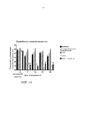
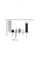
[0017] ФИГУРЫ 1А-1В. (А) представляет собой результаты измерения выработки слезной жидкости либо до начала лечения (день -3, исходный уровень), или в день 7, 14, 21 и 28 после начала лечения; для каждого дня пять столбцов представляют собой измерения слева направо для: наивных, с имитационной обработкой, обработками ВАС, скоп, и ВАС + скоп. Выработка слезной жидкости существенно снижается на протяжении четырех недель введения скополамина (скоп.) и введения хлорида бензалкония плюс скополамин (ВАС + скоп.) относительно контролей или обработки только ВАС. ***=p<0,001. (В) представляет собой результаты измерения показателя окрашивания роговицы флуоресцеином либо до начала лечения (день -3, исходный уровень), или в дни 7, 14, 21 и 28 после начала лечения; для каждого дня пять столбцов представляют собой измерения слева направо для: наивных, с имитационной обработкой, обработками ВАС, скоп, и ВАС + скоп. Окрашивание роговицы флуоресцеином значительно повышается на протяжении четырех недель введения ВАС + скоп, относительно контролен и относительно обработки либо ВАС, либо скоп, по отдельности. ****=p<0,0001.[0017] FIGURES 1A-1B. (A) represents the results of tear fluid production either before treatment (day -3, baseline), or on
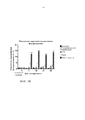
[0018] ФИГУРЫ 2A-2D. (А) ангиогенез роговицы, измеренный по повышенной интенсивности окрашивания клеток CD31+, повышается в глазах, обработанных ВАС + скоп., относительно контролей и относительно обработки либо ВАС, либо скоп, по отдельности. (В) длина скелетообразования (новый кровеносный сосуд) кровеносных сосудов роговицы резко увеличивается на протяжении четырехнедельной обработки в глазах, обработанных ВАС + скоп., относительно контролей и относительно обработки либо ВАС, либо скоп, по отдельности. (С) лимфангиогенез, измеренный по повышенной интенсивности окрашивания клеток LYVE1, повышается в глазах, обработанных ВАС + скоп., относительно контролей и относительно обработки либо ВАС, либо скоп, по отдельности. (D) длина скелетообразования лимфатических сосудов роговицы резко увеличивается на протяжении четырехнедельной обработки в глазах, обработанных ВАС + скоп., относительно контролей и относительно обработки либо ВАС, либо скоп, по отдельности.[0018] FIGURES 2A-2D. (A) corneal angiogenesis, measured by the increased intensity of staining of CD31 + cells, is increased in the eyes treated with BAC + sc., Relative to the controls and relative to the treatment of either BAC or scop, individually. (B) the length of skeletal formation (a new blood vessel) of the corneal blood vessels increases sharply during the four-week treatment in the eyes treated with BAC + scop., Relative to the controls and relative to the treatment of either BAC or scap separately. (C) lymphangiogenesis, measured by the increased intensity of staining of LYVE1 cells, rises in the eyes treated with BAC + sc., Relative to the controls and relative to the treatment of either BAC or scop separately. (D) the length of the skeletal formation of the lymphatic vessels of the cornea increases sharply during the four-week treatment in the eyes treated with BAC + scop., Relative to the controls and relative to the treatment of either BAC or scop separately.
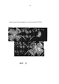
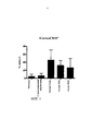
[0019] ФИГ. 3. Исследование с помощью проточной цитометрии внеглазничных слезных желез. Процентная доля клеток CD45+ повышена в клетках, обработанных скоп., ВАС и скоп. + ВАС, относительно контролей.FIG. 3. A study using flow cytometry of extraorbital lacrimal glands. The percentage of CD45 + cells is increased in cells treated with scop., BAC and scop. + YOU, regarding controls.
ПОДРОБНОЕ ОПИСАНИЕ РАСКРЫТИЯDETAILED DESCRIPTION OF DISCLOSURE
[0020] В данном документе раскрыты способы индуцирования сухого кератоконъюнктивита у животного, который отражает патофизиологию сухого кератоконъюнктивита (DED) у людей. Дополнительно раскрыты способы применения животных, относящихся к грызунам, с индуцированным сухим кератоконъюнктивитом при тестировании кандидатных средств для лечения DED.[0020] Methods for inducing dry keratoconjunctivitis in an animal that reflects the pathophysiology of dry keratoconjunctivitis (DED) in humans are disclosed herein. Additionally disclosed are methods of using rodent-related animals with induced dry keratoconjunctivitis in testing candidate agents for the treatment of DED.
[0021] Сухой кератоконъюнктивит (DED), также известный как синдром сухого глаза (DES) или keratoconjunctivitis sicca (KCS), представляет собой состояние, характеризуемое физиологически одним или более, двумя или более, тремя или более или всеми из следующего: сниженная выработка слезной жидкости, повышенное испарение слезной пленки, воспаление глаз, повышенная осмолярность (содержание солей) слез и повреждение поверхности глаза. Симптомы DED включают в себя дискомфорт в глазах, нарушение зрения и нестабильность слезной пленки.[0021] Dry keratoconjunctivitis (DED), also known as dry eye syndrome (DES) or keratoconjunctivitis sicca (KCS), is a condition characterized physiologically by one or more, two or more, three or more, or all of the following: decreased production of lacrimal fluids, increased evaporation of the tear film, inflammation of the eyes, increased osmolarity (salt content) of tears and damage to the surface of the eye. Symptoms of DED include eye discomfort, visual impairment, and instability of the tear film.
[0022] В данном документе раскрыты способы индуцирования сухого кератоконъюнктивита у животного, относящегося к грызунам. Способы включают введение скополамина, антихолинергического средства и хлорида бензалкония (ВАС), средства, вызывающего раздражение глаз, животному, относящемуся к грызунам, в количестве и в течение времени, достаточных для индуцирования сухого кератоконъюнктивита у животного. Получаемое расстройство глаз отображает множественные характеристики DED человека.[0022] Methods for inducing dry keratoconjunctivitis in a rodent animal are disclosed herein. Methods include administering scopolamine, an anticholinergic agent and benzalkonium chloride (BAC), an eye irritant, to a rodent animal in an amount and for a time sufficient to induce dry keratoconjunctivitis in the animal. The resulting eye disorder displays the multiple characteristics of a human DED.
[0023] При разработке подхода, раскрытого в данном документе, авторы настоящего изобретения смогли индуцировать сухой кератоконъюнктивит у животных, относящихся к грызунам, который имеет близкое сходство с клиническим заболеванием человека. Например, состояния, представляющие собой сухой кератоконъюнктивит, индуцированные с помощью раскрытых способов, достигаются легче и могут длиться в течение недель. Дополнительно, модели на животных, полученные с помощью раскрытых способов, характеризуются как воспалением глаз, так и дефицитом слезной жидкости, обычно наблюдаемыми при сухом кератоконъюнктивите человека. Таким образом, эта модель позволяет изучать патофизиологические изменения при сухом кератоконъюнктивите и тестировать кандидатов в терапевтические лекарственные средства для лечения DED.[0023] In developing the approach disclosed herein, the inventors of the present invention were able to induce dry keratoconjunctivitis in rodent animals that closely resembles human clinical disease. For example, dry keratoconjunctivitis conditions induced by the disclosed methods are easier to achieve and can last for weeks. Additionally, animal models obtained using the disclosed methods are characterized by both eye inflammation and tear fluid deficiency, commonly observed in dry human keratoconjunctivitis. Thus, this model allows us to study the pathophysiological changes in dry keratoconjunctivitis and test candidates for therapeutic drugs for the treatment of DED.
Создание сухого кератоконъюнктивита у животногоThe creation of dry keratoconjunctivitis in an animal
[0024] На первой стадии способа скополамин вводят в качестве антихолинергического средства и хлорид бензалкония (ВАС) вводят в качестве средства, вызывающего раздражение глаз, животному, относящемуся к грызунам, такому как мышь или крыса.[0024] In a first step of the method, scopolamine is administered as an anticholinergic agent and benzalkonium chloride (BAC) is administered as an eye irritant to a rodent animal such as a mouse or rat.
[0025] Антихолинергическое средство представляет собой вещество, которое блокирует нейротрансмиттер ацетилхолин в центральной и/или периферической нервной системе. Примеры антихолинергических средств включают, например, скополамин, гидрохлорид скополамина, метобромид скополамина, атропин, метилнитрат атропина и сульфат атропина. В способах, описанных в данном документе, антихолинергическим средством является скополамин или гидрохлорид скополамина.[0025] An anticholinergic agent is a substance that blocks the neurotransmitter acetylcholine in the central and / or peripheral nervous system. Examples of anticholinergics include, for example, scopolamine, scopolamine hydrochloride, scopolamine methobromide, atropine, atropine methyl nitrate and atropine sulfate. In the methods described herein, the anticholinergic agent is scopolamine or scopolamine hydrochloride.
[0026] В раскрытых способах скополамин вводят системно. Способы системного введения включают в себя инъекцию, трансдермальный пластырь и имплантацию насоса, такого как осмотический насос, который позволяет проводить непрерывное введение дозы лабораторным животным. Такие способы системного введения в моделях на животных известны из уровня техники. В конкретных вариантах осуществления скополамин вводят с помощью осмотического мини-насоса. Подходящие осмотические мини-насосы включают, например, осмотические насосы ALZET (Durect Corporation, Купертино, Калифорния).[0026] In the disclosed methods, scopolamine is administered systemically. Methods of systemic administration include injection, a transdermal patch, and implantation of a pump, such as an osmotic pump, which allows continuous administration of a dose to laboratory animals. Such systemic administration methods in animal models are known in the art. In specific embodiments, scopolamine is administered using an osmotic mini pump. Suitable osmotic mini-pumps include, for example, ALZET osmotic pumps (Durect Corporation, Cupertino, CA).
[0027] Для имплантации насоса грызуна анестезируют и устанавливают насос подкожно. Насос обеспечивает контролируемое количество скополамина. Типичные концентрации скополамина включают 0,1-4,0 мг на 20 г веса тела в день, 0,5-3,5 мг на 20 г веса тела в день, 1,0-3,0 мг на 20 г веса тела в день или приблизительно 2,0 мг на 20 г веса тела в день. Скополамин вводят в течение нескольких дней или нескольких недель, например, 1, 2, 3, 4 недель или больше.[0027] To implant the rodent pump, anesthetize and install the pump subcutaneously. The pump provides a controlled amount of scopolamine. Typical concentrations of scopolamine include 0.1-4.0 mg per 20 g body weight per day, 0.5-3.5 mg per 20 g body weight per day, 1.0-3.0 mg per 20 g body weight per day or approximately 2.0 mg per 20 g of body weight per day. Scopolamine is administered for several days or several weeks, for example, 1, 2, 3, 4 weeks or more.
[0028] Средство, вызывающее раздражение глаз, представляет собой вещество, которое приводит к раздражению в глазу. Примеры средств, вызывающих раздражение глаз, включают в себя поверхностно-активные вещества, консерванты, аллергены, мелкие частицы, а также иссушающие средства или условия окружающей среды. В способах, раскрытых в данном документе, средством, вызывающим раздражение глаз, является хлорид бензалкония (ВАС).[0028] An eye irritant is a substance that causes irritation in the eye. Examples of eye irritating agents include surfactants, preservatives, allergens, fine particles, as well as drying agents or environmental conditions. In the methods disclosed herein, an eye irritant is benzalkonium chloride (BAC).
[0029] В раскрытых способах ВАС вводят местно на поверхность глаза. ВАС можно вводить в концентрации 0,05-1,0%, а в конкретных вариантах осуществления 0,1-0,2%, в дозировке 0,5-2,0 мкл на дозу, например 1 мкл на дозу. Введение может осуществляться от одного до четырех раз в день, например два раза в день, в течение от одного до трех дней в неделю. В некоторых вариантах осуществления введение осуществляют два раза в день в течение двух дней в неделю. Введение ВАС можно осуществлять в течение одной или более недель, например, 1, 2, 3, 4 недель или больше.[0029] In the disclosed methods, BAC is administered topically to the surface of the eye. YOU can be administered at a concentration of 0.05-1.0%, and in specific embodiments, 0.1-0.2%, at a dosage of 0.5-2.0 μl per dose, for example 1 μl per dose. The introduction can be carried out from one to four times a day, for example, twice a day, for one to three days a week. In some embodiments, administration is carried out twice a day for two days a week. The introduction of YOU can be carried out for one or more weeks, for example, 1, 2, 3, 4 weeks or more.
Оценка состояний, представляющих собой сухой кератоконъюнктивит, у модельных животныхAssessment of dry keratoconjunctivitis conditions in model animals
[0030] Сухой кератоконъюнктивит подтверждают в модели на животных по выявлению снижения выработки слезной жидкости и повышению раздражения слизистой оболочки глаза относительно контрольных уровней. Сухой кератоконъюнктивит можно дополнительно подтвердить по выявлению усиления ангиогенеза и/или лимфангиогенеза относительно контрольных уровней.[0030] Dry keratoconjunctivitis is confirmed in an animal model by detecting decreased production of tear fluid and increased irritation of the mucous membrane of the eye relative to control levels. Dry keratoconjunctivitis can be further confirmed by detecting increased angiogenesis and / or lymphangiogenesis relative to control levels.
[0031] По всему данному раскрытию контрольные уровни представляют собой уровни в необработанном глазу животного, относящегося к грызунам. Если оценка направлена на эффективность системно применяемого средства, необработанный глаз представляет собой глаз необработанного животного, относящегося к грызунам. Необработанное животное может быть наивным (по сути не получающим обработку) или ложнооперированным животным (получающим имплантированный насос, который не доставляет средство, или который доставляет плацебо, такое как физиологический раствор, животному, или получающим хирургическое вмешательство без фактической имплантации насоса). Если средство наносят местно на глаз, а не системно, контроль может также представлять собой необработанный глаз животного с одним обработанным глазом. В контексте оценки индукции состояний, представляющих собой сухой кератоконъюнктивит, при введении скополамина и ВАС контрольные уровни представляют собой уровни в глазах необработанных животных.[0031] Throughout this disclosure, control levels are levels in the raw eye of a rodent animal. If the assessment is aimed at the effectiveness of a systemically applied agent, the untreated eye is the eye of an untreated animal belonging to rodents. An untreated animal can be naive (essentially not receiving treatment) or a falsely operated animal (receiving an implanted pump that does not deliver the drug, or which delivers a placebo, such as saline, to the animal, or receiving surgery without actually implanting the pump). If the agent is applied topically to the eye, and not systemically, the control may also be the untreated eye of an animal with one treated eye. In the context of assessing the induction of conditions representing dry keratoconjunctivitis, when scopolamine and BAC are administered, control levels are levels in the eyes of untreated animals.
[0032] Сниженную выработку слезной жидкости идентифицируют как снижение выработки слезной жидкости по меньшей мере на 10%, 20%, 30%, 40% или 50% относительно контрольных уровней. Способы измерения выработки слезной жидкости включают фенольный тест с красной нитью (PRT) и тест Ширмера. В тесте PRT животных фиксируют без анестезии и нить, пропитанную феноловым красным (такую как ZONE-QUICK, FCI Ophthalmics, Пембрук, Массачусетс), помещают в медиальный или латеральный кантус (внутренний или внешний угол глаза) или прикасаются к конъюнктивальному или слезному мешку в течение 20 секунд для измерения выработки слезной жидкости. Количество нити, которая становится красной (в результате изменения pH из-за смачивания щелочными слезами), измеряют в миллиметрах. Процедуру можно повторить на другом глазу, чтобы получить 2 измерения для каждой мыши. В тесте Ширмера применяют бумажные полоски для измерения выработки слез. Тест включает в себя помещение небольшой полоски фильтровальной бумаги под нижнее веко (конъюнктивальный мешок). Полоску удерживают на месте в течение нескольких минут. Затем бумагу удаляют и количество влаги измеряют в мм.[0032] Decreased tear fluid production is identified as a decrease in tear fluid production by at least 10%, 20%, 30%, 40%, or 50% relative to control levels. Methods for measuring tear fluid production include the red thread phenolic test (PRT) and the Schirmer test. In the PRT test, the animals are fixed without anesthesia and the thread soaked in phenol red (such as ZONE-QUICK, FCI Ophthalmics, Pembroke, Massachusetts) is placed in the medial or lateral canthus (inner or outer corner of the eye) or is touched with a conjunctival or lacrimal sac for 20 seconds to measure tear fluid production. The amount of thread that turns red (as a result of pH changes due to wetting with alkaline tears) is measured in millimeters. The procedure can be repeated on the other eye to get 2 measurements for each mouse. The Schirmer test uses paper strips to measure tear production. The test involves placing a small strip of filter paper under the lower eyelid (conjunctival sac). The strip is held in place for several minutes. Then the paper is removed and the amount of moisture is measured in mm.
[0033] Повышенное раздражение слизистой оболочки глаза идентифицируют путем выявления в глазу одного или более из: повышения количества воспалительных клеток, повышения количества провоспалительных цитокинов в глазу или повышения интенсивности окрашивания флуоресцеином относительно контрольных уровней.[0033] Increased irritation of the mucous membrane of the eye is identified by detecting in the eye one or more of: an increase in the number of inflammatory cells, an increase in the number of pro-inflammatory cytokines in the eye, or an increase in the intensity of fluorescein staining relative to control levels.
[0034] Повышение количества воспалительных клеток представляет собой повышение по меньшей мере на 10%, 20%, 30%, 40% или 50% одного или более типов воспалительных/иммунных клеток относительно контрольных уровней. Типы воспалительных/иммунных клеток включают миелоидные клетки (включая нейтрофилы, моноциты, эозинофилы и базофилы) и лимфоциты (включая Т-клетки и В-клетки). Воспалительные клетки можно выявить и измерить различными способами, известными из уровня техники. Чаще всего воспалительные клетки измеряют путем определения присутствия/избытка клеток с идентификацией маркеров клеточной поверхности. Например, как миелоидные клетки, так и лимфоциты являются CD45+, и, таким образом, этот маркер можно применять для идентификации присутствия воспалительных клеток относительно невоспалительных клеток. Лимфоцит-специфичные маркеры включают CD3 (идентифицирующий Т-клетки), CD4 (идентифицирующий хелперные Т-клетки), CD8 (идентифицирующий цитотоксические Т-клетки) и В220 или CD19 (идентифицирующий В-клетки). В данной области известны дополнительные маркеры клеточной поверхности, а также антитела и другие средства, с помощью которых обнаруживают эти маркеры. Антитела, с помощью которых обнаруживают эти маркеры, можно применять для идентификации воспалительных клеток, например, путем гистологического исследования ткани роговицы и окрашивания тканевых срезов антителами. Антитела, с помощью которых обнаруживают эти маркеры, также можно использовать путем применения антител к популяции клеток для окрашивания в отношении экспрессии специфических маркеров и анализа клеток с помощью проточной цитометрии для количественного определения клеток, экспрессирующих данные маркеры.[0034] An increase in the number of inflammatory cells is an increase of at least 10%, 20%, 30%, 40%, or 50% of one or more types of inflammatory / immune cells relative to control levels. Types of inflammatory / immune cells include myeloid cells (including neutrophils, monocytes, eosinophils and basophils) and lymphocytes (including T cells and B cells). Inflammatory cells can be detected and measured in various ways known in the art. Inflammatory cells are most often measured by determining the presence / excess of cells with identification of cell surface markers. For example, both myeloid cells and lymphocytes are CD45 +, and thus this marker can be used to identify the presence of inflammatory cells relative to non-inflammatory cells. Lymphocyte-specific markers include CD3 (identifying T cells), CD4 (identifying helper T cells), CD8 (identifying cytotoxic T cells), and B220 or CD19 (identifying B cells). Additional cell surface markers are known in the art, as well as antibodies and other agents by which these markers are detected. The antibodies with which these markers are detected can be used to identify inflammatory cells, for example, by histological examination of corneal tissue and staining of tissue sections with antibodies. Antibodies with which these markers are detected can also be used by applying antibodies to a population of cells for staining for the expression of specific markers and analysis of cells using flow cytometry to quantify cells expressing these markers.
[0035] Повышение содержания провоспалительных цитокинов представляет собой повышение по меньшей мере на 10%, 20%, 30%, 40% или 50% одного или более типов провоспалительных цитокинов относительно контрольных уровней. Провоспалительные цитокины включают в себя интерлейкины (IL), такие как IL-1, IL-2, IL-3, IL-4, IL-5, IL-6, IL-7, IL-8, IL-9, IL-10, IL-11, IL-12, IL-13, факторы некроза опухолей (TNF), такие как TNF-α, TNF-β, TGF-β (трансформирующий фактор роста-β, CXCL9 и интерфероны (IFN), такие как IFN-γ. Способы измерения провоспалительных цитокинов включают в себя иммунологические анализы, такие как твердофазный иммуноферментный анализ (ELISA), в которых применяют антитела или другие средства, с помощью которых обнаруживают цитокины, для идентификации и количественного определения цитокинов в образце.[0035] An increase in pro-inflammatory cytokines is an increase of at least 10%, 20%, 30%, 40% or 50% of one or more types of pro-inflammatory cytokines relative to control levels. Pro-inflammatory cytokines include interleukins (IL), such as IL-1, IL-2, IL-3, IL-4, IL-5, IL-6, IL-7, IL-8, IL-9, IL- 10, IL-11, IL-12, IL-13, tumor necrosis factors (TNF), such as TNF-α, TNF-β, TGF-β (transforming growth factor-β, CXCL9 and interferons (IFN), such as IFN-γ. Methods of measuring pro-inflammatory cytokines include immunological assays, such as enzyme-linked immunosorbent assay (ELISA), which use antibodies or other means by which cytokines are detected to identify and quantify cytokines in a sample.
[0036] Повышение интенсивности окрашивания флуоресцеином представлено баллом, составляющим более 5, 8, 10 или 12, с применением оценки Национального института глаза (National Eye Institute, NEI). Для окрашивания флуоресцеином флуоресцеин натрия наносят на поверхность глаза животного, как правило, без седации. Через несколько минут после нанесения оценивают окрашивание роговицы флуоресцеином под микроскопом с использованием синего света. Нарушения в глазу, такие как ссадины и воспаление, флуоресцируют с большей интенсивностью относительно здоровой ткани роговицы. Флуоресценцию оценивают с применением стандартов оценки Национального института глаза (NEI). Согласно оценке NEI пять секторов роговицы (центральный, верхний, нижний, назальный и височный) оценивают отдельно по шкале 0-3 для флуоресценции, при этом 0 указывает на отсутствие окрашивания и 3 указывает на обширное окрашивание, с максимальным баллом 15 на глаз. Если используется другой стандарт оценки или метод флуоресценции, повышение интенсивности окрашивания флуоресцеином будет определяться как повышение интенсивности флуоресценции по меньшей мере на 10, 20, 30, 40 или 50% относительно контрольных уровней.[0036] The increase in fluorescein staining intensity is represented by a score of more than 5, 8, 10, or 12, using a National Eye Institute (NEI) score. For fluorescein staining, sodium fluorescein is applied to the surface of the animal’s eye, usually without sedation. A few minutes after application, staining of the cornea with fluorescein under a microscope using blue light is evaluated. Disorders in the eye, such as abrasions and inflammation, fluoresce with greater intensity relative to healthy corneal tissue. Fluorescence is assessed using National Institute of Eye (NEI) assessment standards. According to NEI, five sectors of the cornea (central, upper, lower, nasal and temporal) are evaluated separately on a 0-3 scale for fluorescence, with 0 indicating no staining and 3 indicating extensive staining, with a maximum score of 15 per eye. If another evaluation standard or fluorescence method is used, an increase in fluorescence staining intensity will be defined as an increase in fluorescence intensity by at least 10, 20, 30, 40 or 50% relative to control levels.
[0037] Измерения ангиогенеза и/или лимфангиогенеза роговицы могут дополнительно подтвердить сухой кератоконъюнктивит. Для изучения ангиогенеза и лимфангиогенеза животное подвергают эвтаназии и роговицу препарируют и инкубируют со средствами, с помощью которых идентифицируют образование новых лимфатических сосудов, такими как антитела к эндотелиальному гиалуронановому рецептору-1 лимфатических сосудов (LYVE1), или средствами, с помощью которых идентифицируют образование новых кровеносных сосудов, такими как антитела к CD31, маркеру эндотелиальных клеток-предшественников. Идентификация областей роста новых кровеносных или лимфатических сосудов может быть применена в качестве дополнительного индикатора ухудшения состояния глаз и развития сухого кератоконъюнктивита.[0037] Measurements of angiogenesis and / or lymphangiogenesis of the cornea may further confirm dry keratoconjunctivitis. To study angiogenesis and lymphangiogenesis, the animal is euthanized and the cornea is dissected and incubated with agents that identify the formation of new lymphatic vessels, such as antibodies to the endothelial hyaluronan receptor-1 lymphatic vessels (LYVE1), or with the means that identify the formation of new blood vessels vessels, such as antibodies to CD31, a marker of endothelial progenitor cells. Identification of the growth areas of new blood or lymph vessels can be used as an additional indicator of worsening eye condition and the development of dry keratoconjunctivitis.
Тестирование кандидатных средств для лечения сухого кератоконъюнктивитаCandidate Testing for Dry Keratoconjunctivitis
[0038] Грызуны согласно настоящему раскрытию (крысы и мыши) пригодны для изучения DED и тестирования кандидатных средств для лечения сухого кератоконъюнктивита.[0038] Rodents according to the present disclosure (rats and mice) are suitable for studying DED and testing candidate agents for treating dry keratoconjunctivitis.
[0039] Соответственно, в данном документе раскрыты способы тестирования эффективности кандидатного средства для лечения сухого кератоконъюнктивита, которые включают введение кандидатного средства животному с DED, индуцированным с использованием раскрытых способов, и тестирование способности кандидата лечить DED.[0039] Accordingly, methods for testing the efficacy of a candidate agent for treating dry keratoconjunctivitis, which include administering a candidate agent to an animal with DED induced using the disclosed methods, and testing a candidate's ability to treat DED, are disclosed herein.
[0040] Под термином «лечение» подразумевается облегчение симптомов или состояний DED или предупреждение DED (например, предупреждение или задержка наступления или ограничение развития DED).[0040] The term "treatment" means the alleviation of the symptoms or conditions of DED or the prevention of DED (for example, preventing or delaying the onset or limiting the development of DED).
[0041] Выражения «тестируемое средство» и «кандидатное средство» используются в данном документе взаимозаменяемо для обозначения любого средства, предназначенного для лечения сухого кератоконъюнктивита. Средство может представлять собой рекомбинантную, модифицированную или природную молекулу нуклеиновой кислоты; синтетический, модифицированный или природный пептид; синтетический, модифицированный или природный полипептид, включая антитела; или органическое или неорганическое соединение, включая малые молекулы. Выражение «малая молекула» относится к соединениям с молекулярной массой менее 3000 Дальтон, предпочтительно, менее 2000 или 1500, более предпочтительно, менее 1000 или 800 Дальтон.[0041] The terms “test agent” and “candidate agent” are used interchangeably herein to mean any agent intended for the treatment of dry keratoconjunctivitis. The agent may be a recombinant, modified or natural nucleic acid molecule; synthetic, modified or natural peptide; synthetic, modified or natural polypeptide, including antibodies; or an organic or inorganic compound, including small molecules. The expression "small molecule" refers to compounds with a molecular weight of less than 3000 Daltons, preferably less than 2000 or 1500, more preferably less than 1000 or 800 Daltons.
[0042] Тестируемое средство, подлежащее оценке, можно вводить животному местно или системно. Например, средство можно вводить в форме глазных капель или интраназального спрея. В качестве альтернативы, средство можно вводить перорально или парентерально, и если вводится парентерально, оно может находиться в форме раствора, суспензии, мази, инъекции, суппозитория и т.п. Подходящую дозу кандидатного средства можно определить на основании веса тела животного и способа введения. Условия введения, такие как время и количество раз введения, также определяются в зависимости от средства, подлежащего применению.[0042] The test agent to be evaluated can be administered to an animal topically or systemically. For example, the agent may be administered in the form of eye drops or an intranasal spray. Alternatively, the agent can be administered orally or parenterally, and if administered parenterally, it can be in the form of a solution, suspension, ointment, injection, suppository, and the like. An appropriate dose of a candidate agent can be determined based on the animal’s body weight and route of administration. The conditions of administration, such as the time and number of times of administration, are also determined depending on the means to be used.
Введение кандидатного средства в начале индукции DEDCandidate administration at the start of DED induction
[0043] В одном варианте осуществления кандидатное средство вводят одновременно с началом индукции DED. В некоторых вариантах осуществления наборы тестируемых животных, относящихся к грызунам, рандомизируют на следующие группы: наивные (необработанные), с имитационной обработкой (системное введение физиологического раствора, имплантация насоса без обработки скополамином или хирургическая операция, аналогичная имплантации насоса, без фактической имплантации насоса), с хлоридом бензалкония плюс скополамин (ВАС плюс скоп.), только с тестируемым средством или с тестируемым средством плюс ВАС плюс скоп. Животным в группах тестируемого средства вводят кандидатное средство одновременно с началом индукции сухого кератоконъюнктивита, то есть начиная с того же дня или в течение той же недели, когда начинается индукция DED. Все обработки проводили в течение по меньшей мере одной, двух, трех или четырех недель. Выработку слезной жидкости и окрашивание роговицы флуоресцеином измеряют в дни 7, 14, 21 и 28. В день 28 животных подвергают эвтаназии и собирают внеглазничную слезную железу и роговицу для дальнейшего изучения.[0043] In one embodiment, the candidate agent is administered simultaneously with the initiation of DED induction. In some embodiments, the implementation of the sets of test animals related to rodents, randomize into the following groups: naive (untreated), with simulation processing (systemic administration of physiological saline, implantation of the pump without treatment with scopolamine or a surgical operation similar to implantation of the pump, without actual implantation of the pump), with benzalkonium chloride plus scopolamine (BAC plus scop.), only with the test tool or with the test tool plus BAC plus scop. The animals in the groups of the test drug are administered the candidate drug at the same time as the induction of dry keratoconjunctivitis begins, that is, starting from the same day or during the same week when the induction of DED begins. All treatments were performed for at least one, two, three, or four weeks. The production of tear fluid and staining of the cornea with fluorescein is measured on
[0044] Способность кандидатного средства предупреждать, задерживать наступление или ограничивать развитие DED определяют путем выявления способности кандидатного средства сохранять выработку слезной жидкости и/или предупреждать или ограничивать раздражение слизистой оболочки глаза на одинаковых уровнях с контрольными уровнями. В этом контексте контрольные уровни представляют собой уровни для необработанного глаза (то есть глаза без обработки кандидатным средством), который может включать в себяглаз животных, получающих скополамин и хлорид бензалкония, но не получающих кандидатное средство, или один глаз животного, получающего скополамин и хлорид бензалкония, а также кандидатное средство, где кандидатное средство наносится местно на другой глаз того же животного.[0044] The ability of a candidate agent to prevent, delay the onset or limit the development of DED is determined by detecting the ability of the candidate agent to maintain tear fluid production and / or to prevent or limit irritation of the mucous membrane of the eye at the same levels as the control levels. In this context, control levels are the levels for the untreated eye (i.e., the eyes without treatment with a candidate drug), which may include the eyes of animals receiving scopolamine and benzalkonium chloride, but not receiving the candidate drug, or one eye of an animal receiving scopolamine and benzalkonium chloride as well as a candidate agent, where the candidate agent is applied topically to the other eye of the same animal.
[0045] В одном варианте осуществления у животных, получающих тестируемое средство плюс ВАС плюс скоп., не развивается DED, то есть выработка слезной жидкости сохраняется на уровне или в пределах 5%, 10% или 15% от контрольных уровней в течение всего исследования и/или уровни воспаления глаз, измеренные по уровням воспалительных клеток, уровням провоспалительных цитокинов в глазу и/или показателю окрашивания флуоресцеином, сохраняются на уровне или в пределах 5%, 10% или 15% от контрольных уровней в течение всего исследования.[0045] In one embodiment, animals receiving the test agent plus BAC plus sc. Do not develop DED, that is, tear fluid production is maintained at or within 5%, 10%, or 15% of control levels throughout the study and / or the levels of eye inflammation, measured by the levels of inflammatory cells, the levels of proinflammatory cytokines in the eye and / or the fluorescein staining index, remain at or within 5%, 10% or 15% of the control levels throughout the study.
[0046] В другом варианте осуществления животные, получающие тестируемое средство плюс ВАС плюс скоп., демонстрируют задержку наступления симптомов DED, то есть имеет место задержка в развитии сниженной выработки слезной жидкости и/или раздражения слизистой оболочки глаза на одну, две или три недели по сравнению с животными, получающими ВАС плюс скоп., но не получающими кандидатное средство.[0046] In another embodiment, animals receiving the test agent plus BAC plus sc., Show a delay in the onset of DED symptoms, that is, there is a delay in the development of reduced production of tear fluid and / or irritation of the mucous membrane of the eye for one, two or three weeks compared with animals receiving BAC plus scop., but not receiving the candidate drug.
[0047] В другом варианте осуществления у животных, получающих тестируемое средство плюс ВАС плюс скоп., развиваются более мягкие симптомы DED, то есть у этих животных происходит снижение выработки слезной жидкости и/или раздражение слизистой оболочки глаза, но снижение выработки слезной жидкости и/или раздражение глаз происходит на уровнях, промежуточных между уровнями у животных, не получающих обработку, индуцирующую DED (например, наивные животные), и уровнями, наблюдаемыми у животных, получающих ВАС плюс скоп., но не получающих тестируемое средство.[0047] In another embodiment, animals receiving the test agent plus BAC plus scop develop milder DED symptoms, that is, these animals experience a decrease in tear fluid production and / or irritation of the mucous membrane of the eye, but a decrease in tear fluid production and / or eye irritation occurs at levels intermediate between levels in animals not receiving DED-inducing treatment (eg, naive animals) and levels observed in animals receiving BAC plus scop, but not receiving test with COROLLARY.
Введение кандидатного средства после индукции DEDCandidate administration after DED induction
[0048] В другом варианте осуществления кандидатное средство вводят после начала индукции DED. В некоторых вариантах осуществления животным в группах тестируемого средства вводят кандидатное средство, начиная с одной-двух недель после начала индукции сухого кератоконъюнктивита, то есть между днями 7-14 после начала индукции сухого кератоконъюнктивита. Тестируемое средство вводили в течение одной-трех недель. Выработку слезной жидкости и окрашивание роговицы флуоресцеином измеряют в дни 7, 14, 21 и 28 от начала индукции сухого кератоконъюнктивита. В день 28 животных подвергают эвтаназии и собирают внеглазничную слезную железу и роговицу для дальнейшего изучения.[0048] In another embodiment, the candidate agent is administered after initiation of DED induction. In some embodiments, a candidate drug is administered to the animals in the groups of the test drug, starting one to two weeks after the start of dry keratoconjunctivitis induction, that is, between days 7-14 after the start of dry keratoconjunctivitis induction. The test agent was administered for one to three weeks. The production of tear fluid and staining of the cornea with fluorescein is measured on
[0049] Способность кандидатного средства лечить DED определяют путем выявления повышения выработки слезной жидкости и/или снижения раздражения слизистой оболочки глаза у животных, получающих кандидатное средство и ВАС плюс скоп., относительно контрольных уровней, которые в этом контексте могут представлять собой уровни в глазах животных, получающих ВАС плюс скоп., но не получающих кандидатное средство.[0049] The ability of a candidate drug to treat DED is determined by detecting an increase in tear fluid production and / or a decrease in irritation of the mucous membrane of the eye in animals receiving the candidate drug and BAC plus sc., Relative to control levels, which in this context may be levels in the eyes of animals receiving YOU plus scop., but not receiving the candidate drug.
ПРИМЕРЫEXAMPLES
Пример 1. Создание у животного состояния, представляющего собой сухой кератоконъюнктивит.Example 1. The creation of an animal condition, which is a dry keratoconjunctivitis.
[0050] Животные. Все процедуры, связанные с животными, были рассмотрены и одобрены IACUC Regeneron и проведены в соответствии с постановлением о использовании животных в офтальмологических исследованиях и исследованиях зрения ARVO. Взрослых мышей C57BL/6 (возрастом 8-10 недель) приобрели у Taconic Biosciences (Джермантаун, Нью-Йорк).[0050] Animals. All animal procedures have been reviewed and approved by IACUC Regeneron and carried out in accordance with the regulation on the use of animals in ARVO ophthalmic and vision studies. Adult C57BL / 6 mice (8-10 weeks old) were purchased from Taconic Biosciences (Germantown, NY).
[0051] Индукция модели сухого кератоконъюнктивита: 27 взрослыхсамцов мышей C57BL/6 рандомизировали на группы наивных (необработанных), с имитационной обработкой (подвергающихся хирургической операции, эквивалентной хирургической операции для имплантации насоса, но без фактической имплантации насоса и без обработки скополамином или ВАС), с хлоридом бензалкония (ВАС), скополамином (скоп.) или ВАС плюс скоп.[0051] Induction of a model of dry keratoconjunctivitis: 27 adult male C57BL / 6 mice were randomized into naive (untreated) groups with simulated treatment (undergoing surgery equivalent to surgery for pump implantation, but without actual pump implantation and without scopolamine or BAC), with benzalkonium chloride (BAC), scopolamine (sc.) or BAC plus scop.
[0052] Имплантация насоса со скополамином. Для группы скоп, мышам подкожно имплантировали осмотический насос, наполненный скоп.(2 мг/20 г веса тела/день), на 4 недели. Мышей анестезировали кетамином (120 мг/кг, IP) и ксилазином (5 мг/кг, IP). С применением асептической микрохирургической техники и операционного микроскопа осмотический мини-насос помещали под кожу спины, затем кожу зашивали и на рану накладывали офтальмологическую мазь с эритромицином для предупреждения инфекции.[0052] Implantation of a scopolamine pump. For the osprey group, an osmotic pump filled with osprey (2 mg / 20 g body weight / day) was subcutaneously implanted into mice for 4 weeks. Mice were anesthetized with ketamine (120 mg / kg, IP) and xylazine (5 mg / kg, IP). Using an aseptic microsurgical technique and an operating microscope, an osmotic mini-pump was placed under the skin of the back, then the skin was sutured and an ophthalmic ointment with erythromycin was applied to the wound to prevent infection.
[0053] Обработка хлоридом бензалкония. Для обработки ВАС 1 мкл 0,1-0,2% ВАС вводили местно на поверхность правого глаза дважды в день, 2 дня в неделю в течение четырех недель.[0053] Treatment with benzalkonium chloride. To treat YOU, 1 μl of 0.1-0.2% YOU was injected locally on the surface of the right eye twice a day, 2 days a week for four weeks.
[0054] Измерение выработки слезной жидкости и оценка окрашивания роговицы флуоресцеином. Оценку выработки слезной жидкости и окрашивание роговицы флуоресцеином проводили раз в неделю в день 7, 14, 21 и 28. Для измерения выработки слезной жидкости зафиксированному животному в височный мешок глаза (не касаясь роговицы) помещали нить, пропитанную феноловым красным, и удерживали на месте в течение 1 минуты. Влажную часть нити пересчитывали на мм как выработку слезной жидкости. Для окрашивания флуоресцеином 0,5 мкл 2% флуоресцеина натрия помещали на поверхность глаза. Через пять минут после нанесения окрашивание роговицы флуоресцеином оценивали под микроскопом с применением стандартов оценки Национального института глаза (NEI). Для оценки роговицу разделяли на пять секторов (центральный, верхний, нижний, назальный и височный), каждый из которых оценивали по шкале 0-3 для флуоресценции, при этом 0 указывает на отсутствие окрашивания и 3 указывает на обширное окрашивание, с максимальной оценкой 15 на глаз.[0054] Measurement of tear fluid production and evaluation of corneal staining with fluorescein. Assessment of tear fluid production and staining of the cornea with fluorescein was performed once a week on
[0055] Количественная оценка ангиогенеза роговицы, лимфангиогенеза роговицы и инфильтрации слезной железы. В день 28 после индукции сухого кератоконъюнктивита под глубоким наркозом с применением кетамина (120 мг/кг, IP) и ксилазина (5 мг/кг, IP) глазные яблоки собирали в 4% параформальдегид (PFA) для исследования ангиогенеза и лимфангиогенеза роговицы. Внеглазничные слезные железы собирали для исследования с помощью проточной цитометрии, чтобы проверить инфильтрацию иммунных клеток.[0055] Quantifying corneal angiogenesis, corneal lymphangiogenesis, and lacrimal gland infiltration. On
[0056] Гистологический анализ. Роговицу отделяли от глазных яблок, промывали в PBS, блокировали в течение 1 часа при комнатной температуре с 10% нормальной ослиной сывороткой и 1% Triton Х-100 в PBS для блокирования неспецифических сайтов связывания антител. Затем образцы промывали в PBS, инкубировали со смесью антител к эндотелиальному гиалуронановому рецептору-1 лимфатических сосудов (LYVE1) (Regeneron Pharmaceuticals, Inc., Территаун, Нью-Йорк) при 1:1000 и антител к CD31 (Santa Cruz Biotechnology, Санта-Круз, Калифорния) при 1:100 при 4°C в течение ночи. Затем образцы промывали в фосфатно-солевом буферном растворе (PBS) и инкубировали с вторичным антителом, конъюгированным с красителем, антителом осла к IgG(H+L) крысы, конъюгированным с AlexaFluor 488 1:100 (Life Technologies, А21208, Грейсленд, Нью-Йорк) и антителом осла к IgG(H+L) крысы, конъюгированным с AlexaFluor 594 1:400 (Life Technologies, А21207) в течение 2 часов при комнатной температуре. После инкубации образцы промывали в PBS и закрепляли на предметных стеклах. Изображения окрашенных кровеносных и лимфатических сосудов регистрировали с помощью цифровой камеры RT SE Spot, прикрепленной к флуоресцентному микроскопу (Nikon Eclipse 80i). Для анализа изображений использовали программное обеспечение для обработки изображений.[0056] Histological analysis. The cornea was separated from the eyeballs, washed in PBS, blocked for 1 hour at room temperature with 10% normal donkey serum and 1% Triton X-100 in PBS to block non-specific antibody binding sites. Then the samples were washed in PBS, incubated with a mixture of antibodies to the endothelial hyaluronan receptor-1 lymphatic vessels (LYVE1) (Regeneron Pharmaceuticals, Inc., Territown, NY) at 1: 1000 and antibodies to CD31 (Santa Cruz Biotechnology, Santa Cruz , California) at 1: 100 at 4 ° C overnight. Samples were then washed in phosphate buffered saline (PBS) and incubated with a secondary antibody conjugated to a dye, a donkey anti-rat IgG (H + L) antibody conjugated to 1: 100 AlexaFluor 488 (Life Technologies, A21208, Graceland, New York) and donkey anti-rat IgG (H + L) antibody conjugated to AlexaFluor 594 1: 400 (Life Technologies, A21207) for 2 hours at room temperature. After incubation, the samples were washed in PBS and mounted on slides. Images of stained blood and lymph vessels were recorded using an RT SE Spot digital camera attached to a fluorescence microscope (Nikon Eclipse 80i). For image analysis, image processing software was used.
[0057] Проточная цитометрия. После препарирования внеглазничные слезные железы помещали в PBS (Life Technologies) с 3% раствором фетальной бычьей сыворотки (FBS) (Life Technologies). Железы диссоциировали с помощью программы spleen-04 с применением диссоциатора gentleMACS (Miltenyi Biotech, Сан-Диего, Калифорния). Суспензию клеток фильтровали через сетчатый фильтр 70 мкм (Miltenyi Biotech, Сан-Диего, Калифорния) для удаления остатков ткани. Клетки промывали однократно буфером для FACS (PBS с 3% FBS и 2 мМ EDTA) (Life Technologies). Промытые клетки ресуспендировали в буфере для FACS с Аb, блокирующим FcR, (разведение 1:50) (Biolegend, Сан-Диего, Калифорния) и инкубировали в течение 15 минут при 4°C. Клетки центрифугировали с удалением Ab, блокирующего FcR, и дополнительно окрашивали с использованием набора для синего окрашивания мертвых клеток перед фиксацией LIVE/DEAD (разведение 1:500) (Life Technologies) вместе с IgG2b крысы к CD45 мыши, конъюгированным с PerCP-Су5.5, клон 30-F11 (разведение 1:100) (BD Bioscience). Клетки окрашивали в течение 25 мин. при 4°C, после чего проводили две промывки буфером для FACS. Затем клетки ресуспендировали в стабилизирующем фиксаторе (BD) и оценивали на LSRII (BD Bioscience) в соответствии со стандартным протоколом, предоставленным компанией. Данные анализировали с применением FlowJo (Tree Star, Эшленд, Орегон).[0057] Flow cytometry. After preparation, the extraorbital lacrimal glands were placed in PBS (Life Technologies) with a 3% solution of fetal bovine serum (FBS) (Life Technologies). The glands were dissociated using the spleen-04 program using the gentleMACS dissociator (Miltenyi Biotech, San Diego, CA). The cell suspension was filtered through a 70 μm strainer (Miltenyi Biotech, San Diego, CA) to remove tissue residues. Cells were washed once with FACS buffer (PBS with 3% FBS and 2 mM EDTA) (Life Technologies). The washed cells were resuspended in FACS buffer with FcR blocking Ab (1:50 dilution) (Biolegend, San Diego, CA) and incubated for 15 minutes at 4 ° C. Cells were centrifuged to remove Ab blocking FcR, and further stained using a dead cell blue staining kit before fixation with LIVE / DEAD (1: 500 dilution) (Life Technologies) together with rat IgG2b to mouse CD45 conjugated to PerCP-Cy5.5 Clone 30-F11 (1: 100 dilution) (BD Bioscience). Cells were stained for 25 minutes. at 4 ° C, after which two washes with FACS buffer were performed. Cells were then resuspended in a stabilizing fixative (BD) and evaluated on LSRII (BD Bioscience) according to a standard protocol provided by the company. Data was analyzed using FlowJo (Tree Star, Ashland, Oregon).
[0058] Статистический анализ. Статистический анализ параметрических данных проводили с применением двухстороннего ANOVA и критерия Туки для множественного сравнения (выполненного только для длины сосудов роговицы в исследованиях длины лимфатических сосудов) при помощи GraphPad Prism (версия 6.0а, GraphPad Software Inc., Сан-Диего, Калифорния). Данные показаны как среднее значение ± стандартная ошибка среднего (SEM). Значение p менее 0,05 считалось статистически значимым. *=p<0,05; **=p<0,01; ***=p<0,001; ****=p<0,0001.[0058] Statistical analysis. Statistical analysis of the parametric data was performed using a two-way ANOVA and Tuka test for multiple comparisons (performed only for the length of the corneal vessels in studies of the length of the lymphatic vessels) using GraphPad Prism (version 6.0a, GraphPad Software Inc., San Diego, California). Data are shown as mean ± standard error of the mean (SEM). A p value of less than 0.05 was considered statistically significant. * = p <0.05; ** = p <0.01; *** = p <0.001; **** = p <0.0001.
[0059] Результаты. Как показано на ФИГ. 1А, после системной доставки скоп, плюс местное введение ВАС выработка слезной жидкости значительно снижалась. Кроме того, заметно повышался показатель окрашивания роговицы (ФИГ. 1В). Это изменение было явным через одну неделю после введения скоп. + Вас.[0059] Results. As shown in FIG. 1A, after systemic delivery of osprey, plus local administration of BAC, tear fluid production was significantly reduced. In addition, markedly increased indicator of staining of the cornea (FIG. 1B). This change was evident one week after injection of osprey. + You.
[0060] Кроме того, как показано на ФИГ. 2А и 2В, длина скелетообразования кровеносных сосудов роговицы значительно увеличивалась через 4 недели после обработки ВАС + скоп. Аналогичным образом, как показано на ФИГ. 2С и 2D, длина скелетообразования лимфатических сосудов роговицы увеличивалась через 4 недели после обработки ВАС + скоп.[0060] Furthermore, as shown in FIG. 2A and 2B, the length of the skeletal formation of corneal blood vessels significantly increased 4 weeks after treatment with BAC + scop. Similarly, as shown in FIG. 2C and 2D, the length of the skeletal formation of the lymphatic vessels of the cornea increased 4 weeks after treatment with BAC + scop.
[0061] Более того, внеглазничные слезные железы содержали инфильтрат иммунных клеток через 4 недели после обработки, как показано на ФИГ. 3.[0061] Moreover, the extraorbital lacrimal glands contained an infiltrate of
[0062] Пример 2. Тестирование способности кандидатного средства предупреждать или ингибировать наступление сухого кератоконъюнктивита[0062] Example 2. Testing the ability of a candidate agent to prevent or inhibit the onset of dry keratoconjunctivitis
[0063] Для исследования эффективности кандидатных средств для предупреждения или ингибирования развития сухого кератоконъюнктивита набор мышей рандомизировали на следующие группы: наивные (необработанные), с имитационной обработкой (подвергающиеся хирургической операции, эквивалентной имплантации насоса, но без фактической имплантации насоса и без обработки скополамином или ВАС), с хлоридом бензалкония плюс скополамин (ВАС плюс скоп.), только с тестируемым средством или с тестируемым средством плюс ВАС плюс скоп.[0063] To study the efficacy of candidate agents to prevent or inhibit the development of dry keratoconjunctivitis, a set of mice was randomized into the following groups: naive (untreated), with simulated treatment (undergoing surgery equivalent to pump implantation, but without actual pump implantation and without treatment with scopolamine or BAC ), with benzalkonium chloride plus scopolamine (BAC plus scop.), only with the test tool or with the test tool plus BAC plus scop.
[0064] Сухой кератоконъюнктивит индуцировали, как описано выше. Для обработки ВАС плюс скоп, мышам подкожно имплантировали осмотический насос, доставляющий скополамин (2 мг/20 г веса тела/день), и 1 мкл 0,1-0,2% ВАС вводили местно на поверхность правого глаза два раза в день, 2 дня в неделю.[0064] Dry keratoconjunctivitis was induced as described above. To treat BAC plus scop, an osmotic pump delivering scopolamine (2 mg / 20 g body weight / day) was subcutaneously implanted into mice, and 1 μl of 0.1-0.2% BAC was injected locally on the surface of the right eye twice a day, 2 days a week.
[0065] Животным в группах с тестируемым средством вводили кандидатное средство, начиная с того же дня, когда начиналась индукция сухого кератоконъюнктивита. Кандидатное средство вводили животному в форме глазных капель, интраназального спрея или парентерально в форме раствора для инъекций. Подходящую дозу кандидатного средства и частоту введения определяли на основании веса тела животного и пути введения. Время и частота введения зависит от средства, подлежащего применению. Все обработки проводили в течение по меньшей мере одной, двух, трех или четырех недель. Выработку слезной жидкости и окрашивание роговицы флуоресцеином измеряют в дни 7, 14, 21 и 28. В день 28 животных подвергали эвтаназии и собирали внеглазничную слезную железу и роговицу для проточной цитометрии и гистологии. Также количественно оценивали ангиогенез и лимфангиогенез роговицы. [0065] The animals in the groups with the test agent were administered a candidate agent starting from the same day when the induction of dry keratoconjunctivitis began. The candidate agent was administered to the animal in the form of eye drops, an intranasal spray, or parenterally in the form of a solution for injection. The appropriate dose of the candidate agent and the frequency of administration were determined based on the animal’s body weight and route of administration. The time and frequency of administration depends on the drug to be used. All treatments were performed for at least one, two, three, or four weeks. Tear fluid production and staining of the cornea with fluorescein was measured on
[0066] Эффективность кандидатного средства определяли путем выявления способности кандидатного средства сохранять выработку слезной жидкости и/или предупреждать или ограничивать раздражение слизистой оболочки глаза относительно контрольных уровней, например, уровней в глазу животных, получающих скополамин и хлорид бензалкония, но не получающих кандидатное средство.[0066] The effectiveness of the candidate agent was determined by detecting the ability of the candidate agent to maintain tear fluid production and / or to prevent or limit irritation of the mucous membrane of the eye relative to control levels, for example, levels in the eye of animals receiving scopolamine and benzalkonium chloride but not receiving the candidate agent.
[0067] Пример 3. Тестирование способности кандидатного средства лечить состояние, представляющее собой сухой кератоконъюнктивит[0067] Example 3. Testing the ability of a candidate agent to treat a condition representing dry keratoconjunctivitis
[0068] Для исследования эффективности кандидатного средства в лечении сухого кератоконъюнктивита набор мышей рандомизировали на следующие группы: наивные (необработанные), с имитационной обработкой (подвергающиеся хирургической операции, эквивалентной имплантации насоса, но без фактической имплантации насоса и без обработки скополамином или ВАС), с хлоридом бензалкония плюс скополамин (ВАС плюс скоп.), только с тестируемым средством или с тестируемым средством плюс ВАС плюс скоп.[0068] To study the effectiveness of the candidate drug in the treatment of dry keratoconjunctivitis, a set of mice was randomized into the following groups: naive (untreated), with simulation processing (undergoing surgery equivalent to pump implantation, but without actual pump implantation and without treatment with scopolamine or BAC), benzalkonium chloride plus scopolamine (BAC plus scop.), only with the test tool or with the test tool plus BAC plus scop.
[0069] Сухой кератоконъюнктивит индуцировали, как описано выше. Для обработки ВАС плюс скоп, мышам подкожно имплантировали осмотический насос, доставляющий скополамин (2 мг/20 г веса тела/день), и 1 мкл 0,1-0,2% ВАС вводили местно на поверхность правого глаза два раза в день, 2 дня в неделю. Способы индукции сухого кератоконъюнктивита имели продолжительность в течение по меньшей мере одной, двух, трех или четырех недель.[0069] Dry keratoconjunctivitis was induced as described above. To treat BAC plus scop, an osmotic pump delivering scopolamine (2 mg / 20 g body weight / day) was subcutaneously implanted into mice, and 1 μl of 0.1-0.2% BAC was injected locally on the surface of the right eye twice a day, 2 days a week. Methods for inducing dry keratoconjunctivitis have a duration of at least one, two, three or four weeks.
[0070] Животным в группах тестируемого средства вводили кандидатное средство, начиная с одной-двух недель после начала индукции сухого кератоконъюнктивита, например, между 7-14 днями после начала индукции сухого кератоконъюнктивита. Кандидатное средство вводили животному в форме глазных капель, интраназального спрея или парентерально в форме раствора для инъекций. Подходящую дозу кандидатного средства и частоту введения определяли на основании веса тела животного и пути введения. Время и частота введения зависит от средства, подлежащего применению. Тестируемое средство вводили в течение одной-трех недель. Выработку слезной жидкости и окрашивание роговицы флуоресцеином измеряют в дни 7, 14, 21 и 28 от начала индукции сухого кератоконъюнктивита. В день 28 животных подвергали эвтаназии и собирали внеглазничную слезную железу и роговицу для проточной цитометрии и гистологии. Также количественно оценивали ангиогенез и лимфангиогенез роговицы.[0070] The animals in the groups of the test agent were administered a candidate agent starting from one to two weeks after the start of dry keratoconjunctivitis induction, for example, between 7-14 days after the start of dry keratoconjunctivitis induction. The candidate agent was administered to the animal in the form of eye drops, an intranasal spray, or parenterally in the form of a solution for injection. The appropriate dose of the candidate agent and the frequency of administration were determined based on the animal’s body weight and route of administration. The time and frequency of administration depends on the drug to be used. The test agent was administered for one to three weeks. The production of tear fluid and staining of the cornea with fluorescein is measured on
[0071] Эффективность определяли путем выявления повышения выработки слезной жидкости и/или снижения раздражения слизистой оболочки глаза у животных, получающих кандидатное средство и ВАС плюс скоп., относительно контрольных уровней (например, уровней в глазу животных, получающих ВАС плюс скоп., но не получающих кандидатное средство).[0071] Efficacy was determined by detecting an increase in the production of tear fluid and / or a decrease in irritation of the mucous membrane of the eye in animals receiving the candidate drug and BAC plus scop., Relative to control levels (for example, levels in the eye of animals receiving BAC plus scop., But not receiving candidate funds).
Claims (22)
Applications Claiming Priority (3)
| Application Number | Priority Date | Filing Date | Title |
|---|---|---|---|
| US201462086263P | 2014-12-02 | 2014-12-02 | |
| US62/086,263 | 2014-12-02 | ||
| PCT/US2015/063084 WO2016089807A1 (en) | 2014-12-02 | 2015-12-01 | Animal model for dry eye and methods of use of such animals |
Publications (3)
| Publication Number | Publication Date |
|---|---|
| RU2017118978A RU2017118978A (en) | 2019-01-09 |
| RU2017118978A3 RU2017118978A3 (en) | 2019-06-04 |
| RU2706787C2 true RU2706787C2 (en) | 2019-11-21 |
Family
ID=55024246
Family Applications (1)
| Application Number | Title | Priority Date | Filing Date |
|---|---|---|---|
| RU2017118978A RU2706787C2 (en) | 2014-12-02 | 2015-12-01 | Model of dry keratoconjunctivitis on animals and methods of using such animals |
Country Status (14)
| Country | Link |
|---|---|
| US (1) | US20170360013A1 (en) |
| EP (1) | EP3226853B1 (en) |
| JP (1) | JP6692808B2 (en) |
| KR (1) | KR20170100483A (en) |
| CN (1) | CN107106553A (en) |
| AU (1) | AU2015355169A1 (en) |
| CA (1) | CA2967432A1 (en) |
| ES (1) | ES2759729T3 (en) |
| IL (1) | IL252077A0 (en) |
| MX (1) | MX2017007295A (en) |
| RU (1) | RU2706787C2 (en) |
| SG (1) | SG11201703572SA (en) |
| WO (1) | WO2016089807A1 (en) |
| ZA (1) | ZA201703052B (en) |
Families Citing this family (4)
| Publication number | Priority date | Publication date | Assignee | Title |
|---|---|---|---|---|
| ES2909998T3 (en) * | 2016-10-14 | 2022-05-11 | I Com Medical Gmbh | Method to establish, restore and preserve ocular surface homeostasis |
| KR102429281B1 (en) * | 2020-05-25 | 2022-08-05 | (주)노바셀테크놀로지 | A novel pharmaceutical composition for the treatment of dry eye syndrome |
| CN113519461A (en) * | 2021-07-06 | 2021-10-22 | 江西中洪博元生物技术有限公司 | Construction method and application of concanavalin A-induced mouse xerophthalmia model |
| WO2023215261A1 (en) * | 2022-05-02 | 2023-11-09 | Ocular Services On Demand, Llc | Mammalian eye models |
Citations (1)
| Publication number | Priority date | Publication date | Assignee | Title |
|---|---|---|---|---|
| WO2012013884A1 (en) * | 2010-07-29 | 2012-02-02 | Laboratoires Fournier Sa | Compounds for the treatment/prevention of ocular inflammatory diseases |
-
2015
- 2015-12-01 AU AU2015355169A patent/AU2015355169A1/en not_active Abandoned
- 2015-12-01 CA CA2967432A patent/CA2967432A1/en not_active Abandoned
- 2015-12-01 EP EP15816590.2A patent/EP3226853B1/en active Active
- 2015-12-01 US US15/532,527 patent/US20170360013A1/en not_active Abandoned
- 2015-12-01 KR KR1020177012469A patent/KR20170100483A/en unknown
- 2015-12-01 SG SG11201703572SA patent/SG11201703572SA/en unknown
- 2015-12-01 MX MX2017007295A patent/MX2017007295A/en unknown
- 2015-12-01 JP JP2017525337A patent/JP6692808B2/en not_active Expired - Fee Related
- 2015-12-01 CN CN201580065760.3A patent/CN107106553A/en active Pending
- 2015-12-01 ES ES15816590T patent/ES2759729T3/en active Active
- 2015-12-01 RU RU2017118978A patent/RU2706787C2/en not_active IP Right Cessation
- 2015-12-01 WO PCT/US2015/063084 patent/WO2016089807A1/en active Application Filing
-
2017
- 2017-05-03 IL IL252077A patent/IL252077A0/en unknown
- 2017-05-03 ZA ZA2017/03052A patent/ZA201703052B/en unknown
Patent Citations (1)
| Publication number | Priority date | Publication date | Assignee | Title |
|---|---|---|---|---|
| WO2012013884A1 (en) * | 2010-07-29 | 2012-02-02 | Laboratoires Fournier Sa | Compounds for the treatment/prevention of ocular inflammatory diseases |
Non-Patent Citations (3)
| Title |
|---|
| BEYAZYILDIZ Е. et al. Efficacy of Topical Mesenchymal Stem Cell Therapy in the Treatment of Experimental Dry Eye Syndrome * |
| LI R. et al. An NGF Mimetic, MIM-D3, Demonstrates Therapeutic Efficacy in a Rat Model of Scopoloamine-Induced Dry Eye//ARVO Annual Meeting Abstract, April 2009, Volume 50, Issue 13, 3661, . * |
| LI R. et al. An NGF Mimetic, MIM-D3, Demonstrates Therapeutic Efficacy in a Rat Model of Scopoloamine-Induced Dry Eye//ARVO Annual Meeting Abstract, April 2009, Volume 50, Issue 13, 3661, реферат. BEYAZYILDIZ Е. et al. Efficacy of Topical Mesenchymal Stem Cell Therapy in the Treatment of Experimental Dry Eye Syndrome Model//Stem Cells Int. 2014; 2014: 250230.Published online 2014 Jul 17. doi: 10.1155/2014/250230. * |
Also Published As
| Publication number | Publication date |
|---|---|
| RU2017118978A (en) | 2019-01-09 |
| ZA201703052B (en) | 2019-03-27 |
| WO2016089807A1 (en) | 2016-06-09 |
| IL252077A0 (en) | 2017-07-31 |
| MX2017007295A (en) | 2018-03-12 |
| JP6692808B2 (en) | 2020-05-13 |
| CA2967432A1 (en) | 2016-06-09 |
| ES2759729T3 (en) | 2020-05-12 |
| US20170360013A1 (en) | 2017-12-21 |
| AU2015355169A1 (en) | 2017-05-25 |
| SG11201703572SA (en) | 2017-05-30 |
| EP3226853A1 (en) | 2017-10-11 |
| EP3226853B1 (en) | 2019-09-04 |
| KR20170100483A (en) | 2017-09-04 |
| RU2017118978A3 (en) | 2019-06-04 |
| CN107106553A (en) | 2017-08-29 |
| JP2018500546A (en) | 2018-01-11 |
Similar Documents
| Publication | Publication Date | Title |
|---|---|---|
| De Paiva et al. | Dry eye–induced conjunctival epithelial squamous metaplasia is modulated by interferon-γ | |
| Nishida et al. | Cornea and sclera: anatomy and physiology | |
| Solomon et al. | Pro-and anti-inflammatory forms of interleukin-1 in the tear fluid and conjunctiva of patients with dry-eye disease | |
| RU2706787C2 (en) | Model of dry keratoconjunctivitis on animals and methods of using such animals | |
| TWI658828B (en) | Pharmaceutical composition for soothing and reducing myopia, and preparation method and application thereof | |
| Yu et al. | Neurokinin-1 receptor antagonism ameliorates dry eye disease by inhibiting antigen-presenting cell maturation and T helper 17 cell activation | |
| Keane–Myers et al. | Prevention of allergic eye disease by treatment with IL-1 receptor antagonist | |
| Reins et al. | MyD88 deficiency protects against dry eye–induced damage | |
| Ciprandi et al. | Terfenadine exerts antiallergic activity reducing ICAM‐1 expression on nasal epithelial cells in patients with pollen allergy | |
| Goldberg et al. | G-CSF and neutrophils are nonredundant mediators of murine experimental autoimmune uveoretinitis | |
| Li et al. | Comparison of 0.3% hypotonic and isotonic sodium hyaluronate eye drops in the treatment of experimental dry eye | |
| Jin et al. | Anti‑inflammatory effects of glycine thymosin β4 eye drops in experimental dry eye | |
| Dietrich et al. | Comparative analysis on the dynamic of lacrimal gland damage and regeneration after Interleukin-1α or duct ligation induced dry eye disease in mice | |
| Anderson et al. | Gpnmb R 150 X allele must be present in bone marrow derived cells to mediate DBA/2J glaucoma | |
| Turner et al. | STAT6-mediated keratitis and blepharitis: a novel murine model of ocular atopic dermatitis | |
| Vitar et al. | Topical neurokinin-1 receptor antagonist fosaprepitant ameliorates ocular graft-versus-host disease in a preclinical mouse model | |
| Wang et al. | High irisin and low BDNF levels in aqueous humor of high myopia | |
| Okada et al. | TNFalpha suppression of corneal epithelium migration | |
| Hosotani et al. | IL-33-induced keratoconjunctivitis is mediated by group 2 innate lymphoid cells in mice | |
| Trinh et al. | Th1-and Th2-related chemokine and chemokine receptor expression on the ocular surface in endotoxin-induced uveitis | |
| JP5800263B2 (en) | Screening method for therapeutic agent for allergic rhinitis and use thereof | |
| CN114555107A (en) | TSG6 polypeptide fragments for dry eye | |
| Almaliotis et al. | Evaluation of Clinical and Histological Outcomes of Adipose‐Derived Mesenchymal Stem Cells in a Rabbit Corneal Alkali Burn Model | |
| Atef et al. | Interleukin-36α Expression in Vitiligo Skin Lesions and Its Association with Disease Pattern, Activity, and Severity | |
| Kulualp et al. | Therapeutic effect of bovine amniotic fluid in murine dry eye model |
Legal Events
| Date | Code | Title | Description |
|---|---|---|---|
| MM4A | The patent is invalid due to non-payment of fees |
Effective date: 20201202 |