RU2648226C2 - Radiation therapy with adaptive dose calculation of the dose in the real time - Google Patents
Radiation therapy with adaptive dose calculation of the dose in the real time Download PDFInfo
- Publication number
- RU2648226C2 RU2648226C2 RU2015129082A RU2015129082A RU2648226C2 RU 2648226 C2 RU2648226 C2 RU 2648226C2 RU 2015129082 A RU2015129082 A RU 2015129082A RU 2015129082 A RU2015129082 A RU 2015129082A RU 2648226 C2 RU2648226 C2 RU 2648226C2
- Authority
- RU
- Russia
- Prior art keywords
- radiation
- image
- dose
- real
- time
- Prior art date
Links
Images
Classifications
-
- A—HUMAN NECESSITIES
- A61—MEDICAL OR VETERINARY SCIENCE; HYGIENE
- A61N—ELECTROTHERAPY; MAGNETOTHERAPY; RADIATION THERAPY; ULTRASOUND THERAPY
- A61N5/00—Radiation therapy
- A61N5/10—X-ray therapy; Gamma-ray therapy; Particle-irradiation therapy
- A61N5/1048—Monitoring, verifying, controlling systems and methods
-
- A—HUMAN NECESSITIES
- A61—MEDICAL OR VETERINARY SCIENCE; HYGIENE
- A61B—DIAGNOSIS; SURGERY; IDENTIFICATION
- A61B6/00—Apparatus for radiation diagnosis, e.g. combined with radiation therapy equipment
- A61B6/52—Devices using data or image processing specially adapted for radiation diagnosis
- A61B6/5211—Devices using data or image processing specially adapted for radiation diagnosis involving processing of medical diagnostic data
- A61B6/5229—Devices using data or image processing specially adapted for radiation diagnosis involving processing of medical diagnostic data combining image data of a patient, e.g. combining a functional image with an anatomical image
- A61B6/5247—Devices using data or image processing specially adapted for radiation diagnosis involving processing of medical diagnostic data combining image data of a patient, e.g. combining a functional image with an anatomical image combining images from an ionising-radiation diagnostic technique and a non-ionising radiation diagnostic technique, e.g. X-ray and ultrasound
-
- A—HUMAN NECESSITIES
- A61—MEDICAL OR VETERINARY SCIENCE; HYGIENE
- A61B—DIAGNOSIS; SURGERY; IDENTIFICATION
- A61B8/00—Diagnosis using ultrasonic, sonic or infrasonic waves
- A61B8/08—Detecting organic movements or changes, e.g. tumours, cysts, swellings
-
- A—HUMAN NECESSITIES
- A61—MEDICAL OR VETERINARY SCIENCE; HYGIENE
- A61B—DIAGNOSIS; SURGERY; IDENTIFICATION
- A61B8/00—Diagnosis using ultrasonic, sonic or infrasonic waves
- A61B8/42—Details of probe positioning or probe attachment to the patient
- A61B8/4209—Details of probe positioning or probe attachment to the patient by using holders, e.g. positioning frames
- A61B8/4218—Details of probe positioning or probe attachment to the patient by using holders, e.g. positioning frames characterised by articulated arms
-
- A—HUMAN NECESSITIES
- A61—MEDICAL OR VETERINARY SCIENCE; HYGIENE
- A61B—DIAGNOSIS; SURGERY; IDENTIFICATION
- A61B8/00—Diagnosis using ultrasonic, sonic or infrasonic waves
- A61B8/44—Constructional features of the ultrasonic, sonic or infrasonic diagnostic device
- A61B8/4444—Constructional features of the ultrasonic, sonic or infrasonic diagnostic device related to the probe
-
- A—HUMAN NECESSITIES
- A61—MEDICAL OR VETERINARY SCIENCE; HYGIENE
- A61B—DIAGNOSIS; SURGERY; IDENTIFICATION
- A61B8/00—Diagnosis using ultrasonic, sonic or infrasonic waves
- A61B8/46—Ultrasonic, sonic or infrasonic diagnostic devices with special arrangements for interfacing with the operator or the patient
- A61B8/461—Displaying means of special interest
- A61B8/463—Displaying means of special interest characterised by displaying multiple images or images and diagnostic data on one display
-
- A—HUMAN NECESSITIES
- A61—MEDICAL OR VETERINARY SCIENCE; HYGIENE
- A61B—DIAGNOSIS; SURGERY; IDENTIFICATION
- A61B8/00—Diagnosis using ultrasonic, sonic or infrasonic waves
- A61B8/48—Diagnostic techniques
- A61B8/483—Diagnostic techniques involving the acquisition of a 3D volume of data
-
- A—HUMAN NECESSITIES
- A61—MEDICAL OR VETERINARY SCIENCE; HYGIENE
- A61N—ELECTROTHERAPY; MAGNETOTHERAPY; RADIATION THERAPY; ULTRASOUND THERAPY
- A61N5/00—Radiation therapy
- A61N5/10—X-ray therapy; Gamma-ray therapy; Particle-irradiation therapy
- A61N5/103—Treatment planning systems
- A61N5/1037—Treatment planning systems taking into account the movement of the target, e.g. 4D-image based planning
-
- A—HUMAN NECESSITIES
- A61—MEDICAL OR VETERINARY SCIENCE; HYGIENE
- A61N—ELECTROTHERAPY; MAGNETOTHERAPY; RADIATION THERAPY; ULTRASOUND THERAPY
- A61N5/00—Radiation therapy
- A61N5/10—X-ray therapy; Gamma-ray therapy; Particle-irradiation therapy
- A61N5/103—Treatment planning systems
- A61N5/1039—Treatment planning systems using functional images, e.g. PET or MRI
-
- A—HUMAN NECESSITIES
- A61—MEDICAL OR VETERINARY SCIENCE; HYGIENE
- A61N—ELECTROTHERAPY; MAGNETOTHERAPY; RADIATION THERAPY; ULTRASOUND THERAPY
- A61N5/00—Radiation therapy
- A61N5/10—X-ray therapy; Gamma-ray therapy; Particle-irradiation therapy
- A61N5/1048—Monitoring, verifying, controlling systems and methods
- A61N5/1049—Monitoring, verifying, controlling systems and methods for verifying the position of the patient with respect to the radiation beam
-
- A—HUMAN NECESSITIES
- A61—MEDICAL OR VETERINARY SCIENCE; HYGIENE
- A61N—ELECTROTHERAPY; MAGNETOTHERAPY; RADIATION THERAPY; ULTRASOUND THERAPY
- A61N5/00—Radiation therapy
- A61N5/10—X-ray therapy; Gamma-ray therapy; Particle-irradiation therapy
- A61N5/1048—Monitoring, verifying, controlling systems and methods
- A61N5/1064—Monitoring, verifying, controlling systems and methods for adjusting radiation treatment in response to monitoring
- A61N5/1065—Beam adjustment
- A61N5/1067—Beam adjustment in real time, i.e. during treatment
-
- A—HUMAN NECESSITIES
- A61—MEDICAL OR VETERINARY SCIENCE; HYGIENE
- A61N—ELECTROTHERAPY; MAGNETOTHERAPY; RADIATION THERAPY; ULTRASOUND THERAPY
- A61N5/00—Radiation therapy
- A61N5/10—X-ray therapy; Gamma-ray therapy; Particle-irradiation therapy
- A61N5/1048—Monitoring, verifying, controlling systems and methods
- A61N5/1071—Monitoring, verifying, controlling systems and methods for verifying the dose delivered by the treatment plan
-
- A—HUMAN NECESSITIES
- A61—MEDICAL OR VETERINARY SCIENCE; HYGIENE
- A61N—ELECTROTHERAPY; MAGNETOTHERAPY; RADIATION THERAPY; ULTRASOUND THERAPY
- A61N5/00—Radiation therapy
- A61N5/10—X-ray therapy; Gamma-ray therapy; Particle-irradiation therapy
- A61N5/1077—Beam delivery systems
-
- A—HUMAN NECESSITIES
- A61—MEDICAL OR VETERINARY SCIENCE; HYGIENE
- A61B—DIAGNOSIS; SURGERY; IDENTIFICATION
- A61B34/00—Computer-aided surgery; Manipulators or robots specially adapted for use in surgery
- A61B34/30—Surgical robots
-
- A—HUMAN NECESSITIES
- A61—MEDICAL OR VETERINARY SCIENCE; HYGIENE
- A61B—DIAGNOSIS; SURGERY; IDENTIFICATION
- A61B6/00—Apparatus for radiation diagnosis, e.g. combined with radiation therapy equipment
- A61B6/02—Devices for diagnosis sequentially in different planes; Stereoscopic radiation diagnosis
- A61B6/03—Computerised tomographs
- A61B6/032—Transmission computed tomography [CT]
-
- A—HUMAN NECESSITIES
- A61—MEDICAL OR VETERINARY SCIENCE; HYGIENE
- A61N—ELECTROTHERAPY; MAGNETOTHERAPY; RADIATION THERAPY; ULTRASOUND THERAPY
- A61N5/00—Radiation therapy
- A61N5/10—X-ray therapy; Gamma-ray therapy; Particle-irradiation therapy
- A61N5/1048—Monitoring, verifying, controlling systems and methods
- A61N5/1049—Monitoring, verifying, controlling systems and methods for verifying the position of the patient with respect to the radiation beam
- A61N2005/1058—Monitoring, verifying, controlling systems and methods for verifying the position of the patient with respect to the radiation beam using ultrasound imaging
-
- A—HUMAN NECESSITIES
- A61—MEDICAL OR VETERINARY SCIENCE; HYGIENE
- A61N—ELECTROTHERAPY; MAGNETOTHERAPY; RADIATION THERAPY; ULTRASOUND THERAPY
- A61N5/00—Radiation therapy
- A61N5/10—X-ray therapy; Gamma-ray therapy; Particle-irradiation therapy
- A61N5/1048—Monitoring, verifying, controlling systems and methods
- A61N5/1071—Monitoring, verifying, controlling systems and methods for verifying the dose delivered by the treatment plan
- A61N2005/1072—Monitoring, verifying, controlling systems and methods for verifying the dose delivered by the treatment plan taking into account movement of the target
-
- A—HUMAN NECESSITIES
- A61—MEDICAL OR VETERINARY SCIENCE; HYGIENE
- A61N—ELECTROTHERAPY; MAGNETOTHERAPY; RADIATION THERAPY; ULTRASOUND THERAPY
- A61N5/00—Radiation therapy
- A61N5/10—X-ray therapy; Gamma-ray therapy; Particle-irradiation therapy
- A61N5/1048—Monitoring, verifying, controlling systems and methods
- A61N2005/1074—Details of the control system, e.g. user interfaces
-
- A—HUMAN NECESSITIES
- A61—MEDICAL OR VETERINARY SCIENCE; HYGIENE
- A61N—ELECTROTHERAPY; MAGNETOTHERAPY; RADIATION THERAPY; ULTRASOUND THERAPY
- A61N5/00—Radiation therapy
- A61N5/10—X-ray therapy; Gamma-ray therapy; Particle-irradiation therapy
- A61N5/103—Treatment planning systems
- A61N5/1031—Treatment planning systems using a specific method of dose optimization
Abstract
Description
Следующее описание в общем относится к лучевой терапии и медицинской визуализации. В частности, оно находит применение при расчете дозы в реальном масштабе времени при проведении лучевой терапии и трехмерной ультразвуковой визуализации в реальном масштабе времени и будет изложено в конкретной привязке к ним. Однако следует понимать, что оно также находит применение в других областях и не сводится лишь к вышеупомянутой области применения.The following description generally relates to radiation therapy and medical imaging. In particular, it finds application in calculating the dose in real time during radiation therapy and three-dimensional ultrasound imaging in real time and will be described in specific reference to them. However, it should be understood that it also finds application in other fields and is not limited only to the aforementioned field of application.
Лучевая терапия (ЛТ) выявляет и доставляет излучение для уничтожения раковых клеток в целевой области, сохраняя при этом нормальные клетки, окружающие целевую область и включающие в себя органы, подверженные риску (ОПР). Процесс планирования ЛТ предполагает доставку излучения дробными (фракционированными) дозами или дозами, распределенными во времени. Фракционная терапия повышает степень разрушения раковых клеток и позволяет восстанавливаться нормальным клеткам. Процесс планирования представляет собой детальный процесс определения размера, формы, направления и продолжительности пучков излучения для точной доставки максимальной дозы в целевую область при минимизации воздействия на ОПР. Различные плотности тканей, таких как костная, мягкие ткани и ткани органов, на пути прохождения каждого пучка излучения принимаются во внимание при расчете планируемой дозы. Обычно рентгеновское изображение компьютерной томографии (КТ) используется для оценки различных плотностей тканей на пути прохождения пучков излучения и предоставления информации об их ослаблении. Изображения КТ могут обеспечивать высокое разрешение и создают контрастность, соответствующую плотностям тканей.Radiation therapy (RT) detects and delivers radiation to kill cancer cells in the target area, while maintaining normal cells surrounding the target area and including organs at risk (ODA). The planning process for RT involves the delivery of radiation in fractional (fractionated) doses or doses distributed over time. Fractional therapy increases the degree of destruction of cancer cells and allows normal cells to recover. The planning process is a detailed process for determining the size, shape, direction and duration of radiation beams for accurate delivery of the maximum dose to the target area while minimizing the impact on ODA. Different densities of tissues, such as bone, soft tissues and organ tissues, along the path of each radiation beam are taken into account when calculating the planned dose. Typically, an X-ray image of computed tomography (CT) is used to evaluate various tissue densities along the path of radiation beams and provide information about their attenuation. CT images can provide high resolution and create a contrast corresponding to tissue densities.
План, разработанный посредством планирования ЛТ, реализуется в процессе проведения ЛТ-лечения с использованием таких устройств, как линейный ускоритель (LINAC), для доставки с разных направлений пучков излучения выбранных форм и размеров в течение заданной по времени продолжительности. Недавние усовершенствования в доставке ЛТ включают идентификацию целевых областей в ходе доставки ЛТ с помощью ультразвука (УЗ). При ультразвуковой визуализации используются высокочастотные звуковые волны для создания изображений в реальном масштабе времени, при этом высокочастотные звуковые волны не создают помех для пучков одновременной лучевой терапии и, наоборот, пучки излучения не создают помех для УЗ-визуализации. УЗ-зонд или преобразователь, передающий и принимающий звук, удерживается на месте вплотную к телу пациента роботизированной рукой. УЗ повышает точность наведения на цель путем идентификации цели внутри тела пациента относительно проецируемой траектории любого пучка излучения. В число других способов входят использование имплантированных реперных меток, таких как источники излучения, видимые при УЗ-визуализации, флюороскопия или другая проводимая в реальном масштабе времени/автономная визуализация в предстательной железе (или другой целевой области) для повышения точности наведения на цель. Медицинский работник контролирует проецируемую траекторию, обычно накладываемую на УЗ-е или плановое изображение, и может прервать терапию или отрегулировать положение пациента, переместив опору для пациента или кушетку, удерживающую пациента. Изображения записываются и оцениваются с помощью программного обеспечения планирования ЛТ, и медицинский работник вносит поправки в последующую фракционную терапию.The plan developed by planning RT is implemented in the process of performing RT treatment using devices such as a linear accelerator (LINAC) to deliver from different directions radiation beams of selected shapes and sizes over a given time duration. Recent improvements in RT delivery include identification of target areas during RT delivery using ultrasound (US). Ultrasound imaging uses high-frequency sound waves to create images in real time, while high-frequency sound waves do not interfere with the beams of simultaneous radiation therapy and, conversely, the radiation beams do not interfere with ultrasound imaging. An ultrasound probe or transducer that transmits and receives sound is held in place close to the patient's body with a robotic arm. Ultrasound improves the accuracy of targeting by identifying the target inside the patient’s body relative to the projected trajectory of any radiation beam. Other methods include the use of implanted reference marks, such as radiation sources visible during ultrasound imaging, fluoroscopy or other real-time / autonomous imaging in the prostate gland (or other target area) to increase the accuracy of targeting. The healthcare provider controls the projected path, usually superimposed on the ultrasound or plan image, and can interrupt therapy or adjust the patient's position by moving the patient support or couch holding the patient. Images are recorded and evaluated using RT planning software, and the healthcare provider adjusts for subsequent fractional therapy.
Однако ткани пациента двигаются в процессе доставки терапии, например, в силу сердечной или дыхательной деятельности. Пациенты иногда кашляют, чихают или выпускают газы из кишечника, что может резко изменить положение целевого объекта, окружающих тканей и/или ОПР, уводя целевой объект с траектории пучка излучения и вводя на нее ОПР и т.д. Современные технологии не контролируют и не вычисляют действительную дозу, доставляемую в процессе ЛТ-лечения в целевую область, окружающие ткани, ОПР, а это означает, что здоровые ткани могут получить чрезмерную дозу, а целевая область может получить недостаточную дозу при фракционной терапии. В процессе доставки терапии не отслеживаются никакие объемные данные. Анализ, расчет доз или оценки выполняют в перерывах фракционной терапии.However, patient tissue moves during therapy delivery, for example, due to cardiac or respiratory activity. Patients sometimes cough, sneeze or release gases from the intestines, which can dramatically change the position of the target, surrounding tissues and / or ODA, leading the target away from the path of the radiation beam and introducing ODA, etc. Modern technologies do not control and do not calculate the actual dose delivered during LT treatment to the target area, surrounding tissues, ODA, which means that healthy tissues can receive an excessive dose, and the target area can receive an insufficient dose with fractional therapy. No bulk data is monitored during therapy delivery. Analysis, dose calculation or evaluation is performed during breaks in fractional therapy.
Ниже раскрыт новый и усовершенствованный адаптивный расчет доз в реальном масштабе времени при ЛТ, который направлен на решение вышеуказанных и других проблем.Below is a new and improved adaptive real-time dose calculation for RT, which is aimed at solving the above and other problems.
В соответствии с одним аспектом система лучевой терапии включает в себя блок ультразвуковой (УЗ) визуализации, блок регистрации, УЗ-блок движения и подсистему расчета дозы в реальном масштабе времени. Блок ультразвуковой (УЗ) визуализации генерирует базовое изображение и УЗ-изображения в режиме реального времени области тела субъекта, включающей в себя целевой объект и один или более органов, подверженных риску (ОПР). Блок регистрации деформируемо регистрирует плановое изображение и базовое ультразвуковое (УЗ) изображение, а также наносит карту способностей ткани поглощать излучение в плановом изображении на базовое УЗ-изображение. УЗ-блок движения измеряет движение целевого объема и ОПР в процессе проведения лучевой терапии на основе УЗ-изображений в реальном масштабе времени. Подсистема расчета дозы в реальном масштабе времени вычисляет дозу облучения в реальном масштабе времени, доставляемую в ткани, на основе нанесенных в виде карты способностей ткани поглощать излучение и трехмерных УЗ-изображений в реальном масштабе времени.In accordance with one aspect, a radiation therapy system includes an ultrasound (ultrasound) imaging unit, a recording unit, an ultrasound motion unit, and a real-time dose calculation subsystem. The ultrasound (ultrasound) imaging unit generates a base image and ultrasound images in real time of the subject’s body area, which includes the target and one or more organs at risk (ODA). The registration unit deformably records the planned image and the basic ultrasound (US) image, and also maps the ability of the tissue to absorb radiation in the planned image on the basic ultrasound image. The ultrasound motion block measures the movement of the target volume and ODA during radiation therapy based on real-time ultrasound images. The real-time dose calculation subsystem calculates the real-time radiation dose delivered to the tissue based on the card's ability to absorb radiation and 3D ultrasound images plotted in a map.
В соответствии с другим аспектом способ лучевой терапии включает в себя генерирование базового и планового ультразвуковых (УЗ) изображений в реальном масштабе времени области тела субъекта, включающей в себя целевой объект и один или более органов, подверженных риску (ОПР). Плановое изображение и базовое трехмерное УЗ-изображение регистрируют деформируемо (с возможностью изменения формы). Способности ткани поглощать излучение в плановом изображении наносят в виде карты на базовое УЗ-изображение. Движение целевого объекта и подверженных риску органов в реальном масштабе времени измеряют в процессе проведения лучевой терапии на основе УЗ-изображений в реальном масштабе времени. Дозу облучения в реальном масштабе времени, доставляемую в ткани, вычисляют на основе нанесенных в виде карты способностей ткани поглощать излучение и трехмерных УЗ-изображений в реальном масштабе времени.In accordance with another aspect, a radiation therapy method includes generating baseline and planned ultrasound (US) images in real time of a subject body area including a target and one or more organs at risk (ODA). The planned image and the basic three-dimensional ultrasound image are recorded deformably (with the possibility of changing the shape). The ability of a tissue to absorb radiation in a planar image is applied in the form of a map onto a basic ultrasound image. The movement of the target and organs at risk in real time is measured during radiation therapy based on ultrasound images in real time. The real-time radiation dose delivered to the tissue is calculated based on the card's ability to absorb radiation and 3D ultrasound images plotted in a map.
В соответствии с еще одним аспектом система лучевой терапии включает в себя линейный ускоритель (LINAC), управляемый роботом блок ультразвуковой (УЗ) визуализации, блок регистрации, УЗ-блок движения и подсистему расчета дозы. Ускоритель LINAC генерирует множество пучков излучения в по меньшей мере один целевой объем в теле субъекта, при этом каждый пучок имеет размер, форму, направление, интенсивность и продолжительность, заданные на основе плана лучевой терапии. Управляемый роботом блок ультразвуковой (УЗ) визуализации генерирует трехмерные (3D) УЗ-изображения области тела субъекта, включающей в себя упомянутый по меньшей мере один целевой объем и окружающие ткани, подверженные воздействию множества пучков излучения, доставляемых одновременно с и расположенных в соответствии с трехмерными УЗ-изображениями. Блок регистрации деформируемо регистрирует рентгеновское плановое изображение компьютерной томографии (КТ) и базовые ультразвуковые (УЗ) изображения, сгенерированные УЗ-блоком до проведения терапии, а также наносит карту плотностей тканей, основанную на плановом изображении КТ, на базовые трехмерные УЗ-изображения для создания трехмерной карты плотностей тканей. УЗ-блок движения измеряет движение целевого объема и окружающих тканей и регистрирует трехмерную карту плотностей тканей на трехмерных УЗ-изображениях в реальном масштабе времени, сгенерированных блоком УЗ-визуализации. Подсистема расчета дозы вычисляет дозу облучения в реальном масштабе времени, доставленную в упомянутый по меньшей мере один целевой объем, подверженные риску органы и окружающие ткани, на основе трехмерной карты плотностей тканей, УЗ-изображений в реальном масштабе времени и измеренного движения.In accordance with another aspect, the radiation therapy system includes a linear accelerator (LINAC), a robot-controlled ultrasound (ultrasound) imaging unit, a recording unit, an ultrasound motion unit, and a dose calculation subsystem. The LINAC accelerator generates many beams of radiation in at least one target volume in the body of the subject, with each beam having a size, shape, direction, intensity and duration defined on the basis of the radiation therapy plan. The robot-controlled ultrasound (ultrasound) imaging unit generates three-dimensional (3D) ultrasound images of the subject’s body area, which includes at least one target volume and surrounding tissues exposed to multiple radiation beams delivered simultaneously from and arranged in accordance with three-dimensional ultrasound -images. The registration unit deformably records the x-ray planar image of computed tomography (CT) and basic ultrasound (ultrasound) images generated by the ultrasound block before the treatment, and also maps the tissue densities, based on the planned image of the CT, to the basic three-dimensional ultrasound images to create three-dimensional tissue density maps. The ultrasound motion block measures the movement of the target volume and surrounding tissues and registers a three-dimensional map of tissue densities on three-dimensional real-time ultrasound images generated by the ultrasound visualization block. The dose calculation subsystem calculates a real-time radiation dose delivered to the at least one target volume, organs at risk and surrounding tissues based on a three-dimensional map of tissue densities, real-time ultrasound images and measured movement.
Одно из преимуществ заключается в том, что вычисляется доза облучения в реальном масштабе времени в процессе доставки излучения.One of the advantages is that the radiation dose is calculated in real time in the process of radiation delivery.
Другое преимущество заключается в действиях, которые могут быть предприняты в процессе доставки ЛТ на основе доставленной дозы.Another advantage is the actions that can be taken in the delivery process of RT based on the delivered dose.
Еще одно преимущество заключается в повоксельном измерении дозы облучения целевой области, окружающих тканей и ОПР в реальном масштабе времени.Another advantage is the per-dose measurement of the radiation dose of the target area, surrounding tissues and ODA in real time.
Еще одно преимущество заключается в измерении дозы облучения в реальном масштабе времени для различных тканей.Another advantage is the real-time measurement of radiation dose for various tissues.
Еще одно преимущество заключается в корректировке плана ЛТ в реальном масштабе времени.Another benefit is real-time adjustment of the RT plan.
Дополнительные преимущества станут понятны средним специалистам в данной области техники после прочтения и уяснения нижеследующего подробного описания.Additional benefits will become apparent to those of ordinary skill in the art after reading and understanding the following detailed description.
Изобретение может принимать вид различных компонентов и компоновок компонентов, а также различных этапов и компоновках этапов. Чертежи приведены исключительно в целях иллюстрации предпочтительных вариантов осуществления и не должны рассматриваться как ограничивающие изобретение.The invention may take the form of various components and arrangements of components, as well as various steps and arrangements of steps. The drawings are provided solely to illustrate preferred embodiments and should not be construed as limiting the invention.
На ФИГУРЕ 1 схематично показан вариант осуществления системы ЛТ с адаптивным расчетом доз в реальном масштабе времени.FIGURE 1 schematically shows an embodiment of an RT system with adaptive real-time dose calculation.
На ФИГУРЕ 2 показана блок-схема способа лучевой терапии с адаптивным расчетом доз в реальном масштабе времени.FIGURE 2 shows a block diagram of a method of radiation therapy with adaptive calculation of doses in real time.
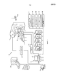
Обращаясь к ФИГУРЕ 1, там схематично показан вариант осуществления системы 1 ЛТ с адаптивным расчетом доз в реальном масштабе времени. Система включает в себя блок 2 трехмерной (3D) визуализации в реальном масштабе времени, такой как блок ультразвуковой (УЗ) визуализации, устройство рентгеновской визуализации (флюороскопии) и т.п. Блок 3D УЗ-визуализации в реальном масштабе времени генерирует трехмерные УЗ-изображения 3 в реальном масштабе времени области тела 4 субъекта. УЗ-изображения 3 области тела субъекта включают в себя ткани, в которые одновременно доставляется излучение от источника излучения, такого как линейный ускоритель (LINAC) 6. УЗ-изображения 3 хранятся в запоминающем устройстве, которое может включать в себя память процессора, память компьютера или энергонезависимую память, такую как дисковый накопитель. Ткани, подверженные воздействию излучения, проходят измерения по отношению к спроецированным пучкам излучения в то время, как субъект расположен на опоре 8 для субъекта, такой как кушетка или кровать.Turning to FIGURE 1, there is schematically shown an embodiment of an
Блок 2 3D УЗ-визуализации в реальном масштабе времени включает в себя один или более процессоров 10, устройство 12 отображения и по меньшей мере одно устройство 14 ввода. Процессор 10, устройство 12 отображения и устройство 14 ввода могут быть реализованы в рабочей станции 16, такой как консоль, интерфейс оператора и т.п. Рабочая станция может представлять собой единственный настольный компьютер, множество настольных компьютеров, соединенных посредством сети, компьютер-сервер, портативный компьютер, планшетный компьютер, их сочетание и т.п. Процессор может представлять собой одиночный процессор или мультипроцессор. Каждый процессор может представлять собой одноядерный или многоядерный процессор. Устройство ввода может включать в себя клавиатуру, манипулятор типа «мышь», микрофон и т.п. Устройство отображения может включать в себя монитор компьютера, телевизионный экран, сенсорный экран, тактильный электронный дисплей, катодно-лучевую трубку (КЛТ), запоминающую трубку, векторный дисплей, плоскопанельный дисплей, вакуумный флюоресцентный дисплей (VF), дисплеи на светоизлучающих диодах (LED), электролюминесцентный дисплей (ELD), плазменные панели отображения (PDP), жидкокристаллический дисплей (LCD), дисплеи на органических светоизлучающих диодах (OLED), проектор, шлем-дисплей и т.п.The real-time 3D ultrasound imaging unit 2 includes one or
Блок 2 3D УЗ-визуализации в реальном масштабе времени включает в себя роботизированный манипулятор 18, управляющий УЗ-зондом 20, или преобразователь, которым управляет управляемое компьютером средство 22 управления роботом. Блок 2 3D УЗ-визуализации в реальном масштабе времени может включать в себя гаптический интерфейс 24 для позиционирования УЗ-зонда 20 средством 22 управления роботом. Блок 2 3D УЗ-визуализации в реальном масштабе времени включает в себя одно или более оптических устройств 26 слежения, отслеживающих положение роботизированного манипулятора, LINAC и тела субъекта. Устройство слежения может включать в себя устройство лазерного, видео-, радиочастотного отслеживания или устройства с электромеханической обратной связью на гентри ускорителя LINAC, роботизированной руке, опоре для субъекта и т.п. Средством 22 управления роботом управляет роботизированным манипулятором 18 или роботизированной рукой, чтобы избежать столкновений с ускорителем LINAC 6 и пучком ускорителя LINAC в процессе доставки излучения. Средство управления роботом позиционирует УЗ-зонд для получения трехмерных УЗ-изображений в реальном масштабе времени областей тела субъекта, получающих дозы облучения одновременно с доставкой излучения.The real-time 3D ultrasound imaging unit 2 includes a
Ускоритель LINAC 6 направляет пучки излучения в по меньшей мере один целевой объем, расположенный в теле 4 субъекта, на основе плана проведения лучевой терапии. Каждый пучок излучения имеет заранее заданные размер, форму, направление или ориентацию, интенсивность и продолжительность и контролируется средством 28 управления ускорителем LINAC. Ускоритель LINAC 6 перемещается вокруг тела субъекта, которое удерживается на опоре 8 для субъекта. Опора для субъекта может перемещаться для совмещения с ускорителем LINAC. Средство управления ускорителем LINAC может быть реализовано в рабочей станции 16 или отдельной рабочей станции, а также функционировать как консоль для LINAC. Средство 28 управления ускорителем LINAC предоставляет данные о траектории, форме, размере и прочую информацию в отношении пучков излучения в блок 2 3D УЗ-визуализации в реальном масштабе времени.The
Система включает в себя блок 30 регистрации изображения, принимающий плановое изображение 32, такое как изображение КТ, МРТ, ПЭТ или ОФЭКТ или их комбинацию, из запоминающего устройства, такого как система управления памятью, система архивации и передачи изображений (PACS), рентгенологическая информационная система (RIS) и т.п., либо непосредственно со сканера 34. Система включает в себя блок 35 сегментации, разбивающий на сегменты целевые объемы и любые ОПР. Например, опухоль в предстательной железе сегментируется в качестве целевого объема, а предстательная железа, мочевой пузырь и прямая кишка субъекта сегментируются в качестве ОПР. Блок сегментации может идентифицировать сегментированные структуры, наложенные на отображение устройством 12 отображения УЗ-изображений 3 в реальном масштабе времени или планового изображения. Например, изображение с целевым объемом и ОПР может включать в себя цветовой контраст границ целевого объема и/или отличный цветовой контраст границ ОПР.The system includes an
Блок 30 регистрации изображения деформируемо регистрирует сегментированное плановое изображение 32 и трехмерное УЗ-базовое изображение 36. Базовое изображение 36 получают перед доставкой первой фракции, и его можно повторно получать и перерегистрировать между фракциями. В процессе регистрации осуществляется построение векторов 38 деформации, которые используются для переноса плотностей тканей с планового изображения, например на основе единиц по шкале Хаунсфилда, и нанесения карты плотностей тканей на базовые трехмерные УЗ-изображения, хранящиеся в запоминающем устройстве, в виде трехмерной карты 40 тканей. Разбивка на сегменты также отображается на трехмерной карте тканей. В одном варианте осуществления сегментированные границы переносятся с планового изображения на трехмерное УЗ-базовое изображение. Различным сегментированным областям может присваиваться номинальная плотность или иной коэффициент ослабления излучения для ткани в каждой области.The
В процессе проведения лучевой терапии УЗ-блок 42 3D движения регистрирует каждое трехмерное УЗ-изображение в реальном масштабе времени на трехмерной карте тканей, используя информацию от системы 26 слежения, а также сравнение изображений непосредственно либо с трехмерным УЗ-базовым изображением, либо плановым изображением. Иными словами, трехмерная карта тканей преобразуется в текущую форму и текущее положение целевой области и ОПР. Измеренное движение может быть вызвано дыхательной деятельностью, деятельностью сердца, перемещением пациента, а также одиночными случайными событиями, такими как кашель, чихание, выпуск газа из кишечника и т.д.During radiation therapy, the
Подсистема 46 расчета дозы в реальном масштабе времени вычисляет дозу облучения в реальном масштабе времени, доставляемую в ткани, на основе трехмерной плотности тканей, нанесенной в виде карты на УЗ-изображения 3 в реальном масштабе времени. Подсистема расчета дозы в реальном масштабе времени включает в вычисление нанесенную в виде карты плотность тканей, через которые проходит каждый пучок излучения. В частности, подсистема расчета дозы интегрирует количество излучения, поглощенного каждым вокселом зарегистрированной трехмерной карты тканей. Преобразование целевых объемов и ОПР карты тканей в текущие положение и форму на основе движения, выявленного блоком 44 3D движения, позволяет определить вокселы, расположенные вдоль траектории пучка излучения. Расчет дозы облучения в реальном масштабе времени, накопленной в каждом вокселе трехмерных УЗ-изображений в реальном масштабе времени, основан на первоначальной интенсивности пучка излучения, ослаблении вдоль траектории, ведущей к каждому вокселу и пересекающей его, а также количестве внесенного излучения во времени в том вокселе, для которого траектория пересекает воксел. Внесенная доза интегрируется или суммируется по всем траекториям, пересекающим каждый воксел в процессе терапии, по мере того как терапевтические пучки, целевая область и ОПР перемещаются относительно своего нормального положения в плановом изображении. Необязательно, карта кумулятивных доз может включать в себя прогнозируемую карту кумулятивных доз, составленную на основе дозы в реальном масштабе времени, вычисленной и накопленной для каждого воксела, а также прогнозируемого значения, основанного на оставшейся части фракционной терапии, запланированной исходя из плана проведения ЛТ-лечения. Кумулятивная доза может наноситься и/или накладываться на план-карту.The real-time
Карта 48 кумулятивных доз может быть использована системой для изменения процесса лечения. Процесс лечения может быть изменен, например, путем остановки процесса в ходе проведения фракции, когда достигнута планируемая доза в целевом объеме и/или в ОПР получена максимальная разрешенная пороговая доза. В качестве альтернативы, процесс лечения может быть изменен путем модифицирования одного или более запланированных пучков излучения, оставшихся для проведения фракционной терапии. Один или более пучков излучения могут быть изменены по размеру, форме, направлению, интенсивности и/или продолжительности. Например, положение опухоли аорты сдвигается в соответствии с частотой дыхательных движений и сердечных сокращений, при этом пучок, направляемый под определенным углом, может сужаться и/или подвергаться стробированию в ходе определенной ритмической последовательности, чтобы свести к минимуму воздействие на ткани сердца. В другом примере пациент кашляет, при этом пучок излучения временно прерывается, пока целевой объем не вернется в свое положение. В обоих примерах осуществляется расчет дозы в реальном масштабе времени на протяжении всего процесса для учета малейших движений, которые оставляют целевой объем в центре пучка, но могут переместить окружающие ткани. Кумулятивная доза может также использоваться для изменения или перерасчета будущих фракций.A map of 48 cumulative doses can be used by the system to change the treatment process. The treatment process can be changed, for example, by stopping the process during the fraction, when the planned dose in the target volume is reached and / or in the ODA the maximum permitted threshold dose is obtained. Alternatively, the treatment process can be modified by modifying one or more planned radiation beams remaining for fractional therapy. One or more beams of radiation can be changed in size, shape, direction, intensity and / or duration. For example, the position of the aortic tumor is shifted in accordance with the frequency of respiratory movements and heart contractions, while the beam directed at a certain angle can narrow and / or undergo gating during a certain rhythmic sequence to minimize the effect on heart tissue. In another example, the patient coughs, while the radiation beam is temporarily interrupted until the target volume returns to its position. In both examples, the dose is calculated in real time over the entire process to take into account the slightest movements that leave the target volume in the center of the beam, but can move the surrounding tissue. The cumulative dose can also be used to change or recalculate future fractions.
Карта 48 кумулятивных доз может отображаться на устройстве 12 отображения, будучи наложенной на трехмерные УЗ-изображения 3 в реальном масштабе времени, базовые изображения 36 и/или плановое КТ-изображение 32. Карта кумулятивных доз может отображаться в виде кумулятивной дозы или прогнозируемой кумулятивной дозы. Карта кумулятивных доз может быть отображена в виде разности между картой кумулятивных доз или прогнозируемой картой кумулятивных доз и картой запланированных доз по плану ЛТ-лечения.The
Различные блоки или средства управления 2, 22, 28, 30, 42, 44, 46 надлежащим образом реализованы электронным устройством обработки данных, таким как электронный процессор или электронное устройство 10 обработки рабочей станции 16, или сетевым компьютером-сервером, функционально связанным с рабочей станцией 16 сетью, и т.д. Кроме того, раскрытые методы визуализации, регистрации, сегментации, нанесения карты, управления и расчета доз надлежащим образом реализуются с использованием невременного носителя информации, хранящего инструкции (например, программное обеспечение), которые могут считываться электронным устройством обработки данных и исполняться этим электронным устройством обработки данных для осуществления раскрытых методов визуализации, регистрации, сегментации, нанесения карты, управления и расчета доз.Various control units or
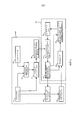
Обращаясь к ФИГУРЕ 2, там показана блок-схема способа адаптивного расчета доз ЛТ в реальном масштабе времени. Этапы могут быть концептуально разделены на предварительные этапы, представленные в виде части планирования ЛТ и/или выполняемые до доставки излучения субъекту, и этапы, выполняемые в ходе или в качестве части ЛТ-лечения. Разработка ЛТ-плана начинается с получения одного или более трехмерных плановых изображений 32 со сканера 34 на этапе 54. Плановое изображение включает в себя один или более целевых объемов для проведения лучевой терапии и обычно включает в себя ОПР. На этапе 56 плановое изображение сегментируют и анализируют, при этом вырабатывают план ЛТ, включающий в себя фракционные терапии. Каждая фракционная терапия включает в себя ряд запланированных воздействий пучками излучения от ускорителя LINAC 6. Для каждого пучка излучения планируют размер, форму, траекторию, интенсивность, продолжительность и т.п. План ЛТ-лечения проверяют и записывают на этапе 58. Проверка может включать в себя моделирование перемещения ускорителя LINAC 6 и манипулятора или роботизированной руки 18 блока 2 3D УЗ-визуализации в реальном масштабе времени. План включает в себя данные управления, используемые средством 28 управления ускорителем LINAC 6 и средством 22 управления роботом УЗ-блока 2.Turning to FIGURE 2, there is shown a flow chart of a method for adaptively calculating RT doses in real time. The stages can be conceptually divided into preliminary stages, presented as part of the planning of radiotherapy and / or performed before the delivery of radiation to the subject, and stages, performed during or as part of the radiotherapy. The development of the LT plan begins with the receipt of one or more three-
На этапе 60 с помощью блока 2 3D УЗ-визуализации в реальном масштабе времени получают базовое трехмерное УЗ-изображение 36. Базовое изображение получают до проведения первой фракции. Базовое изображение 36 хранится в запоминающем устройстве. Базовое изображение можно получать повторно перед проведением каждой фракционной терапии. На этапе 62 полученное трехмерное УЗ-базовое изображение 36 деформируемо регистрируют вместе с сегментированным плановым изображением 32 с помощью блока 30 регистрации деформируемого изображения. В процессе деформируемой регистрации осуществляется построение векторов 38 деформации. Если базовые изображения получают повторно в перерывах между фракционными терапиями, то полученные повторно базовые изображения могут регистрироваться на первом полученном трехмерном УЗ-базовом изображении, друг на друге и/или на трехмерном плановом изображении КТ. Базовое изображение включает в себя ткани, в которые должны доставляться пучки излучения.At
На этапе 64 плотности тканей переносят, например, в единицах по шкале Хаунсфилда, с планового изображения в зарегистрированное УЗ-базовое изображение. На этапе 66 плотности тканей наносят в виде карты на зарегистрированное трехмерное УЗ-базовое изображение на основе векторов деформации. Трехмерная карта 40 тканей и векторы 38 деформации хранятся в запоминающем устройстве. Трехмерная карта 40 тканей предоставляет информацию по ослаблению пучков излучения.At
На этапе 68 излучение доставляют в ткани пациента на основе плана ЛТ-лечения, который включает в себя пучки излучения некоторого размера, формы, интенсивности, продолжительности и ориентации относительно пациента 4, поддерживаемого опорой 8 для пациента. Пучками излучения от LINAC 6 управляют с помощью средства 28 управления ускорителем. Одновременно на этапе 70 блок 3D УЗ-визуализации в реальном масштабе времени, управляемый средством 22 управления роботом, получает трехмерные УЗ-изображения 3 в реальном масштабе времени тканей, расположенных на траектории пучка излучения. На этапе 68 оценивают движение тканей. Движение сегментированных целевых объемов и ОПР измеряют блоком 44 3D движения. На этапе 74 каждый воксел трехмерной карты 40 тканей наносят на карту для преобразования в его текущее положение на основе движения, измеренного в реальном масштабе времени.At
На этапе 72 подсистема 46 расчета дозы в реальном масштабе времени вычисляет дозу облучения в реальном масштабе времени, доставляемую в ткани, на основе трехмерных УЗ-изображений 3 в реальном масштабе времени и трехмерной карты 40 плотностей тканей, откорректированной с учетом движения. Пучки излучения от LINAC 6 отслеживают и измеряют относительно трехмерных УЗ-изображений 3 в реальном масштабе времени. Дозу облучения в реальном масштабе времени рассчитывают для каждого воксела трехмерного УЗ-изображения в реальном масштабе времени. Доза облучения в реальном масштабе времени накапливается на карте 48 доз для этой фракционной терапии. Кумулятивная доза 48 облучения в реальном масштабе времени может включать в себя прогнозируемую дозу для оставшейся части фракционной терапии. Кумулятивная доза 48 облучения в реальном масштабе времени может быть отображена устройством отображения, будучи наложенной либо на трехмерные УЗ-изображения 3 в реальном масштабе времени, либо на зарегистрированное плановое изображение 32 КТ. Вычисленная в реальном масштабе времени кумулятивная доза 48 может быть отображена в виде разности между вычисленной в реальном масштабе времени кумулятивной дозой 48 и запланированной дозой, полученной из плана проведения лучевой терапии, наложенной либо на плановое изображение КТ, либо трехмерные УЗ-изображения в реальном масштабе времени.In
Доставка ЛТ может быть изменена в процессе фракционной терапии на основе карты 48 кумулятивных доз в реальном масштабе времени. Доставка ЛТ может быть изменена на основе измеренного движения. Доставка ЛТ может быть изменена с использованием средства 28 управления ускорителем LINAC для изменения в реальном масштабе времени размера, формы, траектории, интенсивности, продолжительности и т.д. Например, параметры пучка излучения могут изменяться на основе повторяющегося движения, например, вызванного сердечной и/или дыхательной деятельностью. Например, параметры пучка излучения могут меняться для согласования с повторяющимися движениями путем изменения параметров пучка излучения для перемещения вместе с целевым объектом, временного включения/выключения пучка, когда целевой объект входит в пределы или выходит за пределы заданного диапазона, изменения размера и/или формы пучка по мере того, как ОПР в меньшей степени загораживает целевой объем, и т.д. Параметры пучка могут быть изменены при движении, вызванном такими событиями, как кашель, чихание или выпуск газа из кишечника. Например, пучки излучения могут временно задерживаться, пока целевой объем и/или ОПР не вернется в свое положение, зауживаться и/или перемещаться вместе с перемещением целевой области. Изменение параметров пучков излучения может включать в себя отклик(и) на полученную позиционную и дозиметрическую информацию о целевом объекте и/или ОПР, такую как скорости перепозиционирования ускорителя LINAC, и/или перемещения пациента, например регулируемого движения опоры для пациента с целью обеспечения должной ориентации пучка излучения. Доставка ЛТ может изменяться, прерываясь, когда достигается пороговый уровень дозы в одном или более целевых объемах и/или каких-либо ОПР.RT delivery can be changed during fractional therapy based on a map of 48 cumulative doses in real time. Delivery of LT can be changed based on the measured movement. RT delivery can be changed using LINAC accelerator control means 28 to change in real time the size, shape, trajectory, intensity, duration, etc. For example, the parameters of the radiation beam may vary based on repetitive movement, for example, caused by cardiac and / or respiratory activity. For example, the parameters of the radiation beam can be changed to match repetitive movements by changing the parameters of the radiation beam to move with the target object, temporarily turning the beam on / off when the target falls within or outside the specified range, changing the size and / or shape of the beam as ODA to a lesser extent blocks the target volume, etc. Beam parameters can be altered by movement caused by events such as coughing, sneezing, or gas release from the intestines. For example, radiation beams can be temporarily delayed until the target volume and / or ODA returns to its position, narrow and / or move along with the movement of the target area. Changing the parameters of the radiation beams may include a response (s) to the received positional and dosimetric information about the target object and / or the ODA, such as the repositioning speeds of the LINAC accelerator, and / or the movement of the patient, for example, the controlled movement of the support for the patient to ensure proper orientation beam of radiation. RT delivery may vary, interrupted when a threshold dose level is reached in one or more target volumes and / or any ODA.
Карта 48 кумулятивных доз в реальном масштабе времени может быть нанесена на исходное плановое изображение для введения в следующую фракционную терапию, запланированную с использованием этапа 56, т.е. этапа адаптивного планирования лучевой терапии.A real-time
Следует понимать, что применительно к конкретным иллюстративным вариантам осуществления, представленным здесь, определенные конструкционные и/или функциональные признаки описаны как входящие в состав охарактеризованных элементов и/или компонентов. Однако предполагается, что эти признаки с той же или схожей пользой могут при необходимости входить в состав других элементов и/или компонентов. Следует также понимать, что различные аспекты примерных вариантов осуществления могут избирательно использоваться по необходимости для создания других альтернативных вариантов осуществления, пригодных для желательных областей применения, причем эти другие альтернативные варианты осуществления тем самым реализуют соответствующие преимущества включенных сюда аспектов.It should be understood that in relation to the specific illustrative options for implementation presented here, certain structural and / or functional features are described as being part of the characterized elements and / or components. However, it is contemplated that these features, with the same or similar utility, may, if necessary, be part of other elements and / or components. It should also be understood that various aspects of exemplary embodiments can be selectively used as necessary to create other alternative embodiments suitable for the desired applications, these other alternative embodiments thereby realizing the corresponding advantages of the aspects included here.
Следует также понимать, что описанные здесь конкретные элементы или компоненты функционально могут быть соответствующим образом реализованы посредством аппаратного обеспечения, программного обеспечения, аппаратно-программного обеспечения или их комбинации. Кроме того, следует понимать, что определенные элементы, описанные здесь объединенными между собой, могут при подходящих обстоятельствах быть обособленными элементами или быть разделенными иным образом. Точно также, множество конкретных функций, описанных как осуществляемые одним конкретным элементом, могут быть осуществлены множеством отдельных элементов, действующих независимо для осуществления индивидуальных функций, либо определенные индивидуальные функции могут быть разделены и осуществлены множеством отдельных элементов, работающих согласованно. В качестве альтернативы, некоторые элементы или компоненты, иначе описанные и/или показанные здесь отличными друг от друга, могут быть физически или функционально объединены при необходимости.It should also be understood that the specific elements or components described herein can functionally be implemented appropriately by hardware, software, hardware-software, or a combination thereof. In addition, it should be understood that certain elements described herein are interconnected, may, under appropriate circumstances, be separate elements or be otherwise separated. Similarly, many specific functions described as being performed by one particular element can be performed by many separate elements acting independently to carry out individual functions, or certain individual functions can be divided and performed by many separate elements working in concert. Alternatively, some elements or components, otherwise described and / or shown here different from each other, can be physically or functionally combined if necessary.
Коротко говоря, настоящее описание изложено со ссылкой на предпочтительные варианты осуществления. Несомненно, после прочтения и уяснения настоящего описания могут быть предложены модификации и изменения. Предполагается, что данное изобретение следует истолковывать как включающее все подобные модификации и изменения в той степени, в которой они подпадают под объем притязаний прилагаемой формулы изобретения или ее эквивалентов. Иными словами, следует понимать, что многие из вышеописанных и других признаков и функций либо их альтернатив могут быть по желанию объединены во множество других различных систем или приложений, при этом различные непредусмотренные и непредвиденные в настоящее время альтернативы, модификации, варианты или усовершенствования могут быть проделаны позднее специалистами в данной области техники, при этом их также следует считать охваченными нижеследующей формулой изобретения.In short, the present description is set forth with reference to preferred embodiments. Undoubtedly, after reading and clarifying the present description, modifications and changes may be proposed. It is intended that this invention be construed to include all such modifications and changes to the extent that they fall within the scope of the appended claims or their equivalents. In other words, it should be understood that many of the above and other features and functions or their alternatives can be optionally combined into many other different systems or applications, while various currently unforeseen and unforeseen alternatives, modifications, options or improvements can be made later by specialists in the field of technology, while they should also be considered covered by the following claims.
Claims (51)
Applications Claiming Priority (3)
| Application Number | Priority Date | Filing Date | Title |
|---|---|---|---|
| US201261737880P | 2012-12-17 | 2012-12-17 | |
| US61/737,880 | 2012-12-17 | ||
| PCT/IB2013/058588 WO2014096993A1 (en) | 2012-12-17 | 2013-09-17 | Real-time adaptive dose computation radiation therapy |
Publications (2)
| Publication Number | Publication Date |
|---|---|
| RU2015129082A RU2015129082A (en) | 2017-01-23 |
| RU2648226C2 true RU2648226C2 (en) | 2018-03-22 |
Family
ID=49713425
Family Applications (1)
| Application Number | Title | Priority Date | Filing Date |
|---|---|---|---|
| RU2015129082A RU2648226C2 (en) | 2012-12-17 | 2013-09-17 | Radiation therapy with adaptive dose calculation of the dose in the real time |
Country Status (7)
| Country | Link |
|---|---|
| US (1) | US10080910B2 (en) |
| EP (1) | EP2931371B1 (en) |
| JP (1) | JP6480868B2 (en) |
| CN (1) | CN104884126B (en) |
| BR (1) | BR112015013796B1 (en) |
| RU (1) | RU2648226C2 (en) |
| WO (1) | WO2014096993A1 (en) |
Families Citing this family (50)
| Publication number | Priority date | Publication date | Assignee | Title |
|---|---|---|---|---|
| KR20160014933A (en) * | 2014-07-30 | 2016-02-12 | 삼성전자주식회사 | Ultrasonic apparatus and control method for the same |
| US10966688B2 (en) * | 2014-08-26 | 2021-04-06 | Rational Surgical Solutions, Llc | Image registration for CT or MR imagery and ultrasound imagery using mobile device |
| GB2531730A (en) | 2014-10-28 | 2016-05-04 | Elekta ltd | Radiotherapy apparatus |
| US11026750B2 (en) * | 2015-01-23 | 2021-06-08 | Queen's University At Kingston | Real-time surgical navigation |
| JP6893876B2 (en) | 2015-02-11 | 2021-06-23 | ビューレイ・テクノロジーズ・インコーポレイテッドViewRay Technologies, Inc. | Planning and control for radiation therapy guided by magnetic resonance |
| EP3075415B1 (en) * | 2015-03-31 | 2017-03-29 | RaySearch Laboratories AB | Method, computer program and system for dose calculation in radiotherapy |
| CN107530048B (en) | 2015-04-21 | 2024-01-12 | 皇家飞利浦有限公司 | Adjustable arm for patient monitoring device |
| JP7057750B6 (en) * | 2015-10-30 | 2022-06-02 | コーニンクレッカ フィリップス エヌ ヴェ | Indicational treatment plan for hyperthermia-sensitized radiation therapy |
| CN106932829B (en) * | 2015-12-29 | 2020-10-30 | 同方威视技术股份有限公司 | Radiation human body inspection method and radiation human body inspection system |
| WO2017113894A1 (en) * | 2015-12-29 | 2017-07-06 | 同方威视技术股份有限公司 | Method and system for performing radiation-based examination on human body |
| WO2017153211A1 (en) * | 2016-03-09 | 2017-09-14 | Koninklijke Philips N.V. | Pre-optimization method for quick prediction of achievability of clinical goals in intensity modulated radiation therapy |
| US9855445B2 (en) | 2016-04-01 | 2018-01-02 | Varian Medical Systems, Inc. | Radiation therapy systems and methods for delivering doses to a target volume |
| CN106019352B (en) * | 2016-05-16 | 2018-08-17 | 袁子龙 | A kind of automatic calculating method of hospital CT patient radiation doses |
| EP3478361B1 (en) * | 2016-06-29 | 2020-05-20 | Koninklijke Philips N.V. | Real time dosimetry of ultrasound imaging probe |
| EP3487410A4 (en) | 2016-08-01 | 2020-04-08 | Cordance Medical Inc. | Ultrasound guided opening of blood-brain barrier |
| US10507338B2 (en) | 2016-10-31 | 2019-12-17 | Canon Medical Systems Corporation | Particle beam radiotherapy system |
| EP3391940A1 (en) * | 2017-04-21 | 2018-10-24 | Koninklijke Philips N.V. | Planning system for adaptive radiation therapy |
| CN107158581B (en) * | 2017-05-15 | 2019-11-22 | 中国医学科学院肿瘤医院 | Radiotherapy system in radiotherapy planning parameters of scanning paths method and art in art |
| US11273326B2 (en) * | 2017-06-29 | 2022-03-15 | Canon Medical Systems Corporation | Radiotherapy system and treatment support apparatus |
| US10843011B2 (en) | 2017-07-21 | 2020-11-24 | Varian Medical Systems, Inc. | Particle beam gun control systems and methods |
| US11712579B2 (en) | 2017-07-21 | 2023-08-01 | Varian Medical Systems, Inc. | Range compensators for radiation therapy |
| US10245448B2 (en) | 2017-07-21 | 2019-04-02 | Varian Medical Systems Particle Therapy Gmbh | Particle beam monitoring systems and methods |
| US10183179B1 (en) | 2017-07-21 | 2019-01-22 | Varian Medical Systems, Inc. | Triggered treatment systems and methods |
| US10549117B2 (en) | 2017-07-21 | 2020-02-04 | Varian Medical Systems, Inc | Geometric aspects of radiation therapy planning and treatment |
| US10092774B1 (en) | 2017-07-21 | 2018-10-09 | Varian Medical Systems International, AG | Dose aspects of radiation therapy planning and treatment |
| US10609806B2 (en) | 2017-07-21 | 2020-03-31 | Varian Medical Systems Particle Therapy Gmbh | Energy modulation of a cyclotron beam |
| US10675094B2 (en) * | 2017-07-21 | 2020-06-09 | Globus Medical Inc. | Robot surgical platform |
| US11590364B2 (en) | 2017-07-21 | 2023-02-28 | Varian Medical Systems International Ag | Material inserts for radiation therapy |
| CN111556776B (en) | 2017-11-16 | 2022-09-02 | 瓦里安医疗系统公司 | Increased beam output and dynamic field shaping for radiation therapy systems |
| CN107928694B (en) * | 2017-12-05 | 2020-11-03 | 上海联影医疗科技有限公司 | CT dose modulation method and device, CT scanning method and CT system |
| EP3539616A1 (en) * | 2018-03-13 | 2019-09-18 | Koninklijke Philips N.V. | Optimizing fractionation schemes in radiation therapy using biological impact calculation |
| US10910188B2 (en) | 2018-07-25 | 2021-02-02 | Varian Medical Systems, Inc. | Radiation anode target systems and methods |
| US10960232B2 (en) * | 2018-07-28 | 2021-03-30 | Varian Medical Systems, Inc. | Single-pass imaging and radiation treatment delivery via an extended rotation gantry |
| US11116995B2 (en) | 2019-03-06 | 2021-09-14 | Varian Medical Systems, Inc. | Radiation treatment planning based on dose rate |
| US10814144B2 (en) | 2019-03-06 | 2020-10-27 | Varian Medical Systems, Inc. | Radiation treatment based on dose rate |
| US11103727B2 (en) | 2019-03-08 | 2021-08-31 | Varian Medical Systems International Ag | Model based PBS optimization for flash therapy treatment planning and oncology information system |
| US11090508B2 (en) | 2019-03-08 | 2021-08-17 | Varian Medical Systems Particle Therapy Gmbh & Co. Kg | System and method for biological treatment planning and decision support |
| WO2020227719A1 (en) * | 2019-05-09 | 2020-11-12 | The Regents Of The University Of Michigan | Combined radiation acoustics and ultrasound for radiotherapy guidance and cancer targeting |
| US11141609B2 (en) | 2019-05-15 | 2021-10-12 | Elekta Ab (Publ) | Dose guided real-time adaptive radiotherapy |
| US10918886B2 (en) | 2019-06-10 | 2021-02-16 | Varian Medical Systems, Inc. | Flash therapy treatment planning and oncology information system having dose rate prescription and dose rate mapping |
| AU2019453270B2 (en) * | 2019-07-01 | 2023-02-02 | Elekta Ab (Publ) | Geometry-based real-time adaptive radiotherapy |
| CN110464460B (en) * | 2019-07-16 | 2020-11-17 | 江苏霆升科技有限公司 | Method and system for cardiac intervention operation |
| US11291859B2 (en) | 2019-10-03 | 2022-04-05 | Varian Medical Systems, Inc. | Radiation treatment planning for delivering high dose rates to spots in a target |
| DE102019217421A1 (en) * | 2019-11-12 | 2021-05-12 | Siemens Healthcare Gmbh | Process for the automatic control of radiation doses in medical X-ray devices |
| JP7392481B2 (en) * | 2020-01-14 | 2023-12-06 | コニカミノルタ株式会社 | Imaging support equipment, radiography systems and programs |
| US20230086976A1 (en) * | 2020-02-28 | 2023-03-23 | Clarix Imaging Corporation | Volumetric Image Guidance for Improved Pathology Sectioning and Analysis of Tissue Samples |
| US11865361B2 (en) | 2020-04-03 | 2024-01-09 | Varian Medical Systems, Inc. | System and method for scanning pattern optimization for flash therapy treatment planning |
| US11541252B2 (en) | 2020-06-23 | 2023-01-03 | Varian Medical Systems, Inc. | Defining dose rate for pencil beam scanning |
| US11957934B2 (en) | 2020-07-01 | 2024-04-16 | Siemens Healthineers International Ag | Methods and systems using modeling of crystalline materials for spot placement for radiation therapy |
| US11433257B2 (en) * | 2020-12-30 | 2022-09-06 | Varian Medical Systems International Ag | Beam-off motion thresholds in radiation therapy based on breath-hold level determination |
Citations (4)
| Publication number | Priority date | Publication date | Assignee | Title |
|---|---|---|---|---|
| EP0010207B1 (en) * | 1978-10-23 | 1981-11-18 | Westinghouse Electric Corporation | Apparatus for cutting through a tube bundle |
| WO2007002642A2 (en) * | 2005-06-27 | 2007-01-04 | Accuray Incorporated | Method for automatic anatomy-specific treatment planning protocols based on historical integration of previously accepted plans |
| US20080002811A1 (en) * | 2006-06-29 | 2008-01-03 | Allison John W | Treatment delivery optimization |
| US20120035462A1 (en) * | 2010-08-06 | 2012-02-09 | Maurer Jr Calvin R | Systems and Methods for Real-Time Tumor Tracking During Radiation Treatment Using Ultrasound Imaging |
Family Cites Families (10)
| Publication number | Priority date | Publication date | Assignee | Title |
|---|---|---|---|---|
| US5362478A (en) * | 1993-03-26 | 1994-11-08 | Vivorx Pharmaceuticals, Inc. | Magnetic resonance imaging with fluorocarbons encapsulated in a cross-linked polymeric shell |
| US5769790A (en) | 1996-10-25 | 1998-06-23 | General Electric Company | Focused ultrasound surgery system guided by ultrasound imaging |
| JP2003117010A (en) | 2001-08-09 | 2003-04-22 | Mitsubishi Electric Corp | Radiotherapy device, program and computer-readable recording medium recording program |
| EP1460938A4 (en) | 2001-11-05 | 2006-07-26 | Computerized Med Syst Inc | Apparatus and method for registration, guidance, and targeting of external beam radiation therapy |
| US7764818B2 (en) * | 2005-06-20 | 2010-07-27 | Siemens Medical Solutions Usa, Inc. | Surface parameter adaptive ultrasound image processing |
| US7713205B2 (en) | 2005-06-29 | 2010-05-11 | Accuray Incorporated | Dynamic tracking of soft tissue targets with ultrasound images, without using fiducial markers |
| US8249317B2 (en) | 2007-07-20 | 2012-08-21 | Eleckta Ltd. | Methods and systems for compensating for changes in anatomy of radiotherapy patients |
| WO2012069965A1 (en) * | 2010-11-23 | 2012-05-31 | Koninklijke Philips Electronics N.V. | Interactive deformation map corrections |
| US9245336B2 (en) | 2010-12-15 | 2016-01-26 | Koninklijke Philips N.V. | Contour guided deformable image registration |
| EP2854946B1 (en) | 2012-05-29 | 2017-07-12 | Koninklijke Philips N.V. | Elasticity imaging-based planning systems for improved gating efficiency and dynamic margin adjustment in radiation therapy |
-
2013
- 2013-09-17 US US14/649,559 patent/US10080910B2/en active Active
- 2013-09-17 EP EP13799680.7A patent/EP2931371B1/en active Active
- 2013-09-17 BR BR112015013796-2A patent/BR112015013796B1/en active IP Right Grant
- 2013-09-17 RU RU2015129082A patent/RU2648226C2/en active
- 2013-09-17 WO PCT/IB2013/058588 patent/WO2014096993A1/en active Application Filing
- 2013-09-17 JP JP2015547165A patent/JP6480868B2/en active Active
- 2013-09-17 CN CN201380066015.1A patent/CN104884126B/en active Active
Patent Citations (4)
| Publication number | Priority date | Publication date | Assignee | Title |
|---|---|---|---|---|
| EP0010207B1 (en) * | 1978-10-23 | 1981-11-18 | Westinghouse Electric Corporation | Apparatus for cutting through a tube bundle |
| WO2007002642A2 (en) * | 2005-06-27 | 2007-01-04 | Accuray Incorporated | Method for automatic anatomy-specific treatment planning protocols based on historical integration of previously accepted plans |
| US20080002811A1 (en) * | 2006-06-29 | 2008-01-03 | Allison John W | Treatment delivery optimization |
| US20120035462A1 (en) * | 2010-08-06 | 2012-02-09 | Maurer Jr Calvin R | Systems and Methods for Real-Time Tumor Tracking During Radiation Treatment Using Ultrasound Imaging |
Also Published As
| Publication number | Publication date |
|---|---|
| RU2015129082A (en) | 2017-01-23 |
| BR112015013796A2 (en) | 2017-07-11 |
| EP2931371B1 (en) | 2020-07-15 |
| WO2014096993A1 (en) | 2014-06-26 |
| CN104884126B (en) | 2018-08-03 |
| CN104884126A (en) | 2015-09-02 |
| BR112015013796B1 (en) | 2022-10-04 |
| JP6480868B2 (en) | 2019-03-13 |
| US10080910B2 (en) | 2018-09-25 |
| EP2931371A1 (en) | 2015-10-21 |
| JP2015536783A (en) | 2015-12-24 |
| US20150306423A1 (en) | 2015-10-29 |
Similar Documents
| Publication | Publication Date | Title |
|---|---|---|
| RU2648226C2 (en) | Radiation therapy with adaptive dose calculation of the dose in the real time | |
| US11865368B2 (en) | Tracking soft tissue in medical images | |
| US20230008051A1 (en) | Utilization of a transportable ct-scanner for radiotherapy procedures | |
| US9451928B2 (en) | Incorporating internal anatomy in clinical radiotherapy setups | |
| CN103687649B (en) | The dosimetry impact of study movement is to generate the patient-specific allowance of adaptation at EBRT in planning | |
| US10448905B2 (en) | Frameless pre-positioning for radiosurgery | |
| CN104093450A (en) | Beam segment-level dose computation and temporal motion tracking for adaptive treatment planning | |
| US10543381B2 (en) | System for monitoring the position of a patient receiving 4pi radiation therapy | |
| US20180193667A1 (en) | Monitoring a Patient's Position Using a Planning Image and Subsequent Thermal Imaging | |
| EP3391336B1 (en) | Long-exposure-time-imaging for determination of periodically moving structures | |
| US11794035B2 (en) | Triggering of x-ray-images based on surface measurements | |
| US20230141234A1 (en) | Radiation treatment parameters for target region tumour |