RU2629840C2 - Contrast agent for myocardial perfusion visualization - Google Patents
Contrast agent for myocardial perfusion visualization Download PDFInfo
- Publication number
- RU2629840C2 RU2629840C2 RU2014125849A RU2014125849A RU2629840C2 RU 2629840 C2 RU2629840 C2 RU 2629840C2 RU 2014125849 A RU2014125849 A RU 2014125849A RU 2014125849 A RU2014125849 A RU 2014125849A RU 2629840 C2 RU2629840 C2 RU 2629840C2
- Authority
- RU
- Russia
- Prior art keywords
- imaging
- visualization
- patient
- heart
- compound
- Prior art date
Links
Images
Classifications
-
- A—HUMAN NECESSITIES
- A61—MEDICAL OR VETERINARY SCIENCE; HYGIENE
- A61K—PREPARATIONS FOR MEDICAL, DENTAL OR TOILETRY PURPOSES
- A61K49/00—Preparations for testing in vivo
-
- A—HUMAN NECESSITIES
- A61—MEDICAL OR VETERINARY SCIENCE; HYGIENE
- A61K—PREPARATIONS FOR MEDICAL, DENTAL OR TOILETRY PURPOSES
- A61K49/00—Preparations for testing in vivo
- A61K49/06—Nuclear magnetic resonance [NMR] contrast preparations; Magnetic resonance imaging [MRI] contrast preparations
- A61K49/08—Nuclear magnetic resonance [NMR] contrast preparations; Magnetic resonance imaging [MRI] contrast preparations characterised by the carrier
- A61K49/10—Organic compounds
-
- A—HUMAN NECESSITIES
- A61—MEDICAL OR VETERINARY SCIENCE; HYGIENE
- A61K—PREPARATIONS FOR MEDICAL, DENTAL OR TOILETRY PURPOSES
- A61K51/00—Preparations containing radioactive substances for use in therapy or testing in vivo
- A61K51/02—Preparations containing radioactive substances for use in therapy or testing in vivo characterised by the carrier, i.e. characterised by the agent or material covalently linked or complexing the radioactive nucleus
- A61K51/04—Organic compounds
- A61K51/041—Heterocyclic compounds
- A61K51/044—Heterocyclic compounds having nitrogen as a ring hetero atom, e.g. guanethidine, rifamycins
- A61K51/0455—Heterocyclic compounds having nitrogen as a ring hetero atom, e.g. guanethidine, rifamycins having six-membered rings with one nitrogen as the only ring hetero atom
-
- A—HUMAN NECESSITIES
- A61—MEDICAL OR VETERINARY SCIENCE; HYGIENE
- A61P—SPECIFIC THERAPEUTIC ACTIVITY OF CHEMICAL COMPOUNDS OR MEDICINAL PREPARATIONS
- A61P43/00—Drugs for specific purposes, not provided for in groups A61P1/00-A61P41/00
-
- C—CHEMISTRY; METALLURGY
- C07—ORGANIC CHEMISTRY
- C07B—GENERAL METHODS OF ORGANIC CHEMISTRY; APPARATUS THEREFOR
- C07B59/00—Introduction of isotopes of elements into organic compounds ; Labelled organic compounds per se
- C07B59/002—Heterocyclic compounds
-
- C—CHEMISTRY; METALLURGY
- C07—ORGANIC CHEMISTRY
- C07D—HETEROCYCLIC COMPOUNDS
- C07D471/00—Heterocyclic compounds containing nitrogen atoms as the only ring hetero atoms in the condensed system, at least one ring being a six-membered ring with one nitrogen atom, not provided for by groups C07D451/00 - C07D463/00
- C07D471/02—Heterocyclic compounds containing nitrogen atoms as the only ring hetero atoms in the condensed system, at least one ring being a six-membered ring with one nitrogen atom, not provided for by groups C07D451/00 - C07D463/00 in which the condensed system contains two hetero rings
- C07D471/04—Ortho-condensed systems
Abstract
Description
ОБЛАСТЬ ИЗОБРЕТЕНИЯFIELD OF THE INVENTION
Настоящее раскрытие направлено на визуализирующие вещества, фармацевтические композиции и способы для визуализации перфузии миокарда, включающие введение пациенту соединения, связанного с фрагментом, обеспечивающим визуализацию, где указанное соединение связывается с МС-1, и сканирование пациента при помощи диагностической визуализации. Настоящее изобретение также относится к наборам, содержащим указанное визуализирующее вещество или соединения-предшественники, связанные или не связанные с фрагментом, обеспечивающим визуализацию.The present disclosure is directed to imaging agents, pharmaceutical compositions and methods for imaging myocardial perfusion, comprising administering to a patient a compound associated with a visualization fragment, wherein said compound binds to MS-1, and scanning the patient with diagnostic imaging. The present invention also relates to kits containing said imaging agent or precursor compounds, whether or not linked to a visualization moiety.
ПРЕДПОСЫЛКИ ИЗОБРЕТЕНИЯBACKGROUND OF THE INVENTION
Ишемическая болезнь сердца (CAD) является основной причиной смерти в западном мире. Методики визуализации для диагностики и прогнозирования являются очень важными для лечения CAD с тем, чтобы снизить смертность. Визуализация для оценки коронарного кровотока для определения необходимого лечения (часто хирургическая операция) играет важную роль для медицинской помощи при CAD. В настоящее время однофотонная эмиссионная компьютерная томография (SPECT) является основной методикой для визуализации при CAD, но требуются улучшенные способы диагностики.Coronary heart disease (CAD) is the leading cause of death in the Western world. Imaging techniques for diagnosis and prognosis are very important for the treatment of CAD in order to reduce mortality. Imaging for assessing coronary blood flow to determine the necessary treatment (often surgery) plays an important role for CAD care. Single-photon emission computed tomography (SPECT) is currently the main technique for visualization with CAD, but requires improved diagnostic methods.
Клетки сердца, миокарда, имеют очень высокую внутриклеточную плотность, весовое процентное содержание, митохондрий. Таким образом, было обосновано, что соединения, которые избирательно связываются с митохондриями, будут присутствовать в миокарде в большом количестве. Некоторые инсектициды действуют посредством связывания с митохондриальным комплексом I (MCI). К этой группе инсектицидов принадлежат ротенон, пиридабен, тебуфенпирад и феназаквин. Предполагалось, что такие соединения, избирательные по отношению к MCI, можно применять для визуализации ткани, богатой митохондриями. Патентный документ, относящийся к применению меченого ротенона для визуализации коронарного кровотока, был раскрыт в 2001 году.Cells of the heart, myocardium, have a very high intracellular density, weight percent, mitochondria. Thus, it was justified that compounds that selectively bind to mitochondria will be present in large quantities in the myocardium. Some insecticides act by binding to mitochondrial complex I (MCI). This group of insecticides includes rotenone, pyridaben, tebufenpyrad and phenazaquin. It has been suggested that such compounds selective for MCI can be used to visualize tissue rich in mitochondria. A patent document relating to the use of labeled rotenone for imaging coronary blood flow was disclosed in 2001.
В 2005 году BMS подали патентную заявку (WO 2005/079391), в которой описываются 18F-меченые соединения на основе инсектицидов пиридабена, тебуфенпирада и феназаквина для применения в качестве РЕТ-лигандов для диагностики и визуализации коронарного кровотока при CAD. Патенты, принадлежащие BMS, в дальнейшем были приобретены Lantheus Medical Imaging. Применительно к одному из соединений на основе пиридабена, флурпирадаза (BMS747158), проводили интенсивные исследования, и в настоящее время оно находится в фазе III исследований по отношению к визуализации миокарда. Флурпиридаз, как было обнаружено, обеспечивает лучшую оценку функции миокарда, чем вещество для SPECT 99mTc sestamibi.In 2005, BMS filed a patent application (WO 2005/079391), which describes 18 F-labeled compounds based on pyridaben, tebufenpyrad and phenazaquin insecticides for use as PET ligands for the diagnosis and visualization of coronary blood flow in CAD. Patents held by BMS were subsequently acquired by Lantheus Medical Imaging. In relation to one of the compounds based on pyridabene, flupiradase (BMS747158), intensive studies have been carried out, and currently it is in phase III studies in relation to imaging of the myocardium. Flurpiridase has been found to provide a better assessment of myocardial function than the substance for SPECT 99mTc sestamibi.
Respiratorius, фармацевтическая компания, расположенная в Лунде, Швеция, работает над созданием новых бронходилатирующих лекарственных средств. Главной частью исследовательской работы Respiratorius является скрининг малых молекул, которые могут приводить к расслаблению ткани дыхательных путей человека ex vivo. В ходе этого процесса ряд новых 1,8-нафтиридинов был обнаружен в качестве соединений, оказывающих сильный бронхорасслабляющий эффект (описаны в патентной заявке WO/2010/097410). В результате дополнительных фармакологических исследований было обнаружено, что представители этого класса соединений связывались с митохондриальным комплексом I и ингибировали его.Respiratorius, a pharmaceutical company based in Lund, Sweden, is working on new bronchodilating drugs. A major part of Respiratorius research work is the screening of small molecules that can lead to ex vivo relaxation of human respiratory tract tissue. During this process, a number of new 1,8-naphthyridines were discovered as compounds having a strong bronchodilator effect (described in patent application WO / 2010/097410). As a result of additional pharmacological studies, it was found that representatives of this class of compounds bind to and inhibit mitochondrial complex I.
КРАТКОЕ ОПИСАНИЕ ИЗОБРЕТЕНИЯSUMMARY OF THE INVENTION
Было обнаружено, что бронходилатирующее соединение, принадлежащее к классу 1,8-нафтиридинов, также может ингибировать функцию митохондрий при расслаблении гладкой мускулатуры дыхательных путей, изменять функцию митохондрий или связываться с митохондриальным комплексом I. Если эти соединения будут мечеными фрагментом, обеспечивающим визуализацию, станет доступным важный диагностический маркер для визуализации перфузии миокарда.It was found that a bronchodilating compound belonging to the class of 1,8-naphthyridines can also inhibit the function of mitochondria while relaxing the smooth muscles of the respiratory tract, alter the function of mitochondria, or bind to mitochondrial complex I. If these compounds are labeled with a visualization fragment, it will become available an important diagnostic marker for imaging myocardial perfusion.
Настоящее изобретение относится к визуализирующему веществу, имеющему структуруThe present invention relates to an imaging substance having the structure

где R1 представляет собой H, F, CF3, Cl, R представляет собой линкер, и X представляет собой фрагмент, обеспечивающий визуализацию, или аналог или фармацевтическую соль указанного визуализирующего вещества.where R 1 represents H, F, CF 3 , Cl, R represents a linker, and X represents a fragment that provides visualization, or an analog or pharmaceutical salt of the specified imaging substance.
Во втором аспекте настоящее изобретение относится к фармацевтической композиции, содержащей визуализирующее вещество, показанное выше, и фармацевтически приемлемый носитель, разбавитель буфер. Визуализирующее вещество и композиция приводят к высокому отношению накопления в сердце по сравнению с нецелевым участком, причем с минимальным перераспределением. Это также будет приводить в результате к лучшему качеству изображения и выявлению и диагностике заболевания. Будет получено практически подчиняющееся линейному закону накопление в зависимости от потока: до 5 мл/мин./г (высокое значение экстракции при первом прохождении). Это обеспечивает количественную оценку абсолютного значения потока в миокарде и будет эффективным как при физической, так и фармакологической нагрузке. Это также будет характеризоваться соответствующим профилем безопасности и будет доступным в виде стандартной дозы (например, 18F-меченое соединение).In a second aspect, the present invention relates to a pharmaceutical composition comprising the imaging agent shown above and a pharmaceutically acceptable carrier, diluent buffer. The imaging agent and composition lead to a high accumulation ratio in the heart compared to the non-targeted area, with minimal redistribution. This will also result in better image quality and the detection and diagnosis of the disease. Accumulation, practically obeying the linear law, will be obtained depending on the flow: up to 5 ml / min / g (high extraction value at the first passage). This provides a quantitative assessment of the absolute value of the flow in the myocardium and will be effective in both physical and pharmacological stress. It will also be characterized by an appropriate safety profile and will be available as a unit dose (e.g., 18F-labeled compound).
В третьем аспекте настоящее изобретение относится к способу визуализации сердца у пациента, включающему введение пациенту диагностически эффективного количества визуализирующего вещества или фармацевтической композиции, определенной выше, и получение изображения сердца пациента. В последнем аспекте настоящее изобретение относится к набору для диагностики, содержащему соединение, имеющее следующую формулуIn a third aspect, the present invention relates to a method for imaging a heart in a patient, comprising administering to the patient a diagnostically effective amount of an imaging substance or pharmaceutical composition as defined above, and obtaining an image of the patient's heart. In a final aspect, the present invention relates to a diagnostic kit comprising a compound having the following formula
где R1 представляет собой Н, F, CF3, Cl, R представляет собой линкер, и X представляет собой уходящую группу, выбранную из группы, состоящей из тозилата, мезилата, трифлата, нонафлата и галогена, или аналог указанного соединения, и при этом указанный набор можно применять для получения визуализирующего вещества, определенного выше.where R 1 represents H, F, CF 3 , Cl, R represents a linker, and X represents a leaving group selected from the group consisting of tosylate, mesylate, triflate, nonaflate and halogen, or an analogue of the compound, and wherein this kit can be used to obtain the imaging substance defined above.
КРАТКОЕ ОПИСАНИЕ ГРАФИЧЕСКИХ МАТЕРИАЛОВBRIEF DESCRIPTION OF GRAPHIC MATERIALS
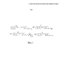
На Фиг. 1 показан путь синтеза для получения визуализирующего соединения.In FIG. 1 shows a synthesis pathway for preparing a visualizing compound.
ПОДРОБНОЕ ОПИСАНИЕ ИЗОБРЕТЕНИЯDETAILED DESCRIPTION OF THE INVENTION
ОпределенияDefinitions
В контексте настоящей заявки и настоящего изобретения применяются следующие определения.In the context of this application and the present invention, the following definitions apply.
Выражение "фармацевтически приемлемая соль" относится к таким солям, которые сохраняют биологическую эффективность и свойства свободных оснований и которые получены путем реакции с неорганическими или органическими кислотами, такими как соляная кислота, бромистоводородная кислота, серная кислота, азотная кислота, фосфорная кислота, метансульфоновая кислота, этансульфоновая кислота, п-толуолсульфоновая кислота, салициловая кислота, яблочная кислота, малеиновая кислота, янтарная кислота, винная кислота, лимонная кислота и т.п.The expression "pharmaceutically acceptable salt" refers to those salts which retain the biological effectiveness and properties of the free bases and which are obtained by reaction with inorganic or organic acids such as hydrochloric acid, hydrobromic acid, sulfuric acid, nitric acid, phosphoric acid, methanesulfonic acid, ethanesulfonic acid, p-toluenesulfonic acid, salicylic acid, malic acid, maleic acid, succinic acid, tartaric acid, citric acid, etc.
Аналог представляет собой молекулу, которая отличается по химической структуре от исходного соединения, например, гомолог (отличающийся дополнительным элементом в химической структуре, например, различие по длине алкильной цепи), фрагмент молекулы, структура, которая отличается одной или несколькими функциональными группами, изменение образования ионов. Структурные аналоги часто находят с использованием количественных соотношений структура-активность (QSAR) при помощи методик, таких как те, которые раскрыты в Remington (The Science and Practice of Pharmacology, 19th Edition (1995), chapter 28).An analogue is a molecule that differs in chemical structure from the starting compound, for example, a homolog (different in an additional element in the chemical structure, for example, difference in the length of the alkyl chain), a fragment of a molecule, a structure that differs in one or more functional groups, change in the formation of ions . Structural analogs are often found using quantitative structure activity relationships (QSAR) using techniques such as those disclosed in Remington (The Science and Practice of Pharmacology , 19 th Edition (1995), chapter 28).
Выражение "связывающая группа", используемое в данном документе, относится к части молекулы, которая служит в качестве спейсера между двумя другими частями молекулы. Связывающие группы также могут выполнять другие функции, как описано в данном документе. Примеры связывающих групп включают линейный, разветвленный или циклический алкил, арил, эфир, полигидрокси, полиэфир, полиамин, гетероциклил, ароматическую группу, гидразид, пептид, пептоид или другие физиологически совместимые ковалентные связи или их комбинации.The expression “linking group” as used herein refers to a part of a molecule that serves as a spacer between two other parts of the molecule. Linking groups can also perform other functions, as described in this document. Examples of linking groups include linear, branched or cyclic alkyl, aryl, ether, polyhydroxy, polyester, polyamine, heterocyclyl, aromatic group, hydrazide, peptide, peptoid or other physiologically compatible covalent bonds or combinations thereof.
В первом варианте осуществления настоящее изобретение относится к визуализирующему веществу, имеющему структуруIn a first embodiment, the present invention relates to an imaging agent having the structure
где R1 представляет собой H, F, CF3, Cl, R представляет собой линкер, и X представляет собой фрагмент, обеспечивающий визуализацию, или аналог или фармацевтически приемлемая соль указанного визуализирующего вещества.where R 1 represents H, F, CF 3 , Cl, R represents a linker, and X represents a fragment that provides visualization, or an analog or pharmaceutically acceptable salt of the specified imaging substance.
R может представлять собой неразветвленный алкил, этиленгликоль (эфир) или полиэтиленгликоль.R may be straight chain alkyl, ethylene glycol (ether) or polyethylene glycol.
Один пример представляет собойOne example is
где R представляет собой линкер, и X представляет собой фрагмент, обеспечивающий визуализацию.where R is a linker, and X is a fragment that provides visualization.
Другой пример представляет собой визуализирующее вещество с формулой, показанной ниже:Another example is an imaging agent with the formula shown below:
где X представляет собой фрагмент, обеспечивающий визуализацию.where X is a fragment that provides visualization.
X может представлять собой изотоп галогена, например, изотоп фтора, брома, хлора или йода. Примеры включают 18F, 19F, 120I, 121I, 122I, 123I, 124I, 125I, 127I, 131I, 35Cl, 37Cl, 75Br, 76Br, 77Br, 79Br, 80Br, 80mBr, 81Br или 64Cu. В конкретном примере применяют 18F или 19F.X may be a halogen isotope, for example, an isotope of fluorine, bromine, chlorine or iodine. Examples include 18 F, 19 F, 120 I, 121 I, 122 I, 123 I, 124 I, 125 I, 127 I, 131 I, 35 Cl, 37 Cl, 75 Br, 76 Br, 77 Br, 79 Br, 80 Br, 80m Br, 81 Br or 64 Cu. In a specific example, 18 F or 19 F is used.
В другом варианте осуществления настоящее изобретение относится к фармацевтической композиции, содержащей визуализирующее вещество, определенное выше, и фармацевтически приемлемый носитель, разбавитель или буфер.In another embodiment, the present invention relates to a pharmaceutical composition comprising an imaging agent as defined above and a pharmaceutically acceptable carrier, diluent or buffer.
"Фармацевтически приемлемый" означает нетоксичный материал, который не снижает эффективность биологической активности активных ингредиентов, т.е. пептида (пептидов), полипептида (полипептидов) или их вариантов. Такие фармацевтически приемлемые буферы, носители или наполнители хорошо известны в данной области техники (см. Remington's Pharmaceutical Sciences, 18th edition, A.R Gennaro, Ed., Mack Publishing Company (1990) и Handbook of Pharmaceutical Excipients, 3rd edition, A. Kibbe, Ed., Pharmaceutical Press (2000)."Pharmaceutically acceptable" means a non-toxic material that does not reduce the effectiveness of the biological activity of the active ingredients, i.e. peptide (s), polypeptide (s) or variants thereof. Such pharmaceutically acceptable buffers, carriers or excipients are well known in the art (see Remington's Pharmaceutical Sciences, 18th edition, AR Gennaro, Ed., Mack Publishing Company (1990) and Handbook of Pharmaceutical Excipients, 3rd edition, A. Kibbe, Ed ., Pharmaceutical Press (2000).
Выражение "буфер", как предполагается, означает водный раствор, содержащий кислотно-основную смесь, предназначенную для стабилизации pH.The term “buffer” is intended to mean an aqueous solution containing an acid-base mixture designed to stabilize the pH.
Выражение "разбавитель", как предполагается, означает водный или неводный раствор, предназначенный для разбавления пептида в фармацевтическом препарате. Разбавитель может представлять собой одно или несколько из солевого раствора, воды, человеческого сывороточного альбумина, например, трис(гидроксиметил)аминометана (и его солей), фосфата, цитрата, бикарбоната, спиртов, в том числе этанола, стерильной воды, физиологического раствора или сбалансированных ионных растворов, содержащих хлоридные и/или бикарбонатные соли или катионы нормальной плазмы крови, такие как кальций, калий, натрий и магний. Меченое соединение может присутствовать в количестве 1,0-50 милликюри, например, 1,0-10, 10-20, 20-30, 30-40, 40-50 милликюри.The term “diluent” is intended to mean an aqueous or non-aqueous solution intended to dilute a peptide in a pharmaceutical preparation. The diluent may be one or more of saline, water, human serum albumin, for example, tris (hydroxymethyl) aminomethane (and its salts), phosphate, citrate, bicarbonate, alcohols, including ethanol, sterile water, physiological saline, or balanced ionic solutions containing chloride and / or bicarbonate salts or cations of normal blood plasma, such as calcium, potassium, sodium and magnesium. The labeled compound may be present in an amount of 1.0-50 millicure, for example, 1.0-10, 10-20, 20-30, 30-40, 40-50 millicure.
Фармацевтический состав в соответствии с настоящим изобретением можно вводить системно. Пути введения включают парентеральный (внутривенный, подкожный и внутримышечный), пероральный, парентеральный, вагинальный и ректальный. Подходящие формы препарата представляют собой, например, дисперсии, суспензии, аэрозоли, капли или раствор для инъекций в ампульной форме, а также препараты с длительным высвобождением активных соединений, причем в этом препарате обычно применяют наполнители, разбавители или носители, описанные выше.The pharmaceutical composition in accordance with the present invention can be entered systemically. Routes of administration include parenteral (intravenous, subcutaneous and intramuscular), oral, parenteral, vaginal and rectal. Suitable forms of the preparation are, for example, dispersions, suspensions, aerosols, drops or injection solution in ampoule form, as well as preparations with sustained release of the active compounds, in which case excipients, diluents or carriers described above are usually used.
Визуализирующие вещества по настоящему изобретению можно применять в способах визуализации, в том числе способах визуализации по отношению к пациенту. Например, способ может включать введение визуализирующего вещества пациенту путем инъекции (например, внутривенной инъекции), инфузии или любого другого известного способа и визуализацию сердца субъекта, где локализовано явление, представляющее интерес.The imaging agents of the present invention can be used in imaging methods, including imaging methods with respect to a patient. For example, a method may include administering an imaging agent to a patient by injection (eg, intravenous injection), infusion, or any other known method, and visualizing the heart of a subject where a phenomenon of interest is localized.
Подходящие дозы, подлежащие введению, и конкретный способ введения будут различаться в зависимости от таких факторов, как возраст, вес, предполагаемое диагностическое применение и форма состава, например, суспензия, эмульсия, микросфера, липосома или т.п., что будет полностью очевидно для специалистов в данной области техники.Suitable dosages to be administered and the particular route of administration will vary depending on factors such as age, weight, intended diagnostic use and form of formulation, for example, suspension, emulsion, microsphere, liposome or the like, which will be fully apparent to specialists in the art.
Как правило, дозу вводят при низких уровнях и увеличивают до достижения требуемого диагностического эффекта (например, получение изображения). В одном варианте осуществления описанные выше визуализирующие вещества можно вводить путем внутривенной инъекции, обычно в солевом растворе, при дозе от приблизительно 0,1 до приблизительно 100 мКи на 70 кг веса тела (и все комбинации и подкомбинации диапазонов доз и конкретные дозы в их пределах) или в некоторых вариантах осуществления при дозе от приблизительно 0,5 до приблизительно 50 мКи. Визуализацию осуществляют при помощи методик, хорошо известных специалисту в данной области техники.Typically, the dose is administered at low levels and increased until the desired diagnostic effect is achieved (e.g., imaging). In one embodiment, the imaging agents described above can be administered by intravenous injection, usually in saline, at a dose of about 0.1 to about 100 mCi per 70 kg body weight (and all combinations and sub-combinations of dose ranges and specific doses within them) or in some embodiments, at a dose of from about 0.5 to about 50 mCi. Visualization is carried out using techniques well known to those skilled in the art.
Другой аспект настоящего изобретения предусматривает наборы для диагностики для получения визуализирующих веществ/диагностических средств для определения (например, выявления), визуализации и/или мониторинга по отношению к по меньшей мере части сердца. Наборы для диагностики по настоящему изобретению могут включать один или несколько флаконов, содержащих стерильный апирогенный состав, содержащий предварительно определенное количество реактива (например, предшественника контрастного вещества) по настоящему изобретению и необязательно другие компоненты, такие как хелатирующие вещества, растворители, буферы, добавки для нейтрализации, добавки для лиофилизации, добавки для стабилизации, добавки для придания растворимости и бактериостатические средства, как более полно описано ниже.Another aspect of the present invention provides diagnostic kits for producing imaging / diagnostic agents for determining (eg, detecting), imaging and / or monitoring with respect to at least a portion of the heart. Diagnostic kits of the present invention may include one or more vials containing a sterile pyrogen-free composition containing a predetermined amount of the reagent (e.g., a contrast agent precursor) of the present invention and optionally other components such as chelating agents, solvents, buffers, neutralizing agents lyophilization additives, stabilization additives, solubility additives and bacteriostatic agents, as more fully described below.
Некоторые неограничивающие примеры буферов, пригодных для получения контрастных веществ и наборов, включают, например, фосфатный, цитратный, сульфосалицилатный и ацетатный буферы. Более полный список можно найти в Фармакопее США.Some non-limiting examples of buffers suitable for preparing contrast agents and kits include, for example, phosphate, citrate, sulfosalicylate and acetate buffers. A more complete list can be found in the United States Pharmacopeia.
Некоторые неограничивающие примеры добавок для лиофилизации, пригодных для получения контрастных веществ и наборов, включают, например, маннит, лактозу, сорбит, декстран, полимер FICOLL.RTM. и поливинилпирролидон (PVP).Some non-limiting examples of lyophilization additives suitable for producing contrast agents and kits include, for example, mannitol, lactose, sorbitol, dextran, FICOLL.RTM polymer. and polyvinylpyrrolidone (PVP).
Некоторые неограничивающие примеры добавок для стабилизации, пригодных для получения контрастных веществ и наборов, включают, например, этанол, аскорбиновую кислоту, этанол, цистеин, тиоглицерин, бисульфит натрия, метабисульфит натрия, гентизиновую кислоту и инозит.Some non-limiting examples of stabilization additives suitable for the preparation of contrast agents and kits include, for example, ethanol, ascorbic acid, ethanol, cysteine, thioglycerol, sodium bisulfite, sodium metabisulfite, gentisic acid and inositol.
Некоторые неограничивающие примеры добавок для придания растворимости, пригодных для получения контрастных веществ и наборов, включают, например, этанол, глицерин, полиэтиленгликоль, пропиленгликоль, полиоксиэтиленсорбитан моноолеат, сорбитан моноолеат, полисорбаты, сополимеры из блоков поли(оксиэтилен)-поли(оксипропилен)-поли(оксиэтилен) ("Pluronics.RTM.") и лецитин.Some non-limiting examples of solubility additives suitable for preparing contrast agents and kits include, for example, ethanol, glycerin, polyethylene glycol, propylene glycol, polyoxyethylene sorbitan monooleate, sorbitan monooleate, polysorbates, poly (oxyethylene) -poly (oxypropylene) block copolymers (hydroxyethylene) ("Pluronics.RTM.") and lecithin.
Некоторые неограничивающие примеры бактериостатических средств, пригодных для получения контрастных веществ и наборов, включают, например, бензиловый спирт, хлорид бензалкония, хлорбутанол и метил-, пропил- или бутилпарабен.Some non-limiting examples of bacteriostatic agents suitable for the preparation of contrast agents and kits include, for example, benzyl alcohol, benzalkonium chloride, chlorobutanol and methyl, propyl or butyl paraben.
Соединения и композиции в соответствии с настоящим изобретением можно применять при помощи методик визуализации, таких как позитронно-эмисионная томография (PET) и однофотонная эмиссионная компьютерная томография (SPECT). Визуализация при помощи PET представляет собой диагностическое обследование, которое предусматривает получение изображений, отражающих физиологическое состояние, на основе детектирования излучения в результате испускания позитронов радиоактивным соединением, введенным пациенту. Радиоактивное соединение, как правило, вводят посредством внутривенной инъекции. Различные цвета или степени яркости на PET-изображении отражают различные уровни функции ткани или органа. Визуализация при помощи SPECT является трехмерной методикой в сочетании с компьютерной реконструкцией изображений органов для выявления как структуры, так и функции. Как и при визуализации при помощи PET, пациентам, подвергающимся визуализации при помощи SPECT, вводят радиоактивную метку. PET- и SPECT-изображения можно применять для оценки при ряде заболеваний и их обычно применяют в областях, связанных с онкологией, кардиологией и неврологией.Compounds and compositions in accordance with the present invention can be applied using imaging techniques such as positron emission tomography (PET) and single photon emission computed tomography (SPECT). PET imaging is a diagnostic examination that involves obtaining images reflecting a physiological state based on the detection of radiation from the emission of positrons by a radioactive compound introduced to the patient. The radioactive compound is typically administered by intravenous injection. Different colors or degrees of brightness in a PET image reflect different levels of tissue or organ function. Visualization using SPECT is a three-dimensional technique in combination with computer reconstruction of organ images to identify both structure and function. As with PET imaging, patients undergoing SPECT imaging are administered a radioactive label. PET and SPECT images can be used to evaluate a number of diseases and are usually used in areas related to oncology, cardiology and neurology.
Способы синтеза контрастных веществMethods for the synthesis of contrast agents
Как правило, визуализирующие вещества, описанные в данном документе, можно синтезировать путем осуществления реакции по меньшей мере первого компонента и второго компонента, так что между ними образуется связь. Например, 18F-меченые соединения можно синтезировать путем осуществления реакции двух компонентов посредством вытеснения соответствующей уходящей группы, связанной по меньшей мере с одним компонентом. Примеры таких уходящих групп включают сложные эфиры сульфоновой кислоты, например, толуолсульфонат (тозилат, TsO-), метансульфонат (мезилат, MsO-) или трифторметансульфонат (трифлат, TfO-), нонафлат или галоген. Уходящая группа также может представлять собой галогенид, фосфиноксид (при помощи реакции Мицунобу) или внутреннюю уходящую группу (например, эпоксид или циклический сульфат). Очистку обычно осуществляют посредством удаления соли при помощи обращенно-фазовой хроматографии.Typically, the imaging agents described herein can be synthesized by reacting at least the first component and the second component, so that a bond is formed between them. For example, 18F-labeled compounds can be synthesized by reacting two components by displacing the corresponding leaving group associated with at least one component. Examples of such leaving groups include sulfonic acid esters, for example, toluenesulfonate (tosylate, TsO-), methanesulfonate (mesylate, MsO-) or trifluoromethanesulfonate (triflate, TfO-), nonaflat or halogen. The leaving group may also be a halide, phosphine oxide (via the Mitsunobu reaction) or an internal leaving group (e.g., epoxide or cyclic sulfate). Purification is usually carried out by removing the salt by reverse phase chromatography.
Типичные способы получения соединений описаны в следующих примерах. Вышеизложенные химические превращения можно осуществлять при помощи методик, которые будут полностью очевидными для специалиста в данной области техники, в сочетании со сведениями, описанными в данном документе. В некоторых случаях способы синтеза контрастных веществ могут предусматривать применение одного или нескольких реакционных растворителей. Типичные реакционные растворители включают, например, DMF, NMP, DMSO, THF, этилацетат, дихлорметан и хлороформ. Реакционный раствор может оставаться нейтральным или основным после добавления амина, например, триэтиламина или DIEA. В некоторых случаях химические превращения (например, реакции) можно осуществлять при температуре окружающей среды и с защитой от воздействия кислорода и воды при помощи атмосферы азота, аргона или гелия.Typical methods for preparing compounds are described in the following examples. The above chemical transformations can be carried out using techniques that will be completely obvious to a person skilled in the art, in combination with the information described in this document. In some cases, methods for the synthesis of contrast agents may include the use of one or more reaction solvents. Typical reaction solvents include, for example, DMF, NMP, DMSO, THF, ethyl acetate, dichloromethane and chloroform. The reaction solution may remain neutral or basic after addition of an amine, for example triethylamine or DIEA. In some cases, chemical transformations (for example, reactions) can be carried out at ambient temperature and protected from oxygen and water using an atmosphere of nitrogen, argon or helium.
В некоторых вариантах осуществления временные защитные группы можно применять для предотвращения того, чтобы другие реакционноспособные функциональные группы, например, амины, тиолы, спирты, фенолы и карбоновые кислоты, участвовали или в реакции или препятствовали ей. Типичные защитные группы для аминов включают, например, трет-бутоксикарбонил и тритил (удаление осуществляется в слабокислотных условиях), Fmoc (удаление осуществляется при помощи вторичных аминов, таких как пиперидин) и бензилоксикарбонил (удаление осуществляется при помощи сильной кислоты или при помощи каталитического гидрогенолиза). Тритильную группу также можно применять для защиты тиолов, фенолов и спиртов. В определенных вариантах осуществления защитные группы для карбоновых кислот включают, например, трет-бутиловый сложный эфир (удаление осуществляется при помощи слабой кислоты), бензиловый сложный эфир (обычно удаление осуществляется при помощи каталитического гидрогенолиза) и алкиловые сложные эфиры, например, метиловый или этиловый (обычно удаление осуществляется при помощи слабого основания). Удаление всех защитных групп можно осуществлять по завершению синтеза с применением условий, описанных выше для отдельных защитных групп, и конечный продукт можно очищать при помощи методик, которые будут полностью очевидными для специалиста в данной области техники, в сочетании со сведениями, описанными в данном документе.In some embodiments, temporary protecting groups can be used to prevent other reactive functional groups, for example, amines, thiols, alcohols, phenols and carboxylic acids, from participating in or inhibiting the reaction. Typical amine protecting groups include, for example, tert-butoxycarbonyl and trityl (removal under mildly acidic conditions), Fmoc (removal using secondary amines such as piperidine) and benzyloxycarbonyl (removal using strong acid or by catalytic hydrogenolysis) . The trityl group can also be used to protect thiols, phenols and alcohols. In certain embodiments, protecting groups for carboxylic acids include, for example, tert-butyl ester (removal using a weak acid), benzyl ester (usually removal using catalytic hydrogenolysis), and alkyl esters, for example methyl or ethyl ( removal is usually done with a weak base). The removal of all protective groups can be carried out at the end of the synthesis using the conditions described above for the individual protective groups, and the final product can be purified using methods that will be completely obvious to a person skilled in the art, in combination with the information described in this document.
Следующие примеры предназначены для иллюстрирования, а не для ограничения настоящего изобретения любым образом, видом или формой, либо явным образом, либо косвенно.The following examples are intended to illustrate and not to limit the present invention in any way, form or form, either explicitly or indirectly.
ПРИМЕРЫEXAMPLES
ПРИМЕР 1EXAMPLE 1
Синтез визуализирующего соединенияSynthesis of Imaging Compound
Пример 1Example 1

N-[[3-фтор-4-(2-фторэтоксиметил)фенил]метил]-2-метил-1,8-нафтиридин-3-карбоксамидN - [[3-fluoro-4- (2-fluoroethoxymethyl) phenyl] methyl] -2-methyl-1,8-naphthyridine-3-carboxamide
Колбу с раствором 19 мг 2-[[2-фтор-4-[[(2-метил-1,8-нафтиридин-3-карбонил)амино]метил]фенил]метокси]этил-4-метилбензолсульфоната (0,036 ммоля), 26 мг Kryptofix 222 (4,7,13,16,21,24-гексаокса-1,10-диазабицикло[8.8.8]гексакозан) (0,069 ммоля) и 4 мг KF (0,069 ммоля) в 1,0 мл безводного MeCN добавляли на предварительно нагретую масляную баню и нагревали при 90°C в течение 30 мин. Реакционную смесь охлаждали до комнатной температуры и разбавляли водой. Смесь дважды экстрагировали EtOAc. Объединенные органические фазы промывали солевым раствором, высушивали (MgSO4) и концентрировали. При помощи флэш-хроматографии получали 9,6 мг (72%).A flask with a solution of 19 mg of 2 - [[2-fluoro-4 - [[(2-methyl-1,8-naphthyridine-3-carbonyl) amino] methyl] phenyl] methoxy] ethyl 4-methylbenzenesulfonate (0.036 mmol), 26 mg Kryptofix 222 (4,7,13,16,21,24-hexaoxa-1,10-diazabicyclo [8.8.8] hexacosan) (0.069 mmol) and 4 mg KF (0.069 mmol) in 1.0 ml of anhydrous MeCN added to a preheated oil bath and heated at 90 ° C for 30 minutes. The reaction mixture was cooled to room temperature and diluted with water. The mixture was extracted twice with EtOAc. The combined organic phases were washed with brine, dried (MgSO4) and concentrated. Flash chromatography gave 9.6 mg (72%).
1Н ЯМР (CDCl3) δ 8,97 (dd, 1Н), 8,03 (s,1H), 8,01 (m, 1Н), 7,42 (m, 2Н), 7,19 (dd, 1Н), 7,13 (m, 1Н), 7,08 (t, 1Н), 4,67 (m, 2Н), 4,65 (s, 3Н), 4,53 (m, 1Н), 3,80 (m, 1Н), 3,73 (m, 1Н). 1 H NMR (CDCl 3 ) δ 8.97 (dd, 1H), 8.03 (s, 1H), 8.01 (m, 1H), 7.42 (m, 2H), 7.19 (dd, 1H), 7.13 (m, 1H), 7.08 (t, 1H), 4.67 (m, 2H), 4.65 (s, 3H), 4.53 (m, 1H), 3, 80 (m, 1H); 3.73 (m, 1H).

2-[[2-фтор-4-[[(2-метил-1,8-нафтиридин-3-карбонил)амино]метил]фенил]метокси]этил-4-метилбензолсульфонат2 - [[2-fluoro-4 - [[(2-methyl-1,8-naphthyridine-3-carbonyl) amino] methyl] phenyl] methoxy] ethyl 4-methylbenzenesulfonate
25 мг тозилхлорида (0,13 ммоля) добавляли к раствору 40 мг N-[[3-фтор-4-(2-гидроксиэтоксиметил)фенил]метил]-2-метил-1,8-нафтиридин-3-карбоксамида (0,11 ммоля), 23 мкл диизопропилэтиламина (6,13 ммоля) и 13 мг DMAP (0,11 ммоля) в 1,0 мл CH2Cl2 при комнатной температуре. Раствор перемешивали в течение 2 ч. Реакционную смесь непосредственно помещали на колонку с SiO2 и очищали при помощи флэш-хроматографии (CH2Cl2/MeOH 50:1). Получали 52 мг (90%).25 mg of tosyl chloride (0.13 mmol) was added to a solution of 40 mg of N - [[3-fluoro-4- (2-hydroxyethoxymethyl) phenyl] methyl] -2-methyl-1,8-naphthyridine-3-carboxamide (0, 11 mmol), 23 μl diisopropylethylamine (6.13 mmol) and 13 mg DMAP (0.11 mmol) in 1.0 ml CH2Cl2 at room temperature. The solution was stirred for 2 hours. The reaction mixture was directly placed on a column of SiO2 and purified by flash chromatography (CH2Cl2 / MeOH 50: 1). Received 52 mg (90%).
1Н ЯМР (CDCl3) δ 8,89 (m, 1Н), 7,99 (s, 1Н), 7,82 (m, 1Н), 7,73 (m, 3Н), 7,31 (m, 4Н), 7,13 (m, 2Н), 4,65 (d, 2Н), 4,51 (s, 2Н), 4,15 (d, 2Н), 3,68 (m, 2Н), 2,77 (s, 3Н), 2,41 (s, 3Н). 1 H NMR (CDCl 3 ) δ 8.89 (m, 1H), 7.99 (s, 1H), 7.82 (m, 1H), 7.73 (m, 3H), 7.31 (m, 4H), 7.13 (m, 2H), 4.65 (d, 2H), 4.51 (s, 2H), 4.15 (d, 2H), 3.68 (m, 2H), 2, 77 (s, 3H); 2.41 (s, 3H).

N-[[3-фтор-4-(2-гидроксиэтоксиметил)фенил]метил]-2-метил-1,8-нафтиридин-3-карбоксамидN - [[3-fluoro-4- (2-hydroxyethoxymethyl) phenyl] methyl] -2-methyl-1,8-naphthyridine-3-carboxamide
45 мкл оксалилхлорида (0,53 ммоля) добавляли к смеси 50 мг 2-метил-1,8-нафтиридин-3-карбоновой кислоты (0,27 ммоля) в 3 мл CH2Cl2 с одной каплей DMF. Реакционную смесь перемешивали в течение 1,5 ч и затем выпаривали досуха при пониженном давлении. Остаток растворяли в 3 мл CH2Cl2. 4 мг DMAP (0,03 ммоля) и 188 мкл триэтиламина (1,35 ммоля) добавляли к раствору с последующим добавлением 54 мг 2-[[4-(аминометил)-2-фторфенил]метокси]этанола (0,27 ммоля). Реакционную смесь перемешивали в течение 4 ч и затем разбавляли водой. Фазы разделяли и водную фазу экстрагировали CH2Cl2. Объединенные органические фазы высушивали (MgSO4) и концентрировали. При помощи флэш-хроматографии (SiO2, CH2Cl2/MeOH 20:1) получали 36 мг (36%) титульного соединения.45 μl of oxalyl chloride (0.53 mmol) was added to a mixture of 50 mg of 2-methyl-1,8-naphthyridine-3-carboxylic acid (0.27 mmol) in 3 ml of CH2Cl2 with one drop of DMF. The reaction mixture was stirred for 1.5 hours and then evaporated to dryness under reduced pressure. The residue was dissolved in 3 ml of CH2Cl2. 4 mg DMAP (0.03 mmol) and 188 μl triethylamine (1.35 mmol) were added to the solution, followed by 54 mg of 2 - [[4- (aminomethyl) -2-fluorophenyl] methoxy] ethanol (0.27 mmol) . The reaction mixture was stirred for 4 hours and then diluted with water. The phases were separated and the aqueous phase was extracted with CH2Cl2. The combined organic phases were dried (MgSO4) and concentrated. Flash chromatography (SiO 2, CH 2 Cl 2 / MeOH 20: 1) gave 36 mg (36%) of the title compound.
1Н ЯМР (CDCl3) δ 8,82 (m, 1Н), 7,96 (s, 1Н), 7,95 (m, 1Н), 7,88 (m, 1Н), 7,31 (m, 2Н), 7,10 (m, 2Н), 4,60 (d, 2Н), 4,56 (s, 2Н), 3,75 (m, 2Н), 3,61 (m, 2Н), 2,68 (s, 3Н). 1 H NMR (CDCl 3 ) δ 8.82 (m, 1H), 7.96 (s, 1H), 7.95 (m, 1H), 7.88 (m, 1H), 7.31 (m, 2H), 7.10 (m, 2H), 4.60 (d, 2H), 4.56 (s, 2H), 3.75 (m, 2H), 3.61 (m, 2H), 2, 68 (s, 3H).
Claims (15)


Applications Claiming Priority (5)
| Application Number | Priority Date | Filing Date | Title |
|---|---|---|---|
| US201161579113P | 2011-12-22 | 2011-12-22 | |
| SE1151249-8 | 2011-12-22 | ||
| SE1151249 | 2011-12-22 | ||
| US61/579,113 | 2011-12-22 | ||
| PCT/SE2012/051421 WO2013095273A1 (en) | 2011-12-22 | 2012-12-18 | Contrast agent for imagining myocardial perfusion |
Publications (2)
| Publication Number | Publication Date |
|---|---|
| RU2014125849A RU2014125849A (en) | 2016-02-10 |
| RU2629840C2 true RU2629840C2 (en) | 2017-09-04 |
Family
ID=48668967
Family Applications (1)
| Application Number | Title | Priority Date | Filing Date |
|---|---|---|---|
| RU2014125849A RU2629840C2 (en) | 2011-12-22 | 2012-12-18 | Contrast agent for myocardial perfusion visualization |
Country Status (16)
| Country | Link |
|---|---|
| US (2) | US9295738B2 (en) |
| EP (1) | EP2793952B1 (en) |
| JP (1) | JP6140187B2 (en) |
| KR (1) | KR101931792B1 (en) |
| CN (1) | CN104114191A (en) |
| AU (1) | AU2012354223B2 (en) |
| BR (1) | BR112014015124A2 (en) |
| CA (1) | CA2859773C (en) |
| ES (1) | ES2687234T3 (en) |
| HK (1) | HK1201459A1 (en) |
| IL (1) | IL233219A (en) |
| MX (1) | MX356258B (en) |
| RU (1) | RU2629840C2 (en) |
| SG (1) | SG11201403429YA (en) |
| WO (1) | WO2013095273A1 (en) |
| ZA (1) | ZA201405199B (en) |
Families Citing this family (2)
| Publication number | Priority date | Publication date | Assignee | Title |
|---|---|---|---|---|
| US9955876B2 (en) * | 2016-09-08 | 2018-05-01 | Raul Chirife | Device and method for assessment of left ventricular ejection fraction and other parameters of cardiac performance |
| CN107233585B (en) * | 2017-06-09 | 2021-01-22 | 四川大学华西医院 | Application of berberine or derivatives thereof in preparation of myocardial perfusion imaging agent |
Citations (3)
| Publication number | Priority date | Publication date | Assignee | Title |
|---|---|---|---|---|
| WO2005079391A2 (en) * | 2004-02-13 | 2005-09-01 | Bristol-Myers Squibb Pharma Company | Contrast agents for myocardial perfusion imaging |
| WO2007021858A2 (en) * | 2005-08-10 | 2007-02-22 | Bristol-Myers Squibb Pharma Company | Methods of making radiolabeled tracers and precursors thereof |
| WO2010097410A1 (en) * | 2009-02-24 | 2010-09-02 | Respiratorius Ab | Novel bronchodilating diazaheteroaryls |
Family Cites Families (1)
| Publication number | Priority date | Publication date | Assignee | Title |
|---|---|---|---|---|
| US7932272B2 (en) | 2003-09-30 | 2011-04-26 | Eisai R&D Management Co., Ltd. | Antifungal agent containing heterocyclic compound |
-
2012
- 2012-12-18 AU AU2012354223A patent/AU2012354223B2/en not_active Ceased
- 2012-12-18 ES ES12858896.9T patent/ES2687234T3/en active Active
- 2012-12-18 CN CN201280069796.5A patent/CN104114191A/en active Pending
- 2012-12-18 RU RU2014125849A patent/RU2629840C2/en active
- 2012-12-18 CA CA2859773A patent/CA2859773C/en active Active
- 2012-12-18 SG SG11201403429YA patent/SG11201403429YA/en unknown
- 2012-12-18 MX MX2014007491A patent/MX356258B/en active IP Right Grant
- 2012-12-18 EP EP12858896.9A patent/EP2793952B1/en not_active Not-in-force
- 2012-12-18 US US14/367,520 patent/US9295738B2/en active Active
- 2012-12-18 WO PCT/SE2012/051421 patent/WO2013095273A1/en active Application Filing
- 2012-12-18 JP JP2014548735A patent/JP6140187B2/en not_active Expired - Fee Related
- 2012-12-18 BR BR112014015124A patent/BR112014015124A2/en not_active Application Discontinuation
- 2012-12-18 KR KR1020147018989A patent/KR101931792B1/en active IP Right Grant
-
2014
- 2014-06-18 IL IL233219A patent/IL233219A/en active IP Right Grant
- 2014-07-16 ZA ZA2014/05199A patent/ZA201405199B/en unknown
-
2015
- 2015-02-27 HK HK15102025.8A patent/HK1201459A1/en not_active IP Right Cessation
- 2015-12-17 US US14/972,127 patent/US9687565B2/en active Active
Patent Citations (3)
| Publication number | Priority date | Publication date | Assignee | Title |
|---|---|---|---|---|
| WO2005079391A2 (en) * | 2004-02-13 | 2005-09-01 | Bristol-Myers Squibb Pharma Company | Contrast agents for myocardial perfusion imaging |
| WO2007021858A2 (en) * | 2005-08-10 | 2007-02-22 | Bristol-Myers Squibb Pharma Company | Methods of making radiolabeled tracers and precursors thereof |
| WO2010097410A1 (en) * | 2009-02-24 | 2010-09-02 | Respiratorius Ab | Novel bronchodilating diazaheteroaryls |
Also Published As
| Publication number | Publication date |
|---|---|
| US20160101195A1 (en) | 2016-04-14 |
| CN104114191A (en) | 2014-10-22 |
| ES2687234T3 (en) | 2018-10-24 |
| AU2012354223B2 (en) | 2017-03-16 |
| IL233219A0 (en) | 2014-08-31 |
| EP2793952A1 (en) | 2014-10-29 |
| US9687565B2 (en) | 2017-06-27 |
| US20140341808A1 (en) | 2014-11-20 |
| MX2014007491A (en) | 2015-02-17 |
| JP2015500873A (en) | 2015-01-08 |
| BR112014015124A2 (en) | 2017-06-13 |
| WO2013095273A1 (en) | 2013-06-27 |
| ZA201405199B (en) | 2015-12-23 |
| JP6140187B2 (en) | 2017-05-31 |
| EP2793952A4 (en) | 2015-09-09 |
| KR101931792B1 (en) | 2019-03-13 |
| EP2793952B1 (en) | 2018-06-13 |
| SG11201403429YA (en) | 2014-07-30 |
| CA2859773C (en) | 2020-09-01 |
| CA2859773A1 (en) | 2013-06-27 |
| AU2012354223A1 (en) | 2014-07-03 |
| RU2014125849A (en) | 2016-02-10 |
| MX356258B (en) | 2018-05-16 |
| HK1201459A1 (en) | 2015-09-04 |
| US9295738B2 (en) | 2016-03-29 |
| IL233219A (en) | 2016-08-31 |
| KR20140107411A (en) | 2014-09-04 |
Similar Documents
| Publication | Publication Date | Title |
|---|---|---|
| CN106977429B (en) | Compositions, methods and systems for synthesis and use of contrast agents | |
| JP6987840B2 (en) | Radioligand for IDO1 enzyme imaging | |
| ES2749640T3 (en) | Radiolabeled octreota analogs as PET tracers | |
| US20210283281A1 (en) | Radiolabeled ligands for targeted pet/spect imaging and methods of their use | |
| JP2002525325A (en) | Radiolabeled neurokinin-1 receptor antagonist | |
| RU2629840C2 (en) | Contrast agent for myocardial perfusion visualization | |
| US7731940B2 (en) | Compositions and methods related to serotonin 5-HT1A receptors | |
| US20240042066A1 (en) | Fibroblast activation protein inhibitor | |
| CA2911307C (en) | Use of fluorinated derivatives of 4-aminopyridine in therapeutics and medical imaging | |
| US11065349B2 (en) | 18F labeled BODIPY dye and its derivatives for PET imaging of heart perfusion and others | |
| CN113387896B (en) | 4- (2-chloro aryl) quinazoline-2-amide derivative and application thereof | |
| KR102403970B1 (en) | Carboxylic acid-having compounds for PSMA-targeting and use thereof | |
| KR101519006B1 (en) | Novel benzamide derivative or pharmaceutically acceptable salt thereof, preparation method thereof and pharmaceutical composition for diagnosis of cancer disease containing the same as an active ingredient | |
| JP6488045B2 (en) | Compounds suitable for detection of acetylcholine vesicle transporters | |
| JP6496101B2 (en) | Compounds suitable for detection of acetylcholine vesicle transporters | |
| BR112019027183B1 (en) | PHARMACEUTICAL COMPOSITION FOR TREATMENT OR DIAGNOSIS OF PROSTATE CANCER, RADIOPHARMACEUTICAL FOR IMAGING DIAGNOSIS OF PROSTATE CANCER | |
| US20160058895A1 (en) | Radiolabeled gnrh antagonists as pet imaging agents | |
| Popko et al. | c12) United States Patent | |
| Popko et al. | c19) United States c12) Patent Application Publication | |
| JP2014218455A (en) | Styrylpyridine derivative compounds | |
| JP2014218454A (en) | Styrylpyridine derivative compounds |