RU2566729C1 - Method for prediction of chorion separation in first trimester of pregnancy - Google Patents
Method for prediction of chorion separation in first trimester of pregnancy Download PDFInfo
- Publication number
- RU2566729C1 RU2566729C1 RU2014117238/15A RU2014117238A RU2566729C1 RU 2566729 C1 RU2566729 C1 RU 2566729C1 RU 2014117238/15 A RU2014117238/15 A RU 2014117238/15A RU 2014117238 A RU2014117238 A RU 2014117238A RU 2566729 C1 RU2566729 C1 RU 2566729C1
- Authority
- RU
- Russia
- Prior art keywords
- detachment
- pregnancy
- trimester
- polymorphic variant
- gene
- Prior art date
Links
- 230000035935 pregnancy Effects 0.000 title claims abstract description 25
- 238000000034 method Methods 0.000 title claims abstract description 22
- 210000001136 chorion Anatomy 0.000 title abstract description 18
- 238000000926 separation method Methods 0.000 title abstract 3
- 108090000623 proteins and genes Proteins 0.000 claims abstract description 49
- 102200088705 rs1801394 Human genes 0.000 claims abstract description 38
- 102200074448 rs886037851 Human genes 0.000 claims abstract description 27
- 101150082137 Mtrr gene Proteins 0.000 claims abstract description 19
- 108010023321 Factor VII Proteins 0.000 claims abstract description 13
- 229940105772 coagulation factor vii Drugs 0.000 claims abstract description 10
- 102000054765 polymorphisms of proteins Human genes 0.000 claims abstract description 10
- 102100024614 Methionine synthase reductase Human genes 0.000 claims abstract description 9
- 230000003321 amplification Effects 0.000 claims abstract description 9
- 238000003199 nucleic acid amplification method Methods 0.000 claims abstract description 9
- 229940014144 folate Drugs 0.000 claims abstract description 8
- OVBPIULPVIDEAO-LBPRGKRZSA-N folic acid Chemical compound C=1N=C2NC(N)=NC(=O)C2=NC=1CNC1=CC=C(C(=O)N[C@@H](CCC(O)=O)C(O)=O)C=C1 OVBPIULPVIDEAO-LBPRGKRZSA-N 0.000 claims abstract description 8
- 235000019152 folic acid Nutrition 0.000 claims abstract description 8
- 239000011724 folic acid Substances 0.000 claims abstract description 8
- 206010020608 Hypercoagulation Diseases 0.000 claims abstract description 5
- 201000005665 thrombophilia Diseases 0.000 claims abstract description 5
- 102000016519 Coagulation factor VII Human genes 0.000 claims abstract 3
- 108010042865 aquacobalamin reductase Proteins 0.000 claims description 5
- 238000002955 isolation Methods 0.000 claims 1
- 101001116314 Homo sapiens Methionine synthase reductase Proteins 0.000 abstract description 6
- 239000003814 drug Substances 0.000 abstract description 5
- 230000000694 effects Effects 0.000 abstract description 3
- 238000011084 recovery Methods 0.000 abstract 1
- 239000000126 substance Substances 0.000 abstract 1
- 238000002604 ultrasonography Methods 0.000 description 10
- 102100023804 Coagulation factor VII Human genes 0.000 description 9
- 230000002068 genetic effect Effects 0.000 description 9
- 206010000234 Abortion spontaneous Diseases 0.000 description 7
- 208000015994 miscarriage Diseases 0.000 description 7
- 230000007170 pathology Effects 0.000 description 7
- 208000000995 spontaneous abortion Diseases 0.000 description 7
- 238000012360 testing method Methods 0.000 description 7
- 206010018852 Haematoma Diseases 0.000 description 6
- 206010072596 Subchorionic haematoma Diseases 0.000 description 6
- 238000011161 development Methods 0.000 description 6
- 230000018109 developmental process Effects 0.000 description 6
- 208000037265 diseases, disorders, signs and symptoms Diseases 0.000 description 6
- 239000000203 mixture Substances 0.000 description 6
- 210000002826 placenta Anatomy 0.000 description 6
- 108010049003 Fibrinogen Proteins 0.000 description 5
- 102000008946 Fibrinogen Human genes 0.000 description 5
- 230000015572 biosynthetic process Effects 0.000 description 5
- 239000003153 chemical reaction reagent Substances 0.000 description 5
- 201000010099 disease Diseases 0.000 description 5
- 229940012952 fibrinogen Drugs 0.000 description 5
- 206010071602 Genetic polymorphism Diseases 0.000 description 4
- 206010054798 Retroplacental haematoma Diseases 0.000 description 4
- 239000003795 chemical substances by application Substances 0.000 description 4
- 238000003745 diagnosis Methods 0.000 description 4
- 230000023597 hemostasis Effects 0.000 description 4
- 230000008569 process Effects 0.000 description 4
- 208000024172 Cardiovascular disease Diseases 0.000 description 3
- 201000006082 Chickenpox Diseases 0.000 description 3
- 239000003154 D dimer Substances 0.000 description 3
- 230000007067 DNA methylation Effects 0.000 description 3
- 102000002322 Egg Proteins Human genes 0.000 description 3
- 108010000912 Egg Proteins Proteins 0.000 description 3
- 208000002193 Pain Diseases 0.000 description 3
- 102000012335 Plasminogen Activator Inhibitor 1 Human genes 0.000 description 3
- 108010022233 Plasminogen Activator Inhibitor 1 Proteins 0.000 description 3
- 206010046980 Varicella Diseases 0.000 description 3
- 210000001015 abdomen Anatomy 0.000 description 3
- 230000000172 allergic effect Effects 0.000 description 3
- 208000010668 atopic eczema Diseases 0.000 description 3
- 238000005516 engineering process Methods 0.000 description 3
- 108010052295 fibrin fragment D Proteins 0.000 description 3
- 230000035772 mutation Effects 0.000 description 3
- 230000008520 organization Effects 0.000 description 3
- 210000004681 ovum Anatomy 0.000 description 3
- 230000036407 pain Effects 0.000 description 3
- 210000003800 pharynx Anatomy 0.000 description 3
- 238000003752 polymerase chain reaction Methods 0.000 description 3
- 230000001850 reproductive effect Effects 0.000 description 3
- 239000000523 sample Substances 0.000 description 3
- 238000007619 statistical method Methods 0.000 description 3
- 210000001325 yolk sac Anatomy 0.000 description 3
- 208000009206 Abruptio Placentae Diseases 0.000 description 2
- 102000015081 Blood Coagulation Factors Human genes 0.000 description 2
- 108010039209 Blood Coagulation Factors Proteins 0.000 description 2
- 238000007400 DNA extraction Methods 0.000 description 2
- 206010058314 Dysplasia Diseases 0.000 description 2
- 102000004190 Enzymes Human genes 0.000 description 2
- 108090000790 Enzymes Proteins 0.000 description 2
- 244000187656 Eucalyptus cornuta Species 0.000 description 2
- 102100032999 Integrin beta-3 Human genes 0.000 description 2
- FYYHWMGAXLPEAU-UHFFFAOYSA-N Magnesium Chemical compound [Mg] FYYHWMGAXLPEAU-UHFFFAOYSA-N 0.000 description 2
- 102100031551 Methionine synthase Human genes 0.000 description 2
- 102000005954 Methylenetetrahydrofolate Reductase (NADPH2) Human genes 0.000 description 2
- 108010030837 Methylenetetrahydrofolate Reductase (NADPH2) Proteins 0.000 description 2
- 108090000854 Oxidoreductases Proteins 0.000 description 2
- 102000004316 Oxidoreductases Human genes 0.000 description 2
- 150000001413 amino acids Chemical class 0.000 description 2
- 238000004458 analytical method Methods 0.000 description 2
- 230000023555 blood coagulation Effects 0.000 description 2
- 239000003114 blood coagulation factor Substances 0.000 description 2
- 238000006243 chemical reaction Methods 0.000 description 2
- 230000015271 coagulation Effects 0.000 description 2
- 238000005345 coagulation Methods 0.000 description 2
- 210000002808 connective tissue Anatomy 0.000 description 2
- 229940088598 enzyme Drugs 0.000 description 2
- 230000001973 epigenetic effect Effects 0.000 description 2
- 210000003743 erythrocyte Anatomy 0.000 description 2
- 230000020764 fibrinolysis Effects 0.000 description 2
- 208000000509 infertility Diseases 0.000 description 2
- 230000036512 infertility Effects 0.000 description 2
- 231100000535 infertility Toxicity 0.000 description 2
- 230000003993 interaction Effects 0.000 description 2
- 230000004807 localization Effects 0.000 description 2
- 229910052749 magnesium Inorganic materials 0.000 description 2
- 239000011777 magnesium Substances 0.000 description 2
- 230000007246 mechanism Effects 0.000 description 2
- 230000003821 menstrual periods Effects 0.000 description 2
- 230000005906 menstruation Effects 0.000 description 2
- 238000010197 meta-analysis Methods 0.000 description 2
- 229930182817 methionine Natural products 0.000 description 2
- 239000002480 mineral oil Substances 0.000 description 2
- 235000010446 mineral oil Nutrition 0.000 description 2
- 239000013642 negative control Substances 0.000 description 2
- 230000009984 peri-natal effect Effects 0.000 description 2
- 201000008532 placental abruption Diseases 0.000 description 2
- 238000002360 preparation method Methods 0.000 description 2
- 230000002265 prevention Effects 0.000 description 2
- 238000012545 processing Methods 0.000 description 2
- 238000012552 review Methods 0.000 description 2
- 230000002269 spontaneous effect Effects 0.000 description 2
- 230000009897 systematic effect Effects 0.000 description 2
- 210000004291 uterus Anatomy 0.000 description 2
- 108010075604 5-Methyltetrahydrofolate-Homocysteine S-Methyltransferase Proteins 0.000 description 1
- 208000035285 Allergic Seasonal Rhinitis Diseases 0.000 description 1
- 208000028185 Angioedema Diseases 0.000 description 1
- -1 CF XIII) G> T Proteins 0.000 description 1
- 108010048623 Collagen Receptors Proteins 0.000 description 1
- 238000007399 DNA isolation Methods 0.000 description 1
- 208000005189 Embolism Diseases 0.000 description 1
- 108010014172 Factor V Proteins 0.000 description 1
- 206010058279 Factor V Leiden mutation Diseases 0.000 description 1
- 108010071289 Factor XIII Proteins 0.000 description 1
- 208000032943 Fetal Distress Diseases 0.000 description 1
- 208000001362 Fetal Growth Retardation Diseases 0.000 description 1
- 108010073385 Fibrin Proteins 0.000 description 1
- 102000009123 Fibrin Human genes 0.000 description 1
- BWGVNKXGVNDBDI-UHFFFAOYSA-N Fibrin monomer Chemical compound CNC(=O)CNC(=O)CN BWGVNKXGVNDBDI-UHFFFAOYSA-N 0.000 description 1
- 238000000729 Fisher's exact test Methods 0.000 description 1
- 206010055690 Foetal death Diseases 0.000 description 1
- 206010016855 Foetal distress syndrome Diseases 0.000 description 1
- 206010070531 Foetal growth restriction Diseases 0.000 description 1
- 208000032843 Hemorrhage Diseases 0.000 description 1
- 208000007353 Hip Osteoarthritis Diseases 0.000 description 1
- 101001078133 Homo sapiens Integrin alpha-2 Proteins 0.000 description 1
- 101001015004 Homo sapiens Integrin beta-3 Proteins 0.000 description 1
- 206010020772 Hypertension Diseases 0.000 description 1
- 102100025305 Integrin alpha-2 Human genes 0.000 description 1
- 102000000507 Integrin alpha2 Human genes 0.000 description 1
- 108010020950 Integrin beta3 Proteins 0.000 description 1
- FFFHZYDWPBMWHY-VKHMYHEASA-N L-homocysteine Chemical compound OC(=O)[C@@H](N)CCS FFFHZYDWPBMWHY-VKHMYHEASA-N 0.000 description 1
- AGPKZVBTJJNPAG-WHFBIAKZSA-N L-isoleucine Chemical compound CC[C@H](C)[C@H](N)C(O)=O AGPKZVBTJJNPAG-WHFBIAKZSA-N 0.000 description 1
- FFEARJCKVFRZRR-BYPYZUCNSA-N L-methionine Chemical compound CSCC[C@H](N)C(O)=O FFEARJCKVFRZRR-BYPYZUCNSA-N 0.000 description 1
- 201000005505 Measles Diseases 0.000 description 1
- 206010028813 Nausea Diseases 0.000 description 1
- 201000009859 Osteochondrosis Diseases 0.000 description 1
- 102000010752 Plasminogen Inactivators Human genes 0.000 description 1
- 108010077971 Plasminogen Inactivators Proteins 0.000 description 1
- 208000002787 Pregnancy Complications Diseases 0.000 description 1
- 206010036877 Prolonged Pregnancy Diseases 0.000 description 1
- 108010094028 Prothrombin Proteins 0.000 description 1
- 102100027378 Prothrombin Human genes 0.000 description 1
- 206010039587 Scarlet Fever Diseases 0.000 description 1
- 108090000190 Thrombin Proteins 0.000 description 1
- 208000001435 Thromboembolism Diseases 0.000 description 1
- 208000007536 Thrombosis Diseases 0.000 description 1
- 206010046798 Uterine leiomyoma Diseases 0.000 description 1
- 206010047700 Vomiting Diseases 0.000 description 1
- 206010000210 abortion Diseases 0.000 description 1
- 231100000176 abortion Toxicity 0.000 description 1
- 239000000443 aerosol Substances 0.000 description 1
- 229940024606 amino acid Drugs 0.000 description 1
- 125000000539 amino acid group Chemical group 0.000 description 1
- 210000004381 amniotic fluid Anatomy 0.000 description 1
- 238000013459 approach Methods 0.000 description 1
- 230000004888 barrier function Effects 0.000 description 1
- 208000003770 biliary dyskinesia Diseases 0.000 description 1
- 230000000740 bleeding effect Effects 0.000 description 1
- 230000000747 cardiac effect Effects 0.000 description 1
- 208000019060 congenital factor VII deficiency Diseases 0.000 description 1
- 230000008602 contraction Effects 0.000 description 1
- 238000012937 correction Methods 0.000 description 1
- 230000007812 deficiency Effects 0.000 description 1
- 208000035475 disorder Diseases 0.000 description 1
- 229940079593 drug Drugs 0.000 description 1
- 230000003628 erosive effect Effects 0.000 description 1
- 230000007717 exclusion Effects 0.000 description 1
- 201000007386 factor VII deficiency Diseases 0.000 description 1
- 230000002349 favourable effect Effects 0.000 description 1
- 230000001605 fetal effect Effects 0.000 description 1
- 208000030941 fetal growth restriction Diseases 0.000 description 1
- 210000002458 fetal heart Anatomy 0.000 description 1
- 231100000562 fetal loss Toxicity 0.000 description 1
- 210000003754 fetus Anatomy 0.000 description 1
- 229950003499 fibrin Drugs 0.000 description 1
- 230000005714 functional activity Effects 0.000 description 1
- 230000002650 habitual effect Effects 0.000 description 1
- 230000002008 hemorrhagic effect Effects 0.000 description 1
- 230000002439 hemostatic effect Effects 0.000 description 1
- 238000002513 implantation Methods 0.000 description 1
- 230000006872 improvement Effects 0.000 description 1
- 239000003112 inhibitor Substances 0.000 description 1
- 229960000310 isoleucine Drugs 0.000 description 1
- AGPKZVBTJJNPAG-UHFFFAOYSA-N isoleucine Natural products CCC(C)C(N)C(O)=O AGPKZVBTJJNPAG-UHFFFAOYSA-N 0.000 description 1
- 201000010260 leiomyoma Diseases 0.000 description 1
- 210000004705 lumbosacral region Anatomy 0.000 description 1
- 238000012423 maintenance Methods 0.000 description 1
- 239000000463 material Substances 0.000 description 1
- 230000008774 maternal effect Effects 0.000 description 1
- 210000001006 meconium Anatomy 0.000 description 1
- 238000002844 melting Methods 0.000 description 1
- 230000008018 melting Effects 0.000 description 1
- 230000004060 metabolic process Effects 0.000 description 1
- 229910052751 metal Inorganic materials 0.000 description 1
- 239000002184 metal Substances 0.000 description 1
- 125000001360 methionine group Chemical group N[C@@H](CCSC)C(=O)* 0.000 description 1
- 125000000325 methylidene group Chemical group [H]C([H])=* 0.000 description 1
- 230000004660 morphological change Effects 0.000 description 1
- 230000008693 nausea Effects 0.000 description 1
- 229940053973 novocaine Drugs 0.000 description 1
- 201000008482 osteoarthritis Diseases 0.000 description 1
- 208000008798 osteoma Diseases 0.000 description 1
- 230000008506 pathogenesis Effects 0.000 description 1
- 210000005259 peripheral blood Anatomy 0.000 description 1
- 239000011886 peripheral blood Substances 0.000 description 1
- 239000002797 plasminogen activator inhibitor Substances 0.000 description 1
- 201000011461 pre-eclampsia Diseases 0.000 description 1
- 208000012113 pregnancy disease Diseases 0.000 description 1
- MFDFERRIHVXMIY-UHFFFAOYSA-N procaine Chemical compound CCN(CC)CCOC(=O)C1=CC=C(N)C=C1 MFDFERRIHVXMIY-UHFFFAOYSA-N 0.000 description 1
- 238000004393 prognosis Methods 0.000 description 1
- 102000004169 proteins and genes Human genes 0.000 description 1
- 229940039716 prothrombin Drugs 0.000 description 1
- 238000004451 qualitative analysis Methods 0.000 description 1
- 238000003753 real-time PCR Methods 0.000 description 1
- 238000011897 real-time detection Methods 0.000 description 1
- 210000005000 reproductive tract Anatomy 0.000 description 1
- 238000011160 research Methods 0.000 description 1
- 230000002441 reversible effect Effects 0.000 description 1
- 201000005404 rubella Diseases 0.000 description 1
- 238000010186 staining Methods 0.000 description 1
- 229960004072 thrombin Drugs 0.000 description 1
- 230000008673 vomiting Effects 0.000 description 1
Landscapes
- Measuring Or Testing Involving Enzymes Or Micro-Organisms (AREA)
Abstract
Description
Настоящее изобретение относится к медицине, а именно к акушерству и гинекологии, и может быть использовано для прогнозирования риска развития отслойки хориона в I триместре. Способ позволяет прогнозировать развитие отслойки хориона на основании молекулярно-генетического исследования полиморфного варианта Arg353Gln (G10976A) коагуляционного фактора VII и полиморфного варианта Ile22Met (A66G) гена MTRR (метионинсинтаза - редуктаза).The present invention relates to medicine, namely to obstetrics and gynecology, and can be used to predict the risk of developing chorionic detachment in the first trimester. The method allows to predict the development of chorionic detachment based on molecular genetic studies of the polymorphic variant Arg353Gln (G10976A) of coagulation factor VII and the polymorphic variant Ile22Met (A66G) of the MTRR gene (methionine synthase reductase).
Отслойка хориона, зачастую приводящая к потере плода на ранних сроках беременности, является частым осложнением гестационного процесса и по данным различных авторов встречается от 3 до 18% от числа всех беременностей (Nagy S, Bush М, Stone J, Lapinski R, Gardo S.Clinical Significance of Subchorionic and Retroplacental Hematomas Detected in the First Trimester of Pregnancy// Obstet Gynecol 2003; 102: 94-100; Nagy S, Bush M, Stone J, Lapinski R, Gardo S. Clinical significance of subchorionic and retroplacental hematomas detected in the first trimester of pregnancy//Orv Hetil 2005 Oct 16; 146(42):2157-61; Tuuli M.G., Norman S.M, Odibo A.O. Perinatal outcomes in women with subchorionic hematoma: a systematic review and metaanalysis.// Obstet Gynecol. 2011. - Vol. 117, N. 5. - P. 1205-12. - - URN: https: //www.clinicalkey.com/#!/ContentPlayerCtrl/doPlayContent/2-s2.0-21508763/{"scope":"all","query":"Perinatal outcomes in women with subchorionic hematoma: a systematic review and meta-analysis").Chorionic detachment, often leading to fetal loss in early pregnancy, is a frequent complication of the gestational process and, according to various authors, occurs from 3 to 18% of all pregnancies (Nagy S, Bush M, Stone J, Lapinski R, Gardo S. Clinical Significance of Subchorionic and Retroplacental Hematomas Detected in the First Trimester of Pregnancy // Obstet Gynecol 2003; 102: 94-100; Nagy S, Bush M, Stone J, Lapinski R, Gardo S. Clinical significance of subchorionic and retroplacental hematomas detected in the first trimester of pregnancy // Orv Hetil 2005 Oct 16; 146 (42): 2157-61; Tuuli MG, Norman SM, Odibo AO Perinatal outcomes in women with subchorionic hematoma: a systematic review and metaanalysis.// Obstet Gynecol. 2011. - Vol. 117, N. 5. - P. 1205-12. - - URN: https: //www.c linicalkey.com/#!/ContentPlayerCtrl/doPlayContent/2-s2.0-21508763/{"scope":"all","query":"Perinatal outcomes in women with subchorionic hematoma: a systematic review and meta-analysis ") .
Потеря беременности в сроки до 16 недель происходит в 22% случаях, из них более половины в сроки до 12 недель (Андреева Е.С., Степанькова Е.А. Особенности морфологических изменений плаценты у пациенток с угрозой прерывания беременности и отслойкой хориона с образованием внутриматочной гематомы // Вестник Российского университета дружбы народов, серия «Медицина». - М., 2012. - №5. - С. 36-39).Pregnancy loss in the period up to 16 weeks occurs in 22% of cases, more than half in the period up to 12 weeks (Andreeva E.S., Stepankova E.A. Features of morphological changes in the placenta in patients with the threat of abortion and chorionic detachment with the formation of an intrauterine device hematomas // Bulletin of the Peoples' Friendship University of Russia, series "Medicine". - M., 2012. - No. 5. - P. 36-39).
В случае же пролонгирования беременности пациентки с ретрохориальной гематомой (РХГ) имеют выше риск развития материнских и неонатальных осложнений: гипертензии беременных (ОР: 2,1; ДИ: 1.5-2.9); преэклампсии (ОР 4,0; ДИ: 2.4-6.7), отслойки плаценты (ОР: 5,6; ДИ: 2.8-11.1); задержки роста плода (ОР: 2,4; ДИ: 1.4-4.1), дистресса плода (ОР: 2,6; ДИ: 1.9-3.5), мекониального окрашивания околоплодных вод (ОР: 2,2; ДИ: 1.7-2.9) и выше риск рождения детей, нуждающихся в проведении интенсивной терапии (ОР: 5,6; ДИ: 4.1-7.6) (Nagy S, Bush М, Stone J, Lapinski R, Gardo S.Clinical Significance of Subchorionic and Retroplacental Hematomas Detected in the First Trimester of Pregnancy// Obstet Gynecol 2003; 102: 94-100;In the case of prolonged pregnancy, patients with retrochorial hematoma (RCH) have a higher risk of developing maternal and neonatal complications: pregnant hypertension (RR: 2.1; CI: 1.5-2.9); preeclampsia (RR 4.0; CI: 2.4-6.7), placental abruption (RR: 5.6; CI: 2.8-11.1); fetal growth retardation (RR: 2.4; CI: 1.4-4.1), fetal distress (RR: 2.6; CI: 1.9-3.5), meconium staining of amniotic fluid (RR: 2.2; CI: 1.7-2.9) and higher risk of having children in need of intensive care (RR: 5.6; CI: 4.1-7.6) (Nagy S, Bush M, Stone J, Lapinski R, Gardo S. Clinical Significance of Subchorionic and Retroplacental Hematomas Detected in the First Trimester of Pregnancy // Obstet Gynecol 2003; 102: 94-100;
Nagy S, Bush M, Stone J, Lapinski R, Gardo S. Clinical significance of subchorionic and retroplacental hematomas detected in the first trimester of pregnancy//Orv Hetil 2005 Oct 16; 146(42):2157-61).Nagy S, Bush M, Stone J, Lapinski R, Gardo S. Clinical significance of subchorionic and retroplacental hematomas detected in the first trimester of pregnancy // Orv Hetil 2005 Oct 16; 146 (42): 2157-61).
В настоящее время подход к профилактике и терапии вышеперечисленных гестационных осложнений носит симптоматический характер, что обусловлено недостаточными знаниями природы патогенеза данных состояний, но развивающиеся быстрыми темпами новые направления медицинской генетики, в частности геномика, занимающаяся исследованиями структуры генома, идентификацией генов, исследованиями мутаций и полиморфизмов, открывают возможности для первичной, досимптоматической диагностики наследственной предрасположенности к болезни и, соответственно, для ее ранней (первичной) профилактики.Currently, the approach to the prevention and treatment of the above gestational complications is symptomatic, due to insufficient knowledge of the nature of the pathogenesis of these conditions, but new directions of medical genetics are developing rapidly, in particular genomics, which studies the genome structure, gene identification, studies of mutations and polymorphisms, open up opportunities for primary, pre-symptomatic diagnosis of a hereditary predisposition to the disease and, accordingly enno to her early (primary) prevention.
На сегодняшний день известно, что полиморфизм характерен практически для всех генов человека. Полиморфизм, затрагивающий смысловые части генов, нередко приводит к замене аминокислот и к появлению белков с новыми функциональными свойствами. Наследуемые полиморфные изменения генов играют решающую роль в определении уникального биохимического профиля каждого человека, в оценке его наследственной предрасположенности к различным мультифакториальным заболеваниям. Изучение медицинских аспектов генетического полиморфизма составляет концептуальную и методическую основу предиктивной (предсказательной) медицины (Баранов B.C. Генетический паспорт - основа индивидуальной и предиктивной медицины/ Под ред. B.C. Баранова - СПб.: Изд-во Н-Л, 2009. - 528 с).Today it is known that polymorphism is characteristic of almost all human genes. Polymorphism affecting the semantic parts of genes often leads to the replacement of amino acids and to the appearance of proteins with new functional properties. Inherited polymorphic changes in genes play a decisive role in determining the unique biochemical profile of each person, in assessing their hereditary predisposition to various multifactorial diseases. The study of the medical aspects of genetic polymorphism constitutes the conceptual and methodological basis of predictive (predictive) medicine (B. Baranov. Genetic passport - the basis of individual and predictive medicine / Edited by B.C. Baranov - St. Petersburg: Publishing House NL, 2009. - 528 p.).
Ряд полиморфизмов генов системы гемостаза относят к тромбофилии, термин которой был введен в клиническую практику для определения состояний, при которых увеличен риск тромбозов и тромбоэмболий (Сидельникова В.М., Сухих Г.Т. Невынашивание беременности: Руководство для практикующих врачей. - М.: ООО «Медицинское информационное агентство», 2010. - 536 с: С. 65).A number of polymorphisms of hemostasis system genes are referred to thrombophilia, the term of which was introduced into clinical practice to determine conditions in which the risk of thrombosis and thromboembolism is increased (Sidelnikova V.M., Sukhikh G.T. Miscarriage: A Guide for Practitioners. - M. : LLC “Medical Information Agency”, 2010. - 536 p.: P. 65).
Интерес исследователей к изучению связи полиморфизмов генов, участвующих в процессах коагуляции и фибринолиза, с такими осложнениями беременности, как отслойка хориона и плаценты, понятен, потому что баланс между коагуляцией и фибринолизом рассматривается в качестве одного из условий, обеспечивающих процессы имплантации на ранних сроках беременности.The researchers' interest in studying the connection of polymorphisms of genes involved in coagulation and fibrinolysis with pregnancy complications such as chorion and placenta detachment is understandable, because the balance between coagulation and fibrinolysis is considered as one of the conditions that ensure implantation processes in early pregnancy.
Известен способ прогнозирования возникновения ретрохориальных гематом у беременных, страдающих недифференцированной дисплазией соединительной ткани (см. патент РФ №2461833, опубликован 20.09.2012) с угрожающим выкидышем без клинической картины отслойки плодного яйца, включающий определение магния в эритроцитах, где при полученных значениях магния в эритроцитах ниже 1,65 ммоль/л у данного контингента беременных прогнозируют возникновение ретрохориальной гематомы. Недостатком известного способа является ограниченность его использования, так как он предназначен только для пациенток с недифференцированной дисплазией соединительной ткани с угрожающим выкидышем без клинической картины отслойки плодного яйца.A known method for predicting the occurrence of retrochorial hematomas in pregnant women suffering from undifferentiated dysplasia of the connective tissue (see RF patent No. 2461833, published September 20, 2012) with a threatening miscarriage without a clinical picture of detachment of the ovum, including the determination of magnesium in red blood cells, where, with the obtained values of magnesium in red blood cells below 1.65 mmol / l in this contingent of pregnant women, the occurrence of retrochorial hematoma is predicted. The disadvantage of this method is the limited use, since it is intended only for patients with undifferentiated dysplasia of the connective tissue with a threatening miscarriage without the clinical picture of detachment of the ovum.
Известен также способ прогнозирования по результатам ДНК-диагностики на носительство определенных полиморфных вариантов генов метилентетрагидрофолатредуктазы (MTHFR) и фактора VII свертывания крови, привычного невынашивания (см. патент РФ №2330071, опубликован 27.07.2008), предусматривающий одновременное тестирование ДНК на наличие полиморфного варианта R353Q (Arg353Gln) гена FVII свертывания крови и полиморфного варианта С677Т гена метилентетрагидрофолатредуктазы (MTHFR), определение генотипа пациентки с учетом данных по двум исследуемым генам и составление заключения, где генотип RRTT квалифицируется как крайне неблагоприятный, генотипы RRCC, RRCC и RQTT - как неблагоприятные, генотипы RQCC и RQCT - как благоприятные прогностические признаки. Недостатком известного способа является то, что формирование отслойки хориона в I триместре беременности возможно при отсутствии сочетания данных генных полиморфизмов, и по всей вероятности имеет дополнительный генез.There is also a method for predicting the results of DNA diagnostics on the carriage of certain polymorphic variants of the genes of methylenetetrahydrofolate reductase (MTHFR) and coagulation factor VII, habitual miscarriage (see RF patent No. 2330071, published July 27, 2008), which provides for simultaneous testing of DNA for the presence of polymorphic variant R353Q (Arg353Gln) of the FVII gene of blood coagulation and polymorphic variant C677T of the gene of methylenetetrahydrofolate reductase (MTHFR), determining the patient’s genotype taking into account data from the two studied genes and compiled conclusion, where the RRTT genotype qualifies as extremely unfavorable, the RRCC, RRCC and RQTT genotypes as unfavorable, the RQCC and RQCT genotypes as favorable prognostic signs. The disadvantage of this method is that the formation of chorionic detachment in the first trimester of pregnancy is possible in the absence of a combination of these gene polymorphisms, and in all likelihood has an additional genesis.
Наиболее близким к заявленному изобретению является способ прогнозирования отслойки хориона и плаценты на ранних сроках беременности (см. патент РФ №2494400, опубликован 27.09.2013), позволяющий с высокой точностью прогнозировать вероятность отслойки хориона и плаценты на ранних сроках на основании выделения ДНК с последующей амплификацией полиморфизма -675 5G>4G гена ингибитора активатора плазминогена I типа (PAI-1) при помощи метода ПЦР. Вероятность развития отслойки хориона и плаценты у женщин с высоким риском самопроизвольных репродуктивных потерь при генотипе 4G/4G прогнозируют как 50,0%, при генотипе 4G/5G - как 35,3%. Изобретение позволяет с довольно высокой точностью прогнозировать вероятность отслойки хориона и плаценты на ранних сроках, что способствует более рациональному ведению беременности и улучшению ее исходов.Closest to the claimed invention is a method for predicting the detachment of chorion and placenta in early pregnancy (see RF patent No. 2494400, published 09/27/2013), which allows to accurately predict the likelihood of detachment of chorion and placenta in the early stages on the basis of DNA extraction with subsequent amplification -675 5G> 4G polymorphism of the plasminogen activator inhibitor type I inhibitor gene (PAI-1) using the PCR method. The probability of developing chorionic and placental abruption in women with a high risk of spontaneous reproductive loss with the 4G / 4G genotype is predicted as 50.0%, with the 4G / 5G genotype as 35.3%. The invention allows predicting with a rather high accuracy the probability of detachment of the chorion and placenta in the early stages, which contributes to a more rational management of pregnancy and an improvement in its outcomes.
Недостатком способа является ограничение контингента для возможности применения, так как изобретением определена возможность прогнозирования развития отслойки хориона и плаценты только у женщин с высоким риском самопроизвольных репродуктивных потерь.The disadvantage of this method is the limitation of the contingent for the possibility of use, since the invention determines the possibility of predicting the development of detachment of chorion and placenta only in women with a high risk of spontaneous reproductive loss.
Задачей заявляемого изобретения является повышение результативности прогнозирования отслойки хориона в I триместре беременности на основании определения сочетания гетерозиготы полиморфного варианта Arg353Gln (G10976A) коагуляционного фактора VII с гомозиготой полиморфного варианта Ile22Met (A66G) гена MTRR.The objective of the invention is to increase the effectiveness of predicting chorionic detachment in the first trimester of pregnancy based on the determination of the combination of the heterozygous polymorphic variant Arg353Gln (G10976A) coagulation factor VII with the homozygous polymorphic variant Ile22Met (A66G) gene MTRR.
Поставленная задача решается тем, что у беременных в I триместре проводят анализ полиморфного варианта Arg353Gln (G10976A) коагуляционного фактора VII и полиморфного варианта Ile22Met (A66G) гена MTRR (метионинсинтаза - редуктаза). При выявлении сочетания гетерозиготы полиморфного варианта Arg353Gln (G10976A) коагуляционного фактора VII с гомозиготой полиморфного варианта Ile22Met (A66G) гена MTRR прогнозируют риск развития отслойки хориона. Доказано, что вероятность развития указанной патологии у пациенток с сочетанием данных полиморфных вариантов выше по сравнению с вероятностью развития отслойки хориона у пациенток с отсутствием данного сочетания.The problem is solved in that pregnant women in the first trimester analyze the polymorphic variant Arg353Gln (G10976A) of coagulation factor VII and the polymorphic variant Ile22Met (A66G) of the MTRR gene (methionine synthase reductase). If a combination of the heterozygous polymorphic variant Arg353Gln (G10976A) of coagulation factor VII with the homozygous polymorphic variant Ile22Met (A66G) of the MTRR gene is detected, the risk of chorionic detachment is predicted. It is proved that the probability of developing this pathology in patients with a combination of these polymorphic variants is higher than the probability of developing chorionic detachment in patients with the absence of this combination.
Заявляемый способ прогнозирования отслойки хориона на ранних сроках беременности осуществляют следующим образом.The inventive method for predicting chorionic detachment in early pregnancy is as follows.
В исследовании участвовало 58 пациенток с верифицированной по данным ультразвукового исследования ретрохориальной гематомой. В группу контроля вошли 63 беременные, не имеющие ретрохориальной гематомы. Критериями исключения из исследования были женщины, беременность у которых наступила в результате вспомогательных репродуктивных технологий.The study involved 58 patients with retrochorial hematoma verified by ultrasound. The control group included 63 pregnant women without retrochorial hematoma. The exclusion criteria from the study were women whose pregnancy occurred as a result of assisted reproductive technologies.
Использован комплект реагентов «Генетика Метаболизма Фолатов», предназначенный для определения генетических полиморфизмов, ассоциированных с нарушениями фолатного цикла, и комплект реагентов «КардиоГенетика Тромбофилия» для определения генетических полиморфизмов, ассоциированных с риском развития тромбофилии из комплекта «КардиоГенетика» (ООО «НПО ДНК-Технология», Россия), методом ПЦР в режиме реального времени.The folate Metabolism Genetics reagent kit used to determine the genetic polymorphisms associated with folate cycle disorders and the CardioGenetics Thrombophilia reagent kit to determine the genetic polymorphisms associated with the risk of thrombophilia from the CardioGenetics kit (NPO DNA-Technology LLC) were used. ”, Russia), by real-time PCR method.
Метод: Полимеразная цепная реакция с детекцией результатов в режиме реального времени, анализ кривых плавления, качественный анализ. Материал для исследования: периферическая кровь.Method: Polymerase chain reaction with real-time detection of results, analysis of melting curves, qualitative analysis. Material for research: peripheral blood.
Выделение ДНК: рекомендуются комплекты реагентов для выделения ДНК ПРОБА-ГС-ГЕНЕТИКА и ПРОБА-РАПИД-ГЕНЕТИКА (ООО «НПО ДНК-Технология», Россия).DNA isolation: Recommended DNA reagent kits PROBA-GS-GENETICS and PROBA-RAPID-GENETICS (NPO DNA-Technology LLC, Russia).
Комплект реагентов для определения генетических полиморфизмов методом полимеразной цепной реакции в режиме реального времени включает смесь для амплификации, ПЦР-буфер, Taq-AT-полимеразу и минеральное масло.The real-time kit of reagents for determining genetic polymorphisms by the polymerase chain reaction method includes a mixture for amplification, PCR buffer, Taq-AT polymerase and mineral oil.
С целью проведения амплификации применяются реагенты.For the purpose of amplification, reagents are used.
С целью выявления генетических маркеров, определяющих развитие отслойки хориона, у пациенток в I триместре проводят генотипирование 4-х полиморфизмов генов фолатного цикла (MTHFR (5,10 - метилентетра-гидрофолат-редуктаза) С677Т), MTHFR (5,10 - метилентетра-гидрофолат-редуктаза) А1298С, MTR (витамин В12-зависимая метионин-синтаза) A2756G, MTRR (метионинсинтаза-редуктаза) А66G и 8-и полиморфизмов генов системы гемостаза (F 2 (протромбин КФ II) G20210A, F5 (проакселерин, лабильный фактор, КФ V) G1691A (Лейденовская мутация), F VII (проконвертин, КФ VII) G10976A, F 13 (Фибриназа, КФ XIII) G>T, FGB (β-цепь фибриногена) G455A, ITGA2 (α-2-интегрин) С807Т, ITGB3 (тромбоцитарный гликопротеин IIIA) Т1565С, PAI-1 (ингибитор активатора плазминогена 1) -675 5G/4G).In order to identify genetic markers that determine the development of chorionic detachment, in the first trimester, patients are genotyped for 4 polymorphisms of the folate cycle genes (MTHFR (5.10 - methylenetetra-hydrophosphate reductase) C677T), MTHFR (5.10 - methylenetetra-hydrophosphate -reductase) A1298C, MTR (vitamin B12-dependent methionine synthase) A2756G, MTRR (methionine synthase reductase) A66G and 8 polymorphisms of the hemostatic system genes (F 2 (prothrombin KF II) G20210A, F5 (proaxelerin, labile factor, labile factor V) G1691A (Leiden mutation), F VII (proconvertin, CF VII) G10976A, F 13 (Fibrinase, CF XIII) G> T, FGB (β-ce fibrinogen s) G455A, ITGA2 (α-2 integrin) S807T, ITGB3 (platelet glycoprotein IIIA) T1565S, PAI-1 (plasminogen activator inhibitor 1) -675 5G / 4G).
Подготовка и проведение полимеразной цепной реакцииPreparation and conduct of polymerase chain reaction
1. Маркируют необходимое количество пробирок для амплификации объемом 0,2 мл (отдельная для каждого исследуемого образца и отрицательного контрольного образца «К-»).1. Label the required number of tubes for amplification with a volume of 0.2 ml (separate for each test sample and negative control sample "K-").
2. Затем, предварительно встряхнув пробирки со смесью для амплификации, в течение 3-5 секунд, центрифугируют в течение 1-3 сек на микроцентрифуге/вортексе.2. Then, after shaking the tubes with the mixture for amplification, for 3-5 seconds, centrifuged for 1-3 seconds in a microcentrifuge / vortex.
3. Вносят в заранее промаркированные пробирки по 20 мкл смеси для амплификации.3. Add 20 µl of the amplification mixture to pre-labeled tubes.
4. Пробирки с ПЦР-буфером и Taq-AT-полимеразой встряхивают4. Tubes with PCR buffer and Taq-AT polymerase shake
в течение 3-5 сек и центрифугируют в течение 1-3 сек на микроцентрифуге/вортексе.for 3-5 seconds and centrifuged for 1-3 seconds in a microcentrifuge / vortex.
5. Приготавливают смесь ПЦР-буфера с Taq-AT-полимеразой, встряхивают пробирку в течение 3-5 сек и центрифугируют в течение 1-3 сек на микроцентрифуге/вортексе.5. A mixture of PCR buffer with Taq-AT polymerase is prepared, the tube is shaken for 3-5 seconds and centrifuged for 1-3 seconds in a microcentrifuge / vortex.
6. В каждую пробирку со смесью для амплификации добавляют по 10 мкл смеси ПЦР-буфера с Taq-AT-полимеразой и по 1 капле (около 20 мкл) минерального масла. Закрывают крышки пробирок.6. Add 10 μl of a mixture of PCR buffer with Taq-AT polymerase and 1 drop (about 20 μl) of mineral oil to each tube with the amplification mixture. Close the lids of the tubes.
8. Вносят препараты ДНК наконечником с аэрозольным барьером в пробирки для исследуемых образцов по 5,0 мкл и в пробирки, маркированные «К-», по 5,0 мкл отрицательного контрольного образца, который прошел этап выделения ДНК.8. Make DNA preparations with a tip with an aerosol barrier in test tubes for test samples of 5.0 μl and in tubes labeled "K-", 5.0 μl of a negative control sample, which went through the DNA extraction step.
9. Вновь центрифугируют пробирки на микроцентрифуге/вортексе в течение 1-3 секунд и устанавливают все пробирки в блок детектирующего амплификатора.9. The tubes are again centrifuged on a microcentrifuge / vortex for 1-3 seconds and all tubes are installed in the detecting amplifier unit.
10. Завершающим этапом запускают программное обеспечение RealTime_PCR, выбирают оператора. Регистрация и учет результатов детектирующим амплифекатором проводится автоматически.10. The final step is the launch of RealTime_PCR software, and the operator is selected. Registration and recording of results by a detecting amplifier is carried out automatically.
Статистическая обработка результатов осуществлялась с использованием непараметрических критериев: критерий χ2, χ2 с поправкой Йетса, точного двустороннего критерия Фишера. Из представленного набора полиморфизмов генов-кандидатов на основании статистического анализа выделен вариант полиморфизма гена, носительство которого может с высокой точностью прогнозировать развитие отслойки хориона в I триместре, это гетерозигота полиморфного варианта Arg353Gln (G10976A) коагуляционного фактора VII (табл. 1).Statistical processing of the results was carried out using nonparametric criteria: χ 2 , χ 2 criterion with Yates correction, Fisher's exact two-sided criterion. Based on statistical analysis, a variant of a gene polymorphism was selected from the presented set of polymorphisms of candidate genes, the carriage of which can predict with high accuracy the development of chorionic detachment in the first trimester, this is the heterozygous polymorphic variant of Arg353Gln (G10976A) coagulation factor VII (Table 1).

Генотип GA полиморфного варианта Arg353Gln (G10976A) гена FVII является гетерозиготой, в то время как генотип GG нормальной гомозиготой. Исследуя их соотношение относительно представленных групп пациенток из общего количества исследуемых беременных - 123 человека, процент встречаемости нормальной гомозиготы в контрольной группе составил 78,46% (51 пациентка), а в группе с отслойкой хориона в первом триместре беременности 53,45% (31) пациентка.The GA genotype of the polymorphic variant Arg353Gln (G10976A) of the FVII gene is heterozygous, while the GG genotype is normal homozygous. Studying their ratio relative to the represented groups of patients from the total number of pregnant women studied - 123 people, the percentage of normal homozygotes in the control group was 78.46% (51 patients), and in the group with chorionic detachment in the first trimester of pregnancy 53.45% (31) the patient.
При рассмотрении частоты встречаемости гетерозиготного полиморфного варианта Arg353Gln (G10976A) гена FVII отмечено, что в группе контроля встречаемость данного гетерозиготного варианта составила 21,54% (14 пациенток), в то время как в группе с отслойкой - 46,55% (27 женщин). У пациенток с отслойкой хориона частота встречаемости генотипа GA полиморфного варианта Arg353Gln (G10976A) гена FVII была статистически значимо выше (р=0,0041) по сравнению с пациентками в группе контроля (без отслойки хориона). Данный факт объясняется следующим образом: полиморфизм гена FVII приводит к снижению его экспрессии и понижению уровня коагуляционного фактора VII по данным литературы на 30%. Проконвертин (коагуляционный фактор FVII) принимает участие во внешнем механизме свертывания крови, в случае гипопроконвертинемии замедляется образование тромбина, который катализирует превращение фибриногена в фибрин и образование сгустка с остановкой кровотечения, этим объясняется развитие геморрагического осложнения. Распространенность AG полиморфного варианта Arg353Gln (G10976A) гена FVII в европейских популяциях составляет 10-20%. Однако до настоящего времени влияние полиморфного варианта Arg353Gln (G10976A) гена FVII на прогноз развития отслойки хориона не было определено.When considering the frequency of occurrence of the heterozygous polymorphic variant Arg353Gln (G10976A) of the FVII gene, it was noted that in the control group the occurrence of this heterozygous variant was 21.54% (14 patients), while in the group with detachment it was 46.55% (27 women) . In patients with chorionic detachment, the frequency of occurrence of the GA genotype of the polymorphic variant Arg353Gln (G10976A) of the FVII gene was statistically significantly higher (p = 0.0041) compared with patients in the control group (without chorionic detachment). This fact is explained as follows: polymorphism of the FVII gene leads to a decrease in its expression and a decrease in the level of coagulation factor VII according to the literature by 30%. Proconvertin (coagulation factor FVII) takes part in the external mechanism of blood coagulation; in the case of hypoproconvertinemia, the formation of thrombin slows down, which catalyzes the conversion of fibrinogen to fibrin and the formation of a clot with stopping bleeding, which explains the development of hemorrhagic complications. The prevalence of the AG polymorphic variant Arg353Gln (G10976A) of the FVII gene in European populations is 10-20%. However, to date, the influence of the polymorphic variant Arg353Gln (G10976A) of the FVII gene on the prognosis of the development of chorionic detachment has not been determined.
Следующим этапом был проведен анализ ген-генных взаимодействий 12 полиморфизмов 11 генов-кандидатов. Применен статистический анализ с оценкой по критериям Пирсона Хи-квадрат, М-П Хи-квадрат, Йетса хи-квад., точного критерия Фишера, одностор., двустор., хи-квад. Макнемара (A/D). Проведен статистический анализ ген-генных взаимодействий всех 11 генов. Таким образом, при выявлении в первом триместре беременности сочетания гетерозиготы GA полиморфного варианта Arg353Gln (G10976A) гена FVII с мутантной гомозиготой GG полиморфного варианта Ile22Met (A66G) гена MTRR в первом триместре беременности возможность прогнозирования отслойки хориона будет выше по сравнению с пациентками, у которых не было выявлено данное сочетание генов.The next step was the analysis of gene-gene interactions of 12 polymorphisms of 11 candidate genes. Statistical analysis was applied with the assessment according to the Pearson criteria Chi-square, MP Chi-square, Yates Chi-quad., Fisher's exact test, one-sided, double-sided, chi-quad. McNemara (A / D). A statistical analysis of gene-gene interactions of all 11 genes was carried out. Thus, when a combination of the GA heterozygous polymorphic variant Arg353Gln (G10976A) of the FVII gene with the mutant homozygous GG polymorphic variant Ile22Met (A66G) of the MTRR gene in the first trimester of pregnancy is detected in the first trimester of pregnancy, the possibility of predicting chorionic detachment will be higher than in patients not this combination of genes has been identified.
В табл. 2 наглядно отражена предрасположенность к отслойке хориона в I триместре беременности у пациенток, имеющих сочетание гетерозиготы GA полиморфного варианта Arg353Gln (G10976A) гена FVII с мутантной гомозиготой GG полиморфного варианта Ile22Met (A66G) гена MTRR.In the table. Figure 2 illustrates the predisposition to chorionic detachment in the first trimester of pregnancy in patients with a combination of the GA heterozygous polymorphic variant Arg353Gln (G10976A) of the FVII gene and the mutant GG homozygous polymorphic variant Ile22Met (A66G) of the MTRR gene.
Частота встречаемости сочетания гетерозиготы GA полиморфного варианта Arg353Gln (G10976A) гена FVII с мутантной гомозиготой GG полиморфного варианта Ile22Met (A66G) гена MTRR была достоверно выше у пациенток с отслойкой по сравнению с пациентками в группе контроля (табл. 2).The frequency of occurrence of the combination of the GA heterozygous polymorphic variant Arg353Gln (G10976A) of the FVII gene with the mutant homozygous GG polymorphic variant Ile22Met (A66G) of the MTRR gene was significantly higher in patients with detachment compared with patients in the control group (Table 2).
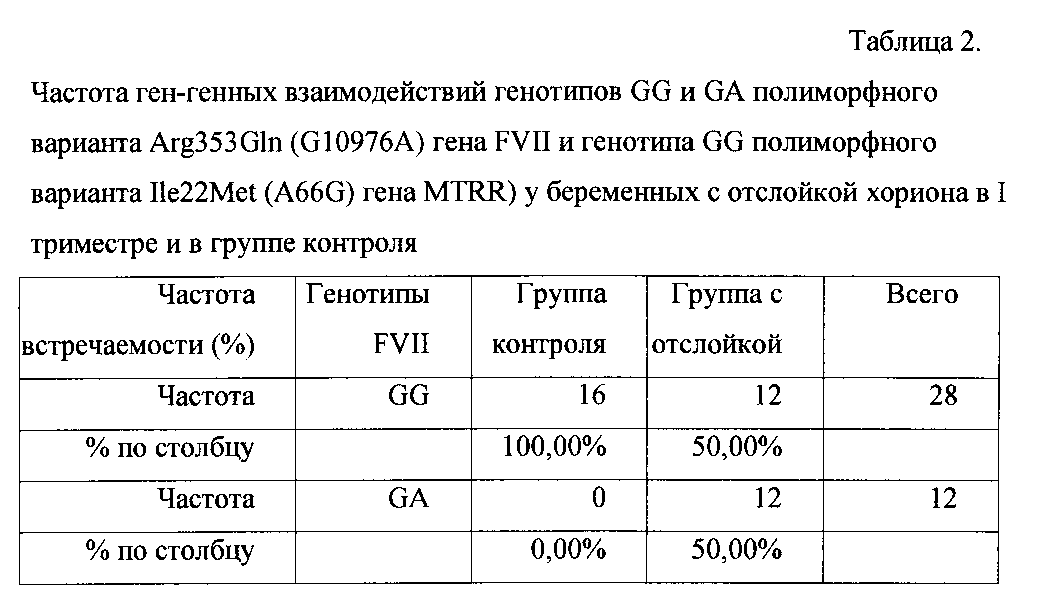

Генотип GG гена FVII является нормой гомозиготой, генотип GA гетерозиготой того же гена, АА гена MTRR представляет собой нормальную гомозиготу. Частота встречаемости сочетания генотипов GG и GA полиморфного варианта Arg353Gln (G10976A) гена FVII и генотипа АА полиморфного варианта Ile22Met (A66G) гена MTRR) не различалась у пациенток, имеющих отслойку хориона в I триместре и без нее (табл. 3).The GV genotype of the FVII gene is the norm homozygous, the GA genotype is the heterozygous of the same gene, and the AA of the MTRR gene is a normal homozygote. The frequency of occurrence of a combination of the GG and GA genotypes of the Arg353Gln polymorphic variant (G10976A) of the FVII gene and the AA genotype of the Ile22Met (A66G) polymorphic variant of the MTRR gene) did not differ in patients with and without chorionic detachment in the first trimester (Table 3).

Генотип AG является гетерозиготной формой полиморфизма Ile22Met (A66G) гена MTRR. Частота встречаемости сочетаний генотипов GG и GA полиморфного варианта Arg353Gln (G10976A) гена FVII и генотипа AG полиморфизма Ile22Met (A66G) гена MTRR не различалась у пациенток, имеющих отслойку хориона в I триместре, и в контрольной группе (табл. 4).The AG genotype is a heterozygous form of the Ile22Met (A66G) polymorphism of the MTRR gene. The frequency of occurrence of combinations of the GG and GA genotypes of the Arg353Gln polymorphic variant (G10976A) of the FVII gene and the AG genotype of the Ile22Met (A66G) polymorphism of the MTRR gene did not differ in patients with chorion detachment in the first trimester and in the control group (Table 4).

Таким образом, как показали результаты нашего исследования, частота встречаемости сочетания гетерозиготы полиморфного варианта Arg353Gln (G10976A) гена FVII с гомозиготой GG и гетерозиготой AG полиморфного варианта Ile22Met (A66G) гена MTRR не различалась у пациенток с отслойкой и пациенток в группе контроля (табл. 3, 4).Thus, as the results of our study showed, the frequency of occurrence of a combination of heterozygous polymorphic variant Arg353Gln (G10976A) of the FVII gene with homozygote GG and heterozygous AG polymorphic variant Ile22Met (A66G) of the MTRR gene did not differ in patients with detachment and patients in the control group (Table 3. , four).
На основании полученных результатов показано, что ген-генные взаимодействия: гетерозиготы полиморфного варианта Arg353Gln (G10976A) гена FVII с гомозиготой полиморфного варианта Ile22Met (A66G) гена MTRR, являются крайне неблагоприятным сочетанием, при котором риск развития отслойки хориона является высоким.Based on the results, it was shown that gene-to-heterozygotes of the polymorphic variant Arg353Gln (G10976A) of the FVII gene with the homozygous polymorphic variant Ile22Met (A66G) of the MTRR gene are an extremely unfavorable combination, in which the risk of developing chorionic detachment is high.
Роль гена MTRR в реализации механизма отслойки хориона заключается в кодировании аминокислотной последовательности фермента метионин синтазы редуктазы, одной из функций которого является обратное превращение гомоцистеина в метионин. В результате замены аминокислотного остатка изолейцина на метионин в позиции 66 функциональная активность фермента снижается, нарушаются процессы метилирования ДНК. Метилирование ДНК играет ведущую роль в формировании и поддержании эпигенетической изменчивости - наследственного динамического процесса, определяющего степень активности генов.The role of the MTRR gene in the implementation of the chorion detachment mechanism is to encode the amino acid sequence of the methionine synthase reductase enzyme, one of the functions of which is the reverse conversion of homocysteine to methionine. As a result of the replacement of the amino acid residue of isoleucine with methionine at position 66, the functional activity of the enzyme is reduced, DNA methylation processes are disrupted. DNA methylation plays a leading role in the formation and maintenance of epigenetic variability - a hereditary dynamic process that determines the degree of gene activity.
Выявленный факт позволил предложить гипотезу, заключающуюся в том, что дефицит фермента метиленсинтаза редуктаза сопровождается недостатком метальных групп, что, в свою очередь, оказывает влияние на эпигенетический статус, и реализацию эффекта гипоконвертинемии, обусловленного мутацией коагуляционного фактора FVII, в результате - манифестация такого клинического проявления, как отслойка хориона.The revealed fact allowed us to propose a hypothesis that the deficiency of the enzyme methylene synthase reductase is accompanied by a lack of metal groups, which, in turn, affects the epigenetic status and the realization of the effect of hypoconvertinemia due to the mutation of coagulation factor FVII, resulting in the manifestation of such a clinical manifestation like a chorion detachment.
Таким образом, предложенная гипотеза о том, что нарушение метилирования ДНК способствует проявлению мутации полиморфного варианта Arg353Gln (G10976A) гена FVII была подтверждена статистической обработкой данных.Thus, the proposed hypothesis that the violation of DNA methylation contributes to the manifestation of a mutation of the polymorphic variant Arg353Gln (G10976A) of the FVII gene was confirmed by statistical data processing.
Сущность предложенного способа поясняется примерами его осуществления.The essence of the proposed method is illustrated by examples of its implementation.
Пример №1Example No. 1
Пациентка Т., 30 лет, первобеременная, поступила в гинекологическое отделение с жалобами на тянущие боли внизу живота в течение 2-х недель. При поступлении в стационар было выполнено УЗИ (в полости матки визуализируется один плод, копчико-теменной размер плода 34 мм; срок беременности - 10 нед. 2 дн.; частота сердечных сокращений плода 154 уд. в мин.; средний внутренний диаметр желточного мешка - 5 мм; в нижнем полюсе плодного яйца выявлен участок отслойки размерами 35×20 мм в стадии организации). Показатели коагулограммы: фибриноген 3,9 г/л, HAVR 4.5 мг/100 мл, Д-димер 1284 мг/мл. Беременность 1-я. Наследственность по риску невынашивания, сердечно-сосудистым заболеваниям, генетической патологии не отягощена. В детстве перенесла ОРВИ, ветряную оспу.Patient T., 30 years old, the first pregnant, was admitted to the gynecological department with complaints of drawing pains in the lower abdomen for 2 weeks. Upon admission to the hospital, an ultrasound scan was performed (one fetus is visualized in the uterine cavity, the coccygeal-parietal fetal size is 34 mm; gestational age is 10 weeks. 2 days; the fetal heart rate is 154 beats per minute; the average inner diameter of the yolk sac is 5 mm; in the lower pole of the ovum, a detachment site of 35 × 20 mm in size is revealed at the organization stage). Coagulogram indicators: fibrinogen 3.9 g / l, HAVR 4.5 mg / 100 ml, D-dimer 1284 mg / ml. Pregnancy 1st. Heredity for the risk of miscarriage, cardiovascular disease, genetic pathology is not burdened. In childhood, she had ARVI, chickenpox.
Гинекологические заболевания: эрозия шейки матки, миома матки.Gynecological diseases: cervical erosion, uterine fibroids.
Экстрагенитальная патология: дискинезия желчевыводящих путей по гипотоническому типу. Аллергологический анамнез: поллиноз.Extragenital pathology: biliary dyskinesia according to the hypotonic type. Allergic history: hay fever.
Проведено молекулярно-генетическое тестирование 8 полиморфных вариантов генов системы гемостаза и 4-х полиморфных вариантов генов фолатного цикла: обнаружены гетерозигота полиморфного варианта FVII: 10976 G>A (GA) и мутантная гомозигота GG по полиморфизму MTRR: 66 A>G. Таким образом, выявление сочетания гетерозиготы GA полиморфного варианта Arg353Gln (G10976A) гена FVII с мутантной гомозиготой GG полиморфного варианта Ile22Met (A66G) гена MTRR подтверждает вероятность диагноза отслойки хориона, верифицированной по результатам УЗИ.Molecular genetic testing of 8 polymorphic variants of hemostasis system genes and 4 polymorphic variants of the folate cycle genes was carried out: heterozygous polymorphic variant FVII: 10976 G> A (GA) and mutant homozygote GG were detected by MTRR: 66 A> G polymorphism. Thus, the identification of the combination of the GA heterozygous polymorphic variant Arg353Gln (G10976A) of the FVII gene with the mutant homozygote GG of the polymorphic variant Ile22Met (A66G) of the MTRR gene confirms the likelihood of a diagnosis of chorion detachment verified by ultrasound.
Пациентка находилась в стационаре 10 дней, при выписке отмечено отсутствие отслойки по данным УЗИ после полученного лечения.The patient was in the hospital for 10 days, when discharged, there was no detachment according to ultrasound after treatment.
Пример №2Example No. 2
Пациентка Ф., 29 лет, первобеременная, поступила в гинекологическое отделение с жалобами на тянущие боли внизу живота в течение одной недели, кровянистые выделения из половых путей в течение 2-х дней. При поступлении в стационар выполнено УЗИ (срок беременности по дню последней менструации - 7 нед. 6 дней, в полости матки визуализируется 1-плодное яйцо, копчико-теменной размер 14 мм, соответствует менструальному сроку беременности 7 нед. 4 дн.; частота сердечных сокращений 161 уд. в минуту, средний внутренний диаметр желточного мешка 4,7 мм, преимущественная локализация хориона - задняя стенка матки, слева, перекрывает область внутреннего зева нижним краем; в областиPatient F., 29 years old, the first pregnant, was admitted to the gynecological department with complaints of drawing pains in the lower abdomen for one week, spotting from the genital tract for 2 days. Upon admission to the hospital, an ultrasound scan was performed (the gestational age on the day of the last menstruation is 7 weeks. 6 days, a 1-membered egg is visualized in the uterine cavity, the coccygeal-parietal size is 14 mm, corresponds to the menstrual period of pregnancy is 7 weeks. 4 days; heart rate 161 beats per minute, the average inner diameter of the yolk sac 4.7 mm, the primary localization of the chorion is the posterior wall of the uterus, on the left, overlaps the region of the internal pharynx with a lower edge; in the region
внутреннего зева эхо (-) щелевидная тень 15×3 мм - участок отслойки хориона.internal pharynx echo (-) slit-like shadow 15 × 3 mm - area of chorionic detachment.
Показатели коагулограммы - фибриноген 3,7 г/л, HAVR 4.2 мг/100 мл, Д-димер 789 мг/мл. Наследственность по риску невынашивания, сердечно-сосудистым заболеваниям, генетической патологии - не отягощена.Coagulogram indicators are fibrinogen 3.7 g / l, HAVR 4.2 mg / 100 ml, D-dimer 789 mg / ml. Heredity for the risk of miscarriage, cardiovascular disease, genetic pathology - not burdened.
В детстве перенесла ОРВИ, ветряную оспу, краснуху.In childhood, she had ARVI, chicken pox, rubella.
Гинекологические заболевания: первичное бесплодие (1,5 года).Gynecological diseases: primary infertility (1.5 years).
Экстрагенитальная патология: остеохондроз поясничного отдела позвоночника, правосторонний коксартроз, остеома правой тазобедренной кости. Аллергологический анамнез: не отягощен. Проведено молекулярно-генетическое тестирование 8 полиморфных вариантов генов системы гемостаза и 4-х полиморфных вариантов генов фолатного цикла: обнаружены гетерозигота полиморфного варианта FVII: 10976 G>A (GA) и мутантная гомозигота GG по полиморфизму MTRR: 66 A>G. Таким образом, выявление сочетания гетерозиготы GA полиморфного варианта Arg353Gln (G10976A) гена FVII с мутантной гомозиготой GG полиморфного варианта Ile22Met (A66G) гена MTRR подтверждает вероятность диагноза отслойки хориона, верифицированной по результатам УЗИ.Extragenital pathology: osteochondrosis of the lumbar spine, right-sided coxarthrosis, osteoma of the right hip. Allergic history: not burdened. Molecular genetic testing of 8 polymorphic variants of hemostasis system genes and 4 polymorphic variants of the folate cycle genes was carried out: heterozygous polymorphic variant FVII: 10976 G> A (GA) and mutant homozygote GG were detected by MTRR: 66 A> G polymorphism. Thus, the identification of the combination of the GA heterozygous polymorphic variant Arg353Gln (G10976A) of the FVII gene with the mutant homozygote GG of the polymorphic variant Ile22Met (A66G) of the MTRR gene confirms the likelihood of a diagnosis of chorion detachment verified by ultrasound.
Пациентка находилась в стационаре 10 дней, выписана в удовлетворительном состоянии с организацией отслойки хориона по данным УЗИ после проведенного лечения.The patient was in the hospital for 10 days, was discharged in satisfactory condition with the organization of the detachment of the chorion according to ultrasound after treatment.
Пример №3Example No. 3
Пациентка С., 26 лет, первобеременная, поступила в отделение гинекологии с жалобами на тянущие боли внизу живота, иррадиирующие в поясницу, в течение 3-х дней, тошноту, периодически рвоту. При поступлении в стационар выполнено УЗИ (срок беременности по дню последней менструации - 9 нед. 2 дн., в полости матки визуализируется 1-плодное яйцо, копчико-теменной размер 20 мм, соответствует менструальному сроку беременности 9 нед. 0 дн.; частота сердечных сокращений 162 уд. в минуту, средний внутренний диаметр желточного мешка 6 мм, преимущественная локализация хориона - задняя стенка матки, на 10 мм выше области внутреннего зева, в нижнем полюсе участок отслойки хориона 7×3 мм с признаками организации). Показатели коагулограммы: фибриноген - 4,2 г/л, Д-димер - 1182 мг/мл, РФМК - 5,0 мг/100 мл.Patient S., 26 years old, the first pregnant, was admitted to the gynecology department with complaints of drawing pains in the lower abdomen, radiating to the lower back, for 3 days, nausea, periodically vomiting. Upon admission to the hospital, an ultrasound scan was performed (the gestational age on the day of the last menstruation is 9 weeks. 2 days, a 1-membered egg is visualized in the uterine cavity, the coccygeal-parietal size is 20 mm; it corresponds to the menstrual period of pregnancy is 9 weeks. 0 days; cardiac frequency contractions 162 beats per minute, the average inner diameter of the yolk sac is 6 mm, the predominant localization of the chorion is the posterior wall of the uterus, 10 mm above the area of the internal pharynx, in the lower pole there is a section of the chorion detachment 7 × 3 mm with signs of organization). Coagulogram indicators: fibrinogen - 4.2 g / l, D-dimer - 1182 mg / ml, RFMC - 5.0 mg / 100 ml.
Наследственность по риску невынашивания, сердечно-сосудистым заболеваниям, генетической патологии - не отягощена.Heredity for the risk of miscarriage, cardiovascular disease, genetic pathology - not burdened.
В детстве перенесла ОРВИ, ветряную оспу, скарлатину, корь.In childhood, she had ARVI, chickenpox, scarlet fever, measles.
Гинекологические заболевания: первичное бесплодие (2 года).Gynecological diseases: primary infertility (2 years).
Экстрагенитальная патология: отрицает.Extragenital pathology: denied.
Аллергологический анамнез отягощен, в анамнезе отек Квинке на введение новокаина. Проведено молекулярно-генетическое тестирование 8 полиморфных вариантов генов системы гемостаза и 4-х полиморфных вариантов генов фолатного цикла. Обнаружены гетерозигота полиморфного варианта FVII: 10976 G>A (GA) и мутантная гомозигота GG по полиморфизму MTRR: 66 A>G. Таким образом, выявление сочетания гетерозиготы GA полиморфного варианта Arg353Gln (G10976A) гена FVII с мутантной гомозиготой GG полиморфного варианта Ile22Met (A66G) гена MTRR подтверждает вероятность диагноза отслойки хориона, подтвержденной по результатам УЗИ.An allergic history is burdened, a history of Quincke's edema on the introduction of novocaine. Molecular genetic testing of 8 polymorphic variants of hemostasis system genes and 4 polymorphic variants of folate cycle genes was carried out. The heterozygote of the polymorphic variant FVII: 10976 G> A (GA) and the mutant homozygote GG by the MTRR polymorphism: 66 A> G were found. Thus, the identification of the combination of the GA heterozygous polymorphic variant Arg353Gln (G10976A) of the FVII gene with the mutant homozygote GG of the polymorphic variant Ile22Met (A66G) of the MTRR gene confirms the probability of the diagnosis of chorionic detachment, confirmed by ultrasound.
Пациентка находилась в стационаре 10 дней, выписана в удовлетворительном состоянии с отсутствием отслойки хориона по данным УЗИ после проведенного лечения.The patient was in the hospital for 10 days, was discharged in satisfactory condition with no chorion detachment according to ultrasound after treatment.
Claims (1)
Priority Applications (1)
| Application Number | Priority Date | Filing Date | Title |
|---|---|---|---|
| RU2014117238/15A RU2566729C1 (en) | 2014-04-28 | 2014-04-28 | Method for prediction of chorion separation in first trimester of pregnancy |
Applications Claiming Priority (1)
| Application Number | Priority Date | Filing Date | Title |
|---|---|---|---|
| RU2014117238/15A RU2566729C1 (en) | 2014-04-28 | 2014-04-28 | Method for prediction of chorion separation in first trimester of pregnancy |
Publications (1)
| Publication Number | Publication Date |
|---|---|
| RU2566729C1 true RU2566729C1 (en) | 2015-10-27 |
Family
ID=54362383
Family Applications (1)
| Application Number | Title | Priority Date | Filing Date |
|---|---|---|---|
| RU2014117238/15A RU2566729C1 (en) | 2014-04-28 | 2014-04-28 | Method for prediction of chorion separation in first trimester of pregnancy |
Country Status (1)
| Country | Link |
|---|---|
| RU (1) | RU2566729C1 (en) |
Cited By (1)
| Publication number | Priority date | Publication date | Assignee | Title |
|---|---|---|---|---|
| RU2611358C1 (en) * | 2016-02-03 | 2017-02-21 | федеральное государственное автономное образовательное учреждение высшего образования "Южный федеральный университет" | Method of predicting high risk of reproductive loss in first trimester of pregnancy |
Citations (1)
| Publication number | Priority date | Publication date | Assignee | Title |
|---|---|---|---|---|
| RU2494400C1 (en) * | 2012-01-16 | 2013-09-27 | Федеральное государственное бюджетное учреждение "Научный центр акушерства, гинекологии и перинатологии имени академика В.И. Кулакова" Министерства здравоохранения и социального развития Российской Федерации | Method for early prediction of chorion and placenta detachment on basis of detecting gene polymorphism of type 1 plasminogen activator inhibitor (pai-1) |
-
2014
- 2014-04-28 RU RU2014117238/15A patent/RU2566729C1/en not_active IP Right Cessation
Patent Citations (1)
| Publication number | Priority date | Publication date | Assignee | Title |
|---|---|---|---|---|
| RU2494400C1 (en) * | 2012-01-16 | 2013-09-27 | Федеральное государственное бюджетное учреждение "Научный центр акушерства, гинекологии и перинатологии имени академика В.И. Кулакова" Министерства здравоохранения и социального развития Российской Федерации | Method for early prediction of chorion and placenta detachment on basis of detecting gene polymorphism of type 1 plasminogen activator inhibitor (pai-1) |
Non-Patent Citations (1)
| Title |
|---|
| Генетическая предрасположенность к ряду мультифакториальных заболеваний. Статьи. Специалистам; 16.11.2010 [Найдено 27.01.2015] [он-лайн], Найдено из Интернет: URL: http://www.vitasite.ru/articles/expert-article/dnk-k-rjadu-multifaktorialnykh-zabolevanij/. ЗАРУДСКАЯ О.М. Ассоциация полиморфизма FVII 10976G/A (RS 6046) с развитием плацентарной недостаточности с синдромом задержки роста плода. Материалы VII Международного конгресса по репродуктивной медицине. Москва, 21-24 января 2013; стр.137-138 [Найдено 27.01.2015] [он-лайн], Найдено из Интернет: URL: http://www.mediexpo.ru/fileadmin/user_upload/content/pdf/thesis/thesis_rzs13.pdf. SAUERBREI E.E. et al. A practical guide to ultrasound in obstetrics and gynecology. Philadelphia (PA): Lippincott-Raven Publishers. 1998; 597 p * |
Cited By (1)
| Publication number | Priority date | Publication date | Assignee | Title |
|---|---|---|---|---|
| RU2611358C1 (en) * | 2016-02-03 | 2017-02-21 | федеральное государственное автономное образовательное учреждение высшего образования "Южный федеральный университет" | Method of predicting high risk of reproductive loss in first trimester of pregnancy |
Similar Documents
| Publication | Publication Date | Title |
|---|---|---|
| CN102459635B (en) | For pregnancy outcome, there is the competence ovocyte of high potential and the method for competence embryo for selecting | |
| Lapaire et al. | Microarray screening for novel preeclampsia biomarker candidates | |
| US9090938B2 (en) | Methods for selecting competent oocytes and competent embryos with high potential for pregnancy outcome | |
| US11149315B2 (en) | Method for predicting cervical shortening and preterm birth | |
| RU2494400C1 (en) | Method for early prediction of chorion and placenta detachment on basis of detecting gene polymorphism of type 1 plasminogen activator inhibitor (pai-1) | |
| Pang et al. | A strategy for identifying circulating placental RNA markers for fetal growth assessment | |
| Wolski et al. | Contribution of inherited thrombophilia to recurrent miscarriage in the Polish population | |
| CN115449548A (en) | Application of STAC2 cumulus cells in evaluation of fertilization capability of oocytes | |
| Kadam et al. | Endocervical trophoblast for interrogating the fetal genome and assessing pregnancy health at five weeks | |
| RU2566729C1 (en) | Method for prediction of chorion separation in first trimester of pregnancy | |
| RU2475740C1 (en) | Method of detecting hereditary predisposition to fast growth of uterine myoma | |
| EP1511863A4 (en) | ENDOMETRIAL GENES IN DISEASES OF THE ENDOMETRIUM | |
| CN114941025A (en) | MiRNA for diagnosing preeclampsia and application thereof | |
| Brummaier et al. | Blood gene transcript signature profiling in pregnancies resulting in preterm birth: A systematic review | |
| Sattorov | PREDICTION OF PREMATURE OUTFLOW OF AMNIOTIC FLUID IN PRETERM PREGNANCY | |
| Rymarchuk et al. | On the issue of pathogenesis of placental dysfunction in women with miscarriage | |
| Linares et al. | P-703 The role of maternal polymorphic karyotype variants among embryonic ploidy and mosaicism status in patients undergoing IVF treatments | |
| Younis et al. | Genetic Factors in Preeclamptic Egyptian Women: Relation with Arginine Vasopressin; A Case-Control Study | |
| Pang | Investigation of the Quantitative Relationship Between Circulating Placental MRNA and Fetal Growth | |
| Римарчук et al. | On the issue of pathogenesis of placental dysfunction in women with miscarriage | |
| Merhi et al. | Pregnancy and preeclampsia alter ovarian genes important in follicular development | |
| Olaya Agudo | Variation in expression of fetal nucleic acids in maternal blood in pregnancies affected by congenital anomalies | |
| Kappou et al. | Non Invasive Prenatal Diagnosis of Down Syndrome | |
| Koha et al. | Noninvasive in vivo monitoring of tissue-specific global gene expression in humans |
Legal Events
| Date | Code | Title | Description |
|---|---|---|---|
| MM4A | The patent is invalid due to non-payment of fees |
Effective date: 20160429 |