RU2562111C2 - Conversion of somatic cells into induced reprogrammed neural stem cells (irnsc) - Google Patents
Conversion of somatic cells into induced reprogrammed neural stem cells (irnsc) Download PDFInfo
- Publication number
- RU2562111C2 RU2562111C2 RU2013110127/10A RU2013110127A RU2562111C2 RU 2562111 C2 RU2562111 C2 RU 2562111C2 RU 2013110127/10 A RU2013110127/10 A RU 2013110127/10A RU 2013110127 A RU2013110127 A RU 2013110127A RU 2562111 C2 RU2562111 C2 RU 2562111C2
- Authority
- RU
- Russia
- Prior art keywords
- cells
- neural stem
- stem cells
- somatic cells
- neurospheres
- Prior art date
Links
Images


Classifications
-
- C—CHEMISTRY; METALLURGY
- C12—BIOCHEMISTRY; BEER; SPIRITS; WINE; VINEGAR; MICROBIOLOGY; ENZYMOLOGY; MUTATION OR GENETIC ENGINEERING
- C12N—MICROORGANISMS OR ENZYMES; COMPOSITIONS THEREOF; PROPAGATING, PRESERVING, OR MAINTAINING MICROORGANISMS; MUTATION OR GENETIC ENGINEERING; CULTURE MEDIA
- C12N5/00—Undifferentiated human, animal or plant cells, e.g. cell lines; Tissues; Cultivation or maintenance thereof; Culture media therefor
- C12N5/06—Animal cells or tissues; Human cells or tissues
- C12N5/0602—Vertebrate cells
- C12N5/0618—Cells of the nervous system
- C12N5/0623—Stem cells
-
- C—CHEMISTRY; METALLURGY
- C12—BIOCHEMISTRY; BEER; SPIRITS; WINE; VINEGAR; MICROBIOLOGY; ENZYMOLOGY; MUTATION OR GENETIC ENGINEERING
- C12N—MICROORGANISMS OR ENZYMES; COMPOSITIONS THEREOF; PROPAGATING, PRESERVING, OR MAINTAINING MICROORGANISMS; MUTATION OR GENETIC ENGINEERING; CULTURE MEDIA
- C12N5/00—Undifferentiated human, animal or plant cells, e.g. cell lines; Tissues; Cultivation or maintenance thereof; Culture media therefor
- C12N5/06—Animal cells or tissues; Human cells or tissues
- C12N5/0602—Vertebrate cells
- C12N5/0696—Artificially induced pluripotent stem cells, e.g. iPS
-
- A—HUMAN NECESSITIES
- A61—MEDICAL OR VETERINARY SCIENCE; HYGIENE
- A61K—PREPARATIONS FOR MEDICAL, DENTAL OR TOILETRY PURPOSES
- A61K35/00—Medicinal preparations containing materials or reaction products thereof with undetermined constitution
- A61K35/12—Materials from mammals; Compositions comprising non-specified tissues or cells; Compositions comprising non-embryonic stem cells; Genetically modified cells
- A61K35/30—Nerves; Brain; Eyes; Corneal cells; Cerebrospinal fluid; Neuronal stem cells; Neuronal precursor cells; Glial cells; Oligodendrocytes; Schwann cells; Astroglia; Astrocytes; Choroid plexus; Spinal cord tissue
-
- A—HUMAN NECESSITIES
- A61—MEDICAL OR VETERINARY SCIENCE; HYGIENE
- A61P—SPECIFIC THERAPEUTIC ACTIVITY OF CHEMICAL COMPOUNDS OR MEDICINAL PREPARATIONS
- A61P25/00—Drugs for disorders of the nervous system
-
- C—CHEMISTRY; METALLURGY
- C12—BIOCHEMISTRY; BEER; SPIRITS; WINE; VINEGAR; MICROBIOLOGY; ENZYMOLOGY; MUTATION OR GENETIC ENGINEERING
- C12N—MICROORGANISMS OR ENZYMES; COMPOSITIONS THEREOF; PROPAGATING, PRESERVING, OR MAINTAINING MICROORGANISMS; MUTATION OR GENETIC ENGINEERING; CULTURE MEDIA
- C12N15/00—Mutation or genetic engineering; DNA or RNA concerning genetic engineering, vectors, e.g. plasmids, or their isolation, preparation or purification; Use of hosts therefor
- C12N15/09—Recombinant DNA-technology
- C12N15/63—Introduction of foreign genetic material using vectors; Vectors; Use of hosts therefor; Regulation of expression
- C12N15/79—Vectors or expression systems specially adapted for eukaryotic hosts
- C12N15/85—Vectors or expression systems specially adapted for eukaryotic hosts for animal cells
- C12N15/86—Viral vectors
-
- C—CHEMISTRY; METALLURGY
- C12—BIOCHEMISTRY; BEER; SPIRITS; WINE; VINEGAR; MICROBIOLOGY; ENZYMOLOGY; MUTATION OR GENETIC ENGINEERING
- C12N—MICROORGANISMS OR ENZYMES; COMPOSITIONS THEREOF; PROPAGATING, PRESERVING, OR MAINTAINING MICROORGANISMS; MUTATION OR GENETIC ENGINEERING; CULTURE MEDIA
- C12N5/00—Undifferentiated human, animal or plant cells, e.g. cell lines; Tissues; Cultivation or maintenance thereof; Culture media therefor
- C12N5/10—Cells modified by introduction of foreign genetic material
-
- C—CHEMISTRY; METALLURGY
- C12—BIOCHEMISTRY; BEER; SPIRITS; WINE; VINEGAR; MICROBIOLOGY; ENZYMOLOGY; MUTATION OR GENETIC ENGINEERING
- C12N—MICROORGANISMS OR ENZYMES; COMPOSITIONS THEREOF; PROPAGATING, PRESERVING, OR MAINTAINING MICROORGANISMS; MUTATION OR GENETIC ENGINEERING; CULTURE MEDIA
- C12N2501/00—Active agents used in cell culture processes, e.g. differentation
- C12N2501/10—Growth factors
- C12N2501/11—Epidermal growth factor [EGF]
-
- C—CHEMISTRY; METALLURGY
- C12—BIOCHEMISTRY; BEER; SPIRITS; WINE; VINEGAR; MICROBIOLOGY; ENZYMOLOGY; MUTATION OR GENETIC ENGINEERING
- C12N—MICROORGANISMS OR ENZYMES; COMPOSITIONS THEREOF; PROPAGATING, PRESERVING, OR MAINTAINING MICROORGANISMS; MUTATION OR GENETIC ENGINEERING; CULTURE MEDIA
- C12N2501/00—Active agents used in cell culture processes, e.g. differentation
- C12N2501/10—Growth factors
- C12N2501/115—Basic fibroblast growth factor (bFGF, FGF-2)
-
- C—CHEMISTRY; METALLURGY
- C12—BIOCHEMISTRY; BEER; SPIRITS; WINE; VINEGAR; MICROBIOLOGY; ENZYMOLOGY; MUTATION OR GENETIC ENGINEERING
- C12N—MICROORGANISMS OR ENZYMES; COMPOSITIONS THEREOF; PROPAGATING, PRESERVING, OR MAINTAINING MICROORGANISMS; MUTATION OR GENETIC ENGINEERING; CULTURE MEDIA
- C12N2501/00—Active agents used in cell culture processes, e.g. differentation
- C12N2501/10—Growth factors
- C12N2501/119—Other fibroblast growth factors, e.g. FGF-4, FGF-8, FGF-10
-
- C—CHEMISTRY; METALLURGY
- C12—BIOCHEMISTRY; BEER; SPIRITS; WINE; VINEGAR; MICROBIOLOGY; ENZYMOLOGY; MUTATION OR GENETIC ENGINEERING
- C12N—MICROORGANISMS OR ENZYMES; COMPOSITIONS THEREOF; PROPAGATING, PRESERVING, OR MAINTAINING MICROORGANISMS; MUTATION OR GENETIC ENGINEERING; CULTURE MEDIA
- C12N2501/00—Active agents used in cell culture processes, e.g. differentation
- C12N2501/10—Growth factors
- C12N2501/13—Nerve growth factor [NGF]; Brain-derived neurotrophic factor [BDNF]; Cilliary neurotrophic factor [CNTF]; Glial-derived neurotrophic factor [GDNF]; Neurotrophins [NT]; Neuregulins
-
- C—CHEMISTRY; METALLURGY
- C12—BIOCHEMISTRY; BEER; SPIRITS; WINE; VINEGAR; MICROBIOLOGY; ENZYMOLOGY; MUTATION OR GENETIC ENGINEERING
- C12N—MICROORGANISMS OR ENZYMES; COMPOSITIONS THEREOF; PROPAGATING, PRESERVING, OR MAINTAINING MICROORGANISMS; MUTATION OR GENETIC ENGINEERING; CULTURE MEDIA
- C12N2501/00—Active agents used in cell culture processes, e.g. differentation
- C12N2501/40—Regulators of development
- C12N2501/405—Cell cycle regulated proteins, e.g. cyclins, cyclin-dependant kinases
-
- C—CHEMISTRY; METALLURGY
- C12—BIOCHEMISTRY; BEER; SPIRITS; WINE; VINEGAR; MICROBIOLOGY; ENZYMOLOGY; MUTATION OR GENETIC ENGINEERING
- C12N—MICROORGANISMS OR ENZYMES; COMPOSITIONS THEREOF; PROPAGATING, PRESERVING, OR MAINTAINING MICROORGANISMS; MUTATION OR GENETIC ENGINEERING; CULTURE MEDIA
- C12N2501/00—Active agents used in cell culture processes, e.g. differentation
- C12N2501/60—Transcription factors
- C12N2501/602—Sox-2
-
- C—CHEMISTRY; METALLURGY
- C12—BIOCHEMISTRY; BEER; SPIRITS; WINE; VINEGAR; MICROBIOLOGY; ENZYMOLOGY; MUTATION OR GENETIC ENGINEERING
- C12N—MICROORGANISMS OR ENZYMES; COMPOSITIONS THEREOF; PROPAGATING, PRESERVING, OR MAINTAINING MICROORGANISMS; MUTATION OR GENETIC ENGINEERING; CULTURE MEDIA
- C12N2501/00—Active agents used in cell culture processes, e.g. differentation
- C12N2501/70—Enzymes
- C12N2501/72—Transferases (EC 2.)
- C12N2501/727—Kinases (EC 2.7.)
-
- C—CHEMISTRY; METALLURGY
- C12—BIOCHEMISTRY; BEER; SPIRITS; WINE; VINEGAR; MICROBIOLOGY; ENZYMOLOGY; MUTATION OR GENETIC ENGINEERING
- C12N—MICROORGANISMS OR ENZYMES; COMPOSITIONS THEREOF; PROPAGATING, PRESERVING, OR MAINTAINING MICROORGANISMS; MUTATION OR GENETIC ENGINEERING; CULTURE MEDIA
- C12N2506/00—Differentiation of animal cells from one lineage to another; Differentiation of pluripotent cells
- C12N2506/13—Differentiation of animal cells from one lineage to another; Differentiation of pluripotent cells from connective tissue cells, from mesenchymal cells
- C12N2506/1307—Differentiation of animal cells from one lineage to another; Differentiation of pluripotent cells from connective tissue cells, from mesenchymal cells from adult fibroblasts
-
- C—CHEMISTRY; METALLURGY
- C12—BIOCHEMISTRY; BEER; SPIRITS; WINE; VINEGAR; MICROBIOLOGY; ENZYMOLOGY; MUTATION OR GENETIC ENGINEERING
- C12N—MICROORGANISMS OR ENZYMES; COMPOSITIONS THEREOF; PROPAGATING, PRESERVING, OR MAINTAINING MICROORGANISMS; MUTATION OR GENETIC ENGINEERING; CULTURE MEDIA
- C12N2510/00—Genetically modified cells
Abstract
Description
Данная заявка относится к способу конверсии соматических клеток в нейральные стволовые клетки (НСК). Кроме того, данная заявка относится к способу конверсии человеческих фибробластов, кератиноцитов или адипоцитов в нейральные стволовые клетки на основании сопряженной пошаговой трансдукции генов и индукции средой определенного химического состава.This application relates to a method for converting somatic cells into neural stem cells (NSCs). In addition, this application relates to a method for converting human fibroblasts, keratinocytes or adipocytes into neural stem cells based on conjugated stepwise gene transduction and medium induction of a specific chemical composition.
Длительное время считалось общепринятой догмой, что полностью дифференцированные соматические клетки не способны изменять свои свойства в обратном направлении. Эта догма начала меняться, как только серия новаторских экспериментов показала, что профиль экспрессии «молчащих» генов может быть полностью реактивирован с помощью слияния клеток различных типов (Blau, Н.М. How fixed is the differentiated state? Lessons from heterokaryons. Trends Genet. 5, 268-272 (1989)). Относительно недавно было показано, что перенос ядра из соматической клетки в энуклеированную яйцеклетку способен привести к полной реверсии профиля экспрессии генов соматических клеток и формированию состояния плюрипотентной клетки, способной воспроизвести целое животное (например, Gurdon, J.В. & Melton, D.A. Nuclear reprogramming in cells. Science 322, 1811-1815 (2008)). Яманака с коллегами (Takahashi, К. & Yamanaka, S. Induction of pluripotent stem cells from mouse embryonic and adult fibroblast cultures by defined factors. Cell 126, 663-676 (2006)) продемонстрировали, что соматические клетки могут быть репрограммированы в индуцированные плюрипотентные стволовые клетки (ипСК) с помощью трансдукции четырех определенных факторов (Sox2, Oct4, Klf4, с-Мус). В плюрипотентное состояние ипСК были репрограммированы различные типы соматических клеток, включая фибробласты, кератиноциты и адипоциты. В последние годы возник вопрос, все ли специфические типы соматических клеток способны трансдифференцироваться в совершенно другой тип соматических клеток, таких как нейроны. Этот вопрос изучали Вениг с коллегами, продемонстрировав прямую конверсию мышиных фибробластов в функциональные нейроны с помощью трансдукции трех критических генов: Mash1, Brn2 and Myt1I (Wernig at al. Direct conversion of fibroblasts to functional neurons by defined factors. Nature 25; 463(7284): 1035-41 (2010)). Однако полученные нейроны являются постмитотическими клетками, которые по определению не способны пролиферировать и не переносят процедуры замораживания-оттаивания. US 2010/0021437 раскрывает способ получения индуцированных плюрипотентных стволовых клеток из фибробластов и индукции таких клеток к дифференцировке в нейральный фенотип.For a long time it was considered a generally accepted dogma that completely differentiated somatic cells are not able to change their properties in the opposite direction. This dogma began to change as a series of innovative experiments showed that the expression profile of silent genes can be completely reactivated by fusion of different types of cells (Blau, N.M. How fixed is the differentiated state? Lessons from heterokaryons. Trends Genet. 5, 268-272 (1989)). Relatively recently, it was shown that transferring a nucleus from a somatic cell to an enucleated ovum can lead to a complete reversal of the expression profile of somatic cell genes and the formation of a pluripotent cell state that can reproduce the whole animal (e.g., Gurdon, J. B. & Melton, DA Nuclear reprogramming in cells. Science 322, 1811-1815 (2008)). Yamanaka et al. (Takahashi, K. & Yamanaka, S. Induction of pluripotent stem cells from mouse embryonic and adult fibroblast cultures by defined factors. Cell 126, 663-676 (2006)) demonstrated that somatic cells can be reprogrammed into induced pluripotent stem cells (iPScs) by transduction of four specific factors (Sox2, Oct4, Klf4, c-Myc). Various types of somatic cells, including fibroblasts, keratinocytes and adipocytes, were reprogrammed into the pluripotent state of iPSCs. In recent years, the question has arisen whether all specific types of somatic cells are capable of transdifferentiating into a completely different type of somatic cells, such as neurons. Wenig and colleagues studied this issue by demonstrating the direct conversion of murine fibroblasts to functional neurons by transducing three critical genes: Mash1, Brn2 and Myt1I (Wernig at al. Direct conversion of fibroblasts to functional neurons by defined factors. Nature 25; 463 (7284) : 1035-41 (2010)). However, the resulting neurons are postmitotic cells that, by definition, are not able to proliferate and cannot tolerate freezing-thawing. US 2010/0021437 discloses a method for producing induced pluripotent stem cells from fibroblasts and inducing such cells to differentiate into a neural phenotype.
Однако прямая конверсия дифференцированных соматических клеток в нейральные стволовые клетки до сих пор не описана. Показано, что нейральные стволовые клетки являются мультипотентными стволовыми клетками, накапливаемыми в специфических условиях. Они требуют определенной химической среды, например, среды N2B27 (N2B27 является смесью 1:1 сред DMEM/F12 (Gibco, Пейсли, Великобритания) с добавлением N2 и В27 (обе от Gibco)) с добавлением FGF (фактор роста фибробластов 2) и EGF (эпидермальный ростовой фактор). Они способны расти как монослойная адгезивная культура, например, в плашках, покрытых полиорнитином/ламином, или как плавучие нейросферы в плашках для неадгезивных клеточных культур. Было показано, что два типа культур нейральных стволовых клеток (нейросферы, адгезивные культуры) полностью взаимно трансформируемы. Нейральные стволовые клетки растут бесконечно и действительно остаются полностью мультипотентными. В определенных условиях они дифференцируются в клеточные типы, из которых состоит взрослый мозг, включая нейроны, астроциты и олигодендроциты. Нейральные стволовые клетки рассматриваются как возможное лечебное средство для лечения пациентов с нейродегенеративными заболеваниями, такими как болезнь Альцгеймера, болезнь Паркинсона, нарушение мозгового кровообращения, и травмы спинного мозга.However, the direct conversion of differentiated somatic cells to neural stem cells has not yet been described. It has been shown that neural stem cells are multipotent stem cells that accumulate under specific conditions. They require a specific chemical medium, for example, N2B27 medium (N2B27 is a 1: 1 mixture of DMEM / F12 media (Gibco, Paisley, UK) with the addition of N2 and B27 (both from Gibco)) with the addition of FGF (fibroblast growth factor 2) and EGF (epidermal growth factor). They are able to grow as a monolayer adhesive culture, for example, in dies coated with polyornithine / lamin, or as floating neurospheres in dies for non-adhesive cell cultures. It was shown that two types of cultures of neural stem cells (neurospheres, adhesive cultures) are completely mutually transformable. Neural stem cells grow endlessly and truly remain fully multipotent. Under certain conditions, they differentiate into the cellular types that make up the adult brain, including neurons, astrocytes, and oligodendrocytes. Neural stem cells are considered as a possible treatment for patients with neurodegenerative diseases such as Alzheimer's disease, Parkinson's disease, cerebrovascular accident, and spinal cord injuries.
Известно, что нейральные стволовые клетки также можно получить in vitro из эмбриональных стволовых клеток (ЭСК) (Chambers et al. Nature 27; 3 (2009)), либо они могут быть выделены напрямую из образцов мозга (Reynolds BA, Rietze RL (2005) Nat Methods 2: 333-336). Однако, также известно, что до сих пор эти методы обладают существенными недостатками, такими как повышенное число высокочувствительных этических вопросов и/или они требуют сложных и трудоемких технологий, которые страдают от серьезных проблем с воспроизводимостью. До сих пор не было описано способов, с помощью которых нейральные стволовые клетки могли быть напрямую получены из дифференцированных соматических клеток. В принципе, нейральные стволовые клетки можно получить из ипСК, полученных из дифференцированных клеток. Однако это подразумевает культивирование ипСК. Было показано, что ипСК растут бесконечно, но условия культивирования сложны и требуют гигантских усилий. В дополнение было отмечено, что образование нейральных стволовых клеток из плюрипотентных стволовых клеток колеблется из-за стохастических механизмов. Общим препятствием ипСК и ЭСК является тот факт, что даже минимальное число недифферецированных клеток может привести к формированию тератом (опухоли из зародышевых клеток, включающие несколько клеточных типов), которые являют собой внушающую опасения контаминацию, которую нельзя игнорировать. Следовательно, остается потребность в легко доступной и воспроизводимой технологии получения нейральных стволовых клеток. В настоящем изобретении предложен способ конвертации соматических клеток напрямую в нейральные стволовые клетки. Новый способ ослабляет необходимость получения ипСК и следовательно устраняет риск формирования тератом. Подобные клетки без способности формировать тератомы пригодны и безопасны для применения в регенеративной медицине. Предпочтительные соматические клетки - это соматические клетки млекопитающих, наиболее предпочтительны человеческие соматические клетки. Предпочтительные соматические клетки - это фибробласты, адипоциты или кератиноциты, наиболее предпочтительны фибробласты. Вышеупомянутые фибробласты, адипоциты или кератиноциты могут быть без труда и безопасно получены от пациента или здорового человека с помощью, например, неинвазивных методов, таких как биопсия кожи, или из выщипанных волос. Способ настоящего изобретения позволяет конвертировать соматические клетки, такие как фибробласты, адипоциты или кератиноциты от здоровых или больных людей напрямую в нейральные стволовые клетки. Такие нейральные стволовые клетки от здоровых людей, или пациент-специфичные, способны делиться бесконечно. Их культивирование удобно и хорошо охарактеризовано. Аликвоты нейральных стволовых клеток от здоровых людей и пациент-специфичных возможно воспроизводимо замораживать и оттаивать. В частности, нейральные стволовые клетки, полученные от пациента, представляют соответствующую заболеванию in vitro модель для изучения патофизиологии заболеваний ЦНС. Конверсия пациент-специфичных соматических клеток напрямую в нейральные стволовые клетки представляет легко доступную и воспроизводимую технологию создания БиоБанков пациент-специфичных нейральных стволовых клеток. Подобные БиоБанки имеют огромное значение для заболеваний ЦНС, патология которых была описана в клетках по меньшей мере трех типов, развивающихся из нейральных стволовых клеток: нейронов, олигодендроцитов и астроцитов. Таким образом, полученные с помощью описываемого способа нейральные стволовые клетки являются чрезвычайно ценной моделью заболеваний для поиска эффективных и безопасных лекарственных препаратов.It is known that neural stem cells can also be obtained in vitro from embryonic stem cells (ESCs) (Chambers et al. Nature 27; 3 (2009)), or they can be isolated directly from brain samples (Reynolds BA, Rietze RL (2005) Nat Methods 2: 333-336). However, it is also known that these methods still have significant drawbacks, such as an increased number of highly sensitive ethical issues and / or they require complex and labor-intensive technologies that suffer from serious problems with reproducibility. Until now, no methods have been described by which neural stem cells can be directly obtained from differentiated somatic cells. In principle, neural stem cells can be obtained from iPScs derived from differentiated cells. However, this implies the cultivation of iPSCs. It has been shown that iPSCs grow endlessly, but cultivation conditions are complex and require tremendous effort. In addition, it was noted that the formation of neural stem cells from pluripotent stem cells fluctuates due to stochastic mechanisms. A common obstacle to iPSCs and ESCs is the fact that even the smallest number of undifferentiated cells can lead to the formation of teratomas (tumors from germ cells, including several cell types), which are an alarming contamination that cannot be ignored. Consequently, there remains a need for an easily accessible and reproducible technology for the production of neural stem cells. The present invention provides a method for converting somatic cells directly into neural stem cells. The new method reduces the need for iPSCs and therefore eliminates the risk of teratomas. Such cells without the ability to form teratomas are suitable and safe for use in regenerative medicine. Preferred somatic cells are mammalian somatic cells, human somatic cells are most preferred. Preferred somatic cells are fibroblasts, adipocytes or keratinocytes, fibroblasts are most preferred. The aforementioned fibroblasts, adipocytes or keratinocytes can be easily and safely obtained from a patient or a healthy person using, for example, non-invasive methods such as skin biopsies or from plucked hair. The method of the present invention allows the conversion of somatic cells, such as fibroblasts, adipocytes or keratinocytes from healthy or sick people directly into neural stem cells. Such neural stem cells from healthy people, or patient-specific ones, are able to divide indefinitely. Their cultivation is convenient and well characterized. Aliquots of neural stem cells from healthy people and patient-specific can reproducibly freeze and thaw. In particular, neural stem cells obtained from a patient provide an in vitro disease-appropriate model for studying the pathophysiology of central nervous system diseases. The conversion of patient-specific somatic cells directly into neural stem cells is an easily accessible and reproducible technology for creating BioBanks of patient-specific neural stem cells. Such BioBanks are of great importance for diseases of the central nervous system, the pathology of which has been described in cells of at least three types developing from neural stem cells: neurons, oligodendrocytes and astrocytes. Thus, the neural stem cells obtained using the described method are an extremely valuable model of diseases for the search for effective and safe drugs.
Множество нейродегенеративных заболеваний характеризуются потерей нервных клеток. Регенеративная способность взрослого мозга в ответ на черепно-мозговые травмы и нейродегенеративные заболевания весьма ограничена. Более того, фармакологическое воздействие часто все в большей степени теряет эффективность из-за прогрессирующей гибели популяции чувствительных к нему нейронов. Полученные с помощью описываемого здесь способа нейральные стволовые клетки также могут быть использованы в регенеративной медицине для лечения нейродегенеративных заболеваний, таких как болезнь Паркинсона, болезнь Альцгеймера, хорея Хантингтона, боковой амиотрофический склероз (болезнь Лу Герига) и травм спинного мозга. С помощью описываемого новаторского способа теперь возможно производить достаточное количество нейральных клеток-предшественников для использования при лечении методами клеточной трансплантации. Нейральные стволовые клетки могут быть получены из соматических клеток, выделенных как из здорового человека, так и из пациента. Пациент-специфичные нейральные стволовые клетки, полученные с помощью описываемого способа, являются новым привлекательным донорским источником для лечения методами аутологичной клеточной трансплантации вследствие подавления любого иммунного отторжения, вызванного иммунологической несовместимостью между пациентом и донором. Данная стратегия способна устранить необходимость иммунной супрессии при лечении методами клеточной трансплантации. Более того, создание БиоБанков нейральных стволовых клеток, полученных от здоровых людей с различными гомозиготными HLA аллелями, может быть использовано как донорский банк для лечения пациентов, нуждающихся в помощи. Гетерологичная трансплантация нейральных стволовых клеток с совместимым HLA-типом снижает риск нежелательных иммунных ответов, которые могут привести к отторжению пересаженных клеток.Many neurodegenerative diseases are characterized by loss of nerve cells. The regenerative capacity of the adult brain in response to head injuries and neurodegenerative diseases is very limited. Moreover, pharmacological effects often increasingly lose their effectiveness due to the progressive death of a population of neurons sensitive to it. Obtained using the method described here, neural stem cells can also be used in regenerative medicine for the treatment of neurodegenerative diseases such as Parkinson's disease, Alzheimer's disease, Huntington’s chorea, amyotrophic lateral sclerosis (Lou Gehrig’s disease) and spinal cord injuries. Using the described innovative method, it is now possible to produce a sufficient number of neural progenitor cells for use in the treatment of cell transplantation methods. Neural stem cells can be obtained from somatic cells isolated from a healthy person or from a patient. Patient-specific neural stem cells obtained using the described method are an attractive new donor source for treatment with autologous cell transplantation methods due to the suppression of any immune rejection caused by immunological incompatibility between the patient and the donor. This strategy can eliminate the need for immune suppression in the treatment of cell transplantation methods. Moreover, the creation of BioBanks of neural stem cells obtained from healthy people with various homozygous HLA alleles can be used as a donor bank for the treatment of patients in need of help. Heterologous transplantation of neural stem cells with a compatible HLA type reduces the risk of unwanted immune responses that can lead to rejection of transplanted cells.
Для достижения описываемого крупного научного прорыва, было необходимо обойти некоторые существующие ограничения репрограммирования, а также объединить трансдукцию генов с использованием стадии индукции в специальной среде.To achieve the described major scientific breakthrough, it was necessary to circumvent some of the existing limitations of reprogramming, as well as to combine gene transduction using the induction stage in a special environment.
В настоящем изобретении предложен способ конверсии соматических клеток в нейральные стволовые клетки (НСК), вышеупомянутый способ включает в себя следующие стадии:The present invention provides a method for converting somatic cells into neural stem cells (NSCs), the aforementioned method includes the following steps:
а) получение соматических клетокa) obtaining somatic cells
б) репрограммирование упомянутых соматических клеток в нейральные стволовые клетки с помощью введения по меньшей мере двух генов иb) reprogramming said somatic cells into neural stem cells by introducing at least two genes and
в) индукция репрограммирования с помощью факторов роста и малой молекулы;c) induction of reprogramming using growth factors and a small molecule;
В дополнительном воплощении указанный способ дополнительно включает в себяIn a further embodiment, said method further includes
г) инкубацию продуктов стадий б) и в) в условиях, подходящих для пролиферации нейральных стволовых клеток. Обычно, продукты стадий б) и в) могут быть легко идентифицированы в клеточной культуре как нейросферы. Предпочтительно вышеупомянутые условия пролиферации нейральных стволовых клеток включают сбор описываемых нейросфер и их рост в среде определенного химического состава. Предпочтительно, указанная среда является средой для роста, а нейросферы культивируются в неадгезивных условиях. Неограниченные примеры ростовых сред описываются ниже.d) incubation of the products of stages b) and c) under conditions suitable for the proliferation of neural stem cells. Typically, the products of steps b) and c) can be easily identified in cell culture as neurospheres. Preferably, the aforementioned conditions for the proliferation of neural stem cells include the collection of the described neurospheres and their growth in a medium of a certain chemical composition. Preferably, said medium is a growth medium, and the neurospheres are cultured under non-adhesive conditions. Unlimited examples of growth media are described below.
Используемый здесь термин «соматическая клетка» подразумевает любую клетку, которая формирует тело организма и не является клеткой зародышевой линии (такой как сперматозоид и яйцеклетка, и клетками из которых они происходят (гаметоциты)) и недифференцированной стволовой клеткой. Внутренние органы, кожа, кости, кровь и соединительная ткань, все состоят из соматических клеток. Предпочтительные соматические клетки, используемые в описываемом здесь способе, являются фибробластами, адипоцитами или кератиноцитами, и предпочтительно получены с помощью биопсии кожи.As used herein, the term “somatic cell” refers to any cell that forms the body of the body and is not a germ line cell (such as a sperm and an egg, and the cells from which they originate (gametocytes)) and an undifferentiated stem cell. Internal organs, skin, bones, blood, and connective tissue are all composed of somatic cells. Preferred somatic cells used in the method described herein are fibroblasts, adipocytes or keratinocytes, and are preferably obtained by skin biopsy.
Предпочтительно, чтобы соматические клетки, используемые для конверсии в нейральные стволовые клетки, происходили от млекопитающих, наиболее предпочтительно - от человека. Данные человеческие соматические клетки могут быть получены от здоровых людей или от пациентов. Упомянутые предпочтительные соматические клетки выбраны из группы фибробластов, адипоцитов или кератиноцитов. Данные донорские клетки могут быть легко получены от любого подходящего источника. В данном случае предпочтительным источником является такой, который позволяет изолировать донорские клетки без инвазивных процедур для человеческого тела. Методы изоляции фибробластов хорошо известны в уровне техники. Фибробласты могут быть получены из любого подходящего источника, например, из тканей различных органов или из тканей кожи. Предпочтительны фибробласты легких, фибробласты крайней плоти и фибробласты взрослой кожи. В одном из воплощений настоящего изобретения вышеупомянутые человеческие фибробласты получены от пациента, например, с помощью биопсии кожи (например, Reprogramming of human somatic cells to pluripotency with defined factors. George Q. Daley et al. Nature 2008; A method for the isolation and serial propagation of keratinocytes, endothelial cells, and fibroblasts from a single punch biopsy of human skin, Normand et al. In Vitro Cellular & Developmental Biology - Animal, 1995). Адипоциты и кератиноциты также могут быть легко получены с помощью биопсии кожи или из выщипанных волос (Isolation and cultivation of human keratinocytes from skin or plucked hair for the generation of induced pluripotent stem cells, Belmonte et al. Nature Protocols 2010) и также являются предпочтительными донорскими клетками для способа данного изобретения.Preferably, the somatic cells used for conversion to neural stem cells are derived from mammals, most preferably from humans. These human somatic cells can be obtained from healthy people or from patients. Said preferred somatic cells are selected from the group of fibroblasts, adipocytes or keratinocytes. These donor cells can be easily obtained from any suitable source. In this case, the preferred source is one that allows you to isolate donor cells without invasive procedures for the human body. Fibroblast isolation methods are well known in the art. Fibroblasts can be obtained from any suitable source, for example, from tissues of various organs or from skin tissues. Lung fibroblasts, foreskin fibroblasts and adult skin fibroblasts are preferred. In one embodiment of the present invention, the aforementioned human fibroblasts are obtained from a patient, for example, using a skin biopsy (e.g. Reprogramming of human somatic cells to pluripotency with defined factors. George Q. Daley et al. Nature 2008; A method for the isolation and serial propagation of keratinocytes, endothelial cells, and fibroblasts from a single punch biopsy of human skin, Normand et al. In Vitro Cellular & Developmental Biology - Animal, 1995). Adipocytes and keratinocytes can also be easily obtained by skin biopsy or from plucked hair (Isolation and cultivation of human keratinocytes from skin or plucked hair for the generation of induced pluripotent stem cells, Belmonte et al. Nature Protocols 2010) and are also preferred donor cells for the method of the present invention.
В одном из предпочтительных аспектов настоящее изобретение представляет собой способ создания пациент-специфичных нейральных стволовых клеток. Другим аспектом представленного изобретения является способ создания нейральных стволовых клеток из соматических клеток, полученных от здоровых людей.In one preferred aspect, the present invention is a method for creating patient-specific neural stem cells. Another aspect of the present invention is a method for creating neural stem cells from somatic cells obtained from healthy people.
Как используется в настоящем изобретении, «нейральные стволовые клетки» относятся к подклассу плюрипотентных клеток, которые экспрессируют некоторые нейральные маркеры, включая, например, нестин. Полученные с помощью представленного здесь способа нейральные стволовые клетки также относятся к «ирНСК»: индуцированные репрограммированные нейральные стволовые клетки. Нейральные стволовые клетки могут делиться бесконечно и могут деффиренцироваться в нейроны или глиальные клетки (такие как астроциты и олигодендроциты). Термин «пациент-специфичная нейральная стволовая клетка» относится к нейральным стволовым клеткам, полученным из соматических клеток пациента, также как и к аутологичным нейральным стволовым клеткам. Используемый здесь термин «нейральные стволовые клетки от здорового человека» относится к нейральным стволовым клеткам, полученным из соматических клеток человека, который считается, что не страдает ни от каких болезней или расстройств.As used in the present invention, “neural stem cells” refer to a subclass of pluripotent cells that express some neural markers, including, for example, nestin. Obtained using the method presented here, the neural stem cells also belong to the "irNSC": induced reprogrammed neural stem cells. Neural stem cells can divide indefinitely and can be differentiated into neurons or glial cells (such as astrocytes and oligodendrocytes). The term “patient-specific neural stem cell” refers to neural stem cells derived from somatic cells of a patient, as well as to autologous neural stem cells. As used herein, the term “neural stem cells from a healthy person” refers to neural stem cells derived from human somatic cells that are believed to not suffer from any disease or disorder.
Как здесь используется, термин «репрограммирование» относится к одному или более стадиям, необходимым для того, чтобы конвертировать соматическую клетку в менее дифференцированную клетку, например, чтобы конвертировать фибробласт, адипоцит или кератиноцит в нейральную стволовую клетку. Репрограммирование соматической клетки в нейральную стволовую клетку достигается введением по меньшей мере двух генов, вовлеченных в поддержание свойств нейральной стволовой клетки. Список генов, подходящих для репрограммирования соматических клеток в нейральные стволовые клетки, включает Sox2 (Seq ID No. 1), Brn2 (Seq ID No. 2), Bmi1 (Seq ID No. 3), Mash1 (Seq ID No. 4), Sox11 (Seq ID No. 5), NCam (Seq ID No. 6), Kpna1 (Seq ID No. 7), Foxg1 (Seq ID No. 8), Emx2 (Seq ID No. 9) и Рах6 (Seq ID No. 10), но не ограничивается ими. В предпочтительном варианте вводятся по меньшей мере два гена в другом предпочтительном варианте вводится три гена. Предпочтительная комбинация генов для введения в соматические клетки включает в себя Bmi1 и Sox2. В дальнейшем предпочтительный вариант такой комбинации по меньшей мере двух генов дополнительно включает в себя Mash1. В другом варианте данная комбинация по меньшей мере двух генов дополнительно включает в себя один ген, выбранный из группы Mash1, Emx2, Foxg1, Рах6 и Sox11. В дополнительном варианте комбинация по меньшей мере двух генов включает в себя Bmi1, и Sox2, и Mash1.As used herein, the term “reprogramming” refers to one or more of the steps necessary to convert a somatic cell to a less differentiated cell, for example, to convert a fibroblast, adipocyte, or keratinocyte into a neural stem cell. Reprogramming a somatic cell into a neural stem cell is achieved by introducing at least two genes involved in maintaining the properties of the neural stem cell. The list of genes suitable for reprogramming somatic cells into neural stem cells includes Sox2 (Seq ID No. 1), Brn2 (Seq ID No. 2), Bmi1 (Seq ID No. 3), Mash1 (Seq ID No. 4), Sox11 (Seq ID No. 5), NCam (Seq ID No. 6), Kpna1 (Seq ID No. 7), Foxg1 (Seq ID No. 8), Emx2 (Seq ID No. 9) and Pax6 (Seq ID No. . 10), but is not limited to them. In a preferred embodiment, at least two genes are introduced; in another preferred embodiment, three genes are introduced. A preferred combination of genes for insertion into somatic cells includes Bmi1 and Sox2. A further preferred embodiment of such a combination of at least two genes further includes Mash1. In another embodiment, this combination of at least two genes further includes one gene selected from the group Mash1, Emx2, Foxg1, Pax6 and Sox11. In a further embodiment, the combination of at least two genes includes Bmi1, and Sox2, and Mash1.
Используемый здесь термин «введение генов» относится к любому способу, который приводит к стабильной экспрессии указанного гена в соматической клетке. Указанные гены вводятся в соматическую клетку с помощью известных в уровне техники способов, таких как доставка в клетку через репрограммирующие векторы, или через активацию указанных генов с помощью малых молекул. Примерами репрограммирующих векторов являются ретровирусы, лентивирусы, аденовирусы, плазмиды и транспозоны. В данном случае для доставки указанных генов преимущество отдается использованию лентивируса. Примерами малых молекул, подходящих для надежной активации указанных генов, являются ингибиторы метилирования ДНК, ингибиторы деацетилирования гистонов, эрголины (например, этиламид лизергиновой кислоты), флавоны (например, 7'гидроксифлавон), пауллоны (например, кенпауллон) (Reprogramming of murine fibroblasts to induced pluripotent stem cells with chemical complementation of Klf4 PNAS 2009 106 (22) 8912-8917), антогонисты каналов L-типа (например, BIX01294), BayK8644 и 5'азоцитидин (Induction of Pluripotent Stem Cells from Mouse Embryonic Fibroblasts by Oct4 and Klf4 with Small-Molecule Compounds Yan Shi et al. Cell Stem Cell - 6 November 2008 (Vol. 3, Issue 5, pp.568-574)). Для успешной индукции репрограммирования соматические клетки выращивались в соответствующей среде с добавлением факторов роста и малых молекул. Используемый здесь термин «фактор роста» подразумевает биологически активный полипептид, который вызывает клеточную пролиферацию, и включает как сами факторы роста, так и их аналоги. Они включают, без ограничений, эпидермальный ростовой фактор, трансформирующий ростовой фактор, фактор роста нервов, кислый и основной факторы роста фибробластов и фактор ангиогенеза, фактор роста тромбоцитов, инсулин и инсулиноподобный ростовой фактор, включая соматомедины, миксома и оспавакциновый ростовые факторы из вирусов. Предпочтительно использовались ростовые факторы BDNF (мозговой нейротрофический фактор), FGF2 (фактор роста фибробластов 2) и EGF (эпидермальный ростовой фактор). Факторы роста можно использовать поодиночке или в попарной комбинации, но наиболее предпочтительно использовать все три фактора вместе. Кроме того, фибробласты культивировались в присутствии по меньшей мере одной малой молекулы. Используемый здесь термин «малая молекула», или «малое соединение» подразумевает органические или неорганические молекулы, как искусственно синтезированные, так и природные, как правило имеющие молекулярный вес менее чем 10000 г/моль, желательно менее 5000 г/моль, и желательно менее 2000 г/моль. В одном предпочтительном варианте данная малая молекула включает в себя ингибитор Rho-ассоциированной суперспирализованной серин/треонин киназы (ROCK) семейства протеинкиназ. Неограничивающие примеры ингибиторов ROCK включают в себя фасудил (1-(5-изокинолинсульфонил)гомопиперазин), тиазовивин (N-бензил-2-(пиримидин-4-иламино)тиазол-4-карбоксамид), Y27632 ((+)-(R)-транс-4-(1-аминоэтил)-N-(4-пиридил)циклогексанкарбоксамид дигидрохлорид) и баланолоподобное-324 соединение (N-{(3R,4R)-4-[4-(2-фтор-6-гидрокси-3-метокси-бензоил)-бензоиламино]-азепан-3-ил}-4-гидрокси-3,5-диметилбензамид). В другом варианте осуществления малая молекула выбрана среди ингибиторов одной или более киназ: АМРК (АМФ-активируемая протеинкиназа, бета 1 некаталитическая субъединица; официальное обозначение: PRKAB1), СНК2 (СНК2 чекпойнт гомолог (S.pombe), официальное обозначение СНЕК2), MSK1 (рибосомальный белок S6 киназа, 90 кДа, полипептид 5; официальное обозначение PRKACA), PKGa (протеинкиназа, цГМФ-зависимая, типа I; официальное обозначение PRKG1) и SGK1 (сывороточная и регулируемая глюкокортикоидами киназа 1, официальный символ: SGK1).As used herein, the term “gene introduction” refers to any method that results in stable expression of a specified gene in a somatic cell. These genes are introduced into the somatic cell using methods known in the art, such as delivery to the cell via reprogramming vectors, or through the activation of these genes using small molecules. Examples of reprogramming vectors are retroviruses, lentiviruses, adenoviruses, plasmids and transposons. In this case, the use of lentivirus is preferred to deliver these genes. Examples of small molecules suitable for the reliable activation of these genes are DNA methylation inhibitors, histone deacetylation inhibitors, ergolines (e.g. lysergic acid ethylamide), flavones (e.g. 7'hydroxyflavone), paullones (e.g. Kenpaullon) (Reprogramming of murine fibroblasts to induced pluripotent stem cells with chemical complementation of Klf4 PNAS 2009 106 (22) 8912-8917), antagonists of L-type channels (e.g., BIX01294), BayK8644 and 5'azocytidine (Induction of Pluripotent Stem Cells from Mouse Embryonic Fibroblasts by Oct4 and Klf4 with Small-Molecule Compounds Yan Shi et al. Cell Stem Cell - November 6, 2008 (Vol. 3,
Используемый термин «среда, подходящая для индукции репрограммирования», он же «среда для индукции» обозначает любую среду определенного химического состава, пригодную для индукции репрограммирования соматических клеток. Предпочтительна бессывороточная среда с добавлением инсулина, трансферрина и прогестерона. Предпочтительная используемая здесь среда содержит 10-50 мкг/мл инсулина, 10-100 мкг/мл трансферрина и 10-50 нМ прогестерона. Примерами бессывороточной среды, подходящей для индукции репрограммирования, являются среда N2B27 (N2B27 является смесью 1:1 сред DMEM/F12 (Gibco, Пейсли, Великобритания) с добавлением N2 и В27 (обе от Gibco)), среда N3 (состоит из DMEM/F12 (Gibco, Пейсли, Великобритания), 25 мкг/мл трансферрина, 30 нМ селенита натрия, 20 нМ прогестерона (Sigma), 100 нМ путресцина (Sigma), или среда для пролиферации NeuroCult® NS-A Proliferation medium (Stemcell Technologies). Наиболее предпочтительно применение описанной выше бессывороточной среды с дополнительным добавлением FGF2, EGF, BDNF и ингибиторов ROCK. Предпочтительно, чтобы данные ингибиторы ROCK включали в себя фасудил или баланолоподобное-324 соединение. В предпочтительном варианте среда дополнялась 10-50 нг/мл FGF2, 10-50 нг/мл EGF, 1-20 нг/мл BDNF и 1-50 мкМ фусидила или 1-10 мкМ баланолоподобного-324 соединения. После введения по меньшей мере двух генов соматические клетки, предназначенные для репрограммирования, предпочтительно выращиваются на вышеупомянутой среде для индукции в течение по меньшей мере 1 дня, предпочтительно от 1 до 7 дней, наиболее предпочтительно в течение от 2 до 3 дней.The term “medium suitable for the induction of reprogramming” is used, and also “medium for induction” means any medium of a certain chemical composition suitable for inducing the reprogramming of somatic cells. Serum-free supplemented with insulin, transferrin and progesterone is preferred. The preferred medium used here contains 10-50 μg / ml insulin, 10-100 μg / ml transferrin, and 10-50 nM progesterone. Examples of serum-free media suitable for inducing reprogramming are N2B27 medium (N2B27 is a 1: 1 mixture of DMEM / F12 media (Gibco, Paisley, UK) supplemented with N2 and B27 (both from Gibco)), N3 medium (consists of DMEM / F12 (Gibco, Paisley, UK), 25 μg / ml transferrin, 30 nM sodium selenite, 20 nM progesterone (Sigma), 100 nM putrescine (Sigma), or NeuroCult® NS-A Proliferation medium (Stemcell Technologies). it is preferable to use the above serum-free medium with additional addition of FGF2, EGF, BDNF and ROCK inhibitors. if these ROCK inhibitors included fasudil or a balananol-like compound 324. In a preferred embodiment, the medium was supplemented with 10-50 ng / ml FGF2, 10-50 ng / ml EGF, 1-20 ng / ml BDNF and 1-50 μm fusidil or 1 -10 μM balanol-like-324 compound: After the introduction of at least two genes, somatic cells intended for reprogramming are preferably grown on the aforementioned induction medium for at least 1 day, preferably from 1 to 7 days, most preferably from 2 up to 3 days.
В одном варианте осуществления соматические клетки на стадии а) предварительно обработаны ингибитором деацетилазы гистонов (HDAC). Как используется здесь «предварительная обработка», или «обработка перед воздействием» подразумевает инкубацию соматических клеток в подходящей среде с добавками вышеуказанных ингибиторов HDAC в течение от 4 до 60 часов, предпочтительно 48 часов. Пригодные для данной процедуры ингибиторы HDAC выбраны из группы, содержащей бутират натрия (натриевая соль масляной кислоты), трихостатин A (TSA, 7-[4-(диметиламино)фенил]-N-гидрокси-4,6-диметил-7-оксогепта-2,4-диенамид) и вальпроевую кислоту (2-пропил-пентановая кислота). В одном варианте осуществления соматические клетки на стадии а) предварительно обработаны вальпроевой кислотой. Во втором варианте осуществления соматические клетки на стадии а) предварительно обработаны вальпроевой кислотой в течение 48 часов.In one embodiment, the somatic cells in step a) are pretreated with a histone deacetylase inhibitor (HDAC). As used herein, “pretreatment” or “pretreatment treatment” means incubating somatic cells in a suitable medium with the addition of the above HDAC inhibitors for 4 to 60 hours, preferably 48 hours. Suitable HDAC inhibitors for this procedure are selected from the group consisting of sodium butyrate (butyric acid sodium salt), trichostatin A (TSA, 7- [4- (dimethylamino) phenyl] -N-hydroxy-4,6-dimethyl-7-oxohepta- 2,4-dienamide) and valproic acid (2-propyl-pentanoic acid). In one embodiment, the somatic cells in step a) are pretreated with valproic acid. In a second embodiment, the somatic cells in step a) are pretreated with valproic acid for 48 hours.
Для стимуляции пролиферации нейральных стволовых клеток в виде культивируемых нейросфер, индуцированные нейральные стволовые клетки выращивались в ростовой среде, включающей в себя бессывороточную среду с добавлением инсулина, трансферрина, прогестерона и ростовых факторов, как описано выше. Предпочтительно, указанные ростовые факторы включают в себя FGF2, BDNF и EGF. В другом варианте осуществления вышеуказанная ростовая среда дополнительно включает в себя одну или более добавок, выбранных из группы гепарина, аскорбиновой кислоты, SHH (рекомбинантный человеческий сигнальный белок Sonic Hedgehog), FGF8 (Рекомбинантный человеческий FGF8a изоформа), DLL4 (рекомбинантный человеческий DLL4), Jagged1 (рекомбинантный человеческий Jagged1 Fc химера), фасудил и баланолоподобное-324 соединение.To stimulate the proliferation of neural stem cells in the form of cultured neurospheres, induced neural stem cells were grown in a growth medium including serum-free medium supplemented with insulin, transferrin, progesterone and growth factors, as described above. Preferably, said growth factors include FGF2, BDNF, and EGF. In another embodiment, the aforementioned growth medium further includes one or more additives selected from the group of heparin, ascorbic acid, SHH (recombinant human signaling protein Sonic Hedgehog), FGF8 (Recombinant human FGF8a isoform), DLL4 (recombinant human DLL4), Jagged1 (recombinant human Jagged1 Fc chimera), fasudil and balananolike-324 compound.
В другом варианте осуществления изобретения, на следующей стадии стимулировали дифференцировку полученных описанным здесь способом нейральных стволовых клеток посредством удаления по меньшей мере одного из ростовых факторов из репрограммирующей среды. Предпочтительно факторы роста для изъятия включали в себя EGF и FGF.In another embodiment, in the next step, the differentiation of the neural stem cells obtained by the method described herein is stimulated by removal of at least one of the growth factors from the reprogramming medium. Preferably, growth factors for seizure include EGF and FGF.
В другом предпочтительном осуществлении данного изобретения, для облегчения оценки и количественного определения успешно репрограммированных нейральных стволовых клеток был использован маркерный ген. Например, в целевые соматические клетки методом лентивирусной трансдукции вводился ген, кодирующий флуоресцентный маркерный белок. Примерами флуоресцентных маркерных белков являются GFP, YFP, EGFP или DsRed. Предпочтительно вышеупомянутый маркерный ген для удобства использования сцеплен с промотором нестина.In another preferred embodiment of the present invention, a marker gene has been used to facilitate the evaluation and quantification of successfully reprogrammed neural stem cells. For example, a gene encoding a fluorescent marker protein was introduced into target somatic cells by the method of lentiviral transduction. Examples of fluorescent marker proteins are GFP, YFP, EGFP or DsRed. Preferably, the aforementioned marker gene is linked to the nestin promoter for ease of use.
Нестин характерно экспрессируется в нейральных стволовых клетках, таким образом, маркерный ген под контролем промотора нестина позволяет быстро оценить и идентифицировать индуцированные репрограммированные нейральные стволовые клетки. Впоследствие, проводят скрининг таких клеток для идентификации клеток демонстрирующих желаемый фенотип, то есть нейросферы. Нейросферы больше, чем 20 мкм, предпочтительно больше чем 50 мкм, были выбраны и собраны для дальнейшего выращивания.Nestin is characteristically expressed in neural stem cells, so the marker gene under the control of the nestin promoter allows you to quickly evaluate and identify induced reprogrammed neural stem cells. Subsequently, such cells are screened to identify cells exhibiting the desired phenotype, i.e., the neurosphere. Neurospheres larger than 20 microns, preferably greater than 50 microns, were selected and collected for further growth.
В другом аспекте данного изобретения, предложена популяция нейральных стволовых клеток, произведенная любым из вышеперечисленных способов. Предпочтительно, чтобы популяция нейральных стволовых клеток была пациент-специфичной, то есть происходила из соматических клеток, полученных от больного человека. В другом варианте осуществления указанная популяция стволовых клеток получена от здорового человека. Нейральные стволовые клетки могут расти бесконечно. Культивирование является легким и хорошо охарактеризовано. Аликвоты нейральных стволовых клеток возможно воспроизводимо замораживать и оттаивать.In another aspect of the invention, there is provided a population of neural stem cells produced by any of the above methods. Preferably, the population of neural stem cells is patient-specific, i.e., comes from somatic cells obtained from a sick person. In another embodiment, said stem cell population is obtained from a healthy person. Neural stem cells can grow indefinitely. Cultivation is easy and well characterized. Aliquots of neural stem cells may be reproducibly frozen and thawed.
Полученные от пациента нейральные стволовые клетки представляют in vitro модель соответствующего заболевания для изучения патофизиологии болезней ЦНС. Конверсия пациент-специфичных соматических клеток напрямую в нейральные стволовые клетки представляет легко доступную и воспроизводимую технологию для формирования БиоБанков пациент-специфичных нейральных стволовых клеток. Следовательно, дополнительным предпочтительным аспектом изобретения предусматривается БиоБанк, охватывающий пациент-специфичные нейральные стволовые клетки. В другом варианте осуществления, создается БиоБанк, включающий различные популяции нейральных стволовых клеток, полученных от здоровых людей.Neural stem cells obtained from the patient represent an in vitro model of the corresponding disease for studying the pathophysiology of central nervous system diseases. The conversion of patient-specific somatic cells directly into neural stem cells is an easily accessible and reproducible technology for the formation of bio-banks of patient-specific neural stem cells. Therefore, a further preferred aspect of the invention provides a BioBank encompassing patient-specific neural stem cells. In another embodiment, a BioBank is created comprising various populations of neural stem cells obtained from healthy people.
Используемый здесь термин «БиоБанк» подразумевает библиотеку биологических образцов, полученных от различных людей или видов. Архивная коллекция образцов с сопроводительными данными предназначена для исследовательских задач с целью изучения неврологических заболеваний, таких как болезнь Альцгеймера, болезнь Паркинсона, хорея Хантингтона, боковой амиотрофический склероз (ALS/болезнь Лу Герига) и травм спинного мозга, или для лечения вышеуказанных неврологических заболеваний.The term “BioBank” as used herein means a library of biological samples obtained from various people or species. The archived collection of samples with accompanying data is intended for research purposes to study neurological diseases such as Alzheimer's disease, Parkinson's disease, Huntington’s chorea, amyotrophic lateral sclerosis (ALS / Lou Gehrig’s disease) and spinal cord injuries, or for the treatment of the above neurological diseases.
Еще одним аспектом изобретения является применение нейральных стволовых клеток, полученных с помощью данного способа. В предпочтительном воплощении, нейральные стволовые клетки, полученные с помощью данного способа, применяются в качестве модели для изучения in vitro патофизиологии заболеваний ЦНС. Например, полученные с помощью способа данного изобретения нейральные стволовые клетки могут быть использованы для скрининга соединений, которые вызывают обратное развитие, ингибируют или предотвращают неврологические заболевания. Дополнительно, они могут применяться для скрининга соединений, которые вызывают обратное развитие, ингибируют или предотвращают нейральные побочные эффекты медикаментов, например, лекарств от диабета. Предпочтительно, чтобы полученные с помощью вышеописанного способа изобретения нейральные стволовые клетки происходили от больного человека.Another aspect of the invention is the use of neural stem cells obtained using this method. In a preferred embodiment, the neural stem cells obtained using this method are used as a model for studying the in vitro pathophysiology of central nervous system diseases. For example, neural stem cells obtained using the method of the invention can be used to screen for compounds that reverse development, inhibit, or prevent neurological diseases. Additionally, they can be used to screen for compounds that reverse development, inhibit, or prevent the neural side effects of medications, such as diabetes medications. Preferably, the neural stem cells obtained using the method of the invention described above are derived from a sick person.
В другом аспекте изобретения предложена терапевтическая композиция, содержащая произведенные любым из вышеупомянутых способов клетки, или содержащая любые из вышеупомянутых клеточных популяций. Предпочтительно, терапевтическая композиция дополнительно включает физиологически совместимый раствор, включая, например, искусственную спинномозговую жидкость или фосфатно-солевой буфер. Данная терапевтическая композиция может быть использована для лечения, предотвращения, или стабилизации неврологических заболеваний, таких как, например, болезнь Альцгеймера, болезнь Паркинсона, хорея Хантингтона, или ALS, болезнь лизосомального накопления, рассеянный склероз, или травмы спинного мозга. Например, фибробласты, кератиноциты или адипоциты могут быть получены с помощью биопсии кожи от человека, нуждающегося в лечении, или от здорового человека, и репрограммированы в нейральные стволовые клетки с помощью способа данного изобретения. В одном варианте изобретения нейральные стволовые клетки собираются и вводятся человеку для лечения заболевания. В другом варианте вышеуказанные нейральные стволовые клетки до введения человеку культивируются в условиях, подходящих для дифференцировки в нейроны, олигодендроциты или астроциты, и могут быть использованы для замены или для помощи в функционировании больной или поврежденной ткани. Большим преимуществом данного изобретения является то, что оно обеспечивает практически неограниченный источник пациент-специфичных человеческих нейральных стволовых клеток или совместимых нейральных стволовых клеток от здоровых людей с одинаковым HLA-типом, пригодным для трансплантации. Использование аутологичных и/или совместимых клеток в клеточной терапии является основным преимуществом по сравнению с использованием неаутологичных клеток, которые являются вероятной причиной иммунологического отторжения. В противоположность, маловероятно, что аутологичные клетки повлекут существенные иммунологические ответы.In another aspect of the invention, there is provided a therapeutic composition comprising cells produced by any of the aforementioned methods, or comprising any of the aforementioned cell populations. Preferably, the therapeutic composition further comprises a physiologically compatible solution, including, for example, artificial cerebrospinal fluid or phosphate buffered saline. This therapeutic composition can be used to treat, prevent, or stabilize neurological diseases such as, for example, Alzheimer's disease, Parkinson's disease, Huntington’s chorea, or ALS, lysosomal storage disease, multiple sclerosis, or spinal cord injuries. For example, fibroblasts, keratinocytes or adipocytes can be obtained by skin biopsy from a person in need of treatment, or from a healthy person, and reprogrammed into neural stem cells using the method of the invention. In one embodiment of the invention, neural stem cells are harvested and administered to a human to treat a disease. In another embodiment, the aforementioned neural stem cells are cultured prior to administration to humans under conditions suitable for differentiation into neurons, oligodendrocytes or astrocytes, and can be used to replace or to aid in the functioning of diseased or damaged tissue. The great advantage of this invention is that it provides a virtually unlimited source of patient-specific human neural stem cells or compatible neural stem cells from healthy people with the same HLA type suitable for transplantation. The use of autologous and / or compatible cells in cell therapy is a major advantage compared to the use of non-autologous cells, which are a likely cause of immunological rejection. In contrast, autologous cells are unlikely to produce significant immunological responses.
Еще одним вариантом осуществления изобретения является применение биобанков нейральных стволовых клеток для лечения неврологических заболеваний. Биобанки предпочтительно включают нейральные стволовые клетки, полученные от пациентов, или от здоровых людей с несколькими HLA-типами. Трансплантация нуждающемуся в лечении человеку полученных от здорового донора клеток с совместимым HLA-типом позволяет избежать значительных проблем реакций отторжения, обычно связанных с гетерологичными клеточными трансплантатамиAnother embodiment of the invention is the use of biobanks of neural stem cells for the treatment of neurological diseases. Biobanks preferably include neural stem cells obtained from patients, or from healthy individuals with multiple HLA types. Transplantation to a person in need of treatment obtained from a healthy donor cells with a compatible HLA type avoids significant problems of rejection reactions, usually associated with heterologous cell transplants
Традиционно, отторжение предотвращается или снижается назначением иммуносуппрессантов или препаратов против отторжения, таких как циклоспорин. Однако, подобные препараты обладают существенными неблагоприятными побочными эффектами, такими как угнетение иммунитета, канцерогенные свойства, почечная токсичность, а также являются очень дорогими. Настоящее изобретение должно устранить, или по меньшей мере, сильно снизить необходимость в препаратах против отторжения, таких как циклоспорин, имулан, FK-506, глюкокортикоиды, и рапамицин, и их производных.Traditionally, rejection is prevented or reduced by the administration of immunosuppressants or anti-rejection drugs such as cyclosporine. However, such drugs have significant adverse side effects, such as immunosuppression, carcinogenic properties, renal toxicity, and are also very expensive. The present invention should eliminate, or at least greatly reduce the need for anti-rejection drugs such as cyclosporin, imulan, FK-506, glucocorticoids, and rapamycin, and their derivatives.
В отношении способов лечения изобретения, предполагается, что применение нейральных стволовых клеток на млекопитающих не ограничено определенным способом введения, дозой, частотой дозирования; настоящее изобретение предусматривает все способы введения, включая внутримышечный, внутривенный, внутрисуставной, в место повреждения, подкожный, или любой другой способ доставки, подходящий для предоставления дозы, достаточной для предотвращения или лечения заболевания. Нейральные стволовые клетки могут быть введены млекопитающим за один прием, или многократными дозами. Когда применяется многократная дозировка, дозы могут быть отделены одна от другой периодом, например, одна неделя, один месяц, один год, или десять лет. Один или более ростовых факторов, гормонов, интерлейкинов, цитокинов, малых молекул или других клеток также могут вводиться до, во время, или после введения клеток для дополнительного направления их к определенному клеточному типу.Regarding the methods of treating the invention, it is contemplated that the use of neural stem cells in mammals is not limited to a particular route of administration, dose, frequency of dosing; the present invention provides all methods of administration, including intramuscular, intravenous, intraarticular, at the site of injury, subcutaneous, or any other delivery method suitable for providing a dose sufficient to prevent or treat the disease. Neural stem cells can be administered to mammals in one go, or in multiple doses. When a multiple dosage is used, the doses may be separated from one another by a period, for example, one week, one month, one year, or ten years. One or more growth factors, hormones, interleukins, cytokines, small molecules, or other cells can also be introduced before, during, or after the introduction of cells to further direct them to a particular cell type.
Краткое описание графических материаловA brief description of the graphic materials
Фиг.1: Схематическое изображение способа конвертации человеческих фибробластов в ирНСК. День 0: человеческие фибробласты были трипсинизированы и трансфецированы в небольшом объеме комбинацией генов и нестиновым GFP репортером с использованием среды для индукции (N2B27 с FGF, EGF 30 нг/мл; BDNF 20 нг/мл; фасудил 10 мкМ, полибрен 4 мкг/мл). Фибробласты были посеяны в нормальной плашке для культивирования в концентрации 10000-30000 клеток/см2. День 1: Среда заменена свежей средой для индукции. GFP/нестин позитивные (GFP+) ирНСК начали появляться с очень низкой частотой (~50 ирНСК GFP+ из 100000). День 2: GFP+ ирНСК увеличились в числе и начали двигаться друг к другу, формируя клеточные кластеры. День 3: Клеточные кластеры организовались в четкие сферические структуры, которые оторвались и начали плавать как GFP+ нейросферы. Нейросферы больше, чем 20 мкм были посчитаны и собраны для дальнейшего выращивания.Figure 1: Schematic illustration of a method for converting human fibroblasts to irNSC. Day 0: human fibroblasts were trypsinized and transfected to a small extent with a combination of genes and a non-styrene GFP reporter using induction medium (N2B27 with FGF,
Фиг.2: Схематическое изображение репортерного лентивируса с человеческим нестином и GFP. Флуоресцентный белок copGFP и селектирующий маркер зеоцин были клонированы под контролем экспрессии фрагмента энхансера в 1,8 тыс. оснований из интрона 2 человеческого нестина, соединенного с минимальным CMV промотером.Figure 2: Schematic representation of reporter lentivirus with human nestin and GFP. The fluorescent copGFP protein and the selection marker zeocin were cloned under the control of expression of an enhancer fragment of 1.8 thousand bases from
Фигура 3: ирНСК в день 1 способа индукции репрограммирования. Верхняя панель: человеческие нетрансформированные фибробласты зародышевого легкого IMR90 (фазовый контраст). Нижняя панель: генерация ирНСК GFP+ клеток (фазовый контраст и GFP канал).Figure 3: irNSC on
Фигура 4: ирНСК в день 2 способа индукции репрограммирования. Клетки проявляют тенденцию к миграции друг к другу и начинают формировать сферическую структуру с ядром из ирНСК GFP+ (фазовый контраст и GFP канал).Figure 4: IrNSC on
Фигура 5: ирНСК в день 3 способа индукции репрограммирования. Сфероидные структуры, сформировавшиеся на день 2 теперь полностью созрели и представляют собой нейросферы, плавающие в среде. Нейросферы имеют размер в диапазоне 20-100 мкм с высокой плотностью клеток. ИрНСК помечены экспрессией нестина GFP и могут быть обнаружены практически во всех нейросферах, хотя не все нейросферы имеют одинаковую пропорцию ирНСК GFP+ (фазовый контраст и GFP канал).Figure 5: irNSC on
Фигура 6: Число нейросфер, образовавшихся при различных комбинациях генов.Figure 6: The number of neurospheres formed by various combinations of genes.
Фигура 7: Прикрепленные нейросферы после трансдукции Sox2-Bmi1. Прикрепленные нейросферы демонстрируют типичную морфологию удлиненных биполярных клеток. Нижняя панель: ирНСК GFP+ нейросферы под сильным увеличением.Figure 7: Attached neurospheres after transduction of Sox2-Bmi1. Attached neurospheres demonstrate the typical morphology of elongated bipolar cells. Bottom panel: irNSC GFP + neurospheres under strong magnification.
Фигура 8: Дифференцированные клетки через 1 неделю после удаления EGF и FGF. ИрНСК из-за удаления пролиферативных ростовых факторов вырастают в клетки с очень тонкими отростками, позитивно окрашенными нейральным маркером tuj1.Figure 8:
Фигура 9: Получение партии нейросфер ирНСК для дальнейшего выращивания и характеристики. 3,6 миллиона человеческих фибробластов IMR90 были трипсинизированы и инфицированы в небольшом объеме репортером с Sox2, Bmi1, нестином GFP с использованием среды для индукции (N2B27 с FGF, EGF 30 нг/мл BDNF 20 нг/мл) с добавлением фасудила 10 мкМ и полибрена 4 мкг/мл. От дня 4 до дня 8: GFP+ нейросферы, большие, чем 50 мкм, были собраны и в дальнейшем использованы для выращивания. Половина нейросфер была выращена с использованием среды для роста (N2B27 с FGF, EGF 30 нг/мл BDNF 20 нг/мл) с фасудилом, а другая половина без фасудила. День 15: Нейросферы, выращенные на среде для роста с фасудилом, обладают лучшей морфологией и чистыми и четкими границами (отличительный признак хорошо сформированных нейросфер, панель В); без фасудила нейросферы имеют неясные границы (панель А).Figure 9: Obtaining a batch of neurospheres irNSC for further cultivation and characteristics. 3.6 million human IMR90 fibroblasts were trypsinized and small infected with a reporter with Sox2, Bmi1, Nestin GFP using induction medium (N2B27 with FGF,
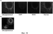
Фигура 10: Иммуноцитохимическая характеристика ирНСК нейросфер для экспрессии маркеров НСК Sox2 и нестина. На день 15 ирНСК нейросферы, выращенные с фасудилом, были посеяны на плашки, покрытые PO/Lam, и через 48 часов покрашены на экспрессию Sox2 и нестина. Прикрепленные ирНСК нейросферы и ирНСК, распространяющиеся из сфер. ирНСК имели типичную морфологию НСК и были Sox2 и нестин позитивны. Панель А: Объединенные и раздельные каналы DAPI, Sox2, нестин; увеличение 20х; Панель В: Объединенные каналы DAPI, Sox2, нестин; увеличение 10х.Figure 10: Immunocytochemical characteristic of irNSC neurospheres for the expression of NSC Sox2 and Nestin markers. On
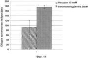
Фигура 11: Сравнение стимуляции производства ирНСК нейросфер с помощью фасудила и баланолоподобного-324 соединения. Человеческие фибробласты IMR90 были трипсинизированы и инфицированы в небольшом объеме репортером с Sox2, Bmi1, нестином GFP, с использованием среды для индукции (NeuroCult® NS-A Proliferation Kit (Human, StemCells Technologies) с FGF, EGF BDNF 20 нг/мл; гепарином 2 мкг/мл; полибреном 4 мкг/мл) с добавлением фасудила 10 мкМ (заштрихованный столбец), или баланолоподобного-324 соединения 2 мкМ (черный столбец). Фибробласты были посеяны на нормальной плашке для культур тканей в концентрации 10000-30000 клеток/см2. День 1: Среда заменена свежей средой для индукции. День 4: Нейросферы GFP+ большие, чем 50 мкм были подсчитаны. Баланолоподобное-324 малое соединение увеличивает эффективность генерации нейросфер приблизительно вдвое (1,9) и обладает большей воспроизводимостью (STDEV, n=3).Figure 11: Comparison of stimulation of the production of irNSC neurospheres using fasudil and balanol-like 324 compounds. Human fibroblasts IMR90 were trypsinized and small-scale infected with a reporter with Sox2, Bmi1, Nestin GFP, using induction medium (NeuroCult® NS-A Proliferation Kit (Human, StemCells Technologies) with FGF,
Фигура 12: Предобработка человеческих фибробластов вальпроевой кислотой (VPA) увеличивает выход GFP+ ирНСК нейросфер. Человеческие фибробласты IMR90 были предварительно обработаны в течение 48 часов с или без ингибитором HDAC вальпроевой кислотой (мононатриевая соль 2-пропил-пентановой кислоты) (1 мМ) перед инфекцией репортером с Sox2, Bmi1, нестином GFP. Среда для индукции (NeuroCult® NS-A Proliferation Kit (Human, StemCells Technologies) с FGF, EGF BDNF 20 нг/мл; гепарином 2 мкг/мл; баланолоподобным-324 соединением 2 мкМ). День 7: Нейросферы большие, чем 50 мкм, подсчитали (Панель А) и представили среднее колчество клеток GFP+ ирНСК на нейросферу (Панель В). Типичные изображения произведенных с предобработкой VPA ирНСК нейросфер (Панель С). Предобработка VPA существенно не влияет на число нейросфер ко дню 7; хотя обработка VPA увеличивает (в 2,1 раза) число GFP+ ирНСК (STDEV, n=3).Figure 12: Pretreatment of human fibroblasts with valproic acid (VPA) increases the yield of GFP + irNSC neurospheres. Human IMR90 fibroblasts were pretreated for 48 hours with or without an HDAC inhibitor valproic acid (monosodium salt of 2-propyl pentanoic acid) (1 mM) before infection with the reporter with Sox2, Bmi1, nestin GFP. Induction medium (NeuroCult® NS-A Proliferation Kit (Human, StemCells Technologies) with FGF,
Фигура 13: Определение минимального набора генов в комбинации с Sox2 и Bmi1 для эффективной индукции ирНСК нейросфер. Человеческие фибробласты IMR90 были предварительно обработаны VPA (1 мМ) в течение 48 часов перед инфекцией репортером с Sox2, Bmi1, нестином GFP, плюс различные гены-кандидаты для усиления их синергитического действия. Среда для индукции: NeuroCult® NS-A Proliferation Kit (Human, StemCells Technologies) с FGF, EGF BDNF 20 нг/мл; гепарином 2 мкг/мл и баланолоподобным-324 соединением 2 мкМ. Определение количества ирНСК нейросфер, больших чем 50 мкм, на день 7. Mash1, Emx2, Foxg1, Рах6 и Sox11 действуют синергично с Bmi1 и Sox2 на генерацию ирНСК нейросфер.Figure 13: Determination of a minimal set of genes in combination with Sox2 and Bmi1 for effective induction of irNSC neurospheres. Human IMR90 fibroblasts were pretreated with VPA (1 mM) for 48 hours before infection by the reporter with Sox2, Bmi1, Nestin GFP, plus various candidate genes to enhance their synergistic effect. Induction medium: NeuroCult® NS-A Proliferation Kit (Human, StemCells Technologies) with FGF,
Фигура 14: Формирование ирНСК нейросфер из взрослых человеческих фибробластов кожи (HDFa). Взрослые человеческие кожные фибробласты предоставлены GIBCO (Кат. номер: С-013-5С). Взрослые человеческие фибробласты кожи были трипсинизированы и инфицированы в небольшом объеме репортером с Sox2, Bmi1, нестином GFP, с использованием среды для индукции (NeuroCult® NS-A Proliferation Kit (Human, StemCells Technologies) с FGF, EGF BDNF 20 нг/мл; гепарином 2 мкг/мл) с добавлением фасудила 10 мкМ. День 8: Обнаружены ирНСК нейросферы (типичные изображения при 2,5 и 10× увеличении).Figure 14: Formation of irNSC neurospheres from adult human skin fibroblasts (HDFa). Adult human skin fibroblasts are provided by GIBCO (Cat. No: C-013-5C). Adult human skin fibroblasts were trypsinized and slightly infected with a reporter with Sox2, Bmi1, Nestin GFP, using induction medium (NeuroCult® NS-A Proliferation Kit (Human, StemCells Technologies) with FGF,
Фигура 15: Выращивание ирНСК нейросфер с использованием аскорбиновой кислоты, сигнального белка Sonic Hedgehog (Shh), Jagged1, DLL4 и FGF8 для получения монослойной культуры ирНСК GFP+. Человеческие фибробласты IMR90 инфицировали репортером с Sox2, Bmi1, нестином GFP с использованием среды для индукции (NeuroCult® NS-A Proliferation Kit (Human, StemCells Technologies) с FGF, EGF BDNF 20 нг/мл; гепарином 2 мкг/мл; баланолоподобным-324 соединением 2 мкМ). День 7: Нейросферы больше, чем 50 мкм, собрали и далее выращивали на среде для роста (NeuroCult® NS-A Proliferation Kit (Human, StemCells Technologies) с FGF, EGF BDNF 20 нг/мл; гепарином 2 мкг/мл; баланолоподобным-324 соединением 2 мкМ; аскорбиновой кислотой 0,2 мМ, SHH (рекомбинантный человеческий сигнальный белок Sonic Hedgehog, каталожный номер: 1845SH) 500 нг/мл, FGF8 (рекомбинантный человеческий FGF8a изоформа, каталожный номер: 4745F8) 100 нг/мл, DLL4 (рекомбинантный человеческий DLL4, каталожный номер: 1506D4) 500 нг/мл, Jagged1 (рекомбинантный человеческий Jagged1 Fc химера, каталожный номер: 1277JG) 500 нг/мл, 1/10 кондиционированной среды от НСК, происходящих от чЭСК, культивированных в течение двух дней на NeuroCult® NS-A Proliferation Kit (Human, StemCells Technologies) с FGF, EGF BDNF 20 нг/мл; гепарином 2 мкг/мл. Типичное изображение ирНСК нейросфер на день 14, выращенных на среде для роста, как описано выше (Панель А). Нейросферы имеют определенные границы, и возможно наблюдать выпячивание отростков из нейросфер (Панель В, увеличено). На день 21 выращенные ирНСК нейросферы были диссоциированы и посеяны на плашки с покрытием PO/Lam для получения гомогенной монослойной культуры ирНСК GFP+ (Панель С, фазовый контраст и GFP канал ирНСК монослоя после 4 дней культивирования в монослое).Figure 15: Growing irNSC neurospheres using ascorbic acid, Sonic Hedgehog (Shh) signal protein, Jagged1, DLL4 and FGF8 to obtain a monolayer culture of irNSC GFP +. Human fibroblasts IMR90 were infected with a reporter with Sox2, Bmi1, Nestin GFP using induction medium (NeuroCult® NS-A Proliferation Kit (Human, StemCells Technologies) with FGF,
Фигура 16: Иммуноцитохимическая характеристика ирНСК нейросфер в отношении экспрессии маркера НСК нестина и раннего нейрального маркера Tuj1. На день 21 ирНСК нейросферы, полученные как описано на Рисунке 15, были диссоциированы и посеяны в условиях для самоподдержания НСК (NeuroCult® NS-А Proliferation Kit (Человеческий, StemCells Technologies) с FGF, EGF BDNF 20 нг/мл; гепарин 2 мкг/мл) для оценки экспрессии маркера нестина (Панель А после 48 часов, все клетки являются нестин+ и Tuj1-), или посеяны в условиях для дифференцировки (NeuroCult® NS-A differentiation Kit (Человеческий, StemCells Technologies) c BDNF 20 нг/мл) и покрашены на Tuj1 и нестин на день 7 (Панель В, все клетки являются Tuj1+, и некоторые клетки нестин+).Figure 16: Immunocytochemical characterization of irNSC neurospheres in relation to the expression of the NSC marker Nestin and the early neural marker Tuj1. On day 21 of the irNSC, the neurospheres obtained as described in Figure 15 were dissociated and seeded under conditions for self-maintenance of the NSC (NeuroCult® NS-A Proliferation Kit (Human, StemCells Technologies) with FGF,
ПримерыExamples
Способ может быть иллюстрирован имеющейся здесь ссылкой на Фигуру 1, которая изображает способ согласно настоящему изобретению, используемый для превращения человеческих фибробластов в нейральные стволовые клетки (НСК). В этом способе человеческие фибробласты на день 0 были трипсинизированы, подсчитаны, и была определена их жизнеспособность. Затем 1.0×10⁵ - 3.0×10⁵ трипсинизированных фибробластов было ресуспендировано в среде для индукции, и в виде лентивирусов была доставлена комбинация генов. Для увеличения эффективности лентивирусной трансдукции в среду для индукции был добавлен полибрен (гексадиметрин бромид). Инфицирование проводили за 15 минут в пробирках эппендорфах. В комбинации с генами был использован человеческий репортер нестин GFP. Нестин является хорошо известным маркером, специфично экспрессирующимся в НСК. В нестин репортере экспрессия флуоресцентного белка GFP находится под контролем промотера нестина человека (Фигура 2), с помощью него, таким образом, можно легко отследить индуцированные репрограммированные нейральные стволовые клетки (ирНСК) GFP+.The method can be illustrated by referring to Figure 1, which depicts the method according to the present invention, used to convert human fibroblasts into neural stem cells (NSCs). In this method, human fibroblasts at
Инфицированные клетки были посеяны в плашки для культур тканей в концентрации 10000-30000 клеток/см2 в соответствующем объеме среды для индукции. На день 1 вся среда для индукции была заменена. Было возможно идентифицировать некоторые ирНСК GFP+ (Фигура 3) по четкой смене морфологии по сравнению с человеческими фибробластами. ИрНСК GFP+ приобретали биполярную удлиненную морфологию с более конденсированной цитоплазмой, типичной для НСК. Более того, ирНСК растут в насыщенной монослойной культуре, напоминающей типичные взаимодействия клетка-к-клетке, приобретаемые традиционными культурами НСК для активации пролиферативного сигнала пути Notch. На день 2 ирНСК GFP+ были в более зрелом состоянии и начали формировать достаточно плотные кластеры клеток. Такие кластеры ирНСК начали формировать сфероидные структуры с плотным ядром, содержащим ирНСК GFP+ (Фигура 4). На день 3 сфероидные структуры были полностью сформированы и начали отрываться от культуральной плашки, плавая в среде как нейросферы. Нейросферы обладают размерами примерно 20-100 мкм с четкими границами и ядром с высокой плотностью клеток, которые возможно отождествлять с ирНСК GFP+ (Фигура 5).Infected cells were seeded in tissue culture plates at a concentration of 10,000-30000 cells / cm 2 in an appropriate volume of induction medium. On
Чтобы достигнуть изобретательского прорыва, было необходимо использовать специфическую комбинацию генов. Следующий список генов, вовлеченных в поддержание характеристик НСК in vivo и in vitro, был получен из литературных данных: Sox2 (Sox транскрипционный фактор и важный маркер НСК), Brn2 (POU домен белка известен, что связывается с белком Sox. Репортер, связывающий Sox2 и Brn2 с промотером нестина. Мыши, нокаутные по Brn2, обладают нарушением развития ЦНС), Bmi1 (Белок вовлечен в регуляцию клеточного цикла, вероятно, усиливает экспрессию р21 и р27 - ингибиторов комплекса циклинЕ/cdk2. Ингибирование циклинЕ/cdk2 устанавливает потерю белком ретинобластомы контроля над клеточным циклом, что влечет быстрый клеточный цикл во время состояния самовоспроизводства НСК), Mash1 (предполагаемый важный регулятор пролиферации нейральных предшественников in vivo), Sox11 (описано, что белок Sox экспрессируется в субгранулярной зоне SGZ in vivo), NCam (НСК маркер для проточной цитометрии и экспрессирующийся в различных районах ЦНС), Kpna1 (более известный как импортин альфа5, ответственный вместе с импортином бета за ядерный транспорт белков в тканях - производных эктодермы).In order to achieve an inventive breakthrough, it was necessary to use a specific combination of genes. The following list of genes involved in maintaining the characteristics of NSCs in vivo and in vitro was obtained from the literature: Sox2 (Sox transcription factor and an important marker of NSCs), Brn2 (the POU domain of the protein is known to bind to the Sox protein. A reporter that binds Sox2 and Brn2 with a nestin promoter. Brn2 knockout mice have impaired central nervous system development), Bmi1 (the protein is involved in cell cycle regulation and probably enhances the expression of p21 and p27 inhibitors of the cyclinE / cdk2 complex. Inhibition of cyclinE / cdk2 establishes the loss of retinoblastoma protein control cellular cycle, which leads to a fast cell cycle during the state of self-reproduction of NSCs), Mash1 (an alleged important regulator of proliferation of neural precursors in vivo), Sox11 (it is described that Sox protein is expressed in the subgranular zone of SGZ in vivo), NCam (NSC marker for flow cytometry and expressed in various regions of the central nervous system), Kpna1 (better known as importin alpha5, responsible, together with import beta, for nuclear transport of proteins in tissues - derivatives of ectoderm).
Все гены были клонированы как кДНК в лентивирусных плазмидах, и впоследствии упакованы в лентивирусы. Упакованные лентивирусные частицы с Sox2, Bmi1, Mash1, Sox11, NCam, Kpna1, нестин GFP репортером были трансдуцированы напрямую в человеческие фибробласты. В описанном выше способе были протестированы различные комбинации генов. На день 3 было возможно оценить успешность продукции ирНСК с помощью подсчета образовавшихся нейросфер. Подсчету подлежали только нейросферы, большие чем 50 мкм.All genes were cloned as cDNA in lentiviral plasmids, and subsequently packed into lentiviruses. Packaged lentiviral particles with Sox2, Bmi1, Mash1, Sox11, NCam, Kpna1, the Nestin GFP reporter were transduced directly into human fibroblasts. In the method described above, various combinations of genes were tested. On
Как представлено на Фигуре 6, трансдукция лентивируса с нестин репортером без добавления в среду для индукции фасудила не репрограммировала человеческие фибробласты в ирНСК. С добавлением фасудила в среду для индукции отмечалось образование нейросфер (около 50 мкм), и более мелких (около 20 мкм, не подсчитывались).As shown in Figure 6, transduction of lentivirus with a nestin reporter without adding fasudil to the medium for inducing fasudil did not reprogram human fibroblasts into irNSCs. With the addition of fasudil to the induction medium, the formation of neurospheres (about 50 μm) was observed, and smaller ones (about 20 μm, were not counted).
Нейросферы, образованные с помощью нашего инновационного способа с использованием комбинации генов Sox2-Bmi1 собирались на день 3 и дальше выращивались 14 дней. Выращивание ирНСК нейросфер было критической стадией. Нейросферы культивировались с использованием среды N2B27 с добавлением FGF, EGF, BDNF в специальных ультратонких неадгезивных плашках (Corning). Для получения гомогенной популяции ирНСК GFP+ нейросфер каждые 2-3 дня проводилась процедура очистки. В течение 14 дней выращивания некоторые нейросферы с низкой плотностью ирНСК GFP+ были не способны пролиферировать надлежащим образом, в основном, вероятно, вследствие контаминации неконвертированными фибробластами. Подобные контаминированные нейросферы разваливались на отдельные клетки, которые было необходимо удалять. На день 14 нейросферы проверялись на: прикрепление к плашкам, покрытым полиорнитином/ламинином и генерацию нейроно-подобных клеток. Для прикрепления, 20-40 нейросфер/см2 были посеяны на покрытые полиорнитином/ламинином плашки в среде для роста с добавлением фасудила 10 мкМ для усиления клеточной адгезии и распластания. На день 1 культивирования можно было увидеть прикрепление и распластывание нейросфер (Фигура 7). В центре распластывающейся нейросферы мы идентифицировали ирНСК GFP+ с типичной морфологией НСК. Нейросферы выращивались дополнительно три дня, и тогда только BDNF был добавлен к N2B27 (условия для дифференцировки в нервную ткань). Из-за изъятия EGF и FGF ирНСК меняли морфологию. Они становились более вытянутыми и начинали формировать аксоноподобные клеточные отростки. На день 7 в условиях дифференцировки клетки были зафиксированы и окрашены на нейрональный маркер tuj1 (Фигура 8).The neurospheres formed using our innovative method using a combination of Sox2-Bmi1 genes were harvested on
Нейросферы, выращенные с фасудилом, обладают лучшей морфологией и чистыми и четкими границами (отличительный признак правильно сформированной нейросферы, Фигура 9, панель В); без фасудила нейросферы имеют неясные границы (Фигура 9, панель А). ИрНСК обладают типичной морфологией НСК и были Sox2 и нестин позитивны (Фигура 10).The neurospheres grown with fasudil have better morphology and clean and clear boundaries (a hallmark of a properly formed neurosphere, Figure 9, panel B); without fasudil, the neurospheres have unclear boundaries (Figure 9, panel A). IrNSCs have a typical NSC morphology and were Sox2 and Nestin positive (Figure 10).
Фигура 11 демонстрирует, что ингибитор Rock киназы баланолоподобное-324 соединение увеличивает выход GFP+ нейросфер.Figure 11 demonstrates that the Rock kinase inhibitor, a balananol-324 compound, increases the yield of GFP + neurospheres.
Данные свидетельства демонстрируют, что с помощью способа по настоящему изобретению возможно конвертировать человеческие фибробласты в ирНСК, основываясь на связанных стадиях трансдукции генов (лучшие комбинации: Sox2-Bmi1, Sox2-Bmi1-Mash1, Sox2-Bmi1-Sox11, Sox2-Bmi1-Emx2, Sox2-Bmi1-Foxg1 и Sox2-Bmi1-Рах6, также см. Фигуру 13) и индукции средой определенного химического состава.These evidences demonstrate that using the method of the present invention it is possible to convert human fibroblasts into irNSCs based on the associated stages of gene transduction (best combinations: Sox2-Bmi1, Sox2-Bmi1-Mash1, Sox2-Bmi1-Sox11, Sox2-Bmi1-Emx2, Sox2-Bmi1-Foxg1 and Sox2-Bmi1-Pax6, also see Figure 13) and medium induction of a specific chemical composition.
Чтобы увеличить выход ирНСК, человеческие фибробласты были предварительно обработаны с или без ингибитора HDAC вальпроевой кислоты (VPA, мононатриевая соль 2-пропил-пентановой кислоты). С этой целью человеческие фибробласты инкубировались в DMEM/F12 с добавлением FBS 10% и L-глутамина с добавлением 1 мМ VPA перед инфицированием (Фигура 12).To increase the yield of irNSCs, human fibroblasts were pretreated with or without a valproic acid HDAC inhibitor (VPA, 2-propyl-pentanoic acid monosodium salt). To this end, human fibroblasts were incubated in DMEM / F12 supplemented with 10% FBS and L-glutamine supplemented with 1 mM VPA before infection (Figure 12).
Фигура 14 демонстрирует генерацию ирНСК нейросфер из взрослых человеческих фибробластов кожи (HDFa).Figure 14 shows the generation of irNSC neurospheres from adult human skin fibroblasts (HDFa).
Фигура 15 демонстрирует выращивание ирНСК нейросфер с использованием комбинации аскорбиновой кислоты, сигнального белка Sonic Hedgehog (Shh), Jagged1, DLL4 и FGF8. Фигура 16 демонстрирует иммуноцитохимическую характеристику ирНСК нейросфер в отношении экспрессии маркера НСК нестина и раннего нейрального маркера Tuj1.Figure 15 shows the growth of irNSC neurospheres using a combination of ascorbic acid, the signal protein Sonic Hedgehog (Shh), Jagged1, DLL4 and FGF8. Figure 16 shows the immunocytochemical characterization of the irNSC neurospheres with respect to the expression of the NSC marker Nestin and the early neural marker Tuj1.
Материалы и методыMaterials and methods
Культуры клеток:Cell culture:
Среда для индукции: N2B27 (N2B27 является смесью 1:1 DMEM/F12 (Gibco, Paisley, Великобритания) с добавлением N2 и В27 (обе от Gibco) с добавлением человеческого EGF (Peprotech) 30 нг/мл, человеческого FGF2 30 нг/мл (Peprotech), человеческого BDNF (Roche) 20 нг/мл и фасудила (Calbiochem) 10 мкМ или баланолоподобного-324 соединения (N-{(3R,4R)-4-[4-(2-фтор-6-гидрокси-3-метокси-бензоил)-бензоиламино]-азепан-3-ил}-4-гидрокси-3,5-диметилбензамид) 2 мкМ.Induction medium: N2B27 (N2B27 is a 1: 1 mixture of DMEM / F12 (Gibco, Paisley, UK) with the addition of N2 and B27 (both from Gibco) with the addition of human EGF (Peprotech) 30 ng / ml,
Среда для роста: N2B27 с добавлением человеческого EGF (Peprotech) 30 нг/мл, человеческого FGF2 30 нг/мл (Peprotech), человеческого BDNF (Roche) 20 нг/мл, или NeuroCult® NS-A Proliferation Kit (Человеческий, StemCells Technologies) с FGF, EGF BDNF 20 нг/мл; гепарином 2 мкг/мл; баланолоподобное-324 2 мкМ; аскорбиновой кислотой 0.2 мМ, SHH (Рекомбинантный человеческий Sonic Hedgehog, Каталожный номер: 1845SH) 500 нг/мл, FGF8 (Рекомбинантный человеческий FGF8a изоформа, Каталожный номер: 4745F8) 100 нг/мл, DLL4 (Рекомбинантный человеческий DLL4, Каталожный номер: 1506D4) 500 нг/мл, Jagged1 (Рекомбинантный человеческий Jagged1 Fc химера, Каталожный номер: 1277JG) 500 нг/мл.Growth medium: N2B27 supplemented with human EGF (Peprotech) 30 ng / ml,
Среда для дифференциации: N2B27 с добавлением человеческого BDNF (Roche) 20 нг/мл, ламинина 2 мкг/мл (Invitrogen).Differentiation medium: N2B27 supplemented with human BDNF (Roche) 20 ng / ml,
Человеческие фибробласты: IMR90 зародышевые фибробласты легкого (АТСС номер партии 580229699) или взрослые фибробласты кожи (GIBCO, каталожный номер: С-013-5С).Human fibroblasts: IMR90 germinal lung fibroblasts (ATCC lot number 580229699) or adult skin fibroblasts (GIBCO, catalog number: C-013-5C).
Лентивирусы: предварительно упакованные, готовые к использованию частицы вирусов были получены от Sigma (Stemgent репрограммирующий человеческий лентивирус Sox2, каталожный No. ST070012), Genecopeia (человеческий Bmi1 Lentifect лентивирусные частицы, каталожный Nr. LP-B0015-Lv105; Sox11 Lentifect лентивирусные частицы, каталожный Nr. LP-MO425-LV105; Mash1 Lentifect лентивирусные частицы, каталожный Nr. LP-Z0740-LV105; человеческий Kpna1 Lentifect лентивирусные частицы, каталожный Nr. LP-U1286-Lv105; NCam1 Lentifect лентивирусные частицы, каталожный Nr. LP-Z2645-Lv105) и SBI Systems Biosciences (нестин GFP репортер: pGreenZeo™-hNestin транскрипционный репортерный вирус, SR10035VA-1)Lentiviruses: pre-packaged, ready-to-use virus particles were obtained from Sigma (Stemgent reprogramming human Sox2 lentivirus, catalog No. ST070012), Genecopeia (human Bmi1 Lentifect lentiviral particles, catalog Nr. LP-B0015-Lv105; Sox11 lentivect Nr. LP-MO425-LV105; Mash1 Lentifect lentiviral particles, catalog Nr. LP-Z0740-LV105; human Kpna1 Lentifect lentiviral particles, catalog Nr. LP-U1286-Lv105; NCam1 Lentifect lentiviral particles, catalog Zr26 Nr. ) and SBI Systems Biosciences (Nestin GFP reporter: pGreenZeo ™ -hNestin transcriptional reporter virus, SR10035VA-1)
Титры NestinGFP 1.45*105/мкл, BMI1 4.3*105/мкл, Sox2 1.07*104/мкл, Sox11 3.2*106/мкл, Mash1 4.7*106/мкл, NCam 3.3*104/мкл, Kpna1 1.8*105/мкл.Titles NestinGFP 1.45 * 10 5 / μl, BMI1 4.3 * 10 5 / μl, Sox2 1.07 * 10 4 / μl, Sox11 3.2 * 10 6 / μl, Mash1 4.7 * 10 6 / μl, NCam 3.3 * 10 4 / μl, Kpna1 1.8 * 10 5 / μl.
Протоколы:Protocols:
1. Генерация ирНСК:1. Generation of irNSC:
- 200.000 человеческих фибробластов IMR90 инфицировали лентивирусами с различными комбинациями генов (мультиплексность инфицирования (М.И.), использованная для каждого отдельного лентивируса 30) и репортерного нестин GFP лентивируса (использована М.И. 10) в эппендорфе с 300 мкл среды для индукции с полибреном (гексадиметрин бромид, Sigma) 4 мкг/мл.- 200,000 human IMR90 fibroblasts were infected with lentiviruses with different gene combinations (infection multiplex (MI) used for each individual lentivirus 30) and reporter nestin GFP lentivirus (used MI 10) in eppendorf with 300 μl of medium for induction with polybrene (hexadimethrin bromide, Sigma) 4 μg / ml.
- Инкубировать при комнатной температуре 15 мин.- Incubate at room temperature for 15 minutes.
- Рассеять 300 мкл в 1,7 мл среды для индукции в лунке 6-луночной плашки для культивирования с модифицированной поверхностью- Dissipate 300 μl in 1.7 ml of induction medium in the well of a 6-well cultured surface modified plate
- День 1, заменить 2 мл среды для индукции/каждая лунка-
- День 3, собрать нейросферы, тщательно отбирая 2 мл с плавающими сферами-
- Вырастить нейросферы- grow neurospheres
2. Выращивание нейросфер:2. Growing neurospheres:
- Собрать среду с плавающими нейросферами из 3 лунок 6-луночной плашки в 15 мл пробирки- Collect a medium with floating neurospheres from 3 wells of a 6-well plate into 15 ml tubes
- Дать сферам осесть в течение 10 минут- Allow spheres to settle for 10 minutes
- Очень аккуратно удалить супернатант (отдельные клетки не оседают вниз и аспирируются вместе с супернатантом)- Very carefully remove the supernatant (individual cells do not settle down and aspirate with the supernatant)
- Ресуспендировать сферы в 4 мл конечного объема среды для роста- Resuspend spheres in 4 ml of the final volume of growth medium
- Посеять в ультранизкие адгезивные плашки В6 (Corning)- Sow in ultra-low adhesive dies B6 (Corning)
- Инкубировать 2-3 дня- Incubate for 2-3 days
- Повторять процедуру выращивания каждые 2-3 дня до 14 дня с момента генерации ирНСК- Repeat the growing procedure every 2-3 days until 14 days after the generation of irNSC
3. Дифференцировка нейросфер:3. Differentiation of neurospheres:
- На день 14 с момента генерации ирНСК и после процедуры выращивания выбрать под стерео микроскопом круглые нейросферы с чистыми границами, которые богаты ирНСК GFP+- On day 14 from the moment of generation of the irNSC and after the growing procedure, select round neurospheres with clean boundaries that are rich in the irNSC GFP + under a stereo microscope
- Поместить 40 нейросфер в лунку 24-луночной плашки, предварительно покрытой полиорнитином/ламинином, используя среду для роста с добавлением фасудила 10 мкМ или баланолоподобного-324 соединения 2 мкМ.-
- Через день поменять среду на среду для роста без фасудила/баланолоподобного-324 соединения 2 мкМ.- In a day, change the medium to medium for growth without fasudil / balananol-like-324 compound of 2 μM.
- Инкубировать в течение трех дней- Incubate for three days
- Удалить среду для роста и добавить среду для дифференцировки- Remove growth medium and add medium for differentiation
- Обновить среду для дифференцировки после 3-4 дней- Update medium for differentiation after 3-4 days
- Инкубировать 3-4 дня- Incubate 3-4 days
- Зафиксировать клетки PFA 4% и выполнить иммуноокрашивание- Fix 4% PFA cells and perform immunostaining
Протокол окрашивания ирНСК нейросфер: Нейросферы на день 15 были окрашены для характиристики следующими антителами: мышиными к нестину и кроличьими к Sox2 O/N, и затем вторичными анти-мышиными 488 и анти-кроличьими 555 в течение 1 часа.IrNSC neurosphere staining protocol: The neurospheres on
Первичные антитела:Primary antibodies:
Мышиные к нестину, моноклональные, разведение 1/500 (МАВ5326 Millipore)Mouse to nestin, monoclonal, 1/500 dilution (MAB5326 Millipore)
Кроличьи к Sox2, поликлональные, разведение 1/500 (AB5603MI Millipore)Rabbits for Sox2, polyclonal, breeding 1/500 (AB5603MI Millipore)
Вторичные антитела:Secondary antibodies:
Alexa fluor 488, IgG, разведение 1/1 000, Козьи анти-мышиные (А11029 Invitrogen)Alexa fluor 488, IgG, 1/1,000 dilution, Goat anti-mouse (A11029 Invitrogen)
Alexa fluor 555, IgG, разведение 1/1 000, Козьи анти-кроличьи (А21429 Invitrogen)Alexa fluor 555, IgG, 1/1,000 dilution, Goat anti-rabbit (A21429 Invitrogen)
Claims (15)
а) получение соматических клеток, выбранных из группы фибробластов, кератиноцитов или адипоцитов, и
б) репрограммирование указанных соматических клеток в НСК с помощью введения в указанные соматические клетки по меньшей мере двух генов, Bmi1 и Sox2; и культивирования указанных соматических клеток в питательной среде, содержащей ростовые факторы, выбранные из группы FGF2, EGF и BDNF и малой молекулы ингибитора ROCK.1. In vitro method for producing neural stem cells (NSC), including:
a) obtaining somatic cells selected from the group of fibroblasts, keratinocytes or adipocytes, and
b) reprogramming said somatic cells into NSCs by introducing at least two genes, Bmi1 and Sox2, into said somatic cells; and culturing said somatic cells in a growth medium containing growth factors selected from the group of FGF2, EGF and BDNF and a small molecule of a ROCK inhibitor.
в) инкубацию продуктов стадии б) в условиях, подходящих для пролиферации НСК.2. The method according to p. 1, further comprising
c) incubation of the products of stage b) under conditions suitable for the proliferation of NSCs.
Applications Claiming Priority (3)
| Application Number | Priority Date | Filing Date | Title |
|---|---|---|---|
| EP10173455.6 | 2010-08-19 | ||
| EP10173455 | 2010-08-19 | ||
| PCT/EP2011/064051 WO2012022725A2 (en) | 2010-08-19 | 2011-08-16 | Conversion of somatic cells to induced reprogrammed neural stem cells (irnscs) |
Publications (2)
| Publication Number | Publication Date |
|---|---|
| RU2013110127A RU2013110127A (en) | 2014-09-27 |
| RU2562111C2 true RU2562111C2 (en) | 2015-09-10 |
Family
ID=43734036
Family Applications (1)
| Application Number | Title | Priority Date | Filing Date |
|---|---|---|---|
| RU2013110127/10A RU2562111C2 (en) | 2010-08-19 | 2011-08-16 | Conversion of somatic cells into induced reprogrammed neural stem cells (irnsc) |
Country Status (12)
| Country | Link |
|---|---|
| US (2) | US9297025B2 (en) |
| EP (1) | EP2606126B1 (en) |
| JP (1) | JP5905006B2 (en) |
| KR (2) | KR101788903B1 (en) |
| CN (1) | CN103068974B (en) |
| BR (1) | BR112013003031A2 (en) |
| CA (1) | CA2807226A1 (en) |
| ES (1) | ES2580837T3 (en) |
| HK (1) | HK1184492A1 (en) |
| MX (1) | MX348419B (en) |
| RU (1) | RU2562111C2 (en) |
| WO (1) | WO2012022725A2 (en) |
Families Citing this family (21)
| Publication number | Priority date | Publication date | Assignee | Title |
|---|---|---|---|---|
| KR101788903B1 (en) | 2010-08-19 | 2017-10-23 | 에프. 호프만-라 로슈 아게 | Conversion of somatic cells to induced reprogrammed neural stem cells (irnscs) |
| CN102604894B (en) | 2012-02-29 | 2014-07-30 | 中国科学院广州生物医药与健康研究院 | Culture medium for preparing neural stem cells and application thereof |
| PL223189B1 (en) * | 2012-11-15 | 2016-10-31 | Celther Polska Spółka Z Ograniczoną Odpowiedzialnością | iNS cell and the method for reprogramming somatic cells by a Sox2 and Sox2 factors and c-Myc to the cell iNS |
| WO2014081906A2 (en) * | 2012-11-21 | 2014-05-30 | Ptc Therapeutics, Inc. | Substituted reverse pyrimidine bmi-1 inhibitors |
| KR20140121317A (en) * | 2013-04-06 | 2014-10-15 | 서울대학교산학협력단 | Method for producing induced neural stem cell from non-neuronal cell, and induced neural stem cell produced by the same |
| EP3024928B1 (en) * | 2013-07-23 | 2018-11-21 | F. Hoffmann-La Roche AG | Small molecule based conversion of somatic cells into neural crest cells |
| CN104342401B (en) * | 2013-07-25 | 2017-04-05 | 中国科学院广州生物医药与健康研究院 | It is adipose cell to promote fibroblast transdifferentiation using the combination of cytokines for determining |
| JP6415582B2 (en) * | 2013-10-25 | 2018-10-31 | ウェイン ステート ユニバーシティー | Methods, systems, and compositions for cell transformation by protein-induced in vivo cell reprogramming |
| JP6478116B2 (en) | 2013-12-25 | 2019-03-06 | 東亞合成株式会社 | Method for inducing differentiation from pluripotent stem cells to endoderm cells |
| CN104894060B (en) * | 2014-03-03 | 2018-11-06 | 中国科学院上海生命科学研究院 | Inducing somatic transdifferentiation is the method and its application of neural stem cell |
| CN104195108B (en) * | 2014-07-29 | 2018-02-06 | 深圳市三启生物技术有限公司 | Purposes of the kinases inhibitor in nerve cell is prepared from non-neuronal cells |
| CN107849538A (en) * | 2015-03-04 | 2018-03-27 | 北卡罗来纳-查佩尔山大学 | Method for manufacturing NSC and application thereof |
| CA2978109A1 (en) | 2015-03-31 | 2016-10-06 | The University Of North Carolina At Chapel Hill | Delivery vehicles for stem cells and uses thereof |
| JP6691756B2 (en) | 2015-09-29 | 2020-05-13 | 東亞合成株式会社 | Method for producing neural stem cells using synthetic peptide |
| MX2018004978A (en) * | 2015-11-12 | 2018-07-06 | Hoffmann La Roche | Oligonucleotides for inducing paternal ube3a expression. |
| KR20180132787A (en) * | 2016-03-31 | 2018-12-12 | 아지노모토 가부시키가이샤 | Medium for Neural Stem Cells Enhancing Neurodiversity |
| CN109312301B (en) | 2016-04-20 | 2023-06-16 | 京都府公立大学法人 | Method for preparing cultured epithelial cell sheet |
| WO2019241620A1 (en) * | 2018-06-14 | 2019-12-19 | Academia Sinica | Method to generate induced oligodendrocyte-lineage cells and treatment using such cells |
| EP3841197A1 (en) | 2018-08-21 | 2021-06-30 | F. Hoffmann-La Roche AG | Methods for assessing transendothelial barrier integrity |
| EP4163381A4 (en) * | 2020-06-09 | 2024-03-13 | Ulsan National Institute Of Science And Tech Unist | Composition for inducing direct cross differentiation of somatic cell into motor neuron, and method for using same |
| AU2021353867A1 (en) * | 2020-09-29 | 2023-05-11 | NeuExcell Therapeutics Inc. | Neurod1 combination vector |
Citations (3)
| Publication number | Priority date | Publication date | Assignee | Title |
|---|---|---|---|---|
| US20090047263A1 (en) * | 2005-12-13 | 2009-02-19 | Kyoto University | Nuclear reprogramming factor and induced pluripotent stem cells |
| RU2351648C2 (en) * | 2001-11-09 | 2009-04-10 | Артесел Сайенсиз, Инк. | Adipose stromal cell differentiation into endocrine pancreas cells and application thereof |
| RU2407790C2 (en) * | 2004-05-05 | 2010-12-27 | Новартис Форшунгсстифтунг, Цвайгнидерлассунг Фридрих Мишер Институте Фор Биомедикал Рисёч | Method of neuronal cell differentiation from embryo stem (es) |
Family Cites Families (8)
| Publication number | Priority date | Publication date | Assignee | Title |
|---|---|---|---|---|
| US5968829A (en) * | 1997-09-05 | 1999-10-19 | Cytotherapeutics, Inc. | Human CNS neural stem cells |
| EP1516926A4 (en) * | 2002-06-24 | 2006-10-11 | Tanabe Seiyaku Co | Process for producing nerve cells |
| US7682828B2 (en) | 2003-11-26 | 2010-03-23 | Whitehead Institute For Biomedical Research | Methods for reprogramming somatic cells |
| EP1745069B1 (en) * | 2004-03-30 | 2009-05-06 | Nsgene A/S | Therapeutic use of growth factor nsg33 |
| US20100021437A1 (en) * | 2008-04-07 | 2010-01-28 | The McLean Hospital Corporation Whitehead Institute for Biomedical Research | Neural stem cells derived from induced pluripotent stem cells |
| KR101102483B1 (en) | 2008-08-12 | 2012-01-05 | 연세대학교 산학협력단 | Human neural stem cell and pharmaceutical composition for treating disorder and injury of central or peripheral nervous system using the same |
| GB0902034D0 (en) * | 2009-02-06 | 2009-03-11 | Reneuron Ltd | Method |
| KR101788903B1 (en) | 2010-08-19 | 2017-10-23 | 에프. 호프만-라 로슈 아게 | Conversion of somatic cells to induced reprogrammed neural stem cells (irnscs) |
-
2011
- 2011-08-16 KR KR1020137003961A patent/KR101788903B1/en active IP Right Grant
- 2011-08-16 CA CA2807226A patent/CA2807226A1/en not_active Abandoned
- 2011-08-16 KR KR1020167007305A patent/KR20160033798A/en not_active Application Discontinuation
- 2011-08-16 CN CN201180040130.2A patent/CN103068974B/en not_active Expired - Fee Related
- 2011-08-16 ES ES11743246.8T patent/ES2580837T3/en active Active
- 2011-08-16 EP EP11743246.8A patent/EP2606126B1/en not_active Not-in-force
- 2011-08-16 JP JP2013524439A patent/JP5905006B2/en active Active
- 2011-08-16 MX MX2013001587A patent/MX348419B/en active IP Right Grant
- 2011-08-16 RU RU2013110127/10A patent/RU2562111C2/en not_active IP Right Cessation
- 2011-08-16 BR BR112013003031A patent/BR112013003031A2/en active Search and Examination
- 2011-08-16 WO PCT/EP2011/064051 patent/WO2012022725A2/en active Application Filing
-
2013
- 2013-02-18 US US13/769,762 patent/US9297025B2/en not_active Expired - Fee Related
- 2013-10-22 HK HK13111852.9A patent/HK1184492A1/en not_active IP Right Cessation
-
2016
- 2016-02-18 US US15/046,885 patent/US20160369234A1/en not_active Abandoned
Patent Citations (3)
| Publication number | Priority date | Publication date | Assignee | Title |
|---|---|---|---|---|
| RU2351648C2 (en) * | 2001-11-09 | 2009-04-10 | Артесел Сайенсиз, Инк. | Adipose stromal cell differentiation into endocrine pancreas cells and application thereof |
| RU2407790C2 (en) * | 2004-05-05 | 2010-12-27 | Новартис Форшунгсстифтунг, Цвайгнидерлассунг Фридрих Мишер Институте Фор Биомедикал Рисёч | Method of neuronal cell differentiation from embryo stem (es) |
| US20090047263A1 (en) * | 2005-12-13 | 2009-02-19 | Kyoto University | Nuclear reprogramming factor and induced pluripotent stem cells |
Also Published As
| Publication number | Publication date |
|---|---|
| KR101788903B1 (en) | 2017-10-23 |
| US20140051171A1 (en) | 2014-02-20 |
| CA2807226A1 (en) | 2012-02-23 |
| ES2580837T3 (en) | 2016-08-29 |
| KR20130030303A (en) | 2013-03-26 |
| CN103068974A (en) | 2013-04-24 |
| WO2012022725A3 (en) | 2012-04-12 |
| US20160369234A1 (en) | 2016-12-22 |
| WO2012022725A2 (en) | 2012-02-23 |
| BR112013003031A2 (en) | 2016-06-14 |
| RU2013110127A (en) | 2014-09-27 |
| US9297025B2 (en) | 2016-03-29 |
| MX2013001587A (en) | 2013-03-21 |
| EP2606126B1 (en) | 2016-05-18 |
| MX348419B (en) | 2017-06-12 |
| JP2013535973A (en) | 2013-09-19 |
| CN103068974B (en) | 2016-05-11 |
| EP2606126A2 (en) | 2013-06-26 |
| JP5905006B2 (en) | 2016-04-20 |
| HK1184492A1 (en) | 2014-01-24 |
| KR20160033798A (en) | 2016-03-28 |
Similar Documents
| Publication | Publication Date | Title |
|---|---|---|
| RU2562111C2 (en) | Conversion of somatic cells into induced reprogrammed neural stem cells (irnsc) | |
| JP7023820B2 (en) | Cortical interneurons and other neuronal cells generated by directing the differentiation of pluripotent cells and pluripotent cells | |
| Decimo et al. | Neural stem cell niches in health and diseases | |
| ES2732938T3 (en) | Culture method for obtaining and conserving a pure or enriched population of neural stem cells and / or mammalian neural progenitor cells that are prone to differentiate into oligodendrocyte lineage cells in vitro | |
| JP2022078245A (en) | Method for differentiating pluripotent cell | |
| KR101277310B1 (en) | Cells exhibiting neuronal progenitor cell characteristics | |
| KR20110011658A (en) | Treating myelin diseases with optimized cell preparations | |
| US10724000B2 (en) | Small molecule based conversion of somatic cells into neural crest cells | |
| Keasey et al. | Inhibition of a novel specific neuroglial integrin signaling pathway increases STAT3-mediated CNTF expression | |
| CA3182115A1 (en) | Methods for differentiating stem cells into dopaminergic progenitor cells | |
| Liu et al. | The Rho kinase inhibitor Y-27632 facilitates the differentiation of bone marrow mesenchymal stem cells | |
| EP4321614A1 (en) | Method for preparing oligodendrocytes and use | |
| An et al. | Kinase inhibit region of SOCS3 attenuates IL6‐induced proliferation and astrocytic differentiation of neural stem cells via cross talk between signaling pathways | |
| US20080219955A1 (en) | Method of Producing Purified Neural Stem Cells and Related Methods of Treating a Patient | |
| Rosenberg | Exploring the role of cooperative and competitive interactions in oligodendrocyte differentiation and myelination |
Legal Events
| Date | Code | Title | Description |
|---|---|---|---|
| TK4A | Correction to the publication in the bulletin (patent) |
Free format text: AMENDMENT TO CHAPTER -FG4A- IN JOURNAL: 25-2015 FOR TAG: (54) |
|
| MM4A | The patent is invalid due to non-payment of fees |
Effective date: 20180817 |