RU2425373C2 - Method of using ultrathin tissue section for mineral grain analysis - Google Patents
Method of using ultrathin tissue section for mineral grain analysis Download PDFInfo
- Publication number
- RU2425373C2 RU2425373C2 RU2009135480/15A RU2009135480A RU2425373C2 RU 2425373 C2 RU2425373 C2 RU 2425373C2 RU 2009135480/15 A RU2009135480/15 A RU 2009135480/15A RU 2009135480 A RU2009135480 A RU 2009135480A RU 2425373 C2 RU2425373 C2 RU 2425373C2
- Authority
- RU
- Russia
- Prior art keywords
- grains
- tissue section
- mineral
- ultrathin
- electron microscopy
- Prior art date
Links
Images
Landscapes
- Pharmaceuticals Containing Other Organic And Inorganic Compounds (AREA)
Abstract
Description
Изобретение относится к области раздела биоминералогии - медицинской минералогии и может быть использовано:The invention relates to the field of biomineralogy - medical mineralogy and can be used:
- в медицине, при исследовании болезней, связанных с воспалениями, нарушением тканевого дыхания, разложением белков;- in medicine, in the study of diseases associated with inflammation, impaired tissue respiration, the decomposition of proteins;
- в фармакологии, для выявления ятрогенных болезней, вызванных различными лекарственными наполнителями;- in pharmacology, to detect iatrogenic diseases caused by various drug excipients;
- в биофизике (магнитобиологии), для объяснения механизма биомедицинских эффектов, производимых электромагнитными полями в организме человека;- in biophysics (magnetobiology), to explain the mechanism of biomedical effects produced by electromagnetic fields in the human body;
- в минералогии, поскольку значительно расширяют температурно-барометрические, временные и окислительно-восстановительные рамки оксидного и сульфидного минералообразования;- in mineralogy, since they significantly expand the temperature-barometric, time and redox framework of oxide and sulfide mineral formation;
- в биохимии, коллоидной химии и т.д.- in biochemistry, colloid chemistry, etc.
Аналогом использования ультратонких срезов биологических тканей для минералогических исследований являются работы ученых из Венгрии: (Е.Berki, A.Korányi, Е.Major, Т.Peres. Ultrastructural Study of Inorganic Substances in Atherosclerotic Aorta Tissue. Calc. Tiss. Res. 4, 84 - 90 (1969), E.Berki, A.Korányi, Gy.Liszka, E.Major. Inorganic content of normal and Atherosclerotic aortas. Orv. Hetil. 106, 201 - 203 (1965)).The use of ultrathin sections of biological tissues for mineralogical studies is analogous to the work of scientists from Hungary: (E. Berki, A. Korányi, E. Major, T. Peres. Ultrastructural Study of Inorganic Substances in Atherosclerotic Aorta Tissue. Calc. Tiss. Res. 4, 84 - 90 (1969), E. Berki, A. Korányi, Gy. Liszka, E. Major. Inorganic content of normal and Atherosclerotic aortas. Orv. Hetil. 106, 201 - 203 (1965)).
В этих статьях ультратонкий (около 60 nm) срез ткани применяется для электронно-микроскопического изучения неорганических зерен из биологической ткани атеросклеротической аорты. Кроме того, в работах E.Berki, A.Koranyi et. al. приводятся данные электронного микродифрактометрического анализа диспергированной и переведенной в суспензию неорганической составляющей атеросклеротических бляшек. При этом используется хорошо известный и достаточно широко применяемый в настоящее время метод дифрактометрии неорганических зерен в коллодиевом препарате. Данный метод описан и часто применяется в работах Г.Н.Батурина (Батурин Г.Н., Дубинчук В.Т. Микроструктуры железомарганцевых конкреций: Атлас микрофотографий. М.: Наука, 1989, 288 с.) Способ исследования биологических тканей в коллодиевом препарате имеет следующие недостатки:In these articles, an ultrathin (about 60 nm) tissue section is used for electron microscopic studies of inorganic grains from biological tissue of the atherosclerotic aorta. In addition, in the works of E. Berki, A. Koranyi et. al. The data of electronic microdiffractometric analysis of the dispersed and in suspension suspended inorganic component of atherosclerotic plaques are presented. In this case, the well-known and currently widely used method of diffractometry of inorganic grains in a collodium preparation is used. This method is described and often used in the works of G.N. Baturin (Baturin G.N., Dubinchuk V.T. Microstructures of ferromanganese nodules: Atlas of microphotographs. Moscow: Nauka, 1989, 288 pp.) Method for the study of biological tissues in collodion preparation has the following disadvantages:
1. Возникает необходимость многоступенчатого и сложного изготовления дополнительного препарата на коллодиевой подложке для электронной микродифракции, требующего дополнительных реактивов, материалов, приспособлений, времени и усилий высококвалифицированных кадров.1. There is a need for multi-stage and complex manufacture of an additional preparation on a collodion substrate for electron microdiffraction, requiring additional reagents, materials, devices, time and effort of highly qualified personnel.
2. При исследовании препарата на коллодиевой основе отсутствует визуальная связь между размером, морфологией изучаемого зерна, его расположением в ткани, с одной стороны, и кристаллическим строением и фазовым составом, с другой стороны. Невозможно непосредственное наблюдение взаимного воздействия друг на друга живой ткани и биоминерала.2. In the study of the preparation on a collodion-based basis, there is no visual connection between the size, morphology of the studied grain, its location in the tissue, on the one hand, and the crystalline structure and phase composition, on the other hand. Direct observation of the mutual influence of living tissue and biomineral on each other is impossible.
3. Возникает опасность смешивания различных минеральных фаз на стадии диспергирования и суспендирования, при приготовлении препарата на коллодиевой подложке, что приводит к дополнительным трудностям в расшифровке микродифрактограмм.3. There is a danger of mixing various mineral phases at the stage of dispersion and suspension, when preparing the preparation on a collodion substrate, which leads to additional difficulties in decoding microdiffraction patterns.
4. Раздробленность минеральных зерен приводит к осложнению оценки процентного соотношения аморфных, монокристаллических и поликристаллических зерен.4. The fragmentation of mineral grains makes it difficult to estimate the percentage ratio of amorphous, single crystalline, and polycrystalline grains.
Широко известен метод суспензионных препаратов на фармваровой пленке, также описанный Г.Н. Батуриным. Однако этот метод дублирует все недостатки коллодиевого препарата и, кроме того, не позволяет достоверно проводить фазовую диагностику слоистых минералов с хорошо развитой гранью (110), т.к. в ходе приготовления препарата путем свободного осаждения из раствора или аэрозоля они прилегают к подложке именно этой гранью. В таких случаях на микродифракционных картинах отсутствуют необходимые для идентификации минералов отражения 00l.The method of suspension preparations on a pharmaceutical film, also described by G.N. Baturin. However, this method duplicates all the disadvantages of the collodium preparation and, in addition, does not allow for reliable phase diagnostics of layered minerals with a well-developed facet (110), because during preparation of the drug by free deposition from a solution or aerosol, they adhere to the substrate precisely with this facet. In such cases, microdiffraction patterns lack the 00l reflection necessary for the identification of minerals.
Размер минеральных зерен в тканях человеческого организма очень редко достигают 100-200 мкм. Обычно эти минеральные выделения не превышают 50 мкм, а нижняя граница размерности находится в наноуровневом диапазоне и измеряется в первых нанометрах. Использование таких зерен из ткани организма для рентгенофазового анализа является невыполнимой задачей по ряду причин. Это трудности, связанные с экстракцией минерального вещества с супертонкой размерностью зерен, с присутствием в общей массе аморфных частиц и др. Поэтому имеется острая необходимость разработки метода исследования фазового состава биоминеральных зерен в тканях организма. В свете вышесказанного предлагаемый ниже метод представляется вполне отвечающим основным требованиям науки.The size of mineral grains in the tissues of the human body very rarely reaches 100-200 microns. Typically, these mineral precipitates do not exceed 50 μm, and the lower boundary of the dimension is in the nanoscale range and is measured in the first nanometers. The use of such grains from the body tissue for x-ray phase analysis is an impossible task for a number of reasons. These are difficulties associated with the extraction of minerals with a superthin grain dimension, with the presence of amorphous particles in the total mass, etc. Therefore, there is an urgent need to develop a method for studying the phase composition of biomineral grains in body tissues. In the light of the foregoing, the method proposed below seems to fully meet the basic requirements of science.
Задачей изобретения является разработка способа применения ультратонкого среза тканей для изучения минеральных зерен для структурно-кристаллохимического и надежного фазового анализов биоминеральных зерен в тканях человеческого организма, необходимого для изучения патогенной минерализации человеческого организма при различных заболеваниях, связанных с дистрофическими изменениями тканей, воспалительными процессами в них, иммунодефицитными состояниями и наследственными генетическими изменениями.The objective of the invention is to develop a method of using an ultrathin tissue section for studying mineral grains for structural-crystallochemical and reliable phase analysis of biomineral grains in the tissues of the human body, which is necessary for studying the pathogenic mineralization of the human body in various diseases associated with degenerative tissue changes, inflammatory processes in them, immunodeficiency states and hereditary genetic changes.
Поставленная задача решается тем, что применяют ультратонкий срез тканей, изготовленный для электронно-микроскопических исследований, для изучения минеральных зерен с помощью электронной микродифрактометрии. Объектами применения изобретения являются готовые для электронно-микроскопического исследования ультратонкие срезы не кальцинированных тканей кардиоваскулярной системы: клапанов и участков восходящих аорт кардиобольных, тканей миокарда, эндокарда, перикарда. Исследуемый материал может быть как биопсийным, так и аутопсийным. При всей распространенности и успешном применении электронно-микроскопических исследований биологических тканей, до сих пор никто из медиков не придавал значения и не изучал весьма часто присутствующие в ультратонких срезах кристаллические образования. Эти кристаллические включения принято считать артефактами, вызванными кристаллизацией различных химреактивов осмия, вольфрама, свинца, урана, которыми пропитываются ультратонкие срезы, изготавливаемые для электронной микроскопии. При этом игнорируются очевидные парадоксы, когда в одной партии одинаково и одновременно готовящихся препаратов часть образцов бывает совершенно «чистой» без так называемых артефактов, а другая часть препаратов буквально переполнена включениями кристаллических зерен. Периодически встречаются мягкие ткани, из которых изготовить ультратонкий срез не представляется возможным, так как они при разрезании на ультрамикротоме все время разрываются по непонятной причине. Несмотря на то, что во всех крупных медицинских исследовательских учреждениях имеются электронные микроскопы, никаких дифрактометрических исследований с их помощью до сих пор не проводилось и не проводится. И это несмотря на интенсивное развитие в последние 15 лет медицинской минералогии, которая делает открытие за открытием в организме человека. Для этих исследований не требуется никакой новой техники, химреактивов, помещений и даже специально подготовленных кадров. Для исследований могут использоваться как новые, так и отработанные ультратонкие срезы тканей, которых немало скапливается в любом медицинском НИИ. При работе со старыми препаратами необходимо учитывать возможность дегидратации содержащих воду или ОН- радикал минералов, неизбежной при хранении ненапыленных образцов на воздухе.The problem is solved by the fact that they use an ultrathin section of tissues made for electron microscopy to study mineral grains using electron microdiffractometry. The objects of application of the invention are ultra-thin sections of non-calcified tissue of the cardiovascular system: valves and sections of the ascending aorta of cardiac patients, myocardial, endocardial, pericardial tissue, ready for electron microscopy. The test material can be either biopsy or autopsy. Despite the prevalence and successful application of electron microscopy studies of biological tissues, so far none of the doctors have attached any importance and have not studied crystal formations very often present in ultrathin sections. These crystalline inclusions are considered to be artifacts caused by the crystallization of various chemical reagents osmium, tungsten, lead, uranium, which are impregnated with ultrathin sections made for electron microscopy. At the same time, obvious paradoxes are ignored, when in one batch of equally and simultaneously preparing preparations, some of the samples are completely “clean” without so-called artifacts, and the other part of the preparations is literally overflowing with inclusions of crystalline grains. Soft tissues are periodically found, from which it is not possible to make an ultrathin section, since when they are cut on an ultramicrotome they always break for an unknown reason. Despite the fact that all large medical research institutions have electron microscopes, no diffractometric studies with their help have yet been conducted and are not being carried out. And this despite the intensive development in the last 15 years of medical mineralogy, which makes discovery after discovery in the human body. For these studies, no new equipment, chemicals, premises, or even specially trained personnel are required. For research, both new and used ultrathin tissue sections can be used, which many accumulate in any medical research institute. When working with old drugs, it is necessary to take into account the possibility of dehydration of minerals containing water or OH radical, which is inevitable when storing unsprayed samples in air.
Ультратонкие срезы тканей изготавливаются по стандартным методикам, применяемым в гистологии при электронно-микроскопическом исследовании образцов (Electron Microscopy. Methods and Protocols. Ed. John Kuo. 2007. Humana Press Inc., New Jersey 608 p.).Ultrathin sections of tissues are made according to standard techniques used in histology for electron microscopic examination of samples (Electron Microscopy. Methods and Protocols. Ed. John Kuo. 2007. Humana Press Inc., New Jersey 608 p.).
Толщина срезов для электронной микроскопии всегда меньше 0,2 мкм. Именно такие требования предъявляются к толщине препаратов для электронной микродифракции, поскольку слой минерального вещества свыше 0,2 мкм является непроницаемым для электронов.The thickness of the slices for electron microscopy is always less than 0.2 microns. It is precisely these requirements that are imposed on the thickness of preparations for electron microdiffraction, since a layer of a mineral substance above 0.2 μm is impermeable to electrons.
Сущность метода заключается в совпадении требований по толщине препарата, предъявляемых к ультратонкому срезу ткани для электронной микроскопии и к электронной дифрактометрии.The essence of the method lies in the coincidence of the requirements for the thickness of the preparation for the ultrafine tissue section for electron microscopy and electron diffractometry.
Пример 1.Example 1
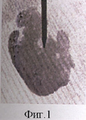
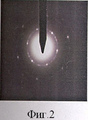
Ультратонкий срез миокарда из зоны инфаркта, изготовленный для электронной микроскопии, был любезно предоставлен ГУ НИИ кардиологии Томского научного центра СО РАМН и использован для микродиффракционного исследования на трансмиссионном просвечивающем электронном микроскопе СМ-12 Phillips. Образец не подвергался напылению, т.к. таковым было условие передачи образцов кардиоцентром. В ходе обзорного изучения препарата с увеличением от 35000× до 100000× выявлено значительное количество (около 15) наноразмерных зерен. Форма сечений биоминералов была различная: изометричная, округлая, приближенно-гексагональная, призматическая. Различным было и кристаллографическое строение зерен. Большая часть их имела кристаллическое строение, визуально диагностированное по полосам экстинкции. Процентов 20 зерен аморфизовывались под нагревающим действием электронного пучка вследствие потери воды или гидроксил-радикала, входящих в решетку минерала. Об аморфизации зерен свидетельствовало исчезновение полос экстинкции в течение первых секунд. Одно или два зерна в препарате изначально имели аморфное строение. Большинство зерен использовалось для получения микродифрактограмм. На основании анализа микродифрактограмм выяснилось, что большинство зерен имеет монокристаллическое строение, и только одно зерно из десяти является поликристаллическим агрегатом, дающим дифрактограмму кольцевого типа (данные приведены на фиг.1 и 2). Одно из таких поликристаллических зерен было выбрано для фазового анализа. С помощью программы Adobe Photoshop CS Version 8.0 по кольцам электронной микродифрактограммы были рассчитаны межплоскостные расстояния. Расчет межплоскостных расстояний позволил определить минерал как маггемит-С, используя базу данных PCPDFWIN. Данный сильно магнитный минерал кубической сингонии образуется в природе при окислении магнетита или при обезвоживании липидокрокита. Биоминералогией магнетит досконально изучен в бактериях, а с 1992 г. и в клеточных структурах головного мозга, названных магнетосомами. Маггемит же с помощью магнитометрических методов неоднократно определялся в ферритине и в тканях человеческого организма при различных нервных и возрастных заболеваниях, таких как болезнь Альцгеймера, Паркинсона и т.д., поэтому изучение распространенности данного биоминерала в тканях больных представляет огромный научный интерес.An ultra-thin section of the myocardial infarction zone, made for electron microscopy, was kindly provided by the State Research Institute of Cardiology of the Tomsk Scientific Center SB RAMS and used for microdiffraction studies using a transmission transmission electron microscope CM-12 Phillips. The sample was not sprayed, because such was the condition for the transfer of samples by the cardiocenter. During a review study of the drug with an increase from 35,000 × to 100,000 ×, a significant amount (about 15) of nanoscale grains was revealed. The cross-sectional shape of the biomineral was different: isometric, round, approximately hexagonal, prismatic. The crystallographic structure of the grains was different. Most of them had a crystalline structure, visually diagnosed by extinction bands. About 20 grains were amorphized under the heating action of the electron beam due to the loss of water or hydroxyl radical entering the mineral lattice. Amorphization of grains was indicated by the disappearance of extinction bands during the first seconds. One or two grains in the preparation initially had an amorphous structure. Most grains were used to obtain microdiffraction patterns. Based on the analysis of microdiffraction patterns, it was found that most grains have a single crystal structure, and only one out of ten grains is a polycrystalline aggregate giving a ring-type diffraction pattern (data are shown in Figs. 1 and 2). One of these polycrystalline grains was selected for phase analysis. Using the program Adobe Photoshop CS Version 8.0, the interplanar distances were calculated from the rings of the electron microdiffractogram. Calculation of interplanar distances allowed the mineral to be defined as maghemite-C using the PCPDFWIN database. This highly magnetic mineral of cubic syngony is formed in nature during the oxidation of magnetite or during dehydration of lipidocrocite. By biomineralogy, magnetite has been thoroughly studied in bacteria, and since 1992, in the cellular structures of the brain called magnetosomes. Maghemite using magnetometric methods was repeatedly determined in ferritin and in the tissues of the human body for various nervous and age-related diseases, such as Alzheimer's disease, Parkinson's disease, etc., therefore, studying the prevalence of this biomineral in the tissues of patients is of great scientific interest.
Пример 2.Example 2
Методом электронной микродифракции исследовался другой ультратонкий срез миокарда из зоны инфаркта, подготовленный для электронной микроскопии и отобранный из другой партии образцов, поступивших в более раннее время, чем первый препарат. Объект исследования также не подвергался напылению.The method of electron microdiffraction was used to study another ultrathin section of the myocardium from the infarction zone, prepared for electron microscopy and taken from a different batch of samples received earlier than the first preparation. The object of study was also not sprayed.
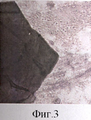
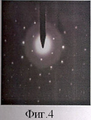
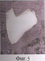
В ходе обзорного изучения препарата с увеличением от 45000× до 100000× выявлено 18 наноразмерных зерен. Как и в первом примере, форма сечений биоминералов была различная: изометричная, округлая, приближенно-гексагональная, призматическая. Различным было и кристаллографическое строение зерен. Большая часть их имела монокристаллическое строение. Два зерна в препарате изначально имели аморфное строение. Было снято 8 микродифрактограмм. В ходе расшифровки выяснилось, что 6 зерен имеют межплоскостные расстояния маггемита, а два зерна характеризуются совершенно другими, отличными друг от друга и от маггемита межплоскостными расстояниями. К сожалению микродифрактограмма точечного типа не позволяет однозначно интерпретировать результаты микродифракции, тем не менее, анализируя полученные данные, удалось выделить группу минералов, чьи межплоскостные расстояния подходят по своим значениям к измеренным. На фиг.3 и 4 изображен монокристалл в гистологическом препарате, межплоскостные расстояния которого очень близки группе соединений, представляющих собой гидроксиды металлов: β-Zn(OH)2 и α-Со(ОН)2, кристаллизующиеся в гексагональной сингонии. Оба соединения не описаны в качестве минералов.During a review study of the drug with an increase from 45,000 × to 100,000 ×, 18 nanoscale grains were detected. As in the first example, the shape of the cross sections of biominerals was different: isometric, round, approximately hexagonal, prismatic. The crystallographic structure of the grains was different. Most of them had a single crystal structure. Two grains in the preparation initially had an amorphous structure. 8 micro diffractograms were taken. In the course of decoding, it turned out that 6 grains have interplanar distances of maghemite, and two grains are characterized by completely different interplanar distances from each other and from maghemite. Unfortunately, a point-type microdiffraction pattern does not allow one to unambiguously interpret the results of microdiffraction; nevertheless, by analyzing the data obtained, it was possible to isolate a group of minerals whose interplanar spacings are similar in value to the measured ones. Figures 3 and 4 show a single crystal in a histological specimen, whose interplanar spacings are very close to the group of metal hydroxides compounds: β-Zn (OH) 2 and α-Co (OH) 2 , crystallizing in hexagonal syngony. Both compounds are not described as minerals.
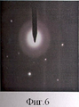
На фиг.5 и 6 представлен призматический кристалл или двойник из того же гистологического препарата, который по вычисленным межплоскостным расстояниям можно отнести к группе рутила.Figures 5 and 6 show a prismatic crystal or a double from the same histological preparation, which according to the calculated interplanar distances can be attributed to the rutile group.
Преимущества способа применения ультратонких срезов тканей, изготовленных для электронно-микроскопических исследований, для изучения минеральных зерен.Advantages of the method of using ultra-thin sections of tissues made for electron microscopy studies for the study of mineral grains.
1. Способ является доступным и экономичным для всех научных учреждений, проводящих исследования в сфере электронной микроскопии. Не требуется никаких дополнительных затрат для изучения роли неорганических зерен и минералов в биологических тканях.1. The method is affordable and economical for all scientific institutions conducting research in the field of electron microscopy. No additional costs are required to study the role of inorganic grains and minerals in biological tissues.
2. Способ обладает исключительно редкой и ценной для науки способностью - сочетать визуализацию типов взаимоотношений органического и неорганического вещества, в пределах живого организма, с количественным и качественным (например: фазовым) анализом обеих форм существования материи.2. The method has an extremely rare and valuable ability for science - to combine visualization of the types of relationships between organic and inorganic matter, within a living organism, with a quantitative and qualitative (for example: phase) analysis of both forms of the existence of matter.
3. Данный способ позволяет осуществить уникальную возможность кристалллохимической и фазовой идентификации наноразмерных зерен в живой ткани.3. This method allows for the unique possibility of crystallochemical and phase identification of nanoscale grains in living tissue.
Claims (1)
Priority Applications (1)
| Application Number | Priority Date | Filing Date | Title |
|---|---|---|---|
| RU2009135480/15A RU2425373C2 (en) | 2009-09-23 | 2009-09-23 | Method of using ultrathin tissue section for mineral grain analysis |
Applications Claiming Priority (1)
| Application Number | Priority Date | Filing Date | Title |
|---|---|---|---|
| RU2009135480/15A RU2425373C2 (en) | 2009-09-23 | 2009-09-23 | Method of using ultrathin tissue section for mineral grain analysis |
Publications (2)
| Publication Number | Publication Date |
|---|---|
| RU2009135480A RU2009135480A (en) | 2011-03-27 |
| RU2425373C2 true RU2425373C2 (en) | 2011-07-27 |
Family
ID=44052612
Family Applications (1)
| Application Number | Title | Priority Date | Filing Date |
|---|---|---|---|
| RU2009135480/15A RU2425373C2 (en) | 2009-09-23 | 2009-09-23 | Method of using ultrathin tissue section for mineral grain analysis |
Country Status (1)
| Country | Link |
|---|---|
| RU (1) | RU2425373C2 (en) |
-
2009
- 2009-09-23 RU RU2009135480/15A patent/RU2425373C2/en not_active IP Right Cessation
Non-Patent Citations (1)
| Title |
|---|
| БАТУРИН Г.Н. и др. Микроструктуры железомарганцевых конкреций: Атлас фотографий. - М.: Наука, 1989, с.288. ЛАМАНОВА Л.М. Сыктывкар: ИГ Коми НЦ Уро РАН «Структура и разнообразие материального мира». Сыктывкар, 2008, с.346-349. ЗЕМЛЯНУХИН В.Н. Лабораторные новости Дальнего Востока, № 1, 1999, с.1-10. * |
Also Published As
| Publication number | Publication date |
|---|---|
| RU2009135480A (en) | 2011-03-27 |
Similar Documents
| Publication | Publication Date | Title |
|---|---|---|
| Akhtar et al. | ZnO nanoflower based sensitive nano-biosensor for amyloid detection | |
| Tavares et al. | Genotoxicity evaluation of nanosized titanium dioxide, synthetic amorphous silica and multi-walled carbon nanotubes in human lymphocytes | |
| Vilarinho et al. | Are lithium niobate (LiNbO3) and lithium tantalate (LiTaO3) ferroelectrics bioactive? | |
| Sirelkhatim et al. | Preferential cytotoxicity of ZnO nanoparticle towards cervical cancer cells induced by ROS-mediated apoptosis and cell cycle arrest for cancer therapy | |
| Mukherjee et al. | Nanoscale surface characterization of human erythrocytes by atomic force microscopy: a critical review | |
| DE3881413T2 (en) | Microspherical bodies and methods for the detection of therapy for Alzheimer's disease and related conditions. | |
| EP3021119A1 (en) | Method for diagnosis of diseases using morphological characteristics of luterial | |
| Sheena et al. | Preparation, characterization, and in vitro evaluation of the anticancer activity of Ce3+ doped CuFe2O4 spinel nanoparticles in MCF-7 cell lines | |
| JP2025063161A (en) | lutearia and methods for isolating and culturing same | |
| Cros et al. | Investigation at the micrometer scale of pancreatic calcifications in chronic pancreatitis by μFTIR spectroscopy and field emission scanning electron microscopy | |
| Al-Sabbah et al. | Modified millet extract-mediated NiO/CaO Nanocomposite potentiometric sensor for monitoring of ciprofloxacin in commercial products | |
| Ye et al. | Engineered nanomaterials’ fate assessment in biological matrices: Recent milestones in electron microscopy | |
| KR102095018B1 (en) | Methods For Diagnosis Of Cancer Using Lectin-Conjugated Nanoparticles | |
| Tawade et al. | Nanotechnology in biological science and engineering | |
| RU2425373C2 (en) | Method of using ultrathin tissue section for mineral grain analysis | |
| Butler et al. | High-pressure freezing/freeze substitution and transmission electron microscopy for characterization of metal oxide nanoparticles within sunscreens | |
| Wang et al. | Functional gold nanoparticles for studying the interaction of lectin with glycosyl complex on living cellular surfaces | |
| Anthony et al. | Characterization of synthesized nanoparticles for medical devices: Current techniques and recent advances | |
| Mohammed et al. | A novel sandwich ELISA method for quantifying CHI3L1 in blood serum and cerebrospinal fluid multiple sclerosis patients using sustainable photo-irradiated zero-valence gold nanoparticles | |
| AU2018300963B2 (en) | Cancer detection method using tissue sample | |
| Saritha et al. | Biological synthesis and characterization of gold nanoparticles using Lemna minor | |
| Tortiglione | An ancient model organism to test in vivo novel functional nanocrystals | |
| Sergunova et al. | Hemoglobin: modification, crystallization, polymerization | |
| Rakshak et al. | Characterizing morphological alterations in blood related disorders through Atomic Force Microscopy | |
| RU2477485C2 (en) | Method for assessing copper nanoparticles administration safety |
Legal Events
| Date | Code | Title | Description |
|---|---|---|---|
| MM4A | The patent is invalid due to non-payment of fees |
Effective date: 20170924 |