RU2400733C2 - Transmission spectroscopy system for use in determining analysed substances in body fluids - Google Patents
Transmission spectroscopy system for use in determining analysed substances in body fluids Download PDFInfo
- Publication number
- RU2400733C2 RU2400733C2 RU2007126679/28A RU2007126679A RU2400733C2 RU 2400733 C2 RU2400733 C2 RU 2400733C2 RU 2007126679/28 A RU2007126679/28 A RU 2007126679/28A RU 2007126679 A RU2007126679 A RU 2007126679A RU 2400733 C2 RU2400733 C2 RU 2400733C2
- Authority
- RU
- Russia
- Prior art keywords
- light
- sample
- lens
- light source
- angle
- Prior art date
Links
- 239000000126 substance Substances 0.000 title claims abstract description 14
- 238000002235 transmission spectroscopy Methods 0.000 title claims description 58
- 210000001124 body fluid Anatomy 0.000 title description 12
- 239000010839 body fluid Substances 0.000 title description 12
- 239000012780 transparent material Substances 0.000 claims abstract description 5
- 238000000034 method Methods 0.000 claims description 136
- 210000004369 blood Anatomy 0.000 claims description 83
- 239000008280 blood Substances 0.000 claims description 83
- 230000005540 biological transmission Effects 0.000 claims description 72
- 238000005534 hematocrit Methods 0.000 claims description 62
- 239000008103 glucose Substances 0.000 claims description 58
- WQZGKKKJIJFFOK-GASJEMHNSA-N Glucose Natural products OC[C@H]1OC(O)[C@H](O)[C@@H](O)[C@@H]1O WQZGKKKJIJFFOK-GASJEMHNSA-N 0.000 claims description 57
- 239000012491 analyte Substances 0.000 claims description 46
- 239000003153 chemical reaction reagent Substances 0.000 claims description 41
- 238000001228 spectrum Methods 0.000 claims description 33
- 238000006243 chemical reaction Methods 0.000 claims description 32
- 239000007788 liquid Substances 0.000 claims description 23
- 238000005259 measurement Methods 0.000 claims description 22
- 238000012937 correction Methods 0.000 claims description 18
- 238000004611 spectroscopical analysis Methods 0.000 claims description 7
- 230000003993 interaction Effects 0.000 claims description 6
- 108010050375 Glucose 1-Dehydrogenase Proteins 0.000 claims description 4
- 238000004891 communication Methods 0.000 claims description 4
- 239000012530 fluid Substances 0.000 claims description 4
- 125000002791 glucosyl group Chemical group C1([C@H](O)[C@@H](O)[C@H](O)[C@H](O1)CO)* 0.000 claims description 4
- 108090000790 Enzymes Proteins 0.000 claims description 3
- 102000004190 Enzymes Human genes 0.000 claims description 3
- XUIMIQQOPSSXEZ-UHFFFAOYSA-N Silicon Chemical compound [Si] XUIMIQQOPSSXEZ-UHFFFAOYSA-N 0.000 claims description 3
- 238000012544 monitoring process Methods 0.000 claims description 3
- 239000013307 optical fiber Substances 0.000 claims description 3
- 229910052710 silicon Inorganic materials 0.000 claims description 3
- 239000010703 silicon Substances 0.000 claims description 3
- 125000003831 tetrazolyl group Chemical group 0.000 claims description 3
- 230000002949 hemolytic effect Effects 0.000 claims description 2
- 230000015556 catabolic process Effects 0.000 claims 1
- 230000007717 exclusion Effects 0.000 claims 1
- 230000000694 effects Effects 0.000 abstract description 2
- 239000000523 sample Substances 0.000 description 211
- 108010054147 Hemoglobins Proteins 0.000 description 29
- 102000001554 Hemoglobins Human genes 0.000 description 29
- 238000010521 absorption reaction Methods 0.000 description 21
- 238000002834 transmittance Methods 0.000 description 20
- 210000003743 erythrocyte Anatomy 0.000 description 17
- 238000004458 analytical method Methods 0.000 description 11
- 230000003287 optical effect Effects 0.000 description 11
- 230000004044 response Effects 0.000 description 10
- 230000008859 change Effects 0.000 description 6
- 238000000411 transmission spectrum Methods 0.000 description 6
- 230000009089 cytolysis Effects 0.000 description 5
- 230000006870 function Effects 0.000 description 5
- 230000003595 spectral effect Effects 0.000 description 5
- 108010064719 Oxyhemoglobins Proteins 0.000 description 4
- HVYWMOMLDIMFJA-DPAQBDIFSA-N cholesterol Chemical compound C1C=C2C[C@@H](O)CC[C@]2(C)[C@@H]2[C@@H]1[C@@H]1CC[C@H]([C@H](C)CCCC(C)C)[C@@]1(C)CC2 HVYWMOMLDIMFJA-DPAQBDIFSA-N 0.000 description 4
- 238000012986 modification Methods 0.000 description 4
- 230000004048 modification Effects 0.000 description 4
- 238000012360 testing method Methods 0.000 description 4
- JVTAAEKCZFNVCJ-UHFFFAOYSA-M Lactate Chemical compound CC(O)C([O-])=O JVTAAEKCZFNVCJ-UHFFFAOYSA-M 0.000 description 3
- 238000000149 argon plasma sintering Methods 0.000 description 3
- WQZGKKKJIJFFOK-VFUOTHLCSA-N beta-D-glucose Chemical compound OC[C@H]1O[C@@H](O)[C@H](O)[C@@H](O)[C@@H]1O WQZGKKKJIJFFOK-VFUOTHLCSA-N 0.000 description 3
- 230000006378 damage Effects 0.000 description 3
- 230000003834 intracellular effect Effects 0.000 description 3
- XLYOFNOQVPJJNP-UHFFFAOYSA-N water Substances O XLYOFNOQVPJJNP-UHFFFAOYSA-N 0.000 description 3
- 108010085682 Hemoglobin A Proteins 0.000 description 2
- 102000007513 Hemoglobin A Human genes 0.000 description 2
- 102000003855 L-lactate dehydrogenase Human genes 0.000 description 2
- 108700023483 L-lactate dehydrogenases Proteins 0.000 description 2
- BAWFJGJZGIEFAR-NNYOXOHSSA-N NAD zwitterion Chemical compound NC(=O)C1=CC=C[N+]([C@H]2[C@@H]([C@H](O)[C@@H](COP([O-])(=O)OP(O)(=O)OC[C@@H]3[C@H]([C@@H](O)[C@@H](O3)N3C4=NC=NC(N)=C4N=C3)O)O2)O)=C1 BAWFJGJZGIEFAR-NNYOXOHSSA-N 0.000 description 2
- 235000012000 cholesterol Nutrition 0.000 description 2
- 230000007423 decrease Effects 0.000 description 2
- 229940088598 enzyme Drugs 0.000 description 2
- 238000002474 experimental method Methods 0.000 description 2
- IXZISFNWUWKBOM-ARQDHWQXSA-N fructosamine Chemical compound NC[C@@]1(O)OC[C@@H](O)[C@@H](O)[C@@H]1O IXZISFNWUWKBOM-ARQDHWQXSA-N 0.000 description 2
- 229910052736 halogen Inorganic materials 0.000 description 2
- 150000002367 halogens Chemical class 0.000 description 2
- 238000012417 linear regression Methods 0.000 description 2
- 229950006238 nadide Drugs 0.000 description 2
- 229930027945 nicotinamide-adenine dinucleotide Natural products 0.000 description 2
- 210000002381 plasma Anatomy 0.000 description 2
- 238000007781 pre-processing Methods 0.000 description 2
- 230000008569 process Effects 0.000 description 2
- 210000003296 saliva Anatomy 0.000 description 2
- 210000002966 serum Anatomy 0.000 description 2
- 210000001519 tissue Anatomy 0.000 description 2
- 210000002700 urine Anatomy 0.000 description 2
- 239000004366 Glucose oxidase Substances 0.000 description 1
- 241000406221 Hypostomus robinii Species 0.000 description 1
- 238000002835 absorbance Methods 0.000 description 1
- 230000032900 absorption of visible light Effects 0.000 description 1
- NIXOWILDQLNWCW-UHFFFAOYSA-N acrylic acid group Chemical group C(C=C)(=O)O NIXOWILDQLNWCW-UHFFFAOYSA-N 0.000 description 1
- 239000012472 biological sample Substances 0.000 description 1
- 230000015572 biosynthetic process Effects 0.000 description 1
- 210000000601 blood cell Anatomy 0.000 description 1
- 210000004027 cell Anatomy 0.000 description 1
- 230000001413 cellular effect Effects 0.000 description 1
- 239000000356 contaminant Substances 0.000 description 1
- 238000011109 contamination Methods 0.000 description 1
- 230000003247 decreasing effect Effects 0.000 description 1
- 230000007547 defect Effects 0.000 description 1
- 230000001419 dependent effect Effects 0.000 description 1
- 239000003599 detergent Substances 0.000 description 1
- 239000002360 explosive Substances 0.000 description 1
- 238000001506 fluorescence spectroscopy Methods 0.000 description 1
- 229940116332 glucose oxidase Drugs 0.000 description 1
- 235000019420 glucose oxidase Nutrition 0.000 description 1
- 239000003219 hemolytic agent Substances 0.000 description 1
- 238000002329 infrared spectrum Methods 0.000 description 1
- 229940056932 lead sulfide Drugs 0.000 description 1
- 229910052981 lead sulfide Inorganic materials 0.000 description 1
- 230000031700 light absorption Effects 0.000 description 1
- 230000002934 lysing effect Effects 0.000 description 1
- 238000004519 manufacturing process Methods 0.000 description 1
- 239000000203 mixture Substances 0.000 description 1
- 229920000642 polymer Polymers 0.000 description 1
- 238000004445 quantitative analysis Methods 0.000 description 1
- 230000035484 reaction time Effects 0.000 description 1
- 239000013074 reference sample Substances 0.000 description 1
- 238000001055 reflectance spectroscopy Methods 0.000 description 1
- 238000000926 separation method Methods 0.000 description 1
- 238000010183 spectrum analysis Methods 0.000 description 1
Images

Classifications
-
- G—PHYSICS
- G01—MEASURING; TESTING
- G01N—INVESTIGATING OR ANALYSING MATERIALS BY DETERMINING THEIR CHEMICAL OR PHYSICAL PROPERTIES
- G01N21/00—Investigating or analysing materials by the use of optical means, i.e. using sub-millimetre waves, infrared, visible or ultraviolet light
- G01N21/17—Systems in which incident light is modified in accordance with the properties of the material investigated
- G01N21/25—Colour; Spectral properties, i.e. comparison of effect of material on the light at two or more different wavelengths or wavelength bands
- G01N21/31—Investigating relative effect of material at wavelengths characteristic of specific elements or molecules, e.g. atomic absorption spectrometry
-
- G—PHYSICS
- G01—MEASURING; TESTING
- G01N—INVESTIGATING OR ANALYSING MATERIALS BY DETERMINING THEIR CHEMICAL OR PHYSICAL PROPERTIES
- G01N21/00—Investigating or analysing materials by the use of optical means, i.e. using sub-millimetre waves, infrared, visible or ultraviolet light
- G01N21/17—Systems in which incident light is modified in accordance with the properties of the material investigated
- G01N21/25—Colour; Spectral properties, i.e. comparison of effect of material on the light at two or more different wavelengths or wavelength bands
- G01N21/255—Details, e.g. use of specially adapted sources, lighting or optical systems
-
- G—PHYSICS
- G01—MEASURING; TESTING
- G01N—INVESTIGATING OR ANALYSING MATERIALS BY DETERMINING THEIR CHEMICAL OR PHYSICAL PROPERTIES
- G01N21/00—Investigating or analysing materials by the use of optical means, i.e. using sub-millimetre waves, infrared, visible or ultraviolet light
- G01N21/17—Systems in which incident light is modified in accordance with the properties of the material investigated
- G01N21/25—Colour; Spectral properties, i.e. comparison of effect of material on the light at two or more different wavelengths or wavelength bands
- G01N21/31—Investigating relative effect of material at wavelengths characteristic of specific elements or molecules, e.g. atomic absorption spectrometry
- G01N21/314—Investigating relative effect of material at wavelengths characteristic of specific elements or molecules, e.g. atomic absorption spectrometry with comparison of measurements at specific and non-specific wavelengths
- G01N21/3151—Investigating relative effect of material at wavelengths characteristic of specific elements or molecules, e.g. atomic absorption spectrometry with comparison of measurements at specific and non-specific wavelengths using two sources of radiation of different wavelengths
-
- G—PHYSICS
- G01—MEASURING; TESTING
- G01N—INVESTIGATING OR ANALYSING MATERIALS BY DETERMINING THEIR CHEMICAL OR PHYSICAL PROPERTIES
- G01N21/00—Investigating or analysing materials by the use of optical means, i.e. using sub-millimetre waves, infrared, visible or ultraviolet light
- G01N21/17—Systems in which incident light is modified in accordance with the properties of the material investigated
- G01N21/25—Colour; Spectral properties, i.e. comparison of effect of material on the light at two or more different wavelengths or wavelength bands
- G01N21/31—Investigating relative effect of material at wavelengths characteristic of specific elements or molecules, e.g. atomic absorption spectrometry
- G01N21/35—Investigating relative effect of material at wavelengths characteristic of specific elements or molecules, e.g. atomic absorption spectrometry using infrared light
- G01N21/3577—Investigating relative effect of material at wavelengths characteristic of specific elements or molecules, e.g. atomic absorption spectrometry using infrared light for analysing liquids, e.g. polluted water
-
- G—PHYSICS
- G01—MEASURING; TESTING
- G01N—INVESTIGATING OR ANALYSING MATERIALS BY DETERMINING THEIR CHEMICAL OR PHYSICAL PROPERTIES
- G01N21/00—Investigating or analysing materials by the use of optical means, i.e. using sub-millimetre waves, infrared, visible or ultraviolet light
- G01N21/17—Systems in which incident light is modified in accordance with the properties of the material investigated
- G01N21/25—Colour; Spectral properties, i.e. comparison of effect of material on the light at two or more different wavelengths or wavelength bands
- G01N21/31—Investigating relative effect of material at wavelengths characteristic of specific elements or molecules, e.g. atomic absorption spectrometry
- G01N21/39—Investigating relative effect of material at wavelengths characteristic of specific elements or molecules, e.g. atomic absorption spectrometry using tunable lasers
-
- G—PHYSICS
- G01—MEASURING; TESTING
- G01N—INVESTIGATING OR ANALYSING MATERIALS BY DETERMINING THEIR CHEMICAL OR PHYSICAL PROPERTIES
- G01N21/00—Investigating or analysing materials by the use of optical means, i.e. using sub-millimetre waves, infrared, visible or ultraviolet light
- G01N21/75—Systems in which material is subjected to a chemical reaction, the progress or the result of the reaction being investigated
- G01N21/77—Systems in which material is subjected to a chemical reaction, the progress or the result of the reaction being investigated by observing the effect on a chemical indicator
- G01N21/78—Systems in which material is subjected to a chemical reaction, the progress or the result of the reaction being investigated by observing the effect on a chemical indicator producing a change of colour
-
- G—PHYSICS
- G01—MEASURING; TESTING
- G01N—INVESTIGATING OR ANALYSING MATERIALS BY DETERMINING THEIR CHEMICAL OR PHYSICAL PROPERTIES
- G01N21/00—Investigating or analysing materials by the use of optical means, i.e. using sub-millimetre waves, infrared, visible or ultraviolet light
- G01N21/17—Systems in which incident light is modified in accordance with the properties of the material investigated
- G01N21/25—Colour; Spectral properties, i.e. comparison of effect of material on the light at two or more different wavelengths or wavelength bands
- G01N21/27—Colour; Spectral properties, i.e. comparison of effect of material on the light at two or more different wavelengths or wavelength bands using photo-electric detection ; circuits for computing concentration
- G01N21/274—Calibration, base line adjustment, drift correction
Abstract
Description
Область техники, к которой относится изобретениеFIELD OF THE INVENTION
Настоящее изобретение относится к спектроскопии в целом и к использованию спектроскопии полного пропускания для определения концентрации анализируемого вещества в жидкости организма, в частности.The present invention relates to spectroscopy in general and to the use of full transmission spectroscopy to determine the concentration of an analyte in a body fluid, in particular.
Уровень техникиState of the art
Спектроскопия пропускания применяется для проведения количественного анализа пробы на основе прохождения светового луча через пробу, содержащуюся в кювете. Компоненты пробы поглощают различные частотные составляющие светового луча, поэтому спектральный анализ прошедшего через пробу луча позволяет проводить анализ самой пробы. Сухие химические реагенты растворяются в пробе, они взаимодействуют с анализируемым веществом с образованием хроматического отклика на некоторых длинах волн в диапазоне от 450 нанометров (нм) до приблизительно 950 нм.Transmission spectroscopy is used to conduct a quantitative analysis of a sample based on the passage of a light beam through a sample contained in a cuvette. The components of the sample absorb various frequency components of the light beam, so the spectral analysis of the beam passed through the sample allows the analysis of the sample itself. Dry chemicals are dissolved in the sample, they interact with the analyte with the formation of a chromatic response at certain wavelengths in the range from 450 nanometers (nm) to about 950 nm.
Спектроскопия пропускания является одним из методов измерения концентрации анализируемого вещества (например, глюкозы, лактата, фруктозамина, гемоглобина A1c и холестерина) в жидкости организма (например, крови, плазме или сыворотке, слюне, моче и тканевой жидкости). Система индикаторных реагентов и анализируемое вещество в пробе жидкости организма вступают в хроматическую реакцию, т.е. реакцию между реагентом и анализируемым веществом, которая приводит к изменению цвета пробы. Степень изменения цвета указывает на концентрацию анализируемого вещества в жидкости организма. Изменение цвета пробы оценивают, например, с помощью спектроскопии, измеряя поглощение прошедшего света. Обычная спектроскопия пропускания подробно описана в патенте США №5866349. Диффузное отражение и флуоресцентная спектроскопия подробно описаны в патентах США №№5518689 "Diffuse Light Reflectance Readhead" ("Считывающая головка в системе с отражением диффузного света"); 5611999 "Diffuse Light Reflectance Readhead" ("Считывающая головка в системе с отражением диффузного света") и 5194393 "Optical Biosensor and Method of Use" ("Оптический биодатчик и способ его применения").Transmission spectroscopy is one method of measuring the concentration of an analyte (e.g., glucose, lactate, fructosamine, hemoglobin A 1c and cholesterol) in body fluids (e.g., blood, plasma or serum, saliva, urine and tissue fluid). The system of indicator reagents and the analyte in a sample of body fluid enter a chromatic reaction, i.e. the reaction between the reagent and the analyte, which leads to a change in the color of the sample. The degree of color change indicates the concentration of the analyte in the body fluid. The color change of the sample is evaluated, for example, using spectroscopy, measuring the absorption of transmitted light. Conventional transmission spectroscopy is described in detail in US patent No. 5866349. Diffuse reflection and fluorescence spectroscopy are described in detail in US patent No. 5518689 "Diffuse Light Reflectance Readhead"("Reading head in a system with diffuse light reflection"); 5611999 "Diffuse Light Reflectance Readhead"("Reading head in a system with reflection of diffuse light") and 5194393 "Optical Biosensor and Method of Use"("Optical biosensor and method of its use").
В простейшем случае анализ с помощью спектроскопии пропускания предусматривает применение источника света, который создает луч света, освещающий образец, и датчика для приема прошедшего через пробу света. Затем принятый прошедший свет сравнивают с эталонным образцом (например, светом, непосредственно принятым датчиком без прохождения через пробу). При обычной спектроскопии пропускания принимают и анализируют свет, выходящий из пробы под малыми углами (например, от приблизительно 0° до приблизительно 15°) относительно нормальной оптической оси, а не рассеянный свет, прошедший через пробу. Нормальная оптическая ось - это ось, перпендикулярная оптическим входному и выходному окошку кюветы с пробой. При спектроскопии полного пропускания принимают по существу весь свет (в т.ч. и рассеянный), выходящий из пробы под большими углами (например, от приблизительно 0° до приблизительно 90°) относительно нормальной оптической оси. В существующих системах для проведения анализа с помощью спектроскопии полного пропускания используется фотометрический шар для приема всего света, прошедшего через пробу, и фотоэлектронный умножитель, необходимый для приема отраженного света с малой части внутренней поверхности фотометрического шара.In the simplest case, transmission spectroscopy analysis involves the use of a light source that generates a light beam illuminating the sample and a sensor for receiving light transmitted through the sample. Then, the received transmitted light is compared with a reference sample (for example, light directly received by the sensor without passing through the sample). In conventional transmission spectroscopy, light coming out of the sample at small angles (for example, from about 0 ° to about 15 °) relative to the normal optical axis is received and analyzed, and not the scattered light passing through the sample. The normal optical axis is the axis perpendicular to the optical input and output window of the sample cell. In full transmission spectroscopy, essentially all of the light (including scattered light) coming out of the sample at large angles (for example, from about 0 ° to about 90 °) relative to the normal optical axis is received. In existing systems, for analysis using full transmission spectroscopy, a photometric ball is used to receive all the light that has passed through the sample, and a photomultiplier is necessary to receive reflected light from a small part of the inner surface of the photometric ball.
Как сообщалось в статье "Data Preprocessing and Partial Least Squares Analysis for Reagentless Determination of Hemoglobin Concentration Using Conventional and Total Transmission Spectroscopy" ("Предварительная обработка данных и частичный анализ методом наименьших квадратов при безреагентном определении концентрации гемоглобина с применением обычной и полной спектроскопии пропускания"), опубликованной в апреле 2001 г. в Journal of Biomedical Optics (Журнале биомедицинской оптики) (том 6, №2), обычные уровни пропускания (без рассеянных составляющих) для цельной крови с содержанием гемоглобина от приблизительно 6,6 до 17,2 г/дл составляют от 15,8 до 0,1%Т по всему видимому и ближнему инфракрасному диапазону (например, от приблизительно 500 нм до приблизительно 800 нм) при длине пути всего лишь 100 мкм; однако уровни полного пропускания (с учетом рассеянного света) для цельной крови с содержанием гемоглобина в том же диапазоне составляют от 79 до 2%Т. Полное пропускание света с длиной волны от приблизительно 600 нм до приблизительно 800 нм составляет около 100%Т, и между разными уровнями гемоглобина существует очень малое различие. Таким образом, уровень концентрации гемоглобина оказывает малое влияние на пропускание света с длиной волны в диапазоне от приблизительно 600 нм до приблизительно 800 нм.As reported in the article "Data Preprocessing and Partial Least Squares Analysis for Reagentless Determination of Hemoglobin Concentration Using Conventional and Total Transmission Spectroscopy" ("Data pre-processing and partial least squares analysis for non-reagent determination of hemoglobin concentration using conventional and complete transmission spectroscopy") published in April 2001 in the Journal of Biomedical Optics (Volume 6, No. 2), the usual transmission levels (without dispersed components) for whole blood with a hemoglobin content of from about 6.6 to 17.2 g / for sosta from 15.8 to 0.1% T over the entire visible and near infrared range (for example, from about 500 nm to about 800 nm) with a path length of only 100 μm; however, the levels of complete transmission (including scattered light) for whole blood with a hemoglobin content in the same range are from 79 to 2% T. The total transmission of light with a wavelength of from about 600 nm to about 800 nm is about 100% T, and there is very little difference between the different levels of hemoglobin. Thus, the level of hemoglobin concentration has little effect on the transmission of light with a wavelength in the range from about 600 nm to about 800 nm.
Недостатком существующих систем для проведения анализа с помощью спектроскопии полного пропускания является низкий уровень сигнала фотометрического шара, вследствие которого необходимо использовать фотоэлектронный умножитель для приема отраженного света с малой части внутренней поверхности фотометрического шара. Другим недостатком систем, использующих обычную и полную спектроскопию пропускания, является стоимость фотометрического шара и фотоэлектронного умножителя. Стоимость этих устройств исключает применение систем спектроскопии полного пропускания в устройствах, используемых пациентами для самопроверки, например определения уровня глюкозы в крови. В результате этого исследования в области применения спектроскопических систем для определения концентрации анализируемого вещества в пробе жидкости организма сосредоточились на измерении обычного пропускания света.A drawback of existing systems for performing analysis using full transmission spectroscopy is the low level of the photometric ball signal, due to which it is necessary to use a photomultiplier to receive reflected light from a small part of the inner surface of the photometric ball. Another disadvantage of systems using conventional and complete transmission spectroscopy is the cost of the photometric ball and the photomultiplier. The cost of these devices eliminates the use of full transmission spectroscopy systems in devices used by patients for self-testing, such as determining blood glucose levels. As a result of this study, the application of spectroscopic systems to determine the concentration of an analyte in a sample of body fluid focused on measuring normal light transmission.
Однако существующие системы обычной спектроскопии пропускания имеют ряд недостатков. Как отмечалось выше, они принимают только свет, выходящий из образца под малыми углами, используя для этого традиционные измерения, в результате чего часто теряется свет, выходящий из образца под большими углами. Значительная часть света, рассеянного красными кровяными тельцами, не собирается существующими системами с использованием обычных измерений пропускания. Это приводит к значительным потерям света и, следовательно, очень низким уровням пропускания через цельную кровь.However, existing conventional transmission spectroscopy systems have several drawbacks. As noted above, they only accept light coming out of the sample at small angles, using traditional measurements for this, as a result of which light coming out of the sample at large angles is often lost. A significant portion of the light scattered by red blood cells is not collected by existing systems using conventional transmittance measurements. This results in significant light loss and, therefore, very low levels of transmission through whole blood.
Для снижения потерь при пропускании в обычных системах пропускания обычно в пробу крови добавляют реагент или детергент, чтобы лизировать красные кровяные тельца. Разрушение стенок красных кровяных телец путем лизирования снижает рассеянную составляющую и повышает обычное прохождение света через пробу. Добавление лизирующего реагента и последующий лизис красных кровяных телец занимает время, являющееся достаточно продолжительным по сравнению с общей длительностью процесса измерения. Эта проблема отсутствует в существующих способах спектроскопии полного пропускания, поскольку оптические средства собирают и рассеянный, и обычный прошедший свет. Уровни полного пропускания обычно достаточно высоки, чтобы устранить необходимость в лизисе красных кровяных телец, что значительно сокращает общее время химического анализа.To reduce transmission loss in conventional transmission systems, a reagent or detergent is usually added to the blood sample to lyse red blood cells. The destruction of the walls of red blood cells by lysis reduces the scattered component and increases the normal passage of light through the sample. Adding a lysing reagent and subsequent lysis of red blood cells takes time, which is quite long compared with the total duration of the measurement process. This problem is not present in existing full transmission spectroscopy methods, since optical means collect both scattered and ordinary transmitted light. The levels of complete transmission are usually high enough to eliminate the need for lysis of red blood cells, which significantly reduces the overall time of chemical analysis.
Другим недостатком, связанным с существующими системами обычной спектроскопии пропускания, является смещение пропускания при длинах волн света, соответствующих области протекания хроматической реакции. Индикаторный реагент может вступать в реакцию с внутриклеточными компонентами (например, гемоглобином, лактатдегидрогеназой и др.), высвобождаемыми при лизисе красных кровяных телец, вызывая дополнительный цветовой отклик. Смещение пропускания, вызванное реакцией реагента с некоторыми внутриклеточными компонентами, такими как гемоглобин, не соответствует уровню глюкозы в крови. Такое смещение пропускания вызывает неточности в определении концентрации анализируемого вещества (например, глюкозы). Величина смещения связана с концентрацией некоторых клеточных компонентов в клетках крови.Another disadvantage associated with existing conventional transmission spectroscopy systems is the transmission bias at light wavelengths corresponding to the region of the chromatic reaction. The indicator reagent can react with intracellular components (for example, hemoglobin, lactate dehydrogenase, etc.) released during lysis of red blood cells, causing an additional color response. The transmission bias caused by the reaction of the reagent with some intracellular components, such as hemoglobin, does not correspond to the level of glucose in the blood. This transmission bias causes inaccuracies in determining the analyte concentration (e.g., glucose). The magnitude of the bias is related to the concentration of certain cellular components in the blood cells.
Поскольку в способах спектроскопии полного пропускания не требуется лизис крови, количество внутриклеточных компонентов, которые могут повлиять на измерения глюкозы, значительно уменьшается. Однако смещение продолжает существовать для таких веществ, как гемоглобин, которые поглощают свет в видимом диапазоне при длинах волн менее чем приблизительно 600 нм. Из указанной выше статьи в журнале Journal of Biomedical Optics известно, что, например, спектр полного пропускания оксигемоглобина имеет пики поглощения при длинах волн приблизительно 542 нм и приблизительно 577 нм. Известно, что уровень поглощения при длинах волн приблизительно 542 нм или приблизительно 577 нм может использоваться для определения концентрации гемоглобина в пробе цельной крови. Остающуюся ошибку взаимодействия, вызываемую гемоглобином, при определении концентрации глюкозы можно исправить путем измерения полного пропускания при 542 нм или 577 нм и коррелирования поглощения с известной концентрацией гемоглобина.Since blood lysis is not required in full transmission spectroscopy methods, the amount of intracellular components that can affect glucose measurements is significantly reduced. However, bias continues to exist for substances such as hemoglobin, which absorb light in the visible range at wavelengths less than about 600 nm. From the above article in the Journal of Biomedical Optics, it is known that, for example, the total transmission spectrum of oxyhemoglobin has absorption peaks at wavelengths of approximately 542 nm and approximately 577 nm. It is known that the absorption level at wavelengths of approximately 542 nm or approximately 577 nm can be used to determine the concentration of hemoglobin in a sample of whole blood. The remaining hemoglobin-induced interaction error in determining glucose concentration can be corrected by measuring the total transmittance at 542 nm or 577 nm and correlating the absorption with a known hemoglobin concentration.
Гематокрит цельной крови также может вызывать смещение пропускания ввиду различия составляющей рассеянного света при разных значениях гематокрита. Потери пропускания, вызванные различными значениями гематокрита, не отображают уровень глюкозы в крови. Существующие системы обычной или полной спектроскопии пропускания не способны определять различие значений гематокрита ввиду низкого уровня пропускания и малого разделения уровней гематокрита при определенных длинах волн света.Whole blood hematocrit can also cause transmission bias due to differences in the scattered light component at different hematocrit values. Loss of transmission caused by different hematocrit values do not reflect the level of glucose in the blood. Existing systems of conventional or complete transmission spectroscopy are not able to determine the difference in hematocrit values due to the low transmission level and the small separation of hematocrit levels at certain light wavelengths.
Другим недостатком существующих систем обычной спектроскопии пропускания является недостаточная точность, связанная с длиной пути в пробе. 10% изменение длины пути в пробе приводит к 10% ошибке в измерении концентрации как в способах обычной спектроскопии пропускания, так и в способах спектроскопии полного пропускания. Механические допуски, обуславливающие различие в длине пути, являются, по существу, одинаковыми независимо от длины пути. Однако в существующих системах, в которых используются обычные способы пропускания, нужны более короткие длины пути, чтобы компенсировать потери пропускания, вызванные рассеянием света красными кровяными тельцами. Таким образом, механический допуск при меньшей длине пути приводит к большим ошибкам при определении концентрации. Большая длина пути (применение которой возможно в системах спектроскопии полного пропускания, собирающих рассеянный свет от красных кровяных телец) снижает ошибку.Another disadvantage of existing systems of conventional transmission spectroscopy is the lack of accuracy associated with the path length in the sample. A 10% change in the path length in the sample leads to a 10% error in the concentration measurement both in conventional transmission spectroscopy methods and in full transmission spectroscopy methods. The mechanical tolerances causing the difference in path length are substantially the same regardless of the path length. However, in existing systems that use conventional transmission methods, shorter path lengths are needed to compensate for transmission losses caused by light scattering by red blood cells. Thus, the mechanical tolerance with a shorter path length leads to large errors in determining the concentration. The large path length (the use of which is possible in complete transmission spectroscopy systems collecting scattered light from red blood cells) reduces the error.
Таким образом, было бы желательным уменьшить или устранить описанные выше проблемы, связанные с существующими системами обычной и полной спектроскопии пропускания, при определении концентрации анализируемого вещества в жидкости организма.Thus, it would be desirable to reduce or eliminate the above problems associated with existing systems of conventional and complete transmission spectroscopy, in determining the concentration of the analyte in the body fluid.
Раскрытие изобретенияDisclosure of invention
В соответствии с одним вариантом осуществления система для спектроскопии полного пропускания для применения при определении концентрации анализируемого вещества в пробе жидкости содержит область размещения пробы, источник света, коллимирующую линзу, первую линзу, вторую линзу и датчик. Область размещения пробы выполнена с возможностью помещения в нее анализируемой пробы. Область размещения пробы выполнена из по существу оптически прозрачного материала. Коллимирующая линза выполнена с возможностью принимать свет от источника света и освещать область размещения пробы по существу коллимированным лучом света. Первая линза принимает обычный и рассеянный свет, прошедший через пробу, с первым углом расхождения. На первую линзу попадает свет с первым углом приема. Из первой линзы выходит свет со вторым углом расхождения. Второй угол расхождения меньше первого угла расхождения. Вторая линза принимает свет от первой линзы, и из нее выходит по существу коллимированный луч света. Датчик измеряет свет, поступающий из второй линзы.In accordance with one embodiment, a complete transmission spectroscopy system for use in determining an analyte concentration in a liquid sample comprises a sample placement region, a light source, a collimating lens, a first lens, a second lens, and a sensor. The sample placement area is configured to place the analyzed sample in it. The sample location area is made of a substantially optically transparent material. The collimating lens is configured to receive light from a light source and illuminate the sample location area with a substantially collimated light beam. The first lens receives ordinary and diffused light that has passed through the sample with a first angle of divergence. The first lens receives light with a first reception angle. Light comes out from the first lens with a second angle of divergence. The second angle of divergence is less than the first angle of divergence. The second lens receives light from the first lens, and a substantially collimated beam of light emerges from it. The sensor measures the light coming from the second lens.
В соответствии с одним способом концентрацию анализируемого вещества в пробе жидкости определяют с помощью системы для спектроскопии полного пропускания. Анализируемую пробу помещают в область размещения пробы системы для спектроскопии полного пропускания. Луч света выходит из источника света. Выходящий из источника света луч является по существу коллимированным. Проба освещается по существу коллимированным лучом света от источника света. Первая линза принимает обычный и рассеянный свет, прошедший через пробу. Угол расхождения прошедшего света уменьшается первой линзой. Свет с меньшим углом расхождения попадает на вторую линзу. Поступающий во вторую линзу свет по существу коллимируется там. Датчик измеряет по существу коллимированный свет, выходящий из второй линзы.In accordance with one method, the concentration of an analyte in a liquid sample is determined using a complete transmission spectroscopy system. The analyzed sample is placed in the area of placement of the sample system for full transmission spectroscopy. A ray of light comes out of a light source. The beam emerging from a light source is substantially collimated. The sample is illuminated by a substantially collimated beam of light from a light source. The first lens receives ordinary and diffused light passing through the sample. The divergence angle of transmitted light is reduced by the first lens. Light with a smaller angle of divergence hits the second lens. The light entering the second lens is essentially collimated there. A sensor measures essentially collimated light exiting the second lens.
В соответствии с одним способом свет, прошедший через пробу жидкости, измеряют с помощью системы для спектроскопии полного пропускания. Проба освещается по существу коллимированным лучом света. Первая линза принимает обычный и рассеянный свет, прошедший через пробу. Угол расхождения прошедшего света уменьшается первой линзой. Прошедший свет по существу коллимируется второй линзой после уменьшения угла расхождения. Датчик измеряет по существу коллимированный, прошедший через пробу свет.In accordance with one method, light transmitted through a sample of a liquid is measured using a complete transmission spectroscopy system. The sample is illuminated by a substantially collimated beam of light. The first lens receives ordinary and diffused light passing through the sample. The divergence angle of transmitted light is reduced by the first lens. The transmitted light is essentially collimated by the second lens after reducing the angle of divergence. The sensor measures essentially collimated light transmitted through the sample.
В соответствии с другим способом концентрацию анализируемого вещества в пробе жидкости измеряют с помощью системы для спектроскопии полного пропускания. Эта система включает источник коллимированного света, область размещения пробы, первую линзу, в которую поступает обычный и рассеянный свет, прошедший через пробу, вторую линзу, в которую поступает свет из первой линзы и из которой выходит по существу коллимированный луч света, и датчик. Проба взаимодействует с реагентом, обеспечивающим получение хроматической реакции в области размещения пробы. Пробу освещают по существу коллимированным лучом ближней инфракрасной части спектра, выходящим из источника света в системе. Свет ближней инфракрасной части спектра, прошедший через пробу, измеряют с помощью датчика системы. Пробу освещают по существу коллимированным лучом видимой части спектра, выходящим из источника света в системе. Свет видимой части спектра, прошедший через пробу, измеряют с помощью датчика. Определяют отношение измерений прошедшего через пробу видимого света к свету ближней инфракрасной части спектра.According to another method, the concentration of an analyte in a liquid sample is measured using a complete transmission spectroscopy system. This system includes a collimated light source, a sample location area, a first lens into which ordinary and diffused light that passes through the sample, a second lens into which light comes from the first lens and from which a substantially collimated light beam comes out, and a sensor. The sample interacts with a reagent that provides a chromatic reaction in the area of the sample. The sample is illuminated with a substantially collimated near infrared ray coming from a light source in the system. The light of the near infrared part of the spectrum that has passed through the sample is measured using a system sensor. The sample is illuminated with a substantially collimated beam of the visible part of the spectrum emerging from the light source in the system. The light of the visible part of the spectrum that has passed through the sample is measured using a sensor. The ratio of the measurements of visible light transmitted through the sample to the light of the near infrared part of the spectrum is determined.
В соответствии с еще одним способом концентрацию глюкозы в пробе крови определяют с помощью системы для спектроскопии полного пропускания. Эта система включает первую линзу, в которую поступает обычный и рассеянный свет, прошедший через пробу, вторую линзу, в которую поступает свет от первой линзы и из которой выходит по существу коллимированный луч света. Способ включает взаимодействие пробы крови с высушенным реагентом в области размещения пробы для получения хроматической реакции. Пробу освещают по существу коллимированным лучом видимой части спектра, выходящим из источника света в системе. Свет видимой части спектра, прошедший через пробу, измеряют с помощью датчика системы. Пробу освещают по существу коллимированным лучом ближней инфракрасной части спектра, выходящим из источника света. Свет ближней инфракрасной части спектра, прошедший через пробу, измеряют с помощью датчика. Вносят коррекцию на смещение пропускания, вызванное гематокритом пробы крови. Определяют концентрацию глюкозы в пробе крови.In accordance with another method, the concentration of glucose in a blood sample is determined using a complete transmission spectroscopy system. This system includes a first lens into which ordinary and diffused light entering through the sample enters, a second lens into which light from the first lens enters and from which a substantially collimated light beam emerges. The method includes the interaction of a blood sample with a dried reagent in the area of placement of the sample to obtain a chromatic reaction. The sample is illuminated with a substantially collimated beam of the visible part of the spectrum emerging from the light source in the system. The light of the visible part of the spectrum passing through the sample is measured using a system sensor. The sample is illuminated with a substantially collimated near-infrared ray coming from a light source. The light of the near infrared part of the spectrum that has passed through the sample is measured using a sensor. A correction is made for the transmission bias caused by the hematocrit of the blood sample. Determine the concentration of glucose in the blood sample.
Краткое описание чертежейBrief Description of the Drawings
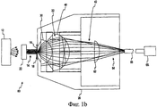
На Фиг.1а приведен вид сбоку системы для спектроскопии полного пропускания, используемой при определении концентрации анализируемого вещества в пробе жидкости в соответствии с первым вариантом осуществления настоящего изобретения.FIG. 1 a is a side view of a total transmission spectroscopy system used to determine the analyte concentration in a liquid sample in accordance with a first embodiment of the present invention.
На Фиг.1b приведен вид сбоку системы для спектроскопии полного пропускания, используемой при определении концентрации анализируемого вещества в пробе жидкости в соответствии с другим вариантом осуществления настоящего изобретения.Fig. 1b is a side view of a full transmission spectroscopy system used to determine the analyte concentration in a liquid sample in accordance with another embodiment of the present invention.
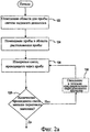
На Фиг.2а приведен алгоритм, описывающий работу системы согласно Фиг.1а в соответствии с первым вариантом осуществления настоящего изобретения, включающим датчик недостаточного наполнения для определения достаточности объема пробы.Fig. 2a is a flowchart describing the operation of the system according to Fig. 1a in accordance with a first embodiment of the present invention, including an underfill sensor for determining the sufficiency of a sample volume.
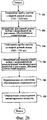
На Фиг.2b приведен алгоритм, описывающий работу системы согласно Фиг.1а в соответствии с другим вариантом осуществления настоящего изобретения, способный вносить корректировку из-за смещения пропускания, вызванного гематокритом пробы крови.FIG. 2b is a flowchart describing the operation of the system of FIG. 1a in accordance with another embodiment of the present invention, capable of adjusting due to transmission bias caused by hematocrit of a blood sample.
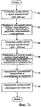
На Фиг.2с приведен алгоритм, описывающий работу системы согласно Фиг.1а в соответствии с другим вариантом осуществления настоящего изобретения, способный вносить корректировку из-за смещения пропускания, вызванного гемоглобином в пробе крови.Fig. 2c is an algorithm describing the operation of the system of Fig. 1a in accordance with another embodiment of the present invention, capable of adjusting due to transmission bias caused by hemoglobin in the blood sample.
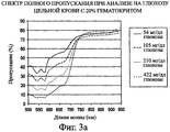
На Фиг.3а приведены спектры полного пропускания, полученные в тестах цельной крови с гематокритом 20% при уровнях глюкозы 54, 105, 210 и 422 мг/дл в видимой и ближней инфракрасной части от 500 нм до 940 нм.Figure 3a shows the complete transmission spectra obtained in tests of whole blood with a hematocrit of 20% at glucose levels of 54, 105, 210 and 422 mg / dl in the visible and near infrared from 500 nm to 940 nm.
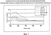
На Фиг.3b приведены спектры полного пропускания, полученные в тетах цельной крови с гематокритом 60% при уровнях глюкозы 59, 117, 239 и 475 мг/дл от видимой до ближней инфракрасной части спектра при длине волны от 500 нм до 940 нм.Figure 3b shows the complete transmission spectra obtained in whole blood tetas with a hematocrit of 60% at glucose levels of 59, 117, 239, and 475 mg / dl from visible to near infrared at a wavelength of 500 nm to 940 nm.
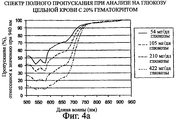
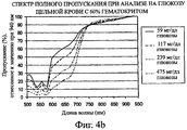
На Фиг.4а приведены спектры полного пропускания согласно Фиг.3а с коррекцией на рассеяние путем отнесения всех значений пропускания к пропусканию при 940 нм.Fig. 4a shows the total transmission spectra according to Fig. 3a with scattering correction by assigning all transmission values to transmission at 940 nm.
На Фиг.4b приведены спектры полного пропускания согласно Фиг.3b с коррекцией на рассеяние путем отнесения всех значений пропускания к пропусканию при 940 нм.Fig. 4b shows the total transmission spectra according to Fig. 3b with scattering correction by assigning all the transmission values to the transmission at 940 nm.
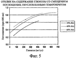
На Фиг.5 приведен график ответного сигнала концентрации глюкозы в цельной крови при значениях гематокрита 20%, 40% и 60%, измеренного методом полного пропускания (в единицах поглощения) при 680 нм с помощью считывающей головки, изображенной на Фиг.1а.Figure 5 shows a graph of the response signal of glucose concentration in whole blood with hematocrit values of 20%, 40% and 60%, measured by the method of full transmission (in absorption units) at 680 nm using the read head shown in Fig.1A.
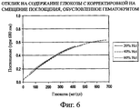
На Фиг.6 приведен график ответного сигнала согласно Фиг.5 с коррекцией на смещение пропускания (в единицах поглощения), вызванное различными значениями гематокрита в пробах крови.Figure 6 shows a graph of the response signal according to Figure 5 with correction for the transmission bias (in units of absorption) caused by different hematocrit values in blood samples.
На Фиг.7 приведены спектры полного пропускания (в единицах поглощения) реагента и цельной крови при уровнях глюкозы 0, 100 и 400 мг/дл, а также воды и реагента в видимой и ближней инфракрасной части спектра от 500 нм до 940 нм.Figure 7 shows the spectra of the total transmission (in absorption units) of the reagent and whole blood at glucose levels of 0, 100 and 400 mg / dl, as well as water and reagent in the visible and near infrared parts of the spectrum from 500 nm to 940 nm.
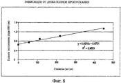
На Фиг.8 приведена линейная зависимость между полным пропусканием (в единицах поглощения) при 680 нм реагента, прореагировавшего с цельной кровью, при концентрациях глюкозы 0, 50, 100, 200 и 450 мг/дл.On Fig shows a linear relationship between the total transmission (in units of absorption) at 680 nm of the reagent reacted with whole blood, at glucose concentrations of 0, 50, 100, 200 and 450 mg / DL.
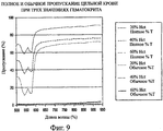
На Фиг.9 приведены спектры обычного и полного пропускания в диапазоне от 500 нм до 940 нм для цельной крови со значениями гематокрита 20%, 40% и 60%.Figure 9 shows the spectra of normal and complete transmission in the range from 500 nm to 940 nm for whole blood with hematocrit values of 20%, 40% and 60%.
Хотя в изобретение могут быть внесены различные модификации или использованы альтернативные формы, на чертежах в качестве примера изображены конкретные варианты осуществления изобретения, которые подробно здесь описаны. Однако следует понимать, что изобретение не должно ограничиваться конкретными описанными вариантами. Изобретение охватывает все модификации, эквиваленты и альтернативы, которые включены в объем настоящего изобретения.Although various modifications or alternative forms may be made to the invention, the drawings show by way of example specific embodiments of the invention, which are described in detail herein. However, it should be understood that the invention should not be limited to the specific options described. The invention covers all modifications, equivalents and alternatives that are included in the scope of the present invention.
Осуществление изобретенияThe implementation of the invention
Рассмотрим теперь чертежи и вначале обратимся к Фиг.1а, где изображена система 10 для спектроскопии пропускания методом полного пропускания для использования при определении концентрации анализируемого вещества в биологической пробе, такой как проба жидкости организма. Неограничивающие примеры анализируемых веществ, концентрация которых может определяться, включают: глюкозу, лактат, фруктозамин, холестерин и гемоглобин A1c. Эти анализируемые вещества могут содержаться в жидкостях организма, таких как кровь (включая плазму и сыворотку крови), слюна, моча и тканевая жидкость.We now consider the drawings and first turn to Figure 1a, which depicts a
Система 10 включает источник света 12. В соответствии с одним вариантом осуществления источник света является галогенной лампой, излучающей белый свет с длиной волны от приблизительно 300 нм до приблизительно 3200 нм. В соответствии с другим вариантом осуществления источник света 12 излучает два или более лучей монохроматического света благодаря светодиодам (СИД) с центральной длиной волны в диапазоне от приблизительно 400 нм до приблизительно 1000 нм. Свет от источника 12 поступает на коллимационную линзу 22, создающую по существу коллимированный луч света 14. Коллимированный луч света 14 освещает пробу 16, расположенную в области 18 размещения пробы считывающей головки 20.
В соответствии с одним вариантом осуществления проба содержит кровь и глюкозу, которая прореагировала с сухой реагентной системой, содержащей индикатор. В соответствии с одним вариантом осуществления индикаторный реагент глюкозы, который может использоваться, содержит глюкозодегидрогеназу, NAD (никотинамидадениндинуклеотид), диафоразу, тетразолиевый индикатор (WST-4)(2-бензотиазоил-3-(4-карбокси-2-метоксифенил)-5-[4-(2-сульфоэтилкарбамоил)фенил]-2Н-тетразолий) и полимеры. Следует понимать, что специалист в данной области техники сможет использовать различные ферменты (такие как PQQ-глюкозодегидрогсназа, глюкозооксидаза или лактатдегидрогеназа и т.д.), индикаторы и медиаторы, а также анализируемые вещества (такие как глюкоза, лактат и т.п.). Нет необходимости в том, чтобы в состав композиции реагента входил гемолизирующий агент, разрушающий красные кровяные тельца. Благодаря отсутствию необходимости в разрушении красных кровяных телец общее время исследования сокращается.In accordance with one embodiment, the sample contains blood and glucose that has reacted with a dry reagent system containing an indicator. According to one embodiment, a glucose indicator reagent that can be used comprises glucose dehydrogenase, NAD (nicotinamide adenine dinucleotide), diaphorase, tetrazolium indicator (WST-4) (2-benzothiazoyl-3- (4-carboxy-2-methoxyphenyl) -5- [4- (2-sulfoethylcarbamoyl) phenyl] -2H-tetrazolium) and polymers. It should be understood that a person skilled in the art will be able to use various enzymes (such as PQQ-glucose dehydrogenase, glucose oxidase or lactate dehydrogenase, etc.), indicators and mediators, as well as analytes (such as glucose, lactate, etc.) . It is not necessary that the composition of the reagent includes a hemolytic agent that destroys red blood cells. Due to the lack of need for the destruction of red blood cells, the total study time is reduced.
По существу коллимированный луч света 14 освещает пробу 16, и часть света проходит через пробу 16. Прошедший через пробу свет, включающий обычный и диффузно рассеянный свет, собирает первая линза 30 и вторая линза 40. В изображенных на чертежах вариантах осуществления первая и вторая линзы являются полусферическими. Следует понимать, что для приема пропущенного света могут использоваться и других типы линз, в т.ч. сферические и асферические.A substantially collimated beam of
В соответствии с альтернативным вариантом осуществления первая линза 30 собирает свет с углом приема приблизительно 72° или числовой апертурой (NA) приблизительно 0,951, однако угол приема имеет диапазон от 0° до 90° для сбора рассеянной части прошедшего света. Свет 32, выходящий из первой линзы 30, расходится под углом в диапазоне от приблизительно 15° до приблизительно 40°, а конкретнее приблизительно 20°. Вторая линза 40 уменьшает расхождение света 32, выходящего из первой линзы 30, до угла расхождения света 42 в диапазоне от 0° до приблизительно 10°, а конкретнее коллимирует от 0° приблизительно до 5°. Обычный и рассеянный свет, прошедший через пробу, не отклоняется или рассеивается первой и второй линзами 30, 40. Таким образом, пара линз 30, 40 собирает по существу весь свет, прошедший через пробу 16. Пара линз 30, 40 по существу коллимирует собранный свет и освещает датчик 50 почти с прямым углом падения. Расходящийся свет 42 имеет угол расхождения менее чем приблизительно 5°.According to an alternative embodiment, the
В соответствии с одним вариантом осуществления системы 10 для спектроскопии перед датчиком 50 может устанавливаться полосовой фильтр 52 или множество полосовых фильтров. Полосовой фильтр(ы) 52 обычно имеет центральную длину волны от приблизительно 400 до приблизительно 1000 нм и узкую полосу пропускания от приблизительно 5 до приблизительно 50 нм. Полосовой фильтр(ы) 52 обычно используется в случае, когда в качестве источника света 12 применяется источник белого света, такой как галогенная лампа. В альтернативном варианте полосовой фильтр может использоваться для изменения спектральной полосы светодиодного источника 12 или удаления составляющей окружающего света, которая не вносит вклад в прохождение света через пробу. Расходящийся свет 42 на полосовом фильтре(ах) 52 по существу коллимируется, потому что свет, проходящий через фильтр вне установленного для фильтра угла падения, окажется вне установленной полосы пропускания фильтра.In accordance with one embodiment of the
Первая и вторая линзы 30, 40 вместе повышают уровень светового сигнала, поступающего в датчик 50, потому что линзы 30, 40 собирают и направляют большую часть света, прошедшего через пробу 16 в датчик 50. Кроме того, уровень сигнала повышается путем направления в датчик 50 коллимированного луча света, который является по существу перпендикулярным поверхности датчика. Обычно угол расхождения коллимированного луча света меньше, чем приблизительно 5°. Нормальный угол падения к поверхности датчика 50 уменьшает потерю сигнала, вызванную френелевским отражением от поверхности датчика 50. Френелевское отражение является причиной значительных потерь света при углах падения более чем приблизительно 20°.The first and
Собранный датчиком 50 свет 42 затем сравнивают с эталонным измерением, представляющим собой значение при отсутствии пробы (наличии только воздуха) в оптическом пути, для определения процента пропускания пробы и концентрации анализируемого вещества в пробе.The light 42 collected by the
В соответствии с изображенным на чертеже вариантом осуществления системы 10 для спектроскопии датчик 50 и полосовой фильтр(ы) 52 по существу линейно совмещены со второй линзой 40. В соответствии с одним вариантом осуществления настоящего изобретения датчик 50 является кремниевым детектором. Однако следует понимать, что для приема пропущенного света могут использоваться и другие оптические датчики, в т.ч. другие типы фотодатчиков, такие как, например, датчики на сульфиде свинца или приборы с зарядовой связью (CCD). В других альтернативных вариантах осуществления датчик 50 и полосовой фильтр(ы) 52 не являются линейно совмещенными со второй линзой 40. Вместо этого световод или оптоволокно(а) (не изображены) имеет вход, по существу линейно совмещенный со второй линзой 40, и передает свет в датчик/фильтр, расположенный в другом месте, или в спектрограф. Спектроскопическая система 10 значительно улучшает уровень получаемого сигнала по сравнению с существующими системами для спектроскопии по методу полного пропускания, поскольку первая и вторая линзы 30, 40 передают свет непосредственно в датчик.According to the embodiment of the
В соответствии с одним вариантом осуществления настоящего изобретения длина пути через пробу 16 составляет от приблизительно 40 мкм до приблизительно 200 мкм, а проба имеет диаметр, приблизительно равный 1 мм. В соответствии с одним вариантом осуществления первая линза 30 представляет собой пластиковую микрополусферическую линзу диаметром около 4 мм. Вторая собирающая линза 40 представляет собой пластиковую микрополусферическую линзу диаметром около 8 мм. Соотношение диаметров первой и второй линз, как правило, составляет приблизительно 1:2. В соответствии с одним вариантом осуществления первая и вторая полусферические линзы 30, 40 изготовлены из акрила.In accordance with one embodiment of the present invention, the path length through the
Датчик 50 вырабатывает выходной сигнал, соответствующий поступающему в него свету. В соответствии с одним вариантом осуществления настоящего изобретения этот выходной сигнал отслеживается системой управления (не изображена) системы 10 для спектроскопии пропускания, которая содержит считывающую головку 20, для определения момента поступления пробы и заполнения области 18 размещения пробы в считывающей головке 20. В некоторых вариантах осуществления настоящего изобретения область 18 размещения пробы может быть частью капиллярного канала или является связанной с капиллярным каналом для заполнения области 18 размещения кюветы для пробы. В соответствии с одним вариантом осуществления область 18 размещения пробы выполнена из по существу оптически прозрачного материала.The
Теперь рассмотрим Фиг.1b, где изображена система 60 для спектроскопии пропускания, используемая для определения концентрации анализируемого вещества в пробе жидкости согласно другому варианту осуществления. Система 60 для спектроскопии пропускания содержит множество таких же компонентов, как и система, описанная выше со ссылкой на Фиг.1а. Кроме того, система 60 для спектроскопии пропускания включает линзу связи 62, собирающую расходящийся свет 42. Линза связи 62 дополнительно уменьшает расхождение света 42 с получением расходящегося света 64 перед поступлением его в оптический кабель 66. Как показано на Фиг.1b, оптический кабель 66 передает расходящийся свет в спектрограф 68. В другом варианте осуществления спектрограф может быть заменен на датчик (например, датчик 50) на Фиг.1а. В таком варианте осуществления может быть добавлен фильтр, такой как (например, фильтр(ы) 52) описанный выше со ссылкой на Фиг.1а.Now, consider FIG. 1b, which shows a
Для предотвращения или уменьшения ошибок, связанных с (а) недостаточным заполнением области 18 размещения пробы или (b) несогласованностью по времени, система управления отслеживает выходной сигнал датчика, который изменяется по мере заполнения области 18 для размещения пробы жидкостью организма (например, кровью). Временная последовательность, вариант осуществления которой описан со ссылкой на Фиг.2а, предусматривает достаточное время для протекания реакции между реагентом и содержащимся в пробе анализируемым веществом. Это улучшает общие результаты исследования, т.к. по существу точное согласование по времени может обеспечить более быстрое и надежное определение содержания анализируемого вещества.To prevent or reduce errors associated with (a) insufficient filling of the
Недостаточное заполнение имеет место, например, в случае получения слишком малой пробы для реакции с предварительно заданным количеством реагента, находящегося в области 18 размещения пробы. После приема прошедшего через пробу света, указывающего на заполнение области 18 размещения пробы, система управления "знает", что последующий выходной сигнал датчика 50 может использоваться для определения концентрации анализируемого вещества в пробе жидкости организма (например, пробе крови).Insufficient filling takes place, for example, in the case of obtaining too small a sample for reaction with a predetermined amount of reagent located in the
Кроме того, в соответствии с одним способом согласно настоящему изобретению после того, как датчик 50 обнаружит пробу или определенное количество пробы, система 10 инициирует временную последовательность, по окончании которой датчик 50 начинает принимать прошедший через пробу свет для проведения анализа. В соответствии с этим способом система 10 для спектроскопии пропускания, описанная со ссылкой на Фиг.2а, начинает работу с контроля области пробы с целью определения правильного момента времени, когда датчик 50 должен начать принимать прошедший через пробу свет. На этапе 122 пустая область 18 для размещения пробы (Фиг.1а) освещается светом от источника света 12. При отсутствии пробы в области размещения пробы уровень пропускания через систему 10 будет очень высоким (например, почти 100%). На этапе 124 проба поступает в область 18 размещения пробы. В соответствии с одним вариантом осуществления настоящего изобретения реагент, предназначенный для смешивания с пробой, в сухом виде заранее помещен в область 18 размещения пробы. В альтернативном варианте реагент может поступать вместе с пробой или после того, как проба поступит в область 18 размещения пробы.In addition, in accordance with one method according to the present invention, after the
На этапе 126 система 10 контролирует область 18 размещения пробы путем измерения света, прошедшего через пробу. На этапе 128 система 10 сравнивает количество прошедшего света, измеренное датчиком 50, с пороговым значением, хранящимся в памяти системы 10. Если измеренное количество превышает пороговое значение, система "приходит к выводу" о том, что на этапе 128 в область размещения пробы не поступило достаточное количество пробы, и на этапе 126 производится повторное измерение количества света, проходящего через область 18 размещения пробы. Система 10 может ожидать в течение предварительно заданного времени (например, 5 или 10 секунд) на этапе 130, прежде чем выполнит следующее измерение. Если измеренное количество света меньше порогового значения, которое хранится в памяти, система может начать выполнение анализа на этапе 150 (Фиг.2b) или этапе 102 (Фиг.2с).At
Хотя измерение прошедшего через пробу света на этапе 126 показано после поступления пробы в область размещения пробы, этот этап может выполняться непрерывно. Например, датчик может непрерывно детектировать свет, прошедший через область 18 размещения пробы, чтобы определить момент, когда следует начать анализ, описанный на Фиг.2с, с момента начала работы системы 10 до получения положительного результата на этапе 128. Кроме того, система 10 может генерировать сигнал ошибки, если после помещения пробы в предназначенную для нее область на этапе 124 не получен положительный результат (например, поступил слишком малый объем пробы после включения системы 10) согласно альтернативному варианту осуществления. Кроме того, желательно точно знать момент начала реакции, чтобы правильно определить время протекания реакции при проведении анализа. Точное время начала реакции может быть определено с помощью способа контроля согласно Фиг.2а.Although the measurement of the light transmitted through the sample in
Система для спектроскопии полного пропускания собирает по существу большее количество пропущенного через пробу света в видимом диапазоне (например, от приблизительно 400 до приблизительно 700 нм) и в ближней инфракрасной части спектра (например, от приблизительно 700 до приблизительно 1100 нм) по сравнению с системами для обычного пропускания для определения концентрации анализируемого вещества в пробе. Система 10 для спектроскопии пропускания обеспечивает лучшие характеристики по сравнению с существующими системами пропускания, поскольку большая часть собранного прошедшего через пробу света попадает в датчик. Такая улучшенная собирающая способность позволяет системе 10 собирать свет в обеих частях спектра, что используется для корректировки смещения или помех, вызванных рассеянием при разных значениях гематокрита (Фиг.2b) или наличием гемоглобина (Фиг.2с) и гематокрита (Фиг.2с) в жидкости организма, такой как проба цельной крови.A complete transmission spectroscopy system collects substantially more light transmitted through the sample in the visible range (e.g., from about 400 to about 700 nm) and in the near infrared (e.g., from about 700 to about 1100 nm) compared to systems for normal transmission to determine the concentration of the analyte in the sample. The
На Фиг.2b проиллюстрирован один способ применения системы 10 для спектроскопии пропускания для определения концентрации анализируемого вещества в жидкости организма (например, пробе цельной крови) и внесения корректировки на смещение пропускания, вызванное различными значениями гематокрита. Степень смещения является функцией гематокрита в пробе цельной крови. В соответствии с одним вариантом осуществления настоящего изобретения индикаторные реагенты подбираются таким образом, чтобы обеспечить хроматическую реакцию, указывающую на концентрацию анализируемого вещества в пробе крови в видимом диапазоне спектра при длине волны менее чем приблизительно 750 нм.FIG. 2b illustrates one method of using
В экспериментах с системой 10 для полного пропускания согласно Фиг.1а предполагали, что общий прошедший через пробу свет изменяется в зависимости от значения гематокрита при измерении в видимой и ближней ИК части спектра при длинах волн от приблизительно 400 до приблизительно 1100 нм. До сделанного авторами изобретения открытия считалось, что различие между значениями гематокрита не может быть обнаружено по общему прошедшему через пробу свету, имеющему длину волны от приблизительно 600 до приблизительно 1000 нм. Например, в статье в журнале Journal of Biomedical Optics, на которую приводилась ссылка в части "Уровень техники", указано на отсутствие различия между разными значениями гематокрита при длинах волн от приблизительно 600 до приблизительно 800 нм.In experiments with the
Однако гематокрит цельной крови влияет на спектральный отклик в видимом и ближнем ИК (инфракрасном) диапазоне (например, 400-1100 нм). Пропускание света зависит и является пропорциональным гематокриту ввиду различия в количестве рассеянного света, обусловленного числом красных кровяных телец. Смещение пропускания для того или иного уровня гематокрита в ближнем ИК диапазоне пропорционально значению гематокрита крови. Сравнение между Фиг.3а и 3b также показывает, что пропускание через кровь с гематокритом 20% на 30%Т выше, чем через кровь с гематокритом 60% в исследованном диапазоне от приблизительно 500 до приблизительно 940 нм. Однако пропускание света, измеренное в ближнем ИК диапазоне, не зависит от концентрации глюкозы, поскольку индикатор предназначен для обеспечения хроматической реакции в видимом диапазоне (например, при длине волны приблизительно 680 нм).However, whole blood hematocrit affects the spectral response in the visible and near infrared (infrared) range (for example, 400-1100 nm). The transmission of light depends and is proportional to the hematocrit due to the difference in the amount of scattered light due to the number of red blood cells. The transmission bias for a given level of hematocrit in the near infrared range is proportional to the value of the blood hematocrit. The comparison between Figs. 3a and 3b also shows that the transmission through blood with a hematocrit of 20% is 30% T higher than through blood with a hematocrit of 60% in the studied range from about 500 to about 940 nm. However, the transmission of light, measured in the near infrared range, does not depend on the glucose concentration, since the indicator is designed to provide a chromatic reaction in the visible range (for example, at a wavelength of approximately 680 nm).
В способе согласно Фиг.2b пробу цельной крови, вступившую в реакцию с реагентом, освещали светом с первой длиной волны (например, от приблизительно 750 до приблизительно 1100 нм) на этапе 150 для определения рассеянной части измеряемого света, зависящей от значения гематокрита пробы цельной крови. Свет (нормальную и рассеянную части) измеряли датчиком 50 на этапе 152, как описано выше со ссыпкой на Фиг.1а. Далее пробу освещали светом со второй диной волны (например, от приблизительно 600 до приблизительно 750 нм) на этапе 154 и нормальную и рассеянную части прошедшего света измеряли датчиком 50 на этапе 156 для определения рассеянной части света, зависящей от гематокрита, и хроматического отклика, зависящего от концентрации анализируемого вещества. На этапе 158 вносили коррекцию из-за зависящего от гематокрита рассеянного света путем расчета отношения данных измерений, полученных на этапах 156 и 152. Концентрацию анализируемого вещества в пробе цельной крови рассчитывали на этапе 160, используя скорректированное значение пропускания, определенное на этапе 158.In the method of FIG. 2b, the whole blood sample that has reacted with the reagent is illuminated with light at a first wavelength (e.g., from about 750 to about 1100 nm) in
В альтернативном варианте осуществления настоящего изобретения могут использоваться дополнительные алгоритмы корректировки, такие как, например, линейная регрессия или подбор многочлена, для определения соотношения между гематокритом и смещением или помехой, вызванной гематокритом при длинах волн, соответствующих области протекания реакции анализируемого вещества.In an alternative embodiment of the present invention, additional adjustment algorithms, such as, for example, linear regression or polynomial matching, can be used to determine the relationship between the hematocrit and the bias or interference caused by the hematocrit at wavelengths corresponding to the reaction region of the analyte.
Теперь рассмотрим Фиг.2с, где проиллюстрирован способ применения системы 10 для спектроскопии пропускания для определения концентрации анализируемого вещества, например, в пробе крови и внесения коррекции на смещение пропускания, вызванное присутствием гемоглобина. Степень смещения является функцией от уровня гемоглобина в пробе цельной крови и рассеяния, вызванного присутствием красных кровяных телец. Согласно способу реакцию пробы цельной крови и реагента освещают светом с первой длиной волны от приблизительно 400 до приблизительно 600 нм на этапе 102. Например, первая длина волны может равняться приблизительно 545 нм или приблизительно 577 нм. Свет (нормальную и рассеянную части) измеряли в единицах поглощения датчиком 50 на этапе 104, как описано выше со ссылкой на Фиг.1а.Now, consider FIG. 2c, which illustrates the use of
Как описано в разделе "Уровень техники", спектр оксигемоглобина имеет пики поглощения при приблизительно 545 нм и приблизительно 577 нм, и на него не влияют результаты реакции при этих длинах волн, поскольку результаты реакции должны измеряться, например, при второй длине волны, приблизительно равной 750 нм. Поглощение, измеренное при первой длине волны, включает составляющие гемоглобина и рассеяния, обусловленного гематокритом крови. В соответствии с одним вариантом осуществления настоящего изобретения индикаторные реагенты обеспечивают хроматическую реакцию, указывающую на концентрацию анализируемого вещества в пробе крови при второй длине волны, большей чем приблизительно 600 нм и меньшей чем приблизительно 1000 нм (видимый - ближний ИК диапазон). Пробу цельной крови и реагент освещают светом со второй длиной волны на этапе 106.As described in the Background Section, the oxyhemoglobin spectrum has absorption peaks at about 545 nm and about 577 nm, and is not affected by the reaction results at these wavelengths, since the reaction results should be measured, for example, at a second wavelength of approximately equal 750 nm. The absorption measured at the first wavelength includes the components of hemoglobin and scattering due to hematocrit of the blood. In accordance with one embodiment of the present invention, the indicator reagents provide a chromatic reaction indicating the concentration of the analyte in the blood sample at a second wavelength greater than about 600 nm and less than about 1000 nm (visible - near infrared). The whole blood sample and reagent are illuminated with light of a second wavelength in step 106.
Смещение, вызванное присутствием гемоглобина в пробе цельной крови, корректируют на этапе 110, используя результаты измерений на этапе 104, чтобы устранить смещение, влияющее на результаты измерений на этапе 108. Способ корректировки смещения зависит от связи между концентрацией гемоглобина и смещением результатов измерений 108, вызванным гемоглобином. Эта связь может быть линейной или нелинейной в зависимости от химической формулы вещества, используемого в реакции. Концентрацию анализируемого вещества в пробе определяют на этапе 112, используя скорректированное значение пропускания, определенное на этапе 110.The bias caused by the presence of hemoglobin in the whole blood sample is corrected at 110 using the measurements at 104 to eliminate the bias affecting the measurements at 108. The bias correction method depends on the relationship between the hemoglobin concentration and the bias at 108 caused by hemoglobin. This bond may be linear or non-linear depending on the chemical formula of the substance used in the reaction. The concentration of the analyte in the sample is determined in step 112, using the adjusted transmittance determined in
Аналогично описанному для Фиг.2b способ определения присутствия достаточной пробы и времени начала реакции, показанный на Фиг.2а, может также применяться в способе согласно Фиг.2с в другом процессе.Similar to that described for FIG. 2b, the method for determining the presence of a sufficient sample and the start time of the reaction shown in FIG. 2a can also be used in the method according to FIG. 2c in another process.
Как отмечалось выше, система 10 для спектроскопии полного пропускания согласно настоящему изобретению собирает по существу большее количество пропущенного через пробу света в видимом диапазоне и в ближней инфракрасной части спектра по сравнению с обычными системами пропускания для определения концентрации анализируемого вещества в пробе. В экспериментах с системой 10 для полного пропускания согласно Фиг.1а предполагают, что значение гематокрита или гемоглобин могут вызывать смещение пропускания при длине волны считывания, если индикаторный реагент вступает в хроматическую реакцию. Смещение пропускания, пропорциональное значению гематокрита при первой длине волны считывания (например, более 750 нм), может использоваться для корректировки значения при второй длине волны считывания (например, от приблизительно 600 до 750 нм), включающего смещение, вызванное значением гематокрита, и результаты хроматической реакции химического индикатора. В альтернативном варианте смещение пропускания, пропорциональное значению гемоглобина при первой длине волны считывания (например, менее 600 нм), может использоваться для корректировки значения при второй длине волны считывания (например, более 600 нм), где химический индикатор обеспечивает протекание хроматической реакции.As noted above, the total
ПримерыExamples
На Фиг.3а изображен один вариант осуществления настоящего изобретения (система 10 для спектроскопии пропускания) с измерением полного пропускания пробы цельной крови со значением гематокрита 20%, которая прореагировала с реагентами и имела различные концентрации глюкозы - 54, 104, 210 и 422 миллиграмм глюкозы на децилитр крови ("мг/дл глюкозы"). Систему 10 для спектроскопии пропускания в примерах будем называть "системой согласно изобретению". Белый свет от источника света 12 (Фиг.1а) пропускали через пробу. Уровень полного пропускания, измеренный при значениях от 500 нм до 940 нм, представили в виде графика на Фиг.3а для каждого значения концентрации глюкозы. Значение пропускания было более низким в диапазоне от 500 до 600 нм ввиду поглощения света гемоглобином. Потеря пропускания, вызванная рассеянием света красными кровяными тельцами (гематокрит), влияет на пропускание в диапазоне от 500 нм до 940 нм. Индикатор реакции с глюкозой поглощает в диапазоне от 500 до 750 нм, поэтому не наблюдается различия между концентрациями глюкозы до приблизительно 750 нм. Как показано на Фиг.3а, при длинах волн свыше приблизительно 750 нм уменьшение уровня полного пропускания было вызвано только потерей света от рассеяния красными кровяными тельцами, поэтому наблюдалось очень малое различие между пробами с разными уровнями глюкозы.Figure 3a shows one embodiment of the present invention (transmission spectroscopy system 10) with a total transmission measurement of a whole blood sample with a hematocrit value of 20%, which reacted with reagents and had different glucose concentrations of 54, 104, 210 and 422 milligrams of glucose per blood deciliter ("mg / dl glucose"). The
На Фиг.3b показано, что уровень полного пропускания снижается в измеряемом диапазоне от 500 нм до 940 нм, если значение гематокрита крови повышается до 60% в пробах с одинаковой концентрацией глюкозы, как показано на Фиг.3а (59, 117, 239 и 475 мг/дл глюкозы). На Фиг.3b также показано различие между уровнями концентрации глюкозы в диапазоне от 500 до приблизительно 750 нм. Как показано на Фиг.3а, уровень пропускания при приблизительно 750 нм составлял от 70 до 80% для крови со значением гематокрита приблизительно 20%. Как показано на Фиг.3b, уровень пропускания при значениях более 750 нм составлял от 40 до 50% для крови со значением гематокрита приблизительно 60%. Различия в спектрах для значений гематокрита 20% и 60% оказались пропорциональными на длинах волн от приблизительно 600 нм до 940 нм, т.е. больших тех, где наблюдается помеха, вызванная поглощением гемоглобина.Fig. 3b shows that the level of total transmission decreases in the measured range from 500 nm to 940 nm if the blood hematocrit increases to 60% in samples with the same glucose concentration, as shown in Fig. 3a (59, 117, 239 and 475 mg / dl glucose). 3b also shows the difference between glucose levels in the range of 500 to about 750 nm. As shown in FIG. 3a, the transmittance at approximately 750 nm was from 70 to 80% for blood with a hematocrit value of approximately 20%. As shown in FIG. 3b, the transmittance at values greater than 750 nm ranged from 40 to 50% for blood with a hematocrit value of approximately 60%. The differences in spectra for hematocrit values of 20% and 60% turned out to be proportional at wavelengths from approximately 600 nm to 940 nm, i.e. large ones where there is interference caused by the absorption of hemoglobin.
Однако наблюдалось малое различие между уровнями глюкозы при длинах волны свыше приблизительно 750 нм при любом значении гематокрита, как показано на Фиг.3а или 3b. Таким образом, спектр 750-940 нм может использоваться для определения значения гематокрита, обусловленного различием в количестве красных кровяных телец. Гематокрит не зависит от концентрации глюкозы или гемоглобина при этих длинах волн. Пропускание света, связанное с рассеянным светом (определенное с помощью света в ближнем ИК диапазоне) используется для внесения коррекции на помеху, вызванную гематокритом, перед определением концентрации глюкозы.However, there was a small difference between glucose levels at wavelengths above about 750 nm at any hematocrit value, as shown in FIGS. 3a or 3b. Thus, the spectrum of 750-940 nm can be used to determine the hematocrit value, due to the difference in the number of red blood cells. The hematocrit is independent of the concentration of glucose or hemoglobin at these wavelengths. Light transmission associated with scattered light (determined using near-infrared light) is used to correct for hematocrit-induced interference before determining glucose concentration.
На Фиг.4а, 4b приведены спектры полного пропускания согласно Фиг.3а, 3b соответственно с коррекцией на рассеяние путем отнесения всех значений пропускания к значением пропускания при 940 нм. После коррекции аналогичные значения пропускания для аналогичных значений концентрации глюкозы получены для проб крови с гематокритом 20% и 60% в диапазоне длин волн, в котором измеряют результаты реакции индикатора в тесте на глюкозу (от приблизительно 660 нм до 680 нм). Таким образом, длины волн ближнего ИК диапазона могут использоваться для корректировки различий, вызванных гематокритом пробы цельной крови. Возможность внесения корректировки на эту помеху повышает точность измерения концентрации глюкозы.Figures 4a, 4b show the total transmission spectra according to Figures 3a, 3b, respectively, with scattering correction by assigning all transmission values to the transmission value at 940 nm. After correction, similar transmittance values for similar glucose concentration values were obtained for blood samples with hematocrit of 20% and 60% in the wavelength range in which the results of the indicator response in the glucose test (from about 660 nm to 680 nm) are measured. Thus, near-IR wavelengths can be used to correct for differences caused by hematocrit from whole blood samples. The ability to correct for this interference improves the accuracy of glucose measurements.
На Фиг.5 раскрыт способ применения значения гематокрита для корректировки измерений концентрации глюкозы в соответствии с одним вариантом осуществления. Отклик полного пропускания показан для цельной крови с гематокритом ("Hct") 20%, 40% и 60% на Фиг.5, где пропускание (в единицах поглощения) для видимого света с длиной волны приблизительно 680 нм нанесено на график как функция от концентрации глюкозы. Аналогичные отклики наблюдали для каждого значения гематокрита, однако существует смещение или помеха, вызванная гематокритом, как видно из различия между тремя значениями гематокрита на графиках на Фиг.5.FIG. 5 discloses a method for applying a hematocrit value to adjust glucose concentration measurements in accordance with one embodiment. The full transmittance response is shown for whole blood with a hematocrit ("Hct") of 20%, 40% and 60% in Figure 5, where the transmittance (in absorption units) for visible light with a wavelength of approximately 680 nm is plotted as a function of concentration glucose. Similar responses were observed for each hematocrit value, however there is a bias or interference caused by the hematocrit, as can be seen from the difference between the three hematocrit values in the graphs in FIG. 5.
На Фиг.6 приведены те же данные, однако вызванное гематокритом смещение скорректировано с помощью отношения значения для видимого света при длине волны приблизительно 680 нм, и значения для ближнего ИК спектра от приблизительно 750 до приблизительно 940 нм света, прошедшего через пробу крови. Корректировка осуществляется путем деления уровня пропускания видимого света при 680 нм (Фиг.5) на уровень пропускания света ближнего ИК диапазона при 940 нм для пробы, как было описано выше. Проще говоря, вызванное гематокритом смещение, обусловленное различием в рассеянии, "вычли", и Фиг.6 отображает отклик, не зависящий от значений гематокрита.Fig. 6 shows the same data, however, the hematocrit-induced offset was corrected by using the ratio of the value for visible light at a wavelength of approximately 680 nm and the value for the near infrared spectrum from approximately 750 to approximately 940 nm of light transmitted through the blood sample. The correction is carried out by dividing the transmittance of visible light at 680 nm (Figure 5) by the transmittance of light near infrared at 940 nm for the sample, as described above. Simply put, the hematocrit-induced bias due to the difference in scattering is “subtracted”, and FIG. 6 displays the response independent of the hematocrit values.
На Фиг.7 система согласно изобретению использовалась для измерения концентрации глюкозы в нескольких пробах цельной крови. Высушенные реагенты восстанавливали пробами крови с концентрацией глюкозы 0,100 и 400 мг/дл. Кроме того, использовали пробу крови с 0 мг/дл без реагента и высушенные реагенты, восстановленные водой. Проба крови без химических веществ давала спектральную составляющую крови, а вода и реагенты - спектральную составляющую реагента. Общие уровни поглощения реакций регистрировали спектрографом каждые 5 секунд в течение общего времени исследования 60 секунд. Реакция протекала в течение 15-30 секунд. Это значительно быстрее, чем при использовании обычной спектроскопии пропускания, где реакция длится от приблизительно 60 до 90 секунд, которые необходимы для разрушения красных кровяных телец. Как показано на Фиг.7, наблюдалось различие между уровнями пропускания света для разных уровней концентрации глюкозы - 0, 100 и 400 мг/дл - при длинах волн видимого диапазона (например, от приблизительно 660 до приблизительно 680 нм), которые используются для определения уровней концентрации глюкозы.7, a system according to the invention was used to measure glucose concentration in several samples of whole blood. Dried reagents were reconstituted with blood samples with glucose concentrations of 0.100 and 400 mg / dl. In addition, a blood sample with 0 mg / dl without reagent and dried reagents reconstituted with water were used. A blood sample without chemicals gave the spectral component of the blood, and water and reagents gave the spectral component of the reagent. Total reaction absorption levels were recorded with a spectrograph every 5 seconds for a total study time of 60 seconds. The reaction proceeded for 15-30 seconds. This is significantly faster than using conventional transmission spectroscopy, where the reaction lasts from about 60 to 90 seconds, which are necessary for the destruction of red blood cells. As shown in FIG. 7, there was a difference between the light transmittance levels for different glucose concentrations — 0, 100 and 400 mg / dl — at visible wavelengths (eg, from about 660 to about 680 nm) that are used to determine the levels glucose concentration.
На Фиг.8 система согласно изобретению использовалась для проведения измерений при пропускании через несколько проб крови с известной концентрацией глюкозы света с длиной волны 680 нм. На Фиг.8 уровни полного пропускания (в единицах поглощения) нанесены на график как функция от известных уровней концентрации глюкозы в цельной крови. Данные на Фиг.8 анализировали методом линейной регрессии. Как видно на графике, имеется по существу линейная зависимость между количеством прошедшего через пробу света и концентрацией глюкозы. Коэффициент линейной зависимости 0,985 (почти 1,000) указывает на отличную корреляцию между уровнем поглощения и концентрацией глюкозы при использовании системы и способа согласно настоящему изобретению.In Fig. 8, the system according to the invention was used to measure when passing through several blood samples with a known light glucose concentration of 680 nm. In Fig. 8, the levels of total transmission (in units of absorption) are plotted as a function of known levels of glucose concentration in whole blood. The data in FIG. 8 was analyzed by linear regression. As can be seen in the graph, there is an essentially linear relationship between the amount of light transmitted through the sample and the glucose concentration. A linear coefficient of 0.985 (almost 1,000) indicates an excellent correlation between the absorption level and glucose concentration when using the system and method according to the present invention.
Рассмотрим теперь Фиг.9, где показаны уровни полного пропускания для проб цельной крови с гематокритом 20%, 40% и 60% соответственно, полученные с использованием системы согласно изобретению. Свет с длинами волн от приблизительно 500 до приблизительно 940 нм пропускали через пробы цельной крови. Длина пути через область размещения пробы составляла 42 мкм. На Фиг.9 полученные уровни пропускания для трех проб наносили на график как функцию от длины волны пропущенного света.Let us now consider Figure 9, which shows the levels of complete transmission for samples of whole blood with a hematocrit of 20%, 40% and 60%, respectively, obtained using the system according to the invention. Light with wavelengths from about 500 to about 940 nm was passed through whole blood samples. The path length through the sample placement region was 42 μm. In Fig. 9, the obtained transmittance levels for three samples were plotted as a function of the wavelength of transmitted light.
Для иллюстрации преимуществ этого варианта осуществления системы согласно изобретению по сравнению с обычной системой пропускания ("обычная система") уровни пропускания для трех проб цельной крови со значениями гематокрита 20%, 40% и 60% показаны для обоих способов на Фиг.9. Обычная система пропускания обозначена на Фиг.9 как «%Hct, обычная %Т», а система полного пропускания обозначена на Фиг.9 как «%Hct, полная %Т». В обеих системах пропускания, которые использовались в данном примере, три пробы освещали по существу коллимированным светом с длинами волн от приблизительно 500 до приблизительно 940 нм. По существу коллимированный, а не рассеянный свет, прошедший через пробы, собирали с помощью обычной системы пропускания. Система согласно изобретению использовалась для приема и обычного, и рассеянного света. В обеих системах длина пути через пробу составляла приблизительно 42 мкм.To illustrate the advantages of this embodiment of the system of the invention compared to a conventional transmission system (“conventional system”), transmission levels for three whole blood samples with hematocrit values of 20%, 40% and 60% are shown for both methods in FIG. 9. The conventional transmission system is indicated in FIG. 9 as “% Hct, normal% T”, and the complete transmission system is indicated in FIG. 9 as “% Hct, full% T”. In both transmission systems used in this example, three samples were illuminated with essentially collimated light with wavelengths from about 500 to about 940 nm. Essentially collimated rather than scattered light that passed through the samples was collected using a conventional transmission system. The system according to the invention was used to receive both ordinary and stray light. In both systems, the path length through the sample was approximately 42 μm.
При сравнении двух наборов данных на Фиг.9 можно видеть, что уровень обычного пропускания света через три образца составляет менее 2% при длинах волн свыше 500 нм. Уровни пропускания света, полученные для трех проб с помощью системы согласно изобретению превышали 10%Т при длинах волн свыше 500 нм. Хорошее различие между уровнями пропускания для трех проб, полученными с помощью системы согласно изобретению, наблюдалось при длинах волн более 500 нм. На Фиг.9 также видно, что в данных, полученных с помощью системы согласно изобретению, наблюдаются "провалы" уровня пропускания в диапазоне от приблизительно 542 до приблизительно 577 нм. Эти две длины волны соответствуют известным пикам поглощения оксигемоглобина. Таким образом, как показано на Фиг.9, описанный вариант осуществления системы согласно изобретению обеспечил прохождение большего количества света при длинах волн от 500 до 940 нм по сравнению с существующей системой спектроскопии пропускания несмотря на поглощение гемоглобином или рассеяние света, обусловленное гематокритом, при этих длинах волн.When comparing the two data sets in FIG. 9, it can be seen that the level of normal light transmission through three samples is less than 2% at wavelengths above 500 nm. The light transmission levels obtained for the three samples using the system according to the invention exceeded 10% T at wavelengths above 500 nm. A good difference between the transmittance levels for the three samples obtained using the system according to the invention was observed at wavelengths greater than 500 nm. Figure 9 also shows that in the data obtained using the system according to the invention, there are "dips" in the transmission level in the range from about 542 to about 577 nm. These two wavelengths correspond to the known absorption peaks of oxyhemoglobin. Thus, as shown in FIG. 9, the described embodiment of the system according to the invention allowed more light to pass through at wavelengths from 500 to 940 nm compared to the existing transmission spectroscopy system despite the absorption by hemoglobin or light scattering due to hematocrit at these lengths waves.
В другом варианте осуществления смещение, вызванное рассеянием вследствие неидеальной структуры области размещения пробы или наличия небольшого количества загрязнителей в пробе, можно скорректировать аналогично способу, описанному для различных значений гематокрита со ссылкой на Фиг.2b. Отношение значений, полученных при считывании на двух длинах волн, может использоваться для внесения поправки на загрязнение области размещения пробы, например, отпечатком пальца, наличие дефектов изготовления области для размещения пробы или наличие царапин на окошках. Такая коррекция значительно повышает точность исследования по сравнению с использованием одной длины волны.In another embodiment, the bias caused by scattering due to imperfect structure of the sample location region or the presence of a small amount of contaminants in the sample can be corrected similarly to the method described for different hematocrit values with reference to FIG. 2b. The ratio of the values obtained when reading at two wavelengths can be used to correct for contamination of the sample location area, for example, a fingerprint, manufacturing defects in the area for placing the sample, or scratches on the windows. Such correction significantly increases the accuracy of the study compared to using a single wavelength.
В другом варианте осуществления применяется диапазон длин волн, где наблюдаются изменения уровней поглощения в зависимости от концентрации глюкозы, вне диапазона длин волн, в котором гемоглобин поглощает свет (например, длины волн более чем приблизительно 600 нм). Также может использоваться индикаторный реагент, действующий при длинах волн более чем приблизительно 600 нм, чтобы избежать взаимного влияния между пиками поглощения гемоглобина и индикаторного реагента. На Фиг.9 также видно, что в данных, полученных с помощью системы согласно изобретению, наблюдаются "провалы" уровня пропускания при приблизительно 530 и приблизительно 570 нм. Эти две длины волны соответствуют известным пикам поглощения оксигемоглобина. Значение поглощения при приблизительно 542 нм или 577 нм может использоваться для определения концентрации гемоглобина после вычитания значения рассеяния, вызванного красными кровяными тельцами и измеренного в ближнем ИК диапазоне (от приблизительно 750 до 1100 нм). В этом случае поглощение видимого света с длиной волны приблизительно 542 нм не будет изменяться или зависеть от концентрации глюкозы.In another embodiment, a wavelength range is used where changes in absorption levels are observed depending on glucose concentration, outside the wavelength range in which hemoglobin absorbs light (for example, wavelengths greater than about 600 nm). An indicator reagent operating at wavelengths greater than about 600 nm may also be used to avoid the mutual influence between the absorption peaks of hemoglobin and the indicator reagent. Figure 9 also shows that in the data obtained using the system according to the invention, there are "dips" in the transmission level at approximately 530 and approximately 570 nm. These two wavelengths correspond to the known absorption peaks of oxyhemoglobin. The absorbance value at approximately 542 nm or 577 nm can be used to determine the hemoglobin concentration after subtracting the scattering value caused by red blood cells and measured in the near infrared range (from about 750 to 1100 nm). In this case, the absorption of visible light with a wavelength of approximately 542 nm will not change or depend on the concentration of glucose.
Как было отмечено в разделе "Уровень техники", уменьшение длины пути может увеличить уровень пропускания при использовании обычного способа пропускания. Специалистам в данной области техники хорошо известно, что механические допуски, влияющие на длину пути, вызывают пропорциональную ошибку в определении концентрации глюкозы. Поэтому более длинный путь, используемый в системе спектроскопии полного пропускания согласно настоящему изобретению, уменьшает ошибку определения концентрации глюкозы.As noted in the Background section, decreasing the path length can increase the transmission rate using the conventional transmission method. Those skilled in the art are well aware that mechanical tolerances affecting path lengths cause a proportional error in determining glucose concentration. Therefore, the longer path used in the full transmission spectroscopy system of the present invention reduces the error in determining glucose concentration.
АЛЬТЕРНАТИВНЫЙ ВАРИАНТ ОСУЩЕСТВЛЕНИЯ АALTERNATIVE EMBODIMENT
Система для спектроскопии полного пропускания для применения при определении концентрации анализируемого вещества в пробе жидкости, включающая:A system for complete transmission spectroscopy for use in determining the concentration of an analyte in a liquid sample, including:
область размещения пробы для помещения анализируемой пробы, выполненную из по существу оптически прозрачного материала;the area of placement of the sample to place the analyzed sample, made of essentially optically transparent material;
источник света;Light source;
коллимирующую линзу, принимающую свет от источника света и направляющую по существу коллимированный луч света на область размещения пробы;a collimating lens receiving light from a light source and directing a substantially collimated light beam to the sample location area;
первую линзу, принимающую обычный и рассеянный свет, прошедший через пробу, с первым углом расхождения, причем первая линза принимает свет с первым углом приема, а из нее выходит свет со вторым углом расхождения, меньшим чем первый угол расхождения;a first lens receiving ordinary and scattered light passing through the sample with a first angle of divergence, the first lens receiving light with a first angle of reception, and light coming out of it with a second angle of divergence smaller than the first angle of divergence;
вторую линзу, принимающую свет от первой линзы, так что из нее выходит по существу коллимированный луч света;a second lens receiving light from the first lens so that a substantially collimated light beam exits from it;
датчик, измеряющий свет, поступающий из второй линзы.a sensor measuring light coming from a second lens.
АЛЬТЕРНАТИВНЫЙ ВАРИАНТ ОСУЩЕСТВЛЕНИЯ ВALTERNATIVE EMBODIMENT
Система согласно альтернативному варианту осуществления А, в которой проба жидкости является кровью, а область размещения пробы содержит высушенный реагент при отсутствии гемолизирующего вещества, причем указанный реагент обеспечивает протекание хроматической реакции при восстановлении его кровью.The system according to alternative embodiment A, in which the liquid sample is blood and the sample location region contains a dried reagent in the absence of a hemolytic substance, said reagent providing a chromatic reaction when restored by blood.
АЛЬТЕРНАТИВНЫЙ ВАРИАНТ ОСУЩЕСТВЛЕНИЯ СALTERNATIVE OPTION FOR CARRYING OUT
Система согласно альтернативному варианту осуществления А, в которой первая и вторая линзы являются полусферическими.A system according to alternative embodiment A, wherein the first and second lenses are hemispherical.
АЛЬТЕРНАТИВНЫЙ ВАРИАНТ ОСУЩЕСТВЛЕНИЯ DALTERNATIVE EMBODIMENT
Система согласно альтернативному варианту осуществления А, в которой первая линза имеет первый угол приема от приблизительно 0° до приблизительно 90°.A system according to alternative embodiment A, wherein the first lens has a first reception angle of from about 0 ° to about 90 °.
АЛЬТЕРНАТИВНЫЙ ВАРИАНТ ОСУЩЕСТВЛЕНИЯ ЕALTERNATIVE EMBODIMENT
Система согласно альтернативному варианту осуществления А, в которой первая линза имеет первый угол приема, превышающий 70°.The system according to alternative embodiment A, in which the first lens has a first reception angle exceeding 70 °.
АЛЬТЕРНАТИВНЫЙ ВАРИАНТ ОСУЩЕСТВЛЕНИЯ FALTERNATIVE EMBODIMENT
Система согласно альтернативному варианту осуществления А, в которой второй угол расхождения первой линзы составляет от приблизительно 15° до приблизительно 40°.A system according to alternative embodiment A, wherein the second divergence angle of the first lens is from about 15 ° to about 40 °.
АЛЬТЕРНАТИВНЫЙ ВАРИАНТ ОСУЩЕСТВЛЕНИЯ GALTERNATIVE EMBODIMENT
Система согласно альтернативному варианту осуществления А, в которой отношение диаметра первой линзы к диаметру второй линзы составляет от приблизительно 1:2.A system according to alternative embodiment A, wherein the ratio of the diameter of the first lens to the diameter of the second lens is from about 1: 2.
АЛЬТЕРНАТИВНЫЙ ВАРИАНТ ОСУЩЕСТВЛЕНИЯ НALTERNATIVE EMBODIMENT
Система согласно альтернативному варианту осуществления А, в которой источник света испускает свет с длиной волны от приблизительно 500 до приблизительно 940 нм.A system according to alternative embodiment A, wherein the light source emits light with a wavelength of from about 500 to about 940 nm.
АЛЬТЕРНАТИВНЫЙ ВАРИАНТ ОСУЩЕСТВЛЕНИЯ IALTERNATIVE EMBODIMENT I
Система согласно альтернативному варианту осуществления А, в которой источник света содержит светодиод.A system according to alternative embodiment A, wherein the light source comprises an LED.
АЛЬТЕРНАТИВНЫЙ ВАРИАНТ ОСУЩЕСТВЛЕНИЯ JALTERNATIVE EMBODIMENT
Система согласно альтернативному варианту осуществления А, в которой источник света испускает монохроматический свет.A system according to alternative embodiment A, wherein the light source emits monochromatic light.
АЛЬТЕРНАТИВНЫЙ ВАРИАНТ ОСУЩЕСТВЛЕНИЯ KALTERNATIVE EMBODIMENT K
Система согласно альтернативному варианту осуществления А, в которой источник света испускает белый свет.A system according to alternative embodiment A, wherein the light source emits white light.
АЛЬТЕРНАТИВНЫЙ ВАРИАНТ ОСУЩЕСТВЛЕНИЯ LALTERNATIVE EMBODIMENT
Система согласно альтернативному варианту осуществления А, в которой датчик содержит кремниевый детектор.A system according to alternative embodiment A, in which the sensor comprises a silicon detector.
АЛЬТЕРНАТИВНЫЙ ВАРИАНТ ОСУЩЕСТВЛЕНИЯ МALTERNATIVE EMBODIMENT
Система согласно альтернативному варианту осуществления А, в которой жидкость является кровью.A system according to alternative embodiment A, wherein the liquid is blood.
АЛЬТЕРНАТИВНЫЙ ВАРИАНТ ОСУЩЕСТВЛЕНИЯ NALTERNATIVE EMBODIMENT FOR N
Система согласно альтернативному варианту осуществления А, в которой анализируемым веществом является глюкоза.A system according to alternative embodiment A, wherein the analyte is glucose.
АЛЬТЕРНАТИВНЫЙ ВАРИАНТ ОСУЩЕСТВЛЕНИЯ ОALTERNATIVE EMBODIMENT
Система согласно альтернативному варианту осуществления А, в которой первая линза принимает по существу весь обычный и рассеянный свет, прошедший через пробу.A system according to alternative embodiment A, wherein the first lens receives substantially all of the ordinary and scattered light that has passed through the sample.
АЛЬТЕРНАТИВНЫЙ ВАРИАНТ ОСУЩЕСТВЛЕНИЯ РALTERNATIVE OPTION FOR CARRYING OUT P
Система согласно альтернативному варианту осуществления А, дополнительно включающая фильтр, предназначенный для выделения определенной длины волны из света, испускаемого источником.The system according to alternative embodiment A, further comprising a filter for extracting a specific wavelength from the light emitted by the source.
АЛЬТЕРНАТИВНЫЙ ВАРИАНТ ОСУЩЕСТВЛЕНИЯ QALTERNATIVE EMBODIMENT OF Q
Система согласно альтернативному варианту осуществления А, дополнительно включающая линзу связи и оптоволоконный кабель для передачи света от второй линзы в датчик.The system according to alternative embodiment A, further comprising a communication lens and an optical fiber cable for transmitting light from the second lens to the sensor.
АЛЬТЕРНАТИВНЫЙ ВАРИАНТ ОСУЩЕСТВЛЕНИЯ RALTERNATIVE EMBODIMENT FOR R
Система согласно альтернативному варианту осуществления А, в которой из второй линзы выходит по существу коллимированный луч света, имеющий угол расхождения менее чем приблизительно пять градусов.A system according to alternative embodiment A, wherein a substantially collimated light beam having a divergence angle of less than about five degrees comes out of the second lens.
АЛЬТЕРНАТИВНЫЙ СПОСОБ SALTERNATIVE METHOD S
Способ для определения концентрации анализируемого вещества в пробе жидкости с помощью системы для спектроскопии полного пропускания, включающий этапы:A method for determining the concentration of an analyte in a liquid sample using a complete transmission spectroscopy system, comprising the steps of:
помещения анализируемой пробы в область размещения пробы системы для спектроскопии полного пропускания;placing the analyzed sample in the area of the sample placement of the system for full transmission spectroscopy;
испускания луча света источником света;emission of a ray of light by a light source;
по существу коллимирования луча света, выходящего из источника света;essentially collimating a beam of light exiting from a light source;
освещения пробы по существу коллимированным лучом света от источника света;illuminating the sample with a substantially collimated light beam from the light source;
собирания обычного и рассеянного света, прошедшего через пробу, первой линзой;collecting the ordinary and scattered light passing through the sample, the first lens;
уменьшения угла расхождения прошедшего света с помощью первой линзы;reducing the angle of divergence of transmitted light using the first lens;
приема света с уменьшенным углом расхождения второй линзой;receiving light with a reduced angle of divergence of the second lens;
по существу коллимирования принятого света с помощью второй линзы;essentially collimating the received light with a second lens;
измерения по существу коллимированного света, поступающего из второй линзы, датчиком.measuring essentially collimated light coming from the second lens by a sensor.
АЛЬТЕРНАТИВНЫЙ СПОСОБ ТALTERNATIVE METHOD T
Способ согласно альтернативному способу S, в котором первая и вторая линзы являются полусферическими.The method according to alternative method S, in which the first and second lenses are hemispherical.
АЛЬТЕРНАТИВНЫЙ СПОСОБ UALTERNATIVE METHOD U
Способ согласно альтернативному способу S, в котором уменьшенный первой линзой угол расхождения составляет от приблизительно 15° до приблизительно 40°.The method according to alternative method S, in which the divergence angle reduced by the first lens is from about 15 ° to about 40 °.
АЛЬТЕРНАТИВНЫЙ СПОСОБ VALTERNATIVE METHOD V
Способ согласно альтернативному способу S, в котором угол расхождения света, принятого второй линзой, уменьшается до менее чем приблизительно 5°.The method according to alternative method S, in which the angle of divergence of the light received by the second lens is reduced to less than about 5 °.
АЛЬТЕРНАТИВНЫЙ СПОСОБ WALTERNATIVE METHOD W
Способ согласно альтернативному способу S, в котором источник света испускает свет с длиной волны от приблизительно 500 до приблизительно 940 нм.The method according to alternative method S, in which the light source emits light with a wavelength of from about 500 to about 940 nm.
АЛЬТЕРНАТИВНЫЙ СПОСОБ ХALTERNATIVE METHOD X
Способ согласно альтернативному способу S, в котором источник света испускает монохроматический свет.The method according to alternative method S, in which the light source emits monochromatic light.
АЛЬТЕРНАТИВНЫЙ СПОСОБ YALTERNATIVE METHOD Y
Способ согласно альтернативному способу S, в котором источник света испускает белый свет.The method according to alternative method S, in which the light source emits white light.
АЛЬТЕРНАТИВНЫЙ СПОСОБ ZALTERNATIVE METHOD Z
Способ согласно альтернативному способу S, дополнительно включающий осуществляемый с помощью датчика контроль поступления в область размещения пробы предварительно определенного количества анализируемой пробы.The method according to alternative method S, further comprising, by means of a sensor, monitoring the receipt of a predetermined amount of the analyzed sample in the sample location area.
АЛЬТЕРНАТИВНЫЙ СПОСОБ ААALTERNATIVE METHOD AA
Способ согласно альтернативному способу Z, дополнительно включающий определение концентрации анализируемого вещества в пробе жидкости после обнаружения поступления в область размещения пробы предварительно заданного количества анализируемой пробы.The method according to alternative method Z, further comprising determining the concentration of the analyte in the liquid sample after detecting the receipt of a predetermined amount of the analyzed sample in the sample location area.
АЛЬТЕРНАТИВНЫЙ СПОСОБ ВВALTERNATIVE METHOD OF BB
Способ измерения света, прошедшего через пробу жидкости с помощью системы для спектроскопии полного пропускания, включающий этапы:A method for measuring light transmitted through a liquid sample using a system for full transmission spectroscopy, comprising the steps of:
освещения пробы по существу коллимированным лучом света;illuminating the sample with a substantially collimated beam of light;
собирания обычного и рассеянного света, прошедшего через пробу, первой линзой;collecting the ordinary and scattered light passing through the sample, the first lens;
уменьшения угла расхождения прошедшего света первой линзой;reducing the angle of divergence of transmitted light by the first lens;
по существу коллимирования пропущенного света второй линзой после уменьшения угла расхождения;essentially collimating the transmitted light with a second lens after reducing the angle of divergence;
измерения по существу коллимированного света, прошедшего через пробу света, датчиком.measuring essentially collimated light transmitted through a light sample by a sensor.
АЛЬТЕРНАТИВНЫЙ СПОСОБ ССALTERNATIVE METHOD OF SS
Способ согласно альтернативному способу ВВ, в котором первая и вторая линзы являются полусферическими.The method according to an alternative method BB, in which the first and second lenses are hemispherical.
АЛЬТЕРНАТИВНЫЙ СПОСОБ DDALTERNATIVE METHOD DD
Способ согласно альтернативному способу ВВ, в котором уменьшенный первой линзой угол расхождения составляет от приблизительно 15° до приблизительно 40°.The method according to an alternative method BB, in which the angle of divergence reduced by the first lens is from about 15 ° to about 40 °.
АЛЬТЕРНАТИВНЫЙ СПОСОБ ЕЕALTERNATIVE METHOD
Способ согласно альтернативному способу ВВ, в котором по существу коллимирование принятого света второй линзой включает уменьшение угла расхождения света, принятого второй линзой, до менее чем приблизительно 5°.The method according to an alternative method BB, in which essentially collimating the received light by the second lens includes reducing the angle of divergence of the light received by the second lens to less than about 5 °.
АЛЬТЕРНАТИВНЫЙ СПОСОБ FFALTERNATIVE METHOD FF
Способ согласно альтернативному способу ВВ, дополнительно включающий обеспечение наличия источника света, испускающего луч света с длиной волны от приблизительно 500 до приблизительно 940 нм.The method according to an alternative method of BB, further comprising providing a light source emitting a beam of light with a wavelength of from about 500 to about 940 nm.
АЛЬТЕРНАТИВНЫЙ СПОСОБ GGALTERNATIVE METHOD GG
Способ согласно альтернативному способу ВВ, дополнительно включающий обеспечение источника света, испускающего луч монохроматического света.The method according to an alternative method of BB, further comprising providing a light source emitting a beam of monochromatic light.
АЛЬТЕРНАТИВНЫЙ СПОСОБ ННALTERNATIVE METHOD OF NN
Способ согласно альтернативному способу ВВ, дополнительно включающий обеспечение источника света, испускающего луч белого света.The method according to an alternative method of BB, further comprising providing a light source emitting a beam of white light.
АЛЬТЕРНАТИВНЫЙ СПОСОБ IIALTERNATIVE METHOD II
Способ согласно альтернативному способу ВВ, в котором прием нормального и рассеянного света включает прием по существу всего нормального и рассеянного света, прошедшего через пробу.The method according to an alternative method BB, in which the reception of normal and scattered light includes receiving essentially all of the normal and scattered light passing through the sample.
АЛЬТЕРНАТИВНЫЙ СПОСОБ JJALTERNATIVE METHOD JJ
Способ согласно альтернативному способу ВВ, дополнительно включающий осуществляемый с помощью датчика контроль поступления в область размещения пробы предварительно заданного количества анализируемой пробы.The method according to an alternative method of explosives, further comprising, by means of a sensor, controlling the receipt of a predetermined amount of the analyzed sample in the sample location area.
АЛЬТЕРНАТИВНЫЙ СПОСОБ KKALTERNATIVE METHOD KK
Способ согласно альтернативному способу JJ, дополнительно включающий определение концентрации анализируемого вещества в пробе жидкости после обнаружения поступления в область размещения пробы предварительно заданного количества анализируемой пробы.The method according to an alternative method JJ, further comprising determining the concentration of the analyte in the liquid sample after detecting the receipt of a predetermined amount of the analyzed sample in the sample location area.
АЛЬТЕРНАТИВНЫЙ СПОСОБ LLALTERNATIVE METHOD LL
Способ определения концентрации анализируемого вещества в пробе жидкости с помощью системы для спектроскопии полного пропускания, которая включает первую линзу, принимающую обычный и рассеянный свет, прошедший через пробу, вторую линзу, в которую поступает свет от первой линзы и из которой выходит по существу коллимированный луч света, источник света и область расположения пробы, включающий этапы:A method for determining the concentration of an analyte in a liquid sample using a complete transmission spectroscopy system that includes a first lens receiving ordinary and scattered light passing through the sample, a second lens into which light comes from the first lens and from which a substantially collimated light beam exits , the light source and the area of the sample, including the steps:
взаимодействия пробы с реагентом, обеспечивающим протекание хроматической реакции в области размещения пробы в системе;the interaction of the sample with a reagent, ensuring the occurrence of a chromatic reaction in the area of placement of the sample in the system;
освещения пробы по существу коллимированным лучом ближней инфракрасной части спектра, выходящим из источника света в системе;lighting the sample with a substantially collimated near-infrared ray coming from a light source in the system;
измерения света ближней инфракрасной части спектра, прошедшего через пробу, с помощью датчика системы;measuring the light of the near infrared part of the spectrum that has passed through the sample using a system sensor;
освещения пробы по существу коллимированным лучом видимой части спектра, выходящим из источника света в системе;illuminating the sample with a substantially collimated beam of the visible part of the spectrum emerging from the light source in the system;
измерения света видимой части спектра, прошедшего через пробу, с помощью датчика;measuring the light of the visible part of the spectrum that has passed through the sample using a sensor;
определения отношения измерений прошедшего через пробу света видимого диапазона и ближней инфракрасной части спектра.determining the measurement ratio of the visible light transmitted through the light sample and the near infrared part of the spectrum.
АЛЬТЕРНАТИВНЫЙ СПОСОБ ММALTERNATIVE METHOD MM
Способ согласно альтернативному способу LL, в котором пробой является кровь.The method according to an alternative LL method in which the sample is blood.
АЛЬТЕРНАТИВНЫЙ СПОСОБ NNALTERNATIVE METHOD NN
Способ согласно альтернативному способу LL, в котором анализируемым веществом является глюкоза.The method according to an alternative LL method, wherein the analyte is glucose.
АЛЬТЕРНАТИВНЫЙ СПОСОБ OOALTERNATIVE METHOD OO
Способ согласно альтернативному способу LL, в котором определение отношения предусматривает исключение смещения пропускания, вызванного гематокритом пробы крови.The method according to an alternative LL method, in which the determination of the ratio involves eliminating the transmission bias caused by the hematocrit of the blood sample.
АЛЬТЕРНАТИВНЫЙ СПОСОБ РРALTERNATIVE METHOD OF PP
Способ согласно альтернативному способу LL, в котором ферментом является глюкозодегидрогеназа в сочетании с опосредующим веществом, создающим цвет вместе с тетразолиевым индикатором.The method according to an alternative LL method, in which the enzyme is glucose dehydrogenase in combination with a mediator that creates color together with a tetrazolium indicator.
АЛЬТЕРНАТИВНЫЙ СПОСОБ QQALTERNATIVE METHOD QQ
Способ определения концентрации глюкозы в пробе крови с помощью системы для спектроскопии полного пропускания, которая включает первую линзу, принимающую обычный и рассеянный свет, прошедший через пробу, и вторую линзу, в которую поступает свет от первой линзы и из которой выходит по существу коллимированный луч света, включающий этапы:A method for determining the concentration of glucose in a blood sample using a complete transmission spectroscopy system that includes a first lens receiving normal and scattered light transmitted through the sample and a second lens into which light comes from the first lens and from which a substantially collimated light beam exits comprising the steps of:
взаимодействия пробы крови с высушенным реагентом, в результате чего в области размещения пробы протекает хроматическая реакция;the interaction of a blood sample with a dried reagent, as a result of which a chromatic reaction proceeds in the area where the sample is placed;
освещения пробы по существу коллимированным лучом видимой части спектра, выходящим из источника света в системе;illuminating the sample with a substantially collimated beam of the visible part of the spectrum emerging from the light source in the system;
измерения света видимой части спектра, прошедшего через пробу, с помощью датчика системы;measuring the light of the visible part of the spectrum that has passed through the sample using a system sensor;
освещения пробы по существу коллимированным лучом ближней инфракрасной части спектра, выходящим из источника света;illuminating the sample with a substantially collimated near-infrared ray from the light source;
измерения света ближней инфракрасной части спектра, прошедшего через пробу, с помощью датчика;measuring light near infrared part of the spectrum that has passed through the sample using a sensor;
внесения коррекции на смещение пропускания, вызванное гематокритом пробы крови;correction for transmission bias caused by hematocrit of a blood sample;
определения концентрации глюкозы в пробе крови.determination of glucose concentration in a blood sample.
АЛЬТЕРНАТИВНЫЙ СПОСОБ RRALTERNATIVE METHOD RR
Способ согласно альтернативному способу QQ, в котором коррекция включает определение отношения измерений прошедшего через пробу света видимого диапазона и ближней инфракрасной части спектра.The method according to an alternative QQ method, in which the correction includes determining the ratio of the measurements of the visible light transmitted through the sample and the near infrared part of the spectrum.
АЛЬТЕРНАТИВНЫЙ СПОСОБ SSALTERNATIVE METHOD SS
Способ согласно альтернативному способу QQ, в котором коррекция включает определение корреляции между результатами измерений прошедшего через пробу света видимого диапазона и ближней инфракрасной части спектра и применение корректировочного значения, полученного по результатам корреляции, к измерениям прошедшего через пробу света видимого диапазона.The method according to an alternative QQ method, in which the correction includes determining the correlation between the results of measurements of visible light transmitted through the sample and the near infrared part of the spectrum and applying the correction value obtained from the correlation results to measurements of visible light transmitted through the sample.
Хотя в изобретение могут быть внесены различные модификации или использованы альтернативные формы, на чертежах в качестве примера изображены конкретные варианты осуществления изобретения, которые подробно здесь описаны. Однако следует понимать, что они не ограничивают данное изобретение конкретными формами, описанными здесь, а наоборот, изобретение охватывает все модификации, эквиваленты и альтернативы, соответствующие его сущности и объему, определенному в прилагаемой формуле изобретения.Although various modifications or alternative forms may be made to the invention, the drawings show by way of example specific embodiments of the invention, which are described in detail herein. However, it should be understood that they do not limit the invention to the specific forms described herein, but rather, the invention covers all modifications, equivalents, and alternatives corresponding to its nature and scope as defined in the attached claims.
Claims (45)
область размещения пробы для помещения анализируемой пробы, выполненную по существу из оптически прозрачного материала;
источник света;
коллимирующую линзу, принимающую свет от источника света и направляющую на область размещения пробы по существу коллимированный луч света;
первую линзу, принимающую обычный и рассеянный свет, прошедший через пробу с первым углом расхождения, причем первая линза принимает свет с первым углом приема, а из нее выходит свет со вторым углом расхождения, меньшим, чем первый угол расхождения;
вторую линзу, принимающую свет от первой линзы, так что из нее выходит по существу коллимированный луч света; и
датчик для измерения света, выходящего из второй линзы, при этом датчик обеспечивает информацией, используемой при определении концентрации анализируемого вещества,
причем область размещения пробы расположена между коллимирующей линзой и первой линзой.1. A system for full transmission spectroscopy to determine the concentration of an analyte in a liquid sample, including:
the area of placement of the sample to place the analyzed sample, made essentially of optically transparent material;
Light source;
a collimating lens receiving light from the light source and directing to the sample location area a substantially collimated light beam;
a first lens receiving ordinary and scattered light that has passed through the sample with a first angle of divergence, the first lens receiving light with a first angle of reception, and light coming out of it with a second angle of divergence smaller than the first angle of divergence;
a second lens receiving light from the first lens so that a substantially collimated light beam exits from it; and
a sensor for measuring light emerging from the second lens, the sensor providing information used in determining the concentration of the analyte,
moreover, the area of the sample is located between the collimating lens and the first lens.
помещают анализируемую пробу в область размещения пробы системы для проведения спектроскопии полного пропускания;
обеспечивают испускание луча света источником света;
по существу коллимируют луч света, выходящий из источника света;
освещают пробу по существу коллимированным лучом света от источника света;
собирают обычный и рассеянный свет, прошедший через пробу, первой линзой;
уменьшают угол расхождения прошедшего света первой линзой;
обеспечивают прием света с уменьшенным углом расхождения второй линзой;
по существу коллимируют принятый свет второй линзой; и
измеряют по существу коллимированный свет, выходящий из второй линзы, датчиком, и
определяют концентрацию анализируемого вещества, используя информацию из детектора,
причем область размещения пробы расположена между коллимирующей линзой и первой линзой.19. The method used to determine the concentration of the analyte in the liquid sample using the system for full transmission spectroscopy, including the stages in which:
place the analyzed sample in the area of placement of the sample system to conduct spectroscopy of full transmission;
provide the emission of a light beam by a light source;
essentially collimating a ray of light exiting from the light source;
illuminating the sample with a substantially collimated beam of light from a light source;
ordinary and diffused light that has passed through the sample is collected by the first lens;
reduce the angle of divergence of transmitted light by the first lens;
provide reception of light with a reduced angle of divergence of the second lens;
essentially collimating the received light with a second lens; and
measuring essentially collimated light exiting the second lens with a sensor, and
determine the concentration of the analyte using information from the detector,
moreover, the area of the sample is located between the collimating lens and the first lens.
освещают пробу по существу коллимированным лучом света;
собирают обычный и рассеянный свет, прошедший через пробу, первой линзой;
уменьшают угол расхождения прошедшего света первой линзой;
по существу коллимируют пропущенный свет второй линзой после уменьшения угла расхождения; и
измеряют по существу коллимированный свет, прошедший через пробу света датчиком, причем область размещения пробы расположена между коллимирующей линзой и первой линзой.28. A method for measuring light transmitted through a sample of a liquid using a system for conducting complete transmission spectroscopy, comprising the steps of:
illuminate the sample with a substantially collimated beam of light;
ordinary and diffused light that has passed through the sample is collected by the first lens;
reduce the angle of divergence of transmitted light by the first lens;
essentially collimating the transmitted light with a second lens after reducing the angle of divergence; and
essentially collimated light is measured, which has passed through the light sample by the sensor, the sample location area being located between the collimating lens and the first lens.
обеспечивают взаимодействие пробы с реагентом, обеспечивающим протекание хроматической реакции в области размещения пробы в системе;
освещают пробу по существу коллимированным лучом ближней инфракрасной части спектра, выходящим из источника света в системе;
измеряют свет ближней инфракрасной части спектра, прошедший через пробу, с помощью датчика системы;
освещают пробу по существу коллимированным лучом видимой части спектра, выходящим из источника света в системе;
измеряют свет видимой части спектра, прошедший через пробу, с помощью датчика; и
определяют отношение полученных измерений прошедшего через пробу света видимого диапазона и ближней инфракрасной части спектра.38. A method for determining the concentration of an analyte in a liquid sample using a complete transmission spectroscopy system, which includes a first lens receiving ordinary and scattered light passing through the sample, a second lens into which light comes from the first lens and from which essentially collimated a light beam, a light source and a sample placement area, comprising the steps of:
ensure the interaction of the sample with a reagent, ensuring the occurrence of a chromatic reaction in the area of placement of the sample in the system;
illuminating the sample with a substantially collimated near-infrared ray coming from a light source in the system;
measure the light of the near infrared part of the spectrum that has passed through the sample using a system sensor;
illuminating the sample with a substantially collimated beam of the visible part of the spectrum emerging from the light source in the system;
measure the light of the visible part of the spectrum that has passed through the sample using a sensor; and
determine the ratio of the measurements obtained through the sample of light in the visible range and the near infrared part of the spectrum.
обеспечивают взаимодействие пробы крови с высушенным реагентом, в результате чего в области размещения пробы протекает хроматическая реакция;
освещают пробу по существу коллимированным лучом видимой части спектра, выходящим из источника света в системе;
измеряют свет видимой части спектра, прошедший через пробу, с помощью датчика системы;
освещают пробу по существу коллимированным лучом ближней инфракрасной части спектра, выходящим из источника света;
измеряют свет ближней инфракрасной части спектра, прошедший через пробу, с помощью датчика;
вносят коррекцию на смещение пропускания, вызванное гематокритом пробы крови; и
определяют концентрацию глюкозы в пробе крови.43. A method for determining the concentration of glucose in a blood sample using a system for conducting complete transmission spectroscopy, which includes a first lens receiving normal and scattered light passing through the sample, and a second lens, which receives light from the first lens and from which essentially comes out a collimated beam of light, comprising the stages in which:
ensure the interaction of the blood sample with the dried reagent, as a result of which a chromatic reaction occurs in the area of the sample;
illuminating the sample with a substantially collimated beam of the visible part of the spectrum emerging from the light source in the system;
measure the light of the visible part of the spectrum that has passed through the sample using a system sensor;
illuminating the sample with a substantially collimated near-infrared ray coming from the light source;
measure the light of the near infrared part of the spectrum that has passed through the sample using a sensor;
make a correction for the transmission bias caused by the hematocrit of the blood sample; and
determine the concentration of glucose in the blood sample.
Applications Claiming Priority (2)
| Application Number | Priority Date | Filing Date | Title |
|---|---|---|---|
| US63566604P | 2004-12-13 | 2004-12-13 | |
| US60/635,666 | 2004-12-13 |
Publications (2)
| Publication Number | Publication Date |
|---|---|
| RU2007126679A RU2007126679A (en) | 2009-01-20 |
| RU2400733C2 true RU2400733C2 (en) | 2010-09-27 |
Family
ID=36072209
Family Applications (1)
| Application Number | Title | Priority Date | Filing Date |
|---|---|---|---|
| RU2007126679/28A RU2400733C2 (en) | 2004-12-13 | 2005-12-12 | Transmission spectroscopy system for use in determining analysed substances in body fluids |
Country Status (13)
| Country | Link |
|---|---|
| US (3) | US7787109B2 (en) |
| EP (3) | EP1875203B1 (en) |
| JP (1) | JP2008523413A (en) |
| CN (1) | CN101088003A (en) |
| AT (1) | ATE508357T1 (en) |
| BR (1) | BRPI0518556A2 (en) |
| CA (2) | CA2761260C (en) |
| DE (1) | DE602005027907D1 (en) |
| MX (1) | MX2007006957A (en) |
| NO (1) | NO20073565L (en) |
| RU (1) | RU2400733C2 (en) |
| TW (1) | TW200634297A (en) |
| WO (1) | WO2006065898A1 (en) |
Cited By (1)
| Publication number | Priority date | Publication date | Assignee | Title |
|---|---|---|---|---|
| RU177649U1 (en) * | 2017-08-07 | 2018-03-05 | федеральное государственное бюджетное образовательное учреждение высшего образования "Омский государственный медицинский университет" Министерства здравоохранения Российской Федерации (ФГБОУ ВО ОмГМУ Минздрава России) | Universal salivary gland secretion device |
Families Citing this family (52)
| Publication number | Priority date | Publication date | Assignee | Title |
|---|---|---|---|---|
| US7776608B2 (en) | 2001-07-09 | 2010-08-17 | Bayer Healthcare Llc | Volume meter testing device and method of use |
| US7004928B2 (en) | 2002-02-08 | 2006-02-28 | Rosedale Medical, Inc. | Autonomous, ambulatory analyte monitor or drug delivery device |
| US20060281187A1 (en) | 2005-06-13 | 2006-12-14 | Rosedale Medical, Inc. | Analyte detection devices and methods with hematocrit/volume correction and feedback control |
| US8801631B2 (en) | 2005-09-30 | 2014-08-12 | Intuity Medical, Inc. | Devices and methods for facilitating fluid transport |
| EP1928316B1 (en) | 2005-09-30 | 2014-02-26 | Intuity Medical, Inc. | Multi-site body fluid sampling and analysis cartridge |
| JP5427362B2 (en) * | 2008-03-25 | 2014-02-26 | テルモ株式会社 | Method and apparatus for measuring hematocrit value or blood component concentration |
| US9833183B2 (en) | 2008-05-30 | 2017-12-05 | Intuity Medical, Inc. | Body fluid sampling device—sampling site interface |
| EP3984454A1 (en) | 2008-06-06 | 2022-04-20 | Intuity Medical, Inc. | Medical diagnostic devices and methods |
| CA3095014A1 (en) | 2008-06-06 | 2009-12-10 | Intuity Medical, Inc. | Detection meter and mode of operation |
| EP2199791B1 (en) * | 2008-12-19 | 2019-05-01 | F. Hoffmann-La Roche AG | Method for calculating the haemolysis of a blood sample and device |
| US20120019819A1 (en) * | 2009-01-21 | 2012-01-26 | Rare Light, Inc. | Raman spectroscopy using multiple discrete light sources |
| SG175120A1 (en) | 2009-04-07 | 2011-11-28 | Rare Light Inc | Peri-critical reflection spectroscopy devices, systems, and methods |
| CA2782047C (en) | 2009-11-30 | 2019-10-29 | Intuity Medical, Inc. | Calibration material delivery devices and methods |
| EP2584964B1 (en) | 2010-06-25 | 2021-08-04 | Intuity Medical, Inc. | Analyte monitoring devices |
| EP2596332B1 (en) * | 2010-07-21 | 2019-08-28 | F.Hoffmann-La Roche Ag | Increase of usable dynamic range in photometry |
| EP2466292B1 (en) * | 2010-10-29 | 2017-12-13 | F. Hoffmann-La Roche AG | System for performing scattering and absorbance assays |
| US9522396B2 (en) | 2010-12-29 | 2016-12-20 | S.D. Sight Diagnostics Ltd. | Apparatus and method for automatic detection of pathogens |
| JP5945282B2 (en) * | 2011-01-21 | 2016-07-05 | セラノス, インコーポレイテッド | System and method for maximizing sample usage |
| CA2843945C (en) | 2011-08-03 | 2022-06-21 | Intuity Medical, Inc. | Devices and methods for body fluid sampling and analysis |
| DE102011116367A1 (en) * | 2011-10-19 | 2013-04-25 | Bluepoint Medical Gmbh & Co. Kg | Device for the high-resolution determination of the concentration of substances in fluid media |
| WO2013098821A1 (en) | 2011-12-29 | 2013-07-04 | Parasight Ltd. | Methods and systems for detecting a pathogen in a biological sample |
| JP5901012B2 (en) * | 2012-02-13 | 2016-04-06 | 国立大学法人 東京医科歯科大学 | Blood information measuring method and apparatus |
| EP3869257A1 (en) | 2013-05-23 | 2021-08-25 | S.D. Sight Diagnostics Ltd. | Method and system for imaging a cell sample |
| CA2912283A1 (en) | 2013-06-21 | 2014-12-21 | Intuity Medical, Inc. | Analyte monitoring system with audible feedback |
| IL227276A0 (en) | 2013-07-01 | 2014-03-06 | Parasight Ltd | A method and system for preparing a monolayer of cells, particularly suitable for diagnosis |
| WO2015009970A1 (en) | 2013-07-18 | 2015-01-22 | Erythron Llc | Spectroscopic measurements with parallel array detector |
| EP3955042A1 (en) | 2013-08-26 | 2022-02-16 | S.D. Sight Diagnostics Ltd. | Digital microscopy systems, methods and computer program products |
| EP3066190B1 (en) | 2013-11-06 | 2020-12-30 | Becton, Dickinson and Company | Microfluidic devices, and methods of using the same |
| CN105899936B (en) * | 2013-11-13 | 2019-12-24 | 贝克顿·迪金森公司 | Optical imaging system and method of using the same |
| WO2015131151A2 (en) | 2014-02-28 | 2015-09-03 | Erythron, Llc | Method and apparatus for determining markers of health by analysis of blood |
| WO2016030897A1 (en) | 2014-08-27 | 2016-03-03 | S.D. Sight Diagnostics Ltd | System and method for calculating focus variation for a digital microscope |
| KR20170073648A (en) * | 2014-10-24 | 2017-06-28 | 모나쉬 유니버시티 | Method and system for detection of disease agents in blood |
| WO2016168090A1 (en) | 2015-04-14 | 2016-10-20 | Nueon, Inc. | Method and apparatus for determining markers of health by analysis of blood |
| FR3038723B1 (en) | 2015-07-07 | 2019-06-14 | Commissariat A L'energie Atomique Et Aux Energies Alternatives | METHOD OF ESTIMATING A QUANTITY OF ANALYTE IN A LIQUID |
| CN114674825A (en) | 2015-09-17 | 2022-06-28 | 思迪赛特诊断有限公司 | Method and apparatus for detecting entities in a body sample |
| JP6757400B2 (en) | 2016-03-08 | 2020-09-16 | テルモ株式会社 | Component measuring device, component measuring method and component measuring program |
| FR3049062B1 (en) * | 2016-03-17 | 2023-06-02 | Commissariat Energie Atomique | METHOD FOR CHARACTERIZING A LIQUID SAMPLE COMPRISING PARTICLES |
| WO2017165403A1 (en) | 2016-03-21 | 2017-09-28 | Nueon Inc. | Porous mesh spectrometry methods and apparatus |
| US11733150B2 (en) | 2016-03-30 | 2023-08-22 | S.D. Sight Diagnostics Ltd. | Distinguishing between blood sample components |
| US11307196B2 (en) | 2016-05-11 | 2022-04-19 | S.D. Sight Diagnostics Ltd. | Sample carrier for optical measurements |
| BR112018072627A2 (en) | 2016-05-11 | 2019-02-19 | S D Sight Diagnostics Ltd | performing optical measurements on a sample |
| WO2018085699A1 (en) | 2016-11-04 | 2018-05-11 | Nueon Inc. | Combination blood lancet and analyzer |
| CN107714049A (en) * | 2017-09-08 | 2018-02-23 | 上海乐糖信息科技有限公司 | Noninvasive Blood Glucose Detection Methods, system and device based on Multi-information acquisition |
| WO2019097387A1 (en) | 2017-11-14 | 2019-05-23 | S.D. Sight Diagnostics Ltd | Sample carrier for optical measurements |
| US11656173B2 (en) * | 2018-03-16 | 2023-05-23 | Keio University | Infrared analysis system, infrared analysis chip, and infrared imaging device |
| EP3611494A1 (en) * | 2018-08-17 | 2020-02-19 | Koninklijke Philips N.V. | System and method for providing an indication of a person's gum health |
| CN109060788A (en) * | 2018-09-04 | 2018-12-21 | 华南师范大学 | A kind of method and device and application by light-intensity test liquid sugar content |
| CN109540844B (en) * | 2018-11-06 | 2021-03-16 | 中国计量科学研究院 | Liquid scattering measurement device and measurement method thereof |
| US10746652B1 (en) * | 2019-04-03 | 2020-08-18 | Redeye Inc. | Hemoglobin sensor and detecting method thereof |
| TWI717117B (en) | 2019-11-22 | 2021-01-21 | 財團法人工業技術研究院 | Residual toxicant detection device |
| CN113866108B (en) * | 2020-06-30 | 2022-09-23 | 华为技术有限公司 | Component content measuring device and terminal device |
| CN113252580A (en) * | 2021-05-11 | 2021-08-13 | 山东大学 | Raw material plasma spectrum collection system |
Family Cites Families (27)
| Publication number | Priority date | Publication date | Assignee | Title |
|---|---|---|---|---|
| DE3318505C2 (en) | 1982-10-23 | 1985-07-11 | Wilhelm Geiger GmbH & Co, 8980 Oberstdorf | Method for filling cavities located below the surface of the earth, such as storage containers that are no longer required or the like. |
| ATE42673T1 (en) | 1984-05-04 | 1989-05-15 | Kurashiki Boseki Kk | SPECTROPHOTOMETRIC DEVICE FOR THE BLOOD-FREE DETERMINATION OF GLUCOSE IN LIVING TISSUE. |
| US4747687A (en) * | 1984-06-08 | 1988-05-31 | Milton Roy Company | Ball cell windows for spectrophotometers |
| JPS61290342A (en) * | 1985-06-14 | 1986-12-20 | バリアン・アソシエイツ・インコ−ポレイテツド | Detector for absorbancy of eluent for liquid chromatography |
| US4935346A (en) | 1986-08-13 | 1990-06-19 | Lifescan, Inc. | Minimum procedure system for the determination of analytes |
| GR1002556B (en) * | 1988-04-28 | 1997-01-29 | Lifescan Inc. | Minimum procedure system for the determination of analytes |
| US5037199A (en) * | 1989-02-22 | 1991-08-06 | Linear Instruments Corporation | Ball lens micro-cell |
| SE466157B (en) | 1989-04-25 | 1992-01-07 | Migrata Uk Ltd | DETERMINED TO DETERMINE THE GLUCOSE CONTENT OF WHOLE BLOOD AND DISPOSABLE BEFORE THIS |
| US5194393A (en) | 1989-11-21 | 1993-03-16 | Bayar Aktiengesellschaft | Optical biosensor and method of use |
| US5054878A (en) | 1990-06-04 | 1991-10-08 | Conoco Inc. | Device for source compensating a fiber optic coupler output |
| JP3343156B2 (en) | 1993-07-14 | 2002-11-11 | アークレイ株式会社 | Optical component concentration measuring apparatus and method |
| US5701181A (en) | 1995-05-12 | 1997-12-23 | Bayer Corporation | Fiber optic diffuse light reflectance sensor utilized in the detection of occult blood |
| US5611999A (en) | 1995-09-05 | 1997-03-18 | Bayer Corporation | Diffused light reflectance readhead |
| US5518689A (en) | 1995-09-05 | 1996-05-21 | Bayer Corporation | Diffused light reflectance readhead |
| US5926271A (en) * | 1995-12-20 | 1999-07-20 | Zeta Technology | Laser-induced fluorescence detector having a capillary detection cell and method for identifying trace compounds implemented by the same device |
| US6015969A (en) | 1996-09-16 | 2000-01-18 | The Regents Of The University Of California | Multiple-wavelength spectroscopic quantitation of light-absorbing species in scattering media |
| US6121050A (en) * | 1997-08-29 | 2000-09-19 | Han; Chi-Neng Arthur | Analyte detection systems |
| US6591125B1 (en) * | 2000-06-27 | 2003-07-08 | Therasense, Inc. | Small volume in vitro analyte sensor with diffusible or non-leachable redox mediator |
| JP2000262298A (en) * | 1999-03-15 | 2000-09-26 | Fuji Photo Film Co Ltd | Determination of glucose or cholesterol concentration in whole blood |
| EP1060768A3 (en) * | 1999-05-05 | 2003-01-08 | Hans Peter Kneubühler | Carrier for roller skates and ice skates |
| US6184990B1 (en) * | 1999-12-22 | 2001-02-06 | Beckman Coulter, Inc. | Miniature multiple wavelength excitation and emission optical system and method for laser-induced fluorescence detectors in capillary electrophoresis |
| US6690467B1 (en) * | 2000-05-05 | 2004-02-10 | Pe Corporation | Optical system and method for optically analyzing light from a sample |
| DE10033457A1 (en) | 2000-07-10 | 2002-01-24 | Bayer Ag | Transmission spectroscopic device for containers |
| US6744502B2 (en) | 2001-09-28 | 2004-06-01 | Pe Corporation (Ny) | Shaped illumination geometry and intensity using a diffractive optical element |
| US6775001B2 (en) * | 2002-02-28 | 2004-08-10 | Lambda Control, Inc. | Laser-based spectrometer for use with pulsed and unstable wavelength laser sources |
| US7154592B2 (en) * | 2003-02-11 | 2006-12-26 | Bayer Healthcare Llc. | Multiwavelength readhead for use in the determination of analytes in body fluids |
| US7183552B2 (en) * | 2003-03-07 | 2007-02-27 | Ric Investments, Llc | Optical system for a gas measurement system |
-
2005
- 2005-12-12 EP EP05854031A patent/EP1875203B1/en not_active Not-in-force
- 2005-12-12 WO PCT/US2005/045233 patent/WO2006065898A1/en active Application Filing
- 2005-12-12 CA CA2761260A patent/CA2761260C/en not_active Expired - Fee Related
- 2005-12-12 CA CA2589996A patent/CA2589996C/en not_active Expired - Fee Related
- 2005-12-12 MX MX2007006957A patent/MX2007006957A/en not_active Application Discontinuation
- 2005-12-12 EP EP16163267.4A patent/EP3062086A1/en not_active Withdrawn
- 2005-12-12 DE DE602005027907T patent/DE602005027907D1/en active Active
- 2005-12-12 BR BRPI0518556-4A patent/BRPI0518556A2/en not_active IP Right Cessation
- 2005-12-12 JP JP2007546855A patent/JP2008523413A/en active Pending
- 2005-12-12 RU RU2007126679/28A patent/RU2400733C2/en not_active IP Right Cessation
- 2005-12-12 EP EP08020417.5A patent/EP2063252B1/en not_active Not-in-force
- 2005-12-12 CN CNA2005800427612A patent/CN101088003A/en active Pending
- 2005-12-12 AT AT05854031T patent/ATE508357T1/en not_active IP Right Cessation
- 2005-12-12 US US11/791,556 patent/US7787109B2/en not_active Expired - Fee Related
- 2005-12-13 TW TW094143961A patent/TW200634297A/en unknown
-
2007
- 2007-07-10 NO NO20073565A patent/NO20073565L/en not_active Application Discontinuation
-
2010
- 2010-07-12 US US12/834,639 patent/US7869009B2/en not_active Expired - Fee Related
-
2011
- 2011-01-10 US US12/987,516 patent/US8184273B2/en active Active
Non-Patent Citations (1)
| Title |
|---|
| ВЕЧКАСОВ И.А. и др. Приборы и методы анализа в ближней инфракрасной области. - М.: Химия, 1977, с.87-91. * |
Cited By (1)
| Publication number | Priority date | Publication date | Assignee | Title |
|---|---|---|---|---|
| RU177649U1 (en) * | 2017-08-07 | 2018-03-05 | федеральное государственное бюджетное образовательное учреждение высшего образования "Омский государственный медицинский университет" Министерства здравоохранения Российской Федерации (ФГБОУ ВО ОмГМУ Минздрава России) | Universal salivary gland secretion device |
Also Published As
| Publication number | Publication date |
|---|---|
| ATE508357T1 (en) | 2011-05-15 |
| CN101088003A (en) | 2007-12-12 |
| BRPI0518556A2 (en) | 2008-11-25 |
| US20110102768A1 (en) | 2011-05-05 |
| WO2006065898A1 (en) | 2006-06-22 |
| JP2008523413A (en) | 2008-07-03 |
| US7787109B2 (en) | 2010-08-31 |
| EP2063252B1 (en) | 2016-06-22 |
| DE602005027907D1 (en) | 2011-06-16 |
| NO20073565L (en) | 2007-07-10 |
| EP1875203A1 (en) | 2008-01-09 |
| EP2063252A1 (en) | 2009-05-27 |
| US20080198360A1 (en) | 2008-08-21 |
| CA2761260A1 (en) | 2006-06-22 |
| CA2761260C (en) | 2014-12-02 |
| CA2589996A1 (en) | 2006-06-22 |
| CA2589996C (en) | 2012-02-21 |
| US8184273B2 (en) | 2012-05-22 |
| US7869009B2 (en) | 2011-01-11 |
| MX2007006957A (en) | 2007-08-21 |
| US20100279326A1 (en) | 2010-11-04 |
| EP3062086A1 (en) | 2016-08-31 |
| RU2007126679A (en) | 2009-01-20 |
| TW200634297A (en) | 2006-10-01 |
| EP1875203B1 (en) | 2011-05-04 |
Similar Documents
| Publication | Publication Date | Title |
|---|---|---|
| RU2400733C2 (en) | Transmission spectroscopy system for use in determining analysed substances in body fluids | |
| US6582964B1 (en) | Method and apparatus for rapid measurement of HbA1c | |
| DK1447658T3 (en) | Multi-wavelength reading head for use in the determination of analytes in body fluids | |
| US4882492A (en) | Non-invasive near infrared measurement of blood analyte concentrations | |
| EP2016390B1 (en) | A method and a system for quantitative hemoglobin determination | |
| US7303922B2 (en) | Reagentless analysis of biological samples by applying mathematical algorithms to smoothed spectra | |
| US20050054082A1 (en) | Analysis system for determining an analyte concentration, taking into consideration sample-and analyte-independent light-intensity changes | |
| US20080248581A1 (en) | Method for performing correction of blood glucose assay bias using blood hemoglobin concentration | |
| US10346983B2 (en) | Method for estimating an amount of analyte in a fluid | |
| EP0282505A1 (en) | Method and apparatus for determining the level of an analyte in a sample of whole blood | |
| WO1998039634A1 (en) | Method and apparatus for measurement of blood substitutes | |
| CN106605144B (en) | Method for measuring and system are solidified for carrying out point-of-care by light detection | |
| US20020131897A1 (en) | Apparatus of handling fluids | |
| US7198955B1 (en) | Method and apparatus for measurement of blood substitutes | |
| EP4325204A1 (en) | Point-of-care testing system, analyzer and method | |
| KR20030072711A (en) | Apparatus of measuring the constituent of blood separately without a separate reagent and method thereof |
Legal Events
| Date | Code | Title | Description |
|---|---|---|---|
| MM4A | The patent is invalid due to non-payment of fees |
Effective date: 20111213 |