RU2394834C1 - Carbohydrate-containing cationic amphiphiles, capable of delivering nucleic acid in mammal cells - Google Patents
Carbohydrate-containing cationic amphiphiles, capable of delivering nucleic acid in mammal cells Download PDFInfo
- Publication number
- RU2394834C1 RU2394834C1 RU2009124773/04A RU2009124773A RU2394834C1 RU 2394834 C1 RU2394834 C1 RU 2394834C1 RU 2009124773/04 A RU2009124773/04 A RU 2009124773/04A RU 2009124773 A RU2009124773 A RU 2009124773A RU 2394834 C1 RU2394834 C1 RU 2394834C1
- Authority
- RU
- Russia
- Prior art keywords
- cells
- rac
- compounds
- carbohydrate
- compound
- Prior art date
Links
Images

Abstract

Description
Изобретение относится к области химии и медицины, а именно к новым соединениям, способным доставлять нуклеиновые кислоты в клетки млекопитающих.The invention relates to the field of chemistry and medicine, namely to new compounds capable of delivering nucleic acids to mammalian cells.
За последние несколько лет в связи с увеличением количества генетических заболеваний возрос интерес к такой области медицины, как генная терапия. Среди методов доставки генетического материала в эукариотические клетки заметное место занимает липофекция - перенос ДНК с помощью катионных амфифилов или липосом [1]. Катионные амфифилы и липосомы на их основе образуют с молекулами нуклеиновых кислот электростатические комплексы, которые защищают нуклеиновые кислоты от деградации под действием клеточных ферментов, а их проникновение в клетку происходит по механизму эндоцитоза. Доставка нуклеиновых кислот с помощью катионных амфифилов in vitro является обыденной рутинной процедурой, однако применение этого метода in vivo ограничено низкой эффективностью переноса, что связано с наличием различных типов биологических барьеров (например, взаимодействием ДНК-липидных частиц с белками плазмы крови и их захватом ретикуло-эндотелиальной системой, а также низкой специфичностью попадания в целевые органы и ткани) [2]. Для увеличения эффективности доставки нуклеиновых кислот в молекулы катионных амфифилов вводят специальные структурные элементы, которые помогают им преодолевать биологические барьеры [3]. Например, для нацеливания на определенные типы клеток молекулу катионного амфифила модифицируют адресными лигандами пептидной или углеводной природы [4].Over the past few years, in connection with an increase in the number of genetic diseases, interest in such a field of medicine as gene therapy has increased. Among the methods for delivering genetic material to eukaryotic cells, lipofection is a prominent place - DNA transfer using cationic amphiphiles or liposomes [1]. Cationic amphiphiles and liposomes based on them form electrostatic complexes with nucleic acid molecules that protect nucleic acids from degradation by cellular enzymes, and their penetration into the cell occurs by the mechanism of endocytosis. In vitro delivery of nucleic acids using cationic amphiphiles is a routine routine; however, the in vivo application of this method is limited by low transfer efficiency due to the presence of various types of biological barriers (for example, the interaction of DNA-lipid particles with blood plasma proteins and their reticulum capture endothelial system, as well as low specificity of getting into target organs and tissues) [2]. To increase the efficiency of nucleic acid delivery, special structural elements are introduced into the molecules of cationic amphiphiles, which help them overcome biological barriers [3]. For example, to target specific types of cells, the cationic amphiphilic molecule is modified with peptide or carbohydrate targeted ligands [4].
К катионным амфифилам относят обширный круг химических веществ, имеющих общие структурные черты: наличие положительно заряженного и гидрофобного доменов, связанных спейсером различной длины [5-7]. Гидрофильный положительно заряженный домен необходим для связывания с молекулой нуклеиновой кислоты, а гидрофобный - для ее инкапсулирования. В последнее время в структуру катионных амфифилов все чаще включают остатки углеводов, которые выступают в качестве адресных маркеров для доставки нуклеиновых кислот в специальные клетки и ядро [8-14]. Кроме того, остаток углевода может служить базисом для размещения одной или нескольких катионных групп [15-17]. Амфифилы, содержащие углеводные остатки, способны вызывать рН-зависимый переход агрегатов амфифилов из ламеллярной фазы в мицеллярную, увеличивая и тем самым эффективность доставки генетического материала [18]. Кроме того, наличие углеводных фрагментов повышает коллоидную стабильность ДНК-липидных частиц в сыворотке крови [19, 20] и уменьшает токсичность [21].The cationic amphiphiles include a wide range of chemicals that have common structural features: the presence of positively charged and hydrophobic domains connected by a spacer of various lengths [5-7]. A hydrophilic positively charged domain is necessary for binding to a nucleic acid molecule, and a hydrophobic domain for its encapsulation. Recently, the structure of cationic amphiphiles increasingly includes carbohydrate residues, which act as address markers for the delivery of nucleic acids to special cells and the nucleus [8-14]. In addition, the carbohydrate residue can serve as a basis for the placement of one or more cationic groups [15-17]. Amphiphiles containing carbohydrate residues can cause a pH-dependent transition of amphiphilic aggregates from the lamellar phase to the micellar phase, thereby increasing the efficiency of delivery of genetic material [18]. In addition, the presence of carbohydrate fragments increases the colloidal stability of DNA lipid particles in blood serum [19, 20] and reduces toxicity [21].
Наиболее близким по структуре к заявляемым соединениям - прототипом является катионный амфифил N-[1-(2,3-диолеилокси)пропил]-N-[2-(6-спермилкарбамоил)этил]-N,N-диметиламмоний трифторацетат (DOSPA) [22]. В структуру данного липида входит остаток 1,2-ди-О-олеоил-rac-глицерина, который формирует гидрофобный домен, остаток природного поликатиона - спермина - формирует катионный домен, который связывается с фосфатными группами нуклеиновых кислот. Недостатком известного соединения является низкая трансфицирующая активность при использовании в виде индивидуального соединения.The closest in structure to the claimed compounds, the prototype is the cationic amphiphile N- [1- (2,3-dioleloxy) propyl] -N- [2- (6-spermylcarbamoyl) ethyl] -N, N-dimethylammonium trifluoroacetate (DOSPA) [ 22]. The structure of this lipid includes the remainder of 1,2-di-O-oleoyl-rac-glycerol, which forms a hydrophobic domain, the remainder of the natural polycation - spermine - forms a cationic domain, which binds to the phosphate groups of nucleic acids. A disadvantage of the known compound is the low transfection activity when used as an individual compound.
Технической задачей изобретения является получение новых эффективных синтетических соединений, обладающих высокой трансфицирующей активностью, низкой токсичностью и способных доставлять нуклеиновые кислоты в клетки млекопитающих.An object of the invention is to obtain new effective synthetic compounds with high transfection activity, low toxicity and capable of delivering nucleic acids to mammalian cells.
Поставленная техническая задача решается предлагаемыми соединениями, представляющими собой углеводсодержащие катионные амфифилы общей формулы:The stated technical problem is solved by the proposed compounds, which are carbohydrate-containing cationic amphiphiles of the general formula:

где А - остаток 1,2-ди-О-тетрадецил-rac-глицерина, B - остаток галактозы (для (1)), лактозы (для (2)) и маннозы (для (3)), C - остаток спермина, n=6, m=2.where A is the remainder of 1,2-di-O-tetradecyl-rac-glycerol, B is the remainder of galactose (for (1)), lactose (for (2)) and mannose (for (3)), C is the spermine residue, n = 6, m = 2.
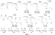
На фигуре 1 представлены структурные формулы соединений (1), (2) и (3). В структуру амфифилов (1), (2) и (3) входят остаток спермина для связывания и компактизации молекулы нуклеиновой кислоты, остаток 1,2-ди-О-тетрадецил-rac-глицерина для формирования гидрофобного окружения. Для связывания гидрофобной и сперминовой частей используют биодеградируемый линкер сукцинильного типа. Для придания углеводным остаткам пространственной подвижности моносахариды присоединены к липидным составляющим через гексаметиленовые мостики.The figure 1 presents the structural formulas of the compounds (1), (2) and (3). The structure of amphiphiles (1), (2) and (3) includes a spermine residue for binding and compaction of a nucleic acid molecule, a 1,2-di-O-tetradecyl-rac-glycerol residue to form a hydrophobic environment. A biodegradable succinyl type linker is used to bind the hydrophobic and spermine moieties. To give carbohydrate residues spatial mobility, monosaccharides are attached to the lipid moieties through hexamethylene bridges.
Новые соединения (1), (2) и (3) были получены по общей схеме, представленной на фигуре 2. Исходным соединением в синтезе являлся 1-бром-1-дезокси-2,3-ди-О-тетрадецил-rac-глицерин (4), который вводили во взаимодействие с N-(6-гидроксигексил)-4-нитробензолсульфонамидом (5), получая бифункциональное соединение (6). Ключевым этапом синтеза соединений (7a-b) явилось гликозилирование соединения (6) 2,3,4,6-тетра-О-ацетил-α-D-галактопиранозилбромидом, 2,2',3,3',4',6,6'-гепта-O-ацетил-β-лактопиранозилбромидом или 2,3,4,6-тетра-O-ацетил-α-D-маннопиранозилбромидом [23]. Для получения удобного узла связывания нитрогруппу ароматического ядра восстанавливали до аминогруппы, используя в качестве катализатора Pd/C. Далее в молекулы амфифилов (8a-b) вводили сукцинильный спейсер обработкой янтарным ангидридом, получая карбоксилаты (9а-с). Связывание соединений (9а-с) с N4,N9,N12-три-трет-бутоксикарбонил-1,12-диамино-4,9-диазадодеканом проводили в присутствии O-(1Н-бензотриазол-1-ил)-N,N,N',N'-тетраметилуроний гексафторфосфата при 4°С. На завершающем этапе синтеза в соединениях (10а-с) проводили деблокирование аминогрупп действием трифторуксусной кислоты, а удаление ацетильных защит - 0.04 н. раствором метилата натрия в метаноле.The new compounds (1), (2) and (3) were obtained according to the general scheme shown in Figure 2. The starting compound in the synthesis was 1-bromo-1-deoxy-2,3-di-O-tetradecyl-rac-glycerol (4) which was reacted with N- (6-hydroxyhexyl) -4-nitrobenzenesulfonamide (5) to give a bifunctional compound (6). The key step in the synthesis of compounds (7a-b) was the glycosylation of compound (6) with 2,3,4,6-tetra-O-acetyl-α-D-galactopyranosyl bromide, 2,2 ', 3,3', 4 ', 6, 6'-hepta-O-acetyl-β-lactopyranosyl bromide or 2,3,4,6-tetra-O-acetyl-α-D-mannopyranosyl bromide [23]. To obtain a convenient binding site, the nitro group of the aromatic nucleus was reduced to the amino group using Pd / C as a catalyst. Next, a succinyl spacer was added to amphiphilic molecules (8a-b) by treatment with succinic anhydride to obtain carboxylates (9a-c). The coupling of compounds (9a-c) with N 4 , N 9 , N 12 -tri-tert-butoxycarbonyl-1,12-diamino-4,9-diazadodecane was carried out in the presence of O- (1H-benzotriazol-1-yl) -N , N, N ', N'-tetramethyluronium hexafluorophosphate at 4 ° C. At the final stage of synthesis in compounds (10a-c), the amino groups were released by the action of trifluoroacetic acid, and the removal of acetyl protections was 0.04 N. a solution of sodium methylate in methanol.
В синтезе использовались очищенные растворители, реагенты отечественного (Химмед, Реахим) и зарубежного производства (Merck, Fluka, Aldrich, Acros). Контроль за ходом реакций осуществляли с помощью ТСХ. Тонкослойную хроматографию проводили на пластинках Kieselgel 60 F254 (Merck), RP -18 F254S (Merck), Сорбфил (Россия). Обнаружение пятен на хроматограммах проводили раствором фосформолибденовая кислота - церий (IV) сульфат с последующим прогреванием, реактивом Драгендорфа, раствором перманганата калия и с помощью УФ-лампы (254 нм). Колоночную хроматографию проводили на силикагеле Kieselgel 60 (0.040-0.063 mm); Kieselgel 60 (0.063-0.200 mm), LiChroprep® RP-18 (0.040-0.063 mm, Merck). Спектры 1H- и 13С-ЯМР регистрировали на импульсном фурье-спектрометре «Bruker DPX-300» и «Bruker АМХ-400» в CDCl3, смеси CDCl3-CD3OD и Py-d5 (внутренний стандарт тетраметилсилан). Значения химических сдвигов (δ) приведены в миллионных долях (м.д.), константы спин-спинового взаимодействия (J) в герцах (Гц). Масс-спектры получали на время-пролетном масс-спектрометре «Bruker Ultraflex» (Германия) методом матриксной лазерно-десорбционной ионизации на матрице с использованием в качестве матрицы 2,5-дигидроксибензойной кислоты. Углы оптического вращения измеряли на фотоэлектрическом спектрополяриметре «Digytor Yasco DIP 360» (Япония).The synthesis used refined solvents, reagents of domestic (Himmed, Reakhim) and foreign production (Merck, Fluka, Aldrich, Acros). Monitoring the progress of reactions was carried out using TLC. Thin layer chromatography was performed on Kieselgel 60 F 254 (Merck), RP-18 F 254S (Merck), Sorbfil (Russia) plates. Spot detection in the chromatograms was carried out with a solution of phosphormolybdenum acid - cerium (IV) sulfate, followed by heating, Dragendorf reagent, potassium permanganate solution and using a UV lamp (254 nm). Column chromatography was performed on silica gel Kieselgel 60 (0.040-0.063 mm); Kieselgel 60 (0.063-0.200 mm), LiChroprep® RP-18 (0.040-0.063 mm, Merck). 1 H- and 13 C-NMR spectra were recorded on a Bruker DPX-300 and Bruker AMX-400 pulsed Fourier spectrometer in CDCl 3 , a mixture of CDCl 3 -CD 3 OD and Py-d5 (internal standard tetramethylsilane). The values of chemical shifts (δ) are given in parts per million (ppm), the spin-spin interaction constants (J) in hertz (Hz). Mass spectra were obtained on a Bruker Ultraflex time-of-flight mass spectrometer (Germany) by matrix laser desorption ionization on a matrix using 2,5-dihydroxybenzoic acid as a matrix. The angles of optical rotation were measured using a Digytor Yasco DIP 360 photoelectric spectropolarimeter (Japan).
Для изучения способности предлагаемых соединений доставлять нуклеиновые кислоты в клетки млекопитающих использовали 25-звенный олигодезоксирибонуклеотид 5'-dTACAGTGGAATTGTATGCCTATTA-3', модифицированный флуоресцеинизотиоционатом (FITC) по 3'-концу, плазмидную ДНК (pEGFP-C2, «Clontech» (Германия)) и 21-звенную двуцепочечную РНК (siPHK, ИХБФМ СО РАН) (последовательность смысловой цепи 5'-GCGCCGAGGUGAAGUUCGATT-3', антисмысловой цепи - 5'-UCGAACUUCACCUCGGCGCGG-3'). FITC-ON был синтезирован фосфитамидным методом [24] и очищен ионообменной и обращенно-фазовой хроматографией. Чистоту олигонуклеотида проверяли с помощью электрофореза в 15% ПААГ в денатурирующих условиях, визуализацию олигонуклеотида в геле проводили с помощью окраски Stains-Аll.To study the ability of the proposed compounds to deliver nucleic acids to mammalian cells, we used a 5-dTACAGTGGAATTGTATGCCTATTA-3 '25-unit oligodeoxyribonucleotide modified at the 3'-end of the fluorescein isothiocyanate (FITC), plasmid DNA (pEGFP-C2, Germany) and 21-stranded double-stranded RNA (siPHK, ICBPM SB RAS) (sense sequence 5'-GCGCCGAGGUGAAGUUCGATT-3 ', antisense strand 5'-UCGAACUUCACCUCGGCGCGG-3'). FITC-ON was synthesized by the phosphitamide method [24] and purified by ion exchange and reverse phase chromatography. The purity of the oligonucleotide was checked by electrophoresis in 15% PAG under denaturing conditions, visualization of the oligonucleotide in the gel was performed using Stains-All staining.
Эффективность проникновения нуклеиновых кислот с использованием катионных амфифилов в клетки млекопитающих in vitro была исследована в экспериментах по трансфекции клеток FITC-меченным олигонуклеотидом, плазмидной ДНК, кодирующей зеленый флуоресцирующий белок (EGFP) и короткой интерферирующей РНК, направленной против матричной РНК гена, кодирующего EGFP.The efficiency of penetration of nucleic acids using cationic amphiphiles into mammalian cells in vitro was studied in experiments on transfection of cells with a FITC-labeled oligonucleotide, plasmid DNA encoding a green fluorescent protein (EGFP) and short interfering RNA directed against a matrix RNA gene encoding EGFP.
Сопоставительный анализ заявляемых соединений с известными и широко используемыми соединениями, такими как Lipofectamine®2000 и Oligofectamine показал, что предлагаемые соединения обладают следующими преимуществами.A comparative analysis of the claimed compounds with known and widely used compounds, such as Lipofectamine® 2000 and Oligofectamine showed that the proposed compounds have the following advantages.
1) Благодаря наличию природных структурных модулей, в том числе углеводных остатков, заявляемые соединения обладают низким токсическим воздействием на клетки млекопитающих.1) Due to the presence of natural structural modules, including carbohydrate residues, the claimed compounds have low toxic effects on mammalian cells.
2) Заявляемые соединения обладают способностью доставлять в клетки млекопитающих нуклеиновые кислоты как короткие, так и протяженные, что позволяет рассматривать их как перспективные агенты для трансфекции клеток млекопитающих.2) The claimed compounds have the ability to deliver nucleic acids to mammalian cells, both short and extended, which allows them to be considered as promising agents for transfection of mammalian cells.
3) Заявляемые соединения доставляют нуклеиновые кислоты в клетки в индивидуальном состоянии в виде водных растворов и не требуют использования дополнительных вспомогательных липидов.3) The inventive compounds deliver nucleic acids to cells in an individual state in the form of aqueous solutions and do not require the use of additional auxiliary lipids.
Заявляемые соединения не требуют сложной процедуры приготовления, свойственной липосомальным композициям, для них характерна легкость приготовления рабочего раствора, для получения которого достаточно растворить заявляемое соединение в воде или спирте. Кроме того, заявляемые соединения стабильны при хранении как в сухом виде, так и в виде концентрированного водного или спиртового растворов.The inventive compounds do not require a complicated preparation procedure characteristic of liposomal compositions, they are characterized by the ease of preparation of the working solution, for which it is sufficient to dissolve the claimed compound in water or alcohol. In addition, the claimed compounds are stable during storage both in dry form and in the form of concentrated aqueous or alcoholic solutions.
Поиск по источникам научно-технической и патентной литературы показал, что заявляемые соединения (1), (2), (3), способные доставлять нуклеиновые кислоты в клетки млекопитающих, в известных источниках не описаны.A search in the sources of scientific, technical and patent literature showed that the claimed compounds (1), (2), (3), capable of delivering nucleic acids to mammalian cells, are not described in known sources.
Изобретение иллюстрируется следующими примерами.The invention is illustrated by the following examples.
Пример 1. Получение rac-N-(6-гидроксигексил)-N-[2,3-ди(тетрадецил-окси)проп-1-ил]-4-нитробензолсульфонамида (промежуточное соединение 6).Example 1. Obtaining rac-N- (6-hydroxyhexyl) -N- [2,3-di (tetradecyl-hydroxy) prop-1-yl] -4-nitrobenzenesulfonamide (intermediate compound 6).
К раствору 0.995 г (1.816 ммоль) rac-1-бром-1-дезокси-2,3-ди-О-тетрадецилглицерина (4) в 10 мл диметилформамида добавили 0.571 г (1.888 ммоль) N-(6-гидроксигексил)-4-нитробензолсульфонамида (5), карбоната цезия (0.355 г, 1.090 ммоль) и тетрабутиламмоний йодида (0.067 г, 0.181 ммоль). Реакционную смесь перемешивали 2 дня при нагревании, экстрагировали петролейным эфиром. Органический экстракт упаривали и хроматографировали на колонке с силикагелем. Выход соединения (6) составил 1.397 г (60%).To a solution of 0.995 g (1.816 mmol) of rac-1-bromo-1-deoxy-2,3-di-O-tetradecylglycerol (4) in 10 ml of dimethylformamide was added 0.571 g (1.888 mmol) of N- (6-hydroxyhexyl) -4 nitrobenzenesulfonamide (5), cesium carbonate (0.355 g, 1.090 mmol) and tetrabutylammonium iodide (0.067 g, 0.181 mmol). The reaction mixture was stirred for 2 days with heating, was extracted with petroleum ether. The organic extract was evaporated and chromatographed on a silica gel column. The yield of compound (6) was 1.397 g (60%).
Пример 2. Получение rac-N-[6-(2,3,4,6-тетра-О-ацетил-β-D-галактопиранозилокси)гексил]-N-[2,3-ди(тетрадецилокси)проп-1-ил]-4-нитробензолсульфонамида (промежуточное соединение 7а).Example 2. Obtaining rac-N- [6- (2,3,4,6-tetra-O-acetyl-β-D-galactopyranosyloxy) hexyl] -N- [2,3-di (tetradecyloxy) prop-1- yl] -4-nitrobenzenesulfonamide (intermediate 7a).
К раствору 1.028 г (1.336 ммоль) rac-N-(6-гидроксигексил)-N-[2,3-ди(тетрадецилокси)проп-1-ил]-4-нитробензолсульфонамида (6) в 40 мл безводного бензола добавили 0.691 г (4.009 ммоль) прокаленного карбоната кадмия и кипятили в аппарате Сокслета. В реакционную смесь вносили раствор 1.648 г (4.009 ммоль) 2,3,4,6-тетра-O-ацетил-α-D-галактопиранозилбромида. Через 2.5 ч реакционную массу фильтровали, растворитель удаляли в вакууме. Остаток хроматографировали на колонке с силикагелем. Выход соединения (7а) составил 1.152 г (78%).To a solution of 1.028 g (1.336 mmol) of rac-N- (6-hydroxyhexyl) -N- [2,3-di (tetradecyloxy) prop-1-yl] -4-nitrobenzenesulfonamide (6) in 40 ml of anhydrous benzene was added 0.691 g (4.009 mmol) of calcined cadmium carbonate and boiled in a Soxhlet apparatus. A solution of 1.648 g (4.009 mmol) of 2,3,4,6-tetra-O-acetyl-α-D-galactopyranosyl bromide was added to the reaction mixture. After 2.5 h, the reaction mass was filtered, the solvent was removed in vacuo. The residue was chromatographed on a silica gel column. The yield of compound (7a) was 1.152 g (78%).
Промежуточные соединения (7b) и (7с) были получены, как описано для получения соединения (7а), исходя из rac-N-(6-гидроксигексил)-N-[2,3-ди(тетрадецилокси)проп-1-ил]-4-нитробензолсульфонамида (6), 2,2',3,3',4',6,6'-гепта-O-ацетил-β-лактопиранозилбромида и 2,3,4,6-тетра-О-ацетил-α-D-маннопиранозилбромида, соответственно. Физико-химические характеристики соединений (7b) и (7с) соответствуют их химической структуре.Intermediates (7b) and (7c) were obtained as described for the preparation of compound (7a), starting from rac-N- (6-hydroxyhexyl) -N- [2,3-di (tetradecyloxy) prop-1-yl] -4-nitrobenzenesulfonamide (6), 2,2 ', 3,3', 4 ', 6,6'-hepta-O-acetyl-β-lactopyranosyl bromide and 2,3,4,6-tetra-O-acetyl- α-D-mannopyranosyl bromide, respectively. The physicochemical characteristics of compounds (7b) and (7c) correspond to their chemical structure.
Пример 3. Получение rac-N-[6-(2,3,4,6-тетра-O-ацетил-β-D-галактопиранозилокси)гексил]-N-[2,3-ди(тетрадецилокси)проп-1-ил]-4-аминобензолсульфонамида (промежуточное соединение 8а).Example 3. Obtaining rac-N- [6- (2,3,4,6-tetra-O-acetyl-β-D-galactopyranosyloxy) hexyl] -N- [2,3-di (tetradecyloxy) prop-1- yl] -4-aminobenzenesulfonamide (intermediate 8a).
К раствору 1.126 г (1.024 ммоль) rac-N-[6-(2,3,4,6-тетра-O-ацетил-[β-D-галактопиранозилокси)гексил]-N-[2,3-ди(тетрадецилокси)проп-1-ил]-4-нитробензолсульфонамида (7а) в 14 мл смеси метанол:тетрагидрофуран (2.5:2) добавили 0.258 г (4.096 ммоль) формиата аммония и нагревали до 60°С, вносили каталитическое количество Pd/C. Через 30 мин реакционную смесь фильтровали, растворители удаляли в вакууме. Остаток хроматографировали на колонке с силикагелем. Выход соединения (8а) составил 0.933 г (85%).To a solution of 1.126 g (1.024 mmol) rac-N- [6- (2,3,4,6-tetra-O-acetyl- [β-D-galactopyranosyloxy) hexyl] -N- [2,3-di (tetradecyloxy ) prop-1-yl] -4-nitrobenzenesulfonamide (7a) in 14 ml of a mixture of methanol: tetrahydrofuran (2.5: 2), 0.258 g (4.096 mmol) of ammonium formate was added and heated to 60 ° C, a catalytic amount of Pd / C was added. After 30 minutes, the reaction mixture was filtered, and the solvents were removed in vacuo. The residue was chromatographed on a silica gel column. The yield of compound (8a) was 0.933 g (85%).
Промежуточные соединения (8b) и (8с) были получены, как описано для получения соединения (8а), исходя из rac-N-[6-(2,2',3,3',4',6,6'-гепта-O-ацетил-β-лактопиранозилокси)гексил]-N-[2,3-ди(тетрадецилокси)проп-1-ил]-4-нитробензолсульфонамида (7b) и rac-N-[6-(2,3,4,6-тетра-O-ацетил-β-D-маннопиранозилокси)гексил]-N-[2,3-ди(тетрадецилокси)-проп-1-ил]-4-нитробензолсульфонамида (7с), соответственно. Физико-химические характеристики соединений (8b) и (8с) соответствуют их химической структуре.Intermediates (8b) and (8c) were obtained as described for the preparation of compound (8a), starting from rac-N- [6- (2.2 ', 3.3', 4 ', 6.6'-hepta -O-acetyl-β-lactopyranosyloxy) hexyl] -N- [2,3-di (tetradecyloxy) prop-1-yl] -4-nitrobenzenesulfonamide (7b) and rac-N- [6- (2,3,4 , 6-tetra-O-acetyl-β-D-mannopyranosyloxy) hexyl] -N- [2,3-di (tetradecyloxy) prop-1-yl] -4-nitrobenzenesulfonamide (7c), respectively. The physicochemical characteristics of compounds (8b) and (8c) correspond to their chemical structure.
Пример 4. Получение rac-N-[6-(2,3,4,6-тетра-O-ацетил-β-D-галактопиранозилокси)гексил]-N-[2,3-ди(тетрадецилокси)проп-1-ил]-4-(3-карбоксипропаноиламино)бензолсульфонамида (промежуточное соединение 9а).Example 4. Obtaining rac-N- [6- (2,3,4,6-tetra-O-acetyl-β-D-galactopyranosyloxy) hexyl] -N- [2,3-di (tetradecyloxy) prop-1- yl] -4- (3-carboxypropanoylamino) benzenesulfonamide (intermediate 9a).
К раствору 0.932 г (0.872 ммоль) rac-N-[6-(2,3,4,6-тетра-O-ацетил-β-D-галактопиранозилокси)гексил]-N-[2,3-ди(тетрадецилокси)проп-1-ил]-4-аминобензолсульфонамида (8а) в дихлорметане добавили 1.025 г (10.23 ммоль) янтарного ангидрида, 0.024 г (0.201 ммоль) диметиламинопиридина и 287 мкл (2.046 ммоль) триэтиламина. Через 36 ч реакционную смесь промывали водным раствором HCl. Органическую фазу сушили, фильтровали, растворители удаляли в вакууме. Остаток хроматографировали на колонке с силикагелем. Выход соединения (9а) составил 0.747 г (73%).To a solution of 0.932 g (0.872 mmol) rac-N- [6- (2,3,4,6-tetra-O-acetyl-β-D-galactopyranosyloxy) hexyl] -N- [2,3-di (tetradecyloxy) prop-1-yl] -4-aminobenzenesulfonamide (8a) in dichloromethane added 1.025 g (10.23 mmol) of succinic anhydride, 0.024 g (0.201 mmol) of dimethylaminopyridine and 287 μl (2.046 mmol) of triethylamine. After 36 hours, the reaction mixture was washed with an aqueous HCl solution. The organic phase was dried, filtered, and the solvents were removed in vacuo. The residue was chromatographed on a silica gel column. The yield of compound (9a) was 0.747 g (73%).
Промежуточные соединения (9b) и (9с) были получены, как описано для получения соединения (9а), исходя из rac-N-[6-(2,2',3,3',4',6,6'-гепта-O-ацетил-β-лактопиранозилокси)гексил]-N-[2,3-ди(тетрадецилокси)проп-1-ил]-4-аминобензолсульфонамида (8b) и rас-N-[6-(2,3,4,6-тетра-O-ацетил-β-D-маннопиранозилокси)гексил]-N-[2,3-ди(тетрадецилокси)проп-1-ил]-4-аминобензолсульфонамида (8с), соответственно. Физико-химические характеристики соединений (9b) и (9с) соответствуют их химической структуре.Intermediates (9b) and (9c) were obtained as described for the preparation of compound (9a), starting from rac-N- [6- (2.2 ', 3.3', 4 ', 6.6'-hepta -O-acetyl-β-lactopyranosyloxy) hexyl] -N- [2,3-di (tetradecyloxy) prop-1-yl] -4-aminobenzenesulfonamide (8b) and raс-N- [6- (2,3,4 , 6-tetra-O-acetyl-β-D-mannopyranosyloxy) hexyl] -N- [2,3-di (tetradecyloxy) prop-1-yl] -4-aminobenzenesulfonamide (8c), respectively. The physicochemical characteristics of compounds (9b) and (9c) correspond to their chemical structure.
Пример 5. Получение rac-N-[6-(2,3,4,6-тетра-O-ацетил-β-D-галактопиранозилокси)гексил]-N-[2,3-ди(тетрадецилокси)проп-1-ил]-4-[(N4,N9,N12-три-трет-бутоксикарбонил-12-амино-4,9-диазадодец-1-ил)аминосукциниламино]бензолсульфонамида (промежуточное соединение 10а).Example 5. Obtaining rac-N- [6- (2,3,4,6-tetra-O-acetyl-β-D-galactopyranosyloxy) hexyl] -N- [2,3-di (tetradecyloxy) prop-1- yl] -4 - [(N 4 , N 9 , N 12 -tri-tert-butoxycarbonyl-12-amino-4,9-diazadodec-1-yl) aminosuccinylamino] benzenesulfonamide (intermediate 10a).
К раствору 0.642 г (0.549 ммоль) соединения (9а) и 0.552 г (1.098 ммоль) N4,N9,N12-три-трет-бутоксикарбонил-1,12-диамино-4,9-диазадодекана в диметилформамида внесли 191 мкл (1.098 ммоль) N,N-диизопропилэтиламина и по каплям раствор 0.416 г (1.098 ммоль) О-(1Н-бензотриазол-1-ил)-N,N,N',N'-тетраметилуроний гексафторфосфата в ДМФА. Через 5.5 ч к реакционной массе добавили хлороформ и экстрагировали водным раствором HCl. Органическую фазу сушили, фильтровали, остаток хроматографировали на колонке с силикагелем. Выход соединения (10а) составил 0.563 г (62%).To a solution of 0.642 g (0.549 mmol) of compound (9a) and 0.552 g (1.098 mmol) of N 4 , N 9 , N 12 -tri-tert-butoxycarbonyl-1,12-diamino-4,9-diazadodecane, 191 μl were added to dimethylformamide (1.098 mmol) of N, N-diisopropylethylamine and a dropwise solution of 0.416 g (1.098 mmol) of O- (1H-benzotriazol-1-yl) -N, N, N ', N'-tetramethyluronium hexafluorophosphate in DMF. After 5.5 hours, chloroform was added to the reaction mass and extracted with an aqueous HCl solution. The organic phase was dried, filtered, and the residue was chromatographed on a silica gel column. The yield of compound (10a) was 0.563 g (62%).
Промежуточные соединения (10b) и (10с) были получены, как описано для получения соединения (10а), исходя из соединений (9b) и (9 с), соответственно. Физико-химические характеристики соединений (10b) и (10с) соответствуют химической структуре.Intermediates (10b) and (10c) were obtained as described for the preparation of compound (10a), starting from compounds (9b) and (9 s), respectively. The physicochemical characteristics of compounds (10b) and (10c) correspond to the chemical structure.
Пример 6. Получение трис(трифторацетата) rac-N-[6-(2,3,4,6-тетра-O-ацетил-β-D-галактопиранозилокси)гексил]-N-[2,3-ди(тетрадецилокси)проп-1-ил]-4-[(12-амино-4,9-диазадодец-1-ил)аминосукциниламино]бензолсульфонамида (промежуточное соединение 11а).Example 6. Preparation of tris (trifluoroacetate) rac-N- [6- (2,3,4,6-tetra-O-acetyl-β-D-galactopyranosyloxy) hexyl] -N- [2,3-di (tetradecyloxy) prop-1-yl] -4 - [(12-amino-4,9-diazadodec-1-yl) aminosuccinylamino] benzenesulfonamide (intermediate 11a).
К раствору 0.669 г (0.404 ммоль) соединения (10а) в дихлорметане добавили 4.416 мл (19.8 ммоль) трифторуксусной кислоты. Через 3.5 ч растворители удаляли в вакууме. Остаток хроматографировали на колонке с силикагелем. Выход соединения (11а) составил 0.472 г (76%).To a solution of 0.669 g (0.404 mmol) of compound (10a) in dichloromethane was added 4.416 ml (19.8 mmol) of trifluoroacetic acid. After 3.5 hours, the solvents were removed in vacuo. The residue was chromatographed on a silica gel column. The yield of compound (11a) was 0.472 g (76%).
Промежуточные соединения (11b) и (11с) были получены, как описано для получения соединения (11а), исходя из (10b) и (10с), соответственно. Физико-химические характеристики соединений (11b) и (11c) соответствуют их химической структуре.Intermediates (11b) and (11c) were prepared as described for the preparation of compound (11a) based on (10b) and (10c), respectively. The physicochemical characteristics of compounds (11b) and (11c) correspond to their chemical structure.
Пример 7. Получение тригидрохлорида rac-N-[6-(β-D-галактопиранозилокси)гексил]-N-[2,3-ди(тетрадецилокси)проп-1-ил]-4-[(12-амино-4,9-диазадодец-1-ил)-аминосукциниламино]бензолсульфонамида (1).Example 7. Obtaining trihydrochloride rac-N- [6- (β-D-galactopyranosyloxy) hexyl] -N- [2,3-di (tetradecyloxy) prop-1-yl] -4 - [(12-amino-4, 9-diazadodec-1-yl) -aminosuccinylamino] benzenesulfonamide (1).
К 0.472 г (0.278 ммоль) соединения (11а) добавили 0.04 н. раствор метилата натрия в метаноле. Через 1 ч к реакционной массе добавляли раствор 4 н. HCl в диоксане, пока pH не станет равным 5. Растворитель удаляли в вакууме, остаток хроматографировали на колонке. Полученный продукт растворяли в 7 мл дистиллированной воды и проводили диализ против воды в мембране. После лиофилизации выход соединения (1) составил 0.199 г (55%). 1Н-ЯМР (δ, м.д.): 0.65 (т, 6Н, J=6.5, 2СН2СН 3), 1.05 (уш.с, 44Н, 2(СН2)11), 1.13-1.26 (м, 4Н, СН2СН2), 1.31-1.49 (м, 8Н, 3CH 2CH2O, CH 2CH2N), 1.68-1.95 (м, 6Н, протоны спермина), 2.28-2.40 (м, 2Н) и 2.58-2.69 (м, 2Н, NCOCH2CH2COO), 2.81-3.55 (м, 24Н, CHHaO, OCH2CH, 2CH2N, 2CH2O, протоны спермина), 3.74-3.95 m, 4Н, H5-Gal, Hab6-Gal, CHHbO), 4.11-4.20 (м, 2Н, H2-Gal, H3-Gal), 4.35-4.31 (м, 1Н, H4-Gal), 4.31-4.49 (д, 1Н, J=7.6, H1-Gal), 7.78-7.86 (м, 2Н) и 8.01-8.09 (м, 2Н, Ar), 8.95-9.02 (уш.с, 1Н, NH). 13С-ЯМР (δ, м.д.): 14.21, 22.87, 23.94, 24.21, 25.56, 25.97, 26.49, 26.81, 27.18, 28.34, 29.55, 29.76, 29.92, 30.11, 30.61, 30.96, 32.06, 32.60, 36.24, 37.78, 39.00, 45.19, 45.54, 47.26, 50.11, 62.19, 69.52, 70.13, 70.55, 71.39, 71.69, 72.46, 75.13, 76.67, 78.75, 105.08, 128.78, 172.23, 174.08. Масс-спектр (MALDI-TOF), m/z: 1185.864 [M-3HCl+H]+, 1207.847 [M-3HCl+Na]+, 1223.825 [M-3HCl+K]+ вычислено для C63H120N6O12S: 1184.8685.To 0.472 g (0.278 mmol) of compound (11a), 0.04 N was added. a solution of sodium methylate in methanol. After 1 h, a solution of 4 N was added to the reaction mass. HCl in dioxane until the pH is 5. The solvent was removed in vacuo and the residue was chromatographed on a column. The resulting product was dissolved in 7 ml of distilled water and dialyzed against water in the membrane. After lyophilization, the yield of compound (1) was 0.199 g (55%). 1 H-NMR (δ, ppm): 0.65 (t, 6H, J = 6.5, 2CH 2 C H 3 ), 1.05 (br s, 44H, 2 (CH 2 ) 11 ), 1.13-1.26 ( m, 4H, CH 2 CH 2 ), 1.31-1.49 (m, 8H, 3C H 2 CH 2 O, C H 2 CH 2 N), 1.68-1.95 (m, 6H, spermine protons), 2.28-2.40 (m , 2H) and 2.58-2.69 (m, 2H, NCOCH 2 CH 2 COO), 2.81-3.55 (m, 24H, CHH a O, OCH 2 CH, 2CH 2 N, 2CH 2 O, spermine protons), 3.74-3.95 m, 4H, H5-Gal, H ab 6-Gal, CHH b O), 4.11-4.20 (m, 2H, H2-Gal, H3-Gal), 4.35-4.31 (m, 1H, H4-Gal), 4.31 -4.49 (d, 1H, J = 7.6, H1-Gal), 7.78-7.86 (m, 2H) and 8.01-8.09 (m, 2H, Ar), 8.95-9.02 (br.s, 1H, NH). 13 C-NMR (δ, ppm): 14.21, 22.87, 23.94, 24.21, 25.56, 25.97, 26.49, 26.81, 27.18, 28.34, 29.55, 29.76, 29.92, 30.11, 30.61, 30.96, 32.06, 32.60, 36.24 , 37.78, 39.00, 45.19, 45.54, 47.26, 50.11, 62.19, 69.52, 70.13, 70.55, 71.39, 71.69, 72.46, 75.13, 76.67, 78.75, 105.08, 128.78, 172.23, 174.08. Mass spectrum (MALDI-TOF), m / z: 1185.864 [M-3HCl + H] + , 1207.847 [M-3HCl + Na] + , 1223.825 [M-3HCl + K] + calculated for C 63 H 120 N 6 O 12 S: 1184.8685.
Соединения (2) и (3) были получены, как описано для получения соединения (1), исходя из (11b) и (11с), соответственно. Физико-химические характеристики соединений (2) и (3) соответствуют их химической структуре.Compounds (2) and (3) were obtained as described for the preparation of compound (1) starting from (11b) and (11c), respectively. The physicochemical characteristics of compounds (2) and (3) correspond to their chemical structure.
Тригидрохлорид rac-N-[6-(β-лактопиранозилокси)гексил]-N-[2,3-ди(тетрадецилокси)проп-1-ил]-4-[(12-амино-4,9-диазадодец-1-ил)аминосукциниламино]бензолсульфонамид (2).Rac-N- [6- (β-lactopyranosyloxy) hexyl] -N- [2,3-di (tetradecyloxy) prop-1-yl] -4 - [(12-amino-4,9-diazadodec-1-) trihydrochloride il) aminosuccinylamino] benzenesulfonamide (2).
Выход соединения (2) составил 64%. [α]D 33 -3.07 (с0.8, CHCl3-СН3ОН, 1:5). 1Н-ЯМР (δ, м.д.): 0.81 (т, 6Н, J=6.8, 2CH2CH 3), 1.16-1.28 (м, 48Н, 2(CH2)11, (CH2)2); 1.36-1.66 (м, 20Н, 3CH 2CH2O, 5CH 2CH2N, CH2CH2), 3.18-3.57 (м, 12Н, 2NCH 2, 2OCH2CH 2, CH2O, CHHaO, CH2CHCH2), 3.65-3.75 (м, 2Н, CHHbO, H5-Lac), 3.78-3.86 (м, 1Н, H5'-Lac), 3.91-4.12 (м, 4Н, Hab6-Lac, Hab6'-Lac), 4.37 (д, 1Н, J=7.8, Н-1' Lac), 4.41 (м, 1Н, Н-4 Lac), 4.43 (д, 1Н, J=7.8, Н-1 Lac), 4.79 (дд, 1Н, J=7.8, 9.4, Н-2 Lac), 4.89 (дд, 1Н, J=3.4, 10.3, Н-3' Lac), 5.03 (дд, 1Н, J=7.8, 10.3, Н-2' Lac), 5.12 (т, 1Н, J=9.4, Н-3 Lac), 5.29 (м, 1Н, Н-4' Lac), 7.75-7.92 (м, 4Н, Ar). 13С-ЯМР (δ, м.д.): 14.84, 23.48, 24.28, 25.50, 26.40, 27.29, 28.54, 30.16, 30.55, 30.72, 30.72, 31.23, 33.63, 38.47, 46.04, 46.47, 48.05, 50.46, 62.32, 62.49, 70.27, 70.40, 71.24, 72.28, 72.88, 75.25, 76.79, 77.49, 78.72, 79.52, 82.04, 104.51, 105.80, 120.28, 129.27, 134.58, 144.82, 172.96, 174.35. Масс-спектр (MALDI-TOF), m/z: 1347.700 [M-3HCl+H], вычислено для C69H1130N6O17S: 1346.9213.The yield of compound (2) was 64%. [α] D 33 -3.07 (s0.8, CHCl 3 -CH 3 OH, 1: 5). 1 H-NMR (δ, ppm): 0.81 (t, 6H, J = 6.8, 2CH 2 C H 3 ), 1.16-1.28 (m, 48H, 2 (CH 2 ) 11 , (CH 2 ) 2 ); 1.36-1.66 (m, 20H, 3C H 2 CH 2 O, 5C H 2 CH 2 N, CH 2 CH 2 ), 3.18-3.57 (m, 12H, 2NC H 2 , 2OCH 2 C H 2 , CH 2 O, CHH a O, CH 2 C H CH 2 ), 3.65-3.75 (m, 2H, CHH b O, H5-Lac), 3.78-3.86 (m, 1H, H5'-Lac), 3.91-4.12 (m, 4H , H ab 6-Lac, H ab 6'-Lac), 4.37 (d, 1H, J = 7.8, H-1 'Lac), 4.41 (m, 1H, H-4 Lac), 4.43 (d, 1H, J = 7.8, H-1 Lac), 4.79 (dd, 1H, J = 7.8, 9.4, H-2 Lac), 4.89 (dd, 1H, J = 3.4, 10.3, H-3 'Lac), 5.03 (dd , 1Н, J = 7.8, 10.3, Н-2 'Lac), 5.12 (t, 1Н, J = 9.4, Н-3 Lac), 5.29 (m, 1Н, Н-4' Lac), 7.75-7.92 (m 4H, Ar). 13 C-NMR (δ, ppm): 14.84, 23.48, 24.28, 25.50, 26.40, 27.29, 28.54, 30.16, 30.55, 30.72, 30.72, 31.23, 33.63, 38.47, 46.04, 46.47, 48.05, 50.46, 62.32 , 62.49, 70.27, 70.40, 71.24, 72.28, 72.88, 75.25, 76.79, 77.49, 78.72, 79.52, 82.04, 104.51, 105.80, 120.28, 129.27, 134.58, 144.82, 172.96, 174.35. Mass spectrum (MALDI-TOF), m / z: 1347.700 [M-3HCl + H], calculated for C 69 H 1130 N 6 O 17 S: 1346.9213.
Тригидрохлорид rac-N-[6-(β-D-маннопиранозилокси)гексил]-N-[2,3-ди(тетрадецилокси)проп-1-ил]-4-[(12-амино-4,9-диазадодец-1-ил)аминосукциниламино]бензолсульфонамид (3).Rac-N- [6- (β-D-mannopyranosyloxy) hexyl] -N- [2,3-di (tetradecyloxy) prop-1-yl] -4 - [(12-amino-4,9-diazadodec- trihydrochloride trihydrochloride 1-yl) aminosuccinylamino] benzenesulfonamide (3).
Выход соединения (3) составил 55%. [α]D 23 6.48 (c1, CH3OH). 1Н-ЯМР (δ, м.д.): 0.67 (т, 6Н, J=6.7, 2 СН2СН 3), 1.10 (уш.с, 44Н, 2(СН2)11), 1.15-1.25 (м, 4Н, CH2CH2), 1.34-1.45 (м, 8Н, 3CH 2CH2O, CH 2CH2N, 1.68-1.95 (м, 6Н, протоны спермина), 2.28-2.40 (м, 2Н) и 2.58-2.69 (м, 2Н, NCOCH2CH2COO), 2.81-3.55 (м, 24Н, CHHaO, OCH2CH, 2CH2N, 2CH2O, протоны спермина), 3.61 (дт, J=9.6, 6.4, 1Н, CHHbO), 3.81-3.85 (м, 2Н, Н5-Man), 4.04 (дд, 1Н, J=2.4, 12.2, Ha6-Man), 4.23 (дд, 1Н, J=5.2, 12.2, Hb6-Man), 4.73 (д, 1Н, J=1.6, H1-Man), 5.16 (дд, 1Н, J=1.6, 3.1, Н2-Man), 5.19 (дд, 1Н, J=9.2, 9.8, H4-Man), 5.28 (дд, 1Н, J=3.1, 9.8, H3-Man), 7.75-7.91 (м, 2Н) и 8.05-8.15 (м, 2Н, Ar), 8.95-9.02 (м, 1Н, NH), 11.05-11.45 (м, 1Н, NH). 13С-ЯМР (δ, м.д.): 24.67, 25.75, 26.33, 27.65, 29.39, 29.62, 29.71, 29.77, 30.46, 31.24, 31.89, 45.32, 46.75, 47.19, 61.93, 66.91, 67.11, 67.99, 70.46, 71.50, 71.78, 72.32, 78.81, 100.77, 119.43, 122.56, 123.92, 128.50, 134.00, 135.98, 143.97, 148.84, 172.21, 173.78. Масс-спектр (MALDI-TOF), m/z: 1185.499 [M-3HCl+H]+, 1207.483 [M-3HCl+Na], 1223.451 [M-3HCl+K]+ вычислено для C63H120N6O12S: 1184.8685.The yield of compound (3) was 55%. [α] D 23 6.48 (c1, CH 3 OH). 1 H-NMR (δ, ppm): 0.67 (t, 6H, J = 6.7, 2 CH 2 C H 3 ), 1.10 (br.s, 44H, 2 (CH 2 ) 11 ), 1.15-1.25 (m, 4H, CH 2 CH 2 ), 1.34-1.45 (m, 8H, 3C H 2 CH 2 O, C H 2 CH 2 N, 1.68-1.95 (m, 6H, spermine protons), 2.28-2.40 (m , 2H) and 2.58-2.69 (m, 2H, NCOCH 2 CH 2 COO), 2.81-3.55 (m, 24H, CHH a O, OCH 2 CH, 2CH 2 N, 2CH 2 O, spermine protons), 3.61 (dt , J = 9.6, 6.4, 1H, CHH b O), 3.81-3.85 (m, 2H, H5-Man), 4.04 (dd, 1H, J = 2.4, 12.2, H a 6-Man), 4.23 (dd, 1H, J = 5.2, 12.2, H b 6-Man), 4.73 (d, 1H, J = 1.6, H1-Man), 5.16 (dd, 1H, J = 1.6, 3.1, H2-Man), 5.19 (dd , 1H, J = 9.2, 9.8, H4-Man), 5.28 (dd, 1H, J = 3.1, 9.8, H3-Man), 7.75-7.91 (m, 2H) and 8.05-8.15 (m, 2H, Ar) 8.95-9.02 (m, 1H, NH), 11.05-11.45 (m, 1H, NH). 13 C-NMR (δ, ppm): 24.67, 25.75, 26.33, 27.65, 29.39, 29.62, 29.71, 29.77, 30.46, 31.24, 31.89, 45.32, 46.75, 47.19, 61.93, 66.91, 67.11, 67.99, 70.46, 71.50, 71.78, 72.32, 78.81, 100.77, 119.43, 1 22.56, 123.92, 128.50, 134.00, 135.98, 143.97, 148.84, 172.21, 173.78. Mass spectrum (MALDI-TOF), m / z: 1185.499 [M-3HCl + H] + , 1207.483 [M-3HCl + Na], 1223.451 [M-3HCl + K] + calculated for C 63 H 120 N 6 O 12 S: 1184.8685.
Пример 8. Влияние углеводсодержащего катионного амфифила (1) на жизнеспособность клеток линии ВНK, ВНK IR-780, HeLa, НЕK 293.Example 8. The effect of carbohydrate-containing cationic amphiphile (1) on the viability of BHK, BHK IR-780, HeLa, HEK 293 cells.
Клетки линий ВНK (эмбриональные клетки почки сирийского хомячка), ВНK IR-780 (модифицированные эмбриональные клетки почки сирийского хомячка), НЕK 293 (эмбриональные клетки почки человека) культивировали в среде DMEM, содержащей 10%-ную эмбриональную телячью сыворотку, в атмосфере 5%-ного CO2 при 37°С.BHK cell lines (Syrian hamster kidney embryonic cells), BHK IR-780 (modified Syrian hamster kidney embryonic cells), HEK 293 (human embryonic kidney cells) were cultured in DMEM medium containing 10% fetal calf serum in an atmosphere of 5% -CO 2 at 37 ° C.
Жизнеспособность клеток после инкубации с катионным амфифилом (1) определяли с помощью МТТ теста, который основан на способности живых клеток превращать соединения на основе тетразола (МТТ) в ярко окрашенные кристаллы формазана, что позволяет спектрофотометрически оценивать количество живых клеток в препарате. Для этого клетки высаживали в 96-луночные планшеты (0.1×10⁵ клеток на лунку для клеток НЕK 293 и 0.03×10⁵ клеток на лунку для клеток ВНK, ВНK IR-780). Через 24 ч в лунках меняли среду и к клеткам добавляли раствор катионного амфифила (1) в среде DMEM до конечной концентрации в лунке от 1 до 80 мкМ. Клетки инкубировали в присутствии амфифила в течение 24 часов в тех же условиях. По окончании инкубации к клеткам без смены среды добавляли раствор МТТ (5 мг/мл) в фосфатно-солевом буфере до концентрации 0.5 мг/мл и инкубировали в течение 3 ч в тех же условиях. Среду удаляли, к клеткам добавляли по 100 мкл диметилсульфоксида, в котором происходит растворение образовавшихся в клетках кристаллов формазана, и измеряли оптическую плотность на многоканальном спектрофотометре на длинах волн 570 и 630 нм, где А570 - поглощение формазана, а А630 - фон клеток.Cell viability after incubation with cationic amphiphile (1) was determined using the MTT test, which is based on the ability of living cells to convert tetrazole-based compounds (MTT) into brightly colored formazan crystals, which allows spectrophotometrically to estimate the number of living cells in the preparation. For this, cells were plated in 96-well plates (0.1 × 10 5 cells per well for HEK 293 cells and 0.03 × 10 5 cells per well for BHK, BHK IR-780 cells). After 24 hours, the medium was changed in the wells and a solution of cationic amphiphile (1) in DMEM medium was added to the cells to a final concentration in the well of 1 to 80 μM. Cells were incubated in the presence of amphiphile for 24 hours under the same conditions. At the end of incubation, MTT solution (5 mg / ml) in phosphate-buffered saline was added to the cells without changing the medium to a concentration of 0.5 mg / ml and incubated for 3 h under the same conditions. The medium was removed, 100 μl of dimethyl sulfoxide, in which the formazan crystals formed in the cells were dissolved, were added to the cells, and the optical density was measured on a multichannel spectrophotometer at wavelengths of 570 and 630 nm, where A 570 is the absorption of formazan and A 630 is the background of the cells.
Из экспериментальных данных вычисляли значение IC50, концентрацию соединения, при которой наблюдается гибель 50% клеток. Значения IC50 катионного амфифила (1) для клеток приведены в таблице 1.From the experimental data, the IC 50 value, the concentration of the compound at which the death of 50% of the cells is observed, was calculated. The IC 50 values of cationic amphiphilic (1) for cells are shown in table 1.
Из приведенных данных видно, что обработка клеток катионным амфифилом (1) вызывает их эффективную гибель только при концентрациях соединений выше 17 мкМ в отсутствии сыворотки в ростовой среде, а при наличии сыворотки - выше 40 мкМ, что свидетельствует о низкой токсичности заявляемого соединения.From the above data it is seen that treatment of cells with cationic amphiphile (1) causes their effective death only at concentrations of compounds above 17 μM in the absence of serum in the growth medium, and in the presence of serum above 40 μM, which indicates a low toxicity of the claimed compound.
Пример 9. Проникновение в клетки FITC-меченного олигонуклеотида с использованием катионного амфифила (1).Example 9. Penetration into cells of a FITC-labeled oligonucleotide using cationic amphiphile (1).
Исследование проникновения FITC-меченного олигонуклеотида в клетки НЕK 293 проводили с помощью проточной цитофлуориметрии. Эффективность доставки оценивали по количеству трансфицированных клеток от общего количества клеток в образце. Клетки высаживали в 24-луночные планшеты (2×10⁵ клеток на лунку) и культивировали в течение суток при 37°С в атмосфере, содержащей 5% CO2. Перед проведением трансфекции для экспериментов в присутствии или в отсутствие сыворотки среду в лунках заменяли на 200 мкл среды DMEM, содержащей 10% FBS, или DMEM, не содержащей сыворотку, соответственно. Раствор катионного амфифила (1) (10 мкМ) в среде OptiMEM смешивали с раствором FITC-меченного олигонуклеотида (5 мкМ) в этой же среде, полученные комплексы добавляли к клеткам и инкубировали в течение 4 ч. По окончании инкубации клетки промывали PBS (300 мкл), добавляли 50 мкл раствора трипсина и инкубировали 1-2 мин (37°С, 5% CO2). По окончании инкубации в лунки добавляли 200 мкл DMEM с 10% сыворотки, клетки суспендировали и переносили в пробирки. Полученную клеточную суспензию центрифугировали при 1000-1200 об/мин, 4°С, отбирали среду и промывали 1 мл PBS. Затем клетки фиксировали 500 мкл 2% раствора формальдегида в PBS. Анализ уровня трансфекции клеток проводили на флоуцитометре BD FACSAria (Becton Dickinson). В этих экспериментах определяли число клеток, трасфицированных FITC-меченным олигонуклеотидом, и средний уровень флуоресценции клеток, определяемый при длине волны возбуждения 488 нм (в приборе используется когерентный сапфировый лазер (20 мВ)).The study of the penetration of a FITC-labeled oligonucleotide into HEK 293 cells was carried out using flow cytometry. Delivery efficiency was evaluated by the number of transfected cells of the total number of cells in the sample. Cells were planted in 24-well plates (2 × 10 5 cells per well) and cultured for one day at 37 ° C. in an atmosphere containing 5% CO 2 . Before transfection for experiments in the presence or absence of serum, the medium in the wells was replaced with 200 μl of DMEM medium containing 10% FBS or DMEM without serum, respectively. A solution of cationic amphiphile (1) (10 μM) in OptiMEM medium was mixed with a solution of FITC-labeled oligonucleotide (5 μM) in the same medium, the resulting complexes were added to the cells and incubated for 4 hours. After incubation, the cells were washed with PBS (300 μl ), 50 μl of trypsin solution was added and incubated for 1-2 min (37 ° C, 5% CO 2 ). At the end of the incubation, 200 μl of DMEM with 10% serum was added to the wells, the cells were suspended and transferred to tubes. The resulting cell suspension was centrifuged at 1000-1200 rpm, 4 ° C, the medium was taken and washed with 1 ml of PBS. Then the cells were fixed with 500 μl of a 2% solution of formaldehyde in PBS. Analysis of the level of cell transfection was carried out on a BD FACSAria flowmeter (Becton Dickinson). In these experiments, the number of cells transfected with a FITC-labeled oligonucleotide and the average level of cell fluorescence determined at an excitation wavelength of 488 nm were determined (the device uses a coherent sapphire laser (20 mV)).
Результаты доставки в клетки ВНK FITC-меченного олигонуклеотида (5 мкМ) с катионным амфифилом (1) (5, 10 мкМ) в присутствии или отсутствие сыворотки в ростовой среде приведены в таблице 2.The results of the delivery of BHK cells to a FITC-labeled oligonucleotide (5 μM) with cationic amphiphile (1) (5, 10 μM) in the presence or absence of serum in the growth medium are shown in Table 2.
Из приведенных данных видно, что в присутствии амфифила (1) FITC-меченный олигонуклеотид эффективно (количество трансфицированных клеток превышает 60%) проникает в клетки в отсутствие сыворотки в ростовой среде. Наличие в ростовой среде 10% сыворотки значительно увеличивает эффективность трансфекции: количество трансфицированных клеток при этом приближается к максимально возможному.It can be seen from the above data that in the presence of amphiphile (1), the FITC-labeled oligonucleotide efficiently (the number of transfected cells exceeds 60%) penetrates into the cells in the absence of serum in the growth medium. The presence of 10% serum in the growth medium significantly increases the efficiency of transfection: the number of transfected cells in this case approaches the maximum possible.
Пример 10. Трансфекция клеток НЕK 293 плазмидной ДНК с использованием катионного амфифила (1).Example 10. Transfection of HEK 293 cells with plasmid DNA using cationic amphiphile (1).
Доставку плазмидной ДНК в клетки НЕK 293 проводили с помощью проточной цитофлуориметрии. Эффективность трансфекции оценивали по количеству клеток, содержащих зеленый флуоресцентный белок (EGFP) от общего количества клеток в образце. Клетки высаживали в 24-луночные планшеты (2×10⁵ клеток на лунку для клеток НЕK 293) и культивировали в течение суток при 37°С в атмосфере, содержащей 5% CO2. Перед проведением трансфекции для экспериментов в присутствии или в отсутствие сыворотки среду в лунках заменяли на 200 мкл среды DMEM, содержащей 10% FBS, или DMEM, не содержащей сыворотку, соответственно. Раствор соединения (1) (10 мкМ) в среде OptiMEM смешивали с раствором pEGFP-C2 (0.3 мкг/мкл) в этой же среде, полученную смесь добавляли к клеткам и инкубировали в течение 24 ч. Через 4 часа в лунках со средой без сыворотки заменяли среду на DMEM с 10% сывороткой. По окончании инкубации клетки обрабатывали и анализировали, как описано в примере 9. В таблице 3 представлены результаты трансфекции клеток плазмидной ДНК pEGFP-C2 с катионным амфифилом (1).Plasmid DNA was delivered to HEK 293 cells by flow cytometry. Transfection efficiency was evaluated by the number of cells containing green fluorescent protein (EGFP) of the total number of cells in the sample. Cells were plated in 24-well plates (2 × 10 5 cells per well for HEK 293 cells) and cultured overnight at 37 ° C. in an atmosphere containing 5% CO 2 . Before transfection for experiments in the presence or absence of serum, the medium in the wells was replaced with 200 μl of DMEM medium containing 10% FBS or DMEM without serum, respectively. A solution of compound (1) (10 μM) in OptiMEM medium was mixed with a solution of pEGFP-C2 (0.3 μg / μl) in the same medium, the resulting mixture was added to the cells and incubated for 24 hours. After 4 hours in wells with serum-free medium replaced the medium with DMEM with 10% serum. At the end of the incubation, the cells were processed and analyzed as described in Example 9. Table 3 presents the results of transfection of plasmid DNA cells with pEGFP-C2 with cationic amphiphile (1).
Из приведенных данных видно, что с использованием катионного амфифила (1) плазмидная ДНК проникает в клетки в заметном количестве в отсутствие сыворотки в ростовой среде.From the above data it is seen that using cationic amphiphilus (1), plasmid DNA penetrates into cells in a noticeable amount in the absence of serum in the growth medium.
Пример 11. Трансфекция клеток ВНK IR-780 короткой интерферирующей РНК с использованием катионного амфифила (1).Example 11. Transfection of BHK IR-780 cells with short interfering RNA using cationic amphiphile (1).
Исследование проникновения siPHK, направленной на подавление синтеза зеленого флуоресцирующего белка EGFP, проводили на клетках линии ВНK IR-780, стабильно экспрессирующих этот белок. В качестве мишени была выбрана мРНК, кодирующая белок EGFP, таким образом по уменьшению флуоресценции клеток, определяемой этим белком, можно судить об эффективности доставки siPHK в цитоплазму.A study of siRNA penetration aimed at suppressing the synthesis of green fluorescent EGFP protein was carried out on BHK line IR-780 cells stably expressing this protein. As a target, mRNA encoding an EGFP protein was chosen, thus, by reducing the fluorescence of cells determined by this protein, one can judge the efficiency of siRNA delivery into the cytoplasm.
Клетки высаживали в 24-луночные планшеты (0.2×10⁵ клеток на лунку для клеток ВНK IR-780) и культивировали в течение суток при 37°С в атмосфере, содержащей 5% CO2. Перед проведением трансфекции для экспериментов в присутствии или в отсутствие сыворотки среду в лунках заменяли на 200 мкл среды DMEM, содержащей 10% FBS, или DMEM, не содержащей сыворотку, соответственно. Раствор катионного амфифила (1) (10 мкМ) в среде OptiMEM смешивали с раствором siPHK (50 нМ) в этой же среде, полученные комплексы добавляли к клеткам и инкубировали в течение 72 ч. Через 4 часа среду в лунках со средой без сыворотки заменяли на DMEM с 10% сывороткой. По окончании инкубации клетки обрабатывали, как описано в примере 9. В таблице 4 представлены результаты по трансфекции клеток короткой интерферирующей РНК с катионным амфифилом (1), определенные по уровню снижения экспрессии белка EGFP.Cells were plated in 24-well plates (0.2 × 10 5 cells per well for BHK IR-780 cells) and cultured for one day at 37 ° C in an atmosphere containing 5% CO 2 . Before transfection for experiments in the presence or absence of serum, the medium in the wells was replaced with 200 μl of DMEM medium containing 10% FBS or DMEM without serum, respectively. A solution of cationic amphiphile (1) (10 μM) in OptiMEM medium was mixed with a siRNA solution (50 nM) in the same medium, the resulting complexes were added to the cells and incubated for 72 hours. After 4 hours, the medium in wells with serum-free medium was replaced with DMEM with 10% serum. At the end of the incubation, the cells were processed as described in Example 9. Table 4 presents the results of transfection of cells with short interfering RNA with cationic amphiphile (1), determined by the level of decrease in EGFP protein expression.
Из приведенных данных видно, что катионный амфифил (1) в количестве, малотоксичном для клеток, способствует высокоэффективному проникновению siRNA в клетки, о чем свидетельствует подавление экспрессии заданного белка в отсутствие сыворотки в ростовой среде на 80%.It can be seen from the above data that cationic amphiphile (1), in an amount that is not toxic to cells, promotes highly efficient penetration of siRNA into cells, as evidenced by 80% inhibition of the expression of a given protein in the absence of serum in the growth medium.
Таким образом, приведенные примеры однозначно указывают на способность предлагаемых углеводсодержащих катионных амфифилов эффективно способствовать проникновению нуклеиновых кислот в клетки млекопитающих, что позволяет использовать их в качестве агентов для доставки нуклеиновых кислот в клетки млекопитающих.Thus, the above examples clearly indicate the ability of the proposed carbohydrate-containing cationic amphiphiles to effectively facilitate the penetration of nucleic acids into mammalian cells, which allows them to be used as agents for the delivery of nucleic acids to mammalian cells.
Источники информацииInformation sources
1. Non-viral Vectors for Gene Therapy, Part 1, Eds L.Huang, M.-C.Hung, E.Wagner, Elsevier, Amsterdam, 2005.1. Non-viral Vectors for Gene Therapy,
2. I.S.Zuhorn, J.B.F.N.Engberts, and D.Hoekstra, Eur. Biophys. J., 2007, 36, 349.2. I.S. Zuhorn, J.B.F.N. Engberts, and D. Hoekstra, Eur. Biophys. J., 2007, 36, 349.
3. M.Morille, C.Passirani, A.Vonarbourg, A.Clavreul, and J.-P.Benoit, Biomaterials, 2008, 29, 3477.3. M. Morille, C. Passirani, A.Vonarbourg, A.Clavreul, and J.-P. Benoit, Biomaterials, 2008, 29, 3477.
4. M.Molas, A.G.Gómez-Valadés, A.Vidal-Alabró, M.Miguel-Turu, J.Bermudez, R.Bartrons, and J.C.Perales, Curr. Gene Ther., 2003, 3, 468.4. M. Molas, A. G. Gómez-Valadés, A. Vidal-Alabró, M. Miguel-Turu, J. Bermudez, R. Bartrons, and J. C. Perales, Curr. Gene Ther., 2003, 3, 468.
5. B.Martin, M.Sainlos, A.Aissaoui, N.Oudrhiri, M.Hauchecorne, J.-P.Vigneron, J.-M.Lehn, and P.Lehn, Curr. Pharm. Design, 2005, 11, 375.5. B. Martin, M. Sainlos, A. Aissaoui, N. Oudrhiri, M. Hauchecorne, J.-P. Vigneron, J.-M. Lehn, and P. Lehn, Curr. Pharm. Design, 2005, 11, 375.
6. M.A.Mintzer and E.E.Simanek, Chem. Rev., 2009, 109, 259.6. M.A. Mintzer and E. E. Simanek, Chem. Rev., 2009, 109, 259.
7. K.Kostarelos and A.D.Miller, Chem. Soc. Rev., 2005, 34, 970.7. K. Kostarelos and A. D. Miller, Chem. Soc. Rev. 2005, 34,970.
8. A.Murao, M.Nishikawa, C.Managit, J.Wong, S.Kawakami, F.Yamashita, and M.Hashida, Pharm. Res., 2002, 19, 1808.8. A. Murao, M. Nishikawa, C. Managit, J. Wong, S. Kawakami, F. Yamashita, and M. Hashida, Pharm. Res., 2002, 19, 1808.
9. K.Fabio, J.Gaucheron, C.Di Giorgio, and P.Vierling, Bioconjugate Chem., 2003, 14, 358.9. K. Fabio, J. Gaucheron, C. Di Giorgio, and P. Vierling, Bioconjugate Chem., 2003, 14, 358.
10. M.Hashimoto, M.Morimoto, H.Saimoto, Y.Shigemasa, and T.Sato, Bioconjugate Chem., 2006, 17, 309.10. M. Hashimoto, M. Morimoto, H. Saimoto, Y.Shigemasa, and T.Sato, Bioconjugate Chem., 2006, 17, 309.
11. R.Mukthavaram, S.Marepally, M.Y.Venkata, G.N.Vegi, R.Sistla, and A.Chaudhuri, Biomaterials, 2009, 30, 2369.11. R. Mukthavaram, S. Marepally, M.Y. Venkata, G.N. Vegi, R. Sistla, and A. Chaudhuri, Biomaterials, 2009, 30, 2369.
12. A.Düffels, L.G.Green, S.V.Ley, and A.D.Miller, Chem. Eur. J., 2000, 6, 1416.12. A. Düffels, L. G. Green, S. V. Ley, and A. D. Miller, Chem. Eur. J., 2000, 6, 1416.
13. Y.Higuchi, S.Kawakami, F.Yamashita, and M.Hashida, Biomaterials, 2007, 28, 532.13. Y. Higuchi, S. Kawakami, F. Yamashita, and M. Hashida, Biomaterials, 2007, 28, 532.
14. T.Masuda, H.Akita, T.Nishio, K.Niikura, K.Kogure, K.Ijiro, and H.Harashima, Biomaterials, 2008, 29, 709.14. T. Masuda, H. Akita, T. Nishio, K. Niikura, K. Kogure, K. Ijiro, and H. Harashima, Biomaterials, 2008, 29, 709.
15.3. Я.Аль Шоэйби, Т.В.Андрюшина, Н.Г.Морозова, Г.А.Серебренникова, Биоорган. химия, 2003, 29, 323.15.3. J. Al Shoeibi, T.V. Andryushina, N.G. Morozova, G.A. Serebrennikova, Bioorgan. Chemistry, 2003, 29, 323.
16. J.Herscovici, M.J.Egron, A.Quenot, F.Leclercq, N.Leforestier, N.Mignet, B.Wetzer, and D.Scherman, Org. Lett., 2001, 3, 1893.16. J. Herscovici, M.J. Egron, A. Quenot, F. Leclercq, N. Leforestier, N. Mignet, B. Wetzer, and D. Scherman, Org. Lett., 2001, 3, 1893.
17. M.А.Маслов, З.Я.Аль Шоэйби, Т.В.Андрюшина, Н.Г.Морозова, Г. А. Серебренникова, Биоорган. химия, 2007, 33, 538.17. M.A. Maslov, Z.Ya. Al Shoeibi, T.V. Andryushina, N.G. Morozova, G.A. Serebrennikova, Bioorgan. Chemistry, 2007, 33, 538.
18. L.Wasungu, М.С.A.Stuart, М.Scarzello, J.В.F.N.Engberts, and D.Hoekstra, Biochim. Biophys. Acta, 2006, 1758, 1677.18. L. Wasungu, M.S. A. Stuart, M. Scarzello, J. B. F. N. Engberts, and D. Hoekstra, Biochim. Biophys. Acta, 2006, 1758, 1677.
19. C.Jacopin, H.Hofland, D.Scherman, and J.Herscovici, Bioorg. Med. Chem. Left, 2001, 11, 419.19. C. Jacopin, H. Hofland, D. Scherman, and J. Herscovici, Bioorg. Med. Chem. Left, 2001, 11, 419.
20. E.Perouzel, M.R.Jorgensen, M.Keller, and A.D.Miller, Bioconjugate Chem., 2003, 14, 88420. E. Perouzel, M. R. Jorgensen, M. Keller, and A. D. Miller, Bioconjugate Chem., 2003, 14, 884
21. S.Horiuchi, and Y.Aoyama, J.Controlled Release, 2006, 116, 107.21. S. Horiuchi, and Y. Aoyama, J. Controlled Release, 2006, 116, 107.
22. P.Hawley-Nelson, V.Ciccarone, G.Geleyehu, J.Jesse, and P. L.Feigner, Focus, 15, 73.22. P. Hawley-Nelson, V. Ciccarone, G. Geleyehu, J. Jesse, and P. L. Feigner, Focus, 15, 73.
23. A.c. 1428755 СССР. Способ получения н-алкил-β-D-гликозидов/ Толкач A.M., Полоник С.Г., Уварова Н.И.; 1988, Бюл. №37.23. A.c. 1428755 USSR. The method of obtaining n-alkyl-β-D-glycosides / Pusher A.M., Polonik S.G., Uvarova N.I .; 1988, Bull. Number 37.
24. D.Proudnikov, A.Mirzabekov, Nucleic Acids Res. 1996, 24, 4535-4542.24. D. Proudnikov, A. Mirzabekov, Nucleic Acids Res. 1996, 24, 4535-4542.
Claims (1)
где А - остаток 1,2-ди-О-тетрадецил-rac-глицерина, В - остаток галактозы (для (1)), лактозы (для (2)) и маннозы (для (3)), С - остаток спермина, n=6, m=2,
обладающие способностью доставлять нуклеиновые кислоты в клетки млекопитающих. Carbon-containing polycationic amphiphiles, which are rac-N- [6- (β-D-glycopyranosyloxy) hexyl] -N- [2,3-di (tetradecyloxy) prop-1-yl] -4 - [(12-amino) trihydrochlorides 4,9-diazadodec-1-yl) amino-succinylamino] benzenesulfonamide of the general formula:
where A is the residue of 1,2-di-O-tetradecyl-rac-glycerol, B is the remainder of galactose (for (1)), lactose (for (2)) and mannose (for (3)), C is the spermine residue, n = 6, m = 2,
with the ability to deliver nucleic acids to mammalian cells.
Priority Applications (1)
| Application Number | Priority Date | Filing Date | Title |
|---|---|---|---|
| RU2009124773/04A RU2394834C1 (en) | 2009-06-29 | 2009-06-29 | Carbohydrate-containing cationic amphiphiles, capable of delivering nucleic acid in mammal cells |
Applications Claiming Priority (1)
| Application Number | Priority Date | Filing Date | Title |
|---|---|---|---|
| RU2009124773/04A RU2394834C1 (en) | 2009-06-29 | 2009-06-29 | Carbohydrate-containing cationic amphiphiles, capable of delivering nucleic acid in mammal cells |
Publications (1)
| Publication Number | Publication Date |
|---|---|
| RU2394834C1 true RU2394834C1 (en) | 2010-07-20 |
Family
ID=42685939
Family Applications (1)
| Application Number | Title | Priority Date | Filing Date |
|---|---|---|---|
| RU2009124773/04A RU2394834C1 (en) | 2009-06-29 | 2009-06-29 | Carbohydrate-containing cationic amphiphiles, capable of delivering nucleic acid in mammal cells |
Country Status (1)
| Country | Link |
|---|---|
| RU (1) | RU2394834C1 (en) |
Cited By (1)
| Publication number | Priority date | Publication date | Assignee | Title |
|---|---|---|---|---|
| RU2618393C1 (en) * | 2016-05-23 | 2017-05-03 | Федеральное государственное бюджетное образовательное учреждение высшего образования "Московский технологический университет" | Lipophilic polyamines with antitumour activity |
-
2009
- 2009-06-29 RU RU2009124773/04A patent/RU2394834C1/en not_active IP Right Cessation
Non-Patent Citations (1)
| Title |
|---|
| Гришаева А.О., Маслов М.А., Морозова Н.Г. М.: Издательство МГУ; СП МЫСЛЬ, 2008. Секция "Химия", Подсекция "Цикл науки о живом", с.269 (см. http:www.lomonosov-msu.ru/archive/Lomonosov_2008/26_5.pdf. Seddon J.M. et al. Structure and phase behavior of synthetic glycolipids. 2nd Anglo-Japanese Seminar on Liquid Crystals, 2003, Pages: 313-320. * |
Cited By (1)
| Publication number | Priority date | Publication date | Assignee | Title |
|---|---|---|---|---|
| RU2618393C1 (en) * | 2016-05-23 | 2017-05-03 | Федеральное государственное бюджетное образовательное учреждение высшего образования "Московский технологический университет" | Lipophilic polyamines with antitumour activity |
Similar Documents
| Publication | Publication Date | Title |
|---|---|---|
| CN110799492B (en) | Novel carbonyl lipid and lipid nanoparticle formulations for delivery of nucleic acids | |
| EP2427472B1 (en) | Lipophilic polynucleotide conjugates | |
| US8329681B2 (en) | Methods and compositions for the efficient delivery of therapeutic agents to cells and animals | |
| EP3170812B1 (en) | Imidazole compound and liposome containing same | |
| US20020062044A1 (en) | Process for synthesis of novel cationic amphiphiles containing N-hydroxyalkyl group for intracellular delivery of biologically active molecules | |
| JP2012521358A (en) | Polyamine derivatives | |
| KR20210120988A (en) | treatment method | |
| US8987226B2 (en) | Modified single-stranded polynucleotides | |
| KR20190104392A (en) | Compounds and methods for transmembrane delivery of molecules | |
| Yue et al. | Synthesis of bifunctional molecules containing [12] aneN 3 and coumarin moieties as effective DNA condensation agents and new non-viral gene vectors | |
| JP2007516233A (en) | Novel lipids for nucleic acid transfection | |
| RU2394834C1 (en) | Carbohydrate-containing cationic amphiphiles, capable of delivering nucleic acid in mammal cells | |
| WO2011019942A2 (en) | Synthetic cholesterylamine-linker derivatives for agent delivery into cells | |
| Yang et al. | Synthesis and anti-HIV-1 activity of the conjugates of gossypol with oligopeptides and D-glucosamine | |
| Ghonaim et al. | N 1, N 12-Diacyl spermines: SAR studies on non-viral lipopolyamine vectors for plasmid DNA and siRNA formulation | |
| Gosangi et al. | Novel 1, 2, 3-triazolium-based dicationic amphiphiles synthesized using click-chemistry approach for efficient plasmid delivery | |
| Yu et al. | Synthesis and Evaluation of a Novel Gene Reporter Molecule: Detection of b-galactosidase Activity Using 19F NMR of a Fluorinated Vitamin B6 Conjugate+ | |
| Mbatha et al. | Spacer length: A determining factor in the design of galactosyl ligands for hepatoma cell-specific liposomal gene delivery | |
| RU2610271C1 (en) | Composition containing disulphide polycationic amphiphile with neutral phospholipide and method for producing thereof | |
| EP2193204B1 (en) | Compositions comprising lipophosphoramides and their uses in gene therapy | |
| Yan et al. | Neamine-heterocycle conjugates as potential anti-HIV agents | |
| RU2405772C1 (en) | rac- N-[2,3-DI(TETRADECYLOXY)PROP-1-YL]PYRIDINIUM BROMIDE AS AGENT FOR DELIVERING NUCLEIC ACIDS INTO MAMMAL CELLS | |
| Zhang et al. | Development of 4, 4′-dibromobinaphthalene analogues with potent photo-inducible DNA cross-linking capability and cytotoxicity towards breast MDA-MB 468 cancer cells | |
| Nakamura et al. | Construction of higher-ordered monolayer membranes derived from archaeal membrane lipid-inspired cyclic lipids with longer alkyl chains | |
| CN113943264B (en) | Compound for inhibiting formation of G3BP1 stress particles, preparation method and application thereof |
Legal Events
| Date | Code | Title | Description |
|---|---|---|---|
| PD4A | Correction of name of patent owner | ||
| PC41 | Official registration of the transfer of exclusive right |
Effective date: 20120829 |
|
| MM4A | The patent is invalid due to non-payment of fees |
Effective date: 20190630 |