RU2197992C2 - Method for applying genetic therapy of vascular diseases by means of active substance specific to cell and dependant on cellular cycle - Google Patents
Method for applying genetic therapy of vascular diseases by means of active substance specific to cell and dependant on cellular cycle Download PDFInfo
- Publication number
- RU2197992C2 RU2197992C2 RU97104482/14A RU97104482A RU2197992C2 RU 2197992 C2 RU2197992 C2 RU 2197992C2 RU 97104482/14 A RU97104482/14 A RU 97104482/14A RU 97104482 A RU97104482 A RU 97104482A RU 2197992 C2 RU2197992 C2 RU 2197992C2
- Authority
- RU
- Russia
- Prior art keywords
- interleukin
- sequence
- dna construct
- dna
- construct according
- Prior art date
Links
Images


Abstract
Description
Область техники, к которой относится изобретение
Описывается ДНК-последовательность для генной терапии заболеваний сосудов. Существенными элементами ДНК-последовательности являются активаторная последовательность, промоторный модуль и ген активного вещества. Активаторная последовательность специфически к клетке активируется в гладкомышечных клетках, активированных эндотелиальных клетках, активированных макрофагах или активированных лимфоцитах. Это активирование специфически к клеточному циклу регулируется промоторным модулем. Активное вещество представляет собой ингибитор роста гладкомышечных клеток и/или свертывания. Описанную ДНК-последовательность включают в вирусный или невирусный вектор, дополненный лигандом со сродством к летке-мишени.FIELD OF THE INVENTION
The DNA sequence for the gene therapy of vascular diseases is described. The essential elements of the DNA sequence are the activator sequence, the promoter module and the gene of the active substance. The activator sequence specific to the cell is activated in smooth muscle cells, activated endothelial cells, activated macrophages or activated lymphocytes. This activation specific to the cell cycle is regulated by the promoter module. The active substance is an inhibitor of smooth muscle cell growth and / or coagulation. The described DNA sequence is included in a viral or non-viral vector, complemented by a ligand with affinity for the target taphole.
1). Заболевания сосудов за счет гладкомышечных клеток
Гладкомышечные клетки сосудов преобладающе локализованы в средней оболочке артериальных кровеносных сосудов и участвуют в локальной и системной регуляции кровяного давления. В случае неповрежденного, здорового сосуда эти гладкомышечные клетки находятся в состоянии покоя при делении клетки (R. Ross, Nature, 362, 801 (1993)). Повреждения сосудов приводят к перемещению гладкомышечных клеток во внутреннюю оболочку стенки сосуда, где они пролиферируют (образование новой внутренней оболочки) и образуют внеклеточные матричные компоненты.1). Vascular diseases due to smooth muscle cells
Smooth muscle vascular cells are predominantly localized in the middle membrane of arterial blood vessels and are involved in local and systemic regulation of blood pressure. In the case of an intact, healthy vessel, these smooth muscle cells are at rest during cell division (R. Ross, Nature, 362, 801 (1993)). Vascular damage leads to the movement of smooth muscle cells into the inner membrane of the vessel wall, where they proliferate (the formation of a new inner membrane) and form extracellular matrix components.
Относящуюся к внутренней оболочке пролиферацию гладкомышечных клеток рассматривают как существенный компонент при возникновении артериосклероза (J. S. Forrester и др., Am. Coll. Cardiol., 17, 758 (1991)). Далее, эта пролиферация гладкомышечных клеток приводит к повторному стенозу (сужению) сосудов после ангиопластических операций, как также после увеличения размеров полости суженных сосудов (R.S. Schwartz и др., Am. Coll. Cardiol., 20, 1284 (1992), M.W. Liu и др., Circulation, 79, 1374 (1989)). Proliferation of smooth muscle cells related to the inner membrane is considered as an essential component in the occurrence of arteriosclerosis (J. S. Forrester et al., Am. Coll. Cardiol., 17, 758 (1991)). Further, this proliferation of smooth muscle cells leads to repeated stenosis (narrowing) of the vessels after angioplasty operations, as well as after an increase in the size of the cavity of the narrowed vessels (RS Schwartz et al., Am. Coll. Cardiol., 20, 1284 (1992), MW Liu and et al., Circulation, 79, 1374 (1989)).
Как известно, артериосклероз, так же как стеноз и повторный стеноз, сосудов приводит в конечном итоге к тромбозу сосуда и тем самым зачастую к угрожающему жизни инфаркту. As you know, arteriosclerosis, as well as stenosis and repeated stenosis of blood vessels, ultimately leads to vascular thrombosis and, thus, often to a life-threatening heart attack.
До сих пор, однако, еще нет никакой терапии, которая путем ингибирования роста гладкомышечных клеток предотвращала бы стенозы сосудов. Правда, известно, что гепарин может ингибировать пролиферацию гладкомышечных клеток (Cochran и др., J. Cell. Physiol., 124, 29 (1995)), однако с помощью гепарина нельзя в достаточной мере предотвращать возникновение стенозов. Таким образом, существует потребность в новых способах для предотвращения роста гладкомышечных клеток в поврежденных сосудах и тем самым устранения опасности инфаркта. При этом используют знание о генах и молекулах, которые регулирующе вмешиваются в рост гладкомышечных клеток. Until now, however, there is still no therapy that, by inhibiting the growth of smooth muscle cells, would prevent vascular stenosis. True, it is known that heparin can inhibit the proliferation of smooth muscle cells (Cochran et al., J. Cell. Physiol., 124, 29 (1995)), but stenosis cannot be sufficiently prevented with heparin. Thus, there is a need for new methods to prevent the growth of smooth muscle cells in damaged vessels and thereby eliminate the risk of heart attack. They use knowledge about genes and molecules that regulatory interfere with the growth of smooth muscle cells.
Так, известно, что протоонкоген c-myb, так же как cdc2-киназа и "ядерный антиген пролиферируюших клеток" (PCNA) принимает участие в пролиферации гладкомышечных клеток. Путем введения антисмысловых к c-myb олигонуклеотидов (Simons и др., Nature, 359, 67 (1992)), так же, как антисмысловых к сdс2-киназе олигонуклеотидов в комбинации с антисмысловыми к PCNA олигонуклеотидами (Morishita и др., Proc. Natl. Acad. Sci., 90, 8474 (1993)) непосредственно после и локально в место повреждения сосуда можно предотвращать пролиферацию гладкомышечных клеток у крысы. Thus, it is known that the c-myb proto-oncogen, as well as the cdc2 kinase and the "proliferating cell nuclear antigen" (PCNA), is involved in the proliferation of smooth muscle cells. By introducing antisense oligonucleotides to c-myb (Simons et al., Nature, 359, 67 (1992)), as well as oligonucleotides antisense oligonucleotides to cdc2 kinase in combination with PCNA antisense oligonucleotides (Morishita et al., Proc. Natl Acad. Sci., 90, 8474 (1993)) immediately after and locally at the site of vascular damage, proliferation of smooth muscle cells in rats can be prevented.
Подобных результатов достигают в случае крысы и свиньи путем введения известного в качестве онкогенного супрессора гена ретинобластомы (Rb), также и здесь непосредственно после и локально в место повреждения сосуда. Для того чтобы предотвратить инактивирование вырабатываемого геном ретинобластомы продукта путем фосфорилирования применяют точечно мутированный Rb-ген (обмен Thr-246, Ser-601, Ser-605, Ser-780, Ser-786, Ser-787, Ser-800 на Ala; Thr-350 на Аrg и Ser-804 на Glu (Hamel и др., Mol. Cell. Biol., 12, 3431 (1992)), который кодирует нефосфорилируемую конститутивно активную форму Rb-протеина. Этот мутированный Rb-ген встраивают в рекомбинантный аденовирус с неполной репликацией и этот вектор вводят локально (Chang и др., Science, 267, 518 (1995)). Similar results are achieved in the case of rats and pigs by introducing the retinoblastoma (Rb) gene known as an oncogenic suppressor, also here immediately after and locally at the site of vascular damage. In order to prevent inactivation of the product produced by the retinoblastoma gene by phosphorylation, a point mutated Rb gene is used (exchange of Thr-246, Ser-601, Ser-605, Ser-780, Ser-786, Ser-787, Ser-800 to Ala; Thr -350 on Arg and Ser-804 on Glu (Hamel et al., Mol. Cell. Biol., 12, 3431 (1992)), which encodes a non-phosphorylated constitutively active form of Rb protein. This mutated Rb gene is inserted into a recombinant adenovirus with incomplete replication and this vector is administered locally (Chang et al., Science, 267, 518 (1995)).
В другом эксперименте применяют рекомбинантный аденовирус с неполной репликацией, в котором ген вводят в тимидинкиназу вируса простого герпеса (AV-HS-TK). Продукт, вырабатываемый геном киназы, способен фосфорилировать предшественник активного вещества ("пропрепарат") ганцикловир и таким образом превращать в неуклеозидный аналог, который ингибирует синтез ДНК. In another experiment, a recombinant incomplete replication adenovirus is used in which the gene is introduced into the herpes simplex virus thymidine kinase (AV-HS-TK). The product produced by the kinase gene is able to phosphorylate the ganciclovir precursor of the active substance ("preparation") and thus turn it into a non-nucleoside analog that inhibits DNA synthesis.
Генный вектор AV-HS-TK вводят спустя 7 дней после повреждения сосуда, однако здесь также локально в место повреждения, и затем ежедневно в течение 14 дней интраперитонеально инъецируют ганцикловир. Путем этой обработки в случае крысы достигают отчетливого ингибирования роста гладкомышечных клеток (Guzman и др. , Proc. Natl. Acad. Sci., 91, 10732 (1994)). Путем такого же рода обработки достигают подобных результатов также в случае свиньи (Ohno и др. , Science, 265, 781 (1994)). Однако здесь вектор вводят непосредственно после повреждения сосуда, а введение ганцикловира осуществляют ежедневно в течение 6 дней. The gene vector AV-HS-TK is introduced 7 days after the damage to the vessel, however, it is also locally at the site of injury, and then ganciclovir is intraperitoneally injected daily for 14 days. By this treatment, in the case of rats, a distinct inhibition of smooth muscle cell growth is achieved (Guzman et al., Proc. Natl. Acad. Sci., 91, 10732 (1994)). By the same kind of treatment, similar results are also achieved in the case of a pig (Ohno et al., Science, 265, 781 (1994)). However, here the vector is administered immediately after damage to the vessel, and ganciclovir is administered daily for 6 days.
В целом эти опыты показывают возможность с помощью генотерапевтических мер, которые представляют собой вмешательство в процессы деления гладкомышечных клеток, предотвращать стеноз после повреждения сосудов. In general, these experiments show the possibility, with the help of gene therapy measures, which are interventions in the processes of smooth muscle cell division, to prevent stenosis after vascular damage.
Недостатком известных из литературы способов, однако, является то, что эффективные вещества (векторы) нужно вводить локально в место повреждений сосудов, причем, возможно, даже время от времени нужно подвергать обтурации соответствующий участок сосуда, чтобы предотвращать "смывание" векторов. Такого рода инвазивные вмешательства проводят, правда, обычным образом в рамках увеличения размеров полости суженных сосудов, однако они требуют значительных расходов и со своей стороны представляют собой значительную угрозу для пациента вследствие опасности тромбозов и эмболии. A disadvantage of the methods known from the literature, however, is that effective substances (vectors) need to be introduced locally at the site of vascular damage, and perhaps even from time to time it is necessary to obstruct the corresponding section of the vessel in order to prevent the “washing off” of the vectors. True, invasive interventions of this kind are performed in the usual way as part of an increase in the size of the cavity of the narrowed vessels, however, they require significant expenses and, for their part, pose a significant threat to the patient due to the danger of thrombosis and embolism.
Между прочим, несмотря на локальное введение векторов, нет гарантии в том, что они трансдуцируют только пролиферирующие гладкомышечные клетки. Трансдукция находящихся вблизи или удаленных клеток может приводить к побочным воздействиям (в том числе к трансформации клеток и индукциям опухолей), как это в настоящее время обсуждается широким кругом специалистов (Friedmann, Science, 244, 1275 (1989); Plummer, Scrip Magazine, III/29 (1995)). Incidentally, despite the local introduction of vectors, there is no guarantee that they transduce only proliferating smooth muscle cells. Transduction of nearby or removed cells can lead to side effects (including cell transformation and induction of tumors), as is currently discussed by a wide range of experts (Friedmann, Science, 244, 1275 (1989); Plummer, Scrip Magazine, III / 29 (1995)).
В качестве альтернативы введению вышеописанных векторов системное (например, внутривенное или оральное) введение цитостатических препаратов для ингибирования пролиферации гладкомышечных клеток в место заболевания сосуда оказывает только незначительное и временное действие, с другой стороны, вызывает опасность повреждения эндотелия и приводит к значительным острым, а также хроническим побочным воздействиям. As an alternative to the administration of the above vectors, systemic (e.g., intravenous or oral) administration of cytostatic drugs to inhibit the proliferation of smooth muscle cells at the site of vascular disease has only a minor and temporary effect, on the other hand, it causes the risk of endothelial damage and leads to significant acute as well as chronic side effects.
2). Тромбозы
Тромбозы представляют собой все еще трудно излечиваемое, отчасти угрожающее жизни осложнение метаболических заболеваний, как артериосклероз, заболевания артериальных и венозных сосудов и локальные, так же как системные иммуно-реактивные синдромы (см. обзоры Philipps и др. , Blood, 71, 831 (1988); Harker, Biomed. Progr., 8, 17 (1995)).2). Thrombosis
Thrombosis is still a difficult to treat, partly life-threatening complication of metabolic diseases, such as arteriosclerosis, arterial and venous vascular diseases, and local, as well as systemic immuno-reactive syndromes (see reviews by Philipps et al., Blood, 71, 831 (1988) ); Harker, Biomed. Progr., 8, 17 (1995)).
Хотя ряд антикоагулянтов, антитромботических средств, фибринолитических препаратов и ингибиторов агрегации тромбоцитов уже давно находит применение в клинической практике и новые вещества проходят клинические испытания, до сих пор угрожающие жизни осложнения тромбоза невозможно в достаточной мере ни предотвращать, ни сдерживать (White, Scrip Magazine, 4, 6 (1994); Antiplatelet Trialists' Collaboration BMJ, 308, 81 (1994)). Although a number of anticoagulants, antithrombotic agents, fibrinolytic drugs and platelet aggregation inhibitors have been used in clinical practice for a long time and new substances are undergoing clinical trials, until now life-threatening complications of thrombosis cannot be adequately prevented or controlled (White, Scrip Magazine, 4 6 (1994); Antiplatelet Trialists' Collaboration BMJ, 308, 81 (1994)).
Таким образом, существует потребность в новых лекарственных средствах для предотвращения и терапии тромбозов (BMJ, 305, 567 (1992); Vinazzer, Biomedical Progress, 6, 17 (1993)). Причиной значительной части тромбозов являются активированные или поврежденные эндотелиальные клетки. Они сами или стимулированные для пролиферации за счет факторов роста в крови гладкомышечные клетки сами по себе или в совокупности с активированными макрофагами, лимфоцитами, тромбоцитами и гранулоцитами вызывают активирование системы свертывания (Nemerson, Blood, 71, 1 (1988)). Thus, there is a need for new drugs for the prevention and treatment of thrombosis (BMJ, 305, 567 (1992); Vinazzer, Biomedical Progress, 6, 17 (1993)). A significant part of thrombosis is caused by activated or damaged endothelial cells. They themselves or stimulated for proliferation due to growth factors in the blood smooth muscle cells alone or in combination with activated macrophages, lymphocytes, platelets and granulocytes cause activation of the coagulation system (Nemerson, Blood, 71, 1 (1988)).
Это активирование приводит в конечном счете к образованию фибрина, активированию и агрегации тромбоцитов и к образованию обогащенных фибрином или обогащенных тромбоцитами сужающих или закупоривающих кровеносные сосуды сгустков крови, тромбозов. Такого рода тромбозы в области артериальной сосудистой системы приводят к инфарктам, которые, например, в случае сердца или мозга, являются угрозой для жизни. This activation ultimately leads to the formation of fibrin, the activation and aggregation of platelets and the formation of fibrin-rich or platelet-rich constricting or clogging blood vessels blood clots, thromboses. Thromboses of this kind in the arterial vascular system lead to heart attacks, which, for example, in the case of the heart or brain, are life-threatening.
Применяемая до сих пор терапия с помощью антитромботических средств (как гепарин или фракции гепарина), антикоагулянтов (как кумарин), ингибиторов агрегации тромбоцитов (как аспирин) и фибринолотических препаратов (как стреп-токиназа, урокиназа или тканевые плазминогенные активаторы (tPA)), правда, оказывает подтвержденное многочисленными клиническими испытаниями профилактическое действие на угрожающие тромбозы и терапевтическое действие на существующие тромбозы, это действие, однако, является недостаточным. Причина этого в значительной мере заключается в том факте, что действие применяемых терапевтических средств не ограничивается местом заболевания, то есть тромбозом, а они действуют системно. Обусловленные тем самым кровотечения, таким образом, ограничивают как увеличение дозы, так и продолжительность применения. The currently used therapy with antithrombotic agents (like heparin or heparin fractions), anticoagulants (like coumarin), platelet aggregation inhibitors (like aspirin) and fibrinolotic drugs (like streptokinase, urokinase or tissue plasminogen activators (tPA)), however It has a confirmed by numerous clinical trials prophylactic effect on threatening thrombosis and therapeutic effect on existing thrombosis, this action, however, is insufficient. The reason for this is largely the fact that the effect of the therapeutic agents used is not limited to the site of the disease, that is, thrombosis, but they act systemically. Thus, bleeding thus limits both the increase in dose and the duration of use.
3). Общее описание изобретения
Предметом изобретения является активное вещество (то есть лекарственное средство), которое можно вводить пациенту как локально, так и также системно и которое:
- влияет только на находящиеся в стадии деления гладкомышечные клетки, подавляет пролиферацию гладкомышечных клеток после повреждений сосудов или поражений сосудов и, таким образом, препятствует стенозу или повторному стенозу сосудов;
- ингибирует свертывание крови только в месте возникшего тромбоза, то есть в месте активированных и пролиферирующих эндотелиальных клеток, гладкомышечных клеток внутренней оболочки кровеносных сосудов, макрофагов и/или лимфоцитов; или
- ингибирует как пролиферацию гладкомышечных клеток, так и локально ингибирует там тромбоз.3). General Description of the Invention
The subject of the invention is an active substance (i.e. a medicine) that can be administered to a patient both locally and also systemically and which:
- affects only smooth muscle cells in the division stage, inhibits the proliferation of smooth muscle cells after vascular damage or vascular lesions, and thus prevents stenosis or repeated stenosis of blood vessels;
- inhibits blood coagulation only at the site of thrombosis, that is, at the site of activated and proliferating endothelial cells, smooth muscle cells of the inner lining of blood vessels, macrophages and / or lymphocytes; or
- inhibits both proliferation of smooth muscle cells and locally inhibits thrombosis there.
Основной составной частью этого активного вещества является конструкция ДНК, которая состоит из следующих элементов:
(ДНК во всем тексте настоящей заявки используют в виде общего понятия как для комплементарной (кДНК), так и для геномной ДНК-последовательности)
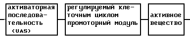
Основной элемент этого активного вещества представляет собой регулируемый клеточным циклом промоторный модуль.The main component of this active substance is the DNA construct, which consists of the following elements:
(DNA throughout the text of this application is used as a general concept for both complementary (cDNA) and genomic DNA sequences)
The main element of this active substance is a promoter module regulated by the cell cycle.
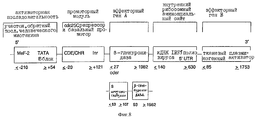
Под регулируемым клеточным циклом промоторным модулем нужно понимать, например, нуклеотидную последовательность CDE-CHR-Inr- (см. ниже). Существенной функцией промоторного модуля является ингибирование функции активаторной последовательности в GO/Gl-фазе клеточного цикла и специфической к клеточному циклу экспрессии в S/G2-фазе и, таким образом, в пролиферирующих клетках. Under the regulated cell cycle, the promoter module must be understood, for example, the nucleotide sequence of CDE-CHR-Inr- (see below). An essential function of the promoter module is the inhibition of the function of the activator sequence in the GO / Gl phase of the cell cycle and expression specific to the cell cycle in the S / G2 phase and, thus, in proliferating cells.
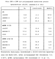
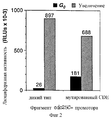
Промоторный модуль CDE-CHR-Inr обнаружен при детальном исследовании G2-специфической экспрессии человеческого промотора cdc25C. Исходным моментом являлось нахождение репрессорного элемента (зависимый от клеточного цикла элемент CDE), который является ответственным за "отключение" промотора в Gl-фазе клеточного цикла (Lucibello и др., EMBO J., 14, 132 (1995)). Путем геномного диметилсульфатного (ДМС) футпринтинга и функционального анализа (фиг. 1, 2) можно показать, что CDE Gl-специфически связывает репрессор ("CDE-связывающий фактор", CDF) и тем самым приводит к ингибированию транскрипции в непролиферирующих (GO) клетках. Локализованный в области базального промотора CDE в своей репримирующей функции зависит от "обратно активирующей (против хода транскрипции) последовательности" (UAS). Это приводит к выводу, что CDE-связывающий фактор подавляет активирующее транскрипцию действие связанных на 5'-конце активаторных протеинов зависимым от клеточного цикла образом, то есть в непролиферирующих клетках, а также в Gl-фазе клеточного цикла (фиг.3). The CDE-CHR-Inr promoter module was detected by a detailed study of the G2-specific expression of the human cdc25C promoter. The starting point was the finding of a repressor element (cell cycle dependent CDE element), which is responsible for the “shutdown” of the promoter in the Gl phase of the cell cycle (Lucibello et al., EMBO J., 14, 132 (1995)). By genomic dimethyl sulfate (DMS) footprinting and functional analysis (Fig. 1, 2), it can be shown that CDE Gl-specific binds a repressor (“CDE-binding factor”, CDF) and thereby leads to inhibition of transcription in non-proliferating (GO) cells . Localized in the region of the basal promoter CDE in its replication function depends on the "reverse activating (against the course of transcription) sequence" (UAS). This leads to the conclusion that the CDE-binding factor suppresses the transcriptional activating effect of the 5 ′ end-bound activator proteins in a cell cycle dependent manner, that is, in non-proliferating cells, as well as in the Gl phase of the cell cycle (FIG. 3).
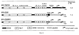
Этот вывод можно подтвердить следующим экспериментом: слияние вирус-специфического, не регулирующего клеточный цикл раннего энхансера SV40 с минимальным промотором cdc25 (состоящим из CDE и расположенной на 3'-конце стартовой области) приводит к отчетливой регуляции клеточным циклом химерного промотора (фиг.4). Последующие исследования энхансера cdc25C показывают, что в случае регулируемых зависимых от клеточного цикла CDF факторов транскрипции речь идет о NF-Y (CBF) (Dom и др., Cell, 50, 863 (1987); Van Hujisduijnen и др., ЕМВО J., 11, 3119 (1990); Coustry и др., J. Biol. Chem., 270, 468 (1995)), Sp1 (Kadonaga и др., TIBS, 11, 10 (1986)) и, возможно, новом связывающем CBS 7 факторе транскрипции (CIF). Другим представляющим интерес заключением этого исследования является обнаружение того, что NF-Y внутри энхансера cdc25C эффективно активирует транскрипцию только в сочетании по меньшей мере с одним другим комплексом NF-Y или с CIF. Как NF-Y, так и Sp1 относятся к классу обогащенных глутамином активаторов, что является важным указанием на механизм репрессии (например, взаимодействие соответственно интерференция с определенными базальными факторами транскрипции или TAFS). This conclusion can be confirmed by the following experiment: the fusion of the virus-specific, non-regulatory cell cycle of the SV40 early enhancer with the minimal cdc25 promoter (consisting of CDE and located at the 3'-end of the starting region) leads to a clear regulation of the chimeric promoter by the cell cycle (figure 4) . Subsequent studies of the cdc25C enhancer show that NF-Y (CBF) (Dom et al., Cell, 50, 863 (1987); Van Hujisduijnen et al., EMBO J. 11, 3119 (1990); Coustry et al., J. Biol. Chem. 270, 468 (1995)), Sp1 (Kadonaga et al., TIBS, 11, 10 (1986)) and, possibly, a new binding CBS 7 transcription factor (CIF). Another conclusion of interest in this study is the discovery that NF-Y within the cdc25C enhancer effectively activates transcription only in combination with at least one other NF-Y complex or CIF. Both NF-Y and Sp1 belong to the class of glutamine-enriched activators, which is an important indication of the mechanism of repression (for example, interaction, respectively, interference with certain basal transcription factors or TAFS).
Сравнение промоторных последовательностей cdc25C, циклина А и cdc2 показывает гомологию в нескольких областях (фиг.5). Во всех трех промоторах (имеющиеся отклонения функционально безотносительны) сохраняется не только CDE, но и также соседние Ус-блоки. Все эти области, как и следовало ожидать, показывают связывание протеина ин виво в случае CDE зависимым от клеточного цикла образом. Кроме того, можно видеть, что все три промотора дерегулируются за счет мутации CDE (таблица 1). Аналогичное подобие наблюдают при сравнении cdc25C, циклина А и cdc2. A comparison of the promoter sequences of cdc25C, cyclin A and cdc2 shows homology in several areas (Fig. 5). In all three promoters (the existing deviations are functionally irrelevant), not only CDEs are preserved, but also neighboring Us units. All these regions, as expected, show protein binding in vivo in the case of CDE in a cell cycle dependent manner. In addition, it can be seen that all three promoters are deregulated due to a CDE mutation (Table 1). A similar similarity was observed when comparing cdc25C, cyclin A and cdc2.
Последовательности также отчетливы (фиг.5) в области непосредственно у 3'-конца CDE ("область, гомологичная гену клеточного цикла"; CHR). Эта область функционально так же важна, как и CDE (таблица 1), однако не выявляется в экспериментах ин виво по диметилсульфатному футпринтингу. Возможным объяснением этого является взаимодействие фактора с маленьким участком ДНК. Результаты экспериментов по "анализу на резкое изменение электрофоретической подвижности" (EMSA) указывают на то, что CDE и CHR вместе связываются в протеиновый комплекс, CDF. Эти наблюдения указывают на то, что репрессия через посредство CDF обогащенных глутамином активаторов протекает по зачастую встречающемуся механизму регулируемой клеточным циклом транскрипции. The sequences are also distinct (Figure 5) in the region immediately at the 3'end of the CDE ("region homologous to the cell cycle gene"; CHR). This area is functionally as important as CDE (Table 1), but it is not detected in vivo in dimethyl sulfate footprinting experiments. A possible explanation for this is the interaction of the factor with a small portion of DNA. The results of the Electrophoretic Mobility Shift Assay (EMSA) experiments indicate that CDE and CHR bind together to form a protein complex, CDF. These observations indicate that repression via CDF of glutamine-enriched activators proceeds via the frequently encountered mechanism of cell-transcriptional regulation.
Для регуляции промотора cdc25C имеет значение, однако, не только CDE-CHR-область, но и также один из сайтов инициации (положение + 1) внутри нуклеотидной последовательности базального промотора (положения ≤ -20 до ≥ +30, см. фиг. 1). Мутации в этой области, которая включает ин витро сайт связывания фактора транскрипции YY-1 (Seto и Shenk, Nature, 354, 241 (1991); Usheva и Shenk, Cell, 76, 1115 (1994)), приводят к полной дерегуляции. Принимая во внимание близость CDE-CHR к базальному промотору, таким образом, очень вероятно взаимодействие CDF с базальным комплексом транскрипции. For regulation of the cdc25C promoter, however, not only the CDE-CHR region is important, but also one of the initiation sites (position + 1) within the nucleotide sequence of the basal promoter (positions ≤ -20 to ≥ +30, see Fig. 1) . Mutations in this region, which includes the in vitro YY-1 transcription factor binding site (Seto and Shenk, Nature, 354, 241 (1991); Usheva and Shenk, Cell, 76, 1115 (1994)), lead to complete deregulation. Given the proximity of CDE-CHR to the basal promoter, thus, the interaction of CDF with the basal transcription complex is very likely.
В качестве активаторной последовательности (обратно активирующая (против хода транскрипции) последовательность = UAS) нужно понимать нуклеотидную последовательность (промоторную или энхансерную последовательность), с которой взаимодействуют образовавшиеся или активные в клетке-мишени факторы транскрипции. В качестве активаторной последовательности можно использовать CMV-энхансер, CMV-промотор (европейский патент 073177 В1), промотор SV40 или любую другую известную специалисту промоторную или энхансерную последовательность. Согласно настоящему изобретению, однако, к предпочтительным активаторным последовательностям причисляют такие ген-регуляторные последовательности, соответственно элементы генов, которые особенно кодируют образовавшиеся в гладкомышечных клетках, активированных эндотелиальных клетках или в активированных макрофагах или лимфоцитах протеины. As the activator sequence (reverse activating (against the transcription) sequence = UAS), it is necessary to understand the nucleotide sequence (promoter or enhancer sequence) with which transcription factors formed or active in the target cell interact. As the activator sequence, a CMV enhancer, a CMV promoter (European patent 073177 B1), SV40 promoter, or any other promoter or enhancer sequence known to those skilled in the art can be used. According to the present invention, however, the preferred activator sequences include such gene regulatory sequences, respectively, elements of genes that especially encode proteins formed in smooth muscle cells, activated endothelial cells, or in activated macrophages or lymphocytes.
Под активным веществом нужно понимать ДНК-последовательность протеина, который может вызывать в месте образования терапевтический эффект, то есть ингибирование пролиферации гладкомышечных клеток, свертывания или (в случае двух активных веществ) ингибирование пролиферации и одновременно свертывания. При выборе нуклеотидной последовательности для активаторной последовательности и активного вещества руководствуются клеткой-мишенью и желательным активным веществом. By active substance is meant the DNA sequence of a protein that can produce a therapeutic effect at the site of formation, i.e., inhibition of smooth muscle cell proliferation, coagulation or (in the case of two active substances) inhibition of proliferation and coagulation at the same time. In selecting the nucleotide sequence for the activator sequence and the active substance, they are guided by the target cell and the desired active substance.
Предлагаемую согласно изобретению конструкцию ДНК известным специалисту образом дополняют до вектора; так, например, вводят в вирусный вектор (см. D. Jolly, Cancer Gene Therapy, 1, 51 (1994)), или, однако, дополняют до плазмиды. Вирусные векторы или плазмиды можно комплексовать с коллоидными дисперсиями, так, например, с липосомами (Farhood и др., Annal. of the New York Academy of Sciences, 716, 23 (1994)) или, однако, с конъюгатами полилизин-лиганд (Curiel и др., Annals of the New York Academy of Sciences, 716, 36 (1994)). Точно также можно получать лекарственные средства с помощью обычных вспомогательных для получения лекарственных средств веществ. The DNA construct according to the invention is complemented in a manner known per se to a vector; for example, they are introduced into a viral vector (see D. Jolly, Cancer Gene Therapy, 1, 51 (1994)), or, however, supplemented with a plasmid. Viral vectors or plasmids can be combined with colloidal dispersions, for example, with liposomes (Farhood et al., Annal. Of the New York Academy of Sciences, 716, 23 (1994)) or, however, with polylysine ligand conjugates (Curiel et al., Annals of the New York Academy of Sciences, 716, 36 (1994)). In the same way, it is possible to obtain drugs using conventional auxiliary substances for the preparation of drugs.
Такого рода вирусные или невирусные векторы можно дополнять лигандом, который обладает сродством к связыванию мембранной структуры с выбранной клеткой-мишенью. При выборе лиганда, таким образом, руководствуются выбором клетки-мишени (см. п.4.4-и последующие и п.5.4 и последующие). Предлагаемое согласно изобретению активное вещество подробнее поясняется, руководствуясь следующими примерами. Such viral or non-viral vectors can be supplemented with a ligand that has an affinity for binding the membrane structure to a selected target cell. When choosing a ligand, in this way, they are guided by the choice of the target cell (see section 4.4 and subsequent and section 5.4 and subsequent). The active substance according to the invention is explained in more detail in accordance with the following examples.
4). Активное вещество для ингибирования пролиферации гладкомышечных клеток:
4.1. Выбор активаторной последовательности гладкомышечных клеток
В качестве активаторных последовательностей согласно настоящему изобретению предпочтительно нужно выбирать ген-регуляторные последовательности, соответственно элементы генов, которые кодируют особенно в гладкомышечных клетках образовавшиеся протеины. Этими генами, например, являются следующие:
- тропомиозин (Tsukahara и др., Nucleic Acid Res., 22, 2318 (1994); Novy и др., Cell Motility and Cytosceleton, 25, 267 (1993); Wilton и др., Cytogenetics and Cell Genetics, 68, 122 (1995));
- α-актин (Sartorelli и др., Gens and Developm., 4, 1811 (1990); Miwa и др., Nucleic Acids Res, 18, 4263 (1990));
- α-миозин (Kelly и др., Can. J. Physiol. and Pharm., 72, 1351 (1994); Moussavi и др., Mol. Cell. Biochem., 128, 219 (1993));
- рецепторы факторов роста, как, например, фактор роста, полученный из тромбоцитов (PDGF), FGF (Rubin и др., Int. Congress Ser., 925, 131 (1990); Ross, Ann. Rev. Med., 38, 71 (1987);
- рецепторы ацетилхолина (Dutton и др., PNAS USA, 90, 2040 (1993); Durr и др., Eur. J. Biochem., 224, 353 (1994));
- фосфофруктокиназа-А (Gekakis и др., Biochemistry, 33, 1771, (1994); Tsujino и др., J. Biol. Chem., 264, 15334 (1989); Castella-Escola и др., Gene, 91, 225 (1990));
- фосфоглицератмутаза (Nakatsuji и др. , Mol. Cell. Biol., 12, 4384 (1992));
- тропонин С (Lin и др., Mol. Cell. Biol., 11, 267 (1991));
- десмин (Li и др. , J. Biol. Chem., 266, 6562 (1991); Neuromuscular Disorders, 3, 423 (1993));
- миогенин (Funk и др., PNAS USA, 89, 9484 (1992); Olson, Symp. Soc. Exp. Biol. , 46, 331 (1992); Zhon и др., Mol. Cell. Biol., 14, 6232 (1994); Atchley и др., PNAS USA, 9JL, 11522 (1994));
- рецепторы эндотелина A (Hosoda и др., J. Biol. Chem., 26 7, 18797 (1992); Orelly и др., J. Cardiovasc, Pharm., 22, 18 (1993); Hayzer и др., Am. J. Med. Sci. 304, 231 (1992); Haendler и др., J. Cardiovasc. Pharm., 20, 1 (1992));
- VEGF; VEGF образуется гладкомышечными клетками, особенно в гипоксических условиях (Berse и др., Mol. Biol. Cell, 3, 211 (1992); Finkenzeller и др. , ВВРС, 208, 432 (1995); Tischer и др., ВВРС, 165, 1198 (1989); Leung и др., Science, 246, 1306 (1989); Ferrara и др., Endoc. Fev., 13, 18 (1992)).4). Active substance for inhibiting smooth muscle cell proliferation:
4.1. Selection of activator sequence of smooth muscle cells
As activator sequences according to the present invention, it is preferable to select gene-regulatory sequences, respectively, elements of genes that encode the proteins formed especially in smooth muscle cells. These genes, for example, are as follows:
- tropomyosin (Tsukahara et al., Nucleic Acid Res., 22, 2318 (1994); Novy et al., Cell Motility and Cytosceleton, 25, 267 (1993); Wilton et al., Cytogenetics and Cell Genetics, 68, 122 (1995));
- α-actin (Sartorelli et al., Gens and Developm., 4, 1811 (1990); Miwa et al., Nucleic Acids Res, 18, 4263 (1990));
- α-myosin (Kelly et al., Can. J. Physiol. and Pharm., 72, 1351 (1994); Moussavi et al., Mol. Cell. Biochem. 128, 219 (1993));
- growth factor receptors, such as, for example, growth factor derived from platelets (PDGF), FGF (Rubin et al., Int. Congress Ser., 925, 131 (1990); Ross, Ann. Rev. Med., 38, 71 (1987);
- acetylcholine receptors (Dutton et al., PNAS USA, 90, 2040 (1993); Durr et al., Eur. J. Biochem., 224, 353 (1994));
- phosphofructokinase-A (Gekakis et al., Biochemistry, 33, 1771, (1994); Tsujino et al., J. Biol. Chem., 264, 15334 (1989); Castella-Escola et al., Gene, 91, 225 (1990));
- phosphoglyceratmutase (Nakatsuji et al., Mol. Cell. Biol., 12, 4384 (1992));
- troponin C (Lin et al., Mol. Cell. Biol., 11, 267 (1991));
- desmin (Li et al., J. Biol. Chem., 266, 6562 (1991); Neuromuscular Disorders, 3, 423 (1993));
- myogenin (Funk et al., PNAS USA, 89, 9484 (1992); Olson, Symp. Soc. Exp. Biol., 46, 331 (1992); Zhon et al., Mol. Cell. Biol., 14, 6232 (1994); Atchley et al., PNAS USA, 9JL, 11522 (1994));
endothelin A receptors (Hosoda et al., J. Biol. Chem., 26 7, 18797 (1992); Orelly et al., J. Cardiovasc, Pharm., 22, 18 (1993); Hayzer et al., Am . J. Med. Sci. 304, 231 (1992); Haendler et al., J. Cardiovasc. Pharm., 20, 1 (1992));
- VEGF; VEGF is produced by smooth muscle cells, especially under hypoxic conditions (Berse et al., Mol. Biol. Cell, 3, 211 (1992); Finkenzeller et al., VVRS, 208, 432 (1995); Tischer et al., VVRS, 165 , 1198 (1989); Leung et al., Science, 246, 1306 (1989); Ferrara et al., Endoc. Fev., 13, 18 (1992)).
Промоторные последовательности генов для этих протеинов указываются в следующих работах:
- тропомиозин (Gooding и др., Embo. J., 13, 3861 (1994));
- α-актин (Shimizu и др., J. Biol. Chem., 270, 7631 (1995); Sartorelli и др., Genes Dev., 4, 1811 (1999));
- α-миозин (Kurabayashi и др., J. Biol. Chem., 265, 19271 (1990); Molkentin и др., Mol. Cell. Biol., 14, 4947 (1994));
- рецептор PDGF (Pistritto и др., Antibiot. Chemother., 46, 73 (1994));
- рецептор FGF (Myers и др., J. Biol. Chem., 270, 8257 (1995); Johnson и др. , Adv. Cencer Res., 60, 1 (1993); Chellaiah и др., J. Biol. Chem., 269, 11620 (1994); Yu и др., Hum. Mol. Genetics, 3, 212 (1994); Wang и др., ВВРС, 203, 1781 (1994); Murgue и др., Cancer Res., 54, 5206 (1994); Avraham и др., Genomics, 21, 656 (1994); Burgess и др. , Ann. Rev. Biochem., 58, 575 (1989));
- MRF-4 (Naidu и др., Mol. Cell. Biol., 15, 2707 (1995));
- фосфофруктокиназа A (Gekakis и др., Biochem., 33, 1771 (1994));
- фосфоглицератмутаза (Makatsuji и др. , Mol. Cell. Biol., 12, 4384 (1992)) ;
- тропонин С (Ip и др., Mol. Cell. Biol., 14, 7517 (1994); Parmacek и др., Mol. Cell. Biol., 12, 1967 (1992));
- миогенин (Salminen и др., J. Cell. Biol., 115, 905 (1991); Durr и др., Eur. J. Biochem. , 224, 353 (1994); Edmondson и др., Mol. Cell. Biol., 12, 3665 (1992));
- рецепторы эндотелина A (Hosoda и др., J. Biol. Chem., 267, 18797 (1992); Li и Paulia, J. Biol. Chem., 266, 6562 (1991));
- десмин (Li и др., Neuromisc. Dis., 3, 423 (1993); Li и Capetanaki, Nucl. Acids Res., 21, 335 (1993));
- VEGF.The promoter sequences of genes for these proteins are indicated in the following works:
tropomyosin (Gooding et al., Embo. J., 13, 3861 (1994));
- α-actin (Shimizu et al., J. Biol. Chem., 270, 7631 (1995); Sartorelli et al., Genes Dev., 4, 1811 (1999));
- α-myosin (Kurabayashi et al., J. Biol. Chem., 265, 19271 (1990); Molkentin et al., Mol. Cell. Biol., 14, 4947 (1994));
- PDGF receptor (Pistritto et al., Antibiot. Chemother., 46, 73 (1994));
- FGF receptor (Myers et al., J. Biol. Chem., 270, 8257 (1995); Johnson et al., Adv. Cencer Res., 60, 1 (1993); Chellaiah et al., J. Biol. Chem., 269, 11620 (1994); Yu et al., Hum. Mol. Genetics, 3, 212 (1994); Wang et al., WWRS, 203, 1781 (1994); Murgue et al., Cancer Res. , 54, 5206 (1994); Avraham et al., Genomics, 21, 656 (1994); Burgess et al., Ann. Rev. Biochem. 58, 575 (1989));
- MRF-4 (Naidu et al., Mol. Cell. Biol., 15, 2707 (1995));
- phosphofructokinase A (Gekakis et al., Biochem., 33, 1771 (1994));
- phosphoglyceratmutase (Makatsuji et al., Mol. Cell. Biol., 12, 4384 (1992));
- troponin C (Ip et al., Mol. Cell. Biol., 14, 7517 (1994); Parmacek et al., Mol. Cell. Biol., 12, 1967 (1992));
myogenin (Salminen et al., J. Cell. Biol., 115, 905 (1991); Durr et al., Eur. J. Biochem. 224, 353 (1994); Edmondson et al., Mol. Cell. Biol., 12, 3665 (1992));
- endothelin A receptors (Hosoda et al., J. Biol. Chem., 267, 18797 (1992); Li and Paulia, J. Biol. Chem., 266, 6562 (1991));
Desmin (Li et al., Neuromisc. Dis., 3, 423 (1993); Li and Capetanaki, Nucl. Acids Res. 21, 335 (1993));
- VEGF.
Ген-регуляторными последовательностями VEGF-гена являются следующие:
- промоторная последовательность VEGF-гена (5'-фланкирующая область) (Michenko и др. , Cell. Mol. Biol. Res., 40, 35 (1994); Tischer и др., J. Biol. Chem., 266, 11947 (1991)); или
- энхансерная последовательность VEGF-гена (3'-фланкирующая область) (Michenko и др., Cell. Mol. Biol. Res., 40, 35 (1994)); или
- c-Src-ген (Mukhopadhyay и др., Nature, 375, 577 (1995) 7 Bonham и др., Oncogene, 8, 1973 (1993); Parker и др., Mol. Cell. Biol., 5, 831 (1985); Anderson и др., Mol. Cell. Biol., 5., 1122 (1985)); или
- V-Src-ген (Mukhodpadhyay и др., Nature, 375, 577 (1995); Anderson и др. , Mol. Cell. Biol. , 5, 1122 (1985); Gibbs и др., J. Virol., 53, 19 (1985));
- "искусственные" промоторы; факторы семейства спираль-петля-спираль (HLH) (MyoD, Myf-5, миогенин, MRF4 (обзор Olson и Klein, Genes Dev., 8, 1 (1994)) описываются в качестве специфических к мышцам активаторов транскрипции. Далее, к специфическим к мышцам факторам транскрипции относятся протеин "цинковый палец" GATA-4 (Arceci и др., Mol. Cell. Biol., 13, 2235 (1993); Ip и др., Mol. Cell. Biol., 14, 7517 (1994)), а также группы факторов транскрипции MEF-2 (Yu и др., Gene Dev., 6, 1783 (1992)).The gene regulatory sequences of the VEGF gene are as follows:
the promoter sequence of the VEGF gene (5'-flanking region) (Michenko et al., Cell. Mol. Biol. Res., 40, 35 (1994); Tischer et al., J. Biol. Chem., 266, 11947 (1991)); or
- enhancer sequence of the VEGF gene (3'-flanking region) (Michenko et al., Cell. Mol. Biol. Res., 40, 35 (1994)); or
- c-Src gene (Mukhopadhyay et al., Nature, 375, 577 (1995) 7 Bonham et al., Oncogene, 8, 1973 (1993); Parker et al., Mol. Cell. Biol., 5, 831 (1985); Anderson et al., Mol. Cell. Biol., 5. 1122 (1985)); or
- V-Src gene (Mukhodpadhyay et al., Nature, 375, 577 (1995); Anderson et al., Mol. Cell. Biol., 5, 1122 (1985); Gibbs et al., J. Virol., 53, 19 (1985));
- "artificial"promoters; helix-loop-helix (HLH) family factors (MyoD, Myf-5, myogenin, MRF4 (reviewed by Olson and Klein, Genes Dev., 8, 1 (1994)) are described as muscle-specific transcription activators. Further, to specific muscle transcription factors include the zinc finger protein GATA-4 (Arceci et al., Mol. Cell. Biol., 13, 2235 (1993); Ip et al., Mol. Cell. Biol., 14, 7517 (1994 )), as well as groups of transcription factors MEF-2 (Yu et al., Gene Dev., 6, 1783 (1992)).
HLH-Протеины, а также GATA-4 проявляют специфическую к мышце транскрипцию не только с промоторами специфических к мышце генов, но и также в гетерологичном окружении, а также с искусственными промоторами. Такого рода искусственными промоторами, например, являются следующие:
- множественные копии сайтов связывания (ДНК) специфических к мышце HLH-протеинов, как Е-блок (MyoD)
(например, 4хAGCAGGTGTTGGGAGGC) (Weintraub и др. , PNAS, 87, 5623 (1990));
- множественные копии сайта связывания ДНК GATA-4 α-миозин-гена вируса синдрома Франклина
(например, ![]()
- multiple copies of the binding sites (DNA) of muscle-specific HLH proteins, such as the E-block (MyoD)
(e.g. 4xAGCAGGTGTTGGGAGGC) (Weintraub et al., PNAS 87, 5623 (1990));
- multiple copies of the GATA-4 DNA binding site of the Franklin Syndrome α-myosin gene
(eg, ![]()
4.2. Выбор активного вещества гладкомышечных клеток
В качестве активного вещества согласно изобретению нужно понимать ДНК-последовательность, экспрессированный протеин которой ингибирует пролиферацию гладкомышечных клеток. К этим ингибиторам клеточного цикла относятся, например, ДНК-последовательности следующих протеинов:
а) Ингибирующие протеины:
- протеин ретинобластомы (pRb=pllO) или родственные р107 и р130 протеины (La Thangue, Curr. Opin. Cell Biol., 6, 443 (1994));
- р53-протеин (Prives и др., Genes Dev., 1, 529 (1993));
- p21 (WAF-1)-протеин (El-Deiry и др., Cell, 75, 817 (1993));
- р16-протеин (Serrano и др. , Nature, 366, 704 (1993); Kamb и др., Science, 264, 436 (1994); Nobori и др., Nature, 368, 753 (1994));
- другие ингибиторы cdK (обзор Pines, TIBS, 19, 143 (1995));
- протеин GADD45 (Papathanasiou и др. , Mol. Cell. Biol., 11, 1009 (1991); Smith и др., Science, 266, 1376 (1994));
- bak-протеин (Farrow и др., Nature, 374, 731 (1995); Chittenden и др., Nature, 374, 733 (1995); Kiefer и др., Nature, 374, 736 (1995)).4.2. Selection of the active substance of smooth muscle cells
As the active substance according to the invention, it is necessary to understand the DNA sequence, the expressed protein of which inhibits the proliferation of smooth muscle cells. These cell cycle inhibitors include, for example, the DNA sequences of the following proteins:
a) Inhibiting proteins:
- retinoblastoma protein (pRb = pllO) or related proteins p107 and p130 (La Thangue, Curr. Opin. Cell Biol., 6, 443 (1994));
- p53 protein (Prives et al., Genes Dev., 1, 529 (1993));
p21 (WAF-1) -protein (El-Deiry et al., Cell, 75, 817 (1993));
p16 protein (Serrano et al., Nature, 366, 704 (1993); Kamb et al., Science, 264, 436 (1994); Nobori et al., Nature, 368, 753 (1994));
- other cdK inhibitors (review by Pines, TIBS, 19, 143 (1995));
- GADD45 protein (Papathanasiou et al., Mol. Cell. Biol., 11, 1009 (1991); Smith et al., Science, 266, 1376 (1994));
- bak protein (Farrow et al., Nature, 374, 731 (1995); Chittenden et al., Nature, 374, 733 (1995); Kiefer et al., Nature, 374, 736 (1995)).
Для того чтобы предотвратить быструю внутриклеточную инактивацию этих игнибиторов клеточного цикла, нужно предпочтительно применять такие гены, которые обладают мутациями сайтов инактивации экспримированных протеинов, при этом не ухудшая этим их в отношении их функции. In order to prevent the rapid intracellular inactivation of these cell cycle igniters, it is preferable to use genes that have mutations in the inactivation sites of the expressed proteins, without compromising their function.
Протеин ретинобластомы (pRb/pllO) и родственные протеины р107 и р130 инактивируются за счет фосфорилирования. Таким образом, предпочтительно используют pRb/p110-, p107- или р130-кДНК-последовательность, которая мутирована таким образом, что участки фосфорилирования кодированного протеина заменены нефосфорилируемыми аминокислотами. Retinoblastoma protein (pRb / pllO) and related proteins p107 and p130 are inactivated by phosphorylation. Thus, a pRb / p110, p107, or p130 cDNA sequence is preferably used that is mutated so that the phosphorylation sites of the encoded protein are replaced by non-phosphorylated amino acids.
Согласно Hamel и др. (Mol. Cell. Biol., 12, 3431 (1992)) кДНК-последовательность протеина ретинобластомы (р110) за счет обмена аминокислот в положениях 246, 350, 601, 605, 780, 786, 787, 800 и 804 становится более нефосфорилируемой, однако ее активность связывания с большим Т-антигеном не ухудшается. Например, аминокислоты Thr-246, Ser-601, Ser-605, Ser-780, Ser-786, Ser-787 и Ser-800 заменяются на Ala, аминокислота Thr-350 заменяется на Arg и аминокислота Ser-804 заменяется на Glu. According to Hamel et al. (Mol. Cell. Biol., 12, 3431 (1992)), the cDNA sequence of the retinoblastoma protein (p110) due to the exchange of amino acids at positions 246, 350, 601, 605, 780, 786, 787, 800 and 804 becomes more non-phosphorylated, however, its binding activity to the large T-antigen does not deteriorate. For example, amino acids Thr-246, Ser-601, Ser-605, Ser-780, Ser-786, Ser-787 and Ser-800 are replaced by Ala, amino acid Thr-350 is replaced by Arg and amino acid Ser-804 is replaced by Glu.
Аналогичным образом мутируется ДНК-последовательность протеина р107 или протеина р130. Similarly, the DNA sequence of protein p107 or protein p130 is mutated.
Протеин р53 инактивируется в клетке либо за счет связывания со специальными протеинами, как, например, MDM2, или вследствие олигомеризации р53 через дефосфорилированный С-концевой серин-392 (Schikawa и др., Leukemia и Lymphoma, 11, 21 (1993); Brown, Annals of Oncology, 4, 623 (1993)). Таким образом, предпочтительно используют ДНК-последовательность протеина р53, который укорочен на С-конце на серин-392. The p53 protein is inactivated in the cell either by binding to special proteins, such as MDM2, or by oligomerization of p53 via the dephosphorylated C-terminal serine-392 (Schikawa et al., Leukemia and Lymphoma, 11, 21 (1993); Brown, Annals of Oncology, 4, 623 (1993)). Thus, the DNA sequence of the p53 protein, which is shortened at the C-terminus to serine-392, is preferably used.
б) Цитостатические или цитотоксические протеины
В качестве активного вещества, далее, нужно понимать ДНК-последовательность, которая экспрессирует (выражает) цитостатический или цитотоксический протеин.b) Cytostatic or cytotoxic proteins
As an active substance, further, it is necessary to understand the DNA sequence that expresses (expresses) a cytostatic or cytotoxic protein.
К такого рода протеинам относятся, например, следующие:
- перфорин (Lin и др., Immunol. Today, 16, 194 (1995));
- гранзим (Smyth и др., Immunol. Today, 16, 202 (1995));
- фактор некроза опухоли (Porter, Tib Tech., 9, 158 (1991); Sidhu и др., Pharmac. Ther., 57, 79 (1993)); в особенности
-α-фактор некроза опухоли (Beutler и др., Nature, 320, 584 (1986); Kriegler и др., Cell, 53, 45 (1988));
-β-фактор некроза опухоли (Gray и др., Nature, 312, 721 (1984); Li и др. , J. Immunol., 138, 4496 (1987); Aggarwal и др., J. Biol. Chem., 260, 2334 (1985)).Such proteins include, for example, the following:
- perforin (Lin et al., Immunol. Today, 16, 194 (1995));
- granzyme (Smyth et al., Immunol. Today, 16, 202 (1995));
- tumor necrosis factor (Porter, Tib Tech., 9, 158 (1991); Sidhu et al., Pharmac. Ther., 57, 79 (1993)); especially
the tumor necrosis factor α (Beutler et al., Nature, 320, 584 (1986); Kriegler et al., Cell, 53, 45 (1988));
β-tumor necrosis factor (Gray et al., Nature, 312, 721 (1984); Li et al., J. Immunol., 138, 4496 (1987); Aggarwal et al., J. Biol. Chem., 260, 2334 (1985)).
в) Ферменты
В качестве активного вещества, однако, также нужно понимать ДНК-последовательность фермента, который инактивный предшественник цитостатического препарата превращает в цитостатический препарат.c) Enzymes
As an active substance, however, it is also necessary to understand the DNA sequence of the enzyme, which converts the inactive precursor of the cytostatic preparation into a cytostatic preparation.
Такого рода ферменты, которые расщепляют инактивные предшественники веществ (пропрепараты) до активных цитостатических препаратов (лекарственные средства), и возможные пропрепараты и лекарственные средства уже наглядно описаны Deonarain и др., (Br. J. Cancer, 70, 786 (1994)), Mullen (Pharmac. Ther., 63, 199 (1994)) и Harris и др. (Gene Ther., 1, 170 (1994)). Such enzymes that break down inactive precursors of substances (drugs) to active cytostatic drugs (drugs), and possible drugs and drugs have already been clearly described by Deonarain et al., (Br. J. Cancer, 70, 786 (1994)), Mullen (Pharmac. Ther., 63, 199 (1994)) and Harris et al. (Gene Ther., 1, 170 (1994)).
Например, можно применять ДНК-последовательность одного из следующих ферментов:
- тимидинкиназа вируса простого герпеса
(Garapin и др., PNAS USA, 76, 3755 (1979); Vile и др., Cancer Res., 53, 3860 (1993); Wagner и др., PNAS USA, 78, 1441 (1981); Moelten и др., Cancer Res., 46, 5276 (1986); J. Natl. Cancer Inst., 82, 297 (1990));
- тимидинкиназа вируса ветряной оспы
(Huber и др. , PNAS USA, 88, 8039 (1991); Snoeck, Int. J. Antimicrob. Agents, 4, 211 (1994));
- бактериальная нитроредуктаза
(Michael и др., FEMS Microbiol. Letters, 124, 195 (1994); Bryant и др., J. Biol. Chem. , 266, 4126 (1991); Watanabe и др., Nucleic Acids Res., 18, 1059 (1990));
- бактериальная β-глюкуронидаза
(Jefferson и др., PNAS USA, 83, 8447 (1986));
- растительная β-глюкуронидаза из сухих зерновых культур
(Schulz и др., Phytochemistry, 26, 933 (1987));
- человеческая β-глюкуронидаза
(Bosslet и др., Br. J. Cancer, 65, 234 (1992); Oshima и др., PNAS USA, 84, 685 (1987));
- человеческая карбоксипептидаза (СВ), например:
СВ-А тучной клетки
(Reynolds и др., J. Clin. Invest, 89, 273 (1992));
СВ-В поджелудочной железы
(Yamamoto и др., J. Biol. Chem., 267, 2575 (1992);
Catasus и др., J. Biol. Chem., 270, 6651 (1995));
- бактериальная карбоксипептидаза
(Hamilton и др. , J. Bacteriol., 174, 1626 (1992); Osterman и др., J. Protein Chem., 11, 561 (1992));
- бактериальная β-лактамаза
(Rodriges и др. , Cancer Res., 55, 63 (1995); Hussain и др., J. Bacteriol., 164, 223 (1985); Coqe и др., Embo J., 12, 631 (1993));
- бактериальная цитозиндезаминаза
(Mullen и др., PNAS USA, 89, 33 (1992); Austin и др., Mol. Pharmac., 43, 380 (1993); Danielson и др., Mol. Microbiol., 6, 1335 (1992));
- человеческая каталаза, соответственно пероксидаза
(Ezurum и др., Nucl. Acids Res., 21, 1607 (1993));
- фосфатаза, в особенности
человеческая щелочная фосфатаза
(Gum и др., Cancer Res., 50, 1085 (1990));
человеческая кислая фосфатаза простаты
(Sharieff и др. , Am. J. Hum. Gen., 49, 412 (1991); Song и др., Gene, 129, 291 (1993); Tailor и др., Nucl. Acids Res., 18, 4928 (1990));
- кислая фосфатаза типа 5
(Gene, 130, 201 (1993));
- оксидаза, в особенности
человеческая лизилоксидаза
(Kimi и др., J. Biol. Chem., 270, 7176 (1995));
человеческая кислая D-аминооксидаза
(Fukui и др., J. Biol. Chem., 267, 18631 (1992));
- пероксидаза, в особенности
человеческая глутатион-пероксидаза
(Chada и др., Genomics, 6, 268 (1990); Ishida и др., Nucl. Acids Res., 15, 10051 (1987));
человеческая эозинофильная пероксидаза
(Ten и др. , J. Exp. Med., 169, 1757 (1989); Sahamaki и др., J. Biol. Chem., 264, 16828 (1989));
- человеческая пероксидаза щитовидной железы
(Kimura, PNAS USA, 84, 5555 (1987)).For example, you can use the DNA sequence of one of the following enzymes:
- herpes simplex virus thymidine kinase
(Garapin et al., PNAS USA, 76, 3755 (1979); Vile et al., Cancer Res., 53, 3860 (1993); Wagner et al., PNAS USA, 78, 1441 (1981); Moelten et al. ., Cancer Res., 46, 5276 (1986); J. Natl. Cancer Inst., 82, 297 (1990));
- chickenpox virus thymidine kinase
(Huber et al., PNAS USA, 88, 8039 (1991); Snoeck, Int. J. Antimicrob. Agents, 4, 211 (1994));
- bacterial nitroreductase
(Michael et al., FEMS Microbiol. Letters, 124, 195 (1994); Bryant et al., J. Biol. Chem., 266, 4126 (1991); Watanabe et al., Nucleic Acids Res., 18, 1059 (1990));
- bacterial β-glucuronidase
(Jefferson et al., PNAS USA, 83, 8447 (1986));
- plant β-glucuronidase from dry crops
(Schulz et al., Phytochemistry, 26, 933 (1987));
- human β-glucuronidase
(Bosslet et al., Br. J. Cancer, 65, 234 (1992); Oshima et al., PNAS USA, 84, 685 (1987));
- human carboxypeptidase (CB), for example:
CB-A mast cell
(Reynolds et al., J. Clin. Invest, 89, 273 (1992));
SV-In the pancreas
(Yamamoto et al., J. Biol. Chem., 267, 2575 (1992);
Catasus et al., J. Biol. Chem., 270, 6651 (1995));
- bacterial carboxypeptidase
(Hamilton et al., J. Bacteriol., 174, 1626 (1992); Osterman et al., J. Protein Chem., 11, 561 (1992));
- bacterial β-lactamase
(Rodriges et al., Cancer Res., 55, 63 (1995); Hussain et al., J. Bacteriol., 164, 223 (1985); Coqe et al., Embo J., 12, 631 (1993)) ;
- bacterial cytosine deaminase
(Mullen et al., PNAS USA, 89, 33 (1992); Austin et al., Mol. Pharmac., 43, 380 (1993); Danielson et al., Mol. Microbiol., 6, 1335 (1992)) ;
- human catalase, respectively peroxidase
(Ezurum et al., Nucl. Acids Res. 21, 1607 (1993));
- phosphatase, in particular
human alkaline phosphatase
(Gum et al., Cancer Res., 50, 1085 (1990));
human acid prostate phosphatase
(Sharieff et al., Am. J. Hum. Gen., 49, 412 (1991); Song et al., Gene, 129, 291 (1993); Tailor et al., Nucl. Acids Res., 18, 4928 (1990));
-
(Gene, 130, 201 (1993));
- oxidase, in particular
human lysyl oxidase
(Kimi et al., J. Biol. Chem., 270, 7176 (1995));
human acid D-amino oxidase
(Fukui et al., J. Biol. Chem., 267, 18631 (1992));
- peroxidase, in particular
human glutathione peroxidase
(Chada et al., Genomics, 6, 268 (1990); Ishida et al., Nucl. Acids Res., 15, 10051 (1987));
human eosinophilic peroxidase
(Ten et al., J. Exp. Med., 169, 1757 (1989); Sahamaki et al., J. Biol. Chem., 264, 16828 (1989));
- human thyroid peroxidase
(Kimura, PNAS USA, 84, 5555 (1987)).
Для облегчения секреции указанных ферментов, возможно, содержащуюся в ДНК-последовательности гомологичную сигнальную последовательность можно заменять на гетерологичную улучшающую внеклеточное выделение сигнальную последовательность. To facilitate the secretion of these enzymes, it is possible that the homologous signal sequence contained in the DNA sequence can be replaced by a heterologous extracellular-enhancing signal sequence.
Так, например, сигнальную последовательность β-глюкуронидазы (положение в ДНК ≤ 27 до 93; Oshima и др., PNAS, 84, 685 (1987)) можно заменять сигнальной последовательностью человеческого иммуноглобулина (положение в ДНК ≤ 63 до ≥ 107; Riechmann и др., Nature, 332, 323 (1988)). For example, the signal sequence of β-glucuronidase (position in DNA ≤ 27 to 93; Oshima et al., PNAS, 84, 685 (1987)) can be replaced by the signal sequence of human immunoglobulin (position in DNA ≤ 63 to ≥ 107; Riechmann and et al., Nature, 332, 323 (1988)).
Далее, предпочтительно нужно выбирать кДНК таких ферментов, которые за счет точечных мутаций в более незначительной степени накапливаются в лизосомах. Такого рода точечные мутации описываются, например, для β-глюкуронидазы (Shipley и др., J. Biol. Chem., 268, 12193 (1993)). Further, it is preferable to select cDNAs of such enzymes that, due to point mutations, accumulate to a lesser extent in lysosomes. Point mutations of this kind are described, for example, for β-glucuronidase (Shipley et al., J. Biol. Chem., 268, 12193 (1993)).
4.3. Комбинация одинаковых или разных активных веществ гладкомышечных клеток
Предметом изобретения, далее, является активное вещество, в котором имеется комбинация ДНК-последовательностей нескольких одинаковых активных веществ (А, А) или разных активных веществ (А, Б). Для экспрессии двух ДНК-последовательностей предпочтительно промежуточно включают кДНК "внутреннего рибосомного аминоацильного сайта" (IRES) в качестве регуляторного элемента. Такого рода IRES описываются Mountford и Smith (TIG, 11, 179 (1995)), Kaufman и др., Nucl. Acids Res., 19, 4485 (1991); Morgan и др., Nucl. Acids Res. , 20, 1293 (1992) и Dirks и др., Gene, 129, 247 (1993); Pelletier и Sonenberg, Nature, 334, 320 (1988); и др., Bio Techn., 12, 694 (1994).4.3. The combination of the same or different active substances of smooth muscle cells
The subject of the invention, further, is an active substance in which there is a combination of DNA sequences of several identical active substances (A, A) or different active substances (A, B). For expression of two DNA sequences, preferably the cDNA of the “internal ribosome aminoacyl site” (IRES) cDNA is included as a regulatory element. Such IRESs are described by Mountford and Smith (TIG, 11, 179 (1995)), Kaufman et al., Nucl. Acids Res., 19, 4485 (1991); Morgan et al., Nucl. Acids Res. 20, 1293 (1992) and Dirks et al., Gene, 129, 247 (1993); Pelletier and Sonenberg, Nature, 334, 320 (1988); et al., Bio Techn., 12, 694 (1994).
Так, кДНК IRES-последовательности полиовируса (положение ≤ 140 до ≥ 630 5'-UTR (Pelletier и Sonenberg, Nature, 334, 320 (1988)) можно применять для связывания ДНК антитромботического вещества А (по 3'-концу) и ДНК антитромботического вещества Б (по 5'-концу). Thus, cDNA of poliovirus IRES sequences (position ≤ 140 to ≥ 630 5'-UTR (Pelletier and Sonenberg, Nature, 334, 320 (1988)) can be used to bind DNA of antithrombotic substance A (at the 3'-end) and DNA of antithrombotic substance B (at the 5'-end).

Такого рода активное вещество в зависимости от сочетания обладает аддитивным или синергическим действием согласно изобретению.
This kind of active substance, depending on the combination, has an additive or synergistic effect according to the invention.
Вследствие относящегося к внутренней оболочке роста гладкомышечных клеток, так же как за счет их апоптоза или некроза, как результат воздействия ингибитора цикла может активироваться система свертывания и могут возникать тромбозы. Такого рода тромбозы нужно предотвращать путем профилактического введения ингибитора свертывания (аспирин, гепарин или другое антитромботическое средство). Введение ингибитора свертывания осуществляют системно, то есть орально или парентерально. Due to the growth of smooth muscle cells related to the inner shell, as well as due to their apoptosis or necrosis, the coagulation system can be activated as a result of exposure to the cycle inhibitor and thrombosis can occur. Thromboses of this kind must be prevented by the prophylactic administration of a coagulation inhibitor (aspirin, heparin or other antithrombotic agent). The coagulation inhibitor is administered systemically, i.e., orally or parenterally.
Однако зачастую побочные действия ингибитора свертывания препятствуют достаточной концентрации в месте относящегося к внутренней оболочке роста гладкомышечных клеток. However, often the side effects of the coagulation inhibitor prevent sufficient concentration at the site of smooth muscle cell growth belonging to the inner membrane.
Таким образом, профилактика тромбоза благодаря такого рода ингибиторам свертывания является ненадежной (Pukac, Am. J. Pathol., 139, 1501 (1991)). Thus, the prevention of thrombosis due to such coagulation inhibitors is unreliable (Pukac, Am. J. Pathol., 139, 1501 (1991)).
Следующим предметом изобретения является то, что заявляемое в изобретении активное вещество дополнительно к активному веществу, которое представляет собой ингибитор клеточного цикла, в качестве другого элемента содержит ДНК-последовательность активного вещества, которое представляет собой ингибитор свертывания. A further subject of the invention is that the active substance claimed in the invention, in addition to the active substance, which is a cell cycle inhibitor, contains, as another element, the DNA sequence of the active substance, which is a coagulation inhibitor.

Экспрессия ингибитора свертывания регулируется таким же образом, как и экспрессия ингибитора клеточного цикла, за счет активаторной последовательности и регулируемого клеточным циклом репрессорного модуля. Одновременная экспрессия как ингибитора клеточного цикла, так же как ингибитора свертывания, регулируется предпочтительно элементом гена "внутренним рибосомным аминоацильным сайтом" (IRES).
The expression of the coagulation inhibitor is regulated in the same manner as the expression of the cell cycle inhibitor, due to the activator sequence and the cell cycle regulated repressor module. Simultaneous expression as a cell cycle inhibitor, as well as a coagulation inhibitor, is preferably regulated by the internal ribosome aminoacyl site (IRES) gene element.
В качестве ингибитора свертывания можно использовать гены, например, активаторов плазминогена (РА), как тканевый PA (tPA) или урокиназа-подобный PA (uPA), протеин С, антитромбин-III, ингибитор метаболического пути тканевого фактора или гирудин. ДНК-Последовательности этих ингибиторов свертывания описываются ниже:
- активатор тканевого плазминогена (tPA)
(Sasaki и др., Nucl. Acids Res., 16, 5695 (1988); Pennica и др., Nature, 301, 214 (1983); Wei и др., DNA, 4, 76 (1985); Harris и др., Mol. Biol. Med. , 3, 279 (1986));
- плазминогенный активатор урокиназного типа (uРА)
(Miyake и др. , J. Biochem., 104, 643 (1988); Nelles и др., J. Biol. Chem., 262, 5682 (1987));
- гибриды tPA и uPA
(Kalyan и др. , Gene, 68, 205 (1988); Devries и др., Biochem. 27, 2565 (1988));
- протеин С (Foster и др., PNAS, 82, 4673 (1985));
- гирудин
(Maerki и др., Semin. Thromb. Hemostas., 17, 88 (1991); De Taxis du Poet и др., Blood Coag. Fibrin., 2, 113 (1991); Harvey и др., PNAS USA, 83, 1084 (1986); Sachhieri и др., европейский патент 0324712 В1; европейский патент 0142860 В1);
- ингибиторы серин-протеиназы (серпины), как, например, C-lS-ингибитор
(Bock и др., Biochem., 25, 4292 (1986); Davis и др., PNAS USA, 83, 3161 (1986); Que, BBPC, 137, 620 (1986); Rauth и др., Proteine Sequences and Data Analysis, 1, 251 (1988); Carter и др., Eur. J. Biochem., 173, 163 (1988); Tosi и др., Gene, 42, 265 (1986); Carter и др., Eur. J. Biochem., 197, 301 (1991); Eldering и др., J. Biol. Chem., 267, 7013 (1993));
- 1-антитрипсин
(Tosi и др., Gene, 42, 265 (1986); Graham и др., Hum. Genetics, 85, 381 (1990); Hafeez и др. , J. Clin. Invest., 89, 1214 (1992); Тикунова и др., Биоорганическая химия, 17, 1694 (1991); Kay и др., Human Gene Ther., 3, 641 (1992); Lemarchand и др., Mol. Biol., 27, 1014 (1993); Lambach и др., Human Mol. Gen., 7, 1001 (1993));
- антитромбин-III
(Stackhouse и др., J. Biol. Chem., 258, 703 (1983); Olds и др., Bioshem. , 32, 4216 (1993); Laue и др., Nucl. Acids Res., 22, 3556 (1994));
- ингибитор метаболического пути тканевого фактора
(TFPI)
(Enjyoji и др. , Genomics, 17, 423 (1993); Wun и др., J. Biol. Chem., 263, 6001 (1988); Girard и др., Thromb. Res., 55, 37 (1989)).As a coagulation inhibitor, genes, for example plasminogen activators (PA), such as tissue PA (tPA) or urokinase-like PA (uPA), protein C, antithrombin-III, tissue factor metabolic pathway inhibitor or hirudin can be used. The DNA sequences of these coagulation inhibitors are described below:
- tissue plasminogen activator (tPA)
(Sasaki et al., Nucl. Acids Res., 16, 5695 (1988); Pennica et al., Nature, 301, 214 (1983); Wei et al., DNA, 4, 76 (1985); Harris et al. ., Mol. Biol. Med., 3, 279 (1986));
- plasminogen activator urokinase type (uRA)
(Miyake et al., J. Biochem., 104, 643 (1988); Nelles et al., J. Biol. Chem., 262, 5682 (1987));
- tPA and uPA hybrids
(Kalyan et al., Gene, 68, 205 (1988); Devries et al., Biochem. 27, 2565 (1988));
- protein C (Foster et al., PNAS, 82, 4673 (1985));
- hirudin
(Maerki et al., Semin. Thromb. Hemostas., 17, 88 (1991); De Taxis du Poet et al., Blood Coag. Fibrin., 2, 113 (1991); Harvey et al., PNAS USA, 83 1084 (1986); Sachhieri et al., European patent 0324712 B1; European patent 0142860 B1);
serine proteinase inhibitors (serpins), such as, for example, a C-lS inhibitor
(Bock et al., Biochem., 25, 4292 (1986); Davis et al., PNAS USA, 83, 3161 (1986); Que, BBPC, 137, 620 (1986); Rauth et al., Proteine Sequences and Data Analysis, 1, 251 (1988); Carter et al., Eur. J. Biochem., 173, 163 (1988); Tosi et al., Gene, 42, 265 (1986); Carter et al., Eur. J. Biochem., 197, 301 (1991); Eldering et al., J. Biol. Chem., 267, 7013 (1993));
- 1-antitrypsin
(Tosi et al., Gene, 42, 265 (1986); Graham et al., Hum. Genetics, 85, 381 (1990); Hafeez et al., J. Clin. Invest., 89, 1214 (1992); Tikunova et al., Bioorganic Chemistry, 17, 1694 (1991); Kay et al., Human Gene Ther., 3, 641 (1992); Lemarchand et al., Mol. Biol., 27, 1014 (1993); Lambach et al., Human Mol. Gen., 7, 1001 (1993));
- antithrombin-III
(Stackhouse et al., J. Biol. Chem., 258, 703 (1983); Olds et al., Bioshem. 32, 4216 (1993); Laue et al., Nucl. Acids Res., 22, 3556 ( 1994));
- tissue factor metabolic pathway inhibitor
(TFPI)
(Enjyoji et al., Genomics, 17, 423 (1993); Wun et al., J. Biol. Chem., 263, 6001 (1988); Girard et al., Thromb. Res., 55, 37 (1989) )
4.4. Выбор лиганда гладкомышечных клеток
В качестве лиганда в коллоидных дисперсиях, например, полилизин-лиганд-конъюгаты, предпочтительны вещества, которые связываются с поверхностью гладкомышечных клеток. Сюда относятся антитела или фрагменты антител, направленные против мембранных структур гладкомышечных клеток, как, например:
- антитело 10F3
(Printseva и др., Exp. Cell Res., 169, 85 (1987); Am. J. Path., 134, 305 (1989)); или
- антитело против актина
(Desmonliere и др., Comptes Rendus des Seances de la Soc. de Biol. и de ses Filiales, 182, 391 (1988)); или
- антитела против рецепторов ангиотензина-II
(Butcher и др., ВВРА, 196, 1280 (1993)); или
- антитела против рецепторов факторов роста
(обзор Mendelsohn, Prog. All., 45, 147 (1988); Sato и др., J. Nat. Canс. Inst., 81, 1600 (1989); Hynes и др., ВВА, 1198, 165 (1994));
или антитела, направленные, например, против
- EGF-рецепторов
(Fan и др., Cancer Res., 53, 4322 (1993); Bender и др., Cancer Res., 52, 121 (1992); Aboud-Pirak и др., J. Nat. Cancer Inst., 80, 1605 (1988); Sato и др., Mol. Biol. Med., 1, 511 (1983); Kawamoto и др., PNAS, 80, 1337 (1983));
- или против PDGF-рецепторов
(Yu и др. , J. Biol. Chem., 269, 10668 (1994); Kelly и др., J. Biol. Chem. , 266, 8987 (1991); Bowen-Pope и др. , J. Biol. Chem., 257, 5161 (1982));
- или против FGF-рецепторов
(Vanhalteswaran и др., J. Cell. Biol., 115, 418 (1991); Zhan и др., J. Biol. Chem., 269, 20221 (1994));
- или антитела против рецепторов эндотелина-А.4.4. Smooth muscle cell ligand selection
As a ligand in colloidal dispersions, for example, polylysine ligand conjugates, substances that bind to the surface of smooth muscle cells are preferred. This includes antibodies or antibody fragments directed against the membrane structures of smooth muscle cells, such as:
- antibody 10F3
(Printseva et al., Exp. Cell Res., 169, 85 (1987); Am. J. Path., 134, 305 (1989)); or
anti-actin antibody
(Desmonliere et al., Comptes Rendus des Seances de la Soc. De Biol. And de ses Filiales, 182, 391 (1988)); or
- antibodies against angiotensin-II receptors
(Butcher et al., WWRA, 196, 1280 (1993)); or
- antibodies against growth factor receptors
(review by Mendelsohn, Prog. All., 45, 147 (1988); Sato et al., J. Nat. Canc. Inst., 81, 1600 (1989); Hynes et al., BBA, 1198, 165 (1994) );
or antibodies directed, for example, against
- EGF receptors
(Fan et al., Cancer Res., 53, 4322 (1993); Bender et al., Cancer Res., 52, 121 (1992); Aboud-Pirak et al., J. Nat. Cancer Inst., 80, 1605 (1988); Sato et al., Mol. Biol. Med., 1, 511 (1983); Kawamoto et al., PNAS, 80, 1337 (1983));
- or against PDGF receptors
(Yu et al., J. Biol. Chem., 269, 10668 (1994); Kelly et al., J. Biol. Chem., 266, 8987 (1991); Bowen-Pope et al., J. Biol. Chem., 257, 5161 (1982));
- or against FGF receptors
(Vanhalteswaran et al., J. Cell. Biol., 115, 418 (1991); Zhan et al., J. Biol. Chem., 269, 20221 (1994));
- or antibodies against endothelin-A receptors.
Мышиные моноклональные антитела предпочтительно нужно использовать в гуманизированной форме. Гуманизирование осуществляют способом, описанным Winter и др., Nature, 349, 293 (1991), и Hoogenbooms и др., Rev. Tr. Transfus. Hemobiol., 36, 19 (1993). Фрагменты антител получают согласно уровню техники, например, способом, описанным Winter и др., Nature. 349, 293 (1991); Hoogenboom и др., Rev. Tr. Transfus. Hemobiol., 36, 19 (1993); Girol, Mol. Immunol. , 28, 1379 (1991) или Huston и др., Int. Res. Immunol., 10, 195 (1993). Mouse monoclonal antibodies should preferably be used in humanized form. Humanization is carried out by the method described by Winter et al., Nature, 349, 293 (1991), and Hoogenbooms et al., Rev. Tr. Transfus Hemobiol., 36, 19 (1993). Antibody fragments are prepared according to the prior art, for example, by the method described by Winter et al., Nature. 349, 293 (1991); Hoogenboom et al., Rev. Tr. Transfus Hemobiol., 36, 19 (1993); Girol, Mol. Immunol. 28, 1379 (1991) or Huston et al., Int. Res. Immunol., 10, 195 (1993).
К лигандам относятся, далее, все активные вещества, которые связываются с мембранными структурами или мембранными рецепторами гладкомышечных клеток (обзор Pusztai и др. , J. Pathol., 169, 191 (1993); Harris, Current Opin. Biotechnol. , 2, 260 (1991)). Например, сюда относятся факторы роста или их фрагменты, соответственно их частичные последовательности, которые связываются с экспрессированными гладкомышечными клетками рецепторами, как, например:
- PDGF
(Westermark и др. , Cancer Res., 51, 5087 (1991); Ponten и др., J. Invest. Dermatol., 102, 304 (1994));
- EGF
(Modjtahedi и др., Int. J. Oncol., 4, 277 (1994);
Carpentar и др., J. Biol. Chem., 265, 7709 (1990);
- TGF-β
(Segarini, BBA, 1155, 269 (1993));
- TGF-α
(Salomon и др., Cancer Cells, 2, 389 (1990));
- FGF
(Burgess и др., Annu. Rev. Biochem., 58, 575 (1989));
- эндотелин-А
(Oreilly и др., J. Cardiovasc. Pharm., 22, 18 (1993)).Ligands include, further, all active substances that bind to membrane structures or membrane receptors of smooth muscle cells (review by Pusztai et al., J. Pathol., 169, 191 (1993); Harris, Current Opin. Biotechnol., 2, 260 (1991)). For example, this includes growth factors or fragments thereof, respectively, their partial sequences that bind to expressed smooth muscle cells by receptors, such as:
- PDGF
(Westermark et al., Cancer Res., 51, 5087 (1991); Ponten et al., J. Invest. Dermatol., 102, 304 (1994));
- EGF
(Modjtahedi et al., Int. J. Oncol., 4, 277 (1994);
Carpentar et al., J. Biol. Chem., 265, 7709 (1990);
- TGF-β
(Segarini, BBA, 1155, 269 (1993));
- TGF-α
(Salomon et al., Cancer Cells, 2, 389 (1990));
- FGF
(Burgess et al., Annu. Rev. Biochem., 58, 575 (1989));
- endothelin-A
(Oreilly et al., J. Cardiovasc. Pharm., 22, 18 (1993)).
4.5. Получение биологически активного вещества гладкомышечных клеток
Получение предлагаемого согласно изобретению активного вещества описывается подробнее, руководствуясь следующими примерами:
а). Конструкция химерного промотора миогенин-промотор CDE-CHR-Inr
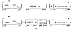
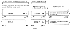
Человеческий миогениновый промотор (положение ≤ -210 до ≥ +54 ДНК-последовательности, опубликованной Salmin и др. , J. Cell. Biol., 115, 905 (1991)) своим 3'-концом связывается с 5'-концом CDE-CHR-Inr-модуля человеческого cdc25C-гeнa (положение ≤ -20 до ≥ +121 последовательности, опубликованной Lucibello и др., EMBO J., 14, 132 (1995)) (см. фиг.6). Связывание осуществляется с помощью известных специалисту и имеющихся в продаже ферментов. Кроме того, применяют различные фрагменты миогениновой промоторной последовательности (см. фиг. 6). Так, применяют содержащую ТАТА-блок ДНК-последовательность миогенинового промотора. Однако равным образом можно применять также промоторную последовательность в положении ≤ -210 до ≥ -40 (см. фиг.6).4.5. Obtaining biologically active substances of smooth muscle cells
The preparation of the active substance according to the invention is described in more detail in accordance with the following examples:
a). Design of the chimeric promoter myogenin promoter CDE-CHR-Inr
The human myogenin promoter (position ≤ -210 to ≥ +54 of the DNA sequence published by Salmin et al., J. Cell. Biol., 115, 905 (1991)) binds to the 5'-end of CDE-CHR by its 3 ′ end -Inr module of the human cdc25C gene (position ≤ -20 to ≥ +121 of the sequence published by Lucibello et al., EMBO J., 14, 132 (1995)) (see FIG. 6). The binding is carried out using known to the specialist and commercially available enzymes. In addition, various fragments of the myogenin promoter sequence are used (see FIG. 6). Thus, a DNA sequence of the myogenin promoter is used containing a TATA block. However, the promoter sequence at position ≤ -210 to ≥ -40 can also be used in the same way (see FIG. 6).
б). Конструкция плазмиды, содержащей основную (центральную) составную часть активного вещества
Таким образом полученную химерную контрольную единицу транскрипции промоторного модуля миогенина ее 3'-концом связывают с 5'-концом ДНК, которая содержит полную кодирующую область человеческой β-глюкуронидазы (положение в ДНК ≤ 27 - ≥ 1982 последовательности, опубликованной Oshima и др., PNAS USA, 84, 684 (1987)). (см. фиг.6).b) The construction of a plasmid containing the main (Central) component of the active substance
The thus obtained chimeric control unit for the transcription of the myogenin promoter module by its 3'-end is linked to the 5'-end of the DNA, which contains the complete coding region of human β-glucuronidase (position in DNA ≤ 27 - ≥ 1982 of the sequence published by Oshima et al., PNAS USA 84, 684 (1987)). (see Fig.6).
Эта ДНК также содержит необходимую для секреции сигнальную последовательность (22 N-концевые аминокислоты). Для облегчения клеточного выделения эту сигнальную последовательность предпочтительно обменивают на сигнальную последовательность иммуноглобулина (положение ≤ 63 до ≥ 107; Riechmann и др. , Nature, 332, 323 (1988)) (фиг.7). Таким образом полученные контрольные единицы транскрипции и ДНК человеческой β-глюкуронидазы клонируют в pUC18/19 или производимых от Bluescript плазмидных векторов, которые можно использовать непосредственно или в коллоидных дисперсионных системах для введения ин виво. Альтернативно, химерные гены можно трансферировать в вирусные векторы или другие пригодные векторы и вводить путем инъекции. This DNA also contains the signal sequence necessary for secretion (22 N-terminal amino acids). To facilitate cell isolation, this signal sequence is preferably exchanged for an immunoglobulin signal sequence (position ≤ 63 to ≥ 107; Riechmann et al., Nature, 332, 323 (1988)) (FIG. 7). Thus obtained transcriptional control units and human β-glucuronidase DNA are cloned into pUC18 / 19 or from plasmid vectors produced from Bluescript, which can be used directly or in colloidal dispersion systems for in vivo administration. Alternatively, chimeric genes can be transformed into viral vectors or other suitable vectors and injected.
в). Конструкция плазмиды, содержащей гены β-глюкуронидазы, как также тканевый плазминогенный активатор
Полученный как в п.2) элемент β-глюкуронидазы миогенинового репрессорного модуля его 3'-концом связывают с 5'-концом кДНК "внутреннего рибосомного аминоациального сайта" полиовируса (положение ≤ 140 до ≥ 630 5'-UTR-элемента Pelletier и Sonenberg, Nature, 334, 320 (1988)). С его 3'-концом снова связывают 5'-конец ДНК тканевого плазминогенного активатора (положение ≤ 85 до ≥ 1753; Pennica и др., Nature, 301, 214 (1983)) (фиг.7). Затем всю конструкцию клонируют в pUC17/19 или производимых от Bluescript плазмидных векторах, которые можно использовать непосредственно или в коллоидальных дисперсионных системах для переноса ин виво. Альтернативно, химерные гены можно трансферировать в вирусные векторы или другие пригодные векторы (фиг. 8).in). Construction of a plasmid containing β-glucuronidase genes, as well as tissue plasminogen activator
Obtained as in claim 2), the β-glucuronidase element of the myogenin repressor module with its 3 ′ end is connected to the 5 ′ end of the cDNA of the “internal ribosomal aminoacial site” of the poliovirus (position ≤ 140 to ≥ 630 of the 5′-UTR element of Pelletier and Sonenberg, Nature, 334, 320 (1988)). The 5 ′ end of the DNA of the tissue plasminogen activator (position ≤ 85 to ≥ 1753; Pennica et al., Nature, 301, 214 (1983)) is again linked to its 3′-end (FIG. 7). The whole construct is then cloned into pUC17 / 19 or from Bluescript-derived plasmid vectors, which can be used directly or in colloidal dispersion systems for in vivo transfer. Alternatively, chimeric genes can be transformed into viral vectors or other suitable vectors (FIG. 8).
5). Активное вещество для ингибирования свертывания
5.1. Выбор активаторной последовательности для игнибирования или свертывания
В качестве активаторных последовательностей согласно изобретению предпочтительно нужно применять ген-регуляторные последовательности, соответственно элементы из генов, которые кодируют обнаруживаемые в гладкомышечных клетках, в активированных эндотелиальных клетках, в активированных макрофагах или в активированных лимфоцитах протеины.5). Coagulation Inhibition Active
5.1. The choice of the activator sequence for inhibition or coagulation
As activator sequences according to the invention, it is preferable to use gene-regulatory sequences, respectively, elements from genes that encode proteins found in smooth muscle cells, in activated endothelial cells, in activated macrophages or in activated lymphocytes.
а) Гладкомышечные клетки
Примеры активаторных последовательностей генов в гладкомышечных клетках уже указаны в п.4.1.a) Smooth muscle cells
Examples of activator sequences of genes in smooth muscle cells are already indicated in Section 4.1.
б). Активированные эндотелиальные клетки
Примеры протеинов, которые особенно образуются в активированных эндотелиальных клетках, описываются Burrows и др., (Pharmac. Therp., 64, 155 (1994)) и Plate и сотр., (Brain Pathol., 4, 207 (1994)). В особенности к этим интенсивно образующимся в эндотелиальных клетках протеинам относятся, например, следующие:
- специфический к мозгу эндотелиальный переносчик глюкозы-I; эндотелиальные клетки мозга отличаются очень сильной экспрессией этого переносчика, чтобы осуществить трансэндотелиальную транспортировку D-глюкозы в мозг (Gerhart и др., J. Neurosci. Res., 22, 464 (1989)). Промоторная последовательность описывается Murakami и др., J. Biol. Chem., 267, 9300 (1992);
- эндоглин; эндоглин, по-видимому, является не переносящим сигнала рецептором TGF-β (Gougos и др., J. Biol. Chem., 265, 8361 (1990); Cheifetz, 1. Biol. Chem., 267, 19027 (1992); Moren и др., ВВРС, 189, 356 (1992)). В незначительных количествах он встречается в нормальном эндотелии, однако усиленно экспримируется пролиферирующим эндотелием (Westphal и др., J. Invest. Derm. , 100, 27 (1993); Burrows и др., Pharmac. Ther., 64, 155 (1994)). Промоторная последовательность описывается Bellon и др. (Eur. J. Immunol., 23, 2340 (1993)) и Ge и др. (Gene, 138, 201 (1994)).b) Activated Endothelial Cells
Examples of proteins that are especially formed in activated endothelial cells are described by Burrows et al. (Pharmac. Therp., 64, 155 (1994)) and Plate et al. (Brain Pathol., 4, 207 (1994)). In particular, these proteins, which are intensively formed in endothelial cells, include, for example, the following:
- brain-specific endothelial carrier of glucose-I; endothelial cells of the brain are characterized by very strong expression of this carrier in order to carry out transendothelial transport of D-glucose to the brain (Gerhart et al., J. Neurosci. Res., 22, 464 (1989)). The promoter sequence is described by Murakami et al., J. Biol. Chem., 267, 9300 (1992);
- endoglin; endoglin is apparently a TGF-β receptor (Gougos et al., J. Biol. Chem., 265, 8361 (1990); Cheifetz, 1. Biol. Chem., 267, 19027 (1992); Moren et al., VVRS, 189, 356 (1992)). In small amounts, it is found in normal endothelium, but is strongly expressed by the proliferating endothelium (Westphal et al., J. Invest. Derm., 100, 27 (1993); Burrows et al., Pharmac. Ther., 64, 155 (1994) ) The promoter sequence is described by Bellon et al. (Eur. J. Immunol., 23, 2340 (1993)) and Ge et al. (Gene, 138, 201 (1994)).
- VEGF-рецепторы:
Различают два рецептора (Plate и др., Int. J. Cancer, 59, 520 (1994)):
- VEGF-рецептор-1 (flt-1)
(Vries и др., Science, 255, 989 (1992))
(в цитостатической части содержит fms-подобную тирозинкиназу); и
- VEGF-рецептор-2 (flk-1, KDR)
(Terman и др., ВВРС, 187, 1579 (1992))
(содержит в цитоплазмической части тирозинкиназу).- VEGF receptors:
Two receptors are distinguished (Plate et al., Int. J. Cancer, 59, 520 (1994)):
VEGF receptor-1 (flt-1)
(Vries et al., Science, 255, 989 (1992))
(in the cytostatic part contains fms-like tyrosine kinase); and
VEGF receptor-2 (flk-1, KDR)
(Terman et al., VVRS, 187, 1579 (1992))
(contains tyrosine kinase in the cytoplasmic part).
Оба рецептора почти исключительно встречаются в эндотелиальных клетках (Senger и др., Cancer Metast. Rev., 12, 303 (1993));
- другие специфические к эндотелию рецепторные тирозинкиназы:
- til-1 или til-2
(Partanen и др. , Mol. Cell Biol., 12, 1698 (1992); ![]()
- В61-рецептор (Eck-рецептор)
(Bartley и др., Nature, 368, 558 (1994); Pandey и др., Science, 268, 567 (1995); van der Geer и др., Ann. Rev. Cell Biol., 10, 251 (1994));
- B61
Молекула B61 представляет собой лиганд В61-рецептора
(Hoizman и др., J. Am. Soc. Nephrol., 4, 466 (1993); Bartley и др., Nature, 368, 558 (1994));
- эндотелии, в особенности:
- эндотелин В
(Oreilly и др., J. Cardiovasc. Pharm., 22, 18 (1993); Benatti и др., J. Clin. Invest., 91, 1149 (1993); O'Reilly и др., ВВРС, 193, 834 (1993)). Промоторная последовательность описывается Benatti и др., J. Clin. Invest., 91, 1149 (1993);
- эндотелин-1
(Yanasigawa и др. , Nature, 332, 411 (1988)). Промоторная последовательность описывается Wilson и др., Mol. Cell. Biol., 10, 4654 (1990);
- рецепторы эндотелина, в особенности рецептор эндотелина-В
(Webb и др. , Mol. Pharmacol., 47, 730 (1995); Haendler и др., J. Cardiovasc. Pharm., 20, 1 (1992));
- манноза-6-фосфат-рецепторы
(Perales и др., Eur. J. Biochem., 226, 225 (1994)).Both receptors are almost exclusively found in endothelial cells (Senger et al., Cancer Metast. Rev., 12, 303 (1993));
- other endothelial specific receptor tyrosine kinases:
- til-1 or til-2
(Partanen et al., Mol. Cell Biol., 12, 1698 (1992); ![]()
- B61 receptor (Eck receptor)
(Bartley et al., Nature, 368, 558 (1994); Pandey et al., Science, 268, 567 (1995); van der Geer et al., Ann. Rev. Cell Biol., 10, 251 (1994) );
- B61
The B61 molecule is a B61 receptor ligand
(Hoizman et al., J. Am. Soc. Nephrol., 4, 466 (1993); Bartley et al., Nature, 368, 558 (1994));
- endothelium, in particular:
- endothelin B
(Oreilly et al., J. Cardiovasc. Pharm., 22, 18 (1993); Benatti et al., J. Clin. Invest. 91, 1149 (1993); O'Reilly et al., VVRS, 193, 834 (1993)). The promoter sequence is described by Benatti et al., J. Clin. Invest., 91, 1149 (1993);
- endothelin-1
(Yanasigawa et al., Nature, 332, 411 (1988)). The promoter sequence is described by Wilson et al., Mol. Cell. Biol., 10, 4654 (1990);
- endothelin receptors, in particular the endothelin-B receptor
(Webb et al., Mol. Pharmacol., 47, 730 (1995); Haendler et al., J. Cardiovasc. Pharm., 20, 1 (1992));
- mannose-6-phosphate receptors
(Perales et al., Eur. J. Biochem., 226, 225 (1994)).
Промоторные последовательности описываются Ludwig и др. Promoter sequences are described by Ludwig et al.
(Gene, 142, 311 (1994)), Oshima и др. , (J. Biol. Chem., 263, 2553 (1988)) и Pohlmann и др., (PNAS USA, 84, 5575 (1987));
- фактор Виллебранда;
промоторная последовательность описывается Jahroudi и Lynch (Mol. Cell. Biol. , 14, 999 (1994); Ferreira и др., Biochem. J., 293, 641 (1993)) и Aird и др. (PNAS USA, 92, 4567 (1995));
- интерлейкин-lα, интерлейкин-1β.(Gene, 142, 311 (1994)), Oshima et al., (J. Biol. Chem., 263, 2553 (1988)) and Pohlmann et al., (PNAS USA, 84, 5575 (1987));
- von Willebrand factor;
the promoter sequence is described by Jahroudi and Lynch (Mol. Cell. Biol., 14, 999 (1994); Ferreira et al., Biochem. J., 293, 641 (1993)) and Aird et al. (PNAS USA, 92, 4567 (1995));
- interleukin-lα, interleukin-1β.
Интерлейкин-1 продуцируется активированными эндотелиальными клетками (Wamer и др., J. Immunol., 139, 1911 (1987)). Промоторные последовательности описываются Hangen и др. , Mol. Carcinog., 2, 68 (1986), Turner и др., J. Iimnunol. , 143, 3556 (1989); Fenton и др., J. Immunol., 138, 3972 (1987); Bensi и др., Cell. Growth. Diff., 1, 491 (1990); Hiscott и др., Mol. Cell. Biol., 13, 6231 (1993) и Mori и др., Blood, 84, 1688 (1994);
- рецептор интерлейкина-1;
промоторная последовательность описывается Ye и др., PNAS USA, 90, 2295 (1993);
- адгезионная молекула сосудистой клетки (VCAM-1). Экспрессия VCAM-1 в эндотелиальных клетках активируется липополисахаридами, α-фактором некроза опухоли (Neish и др., Mol. Cell. Biol., 15, 2558 (1995)); интерлейкином-4 (Iademarko и др., J. Clin. Invest., 95, 264 (1995)) и интерлейкином-1 (Marni и др., J. Clin. Invest., 92, 1866 (1993)).Interleukin-1 is produced by activated endothelial cells (Wamer et al., J. Immunol., 139, 1911 (1987)). Promoter sequences are described by Hangen et al., Mol. Carcinog., 2, 68 (1986), Turner et al., J. Iimnunol. 143, 3556 (1989); Fenton et al., J. Immunol., 138, 3972 (1987); Bensi et al., Cell. Growth. Diff., 1, 491 (1990); Hiscott et al., Mol. Cell. Biol., 13, 6231 (1993); and Mori et al., Blood, 84, 1688 (1994);
- receptor for interleukin-1;
the promoter sequence is described by Ye et al., PNAS USA, 90, 2295 (1993);
- vascular cell adhesion molecule (VCAM-1). The expression of VCAM-1 in endothelial cells is activated by lipopolysaccharides, the tumor necrosis factor α (Neish et al., Mol. Cell. Biol., 15, 2558 (1995)); interleukin-4 (Iademarko et al., J. Clin. Invest., 95, 264 (1995)) and interleukin-1 (Marni et al., J. Clin. Invest., 92, 1866 (1993)).
Промоторная последовательность VCAM-1 описывается Neish и др., Mol. Cell. Biol., 15, 2558 (1995); Ahmad и др., J. Biol. Chem., 270, 8976 (1995); Neish и др., J. Exp. Med., 176, 1583 (1992); Iademarco и др., J. Biol. Chem. , 267, 16323 (1992) и Cybulsky и др., PNAS USA, 88, 7859 (1991);
- синтетическая активаторная последовательность. В качестве альтернативы природным специфическим к эндотелию промоторам также можно применять синтетические активаторные последовательности, которые состоят из олигомеризованных сайтов связывания факторов транскрипции, предпочтительно или селективно активных в эндотелиальных клетках. Примером их является фактор транскрипции GATA-2, сайтом связывания которого в эндотелине-1 является ген 5'-ТТАТСТ-3' (Lee и др. , Biol. Chem., 266, 16188 (1991); Dorfmann и др., J. Biol. Chem. , 267, 1279 (1992) и Wilson и др., Mol. Cell. Biol., 10, 4854 (1990)).The promoter sequence of VCAM-1 is described by Neish et al., Mol. Cell. Biol., 15, 2558 (1995); Ahmad et al., J. Biol. Chem., 270, 8976 (1995); Neish et al., J. Exp. Med., 176, 1583 (1992); Iademarco et al., J. Biol. Chem. , 267, 16323 (1992) and Cybulsky et al., PNAS USA, 88, 7859 (1991);
- synthetic activator sequence. Synthetic activator sequences that consist of oligomerized binding sites of transcription factors, preferably or selectively active in endothelial cells, can also be used as alternatives to natural endothelial specific promoters. An example of this is the transcription factor GATA-2, the binding site of which in endothelin-1 is the 5'-TTATST-3 'gene (Lee et al., Biol. Chem., 266, 16188 (1991); Dorfmann et al., J. Biol. Chem., 267, 1279 (1992) and Wilson et al., Mol. Cell. Biol. 10, 4854 (1990)).
в). Активированные макрофаги и/или активированные лимфоциты
В качестве активаторной последовательности согласно настоящему изобретению, кроме того, нужно понимать промоторные последовательности генов протеинов, которые интенсивно образуются при иммунной реакции в макрофагах и/или лимфоцитах. Сюда относятся, например:
- интерлейкин-1
(Bensi и др., Gene, 52, 95 (1987); Fibbe и др., Blut., 59, 147 (1989));
- рецептор интерлейкина-1
(Colotta и др., Immunol. Today, 15, 562 (1994); Sims и др., Clin. Immunol. Immunopath., 72, 9 (1994); Ye и др., PNAS USA, 90, 2295 (1993));
- интерлейкин-2
(Jansen и др. , Cell, 39, 207 (1994); Ohbo и др., J. Biol. Chem., 270, 7479 (1995));
- рецептор интерлейкина-2 (Semenzato и др., Int. J. Clin. Lab. Res., 22, 133 (1992));
- γ-интерферон
(Kirchner, DMW, 111, 64 (1986); Lehmann и др., J. Immunol., 153, 165 (1994));
- интерлейкин-4
(Paul, Blood, 77, 1859 (1991); te Velde и др., Blood, 76, 1392 (1990));
- рецептор интерлейкина-4
(Vallenga и др., Leukemia, 7, 1131 (1993); Gelizzi и др., Int. Immunol., 2, 669 (1990));
- интерлейкин-3
(Frendl, Int. J. Immunopharm., 14, 421 (1992));
- интерлейкин-5
(Azuma и др., Nucl. Acid Res., 14, 9149 (1986); Yokota и др., PNAS, 84, 7388 (1987));
- интерлейкин-6
(Brack и др., Int. J. Clin. Lab. Res., 22, 143 (1992);
- LIF
(Metcalf, Int. J. Cell Clon. , 9, 95 (1991); Samal, ВВА, 1260, 27 (1995));
- интерлейкин-7
(Joshi и др., 21, 681 (1991));
- интерлейкин-10
(Benjamin и др., Leuk. Lymph., 12, 205 (1994); Fluchiger и др., J. Exp. Med., 179, 91 (1994));
- интерлейкин-11
(Yang и др., Biofactors, 4, 15 (1992));
- интерлейкин-12
(Kiniwa и др., J. Clin. Invest., 90, 262 (1992), Gatelay, Cancer Invest. , 11, 500 (1993));
- интерлейкин-13
(Punnonen и др. , PNAS, 90, 3730 (1993); Muzio и др., Blood, 83., 1738 (1994));
- GM-CSF
(Metcalf, Cancer, 15, 2185 (1990));
- рецептор GM-CSF
Nakagawa и др., J. Biol. Chem., 269, 10905 (1994));
- адгезионные протеины, как интегрин, бета-2-протеин
(Nueda и др., J. Biol. Chem., 268, 19305 (1993)).in). Activated macrophages and / or activated lymphocytes
As the activator sequence according to the present invention, in addition, it is necessary to understand the promoter sequences of the protein genes that are intensively formed during the immune response in macrophages and / or lymphocytes. This includes, for example:
- interleukin-1
(Bensi et al., Gene, 52, 95 (1987); Fibbe et al., Blut., 59, 147 (1989));
- receptor for interleukin-1
(Colotta et al., Immunol. Today, 15, 562 (1994); Sims et al., Clin. Immunol. Immunopath. 72, 9 (1994); Ye et al., PNAS USA, 90, 2295 (1993) );
- interleukin-2
(Jansen et al., Cell, 39, 207 (1994); Ohbo et al., J. Biol. Chem. 270, 7479 (1995));
- receptor for interleukin-2 (Semenzato et al., Int. J. Clin. Lab. Res., 22, 133 (1992));
- γ-interferon
(Kirchner, DMW, 111, 64 (1986); Lehmann et al., J. Immunol., 153, 165 (1994));
- interleukin-4
(Paul, Blood, 77, 1859 (1991); te Velde et al., Blood, 76, 1392 (1990));
- receptor for interleukin-4
(Vallenga et al., Leukemia, 7, 1131 (1993); Gelizzi et al., Int. Immunol., 2, 669 (1990));
- interleukin-3
(Frendl, Int. J. Immunopharm., 14, 421 (1992));
- interleukin-5
(Azuma et al., Nucl. Acid Res., 14, 9149 (1986); Yokota et al., PNAS, 84, 7388 (1987));
- interleukin-6
(Brack et al., Int. J. Clin. Lab. Res., 22, 143 (1992);
- LIF
(Metcalf, Int. J. Cell Clon., 9, 95 (1991); Samal, BBA, 1260, 27 (1995));
- interleukin-7
(Joshi et al., 21, 681 (1991));
- interleukin-10
(Benjamin et al., Leuk. Lymph., 12, 205 (1994); Fluchiger et al., J. Exp. Med., 179, 91 (1994));
- interleukin-11
(Yang et al., Biofactors, 4, 15 (1992));
- interleukin-12
(Kiniwa et al., J. Clin. Invest., 90, 262 (1992), Gatelay, Cancer Invest., 11, 500 (1993));
- interleukin-13
(Punnonen et al., PNAS, 90, 3730 (1993); Muzio et al., Blood, 83., 1738 (1994));
- GM-CSF
(Metcalf, Cancer, 15, 2185 (1990));
GM-CSF receptor
Nakagawa et al., J. Biol. Chem., 269, 10905 (1994));
- adhesion proteins like integrin, beta-2 protein
(Nueda et al., J. Biol. Chem., 268, 19305 (1993)).
Промоторные последовательности этих протеинов известны из следующих публикаций:
- рецептор интерлейкина-1
(Ye и др., PNAS USA, 90, 2295 (1993));
- интерлейкин-lα.The promoter sequences of these proteins are known from the following publications:
- receptor for interleukin-1
(Ye et al., PNAS USA, 90, 2295 (1993));
- interleukin-lα.
(Hangen и др., Mol. Carcinog., 2, 68 (1986); Turner и др., J. Immunol., 143, 3556 (1989); Mori и др., Blood, 84, 1688 (1994));
- интерлейкин-lβ
(Fenton и др., J. Immunol., 138, 3972 (1987); Bensi и др., Cell Growth. Diff., 1, 491 (1990); Turner и др., J. Immunol., 143, 3556 (1989); Hiscott и др., Mol. Cell. Biol., 13, 6231 (1993));
- интерлейкин-2
(Fujita и др., Cell, 46, 401 (1986); Наmа и др., J. Exp. Med., 181, 1217 (1995); Kant и др. , Lymph. Rec. Interact., 179 (1989); Kamps и др., Mol. Cell. Biol., 10, 5464 (1990); Williams и др., J. Immunol., 141, 662 (1988); Brunvand, FASEB J., 6, А 998 (1992));
- рецептор интерлейкина-2
(Ohbo и др. , J. Biol. Chem., 270, 7479 (1995); Shibuya и др., Nucl. Acids Res. , 18, 3697 (1990); Lin и др., Mol. Cell. Biol., 13, 6201 (1993); Williams и др., J. Immunol., 141, 662 (1988));
- γ-интерферон
(Ye и др., J. Biol. Chem., 269, 25728 (1994));
- интерлейкин-4
(Rooney и др., EMBO J., 13, 625 (1994); Hama и др., J. Exp. Med., 181, 1217 (1995); Li-Weber и др., J. Immunol, 153, 4122 (1994); Min и др., J. Immunol., 148, 1913 (1992); Abe и др., PNAS, 89, 2864 (1992));
- рецептор интерлейкина-4
(Beckmann и др., Chem. Immunol., 51, 107 (1992); Ohara и др., PNAS, 85, 8221 (1988));
- интерлейкин-3
(Mathey-Prevot и др., PNAS USA, 87, 5046 (1990); Cameron и др., Blood, 83, 2851 (1994); Arai и др., Lymphokine Res., 9, 551 (1990));
- рецептор интерлейкина-3 (α-субъединица);
Miyajima и др., Blood, 85, 1246 (1995); Rapaport и др., Gene, 137, 333 (1993); Kosugi и др., ВВРС, 208, 360 (1995));
- рецептор интерлейкина-3 (β-субъединица)
(Gorman и др., J. Biol. Chem., 267, 15842 (1992); Kitamura и др., Cell, 66, 1165 (1991); Hayashida и др., PNAS USA, 87, 9655 (1990));
- интерлейкин-5
(Lee и др., J. Allerg. Clin. Immunol., 94, 594 (1994); Kauhansky и др., J. Immunol., 152, 1812 (1994); Staynov и др., PNAS USA, 92, 3606 (1995));
- интерлейкин-6
(Lu и др. , J. Biol. Chem., 270, 9748 (1995); Gruss и др., Blood, 80, 2563 (1992); Ray и др., PNAS, 85, 6701 (1988); Droogmans и др., DNA-Sequence, 3, 115 (1992); Mori и др., Blood, 84, 2904 (1994); Liberman и др., Mol. Cell. Biol., 10, 2327 (1990); Ishiki и др., Mol. Cell. Biol., 10, 2757 (1990));
- интерлейкин-7
(Pleiman и др. , Mol. Cell. Biol., 11, 3052 (1991); Lapton и др., J. Immunol., 144, 3592 (1990));
- интерлейкин-8
(Chang и др. , J. Biol. Chem., 269, 25277 (1994); Sprenger и др., J. Immunol., 153, 2524 (1994));
- интерлейкин-10
(Kim и др., J. Immunol., 148, 3616 (1992); Platzer и др., DNA-Sequence, 4, 399 (1994); Kube и др., Cytokine, 7, 1 (1995));
- интерлейкин-11
(Yang и др., J. Biol. Chem., 269, 32732 (1994));
- GM-CSF
(Nimer и др. , Mol. Cell. Biol., 10, 6084 (1990); Staynov и др., PNAS USA, 92, 3606 (1995); Koyano-Nakayawa и др., Int. Immunol., 5, 345 (1993); Ye и др., Nucl. Acids Res., 22, 5672 (1994));
- рецептор GM-CSF (α-цепь)
(Nakagawa и др., J. Biol. Chem., 269, 10905 (1994));
- рецептор макрофаг-колониестимулирующего фактора (M-CSF)
(Yue и др., Mol. Cell. Biol., 13, 3191 (1993); Zhang и др., Mol. Cell. Biol., 14, 373 (1994));
- тип I и тип II макрофаговых сапрофитных рецепторов
(Moulton и др., Mol. Cell. Biol., 14, 4408 (1994));
- интерлейкин-13
(Staynov и др., PNAS USA, 92, 3606 (1995));
- LIF
(Gough и др., Ciba Found. Symp., 167, 24 (1992); Stahl и др., Cytokine, 5, 386 (1993));
- интерфероновый регуляторный фактор-1, промотор которого стимулируется интерлейкином-6, также гамма- или бета-интерфероном
(Harrock и др., ЕМВО J., 13, 1942 (1994));
- реактивный промотор γ-интерферона
(Lamb и др., Blood, 83, 2063 (1994));
- γ-интерферон
(Hardy и др., PNAS, 82, 8173 (1985));
- MAC-1
(Dziennis и др., Blood, 85, 319 (1995); Bauer и др., Hum. Gene Ther., 5, 709 (1994); Hickstein и др., PNAS USA, 89, 2105 (1992));
- LFA-1
(Nueda и др., J. Biol. Chem., 268, 19305 (1993); Agura и др., Blood, 79, 602 (1992); Cornwell и др., PNAS USA, 90, 4221 (1993));
- р150, 95
(Noti и др., DNA and Cell. Biol., 11, 123 (1992); Lopezcabrera и др., J. Biol. Chem., 268, 1187 (1993)).(Hangen et al., Mol. Carcinog., 2, 68 (1986); Turner et al., J. Immunol., 143, 3556 (1989); Mori et al., Blood, 84, 1688 (1994));
- interleukin-lβ
(Fenton et al., J. Immunol., 138, 3972 (1987); Bensi et al., Cell Growth. Diff., 1, 491 (1990); Turner et al., J. Immunol., 143, 3556 ( 1989); Hiscott et al., Mol. Cell. Biol., 13, 6231 (1993));
- interleukin-2
(Fujita et al., Cell, 46, 401 (1986); Nama et al., J. Exp. Med. 181, 1217 (1995); Kant et al., Lymph. Rec. Interact., 179 (1989) ; Kamps et al., Mol. Cell. Biol., 10, 5464 (1990); Williams et al., J. Immunol., 141, 662 (1988); Brunvand, FASEB J., 6, A 998 (1992) );
- receptor for interleukin-2
(Ohbo et al., J. Biol. Chem., 270, 7479 (1995); Shibuya et al., Nucl. Acids Res., 18, 3697 (1990); Lin et al., Mol. Cell. Biol., 13, 6201 (1993); Williams et al., J. Immunol., 141, 662 (1988));
- γ-interferon
(Ye et al., J. Biol. Chem., 269, 25728 (1994));
- interleukin-4
(Rooney et al., EMBO J., 13, 625 (1994); Hama et al., J. Exp. Med., 181, 1217 (1995); Li-Weber et al., J. Immunol, 153, 4122 (1994); Min et al., J. Immunol., 148, 1913 (1992); Abe et al., PNAS, 89, 2864 (1992));
- receptor for interleukin-4
(Beckmann et al., Chem. Immunol., 51, 107 (1992); Ohara et al., PNAS, 85, 8221 (1988));
- interleukin-3
(Mathey-Prevot et al., PNAS USA, 87, 5046 (1990); Cameron et al., Blood, 83, 2851 (1994); Arai et al., Lymphokine Res., 9, 551 (1990));
- receptor for interleukin-3 (α-subunit);
Miyajima et al., Blood, 85, 1246 (1995); Rapaport et al., Gene, 137, 333 (1993); Kosugi et al., VVRS, 208, 360 (1995));
- receptor for interleukin-3 (β-subunit)
(Gorman et al., J. Biol. Chem., 267, 15842 (1992); Kitamura et al., Cell, 66, 1165 (1991); Hayashida et al., PNAS USA, 87, 9655 (1990));
- interleukin-5
(Lee et al., J. Allerg. Clin. Immunol., 94, 594 (1994); Kauhansky et al., J. Immunol., 152, 1812 (1994); Staynov et al., PNAS USA, 92, 3606 (1995));
- interleukin-6
(Lu et al., J. Biol. Chem., 270, 9748 (1995); Gruss et al., Blood, 80, 2563 (1992); Ray et al., PNAS, 85, 6701 (1988); Droogmans and al., DNA Sequence, 3, 115 (1992); Mori et al., Blood, 84, 2904 (1994); Liberman et al., Mol. Cell. Biol. 10, 2327 (1990); Ishiki et al. ., Mol. Cell. Biol., 10, 2757 (1990));
- interleukin-7
(Pleiman et al., Mol. Cell. Biol., 11, 3052 (1991); Lapton et al., J. Immunol. 144, 3592 (1990));
- interleukin-8
(Chang et al., J. Biol. Chem., 269, 25277 (1994); Sprenger et al., J. Immunol., 153, 2524 (1994));
- interleukin-10
(Kim et al., J. Immunol., 148, 3616 (1992); Platzer et al., DNA Sequence, 4, 399 (1994); Kube et al., Cytokine, 7, 1 (1995));
- interleukin-11
(Yang et al., J. Biol. Chem., 269, 32732 (1994));
- GM-CSF
(Nimer et al., Mol. Cell. Biol., 10, 6084 (1990); Staynov et al., PNAS USA, 92, 3606 (1995); Koyano-Nakayawa et al., Int. Immunol., 5, 345 (1993); Ye et al., Nucl. Acids Res. 22, 5672 (1994));
GM-CSF receptor (α chain)
(Nakagawa et al., J. Biol. Chem., 269, 10905 (1994));
- macrophage colony stimulating factor receptor (M-CSF)
(Yue et al., Mol. Cell. Biol., 13, 3191 (1993); Zhang et al., Mol. Cell. Biol., 14, 373 (1994));
- type I and type II macrophage saprophytic receptors
(Moulton et al., Mol. Cell. Biol., 14, 4408 (1994));
- interleukin-13
(Staynov et al., PNAS USA, 92, 3606 (1995));
- LIF
(Gough et al., Ciba Found. Symp., 167, 24 (1992); Stahl et al., Cytokine, 5, 386 (1993));
- interferon regulatory factor-1, the promoter of which is stimulated by interleukin-6, also gamma or beta interferon
(Harrock et al., EMBO J., 13, 1942 (1994));
- reactive promoter of γ-interferon
(Lamb et al., Blood, 83, 2063 (1994));
- γ-interferon
(Hardy et al., PNAS, 82, 8173 (1985));
- MAC-1
(Dziennis et al., Blood, 85, 319 (1995); Bauer et al., Hum. Gene Ther., 5, 709 (1994); Hickstein et al., PNAS USA, 89, 2105 (1992));
- LFA-1
(Nueda et al., J. Biol. Chem., 268, 19305 (1993); Agura et al., Blood, 79, 602 (1992); Cornwell et al., PNAS USA, 90, 4221 (1993));
- p150, 95
(Noti et al., DNA and Cell. Biol., 11, 123 (1992); Lopezcabrera et al., J. Biol. Chem., 268, 1187 (1993)).
5.2. Выбор активного вещества для ингибирования свертывания
В качестве активного вещества согласно настоящему изобретению нужно понимать ДНК-последовательность, которая кодирует протеин, прямо или косвенно ингибирующий агрегацию тромбоцитов, или фактор свертывания крови, или стимулирующий фибринолиз.5.2. The choice of active substance to inhibit coagulation
As the active substance according to the present invention, it is necessary to understand a DNA sequence that encodes a protein that directly or indirectly inhibits platelet aggregation, or blood coagulation factor, or stimulates fibrinolysis.
Такого рода активное вещество обозначают как ингибитор свертывания. В качестве ингибиторов свертывания можно использовать гены, например, активаторов плазминогена (РА), так, тканевый PA (tPA), или урокиназоподобный PA (uPA), или протеин С, антитромбин-III, C-IS-ингибитор, α-1-антитрипсин, ингибитор метаболического пути тканевого фактора (TFPI) или гирудин. ДНК-последовательности этих ингибиторов свертывания уже описаны в разделе 4.3. Such an active substance is designated as a coagulation inhibitor. As coagulation inhibitors, you can use genes, for example, plasminogen activators (RA), such as tissue PA (tPA), or urokinase-like PA (uPA), or protein C, antithrombin-III, C-IS-inhibitor, α-1-antitrypsin , tissue factor metabolic pathway inhibitor (TFPI) or hirudin. The DNA sequences of these coagulation inhibitors are already described in section 4.3.
5.3. Комбинация двух одинаковых или разных активных веществ для ингибирования свертывания
Предметом изобретения, далее, является активное вещество, в котором встречается сочетание ДНК-последовательностей: двух одинаковых, ингибирующих свертывание веществ (А, А), или двух различных, ингибирующих свертывание веществ (А, Б). Для экспрессии обеих ДНК-последовательностей предпочтительно промежуточно включают кДНК "внутреннего рибосомного аминоацильного сайта" (IRES) в качестве регуляторного элемента.5.3. The combination of two identical or different active substances to inhibit coagulation
The subject of the invention, further, is an active substance in which a combination of DNA sequences occurs: two identical, coagulation-inhibiting substances (A, A), or two different coagulation-inhibiting substances (A, B). For expression of both DNA sequences, cDNAs of the “internal ribosome aminoacyl site” (IRES) cDNA are preferably included as a regulatory element.
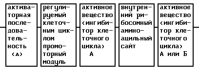
Такого рода IRES описываются, например, Montford и Smith (TIG, 11, 179 (1995); Kaufman и др. , Nucl. Acids Res., 19, 4485 (1991); Morgan и др., Nucl. Acids Res. , 20, 1293 (1992); Dirks и др., Gene, 128, 247 (1993); Pelletier и Sonenberg, Nature, 334, 320 (1988) и Sugitomo и др., Bio-Techn., 12, 694 (1994).
IRES of this kind are described, for example, by Montford and Smith (TIG, 11, 179 (1995); Kaufman et al., Nucl. Acids Res., 19, 4485 (1991); Morgan et al., Nucl. Acids Res., 20 , 1293 (1992); Dirks et al., Gene, 128, 247 (1993); Pelletier and Sonenberg, Nature, 334, 320 (1988) and Sugitomo et al., Bio-Techn., 12, 694 (1994).
Так, можно использовать кДНК IRES-последовательности полиовируса (положение ≤ 140 до ≥ 630 5'-UTR (Pelletier и Sonenberg, Nature, 334, 320 (1988)) для связывания ДНК антитромботического вещества А (по 3'-концу) и ДНК антитромботического вещества Б (по 5'-концу). Thus, cDNA of poliovirus IRES sequences (position ≤ 140 to ≥ 630 5'-UTR (Pelletier and Sonenberg, Nature, 334, 320 (1988)) can be used to bind DNA of antithrombotic substance A (at the 3'-end) and DNA of antithrombotic substance B (at the 5'-end).
Такого рода сочетание двух одинаковых или разных генов способствует аддитивному действию (в случае одинаковых генов) или синергическому действию выбранных антитромботических веществ. This kind of combination of two identical or different genes promotes the additive effect (in the case of identical genes) or the synergistic effect of the selected antithrombotic substances.
5.4. Выбор лиганда для ингибирования свертывания
В качестве лиганда вирусных или невирусных векторов, например, в коллоидных дисперсиях, содержащих полилизин-лиганд-конъюгаты, предпочтительны вещества, которые связываются с поверхностью гладкомышечных клеток, или пролиферирующих эндотелиальных клеток, или активированных макрофагов и/или лимфоцитов.5.4. The choice of ligand for inhibition of coagulation
As the ligand of viral or non-viral vectors, for example, in colloidal dispersions containing polylysine ligand conjugates, substances that bind to the surface of smooth muscle cells, or proliferating endothelial cells, or activated macrophages and / or lymphocytes are preferred.
а). Лиганды гладкомышечных клеток
Примеры лигандов, которые связываются с гладкомышечными клетками, уже указаны в разделе 4.4.a). Smooth muscle cell ligands
Examples of ligands that bind to smooth muscle cells are already listed in section 4.4.
б). Лиганды активированных эндотелиальных клеток
Сюда относятся согласно изобретению антитела или фрагменты антител, направленные против мембранных структур эндотелиальных клеток, которые описываются, например, Burrows и др. (Pharmac. Ther., 64, 155 (1994)), Hughes и др. (Cancer Res. , 49, 6214 (1989)) и Maruyama и др. (PNAS USA, 87, 5744 (1990)). В особенности сюда относятся антитела против VEGF-рецепторов.b) Activated Endothelial Cell Ligands
These include according to the invention antibodies or antibody fragments directed against the membrane structures of endothelial cells, which are described, for example, by Burrows et al. (Pharmac. Ther., 64, 155 (1994)), Hughes et al. (Cancer Res., 49, 6214 (1989)) and Maruyama et al. (PNAS USA, 87, 5744 (1990)). In particular, antibodies against VEGF receptors are included.
Мышиные моноклональные антитела предпочтительно нужно использовать в гуманизированной форме. Гуманизирование осуществляют описанным в разделе 4.4. образом. Фрагменты антител получают согласно уровню техники, например, описанным в разделе 4.4 способом. Mouse monoclonal antibodies should preferably be used in humanized form. Humanization is carried out as described in section 4.4. way. Antibody fragments are prepared according to the prior art, for example, as described in section 4.4.
К лигандам, далее, относятся все активные вещества, которые связываются с мембранными структурами или мембранными рецепторами эндотелиальных клеток. Например, сюда относятся вещества, которые содержат концевую маннозу, далее, интерлейкин-1 или факторы роста или их фрагменты, соответственно их частичные последовательности, которые связываются с рецепторами, экспрессируемыми эндотелиальными клетками, как, например, PDGF, bFGF, VEGF, TGF-β (Pusztain и др., J. Pathol., 169, 191 (1993)). Ligands further include all active substances that bind to membrane structures or membrane receptors of endothelial cells. For example, this includes substances that contain terminal mannose, further, interleukin-1 or growth factors or fragments thereof, respectively, their partial sequences that bind to receptors expressed by endothelial cells, such as PDGF, bFGF, VEGF, TGF-β (Pusztain et al., J. Pathol., 169, 191 (1993)).
Далее, к таким лигандам относятся адгезионные молекулы, которые связываются с активированными и/или пролиферирующими эндотелиальными клетками. Такого рода адгезионные молекулы, как, например, SLex, LFA-1, MAC-1, LECAM-1 или VLA-4, уже описаны (обзоры Augustin-Voss и др., J. Cell. Biol., 119, 483 (1992); Pauli и др., Cancer Metast. Rev., 9, 175 (1990); Honn и др., Cancer Metast. Rev., 11, 353 (1992)). Further, such ligands include adhesive molecules that bind to activated and / or proliferating endothelial cells. Adhesion molecules of this kind, such as SLex, LFA-1, MAC-1, LECAM-1 or VLA-4, have already been described (reviews by Augustin-Voss et al., J. Cell. Biol., 119, 483 (1992 ); Pauli et al., Cancer Metast. Rev., 9, 175 (1990); Honn et al., Cancer Metast. Rev., 11, 353 (1992)).
в). Лиганды активированных макрофагов и/или активированных лимфоцитов
К лигандам согласно изобретению относятся, далее, вещества, которые специфически связываются с поверхностью иммунных клеток. Сюда относятся антитела или фрагменты антител, направленные против мембранных структур иммунных клеток, которые описываются, например, Powelson и др., Biotech. Adv., 11, 725 (1993).in). Ligands of activated macrophages and / or activated lymphocytes
The ligands according to the invention further include substances that specifically bind to the surface of immune cells. This includes antibodies or antibody fragments directed against the membrane structures of immune cells, which are described, for example, Powelson et al., Biotech. Adv., 11, 725 (1993).
Далее, к лигандам также относятся моноклональные или поликлональные антитела или фрагменты антител, которые с помощью своих постоянных доменов связываются с Fc - или μ-рецепторами иммунных клеток (Rojanasakul и др., Pharm. Res., 11, 1731 (1994)). Further, ligands also include monoclonal or polyclonal antibodies or antibody fragments that, through their constant domains, bind to Fc or μ receptors of immune cells (Rojanasakul et al., Pharm. Res., 11, 1731 (1994)).
Здесь также предпочтительно используют мышиные моноклональные антитела в гуманизированной форме (см. раздел 4.4.) и фрагменты получают, например, с помощью указанной в разделе 4.4 методики. Mouse monoclonal antibodies in humanized form are also preferably used (see section 4.4.) And fragments are obtained, for example, using the procedure indicated in section 4.4.
К лигандам, далее, относятся все вещества, которые связываются с мембранными рецепторами на поверхности иммунных клеток. Например, сюда относятся факторы роста, как цитокины, EGF, TGF, FGF или PDGF или их фрагменты, соответственно их частичные последовательности, которые связываются с рецепторами, экспрессируемыми такого рода клетками. Ligands, further, include all substances that bind to membrane receptors on the surface of immune cells. For example, these include growth factors such as cytokines, EGF, TGF, FGF or PDGF or fragments thereof, respectively, their partial sequences that bind to receptors expressed by such cells.
5.5. Получение активного вещества для ингибирования свертывания
Получение предлагаемого согласно изобретению активного вещества описывается подробнее, руководствуясь следующими примерами.5.5. Obtaining an active substance to inhibit coagulation
The preparation of the active substance according to the invention is described in more detail in accordance with the following examples.
а). Конструкция химерного промотора эндотелина-1-CDE-CHR-Inr
Человеческий промотор эндотелин-1 (положение ≤ -170 до ≥ -10; Wilson и др., Mol. Cell. Biol., 10, 4854 (1990)) или укороченный на ТАТА-блок вариант (положение ≤ -170 до ≥ -40) по его 3'-концу связывают с 5'-концом CDE-CHR-Inr-модуля (положение ≤ -20 до ≥ +121) человеческого cdc25C-гена (Lucibello и др. , EMBO J. , 14, 132 (1995)) (фиг.9). Связывание осуществляют с помощью известных специалисту и имеющихся в продаже ферментов.a). The design of the chimeric promoter of endothelin-1-CDE-CHR-Inr
The human endothelin-1 promoter (position ≤ -170 to ≥ -10; Wilson et al., Mol. Cell. Biol., 10, 4854 (1990)) or a variant shortened by the TATA block (position ≤ -170 to ≥ -40 ) at its 3'-end is associated with the 5'-end of the CDE-CHR-Inr module (position ≤ -20 to ≥ +121) of the human cdc25C gene (Lucibello et al., EMBO J., 14, 132 (1995) ) (Fig. 9). The binding is carried out using known to the specialist and commercially available enzymes.
б). Конструкция плазмиды, содержащей химерный промотор эндотелин-1-CDE-CHR-Inr в центральной составной части активного вещества
Описанную транскрипционную единицу химерного промоторного модуля эндотелина-1 связывают по ее 3'-концу с 5'-концом ДНК, которая содержит полную кодирующую область тканевого плазминогенного активатора (положение ≤ 85 до ≥ 1753; Pennica и др., Nature, 301, 214 (1983)) (фиг.9). Эта ДНК также содержит необходимую для секреции сигнальную последовательность. Транскрипционные контрольные единицы и ДНК тканевого плазминогенного активатора клонируют в pUC19/19 или производимых от Bluescript плазмидных векторах, которые можно использовать непосредственно или в коллоидальных дисперсионных системах для введения ин виво. Альтернативно, химерные гены трансферируют в вирусные векторы или другие пригодные векторы и инъецируют.b) The construction of a plasmid containing the chimeric promoter endothelin-1-CDE-CHR-Inr in the Central component of the active substance
The described transcriptional unit of the chimeric promoter module of endothelin-1 is linked at its 3'-end to the 5'-end of DNA, which contains the complete coding region of tissue plasminogen activator (position ≤ 85 to ≥ 1753; Pennica et al., Nature, 301, 214 ( 1983)) (Fig. 9). This DNA also contains the signal sequence necessary for secretion. Transcriptional control units and tissue plasminogen activator DNA are cloned into pUC19 / 19 or from Bluescript-derived plasmid vectors, which can be used directly or in colloidal dispersion systems for in vivo administration. Alternatively, chimeric genes are transformed into viral vectors or other suitable vectors and injected.
в). Конструкция химерного промотора CDE-CHR-Inr миогенина
Человеческий миогениновый промотор (положение ≤ -210 до ≥ +54 ДНК-последовательности, опубликованной Salmin и др. , J. Cell. Biol., 115, 905 (1991)) по его 3'-концу связывают с 5'-концом CDE-CHR-Inr-модуля человеческого cdc25C-гена (положение ≤ -20 до ≥ +121 последовательности, опубликованной Lucibello и др., ЕМВО J., 14, 132 (1995)) (см. фиг.10). Связывание осуществляют с помощью известных специалисту и имеющихся в продаже ферментов. Кроме того, используют различные фрагменты промоторной последовательности миогенина (см. фиг.10). Так, применяют содержащую ТАТА-блок ДНК-последовательность промотора миогенина. Однако равным образом также можно использовать промоторную последовательность в положении ≤ -210 до ≥ -40.in). Design of the chimeric promoter CDE-CHR-Inr myogenin
The human myogenin promoter (position ≤ -210 to ≥ +54 of the DNA sequence published by Salmin et al., J. Cell. Biol., 115, 905 (1991)) at its 3 ′ end is linked to the 5′-end of CDE- CHR-Inr module of the human cdc25C gene (position ≤ -20 to ≥ +121 of the sequence published by Lucibello et al., EMBO J., 14, 132 (1995)) (see FIG. 10). The binding is carried out using known to the specialist and commercially available enzymes. In addition, various fragments of the myogenin promoter sequence are used (see FIG. 10). Thus, a DNA sequence containing the myogenin promoter containing a TATA block is used. However, the promoter sequence in position ≤ -210 to ≥ -40 can also be used in the same way.
г). Конструкция плазмиды, содержащей химерный промотор CDE-CHR-Inr миогенина в центральной составной части активного вещества
Таким образом полученную контрольную транскрипционную единицу химерного промоторного модуля миогенина по ее 3'-концу связывают с 5'-концом ДНК, которая содержит полную кодирующую область тканевого плазминогенного активатора (см. фиг. 10). Эта ДНК также содержит необходимую для секреции сигнальную последовательность. Транскрипционные контрольные единицы и ДНК тканевого плазминогенного активатора клонируют в pUC18/19 или производимых от Bluescript плазмидных векторах, которые непосредственно или в коллоидальной дисперсионной системе можно использовать для введения ин виво. Альтернативно, химерные гены можно трансферировать в вирусные векторы или другие пригодные векторы и инъецировать.d). The construction of a plasmid containing the chimeric promoter CDE-CHR-Inr myogenin in the Central component of the active substance
Thus obtained control transcriptional unit of the chimeric promoter module of myogenin at its 3'-end is associated with the 5'-end of the DNA, which contains the complete coding region of tissue plasminogen activator (see Fig. 10). This DNA also contains the signal sequence necessary for secretion. Transcriptional control units and tissue plasminogen activator DNA are cloned into pUC18 / 19 or from Bluescript-derived plasmid vectors that can be used directly or in a colloidal dispersion system for in vivo administration. Alternatively, chimeric genes can be transformed into viral vectors or other suitable vectors and injected.
д). Конструкция плазмиды, содержащей два гена активных веществ
Описанную в п. в) транскрипционную единицу CDE-CHR-Inr миогенина по ее 3'-концу связывают с 5'-концом ДНК ингибитора метаболического пути тканевого фактора (TFPI; положение ≤ 133 до ≥ 957; Wun и др., J. Biol. Chem., 263, 6001 (1988); или положение ≤ 382 до ≥ 1297; Girard и др., Thromb. Res., 55, 37 (1989)). Связывание осуществляют с помощью известных специалисту и имеющихся в продаже ферментов.e). Construction of a plasmid containing two genes of active substances
The transcriptional unit of CDE-CHR-Inr myogenin described in paragraph c) is linked at its 3'-end to the 5'-end of the tissue factor metabolic pathway inhibitor DNA (TFPI; position ≤ 133 to ≥ 957; Wun et al., J. Biol Chem., 263, 6001 (1988); or position ≤ 382 to ≥ 1297; Girard et al., Thromb. Res., 55, 37 (1989)). The binding is carried out using known to the specialist and commercially available enzymes.
3'-конец ДНК TFPI теперь связывают с 5'-концом кДНК внутреннего рибосомного аминоацильного сайта (положение ≤ 140 до ≥ 630; Pelletier и Sonnenberg, Nature., 334, 320 (1988)) и затем его 3'-конец связывают с 5'-концом ДНК тканевого плазминогенного активатора (см. фиг.11). Это таким образом полученное активное вещество, наконец, клонируют в pUC18/19 или производимых от Bluescript плазмидных векторах, которые непосредственно или в коллоидальной дисперсионной системе можно использовать для введения ин виво. Альтернативно, химерные гены можно трансферировать в вирусные векторы или другие пригодные векторы и инъецировать. The 3 ′ end of TFPI DNA is now linked to the 5 ′ end of the cDNA of the internal ribosomal aminoacyl site (position ≤ 140 to ≥ 630; Pelletier and Sonnenberg, Nature., 334, 320 (1988)) and then its 3 ′ end is linked to 5 the 'end of DNA of a tissue plasminogen activator (see Fig. 11). This thus obtained active substance is finally cloned into pUC18 / 19 or from plasmid vectors produced from Bluescript, which, directly or in a colloidal dispersion system, can be used for in vivo administration. Alternatively, chimeric genes can be transformed into viral vectors or other suitable vectors and injected.
6. Действие активного вещества на гладкомышечные клетки и/или на свертывание
Активное вещество согласно настоящему изобретению после локального или системного, предпочтительно внутривенного или внутриартериального введения оказывает преимущественное, если не исключительное, действие на такие гладкомышечные клетки, которые непосредственно доступны вследствие повреждения или травмирования сосуда (в особенности слой эндотелия) и, возможно, после попадания во внутреннюю оболочку объема кровеносного сосуда.6. The effect of the active substance on smooth muscle cells and / or on coagulation
The active substance according to the present invention, after local or systemic, preferably intravenous or intraarterial administration, exerts a predominant, if not exceptional, effect on smooth muscle cells that are directly accessible due to damage or trauma to the vessel (especially the endothelium layer) and, possibly, after entering the internal the sheath of the volume of a blood vessel.
Благодаря сочетанию специфической к ткани активаторной последовательности и регулируемого клеточным циклом репрессорного модуля обеспечивается то, что ингибитор клеточного цикла активируется преобладающе или исключительно в делящихся гладкомышечных клетках. By combining the tissue-specific activator sequence and the cell cycle-regulated repressor module, it is ensured that the cell cycle inhibitor is activated predominantly or exclusively in dividing smooth muscle cells.
Благодаря предлагаемому согласно изобретению применению мутированных ингибиторов клеточного цикла обеспечивается их более продолжительное по времени ингибирующее пролиферацию действие. Thanks to the use of mutated cell cycle inhibitors according to the invention, their longer proliferation-inhibiting effect is ensured.
Активное вещество согласно настоящему изобретению, далее, позволяет осуществлять то, что после локального (например, в ткань, полости тела или промежуточные пространства между тканями) или после системного, предпочтительно внутривенного или внутриартериального введения, преобладающе, если не исключительно, в гладкомышечные клетки, активированные пролиферирующие эндотелиальные клетки, активированные лимфоциты или активированные макрофаги экспрессируют антитромботическое вещество, и оно, таким образом, высвобождается в месте возникновения тромбоза. The active substance according to the present invention, further, allows for the fact that after local (for example, in tissue, body cavities or intermediate spaces between tissues) or after systemic, preferably intravenous or intraarterial administration, predominantly, if not exclusively, in smooth muscle cells activated proliferating endothelial cells, activated lymphocytes, or activated macrophages express an antithrombotic substance, and so it is released at the site of thrombosis.
Так как активное вещество как за счет его специфичности к клетке, так и также за счет его специфичности к клеточному циклу гарантирует высокую степень безопасности, его также можно использовать в высоких дозировках и в случае необходимости многократно с интервалами от дней или недель для профилактики или терапии закупорки сосудов, вызываемой за счет пролиферирующих гладкомышечных клеток, и/или для профилактики и/или терапии тромбозов. Since the active substance, both due to its specificity to the cell and also due to its specificity to the cell cycle, guarantees a high degree of safety, it can also be used in high dosages and, if necessary, repeatedly at intervals of days or weeks for the prevention or therapy of blockage vessels caused by proliferating smooth muscle cells, and / or for the prevention and / or therapy of thrombosis.
Пояснение к фигурам 1-11
Фиг.1:
Нуклеотидная последовательность cdc25C-промоторного участка с найденными ин виво сайтами связывания протеина (геномный диметилсульфатный футпринтинг; закрашенный кружок - полная конститутивная защита; незакрашенный кружок - частичная конститутивная защита; звездочка - регулируемая клеточным циклом Gl-специфическая защита; CBS - конститутивный связывающий сайт; CDE - зависимый от клеточного цикла элемент. Окрашенные в серый цвет области указывают YC-блоки (NF-Y-сайты связывания). Стартовые участки маркированы закрашенными квадратами.Explanation of Figures 1-11
Figure 1:
The nucleotide sequence of the cdc25C promoter region with in vivo protein binding sites (genomic dimethyl sulfate footprinting; filled circle indicates complete constitutive protection; open circle represents partial constitutive protection; asterisk indicates Gl-specific cell regulation; CBS constitutive binding site; CDE cell cycle dependent element. The gray-stained areas indicate Y C blocks (NF-Y binding sites). Starting sites are marked with filled squares.
Фиг.2:
Дерепрессия сdс25С-промотора специфически в G0 путем мутации cdc.Figure 2:
Derepression of the cdc25C promoter specifically at G 0 by mutation of cdc.
Фиг.3:
Схематическое изображение регуляции сdс25С-энхансера за счет CDE.Figure 3:
Schematic representation of the regulation of the cdc25C enhancer by CDE.
Фиг.4:
G0/G1 - специфическая репрессия 8У40-энхансера за счет CDE.Figure 4:
G 0 / G 1 - specific repression of the 8Y40 enhancer due to CDE.
Фиг.5:
Гомологии в CDE-CHR-области и расположенные у 5'-концов YC-блоки в cdc25C, циклине-А и cdc2-промоторах.Figure 5:
Homology in the CDE-CHR region and located at the 5'-ends of the Y C blocks in cdc25C, cyclin-A and cdc2 promoters.
Фиг.6:
Химерная конструкция, состоящая из различных частей человеческого промотора миогенина (Myf-4), слитого на 3'-конце промоторного модуля с CDE и CHR репрессорными элементами, а также ДНК человеческой β-глюкуронидазы (полный кодирующий участок, положение ≤ 27 до ≥ 1982) в качестве эффектора (Oshima и др., PNAS USA, 84, 685 (1987)). Данные о расположении относятся к данным Salminen и др., J. Cell. Biol., 115, 905 (1991) для гена миогенина, соответственно к системе cdc25C, применяемой Lucibello и др., ЕМВО J., 14, 132 (1995).6:
Chimeric construct consisting of various parts of the human myogenin promoter (Myf-4), fused at the 3'-end of the promoter module with CDE and CHR repressor elements, as well as human β-glucuronidase DNA (full coding region, position ≤ 27 to ≥ 1982) as an effector (Oshima et al., PNAS USA, 84, 685 (1987)). Location data refer to Salminen et al., J. Cell. Biol., 115, 905 (1991) for the myogenin gene, respectively, to the cdc25C system used by Lucibello et al., EMBO J., 14, 132 (1995).
Фиг.7:
Замена гомологичной сигнальной последовательности β-глюкуронидазы гетерологичной сигнальной последовательностью (человеческого иммуноглобулина). Данные о расположении сигнальной последовательности (MGWSCIILFLVATAT) иммуноглобулина (HuVH-cAMP) относятся к данным Riechmann и др., Nature, 332, 323 (1988).7:
Replacing the homologous signal sequence of β-glucuronidase with a heterologous signal sequence (human immunoglobulin). Data on the location of the signal sequence (MGWSCIILFLVATAT) of immunoglobulin (HuVH-cAMP) refers to the data of Riechmann et al., Nature, 332, 323 (1988).
Альтернатива: Встраивание сигнального пептида иммуноглобулина для лучшего внеклеточного выделения β-глюкуронидазы; см.(Б). Alternative: Embedding an immunoglobulin signal peptide for better extracellular isolation of β-glucuronidase; see (B).
Фиг.8:
Химерная конструкция, состоящая из различных частей человеческого промотора миогенина (Myf-4), слитого на 3'-конце промоторного модуля с CDE и CHR репрессорными элементами, а также ДНК человеческой β-глюкуронидазы, внутреннего рибосомного аминоациального сайта в качестве регулирующей нуклеотидной последовательности и ДНК тканевого плазминогенного активатора. Данные о расположении относятся к таковым Salminen и др., J. Cell. Biol., 115, 905 (1991) для миогенина; Lucibello и др., ЕМВО J., 14, 132 (1995) для элемента CDE/CHR-Inr; Oshima и др. , PNAS USA, 84, 685 (1987) для β-глюкуронидазы; Riechmann и др., Nature, 332? 323 (1988) для сигнальной последовательности иммуноглобулина; Pelletier и Sonenberg, Nature, 334, 320 (1988) для внутреннего рибосомного аминоациального сайта полиовируса и Pennica и др., Nature, 301, 214 (1983) для человеческого тканевого плазминогенного активатора.Fig. 8:
A chimeric construct consisting of various parts of the human myogenin promoter (Myf-4) fused at the 3'-end of the promoter module with CDE and CHR repressor elements, as well as human β-glucuronidase DNA, an internal ribosome aminoacial site as a regulatory nucleotide sequence and DNA tissue plasminogen activator. Location data refers to those of Salminen et al., J. Cell. Biol., 115, 905 (1991) for myogenin; Lucibello et al., EMBO J., 14, 132 (1995) for the element CDE / CHR-Inr; Oshima et al., PNAS USA, 84, 685 (1987) for β-glucuronidase; Riechmann et al., Nature, 332? 323 (1988) for the immunoglobulin signal sequence; Pelletier and Sonenberg, Nature, 334, 320 (1988) for the internal ribosomal aminoacial site of poliovirus and Pennica et al., Nature, 301, 214 (1983) for human tissue plasminogen activator.
Фиг.9:
Химерная конструкция, состоящая из различных частей человеческого промотора эндотелина-1, слитого по 3'-концу промоторного модуля с CDE и CHR репрессорными элементами, а также ДНК человеческого тканевого плазминогенного активатора (полная кодирующая область; Pennica и др., Nature, 301, 214 (1983)) в качестве эффектора. Данные о расположении относятся к таковым Wilson и др., Mol. Cell. Biol., 10, 4854 (1990) для гена эндотелина-1, соответственно к используемой Lucibello и др., ЕМВО J., 14, 132 (1995), системе cdc25C.Fig.9:
A chimeric construct consisting of various parts of the human endothelin-1 promoter fused at the 3'-end of the promoter module with CDE and CHR repressor elements, as well as human tissue plasminogen activator DNA (full coding region; Pennica et al., Nature, 301, 214 (1983)) as an effector. Location data are those of Wilson et al., Mol. Cell. Biol., 10, 4854 (1990) for the endothelin-1 gene, respectively, used by Lucibello et al., EMBO J., 14, 132 (1995), cdc25C system.
Фиг.10:
Химерная конструкция, состоящая из различных частей человеческого промотора миогенина (Myf-4), слитого по 3'-концу промоторного модуля с CDE и CHR репрессорными элементами, а также ДНК человеческого тканевого плазминогенного активатора (полная кодирующая область) в качестве эффектора. Данные о расположении относятся к таковым Salminen и др., J. Cell. Biol., 115, 905 (1991) для гена миогенина, соответственно к используемой Lucibello и др., ЕМВО J. , 14, 132 (1995) системе cdc25C и к данным Pennica и др., Nature, 301, 214 (1983) для тканевого плазминогенного активатора.Figure 10:
A chimeric construct consisting of various parts of the human myogenin promoter (Myf-4) fused at the 3'-end of the promoter module with CDE and CHR repressor elements, as well as human tissue plasminogen activator DNA (full coding region) as an effector. Location data refers to those of Salminen et al., J. Cell. Biol., 115, 905 (1991) for the myogenin gene, respectively, to the cdc25C system used by Lucibello et al., EMBO J., 14, 132 (1995) and to Pennica et al., Nature, 301, 214 (1983) for tissue plasminogen activator.
Фиг.11:
Химерная конструкция с двумя эффекторными генами.11:
Chimeric construction with two effector genes.
Данные о расположении для ингибитора метаболического пути тканевого фактора относятся к данным
*) Wun и др., J. Biol. Chem., 263, 6001 (1988);
**) Girard и др., Thromb. Res., 55, 37 (1989);
а данные для внутреннего рибосомного аминоациального сайта (IRES) кДНК основываются на таковых Pelletier и Sonenberg, Nature, 334, 320 (1988).Location data for tissue factor metabolic pathway inhibitor are data
*) Wun et al., J. Biol. Chem., 263, 6001 (1988);
**) Girard et al., Thromb. Res., 55, 37 (1989);
and data for the internal ribosomal aminoacial site (IRES) of the cDNA are based on those of Pelletier and Sonenberg, Nature, 334, 320 (1988).
Claims (34)
Applications Claiming Priority (3)
| Application Number | Priority Date | Filing Date | Title |
|---|---|---|---|
| GB9417366.3 | 1994-08-26 | ||
| GB9417366A GB9417366D0 (en) | 1994-08-26 | 1994-08-26 | Cell cycle regulated repressor and DNA element |
| GB9506466.3 | 1995-03-29 |
Publications (2)
| Publication Number | Publication Date |
|---|---|
| RU97104482A RU97104482A (en) | 1999-05-10 |
| RU2197992C2 true RU2197992C2 (en) | 2003-02-10 |
Family
ID=10760519
Family Applications (1)
| Application Number | Title | Priority Date | Filing Date |
|---|---|---|---|
| RU97104482/14A RU2197992C2 (en) | 1994-08-26 | 1995-08-25 | Method for applying genetic therapy of vascular diseases by means of active substance specific to cell and dependant on cellular cycle |
Country Status (3)
| Country | Link |
|---|---|
| GB (1) | GB9417366D0 (en) |
| RU (1) | RU2197992C2 (en) |
| ZA (1) | ZA957074B (en) |
-
1994
- 1994-08-26 GB GB9417366A patent/GB9417366D0/en active Pending
-
1995
- 1995-08-23 ZA ZA9507074A patent/ZA957074B/en unknown
- 1995-08-25 RU RU97104482/14A patent/RU2197992C2/en not_active IP Right Cessation
Also Published As
| Publication number | Publication date |
|---|---|
| GB9417366D0 (en) | 1994-10-19 |
| ZA957074B (en) | 1997-02-24 |
Similar Documents
| Publication | Publication Date | Title |
|---|---|---|
| PT777739E (en) | TREATMENT OF DISEASES OF VASES THROUGH GENETIC THERAPY THROUGH A SPECIFIC ACTIVE MATERIAL OF CELLS AND CELL CYCLE DEPENDENT | |
| US6384202B1 (en) | Cell-specific active compounds regulated by the cell cycle | |
| US6210939B1 (en) | Recombinant adenoviral vector and methods of use | |
| Varley et al. | Inflammation-induced recombinant protein expression in vivo using promoters from acute-phase protein genes. | |
| US6576758B1 (en) | Nucleic acid constructs containing hybrid promoters | |
| US6033856A (en) | Promoter of the cdc25B gene, its preparation and use | |
| US5772993A (en) | Osteocalcin promoter-based toxic gene therapy for the treatment of calcified tumors and tissues | |
| AU716178B2 (en) | Nucleic acid constructs containing genes encoding transport signals | |
| Shiratori et al. | Strategy of liver‐directed gene therapy: present status and future prospects | |
| AU9325698A (en) | Oncogene- or virus-controlled expression systems | |
| AU5155799A (en) | Expression system containing chimeric promoters with binding sites for recombinant transcription factors | |
| Ahmed et al. | In vivo tumor suppression by adenovirus-mediated interferon alpha2b gene delivery | |
| EP2386319A1 (en) | Promoters exhibiting endothelial cell specificity and methods of using same | |
| US20040266006A1 (en) | Adenoviral vectors having a protein IX deletion | |
| WO1996006938A1 (en) | Genetic therapy of vascular diseases with a cell-specific active substance which is dependent on the cell cycle | |
| RU2197992C2 (en) | Method for applying genetic therapy of vascular diseases by means of active substance specific to cell and dependant on cellular cycle | |
| US6743623B2 (en) | Viral recombinant vectors for expression in muscle cells | |
| AU2001294793B2 (en) | Viruses targeted to hypoxic cells and tissues | |
| US7482155B1 (en) | Chimeric promoters for controlling expression in smooth muscle cells | |
| JP2004509635A5 (en) | ||
| US6099831A (en) | Viral recombinant vectors for expression in muscle cells | |
| Ehsan et al. | Antisense and gene therapy to prevent restenosis | |
| ABAAN et al. | Gene therapy in human breast cancer |
Legal Events
| Date | Code | Title | Description |
|---|---|---|---|
| MM4A | The patent is invalid due to non-payment of fees |
Effective date: 20030826 |