RU2170254C2 - Strain of cow herpes virus type-i (bhv-i) cncm n 1-1213, vaccine, diagnostic serological reagent, method of vaccination, method of serological distinction - Google Patents
Strain of cow herpes virus type-i (bhv-i) cncm n 1-1213, vaccine, diagnostic serological reagent, method of vaccination, method of serological distinction Download PDFInfo
- Publication number
- RU2170254C2 RU2170254C2 RU93058614/13A RU93058614A RU2170254C2 RU 2170254 C2 RU2170254 C2 RU 2170254C2 RU 93058614/13 A RU93058614/13 A RU 93058614/13A RU 93058614 A RU93058614 A RU 93058614A RU 2170254 C2 RU2170254 C2 RU 2170254C2
- Authority
- RU
- Russia
- Prior art keywords
- bhv
- gene
- deletion
- glycoprotein
- fragment
- Prior art date
Links
- 238000000034 method Methods 0.000 title claims abstract description 54
- 229960005486 vaccine Drugs 0.000 title claims abstract description 51
- 241001529453 unidentified herpesvirus Species 0.000 title claims abstract description 10
- 230000000405 serological effect Effects 0.000 title claims description 14
- 239000003153 chemical reaction reagent Substances 0.000 title claims 3
- 238000002255 vaccination Methods 0.000 title abstract description 13
- 238000012217 deletion Methods 0.000 claims abstract description 107
- 230000037430 deletion Effects 0.000 claims abstract description 107
- 241001465754 Metazoa Species 0.000 claims abstract description 30
- 108090000288 Glycoproteins Proteins 0.000 claims description 101
- 239000012634 fragment Substances 0.000 claims description 101
- 102000003886 Glycoproteins Human genes 0.000 claims description 95
- 241000283690 Bos taurus Species 0.000 claims description 41
- 210000002966 serum Anatomy 0.000 claims description 33
- 238000004458 analytical method Methods 0.000 claims description 28
- 230000000890 antigenic effect Effects 0.000 claims description 9
- 241000700588 Human alphaherpesvirus 1 Species 0.000 claims 1
- 230000003993 interaction Effects 0.000 claims 1
- LWGJTAZLEJHCPA-UHFFFAOYSA-N n-(2-chloroethyl)-n-nitrosomorpholine-4-carboxamide Chemical compound ClCCN(N=O)C(=O)N1CCOCC1 LWGJTAZLEJHCPA-UHFFFAOYSA-N 0.000 claims 1
- 108090000623 proteins and genes Proteins 0.000 abstract description 91
- 102000004169 proteins and genes Human genes 0.000 abstract description 29
- 208000015181 infectious disease Diseases 0.000 abstract description 21
- 238000001514 detection method Methods 0.000 abstract description 15
- 150000007523 nucleic acids Chemical class 0.000 abstract description 15
- 108020004707 nucleic acids Proteins 0.000 abstract description 14
- 102000039446 nucleic acids Human genes 0.000 abstract description 14
- 238000002360 preparation method Methods 0.000 abstract description 14
- 229940031551 inactivated vaccine Drugs 0.000 abstract description 7
- 230000002238 attenuated effect Effects 0.000 abstract description 4
- 244000052616 bacterial pathogen Species 0.000 abstract description 4
- 229940031567 attenuated vaccine Drugs 0.000 abstract description 3
- 238000003745 diagnosis Methods 0.000 abstract description 2
- 230000000694 effects Effects 0.000 abstract description 2
- 241000223109 Trypanosoma cruzi Species 0.000 abstract 1
- 239000000126 substance Substances 0.000 abstract 1
- 101150072564 gE gene Proteins 0.000 description 63
- 241000700605 Viruses Species 0.000 description 59
- 108020004414 DNA Proteins 0.000 description 46
- 244000309466 calf Species 0.000 description 43
- 239000013598 vector Substances 0.000 description 38
- 210000004027 cell Anatomy 0.000 description 31
- 102100026115 S-adenosylmethionine synthase isoform type-1 Human genes 0.000 description 29
- 150000001413 amino acids Chemical class 0.000 description 27
- 208000017972 multifocal atrial tachycardia Diseases 0.000 description 26
- 235000018102 proteins Nutrition 0.000 description 26
- 239000002773 nucleotide Substances 0.000 description 25
- 125000003729 nucleotide group Chemical group 0.000 description 25
- 238000003752 polymerase chain reaction Methods 0.000 description 22
- 108090000765 processed proteins & peptides Proteins 0.000 description 20
- 239000000523 sample Substances 0.000 description 20
- 235000001014 amino acid Nutrition 0.000 description 19
- 230000014509 gene expression Effects 0.000 description 19
- 238000012360 testing method Methods 0.000 description 19
- 108091026890 Coding region Proteins 0.000 description 16
- 210000001331 nose Anatomy 0.000 description 16
- 239000000427 antigen Substances 0.000 description 15
- 101150030521 gI gene Proteins 0.000 description 15
- 230000006798 recombination Effects 0.000 description 15
- 238000005215 recombination Methods 0.000 description 15
- 108091007433 antigens Proteins 0.000 description 14
- 102000036639 antigens Human genes 0.000 description 14
- 239000013604 expression vector Substances 0.000 description 13
- 238000010561 standard procedure Methods 0.000 description 13
- 241000700584 Simplexvirus Species 0.000 description 12
- 239000000203 mixture Substances 0.000 description 12
- 102000004196 processed proteins & peptides Human genes 0.000 description 12
- 230000009465 prokaryotic expression Effects 0.000 description 12
- 108091008146 restriction endonucleases Proteins 0.000 description 12
- 108020004440 Thymidine kinase Proteins 0.000 description 11
- 201000010099 disease Diseases 0.000 description 10
- 208000037265 diseases, disorders, signs and symptoms Diseases 0.000 description 10
- PEDCQBHIVMGVHV-UHFFFAOYSA-N Glycerine Chemical compound OCC(O)CO PEDCQBHIVMGVHV-UHFFFAOYSA-N 0.000 description 9
- 108700026244 Open Reading Frames Proteins 0.000 description 9
- 238000010276 construction Methods 0.000 description 9
- 210000003527 eukaryotic cell Anatomy 0.000 description 9
- 230000012010 growth Effects 0.000 description 9
- 244000144972 livestock Species 0.000 description 9
- 239000013642 negative control Substances 0.000 description 9
- 239000000047 product Substances 0.000 description 9
- 108010038807 Oligopeptides Proteins 0.000 description 8
- 102000015636 Oligopeptides Human genes 0.000 description 8
- 210000004369 blood Anatomy 0.000 description 8
- 239000008280 blood Substances 0.000 description 8
- 244000052769 pathogen Species 0.000 description 8
- 239000013612 plasmid Substances 0.000 description 8
- 108020004705 Codon Proteins 0.000 description 7
- 239000000020 Nitrocellulose Substances 0.000 description 7
- 102000006601 Thymidine Kinase Human genes 0.000 description 7
- 108010070396 bovine herpesvirus type-1 glycoproteins Proteins 0.000 description 7
- 238000006243 chemical reaction Methods 0.000 description 7
- 230000029087 digestion Effects 0.000 description 7
- 239000012530 fluid Substances 0.000 description 7
- 238000009396 hybridization Methods 0.000 description 7
- 229920001220 nitrocellulos Polymers 0.000 description 7
- 230000001717 pathogenic effect Effects 0.000 description 7
- 229920001184 polypeptide Polymers 0.000 description 7
- 210000000582 semen Anatomy 0.000 description 7
- LFQSCWFLJHTTHZ-UHFFFAOYSA-N Ethanol Chemical compound CCO LFQSCWFLJHTTHZ-UHFFFAOYSA-N 0.000 description 6
- 108091028043 Nucleic acid sequence Proteins 0.000 description 6
- 108020004511 Recombinant DNA Proteins 0.000 description 6
- FAPWRFPIFSIZLT-UHFFFAOYSA-M Sodium chloride Chemical compound [Na+].[Cl-] FAPWRFPIFSIZLT-UHFFFAOYSA-M 0.000 description 6
- 230000004927 fusion Effects 0.000 description 6
- 238000002955 isolation Methods 0.000 description 6
- BPHPUYQFMNQIOC-NXRLNHOXSA-N isopropyl beta-D-thiogalactopyranoside Chemical compound CC(C)S[C@@H]1O[C@H](CO)[C@H](O)[C@H](O)[C@H]1O BPHPUYQFMNQIOC-NXRLNHOXSA-N 0.000 description 6
- 239000003550 marker Substances 0.000 description 6
- 235000013336 milk Nutrition 0.000 description 6
- 239000008267 milk Substances 0.000 description 6
- 210000004080 milk Anatomy 0.000 description 6
- 210000001519 tissue Anatomy 0.000 description 6
- 241000894006 Bacteria Species 0.000 description 5
- 102000004190 Enzymes Human genes 0.000 description 5
- 108090000790 Enzymes Proteins 0.000 description 5
- 241000124008 Mammalia Species 0.000 description 5
- 230000008901 benefit Effects 0.000 description 5
- 238000010367 cloning Methods 0.000 description 5
- 238000010586 diagram Methods 0.000 description 5
- 238000005516 engineering process Methods 0.000 description 5
- 108020001507 fusion proteins Proteins 0.000 description 5
- 102000037865 fusion proteins Human genes 0.000 description 5
- 239000001963 growth medium Substances 0.000 description 5
- 238000011534 incubation Methods 0.000 description 5
- 210000002850 nasal mucosa Anatomy 0.000 description 5
- 230000036961 partial effect Effects 0.000 description 5
- 230000001105 regulatory effect Effects 0.000 description 5
- 230000003612 virological effect Effects 0.000 description 5
- 108010078791 Carrier Proteins Proteins 0.000 description 4
- 102000014914 Carrier Proteins Human genes 0.000 description 4
- 241000701024 Human betaherpesvirus 5 Species 0.000 description 4
- 108060003951 Immunoglobulin Proteins 0.000 description 4
- 108091034117 Oligonucleotide Proteins 0.000 description 4
- 108010076504 Protein Sorting Signals Proteins 0.000 description 4
- 206010039101 Rhinorrhoea Diseases 0.000 description 4
- DBMJMQXJHONAFJ-UHFFFAOYSA-M Sodium laurylsulphate Chemical compound [Na+].CCCCCCCCCCCCOS([O-])(=O)=O DBMJMQXJHONAFJ-UHFFFAOYSA-M 0.000 description 4
- 108010008038 Synthetic Vaccines Proteins 0.000 description 4
- JLCPHMBAVCMARE-UHFFFAOYSA-N [3-[[3-[[3-[[3-[[3-[[3-[[3-[[3-[[3-[[3-[[3-[[5-(2-amino-6-oxo-1H-purin-9-yl)-3-[[3-[[3-[[3-[[3-[[3-[[5-(2-amino-6-oxo-1H-purin-9-yl)-3-[[5-(2-amino-6-oxo-1H-purin-9-yl)-3-hydroxyoxolan-2-yl]methoxy-hydroxyphosphoryl]oxyoxolan-2-yl]methoxy-hydroxyphosphoryl]oxy-5-(5-methyl-2,4-dioxopyrimidin-1-yl)oxolan-2-yl]methoxy-hydroxyphosphoryl]oxy-5-(6-aminopurin-9-yl)oxolan-2-yl]methoxy-hydroxyphosphoryl]oxy-5-(6-aminopurin-9-yl)oxolan-2-yl]methoxy-hydroxyphosphoryl]oxy-5-(6-aminopurin-9-yl)oxolan-2-yl]methoxy-hydroxyphosphoryl]oxy-5-(6-aminopurin-9-yl)oxolan-2-yl]methoxy-hydroxyphosphoryl]oxyoxolan-2-yl]methoxy-hydroxyphosphoryl]oxy-5-(5-methyl-2,4-dioxopyrimidin-1-yl)oxolan-2-yl]methoxy-hydroxyphosphoryl]oxy-5-(4-amino-2-oxopyrimidin-1-yl)oxolan-2-yl]methoxy-hydroxyphosphoryl]oxy-5-(5-methyl-2,4-dioxopyrimidin-1-yl)oxolan-2-yl]methoxy-hydroxyphosphoryl]oxy-5-(5-methyl-2,4-dioxopyrimidin-1-yl)oxolan-2-yl]methoxy-hydroxyphosphoryl]oxy-5-(6-aminopurin-9-yl)oxolan-2-yl]methoxy-hydroxyphosphoryl]oxy-5-(6-aminopurin-9-yl)oxolan-2-yl]methoxy-hydroxyphosphoryl]oxy-5-(4-amino-2-oxopyrimidin-1-yl)oxolan-2-yl]methoxy-hydroxyphosphoryl]oxy-5-(4-amino-2-oxopyrimidin-1-yl)oxolan-2-yl]methoxy-hydroxyphosphoryl]oxy-5-(4-amino-2-oxopyrimidin-1-yl)oxolan-2-yl]methoxy-hydroxyphosphoryl]oxy-5-(6-aminopurin-9-yl)oxolan-2-yl]methoxy-hydroxyphosphoryl]oxy-5-(4-amino-2-oxopyrimidin-1-yl)oxolan-2-yl]methyl [5-(6-aminopurin-9-yl)-2-(hydroxymethyl)oxolan-3-yl] hydrogen phosphate Polymers Cc1cn(C2CC(OP(O)(=O)OCC3OC(CC3OP(O)(=O)OCC3OC(CC3O)n3cnc4c3nc(N)[nH]c4=O)n3cnc4c3nc(N)[nH]c4=O)C(COP(O)(=O)OC3CC(OC3COP(O)(=O)OC3CC(OC3COP(O)(=O)OC3CC(OC3COP(O)(=O)OC3CC(OC3COP(O)(=O)OC3CC(OC3COP(O)(=O)OC3CC(OC3COP(O)(=O)OC3CC(OC3COP(O)(=O)OC3CC(OC3COP(O)(=O)OC3CC(OC3COP(O)(=O)OC3CC(OC3COP(O)(=O)OC3CC(OC3COP(O)(=O)OC3CC(OC3COP(O)(=O)OC3CC(OC3COP(O)(=O)OC3CC(OC3COP(O)(=O)OC3CC(OC3COP(O)(=O)OC3CC(OC3COP(O)(=O)OC3CC(OC3CO)n3cnc4c(N)ncnc34)n3ccc(N)nc3=O)n3cnc4c(N)ncnc34)n3ccc(N)nc3=O)n3ccc(N)nc3=O)n3ccc(N)nc3=O)n3cnc4c(N)ncnc34)n3cnc4c(N)ncnc34)n3cc(C)c(=O)[nH]c3=O)n3cc(C)c(=O)[nH]c3=O)n3ccc(N)nc3=O)n3cc(C)c(=O)[nH]c3=O)n3cnc4c3nc(N)[nH]c4=O)n3cnc4c(N)ncnc34)n3cnc4c(N)ncnc34)n3cnc4c(N)ncnc34)n3cnc4c(N)ncnc34)O2)c(=O)[nH]c1=O JLCPHMBAVCMARE-UHFFFAOYSA-N 0.000 description 4
- 239000011543 agarose gel Substances 0.000 description 4
- 239000012472 biological sample Substances 0.000 description 4
- 210000001124 body fluid Anatomy 0.000 description 4
- 239000010839 body fluid Substances 0.000 description 4
- 238000004590 computer program Methods 0.000 description 4
- 102000018358 immunoglobulin Human genes 0.000 description 4
- 210000004072 lung Anatomy 0.000 description 4
- 208000010753 nasal discharge Diseases 0.000 description 4
- 230000037361 pathway Effects 0.000 description 4
- 229940124551 recombinant vaccine Drugs 0.000 description 4
- 230000002829 reductive effect Effects 0.000 description 4
- 210000003296 saliva Anatomy 0.000 description 4
- 238000013519 translation Methods 0.000 description 4
- 229920000936 Agarose Polymers 0.000 description 3
- 206010002942 Apathy Diseases 0.000 description 3
- 101710088235 Envelope glycoprotein C homolog Proteins 0.000 description 3
- 108700039691 Genetic Promoter Regions Proteins 0.000 description 3
- 108010001336 Horseradish Peroxidase Proteins 0.000 description 3
- ZQISRDCJNBUVMM-UHFFFAOYSA-N L-Histidinol Natural products OCC(N)CC1=CN=CN1 ZQISRDCJNBUVMM-UHFFFAOYSA-N 0.000 description 3
- ZQISRDCJNBUVMM-YFKPBYRVSA-N L-histidinol Chemical compound OC[C@@H](N)CC1=CNC=N1 ZQISRDCJNBUVMM-YFKPBYRVSA-N 0.000 description 3
- 230000004988 N-glycosylation Effects 0.000 description 3
- 241000283973 Oryctolagus cuniculus Species 0.000 description 3
- 206010036790 Productive cough Diseases 0.000 description 3
- 239000007983 Tris buffer Substances 0.000 description 3
- 230000003321 amplification Effects 0.000 description 3
- 230000004596 appetite loss Effects 0.000 description 3
- 238000003556 assay Methods 0.000 description 3
- 230000000903 blocking effect Effects 0.000 description 3
- 210000000601 blood cell Anatomy 0.000 description 3
- 239000000872 buffer Substances 0.000 description 3
- 238000004113 cell culture Methods 0.000 description 3
- 239000003593 chromogenic compound Substances 0.000 description 3
- OPTASPLRGRRNAP-UHFFFAOYSA-N cytosine Chemical group NC=1C=CNC(=O)N=1 OPTASPLRGRRNAP-UHFFFAOYSA-N 0.000 description 3
- RGWHQCVHVJXOKC-SHYZEUOFSA-J dCTP(4-) Chemical compound O=C1N=C(N)C=CN1[C@@H]1O[C@H](COP([O-])(=O)OP([O-])(=O)OP([O-])([O-])=O)[C@@H](O)C1 RGWHQCVHVJXOKC-SHYZEUOFSA-J 0.000 description 3
- 238000002405 diagnostic procedure Methods 0.000 description 3
- 238000001962 electrophoresis Methods 0.000 description 3
- 238000002474 experimental method Methods 0.000 description 3
- 238000011010 flushing procedure Methods 0.000 description 3
- 239000000499 gel Substances 0.000 description 3
- 230000028993 immune response Effects 0.000 description 3
- 235000021266 loss of appetite Nutrition 0.000 description 3
- 208000019017 loss of appetite Diseases 0.000 description 3
- 238000003199 nucleic acid amplification method Methods 0.000 description 3
- 102000013415 peroxidase activity proteins Human genes 0.000 description 3
- 108040007629 peroxidase activity proteins Proteins 0.000 description 3
- 238000013207 serial dilution Methods 0.000 description 3
- 239000011780 sodium chloride Substances 0.000 description 3
- 210000003802 sputum Anatomy 0.000 description 3
- 208000024794 sputum Diseases 0.000 description 3
- LENZDBCJOHFCAS-UHFFFAOYSA-N tris Chemical compound OCC(N)(CO)CO LENZDBCJOHFCAS-UHFFFAOYSA-N 0.000 description 3
- 230000001018 virulence Effects 0.000 description 3
- 241000283707 Capra Species 0.000 description 2
- HEDRZPFGACZZDS-UHFFFAOYSA-N Chloroform Chemical compound ClC(Cl)Cl HEDRZPFGACZZDS-UHFFFAOYSA-N 0.000 description 2
- 102000004127 Cytokines Human genes 0.000 description 2
- 108090000695 Cytokines Proteins 0.000 description 2
- 241000588724 Escherichia coli Species 0.000 description 2
- 241000620209 Escherichia coli DH5[alpha] Species 0.000 description 2
- WSFSSNUMVMOOMR-UHFFFAOYSA-N Formaldehyde Chemical compound O=C WSFSSNUMVMOOMR-UHFFFAOYSA-N 0.000 description 2
- ZHNUHDYFZUAESO-UHFFFAOYSA-N Formamide Chemical compound NC=O ZHNUHDYFZUAESO-UHFFFAOYSA-N 0.000 description 2
- 108010070675 Glutathione transferase Proteins 0.000 description 2
- 101000980673 Homo sapiens Multicilin Proteins 0.000 description 2
- 241000701085 Human alphaherpesvirus 3 Species 0.000 description 2
- 241000188250 Idas Species 0.000 description 2
- KFZMGEQAYNKOFK-UHFFFAOYSA-N Isopropanol Chemical compound CC(C)O KFZMGEQAYNKOFK-UHFFFAOYSA-N 0.000 description 2
- 102100024179 Multicilin Human genes 0.000 description 2
- 241001529936 Murinae Species 0.000 description 2
- 208000020663 Nasal mucosal disease Diseases 0.000 description 2
- 238000012300 Sequence Analysis Methods 0.000 description 2
- 108020005202 Viral DNA Proteins 0.000 description 2
- 230000001154 acute effect Effects 0.000 description 2
- 230000036528 appetite Effects 0.000 description 2
- 235000019789 appetite Nutrition 0.000 description 2
- 238000013459 approach Methods 0.000 description 2
- 238000000376 autoradiography Methods 0.000 description 2
- 230000015572 biosynthetic process Effects 0.000 description 2
- 210000001072 colon Anatomy 0.000 description 2
- 238000013461 design Methods 0.000 description 2
- 238000011161 development Methods 0.000 description 2
- 230000004069 differentiation Effects 0.000 description 2
- 238000010790 dilution Methods 0.000 description 2
- 239000012895 dilution Substances 0.000 description 2
- 208000028659 discharge Diseases 0.000 description 2
- 238000001976 enzyme digestion Methods 0.000 description 2
- 238000000605 extraction Methods 0.000 description 2
- 238000004817 gas chromatography Methods 0.000 description 2
- BRZYSWJRSDMWLG-CAXSIQPQSA-N geneticin Chemical compound O1C[C@@](O)(C)[C@H](NC)[C@@H](O)[C@H]1O[C@@H]1[C@@H](O)[C@H](O[C@@H]2[C@@H]([C@@H](O)[C@H](O)[C@@H](C(C)O)O2)N)[C@@H](N)C[C@H]1N BRZYSWJRSDMWLG-CAXSIQPQSA-N 0.000 description 2
- 230000001900 immune effect Effects 0.000 description 2
- 230000002163 immunogen Effects 0.000 description 2
- 239000003547 immunosorbent Substances 0.000 description 2
- 230000002458 infectious effect Effects 0.000 description 2
- 239000010410 layer Substances 0.000 description 2
- 229940124590 live attenuated vaccine Drugs 0.000 description 2
- 229940023012 live-attenuated vaccine Drugs 0.000 description 2
- 238000013507 mapping Methods 0.000 description 2
- 238000002844 melting Methods 0.000 description 2
- 230000008018 melting Effects 0.000 description 2
- 238000010369 molecular cloning Methods 0.000 description 2
- 230000003472 neutralizing effect Effects 0.000 description 2
- 108010085336 phosphoribosyl-AMP cyclohydrolase Proteins 0.000 description 2
- 230000001681 protective effect Effects 0.000 description 2
- 230000007420 reactivation Effects 0.000 description 2
- 230000009257 reactivity Effects 0.000 description 2
- 238000009589 serological test Methods 0.000 description 2
- -1 serum Substances 0.000 description 2
- 239000002356 single layer Substances 0.000 description 2
- 239000000243 solution Substances 0.000 description 2
- 238000003786 synthesis reaction Methods 0.000 description 2
- BRZYSWJRSDMWLG-DJWUNRQOSA-N (2r,3r,4r,5r)-2-[(1s,2s,3r,4s,6r)-4,6-diamino-3-[(2s,3r,4r,5s,6r)-3-amino-4,5-dihydroxy-6-[(1r)-1-hydroxyethyl]oxan-2-yl]oxy-2-hydroxycyclohexyl]oxy-5-methyl-4-(methylamino)oxane-3,5-diol Chemical compound O1C[C@@](O)(C)[C@H](NC)[C@@H](O)[C@H]1O[C@@H]1[C@@H](O)[C@H](O[C@@H]2[C@@H]([C@@H](O)[C@H](O)[C@@H]([C@@H](C)O)O2)N)[C@@H](N)C[C@H]1N BRZYSWJRSDMWLG-DJWUNRQOSA-N 0.000 description 1
- UHEPSJJJMTWUCP-DHDYTCSHSA-N (2r,3r,4r,5r)-2-[(1s,2s,3r,4s,6r)-4,6-diamino-3-[(2s,3r,4r,5s,6r)-3-amino-4,5-dihydroxy-6-[(1r)-1-hydroxyethyl]oxan-2-yl]oxy-2-hydroxycyclohexyl]oxy-5-methyl-4-(methylamino)oxane-3,5-diol;sulfuric acid Chemical compound OS(O)(=O)=O.OS(O)(=O)=O.O1C[C@@](O)(C)[C@H](NC)[C@@H](O)[C@H]1O[C@@H]1[C@@H](O)[C@H](O[C@@H]2[C@@H]([C@@H](O)[C@H](O)[C@@H]([C@@H](C)O)O2)N)[C@@H](N)C[C@H]1N UHEPSJJJMTWUCP-DHDYTCSHSA-N 0.000 description 1
- WONRDHPFOHAWOG-UHFFFAOYSA-N 2-chloronaphthalen-1-ol Chemical compound C1=CC=C2C(O)=C(Cl)C=CC2=C1 WONRDHPFOHAWOG-UHFFFAOYSA-N 0.000 description 1
- HWOWEGAQDKKHDR-UHFFFAOYSA-N 4-hydroxy-6-(pyridin-3-yl)-2H-pyran-2-one Chemical compound O1C(=O)C=C(O)C=C1C1=CC=CN=C1 HWOWEGAQDKKHDR-UHFFFAOYSA-N 0.000 description 1
- ZDVJGWXFXGJSIU-UHFFFAOYSA-N 5-methylhexan-2-ol Chemical compound CC(C)CCC(C)O ZDVJGWXFXGJSIU-UHFFFAOYSA-N 0.000 description 1
- 235000011330 Armoracia rusticana Nutrition 0.000 description 1
- 240000003291 Armoracia rusticana Species 0.000 description 1
- 241000972773 Aulopiformes Species 0.000 description 1
- 108091003079 Bovine Serum Albumin Proteins 0.000 description 1
- 101150030344 CST gene Proteins 0.000 description 1
- VEXZGXHMUGYJMC-UHFFFAOYSA-M Chloride anion Chemical compound [Cl-] VEXZGXHMUGYJMC-UHFFFAOYSA-M 0.000 description 1
- 108020004638 Circular DNA Proteins 0.000 description 1
- KRKNYBCHXYNGOX-UHFFFAOYSA-K Citrate Chemical compound [O-]C(=O)CC(O)(CC([O-])=O)C([O-])=O KRKNYBCHXYNGOX-UHFFFAOYSA-K 0.000 description 1
- 238000007399 DNA isolation Methods 0.000 description 1
- KCXVZYZYPLLWCC-UHFFFAOYSA-N EDTA Chemical compound OC(=O)CN(CC(O)=O)CCN(CC(O)=O)CC(O)=O KCXVZYZYPLLWCC-UHFFFAOYSA-N 0.000 description 1
- 102100021856 Embryonic testis differentiation protein homolog C Human genes 0.000 description 1
- 241000206602 Eukaryota Species 0.000 description 1
- 229920001917 Ficoll Polymers 0.000 description 1
- 108010010803 Gelatin Proteins 0.000 description 1
- 102000005720 Glutathione transferase Human genes 0.000 description 1
- 208000007514 Herpes zoster Diseases 0.000 description 1
- 101000898088 Homo sapiens Embryonic testis differentiation protein homolog C Proteins 0.000 description 1
- XQFRJNBWHJMXHO-RRKCRQDMSA-N IDUR Chemical compound C1[C@H](O)[C@@H](CO)O[C@H]1N1C(=O)NC(=O)C(I)=C1 XQFRJNBWHJMXHO-RRKCRQDMSA-N 0.000 description 1
- 108700002232 Immediate-Early Genes Proteins 0.000 description 1
- 102000006992 Interferon-alpha Human genes 0.000 description 1
- 108010047761 Interferon-alpha Proteins 0.000 description 1
- 102000008070 Interferon-gamma Human genes 0.000 description 1
- 108010074328 Interferon-gamma Proteins 0.000 description 1
- 102000000588 Interleukin-2 Human genes 0.000 description 1
- 108010002350 Interleukin-2 Proteins 0.000 description 1
- FFEARJCKVFRZRR-BYPYZUCNSA-N L-methionine Chemical compound CSCC[C@H](N)C(O)=O FFEARJCKVFRZRR-BYPYZUCNSA-N 0.000 description 1
- 241000699666 Mus <mouse, genus> Species 0.000 description 1
- 241000699670 Mus sp. Species 0.000 description 1
- 241000282320 Panthera leo Species 0.000 description 1
- 102000035195 Peptidases Human genes 0.000 description 1
- 108091005804 Peptidases Proteins 0.000 description 1
- 206010051497 Rhinotracheitis Diseases 0.000 description 1
- 101710186154 S-adenosylmethionine synthase 1 Proteins 0.000 description 1
- 101710186153 S-adenosylmethionine synthase 2 Proteins 0.000 description 1
- 101710186159 S-adenosylmethionine synthase 4 Proteins 0.000 description 1
- 101710167538 S-adenosylmethionine synthase isoform type-1 Proteins 0.000 description 1
- 102100035947 S-adenosylmethionine synthase isoform type-2 Human genes 0.000 description 1
- 101710167557 S-adenosylmethionine synthase isoform type-2 Proteins 0.000 description 1
- 206010039424 Salivary hypersecretion Diseases 0.000 description 1
- 206010039491 Sarcoma Diseases 0.000 description 1
- 241000242677 Schistosoma japonicum Species 0.000 description 1
- 229930006000 Sucrose Natural products 0.000 description 1
- CZMRCDWAGMRECN-UGDNZRGBSA-N Sucrose Chemical compound O[C@H]1[C@H](O)[C@@H](CO)O[C@@]1(CO)O[C@@H]1[C@H](O)[C@@H](O)[C@H](O)[C@@H](CO)O1 CZMRCDWAGMRECN-UGDNZRGBSA-N 0.000 description 1
- 241000701093 Suid alphaherpesvirus 1 Species 0.000 description 1
- 102100023935 Transmembrane glycoprotein NMB Human genes 0.000 description 1
- 206010046865 Vaccinia virus infection Diseases 0.000 description 1
- SXEHKFHPFVVDIR-UHFFFAOYSA-N [4-(4-hydrazinylphenyl)phenyl]hydrazine Chemical compound C1=CC(NN)=CC=C1C1=CC=C(NN)C=C1 SXEHKFHPFVVDIR-UHFFFAOYSA-N 0.000 description 1
- 230000002159 abnormal effect Effects 0.000 description 1
- 230000005856 abnormality Effects 0.000 description 1
- 239000002253 acid Substances 0.000 description 1
- 150000007513 acids Chemical class 0.000 description 1
- 125000000539 amino acid group Chemical group 0.000 description 1
- AVKUERGKIZMTKX-NJBDSQKTSA-N ampicillin Chemical compound C1([C@@H](N)C(=O)N[C@H]2[C@H]3SC([C@@H](N3C2=O)C(O)=O)(C)C)=CC=CC=C1 AVKUERGKIZMTKX-NJBDSQKTSA-N 0.000 description 1
- 229960000723 ampicillin Drugs 0.000 description 1
- 238000010171 animal model Methods 0.000 description 1
- 230000005875 antibody response Effects 0.000 description 1
- 238000000211 autoradiogram Methods 0.000 description 1
- 230000001580 bacterial effect Effects 0.000 description 1
- 230000003115 biocidal effect Effects 0.000 description 1
- 230000037396 body weight Effects 0.000 description 1
- 229940098773 bovine serum albumin Drugs 0.000 description 1
- 238000009395 breeding Methods 0.000 description 1
- 230000001488 breeding effect Effects 0.000 description 1
- UDSAIICHUKSCKT-UHFFFAOYSA-N bromophenol blue Chemical compound C1=C(Br)C(O)=C(Br)C=C1C1(C=2C=C(Br)C(O)=C(Br)C=2)C2=CC=CC=C2S(=O)(=O)O1 UDSAIICHUKSCKT-UHFFFAOYSA-N 0.000 description 1
- AIYUHDOJVYHVIT-UHFFFAOYSA-M caesium chloride Chemical compound [Cl-].[Cs+] AIYUHDOJVYHVIT-UHFFFAOYSA-M 0.000 description 1
- 238000004364 calculation method Methods 0.000 description 1
- 238000005119 centrifugation Methods 0.000 description 1
- 238000012512 characterization method Methods 0.000 description 1
- 239000003795 chemical substances by application Substances 0.000 description 1
- 238000003776 cleavage reaction Methods 0.000 description 1
- 239000013599 cloning vector Substances 0.000 description 1
- 238000000975 co-precipitation Methods 0.000 description 1
- 239000011248 coating agent Substances 0.000 description 1
- 238000000576 coating method Methods 0.000 description 1
- 229940000425 combination drug Drugs 0.000 description 1
- 230000000052 comparative effect Effects 0.000 description 1
- 230000000295 complement effect Effects 0.000 description 1
- 230000006835 compression Effects 0.000 description 1
- 238000007906 compression Methods 0.000 description 1
- 125000004122 cyclic group Chemical group 0.000 description 1
- XUJNEKJLAYXESH-UHFFFAOYSA-N cysteine Natural products SCC(N)C(O)=O XUJNEKJLAYXESH-UHFFFAOYSA-N 0.000 description 1
- 235000018417 cysteine Nutrition 0.000 description 1
- 125000000151 cysteine group Chemical group N[C@@H](CS)C(=O)* 0.000 description 1
- 229940104302 cytosine Drugs 0.000 description 1
- UREBDLICKHMUKA-CXSFZGCWSA-N dexamethasone Chemical compound C1CC2=CC(=O)C=C[C@]2(C)[C@]2(F)[C@@H]1[C@@H]1C[C@@H](C)[C@@](C(=O)CO)(O)[C@@]1(C)C[C@@H]2O UREBDLICKHMUKA-CXSFZGCWSA-N 0.000 description 1
- 229960003957 dexamethasone Drugs 0.000 description 1
- MOTZDAYCYVMXPC-UHFFFAOYSA-N dodecyl hydrogen sulfate Chemical compound CCCCCCCCCCCCOS(O)(=O)=O MOTZDAYCYVMXPC-UHFFFAOYSA-N 0.000 description 1
- 229940079593 drug Drugs 0.000 description 1
- 239000003814 drug Substances 0.000 description 1
- 238000009585 enzyme analysis Methods 0.000 description 1
- 238000001952 enzyme assay Methods 0.000 description 1
- 210000003743 erythrocyte Anatomy 0.000 description 1
- 238000012869 ethanol precipitation Methods 0.000 description 1
- DNJIEGIFACGWOD-UHFFFAOYSA-N ethyl mercaptane Natural products CCS DNJIEGIFACGWOD-UHFFFAOYSA-N 0.000 description 1
- 230000029142 excretion Effects 0.000 description 1
- 231100000502 fertility decrease Toxicity 0.000 description 1
- 101150055782 gH gene Proteins 0.000 description 1
- 238000001502 gel electrophoresis Methods 0.000 description 1
- 239000008273 gelatin Substances 0.000 description 1
- 229920000159 gelatin Polymers 0.000 description 1
- 235000019322 gelatine Nutrition 0.000 description 1
- 235000011852 gelatine desserts Nutrition 0.000 description 1
- 108091008053 gene clusters Proteins 0.000 description 1
- 238000012224 gene deletion Methods 0.000 description 1
- 230000013595 glycosylation Effects 0.000 description 1
- 238000006206 glycosylation reaction Methods 0.000 description 1
- 239000008187 granular material Substances 0.000 description 1
- 208000037824 growth disorder Diseases 0.000 description 1
- 230000009036 growth inhibition Effects 0.000 description 1
- 230000035876 healing Effects 0.000 description 1
- 238000010438 heat treatment Methods 0.000 description 1
- 210000000987 immune system Anatomy 0.000 description 1
- 239000012133 immunoprecipitate Substances 0.000 description 1
- 230000001939 inductive effect Effects 0.000 description 1
- 230000002401 inhibitory effect Effects 0.000 description 1
- 238000003780 insertion Methods 0.000 description 1
- 230000037431 insertion Effects 0.000 description 1
- 229960003130 interferon gamma Drugs 0.000 description 1
- 238000007918 intramuscular administration Methods 0.000 description 1
- 230000007794 irritation Effects 0.000 description 1
- 210000003292 kidney cell Anatomy 0.000 description 1
- 230000002147 killing effect Effects 0.000 description 1
- 230000003902 lesion Effects 0.000 description 1
- 239000007788 liquid Substances 0.000 description 1
- 238000011068 loading method Methods 0.000 description 1
- 238000004519 manufacturing process Methods 0.000 description 1
- 239000000463 material Substances 0.000 description 1
- 230000007246 mechanism Effects 0.000 description 1
- 239000002609 medium Substances 0.000 description 1
- 229930182817 methionine Natural products 0.000 description 1
- 230000004048 modification Effects 0.000 description 1
- 238000012986 modification Methods 0.000 description 1
- 230000021332 multicellular organism growth Effects 0.000 description 1
- 229940031348 multivalent vaccine Drugs 0.000 description 1
- 230000035772 mutation Effects 0.000 description 1
- 210000004897 n-terminal region Anatomy 0.000 description 1
- 210000000944 nerve tissue Anatomy 0.000 description 1
- 230000003287 optical effect Effects 0.000 description 1
- ISWSIDIOOBJBQZ-UHFFFAOYSA-N phenol group Chemical group C1(=CC=CC=C1)O ISWSIDIOOBJBQZ-UHFFFAOYSA-N 0.000 description 1
- 239000013600 plasmid vector Substances 0.000 description 1
- 229920002401 polyacrylamide Polymers 0.000 description 1
- 238000002264 polyacrylamide gel electrophoresis Methods 0.000 description 1
- 235000010482 polyoxyethylene sorbitan monooleate Nutrition 0.000 description 1
- 229920000053 polysorbate 80 Polymers 0.000 description 1
- 229920000036 polyvinylpyrrolidone Polymers 0.000 description 1
- 239000001267 polyvinylpyrrolidone Substances 0.000 description 1
- 235000013855 polyvinylpyrrolidone Nutrition 0.000 description 1
- 239000013641 positive control Substances 0.000 description 1
- 230000004481 post-translational protein modification Effects 0.000 description 1
- 239000000843 powder Substances 0.000 description 1
- 239000002244 precipitate Substances 0.000 description 1
- 235000019833 protease Nutrition 0.000 description 1
- 238000002331 protein detection Methods 0.000 description 1
- 238000000163 radioactive labelling Methods 0.000 description 1
- 230000010076 replication Effects 0.000 description 1
- 208000023504 respiratory system disease Diseases 0.000 description 1
- 230000004044 response Effects 0.000 description 1
- 235000019515 salmon Nutrition 0.000 description 1
- 230000007017 scission Effects 0.000 description 1
- 230000028327 secretion Effects 0.000 description 1
- 238000000926 separation method Methods 0.000 description 1
- 239000011734 sodium Substances 0.000 description 1
- 239000001509 sodium citrate Substances 0.000 description 1
- NLJMYIDDQXHKNR-UHFFFAOYSA-K sodium citrate Chemical compound O.O.[Na+].[Na+].[Na+].[O-]C(=O)CC(O)(CC([O-])=O)C([O-])=O NLJMYIDDQXHKNR-UHFFFAOYSA-K 0.000 description 1
- 238000012916 structural analysis Methods 0.000 description 1
- 239000000758 substrate Substances 0.000 description 1
- 239000005720 sucrose Substances 0.000 description 1
- 208000024891 symptom Diseases 0.000 description 1
- 231100000816 toxic dose Toxicity 0.000 description 1
- 238000001890 transfection Methods 0.000 description 1
- 238000003151 transfection method Methods 0.000 description 1
- 238000012546 transfer Methods 0.000 description 1
- 108091007466 transmembrane glycoproteins Proteins 0.000 description 1
- 241001430294 unidentified retrovirus Species 0.000 description 1
- 238000011144 upstream manufacturing Methods 0.000 description 1
- 208000007089 vaccinia Diseases 0.000 description 1
- 230000029812 viral genome replication Effects 0.000 description 1
- 239000013603 viral vector Substances 0.000 description 1
- 208000010484 vulvovaginitis Diseases 0.000 description 1
- 238000005406 washing Methods 0.000 description 1
- DGVVWUTYPXICAM-UHFFFAOYSA-N β‐Mercaptoethanol Chemical compound OCCS DGVVWUTYPXICAM-UHFFFAOYSA-N 0.000 description 1
Images
Classifications
-
- C—CHEMISTRY; METALLURGY
- C07—ORGANIC CHEMISTRY
- C07K—PEPTIDES
- C07K14/00—Peptides having more than 20 amino acids; Gastrins; Somatostatins; Melanotropins; Derivatives thereof
- C07K14/005—Peptides having more than 20 amino acids; Gastrins; Somatostatins; Melanotropins; Derivatives thereof from viruses
-
- A—HUMAN NECESSITIES
- A61—MEDICAL OR VETERINARY SCIENCE; HYGIENE
- A61K—PREPARATIONS FOR MEDICAL, DENTAL OR TOILETRY PURPOSES
- A61K39/00—Medicinal preparations containing antigens or antibodies
- A61K39/12—Viral antigens
-
- A—HUMAN NECESSITIES
- A61—MEDICAL OR VETERINARY SCIENCE; HYGIENE
- A61K—PREPARATIONS FOR MEDICAL, DENTAL OR TOILETRY PURPOSES
- A61K39/00—Medicinal preparations containing antigens or antibodies
- A61K39/12—Viral antigens
- A61K39/245—Herpetoviridae, e.g. herpes simplex virus
- A61K39/265—Infectious rhinotracheitis virus
-
- A—HUMAN NECESSITIES
- A61—MEDICAL OR VETERINARY SCIENCE; HYGIENE
- A61P—SPECIFIC THERAPEUTIC ACTIVITY OF CHEMICAL COMPOUNDS OR MEDICINAL PREPARATIONS
- A61P31/00—Antiinfectives, i.e. antibiotics, antiseptics, chemotherapeutics
- A61P31/12—Antivirals
-
- A—HUMAN NECESSITIES
- A61—MEDICAL OR VETERINARY SCIENCE; HYGIENE
- A61P—SPECIFIC THERAPEUTIC ACTIVITY OF CHEMICAL COMPOUNDS OR MEDICINAL PREPARATIONS
- A61P31/00—Antiinfectives, i.e. antibiotics, antiseptics, chemotherapeutics
- A61P31/12—Antivirals
- A61P31/20—Antivirals for DNA viruses
- A61P31/22—Antivirals for DNA viruses for herpes viruses
-
- C—CHEMISTRY; METALLURGY
- C07—ORGANIC CHEMISTRY
- C07K—PEPTIDES
- C07K16/00—Immunoglobulins [IGs], e.g. monoclonal or polyclonal antibodies
- C07K16/08—Immunoglobulins [IGs], e.g. monoclonal or polyclonal antibodies against material from viruses
- C07K16/081—Immunoglobulins [IGs], e.g. monoclonal or polyclonal antibodies against material from viruses from DNA viruses
- C07K16/085—Herpetoviridae, e.g. pseudorabies virus, Epstein-Barr virus
- C07K16/087—Herpes simplex virus
-
- C—CHEMISTRY; METALLURGY
- C12—BIOCHEMISTRY; BEER; SPIRITS; WINE; VINEGAR; MICROBIOLOGY; ENZYMOLOGY; MUTATION OR GENETIC ENGINEERING
- C12N—MICROORGANISMS OR ENZYMES; COMPOSITIONS THEREOF; PROPAGATING, PRESERVING, OR MAINTAINING MICROORGANISMS; MUTATION OR GENETIC ENGINEERING; CULTURE MEDIA
- C12N15/00—Mutation or genetic engineering; DNA or RNA concerning genetic engineering, vectors, e.g. plasmids, or their isolation, preparation or purification; Use of hosts therefor
- C12N15/09—Recombinant DNA-technology
- C12N15/63—Introduction of foreign genetic material using vectors; Vectors; Use of hosts therefor; Regulation of expression
- C12N15/79—Vectors or expression systems specially adapted for eukaryotic hosts
- C12N15/85—Vectors or expression systems specially adapted for eukaryotic hosts for animal cells
- C12N15/86—Viral vectors
-
- A—HUMAN NECESSITIES
- A61—MEDICAL OR VETERINARY SCIENCE; HYGIENE
- A61K—PREPARATIONS FOR MEDICAL, DENTAL OR TOILETRY PURPOSES
- A61K39/00—Medicinal preparations containing antigens or antibodies
- A61K2039/51—Medicinal preparations containing antigens or antibodies comprising whole cells, viruses or DNA/RNA
- A61K2039/525—Virus
- A61K2039/5254—Virus avirulent or attenuated
-
- A—HUMAN NECESSITIES
- A61—MEDICAL OR VETERINARY SCIENCE; HYGIENE
- A61K—PREPARATIONS FOR MEDICAL, DENTAL OR TOILETRY PURPOSES
- A61K39/00—Medicinal preparations containing antigens or antibodies
- A61K2039/55—Medicinal preparations containing antigens or antibodies characterised by the host/recipient, e.g. newborn with maternal antibodies
- A61K2039/552—Veterinary vaccine
-
- A—HUMAN NECESSITIES
- A61—MEDICAL OR VETERINARY SCIENCE; HYGIENE
- A61K—PREPARATIONS FOR MEDICAL, DENTAL OR TOILETRY PURPOSES
- A61K38/00—Medicinal preparations containing peptides
-
- A—HUMAN NECESSITIES
- A61—MEDICAL OR VETERINARY SCIENCE; HYGIENE
- A61K—PREPARATIONS FOR MEDICAL, DENTAL OR TOILETRY PURPOSES
- A61K39/00—Medicinal preparations containing antigens or antibodies
-
- C—CHEMISTRY; METALLURGY
- C07—ORGANIC CHEMISTRY
- C07K—PEPTIDES
- C07K2319/00—Fusion polypeptide
-
- C—CHEMISTRY; METALLURGY
- C12—BIOCHEMISTRY; BEER; SPIRITS; WINE; VINEGAR; MICROBIOLOGY; ENZYMOLOGY; MUTATION OR GENETIC ENGINEERING
- C12N—MICROORGANISMS OR ENZYMES; COMPOSITIONS THEREOF; PROPAGATING, PRESERVING, OR MAINTAINING MICROORGANISMS; MUTATION OR GENETIC ENGINEERING; CULTURE MEDIA
- C12N2710/00—MICROORGANISMS OR ENZYMES; COMPOSITIONS THEREOF; PROPAGATING, PRESERVING, OR MAINTAINING MICROORGANISMS; MUTATION OR GENETIC ENGINEERING; CULTURE MEDIA dsDNA viruses
- C12N2710/00011—Details
- C12N2710/16011—Herpesviridae
- C12N2710/16711—Varicellovirus, e.g. human herpesvirus 3, Varicella Zoster, pseudorabies
- C12N2710/16722—New viral proteins or individual genes, new structural or functional aspects of known viral proteins or genes
-
- C—CHEMISTRY; METALLURGY
- C12—BIOCHEMISTRY; BEER; SPIRITS; WINE; VINEGAR; MICROBIOLOGY; ENZYMOLOGY; MUTATION OR GENETIC ENGINEERING
- C12N—MICROORGANISMS OR ENZYMES; COMPOSITIONS THEREOF; PROPAGATING, PRESERVING, OR MAINTAINING MICROORGANISMS; MUTATION OR GENETIC ENGINEERING; CULTURE MEDIA
- C12N2710/00—MICROORGANISMS OR ENZYMES; COMPOSITIONS THEREOF; PROPAGATING, PRESERVING, OR MAINTAINING MICROORGANISMS; MUTATION OR GENETIC ENGINEERING; CULTURE MEDIA dsDNA viruses
- C12N2710/00011—Details
- C12N2710/16011—Herpesviridae
- C12N2710/16711—Varicellovirus, e.g. human herpesvirus 3, Varicella Zoster, pseudorabies
- C12N2710/16732—Use of virus as therapeutic agent, other than vaccine, e.g. as cytolytic agent
-
- C—CHEMISTRY; METALLURGY
- C12—BIOCHEMISTRY; BEER; SPIRITS; WINE; VINEGAR; MICROBIOLOGY; ENZYMOLOGY; MUTATION OR GENETIC ENGINEERING
- C12N—MICROORGANISMS OR ENZYMES; COMPOSITIONS THEREOF; PROPAGATING, PRESERVING, OR MAINTAINING MICROORGANISMS; MUTATION OR GENETIC ENGINEERING; CULTURE MEDIA
- C12N2710/00—MICROORGANISMS OR ENZYMES; COMPOSITIONS THEREOF; PROPAGATING, PRESERVING, OR MAINTAINING MICROORGANISMS; MUTATION OR GENETIC ENGINEERING; CULTURE MEDIA dsDNA viruses
- C12N2710/00011—Details
- C12N2710/16011—Herpesviridae
- C12N2710/16711—Varicellovirus, e.g. human herpesvirus 3, Varicella Zoster, pseudorabies
- C12N2710/16734—Use of virus or viral component as vaccine, e.g. live-attenuated or inactivated virus, VLP, viral protein
-
- C—CHEMISTRY; METALLURGY
- C12—BIOCHEMISTRY; BEER; SPIRITS; WINE; VINEGAR; MICROBIOLOGY; ENZYMOLOGY; MUTATION OR GENETIC ENGINEERING
- C12N—MICROORGANISMS OR ENZYMES; COMPOSITIONS THEREOF; PROPAGATING, PRESERVING, OR MAINTAINING MICROORGANISMS; MUTATION OR GENETIC ENGINEERING; CULTURE MEDIA
- C12N2710/00—MICROORGANISMS OR ENZYMES; COMPOSITIONS THEREOF; PROPAGATING, PRESERVING, OR MAINTAINING MICROORGANISMS; MUTATION OR GENETIC ENGINEERING; CULTURE MEDIA dsDNA viruses
- C12N2710/00011—Details
- C12N2710/16011—Herpesviridae
- C12N2710/16711—Varicellovirus, e.g. human herpesvirus 3, Varicella Zoster, pseudorabies
- C12N2710/16741—Use of virus, viral particle or viral elements as a vector
- C12N2710/16743—Use of virus, viral particle or viral elements as a vector viral genome or elements thereof as genetic vector
Landscapes
- Health & Medical Sciences (AREA)
- Life Sciences & Earth Sciences (AREA)
- Chemical & Material Sciences (AREA)
- Virology (AREA)
- Organic Chemistry (AREA)
- Genetics & Genomics (AREA)
- General Health & Medical Sciences (AREA)
- Medicinal Chemistry (AREA)
- Engineering & Computer Science (AREA)
- Molecular Biology (AREA)
- Animal Behavior & Ethology (AREA)
- Public Health (AREA)
- Veterinary Medicine (AREA)
- Immunology (AREA)
- Biochemistry (AREA)
- Biophysics (AREA)
- Pharmacology & Pharmacy (AREA)
- Microbiology (AREA)
- Biotechnology (AREA)
- Proteomics, Peptides & Aminoacids (AREA)
- Mycology (AREA)
- Wood Science & Technology (AREA)
- Communicable Diseases (AREA)
- Bioinformatics & Cheminformatics (AREA)
- General Engineering & Computer Science (AREA)
- Epidemiology (AREA)
- Biomedical Technology (AREA)
- Zoology (AREA)
- General Chemical & Material Sciences (AREA)
- Gastroenterology & Hepatology (AREA)
- Nuclear Medicine, Radiotherapy & Molecular Imaging (AREA)
- Chemical Kinetics & Catalysis (AREA)
- Plant Pathology (AREA)
- Oncology (AREA)
- Tropical Medicine & Parasitology (AREA)
- Physics & Mathematics (AREA)
- Medicines Containing Antibodies Or Antigens For Use As Internal Diagnostic Agents (AREA)
- Micro-Organisms Or Cultivation Processes Thereof (AREA)
- Peptides Or Proteins (AREA)
- Measuring Or Testing Involving Enzymes Or Micro-Organisms (AREA)
Abstract
Description
Область изобретения
Изобретение относится к областям вакцинации и диагностики в связи с заболеваниями, которые вызываются патогенными бактериями, и включает использование как классических способов получения живой ослабленной вакцины или инактивированной вакцины, так и современных способов, основанных на ДНК рекомбинантной технологии.Field of Invention
The invention relates to the field of vaccination and diagnosis in connection with diseases caused by pathogenic bacteria, and includes the use of both classical methods for producing live attenuated vaccines or inactivated vaccines, as well as modern methods based on recombinant DNA technology.
Более конкретно, изобретение относится к живым ослабленным вакцинам и инактивированным вакцинам для защиты животных, особенно крупного рогатого скота, против вируса герпеса типа 1 у коров /BHV-1/, причем эти вакцины таковы, что они не только безопасны и эффективны, но также дают возможность отличить зараженных от незараженных животных в вакцинированной популяции. More specifically, the invention relates to live attenuated vaccines and inactivated vaccines for the protection of animals, especially cattle, against
Диагностические комплексы и процедуры, которые можно использовать для такого теста на различение зараженных и незараженных животных в вакцинированной популяции, являются также аспектом настоящего изобретения. Diagnostic complexes and procedures that can be used for such a test to distinguish between infected and non-infected animals in a vaccinated population are also an aspect of the present invention.
Предпосылки изобретения
BHV-1, включая инфекционный вирус ринотрахеита /IBPV/ и инфекционный пустулезный вирус вульвовагинита /IPVV/, играет важную роль в развитии респираторных заболеваний и нарушении плодовитости у коров. После острого заражения BH-1 часто остается у носителя в латентной форме. Латентный вирус может вновь активизироваться под влиянием стресса - что может сопровождаться клиническими проявлениями -, а затем вывестись из организма. Как следствие, зараженный скот можно рассматривать как потенциальный разносчик BHV-1. BHV-1 часто приобретает эндемический характер приблизительно в 75% голландских ферм. Особенно серологически позитивны к нему старые животные.BACKGROUND OF THE INVENTION
BHV-1, including the infectious rhinotracheitis virus / IBPV / and the infectious pustular vulvovaginitis virus / IPVV /, plays an important role in the development of respiratory diseases and impaired fertility in cows. After acute infection, BH-1 often remains latent in the vehicle. The latent virus can re-activate under the influence of stress - which may be accompanied by clinical manifestations - and then be eliminated from the body. As a result, infected livestock can be considered as a potential peddler of BHV-1. BHV-1 often becomes endemic in approximately 75% of Dutch farms. Old animals are especially serologically positive for him.
Существует ряд инактивированных /"убитых"/ вакцин и множество ослабленных /"живых"/ вакцин для прививки против заражений BHV-1. Инактивированные вакцины готовятся умерщвлением вируса BHV-1, например, теплообработкой, облучением или обработкой этанолом или формалином. Однако это не всегда обеспечивает достаточную защиту. Ослабленные вакцины готовятся большим числом пересевов на гомологичные /коровьи/ или на гетерологичные клетки, такие как клетки свиньи или собаки, и иногда потом вирусы также обрабатываются физически или химически. Таким образом, в геноме вируса развиваются неизвестные мутации/делеции, которые часто снижают свойства вируса, продуцирующие заболевание. Ослабленные живые вакцины дают лучшую защиту, чем инактивированные вакцины, поскольку они дают больше вирусных антигенов иммунной системе хозяина. Другим важным преимуществом живых вакцин является то, что их можно назначать через нос, т.е. на участке, где происходит первое размножение вируса дикого типа после заражения. И все же живые вакцины нуждаются в усовершенствовании. Некоторые живые вакцины все еще обладают абортогенной способностью, что проявляется особенно после внутримышечного назначения. Кроме того, вероятно, все живые вакцины присутствуют в вакцинированной корове в латентном состоянии. И существует вероятность, что, если вакцина не сильно отличается от вируса дикого типа, возможен возврат к вирулентности. Но одна из основных проблем заключается в том, что вакцины BHV-1 не могут помешать заражению вирусами дикого типа. В результате вакцинированный скот может также разносить BHV-1 дикого типа. There are a number of inactivated / "killed" / vaccines and many weakened / "live" / vaccines for vaccination against BHV-1 infections. Inactivated vaccines are prepared by killing the BHV-1 virus, for example, by heat treatment, irradiation or treatment with ethanol or formalin. However, this does not always provide sufficient protection. Attenuated vaccines are prepared by a large number of transfers to homologous / bovine / or heterologous cells, such as pig or dog cells, and sometimes viruses are also processed physically or chemically. Thus, unknown mutations / deletions develop in the genome of the virus, which often reduce the properties of the virus that produce the disease. Attenuated live vaccines offer better protection than inactivated vaccines because they give more viral antigens to the host immune system. Another important advantage of live vaccines is that they can be administered through the nose, i.e. in the area where the first propagation of the wild-type virus occurs after infection. Still, live vaccines need to be improved. Some live vaccines still have abortogenic ability, which manifests itself especially after intramuscular administration. In addition, it is likely that all live vaccines are present in the vaccinated cow in a latent state. And it is likely that if the vaccine is not very different from the wild-type virus, a return to virulence is possible. But one of the main problems is that BHV-1 vaccines cannot prevent infection with wild-type viruses. As a result, vaccinated cattle can also carry wild-type BHV-1.
Для хорошей программы контроля BHV-1 необходимо иметь эффективную и безопасную вакцину, которую можно отличить от вируса дикого типа, поскольку применение эффективной вакцины может снизить значительно циркуляцию BHV-1, а тест, который может различить вакцину и вирус дикого типа, делает возможным обнаружение /и затем удаление/ инфицированного скота в вакцинированной популяции. For a good BHV-1 control program, it is necessary to have an effective and safe vaccine that can be distinguished from wild-type virus, since the use of an effective vaccine can significantly reduce BHV-1 circulation, and a test that can distinguish between a vaccine and wild-type virus makes it possible and then removing / infected livestock in the vaccinated population.
Были разработаны BHV-1 вакцины, которые представлены как более безопасные, чем традиционные вакцины, и отличительные от вируса дикого типа. Был изолирован мутант с делецией тимидинкиназы, который обладает меньшей абортогенностью, становится латентным не так часто и не может реактивироваться. Кроме того, с использованием техники рекомбинантной ДНК была создана BHV-1 вакцина, которая имела делецию в гене для гликопротеина gIII, что делает эту вакцину отличаемой от BHV-1 дикого типа посредством серологических приемов. Однако все же существуют возражения против этих вакцин. С одной стороны, ген тимидинкиназы вовлечен в вирусную репликацию, а снижение репликации ведет к снижению защиты. С другой стороны, гликопротеин gIII важен для выработки защитных антител, что делает менее эффективной вакцину с делецией gIII. Практическая проблема заключается в том, что внутриносовое введение, которое дает наилучшую защиту в случае рекомбинантных вакцин, не разрешено в некоторых странах. Соответственно, существует необходимость создания вакцины, которая была бы столь же безопасна, как и эффективна, и, кроме того, отличима от BHV-1 дикого типа, еще также желательно, чтобы, по меньшей мере, одна из таких вакцин была основана на вирусе, ослабленном с помощью традиционной техники, нежели вирусе, созданном техникой рекомбинантной ДНК. BHV-1 vaccines have been developed that are presented as safer than traditional vaccines and distinct from the wild-type virus. A thymidine kinase deletion mutant was isolated, which has less abortogenicity, becomes latent not so often, and cannot be reactivated. In addition, using the recombinant DNA technique, a BHV-1 vaccine was created that had a deletion in the gene for glycoprotein gIII, which makes this vaccine different from wild-type BHV-1 by serological techniques. However, there are still objections to these vaccines. On the one hand, the thymidine kinase gene is involved in viral replication, and a decrease in replication leads to a decrease in protection. On the other hand, glycoprotein gIII is important for the production of protective antibodies, which makes the gIII deletion vaccine less effective. The practical problem is that the intranasal administration, which provides the best protection in the case of recombinant vaccines, is not permitted in some countries. Accordingly, there is a need for a vaccine that is as safe as effective and, in addition, distinguishable from wild-type BHV-1, it is also desirable that at least one of these vaccines be based on a virus, attenuated using traditional techniques than the virus created by the recombinant DNA technique.
Теперь, с помощью пересевов в клеточных культурах был получен штамм BHV-1, в котором отсутствует ген гликопротеина gE. Первые результаты наших исследований показывают, что этот ген очень полезен в серологическом разграничении с BHV-1 дикого типа и что он участвует в выражении вирулентности. Поэтому его делеция способствует безопасности и может сделать использование делеций тимидинкиназы ненужным. Гликопротеин gE представляется менее важным для индуцирования защиты, чем гликопротеин gIII. Ослабленный традиционным способом штамм BHV-1, который можно серологически отличить от вируса дикого типа, уникален. Расположение и последовательность ДНК гена gE, описанные здесь в первый раз, не были ранее известны. Не были также известны ни олигонуклеотиды, ни полипептиды и олигопептиды, которые можно получить от них. Проба на серологическое различение на основе gE гена также уникальна. Now, using passages in cell cultures, a strain of BHV-1 was obtained in which the gE glycoprotein gene is missing. The first results of our studies show that this gene is very useful in the serological differentiation from wild-type BHV-1 and that it is involved in the expression of virulence. Therefore, its deletion contributes to safety and may make the use of thymidine kinase deletions unnecessary. GE glycoprotein appears to be less important for inducing protection than gIII glycoprotein. The BHV-1 strain weakened in the traditional way, which can be serologically distinguished from the wild-type virus, is unique. The location and DNA sequence of the gE gene described here for the first time were not previously known. Neither oligonucleotides, nor the polypeptides and oligopeptides that can be obtained from them were also known. The test for serological discrimination based on the gE gene is also unique.
Важное преимущество этого "традиционного" мутанта с делецией gE /"традиционным" считается использование традиционного метода для изоляции ослабленного вируса/ заключается в том, что его внутриносовое назначение допускается в странах, где это запрещено в отношении рекомбинантных вакцин. Однако с учетом различных мнений по безопасности, кроме этой традиционной вакцины с делецией gE, были также созданы конкретные рекомбинантные варианты. Эти рекомбинантные вакцины также имели делению gE и могут иметь или не иметь делецию гена тимидинкиназы, и также могут использоваться как векторы для экспрессии гетерологичных векторов. Все эти рекомбинантные вакцины можно отличить от вируса дикого типа с помощью того же gE-специфичного теста. Использование стандартного теста для ряда разных вакцин может быть большим преимуществом в борьбе с BHV-1 в международном масштабе. Такой подход не был ранее описан в области BHV-1 вакцин. An important advantage of this “traditional” gE deletion mutant / “traditional” is the use of the traditional method for isolating attenuated virus / because its intranasal administration is allowed in countries where it is prohibited for recombinant vaccines. However, based on various safety considerations, apart from this traditional gE deletion vaccine, specific recombinant variants have also been created. These recombinant vaccines also had a gE division and may or may not have a thymidine kinase gene deletion, and can also be used as vectors for expressing heterologous vectors. All of these recombinant vaccines can be distinguished from wild-type virus using the same gE-specific test. Using a standard test for a number of different vaccines can be a big advantage in the fight against BHV-1 internationally. This approach has not been previously described in the field of BHV-1 vaccines.
Серологический анализ ответной реакции на BHV-1 у скота показал, что важная фракция анти-gE антител направлена против комплекса, сформированного гликопротеином gE и другим гликопротеином BHV-1 - гликопротеином gI. Поэтому серологические тесты, которые могут /также/ демонстрировать присутствие таких комплекс-специфических антител, более чувствительны, чем тесты, которые могут только обнаруживать анти-gE антитела. Скот, вакцинированный одним мутантом с делецией gE, может вырабатывать анти-gI антитела, которые могут помешать обнаружению анти-gI/gE антител. Следовательно, это изобретение также включает вакцину с двойной делецией gI/gE. Serological analysis of the response to BHV-1 in livestock showed that an important fraction of anti-gE antibodies is directed against the complex formed by glycoprotein gE and another glycoprotein BHV-1 - glycoprotein gI. Therefore, serological tests that can / also demonstrate the presence of such complex-specific antibodies are more sensitive than tests that can only detect anti-gE antibodies. Livestock vaccinated with the same gE deletion mutant can produce anti-gI antibodies that can interfere with the detection of anti-gI / gE antibodies. Therefore, this invention also includes a gI / gE double deletion vaccine.
Содержание изобретения
Во-первых, это изобретение предусматривает мутант с делецией BHV-1, который имеет делецию в gE-гене гликопротеина. Слова "деления в" охватывают делецию гена в целом.The content of the invention
First, this invention provides a mutant with a deletion of BHV-1, which has a deletion in the gE gene of glycoprotein. The words “divisions in” encompass the deletion of the gene as a whole.
Предпочтительный вариант изобретения предполагает мутант BHV-1 с делецией, который имеет делецию в gE-гене гликопротеина, что вызвано процедурой ослабления, такой, как мутант с делецией Difivac-1, описанной ниже. A preferred embodiment of the invention involves a deletion mutant BHV-1 that has a deletion in the glycoprotein gE gene, which is caused by an attenuation procedure, such as the Difivac-1 deletion mutant described below.
Другие предпочтительные варианты изобретения заключаются в мутанте с делецией BHV-1, включающем делецию в gE-гене гликопротеина, который был создан технологией рекомбинантной ДНК, такой, как мутанты с делецией IB7 или IB8, описанные ниже. Other preferred embodiments of the invention comprise a BHV-1 deletion mutant comprising a deletion in the gE gene of a glycoprotein that was created by recombinant DNA technology, such as the mutants with deletion of IB7 or IB8, described below.
Другой предпочтительный вариант изобретения состоит из мутанта BHV-1 с двойной делецией, включающего делецию в gE-гене гликопротеина и делецию в gI-гене гликопротеина, такого, как мутант с двойной делецией gI/gE Difivac-1E, описанный ниже. Another preferred embodiment of the invention consists of a double deletion mutant BHV-1, comprising a deletion in the gE gene of a glycoprotein and a deletion in the gI gene of a glycoprotein, such as a mutant with a double deletion of gI / gE Difivac-1E, described below.
Кроме того, учитывая максимальную безопасность, в соответствии с изобретением, предпочтителен мутант с делецией BHV-1, который имеет делецию в gE-гене гликопротеина и делецию в гене тимидинкиназы. В изобретение включен также мутант с делецией BHV-1, который имеет делецию в gE-гене гликопротеина, gI-гене гликопротеина, и гене тимидинкиназы. In addition, given the maximum safety, in accordance with the invention, a mutant with a deletion of BHV-1, which has a deletion in the gE gene of glycoprotein and a deletion in the gene of thymidine kinase, is preferred. The invention also includes a mutant with a deletion of BHV-1, which has a deletion in the gE gene of glycoprotein, gI gene of glycoprotein, and the gene of thymidine kinase.
Изобретение предусматривает композицию вакцины для вакцинации животных, в частности млекопитающих, конкретнее коров, чтобы защитить их против BHV-1, включающую мутант с делецией BHV-1, как описано выше, и подходящий носитель или стимулятор. Указанная композиция может быть композицией живой или инактивированной вакцины. The invention provides a vaccine composition for vaccinating animals, in particular mammals, more specifically cows, in order to protect them against BHV-1, comprising a mutant with a deletion of BHV-1, as described above, and a suitable carrier or stimulant. The composition may be a live or inactivated vaccine composition.
Далее изобретение предусматривает мутант BHV-1, который имеет делецию в gE-гене гликопротеина и содержит гетерологичный ген, введенный технологией рекомбинантной ДНК. Предпочтительно это касается мутанта BHV-1, который содержит гетерологичный ген, введенный технологией рекомбинантной ДНК, в местоположение gE-гена гликопротеина, и этот гетерологичный ген находится под контролем регуляторных последовательностей gE-гена и может быть связан с частью gE-гена, которая кодирует сигнальный пептид. Такой гетерологичный ген может также быть под контролем другого промотора BHV-1 или под контролем гетерологичного промотора. Если мутант BHV-1 имеет другие делеции в дополнение к делеции в gE-гена гликопротеина, такие, как делеция в гене тимидинкиназы и/или делеция в gI-гене гликопротеина, указанный гетерологичный ген можно также ввести в местоположение этой дополнительной делеции /й/. Множественные вставки - еще одна возможность либо в месте в местоположении одной делеции, либо распределенные по местоположениям нескольких делеции. The invention further provides a mutant BHV-1, which has a deletion in the gE gene of a glycoprotein and contains a heterologous gene introduced by recombinant DNA technology. Preferably, this relates to a BHV-1 mutant that contains a heterologous gene introduced by recombinant DNA technology at the location of the glycoprotein gE gene, and this heterologous gene is controlled by the regulatory sequences of the gE gene and can be linked to a portion of the gE gene that encodes a signal peptide. Such a heterologous gene may also be under the control of another BHV-1 promoter or under the control of a heterologous promoter. If the BHV-1 mutant has other deletions in addition to a deletion in the gE gene of the glycoprotein, such as a deletion in the thymidine kinase gene and / or a deletion in the gI gene of the glycoprotein, the specified heterologous gene can also be introduced at the location of this additional deletion / st /. Multiple inserts are another option, either at a location in the location of one deletion, or distributed over the locations of multiple deletions.
Введенный гетерологичный ген предпочтительно кодирует для иммунногенного белка или пептида другого патогена или для цитокина, который стимулирует иммунную реакцию. Примерами подходящих цитокинов являются интерлейкин 2, интерферон-альфа и интерферон-гамма. The introduced heterologous gene preferably encodes for an immunogenic protein or peptide of another pathogen or for a cytokine that stimulates an immune response. Examples of suitable cytokines are
Изобретение также предусматривает /живую или инактивированную/ композицию вакцины для вакцинации животных, в частности млекопитающих, конкретнее коров, чтобы защитить их против /различных/ патогенных бактерий, включающую мутант BHV-1, имеющий в себе кодирование гетерологичного гена для иммунногенного белка или пептида этого другого патогена, и подходящий носитель стимулятора. Конечно, защита может относиться более чем к одному патогену, т.е. это многовалентная вакцина, в которой мутант содержит множество гетерологичных генов. The invention also provides a live or inactivated vaccine composition for vaccinating animals, in particular mammals, more specifically cows, in order to protect them against / various / pathogenic bacteria, including a BHV-1 mutant that encodes a heterologous gene for an immunogenic protein or peptide of this other pathogen, and a suitable stimulant carrier. Of course, protection can relate to more than one pathogen, i.e. it is a multivalent vaccine in which a mutant contains many heterologous genes.
Далее изобретение относится к композиции, включающей рекомбинантную нуклеиновую кислоту, содержащую gE-ген гликопротеина BHV-1, часть этого gE-гена гликопротеина или нуклеотидную последовательность, производную от этого gE-гена гликопротеина. Эта композиция может содержать вектор для клонирования или экспрессии, имеющий в себе вставку рекомбинантной нуклеиновой кислоты, которая включает gE-ген гликопротеина BHV-1, часть этого gE-гена гликопротеина или нуклеотидную последовательность, производную от этого gE-гена гликопротеина. The invention further relates to a composition comprising a recombinant nucleic acid comprising a BHV-1 glycoprotein gE gene, a portion of this glycoprotein gE gene, or a nucleotide sequence derived from this glycoprotein gE gene. This composition may contain a cloning or expression vector having a recombinant nucleic acid insert that includes the BHV-1 glycoprotein gE gene, a portion of this glycoprotein gE gene, or a nucleotide sequence derived from this glycoprotein gE gene.
Изобретение также включает композицию, содержащую гликопротеин gE BHV-1, часть этого гликопротеина gE, пептид, производный от этого гликопротеина gE, или комплекс гликопротеинов gE и gI, и композицию, содержащую антитело, которое специфично для гликопротеина gE BHV-1, части этого гликопротеина gE, пептида, производного от этого гликопротеина gE, или комплекса гликопротеинов gE и gI. "Антитело" означает как препарат поликлонального антитела, так и моноклонального антитела, предпочитаемый для большинства применений. Под терминами "часть гликопротеина gE" и "пептид, производный от гликопротеина gE" понимается обозначение последовательностей gE-специфической аминокислоты, которые обычно имеют длину, по меньшей мере, около 8 аминокислот. The invention also includes a composition comprising gE BHV-1 glycoprotein, a portion of this gE glycoprotein, a peptide derived from this gE glycoprotein, or a complex of gE and gI glycoproteins, and a composition comprising an antibody that is specific for BHV-1 gE glycoprotein, portions of this glycoprotein gE, a peptide derived from this glycoprotein gE, or a complex of glycoproteins gE and gI. "Antibody" means both the preparation of a polyclonal antibody and a monoclonal antibody, preferred for most applications. By the terms “part of the glycoprotein gE” and “peptide derived from the glycoprotein gE” is meant the designation of sequences of a gE-specific amino acid, which usually have a length of at least about 8 amino acids.
Далее изобретение относится к диагностическим комплексам и процедурам для обнаружения нуклеиновой кислоты BHV-1 в образце, биологическом образце, таком как кровь или сыворотка крови, кровяные клетки, молоко, жидкости тела, такие как слезы, промывающая жидкость легких, носовая жидкость, сперма, в частности семенная жидкость, слюна, мокрота или ткань, в частности нервная ткань, животного, в частности млекопитающего, конкретно коровы, включающим зонд или праймер нуклеиновой кислоты, имеющей нуклеотидную последовательность, полученную от gE-гена гликопротеинa BHV-1, и средства обнаружения, пригодные для реакции на обнаружение нуклеиновой кислоты. The invention further relates to diagnostic complexes and procedures for detecting BHV-1 nucleic acid in a sample, a biological sample, such as blood or blood serum, blood cells, milk, body fluids, such as tears, lung flushing fluid, nasal fluid, sperm, in in particular seminal fluid, saliva, sputum or tissue, in particular nervous tissue, of an animal, in particular a mammal, specifically a cow, comprising a probe or primer of a nucleic acid having a nucleotide sequence obtained from the gE gene glikoproteina BHV-1, and a detection means suitable for the reaction to the detection of nucleic acids.
Далее, изобретение относится к диагностическим комплексам и процедурам для обнаружения антител, которые специфичны для BHV-1 в образце, в частности биологическом образце, таком как кровь или сыворотка крови, слюна, мокрота, жидкости тела, такие как слезы, промывающая жидкость легких, носовая жидкость, молоко, или ткань животного, конкретно коровы, включающим гликопротеин gE BHV-1, часть этого гликопротеина gE, пептид, производный от этого гликопротеина gE, или комплекс гликопротеинов gE и gI, и средства обнаружения, пригодные для реакции на обнаружение антитела. Такие диагностические процедуры могут также содержать одно или несколько антител, которые специфичны для гликопротеина gE BHV-1 или специфичны для комплекса гликопротеинов gE и gI BHV-1. The invention further relates to diagnostic complexes and procedures for detecting antibodies that are specific for BHV-1 in a sample, in particular a biological sample, such as blood or blood serum, saliva, sputum, body fluids, such as tears, lung lavage, nasal liquid, milk, or tissue of an animal, specifically a cow, including the gE BHV-1 glycoprotein, part of this gE glycoprotein, a peptide derived from this gE glycoprotein, or a complex of glycoproteins gE and gI, and detection agents suitable for reaction to detected its antibodies. Such diagnostic procedures may also contain one or more antibodies that are specific for the BHV-1 gE glycoprotein or specific for the gE and gI BHV-1 glycoprotein complex.
Изобретение также относится к диагностическим процедурам для обнаружения белка BHV-1 в образце, в частности биологическом образце, таком как кровь или сыворотка крови, клетки крови, молоко, жидкости тела, такие как слезы, промывающая жидкость легких, носовая жидкость, сперма, в частности семенная жидкость, слюна, мокрота или ткань, в частности нервная ткань, животного, млекопитающего, конкретнее коровы, включающим одно или несколько антител, которые специфичны для гликопротеина gE BHV-1 или специфичны для комплекса гликопротеинов gE и gI BHV-1, и средства обнаружения, пригодные для анализа на обнаружение белка. The invention also relates to diagnostic procedures for detecting BHV-1 protein in a sample, in particular a biological sample, such as blood or blood serum, blood cells, milk, body fluids such as tears, lung flushing fluid, nasal fluid, semen, in particular seminal fluid, saliva, sputum or tissue, in particular nervous tissue, of an animal, mammal, more specifically a cow, comprising one or more antibodies that are specific for the gE BHV-1 glycoprotein or specific for the gE and gI BHV-1 glycoprotein complex, and medium CTBA detection suitable for a protein detection assay.
Изобретение далее предусматривает способ определения заражения BHV-1 животного, в частности млекопитающего, конкретно коровы, включающий исследование образца от животного, в частности биологического образца, такого как кровь, или сыворотка крови, клетки крови, сперма, в частности семенная жидкость, слюна, жидкости тела, такие как слезы, промывающая жидкость легких, носовая жидкость, молоко, или ткань, в частности нервная ткань, на присутствие нуклеиновой кислоты, содержащей gE-ген гликопротеина BHV-1, или присутствие гликопротеид gE BHV-1, или присутствие антител, которые специфичны для гликопротеин gE BHV-1 или специфичны для комплекса гликопротеинов gE и gI BHV-1. Образец для исследования может быть взят у животного, которое ранее не вакцинировалось композицией вакцины по изобретению или у животного, которое ранее вакцинировалось препаратом вакцины по изобретению. The invention further provides a method for determining the BHV-1 infection of an animal, in particular a mammal, specifically a cow, comprising examining a sample from an animal, in particular a biological sample, such as blood or blood serum, blood cells, sperm, in particular seminal fluid, saliva, fluids bodies, such as tears, lung flushing fluid, nasal fluid, milk, or tissue, in particular nerve tissue, for the presence of a nucleic acid containing the BHV-1 glycoprotein gE gene or the presence of the gE BHV-1 glycoprotein, or the presence of antibodies that are specific for the glycoprotein gE BHV-1 or specific for the complex of glycoproteins gE and gI BHV-1. A test sample may be taken from an animal that has not previously been vaccinated with the vaccine composition of the invention, or from an animal that has previously been vaccinated with the vaccine preparation of the invention.
Подробное описание изобретения
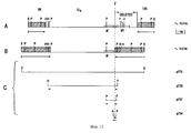
Изобретение относится к ряду BHV-1 вакцин как живых, так и инактивированных, у которых общее то, что в них полностью или частично отсутствует гликопротеин gE ген. Эта серия включает как естественный мутант с gE делецией, так и созданные мутанты с делецией gE, которые могут включать или не включать также делецию гена тимидинкиназы и/или гена gI гликопротеина, и созданные мутанты с делецией gE, которые используются как векторы для гетерологичных генов. Изобретение далее относится к нуклеотидным последовательностям, кодирующим гликопротеид gE-ген BHV-1, олигонуклеотидам, производным от этих последовательностей, самому гликопротеину gE, пептидам, которые получены от них, и /моноклональным или поликлональным/ антителам, которые направлены против gE гликопротеина и пептидов, полученных от них. Изобретение также относится к комплексам гликопротеинов gE и gI BHV-1 и к антителам, направленным против таких комплексов.DETAILED DESCRIPTION OF THE INVENTION
The invention relates to a series of BHV-1 vaccines, both live and inactivated, which have in common that they completely or partially lack the glycoprotein gE gene. This series includes both a natural mutant with a gE deletion, and created mutants with a gE deletion, which may or may not include a deletion of the thymidine kinase gene and / or the gI glycoprotein gene, and created mutants with a gE deletion, which are used as vectors for heterologous genes. The invention further relates to nucleotide sequences encoding the glycoprotein gE gene BHV-1, oligonucleotides derived from these sequences, the glycoprotein gE itself, the peptides derived from them, and / monoclonal or polyclonal / antibodies that are directed against gE glycoprotein and peptides, received from them. The invention also relates to complexes of glycoproteins gE and gI BHV-1 and to antibodies directed against such complexes.
Эти материалы в соответствии с изобретением можно использовать для:
1/ вакцинации скота против заболеваний, вызванных BHV-1, так что можно различить зараженных BHV-1 животных и вакцинированных животных, наряду с традиционной можно использовать созданную вакцину;
2/ вакцинации скота против как заболеваний BHV-1, так и заболеваний, вызванных другими патогенными бактериями, кодирующие последовательности которых для защитных антигенов можно ввести в мутанты с делецией BHV-1;
3/ исследования крови, сыворотки, молока или других жидкостей тела скота на серологическое определение или посредством техники обнаружения нуклеиновой кислоты /например, ЦРП (ЦРП - цепная реакция полимеразы)/, были ли животные заражены BHV-1 дикого типа или были вакцинированы мутантом с делецией gE.These materials in accordance with the invention can be used for:
1 / vaccination of cattle against diseases caused by BHV-1, so that it is possible to distinguish between BHV-1 infected animals and vaccinated animals, along with the traditional one, the created vaccine can be used;
2 / vaccination of cattle against both BHV-1 diseases and diseases caused by other pathogenic bacteria, the coding sequences of which for protective antigens can be introduced into mutants with deletion of BHV-1;
3 / tests of blood, serum, milk or other body fluids of the livestock for serological determination or by means of a nucleic acid detection technique / for example, CRP (CRP (polymerase chain reaction)) / whether animals were infected with wild-type BHV-1 or were vaccinated with a deletion mutant gE.
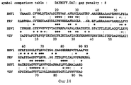
Синтез олигопептидов, полипептидов и гликопротеинов, полученных от кодирующей последовательности гликопротеин gE-гена и гликопротеин gI-гена BHV-1. Synthesis of oligopeptides, polypeptides and glycoproteins obtained from the coding sequence of the glycoprotein gE gene and glycoprotein gI gene BHV-1.
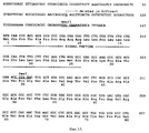
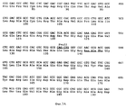
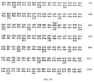
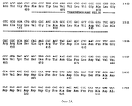
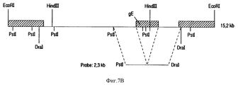
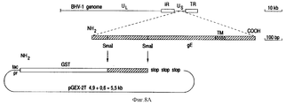
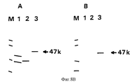
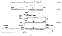
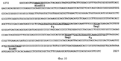
Результаты анализа последовательности ДНК, описанного в примерах, гликопротеин gE-гена /фиг. 3А/ и изолированных фрагментов ДНК, которые кодируют для этого гена, делают возможным, используя стандартные молекулярно-биологические процедуры, как синтезировать пептиды gE белка /олиго- или полипептиды/, так и выразить gE белок полностью или большими частями через прокариотный путь /в бактериях/ или через эукариотный путь /например, в мышиных клетках/. Через эти пути можно получить gE-специфический антиген, который может, например, служить для выработки gE-специфических моноклональных антител /MAT/. Кроме того, и gE-специфический антиген /и gE-специфические MAT/ можно использовать в серологических тестах, чтобы сделать различие между животными, вакцинированными вакциной BHV-1 делеции gE и животными, зараженными вирусом BHV-1 дикого типа. The results of the analysis of the DNA sequence described in the examples, glycoprotein gE gene / Fig. 3A / and isolated DNA fragments that encode for this gene are made possible, using standard molecular biological procedures, both to synthesize gE protein peptides / oligo or polypeptides / and to express the gE protein in whole or in large parts through the prokaryotic pathway / in bacteria / or via the eukaryotic pathway / for example, in murine cells /. Through these pathways, you can get gE-specific antigen, which can, for example, serve to produce gE-specific monoclonal antibodies / MAT /. In addition, both the gE-specific antigen / and gE-specific MAT / can be used in serological tests to distinguish between animals vaccinated with the gH deletion BHV-1 vaccine and animals infected with wild-type BHV-1 virus.
Результаты частичного анализа последовательности ДНК гликопротеина gI гена - описанного в примерах - и изолированные фрагменты ДНК, которые кодируют для этого гена, вместе с эукариотными клетками, выражающими гликопротеин gE, позволяют экспрессию gI/gE комплекса в эукариотных клетках /См. фиг. 13 и 14/. Этот гликопротеиновый комплекс может быть использован, чтобы получить gI/gE специфические моноклональные антитела. gI/gE комплекс можно также использовать как антиген в серологических пробах для дифференциации между животными, вакцинированными одним gE BHV-1 мутантом с делецией или двойным gI/gE BHV-1 мутантом с делецией, и животными, зараженными вирусом BHV-1 дикого типа. The results of a partial analysis of the DNA sequence of the glycoprotein gI gene - described in the examples - and isolated DNA fragments that encode for this gene, together with eukaryotic cells expressing the gE glycoprotein, allow expression of the gI / gE complex in eukaryotic cells / See FIG. 13 and 14 /. This glycoprotein complex can be used to obtain gI / gE specific monoclonal antibodies. The gI / gE complex can also be used as an antigen in serological samples to differentiate between animals vaccinated with one gE BHV-1 deletion mutant or a double gI / gE BHV-1 deletion mutant and animals infected with wild-type BHV-1 virus.
gE-специфические пептиды
На основе известной кодирующей последовательности белка с помощью автоматического синтезатора можно получить полипептиды не менее чем 40-50 аминокислот. Теперь, когда выявлена кодирующая последовательность белка gE гликопротеина штамма Lam BHV-1 /фиг. 3A/, можно синтезировать полипептиды этого BHV-1 gE гликопротеина. Такими полипептидами по стандартным методам можно иммунизировать экспериментальных животных, таких как мышей или кроликов, чтобы выработать gE-специфические антитела. Кроме того, используя эти gE-специфические пептиды, можно также определить местоположения, где анти-gE антитела реагируют с gE белком /эпитопы/, например, способом PEPSCAN /Geysen и др. , 1984, Proc. Natl, Acad. Sci, США 81, 3998-4002/. gE-специфические олигопептиды можно также использовать в серологических реакциях, которые демонстрируют анти-gE антитела.gE-specific peptides
Based on the known protein coding sequence, an automated synthesizer can produce polypeptides of at least 40-50 amino acids. Now that the coding sequence of the gE glycoprotein protein of the Lam strain BHV-1 / FIG. 3A /, polypeptides of this BHV-1 gE glycoprotein can be synthesized. Experimental animals, such as mice or rabbits, can be immunized with such polypeptides by standard methods to generate gE-specific antibodies. In addition, using these gE-specific peptides, it is also possible to determine the locations where anti-gE antibodies react with the gE protein / epitopes /, for example, by the method of PEPSCAN / Geysen et al., 1984, Proc. Natl, Acad. Sci, US 81, 3998-4002 /. gE-specific oligopeptides can also be used in serological reactions that exhibit anti-gE antibodies.
Прокариотная экспрессия gE
Для синтеза gE белка в бактериях /т.е. прокариотной экспрессии gE/, фрагменты ДНК, которые кодируют для гликопротеина gE или для его частей, должны клонироваться в векторы прокариотной экспрессии. Векторы прокариотной экспрессии представляют собой циркулярные молекулы ДНК, которые могут сохранять себя в бактерии как отдельная воспроизводящая молекула /плазмида/. Эти векторы экспрессии содержат один или несколько маркерных генов, которые кодируют для устойчивости к антибиотику, и таким образом позволяют выбор для бактерий с вектором экспрессии. Кроме того, векторы экспрессии включают /часто контролируемый/ участок промотора, за которым фрагменты ДНК можно лигировать и которые затем выражаются под влиянием промотора. Во многих настоящих векторах прокариотной экспрессии нужный белок выражен в состоянии слияния с так называемым белком-носителем. В векторе за промотором располагается кодирующая последовательность для белка-носителя, непосредственно к которому можно лигировать нужный фрагмент ДНК. Белки слияния часто более стабильны и/или их легче распознать и/или изолировать. Уровень состояния устойчивости, которого может достичь определенный слитый белок в определенном бактериальном штамме, зависит от вида слияния и от штамма. Обычно пробуют различные комбинации.Prokaryotic expression of gE
For the synthesis of gE protein in bacteria / i.e. prokaryotic expression of gE /, DNA fragments that encode for the gE glycoprotein or parts thereof must be cloned into prokaryotic expression vectors. Vectors of prokaryotic expression are circular DNA molecules that can retain themselves in bacteria as a separate reproducing molecule / plasmid /. These expression vectors contain one or more marker genes that encode for antibiotic resistance, and thus allow selection for bacteria with an expression vector. In addition, expression vectors include a / often controlled / portion of the promoter beyond which DNA fragments can be ligated and which are then expressed under the influence of the promoter. In many of these vectors of prokaryotic expression, the desired protein is expressed in a state of fusion with the so-called carrier protein. In the vector behind the promoter, there is a coding sequence for the carrier protein, directly to which the desired DNA fragment can be ligated. Fusion proteins are often more stable and / or easier to recognize and / or isolate. The level of stability that a particular fusion protein can achieve in a particular bacterial strain depends on the type of fusion and the strain. Usually try different combinations.
Эукариотной экспрессии гликопротеин gE-гена
Хотя прокариотная экспрессия белков предлагает некоторые преимущества, белки не имеют модификаций, таких как гликосилация и других, которые имеют место в эукариотных клетках. В результате этого эукариотически выраженный белок часто является более стабильным антигеном. Для гетерологичной экспрессии белков в эукариотных клетках, таких как мышиные клетки, используются векторы эукариотной экспрессии. Этими векторами являются плазмиды, которые не только могут размножаться в клетках E.coli, но также стабильно существуют в эукариотных клетках. В дополнение к прокариотному селективному маркеру они также включают эукариотный селективный маркер. Аналогично векторам прокариотной экспрессии векторы эукариотной экспрессии содержат участок промотора, за которым можно лигировать желаемые гены. Однако последовательности промотора в эукариотных векторах специфичны для эукариотных клеток. Более того, в эукариотных векторах слияние с белками-носителями используется очень редко. Эти векторы вводятся в эукариотные клетки посредством стандартного способа трансфекции /Graham и A.I.van der Eb, 1973, Virology, 52, 456-467/. Кроме эукариотных плазмидных векторов имеются также вирусные векторы, где гетерологичный ген вводится в геном вирусе /напр., ретровирусы, вирусы герпеса и коровьей оспы/. Следовательно, эукариотные клетки можно заразить рекомбинантными вирусами.Eukaryotic expression of the gE gene glycoprotein
Although prokaryotic expression of proteins offers some advantages, proteins do not have modifications, such as glycosylation, and others that occur in eukaryotic cells. As a result, a eukaryotic protein is often a more stable antigen. For heterologous expression of proteins in eukaryotic cells, such as murine cells, eukaryotic expression vectors are used. These vectors are plasmids that can not only multiply in E. coli cells, but also stably exist in eukaryotic cells. In addition to the prokaryotic selective marker, they also include a eukaryotic selective marker. Similar to prokaryotic expression vectors, eukaryotic expression vectors contain a promoter region beyond which the desired genes can be ligated. However, promoter sequences in eukaryotic vectors are specific for eukaryotic cells. Moreover, fusion with carrier proteins is very rarely used in eukaryotic vectors. These vectors are introduced into eukaryotic cells by a standard transfection method / Graham and AIvan der Eb, 1973, Virology, 52, 456-467 /. In addition to eukaryotic plasmid vectors, there are also viral vectors where the heterologous gene is introduced into the genome of the virus (for example, retroviruses, herpes viruses and vaccinia). Therefore, eukaryotic cells can be infected with recombinant viruses.
В общем, нельзя предсказать какой вектор и тип клетки наиболее приемлемы для определенного продукта гена. По большей части пробуются несколько комбинаций. In general, it is not possible to predict which vector and cell type are most appropriate for a particular gene product. For the most part, several combinations are tried.
Эукариотная экспрессия и гликопротеина gE и гликопротеина gI
Конечная структура, которой достигает белок, зависит от последовательности его основной аминокислоты, его укладки, его посттрансляционной модификации и т. д. Важный фактор, от которого зависит структура белка, - его взаимосвязь с одним или несколькими другими белками. Мы выявили, что BHV-1 гликопротеин gE образует комплекс, по меньшей мере, с одним другим гликопротеином: BHV-1 гликопротеином gI. Первое указание на такой комплекс появилось из наших результатов с кандидатом анти-gE MAT 1, 51, 67, 75 и 78 /См. таблицу 2/. Эти MAT не реагировали ни с Difivac-1, ни с Lamg E-, а также не опознавали гликопротеин gE-экспрессирующие ЗТЗ клетки. Однако эти MAT не реагировали с gE-экспрессирующими ЗТЗ клетками после заражения Difivac-1, показывая, что нужны комплементарные факторы, чтобы дать гликопротеину gE нужную антигенную конформацию для этих МАТ. В некоторых из наших экспериментах по радио-иммунопреципитату с MAT 81 мы выявили копреципитацию белка с молекулярным весом в 63 кД. Ввиду того, что гликопротеин gE вируса простого герпеса образует комплекс с белком со сравнительным молекулярным весом /HSVI гликопротеин gI/, мы сделали вывод, что BH-1 гликопротеин gE образует комплекс с BHV-1 гомологом гликопротеина gI. Чтобы изучить этот BHV-1 gE/gI комплекс и получить gE антиген с должной антигенной структурой, мы экспрессировали оба гликопротеина в одной эукариотной клетке. Для этого мы применили те же процедуры, что описаны для эукариотной экспрессии одного гликопротеина gE. Единственным дополнительным условием было использование векторов экспрессии с различными эукариотными селектируемыми маркерами.Eukaryotic expression of both gE glycoprotein and gI glycoprotein
The final structure that the protein reaches depends on the sequence of its basic amino acid, its folding, its post-translational modification, etc. An important factor on which the structure of the protein depends is its relationship with one or more other proteins. We found that BHV-1 glycoprotein gE forms a complex with at least one other glycoprotein: BHV-1 glycoprotein gI. The first indication of such a complex emerged from our results with the
Серологические реакции
Серологические методы для различения скота, вакцинированного Defivac-1 скота, зараженного BH-1 дикого типа на основе антител против gE, предпочтительно основываются на использовании моноклональных антител, направленных против gE.Serological reactions
Serological methods for distinguishing livestock vaccinated with Defivac-1 livestock infected with wild-type BH-1 based on anti-gE antibodies are preferably based on the use of monoclonal antibodies directed against gE.
Они используются следующим образом:
a/ В соответствии с принципом, описанным Van Oirschot и др. /Jorn. of Virological Methods, 22, 191-206, 1988/. В этом ELISA (ELISA - блокирующий ферментный иммуносорбентный тест) для обнаружения gI антител против вируса болезни Aujeszky's, антитела проявляются своим блокирующим действием на реакцию двух MAT, имеющих два разных эпитопа на gI. Тест или реакция проводится следующим образом. Микротитрационные планшеты покрываются MAT 1 в течение ночи при 37oC, после чего они стоят, например, при 4oC или -20oC. Исследуемая сыворотка преинкубируется антигеном в отдельных непокрытых микротитрационных планшетах, например, 2 часа при 37oC. Планшеты, покрытые МАТ-1, промываются, скажем, 5 раз, после чего на эти планшеты добавляется MAT 2, связанное с пероксидазой хрена обыкновенного /HRPO/. Затем преинкубированные смеси сыворотка-антиген переносятся на планшеты, в которых находятся два MAT, с последующей инкубацией, например, в течение 1 часа при 37oC. Планшеты промываются и субстрат добавляется к каждой лунке. После приблизительно 2 часов при комнатной температуре планшеты считываются спектрофотометрически. На каждом планшете имеются четыре отрицательные контрольные сыворотки и четыре серийных разведения положительной сыворотки. Сыворотка с величиной оптической плотности /ОП/ менее 50% средней величины ОП 4 отрицательных контрольных сывороток, которые исследовались на том же планшете, считается положительной.They are used as follows:
a / In accordance with the principle described by Van Oirschot et al. / Jorn. of Virological Methods, 22, 191-206, 1988 /. In this ELISA (ELISA - blocking enzyme immunosorbent assay) for detecting gI antibodies against Aujeszky's disease virus, the antibodies manifest their blocking effect on the reaction of two MATs having two different epitopes on gI. The test or reaction is carried out as follows. Microtiter plates are coated with
b/ В соответствии с принципом бутерброда непрямого двойного антитела /IDAS/. Здесь микротитровальные планшеты покрываются MAT или поликлональной сывороткой, направленной против gE белка. Инкубация препаратом gE-антигена приводит к тому, что gE связывается с покрытием. Антитела, специфически направленные против gE в исследуемой коровьей сыворотке, последовательно связываются с gE. Эти связанные антитела распознаются антикоровьим иммуноглобулиновым конъюгатом. Антитела в этом конъюгате ковалентно связаны с ферментом пероксидазы. Наконец, связанный конъюгат виден при добавлении хромогенного субстрата. Специфичность реакции проверяется проведением той же процедуры с gE-отрицательным контрольным препаратом вместо препарата gE-антигена. На каждом микротитровальном планшете имеются положительные и отрицательные контрольные сыворотки. Тест считается действительным, если положительные сыворотки дают положительный результат в определенном разбавлении. Сыворотка положительна, если ее ОП выше на 0.2, чем у стандартной отрицательной контрольной сыворотки. b / In accordance with the principle of an indirect double antibody sandwich / IDAS /. Here, microtiter plates are coated with MAT or polyclonal serum directed against the gE protein. Incubation with a gE antigen preparation causes the gE to bind to the coating. Antibodies specifically directed against gE in test cow serum bind sequentially to gE. These coupled antibodies are recognized by the anti-cow immunoglobulin conjugate. Antibodies in this conjugate are covalently linked to the peroxidase enzyme. Finally, the bound conjugate is visible upon the addition of a chromogenic substrate. The specificity of the reaction is verified by performing the same procedure with the gE-negative control drug instead of the gE antigen preparation. Each microtiter plate contains positive and negative control sera. A test is considered valid if positive sera give a positive result in a certain dilution. A serum is positive if its OD is higher by 0.2 than that of a standard negative control serum.
c/ В соответствии с принципом IDAS, описанным под 2, но после инкубации исследуемой сыворотки вместо конъюгата анти-коровьего иммуноглобулина используется анти-gE MAT/HPPO. Можно использовать сыворотку анти-gE пептида или анти-gE поликлональную сыворотку вместо анти-gE MAT. Планшеты промываются и к каждой лунке добавляется хромогенный субстрат. Приблизительно после 2 часов при комнатной температуре планшеты считываются спектрофотометрически. В каждый планшет включены четыре отрицательных контрольных сыворотки и четыре серийных разбавления положительной сыворотки. Сыворотка с величиной ОП менее 50% величины ОП 4-х отрицательных контрольных сывороток, которые исследовались на том же планшете, считается положительной. c / In accordance with the IDAS principle described under 2, but after incubation of the test serum, anti-gE MAT / HPPO is used instead of the anti-bovine immunoglobulin conjugate. Anti-gE peptide serum or anti-gE polyclonal serum can be used instead of anti-gE MAT. The plates are washed and a chromogenic substrate is added to each well. After approximately 2 hours at room temperature, the plates are read spectrophotometrically. Four negative control sera and four serial dilutions of positive serum are included in each plate. A serum with an OD value of less than 50% of the OD value of the 4 negative control sera that were tested on the same plate is considered positive.
d/ В соответствии с принципом блокирующего ферментного иммуносорбентного теста /ELISA/, по которому антиген вируса, который может быть очищенным или неочищенным, наносится покрытием на микротитровальный планшет на всю ночь. На этих планшетах исследуемая сыворотка инкубируется примерно один час или больше при 37oC. После процедуры промывания на планшеты добавляется анти-gE MAT с последующей инкубацией приблизительно 1 час при 37oC. Вместо анти-gE MAT можно использовать сыворотку анти-gE пептида или анти-gE поликлональную сыворотку. Планшеты промываются и к каждой лунке добавляется хромогенный субстрат. После примерно 2 часов при комнатной температуре планшеты считываются спектрофотометрически. На каждом планшете имеются четыре отрицательные контрольные сыворотки и четыре серийных разбавления положительной сыворотки. Сыворотка с величиной ОП менее 50% средней величины ОП 4х отрицательных контрольных сывороток, которые исследовались на том же планшете, считается положительной.d / In accordance with the principle of an enzyme-blocking immunosorbent assay (ELISA), according to which a virus antigen, which may be purified or uncleaned, is coated on a microtiter plate overnight. On these plates, the test serum is incubated for approximately one hour or more at 37 ° C. After the washing procedure, anti-gE MAT is added to the plates, followed by incubation for approximately 1 hour at 37 ° C. Instead of anti-gE MAT, anti-gE peptide serum or anti-gE polyclonal serum. The plates are washed and a chromogenic substrate is added to each well. After approximately 2 hours at room temperature, the plates are read spectrophotometrically. Each tablet has four negative control sera and four serial dilutions of positive serum. Serum with an OD value of less than 50% of the average OD value of 4 x negative control sera that were tested on the same plate is considered positive.
Во всех указанных выше процедурах может использоваться традиционный культивированный антиген вируса, который содержит gE, а также gE-антиген, который выражен через прокариоты или эукариоты. Как вариант, вместо традиционного антигена в указанных диагностических пробах могут использоваться олигопептиды, основанные на BHV-1 gE последовательности. Кроме того, такие олигопептиды можно использовать для разработки так называемого "коровьего" теста в соответствии с принципом, описанным в статье Kemp и др. Science, 241, 1352-1354, 1988. Такой тест будет основываться на связи антигенной последовательности олигопептида антителами, направленными против gE, присутствующего в зараженных животных. Для такого теста олигопептиды должны быть связаны с MAT, направленными против эритроцитов коров. In all of the above procedures, a traditional cultured virus antigen that contains gE, as well as a gE antigen that is expressed through prokaryotes or eukaryotes, can be used. Alternatively, instead of the traditional antigen, oligopeptides based on the BHV-1 gE sequence can be used in these diagnostic tests. In addition, such oligopeptides can be used to develop the so-called "cow" test in accordance with the principle described in Kemp et al. Science, 241, 1352-1354, 1988. Such a test will be based on the connection of the antigenic sequence of the oligopeptide with antibodies directed against gE present in infected animals. For such a test, oligopeptides must be linked to MATs directed against bovine red blood cells.
Анализ на нуклеиновую кислоту с использованием цепной реакции полимеразы
Олигонуклеотиды /зонды и праймеры/ могут, например, использоваться в цепной реакции полимеразы, чтобы различить вакцинированных и зараженных животных. Цепная реакция полимеразы /ЦРП/ - технологический прием, посредством которого нуклеиновые кислоты патогена можно в короткое время размножить миллиарды раз /De polymesase kettingreactie. P.F. Hilderiuk, J.A. Wagenaar, J. W. B. van der Giessen u B.A.M. van der Jeijst, 1990, Fijdschrift voor Dierdeneeskunde deel 115, 1111-1117/. gE олигонуклеотиды можно выбрать таким образом, что в gE положительном геноме образуется другой продукт, нежели в gE отрицательном геноме. Преимуществом этого является то, что и животное, которое было вакцинировано вакциной с делецией gE, дает положительный сигнал в тесте на ЦРП. Однако этот подход зависит от присутствия нуклеиновых кислот вируса в образце, например, крови от животного, на которой проводится тест.Nucleic acid analysis using polymerase chain reaction
Oligonucleotides / probes and primers / can, for example, be used in the polymerase chain reaction to distinguish between vaccinated and infected animals. The polymerase chain reaction / CRP / is a technological method by which pathogen nucleic acids can be multiplied billions of times in a short time / De polymesase kettingreactie. PF Hilderiuk, JA Wagenaar, JWB van der Giessen and BAM van der Jeijst, 1990, Fijdschrift
После острого заражения BHV-1 имеется большая вероятность, что BHV-1 специфические нуклеиновые кислоты могут быть продемонстрированы в крови, но еще не определилось, будут ли также демонстрироваться в крови нуклеиновые кислоты BHV-1 во время латентного периода. After acute infection with BHV-1, it is highly likely that BHV-1 specific nucleic acids can be demonstrated in the blood, but it has not yet been determined whether BHV-1 nucleic acids will also be shown in the blood during the latent period.
Использование ВНV-1 в качестве вектора
Для экспрессии гетерологичных генов в BHV-1 геноме необходимо иметь в наличии точную информацию об участке, куда должен вставляться гетерологичный ген. Основные последовательности не должны нарушать структуру, а для экспрессии гетерологичного гена должны быть регуляторные последовательности. Гликопротеин gE-ген является подходящим местом для экспрессии гетерологичных генов. gE-ген не является существенным, следовательно, не может быть возражений по замене gE гена гетерологичным геном. Как следствие этого, гетерологичный ген может так располагаться, что он будет находиться под влиянием регуляторных последовательностей gE гена. Однако нет необходимости использовать регуляторные последовательности gE-гена. Экспрессия гетерологичных генов может контролироваться другими, т. е. более сильными регуляторными последовательностями различных генов. Представляется возможным также лигировать гетерологичный ген к /экпортному/ сигнальному пептиду gE гена с тем, чтобы повлиять на секрецию продукта гетерологичного гена. Очевидно, что подробное знание гена gE и белка gE дает возможность использовать BHV-1 как вектор достаточно контролируемым образом. Разработанные векторы можно серологически отличить от дикого типа. Создание ВНV-1 мутантов, которые эксцрессируют гетерологичные гены, можно реализовать таким же образом, что и создание мутантов с делецией gE, показанное в примерах. Однако тогда фрагменты делеции следует заменить фрагментом, на котором гетерологичный ген расположен в местоположении делеции.Using BHV-1 as a vector
For the expression of heterologous genes in the BHV-1 genome, it is necessary to have accurate information about the site where the heterologous gene is to be inserted. The main sequences must not disrupt the structure, and for the expression of a heterologous gene there must be regulatory sequences. The glycoprotein gE gene is a suitable site for the expression of heterologous genes. The gE gene is not significant, therefore, there can be no objection to replacing the gE gene with a heterologous gene. As a consequence of this, the heterologous gene can be so arranged that it will be influenced by the regulatory sequences of the gE gene. However, there is no need to use the regulatory sequences of the gE gene. Expression of heterologous genes can be controlled by other, i.e., stronger regulatory sequences of different genes. It is also possible to ligate the heterologous gene to the / export / signal peptide of the gE gene in order to affect the secretion of the product of the heterologous gene. Obviously, detailed knowledge of the gE gene and gE protein makes it possible to use BHV-1 as a vector in a fairly controlled manner. The developed vectors can be serologically distinguished from the wild type. The creation of BHV-1 mutants that express heterologous genes can be implemented in the same way as the creation of gE deletion mutants shown in the examples. However, then the deletion fragments should be replaced by the fragment on which the heterologous gene is located at the location of the deletion.
Примеры
1/ Изоляция и определение естественного мутанта с делецией gE.Examples
1 / Isolation and determination of a natural mutant with a gE deletion.
a/ Изоляция естественного /природного/ мутанта
Геномная ДНК была изолирована из ряда ослабленных традиционным путем вакцин в соответствии со стандартными способами и проанализирована с использованием рестрикционных ферментов. В частности, мы искали отклонения генома, которые бы подходили, чтобы отличить от вируса BHV-1 дикого типа.a / Isolation of natural / natural / mutant
Genomic DNA was isolated from a number of conventionally attenuated vaccines in accordance with standard methods and analyzed using restriction enzymes. In particular, we looked for genomic abnormalities that would be suitable to distinguish from wild-type BHV-1 virus.
Ослабление было направлено, в частности, на Us участок генома BHV-1, потому что в этом участке - по аналогии с вирусом герпес простой - вероятно располагается ряд генов, кодирующих для несущественных гликопротеинов /Определение гена гликопротеида вируса простого герпеса 1 внутри кластера генов, необязательного для роста в клеточной культуре, R. Longnecker S. Chatterjee, R.J. Whitley u B. Roijrnan (1987) Proc. Natl dead. Sci, 84, 4,03-4307/.The attenuation was directed, in particular, to the U s region of the BHV-1 genome, because in this region - by analogy with the herpes simplex virus - there are probably a number of genes encoding for non-essential glycoproteins / Determination of the herpes simplex
Подборка BHV-1 вакцин из Загребского университета, Югославия /Lugovic и др. Veterinarski Arhiv, 55, 241-245, 1985/, после большого числа пересевов на эмбриональные почечные клетки коровы и эмбриональные трахеальные клетки коровы /Эбтр/, обладала участком отклонения Us, помимо наличия нормального участка Us. Более того, эта вакцина образовывала и большие и малые пятна на Эбтр клетках. Из этой смешанной популяции тремя этапами ограниченного разведения был изолирован вирус с участком Us отклоняющейся формы, с небольшими пятнами в каждом случае. Вирус, изолированный этим путем, исследовался дальше и был назван Difivac-1. Он был депонирован в Институт Пастера, Париж, Франция 27 мая 1992 г, номер депозита 1-1213.A selection of BHV-1 vaccines from the University of Zagreb, Yugoslavia / Lugovic et al. Veterinarski Arhiv, 55, 241-245, 1985 /, after a large number of passages for cow embryonic kidney cells and cow embryonic tracheal cells / Ebtr /, had a site of deviation of U s , in addition to the presence of a normal site U s . Moreover, this vaccine also formed large and small spots on the Ebtr cells. From this mixed population, a virus with a plot of U s deviating shape, with small spots in each case, was isolated by three stages of limited dilution. The virus isolated in this way was investigated further and was named Difivac-1. He was deposited at the Pasteur Institute, Paris, France on May 27, 1992, deposit number 1-1213.
b/ Определение делеции в gE гена в Difivac-1
Для дальнейшего анализа этого отклонения в Us участке геномная ДНК Difivac-1 была изолирована в соответствии со стандартными методами и подвергнута блот-анализу /фиг. 1A/. Гиридизация этого блота фрагментом Hind III K дикого типа, помеченного 32p подтвердила, что этот фрагмент, расположенный в центре участка Us, короче на 1.0 тысячу пар нуклеотидов /т.п.н./ в Difivac-1. Более того, этим анализом можно было приблизительно определить позицию отсутствующей части /фиг. 1B/. Для дальнейшего анализа этой делеции Us участок BHV-1 штамма Lam дикого типа был изолирован и клонирован в прокариотные векторы. С этой целью в соответствии со стандартными методами геномная ДНК штамма Lam /фиг. 2A/ была изолирована и клонирована в векторы pUC18, рACYC и pBP322 /фиг. 2B/. Была составлена физическая карта участка вокруг предполагаемой позиции делеции /фиг. 2C/. Исходя из этой физической карты, в векторах pKUN 19 и pUC18 /фиг. 2/ были конструктированы субклоны, пригодные для определения последовательности нуклеотидов этой области. Используя эти субклоны, методом Сангера была определена нуклеотидная последовательность двух цепей всей области /показано на фиг. 2C/. Эта нуклеотидная последовательность была анализирована с использованием программы PC/ген. Из концептуальной трансляции оказывается, что нуклеотиды /нт/ 168-1893 кодируют для открытой рамки считывания 575 аминокислот /фиг. 3A/. Дальнейший анализ показал, что эта последовательность аминокислоты имеет характеристики трансмембранного гликопротеина, как показано на фиг. 3B. Факт таков, что эти первые 26 аминокислот /ак/ опознаются как типично эукариотный экспортный сигнал и область между ак 423 и ак 450 опознается как трансмембранная область. Кроме того, в этой последовательности оказываются три потенциальных участка N-связанной гликосиляции. Эта предсказанная последовательность аминокислоты демонстрирует близкое сходство с gE-геном гликопротеина вируса простого герпеса /HSV/; см. фиг. 4A и 4B. Эти и другие сходства оправдывают вывод, что найденный ген является gE гомологом BHV-1. По этой причине ген называется gE. Чтобы определить, до какой степени этот BHV-1 gE-ген отсутствует в Difivac-1, был изолирован p318 фрагмент. Фрагмент p318 начинается на AluI участке 55 нт до требуемой открытой рамки считывания BHV-1 gE и заканчивается 133 нт за ней. Геномная ДНК Difivac-1 анализировалась этим p318 фрагментом с использованием блот-гибридизации. Это выявило, что Difivac-1 не содержит p318 обнаруживаемых последовательностей /фиг. 5/. Этот эксперимент подтвердил, что Difivac-1 содержит делецию и четко продемонстрировал, что эта делеция проходит по всему gE гену.b / Determination of deletion in gE gene in Difivac-1
For further analysis of this deviation in the U s region, Difivac-1 genomic DNA was isolated in accordance with standard methods and subjected to blot analysis / Fig. 1A /. The gyridization of this blot with a wild-type Hind III K fragment labeled 32p confirmed that this fragment located in the center of the U s region is shorter than 1.0 thousand pairs of nucleotides (bp) in Difivac-1. Moreover, with this analysis it was possible to approximately determine the position of the missing part / FIG. 1B /. For further analysis of this deletion of U s, the wild-type LH strain BHV-1 was isolated and cloned into prokaryotic vectors. To this end, in accordance with standard methods, the genomic DNA of the Lam strain / Fig. 2A / was isolated and cloned into the vectors pUC18, pACYC and pBP322 / Fig. 2B /. A physical map of the region around the putative position of the deletion / FIG. 2C /. Based on this physical map, in vectors pKUN 19 and pUC18 / Fig. 2 / subclones suitable for determining the nucleotide sequence of this region were constructed. Using these subclones, the nucleotide sequence of two chains of the entire region was determined by the Sanger method / shown in FIG. 2C /. This nucleotide sequence was analyzed using the PC / gene program. From the conceptual translation, it appears that nucleotides / nt / 168-1893 encode 575 amino acids / FIG. 3A /. Further analysis showed that this amino acid sequence has the characteristics of a transmembrane glycoprotein, as shown in FIG. 3B. The fact is that these first 26 amino acids / ak / are recognized as a typically eukaryotic export signal and the region between ak 423 and
Чтобы определить размер и позицию участка делеции, геномные последовательности, охватывающие Us регион Difivac-1, были клонированы в прокариотные векторы (См. фиг. 11C), фрагмент с 14.5 т.п.н. был клонирован в pACYC вектор и назван p775. HindIII фрагмент в 7.4 т.п.н. был независимо клонирован в pUC18 вектор и назван p728, были изолированы два субклона: фрагмент PstI с 1,4 т. п. н. в клоне p737 и фрагмент AluI - PstI в клоне p754 с 350 т.п.н. Анализ рестрикционного фермента и бло-анализ этих клонов /данные не показаны/ продемонстрировали, что делеция gE в Difivac-1 составляет длину 2,7 т.п. н., начиная сразу от 5' с gE гена и кончая на границе Us участка. Эти 2.7 т. п. н. были замещены дублированием одного сегмента в 1 т.п.н., расположенного в участке Us напротив gE гена. См. фиг. 11B. Чтобы подтвердить результаты этого анализа и определить точную точку рекомбинации, нуклеотидная последовательность большинства вставок клона p754 определялась и сравнивалась с последовательностями дикого типа. См. фиг. 12. Этот анализ показал, что точка рекомбинации расположена у 77 пары основания /по/ верх от начального кодона gE гена.To determine the size and position of the deletion site, genomic sequences spanning the U s region of Difivac-1 were cloned into prokaryotic vectors (See FIG. 11C), fragment with 14.5 kb. was cloned into a pACYC vector and named p775. HindIII fragment of 7.4 kb was independently cloned into pUC18 vector and named p728, two subclones were isolated: a PstI fragment with 1.4 bp in clone p737 and the AluI - PstI fragment in clone p754 with 350 kbp The restriction enzyme assay and the bloom analysis of these clones (data not shown) showed that the gE deletion in Difivac-1 is 2.7. n., starting immediately from 5 'with the gE gene and ending at the border of the U s site. These 2.7 t.p. were replaced by duplication of one segment of 1 kb located in the U s region opposite the gE gene. See FIG. 11B. To confirm the results of this analysis and determine the exact recombination point, the nucleotide sequence of most insertions of clone p754 was determined and compared with wild-type sequences. See FIG. 12. This analysis showed that the recombination point is located at 77 base pairs / up / up from the initial codon of the gE gene.
c/ Оценка безопасности и эффективности Difivac-1
Difivac-1 был опробован на семинедельных телятах, свободных от BHV-1 сероотрицательного специфического патогена. Восемь телят были вакцинированы через нос 105 TCID50 в 2 мл, из которых по 1 мл распылялось в каждую ноздрю. Восемь семинедельных телят, свободных от BHV-1 сероотрицательного специфического патогена, помещались в изоляции, им вводилось в нос 2 мл среды культуры, и они служили невакцинированными контрольными животными. Через пять недель после вакцинации вакцинированным и контрольным телятам капали в нос 107 TCID50 очень вирулентный штамм Iowa BHV-1. Шесть недель спустя после контрольного заражения всем телятам вводился внутримышечно дексаметазон в течение 5 дней, чтобы реактивировать предполагаемый латентный вирус. Клинические проявления, ректальные температуры и рост организма находились под контролем. Вирус изолировался из носовых мазков, и в сыворотке были определены титры нейтрализующего антитела.c / Safety and efficacy assessment of Difivac-1
Difivac-1 was tested on seven-week-old calves free of serum negative specific pathogen BHV-1. Eight calves were vaccinated through the nose with 10 5 TCID 50 in 2 ml, of which 1 ml was sprayed into each nostril. Eight seven-week-old calves that were free of BHV-1 sero-negative specific pathogen were placed in isolation, 2 ml of culture medium was injected into their nose, and they served as unvaccinated control animals. Five weeks after vaccination with vaccinated and control calves, 10 7 TCID 50 very virulent Iowa strain BHV-1 was dripped into the nose. Six weeks after challenge, all calves were injected intramuscularly with dexamethasone for 5 days to reactivate the suspected latent virus. Clinical manifestations, rectal temperatures, and body growth were controlled. The virus was isolated from nasal swabs, and neutralizing antibody titers were determined in serum.
После вакцинации поведение, аппетит, ректальные температуры и скорости роста телят оставались нормальными, но у вакцинированных телят наблюдались некоторые серьезные выделения из носа и повышенное слюноотделение. Нарушений в слизистой оболочке носа не наблюдалось. После вакцинации из носовых мазков выделялся Difivac-1 /фиг. 17/. Все вакцинированные телята вырабатывали нейтрализующие антитела по отношению к BHV-1. После контрольного заражения все не вакцинированные контрольные телята проявляли апатию, потерю аппетита, выделения из носа и глаз, покраснение десен нижней челюсти, сильные нарушения в слизистой оболочке носа в течение 14 дней после контрольного заражения и остановку роста в течение 4 дней. У вакцинированных телят были небольшие, быстро заживающие повреждения слизистой носа и не было остановки роста. Ежедневные клинические проявления, ректальная температура и развитие роста даны на фиг. 18, 19 и 20. После контрольного заражения все телята выделили вирус через нос, но количество и период вирусного выделения были заметно снижены у вакцинированных телят (фиг. 21). Вторичная реакция антитела, развившаяся у вакцинированных и невакцинированных телят, выработала антитела после заражения. After vaccination, behavior, appetite, rectal temperatures, and calf growth rates remained normal, but some serious nasal discharge and increased salivation were observed in vaccinated calves. Violations in the nasal mucosa were not observed. After vaccination, Difivac-1 / FIG. 17 /. All vaccinated calves produced neutralizing antibodies against BHV-1. After the control infection, all unvaccinated control calves showed apathy, loss of appetite, discharge from the nose and eyes, redness of the gums of the lower jaw, severe irregularities in the nasal mucosa within 14 days after the control infection and growth inhibition for 4 days. The vaccinated calves had small, rapidly healing lesions of the nasal mucosa and there was no stunting. Daily clinical manifestations, rectal temperature and growth development are given in FIG. 18, 19 and 20. After the challenge, all the calves secreted the virus through the nose, but the number and period of viral excretion were markedly reduced in vaccinated calves (Fig. 21). The secondary antibody response that developed in vaccinated and unvaccinated calves produced antibodies after infection.
После реактивации вирус контрольного заражения был изолирован от одного вакцинированного теленка и от 5 невакцинированных телят. Difivac-1 нельзя было реактивировать. After reactivation, the challenge virus was isolated from one vaccinated calf and from 5 unvaccinated calves. Difivac-1 could not be reactivated.
Данные результаты демонстрируют, что Difivac-1 не вызывал какого-либо признака заболевания у молодых телят и не подвержен реактивации. Difivac-1 значительно снизил силу заболевания и количество выделения вируса после контрольного заражения. These results demonstrate that Difivac-1 did not cause any sign of disease in young calves and is not susceptible to reactivation. Difivac-1 significantly reduced the severity of the disease and the amount of virus isolation after challenge.
В заключение можно сказать, что Difivac-1 является безопасной и эффективной вакциной для использования в вакцинации животных против заражений BHV-1. In conclusion, Difivac-1 is a safe and effective vaccine for use in vaccinating animals against BHV-1 infections.
2/ Построение рекомбинантных мутантов с делецией gE вируса BHV-1
Чтобы иметь в распоряжении BHV-1 вакцины со способностью дифференциации, которые молекулярно определены лучше, чем Difivac-1, и которые при желании могут содержать делецию, например, в гене тимидинкиназы в дополнение к делении в gE гене, как дополнение к Difivac-1, были построены рекомбинантные мутанты с делецией gE. Начиная с определенной позиции гликопротеин gE-гена и используя клонированные ДНК фрагменты, которые фланкируют gE-ген, можно построить фрагмент делеции gE. С использованием стандартной техники /F.L. Graham u van der Eb. 1973, Virolоgy, 52, 456-467/, можно рекомбинировать этот фрагмент делеции в геноме штамма BHV-1 дикого типа, что дает мутант с делецией gE.2 / Construction of recombinant mutants with a deletion of gE virus BHV-1
In order to have BHV-1 vaccines with differentiation ability which are better defined molecularly than Difivac-1 and which, if desired, may contain a deletion, for example, in the thymidine kinase gene in addition to division in the gE gene, as an addition to Difivac-1, recombinant gE deletion mutants were constructed. Starting from a specific position on the gE gene glycoprotein and using cloned DNA fragments that flank the gE gene, a gE deletion fragment can be constructed. Using standard technique / FL Graham u van der Eb. 1973,
a/ Построение фрагмента с делецией gE
Для построения фрагмента с делецией gE выбирался фрагмент, у которого, с одной стороны, не доставало полной gE последовательности, и, с другой стороны, который содержал достаточную фланкирующую последовательность, чтобы позволить рекомбинацию с геномом дикого типа. Был выбран ген на 5' /вверх/ стороне, фрагмента 1.2 т.п. PstI - AsuII, который заканчивается 18 нт до начального кодона gE. Для 3' /вниз/ фрагмента был выбран фрагмент 1.2 т.п.н. EcoNI-DcaI, который начинается от 2 нт до остановочного кодона gE гена /фиг. 6/.a / Construction of fragment with deletion gE
To construct a fragment with a gE deletion, a fragment was selected which, on the one hand, lacked the complete gE sequence, and, on the other hand, which contained a sufficient flanking sequence to allow recombination with the wild-type genome. A gene was selected on the 5 '/ up / side, fragment 1.2, etc. PstI - AsuII, which ends with 18 nt before the initial codon gE. For a 3 '/ down / fragment, a 1.2 kb fragment was selected. EcoNI-DcaI, which starts from 2 NT to the stop codon of the gE gene / FIG. 6 /.
Для построения фрагмента с делецией gE фрагмент в 1.4 т.п.н. PstI-SmaI от 8.4 т. п. н. HindIII К фрагмента штамма Iam BHV-1, расположенного на 5' стороне gE гена, был субклонирован в SmaI и PstI участок плазмиды pUC18. Этот клон был назван p515. EcoNI-SmaI фрагмент, расположенный на 3' стороне gE и исходящий от 4.1 т.п.н. HindIII-EcoRI клона, был клонирован в уникальный AsuII участок p515. Таким образом построение фрагмента с делецией gE было завершено, и построенный клон был назван p519. Хотя в принципе всю вставку PstI-SmaI p5I9 можно было использовать как фрагмент с делецией gE, это нежелательно. Дело в том, что PstI-SmaI имеет протяженность приблизительно 100-150 пар оснований /по/ в повторяющуюся последовательность, которая фланкирует Us участок. Этот кусок в 100-150 по может рекомбинироваться с повторяющейся последовательностью на другой стороне Us области, где не расположен gE ген, и поэтому может дать нежелательные продукты рекомбинации. По этой причине для эксперимента рекомбинации был выбран PstI-DraI фрагмент, так что 100 по повтора удалены.To build a fragment with a gE deletion, a 1.4 kb fragment PstI-SmaI from 8.4 bp The HindIII K fragment of strain Iam BHV-1, located on the 5 'side of the gE gene, was subcloned into the SmaI and PstI region of plasmid pUC18. This clone was named p515. EcoNI-SmaI fragment located on the 3 'side of gE and proceeding from 4.1 kb The HindIII-EcoRI clone was cloned into the unique AsuII region of p515. Thus, the construction of the gE deletion fragment was completed, and the constructed clone was named p519. Although, in principle, the entire PstI-SmaI p5I9 insert could be used as a fragment with a gE deletion, this is undesirable. The fact is that PstI-SmaI has a length of approximately 100-150 base pairs / po / in a repeating sequence that flanks the U s site. This piece of 100-150 po can recombine with a repeating sequence on the other side of the U s region where the gE gene is not located, and therefore can produce undesirable recombination products. For this reason, a PstI-DraI fragment was selected for the recombination experiment, so that 100 repeats were deleted.
b/ Рекомбинация фрагмента с делецией gE с геномом BHV-1 дикого типа
Чтобы осуществить рекомбинацию между построенным фрагментом с делецией gE и геномом BHV-1 дикого типа, микрограммовые количества двух молекул ДНК вместе трансфицируются на эмбриональные трахеальные клетки коровы /Эбтр/ в соответствии со стандартным методом F.L. Graham u A.J. van der Eb. /1973, Virology, 52, 456-467/. Механизмы клеточной рекомбинации обеспечивают рекомбинацию небольшого процента молекул ДНК /2-4%/, которые включены в клетки. Для выбора рекомбинированных мутантов с делецией gE, вирусная смесь, сформированная после трансфекции, рассеивается на свежую клеточную культуру Эбтр. В большинстве случаев отдельные популяции вируса, которые развиваются, /пятна/, берут начало от одного вируса. Для изоляции мутантов BHV-1 штамма Lam с делецией gE были изолированы 230 из этих пятен /бляшек/ и исследованы в соответствии со стандартными иммунологическими методами с помощью специфических моноклональных антител BHV-1 /MAT/, которые не реагируют с зараженными клетками Difivac-1. Эти MAT направлены против гликопротеина gE. Пять из 230 бляшек не реагировали с этими MAT. ДНК этих 5 бляшек исследовалась дальше.b / Recombination of a fragment with a gE deletion with the wild-type BHV-1 genome
In order to recombine between the constructed gE deletion fragment and the wild-type BHV-1 genome, microgram amounts of two DNA molecules are transfected together to cow embryonic tracheal cells (Ebtr) according to the standard method of FL Graham u AJ van der Eb. / 1973, Virology, 52, 456-467 /. The mechanisms of cell recombination provide recombination of a small percentage of DNA molecules / 2-4% /, which are included in the cells. To select recombinant gE deletion mutants, the viral mixture formed after transfection is scattered onto fresh Ebtr cell culture. In most cases, individual virus populations that develop, / spots /, originate from a single virus. To isolate the BHV-1 mutants of the Lam strain with a gE deletion, 230 of these spots / plaques / were isolated and tested according to standard immunological methods using specific BHV-1 / MAT / monoclonal antibodies that did not react with the infected Difivac-1 cells. These MATs are directed against gE glycoprotein. Five of the 230 plaques did not react with these MATs. The DNA of these 5 plaques was investigated further.
c/ ДНК анализ построенных мутантов с делецией gE штамма Lam BHV-1
ДНК препараты 3х /1B7, 1B8, 2H10/ вышеупомянутых 5 кандидатов мутантов с делецией gE исследовались далее с использованием стандартной техники блот-анализа /Sambrook и др. 1989/. Двойное переваривание этих препаратов ДНК с помощью PstI и DraI с последующим гелевым электрофорезом и блот-гибридизацией фрагментом делеции PstI-DraI в 2.3 т.п.н. в качестве зонда показали, что gE ген генома вирусных популяций 1B7 и 1B8 точно удален желаемым образом; см. фиг. 7A и 7B. Популяция 2H10 имеет PstI-DraI фрагмент отклонения. Блот-гибридизации с gE-специфическим зондом показывают, что ни в одном из трех препаратов ДНК не располагаются gE последовательности /результаты не показаны/. Популяции 1B7 и 1B8 вируса BHV-1 являются предполагаемыми рекомбинантными мутантами с делецией gE. Популяция 1B7 вируса BHV-1 тестировалась на свойства вакцины.c / DNA analysis of the constructed mutants with a deletion of gE strain Lam BHV-1
d/ Построение мутантов с двойной делецией тимидинкиназы/gE
Поскольку BHV-1 рекомбинантные мутанты с делецией только в одном гене могут быть недостаточно сниженной вирулентности, делеции также предусмотрены в гене тимидинкиназы /ТК/ BHV-1 штаммов Lam и Harberink. Эти мутанты были построены аналогично используемым для вышеупомянутых мутантов с делецией gE /результаты не показаны/. Эти мутанты с делецией ТК использовались для построения мутантов с двойной делецией ТК/gE.d / Construction of mutants with a double deletion of thymidine kinase / gE
Since BHV-1 recombinant mutants with a deletion in only one gene may have insufficiently reduced virulence, deletions are also provided for in the thymidine kinase / TK / BHV-1 gene of the Lam and Harberink strains. These mutants were constructed similarly used for the above mutants with a gE deletion / results not shown /. These TK deletion mutants were used to construct TK / gE double deletion mutants.
e/ Построение мутантов с двойной делецией гликопротеин gI/гликопротеин gE
Поскольку скот, вакцинированный мутантом с одной делецией gE, может выработать анти-gI антитела, которые могут помешать обнаружению анти gI/gE антител /обсуждается ниже/, мы также изобрели вакцину с двойной делецией gI/gE. Такой мутант с двойной делецией gI/gE может быть построен с использованием тех же процедур, которые использовались для построения мутанта с одной делецией gE. Анализ частично нуклеотидной последовательности верхнего конца от фрагмента 1.8 т.п.н. PstI, который охватывает 5' конец gE гена, выявил открытую рамку считывания со значительной гомологией по отношению к gI гомологам, найденным в других вирусах герпеса. См. фиг. 13 и 14. Используя SmaI - PstI фрагмент с 350 по, которых охватывает предполагаемый 5' конец gI гена и фрагмент EcoNI-SmaI, расположенный вниз от gE гена, можно построить фрагмент делеции gI/gE. Этот фрагмент можно рекомбинировать с геномом дикого типа, чтобы получить мутант с делецией и gI/gE вируса BHV-1. См. фиг. 16. 80-90 аминокислот, которые еще можно теоретически получить, не смогут выявить антитела, которые могут помешать обнаружению анти-gI/gE антител. Дальнейший анализ последовательности gI гена позволит создать делецию gI, которая охватывает полную область кодирования gI. Этот мутант с двойной делецией gI/gE был назван Difivac-IE.e / Construction of double deletion mutants glycoprotein gI / glycoprotein gE
Since livestock vaccinated with a single gE deletion mutant can produce anti-gI antibodies that can interfere with the detection of anti gI / gE antibodies / discussed below /, we also invented a double gI / gE deletion vaccine. Such a gI / gE double deletion mutant can be constructed using the same procedures as used to construct a single gE deletion mutant. Analysis of the partial nucleotide sequence of the upper end from the 1.8 kb fragment PstI, which spans the 5 'end of the gE gene, revealed an open reading frame with significant homology to the gI homologs found in other herpes viruses. See FIG. 13 and 14. Using a 350a SmaI - PstI fragment covering the putative 5 ′ end of the gI gene and an EcoNI-SmaI fragment located downstream of the gE gene, a gI / gE deletion fragment can be constructed. This fragment can be recombined with the wild-type genome to obtain a mutant with a deletion and gI / gE of the BHV-1 virus. See FIG. 16. 80-90 amino acids, which can still be theoretically obtained, will not be able to identify antibodies that can interfere with the detection of anti-gI / gE antibodies. Further analysis of the gI gene sequence will allow the creation of a gI deletion that covers the entire gI coding region. This gI / gE double deletion mutant was named Difivac-IE.
e/ Оценка безопасности и эффективности мутантов Lam gE- и Lam gE-, TK-
Свойства вакцины мутантных штаммов Lam gE- и Lam gE-, TK-, BHV-1 тестировались на семинедельных сероотрицательных свободных от специфических патогенов телятах. Каждый мутантный штамм распылялся в нос 6 телятам. Каждому теленку давалась общая доза 105 TCID50 в 2 мл среды культуры, из которых 1 мл распылялся в каждую ноздрю. Еще 6 телятам в нос распылялась среда культуры, свободная от вируса, и это были контрольные невакцинированные телята. Спустя 5 недель после вакцинации все телята, вакцинированные и контрольные, заражались через нос 107 TCID50 высоко вирулентным штаммом Iowa BHV-1. После вакцинации и после контрольного заражения контролировались клинические проявления, ректальная температура и вес тела. Брались мазки из носа на определение числа дней выделения вируса из носа.e / Safety and efficacy assessment of the mutants Lam gE - and Lam gE - , TK -
The vaccine properties of the mutant strains Lam gE - and Lam gE - , TK - , BHV-1 were tested on seven-week sero-negative calves free of specific pathogens. Each mutant strain was sprayed into the nose of 6 calves. Each calf was given a total dose of 10 5 TCID 50 in 2 ml of culture medium, of which 1 ml was sprayed into each nostril. A further 6 calves in the nose were sprayed with virus-free culture medium, and these were control unvaccinated calves. 5 weeks after vaccination, all calves vaccinated and control were infected through the
После вакцинации поведение, аппетит, ректальная температура и рост телят оставались нормальными. У всех вакцинированных телят наблюдались сильные носовые выделения и небольшие нарушения слизистой носа. Вирус можно было изолировать из носа вакцинированных телят приблизительно за 7 дней /Таблица 1/. After vaccination, behavior, appetite, rectal temperature, and calf growth remained normal. All vaccinated calves showed strong nasal discharge and slight nasal mucosal disorders. The virus could be isolated from the nose of vaccinated calves in approximately 7 days / Table 1 /.
После контрольного заражения все невакцинированные контрольные телята проявляли апатию, потерю аппетита, выделения из носа и глаз, покраснение десен нижней челюсти, сильные нарушения слизистой носа и снижение темпа роста. У телят, вакцинированных Lam gE-, TK-, у всех наблюдались выделения из носа и небольшое раздражение слизистой носа. Не у всех телят, вакцинированных Lam gE-, развивались выделения из носа или нарушения слизистой оболочки. У вакцинированных телят не наблюдалось апатии, потери аппетита или других клинических симптомов заболевания. Ректальная температура, рост и клиника после контрольного заражения показаны на фиг. 22, 23 и 24. Невакцинированные телята выделяли вирус из носа в 2 раза дольше вакцинированных /Таблица 1/.After infection, all unvaccinated control calves showed apathy, loss of appetite, discharge from the nose and eyes, redness of the gums of the lower jaw, severe disturbances in the nasal mucosa and a decrease in growth rate. In calves vaccinated with Lam gE - , TK - , all showed nasal discharge and slight irritation of the nasal mucosa. Not all Lam gE - vaccinated calves developed nasal discharge or mucosal disorders. The vaccinated calves did not experience apathy, loss of appetite, or other clinical symptoms of the disease. Rectal temperature, growth, and clinic after challenge are shown in FIG. 22, 23 and 24. Unvaccinated calves secreted the virus from the
Указанные результаты демонстрируют, что штаммы мутантов Lam gE- и LamgE-, TK- BHV-1 практически не вызывают каких-либо клинических признаков заболевания в молодых телятах. Оба мутантных штамма остановили проявление болезни после контрольного заражения и снизили период выделения вируса через нос на 50%.These results demonstrate that the strains of the mutants Lam gE - and LamgE - , TK - BHV-1 practically do not cause any clinical signs of the disease in young calves. Both mutant strains stopped the manifestation of the disease after infection control and reduced the period of virus isolation through the nose by 50%.
Мутантные штаммы Lam gE- и Lam gE-, TK- вируса BHV-1 безопасны и эффективны для использования в качестве вакцины для животных против заражений BHV-1.Mutant strains of Lam gE - and Lam gE - , TK - of BHV-1 virus are safe and effective for use as vaccines for animals against BHV-1 infections.
3/ Прокариотная экспрессия gE
Для прокариотной экспрессии gE-гена гликопротеина BHV-1 до сих пор использовались векторы экспрессии pGEX /D. B. Smith и K.S. Johnson, Ген 67 /1988/ 31-40/. Код векторов pGEX для глютатион S-трансферазы /GST/ белка-носителя от Schistoma japonicum, которая находится под влиянием промотора, который индуцируется для экспрессии изопропилтиогалактозидом /IPTG/. Примером белка слияния GST-gE является продукт построения pGEX-2T600S3 /фиг. 8A/. В этой конструкции с использованием стандартной молекулярно-биологической техники /Sambrook и др. 1989/, за CST геном был лигирован SmaI фрагмент на 600 по, который кодирует для N-терминального участка в 200 аминокислот gE белка. Эта конструкция предполагается быть в устроенном варианте, и с каждым разом к GST лигируется фрагмент на 600 по с разной рамкой считывания. Все эти конструкции были введены в штамм DH5 Escherichima coli, индуцированный IPTG и сформированные белки были перенесены на нитроцеллюлозу после электрофореза полиакриламидным гелем посредством блотирования /wester blatteng/. Иммунологическое обнаружение с помощью анти-GST продемонстрировало, что только правильная рамка считывания /N 3/, которая кодирует для участка gE белка, ведет к экспрессии четкого белка слияния предполагаемого размера в 27к /GST/ + 20к /gE/ = 47 к. Три из МАБ, изолированные нами, которые не реагируют с Difivac-1, опознают 47 кД GST - gE белок слияния в блоте; см. фиг. 8B.3 / Prokaryotic expression of gE
For prokaryotic expression of the gH gene of glycoprotein BHV-1, expression vectors pGEX / DB Smith and KS Johnson, Gene 67/1988 / 31-40 / were still used. The pGEX vector code for glutathione S-transferase / GST / carrier protein from Schistoma japonicum, which is influenced by a promoter that is induced for expression by isopropylthiogalactoside / IPTG /. An example of a GST-gE fusion protein is the product of pGEX-2T600S3 / FIG. 8A /. In this construct, using standard molecular biological techniques (Sambrook et al. 1989), a 600-po SmaI fragment was ligated behind the CST gene, which encodes a 200 amino acid gE protein for the N-terminal region. This design is supposed to be in an arranged version, and each time a 600-by fragment with a different reading frame is ligated to the GST. All these constructs were introduced into Escherichima coli DH5 strain induced by IPTG and the formed proteins were transferred onto nitrocellulose after polyacrylamide gel electrophoresis by blotting / wester blatteng /. Immunological detection with anti-GST demonstrated that only the correct reading frame /
4/ Эукариотная экспрессия гликопротеин gE-гена
Для эукариотной экспрессии гликопротеин gE-гена заранее выбирается вектор pEVHIS. Вектор pEVHIS в качестве эукариотного маркера имеет кодирование HISD гена для гистидинолдегидрогеназы [EC 1.1.1.23] /C. Hartmann и R. Mulligan 1988, Proc. Natl Acad. Sci США, 85, 8047-8051/, которая заставляет клетки перенести токсическую концентрацию гистидинола в 2.5 мМ. Вектор также включает регион промотора непосредственного раннего гена человеческого цитомегаловируса /HCMV/, с уникальными участками рестрикционного фермента, расположенными за ним. Для создания вектора экспрессии pEVHIS/gE использовался фрагмент, включающий целый кодирующий регион гликопротеид gE-гена. Он начинается на AluI участке 55 по до предполагаемой открытой рамки считывания gE и кончается на 133 по за ней. Этот регион был клонирован за HCMV промотором вектора pEVHIS, в результате чего была получена конструкция pEVHIS/gE /фиг. 9/. pEVHIS/gE был амплифицирован в клетках E. coli - DH5α и очищен посредством градиента хлорида цезия /Sambrook и др. 1989/. Эта очищенная ДНК была трансфицирована на BaLb/C-3T3 в соответствии с методом Graham и van der Eb. Трансформированные клетки отбирались гистидинолом с тем, чтобы изолировать потом двадцать устойчивых к гистидинолу колоний. Эти колонии исследовались МАТ 81 посредством иммунной реакции монослоя пероксидазы /IPMA/. В четырех колониях был выражен gE белок, из этих четырех колоний 3T3 gE клон 9 использовался, чтобы изолировать субклон с высокой gE экспрессией. Клон, изолированный этим методом, /названный 3T3 gE 9.5/ использовался для характеристики кандидата в анти-gE моноклональные антитела.4 / Eukaryotic expression of the gE gene glycoprotein
For eukaryotic expression of the gE gene glycoprotein, the pEVHIS vector is preselected. The pEVHIS vector, as a eukaryotic marker, has the HISD gene encoding for histidinol dehydrogenase [EC 1.1.1.23] / C. Hartmann and R. Mulligan 1988, Proc. Natl Acad. Sci USA, 85, 8047-8051 /, which causes cells to tolerate a toxic concentration of histidinol of 2.5 mM. The vector also includes the region of the promoter of the immediate early gene of the human cytomegalovirus / HCMV /, with unique restriction enzyme sites located behind it. To create the expression vector pEVHIS / gE, a fragment was used that included the entire coding region of the gE gene glycoprotein. It starts at
5/ Эукариотная экспрессия и BHV-1 гликопротеина gE, и BHV-1 гликопротеина gI в той же клетке
Чтобы экспрессировать BHV-1 гликопротеин gI в той же клетке, что и BHV-1 гликопротеин gE, мы вначале определили предполагаемую позицию гена BHV-1 gI. Поскольку ген gI гликопротеина вируса простого герпеса располагается непосредственно выше от gE гена гликопротеида, было предположено, что ген BHV-1 будет располагаться на соответствующей позиции. Чтобы проверить это, определялась последовательность региона в 283 нуклеотида, расположенных у 1 т.п.н. вверх от начала гена gE BHV-1. Концептуальная трансляция этого региона показала, что вторая рамка считывания кодирует для последовательности 94 аминокислот, которая гомологична гликопротеину gI вируса простого герпеса /фиг. 13 и 14/. Поскольку гомологичный сегмент находится на протяженности 80 аминокислот от начального кодона, предполагаемое начало открытой рамки считывания gI гена BHV-1 находится приблизительно у 250 нт вверх от региона последовательности. Из этого следует, что фрагмент SmaI в 1.7 т.п.н., который начинается от 400 нт вверх от региона последовательности, и заканчивается в гене gE, содержит полный кодирующий регион gI гена BHV-1. Этот SmaI фрагмент в 1.7 т.п.н. был клонирован в эукариотный вектор MSV - neo /См. фиг. 15/. Этот вектор содержит сильный промотор вируса мышиной саркомы и селекторный ген neo, который кодирует для устойчивости против антибиотика G-418 сульфат Генетицина. Полученная конструкция MSV neo GI была амплифицирована в DH5α клетках E. coli и была трансфицирована в 3T3 gE 9.5 клетки с использованием метода Graham и van der Eb. Трансфицированные клетки отбирались с помощью среды культуры 400 мкг генетицина/мл и устойчивые колонии были изолированы и тестировались кандидатом анти-gE MAT, который не реагировал с 3T3 gE 9.5 клетками. Из этого мы отобрали клон, который реагировал, например, с MAT 66, как реагирует BHV-1 дикого типа.5 / Eukaryotic expression of both BHV-1 glycoprotein gE and BHV-1 glycoprotein gI in the same cell
In order to express BHV-1 glycoprotein gI in the same cell as BHV-1 glycoprotein gE, we first determined the putative position of the BHV-1 gI gene. Since the herpes simplex virus gI glycoprotein gI gene is located directly above the gE glycoprotein gene, it has been suggested that the BHV-1 gene will be located at the corresponding position. To check this, the sequence of the region was determined at 283 nucleotides located at 1 kb. up from the start of the gE BHV-1 gene. The conceptual translation of this region showed that the second reading frame encodes for a sequence of 94 amino acids that is homologous to the herpes simplex virus glycoprotein gI / FIG. 13 and 14 /. Since the homologous segment is 80 amino acids from the starting codon, the estimated start of the open reading frame for the gI of the BHV-1 gene is approximately 250 nt upstream of the region of the sequence. It follows that the 1.7 kb SmaI fragment, which starts from 400 nt up from the region of the sequence and ends in the gE gene, contains the complete coding region gI of the BHV-1 gene. This SmaI fragment is 1.7 kb. was cloned into the eukaryotic vector MSV - neo / See FIG. fifteen/. This vector contains a strong mouse sarcoma virus promoter and the neo selector gene, which encodes Geneticin sulfate for resistance against the antibiotic G-418. The resulting MSV neo GI construct was amplified in E. coli DH5α cells and transfected into 3T3 gE 9.5 cells using the Graham and van der Eb methods. Transfected cells were selected using
6/ Характеристика кандидата анти-gE MAT
Были получены MAT против BHV-1 дикого типа и отобраны по их неспособности реагировать с эмбриональными трахеальными клетками коровы (Эбтр), инфицированными Difivac-1. Эти MAT исследовались на их реактивность с
a/ мутантом с делецией Lam gE-;
b/ вышеописанным продуктом прокариотной экспрессии в блоте;
c/ вышеописанными gE-экспрессирующими Balb/c-3T3 клетками;
d/ клетками, упомянутыми под c/ и зараженными Difivac-1, и
e/ Balb/c-3T3 клетками, экспрессирующими gE/gI комплекс.6 / Characterization of candidate anti-gE MAT
MATs were obtained against wild-type BHV-1 and were selected for their inability to react with cow embryonic tracheal cells (Ebtr) infected with Difivac-1. These MATs were tested for their reactivity with
a / mutant with a deletion of Lam gE - ;
b / the above product prokaryotic expression in the blot;
c / the above gE-expressing Balb / c-3T3 cells;
d / cells referred to under c / and infected with Difivac-1, and
e / Balb / c-3T3 cells expressing the gE / gI complex.
Для тестирования на реактивность под a, c, d и e использовалась иммунная реакция монослоя пероксидазы /IPMA/. Результаты в Таблице 2 показывают, что мы получили MAT, которые направлены против E /номеров 2, 3, 4, 52, 66, 68, 72 и 81/ и MAT /номера 1, 51, 53, 67, 75 и 78/, которые могут быть направлены против конформационных антигенных доменов на gE/gI комплексе. Завершение IPMA для нанесения на карту антигенных доменов, опознанных различными MAT, показало, что, по меньшей мере, 4 антигенных домена присутствуют на гликопротеине gE и что один домен, вероятно, сформирован комплексом gE/gI. Таблица 2. To test the reactivity under a, c, d, and e, the immune response of the peroxidase monolayer / IPMA / was used. The results in Table 2 show that we got MATs that are directed against E /
Обнаружение анти-gE антител у скота, зараженного BHV-1
Чтобы определить, присутствуют ли в сыворотке зараженного скота антитела против gE, было проведено непрямое блокирование IPMA 16 кандидатами gE-MAT и следующими 8 отобранными сыворотками:
- 2 сыворотками коров, вакцинированных Difivac-1 и зараженных контрольно вирулетным штаммом Iowa, которые забирались 14 дней спустя после контрольного заражения;
- 2 сыворотками коров, экспериментально зараженных подтипом 1 вируса BHV-1, которые забирались 20 месяцев спустя после заражения. Одна из коров заражалась подвержением контакту;
- 2 сыворотками коров, экспериментально зараженных подтипом 2 вируса BHV-1, которые забирались 20 месяцев спустя после заражения. Одна из коров заражалась от контакта;
- сывороткой теленка, свободной от специфического патогена, который был вакцинирован ts мутантной вакциной и спустя 3 недели заражался подтипом 2b вируса BHV-1, сыворотка забиралась через 7 недель после контрольного заражения;
- сывороткой гнотобиотического теленка, вакцинированного мутантной вакциной и зараженного 3 недели спустя подтипом 2b вируса BHV-1, сыворотка забиралась через 7 недель после контрольного заражения.Detection of anti-gE antibodies in cattle infected with BHV-1
To determine whether anti-gE antibodies are present in the serum of infected livestock, IPMA was indirectly blocked by 16 gE-MAT candidates and the following 8 selected sera:
- 2 sera of cows vaccinated with Difivac-1 and infected with a control Iowa virus strain that were collected 14 days after the challenge;
- 2 sera of cows experimentally infected with
- 2 sera of cows experimentally infected with
- calf serum free of a specific pathogen that was vaccinated with a ts mutant vaccine and after 3 weeks was infected with subtype 2b of the BHV-1 virus, serum was collected 7 weeks after the challenge;
- serum of a gnitobiotic calf vaccinated with a mutant vaccine and infected 3 weeks later with subtype 2b of the BHV-1 virus, serum was collected 7 weeks after the challenge.
Таблица 2 показывает, что все эти сыворотки содержали антитела против антигенных доменов III и IV на gE и против антигенного домена 1, который, вероятно, расположен на комплексе gE/gI. Мы можем сделать вывод, что gE является подходящим серологическим маркером для различения между зараженным BHV-1 и вакцинированным скотом. Table 2 shows that all of these sera contained antibodies against antigenic domains III and IV on gE and against
7/ Обнаружение нуклеиновых кислот BHV-1 посредством ЦРП процедуры с использованием gE-специфических праймеров BHV-1
Начиная с определенной нуклеотидной последовательности gE гена BHV-1, была выбрана пара праймера, подходящая для ЦРП с использованием программы селекции праймера Lowe и др. /Rowe, J. Sharefkin. S. Ai Jand и C.W. Dieffenbach, 1990, nucleic Acids Res. 18, 1757-1761/. Эти праймеры были названы P3 и P4 и показаны на фиг. 10. Праймеры располагаются на 159 нт в обе стороны и обеспечивают амплификацию фрагмента 200 нт. Используя праймеры P3 и P4 и изолированную ДНК BHV-1, были оптимизированы условия для процедуры ЦРП. Они включали, в частности, варьирование концентрации MgCl2, концентрации глицерола и условий циклов. Оптимальный подобранный буфер для использования P3 и P4 для амплификации ДНК BHV-1 - 10 мМ Трис pH 8.0, 50 мМ KCl, 0.01% желатин, 2.6 мМ MgCl2 и 20% глицерол. Оптимальные циклические условия /Perkin Elber Cetus DNA Thermal Cyclek/ для циклов 1-5: 1 мин 98oC, 30 сек 55oC и 45 сек 72oC и для циклов 6-35: 30 сек 96oC, 30 сек 55oC и 45 сек 72oC. После амплификации ЦРП полученный фрагмент ДНК в 200 нт был подвергнут электрофорезу на 2% агарозном геле, блотирован на нитроцеллюлозе и затем подвергнут блот-анализу. Проба, помеченная 32p dCTP, используемая для блот-анализа, была фрагментом TagI 137 по, который располагался между связующими участками праймера. /фиг. 10/. После авторадиографии гибридизированных фильтров можно наблюдать полосу в 200 по. С помощью этого пути амплификация только 10 BHV-1 геномов /приблизительно 1,5 • 1015 мкг ДНК/ ведет к хорошо обнаруживаемому сигналу /результат не показан/. Аналогичным образом была разработана процедура ЦРП с использованием праймеров, которые основаны на кодирующей последовательности гликопротеида gIII BHV-1 /D.R. Fitzpatrick, Babink, Zamb, 1989, Virology, 1973, 46-57/. Для различения между ДНК BHV-1 дикого типа и вакциной мутанта с делецией gE образцы ДНК были оба подвергнуты анализу gE-специфической ЦРП и gIll-специфической ЦРП. В такой пробе препарат ДНК Difivac-1 был выявлен как gIII положительный и gE отрицательный.7 / Detection of BHV-1 nucleic acids by the CRP procedure using gE-specific primers BHV-1
Starting from the specific nucleotide sequence of the gE gene of BHV-1, a primer pair was selected that is suitable for CRP using the Lowe primer selection program, Rowe, J. Sharefkin. S. Ai Jand and CW Dieffenbach, 1990, nucleic Acids Res. 18, 1757-1761 /. These primers were named P 3 and P 4 and are shown in FIG. 10. Primers are located at 159 NT in both directions and provide amplification of the fragment of 200 NT. Using primers P 3 and P 4 and isolated BHV-1 DNA, the conditions for the CRP procedure were optimized. These included, in particular, varying the concentration of MgCl 2 , the concentration of glycerol and the cycle conditions. The optimal buffer for using P 3 and P 4 for amplifying BHV-1 DNA is 10 mM Tris pH 8.0, 50 mM KCl, 0.01% gelatin, 2.6 mM MgCl 2 and 20% glycerol. Optimum cyclic conditions / Perkin Elber Cetus DNA Thermal Cyclek / for cycles 1-5: 1
Поскольку обнаружение ДНК BHV-1 в бычьей сперме было бы важно для BHV-1 специфической процедуры ЦРП, была сделана попытка провести gE-специфическую ЦРП на бычьей сперме, зараженной BHV-1. Однако неизвестные компоненты в сперме обладают сильным тормозящим действием на цепную реакцию полимеразы /ЦРП/. Поэтому была разработана процедура изоляции ДНК BHV-1 от бычьей спермы. Чтобы изолировать ДНК от бычьей спермы, 30 мкл спермы инкубировалось 1 мг/мл протеиназы K /пК/ в общем объеме 300 мкл 0.15 NaCl, 0.5% Na-Sarkosil и 40 мМ ДТТ при 60oC. После 1 часа образец охлаждался до комнатной температуры и добавлялось 300 мкл 6М NaI и образец инкубировался 5 минут. Из этой смеси ДНК изолировалась стандартной экстракцией хлороформ/изоамилэтанол и осаждалась 1 объемом изопропанола. Осадок промывался 2.5 М NH4Ac/70% этанолом и повторно суспендировался в 10 мМ Трис pH 7.4, 1 мМ ЭДТК, 0.5% Tween 80 и 0.1 мг/мл пК для второй инкубации в течение 1 часа при 60oC. Этот препарат ДНК можно было непосредственно подвергать цепной реакции полимеразы.Since the detection of BHV-1 DNA in bovine semen would be important for the BHV-1 specific CRP procedure, an attempt was made to carry out gE-specific CRP on bovine semen infected with BHV-1. However, unknown components in semen have a strong inhibitory effect on the polymerase chain reaction / CRP /. Therefore, a procedure was developed to isolate BHV-1 DNA from bovine sperm. To isolate DNA from bovine sperm, 30 μl of sperm was incubated with 1 mg / ml K / pK / proteinase in a total volume of 300 μl of 0.15 NaCl, 0.5% Na-Sarkosil and 40 mM DTT at 60 ° C. After 1 hour, the sample was cooled to room temperature and 300 μl of 6M NaI was added and the sample was incubated for 5 minutes. From this mixture, DNA was isolated by standard extraction of chloroform / isoamylethanol and precipitated with 1 volume of isopropanol. The precipitate was washed with 2.5 M NH 4 Ac / 70% ethanol and resuspended in 10 mM Tris pH 7.4, 1 mM EDTA, 0.5
Описание чертежей
Фиг. 1
Блот-анализ /Sodthern blot/ штаммов BH-1 Difivac-1 и Iowa.Description of drawings
FIG. 1
Blot analysis / Sodthern blot / strains BH-1 Difivac-1 and Iowa.
A. Авторадиограмма блота Defivac-1 и Iowa геномной ДНК. A. Autoradiogram of Defivac-1 blot and Iowa genomic DNA.
В дорожках 1 и 3 ДНК Difivac-1 применялась после переваривания рестрикционного фермента HindIII и PstI соответственно. В дорожках 2 и 4 ДНК Iowa применялась после переваривания рестрикционного фермента HindIII и PstI соответственно. Размер фрагментов указан в тысячах пар нуклеотидов /т.п.н./. In
Вирусная ДНК изолировалась центрифугированием среды культуры /70 мл/ роллер-флакон в 450 см2/ с Эбтр клетками, зараженными вирусом в течение 2 часов через 25% /вес/вес/ сахарозную подушку, в 10 мМ Трис pH 7.4. 150 мМ NaCl и 1 мМ ЭТДК при 20 тыс. об/мин в SW 27 роторе ультрацентрифуге Бекмана L5-65. Из полученной таким образом гранулы вируса ДНК изолировалась в соответствии со стандартными методами /J.Sanbroon, Fretsch и T.Maniatis, 1989, Molecular Cloning: Lab. Man, 2-е изд. Cold Spring Harbor Laboratory Press, Naw York). На этой ДНК переваривание рестрикционного фермента проводится ферментами из Boehringer Mannheim в SURE /разрывными буферами, поставляемыми производителем/.Viral DNA was isolated by centrifugation of the culture medium / 70 ml / roller bottle at 450 cm 2 / s Ebter cells infected with the virus for 2 hours after 25% / weight / weight / sucrose pad in 10 mM Tris pH 7.4. 150 mm NaCl and 1 mm ETDC at 20 thousand rpm in a SW 27 rotor Beckman ultracentrifuge L5-65. From the virus granules thus obtained, DNA was isolated in accordance with standard methods / J. Sanbroon, Fretsch and T. Maniatis, 1989, Molecular Cloning: Lab. Man, 2nd ed. Cold Spring Harbor Laboratory Press, Naw York). On this DNA, restriction enzyme digestion is performed by enzymes from Boehringer Mannheim in SURE / discontinuous buffers supplied by the manufacturer /.
После отделения на 0,7% агарозном геле для горизонтального электрофореза и блотирования на нитроцеллюлозном фильтре /Shleicher & Schuell/ фильтр предварительно гибридизировался 6 часов при 42oC в 50% формамиде, 3 x SSC /1 • SSC = 0.15 М NaCl и 0.015 М Na-цитрата, pH 7.4/, ДНК 50 мкл денатурированной спермы лосося /Sigma/ мл и 0.02% альбумина бычьей сыворотки, 0.02% поливинил пирролидона и 0.02 фиколла и 0.1% а-додецилсульфата /SDS/. Затем гибридизация проводилась добавлением к тому же раствору HindIII K фрагмента, помеченного 32p dCTP /Выбор HindIII K фрагмента основывался на клонировании и расщеплении участка на карте ДHК от коровьего герпеса вируса I /Штамм Купера/ Lohn F. Wayfield, Peter L. Good, Holly J. Vanoort, Aephonso R. Campboll и Dand A. Reed, Lournаl of Virology N 1983/ 259-264/. После 12-14 часов гибридизации фильтр промывался 2 часа в 0.1% SDS и 0.1% • SSC /растворе хлорида и цитрата натрия/ при 60oC. HindIII K фрагмент клонировался в pUC18 вектор в соответствии со стандартными процедурами клонирования /Sambrook, Fritsch Maniatis 1989, Moleculal Cloning: Lab. Manual, Inc 2-е изд. Cold Spring Habor Laboratory Press, N.-York. После переваривания Hind III клона HindIII K pUC18 вектор вновь был отделен от HindIII K фрагмента электрофорезом на 0.7% агарозном с низкой температурой плавлении /BRL, Life Technologies, / геле и изолирован от агарозы стандартной фенольной экстракцией и этаноловым осаждением. Изолированный HindIII К фрагмент был помечен меткой произвольной озатравки ДНК Kit 1004.760 от Boehringer Mannheim. Авторадиография гибридизированых фильтров проводилась с экспозицией 36 часов пленкой Kodak XAR при -70oC с использованием отражательного экрана.After separation on a 0.7% agarose gel for horizontal electrophoresis and blotting on a nitrocellulose filter / Shleicher & Schuell /, the filter was pre-hybridized for 6 hours at 42 ° C in 50% formamide, 3 x SSC / 1 • SSC = 0.15 M NaCl and 0.015 M Na citrate, pH 7.4 /,
B. Физические карты HindIII K фрагмента Iowa в 8.4 т.п.к. и HindIII фрагмента в Difivac-1 в 7.4 т.п.н. Ввиду комиграции PstI фрагментов в 6 т.п. н. и отсутствия PstI фрагмента в 1.8 т.п.н. в Didivac-I делеция должна происходить в заштрихованной области. B. Physical maps of the HindIII K fragment of Iowa at 8.4 kb and the HindIII fragment in Difivac-1 at 7.4 kb In view of the com bination of PstI fragments in 6, etc. n and the absence of a PstI fragment of 1.8 kb in Didivac-I, deletion should occur in the hatched region.
Фиг. 2
Субклонирование фрагментов BHV-1 дикого типа вокруг региона без Difivac-1
В A показаны компоненты генома BHV-1: Уникальный длинный регион /Uv/; Уникальный короткий регион /Us/ и два повтора /Iv и Tv/. Эта карта основана на опубликованном анализе штамма Купера /John F. Wayfield P.Y. Good, Holly J. Vanoort, A.R. Campboll и David A.Reed, Journal of Virology /1983/ 259-264/.FIG. 2
Subcloning wild-type BHV-1 fragments around a region without Difivac-1
In A, the components of the BHV-1 genome are shown: Unique long region / U v /; Unique short region / U s / and two repeats / I v and T v /. This map is based on a published analysis of the Cooper / John F. Wayfield PY Good strain, Holly J. Vanoort, AR Campboll and David A. Reed, Journal of Virology / 1983 / 259-264 /.
В B показаны фрагменты из региона Us, которые были клонированы в прокариотные векторы: фрагмент EcoRI в 15.2 т.п.н. в pACYC, фрагмент HindIII в 8.4 т. п. н. в pUC18 и фрагмент EcoRI-HindIII в 2.7 и 4.1 т.п.н. в pBP322. Изоляция вирусных фрагментов ДНК проводилась в соответствии с процедурами, которые упомянуты в надписях фиг. 1A. Клонирование этих фрагментов в различные векторы проводилось в соответствии со стандартными процедурами /J. Sambrook, E. F. Fritsch и T.Waniatis 1989. Molecular Cloning: лабораторный протокол, 2-е изд. Cold spring Habor Laboratory Press, N.-York/.B shows fragments from the U s region that were cloned into prokaryotic vectors: an EcoRI fragment of 15.2 kb. in pACYC, a HindIII fragment of 8.4 bp in pUC18 and the EcoRI-HindIII fragment in 2.7 and 4.1 kb. in pBP322. Isolation of viral DNA fragments was carried out in accordance with the procedures mentioned in the inscriptions of FIG. 1A. Cloning of these fragments into various vectors was carried out in accordance with standard procedures / J. Sambrook, EF Fritsch, and T. Vaniatis 1989. Molecular Cloning: Laboratory Protocol, 2nd ed. Cold spring Habor Laboratory Press, N.-York /.
В C показана физическая карта региона, где локализуется предполагаемая делеция в Difivac-1. C shows a physical map of the region where the putative deletion in Difivac-1 is localized.
В D указаны некоторые субклоны этого региона, которые использовались для дальнейшего анализа. Два PstI фрагмента клонировались в pKUN19 и остальные фрагменты в pUC18. D shows some subclones of this region that were used for further analysis. Two PstI fragments were cloned into pKUN19 and the remaining fragments in pUC18.
Фиг. 3
A: Нуклеотидная последовательность 2027 нуклеотидов из Us региона штамма Lam BHV-1 вокруг предполагаемого местоположения, которое было делировано в Difivac-1, как показано на фиг. 2C [от участка опознания ALuI на крайнем левом до участка опознания HincII на крайнем правом. Нуклеотидная последовательность во вставках субклонов, показанных на фиг. 2, была определена анализом на двух нитях с использованием метода дидеокси последовательности Sanger и др. /F. Sanger, S. Nicklen и Coulson, 1977, Proc. Natl. Acad. Sci, 74, 5463-5467/. С этой целью использовались процедуры Т7 последовательности Pharmacia в соответствии с процедурой, определенной производителем. Для радиоактивной пометки был использован [35S] ATP /Amersham/. Анализ последовательности регионов с газовой хроматографией /ГХ/ с артефактами сжатия повторялся с 7-деаза-dGТР вариантом процедур Pharmacia. Указанная ниже нуклеотидная последовательность в трехбуквенном коде представляет собой последовательность /ак/ аминокислоты открытой рамки считывания остатков 575 ак, которая была выявлена после концептуальной трансляции нуклеотидной последовательности. Эта трансляция основана на универсальном коде и была определена с использованием компьютерной программы PC /Ген/ версия 1.03 PC/ген, ноябрь. 1987/. Эта открытая рамка считывания 575 ак начинается с метионина на нт 168 и заканчивается останавливающим кодоном на нуклеотиде 1893.FIG. 3
A: The nucleotide sequence of 2027 nucleotides from the U s region of the Lam strain BHV-1 around a putative location that was delivirovanny in Difivac-1, as shown in FIG. 2C [from the ALuI identification site on the far left to the HincII identification site on the far right. The nucleotide sequence in the subclone inserts shown in FIG. 2 was determined by analysis on two strands using the dideoxy sequence method of Sanger et al. / F. Sanger, S. Nicklen and Coulson, 1977, Proc. Natl. Acad Sci, 74, 5463-5467 /. To this end, Pharmacia T7 procedures were used in accordance with the procedure specified by the manufacturer. [ 35 S] ATP / Amersham / was used for radioactive labeling. The sequence analysis of regions with gas chromatography (GC) with compression artifacts was repeated with the 7-dease-dGTP variant of the Pharmacia procedures. The following nucleotide sequence in the three-letter code represents the sequence / ak / amino acids of the open reading frame of
Структурный анализ открытой рамки считывания остатков 575 ак также проводился компьютерной программой Pc/ген. Первые 26 ак образуют эукариотный экспортный сигнал, показанный на фиг. "сигнальным пептидом". Со счетом 6.2 расщепление последовательности этого сигнала предполагается между ак 26 и 27. Последовательность 575 ак имеет 3 возможных участка N-связанной гликосиляции /NXT/S/, указанных линией под остатками аминокислоты. В соответствии с методом Pao и Argos между ак 423 и ак 450 имеется трансмембранный регион, показанный на фиг. "трансмембранным витком". Последовательности опознавания /участки/ для рестрикционных ферментов AsuII, SmaI, HindIII и Econi подчеркнуты. Рассчитанный молекулярный вес этого полипептида - 61212. Structural analysis of the open reading frame of 575 ak residues was also carried out by the Pc / gene computer program. The first 26 ac form the eukaryotic export signal shown in FIG. "signal peptide". With a score of 6.2, splitting of the sequence of this signal is assumed between
B: Схематическое представление структурных характеристик открытой рамки считывания вышеупомянутой 575 ак. B: Schematic representation of the structural characteristics of an open reading frame of the above 575 ac.
Фиг. 4
Аминокислотное сравнение последовательности аминокислоты gE гена BHB-I с последовательностью аминокислоты gE гена вируса простого герпеса /ВПГ/ и другими E гомологичными генами gI вируса pseudoralies и др. I varicella - zoster (VZV).FIG. 4
Amino acid comparison of the gE amino acid sequence of the BHB-I gene with the gE amino acid sequence of the herpes simplex virus (HSV) gene and other E homologous gI genes of the pseudoralies virus and other I varicella - zoster (VZV).
- Последовательности, используемые для этого сравнения, брались из следующих публикаций: HSV: Seguence determination and genetiе content od the short unique region in the genome of herpes Simplex virus type I D.Y. Me Geoch, A. Dolan, S. Donald и F.Y. Rixon (1985) Jornal Mol. Biol 181, 1-13, vsv: DNA sequence of the Us component of the varicella - zoster virus genome. A.J. Davidson (1983), EMBO Journal 2.2203-2209. PRV: use of Agt II to usalate genes for 2 ps. rabus v. grey coprotoid, Petnovskis и др. (1986). J. of Virology GO, 1985-193].- The sequences used for this comparison were taken from the following publications: HSV: Seguence determination and genetie content od the short unique region in the genome of herpes Simplex virus type I DY Me Geoch, A. Dolan, S. Donald and FY Rixon (1985 ) Jornal Mol. Biol 181, 1-13, vsv: DNA sequence of the U s component of the varicella - zoster virus genome. AJ Davidson (1983), EMBO Journal 2.2203-2209. PRV: use of Agt II to usalate genes for 2 ps. rabus v. gray coprotoid, Petnovskis et al. (1986). J. of Virology GO, 1985-193].
Эти последовательности сравнивались с использованием программы анализа последовательности Miltalin/Corpet, 1988, Nucl. Acids Res 16, 10881-10890/. These sequences were compared using the sequence analysis program Miltalin / Corpet, 1988, Nucl. Acids Res 16, 10881-10890 /.
На A показана диаграмма, на которой все последовательности четырех аминокислот показаны схематически. Здесь предсказанные трансмембранные части /TM/ показаны друг под другом. Кроме предсказанных экспортных сигнальных последовательностей и возможных N-связанных участков гликосиляции /I/ показаны две законсервированные области, в которых относительная позиция остатков цистеина часто остается неизменной /CCC/. A is a diagram in which all four amino acid sequences are shown schematically. Here, the predicted transmembrane portions / TM / are shown under each other. In addition to the predicted export signal sequences and possible N-linked glycosylation sites / I /, two conserved regions are shown in which the relative position of cysteine residues often remains unchanged / CCC /.
В B показаны результаты Multalin сравнения центрально расположенного региона, богатого цистеином, четырех версий gE. Звездочки показывают идентичные аминокислоты и двоеточия аналогичные аминокислоты. B shows the results of a Multalin comparison of a centrally located cysteine rich region of four versions of gE. Asterisks indicate identical amino acids and colon similar amino acids.
Фиг. 5
Рисунок фотографий, полученных в блот-анализе Difivac-1 и
Панель A: Геномные ДНК перевариваний рестрикционного фермента Difivaс-1 и Iowa с помощью BstI /1, 2/, EcoPI /3, 4/ и HindIII /5, 6/, отделенные на 0.7% агарозном геле, блотированные на нитроцеллюлозе и гибридизированные HindIII K фрагментом с пометкой 32p штамма Lam BHV-1, в соответствии с процедурами, определенными в надписях фиг. 1A.FIG. 5
Figure photographs obtained in the differential analysis of Difivac-1 and
Panel A: Genomic DNAs of digestion of the restriction enzyme Difivaс-1 and Iowa using BstI / 1, 2 /, EcoPI / 3, 4 / and HindIII / 5, 6 /, separated on a 0.7% agarose gel, blotted on nitrocellulose and hybridized with HindIII K fragment marked 32 p strain Lam BHV-1, in accordance with the procedures defined in the inscriptions of FIG. 1A.
Панель B: Нитроцеллюлозный блот того же геля, что и в A, гибридизированного gE-специфическим зондом p318 BHV-1. Этот зонд включает целый AluI-HincIII регион, показанный на фиг. 2C. Panel B: Nitrocellulose blot of the same gel as in A hybridized with the gE-specific probe p318 BHV-1. This probe includes the entire AluI-HincIII region shown in FIG. 2C.
Фиг. 6
Построение фрагмента BHV-I с gE делецией
В A показана позиция gE гена и используемых клонов. Компонентами генома BHV-I являются: Уникальный длинный /Ui/ регион; Уникальный короткий /Us/ регион и два повтора /IP и TP/. Чтобы получить регион, расположенный на 5' стороне gE гена, PstI-SmaI фрагмент в 1.4 т.п.н. от HindIII K фрагмента ВНV-1 в 8.4 т. п. н. штамма Lam был субклонирован в участок SmaI и PstI плазмиды PuC18. Этот клон был назван p515, и он показан в B. EcoNI-SmaI фрагмент, расположенный на 3' стороне gE, исходящий из HindIII-EcoRI клона в 4.1 т. п. н., был клонирован в уникальный AUSII участок p515. Чтобы сделать возможным лигирование остатка EcoNI к остатку ASUII клон p515 был переварен ASUII, затем обработан ферментом Кленова /Boehringer Mann/ и dCTR, чтобы получить один цитозиновый остаток в остальной части ASUII /Sanbrook и др. 1989/. Этот дополнительный цитозин показан звездочкой в D. Затем p515 был также переварен ферментом SmaI, после чего EcoRI фрагмент можно было лигировать в этот вектор. Построенный таким образом клон был назван p519.FIG. 6
Construction of a fragment of BHV-I with gE deletion
A shows the position of the gE gene and clones used. The components of the BHV-I genome are: Unique long / U i / region; Unique Short / U s / region and the two repeats / IP and TP /. To obtain the region located on the 5 'side of the gE gene, the 1.4 kb PstI-SmaI fragment from HindIII K of the BHV-1 fragment at 8.4 bp Lam strain was subcloned into the SmaI and PstI region of the PuC18 plasmid. This clone was named p515, and it is shown in the B. EcoNI-SmaI fragment located on the 3 'side of gE, originating from the HindIII-EcoRI clone in 4.1 bp, was cloned into a unique AUSII region of p515. To enable ligation of the EcoNI residue to the ASUII residue, clone p515 was digested with ASUII, then digested with Klenov enzyme / Boehringer Mann / and dCTR to obtain one cytosine residue in the rest of ASUII / Sanbrook et al. 1989 /. This additional cytosine is indicated by an asterisk in D. Then p515 was also digested with SmaI, after which the EcoRI fragment could be ligated into this vector. The clone thus constructed was named p519.
Фиг. 7
A. Рисунок фотографий, полученных в блот-анализе препаратов ДНК IB7, IB8 и 2H10. Изоляция ДНК, переваривания рестрикционного фермента, блотирование и гибридизация были проведены в соответствии с процедурами, описанными на фиг. 1A. После двойного переваривания препаратов ДНК 1B7, 1B8 и 2H10 с помощью PstI-DraI фрагменты были отделены на 0.7% агарозном геле и затем блотированы на нитроцеллюлозном фильтре. Этот фильтр гибридизировался 32р dCTP, помеченный в качестве зонда фрагментом делеции PstI-DraI в 2.3 т.п.н. В рядах 1-3 соответственно были отделены образцы 1ВГ, 1B8 и 2H10. В ряде 4 применялась ДНК BHV-1 дикого типа штамма Lam и в ряде 5 - фрагмент делеции в 2.3 т.п.н.FIG. 7
A. Figure of photographs taken in a blot analysis of IB7, IB8, and 2H10 DNA preparations. DNA isolation, restriction enzyme digestion, blotting and hybridization were carried out in accordance with the procedures described in FIG. 1A. After double digestion of DNA preparations 1B7, 1B8 and 2H10 using PstI-DraI, the fragments were separated on a 0.7% agarose gel and then blotted on a nitrocellulose filter. This filter hybridized with 32 p dCTP, labeled as a probe with a 2.3 kb PstI-DraI deletion fragment. In rows 1-3, samples 1VG, 1B8, and 2H10 were separated, respectively. In
B. Физическая карта EcoRI фрагмента штамма Lam BHV-I в 15,2 т.п.н. Карта показывает позицию участков опознавания PstI, DraI и HindIII и позицию зонда гибридизации, упомянутого в 7A. B. Physical map of the EcoRI fragment of the Lam strain BHV-I at 15.2 TPN The map shows the position of the PstI, DraI, and HindIII recognition sites and the position of the hybridization probe mentioned in 7A.
Фиг. 8
Прокариотная экспрессия gE BHV-I
Для прокариотной экспрессии gE BHV-I фрагмент SmaI gE гена в 600 по был слит в трех рамках считывания с кодирующим регионом гена глютатион-S-трансферазы от Schistosoma japonicum в векторе pGEX-2T/ D.B.Smith, K. Johnson, Gege 67 /1988/ 31-40/. Рекомбинантные молекулы с нужной ориентацией (Syn) фрагмента опознавались посредством анализа рестрикционного фермента с использованием стандартных методов. Клоны DH5 α E.Coli с таким построением слиянием были названы pGEX-2T600SI, PGEX-2T600S 2 и рGЕХ-2T600S3.FIG. 8
Prokaryotic expression of gE BHV-I
For prokaryotic expression of gE BHV-I, a 600-SmaI fragment of the gE gene was fused in three reading frames with the coding region of the glutathione-S-transferase gene from Schistosoma japonicum in the vector pGEX-2T / DBSmith, K. Johnson, Gege 67/1988/31 / -40 /. Recombinant molecules with the desired orientation (Syn) of the fragment were identified by restriction enzyme analysis using standard methods. Clones of DH5 α E. Coli with such a fusion construct were named pGEX-2T600SI, PGEX-
A. Диаграмма одной из конструкций pGEX-2T600S. На NH2 стороне региона, который кодирует для продукта слияния GST-gE, располагается регион tac промотора, индуцируемый изопропилтиогалактозидом /ИПТГ/.A. Diagram of one of pGEX-2T600S designs. On the NH 2 side of the region that encodes for the GST-gE fusion product is the tac promoter region induced by isopropylthiogalactoside / IPTG /.
B. Изображение фотографий, полученных в блот-анализе всех препаратов белка клеток DH5 α, трансформированных pGEX-2T600S. Ночные культуры клеток DH5 α, трансфицированные конструкциями pGEX-2T600S1, pGEX-2T600S2 и pGEX-2T600S3, пребывали 1/10 в среде Luria - Bertani /LB/ с 50 мкг/мл ампициллина и после 1 часа роста, индуцированного ИПТГ- 5 часов. Эти индуцированные культуры были центрифугированы в течение 5 минут при 6000 х g и включены в 1 х смешанный слой /2% SDS /додецилсульфат натрия/, 10% глицерол, 5% меркаптоэтанол и 0.01% бромофенол голубой/ [1.5 мл культуры включалось в 500 мкл смешанного слоя и нагревались при 95oC 5 минут. Затем 50 мкл на дорожку отделялось на вертикальном 12.5% полиакриламидном геле в соответствии со стандартными процедурами и затем в полусухом состоянии блотировались на нитроцеллюлозный фильтр с использованием системы LKB-miltiphor II Nova Blot при условиях, указанных производителем.B. Image of photographs taken in a blot analysis of all preparations of DH5α cell protein transformed with pGEX-2T600S. Overnight cultures of DH5α cells transfected with pGEX-2T600S1, pGEX-2T600S2, and pGEX-2T600S3 constructs were 1/10 in Luria-Bertani / LB / s 50 μg / ml ampicillin and after 1 hour of IPTG-induced growth for 5 hours. These induced cultures were centrifuged for 5 minutes at 6000 x g and included in 1 x mixed layer / 2% SDS / sodium dodecyl sulfate /, 10% glycerol, 5% mercaptoethanol and 0.01% bromophenol blue / [1.5 ml of culture was included in 500 μl mixed layer and heated at 95 o C for 5 minutes. Then, 50 μl per lane was separated on a vertical 12.5% polyacrylamide gel in accordance with standard procedures and then blotted in a dry state on a nitrocellulose filter using the LKB-miltiphor II Nova Blot system under the conditions specified by the manufacturer.
В дорожках M применялся предварительно окрашенный маркерный белок /BRL Life Techol. Inc. 236к, 112к, 71к, 44к, 28к, 18к и 15к/ и в дорожках 1,2,3-препараты общего белка клеток DH5α, трансфицированных тремя соответствующими рамками: pGEX-2T600S1, pGEX-2T600S2 и pGEX-2T600S3. Lane M pre-stained marker protein / BRL Life Techol was used. Inc. 236k, 112k, 71k, 44k, 28k, 18k and 15k / and in
На панели A можно видеть результат блот-анализа антисыворотки. С этой целью фильтр инкубировался в соответствии со стандартными процедурами /E Hallow, Lane, 1986, Antibodies: Cold Spring Harbor Laboratory, N.-York/ в блокирующем буфере /PBS + 2% молочный порошок и 0.05% Twecn 20/, а затем поликлональной анти-GST сывороткой кролика. Затем фильтр промывался и инкубировался козьей-анти-кроличьей иммуноглобулиновой сывороткой, конъюгированной пероксидазой хрена обыкновенного. Затем связанные козьи антитела иммунохимически обнаруживались хроматогеном /диаминобензидин, хлоронафтол и H2O2/. Продукт слияния GST, который указывается стрелкой, имеет предсказанный размер приблизительно 47 к только в рамке 3.In panel A, you can see the result of a blot analysis of antiserum. To this end, the filter was incubated in accordance with standard procedures / E Hallow, Lane, 1986, Antibodies: Cold Spring Harbor Laboratory, N.-York / in blocking buffer / PBS + 2% milk powder and 0.05
На панели B можно видеть результат блот-анализа с моноклональным антителом MAT 4, которое опознает gE белок. С этой целью дупло фильтр, как на панели A, был блокирован, инкубирован MAT, промыт и инкубирован козьей-анти-кроличьей иммуноглобулиновой сывороткой, конъюгированной пероксидазой хрена обыкновенного /ПоХО/. Затем связанные кроличьи антитела иммунохимически обнаруживались хромогеном. Полоса, которая видна в дорожке 3 /рамка 3/, составляет 47 т в размере и указана стрелкой. In panel B, you can see the result of a blot analysis with a
Фиг. 9
Построение pEVHIS gE плазмиды для эукариотной экспрессии gE гена BGV-I.FIG. 9
Construction of pEVHIS gE plasmid for eukaryotic expression of the gE gene of BGV-I.
Для эукариотной экспрессии gE гена весь регион gE кодирования клонировался в соответствующей ориентации за регионом промотора HCMV вектора экспрессии pEVHIS, используя стандартные процедуры /Sambrook и др. 1989/. С этой целью AIUI фрагмент с 394 по, который начинается на 55 по до открытой рамки считывания gE, был клонирован в pUC18 и назван p201. Затем после переваривания p201 посредством HincII, HincII фрагмент с 1740 по, который включает большую часть gE гена, был клонирован в p201. Это дало плазмиду p318, которая в полилинкере pUC18 включает всю область кодирования gE от AIUI участка на 55 по до начального кодона gE до HincII участка на 133 по за останавливающим кодоном gE. Используя участки рестрикционного фермента в полилинкере вектора, этот фрагмент был вырезан из p318 ферментами BamHI и SphI. Вначале p318 был переварен SphI и затем участок SphI был заполнен использованием полимеразы Кленова и dNTP. После переваривания BamHI вставка в 1.9 т.п.н. была выделена из pUC18 вектора в агарозу с низкой температурой плавления и лигирована в pEVHIS вектор, который был переварен BamHI и ScoRV с этой целью. Полученная таким образом плазмида была названа pEVHIS/gE. For eukaryotic expression of the gE gene, the entire gE coding region was cloned in the appropriate orientation beyond the region of the HCMV promoter of the pEVHIS expression vector using standard procedures / Sambrook et al. 1989 /. To this end, the 394-po AIUI fragment, which begins at 55-po to the open reading frame of gE, was cloned into pUC18 and named p201. Then, after digestion of p201 by HincII, the HincII fragment from 1740o, which includes most of the gE gene, was cloned into p201. This yielded the plasmid p318, which in the pUC18 polylinker includes the entire gE coding region from the AIUI region at 55 bp to the initial codon gE to the HincII region at 133 bp followed by the stop codon gE. Using restriction enzyme sites in the polylinker of the vector, this fragment was excised from p318 by BamHI and SphI enzymes. Initially, p318 was digested with SphI and then the SphI site was filled using Klenov polymerase and dNTP. After digesting, BamHI insert into 1.9 kb was isolated from the pUC18 vector into low melting point agarose and ligated into the pEVHIS vector, which was digested by BamHI and ScoRV for this purpose. The plasmid thus obtained was named pEVHIS / gE.
Фиг. 10
Позиция gE-специфических праймеров и зонда для процедуры ЦРП для обнаружения ДНК BHV-1
На схеме показана последовательность нуклеиновой кислоты gE гена гликопротеина BHV-1 из нуклеотида 1272-2027 [последовательность была взята с фиг. 3] . Праймеры, используемые для gE-специфической процедуры ЦРП, были названы P3 и P4. Участки связывания праймера для P3 и P4 подчеркнуты. Нуклеотидная последовательность P3-5'-ACG-TGG-TGG-TGC-CAG-TTA-GC-3' /Посл. ID N:2/. Нуклеотидная последовательность P4/дополнительно к связующей последовательности праймера, определенной выше/ -5'-ACC-AAA-CTT-TGA-ACC-CAG-AGC-G-3' /Посл. ID N: 3/. Зонд, который использовался для гибридизации блота для обнаружения амплифицированной ЦРП ДНК, представлял собой TagI фрагмент в 137 по, расположенный между участками связывания праймера, концы этого фрагмента указаны. Для сравнения с фиг. 3 указаны также участки HindIII и EconI.FIG. 10
The position of the gE-specific primers and probe for the CRP procedure for the detection of BHV-1 DNA
The diagram shows the nucleic acid sequence gE of the BHV-1 glycoprotein gene from nucleotide 1272-2027 [the sequence was taken from FIG. 3]. The primers used for the gE-specific CRP procedure were named P 3 and P 4 . The primer binding sites for P 3 and P 4 are underlined. The nucleotide sequence of P 3 -5'-ACG-TGG-TGG-TGC-CAG-TTA-GC-3 '/ Last. ID N: 2 /. The nucleotide sequence of P 4 / in addition to the primer binding sequence defined above / -5'-ACC-AAA-CTT-TGA-ACC-CAG-AGC-G-3 '/ Last. ID N: 3 /. The probe that was used to hybridize the blot to detect amplified CRP DNA was a 137 Po TagI fragment located between the primer binding sites, the ends of this fragment are indicated. For comparison with FIG. 3 also shows the sites of HindIII and EconI.
Фиг. 11
Картирование gE делеции Difivac-1
На A показана физическая карта EcoRI фрагмента в 15.5 т.п.н. штамма Lam BHV-1 дикого типа. На B показана физическая карта EcoRI фрагмента в 14.5 т. п. н. Difivac-1. Оба EcoRI фрагмента покрывают полные уникальные короткие регионы геномов соответствующих вирусов. Позиция gE гена и предполагаемая позиция gI гена указаны открытыми боксами. Карты A и B расположены таким образом, что фрагменты PstI в 6 т.п.н. внутри каждой карты выравнены. На обоих картах последовательности внутреннего повтора и концевых повторов были указаны заштрихованными боксами. Стрелки под повторами показывают ориентацию этих последовательностей.FIG. eleven
Mapping gE deletions of Difivac-1
Figure A shows a physical map of an EcoRI fragment of 15.5 kb. wild-type Lam BHV-1 strain. B shows a physical map of the EcoRI fragment at 14.5 bp. Difivac-1. Both EcoRI fragments cover the complete unique short regions of the genomes of the corresponding viruses. The position of the gE gene and the estimated position of the gI gene are indicated by open boxes. Maps A and B are located in such a way that PstI fragments are 6 kbp. Inside each card are aligned. On both maps, the sequences of the internal repeat and end repeats were indicated by shaded boxes. The arrows under the repeats show the orientation of these sequences.
В A была указана часть Us региона, которая отсутствует в штамме Difivac-1.In A, a portion of the U s region that is missing from the Difivac-1 strain was indicated.
C показывает позицию клонированных Difivac-1 фрагментов, используемых для картирования gE делеции и получения физической карты> показанной в B. Стрелки под вставками клонов p728, p737 и p754 указывают регионы, которые имели порядок следования, чтобы определить рекомбинационную точку. C shows the position of the cloned Difivac-1 fragments used to map the gE deletion and obtain a physical map> shown in B. The arrows under the inserts of clones p728, p737 and p754 indicate regions that were in order to determine the recombination point.
Сокращения:
A = AliI, E = EcoRI, P = PstI, H = HindIII, r - рекомбинационная точка, IR = внутренний повтор, TR = концевой повтор.Abbreviations:
A = AliI, E = EcoRI, P = PstI, H = HindIII, r - recombination point, IR = internal repeat, TR = terminal repeat.
Фиг. 12
Определение точной рекомбинационной точки в Us регионе Difivac-1
Чтобы определить точные границы E делеции, выявленной в штамме Difivac-1, был определен порядок следования клона p754 и концов клонов p728 и p737. Вставки этих клонов были указаны на фиг. 11. Используемые процедуры последовательности были описаны в пояснениях фиг. 3.FIG. 12
Determination of the exact recombination point in the U s region of Difivac-1
To determine the exact boundaries of the E deletion detected in strain Difivac-1, the sequence of clone p754 and the ends of clones p728 and p737 was determined. Inserts of these clones were indicated in FIG. 11. The sequence procedures used have been described in the explanations of FIG. 3.
На A была показана последовательность большей части AluI-PatI фрагмента. Эта последовательность начинается в регионе промотора gE гена. Предполагаемый ТАТА бокс подчеркнут. В точке r / = рекомбинационной точке/ этот регион промотора сливается с последовательностью, также выявленной на противоположном участке Us региона, и называется инвертированный повтор. Точная рекомбинационная точка была определена сравнением повтора, выявленного на регионе gE промотора, с копией повтора, выявленного на противоположном участке Us региона. Точка, где эти последовательности расходятся, была указана в B /под I/ обозначением "r". Подобное сравнение было сделано с последовательностью gE промотора, выявленной в Difivac-1, и gE промотора, выявленной в штамме Lam дикого типа. Точка, где эти последовательности расходятся, была показана в B /под II/ и также помечена "r". Выявленные точки рекомбинации те же самые.On A, the sequence of most of the AluI-PatI fragment was shown. This sequence begins in the region of the gE gene promoter. The alleged tata boxing is underlined. At the point r / = recombination point /, this region of the promoter merges with the sequence also identified in the opposite region U s of the region, and is called inverted repeat. The exact recombination point was determined by comparing the repeat detected in the gE promoter region with a copy of the repeat detected in the opposite region of the U s region. The point where these sequences diverge was indicated in B / under I / by the designation "r". A similar comparison was made with the gE promoter sequence identified in Difivac-1 and the gE promoter identified in the wild-type Lam strain. The point where these sequences diverge was shown in B / under II / and is also marked with "r". The detected recombination points are the same.
Фиг. 13
Анализ частичной последовательности gE гена BHV-1
С использованием PstI клона штамма BHV-1 в 1.8 т.п.к., который попадает и в gI, и gE ген BHV-1 /см. фиг. 11/, была определена последовательность 284 нуклеотидов внутри кодирующего региона BHV-1 gI. Используемые процедуры последовательности были описаны в пояснениях к фиг. 3. Транслируемая последовательность основывалась на универсальном коде версий 1.03 компьютерной программы PC/ген /ноябрь 1987/.FIG. thirteen
Partial sequence analysis of gE gene BHV-1
Using the PstI clone of the BHV-1 strain of 1.8 kb, which falls into both the gI and gE BHV-1 / cm gene. FIG. 11 /, a 284 nucleotide sequence was determined within the coding region of BHV-1 gI. The sequence procedures used have been described in the explanation of FIG. 3. The broadcast sequence was based on the universal code versions 1.03 of the PC computer program / gene / November 1987 /.
Последовательность аминокислоты, кодируемой второй рамкой считывания, дается в однобуквенном коде ниже нуклеотидной последовательности. Эта последовательность аминокислоты гомологична кодирующему региону других gI гомологов вируса герпеса /См. фиг. 14/. The amino acid sequence encoded by the second reading frame is given in a one-letter code below the nucleotide sequence. This amino acid sequence is homologous to the coding region of other gI homologs of the herpes virus / See FIG. 14/.
Фиг. 14
Сравнение аминокислоты частичной последовательности аминокислоты предполагаемого gI гена BHV-1 с соответствующими частями кодирующих регионов gI гена вируса простого герпеса, др 63 гена вируса (PRV) psudorabies и др. IV гена вируса varecella-joster (vzv).FIG. 14
Comparison of the amino acid of the partial amino acid sequence of the putative BHV-1 gI gene with the corresponding parts of the coding regions of the herpes simplex virus gI gene, other 63 psudorabies virus (PRV) genes, and other varecella-joster virus (vzv) IV genes.
Последовательность PRV начинается у аминокислоты 82, HSVI последовательность начинается у ак 80 и последовательность vzv начинается у ак 76 их соответствующих кодирующих регионов. Используемая последовательность была опубликована в документах, упомянутых в пояснениях к фиг. 4. Сравнение проводилось с использованием Miltalen компьютерной программы. Звездочки указывают идентичные аминокислоты, и двоеточия указывают аналогичные аминокислоты. The PRV sequence starts at amino acid 82, the HSVI sequence starts at
Фиг. 15
Построение MSV neo GI плазмиды для эукариотной экспрессии gI гена BHV-1
На основании сравнения аминокислоты частичной последовательности gI гена BHV-1 была рассчитана предполагаемая позиция gI гена BHV-1. Основываясь на этом расчете, предполагалось, что SmaI фрагмент в 1.7 т.п.н. должен содержать полный кодирующий регион gE гена BHV-1. Позиция этого SmaI фрагмента была указана в A. К тупым концам этого SmaI фрагмента в 1.7 т.п.н. были лигированы BamHI линкеры с использованием стандартных процедур. Полученный продукт был переварен BamHI и лигирован в вектор MSV-neo эукариотной экспрессии. Вектор MSV neo имеет уникальный участок BamHI за MSV-LTR, который обладает сильной активностью промотора. Этот вектор был описан в Rijsewijk и др. 1987 EMBO Y. 6, 127-131.FIG. fifteen
Construction of MSV neo GI plasmids for eukaryotic expression of the gI gene BHV-1
Based on a comparison of the amino acid of the partial gI sequence of the BHV-1 gene, the estimated gI position of the BHV-1 gene was calculated. Based on this calculation, it was assumed that the SmaI fragment was 1.7 kb. must contain the complete coding region gE of the BHV-1 gene. The position of this SmaI fragment was indicated in A. To the blunt ends of this SmaI fragment of 1.7 kb. BamHI linkers were ligated using standard procedures. The resulting product was digested with BamHI and ligated into the MSV-neo eukaryotic expression vector. The MSV neo vector has a unique BamHI region behind the MSV-LTR, which has strong promoter activity. This vector has been described in Rijsewijk et al. 1987 EMBO Y. 6, 127-131.
Фиг. 16
Построение фрагмента двойной делеции gI/gE BHV-1
Позиция гликопротеин gE гена и предполагаемая позиция гликопротеид gI гена в Us регионе BHV-1 изображена на диаграме A. Заштрихованные блоки показывают повторы, которые граничат с регионом Us. B показывает физическую карту некоторых значительных участков рестрикционного фермента по отношению к позиции обеих генов. Чтобы построить фрагмент делеций gI/gE, клон pl.r-SmaI/o, содержащий SmaI фрагмент, в 1.7 т.п.н., который охватывает gI ген, будет переварен PstI. Участок PstI остающейся вставки SmaI-PstI в 350 по будет сделан с затупленными концами с использованием стандартных молекулярно-биологических процедур. EcoNI-SmaI фрагмент /См. фиг. 6В/, изолированный от фрагмента HindIII-EcoRI в 4.1 т.п.н., описанный на фиг. 6А, также будет сделан с затупленными концами и лигирован к модифицированному PI участку. Это показано на диаграмме в C и D. Из полученного клона pAIE фрагмент SmaI-DraI в 1.4 т.п.н. может быть изолирован для рекомбинации с ДНК BHV-1 дикого типа.FIG. 16
Construction of a double deletion fragment gI / gE BHV-1
The position of the glycoprotein of the gE gene and the estimated position of the glycoprotein of the gI gene in the U s region of BHV-1 are shown in Diagram A. Shaded blocks show repeats that border the U s region. B shows a physical map of some significant restriction enzyme sites relative to the position of both genes. To construct a gI / gE deletion fragment, a cl. Pl.r-SmaI / o clone containing a 1.7 kb SmaI fragment that spans the gI gene will be digested with PstI. The PstI region of the remaining 350 SmaI-PstI insert will be blunt-ended using standard molecular biological procedures. EcoNI-SmaI fragment / See FIG. 6B /, isolated from a 4.1 kb HindIII-EcoRI fragment described in FIG. 6A will also be made with blunt ends and ligated to a modified PI site. This is shown in the diagrams in C and D. From the resulting pAIE clone, the 1.4 kb SmaI-DraI fragment can be isolated for recombination with wild-type BHV-1 DNA.
Сокращения:
E = EcoRI, H = HindIII, S = SmaI, P = PstI, ENI = EcoNI, D = DraI, kb = тысяча пар нуклеотидов /т.п.н./ и Us = уникальный короткий.Abbreviations:
E = EcoRI, H = HindIII, S = SmaI, P = PstI, ENI = EcoNI, D = DraI, kb = one thousand pairs of nucleotides / bp / and U s = unique short.
Фиг. 17
Среднее количество вируса, выделяющееся из носа телят после вакцинации
. = = вакцинированные Difivac-1, 0 = не вакцинированные контрольные телята.FIG. 17
The average amount of virus released from the nose of calves after vaccination
. = = vaccinated Difivac-1, 0 = unvaccinated control calves.
Фиг. 18
Средние ежедневные клинические проявления у телят после контрольного заражения вирулентным штаммом BHV-1, ключевые обозначения, как на фиг. 17.FIG. 18
Average daily clinical manifestations in calves after challenge with a virulent BHV-1 strain, key designations as in FIG. 17.
Фиг. 19
Средняя ректальная температура телят, с контрольным заражением вирулентным штаммом BHV-1, ключевые обозначения, как на фиг. 17.FIG. 19
Average rectal temperature of calves, with control of infection with a virulent strain of BHV-1, key designations, as in FIG. 17.
Фиг. 20
Средний рост телят после контрольного заражения вирулентным штаммом BHV-1, ключевые обозначения, как на фиг. 17.FIG. 20
Average calf growth after challenge with a virulent BHV-1 strain, key designations as in FIG. 17.
Фиг. 21
Среднее количество вируса, выделяющееся из носа телят после контрольного заражения вирулентным штаммом BHV-1, ключевые обозначения, как на фиг. 17.FIG. 21
The average amount of virus secreted from the nose of calves after challenge with a virulent strain of BHV-1, key designations as in FIG. 17.
Фиг. 22
Средняя ректальная температура телят после контрольного заражения вирулентным штаммом BHV-1
. = вакцинированные Lam gE-, 0 = вакцинированные Lam gE-/TK-,
x = невакцинированные контрольные телята.FIG. 22
The average rectal temperature of calves after control of infection with a virulent strain of BHV-1
. = vaccinated with Lam gE - , 0 = vaccinated with Lam gE - / TK - ,
x = unvaccinated control calves.
Фиг. 23
Средний рост телят после контрольного заражения вирулентным штаммом BHV-1, ключевые обозначения, как на фиг. 22.FIG. 23
Average calf growth after challenge with a virulent BHV-1 strain, key designations as in FIG. 22.
Фиг. 24
Средние ежедневные клинические проявления у телят после контрольного заряжения вирулентным штаммом, ключевые обозначения, как на фиг. 22.FIG. 24
Average daily clinical manifestations in calves after control loading with a virulent strain, key designations, as in FIG. 22.
Claims (5)
Applications Claiming Priority (2)
| Application Number | Priority Date | Filing Date | Title |
|---|---|---|---|
| NL9100989 | 1991-06-07 | ||
| NL9100989A NL9100989A (en) | 1991-06-07 | 1991-06-07 | BOVINE HERPESVIRUS TYPE 1 DELETION MUTANTS, VACCINES BASED ON THESE, DIAGNOSTIC KITS FOR DETECTION OF BOVINE HERPESVIRUS TYPE 1. |
Publications (2)
| Publication Number | Publication Date |
|---|---|
| RU93058614A RU93058614A (en) | 1997-02-10 |
| RU2170254C2 true RU2170254C2 (en) | 2001-07-10 |
Family
ID=19859344
Family Applications (1)
| Application Number | Title | Priority Date | Filing Date |
|---|---|---|---|
| RU93058614/13A RU2170254C2 (en) | 1991-06-07 | 1992-06-05 | Strain of cow herpes virus type-i (bhv-i) cncm n 1-1213, vaccine, diagnostic serological reagent, method of vaccination, method of serological distinction |
Country Status (20)
| Country | Link |
|---|---|
| US (3) | US5676951A (en) |
| EP (1) | EP0587805B2 (en) |
| JP (3) | JPH06508032A (en) |
| KR (1) | KR100293761B1 (en) |
| AT (1) | ATE143412T1 (en) |
| AU (1) | AU671802B2 (en) |
| CA (1) | CA2110519C (en) |
| CZ (1) | CZ283283B6 (en) |
| DE (1) | DE69214146T3 (en) |
| DK (1) | DK0587805T4 (en) |
| ES (1) | ES2097340T5 (en) |
| FI (1) | FI110060B (en) |
| GR (1) | GR3022138T3 (en) |
| HU (1) | HU219602B (en) |
| NL (1) | NL9100989A (en) |
| NO (2) | NO934431L (en) |
| RU (1) | RU2170254C2 (en) |
| SK (1) | SK280862B6 (en) |
| UA (1) | UA40571C2 (en) |
| WO (1) | WO1992021751A1 (en) |
Families Citing this family (25)
| Publication number | Priority date | Publication date | Assignee | Title |
|---|---|---|---|---|
| US6410033B1 (en) | 1987-07-27 | 2002-06-25 | Syntro Corporation | Recombinant infectious bovine rhinotracheitis virus |
| US5783195A (en) * | 1991-07-18 | 1998-07-21 | Syntro Corporation | Recombinant infectious bovine rhinotracheitis virus S-IBR-052 and uses thereof |
| FR2693472B1 (en) * | 1992-06-26 | 1994-12-23 | Rhone Merieux | Mutants of the infectious bovine rhinotracheitis virus, deleted in one of the genes of minor glycoproteins, vaccines prepared from these strains, production methods and methods of use. |
| US6180369B1 (en) * | 1990-10-04 | 2001-01-30 | Research Corporation Technologies, Inc. | Varicella-zoster virus antigen |
| NL9100989A (en) * | 1991-06-07 | 1993-01-04 | Stichting Centr Diergeneeskund | BOVINE HERPESVIRUS TYPE 1 DELETION MUTANTS, VACCINES BASED ON THESE, DIAGNOSTIC KITS FOR DETECTION OF BOVINE HERPESVIRUS TYPE 1. |
| EP0598759A4 (en) * | 1991-07-18 | 1998-06-03 | Syntro Corp | Recombinant infectious bovine rhinotracheitis virus. |
| DE4141400A1 (en) * | 1991-12-16 | 1993-06-17 | Bayer Ag | Vaccines based on modified Bovine herpes viruses type 1 |
| EP0631629B1 (en) * | 1992-03-20 | 2003-12-03 | Max-Planck-Gesellschaft zur Förderung der Wissenschaften e.V. | Fungus-responsive chimaeric gene |
| WO1994024296A2 (en) * | 1993-04-19 | 1994-10-27 | University Of Saskatchewan | Recombinant bovine herpesvirus type 1 vaccines |
| CA2136381A1 (en) * | 1993-11-23 | 1995-05-24 | Gunther Keil | Vector vaccines of bovine herpesvirus 1 |
| ES2074965B1 (en) * | 1994-01-31 | 1996-05-01 | Hipra Lab Sa | RECOMBINANT MUTANT VIRUSES OF INFECTIOUS BOVINE RHINOTRACHEITIS AND VACCINES THAT CONTAIN IT. |
| ES2088371B1 (en) * | 1995-02-01 | 1997-04-16 | Hipra Lab Sa | PROCEDURE FOR THE RECOMBINANT PRODUCTION OF ANTIGENS OF THE GLYCOPROTEIN GE OF BOVINE HERPESVIRUS TYPE 1 AND ITS USE IN THE SEROLOGICAL DIFFERENTIATION BETWEEN INFECTED ANIMALS AND VACCINATED ANIMALS. |
| US6221360B1 (en) * | 1996-02-26 | 2001-04-24 | Kansas State University Research Foundation | Infectious bovine rhinotracheitis vaccines and methods |
| US6284251B1 (en) | 1996-02-26 | 2001-09-04 | Kansas State University Research Foundation | BHV-1 gene-deleted virus vaccine |
| EP1170367A1 (en) | 2000-06-27 | 2002-01-09 | Bayer Ag | BVDV virus-like particles |
| US6935866B2 (en) * | 2002-04-02 | 2005-08-30 | Adc Telecommunications, Inc. | Card edge coaxial connector |
| CA2515978A1 (en) | 2003-02-19 | 2004-09-02 | Merial Limited | Vaccination or immunization using a prime-boost regimen against bhv-1 |
| CA2693137C (en) * | 2007-07-19 | 2014-04-08 | Azad Kumar Kaushik | Engineered scfv against bovine herpes virus type i |
| EP2108375A1 (en) * | 2008-04-10 | 2009-10-14 | Riemser Arzneimittel AG | Live vaccine compound |
| CN102649948B (en) * | 2011-02-23 | 2016-05-25 | 华中农业大学 | Infectious bovine rhinotrachetis Δ TK/ Δ gE gene delection mark live vaccine and preparation method |
| US8877211B2 (en) | 2011-06-27 | 2014-11-04 | Board Of Supervisors Of Louisiana State University And Agricultural And Mechanical College | Bovine herpes virus vaccine with multiple mutations |
| CA2839941C (en) * | 2011-07-05 | 2019-12-31 | The State Of Queensland Acting Through The Department Of Agriculture, Fisheries And Forestry | Recombinant low virulence bovine herpesvirus 1 (bohv-1) vaccine vectors |
| CN105368791A (en) * | 2014-02-21 | 2016-03-02 | 普莱柯生物工程股份有限公司 | Porcine pseudorabies virus gene-deleted strain, vaccine composition and their preparation method and application |
| DE202015009507U1 (en) | 2014-05-08 | 2018-01-11 | In3Diagnostic S.R.L. | Kit and in vitro method for identifying bovine herpes virus 1 infections |
| PL3194968T3 (en) | 2014-08-01 | 2021-04-06 | The Board Of Supervisors Of Louisiana State University And Agricultural And Mechanical College | Bovine herpesvirus detection and treatment |
Family Cites Families (7)
| Publication number | Priority date | Publication date | Assignee | Title |
|---|---|---|---|---|
| US4703011A (en) * | 1985-11-12 | 1987-10-27 | Novagene, Inc. | Thymidine kinase deletion mutants of bovine herpesvirus-1 |
| US5593873A (en) * | 1986-01-27 | 1997-01-14 | Syntro Corporation | Recombinant infectious bovine rhinotracheitis virus |
| US4992051A (en) * | 1987-11-03 | 1991-02-12 | Novagene, Inc. | Infectious bovine rhinotracheitis virus mutants, methods for the production of same and methods for the use of same |
| JPH0235079A (en) * | 1988-01-26 | 1990-02-05 | Novagene Inc | Insertion mutant of infections bovine rhinotracheitis virus |
| WO1989010965A2 (en) * | 1988-05-03 | 1989-11-16 | The Upjohn Company | Glycoprotein h of herpes viruses |
| NL9100989A (en) * | 1991-06-07 | 1993-01-04 | Stichting Centr Diergeneeskund | BOVINE HERPESVIRUS TYPE 1 DELETION MUTANTS, VACCINES BASED ON THESE, DIAGNOSTIC KITS FOR DETECTION OF BOVINE HERPESVIRUS TYPE 1. |
| DE4141400A1 (en) * | 1991-12-16 | 1993-06-17 | Bayer Ag | Vaccines based on modified Bovine herpes viruses type 1 |
-
1991
- 1991-06-07 NL NL9100989A patent/NL9100989A/en not_active Application Discontinuation
-
1992
- 1992-06-05 AT AT92914642T patent/ATE143412T1/en active
- 1992-06-05 SK SK1375-93A patent/SK280862B6/en not_active IP Right Cessation
- 1992-06-05 CZ CS932646A patent/CZ283283B6/en not_active IP Right Cessation
- 1992-06-05 EP EP92914642A patent/EP0587805B2/en not_active Expired - Lifetime
- 1992-06-05 WO PCT/NL1992/000097 patent/WO1992021751A1/en active IP Right Grant
- 1992-06-05 US US08/150,203 patent/US5676951A/en not_active Expired - Lifetime
- 1992-06-05 CA CA002110519A patent/CA2110519C/en not_active Expired - Lifetime
- 1992-06-05 DK DK92914642T patent/DK0587805T4/en active
- 1992-06-05 DE DE69214146T patent/DE69214146T3/en not_active Expired - Lifetime
- 1992-06-05 JP JP5500368A patent/JPH06508032A/en active Pending
- 1992-06-05 AU AU22750/92A patent/AU671802B2/en not_active Expired
- 1992-06-05 HU HU9303470A patent/HU219602B/en unknown
- 1992-06-05 KR KR1019930703773A patent/KR100293761B1/en not_active IP Right Cessation
- 1992-06-05 ES ES92914642T patent/ES2097340T5/en not_active Expired - Lifetime
- 1992-06-05 UA UA93004426A patent/UA40571C2/en unknown
- 1992-06-05 RU RU93058614/13A patent/RU2170254C2/en active
-
1993
- 1993-12-06 NO NO934431A patent/NO934431L/en unknown
- 1993-12-06 NO NO934431D patent/NO934431D0/en unknown
- 1993-12-07 FI FI935459A patent/FI110060B/en not_active IP Right Cessation
-
1995
- 1995-05-31 US US08/454,730 patent/US5789177A/en not_active Expired - Lifetime
-
1996
- 1996-12-23 GR GR960403580T patent/GR3022138T3/en unknown
-
1997
- 1997-10-14 US US08/949,788 patent/US6403097B1/en not_active Expired - Lifetime
-
2006
- 2006-04-11 JP JP2006109215A patent/JP4486055B2/en not_active Expired - Lifetime
-
2009
- 2009-11-26 JP JP2009268316A patent/JP2010104367A/en active Pending
Also Published As
Similar Documents
| Publication | Publication Date | Title |
|---|---|---|
| RU2170254C2 (en) | Strain of cow herpes virus type-i (bhv-i) cncm n 1-1213, vaccine, diagnostic serological reagent, method of vaccination, method of serological distinction | |
| Mettenleiter et al. | Pseudorabies virus avirulent strains fail to express a major glycoprotein | |
| JP3217019B2 (en) | Glycoprotein of HCMV, and HCMV vaccine and production method thereof | |
| AU623333B2 (en) | Attenuated herpesviruses, herpesviruses which include foreign dna encoding an amino acid sequence and vaccine containing same | |
| JP2003180385A (en) | Composition and method for vaccination against coronavirus | |
| Bowles et al. | The ICP0 protein of equine herpesvirus 1 is an early protein that independently transactivates expression of all classes of viral promoters | |
| US5047237A (en) | Attenuated pseudorabies virus having a deletion of at least a portion of a gene encoding an antigenic, nonessential protein, vaccine containing same and methods of identifying animals vaccinated with the vaccine | |
| US20100291142A1 (en) | Bacterial Artificial Chromosome Containing Feline Herpes Virus Type 1 Genome and Uses Thereof | |
| US7592169B2 (en) | Methods and compositions for treatment and prevention of HSV-2 infections and conditions | |
| US5922328A (en) | Methods and compositions for treatment of HSV-2 infections and conditions | |
| WO1997009999A9 (en) | Methods and compositions for treatment of hsv-2 infections and conditions | |
| Kirisawa et al. | Isolation of equine herpesvirus-1 lacking glycoprotein C from a dead neonatal foal in Japan | |
| IE922829A1 (en) | Bovine herpesvirus type 1 deletion mutants, vaccines based¹thereon, diagnostic kits for detection of bovine herpesvirus¹type 1 | |
| NZ245219A (en) | Bovine herpesvirus type 1 deletion mutants, vaccines against them and diagnostic kits for detecting bovine herpes virus type 1 | |
| IE83654B1 (en) | Bovine herpesvirus type I deletion mutants, vaccines based thereon, diagnostic kits for detection of bovine herpesvirus type I | |
| PT100810B (en) | DELIVERY MUTANTS OF THE HERPES BOVINE TYPE 1 VIRUS, VACCINES BASED ON THESE MUTANTS AND DIAGNOSTIC EQUIPMENT FOR THE DETECTION OF HERPES BOVINE TYPE 1 VIRUS | |
| Tirabassi | A mutational analysis of the glycoproteins E and I of pseudorabies virus: Domains involved in trafficking, virulence and direct cell-to-cell spread |
Legal Events
| Date | Code | Title | Description |
|---|---|---|---|
| PC43 | Official registration of the transfer of the exclusive right without contract for inventions |
Effective date: 20101025 |