RU2155190C2 - Immunostimulatory monoclonal antibodies - Google Patents
Immunostimulatory monoclonal antibodies Download PDFInfo
- Publication number
- RU2155190C2 RU2155190C2 RU96116984/13A RU96116984A RU2155190C2 RU 2155190 C2 RU2155190 C2 RU 2155190C2 RU 96116984/13 A RU96116984/13 A RU 96116984/13A RU 96116984 A RU96116984 A RU 96116984A RU 2155190 C2 RU2155190 C2 RU 2155190C2
- Authority
- RU
- Russia
- Prior art keywords
- cells
- bat
- mab
- mabs
- mice
- Prior art date
Links
Images


Classifications
-
- C—CHEMISTRY; METALLURGY
- C07—ORGANIC CHEMISTRY
- C07K—PEPTIDES
- C07K16/00—Immunoglobulins [IGs], e.g. monoclonal or polyclonal antibodies
- C07K16/18—Immunoglobulins [IGs], e.g. monoclonal or polyclonal antibodies against material from animals or humans
- C07K16/28—Immunoglobulins [IGs], e.g. monoclonal or polyclonal antibodies against material from animals or humans against receptors, cell surface antigens or cell surface determinants
-
- A—HUMAN NECESSITIES
- A61—MEDICAL OR VETERINARY SCIENCE; HYGIENE
- A61P—SPECIFIC THERAPEUTIC ACTIVITY OF CHEMICAL COMPOUNDS OR MEDICINAL PREPARATIONS
- A61P35/00—Antineoplastic agents
-
- A—HUMAN NECESSITIES
- A61—MEDICAL OR VETERINARY SCIENCE; HYGIENE
- A61P—SPECIFIC THERAPEUTIC ACTIVITY OF CHEMICAL COMPOUNDS OR MEDICINAL PREPARATIONS
- A61P37/00—Drugs for immunological or allergic disorders
- A61P37/02—Immunomodulators
- A61P37/04—Immunostimulants
-
- C—CHEMISTRY; METALLURGY
- C07—ORGANIC CHEMISTRY
- C07K—PEPTIDES
- C07K14/00—Peptides having more than 20 amino acids; Gastrins; Somatostatins; Melanotropins; Derivatives thereof
- C07K14/435—Peptides having more than 20 amino acids; Gastrins; Somatostatins; Melanotropins; Derivatives thereof from animals; from humans
- C07K14/705—Receptors; Cell surface antigens; Cell surface determinants
-
- A—HUMAN NECESSITIES
- A61—MEDICAL OR VETERINARY SCIENCE; HYGIENE
- A61K—PREPARATIONS FOR MEDICAL, DENTAL OR TOILETRY PURPOSES
- A61K38/00—Medicinal preparations containing peptides
Landscapes
- Health & Medical Sciences (AREA)
- Chemical & Material Sciences (AREA)
- Life Sciences & Earth Sciences (AREA)
- Organic Chemistry (AREA)
- Immunology (AREA)
- Medicinal Chemistry (AREA)
- General Health & Medical Sciences (AREA)
- Proteomics, Peptides & Aminoacids (AREA)
- Molecular Biology (AREA)
- Biochemistry (AREA)
- Biophysics (AREA)
- Genetics & Genomics (AREA)
- Chemical Kinetics & Catalysis (AREA)
- Public Health (AREA)
- Zoology (AREA)
- Toxicology (AREA)
- Cell Biology (AREA)
- General Chemical & Material Sciences (AREA)
- Nuclear Medicine, Radiotherapy & Molecular Imaging (AREA)
- Pharmacology & Pharmacy (AREA)
- Animal Behavior & Ethology (AREA)
- Gastroenterology & Hepatology (AREA)
- Veterinary Medicine (AREA)
- Engineering & Computer Science (AREA)
- Bioinformatics & Cheminformatics (AREA)
- Medicines Containing Antibodies Or Antigens For Use As Internal Diagnostic Agents (AREA)
- Preparation Of Compounds By Using Micro-Organisms (AREA)
- Peptides Or Proteins (AREA)
- Medicines That Contain Protein Lipid Enzymes And Other Medicines (AREA)
- Micro-Organisms Or Cultivation Processes Thereof (AREA)
- Polysaccharides And Polysaccharide Derivatives (AREA)
- Detergent Compositions (AREA)
Abstract
Description
Изобретение относится к области иммунотерапии и более конкретно касается антител, полезных в рамках такой терапии для различных показаний, например в лечении рака. The invention relates to the field of immunotherapy and more specifically relates to antibodies useful in the framework of such therapy for various indications, for example in the treatment of cancer.
Ниже представлен список литературы, касающийся уровня техники, который является существенным для последующего описания:
1. Clark, E.A., and Ledbetter, J.A. Amplification of the immune response by agonistic antibodies, Immunol., Today, 7,267-270, 1986.Below is a list of literature relating to the prior art, which is essential for the following description:
1. Clark, EA, and Ledbetter, JA Amplification of the immune response by agonistic antibodies, Immunol., Today, 7,267-270, 1986.
2. Meuer, S.C., Huspey, R.E., Cantrell, D.A., Hodgdon, J.C., Schlomman, S. F. , Smith, K.A., and Reinberg, E.L., Triggering of the T3-T1 antigen-receptor complex results in clonal T cell proliferation through an interleukin-2 dependen autocine pathway, Proc. Natl. Acad. Sci., (USA), 31, 1509-1513, 1984. 2. Meuer, SC, Huspey, RE, Cantrell, DA, Hodgdon, JC, Schlomman, SF, Smith, KA, and Reinberg, EL, Triggering of the T3-T1 antigen-receptor complex results in clonal T cell proliferation through an interleukin -2 dependen autocine pathway, Proc. Natl. Acad Sci., (USA), 31, 1509-1513, 1984.
3. Van Wauve, J.P., De Mey, J.R., and Gooser, J.G., OKT3: monoclonal anti-human T lymphocyte antibody and mitogenic properties, J. Immunol., 124, 2708-2713, 1984. 3. Van Wauve, J.P., De Mey, J.R., and Gooser, J.G., OKT3: monoclonal anti-human T lymphocyte antibody and mitogenic properties, J. Immunol., 124, 2708-2713, 1984.
4. Jung, G. , Martin, D. E., and Muller-Eberhard, J.H., Induction of cytotoxocity in human peripheral blood mononuclear cells by monoclonal antibody OKT3, J. Immunol., 139, 639-644, 1987. 4. Jung, G., Martin, D. E., and Muller-Eberhard, J.H., Induction of cytotoxocity in human peripheral blood mononuclear cells by monoclonal antibody OKT3, J. Immunol., 139, 639-644, 1987.
5. Van Lier, R., Blocmena, E., Brouwer, M., Van Heim, J., Weinreich, S., and Aarden, L, Srudied on the monocyte dependence on T-cell proliferation induced by monoclonal antibodies directed against region 1 and 11 of CD2 antigen, Immunology, 67, 333-338, 1989. 5. Van Lier, R., Blocmena, E., Brouwer, M., Van Heim, J., Weinreich, S., and Aarden, L, Srudied on the monocyte dependence on T-cell proliferation induced by monoclonal antibodies directed against
6. Ellenhorn, J.D., Hirsch, R., Schreiber, H., and Bluestone, J.A., In vivo administration of anti- CD3 prevents malignant progressor tumor growth, Science (Washington DC), 242, 569-571, 1988. 6. Ellenhorn, J.D., Hirsch, R., Schreiber, H., and Bluestone, J.A., In vivo administration of anti-CD3 prevents malignant progressor tumor growth, Science (Washington DC), 242, 569-571, 1988.
7. Gallinger, S., Hoskins, D.W., Mullen, J.B.M., Wong, A.H.C., and Roder, J.C., Comparison of cellular immunotherapies and anti-CD3 in the treatment of MCA-38-Ld experimental hepatic metastases in C57BL/6 mice. Cancer Res., 50, 2476-2480, 1990. 7. Gallinger, S., Hoskins, D.W., Mullen, J. B. M., Wong, A. H. C., and Roder, J. C., Comparison of cellular immunotherapies and anti-CD3 in the treatment of MCA-38-Ld experimental hepatic metastases in C57BL / 6 mice. Cancer Res., 50, 2476-2480, 1990.
8. Ledbetter, J.A., Martin, P.J., Spooner, C.E., Wofsy, D., Tsu, T.T., Beatty, P. G. , and Gladstone, P., Antibodies to Tp67 and Tp44 augment and sustain proliferation responses of activated T cells. J.lmmunol., 135, 2331-2336, 1985. 8. Ledbetter, J.A., Martin, P.J., Spooner, C.E., Wofsy, D., Tsu, T.T., Beatty, P. G., and Gladstone, P., Antibodies to Tp67 and Tp44 augment and sustain proliferation responses of activated T cells. J. lmmunol., 135, 2331-2336, 1985.
9. Moretta, A., Goggi, A., Pende, D., Tripodi, G., Orento, A., Pella, N. , Augugliarro, R., Bottino, C., Ciccone, E., and Moretta, I., CD69-mediated pathway of lymphocyte activation: anti-CD69 monoclonal antibodies trigger the cytolitic activity of differen Lymphoid effector cells with the exception of cytolytic T lymphocyte expressing Tcell receptor α/β, J.Exp.Med., 174, 1393- 1398, 1991. 9. Moretta, A., Goggi, A., Pende, D., Tripodi, G., Orento, A., Pella, N., Augugliarro, R., Bottino, C., Ciccone, E., and Moretta, I., CD69-mediated pathway of lymphocyte activation: anti-CD69 monoclonal antibodies trigger the cytolitic activity of differen Lymphoid effector cells with the exception of cytolytic T lymphocyte expressing Tcell receptor α / β, J.Exp.Med., 174, 1393- 1398, 1991.
10. Van Lier, R.A., Brouwer, M., and Aarden, L.A., Signals involved in T cell activation. T cell proliferation induced through the synergistic action of anti-CD28 and anti-CD2 monoclonal antibodies, Eur.J.lmmunol., 13, 167-172, 1988. 10. Van Lier, R.A., Brouwer, M., and Aarden, L.A., Signals involved in T cell activation. T cell proliferation induced through the synergistic action of anti-CD28 and anti-CD2 monoclonal antibodies, Eur. J. lmmunol., 13, 167-172, 1988.
11. Jenkins, M. K., Taylor, P.S., Norton, S.D., and Urdahl, K.B., CD28 delivers a costimulatory signal involved in antigen-specific IL-2 production by human T cells., J.lmmunol., 147, 2461-2466, 1991. 11. Jenkins, MK, Taylor, PS, Norton, SD, and Urdahl, KB, CD28 delivers a costimulatory signal involved in antigen-specific IL-2 production by human T cells., J.lmmunol., 147, 2461-2466, 1991.
12. Townsend, S. E. , and Allison, J.P., Tumor rejection after direct costimulation of CD8+T cells by B7-transfected melanoma cells, Science (Waschington DC), 259, 368-370, 1993. 12. Townsend, S. E., and Allison, J.P., Tumor rejection after direct costimulation of CD8 + T cells by B7-transfected melanoma cells, Science (Waschington DC), 259, 368-370, 1993.
13. Hardy, В., Dotan, D., and Novogrodsky, A., A monoclonal antibody to human В lymphoblastoid cells activated human and murine T lymphocytes, Cell Immunol., 118, 22-29, 1989. 13. Hardy, B., Dotan, D., and Novogrodsky, A., A monoclonal antibody to human B lymphoblastoid cells activated human and murine T lymphocytes, Cell Immunol., 118, 22-29, 1989.
Подтверждение здесь любой приведенной выше ссылки позволяет читателю получить оценку уровня техники. Подтверждение не должно, однако, толковаться как указание на то, что эти ссылки в любом случае являются уместными для признания патентоспособности изобретения, как оно определено в приложенной формуле изобретения. Confirmation here of any of the above links allows the reader to get an assessment of the prior art. The confirmation should not, however, be construed as indicating that these references are in any case appropriate for the recognition of the patentability of the invention as defined in the attached claims.
Подтверждение вышеприведенных ссылок будет осуществляться с помощью указания номера из приведенного выше списка. Confirmation of the above links will be carried out by indicating the numbers from the above list.
Рак в различных формах является главной причиной смерти населения. У одной трети всего населения Соединенных штатов проявляется рак и 20% населения умирает от рака (494000 в 1988 г.). Cancer in various forms is the main cause of death. One-third of the total population of the United States develops cancer and 20% of the population dies of cancer (494,000 in 1988).
Наиболее широко используемое терапевтическое лечение рака представляет оперативное вмешательство, облучение и химиотерапию. В последние годы также было предложено другое терапевтическое лечение, основанное на использовании модификаторов биологической восприимчивости (BRM). Использованные BRM включают в основном цитокины (например, интерлейкин-2 (IL-2) и интерферон-α (INF-α), активированные моноядерные клетки (например, лимфокинактивированные клетки-килеры (LAK)) и антитела. BRM действуют как непосредственно на опухоль, так и опосредованно за счет усиления неспецифического или специфического иммунологического и цитотоксического механизмов. The most widely used therapeutic treatment for cancer is surgery, radiation, and chemotherapy. In recent years, another therapeutic treatment based on the use of biological susceptibility modifiers (BRM) has also been proposed. Used BRMs mainly include cytokines (eg, interleukin-2 (IL-2) and interferon-α (INF-α), activated mononuclear cells (eg, lymphokine-activated killer cells (LAK)) and antibodies. BRM act directly on the tumor , and indirectly due to the strengthening of non-specific or specific immunological and cytotoxic mechanisms.
До настоящего времени не был получен значительный клинический успех с использованием BRM, в основном из-за их токсичности и побочных эффектов. Была предложена также активная иммунизация по отношению к раковой опухоли, но она оказалась неэффективной. Кроме того, были оценены несколько моноклональных антител (mAbs) для использования в диагнозах и терапии рака, но пока нет доказательств того, что mAbs следует принять эффективным в рамках стандартной терапевтической процедуры у раковых пациентов. To date, significant clinical success with BRM has not been obtained, mainly due to their toxicity and side effects. Active immunization against a cancer tumor was also proposed, but it was ineffective. In addition, several monoclonal antibodies (mAbs) have been evaluated for use in the diagnosis and treatment of cancer, but there is no evidence that mAbs should be taken as part of a standard therapeutic procedure in cancer patients.
Были найдены различные mAbs, способные прикрепляться к детерминантам на поверхности Т-клеток с идуцированием пролиферации, активации или дифференциации этих клеток(1). Было продемонстрировано, что прикрепление mAbs, направленных против комплекса CD3/TCR, на Т-клетки(2-4), прикрепление mAbs направленных на CD2(5) рецепторный антиген на T-клетки, а также прикрепление обоих приведенных выше типов антител к T-клеткам, приводит к пролиферации T клетки, экспрессии рецептора IL-2 и продуцированию IL-2 в T клетках, приводя к усилению цитолитических процессов в этих клетках. Было показано, что анти CD3 mAbs инициирует противоопухолевую активность in vivo у модельных животных(6-7).Various mAbs have been found that are able to attach to determinants on the surface of T cells with the induction of proliferation, activation, or differentiation of these cells (1) . It has been demonstrated that the attachment of mAbs directed against the CD3 / TCR complex to T cells (2-4) , the attachment of mAbs directed to CD2 (5) receptor antigen to T cells, and the attachment of both of the above types of antibodies to T- cells, leads to the proliferation of T cells, expression of the IL-2 receptor and the production of IL-2 in T cells, leading to increased cytolytic processes in these cells. Anti-CD3 mAbs have been shown to initiate in vivo antitumor activity in model animals (6-7) .
Были предложены также другие различные mAbs, направленные против антигенов T-лимфоцитов, которые после прикрепления к клеткам вызывали их активацию, такую как mAbs, направленный против CD5(8), против CD69(9) и против CD28(10, 11). Сообщали, что анти-CD28 mAbs уменьшали скорость роста миеломы мыши, хотя они не были успешными в полном излечении опухолей у мышей(12).Various other mAbs have also been proposed that are directed against T-lymphocyte antigens that, upon attachment to cells, caused their activation, such as mAbs directed against CD5 (8) , against CD69 (9) and against CD28 (10, 11) . Anti-CD28 mAbs have been reported to decrease mouse myeloma growth rates, although they have not been successful in completely curing tumors in mice (12) .
Было показано, что mAbs, направленный против линии лимфобластоидных B клеток человека, называемых "Daudi", стимулировал лимфоциты мыши и периферические T клетки(13) человека.It has been shown that mAbs directed against a human lymphoblastoid B cell line called "Daudi" stimulated mouse lymphocytes and human peripheral T cells (13) .
Настоящее изобретение первым из его аспектов обеспечивает моноклональное иммуностимуляторное антитело, которое прикрепляется к лимфобластоидным В клеткам и индуцирует пролиферацию и активацию лимфоцитов периферической крови, моноклональное антитело, которое характеризуется тем, что когда оно вводится животному носителю опухоли, оно вызывает противоопухолевый эффект. The present invention is the first of its aspects to provide a monoclonal immunostimulatory antibody that attaches to lymphoblastoid B cells and induces the proliferation and activation of peripheral blood lymphocytes, a monoclonal antibody that is characterized in that when it is administered to an animal carrier of a tumor, it causes an antitumor effect.
Противоопухолевый эффект представляет биологический эффект, который может быть продемонстрирован путем уменьшения размера опухоли, снижения количества метастаз, увеличения вероятности жизни или улучшения различных биологических симптомов, связанных с раковым состоянием. Противоопухолевый эффект может быть также доказан с помощью способности mAbs в первую очередь предотвращать появления опухоли. С помощью этих свойств mAbs изобретения также может быть использован как при лечении острого состояния рака, так и при профилактике рака. The antitumor effect is a biological effect that can be demonstrated by reducing the size of the tumor, reducing the number of metastases, increasing the likelihood of life, or improving various biological symptoms associated with the cancerous condition. The antitumor effect can also be proven by the ability of mAbs to primarily prevent the appearance of tumors. Using these properties, the mAbs of the invention can also be used both in the treatment of acute cancer and in cancer prevention.
Моноклональные антитела изобретения могут быть получены путем первичной иммунизации животного иммуногеном, который представляет B лимфобластоидные клетки, лизированные B лимфобластоидные клетки или их мембранные препараты. Вслед за иммунизацией и развитием иммунной реакции у иммунизованных животных выделяют B лимфоциты у животного, выращивают линии и селектируют для таких клеток с секреторными антителами, которые прикреплятся к иммуногену. Селектированные mAbs подвергают затем дальнейшей селекции для получения таких клеток, которые способны индуцировать пролиферацию и активацию лимфоцитов периферической крови. Monoclonal antibodies of the invention can be obtained by primary immunization of an animal with an immunogen that is B lymphoblastoid cells, lysed B lymphoblastoid cells or their membrane preparations. Following immunization and the development of an immune response in immunized animals, B lymphocytes are isolated from the animal, lines are grown and selected for these cells with secretory antibodies that attach to the immunogen. The selected mAbs are then further selected to obtain cells that are capable of inducing proliferation and activation of peripheral blood lymphocytes.
Для того, чтобы получить mAbs изобретения, выбирают антитела, как описано выше, и затем их еще подвергают дальнейшей селекции для получения таких антител, которые способны вызывать противоопухолевый эффект. Для того, чтобы выбрать такие антитела, обычно может быть использовано раковое модельное животное. Иногда эффект может быть также испытан на различных моделях in vitro. Такая модель может быть, например, любым лабораторным животным, у которого был индуцирован рак. Особенно уместными конкретно при выборе мыши для терапевтического использования у людей являются модели, которые позволяют развиваться опухолям, возникающим у человека. Примеры модельных животных последнего типа представляют иммуносогласованные мыши, например мышь SCID (с тяжелым комбинированным иммунодифицитом) или голая мышь, которой были введены или имплантированы раковые клетки или ткани, полученные от раковых пациентов (человека). В такой селекции способность mAbs уменьшать размер опухоли, увеличивать время выживания и т.д. определяют по сравнению с контролем (аналогичным животным носителем опухоли, но не подвергнутым лечению mAb или подвергнутым лечению не соответствующим mAb). In order to obtain mAbs of the invention, antibodies are selected as described above, and then they are further subjected to further selection to obtain antibodies that are capable of causing an antitumor effect. In order to select such antibodies, a cancer model animal can usually be used. Sometimes the effect can also be tested on various in vitro models. Such a model can be, for example, any laboratory animal in which cancer has been induced. Especially appropriate when choosing a mouse for therapeutic use in humans are models that allow the development of tumors that occur in humans. Examples of model animals of the latter type are immunocompatible mice, for example, a SCID mouse (with severe combined immunodeficiency) or a nude mouse, which introduced or implanted cancer cells or tissues obtained from cancer patients (humans). In such a selection, the ability of mAbs to reduce tumor size, increase survival time, etc. compared with a control (similar to an animal tumor carrier but not treated with an mAb or treated with an inappropriate mAb).
Селекция может также включать испытание способности mAb's в соответствующей модели в предотвращении проявления рака. Например, животные, имеющие генетическое предрасположение для развития рака, могут быть подвергнуты введению mAb, и затем распространение и выживание в группе животных, подвергнутых лечению mAb, может быть сравнено с контрольной группой, в остальном подвергнутой лечению и уходу в той же степени. Вместо того, чтобы использовать животных, имеющих генетическое предрасположение для развития рака, способность mAb предотвращать рак может быть испытана у других различных животных, подвергнутых лечению mAb до экспериментального введения злокачественных клеток животному. Breeding may also include testing mAb's ability in an appropriate model to prevent the onset of cancer. For example, animals having a genetic predisposition for cancer can be administered with an mAb, and then the distribution and survival in a group of animals treated with a mAb can be compared to a control group that was otherwise treated and cared for. Instead of using animals with a genetic predisposition for cancer, the ability of a mAb to prevent cancer can be tested in various other animals treated with mAb before experimentally introducing malignant cells into the animal.
Для того, чтобы достигнуть такого противоопухолевого эффекта, в организм следует вводить эффективное количество mAb изобретения. Термин "эффективное количество" должен быть понят как количество mAb, требуемое для достижения терапевтического эффекта. Эффективное количество, требуемое для достижения терапевтического конечного результата, может зависеть от ряда факторов, включающих, например, тип опухоли и тяжесть состояния пациента (т.е. раковое состояние), и вводится ли mAb совместно с другим агентом, который действует вместе с mAb дополнительным или синергетическим образом (относительно такого совместного введения, смотри ниже). In order to achieve such an antitumor effect, an effective amount of the mAbs of the invention should be administered to the body. The term "effective amount" should be understood as the amount of mAb required to achieve a therapeutic effect. The effective amount required to achieve a therapeutic end result may depend on a number of factors, including, for example, the type of tumor and the severity of the patient's condition (i.e., cancer), and whether the mAb is administered in conjunction with another agent that acts with the additional mAb or in a synergistic manner (regarding such co-administration, see below).
Получение линии иммортализованных клеток, секретирующей mAbs изобретения, может быть проведено с помощью ряда способов per se, таких как слиянием с линией иммортализованных клеток с получением гибридомы; путем EBV трансформации; путем гинетической инженерии линии клеток, например CHO линии клеток, которая может быть достигнута с помощью различных способов известных per se; и т.д. В общем, способ получения линий секретирующих клеток иммортализованных моноклональных антител сегодня является рутинной процедурой, известной специалисту в этой области, и описание его выходит за рамки настоящего описания. Obtaining a line of immortalized cells secreting mAbs of the invention can be carried out using a number of methods per se, such as fusion with a line of immortalized cells to obtain a hybridoma; by EBV transformation; by genetic engineering of a cell line, for example a CHO cell line, which can be achieved using various methods known per se; etc. In General, a method of obtaining lines of secreting cells of immortalized monoclonal antibodies today is a routine known to the person skilled in the art, and its description is beyond the scope of the present description.
Обычно там, где mAb предназначается для лечения человека, иммуноген будет человеческого происхождения. Однако иногда присутствуют образцы перекрестной отвечаемости, и их также возможно периодически использовать у людей, получивших mAb с последующей иммунизацией нечеловеческим иммуногеном, как например, полученным от примата. Usually where the mAb is intended to treat a person, the immunogen will be of human origin. However, sometimes cross-responsive samples are present, and it is also possible to periodically use them in people who have received mAbs, followed by immunization with a non-human immunogen, such as that obtained from a primate.
Антитела изобретения должны пониматься как включающие моноклональные антитела, которые могут быть lgG или lgM антителами, могут быть Fab фрагментами таких антител, F(ab')2 фрагментами, одноцепочечными антителами и им подобными. Кроме того, антитела полученные от нечеловеческого источника, например мышей, могут быть "очеловечены" с помощью различных способов генной инженерии ("очеловеченное" антитело представляет антитело, в котором большинство сегментов были заменены сегментами человеческого происхождения).Antibodies of the invention should be understood as including monoclonal antibodies, which may be IgG or IgM antibodies, may be Fab fragments of such antibodies, F (ab ') 2 fragments, single chain antibodies and the like. In addition, antibodies obtained from a non-human source, such as mice, can be “humanized” using various genetic engineering methods (the “humanized” antibody is an antibody in which most segments have been replaced by segments of human origin).
Антитела в соответствии с изобретением, в то время как являются полезными для различных терапевтических показаний, используют в соответствии с предпочтительным вариантом изобретения для лечения рака. Было найдено, что моноклональное антитело в соответствии с изобретением является активным в снижении онкогенности различных опухолей. Antibodies in accordance with the invention, while being useful for various therapeutic indications, are used in accordance with a preferred embodiment of the invention for the treatment of cancer. It was found that the monoclonal antibody in accordance with the invention is active in reducing the oncogenicity of various tumors.
Характерная линия клеток гибридомы в соответствии с настоящим изобретением расположена в Национальной коллекции культур и организмов (CNCM), Institute pasteur, 25, Rue du Docteur Roux, 75724, Paris, Cedex 15, под номером 1-1397, 28 января 1994. На клетки этой линии здесь иногда ссылаются как на "ВАТ-1 клетки" и на соответствующее моноклональное антитело здесь ссылаются иногда как на "ВАТ-1 mAb". A characteristic hybridoma cell line in accordance with the present invention is located in the National Culture and Organism Collection (CNCM), Institute pasteur, 25, Rue du Docteur Roux, 75724, Paris,
Использование моноклонального антитела, имеющего характеристики ВАТ-1 mAb, особенно самого ВАТ-1 mAb, является предпочтительным вариантом изобретения. The use of a monoclonal antibody having the characteristics of a BAT-1 mAb, especially BAT-1 mAb itself, is a preferred embodiment of the invention.
ВАТ-1 моноклональное антитело было выбрано, исходя из его способности к прикреплению к клеткам Daudi В лимфобластоидной линии человека. Было найдено, что ВАТ-1 mAb должен привязываться к белковым веществам (названным ниже как "ВАТ-1 связывающий протеин"), имеющим кажущуюся молекулярную массу 48-50К Дальтон, как она определена с помощью SDS-PAGE. BAT-1 monoclonal antibody was selected based on its ability to attach to Daudi cells in the human lymphoblastoid line. It was found that the BAT-1 mAb should bind to protein substances (referred to below as the “BAT-1 binding protein”) having an apparent molecular weight of 48-50K Daltons, as determined by SDS-PAGE.
ВАТ-1 связывающий протеин представляет другой аспект настоящего изобретения. ВАТ-1 связывающий протеин может быть выделен с помощью различных методов известных per se, путем использования mAbs изобретения. ВАТ-1 связывающий протеин может быть затем использован для иммунизации животных, от которых может быть затем получен mAbs изобретения. BAT-1 binding protein is another aspect of the present invention. BAT-1 binding protein can be isolated using various methods known per se, using the mAbs of the invention. BAT-1 binding protein can then be used to immunize animals, from which mAbs of the invention can then be obtained.
Настоящее изобретение обеспечивает также фармацевтическую композицию, включающую в качестве активного ингредиента эффективное количество mAb изобретения и физиологически приемлемый носитель. The present invention also provides a pharmaceutical composition comprising, as an active ingredient, an effective amount of an mAb of the invention and a physiologically acceptable carrier.
Дальнейший аспект изобретения представляет способ лечения заболевания или нарушения, конкретно рака, включающий введение субъекту, нуждающему в таком лечении эффективного количества mAb изобретения. Введение mAb обычно проводят путем парентерального введения, например внутривенно (I.v.), интраперитонеально (I.p.) или внутримышечно (i.m.). Носителем для введения может быть любой носитель, известный per se, например солевой раствор или любой другой пригодный физиологический раствор. A further aspect of the invention provides a method of treating a disease or disorder, specifically cancer, comprising administering to a subject in need of such treatment an effective amount of an mAb of the invention. The administration of a mAb is usually carried out by parenteral administration, for example, intravenously (I.v.), intraperitoneally (I.p.) or intramuscularly (i.m.). The carrier for administration may be any carrier known per se, for example, saline or any other suitable physiological saline.
Было найдено, что mAb изобретения, как уже отмечалось выше, является активным в снижении онкогенности различных опухолей. Эффективность mAb изобретения в снижении онкогенности коррелирует с его способностью к снижению цитотоксической активности лимфоцитов. Для того, чтобы повысить эту активность, предпочтительным иногда является введение mAb изобретения вместе с другими агентами, которые могут действовать дополнительным или синергетическим образом с mAb. Примеры включают различные цитокины, такие как IL-1 (Интерлейкин- 1), IL-2, IL-6 и IFN-α (Интерферон-α). It was found that the mAbs of the invention, as noted above, are active in reducing the oncogenicity of various tumors. The effectiveness of the mAb of the invention in reducing oncogenicity correlates with its ability to reduce the cytotoxic activity of lymphocytes. In order to increase this activity, it is sometimes preferable to administer the mAb of the invention together with other agents that may act in an additional or synergistic manner with the mAb. Examples include various cytokines, such as IL-1 (Interleukin-1), IL-2, IL-6, and IFN-α (Interferon-α).
mAb изобретения может быть полезным в терапии других чем рак различных заболеваний, где активность или другие эффекты mAb на цитотоксическую активность иммунной системы могут оказывать терапевтический эффект, такой как, например, на ранних стадиях HIV инфекции (вирус, вызывающий синдром приобретенного иммунного дефицита, СПИД (AIDS)), при различных иммунных нарушениях, или в некоторых случаях гинетического или приобретенного иммунного дефицитов. The mAbs of the invention may be useful in the treatment of various diseases other than cancer, where the activity or other effects of mAbs on the cytotoxic activity of the immune system can have a therapeutic effect, such as, for example, in the early stages of HIV infection (virus that causes acquired immune deficiency syndrome, AIDS ( AIDS)), for various immune disorders, or in some cases of genetic or acquired immune deficiencies.
При лечении рака антитело может быть введено либо вслед за обнаружением первичных или вторичных опухолей у пациента, либо в качестве профилактической терапии пациенту, обладающему высоким риском развития различных форм рака, таких как индивидуальное облучение или таких как гинетическое предрасположение. Аналогично, у пациентов с СПИД, mAb может быть введен инфицированным пациентам, у которых пока не появилось никаких симптомов заболевания, или пациентам с ранними стадиями инфекции HIV. In the treatment of cancer, an antibody can be administered either following the detection of primary or secondary tumors in a patient, or as a prophylactic therapy to a patient at high risk of developing various forms of cancer, such as individual radiation exposure or a genetic predisposition. Similarly, in patients with AIDS, the mAb can be administered to infected patients who have not yet developed any symptoms of the disease, or to patients with early stages of HIV infection.
В дальнейшем, изобретение будет проиллюстрировано различными примерами, описывающими эксперименты, демонстрирующие противораковую терапевтическую активность. Однако должно быть понятно, что это предназначается только для иллюстративных целей и не должно рассматриваться как ограничение. Hereinafter, the invention will be illustrated by various examples describing experiments demonstrating anti-cancer therapeutic activity. However, it should be understood that this is for illustrative purposes only and should not be construed as limiting.
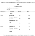
Фиг. 1 показывает данные проточного цитометрического анализа прикрепления ВАТ моноклональных антител (mAbs) к очищенным T-лимфоцитам человека. Прикрепление ВАТ антител оценивали с помощью второго антитела, несущего флуоресцентную метку, антитела анти-мыши- флуоресцинизоцианат (FITC)-баран. FIG. 1 shows flow cytometric analysis of the attachment of BAT monoclonal antibodies (mAbs) to purified human T-lymphocytes. The attachment of BAT antibodies was evaluated using a second antibody bearing a fluorescent label, anti-mouse fluorescein isocyanate (FITC) -ambran antibodies.
Фиг. 1a - вариант фона без ВАТ антитела. FIG. 1a is a background variant without a BAT antibody.
Фиг. 1b - антитело анти-CD3. FIG. 1b is an anti-CD3 antibody.
Фиг. 1c - ВАТ-1. FIG. 1c - BAT-1.
Фиг. 1d - BAT-5. FIG. 1d - BAT-5.
Фиг. 1e - ВАТ-2. FIG. 1e - BAT-2.
Фиг. 1f - ВАТ-4. FIG. 1f - BAT-4.
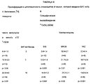
Фиг. 2 показывает данные проточного цитометрического анализа моноцитов периферической крови человека (РВМ) (левая часть) и T-клеток Jurkat (правая часть) дважды меченных первоначально антителом, которое является либо ВАТ mAb, либо анти-CD3 mAb и второй раз lgG антителом анти-мыши меченным FITC. FIG. 2 shows flow cytometric analysis of human peripheral blood monocytes (PBMs) (left side) and Jurkat T cells (right side) double-labeled initially with an antibody that is either BAT mAb or anti-CD3 mAb and a second time IgG anti-mouse antibody labeled FITC.
Фиг. 2A - неподвижные (ungated) клетки. FIG. 2A - motionless (ungated) cells.
Фиг. 2A(a) - РВМ клетки, которым было введено ВАТ mAb. FIG. 2A (a) —RVM cells to which BAT mAb was administered.
Фиг. 2A(b) - РВМ клетки, которым было введено анти-CD3 mAb. FIG. 2A (b) - PBM cells that were injected with anti-CD3 mAb.
Фиг. 2A(c) - Jurkat T-клетки, которым было введено ВАТ mAb. FIG. 2A (c) - Jurkat T cells to which BAT mAb was introduced.
Фиг. 2A(d) - Jurkat T-клетки, которым было введено анти-CD3. FIG. 2A (d) - Jurkat T cells that were injected with anti-CD3.
Фиг. 2B - подвижные клетки. FIG. 2B - motile cells.
Фиг. 2B(а) - РВМ клетки, продвинувшиеся к малым (R1) или большим клеткам (R2). FIG. 2B (a) - PBM cells advancing to small (R1) or large cells (R2).
Фиг. 2B(b) - (R1) клетки, которым было введено ВАТ mAb. FIG. 2B (b) - (R1) cells to which BAT mAb was administered.
Фиг. 2B(c) - (R1) клетки, которым было введено анти-CD3. FIG. 2B (c) - (R1) cells to which anti-CD3 has been introduced.
Фиг. 2B(d) - (R2) клетки, которым было введено ВАТ mAb. FIG. 2B (d) - (R2) cells to which BAT mAb was administered.
Фиг. 2B(e) - (R2) клетки, которым было введено анти-CD3. FIG. 2B (e) - (R2) cells to which anti-CD3 has been introduced.
Фиг. 3 показывает данные проточного цитометрического анализа поверхности экспрессии ВАТ связывающего протеина и CD3 на РВМ человека, дважды меченнные антителом ВАТ mAb, меченным флуоресцином (FITC), и антителом анти-CD3, меченным PE. FIG. 3 shows flow cytometric analysis of surface expression of BAT binding protein and CD3 on human PBM double-labeled with BAT mAb fluorescein-labeled (FITC) and anti-CD3 antibody labeled with PE.
Фиг. 3A - неподвижные клетки. FIG. 3A - motionless cells.
Фиг. 3A(a)-Becton-Dickinson simultest контроль, использованный в качестве отрицательного контроля. FIG. 3A (a) -Becton-Dickinson simultest control used as a negative control.
Фиг. 3A(b)-один РЕ-анти-CD3. FIG. 3A (b) -one PE-anti-CD3.
Фиг. 3A(c)-один FITC-BAT mAb. FIG. 3A (c) -one FITC-BAT mAb.
Фиг. 3F(d) - антитело, дважды меченное FITC-BAT mAb и РЕ-анти- CD3. FIG. 3F (d) is an antibody double-labeled with FITC-BAT mAb and PE-anti-CD3.
Фиг. 3B - подвижные клетки. FIG. 3B - motile cells.
Фиг. 3B(а) - клетки РВМ, продвинувшиеся к малым (R1) и большим (R2) клеткам. FIG. 3B (a) - PBM cells advancing to small (R1) and large (R2) cells.
Фиг. 3B(b) - (R1) клетки, дважды меченные FITC-BAT mAb и РЕ-анти-CD3. FIG. 3B (b) - (R1) cells double-labeled with FITC-BAT mAb and PE-anti-CD3.
Фиг. 3B(c) - (R2) клетки, дважды меченные FITC-BAT mAb и РЕ-анти-CD3. FIG. 3B (c) - (R2) cells double-labeled with FITC-BAT mAb and PE-anti-CD3.
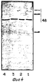
Фиг. 4 показывает данные анализа Western Blot прикрепления BAT-1 антител к различным лизатам клеток Daudi. FIG. 4 shows Western Blot assay data for the attachment of BAT-1 antibodies to various Daudi cell lysates.
Фиг. 4(1) - лизаты из клеток Daudi (которым ничего не было введено). FIG. 4 (1) - lysates from Daudi cells (to which nothing was administered).
Фиг. 4(2) - лизаты, в которые введена нейроминидаза (0.2 ед./мл). FIG. 4 (2) - lysates into which neurominidase is introduced (0.2 units / ml).
Фиг. 4(3) - лизаты, в которые введена нейроминидаза (0.4 ед./мл). FIG. 4 (3) - lysates into which neurominidase is introduced (0.4 units / ml).
Фиг. 4(4) - лизаты, в которые введен Endo Hf (100 ед./мкг). FIG. 4 (4) - lysates into which Endo Hf (100 units / μg) was introduced.
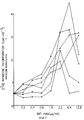
Фиг. 5 дает графическое представление результатов введения [3H]тимидина в клетки, культивированные в течение 6 дней в присутствии повышающихся концентраций набора ВАТ
Mabs: BAT-1(-![]()
![]()
Mabs: BAT-1 (- ![]()
![]()
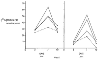
Фиг. 6 дает графическое представление экспериментов, в которых индукция цитотоксической активности была испытана в РВМ культивированных в течение различных интервалов времени 2.5 мкг/мл ВАТ Mabs. В качестве клеток-мишеней были использованы НТ- 29 (слева) клетки или RC-29 (справа) клетки. Отношение эффектора к мишени составляло 20:1 Контроль (-O-), BAT-1 (-•-), BAT-2 (-X-), BAT-3 (-![]()
![]()
Фиг. 7 показывает легкие, от C57BL мышей, инокулированных клетками миеломы В-16. Верхний ряд показывает легкие мышей, инокулированных только клетками, спустя 24 дня после инокуляции. Нижний ряд показывает легкие мышей инокулированных клетками В-16, как указано выше, с последующим введением через 14 суток внутривенно 10 мкг ВАТ-1 Mabs, спустя 24 часа после инокуляции. FIG. 7 shows lungs from C57BL mice inoculated with B-16 myeloma cells. The upper row shows the lungs of mice inoculated with cells only 24 days after inoculation. The bottom row shows the lungs of mice inoculated with B-16 cells as described above, followed by intravenous administration of 10 μg BAT-1 Mabs after 14 days, 24 hours after inoculation.
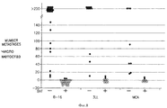
Фиг. 8 является графическим представлением суммарных экспериментальных данных, представленных на фиг. 7, показывающим количество метастаз в легких мышей, которые были инокулированы опухолевыми клетками, являющимися либо клетками миеломы В-16, либо клетками карциномы легкого 3LL Lewis или клетками фибросаркомы МСА 105, спустя месяц после инокуляции. Результаты являются суммой 3-4 экспериментов, проведенных с каждым типом опухоли. Мыши, которым не вводили (-) антитело, или мыши, которым вводили (+) ВАТ-1 (10 мкг/мышь) через 2 недели после введения опухолевых клеток. Метастазы (•): метастазы отсутствуют (о). FIG. 8 is a graphical representation of the total experimental data shown in FIG. 7, showing the number of metastases in the lungs of mice that were inoculated with tumor cells, either B-16 myeloma cells, or 3LL Lewis lung carcinoma cells or
Фиг. 9 показывает легкие, полученные от мышей, инокулированных клетками карциномы легкого 3LL Lewis. Аналогично фиг. 7 верхний ряд показывает легкое, инокулированное только клетками опухоли, и нижний ряд показывает легкие мышей, инокулированных внутривенно 14 дней спустя 10 мкг BAT-1 mAbs. FIG. 9 shows lungs obtained from mice inoculated with Lewis 3LL lung carcinoma cells. Similarly to FIG. 7, the upper row shows a lung inoculated only with tumor cells, and the lower row shows the lungs of mice inoculated intravenously 14 days later with 10 μg BAT-1 mAbs.
Фиг. 10 показывает эксперимент, аналогичный тому, что показан на фиг. 9, где клетки опухоли представляют клетки фибробластомы МСА 105. FIG. 10 shows an experiment similar to that shown in FIG. 9, where the tumor cells are
Продуцирование моноклональных антител. The production of monoclonal antibodies.
BALB/c мыши были иммунизованы мембранным препаратом клеток Daudi. Мембраны из клеток Daudi были приготовлены с помощью метода глицериновая нагрузка-гипотонический шок (Jett, M. , Seed,T.M., and Jamieson. G.A., J. Biol. Chem. 252: 2134 (1977). 50-80 • 106 клеток, суспендированных в PBS и инкубированных при 37oC постепенно нагружали 30% глицерина. Через 5 минут инкубации на льду их центрифугировали и ресуспендировали в холодный Трис лизатный буфер (содержащий 10 ммоль/л Трис-HCl, 1 ммоль/л MgCl2, 1 ммоль/л CaCl2, pH 7.4), перемешивали в течение 5 минут при 4oC и центрифугировали при 700 g. Супернатант удаляли и центрифугировали при 3300 g (в течение 10 минут при 4oC). Дебрис промывали вновь и два супернатанта, содержащих мембранную фракцию, собирали. 260 мкл мембранного препарата (3 мг/мл) эмульгировали 260 мкл адьюванта compele Freund и проводили интраперитонеальную инъекцию мышам BALB/c. Спустя три недели удаляли селезенки мышей. Спленоциты подвергали слиянию с линией клеток миеломы NS-0 при отношении 10:1. Слияние проводили, используя полиэтиленгликоль, и гибридомы выращивали в селективной среде согласно Kohler and Milstein (Kohler.G. and Milstein.C., Nature (London) 256: 495 (1975)).BALB / c mice were immunized with a Daudi cell membrane preparation. Membranes from Daudi cells were prepared using the glycerol loading-hypotonic shock method (Jett, M., Seed, TM, and Jamieson. GA, J. Biol. Chem. 252: 2134 (1977). 50-80 • 10 6 cells, 30% glycerol was suspended in PBS and incubated at 37 ° C. After 5 minutes of incubation on ice, they were centrifuged and resuspended in cold Tris lysate buffer (containing 10 mmol / L Tris-HCl, 1 mmol / L MgCl 2 , 1 mmol / l of CaCl 2 , pH 7.4), was stirred for 5 minutes at 4 o C and centrifuged at 700 g, the Supernatant was removed and centrifuged at 3300 g (for 10 minutes at 4 o C). c was washed again and two supernatants containing the membrane fraction were collected 260 μl of the membrane preparation (3 mg / ml) was emulsified with 260 μl of compele Freund adjuvant and intraperitoneally injected to BALB / c mice. NS-0 myeloma cells at a ratio of 10: 1. Fusion was performed using polyethylene glycol, and hybridomas were grown in selective medium according to Kohler and Milstein (Kohler.G. and Milstein, C., Nature (London) 256: 495 (1975)).
Твердофазный иммуноферментный анализ (ELISA) использовали для определения супернатантов из растущих гибридом (Glassy, M.C. and Surh, C.D., J. lmmunol. Method, 81:115 (1985)), которые прикреплены к клеткам Daudi. Положительные супернатанты гибридом выбирали по их способности индуцировать пролиферацию РВМ человека, используя анализ поглощения [3H]тимидина. Положительные клоны субклонировали с помощью ограниченного разбавления, повторно испытывали и подвергали распластыванию и росту в культуре.An enzyme-linked immunosorbent assay (ELISA) was used to determine supernatants from growing hybridomas (Glassy, MC and Surh, CD, J. lmmunol. Method, 81: 115 (1985)), which are attached to Daudi cells. Positive hybridoma supernatants were selected for their ability to induce human PBM proliferation using [ 3 H] thymidine uptake assay. Positive clones were subcloned using limited dilution, re-tested and subjected to spreading and growth in culture.
mAbs очищали от среды для культивирования осаждением 50% раствором сульфата аммония с последующим интенсивным диализом по отношению к PBS. Дальнейшую очистку проводили с помощью аффинной хроматографии на колонках с Сефарозой (торговая марка Pharmacia, Sweden) с прикрепленным антителом анти-мыши. mAbs were purified from the culture medium by precipitation with a 50% solution of ammonium sulfate, followed by intensive dialysis against PBS. Further purification was performed using affinity chromatography on Sepharose columns (trademark Pharmacia, Sweden) with an anti-mouse antibody attached.
Среда для культивирования. Cultivation medium.
Все клетки суспендировали в RPMI 164 среде, дополненной 10% фетальной телячьей сывороткой (FCS), Na-пируватом (1.1 мг/мл), L-глутаматом (0.3 мг/мл) и антибиотиками (пеницилином 200 мг/мл и стрептомицином 10 мкг/мл), и инкубировали во влажном инкубаторе с 5% CO2.All cells were suspended in RPMI 164 medium supplemented with 10% fetal calf serum (FCS), Na-pyruvate (1.1 mg / ml), L-glutamate (0.3 mg / ml) and antibiotics (
IL-2 использованные единицы составляли единицы Cetus (1 единица Cetus равна 3 Международным единицам). The IL-2 units used were Cetus units (1 Cetus unit equals 3 International units).
Клеточные препараты. Cellular preparations.
Моноядерные клетки периферической крови человека (РВМ) получали от взрослых здоровых доноров с помощью фиколл-гипакового центрифугирования (Гистопак, торговая марка Sigma, St. Louis, Missouri, USA). РВМ подвергали элиминации моноцитов с помощью колонок с Сефадексом (торговая марка Pharmacia, Sweden) G10. Т-клетки выделяли с помощью розеткообразного структурного метода SRBC. Элиминацию CD3 положительных клеток и Leu 19 положительных клеток проводили за счет иммуномагнитной техники. Культуры РВМ, инкубированные в течение 5-6 суток ВАТ mAbs, или контрольные культуры промывали три раза PBS. Антитела, неконъюгированные либо к CD3, либо к Leu19(CD56), добавляли к клеткам в полную RPMI среду и инкубировали в течение 1 часа при 4oC. Магнитный бисер, покрытый антителом антимыши, добавляли в течение 30 минут. Клетки, прикрепленные к бисеру, удаляли с помощью магнита, а неприкрепленные клетки окрашивали и анализировали на цитотоксическую активность с помощью проточного цитометрического анализа.Mononuclear cells of human peripheral blood (PBM) were obtained from adult healthy donors using ficoll-hypakic centrifugation (Histopack, trademark Sigma, St. Louis, Missouri, USA). PBM was subjected to the elimination of monocytes using columns with Sephadex (trademark Pharmacia, Sweden) G10. T cells were isolated using the rosette-like structural method of SRBC. The elimination of CD3 positive cells and
Реакция цитотоксичности. Cytotoxicity reaction.
Реакции цитотоксичности проводили следующим образом: 2-4 • 106 клеток мишеней смешивали с 200 мк Ci51Cr-хромата в течение 1 часа в среде, свободной от сыворотки. Клетки три раза промывали полной средой и наконец ресуспендировали в RPMI-10% FCS и высевали 104 клеток в лунку. Эффекторные клетки культивировали, лимфоциты, приготовленные из нормальной периферической крови, инкубировали в течение различных промежутков времени различными mAbs, изотопным контрольным lgG или IL-2. До анализа клетки три раза промывали средой RPMI, окрашивали для визуализации клеток, используя 1%-ный раствор трипанового голубого, смешивали с клетками мишенями при различных отношениях эффектор-мишень в микрометрических планшетах с круглым дном и инкубировали в течение 3 часов при 15-35oC в 5% CO2;. Супернатанты культур собирали и подвергали подсчету в β-сцинтиляционном счетчике. Максимальное высвобождение изотопа (MR) получают с помощью инкубирования клеток-мишеней с тритоном (торговая марка Sigma, St. Louis, Missouori, USA) x-100. Спонтанное высвобождение (SR) измеряют с помощью инкубации мишеней одной средой. Процентное содержание лизированных клеток рассчитывают с помощью выражения (ER-SR/MR-SR)•100, где ER представляет экспериментальное высвобождение эффектора.Cytotoxicity reactions were carried out as follows: 2-4 • 10 6 target cells were mixed with 200 μ Ci 51 Cr-chromate for 1 hour in serum-free medium. Cells were washed three times with complete medium and finally resuspended in RPMI-10% FCS and 10 4 cells were plated per well. Effector cells were cultured, lymphocytes prepared from normal peripheral blood were incubated for various periods of time with various mAbs, isotopic control IgG or IL-2. Prior to analysis, the cells were washed three times with RPMI medium, stained for cell imaging using a 1% trypan blue solution, mixed with target cells at different effector-target ratios in round bottom micrometer plates and incubated for 3 hours at 15-35 o C in 5% CO 2 ;. Culture supernatants were collected and counted in a β-scintillation counter. Maximum isotope release (MR) is obtained by incubating target cells with newt (trademark Sigma, St. Louis, Missouori, USA) x-100. Spontaneous release (SR) is measured by incubating targets with a single medium. The percentage of lysed cells is calculated using the expression (ER-SR / MR-SR) • 100, where ER represents the experimental release of the effector.
Индукция цитотоксичности в субпопуляциях лимфоцитов человека. Induction of cytotoxicity in subpopulations of human lymphocytes.
РВМ клетки (4•106/мл) культивировали в течение 6 дней в присутствии ВАТ mAbs. После этого клетки промывали три раза и клетки CD3 и Leu19 элиминировали с помощью магнитного бисера, покрытого фрагментом f(ab')2 анти-мыши и испытывали на цитотоксичность по отношению к клеткам К562 и клеткам Daudi.PBM cells (4 x 10 6 / ml) were cultured for 6 days in the presence of BAT mAbs. After that, the cells were washed three times and CD3 and Leu19 cells were eliminated using magnetic beads coated with anti-mouse f (ab ') 2 fragment and tested for cytotoxicity with respect to K562 cells and Daudi cells.
Проточная цитометрия. Flow cytometry.
Клеточную поверхность антигенов определяли с помощью проточной цитометрии, используя FACS 440 (Becton-Dickinson). Для каждого анализа использовали 106 клеток. Клетки окрашивали за счет последовательного инкубирования с оптимальной концентрацией мышиного mAb к CD3 человека, IL-2 рецептору или mAb ВАТ 1-9, которые продуцировали. Фрагмент F(ab')2 анти-мыши, меченный FITC барана, использовали в качестве второго антитела в этом непрямом процессе получения линии. Каждую инкубацию проводили в PDS pH 7.4, содержащем 1% BSA и 0.5% азида Na в течение 30 минут при 4oC и впоследствии трижды промывали тем же самым буфером. Анализировали 104 окрашенных клеток.Cell surface antigens were determined by flow cytometry using FACS 440 (Becton-Dickinson). For each analysis, 10 6 cells were used. Cells were stained by sequential incubation with the optimal concentration of mouse mAb to human CD3, IL-2 receptor or mAb BAT 1-9, which were produced. Fragment F (ab ') 2 anti-mouse labeled with FITC ram was used as the second antibody in this indirect line-making process. Each incubation was carried out in PDS pH 7.4 containing 1% BSA and 0.5% Na azide for 30 minutes at 4 ° C. and subsequently washed three times with the same buffer. 10 4 stained cells were analyzed.
Определение ВАТ mAb связывающей детерминанты (нант). Determination of BAT mAb binding determinants (nantes).
Определение ВАТ mAb связывающей детерминанты (нант) на лизатах B лимфобластоидных клеток Daudi проводили с использованием анализа Western Blot. Кратко, 50 • 106 клеток/мл суспендировали в PBS, постепенно нагруженный 30% глицерина, и мембраны отделяли за счет последующего центрифугирования.Determination of BAT mAb binding determinants (nantes) on Daudi B lymphoblastoid cell lysates was performed using Western Blot analysis. Briefly, 50 x 10 6 cells / ml were suspended in PBS, gradually loaded with 30% glycerol, and the membranes were separated by subsequent centrifugation.
Образцы мембранных препаратов отделяли с помощью SDS-PAGE (12%) (метода электрофореза в полиакриламидном геле с додецилсульфатом натрия) и переносили в нитроцеллюлозный планшет, который погружали в 1% раствор молока низкой жирности в PBS. Определение ВАТ mAb связывающего протеина в нитроцеллюлозных планшетах проводили за счет инкубирования планшета с ВАТ mAb в течение 2 часов при комнатной температуре, с последующим инкубированием в течение 30 минут с пероксидазой хрена конъюгированной фрагментом (Fab')2 антитела 70 IgG анти-мыши. Затем клетки промывали и окрашенные полосы определяли O-дианизидным субстратом.Membrane samples were separated using SDS-PAGE (12%) (polyacrylamide gel electrophoresis with sodium dodecyl sulfate) and transferred to a nitrocellulose plate, which was immersed in a 1% solution of low fat milk in PBS. The determination of BAT mAb binding protein in nitrocellulose tablets was performed by incubating the tablet with BAT mAb for 2 hours at room temperature, followed by incubation for 30 minutes with horseradish peroxidase conjugated by a fragment of (Fab ') 2
Модели мышиной опухоли. Mouse tumor models.
Были использованы три модели мышиной опухоли. B16 миелома, карцинома легкого Lewis (3LL) и фиброкарцинома, индуцированная метилхолантреном (МСА 105). 50-200 • 106 клеток впрыскивали внутривенно (i.v.) мышам C57BL (8-недельного возраста). Спустя две недели вводили ВАТ-1 (внутривенно), 1-10 мкмг/мышь и спустя 10 дней мышей умерщвляли и подсчитывали метастазы легкого.Three mouse tumor models were used. B16 myeloma, Lewis lung carcinoma (3LL) and methylcholanthrene-induced fibrocarcinoma (MCA 105). 50-200
Пример 1. Example 1
а. Характеристики связывания. a. Binding characteristics.
Девять моноклональных антител (mAbs), обозначенных ВАТ 1-9, полученных с помощью иммунизации B лимфобластоидных клеток, выбирали сначала для прикрепления к клеткам Daudi и затем для индуцирования пролиферации лимфоцитов периферической крови человека. Изотопы ВАТ mAbs определяли с помощью ELISA и анализа Ochterlony. Было найдено, что ВАТ 1, 2, 3, 6, 7 и 9 представляют класс IgG1, тогда как ВАТ 4 и 5 представляют класс IgM. ВАТ 8 был IgG2a. Nine monoclonal antibodies (mAbs), designated BAT 1-9, obtained by immunizing B lymphoblastoid cells, were selected first to attach to Daudi cells and then to induce proliferation of human peripheral blood lymphocytes. BAT mAbs isotopes were determined by ELISA and Ochterlony analysis.
Прикрепление вышеуказанных mAbs к очищенным периферическим T клеткам человека анализировали с помощью FACS, используя непрямое иммунофлуоресцентное окрашивание. Фиг. 1 демонстрирует данные анализа FACS такого эксперимента. Как можно видеть, ВАТ mAbs прикрепляется к CD3-T клеткам периферической крови. Количественное содержание этого прикрепления изменяется от ВАТ-2 44%, BAT-5 38%, BAT-1 32% до некоторого ослабления прикрепления у ВАТ-4 (13%). Очищенные В лимфоциты периферичекой крови от тех же самых доноров крови не прикрепляли эти mAbs (данные не показаны). The attachment of the aforementioned mAbs to purified human peripheral T cells was analyzed by FACS using indirect immunofluorescence staining. FIG. 1 shows FACS analysis data from such an experiment. As you can see, BAT mAbs are attached to peripheral blood CD3-T cells. The quantitative content of this attachment varies from BAT-2 44%, BAT-5 38%, BAT-1 32% to some weakening of attachment at BAT-4 (13%). Purified peripheral blood lymphocytes from the same blood donors did not attach these mAbs (data not shown).
b. Прикрепление ВАТ-1 mAb к субпопуляциям лимфоцитов человека. b. Attachment of BAT-1 mAb to subpopulations of human lymphocytes.
Как видно из фиг. 2, дальнейший анализ FACS показывает, что ВАТ-1 прикрепляет CD3+PBM человека так же, как и линию T-клеток Jurkat. Кроме того, было даже подтверждено, прикрепление ВАТ-1 к CD3+PBM клеткам, с использованием анализа FACS дважды меченных клеток (фиг. 3). As can be seen from FIG. 2, further FACS analysis shows that BAT-1 attaches human CD3 + PBM in the same way as the Jurkat T cell line. In addition, it was even confirmed that BAT-1 adhered to CD3 + PBM cells using FACS analysis of double-labeled cells (Fig. 3).
Как видно из таблицы 1, приведенной в конце описания, ВАТ-1 в дополнение к прикреплению его к CD3 несущим клеткам прикрепляется также к Leu 19/NK клеткам. As can be seen from table 1 at the end of the description, BAT-1 in addition to attaching it to CD3-bearing cells also attaches to
c. Прикрепление ВАТ mAbs к различным типам клеток. c. Attaching BAT mAbs to various types of cells.
Было определено прикрепление ВАТ mAbs к различным типам клеток. Как можно видеть из таблицы 2, приведенной в конце описания, ВАТ mAbs прикрепляется к К562, линии клеток эритролейкемии и MCF7, линии клеток карциномы молочной железы человека, в дополнение к их прикреплению к PBL, Daudi и линии Т клеток Jukart. Мера прикрепления изменяется среди ВАТ mAbs и испытанных типов клеток. mAbs 2-9 прикреплялись к клеткам почечной карциномы мыши (MR28) только с очень низкой эффективностью. Только ВАТ-8 прикреплялся к MEL клеткам (клеткам мышиной эритролейкемии), и также с очень низкой эффективностью. The attachment of BAT mAbs to various types of cells was determined. As can be seen from Table 2 at the end of the description, BAT mAbs are attached to K562, an erythroleukemia cell line and MCF7, a human breast carcinoma cell line, in addition to their attachment to PBL, Daudi, and Jukart T cell line. The attachment measure varies among BAT mAbs and tested cell types. mAbs 2-9 were attached to mouse renal carcinoma cells (MR28) only with very low efficiency. Only BAT-8 was attached to MEL cells (mouse erythroleukemia cells), and also with very low efficiency.
Пример 2
Анализ ВАТ mAb связывающего сайта и очистка ВАТ-1 связывающего протеина.Example 2
Analysis of BAT mAb binding site and purification of BAT-1 binding protein.
Для того, чтобы определить молекулярный вес мембранного протеина, который взаимодействует с ВАТ-1 mAbs, мембранный препарат клеток Daudi солюбилизировали и протеин отделяли с помощью SDS-PAGE. Перенесенные на нитроцеллюлозные планшеты инкубировали ВАТ-1 mAb и полосы регистрировали с помощью дальнейшей инкубирации антителом пероксидазы хрена, конъюгированным с (Fab')2 IgG анти-мыши, и регистрировали, используя O-дианизидиновый субстрат. Было найдено, что молекулярный размер ВАТ-1 связывающего протеина составляет 48-50 кДа (фиг.4).In order to determine the molecular weight of the membrane protein that interacts with BAT-1 mAbs, the Daudi cell membrane preparation was solubilized and the protein was separated using SDS-PAGE. Transferred onto nitrocellulose plates, BAT-1 mAb was incubated and the bands were recorded by further incubation with horseradish peroxidase antibody conjugated to (Fab ') 2 anti-mouse IgG and recorded using O-dianisidine substrate. It was found that the molecular size of BAT-1 binding protein is 48-50 kDa (figure 4).
ВАТ mAb связывающий протеин очищали, используя ВАТ mAb, конъюгированный к Сефарозе (торговая марка Pharmacia, Sweden). Этот связывающий протеин может быть также приготовлен за счет клонирования, используя приемы молекулярной биологии. Введение ВАТ-1 in vivo мышам приводило к индуцированию ВАТ антител. BAT mAb binding protein was purified using BAT mAb conjugated to Sepharose (trademark Pharmacia, Sweden). This binding protein can also be prepared by cloning using molecular biology techniques. In vivo administration of BAT-1 to mice led to the induction of BAT antibodies.
Пример 3. Example 3
Функциональные характеристики ВАТ mAbs. Functional characteristics of BAT mAbs.
а. ВАТ mAbs, индуцированный введением тимидина. a. BAT mAbs Induced by Thymidine Administration
Клетки периферической крови человека культивировали в течение 6 дней в присутствии увеличивающихся концентраций набора ВАТ mAbs и проводили импульсное мечение [3H]тимидином в течение 20 часов до сбора клеток.Human peripheral blood cells were cultured for 6 days in the presence of increasing concentrations of BAT mAbs and pulsed labeling with [ 3 H] thymidine for 20 hours before cell collection.
Как видно из фиг. 5, постепенное увеличение концентрации ВАТ mAbs приводит в результате к скромному, но значительному увеличению введения [3H] тимидина в РВМ клетки. Однако высокая доза антититела вызывает снижение поглощения клетками. В контрольных экспериментах антитела, меченные изотопом, не увеличивают [3H]тимидин в PBL клетках, указывая на то, что агонистическое влияние ВАТ mAbs зависит от его связывающих специфических свойств. Например, mAb изотопа IgG, который был выращен в нашей лаборатории, по отношению к овариановым клеткам карциномы не вызывал увеличения поглощения [3H]тимидина, в противоположность ВАТ 1, 2, 3, 6, 7 и 9 mAbs, которые также принадлежат к IgG1 классу.As can be seen from FIG. 5, a gradual increase in the concentration of BAT mAbs results in a modest but significant increase in the introduction of [ 3 H] thymidine into PBM cells. However, a high dose of antibodies causes a decrease in cell uptake. In control experiments, isotope-labeled antibodies do not increase [ 3 H] thymidine in PBL cells, indicating that the agonistic effect of BAT mAbs depends on its binding specific properties. For example, the IgG isotope mAb, which was grown in our laboratory, did not cause an increase in [ 3 H] thymidine uptake in relation to ovarian carcinoma cells, as opposed to
b. Индуцирование цитотоксичности ВАТ mAbs в РВМ человека. b. Induction of cytotoxicity of BAT mAbs in human PBM.
Культуры моноядерных клеток периферической крови человека, инкубированные ВАТ mAbs в течение различных промежутков времени, испытывали на их способность к лизированию линии опухолевых клеток. Human peripheral blood mononuclear cell cultures incubated with BAT mAbs for various time periods were tested for their ability to lyse tumor cell lines.
Как видно из таблицы 3, приведенной в конце описания, клетки РВМ человека, инкубированные в течение одной недели пробами ВАТ mAbs, обладали цитотоксичностью по отношению к К562, эритролейкемии человека (NK чувствительным) и RC-29, линии клеток почечной карциномы (NK резистентной). As can be seen from table 3 at the end of the description, human PBM cells incubated for one week with BAT mAbs were cytotoxic to K562, human erythroleukemia (NK sensitive) and RC-29, renal carcinoma cell line (NK resistant) .
Исследовали кинетику повышения цитотоксической активности РВМ человека, которая была стимулирована ВАТ mAbs. Как видно из фиг. 6 максимальную цитотоксичность по отношению к колонии клеток карциномы человека (НТ-29) и почечных клеток карциномы (RC-29) достигали после 7 суток инкубации РВМ человека ВАТ mAbs. The kinetics of increasing the cytotoxic activity of human PBM, which was stimulated by BAT mAbs, was investigated. As can be seen from FIG. 6, maximum cytotoxicity with respect to the colony of human carcinoma cells (NT-29) and renal carcinoma cells (RC-29) was achieved after 7 days of incubation with human PBM BAT mAbs.
с. Характеристики субпопуляции лимфоцита, включенного в ВАТ-индуцированную цитотоксичность. with. Characteristics of a subpopulation of a lymphocyte included in BAT-induced cytotoxicity.
Для того, чтобы оценить, увеличивается ли цитотоксическая активность РВМ человека, индуцированная с помощью ВАТ mAbs, благодаря активации NK клеток, T клеток или тех и других, NK и T клетки очищали и определяли их цитотоксичность, индуцированную ВАТ. Для очистки NK и T клеток Leu 19 и CD3 моноклональные антитела инкубировали клетками РВМ человека с последующей инкубацией магнитным бисером покрытым анти-мышь IgG. Это приводило к элиминации субпопуляций клеток, которые прикреплены к соответствующему антителу. Как видно из таблицы 4, приведенной в конце описания, количество литических единиц увеличивается и в случае элиминации культур клеток CD3 и в случае элиминации культур клеток Leu 19. В этих экспериментах были использованы ВАТ 6 и 8 и клетки-мишени были клетками эритролейкемии человека (К562) и лимфомы человека (Daudi). In order to evaluate whether the cytotoxic activity of human PBM induced by BAT mAbs increases due to the activation of NK cells, T cells, or both, NK and T cells were purified and their cytotoxicity induced by BAT was determined. To purify the NK and T cells of
Пример 4
Синергизм между BAT mAb и IL-2 в индуцировании цитотоксичности.Example 4
Synergy between BAT mAb and IL-2 in inducing cytotoxicity.
Индуцирование цитотоксичности в РВМ человека изучали при инкубации клеток РВМ комбинацией ВАТ mAb и IL-2. IL-2 при субоптимальных концентрациях (1 U/мл) добавляли вместе с повышающимися концентрациями ВАТ-2 mAb. Цитотоксичность определяли спустя одну неделю в культуре по отношению к линиям опухолевых клеток К562 и НТ29. Как показано в таблице 5, приведенной в конце описания, низкие концентрации ВАТ-2 действуют синергетически с IL-2 в индуцировании цитотоксичности клеток РВМ по отношению к обоим типам клеток мишеней. The induction of cytotoxicity in human PBM was studied by incubating PBM cells with a combination of BAT mAb and IL-2. IL-2 at suboptimal concentrations (1 U / ml) was added along with increasing concentrations of BAT-2 mAb. Cytotoxicity was determined after one week in culture with respect to tumor cell lines K562 and HT29. As shown in table 5 at the end of the description, low concentrations of BAT-2 act synergistically with IL-2 in inducing cytotoxicity of PBM cells with respect to both types of target cells.
Ранее было показано, что INF-α усиливает экспрессию антигенов класса МНС-1. Поэтому введение INF-α сопровождается, вероятно, потенциальным анти-опухолевым эффектом ВАТ, который требует вмешательства за счет цитотоксических клеток, направленных против различных опухолевых клеток (несущих антигены класса МНС). It was previously shown that INF-α enhances the expression of MHC-1 class antigens. Therefore, the introduction of INF-α is probably accompanied by a potential anti-tumor effect of BAT, which requires intervention due to cytotoxic cells directed against various tumor cells (bearing MHC class antigens).
Пример 5
Иммуностимуляторные эффекты BAT-1 мыши:
а. Исследования in vitro.Example 5
Immunostimulatory effects of mouse BAT-1:
a. In vitro studies.
mAb BAT-1 демонстрирует стимуляторные свойства у мышиных спленоцитов, подобные тем, какие наблюдают в PBL: человека. Они включают:
(i) Повышенную пролиферацию спленоцита in vitro, как она определена с помощью введения 3H тимидина (таблица 6);
(ii) Синергетический стимуляторный эффект за счет инкубации спленоцитов комбинацией ВАТ-1 и IL-2 (таблица 6);
(iii) Повышенную цитотоксичность в мышиной культуре спленоцитов в присутствии ВАТ-1 и дальнейшее повышение цитотоксичности при инкубации в присутствии IL-2 (таблица 7).BAT-1 mAb demonstrates stimulatory properties in murine splenocytes, similar to those observed in PBL: human. They include:
(i) Increased in vitro splenocyte proliferation as determined by the administration of 3 H thymidine (Table 6);
(ii) A synergistic stimulatory effect due to incubation of splenocytes by a combination of BAT-1 and IL-2 (table 6);
(iii) Increased cytotoxicity in a mouse culture of splenocytes in the presence of BAT-1 and a further increase in cytotoxicity upon incubation in the presence of IL-2 (Table 7).
C57BL мышиные спленоциты, инкубированные в течение 5 дней in vitro различными концентрациями BAT-1 и в комбинации с интерлейкином-2 (1u и 10 u на мл). C57BL mouse splenocytes incubated for 5 days in vitro with various concentrations of BAT-1 and in combination with interleukin-2 (1u and 10 u per ml).
Индуцирование цитотоксичности в C57BL культурах спленоцитов, инкубированных в течение 5 дней in vitro в присутствии различных концентраций ВАТ-1 и в комбинации с низкими дозами интерлейкина-2. Induction of cytotoxicity in C57BL splenocyte cultures incubated for 5 days in vitro in the presence of various concentrations of BAT-1 and in combination with low doses of interleukin-2.
Отношение эффектора к мишени равно 50:1
Мышиные опухолевые клетки-мишени, которые были восприимчивы к умерщвляющему эффекту ВАТ-1 активированными спленоцитами, включают: В16 миелому, карциному легкого Lewis (3LL), фибросаркому (MCA 105), почечную клеточную карциному (MR 28) и лимфому (YAC) (таблица 8).The ratio of effector to target is 50: 1
Mouse tumor target cells that were susceptible to the killing effect of BAT-1 activated splenocytes include: B16 myeloma, Lewis lung carcinoma (3LL), fibrosarcoma (MCA 105), renal cell carcinoma (MR 28) and lymphoma (YAC) (table eight).
Цитотоксичность определяли при отношении эффектор/мишень 60:1. Cytotoxicity was determined at an effector / target ratio of 60: 1.
b. Исследования in vitro. b. In vitro studies.
Как показано в таблице 9, приведенной в конце описания , ВАТ-1 проявляет иммуностимуляторные эффекты при введении in vitro. Они включают:
(i) Стимулирование включения [3H]тимидина в спленоциты от мыши, которой вводили ВАТ-1 за 10 дней до этого (таблица 9A). Максимальное стимулирование (10-кратное) достигали при введении ВАТ с дозами 10 мкг/мышь.As shown in table 9 at the end of the description, BAT-1 exhibits immunostimulatory effects when administered in vitro. They include:
(i) Stimulating the incorporation of [ 3 H] thymidine into splenocytes from a mouse injected with BAT-1 10 days earlier (Table 9A). The maximum stimulation (10-fold) was achieved with the introduction of BAT with doses of 10 μg / mouse.
(ii) Индуцирование цитотоксичности в спленоцитах от мыши, которой вводили ВАТ-1 (таблица 9B). ВАТ-1 введенный в различных дозах за 10 дней до анализа на цитотоксичность, индуцировал цитотоксичность по отношению к клеткам миеломы мыши (B16-F10), ренальным клеткам карциномы (bR-28) и клеткам лимфомы (YAC). Максимальный эффект достигали при введении ВАТ в дозе 10 мкг/мышь. (ii) Induction of cytotoxicity in splenocytes from a mouse injected with BAT-1 (Table 9B). BAT-1 administered at
Мышам C57BL и BALB/c была введена инъекция внутривенно ВАТ mAb в различных концентрациях. 10 дней спустя определяли цитотоксичность и включение [3H]тимидина в выделенных спленоцитах. Спленоциты от мыши C57BL были испытаны на B16 клетки-мишени миеломы и от мыши BALB/c были испытаны на MR-28 и YAC клетках (отношение эффектор: мишень 50:1). Каждая группа содержала от 6 до 16 мышей в 3-4 раздельных экспериментах. Цитотоксичность и включение [3H] тимидина для каждой мыши были определены три раза и значения расчитаных результатов выражали как значение ± стандартная ошибка n мышей, даны p-величины, представляющие разницу между контролем и данными для животных, которым вводили ВАТ.C57BL and BALB / c mice were given intravenous injection of BAT mAb at various concentrations. 10 days later, cytotoxicity and incorporation of [ 3 H] thymidine in isolated splenocytes was determined. Splenocytes from a C57BL mouse were tested on B16 myeloma target cells and from BALB / c mouse were tested on MR-28 and YAC cells (effector: target ratio 50: 1). Each group contained from 6 to 16 mice in 3-4 separate experiments. The cytotoxicity and incorporation of [ 3 H] thymidine for each mouse were determined three times and the calculated results were expressed as the value ± standard error of n mice, p-values representing the difference between the control and the data for animals that were administered BAT were given.
Пример 6
Иммунотерапевтический эффект ВАТ-1 по отношению к опухолям мыши.Example 6
Immunotherapeutic effect of BAT-1 against mouse tumors.
Как видно из таблицы 10, введение ВАТ-1 мышам, инокулированным меланомой, на 14 сутки после инокуляции опухолевых клеток снижает количество метастаз легкого в легких у мышей, у которых присутствовали опухоли В16, 3LL и МСА. As can be seen from table 10, the introduction of BAT-1 to mice inoculated with melanoma, on the 14th day after the inoculation of tumor cells, reduces the number of lung metastases in the mice in which B16, 3LL and MCA tumors were present.
a1. Устранение с помощью ВАТ-1 метастаз легкого у мышей с B16 инокулированной миеломой, используя установленную модель метастаз легкого. a1. Elimination using BAT-1 lung metastases in mice with B16 inoculated myeloma using an established model of lung metastases.
C57BL мышам была проведена инъекция (внутривенная) 50 •103 клеток миеломы B16 (таблица 10). Как видно на фиг. 7, верхний ряд, на 24 день после инъекции развивается большое число метастаз в легких (практически достигается слияние).C57BL mice were injected (intravenously) with 50 • 10 3 B16 myeloma cells (Table 10). As seen in FIG. 7, upper row, a large number of lung metastases develop on
В противоположность этому, как видно на фиг. 7, нижний ряд, легкое мыши, которой была проведена инъекция ВАТ-1 (10 мкг/мышь) через две недели после инокуляции миеломой B16, практически свободно от метастаз. In contrast, as seen in FIG. 7, the lower row, the lung of a mouse injected with BAT-1 (10 μg / mouse) two weeks after inoculation with B16 myeloma, is almost free of metastases.
Фиг. 8 суммирует результаты шести раздельных экспериментов, проведенных в подобных условиях, как указано выше. FIG. 8 summarizes the results of six separate experiments conducted under similar conditions, as described above.
a2. Противоопухолевый эффект ВАТ-1 mAb. инъекцию которого проводили в различное время, относительно инокуляции B16 опухоли. a2. Antitumor effect of BAT-1 mAb. the injection of which was carried out at different times, relative to the inoculation of the B16 tumor.
Как видно из таблицы 11, мыши, которым были проведены инъекции клеток миеломы и затем через 10-14 дней спустя был введен ВАТ-1 mAb, были свободны от метастаз и имели нормальные веса легких. Заметное снижение количества метастаз в легких мышей, хотя и не полное, было замечено через 5 дней после инокуляции и только через 19 дней после введения опухоли. Инъекция ВАТ-1 в тот же день, что и инокуляции опухолевых клеток не оказывала терапевтического эффекта. As can be seen from table 11, the mice that were injected with myeloma cells and then BAT-1 mAb were administered 10-14 days later were free of metastases and had normal lung weights. A noticeable decrease in the number of metastases in the lungs of mice, although not complete, was seen 5 days after inoculation and only 19 days after the introduction of the tumor. Injection of BAT-1 on the same day as the inoculation of tumor cells did not have a therapeutic effect.
б. Устранение метастаз легкого с помощью ВАТ у мышей Lewis, которым инокулирована карцинома легкого (3LL), используя установленную модель метастаз легкого. b. Elimination of lung metastases using BAT in Lewis mice inoculated with lung carcinoma (3LL) using an established model of lung metastases.
Экспериментальные условия были аналогичны условиям, описанным для миеломы B16 (смотри выше), за исключением того, что проводили инъекцию 2 • 105 клеток 3LL. Фиг. 9, верхний ряд, показывает легкие от мышей, инокулированных 3LL с многочисленными метастазами. Фиг. 9, нижний ряд, показывает легкие от мышей, которые были инокулированы клетками опухоли с последующим введением через 14 дней ВАТ-1, которые как видно почти свободны от метастаз.The experimental conditions were similar to those described for B16 myeloma (see above), except that 2 x 10 5 3LL cells were injected. FIG. 9, the upper row, shows lungs from mice inoculated with 3LL with numerous metastases. FIG. 9, the bottom row, shows lungs from mice that were inoculated with tumor cells, followed by BAT-1 administration after 14 days, which are apparently nearly free of metastases.
с. Устранение метастаз легкого у мышей, которым инокулирована МСА фибросаркома (МСА 105), испольуя установленную модель метастаз легкого. with. Elimination of lung metastases in mice inoculated with MCA fibrosarcoma (MCA 105) using an established model of lung metastases.
Экспериментальные условия были аналогичны условиям, описанным для модели карциномы легкого 3LL Lewis. Фиг. 10, верхний ряд, показывает легкие от мышей, инокулированных МСА 105 с многочисленными метастазами. Фиг. 10, нижний ряд, показывает легкие от мышей, которые были инокулированы клетками опухоли с последующим введением ВАТ-1, которые, как видно, почти свободны от метастаз. The experimental conditions were similar to those described for the 3LL Lewis lung carcinoma model. FIG. 10, the upper row, shows lungs from mice inoculated with
Пример 7. Example 7
Лечение с помощью ВАТ-1 мышей, у которых присутствуют опухоли В16 и 3LL. BAT-1 treatment in mice with B16 and 3LL tumors.
Мыши, инокулированные клетками миеломы В 16 или клетками 3LL, как описано выше, погибали в течение 25-35 дней после инокуляции опухоли. В противоположность этому, как видно из фиг. 11, все мыши, которые были подвергнуты инъекции ВАТ-1 (10 мкг/мышь) через 14 дней после инокуляции опухоли, жили свыше 100 дней. Большинство животных, за которыми следили вплоть до 5 месяцев, не обнаруживали симптомов болезни и при патологических исследованиях не обнаруживалось метастаз. Mice inoculated with B16 myeloma cells or 3LL cells, as described above, died within 25-35 days after tumor inoculation. In contrast, as can be seen from FIG. 11, all mice that were injected with BAT-1 (10 μg / mouse) 14 days after tumor inoculation lived over 100 days. Most of the animals that were followed up to 5 months did not show symptoms of the disease and no pathological studies showed metastasis.
Пример 8
Заимствованный перенос спленоцитов от мышей, которым вводили ВАТ-1 mAb.Example 8
Borrowed transfer of splenocytes from mice injected with BAT-1 mAb.
Спленоциты от мышей, которым вводили один BAT-1 mAb, или сначала проводили инъекции клеток миеломы В16 и затем вводили ВАТ- 1 mAb, переносили мышам-реципиентам. Мыши-реципиенты были инокулированы либо клетками миеломы В16, либо опухолевыми клетками 3LL. Как видно из таблицы 12, приведенной в конце описания, заимсвованный перенос спленоцитов от мыши, которой вводили ВАТ-1 mAb, индуцировал регрессию опухоли у мышей, которые имели опухоль, получая перенесенные спленоциты. Наиболее эффективного лечения достигали, когда 103 спленоцитов мышей, которые одновременно были инокулированы и клетками миеломы В16 и через 14 дней, инъекцией ВАТ-1 были заимствованно перенесены мышам-реципиентам несущим опухоль. Как видно из таблицы 12, в этом случае, имело место полное отсутствие опухолевых клеток миеломы у реципиентов, несущих опухоль В16, и заметная регрессия опухоли у реципиентов, несущих опухоль 3LL, как представлено количеством метастаз и весом легкого.Splenocytes from mice injected with a single BAT-1 mAb or injected with B16 myeloma cells first and then injected with BAT-1 mAb were transferred to recipient mice. Recipient mice were inoculated with either B16 myeloma cells or 3LL tumor cells. As can be seen from table 12 at the end of the description, the borrowed splenocyte transfer from a mouse injected with BAT-1 mAb induced tumor regression in mice that had a tumor, receiving transferred splenocytes. The most effective treatment was achieved when 10 3 splenocytes of mice that were simultaneously inoculated with B16 myeloma cells and after 14 days, were injected with BAT-1 and transferred to recipient mice bearing tumors. As can be seen from table 12, in this case, there was a complete absence of myeloma tumor cells in recipients carrying a B16 tumor, and a noticeable regression of the tumor in recipients carrying a 3LL tumor, as represented by the number of metastases and lung weight.
Эти результаты показывают, что одни спленоциты от мышей, инокулированных B16 и введением ВАТ-1, обладают противоопухолевым эффектом по отношению к обоим типам опухолей и B16, и 3LL. Поэтому вероятно, что ВАТ-1 усиливает неспецифические клеточные эффекторные механизмы. Кроме того, присутствие опухолей приводит к потенциальному усилению ВАТ-1 индуцированной генерации таких эффекторных клеток. These results show that some splenocytes from mice inoculated with B16 and the administration of BAT-1 have an antitumor effect with respect to both types of tumors and B16 and 3LL. Therefore, it is likely that BAT-1 enhances nonspecific cellular effector mechanisms. In addition, the presence of tumors leads to a potential increase in BAT-1 induced generation of such effector cells.
Claims (4)
Applications Claiming Priority (2)
| Application Number | Priority Date | Filing Date | Title |
|---|---|---|---|
| IL10850194A IL108501A (en) | 1994-01-31 | 1994-01-31 | Antibodies and pharmaceutical compositions containing them |
| IL108,501 | 1994-01-31 |
Publications (2)
| Publication Number | Publication Date |
|---|---|
| RU96116984A RU96116984A (en) | 1999-05-10 |
| RU2155190C2 true RU2155190C2 (en) | 2000-08-27 |
Family
ID=11065761
Family Applications (1)
| Application Number | Title | Priority Date | Filing Date |
|---|---|---|---|
| RU96116984/13A RU2155190C2 (en) | 1994-01-31 | 1995-01-30 | Immunostimulatory monoclonal antibodies |
Country Status (20)
| Country | Link |
|---|---|
| US (1) | US5897862A (en) |
| EP (1) | EP0742795B1 (en) |
| JP (1) | JP3478398B2 (en) |
| CN (1) | CN1052733C (en) |
| AT (1) | ATE171461T1 (en) |
| AU (1) | AU693526B2 (en) |
| CA (1) | CA2182289C (en) |
| DE (1) | DE69504955T2 (en) |
| DK (1) | DK0742795T3 (en) |
| EE (1) | EE03285B1 (en) |
| ES (1) | ES2124533T3 (en) |
| FI (1) | FI963004A (en) |
| IL (1) | IL108501A (en) |
| LT (1) | LT4225B (en) |
| LV (1) | LV11624B (en) |
| MD (1) | MD1374G2 (en) |
| MX (1) | MX9603080A (en) |
| NO (1) | NO317020B1 (en) |
| RU (1) | RU2155190C2 (en) |
| WO (1) | WO1995020605A1 (en) |
Families Citing this family (30)
| Publication number | Priority date | Publication date | Assignee | Title |
|---|---|---|---|---|
| IL129299A0 (en) * | 1999-03-31 | 2000-02-17 | Mor Research Applic Ltd | Monoclonal antibodies antigens and diagnosis of malignant diseases |
| IL145926A0 (en) | 2001-10-15 | 2002-07-25 | Mor Research Applic Ltd | Peptide epitopes of mimotopes useful in immunomodulation |
| IL149820A0 (en) * | 2002-05-23 | 2002-11-10 | Curetech Ltd | Humanized immunomodulatory monoclonal antibodies for the treatment of neoplastic disease or immunodeficiency |
| EP2206517B1 (en) | 2002-07-03 | 2023-08-02 | Ono Pharmaceutical Co., Ltd. | Immunopotentiating compositions comprising anti-PD-L1 antibodies |
| DE10311248A1 (en) * | 2003-03-14 | 2004-09-30 | Müller-Hermelink, Hans Konrad, Prof. Dr. | Human monoclonal antibody |
| US20090123413A1 (en) * | 2004-08-23 | 2009-05-14 | Britta Hardy | Use of bat monoclonal antibody for immunotherapy |
| EP3530736A3 (en) | 2005-05-09 | 2019-11-06 | ONO Pharmaceutical Co., Ltd. | Human monoclonal antibodies to programmed death 1 (pd-1) and methods for treating cancer using anti-pd-1 antibodies alone or in combination with other immunotherapeutics |
| WO2007005874A2 (en) | 2005-07-01 | 2007-01-11 | Medarex, Inc. | Human monoclonal antibodies to programmed death ligand 1 (pd-l1) |
| ES2616355T3 (en) | 2007-06-18 | 2017-06-12 | Merck Sharp & Dohme B.V. | Antibodies for the human programmed death receptor PD-1 |
| RU2531758C2 (en) | 2008-02-11 | 2014-10-27 | Куретек Лтд. | Monoclonal antibodies for tumour treatment |
| MD3972C2 (en) * | 2009-03-11 | 2010-07-31 | Ион МЕРЕУЦЭ | Remedy and method for treating precancerous conditions |
| KR20140041598A (en) | 2011-07-24 | 2014-04-04 | 큐어 테크 리미티드 | Variants of humanized immunomodulatory monoclonal antibodies |
| US10570204B2 (en) | 2013-09-26 | 2020-02-25 | The Medical College Of Wisconsin, Inc. | Methods for treating hematologic cancers |
| US9580504B1 (en) | 2013-11-07 | 2017-02-28 | Curetech Ltd. | Pidilizumab monoclonal antibody therapy following stem cell transplantation |
| JOP20200094A1 (en) | 2014-01-24 | 2017-06-16 | Dana Farber Cancer Inst Inc | Antibody molecules to pd-1 and uses thereof |
| JOP20200096A1 (en) | 2014-01-31 | 2017-06-16 | Children’S Medical Center Corp | Antibody molecules to tim-3 and uses thereof |
| TWI693232B (en) | 2014-06-26 | 2020-05-11 | 美商宏觀基因股份有限公司 | Covalently bonded diabodies having immunoreactivity with pd-1 and lag-3, and methods of use thereof |
| US11344620B2 (en) | 2014-09-13 | 2022-05-31 | Novartis Ag | Combination therapies |
| TWI773646B (en) | 2015-06-08 | 2022-08-11 | 美商宏觀基因股份有限公司 | Lag-3-binding molecules and methods of use thereof |
| TWI833183B (en) | 2015-07-30 | 2024-02-21 | 美商宏觀基因股份有限公司 | Pd-1-binding molecules and methods of use thereof |
| US10323091B2 (en) | 2015-09-01 | 2019-06-18 | Agenus Inc. | Anti-PD-1 antibodies and methods of use thereof |
| BR112018006237A2 (en) | 2015-09-29 | 2018-10-09 | Celgene Corp | pd-1 binding proteins and methods of using them |
| BR112018008867A8 (en) | 2015-11-03 | 2019-02-26 | Janssen Biotech Inc | antibodies that specifically bind to pd-1 and their uses |
| EP3389714A4 (en) | 2015-12-14 | 2019-11-13 | MacroGenics, Inc. | Bispecific molecules having immunoreactivity with pd-1 and ctla-4, and methods of use thereof |
| IL263834B2 (en) | 2016-06-20 | 2024-01-01 | Kymab Ltd | Anti-pd-l1 antibodies |
| US10766958B2 (en) | 2016-09-19 | 2020-09-08 | Celgene Corporation | Methods of treating vitiligo using PD-1 binding antibodies |
| SG11201901950TA (en) | 2016-09-19 | 2019-04-29 | Celgene Corp | Methods of treating immune disorders using pd-1 binding proteins |
| KR102603681B1 (en) | 2016-12-07 | 2023-11-17 | 아게누스 인코포레이티드 | Antibodies and methods of using them |
| GB202107994D0 (en) | 2021-06-04 | 2021-07-21 | Kymab Ltd | Treatment of cancer |
| WO2024160721A1 (en) | 2023-01-30 | 2024-08-08 | Kymab Limited | Antibodies |
Family Cites Families (1)
| Publication number | Priority date | Publication date | Assignee | Title |
|---|---|---|---|---|
| US5182368A (en) * | 1986-06-13 | 1993-01-26 | Ledbetter Jeffrey A | Ligands and methods for augmenting B-cell proliferation |
-
1994
- 1994-01-31 IL IL10850194A patent/IL108501A/en not_active IP Right Cessation
-
1995
- 1995-01-30 DK DK95910891T patent/DK0742795T3/en active
- 1995-01-30 CN CN95192406A patent/CN1052733C/en not_active Expired - Lifetime
- 1995-01-30 RU RU96116984/13A patent/RU2155190C2/en active
- 1995-01-30 US US08/380,857 patent/US5897862A/en not_active Expired - Lifetime
- 1995-01-30 AU AU18686/95A patent/AU693526B2/en not_active Expired
- 1995-01-30 WO PCT/US1995/001137 patent/WO1995020605A1/en not_active Application Discontinuation
- 1995-01-30 DE DE69504955T patent/DE69504955T2/en not_active Expired - Lifetime
- 1995-01-30 EP EP95910891A patent/EP0742795B1/en not_active Expired - Lifetime
- 1995-01-30 MD MD96-0296A patent/MD1374G2/en unknown
- 1995-01-30 EE EE9600107A patent/EE03285B1/en unknown
- 1995-01-30 JP JP52021095A patent/JP3478398B2/en not_active Expired - Lifetime
- 1995-01-30 CA CA002182289A patent/CA2182289C/en not_active Expired - Lifetime
- 1995-01-30 ES ES95910891T patent/ES2124533T3/en not_active Expired - Lifetime
- 1995-01-30 AT AT95910891T patent/ATE171461T1/en active
-
1996
- 1996-07-29 MX MX9603080A patent/MX9603080A/en not_active IP Right Cessation
- 1996-07-29 NO NO19963168A patent/NO317020B1/en not_active IP Right Cessation
- 1996-07-29 FI FI963004A patent/FI963004A/en unknown
- 1996-07-30 LT LT96-115A patent/LT4225B/en not_active IP Right Cessation
- 1996-09-06 LV LVP-96-317A patent/LV11624B/en unknown
Non-Patent Citations (1)
| Title |
|---|
| B.Hardy et al., Cellular Immunology, 1989, v.118, nol, p.22-29. B.Hardy et al., Cancer Research, 1994, v.54, p.5793-5796. * |
Also Published As
| Publication number | Publication date |
|---|---|
| CN1052733C (en) | 2000-05-24 |
| CA2182289A1 (en) | 1995-08-03 |
| FI963004A (en) | 1996-09-27 |
| MD1374F2 (en) | 1999-12-31 |
| NO963168L (en) | 1996-09-27 |
| LT96115A (en) | 1997-05-26 |
| AU693526B2 (en) | 1998-07-02 |
| LV11624A (en) | 1996-12-20 |
| US5897862A (en) | 1999-04-27 |
| DK0742795T3 (en) | 1999-06-14 |
| ATE171461T1 (en) | 1998-10-15 |
| EP0742795A1 (en) | 1996-11-20 |
| MD1374G2 (en) | 2000-09-30 |
| ES2124533T3 (en) | 1999-02-01 |
| DE69504955T2 (en) | 1999-05-27 |
| IL108501A (en) | 1998-10-30 |
| NO963168D0 (en) | 1996-07-29 |
| IL108501A0 (en) | 1994-05-30 |
| LT4225B (en) | 1997-10-27 |
| EP0742795B1 (en) | 1998-09-23 |
| WO1995020605A1 (en) | 1995-08-03 |
| CN1145077A (en) | 1997-03-12 |
| MX9603080A (en) | 1998-01-31 |
| DE69504955D1 (en) | 1998-10-29 |
| NO317020B1 (en) | 2004-07-26 |
| AU1868695A (en) | 1995-08-15 |
| LV11624B (en) | 1997-06-20 |
| FI963004A0 (en) | 1996-07-29 |
| JP3478398B2 (en) | 2003-12-15 |
| CA2182289C (en) | 2003-10-21 |
| EE03285B1 (en) | 2000-08-15 |
| JPH09508390A (en) | 1997-08-26 |
Similar Documents
| Publication | Publication Date | Title |
|---|---|---|
| RU2155190C2 (en) | Immunostimulatory monoclonal antibodies | |
| WO1995020605A9 (en) | Immuno-stimulatory monoclonal antibodies | |
| MXPA96003080A (en) | Monoclonal antibodies immuno-estimulan | |
| Lundqvist et al. | Intraepithelial lymphocytes in human gut have lytic potential and a cytokine profile that suggest T helper 1 and cytotoxic functions. | |
| US7794710B2 (en) | Methods of enhancing T cell responsiveness | |
| CN109069539A (en) | Combined immunization therapy use for cancer treatment and cell factor control therapy | |
| Iwanuma et al. | Antitumor immune response of human peripheral blood lymphocytes coengrafted with tumor into severe combined immunodeficient mice | |
| JP6682438B2 (en) | Improved cell compositions and methods for cancer treatment | |
| JPH0198477A (en) | Igg monoclonal antibody-productive hybridmer | |
| WO2020055040A1 (en) | Pharmaceutical combinations for treating tumor comprising anti-cd19 antibody and natural killer cell | |
| CN109195988A (en) | For enhancing the method and composition of the immunotherapy for cancer effect of super antigen mediation | |
| Hardy et al. | A monoclonal antibody against a human B lymphoblastoid cell line induces tumor regression in mice | |
| Cross et al. | Administration of a prostaglandin synthetase inhibitor associated with an increased immune cell infiltrate in squamous cell carcinoma of the head and neck | |
| Ishii et al. | A combination of check-point blockade and α-galactosylceramide elicits long-lasting suppressive effects on murine hepatoma cell growth in vivo | |
| Zalman et al. | The cytolytic protein of human lymphocytes related to the ninth component (C9) of human complement: isolation from anti-CD3-activated peripheral blood mononuclear cells. | |
| JP2005124568A (en) | Method for activating cell, method for producing the cell using the same, and pharmaceutical composition | |
| JPH01126558A (en) | Measurement of antibody dependant cellar cell poison | |
| Yoshino et al. | Cytolytic potential of peripheral blood T-lymphocytes following adoptive immunotherapy with lymphokine-activated killer cells and low-dose interleukin 2 | |
| KR20230085116A (en) | Pharmaceutical combinations for treating tumor comprising anti-cd19 antibody and natural killer cell | |
| Pfister et al. | The in vitro anti-inflammatory effects of recombinant anti-CD25 immunotoxin on lamina propria T cells of patients with inflammatory bowel disease are not sufficient to cure experimental colitis in mice | |
| Kudo et al. | Specific targeting immunotherapy of cancer with bispecific antibodies | |
| JP2004315381A (en) | THERAPEUTIC DRUG FOR IMMUNOLOGICAL DISEASE USING STIMULATION OF NATURAL KILLER T CELL BY INTERLEUKIN 18, THERAPEUTIC KIT, AND METHOD FOR CONTROLLING IgE PRODUCTION | |
| CN105597090B (en) | Reagent for improving survival rate and activity of CD4 positive T lymphocytes and application thereof | |
| Herberman | Therapy of cancer with interleukin-2-activated natural killer cells | |
| Azuma et al. | Autologous tumor-specific cytotoxic T-lymphocytes in a child with neuroblastoma |