KR910008637B1 - Monoclonal antibody to cardlac myosin heavy chain - Google Patents
Monoclonal antibody to cardlac myosin heavy chain Download PDFInfo
- Publication number
- KR910008637B1 KR910008637B1 KR1019840003916A KR840003916A KR910008637B1 KR 910008637 B1 KR910008637 B1 KR 910008637B1 KR 1019840003916 A KR1019840003916 A KR 1019840003916A KR 840003916 A KR840003916 A KR 840003916A KR 910008637 B1 KR910008637 B1 KR 910008637B1
- Authority
- KR
- South Korea
- Prior art keywords
- heavy chain
- myosin heavy
- antibody
- antibodies
- myocardial
- Prior art date
Links
Images


Classifications
-
- C—CHEMISTRY; METALLURGY
- C12—BIOCHEMISTRY; BEER; SPIRITS; WINE; VINEGAR; MICROBIOLOGY; ENZYMOLOGY; MUTATION OR GENETIC ENGINEERING
- C12N—MICROORGANISMS OR ENZYMES; COMPOSITIONS THEREOF; PROPAGATING, PRESERVING, OR MAINTAINING MICROORGANISMS; MUTATION OR GENETIC ENGINEERING; CULTURE MEDIA
- C12N5/00—Undifferentiated human, animal or plant cells, e.g. cell lines; Tissues; Cultivation or maintenance thereof; Culture media therefor
- C12N5/10—Cells modified by introduction of foreign genetic material
- C12N5/12—Fused cells, e.g. hybridomas
- C12N5/16—Animal cells
-
- C—CHEMISTRY; METALLURGY
- C07—ORGANIC CHEMISTRY
- C07K—PEPTIDES
- C07K16/00—Immunoglobulins [IGs], e.g. monoclonal or polyclonal antibodies
- C07K16/40—Immunoglobulins [IGs], e.g. monoclonal or polyclonal antibodies against enzymes
-
- A—HUMAN NECESSITIES
- A61—MEDICAL OR VETERINARY SCIENCE; HYGIENE
- A61K—PREPARATIONS FOR MEDICAL, DENTAL OR TOILETRY PURPOSES
- A61K51/00—Preparations containing radioactive substances for use in therapy or testing in vivo
- A61K51/02—Preparations containing radioactive substances for use in therapy or testing in vivo characterised by the carrier, i.e. characterised by the agent or material covalently linked or complexing the radioactive nucleus
- A61K51/04—Organic compounds
- A61K51/08—Peptides, e.g. proteins, carriers being peptides, polyamino acids, proteins
- A61K51/10—Antibodies or immunoglobulins; Fragments thereof, the carrier being an antibody, an immunoglobulin or a fragment thereof, e.g. a camelised human single domain antibody or the Fc fragment of an antibody
- A61K51/1075—Antibodies or immunoglobulins; Fragments thereof, the carrier being an antibody, an immunoglobulin or a fragment thereof, e.g. a camelised human single domain antibody or the Fc fragment of an antibody the antibody being against an enzyme
-
- C—CHEMISTRY; METALLURGY
- C07—ORGANIC CHEMISTRY
- C07K—PEPTIDES
- C07K16/00—Immunoglobulins [IGs], e.g. monoclonal or polyclonal antibodies
- C07K16/18—Immunoglobulins [IGs], e.g. monoclonal or polyclonal antibodies against material from animals or humans
-
- A—HUMAN NECESSITIES
- A61—MEDICAL OR VETERINARY SCIENCE; HYGIENE
- A61K—PREPARATIONS FOR MEDICAL, DENTAL OR TOILETRY PURPOSES
- A61K2123/00—Preparations for testing in vivo
-
- Y—GENERAL TAGGING OF NEW TECHNOLOGICAL DEVELOPMENTS; GENERAL TAGGING OF CROSS-SECTIONAL TECHNOLOGIES SPANNING OVER SEVERAL SECTIONS OF THE IPC; TECHNICAL SUBJECTS COVERED BY FORMER USPC CROSS-REFERENCE ART COLLECTIONS [XRACs] AND DIGESTS
- Y10—TECHNICAL SUBJECTS COVERED BY FORMER USPC
- Y10S—TECHNICAL SUBJECTS COVERED BY FORMER USPC CROSS-REFERENCE ART COLLECTIONS [XRACs] AND DIGESTS
- Y10S530/00—Chemistry: natural resins or derivatives; peptides or proteins; lignins or reaction products thereof
- Y10S530/808—Materials and products related to genetic engineering or hybrid or fused cell technology, e.g. hybridoma, monoclonal products
- Y10S530/809—Fused cells, e.g. hybridoma
Abstract
내용 없음.No content.
Description
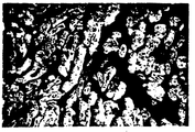
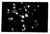
제1도 내지 제6도는 본 발명 항체로 염색된 인심방근 또는 심실근의 절단표본을 형광현미경으로 찍은 사진이다.1 to 6 are photographs taken by fluorescence microscopy of cut samples of atrial or ventricular muscle stained with the antibody of the present invention.
제1도는 CMA-19 세포주에 의하여 생성된 항체로 염색된 정상인 심방근,1 is normal atrial muscle stained with antibodies produced by the CMA-19 cell line,
제2도는 동일한 정상인 심실근을 나타내고,2 shows the same normal ventricular muscle,
제3도는 HMC-14 세포주에 의하여 생성된 항체로 염색된 정상인 심실근,3 shows normal ventricular muscle stained with antibodies produced by the HMC-14 cell line,
제4도는 동일한 정상인 심방근을 나타낸다.4 shows the same normal atrial muscle.
제5도는 HMC-14 세포주에 의하여 생성된 항체로 염색된 심장판막중 환자의 심방근을 나타내고,5 shows atrial muscles of patients in heart valves stained with antibodies produced by HMC-14 cell line,
제6도는 CMA-19 세포주에 의하여 생성된 항체로 조직 염색된 심장판막중 환자의 심방근을 나타내는데 여기서 밝은부분이 염색된 부분을 나타낸다.FIG. 6 shows atrial muscles of patients in tissue valves stained with antibodies produced by the CMA-19 cell line, where the bright areas are stained.
제7도는 본 발명 항체의 사용으로 조직염색됨에 따라 심방압과 심방근중의 V3미오신 아이소자임(B형)의 비율간의 관계를 나타내는 그래프이다.7 is a graph showing the relationship between atrial pressure and the proportion of V 3 myosin isozyme (type B) in atrial muscle as tissue stained with the use of the antibody of the present invention.
이 발명은 심근 미오신 중사슬의 아이소자임에 대하여 특이성이 있는 신규한 단일클론 항체에 관한 것이다.This invention relates to novel monoclonal antibodies that are specific for isozymes of myocardial myosin heavy chain.
근년에, 높은 특이성을 갖는 항체를 많은 양 얻기 위한 방법으로서, 항체-생성세포를 골수종(myeloma) 세포로 융합하고, 이와 같이 융합되어 얻어진 히브리도마를 배양함으로써 단일클론 항체를 생산하는 히브리도마 제조법이 공지되어 왔으며[Milstein et al, Nature, Vol.256,P.495(1975)] 많은 수의 단일클론 항체가 이러한 방법에 의하여 얻어졌다.In recent years, as a method for obtaining a large amount of high specific antibodies, a hybridoma production method for producing monoclonal antibodies by fusion of antibody-producing cells into myeloma cells and culturing the hybridomas thus obtained are fused. It has been known [Milstein et al, Nature, Vol. 256, P. 495 (1975)] and a large number of monoclonal antibodies have been obtained by this method.
근(筋)연구분야에 잇어서 근 단백질에 대한 항체가 오랫동안 이용되었다. 근은 횡문근과 평활근의 둘로 크게 구분된다. 횡문근은 심근과 골격근으로 더 구분되는데 골격근은 속근과 지근으로 더 구분된다.In the field of muscle research, antibodies to muscle proteins have long been used. The muscle is divided into two types, rhabdomyo and smooth muscle. Rhabdomyomus is further divided into myocardium and skeletal muscle.
이들은 근육의 주성분인 미오신 분자의 면역성의 차이로부터 면역화학적으로 구별이 될 수 있음이 보고되어 있다.It is reported that they can be distinguished immunochemically from the differences in the immunity of myosin molecules, the main component of muscle.
[Masaki et al; J.Bio,Chem.Vol.76, p441(1974)].Masaki et al; J. Bio, Chem. Vol. 76, p441 (1974).
최근에 심근에 관해서도 두 아이소자임 즉, 하나는 높은 ATPase 활성을 갖는 V1(α형)이고 다른 하나는 낮은 ATPase 활성을 갖는 V3(β형)인데 [Yazaki etal : Circulation Research, Vol.35 p.15(1974) ; Houet al : J.Mol.Cell.Cardiol.Vol.10, p.1053(1978)], 이 둘의 존재가 명백해졌다. 일반적으로 말하면 사람, 소, 개등과 같은 동물에 있어서 심방근은 주로 V1(α형)을 포함하고 반면에 심실근은 실질적으로 V3(β형)을 포함한다. 따라서, α형 또는 β형 미오신에 특이한 단일클론 항체를 제조하는 것이 가능하다면 심방근과 심실근은 비오틴-아비단계와 같은 방법에 의하여 염색분류할 수 있다. 또한, 이들 항체는 방사성동위원소로 라벨될 수 있고 심근경색의 부위별 진단에 사용될 수 있다.Recently, as for the myocardium, there are two isozymes, namely V 1 (type α) with high ATPase activity and V 3 (β type) with low ATPase activity [Yazaki et al: Circulation Research, Vol. 35 p. .15 (1974); Hou et al: J. Mol. Cell. Cardiol. Vol. 10, p. 1053 (1978)]. Generally speaking, in animals such as humans, cattle, dogs, etc., the atrial muscles mainly include V 1 (type α), while the ventricular muscles substantially include V 3 (type β). Thus, if it is possible to produce monoclonal antibodies specific for α or β myosin, the atrial and ventricular muscles can be stained by methods such as the biotin-avi step. In addition, these antibodies can be labeled as radioisotopes and used for site-specific diagnosis of myocardial infarction.
클라크(W.A.Clark)등은 닭 또는 토끼 심근미오신으로 생쥐와 쥐를 면역하고 심근미오신 중사슬과 반응한 단일클론 항체를 얻었다[Biochem.Biophys.Res.Commun.Vol.95,P.1680(1980)]Clark et al. Obtained monoclonal antibodies that immunized mice and mice with chicken or rabbit myocardial myosin and reacted with the myocardial myosin heavy chain [Biochem. Biophys. Res. Commmun. Vol. 95, P. 1680 (1980). ]
그들은 얻어진 것들중 하나의 주(株)가 닭심근에 특이적이고 인심근과는 반응하지 않음을 보고하였다. 다른 두주는 닭, 토끼 및 쥐의 심근과 반응하고 또한 인심근과도 반응하나 그들은 또한 골격근과도 반응하므로 그렇기 때문에 심근에 특이적이 아니다.They reported that one of the obtained ones was specific for chicken myocardium and did not react with the human myocardium. The other two strains react with the myocardium of chickens, rabbits and mice and also with the myocardium, but because they also react with skeletal muscle, they are not specific for the myocardium.
이들 항체는 β형에 대하여 α형의 인심근 미오신을 식별하지 못하며, 그 역도 마찬가지이다.These antibodies do not identify α type myocardial myosin relative to β and vice versa.
또한, 클라크등은 닭심근 미오신 또는 토끼 심근 미오신으로 쥐를 면역하고 심근 미오신중사슬 V1형과 심근 미오신 중사슬 V3형에 대한 단일 클론항체를 얻었다. [(J.Biol.Chem., Vol.257,p.5449(1982)].Clark et al. Immunized mice with chicken myocardial myosin or rabbit myocardial myosin and obtained monoclonal antibodies against myocardial myosin heavy chain V 1 and myocardial myosin heavy chain V 3 . (J. Biol. Chem., Vol. 257, p. 5449 (1982)].
그러나, 이들 항체는 그의 아이소자임들 사이에 교차반응성도 또한 나타냄을 보여주며 인심근미오신에 대한 그들의 특이성에 대해서는 아무 것도 나타나 있는 바가 없다.However, these antibodies also show cross-reactivity between their isozymes and nothing about their specificity for myocardial myosin.
본 발명을 요약하면 다음과 같다. 본 발명은 상기한 바와 같은 기술적 배경 하에 달성되었고 심근 미오신 중 사슬의 아이소자임에 대하여 특이성이 있는 단일클론항체를 제공한다.In summary, the present invention is as follows. The present invention has been accomplished under the technical background as described above and provides monoclonal antibodies which are specific for isozymes of the chains in myocardial myosin.
좀더 상세히는, 본 발명은 심근미오신 중 사슬 α형에 특이성을 가지나 심근미오신 중사슬 β형을 인식하지 않는 단일 클론항체와 또한 심근미오신 중사슬 β형에 특이성을 가지나 심근미오신 중사슬 α형을 인식하지 않는 단일클론항체를 제공한다.More specifically, the present invention recognizes a monoclonal antibody having specificity for chain α type of myocardial myosin but not myocardial myosin heavy chain β type, and also recognizes myocardial myosin heavy chain α type. Monoclonal antibodies are provided.
본 발명 항체는 상기한 바와 같이 심근에 관계된 생화학적 및 병리학적 연구에 중요한 시약으로서 유용하다.The antibodies of the present invention are useful as reagents important for biochemical and pathological studies involving myocardium as described above.
또한, 본 발명 항체는 테크니튬 -99m, 인듐등과 같은 방사성동위원소로 라벨 될 수 있고 면역검출(immunodetection)에 적응될 수 있는데, 그것은 환자에게 투여 후 전신감마신 티그래피에 의하여 측정되어, 이로써 심근경색의 부위별 진단이 가능하게 된다. 특히, 심실근 경색과 합병되기도 하는 심방근 경색의 진단에 유용하다.In addition, the antibodies of the present invention may be labeled with radioisotopes such as technitium-99m, indium and the like and may be adapted for immunodetection, which is measured by systemic gammacinography after administration to a patient, thereby The site-specific diagnosis of myocardial infarction is possible In particular, it is useful for the diagnosis of atrial myocardial infarction which may be combined with ventricular myocardial infarction.
본 발명 항체로 면역측정(immunoassay)을 수행함으로서 심근 경색의 동안에 혈중의 미오신 중사슬의 누출을 검출하는 것이 또한 가능한 것으로 생각된다. 따라서 발명항체는 심근경색의 예후에도 유용하게 된다. 이제본 발명을 상세히 설명하겠다. 본 발명 항체는 심근 미오신의 아이소자임을 인식할 수 있는 특징을 갖는 기술상 공지되어 있는 항체와는 구별이 될 수 있다.It is also thought possible to detect leakage of the myosin heavy chain in the blood during myocardial infarction by performing an immunoassay with the antibody of the invention. Therefore, the antibody of the present invention is also useful for prognosis of myocardial infarction. The present invention will now be described in detail. Antibodies of the invention can be distinguished from those known in the art having the ability to recognize isozymes of myocardial myosin.
본 발명 항체의 그 이상의 유용한 특징은 인심근 미오신의 아이소자임을 식별할 수 있는 것, 특히 한가지 아이소자임에 특이성을 갖고 다른 것은 식별하지 않는 것이다.Further useful features of the antibodies of the invention are those which can identify isozymes of myocardial myosin, in particular one isozyme and no other.
본 발명항체는 그 제조방법 또는 제조형태에 있어서 특별히 제한되어 있지 않는데, 목적에 따라 적당히 선택될 수 있다. 본 발명 항체를 생성하는 히브리도마는 일반적으로 실행되는 세포융합법을 적용함으로써 얻어질 수 있다. 이제이 세포 융합 법을 설명하기로 한다.The antibody of the present invention is not particularly limited in its production method or production form, and may be appropriately selected according to the purpose. The hybridomas producing the antibodies of the present invention can be obtained by applying a commonly performed cell fusion method. Now this cell fusion method will be described.
(1) 항체 생성세포의 제조(1) Preparation of antibody producing cell
항체 생성세포의 제조는 인심방미오신(α형), 인 심실미오신(β형) 또는 소, 말 또는 돼지로부터 제조된 인심근 미오신 α형 또는 β형에 면역화학적으로 동등한 심근미오신으로 생쥐, 쥐, 토끼, 양, 말, 소 등과 같은 이종동물을 면역하고 비(脾)세포, 흉선세포, 임파절세포 및/또는 말초혈세포로부터 항체생성 세포를 취함으로써 수행된다.The production of antibody-producing cells may be performed in mice, mice, It is performed by immunizing heterologous animals such as rabbits, sheep, horses, cattle and the like and taking antibody-producing cells from non-cells, thymic cells, lymph node cells and / or peripheral blood cells.
(2) 골수종 세포의 제조(2) Preparation of myeloma cells
골수종 세포로서 생쥐, 쥐, 토끼와 같은 여러 가지 동물 및 사람으로부터의 세포주가 사용될 수 있다. 사용되는 세포주는 바람직하게는 약제저항성이어야 하며 선택배지내에서는 생존하지 않으나 융합후에 생존하여야 한다. 가장 통상적으로 사용되는 세포주는 8-아자구아닌 저항성 세포주인데, 하이포 크산틴 포스포리보실 트랜스퍼라제내에서 결손 하여 하이포크산틴-아미노프테린-디미딘(HAT)배지 내에서 성장될 수 없다. 세포주는 또한 "비분비성"형태인 것이 바람직하다.As myeloma cells, cell lines from various animals and humans such as mice, mice, rabbits can be used. The cell line used should preferably be drug resistant and not survive in selective media but after fusion. The most commonly used cell line is an 8-azaguanine resistant cell line, which is missing in hypoxanthine phosphoribosyl transferase and cannot be grown in hypoxanthine-aminopterin-dimidine (HAT) medium. The cell line is also preferably in a "non-secretory" form.
이러한 세포주의 전형적인 예들은 생쥐 골수종 MOPC-21세포주로부터 유도된 P3/x63-Ag 8U1(P3U1), P3/x63-Ag/8 6·5·3(x63·5·3), P3/NSI-1-Ag 4-1(NS-1), Sp210-Ag14(SP2)이다. 쥐 골수종 210 RCY 3Ag 1·2·3(Y3 Agl·2·3) 및 인 골수종 U-266-AR1, 그리고 GM 1500도 또한 유용하다.Typical examples of these cell lines are P 3 / x63-Ag 8U 1 (P 3 U 1 ), P 3 / x63-Ag / 8 6 · 5 · 3 (x63 · 5 · 3) derived from the mouse myeloma MOPC-21 cell line. , P 3 / NSI-1-Ag 4-1 (NS-1), Sp210-Ag14 (SP2). Rat myeloma 210 RCY 3Ag 1 · 2 · 3 (Y3 Agl · 2 · 3) and phosphorus myeloma U-266-AR 1 , and GM 1500 are also useful.
(3) 세포융합(3) cell fusion
세포융합은 이글(Eagle)의 최소기본배지(MEM)와 RPMI 1640과 같은 동물세포 배양용 배지내에서 107내지 108개의 골수종세포를 항체생성세포와 1 : 4 내지 1 : 10 혼합비유로 혼합함으로써 수행된다.Cell fusion was performed by mixing 10 7 to 10 8 myeloma cells with an antibody-producing cell in a 1: 4 to 1:10 mixed ratio in an animal cell culture medium such as Eagle's minimum basic medium (MEM) and RPMI 1640. Is performed.
융합보조제로서, 1,000내지 6,000의 평균분자량을 갖는 폴리에틸렌글리콜(PEG), 폴리비닐알콜, 바이럿, 또는 기타를 사용하는 것이 가능하다.As the fusion aid, it is possible to use polyethylene glycol (PEG), polyvinyl alcohol, viat, or the like having an average molecular weight of 1,000 to 6,000.
(4) 선택배지내의 히브리도마의 선별(4) Selection of hybridomas in selective medium
세포융합과정 후 세포로부터의 히브리도마의 선별은 선택배지내의 선택적인 성장에 의하여 행할 수 있다. 예를 들면 세포를 예를 들어서 15%의 소 태아혈청을 함유하는 RPMI 1640배지로 적당히 희석하고 미소 적정판(microtiter plate)에 약 105-106세포/웰(well)정도로 덮고, 선택배지(예로써, HAT배지)를 각 웰에 가하는데 각 단계에 이어서 선택배지의 적당한 교환을 한다. 예를 들면, 8-아자구아닌 저항성 세포주가 골수종 세포로서 사용되고 선택배지로서 HAT배지가 사용될 때 미융합 세포들은 배양 후 약 10일째에 사멸할 것이며 항체생성세포는 체외에서 오랜 기간 성장할 수 없다. 따라서, 10일 내지 14일째 날에 성장된 세포는 모두 히브리도마이다.Selection of hybridomas from cells after the cell fusion process can be performed by selective growth in selective media. For example, cells are appropriately diluted with, for example, RPMI 1640 medium containing 15% fetal bovine serum, covered in a microtiter plate with about 10 5 -10 6 cells / well, and the selective medium ( For example, a HAT medium) is added to each well followed by the appropriate exchange of the selection medium following each step. For example, when 8-azaguanine resistant cell lines are used as myeloma cells and HAT medium as the selection medium, unfused cells will die about 10 days after culture and antibody-producing cells cannot grow in vitro for a long time. Thus, cells grown on days 10-14 are all hybridomas.
(5) 항-심근 미오신 중사슬 α항체 및 항-심근 미오신 중사슬 β항체를 생성하는 히브리도마의 검색.(5) Search for hybridomas that produce anti-myocardial myosin heavy chain α antibody and anti-myocardial myosin heavy chain β antibody.
항-심근 미오신 중사슬 α항체 및 항-심근 미오신 중사슬 β항체를 생성하는 히브리도마의 검색은 효소면역측정법(Enzyme Linked Immunosorbant Assay)에 따라서 수행하였는데, 이것은 이후부터 "ELISA"라 칭하였다.The screening of hybridomas producing anti-myocardial myosin heavy chain α and anti-myocardial myosin heavy chain β antibodies was performed according to the Enzyme Linked Immunosorbant Assay, which was hereinafter referred to as "ELISA".
좀 더 상세히는, 소 심방근 미오신과 같은 심근 미오신 중사슬 α형 또는 인 심실근 미오신과 같은 심근 미오신 중사슬 β형을 미리 인산염 완충 생리식염수(PBS) 또는 탄산수소나트륨(pH 8.0)과 같은 완충액 내에 10-100㎍/ml로 용해시킨다.More specifically, myocardial myosin heavy chain type α such as bovine atrial myosin or myocardial myosin heavy chain β type such as ventricular myosin myosin is previously contained in a buffer such as phosphate buffered saline (PBS) or sodium bicarbonate (pH 8.0). Dissolve at 10-100 μg / ml.
각 50㎕의 몫을 ELISA용 폴리비닐클로라이드(PVC)판과 같은 유연한 판(9웰)에 가하였다. 그리고 그 판을 4℃에서 하룻밤 방치한다.An aliquot of 50 μl each was added to a flexible plate (9 wells) such as a polyvinylchloride (PVC) plate for ELISA. The plate is then left at 4 ° C. overnight.
다음에, 항원을 따르고 PBS로 세정한 후, 1% 소혈청 알부민(BSA)을 함유하는 PBS를 첨가하고 혼합물을 실온에서 한 시간동안 방치하여 항원이 결합되지 않는 부위를 BSA로 블록킹한다. 각 웰의 상등액으로부터 50㎕의 몫을 첨가하고 실온에서 한 시간동안 방치하고 PBS로 세 번 세정한다. 다음에 비오틸 항-쥐 면역글로부린 혈청(제2항체)를 첨가하고 혼합물을 실온에서 한시간동안 방치한다. PBS로 세 번 세정한 후 아비딘 D-효소착물을 첨가하고 혼합물을 실온에서 15분간 방치한다. PBS로 네 번 세정한 후 효소용 기질을 첨가하여 광학밀도를 측정한다.Next, the antigen is followed and washed with PBS, then PBS containing 1% bovine serum albumin (BSA) is added and the mixture is left at room temperature for one hour to block the site where antigen is not bound with BSA. Add 50 μl of the share from the supernatant of each well and leave at room temperature for one hour and rinse three times with PBS. Biotyl anti-mouse immunoglobulin serum (second antibody) is then added and the mixture is left at room temperature for one hour. After washing three times with PBS, avidin D-enzyme complex is added and the mixture is left at room temperature for 15 minutes. After washing four times with PBS, the substrate for enzyme was added to measure the optical density.
항원에 특이한 단일클론 항체를 포함하는 웰은 위에 기술한 바와 같은 방법에 따라 쉽게 판별될 수 있어서, 이로써 히브리도마의 검색이 수행될 수 있다.Wells comprising monoclonal antibodies specific for the antigen can be readily determined according to the methods described above, so that searches for hybridomas can be performed.
(6) 클로우닝(6) Cloning
각 웰내에 둘 또는 그 이상의 히브리도마가 포함될 가능성이 있다. 그러므로 클로우닝은 단일 클론항체-생성 히브리도마를 얻기 위하여 예를 들어서, 한계 희석법에 따라서 행한다.Each well is likely to contain two or more hybridomas. Therefore, cloning is performed according to, for example, a limiting dilution method to obtain monoclonal antibody-producing hybridomas.
(7) 항체의 생성(7) Generation of Antibody
가장 순수한 단일클론항체는 10내지 15% 소태아혈청을 함유하는 RPMI 1640 배지 또는 무혈청배지와 같은 동물세포배양용 배지 내에서 상기 단일클론항체를 생성하는 히브리도마를 배양하고 상등액으로부터 항체를 얻음으로써 얻을 수 있다.The purest monoclonal antibodies were obtained by culturing the hybridomas producing the monoclonal antibody in RPMI 1640 medium containing 10 to 15% fetal bovine serum or in animal cell culture medium such as serum-free medium and obtaining the antibody from the supernatant. You can get it.
세포배양 방법 및 조건들에 대하여는, 동물세포 배양방법에 재래적으로 사용된 것들을 알맞게 적용할 수 있다.Regarding cell culture methods and conditions, those conventionally used in animal cell culture methods can be suitably applied.
한편, 많은 양으로 항체를 생산하기 위한 방법으로서, 프리스탄(2, 6, 10, 14-테트라메틸펜타데칸)과 같은 광물유를 히브리도마의 친(親)골수종이 유래된 동계동물에 복강내 투여를 한후, 히브리도마를 그안에서 많은 양으로 증식되도록 복강내 주사를 하는 방법을 사용하는 것이 가능하다. 히브리도마는 10-18일 이내에 복수종양으로 성장하고 혈청과 복수액내에 고농도로(약 1내지 20mg/ml) 항체를 생성하게 된다.On the other hand, as a method for producing antibodies in large amounts, intraperitoneal administration of mineral oils such as pristane (2, 6, 10, 14-tetramethylpentadecane) to syngeneic animals derived from protomyeloma of hybridoma It is then possible to use a method of intraperitoneal injection so that the hybridomas are multiplied in large amounts therein. Hybridomas grow into multiple tumors within 10-18 days and produce antibodies at high concentrations (about 1 to 20 mg / ml) in serum and ascites fluid.
정제가 필요할때는, DEAE셀룰로오스 이온 교환 컬럼크로마토그래피, 심근 미오신이 결합되거나 한 세파로우스 4B를 사용하는 어피니티 컬럼 크로마토그래피, 또는 분자체(gel filteration)컬럼 크로마토그래피와 같은 방법에 의하여 황산암모늄 분별(fractionation)후에 정제를 수행할 수 있다.When purification is required, fractionation of ammonium sulfate by methods such as DEAE cellulose ion exchange column chromatography, affinity column chromatography using Sepharose 4B with or without myocardial myosin, or gel filteration column chromatography Purification can be carried out after fractionation.
지금까지 얻어진 본 발명 항체를 생산하기 위한 바람직한 히브리도마의 예들은 심근 미오신 중사슬 α형에 특이성이 있는 세포주를 생성하는 항체로서 히브리도마 CMA-19세포주, 그리고 심근 미오신 중사슬 β형에 특이성이 있는 항체생성주로서 히브리도마 HMC-14세포주, HMC-48세포주 및 HMC-50 세포주이다.Examples of preferred hybridomas for producing the antibodies of the present invention thus far obtained are antibodies that produce cell lines specific for myocardial myosin heavy chain α-type, and specific for the hybridoma CMA-19 cell line, and myocardial myosin heavy chain β-type. Antibody production lines are the hybridoma HMC-14 cell line, HMC-48 cell line and HMC-50 cell line.
본 발명의 바람직한 구체예의 설명으로서, 이들 히브리도마의 제조방법과 본 발명 항체의 특성을 이하에 상세히 기술하기로 한다.As a description of preferred embodiments of the present invention, these hybridoma production methods and the characteristics of the antibodies of the present invention will be described in detail below.
1. 히브리도마 취득1. Obtaining Hebrew
소심방근 미오신(1mg/ml) 또는 인 심실근 미오신(1mg/ml)을 생리식염수에 용해시키고 완전 프룬드 어주번트(complete freund's adjuvant)와 1 : 1의 비율로 혼합하여 에멀션을 제조하였다. 에멀션을 BALB/C 생쥐(암컷, 6주일생)에 2주마다 여러 번(50㎍/마리) 복강 내 투여를 하였고 최종적으로 30㎍의 소심방근 미오신 또는 인 심실근 미오신을 복강 내에 투여 하였다.Emulsions were prepared by dissolving either atrial myosin myosin (1 mg / ml) or pulmonary ventricular myosin (1 mg / ml) in physiological saline and mixing 1: 1 with complete freund's adjuvant. The emulsion was intraperitoneally administered BALB / C mice (females, 6 weeks old) multiple times every two weeks (50 μg / horse) and finally 30 μg of small atrial myosin or ventricular myosin intraperitoneally.
최종면역 3일 후, 생쥐로부터 비(脾)세포를 취해서 MEM으로 세정하였다. 생쥐 골수종세포 P3U1을 MEM으로 세정하고 비 세포와 10 : 1의 비율로 혼합하였다. 원심 분리 후 50% PEG 100°MEM용액 1ml를 이와 같이 얻은 펠릿 또는 케익에 점차적으로 가하여 세포융합을 행하였다. 또한, MEM용액을 점차적으로 가하여 최종 량 10ml를 얻었다.After 3 days of final immunization, nasal cells were taken from the mice and washed with MEM. Mouse myeloma cells P 3 U 1 were washed with MEM and mixed with non-cells at a ratio of 10: 1. After centrifugation, 1 ml of a 50
다시 원심분리를 행하였고 펠릿을 15% 소태 아혈청을 함유하는 RPMI 1640 배지내에 P3U1으로서 1×10⁵세포/0.1ml로 현탁시켰으며 0.1ml/웰 내의 96-웰 마이크로판상에 분무하였다.Centrifugation was again performed and the pellet was suspended at 1 × 10 5 cells / 0.1 ml as P 3 U 1 in RPMI 1640 medium containing 15% fetal bovine serum and sprayed onto 96-well microplates in 0.1 ml / well. .
1일 후, HAT배지의 각 0.1ml의 몫을 가하였고 그후 3-4일마다 배지의 반을 신선한 HAT 배지로 갈아 주었다. 약 7일째에 몇몇 웰내에서 히브리도마의 성장이 식별되었다.After 1 day, 0.1 ml of each of the HAT medium was added and then half of the medium was changed to fresh HAT medium every 3-4 days. At about 7 days growth of hybridomas was identified in several wells.
상등액 각각 50㎕의 몫들을 소심방근 미오신(α형) 또는 인 심실근 미오신(β형으로 사전에 코우팅된 96-웰 유연판에 가하였다.50 μl shares of each of the supernatants were added to a 96-well flexible plate precoated with atrial myocardial myosin (type α) or pulmonary ventricular myosin (type β).
아비딘 D-효소 컨쥬게이트로서 아비딘 D-퍼옥시다제(Vector co.제품) 그리고 기질과 발색제로서 과산화수소, 4-아미노안티피린 및 페놀을 상기한 바와 같이 ELISA방법에 따라 사용함으로써, 소심방근 미오신과 반응하나 인심실근 미오신과는 반응하지 않는 상등액(심근미오신 중사슬 X형에 특이성을 갖는 단일 클론항체가 이상등액에 포함되어 있다) 그리고 인심실근 미오신과는 반응하나 소심방근 미오신과는 반응하지 않는 상등액(심근 미오신 중사슬 β형에 특이성을 갖는 단일클론항체가 이 상등액에 포함되어 있다)이 선별되었고 히브리도마는 한계 희석법에 의하여 클로우닝을 행하였다.Avidin D-peroxidase (manufactured by Vector co.) As an avidin D-enzyme conjugate and hydrogen peroxide, 4-aminoantipyrine and phenol as substrates and colorants according to the ELISA method described above, react with small atrial myosin Supernatant that does not react with myocardial myosin (myocardial myosin heavy chain type X monoclonal antibody is included in the supernatant), and supernatant that reacts with myocardial myosin but not with small myocardial myosin ( Monoclonal antibodies specific for myocardial myosin heavy chain β-type were included in this supernatant) and hybridomas were cloned by limiting dilution.
그 결과, 심근 미오신 중사슬 α형에 특이성을 갖는 항체를 생성하는 히브리도마 CMA-19 세포주, 그리고 심근 미오신 중사슬 β형에 특이성을 갖는 항체를 생성하는 HMC-14세포주, HMC-48세포주 및 HMC-50세포주를 얻었다.As a result, HMC-14 cell line, HMC-48 cell line, and HMC producing hybridoma CMA-19 cell line producing antibodies having specificity for myocardial myosin heavy chain α type, and antibodies having specificity for myocardial myosin heavy chain β type. -50 cell lines were obtained.
II. 단일클론항체의 생산II. Production of Monoclonal Antibodies
히브리도마 CMA-19세포주, HMC-14세포주, HMC-48세포주 및 HMC-50세포주를 15% 소태아 혈청을 함유하는 RPMI 1640 배지에서 96-웰판, 다음에 25cm2플라스크로 규모를 늘려 배양하였고 배양상등액을 수집하였다.Hybridoma CMA-19, HMC-14, HMC-48 and HMC-50 cell lines were cultured in RPMI 1640 medium containing 15% fetal bovine serum in 96-well plates and then in a 25 cm 2 flask. Supernatants were collected.
이들 상등액의 항-심근 미오신 항체의 역가는 ELISA방법에 의하여 결정하여 제1표에 나타나 있는 결과들을 얻었다. 역가는 코우팅된 항원에 대하여 충분한 양의 항체가 존재하는 시료에 대하여 ELISA방법에 의하여 얻은 것을 100%로 하여 50%의 흡광도를 주는 원용액으로부터의 항체시료의 희석배율로서 표현된다.The titers of anti-myocardial myosin antibodies of these supernatants were determined by ELISA method to obtain the results shown in Table 1. The titer is expressed as the dilution ratio of the antibody sample from the original solution giving 50% absorbance at 100% obtained by the ELISA method for a sample in which a sufficient amount of antibody is present with respect to the coated antigen.
[표 1]TABLE 1

이들 항체는 인 골격근과 실질적으로 교차 반응성이 없음을 나타내었다.These antibodies have been shown to be substantially cross reactive with phosphorus skeletal muscle.
III. 항체의 서브클라스(subclass)의 결정III. Determination of Subclasses of Antibodies
96-웰 유연 마이크로판을 각 단일클론항체로 코우팅하고 1% BSA함유 PBS로 블록킹한 후, MONOABID EIA KIT(ZYMED Co. 제품)에 의하여 항-IgA항체, 항-IgG1항체, 항-IgG2a항체, 항-IgG2b항체, 항-IgG3항체 및 항-IgM항체와의 반응을 관찰하여 각 단일클론항체의 서브클라스를 결정하였다. 또한 L사슬의 항 K항체, 항 λ항체와의 반응성을 조사하여 형을 결정하였다.96-well flexible microplates were coated with each monoclonal antibody and blocked with 1% BSA containing PBS, followed by anti-IgA antibody, anti-IgG 1 antibody, anti-IgG by MONOABID EIA KIT (manufactured by ZYMED Co.). 2 a antibodies, by observing the reaction of the anti -IgG 2 b antibody, anti -IgG 3 and anti -IgM antibody was determined subclass of each monoclonal antibody. In addition, the type was determined by investigating the reactivity with L-chain anti-K antibody and anti-λ antibody.
그 결과, CMA-19세포주에 의하여 생성된 항체는 IgG1/K이고, HMC-14세포에 의하여 생성된 항체는 IgG2a/k 그리고 HMC-48세포주와 HMC-50세포주에 의하여 생성된 항체는 IgG2b/k임을 발견하였다.As a result, the antibody produced by CMA-19 cell line was IgG 1 / K, the antibody produced by HMC-14 cell was IgG 2a / k and the antibody produced by HMC-48 cell line and HMC-50 cell line was IgG It was found to be 2b / k.
IV. 본 발명 항체와의 조직염색IV. Tissue Staining with Antibodies of the Invention
판막이식등을 위한 심장절개수술시 채취한 인 심방근 및 심실근을 Tissue-TEK II에 의하여 고정시킨 후 크리오스타트에 의하여 절단표본을 제조하였다.Phosphorus atrial and ventricular muscles collected during cardiac surgery for valve transplantation were fixed by Tissue-TEK II, and a cut specimen was prepared by cryostat.
이들 절단표본들은 본 발명 항체를 사용하여 비오틴-아비딘계에 따라 염색되었다.These cleavage samples were stained according to the biotin-avidin system using the antibodies of the invention.
좀더 상세히는, 각 절단표본을 제1항체로서 본 발명항체와 PBS(0.01M, pH 7.2)내에서 37℃에서 40분간 인큐베이트하였다.More specifically, each cut specimen was incubated at 37 ° C. for 40 minutes in PBS (0.01 M, pH 7.2) with the present invention as the first antibody.
다음에, 다시 세정한후, 제2항체로서 비오티닐 항-생쥐 IgG 항체(TAGO Co., 사람의 간호모제네이트 및 혈청으로 흡수후 20배 희석하여 사용됨)와 인큐베이션을 동일하게 수행하였다. 또한 다시 세정후, 플루오레세인 이소티오시아네이트 표식(label)된 아비딘(E.Y.Laboratories Co., 사람의 간 호모제네이트 및 혈청으로 흡수후 20--배 희석하여 사용됨)과도 같은 방법으로 인큐베이트하였다. 이 시료는 세정하고 글리세린으로 밀폐하여 형광염색 표본을 제조하였다.Next, after washing again, the same incubation with the biotinyl anti-mouse IgG antibody (TAGO Co., used for 20 times dilution after absorption with human nursing mother and serum) was performed as the second antibody. After washing again, it was incubated in the same manner as fluorescein isothiocyanate labeled avidin (EYLaboratories Co., used 20-fold dilution after absorption with human liver homogenate and serum). . This sample was washed and sealed with glycerin to prepare a fluorescent stained sample.
이들 표본을 형광현미경하에 검사하였다. 그 결과, CMA-19세포주에 의하여 생성된 항체가 사용되었을 때 정상인 심방근내에서 95내지 96%의 세포들이 염색되었다 (제1도 참조). 그러나 10%이하의 심실근 세포들이 염색되었다(제2도 참조). 한편, HMC-14 세포주가 사용되었을때는 100%의 심실근세포들이 염색되었으나(제3도), 단지 20내지 30%의 심방근 세포들만이 염색되었다(제4도 참조). (정상인 심방근의 20-30%가 αβ의 형태로 존재한다)These samples were examined under fluorescence microscopy. As a result, 95-96% of the cells were stained in normal atrial muscle when the antibody produced by the CMA-19 cell line was used (see FIG. 1). However, less than 10% of ventricular myocytes were stained (see Figure 2). On the other hand, 100% of ventricular myocytes were stained when the HMC-14 cell line was used (Figure 3), but only 20 to 30% of the atrial muscle cells were stained (see Figure 4). (20-30% of normal atrial muscles are in the form of αβ)
반대로, 판막증 환자의 심방근에서는 심방근내의 HMC-14세포주에 의하여 생성된 항체로 염색된 세포는 증가하였고(제5도 참조) CMA-19세포주에 의하여 생성된 항체로 염색된 부분은 대응하여 감소하였다 (제6도 참조)In contrast, in the atrial muscles of patients with valve disease, cells stained with antibodies produced by the HMC-14 cell line in the atrial muscles increased (see FIG. 5), and portions stained with antibodies produced by the CMA-19 cell line correspondingly decreased ( See figure 6.
이 현상은 판막증의 심방근 미오신의 α형으로부터 β형으로 아이소자임상의 변화가 일어남을 말한다.This phenomenon refers to a change in the isozyme phase from the α type to the β type of atrial myosin in valve disease.
더욱이, 심방압과 심방근내의 V3미오신 아이소자임(β형)의 비율간의 관계는 본 발명항체를 사용하는 조직염색에 의하여 검사하였는데 이것으로 제7도에 나타낸 결과가 얻어졌다.Furthermore, the relationship between atrial pressure and the proportion of V 3 myosin isozyme (β) in the atrial muscle was examined by tissue staining using the antibody of the present invention, which resulted in the results shown in FIG.
즉, 정상인 심방근에서 심방압은 5mmHg 또는 그 이하이고 β형 아이소자임의 함량은 10% 또는 그 이하만큼 적다. 한편, 판막증환자에 있어서는 심방압은 10mmHg 또는 그 이상이고 심방근미오신의 아이소자임패턴은 α형은 감소하고 β형은 증가한다.That is, in normal atrial muscle, the atrial pressure is 5 mmHg or less and the content of β-type isozyme is as small as 10% or less. On the other hand, in patients with valve disease, the atrial pressure is 10 mmHg or more, and the isozyme pattern of a myocardial myosin decreases α-type and increases β-type.
Claims (3)
Applications Claiming Priority (3)
| Application Number | Priority Date | Filing Date | Title |
|---|---|---|---|
| JP122579 | 1983-07-06 | ||
| JP58122579A JPS6028998A (en) | 1983-07-06 | 1983-07-06 | Monoclonal antibody to heavy chain of myocardial myosine |
| JP???58-122579 | 1983-07-06 |
Publications (2)
| Publication Number | Publication Date |
|---|---|
| KR850001282A KR850001282A (en) | 1985-03-18 |
| KR910008637B1 true KR910008637B1 (en) | 1991-10-19 |
Family
ID=14839400
Family Applications (1)
| Application Number | Title | Priority Date | Filing Date |
|---|---|---|---|
| KR1019840003916A KR910008637B1 (en) | 1983-07-06 | 1984-07-06 | Monoclonal antibody to cardlac myosin heavy chain |
Country Status (7)
| Country | Link |
|---|---|
| US (1) | US4767843A (en) |
| EP (1) | EP0131834B1 (en) |
| JP (1) | JPS6028998A (en) |
| KR (1) | KR910008637B1 (en) |
| AU (1) | AU570696B2 (en) |
| CA (1) | CA1225608A (en) |
| DE (1) | DE3485965T2 (en) |
Families Citing this family (10)
| Publication number | Priority date | Publication date | Assignee | Title |
|---|---|---|---|---|
| JPS60201260A (en) * | 1984-03-27 | 1985-10-11 | Yamasa Shoyu Co Ltd | Myocardial myosin diagnosing agent |
| JPH0625762B2 (en) * | 1985-06-13 | 1994-04-06 | ヤマサ醤油株式会社 | Myosin light chain assay |
| GB2200358B (en) * | 1987-01-27 | 1991-02-27 | Univ Birmingham | Materials for in vivo detection of cardiac damage, and methods for their preparation |
| US5046499A (en) * | 1988-06-13 | 1991-09-10 | Centocor, Inc. | Method for myocardial infarct risk assessment |
| WO1990011520A1 (en) * | 1989-03-28 | 1990-10-04 | Yamasa Shoyu Kabushiki Kaisha | Antibody against heavy chain of smooth muscle myosin |
| US5830720A (en) * | 1989-04-21 | 1998-11-03 | Boehringer Mannheim Gmbh | Recombinant DNA and expression vector for the repressible and inducible expression of foreign genes |
| US5364612A (en) * | 1991-05-06 | 1994-11-15 | Immunomedics, Inc. | Detection of cardiovascular lesions |
| US5908757A (en) * | 1994-10-25 | 1999-06-01 | Yamasa Corporation | Antibody reagent for detecting dissecting aortic aneurysm and uses thereof |
| CA2246666A1 (en) * | 1996-02-19 | 1997-08-21 | Yamasa Corporation | Diagnostic agent for angiopathic diseases |
| US6248869B1 (en) | 1997-05-29 | 2001-06-19 | Medical Analysis Systems, Inc. | Troponin I forms and use of the same |
-
1983
- 1983-07-06 JP JP58122579A patent/JPS6028998A/en active Granted
-
1984
- 1984-07-02 DE DE8484107663T patent/DE3485965T2/en not_active Expired - Fee Related
- 1984-07-02 US US06/626,918 patent/US4767843A/en not_active Expired - Lifetime
- 1984-07-02 EP EP84107663A patent/EP0131834B1/en not_active Expired
- 1984-07-03 AU AU30228/84A patent/AU570696B2/en not_active Ceased
- 1984-07-06 CA CA000458361A patent/CA1225608A/en not_active Expired
- 1984-07-06 KR KR1019840003916A patent/KR910008637B1/en not_active IP Right Cessation
Also Published As
| Publication number | Publication date |
|---|---|
| EP0131834B1 (en) | 1992-10-28 |
| KR850001282A (en) | 1985-03-18 |
| EP0131834A3 (en) | 1987-06-24 |
| US4767843A (en) | 1988-08-30 |
| DE3485965D1 (en) | 1992-12-03 |
| DE3485965T2 (en) | 1993-03-18 |
| AU3022884A (en) | 1985-01-10 |
| CA1225608A (en) | 1987-08-18 |
| JPH0231958B2 (en) | 1990-07-17 |
| AU570696B2 (en) | 1988-03-24 |
| EP0131834A2 (en) | 1985-01-23 |
| JPS6028998A (en) | 1985-02-14 |
Similar Documents
| Publication | Publication Date | Title |
|---|---|---|
| US4628027A (en) | Vitro diagnostic methods using monoclonal antibodies against connective tissue proteins | |
| CA1190873A (en) | Recombinant monoclonal antibodies | |
| EP0741144B1 (en) | Antiannexin-v monoclonal antibody, process for producing the same, and use therof | |
| KR910008637B1 (en) | Monoclonal antibody to cardlac myosin heavy chain | |
| JP3018110B2 (en) | Monoclonal antibody | |
| NZ204255A (en) | Monoclonal antibodies against connective tissue proteins:use in diagnosis and monitoring of human diseases | |
| AU2006213411A1 (en) | Antibody for assay of ADAMTS13 activity and method of activity assay | |
| EP0205177B1 (en) | Method of assaying myosin light chain | |
| US5358850A (en) | Sandwich immunoassay of β-n-acetylglucosaminidase and monoclonal antibody used therein | |
| DK172758B1 (en) | Monoclonal antibody to prosomal proteins, immortalized cell line producing the antibody, method of detection | |
| JP5058403B2 (en) | CK-MB activity measuring method and CK-MB activity measuring reagent | |
| WO1986002359A1 (en) | Monoclonal antibodies and their use | |
| US4952507A (en) | Production and diagnostic use of antibodies against pancreatic alpha-amaylase | |
| IE881290L (en) | Monoclonal antibodies for the selective immunological¹determination of intact procollagen peptide (Type III) and¹procollagen (Type III) in body fluids | |
| US7148063B2 (en) | Mitochondrial creatine kinase antibody | |
| JPH0320240B2 (en) | ||
| JP3036545B2 (en) | Monoclonal antibody, hybridoma cell line producing the same, and immunoassay using the same | |
| JPS60253871A (en) | Anti-apo a-1 antibody | |
| JPH061271B2 (en) | Method for producing antibody against acidic metabolite of catecholamine and antigen used therefor | |
| JP2567664B2 (en) | Monoclonal antibody against human Mn superoxide dismutase | |
| JPH0977798A (en) | Antibody resistance to cholesteryl ester transfer protein | |
| JPH0690784A (en) | Monoclonal antibody and its use | |
| WO1986002356A1 (en) | Monoclonal antibodies and their use | |
| JPH0630620B2 (en) | Monoclonal antibody reactive with cholinesterase having a molecular weight of 70,000 to 300,000 present in human body fluid | |
| JPH06261785A (en) | Monoclonal antibody against apo-e protein receptor |
Legal Events
| Date | Code | Title | Description |
|---|---|---|---|
| A201 | Request for examination | ||
| E902 | Notification of reason for refusal | ||
| G160 | Decision to publish patent application | ||
| E701 | Decision to grant or registration of patent right | ||
| GRNT | Written decision to grant | ||
| FPAY | Annual fee payment |
Payment date: 20001011 Year of fee payment: 10 |
|
| LAPS | Lapse due to unpaid annual fee |