KR20240043786A - Anti-TIGIT antibodies and uses thereof - Google Patents
Anti-TIGIT antibodies and uses thereof Download PDFInfo
- Publication number
- KR20240043786A KR20240043786A KR1020247007621A KR20247007621A KR20240043786A KR 20240043786 A KR20240043786 A KR 20240043786A KR 1020247007621 A KR1020247007621 A KR 1020247007621A KR 20247007621 A KR20247007621 A KR 20247007621A KR 20240043786 A KR20240043786 A KR 20240043786A
- Authority
- KR
- South Korea
- Prior art keywords
- antibody
- antigen
- cancer
- binding fragment
- seq
- Prior art date
Links
- 230000027455 binding Effects 0.000 claims abstract description 183
- 238000009739 binding Methods 0.000 claims abstract description 171
- 239000000427 antigen Substances 0.000 claims abstract description 151
- 108091007433 antigens Proteins 0.000 claims abstract description 149
- 102000036639 antigens Human genes 0.000 claims abstract description 149
- 239000012634 fragment Substances 0.000 claims abstract description 134
- 101000831007 Homo sapiens T-cell immunoreceptor with Ig and ITIM domains Proteins 0.000 claims abstract description 91
- 102100024834 T-cell immunoreceptor with Ig and ITIM domains Human genes 0.000 claims abstract description 77
- 150000007523 nucleic acids Chemical class 0.000 claims abstract description 30
- 108020004707 nucleic acids Proteins 0.000 claims abstract description 29
- 102000039446 nucleic acids Human genes 0.000 claims abstract description 29
- 239000003446 ligand Substances 0.000 claims abstract description 16
- 210000004027 cell Anatomy 0.000 claims description 117
- 241000282414 Homo sapiens Species 0.000 claims description 66
- 206010028980 Neoplasm Diseases 0.000 claims description 56
- 108010047041 Complementarity Determining Regions Proteins 0.000 claims description 49
- 238000000034 method Methods 0.000 claims description 49
- 238000006467 substitution reaction Methods 0.000 claims description 41
- 108090000623 proteins and genes Proteins 0.000 claims description 36
- 239000013598 vector Substances 0.000 claims description 34
- 150000001413 amino acids Chemical class 0.000 claims description 31
- 125000003275 alpha amino acid group Chemical group 0.000 claims description 26
- 108010048507 poliovirus receptor Proteins 0.000 claims description 25
- 210000004978 chinese hamster ovary cell Anatomy 0.000 claims description 24
- 230000000694 effects Effects 0.000 claims description 22
- 102100029740 Poliovirus receptor Human genes 0.000 claims description 20
- YBJHBAHKTGYVGT-ZKWXMUAHSA-N (+)-Biotin Chemical compound N1C(=O)N[C@@H]2[C@H](CCCCC(=O)O)SC[C@@H]21 YBJHBAHKTGYVGT-ZKWXMUAHSA-N 0.000 claims description 18
- 210000001744 T-lymphocyte Anatomy 0.000 claims description 18
- 239000008194 pharmaceutical composition Substances 0.000 claims description 18
- 238000007792 addition Methods 0.000 claims description 17
- 238000001514 detection method Methods 0.000 claims description 17
- 238000012217 deletion Methods 0.000 claims description 16
- 230000037430 deletion Effects 0.000 claims description 16
- 239000003153 chemical reaction reagent Substances 0.000 claims description 15
- 108060003951 Immunoglobulin Proteins 0.000 claims description 13
- 102000018358 immunoglobulin Human genes 0.000 claims description 13
- 102100035488 Nectin-2 Human genes 0.000 claims description 12
- 230000010056 antibody-dependent cellular cytotoxicity Effects 0.000 claims description 12
- 210000000822 natural killer cell Anatomy 0.000 claims description 12
- 230000028993 immune response Effects 0.000 claims description 10
- 206010009944 Colon cancer Diseases 0.000 claims description 9
- 102000004190 Enzymes Human genes 0.000 claims description 9
- 108090000790 Enzymes Proteins 0.000 claims description 9
- 241000282567 Macaca fascicularis Species 0.000 claims description 9
- 206010035226 Plasma cell myeloma Diseases 0.000 claims description 9
- 229960002685 biotin Drugs 0.000 claims description 9
- 235000020958 biotin Nutrition 0.000 claims description 9
- 239000011616 biotin Substances 0.000 claims description 9
- 201000011510 cancer Diseases 0.000 claims description 9
- 208000029742 colonic neoplasm Diseases 0.000 claims description 9
- 210000004602 germ cell Anatomy 0.000 claims description 9
- 238000003018 immunoassay Methods 0.000 claims description 9
- -1 radionuclides Substances 0.000 claims description 9
- 206010025323 Lymphomas Diseases 0.000 claims description 8
- 239000003814 drug Substances 0.000 claims description 8
- 230000033581 fucosylation Effects 0.000 claims description 8
- 208000015181 infectious disease Diseases 0.000 claims description 8
- 208000032839 leukemia Diseases 0.000 claims description 8
- 201000000050 myeloid neoplasm Diseases 0.000 claims description 8
- 210000004881 tumor cell Anatomy 0.000 claims description 8
- 102000002260 Alkaline Phosphatase Human genes 0.000 claims description 7
- 108020004774 Alkaline Phosphatase Proteins 0.000 claims description 7
- 108010001336 Horseradish Peroxidase Proteins 0.000 claims description 7
- 241000124008 Mammalia Species 0.000 claims description 7
- KJTLSVCANCCWHF-UHFFFAOYSA-N Ruthenium Chemical class [Ru] KJTLSVCANCCWHF-UHFFFAOYSA-N 0.000 claims description 7
- GNBHRKFJIUUOQI-UHFFFAOYSA-N fluorescein Chemical compound O1C(=O)C2=CC=CC=C2C21C1=CC=C(O)C=C1OC1=CC(O)=CC=C21 GNBHRKFJIUUOQI-UHFFFAOYSA-N 0.000 claims description 7
- HWYHZTIRURJOHG-UHFFFAOYSA-N luminol Chemical compound O=C1NNC(=O)C2=C1C(N)=CC=C2 HWYHZTIRURJOHG-UHFFFAOYSA-N 0.000 claims description 7
- 229910052707 ruthenium Inorganic materials 0.000 claims description 7
- NFGXHKASABOEEW-UHFFFAOYSA-N 1-methylethyl 11-methoxy-3,7,11-trimethyl-2,4-dodecadienoate Chemical compound COC(C)(C)CCCC(C)CC=CC(C)=CC(=O)OC(C)C NFGXHKASABOEEW-UHFFFAOYSA-N 0.000 claims description 6
- 239000007850 fluorescent dye Substances 0.000 claims description 6
- 108091006047 fluorescent proteins Proteins 0.000 claims description 6
- 102000034287 fluorescent proteins Human genes 0.000 claims description 6
- 210000002865 immune cell Anatomy 0.000 claims description 6
- 238000000338 in vitro Methods 0.000 claims description 6
- 210000004962 mammalian cell Anatomy 0.000 claims description 6
- 239000000546 pharmaceutical excipient Substances 0.000 claims description 6
- 102000006471 Fucosyltransferases Human genes 0.000 claims description 5
- 108010019236 Fucosyltransferases Proteins 0.000 claims description 5
- 108010003723 Single-Domain Antibodies Proteins 0.000 claims description 5
- 239000003937 drug carrier Substances 0.000 claims description 5
- 238000004519 manufacturing process Methods 0.000 claims description 5
- 201000001441 melanoma Diseases 0.000 claims description 5
- 208000035143 Bacterial infection Diseases 0.000 claims description 4
- 206010005003 Bladder cancer Diseases 0.000 claims description 4
- 206010005949 Bone cancer Diseases 0.000 claims description 4
- 208000018084 Bone neoplasm Diseases 0.000 claims description 4
- 206010006187 Breast cancer Diseases 0.000 claims description 4
- 208000026310 Breast neoplasm Diseases 0.000 claims description 4
- 206010008342 Cervix carcinoma Diseases 0.000 claims description 4
- 206010014733 Endometrial cancer Diseases 0.000 claims description 4
- 206010014759 Endometrial neoplasm Diseases 0.000 claims description 4
- 208000000461 Esophageal Neoplasms Diseases 0.000 claims description 4
- 206010017533 Fungal infection Diseases 0.000 claims description 4
- 229940076838 Immune checkpoint inhibitor Drugs 0.000 claims description 4
- 102000037984 Inhibitory immune checkpoint proteins Human genes 0.000 claims description 4
- 108091008026 Inhibitory immune checkpoint proteins Proteins 0.000 claims description 4
- 208000008839 Kidney Neoplasms Diseases 0.000 claims description 4
- 206010058467 Lung neoplasm malignant Diseases 0.000 claims description 4
- 208000031888 Mycoses Diseases 0.000 claims description 4
- 208000034176 Neoplasms, Germ Cell and Embryonal Diseases 0.000 claims description 4
- 206010030155 Oesophageal carcinoma Diseases 0.000 claims description 4
- 206010033128 Ovarian cancer Diseases 0.000 claims description 4
- 206010061535 Ovarian neoplasm Diseases 0.000 claims description 4
- 206010061902 Pancreatic neoplasm Diseases 0.000 claims description 4
- 208000030852 Parasitic disease Diseases 0.000 claims description 4
- 206010060862 Prostate cancer Diseases 0.000 claims description 4
- 208000000236 Prostatic Neoplasms Diseases 0.000 claims description 4
- 206010038389 Renal cancer Diseases 0.000 claims description 4
- 206010039491 Sarcoma Diseases 0.000 claims description 4
- 208000000453 Skin Neoplasms Diseases 0.000 claims description 4
- 208000005718 Stomach Neoplasms Diseases 0.000 claims description 4
- 208000024313 Testicular Neoplasms Diseases 0.000 claims description 4
- 206010057644 Testis cancer Diseases 0.000 claims description 4
- 208000024770 Thyroid neoplasm Diseases 0.000 claims description 4
- 208000007097 Urinary Bladder Neoplasms Diseases 0.000 claims description 4
- 208000006105 Uterine Cervical Neoplasms Diseases 0.000 claims description 4
- 208000036142 Viral infection Diseases 0.000 claims description 4
- 239000013543 active substance Substances 0.000 claims description 4
- 208000022362 bacterial infectious disease Diseases 0.000 claims description 4
- 201000007455 central nervous system cancer Diseases 0.000 claims description 4
- 208000025997 central nervous system neoplasm Diseases 0.000 claims description 4
- 201000010881 cervical cancer Diseases 0.000 claims description 4
- 239000003795 chemical substances by application Substances 0.000 claims description 4
- 230000001079 digestive effect Effects 0.000 claims description 4
- 201000004101 esophageal cancer Diseases 0.000 claims description 4
- 239000013604 expression vector Substances 0.000 claims description 4
- 206010017758 gastric cancer Diseases 0.000 claims description 4
- 201000003115 germ cell cancer Diseases 0.000 claims description 4
- 201000010536 head and neck cancer Diseases 0.000 claims description 4
- 208000014829 head and neck neoplasm Diseases 0.000 claims description 4
- 239000012274 immune-checkpoint protein inhibitor Substances 0.000 claims description 4
- 201000010982 kidney cancer Diseases 0.000 claims description 4
- 201000007270 liver cancer Diseases 0.000 claims description 4
- 208000014018 liver neoplasm Diseases 0.000 claims description 4
- 201000005202 lung cancer Diseases 0.000 claims description 4
- 208000020816 lung neoplasm Diseases 0.000 claims description 4
- 208000015486 malignant pancreatic neoplasm Diseases 0.000 claims description 4
- 201000002528 pancreatic cancer Diseases 0.000 claims description 4
- 208000008443 pancreatic carcinoma Diseases 0.000 claims description 4
- 201000000849 skin cancer Diseases 0.000 claims description 4
- 201000011549 stomach cancer Diseases 0.000 claims description 4
- 201000003120 testicular cancer Diseases 0.000 claims description 4
- 201000002510 thyroid cancer Diseases 0.000 claims description 4
- 201000005112 urinary bladder cancer Diseases 0.000 claims description 4
- 230000009385 viral infection Effects 0.000 claims description 4
- 238000002965 ELISA Methods 0.000 claims description 3
- DZBUGLKDJFMEHC-UHFFFAOYSA-O acridine;hydron Chemical class C1=CC=CC2=CC3=CC=CC=C3[NH+]=C21 DZBUGLKDJFMEHC-UHFFFAOYSA-O 0.000 claims description 3
- 230000002708 enhancing effect Effects 0.000 claims description 3
- RXNXLAHQOVLMIE-UHFFFAOYSA-N phenyl 10-methylacridin-10-ium-9-carboxylate Chemical compound C12=CC=CC=C2[N+](C)=C2C=CC=CC2=C1C(=O)OC1=CC=CC=C1 RXNXLAHQOVLMIE-UHFFFAOYSA-N 0.000 claims description 3
- 238000002360 preparation method Methods 0.000 claims description 3
- FWMNVWWHGCHHJJ-SKKKGAJSSA-N 4-amino-1-[(2r)-6-amino-2-[[(2r)-2-[[(2r)-2-[[(2r)-2-amino-3-phenylpropanoyl]amino]-3-phenylpropanoyl]amino]-4-methylpentanoyl]amino]hexanoyl]piperidine-4-carboxylic acid Chemical compound C([C@H](C(=O)N[C@H](CC(C)C)C(=O)N[C@H](CCCCN)C(=O)N1CCC(N)(CC1)C(O)=O)NC(=O)[C@H](N)CC=1C=CC=CC=1)C1=CC=CC=C1 FWMNVWWHGCHHJJ-SKKKGAJSSA-N 0.000 claims description 2
- BWGNESOTFCXPMA-UHFFFAOYSA-N Dihydrogen disulfide Chemical compound SS BWGNESOTFCXPMA-UHFFFAOYSA-N 0.000 claims description 2
- 239000013599 cloning vector Substances 0.000 claims description 2
- 238000012258 culturing Methods 0.000 claims description 2
- 239000000975 dye Substances 0.000 claims description 2
- 230000021633 leukocyte mediated immunity Effects 0.000 claims description 2
- 238000003127 radioimmunoassay Methods 0.000 claims description 2
- 201000005787 hematologic cancer Diseases 0.000 claims 2
- 208000024200 hematopoietic and lymphoid system neoplasm Diseases 0.000 claims 2
- DZBUGLKDJFMEHC-UHFFFAOYSA-N acridine Chemical class C1=CC=CC2=CC3=CC=CC=C3N=C21 DZBUGLKDJFMEHC-UHFFFAOYSA-N 0.000 claims 1
- 238000003556 assay Methods 0.000 claims 1
- 238000003119 immunoblot Methods 0.000 claims 1
- 230000001225 therapeutic effect Effects 0.000 abstract description 7
- 230000002401 inhibitory effect Effects 0.000 abstract description 6
- 230000011664 signaling Effects 0.000 abstract 1
- 235000001014 amino acid Nutrition 0.000 description 30
- 230000000903 blocking effect Effects 0.000 description 28
- 229940024606 amino acid Drugs 0.000 description 26
- 108090000765 processed proteins & peptides Proteins 0.000 description 25
- 241000699670 Mus sp. Species 0.000 description 24
- 241000699666 Mus <mouse, genus> Species 0.000 description 22
- 102000004196 processed proteins & peptides Human genes 0.000 description 21
- 239000000243 solution Substances 0.000 description 21
- 239000002773 nucleotide Substances 0.000 description 20
- 125000003729 nucleotide group Chemical group 0.000 description 20
- 229920001184 polypeptide Polymers 0.000 description 20
- 102000004169 proteins and genes Human genes 0.000 description 17
- FAPWRFPIFSIZLT-UHFFFAOYSA-M Sodium chloride Chemical compound [Na+].[Cl-] FAPWRFPIFSIZLT-UHFFFAOYSA-M 0.000 description 16
- 235000018102 proteins Nutrition 0.000 description 16
- 102000049823 human TIGIT Human genes 0.000 description 14
- 108700017028 mouse T cell Ig and ITIM domain Proteins 0.000 description 14
- 238000012216 screening Methods 0.000 description 14
- 230000002195 synergetic effect Effects 0.000 description 14
- XLYOFNOQVPJJNP-UHFFFAOYSA-N water Chemical compound O XLYOFNOQVPJJNP-UHFFFAOYSA-N 0.000 description 14
- 201000010099 disease Diseases 0.000 description 13
- 208000037265 diseases, disorders, signs and symptoms Diseases 0.000 description 13
- 239000012636 effector Substances 0.000 description 13
- 229960003852 atezolizumab Drugs 0.000 description 12
- 125000000539 amino acid group Chemical group 0.000 description 11
- 238000001943 fluorescence-activated cell sorting Methods 0.000 description 11
- 210000005253 yeast cell Anatomy 0.000 description 11
- 108010021625 Immunoglobulin Fragments Proteins 0.000 description 10
- 102000008394 Immunoglobulin Fragments Human genes 0.000 description 10
- 108700008625 Reporter Genes Proteins 0.000 description 10
- 238000010494 dissociation reaction Methods 0.000 description 10
- 230000005593 dissociations Effects 0.000 description 10
- 239000007924 injection Substances 0.000 description 10
- 238000002347 injection Methods 0.000 description 10
- 239000000523 sample Substances 0.000 description 10
- 108060001084 Luciferase Proteins 0.000 description 9
- 239000005089 Luciferase Substances 0.000 description 9
- 238000002474 experimental method Methods 0.000 description 9
- 230000006870 function Effects 0.000 description 9
- 238000010172 mouse model Methods 0.000 description 9
- 239000000126 substance Substances 0.000 description 9
- 238000011282 treatment Methods 0.000 description 9
- 239000011780 sodium chloride Substances 0.000 description 8
- 229950007133 tiragolumab Drugs 0.000 description 8
- 108010074708 B7-H1 Antigen Proteins 0.000 description 7
- WQZGKKKJIJFFOK-GASJEMHNSA-N Glucose Chemical compound OC[C@H]1OC(O)[C@H](O)[C@@H](O)[C@@H]1O WQZGKKKJIJFFOK-GASJEMHNSA-N 0.000 description 7
- 102100024216 Programmed cell death 1 ligand 1 Human genes 0.000 description 7
- 230000000259 anti-tumor effect Effects 0.000 description 7
- 239000006285 cell suspension Substances 0.000 description 7
- 238000001727 in vivo Methods 0.000 description 7
- QTBSBXVTEAMEQO-UHFFFAOYSA-N Acetic acid Chemical compound CC(O)=O QTBSBXVTEAMEQO-UHFFFAOYSA-N 0.000 description 6
- 241001465754 Metazoa Species 0.000 description 6
- 102100040678 Programmed cell death protein 1 Human genes 0.000 description 6
- 101710089372 Programmed cell death protein 1 Proteins 0.000 description 6
- 239000008228 bacteriostatic water for injection Substances 0.000 description 6
- 239000008366 buffered solution Substances 0.000 description 6
- 239000000562 conjugate Substances 0.000 description 6
- 238000000684 flow cytometry Methods 0.000 description 6
- 238000007920 subcutaneous administration Methods 0.000 description 6
- 239000006228 supernatant Substances 0.000 description 6
- 239000004094 surface-active agent Substances 0.000 description 6
- 239000011534 wash buffer Substances 0.000 description 6
- 239000008215 water for injection Substances 0.000 description 6
- 229910002091 carbon monoxide Inorganic materials 0.000 description 5
- 238000005119 centrifugation Methods 0.000 description 5
- 238000005516 engineering process Methods 0.000 description 5
- 229940088598 enzyme Drugs 0.000 description 5
- 230000001404 mediated effect Effects 0.000 description 5
- 210000003819 peripheral blood mononuclear cell Anatomy 0.000 description 5
- DHMQDGOQFOQNFH-UHFFFAOYSA-N Glycine Chemical compound NCC(O)=O DHMQDGOQFOQNFH-UHFFFAOYSA-N 0.000 description 4
- ISWSIDIOOBJBQZ-UHFFFAOYSA-N Phenol Chemical compound OC1=CC=CC=C1 ISWSIDIOOBJBQZ-UHFFFAOYSA-N 0.000 description 4
- 239000012980 RPMI-1640 medium Substances 0.000 description 4
- 239000011324 bead Substances 0.000 description 4
- WQZGKKKJIJFFOK-VFUOTHLCSA-N beta-D-glucose Chemical compound OC[C@H]1O[C@@H](O)[C@H](O)[C@@H](O)[C@@H]1O WQZGKKKJIJFFOK-VFUOTHLCSA-N 0.000 description 4
- 210000004369 blood Anatomy 0.000 description 4
- 239000008280 blood Substances 0.000 description 4
- 230000037396 body weight Effects 0.000 description 4
- OSASVXMJTNOKOY-UHFFFAOYSA-N chlorobutanol Chemical compound CC(C)(O)C(Cl)(Cl)Cl OSASVXMJTNOKOY-UHFFFAOYSA-N 0.000 description 4
- 239000012470 diluted sample Substances 0.000 description 4
- 229940079593 drug Drugs 0.000 description 4
- 239000008103 glucose Substances 0.000 description 4
- 230000001506 immunosuppresive effect Effects 0.000 description 4
- 238000011081 inoculation Methods 0.000 description 4
- 239000007788 liquid Substances 0.000 description 4
- 238000002826 magnetic-activated cell sorting Methods 0.000 description 4
- 239000000203 mixture Substances 0.000 description 4
- 238000012544 monitoring process Methods 0.000 description 4
- 230000035772 mutation Effects 0.000 description 4
- 239000003755 preservative agent Substances 0.000 description 4
- 230000004614 tumor growth Effects 0.000 description 4
- 102100021266 Alpha-(1,6)-fucosyltransferase Human genes 0.000 description 3
- 102100038077 CD226 antigen Human genes 0.000 description 3
- PEDCQBHIVMGVHV-UHFFFAOYSA-N Glycerine Chemical compound OCC(O)CO PEDCQBHIVMGVHV-UHFFFAOYSA-N 0.000 description 3
- 101000819490 Homo sapiens Alpha-(1,6)-fucosyltransferase Proteins 0.000 description 3
- 101000884298 Homo sapiens CD226 antigen Proteins 0.000 description 3
- 108010054477 Immunoglobulin Fab Fragments Proteins 0.000 description 3
- 102000001706 Immunoglobulin Fab Fragments Human genes 0.000 description 3
- 241000713666 Lentivirus Species 0.000 description 3
- 229910019142 PO4 Inorganic materials 0.000 description 3
- 229920001213 Polysorbate 20 Polymers 0.000 description 3
- 230000015572 biosynthetic process Effects 0.000 description 3
- 239000000872 buffer Substances 0.000 description 3
- 125000003178 carboxy group Chemical group [H]OC(*)=O 0.000 description 3
- 230000001413 cellular effect Effects 0.000 description 3
- 238000007385 chemical modification Methods 0.000 description 3
- 230000009260 cross reactivity Effects 0.000 description 3
- 239000003085 diluting agent Substances 0.000 description 3
- 238000010790 dilution Methods 0.000 description 3
- 239000012895 dilution Substances 0.000 description 3
- 239000002552 dosage form Substances 0.000 description 3
- 238000010828 elution Methods 0.000 description 3
- 239000003623 enhancer Substances 0.000 description 3
- MHMNJMPURVTYEJ-UHFFFAOYSA-N fluorescein-5-isothiocyanate Chemical compound O1C(=O)C2=CC(N=C=S)=CC=C2C21C1=CC=C(O)C=C1OC1=CC(O)=CC=C21 MHMNJMPURVTYEJ-UHFFFAOYSA-N 0.000 description 3
- 230000036541 health Effects 0.000 description 3
- 230000001900 immune effect Effects 0.000 description 3
- 230000036039 immunity Effects 0.000 description 3
- 238000011534 incubation Methods 0.000 description 3
- 239000007928 intraperitoneal injection Substances 0.000 description 3
- 238000004020 luminiscence type Methods 0.000 description 3
- NBIIXXVUZAFLBC-UHFFFAOYSA-K phosphate Chemical compound [O-]P([O-])([O-])=O NBIIXXVUZAFLBC-UHFFFAOYSA-K 0.000 description 3
- 239000010452 phosphate Substances 0.000 description 3
- 239000000256 polyoxyethylene sorbitan monolaurate Substances 0.000 description 3
- 235000010486 polyoxyethylene sorbitan monolaurate Nutrition 0.000 description 3
- 229940068977 polysorbate 20 Drugs 0.000 description 3
- 239000000843 powder Substances 0.000 description 3
- 239000000047 product Substances 0.000 description 3
- 102000005962 receptors Human genes 0.000 description 3
- 108020003175 receptors Proteins 0.000 description 3
- 210000003289 regulatory T cell Anatomy 0.000 description 3
- 210000002966 serum Anatomy 0.000 description 3
- 241000894007 species Species 0.000 description 3
- 230000009870 specific binding Effects 0.000 description 3
- 230000004083 survival effect Effects 0.000 description 3
- 208000024891 symptom Diseases 0.000 description 3
- 230000002100 tumorsuppressive effect Effects 0.000 description 3
- OBYNJKLOYWCXEP-UHFFFAOYSA-N 2-[3-(dimethylamino)-6-dimethylazaniumylidenexanthen-9-yl]-4-isothiocyanatobenzoate Chemical compound C=12C=CC(=[N+](C)C)C=C2OC2=CC(N(C)C)=CC=C2C=1C1=CC(N=C=S)=CC=C1C([O-])=O OBYNJKLOYWCXEP-UHFFFAOYSA-N 0.000 description 2
- 241000228212 Aspergillus Species 0.000 description 2
- 241000283707 Capra Species 0.000 description 2
- 108020004414 DNA Proteins 0.000 description 2
- 241000588724 Escherichia coli Species 0.000 description 2
- 108010010803 Gelatin Proteins 0.000 description 2
- WHUUTDBJXJRKMK-UHFFFAOYSA-N Glutamic acid Natural products OC(=O)C(N)CCC(O)=O WHUUTDBJXJRKMK-UHFFFAOYSA-N 0.000 description 2
- 239000004471 Glycine Substances 0.000 description 2
- 241000238631 Hexapoda Species 0.000 description 2
- 101000586618 Homo sapiens Poliovirus receptor Proteins 0.000 description 2
- 101000596234 Homo sapiens T-cell surface protein tactile Proteins 0.000 description 2
- 101001102797 Homo sapiens Transmembrane protein PVRIG Proteins 0.000 description 2
- 102100029567 Immunoglobulin kappa light chain Human genes 0.000 description 2
- 101710189008 Immunoglobulin kappa light chain Proteins 0.000 description 2
- QNAYBMKLOCPYGJ-REOHCLBHSA-N L-alanine Chemical compound C[C@H](N)C(O)=O QNAYBMKLOCPYGJ-REOHCLBHSA-N 0.000 description 2
- WHUUTDBJXJRKMK-VKHMYHEASA-N L-glutamic acid Chemical compound OC(=O)[C@@H](N)CCC(O)=O WHUUTDBJXJRKMK-VKHMYHEASA-N 0.000 description 2
- HNDVDQJCIGZPNO-YFKPBYRVSA-N L-histidine Chemical compound OC(=O)[C@@H](N)CC1=CN=CN1 HNDVDQJCIGZPNO-YFKPBYRVSA-N 0.000 description 2
- AGPKZVBTJJNPAG-WHFBIAKZSA-N L-isoleucine Chemical compound CC[C@H](C)[C@H](N)C(O)=O AGPKZVBTJJNPAG-WHFBIAKZSA-N 0.000 description 2
- COLNVLDHVKWLRT-QMMMGPOBSA-N L-phenylalanine Chemical compound OC(=O)[C@@H](N)CC1=CC=CC=C1 COLNVLDHVKWLRT-QMMMGPOBSA-N 0.000 description 2
- AYFVYJQAPQTCCC-GBXIJSLDSA-N L-threonine Chemical compound C[C@@H](O)[C@H](N)C(O)=O AYFVYJQAPQTCCC-GBXIJSLDSA-N 0.000 description 2
- QIVBCDIJIAJPQS-VIFPVBQESA-N L-tryptophane Chemical compound C1=CC=C2C(C[C@H](N)C(O)=O)=CNC2=C1 QIVBCDIJIAJPQS-VIFPVBQESA-N 0.000 description 2
- OUYCCCASQSFEME-QMMMGPOBSA-N L-tyrosine Chemical compound OC(=O)[C@@H](N)CC1=CC=C(O)C=C1 OUYCCCASQSFEME-QMMMGPOBSA-N 0.000 description 2
- KZSNJWFQEVHDMF-BYPYZUCNSA-N L-valine Chemical compound CC(C)[C@H](N)C(O)=O KZSNJWFQEVHDMF-BYPYZUCNSA-N 0.000 description 2
- 206010027476 Metastases Diseases 0.000 description 2
- PPQNQXQZIWHJRB-UHFFFAOYSA-N Methylcholanthrene Chemical compound C1=CC=C2C3=CC4=CC=C(C)C(CC5)=C4C5=C3C=CC2=C1 PPQNQXQZIWHJRB-UHFFFAOYSA-N 0.000 description 2
- 108091028043 Nucleic acid sequence Proteins 0.000 description 2
- 108020004511 Recombinant DNA Proteins 0.000 description 2
- 108010090804 Streptavidin Proteins 0.000 description 2
- 102100035268 T-cell surface protein tactile Human genes 0.000 description 2
- AYFVYJQAPQTCCC-UHFFFAOYSA-N Threonine Natural products CC(O)C(N)C(O)=O AYFVYJQAPQTCCC-UHFFFAOYSA-N 0.000 description 2
- 239000004473 Threonine Substances 0.000 description 2
- 102100039630 Transmembrane protein PVRIG Human genes 0.000 description 2
- QIVBCDIJIAJPQS-UHFFFAOYSA-N Tryptophan Natural products C1=CC=C2C(CC(N)C(O)=O)=CNC2=C1 QIVBCDIJIAJPQS-UHFFFAOYSA-N 0.000 description 2
- KZSNJWFQEVHDMF-UHFFFAOYSA-N Valine Natural products CC(C)C(N)C(O)=O KZSNJWFQEVHDMF-UHFFFAOYSA-N 0.000 description 2
- 241000700605 Viruses Species 0.000 description 2
- 239000004480 active ingredient Substances 0.000 description 2
- 239000002671 adjuvant Substances 0.000 description 2
- 235000004279 alanine Nutrition 0.000 description 2
- 238000004458 analytical method Methods 0.000 description 2
- 210000004102 animal cell Anatomy 0.000 description 2
- 238000010171 animal model Methods 0.000 description 2
- 239000003242 anti bacterial agent Substances 0.000 description 2
- 230000000844 anti-bacterial effect Effects 0.000 description 2
- 229940121375 antifungal agent Drugs 0.000 description 2
- 239000003429 antifungal agent Substances 0.000 description 2
- 239000007864 aqueous solution Substances 0.000 description 2
- 239000007900 aqueous suspension Substances 0.000 description 2
- 210000004436 artificial bacterial chromosome Anatomy 0.000 description 2
- 210000004507 artificial chromosome Anatomy 0.000 description 2
- 210000001106 artificial yeast chromosome Anatomy 0.000 description 2
- 239000012131 assay buffer Substances 0.000 description 2
- 230000009286 beneficial effect Effects 0.000 description 2
- 230000008859 change Effects 0.000 description 2
- 229960004926 chlorobutanol Drugs 0.000 description 2
- 238000003776 cleavage reaction Methods 0.000 description 2
- 230000000295 complement effect Effects 0.000 description 2
- 239000012228 culture supernatant Substances 0.000 description 2
- 230000029087 digestion Effects 0.000 description 2
- 231100000673 dose–response relationship Toxicity 0.000 description 2
- 230000002255 enzymatic effect Effects 0.000 description 2
- BEFDCLMNVWHSGT-UHFFFAOYSA-N ethenylcyclopentane Chemical compound C=CC1CCCC1 BEFDCLMNVWHSGT-UHFFFAOYSA-N 0.000 description 2
- 230000002538 fungal effect Effects 0.000 description 2
- 239000008273 gelatin Substances 0.000 description 2
- 229920000159 gelatin Polymers 0.000 description 2
- 235000019322 gelatine Nutrition 0.000 description 2
- 235000011852 gelatine desserts Nutrition 0.000 description 2
- 235000013922 glutamic acid Nutrition 0.000 description 2
- 239000004220 glutamic acid Substances 0.000 description 2
- 230000012010 growth Effects 0.000 description 2
- 239000001963 growth medium Substances 0.000 description 2
- 208000019691 hematopoietic and lymphoid cell neoplasm Diseases 0.000 description 2
- HNDVDQJCIGZPNO-UHFFFAOYSA-N histidine Natural products OC(=O)C(N)CC1=CN=CN1 HNDVDQJCIGZPNO-UHFFFAOYSA-N 0.000 description 2
- 238000002744 homologous recombination Methods 0.000 description 2
- 230000006801 homologous recombination Effects 0.000 description 2
- 210000005260 human cell Anatomy 0.000 description 2
- 229910052739 hydrogen Inorganic materials 0.000 description 2
- 210000000987 immune system Anatomy 0.000 description 2
- 229940102223 injectable solution Drugs 0.000 description 2
- 230000003993 interaction Effects 0.000 description 2
- AGPKZVBTJJNPAG-UHFFFAOYSA-N isoleucine Natural products CCC(C)C(N)C(O)=O AGPKZVBTJJNPAG-UHFFFAOYSA-N 0.000 description 2
- 229960000310 isoleucine Drugs 0.000 description 2
- 230000000670 limiting effect Effects 0.000 description 2
- 239000008176 lyophilized powder Substances 0.000 description 2
- 238000005259 measurement Methods 0.000 description 2
- 230000009401 metastasis Effects 0.000 description 2
- 230000003287 optical effect Effects 0.000 description 2
- 230000003204 osmotic effect Effects 0.000 description 2
- 229960003742 phenol Drugs 0.000 description 2
- COLNVLDHVKWLRT-UHFFFAOYSA-N phenylalanine Natural products OC(=O)C(N)CC1=CC=CC=C1 COLNVLDHVKWLRT-UHFFFAOYSA-N 0.000 description 2
- 239000008363 phosphate buffer Substances 0.000 description 2
- 108091033319 polynucleotide Proteins 0.000 description 2
- 102000040430 polynucleotide Human genes 0.000 description 2
- 239000002157 polynucleotide Substances 0.000 description 2
- 210000004986 primary T-cell Anatomy 0.000 description 2
- 210000001236 prokaryotic cell Anatomy 0.000 description 2
- 238000002818 protein evolution Methods 0.000 description 2
- 230000005180 public health Effects 0.000 description 2
- 238000000746 purification Methods 0.000 description 2
- 239000012429 reaction media Substances 0.000 description 2
- 230000009467 reduction Effects 0.000 description 2
- 230000007017 scission Effects 0.000 description 2
- 239000004334 sorbic acid Substances 0.000 description 2
- 229940075582 sorbic acid Drugs 0.000 description 2
- 235000010199 sorbic acid Nutrition 0.000 description 2
- 210000000130 stem cell Anatomy 0.000 description 2
- 238000003860 storage Methods 0.000 description 2
- 235000000346 sugar Nutrition 0.000 description 2
- 238000002198 surface plasmon resonance spectroscopy Methods 0.000 description 2
- 238000003786 synthesis reaction Methods 0.000 description 2
- 230000000699 topical effect Effects 0.000 description 2
- OUYCCCASQSFEME-UHFFFAOYSA-N tyrosine Natural products OC(=O)C(N)CC1=CC=C(O)C=C1 OUYCCCASQSFEME-UHFFFAOYSA-N 0.000 description 2
- 239000004474 valine Substances 0.000 description 2
- 210000003462 vein Anatomy 0.000 description 2
- 238000005406 washing Methods 0.000 description 2
- MTCFGRXMJLQNBG-REOHCLBHSA-N (2S)-2-Amino-3-hydroxypropansäure Chemical compound OC[C@H](N)C(O)=O MTCFGRXMJLQNBG-REOHCLBHSA-N 0.000 description 1
- QCDWFXQBSFUVSP-UHFFFAOYSA-N 2-phenoxyethanol Chemical compound OCCOC1=CC=CC=C1 QCDWFXQBSFUVSP-UHFFFAOYSA-N 0.000 description 1
- BUZOGVVQWCXXDP-VPENINKCSA-N 8-oxo-dGTP Chemical compound O=C1NC=2C(=O)NC(N)=NC=2N1[C@H]1C[C@H](O)[C@@H](COP(O)(=O)OP(O)(=O)OP(O)(O)=O)O1 BUZOGVVQWCXXDP-VPENINKCSA-N 0.000 description 1
- 108010088751 Albumins Proteins 0.000 description 1
- 102000009027 Albumins Human genes 0.000 description 1
- GUBGYTABKSRVRQ-XLOQQCSPSA-N Alpha-Lactose Chemical compound O[C@@H]1[C@@H](O)[C@@H](O)[C@@H](CO)O[C@H]1O[C@@H]1[C@@H](CO)O[C@H](O)[C@H](O)[C@H]1O GUBGYTABKSRVRQ-XLOQQCSPSA-N 0.000 description 1
- 239000004475 Arginine Substances 0.000 description 1
- DCXYFEDJOCDNAF-UHFFFAOYSA-N Asparagine Natural products OC(=O)C(N)CC(N)=O DCXYFEDJOCDNAF-UHFFFAOYSA-N 0.000 description 1
- 108090001008 Avidin Proteins 0.000 description 1
- 244000063299 Bacillus subtilis Species 0.000 description 1
- 235000014469 Bacillus subtilis Nutrition 0.000 description 1
- 102000011632 Caseins Human genes 0.000 description 1
- 108010076119 Caseins Proteins 0.000 description 1
- 102000014447 Complement C1q Human genes 0.000 description 1
- 108010078043 Complement C1q Proteins 0.000 description 1
- 108020004635 Complementary DNA Proteins 0.000 description 1
- 241000557626 Corvus corax Species 0.000 description 1
- FBPFZTCFMRRESA-FSIIMWSLSA-N D-Glucitol Natural products OC[C@H](O)[C@H](O)[C@@H](O)[C@H](O)CO FBPFZTCFMRRESA-FSIIMWSLSA-N 0.000 description 1
- FBPFZTCFMRRESA-KVTDHHQDSA-N D-Mannitol Chemical compound OC[C@@H](O)[C@@H](O)[C@H](O)[C@H](O)CO FBPFZTCFMRRESA-KVTDHHQDSA-N 0.000 description 1
- FBPFZTCFMRRESA-JGWLITMVSA-N D-glucitol Chemical compound OC[C@H](O)[C@@H](O)[C@H](O)[C@H](O)CO FBPFZTCFMRRESA-JGWLITMVSA-N 0.000 description 1
- 102000053602 DNA Human genes 0.000 description 1
- 241000702421 Dependoparvovirus Species 0.000 description 1
- 229920002307 Dextran Polymers 0.000 description 1
- 206010061818 Disease progression Diseases 0.000 description 1
- 241000255581 Drosophila <fruit fly, genus> Species 0.000 description 1
- 241000196324 Embryophyta Species 0.000 description 1
- 241000701959 Escherichia virus Lambda Species 0.000 description 1
- 241001524679 Escherichia virus M13 Species 0.000 description 1
- 108010087819 Fc receptors Proteins 0.000 description 1
- 102000009109 Fc receptors Human genes 0.000 description 1
- 108010008177 Fd immunoglobulins Proteins 0.000 description 1
- 108700028146 Genetic Enhancer Elements Proteins 0.000 description 1
- 108010015776 Glucose oxidase Proteins 0.000 description 1
- 239000004366 Glucose oxidase Substances 0.000 description 1
- 241000282412 Homo Species 0.000 description 1
- 101000914514 Homo sapiens T-cell-specific surface glycoprotein CD28 Proteins 0.000 description 1
- 101100369640 Homo sapiens TIGIT gene Proteins 0.000 description 1
- 108010091135 Immunoglobulin Fc Fragments Proteins 0.000 description 1
- 102000018071 Immunoglobulin Fc Fragments Human genes 0.000 description 1
- 108010067060 Immunoglobulin Variable Region Proteins 0.000 description 1
- 102000017727 Immunoglobulin Variable Region Human genes 0.000 description 1
- 206010062016 Immunosuppression Diseases 0.000 description 1
- XUJNEKJLAYXESH-REOHCLBHSA-N L-Cysteine Chemical compound SC[C@H](N)C(O)=O XUJNEKJLAYXESH-REOHCLBHSA-N 0.000 description 1
- ONIBWKKTOPOVIA-BYPYZUCNSA-N L-Proline Chemical compound OC(=O)[C@@H]1CCCN1 ONIBWKKTOPOVIA-BYPYZUCNSA-N 0.000 description 1
- ODKSFYDXXFIFQN-BYPYZUCNSA-P L-argininium(2+) Chemical compound NC(=[NH2+])NCCC[C@H]([NH3+])C(O)=O ODKSFYDXXFIFQN-BYPYZUCNSA-P 0.000 description 1
- DCXYFEDJOCDNAF-REOHCLBHSA-N L-asparagine Chemical compound OC(=O)[C@@H](N)CC(N)=O DCXYFEDJOCDNAF-REOHCLBHSA-N 0.000 description 1
- CKLJMWTZIZZHCS-REOHCLBHSA-N L-aspartic acid Chemical compound OC(=O)[C@@H](N)CC(O)=O CKLJMWTZIZZHCS-REOHCLBHSA-N 0.000 description 1
- ZDXPYRJPNDTMRX-VKHMYHEASA-N L-glutamine Chemical compound OC(=O)[C@@H](N)CCC(N)=O ZDXPYRJPNDTMRX-VKHMYHEASA-N 0.000 description 1
- ROHFNLRQFUQHCH-YFKPBYRVSA-N L-leucine Chemical compound CC(C)C[C@H](N)C(O)=O ROHFNLRQFUQHCH-YFKPBYRVSA-N 0.000 description 1
- KDXKERNSBIXSRK-YFKPBYRVSA-N L-lysine Chemical compound NCCCC[C@H](N)C(O)=O KDXKERNSBIXSRK-YFKPBYRVSA-N 0.000 description 1
- FFEARJCKVFRZRR-BYPYZUCNSA-N L-methionine Chemical compound CSCC[C@H](N)C(O)=O FFEARJCKVFRZRR-BYPYZUCNSA-N 0.000 description 1
- 102000004407 Lactalbumin Human genes 0.000 description 1
- 108090000942 Lactalbumin Proteins 0.000 description 1
- GUBGYTABKSRVRQ-QKKXKWKRSA-N Lactose Natural products OC[C@H]1O[C@@H](O[C@H]2[C@H](O)[C@@H](O)C(O)O[C@@H]2CO)[C@H](O)[C@@H](O)[C@H]1O GUBGYTABKSRVRQ-QKKXKWKRSA-N 0.000 description 1
- ROHFNLRQFUQHCH-UHFFFAOYSA-N Leucine Natural products CC(C)CC(N)C(O)=O ROHFNLRQFUQHCH-UHFFFAOYSA-N 0.000 description 1
- KDXKERNSBIXSRK-UHFFFAOYSA-N Lysine Natural products NCCCCC(N)C(O)=O KDXKERNSBIXSRK-UHFFFAOYSA-N 0.000 description 1
- 239000004472 Lysine Substances 0.000 description 1
- 229930195725 Mannitol Natural products 0.000 description 1
- 208000034578 Multiple myelomas Diseases 0.000 description 1
- 241000699660 Mus musculus Species 0.000 description 1
- 101150065403 NECTIN2 gene Proteins 0.000 description 1
- 108060005251 Nectin Proteins 0.000 description 1
- 238000012408 PCR amplification Methods 0.000 description 1
- 108090000526 Papain Proteins 0.000 description 1
- 241001631646 Papillomaviridae Species 0.000 description 1
- 206010057249 Phagocytosis Diseases 0.000 description 1
- OAICVXFJPJFONN-UHFFFAOYSA-N Phosphorus Chemical compound [P] OAICVXFJPJFONN-UHFFFAOYSA-N 0.000 description 1
- 239000004743 Polypropylene Substances 0.000 description 1
- 239000004793 Polystyrene Substances 0.000 description 1
- ONIBWKKTOPOVIA-UHFFFAOYSA-N Proline Natural products OC(=O)C1CCCN1 ONIBWKKTOPOVIA-UHFFFAOYSA-N 0.000 description 1
- 239000004365 Protease Substances 0.000 description 1
- MTCFGRXMJLQNBG-UHFFFAOYSA-N Serine Natural products OCC(N)C(O)=O MTCFGRXMJLQNBG-UHFFFAOYSA-N 0.000 description 1
- 241000700584 Simplexvirus Species 0.000 description 1
- 229920002472 Starch Polymers 0.000 description 1
- CZMRCDWAGMRECN-UGDNZRGBSA-N Sucrose Chemical compound O[C@H]1[C@H](O)[C@@H](CO)O[C@@]1(CO)O[C@@H]1[C@H](O)[C@@H](O)[C@H](O)[C@@H](CO)O1 CZMRCDWAGMRECN-UGDNZRGBSA-N 0.000 description 1
- 229930006000 Sucrose Natural products 0.000 description 1
- 102100027213 T-cell-specific surface glycoprotein CD28 Human genes 0.000 description 1
- YJQCOFNZVFGCAF-UHFFFAOYSA-N Tunicamycin II Natural products O1C(CC(O)C2C(C(O)C(O2)N2C(NC(=O)C=C2)=O)O)C(O)C(O)C(NC(=O)C=CCCCCCCCCC(C)C)C1OC1OC(CO)C(O)C(O)C1NC(C)=O YJQCOFNZVFGCAF-UHFFFAOYSA-N 0.000 description 1
- 108010046334 Urease Proteins 0.000 description 1
- 239000005862 Whey Substances 0.000 description 1
- 102000007544 Whey Proteins Human genes 0.000 description 1
- 108010046377 Whey Proteins Proteins 0.000 description 1
- BUBBEHCXSMCYNY-CVEARBPZSA-N [3-hydroxy-5-methyl-4-[(2S,3R)-2,3,4-trihydroxybutoxy]carbonylphenyl] 2,4-dihydroxy-6-methylbenzoate Chemical compound CC1=CC(O)=CC(O)=C1C(=O)OC1=CC(C)=C(C(=O)OC[C@H](O)[C@H](O)CO)C(O)=C1 BUBBEHCXSMCYNY-CVEARBPZSA-N 0.000 description 1
- 239000003070 absorption delaying agent Substances 0.000 description 1
- 238000010521 absorption reaction Methods 0.000 description 1
- 230000021736 acetylation Effects 0.000 description 1
- 238000006640 acetylation reaction Methods 0.000 description 1
- 230000002378 acidificating effect Effects 0.000 description 1
- 230000004913 activation Effects 0.000 description 1
- 238000004115 adherent culture Methods 0.000 description 1
- 230000002411 adverse Effects 0.000 description 1
- 150000001298 alcohols Chemical class 0.000 description 1
- 230000003321 amplification Effects 0.000 description 1
- 125000000129 anionic group Chemical group 0.000 description 1
- 239000003945 anionic surfactant Substances 0.000 description 1
- 230000002924 anti-infective effect Effects 0.000 description 1
- 230000009830 antibody antigen interaction Effects 0.000 description 1
- 239000001988 antibody-antigen conjugate Substances 0.000 description 1
- 238000013459 approach Methods 0.000 description 1
- 239000012062 aqueous buffer Substances 0.000 description 1
- ODKSFYDXXFIFQN-UHFFFAOYSA-N arginine Natural products OC(=O)C(N)CCCNC(N)=N ODKSFYDXXFIFQN-UHFFFAOYSA-N 0.000 description 1
- 125000003118 aryl group Chemical group 0.000 description 1
- 235000009582 asparagine Nutrition 0.000 description 1
- 229960001230 asparagine Drugs 0.000 description 1
- 235000003704 aspartic acid Nutrition 0.000 description 1
- 230000001580 bacterial effect Effects 0.000 description 1
- 102000005936 beta-Galactosidase Human genes 0.000 description 1
- 108010005774 beta-Galactosidase Proteins 0.000 description 1
- OQFSQFPPLPISGP-UHFFFAOYSA-N beta-carboxyaspartic acid Natural products OC(=O)C(N)C(C(O)=O)C(O)=O OQFSQFPPLPISGP-UHFFFAOYSA-N 0.000 description 1
- 238000005415 bioluminescence Methods 0.000 description 1
- 230000029918 bioluminescence Effects 0.000 description 1
- 239000007975 buffered saline Substances 0.000 description 1
- 239000002775 capsule Substances 0.000 description 1
- 239000005018 casein Substances 0.000 description 1
- BECPQYXYKAMYBN-UHFFFAOYSA-N casein, tech. Chemical compound NCCCCC(C(O)=O)N=C(O)C(CC(O)=O)N=C(O)C(CCC(O)=N)N=C(O)C(CC(C)C)N=C(O)C(CCC(O)=O)N=C(O)C(CC(O)=O)N=C(O)C(CCC(O)=O)N=C(O)C(C(C)O)N=C(O)C(CCC(O)=N)N=C(O)C(CCC(O)=N)N=C(O)C(CCC(O)=N)N=C(O)C(CCC(O)=O)N=C(O)C(CCC(O)=O)N=C(O)C(COP(O)(O)=O)N=C(O)C(CCC(O)=N)N=C(O)C(N)CC1=CC=CC=C1 BECPQYXYKAMYBN-UHFFFAOYSA-N 0.000 description 1
- 235000021240 caseins Nutrition 0.000 description 1
- 230000015556 catabolic process Effects 0.000 description 1
- 125000002091 cationic group Chemical group 0.000 description 1
- 239000003093 cationic surfactant Substances 0.000 description 1
- 238000004113 cell culture Methods 0.000 description 1
- 230000003915 cell function Effects 0.000 description 1
- 238000006243 chemical reaction Methods 0.000 description 1
- 238000010367 cloning Methods 0.000 description 1
- 238000003501 co-culture Methods 0.000 description 1
- 230000004154 complement system Effects 0.000 description 1
- 150000001875 compounds Chemical class 0.000 description 1
- 230000008094 contradictory effect Effects 0.000 description 1
- 238000007796 conventional method Methods 0.000 description 1
- 108091008034 costimulatory receptors Proteins 0.000 description 1
- 238000004132 cross linking Methods 0.000 description 1
- XUJNEKJLAYXESH-UHFFFAOYSA-N cysteine Natural products SCC(N)C(O)=O XUJNEKJLAYXESH-UHFFFAOYSA-N 0.000 description 1
- 235000018417 cysteine Nutrition 0.000 description 1
- 125000000151 cysteine group Chemical group N[C@@H](CS)C(=O)* 0.000 description 1
- 230000007812 deficiency Effects 0.000 description 1
- 230000003111 delayed effect Effects 0.000 description 1
- 238000011161 development Methods 0.000 description 1
- 230000005750 disease progression Effects 0.000 description 1
- 230000007783 downstream signaling Effects 0.000 description 1
- 239000006196 drop Substances 0.000 description 1
- 238000001647 drug administration Methods 0.000 description 1
- 239000012893 effector ligand Substances 0.000 description 1
- 239000000839 emulsion Substances 0.000 description 1
- 239000002158 endotoxin Substances 0.000 description 1
- BUBBEHCXSMCYNY-UHFFFAOYSA-N erythrin Natural products CC1=CC(O)=CC(O)=C1C(=O)OC1=CC(C)=C(C(=O)OCC(O)C(O)CO)C(O)=C1 BUBBEHCXSMCYNY-UHFFFAOYSA-N 0.000 description 1
- 230000005713 exacerbation Effects 0.000 description 1
- 210000002950 fibroblast Anatomy 0.000 description 1
- 206010016629 fibroma Diseases 0.000 description 1
- 239000012530 fluid Substances 0.000 description 1
- 230000003325 follicular Effects 0.000 description 1
- 235000013305 food Nutrition 0.000 description 1
- 230000022244 formylation Effects 0.000 description 1
- 238000006170 formylation reaction Methods 0.000 description 1
- 238000013467 fragmentation Methods 0.000 description 1
- 238000006062 fragmentation reaction Methods 0.000 description 1
- 238000004108 freeze drying Methods 0.000 description 1
- 239000007789 gas Substances 0.000 description 1
- 239000000499 gel Substances 0.000 description 1
- 238000010353 genetic engineering Methods 0.000 description 1
- 239000011521 glass Substances 0.000 description 1
- 229940116332 glucose oxidase Drugs 0.000 description 1
- 235000019420 glucose oxidase Nutrition 0.000 description 1
- ZDXPYRJPNDTMRX-UHFFFAOYSA-N glutamine Natural products OC(=O)C(N)CCC(N)=O ZDXPYRJPNDTMRX-UHFFFAOYSA-N 0.000 description 1
- 230000013595 glycosylation Effects 0.000 description 1
- 238000006206 glycosylation reaction Methods 0.000 description 1
- PCHJSUWPFVWCPO-UHFFFAOYSA-N gold Chemical compound [Au] PCHJSUWPFVWCPO-UHFFFAOYSA-N 0.000 description 1
- 239000008187 granular material Substances 0.000 description 1
- 238000003306 harvesting Methods 0.000 description 1
- 210000002443 helper t lymphocyte Anatomy 0.000 description 1
- 239000001257 hydrogen Substances 0.000 description 1
- 230000003301 hydrolyzing effect Effects 0.000 description 1
- 230000005746 immune checkpoint blockade Effects 0.000 description 1
- 230000009851 immunogenic response Effects 0.000 description 1
- 229940072221 immunoglobulins Drugs 0.000 description 1
- 238000009169 immunotherapy Methods 0.000 description 1
- 230000006698 induction Effects 0.000 description 1
- 238000005305 interferometry Methods 0.000 description 1
- 238000010255 intramuscular injection Methods 0.000 description 1
- 239000007927 intramuscular injection Substances 0.000 description 1
- 238000007913 intrathecal administration Methods 0.000 description 1
- 238000001990 intravenous administration Methods 0.000 description 1
- 238000010253 intravenous injection Methods 0.000 description 1
- FZWBNHMXJMCXLU-BLAUPYHCSA-N isomaltotriose Chemical compound O[C@@H]1[C@@H](O)[C@H](O)[C@@H](CO)O[C@@H]1OC[C@@H]1[C@@H](O)[C@H](O)[C@@H](O)[C@@H](OC[C@@H](O)[C@@H](O)[C@H](O)[C@@H](O)C=O)O1 FZWBNHMXJMCXLU-BLAUPYHCSA-N 0.000 description 1
- 239000007951 isotonicity adjuster Substances 0.000 description 1
- 238000012933 kinetic analysis Methods 0.000 description 1
- 239000008101 lactose Substances 0.000 description 1
- 239000004816 latex Substances 0.000 description 1
- 229920000126 latex Polymers 0.000 description 1
- 238000011068 loading method Methods 0.000 description 1
- 210000004072 lung Anatomy 0.000 description 1
- 239000000594 mannitol Substances 0.000 description 1
- 235000010355 mannitol Nutrition 0.000 description 1
- 239000003550 marker Substances 0.000 description 1
- 239000000463 material Substances 0.000 description 1
- 230000007246 mechanism Effects 0.000 description 1
- 239000002609 medium Substances 0.000 description 1
- 230000002503 metabolic effect Effects 0.000 description 1
- 229930182817 methionine Natural products 0.000 description 1
- 230000004048 modification Effects 0.000 description 1
- 238000012986 modification Methods 0.000 description 1
- 238000010369 molecular cloning Methods 0.000 description 1
- 239000003068 molecular probe Substances 0.000 description 1
- LPUQAYUQRXPFSQ-DFWYDOINSA-M monosodium L-glutamate Chemical compound [Na+].[O-]C(=O)[C@@H](N)CCC(O)=O LPUQAYUQRXPFSQ-DFWYDOINSA-M 0.000 description 1
- 235000013923 monosodium glutamate Nutrition 0.000 description 1
- 239000004223 monosodium glutamate Substances 0.000 description 1
- 238000002703 mutagenesis Methods 0.000 description 1
- 231100000350 mutagenesis Toxicity 0.000 description 1
- 231100000219 mutagenic Toxicity 0.000 description 1
- 230000003505 mutagenic effect Effects 0.000 description 1
- 239000002736 nonionic surfactant Substances 0.000 description 1
- 238000003199 nucleic acid amplification method Methods 0.000 description 1
- 239000002674 ointment Substances 0.000 description 1
- 238000005457 optimization Methods 0.000 description 1
- 239000003002 pH adjusting agent Substances 0.000 description 1
- 229940055729 papain Drugs 0.000 description 1
- 235000019834 papain Nutrition 0.000 description 1
- 238000007911 parenteral administration Methods 0.000 description 1
- 230000036961 partial effect Effects 0.000 description 1
- 229960002621 pembrolizumab Drugs 0.000 description 1
- 230000008782 phagocytosis Effects 0.000 description 1
- 239000000825 pharmaceutical preparation Substances 0.000 description 1
- 229940127557 pharmaceutical product Drugs 0.000 description 1
- 229960005323 phenoxyethanol Drugs 0.000 description 1
- 229910052698 phosphorus Inorganic materials 0.000 description 1
- 239000011574 phosphorus Substances 0.000 description 1
- 239000006187 pill Substances 0.000 description 1
- 239000013612 plasmid Substances 0.000 description 1
- 239000004033 plastic Substances 0.000 description 1
- 229920005862 polyol Polymers 0.000 description 1
- 150000003077 polyols Chemical class 0.000 description 1
- 235000010482 polyoxyethylene sorbitan monooleate Nutrition 0.000 description 1
- 229920001155 polypropylene Polymers 0.000 description 1
- 229920000053 polysorbate 80 Polymers 0.000 description 1
- 229920002223 polystyrene Polymers 0.000 description 1
- 125000002924 primary amino group Chemical group [H]N([H])* 0.000 description 1
- 230000008569 process Effects 0.000 description 1
- 230000002035 prolonged effect Effects 0.000 description 1
- 238000011321 prophylaxis Methods 0.000 description 1
- 239000002096 quantum dot Substances 0.000 description 1
- 230000002829 reductive effect Effects 0.000 description 1
- 230000001105 regulatory effect Effects 0.000 description 1
- 230000010076 replication Effects 0.000 description 1
- 238000011160 research Methods 0.000 description 1
- 108091008146 restriction endonucleases Proteins 0.000 description 1
- PYWVYCXTNDRMGF-UHFFFAOYSA-N rhodamine B Chemical compound [Cl-].C=12C=CC(=[N+](CC)CC)C=C2OC2=CC(N(CC)CC)=CC=C2C=1C1=CC=CC=C1C(O)=O PYWVYCXTNDRMGF-UHFFFAOYSA-N 0.000 description 1
- 150000003303 ruthenium Chemical class 0.000 description 1
- 238000000926 separation method Methods 0.000 description 1
- 238000002741 site-directed mutagenesis Methods 0.000 description 1
- 239000002904 solvent Substances 0.000 description 1
- 239000000600 sorbitol Substances 0.000 description 1
- 235000010356 sorbitol Nutrition 0.000 description 1
- 239000007921 spray Substances 0.000 description 1
- 230000006641 stabilisation Effects 0.000 description 1
- 238000011105 stabilization Methods 0.000 description 1
- 239000003381 stabilizer Substances 0.000 description 1
- 238000010561 standard procedure Methods 0.000 description 1
- 239000008107 starch Substances 0.000 description 1
- 235000019698 starch Nutrition 0.000 description 1
- 238000010254 subcutaneous injection Methods 0.000 description 1
- 239000007929 subcutaneous injection Substances 0.000 description 1
- 239000005720 sucrose Substances 0.000 description 1
- 150000008163 sugars Chemical class 0.000 description 1
- 239000000829 suppository Substances 0.000 description 1
- 239000000725 suspension Substances 0.000 description 1
- 239000003826 tablet Substances 0.000 description 1
- 238000012360 testing method Methods 0.000 description 1
- MPLHNVLQVRSVEE-UHFFFAOYSA-N texas red Chemical compound [O-]S(=O)(=O)C1=CC(S(Cl)(=O)=O)=CC=C1C(C1=CC=2CCCN3CCCC(C=23)=C1O1)=C2C1=C(CCC1)C3=[N+]1CCCC3=C2 MPLHNVLQVRSVEE-UHFFFAOYSA-N 0.000 description 1
- 238000002560 therapeutic procedure Methods 0.000 description 1
- RTKIYNMVFMVABJ-UHFFFAOYSA-L thimerosal Chemical compound [Na+].CC[Hg]SC1=CC=CC=C1C([O-])=O RTKIYNMVFMVABJ-UHFFFAOYSA-L 0.000 description 1
- 229940033663 thimerosal Drugs 0.000 description 1
- ANRHNWWPFJCPAZ-UHFFFAOYSA-M thionine Chemical compound [Cl-].C1=CC(N)=CC2=[S+]C3=CC(N)=CC=C3N=C21 ANRHNWWPFJCPAZ-UHFFFAOYSA-M 0.000 description 1
- 210000001519 tissue Anatomy 0.000 description 1
- 230000005026 transcription initiation Effects 0.000 description 1
- 230000026683 transduction Effects 0.000 description 1
- 238000010361 transduction Methods 0.000 description 1
- 238000001890 transfection Methods 0.000 description 1
- 238000003151 transfection method Methods 0.000 description 1
- 230000009466 transformation Effects 0.000 description 1
- 238000011830 transgenic mouse model Methods 0.000 description 1
- ZHSGGJXRNHWHRS-VIDYELAYSA-N tunicamycin Chemical compound O([C@H]1[C@@H]([C@H]([C@@H](O)[C@@H](CC(O)[C@@H]2[C@H]([C@@H](O)[C@@H](O2)N2C(NC(=O)C=C2)=O)O)O1)O)NC(=O)/C=C/CC(C)C)[C@H]1O[C@H](CO)[C@@H](O)[C@H](O)[C@H]1NC(C)=O ZHSGGJXRNHWHRS-VIDYELAYSA-N 0.000 description 1
- MEYZYGMYMLNUHJ-UHFFFAOYSA-N tunicamycin Natural products CC(C)CCCCCCCCCC=CC(=O)NC1C(O)C(O)C(CC(O)C2OC(C(O)C2O)N3C=CC(=O)NC3=O)OC1OC4OC(CO)C(O)C(O)C4NC(=O)C MEYZYGMYMLNUHJ-UHFFFAOYSA-N 0.000 description 1
- 241000701161 unidentified adenovirus Species 0.000 description 1
- 241001529453 unidentified herpesvirus Species 0.000 description 1
- 241001430294 unidentified retrovirus Species 0.000 description 1
- 210000003934 vacuole Anatomy 0.000 description 1
- 238000001291 vacuum drying Methods 0.000 description 1
- 238000001262 western blot Methods 0.000 description 1
Images


Classifications
-
- C—CHEMISTRY; METALLURGY
- C07—ORGANIC CHEMISTRY
- C07K—PEPTIDES
- C07K16/00—Immunoglobulins [IGs], e.g. monoclonal or polyclonal antibodies
- C07K16/18—Immunoglobulins [IGs], e.g. monoclonal or polyclonal antibodies against material from animals or humans
- C07K16/28—Immunoglobulins [IGs], e.g. monoclonal or polyclonal antibodies against material from animals or humans against receptors, cell surface antigens or cell surface determinants
- C07K16/2803—Immunoglobulins [IGs], e.g. monoclonal or polyclonal antibodies against material from animals or humans against receptors, cell surface antigens or cell surface determinants against the immunoglobulin superfamily
-
- A—HUMAN NECESSITIES
- A61—MEDICAL OR VETERINARY SCIENCE; HYGIENE
- A61K—PREPARATIONS FOR MEDICAL, DENTAL OR TOILETRY PURPOSES
- A61K39/00—Medicinal preparations containing antigens or antibodies
- A61K39/395—Antibodies; Immunoglobulins; Immune serum, e.g. antilymphocytic serum
-
- A—HUMAN NECESSITIES
- A61—MEDICAL OR VETERINARY SCIENCE; HYGIENE
- A61P—SPECIFIC THERAPEUTIC ACTIVITY OF CHEMICAL COMPOUNDS OR MEDICINAL PREPARATIONS
- A61P35/00—Antineoplastic agents
-
- C—CHEMISTRY; METALLURGY
- C07—ORGANIC CHEMISTRY
- C07K—PEPTIDES
- C07K16/00—Immunoglobulins [IGs], e.g. monoclonal or polyclonal antibodies
-
- C—CHEMISTRY; METALLURGY
- C07—ORGANIC CHEMISTRY
- C07K—PEPTIDES
- C07K16/00—Immunoglobulins [IGs], e.g. monoclonal or polyclonal antibodies
- C07K16/18—Immunoglobulins [IGs], e.g. monoclonal or polyclonal antibodies against material from animals or humans
- C07K16/28—Immunoglobulins [IGs], e.g. monoclonal or polyclonal antibodies against material from animals or humans against receptors, cell surface antigens or cell surface determinants
- C07K16/2803—Immunoglobulins [IGs], e.g. monoclonal or polyclonal antibodies against material from animals or humans against receptors, cell surface antigens or cell surface determinants against the immunoglobulin superfamily
- C07K16/2818—Immunoglobulins [IGs], e.g. monoclonal or polyclonal antibodies against material from animals or humans against receptors, cell surface antigens or cell surface determinants against the immunoglobulin superfamily against CD28 or CD152
-
- C—CHEMISTRY; METALLURGY
- C07—ORGANIC CHEMISTRY
- C07K—PEPTIDES
- C07K16/00—Immunoglobulins [IGs], e.g. monoclonal or polyclonal antibodies
- C07K16/18—Immunoglobulins [IGs], e.g. monoclonal or polyclonal antibodies against material from animals or humans
- C07K16/28—Immunoglobulins [IGs], e.g. monoclonal or polyclonal antibodies against material from animals or humans against receptors, cell surface antigens or cell surface determinants
- C07K16/2803—Immunoglobulins [IGs], e.g. monoclonal or polyclonal antibodies against material from animals or humans against receptors, cell surface antigens or cell surface determinants against the immunoglobulin superfamily
- C07K16/2827—Immunoglobulins [IGs], e.g. monoclonal or polyclonal antibodies against material from animals or humans against receptors, cell surface antigens or cell surface determinants against the immunoglobulin superfamily against B7 molecules, e.g. CD80, CD86
-
- C—CHEMISTRY; METALLURGY
- C07—ORGANIC CHEMISTRY
- C07K—PEPTIDES
- C07K16/00—Immunoglobulins [IGs], e.g. monoclonal or polyclonal antibodies
- C07K16/46—Hybrid immunoglobulins
-
- C—CHEMISTRY; METALLURGY
- C12—BIOCHEMISTRY; BEER; SPIRITS; WINE; VINEGAR; MICROBIOLOGY; ENZYMOLOGY; MUTATION OR GENETIC ENGINEERING
- C12N—MICROORGANISMS OR ENZYMES; COMPOSITIONS THEREOF; PROPAGATING, PRESERVING, OR MAINTAINING MICROORGANISMS; MUTATION OR GENETIC ENGINEERING; CULTURE MEDIA
- C12N15/00—Mutation or genetic engineering; DNA or RNA concerning genetic engineering, vectors, e.g. plasmids, or their isolation, preparation or purification; Use of hosts therefor
- C12N15/09—Recombinant DNA-technology
- C12N15/63—Introduction of foreign genetic material using vectors; Vectors; Use of hosts therefor; Regulation of expression
- C12N15/79—Vectors or expression systems specially adapted for eukaryotic hosts
- C12N15/85—Vectors or expression systems specially adapted for eukaryotic hosts for animal cells
-
- G—PHYSICS
- G01—MEASURING; TESTING
- G01N—INVESTIGATING OR ANALYSING MATERIALS BY DETERMINING THEIR CHEMICAL OR PHYSICAL PROPERTIES
- G01N33/00—Investigating or analysing materials by specific methods not covered by groups G01N1/00 - G01N31/00
- G01N33/48—Biological material, e.g. blood, urine; Haemocytometers
- G01N33/50—Chemical analysis of biological material, e.g. blood, urine; Testing involving biospecific ligand binding methods; Immunological testing
- G01N33/53—Immunoassay; Biospecific binding assay; Materials therefor
- G01N33/574—Immunoassay; Biospecific binding assay; Materials therefor for cancer
- G01N33/57484—Immunoassay; Biospecific binding assay; Materials therefor for cancer involving compounds serving as markers for tumor, cancer, neoplasia, e.g. cellular determinants, receptors, heat shock/stress proteins, A-protein, oligosaccharides, metabolites
-
- A—HUMAN NECESSITIES
- A61—MEDICAL OR VETERINARY SCIENCE; HYGIENE
- A61K—PREPARATIONS FOR MEDICAL, DENTAL OR TOILETRY PURPOSES
- A61K39/00—Medicinal preparations containing antigens or antibodies
- A61K2039/505—Medicinal preparations containing antigens or antibodies comprising antibodies
-
- Y—GENERAL TAGGING OF NEW TECHNOLOGICAL DEVELOPMENTS; GENERAL TAGGING OF CROSS-SECTIONAL TECHNOLOGIES SPANNING OVER SEVERAL SECTIONS OF THE IPC; TECHNICAL SUBJECTS COVERED BY FORMER USPC CROSS-REFERENCE ART COLLECTIONS [XRACs] AND DIGESTS
- Y02—TECHNOLOGIES OR APPLICATIONS FOR MITIGATION OR ADAPTATION AGAINST CLIMATE CHANGE
- Y02A—TECHNOLOGIES FOR ADAPTATION TO CLIMATE CHANGE
- Y02A50/00—TECHNOLOGIES FOR ADAPTATION TO CLIMATE CHANGE in human health protection, e.g. against extreme weather
- Y02A50/30—Against vector-borne diseases, e.g. mosquito-borne, fly-borne, tick-borne or waterborne diseases whose impact is exacerbated by climate change
Abstract
TIGIT에 특이적으로 결합하는 항체 또는 이의 항원-결합 단편은 TIGIT가 이의 리간드에 결합하는 것을 효과적으로 차단하고 TIGIT의 하류 신호에 대한 TIGIT의 리간드의 억제 효과를 나타낼 수 있다. 본 발명은 또한 항체 또는 이의 항원-결합 단편, 항체 또는 이의 항원-결합 단편을 암호화하는 핵산, 핵산을 포함하는 숙주 세포, 및 관련 용도에 관한 것이다. 또한, 본 발명은 항체 또는 이의 항원-결합 단편의 치료 및 진단 용도에 관한 것이다.An antibody or antigen-binding fragment thereof that specifically binds to TIGIT can effectively block TIGIT from binding to its ligand and exert an inhibitory effect of TIGIT's ligand on signaling downstream of TIGIT. The invention also relates to antibodies or antigen-binding fragments thereof, nucleic acids encoding antibodies or antigen-binding fragments thereof, host cells comprising the nucleic acids, and related uses. The invention also relates to therapeutic and diagnostic uses of antibodies or antigen-binding fragments thereof.
Description
본 발명은 TIGIT에 특이적으로 결합하는 항체 또는 이의 항원-결합 단편 및 상기 항체 또는 이의 항원-결합 단편을 포함하는 조성물에 관한 것이다. 또한, 본 발명은 상기 항체 또는 이의 항원-결합 단편을 암호화하는 핵산 분자 및 상기 핵산 분자를 포함하는 숙주 세포, 및 이의 관련 용도에 관한 것이다. 또한, 본 발명은 상기 항체 또는 이의 항원-결합 단편의 치료 및 진단 용도에 관한 것이다.The present invention relates to an antibody or antigen-binding fragment thereof that specifically binds to TIGIT and a composition comprising the antibody or antigen-binding fragment thereof. The invention also relates to nucleic acid molecules encoding said antibodies or antigen-binding fragments thereof and host cells comprising said nucleic acid molecules, and related uses thereof. The invention also relates to therapeutic and diagnostic uses of the antibody or antigen-binding fragment thereof.
Ig 및 ITIM 도메인이 있는 T 세포 면역수용체 (TIGIT, WUCAM, Vstm3, VSIG9으로도 알려짐)는 활성화된 CD8+ T 및 CD4+ T 세포, 자연 살해 (NK) 세포, 조절 T 세포 (Tregs) 및 여포 보조 T 세포에 의해 발현되는 새로운 유형의 면역억제 수용체이다.T cell immunoreceptors with Ig and ITIM domains (also known as TIGIT, WUCAM, Vstm3, and VSIG9) are responsible for the activation of activated CD8+ T and CD4+ T cells, natural killer (NK) cells, regulatory T cells (Tregs), and follicular helper T cells. It is a new type of immunosuppressive receptor expressed by .
TIGIT는 다중 면역억제 수용체 (예를 들어, CD96/TACTILE, CD112R/PVRIG), 경쟁 보조자극 수용체 (DNAM-1/CD226), 및 다중 리간드 (예를 들어, CD155 (PVR/NECL-5), CD112 (Nectin-2/PVRL2))를 포함하는, 종양 면역의 복잡한 조절 네트워크에 참여한다. DNAM-1, TIGIT 및 CD96은 T 세포 및 NK 세포에서 발현되고 리간드로서 CD155를 공유한다. 전임상 마우스 종양 모델에서, TIGIT 결핍은 피하 B16F10 및 MC38 종양의 성장을 지연시켰다 (Kurtulus S, Sakuishi K, Ngiow SF et al. TIGIT predominantly regulates the immune response via regulatory T cells. J Clin Invest. 2015 Nov 2;125(11):4053-62.). 또한, 마우스에 흑색종 B16 세포를 접종한 경우, TIGIT 유전자가 정상적으로 발현된 마우스에 비해 TIGIT-/- 마우스에서 폐 전이가 거의 발생하지 않았고, 종양을-가진 TIGIT-/- 마우스의 전체 생존 기간이 현저히 연장되었다 (Zhang Q, Bi J, Zheng X et al. Blockade of the checkpoint receptor TIGIT prevents NK cell exhaustion and elicits potent anti-tumor immunity. Nat Immunol. 2018 Jul;19(7):723-732.). 이러한 결과는 TIGIT가 면역억제 수용체로서 종양 면역에서 중요한 역할을 한다는 것을 나타낸다.TIGIT targets multiple immunosuppressive receptors (e.g., CD96/TACTILE, CD112R/PVRIG), competing costimulatory receptors (DNAM-1/CD226), and multiple ligands (e.g., CD155 (PVR/NECL-5), CD112 (Nectin-2/PVRL2)) participates in a complex regulatory network of tumor immunity. DNAM-1, TIGIT and CD96 are expressed on T cells and NK cells and share CD155 as a ligand. In a preclinical mouse tumor model, TIGIT deficiency delayed the growth of subcutaneous B16F10 and MC38 tumors (Kurtulus S, Sakuishi K, Ngiow SF et al. TIGIT predominantly regulates the immune response via regulatory T cells. J Clin Invest. 2015 Nov 2; 125(11):4053-62.). In addition, when mice were inoculated with melanoma B16 cells, lung metastasis rarely occurred in TIGIT-/- mice compared to mice in which the TIGIT gene was normally expressed, and the overall survival time of tumor-bearing TIGIT-/- mice was shortened. It was significantly prolonged (Zhang Q, Bi J, Zheng These results indicate that TIGIT plays an important role in tumor immunity as an immunosuppressive receptor.
종양 면역억제에서 TIGIT의 역할은 PD-1/PD-L1의 역할과 유사하다. TIGIT-매개 종양 면역 반응의 메커니즘을 기반으로, 연구자들은 T 또는 NK 세포의 활성을 강화시키고 항-종양 효과를 발휘하도록, TIGIT 차단 단일클론 항체를 치료적 개입을 위해 사용한다 (Guillerey C, Harjunpaa H, Carrie N, et al. TIGIT immune checkpoint blockade restores CD8(+) T-cell immunity against multiple myeloma. Blood 2018, 132, 1689-1694.). 또 다른 연구에서는 항-TIGIT 단일클론 항체가 피하 CT26 종양 및 메틸콜란트렌 (MCA)-유도 섬유종의 성장을 지연시킬 수 있고, 4T1 또는 B16 종양의 실험적 전이로부터 마우스를 보호할 수 있다고 보고하였다 (Zhang Q, Bi J, Zheng X, et al. Blockade of the checkpoint receptor TIGIT prevents NK cell exhaustion and elicits potent anti-tumor immunity. Nat Immunol. 2018 Jul;19(7):723-732.).The role of TIGIT in tumor immunosuppression is similar to that of PD-1/PD-L1. Based on the mechanism of TIGIT-mediated tumor immune response, researchers use TIGIT blocking monoclonal antibodies for therapeutic intervention to enhance the activity of T or NK cells and exert anti-tumor effects (Guillerey C, Harjunpaa H , Carrie N, et al. TIGIT immune immunity checkpoint blockade restores CD8(+) T-cell against multiple myeloma. Blood 2018, 132, 1689-1694.). Another study reported that anti-TIGIT monoclonal antibody could delay the growth of subcutaneous CT26 tumors and methylcholanthrene (MCA)-induced fibromas, and protect mice from experimental metastasis of 4T1 or B16 tumors (Zhang Q, Bi J, Zheng
면역 반응에서 TIGIT의 다영한 역할들의 관점에서, TIGIT는 종양 면역요법을 위한 매력적인 표적으로서 고려된다. 따라서, 질병 치료, 특히 암 치료를 위한 새로운 TIGIT 항체를 개발할 필요성이 당업계에 존재한다.In view of the diverse roles of TIGIT in immune responses, TIGIT is considered an attractive target for tumor immunotherapy. Accordingly, there is a need in the art to develop new TIGIT antibodies for the treatment of diseases, especially cancer treatment.
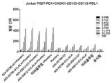
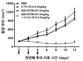
광범위한 연구 끝에, 본 출원의 발명자들은 TIGIT에 대한 높은 결합 친화도를 가지고, TIGIT의 이의 리간드 CD155/CD112에 대한 결합을 효과적으로 차단할 수 있는 TIGIT에 대한 일련의 온전한 인간 항체를 스크리닝하여 얻었고, 이로써 세포로 전달되는 억제 신호를 감소시키거나 제거할 수 있고, 본 발명의 항체의 투여는 동물 모델에서 종양 성장을 현저하게 억제할 수 있다. 이를 기초로, 본 출원은 또한 항체 또는 이의 항원-결합 단편을 포함하는 조성물, 항체 또는 이의 항원-결합 단편을 암호화하는 핵산 분자, 이를 포함하는 숙주 세포, 및 이의 관련 용도도 제공한다.After extensive research, the inventors of the present application have screened and obtained a series of intact human antibodies against TIGIT that have high binding affinity for TIGIT and can effectively block the binding of TIGIT to its ligand CD155/CD112, thereby inoculating the cells. Transmitted inhibitory signals can be reduced or eliminated, and administration of the antibodies of the present invention can significantly inhibit tumor growth in animal models. On this basis, the present application also provides compositions comprising an antibody or antigen-binding fragment thereof, nucleic acid molecules encoding the antibody or antigen-binding fragment thereof, host cells comprising the same, and related uses thereof.
ADI-55796ADI-55796
따라서, 첫 번째 측면에서, 본 출원은 TIGIT에 특이적으로 결합할 수 있는 항체 또는 이의 항원-결합 단편을 제공하며, 상기 항체 또는 이의 항원-결합 단편은 다음을 포함한다:Accordingly, in a first aspect, the present application provides an antibody or antigen-binding fragment thereof capable of specifically binding TIGIT, said antibody or antigen-binding fragment thereof comprising:
서열번호 4에 표시된 중쇄 가변 영역 (VH)에 포함된 VH CDR1 또는 이의 변이체, VH CDR2 또는 이의 변이체 및 VH CDR3 또는 이의 변이체; 및/또는, 서열번호 8에 표시된 경쇄 가변 영역 (VL)에 포함된 VL CDR1 또는 이의 변이체, VL CDR2 또는 이의 변이체, 및 VL CDR3 또는 이의 변이체;VH CDR1 or a variant thereof, VH CDR2 or a variant thereof and VH CDR3 or a variant thereof contained in the heavy chain variable region (VH) shown in SEQ ID NO: 4; And/or, VL CDR1 or a variant thereof, VL CDR2 or a variant thereof, and VL CDR3 or a variant thereof contained in the light chain variable region (VL) shown in SEQ ID NO: 8;
여기서, 상기 변이체는 그것이 유래된 상기 서열과 비교하여 하나 또는 여러 개의 아미노산들의 치환, 결실 또는 첨가 (예를 들어, 1, 2 또는 3개의 아미노산들의 치환 (보존적 치환과 같은), 결실 또는 첨가)를 가진다. 특정 구현예들에서, 상기 치환은 보존적 치환이다.Here, the variant has a substitution, deletion or addition of one or several amino acids (e.g., a substitution (such as a conservative substitution), deletion or addition of 1, 2 or 3 amino acids) compared to the sequence from which it is derived. has In certain embodiments, the substitution is a conservative substitution.
특정 구현예들에서, 상기 항체 또는 이의 항원-결합 단편은 다음을 포함한다: 서열번호 4에 표시된 상기 중쇄 가변 영역 (VH)에 포함된 3개의 CDR들; 및/또는, 서열번호 8에 표시된 상기 경쇄 가변 영역 (VL)에 포함된 3개의 CDR들.In certain embodiments, the antibody or antigen-binding fragment thereof comprises: three CDRs comprised in the heavy chain variable region (VH) shown in SEQ ID NO: 4; and/or the three CDRs contained in the light chain variable region (VL) shown in SEQ ID NO:8.
특정 구현예들에서, 상기 VH에 포함된 3개의 CDR들 및/또는 VL에 포함된 3개의 CDR들은 Kabat, IMGT 또는 Chothia 넘버링 시스템에 의해 정의된다.In certain implementations, the three CDRs contained in the VH and/or the three CDRs contained in the VL are defined by the Kabat, IMGT or Chothia numbering system.
특정 구현예들에서, 상기 항체 또는 이의 항원-결합 단편은 다음을 포함한다:In certain embodiments, the antibody or antigen-binding fragment thereof comprises:
상기 중쇄 가변 영역 (VH)의 다음 3개의 상보성-결정 영역들 (CDRs): 서열번호 1에 표시된 서열을 갖는 VH CDR1, 서열번호 2에 표시된 서열을 갖는 VH CDR2, 및 서열번호 3에 표시된 서열을 갖는 VH CDR3; 및/또는, 상기 경쇄 가변 영역 (VL)의 상기 다음 3개의 상보성-결정 영역들 (CDRs): 서열번호 5에 표시된 서열을 갖는 VL CDR1, 서열번호 6에 표시된 서열을 갖는 VL CDR2, 서열번호 7에 표시된 서열을 갖는 VL CDR3;The following three complementarity-determining regions (CDRs) of the heavy chain variable region (VH): VH CDR1 having the sequence shown in SEQ ID NO: 1, VH CDR2 having the sequence shown in SEQ ID NO: 2, and VH CDR2 having the sequence shown in SEQ ID NO: 3. having VH CDR3; and/or the following three complementarity-determining regions (CDRs) of the light chain variable region (VL): VL CDR1 having the sequence shown in SEQ ID NO: 5, VL CDR2 having the sequence shown in SEQ ID NO: 6, SEQ ID NO: 7 VL CDR3 with the sequence shown in;
여기서, 상기 CDR들은 IMGT 넘버링 시스템에 의해 정의된다.Here, the CDRs are defined by the IMGT numbering system.
특정 구현예들에서, 상기 항체 또는 이의 항원-결합 단편은 인간 면역글로불린의 프레임워크 영역을 추가로 포함한다.In certain embodiments, the antibody or antigen-binding fragment thereof further comprises a framework region of a human immunoglobulin.
특정 구현예들에서, 상기 항체 또는 이의 항원-결합 단편은 인간 항체 생식계열 유전자에 의해 암호화되는 아미노산 서열에 포함된 프레임워크 영역을 포함한다. 특정 구현예들에서, 상기 항체 또는 이의 항원-결합 단편은 다음을 포함한다: 인간 중쇄 생식계열 유전자에 의해 암호화된 아미노산 서열에 포함된 중쇄 프레임워크 영역, 및/또는 인간 경쇄 생식계열 유전자에 의해 암호화된 아미노산 서열에 포함된 경쇄 프레임워크 영역.In certain embodiments, the antibody or antigen-binding fragment thereof comprises a framework region comprised in an amino acid sequence encoded by a human antibody germline gene. In certain embodiments, the antibody or antigen-binding fragment thereof comprises: a heavy chain framework region comprised in an amino acid sequence encoded by a human heavy chain germline gene, and/or encoded by a human light chain germline gene. Light chain framework region included in the amino acid sequence.
특정 구현예들에서, 상기 항체 또는 이의 항원-결합 단편은 다음을 포함한다: 서열번호 4에 표시된 서열을 포함하는 VH 또는 이의 변이체, 및/또는 서열번호 8에 표시된 서열을 포함하는 VL 또는 이의 변이체;In certain embodiments, the antibody or antigen-binding fragment thereof comprises: a VH comprising the sequence shown in SEQ ID NO: 4, or a variant thereof, and/or a VL comprising the sequence shown in SEQ ID NO: 8, or a variant thereof. ;
여기서, 상기 변이체는 하나 또는 여러 개의 아미노산들의 치환, 결실 또는 첨가 (예를 들어, 1, 2, 3, 4 또는 5개의 아미노산들의 치환, 결실 또는 첨가)를 가지고, 또는 그것이 유래된 상기 서열과 비교하여, 적어도 80%, 적어도 85%, 적어도 90%, 적어도 91%, 적어도 92%, 적어도 93%, 적어도 94%, 적어도 95%, 적어도 96%, 적어도 97%, 적어도 98%, 적어도 99% 또는 100%의 서열 상동성을 가진다. 특정 구현예들에서, 상기 치환은 보존적 치환이다.wherein the variant has a substitution, deletion or addition of one or several amino acids (e.g., a substitution, deletion or addition of 1, 2, 3, 4 or 5 amino acids), or is compared to the sequence from which it is derived. So, at least 80%, at least 85%, at least 90%, at least 91%, at least 92%, at least 93%, at least 94%, at least 95%, at least 96%, at least 97%, at least 98%, at least 99%, or It has 100% sequence homology. In certain embodiments, the substitution is a conservative substitution.
특정 구현예들에서, 상기 항체 또는 이의 항원-결합 단편은 서열번호 4에 표시된 VH, 및/또는 서열번호 8에 표시된 VL을 포함한다.In certain embodiments, the antibody or antigen-binding fragment thereof comprises the VH shown in SEQ ID NO: 4, and/or the VL shown in SEQ ID NO: 8.
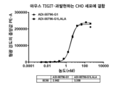
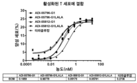
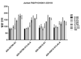
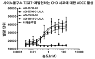
특정 구현예들에서, 상기 항체 또는 이의 항원-결합 단편은 인간 TIGIT에 특이적으로 결합할 수 있고 및/또는 인간 TIGIT의 이의 리간드에 대한 결합을 차단할 수 있으며, 예를 들어, 상기 결합 활성 및/또는 차단 활성이 티라골루맙에 비해 우수하다.In certain embodiments, the antibody or antigen-binding fragment thereof is capable of specifically binding to human TIGIT and/or blocking binding of human TIGIT to its ligand, e.g., by modifying the binding activity and/or Alternatively, the blocking activity is superior to tiragolumab.
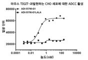
특정 구현예들에서, 상기 항체 또는 이의 항원-결합 단편은 마우스 TIGIT에 특이적으로 결합할 수 있고 및/또는 마우스 TIGIT의 이의 리간드에 대한 결합을 차단할 수 있다.In certain embodiments, the antibody or antigen-binding fragment thereof is capable of specifically binding mouse TIGIT and/or blocking binding of mouse TIGIT to its ligand.
특정 바람직한 구현예들에서, 상기 항체 또는 이의 항원-결합 단편은 인간, 사이노몰구스 및/또는 마우스 TIGIT와 교차-반응성을 가지며, 예를 들어, 인간, 사이노몰구스 및 마우스 TIGIT와 교차-반응성을 가진다.In certain preferred embodiments, the antibody or antigen-binding fragment thereof has cross-reactivity with human, cynomolgus and/or mouse TIGIT, e.g., cross-reactivity with human, cynomolgus and mouse TIGIT. have
ADI-55812ADI-55812
두 번째 측면에서, 본 출원은 TIGIT에 특이적으로 결합할 수 있는 항체 또는 이의 항원-결합 단편을 제공하고, 상기 항체 또는 이의 항원-결합 단편은 다음을 포함한다:In a second aspect, the present application provides an antibody or antigen-binding fragment thereof capable of specifically binding to TIGIT, wherein the antibody or antigen-binding fragment thereof comprises:
서열번호 12에 표시된 중쇄 가변 영역 (VH)에 포함된 VH CDR1 또는 이의 변이체, VH CDR2 또는 이의 변이체, 및 VH CDR3 또는 이의 변이체; 및/또는 서열번호 16에 표시된 경쇄 가변 영역(VL)에 포함된 VL CDR1 또는 이의 변이체, VL CDR2 또는 이의 변이체, 및 VL CDR3 또는 이의 변이체;VH CDR1 or a variant thereof, VH CDR2 or a variant thereof, and VH CDR3 or a variant thereof contained in the heavy chain variable region (VH) shown in SEQ ID NO: 12; and/or VL CDR1 or a variant thereof, VL CDR2 or a variant thereof, and VL CDR3 or a variant thereof contained in the light chain variable region (VL) shown in SEQ ID NO: 16;
여기서, 상기 변이체는 그것이 유래된 상기 서열과 비교하여 하나 또는 여러 개의 아미노산의 치환, 결실 또는 첨가 (예를 들어, 1, 2 또는 3개의 아미노산들의 치환 (보존적 치환과 같은), 결실 또는 첨가)를 갖는다. 특정 구현예들에서, 상기 치환은 보존적 치환이다.Here, the variant has a substitution, deletion or addition of one or several amino acids (e.g., a substitution (such as a conservative substitution), deletion or addition of 1, 2 or 3 amino acids) compared to the sequence from which it is derived. has In certain embodiments, the substitution is a conservative substitution.
특정 구현예들에서, 상기 항체 또는 이의 항원-결합 단편은 다음을 포함한다: 서열번호 12에 표시된 상기 중쇄 가변 영역 (VH)에 포함된 3개의 CDR들; 및/또는, 서열번호 16에 표시된 상기 경쇄 가변 영역 (VL)에 포함된 3개의 CDR들.In certain embodiments, the antibody or antigen-binding fragment thereof comprises: three CDRs comprised in the heavy chain variable region (VH) set forth in SEQ ID NO: 12; and/or the three CDRs contained in the light chain variable region (VL) shown in SEQ ID NO: 16.
특정 구현예들에서, 상기 VH에 포함된 상기 3개의 CDR들 및/또는 상기 VL에 포함된 상기 3개의 CDR들은 Kabat, IMGT 또는 Chothia 넘버링 시스템에 의해 정의된다.In certain implementations, the three CDRs included in the VH and/or the three CDRs included in the VL are defined by the Kabat, IMGT or Chothia numbering system.
특정 구현예들에서, 상기 항체 또는 이의 항원-결합 단편은 다음을 포함한다:In certain embodiments, the antibody or antigen-binding fragment thereof comprises:
상기 중쇄 가변 영역 (VH)의 상기 다음 3개의 상보성-결정 영역들 (CDRs): 서열번호 9에 표시된 서열을 갖는 VH CDR1, 서열번호 10에 표시된 서열을 갖는 VH CDR2, 및 서열번호 11에 표시된 서열을 갖는 VH CDR3; 및/또는, 상기 경쇄 가변 영역 (VL)의 상기 다음 3개의 상보성-결정 영역들 (CDRs): 서열번호 13에 표시된 서열을 갖는 VL CDR1, 서열번호 14에 표시된 서열을 갖는 VL CDR2, 서열번호 15에 표시된 서열을 갖는 VL CDR3;The following three complementarity-determining regions (CDRs) of the heavy chain variable region (VH): VH CDR1 having the sequence shown in SEQ ID NO: 9, VH CDR2 having the sequence shown in SEQ ID NO: 10, and sequence shown in SEQ ID NO: 11 VH CDR3 with; and/or the following three complementarity-determining regions (CDRs) of the light chain variable region (VL): VL CDR1 having the sequence shown in SEQ ID NO: 13, VL CDR2 having the sequence shown in SEQ ID NO: 14, SEQ ID NO: 15 VL CDR3 with the sequence shown in;
여기서, 상기 CDR들은 IMGT 넘버링 시스템에 의해 정의된다.Here, the CDRs are defined by the IMGT numbering system.
특정 구현예들에서, 상기 항체 또는 이의 항원-결합 단편은 인간 면역글로불린의 프레임워크 영역을 추가로 포함한다.In certain embodiments, the antibody or antigen-binding fragment thereof further comprises a framework region of a human immunoglobulin.
특정 구현예들에서, 상기 항체 또는 이의 항원-결합 단편은 인간 항체 생식계열 유전자에 의해 암호화되는 아미노산 서열에 포함된 프레임워크 영역을 포함한다. 특정 구현예들에서, 상기 항체 또는 이의 항원-결합 단편은 다음을 포함한다: 인간 중쇄 생식계열 유전자에 의해 암호화되는 아미노산 서열에 포함된 중쇄 프레임워크 영역, 및/또는 인간 경쇄 생식계열 유전자에 의해 암호화되는 아미노산 서열에 포함된 경쇄 프레임워크 영역.In certain embodiments, the antibody or antigen-binding fragment thereof comprises a framework region comprised in an amino acid sequence encoded by a human antibody germline gene. In certain embodiments, the antibody or antigen-binding fragment thereof comprises: a heavy chain framework region comprised in an amino acid sequence encoded by a human heavy chain germline gene, and/or encoded by a human light chain germline gene. The light chain framework region included in the amino acid sequence.
특정 구현예들에서, 상기 항체 또는 이의 항원-결합 단편은 다음을 포함한다: 서열번호 12에 표시된 서열을 갖는 VH 또는 이의 변이체, 및/또는 서열번호 16에 표시된 서열을 갖는 VL 또는 이의 변이체;In certain embodiments, the antibody or antigen-binding fragment thereof comprises: a VH having the sequence shown in SEQ ID NO: 12, or a variant thereof, and/or a VL having the sequence shown in SEQ ID NO: 16, or a variant thereof;
여기서, 상기 변이체는 하나 또는 여러 개의 아미노산들의 치환, 결실 또는 첨가 (예를 들어, 1, 2, 3, 4 또는 5개의 아미노산들의 치환, 결실 또는 첨가)를 가지고, 또는 그것이 유래된 상기 서열과 비교하여, 적어도 80%, 적어도 85%, 적어도 90%, 적어도 91%, 적어도 92%, 적어도 93%, 적어도 94%, 적어도 95%, 적어도 96%, 적어도 97%, 적어도 98%, 적어도 99% 또는 100%의 서열 상동성을 가진다. 특정 구현예들에서, 상기 치환은 보존적 치환이다.wherein the variant has a substitution, deletion or addition of one or several amino acids (e.g., a substitution, deletion or addition of 1, 2, 3, 4 or 5 amino acids), or is compared to the sequence from which it is derived. So, at least 80%, at least 85%, at least 90%, at least 91%, at least 92%, at least 93%, at least 94%, at least 95%, at least 96%, at least 97%, at least 98%, at least 99%, or It has 100% sequence homology. In certain embodiments, the substitution is a conservative substitution.
특정 구현예들에서, 상기 항체 또는 이의 항원-결합 단편은 서열번호 12에 표시된 VH, 및/또는 서열번호 16에 표시된 VL을 포함한다.In certain embodiments, the antibody or antigen-binding fragment thereof comprises the VH shown in SEQ ID NO: 12, and/or the VL shown in SEQ ID NO: 16.
특정 구현예들에서, 상기 항체 또는 이의 항원-결합 단편은 인간 TIGIT에 특이적으로 결합할 수 있고 및/또는 인간 TIGIT의 이의 리간드에 대한 결합을 차단할 수 있으며, 예를 들어, 상기 결합 활성 및/또는 차단 활성이 티라골루맙에 비해 우수하다.In certain embodiments, the antibody or antigen-binding fragment thereof is capable of specifically binding to human TIGIT and/or blocking binding of human TIGIT to its ligand, e.g., by modifying the binding activity and/or Alternatively, the blocking activity is superior to tiragolumab.
특정 바람직한 구현예들에서, 상기 항체 또는 이의 항원-결합 단편은 인간 및 사이노몰구스 TIGIT와 교차-반응성을 가진다.In certain preferred embodiments, the antibody or antigen-binding fragment thereof has cross-reactivity with human and cynomolgus TIGIT.
특정 구현예들에서, 본 발명의 첫 번째 또는 두 번째 측면의 상기 항체 또는 이의 항원-결합 단편은 인간 면역글로불린으로부터 유래된 불변 영역을 추가로 포함한다.In certain embodiments, the antibody or antigen-binding fragment thereof of the first or second aspect of the invention further comprises a constant region derived from a human immunoglobulin.
특정 구현예들에서, 상기 항체 또는 이의 항원-결합 단편의 중쇄는 인간 면역글로불린 또는 이의 변이체의 중쇄 불변 영역 (CH)을 포함하고, 상기 변이체는 그것이 유래된 상기 서열과 비교하여, 하나 또는 여러 아미노산들의 치환, 결실 또는 첨가 (예를 들어, 최대 20개, 최대 15개, 최대 10개 또는 최대 5개 아미노산들의 치환, 결실 또는 첨가; 예를 들어, 1, 2, 3, 4 또는 5개 아미노산들의 치환, 결실 또는 첨가)를 가지고; 및/또는,In certain embodiments, the heavy chain of the antibody or antigen-binding fragment thereof comprises the heavy chain constant region (CH) of a human immunoglobulin or a variant thereof, wherein the variant has one or more amino acids compared to the sequence from which it is derived. Substitution, deletion or addition of (e.g., substitution, deletion or addition of up to 20, up to 15, up to 10 or up to 5 amino acids; e.g., of 1, 2, 3, 4 or 5 amino acids) substitution, deletion or addition); and/or,
상기 항체 또는 이의 항원-결합 단편의 경쇄는 인간 면역글로불린 또는 이의 변이체의 경쇄 불변 영역 (CL)을 포함하고, 상기 변이체는 그것이 유래된 상기 서열과 비교하여 최대 20개 아미노산들의 보존적 치환 (예를 들어, 최대 15개, 최대 10개 또는 최대 5개의 아미노산; 1, 2, 3, 4 또는 5개의 아미노산들의 보존적 치환과 같은)을 가진다.The light chain of the antibody or antigen-binding fragment thereof comprises the light chain constant region (CL) of a human immunoglobulin or a variant thereof, wherein the variant has conservative substitutions of up to 20 amino acids compared to the sequence from which it is derived (e.g. For example, up to 15, up to 10, or up to 5 amino acids; such as conservative substitutions of 1, 2, 3, 4, or 5 amino acids).
일부 구현예들에서, 상기 중쇄 불변 영역 (CH)의 변이체는 그것이 유래된 서열과 비교하여 하나 이상의 아미노산의 보존적 치환을 가질 수 있다. 이러한 구현예들에서, 상기 중쇄 불변 영역 (CH)의 변이체는 그것이 유래된 야생-형 서열과 비교하여 동일하거나 실질적으로 동일한 이펙터 기능을 가질 수 있다.In some embodiments, the variant of the heavy chain constant region (CH) may have a conservative substitution of one or more amino acids compared to the sequence from which it is derived. In these embodiments, the variant of the heavy chain constant region (CH) may have identical or substantially identical effector function compared to the wild-type sequence from which it is derived.
다른 구현예들에서, 상기 중쇄 불변 영역 (CH)의 변이체는 본 발명의 상기 항체의 다음 특성 중 하나 이상을 변경하는 하나 이상의 아미노산 돌연변이 또는 화학적 변형을 포함할 수 있다: Fc 수용체 결합, 항체 글리코실화, 시스테인 잔기의 수, 이펙터 세포 기능, 또는 보체 기능, 등. 이펙터 기능은 상기 항체의 불변 영역에 있는 하나 이상의 아미노산 잔기를 다른 잔기로 교체하거나 또는 화학적 변형을 통해 기능적 변화를 생성함으로써 변경될 수 있고, 예를 들어, 이펙터 리간드 (예를 들어, FcR 또는 보체 C1q)에 대한 항체의 친화도를 변경하면 이펙터 기능이 변경 (예를 들어, 감소 또는 강화)될 수 있다. 상기 항체의 Fc 영역은 ADCC, 식세포작용, CDC 등과 같은 몇 가지 중요한 이펙터 기능을 매개한다.In other embodiments, the variant of the heavy chain constant region (CH) may comprise one or more amino acid mutations or chemical modifications that alter one or more of the following properties of the antibody of the invention: Fc receptor binding, antibody glycosylation , number of cysteine residues, effector cell function, or complement function, etc. Effector function can be altered by replacing one or more amino acid residues in the constant region of the antibody with other residues or by creating a functional change through chemical modification, for example, by producing a functional change in an effector ligand (e.g., FcR or complement C1q). ) can alter (e.g., reduce or enhance) the effector function. The Fc region of the antibody mediates several important effector functions such as ADCC, phagocytosis, CDC, etc.
특정 구현예들에서, 상기 항체 또는 이의 항원-결합 단편은 ADCC 활성을 보유한다. 특정 구현예들에서, 상기 항체 또는 이의 항원-결합 단편은 돌연변이 (예를 들어, 아미노산 치환) 또는 화학적으로 변형된 Fc 영역을 포함하고, 여기서 상기 돌연변이 또는 화학적으로 변형된 Fc 영역은 향상된 ADCC 활성을 제공한다. 특정 구현예들에서, 상기 항체 또는 이의 항원-결합 단편은 푸코실화 활성이 낮거나 전혀 없는 숙주 세포에서 발현에 의해 생산되고, 생산된 상기 항체는 ADCC 활성을 강화시키는 푸코실화가 낮거나 전혀 없다. 이러한 숙주 세포들은 푸코실트랜스퍼라제 (예를 들어, FUT8)를 암호화하는 유전자가 결여된 CHO 세포와 같은 포유동물 세포일 수 있다.In certain embodiments, the antibody or antigen-binding fragment thereof possesses ADCC activity. In certain embodiments, the antibody or antigen-binding fragment thereof comprises a mutated (e.g., amino acid substitution) or chemically modified Fc region, wherein the mutated or chemically modified Fc region exhibits enhanced ADCC activity. to provide. In certain embodiments, the antibody or antigen-binding fragment thereof is produced by expression in a host cell with low or no fucosylation activity, and the antibody produced has low or no fucosylation that enhances ADCC activity. These host cells may be mammalian cells, such as CHO cells, lacking the gene encoding a fucosyltransferase (eg, FUT8).
특정 구현예들에서, 상기 중쇄 불변 영역은 IgG 중쇄 불변 영역, 예컨대 IgG1, IgG2, IgG3, 또는 IgG4 중쇄 불변 영역, 예컨대 IgG1 중쇄 불변 영역이다. 특정 구현예들에서, 상기 항체 또는 이의 항원-결합 단편은 IgG1, IgG2, IgG3, 또는 IgG4와 같은 IgG로부터 선택된다. 특정 예시적 구현예들에서, 상기 항체 또는 이의 항원-결합 단편의 중쇄는 서열번호 17 또는 서열번호 18에 표시된 중쇄 불변 영역을 포함한다.In certain embodiments, the heavy chain constant region is an IgG heavy chain constant region, such as an IgG1, IgG2, IgG3, or IgG4 heavy chain constant region, such as an IgG1 heavy chain constant region. In certain embodiments, the antibody or antigen-binding fragment thereof is selected from IgG, such as IgG1, IgG2, IgG3, or IgG4. In certain exemplary embodiments, the heavy chain of the antibody or antigen-binding fragment thereof comprises a heavy chain constant region shown in SEQ ID NO: 17 or SEQ ID NO: 18.
특정 구현예들에서, 상기 항체 또는 이의 항원-결합 단편의 상기 경쇄는 인간 면역글로불린으로부터 유래된 경쇄 영역 (예를 들어, κ 또는 λ 쇄의 불변 영역)을 포함한다. 특정 예시적 구현예들에서, 상기 항체 또는 이의 항원-결합 단편의 경쇄는 서열번호 22에 표시된 경쇄 불변 영역을 포함한다.In certain embodiments, the light chain of the antibody or antigen-binding fragment thereof comprises a light chain region derived from a human immunoglobulin (e.g., the constant region of a κ or λ chain). In certain exemplary embodiments, the light chain of the antibody or antigen-binding fragment thereof comprises the light chain constant region set forth in SEQ ID NO: 22.
특정 구현예들에서, 상기 항원-결합 단편은 Fab, Fab', (Fab')2, Fv, 디설파이드-연결된 Fv, scFv, 디아바디, 및 단일 도메인 항체 (sdAb)로 이루어진 그룹에서 선택되고; 및/또는, 상기 항체는 키메릭 항체, 이중특이성 항체 또는 다중특이성 항체이다.In certain embodiments, the antigen-binding fragment is selected from the group consisting of Fab, Fab', (Fab') 2 , Fv, disulfide-linked Fv, scFv, diabody, and single domain antibody (sdAb); And/or, the antibody is a chimeric antibody, bispecific antibody, or multispecific antibody.
본 발명의 항체는 유전공학 및 재조합 기술과 같은 당업계에 공지된 다양한 방법에 의해 제조될 수 있다. 예를 들어, 본 발명의 항체의 중쇄 및 경쇄를 암호화하는 DNA 분자는 화학적 합성 또는 PCR 증폭에 의해 얻어진다. 생성된 DNA 분자는 발현 벡터에 삽입된 다음 숙주 세포에 형질감염된다. 이어서, 상기 형질감염된 숙주 세포들은 본 발명의 항체를 발현시키기 위하여 특정 조건 하에서 배양된다.The antibodies of the present invention can be produced by various methods known in the art, such as genetic engineering and recombinant technology. For example, DNA molecules encoding the heavy and light chains of the antibodies of the invention are obtained by chemical synthesis or PCR amplification. The resulting DNA molecule is inserted into an expression vector and then transfected into host cells. The transfected host cells are then cultured under specific conditions to express the antibodies of the invention.
본 발명의 항원-결합 단편들은 온전한 항체 분자를 가수분해함으로써 얻을 수 있다 (Morimoto et al., J. Biochem. Biophys. Methods 24:107-117 (1992) 및 Brennan et al., Science 229:81 (1985) 참조). 게다가, 이들 항원-결합 단편들은 또한 재조합 숙주 세포들로부터 직접 생산될 수도 있다 (Hudson, Curr. Opin. Immunol. 11: 548-557 (1999); Little et al., Immunol. Today, 21: 364-370 (2000)에서 검토됨). 예를 들어, Fab' 단편들은 숙주 세포들로부터 직접 얻을 수 있고; Fab' 단편들은 화학적으로 커플링되어 F(ab')2 단편들을 형성할 수 있다 (Carter et al., Bio/Technology, 10: 163-167 (1992)). 또한, Fv, Fab 또는 F(ab')2 단편들은 재조합 숙주 세포의 세포 배양물로부터 직접 분리될 수도 있다. 당업자들은 이러한 항원-결합 단편들을 제조하기 위한 다른 기술을 잘 알고 있다.Antigen-binding fragments of the invention can be obtained by hydrolyzing intact antibody molecules (Morimoto et al., J. Biochem. Biophys. Methods 24:107-117 (1992) and Brennan et al., Science 229:81 ( 1985). In addition, these antigen-binding fragments can also be produced directly from recombinant host cells (Hudson, Curr. Opin. Immunol. 11: 548-557 (1999); Little et al., Immunol. Today, 21: 364- 370 (reviewed in 2000)). For example, Fab' fragments can be obtained directly from host cells; Fab' fragments can be chemically coupled to form F(ab') 2 fragments (Carter et al., Bio/Technology, 10: 163-167 (1992)). Additionally, Fv, Fab or F(ab') 2 fragments can also be isolated directly from cell culture of recombinant host cells. Those skilled in the art are familiar with other techniques for preparing such antigen-binding fragments.
핵산 분자들nucleic acid molecules
또 다른 측면에서, 본 출원은 상기 기재된 바와 같은 항체 또는 이의 항원-결합 단편, 또는 이의 중쇄 가변 영역 및/또는 경쇄 가변 영역을 암호화하는 단리된 핵산 분자를 제공한다.In another aspect, the present application provides an isolated nucleic acid molecule encoding an antibody or antigen-binding fragment thereof as described above, or a heavy chain variable region and/or a light chain variable region thereof.
특정 구현예들에서, 상기 단리된 핵산 분자는 본 발명의 항체 또는 이의 항원-결합 단편의 중쇄 또는 중쇄 가변 영역을 암호화하는 제1 뉴클레오티드 서열 및 항체 또는 이의 항원-결합 단편의 경쇄 또는 경쇄 가변 영역을 암호화하는 제2 뉴클레오티드 서열을 포함하고, 여기서 제1 뉴클레오티드 서열 및 제2 뉴클레오티드 서열은 동일하거나 상이한 단리된 핵산 분자 상에 존재한다. 제1 뉴클레오티드 서열 및 제2 뉴클레오티드 서열이 상이한 단리된 핵산 분자 상에 존재하는 경우, 본 발명의 단리된 핵산 분자는 제1 뉴클레오티드 서열을 포함하는 제1 핵산 분자 및 제2 뉴클레오티드 서열을 포함하는 제2 핵산 분자를 포함한다.In certain embodiments, the isolated nucleic acid molecule comprises a first nucleotide sequence encoding the heavy chain or heavy chain variable region of an antibody or antigen-binding fragment thereof of the invention and a light chain or light chain variable region of the antibody or antigen-binding fragment thereof. and a second nucleotide sequence that encodes, wherein the first nucleotide sequence and the second nucleotide sequence are on the same or different isolated nucleic acid molecule. When the first nucleotide sequence and the second nucleotide sequence are on different isolated nucleic acid molecules, the isolated nucleic acid molecule of the invention includes a first nucleic acid molecule comprising the first nucleotide sequence and a second nucleic acid molecule comprising the second nucleotide sequence. Contains nucleic acid molecules.
벡터들vectors
또 다른 측면에서, 본 출원은 상기 기재된 바와 같은 핵산 분자를 포함하는 벡터를 제공한다. 특정 구현예들에서, 상기 벡터는 클로닝 벡터 또는 발현 벡터이다.In another aspect, the present application provides a vector comprising a nucleic acid molecule as described above. In certain embodiments, the vector is a cloning vector or an expression vector.
특정 구현예들에서, 상기 벡터는 본 발명의 항체 또는 이의 항원-결합 단편의 중쇄 또는 중쇄 가변 영역을 암호화하는 제1 뉴클레오티드 서열 및 항체 또는 이의 항원-결합 단편의 경쇄 또는 경쇄 가변 영역을 암호화하는 제2 뉴클레오티드 서열을 포함하고, 여기서 제1 뉴클레오티드 서열 및 제2 뉴클레오티드 서열은 동일하거나 상이한 벡터 상에 존재한다. 제1 뉴클레오티드 서열과 제2 뉴클레오티드 서열이 상이한 벡터 상에 존재하는 경우, 본 발명의 벡터는 제1 뉴클레오티드 서열을 포함하는 제1 벡터 및 제2 뉴클레오티드 서열을 포함하는 제2 벡터를 포함한다.In certain embodiments, the vector comprises a first nucleotide sequence encoding the heavy chain or heavy chain variable region of an antibody or antigen-binding fragment thereof of the invention and a second nucleotide sequence encoding the light chain or light chain variable region of the antibody or antigen-binding fragment thereof. It comprises a two nucleotide sequence, wherein the first nucleotide sequence and the second nucleotide sequence are on the same or different vector. When the first nucleotide sequence and the second nucleotide sequence are on different vectors, the vector of the invention includes a first vector comprising the first nucleotide sequence and a second vector comprising the second nucleotide sequence.
숙주 세포들host cells
또 다른 측면에서, 본 출원은 상기 기재된 바와 같은 핵산 분자 또는 벡터를 포함하는 숙주 세포를 제공한다. 이러한 숙주 세포에는 박테리아 세포 (예를 들어, E. coli 세포)와 같은 원핵 세포, 및 진균 세포 (예를 들어, 효모 세포), 곤충 세포, 식물 세포, 및 동물 세포 (예를 들어, 포유동물 세포, 예를 들어, 마우스 세포, 인간 세포, 등)와 같은 진핵 세포가 포함되나, 이에 한정되는 것은 아니다.In another aspect, the present application provides a host cell comprising a nucleic acid molecule or vector as described above. These host cells include prokaryotic cells, such as bacterial cells (e.g., E. coli cells), and fungal cells (e.g., yeast cells), insect cells, plant cells, and animal cells (e.g., mammalian cells). , e.g., mouse cells, human cells, etc.).
특정 구현예들에서, 상기 숙주 세포는 푸코실화 활성이 낮거나 전혀 없다. 특정 구현예들에서, 상기 숙주 세포는 푸코실트랜스퍼라제 (예를 들어, FUT8)를 암호화하는 유전자가 결여된 CHO 세포와 같은 포유동물 세포일 수 있다.In certain embodiments, the host cell has low or no fucosylation activity. In certain embodiments, the host cell can be a mammalian cell, such as a CHO cell, lacking the gene encoding a fucosyltransferase (e.g., FUT8).
제조 방법Manufacturing method
또 다른 측면에서, 본 출원은 상기 기재된 바와 같은 항체 또는 이의 항원-결합 단편을 제조하는 방법을 제공하고, 이는 항체 또는 이의 항원-결합 단편의 발현을 허용하는 조건 하에서 상기 기재된 바와 같은 숙주 세포를 배양하는 단계, 및 상기 숙주 세포의 배양물로부터 상기 항체 또는 이의 항원-결합 단편을 회수하는 단계를 포함한다.In another aspect, the present application provides a method of making an antibody or antigen-binding fragment thereof as described above, comprising culturing a host cell as described above under conditions permitting expression of the antibody or antigen-binding fragment thereof. and recovering the antibody or antigen-binding fragment thereof from a culture of the host cells.
특정 구현예들에서, 상기 숙주 세포는 푸코실화 활성이 낮거나 전혀 없으므로, 푸코실화가 낮거나 전혀 없는 항체를 생산하고, 이는 이로부터 생산된 항체의 ADCC 활성을 강화시킨다. 특정 구현예들에서, 상기 숙주 세포는 푸코실트랜스퍼라제 (예를 들어, FUT8)를 암호화하는 유전자가 결여된 CHO 세포와 같은 포유동물 세포일 수 있다.In certain embodiments, the host cell has low or no fucosylation activity, thereby producing an antibody with low or no fucosylation, which enhances the ADCC activity of the antibody produced therefrom. In certain embodiments, the host cell can be a mammalian cell, such as a CHO cell, lacking the gene encoding a fucosyltransferase (e.g., FUT8).
약학 조성물pharmaceutical composition
또 다른 측면에서, 본 출원은 상기 기재된 바와 같은 항체 또는 이의 항원-결합 단편, 단리된 핵산 분자, 상기 기재된 바와 같은 벡터 또는 숙주 세포, 및 약제학적으로 허용되는 담체 및/또는 부형제를 포함하는 약학 조성물을 제공한다.In another aspect, the present application relates to a pharmaceutical composition comprising an antibody or antigen-binding fragment thereof as described above, an isolated nucleic acid molecule, a vector or host cell as described above, and a pharmaceutically acceptable carrier and/or excipient. provides.
특정 구현예들에서, 상기 약학 조성물은 추가적인 면역 관문 억제제를 추가로 포함한다.In certain embodiments, the pharmaceutical composition further comprises an additional immune checkpoint inhibitor.
특정 구현예들에서, 상기 약학 조성물은 항-PD-1 항체 또는 항-PD-L1 항체를 추가로 포함한다.In certain embodiments, the pharmaceutical composition further comprises an anti-PD-1 antibody or an anti-PD-L1 antibody.
특정 예시적 구현예들에서, 상기 약제학적으로 허용되는 담체 및/또는 부형제는 멸균 주사용 액체 (예를 들어, 수성 또는 비수성 현탁액 또는 용액)를 포함한다. 특정 예시적 구현예들에서, 이러한 멸균 주사용 액체는 주사용수 (WFI), 정균 주사용수 (BWFI), 염화 나트륨 용액 (예를 들어, 0.9% (w/v) NaCl), 덱스트로스 용액 (예를 들어, 5% 글루코스), 계면활성제-함유 용액 (예를 들어, 0.01% 폴리소르베이트 20을 함유한 용액), pH 완충 용액 (예를 들어, 인산염 완충 용액), 링거 용액 및 이들의 조합으로 이루어진 그룹에서 선택된다.In certain exemplary embodiments, the pharmaceutically acceptable carrier and/or excipient comprises a sterile injectable liquid (e.g., an aqueous or non-aqueous suspension or solution). In certain exemplary embodiments, such sterile injectable liquids include water for injection (WFI), bacteriostatic water for injection (BWFI), sodium chloride solution (e.g., 0.9% (w/v) NaCl), dextrose solution (e.g. (e.g., 5% glucose), surfactant-containing solutions (e.g., solutions containing 0.01% polysorbate 20), pH buffered solutions (e.g., phosphate buffered solutions), Ringer's solutions, and combinations thereof. are selected from a group consisting of
용도Usage
또 다른 측면에서, 본 출원은 약제의 제조에 있어서 상기 기재된 바와 같은 항체 또는 이의 항원-결합 단편, 상기 단리된 핵산 분자, 상기 벡터, 상기 숙주 세포 또는 상기 약학 조성물의 용도를 제공하고, 상기 약제는 다음의 용도로 사용된다:In another aspect, the present application provides the use of an antibody or antigen-binding fragment thereof, said isolated nucleic acid molecule, said vector, said host cell or said pharmaceutical composition as described above in the manufacture of a medicament, wherein said medicament It is used for the following purposes:
(1) 시험관내 또는 대상체 (예를 들어, 인간)에서 면역 세포 활성을 증가시키거나;(1) increase immune cell activity in vitro or in a subject (e.g., a human);
(2) 대상체 (예를 들어, 인간)에서 면역 반응을 강화시키거나;(2) enhance the immune response in a subject (e.g., a human);
(3) 대상체 (예를 들어, 인간)에서 종양을 예방 및/또는 치료하거나; 또는(3) prevent and/or treat a tumor in a subject (e.g., a human); or
(4) 대상체 (예를 들어, 인간)에서 감염을 예방 및/치료하기 위한 상기 약제의 용도.(4) Use of the agent to prevent and/or treat infection in a subject (e.g., a human).
특정 구현예들에서, 상기 면역 세포는 T 세포 및/또는 NK 세포이다.In certain embodiments, the immune cells are T cells and/or NK cells.
특정 구현예들에서, 상기 면역 반응은 T 세포- 또는 NK 세포-매개 면역 반응이다.In certain embodiments, the immune response is a T cell- or NK cell-mediated immune response.
특정 구현예들에서, 상기 종양은 TIGIT-양성 침윤 T 세포 및/또는 NK 세포를 포함하고 및/또는 TIGIT 리간드 (예를 들어, CD155 및/또는 CD112)-양성 종양 세포를 포함한다.In certain embodiments, the tumor comprises TIGIT-positive infiltrating T cells and/or NK cells and/or TIGIT ligand (e.g., CD155 and/or CD112)-positive tumor cells.
특정 구현예들에서, 상기 종양은 고형 종양 또는 혈액학적 종양 (예를 들어, 백혈병, 림프종, 골수종)으로부터 선택된다.In certain embodiments, the tumor is selected from a solid tumor or a hematological tumor (e.g., leukemia, lymphoma, myeloma).
특정 구현예들에서, 상기 종양은 대장암, 결장암, 방광암, 유방암, 자궁내막암/자궁경부암, 난소암, 전립선암, 고환암, 식도암, 소화기암, 췌장암, 신장암, 두경부암, 폐암, 위암, 생식세포암, 뼈암, 간암, 갑상선암, 피부암, 중추신경계 종양, 림프종, 백혈병, 골수종, 육종 및 흑색종으로 이루어진 그룹에서 선택된다.In certain embodiments, the tumor is colon cancer, colon cancer, bladder cancer, breast cancer, endometrial cancer/cervical cancer, ovarian cancer, prostate cancer, testicular cancer, esophageal cancer, digestive cancer, pancreatic cancer, kidney cancer, head and neck cancer, lung cancer, stomach cancer, It is selected from the group consisting of germ cell cancer, bone cancer, liver cancer, thyroid cancer, skin cancer, central nervous system tumor, lymphoma, leukemia, myeloma, sarcoma, and melanoma.
특정 구현예들에서, 상기 감염은 바이러스 감염, 박테리아 감염, 진균 감염 및 기생충 감염으로 이루어진 그룹에서 선택된다.In certain embodiments, the infection is selected from the group consisting of viral infection, bacterial infection, fungal infection, and parasitic infection.
특정 구현예들에서, 상기 대상체는 인간, 사이노몰구스 원숭이, 또는 마우스와 같은, 포유동물이다.In certain embodiments, the subject is a mammal, such as a human, cynomolgus monkey, or mouse.
특정 구현예들에서, 상기 항체 또는 이의 항원-결합 단편, 상기 단리된 핵산 분자, 상기 벡터, 상기 숙주 세포, 또는 상기 약학 조성물은 단독으로 또는 추가적인 약제학적 활성제와 조합으로 사용된다. 특정 구현예들에서, 상기 추가적인 약제학적 활성제는 추가적인 면역 관문 억제제이다.In certain embodiments, the antibody or antigen-binding fragment thereof, the isolated nucleic acid molecule, the vector, the host cell, or the pharmaceutical composition are used alone or in combination with an additional pharmaceutically active agent. In certain embodiments, the additional pharmaceutically active agent is an additional immune checkpoint inhibitor.
특정 구현예들에서, 상기 항체 또는 이의 항원-결합 단편, 상기 단리된 핵산 분자, 상기 벡터, 상기 숙주 세포 또는 상기 약학 조성물은 항-PD-1 항체 또는 항-PD-L1 항체와 조합으로 사용된다.In certain embodiments, the antibody or antigen-binding fragment thereof, the isolated nucleic acid molecule, the vector, the host cell, or the pharmaceutical composition are used in combination with an anti-PD-1 antibody or an anti-PD-L1 antibody. .
또 다른 측면에서, 본 출원은 대상체에서 면역 반응을 강화시키거나 종양 또는 감염을 예방 및/또는 치료하기 위한 방법을 제공하고, 이는 다음을 포함한다: 상기 기재된 바와 같은 항체 또는 이의 항원-결합 단편 또는 약학 조성물의 유효량을 이를 필요로 하는 대상체에게 투여하는 단계.In another aspect, the present application provides a method for enhancing an immune response or preventing and/or treating a tumor or infection in a subject, comprising: an antibody or antigen-binding fragment thereof as described above, or Administering an effective amount of the pharmaceutical composition to a subject in need thereof.
특정 구현예들에서, 상기 종양은 TIGIT-양성 침윤 T 세포 및/또는 NK 세포를 포함하고 및/또는, TIGIT 리간드 (예를 들어, CD155 및/또는 CD112)-양성 종양 세포를 포함한다.In certain embodiments, the tumor comprises TIGIT-positive infiltrating T cells and/or NK cells and/or comprises TIGIT ligand (e.g., CD155 and/or CD112)-positive tumor cells.
특정 구현예들에서, 상기 종양은 고형 종양 또는 혈액학적 종양 (예를 들어, 백혈병, 림프종, 골수종)으로부터 선택된다.In certain embodiments, the tumor is selected from a solid tumor or a hematological tumor (e.g., leukemia, lymphoma, myeloma).
특정 구현예들에서, 상기 종양은 대장암, 결장암, 방광암, 유방암, 자궁내막암/자궁경부암, 난소암, 전립선암, 고환암, 식도암, 소화기암, 췌장암, 신장암, 두경부암, 폐암, 위암, 생식세포암, 뼈암, 간암, 갑상선암, 피부암, 중추신경계 종양, 림프종, 백혈병, 골수종, 육종 및 흑색종으로 이루어진 그룹에서 선택된다.In certain embodiments, the tumor is colon cancer, colon cancer, bladder cancer, breast cancer, endometrial cancer/cervical cancer, ovarian cancer, prostate cancer, testicular cancer, esophageal cancer, digestive cancer, pancreatic cancer, kidney cancer, head and neck cancer, lung cancer, stomach cancer, It is selected from the group consisting of germ cell cancer, bone cancer, liver cancer, thyroid cancer, skin cancer, central nervous system tumor, lymphoma, leukemia, myeloma, sarcoma, and melanoma.
특정 구현예들에서, 상기 감염은 바이러스 감염, 박테리아 감염, 진균 감염 및 기생충 감염으로 이루어진 그룹에서 선택된다.In certain embodiments, the infection is selected from the group consisting of viral infection, bacterial infection, fungal infection, and parasitic infection.
특정 구현예들에서, 상기 대상은 인간과 같은, 포유동물이다.In certain embodiments, the subject is a mammal, such as a human.
본 출원의 항체 또는 이의 항원-결합 단편 또는 약학 조성물은 의학 분야에 공지된 임의의 투여 형태, 예를 들어, 정제, 환제, 현탁액, 에멀젼, 용액, 겔, 캡슐, 분말, 과립, 엘릭서, 정제, 좌약, 주사제 (주사용 용액, 멸균 주사용 분말 및 주사용 농축 용액 포함), 흡입제, 스프레이 등으로 제제화될 수 있다. 바람직한 투여 형태는 의도된 투여 방식 및 치료 용도에 따라 다르다. 본 발명의 항체 또는 이의 항원-결합 단편 또는 약학 조성물은 멸균되어야 하며 생산 및 보관 조건 하에서 안정해야 한다. 어떤 바람직한 투여 형태는 주사제이다. 이러한 주사는 멸균된 주사용 용액일 수 있다. 예를 들어, 상기 멸균 주사용 용액은 적절한 용매에 필요한 용량의 본 발명의 항체 또는 이의 항원-결합 단편, 및, 선택적으로, 다른 원하는 성분 (pH 조절제, 계면활성제, 보조제, 이온 강도 강화제, 등장화제, 방부제, 희석제 또는 이들의 조합을 포함하지만 이에 한정되지 않음)을 혼합한 후 필터 멸균함으로써 제조될 수 있다. 추가적으로, 상기 멸균 주사용 용액은 보관 및 사용의 용이성을 위해 멸균 동결건조 분말 (예를 들어, 진공 건조 또는 동결 건조에 의해)로 제조될 수 있다. 이러한 멸균 동결건조 분말은 사용 전 주사용수 (WFI), 정균 주사용수 (BWFI), 염화 나트륨 용액 (예를 들어, 0.9% (w/v) NaCl), 덱스트로스 용액 (예를 들어, 5% 글루코스), 계면활성제-함유 용액 (예를 들어, 0.01% 폴리소르베이트 20을 함유한 용액), pH 완충 용액 (예를 들어, 인산염 완충 용액), 링거 용액 및 이들의 조합으로 이루어진 그룹에서 선택되는 것과 같은 적합한 담체에 분산시킬 수 있다.The antibodies or antigen-binding fragments thereof or pharmaceutical compositions of the present application may be administered in any dosage form known in the medical field, such as tablets, pills, suspensions, emulsions, solutions, gels, capsules, powders, granules, elixirs, tablets, It can be formulated as suppositories, injections (including solutions for injections, sterile powders for injections, and concentrated solutions for injections), inhalants, sprays, etc. The preferred dosage form depends on the intended mode of administration and therapeutic use. The antibody or antigen-binding fragment thereof or pharmaceutical composition of the present invention must be sterile and stable under production and storage conditions. Some preferred dosage forms are injectable. These injections may be sterile injectable solutions. For example, the sterile injectable solution may contain the required dose of an antibody or antigen-binding fragment thereof of the invention in an appropriate solvent, and, optionally, other desired components (pH adjusters, surfactants, adjuvants, ionic strength enhancers, isotonic agents). , preservatives, diluents, or combinations thereof) and then filter sterilized. Additionally, the sterile injectable solution can be prepared as a sterile lyophilized powder (e.g., by vacuum drying or lyophilization) for ease of storage and use. These sterile lyophilized powders may be dissolved in water for injection (WFI), bacteriostatic water for injection (BWFI), sodium chloride solution (e.g., 0.9% (w/v) NaCl), or dextrose solution (e.g., 5% glucose) prior to use. ), surfactant-containing solutions (e.g., solutions containing 0.01% polysorbate 20), pH buffered solutions (e.g., phosphate buffered solutions), Ringer's solutions, and combinations thereof. It can be dispersed in a suitable carrier such as
본 출원의 항체 또는 이의 항원-결합 단편, 또는 본 발명의 약학 조성물은 경구, 구강, 설하, 안과, 국소, 비경구, 직장, 척수강내, 수조-내, 서혜부, 방광내, 국소 (예를 들어, 분말, 연고 또는 점적), 또는 비강 경로를 포함하나, 이에 한정되지 않는, 당업계에 공지된 임의의 적합한 방법에 의해 투여될 수 있다. 그러나, 많은 치료 용도의 경우, 바람직한 투여 경로/방식은 비경구 투여 (예를 들어, 정맥내 또는 볼루스 주사, 피하 주사, 복강내 주사, 근육내 주사)이다. 당업자들은 투여 경로 및/또는 방식이 의도된 목적에 따라 달라질 것임을 이해할 것이다. 특정 구현예들에서, 본 발명의 항체 또는 이의 항원-결합 단편 또는 약학 조성물은 정맥내 주사 또는 볼루스 주사에 의해 투여된다.The antibody or antigen-binding fragment thereof of the present application, or the pharmaceutical composition of the present invention may be administered orally, buccal, sublingual, ophthalmic, topical, parenteral, rectal, intrathecal, intra-cisternal, inguinal, intravesical, topical (e.g. , powder, ointment or drops), or nasal route. However, for many therapeutic applications, the preferred route/mode of administration is parenteral administration (e.g., intravenous or bolus injection, subcutaneous injection, intraperitoneal injection, intramuscular injection). Those skilled in the art will understand that the route and/or mode of administration will vary depending on the intended purpose. In certain embodiments, the antibody or antigen-binding fragment thereof or pharmaceutical composition of the invention is administered by intravenous injection or bolus injection.
접합체zygote
또 다른 측면에서, 본 출원은 상기 기재된 바와 같은 항체 또는 이의 항원-결합 단편, 및 상기 항체 또는 이의 항원-결합 단편에 연결된 검출 가능한 표지를 포함하는 접합체를 제공한다.In another aspect, the present application provides a conjugate comprising an antibody, or antigen-binding fragment thereof, as described above, and a detectable label linked to the antibody or antigen-binding fragment thereof.
특정 구현예들에서, 상기 검출 가능한 표지는 효소 (예를 들어, 양고추냉이 과산화효소 또는 알칼리성 포스파타제), 화학발광 시약 (예를 들어, 아크리디니움 에스터, 루미놀 및 이의 유도체, 또는 루테니움 유도체), 형광 염료 (예를 들어, 플루오레세인 또는 형광 단백질), 방사성 핵종 또는 비오틴으로 이루어진 그룹에서 선택된다.In certain embodiments, the detectable label is an enzyme (e.g., horseradish peroxidase or alkaline phosphatase), a chemiluminescent reagent (e.g., acridinium ester, luminol and derivatives thereof, or ruthenium derivatives). ), a fluorescent dye (e.g., fluorescein or a fluorescent protein), a radionuclide, or biotin.
키트kit
또 다른 측면에서, 본 출원은 상기 기재된 바와 같은 항체 또는 이의 항원-결합 단편 또는 접합체를 포함하는 키트를 제공한다.In another aspect, the present application provides a kit comprising an antibody or antigen-binding fragment or conjugate thereof as described above.
특정 구현예들에서, 상기 키트는 상기 기재된 바와 같은 접합체를 포함한다.In certain embodiments, the kit includes a conjugate as described above.
특정 구현예들에서, 상기 키트는 상기 기재된 바와 같은 항체 또는 이의 항원-결합 단편, 및 상기 항체 또는 이의 항원-결합 단편을 특이적으로 인식하는 제2 항체를 포함한다. 특정 구현예들에서, 상기 제2 항체는 효소 (예를 들어, 양고추냉이 과산화효소 또는 알칼리성 포스파타제), 화학발광 시약 (예를 들어, 아크리디니움 에스터, 루미놀 및 이의 유도체, 또는 루테니움 유도체), 형광 염료 (예를 들어, 플루오레세인 또는 형광 단백질), 방사성 핵종 또는 비오틴과 같은 검출 가능한 표지를 추가로 포함한다.In certain embodiments, the kit comprises an antibody or antigen-binding fragment thereof as described above, and a second antibody that specifically recognizes the antibody or antigen-binding fragment thereof. In certain embodiments, the second antibody is an enzyme (e.g., horseradish peroxidase or alkaline phosphatase), a chemiluminescent reagent (e.g., acridinium ester, luminol and derivatives thereof, or ruthenium derivatives). ), a fluorescent dye (e.g., fluorescein or fluorescent protein), a radionuclide, or biotin.
또 다른 측면에서, 본 출원은 상기 기재된 바와 같은 항체 또는 이의 항원-결합 단편 또는 상기 기재된 바와 같은 접합체를 사용하는 것을 포함하는, 샘플 내 TIGIT의 존재 또는 수준을 검출하는 방법을 제공한다. 특정 구현예들에서, 상기 방법은 치료 목적, 진단 목적, 또는 비-치료적 비-진단 목적으로 사용된다.In another aspect, the present application provides a method of detecting the presence or level of TIGIT in a sample comprising using an antibody or antigen-binding fragment thereof as described above or a conjugate as described above. In certain embodiments, the method is used for therapeutic purposes, diagnostic purposes, or non-therapeutic non-diagnostic purposes.
특정 구현예들에서, 상기 방법은 웨스턴 블롯, 효소 면역분석 (예를 들어, ELISA), 화학발광 면역분석, 형광 면역분석 또는 방사성 면역분석과 같은, 면역분석이다.In certain embodiments, the method is an immunoassay, such as a Western blot, enzyme immunoassay (e.g., ELISA), chemiluminescence immunoassay, fluorescence immunoassay, or radioimmunoassay.
특정 구현예들에서, 상기 방법은 상기 기재된 바와 같은 접합체를 사용하는 것을 포함한다.In certain embodiments, the method includes using a conjugate as described above.
특정 구현예들에서, 상기 방법은 상기 기재된 바와 같은 항체 또는 이의 항원-결합 단편을 사용하는 단계를 포함하고, 상기 방법은 항체 또는 이의 항원-결합 단편을 검출하기 위해 검출 가능한 표지 (예를 들어, 효소 (예를 들어, 양고추냉이 과산화효소 또는 알칼리성 포스파타제), 화학발광 시약 (예를 들어, 아크리디니움 에스터, 루미놀 및 이의 유도체, 또는 루테니움 유도체), 형광 염료 (예를 들어, 플루오레세인 또는 형광 단백질), 방사성 핵종 또는 비오틴)를 운반하는 제2 항체를 사용하는 것을 추가로 포함한다.In certain embodiments, the method comprises using an antibody or antigen-binding fragment thereof as described above, the method comprising a detectable label (e.g., a detectable label) to detect the antibody or antigen-binding fragment thereof. Enzymes (e.g. horseradish peroxidase or alkaline phosphatase), chemiluminescent reagents (e.g. acridinium esters, luminol and derivatives thereof, or ruthenium derivatives), fluorescent dyes (e.g. fluorescein It further includes using a second antibody carrying a phosphorus or fluorescent protein), a radionuclide or biotin.
특정 구현예들에서, 상기 방법은 다음을 포함한다: (1) 상기 샘플을 본 발명의 항체 또는 이의 항원-결합 단편과 접촉시키는 단계; (2) 항원-항체 면역 복합체의 형성을 검출하거나 면역 복합체의 양을 검출하는 단계. 상기 면역 복합체의 형성은 TIGIT 또는 TIGIT 발현 세포의 존재를 나타낸다.In certain embodiments, the method includes: (1) contacting the sample with an antibody or antigen-binding fragment thereof of the invention; (2) detecting the formation of an antigen-antibody immune complex or detecting the amount of the immune complex. The formation of the immune complex indicates the presence of TIGIT or TIGIT expressing cells.
또 다른 측면에서, 본 출원은 또한 샘플 내 TIGIT의 존재 또는 수준을 검출하기 위한 검출 시약의 제조에 있어서 상기 기재된 바와 같은 항체 또는 이의 항원-결합 단편 또는 접합체의 용도를 제공한다.In another aspect, the present application also provides the use of an antibody or antigen-binding fragment or conjugate thereof as described above in the preparation of a detection reagent for detecting the presence or level of TIGIT in a sample.
특정 구현예들에서, 상기 검출 시약은 상기 기재된 바와 같은 방법에 의해 샘플 내 TIGIT의 존재 또는 수준을 검출한다.In certain embodiments, the detection reagent detects the presence or level of TIGIT in a sample by a method as described above.
특정 구현예들에서, 상기 샘플은 대상체 (예를 들어, 포유동물, 바람직하게는 인간, 사이노몰구스 원숭이 또는 마우스)로부터의 세포 샘플 (예를 들어, 면역 세포)이다.In certain embodiments, the sample is a cell sample (e.g. immune cells) from a subject (e.g. a mammal, preferably a human, cynomolgus monkey or mouse).
용어의 정의Definition of Terms
본 발명에서, 달리 명시하지 않는 한, 여기에 사용된 과학 및 기술 용어는 당업자들이 일반적으로 이해하는 의미를 가진다. 또한, 여기에 사용된 바이러스학, 생화학, 및 면역학 실험실 절차들은 해당 분야에서 널리 사용되는 일상적인 절차이다. 한편, 본 발명의 이해를 돕기 위해, 관련 용어의 정의 및 설명이 이하에 제공된다.In the present invention, unless otherwise specified, scientific and technical terms used herein have meanings commonly understood by those skilled in the art. Additionally, the virology, biochemistry, and immunology laboratory procedures used herein are widely used routine procedures in the field. Meanwhile, to aid understanding of the present invention, definitions and explanations of related terms are provided below.
"예를 들어", "~와 같은", "예를 들어", "포함하는", "포함하는" 또는 그 변형이 여기에 사용되는 경우, 이러한 용어는 한정적인 용어로 간주되지 않으며 대신 "그러나 한정되지 않음" 또는 "한정 없음"을 의미하는 것으로 해석된다.When “for example”, “such as”, “for example”, “including”, “including” or variations thereof are used herein, such terms shall not be considered limiting terms and instead “but It is interpreted to mean “not limited” or “without limitation.”
여기서 다르게 표시되거나 문맥상 명확하게 모순되지 않는 한, 용어 "a" 및 "an"은 물론 "the" 및 본 발명을 기재하는 문맥에서 유사한 지시어 (특히 다음 청구범위의 문맥에서)는 단수 및 복수를 포괄하는 것으로 해석되어야 한다.Unless otherwise indicated herein or clearly contradictory from context, the terms "a" and "an" as well as "the" and similar referents in the context of describing the invention (and especially in the context of the following claims) shall be used in the singular and plural. It should be interpreted as inclusive.
여기에 사용된, 용어 "항체"는 전형적으로 두 쌍의 폴리펩티드 사슬로 이루어진 면역글로불린 분자를 의미하며, 각 쌍은 경쇄 (LC) 및 중쇄 (HC)를 가진다. 항체 경쇄는 κ (카파) 경쇄와 λ (람다) 경쇄로 분류될 수 있다. 중쇄는 μ, δ, γ, α 또는 ε으로 분류될 수 있고, 상기 항체의 아이소타입은 각각 IgM, IgD, IgG, IgA 및 IgE로 정의할 수 있다. 경쇄와 중쇄 내에서, 가변 및 불변 영역은 대략 12개 이상의 아미노산들로 구성된 "J" 영역으로 연결되며, 상기 중쇄는 또한 대략 3개 이상의 아미노산들로 구성된 "D" 영역을 포함한다. 각 중쇄는 중쇄 가변 영역 (VH)과 중쇄 불변 영역 (CH)으로 이루어진다. 상기 중쇄 불변 영역은 3개의 도메인들 (CH1, CH2 및 CH3)로 이루어진다. 각 경쇄는 경쇄 가변 영역 (VL)과 경쇄 불변 영역 (CL)으로 이루어진다. 상기 경쇄 불변 영역은 CL이라는 하나의 도메인으로 이루어진다. 상기 불변 도메인은 항체와 항원의 결합에 직접적으로 관여하지 않지만, 면역계의 다양한 세포 (예를 들어, 이펙터 세포) 및 고전적 보체 시스템 (C1q)의 첫 번째 구성 요소를 포함하여 면역글로불린과 숙주 조직 또는 인자의 결합을 매개하는 등 다양한 이펙터 기능을 나타낸다. 상기 VH 및 VL 영역은 또한 프레임워크 영역 (FR)이라고 하는 보다 보존적인 영역이 산재되어 있는 상보성-결정 영역 (CDR)이라고 하는 높은 가변성의 영역으로 세분될 수 있다. 각각의 VH 및 VL은 다음 순서로 아미노 말단부터 카르복실 말단까지 배열된 3개의 CDR들 및 4개의 FR들로 이루어진다: FR1, CDR1, FR2, CDR2, FR3, CDR3, FR4. 상기 각 중쇄/경쇄 쌍의 가변 영역 (VH 및 VL)은 각각 항원-결합 부위를 형성한다. 영역 또는 도메인에 대한 아미노산의 지정은 Kabat, Sequences of Proteins of Immunological Interest (National Institutes of Health, Bethesda, MD. (1987 및 1991)), 또는 Chothia & Lesk (1987) J. Mol. Biol. 196 :901-917; Chothia et al. (1989) Nature 342:878-883에 기재된 정의를 따를 수 있다.As used herein, the term “antibody” refers to an immunoglobulin molecule that typically consists of two pairs of polypeptide chains, each pair having a light chain (LC) and a heavy chain (HC). Antibody light chains can be classified into κ (kappa) light chains and λ (lambda) light chains. Heavy chains can be classified as μ, δ, γ, α or ε, and the isotypes of the antibodies can be defined as IgM, IgD, IgG, IgA and IgE, respectively. Within the light and heavy chains, the variable and constant regions are connected by a “J” region of approximately 12 or more amino acids, which heavy chains also include a “D” region of approximately 3 or more amino acids. Each heavy chain consists of a heavy chain variable region (VH) and a heavy chain constant region (CH). The heavy chain constant region consists of three domains (CH1, CH2 and CH3). Each light chain consists of a light chain variable region (VL) and a light chain constant region (CL). The light chain constant region consists of one domain, CL. The constant domains are not directly involved in the binding of antibodies to antigens, but are involved in various cells of the immune system (e.g., effector cells) and immunoglobulins and host tissues or factors, including the first component of the classical complement system (C1q). It exhibits various effector functions, such as mediating the binding of . The VH and VL regions can also be subdivided into regions of high variability called complementarity-determining regions (CDR), interspersed with more conservative regions called framework regions (FR). Each V H and V L consists of three CDRs and four FRs arranged from amino terminus to carboxyl terminus in the following order: FR1, CDR1, FR2, CDR2, FR3, CDR3, FR4. The variable regions (VH and VL) of each heavy/light chain pair each form an antigen-binding site. Assignment of amino acids to regions or domains is described in Kabat, Sequences of Proteins of Immunological Interest (National Institutes of Health, Bethesda, MD. (1987 and 1991)), or Chothia & Lesk (1987) J. Mol. Biol. 196:901-917; Chothia et al. (1989) Nature 342:878-883.
여기에 사용된, 용어 "상보성-결정 영역" 또는 "CDR"은 항원 결합을 담당하는 항체의 가변 영역 내의 아미노산 잔기들을 의미한다. 중쇄와 경쇄의 가변 영역들은 각각 CDR1, CDR2 및 CDR3으로 명명된 3개의 CDR들을 포함한다. 이들 CDR들의 정확한 경계는 Kabat 넘버링 시스템 (Kabat et al., Sequences of Proteins of Immunological Interest, 5th Ed. Public Health Service, National Institutes of Health, 5th Ed. Public Health Service, National Institutes of Health, Bethesda, Md., 1991), Chothia 넘버링 시스템 (Chothia & Lesk (1987) J. Mol. Biol. 196:901-917; Chothia et al. (1989) Nature 342:878-883), 또는 IMGT 넘버링 시스템 (Lefranc et al., Dev. Comparat. Immunol. 27:55-77, 2003)과 같이 당업계에 공지된 다양한 넘버링 시스템에 따라 정의될 수 있다. 주어진 항체에 대해, 당업자는 각 넘버링 시스템에 의해 정의된 CDR들을 쉽게 확인할 수 있을 것이다. 더욱이, 서로 다른 넘버링 시스템 사이의 대응은 당업자들에게 잘 알려져 있다 (예를 들어, Lefranc et al., Dev. Comparat. Immunol. 27:55-77, 2003 참조).As used herein, the term “complementarity-determining region” or “CDR” refers to amino acid residues within the variable region of an antibody that are responsible for antigen binding. The variable regions of the heavy and light chains contain three CDRs named CDR1, CDR2 and CDR3, respectively. The exact boundaries of these CDRs are determined using the Kabat numbering system (Kabat et al., Sequences of Proteins of Immunological Interest, 5th Ed. Public Health Service, National Institutes of Health, 5th Ed. Public Health Service, National Institutes of Health, Bethesda, Md. , 1991), the Chothia numbering system (Chothia & Lesk (1987) J. Mol. Biol. 196:901-917; Chothia et al. (1989) Nature 342:878-883), or the IMGT numbering system (Lefranc et al. , Dev. Comparat. Immunol. 27:55-77, 2003) may be defined according to various numbering systems known in the art. For a given antibody, one skilled in the art will readily be able to identify the CDRs defined by each numbering system. Moreover, the correspondence between different numbering systems is well known to those skilled in the art (see, e.g., Lefranc et al., Dev. Comparat. Immunol. 27:55-77, 2003).
본 발명에서, 본 발명의 항체 또는 이의 항원-결합 단편에 포함된 CDR들은 당업계에 공지된 다양한 넘버링 시스템에 따라 결정될 수 있다. 특정 구현예들에서, 본 발명의 항체 또는 이의 항원-결합 단편에 포함된 CDR들은 바람직하게는 Kabat, Chothia 또는 IMGT 넘버링 시스템에 의해 결정된다. 특정 구현예들에서, 본 발명의 항체 또는 이의 항원 결합 단편에 포함된 CDR들은 바람직하게는 IMGT 넘버링 시스템에 의해 결정된다.In the present invention, the CDRs included in the antibody or antigen-binding fragment thereof of the present invention can be determined according to various numbering systems known in the art. In certain embodiments, the CDRs included in the antibody or antigen-binding fragment thereof of the invention are preferably determined by the Kabat, Chothia or IMGT numbering system. In certain embodiments, the CDRs included in the antibodies or antigen-binding fragments thereof of the invention are preferably determined by the IMGT numbering system.
여기에 사용된, 용어 "프레임워크 영역" 또는 "FR" 잔기는 상기 정의된 CDR 잔기 이외의 항체 가변 영역 내의 아미노산 잔기들을 의미한다.As used herein, the term “framework region” or “FR” residues refers to amino acid residues within an antibody variable region other than the CDR residues as defined above.
상기 용어 "항체"는 항체를 생산하는 어느 특정한 방법으로 한정되지 않는다. 이는, 예를 들어, 재조합 항체, 단일클론 항체, 및 다클론 항체 등을 포함한다. 상기 항체는 다양한 이소형일 수 있고, 예를 들어, IgG (예를 들어, IgG1, IgG2, IgG3 또는 IgG4 하위 유형), IgA1, IgA2, IgD, IgE 또는 IgM 항체일 수 있다.The term “antibody” is not limited to any particular method of producing an antibody. This includes, for example, recombinant antibodies, monoclonal antibodies, and polyclonal antibodies. The antibodies may be of various isotypes, for example, IgG (e.g., IgG1, IgG2, IgG3 or IgG4 subtype), IgA1, IgA2, IgD, IgE or IgM antibodies.
여기에 사용된, 용어 항체의 "항원-결합 단편"은 전-장 항체가 결합하는 동일한 항원에 특이적으로 결합하는 능력을 보유하는 전-장 항체의 단편을 포함하는 폴리펩티드를 의미하고, 및/또는 "항원-결합 부분"이라고도 불리는 항원에 특이적으로 결합하기 위해 전-장 항체와 경쟁한다. 일반적으로, 모든 목적을 위해 전체 내용이 참조로 여기에 포함된 Fundamental Immunology, Ch. 7 (Paul, W., ed., 2nd ed., Raven Press, N.Y. (1989) 참조. 항체의 항원-결합 단편은 재조합 DNA 기술 또는 온전한 항체의 효소적 또는 화학적 절단을 통해 얻을 수 있다. 항원-결합 단편의 비-제한적인 예에는 Fab, Fab', F(ab')2, Fd, Fv, 상보성 결정 영역 (CDR) 단편, scFv, 디아바디, 단일 도메인 항체, 키메라 항체, 선형 항체, 나노바디 (Domantis의 기술), 프로바디, 및 이러한 폴리펩티드가 포함되고, 이는 항원-결합 능력이 있는 상기 폴리펩티드에 특이성을 부여하기에 충분한 항체의 적어도 일부를 포함한다. 조작된 항체 변이체는 Holliger et al., 2005, Nat Biotechnol, 23: 1126-1136에서 검토된 바와 같다.As used herein, the term “antigen-binding fragment” of an antibody refers to a polypeptide comprising a fragment of a full-length antibody that retains the ability to specifically bind to the same antigen to which the full-length antibody binds, and/ or "antigen-binding portion", which competes with the full-length antibody for specific binding to the antigen. In general, see Fundamental Immunology, Ch., incorporated herein by reference in its entirety for all purposes. 7 (See Paul, W., ed., 2nd ed., Raven Press, NY (1989). Antigen-binding fragments of antibodies can be obtained through recombinant DNA techniques or enzymatic or chemical cleavage of intact antibodies. Antigen- Non-limiting examples of binding fragments include Fab, Fab', F(ab') 2 , Fd, Fv, complementarity determining region (CDR) fragment, scFv, diabody, single domain antibody, chimeric antibody, linear antibody, nanobody. (Domantis technology), probodies, and such polypeptides, which include at least a portion of the antibody sufficient to confer specificity to said polypeptide with antigen-binding ability.Engineered antibody variants are described in Holliger et al., As reviewed in 2005, Nat Biotechnol, 23: 1126-1136.
여기에 사용된, 용어 "전-장 항체"는 2개의 "전-장 중쇄" 및 2개의 "전-장 경쇄"로 구성된 항체를 의미한다. 이 중, "전-장 중쇄"는 중쇄 가변 영역 (VH), 중쇄 불변 영역 CH1 도메인, 힌지 영역 (HR), 중쇄 불변 영역 CH2 도메인, N-말단에서 C-말단 방향의 중쇄 불변 영역 CH3 도메인으로 이루어진 폴리펩티드 사슬을 포함하고; 및, 전-장 항체가 IgE 이소형인 경우, 이는 또한 임의로 중쇄 불변 영역 CH4 도메인도 포함한다. 바람직하게는, "전-장 중쇄"는 N-말단에서 C-말단 방향으로 VH, CH1, HR, CH2 및 CH3으로 이루어진 폴리펩티드 사슬이다. "전-장 경쇄"는 N-말단에서 C-말단 방향의 경쇄 가변 영역 (VL)과 경쇄 불변 영역 (CL)으로 이루어진 폴리펩티드 사슬이다. 두 쌍의 전-장 항체 사슬은 CL과 CH1 사이, 및 두 전-장 중쇄의 HR 사이에 이황화 결합에 의해 함께 연결된다. 본 발명의 전-장 항체는 단일 종, 예컨대 인간에서 유래된 것일 수 있다; 이는 또한 키메라 항체 또는 인간화 항체일 수 있다. 본 발명의 전-장 항체는 각각 VH 및 VL 쌍으로 형성된 2개의 항원-결합 부위를 포함하고, 이들 2개의 항원-결합 부위는 동일한 항원을 특이적으로 인식/결합한다.As used herein, the term “full-length antibody” refers to an antibody composed of two “full-length heavy chains” and two “full-length light chains.” Among these, “full-length heavy chain” refers to a heavy chain variable region (VH), a heavy chain constant region CH1 domain, a hinge region (HR), a heavy chain constant region CH2 domain, and a heavy chain constant region CH3 domain from the N-terminus to the C-terminus. comprising a polypeptide chain consisting of; And, if the full-length antibody is of the IgE isotype, it optionally also includes a heavy chain constant region CH4 domain. Preferably, the “full-length heavy chain” is a polypeptide chain consisting of VH, CH1, HR, CH2 and CH3 from N-terminus to C-terminus. A “full-length light chain” is a polypeptide chain consisting of a light chain variable region (VL) and a light chain constant region (CL) from N-terminus to C-terminus. The two pairs of full-length antibody chains are linked together by disulfide bonds between CL and CH1 and between HR of the two full-length heavy chains. The full-length antibodies of the invention may be derived from a single species, such as human; It may also be a chimeric or humanized antibody. The full-length antibody of the invention contains two antigen-binding sites, each formed by a VH and VL pair, and these two antigen-binding sites specifically recognize/bind the same antigen.
여기에 사용된, 용어 "Fd"는 VH 및 CH1 도메인들로 이루어진 항체 단편을 의미하고; 용어 "dAb 단편"은 VH 도메인으로 이루어진 항체 단편을 의미하고 (Ward et al., Nature 341:544 546 (1989)); 용어 "Fab 단편"은 VL, VH, CL 및 CH1 도메인들로 이루어진 항체 단편을 의미하고; 용어 "F(ab')2 단편"은 힌지 영역에서 이황화 가교에 의해 연결된 2개의 Fab 단편들로 이루어진 항체 단편을 의미하고; 용어 "Fab' 단편"은 F(ab')2 단편 중 두 개의 중쇄 단편들을 연결하는 이황화 결합이 환원되어 얻어지는 단편을 의미하고, 온전한 경쇄 및 중쇄 Fd 단편 (VH 및 CH1 도메인들로 이루어진)으로 이루어진다.As used herein, the term “Fd” refers to an antibody fragment consisting of VH and CH1 domains; The term “dAb fragment” refers to an antibody fragment consisting of the VH domain (Ward et al., Nature 341:544 546 (1989)); The term “Fab fragment” refers to an antibody fragment consisting of the VL, VH, CL and CH1 domains; The term “F(ab') 2 fragment” refers to an antibody fragment consisting of two Fab fragments linked by a disulfide bridge in the hinge region; The term "Fab'fragment" refers to a fragment obtained by reduction of the disulfide bond connecting two heavy chain fragments of the F(ab') 2 fragment, and consists of intact light and heavy chain Fd fragments (consisting of VH and CH1 domains) .
여기서 사용된, 용어 "Fv"는 항체의 단일 암 (arm)의 VL 및 VH 도메인들로 이루어진 항체 단편을 의미한다. Fv 단편은 일반적으로 온전한 항원-결합 부위를 형성할 수 있는 가장 작은 항체 단편들로 간주된다. 일반적으로 6개의 CDR들이 항체의 항원-결합 특이성을 부여한다고 믿어진다. 그러나, 가변 영역 (예를 들어, 3개의 항원-특이적 CDR들만 포함하는 Fd 단편)도 항원을 인식하고 결합할 수 있지만, 이의 친화도는 온전한 결합 부위보다 낮을 수 있다.As used herein, the term “Fv” refers to an antibody fragment consisting of the VL and VH domains of a single arm of the antibody. Fv fragments are generally considered the smallest antibody fragments capable of forming an intact antigen-binding site. It is generally believed that six CDRs confer the antigen-binding specificity of an antibody. However, a variable region (e.g., an Fd fragment containing only the three antigen-specific CDRs) can also recognize and bind antigen, but its affinity may be lower than that of the intact binding site.
여기서 사용된, 용어 "Fc"는 항체의 제1 중쇄의 제2 및 제3 불변 영역이 이황화 결합을 통해 항체의 제2 중쇄의 제2 및 제3 불변 영역에 결합되어 형성된 항체 단편을 의미한다. 상기 항체의 Fc 단편은 다양한 기능을 가지지만 항원 결합에는 참여하지 않는다.As used herein, the term “Fc” refers to an antibody fragment formed by linking the second and third constant regions of a first heavy chain of an antibody to the second and third constant regions of a second heavy chain of an antibody via a disulfide bond. The Fc fragment of the antibody has various functions but does not participate in antigen binding.
여기서 사용된, 용어 "scFv"는 VL 및 VH 도메인들을 포함하는 단일 폴리펩티드 사슬을 의미하고, 여기서 VL 및 VH는 링커를 통해 연결된다 (예를 들어, Bird et al., Science 242:423-426 (1988); Huston et al., Proc. Natl. Acad. Sci. USA 85:5879-5883 (1988); 및 Pluckthun, The Pharmacology of Monoclonal Antibodies, Vol. 113, Roseburg and Moore, eds., Springer-Verlag, New York, pp. 269-315 (1994) 참조). 이러한 scFv 분자들은 일반 구조를 가질 수 있다: NH2-VL-링커-VH-COOH 또는 NH2-VH-링커-VL-COOH. 적합한 선행 기술 링커는 반복된 GGGGS 아미노산 서열들 또는 이의 변이체로 이루어진다. 예를 들어, 아미노산 서열 (GGGGS)4을 갖는 링커를 사용할 수 있으나, 또한 이의 변이체를 사용할 수 있다 (Holliger et al. (1993), Proc. Natl. Acad. Sci. USA 90: 6444-6448). 본 발명에 유용한 다른 링커는 Alfthan et al. (1995), Protein Eng. 8:725-731, Choi et al. (2001), Eur. J. Immunol. 31:94-106, Hu et al. (1996), Cancer Res. 56:3055-3061, Kipriyanov et al. (1999), J. Mol. Biol. 293:41-56 and Roovers et al. (2001), Cancer Immunol 에 기재된 바와 같다. 어떤 경우, scFv의 VH와 VL 사이에 이황화 결합이 존재할 수도 있다. 본 발명의 특정 구현예들에서, scFv는 di-scFv를 형성할 수 있고, 이는 2개 이상의 개별 scFv를 직렬로 연결하여 형성된 항체를 의미한다. 본 발명의 특정 구현예들에서, scFv는 (scFv)2를 형성할 수 있고, 이는 2개 이상의 개별 scFv를 병렬로 연결하여 형성된 항체를 의미한다.As used herein, the term “scFv” refers to a single polypeptide chain comprising VL and VH domains, where VL and VH are connected via a linker (e.g., Bird et al., Science 242:423-426 ( 1988); Huston et al., Proc. Natl. Acad. Sci. USA 85:5879-5883 (1988); and Pluckthun, The Pharmacology of Monoclonal Antibodies, Vol. 113, Roseburg and Moore, eds., Springer-Verlag, New York, pp. 269-315 (1994)). These scFv molecules may have the general structure: NH 2 -VL-Linker-VH-COOH or NH 2 -VH-Linker-VL-COOH. Suitable prior art linkers consist of repeated GGGGS amino acid sequences or variants thereof. For example, a linker with the amino acid sequence (GGGGS) 4 can be used, but variants thereof can also be used (Holliger et al. (1993), Proc. Natl. Acad. Sci. USA 90: 6444-6448). Other linkers useful in the present invention are those described by Alfthan et al. (1995), Protein Eng. 8:725-731, Choi et al. (2001), Eur. J Immunol. 31:94-106, Hu et al. (1996), Cancer Res. 56:3055-3061, Kipriyanov et al. (1999), J. Mol. Biol. 293:41-56 and Roovers et al. (2001), as described in Cancer Immunol. In some cases, a disulfide bond may exist between the VH and VL of the scFv. In certain embodiments of the invention, an scFv can form a di-scFv, meaning an antibody formed by linking two or more individual scFvs in series. In certain embodiments of the invention, the scFv may form (scFv) 2 , meaning an antibody formed by linking two or more individual scFvs in parallel.
여기에 사용된, 용어 "디아바디"는 VH 및 VL 도메인들이 단일 폴리펩티드 사슬에서 발현되나, 사용된 링커가 너무 짧아서 동일한 사슬 상의 두 도메인들 사이의 쌍을 이룰 수 없어, 상기 도메인이 다른 사슬의 상보적 도메인과 쌍을 이루고 두 개의 항원 결합 부위를 생성하여 결합되도록 하는 항체를 의미한다 (예를 들어, Holliger P. et al., Proc. Natl. Acad. Sci. USA 90:6444-6448 (1993) 및 Poljak R. J. et al., Structure 2:1121-1123 (1994) 참조).As used herein, the term "diabody" means that the VH and VL domains are expressed in a single polypeptide chain, but the linker used is too short to allow pairing between the two domains on the same chain, so that the domains are complementary to the other chain. Refers to an antibody that pairs with an enemy domain and creates two antigen binding sites to allow binding (e.g., Holliger P. et al., Proc. Natl. Acad. Sci. USA 90:6444-6448 (1993) and Poljak R. J. et al., Structure 2:1121-1123 (1994)).
여기에 사용된, 용어 "단일-도메인 항체 (sdAb)"는 당업자들이 일반적으로 이해하는 바와 같은 일반적인 의미를 가지고, 단일 단량체 가변 항체 도메인 (예를 들어, 단일 중쇄 가변 영역)으로 이루어진 항체 단편을 의미하고, 이는 전-장 항체가 결합하는 동일한 항원에 특이적으로 결합하는 능력을 유지한다. 상기 단일 도메인 항체는 나노바디라고도 한다.As used herein, the term “single-domain antibody (sdAb)” has its ordinary meaning as commonly understood by those skilled in the art and refers to an antibody fragment consisting of a single monomeric variable antibody domain (e.g., a single heavy chain variable region). and maintains the ability to specifically bind to the same antigen to which the full-length antibody binds. The single domain antibody is also called a nanobody.
상기 항체 단편들 각각은 전-장 항체가 결합하는 동일한 항원에 특이적으로 결합하는 능력을 보유하고 및/또는 항원에 대한 특이적 결합을 위해 전-장 항체와 경쟁한다.Each of the antibody fragments retains the ability to specifically bind to the same antigen to which the full-length antibody binds and/or competes with the full-length antibody for specific binding to the antigen.
항체의 항원-결합 단편들 (예를 들어, 상기-기재된 항체 단편들)은 당업자들에게 공지된 통상적인 기술 (예를 들어, 재조합 DNA 기술 또는 효소적 또는 화학적 단편화 방법들)을 사용하여 주어진 항체 (예를 들어, 본 발명에 의해 제공되는 항체)로부터 얻을 수 있고, 항체의 항원-결합 단편들은 온전한 항체들과 동일한 방식으로 특이성에 대해 스크리닝된다.Antigen-binding fragments of an antibody (e.g., the above-described antibody fragments) can be prepared using conventional techniques known to those skilled in the art (e.g., recombinant DNA techniques or enzymatic or chemical fragmentation methods). (e.g., antibodies provided by the present invention), and antigen-binding fragments of antibodies are screened for specificity in the same manner as intact antibodies.
여기서 사용된, 용어 "항체"가 언급되는 경우, 문맥상 명백히 달리 나타내지 않는 한, 이는 온전한 항체 뿐만 아니라 항체의 항원-결합 단편도 포함한다.As used herein, when the term “antibody” is mentioned, it includes intact antibodies as well as antigen-binding fragments of antibodies, unless the context clearly indicates otherwise.
여기서 사용된, 용어 "키메라 항체"는 경쇄 및/또는 중쇄의 일부가 항체 (특정 종에서 유래되거나 특정 항체 클래스 또는 서브클래스에 속할 수 있음)로부터 유래되고, 경쇄 및/또는 중쇄의 나머지 부분이 또 다른 항체로부터 유래된 항체를 의미하고 (동일하거나 다른 종에서 유래할 수 있거나 동일하거나 다른 항체 클래스 또는 서브클래스에 속할 수 있음), 그럼에도 불구하고 여전히 표적 항원에 대한 결합 활성을 유지한다 (U.S.P 4,816,567 to Cabilly et al.; Morrison et al., Proc. Natl. Acad. Sci. USA, 81:6851 6855 (1984)). 특정 구현예들에서, 용어 "키메라 항체"는 항체의 중쇄 및 경쇄 가변 영역이 제1 항체로부터 유래되고 항체의 중쇄 및 경쇄 불변 영역이 제2 항체로부터 유래된 것과 같은 항체를 포함할 수 있다.As used herein, the term “chimeric antibody” means that a portion of the light and/or heavy chain is derived from an antibody (which may be derived from a particular species or belongs to a particular antibody class or subclass) and the remaining portion of the light and/or heavy chain is derived from an antibody (which may be derived from a particular species or belongs to a particular antibody class or subclass). means an antibody that is derived from another antibody (may be from the same or different species, or may belong to the same or different antibody class or subclass), and yet still retains binding activity to the target antigen (U.S.P 4,816,567 to Cabilly et al.; Morrison et al., Proc. Natl. Acad. Sci. USA, 81:6851 6855 (1984)). In certain embodiments, the term “chimeric antibody” may include antibodies such that the heavy and light chain variable regions of the antibody are derived from a first antibody and the heavy and light chain constant regions of the antibody are derived from a second antibody.
여기서 사용된, 용어 "동일성"은 2개의 폴리펩티드들 사이 또는 2개의 핵산들 사이의 서열 일치를 지칭하는 데 사용된다. 2개의 아미노산 서열들 또는 2개의 핵산 서열들의 동일성 백분율을 결정하기 위해, 상기 서열들은 최적의 비교 목적을 위해 정렬된다 (예를 들어, 제2 아미노산 또는 핵산 서열과의 최적의 정렬을 위해 제1 아미노산 서열 또는 핵산 서열에 갭이 도입될 수 있음). 이어서, 상응하는 아미노산 위치들 또는 뉴클레오티드 위치들의 아미노산 잔기들 또는 뉴클레오티드들을 비교한다. 첫 번째 서열의 위치가 두 번째 서열의 해당 위치와 동일한 아미노산 잔기 또는 뉴클레오티드로 채워지면 분자는 동일하다. 두 서열들 간의 동일성 백분율은 서열이 공유하는 동일한 위치 수의 함수이다 (즉, 동일성 백분율 = 동일한 중복 위치의 수/전체 위치의 수 x 100%). 특정 구현예들에서, 두 서열은 모두 동일한 길이를 갖는다.As used herein, the term “identity” is used to refer to sequence identity between two polypeptides or between two nucleic acids. To determine the percent identity of two amino acid sequences or two nucleic acid sequences, the sequences are aligned for optimal comparison purposes (e.g., a first amino acid for optimal alignment with a second amino acid or nucleic acid sequence). gaps may be introduced in the sequence or nucleic acid sequence). The amino acid residues or nucleotides of the corresponding amino acid positions or nucleotide positions are then compared. Molecules are identical if a position in the first sequence is filled with the same amino acid residue or nucleotide as the corresponding position in the second sequence. The percent identity between two sequences is a function of the number of identical positions shared by the sequences (i.e., percent identity = number of identical overlapping positions/number of total positions x 100%). In certain embodiments, both sequences have the same length.
두 서열들 사이의 동일성 백분율의 결정은 수학적 알고리즘을 사용하여 수행될 수 있다. 두 서열들의 비교를 위한 수학적 알고리즘의 어떤 비-제한적인 예는 Karlin and Altschul, 1990, Proc. Natl. Acad. Sci. U.S.A. 87:2264-2268의 알고리즘이고, 이는 Karlin and Altschul, 1993, Proc. Natl. Acad. Sci. U.S.A. 90:5873-5877에 의해 개선되었다. 이러한 알고리즘은 Altschul et al., 1990, J. Mol. Biol. 215:403의 NBLAST 및 XBLAST 프로그램에 의해 통합되었다.Determination of percent identity between two sequences can be performed using mathematical algorithms. Some non-limiting examples of mathematical algorithms for comparing two sequences are Karlin and Altschul, 1990, Proc. Natl. Acad. Sci. U.S.A. 87:2264-2268, which is the algorithm of Karlin and Altschul, 1993, Proc. Natl. Acad. Sci. U.S.A. Improved by 90:5873-5877. This algorithm is described in Altschul et al., 1990, J. Mol. Biol. Consolidated by the NBLAST and XBLAST programs in 215:403.
여기에 사용된, 용어 "변이체"는 폴리펩티드들 (폴리펩티드들 포함)과 관련하여 또한 아미노산 잔기들의 치환, 결실 또는 첨가에 의해 변경된 아미노산 서열을 포함하는 폴리펩티드 또는 펩티드를 의미한다. 일부 경우에, 용어 "변이체"는 또한 변형된 (즉, 임의 유형의 분자를 폴리펩티드 또는 펩티드에 공유 결합시킴으로써) 폴리펩티드 또는 펩티드를 의미한다. 예를 들어, 한정 없이, 폴리펩티드들은, 예를 들어, 글리코실화, 아세틸화, 페길화, 인산화, 아미드화, 공지된 보호/차단 그룹에 의한 유도체화, 단백질분해 절단, 세포 리간드 또는 다른 단백질에 대한 부착 등에 의해 변형될 수 있다. 유도체화된 폴리펩티드들 또는 펩티드들은 특정 화학적 절단, 아세틸화, 포르밀화, 투니카마이신의 대사 합성 및 이와 유사한 것들을 포함하나, 이에 한정되지 않는, 당업자들에게 공지된 기술을 사용하여 화학적 변형에 의해 생산될 수 있다. 더욱이, 변이체는 그것이 유래된 폴리펩티드 또는 펩티드와 유사하거나 동일하거나 또는 개선된 기능을 가진다.As used herein, the term “variant”, with respect to polypeptides (including polypeptides), also means a polypeptide or peptide comprising an amino acid sequence that has been altered by substitution, deletion or addition of amino acid residues. In some cases, the term “variant” also refers to a polypeptide or peptide that has been modified (i.e., by covalently attaching any type of molecule to the polypeptide or peptide). For example, without limitation, polypeptides may be, for example, glycosylated, acetylated, pegylated, phosphorylated, amidated, derivatized with known protecting/blocking groups, proteolytically cleaved, bound to cellular ligands or other proteins. It may be deformed by attachment, etc. Derivatized polypeptides or peptides are produced by chemical modification using techniques known to those skilled in the art, including, but not limited to, specific chemical cleavage, acetylation, formylation, metabolic synthesis of tunicamycin, and the like. It can be. Moreover, the variant has similar, identical, or improved function as the polypeptide or peptide from which it is derived.
여기에 사용된, 용어 "특이적 결합"은 항체와 및 그것이 표적으로 삼는 항원 사이의 반응과 같이, 두 분자들 사이의 비-무작위 결합 반응을 의미한다. 특정 결합 상호작용의 강도 또는 친화도는 상호작용의 평형 해리 상수(KD)로 표현될 수 있다. 본 발명에서, 용어 "KD"는 항체 및 항원 사이의 결합 친화도를 설명하기 위해 사용되는 특정 항체-항원 상호작용의 해리 평형 상수를 의미한다. 평형 해리 상수가 작을수록 항체-항원 결합은 더 단단해지고, 항체 및 항원 사이의 친화도는 더 높아진다.As used herein, the term “specific binding” refers to a non-random binding reaction between two molecules, such as the reaction between an antibody and the antigen it targets. The strength or affinity of a particular binding interaction can be expressed in terms of the equilibrium dissociation constant (K D ) of the interaction. In the present invention, the term "K D "refers to the dissociation equilibrium constant of a particular antibody-antigen interaction used to describe the binding affinity between an antibody and an antigen. The smaller the equilibrium dissociation constant, the tighter the antibody-antigen bond and the higher the affinity between the antibody and antigen.
두 분자들 사이의 특정 결합 특성은 당업계에 알려진 방법을 사용하여 결정될 수 있다. 어떤 접근법은 항원 결합 부위/항원 복합체가 형성되고 해리되는 속도를 측정하는 것이다. "결합 속도 상수" (ka 또는 kon) 및 "해리 속도 상수" (kdis 또는 koff)는 모두 결합 및 해리의 농도 및 실제 속도로부터 계산될 수 있다 (Malmqvist M, Nature, 1993, 361:186-187 참조). kdis/kon 비율은 해리 상수 KD와 동일하다 (Davies et al., Annual Rev Biochem, 1990; 59:439-473 참조). KD, kon 및 kdis 값은 임의의 유효한 방법으로 측정될 수 있다. 특정 구현예들에서, 해리 상수는 Biacore 기기를 사용하여 표면 플라즈몬 공명 (SPR)에 의해 측정될 수 있다. 게다가, 생물발광 간섭계 또는 Kinexa를 사용하여 해리 상수를 측정할 수 있다.The specific bond characteristics between two molecules can be determined using methods known in the art. One approach is to measure the rate at which antigen binding site/antigen complexes form and dissociate. Both the “association rate constant” (k a or k on ) and the “dissociation rate constant” (k dis or k off ) can be calculated from the concentration and actual rates of association and dissociation (Malmqvist M, Nature, 1993, 361: 186-187). The k dis /k on ratio is equal to the dissociation constant K D (see Davies et al., Annual Rev Biochem, 1990; 59:439-473). The K D , k on and k dis values can be measured by any valid method. In certain embodiments, dissociation constants can be measured by surface plasmon resonance (SPR) using a Biacore instrument. Additionally, dissociation constants can be measured using bioluminescence interferometry or Kinexa.
여기에 사용된, 본 발명의 검출 가능한 표지는 형광, 분광, 광화학적, 생화학적, 면역학적, 전기적, 광학적 또는 화학적 수단에 의해 검출 가능한 임의의 물질일 수 있다. 이러한 표지는 당업계에 잘 알려져 있으며, 예로는 효소 (예를 들어, 양고추냉이 과산화효소, 알칼리성 포스파타제, 베타-갈락토시다제, 우레아제, 글루코스 산화효소 등), 방사성 핵종 (예를 들어, 3H, 125I, 35S, 14C 또는 32P), 형광 염료 (예를 들어, 플루오레세인 이소티오시아네이트 (FITC), 플루오레세인, 테트라메틸로다민 이소티오시아네이트 (TRITC), 피코에리트린 (PE), 텍사스 레드, 로다민, 양자점 또는 시아닌 염료 유도체 (예를 들어, Cy7, Alexa 750)), 발광 물질 (예를 들어, 아크리디늄 에스테르, 루미놀 및 이의 유도체 화합물과 같은 화학발광 물질, 루테늄 테르피리딜과 같은 루테늄 유도체), 자성 비드 (예를 들어, Dynabeads®), 콜로이드 금 또는 유색 유리 또는 플라스틱 (예를 들어, 폴리스티렌, 폴리프로필렌, 라텍스 등)과 같은 열량 측정 마커 비드, 및 위 라벨로 변형된 아비딘 (예를 들어, 스트렙트아비딘)에 결합하기 위한 비오틴이 포함되나, 이에 한정되는 것은 아니다.As used herein, the detectable label of the present invention may be any substance detectable by fluorescent, spectroscopic, photochemical, biochemical, immunological, electrical, optical or chemical means. Such labels are well known in the art and include, for example, enzymes (e.g., horseradish peroxidase, alkaline phosphatase, beta-galactosidase, urease, glucose oxidase, etc.), radionuclides (e.g., 3 H, 125 I, 35 S, 14 C or 32 P), fluorescent dyes (e.g., fluorescein isothiocyanate (FITC), fluorescein, tetramethylrhodamine isothiocyanate (TRITC), pico Chemiluminescent substances such as erythrin (PE), Texas Red, rhodamine, quantum dots or cyanine dye derivatives (e.g. Cy7, Alexa 750)), luminescent substances (e.g. acridinium esters, luminol and derivative compounds thereof) materials, ruthenium derivatives such as ruthenium terpyridyl), magnetic beads (e.g. Dynabeads®), calorimetric marker beads such as colloidal gold or colored glass or plastic (e.g. polystyrene, polypropylene, latex, etc.), and biotin for binding to avidin (e.g., streptavidin) modified with the above label.
여기에 사용된, 용어 "벡터"는 폴리뉴클레오티드가 삽입될 수 있는 핵산 전달체를 의미한다. 벡터가 삽입된 폴리뉴클레오티드에 의해 암호화된 단백질을 발현할 수 있는 경우, 상기 벡터를 발현 벡터라고 한다. 벡터는 형질전환, 형질도입 또는 형질감염을 통해 숙주 세포에 도입될 수 있고, 이로써 벡터가 운반하는 유전 물질 요소가 숙주 세포에서 발현될 수 있다. 벡터는 당업자들에게 잘 알려져 있고, 다음을 포함하나 이에 한정되는 것은 아니다: 플라스미드; 파지미드; 코스미드; 효모 인공 염색체 (YAC), 세균 인공 염색체 (BAC) 또는 P1 유래 인공 염색체 (PAC)와 같은 인공 염색체; λ 파지 또는 M13 파지 및 동물 바이러스 등과 같은 파지. 벡터로 사용될 수 있는 동물 바이러스는 다음을 포함하나 이에 한정되는 것은 아니다: 레트로바이러스 (렌티바이러스 포함), 아데노바이러스, 아데노-관련 바이러스, 헤르페스바이러스 (예를 들어, 단순 포진 바이러스), 폭스바이러스, 바큘로바이러스, 유두종바이러스, 파포바바이러스 (예를 들어, SV40). 벡터는 프로모터 서열, 전사 개시 서열, 인핸서 서열, 선택 요소, 및 리포터 유전자를 포함하나, 이에 한정되지 않는 다양한 발현-제어 요소를 포함할 수 있다. 또한, 상기 벡터에는 복제 원점이 포함될 수도 있다.As used herein, the term “vector” refers to a nucleic acid carrier into which a polynucleotide can be inserted. If the vector can express the protein encoded by the inserted polynucleotide, the vector is called an expression vector. A vector can be introduced into a host cell through transformation, transduction, or transfection, so that the elements of genetic material it carries can be expressed in the host cell. Vectors are well known to those skilled in the art and include, but are not limited to: plasmids; phagemid; cosmid; Artificial chromosomes such as yeast artificial chromosome (YAC), bacterial artificial chromosome (BAC), or P1-derived artificial chromosome (PAC); Phages such as λ phage or M13 phage and animal viruses. Animal viruses that can be used as vectors include, but are not limited to: retroviruses (including lentiviruses), adenoviruses, adeno-associated viruses, herpesviruses (e.g., herpes simplex virus), poxviruses, and vacuoles. Rovirus, papillomavirus, papovavirus (e.g. SV40). Vectors may contain various expression-control elements, including but not limited to promoter sequences, transcription initiation sequences, enhancer sequences, selection elements, and reporter genes. Additionally, the vector may contain an origin of replication.
여기에 사용된, 용어 "숙주 세포"는 벡터를 도입하는데 사용될 수 있는 세포를 의미하고, 대장균 (E. coli), 바실러스 서브틸리스 (Bacillus subtilis)와 같은 원핵 세포, 효모 세포 또는 아스퍼질러스 (Aspergillus)와 같은 진균 세포, S2 초파리 (Drosophila) 세포 또는 Sf9와 같은 곤충 세포, 또는 섬유아세포, CHO 세포, COS 세포, NSO 세포, HeLa 세포, BHK 세포, HEK 293 세포 또는 인간 세포와 같은 동물 세포를 포함하나, 이에 한정되는 것은 아니다.As used herein, the term “host cell” refers to a cell that can be used to introduce a vector, and may be a prokaryotic cell such as E. coli , Bacillus subtilis, yeast cell, or Aspergillus ( fungal cells such as Aspergillus , insect cells such as S2 Drosophila cells or Sf9, or animal cells such as fibroblasts, CHO cells, COS cells, NSO cells, HeLa cells, BHK cells, HEK 293 cells or human cells. Including, but not limited to this.
여기에 사용된, 용어 "보존적 치환"은 아미노산 서열을 포함하는 단백질/폴리펩티드의 예상 특성에 부정적인 영향을 미치거나 변경시키지 않는 아미노산 치환을 의미한다. 예를 들어, 보존적 치환들은 부위-지정 돌연변이 및 PCR-매개 돌연변이와 같은, 당업계에 공지된 표준 기술에 의해 도입될 수 있다. 보존적 아미노산 치환들에는 아미노산 잔기가 유사한 측쇄, 예를 들어, 상응하는 아미노산 잔기와 물리적으로 또는 기능적으로 유사한 (예를 들어, 공유 결합이나 수소 결합을 형성하는 능력 등을 포함한 유사한 크기, 모양, 전하, 화학적 특성, 등.) 것을 갖는 아미노산 잔기로 대체되는 것이 포함된다. 유사한 측쇄를 갖는 아미노산 잔기들의 계열이 당업계에 정의되어 있다. 이러한 계열에는 염기성 측쇄들 (예를 들어, 리신, 아르기닌, 및 히스티딘), 산성 측쇄들 (예를 들어, 아스파르트산, 글루탐산), 전하를 띠지 않는 극성 측쇄들 (예를 들어, 글리신, 아스파라긴, 글루타민, 세린, 트레오닌, 티로신, 시스테인, 트립토판), 비-극성 측쇄들 (예를 들어, 알라닌, 발린, 류신, 이소류신, 프롤린, 페닐알라닌, 메티오닌), β-분지 측쇄들 (예를 들어, 트레오닌, 발린, 이소류신) 및 방향족 측쇄들 (예를 들어, 티로신, 페닐알라닌, 트립토판, 히스티딘)이 포함된다. 따라서, 상응하는 아미노산 잔기를 동일한 측쇄 계열의 다른 아미노산 잔기로 교체하는 것이 바람직하다. 아미노산들의 보존적 치환을 확인하는 방법은 당업계에 잘 알려져 있다 (예를 들어, Brummell et al., Biochem. 32:1180-1187 (1993); Kobayashi et al., Protein Eng. 12(10):879-884 (1999); 및 Burks et al. Proc. Natl Acad. Set USA 94:412-417 (1997) 참조, 여기에 참조로 포함됨).As used herein, the term “conservative substitution” refers to an amino acid substitution that does not adversely affect or alter the expected properties of the protein/polypeptide comprising the amino acid sequence. For example, conservative substitutions can be introduced by standard techniques known in the art, such as site-directed mutagenesis and PCR-mediated mutagenesis. Conservative amino acid substitutions involve amino acid residues having similar side chains, e.g., similar size, shape, charge, etc., that are physically or functionally similar to the corresponding amino acid residue (e.g., ability to form covalent or hydrogen bonds, etc.). , chemical properties, etc.). Families of amino acid residues with similar side chains are defined in the art. This series includes basic side chains (e.g., lysine, arginine, and histidine), acidic side chains (e.g., aspartic acid, glutamic acid), and uncharged polar side chains (e.g., glycine, asparagine, glutamine). , serine, threonine, tyrosine, cysteine, tryptophan), non-polar side chains (e.g. alanine, valine, leucine, isoleucine, proline, phenylalanine, methionine), β-branched side chains (e.g. threonine, valine) , isoleucine) and aromatic side chains (e.g., tyrosine, phenylalanine, tryptophan, histidine). Therefore, it is desirable to replace the corresponding amino acid residue with another amino acid residue of the same side chain series. Methods for identifying conservative substitutions of amino acids are well known in the art (e.g., Brummell et al., Biochem. 32:1180-1187 (1993); Kobayashi et al., Protein Eng. 12(10): 879-884 (1999); and Burks et al. Proc. Natl Acad. Set USA 94:412-417 (1997), incorporated herein by reference).
여기에 포함된 20개의 기존 아미노산들은 기존 사용법에 따라 작성되었다. 예를 들어, 여기에 참조로 포함된 Immunology-A Synesis (2nd Edition, E.S. Golub and D.R. Gren, Eds., Sinauer Associates, Sunderland, Mass. (1991)) 참조. 본 발명에서, 용어 "폴리펩티드"와 "단백질"은 동일한 의미를 가지며 상호교환적으로 사용된다. 그리고 본 발명에서, 아미노산들은 일반적으로 당업계에 잘 알려진 1-글자 및 3-글자 약어로 표현된다. 예를 들어, 알라닌은 A 또는 Ala로 표현될 수 있다.The 20 existing amino acids included here were prepared according to existing usage. See, e.g., Immunology-A Synesis (2nd Edition, E.S. Golub and D.R. Gren, Eds., Sinauer Associates, Sunderland, Mass. (1991)), incorporated herein by reference. In the present invention, the terms “polypeptide” and “protein” have the same meaning and are used interchangeably. And in the present invention, amino acids are generally expressed by one-letter and three-letter abbreviations well known in the art. For example, alanine can be expressed as A or Ala.
여기에 사용된, 용어 "약제학적으로 허용되는 담체 및/또는 부형제"는 대상체 및 활성 성분과 약리학적 및/또는 생리학적으로 적합한 담체 및/또는 부형제를 의미하고, 이는 당업계에 잘 알려져 있고 (예를 들어, Remington's Pharmaceutical Sciences. Edited by Gennaro AR, 19th ed. Pennsylvania: Mack Publishing Company, 1995 참조), pH 조절제, 계면활성제, 보조제, 이온 강도 강화제, 희석제, 삼투압 유지제, 흡수지연제, 방부제를 포함하나, 이에 한정되는 것은 아니다. 예를 들어, pH 조절제는 인산염 완충액을 포함하나, 이에 한정되는 것은 아니다. 계면활성제는 Tween-80과 같은 양이온성, 음이온성 또는 비이온성 계면활성제를 포함하나, 이에 한정되는 것은 아니다. 이온 강도 강화제는 염화나트륨을 포함하나, 이에 한정되는 것은 아니다. 방부제는 파라벤, 클로로부탄올, 페놀, 소르빈산 등과 같은 다양한 항균 및 항진균제를 포함하나, 이에 한정되는 것은 아니다. 삼투압 유지제는 설탕, NaCl, 및 이와 유사한 것을 포함하나, 이에 한정되는 것은 아니다. 흡수지연제는 모노스테아레이트 및 젤라틴을 포함하나, 이에 한정되는 것은 아니다. 희석제는 물, 수성 완충액 (예를 들어, 완충 식염수), 알코올 및 폴리올 (예를 들어, 글리세롤) 및 이와 유사한 것을 포함하나, 이에 한정되는 것은 아니다. 방부제는 티메로살, 2-페녹시에탄올, 파라벤, 클로로부탄올, 페놀, 소르빈산 등과 같은 다양한 항균 및 항진균제를 포함하나, 이에 한정되는 것은 아니다. 안정제는 당업자들이 일반적으로 이해하는 의미를 가지며, 글루타민산 나트륨, 젤라틴, SPGA, 당류 (예를 들어, 소르비톨, 만니톨, 전분, 자당, 유당, 덱스트란, 또는 글루코스), 아미노산 (예를 들어, 글루탐산, 글리신), 단백질 (예를 들어, 건조 유장, 알부민 또는 카제인) 또는 이의 분해 산물 (예를 들어, 락트알부민 가수분해물) 등을 포함하나, 이에 한정되지 않는, 의약품 내 활성 성분의 원하는 활성을 안정화할 수 있다. 특정 예시적 구현예들에서, 상기 약제학적으로 허용되는 담체 또는 부형제는 멸균 주사용 액체 (예를 들어, 수성 또는 비-수성 현탁액 또는 용액)를 포함한다. 특정 예시적 구현예들에서, 이러한 멸균 주사용 액체는 주사용수 (WFI), 정균 주사용수 (BWFI), 염화 나트륨 용액 (예를 들어, 0.9% (w/v) NaCl), 덱스트로스 용액 (예를 들어, 5% 글루코스), 계면활성제-함유 용액 (예를 들어, 0.01% 폴리소르베이트 20을 함유한 용액), pH 완충 용액 (예를 들어, 인산염 완충 용액), 링거 용액 및 이들의 조합으로 이루어진 그룹에서 선택된다.As used herein, the term “pharmaceutically acceptable carrier and/or excipient” refers to a carrier and/or excipient that is pharmacologically and/or physiologically compatible with the subject and the active ingredient, and is well known in the art ( (see, e.g., Remington's Pharmaceutical Sciences. Edited by Gennaro AR, 19th ed. Pennsylvania: Mack Publishing Company, 1995), pH adjusters, surfactants, adjuvants, ionic strength enhancers, diluents, osmotic pressure maintainers, absorption retardants, and preservatives. Including, but not limited to this. For example, pH adjusting agents include, but are not limited to, phosphate buffer solutions. Surfactants include, but are not limited to, cationic, anionic, or nonionic surfactants such as Tween-80. Ionic strength enhancers include, but are not limited to, sodium chloride. Preservatives include, but are not limited to, various antibacterial and antifungal agents such as parabens, chlorobutanol, phenol, sorbic acid, etc. Osmotic pressure maintaining agents include, but are not limited to, sugar, NaCl, and the like. Absorption delaying agents include, but are not limited to, monostearate and gelatin. Diluents include, but are not limited to, water, aqueous buffers (e.g., buffered saline), alcohols and polyols (e.g., glycerol), and the like. Preservatives include, but are not limited to, various antibacterial and antifungal agents such as thimerosal, 2-phenoxyethanol, parabens, chlorobutanol, phenol, sorbic acid, etc. Stabilizer has the meaning commonly understood by those skilled in the art and includes: monosodium glutamate, gelatin, SPGA, sugars (e.g., sorbitol, mannitol, starch, sucrose, lactose, dextran, or glucose), amino acids (e.g., glutamic acid, glycine), proteins (e.g., dried whey, albumin, or casein), or their breakdown products (e.g., lactalbumin hydrolyzate), etc., which stabilize the desired activity of the active ingredient in the pharmaceutical product. You can. In certain exemplary embodiments, the pharmaceutically acceptable carrier or excipient comprises a sterile injectable liquid (e.g., an aqueous or non-aqueous suspension or solution). In certain exemplary embodiments, such sterile injectable liquids include water for injection (WFI), bacteriostatic water for injection (BWFI), sodium chloride solution (e.g., 0.9% (w/v) NaCl), dextrose solution (e.g. (e.g., 5% glucose), surfactant-containing solutions (e.g., solutions containing 0.01% polysorbate 20), pH buffered solutions (e.g., phosphate buffered solutions), Ringer's solutions, and combinations thereof. are selected from a group consisting of
여기에 사용된, 용어 "예방"은 대상체에서 질환 또는 상태 또는 증상의 발생을 예방하거나 지연시키기 위해 수행되는 방법을 의미한다. 여기에 사용된, 용어 "치료"는 유익하거나 기대되는 임상 결과를 얻기 위해 수행되는 방법을 의미한다. 본 발명의 목적을 위해, 유익하거나 기대되는 임상 결과는, 검출 가능하거나 또는 검출 가능하지 않거나에 관계 없이, 증상 완화, 질환 정도의 감소, 질환 상태의 안정화 (즉, 악화 없음), 질환 진행의 지연 또는 둔화, 질환 상태의 개선 또는 경감, 증상 (부분적이든 완전하든)의 완화를 포함하나, 이에 한정되는 것은 아니다. 게다가, "치료"는 치료를 받지 않을 경우 예상되는 생존 기간에 비해 생존 기간이 연장되는 것 또한 의미한다.As used herein, the term “prophylaxis” refers to a method performed to prevent or delay the development of a disease or condition or symptom in a subject. As used herein, the term “treatment” means a method performed to achieve a beneficial or expected clinical result. For the purposes of this invention, beneficial or expected clinical outcomes, whether detectable or non-detectable, include relief of symptoms, reduction of disease severity, stabilization of the disease state (i.e., no exacerbation), or delay of disease progression. or slowing down, ameliorating or alleviating disease conditions, or relieving symptoms (whether partial or complete), but are not limited thereto. Additionally, “treatment” also means prolonging survival compared to expected survival without treatment.
여기에 사용된, 용어 "대상체"는 인간, 사이노몰구스 원숭이, 마우스와 같은 포유동물을 의미한다. 특정 구현예들에서, 상기 대상체 (예를 들어, 인간, 사이노몰구스 원숭이, 마우스)는 TIGIT와 연관된 질환 (예를 들어, TIGIT-양성 침윤 T 세포 및/또는 NK 세포와 관련된, 및/또는 TIGIT 리간드 (예를 들어, CD155 및/또는 CD112)-양성 종양 세포)을 앓고 있거나, 상기 언급된 질환을 앓을 위험이 있다.As used herein, the term “subject” refers to a mammal, such as a human, cynomolgus monkey, or mouse. In certain embodiments, the subject (e.g., human, cynomolgus monkey, mouse) has a disease associated with TIGIT (e.g., associated with TIGIT-positive infiltrating T cells and/or NK cells, and/or TIGIT suffer from ligand (e.g., CD155 and/or CD112)-positive tumor cells) or are at risk of suffering from the diseases mentioned above.
여기에 사용된, 용어 "유효량"은 기대하는 효과를 달성하거나 적어도 부분적으로 달성하기에 충분한 양을 의미한다. 예를 들어, 질환 (예를 들어, TIGIT-양성 침윤 T 세포 및/또는 NK 세포와 관련된, 및/또는 TIGIT 리간드 (예를 들어, CD155 및/또는 CD112)-양성 종양 세포) 예방을 위한 유효량은 질환의 발생을 예방, 정지 또는 지연시키는 데 충분한 양을 의미하고; 질환을 치료하기 위한 유효량은 이미 질환을 앓고 있는 환자의 질환 및 그 합병증을 치료하거나 적어도 부분적으로 예방하기에 충분한 양을 의미한다. 그러한 유효량을 결정하는 것은 당업자들의 능력 내에 있다. 예를 들어, 치료를 위한 상기 유효량은 치료할 질환의 중증도, 환자 자신의 면역 체계의 전반적인 상태, 연령, 체중, 성별과 같은 환자의 일반적인 상태, 약물 투여 방식, 동시에 수행되는 다른 치료 등에 따라 달라진다.As used herein, the term “effective amount” means an amount sufficient to achieve or at least partially achieve the desired effect. For example, an effective amount for preventing disease (e.g., associated with TIGIT-positive infiltrating T cells and/or NK cells, and/or TIGIT ligand (e.g., CD155 and/or CD112)-positive tumor cells) is means an amount sufficient to prevent, stop or delay the occurrence of a disease; An effective amount for treating a disease means an amount sufficient to treat or at least partially prevent the disease and its complications in patients already suffering from the disease. It is within the ability of those skilled in the art to determine such effective amounts. For example, the effective amount for treatment depends on the severity of the disease to be treated, the overall state of the patient's own immune system, the patient's general condition such as age, weight, gender, the method of drug administration, other treatments performed simultaneously, etc.
본 발명은 TIGIT에 대한 높은 결합 친화도를 가지고, 이의 리간드 CD155/CD112에 대한 TIGIT의 결합을 효과적으로 차단할 수 있으며, 이로써 세포로 전달되는 억제 신호를 감소 또는 제거할 수 있는 TIGIT에 대한 온전한 인간 항체를 제공하고, 본 발명의 항체의 투여는 동물 모델에서 종양 성장을 현저하게 억제할 수 있다. 따라서, 본 발명의 항체는 면역 반응 강화, 종양 성장 억제, 항-감염 및 TIGIT 검출을 포함하는 다양한 목적을 위해 사용될 수 있으나, 이에 한정되지 않는다. 또한, 본 발명의 온전한 인간 항체는 면역원성 반응을 유발하지 않고 인간 대상체에게 안전하게 투여될 수 있다. 따라서, 본 발명의 항체는 현저한 임상적인 가치를 가진다.The present invention provides a fully human antibody against TIGIT that has high binding affinity to TIGIT and can effectively block the binding of TIGIT to its ligand CD155/CD112, thereby reducing or eliminating the inhibitory signal transmitted to the cell. Provided, administration of the antibody of the present invention can significantly inhibit tumor growth in animal models. Accordingly, the antibodies of the present invention can be used for various purposes including, but are not limited to, enhancing immune responses, inhibiting tumor growth, anti-infection, and TIGIT detection. Additionally, the fully human antibodies of the invention can be safely administered to human subjects without eliciting an immunogenic response. Therefore, the antibody of the present invention has significant clinical value.
이하, 본 발명의 구현예가 첨부된 도면 및 실시예를 참조하여 상세하게 설명될 것이지만, 당업자는 다음의 도면 및 실시예가 단지 본 발명을 설명하기 위하여 사용되었고 본 발명의 범위를 한정하지 않는다는 것을 이해할 것이다. 본 발명의 다양한 목적 및 유리한 측면은 첨부된 도면 및 바람직한 구현예에 대한 다음의 상세한 설명으로부터 당업자에게 명백해질 것이다.Hereinafter, embodiments of the present invention will be described in detail with reference to the accompanying drawings and examples, but those skilled in the art will understand that the following drawings and examples are used only to illustrate the present invention and do not limit the scope of the present invention. . The various objects and advantageous aspects of the present invention will become apparent to those skilled in the art from the accompanying drawings and the following detailed description of preferred embodiments.
도 1은 CHO 세포에서 과발현된 인간 TIGIT에 대한 본 발명의 항-TIGIT 항체의 결합 활성을 나타낸 것이다.
도 2는 CHO 세포에서 과발현된 사이노몰구스 TIGIT에 대한 본 발명의 항-TIGIT 항체의 결합 활성을 나타낸 것이다.
도 3은 CHO 세포에서 과발현된 마우스 TIGIT에 대한 본 발명의 항-TIGIT 항체의 결합 활성을 나타낸 것이다.
도 4는 인간 CD155에 대한 CHO 세포에서 과발현된 인간 TIGIT의 결합을 차단하는데 있어서 본 발명의 항-TIGIT 항체의 차단 활성을 나타낸 것이다.
도 5는 마우스 CD155에 대한 CHO 세포에서 과발현된 마우스 TIGIT의 결합을 차단하는데 있어서 본 발명의 항-TIGIT 항체의 차단 활성을 나타낸 것이다.
도 6은 활성화된 인간 1차 T 세포에서 TIGIT에 대한 본 발명의 항-TIGIT 항체의 결합 활성을 나타낸 것이다.
도 7은 TIGIT/CD155 신호를 차단하는데 있어서 본 발명의 항-TIGIT 단일클론 항체의 차단 활성을 나타낸 것이다.
도 8a 내지 도 8b는 TIGIT/CD155/CD112 및 PD-L1/PD-1 신호를 상승적으로 차단하는데 있어서 본 발명의 항-TIGIT 항체 및 항-PD-L1/PD-1 분자의 차단 활성을 나타낸 것이다.
도 9a 내지 도 9c는 본 발명의 항-TIGIT 항체의 시험관내 ADCC 활성을 나타낸 것이다.
도 10a 내지 도 10b는 CT-26 종양을-가진 야생-형 마우스 모델에서 본 발명의 항-TIGIT 항체의 약물 효능을 나타낸 것이다.
도 11은 CT-26 종양을-가진 야생-형 마우스 모델에서 본 발명의 항-TIGIT 항체 및 항-PD-L1 항체의 상승적인 약물 효능을 나타낸 것이다.
도 12a 내지 도 12b는 CT-26 종양을-가진 huTIGIT KI 마우스 모델에서 본 발명의 항-TIGIT 항체 및 항-PD-L1 항체의 상승적인 약물 효능을 나타낸 것이다.
도 13은 A375 및 인간 PBMC을 혼합하여 접종된 B-NDG 마우스 모델에서 본 발명의 항-TIGIT 항체 및 항-PD-L1 항체의 상승적인 약물 효능을 나타낸 것이다.
도 14a 내지 도 14b는 마우스에서 본 발명의 항-TIGIT 항체의 반감-기를 나타낸 것이다.Figure 1 shows the binding activity of the anti-TIGIT antibody of the present invention to human TIGIT overexpressed in CHO cells.
Figure 2 shows the binding activity of the anti-TIGIT antibody of the present invention to cynomolgus TIGIT overexpressed in CHO cells.
Figure 3 shows the binding activity of the anti-TIGIT antibody of the present invention to mouse TIGIT overexpressed in CHO cells.
Figure 4 shows the blocking activity of the anti-TIGIT antibody of the present invention in blocking the binding of human TIGIT overexpressed in CHO cells to human CD155.
Figure 5 shows the blocking activity of the anti-TIGIT antibody of the present invention in blocking the binding of mouse TIGIT overexpressed in CHO cells to mouse CD155.
Figure 6 shows the binding activity of the anti-TIGIT antibody of the present invention to TIGIT in activated human primary T cells.
Figure 7 shows the blocking activity of the anti-TIGIT monoclonal antibody of the present invention in blocking TIGIT/CD155 signals.
8A to 8B show the blocking activity of the anti-TIGIT antibody and anti-PD-L1/PD-1 molecule of the present invention in synergistically blocking TIGIT/CD155/CD112 and PD-L1/PD-1 signals. .
Figures 9A to 9C show the in vitro ADCC activity of the anti-TIGIT antibody of the present invention.
Figures 10A to 10B show the drug efficacy of the anti-TIGIT antibody of the present invention in a CT-26 tumor-bearing wild-type mouse model.
Figure 11 shows the synergistic drug efficacy of the anti-TIGIT antibody and anti-PD-L1 antibody of the present invention in a CT-26 tumor-bearing wild-type mouse model.
Figures 12A to 12B show the synergistic drug efficacy of the anti-TIGIT antibody and anti-PD-L1 antibody of the present invention in the CT-26 tumor-bearing huTIGIT KI mouse model.
Figure 13 shows the synergistic drug efficacy of the anti-TIGIT antibody and anti-PD-L1 antibody of the present invention in the B-NDG mouse model inoculated with a mixture of A375 and human PBMC.
Figures 14A to 14B show the half-life of the anti-TIGIT antibody of the present invention in mice.
서열 정보sequence information
본 출원에서 다루는 서열에 대한 설명은 하기 표에서 제공된다.A description of the sequences covered in this application is provided in the table below.
GGSISSYDHYADI-55796 VH CDR1
VYYSGSTADI-55796 VH CDR2
VYYSGST
ARVGPDVSHPPFDYADI-55796 VH CDR3
QLQLQESGPGLVKPAETLSLTCTVSGGSISSYDHYWTWIRQPPGKGLEWIGTVYYSGSTFHNPSLKSRVTIPVDT
SKNQFSLKLSSVTAADTAVYYCARVGPDVSHPPFDYWGQGTLVTVSSADI-55796 VH
QLQLQESGPGLVKPAETLSLTCTVSGGSISSYDHYWTWIRQPPGKGLEWIGTVYYSGSTFHNPSLKSRVTIPVDT
QSISSYADI-55796 VL CDR1
AASADI-55796 VL CDR2
AAS
QQSYSTPITADI-55796 VL CDR3
DIQMTQSPSSLSASVGDRVTITCRASQSISSYLNWYQQKPGKAPKLLIYAASSLQSGVPSRFSGSGSGTDFTLTI
SSLQPEDFATYYCQQSYSTPITFGGGTKVEIKADI-55796 VL
DIQMTQSPSSLSASVGDRVTITCRASQSISSYLNWYQQKPGKAPKLLIYAASSLQSGVPSRFSGSGSGTDFTLTI
SSLQPEDFATYYCQQSYSTPITFGGGTKVEIK
GYAFTGYYADI-55812 VH CDR1
IIPFSGEAADI-55812 VH CDR2
IIPFSGEA
ARGPGSLDRLWYYYYGMDVADI-55812 VH CDR3
QVQLVQSGAEVKKPGASVKVSCKASGYAFTGYYMHWVRQAPGQGLEWMGSIIPFSGEANYAQKFQGRVTMTRDTS
ISTAYMELSRLRSDDTAVYYCARGPGSLDRLWYYYYGMDVWGQGTTVTVSSADI-55812 VH
QVQLVQSGAEVKKPGASVKVSCKASGYAFTGYYMHWVRQAPGQGLEWMGSIIPFSGEANYAQKFQGRVTMTRDTS
QSVSSSYADI-55812 VL CDR1
GASADI-55812 VL CDR2
QQYGSSPITADI-55812 VL CDR3
EIVLTQSPGTLSLSPGERATLSCRASQSVSSSYLAWYQQKPGQAPRLLIYGASSRATGIPDRFSGSGSGTDFTLT
ISRLEPEDFAVYYCQQYGSSPITFGGGTKVEIKADI-55812VL
EIVLTQSPGTLSLSPGERATLSCRASQSVSSSYLAWYQQKPGQAPRLLIYGASSRATGIPDRFSGSGSGTDFTLT
ASTKGPSVFPLAPSSKSTSGGTAALGCLVKDYFPEPVTVSWNSGALTSGVHTFPAVLQSSGLYSLSSVVTVPSSSLG
TQTYICNVNHKPSNTKVDKKVEPKSCDKTHTCPPCPAPELLGGPSVFLFPPKPKDTLMISRTPEVTCVVVDVSHEDP
EVKFNWYVDGVEVHNAKTKPREEQYNSTYRVVSVLTVLHQDWLNGKEYKCKVSNKALPAPIEKTISKAKGQPREPQV
YTLPPSRDELTKNQVSLTCLVKGFYPSDIAVEWESNGQPENNYKTTPPVLDSDGSFFLYSKLTVDKSRWQQGNVFSC
SVMHEALHNHYTQKSLSLSPGAmino acid sequence of the heavy chain constant region of wild-type human IgG1
ASTKGPSVFPLAPSSKSTSGGTAALGCLVKDYFPEPVTVSWNSGALTSGVHTFPAVLQSSGLYSLSSVVTVPSSSLG
TQTYICNVNHKPSNTKVDKKVEPKSCDKTHTCPPCPAPELLGGPSVFLFPPKPKDTLMISRTPEVTCVVVDVSHEDP
EVKFNWYVDGVEVHNAKTKPREEQYNSTYRVVSVLTVLHQDWLNGKEYKCKVSNKALPAPIEKTISKAKGQPREPQV
YTLPPSRDELTKNQVSLTCLVKGFYPSDIAVEWESNGQPENNYKTTPPVLDSDGSFFLYSKLTVDKSRWQQGNVFSC
SVMHEALHNHYTQKSLSLSPG
ASTKGPSVFPLAPSSKSTSGGTAALGCLVKDYFPEPVTVSWNSGALTSGVHTFPAVLQSSGLYSLSSVVTVPSSSLG
TQTYICNVNHKPSNTKVDKKVEPKSCDKTHTCPPCPAPEAAGGPSVFLFPPKPKDTLMISRTPEVTCVVVDVSHEDP
EVKFNWYVDGVEVHNAKTKPREEQYNSTYRVVSVLTVLHQDWLNGKEYKCKVSNKALPAPIEKTISKAKGQPREPQV
YTLPPSRDELTKNQVSLTCLVKGFYPSDIAVEWESNGQPENNYKTTPPVLDSDGSFFLYSKLTVDKSRWQQGNVFSC
SVMHEALHNHYTQKSLSLSPGAmino acid sequence of the heavy chain constant region of IgG1 with mutations L234A and L235A (G1LALA)
ASTKGPSVFPLAPSSKSTSGGTAALGCLVKDYFPEPVTVSWNSGALTSGVHTFPAVLQSSGLYSLSSVVTVPSSSLG
TQTYICNVNHKPSNTKVDKKVEPKSCDKTHTCPPCPAPEAAGGPSVFLFPPKPKDTLMISRTPEVTCVVVDVSHEDP
EVKFNWYVDGVEVHNAKTKPREEQYNSTYRVVSVLTVLHQDWLNGKEYKCKVSNKALPAPIEKTISKAKGQPREPQV
YTLPPSRDELTKNQVSLTCLVKGFYPSDIAVEWESNGQPENNYKTTPPVLDSDGSFFLYSKLTVDKSRWQQGNVFSC
SVMHEALHNHYTQKSLSLSPG
MMTGTIETTGNISAEKGGSIILQCHLSSTTAQVTQVNWEQQDQLLAICNADLGWHISPSFKDRVAPGPGLGLTLQSL
TVNDTGEYFCIYHTYPDGTYTGRIFLEVLESSVAEHGARFQIPAmino acid sequence of the human TIGIT extracellular region
MMTGTIETTGNISAEKGGSIILQCHLSSTTAQVTQVNWEQQDQLLAICNADLGWHISPSFKDRVAPPGGLGLTLQSL
KPGFSETVFSHRLSFTVLSAVGYFRWQKRPHLLPVSPLGRSMRWCLFLIWAQGLRQAPLASGMMTGTIETTGNISAK
KGGSVILQCHLSSTMAQVTQVNWEQHDHSLLAIRNAELGWHIYPAFKDRVAPGPGLGLTLQSLTMNDTGEYFCTYHT
YPDGTYRGRIFLEVLESSVAEHSARFQIPAmino acid sequence of the Cynomolgus TIGIT extracellular domain.
KPGFSETVFSHRLSFTVLSAVGYFRWQKRPHLLPVSPLGRSMRWCLFLIWAQGLRQAPLASGMMTGTIETTGNISAK
KGGSVILQCHLSSTMAQVTQVNWEQHDHSLLAIRNAELGWHIYPAFKDRVAPGPGLGLTLQSLTMNDTGEYFCTYHT
YPDGTYRGRIFLEVLESSVAEHSARFQIP
TIDTKRNISAEEGGSVILQCHFSSDTAEVTQVDWKQQDQLLAIYSVDLGWHVASVFSDRVVPGPSLGLTFQSLTMND
TGEYFCTYHTYPGGIYKGRIFLKVQESSDDRNGLAQFQTAPLGAmino acid sequence of the mouse TIGIT extracellular region
TIDTKRNISAEEGGSVILQCHFSSDTAEVTQVDWKQQDQLLAIYSVDLGWHVASVFSDRVVPGPSLGLTFQSLTMND
TGEYFCTYHTYPGGIYKGRIFLKVQESSDDRNGLAQFQTAPLG
RTVAAPSVFIFPPSDEQLKSGTASVVCLLNNFYPREAKVQWKVDNALQSGNSQESVTEQDSKDSTYSLSSTLTLSKA
DYEKHKVYACEVTHQGLSSPVTKSFNRGECAmino acid sequence of the kappa light chain constant region of human immunoglobulin
RTVAAPSVFIFPPSDEQLKSGTASVVCLLNNFYPREAKVQWKVDNALQSGNSQESVTEQDSKDSTYSLSSTLTLSKA
DYEKHKVYACEVTHQGLSSPVTKSFNRGEC
본 발명을 수행하기 위한 특이적 모델Specific model for carrying out the invention
이하 본 발명을 예시하기 위한 것일 뿐 한정하기 위한 것이 아닌 다음의 실시예를 참조하여 본 발명을 설명하고자 한다.Hereinafter, the present invention will be described with reference to the following examples, which are for illustrative purposes only and are not intended to limit the present invention.
달리 명시하지 않는 한, 본 발명에서 사용된 분자생물학 실험 방법 및 면역분석 방법은 기본적으로 J. Sambrook et al., Molecular Cloning: A Laboratory Manual, 2nd Edition, Cold Spring Harbor Laboratory Press, 1989, and F. M. Ausubel et al., Compiled Laboratory Guide to Molecular Biology, 3rd Edition, John Wiley & Sons, Inc., 1995;에 기재된 방법을 참조하여 수행되었고, 제한효소의 사용은 제품 제조사가 권장하는 조건을 따랐다. 당업자는 실시예가 본 발명을 예시로서 기재하고 청구된 본 발명의 범위를 한정하려는 의도가 아님을 이해할 것이다.Unless otherwise specified, the molecular biology experimental methods and immunoassay methods used in the present invention are basically those of J. Sambrook et al., Molecular Cloning: A Laboratory Manual, 2nd Edition, Cold Spring Harbor Laboratory Press, 1989, and F. M. Ausubel. et al., Compiled Laboratory Guide to Molecular Biology, 3rd Edition, John Wiley & Sons, Inc., 1995; and the use of restriction enzymes followed the conditions recommended by the product manufacturer. Those skilled in the art will understand that the examples describe the invention by way of example and are not intended to limit the scope of the invention as claimed.
실시예 1: 항체의 제조Example 1: Preparation of Antibodies
1.1 효모 디스플레이 기술에 의해 온전한 인간 항-TIGIT 항체의 스크리닝1.1 Screening of intact human anti-TIGIT antibodies by yeast display technology
효모 디스플레이 항체 라이브러리 (Adimab)를 기반으로, 기존 방법 (특허 출원 WO2009036379, WO201010556 및 WO2012009568 참조)에 따라 증폭을 수행하였으며, 각 라이브러리의 다양성은 1×10⁹에 달했다. 간략하게, 처음 두 차례의 스크리닝에서는, Milteny's MACS 시스템을 자기 비드 세포 분류에 사용하였다. 먼저, 라이브러리에서 효모 세포 (~1×10¹⁰ 세포/라이브러리)를 FACS 세척 완충액 (0.1% BSA를 포함하는 인산염 완충액)에서 15 분 동안 실온에서 배양하였으며, 이 완충액은 항체로서 100 nM 비오틴-표지된 인간 TIGIT (Acro로부터 구매함, Cat.No.:TIT-H5254)를 포함하였다. 50 ㎖의 미리-냉각된 FACS 세척 완충액으로 1 회 세척한 후, 세포를 40 ㎖의 완충액에 현탁시키고, 500 ㎕의 스트렙트아비딘 자성 비드 (Miltenyi LS)를 첨가하고, 실온에서 15 분 동안 배양하였다. 1000 rpm에서 5 분 동안 원심분리한 후 상등액을 제거하고, 세포를 5 ㎖의 FACS 세척 완충액에 재현탁시키고, 세포 현탁액을 Miltenyi LS 컬럼에 로딩하였다. 샘플 로딩이 완료된 후, 컬럼을 FACS 세척 완충액으로 매 회 3 ㎖씩, 3 회 세척하였다. Miltenyi LS 컬럼을 자기 영역에서 꺼내고, 5 ㎖의 성장 배지로 용출하여, 용출된 효모 세포를 수집하고 37 ℃에서 밤새 성장하도록 허용하였다.Based on the yeast display antibody library (Adimab), amplification was performed according to existing methods (see patent applications WO2009036379, WO201010556 and WO2012009568), and the diversity of each library reached 1 × 10 9 . Briefly, in the first two rounds of screening, Milteny's MACS system was used for magnetic bead cell sorting. First, yeast cells (~1 × 10 cells/library) from the library were incubated in FACS wash buffer (phosphate buffer containing 0.1% BSA) for 15 min at room temperature, which contained 100 nM biotin-labeled antibodies. Human TIGIT (purchased from Acro, Cat.No.:TIT-H5254) was included. After washing once with 50 ml of pre-chilled FACS wash buffer, cells were suspended in 40 ml of buffer, 500 μl of streptavidin magnetic beads (Miltenyi LS) were added, and incubated for 15 minutes at room temperature. . After centrifugation at 1000 rpm for 5 minutes, the supernatant was removed, the cells were resuspended in 5 ml of FACS wash buffer, and the cell suspension was loaded onto a Miltenyi LS column. After sample loading was completed, the column was washed three times, 3 ml each time, with FACS wash buffer. The Miltenyi LS column was removed from the magnetic field, eluted with 5 ml of growth medium, and the eluted yeast cells were collected and allowed to grow overnight at 37°C.
다음 차례의 분류를 위하여 유세포분석을 사용하였다: MACS 시스템 스크리닝을 통하여 얻은 약 1×10⁸ 개의 효모 세포를 FACS 완충액으로 3 회 세척하고, 저농도의 비오틴-표지된 TIGIT 항원 (1 nM, 10 nM, 100 nM)을 포함하는 용액에서 상온에서 배양하였다. 배양 배지를 제거하고, FACS 세척 완충액으로 세포를 2 회 세척하였다. 세포를 LC-FITC (FITC-표지된 항-인간 면역글로불린 카파 경쇄 항체, Southern Biotech로부터 구매함) (1:100 희석)와 혼합하고, SA-633 (스트렙트아비딘-633, Molecular Probes로부터 구매함) (1:500 희석) 또는 SA-PE (스트렙트아비딘-PE, Sigma로부터 구매함) (1:50 희석)와 혼합한 후, 40 ℃에서 15 분 동안 배양하였다. 미리-냉각된 FACS 세척 완충액으로 2회 용출하고, 세포를 0.4 ㎖의 완충액에서 재현탁시켰다. 세포를 필터가 장착된 분리 튜브로 옮겼다. 세포를 FACS ARIA (BD Bioscience)를 사용하여 분류하였다.Flow cytometry was used for the next round of sorting: approximately 1 × 10 8 yeast cells obtained through MACS system screening were washed three times with FACS buffer and incubated with low concentrations of biotin-labeled TIGIT antigen (1 nM, 10 nM, was cultured at room temperature in a solution containing 100 nM). Culture medium was removed, and cells were washed twice with FACS wash buffer. Cells were mixed with LC-FITC (FITC-labeled anti-human immunoglobulin kappa light chain antibody, purchased from Southern Biotech) (1:100 dilution) and SA-633 (Streptavidin-633, purchased from Molecular Probes) ) (1:500 dilution) or SA-PE (Streptavidin-PE, purchased from Sigma) (1:50 dilution) and incubated at 40°C for 15 minutes. Elution was performed twice with pre-chilled FACS wash buffer, and cells were resuspended in 0.4 ml of buffer. Cells were transferred to a separation tube equipped with a filter. Cells were sorted using FACS ARIA (BD Bioscience).
스크리닝을 통하여 얻은 효모 세포를 진탕시키고, 30 ℃에서 48 시간 동안 표적 항-TIGIT 항체 (전-장 IgG)를 분비 및 발현하도록 유도하였다. 유도한 후, 1300 rpm에서 10 분 동안 원심분리하여 효모 세포를 제거하고, 상등액을 수집하였다. 상등액에서 항-TIGIT 항체를 정제하기 위하여 단백질 A를 사용하였고, pH 2.0 아세트산 용액으로 용출한 후, 항-TIGIT 항체를 수확하였다.Yeast cells obtained through screening were shaken and induced to secrete and express target anti-TIGIT antibody (full-intestinal IgG) for 48 hours at 30°C. After induction, yeast cells were removed by centrifugation at 1300 rpm for 10 minutes, and the supernatant was collected. Protein A was used to purify the anti-TIGIT antibody from the supernatant, and after elution with a pH 2.0 acetic acid solution, the anti-TIGIT antibody was harvested.
1.2 항-인간 TIGIT 항체의 친화도 최적화1.2 Affinity optimization of anti-human TIGIT antibody
보다 높은 친화도의 항-인간 TIGIT 항체를 얻기 위하여, 상기 스크리닝을 통하여 얻은 항체를 다음의 방법을 통하여 최적화하였으나, 이에 한정되지 않는다.In order to obtain an anti-human TIGIT antibody with higher affinity, the antibody obtained through the above screening was optimized through the following method, but is not limited to this.
1.2.1 CDRH1/CDRH2 스크리닝1.2.1 CDRH1/CDRH2 screening
모분자의 CDRH3 유전자를 다양성이 1x10⁸인 CDRH1/CDRH2 유전자 라이브러리로 구축하고, 3 차례의 스크리닝을 수행하였다. 첫번째 차례에서는 MACS 방법을 사용하였고, 두번째 및 세번째 차례에서는 FACS를 사용하였다. 항체-항원 접합체에 친화도 스트레스를 가하여 높은-친화도의 항체를 선별하였다. TIGIT 항체를 생산하기 위하여 스크리닝을 통하여 얻은 항-TIGIT 항체를 발현하는 효모 세포를 30 ℃에서 48 시간 동안 진탕 배양하였다. 유도된 발현이 완료된 후, 1300 rpm에서 10 분 동안 원심분리하여 효모 세포를 제거하고, 상등액을 수확하였다. 프로틴 A를 사용하여 상등액에서 항-TIGIT 항체를 정제하고, pH 2.0 아세트산 용액으로 용출을 수행하여, 항-TIGIT 항체를 수확하였다. 상응하는 Fab 단편을 얻기 위하여 papain and KappaSelect (GE Life Healthcare)를 사용하여 정제를 수행하였다.The CDRH3 gene of the parent molecule was constructed as a CDRH1/CDRH2 gene library with a diversity of 1x10⁸ , and three rounds of screening were performed. The MACS method was used in the first round, and FACS was used in the second and third rounds. High-affinity antibodies were selected by applying affinity stress to the antibody-antigen conjugate. To produce TIGIT antibodies, yeast cells expressing anti-TIGIT antibodies obtained through screening were cultured with shaking at 30°C for 48 hours. After induced expression was complete, yeast cells were removed by centrifugation at 1300 rpm for 10 minutes, and the supernatant was harvested. The anti-TIGIT antibody was purified from the supernatant using Protein A, and elution was performed with a pH 2.0 acetic acid solution to harvest the anti-TIGIT antibody. Purification was performed using papain and KappaSelect (GE Life Healthcare) to obtain the corresponding Fab fragment.
1.2.2 VHmut 스크리닝1.2.2 VHmut screening
이 방법에서는, 전통적인 error-prone PCR 방법을 통하여 중쇄 영역에 돌연변이를 도입하였다. PCR 과정 동안, 1 uM의 높은 돌연변이성 염기 유사체인 dPTP 및 8-oxo-dGTP를 사용함으로써 염기 불일치 확률을 약 0.01 bp로 증가시켰다.In this method, mutations were introduced into the heavy chain region through the traditional error-prone PCR method. During the PCR process, the base mismatch probability was increased to approximately 0.01 bp by using 1 uM of the highly mutagenic base analogs dPTP and 8-oxo-dGTP.
error-prone PCR을 통하여 얻은 생성물을 상동 재조합에 의하여 중쇄 불변 영역을 포함하는 벡터로 구축하였다. 이 방법을 통하여, TIGIT 항원 역가, 비표지 항원 경쟁, 및 모항체를 이용한 경쟁을 포함하는 스크리닝 스트레스 하에서 용량이 1x10⁷인 2차 라이브러리를 얻었고, FACS 방법에 의해 3 차례의 스크리닝을 수행하여, 상응하는 항체를 발현하는 효모 세포를 얻었다.The product obtained through error-prone PCR was constructed into a vector containing the heavy chain constant region by homologous recombination. Through this method, a secondary library with a capacity of 1x10⁷ was obtained under screening stress including TIGIT antigen titer, unlabeled antigen competition, and competition using the parent antibody, and three rounds of screening were performed by FACS method to obtain the corresponding Yeast cells expressing antibodies were obtained.
1.2.3 CDRL1/CDRL2/CDRL3 스크리닝1.2.3 CDRL1/CDRL2/CDRL3 screening
CDRL3 다양성 유전자는 상동 재조합을 통하여 CDRL1/CDRL2 유전자 라이브러리로 구축되었으며, 라이브러리 용량이 1x10⁷인 2차 라이브러리를 얻었다. 한 차례의 MACS 및 두 차례의 FACS에 의하여 스크리닝을 수행하였고, 상응하는 항체를 발현하는 효모 세포를 얻었다.The CDRL3 diversity gene was constructed into a CDRL1/CDRL2 gene library through homologous recombination, and a secondary library with a library capacity of 1x10 7 was obtained. Screening was performed by one MACS and two FACS, and yeast cells expressing the corresponding antibodies were obtained.
본 실시예에서는, 친화도 성숙 항체 분자 ADI-55796 및 ADI-55812를 얻었다. ADI-55796 및 ADI-55812의 서열 및 서열번호는 상기 표 1에 제시된 바와 같고, 여기서 상기 CDR들은 IMGT 넘버링 시스템에 의해 결정되었다.In this example, affinity mature antibody molecules ADI-55796 and ADI-55812 were obtained. The sequences and sequence numbers of ADI-55796 and ADI-55812 are as shown in Table 1 above, where the CDRs were determined by the IMGT numbering system.
1.3 항체의 발현 및 정제1.3 Expression and purification of antibodies
실시예에서 사용된 다음의 대조군 항체는 HEK293 세포로부터 발현 및 정제되었다: Genentech로부터 얻은 HEK293 세포에 의해 발현된 항-인간 TIGIT 항체인, 티라골루맙, 및 이의 경쇄 및 중쇄 가변 영역 서열은 INN 목록 177 NO 1918185-84-8의 서열과 일치한다.The following control antibodies used in the examples were expressed and purified from HEK293 cells: tiragolumab, an anti-human TIGIT antibody expressed by HEK293 cells obtained from Genentech, and its light and heavy chain variable region sequences are listed in INN Listing 177 Matches the sequence of NO 1918185-84-8.
후보 클론 ADI-55796 및 ADI-55812를 위하여, 이들의 중쇄 가변 영역은 각각 야생-형 인간 IgG1 (아미노산 서열은 서열번호 17에 제시됨), 및 돌연변이 L234A 및 L235A를 가진 인간 IgG1 (G1LALA) (아미노산 서열은 서열번호 18에 제시됨)의 중쇄 불변 영역에 구축되었고; 이들의 경쇄 가변 영역은 인간 면역글로불린 카파 경쇄 불변 영역 (아미노산 서열은 서열번호 22에 제시됨)에 구축되었다. 항체는 HEK293 발현 시스템을 통하여 일시적으로 발현 및 정제되었고, 구체적인 조작은 다음과 같다: 항체의 중쇄 및 경쇄를 포함하는 pcDNA3.1 벡터를 화학적 형질감염 방법에 의해 HEK293 세포에 전달하고, 37 ℃, 8% CO2에서 7 일 동안 배양하였다. 세포액을 수집하고 13,000 rpm에서 20 분 동안 원심분리하였다. 상등액을 채취하여, 프로틴 A로 정제한 후, 내독소 함량이 조절되는 동안, SEC에 의해 항체 순도를 검출하였다. 마지막으로, 항체 ADI-55796-G1, ADI-55812-G1, ADI-55796-G1LALA, 및 ADI-55812-G1LALA를 얻었다.For candidate clones ADI-55796 and ADI-55812, their heavy chain variable regions were wild-type human IgG1 (amino acid sequence shown in SEQ ID NO: 17), and human IgG1 with mutations L234A and L235A (G1LALA) (amino acid sequence), respectively. was constructed in the heavy chain constant region of (set forth in SEQ ID NO: 18); Their light chain variable region was built on the human immunoglobulin kappa light chain constant region (amino acid sequence is set forth in SEQ ID NO: 22). The antibody was transiently expressed and purified through the HEK293 expression system, and the specific manipulation was as follows: pcDNA3.1 vector containing the heavy and light chains of the antibody was transferred to HEK293 cells by chemical transfection method, and stored at 37°C for 8 days. Cultured for 7 days in % CO 2 . Cell fluid was collected and centrifuged at 13,000 rpm for 20 minutes. The supernatant was collected, purified with Protein A, and antibody purity was detected by SEC while the endotoxin content was adjusted. Finally, antibodies ADI-55796-G1, ADI-55812-G1, ADI-55796-G1LALA, and ADI-55812-G1LALA were obtained.
실시예 2: 항체 친화도의 검출Example 2: Detection of Antibody Affinity
생물막층 광간섭 기술 (ForteBio)을 사용하여 실시예 1에서 얻은 항체 ADI-55796-G1, ADI-55812-G1, ADI-55796-G1LALA, 및 ADI-55812-G1LALA의 인간, 사이노몰구스 원숭이, 및 마우스로부터 유래된 TIGIT에 대한 결합 및 해리 속도 상수 (KD) 값을 결정하였다. Fortebio 친화도 측정은 기존 방법 (Este, P et al., High throughput solution-based measurement of antibody-antigen affinity and epitope binning.Mabs, 2013.5(2):p.270-8)에 따라 수행하였고, 인간, 사이노몰구스 원숭이 및 마우스의 TIGIT의 세포외 영역의 아미노산 서열은 각각 서열번호 19 내지 21에 제시되었다.Antibodies ADI-55796-G1, ADI-55812-G1, ADI-55796-G1LALA, and ADI-55812-G1LALA obtained in Example 1 using biofilm layer optical interference technology (ForteBio) in humans, cynomolgus monkeys, and Association and dissociation rate constants (K D ) values for TIGIT derived from mouse were determined. Fortebio affinity measurement was performed according to the existing method (Este, P et al., High throughput solution-based measurement of antibody-antigen affinity and epitope binning.Mabs, 2013.5(2):p.270-8), and human, The amino acid sequences of the extracellular region of TIGIT from cynomolgus monkey and mouse are shown in SEQ ID NOs: 19 to 21, respectively.
인간, 사이노몰구스 원숭이, 및 마우스 TIGIT-his 단백질에 대한 온전한 IgG 항체의 1가 친화도를 측정하였고, 여기서 센서는 분석 완충액에서 20 분 동안 오프라인으로 평형화시킨 다음, 기준선 설정을 위하여 120 초 동안 온라인 검출을 수행하였으며, 친화도 검출을 위하여 온전한 TIGIT 항체를 1 ㎚의 두께에 달하도록 AHQ 센서에 로딩하였다. 항체가-로딩된 센서를 100 nM TIGIT-his 항원에서 고원 단계까지 배양한 다음, 센서를 분석 완충액에 옮기고 해리 속도를 측정하기 위하여 적어도 2 분 동안 배양하였다. 동역학 분석은 1:1 결합 모델을 사용하여 수행하였다.Monovalent affinities of intact IgG antibodies against human, cynomolgus monkey, and mouse TIGIT-his proteins were measured, where the sensor was equilibrated offline in assay buffer for 20 min and then online for 120 s to establish a baseline. Detection was performed, and the intact TIGIT antibody was loaded onto the AHQ sensor to reach a thickness of 1 nm for affinity detection. Antibody-loaded sensors were incubated in 100 nM TIGIT-his antigen to plateau phase, then the sensors were transferred to assay buffer and incubated for at least 2 minutes to determine the dissociation rate. Kinetic analysis was performed using a 1:1 binding model.
상기 결정 방법에서, 분자의 KD 값을 하기 표 2에 나타내었다:In the above determination method, the K D values of the molecules are shown in Table 2 below:
참고: “N.B.”: 결합 없음; “NA”: 측정 불가Note: “N.B.”: No combination; “NA”: Not measurable
표 2의 결과로부터 다음을 알 수 있다: (1) 항-TIGIT 클론 ADI-55796 및 ADI-55812은 모두 인간 TIGIT 및 사이노몰구스 TIGIT에 대하여 높은 결합 활성을 가지고, 또한, ADI-55796은 마우스 TIGIT에 교차-결합 활성도 가진다.From the results in Table 2, it can be seen that: (1) anti-TIGIT clones ADI-55796 and ADI-55812 both have high binding activity against human TIGIT and cynomolgus TIGIT, and ADI-55796 also binds to mouse TIGIT It also has cross-linking activity.
실시예 3: 인간/사이노몰구스/마우스 TIGIT-과발현하는 CHO 세포에 대한 항-TIGIT 항체의 결합 활성 및 차단 활성Example 3: Binding activity and blocking activity of anti-TIGIT antibodies against human/cynomolgus/mouse TIGIT-overexpressing CHO cells
3.1 유세포분석 기반 CHO 세포에서 과발현되는 인간/사이노몰구스/마우스 TIGIT에 대한 항-TIGIT 항체의 결합 활성의 검출3.1 Detection of binding activity of anti-TIGIT antibodies to human/cynomolgus/mouse TIGIT overexpressed in CHO cells based on flow cytometry
구체적으로, 인간 TIGIT-과발현하는 CHO-S 세포 (CHO-huTIGIT 세포), 사이노몰구스 TIGIT-과발현하는 CHO-S 세포 (CHO-cynoTIGIT 세포), 및 마우스 TIGIT-과발현하는 CHO-S 세포 (CHO-muTIGIT 세포)는 다중 클로닝 부위 (MCS)에, 각각 클로닝된 인간 TIGIT, 사이노몰구스 TIGIT, 및 마우스 TIGIT를 암호화하는 cDNA로 pCHO1.0 벡터 (Invitrogen으로부터 구매함)를 형질감염, 및 후속 압력 스크리닝에 의해 제조하였다. 과발현하는 세포를 확장하고 적당한 세포 밀도로 조정한 후, 96-웰 유세포 분석 플레이트에 첨가하였다. 원심분리한 후, 세포에 시험할 샘플을 점진적으로 희석하여 첨가하였고, 4 ℃에서 30 분 동안 배양하였다. 세포를 PBS로 2 회 세척하고, 이에 상응하도록 적당한 농도로 희석된 형광 2차 항체를 첨가한 다음, 4 ℃에서 30 분 동안 배양하고, PBS로 2 회 세척하였다. 세포를 PBS로 재현탁시키고, CytoFlex 유세포분석기에서 검출하고, 상응하는 MFI를 계산하였다. 그래프 분석을 위하여 Graphpad 소프트웨어를 사용하여 EC50 값을 얻었다. 결과는 하기 표 3 및 도 1 내지 도 3에 나타내었다.Specifically, human TIGIT-overexpressing CHO-S cells (CHO-huTIGIT cells), cynomolgus TIGIT-overexpressing CHO-S cells (CHO-cynoTIGIT cells), and mouse TIGIT-overexpressing CHO-S cells (CHO- muTIGIT cells) were transfected with pCHO1.0 vector (purchased from Invitrogen) with cDNAs encoding human TIGIT, cynomolgus TIGIT, and mouse TIGIT cloned, respectively, into the multiple cloning site (MCS), and subsequent pressure screening. Manufactured by. Overexpressing cells were expanded and adjusted to appropriate cell density and then added to 96-well flow cytometry plates. After centrifugation, the sample to be tested was gradually diluted and added to the cells, and incubated at 4° C. for 30 minutes. Cells were washed twice with PBS, fluorescent secondary antibodies diluted to an appropriate concentration were added, incubated at 4°C for 30 minutes, and washed twice with PBS. Cells were resuspended in PBS, detected on a CytoFlex flow cytometer, and the corresponding MFI was calculated. For graphical analysis, EC50 values were obtained using Graphpad software. The results are shown in Table 3 below and Figures 1 to 3.
3.2 CHO 세포에서 과발현된 인간 TIGIT의 인간 CD155에 대한 결합 차단 및 CHO 세포에서 과발현된 마우스 TIGIT의 마우스 CD155에 대한 결합 차단에 있어서 항-TIGIT 항체의 차단 활성의 유세포분석 검출3.2 Flow cytometry detection of the blocking activity of anti-TIGIT antibodies in blocking the binding of human TIGIT overexpressed in CHO cells to human CD155 and in blocking the binding of mouse TIGIT overexpressed in CHO cells to mouse CD155
구체적으로, 확장된 CHO-huTIGIT 세포를 2x10⁶ 세포/㎖의 밀도로 조정하고, 96-웰 플로우 플레이트에 100 ㎕/웰로 첨가한 후, 이후의 사용을 위하여 원심분리하였다. 정제된 단일클론 항체를 400 nM 부터 시작하여 총 12 개의 농도 구배를 위하여 PBS로 3-배 희석하였다. 희석된 샘플을 세포가-담긴 96-웰 플로우 플레이트에 60 ㎕/웰로 첨가하고, 4 ℃에서 30 분 동안 배양하였다. 그런 다음, 마우스 IgG2a Fc 태그를 가진 인간 CD155 단백질을 최종 농도 2 ㎍/㎖에 달하도록 60 ㎕/웰로 첨가하고, 4 ℃에서 30 분 동안 배양한 후, PBS로 2 회 세척하였다. PBS로 100-배 희석된 APC-표지된 염소 항-마우스 IgG 항체를 100 ㎕/웰로 첨가하고, 4 ℃에서 30 분 동안 배양한 후, PBS로 2 회 세척하였다. 세포를 재현탁시키기 위하여 PBS를 100 ㎕/웰로 첨가하고, CytoFlex 유세포분석기로 검출을 수행하고 상응하는 MFI를 계산하였다.Specifically, expanded CHO-huTIGIT cells were adjusted to a density of 2x10 6 cells/ml, added to a 96-well flow plate at 100 μl/well, and then centrifuged for further use. Purified monoclonal antibodies were diluted 3-fold in PBS for a total of 12 concentration gradients starting from 400 nM. The diluted samples were added to a cell-containing 96-well flow plate at 60 μl/well and incubated at 4° C. for 30 minutes. Then, human CD155 protein with a mouse IgG2a Fc tag was added at 60 μl/well to reach a final concentration of 2 μg/ml, incubated at 4°C for 30 minutes, and washed twice with PBS. APC-labeled goat anti-mouse IgG antibody diluted 100-fold with PBS was added at 100 μl/well, incubated at 4° C. for 30 minutes, and then washed twice with PBS. To resuspend the cells, PBS was added at 100 μl/well, and detection was performed with a CytoFlex flow cytometer and the corresponding MFI was calculated.
확장된 CHO-muTIGIT 세포를 2x10⁶ 세포/㎖의 밀도로 조정하고, 96-웰 플로우 플레이트에 100 ㎕/웰로 첨가한 후, 이후의 사용을 위하여 원심분리하였다. 정제된 단일클론 항체를 400 nM 부터 시작하여 총 12 개의 농도 구배를 위하여 PBS로 3-배 희석하였다. 희석된 샘플을 세포가-담긴 96-웰 플로우 플레이트에 60 ㎕/웰로 첨가하고, 4 ℃에서 30 분 동안 배양하였다. 그런 다음, 마우스 IgG2a Fc 태그를 가진 마우스 CD155 단백질을 최종 농도 2 ㎍/㎖에 달하도록 60 ㎕/웰로 첨가하고, 4 ℃에서 30 분 동안 배양한 후, PBS로 2 회 세척하였다. PBS로 100-배 희석된 APC-표지된 염소 항-마우스 IgG 항체를 100 ㎕/웰로 첨가하고, 4 ℃에서 30 분 동안 배양한 후, PBS로 2 회 세척하였다. 세포를 재현탁시키기 위하여 PBS를 100 ㎕/웰로 첨가하고, CytoFlex 유세포분석기로 검출을 수행하고 상응하는 MFI를 계산하였다. 그래프 분석을 위하여 Graphpad 소프트웨어를 사용하여 IC50 값을 얻었다. 결과는 표 3 및 도 4 내지 도 5에 나타내었다.Expanded CHO-muTIGIT cells were adjusted to a density of 2×10 6 cells/ml, added to 96-well flow plates at 100 μl/well, and centrifuged for further use. Purified monoclonal antibodies were diluted 3-fold in PBS for a total of 12 concentration gradients starting from 400 nM. The diluted samples were added to a cell-containing 96-well flow plate at 60 μl/well and incubated at 4° C. for 30 minutes. Then, mouse CD155 protein with a mouse IgG2a Fc tag was added at 60 μl/well to reach a final concentration of 2 μg/ml, incubated at 4°C for 30 minutes, and washed twice with PBS. APC-labeled goat anti-mouse IgG antibody diluted 100-fold with PBS was added at 100 μl/well, incubated at 4° C. for 30 minutes, and then washed twice with PBS. To resuspend the cells, PBS was added at 100 μl/well, and detection was performed with a CytoFlex flow cytometer and the corresponding MFI was calculated. For graphical analysis, IC50 values were obtained using Graphpad software. The results are shown in Table 3 and Figures 4 and 5.
참고: “N.B.”: 결합 없음; “NA”: 측정 불가Note: “N.B.”: No combination; “NA”: Not measurable
표 3 및 도 1 내지 도 3으로부터 다음의 결론을 도출할 수 있다: (1) 인간 TIGIT 및 사이노몰구스 TIGIT에 대한 ADI-55796 및 ADI-55812의 결합 활성은 대조군 분자 티라골루맙의 결합 활성 보다 우수하였다; 그리고 (2) ADI-55796은 CHO 세포의 표면에서 과발현된 마우스 TIGIT 단백질에 대한 현저한 결합을 나타내었다.The following conclusions can be drawn from Table 3 and Figures 1 to 3: (1) The binding activity of ADI-55796 and ADI-55812 to human TIGIT and cynomolgus TIGIT is greater than that of the control molecule tiragolumab. It was excellent; and (2) ADI-55796 showed significant binding to mouse TIGIT protein overexpressed on the surface of CHO cells.
표 3 및 도 4 내지 도 5로부터 다음의 결론을 도출할 수 있다: (1) 인간 CD155에 대한 CHO 세포 표면에서 과발현된 인간 TIGIT 단백질의 결합 차단에 있어서 ADI-55796 및 ADI-55812의 차단 활성은 대조군 분자 티라골루맙의 차단 활성 보다 우수하였다; 그리고 (2) CHO 세포 표면에서 과발현된 마우스 TIGIT 단백질에 대한 결합을 나타내는 항-TIGIT 항체 분자 ADI-55796은, 또한 마우스 CD155에 대한 CHO 세포 표면에서 과발현된 마우스 TIGIT의 결합도 현저하게 차단할 수 있다.From Table 3 and Figures 4 to 5, the following conclusions can be drawn: (1) The blocking activity of ADI-55796 and ADI-55812 in blocking the binding of human TIGIT protein overexpressed on the surface of CHO cells to human CD155 is It was superior to the blocking activity of the control molecule tiragolumab; and (2) the anti-TIGIT antibody molecule ADI-55796, which shows binding to mouse TIGIT protein overexpressed on the CHO cell surface, can also significantly block the binding of mouse TIGIT overexpressed on the CHO cell surface to mouse CD155.
실시예 4: 1차 T 세포의 표면에서 TIGIT에 대한 항-TIGIT 항체의 결합Example 4: Binding of anti-TIGIT antibodies to TIGIT on the surface of primary T cells
활성화된 T 세포의 표면에서 TIGIT에 대한 본 발명의 항-TIGIT 항체의 결합 활성을 유세포분석 검출 방법을 기반으로 검출하였다.The binding activity of the anti-TIGIT antibody of the present invention to TIGIT on the surface of activated T cells was detected based on a flow cytometry detection method.
구체적으로, 인간 전체 T 세포를 얻기 위하여 인간 PBMC를 STEMCELL Company (stemcell, Cat. No.: #17951C)에 의해 제공되는 실험 프로토콜에 따라 분류하였다. X-VIVO15 배지 (lonza로부터 구입함, Cat. No.: 04-418Q)를 사용하여 T 세포의 농도가 1.0×10⁶ 세포/㎖가 되도록 조정하고, 1㎕의 IL-2 스톡 용액 (1,000,000 IU)을 첨가하였으며, CD3/CD28 Dynabeads (gibco로부터 구입함, Cat. No.: 11132D)를 1:1 (bead-to-cell)로 동시에 첨가하고, 37 ℃, 5% CO2 인큐베이터에서 48 시간 동안 배양하였다. 활성화된 T 세포를 적당한 세포 밀도로 조정하고 96-웰 유세포 분석 플레이트에 첨가하였다. 원심분리 후 농도구배 희석된 시험 샘플을 첨가하고, 4 ℃에서 30 분 동안 배양하였다. PBS로 2 회 세척한 후, 적당한 농도로 희석된 형광 2차 항체를 첨가하고, 4 ℃에서 30 분 동안 배양한 후, PBS로 2 회 세척하였다. 세포를 재현탁시키기 위하여 PBS를 첨가하였고, CytoFlex 유세포분석기로 검출을 수행하였고, 상응하는 MFI를 계산하였다. 결과는 도 6에 나타내었다. 결과는 본 발명의 항-TIGIT 항체 ADI-55796-G1, ADI-55796-G1LALA, ADI-55812-G1, 및 ADI-55812-G1LALA가 활성화된 T 세포의 표면에서 TIGIT 분자에 결합할 수 있고, 대조군 분자 티라골루맙보다 더 우수한 결합 활성을 가지는 것을 나타내었다.Specifically, to obtain total human T cells, human PBMC were sorted according to the experimental protocol provided by STEMCELL Company (stemcell, Cat. No.: #17951C). X-VIVO15 medium (purchased from lonza, Cat. No.: 04-418Q) was used to adjust the concentration of T cells to 1.0 ) was added, and CD3/CD28 Dynabeads (purchased from Gibco, Cat. No.: 11132D) were added simultaneously at 1:1 (bead-to-cell), and incubated at 37°C, 5% CO 2 in an incubator for 48 hours. Cultured. Activated T cells were adjusted to appropriate cell density and added to 96-well flow cytometry plates. After centrifugation, the concentration gradient diluted test sample was added and incubated at 4°C for 30 minutes. After washing twice with PBS, a fluorescent secondary antibody diluted to an appropriate concentration was added, incubated at 4°C for 30 minutes, and then washed twice with PBS. PBS was added to resuspend the cells, detection was performed with a CytoFlex flow cytometer, and the corresponding MFI was calculated. The results are shown in Figure 6. The results show that the anti-TIGIT antibodies ADI-55796-G1, ADI-55796-G1LALA, ADI-55812-G1, and ADI-55812-G1LALA of the present invention can bind to TIGIT molecules on the surface of activated T cells, and the control group It was shown to have better binding activity than the molecule tiragolumab.
실시예 5: 루시퍼라제 리포터 유전자 시스템에 의해 항-TIGIT 항체 활성의 검출Example 5: Detection of anti-TIGIT antibody activity by luciferase reporter gene system
세포 수준에서 TIGIT 항체의 차단 활성을 추가로 검출하기 위하여, 본 실시예에서는 루시퍼라제 리포터 유전자 시스템을 구축하였다. 간략하게, 렌티바이러스를 사용하여 인간 CD155 및 OKT-3 scFv를 과발현하는 CHO-K1 세포주 (CHO-K1-CD155), 및 인간 TIGIT를 과발현하고 NF-AT 루시퍼라제 리포터 유전자를 포함하는 Jurkat 세포주 (Jurkat-TIGIT-luc)를 구축하기 위하여 세포를 형질감염시켰고, 이 리포터 유전자 시스템은 이후 관련 실험을 수행하기 위하여 사용되었다.In order to further detect the blocking activity of the TIGIT antibody at the cellular level, a luciferase reporter gene system was constructed in this example. Briefly, the CHO-K1 cell line (CHO-K1-CD155) overexpressing human CD155 and OKT-3 scFv using lentivirus, and the Jurkat cell line (Jurkat) overexpressing human TIGIT and containing the NF-AT luciferase reporter gene. Cells were transfected to construct -TIGIT-luc), and this reporter gene system was later used to perform related experiments.
구체적으로, 분해 (digestion)에 의해 CHO-K1-CD155 기능성 세포를 얻고, 원하는 세포 밀도로 조정한 후, 96-well 백색 바닥 플레이트에 100 ㎕/well로 첨가하고, 밤새 부착 배양을 수행하였다. 다음날, Jurkat-TIGIT-luc 이펙터 세포 현탁액을 준비하고, 시험할 샘플을 반응 배지 (RPMI1640+10%FBS)로 점진적으로 희석하였다. 흰색 바닥 플레이트를 채취하고, 피펫팅에 의해 배양 상등액을 제거하고, 흰색 바닥 플레이트에 상기 희석된 샘플을 40 ㎕/well로 첨가하고, 동시에 Jurkat-TIGIT-luc 이펙터 세포 현탁액을 40 ㎕/well로 첨가하였으며, 37 ℃, 5% CO2 인큐베이터에서 6 시간 동안 배양하였다. 이 기간 동안, Bio-Glo?? 시약 (promega로부터 구입함, Cat. No.: G7940)을 실온으로 되돌렸다. 배양이 완료된 후, 세포를 채취하고 상온에서 5 분 동안 평형화시킨 후, Bio-GloTM 시약을 80 ㎕/well로 첨가하고, 다기능 마이크로플레이트 리더기를 사용하여 발광 신호 값을 판독하였다. 결과는 도 7에 나타내었다. 결과는 본 발명의 항-TIGIT 항체 ADI-55796-G1, ADI-55796-G1LALA, ADI-55812-G1, 및 ADI-55812-G1LALA가 CD155-매개 TIGIT 하류 신호를 차단하는 차단 활성을 입증하였고, 리포터 유전자의 루시퍼라제 발현을 상향조절할 수 있는 것을 나타내었다.Specifically, CHO-K1-CD155 functional cells were obtained by digestion, adjusted to the desired cell density, added to a 96-well white bottom plate at 100 μl/well, and adherent culture was performed overnight. The next day, the Jurkat-TIGIT-luc effector cell suspension was prepared and the samples to be tested were gradually diluted with reaction medium (RPMI1640+10%FBS). Take the white bottom plate, remove the culture supernatant by pipetting, add the diluted sample to the white bottom plate at 40 μl/well, and simultaneously add the Jurkat-TIGIT-luc effector cell suspension at 40 μl/well. and cultured for 6 hours in an incubator at 37°C and 5% CO 2 . During this period, Bio-Glo?? The reagent (purchased from promega, Cat. No.: G7940) was returned to room temperature. After the incubation was completed, the cells were collected and equilibrated at room temperature for 5 minutes, then Bio-Glo TM reagent was added at 80 ㎕/well, and the luminescence signal value was read using a multifunctional microplate reader. The results are shown in Figure 7. The results demonstrated the blocking activity of the anti-TIGIT antibodies ADI-55796-G1, ADI-55796-G1LALA, ADI-55812-G1, and ADI-55812-G1LALA of the present invention to block CD155-mediated TIGIT downstream signaling, and the reporter It was shown that luciferase expression of the gene could be upregulated.
실시예 6: 루시퍼라제 리포터 유전자 시스템에 의해 항-TIGIT 항체 및 항-PD-L1/PD-1 항체의 상승적인 효과의 검출Example 6: Detection of the synergistic effect of anti-TIGIT antibody and anti-PD-L1/PD-1 antibody by luciferase reporter gene system
TIGIT 항체 및 항-PD-1/PD-L1 항체의 상승적인 차단 활성을 세포 수준에서 추가로 검출하기 위하여, 본 실시예에서는 다음의 루시퍼라제 리포터 유전자 시스템을 구축하였다. 간략하게, 실시예 5를 기반으로, 렌티바이러스를 사용하여 CD122 및 PD-L1을 과발현시키기 위하여 CHO-K1-CD155를 감염시켜 CHO-K1-CD155-CD112-PD-L1 기능성 세포를 얻었고, 렌티바이러스를 사용하여 PD-1을 과발현시키기 위하여 Jurkat-TIGIT-luc를 감염시켜 Jurkat-TIGIT-PD-1-luc 이펙터 세포 현탁액을 얻었으며, 이 리포터 유전자 시스템을 후속 실험에 사용하였다.In order to further detect the synergistic blocking activity of the TIGIT antibody and the anti-PD-1/PD-L1 antibody at the cellular level, the following luciferase reporter gene system was constructed in this example. Briefly, based on Example 5, CHO-K1-CD155-CD112-PD-L1 functional cells were obtained by infecting CHO-K1-CD155 to overexpress CD122 and PD-L1 using lentivirus. To overexpress PD-1, Jurkat-TIGIT-luc was infected to obtain a Jurkat-TIGIT-PD-1-luc effector cell suspension, and this reporter gene system was used in subsequent experiments.
구체적으로, 분해에 의해 CHO-K1-CD155-CD112-PD-L1 기능성 세포를 얻고, 원하는 세포 밀도로 조정한 후, 96-well 백색 바닥 플레이트에 100 ㎕/well로 첨가하고, 밤새 부착 배양을 수행하였다. 다음날, Jurkat-TIGIT-PD-1-luc 이펙터 세포 현탁액을 준비하고, 시험할 샘플 (여기서, 사용된 항-PD-1/PD-L1 항체는 아테졸리주맙 및 펨브롤리주맙 이었음)을 반응 배지로 점진적으로 희석하였다. 흰색 바닥 플레이트를 채취하고, 피펫팅에 의해 배양 상등액을 제거하고, 흰색 바닥 플레이트에 상기 희석된 샘플을 40 ㎕/well로 첨가하고, Jurkat-TIGIT-PD-1-luc 이펙터 세포 현탁액을 40 ㎕/well로 첨가한 후, 37 ℃, 5% CO2 인큐베이터에서 6 시간 동안 배양하였다. 이 기간 동안, Bio-GloTM 시약을 실온으로 되돌렸다. 배양이 완료된 후, 세포를 채취하여 상온에서 5 분 동안 평형화시킨 후, Bio-Glo?? 시약을 80 ㎕/well로 첨가하고, 다기능 마이크로플레이트 리더기를 사용하여 발광 신호 값을 판독하였다. 결과는 도 8a 내지 도 8b에 나타내었다. 결과는 본 발명의 항-TIGIT 항체 ADI-55796-G1, ADI-55796-G1LALA, ADI-55812-G1, 및 ADI-55812-G1LALA와 항-PD-1/PD-L1 항체가 CD155-매개 TIGIT 하류 억제 신호, 및 PD-L1-매개 PD-1 하류 억제 신호를 상승적으로 완화할 수 있고, 이로 인하여 리포터 유전자의 루시퍼라제 발현을 상향-조절하는 것을 나타내었다.Specifically, CHO-K1-CD155-CD112-PD-L1 functional cells were obtained by digestion, adjusted to the desired cell density, then added to a 96-well white bottom plate at 100 μl/well, and attachment culture was performed overnight. did. The next day, the Jurkat-TIGIT-PD-1-luc effector cell suspension was prepared and the samples to be tested (wherein the anti-PD-1/PD-L1 antibodies used were atezolizumab and pembrolizumab) were added to the reaction medium. It was diluted gradually. The white bottom plate was collected, the culture supernatant was removed by pipetting, the above diluted sample was added to the white bottom plate at 40 μl/well, and the Jurkat-TIGIT-PD-1-luc effector cell suspension was added at 40 μl/well. After addition to the well, it was cultured in an incubator at 37°C and 5% CO 2 for 6 hours. During this period, the Bio-Glo ™ reagent was returned to room temperature. After incubation was completed, cells were harvested, equilibrated for 5 minutes at room temperature, and then incubated with Bio-Glo?? The reagent was added at 80 μl/well, and the luminescence signal value was read using a multifunctional microplate reader. The results are shown in Figures 8A to 8B. The results show that the anti-TIGIT antibodies ADI-55796-G1, ADI-55796-G1LALA, ADI-55812-G1, and ADI-55812-G1LALA of the present invention and the anti-PD-1/PD-L1 antibodies downstream of CD155-mediated TIGIT. It was shown that it can synergistically alleviate inhibitory signals, and PD-L1-mediated PD-1 downstream inhibitory signals, thereby up-regulating luciferase expression of the reporter gene.
실시예 7: 항-TIGIT 항체의 시험관내 ADCC 활성 검출Example 7: In vitro ADCC activity detection of anti-TIGIT antibodies
루시퍼라제 리포터 유전자 시스템을 기반으로, 본 발명의 항-TIGIT 항체의 시험관내 ADCC 활성을 검출하였다.Based on the luciferase reporter gene system, the in vitro ADCC activity of the anti-TIGIT antibody of the present invention was detected.
구체적으로, Jurkat-NFAT-Luciferase-CD16 ADCC 이펙터 세포 (Promega로부터 구입함)를 확장하고, 세포를 10% 낮은 IgG FBS를 포함하는 RPMI1640 배지에 4×10⁶ 세포/㎖로 재현탁시켰다. CHO-huTIGIT 세포, CHO-cynoTIGIT 세포, 및 CHO-muTIGIT 세포를 10% 낮은 IgG FBS를 포함하는 RPMI1640 배지에 재현탁시키고, 1.6x10⁶ 세포/㎖로 희석하였다. 상기 세 가지 세포 현탁액을 Jurkat-NFAT-Luciferase-CD16 세포와 각각 1:1의 비율로 혼합한 후, 멸균된 96-웰 흰색 바닥 플레이트에 50 ㎕/well로 첨가하고, 시험될 항체 샘플을 10% 저농도 IgG FBS를 포함하는 RPMI1640 배지로 점진적으로 희석하였다. 공동-배양은 37 ℃, 5% CO2에서 6 시간 동안 수행하였다. 배양이 완료된 후, 세포를 채취하여 상온에서 5 분 동안 평형화시키고, Bio-Glo?? 시약을 100 ㎕/well로 첨가하고, 다기능 마이크로플레이트 리더기를 사용하여 발광 신호 값을 판독하였다. 결과는 도 9a 내지 도 9c에 나타내었다. 결과는 본 발명의 항-TIGIT 항체 ADI-55796-G1 및 ADI-55812-G1이 루시퍼라제 리포터 시스템에 의해 시험관내에서 CHO-huTIGIT 세포, CHO-cynoTIGIT 세포 및 CHO-muTIGIT 세포에 대한 ADCC 활성을 매개할 수 있는 것을 나타내었다.Specifically, Jurkat-NFAT-Luciferase-CD16 ADCC effector cells (purchased from Promega) were expanded, and the cells were resuspended at 4×10 6 cells/ml in RPMI1640 medium containing 10% low IgG FBS. CHO-huTIGIT cells, CHO-cynoTIGIT cells, and CHO-muTIGIT cells were resuspended in RPMI1640 medium containing 10% low IgG FBS and diluted to 1.6x10 6 cells/ml. The above three cell suspensions were mixed with Jurkat-NFAT-Luciferase-CD16 cells at a ratio of 1:1, then added to a sterilized 96-well white bottom plate at 50 μl/well, and the antibody sample to be tested was added at 10% It was gradually diluted with RPMI1640 medium containing low concentration IgG FBS. Co-culture was performed at 37° C., 5% CO 2 for 6 hours. After incubation was completed, cells were harvested, equilibrated for 5 minutes at room temperature, and incubated with Bio-Glo?? The reagent was added at 100 μl/well, and the luminescence signal value was read using a multifunctional microplate reader. The results are shown in Figures 9A to 9C. The results show that the anti-TIGIT antibodies of the present invention, ADI-55796-G1 and ADI-55812-G1, mediate ADCC activity against CHO-huTIGIT cells, CHO-cynoTIGIT cells and CHO-muTIGIT cells in vitro by a luciferase reporter system. Shows what can be done.
실시예 8: 야생-형 Balb/c 마우스에서 항-TIGIT 항체에 대한 생체내 약물동역학 연구Example 8: In vivo pharmacokinetic studies of anti-TIGIT antibodies in wild-type Balb/c mice
본 실험에서는, 본 발명의 항-TIGIT 항체의 항-종양 효과를 확인하기 위하여 야생-형 Balb/c 마우스에 CT-26 대장암 세포 (Gempharmatech Co., Ltd.로부터 구입함)를 접종하였다.In this experiment, to confirm the anti-tumor effect of the anti-TIGIT antibody of the present invention, wild-type Balb/c mice were inoculated with CT-26 colon cancer cells (purchased from Gempharmatech Co., Ltd.).
구체적으로, CT-26 종양을-가진 마우스 모델은 피하 접종에 의해 처음으로 확립되었다. 평균 종양 부피가 100 내지 200mm3에 달하였을 때, 동물을 그룹들로 나누었다. 치료를 위하여 본 발명의 항-TIGIT 항체를 상이한 용량으로 복강내 투여하고, 2 내지 3 주 동안, 2 내지 3 일당 1 회의 모니터링 빈도로 각 그룹의 마우스에서 종양 부피 및 체중의 변화를 모니터링하였다. 투여 용량 및 방법은 표 4 내지 표 5에 나타내었다. 결과는 도 10a 내지 도 10b에 나타내었다. 결과는 본 발명의 항-TIGIT 항체 ADI-55796-G1이 용량-의존적 방식으로 마우스에서 종양의 성장을 현저하게 억제할 수 있는 것을 나타내었다.Specifically, the CT-26 tumor-bearing mouse model was first established by subcutaneous inoculation. When the average tumor volume reached 100 to 200 mm 3 , animals were divided into groups. For treatment, the anti-TIGIT antibody of the present invention was administered intraperitoneally at different doses, and changes in tumor volume and body weight were monitored in each group of mice with a monitoring frequency of once per 2 to 3 days for 2 to 3 weeks. Dosages and methods of administration are shown in Tables 4 to 5. The results are shown in Figures 10A and 10B. The results showed that the anti-TIGIT antibody ADI-55796-G1 of the present invention can significantly inhibit tumor growth in mice in a dose-dependent manner.
실시예 9: 야생-형 Balb/c 마우스에서 항-TIGIT 항체 및 항-PD-L1 항체에 대한 생체내 상승적인 약물동역학 연구Example 9: In vivo synergistic pharmacokinetic study of anti-TIGIT antibody and anti-PD-L1 antibody in wild-type Balb/c mice
본 실험에서는, 본 발명의 항-TIGIT 항체 및 항-PD-L1 항체의 상승적인 항-종양 효과를 확인하기 위하여 야생-형 Balb/c 마우스에 CT-26 종양 세포를 접종하였다.In this experiment, to confirm the synergistic anti-tumor effect of the anti-TIGIT antibody and anti-PD-L1 antibody of the present invention, wild-type Balb/c mice were inoculated with CT-26 tumor cells.
구체적으로, CT-26 종양을-가진 마우스 모델은 피하 접종에 의해 처음으로 확립되었다. 평균 종양 부피가 100 내지 200mm3에 달하였을 때, 동물을 그룹들로 나누었다. 치료를 위하여 본 발명의 항-TIGIT 항체 및/또는 항-PD-L1 항체 (아테졸리주맙)를 복강내 투여하였다. 2 내지 3 주 동안, 2 내지 3 일당 1 회의 모니터링 빈도로 각 그룹의 마우스에서 종양 부피 및 체중의 변화를 모니터링하였다. 투여 용량 및 방법은 표 6에 나타내었다. 결과는 도 11에 나타내었다. 결과는 항-PD-L1 항체 아테졸리주맙과 조합된 본 발명의 항-TIGIT 항체 ADI-55796-G1이 2개의 상응하는 단일클론 항체보다 현저하게 더 우수한 종양 억제 활성을 보이는 것을 나타내었다. 본 발명의 항-TIGIT 항체 ADI-55796-G1은 CT-26 종양을-가진 야생-형 마우스 모델에서 항-PD-L1 항체 아테졸리주맙과 상승적인 항-종양 활성을 발휘할 수 있는 것을 시사한다.Specifically, the CT-26 tumor-bearing mouse model was first established by subcutaneous inoculation. When the average tumor volume reached 100 to 200 mm 3 , animals were divided into groups. For treatment, the anti-TIGIT antibody and/or anti-PD-L1 antibody (atezolizumab) of the present invention was administered intraperitoneally. Changes in tumor volume and body weight were monitored in each group of mice with a monitoring frequency of once per 2 to 3 days for 2 to 3 weeks. The dosage and method of administration are shown in Table 6. The results are shown in Figure 11. The results showed that the anti-TIGIT antibody ADI-55796-G1 of the invention in combination with the anti-PD-L1 antibody atezolizumab showed significantly better tumor suppressive activity than the two corresponding monoclonal antibodies. This suggests that the anti-TIGIT antibody ADI-55796-G1 of the present invention can exert synergistic anti-tumor activity with the anti-PD-L1 antibody atezolizumab in a CT-26 tumor-bearing wild-type mouse model.
실시예 10: huTIGIT KI 마우스에서 항-TIGIT 항체 및 항-PD-L1 항체에 대한 생체내 상승적인 약물동역학 연구Example 10: In vivo synergistic pharmacokinetic study of anti-TIGIT antibody and anti-PD-L1 antibody in huTIGIT KI mice
본 실험에서는, 본 발명의 TIGIT 항체 및 항-PD-L1 항체의 상승적인 항-종양 효과를 확인하기 위하여 인간 TIGIT-knock-in 형질전환 마우스 (huTIGIT KI 마우스)에 CT-26 종양 세포를 이식하였다.In this experiment, CT-26 tumor cells were transplanted into human TIGIT-knock-in transgenic mice (huTIGIT KI mice) to confirm the synergistic anti-tumor effect of the TIGIT antibody and anti-PD-L1 antibody of the present invention. .
구체적으로, CT-26 종양을-가진 마우스 모델은 피하 접종에 의해 처음으로 확립되었다. 평균 종양 부피가 80 내지 120mm3에 달하였을 때, 동물을 그룹들로 나누었다. 상이한 항체를 상이한 용량으로 복강내 주사에 의해 투여하고, 2 내지 3 주 동안, 2 내지 3 일당 1 회의 모니터링 빈도로 각 그룹의 마우스에서 종양 부피 및 체중의 변화를 모니터링하였다. 투여 용량 및 방법은 표 7 내지 표 8에 나타내었다. 결과는 도 12a 내지 도 12b에 나타내었다. 결과는 CT-26 종양 세포가 이식된 인간 TIGIT-knock-in 형질전환 마우스 모델에서 항-PD-L1 항체 아테졸리주맙과 본 발명의 항-TIGIT 항체 ADI-55796-G1, ADI-55796-G1LALA, 및 ADI-55812-G1은 모두 상승적인 항종양 활성을 가지는 것을 나타내었다.Specifically, the CT-26 tumor-bearing mouse model was first established by subcutaneous inoculation. When the average tumor volume reached 80 to 120 mm 3 , animals were divided into groups. Different antibodies were administered by intraperitoneal injection at different doses, and changes in tumor volume and body weight were monitored in each group of mice with a monitoring frequency of once per 2 to 3 days for 2 to 3 weeks. Dosages and methods of administration are shown in Tables 7 to 8. The results are shown in Figures 12A to 12B. The results showed that the anti-PD-L1 antibody atezolizumab and the anti-TIGIT antibodies of the present invention, ADI-55796-G1, ADI-55796-G1LALA, and ADI-55812-G1 both showed synergistic antitumor activity.
실시예 11: A375 및 인간 PBMC를 혼합하여 접종된 B-NDG 마우스에서 항-TIGIT 항체 및 항-PD-L1 항체에 대한 생체내 약물동역학 연구Example 11: In vivo pharmacokinetic studies of anti-TIGIT antibody and anti-PD-L1 antibody in B-NDG mice inoculated with a mixture of A375 and human PBMC
본 실험에서는, 본 발명의 TIGIT 항체 및 항-PD-L1 항체의 상승적인 항종양 효과를 확인하기 위하여 A375 (Addexbio로부터 구매함, Cat. No.: C0020004) 및 인간 PMBC 세포 (Milestone® Biotechnologies, A10S033014/PB100C)를 혼합하여 B-NDG 마우스에 접종하였다.In this experiment, to confirm the synergistic anti-tumor effect of the TIGIT antibody and anti-PD-L1 antibody of the present invention, A375 (purchased from Addexbio, Cat. No.: C0020004) and human PMBC cells (Milestone® Biotechnologies, A10S033014) /PB100C) were mixed and inoculated into B-NDG mice.
구체적으로, A375 및 인간 PBMC 종양을-가진 마우스 모델은 A375 및 인간 PBMC 세포를 혼합하여 피하 접종에 의해 처음으로 확립되었다. 평균 종양 부피가 200mm3에 달하였을 때, 동물을 그룹들로 나누었다. 상이한 항체를 상이한 용량으로 복강내 주사에 의해 투여하고, 2 내지 3 주 동안, 2 내지 3 일당 1 회의 모니터링 빈도로 각 그룹의 마우스에서 종양 부피 및 체중의 변화를 모니터링하였다. 투여 용량 및 방법은 표 9에 나타내었다. 결과는 도 13에 나타내었다. 결과는 본 발명의 항-TIGIT 항체 ADI-55796-G1 및 항-PD-L1 단일클론 항체 C-Ye-18-5 (PCT 특허 출원: PCT/IB2020/058303 참조)의 병용 투여가 현저하게 상승적인 효과를 보이는 것을 나타내었다.Specifically, the A375 and human PBMC tumor-bearing mouse model was first established by subcutaneous inoculation with mixed A375 and human PBMC cells. When the average tumor volume reached 200 mm 3 , animals were divided into groups. Different antibodies were administered by intraperitoneal injection at different doses, and changes in tumor volume and body weight were monitored in each group of mice with a monitoring frequency of once per 2 to 3 days for 2 to 3 weeks. The dosage and method of administration are shown in Table 9. The results are shown in Figure 13. The results show that the combined administration of the anti-TIGIT antibody ADI-55796-G1 of the present invention and the anti-PD-L1 monoclonal antibody C-Ye-18-5 (see PCT patent application: PCT/IB2020/058303) is significantly synergistic. It was shown to be effective.
실시예 12: 마우스에서 항-TIGIT 항체의 생체내 반감-기에 대한 연구Example 12: Study of in vivo half-life of anti-TIGIT antibodies in mice
마우스에서 본 발명의 항-TIGIT 항체의 생체내 반감-기는 단일 꼬리 정맥 주사 방법에 의해 검출하였다.The in vivo half-life of the anti-TIGIT antibody of the invention in mice was detected by a single tail vein injection method.
구체적으로, 본 실험에서 사용된 Balb/c 마우스는, 절반은 수컷이고 나머지 절반은 암컷이었으며, 온도 24±2℃, 습도 40-70%에서, 12/12 시간의 명/암 조절, 및 물과 음식에 자유 접근으로 사육하였다. 실험 당일, Balb/c 마우스에 10 ㎎/㎏의 용량으로 단일클론 항체 분자를 단일 꼬리 정맥 주사로 투여하였다. 혈액 수집 시점: 투여 후 5 분, 0.5 시간, 2 시간, 6 시간, 24 시간, 48 시간, 96 시간, 168 시간, 336 시간 및 504 시간, 여기서 마우스 궤도로부터 혈액을 수집하였다. 전혈 샘플을 2-8℃에서 30 분 동안 방치한 후, 12,000 rpm에서 5 분 동안 원심분리하여 혈청을 수집하였다. 수집한 혈청을 2-8℃, 12,000 rpm에서 5 분 동안 원심분리하고 -80℃에서 보관하였다. 혈청 내 단일클론 항체 분자의 함량을 ELISA에 의해 검출하였다. 결과는 도 14a 내지 도 14b에 나타내었다. 결과는 단일 주사 후 마우스에서 본 발명의 항-TIGIT 항체 ADI-55796-G1 및 ADI-55812-G1의 생체내 반감-기가, 각각 257 시간 및 249 시간인 것을 나타내었다.Specifically, the Balb/c mice used in this experiment were half male and half female, at a temperature of 24 ± 2°C, humidity of 40-70%, 12/12 hours of light/dark control, and water. They were reared with free access to food. On the day of the experiment, monoclonal antibody molecules were administered to Balb/c mice at a dose of 10 mg/kg by single tail vein injection. Blood collection time points: 5 minutes, 0.5 hours, 2 hours, 6 hours, 24 hours, 48 hours, 96 hours, 168 hours, 336 hours and 504 hours after administration, where blood was collected from the mouse orbit. Whole blood samples were left at 2-8°C for 30 minutes and then centrifuged at 12,000 rpm for 5 minutes to collect serum. The collected serum was centrifuged at 2-8°C, 12,000 rpm for 5 minutes and stored at -80°C. The content of monoclonal antibody molecules in serum was detected by ELISA. The results are shown in Figures 14A and 14B. The results showed that the in vivo half-lives of anti-TIGIT antibodies ADI-55796-G1 and ADI-55812-G1 of the invention in mice after a single injection were 257 hours and 249 hours, respectively.
본 발명의 특정 구현예가 상세히 설명되었지만, 당업자는 개시된 모든 교시에 따라 이러한 세부사항에 대해 다양한 수정 및 대체가 이루어질 수 있고, 이러한 변경은 모두 본 발명의 범위 내에 속하고 있음을 이해할 것이다. 본 발명의 전체 범위는 첨부된 청구범위 및 이의 임의의 동등물로써 주어진다.Although specific embodiments of the invention have been described in detail, those skilled in the art will understand that various modifications and substitutions may be made to these details in accordance with the teachings disclosed, and all such changes remain within the scope of the invention. The full scope of the invention is given by the appended claims and any equivalents thereof.
Claims (17)
(a) 서열번호 4에 표시된 중쇄 가변 영역 (VH)에 포함된 VH CDR1 또는 이의 변이체, VH CDR2 또는 이의 변이체 및 VH CDR3 또는 이의 변이체; 및/또는, 서열번호 8에 표시된 경쇄 가변 영역 (VL)에 포함된 VL CDR1 또는 이의 변이체, VL CDR2 또는 이의 변이체, 및 VL CDR3 또는 이의 변이체; 또는,
(b) 서열번호 12에 표시된 중쇄 가변 영역 (VH)에 포함된 VH CDR1 또는 이의 변이체, VH CDR2 또는 이의 변이체, 및 VH CDR3 또는 이의 변이체; 및/또는, 서열번호 16에 표시된 경쇄 가변 영역 (VL)에 포함된 VL CDR1 또는 이의 변이체, VL CDR2 또는 이의 변이체, 및 VL CDR3 또는 이의 변이체;
여기서, 상기 변이체는 그것이 유래된 상기 서열과 비교하여 하나 또는 여러 개의 아미노산들의 치환, 결실 또는 첨가 (예를 들어, 1, 2 또는 3개의 아미노산들의 치환 (보존적 치환과 같은), 결실 또는 첨가)를 가지고; 바람직하게는, 상기 치환은 보존적 치환이며,
바람직하게는, 다음을 포함하는 상기 항체 또는 이의 항원-결합 단편:
(a) 서열번호 4에 표시된 상기 중쇄 가변 영역 (VH)에 포함된 3개의 CDR들; 및/또는, 서열번호 8에 표시된 상기 경쇄 가변 영역 (VL)에 포함된 3개의 CDR들; 또는,
(b) 서열번호 12에 표시된 상기 중쇄 가변 영역 (VH)에 포함된 3개의 CDR들; 및/또는, 서열번호 16에 표시된 상기 경쇄 가변 영역 (VL)에 포함된 3개의 CDR들;
바람직하게는, 상기 VH에 포함된 상기 3개의 CDR들 및/또는 상기 VL에 포함된 상기 3개의 CDR들은 Kabat, IMGT 또는 Chothia 넘버링 시스템에 의해 정의된다.An antibody or antigen-binding fragment thereof capable of specifically binding to TIGIT, comprising:
(a) VH CDR1 or a variant thereof, VH CDR2 or a variant thereof and VH CDR3 or a variant thereof contained in the heavy chain variable region (VH) shown in SEQ ID NO: 4; And/or, VL CDR1 or a variant thereof, VL CDR2 or a variant thereof, and VL CDR3 or a variant thereof contained in the light chain variable region (VL) shown in SEQ ID NO: 8; or,
(b) VH CDR1 or a variant thereof, VH CDR2 or a variant thereof, and VH CDR3 or a variant thereof contained in the heavy chain variable region (VH) shown in SEQ ID NO: 12; And/or, VL CDR1 or a variant thereof, VL CDR2 or a variant thereof, and VL CDR3 or a variant thereof contained in the light chain variable region (VL) shown in SEQ ID NO: 16;
Here, the variant has a substitution, deletion or addition of one or several amino acids (e.g., a substitution (such as a conservative substitution), deletion or addition of 1, 2 or 3 amino acids) compared to the sequence from which it is derived. have; Preferably, the substitution is a conservative substitution,
Preferably, said antibody or antigen-binding fragment thereof comprising:
(a) three CDRs contained in the heavy chain variable region (VH) shown in SEQ ID NO: 4; and/or the three CDRs contained in the light chain variable region (VL) shown in SEQ ID NO: 8; or,
(b) three CDRs contained in the heavy chain variable region (VH) shown in SEQ ID NO: 12; and/or the three CDRs contained in the light chain variable region (VL) shown in SEQ ID NO: 16;
Preferably, the three CDRs included in the VH and/or the three CDRs included in the VL are defined by the Kabat, IMGT or Chothia numbering system.
(a) 상기 중쇄 가변 영역 (VH)의 상기 다음 3개의 상보성-결정 영역들 (CDRs): 서열번호 1에 표시된 서열을 갖는 VH CDR1, 서열번호 2에 표시된 서열을 갖는 VH CDR2, 및 서열번호 3에 표시된 서열을 갖는 VH CDR3; 및/또는, 상기 경쇄 가변 영역 (VL)의 상기 다음 3개의 상보성-결정 영역들 (CDRs): 서열번호 5에 표시된 서열을 갖는 VL CDR1, 서열번호 6에 표시된 서열을 갖는 VL CDR2, 서열번호 7에 표시된 서열을 갖는 VL CDR3; 또는,
(b) 상기 중쇄 가변 영역 (VH)의 상기 다음 3개의 상보성-결정 영역들 (CDRs): 서열번호 9에 표시된 서열을 갖는 VH CDR1, 서열번호 10에 표시된 서열을 갖는 VH CDR2, 및 서열번호 11에 표시된 서열을 갖는 VH CDR3; 및/또는, 상기 경쇄 가변 영역 (VL)의 상기 다음 3개의 상보성-결정 영역들 (CDRs): 서열번호 13에 표시된 서열을 갖는 VL CDR1, 서열번호 14에 표시된 서열을 갖는 VL CDR2, 서열번호 15에 표시된 서열을 갖는 VL CDR3;
여기서, 상기 CDR은 IMGT 넘버링 시스템에 의해 정의된다.2. The antibody or antigen-binding fragment thereof of claim 1, comprising:
(a) the following three complementarity-determining regions (CDRs) of the heavy chain variable region (VH): VH CDR1 having the sequence shown in SEQ ID NO: 1, VH CDR2 having the sequence shown in SEQ ID NO: 2, and SEQ ID NO: 3 VH CDR3 with the sequence shown in; and/or the following three complementarity-determining regions (CDRs) of the light chain variable region (VL): VL CDR1 having the sequence shown in SEQ ID NO: 5, VL CDR2 having the sequence shown in SEQ ID NO: 6, SEQ ID NO: 7 VL CDR3 with the sequence shown in; or,
(b) the following three complementarity-determining regions (CDRs) of the heavy chain variable region (VH): VH CDR1 having the sequence shown in SEQ ID NO: 9, VH CDR2 having the sequence shown in SEQ ID NO: 10, and SEQ ID NO: 11 VH CDR3 with the sequence shown in; and/or the following three complementarity-determining regions (CDRs) of the light chain variable region (VL): VL CDR1 having the sequence shown in SEQ ID NO: 13, VL CDR2 having the sequence shown in SEQ ID NO: 14, SEQ ID NO: 15 VL CDR3 with the sequence shown in;
Here, the CDR is defined by the IMGT numbering system.
예를 들어, 상기 항체 또는 이의 항원-결합 단편은 인간 항체 생식계열 유전자에 의해 암호화되는 아미노산 서열에 포함된 프레임워크 영역을 포함하고; 예를 들어, 다음을 포함하는 상기 항체 또는 이의 항원-결합 단편: 인간 중쇄 생식계열 유전자에 의해 암호화되는 아미노산 서열에 포함된 중쇄 프레임워크 영역, 및/또는 인간 경쇄 생식계열 유전자에 의해 암호화되는 아미노산 서열에 포함된 경쇄 프레임워크 영역.3. The antibody or antigen-binding fragment thereof according to claim 1 or 2, further comprising a framework region of a human immunoglobulin;
For example, the antibody or antigen-binding fragment thereof comprises a framework region comprised in an amino acid sequence encoded by a human antibody germline gene; For example, the antibody or antigen-binding fragment thereof comprising: a heavy chain framework region comprised in an amino acid sequence encoded by a human heavy chain germline gene, and/or an amino acid sequence encoded by a human light chain germline gene. Light chain framework region included in .
(a) 서열번호 4에 표시된 서열을 포함하는 VH 또는 이의 변이체, 및/또는, 서열번호 8에 표시된 서열을 포함하는 VL 또는 이의 변이체; 또는,
(b) 서열번호 12에 표시된 서열을 포함하는 VH 또는 이의 변이체, 및/또는, 서열번호 16에 표시된 서열을 포함하는 VL 또는 이의 변이체;
여기서, 상기 변이체는 하나 또는 여러 개의 아미노산들의 치환, 결실 또는 첨가 (예를 들어, 1, 2, 3, 4 또는 5개의 아미노산들의 치환, 결실 또는 첨가)를 가지고, 또는 그것이 유래된 상기 서열과 비교하여, 적어도 80%, 적어도 85%, 적어도 90%, 적어도 91%, 적어도 92%, 적어도 93%, 적어도 94%, 적어도 95%, 적어도 96%, 적어도 97%, 적어도 98%, 적어도 99% 또는 100%의 서열 상동성을 가지고; 바람직하게는, 상기 치환은 보존적 치환이며,
예를 들어, 상기 항체 또는 이의 항원-결합 단편은 서열번호 4에 표시된 VH, 및/또는 서열번호 8에 표시된 VL을 포함하고;
예를 들어, 상기 항체 또는 이의 항원-결합 단편은 서열번호 12에 표시된 VH, 및/또는 서열번호 16에 표시된 VL을 포함한다.4. The antibody or antigen-binding fragment thereof according to any one of claims 1 to 3, comprising:
(a) VH or a variant thereof comprising the sequence shown in SEQ ID NO: 4, and/or VL or a variant thereof comprising the sequence shown in SEQ ID NO: 8; or,
(b) VH or a variant thereof comprising the sequence shown in SEQ ID NO: 12, and/or VL or a variant thereof comprising the sequence shown in SEQ ID NO: 16;
wherein the variant has a substitution, deletion or addition of one or several amino acids (e.g., a substitution, deletion or addition of 1, 2, 3, 4 or 5 amino acids), or is compared to the sequence from which it is derived. So, at least 80%, at least 85%, at least 90%, at least 91%, at least 92%, at least 93%, at least 94%, at least 95%, at least 96%, at least 97%, at least 98%, at least 99%, or has 100% sequence homology; Preferably, the substitution is a conservative substitution,
For example, the antibody or antigen-binding fragment thereof comprises the VH shown in SEQ ID NO: 4, and/or the VL shown in SEQ ID NO: 8;
For example, the antibody or antigen-binding fragment thereof comprises the VH shown in SEQ ID NO: 12, and/or the VL shown in SEQ ID NO: 16.
바람직하게는, 상기 항체 또는 이의 항원-결합 단편의 상기 중쇄는 인간 면역글로불린 (예를 들어, IgG1, IgG2, IgG3 또는 IgG4)으로부터 유래된 중쇄 불변 영역을 포함하고; 바람직하게는, 상기 항체 또는 이의 항원-결합 단편은 ADCC 활성을 보유하고; 바람직하게는, 상기 항체 또는 이의 항원-결합 단편은 야생형 Fc 영역과 비교하여 증가된 ADCC 활성을 제공하는 돌연변이 또는 화학적으로 변형된 Fc 영역을 포함하고; 바람직하게는, 상기 항체 또는 이의 항원-결합 단편은 푸코실화가 낮거나 전혀 없으며;
바람직하게는, 상기 항체 또는 이의 항원-결합 단편의 상기 경쇄는 인간 면역글로불린으로부터 유래된 경쇄 영역 (예를 들어, κ 또는 λ 쇄의 불변 영역)을 포함한다.5. The antibody or antigen-binding fragment thereof according to any one of claims 1 to 4, wherein said antibody or antigen-binding fragment thereof further comprises a constant region derived from a human immunoglobulin;
Preferably, the heavy chain of the antibody or antigen-binding fragment thereof comprises a heavy chain constant region derived from a human immunoglobulin (e.g., IgG1, IgG2, IgG3 or IgG4); Preferably, the antibody or antigen-binding fragment thereof possesses ADCC activity; Preferably, the antibody or antigen-binding fragment thereof comprises a mutated or chemically modified Fc region that provides increased ADCC activity compared to a wild-type Fc region; Preferably, the antibody or antigen-binding fragment thereof has low or no fucosylation;
Preferably, the light chain of the antibody or antigen-binding fragment thereof comprises a light chain region derived from a human immunoglobulin (e.g., the constant region of a κ or λ chain).
바람직하게는, 상기 숙주 세포는 푸코실화 활성이 낮거나 전혀 없고, 예를 들어 푸코실트랜스퍼라제를 암호화하는 유전자가 결여된 포유동물 세포 (예를 들어, CHO 세포)이다.As a host cell comprising said nucleic acid molecule according to claim 7 or said vector according to claim 8;
Preferably, the host cell is a mammalian cell (e.g. a CHO cell) that has low or no fucosylation activity, for example lacking the gene encoding a fucosyltransferase.
바람직하게는, 상기 숙주 세포는 푸코실화 활성이 낮거나 전혀 없고, 예를 들어, 푸코실트랜스퍼라제를 암호화하는 유전자가 결여된 포유동물 세포 (예를 들어, CHO 세포)이다.7. A method of producing said antibody or antigen-binding fragment thereof according to any one of claims 1 to 6, wherein said antibody or antigen-binding fragment thereof is produced using a host cell according to claim 9 under conditions permitting expression of said antibody or antigen-binding fragment thereof. culturing, and recovering the antibody or antigen-binding fragment thereof from the culture of the host cells;
Preferably, the host cell is a mammalian cell (e.g., a CHO cell) that has low or no fucosylation activity, for example, lacking the gene encoding a fucosyltransferase.
바람직하게는, 상기 약학 조성물은 추가적인 면역 관문 억제제를 추가로 포함하고;
바람직하게는, 상기 약학 조성물은 항-PD-1 항체 또는 항-PD-L1 항체를 추가로 포함한다.The antibody or antigen-binding fragment thereof according to any one of claims 1 to 6, the isolated nucleic acid molecule according to claim 7, the vector according to claim 8 or the host cell according to claim 9, and pharmaceutically acceptable carriers and/or excipients;
Preferably, the pharmaceutical composition further comprises an additional immune checkpoint inhibitor;
Preferably, the pharmaceutical composition further comprises an anti-PD-1 antibody or an anti-PD-L1 antibody.
(1) 시험관내 또는 대상체 (예를 들어, 인간)에서 면역 세포 활성을 증가시키거나;
(2) 대상체 (예를 들어, 인간)에서 면역 반응을 강화시키거나;
(3) 대상체 (예를 들어, 인간)에서 종양을 예방 및/또는 치료하거나; 또는
(4) 대상체 (예를 들어, 인간)에서 감염을 예방 및/치료하기 위한 상기 약제의 용도.
바람직하게는, 상기 면역 세포는 T 세포 및/또는 NK 세포이고;
바람직하게는, 상기 면역 반응은 T 세포- 또는 NK 세포-매개 면역 반응이고;
바람직하게는, 상기 종양은 TIGIT-양성 침윤 T 세포 및/또는 NK 세포를 포함하고, 및/또는 TIGIT 리간드 (예를 들어, CD155 및/또는 CD112)-양성 종양 세포를 포함하고;
바람직하게는, 상기 종양은 고형 종양 또는 혈액학적 종양 (예를 들어, 백혈병, 림프종, 골수종)으로부터 선택되고;
바람직하게는, 상기 종양은 대장암, 결장암, 방광암, 유방암, 자궁내막암/자궁경부암, 난소암, 전립선암, 고환암, 식도암, 소화기암, 췌장암, 신장암, 두경부암, 폐암, 위암, 생식세포암, 뼈암, 간암, 갑상선암, 피부암, 중추신경계 종양, 림프종, 백혈병, 골수종, 육종 및 흑색종으로 이루어진 그룹에서 선택되고;
바람직하게는, 상기 감염은 바이러스 감염, 박테리아 감염, 진균 감염 및 기생충 감염으로 이루어진 그룹에서 선택되고;
바람직하게는, 상기 대상체는 인간, 사이노몰구스 원숭이 또는 마우스와 같은, 포유동물이고;
바람직하게는, 상기 항체 또는 이의 항원-결합 단편, 상기 단리된 핵산 분자, 상기 벡터, 상기 숙주 세포 또는 상기 약학 조성물은 단독으로 또는 추가적인 약제학적 활성제와 조합으로 사용되고;
바람직하게는, 상기 추가적인 약제학적 활성제는 추가적인 면역 관문 억제제이며;
바람직하게는, 상기 항체 또는 이의 항원-결합 단편, 상기 단리된 핵산 분자, 상기 벡터, 상기 숙주 세포 또는 상기 약학 조성물은 항-PD-1 항체 또는 항-PD-L1 항체와 조합으로 사용된다.The antibody or antigen-binding fragment thereof according to any one of claims 1 to 6, the isolated nucleic acid molecule according to claim 7, the vector according to claim 8, or the vector according to claim 9 for the manufacture of a medicament. As for the use of the host cell according to claim 11 or the pharmaceutical composition according to claim 11, the medicament is used for the following purposes:
(1) increase immune cell activity in vitro or in a subject (e.g., a human);
(2) enhance the immune response in a subject (e.g., a human);
(3) prevent and/or treat a tumor in a subject (e.g., a human); or
(4) Use of the agent to prevent and/or treat infection in a subject (e.g., a human).
Preferably, the immune cells are T cells and/or NK cells;
Preferably, the immune response is a T cell- or NK cell-mediated immune response;
Preferably, the tumor comprises TIGIT-positive infiltrating T cells and/or NK cells, and/or comprises TIGIT ligand (e.g., CD155 and/or CD112)-positive tumor cells;
Preferably, the tumor is selected from solid tumors or hematological tumors (eg leukemia, lymphoma, myeloma);
Preferably, the tumor is colon cancer, colon cancer, bladder cancer, breast cancer, endometrial cancer/cervical cancer, ovarian cancer, prostate cancer, testicular cancer, esophageal cancer, digestive cancer, pancreatic cancer, kidney cancer, head and neck cancer, lung cancer, stomach cancer, and germ cell cancer. selected from the group consisting of cancer, bone cancer, liver cancer, thyroid cancer, skin cancer, central nervous system tumor, lymphoma, leukemia, myeloma, sarcoma, and melanoma;
Preferably, the infection is selected from the group consisting of viral infection, bacterial infection, fungal infection and parasitic infection;
Preferably, the subject is a mammal, such as a human, cynomolgus monkey or mouse;
Preferably, said antibody or antigen-binding fragment thereof, said isolated nucleic acid molecule, said vector, said host cell or said pharmaceutical composition is used alone or in combination with an additional pharmaceutically active agent;
Preferably, the additional pharmaceutically active agent is an additional immune checkpoint inhibitor;
Preferably, the antibody or antigen-binding fragment thereof, the isolated nucleic acid molecule, the vector, the host cell or the pharmaceutical composition are used in combination with an anti-PD-1 antibody or an anti-PD-L1 antibody.
바람직하게는, 상기 종양은 TIGIT-양성 침윤 T 세포 및/또는 NK 세포를 포함하고 및/또는, TIGIT 리간드 (예를 들어, CD155 및/또는 CD112)-양성 종양 세포를 포함하고;
바람직하게는, 상기 종양은 고형 종양 또는 혈액학적 종양 (예를 들어, 백혈병, 림프종, 골수종)으로부터 선택되고;
바람직하게는, 상기 종양은 대장암, 결장암, 방광암, 유방암, 자궁내막암/자궁경부암, 난소암, 전립선암, 고환암, 식도암, 소화기암, 췌장암, 신장암, 두경부암, 폐암, 위암, 생식세포암, 뼈암, 간암, 갑상선암, 피부암, 중추신경계 종양, 림프종, 백혈병, 골수종, 육종 및 흑색종으로 이루어진 그룹에서 선택되고;
바람직하게는, 상기 감염은 바이러스 감염, 박테리아 감염, 진균 감염 및 기생충 감염으로 이루어진 그룹에서 선택되며;
바람직하게는, 상기 대상체는 인간과 같은, 포유동물이다.A method for enhancing an immune response or preventing and/or treating a tumor or infection in a subject, comprising: said antibody or antigen-binding fragment thereof according to any one of claims 1 to 6, or an agent. Administering an effective amount of the pharmaceutical composition according to item 11 to a subject in need thereof;
Preferably, the tumor comprises TIGIT-positive infiltrating T cells and/or NK cells and/or comprises TIGIT ligand (e.g. CD155 and/or CD112)-positive tumor cells;
Preferably, the tumor is selected from solid tumors or hematological tumors (eg leukemia, lymphoma, myeloma);
Preferably, the tumor is colon cancer, colon cancer, bladder cancer, breast cancer, endometrial cancer/cervical cancer, ovarian cancer, prostate cancer, testicular cancer, esophageal cancer, digestive cancer, pancreatic cancer, kidney cancer, head and neck cancer, lung cancer, stomach cancer, and germ cell cancer. selected from the group consisting of cancer, bone cancer, liver cancer, thyroid cancer, skin cancer, central nervous system tumor, lymphoma, leukemia, myeloma, sarcoma, and melanoma;
Preferably, the infection is selected from the group consisting of viral infection, bacterial infection, fungal infection and parasitic infection;
Preferably, the subject is a mammal, such as a human.
바람직하게는, 상기 검출 가능한 표지는 효소 (예를 들어, 양고추냉이 과산화효소 또는 알칼리성 포스파타제), 화학발광 시약 (예를 들어, 아크리디니움 에스터, 루미놀 및 이의 유도체, 또는 루테니움 유도체), 형광 염료 (예를 들어, 플루오레세인 또는 형광 단백질), 방사성 핵종 또는 비오틴으로부터 선택된다.A conjugate comprising the antibody or antigen-binding fragment thereof according to any one of claims 1 to 6 and a detectable label linked to the antibody or antigen-binding fragment thereof;
Preferably, the detectable label is an enzyme (e.g. horseradish peroxidase or alkaline phosphatase), a chemiluminescent reagent (e.g. acridinium esters, luminol and derivatives thereof, or ruthenium derivatives), is selected from fluorescent dyes (e.g., fluorescein or fluorescent protein), radionuclides, or biotin.
바람직하게는, 상기 키트는 제14항에 따른 상기 접합체를 포함하고;
바람직하게는, 상기 키트는 제1항 내지 제6항 중 어느 한 항에 따른 상기 항체 또는 이의 항원-결합 단편, 및 상기 항체 또는 이의 항원-결합 단편을 특이적으로 인식하는 2차 항체를 포함하고; 선택적으로, 상기 2차 항체는 효소 (예를 들어, 양고추냉이 과산화효소 또는 알칼리성 포스파타제), 화학발광 시약 (예를 들어, 아크리디니움 에스터, 루미놀 및 이의 유도체, 또는 루테니움 유도체), 형광 염료 (예를 들어, 플루오레세인 또는 형광 단백질), 방사성 핵종 또는 비오틴과 같은 검출 가능한 표지를 추가로 포함한다.A kit comprising said antibody or antigen-binding fragment thereof according to any one of claims 1 to 6 or said conjugate according to claim 14;
Preferably, the kit comprises the conjugate according to claim 14;
Preferably, the kit comprises the antibody or antigen-binding fragment thereof according to any one of claims 1 to 6, and a secondary antibody that specifically recognizes the antibody or antigen-binding fragment thereof, ; Optionally, the secondary antibody is an enzyme (e.g., horseradish peroxidase or alkaline phosphatase), a chemiluminescent reagent (e.g., acridinium ester, luminol and derivatives thereof, or ruthenium derivatives), or a fluorescent antibody. It further includes a detectable label such as a dye (e.g., fluorescein or fluorescent protein), a radionuclide, or biotin.
바람직하게는, 상기 방법은 면역블로팅 분석, 효소 면역분석 (예를 들어, ELISA), 화학발광 면역분석, 형광 면역분석 또는 방사성 면역분석과 같은, 면역분석이고;
바람직하게는, 상기 방법은 제14항에 따른 상기 접합체를 사용하는 단계를 포함하며;
바람직하게는, 상기 방법은 제1항 내지 제6항 중 어느 한 항에 따른 상기 항체 또는 이의 항원-결합 단편을 사용하는 단계를 포함하고, 상기 방법은 상기 항체 또는 이의 항원-결합 단편을 검출하기 위하여 검출 가능한 표지 (예를 들어, 효소 (예를 들어, 양고추냉이 과산화효소 또는 알칼리성 포스파타제), 화학발광 시약 (예를 들어, 아크리디니움 에스터, 루미놀 및 이의 유도체, 또는 루테니움 유도체), 형광 염료 (예를 들어, 플루오레세인 또는 형광 단백질), 방사성 핵종 또는 비오틴)를 운반하는 2차 항체를 사용하는 단계를 추가로 포함한다.15. A method for detecting the presence or amount of TIGIT in a sample comprising using said antibody or antigen-binding fragment thereof according to any one of claims 1 to 6 or said conjugate according to claim 14;
Preferably, the method is an immunoassay, such as an immunoblotting assay, enzyme immunoassay (e.g. ELISA), chemiluminescence immunoassay, fluorescence immunoassay or radioimmunoassay;
Preferably, the method comprises using the conjugate according to claim 14;
Preferably, the method comprises using the antibody or antigen-binding fragment thereof according to any one of claims 1 to 6, wherein the method comprises detecting the antibody or antigen-binding fragment thereof. detectable labels (e.g., enzymes (e.g., horseradish peroxidase or alkaline phosphatase), chemiluminescent reagents (e.g., acridinium esters, luminol and derivatives thereof, or ruthenium derivatives), It further includes the step of using a secondary antibody carrying a fluorescent dye (e.g., fluorescein or fluorescent protein), radionuclide or biotin).
바람직하게는, 상기 검출 시약은 제16항에 따른 상기 방법에 의해 샘플에서 상기 TIGIT의 존재 또는 양을 검출하며;
바람직하게는, 상기 샘플은 대상체 (예를 들어, 포유동물, 바람직하게는, 인간, 사이노몰구스 원숭이 또는 마우스)로부터의 세포 샘플 (예를 들어, 면역 세포)이다.In the preparation of a detection reagent used to detect the presence or amount of TIGIT in a sample, the antibody or antigen-binding fragment thereof according to any one of claims 1 to 6 or the conjugate according to claim 14 As a use;
Preferably, said detection reagent detects the presence or amount of said TIGIT in a sample by said method according to claim 16;
Preferably, the sample is a cell sample (eg immune cells) from a subject (eg a mammal, preferably a human, cynomolgus monkey or mouse).
Applications Claiming Priority (3)
| Application Number | Priority Date | Filing Date | Title |
|---|---|---|---|
| CN202110907583 | 2021-08-09 | ||
| CN202110907583.6 | 2021-08-09 | ||
| PCT/CN2022/111145 WO2023016450A1 (en) | 2021-08-09 | 2022-08-09 | Anti-tigit antibody and use thereof |
Publications (1)
| Publication Number | Publication Date |
|---|---|
| KR20240043786A true KR20240043786A (en) | 2024-04-03 |
Family
ID=85199833
Family Applications (1)
| Application Number | Title | Priority Date | Filing Date |
|---|---|---|---|
| KR1020247007621A KR20240043786A (en) | 2021-08-09 | 2022-08-09 | Anti-TIGIT antibodies and uses thereof |
Country Status (5)
| Country | Link |
|---|---|
| KR (1) | KR20240043786A (en) |
| CN (1) | CN117794952A (en) |
| AU (1) | AU2022327511A1 (en) |
| CA (1) | CA3228504A1 (en) |
| WO (1) | WO2023016450A1 (en) |
Family Cites Families (9)
| Publication number | Priority date | Publication date | Assignee | Title |
|---|---|---|---|---|
| US4816567A (en) | 1983-04-08 | 1989-03-28 | Genentech, Inc. | Recombinant immunoglobin preparations |
| MX344415B (en) | 2007-09-14 | 2016-12-15 | Adimab Inc | Rationally designed, synthetic antibody libraries and uses therefor. |
| US8877688B2 (en) | 2007-09-14 | 2014-11-04 | Adimab, Llc | Rationally designed, synthetic antibody libraries and uses therefor |
| WO2012009568A2 (en) | 2010-07-16 | 2012-01-19 | Adimab, Llc | Antibody libraries |
| SI3347379T1 (en) * | 2016-08-17 | 2020-04-30 | Compugen Ltd. | Anti-tigit antibodies, anti-pvrig antibodies and combinations thereof |
| CA3042727A1 (en) * | 2016-11-19 | 2018-05-24 | Potenza Therapeutics, Inc. | Anti-gitr antigen-binding proteins and methods of use thereof |
| CN110997720A (en) * | 2017-07-27 | 2020-04-10 | Iteos治疗公司 | anti-TIGIT antibody |
| TWI754157B (en) * | 2018-07-25 | 2022-02-01 | 大陸商信達生物製藥(蘇州)有限公司 | Anti-tigit antibody and use thereof |
| CN112794909B (en) * | 2021-02-04 | 2022-06-14 | 广州爱思迈生物医药科技有限公司 | anti-TIGIT monoclonal antibody and application thereof |
-
2022
- 2022-08-09 KR KR1020247007621A patent/KR20240043786A/en unknown
- 2022-08-09 AU AU2022327511A patent/AU2022327511A1/en active Pending
- 2022-08-09 CA CA3228504A patent/CA3228504A1/en active Pending
- 2022-08-09 CN CN202280054124.0A patent/CN117794952A/en active Pending
- 2022-08-09 WO PCT/CN2022/111145 patent/WO2023016450A1/en active Application Filing
Also Published As
| Publication number | Publication date |
|---|---|
| AU2022327511A1 (en) | 2024-03-07 |
| WO2023016450A1 (en) | 2023-02-16 |
| CN117794952A (en) | 2024-03-29 |
| CA3228504A1 (en) | 2023-02-16 |
Similar Documents
| Publication | Publication Date | Title |
|---|---|---|
| US20230192840A1 (en) | Antibody and use thereof | |
| US9884921B2 (en) | Bispecific heterodimeric diabodies and uses thereof | |
| CN112020515A (en) | anti-CD 25 for tumor specific cell clearance | |
| CN110734493B (en) | anti-PD-1 antibodies and uses thereof | |
| EP4039704A1 (en) | Anti-pd-1 antibody and use thereof | |
| JP2023525827A (en) | ANTI-CD73 ANTIBODY AND USES THEREOF | |
| JP2023522730A (en) | Anti-CD73/anti-PD-1 bispecific antibodies and uses thereof | |
| JP2023550832A (en) | Novel conjugate molecules targeting CD39 and TGFβ | |
| KR102624213B1 (en) | Anti-HER2 affibody and switchable chimeric antigen receptor using the same as switch molecule | |
| EP4253414A1 (en) | Anti-tigit antibody, and pharmaceutical composition and use thereof | |
| JP2024509018A (en) | Combination medicine containing anti-PD-1-anti-VEGFA bispecific antibody and use thereof | |
| KR20230074192A (en) | Novel human antibodies that bind to human CD3 epsilon | |
| KR20240043786A (en) | Anti-TIGIT antibodies and uses thereof | |
| WO2023236951A1 (en) | Anti-cd40 antibody and use thereof | |
| WO2024094003A1 (en) | Anti-ccr8 antibody and use thereof | |
| WO2023186121A1 (en) | Anti-cd39 nanobody and uses thereof | |
| EP4306540A1 (en) | Ror1 binding protein and use thereof | |
| US20220162305A1 (en) | Antibodies having specificity for btn2 and uses thereof | |
| CN116925233A (en) | anti-TIGIT-anti-PVRIG bispecific antibody, pharmaceutical composition and use thereof | |
| CN116135884A (en) | anti-TIGIT-anti-PD-L1 bispecific antibodies, pharmaceutical compositions and uses thereof | |
| CN117460745A (en) | anti-GPC 3 and anti-CD 137 multispecific antibodies and methods of use | |
| CN116925222A (en) | anti-PVRIG antibody, pharmaceutical composition and application thereof |