KR20240033285A - Anti-adrenomedullin (adm) antibody or anti-adm antibody fragment or anti-adm non-ig scaffold for use in intervention and therapy of congestion in a patient in need thereof - Google Patents
Anti-adrenomedullin (adm) antibody or anti-adm antibody fragment or anti-adm non-ig scaffold for use in intervention and therapy of congestion in a patient in need thereof Download PDFInfo
- Publication number
- KR20240033285A KR20240033285A KR1020247006366A KR20247006366A KR20240033285A KR 20240033285 A KR20240033285 A KR 20240033285A KR 1020247006366 A KR1020247006366 A KR 1020247006366A KR 20247006366 A KR20247006366 A KR 20247006366A KR 20240033285 A KR20240033285 A KR 20240033285A
- Authority
- KR
- South Korea
- Prior art keywords
- adm
- antibody
- ser
- seq
- gly
- Prior art date
Links
- 208000027744 congestion Diseases 0.000 title claims abstract description 153
- 102000008394 Immunoglobulin Fragments Human genes 0.000 title claims abstract description 126
- 108010021625 Immunoglobulin Fragments Proteins 0.000 title claims abstract description 126
- 238000002560 therapeutic procedure Methods 0.000 title description 10
- 238000011282 treatment Methods 0.000 claims abstract description 124
- 239000012634 fragment Substances 0.000 claims description 79
- 206010019280 Heart failures Diseases 0.000 claims description 76
- 150000001413 amino acids Chemical class 0.000 claims description 65
- 206010007556 Cardiac failure acute Diseases 0.000 claims description 41
- 238000000034 method Methods 0.000 claims description 39
- 101000690940 Homo sapiens Pro-adrenomedullin Proteins 0.000 claims description 38
- 239000002934 diuretic Substances 0.000 claims description 30
- 206010030113 Oedema Diseases 0.000 claims description 29
- 210000003734 kidney Anatomy 0.000 claims description 28
- 239000000825 pharmaceutical preparation Substances 0.000 claims description 27
- 201000010099 disease Diseases 0.000 claims description 23
- 208000037265 diseases, disorders, signs and symptoms Diseases 0.000 claims description 23
- 230000027455 binding Effects 0.000 claims description 19
- 208000019423 liver disease Diseases 0.000 claims description 13
- 206010020772 Hypertension Diseases 0.000 claims description 12
- 229940030606 diuretics Drugs 0.000 claims description 12
- 208000017169 kidney disease Diseases 0.000 claims description 10
- 206010042674 Swelling Diseases 0.000 claims description 9
- 230000008961 swelling Effects 0.000 claims description 9
- 238000011863 diuretic therapy Methods 0.000 claims description 7
- FWMNVWWHGCHHJJ-SKKKGAJSSA-N 4-amino-1-[(2r)-6-amino-2-[[(2r)-2-[[(2r)-2-[[(2r)-2-amino-3-phenylpropanoyl]amino]-3-phenylpropanoyl]amino]-4-methylpentanoyl]amino]hexanoyl]piperidine-4-carboxylic acid Chemical compound C([C@H](C(=O)N[C@H](CC(C)C)C(=O)N[C@H](CCCCN)C(=O)N1CCC(N)(CC1)C(O)=O)NC(=O)[C@H](N)CC=1C=CC=CC=1)C1=CC=CC=C1 FWMNVWWHGCHHJJ-SKKKGAJSSA-N 0.000 claims description 6
- 210000001124 body fluid Anatomy 0.000 claims description 6
- 210000004899 c-terminal region Anatomy 0.000 claims description 6
- 238000001802 infusion Methods 0.000 claims description 6
- 239000008194 pharmaceutical composition Substances 0.000 claims 13
- 239000008177 pharmaceutical agent Substances 0.000 claims 1
- 101800004616 Adrenomedullin Proteins 0.000 description 289
- ULCUCJFASIJEOE-NPECTJMMSA-N adrenomedullin Chemical compound C([C@@H](C(=O)N[C@@H](CCC(N)=O)C(=O)NCC(=O)N[C@@H]([C@@H](C)CC)C(=O)N[C@@H](CCCNC(N)=N)C(=O)N[C@@H](CO)C(=O)N[C@@H](CC=1C=CC=CC=1)C(=O)NCC(=O)N[C@@H]1C(N[C@@H](CCCNC(N)=N)C(=O)N[C@@H](CC=2C=CC=CC=2)C(=O)NCC(=O)N[C@H](C(=O)N[C@@H](CSSC1)C(=O)N[C@@H]([C@@H](C)O)C(=O)N[C@@H](C(C)C)C(=O)N[C@@H](CCC(N)=O)C(=O)N[C@@H](CCCCN)C(=O)N[C@@H](CC(C)C)C(=O)N[C@@H](C)C(=O)N[C@@H](CC=1NC=NC=1)C(=O)N[C@@H](CCC(N)=O)C(=O)N[C@@H]([C@@H](C)CC)C(=O)N[C@@H](CC=1C=CC(O)=CC=1)C(=O)N[C@@H](CCC(N)=O)C(=O)N[C@@H](CC=1C=CC=CC=1)C(=O)N[C@@H]([C@@H](C)O)C(=O)N[C@@H](CC(O)=O)C(=O)N[C@@H](CCCCN)C(=O)N[C@@H](CC(O)=O)C(=O)N[C@@H](CCCCN)C(=O)N[C@@H](CC(O)=O)C(=O)N[C@@H](CC(N)=O)C(=O)N[C@@H](C(C)C)C(=O)N[C@@H](C)C(=O)N1[C@@H](CCC1)C(=O)N[C@@H](CCCNC(N)=N)C(=O)N[C@@H](CO)C(=O)N[C@@H](CCCCN)C(=O)N[C@@H]([C@@H](C)CC)C(=O)N[C@@H](CO)C(=O)N1[C@@H](CCC1)C(=O)N[C@@H](CCC(N)=O)C(=O)NCC(=O)N[C@@H](CC=1C=CC(O)=CC=1)C(N)=O)[C@@H](C)O)=O)NC(=O)[C@H](CC(N)=O)NC(=O)[C@H](CC(N)=O)NC(=O)[C@H](CCSC)NC(=O)[C@H](CO)NC(=O)[C@H](CCC(N)=O)NC(=O)[C@H](CCCNC(N)=N)NC(=O)[C@@H](N)CC=1C=CC(O)=CC=1)C1=CC=CC=C1 ULCUCJFASIJEOE-NPECTJMMSA-N 0.000 description 285
- 102000004379 Adrenomedullin Human genes 0.000 description 284
- 235000001014 amino acid Nutrition 0.000 description 59
- FAPWRFPIFSIZLT-UHFFFAOYSA-M Sodium chloride Chemical compound [Na+].[Cl-] FAPWRFPIFSIZLT-UHFFFAOYSA-M 0.000 description 31
- 230000000694 effects Effects 0.000 description 31
- 102000046663 human ADM Human genes 0.000 description 29
- 238000003556 assay Methods 0.000 description 28
- 241001529936 Murinae Species 0.000 description 27
- 238000002405 diagnostic procedure Methods 0.000 description 26
- 206010040047 Sepsis Diseases 0.000 description 25
- 230000001684 chronic effect Effects 0.000 description 25
- 210000002381 plasma Anatomy 0.000 description 25
- 108090000765 processed proteins & peptides Proteins 0.000 description 25
- 230000003472 neutralizing effect Effects 0.000 description 24
- 230000001154 acute effect Effects 0.000 description 22
- 208000024891 symptom Diseases 0.000 description 22
- 241001465754 Metazoa Species 0.000 description 20
- 210000004369 blood Anatomy 0.000 description 20
- 239000008280 blood Substances 0.000 description 20
- 108090000623 proteins and genes Proteins 0.000 description 19
- 230000001882 diuretic effect Effects 0.000 description 18
- 102000034567 proadrenomedullin Human genes 0.000 description 18
- 108010012004 proadrenomedullin Proteins 0.000 description 18
- 239000000243 solution Substances 0.000 description 18
- 108010047041 Complementarity Determining Regions Proteins 0.000 description 17
- 108060003951 Immunoglobulin Proteins 0.000 description 17
- 239000000427 antigen Substances 0.000 description 17
- 108091007433 antigens Proteins 0.000 description 17
- 102000036639 antigens Human genes 0.000 description 17
- 239000011230 binding agent Substances 0.000 description 17
- 239000012530 fluid Substances 0.000 description 17
- 102000018358 immunoglobulin Human genes 0.000 description 17
- 239000011780 sodium chloride Substances 0.000 description 17
- LFQSCWFLJHTTHZ-UHFFFAOYSA-N Ethanol Chemical compound CCO LFQSCWFLJHTTHZ-UHFFFAOYSA-N 0.000 description 16
- 238000003018 immunoassay Methods 0.000 description 16
- 230000005764 inhibitory process Effects 0.000 description 15
- 239000000902 placebo Substances 0.000 description 15
- 229940068196 placebo Drugs 0.000 description 15
- 239000000523 sample Substances 0.000 description 15
- 238000012360 testing method Methods 0.000 description 15
- AMRRYKHCILPAKD-FXQIFTODSA-N Ser-Met-Asn Chemical compound CSCC[C@@H](C(=O)N[C@@H](CC(=O)N)C(=O)O)NC(=O)[C@H](CO)N AMRRYKHCILPAKD-FXQIFTODSA-N 0.000 description 14
- HKIUVWMZYFBIHG-KKUMJFAQSA-N Tyr-Arg-Gln Chemical compound C1=CC(=CC=C1C[C@@H](C(=O)N[C@@H](CCCN=C(N)N)C(=O)N[C@@H](CCC(=O)N)C(=O)O)N)O HKIUVWMZYFBIHG-KKUMJFAQSA-N 0.000 description 14
- 108010078144 glutaminyl-glycine Proteins 0.000 description 14
- 230000000087 stabilizing effect Effects 0.000 description 14
- 206010030124 Oedema peripheral Diseases 0.000 description 13
- 230000037396 body weight Effects 0.000 description 13
- 210000004027 cell Anatomy 0.000 description 13
- 102000004196 processed proteins & peptides Human genes 0.000 description 13
- 206010007558 Cardiac failure chronic Diseases 0.000 description 12
- 230000000747 cardiac effect Effects 0.000 description 12
- BYYNJRSNDARRBX-YFKPBYRVSA-N Gly-Gln-Gly Chemical compound NCC(=O)N[C@@H](CCC(N)=O)C(=O)NCC(O)=O BYYNJRSNDARRBX-YFKPBYRVSA-N 0.000 description 11
- 241000699670 Mus sp. Species 0.000 description 11
- 206010037423 Pulmonary oedema Diseases 0.000 description 11
- 206010040070 Septic Shock Diseases 0.000 description 11
- UQFYNFTYDHUIMI-WHFBIAKZSA-N Ser-Gly-Ala Chemical compound OC(=O)[C@H](C)NC(=O)CNC(=O)[C@@H](N)CO UQFYNFTYDHUIMI-WHFBIAKZSA-N 0.000 description 11
- XBGGUPMXALFZOT-UHFFFAOYSA-N glycyl-L-tyrosine hemihydrate Natural products NCC(=O)NC(C(O)=O)CC1=CC=C(O)C=C1 XBGGUPMXALFZOT-UHFFFAOYSA-N 0.000 description 11
- 108010089804 glycyl-threonine Proteins 0.000 description 11
- 230000036303 septic shock Effects 0.000 description 11
- 230000002829 reductive effect Effects 0.000 description 10
- 210000002966 serum Anatomy 0.000 description 10
- 230000008728 vascular permeability Effects 0.000 description 10
- 208000009304 Acute Kidney Injury Diseases 0.000 description 9
- WSFSSNUMVMOOMR-UHFFFAOYSA-N Formaldehyde Chemical compound O=C WSFSSNUMVMOOMR-UHFFFAOYSA-N 0.000 description 9
- 102100021243 G-protein coupled receptor 182 Human genes 0.000 description 9
- 102400001018 Proadrenomedullin N-20 terminal peptide Human genes 0.000 description 9
- 101800000795 Proadrenomedullin N-20 terminal peptide Proteins 0.000 description 9
- 208000033626 Renal failure acute Diseases 0.000 description 9
- UIGMAMGZOJVTDN-WHFBIAKZSA-N Ser-Gly-Ser Chemical compound OC[C@H](N)C(=O)NCC(=O)N[C@@H](CO)C(O)=O UIGMAMGZOJVTDN-WHFBIAKZSA-N 0.000 description 9
- OWFGFHQMSBTKLX-UFYCRDLUSA-N Val-Tyr-Tyr Chemical compound CC(C)[C@@H](C(=O)N[C@@H](CC1=CC=C(C=C1)O)C(=O)N[C@@H](CC2=CC=C(C=C2)O)C(=O)O)N OWFGFHQMSBTKLX-UFYCRDLUSA-N 0.000 description 9
- 201000011040 acute kidney failure Diseases 0.000 description 9
- 108010063640 adrenomedullin receptors Proteins 0.000 description 9
- 230000017531 blood circulation Effects 0.000 description 9
- 238000006243 chemical reaction Methods 0.000 description 9
- 230000006870 function Effects 0.000 description 9
- 108010063718 gamma-glutamylaspartic acid Proteins 0.000 description 9
- 210000004185 liver Anatomy 0.000 description 9
- 238000003757 reverse transcription PCR Methods 0.000 description 9
- ZVUMKOMKQCANOM-AVGNSLFASA-N Asn-Phe-Gln Chemical compound [H]N[C@@H](CC(N)=O)C(=O)N[C@@H](CC1=CC=CC=C1)C(=O)N[C@@H](CCC(N)=O)C(O)=O ZVUMKOMKQCANOM-AVGNSLFASA-N 0.000 description 8
- 108091003079 Bovine Serum Albumin Proteins 0.000 description 8
- 208000032456 Hemorrhagic Shock Diseases 0.000 description 8
- BRTVHXHCUSXYRI-CIUDSAMLSA-N Leu-Ser-Ser Chemical compound CC(C)C[C@H](N)C(=O)N[C@@H](CO)C(=O)N[C@@H](CO)C(O)=O BRTVHXHCUSXYRI-CIUDSAMLSA-N 0.000 description 8
- YDTUEBLEAVANFH-RCWTZXSCSA-N Pro-Val-Thr Chemical compound C[C@@H](O)[C@@H](C(O)=O)NC(=O)[C@H](C(C)C)NC(=O)[C@@H]1CCCN1 YDTUEBLEAVANFH-RCWTZXSCSA-N 0.000 description 8
- 241000700159 Rattus Species 0.000 description 8
- HNDMFDBQXYZSRM-IHRRRGAJSA-N Ser-Val-Phe Chemical compound [H]N[C@@H](CO)C(=O)N[C@@H](C(C)C)C(=O)N[C@@H](CC1=CC=CC=C1)C(O)=O HNDMFDBQXYZSRM-IHRRRGAJSA-N 0.000 description 8
- 206010049771 Shock haemorrhagic Diseases 0.000 description 8
- 230000000903 blocking effect Effects 0.000 description 8
- 239000000872 buffer Substances 0.000 description 8
- 239000003795 chemical substances by application Substances 0.000 description 8
- DDRJAANPRJIHGJ-UHFFFAOYSA-N creatinine Chemical compound CN1CC(=O)NC1=N DDRJAANPRJIHGJ-UHFFFAOYSA-N 0.000 description 8
- 230000029087 digestion Effects 0.000 description 8
- 230000004064 dysfunction Effects 0.000 description 8
- 230000006698 induction Effects 0.000 description 8
- 108010017391 lysylvaline Proteins 0.000 description 8
- 102000004169 proteins and genes Human genes 0.000 description 8
- 208000005333 pulmonary edema Diseases 0.000 description 8
- 230000009467 reduction Effects 0.000 description 8
- 230000006641 stabilisation Effects 0.000 description 8
- 238000011105 stabilization Methods 0.000 description 8
- 230000009885 systemic effect Effects 0.000 description 8
- 210000001519 tissue Anatomy 0.000 description 8
- 208000030090 Acute Disease Diseases 0.000 description 7
- GSUFZRURORXYTM-STQMWFEESA-N Arg-Phe-Gly Chemical compound NC(N)=NCCC[C@H](N)C(=O)N[C@H](C(=O)NCC(O)=O)CC1=CC=CC=C1 GSUFZRURORXYTM-STQMWFEESA-N 0.000 description 7
- 206010013975 Dyspnoeas Diseases 0.000 description 7
- NSTUFLGQJCOCDL-UWVGGRQHSA-N Gly-Leu-Arg Chemical compound NCC(=O)N[C@@H](CC(C)C)C(=O)N[C@H](C(O)=O)CCCN=C(N)N NSTUFLGQJCOCDL-UWVGGRQHSA-N 0.000 description 7
- 108010054477 Immunoglobulin Fab Fragments Proteins 0.000 description 7
- 102000001706 Immunoglobulin Fab Fragments Human genes 0.000 description 7
- 206010031123 Orthopnoea Diseases 0.000 description 7
- PZTZYZUTCPZWJH-FXQIFTODSA-N Val-Ser-Ser Chemical compound CC(C)[C@@H](C(=O)N[C@@H](CO)C(=O)N[C@@H](CO)C(=O)O)N PZTZYZUTCPZWJH-FXQIFTODSA-N 0.000 description 7
- 239000005557 antagonist Substances 0.000 description 7
- 229940098773 bovine serum albumin Drugs 0.000 description 7
- 208000020832 chronic kidney disease Diseases 0.000 description 7
- 229940079593 drug Drugs 0.000 description 7
- 239000003814 drug Substances 0.000 description 7
- 108010087823 glycyltyrosine Proteins 0.000 description 7
- 230000003993 interaction Effects 0.000 description 7
- 230000003907 kidney function Effects 0.000 description 7
- 230000014759 maintenance of location Effects 0.000 description 7
- 238000005259 measurement Methods 0.000 description 7
- MYWUZJCMWCOHBA-VIFPVBQESA-N methamphetamine Chemical compound CN[C@@H](C)CC1=CC=CC=C1 MYWUZJCMWCOHBA-VIFPVBQESA-N 0.000 description 7
- 208000012144 orthopnea Diseases 0.000 description 7
- 230000036581 peripheral resistance Effects 0.000 description 7
- 235000018102 proteins Nutrition 0.000 description 7
- 108010073969 valyllysine Proteins 0.000 description 7
- 230000002792 vascular Effects 0.000 description 7
- 238000005406 washing Methods 0.000 description 7
- KHGPWGKPYHPOIK-QWRGUYRKSA-N Asp-Gly-Phe Chemical compound [H]N[C@@H](CC(O)=O)C(=O)NCC(=O)N[C@@H](CC1=CC=CC=C1)C(O)=O KHGPWGKPYHPOIK-QWRGUYRKSA-N 0.000 description 6
- 208000037487 Endotoxemia Diseases 0.000 description 6
- 206010016803 Fluid overload Diseases 0.000 description 6
- ZFBBMCKQSNJZSN-AUTRQRHGSA-N Gln-Val-Gln Chemical compound [H]N[C@@H](CCC(N)=O)C(=O)N[C@@H](C(C)C)C(=O)N[C@@H](CCC(N)=O)C(O)=O ZFBBMCKQSNJZSN-AUTRQRHGSA-N 0.000 description 6
- UQJNXZSSGQIPIQ-FBCQKBJTSA-N Gly-Gly-Thr Chemical compound C[C@@H](O)[C@@H](C(O)=O)NC(=O)CNC(=O)CN UQJNXZSSGQIPIQ-FBCQKBJTSA-N 0.000 description 6
- GBYYQVBXFVDJPJ-WLTAIBSBSA-N Gly-Tyr-Thr Chemical compound C[C@H]([C@@H](C(=O)O)NC(=O)[C@H](CC1=CC=C(C=C1)O)NC(=O)CN)O GBYYQVBXFVDJPJ-WLTAIBSBSA-N 0.000 description 6
- FEHQLKKBVJHSEC-SZMVWBNQSA-N Leu-Glu-Trp Chemical compound C1=CC=C2C(C[C@H](NC(=O)[C@H](CCC(O)=O)NC(=O)[C@@H](N)CC(C)C)C(O)=O)=CNC2=C1 FEHQLKKBVJHSEC-SZMVWBNQSA-N 0.000 description 6
- PDIDTSZKKFEDMB-UWVGGRQHSA-N Lys-Pro-Gly Chemical compound [H]N[C@@H](CCCCN)C(=O)N1CCC[C@H]1C(=O)NCC(O)=O PDIDTSZKKFEDMB-UWVGGRQHSA-N 0.000 description 6
- 241000699666 Mus <mouse, genus> Species 0.000 description 6
- WEDZFLRYSIDIRX-IHRRRGAJSA-N Phe-Ser-Arg Chemical compound NC(=N)NCCC[C@@H](C(O)=O)NC(=O)[C@H](CO)NC(=O)[C@@H](N)CC1=CC=CC=C1 WEDZFLRYSIDIRX-IHRRRGAJSA-N 0.000 description 6
- XKFJENWJGHMDLI-QWRGUYRKSA-N Ser-Phe-Gly Chemical compound [H]N[C@@H](CO)C(=O)N[C@@H](CC1=CC=CC=C1)C(=O)NCC(O)=O XKFJENWJGHMDLI-QWRGUYRKSA-N 0.000 description 6
- BMKNXTJLHFIAAH-CIUDSAMLSA-N Ser-Ser-Leu Chemical compound [H]N[C@@H](CO)C(=O)N[C@@H](CO)C(=O)N[C@@H](CC(C)C)C(O)=O BMKNXTJLHFIAAH-CIUDSAMLSA-N 0.000 description 6
- LGIMRDKGABDMBN-DCAQKATOSA-N Ser-Val-Lys Chemical compound CC(C)[C@@H](C(=O)N[C@@H](CCCCN)C(=O)O)NC(=O)[C@H](CO)N LGIMRDKGABDMBN-DCAQKATOSA-N 0.000 description 6
- NJEMRSFGDNECGF-GCJQMDKQSA-N Thr-Ala-Asp Chemical compound C[C@@H](O)[C@H](N)C(=O)N[C@@H](C)C(=O)N[C@H](C(O)=O)CC(O)=O NJEMRSFGDNECGF-GCJQMDKQSA-N 0.000 description 6
- SHOMROOOQBDGRL-JHEQGTHGSA-N Thr-Glu-Gly Chemical compound [H]N[C@@H]([C@@H](C)O)C(=O)N[C@@H](CCC(O)=O)C(=O)NCC(O)=O SHOMROOOQBDGRL-JHEQGTHGSA-N 0.000 description 6
- NJLQMKZSXYQRTO-FHWLQOOXSA-N Tyr-Glu-Tyr Chemical compound C([C@H](N)C(=O)N[C@@H](CCC(O)=O)C(=O)N[C@@H](CC=1C=CC(O)=CC=1)C(O)=O)C1=CC=C(O)C=C1 NJLQMKZSXYQRTO-FHWLQOOXSA-N 0.000 description 6
- 102000005789 Vascular Endothelial Growth Factors Human genes 0.000 description 6
- 108010019530 Vascular Endothelial Growth Factors Proteins 0.000 description 6
- 108010092854 aspartyllysine Proteins 0.000 description 6
- 239000000090 biomarker Substances 0.000 description 6
- 239000002131 composite material Substances 0.000 description 6
- 150000001875 compounds Chemical class 0.000 description 6
- 230000006378 damage Effects 0.000 description 6
- 230000007423 decrease Effects 0.000 description 6
- 238000003745 diagnosis Methods 0.000 description 6
- 231100000673 dose–response relationship Toxicity 0.000 description 6
- 238000002825 functional assay Methods 0.000 description 6
- 238000011534 incubation Methods 0.000 description 6
- 201000006370 kidney failure Diseases 0.000 description 6
- 108010038320 lysylphenylalanine Proteins 0.000 description 6
- PIRWNASAJNPKHT-SHZATDIYSA-N pamp Chemical compound C([C@@H](C(=O)N[C@@H](CCCNC(N)=N)C(=O)N[C@@H](CCCCN)C(=O)N[C@@H](CCCCN)C(=O)N[C@@H](CC=1C2=CC=CC=C2NC=1)C(=O)N[C@@H](CC(N)=O)C(=O)N[C@@H](CCCCN)C(=O)N[C@@H](CC=1C2=CC=CC=C2NC=1)C(=O)N[C@@H](C)C(=O)N[C@@H](CC(C)C)C(=O)N[C@@H](CO)C(=O)N[C@@H](CCCNC(N)=N)C(N)=O)NC(=O)[C@H](CCC(O)=O)NC(=O)[C@H](CO)NC(=O)[C@H](C)NC(=O)[C@@H](NC(=O)[C@H](CC(O)=O)NC(=O)[C@H](CC(C)C)NC(=O)[C@H](CCCNC(N)=N)NC(=O)[C@H](C)N)C(C)C)C1=CC=CC=C1 PIRWNASAJNPKHT-SHZATDIYSA-N 0.000 description 6
- 230000036470 plasma concentration Effects 0.000 description 6
- 108010031719 prolyl-serine Proteins 0.000 description 6
- 230000002685 pulmonary effect Effects 0.000 description 6
- 230000035939 shock Effects 0.000 description 6
- AWAXZRDKUHOPBO-GUBZILKMSA-N Ala-Gln-Lys Chemical compound C[C@H](N)C(=O)N[C@@H](CCC(N)=O)C(=O)N[C@@H](CCCCN)C(O)=O AWAXZRDKUHOPBO-GUBZILKMSA-N 0.000 description 5
- MNZHHDPWDWQJCQ-YUMQZZPRSA-N Ala-Leu-Gly Chemical compound C[C@H](N)C(=O)N[C@@H](CC(C)C)C(=O)NCC(O)=O MNZHHDPWDWQJCQ-YUMQZZPRSA-N 0.000 description 5
- WQKAQKZRDIZYNV-VZFHVOOUSA-N Ala-Ser-Thr Chemical compound [H]N[C@@H](C)C(=O)N[C@@H](CO)C(=O)N[C@@H]([C@@H](C)O)C(O)=O WQKAQKZRDIZYNV-VZFHVOOUSA-N 0.000 description 5
- ISJWBVIYRBAXEB-CIUDSAMLSA-N Arg-Ser-Glu Chemical compound [H]N[C@@H](CCCNC(N)=N)C(=O)N[C@@H](CO)C(=O)N[C@@H](CCC(O)=O)C(O)=O ISJWBVIYRBAXEB-CIUDSAMLSA-N 0.000 description 5
- ULBHWNVWSCJLCO-NHCYSSNCSA-N Arg-Val-Glu Chemical compound OC(=O)CC[C@@H](C(O)=O)NC(=O)[C@H](C(C)C)NC(=O)[C@@H](N)CCCN=C(N)N ULBHWNVWSCJLCO-NHCYSSNCSA-N 0.000 description 5
- PUUPMDXIHCOPJU-HJGDQZAQSA-N Asn-Thr-Lys Chemical compound C[C@H]([C@@H](C(=O)N[C@@H](CCCCN)C(=O)O)NC(=O)[C@H](CC(=O)N)N)O PUUPMDXIHCOPJU-HJGDQZAQSA-N 0.000 description 5
- MJIJBEYEHBKTIM-BYULHYEWSA-N Asn-Val-Asn Chemical compound CC(C)[C@@H](C(=O)N[C@@H](CC(=O)N)C(=O)O)NC(=O)[C@H](CC(=O)N)N MJIJBEYEHBKTIM-BYULHYEWSA-N 0.000 description 5
- MNQMTYSEKZHIDF-GCJQMDKQSA-N Asp-Thr-Ala Chemical compound [H]N[C@@H](CC(O)=O)C(=O)N[C@@H]([C@@H](C)O)C(=O)N[C@@H](C)C(O)=O MNQMTYSEKZHIDF-GCJQMDKQSA-N 0.000 description 5
- HTSSXFASOUSJQG-IHPCNDPISA-N Asp-Tyr-Trp Chemical compound [H]N[C@@H](CC(O)=O)C(=O)N[C@@H](CC1=CC=C(O)C=C1)C(=O)N[C@@H](CC1=CNC2=C1C=CC=C2)C(O)=O HTSSXFASOUSJQG-IHPCNDPISA-N 0.000 description 5
- KRKNYBCHXYNGOX-UHFFFAOYSA-K Citrate Chemical compound [O-]C(=O)CC(O)(CC([O-])=O)C([O-])=O KRKNYBCHXYNGOX-UHFFFAOYSA-K 0.000 description 5
- OHLLDUNVMPPUMD-DCAQKATOSA-N Cys-Leu-Val Chemical compound CC(C)C[C@@H](C(=O)N[C@@H](C(C)C)C(=O)O)NC(=O)[C@H](CS)N OHLLDUNVMPPUMD-DCAQKATOSA-N 0.000 description 5
- 206010048554 Endothelial dysfunction Diseases 0.000 description 5
- OYTPNWYZORARHL-XHNCKOQMSA-N Gln-Ala-Pro Chemical compound C[C@@H](C(=O)N1CCC[C@@H]1C(=O)O)NC(=O)[C@H](CCC(=O)N)N OYTPNWYZORARHL-XHNCKOQMSA-N 0.000 description 5
- IKFZXRLDMYWNBU-YUMQZZPRSA-N Gln-Gly-Arg Chemical compound NC(=O)CC[C@H](N)C(=O)NCC(=O)N[C@H](C(O)=O)CCCN=C(N)N IKFZXRLDMYWNBU-YUMQZZPRSA-N 0.000 description 5
- JWNZHMSRZXXGTM-XKBZYTNZSA-N Glu-Ser-Thr Chemical compound [H]N[C@@H](CCC(O)=O)C(=O)N[C@@H](CO)C(=O)N[C@@H]([C@@H](C)O)C(O)=O JWNZHMSRZXXGTM-XKBZYTNZSA-N 0.000 description 5
- ZYRXTRTUCAVNBQ-GVXVVHGQSA-N Glu-Val-Lys Chemical compound CC(C)[C@@H](C(=O)N[C@@H](CCCCN)C(=O)O)NC(=O)[C@H](CCC(=O)O)N ZYRXTRTUCAVNBQ-GVXVVHGQSA-N 0.000 description 5
- FULZDMOZUZKGQU-ONGXEEELSA-N Gly-Val-His Chemical compound CC(C)[C@@H](C(=O)N[C@@H](CC1=CN=CN1)C(=O)O)NC(=O)CN FULZDMOZUZKGQU-ONGXEEELSA-N 0.000 description 5
- TTYKEFZRLKQTHH-MELADBBJSA-N His-Lys-Pro Chemical compound C1C[C@@H](N(C1)C(=O)[C@H](CCCCN)NC(=O)[C@H](CC2=CN=CN2)N)C(=O)O TTYKEFZRLKQTHH-MELADBBJSA-N 0.000 description 5
- PMGDADKJMCOXHX-UHFFFAOYSA-N L-Arginyl-L-glutamin-acetat Natural products NC(=N)NCCCC(N)C(=O)NC(CCC(N)=O)C(O)=O PMGDADKJMCOXHX-UHFFFAOYSA-N 0.000 description 5
- 241000880493 Leptailurus serval Species 0.000 description 5
- WSGXUIQTEZDVHJ-GARJFASQSA-N Leu-Ala-Pro Chemical compound CC(C)C[C@H](N)C(=O)N[C@@H](C)C(=O)N1CCC[C@@H]1C(O)=O WSGXUIQTEZDVHJ-GARJFASQSA-N 0.000 description 5
- UCBPDSYUVAAHCD-UWVGGRQHSA-N Leu-Pro-Gly Chemical compound CC(C)C[C@H](N)C(=O)N1CCC[C@H]1C(=O)NCC(O)=O UCBPDSYUVAAHCD-UWVGGRQHSA-N 0.000 description 5
- ILDSIMPXNFWKLH-KATARQTJSA-N Leu-Thr-Ser Chemical compound CC(C)C[C@H](N)C(=O)N[C@@H]([C@@H](C)O)C(=O)N[C@@H](CO)C(O)=O ILDSIMPXNFWKLH-KATARQTJSA-N 0.000 description 5
- VUBIPAHVHMZHCM-KKUMJFAQSA-N Leu-Tyr-Ser Chemical compound CC(C)C[C@H](N)C(=O)N[C@H](C(=O)N[C@@H](CO)C(O)=O)CC1=CC=C(O)C=C1 VUBIPAHVHMZHCM-KKUMJFAQSA-N 0.000 description 5
- MVJRBCJCRYGCKV-GVXVVHGQSA-N Leu-Val-Gln Chemical compound [H]N[C@@H](CC(C)C)C(=O)N[C@@H](C(C)C)C(=O)N[C@@H](CCC(N)=O)C(O)=O MVJRBCJCRYGCKV-GVXVVHGQSA-N 0.000 description 5
- NTBFKPBULZGXQL-KKUMJFAQSA-N Lys-Asp-Tyr Chemical compound NCCCC[C@H](N)C(=O)N[C@@H](CC(O)=O)C(=O)N[C@H](C(O)=O)CC1=CC=C(O)C=C1 NTBFKPBULZGXQL-KKUMJFAQSA-N 0.000 description 5
- RFQATBGBLDAKGI-VHSXEESVSA-N Lys-Gly-Pro Chemical compound C1C[C@@H](N(C1)C(=O)CNC(=O)[C@H](CCCCN)N)C(=O)O RFQATBGBLDAKGI-VHSXEESVSA-N 0.000 description 5
- SJDQOYTYNGZZJX-SRVKXCTJSA-N Met-Glu-Leu Chemical compound CSCC[C@H](N)C(=O)N[C@@H](CCC(O)=O)C(=O)N[C@@H](CC(C)C)C(O)=O SJDQOYTYNGZZJX-SRVKXCTJSA-N 0.000 description 5
- 206010028980 Neoplasm Diseases 0.000 description 5
- 108090000526 Papain Proteins 0.000 description 5
- QARPMYDMYVLFMW-KKUMJFAQSA-N Phe-Pro-Glu Chemical compound C([C@H](N)C(=O)N1[C@@H](CCC1)C(=O)N[C@@H](CCC(O)=O)C(O)=O)C1=CC=CC=C1 QARPMYDMYVLFMW-KKUMJFAQSA-N 0.000 description 5
- CDGABSWLRMECHC-IHRRRGAJSA-N Pro-Lys-His Chemical compound C1C[C@H](NC1)C(=O)N[C@@H](CCCCN)C(=O)N[C@@H](CC2=CN=CN2)C(=O)O CDGABSWLRMECHC-IHRRRGAJSA-N 0.000 description 5
- 239000004365 Protease Substances 0.000 description 5
- 208000001647 Renal Insufficiency Diseases 0.000 description 5
- XNCUYZKGQOCOQH-YUMQZZPRSA-N Ser-Leu-Gly Chemical compound [H]N[C@@H](CO)C(=O)N[C@@H](CC(C)C)C(=O)NCC(O)=O XNCUYZKGQOCOQH-YUMQZZPRSA-N 0.000 description 5
- SRSPTFBENMJHMR-WHFBIAKZSA-N Ser-Ser-Gly Chemical compound OC[C@H](N)C(=O)N[C@@H](CO)C(=O)NCC(O)=O SRSPTFBENMJHMR-WHFBIAKZSA-N 0.000 description 5
- OZPDGESCTGGNAD-CIUDSAMLSA-N Ser-Ser-Lys Chemical compound NCCCC[C@@H](C(O)=O)NC(=O)[C@H](CO)NC(=O)[C@@H](N)CO OZPDGESCTGGNAD-CIUDSAMLSA-N 0.000 description 5
- XJDMUQCLVSCRSJ-VZFHVOOUSA-N Ser-Thr-Ala Chemical compound [H]N[C@@H](CO)C(=O)N[C@@H]([C@@H](C)O)C(=O)N[C@@H](C)C(O)=O XJDMUQCLVSCRSJ-VZFHVOOUSA-N 0.000 description 5
- SNXUIBACCONSOH-BWBBJGPYSA-N Ser-Thr-Ser Chemical compound OC[C@H](N)C(=O)N[C@@H]([C@H](O)C)C(=O)N[C@@H](CO)C(O)=O SNXUIBACCONSOH-BWBBJGPYSA-N 0.000 description 5
- 238000000692 Student's t-test Methods 0.000 description 5
- JVTHIXKSVYEWNI-JRQIVUDYSA-N Thr-Asn-Tyr Chemical compound [H]N[C@@H]([C@@H](C)O)C(=O)N[C@@H](CC(N)=O)C(=O)N[C@@H](CC1=CC=C(O)C=C1)C(O)=O JVTHIXKSVYEWNI-JRQIVUDYSA-N 0.000 description 5
- UZJDBCHMIQXLOQ-HEIBUPTGSA-N Thr-Cys-Thr Chemical compound C[C@H]([C@@H](C(=O)N[C@@H](CS)C(=O)N[C@@H]([C@@H](C)O)C(=O)O)N)O UZJDBCHMIQXLOQ-HEIBUPTGSA-N 0.000 description 5
- KGKWKSSSQGGYAU-SUSMZKCASA-N Thr-Gln-Thr Chemical compound C[C@H]([C@@H](C(=O)N[C@@H](CCC(=O)N)C(=O)N[C@@H]([C@@H](C)O)C(=O)O)N)O KGKWKSSSQGGYAU-SUSMZKCASA-N 0.000 description 5
- JMBRNXUOLJFURW-BEAPCOKYSA-N Thr-Phe-Pro Chemical compound C[C@H]([C@@H](C(=O)N[C@@H](CC1=CC=CC=C1)C(=O)N2CCC[C@@H]2C(=O)O)N)O JMBRNXUOLJFURW-BEAPCOKYSA-N 0.000 description 5
- MBLJBGZWLHTJBH-SZMVWBNQSA-N Trp-Val-Arg Chemical compound C1=CC=C2C(C[C@H](N)C(=O)N[C@@H](C(C)C)C(=O)N[C@@H](CCCN=C(N)N)C(O)=O)=CNC2=C1 MBLJBGZWLHTJBH-SZMVWBNQSA-N 0.000 description 5
- JJNXZIPLIXIGBX-HJPIBITLSA-N Tyr-Ile-Cys Chemical compound CC[C@H](C)[C@@H](C(=O)N[C@@H](CS)C(=O)O)NC(=O)[C@H](CC1=CC=C(C=C1)O)N JJNXZIPLIXIGBX-HJPIBITLSA-N 0.000 description 5
- BMGOFDMKDVVGJG-NHCYSSNCSA-N Val-Asp-Lys Chemical compound CC(C)[C@@H](C(=O)N[C@@H](CC(=O)O)C(=O)N[C@@H](CCCCN)C(=O)O)N BMGOFDMKDVVGJG-NHCYSSNCSA-N 0.000 description 5
- HGJRMXOWUWVUOA-GVXVVHGQSA-N Val-Leu-Gln Chemical compound CC(C)C[C@@H](C(=O)N[C@@H](CCC(=O)N)C(=O)O)NC(=O)[C@H](C(C)C)N HGJRMXOWUWVUOA-GVXVVHGQSA-N 0.000 description 5
- SSYBNWFXCFNRFN-GUBZILKMSA-N Val-Pro-Ser Chemical compound CC(C)[C@H](N)C(=O)N1CCC[C@H]1C(=O)N[C@@H](CO)C(O)=O SSYBNWFXCFNRFN-GUBZILKMSA-N 0.000 description 5
- JQTYTBPCSOAZHI-FXQIFTODSA-N Val-Ser-Cys Chemical compound CC(C)[C@@H](C(=O)N[C@@H](CO)C(=O)N[C@@H](CS)C(=O)O)N JQTYTBPCSOAZHI-FXQIFTODSA-N 0.000 description 5
- SDHZOOIGIUEPDY-JYJNAYRXSA-N Val-Ser-Trp Chemical compound C1=CC=C2C(C[C@H](NC(=O)[C@H](CO)NC(=O)[C@@H](N)C(C)C)C(O)=O)=CNC2=C1 SDHZOOIGIUEPDY-JYJNAYRXSA-N 0.000 description 5
- WUFHZIRMAZZWRS-OSUNSFLBSA-N Val-Thr-Ile Chemical compound CC[C@H](C)[C@@H](C(=O)O)NC(=O)[C@H]([C@@H](C)O)NC(=O)[C@H](C(C)C)N WUFHZIRMAZZWRS-OSUNSFLBSA-N 0.000 description 5
- JVGDAEKKZKKZFO-RCWTZXSCSA-N Val-Val-Thr Chemical compound C[C@H]([C@@H](C(=O)O)NC(=O)[C@H](C(C)C)NC(=O)[C@H](C(C)C)N)O JVGDAEKKZKKZFO-RCWTZXSCSA-N 0.000 description 5
- 238000009825 accumulation Methods 0.000 description 5
- 108010069020 alanyl-prolyl-glycine Proteins 0.000 description 5
- 238000004458 analytical method Methods 0.000 description 5
- 108010008355 arginyl-glutamine Proteins 0.000 description 5
- 108010038633 aspartylglutamate Proteins 0.000 description 5
- 238000004166 bioassay Methods 0.000 description 5
- 201000011510 cancer Diseases 0.000 description 5
- 210000004534 cecum Anatomy 0.000 description 5
- 238000001514 detection method Methods 0.000 description 5
- 230000008694 endothelial dysfunction Effects 0.000 description 5
- 238000005516 engineering process Methods 0.000 description 5
- 210000003722 extracellular fluid Anatomy 0.000 description 5
- VPZXBVLAVMBEQI-UHFFFAOYSA-N glycyl-DL-alpha-alanine Natural products OC(=O)C(C)NC(=O)CN VPZXBVLAVMBEQI-UHFFFAOYSA-N 0.000 description 5
- 108010000434 glycyl-alanyl-leucine Proteins 0.000 description 5
- 108010050475 glycyl-leucyl-tyrosine Proteins 0.000 description 5
- 108010028295 histidylhistidine Proteins 0.000 description 5
- 230000006872 improvement Effects 0.000 description 5
- 238000002347 injection Methods 0.000 description 5
- 239000007924 injection Substances 0.000 description 5
- 230000003834 intracellular effect Effects 0.000 description 5
- 239000000203 mixture Substances 0.000 description 5
- 229940055729 papain Drugs 0.000 description 5
- 235000019834 papain Nutrition 0.000 description 5
- 108010024654 phenylalanyl-prolyl-alanine Proteins 0.000 description 5
- 238000012552 review Methods 0.000 description 5
- 230000000638 stimulation Effects 0.000 description 5
- 208000011580 syndromic disease Diseases 0.000 description 5
- 238000012353 t test Methods 0.000 description 5
- 108010038745 tryptophylglycine Proteins 0.000 description 5
- SFLSHLFXELFNJZ-QMMMGPOBSA-N (-)-norepinephrine Chemical compound NC[C@H](O)C1=CC=C(O)C(O)=C1 SFLSHLFXELFNJZ-QMMMGPOBSA-N 0.000 description 4
- 108010088751 Albumins Proteins 0.000 description 4
- 102000009027 Albumins Human genes 0.000 description 4
- AMIQZQAAYGYKOP-FXQIFTODSA-N Arg-Ser-Asn Chemical compound [H]N[C@@H](CCCNC(N)=N)C(=O)N[C@@H](CO)C(=O)N[C@@H](CC(N)=O)C(O)=O AMIQZQAAYGYKOP-FXQIFTODSA-N 0.000 description 4
- 206010003445 Ascites Diseases 0.000 description 4
- DXVMJJNAOVECBA-WHFBIAKZSA-N Asn-Gly-Asn Chemical compound NC(=O)C[C@H](N)C(=O)NCC(=O)N[C@@H](CC(N)=O)C(O)=O DXVMJJNAOVECBA-WHFBIAKZSA-N 0.000 description 4
- 101100315624 Caenorhabditis elegans tyr-1 gene Proteins 0.000 description 4
- 102000056906 Calcitonin Receptor-Like Human genes 0.000 description 4
- 101710118454 Calcitonin gene-related peptide type 1 receptor Proteins 0.000 description 4
- 206010007559 Cardiac failure congestive Diseases 0.000 description 4
- 208000017667 Chronic Disease Diseases 0.000 description 4
- 208000000059 Dyspnea Diseases 0.000 description 4
- 238000002965 ELISA Methods 0.000 description 4
- 102000004190 Enzymes Human genes 0.000 description 4
- 108090000790 Enzymes Proteins 0.000 description 4
- SMLDOQHTOAAFJQ-WDSKDSINSA-N Gln-Gly-Ser Chemical compound [H]N[C@@H](CCC(N)=O)C(=O)NCC(=O)N[C@@H](CO)C(O)=O SMLDOQHTOAAFJQ-WDSKDSINSA-N 0.000 description 4
- JKGHMESJHRTHIC-SIUGBPQLSA-N Gln-Ile-Tyr Chemical compound CC[C@H](C)[C@@H](C(=O)N[C@@H](CC1=CC=C(C=C1)O)C(=O)O)NC(=O)[C@H](CCC(=O)N)N JKGHMESJHRTHIC-SIUGBPQLSA-N 0.000 description 4
- YZACQYVWLCQWBT-BQBZGAKWSA-N Gly-Cys-Arg Chemical compound [H]NCC(=O)N[C@@H](CS)C(=O)N[C@@H](CCCNC(N)=N)C(O)=O YZACQYVWLCQWBT-BQBZGAKWSA-N 0.000 description 4
- SOEGEPHNZOISMT-BYPYZUCNSA-N Gly-Ser-Gly Chemical compound NCC(=O)N[C@@H](CO)C(=O)NCC(O)=O SOEGEPHNZOISMT-BYPYZUCNSA-N 0.000 description 4
- ZKJZBRHRWKLVSJ-ZDLURKLDSA-N Gly-Thr-Cys Chemical compound C[C@H]([C@@H](C(=O)N[C@@H](CS)C(=O)O)NC(=O)CN)O ZKJZBRHRWKLVSJ-ZDLURKLDSA-N 0.000 description 4
- 208000001953 Hypotension Diseases 0.000 description 4
- PWWVAXIEGOYWEE-UHFFFAOYSA-N Isophenergan Chemical compound C1=CC=C2N(CC(C)N(C)C)C3=CC=CC=C3SC2=C1 PWWVAXIEGOYWEE-UHFFFAOYSA-N 0.000 description 4
- PBCHMHROGNUXMK-DLOVCJGASA-N Leu-Ala-His Chemical compound CC(C)C[C@H](N)C(=O)N[C@@H](C)C(=O)N[C@H](C(O)=O)CC1=CN=CN1 PBCHMHROGNUXMK-DLOVCJGASA-N 0.000 description 4
- LMVOVCYVZBBWQB-SRVKXCTJSA-N Lys-Asp-Lys Chemical compound NCCCC[C@H](N)C(=O)N[C@@H](CC(O)=O)C(=O)N[C@H](C(O)=O)CCCCN LMVOVCYVZBBWQB-SRVKXCTJSA-N 0.000 description 4
- RAGOJJCBGXARPO-XVSYOHENSA-N Phe-Thr-Asp Chemical compound OC(=O)C[C@@H](C(O)=O)NC(=O)[C@H]([C@H](O)C)NC(=O)[C@@H](N)CC1=CC=CC=C1 RAGOJJCBGXARPO-XVSYOHENSA-N 0.000 description 4
- ZPPVJIJMIKTERM-YUMQZZPRSA-N Pro-Gln-Gly Chemical compound OC(=O)CNC(=O)[C@H](CCC(=O)N)NC(=O)[C@@H]1CCCN1 ZPPVJIJMIKTERM-YUMQZZPRSA-N 0.000 description 4
- QKWYXRPICJEQAJ-KJEVXHAQSA-N Pro-Tyr-Thr Chemical compound C[C@H]([C@@H](C(=O)O)NC(=O)[C@H](CC1=CC=C(C=C1)O)NC(=O)[C@@H]2CCCN2)O QKWYXRPICJEQAJ-KJEVXHAQSA-N 0.000 description 4
- 206010037368 Pulmonary congestion Diseases 0.000 description 4
- UGHCUDLCCVVIJR-VGDYDELISA-N Ser-His-Ile Chemical compound CC[C@H](C)[C@@H](C(=O)O)NC(=O)[C@H](CC1=CN=CN1)NC(=O)[C@H](CO)N UGHCUDLCCVVIJR-VGDYDELISA-N 0.000 description 4
- KCGIREHVWRXNDH-GARJFASQSA-N Ser-Leu-Pro Chemical compound CC(C)C[C@@H](C(=O)N1CCC[C@@H]1C(=O)O)NC(=O)[C@H](CO)N KCGIREHVWRXNDH-GARJFASQSA-N 0.000 description 4
- PXIPVTKHYLBLMZ-UHFFFAOYSA-N Sodium azide Chemical compound [Na+].[N-]=[N+]=[N-] PXIPVTKHYLBLMZ-UHFFFAOYSA-N 0.000 description 4
- VRUFCJZQDACGLH-UVOCVTCTSA-N Thr-Leu-Thr Chemical compound [H]N[C@@H]([C@@H](C)O)C(=O)N[C@@H](CC(C)C)C(=O)N[C@@H]([C@@H](C)O)C(O)=O VRUFCJZQDACGLH-UVOCVTCTSA-N 0.000 description 4
- KZTLZZQTJMCGIP-ZJDVBMNYSA-N Thr-Val-Thr Chemical compound [H]N[C@@H]([C@@H](C)O)C(=O)N[C@@H](C(C)C)C(=O)N[C@@H]([C@@H](C)O)C(O)=O KZTLZZQTJMCGIP-ZJDVBMNYSA-N 0.000 description 4
- ZEVNVXYRZRIRCH-GVXVVHGQSA-N Val-Gln-Lys Chemical compound CC(C)[C@@H](C(=O)N[C@@H](CCC(=O)N)C(=O)N[C@@H](CCCCN)C(=O)O)N ZEVNVXYRZRIRCH-GVXVVHGQSA-N 0.000 description 4
- 210000000683 abdominal cavity Anatomy 0.000 description 4
- 230000004872 arterial blood pressure Effects 0.000 description 4
- 230000009286 beneficial effect Effects 0.000 description 4
- 229940109239 creatinine Drugs 0.000 description 4
- 210000003038 endothelium Anatomy 0.000 description 4
- 229940088598 enzyme Drugs 0.000 description 4
- 238000002474 experimental method Methods 0.000 description 4
- 108010060857 isoleucyl-valyl-tyrosine Proteins 0.000 description 4
- 238000007477 logistic regression Methods 0.000 description 4
- 210000004072 lung Anatomy 0.000 description 4
- 230000007246 mechanism Effects 0.000 description 4
- 239000002609 medium Substances 0.000 description 4
- 208000010125 myocardial infarction Diseases 0.000 description 4
- 229960002748 norepinephrine Drugs 0.000 description 4
- SFLSHLFXELFNJZ-UHFFFAOYSA-N norepinephrine Natural products NCC(O)C1=CC=C(O)C(O)=C1 SFLSHLFXELFNJZ-UHFFFAOYSA-N 0.000 description 4
- 230000036961 partial effect Effects 0.000 description 4
- 239000012071 phase Substances 0.000 description 4
- 108010051242 phenylalanylserine Proteins 0.000 description 4
- 102000005962 receptors Human genes 0.000 description 4
- 108020003175 receptors Proteins 0.000 description 4
- 230000001105 regulatory effect Effects 0.000 description 4
- 239000011347 resin Substances 0.000 description 4
- 229920005989 resin Polymers 0.000 description 4
- 108010026333 seryl-proline Proteins 0.000 description 4
- 238000003860 storage Methods 0.000 description 4
- 239000005526 vasoconstrictor agent Substances 0.000 description 4
- 229940124549 vasodilator Drugs 0.000 description 4
- 239000003071 vasodilator agent Substances 0.000 description 4
- 230000002861 ventricular Effects 0.000 description 4
- XLYOFNOQVPJJNP-UHFFFAOYSA-N water Substances O XLYOFNOQVPJJNP-UHFFFAOYSA-N 0.000 description 4
- WEVYAHXRMPXWCK-UHFFFAOYSA-N Acetonitrile Chemical compound CC#N WEVYAHXRMPXWCK-UHFFFAOYSA-N 0.000 description 3
- CXRCVCURMBFFOL-FXQIFTODSA-N Ala-Ala-Pro Chemical compound C[C@H](N)C(=O)N[C@@H](C)C(=O)N1CCC[C@H]1C(O)=O CXRCVCURMBFFOL-FXQIFTODSA-N 0.000 description 3
- KUDREHRZRIVKHS-UWJYBYFXSA-N Ala-Asp-Tyr Chemical compound [H]N[C@@H](C)C(=O)N[C@@H](CC(O)=O)C(=O)N[C@@H](CC1=CC=C(O)C=C1)C(O)=O KUDREHRZRIVKHS-UWJYBYFXSA-N 0.000 description 3
- LTSBJNNXPBBNDT-HGNGGELXSA-N Ala-His-Gln Chemical compound N[C@@H](C)C(=O)N[C@@H](CC1=CNC=N1)C(=O)N[C@@H](CCC(N)=O)C(=O)O LTSBJNNXPBBNDT-HGNGGELXSA-N 0.000 description 3
- MDNAVFBZPROEHO-UHFFFAOYSA-N Ala-Lys-Val Natural products CC(C)C(C(O)=O)NC(=O)C(NC(=O)C(C)N)CCCCN MDNAVFBZPROEHO-UHFFFAOYSA-N 0.000 description 3
- ARHJJAAWNWOACN-FXQIFTODSA-N Ala-Ser-Val Chemical compound [H]N[C@@H](C)C(=O)N[C@@H](CO)C(=O)N[C@@H](C(C)C)C(O)=O ARHJJAAWNWOACN-FXQIFTODSA-N 0.000 description 3
- XRNXPIGJPQHCPC-RCWTZXSCSA-N Arg-Thr-Val Chemical compound CC(C)[C@H](NC(=O)[C@@H](NC(=O)[C@@H](N)CCCNC(N)=N)[C@@H](C)O)C(O)=O XRNXPIGJPQHCPC-RCWTZXSCSA-N 0.000 description 3
- CPTXATAOUQJQRO-GUBZILKMSA-N Arg-Val-Ser Chemical compound [H]N[C@@H](CCCNC(N)=N)C(=O)N[C@@H](C(C)C)C(=O)N[C@@H](CO)C(O)=O CPTXATAOUQJQRO-GUBZILKMSA-N 0.000 description 3
- XYOVHPDDWCEUDY-CIUDSAMLSA-N Asn-Ala-Leu Chemical compound [H]N[C@@H](CC(N)=O)C(=O)N[C@@H](C)C(=O)N[C@@H](CC(C)C)C(O)=O XYOVHPDDWCEUDY-CIUDSAMLSA-N 0.000 description 3
- GXMSVVBIAMWMKO-BQBZGAKWSA-N Asn-Arg-Gly Chemical compound NC(=O)C[C@H](N)C(=O)N[C@H](C(=O)NCC(O)=O)CCCN=C(N)N GXMSVVBIAMWMKO-BQBZGAKWSA-N 0.000 description 3
- MRQQMVZUHXUPEV-IHRRRGAJSA-N Asp-Arg-Phe Chemical compound [H]N[C@@H](CC(O)=O)C(=O)N[C@@H](CCCNC(N)=N)C(=O)N[C@@H](CC1=CC=CC=C1)C(O)=O MRQQMVZUHXUPEV-IHRRRGAJSA-N 0.000 description 3
- RDRMWJBLOSRRAW-BYULHYEWSA-N Asp-Asn-Val Chemical compound [H]N[C@@H](CC(O)=O)C(=O)N[C@@H](CC(N)=O)C(=O)N[C@@H](C(C)C)C(O)=O RDRMWJBLOSRRAW-BYULHYEWSA-N 0.000 description 3
- 102400000667 Brain natriuretic peptide 32 Human genes 0.000 description 3
- 101800000407 Brain natriuretic peptide 32 Proteins 0.000 description 3
- 101800002247 Brain natriuretic peptide 45 Proteins 0.000 description 3
- 208000004990 Cardiorenal syndrome Diseases 0.000 description 3
- 208000024172 Cardiovascular disease Diseases 0.000 description 3
- GEEXORWTBTUOHC-FXQIFTODSA-N Cys-Arg-Ser Chemical compound C(C[C@@H](C(=O)N[C@@H](CO)C(=O)O)NC(=O)[C@H](CS)N)CN=C(N)N GEEXORWTBTUOHC-FXQIFTODSA-N 0.000 description 3
- VIRYODQIWJNWNU-NRPADANISA-N Cys-Glu-Val Chemical compound CC(C)[C@@H](C(=O)O)NC(=O)[C@H](CCC(=O)O)NC(=O)[C@H](CS)N VIRYODQIWJNWNU-NRPADANISA-N 0.000 description 3
- BBQIWFFTTQTNOC-AVGNSLFASA-N Cys-Phe-Gln Chemical compound C1=CC=C(C=C1)C[C@@H](C(=O)N[C@@H](CCC(=O)N)C(=O)O)NC(=O)[C@H](CS)N BBQIWFFTTQTNOC-AVGNSLFASA-N 0.000 description 3
- 241000588724 Escherichia coli Species 0.000 description 3
- IULKWYSYZSURJK-AVGNSLFASA-N Gln-Leu-Lys Chemical compound NC(=O)CC[C@H](N)C(=O)N[C@@H](CC(C)C)C(=O)N[C@@H](CCCCN)C(O)=O IULKWYSYZSURJK-AVGNSLFASA-N 0.000 description 3
- FKXCBKCOSVIGCT-AVGNSLFASA-N Gln-Lys-Leu Chemical compound [H]N[C@@H](CCC(N)=O)C(=O)N[C@@H](CCCCN)C(=O)N[C@@H](CC(C)C)C(O)=O FKXCBKCOSVIGCT-AVGNSLFASA-N 0.000 description 3
- LKDIBBOKUAASNP-FXQIFTODSA-N Glu-Ala-Glu Chemical compound OC(=O)CC[C@H](N)C(=O)N[C@@H](C)C(=O)N[C@@H](CCC(O)=O)C(O)=O LKDIBBOKUAASNP-FXQIFTODSA-N 0.000 description 3
- JJKKWYQVHRUSDG-GUBZILKMSA-N Glu-Ala-Lys Chemical compound [H]N[C@@H](CCC(O)=O)C(=O)N[C@@H](C)C(=O)N[C@@H](CCCCN)C(O)=O JJKKWYQVHRUSDG-GUBZILKMSA-N 0.000 description 3
- CLROYXHHUZELFX-FXQIFTODSA-N Glu-Gln-Asp Chemical compound [H]N[C@@H](CCC(O)=O)C(=O)N[C@@H](CCC(N)=O)C(=O)N[C@@H](CC(O)=O)C(O)=O CLROYXHHUZELFX-FXQIFTODSA-N 0.000 description 3
- OCQUNKSFDYDXBG-QXEWZRGKSA-N Gly-Arg-Ile Chemical compound CC[C@H](C)[C@@H](C(O)=O)NC(=O)[C@@H](NC(=O)CN)CCCN=C(N)N OCQUNKSFDYDXBG-QXEWZRGKSA-N 0.000 description 3
- STVHDEHTKFXBJQ-LAEOZQHASA-N Gly-Glu-Ile Chemical compound [H]NCC(=O)N[C@@H](CCC(O)=O)C(=O)N[C@@H]([C@@H](C)CC)C(O)=O STVHDEHTKFXBJQ-LAEOZQHASA-N 0.000 description 3
- NNCSJUBVFBDDLC-YUMQZZPRSA-N Gly-Leu-Ser Chemical compound NCC(=O)N[C@@H](CC(C)C)C(=O)N[C@@H](CO)C(O)=O NNCSJUBVFBDDLC-YUMQZZPRSA-N 0.000 description 3
- XHVONGZZVUUORG-WEDXCCLWSA-N Gly-Thr-Lys Chemical compound NCC(=O)N[C@@H]([C@H](O)C)C(=O)N[C@H](C(O)=O)CCCCN XHVONGZZVUUORG-WEDXCCLWSA-N 0.000 description 3
- DHMQDGOQFOQNFH-UHFFFAOYSA-N Glycine Chemical compound NCC(O)=O DHMQDGOQFOQNFH-UHFFFAOYSA-N 0.000 description 3
- 206010018873 Haemoconcentration Diseases 0.000 description 3
- 208000032843 Hemorrhage Diseases 0.000 description 3
- 206010019842 Hepatomegaly Diseases 0.000 description 3
- NNBWMLHQXBTIIT-HVTMNAMFSA-N His-Gln-Ile Chemical compound CC[C@H](C)[C@@H](C(=O)O)NC(=O)[C@H](CCC(=O)N)NC(=O)[C@H](CC1=CN=CN1)N NNBWMLHQXBTIIT-HVTMNAMFSA-N 0.000 description 3
- QQFSKBMCAKWHLG-UHFFFAOYSA-N Ile-Phe-Pro-Pro Chemical compound C1CCC(C(=O)N2C(CCC2)C(O)=O)N1C(=O)C(NC(=O)C(N)C(C)CC)CC1=CC=CC=C1 QQFSKBMCAKWHLG-UHFFFAOYSA-N 0.000 description 3
- JNLSTRPWUXOORL-MMWGEVLESA-N Ile-Ser-Pro Chemical compound CC[C@H](C)[C@@H](C(=O)N[C@@H](CO)C(=O)N1CCC[C@@H]1C(=O)O)N JNLSTRPWUXOORL-MMWGEVLESA-N 0.000 description 3
- GNXGAVNTVNOCLL-SIUGBPQLSA-N Ile-Tyr-Gln Chemical compound CC[C@H](C)[C@@H](C(=O)N[C@@H](CC1=CC=C(C=C1)O)C(=O)N[C@@H](CCC(=O)N)C(=O)O)N GNXGAVNTVNOCLL-SIUGBPQLSA-N 0.000 description 3
- QSXSHZIRKTUXNG-STECZYCISA-N Ile-Val-Tyr Chemical compound CC[C@H](C)[C@H](N)C(=O)N[C@@H](C(C)C)C(=O)N[C@H](C(O)=O)CC1=CC=C(O)C=C1 QSXSHZIRKTUXNG-STECZYCISA-N 0.000 description 3
- 108700005091 Immunoglobulin Genes Proteins 0.000 description 3
- 108010067060 Immunoglobulin Variable Region Proteins 0.000 description 3
- 206010061218 Inflammation Diseases 0.000 description 3
- DBVWMYGBVFCRBE-CIUDSAMLSA-N Leu-Asn-Asn Chemical compound [H]N[C@@H](CC(C)C)C(=O)N[C@@H](CC(N)=O)C(=O)N[C@@H](CC(N)=O)C(O)=O DBVWMYGBVFCRBE-CIUDSAMLSA-N 0.000 description 3
- HPBCTWSUJOGJSH-MNXVOIDGSA-N Leu-Glu-Ile Chemical compound [H]N[C@@H](CC(C)C)C(=O)N[C@@H](CCC(O)=O)C(=O)N[C@@H]([C@@H](C)CC)C(O)=O HPBCTWSUJOGJSH-MNXVOIDGSA-N 0.000 description 3
- NRFGTHFONZYFNY-MGHWNKPDSA-N Leu-Ile-Tyr Chemical compound CC(C)C[C@H](N)C(=O)N[C@@H]([C@@H](C)CC)C(=O)N[C@H](C(O)=O)CC1=CC=C(O)C=C1 NRFGTHFONZYFNY-MGHWNKPDSA-N 0.000 description 3
- AMSSKPUHBUQBOQ-SRVKXCTJSA-N Leu-Ser-Lys Chemical compound CC(C)C[C@@H](C(=O)N[C@@H](CO)C(=O)N[C@@H](CCCCN)C(=O)O)N AMSSKPUHBUQBOQ-SRVKXCTJSA-N 0.000 description 3
- UWKNTTJNVSYXPC-CIUDSAMLSA-N Lys-Ala-Ser Chemical compound OC[C@@H](C(O)=O)NC(=O)[C@H](C)NC(=O)[C@@H](N)CCCCN UWKNTTJNVSYXPC-CIUDSAMLSA-N 0.000 description 3
- KNKHAVVBVXKOGX-JXUBOQSCSA-N Lys-Ala-Thr Chemical compound [H]N[C@@H](CCCCN)C(=O)N[C@@H](C)C(=O)N[C@@H]([C@@H](C)O)C(O)=O KNKHAVVBVXKOGX-JXUBOQSCSA-N 0.000 description 3
- FGMHXLULNHTPID-KKUMJFAQSA-N Lys-His-Lys Chemical compound NCCCC[C@H](N)C(=O)N[C@H](C(=O)N[C@@H](CCCCN)C(O)=O)CC1=CN=CN1 FGMHXLULNHTPID-KKUMJFAQSA-N 0.000 description 3
- MYZMQWHPDAYKIE-SRVKXCTJSA-N Lys-Leu-Ala Chemical compound [H]N[C@@H](CCCCN)C(=O)N[C@@H](CC(C)C)C(=O)N[C@@H](C)C(O)=O MYZMQWHPDAYKIE-SRVKXCTJSA-N 0.000 description 3
- SQXZLVXQXWILKW-KKUMJFAQSA-N Lys-Ser-Phe Chemical compound [H]N[C@@H](CCCCN)C(=O)N[C@@H](CO)C(=O)N[C@@H](CC1=CC=CC=C1)C(O)=O SQXZLVXQXWILKW-KKUMJFAQSA-N 0.000 description 3
- QLFAPXUXEBAWEK-NHCYSSNCSA-N Lys-Val-Asp Chemical compound [H]N[C@@H](CCCCN)C(=O)N[C@@H](C(C)C)C(=O)N[C@@H](CC(O)=O)C(O)=O QLFAPXUXEBAWEK-NHCYSSNCSA-N 0.000 description 3
- 108010079364 N-glycylalanine Proteins 0.000 description 3
- 102000057297 Pepsin A Human genes 0.000 description 3
- 108090000284 Pepsin A Proteins 0.000 description 3
- JMVQDLDPDBXAAX-YUMQZZPRSA-N Pro-Gly-Gln Chemical compound NC(=O)CC[C@@H](C(O)=O)NC(=O)CNC(=O)[C@@H]1CCCN1 JMVQDLDPDBXAAX-YUMQZZPRSA-N 0.000 description 3
- HBOABDXGTMMDSE-GUBZILKMSA-N Ser-Arg-Val Chemical compound [H]N[C@@H](CO)C(=O)N[C@@H](CCCNC(N)=N)C(=O)N[C@@H](C(C)C)C(O)=O HBOABDXGTMMDSE-GUBZILKMSA-N 0.000 description 3
- FTVRVZNYIYWJGB-ACZMJKKPSA-N Ser-Asp-Glu Chemical compound [H]N[C@@H](CO)C(=O)N[C@@H](CC(O)=O)C(=O)N[C@@H](CCC(O)=O)C(O)=O FTVRVZNYIYWJGB-ACZMJKKPSA-N 0.000 description 3
- ZOHGLPQGEHSLPD-FXQIFTODSA-N Ser-Gln-Glu Chemical compound [H]N[C@@H](CO)C(=O)N[C@@H](CCC(N)=O)C(=O)N[C@@H](CCC(O)=O)C(O)=O ZOHGLPQGEHSLPD-FXQIFTODSA-N 0.000 description 3
- FMDHKPRACUXATF-ACZMJKKPSA-N Ser-Gln-Ser Chemical compound OC[C@H](N)C(=O)N[C@@H](CCC(N)=O)C(=O)N[C@@H](CO)C(O)=O FMDHKPRACUXATF-ACZMJKKPSA-N 0.000 description 3
- BPMRXBZYPGYPJN-WHFBIAKZSA-N Ser-Gly-Asn Chemical compound [H]N[C@@H](CO)C(=O)NCC(=O)N[C@@H](CC(N)=O)C(O)=O BPMRXBZYPGYPJN-WHFBIAKZSA-N 0.000 description 3
- SFTZWNJFZYOLBD-ZDLURKLDSA-N Ser-Gly-Thr Chemical compound C[C@@H](O)[C@@H](C(O)=O)NC(=O)CNC(=O)[C@@H](N)CO SFTZWNJFZYOLBD-ZDLURKLDSA-N 0.000 description 3
- XXXAXOWMBOKTRN-XPUUQOCRSA-N Ser-Gly-Val Chemical compound [H]N[C@@H](CO)C(=O)NCC(=O)N[C@@H](C(C)C)C(O)=O XXXAXOWMBOKTRN-XPUUQOCRSA-N 0.000 description 3
- PPNPDKGQRFSCAC-CIUDSAMLSA-N Ser-Lys-Asp Chemical compound NCCCC[C@H](NC(=O)[C@@H](N)CO)C(=O)N[C@@H](CC(O)=O)C(O)=O PPNPDKGQRFSCAC-CIUDSAMLSA-N 0.000 description 3
- SIEBDTCABMZCLF-XGEHTFHBSA-N Ser-Val-Thr Chemical compound [H]N[C@@H](CO)C(=O)N[C@@H](C(C)C)C(=O)N[C@@H]([C@@H](C)O)C(O)=O SIEBDTCABMZCLF-XGEHTFHBSA-N 0.000 description 3
- 208000032023 Signs and Symptoms Diseases 0.000 description 3
- KRPKYGOFYUNIGM-XVSYOHENSA-N Thr-Asp-Phe Chemical compound C[C@H]([C@@H](C(=O)N[C@@H](CC(=O)O)C(=O)N[C@@H](CC1=CC=CC=C1)C(=O)O)N)O KRPKYGOFYUNIGM-XVSYOHENSA-N 0.000 description 3
- CYVQBKQYQGEELV-NKIYYHGXSA-N Thr-His-Gln Chemical compound C[C@H]([C@@H](C(=O)N[C@@H](CC1=CN=CN1)C(=O)N[C@@H](CCC(=O)N)C(=O)O)N)O CYVQBKQYQGEELV-NKIYYHGXSA-N 0.000 description 3
- MECLEFZMPPOEAC-VOAKCMCISA-N Thr-Leu-Lys Chemical compound C[C@H]([C@@H](C(=O)N[C@@H](CC(C)C)C(=O)N[C@@H](CCCCN)C(=O)O)N)O MECLEFZMPPOEAC-VOAKCMCISA-N 0.000 description 3
- PZSDPRBZINDEJV-HTUGSXCWSA-N Thr-Phe-Gln Chemical compound [H]N[C@@H]([C@@H](C)O)C(=O)N[C@@H](CC1=CC=CC=C1)C(=O)N[C@@H](CCC(N)=O)C(O)=O PZSDPRBZINDEJV-HTUGSXCWSA-N 0.000 description 3
- JAWUQFCGNVEDRN-MEYUZBJRSA-N Thr-Tyr-Leu Chemical compound C[C@H]([C@@H](C(=O)N[C@@H](CC1=CC=C(C=C1)O)C(=O)N[C@@H](CC(C)C)C(=O)O)N)O JAWUQFCGNVEDRN-MEYUZBJRSA-N 0.000 description 3
- CYCGARJWIQWPQM-YJRXYDGGSA-N Thr-Tyr-Ser Chemical compound C[C@@H](O)[C@H]([NH3+])C(=O)N[C@H](C(=O)N[C@@H](CO)C([O-])=O)CC1=CC=C(O)C=C1 CYCGARJWIQWPQM-YJRXYDGGSA-N 0.000 description 3
- YTCNLMSUXPCFBW-SXNHZJKMSA-N Trp-Ile-Glu Chemical compound [H]N[C@@H](CC1=CNC2=C1C=CC=C2)C(=O)N[C@@H]([C@@H](C)CC)C(=O)N[C@@H](CCC(O)=O)C(O)=O YTCNLMSUXPCFBW-SXNHZJKMSA-N 0.000 description 3
- TWAVEIJGFCBWCG-JYJNAYRXSA-N Tyr-Gln-Leu Chemical compound CC(C)C[C@@H](C(=O)O)NC(=O)[C@H](CCC(=O)N)NC(=O)[C@H](CC1=CC=C(C=C1)O)N TWAVEIJGFCBWCG-JYJNAYRXSA-N 0.000 description 3
- AUZADXNWQMBZOO-JYJNAYRXSA-N Tyr-Pro-Arg Chemical compound C([C@H](N)C(=O)N1[C@@H](CCC1)C(=O)N[C@@H](CCCN=C(N)N)C(O)=O)C1=CC=C(O)C=C1 AUZADXNWQMBZOO-JYJNAYRXSA-N 0.000 description 3
- ZLFHAAGHGQBQQN-GUBZILKMSA-N Val-Ala-Pro Natural products CC(C)[C@H](N)C(=O)N[C@@H](C)C(=O)N1CCC[C@H]1C(O)=O ZLFHAAGHGQBQQN-GUBZILKMSA-N 0.000 description 3
- LHADRQBREKTRLR-DCAQKATOSA-N Val-Cys-Leu Chemical compound CC(C)C[C@@H](C(=O)O)NC(=O)[C@H](CS)NC(=O)[C@H](C(C)C)N LHADRQBREKTRLR-DCAQKATOSA-N 0.000 description 3
- OXVPMZVGCAPFIG-BQFCYCMXSA-N Val-Gln-Trp Chemical compound CC(C)[C@@H](C(=O)N[C@@H](CCC(=O)N)C(=O)N[C@@H](CC1=CNC2=CC=CC=C21)C(=O)O)N OXVPMZVGCAPFIG-BQFCYCMXSA-N 0.000 description 3
- QPJSIBAOZBVELU-BPNCWPANSA-N Val-Tyr-Ala Chemical compound C[C@@H](C(=O)O)NC(=O)[C@H](CC1=CC=C(C=C1)O)NC(=O)[C@H](C(C)C)N QPJSIBAOZBVELU-BPNCWPANSA-N 0.000 description 3
- 108010081404 acein-2 Proteins 0.000 description 3
- 230000004913 activation Effects 0.000 description 3
- 230000006978 adaptation Effects 0.000 description 3
- 108010086434 alanyl-seryl-glycine Proteins 0.000 description 3
- 108010070944 alanylhistidine Proteins 0.000 description 3
- 108010050025 alpha-glutamyltryptophan Proteins 0.000 description 3
- 229940125364 angiotensin receptor blocker Drugs 0.000 description 3
- 238000010171 animal model Methods 0.000 description 3
- 229940124572 antihypotensive agent Drugs 0.000 description 3
- 238000013459 approach Methods 0.000 description 3
- 108010009111 arginyl-glycyl-glutamic acid Proteins 0.000 description 3
- 230000008901 benefit Effects 0.000 description 3
- 230000015572 biosynthetic process Effects 0.000 description 3
- 230000036772 blood pressure Effects 0.000 description 3
- 235000018417 cysteine Nutrition 0.000 description 3
- XUJNEKJLAYXESH-UHFFFAOYSA-N cysteine Natural products SCC(N)C(O)=O XUJNEKJLAYXESH-UHFFFAOYSA-N 0.000 description 3
- 230000034994 death Effects 0.000 description 3
- 230000003247 decreasing effect Effects 0.000 description 3
- 206010012601 diabetes mellitus Diseases 0.000 description 3
- 201000000523 end stage renal failure Diseases 0.000 description 3
- 210000002889 endothelial cell Anatomy 0.000 description 3
- 238000011156 evaluation Methods 0.000 description 3
- 238000013467 fragmentation Methods 0.000 description 3
- 238000006062 fragmentation reaction Methods 0.000 description 3
- 230000004927 fusion Effects 0.000 description 3
- 108010050848 glycylleucine Proteins 0.000 description 3
- 108010037850 glycylvaline Proteins 0.000 description 3
- 230000004217 heart function Effects 0.000 description 3
- 210000004408 hybridoma Anatomy 0.000 description 3
- 230000002706 hydrostatic effect Effects 0.000 description 3
- 230000036543 hypotension Effects 0.000 description 3
- 230000001077 hypotensive effect Effects 0.000 description 3
- 229940072221 immunoglobulins Drugs 0.000 description 3
- 238000001727 in vivo Methods 0.000 description 3
- 230000004054 inflammatory process Effects 0.000 description 3
- 238000010253 intravenous injection Methods 0.000 description 3
- 108010044374 isoleucyl-tyrosine Proteins 0.000 description 3
- 230000007774 longterm Effects 0.000 description 3
- 238000004091 panning Methods 0.000 description 3
- 230000001575 pathological effect Effects 0.000 description 3
- 229940111202 pepsin Drugs 0.000 description 3
- 238000002823 phage display Methods 0.000 description 3
- 239000002243 precursor Substances 0.000 description 3
- 230000002265 prevention Effects 0.000 description 3
- 238000012545 processing Methods 0.000 description 3
- 238000011002 quantification Methods 0.000 description 3
- 230000002441 reversible effect Effects 0.000 description 3
- 230000035945 sensitivity Effects 0.000 description 3
- 108010069117 seryl-lysyl-aspartic acid Proteins 0.000 description 3
- 208000013220 shortness of breath Diseases 0.000 description 3
- 239000011734 sodium Substances 0.000 description 3
- 241000894007 species Species 0.000 description 3
- 239000000126 substance Substances 0.000 description 3
- 238000006467 substitution reaction Methods 0.000 description 3
- 239000013589 supplement Substances 0.000 description 3
- 230000009469 supplementation Effects 0.000 description 3
- 230000004083 survival effect Effects 0.000 description 3
- 108010061238 threonyl-glycine Proteins 0.000 description 3
- 210000005239 tubule Anatomy 0.000 description 3
- 108010079202 tyrosyl-alanyl-cysteine Proteins 0.000 description 3
- 108010071635 tyrosyl-prolyl-arginine Proteins 0.000 description 3
- 210000002700 urine Anatomy 0.000 description 3
- 230000006453 vascular barrier function Effects 0.000 description 3
- YBJHBAHKTGYVGT-ZKWXMUAHSA-N (+)-Biotin Chemical compound N1C(=O)N[C@@H]2[C@H](CCCCC(=O)O)SC[C@@H]21 YBJHBAHKTGYVGT-ZKWXMUAHSA-N 0.000 description 2
- PJBFVWGQFLYWCB-QUYAXPHCSA-N 7805s5hihx Chemical compound C([C@H](C[C@@H](C1)C2)C3)C2C31C1=NC(N(C(N(CCC)C2=O)=O)CCC)=C2N1 PJBFVWGQFLYWCB-QUYAXPHCSA-N 0.000 description 2
- 101150007969 ADORA1 gene Proteins 0.000 description 2
- IKKVASZHTMKJIR-ZKWXMUAHSA-N Ala-Asp-Val Chemical compound [H]N[C@@H](C)C(=O)N[C@@H](CC(O)=O)C(=O)N[C@@H](C(C)C)C(O)=O IKKVASZHTMKJIR-ZKWXMUAHSA-N 0.000 description 2
- MQIGTEQXYCRLGK-BQBZGAKWSA-N Ala-Gly-Pro Chemical compound C[C@H](N)C(=O)NCC(=O)N1CCC[C@H]1C(O)=O MQIGTEQXYCRLGK-BQBZGAKWSA-N 0.000 description 2
- OKEWAFFWMHBGPT-XPUUQOCRSA-N Ala-His-Gly Chemical compound OC(=O)CNC(=O)[C@@H](NC(=O)[C@@H](N)C)CC1=CN=CN1 OKEWAFFWMHBGPT-XPUUQOCRSA-N 0.000 description 2
- SOBIAADAMRHGKH-CIUDSAMLSA-N Ala-Leu-Ser Chemical compound [H]N[C@@H](C)C(=O)N[C@@H](CC(C)C)C(=O)N[C@@H](CO)C(O)=O SOBIAADAMRHGKH-CIUDSAMLSA-N 0.000 description 2
- MAZZQZWCCYJQGZ-GUBZILKMSA-N Ala-Pro-Arg Chemical compound [H]N[C@@H](C)C(=O)N1CCC[C@H]1C(=O)N[C@@H](CCCNC(N)=N)C(O)=O MAZZQZWCCYJQGZ-GUBZILKMSA-N 0.000 description 2
- 108010048154 Angiopoietin-1 Proteins 0.000 description 2
- 102000009088 Angiopoietin-1 Human genes 0.000 description 2
- 102100030988 Angiotensin-converting enzyme Human genes 0.000 description 2
- XPSGESXVBSQZPL-SRVKXCTJSA-N Arg-Arg-Arg Chemical compound NC(N)=NCCC[C@H](N)C(=O)N[C@@H](CCCN=C(N)N)C(=O)N[C@@H](CCCN=C(N)N)C(O)=O XPSGESXVBSQZPL-SRVKXCTJSA-N 0.000 description 2
- XEPSCVXTCUUHDT-AVGNSLFASA-N Arg-Arg-Leu Natural products CC(C)C[C@@H](C(O)=O)NC(=O)[C@H](CCCN=C(N)N)NC(=O)[C@@H](N)CCCN=C(N)N XEPSCVXTCUUHDT-AVGNSLFASA-N 0.000 description 2
- UISQLSIBJKEJSS-GUBZILKMSA-N Arg-Arg-Ser Chemical compound NC(N)=NCCC[C@H](N)C(=O)N[C@@H](CCCN=C(N)N)C(=O)N[C@@H](CO)C(O)=O UISQLSIBJKEJSS-GUBZILKMSA-N 0.000 description 2
- MAISCYVJLBBRNU-DCAQKATOSA-N Arg-Asn-Lys Chemical compound C(CCN)C[C@@H](C(=O)O)NC(=O)[C@H](CC(=O)N)NC(=O)[C@H](CCCN=C(N)N)N MAISCYVJLBBRNU-DCAQKATOSA-N 0.000 description 2
- NVUIWHJLPSZZQC-CYDGBPFRSA-N Arg-Ile-Arg Chemical compound NC(N)=NCCC[C@H](N)C(=O)N[C@@H]([C@@H](C)CC)C(=O)N[C@@H](CCCN=C(N)N)C(O)=O NVUIWHJLPSZZQC-CYDGBPFRSA-N 0.000 description 2
- YKZJPIPFKGYHKY-DCAQKATOSA-N Arg-Leu-Asp Chemical compound [H]N[C@@H](CCCNC(N)=N)C(=O)N[C@@H](CC(C)C)C(=O)N[C@@H](CC(O)=O)C(O)=O YKZJPIPFKGYHKY-DCAQKATOSA-N 0.000 description 2
- HOIFSHOLNKQCSA-FXQIFTODSA-N Asn-Arg-Asp Chemical compound [H]N[C@@H](CC(N)=O)C(=O)N[C@@H](CCCNC(N)=N)C(=O)N[C@@H](CC(O)=O)C(O)=O HOIFSHOLNKQCSA-FXQIFTODSA-N 0.000 description 2
- XFJKRRCWLTZIQA-XIRDDKMYSA-N Asn-Lys-Trp Chemical compound C1=CC=C2C(=C1)C(=CN2)C[C@@H](C(=O)O)NC(=O)[C@H](CCCCN)NC(=O)[C@H](CC(=O)N)N XFJKRRCWLTZIQA-XIRDDKMYSA-N 0.000 description 2
- KRXIWXCXOARFNT-ZLUOBGJFSA-N Asp-Ala-Ala Chemical compound OC(=O)[C@H](C)NC(=O)[C@H](C)NC(=O)[C@@H](N)CC(O)=O KRXIWXCXOARFNT-ZLUOBGJFSA-N 0.000 description 2
- CTWCFPWFIGRAEP-CIUDSAMLSA-N Asp-Lys-Asp Chemical compound [H]N[C@@H](CC(O)=O)C(=O)N[C@@H](CCCCN)C(=O)N[C@@H](CC(O)=O)C(O)=O CTWCFPWFIGRAEP-CIUDSAMLSA-N 0.000 description 2
- XWKPSMRPIKKDDU-RCOVLWMOSA-N Asp-Val-Gly Chemical compound [H]N[C@@H](CC(O)=O)C(=O)N[C@@H](C(C)C)C(=O)NCC(O)=O XWKPSMRPIKKDDU-RCOVLWMOSA-N 0.000 description 2
- ZUNMTUPRQMWMHX-LSJOCFKGSA-N Asp-Val-Val Chemical compound [H]N[C@@H](CC(O)=O)C(=O)N[C@@H](C(C)C)C(=O)N[C@@H](C(C)C)C(O)=O ZUNMTUPRQMWMHX-LSJOCFKGSA-N 0.000 description 2
- COXVTLYNGOIATD-HVMBLDELSA-N CC1=C(C=CC(=C1)C1=CC(C)=C(C=C1)\N=N\C1=C(O)C2=C(N)C(=CC(=C2C=C1)S(O)(=O)=O)S(O)(=O)=O)\N=N\C1=CC=C2C(=CC(=C(N)C2=C1O)S(O)(=O)=O)S(O)(=O)=O Chemical compound CC1=C(C=CC(=C1)C1=CC(C)=C(C=C1)\N=N\C1=C(O)C2=C(N)C(=CC(=C2C=C1)S(O)(=O)=O)S(O)(=O)=O)\N=N\C1=CC=C2C(=CC(=C(N)C2=C1O)S(O)(=O)=O)S(O)(=O)=O COXVTLYNGOIATD-HVMBLDELSA-N 0.000 description 2
- 208000006029 Cardiomegaly Diseases 0.000 description 2
- 206010011376 Crepitations Diseases 0.000 description 2
- PKNIZMPLMSKROD-BIIVOSGPSA-N Cys-Ala-Pro Chemical compound C[C@@H](C(=O)N1CCC[C@@H]1C(=O)O)NC(=O)[C@H](CS)N PKNIZMPLMSKROD-BIIVOSGPSA-N 0.000 description 2
- 206010013974 Dyspnoea paroxysmal nocturnal Diseases 0.000 description 2
- KCXVZYZYPLLWCC-UHFFFAOYSA-N EDTA Chemical compound OC(=O)CN(CC(O)=O)CCN(CC(O)=O)CC(O)=O KCXVZYZYPLLWCC-UHFFFAOYSA-N 0.000 description 2
- 208000010334 End Stage Liver Disease Diseases 0.000 description 2
- 206010015866 Extravasation Diseases 0.000 description 2
- NKCZYEDZTKOFBG-GUBZILKMSA-N Gln-Gln-Arg Chemical compound [H]N[C@@H](CCC(N)=O)C(=O)N[C@@H](CCC(N)=O)C(=O)N[C@@H](CCCNC(N)=N)C(O)=O NKCZYEDZTKOFBG-GUBZILKMSA-N 0.000 description 2
- YXQCLIVLWCKCRS-RYUDHWBXSA-N Gln-Gly-Tyr Chemical compound C1=CC(=CC=C1C[C@@H](C(=O)O)NC(=O)CNC(=O)[C@H](CCC(=O)N)N)O YXQCLIVLWCKCRS-RYUDHWBXSA-N 0.000 description 2
- FNAJNWPDTIXYJN-CIUDSAMLSA-N Gln-Pro-Ala Chemical compound OC(=O)[C@H](C)NC(=O)[C@@H]1CCCN1C(=O)[C@@H](N)CCC(N)=O FNAJNWPDTIXYJN-CIUDSAMLSA-N 0.000 description 2
- KJBGAZSLZAQDPV-KKUMJFAQSA-N Glu-Phe-Arg Chemical compound C1=CC=C(C=C1)C[C@@H](C(=O)N[C@@H](CCCN=C(N)N)C(=O)O)NC(=O)[C@H](CCC(=O)O)N KJBGAZSLZAQDPV-KKUMJFAQSA-N 0.000 description 2
- WZUVPPKBWHMQCE-UHFFFAOYSA-N Haematoxylin Chemical compound C12=CC(O)=C(O)C=C2CC2(O)C1C1=CC=C(O)C(O)=C1OC2 WZUVPPKBWHMQCE-UHFFFAOYSA-N 0.000 description 2
- 102000001554 Hemoglobins Human genes 0.000 description 2
- 108010054147 Hemoglobins Proteins 0.000 description 2
- 241000282412 Homo Species 0.000 description 2
- 208000003623 Hypoalbuminemia Diseases 0.000 description 2
- 206010058558 Hypoperfusion Diseases 0.000 description 2
- DGAQECJNVWCQMB-PUAWFVPOSA-M Ilexoside XXIX Chemical compound C[C@@H]1CC[C@@]2(CC[C@@]3(C(=CC[C@H]4[C@]3(CC[C@@H]5[C@@]4(CC[C@@H](C5(C)C)OS(=O)(=O)[O-])C)C)[C@@H]2[C@]1(C)O)C)C(=O)O[C@H]6[C@@H]([C@H]([C@@H]([C@H](O6)CO)O)O)O.[Na+] DGAQECJNVWCQMB-PUAWFVPOSA-M 0.000 description 2
- 102000006496 Immunoglobulin Heavy Chains Human genes 0.000 description 2
- 108010019476 Immunoglobulin Heavy Chains Proteins 0.000 description 2
- 102000017727 Immunoglobulin Variable Region Human genes 0.000 description 2
- SITWEMZOJNKJCH-UHFFFAOYSA-N L-alanine-L-arginine Natural products CC(N)C(=O)NC(C(O)=O)CCCNC(N)=N SITWEMZOJNKJCH-UHFFFAOYSA-N 0.000 description 2
- 206010049694 Left Ventricular Dysfunction Diseases 0.000 description 2
- VULJUQZPSOASBZ-SRVKXCTJSA-N Leu-Pro-Glu Chemical compound [H]N[C@@H](CC(C)C)C(=O)N1CCC[C@H]1C(=O)N[C@@H](CCC(O)=O)C(O)=O VULJUQZPSOASBZ-SRVKXCTJSA-N 0.000 description 2
- 206010067125 Liver injury Diseases 0.000 description 2
- AAORVPFVUIHEAB-YUMQZZPRSA-N Lys-Asp-Gly Chemical compound [H]N[C@@H](CCCCN)C(=O)N[C@@H](CC(O)=O)C(=O)NCC(O)=O AAORVPFVUIHEAB-YUMQZZPRSA-N 0.000 description 2
- PRSBSVAVOQOAMI-BJDJZHNGSA-N Lys-Ile-Ser Chemical compound OC[C@@H](C(O)=O)NC(=O)[C@H]([C@@H](C)CC)NC(=O)[C@@H](N)CCCCN PRSBSVAVOQOAMI-BJDJZHNGSA-N 0.000 description 2
- BEGQVWUZFXLNHZ-IHPCNDPISA-N Lys-Lys-Trp Chemical compound C1=CC=C2C(C[C@H](NC(=O)[C@H](CCCCN)NC(=O)[C@@H](N)CCCCN)C(O)=O)=CNC2=C1 BEGQVWUZFXLNHZ-IHPCNDPISA-N 0.000 description 2
- SVSQSPICRKBMSZ-SRVKXCTJSA-N Lys-Pro-Gln Chemical compound [H]N[C@@H](CCCCN)C(=O)N1CCC[C@H]1C(=O)N[C@@H](CCC(N)=O)C(O)=O SVSQSPICRKBMSZ-SRVKXCTJSA-N 0.000 description 2
- AXHNAGAYRGCDLG-UWVGGRQHSA-N Met-Lys-Gly Chemical compound CSCC[C@H](N)C(=O)N[C@@H](CCCCN)C(=O)NCC(O)=O AXHNAGAYRGCDLG-UWVGGRQHSA-N 0.000 description 2
- SPSSJSICDYYTQN-HJGDQZAQSA-N Met-Thr-Gln Chemical compound CSCC[C@H](N)C(=O)N[C@@H]([C@@H](C)O)C(=O)N[C@H](C(O)=O)CCC(N)=O SPSSJSICDYYTQN-HJGDQZAQSA-N 0.000 description 2
- 101100001051 Mus musculus Adm gene Proteins 0.000 description 2
- KZNQNBZMBZJQJO-UHFFFAOYSA-N N-glycyl-L-proline Natural products NCC(=O)N1CCCC1C(O)=O KZNQNBZMBZJQJO-UHFFFAOYSA-N 0.000 description 2
- 102400001263 NT-proBNP Human genes 0.000 description 2
- 101800001904 NT-proBNP Proteins 0.000 description 2
- 101100068676 Neurospora crassa (strain ATCC 24698 / 74-OR23-1A / CBS 708.71 / DSM 1257 / FGSC 987) gln-1 gene Proteins 0.000 description 2
- CTQNGGLPUBDAKN-UHFFFAOYSA-N O-Xylene Chemical compound CC1=CC=CC=C1C CTQNGGLPUBDAKN-UHFFFAOYSA-N 0.000 description 2
- 208000004327 Paroxysmal Dyspnea Diseases 0.000 description 2
- LLGTYVHITPVGKR-RYUDHWBXSA-N Phe-Gln-Gly Chemical compound [H]N[C@@H](CC1=CC=CC=C1)C(=O)N[C@@H](CCC(N)=O)C(=O)NCC(O)=O LLGTYVHITPVGKR-RYUDHWBXSA-N 0.000 description 2
- YYKZDTVQHTUKDW-RYUDHWBXSA-N Phe-Gly-Gln Chemical compound C1=CC=C(C=C1)C[C@@H](C(=O)NCC(=O)N[C@@H](CCC(=O)N)C(=O)O)N YYKZDTVQHTUKDW-RYUDHWBXSA-N 0.000 description 2
- 206010035226 Plasma cell myeloma Diseases 0.000 description 2
- 208000002151 Pleural effusion Diseases 0.000 description 2
- 206010035664 Pneumonia Diseases 0.000 description 2
- LNLNHXIQPGKRJQ-SRVKXCTJSA-N Pro-Arg-Arg Chemical compound NC(N)=NCCC[C@@H](C(O)=O)NC(=O)[C@H](CCCN=C(N)N)NC(=O)[C@@H]1CCCN1 LNLNHXIQPGKRJQ-SRVKXCTJSA-N 0.000 description 2
- CYQQWUPHIZVCNY-GUBZILKMSA-N Pro-Arg-Ser Chemical compound [H]N1CCC[C@H]1C(=O)N[C@@H](CCCNC(N)=N)C(=O)N[C@@H](CO)C(O)=O CYQQWUPHIZVCNY-GUBZILKMSA-N 0.000 description 2
- PZSCUPVOJGKHEP-CIUDSAMLSA-N Pro-Gln-Asp Chemical compound [H]N1CCC[C@H]1C(=O)N[C@@H](CCC(N)=O)C(=O)N[C@@H](CC(O)=O)C(O)=O PZSCUPVOJGKHEP-CIUDSAMLSA-N 0.000 description 2
- XQHGISDMVBTGAL-ULQDDVLXSA-N Pro-His-Phe Chemical compound C([C@@H](C(=O)[O-])NC(=O)[C@H](CC=1NC=NC=1)NC(=O)[C@H]1[NH2+]CCC1)C1=CC=CC=C1 XQHGISDMVBTGAL-ULQDDVLXSA-N 0.000 description 2
- 108010076504 Protein Sorting Signals Proteins 0.000 description 2
- 208000037656 Respiratory Sounds Diseases 0.000 description 2
- UIPXCLNLUUAMJU-JBDRJPRFSA-N Ser-Ile-Ser Chemical compound [H]N[C@@H](CO)C(=O)N[C@@H]([C@@H](C)CC)C(=O)N[C@@H](CO)C(O)=O UIPXCLNLUUAMJU-JBDRJPRFSA-N 0.000 description 2
- LRWBCWGEUCKDTN-BJDJZHNGSA-N Ser-Lys-Ile Chemical compound [H]N[C@@H](CO)C(=O)N[C@@H](CCCCN)C(=O)N[C@@H]([C@@H](C)CC)C(O)=O LRWBCWGEUCKDTN-BJDJZHNGSA-N 0.000 description 2
- FKYWFUYPVKLJLP-DCAQKATOSA-N Ser-Pro-Leu Chemical compound CC(C)C[C@@H](C(O)=O)NC(=O)[C@@H]1CCCN1C(=O)[C@@H](N)CO FKYWFUYPVKLJLP-DCAQKATOSA-N 0.000 description 2
- 108010003723 Single-Domain Antibodies Proteins 0.000 description 2
- 206010041277 Sodium retention Diseases 0.000 description 2
- 108010090804 Streptavidin Proteins 0.000 description 2
- 241000282887 Suidae Species 0.000 description 2
- QQWNRERCGGZOKG-WEDXCCLWSA-N Thr-Gly-Leu Chemical compound [H]N[C@@H]([C@@H](C)O)C(=O)NCC(=O)N[C@@H](CC(C)C)C(O)=O QQWNRERCGGZOKG-WEDXCCLWSA-N 0.000 description 2
- 229920004890 Triton X-100 Polymers 0.000 description 2
- 239000013504 Triton X-100 Substances 0.000 description 2
- SAKLWFSRZTZQAJ-GQGQLFGLSA-N Trp-Ile-Ser Chemical compound CC[C@H](C)[C@@H](C(=O)N[C@@H](CO)C(=O)O)NC(=O)[C@H](CC1=CNC2=CC=CC=C21)N SAKLWFSRZTZQAJ-GQGQLFGLSA-N 0.000 description 2
- AZSHAZJLOZQYAY-FXQIFTODSA-N Val-Ala-Ser Chemical compound CC(C)[C@H](N)C(=O)N[C@@H](C)C(=O)N[C@@H](CO)C(O)=O AZSHAZJLOZQYAY-FXQIFTODSA-N 0.000 description 2
- LCHZBEUVGAVMKS-RHYQMDGZSA-N Val-Thr-Leu Chemical compound CC(C)C[C@H](NC(=O)[C@@H](NC(=O)[C@@H](N)C(C)C)[C@@H](C)O)C(O)=O LCHZBEUVGAVMKS-RHYQMDGZSA-N 0.000 description 2
- 208000036142 Viral infection Diseases 0.000 description 2
- 241000700605 Viruses Species 0.000 description 2
- 206010052428 Wound Diseases 0.000 description 2
- 229940121359 adenosine receptor antagonist Drugs 0.000 description 2
- 239000002671 adjuvant Substances 0.000 description 2
- 239000000556 agonist Substances 0.000 description 2
- 230000003042 antagnostic effect Effects 0.000 description 2
- 239000003242 anti bacterial agent Substances 0.000 description 2
- 229940088710 antibiotic agent Drugs 0.000 description 2
- 108010068380 arginylarginine Proteins 0.000 description 2
- 108010062796 arginyllysine Proteins 0.000 description 2
- 108010060035 arginylproline Proteins 0.000 description 2
- QVGXLLKOCUKJST-UHFFFAOYSA-N atomic oxygen Chemical compound [O] QVGXLLKOCUKJST-UHFFFAOYSA-N 0.000 description 2
- 210000003719 b-lymphocyte Anatomy 0.000 description 2
- 230000004888 barrier function Effects 0.000 description 2
- 239000002876 beta blocker Substances 0.000 description 2
- 229940097320 beta blocking agent Drugs 0.000 description 2
- 230000001588 bifunctional effect Effects 0.000 description 2
- 239000013060 biological fluid Substances 0.000 description 2
- 229920001222 biopolymer Polymers 0.000 description 2
- 210000004204 blood vessel Anatomy 0.000 description 2
- 238000004113 cell culture Methods 0.000 description 2
- 238000005119 centrifugation Methods 0.000 description 2
- 238000012512 characterization method Methods 0.000 description 2
- 210000000038 chest Anatomy 0.000 description 2
- 238000011976 chest X-ray Methods 0.000 description 2
- 208000011444 chronic liver failure Diseases 0.000 description 2
- 230000004087 circulation Effects 0.000 description 2
- 230000002016 colloidosmotic effect Effects 0.000 description 2
- 230000002596 correlated effect Effects 0.000 description 2
- 238000010168 coupling process Methods 0.000 description 2
- 238000005859 coupling reaction Methods 0.000 description 2
- 239000012228 culture supernatant Substances 0.000 description 2
- 238000010612 desalination reaction Methods 0.000 description 2
- 238000010790 dilution Methods 0.000 description 2
- 239000012895 dilution Substances 0.000 description 2
- 230000006864 diuretic response Effects 0.000 description 2
- 239000012636 effector Substances 0.000 description 2
- 239000012149 elution buffer Substances 0.000 description 2
- 208000028208 end stage renal disease Diseases 0.000 description 2
- 230000002124 endocrine Effects 0.000 description 2
- 230000008497 endothelial barrier function Effects 0.000 description 2
- 230000003511 endothelial effect Effects 0.000 description 2
- 230000006862 enzymatic digestion Effects 0.000 description 2
- 229960003699 evans blue Drugs 0.000 description 2
- 230000036251 extravasation Effects 0.000 description 2
- 210000003191 femoral vein Anatomy 0.000 description 2
- 238000002637 fluid replacement therapy Methods 0.000 description 2
- ZZUFCTLCJUWOSV-UHFFFAOYSA-N furosemide Chemical compound C1=C(Cl)C(S(=O)(=O)N)=CC(C(O)=O)=C1NCC1=CC=CO1 ZZUFCTLCJUWOSV-UHFFFAOYSA-N 0.000 description 2
- 229960003883 furosemide Drugs 0.000 description 2
- 108020001507 fusion proteins Proteins 0.000 description 2
- 102000037865 fusion proteins Human genes 0.000 description 2
- 238000002695 general anesthesia Methods 0.000 description 2
- 238000010353 genetic engineering Methods 0.000 description 2
- 108010049041 glutamylalanine Proteins 0.000 description 2
- 108010082286 glycyl-seryl-alanine Proteins 0.000 description 2
- 230000036541 health Effects 0.000 description 2
- 230000000004 hemodynamic effect Effects 0.000 description 2
- 208000006454 hepatitis Diseases 0.000 description 2
- 231100000283 hepatitis Toxicity 0.000 description 2
- 238000002868 homogeneous time resolved fluorescence Methods 0.000 description 2
- 230000036039 immunity Effects 0.000 description 2
- 238000002649 immunization Methods 0.000 description 2
- 230000003053 immunization Effects 0.000 description 2
- 230000016784 immunoglobulin production Effects 0.000 description 2
- 230000001771 impaired effect Effects 0.000 description 2
- 210000003093 intracellular space Anatomy 0.000 description 2
- 238000001990 intravenous administration Methods 0.000 description 2
- 238000002350 laparotomy Methods 0.000 description 2
- 210000005240 left ventricle Anatomy 0.000 description 2
- 108010083708 leucyl-aspartyl-valine Proteins 0.000 description 2
- 238000012417 linear regression Methods 0.000 description 2
- 238000004020 luminiscence type Methods 0.000 description 2
- 239000003550 marker Substances 0.000 description 2
- 238000002156 mixing Methods 0.000 description 2
- 229940126619 mouse monoclonal antibody Drugs 0.000 description 2
- 201000000050 myeloid neoplasm Diseases 0.000 description 2
- 210000000056 organ Anatomy 0.000 description 2
- 239000001301 oxygen Substances 0.000 description 2
- 229910052760 oxygen Inorganic materials 0.000 description 2
- 239000012188 paraffin wax Substances 0.000 description 2
- 230000008289 pathophysiological mechanism Effects 0.000 description 2
- 230000002093 peripheral effect Effects 0.000 description 2
- 206010034674 peritonitis Diseases 0.000 description 2
- 230000035699 permeability Effects 0.000 description 2
- RXNXLAHQOVLMIE-UHFFFAOYSA-N phenyl 10-methylacridin-10-ium-9-carboxylate Chemical compound C12=CC=CC=C2[N+](C)=C2C=CC=CC2=C1C(=O)OC1=CC=CC=C1 RXNXLAHQOVLMIE-UHFFFAOYSA-N 0.000 description 2
- 238000002360 preparation method Methods 0.000 description 2
- 108010049361 preproadrenomedullin Proteins 0.000 description 2
- 230000008569 process Effects 0.000 description 2
- 239000000047 product Substances 0.000 description 2
- 238000004393 prognosis Methods 0.000 description 2
- 229960004134 propofol Drugs 0.000 description 2
- OLBCVFGFOZPWHH-UHFFFAOYSA-N propofol Chemical compound CC(C)C1=CC=CC(C(C)C)=C1O OLBCVFGFOZPWHH-UHFFFAOYSA-N 0.000 description 2
- 238000000746 purification Methods 0.000 description 2
- 239000000296 purinergic P1 receptor antagonist Substances 0.000 description 2
- 239000000700 radioactive tracer Substances 0.000 description 2
- 238000003127 radioimmunoassay Methods 0.000 description 2
- 238000011552 rat model Methods 0.000 description 2
- 230000009103 reabsorption Effects 0.000 description 2
- 230000009257 reactivity Effects 0.000 description 2
- 229940044551 receptor antagonist Drugs 0.000 description 2
- 239000002464 receptor antagonist Substances 0.000 description 2
- 238000012959 renal replacement therapy Methods 0.000 description 2
- 229950008067 rolofylline Drugs 0.000 description 2
- 230000001568 sexual effect Effects 0.000 description 2
- 210000000813 small intestine Anatomy 0.000 description 2
- 229910052708 sodium Inorganic materials 0.000 description 2
- 238000002198 surface plasmon resonance spectroscopy Methods 0.000 description 2
- 238000003786 synthesis reaction Methods 0.000 description 2
- 230000001225 therapeutic effect Effects 0.000 description 2
- LWIHDJKSTIGBAC-UHFFFAOYSA-K tripotassium phosphate Chemical compound [K+].[K+].[K+].[O-]P([O-])([O-])=O LWIHDJKSTIGBAC-UHFFFAOYSA-K 0.000 description 2
- 238000000108 ultra-filtration Methods 0.000 description 2
- 238000010200 validation analysis Methods 0.000 description 2
- 210000005166 vasculature Anatomy 0.000 description 2
- 210000003462 vein Anatomy 0.000 description 2
- 230000009385 viral infection Effects 0.000 description 2
- 108010000998 wheylin-2 peptide Proteins 0.000 description 2
- 239000008096 xylene Substances 0.000 description 2
- JNTMAZFVYNDPLB-PEDHHIEDSA-N (2S,3S)-2-[[[(2S)-1-[(2S,3S)-2-amino-3-methyl-1-oxopentyl]-2-pyrrolidinyl]-oxomethyl]amino]-3-methylpentanoic acid Chemical compound CC[C@H](C)[C@H](N)C(=O)N1CCC[C@H]1C(=O)N[C@@H]([C@@H](C)CC)C(O)=O JNTMAZFVYNDPLB-PEDHHIEDSA-N 0.000 description 1
- UUUHXMGGBIUAPW-UHFFFAOYSA-N 1-[1-[2-[[5-amino-2-[[1-[5-(diaminomethylideneamino)-2-[[1-[3-(1h-indol-3-yl)-2-[(5-oxopyrrolidine-2-carbonyl)amino]propanoyl]pyrrolidine-2-carbonyl]amino]pentanoyl]pyrrolidine-2-carbonyl]amino]-5-oxopentanoyl]amino]-3-methylpentanoyl]pyrrolidine-2-carbon Chemical compound C1CCC(C(=O)N2C(CCC2)C(O)=O)N1C(=O)C(C(C)CC)NC(=O)C(CCC(N)=O)NC(=O)C1CCCN1C(=O)C(CCCN=C(N)N)NC(=O)C1CCCN1C(=O)C(CC=1C2=CC=CC=C2NC=1)NC(=O)C1CCC(=O)N1 UUUHXMGGBIUAPW-UHFFFAOYSA-N 0.000 description 1
- JKMHFZQWWAIEOD-UHFFFAOYSA-N 2-[4-(2-hydroxyethyl)piperazin-1-yl]ethanesulfonic acid Chemical compound OCC[NH+]1CCN(CCS([O-])(=O)=O)CC1 JKMHFZQWWAIEOD-UHFFFAOYSA-N 0.000 description 1
- IJJWOSAXNHWBPR-HUBLWGQQSA-N 5-[(3as,4s,6ar)-2-oxo-1,3,3a,4,6,6a-hexahydrothieno[3,4-d]imidazol-4-yl]-n-(6-hydrazinyl-6-oxohexyl)pentanamide Chemical compound N1C(=O)N[C@@H]2[C@H](CCCCC(=O)NCCCCCC(=O)NN)SC[C@@H]21 IJJWOSAXNHWBPR-HUBLWGQQSA-N 0.000 description 1
- 239000005541 ACE inhibitor Substances 0.000 description 1
- 101150058750 ALB gene Proteins 0.000 description 1
- 241000251468 Actinopterygii Species 0.000 description 1
- 206010001029 Acute pulmonary oedema Diseases 0.000 description 1
- WYPUMLRSQMKIJU-BPNCWPANSA-N Ala-Arg-Tyr Chemical compound [H]N[C@@H](C)C(=O)N[C@@H](CCCNC(N)=N)C(=O)N[C@@H](CC1=CC=C(O)C=C1)C(O)=O WYPUMLRSQMKIJU-BPNCWPANSA-N 0.000 description 1
- XQJAFSDFQZPYCU-UWJYBYFXSA-N Ala-Asn-Tyr Chemical compound C[C@@H](C(=O)N[C@@H](CC(=O)N)C(=O)N[C@@H](CC1=CC=C(C=C1)O)C(=O)O)N XQJAFSDFQZPYCU-UWJYBYFXSA-N 0.000 description 1
- SFNFGFDRYJKZKN-XQXXSGGOSA-N Ala-Gln-Thr Chemical compound C[C@H]([C@@H](C(=O)O)NC(=O)[C@H](CCC(=O)N)NC(=O)[C@H](C)N)O SFNFGFDRYJKZKN-XQXXSGGOSA-N 0.000 description 1
- VNYMOTCMNHJGTG-JBDRJPRFSA-N Ala-Ile-Ser Chemical compound [H]N[C@@H](C)C(=O)N[C@@H]([C@@H](C)CC)C(=O)N[C@@H](CO)C(O)=O VNYMOTCMNHJGTG-JBDRJPRFSA-N 0.000 description 1
- IPZQNYYAYVRKKK-FXQIFTODSA-N Ala-Pro-Ala Chemical compound C[C@H](N)C(=O)N1CCC[C@H]1C(=O)N[C@@H](C)C(O)=O IPZQNYYAYVRKKK-FXQIFTODSA-N 0.000 description 1
- OLVCTPPSXNRGKV-GUBZILKMSA-N Ala-Pro-Pro Chemical compound C[C@H](N)C(=O)N1CCC[C@H]1C(=O)N1[C@H](C(O)=O)CCC1 OLVCTPPSXNRGKV-GUBZILKMSA-N 0.000 description 1
- VJVQKGYHIZPSNS-FXQIFTODSA-N Ala-Ser-Arg Chemical compound C[C@H](N)C(=O)N[C@@H](CO)C(=O)N[C@H](C(O)=O)CCCN=C(N)N VJVQKGYHIZPSNS-FXQIFTODSA-N 0.000 description 1
- VNFSAYFQLXPHPY-CIQUZCHMSA-N Ala-Thr-Ile Chemical compound [H]N[C@@H](C)C(=O)N[C@@H]([C@@H](C)O)C(=O)N[C@@H]([C@@H](C)CC)C(O)=O VNFSAYFQLXPHPY-CIQUZCHMSA-N 0.000 description 1
- 206010002091 Anaesthesia Diseases 0.000 description 1
- 102100034594 Angiopoietin-1 Human genes 0.000 description 1
- 102000008102 Ankyrins Human genes 0.000 description 1
- 108010049777 Ankyrins Proteins 0.000 description 1
- 240000002022 Anthriscus cerefolium Species 0.000 description 1
- 208000019901 Anxiety disease Diseases 0.000 description 1
- 108010006591 Apoenzymes Proteins 0.000 description 1
- 108091023037 Aptamer Proteins 0.000 description 1
- NKBQZKVMKJJDLX-SRVKXCTJSA-N Arg-Glu-Leu Chemical compound [H]N[C@@H](CCCNC(N)=N)C(=O)N[C@@H](CCC(O)=O)C(=O)N[C@@H](CC(C)C)C(O)=O NKBQZKVMKJJDLX-SRVKXCTJSA-N 0.000 description 1
- PHHRSPBBQUFULD-UWVGGRQHSA-N Arg-Gly-Lys Chemical compound C(CCN)C[C@@H](C(=O)O)NC(=O)CNC(=O)[C@H](CCCN=C(N)N)N PHHRSPBBQUFULD-UWVGGRQHSA-N 0.000 description 1
- JCROZIFVIYMXHM-GUBZILKMSA-N Arg-Met-Ser Chemical compound OC[C@@H](C(O)=O)NC(=O)[C@H](CCSC)NC(=O)[C@@H](N)CCCN=C(N)N JCROZIFVIYMXHM-GUBZILKMSA-N 0.000 description 1
- JOTRDIXZHNQYGP-DCAQKATOSA-N Arg-Ser-Lys Chemical compound C(CCN)C[C@@H](C(=O)O)NC(=O)[C@H](CO)NC(=O)[C@H](CCCN=C(N)N)N JOTRDIXZHNQYGP-DCAQKATOSA-N 0.000 description 1
- RYQSYXFGFOTJDJ-RHYQMDGZSA-N Arg-Thr-Leu Chemical compound [H]N[C@@H](CCCNC(N)=N)C(=O)N[C@@H]([C@@H](C)O)C(=O)N[C@@H](CC(C)C)C(O)=O RYQSYXFGFOTJDJ-RHYQMDGZSA-N 0.000 description 1
- HUZGPXBILPMCHM-IHRRRGAJSA-N Asn-Arg-Phe Chemical compound [H]N[C@@H](CC(N)=O)C(=O)N[C@@H](CCCNC(N)=N)C(=O)N[C@@H](CC1=CC=CC=C1)C(O)=O HUZGPXBILPMCHM-IHRRRGAJSA-N 0.000 description 1
- OLGCWMNDJTWQAG-GUBZILKMSA-N Asn-Glu-Lys Chemical compound NCCCC[C@@H](C(O)=O)NC(=O)[C@H](CCC(O)=O)NC(=O)[C@@H](N)CC(N)=O OLGCWMNDJTWQAG-GUBZILKMSA-N 0.000 description 1
- WLVLIYYBPPONRJ-GCJQMDKQSA-N Asn-Thr-Ala Chemical compound [H]N[C@@H](CC(N)=O)C(=O)N[C@@H]([C@@H](C)O)C(=O)N[C@@H](C)C(O)=O WLVLIYYBPPONRJ-GCJQMDKQSA-N 0.000 description 1
- CPYHLXSGDBDULY-IHPCNDPISA-N Asn-Trp-Phe Chemical compound [H]N[C@@H](CC(N)=O)C(=O)N[C@@H](CC1=CNC2=C1C=CC=C2)C(=O)N[C@@H](CC1=CC=CC=C1)C(O)=O CPYHLXSGDBDULY-IHPCNDPISA-N 0.000 description 1
- CBWCQCANJSGUOH-ZKWXMUAHSA-N Asn-Val-Ala Chemical compound [H]N[C@@H](CC(N)=O)C(=O)N[C@@H](C(C)C)C(=O)N[C@@H](C)C(O)=O CBWCQCANJSGUOH-ZKWXMUAHSA-N 0.000 description 1
- NYQHSUGFEWDWPD-ACZMJKKPSA-N Asp-Gln-Ala Chemical compound C[C@@H](C(=O)O)NC(=O)[C@H](CCC(=O)N)NC(=O)[C@H](CC(=O)O)N NYQHSUGFEWDWPD-ACZMJKKPSA-N 0.000 description 1
- AYFVRYXNDHBECD-YUMQZZPRSA-N Asp-Leu-Gly Chemical compound [H]N[C@@H](CC(O)=O)C(=O)N[C@@H](CC(C)C)C(=O)NCC(O)=O AYFVRYXNDHBECD-YUMQZZPRSA-N 0.000 description 1
- WMLFFCRUSPNENW-ZLUOBGJFSA-N Asp-Ser-Ala Chemical compound [H]N[C@@H](CC(O)=O)C(=O)N[C@@H](CO)C(=O)N[C@@H](C)C(O)=O WMLFFCRUSPNENW-ZLUOBGJFSA-N 0.000 description 1
- MGSVBZIBCCKGCY-ZLUOBGJFSA-N Asp-Ser-Ser Chemical compound [H]N[C@@H](CC(O)=O)C(=O)N[C@@H](CO)C(=O)N[C@@H](CO)C(O)=O MGSVBZIBCCKGCY-ZLUOBGJFSA-N 0.000 description 1
- GIKOVDMXBAFXDF-NHCYSSNCSA-N Asp-Val-Leu Chemical compound [H]N[C@@H](CC(O)=O)C(=O)N[C@@H](C(C)C)C(=O)N[C@@H](CC(C)C)C(O)=O GIKOVDMXBAFXDF-NHCYSSNCSA-N 0.000 description 1
- GYNUXDMCDILYIQ-QRTARXTBSA-N Asp-Val-Trp Chemical compound CC(C)[C@@H](C(=O)N[C@@H](CC1=CNC2=CC=CC=C21)C(=O)O)NC(=O)[C@H](CC(=O)O)N GYNUXDMCDILYIQ-QRTARXTBSA-N 0.000 description 1
- 201000001320 Atherosclerosis Diseases 0.000 description 1
- 241000532370 Atla Species 0.000 description 1
- 208000023275 Autoimmune disease Diseases 0.000 description 1
- 208000035143 Bacterial infection Diseases 0.000 description 1
- 241000283707 Capra Species 0.000 description 1
- 208000004672 Cardiovascular Infections Diseases 0.000 description 1
- 102000014914 Carrier Proteins Human genes 0.000 description 1
- 206010057573 Chronic hepatic failure Diseases 0.000 description 1
- 102000016550 Complement Factor H Human genes 0.000 description 1
- 108010053085 Complement Factor H Proteins 0.000 description 1
- 206010011224 Cough Diseases 0.000 description 1
- PMATZTZNYRCHOR-CGLBZJNRSA-N Cyclosporin A Chemical compound CC[C@@H]1NC(=O)[C@H]([C@H](O)[C@H](C)C\C=C\C)N(C)C(=O)[C@H](C(C)C)N(C)C(=O)[C@H](CC(C)C)N(C)C(=O)[C@H](CC(C)C)N(C)C(=O)[C@@H](C)NC(=O)[C@H](C)NC(=O)[C@H](CC(C)C)N(C)C(=O)[C@H](C(C)C)NC(=O)[C@H](CC(C)C)N(C)C(=O)CN(C)C1=O PMATZTZNYRCHOR-CGLBZJNRSA-N 0.000 description 1
- 108010036949 Cyclosporine Proteins 0.000 description 1
- JTEGHEWKBCTIAL-IXOXFDKPSA-N Cys-Thr-Phe Chemical compound C[C@H]([C@@H](C(=O)N[C@@H](CC1=CC=CC=C1)C(=O)O)NC(=O)[C@H](CS)N)O JTEGHEWKBCTIAL-IXOXFDKPSA-N 0.000 description 1
- KFYPRIGJTICABD-XGEHTFHBSA-N Cys-Thr-Val Chemical compound C[C@H]([C@@H](C(=O)N[C@@H](C(C)C)C(=O)O)NC(=O)[C@H](CS)N)O KFYPRIGJTICABD-XGEHTFHBSA-N 0.000 description 1
- 206010015108 Epstein-Barr virus infection Diseases 0.000 description 1
- 241000283074 Equus asinus Species 0.000 description 1
- 206010015719 Exsanguination Diseases 0.000 description 1
- 206010016654 Fibrosis Diseases 0.000 description 1
- 206010018092 Generalised oedema Diseases 0.000 description 1
- UWZLBXOBVKRUFE-HGNGGELXSA-N Gln-Ala-His Chemical compound C[C@@H](C(=O)N[C@@H](CC1=CN=CN1)C(=O)O)NC(=O)[C@H](CCC(=O)N)N UWZLBXOBVKRUFE-HGNGGELXSA-N 0.000 description 1
- XOKGKOQWADCLFQ-GARJFASQSA-N Gln-Arg-Pro Chemical compound C1C[C@@H](N(C1)C(=O)[C@H](CCCN=C(N)N)NC(=O)[C@H](CCC(=O)N)N)C(=O)O XOKGKOQWADCLFQ-GARJFASQSA-N 0.000 description 1
- SXFPZRRVWSUYII-KBIXCLLPSA-N Gln-Ser-Ile Chemical compound CC[C@H](C)[C@@H](C(=O)O)NC(=O)[C@H](CO)NC(=O)[C@H](CCC(=O)N)N SXFPZRRVWSUYII-KBIXCLLPSA-N 0.000 description 1
- UTKUTMJSWKKHEM-WDSKDSINSA-N Glu-Ala-Gly Chemical compound OC(=O)CNC(=O)[C@H](C)NC(=O)[C@@H](N)CCC(O)=O UTKUTMJSWKKHEM-WDSKDSINSA-N 0.000 description 1
- VSRCAOIHMGCIJK-SRVKXCTJSA-N Glu-Leu-Arg Chemical compound OC(=O)CC[C@H](N)C(=O)N[C@@H](CC(C)C)C(=O)N[C@@H](CCCN=C(N)N)C(O)=O VSRCAOIHMGCIJK-SRVKXCTJSA-N 0.000 description 1
- DWBBKNPKDHXIAC-SRVKXCTJSA-N Glu-Leu-Met Chemical compound CSCC[C@@H](C(O)=O)NC(=O)[C@H](CC(C)C)NC(=O)[C@@H](N)CCC(O)=O DWBBKNPKDHXIAC-SRVKXCTJSA-N 0.000 description 1
- JLCYOCDGIUZMKQ-JBACZVJFSA-N Glu-Trp-Phe Chemical compound C1=CC=C(C=C1)C[C@@H](C(=O)O)NC(=O)[C@H](CC2=CNC3=CC=CC=C32)NC(=O)[C@H](CCC(=O)O)N JLCYOCDGIUZMKQ-JBACZVJFSA-N 0.000 description 1
- CGWHAXBNGYQBBK-JBACZVJFSA-N Glu-Trp-Tyr Chemical compound C([C@H](NC(=O)[C@H](CC=1C2=CC=CC=C2NC=1)NC(=O)[C@H](CCC(O)=O)N)C(O)=O)C1=CC=C(O)C=C1 CGWHAXBNGYQBBK-JBACZVJFSA-N 0.000 description 1
- WQZGKKKJIJFFOK-GASJEMHNSA-N Glucose Natural products OC[C@H]1OC(O)[C@H](O)[C@@H](O)[C@@H]1O WQZGKKKJIJFFOK-GASJEMHNSA-N 0.000 description 1
- QXPRJQPCFXMCIY-NKWVEPMBSA-N Gly-Ala-Pro Chemical compound C[C@@H](C(=O)N1CCC[C@@H]1C(=O)O)NC(=O)CN QXPRJQPCFXMCIY-NKWVEPMBSA-N 0.000 description 1
- DTPOVRRYXPJJAZ-FJXKBIBVSA-N Gly-Arg-Thr Chemical compound C[C@@H](O)[C@@H](C(O)=O)NC(=O)[C@@H](NC(=O)CN)CCCN=C(N)N DTPOVRRYXPJJAZ-FJXKBIBVSA-N 0.000 description 1
- ADZGCWWDPFDHCY-ZETCQYMHSA-N Gly-His-Gly Chemical compound OC(=O)CNC(=O)[C@@H](NC(=O)CN)CC1=CN=CN1 ADZGCWWDPFDHCY-ZETCQYMHSA-N 0.000 description 1
- NSVOVKWEKGEOQB-LURJTMIESA-N Gly-Pro-Gly Chemical compound NCC(=O)N1CCC[C@H]1C(=O)NCC(O)=O NSVOVKWEKGEOQB-LURJTMIESA-N 0.000 description 1
- XBGGUPMXALFZOT-VIFPVBQESA-N Gly-Tyr Chemical compound NCC(=O)N[C@H](C(O)=O)CC1=CC=C(O)C=C1 XBGGUPMXALFZOT-VIFPVBQESA-N 0.000 description 1
- 239000004471 Glycine Substances 0.000 description 1
- 239000007995 HEPES buffer Substances 0.000 description 1
- 208000010496 Heart Arrest Diseases 0.000 description 1
- 206010019283 Heart failure signs and symptoms Diseases 0.000 description 1
- HTTJABKRGRZYRN-UHFFFAOYSA-N Heparin Chemical compound OC1C(NC(=O)C)C(O)OC(COS(O)(=O)=O)C1OC1C(OS(O)(=O)=O)C(O)C(OC2C(C(OS(O)(=O)=O)C(OC3C(C(O)C(O)C(O3)C(O)=O)OS(O)(=O)=O)C(CO)O2)NS(O)(=O)=O)C(C(O)=O)O1 HTTJABKRGRZYRN-UHFFFAOYSA-N 0.000 description 1
- 206010051448 Hepatojugular reflux Diseases 0.000 description 1
- SVVULKPWDBIPCO-BZSNNMDCSA-N His-Phe-Leu Chemical compound [H]N[C@@H](CC1=CNC=N1)C(=O)N[C@@H](CC1=CC=CC=C1)C(=O)N[C@@H](CC(C)C)C(O)=O SVVULKPWDBIPCO-BZSNNMDCSA-N 0.000 description 1
- 101000924552 Homo sapiens Angiopoietin-1 Proteins 0.000 description 1
- 101000935587 Homo sapiens Flavin reductase (NADPH) Proteins 0.000 description 1
- 206010020565 Hyperaemia Diseases 0.000 description 1
- 208000035150 Hypercholesterolemia Diseases 0.000 description 1
- 208000008454 Hyperhidrosis Diseases 0.000 description 1
- 206010021143 Hypoxia Diseases 0.000 description 1
- DMHGKBGOUAJRHU-UHFFFAOYSA-N Ile-Arg-Pro Natural products CCC(C)C(N)C(=O)NC(CCCN=C(N)N)C(=O)N1CCCC1C(O)=O DMHGKBGOUAJRHU-UHFFFAOYSA-N 0.000 description 1
- PMMMQRVUMVURGJ-XUXIUFHCSA-N Ile-Leu-Pro Chemical compound CC[C@H](C)[C@H](N)C(=O)N[C@@H](CC(C)C)C(=O)N1CCC[C@H]1C(O)=O PMMMQRVUMVURGJ-XUXIUFHCSA-N 0.000 description 1
- FBGXMKUWQFPHFB-JBDRJPRFSA-N Ile-Ser-Cys Chemical compound CC[C@H](C)[C@@H](C(=O)N[C@@H](CO)C(=O)N[C@@H](CS)C(=O)O)N FBGXMKUWQFPHFB-JBDRJPRFSA-N 0.000 description 1
- 102000009786 Immunoglobulin Constant Regions Human genes 0.000 description 1
- 108010009817 Immunoglobulin Constant Regions Proteins 0.000 description 1
- 102000013463 Immunoglobulin Light Chains Human genes 0.000 description 1
- 108010065825 Immunoglobulin Light Chains Proteins 0.000 description 1
- 102000012745 Immunoglobulin Subunits Human genes 0.000 description 1
- 108010079585 Immunoglobulin Subunits Proteins 0.000 description 1
- 108010065920 Insulin Lispro Proteins 0.000 description 1
- PIWKPBJCKXDKJR-UHFFFAOYSA-N Isoflurane Chemical compound FC(F)OC(Cl)C(F)(F)F PIWKPBJCKXDKJR-UHFFFAOYSA-N 0.000 description 1
- 206010023232 Joint swelling Diseases 0.000 description 1
- YQEZLKZALYSWHR-UHFFFAOYSA-N Ketamine Chemical compound C=1C=CC=C(Cl)C=1C1(NC)CCCCC1=O YQEZLKZALYSWHR-UHFFFAOYSA-N 0.000 description 1
- UCOCBWDBHCUPQP-DCAQKATOSA-N Leu-Arg-Ser Chemical compound CC(C)C[C@H](N)C(=O)N[C@@H](CCCNC(N)=N)C(=O)N[C@@H](CO)C(O)=O UCOCBWDBHCUPQP-DCAQKATOSA-N 0.000 description 1
- KAFOIVJDVSZUMD-DCAQKATOSA-N Leu-Gln-Gln Chemical compound [H]N[C@@H](CC(C)C)C(=O)N[C@@H](CCC(N)=O)C(=O)N[C@@H](CCC(N)=O)C(O)=O KAFOIVJDVSZUMD-DCAQKATOSA-N 0.000 description 1
- KAFOIVJDVSZUMD-UHFFFAOYSA-N Leu-Gln-Gln Natural products CC(C)CC(N)C(=O)NC(CCC(N)=O)C(=O)NC(CCC(N)=O)C(O)=O KAFOIVJDVSZUMD-UHFFFAOYSA-N 0.000 description 1
- FQZPTCNSNPWHLJ-AVGNSLFASA-N Leu-Gln-Lys Chemical compound CC(C)C[C@H](N)C(=O)N[C@@H](CCC(N)=O)C(=O)N[C@@H](CCCCN)C(O)=O FQZPTCNSNPWHLJ-AVGNSLFASA-N 0.000 description 1
- HYMLKESRWLZDBR-WEDXCCLWSA-N Leu-Gly-Thr Chemical compound CC(C)C[C@H](N)C(=O)NCC(=O)N[C@@H]([C@@H](C)O)C(O)=O HYMLKESRWLZDBR-WEDXCCLWSA-N 0.000 description 1
- AVEGDIAXTDVBJS-XUXIUFHCSA-N Leu-Ile-Arg Chemical compound [H]N[C@@H](CC(C)C)C(=O)N[C@@H]([C@@H](C)CC)C(=O)N[C@@H](CCCNC(N)=N)C(O)=O AVEGDIAXTDVBJS-XUXIUFHCSA-N 0.000 description 1
- AKVBOOKXVAMKSS-GUBZILKMSA-N Leu-Ser-Gln Chemical compound [H]N[C@@H](CC(C)C)C(=O)N[C@@H](CO)C(=O)N[C@@H](CCC(N)=O)C(O)=O AKVBOOKXVAMKSS-GUBZILKMSA-N 0.000 description 1
- 102000019298 Lipocalin Human genes 0.000 description 1
- 108050006654 Lipocalin Proteins 0.000 description 1
- 208000019693 Lung disease Diseases 0.000 description 1
- 206010052315 Lymphatic obstruction Diseases 0.000 description 1
- XFIHDSBIPWEYJJ-YUMQZZPRSA-N Lys-Ala-Gly Chemical compound OC(=O)CNC(=O)[C@H](C)NC(=O)[C@@H](N)CCCCN XFIHDSBIPWEYJJ-YUMQZZPRSA-N 0.000 description 1
- NKKFVJRLCCUJNA-QWRGUYRKSA-N Lys-Gly-Lys Chemical compound NCCCC[C@H](N)C(=O)NCC(=O)N[C@H](C(O)=O)CCCCN NKKFVJRLCCUJNA-QWRGUYRKSA-N 0.000 description 1
- ULNXMMYXQKGNPG-LPEHRKFASA-N Met-Ala-Pro Chemical compound C[C@@H](C(=O)N1CCC[C@@H]1C(=O)O)NC(=O)[C@H](CCSC)N ULNXMMYXQKGNPG-LPEHRKFASA-N 0.000 description 1
- VOOINLQYUZOREH-SRVKXCTJSA-N Met-Gln-Leu Chemical compound CC(C)C[C@@H](C(=O)O)NC(=O)[C@H](CCC(=O)N)NC(=O)[C@H](CCSC)N VOOINLQYUZOREH-SRVKXCTJSA-N 0.000 description 1
- MIXPUVSPPOWTCR-FXQIFTODSA-N Met-Ser-Ser Chemical compound [H]N[C@@H](CCSC)C(=O)N[C@@H](CO)C(=O)N[C@@H](CO)C(O)=O MIXPUVSPPOWTCR-FXQIFTODSA-N 0.000 description 1
- 101500024170 Mus musculus Adrenomedullin Proteins 0.000 description 1
- 101100275406 Mus musculus Cotl1 gene Proteins 0.000 description 1
- 101100275978 Mus musculus Csrp3 gene Proteins 0.000 description 1
- 208000023178 Musculoskeletal disease Diseases 0.000 description 1
- 108020001621 Natriuretic Peptide Proteins 0.000 description 1
- 102000004571 Natriuretic peptide Human genes 0.000 description 1
- 208000012902 Nervous system disease Diseases 0.000 description 1
- 208000025966 Neurological disease Diseases 0.000 description 1
- 101100342977 Neurospora crassa (strain ATCC 24698 / 74-OR23-1A / CBS 708.71 / DSM 1257 / FGSC 987) leu-1 gene Proteins 0.000 description 1
- 206010029538 Non-cardiogenic pulmonary oedema Diseases 0.000 description 1
- 108700026244 Open Reading Frames Proteins 0.000 description 1
- 206010053159 Organ failure Diseases 0.000 description 1
- 206010033546 Pallor Diseases 0.000 description 1
- QGMRQYFBGABWDR-UHFFFAOYSA-M Pentobarbital sodium Chemical compound [Na+].CCCC(C)C1(CC)C(=O)NC(=O)[N-]C1=O QGMRQYFBGABWDR-UHFFFAOYSA-M 0.000 description 1
- 102000007079 Peptide Fragments Human genes 0.000 description 1
- 108010033276 Peptide Fragments Proteins 0.000 description 1
- 108090000882 Peptidyl-Dipeptidase A Proteins 0.000 description 1
- MMYUOSCXBJFUNV-QWRGUYRKSA-N Phe-Gly-Cys Chemical compound C1=CC=C(C=C1)C[C@@H](C(=O)NCC(=O)N[C@@H](CS)C(=O)O)N MMYUOSCXBJFUNV-QWRGUYRKSA-N 0.000 description 1
- NAXPHWZXEXNDIW-JTQLQIEISA-N Phe-Gly-Gly Chemical compound OC(=O)CNC(=O)CNC(=O)[C@@H](N)CC1=CC=CC=C1 NAXPHWZXEXNDIW-JTQLQIEISA-N 0.000 description 1
- MCIXMYKSPQUMJG-SRVKXCTJSA-N Phe-Ser-Ser Chemical compound [H]N[C@@H](CC1=CC=CC=C1)C(=O)N[C@@H](CO)C(=O)N[C@@H](CO)C(O)=O MCIXMYKSPQUMJG-SRVKXCTJSA-N 0.000 description 1
- 241001674048 Phthiraptera Species 0.000 description 1
- 239000002202 Polyethylene glycol Substances 0.000 description 1
- 239000004793 Polystyrene Substances 0.000 description 1
- 208000004880 Polyuria Diseases 0.000 description 1
- AJLVKXCNXIJHDV-CIUDSAMLSA-N Pro-Ala-Gln Chemical compound [H]N1CCC[C@H]1C(=O)N[C@@H](C)C(=O)N[C@@H](CCC(N)=O)C(O)=O AJLVKXCNXIJHDV-CIUDSAMLSA-N 0.000 description 1
- CGBYDGAJHSOGFQ-LPEHRKFASA-N Pro-Ala-Pro Chemical compound C[C@@H](C(=O)N1CCC[C@@H]1C(=O)O)NC(=O)[C@@H]2CCCN2 CGBYDGAJHSOGFQ-LPEHRKFASA-N 0.000 description 1
- MGDFPGCFVJFITQ-CIUDSAMLSA-N Pro-Glu-Asp Chemical compound [H]N1CCC[C@H]1C(=O)N[C@@H](CCC(O)=O)C(=O)N[C@@H](CC(O)=O)C(O)=O MGDFPGCFVJFITQ-CIUDSAMLSA-N 0.000 description 1
- DMKWYMWNEKIPFC-IUCAKERBSA-N Pro-Gly-Arg Chemical compound [H]N1CCC[C@H]1C(=O)NCC(=O)N[C@@H](CCCNC(N)=N)C(O)=O DMKWYMWNEKIPFC-IUCAKERBSA-N 0.000 description 1
- RMODQFBNDDENCP-IHRRRGAJSA-N Pro-Lys-Leu Chemical compound [H]N1CCC[C@H]1C(=O)N[C@@H](CCCCN)C(=O)N[C@@H](CC(C)C)C(O)=O RMODQFBNDDENCP-IHRRRGAJSA-N 0.000 description 1
- SBVPYBFMIGDIDX-SRVKXCTJSA-N Pro-Pro-Pro Chemical compound OC(=O)[C@@H]1CCCN1C(=O)[C@H]1N(C(=O)[C@H]2NCCC2)CCC1 SBVPYBFMIGDIDX-SRVKXCTJSA-N 0.000 description 1
- BGWKULMLUIUPKY-BQBZGAKWSA-N Pro-Ser-Gly Chemical compound OC(=O)CNC(=O)[C@H](CO)NC(=O)[C@@H]1CCCN1 BGWKULMLUIUPKY-BQBZGAKWSA-N 0.000 description 1
- 206010036790 Productive cough Diseases 0.000 description 1
- 229940124158 Protease/peptidase inhibitor Drugs 0.000 description 1
- 238000002123 RNA extraction Methods 0.000 description 1
- 239000013614 RNA sample Substances 0.000 description 1
- 239000012980 RPMI-1640 medium Substances 0.000 description 1
- 241000700157 Rattus norvegicus Species 0.000 description 1
- 206010062237 Renal impairment Diseases 0.000 description 1
- 206010038687 Respiratory distress Diseases 0.000 description 1
- 206010039163 Right ventricular failure Diseases 0.000 description 1
- 102000000395 SH3 domains Human genes 0.000 description 1
- 108050008861 SH3 domains Proteins 0.000 description 1
- 101100319898 Saccharomyces cerevisiae (strain ATCC 204508 / S288c) YAP6 gene Proteins 0.000 description 1
- SSJMZMUVNKEENT-IMJSIDKUSA-N Ser-Ala Chemical compound OC(=O)[C@H](C)NC(=O)[C@@H](N)CO SSJMZMUVNKEENT-IMJSIDKUSA-N 0.000 description 1
- BRKHVZNDAOMAHX-BIIVOSGPSA-N Ser-Ala-Pro Chemical compound C[C@@H](C(=O)N1CCC[C@@H]1C(=O)O)NC(=O)[C@H](CO)N BRKHVZNDAOMAHX-BIIVOSGPSA-N 0.000 description 1
- QVOGDCQNGLBNCR-FXQIFTODSA-N Ser-Arg-Ser Chemical compound [H]N[C@@H](CO)C(=O)N[C@@H](CCCNC(N)=N)C(=O)N[C@@H](CO)C(O)=O QVOGDCQNGLBNCR-FXQIFTODSA-N 0.000 description 1
- PTWIYDNFWPXQSD-GARJFASQSA-N Ser-Lys-Pro Chemical compound C1C[C@@H](N(C1)C(=O)[C@H](CCCCN)NC(=O)[C@H](CO)N)C(=O)O PTWIYDNFWPXQSD-GARJFASQSA-N 0.000 description 1
- BSXKBOUZDAZXHE-CIUDSAMLSA-N Ser-Pro-Glu Chemical compound [H]N[C@@H](CO)C(=O)N1CCC[C@H]1C(=O)N[C@@H](CCC(O)=O)C(O)=O BSXKBOUZDAZXHE-CIUDSAMLSA-N 0.000 description 1
- CUXJENOFJXOSOZ-BIIVOSGPSA-N Ser-Ser-Pro Chemical compound C1C[C@@H](N(C1)C(=O)[C@H](CO)NC(=O)[C@H](CO)N)C(=O)O CUXJENOFJXOSOZ-BIIVOSGPSA-N 0.000 description 1
- OLKICIBQRVSQMA-SRVKXCTJSA-N Ser-Ser-Tyr Chemical compound [H]N[C@@H](CO)C(=O)N[C@@H](CO)C(=O)N[C@@H](CC1=CC=C(O)C=C1)C(O)=O OLKICIBQRVSQMA-SRVKXCTJSA-N 0.000 description 1
- KIEIJCFVGZCUAS-MELADBBJSA-N Ser-Tyr-Pro Chemical compound C1C[C@@H](N(C1)C(=O)[C@H](CC2=CC=C(C=C2)O)NC(=O)[C@H](CO)N)C(=O)O KIEIJCFVGZCUAS-MELADBBJSA-N 0.000 description 1
- 108010071390 Serum Albumin Proteins 0.000 description 1
- 102000007562 Serum Albumin Human genes 0.000 description 1
- 208000006011 Stroke Diseases 0.000 description 1
- 206010051379 Systemic Inflammatory Response Syndrome Diseases 0.000 description 1
- 210000001744 T-lymphocyte Anatomy 0.000 description 1
- 101150006914 TRP1 gene Proteins 0.000 description 1
- 102100024554 Tetranectin Human genes 0.000 description 1
- FQPDRTDDEZXCEC-SVSWQMSJSA-N Thr-Ile-Ser Chemical compound [H]N[C@@H]([C@@H](C)O)C(=O)N[C@@H]([C@@H](C)CC)C(=O)N[C@@H](CO)C(O)=O FQPDRTDDEZXCEC-SVSWQMSJSA-N 0.000 description 1
- FLPZMPOZGYPBEN-PPCPHDFISA-N Thr-Leu-Ile Chemical compound [H]N[C@@H]([C@@H](C)O)C(=O)N[C@@H](CC(C)C)C(=O)N[C@@H]([C@@H](C)CC)C(O)=O FLPZMPOZGYPBEN-PPCPHDFISA-N 0.000 description 1
- KZSYAEWQMJEGRZ-RHYQMDGZSA-N Thr-Leu-Val Chemical compound [H]N[C@@H]([C@@H](C)O)C(=O)N[C@@H](CC(C)C)C(=O)N[C@@H](C(C)C)C(O)=O KZSYAEWQMJEGRZ-RHYQMDGZSA-N 0.000 description 1
- DEGCBBCMYWNJNA-RHYQMDGZSA-N Thr-Pro-Leu Chemical compound CC(C)C[C@@H](C(O)=O)NC(=O)[C@@H]1CCCN1C(=O)[C@@H](N)[C@@H](C)O DEGCBBCMYWNJNA-RHYQMDGZSA-N 0.000 description 1
- XHWCDRUPDNSDAZ-XKBZYTNZSA-N Thr-Ser-Glu Chemical compound C[C@H]([C@@H](C(=O)N[C@@H](CO)C(=O)N[C@@H](CCC(=O)O)C(=O)O)N)O XHWCDRUPDNSDAZ-XKBZYTNZSA-N 0.000 description 1
- WPSKTVVMQCXPRO-BWBBJGPYSA-N Thr-Ser-Ser Chemical compound [H]N[C@@H]([C@@H](C)O)C(=O)N[C@@H](CO)C(=O)N[C@@H](CO)C(O)=O WPSKTVVMQCXPRO-BWBBJGPYSA-N 0.000 description 1
- 208000007536 Thrombosis Diseases 0.000 description 1
- 102000004338 Transferrin Human genes 0.000 description 1
- 108090000901 Transferrin Proteins 0.000 description 1
- 239000007983 Tris buffer Substances 0.000 description 1
- LVTKHGUGBGNBPL-UHFFFAOYSA-N Trp-P-1 Chemical compound N1C2=CC=CC=C2C2=C1C(C)=C(N)N=C2C LVTKHGUGBGNBPL-UHFFFAOYSA-N 0.000 description 1
- UOXPLPBMEPLZBW-WDSOQIARSA-N Trp-Val-Lys Chemical compound C1=CC=C2C(C[C@H](N)C(=O)N[C@@H](C(C)C)C(=O)N[C@@H](CCCCN)C(O)=O)=CNC2=C1 UOXPLPBMEPLZBW-WDSOQIARSA-N 0.000 description 1
- PMDWYLVWHRTJIW-STQMWFEESA-N Tyr-Gly-Arg Chemical compound NC(N)=NCCC[C@@H](C(O)=O)NC(=O)CNC(=O)[C@@H](N)CC1=CC=C(O)C=C1 PMDWYLVWHRTJIW-STQMWFEESA-N 0.000 description 1
- OLWFDNLLBWQWCP-STQMWFEESA-N Tyr-Gly-Met Chemical compound [H]N[C@@H](CC1=CC=C(O)C=C1)C(=O)NCC(=O)N[C@@H](CCSC)C(O)=O OLWFDNLLBWQWCP-STQMWFEESA-N 0.000 description 1
- RMRFSFXLFWWAJZ-HJOGWXRNSA-N Tyr-Tyr-Tyr Chemical compound C([C@H](N)C(=O)N[C@@H](CC=1C=CC(O)=CC=1)C(=O)N[C@@H](CC=1C=CC(O)=CC=1)C(O)=O)C1=CC=C(O)C=C1 RMRFSFXLFWWAJZ-HJOGWXRNSA-N 0.000 description 1
- 108090000848 Ubiquitin Proteins 0.000 description 1
- 102000044159 Ubiquitin Human genes 0.000 description 1
- 206010045545 Univentricular heart Diseases 0.000 description 1
- GVJUTBOZZBTBIG-AVGNSLFASA-N Val-Lys-Arg Chemical compound CC(C)[C@@H](C(=O)N[C@@H](CCCCN)C(=O)N[C@@H](CCCN=C(N)N)C(=O)O)N GVJUTBOZZBTBIG-AVGNSLFASA-N 0.000 description 1
- VHIZXDZMTDVFGX-DCAQKATOSA-N Val-Ser-Leu Chemical compound CC(C)C[C@@H](C(=O)O)NC(=O)[C@H](CO)NC(=O)[C@H](C(C)C)N VHIZXDZMTDVFGX-DCAQKATOSA-N 0.000 description 1
- HTONZBWRYUKUKC-RCWTZXSCSA-N Val-Thr-Val Chemical compound CC(C)[C@H](N)C(=O)N[C@@H]([C@@H](C)O)C(=O)N[C@@H](C(C)C)C(O)=O HTONZBWRYUKUKC-RCWTZXSCSA-N 0.000 description 1
- IECQJCJNPJVUSB-IHRRRGAJSA-N Val-Tyr-Ser Chemical compound CC(C)[C@H](N)C(=O)N[C@@H](Cc1ccc(O)cc1)C(=O)N[C@@H](CO)C(O)=O IECQJCJNPJVUSB-IHRRRGAJSA-N 0.000 description 1
- 208000027418 Wounds and injury Diseases 0.000 description 1
- ZDVWPPFTKCGYBB-UHFFFAOYSA-N [4-[3-(2,5-dioxopyrrolidin-1-yl)oxy-3-oxopropyl]phenyl] 10-methylacridin-10-ium-9-carboxylate Chemical compound C12=CC=CC=C2[N+](C)=C2C=CC=CC2=C1C(=O)OC(C=C1)=CC=C1CCC(=O)ON1C(=O)CCC1=O ZDVWPPFTKCGYBB-UHFFFAOYSA-N 0.000 description 1
- PNNCWTXUWKENPE-UHFFFAOYSA-N [N].NC(N)=O Chemical compound [N].NC(N)=O PNNCWTXUWKENPE-UHFFFAOYSA-N 0.000 description 1
- 230000002159 abnormal effect Effects 0.000 description 1
- 230000005856 abnormality Effects 0.000 description 1
- 238000002835 absorbance Methods 0.000 description 1
- 230000009471 action Effects 0.000 description 1
- 206010000891 acute myocardial infarction Diseases 0.000 description 1
- 201000000028 adult respiratory distress syndrome Diseases 0.000 description 1
- 238000001042 affinity chromatography Methods 0.000 description 1
- 239000011543 agarose gel Substances 0.000 description 1
- 230000032683 aging Effects 0.000 description 1
- 108010087924 alanylproline Proteins 0.000 description 1
- 229940126575 aminoglycoside Drugs 0.000 description 1
- -1 aminoglycosides Chemical compound 0.000 description 1
- 230000037005 anaesthesia Effects 0.000 description 1
- 238000013103 analytical ultracentrifugation Methods 0.000 description 1
- 230000033115 angiogenesis Effects 0.000 description 1
- 239000002333 angiotensin II receptor antagonist Substances 0.000 description 1
- 229940127282 angiotensin receptor antagonist Drugs 0.000 description 1
- 229940044094 angiotensin-converting-enzyme inhibitor Drugs 0.000 description 1
- 239000002246 antineoplastic agent Substances 0.000 description 1
- 229940041181 antineoplastic drug Drugs 0.000 description 1
- 230000036506 anxiety Effects 0.000 description 1
- 206010003119 arrhythmia Diseases 0.000 description 1
- 230000006793 arrhythmia Effects 0.000 description 1
- 108010068265 aspartyltyrosine Proteins 0.000 description 1
- 239000012131 assay buffer Substances 0.000 description 1
- 230000003143 atherosclerotic effect Effects 0.000 description 1
- 238000002555 auscultation Methods 0.000 description 1
- OPWOOOGFNULJAQ-UHFFFAOYSA-L azane;cyclopentanamine;2-hydroxybutanedioate;platinum(2+) Chemical compound N.[Pt+2].NC1CCCC1.[O-]C(=O)C(O)CC([O-])=O OPWOOOGFNULJAQ-UHFFFAOYSA-L 0.000 description 1
- XTKDAFGWCDAMPY-UHFFFAOYSA-N azaperone Chemical compound C1=CC(F)=CC=C1C(=O)CCCN1CCN(C=2N=CC=CC=2)CC1 XTKDAFGWCDAMPY-UHFFFAOYSA-N 0.000 description 1
- 229950003616 azaperone Drugs 0.000 description 1
- 230000001580 bacterial effect Effects 0.000 description 1
- 208000022362 bacterial infectious disease Diseases 0.000 description 1
- 230000005549 barrier dysfunction Effects 0.000 description 1
- 108091008324 binding proteins Proteins 0.000 description 1
- 230000000975 bioactive effect Effects 0.000 description 1
- 230000004071 biological effect Effects 0.000 description 1
- 229960002685 biotin Drugs 0.000 description 1
- 235000020958 biotin Nutrition 0.000 description 1
- 239000011616 biotin Substances 0.000 description 1
- 230000000740 bleeding effect Effects 0.000 description 1
- 238000009530 blood pressure measurement Methods 0.000 description 1
- 230000036770 blood supply Effects 0.000 description 1
- 239000010839 body fluid Substances 0.000 description 1
- 230000004856 capillary permeability Effects 0.000 description 1
- 230000001269 cardiogenic effect Effects 0.000 description 1
- 210000000748 cardiovascular system Anatomy 0.000 description 1
- PUXBGTOOZJQSKH-UHFFFAOYSA-N carprofen Chemical compound C1=C(Cl)C=C2C3=CC=C(C(C(O)=O)C)C=C3NC2=C1 PUXBGTOOZJQSKH-UHFFFAOYSA-N 0.000 description 1
- 230000015556 catabolic process Effects 0.000 description 1
- 239000006143 cell culture medium Substances 0.000 description 1
- 230000008859 change Effects 0.000 description 1
- 238000004587 chromatography analysis Methods 0.000 description 1
- 229960001265 ciclosporin Drugs 0.000 description 1
- 230000007882 cirrhosis Effects 0.000 description 1
- 208000019425 cirrhosis of liver Diseases 0.000 description 1
- 238000003776 cleavage reaction Methods 0.000 description 1
- 238000010367 cloning Methods 0.000 description 1
- 230000015271 coagulation Effects 0.000 description 1
- 238000005345 coagulation Methods 0.000 description 1
- 230000001332 colony forming effect Effects 0.000 description 1
- 239000002299 complementary DNA Substances 0.000 description 1
- 230000021615 conjugation Effects 0.000 description 1
- 238000001816 cooling Methods 0.000 description 1
- 230000036757 core body temperature Effects 0.000 description 1
- 208000029078 coronary artery disease Diseases 0.000 description 1
- 238000012937 correction Methods 0.000 description 1
- 230000000875 corresponding effect Effects 0.000 description 1
- 230000008878 coupling Effects 0.000 description 1
- 230000001186 cumulative effect Effects 0.000 description 1
- 230000001351 cycling effect Effects 0.000 description 1
- 229930182912 cyclosporin Natural products 0.000 description 1
- 125000000151 cysteine group Chemical group N[C@@H](CS)C(=O)* 0.000 description 1
- 231100000599 cytotoxic agent Toxicity 0.000 description 1
- 239000002619 cytotoxin Substances 0.000 description 1
- 238000000354 decomposition reaction Methods 0.000 description 1
- 239000000850 decongestant Substances 0.000 description 1
- 238000006731 degradation reaction Methods 0.000 description 1
- 239000003398 denaturant Substances 0.000 description 1
- 238000004925 denaturation Methods 0.000 description 1
- 230000036425 denaturation Effects 0.000 description 1
- 238000000326 densiometry Methods 0.000 description 1
- 238000009795 derivation Methods 0.000 description 1
- 238000013461 design Methods 0.000 description 1
- 230000006866 deterioration Effects 0.000 description 1
- 230000001627 detrimental effect Effects 0.000 description 1
- 238000011161 development Methods 0.000 description 1
- 230000018109 developmental process Effects 0.000 description 1
- 238000000502 dialysis Methods 0.000 description 1
- 230000035487 diastolic blood pressure Effects 0.000 description 1
- 235000005911 diet Nutrition 0.000 description 1
- 230000037213 diet Effects 0.000 description 1
- IJKVHSBPTUYDLN-UHFFFAOYSA-N dihydroxy(oxo)silane Chemical compound O[Si](O)=O IJKVHSBPTUYDLN-UHFFFAOYSA-N 0.000 description 1
- 238000007865 diluting Methods 0.000 description 1
- 238000006471 dimerization reaction Methods 0.000 description 1
- 108010054812 diprotin A Proteins 0.000 description 1
- 238000002224 dissection Methods 0.000 description 1
- 230000035619 diuresis Effects 0.000 description 1
- 239000003792 electrolyte Substances 0.000 description 1
- 210000003527 eukaryotic cell Anatomy 0.000 description 1
- 230000029142 excretion Effects 0.000 description 1
- 230000001747 exhibiting effect Effects 0.000 description 1
- 239000013613 expression plasmid Substances 0.000 description 1
- 210000001723 extracellular space Anatomy 0.000 description 1
- 210000001105 femoral artery Anatomy 0.000 description 1
- PJMPHNIQZUBGLI-UHFFFAOYSA-N fentanyl Chemical compound C=1C=CC=CC=1N(C(=O)CC)C(CC1)CCN1CCC1=CC=CC=C1 PJMPHNIQZUBGLI-UHFFFAOYSA-N 0.000 description 1
- 229960002428 fentanyl Drugs 0.000 description 1
- 239000012091 fetal bovine serum Substances 0.000 description 1
- 238000011049 filling Methods 0.000 description 1
- 238000011010 flushing procedure Methods 0.000 description 1
- 235000013305 food Nutrition 0.000 description 1
- 238000007710 freezing Methods 0.000 description 1
- 230000008014 freezing Effects 0.000 description 1
- 239000007789 gas Substances 0.000 description 1
- 210000001035 gastrointestinal tract Anatomy 0.000 description 1
- 239000000499 gel Substances 0.000 description 1
- 238000001502 gel electrophoresis Methods 0.000 description 1
- 238000002523 gelfiltration Methods 0.000 description 1
- 210000004602 germ cell Anatomy 0.000 description 1
- 230000024924 glomerular filtration Effects 0.000 description 1
- 239000008103 glucose Substances 0.000 description 1
- 108010057083 glutamyl-aspartyl-leucine Proteins 0.000 description 1
- XKUKSGPZAADMRA-UHFFFAOYSA-N glycyl-glycyl-glycine Natural products NCC(=O)NCC(=O)NCC(O)=O XKUKSGPZAADMRA-UHFFFAOYSA-N 0.000 description 1
- 108010033719 glycyl-histidyl-glycine Proteins 0.000 description 1
- 108010077515 glycylproline Proteins 0.000 description 1
- 239000001963 growth medium Substances 0.000 description 1
- 208000019622 heart disease Diseases 0.000 description 1
- 208000038003 heart failure with preserved ejection fraction Diseases 0.000 description 1
- 208000038002 heart failure with reduced ejection fraction Diseases 0.000 description 1
- 238000005534 hematocrit Methods 0.000 description 1
- 208000014951 hematologic disease Diseases 0.000 description 1
- 230000002008 hemorrhagic effect Effects 0.000 description 1
- 229960002897 heparin Drugs 0.000 description 1
- 229920000669 heparin Polymers 0.000 description 1
- 231100000234 hepatic damage Toxicity 0.000 description 1
- 231100000304 hepatotoxicity Toxicity 0.000 description 1
- 238000004128 high performance liquid chromatography Methods 0.000 description 1
- HNDVDQJCIGZPNO-UHFFFAOYSA-N histidine Natural products OC(=O)C(N)CC1=CN=CN1 HNDVDQJCIGZPNO-UHFFFAOYSA-N 0.000 description 1
- 230000013632 homeostatic process Effects 0.000 description 1
- 208000021822 hypotensive Diseases 0.000 description 1
- 230000007954 hypoxia Effects 0.000 description 1
- 230000001900 immune effect Effects 0.000 description 1
- 230000036737 immune function Effects 0.000 description 1
- 230000003832 immune regulation Effects 0.000 description 1
- 230000000984 immunochemical effect Effects 0.000 description 1
- 238000003364 immunohistochemistry Methods 0.000 description 1
- 238000009169 immunotherapy Methods 0.000 description 1
- 238000000338 in vitro Methods 0.000 description 1
- 239000003112 inhibitor Substances 0.000 description 1
- 208000014674 injury Diseases 0.000 description 1
- 230000000297 inotrophic effect Effects 0.000 description 1
- 239000004041 inotropic agent Substances 0.000 description 1
- 238000003780 insertion Methods 0.000 description 1
- 230000037431 insertion Effects 0.000 description 1
- 230000003434 inspiratory effect Effects 0.000 description 1
- 210000002977 intracellular fluid Anatomy 0.000 description 1
- 238000007912 intraperitoneal administration Methods 0.000 description 1
- 229960002725 isoflurane Drugs 0.000 description 1
- 238000002955 isolation Methods 0.000 description 1
- 210000004731 jugular vein Anatomy 0.000 description 1
- 229960003299 ketamine Drugs 0.000 description 1
- 238000002372 labelling Methods 0.000 description 1
- 208000010729 leg swelling Diseases 0.000 description 1
- 231100000636 lethal dose Toxicity 0.000 description 1
- 239000003446 ligand Substances 0.000 description 1
- 210000005229 liver cell Anatomy 0.000 description 1
- 230000008818 liver damage Effects 0.000 description 1
- 210000005228 liver tissue Anatomy 0.000 description 1
- 230000007056 liver toxicity Effects 0.000 description 1
- 239000012139 lysis buffer Substances 0.000 description 1
- 238000004519 manufacturing process Methods 0.000 description 1
- 230000010534 mechanism of action Effects 0.000 description 1
- 208000030159 metabolic disease Diseases 0.000 description 1
- 230000004060 metabolic process Effects 0.000 description 1
- 229910021645 metal ion Inorganic materials 0.000 description 1
- 108010022588 methionyl-lysyl-proline Proteins 0.000 description 1
- 238000000302 molecular modelling Methods 0.000 description 1
- 230000035772 mutation Effects 0.000 description 1
- 239000000692 natriuretic peptide Substances 0.000 description 1
- 210000000885 nephron Anatomy 0.000 description 1
- 230000001607 nephroprotective effect Effects 0.000 description 1
- 238000007857 nested PCR Methods 0.000 description 1
- 230000007935 neutral effect Effects 0.000 description 1
- 230000009871 nonspecific binding Effects 0.000 description 1
- 230000004768 organ dysfunction Effects 0.000 description 1
- 244000144985 peep Species 0.000 description 1
- 239000000137 peptide hydrolase inhibitor Substances 0.000 description 1
- 238000009527 percussion Methods 0.000 description 1
- 230000010412 perfusion Effects 0.000 description 1
- 230000002688 persistence Effects 0.000 description 1
- 230000002085 persistent effect Effects 0.000 description 1
- 238000011338 personalized therapy Methods 0.000 description 1
- 108010070409 phenylalanyl-glycyl-glycine Proteins 0.000 description 1
- 208000028591 pheochromocytoma Diseases 0.000 description 1
- 230000001766 physiological effect Effects 0.000 description 1
- 230000035790 physiological processes and functions Effects 0.000 description 1
- 229920001223 polyethylene glycol Polymers 0.000 description 1
- 229920001184 polypeptide Polymers 0.000 description 1
- 229920002223 polystyrene Polymers 0.000 description 1
- 231100000857 poor renal function Toxicity 0.000 description 1
- 230000008092 positive effect Effects 0.000 description 1
- 229910000160 potassium phosphate Inorganic materials 0.000 description 1
- 235000011009 potassium phosphates Nutrition 0.000 description 1
- 238000004321 preservation Methods 0.000 description 1
- 238000011321 prophylaxis Methods 0.000 description 1
- 238000001273 protein sequence alignment Methods 0.000 description 1
- 230000017854 proteolysis Effects 0.000 description 1
- 230000006337 proteolytic cleavage Effects 0.000 description 1
- 230000004088 pulmonary circulation Effects 0.000 description 1
- 208000002815 pulmonary hypertension Diseases 0.000 description 1
- 230000000541 pulsatile effect Effects 0.000 description 1
- 230000007420 reactivation Effects 0.000 description 1
- 238000011160 research Methods 0.000 description 1
- 230000000241 respiratory effect Effects 0.000 description 1
- 230000004202 respiratory function Effects 0.000 description 1
- 230000036387 respiratory rate Effects 0.000 description 1
- 208000023504 respiratory system disease Diseases 0.000 description 1
- 230000004044 response Effects 0.000 description 1
- 230000000284 resting effect Effects 0.000 description 1
- 238000010839 reverse transcription Methods 0.000 description 1
- 238000004007 reversed phase HPLC Methods 0.000 description 1
- 210000005241 right ventricle Anatomy 0.000 description 1
- 229940099315 rimadyl Drugs 0.000 description 1
- 238000005070 sampling Methods 0.000 description 1
- 230000007017 scission Effects 0.000 description 1
- 230000028327 secretion Effects 0.000 description 1
- 238000012163 sequencing technique Methods 0.000 description 1
- 239000002356 single layer Substances 0.000 description 1
- 210000003491 skin Anatomy 0.000 description 1
- 239000007790 solid phase Substances 0.000 description 1
- 125000006850 spacer group Chemical group 0.000 description 1
- 230000003595 spectral effect Effects 0.000 description 1
- 238000001228 spectrum Methods 0.000 description 1
- 210000000952 spleen Anatomy 0.000 description 1
- 210000004988 splenocyte Anatomy 0.000 description 1
- 208000024794 sputum Diseases 0.000 description 1
- 210000003802 sputum Anatomy 0.000 description 1
- 238000010186 staining Methods 0.000 description 1
- 238000010561 standard procedure Methods 0.000 description 1
- 238000007619 statistical method Methods 0.000 description 1
- 239000006228 supernatant Substances 0.000 description 1
- 238000001356 surgical procedure Methods 0.000 description 1
- 230000035900 sweating Effects 0.000 description 1
- 238000002636 symptomatic treatment Methods 0.000 description 1
- 230000002194 synthesizing effect Effects 0.000 description 1
- 230000001839 systemic circulation Effects 0.000 description 1
- 230000035488 systolic blood pressure Effects 0.000 description 1
- 230000008685 targeting Effects 0.000 description 1
- 108010013645 tetranectin Proteins 0.000 description 1
- 238000004448 titration Methods 0.000 description 1
- 239000012581 transferrin Substances 0.000 description 1
- 230000009261 transgenic effect Effects 0.000 description 1
- 230000008733 trauma Effects 0.000 description 1
- 230000001960 triggered effect Effects 0.000 description 1
- 108010020532 tyrosyl-proline Proteins 0.000 description 1
- 108010003137 tyrosyltyrosine Proteins 0.000 description 1
- 230000002485 urinary effect Effects 0.000 description 1
- 208000014001 urinary system disease Diseases 0.000 description 1
- 210000004509 vascular smooth muscle cell Anatomy 0.000 description 1
- 230000006442 vascular tone Effects 0.000 description 1
- 230000024883 vasodilation Effects 0.000 description 1
- 230000009724 venous congestion Effects 0.000 description 1
- 238000009423 ventilation Methods 0.000 description 1
- 230000004580 weight loss Effects 0.000 description 1
Images


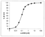


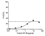


Classifications
-
- A—HUMAN NECESSITIES
- A61—MEDICAL OR VETERINARY SCIENCE; HYGIENE
- A61K—PREPARATIONS FOR MEDICAL, DENTAL OR TOILETRY PURPOSES
- A61K39/00—Medicinal preparations containing antigens or antibodies
- A61K39/395—Antibodies; Immunoglobulins; Immune serum, e.g. antilymphocytic serum
- A61K39/39533—Antibodies; Immunoglobulins; Immune serum, e.g. antilymphocytic serum against materials from animals
- A61K39/3955—Antibodies; Immunoglobulins; Immune serum, e.g. antilymphocytic serum against materials from animals against proteinaceous materials, e.g. enzymes, hormones, lymphokines
-
- C—CHEMISTRY; METALLURGY
- C07—ORGANIC CHEMISTRY
- C07K—PEPTIDES
- C07K16/00—Immunoglobulins [IGs], e.g. monoclonal or polyclonal antibodies
- C07K16/18—Immunoglobulins [IGs], e.g. monoclonal or polyclonal antibodies against material from animals or humans
- C07K16/22—Immunoglobulins [IGs], e.g. monoclonal or polyclonal antibodies against material from animals or humans against growth factors ; against growth regulators
-
- A—HUMAN NECESSITIES
- A61—MEDICAL OR VETERINARY SCIENCE; HYGIENE
- A61P—SPECIFIC THERAPEUTIC ACTIVITY OF CHEMICAL COMPOUNDS OR MEDICINAL PREPARATIONS
- A61P1/00—Drugs for disorders of the alimentary tract or the digestive system
- A61P1/16—Drugs for disorders of the alimentary tract or the digestive system for liver or gallbladder disorders, e.g. hepatoprotective agents, cholagogues, litholytics
-
- A—HUMAN NECESSITIES
- A61—MEDICAL OR VETERINARY SCIENCE; HYGIENE
- A61P—SPECIFIC THERAPEUTIC ACTIVITY OF CHEMICAL COMPOUNDS OR MEDICINAL PREPARATIONS
- A61P13/00—Drugs for disorders of the urinary system
- A61P13/12—Drugs for disorders of the urinary system of the kidneys
-
- A—HUMAN NECESSITIES
- A61—MEDICAL OR VETERINARY SCIENCE; HYGIENE
- A61P—SPECIFIC THERAPEUTIC ACTIVITY OF CHEMICAL COMPOUNDS OR MEDICINAL PREPARATIONS
- A61P43/00—Drugs for specific purposes, not provided for in groups A61P1/00-A61P41/00
-
- A—HUMAN NECESSITIES
- A61—MEDICAL OR VETERINARY SCIENCE; HYGIENE
- A61P—SPECIFIC THERAPEUTIC ACTIVITY OF CHEMICAL COMPOUNDS OR MEDICINAL PREPARATIONS
- A61P7/00—Drugs for disorders of the blood or the extracellular fluid
- A61P7/10—Antioedematous agents; Diuretics
-
- A—HUMAN NECESSITIES
- A61—MEDICAL OR VETERINARY SCIENCE; HYGIENE
- A61P—SPECIFIC THERAPEUTIC ACTIVITY OF CHEMICAL COMPOUNDS OR MEDICINAL PREPARATIONS
- A61P9/00—Drugs for disorders of the cardiovascular system
-
- A—HUMAN NECESSITIES
- A61—MEDICAL OR VETERINARY SCIENCE; HYGIENE
- A61P—SPECIFIC THERAPEUTIC ACTIVITY OF CHEMICAL COMPOUNDS OR MEDICINAL PREPARATIONS
- A61P9/00—Drugs for disorders of the cardiovascular system
- A61P9/04—Inotropic agents, i.e. stimulants of cardiac contraction; Drugs for heart failure
-
- A—HUMAN NECESSITIES
- A61—MEDICAL OR VETERINARY SCIENCE; HYGIENE
- A61P—SPECIFIC THERAPEUTIC ACTIVITY OF CHEMICAL COMPOUNDS OR MEDICINAL PREPARATIONS
- A61P9/00—Drugs for disorders of the cardiovascular system
- A61P9/12—Antihypertensives
-
- A—HUMAN NECESSITIES
- A61—MEDICAL OR VETERINARY SCIENCE; HYGIENE
- A61P—SPECIFIC THERAPEUTIC ACTIVITY OF CHEMICAL COMPOUNDS OR MEDICINAL PREPARATIONS
- A61P9/00—Drugs for disorders of the cardiovascular system
- A61P9/14—Vasoprotectives; Antihaemorrhoidals; Drugs for varicose therapy; Capillary stabilisers
-
- A—HUMAN NECESSITIES
- A61—MEDICAL OR VETERINARY SCIENCE; HYGIENE
- A61K—PREPARATIONS FOR MEDICAL, DENTAL OR TOILETRY PURPOSES
- A61K39/00—Medicinal preparations containing antigens or antibodies
- A61K2039/505—Medicinal preparations containing antigens or antibodies comprising antibodies
-
- C—CHEMISTRY; METALLURGY
- C07—ORGANIC CHEMISTRY
- C07K—PEPTIDES
- C07K2317/00—Immunoglobulins specific features
- C07K2317/20—Immunoglobulins specific features characterized by taxonomic origin
- C07K2317/21—Immunoglobulins specific features characterized by taxonomic origin from primates, e.g. man
-
- C—CHEMISTRY; METALLURGY
- C07—ORGANIC CHEMISTRY
- C07K—PEPTIDES
- C07K2317/00—Immunoglobulins specific features
- C07K2317/20—Immunoglobulins specific features characterized by taxonomic origin
- C07K2317/24—Immunoglobulins specific features characterized by taxonomic origin containing regions, domains or residues from different species, e.g. chimeric, humanized or veneered
-
- C—CHEMISTRY; METALLURGY
- C07—ORGANIC CHEMISTRY
- C07K—PEPTIDES
- C07K2317/00—Immunoglobulins specific features
- C07K2317/30—Immunoglobulins specific features characterized by aspects of specificity or valency
- C07K2317/34—Identification of a linear epitope shorter than 20 amino acid residues or of a conformational epitope defined by amino acid residues
-
- C—CHEMISTRY; METALLURGY
- C07—ORGANIC CHEMISTRY
- C07K—PEPTIDES
- C07K2317/00—Immunoglobulins specific features
- C07K2317/50—Immunoglobulins specific features characterized by immunoglobulin fragments
- C07K2317/54—F(ab')2
-
- C—CHEMISTRY; METALLURGY
- C07—ORGANIC CHEMISTRY
- C07K—PEPTIDES
- C07K2317/00—Immunoglobulins specific features
- C07K2317/50—Immunoglobulins specific features characterized by immunoglobulin fragments
- C07K2317/55—Fab or Fab'
-
- C—CHEMISTRY; METALLURGY
- C07—ORGANIC CHEMISTRY
- C07K—PEPTIDES
- C07K2317/00—Immunoglobulins specific features
- C07K2317/70—Immunoglobulins specific features characterized by effect upon binding to a cell or to an antigen
- C07K2317/76—Antagonist effect on antigen, e.g. neutralization or inhibition of binding
-
- C—CHEMISTRY; METALLURGY
- C07—ORGANIC CHEMISTRY
- C07K—PEPTIDES
- C07K2317/00—Immunoglobulins specific features
- C07K2317/90—Immunoglobulins specific features characterized by (pharmaco)kinetic aspects or by stability of the immunoglobulin
- C07K2317/92—Affinity (KD), association rate (Ka), dissociation rate (Kd) or EC50 value
-
- C—CHEMISTRY; METALLURGY
- C07—ORGANIC CHEMISTRY
- C07K—PEPTIDES
- C07K2317/00—Immunoglobulins specific features
- C07K2317/90—Immunoglobulins specific features characterized by (pharmaco)kinetic aspects or by stability of the immunoglobulin
- C07K2317/94—Stability, e.g. half-life, pH, temperature or enzyme-resistance
Abstract
본 발명의 대상은 울혈의 중재 및 치료가 필요한 환자에서 울혈의 중재 및 치료에 사용하기 위한 항-아드레노메둘린 (ADM) 항체 또는 항-아드레노메둘린 항체 단편 또는 항-ADM 비-Ig 스캐폴드이다.The subject of the present invention is an anti-adrenomedullin (ADM) antibody or anti-adrenomedullin antibody fragment or an anti-ADM non-Ig scan for use in the intervention and treatment of congestion in patients in need of intervention and treatment of congestion. It's a fold.
Description
발명의 기술분야Technical field of invention
본 발명의 대상은 울혈의 중재 및 치료가 필요한 환자에서 울혈의 중재 및 치료에 사용하기 위한 항-아드레노메둘린 (ADM) 항체 또는 항-아드레노메둘린 항체 단편 또는 항-ADM 비-Ig 스캐폴드이다. The subject of the present invention is an anti-adrenomedullin (ADM) antibody or anti-adrenomedullin antibody fragment or an anti-ADM non-Ig scan for use in the intervention and treatment of congestion in patients in need of intervention and treatment of congestion. It's a fold.
배경기술background technology
펩티드 아드레노메둘린 (ADM)은 인간 크롬친화세포종 세포주 (서열식별번호(SEQ ID NO): 20)로부터 단리된, 52개 아미노산을 포함하는 신규한 저혈압성 펩티드로서 1993년도 최초로 기재되었다 ( Kitamura et al., 1993. Biochem Biophys Res Comm 192 (2): 553-560 ). 같은 해에, 185개 아미노산을 포함하는 전구체 펩티드를 코딩하는 cDNA 및 상기 전구체 펩티드의 완전한 아미노산 서열이 또한 기재되었다. 그 중에서도 N-말단에서 21개 아미노산의 신호 서열을 포함하는 전구체 펩티드는 "프리-프로아드레노메둘린(pre-proadrenomedullin)" (프리-프로ADM)으로서 지칭된다. 본 설명에서, 명시된 모든 아미노산 위치는 일반적으로 185개 아미노산을 포함하는 프리-프로ADM에 관한 것이다. 펩티드 아드레노메둘린 (ADM)은 52개 아미노산 (서열식별번호: 20)을 포함하고 프리-프로ADM의 아미노산 95 내지 146을 포함하는 펩티드이며, 단백질 가수분해성 절단에 의해 형성된다. 지금까지, 실질적으로 프리-프로ADM의 절단에 의해 형성된 펩티드 단편 중 몇몇 단편만이, 특히 생리학적으로 활성인 펩티드 ADM 및 "PAMP" (20개 아미노산 (22-41)을 포함하는 펩티드이며, 프리-프로ADM에서 신호 펩티드의 21개 아미노산에 이어짐)가 더욱 정확하게 연구되었다. 1993년도의 ADM 발견 및 특징분석은 집중 연구 활동을 촉발시켰으며, 그 결과는 본 설명과 관련하여 다양한 검토 논문에 요약되어 있고, 특히 ADM에 주력한 "펩티드" 이슈에서 발견될 논문을 참고한다 ( Takahashi 2001. Peptides 22: 1691; Eto 2001. Peptides 22: 1693-1711 ). Hinson et al. 2000 ( Hinson et al. 2000. Endocrine Reviews 21(2):138-167 )에서 추가로 검토된다. 지금까지의 과학적 연구에서, 그 중에서도, ADM이 다기능성 조절성 펩티드로서 간주될 수 있음이 발견되었다. 이는 글리신에 의해 연장된 불활성 형태로 순환계로 방출된다 ( Kitamura et al. 1998. Biochem Biophys Res Commun 244(2): 551-555 ). ADM에 대해 특이적이고 아마도 ADM의 효과를 조절할 가능성이 있는 결합 단백질 ( Pio et al. 2001. The Journal of Biological Chemistry 276(15): 12292-12300 )이 또한 존재한다. 지금까지의 연구에서 ADM 뿐만 아니라 PAMP의 매우 중요한 생리학적 효과는 혈압에 영향을 미치는 효과였다.The peptide adrenomedullin (ADM) is a novel hypotensive peptide containing 52 amino acids, isolated from a human pheochromocytoma cell line (SEQ ID NO: 20) and first described in 1993 ( Kitamura et al., 1993. Biochem Biophys Res Comm 192 (2): 553-560 ). In the same year, a cDNA encoding a precursor peptide containing 185 amino acids and the complete amino acid sequence of the precursor peptide were also described. Among them, the precursor peptide containing a signal sequence of 21 amino acids at the N-terminus is referred to as “pre-proadrenomedullin” (pre-proADM). In this description, all amino acid positions specified relate to the pre-proADM, which generally contains 185 amino acids. Peptide adrenomedullin (ADM) is a peptide containing 52 amino acids (SEQ ID NO: 20) and amino acids 95 to 146 of pre-proADM, and is formed by proteolytic cleavage. So far, only a few fragments of peptide fragments are substantially formed by cleavage of pre-proADM, especially the physiologically active peptides ADM and "PAMP" (peptides containing 20 amino acids (22-41), - followed by 21 amino acids of the signal peptide in ProADM) was studied more precisely. The discovery and characterization of ADM in 1993 triggered an intensive research activity, the results of which are summarized in various review papers relevant to this account, with particular reference to those found in the ADM-focused "Peptides" issue ( Takahashi 2001. Peptides 22: 1691; Eto 2001. Peptides 22: 1693-1711 ). Hinson et al. 2000 ( Hinson et al. 2000. Endocrine Reviews 21(2):138-167 ). Scientific studies to date have found, inter alia, that ADM can be regarded as a multifunctional regulatory peptide. It is released into the circulation in an inactive form extended by glycine ( Kitamura et al. 1998. Biochem Biophys Res Commun 244(2): 551-555 ). There are also binding proteins that are specific for ADM and possibly modulate the effects of ADM ( Pio et al. 2001. The Journal of Biological Chemistry 276(15): 12292-12300 ). In studies so far, a very important physiological effect of ADM as well as PAMP was the effect on blood pressure.
따라서, ADM은 효과적인 혈관확장제이며, 이는 ADM의 C-말단 부분에 있는 특정한 펩티드 세그먼트를 저혈압 효과와 연관시키는 것을 가능하게 한다. 추가로, ADM과는 상이한 작용 메카니즘을 갖는 것으로 보이기는 하지만, 프리-프로ADM으로부터 형성된 상기 언급된 생리학적으로 활성인 펩티드 PAMP가 마찬가지로 저혈압 효과를 나타낸다는 것이 발견되었다 (상기 언급된 Eto et al. 2001 및 Hinson et al. 2000 검토 논문 외에도, Kuwasaki et al. 1997. FEBS Lett 414(1): 105-110; Kuwasaki et al. 1999. Ann. Clin. Biochem. 36: 622-628; Tsuruda et al. 2001 Life Sci. 69(2): 239-245 및 EP-A2 0 622 458 또한 참고). 추가로, 순환계 및 다른 생물학적 액체에서 측정될 수 있는 ADM의 농도가 수많은 병리학적 상태에서 건강한 대조군 대상체에서 발견되는 농도보다 유의하게 높다는 것이 확인되었다. 따라서, 울혈성 심부전, 심근 경색, 신장 질환, 고혈압 장애, 진성 당뇨병, 쇼크의 급성기 및 패혈증 및 패혈성 쇼크를 가진 환자에서 ADM 수준은 정도는 상이하지만 유의하게 증가된다. PAMP 농도가 또한 상기 병리학적 상태 중 일부에서 증가되지만, 혈장 수준이 ADM에 비해 낮다 ( Eto 2001. Peptides 22: 1693-1711 ). 상당히 높은 농도의 ADM이 패혈증에서 관찰되었고, 가장 높은 농도가 패혈성 쇼크에서 관찰되는 것으로 보고되었다 ( Eto 2001. Peptides 22: 1693-1711; Hirata et al. Journal of Clinical Endocrinology and Metabolism 81(4): 1449-1453; Ehlenz et al. 1997. Exp Clin Endocrinol Diabetes 105: 156-162; Tomoda et al. 2001. Peptides 22: 1783-1794; Ueda et al. 1999. Am. J. Respir. Crit. Care Med.160: 132-136 및 Wang et al. 2001. Peptides 22: 1835-1840 ).Therefore, ADM is an effective vasodilator, which makes it possible to associate a specific peptide segment in the C-terminal part of ADM with a hypotensive effect. Additionally, it has been discovered that the above-mentioned physiologically active peptides PAMPs formed from pre-proADM, although appearing to have a different mechanism of action than ADM, likewise exhibit hypotensive effects (see Eto et al., supra). In addition to the 2001 and Hinson et al . 2001 Life Sci. 69(2): 239-245 and EP-
심부전을 가진 환자에서 ADM의 혈장 농도가 상승하고, 이는 질환 중증도와 상관관계가 있다 ( Hirayama et al. 1999. J Endocrinol 160: 297-303; Yu et al. 2001. Heart 86: 155-160 ). 높은 혈장 ADM은 이들 대상체에서 독립적인 부정적인 예후 지표이다 ( Poyner et al. 2002. Pharmacol Rev 54: 233-246 ).Plasma levels of ADM are elevated in patients with heart failure, and this correlates with disease severity ( Hirayama et al. 1999. J Endocrinol 160: 297-303; Yu et al. 2001. Heart 86: 155-160 ). High plasma ADM is an independent negative prognostic indicator in these subjects ( Poyner et al. 2002. Pharmacol Rev 54: 233-246 ).
심부전에서 MR-프로ADM (서열식별번호: 33)의 역할이 몇몇 연구에서 탐구되었다. BACH 연구에서 ( Maisel et al. 2010. J. Am. Coll. Cardiol. 55: 2062-2076 ), MR-프로ADM은 90 일째 사망에 대한 강력한 예후이었으며, 나트륨뇨 펩티드를 넘어서는 예후 값을 추가한다. PRIDE 연구로부터의 후속적인 데이터 ( Shah et al. 2012. Eur. Heart J. 33: 2197-2205 )는 MR-프로ADM에 대한 잠재적인 예후 역할을 확고히 하고; 환자들 중에서 MR-프로ADM은 1 년째 사망률에 대해 최상의 곡선하 면적 (AUC)을 가졌다. 유사하게, 만성 심부전 (CHF)을 갖는 환자에서 MR-프로ADM의 수준은 질환 중증도와 강력한 상관관계가 있었고, 상기 펩티드의 상승된 수준은 12 개월의 추적 관찰에서 증가된 사망 위험과 강력한 연관이 있었다 ( van Haehling et al. 2010. European Journal of Heart Failure 12: 484-491; Adlbrecht et al. 2009. European Journal of Heart Failure 11: 361-366 ).The role of MR-ProADM (SEQ ID NO: 33) in heart failure has been explored in several studies. In the BACH study ( Maisel et al. 2010. J. Am. Coll. Cardiol. 55: 2062-2076 ), MR-proADM was strongly predictive of death at 90 days, adding prognostic value beyond natriuretic peptides. Subsequent data from the PRIDE study ( Shah et al. 2012. Eur. Heart J. 33: 2197-2205 ) solidify a potential prognostic role for MR-proADM; Among patients, MR-ProADM had the best area under the curve (AUC) for mortality at 1 year. Similarly, in patients with chronic heart failure (CHF), levels of MR-proADM were strongly correlated with disease severity, and elevated levels of this peptide were strongly associated with increased risk of death at 12 months of follow-up. ( van Haehling et al. 2010. European Journal of Heart Failure 12: 484-491; Adlbrecht et al. 2009. European Journal of Heart Failure 11: 361-366 ).
급성 대상부전된 심부전을 가진 환자에서 치료 동안에 MR-프로ADM을 연구하였고 ( Boyer et al. 2012. Congest Heart Fail 18 (2): 91-97 ): 급성 요법 동안에 MR-프로ADM 수준이 증가하는 경향이 있는 환자는 지속적인 울혈과 연관된 결과를 갖는다. 치료후 12-24 시간 후에, MR-프로ADM이 상승된 환자는 말초 부종이 증가하였다. 카이저(Kaiser) 등은 공통심실 심장을 가진 환자에서 MR-프로ADM을 측정하였다 ( Kaiser et al. 2014. Europ J Heart Failure 16: 1082-1088 ). 실패한 폰탄(Fontan) 회로를 갖는 환자 (복수 및 말초 부종을 나타냄)에서의 수준은 폰탄 실패가 없는 환자에 비해 유의하게 높았다. 더욱이, 아이젠헛(Eisenhut)은 아드레노메둘린 수준의 감소를 유도하는 치료가 폐렴 및 패혈증에서 폐포성 부종의 중증도 및 규모를 감소시킬 수 있는지 여부를 추측하였다 ( Eisenhut 2006. Crit Care 10: 418 )MR-ProADM was studied during treatment in patients with acute decompensated heart failure ( Boyer et al. 2012. Congest Heart Fail 18 (2): 91-97 ): MR-ProADM levels tended to increase during acute therapy. Patients with this have persistent congestion and associated consequences. 12-24 hours after treatment, patients with elevated MR-proADM had increased peripheral edema. Kaiser et al. measured MR-ProADM in patients with common ventricle hearts ( Kaiser et al. 2014. Europ J Heart Failure 16: 1082-1088 ). Levels in patients with failed Fontan circuits (presenting ascites and peripheral edema) were significantly higher compared to patients without Fontan failure. Moreover, Eisenhut speculated whether treatments that induce a decrease in adrenomedullin levels could reduce the severity and magnitude of alveolar edema in pneumonia and sepsis ( Eisenhut 2006. Crit Care 10: 418 )
진단 목적을 위해, 특히 패혈증 진단, 심장병 진단 및 암 진단의 범위 내에서 생물학적 액체에서 아드레노메둘린 면역반응성을 확인하는 방법이 관련 기술 분야에 추가로 공지되어 있다. 본 발명에 따라, 전체 프리프로아드레노메둘린의 아미노산 (45-92)를 함유하는 프로아드레노메둘린 (서열식별번호 33)의 중간지역(midregional) 부분 펩티드는 특히 미드-프로ADM의 서열을 특이적으로 인식하는 적어도 하나의 표지된 항체를 이용하여 작업하는 면역검정에 의해 측정된다 (WO2004/090546).Methods for determining adrenomedullin immunoreactivity in biological fluids for diagnostic purposes, particularly within the scope of sepsis diagnosis, heart disease diagnosis and cancer diagnosis, are further known in the art. According to the present invention, the midregional partial peptide of proadrenomedullin (SEQ ID NO: 33) containing amino acids (45-92) of the entire preproadrenomedullin has the sequence of mid-proADM in particular. It is measured by an immunoassay working with at least one labeled antibody that specifically recognizes (WO2004/090546).
WO2004/097423은 심혈관 장애의 진단, 예후 및 치료를 위해 아드레노메둘린에 대한 항체의 사용을 기재한다. ADM 수용체를 차단함으로써 질환을 치료하는 것 또한 관련 기술분야에 기재되어 있으며 (예를 들어 WO2006/027147, PCT/EP2005/012844), 상기 질환은 패혈증, 패혈성 쇼크, 심혈관 질환, 감염, 피부과적 질환, 내분비학적 질환, 대사 질환, 위장병학적 질환, 암, 염증, 혈액학적 질환, 호흡기 질환, 근육 골격 질환, 신경학적 질환, 비뇨기 질환일 수 있다.WO2004/097423 describes the use of antibodies against adrenomedullin for the diagnosis, prognosis and treatment of cardiovascular disorders. Treating diseases by blocking ADM receptors is also described in the related art (e.g. WO2006/027147, PCT/EP2005/012844), and the diseases include sepsis, septic shock, cardiovascular disease, infection, and dermatological disease. , endocrinological diseases, metabolic diseases, gastroenterological diseases, cancer, inflammation, hematological diseases, respiratory diseases, musculoskeletal diseases, neurological diseases, and urinary diseases.
패혈증 초기의 경우에는, ADM이 심장 기능, 및 간, 비장, 신장 및 소장에서의 혈액 공급을 개선시키는 것으로 보고되었다. 항-ADM-중화 항체는 패혈증의 초기 동안에 상기 언급된 효과를 중화시킨다 ( Wang et al. 2001. Peptides 22: 1835-1840 ).In early cases of sepsis, ADM has been reported to improve heart function and blood supply to the liver, spleen, kidneys and small intestine. Anti-ADM-neutralizing antibodies neutralize the above-mentioned effects during the early stages of sepsis ( Wang et al. 2001. Peptides 22: 1835-1840 ).
다른 질환의 경우, ADM의 차단이 어느 정도 유익할 수 있다. 그러나, 특정량의 ADM이 몇몇 생리학적 기능을 위해 필요할 수 있기 때문에, ADM이 완전히 중화되는 경우에는 상기 차단이 유해할 수 있다. 여러 보고에서, ADM의 투여가 특정 질환에 유익할 수 있음이 강조되었다. 그와 대조적으로, 다른 보고에서는 ADM이 특정 상태에서 투여될 때 생명을 위협하는 것으로 보고되었다.For other diseases, blockade of ADM may be of some benefit. However, since certain amounts of ADM may be required for some physiological functions, this blocking may be detrimental if ADM is completely neutralized. Several reports have highlighted that administration of ADM may be beneficial for certain diseases. In contrast, other reports have shown that ADM is life-threatening when administered in certain conditions.
WO2013/072510은 중증의 만성 또는 급성 질환 또는 급성 상태를 가진 환자의 사망 위험을 감소시키기 위해 상기 환자의 치료에서 사용하기 위한 비-중화 항-ADM 항체를 기재한다.WO2013/072510 describes non-neutralizing anti-ADM antibodies for use in the treatment of patients with severe chronic or acute diseases or acute conditions to reduce the risk of death in such patients.
WO2013/072511은 장기 기능장애 또는 장기 부전의 예방 또는 감소를 위해 만성 또는 급성 질환 또는 급성 상태를 가진 환자의 치료에서 사용하기 위한 비-중화 항-ADM 항체를 기재한다. WO2013/072511 describes non-neutralizing anti-ADM antibodies for use in the treatment of patients with chronic or acute diseases or acute conditions for the prevention or reduction of organ dysfunction or organ failure.
WO2013/072512는 혈청, 혈액, 혈장에서 아드레노메둘린의 반감기 (t1/2 절반 유지 시간)를 개선시키는 ADM 안정화 항체인 비-중화 항-ADM 항체를 기재한다. 이 ADM 안정화 항체는 ADM의 생체활성을 80% 미만으로 차단한다.WO2013/072512 describes a non-neutralizing anti-ADM antibody, which is an ADM stabilizing antibody that improves the half-life (t 1/2 half retention time) of adrenomedullin in serum, blood and plasma. This ADM stabilizing antibody blocks the bioactivity of ADM by less than 80%.
WO2013/072513은 순환계의 안정화를 위해 급성 질환 또는 상태를 가진 환자의 치료에서 사용하기 위한 비-중화 항-ADM 항체를 기재한다.WO2013/072513 describes non-neutralizing anti-ADM antibodies for use in the treatment of patients with acute diseases or conditions for stabilization of the circulatory system.
WO2013/072514는 만성 또는 급성 질환 또는 급성 상태를 가진 환자에서 유체 균형을 조절하기 위한 비-중화 항-ADM 항체를 기재한다.WO2013/072514 describes non-neutralizing anti-ADM antibodies for regulating fluid balance in patients with chronic or acute diseases or acute conditions.
발명의 설명Description of the Invention
본 발명에 따라, ADM에 결합하는 항-ADM 항체 또는 항-ADM 항체 단편 또는 ADM에 결합하는 항-ADM 비-Ig 스캐폴드의 투여가 울혈의 중재 및 치료를 필요로 하는 환자의 울혈의 중재 및 치료를 위해 사용될 수 있음이 발견되었다.According to the present invention, administration of an anti-ADM antibody or anti-ADM antibody fragment that binds to an ADM or an anti-ADM non-Ig scaffold that binds to an ADM can be used to treat and treat congestion in a patient in need thereof. It was discovered that it could be used for treatment.
도 1a:
항체 포맷의 도시 - Fv 및 scFv-변이체.
도 1b:
항체 포맷의 도시 - 이종성 융합 및 이관능성 항체.
도 1c:
항체 포맷의 도시 - 2가 항체 및 이중특이적 항체.
도 2:
a: 인간 ADM의 용량 반응 곡선. 최대 cAMP 자극을 100% 활성화로 조정하였다.
b: 5.63nM hADM의 존재하에 인간 ADM 22-52 (ADM-수용체 길항제)의 용량/ 억제 곡선.
c: 5.63 nM hADM의 존재하에 CT-H의 용량/ 억제 곡선.
d: 5.63 nM hADM의 존재하에 MR-H의 용량/ 억제 곡선.
e: 5.63 nM hADM의 존재하에 NT-H의 용량/ 억제 곡선.
f: 마우스 ADM의 용량 반응 곡선. 최대 cAMP 자극을 100% 활성화로 조정하였다.
g: 0.67 nM mADM의 존재하에 인간 ADM 22-52 (ADM-수용체 길항제)의 용량/ 억제 곡선.
h: 0.67 nM mADM의 존재하에 CT-M의 용량/ 억제 곡선.
i: 0.67 nM mADM의 존재하에 MR-M의 용량/ 억제 곡선.
j: 0.67 nM mADM의 존재하에 NT-M의 용량/ 억제 곡선.
k: F(ab)2 NT-M에 의한 및 Fab NT-M에 의한 ADM의 억제를 도시함.
l: F(ab)2 NT-M에 의한 및 Fab NT-M에 의한 ADM의 억제를 도시함.
도 3:
이 도면은 전형적인 hADM 용량/ 신호 곡선을 도시한다. 100 ㎍/mL 항체 NT-H의 존재하에 hADM 용량 신호 곡선.
도 4:
이 도면은 NT-H 항체의 부재하에 및 존재하에 인간 혈장 (시트레이트)에서 hADM의 안정화를 도시한다.
도 5:
상동성 인간 프레임워크 서열을 갖는 Fab의 정렬.
도 6:
상이한 용량으로 CLP 및 NT-M을 적용한지 18 시간 이후 세포외 알부민 축적.
도 7:
상이한 용량으로 CLP 및 NT-M을 적용한지 18 시간 이후 VEGF 발현.
도 8:
상이한 용량으로 CLP 및 NT-M을 적용한지 18 시간 이후 안지오포에틴-1 발현.
도 9:
60 일까지 상이한 용량으로 NT-H 적용 이후 건강한 인간 대상체에서 ADM-농도.
도 10:
2-타격 돼지 모델에서 처리 및 혈액 샘플 채취에 대한 시간 스케쥴.
도 11:
비히클 (사각형) 및 처리 (점) 그룹의 ADM 농도 (SEM과 함께 평균 값) (상호작용의 경우 p=0.003; t=7h에서 19h까지, 다변량, 시간*그룹).
도 12:
비히클 (사각형) 및 처리 (점) 그룹의 심박동수 (SEM과 함께 평균 값), 상호작용의 경우 p=0.097 (t=7h에서 t=19h까지, 다변량, 시간*그룹).
도 13:
비히클 (사각형) 및 처리 (점) 그룹의 심박출량 (SEM과 함께 평균 값) (t-시험: p<0.05, t=9, 10, 11h에 대해).
도 14:
비히클 (사각형) 및 처리 (점) 그룹의 부피 요구/적용 (SEM과 함께 평균 값) (17h에서 t-시험: p=0.034; 19h에서 t-시험: p=0.045).
도 15:
비히클 (사각형) 및 처리 (점) 그룹의 누적된 부피 요구/적용 (SEM과 함께 평균 값) (19h에서 t-시험의 경우 p=0.039; 19h에서 만-휘트니(Mann-Whitney)의 경우 p=0.036).
도 16:
비히클 (사각형) 및 처리 (점) 그룹의 노르아드레날린 요구/적용 (SEM과 함께 평균 값).
도 17:
비히클 (사각형) 및 처리 (점) 그룹의 승압제 보충에 대한 필요성 (t=19h에서 Chi2 시험: 상호작용의 경우 p = 0.014 (t=7h-19h, 다변량, 시간*그룹: 0.019).
도 18:
비히클 (사각형) 및 처리 (점) 그룹의 전신 혈관 내성 (SEM과 함께 평균 값) (t-시험: 15h: p=0.069; 17h: p= 0.037; 19h: p=0.066).
도 19:
LPS-유도된 내독소혈증 래트 모델에서 rADM 농도
도 20:
내독소혈증 래트 모델에서 혈관 투과성 Figure 1a:
Illustration of antibody formats - Fv and scFv-variants.
Figure 1b:
Illustration of antibody formats - heterologous fusion and bifunctional antibodies.
Figure 1c:
Illustration of antibody formats - bivalent antibodies and bispecific antibodies.
Figure 2:
a: Dose response curve of human ADM. Maximal cAMP stimulation was adjusted to 100% activation.
b: Dose/inhibition curve of human ADM 22-52 (ADM-receptor antagonist) in the presence of 5.63 nM hADM.
c: Dose/inhibition curve of CT-H in the presence of 5.63 nM hADM.
d: Dose/inhibition curve of MR-H in the presence of 5.63 nM hADM.
e: Capacity/inhibition curve of NT-H in the presence of 5.63 nM hADM.
f: Dose response curve of mouse ADM. Maximal cAMP stimulation was adjusted to 100% activation.
g: Dose/inhibition curve of human ADM 22-52 (ADM-receptor antagonist) in the presence of 0.67 nM mADM.
h: Dose/inhibition curve of CT-M in the presence of 0.67 nM mADM.
i: Dose/inhibition curve of MR-M in the presence of 0.67 nM mADM.
j: Capacity/inhibition curve of NT-M in the presence of 0.67 nM mADM.
k: Depicts inhibition of ADM by F(ab)2 NT-M and by Fab NT-M.
l: Showing inhibition of ADM by F(ab)2 NT-M and by Fab NT-M.
Figure 3:
This figure shows a typical hADM dose/signal curve. hADM dose signal curve in the presence of 100 μg/mL antibody NT-H.
Figure 4:
This figure depicts the stabilization of hADM in human plasma (citrate) in the absence and presence of NT-H antibody.
Figure 5:
Alignment of Fabs with homologous human framework sequences.
Figure 6:
Extracellular albumin accumulation 18 h after application of CLP and NT-M at different doses.
Figure 7:
VEGF expression after 18 h of application of CLP and NT-M at different doses.
Figure 8:
Angiopoietin-1
Figure 9:
ADM-concentrations in healthy human subjects following NT-H application at different doses up to 60 days.
Figure 10:
Time schedule for processing and blood sample collection in the 2-percussion porcine model.
Figure 11:
ADM concentrations (mean values with SEM) in vehicle (squares) and treatment (dots) groups (p=0.003 for interaction; t=7h to 19h, multivariate, time*group).
Figure 12:
Heart rate (mean value with SEM) in vehicle (squares) and treatment (dots) groups, p=0.097 for interaction (t=7h to t=19h, multivariate, time*group).
Figure 13:
Cardiac output (mean values with SEM) in vehicle (squares) and treatment (dots) groups (t-test: p<0.05, for t=9, 10, 11 h).
Figure 14:
Volume requirement/application (mean values with SEM) for vehicle (squares) and treatment (dots) groups (t-test at 17h: p=0.034; t-test at 19h: p=0.045).
Figure 15:
Cumulative volume demand/application (mean values with SEM) for vehicle (squares) and treatment (dots) groups (p=0.039 for t-test at 19h; p=0.039 for Mann-Whitney at 19h) 0.036).
Figure 16:
Noradrenaline demand/application (mean values with SEM) in vehicle (squares) and treatment (dots) groups.
Figure 17:
Need for vasopressor supplementation in vehicle (squares) and treatment (dots) groups (Chi 2 test at t=19h: p = 0.014 for interaction (t=7h-19h, multivariate, time*group: 0.019).
Figure 18:
Systemic vascular resistance (mean values with SEM) in vehicle (squares) and treatment (dots) groups (t-test: 15h: p=0.069; 17h: p=0.037; 19h: p=0.066).
Figure 19:
rADM concentration in LPS-induced endotoxemia rat model
Figure 20:
Vascular permeability in a rat model of endotoxemia.
본 명세서에 걸쳐, 본 발명에 따른 "항체" 또는 "항체 단편" 또는 "비-Ig 스캐폴드"는 ADM에 결합할 수 있고, 따라서 ADM에 대해 지시되며, 따라서 "항-ADM 항체", "항-ADM 항체 단편" 또는 "항-ADM 비-Ig 스캐폴드"로 지칭될 수 있다.Throughout this specification, an “antibody” or “antibody fragment” or “non-Ig scaffold” according to the invention is capable of binding ADM and is therefore directed against ADM, and is therefore referred to as an “anti-ADM antibody”, “anti- -ADM antibody fragment” or “anti-ADM non-Ig scaffold”.
예를 들어 이뇨제의 투여와는 대조적으로 ADM에 결합하는 항-ADM 항체 또는 항-ADM 항체 단편 또는 ADM에 결합하는 항-ADM 비-Ig 스캐폴드의 투여의 이점은 신장 보호 효과를 갖는다. ADM에 결합하는 항-ADM 항체 또는 항-ADM 항체 단편 또는 ADM에 결합하는 항-ADM 비-Ig 스캐폴드는 신장을 손상시키지 않고, 따라서 이와 관련된 부작용이 예상되지 않는다. For example, the advantage of administration of an anti-ADM antibody or anti-ADM antibody fragment that binds to ADM or an anti-ADM non-Ig scaffold that binds to ADM, as opposed to administration of diuretics, is that it has a nephroprotective effect. Anti-ADM antibodies or anti-ADM antibody fragments that bind to ADM or anti-ADM non-Ig scaffolds that bind to ADM do not damage the kidneys, and therefore no associated side effects are expected.
본 발명에 따라, ADM에 결합하는 항-ADM 항체 또는 항-ADM 항체 단편 또는 ADM에 결합하는 항-ADM 비-Ig 스캐폴드의 투여는 바람직하게는 전신 적용이다.According to the invention, administration of an anti-ADM antibody or anti-ADM antibody fragment that binds ADM or an anti-ADM non-Ig scaffold that binds ADM is preferably a systemic application.
특정한 실시양태에서, ADM에 결합하는 항-ADM 항체 또는 항-ADM 항체 단편 또는 ADM에 결합하는 항-ADM 비-Ig 스캐폴드를 울혈을 일으킬 수 있는 혈관 장벽 기능장애 또는 내피 기능장애를 가진 환자에게 투여할 수 있다.In certain embodiments, an anti-ADM antibody or anti-ADM antibody fragment that binds ADM or an anti-ADM non-Ig scaffold that binds ADM is administered to patients with vascular barrier dysfunction or endothelial dysfunction that may cause congestion. It can be administered.
혈관 장벽 기능장애 또는 내피 기능장애는 내피 (혈관의 내벽)의 전신 병리학적 상태이고, 광범위하게는 내피에 의해 생성되는 (또는 내피 상에서 작용하는) 혈관확장 물질과 혈관수축 물질 사이의 불균형으로 정의될 수 있다 ( Deanfield et al. 2005. J Hypertens 23 (1): 7-17 ). 내피 세포의 정상적인 기능에는 응고의 매개, 혈소판 부착, 면역 기능, 및 세포내 및 세포외 공간의 부피 및 전해질 함량의 조절이 포함된다. 내피는 전체 심혈관계의 안쪽 벽에 있는 세포 단일층이고, 혈관 긴장도, 혈전증, 혈관형성 및 염증을 비롯한 여러 과정을 조절한다. 내피 세포는 표현형적으로 역동적인 것으로 확인되었고, 다양한 국부 및 전신 자극에 반응하여 휴지 상태와 활성 상태를 전이시킬 수 있다 ( Colombo et al. 2015. Curr Heart Fail Rep. 12(3): 215-222 ). 최근 새로운 연구에 의해, 내피 기능장애가 고혈압, 아테롬성 동맥경화증 및 울혈성 심부전을 비롯한 심혈관 질환의 주요 원인인 것으로 입증되었다 ( Gutierrez et al. 2013. European Heart Journal 34: 3175-3181 ). 내피는 순환계로부터 주변 조직으로의 유체 교환을 치밀하게 조절하고, 이 장벽의 기능장애는 울혈 및/또는 부종을 일으킬 수 있는 조절되지 않는 유체 유출을 유도한다. 부종 (예를 들어, 폐 부종)의 일반적인 특징은 저분자량 용질인 물에 대한 증가된 투과성이다 ( Rocker et al. 1987. Thorax 42: 620-23 ).Vascular barrier dysfunction or endothelial dysfunction is a systemic pathological condition of the endothelium (the inner lining of blood vessels) and can be broadly defined as an imbalance between vasodilator and vasoconstrictor substances produced by (or acting on) the endothelium. ( Deanfield et al. 2005. J Hypertens 23 (1): 7-17 ). Normal functions of endothelial cells include mediation of coagulation, platelet adhesion, immune function, and regulation of the volume and electrolyte content of intracellular and extracellular spaces. The endothelium is a single layer of cells that lines the inner wall of the entire cardiovascular system and regulates several processes, including vascular tone, thrombosis, angiogenesis, and inflammation. Endothelial cells have been shown to be phenotypically dynamic, capable of transitioning between resting and active states in response to a variety of local and systemic stimuli ( Colombo et al. 2015. Curr Heart Fail Rep. 12(3): 215-222 ). Recently, new studies have demonstrated that endothelial dysfunction is a major cause of cardiovascular diseases, including hypertension, atherosclerosis, and congestive heart failure ( Gutierrez et al. 2013. European Heart Journal 34: 3175-3181 ). The endothelium tightly regulates fluid exchange from the circulatory system to surrounding tissues, and dysfunction of this barrier leads to uncontrolled fluid extravasation that can cause congestion and/or edema. A common feature of edema (e.g., pulmonary edema) is increased permeability to water, a low molecular weight solute ( Rocker et al. 1987. Thorax 42: 620-23 ).
내피 기능장애는 고혈압, 고콜레스테롤혈증, 당뇨병 또는 패혈성 쇼크에서 발생하는 몇몇 질환 과정에서 비롯되고/거나 그에 기여할 수 있다. 내피 기능장애는 관상 동맥 질환 및 다른 아테롬성 동맥경화증 질환을 유도하는 주요 병리생리학적 메카니즘이다.Endothelial dysfunction may result from and/or contribute to several disease processes, such as those occurring in hypertension, hypercholesterolemia, diabetes, or septic shock. Endothelial dysfunction is a major pathophysiological mechanism driving coronary artery disease and other atherosclerotic diseases.
패혈증/ 패혈성 쇼크의 모델에서의 전임상 실험으로부터, 항-ADM 항체의 투여가 혈장 생체-ADM 농도의 증가를 유도하고 (실시예 8, 도 9), 이것이 증가된 생존율과 일치한다는 것이 공지되어 있다 ( Struck et al. 2013. Intensive Care Med Exp 1(1):22 ). 이 효과의 근간을 이루는 메카니즘은 다음과 같이 생각된다: 항체는 정맥내로 투여될 때 그의 크기 때문에 내피 장벽을 가로질러 간질로 넘어갈 수 없고, 혈액 순환계에 잔류한다. 대조적으로, 소형 펩티드로서 ADM은 내피 장벽을 가로질러 자유롭게 확산할 수 있다. 따라서, 항체는 내인성 ADM에 비해 막대한 몰 과량으로 투여될 때 사실상 혈장에서 모든 ADM에 결합하고, 결합 평형의 도달이라는 간단한 결과로서, ADM이 간질로부터 혈액 순환계로 전위된다. 흥미롭게도, 전위된 ADM은 혈관 평활근 세포에 결합할 수 있고, 이완을 유도하여 혈관확장을 일으킨다. 이는 항체의 투여에 의해 감소된다. 한편, 혈장 중 ADM은 내피 세포에 결합하여, 혈관 온전성을 안정화시키거나 심지어 복구시킨다. 따라서, 비-중화 항체인 항체의 투여 결과로서 혈장 ADM 수준이 증가할 때, 상기 기능이 강화된다. 마지막으로, 항체와 ADM의 결합은 ADM의 단백질 가수분해성 붕괴를 감소시킨다.From preclinical experiments in models of sepsis/septic shock, it is known that administration of anti-ADM antibodies leads to an increase in plasma bio-ADM concentrations (Example 8, Figure 9), which is consistent with increased survival. ( Struck et al. 2013. Intensive Care Med Exp 1(1):22 ). The mechanism underlying this effect is thought to be as follows: when administered intravenously, antibodies cannot cross the endothelial barrier into the interstitium due to their size and remain in the blood circulation. In contrast, as a small peptide, ADM can diffuse freely across the endothelial barrier. Thus, the antibody binds virtually all ADM in the plasma when administered in large molar excess relative to endogenous ADM, and translocates ADM from the interstitium into the blood circulation as a simple consequence of reaching binding equilibrium. Interestingly, translocated ADM can bind to vascular smooth muscle cells and induce relaxation, resulting in vasodilation. This is reduced by administration of antibodies. Meanwhile, ADM in plasma binds to endothelial cells, stabilizing or even restoring vascular integrity. Accordingly, this function is enhanced when plasma ADM levels increase as a result of administration of antibodies that are non-neutralizing antibodies. Finally, binding of antibodies to ADM reduces proteolytic degradation of ADM.
놀랍게도, 본 발명자들은 PROTECT 연구 (실시예 6) 및 BIOSTAT 연구 (실시예 7)를 통해, 심부전을 가진 대상체에서 이뇨제의 처리에도 불구하고 생체-ADM 농도가 울혈의 존재 및 중증도에 따라 증가한다는 것을 관찰하였다. 따라서, 이들 환자에서 생체-ADM 증가는 조직 울혈에 대한 신체의 역조절이다. 그러나, 자연적인 증가는 이러한 역조절을 효과적으로 달성하는데 불충분하다. 조직 울혈은 패혈증에서도 발생한다. 실시예 5, 9 및 10은 패혈증 동물 모델에서 항-ADM 항체의 투여가 손상된 혈관 온전성을 복구시킴을 입증한다. 패혈증 및 심부전 둘 다에서 조직 울혈의 병행 메카니즘으로 인해, 항-ADM 항체의 투여가 패혈증/패혈성 쇼크와 유사하게 심부전에서 울혈의 치료에 유익할 것임이 이 분야의 전문가에게 신뢰성이 있다.Surprisingly, we observed through the PROTECT study (Example 6) and the BIOSTAT study (Example 7) that bio-ADM concentrations increased with the presence and severity of congestion in subjects with heart failure despite treatment with diuretics. did. Therefore, increased bio-ADM in these patients is a counterregulation of the body against tissue congestion. However, natural increase is insufficient to effectively achieve this counterregulation. Tissue congestion also occurs in sepsis. Examples 5, 9, and 10 demonstrate that administration of anti-ADM antibodies restores impaired vascular integrity in an animal model of sepsis. Due to the parallel mechanisms of tissue congestion in both sepsis and heart failure, it is credible to experts in the field that administration of anti-ADM antibodies will be beneficial in the treatment of congestion in heart failure, similarly to sepsis/septic shock.
특정한 실시양태에서, ADM에 결합하는 항-ADM 항체 또는 항-ADM 항체 단편 또는 ADM에 결합하는 항-ADM 비-Ig 스캐폴드를 동반 진단 방법의 도움으로 울혈의 중재 및 치료에서 사용하기 위해 환자에게 투여할 수 있다. 상기 동반 진단 방법은 하기에 기재된다.In certain embodiments, an anti-ADM antibody or anti-ADM antibody fragment that binds ADM or an anti-ADM non-Ig scaffold that binds ADM is administered to a patient for use in the intervention and treatment of congestion with the aid of a companion diagnostic method. It can be administered. The companion diagnostic method is described below.
프로-아드레노메둘린 또는 적어도 5개 아미노산을 갖는 그의 단편은 울혈에 대한 초기 대용물 마커로서 사용될 수 있으며, 이로써 다음을 포함하는 울혈의 치료 또는 중재를 유도할 수 있다:Pro-adrenomedullin or a fragment thereof with at least 5 amino acids can be used as an initial surrogate marker for congestion, thereby leading to treatment or intervention of congestion, including:
ㆍ 상기 대상체로부터 수득된 체액에서 프로-아드레노메둘린 또는 적어도 5개 아미노산을 갖는 그의 단편의 수준을 측정하고; • Determining the level of pro-adrenomedullin or a fragment thereof with at least 5 amino acids in body fluids obtained from said subject;
a) 상기 프로-아드레노메둘린 또는 그의 단편의 수준과 상기 대상체에서 울혈의 규모를 상호비교하거나 또는 울혈을 진단하고, 여기서 특정한 역가보다 높은 상승된 수준은 울혈 또는 울혈 규모의 지표이거나, 또는a) comparing the level of pro-adrenomedullin or a fragment thereof with the magnitude of congestion in the subject or diagnosing congestion, wherein an elevated level above a certain titer is indicative of congestion or the magnitude of congestion, or
b) 상기 프로-아드레노메둘린 또는 그의 단편의 수준과 상기 대상체에서 울혈의 치료 또는 중재 및 치료의 필요성 또는 성공을 상호비교하고, 여기서 특정한 역가보다 낮은 수준은 울혈의 치료 또는 중재 및 치료의 성공에 대한 예측이고, 특정한 역가보다 높은 수준은 울혈의 치료 또는 중재 및 치료의 필요성에 대한 지표이거나, 또는b) Comparing the level of pro-adrenomedullin or fragment thereof with the need or success of treatment or intervention and treatment of congestion in the subject, wherein a level lower than the specified titer indicates the success of treatment or intervention and treatment of congestion. is a prediction for, and levels higher than a certain titer are indicative of the need for treatment or intervention and treatment of congestion, or
c) 상기 프로-아드레노메둘린 또는 그의 단편의 수준과 울혈의 치료 또는 중재 및 치료 이후의 탈울혈 또는 잔류 울혈의 예측을 상호비교하고, 여기서 특정한 역가보다 높은 상승된 수준은 울혈의 치료 또는 중재 및 치료 이후의 잔류 울혈을 예측하는 반면에, 특정한 역가보다 낮은 수준은 울혈의 치료 또는 중재 및 치료 이후의 탈울혈을 예측하거나, 또는c) Comparing the level of the pro-adrenomedullin or its fragment with the treatment or intervention of congestion and the prediction of decongestion or residual congestion after treatment, wherein an elevated level above a certain titer indicates treatment or intervention of congestion. and predict residual congestion following treatment, while levels below a certain titer predict decongestion following treatment or intervention and treatment of congestion, or
d) 상기 프로-아드레노메둘린 또는 그의 단편의 수준과 울혈의 치료 또는 중재 및 치료 이후의 탈울혈 또는 잔류 울혈을 상호비교하고, 여기서 특정한 역가보다 높은 상승된 수준은 울혈의 치료 또는 중재 및 치료 이후의 잔류 울혈을 나타내는 반면에, 특정한 역가보다 낮은 수준은 울혈의 치료 또는 중재 및 치료 이후의 탈울혈을 나타내거나, 또는d) Comparing the level of pro-adrenomedullin or its fragment with the treatment or intervention of congestion and decongestion or residual congestion after treatment, wherein an elevated level above a certain titer is associated with treatment or intervention and treatment of congestion. Levels below a certain titer may indicate decongestion following treatment or intervention and treatment of congestion, whereas levels below a certain titer may indicate subsequent residual congestion, or
e) 상기 프로-아드레노메둘린 또는 그의 단편의 수준과 병원 퇴원에 대한 결정의 평가를 상호비교하고, 여기서 특정한 역가보다 높은 상승된 수준은 대상체가 퇴원하지 못함을 의미하고, 특정한 역가보다 낮은 수준은 대상체가 퇴원할 수 있음을 의미하고,e) Comparing the level of pro-adrenomedullin or its fragment with the assessment of the decision to discharge from hospital, wherein an elevated level above a certain titer means that the subject cannot be discharged, and a level below a certain titer means that the subject cannot be discharged. means that the subject can be discharged,
상기 프로-아드레노메둘린 또는 단편은 서열식별번호 31에 따른 프로-아드레노메둘린 또는 서열식별번호: 32에 따른 PAMP 또는 서열식별번호: 33에 따른 MR-프로ADM 또는 서열식별번호: 20에 따른 ADM-NH2 또는 서열식별번호: 34에 따른 ADM-Gly 또는 서열식별번호: 35에 따른 CT-프로ADM을 포함하는 군으로부터 선택된다.The pro-adrenomedullin or fragment is pro-adrenomedullin according to SEQ ID NO: 31 or PAMP according to SEQ ID NO: 32 or MR-proADM according to SEQ ID NO: 33 or SEQ ID NO: 20 ADM-NH 2 according to SEQ ID NO: 34 or ADM-Gly according to SEQ ID NO: 34 or CT-ProADM according to SEQ ID NO: 35.
이러한 방법은 유럽 특허 출원 EP16199092 및 EP16178725에 상세하게 기재되어 있으며, 이는 본원에 참고로 포함된다. 상기 진단 방법에서 언급된 치료 및 중재는 ADM에 결합하는 항-ADM 항체 또는 항-ADM 항체 단편 또는 ADM에 결합하는 항-ADM 비-Ig 스캐폴드의 투여이다.This method is described in detail in European patent applications EP16199092 and EP16178725, which are incorporated herein by reference. The treatment and intervention referred to in the above diagnostic method is the administration of an anti-ADM antibody or anti-ADM antibody fragment that binds ADM or an anti-ADM non-Ig scaffold that binds ADM.
울혈의 중증도, 울혈의 규모, 울혈의 정도, 울혈의 등급 등은 본 명세서에 걸쳐 동의어로 사용된다. Severity of congestion, magnitude of congestion, degree of congestion, grade of congestion, etc. are used synonymously throughout this specification.
성숙한 ADM, 생체-ADM 및 ADM-NH2는 본 명세서에 걸쳐 동의어로 사용되며, 서열식별번호: 20에 따른 분자이다.Mature ADM, bio-ADM and ADM-NH 2 are used synonymously throughout this specification and are the molecules according to SEQ ID NO: 20.
프로-아드레노메둘린 또는 그의 단편은 특히 급성 심부전을 가진 대상체 및/또는 징후 악화를 나타내는 심부전을 가진 대상체 및/또는 심부전 또는 급성 심부전의 증상을 가진 대상체에서 급성 심부전 및 심부전 상황에서 울혈에 대한 초기의 정량적이고 정확한 대용물이다. 급성 심부전 또는 심부전의 상황에서 울혈에 대한 초기의 정확한 대용물이란, 그들의 농도 및/또는 면역 반응성 수준이 울혈의 규모를 반영함을 의미한다. Pro-adrenomedullin or a fragment thereof may be used as an initial treatment for congestion in the setting of acute heart failure and heart failure, particularly in subjects with acute heart failure and/or in subjects with heart failure showing worsening signs and/or in subjects with heart failure or symptoms of acute heart failure. It is a quantitative and accurate substitute for . An early and accurate proxy for congestion in the setting of acute heart failure or heart failure means that their concentration and/or level of immunoreactivity reflects the extent of congestion.
상기 프로-아드레노메둘린 또는 그의 단편의 수준이 특정한 역가 수준보다 높은 경우에는, ADM에 결합하는 항-ADM 항체 또는 항-ADM 항체 단편 또는 ADM에 결합하는 항-ADM 비-Ig 스캐폴드가 울혈의 치료 또는 중재로서 투여된다.If the level of the pro-adrenomedullin or fragment thereof is above a certain titer level, the anti-ADM antibody or anti-ADM antibody fragment binding to ADM or the anti-ADM non-Ig scaffold binding to ADM becomes congested. It is administered as a treatment or intervention.
본 발명의 대상의 특정한 실시양태에서, 이는 ADM에 결합하는 항-ADM 항체 또는 항-ADM 항체 단편 또는 ADM에 결합하는 항-ADM 비-Ig 스캐폴드가 환자에서 울혈의 중재 및 치료에 사용됨을 의미하고, 상기 환자로부터 취한 체액의 샘플이 특정한 역가보다 높은 프로ADM 및/또는 적어도 5개 아미노산을 갖는 그의 단편의 상승된 수준을 나타낸다. 따라서, 상기 프로ADM 및/또는 단편을 이용하는 진단 방법은 동반 진단 방법으로서 사용된다.In certain embodiments of the subject matter of the invention, this means that the anti-ADM antibody or anti-ADM antibody fragment that binds to ADM or the anti-ADM non-Ig scaffold that binds to ADM is used for the intervention and treatment of congestion in a patient. and a sample of bodily fluid taken from the patient exhibits elevated levels of proADM and/or a fragment thereof having at least 5 amino acids above a specified titer. Accordingly, the diagnostic method using the proADM and/or fragment is used as a companion diagnostic method.
진단 방법의 특정한 실시양태에서, 상기 프로ADM 및/또는 적어도 5개 아미노산을 갖는 그의 단편은 다음을 포함하는 군으로부터 선택된다:In certain embodiments of the diagnostic method, the proADM and/or fragment thereof having at least 5 amino acids is selected from the group comprising:
서열식별번호 31 (프로ADM): 164개 아미노산 (프리프로ADM의 22 - 185)SEQ ID NO: 31 (ProADM): 164 amino acids (22 - 185 of pre-ADM)

서열식별번호 32 (프로아드레노메둘린 N-20 말단 펩티드, PAMP): 프리프로ADM의 아미노산 22 - 41SEQ ID NO: 32 (Proadrenomedullin N-20 terminal peptide, PAMP): amino acids 22 - 41 of preproADM
ARLDVASEF RKKWNKWALS RARLDVASEF RKKWNKWALS R
서열식별번호 33 (중간지역 프로아드레노메둘린, MR-프로ADM): 프리프로ADM의 아미노산 45 - 92SEQ ID NO: 33 (middle region proadrenomedullin, MR-proADM): Amino acids 45 - 92 of preproADM
ELRMSS SYPTGLADVK AGPAQTLIRP QDMKGASRSP EDSSPDAARI RVELRMSS SYPTGLADVK AGPAQTLIRP QDMKGASRSP EDSSPDAARI RV
서열식별번호 20 (성숙한 아드레노메둘린 (성숙한 ADM); 아미드화 ADM; 생체-ADM): 아미노산 95 - 146 -CONH2 SEQ ID NO: 20 (mature adrenomedullin (mature ADM); amidated ADM; bio-ADM): amino acids 95 - 146 -CONH 2
YRQSMN NFQGLRSFGC RFGTCTVQKL AHQIYQFTDK DKDNVAPRSK ISPQGY - CONH2 YRQSMN NFQGLRSFGC RFGTCTVQKL AHQIYQFTDK DKDNVAPRSK ISPQGY - CONH 2
서열식별번호 34 (아드레노메둘린 1-52-Gly (ADM 1-52-Gly)): 프리프로ADM의 아미노산 95 - 147SEQ ID NO: 34 (Adrenomedullin 1-52-Gly (ADM 1-52-Gly)): Amino acids 95 - 147 of preproADM
YRQSMN NFQGLRSFGC RFGTCTVQKL AHQIYQFTDK DKDNVAPRSK ISPQGYGYRQSMN NFQGLRSFGC RFGTCTVQKL AHQIYQFTDK DKDNVAPRSK ISPQGYG
서열식별번호 35 (C-말단 프로아드레노메둘린, CT-프로ADM): 프리프로ADM의 아미노산 148 - 185SEQ ID NO: 35 (C-terminal proadrenomedullin, CT-proADM): Amino acids 148 - 185 of preproADM
RRR RRSLPEAGPG RTLVSSKPQA HGAPAPPSGS APHFLRRR RRSLPEAGPG RTLVSSKPQA HGAPAPPSGS APHFL
진단 방법의 특정한 실시양태에서, 상기 프로ADM 및/또는 적어도 5개 아미노산을 갖는 그의 단편은 성숙한 ADM-NH2 (서열식별번호 20), ADM 1-52-Gly (서열식별번호 34), MR-프로ADM (서열식별번호 33) 및 CT-프로ADM (서열식별번호 35)를 포함하는 군으로부터 선택된다.In certain embodiments of the diagnostic method, the proADM and/or fragments thereof having at least 5 amino acids are mature ADM-NH 2 (SEQ ID NO: 20), ADM 1-52-Gly (SEQ ID NO: 34), MR- It is selected from the group comprising ProADM (SEQ ID NO: 33) and CT-ProADM (SEQ ID NO: 35).
진단 방법의 특정한 실시양태에서, 성숙한 ADM-NH2 (서열식별번호 20) 및/또는 ADM 1-52-Gly (서열식별번호 34) 면역반응성의 수준 또는 MR-프로ADM (서열식별번호 33) 면역반응성의 수준 또는 CT-프로ADM (서열식별번호 35) 면역반응성의 수준을 측정하고, 이를 치료 또는 중재에 대한 상기 환자의 필요성과 상호비교하며, 여기서 상기 대상체의 체액에서 성숙한 ADM-NH2 (서열식별번호 20) 및/또는 ADM 1-52-Gly (서열식별번호 34) 면역반응성의 수준 또는 MR-프로ADM (서열식별번호 33) 면역반응성의 수준 또는 CT-프로ADM (서열식별번호 35) 면역반응성의 수준이 역가보다 높은 경우에는, 상기 환자가 이러한 필요성을 갖는 것으로 확인된다.In certain embodiments of the diagnostic method, the level of mature ADM-NH 2 (SEQ ID NO: 20) and/or ADM 1-52-Gly (SEQ ID NO: 34) immunoreactivity or MR-proADM (SEQ ID NO: 33) immunity The level of reactivity or CT-ProADM (SEQ ID NO: 35) immunoreactivity is determined and compared to the patient's need for treatment or intervention, wherein the level of mature ADM-NH 2 (SEQ ID NO: 35) is present in the subject's bodily fluids. SEQ ID NO: 20) and/or ADM 1-52-Gly (SEQ ID NO: 34) level of immunoreactivity or MR-ProADM (SEQ ID NO: 33) level of immunoreactivity or CT-ProADM (SEQ ID NO: 35) immunity If the level of reactivity is higher than titer, the patient is identified as having this need.
진단 방법의 특정한 실시양태에서, 프로ADM 및/또는 그의 단편의 수준은 성숙한 ADM-NH2 (서열식별번호 20) 및/또는 ADM1-52-Gly (서열식별번호 34)의 서열 내에 포함된 영역에 결합하는 결합제 및 성숙한 ADM-NH2 (서열식별번호 20) 및/또는 ADM 1-52-Gly (서열식별번호 34)의 서열 내에 포함된 영역에 결합하는 제2 결합제의 군으로부터 선택된 적어도 하나의 결합제를 이용함으로써 특정된다.In certain embodiments of the diagnostic method, the level of proADM and/or fragments thereof is determined in a region comprised within the sequence of mature ADM-NH 2 (SEQ ID NO: 20) and/or ADM1-52-Gly (SEQ ID NO: 34). At least one binding agent selected from the group of binders that bind and a second binder that binds to a region comprised within the sequence of mature ADM-NH 2 (SEQ ID NO: 20) and/or ADM 1-52-Gly (SEQ ID NO: 34) It is specified by using .
진단 방법의 특정한 실시양태에서, 프로ADM 및/또는 그의 단편의 수준은 MR-프로ADM (서열식별번호 33)의 서열 내에 포함된 영역에 결합하는 결합제 및 MR-프로ADM (서열식별번호 33)의 서열 내에 포함된 영역에 결합하는 제2 결합제의 군으로부터 선택된 적어도 하나의 결합제를 이용함으로써 특정된다.In certain embodiments of the diagnostic method, the level of proADM and/or fragments thereof is determined by a binding agent that binds to a region comprised within the sequence of MR-ProADM (SEQ ID NO: 33) and It is specified by using at least one binding agent selected from the group of second binding agents that bind to a region contained within the sequence.
진단 방법의 특정한 실시양태에서, 프로-ADM 및/또는 그의 단편의 수준은 CT-프로ADM (서열식별번호 35)의 서열 내에 포함된 영역에 결합하는 결합제 및 CT-프로-ADM (서열식별번호 35)의 서열 내에 포함된 영역에 결합하는 제2 결합제의 군으로부터 선택된 적어도 하나의 결합제를 이용함으로써 결정된다.In certain embodiments of the diagnostic method, the level of pro-ADM and/or fragments thereof is determined by a binding agent that binds to a region comprised within the sequence of CT-pro-ADM (SEQ ID NO: 35) and ) is determined by using at least one binding agent selected from the group of second binding agents that bind to a region comprised within the sequence.
본 발명의 진단 방법의 특정한 실시양태에서 대상은, 상기 단편이 서열식별번호: 33에 따른 MR-프로ADM 또는 서열식별번호: 20에 따른 성숙한 ADM-NH2로부터 선택될 수 있는 것인 본 발명에 따른 방법이다.In certain embodiments of the diagnostic method of the invention the subject is subject to the present invention wherein the fragment may be selected from MR-proADM according to SEQ ID NO: 33 or mature ADM-NH 2 according to SEQ ID NO: 20. This is the method followed.
본 발명의 진단 방법의 대상은, 프로-아드레노메둘린 또는 적어도 5개 아미노산을 갖는 그의 단편의 수준을 프로-아드레노메둘린 또는 적어도 5개 아미노산을 갖는 그의 단편에 대한 결합제를 사용함으로써 측정하는 것인 진단 방법 발명에 따른 방법이다.The object of the diagnostic method of the present invention is to measure the level of pro-adrenomedullin or its fragment having at least 5 amino acids by using a binding agent for pro-adrenomedullin or its fragment having at least 5 amino acids. This is a method according to the invention of a diagnostic method.
본 발명의 진단 방법의 대상은, 결합제가 프로-아드레노메둘린 또는 적어도 5개 아미노산을 갖는 그의 단편에 결합하는 항체, 항체 단편 또는 비-Ig-스캐폴드를 포함하는 군으로부터 선택된 것인 진단 방법 발명에 따른 방법이다.The object of the diagnostic method of the present invention is a diagnostic method wherein the binding agent is selected from the group comprising an antibody, antibody fragment or non-Ig-scaffold that binds to pro-adrenomedullin or a fragment thereof having at least 5 amino acids. This is a method according to the invention.
한 특정한 실시양태에서, 본 발명에 따른 체액은 혈액 샘플이다. 혈액 샘플은 전혈, 혈청 및 혈장을 포함하는 군으로부터 선택될 수 있다. 진단 방법의 특정한 실시양태에서, 상기 샘플은 인간 시트레이트 혈장, 헤파린 혈장 및 EDTA 혈장을 포함하는 군으로부터 선택된다. In one particular embodiment, the bodily fluid according to the invention is a blood sample. The blood sample may be selected from the group including whole blood, serum, and plasma. In certain embodiments of the diagnostic method, the sample is selected from the group comprising human citrate plasma, heparin plasma, and EDTA plasma.
본 발명의 특정한 실시양태에서, ADM에 결합하는 항-ADM 항체 또는 항-ADM 항체 단편 또는 ADM에 결합하는 항-ADM 비-Ig 스캐폴드는 본 발명의 임의의 실시양태에 따라 환자에서 울혈의 중재 및 치료에 사용되고, 상기 환자는 이뇨제에 대해 내성이거나 또는 이뇨제 요법에 대한 무반응자이다.In certain embodiments of the invention, an anti-ADM antibody or anti-ADM antibody fragment that binds to an ADM or an anti-ADM non-Ig scaffold that binds to an ADM is used to mediate congestion in a patient according to any embodiment of the invention. and used in the treatment, wherein the patient is resistant to diuretics or is a non-responder to diuretic therapy.
본 발명의 또 다른 구체적인 실시양태는 울혈의 중재 및 치료가 필요한 환자에서 울혈의 중재 및 치료에 사용하기 위한 항-아드레노메둘린 항체 또는 항-아드레노메둘린 항체 단편 또는 항-ADM 비-Ig 스캐폴드에 관한 것이고, 상기 항-ADM 항체 또는 항-ADM 단편 또는 항-ADM 비-Ig 스캐폴드는 아드레노메둘린의 N-말단 부분 (aa 1-21): YRQSMNNFQGLRSFGCRFGTC (서열식별번호 22)에 결합하고, 상기 환자는 이뇨제에 대해 내성이거나 또는 이뇨제 요법에 대한 무반응자이다.Another specific embodiment of the invention provides an anti-adrenomedullin antibody or anti-adrenomedullin antibody fragment or anti-ADM non-Ig for use in the intervention and treatment of congestion in patients in need thereof. relates to a scaffold, wherein the anti-ADM antibody or anti-ADM fragment or anti-ADM non-Ig scaffold is linked to the N-terminal portion (aa 1-21) of adrenomedullin: YRQSMNNFQGLRSFGCRFGTC (SEQ ID NO: 22) In combination, the patient is resistant to diuretics or is a non-responder to diuretic therapy.
용어 "이뇨제 내성"은 일반적으로 이뇨제의 자유로운 사용에도 불구하고 세포외 유체 부피를 감소시키는데 실패한 것으로 정의된다 ( Ravnan et al. 2002. CHF 8:80-85 ). Epstein 등은 이뇨제 내성을 1일 2회 제공되는 160-mg 경구 푸로세미드의 투여 72 시간 내에 적어도 90 mmol의 나트륨을 배출하는데 실패한 것으로 정의하였다 ( Epstein et al. 1977. Curr Ther Res. 21:656-667 ).The term “diuretic resistance” is generally defined as failure to reduce extracellular fluid volume despite liberal use of diuretics ( Ravnan et al. 2002. CHF 8:80-85 ). Epstein et al. defined diuretic resistance as failure to excrete at least 90 mmol sodium within 72 hours of administration of 160-mg oral furosemide given twice daily ( Epstein et al. 1977. Curr Ther Res. 21:656 -667 ).
이뇨제 약물에 대한 적응 및 이뇨제 내성은 유사한 메카니즘에 의해 유발될 수 있다. 이뇨제 적응은 이뇨 작용 동안에 일어나는 것, 단기간에 나트륨 저류를 유발하는 것 ("이뇨제 이후 NaCl 저류"를 유발함), 및 만성적으로 나트륨 저류를 증가시키는 것 ("제동 현상")으로 분류될 수 있다. 신장이 만성 이뇨제 처리에 적응하는 방식은 다음과 같다: 먼저, 이뇨제 투여 동안이, 전달된 NaCl 부하가 증가하기 때문에, 이뇨 작용 부위로부터 하류에 있는 네프론 세그먼트는 NaCl 재흡수를 증가시킨다. 두번째로, 세뇨관에서 이뇨제 농도가 저하될 때, 신장 세뇨관은 다음 번 용량의 이뇨제가 투여될 때까지 Na를 보유하도록 작용한다. 세번째로, 이뇨제가 신장 NaCl 배출을 증가시키는 능력이 시간이 경과함에 따라 저하되고, 이러한 효과는 세포외 유체 부피의 고갈 및 신장 세뇨관 자체의 구조적 및 기능적 변화 둘 다로 인한 것이다. 이러한 적응은 모두 NaCl 재흡수 속도를 증가시키고, 이뇨제 요법의 효과를 둔화시킨다. 검토를 위해 Ellison 1999. Semin Nephrol. 19(6):581-97 및 De Bruyne 2003. Postgrad Med J 79:268-271 을 참고한다.Adaptation to diuretic drugs and diuretic resistance may be caused by similar mechanisms. Diuretic adaptations can be categorized as those that occur during diuresis, those that cause sodium retention in the short term (causing “post-diuretic NaCl retention”), and those that chronically increase sodium retention (“braking phenomenon”). The way the kidney adapts to chronic diuretic treatment is as follows: First, during diuretic administration, because the delivered NaCl load increases, nephron segments downstream from the site of diuretic action increase NaCl reabsorption. Second, when the diuretic concentration in the tubules drops, the renal tubules act to retain Na until the next dose of diuretic is administered. Third, the ability of diuretics to increase renal NaCl excretion declines over time, and this effect is due to both depletion of extracellular fluid volume and structural and functional changes in the renal tubules themselves. Both of these adaptations increase the rate of NaCl reabsorption and blunt the effectiveness of diuretic therapy. For review Ellison 1999. Semin Nephrol. 19(6):581-97 and De Bruyne 2003. Postgrad Med J 79:268-271 .
정량화하기 어렵긴 하지만, 이뇨제 내성은 울혈성 HF를 가진 세명의 환자 중 한명에서 발생하는 것으로 생각된다. 심부전은 이뇨제 내성이 관찰되는 가장 흔한 임상적 상황을 나타낸다. 경증의 울혈성 HF에서는, 신장 기능이 보존되는 한 이뇨제 내성이 흔히 나타나지 않는다. 그러나, 중간 정도의 및 중증의 울혈성 HF 환자에서는, 이뇨제 내성이 더욱 빈번하게 일어나고, 종종 임상적으로 문제가 된다 ( Brater 1985. Drugs 30:427-443; Taylor 2000 Cardiol Rev. 8:104-114 ).Although difficult to quantify, diuretic resistance is thought to occur in one in three patients with congestive HF. Heart failure represents the most common clinical situation in which diuretic resistance is observed. In mild congestive HF, diuretic resistance is not common as long as renal function is preserved. However, in patients with moderate and severe congestive HF, diuretic resistance occurs more frequently and is often clinically problematic ( Brater 1985. Drugs 30:427-443; Taylor 2000 Cardiol Rev. 8:104-114 ).
진단 방법의 특정한 실시양태에서, 프로ADM 및/또는 적어도 5개 아미노산을 갖는 그의 단편의 수준을 측정하기 위해 검정이 이용되며, 상기 검정의 검정 민감도는 건강한 대상체의 성숙한 ADM-NH2를 정량화할 수 있고, < 70 pg/ml, 바람직하게는 < 40 pg/ml 및 더욱 바람직하게는 < 10 pg/ml이다. 상기 언급된 농도는 본 발명에 따른 방법을 위한 역가로서 사용될 수 있다.In certain embodiments of the diagnostic method, an assay is used to measure the level of proADM and/or fragments thereof having at least 5 amino acids, the assay sensitivity being capable of quantifying mature ADM-NH 2 in healthy subjects. and <70 pg/ml, preferably <40 pg/ml and more preferably <10 pg/ml. The above-mentioned concentrations can be used as titers for the method according to the invention.
진단 방법의 특정한 실시양태에서, 프로ADM 및/또는 적어도 5개 아미노산을 갖는 그의 단편의 수준을 측정하기 위해 검정이 이용되며, 상기 검정의 검정 민감도는 건강한 대상체의 MR-프로ADM을 정량화할 수 있고, < 0.5 nmol/L, 바람직하게는 < 0.4 nmol/L 및 더욱 바람직하게는 < 0.2 nmol/L이다. 상기 언급된 농도는 본 발명에 따른 방법을 위한 역가로서 사용될 수 있다.In certain embodiments of the diagnostic method, an assay is used to measure the level of pro-ADM and/or fragments thereof having at least 5 amino acids, the assay sensitivity being capable of quantifying MR-pro-ADM in healthy subjects; , <0.5 nmol/L, preferably <0.4 nmol/L and more preferably <0.2 nmol/L. The above-mentioned concentrations can be used as titers for the method according to the invention.
진단 방법의 특정한 실시양태에서, 프로ADM 및/또는 적어도 5개 아미노산을 갖는 그의 단편의 수준을 측정하기 위해 검정이 이용되며, 상기 검정의 검정 민감도는 건강한 대상체의 CT-프로ADM을 정량화할 수 있고, < 100 pmol/L, 바람직하게는 < 75 pmol/L 및 더욱 바람직하게는 < 50 pmol/L이다. 상기 언급된 농도는 본 발명에 따른 방법을 위한 역가로서 사용될 수 있다.In certain embodiments of the diagnostic method, an assay is used to measure the level of proADM and/or fragments thereof having at least 5 amino acids, the assay sensitivity being capable of quantifying CT-proADM in healthy subjects; , <100 pmol/L, preferably <75 pmol/L and more preferably <50 pmol/L. The above-mentioned concentrations can be used as titers for the method according to the invention.
진단 방법의 특정한 실시양태에서, 상기 결합제는 적어도 107 M-1, 바람직하게는 108 M-1의 프로ADM 및/또는 그의 단편에 대한 결합 친화도를 나타내고, 바람직한 친화도는 109 M-1 초과, 가장 바람직하게는 1010 M-1 초과이다. 관련 분야의 기술자는 고용량의 화합물을 적용함으로써 낮은 친화도를 보완하는 것이 고려될 수 있음을 알고 있고, 이는 본 발명의 범위를 벗어나지 않는다. In certain embodiments of the diagnostic method, the binding agent exhibits a binding affinity for proADM and/or fragments thereof of at least 10 7 M -1 , preferably 10 8 M -1 , with a preferred affinity of 10 9 M -1 greater than 1 , most preferably greater than 10 10 M -1 . Those skilled in the art are aware that compensating for low affinity by applying a high dose of the compound may be considered, and this does not depart from the scope of the present invention.
아드레노메둘린에 대한 항체의 친화도를 측정하기 위해, 고정된 항체에 대한 아드레노메둘린의 결합 동력학을 비아코어(Biacore) 2000 시스템 (지이 헬쓰케어 유럽 게엠베하(GE Healthcare Europe GmbH), 독일 프라이부르크)을 이용하여 표지-무함유 표면 플라즈몬 공명에 의해 측정하였다. 제조자의 지침에 따라 CM5 센서 표면에 고밀도로 공유 커플링된 항-마우스 Fc 항체를 사용하여 항체의 가역적인 고정화를 수행하였다 (마우스 항체 포획 키트; 지이 헬쓰케어), ( Lorenz et al. 2011. Antimicrob Agents Chemother. 55 (1): 165-173 ).To determine the affinity of antibodies for adrenomedullin, the binding kinetics of adrenomedullin to immobilized antibodies were measured using the Biacore 2000 system (GE Healthcare Europe GmbH, Germany). Freiburg) was used to measure label-free surface plasmon resonance. Reversible immobilization of antibodies was performed using anti-mouse Fc antibodies covalently coupled at high density to the CM5 sensor surface according to the manufacturer's instructions (Mouse Antibody Capture Kit; GE Healthcare), ( Lorenz et al. 2011. Antimicrob Agents Chemother. 55 (1): 165-173 ).
진단 방법의 특정한 실시양태에서, 상기 결합제는 프로ADM 및/또는 그의 단편에 결합하는 항체 또는 항체 단편 또는 비-Ig 스캐폴드를 포함하는 군으로부터 선택된다.In certain embodiments of the diagnostic method, the binding agent is selected from the group comprising an antibody or antibody fragment that binds proADM and/or fragments thereof or a non-Ig scaffold.
진단 방법의 특정한 실시양태에서, 프로ADM 및/또는 적어도 5개 아미노산을 갖는 그의 단편의 수준을 측정하기 위해 검정이 이용되며, 이러한 검정은 샌드위치 검정, 바람직하게는 완전 자동화 검정이다.In certain embodiments of the diagnostic method, an assay is used to measure the levels of proADM and/or fragments thereof having at least 5 amino acids, the assay being a sandwich assay, preferably a fully automated assay.
본 발명의 한 실시양태에서, 이는 소위 POC-시험 (현장 진단, point-of-care)일 수 있고, 이는 완전 자동화 검정 시스템의 필요없이 환자 근처에서 1 시간 미만 내에 시험의 수행을 가능하게 하는 시험 기술이다. 이 기술의 한 예는 면역크로마토그래피 시험 기술이다.In one embodiment of the invention, this may be a so-called POC-test (point-of-care), a test that allows performance of the test in less than 1 hour in the vicinity of the patient without the need for a fully automated assay system. It's technology. An example of this technique is the immunochromatographic testing technique.
진단 방법의 한 실시양태에서, 이러한 검정은 임의의 종류의 검출 기술, 예컨대 비제한적으로 효소 표지, 화학발광 표지, 전기화학발광 표지를 이용하는 샌드위치 면역검정, 바람직하게는 완전 자동화 검정이다. 진단 방법의 한 실시양태에서, 이러한 검정은 효소 표지된 샌드위치 검정이다. 자동화 또는 완전 자동화 검정의 예에는 하기 시스템 중 하나에 대해 사용될 수 있는 검정이 포함된다: 로쉐 일렉시스(Roche Elecsys®), 애보트 아키텍트(Abbott Architect®), 지멘스 센토어(Siemens Centauer®), 브람스 크립터(Brahms Kryptor®), 바이오메리욱스비다스(BiomerieuxVidas®), 엘리어 트리에이지(Alere Triage®).In one embodiment of the diagnostic method, the assay is a sandwich immunoassay, preferably a fully automated assay, utilizing any type of detection technique, including but not limited to enzyme labels, chemiluminescent labels, electrochemiluminescent labels. In one embodiment of the diagnostic method, the assay is an enzyme-labeled sandwich assay. Examples of automated or fully automated assays include assays that can be used for one of the following systems: Roche Elecsys®, Abbott Architect®, Siemens Centauer®, Brahms Cribb. Brahms Kryptor®, BiomerieuxVidas®, Alere Triage®.
다양한 면역검정이 공지되어 있고, 본 발명의 검정 및 방법을 위해 사용될 수 있으며, 이들에는 방사선면역검정 ("RIA"), 균질 효소-증대 면역검정 ("EMIT"), 효소 결합 면역흡착 검정 ("ELISA"), 아포효소 재활성화 면역검정 ("ARIS"), 딥스틱 면역검정 및 면역크로마토그래피 검정이 포함된다.A variety of immunoassays are known and can be used for the assays and methods of the present invention, including radioimmunoassay (“RIA”), homogeneous enzyme-enhanced immunoassay (“EMIT”), and enzyme-linked immunosorbent assay (“EMIT”). ELISA"), apoenzyme reactivation immunoassay ("ARIS"), dipstick immunoassay, and immunochromatographic assays.
진단 방법의 특정한 실시양태에서, 상기 두가지 결합제 중 적어도 하나를 검출하기 위해 표지한다. In certain embodiments of the diagnostic method, at least one of the two binding agents is labeled for detection.
본 발명의 대상은 환자에서 울혈의 중재 및 치료에 사용하기 위한 항-아드레노메둘린 (ADM) 항체 또는 항-아드레노메둘린 항체 단편 또는 항-ADM 비-Ig 스캐폴드이며, 상기 환자는 울혈성 고혈압, 종창 또는 수종 (부종), 심부전, 특히 급성 심부전, 신장 또는 간 질환을 포함하는 군으로부터 선택된 질환 또는 상태를 갖는다.A subject of the present invention is an anti-adrenomedullin (ADM) antibody or an anti-adrenomedullin antibody fragment or an anti-ADM non-Ig scaffold for use in the intervention and treatment of congestion in a patient, said patient suffering from congestion. Have a disease or condition selected from the group comprising sexual hypertension, swelling or dropsy (edema), heart failure, especially acute heart failure, kidney or liver disease.
본 발명의 대상은 환자에서 울혈의 중재 및 치료에 사용하기 위한 항-아드레노메둘린 (ADM) 항체 또는 항-아드레노메둘린 항체 단편 또는 항-ADM 비-Ig 스캐폴드이며, 상기 환자는 울혈성 고혈압, 종창 또는 수종 (부종), 및 심부전, 특히 급성 심부전을 포함하는 군으로부터 선택된 질환 또는 상태를 갖는다.A subject of the present invention is an anti-adrenomedullin (ADM) antibody or an anti-adrenomedullin antibody fragment or an anti-ADM non-Ig scaffold for use in the intervention and treatment of congestion in a patient, said patient suffering from congestion. have a disease or condition selected from the group comprising sexual hypertension, swelling or dropsy (edema), and heart failure, particularly acute heart failure.
심부전 (HF)은, 심장의 구조 또는 기능과 관련된 문제가 신체의 요구를 충족시키기 위해 충분한 혈류를 공급하는 그의 능력을 손상시킬 때 발생하는 심장 상태이다. 이는 매우 다양한 증상, 특히 휴식시에 또는 운동하는 동안에 숨가쁨 (SOB), 유체 저류의 징후, 예컨대 폐 울혈 또는 발목 종창, 및 휴식시 심장의 구조 또는 기능의 비정상의 객관적인 증거를 유발할 수 있다. Heart failure (HF) is a heart condition that occurs when problems with the heart's structure or function impair its ability to supply sufficient blood flow to meet the body's needs. This can cause a wide variety of symptoms, particularly shortness of breath (SOB) at rest or during exercise, signs of fluid retention such as pulmonary congestion or ankle swelling, and objective evidence of abnormalities in the structure or function of the heart at rest.
심부전은 심장 기능장애에 의해 유발되는 증상 및 징후의 집합을 특징으로 하는 임상적 증상이다. 이는 선진국에서 이환율 및 사망률의 주요 원인 중 하나이며, 1-2%의 유병률을 나타낸다. 심부전은 만성 HF 및 급성 HF로 분류될 수 있다. 만성 HF를 가진 환자는 안정한 만성 HF, 만성 HF의 징후 및 증상의 악화, 및 만성 HF의 급성 대상부전으로 분류될 수 있다. 급성 심부전 (AHF)은 응급 치료 및 입원의 필요성을 유발하는 심부전의 징후 및 증상의 급속한 발병으로 정의된다. AHF는 급성 데 노보(de novo) HF (이전의 심장 기능장애가 없었던 환자에서 AHF의 새로운 발병) 또는 만성 HF의 급성 대상부전으로서 나타날 수 있다. AHF는 65세가 넘는 성인에서 입원의 주요 원인이다. 과거 수십년에 걸쳐 주로 치료적 진보와 관련하여 만성 심부전 환자의 예후에서의 현저한 개선에도 불구하고, 일단 환자가 대상부전된 심부전으로 인해 입원하면, 단기간 및 장기간 결과 둘 다 매우 불량하다. AHF로 인해 입원한 환자의 거의 25%는 퇴원 30 일 이내에 재입원이 필요하며, 50% 미만은 입원후 5 년 넘게 생존한다. 발병 환자의 생존 및 삶의 질을 상당히 감소시키는 것 외에도, 건강 관리 시스템에 대한 AHF의 금전적인 부담이 막대하다. 심부전의 총 비용은 2012년도에 미국에서만 310억 달러로 추정되었고, 상기 비용의 대부분은 임원 관리와 관련된 것이다. 상기 비용은 인구 고령화로 인해 2030년도에는 전례가 없는 700억 달러로 증가할 것으로 예상된다.Heart failure is a clinical condition characterized by a collection of symptoms and signs caused by cardiac dysfunction. It is one of the leading causes of morbidity and mortality in developed countries, with a prevalence of 1-2%. Heart failure can be classified into chronic HF and acute HF. Patients with chronic HF can be classified into stable chronic HF, worsening signs and symptoms of chronic HF, and acute decompensation of chronic HF. Acute heart failure (AHF) is defined as the rapid onset of signs and symptoms of heart failure that lead to the need for emergency treatment and hospitalization. AHF may present as acute de novo HF (new onset of AHF in a patient with no previous cardiac dysfunction) or acute decompensation of chronic HF. AHF is a leading cause of hospitalization in adults over 65 years of age. Despite significant improvements in the prognosis of chronic heart failure patients over the past few decades, primarily related to therapeutic advances, once patients are hospitalized for decompensated heart failure, both short-term and long-term outcomes are very poor. Nearly 25% of patients hospitalized for AHF require readmission within 30 days of discharge, and less than 50% survive more than 5 years after hospitalization. In addition to significantly reducing the survival and quality of life of affected patients, the financial burden of AHF on the health care system is enormous. The total cost of heart failure was estimated at $31 billion in the United States alone in 2012, and most of these costs are related to executive management. These costs are expected to increase to an unprecedented $70 billion by 2030 due to the aging population.
심부전은 전형적으로 ≥50%로 고려되는 정상 좌심실 박출률 (LVEF)을 갖는 환자 (보존된 EF를 갖는 HF (HFpEF)로도 공지됨)에서부터 전형적으로 <40%로 고려되는 감소된 LVEF를 갖는 환자 (감소된 EF를 갖는 HF (HFrEF)로도 공지됨)까지 광범위한 환자를 포함한다. 40-49% 범위의 LVEF를 갖는 환자는 '회색 영역'을 나타내고, 이는 중간 범위의 EF를 갖는 HF (HFmrEF)로 정의된다 ( Ponikowski et al. 2016. European Heart Journal 18(8): 891-975 ).Heart failure can range from patients with normal left ventricular ejection fraction (LVEF), typically considered ≥50% (also known as HF with preserved EF (HFpEF)) to patients with reduced LVEF, typically considered <40%. HF with reduced EF (also known as HFrEF). Patients with LVEF in the range of 40-49% represent the 'grey area', defined as HF with mid-range EF (HFmrEF) ( Ponikowski et al. 2016. European Heart Journal 18(8): 891-975 ).
입원 상황에서 AHF 치료의 주요 목적은 탈울혈 (간단히 과도한 세포내 및 세포외 유체의 제거) 및 울혈의 증상 및 징후의 완화이다. 이뇨제는 AHF에서 주요 탈울혈성 요법이며, 거의 모든 입원 환자가 이 부류의 약물을 제공받는다. 심박출량을 증가시키고 충만 압력을 감소시키는 다른 부류의 약물, 예컨대 근수축제 및 혈관확장제는 선택된 그룹의 환자에게 제공된다. 일부 환자, 특히 이뇨제 요법에 대해 적절하게 반응하지 않는 환자에서는 한외여과 또한 고려될 수 있다. The primary goals of AHF treatment in the hospital setting are decongestion (simply removal of excess intracellular and extracellular fluid) and relief of symptoms and signs of congestion. Diuretics are the main decongestant therapy in AHF, and almost all hospitalized patients receive this class of drugs. Other classes of drugs that increase cardiac output and reduce filling pressure, such as inotropic agents and vasodilators, are given to selected groups of patients. Ultrafiltration may also be considered in some patients, especially those who do not respond adequately to diuretic therapy.
환자가 (일반적으로) 이뇨제 요법에 대해 잘 반응하지만, 상당한 비율의 환자가 적절한 수준의 탈울혈 및 정상혈량성 상태를 달성하지 않고 (즉, 잔류 울혈) 퇴원한다. 이는 주로 울혈의 임상적 평가에 대한 현재 접근법이 부적절하다는 사실과 관련이 있다. 퇴원 동안에 잔류 울혈의 존재가 퇴원후 더 나쁜 결과, 특히 병원 재입원과 관련이 있음을 나타내는 일관된 증거가 있다. 따라서, 달성한 탈울혈 수준 및 퇴원 시기의 적절성과 관련하여 객관적인 최적의 결정을 용이하게 할 수 있는, 울혈에 대한 더욱 정확하고 신뢰가능한 대용물에 대한 요구가 크게 충족되지 않고 있다.Although patients (generally) respond well to diuretic therapy, a significant proportion of patients are discharged without achieving adequate levels of decongestion and normovolemic status (i.e., residual congestion). This is mainly related to the fact that current approaches to the clinical assessment of congestion are inadequate. There is consistent evidence indicating that the presence of residual congestion during discharge is associated with poorer outcomes after discharge, particularly hospital readmission. Therefore, there is a significant unmet need for a more accurate and reliable proxy for congestion that can facilitate objective optimal decisions regarding the level of decongestion achieved and the appropriate timing of discharge.
본 발명의 특정한 측면에서, 대상체는 심부전을 가진 대상체이다. 본 발명의 또 다른 특정한 측면에서, 대상체는 급성 심부전을 가진 대상체 및/또는 징후의 악화를 나타내는 심부전을 가진 대상체 및/또는 심부전 또는 급성 심부전의 증상을 가진 대상체이다. 본 발명의 한 특정한 측면에서, 상기 대상체는 급성 심부전, 즉, 새로 발병한 AHF 또는 급성 대상부전된 HF를 갖는다. 본 발명의 또 다른 특정한 측면에서, 상기 대상체는 급성 대상부전된 만성 HF 또는 만성 심부전의 징후/증상 악화를 갖는다. 본 발명의 한 특정한 측면에서, 상기 대상체는 급성 심부전, 특히 새로 발병한 AHF를 갖는다.In certain aspects of the invention, the subject is a subject with heart failure. In another specific aspect of the invention, the subject is a subject with acute heart failure and/or a subject with heart failure exhibiting worsening of symptoms and/or a subject with heart failure or symptoms of acute heart failure. In one particular aspect of the invention, the subject has acute heart failure, i.e., new-onset AHF or acute decompensated HF. In another specific aspect of the invention, the subject has acutely decompensated chronic HF or worsening signs/symptoms of chronic heart failure. In one particular aspect of the invention, the subject has acute heart failure, particularly new-onset AHF.
용어 "급성"은 급속한 발병을 의미하기 위해, 그리고 악화된 또는 대상부전된 심부전을 기재하기 위해 사용되며, 환자가 응급 치료 또는 입원의 필요성을 유발하는 심부전 징후 및 증상에서의 변화를 갖는 것을 특징으로 할 수 있는 에피소드를 지칭한다. The term “acute” is used to mean rapid onset and to describe worsening or decompensated heart failure, characterized by the patient having changes in heart failure signs and symptoms that lead to the need for emergency treatment or hospitalization. Refers to an episode that can be done.
용어 "만성"은 장기간 지속을 지칭한다. 만성 심부전은 일반적으로 증상의 치료에 의해 안정하게 유지되는 장기간 상태 (안정한 만성 HF)이다.The term “chronic” refers to long-term persistence. Chronic heart failure is a long-term condition that is usually kept stable by symptomatic treatment (stable chronic HF).
안정한 만성 HF는 Stable chronic HF is
1. 신체의 요구를 충족시키기 위해 충분한 혈류를 공급하는 그의 능력을 손상시키는 심장의 구조적 또는 기능적 실패의 존재,1. The presence of structural or functional failure of the heart that impairs its ability to supply sufficient blood flow to meet the body's needs,
2. 과부하된 부피 (폐 및/또는 전신 울혈에 의해 나타남) 및/또는 심박출량의 현저한 저하 (저혈압, 신부전 및/또는 쇼크 증후군에 의해 나타남)의 부재2. Absence of volume overload (manifested by pulmonary and/or systemic congestion) and/or significant reduction in cardiac output (manifested by hypotension, renal failure and/or shock syndrome)
를 특징으로 하는 반면에, 환자는 응급 치료 또는 치료 조정을 필요로 하지 않고, 입원을 필요로 하지 않는다.On the other hand, the patient does not require emergency treatment or treatment coordination and does not require hospitalization.
징후 및 증상의 악화를 나타내는 만성 HF는 Chronic HF, which presents with worsening signs and symptoms, is
1. 신체의 요구를 충족시키기 위해 충분한 혈류를 공급하는 그의 능력을 손상시키는 심장의 구조적 또는 기능적 실패의 존재,1. The presence of structural or functional failure of the heart that impairs its ability to supply sufficient blood flow to meet the body's needs,
2. 과부하된 부피 (폐 및/또는 전신 울혈에 의해 나타남) 및/또는 심박출량의 현저한 저하 (저혈압, 신부전 및/또는 쇼크 증후군에 의해 나타남)2. Overloaded volume (manifested by pulmonary and/or systemic congestion) and/or marked reduction in cardiac output (manifested by hypotension, renal failure and/or shock syndrome)
를 특징으로 하는 반면에, 환자는 응급 치료를 필요로 하지 않고, 입원을 필요로 하지 않지만, 치료 조정은 필요로 한다.On the other hand, the patient does not require emergency treatment and does not require hospitalization, but requires treatment coordination.
만성 심부전은 또한 대상부전될 수 있고 (급성 대상부전된 심부전 또는 급성 대상부전된 만성 심부전으로 명명됨), 이는 가장 흔하게는 병발성 질환 (예컨대, 폐렴), 심근 경색, 부정맥, 조절되지 않는 고혈압, 또는 환자의 유체 제한, 식이요법 또는 약물요법 유지 실패로 인한 결과이다. 치료 후에, 급성 대상부전된 만성 HF를 가진 환자가 안정한 만성 대상성 상태 (안정한 만성 HF)로 되돌아 갈 수 있다.Chronic heart failure may also be decompensated (termed acutely decompensated heart failure or acutely decompensated chronic heart failure), most commonly due to concurrent diseases (e.g. pneumonia), myocardial infarction, arrhythmias, uncontrolled hypertension, or is the result of the patient's failure to maintain fluid restriction, diet, or medication therapy. After treatment, patients with acutely decompensated chronic HF may return to a stable chronic decompensated state (stable chronic HF).
새로 발병한 급성 HF 및 급성 대상부전된 만성 HF는New-onset acute HF and acutely decompensated chronic HF
1. 신체의 요구를 충족시키기 위해 충분한 혈류를 공급하는 그의 능력을 손상시키는 심장의 구조적 또는 기능적 실패의 존재,1. The presence of structural or functional failure of the heart that impairs its ability to supply sufficient blood flow to meet the body's needs,
2. 과부하된 부피 (폐 및/또는 전신 울혈에 의해 나타남) 및/또는 심박출량의 현저한 저하 (저혈압, 신부전 및/또는 쇼크 증후군에 의해 나타남)2. Overloaded volume (manifested by pulmonary and/or systemic congestion) and/or marked reduction in cardiac output (manifested by hypotension, renal failure and/or shock syndrome)
를 특징으로 하는 반면에, 환자는 응급 치료 또는 치료 조정을 필요로 하고, 입원을 필요로 한다.On the other hand, the patient requires emergency treatment or treatment coordination and requires hospitalization.

새로 발병한 AHF 또는 급성 대상부전된 HF 또는 급성 대상부전된 만성 HF 또는 만성 심부전의 징후/증상의 악화인 급성 심부전에 대한 상기 정의는 Voors et al., European Journal of Heart Failure (2016), 18, 716 - 726 에 따른 것이다.The above definition of acute heart failure as new-onset AHF or acutely decompensated HF or acutely decompensated chronic HF or worsening of signs/symptoms of chronic heart failure is based on Voors et al., European Journal of Heart Failure (2016), 18; According to 716 - 726 .
신장 기능의 악화는 급성 및 만성 심부전 (HF) 상황 둘 다에서 공통적이고, 나중에 '심장신장 증후군'으로 기재된다. 신장 울혈 (RC)이 심장신장 증후군의 잠재적인 기여자로서 점차 인식되기 시작했고, 신장 기능의 동시 개선/보존과 함께 울혈의 적절한 조절이 HF에서 환자 관리의 중심적인 목적으로서 제안되었다 (Deterioration of renal function is common in both acute and chronic heart failure (HF) settings, later described as 'cardiorenal syndrome'. Renal congestion (RC) has begun to be increasingly recognized as a potential contributor to cardiorenal syndrome, and appropriate control of congestion with simultaneous improvement/preservation of renal function has been proposed as a central goal of patient management in HF ( Aronson 2012. Expert Rev Cardiovasc Ther 10: 177-189Aronson 2012. Expert Rev Cardiovasc Ther 10: 177-189 ).).
HF에서 울혈은 HF의 징후 및 증상, 예컨대 호흡곤란, 수포음 및/또는 부종과 관련된 높은 좌심실 확장기 압력으로서 정의된다. 울혈과 관련된 이들 징후 및 증상은 HF-관련된 입원에 대한 주요 이유이다.Congestion in HF is defined as high left ventricular diastolic pressure associated with signs and symptoms of HF such as dyspnea, crackles, and/or edema. These signs and symptoms associated with congestion are the main reasons for HF-related hospitalization.
울혈 (및 관련된 징후/증상)의 완화 및 정상혈량성 상태의 달성이 병원내 AHF 치료의 주요 목적이지만, 울혈의 평가에 대한 표준 알고리즘 또는 임상적 도구가 없다. 울혈의 임상적 평가와 관련된 현재의 실무는 징후 및 증상을 위주로 이루어진다. 신체 검사 결과, 예컨대 상승된 경정맥 압력 (JVP), 말초 부종, 좌위호흡, S3 심음 및 간비대 또는 흉부 X-선 결과, 예컨대 심비대 및 간질성/폐포성 부종이 울혈의 대용물로서 이용된다. 주의하여 수행된 JVP 평가 외에도, 울혈을 검출하기 위한 이들 파라미터의 예측값이 미약 내지는 적절하다는 것을 주목해야 한다. 울혈에 대한 신뢰가능하고 정확한 대용물 마커를 갖는 것에 대한 요구가 크게 충족되지 않고 있다. 정량적인 및 정성적인 방식으로 울혈 및 탈울혈을 측정, 예측, 평가 및/또는 모니터링하는 것에 대한 의학적 요구가 크게 충족되지 않고 있다. 울혈의 규모, 즉, 울혈의 등급을 측정, 예측, 평가 및/또는 모니터링하는 것이 요구된다. Although relief of congestion (and associated signs/symptoms) and achievement of normovolemic status are the primary goals of in-hospital AHF treatment, there are no standard algorithms or clinical tools for the assessment of congestion. Current practice regarding the clinical evaluation of congestion focuses on signs and symptoms. Physical examination findings, such as elevated jugular venous pressure (JVP), peripheral edema, orthopnea, S3 heart sounds, and hepatomegaly, or chest X-ray findings, such as cardiomegaly and interstitial/alveolar edema, are used as surrogates for congestion. In addition to the carefully performed JVP evaluation, it should be noted that the predictive value of these parameters for detecting congestion is weak to adequate. There is a largely unmet need to have reliable and accurate surrogate markers for congestion. There is a significant unmet medical need to measure, predict, evaluate and/or monitor congestion and decongestion in a quantitative and qualitative manner. There is a need to measure, predict, evaluate and/or monitor the magnitude of congestion, i.e. the grade of congestion.
본 발명의 경우, 울혈의 규모는 또한 울혈의 등급 중증도로서 표현될 수 있고, 하기 기재된 바와 같이 측정되었다. 그러나, 관련 분야의 기술자는 울혈의 규모가 다른 점수 또는 대용물, 예컨대 Ambrosy 등에 의해 이용된 점수에 의해 표현될 수 있음을 알고 있다 ( Ambrosy et al. 2013. European Heart Journal 34 (11): 835-843 ).For the present invention, the magnitude of congestion can also be expressed as a graded severity of congestion and was measured as described below. However, those skilled in the art recognize that the magnitude of congestion can be expressed by other scores or surrogates, such as the score used by Ambrosy et al. 2013. European Heart Journal 34 (11): 835- 843 ).
상기 설명된 바와 같이, 울혈은 여러 상이한 방식으로 분류될 수 있다. 관련 분야의 기술자는 울혈의 규모가 다른 점수 또는 대용물에 의해 표현될 수 있음을 알고 있다. 임상적 분류는 울혈의 임상적 증상/징후 (존재시 '습윤' 대 부재시 '건조') 및/또는 말초 저관류 (존재시 '차가움' 대 부재시 '따뜻함')의 존재를 검출하기 위해 병상 신체 검사를 기준으로 할 수 있다 (검토를 위해 Ponikowski et al. 2016. Eur Heart J. ehw128 참고). 이들 옵션의 조합은 4가지 그룹을 식별한다: 따뜻함 및 습윤 (잘 관류되고 울혈이 있음) - 가장 흔히 나타남; 차가움 및 습윤 (저관류되고 울혈이 있음); 차가움 및 건조 (울혈이 없이 저관류됨); 및 따뜻함 및 건조 (대상성, 울혈이 없이 잘 관류됨). 이 분류는 초기에 치료를 지시하고 예후 정보를 보유하는데 도움이 될 수 있다. As explained above, congestion can be classified in several different ways. Those skilled in the art recognize that the magnitude of congestion may be expressed by other scores or surrogates. Clinical classification involves a bedside physical examination to detect the presence of clinical symptoms/signs of congestion (‘wet’ when present vs. ‘dry’ when absent) and/or peripheral hypoperfusion (‘cold’ when present vs. ‘warm’ when absent). It can be used as a standard (for review, see Ponikowski et al. 2016. Eur Heart J. ehw128 ). Combinations of these options identify four groups: warm and moist (well-perfused and congested) - most common; Cool and wet (hypoperfused and congested); Cold and dry (hypoperfused without congestion); and warm and dry (compensated, well perfused without congestion). This classification may be helpful in directing treatment early and retaining prognostic information.
전형적으로, AHF의 증상 및 징후는 과부하된 유체 (폐 울혈 및/또는 말초 부종) 또는 덜 흔하게는 말초 저관류를 갖는 감소된 심박출량을 반영한다. 흉부 X-선은 AHF의 진단을 위한 유용한 시험일 수 있다. 폐 정맥 울혈, 흉막 삼출, 간질성 또는 폐포성 부종 및 심비대가 AHF에 대해 가장 특이적인 결과이지만, AHF를 가진 환자 중 20%까지에서는 흉부 X-선이 거의 정상이다.Typically, the symptoms and signs of AHF reflect reduced cardiac output with fluid overload (pulmonary congestion and/or peripheral edema) or, less commonly, peripheral hypoperfusion. Chest X-ray may be a useful test for the diagnosis of AHF. Pulmonary venous congestion, pleural effusion, interstitial or alveolar edema, and cardiomegaly are the most specific findings for AHF, but chest X-rays are nearly normal in up to 20% of patients with AHF.
울혈 (좌측)의 증상/징후는 좌위호흡, 발작성 야간 호흡곤란, 폐 수포음 (좌우), 말초 부종 (좌우)으로서 정의된다. 울혈 (우측)의 증상/징후는 경정맥 확장, 말초 부종 (좌우), 울혈성 간비대, 간경정맥 역류, 복수, 소화관 울혈 증상으로 정의된다 (검토를 위해 Ponikowski et al. 2016. Eur Heart J. ehw128 의 표 12.2 참고).Symptoms/signs of congestion (left) are defined as orthopnea, paroxysmal nocturnal dyspnea, pulmonary crackles (left and right), and peripheral edema (left and right). Symptoms/signs of congestion (right) are defined as jugular venous distension, peripheral edema (left and right), congestive hepatomegaly, hepatojugular reflux, ascites, and symptoms of digestive tract congestion (for review, see Ponikowski et al. 2016. Eur Heart J. ehw128 (see Table 12.2).
부종은 간질성 유체 부피에서의 비정상적인 팽창으로 인해 발생하는 세포내 조직에서의 유체 축적이다. 간질성 공간과 세포내 공간 사이의 유체는 모세관을 가로지르는 모세관 정수압 구배 및 교질 삼투압 구배에 의해 조절된다 ( Trayes et al. 2013. Am Fam Physician 88(2):102-110 ). 유체의 축적은 국부 또는 전신 상태가 이러한 평형을 교란시킬 때 발생하며, 이는 증가된 모세관 정수압, 증가된 혈장 부피, 감소된 혈장 교질 삼투압 (저알부민혈증), 증가된 모세관 투과성, 또는 림프 폐색을 유도한다.Edema is an accumulation of fluid in intracellular tissue resulting from abnormal expansion in the interstitial fluid volume. Fluid between the interstitial and intracellular spaces is regulated by the capillary hydrostatic pressure gradient across the capillary and the colloid osmotic pressure gradient ( Trayes et al. 2013. Am Fam Physician 88(2):102-110 ). Accumulation of fluid occurs when local or systemic conditions disturb this equilibrium, leading to increased capillary hydrostatic pressure, increased plasma volume, decreased plasma colloid osmotic pressure (hypoalbuminemia), increased capillary permeability, or lymphatic obstruction. do.
임상적으로, 부종은 종창으로 나타나며: 간질성 유체의 양은 유체 항상성의 균형에 의해 측정되고, 간질로 유체의 분비 증가 또는 유체의 제거 손상은 부종을 유발할 수 있다. 정수압의 상승은 심부전에서 일어난다. 전신으로 일반화되는 부종의 원인은 여러 장기에서 및 말초에서 부종을 유발할 수 있다. 예를 들어, 중증 심부전은 폐 부종, 흉막 삼출, 복수 및 말초 부종을 유발할 수 있다.Clinically, edema manifests itself as swelling: the amount of interstitial fluid is measured by the balance of fluid homeostasis, and increased secretion of fluid into the interstitium or impaired clearance of fluid can cause edema. Elevated hydrostatic pressure occurs in heart failure. Causes of generalized edema throughout the body can cause edema in multiple organs and in the periphery. For example, severe heart failure can cause pulmonary edema, pleural effusion, ascites, and peripheral edema.
폐 부종은 폐의 공기 공간 및 실질에서의 유체 축적이다. 이는 기체 교환을 손상시키고, 호흡기 장애를 유발할 수 있다. 이는 심장 좌심실이 폐 순환으로부터 혈액을 적절히 제거하는 것의 실패 ("심장성 폐 부종"), 또는 폐 실질 또는 폐 혈관에 대한 손상 ("비심장성 폐 부종")으로 인한 것이다 ( Ware and Matthay 2005. N. Engl. J. Med. 353 (26): 2788-96 ). 치료는 3가지 측면에 집중한다: 첫번째로 호흡기 기능의 개선, 두번째로 근본적인 원인의 치료, 및 세번째로 폐에 대한 추가 손상의 방지. 폐 부종, 특히 급성 폐 부종은 저산소증으로 인한 치명적인 호흡 곤란 또는 심정지를 유도할 수 있다. 이는 울혈성 심부전의 가장 중요한 특징이다.Pulmonary edema is an accumulation of fluid in the air spaces and parenchyma of the lungs. This impairs gas exchange and can cause respiratory problems. This may be due to failure of the left ventricle of the heart to adequately remove blood from the pulmonary circulation (“cardiogenic pulmonary edema”), or damage to the lung parenchyma or pulmonary blood vessels (“non-cardiogenic pulmonary edema”) ( Ware and Matthay 2005. N . Engl. J. Med. 353 (26): 2788-96 ). Treatment focuses on three aspects: firstly improving respiratory function, secondly treating the underlying cause, and thirdly preventing further damage to the lungs. Pulmonary edema, especially acute pulmonary edema, can lead to fatal respiratory distress or cardiac arrest due to hypoxia. This is the most important feature of congestive heart failure.
폐 부종의 압도적인 증상은 호흡 곤란이지만, 혈액 기침 (전형적으로 분홍색의 거품이 있는 가래로 보임), 과도한 발한, 불안 및 창백한 피부 또한 포함될 수 있다. 숨가쁨은 좌위호흡 (호흡곤란으로 인해 평평하게 누워있을 수 없음) 및/또는 발작성 야간 호흡곤란 (야간에 급작스런 중증 호흡곤란 에피소드)으로 나타날 수 있다. 이들은 좌심실 장애로 인한 만성 폐 부종의 흔히 나타나는 증상이다. 폐 부종의 발달은 "과부화된 유체"의 증상 및 징후와 관련이 있을 수 있으며; 이는 나머지 신체에 대한 좌심실 장애의 발현을 기재하기 위한 비특이적인 용어이며, 말초 부종 (일반적으로 "움푹 들어가는(pitting)" 종류의 다리 종창, 피부를 눌렀을 때 느리게 정상으로 돌아옴), 증가된 경정맥 압력 및 간비대 (간이 확대되고, 부드러워질 수 있거나 또는 심지어 박동성일 수 있음)가 포함딘다. 다른 징후에는 청진시 흡기말에 탁탁하는 소리 (깊은 숨 끝에 들리는 소리) 및 제3 심음의 존재가 포함된다.The overwhelming symptom of pulmonary edema is difficulty breathing, but it may also include coughing up blood (typically seen as pink, frothy sputum), excessive sweating, anxiety, and pale skin. Shortness of breath may present as orthopnea (unable to lie flat due to difficulty breathing) and/or paroxysmal nocturnal dyspnea (sudden episodes of severe difficulty breathing at night). These are common symptoms of chronic pulmonary edema caused by left ventricular dysfunction. The development of pulmonary edema may be associated with signs and symptoms of “fluid overload”; This is a non-specific term to describe the manifestations of left ventricular dysfunction on the rest of the body and include peripheral edema (usually a "pitting" type of leg swelling that slowly returns to normal when the skin is pressed), increased jugular venous pressure, and These include hepatomegaly (the liver may be enlarged, tender, or even pulsatile). Other signs include end-inspiratory crackling (a sound heard at the end of a deep breath) and the presence of a third heart sound on auscultation.
상기에서 강조된 바와 같이, 임상적 대용물은 울혈의 검출에 대해 최적의 예측값에 미치지 못한다. 소위 보호 연구에서 ( O`Connor et al. 2012 European Journal of Heart Failure 14: 605-612 ), 울혈의 세가지 가장 강력한 임상적 대용물 (즉, JVP, 말초 부종 및 좌위호흡)을 조합하여 정확도를 개선시켰고, 하기 나타낸 분류를 이용하여 복합적인 임상적 울혈 점수 (CCS)를 개발하였다:As highlighted above, clinical surrogates provide less than optimal predictive value for the detection of congestion. In a so-called guarded study ( O'Connor et al. 2012 European Journal of Heart Failure 14: 605-612 ), accuracy was improved by combining the three most powerful clinical surrogates of congestion (i.e. JVP, peripheral edema and orthopnea). and developed a composite clinical congestion score (CCS) using the classification shown below:

이어서, 이들 3가지 파라미터 각각에 대한 점수를 합하여, 복합적인 울혈 점수를 수득하였고, 이는 0 내지 8의 범위를 가졌다.The scores for each of these three parameters were then summed to obtain a composite congestion score, which ranged from 0 to 8.
이어서, 하기 알고리즘을 이용하여 울혈의 중증도를 등급화하였다. The severity of congestion was then graded using the following algorithm.
CCS=0, 임상적 울혈 없음 CCS=0, no clinical congestion
CCS 1-3, 경증의 임상적 울혈CCS 1-3, mild clinical congestion
CCS 4-5, 중간 정도의 임상적 울혈CCS 4-5, moderate clinical congestion
CCS ≥6, 중증의 임상적 울혈CCS ≥6, severe clinical congestion
신장 구조 및 기능에 영향을 미치는 상태는 그들의 지속기간에 따라 급성 또는 만성으로 고려될 수 있다 (만성 신장 질환 (CKD), 급성 신장 질환 (AKD) 또는 급성 신장 손상 (AKI)).Conditions that affect kidney structure and function may be considered acute or chronic depending on their duration (chronic kidney disease (CKD), acute kidney disease (AKD), or acute kidney injury (AKI)).
AKD는 <3 개월 동안 구조적 신장 손상, 및 AKI에서도 발견되는 기능적 기준, 또는 <3 개월 동안 1.73 ㎡당 <60ml/min의 GFR, 또는 <3 개월 동안 GFR의 ≥35% 감소 또는 혈청 크레아티닌 (SCr)의 >50% 증가를 특징으로 한다 ( Kidney International Supplements, Vol. 2, Issue 1, March 2012, pp. 19-36 ).AKD is defined as structural kidney damage for <3 months, and functional criteria also found in AKI, or GFR of <60 ml/min per 1.73 m2 for <3 months, or ≥35% decrease in GFR for <3 months, or serum creatinine (SCr) characterized by a >50% increase in ( Kidney International Supplements, Vol. 2,
AKI는 수많은 급성 신장 질환 및 장애 (AKD) 중 하나이고, 다른 급성 또는 만성 신장 질환 및 장애와 함께 또는 없이 발생할 수 있다.AKI is one of a number of acute kidney diseases and disorders (AKD), and can occur with or without other acute or chronic kidney diseases and disorders.
AKI는 신장 기능의 감소, 예컨대 감소된 GFR 및 신부전으로 정의된다. AKI의 진단에 대한 기준 및 AKI의 중증도 단계는 SCr 및 소변 배출에서의 변화를 기준으로 한다. AKI에서는 구조적 기준이 필요하지 않지만 (존재할 수는 있음), 7 일 이내에 혈청 크레아티닌 (SCr)의 50% 증가, 또는 0.3 mg/dl (26.5 μmol/l) 증가, 또는 요감소가 관찰된다. AKD는 외상, 졸중, 패혈증, SIRS, 패혈성 쇼크, 급성 심근 경색 (MI), MI 이후, 국부 및 전신 박테리아 및 바이러스 감염, 자가면역 질환, 화상 환자, 수술 환자, 암, 간 질환, 폐 질환을 가진 환자에서 뿐만 아니라, 신세포독소, 예컨대 시클로스포린, 항생제, 예컨대 아미노글리코시드 및 항암 약물, 예컨대 시클로플라틴을 제공받은 환자에서 발생할 수 있다. AKI is defined as a decrease in kidney function, such as reduced GFR and kidney failure. The criteria for the diagnosis of AKI and the level of severity of AKI are based on changes in SCr and urine output. In AKI, structural criteria are not required (though they may be present), but a 50% increase in serum creatinine (SCr), or a 0.3 mg/dl (26.5 μmol/l) increase, or a urinary decrease is observed within 7 days. AKD is associated with trauma, stroke, sepsis, SIRS, septic shock, acute myocardial infarction (MI), post-MI, local and systemic bacterial and viral infections, autoimmune diseases, burn patients, surgical patients, cancer, liver disease, and lung disease. It can occur in patients receiving renal cytotoxins, such as cyclosporine, antibiotics, such as aminoglycosides, and anticancer drugs, such as cycloplatin.
신부전은 AKI의 단계이며, 1.73 ㎡ 체표면적당 <15 ml/min의 GFR, 또는 신장 대체 요법 (RRT)에 대한 필요성으로 정의된다.Renal failure is a stage of AKI and is defined as a GFR of <15 ml/min per 1.73 m2 body surface area, or the need for renal replacement therapy (RRT).
CKD는 >3 개월 동안 1.73 ㎡당 < 60ml/min의 사구체 여과 속도 (GFR) 및 >3 개월 동안 신장 손상을 특징으로 한다 ( Kidney International Supplements, 2013; Vol. 3: 19-62 ).CKD is characterized by a glomerular filtration rate (GFR) of <60 ml/min per 1.73 m2 for >3 months and kidney damage for >3 months ( Kidney International Supplements, 2013; Vol. 3: 19-62 ).
세포외 부피의 만성 팽창은 말기 신장 질환 (ESRD)의 증후군을 구성하는 가장 흔하고 오래된 교란 중 하나이다. 경증 내지 중간 정도의 부피 팽창은 ESRD에서 검출되지 않을 수 있거나 또는 간과되지만, 이들 환자에서 현저한 유체 과부하는 궁극적으로 이들 환자에서 입원 및 추가 투석을 요하는 의학적 응급상황이다. 폐 울혈 및 울혈성 심부전 둘 다 ESRD에서 흔하다 ( Zoccali et al. 2013 Blood Purif 36:184-191 ).Chronic expansion of the extracellular volume is one of the most common and oldest disturbances that constitute the syndrome of end-stage renal disease (ESRD). Mild to moderate volume expansion may go undetected or is overlooked in ESRD, but significant fluid overload in these patients is ultimately a medical emergency requiring hospitalization and additional dialysis in these patients. Both pulmonary congestion and congestive heart failure are common in ESRD ( Zoccali et al. 2013 Blood Purif 36:184-191 ).
간 질환 (간장 질환으로도 지칭됨)은 간에 대한 손상 또는 간 질환의 유형이다. 간 질환은 몇몇 메카니즘을 통해 발생할 수 있다. 간 질환의 흔한 형태는 예를 들어 간염 바이러스에 의해 유발되는 바이러스 감염이다. 간경변증은 다양한 원인, 예컨대 간염 바이러스, 알콜 과다섭취, 및 만성 간부전을 유발하는 다른 형태의 간 독성으로 인해 사망한 간 세포 대신에 섬유성 조직의 형성이다. Liver disease (also referred to as liver disease) is a type of liver disease or damage to the liver. Liver disease can occur through several mechanisms. A common form of liver disease is viral infection, for example caused by the hepatitis virus. Cirrhosis is the formation of fibrous tissue in place of dead liver cells due to a variety of causes, such as the hepatitis virus, excessive alcohol consumption, and other forms of liver toxicity that lead to chronic liver failure.
울혈성 간병증은 우심실 부전 상황에서 또는 중증의 폐 고혈압을 비롯한 임의의 원인의 중심 정맥 압력 증가에서 발생하는 수동적인 간 울혈에서 기인하는 만성 간 손상의 범위를 지칭한다 ( Shah and Sass 2015. Liver Res Open J. 1(1): 1-10 ). 심장간장 기능장애는 급성 대상부전된 HF를 나타내는 환자에서 빈번하고, 심장간장 증후군은 심장신장 증후군과 일부 공통된 병리생리학 메카니즘, 예컨대 정맥 울혈의 증가를 공유한다 ( Nikolaou et al. 2013. European Heart Journal 34: 742-749 ). 말기 간 질환은 현저한 염 및 수종을 일으킨다. 이들 대부분의 유체 저류가 복수로서 복강 내에서 나타나지만, 말초 부종은 후기 단계에서, 특히 중증의 저알부민혈증이 있을 경우에 현저해지기 시작한다 ( Cho and Atwood 2002. Am J Med. 113:580-586 ).Congestive hepatopathy refers to a spectrum of chronic liver damage resulting from passive liver congestion that occurs in the setting of right ventricular failure or increased central venous pressure from any cause, including severe pulmonary hypertension ( Shah and Sass 2015. Liver Res Open J. 1(1): 1-10 ). Cardiohepatic dysfunction is frequent in patients presenting with acute decompensated HF, and cardiohepatic syndrome shares some common pathophysiological mechanisms with cardiorenal syndrome, such as increased venous congestion ( Nikolaou et al. 2013. European Heart Journal 34 : 742-749 ). End-stage liver disease causes marked inflammation and dropsy. Although most of these fluid collections manifest within the abdominal cavity as ascites, peripheral edema begins to become noticeable in later stages, especially in cases of severe hypoalbuminemia ( Cho and Atwood 2002. Am J Med. 113:580-586 ).
본 발명에 따라 환자에서 울혈의 중재 또는 치료를 최신 기술 치료와 조합할 수 있다. 최신 기술에 따른 울혈의 치료 또는 중재는 이뇨제 투여, 근수축제 투여, 혈관확장제 투여, 한외여과, 특히 이뇨제를 포함하는 군으로부터 선택될 수 있다.Intervention or treatment of congestion in patients according to the present invention can be combined with state-of-the-art treatments. Treatment or intervention of congestion according to the latest technology may be selected from the group comprising diuretic administration, inotropic administration, vasodilator administration, ultrafiltration, and especially diuretics.
추가로, 본 발명의 한 실시양태에서, 항-아드레노메둘린 (ADM) 항체 또는 항-아드레노메둘린 항체 단편 또는 항-ADM 비-Ig 스캐폴드는 단일특이적이다.Additionally, in one embodiment of the invention, the anti-adrenomedullin (ADM) antibody or anti-adrenomedullin antibody fragment or anti-ADM non-Ig scaffold is monospecific.
단일특이적 항-아드레노메둘린 (ADM) 항체 또는 단일특이적 항-아드레노메둘린 항체 단편 또는 단일특이적 항-ADM 비-Ig 스캐폴드는, 상기 항체 또는 항체 단편 또는 비-Ig 스캐폴드가 표적 ADM 내에 적어도 5개 아미노산을 포함하는 하나의 특이적인 영역에 결합함을 의미한다. 단일특이적 항-아드레노메둘린 (ADM) 항체 또는 단일특이적 항-아드레노메둘린 항체 단편 또는 단일특이적 항-ADM 비-Ig 스캐폴드는 모두 동일한 항원에 대해 친화도를 갖는 항-아드레노메둘린 (ADM) 항체 또는 항-아드레노메둘린 항체 단편 또는 항-ADM 비-Ig 스캐폴드이다.A monospecific anti-adrenomedullin (ADM) antibody or monospecific anti-adrenomedullin antibody fragment or monospecific anti-ADM non-Ig scaffold may comprise the antibody or antibody fragment or non-Ig scaffold. This means that it binds to one specific region containing at least 5 amino acids within the target ADM. Monospecific anti-adrenomedullin (ADM) antibodies or monospecific anti-adrenomedullin antibody fragments or monospecific anti-ADM non-Ig scaffolds are anti-Ig antibodies that all have affinity for the same antigen. adrenomedullin (ADM) antibody or anti-adrenomedullin antibody fragment or anti-ADM non-Ig scaffold.
또 다른 구체적인 바람직한 실시양태에서, ADM에 결합하는 항-ADM 항체 또는 항-ADM 항체 단편 또는 항-ADM 비-Ig 스캐폴드는 각각 단일특이적 항체, 항체 단편 또는 비-Ig 스캐폴드이며, 단일특이적이란 상기 항체 또는 항체 단편 또는 비-Ig 스캐폴드가 표적 ADM 내에 적어도 4개 아미노산을 포함하는 하나의 특이적인 영역에 결합한다는 것을 의미한다. 본 발명에 따른 단일특이적 항체 또는 단편 또는 비-Ig 스캐폴드는 모두 동일한 항원에 대해 친화도를 갖는 항체 또는 단편 또는 비-Ig 스캐폴드이다. 모노클로날 항체는 단일특이적이지만, 단일특이적 항체는 일반적인 생식 세포로부터 이들을 생성하는 것 이외의 수단에 의해서도 생성될 수 있다. In another specific preferred embodiment, the anti-ADM antibody or anti-ADM antibody fragment or anti-ADM non-Ig scaffold that binds to ADM is a monospecific antibody, antibody fragment or non-Ig scaffold, respectively, and By antagonistic it is meant that the antibody or antibody fragment or non-Ig scaffold binds to a specific region comprising at least four amino acids within the target ADM. Monospecific antibodies or fragments or non-Ig scaffolds according to the invention are antibodies or fragments or non-Ig scaffolds that all have affinity for the same antigen. Monoclonal antibodies are monospecific, but monospecific antibodies can also be produced by means other than producing them from normal germ cells.
ADM에 결합하는 항-ADM 항체 또는 항체 단편 또는 ADM에 결합하는 비-Ig 스캐폴드는 ADM에 결합하는 비-중화 항-ADM 항체 또는 항체 단편 또는 ADM에 결합하는 비-Ig 스캐폴드일 수 있다.The anti-ADM antibody or antibody fragment that binds to the ADM or the non-Ig scaffold that binds to the ADM may be a non-neutralizing anti-ADM antibody or antibody fragment that binds to the ADM or the non-Ig scaffold that binds to the ADM.
특정한 실시양태에서, 상기 항-ADM 항체, 항-ADM 항체 단편 또는 항-ADM 비-Ig 스캐폴드는 비-중화 항체, 단편 또는 비-Ig 스캐폴드이다. 중화 항-ADM 항체, 항-ADM 항체 단편 또는 항-ADM 비-Ig 스캐폴드는 ADM의 생체활성을 거의 100%, 적어도 90% 초과, 바람직하게는 적어도 95% 초과로 차단할 것이다.In certain embodiments, the anti-ADM antibody, anti-ADM antibody fragment, or anti-ADM non-Ig scaffold is a non-neutralizing antibody, fragment, or non-Ig scaffold. A neutralizing anti-ADM antibody, anti-ADM antibody fragment or anti-ADM non-Ig scaffold will block the bioactivity of ADM by approximately 100%, at least greater than 90%, preferably greater than at least 95%.
대조적으로, 비-중화 항-ADM 항체, 또는 항-ADM 항체 단편 또는 항-ADM 비-Ig 스캐폴드는 ADM의 생체활성을 100% 미만, 바람직하게는 95% 미만, 바람직하게는 90% 미만, 더욱 바람직하게는 80% 미만 및 훨신 더 바람직하게는 50% 미만으로 차단한다. 이는 ADM의 생체활성이 100% 미만, 95% 이하, 90% 이하, 80% 이하, 50% 이하로 감소됨을 의미한다. 이는 비-중화 항-ADM 항체 또는 항-ADM 항체 단편 또는 항-ADM 비-Ig 스캐폴드에 결합된 ADM의 잔류 생체활성이 0% 초과, 바람직하게는 5% 초과, 바람직하게는 10% 초과, 더욱 바람직하게는 20% 초과, 더욱 더 바람직하게는 50% 초과임을 의미한다.In contrast, non-neutralizing anti-ADM antibodies, or anti-ADM antibody fragments or anti-ADM non-Ig scaffolds, reduce the bioactivity of ADM by less than 100%, preferably less than 95%, preferably less than 90%, More preferably less than 80% and even more preferably less than 50%. This means that the bioactivity of ADM is reduced to less than 100%, less than 95%, less than 90%, less than 80%, and less than 50%. This means that the residual bioactivity of the non-neutralizing anti-ADM antibody or anti-ADM antibody fragment or ADM bound to the anti-ADM non-Ig scaffold is greater than 0%, preferably greater than 5%, preferably greater than 10%, More preferably, it means more than 20%, and even more preferably, it means more than 50%.
이 문맥에서 (a) "비-중화 항-ADM 활성"을 갖는 항체 또는 항체 단편 또는 비-Ig 스캐폴드 (집합적으로 본원에서 간단히 "비-중화" 항-ADM 항체, 항체 단편 또는 비-Ig 스캐폴드로 명명됨)이며 예를 들어 ADM의 생체활성을 80% 미만으로 차단하는 분자(들)은 In this context (a) an antibody or antibody fragment or non-Ig scaffold having “non-neutralizing anti-ADM activity” (collectively herein referred to simply as “non-neutralizing” anti-ADM antibodies, antibody fragments or non-Ig molecule(s) that block the bioactivity of ADM by less than 80%, e.g.
- ADM에 결합하는 분자 또는 분자들로 정의되고, 이는 CRLR (칼시토닌 수용체 유사 수용체) 및 RAMP3 (수용체-활성 변형 단백질 3)으로 구성된 기능성 인간 재조합 ADM 수용체를 발현하는 진핵생물 세포주의 배양물에 첨가시, 병행 첨가된 인간 합성 ADM 펩티드의 작용을 통해 세포주에 의해 생성된 cAMP의 양을 감소시키고, 상기 첨가된 인간 합성 ADM은 분석할 비-중화 항체의 부재시에 cAMP 합성의 최대-절반 자극을 유도하는 양으로 첨가되고, 심지어 ADM에 결합하는 분석할 비-중화 분자(들)을 분석할 비-중화 항체에 의해 수득가능한 cAMP 합성의 최대 감소를 얻는데 필요한 양보다 10배 더 많은 양으로 첨가할 경우에도, ADM에 결합하는 상기 분자(들)에 의한 cAMP의 감소가 80% 이하 정도로 일어난다. - defined as the molecule or molecules that bind to ADM when added to a culture of a eukaryotic cell line expressing a functional human recombinant ADM receptor consisting of CRLR (calcitonin receptor-like receptor) and RAMP3 (receptor-activating modifying protein 3) , reducing the amount of cAMP produced by the cell line through the action of a parallel added human synthetic ADM peptide, wherein the added human synthetic ADM induces half-maximum stimulation of cAMP synthesis in the absence of a non-neutralizing antibody to be analyzed. Added in amounts, even when added in
동일한 정의가 95%, 90%, 50% 등의 다른 범위에도 적용된다.The same definition applies to other ranges such as 95%, 90%, and 50%.
본 발명에 따른 항체 또는 단편은 항원에 특이적으로 결합하는 면역글로불린 유전자에 의해 실질적으로 코딩되는 하나 이상의 폴리펩티드를 포함하는 단백질이다. 인식된 면역글로불린 유전자는 카파, 람다, 알파 (IgA), 감마 (IgG1, IgG2, IgG3, IgG4), 델타 (IgD), 엡실론 (IgE) 및 뮤 (IgM) 불변 영역 유전자, 뿐만 아니라 무수한 면역글로불린 가변 영역 유전자를 포함한다. 전장 면역글로불린 경쇄는 일반적으로 약 25 Kd 또는 214개 아미노산 길이를 갖는다.An antibody or fragment according to the present invention is a protein comprising one or more polypeptides substantially encoded by an immunoglobulin gene that specifically binds to an antigen. Recognized immunoglobulin genes include kappa, lambda, alpha (IgA), gamma (IgG 1 , IgG 2 , IgG 3 , IgG 4 ), delta (IgD), epsilon (IgE), and mu (IgM) constant region genes, as well as Contains numerous immunoglobulin variable region genes. Full-length immunoglobulin light chains are generally about 25 Kd, or 214 amino acids long.
전장 면역글로불린 중쇄는 일반적으로 약 50 Kd 또는 446개 아미노산 길이를 갖는다. 경쇄는 NH2-말단에서 가변 영역 유전자에 의해 (약 110개 아미노산 길이) 및 COOH-말단에서 카파 또는 람다 불변 영역 유전자에 의해 코딩된다. 중쇄는 유사하게 가변 영역 유전자 (약 116개 아미노산 길이)에 의해 및 다른 불변 영역 유전자들 중 하나에 의해 코딩된다.Full-length immunoglobulin heavy chains are generally about 50 Kd, or 446 amino acids long. The light chain is encoded by a variable region gene at the NH 2 -terminus (about 110 amino acids long) and a kappa or lambda constant region gene at the COOH-terminus. The heavy chain is similarly encoded by a variable region gene (about 116 amino acids long) and by one of the other constant region genes.
항체의 기본 구조 단위는 일반적으로 2개의 동일한 쌍의 면역글로불린 쇄로 구성된 사량체이며, 각각의 쌍은 하나의 경쇄 및 하나의 중쇄를 갖는다. 각각의 쌍에서, 경쇄 및 중쇄 가변 영역은 항원에 결합하고, 불변 영역은 이펙터 기능을 매개한다. 면역글로불린은 또한 다양한 다른 형태, 예를 들어 Fv, Fab 및 (Fab')2, 뿐만 아니라 이관능성 혼성체 항체 및 단일 쇄로 존재한다 (예를 들어, Lanzavecchia et al. 1987. Eur. J. Immunol. 17:105; Huston et al. 1988. Proc. Natl. Acad. Sci. U.S.A., 85:5879-5883; Bird et al. 1988. Science 242:423-426; Hood et al. 1984, Immunology, Benjamin, N.Y., 2nd ed.; Hunkapiller and Hood 1986. Nature 323:15-16 ). 면역글로불린 경쇄 또는 중쇄 가변 영역은 상보성 결정 영역 (CDR)으로도 지칭되는 3개의 초가변 영역에 의해 개재된 프레임워크 영역을 포함한다 ( Sequences of Proteins of Immunological Interest, E. Kabat et al. 1983, U.S. Department of Health and Human Services 참고). 상기 언급된 바와 같이, CDR은 주로 항원의 에피토프로의 결합을 담당한다. 면역 복합체는 항원에 특이적으로 결합하는 항체, 예컨대 모노클로날 항체, 키메라 항체, 인간화 항체 또는 인간 항체, 또는 기능성 항체 단편이다. The basic structural unit of an antibody is a tetramer, which generally consists of two identical pairs of immunoglobulin chains, each pair having one light chain and one heavy chain. In each pair, the light and heavy chain variable regions bind antigen and the constant regions mediate effector functions. Immunoglobulins also exist in a variety of other forms, such as Fv, Fab and (Fab') 2 , as well as bifunctional hybrid antibodies and single chains (see, for example, Lanzavecchia et al. 1987. Eur. J. Immunol. 17:105; Huston et al. 1988. Proc. Natl. Acad. Sci. USA, 85:5879-5883; Bird et al. 1988. Science 242:423-426; Hood et al. 1984, Immunology, Benjamin, NY , 2nd ed.; Hunkapiller and Hood 1986. Nature 323:15-16 ). The immunoglobulin light or heavy chain variable region contains a framework region interrupted by three hypervariable regions, also referred to as complementarity determining regions (CDRs) ( Sequences of Proteins of Immunological Interest, E. Kabat et al. 1983, US (see Department of Health and Human Services ). As mentioned above, CDRs are primarily responsible for binding of antigens to epitopes. Immune complexes are antibodies that specifically bind to an antigen, such as monoclonal antibodies, chimeric antibodies, humanized or human antibodies, or functional antibody fragments.
키메라 항체는 경쇄 및 중쇄 유전자가 상이한 종에 속하는 면역글로불린 가변 및 불변 영역 유전자로부터 전형적으로 유전자 조작에 의해 구축된 것인 항체이다. 예를 들어, 마우스 모노클로날 항체로부터의 유전자의 가변 세그먼트는 인간 불변 세그먼트, 예컨대 카파 및 감마 1 또는 감마 3에 결합될 수 있다. 따라서, 한 예에서, 치료용 키메라 항체는 마우스 항체로부터의 가변 또는 항원-결합 도메인 및 인간 항체로부터의 불변 또는 이펙터 도메인으로 구성된 혼성체 단백질이지만, 다른 포유류 종이 사용될 수 있거나, 또는 가변 영역이 분자 기술에 의해 생성될 수 있다. 키메라 항체의 제조 방법은 관련 기술분야에 널리 공지되어 있으며, 예를 들어 미국 특허 번호 5,807,715를 참고한다. "인간화" 면역글로불린은 인간 프레임워크 영역 및 비-인간 (예컨대, 마우스, 래트 또는 합성) 면역글로불린으로부터의 하나 이상의 CDR을 포함하는 면역글로불린이다. CDR을 제공하는 비-인간 면역글로불린은 "공여자"로 명명되고, 프레임워크를 제공하는 인간 면역글로불린은 "수용자"로 명명된다. 한 실시양태에서, 모든 CDR은 인간화 면역글로불린에서 공여자 면역글로불린으로부터의 것이다. 불변 영역이 존재할 필요는 없지만, 존재하는 경우에는 이들이 인간 면역글로불린 불변 영역과 실질적으로 동일하여야 하며, 즉, 적어도 약 85-90%, 예컨대 약 95% 이상 동일하여야 한다. 따라서, 가능하게는 CDR을 제외한 인간화 면역글로불린의 모둔 부분이 천연 인간 면역글로불린 서열의 상응하는 부분과 실질적으로 동일하다. "인간화 항체"는 인간화 경쇄 및 인간화 중쇄 면역글로불린을 포함하는 항체이다. 인간화 항체는 CDR을 제공하는 공여자 항체와 동일한 항체에 결합한다. 인간화 면역글로불린 또는 항체의 수용자 프레임워크는 공여자 프레임워크로부터 취해진 아미노산에 의한 제한된 개수의 치환을 가질 수 있다. 인간화 또는 다른 모노클로날 항체는 추가의 보존적 아미노산 치환을 가질 수 있고, 이는 항원 결합 또는 다른 면역글로불린 기능에 대해 실질적으로 효과를 미치지 않는다. 예시적인 보존적 치환은 gly, ala; val, ile, leu; asp, glu; asn, gln; ser, thr; lys, arg; 및 phe, tyr와 같은 것들이다. 인간화 면역글로불린은 유전자 조작을 이용하여 구축될 수 있다 (예를 들어, 미국 특허 번호 5,585,089 참고). 인간 항체는 경쇄 및 중쇄 유전자가 인간 기원인 것인 항체이다. 인간 항체는 관련 기술분야에 공지된 방법을 이용하여 생성될 수 있다. 인간 항체는 관심 항체를 분비하는 인간 B 세포를 불멸화시킴으로써 생성될 수 있다. 불멸화는 예를 들어 EBV 감염에 의해 또는 인간 B 세포를 골수종 또는 하이브리도마 세포와 융합시켜 트리오마 세포를 생성함으로써 달성될 수 있다. 인간 항체는 또한 파지 디스플레이 방법에 의해 생성될 수 있거나 (예를 들어 WO91/17271; WO92/001047; WO92/20791 참고), 또는 인간 조합적 모노클로날 항체 라이브러리로부터 선택될 수 있다 (모르포시스(Morphosys) 웹사이트 참고). 인간 항체는 또한 인간 면역글로불린 유전자를 보유하는 트랜스제닉 동물을 이용하여 생성될 수 있다 (예를 들어, WO93/12227; WO 91/10741 참고).Chimeric antibodies are antibodies in which the light and heavy chain genes are typically constructed by genetic engineering from immunoglobulin variable and constant region genes belonging to different species. For example, the variable segments of a gene from a mouse monoclonal antibody can be linked to human constant segments, such as kappa and
따라서, 항-ADM 항체는 관련 기술분야에 공지된 포맷을 가질 수 있다. 그 예는 인간 항체, 모노클로날 항체, 인간화 항체, 키메라 항체, CDR-그래프팅된 항체이다. 바람직한 실시양태에서, 본 발명에 따른 항체는 예를 들어 전형적인 전장 면역글로불린인 IgG로서 재조합적으로 생성된 항체이거나, 또는 예를 들어 화학적으로 커플링된 항체 (단편 항원 결합)로서 적어도 중쇄 및/또는 경쇄의 F-가변 도메인을 함유하는 항체 단편, 예컨대 비제한적으로 Fab-단편, 예컨대 Fab 미니바디, 단일 쇄 Fab 항체, 에피토프 태그를 갖는 1가 Fab 항체, 예를 들어 Fab-V5Sx2; CH3 도메인과 이량체화된 2가 Fab (미니-항체); 예를 들어 이종성 도메인의 도움으로 다량체화를 통해, 예를 들어 dHLX 도메인의 이량체화를 통해 형성된 2가 Fab 또는 다가 Fab, 예를 들어 Fab-dHLX-FSx2; F(ab')2-단편, scFv-단편, 다량체화된 다가 또는/및 다중특이적 scFv-단편, 2가 및/또는 이중특이적 디아바디, BITE® (이중특이적 T-세포 인게이저), 삼관능성 항체, 예를 들어 G와는 상이한 부류로부터의 다가 항체; 단일-도메인 항체, 예를 들어 낙타과 또는 어류 면역글로불린 및 수많은 다른 것들로부터 유래된 나노바디이다.Accordingly, anti-ADM antibodies can have formats known in the art. Examples are human antibodies, monoclonal antibodies, humanized antibodies, chimeric antibodies, CDR-grafted antibodies. In a preferred embodiment, the antibody according to the invention is a recombinantly produced antibody, for example as an IgG, which is a typical full-length immunoglobulin, or as a chemically coupled antibody (fragment antigen binding), for example, with at least heavy chains and/or Antibody fragments containing the F-variable domain of a light chain, including but not limited to Fab-fragments such as Fab minibodies, single chain Fab antibodies, monovalent Fab antibodies with an epitope tag, such as Fab-V5Sx2; Bivalent Fab (mini-antibody) dimerized with CH3 domain; Bivalent Fabs or multivalent Fabs formed through multimerization, for example with the help of heterologous domains, for example through dimerization of dHLX domains, for example Fab-dHLX-FSx2; F(ab') 2 -fragment, scFv-fragment, multimerized multivalent or/and multispecific scFv-fragment, bivalent and/or bispecific diabody, BITE ® (bispecific T-cell engager) , trifunctional antibodies, for example multivalent antibodies from a class different from G; Single-domain antibodies, such as nanobodies derived from camelid or fish immunoglobulins and numerous others.
항-ADM 항체 외에도, 다른 생물중합체 스캐폴드가 표적 분자와 복합체를 형성하는 것으로 관련 기술분야에 널리 공지되어 있고, 높은 표적 특이적 생물중합체의 생성을 위해 사용되어 왔다. 그 예는 압타머, 스피겔머(spiegelmer), 안티칼린 및 코노톡신이다. 항체 포맷의 도시에 대해서는, 도 1a, 1b 및 1c를 참고한다.In addition to anti-ADM antibodies, other biopolymer scaffolds are well known in the art to form complexes with target molecules and have been used for the generation of highly target specific biopolymers. Examples are aptamers, spiegelmers, anticalins and conotoxins. For illustrations of antibody formats, see Figures 1A, 1B and 1C.
바람직한 실시양태에서, 항-ADM 항체 포맷은 Fv 단편, scFv 단편, Fab 단편, scFab 단편, F(ab)2 단편 및 scFv-Fc 융합 단백질을 포함하는 군으로부터 선택된다. 또 다른 바람직한 실시양태에서, 항체 포맷은 scFab 단편, Fab 단편, scFv 단편 및 이들의 생체이용률 최적화된 접합체, 예컨대 PEG화 단편을 포함하는 군으로부터 선택된다. 가장 바람직한 포맷 중 하나는 scFab 포맷이다.In a preferred embodiment, the anti-ADM antibody format is selected from the group comprising Fv fragment, scFv fragment, Fab fragment, scFab fragment, F(ab) 2 fragment and scFv-Fc fusion protein. In another preferred embodiment, the antibody format is selected from the group comprising scFab fragments, Fab fragments, scFv fragments and bioavailability optimized conjugates thereof, such as PEGylated fragments. One of the most preferred formats is the scFab format.
비-Ig 스캐폴드는 단백질 스캐폴드일 수 있으며, 이들이 리간드 또는 항원에 결합할 수 있기 때문에 항체 모방체로서 사용될 수 있다. 비-Ig 스캐폴드는 테트라넥틴-기재 비-Ig 스캐폴드 (예를 들어, US 2010/0028995 에 기재됨), 비프로넥틴 스캐폴드 (예를 들어, EP 1 266 025 에 기재됨); 리포칼린-기재 스캐폴드 (예를 들어, WO 2011/154420 에 기재됨); 유비퀴틴 스캐폴드 (예를 들어, WO 2011/073214 에 기재됨), 트랜스페린 스캐폴드 (예를 들어, US 2004/0023334 에 기재됨), 단백질 A 스캐폴드 (예를 들어, EP 2 231 860 에 기재됨), 안키린 반복부 기재 스캐폴드 (예를 들어, WO 2010/060748 에 기재됨), 마이크로단백질 (바람직하게는 시스테인 매듭을 형성하는 마이크로단백질) 스캐폴드 (예를 들어, EP 2314308 에 기재됨), Fyn SH3 도메인 기재 스캐폴드 (예를 들어, WO 2011/023685 에 기재됨), EGFR-A-도메인 기재 스캐폴드 (예를 들어, WO 2005/040229 에 기재됨) 및 쿠니츠(Kunitz) 도메인 기재 스캐폴드 (예를 들어, EP 1 941 867 에 기재됨)를 포함하는 군으로부터 선택될 수 있다.Non-Ig scaffolds can be protein scaffolds and can be used as antibody mimetics because they can bind a ligand or antigen. Non-Ig scaffolds include tetranectin-based non-Ig scaffolds (e.g. described in US 2010/0028995 ), nonpronectin scaffolds (e.g. described in
본 발명의 한 실시양태에서, 본 발명에 따른 항-ADM 항체는 항원으로서 ADM의 단편을 합성함으로써 실시예 1에 설명된 바와 같이 생성될 수 있다. 그 후에, 하기 기재된 방법 또는 관련 기술분야에 공지된 다른 방법을 이용하여 상기 단편에 대한 결합제를 식별한다. In one embodiment of the invention, anti-ADM antibodies according to the invention can be produced as described in Example 1 by synthesizing fragments of ADM as antigen. Binders for the fragments are then identified using the methods described below or other methods known in the art.
뮤린 항체의 인간화는 하기 절차에 따라 수행될 수 있다: 뮤린 기원의 항체의 인간화의 경우, 항체 서열을 프레임워크 영역 (FR)과 상보성 결정 영역 (CDR) 및 항원 사이의 구조적 상호작용에 대해 분석한다. 구조적 모델링을 기반으로 하여, 인간 기원의 적절한 FR을 선택하고, 뮤린 CDR 서열을 인간 FR에 이식한다. CDR 또는 FR의 아미노산 서열에서의 변화를 도입하여, FR 서열의 종 전환에 의해 제거되었던 구조적 상호작용을 회복시킬 수 있다. 이러한 구조적 상호작용의 회복은 파지 디스플레이 라이브러리를 이용하는 랜덤 접근법에 의해 또는 분자 모델링에 의해 지시되는 직접적인 접근법을 통해 달성될 수 있다 ( Almagro and Fransson 2008. Humanization of antibodies. Front Biosci. 2008 Jan 1;13:1619-33 ).Humanization of murine antibodies can be performed according to the following procedure: For humanization of antibodies of murine origin, the antibody sequence is analyzed for structural interactions between the framework regions (FR) and complementarity determining regions (CDR) and the antigen. . Based on structural modeling, an appropriate FR of human origin is selected and the murine CDR sequences are grafted into the human FR. By introducing changes in the amino acid sequence of the CDR or FR, structural interactions that have been eliminated by species conversion of the FR sequence can be restored. Restoration of these structural interactions can be achieved by random approaches using phage display libraries or through direct approaches directed by molecular modeling ( Almagro and Fransson 2008. Humanization of antibodies. Front Biosci. 2008
바람직한 실시양태에서, ADM 항체 포맷은 Fv 단편, scFv 단편, Fab 단편, scFab 단편, F(ab)2 단편 및 scFv-Fc 융합 단백질을 포함하는 군으로부터 선택된다. 또 다른 바람직한 실시양태에서, 항체 포맷은 scFab 단편, Fab 단편, scFv 단편 및 이들의 생체이용률 최적화된 접합체, 예컨대 PEG화 단편을 포함하는 군으로부터 선택될 수 있다. 가장 바람직한 포맷 중 하나는 scFab 포맷이다.In a preferred embodiment, the ADM antibody format is selected from the group comprising Fv fragment, scFv fragment, Fab fragment, scFab fragment, F(ab) 2 fragment and scFv-Fc fusion protein. In another preferred embodiment, the antibody format may be selected from the group comprising scFab fragments, Fab fragments, scFv fragments and bioavailability optimized conjugates thereof, such as PEGylated fragments. One of the most preferred formats is the scFab format.
또 다른 바람직한 실시양태에서, 항-ADM 항체, 항-ADM 항체 단편, 또는 항-ADM 비-Ig 스캐폴드는 전장 항체, 항체 단편, 또는 비-Ig 스캐폴드이다.In another preferred embodiment, the anti-ADM antibody, anti-ADM antibody fragment, or anti-ADM non-Ig scaffold is a full-length antibody, antibody fragment, or non-Ig scaffold.
바람직한 실시양태에서, 항-아드레노메둘린 항체 또는 항-아드레노메둘린 항체 단편 또는 항-ADM 비-Ig 스캐폴드는 ADM에 함유된 적어도 5개 아미노산 길이를 갖는 에피토프에 대해 지정되어 그에 결합할 수 있다.In a preferred embodiment, the anti-adrenomedullin antibody or anti-adrenomedullin antibody fragment or anti-ADM non-Ig scaffold is directed against and binds to an epitope of at least 5 amino acids in length contained in the ADM. You can.
더욱 바람직한 실시양태에서, 항-아드레노메둘린 항체 또는 항-아드레노메둘린 항체 단편 또는 항-ADM 비-Ig 스캐폴드는 ADM에 함유된 적어도 4개 아미노산 길이를 갖는 에피토프에 대해 지정되어 그에 결합할 수 있다.In a more preferred embodiment, the anti-adrenomedullin antibody or anti-adrenomedullin antibody fragment or anti-ADM non-Ig scaffold is directed against and binds to an epitope of at least 4 amino acids in length contained in the ADM. can do.
본 발명의 한 구체적인 실시양태에서, 아드레노메둘린에 결합하는 항-아드레노메둘린 (ADM) 항체 또는 항-ADM 항체 단편 또는 아드레노메둘린에 결합하는 항-ADM 비-Ig 스캐폴드는 환자의 급성 질환 또는 급성 상태의 치료 또는 예방에 사용하기 위해 제공되며, 상기 항체 또는 단편 또는 스캐폴드는 ADM-결합-단백질-1 (보체 인자 H)이 아니다.In one specific embodiment of the invention, an anti-adrenomedullin (ADM) antibody or anti-ADM antibody fragment that binds adrenomedullin or an anti-ADM non-Ig scaffold that binds adrenomedullin is administered to a patient. provided for use in the treatment or prophylaxis of an acute disease or condition, wherein said antibody or fragment or scaffold is not ADM-binding-protein-1 (complement factor H).
본 발명의 한 구체적인 실시양태에서, 아드레노메둘린에 결합하는 항-아드레노메둘린 (ADM) 항체 또는 항-ADM 항체 단편 또는 아드레노메둘린에 결합하는 항-ADM 비-Ig 스캐폴드는 환자의 급성 질환 또는 급성 상태의 치료 또는 예방에 사용하기 위해 제공되며, 상기 항체 또는 항체 단편 또는 비-Ig 스캐폴드는 하기 성숙한 인간 ADM의 aa 1-42의 서열 내에 있는 바람직하게는 적어도 4개 또는 적어도 5개 아미노산을 갖는 영역에 결합한다:In one specific embodiment of the invention, an anti-adrenomedullin (ADM) antibody or anti-ADM antibody fragment that binds adrenomedullin or an anti-ADM non-Ig scaffold that binds adrenomedullin is administered to a patient. Provided for use in the treatment or prevention of an acute disease or acute condition, wherein the antibody or antibody fragment or non-Ig scaffold preferably has at least 4 or at least within the sequence of aa 1-42 of the mature human ADM as follows: Binds to a region containing 5 amino acids:
서열식별번호: 23SEQ ID NO: 23
YRQSMNNFQGLRSFGCRFGTCTVQKLAHQIYQFTDKDKDNVA.YRQSMNNFQGLRSFGCRFGTCTVQKLAHQIYQFTDKDKDNVA.
본 발명의 한 구체적인 실시양태에서, 아드레노메둘린에 결합하는 항-아드레노메둘린 (ADM) 항체 또는 항-ADM 항체 단편 또는 아드레노메둘린에 결합하는 항-ADM 비-Ig 스캐폴드는 환자의 급성 질환 또는 급성 상태의 치료 또는 예방에 사용하기 위해 제공되며, 상기 항체 또는 단편 또는 스캐폴드는 하기 성숙한 인간 ADM의 aa 1-21의 서열 내에 있는 바람직하게는 적어도 4개 또는 적어도 5개 아미노산을 갖는 영역에 결합한다:In one specific embodiment of the invention, an anti-adrenomedullin (ADM) antibody or anti-ADM antibody fragment that binds adrenomedullin or an anti-ADM non-Ig scaffold that binds adrenomedullin is administered to a patient. Provided for use in the treatment or prevention of an acute disease or acute condition, wherein the antibody or fragment or scaffold preferably comprises at least 4 or at least 5 amino acids within the sequence of aa 1-21 of the mature human ADM: Binds to a region that has:
서열식별번호: 22SEQ ID NO: 22
YRQSMNNFQGLRSFGCRFGTC.YRQSMNNFQGLRSFGCRFGTC.
본 발명의 바람직한 실시양태에서, 상기 항-ADM 항체 또는 항-아드레노메둘린 항체 단편 또는 항-ADM 비-Ig 스캐폴드는 아드레노메둘린의 N-말단 부분 (aa 1-21)에 위치하는 ADM의 영역 또는 에피토프에 결합한다.In a preferred embodiment of the invention, the anti-ADM antibody or anti-adrenomedullin antibody fragment or anti-ADM non-Ig scaffold is located in the N-terminal portion (aa 1-21) of adrenomedullin. Binds to a region or epitope of ADM.
또 다른 바람직한 실시양태에서, 상기 항-ADM-항체 또는 항-아드레노메둘린 항체 단편 또는 항-ADM 비-Ig 스캐폴드는 아드레노메둘린의 아미노산 1-14 (서열식별번호: 25) (이는 아드레노메둘린의 N-말단 부분 (aa 1-14)을 의미함) 내에 있는 영역 또는 에피토프를 인식하여 그에 결합한다. 또 다른 바람직한 실시양태에서, 상기 항-ADM-항체 또는 항-아드레노메둘린 항체 단편 또는 항-ADM 비-Ig 스캐폴드는 아드레노메둘린의 아미노산 1-10 (서열식별번호: 26) (이는 아드레노메둘린의 N-말단 부분 (aa 1-10)을 의미함) 내에 있는 영역 또는 에피토프를 인식하여 그에 결합한다. In another preferred embodiment, the anti-ADM-antibody or anti-adrenomedullin antibody fragment or anti-ADM non-Ig scaffold comprises amino acids 1-14 of adrenomedullin (SEQ ID NO: 25) (which is It recognizes and binds to a region or epitope within the N-terminal portion (aa 1-14) of adrenomedullin. In another preferred embodiment, the anti-ADM-antibody or anti-adrenomedullin antibody fragment or anti-ADM non-Ig scaffold comprises amino acids 1-10 of adrenomedullin (SEQ ID NO: 26) (which is It recognizes and binds to a region or epitope within the N-terminal portion (aa 1-10) of adrenomedullin.
ADM의 aa 1-14aa 1-14 of ADM
YRQSMNNFQGLRSF (서열식별번호: 25)YRQSMNNFQGLRSF (SEQ ID NO: 25)
ADM의 aa 1-10ADM's aa 1-10
YRQSMNNFQG (서열식별번호: 26)YRQSMNNFQG (SEQ ID NO: 26)
또 다른 바람직한 실시양태에서, 상기 항-ADM 항체 또는 항-아드레노메둘린 항체 단편 또는 항-ADM 비-Ig 스캐폴드는 아드레노메둘린의 아미노산 1-6 (서열식별번호: 27) (이는 아드레노메둘린의 N-말단 부분 (aa 1-6)을 의미함) 내에 있는 영역 또는 에피토프를 인식하여 그에 결합한다. 상기 명시된 바와 같이, 상기 영역 또는 에피토프는 바람직하게는 적어도 4개 또는 적어도 5개 아미노산 길이를 포함한다.In another preferred embodiment, the anti-ADM antibody or anti-adrenomedullin antibody fragment or anti-ADM non-Ig scaffold comprises amino acids 1-6 of adrenomedullin (SEQ ID NO: 27) (which is It recognizes and binds to a region or epitope within the N-terminal portion (aa 1-6) of lenomedullin. As specified above, the region or epitope preferably comprises at least 4 or at least 5 amino acids in length.
ADM의 aa 1-6ADM's aa 1-6
YRQSMN (서열식별번호: 27)YRQSMN (SEQ ID NO: 27)
또 다른 바람직한 실시양태에서, 상기 항-ADM 항체 또는 항-아드레노메둘린 항체 단편 또는 항-ADM 비-Ig 스캐폴드는 아드레노메둘린의 N-말단 (aa1)을 인식하여 그에 결합한다. N-말단은 서열식별번호 20, 22 또는 23의 아미노산 1, 즉, "Y"를 의미하고, 이는 항체 결합에 필수적이다. 항체 또는 단편 또는 스캐폴드는 N-말단 연장된 또는 N-말단 변형된 아드레노메둘린에도 결합하지 않고 N-말단 분해된 아드레노메둘린에도 결합하지 않을 것이다. 또 다른 바람직한 실시양태에서, 이는 ADM의 N-말단이 유리되어 있는 경우, 상기 항-ADM-항체 또는 항-아드레노메둘린 항체 단편 또는 항-ADM 비-Ig 스캐폴드가 성숙한 ADM의 서열 내에 있는 영역에만 결합한다는 것을 의미한다. 상기 실시양태에서, 상기 서열이 예를 들어 프로-ADM 내에 포함되어 있는 경우에는, 항-ADM 항체 또는 항-아드레노메둘린 항체 단편 또는 비-Ig 스캐폴드는 성숙한 ADM의 서열 내에 있는 영역에 결합하지 않을 것이다.In another preferred embodiment, the anti-ADM antibody or anti-adrenomedullin antibody fragment or anti-ADM non-Ig scaffold recognizes and binds to the N-terminus (aa1) of adrenomedullin. N-terminus refers to
명료함을 위해, "N-말단 부분 (aa 1-21)"과 같이 ADM의 특정한 영역에 대해 괄호 안의 숫자는, ADM의 N-말단 부분이 성숙한 ADM 서열의 아미노산 1-21으로 구성되는 것으로 관련 분야의 기술자에 의해 이해된다.For clarity, numbers in parentheses for specific regions of the ADM, such as "N-terminal portion (aa 1-21)", refer to the N-terminal portion of the ADM as consisting of amino acids 1-21 of the mature ADM sequence. Understood by technicians in the field.
본 발명에 따른 또 다른 구체적인 실시양태에서, 본원에 제공된 항-ADM 항체 또는 항-ADM 항체 단편 또는 항-ADM 비-Ig 스캐폴드는 ADM의 C-말단 부분, 즉, ADM의 aa 43 - 52 (서열식별번호: 24): PRSKISPQGY-NH2에 결합하지 않는다In another specific embodiment according to the invention, the anti-ADM antibody or anti-ADM antibody fragment or anti-ADM non-Ig scaffold provided herein comprises the C-terminal portion of ADM, i.e. aa 43 - 52 of ADM ( SEQ ID NO: 24): Does not bind to PRSKISPQGY-NH2
한 구체적인 실시양태에서, 본 발명에 따른 항-ADM 항체 또는 항-아드레노메둘린 항체 단편 또는 항-ADM 비-Ig 스캐폴드를 사용하는 것이 바람직하며, 상기 항-아드레노메둘린 항체 또는 상기 항-아드레노메둘린 항체 단편 또는 비-Ig 스캐폴드는 혈청, 혈액, 혈장에서의 ADM 수준 또는 ADM 면역반응성의 적어도 10%, 바람직하게는 적어도 50%, 더욱 바람직하게는 >50%, 가장 바람직하게는 >100% 증가를 유도한다.In one specific embodiment, it is preferred to use an anti-ADM antibody or an anti-adrenomedullin antibody fragment or an anti-ADM non-Ig scaffold according to the invention, wherein said anti-adrenomedullin antibody or said anti-Ig scaffold is used. -The adrenomedullin antibody fragment or non-Ig scaffold is at least 10%, preferably at least 50%, more preferably >50%, most preferably at least 10% of ADM levels or ADM immunoreactivity in serum, blood, plasma. leads to an increase of >100%.
한 구체적인 실시양태에서, 본 발명에 따른 항-ADM 항체 또는 항-아드레노메둘린 항체 단편 또는 항-ADM 비-Ig 스캐폴드를 사용하는 것이 바람직하며, 상기 항-아드레노메둘린 항체 또는 상기 항-아드레노메둘린 항체 단편 또는 비-Ig 스캐폴드는, 혈청, 혈액, 혈장에서의 아드레노메둘린의 반감기 (t1/2; 절반 유지 시간)를 적어도 10%, 바람직하게는 적어도 50%, 더욱 바람직하게는 >50%, 가장 바람직하게는 >100% 개선시키는 ADM 안정화 항체 또는 아드레노메둘린 안정화 항체 단편 또는 아드레노메둘린 안정화 비-Ig 스캐폴드이다.In one specific embodiment, it is preferred to use an anti-ADM antibody or an anti-adrenomedullin antibody fragment or an anti-ADM non-Ig scaffold according to the invention, wherein said anti-adrenomedullin antibody or said anti-Ig scaffold is used. - the adrenomedullin antibody fragment or non-Ig scaffold reduces the half-life (t 1/2 ; half retention time) of adrenomedullin in serum, blood, plasma by at least 10%, preferably at least 50%, More preferably it is an ADM stabilized antibody or an adrenomedullin stabilized antibody fragment or an adrenomedullin stabilized non-Ig scaffold that provides >50% improvement, most preferably >100%.
ADM의 반감기 (절반 유지 시간)는 ADM의 정량화를 위한 면역검정을 이용하여 각각 ADM 안정화 항체 또는 아드레노메둘린 안정화 항체 단편 또는 아드레노메둘린 안정화 비-Ig 스캐폴드의 부재 및 존재하에 인간 혈청, 혈액 또는 혈장에서 측정할 수 있다.The half-life (half retention time) of ADM was measured using immunoassays for quantification of ADM in human serum, in the absence and presence of ADM-stabilizing antibodies or adrenomedullin-stabilizing antibody fragments or adrenomedullin-stabilizing non-Ig scaffolds, respectively. It can be measured in blood or plasma.
하기 단계를 수행할 수 있다:You can perform the following steps:
- ADM을 각각 ADM 안정화 항체 또는 아드레노메둘린 안정화 항체 단편 또는 아드레노메둘린 안정화 비-Ig 스캐폴드의 부재 및 존재하에 인간 시트레이트 혈장에 희석시킬 수 있고, 24℃에서 인큐베이션할 수 있다.- ADM can be diluted in human citrate plasma in the absence and presence of ADM stabilizing antibody or adrenomedullin stabilizing antibody fragment or adrenomedullin stabilizing non-Ig scaffold, respectively, and incubated at 24°C.
- 분취액을 선택된 시점에서 (예를 들어, 24 시간 이내에) 취하고, -20℃에서 냉각시킴으로써 상기 분취액에서 ADM의 분해를 중지시킬 수 있다.- The decomposition of ADM can be stopped in an aliquot by taking an aliquot at a selected time point (e.g. within 24 hours) and cooling it at -20°C.
- 선택된 검정이 안정화 항체에 의해 영향을 받지 않는 경우에는, ADM의 양을 hADM 면역검정에 의해 직접적으로 측정할 수 있다. 대안적으로, 분취액을 변성제 (예컨대, HCl)로 처리할 수 있고, 샘플을 (예를 들어, 원심분리에 의해) 깨끗이 한 후, pH를 중화시킬 수 있고, ADM을 ADM 면역검정에 의해 정량화할 수 있다. 대안적으로, 비-면역검정 기술 (예를 들어, RP-HPLC)을 ADM-정량화를 위해 이용할 수 있다. - If the selected assay is not affected by stabilizing antibodies, the amount of ADM can be measured directly by the hADM immunoassay. Alternatively, an aliquot can be treated with a denaturant (e.g., HCl), the sample can be cleared (e.g., by centrifugation), the pH can be neutralized, and ADM quantified by ADM immunoassay. can do. Alternatively, non-immunoassay techniques (e.g., RP-HPLC) can be used for ADM-quantification.
- 각각 ADM 안정화 항체 또는 아드레노메둘린 안정화 항체 단편 또는 아드레노메둘린 안정화 비-Ig 스캐폴드의 부재 및 존재하에 인큐베이션된 ADM에 대해 ADM의 반감기를 계산한다.- Calculate the half-life of ADM for ADM incubated in the absence and presence of ADM stabilizing antibody or adrenomedullin stabilizing antibody fragment or adrenomedullin stabilizing non-Ig scaffold, respectively.
- 안정화된 ADM에 대한 반감기의 개선을, ADM 안정화 항체 또는 아드레노메둘린 안정화 항체 단편 또는 아드레노메둘린 안정화 비-Ig 스캐폴드의 부재시에 인큐베이션한 ADM과 비교하여 계산한다. - The improvement in half-life for stabilized ADM is calculated compared to ADM incubated in the absence of ADM stabilizing antibody or adrenomedullin stabilizing antibody fragment or adrenomedullin stabilizing non-Ig scaffold.
ADM의 반감기의 2배 증가는 100%의 반감기 개선이다.Doubling the half-life of ADM is a 100% half-life improvement.
반감기 (절반 유지 시간)는 특정한 화학물질 또는 약물의 농도가 특정한 유체 또는 혈액 중에서 그의 기준선 농도의 절반으로 저하되는데 걸린 기간으로 정의된다.Half-life (half retention time) is defined as the period of time it takes for the concentration of a particular chemical or drug to fall to half its baseline concentration in a particular fluid or blood.
혈청, 혈액, 혈장 중에서 아드레노메둘린의 반감기 (절반 유지 시간)를 측정하기 위해 사용될 수 있는 검정이 실시예 3에 기재된다.An assay that can be used to measure the half-life (half retention time) of adrenomedullin in serum, blood, and plasma is described in Example 3.
바람직한 실시양태에서, 상기 항-ADM 항체, 항-ADM 항체 단편 또는 항-ADM 비-Ig 스캐폴드는 비-중화 항체, 단편 또는 스캐폴드이다. 중화 항-ADM 항체, 항-ADM 항체 단편 또는 항-ADM 비-Ig 스캐폴드는 ADM의 생체활성을 거의 100%, 적어도 90% 초과, 바람직하게는 적어도 95% 초과로 차단할 것이다. 달리 말하면, 이는 상기 비-중화 항-ADM 항체, 항-ADM 항체 단편 또는 항-ADM 비-Ig 스캐폴드가 ADM의 생체활성을 100% 미만, 바람직하게는 95% 미만, 바람직하게는 90% 미만으로 차단하는 것을 의미한다. 상기 비-중화 항-ADM 항체, 항-ADM 항체 단편 또는 항-ADM 비-Ig 스캐폴드가 ADM의 생체활성을 95% 미만으로 차단하는 실시양태에서, ADM의 생체활성을 95% 초과로 차단하는 항-ADM 항체, 항-ADM 항체 단편 또는 항-ADM 비-Ig 스캐폴드는 상기 실시양태의 범위 밖에 있을 것이다. 한 실시양태에서, 이는 생체활성이 95% 이하, 바람직하게는 90% 이하, 더욱 바람직하게는 80% 이하, 더욱 바람직하게는 50% 이하로 감소되는 것을 의미한다.In a preferred embodiment, the anti-ADM antibody, anti-ADM antibody fragment or anti-ADM non-Ig scaffold is a non-neutralizing antibody, fragment or scaffold. A neutralizing anti-ADM antibody, anti-ADM antibody fragment or anti-ADM non-Ig scaffold will block the bioactivity of ADM by approximately 100%, at least greater than 90%, preferably greater than at least 95%. In other words, this means that the non-neutralizing anti-ADM antibody, anti-ADM antibody fragment or anti-ADM non-Ig scaffold reduces the bioactivity of ADM by less than 100%, preferably less than 95%, preferably less than 90%. means blocking. In embodiments wherein the non-neutralizing anti-ADM antibody, anti-ADM antibody fragment or anti-ADM non-Ig scaffold blocks the bioactivity of the ADM by less than 95%, it blocks the bioactivity of the ADM by more than 95%. Anti-ADM antibodies, anti-ADM antibody fragments or anti-ADM non-Ig scaffolds would be outside the scope of this embodiment. In one embodiment, this means that the bioactivity is reduced by 95% or less, preferably by 90% or less, more preferably by 80% or less, and even more preferably by 50% or less.
본 발명의 한 실시양태에서, 비-중화 항체는 성숙한 인간 ADM의 aa 1-42의 서열 (서열식별번호: 23) 내에 있는, 바람직하게는 성숙한 인간 ADM의 서열 aa 1-32 (서열식별번호: 28) 내에 있는 적어도 5개 아미노산을 갖는 영역에 결합하는 항체, 또는 성숙한 뮤린 ADM의 aa 1-40의 서열 (서열식별번호: 29) 내에 있는, 바람직하게는 성숙한 뮤린 ADM의 서열 aa 1-31 (서열식별번호: 30) 내에 있는 적어도 5개 아미노산을 갖는 영역에 결합하는 항체이다.In one embodiment of the invention, the non-neutralizing antibody is within the sequence aa 1-42 of mature human ADM (SEQ ID NO: 23), preferably within the sequence aa 1-32 of mature human ADM (SEQ ID NO: 28), or the sequence aa 1-40 of the mature murine ADM (SEQ ID NO: 29), preferably the sequence aa 1-31 of the mature murine ADM (SEQ ID NO: 29) It is an antibody that binds to a region with at least 5 amino acids within SEQ ID NO: 30).
본 발명의 또 다른 바람직한 실시양태에서, 비-중화 항체는 성숙한 인간 ADM의 aa 1-42의 서열 (서열식별번호: 23) 내에 있는, 바람직하게는 성숙한 인간 ADM의 서열 aa 1-32 (서열식별번호: 28) 내에 있는 적어도 4개 아미노산을 갖는 영역에 결합하는 항체, 또는 성숙한 뮤린 ADM의 aa 1-40의 서열 (서열식별번호: 29) 내에 있는, 바람직하게는 성숙한 뮤린 ADM의 서열 aa 1-31 (서열식별번호: 30) 내에 있는 적어도 4개 아미노산을 갖는 영역에 결합하는 항체이다.In another preferred embodiment of the invention, the non-neutralizing antibody is within the sequence aa 1-42 of mature human ADM (SEQ ID NO: 23), preferably within sequence aa 1-32 of mature human ADM (SEQ ID NO: 23). No.: 28), or an antibody that binds to a region with at least 4 amino acids within the sequence aa 1-40 of the mature murine ADM (SEQ ID NO: 29), preferably sequence aa 1- of the mature murine ADM. It is an antibody that binds to a region with at least 4 amino acids within 31 (SEQ ID NO: 30).
aa 1-32 인간 성숙한 인간 ADMaa 1-32 human adult human ADM
YRQSMNNFQGLRSFGCRFGTCTVQKLAHQIYQ (서열식별번호: 28)YRQSMNNFQGLRSFGCRFGTCTVQKLAHQIYQ (SEQ ID NO: 28)
aa 1-40 성숙한 뮤린 ADMaa 1-40 mature murine ADM
YRQSMNQGSRSNGCRFGTCTFQKLAHQIYQLTDKDKDGMA (서열식별번호: 29)YRQSMNQGSRSNGCRFGTCTFQKLAHQIYQLTDKDKDGMA (SEQ ID NO: 29)
aa 1-31 성숙한 뮤린 ADMaa 1-31 mature murine ADM
YRQSMNQGSRSNGCRFGTCTFQKLAHQIYQL (서열식별번호: 30)YRQSMNQGSRSNGCRFGTCTFQKLAHQIYQL (SEQ ID NO: 30)
본 발명에 따른 특정한 실시양태에서, 비-중화 항-ADM 항체 또는 항-아드레노메둘린 항체 단편 또는 ADM 비-Ig 스캐폴드가 사용되며, 상기 항-ADM 항체 또는 항-아드레노메둘린 항체 단편은 ADM의 생체활성을 (기준선 값의) 80% 미만, 바람직하게는 50% 미만으로 차단한다. ADM의 생체활성의 제한된 차단 (생체활성의 감소를 의미함)이 심지어 과잉 농도의 항체, 단편 또는 스캐폴드에서도 일어날 수 있음을 이해해야 하고, 이는 ADM에 비해 과량의 항체, 단편 또는 스캐폴드를 의미한다. 상기 구체적인 실시양태에서, 상기 제한된 차단은 ADM 결합제 자체의 고유 특성이다. 이는 상기 항체, 단편 또는 스캐폴드가 각각 80% 또는 50%의 최대 억제를 가짐을 의미한다. 바람직한 실시양태에서, 상기 항-ADM 항체, 항-ADM 항체 단편 또는 항-ADM 비-Ig 스캐폴드는 생체활성을 차단하고 / 항-ADM의 생체활성을 적어도 5% 감소시킨다. 상기 언급은 각각 대략 20% 또는 50% 또는 심지어 95%의 잔류 ADM 생체활성이 유지됨을 의미한다. In certain embodiments according to the invention, a non-neutralizing anti-ADM antibody or anti-adrenomedullin antibody fragment or an ADM non-Ig scaffold is used, wherein said anti-ADM antibody or anti-adrenomedullin antibody fragment blocks the bioactivity of ADM by less than 80% (of the baseline value), preferably by less than 50%. It should be understood that a limited blockade of the bioactivity of ADM (meaning a decrease in bioactivity) can occur even at excessive concentrations of antibody, fragment or scaffold, meaning excess of antibody, fragment or scaffold relative to the ADM. . In this specific embodiment, the limited blocking is an inherent property of the ADM binder itself. This means that the antibody, fragment or scaffold has a maximum inhibition of 80% or 50% respectively. In a preferred embodiment, the anti-ADM antibody, anti-ADM antibody fragment or anti-ADM non-Ig scaffold blocks bioactivity/reduces the bioactivity of anti-ADM by at least 5%. The above statement means that approximately 20% or 50% or even 95% of residual ADM bioactivity is maintained, respectively.
따라서, 본 발명에 따라, 제공된 항-ADM 항체, 항-ADM 항체 단편, 및 항-ADM 비-Ig 스캐폴드는 각각의 ADM 생체활성을 중화시키지 않는다.Accordingly, according to the present invention, the provided anti-ADM antibodies, anti-ADM antibody fragments, and anti-ADM non-Ig scaffolds do not neutralize the respective ADM bioactivity.
생체활성은 소정의 물질이 생체 내에서 또는 시험관 내에서 (예를 들어, 검정에서) 살아있는 유기체 또는 조직 또는 장기 또는 기능성 단위에 대해 그의 상호작용 이후에 일어나는 효과로서 정의된다. ADM 생체활성의 경우, 이는 인간 재조합 아드레노메둘린 수용체 cAMP 기능 검정에서 ADM의 효과일 수 있다. 따라서, 본 발명에 따라 생체활성은 아드레노메둘린 수용체 cAMP 기능 검정을 통해 정의된다. 이러한 검정에서 ADM의 생체활성을 측정하기 위해 하기 단계가 수행될 수 있다:Bioactivity is defined as the effect that occurs after the interaction of a given substance on a living organism or tissue or organ or functional unit, either in vivo or in vitro (e.g., in an assay). In the case of ADM bioactivity, this may be the effect of ADM in human recombinant adrenomedullin receptor cAMP functional assays. Therefore, according to the present invention bioactivity is defined through the adrenomedullin receptor cAMP function assay. The following steps can be performed to measure the bioactivity of ADM in this assay:
- 상기 인간 재조합 아드레노메둘린 수용체 cAMP 기능 검정에서 ADM에 대해 용량 반응 곡선을 수행한다.- A dose response curve is performed for ADM in the human recombinant adrenomedullin receptor cAMP functional assay.
- 최대-절반 cAMP 자극의 ADM-농도를 계산할 수 있다.- ADM-concentration of half-maximal cAMP stimulation can be calculated.
- 일정한 최대-절반 cAMP-자극 ADM-농도에서 용량 반응 곡선 (100 ㎍/ml까지의 최종 농도)을 각각 ADM 안정화 항체 또는 아드레노메둘린 안정화 항체 단편 또는 아드레노메둘린 안정화 비-Ig 스캐폴드에 의해 수행한다.- Dose response curves at constant half-maximum cAMP-stimulated ADM-concentration (final concentration up to 100 μg/ml) on ADM-stabilized antibody or adrenomedullin-stabilized antibody fragment or adrenomedullin-stabilized non-Ig scaffold, respectively. carried out by
상기 ADM 생물검정에서 50% 최대 억제는, 상기 항-ADM 항체 또는 상기 항-아드레노메둘린 항체 단편 또는 상기 항-아드레노메둘린 비-Ig 스캐폴드 각각이 ADM의 생체활성을 기준선 값의 50%로 차단함을 의미한다. 상기 ADM 생물검정에서 80% 최대 억제는 상기 항-ADM 항체 또는 상기 항-아드레노메둘린 항체 단편 또는 상기 항-아드레노메둘린 비-Ig 스캐폴드 각각이 ADM의 생체활성을 80%로 차단함을 의미한다. 이는 ADM 생체활성을 80% 이하로 차단함을 의미한다. 이는 대략 20%의 잔류 ADM 생체활성이 유지됨을 의미한다. 50% maximal inhibition in the ADM bioassay means that the anti-ADM antibody or the anti-adrenomedullin antibody fragment or the anti-adrenomedullin non-Ig scaffold, respectively, increases the bioactivity of ADM by 50% of the baseline value. It means blocking by %. 80% maximal inhibition in the ADM bioassay means that the anti-ADM antibody or the anti-adrenomedullin antibody fragment or the anti-adrenomedullin non-Ig scaffold each blocks the bioactivity of ADM by 80%. means. This means that ADM bioactivity is blocked by less than 80%. This means that approximately 20% of the residual ADM bioactivity is maintained.
그러나, 본 명세서에서 및 상기 문맥에서, 본원에 개시된 항-ADM 항체, 항-ADM 항체 단편 및 항-ADM 비-Ig 스캐폴드와 관련하여 "ADM의 생체활성의 차단"이라는 표현은 단순히 ADM의 생체활성을 100%에서 20% 잔류 ADM 생체활성으로 최대로 감소시키고, 바람직하게는 ADM 생체활성을 100%에서 50% 잔류 ADM 생체활성으로 감소시키는 것으로 이해되어야 하며; 임의의 경우에 상기 명시한 바와 같이 측정될 수 있는 ADM 생체활성이 잔류한다. However, herein and in this context, the expression “blocking the bioactivity of ADM” with respect to the anti-ADM antibodies, anti-ADM antibody fragments and anti-ADM non-Ig scaffolds disclosed herein refers simply to the bioactivity of ADM. It should be understood as maximally reducing the activity from 100% to 20% residual ADM bioactivity, preferably reducing ADM bioactivity from 100% to 50% residual ADM bioactivity; In any case there remains ADM bioactivity, which can be measured as specified above.
ADM의 생체활성은 실시예 2에 따라 인간 재조합 아드레노메둘린 수용체 cAMP 기능 검정 (아드레노메둘린 생물검정)에서 측정될 수 있다.The bioactivity of ADM can be measured in a human recombinant adrenomedullin receptor cAMP functional assay (adrenomedullin bioassay) according to Example 2.
바람직한 실시양태에서, 조절성 항체 또는 조절성 항-아드레노메둘린 항체 단편 또는 조절성 항-아드레노메둘린 비-Ig 스캐폴드는 순환의 안정화, 특히 전신 순환의 안정화를 위해 환자의 만성 또는 급성 질환 또는 급성 상태를 치료 또는 예방하는데 사용된다.In a preferred embodiment, the modulatory antibody or modulatory anti-adrenomedullin antibody fragment or modulatory anti-adrenomedullin non-Ig scaffold is used for stabilization of the circulation, especially stabilization of the systemic circulation, in chronic or acute conditions in the patient. Used to treat or prevent disease or acute conditions.
"조절성" 항-ADM 항체 또는 조절성 항-아드레노메둘린 항체 단편 또는 조절성 항-아드레노메둘린 비-Ig 스캐폴드는 혈청, 혈액, 혈장에서 아드레노메둘린의 반감기 (t1/2 절반 유지 시간)를 적어도 10%, 바람직하게는 적어도 50%, 더욱 바람직하게는 >50%, 가장 바람직하게는 >100% 개선시키고 ADM의 생체활성을 80% 미만, 바람직하게는 50% 미만으로 차단하는 항체 또는 항-아드레노메둘린 항체 단편 또는 비-Ig 스캐폴드이고, 상기 항-ADM 항체, 항-ADM 항체 단편 또는 항-ADM 비-Ig 스캐폴드는 ADM의 생체활성을 적어도 5%로 차단할 것이다. 반감기 및 생체활성 차단과 관련된 이들 값은 이들 값을 측정하기 위해 상기 언급된 검정과 관련하여 이해되어야 한다. 이는 각각 ADM 생체활성을 80% 이하 또는 50% 이하로 차단함을 의미한다.“Regulatory” anti-ADM antibodies or modulatory anti-adrenomedullin antibody fragments or modulatory anti-adrenomedullin non-Ig scaffolds are used to measure the half-life of adrenomedullin (t 1/ 2 half retention time) by at least 10%, preferably by at least 50%, more preferably by >50%, most preferably by >100% and the bioactivity of the ADM by less than 80%, preferably by less than 50%. An antibody or anti-adrenomedullin antibody fragment or non-Ig scaffold that blocks, wherein the anti-ADM antibody, anti-ADM antibody fragment or anti-ADM non-Ig scaffold reduces the bioactivity of ADM by at least 5%. will block These values related to half-life and bioactivity cutoff must be understood in relation to the assays mentioned above to determine these values. This means blocking ADM bioactivity by less than 80% or less than 50%, respectively.
이러한 조절성 항-ADM 항체 또는 조절성 항-아드레노메둘린 항체 단편 또는 조절성 항-아드레노메둘린 비-Ig 스캐폴드는 투여 용량을 용이하게 한다는 이점을 제공한다. 아드레노메둘린 생체활성의 부분적인 차단 또는 부분적인 감소 및 생체내 반감기의 증가 (아드레노메둘린 생체활성의 증가)의 조합은 항-아드레노메둘린 항체 또는 항-아드레노메둘린 항체 단편 또는 항-아드레노메둘린 비-Ig 스캐폴드 용량의 유익한 간편성을 유도한다. 과량의 내인성 아드레노메둘린 (최대 자극, 후기 패혈증 단계, 쇼크, 심실수축저하 단계)의 상황에서는, 활성 저하 효과가 항체 또는 단편 또는 스캐폴드의 주요 영향이고, 이는 아드레노메둘린의 (부정적인) 효과를 제한한다. 낮은 또는 정상의 내인성 아드레노메둘린 농도의 경우에는, 항-아드레노메둘린 항체 또는 항-아드레노메둘린 항체 단편 또는 항-ADM 비-Ig 스캐폴드의 생물학적 효과가 저하시키는 것과 (부분적인 차단에 의해) 아드레노메둘린 반감기의 증가에 의해 증가시키는 것의 조합이다. 따라서, 비-중화 및 조절성 항-아드레노메둘린 항체 또는 항-아드레노메둘린 항체 단편 또는 항-아드레노메둘린 비-Ig 스캐폴드는 특정한 생리학적 범위 내에서 ADM의 생체활성을 유지하기 위해 ADM 생체활성 완충제와 같이 작용한다. Such modulatory anti-ADM antibodies or modulatory anti-adrenomedullin antibody fragments or modulatory anti-adrenomedullin non-Ig scaffolds provide the advantage of facilitating administration of doses. The combination of partial blockade or partial reduction of adrenomedullin bioactivity and increase in in vivo half-life (increased adrenomedullin bioactivity) can be achieved by using an anti-adrenomedullin antibody or an anti-adrenomedullin antibody fragment or Anti-adrenomedullin leads to the beneficial simplicity of non-Ig scaffold dosage. In situations of excess endogenous adrenomedullin (maximum stimulation, late septic phase, shock, ventricular hypotonic phase), the activity-depressing effect is the main effect of the antibody or fragment or scaffold, which is the (negative) effect of adrenomedullin. Limits the effect. In the case of low or normal endogenous adrenomedullin concentrations, the biological effects of anti-adrenomedullin antibodies or anti-adrenomedullin antibody fragments or anti-ADM non-Ig scaffolds are reduced (partial blockade). by) is a combination of increasing by increasing the half-life of adrenomedullin. Therefore, non-neutralizing and modulatory anti-adrenomedullin antibodies or anti-adrenomedullin antibody fragments or anti-adrenomedullin non-Ig scaffolds are used to maintain the bioactivity of ADM within a specific physiological range. It acts like a bioactive buffer for ADM.
본 발명의 특정한 실시양태에서, 항체는 모노클로날 항체 또는 그의 단편이다. 본 발명의 한 실시양태에서, 항-ADM 항체 또는 항-ADM 항체 단편은 인간 또는 인간화 항체이거나 또는 그로부터 유래된다. 한 구체적인 실시양태에서, 하나 이상의 (뮤린) CDR은 인간 항체 또는 항체 단편에 그래프팅된다.In certain embodiments of the invention, the antibody is a monoclonal antibody or fragment thereof. In one embodiment of the invention, the anti-ADM antibody or anti-ADM antibody fragment is or is derived from a human or humanized antibody. In one specific embodiment, one or more (murine) CDRs are grafted onto a human antibody or antibody fragment.
한 측면에서, 본 발명의 대상은 ADM에 결합하는 인간 CDR-그래프팅된 항체 또는 그의 항체 단편이고, 인간 CDR-그래프팅된 항체 또는 그의 항체 단편은 In one aspect, the subject matter of the invention is a human CDR-grafted antibody or antibody fragment thereof that binds to an ADM, wherein the human CDR-grafted antibody or antibody fragment thereof
서열식별번호:1SEQ ID NO: 1
GYTFSRYWGYTFSRYW
서열식별번호: 2SEQ ID NO: 2
ILPGSGSTILPGSGST
및/또는and/or
서열식별번호: 3SEQ ID NO: 3
TEGYEYDGFDYTEGYEYDGFDY
를 포함하는 항체 중쇄 (H 쇄)를 포함하고/거나,and/or comprises an antibody heavy chain (H chain) comprising,
서열식별번호: 4SEQ ID NO: 4
QSIVYSNGNTYQSIVYSNGNTY
서열 "RVS" (서열 목록의 일부가 아님):Sequence “RVS” (not part of the sequence listing):
RVSRVS
및/또는and/or
서열식별번호: 5SEQ ID NO: 5
FQGSHIPYTFQGSHIPYT
를 포함하는 항체 경쇄 (L 쇄)를 추가로 포함한다.It further comprises an antibody light chain (L chain).
본 발명의 한 구체적인 실시양태에서, 본 발명의 대상은 ADM에 결합하는 인간 모노클로날 항체 또는 ADM에 결합하는 그의 항체 단편이고, 중쇄는 In one specific embodiment of the invention, the subject of the invention is a human monoclonal antibody that binds ADM or an antibody fragment thereof that binds ADM, wherein the heavy chain
서열식별번호: 1SEQ ID NO: 1
GYTFSRYWGYTFSRYW
서열식별번호: 2SEQ ID NO: 2
ILPGSGSTILPGSGST
서열식별번호: 3SEQ ID NO: 3
TEGYEYDGFDYTEGYEYDGFDY
를 포함하는 군으로부터 선택된 적어도 하나의 CDR을 포함하고,Contains at least one CDR selected from the group comprising,
경쇄는 light chain
서열식별번호: 4SEQ ID NO: 4
QSIVYSNGNTYQSIVYSNGNTY
서열 "RVS" (서열 목록의 일부가 아님):Sequence “RVS” (not part of the sequence listing):
RVSRVS
서열식별번호: 5SEQ ID NO: 5
FQGSHIPYTFQGSHIPYT
를 포함하는 군으로부터 선택된 적어도 하나의 CDR을 포함한다.It includes at least one CDR selected from the group comprising.
본 발명의 더욱 구체적인 실시양태에서, 본 발명의 대상은 ADM에 결합하는 인간 모노클로날 항체 또는 ADM에 결합하는 그의 항체 단편이며, 중쇄는 하기 서열을 포함하고:In a more specific embodiment of the invention, the subject of the invention is a human monoclonal antibody that binds to ADM or an antibody fragment thereof that binds to ADM, the heavy chain comprising the following sequence:
서열식별번호: 1SEQ ID NO: 1
GYTFSRYWGYTFSRYW
서열식별번호: 2SEQ ID NO: 2
ILPGSGSTILPGSGST
서열식별번호: 3SEQ ID NO: 3
TEGYEYDGFDYTEGYEYDGFDY
경쇄는 하기 서열을 포함한다:The light chain contains the following sequence:
서열식별번호: 4SEQ ID NO: 4
QSIVYSNGNTYQSIVYSNGNTY
서열 "RVS" (서열 목록의 일부가 아님):Sequence “RVS” (not part of the sequence listing):
RVSRVS
서열식별번호: 5SEQ ID NO: 5
FQGSHIPYT.FQGSHIPYT.
매우 구체적인 실시양태에서, 항-ADM 항체는 서열식별번호 6, 7, 8, 9, 10, 11, 12 및 13을 포함하는 군으로부터 선택된 서열을 갖는다.In a very specific embodiment, the anti-ADM antibody has a sequence selected from the group comprising SEQ ID NOs: 6, 7, 8, 9, 10, 11, 12, and 13.
본 발명에 따른 항-ADM 항체 또는 항-아드레노메둘린 항체 단편 또는 항-ADM 비-Ig 스캐폴드는 친화도 상수가 10-7 M, 바람직하게는 10-8 M 초과이도록 인간 ADM에 대한 친화도를 나타내며, 바람직한 친화도는 10-9 M 초과이고, 가장 바람직하게는 10-10 M보다 높다. 관련 분야의 기술자는 고용량의 화합물을 적용함으로써 낮은 친화도를 보완하는 것이 고려될 수 있음을 알고 있고, 이는 본 발명의 범위를 벗어나지 않는다. 친화도 상수는 실시예 1에 기재된 방법에 따라 측정될 수 있다.The anti-ADM antibody or anti-adrenomedullin antibody fragment or anti-ADM non-Ig scaffold according to the invention has an affinity for human ADM such that the affinity constant is greater than 10 -7 M, preferably greater than 10 -8 M. The preferred affinity is greater than 10 -9 M, and most preferably higher than 10 -10 M. Those skilled in the art are aware that compensating for low affinity by applying a high dose of the compound may be considered, and this does not depart from the scope of the present invention. Affinity constants can be measured according to the method described in Example 1.
본 발명의 대상은 본 발명에 따라 환자에서 울혈의 중재 및 치료에 사용하기 위한 ADM에 결합하는 인간 모노클로날 항체 또는 단편 또는 그의 항체 단편이며, 상기 항체 또는 단편은 다음을 포함하는 군으로부터 선택된 서열을 포함한다:A subject of the invention is a human monoclonal antibody or fragment or antibody fragment thereof that binds to ADM for use in the intervention and treatment of congestion in patients according to the invention, said antibody or fragment having a sequence selected from the group comprising: Includes:
서열식별번호: 6 (AM-VH-C)SEQ ID NO: 6 (AM-VH-C)

서열식별번호: 7 (AM-VH1)SEQ ID NO: 7 (AM-VH1)

서열식별번호: 8 (AM-VH2-E40)SEQ ID NO: 8 (AM-VH2-E40)

서열식별번호: 9 (AM-VH3-T26-E55)SEQ ID NO: 9 (AM-VH3-T26-E55)

서열식별번호: 10 (AM-VH4-T26-E40-E55)SEQ ID NO: 10 (AM-VH4-T26-E40-E55)

서열식별번호: 11 (AM-VL-C)SEQ ID NO: 11 (AM-VL-C)

서열식별번호: 12 (AM-VL1)SEQ ID NO: 12 (AM-VL1)

서열식별번호: 13 (AM-VL2-E40)SEQ ID NO: 13 (AM-VL2-E40)

본 발명의 대상은 본 발명에 따른 항체 또는 단편 또는 스캐폴드를 포함하는, 환자에서 울혈의 중재 및 치료에 사용하기 위한 제약 제제이다.Subject matter of the invention is a pharmaceutical preparation for use in the intervention and treatment of congestion in patients, comprising an antibody or fragment or scaffold according to the invention.
본 발명의 대상은 본 발명에 따른 항체 또는 단편 또는 스캐폴드를 포함하는, 환자에서 울혈의 중재 및 치료에 사용하기 위한 제약 제제이며, 상기 환자는 울혈성 고혈압, 종창 또는 수종 (부종), 심부전, 특히 급성 심부전, 신장 또는 간 질환을 포함하는 군으로부터 선택된 질환 또는 상태를 갖는다.Subject of the present invention is a pharmaceutical preparation comprising an antibody or fragment or scaffold according to the invention for use in the intervention and treatment of congestion in a patient, said patient suffering from congestive hypertension, swelling or dropsy (edema), heart failure, particularly having a disease or condition selected from the group comprising acute heart failure, kidney or liver disease.
본 발명의 대상은 본 발명에 따라 환자에서 울혈의 중재 및 치료에 사용하기 위한 제약 제제이며, 상기 제약 제제는 용액, 바람직하게는 즉시 사용가능한 용액이다.The subject of the invention is a pharmaceutical preparation for use in the intervention and treatment of congestion in patients according to the invention, said pharmaceutical preparation being a solution, preferably a ready-to-use solution.
본 발명의 대상은 본 발명에 따라 환자에서 울혈의 중재 및 치료에 사용하기 위한 제약 제제이며, 상기 제약 제제는 동결-건조된 상태이다.The object of the present invention is a pharmaceutical preparation for use in the intervention and treatment of congestion in patients according to the invention, the pharmaceutical preparation being in a freeze-dried state.
본 발명의 대상은 본 발명에 따라 환자에서 울혈의 중재 및 치료에 사용하기 위한 제약 제제이며, 상기 제약 제제는 근육내로 투여된다.The subject of the invention is a pharmaceutical preparation for use in the intervention and treatment of congestion in patients according to the invention, the pharmaceutical preparation being administered intramuscularly.
본 발명의 대상은 본 발명에 따라 환자에서 울혈의 중재 및 치료에 사용하기 위한 제약 제제이며, 상기 제약 제제는 혈관내로 투여된다.The subject of the invention is a pharmaceutical preparation for use in the intervention and treatment of congestion in patients according to the invention, wherein the pharmaceutical preparation is administered intravascularly.
본 발명의 대상은 본 발명에 따라 환자에서 울혈의 중재 및 치료에 사용하기 위한 제약 제제이며, 상기 제약 제제는 주입을 통해 투여된다.The subject of the invention is a pharmaceutical preparation for use in the intervention and treatment of congestion in patients according to the invention, said pharmaceutical preparation being administered via infusion.
본 발명의 대상은 본 발명에 따라 환자에서 울혈의 중재 및 치료에 사용하기 위한 제약 제제이며, 상기 제약 제제는 전신으로 투여된다.The subject matter of the present invention is a pharmaceutical preparation for use in the intervention and treatment of congestion in patients according to the invention, which pharmaceutical preparation is administered systemically.
하기 실시양태는 본 발명의 대상이다:The following embodiments are the subject matter of the present invention:
1. 울혈의 중재 및 치료가 필요한 환자에서 울혈의 중재 및 치료에 사용하기 위한 항-아드레노메둘린 (ADM) 항체 또는 항-아드레노메둘린 항체 단편 또는 항-ADM 비-Ig 스캐폴드.1. Anti-adrenomedullin (ADM) antibody or anti-adrenomedullin antibody fragment or anti-ADM non-Ig scaffold for use in the intervention and treatment of congestion in patients in need of intervention and treatment of congestion.
2. 제1항에 있어서, 상기 환자가 울혈성 고혈압, 종창 또는 수종 (부종), 심부전, 특히 급성 심부전, 신장 또는 간 질환을 포함하는 군으로부터 선택된 질환 또는 상태를 갖는 것인, 환자에서 울혈의 중재 및 치료에 사용하기 위한 항-아드레노메둘린 (ADM) 항체 또는 항-아드레노메둘린 항체 단편 또는 항-ADM 비-Ig 스캐폴드.2. Treatment of congestion in a patient according to
3. 제1항 또는 제2항에 있어서, 상기 환자가 울혈성 고혈압, 종창 또는 수종 (부종), 및 심부전, 특히 급성 심부전을 포함하는 군으로부터 선택된 질환 또는 상태를 갖는 것인, 환자에서 울혈의 중재 및 치료에 사용하기 위한 항-아드레노메둘린 (ADM) 항체 또는 항-아드레노메둘린 항체 단편 또는 항-ADM 비-Ig 스캐폴드.3. Treatment of congestion in a patient according to
4. 제1항 내지 제3항 중 어느 한 항에 있어서, 상기 항체 또는 항체 단편 또는 비-Ig 스캐폴드가 단일특이적인, 환자에서 울혈의 중재 및 치료에 사용하기 위한 아드레노메둘린에 결합하는 항-아드레노메둘린 (ADM) 항체 또는 항-ADM 항체 단편 또는 아드레노메둘린에 결합하는 항-ADM 비-Ig 스캐폴드.4. The method according to any one of
5. 제1항 내지 제4항 중 어느 한 항에 있어서, 상기 항체 또는 단편 또는 스캐폴드가 적어도 10-7 M의 ADM에 대한 결합 친화도를 나타내는 것인, 환자에서 울혈의 중재 및 치료에 사용하기 위한 아드레노메둘린에 결합하는 항-아드레노메둘린 (ADM) 항체 또는 항-ADM 항체 단편 또는 아드레노메둘린에 결합하는 항-ADM 비-Ig 스캐폴드. 5. Use in the intervention and treatment of congestion in patients according to any one of
6. 제1항 내지 제5항 중 어느 한 항에 있어서, 상기 항체 또는 항체 단편 또는 비-Ig 스캐폴드가 성숙한 인간 ADM의 aa 1-42의 서열: YRQSMNNFQGLRSFGCRFGTCTVQKLAHQIYQFTDKDKDNVA (서열식별번호: 23) 내에 있는 바람직하게는 적어도 4개 또는 적어도 5개 아미노산을 갖는 영역에 결합하는 것인, 환자에서 울혈의 중재 및 치료에 사용하기 위한 아드레노메둘린에 결합하는 항-아드레노메둘린 (ADM) 항체 또는 항-ADM 항체 단편 또는 아드레노메둘린에 결합하는 항-ADM 비-Ig 스캐폴드.6. Preferably according to any one of
7. 제1항 내지 제6항 중 어느 한 항에 있어서, 상기 항체 또는 단편 또는 스캐폴드가 아드레노메둘린의 N-말단 부분 (aa 1-21): YRQSMNNFQGLRSFGCRFGTC (서열식별번호 22)에 결합하는 것인, 환자에서 울혈의 중재 및 치료에 사용하기 위한 항-ADM 항체 또는 항-아드레노메둘린 항체 단편 또는 항-ADM 비-Ig 스캐폴드. 7. The method of any one of
8. 제1항 내지 제7항 중 어느 한 항에 있어서, 상기 항체 또는 단편 또는 스캐폴드가 아드레노메둘린의 N-말단 (aa 1)을 인식하고 그에 결합하는 것인, 환자에서 울혈의 중재 및 치료에 사용하기 위한 항-ADM 항체 또는 항-아드레노메둘린 항체 단편 또는 항-ADM 비-Ig 스캐폴드.8. Intervention of congestion in a patient according to any one of
9. 제1항 내지 제8항 중 어느 한 항에 있어서, 상기 항체, 항체 단편 또는 비-Ig 스캐폴드가 ADM의 서열 aa 43-52: PRSKISPQGY-NH2 (서열식별번호: 24)를 갖는 ADM의 C-말단 부분에 결합하지 않는 것을 특징으로 하는 것인, 환자에서 울혈의 중재 및 치료에 사용하기 위한 아드레노메둘린에 결합하는 항-아드레노메둘린 (ADM) 항체 또는 항-ADM 항체 단편 또는 아드레노메둘린에 결합하는 항-ADM 비-Ig 스캐폴드.9. The method of any one of
10. 제1항 내지 제9항 중 어느 한 항에 있어서, 상기 항체 또는 단편 또는 스캐폴드가 ADM의 생체활성을 80% 이하, 바람직하게는 50% 이하로 차단하는 것인, 환자에서 울혈의 중재 및 치료에 사용하기 위한 항-ADM 항체 또는 항-아드레노메둘린 항체 단편 또는 항-ADM 비-Ig 스캐폴드.10. Intervention of congestion in a patient according to any one of
11. 제1항 내지 제10항 중 어느 한 항에 있어서, 상기 환자가 ICU 환자인, 환자에서 울혈의 중재 및 치료에 사용하기 위한 항-ADM 항체 또는 항-아드레노메둘린 항체 단편 또는 항-ADM 비-Ig 스캐폴드.11. An anti-ADM antibody or an anti-adrenomedullin antibody fragment or an anti-ADM antibody for use in the intervention and treatment of congestion in a patient according to any one of
12. 제1항 내지 제11항 중 어느 한 항에 있어서, 상기 항체 또는 단편이 ADM에 결합하는 인간 모노클로날 항체 또는 단편 또는 그의 항체 단편이고, 중쇄가 하기 서열을 포함하고:12. The method of any one of
서열식별번호: 1SEQ ID NO: 1
GYTFSRYWGYTFSRYW
서열식별번호: 2SEQ ID NO: 2
ILPGSGSTILPGSGST
서열식별번호: 3SEQ ID NO: 3
TEGYEYDGFDYTEGYEYDGFDY
경쇄가 하기 서열을 포함하는 것인:wherein the light chain comprises the following sequence:
서열식별번호: 4SEQ ID NO: 4
QSIVYSNGNTY "RVS" (서열 목록의 일부가 아님)QSIVYSNGNTY "RVS" (not part of the sequence listing)
RVSRVS
서열식별번호: 5SEQ ID NO: 5
FQGSHIPYTFQGSHIPYT
환자에서 울혈의 중재 및 치료에 사용하기 위한 항-ADM 항체 또는 항-아드레노메둘린 항체 단편.Anti-ADM antibody or anti-adrenomedullin antibody fragment for use in the intervention and treatment of congestion in patients.
13. 제12항에 있어서, 상기 항체 또는 단편이 13. The method of
서열식별번호: 6 (AM-VH-C)SEQ ID NO: 6 (AM-VH-C)

서열식별번호: 7 (AM-VH1)SEQ ID NO: 7 (AM-VH1)

서열식별번호: 8 (AM-VH2-E40)SEQ ID NO: 8 (AM-VH2-E40)

서열식별번호: 9 (AM-VH3-T26-E55)SEQ ID NO: 9 (AM-VH3-T26-E55)

서열식별번호: 10 (AM-VH4-T26-E40-E55)SEQ ID NO: 10 (AM-VH4-T26-E40-E55)

서열식별번호: 11 (AM-VL-C)SEQ ID NO: 11 (AM-VL-C)

서열식별번호: 12 (AM-VL1)SEQ ID NO: 12 (AM-VL1)

서열식별번호: 13 (AM-VL2-E40)SEQ ID NO: 13 (AM-VL2-E40)

을 포함하는 군으로부터 선택된 서열을 포함하는 것인, 환자에서 울혈의 중재 및 치료에 사용하기 위한 ADM에 결합하는 인간 모노클로날 항체 또는 단편 또는 그의 항체 단편.A human monoclonal antibody or fragment or antibody fragment thereof that binds to ADM for use in the intervention and treatment of congestion in a patient, comprising a sequence selected from the group comprising.
14. 제1항 내지 제13항 중 어느 한 항에 있어서, 상기 환자로부터 취한 체액의 샘플이 특정한 역가보다 높은 프로ADM 및/또는 적어도 5개 아미노산을 갖는 그의 단편의 상승된 수준을 나타내는 것인, 환자에서 울혈의 중재 및 치료에 사용하기 위한 ADM에 결합하는 항-ADM 항체 또는 항-ADM 항체 단편 또는 ADM에 결합하는 항-ADM 비-Ig 스캐폴드.14. The method according to any one of
15. 제1항 내지 제14항 중 어느 한 항에 있어서, 상기 환자가 이뇨제에 대해 다시 내성이거나 또는 이뇨제 요법에 대해 무반응자인, 환자에서 울혈의 중재 및 치료에 사용하기 위한 ADM에 결합하는 항-ADM 항체 또는 항-ADM 항체 단편 또는 ADM에 결합하는 항-ADM 비-Ig 스캐폴드.15. An antibody according to any one of
16. 제1항 내지 제15항 중 어느 한 항에 따른 항체 또는 단편 또는 스캐폴드를 포함하는, 환자에서 울혈의 중재 및 치료에 사용하기 위한 제약 제제.16. Pharmaceutical preparation for use in the intervention and treatment of congestion in patients, comprising an antibody or fragment or scaffold according to any one of
17. 환자가 울혈성 고혈압, 종창 또는 수종 (부종), 심부전, 특히 급성 심부전, 신장 또는 간 질환을 포함하는 군으로부터 선택된 질환 또는 상태를 갖는 것인, 제1항 내지 제16항 중 어느 한 항에 따른 항체 또는 단편 또는 스캐폴드를 포함하는, 환자에서 울혈의 중재 및 치료에 사용하기 위한 제약 제제.17. Any one of
18. 제16항 또는 제17항에 있어서, 용액, 바람직하게는 즉시 사용가능한 용액인, 환자에서 울혈의 중재 및 치료에 사용하기 위한 제약 제제.18. Pharmaceutical preparation according to
19. 제18항에 있어서, 동결-건조된 상태인, 환자에서 울혈의 중재 및 치료에 사용하기 위한 제약 제제.19. The pharmaceutical preparation according to
20. 제18항 또는 제19항에 있어서, 근육내로 투여되는, 환자에서 울혈의 중재 및 치료에 사용하기 위한 제약 제제.20. Pharmaceutical preparation according to
21. 제18항 또는 제19항에 있어서, 혈관내로 투여되는, 환자에서 울혈의 중재 및 치료에 사용하기 위한 제약 제제.21. Pharmaceutical preparation according to
22. 제21항에 있어서, 주입을 통해 투여되는, 환자에서 울혈의 중재 및 치료에 사용하기 위한 제약 제제.22. Pharmaceutical preparation according to clause 21, administered via infusion, for use in the intervention and treatment of congestion in patients.
23. 제18항 내지 제22항 중 어느 한 항에 있어서, 전신으로 투여되는, 환자에서 울혈의 중재 및 치료에 사용하기 위한 제약 제제.23. Pharmaceutical preparation according to any one of
실시예Example
본 발명에 따라 실시예 부분의 항체, 항체 단편 및 비-Ig 스캐폴드는 ADM에 결합하는 것임이 강조되어야 하고, 따라서 항-ADM 항체/항체 단편/비-Ig 스캐폴드로서 고려되어야 한다.It should be emphasized that the antibodies, antibody fragments and non-Ig scaffolds of the examples according to the present invention bind to ADM and should therefore be considered as anti-ADM antibodies/antibody fragments/non-Ig scaffolds.
실시예 1Example 1
항체의 생성 및 그들의 친화도 상수의 측정Generation of antibodies and determination of their affinity constants
몇몇 인간 및 뮤린 항체를 생성하였고, 그들의 친화도 상수를 측정하였다 (표 1 및 2 참고).Several human and murine antibodies were generated and their affinity constants were determined (see Tables 1 and 2).
면역화를 위한 펩티드 / 접합체:Peptides/Conjugates for Immunization:
면역화를 위한 펩티드를 합성하였고, 표 1을 참고하며 (제이피티 테크놀로지즈(JPT Technologies), 독일 베를린), 이는 펩티드와 소 혈청 알부민 (BSA)의 접합을 위해 추가의 N-말단 시스테인 (시스테인이 선택된 ADM-서열 내에 존재하지 않는 경우) 잔기를 갖는다. 술포연결-커플링 겔 (퍼바이오-사이언스(Perbio-science), 독일 본)을 이용함으로써 펩티드를 BSA에 공유 연결시켰다. 커플링 절차를 퍼바이오의 매뉴얼에 따라 수행하였다.Peptides for immunization were synthesized, see Table 1 (JPT Technologies, Berlin, Germany), containing an additional N-terminal cysteine (cysteine was selected for ADM) for conjugation of the peptide to bovine serum albumin (BSA). -has a residue (if not present in the sequence). Peptides were covalently linked to BSA by using a sulpholink-coupling gel (Perbio-science, Bonn, Germany). The coupling procedure was performed according to Perbio's manual.
뮤린 항체를 하기 방법에 따라 생성하였다: Murine antibodies were generated according to the following method:
Balb/c 마우스를 0 및 14 일째에 펩티드-BSA-접합체 100㎍ (100㎕ 완전 프로인트(Freund) 아주반트 중에 유화됨) 및 21 및 28 일째에 50㎍ (100㎕ 불완전 프로인트 아주반트 중에)에 의해 면역화시켰다. 융합 실험을 수행하기 3 일 전에, 100㎕ 식염수에 용해된 50㎍의 접합체를 1회 복강내 및 1회 정맥내 주사로서 동물에게 제공하였다.Balb/c mice were administered 100 μg of peptide-BSA-conjugate (emulsified in 100 μl complete Freund's adjuvant) on
면역화된 마우스로부터의 비장세포 및 골수종 세포주 SP2/0의 세포를 1ml의 50% 폴리에틸렌 글리콜에 의해 30 초 동안 37℃에서 융합시켰다. 세척한 후, 상기 세포를 96-웰 세포 배양 플레이트에 시딩하였다. HAT 배지 [20% 태아 소 혈청 및 HAT-보충제로 보충된 RPMI 1640 배양 배지]에서 성장시킴으로써 혼성체 클론을 선택하였다. 2 주 후에, 3회 계대 배양 동안 HAT 배지를 HT 배지로 교체한 후, 정상 세포 배양 배지로 되돌렸다.Splenocytes from immunized mice and cells of the myeloma cell line SP2/0 were fused with 1 ml of 50% polyethylene glycol for 30 seconds at 37°C. After washing, the cells were seeded into 96-well cell culture plates. Hybrid clones were selected by growing in HAT medium [RPMI 1640 culture medium supplemented with 20% fetal bovine serum and HAT-supplement]. After 2 weeks, HAT medium was replaced with HT medium for three subcultures and then returned to normal cell culture medium.
융합 3 주 후에 세포 배양 상청액을 항원 특이적 IgG 항체에 대해 일차적으로 스크리닝하였다. 양성의 시험된 미소배양물을 전파를 위해 24-웰 플레이트로 옮겼다. 재시험 이후, 선택된 배양물을 제한-희석 기술을 이용하여 클로닝 및 재클로닝하고, 이소타입을 측정하였다 ( Lane, R.D. 1985. J. Immunol. Meth. 81: 223-228; Ziegler et al. 1996. Horm. Metab. Res. 28: 11-15 또한 참고).Three weeks after fusion, cell culture supernatants were primarily screened for antigen-specific IgG antibodies. Positive tested microcultures were transferred to 24-well plates for propagation. After retesting, selected cultures were cloned and recloned using limiting-dilution techniques, and isotypes were determined ( Lane, RD 1985. J. Immunol. Meth. 81: 223-228; Ziegler et al. 1996. Horm See also Metab. Res. 28: 11-15 ).
마우스 모노클로날 항체 생성:Mouse monoclonal antibody generation:
표준 항체 생성 방법 ( Marx et al., 1997. Monoclonal Antibody Production, ATLA 25, 121 )을 통해 항체를 생성하였고, 단백질 A를 통해 정제하였다. 항체 순도는 SDS 겔 전기영동 분석을 기준으로 > 95%이었다.Antibodies were generated using standard antibody production methods ( Marx et al., 1997. Monoclonal Antibody Production, ATLA 25, 121 ) and purified using protein A. Antibody purity was >95% based on SDS gel electrophoresis analysis.
인간 항체:Human Antibodies:
인간 항체를 하기 절차에 따라 파지 디스플레이를 이용하여 생성하였다:Human antibodies were generated using phage display according to the following procedure:
인간 비처리(naive) 항체 유전자 라이브러리 HAL7/8을 아드레노메둘린 펩티드에 대한 재조합 단일 쇄 F-가변 도메인 (scFv)의 단리를 위해 사용하였다. 2개의 상이한 스페이서를 통해 아드레노메둘린 펩티드 서열에 연결된 비오틴 태그를 함유하는 펩티드를 사용하는 것을 포함하는 패닝(panning) 전략을 이용하여 항체 유전자 라이브러리를 스크리닝하였다. 비특이적으로 결합된 항원 및 스트렙타비딘 결합된 항원을 이용하는 패닝 라운드의 혼합물을 사용하여, 비특이적 결합제의 백그라운드를 최소화하였다. 패닝의 세번째 라운드로부터의 용리된 파지를 모노클로날 scFv 발현 이. 콜라이(E. coli) 균주의 생성을 위해 사용하였다. 이들 클론 균주의 배양으로부터의 상청액을 항원 ELISA 시험을 위해 직접 사용하였다 ( Hust et al. 2011. Journal of Biotechnology 152, 159-170; Schutte et al. 2009. PLoS One 4, e6625 또한 참고).The human naive antibody gene library HAL7/8 was used for the isolation of recombinant single chain F-variable domain (scFv) to adrenomedullin peptide. An antibody gene library was screened using a panning strategy that involved using a peptide containing a biotin tag linked to the adrenomedullin peptide sequence through two different spacers. A mixture of panning rounds using non-specifically bound antigen and streptavidin-bound antigen was used to minimize the background of non-specific binding agents. The eluted phage from the third round of panning was used for monoclonal scFv expression. It was used for the generation of E. coli strains. Supernatants from cultures of these clonal strains were used directly for antigen ELISA testing (see also Hust et al. 2011. Journal of Biotechnology 152, 159-170; Schutte et al. 2009. PLoS One 4, e6625 ).
양성 클론은 항원에 대한 양성 ELISA 신호 및 스트렙타비딘 코팅된 미세 역가 플레이트에 대한 음성을 기반으로 선택하였다. 추가의 특징분석을 위해, scFv 오픈 리딩 프레임을 발현 플라스미드 pOPE107 ( Hust et al., J. Biotechn. 2011 )에 클로닝하고, 고정된 금속 이온 친화도 크로마토그래피를 통해 배양물 상청액으로부터 포획하고, 크기 배제 크로마토그래피에 의해 정제하였다.Positive clones were selected based on positive ELISA signal for the antigen and negative on streptavidin-coated microtiter plates. For further characterization, the scFv open reading frame was cloned into the expression plasmid pOPE107 ( Hust et al., J. Biotechn. 2011 ), captured from culture supernatants via immobilized metal ion affinity chromatography, and size excluded. Purified by chromatography.
친화도 상수:Affinity Constant:
아드레노메둘린에 대한 항체의 친화도를 측정하기 위해, 고정된 항체에 대한 아드레노메둘린의 결합 동력학을 비아코어 2000 시스템을 이용하는 표지-무함유 표면 플라즈몬 공명에 의해 측정하였다 (지이 헬쓰케어 유럽 게엠베하, 독일 프라이부르크). 제조자의 지침에 따라 CM5 센서 표면에 고밀도로 공유 커플링된 항-마우스 Fc 항체를 사용하여 항체의 가역적인 고정화를 수행하였다 (마우스 항체 포획 키트; 지이 헬스케어(GE Healthcare)) ( Lorenz et al. 2011. Antimicrob Agents Chemother. 55(1): 165-173 ).To determine the affinity of antibodies for adrenomedullin, the binding kinetics of adrenomedullin to immobilized antibodies were measured by label-free surface plasmon resonance using a Biacore 2000 system (GE Healthcare Europe) GmbH, Freiburg, Germany). Reversible immobilization of antibodies was performed using an anti-mouse Fc antibody densely covalently coupled to the CM5 sensor surface (mouse antibody capture kit; GE Healthcare) according to the manufacturer's instructions ( Lorenz et al. 2011. Antimicrob Agents Chemother. 55(1): 165-173 ).
각각 인간 및 뮤린 ADM의 하기 나타낸 ADM 영역에 대한 모노클로날 항체를 발생시켰다. 하기 표는 추가의 실험에서 사용된 수득된 항체의 선택을 나타낸다. 선택은 표적 영역을 기준으로 하였다:Monoclonal antibodies were raised against the ADM regions indicated below in human and murine ADM, respectively. The table below shows a selection of the obtained antibodies used in further experiments. Selection was based on the target area:
표 1:Table 1:

다음은 추가로 수득된 모노클로날 항체의 목록이다:The following is a list of additionally obtained monoclonal antibodies:
표 2:Table 2:


효소적 소화에 의한 항체 단편의 생성:Generation of antibody fragments by enzymatic digestion:
Fab 및 F(ab)2 단편의 생성은 뮤린 전장 항체 NT-M의 효소적 소화에 의해 생성하였다. 항체 NT-M을 a) 펩신-기재 F(ab)2 제조 키트 (피어스(Pierce) 44988) 및 b) 파파인-기재 Fab 제조 키트 (피어스 44985)를 사용하여 소화시켰다. 단편화 절차를 공급자에 의해 제공된 지침에 따라 수행하였다. F(ab)2-단편화의 경우에는 37℃에서 8 시간 동안 소화를 수행하였다. Fab-단편화 소화는 각각 16 시간 동안 수행하였다.Fab and F(ab) 2 fragments were generated by enzymatic digestion of the full-length murine antibody NT-M. Antibody NT-M was digested using a) pepsin-based F(ab) 2 preparation kit (Pierce 44988) and b) papain-based Fab preparation kit (Pierce 44985). The fragmentation procedure was performed according to the instructions provided by the supplier. For F(ab) 2 -fragmentation, digestion was performed at 37°C for 8 hours. Fab-fragmentation digestions were each performed for 16 hours.
Fab 생성 및 정제에 대한 절차:Procedure for Fab generation and purification:
수지를 0.5 ml의 소화 완충제로 세척하고, 컬럼을 5000 x g에서 1 분 동안 원심분리함으로써, 고정된 파파인을 평형화시켰다. 완충제는 이후에 폐기하였다. 보관 용액을 제거하고, 이를 소화 완충제로 세척하고, 매회 1000 x g에서 2 분 동안 원심분리함으로써, 탈염화 컬럼을 제조하였다. 0.5ml의 제조된 IgG 샘플을 평형화된 고정된 파파인을 함유하는 스핀 컬럼 튜브에 첨가하였다. 소화 반응의 인큐베이션 시간은 37℃에서 테이블탑 로커 상에서 16 시간 동안 수행하였다. 컬럼을 5000 x g에서 1 분 동안 원심분리하여, 고정된 파파인으로부터 소화물을 분리하였다. 그 후, 수지를 0.5ml PBS로 세척하고, 5000 x g에서 1 분 동안 원심분리하였다. 세척 분획을 소화된 항체에 첨가하였고, 총 샘플 부피는 1.0ml이었다. NAb 단백질 A 컬럼을 실온에서 PBS 및 IgG 용리 완충제에 의해 평형화시켰다. 컬럼을 1 분 동안 원심분리하여, 보관 용액 (0.02% 아지드화나트륨)을 제거하고, 2ml의 PBS를 첨가함으로써 평형화시키고, 1 분 동안 다시 원심분리하고, 통과액을 폐기하였다. 샘플을 컬럼에 적용하고, 역전에 의해 재현탁시켰다. 인큐베이션을 실온에서 10 분 동안 회전 혼합에 의해 수행하였다. 컬럼을 1 분 동안 원심분리하고, Fab 단편을 갖는 통과액을 저장하였다. (참고문헌: Coulter and Harris 1983. J. Immunol. Meth. 59, 199-203.; Lindner et al. 2010. Cancer Res. 70, 277-87; Kaufmann et al. 2010. PNAS. 107, 18950-5.; Chen et al. 2010. PNAS. 107, 14727-32; Uysal et al. 2009 J. Exp. Med. 206, 449-62; Thomas et al. 2009. J. Exp. Med. 206, 1913-27; Kong et al. 2009 J. Cell Biol. 185, 1275-840 ).The immobilized papain was equilibrated by washing the resin with 0.5 ml of digestion buffer and centrifuging the column at 5000 xg for 1 minute. The buffer was subsequently discarded. The desalination column was prepared by removing the storage solution, washing it with digestion buffer, and centrifuging at 1000 xg for 2 minutes each time. 0.5 ml of prepared IgG sample was added to the spin column tube containing equilibrated immobilized papain. The incubation time of the digestion reaction was 16 hours at 37°C on a tabletop rocker. The column was centrifuged at 5000 xg for 1 minute to separate the digest from the immobilized papain. Afterwards, the resin was washed with 0.5ml PBS and centrifuged at 5000 xg for 1 minute. Wash fractions were added to the digested antibody, and the total sample volume was 1.0 ml. The NAb Protein A column was equilibrated with PBS and IgG elution buffer at room temperature. The column was centrifuged for 1 minute to remove the storage solution (0.02% sodium azide), equilibrated by adding 2 ml of PBS, centrifuged again for 1 minute, and the flow-through was discarded. Samples were applied to the column and resuspended by inversion. Incubation was performed by rotating mixing for 10 minutes at room temperature. The column was centrifuged for 1 minute and the flow-through with Fab fragments was saved. (References: Coulter and Harris 1983. J. Immunol. Meth. 59, 199-203.; Lindner et al. 2010. Cancer Res. 70, 277-87; Kaufmann et al. 2010. PNAS. 107, 18950-5 .; Chen et al. 2010. PNAS. 107, 14727-32; Uysal et al. 2009 J. Exp. Med. 206, 449-62; Thomas et al. 2009. J. Exp. Med. 206, 1913-27 ; Kong et al. 2009 J. Cell Biol. 185, 1275-840 ).
F(ab')F(ab') 22 단편의 생성 및 정제를 위한 절차: Procedure for generation and purification of fragments:
수지를 0.5 ml의 소화 완충제로 세척하고, 컬럼을 5000 x g에서 1 분 동안 원심분리함으로써, 고정된 펩신을 평형화시켰다. 그 후, 완충제를 폐기하였다. 보관 용액을 제거하고, 이를 소화 완충제로 세척한 후, 매회 1000 x g에서 2 분 동안 원심분리함으로써, 탈염화 컬럼을 제조하였다. 0.5ml의 제조된 IgG 샘플을 평형화된 고정된 펩신을 함유하는 스핀 컬럼 튜브에 첨가하였다. 소화 반응의 인큐베이션 시간을 테이블탑 로커 상에서 37℃에서 16 시간 동안 수행하였다. 컬럼을 5000 x g에서 1 분 동안 원심분리하여, 고정된 파파인으로부터 소화물을 제거하였다. 그 후, 수지를 0.5mL PBS로 세척하고, 5000 x g에서 1 분 동안 원심분리하였다. 세척 분획을 소화된 항체에 첨가하였고, 총 샘플 부피는 1.0ml이었다. NAb 단백질 A 컬럼을 실온에서 PBS 및 IgG 용리 완충제에 의해 평형화시켰다. 컬럼을 1 분 동안 원심분리하여, 보관 용액 (0.02% 아지드화나트륨을 함유함)을 제거하고, 2mL의 PBS를 첨가함으로써 평형화시키고, 1 분 동안 다시 원심분리하고, 통과액을 폐기하였다. 샘플을 컬럼에 적용하고, 역전에 의해 재현탁시켰다. 인큐베이션을 실온에서 10 분 동안 회전 혼합에 의해 수행하였다. 컬럼을 1 분 동안 원심분리하고, Fab 단편을 갖는 통과액을 저장하였다. (참고문헌: Mariani et al. 1991. Mol. Immunol. 28: 69-77; Beale 1987. Exp Comp Immunol 11:287-96; Ellerson et al. 1972. FEBS Letters 24(3):318-22; Kerbel and Elliot 1983. Meth Enzymol 93:113-147; Kulkarni et al. 1985. Cancer Immunol Immunotherapy 19:211-4; Lamoyi 1986. Meth Enzymol 121:652-663; Parham et al. 1982. J Immunol Meth 53:133-73; Raychaudhuri et al. 1985. Mol Immunol 22(9):1009-19; Rousseaux et al. 1980. Mol Immunol 17:469-82; Rousseaux et al. 1983. J Immunol Meth 64:141-6; Wilson et al. 1991. J Immunol Meth 138:111-9 ).The immobilized pepsin was equilibrated by washing the resin with 0.5 ml of digestion buffer and centrifuging the column at 5000 xg for 1 minute. Afterwards, the buffer was discarded. A desalination column was prepared by removing the storage solution, washing it with digestion buffer, and centrifuging at 1000 xg for 2 minutes each time. 0.5 ml of prepared IgG sample was added to the spin column tube containing equilibrated immobilized pepsin. The incubation time of the digestion reaction was carried out on a tabletop rocker at 37°C for 16 hours. The column was centrifuged at 5000 xg for 1 minute to remove the digest from the immobilized papain. Afterwards, the resin was washed with 0.5mL PBS and centrifuged at 5000 xg for 1 minute. Wash fractions were added to the digested antibody, and the total sample volume was 1.0 ml. The NAb Protein A column was equilibrated with PBS and IgG elution buffer at room temperature. The column was centrifuged for 1 minute to remove the storage solution (containing 0.02% sodium azide), equilibrated by adding 2 mL of PBS, centrifuged again for 1 minute, and the flow-through was discarded. Samples were applied to the column and resuspended by inversion. Incubation was performed by rotating mixing for 10 minutes at room temperature. The column was centrifuged for 1 minute and the flow-through with Fab fragments was saved. (References: Mariani et al. 1991. Mol. Immunol. 28: 69-77; Beale 1987. Exp Comp Immunol 11:287-96; Ellerson et al. 1972. FEBS Letters 24(3):318-22; Kerbel and Elliot 1983. Meth Enzymol 93:113-147; Kulkarni et al. 1985. Cancer Immunol Immunotherapy 19:211-4; Lamoyi 1986. Meth Enzymol 121:652-663; Parham et al. 1982. J Immunol Meth 53:133 -73; Raychaudhuri et al. 1985. Mol Immunol 22(9):1009-19; Rousseaux et al. 1980. Mol Immunol 17:469-82; Rousseaux et al. 1983. J Immunol Meth 64:141-6; Wilson et al. 1991. J Immunol Meth 138:111-9 ).
NT-H-항체 단편 인간화:NT-H-antibody fragment humanization:
항체 단편을 CDR-그래프팅 방법에 의해 인간화하였다 ( Jones et al. 1986. Nature 321, 522-525 ).Antibody fragments were humanized by the CDR-grafting method ( Jones et al. 1986. Nature 321, 522-525 ).
인간화 서열을 달성하기 위해 하기 단계를 수행하였다:The following steps were performed to achieve humanized sequences:
총 RNA 추출: 총 RNA를 퀴아젠(Qiagen) 키트를 사용하여 NT-H 하이브리도마로부터 추출하였다.Total RNA extraction: Total RNA was extracted from NT-H hybridomas using a Qiagen kit.
제1-라운드 RT-PCR: 퀴아젠® 원스텝(OneStep) RT-PCR 키트 (카탈로그 번호 210210)를 사용하였다. RT-PCR을 중쇄 및 경쇄에 대해 특이적인 프라이머 세트를 이용하여 수행하였다. 각각의 RNA 샘플에 대해, 12회의 개별 중쇄 및 11회의 경쇄 RT-PCR 반응을 가변 영역의 리더 서열을 포괄하는 축퇴성 정방향 프라이머 혼합물을 이용하여 설정하였다. 역방향 프라이머는 중쇄 및 경쇄의 불변 영역에 위치한다. 제한 부위는 프라이머로 조작되지 않았다.First-Round RT-PCR: Qiagen ® OneStep RT-PCR Kit (Cat. No. 210210) was used. RT-PCR was performed using primer sets specific for heavy and light chains. For each RNA sample, 12 separate heavy chain and 11 light chain RT-PCR reactions were set up using a degenerate forward primer mixture encompassing the leader sequence of the variable region. Reverse primers are located in the constant regions of the heavy and light chains. Restriction sites were not manipulated with primers.
반응 설정: 5x 퀴아젠® 원스텝 RT-PCR 완충제 5.0 ㎕, dNTP 혼합물 (10 mM의 각각의 dNTP를 함유함) 0.8 ㎕, 프라이머 세트 0.5 ㎕, 퀴아젠® 원스텝 RT-PCR 효소 혼합물 0.8 ㎕, 주형 RNA 2.0 ㎕, RNase-무함유 물 20.0 ㎕가 될 때까지, 총 부피 20.0 ㎕, PCR 조건: 역방향 전사: 50℃, 30 분; 초기 PCR 활성화: 95℃, 15 분 사이클링: 94℃, 25 초; 54℃, 30 초; 72℃, 30 초의 20 사이클; 최종 확장: 72℃, 10 분, 제2-라운드 세미-네스티드(semi-nested) PCR: 제1-라운드 반응으로부터의 RT-PCR 생성물을 제2-라운드 PCR에서 추가로 증폭시켰다. 12회의 개별 중쇄 및 11회의 경쇄 RT-PCR 반응을 항체 가변 영역에 대해 특이적인 세미-네스티드 프라이머 세트를 이용하여 설정하였다.Reaction setup: 5.0 μl 5x Qiagen ® One Step RT-PCR Buffer, 0.8 μl dNTP mixture (containing 10 mM of each dNTP), 0.5 μl Primer Set, 0.8 μl Qiagen ® One Step RT-PCR Enzyme Mix, template RNA 2.0 μl, RNase-free water to 20.0 μl, total volume 20.0 μl, PCR conditions: Reverse transcription: 50°C, 30 min; Initial PCR activation: 95°C, 15 min Cycling: 94°C, 25 s; 54°C, 30 seconds; 72°C, 20 cycles of 30 seconds; Final extension: 72°C, 10 min, 2nd-round semi-nested PCR: RT-PCR products from the 1st-round reaction were further amplified in the 2nd-round PCR. Twelve separate heavy chain and 11 light chain RT-PCR reactions were set up using semi-nested primer sets specific for the antibody variable region.
반응 설정: 2x PCR 혼합물 10 ㎕; 프라이머 세트 2 ㎕; 제1-라운드 PCR 생성물 8 ㎕; 총 부피 20 ㎕; 하이브리도마 항체 클로닝 리포트 PCR 조건: 95℃에서 5 분의 초기 변성; 25 초 동안 95℃, 30 초 동안 57℃, 30 초 동안 68℃의 25 사이클; 최종 확장은 10 분 68℃이었다.Reaction setup: 10 μl of 2x PCR mixture; Primer set 2 μl; 8 μl of first-round PCR product;
PCR을 종료한 후에, 증폭된 DNA 단편을 가시화하기 위해 아가로스 겔 상에서 PCR 반응 샘플을 작동시켰다. 네스티드 RT-PCR에 의해 증폭된 15개 초과의 클로닝된 DNA 단편을 서열분석한 후에, 몇몇 마우스 항체 중쇄 및 경쇄를 클로닝하였고, 정확한 것으로 보였다. 단백질 서열 정렬 및 CDR 분석은 하나의 중쇄 및 하나의 경쇄를 식별한다. 상동성 인간 프레임워크 서열을 이용하여 정렬시킨 후, 가변 중쇄에 대해 생성된 인간화 서열은 다음과 같다: 도 5를 참고한다. 가변 중쇄에서 위치 26, 40 및 55 상의 아미노산 및 가변 경쇄에서 위치 40 상의 아미노산이 결합 성질에 결정적이기 때문에, 이들을 원래의 뮤린으로 되돌릴 수 있다. 생성된 후보는 하기에 기재된다. ( Padlan 1991. Mol. Immunol. 28, 489-498; Harris and Bajorath.1995. Protein Sci. 4, 306-310 ).After completing the PCR, PCR reaction samples were run on an agarose gel to visualize the amplified DNA fragments. After sequencing more than 15 cloned DNA fragments amplified by nested RT-PCR, several mouse antibody heavy and light chains were cloned and appeared to be accurate. Protein sequence alignment and CDR analysis identify one heavy chain and one light chain. After alignment using the homologous human framework sequence, the humanized sequences generated for the variable heavy chain are as follows: See Figure 5. Because the amino acids at
항체 단편 서열 (서열식별번호: 7-14)에 대한 주석: 굵게 및 밑줄은 연대순으로 정렬된 CDR 1, 2, 3이고; 기울임꼴은 불변 영역이고; 힌지 영역은 굵은 글자로 강조하였고, 히스티딘 태그는 굵게 및 기울임꼴 글자로 강조하였고; 프레임워크 점 돌연변이는 회색 글자-배경을 갖는다. Notes on antibody fragment sequences (SEQ ID NOs: 7-14): CDRs 1, 2, 3 in bold and underlined chronologically; Italics are constant regions; The hinge region is highlighted in bold letters, and the histidine tag is highlighted in bold and italic letters; Framework point mutations have a gray text-background.
서열식별번호: 6 (AM-VH-C)SEQ ID NO: 6 (AM-VH-C)

서열식별번호: 7 (AM-VH1)SEQ ID NO: 7 (AM-VH1)

서열식별번호: 8 (AM-VH2-E40)SEQ ID NO: 8 (AM-VH2-E40)

서열식별번호: 9 (AM-VH3-T26-E55)SEQ ID NO: 9 (AM-VH3-T26-E55)

서열식별번호: 10 (AM-VH4-T26-E40-E55)SEQ ID NO: 10 (AM-VH4-T26-E40-E55)

서열식별번호: 11 (AM-VL-C)SEQ ID NO: 11 (AM-VL-C)

서열식별번호: 12 (AM-VL1)SEQ ID NO: 12 (AM-VL1)

서열식별번호: 13 (AM-VL2-E40)SEQ ID NO: 13 (AM-VL2-E40)

실시예 2Example 2
항-ADM-생체활성에 대한 선택된 항-ADM-항체의 효과Effect of selected anti-ADM-antibodies on anti-ADM-bioactivity
ADM-생체활성에 대한 선택된 ADM-항체의 효과를 인간 재조합 아드레노메둘린 수용체 cAMP 기능 검정 (아드레노메둘린 생물검정)으로 시험하였다.The effect of selected ADM-antibodies on ADM-bioactivity was tested with the human recombinant adrenomedullin receptor cAMP functional assay (adrenomedullin bioassay).
인간 재조합 아드레노메둘린 수용체 cAMP 기능 검정 (아드레노메둘린 생물검정)에서 인간 또는 마우스 아드레노메둘린을 표적으로 하는 항체Antibodies targeting human or mouse adrenomedullin in the human recombinant adrenomedullin receptor cAMP functional assay (adrenomedullin bioassay)
물질:matter:
세포주: CHO-K1Cell line: CHO-K1
수용체: 아드레노메둘린 (CRLR + RAMP3)Receptor: Adrenomedullin (CRLR + RAMP3)
수용체 수탁 번호 세포주: CRLR: U17473; RAMP3: AJ001016Receptor Accession Number Cell Line: CRLR: U17473; RAMP3: AJ001016
시험하기 전에 항생제 없이 배지에서 성장시킨 인간 재조합 아드레노메둘린 수용체를 발현하는 CHO-K1 세포 (FAST-027C)를 PBS-EDTA (5 mM EDTA)로 부드럽게 플러슁함으로써 탈착시키고, 원심분리에 의해 회수하고, 검정 완충제 (KRH: 5 mM KCl, 1.25 mM MgSO4, 124 mM NaCl, 25 mM HEPES, 13.3 mM 글루코스, 1.25 mM KH2PO4, 1.45 mM CaCl2, 0.5 g/l BSA)에 재현탁시켰다.Before testing, CHO-K1 cells expressing human recombinant adrenomedullin receptor (FAST-027C) grown in medium without antibiotics were detached by gentle flushing with PBS-EDTA (5 mM EDTA) and recovered by centrifugation. and resuspended in assay buffer (KRH: 5mM KCl, 1.25mM MgSO 4 , 124mM NaCl, 25mM HEPES, 13.3mM glucose, 1.25mM KH 2 PO 4 , 1.45mM CaCl 2 , 0.5 g/l BSA). .
용량 반응 곡선을 기준 효능제 (hADM 또는 mADM)와 함께 수행하였다.Dose response curves were performed with reference agonist (hADM or mADM).
길항제 시험 (96웰):Antagonist test (96 wells):
길항제 시험을 위해, 6 ㎕의 기준 효능제 (인간 (5.63 nM) 또는 마우스 (0.67 nM) 아드레노메둘린)를 상이한 길항제 희석에서 6 ㎕의 시험 샘플과 또는 6 ㎕ 완충제와 혼합하였다. 실온에서 60 분 동안 인큐베이션한 후, 12 ㎕의 세포 (2,500 세포/웰)를 첨가하였다. 플레이트를 실온에서 30 분 동안 인큐베이션하였다. 용해 완충제를 첨가한 후에, 델타에프(DeltaF)의 백분율은 시스-바이오 인터내셔널(Cis-Bio International)로부터의 HTRF 키트 (카탈로그 번호 62AM2 PEB)를 사용하여 제조자의 설명서에 따라 추정될 것이며, hADM 22-52를 기준 길항제로서 사용하였다.For antagonist testing, 6 μl of reference agonist (human (5.63 nM) or mouse (0.67 nM) adrenomedullin) was mixed with 6 μl test sample at different antagonist dilutions or with 6 μl buffer. After incubation at room temperature for 60 minutes, 12 μl of cells (2,500 cells/well) were added. Plates were incubated for 30 minutes at room temperature. After addition of lysis buffer, the percentage of DeltaF will be estimated according to the manufacturer's instructions using the HTRF kit from Cis-Bio International (catalog number 62AM2 PEB), hADM 22- 52 was used as the reference antagonist.
항체 시험 cAMP-HTRF 검정Antibody Test cAMP-HTRF Assay
항-h-ADM 항체 (NT-H, MR-H, CT-H)를 하기 최종 항체 농도: 100 ㎍/ml, 20 ㎍/ml, 4 ㎍/ml, 0.8 ㎍/ml, 0.16 ㎍/ml에서 5.63 nM 인간 ADM 1-52의 존재하에 인간 재조합 아드레노메둘린 수용체 (FAST-027C) cAMP 기능 검정에서 길항제 활성에 대해 시험하였다.Anti-h-ADM antibodies (NT-H, MR-H, CT-H) were incubated at the following final antibody concentrations: 100 μg/ml, 20 μg/ml, 4 μg/ml, 0.8 μg/ml, and 0.16 μg/ml. Human recombinant adrenomedullin receptor (FAST-027C) was tested for antagonist activity in a cAMP function assay in the presence of 5.63 nM human ADM 1-52.
항-m-ADM 항체 (NT-M, MR-M, CT-M)를 하기 최종 항체 농도: 100 ㎍/ml, 20 ㎍/ml, 4 ㎍/ml, 0.8 ㎍/ml, 0.16 ㎍/ml에서 0.67 nM 마우스 ADM 1-50의 존재하에 인간 재조합 아드레노메둘린 수용체 (FAST-027C) cAMP 기능 검정에서 길항제 활성에 대해 시험하였다. 데이터를 상대적인 억제 대 길항제 농도로 플롯팅하였다 (도 2a 내지 2l 참고). 개별 항체에 의한 최대 억제를 표 3에 나타내었다.Anti-m-ADM antibodies (NT-M, MR-M, CT-M) were used at the following final antibody concentrations: 100 μg/ml, 20 μg/ml, 4 μg/ml, 0.8 μg/ml, and 0.16 μg/ml. Human recombinant adrenomedullin receptor (FAST-027C) was tested for antagonist activity in a cAMP functional assay in the presence of 0.67 nM mouse ADM 1-50. Data were plotted as relative inhibition versus antagonist concentration (see Figures 2A-2L). Maximal inhibition by individual antibodies is shown in Table 3.
표 3:Table 3:

실시예 3Example 3
항-ADM 항체에 의한 hADM의 안정화에 대한 데이터Data on stabilization of hADM by anti-ADM antibodies
인간 ADM 항체에 의한 인간 ADM의 안정화 효과를 hADM 면역검정을 이용하여 시험하였다.The stabilizing effect of human ADM by human ADM antibodies was tested using hADM immunoassay.
인간 아드레노메둘린의 정량화에 대한 면역검정Immunoassay for quantification of human adrenomedullin.
이용된 기술은 아크리디늄 에스테르 표지화를 기반으로 하는 샌드위치 코팅된 튜브 발광 면역검정이었다.The technique used was a sandwich coated tube luminescence immunoassay based on acridinium ester labeling.
표지화된 화합물 (추적자):Labeled Compound (Tracer):
100㎍ (100 ㎕)의 CT-H (PBS 중 1mg/ml, pH 7.4, 아드레노메드 아게(AdrenoMed AG) 독일)를 10㎕의 아크리디늄 NHS-에스테르 (아세토니트릴 중 1mg/ml, 인벤트 게엠베하(InVent GmbH), 독일) (EP 0353971)와 혼합하고, 실온에서 20 분 동안 인큐베이션하였다. 표지화된 CT-H를 바이오-실(Bio-Sil®) SEC 400-5 (바이오-라드 래버러토리즈, 인크.(Bio-Rad Laboratories, Inc.), 미국) 상에서 겔-여과 HPLC에 의해 정제하였다. 정제된 CT-H를 (300 mmol/L 인산칼륨, 100 mmol/L NaCl, 10 mmol/L Na-EDTA, 5 g/L 소 혈청 알부민, pH 7.0) 중에서 희석하였다. 최종 농도는 200 ㎕당 대략 800.000 상대 발광 단위 (RLU)의 표지된 화합물 (대략 20ng 표지된 항체)이었다. 아크리디늄에스테르 화학발광을 오토루마트 엘비(AutoLumat LB) 953 (베르톨드 테크놀로지즈 게엠베하 앤 코. 카게(Berthold Technologies GmbH & Co. KG))를 이용하여 측정하였다.100 μg (100 μl) of CT-H (1 mg/ml in PBS, pH 7.4, AdrenoMed AG, Germany) was incubated with 10 μl of acridinium NHS-ester (1 mg/ml in acetonitrile, Invent). InVent GmbH, Germany) (EP 0353971) and incubated for 20 minutes at room temperature. Labeled CT-H was purified by gel-filtration HPLC on Bio-Sil ® SEC 400-5 (Bio-Rad Laboratories, Inc., USA). did. Purified CT-H was diluted in (300 mmol/L potassium phosphate, 100 mmol/L NaCl, 10 mmol/L Na-EDTA, 5 g/L bovine serum albumin, pH 7.0). The final concentration was approximately 800.000 relative luminescence units (RLU) of labeled compound (approximately 20ng labeled antibody) per 200 μl. Acridinium ester chemiluminescence was measured using AutoLumat LB 953 (Berthold Technologies GmbH & Co. KG).
고체상:Solid phase:
폴리스티렌 튜브 (그라이너 바이오-원 인터내셔널 아게(Greiner Bio-One International AG), 오스트리아)를 MR-H (아드레노메드 아게, 독일) (1.5 ㎍ MR-H/0.3 mL 100 mmol/L NaCl, 50 mmol/L TRIS/HCl, pH 7.8)로 (실온에서 18 시간) 코팅하였다. 5% 소 혈청 알부민으로 차단시킨 후, 튜브를 PBS, pH 7.4로 세척하고, 진공 건조하였다.Polystyrene tubes (Greiner Bio-One International AG, Austria) were incubated with MR-H (Adrenomed AG, Germany) (1.5 μg MR-H/0.3
보정:correction:
250 mmol/L NaCl, 2 g/L 트리톤(Triton) X-100, 50 g/L 소 혈청 알부민, 20 tab/L 프로테아제 억제제 칵테일 (로쉐 다이아그노스틱스 아게(Roche Diagnostics AG), 스위스) 중에서 hADM (바켐 아게(BACHEM AG), 스위스)을 희석함으로써 검정을 보정하였다.hADM (250 mmol/L NaCl, 2 g/L Triton X-100, 50 g/L bovine serum albumin, 20 tab/L protease inhibitor cocktail (Roche Diagnostics AG, Switzerland) The assay was calibrated by diluting BACHEM AG (Bachem AG, Switzerland).
hADM 면역검정:hADM immunoassay:
50 ㎕의 샘플 (또는 캘리브레이터)을 코팅된 튜브로 피펫팅하고, 표지된 CT-H (200㎕)를 첨가한 후에, 튜브를 4℃에서 4 시간 동안 인큐베이션하였다. 비결합된 추적자를 세척 용액 (20mM PBS, pH 7.4, 0.1 % 트리톤 X-100)으로 5회 (각각 1ml) 세척함으로써 제거하였다.50 μl of sample (or calibrator) was pipetted into the coated tube, labeled CT-H (200 μl) was added, and the tube was incubated at 4° C. for 4 hours. Unbound tracer was removed by washing five times (1 ml each) with washing solution (20mM PBS, pH 7.4, 0.1% Triton X-100).
LB 953을 이용하여 튜브-결합된 화학발광을 측정하였다:Tube-coupled chemiluminescence was measured using LB 953:
도 3은 전형적인 hADM 용량/ 신호 곡선을 도시한다. 100 ㎍/mL 항체 NT-H의 존재하에 hADM 용량 신호 곡선. NT-H는 상기 기재된 hADM 면역검정에 영향을 미치지 않았다.Figure 3 shows a typical hADM dose/signal curve. hADM dose signal curve in the presence of 100 μg/mL antibody NT-H. NT-H had no effect on the hADM immunoassay described above.
인간 아드레노메둘린의 안정화:Stabilization of human adrenomedullin:
인간 ADM을 인간 시트레이트 혈장 (최종 농도 10 nM) 중에서 희석하고, 24℃에서 인큐베이션하였다. 선택된 시점에서, -20℃에서 냉동시킴으로써 hADM의 분해를 중단시켰다. 인큐베이션을 NT-H (100 ㎍/ml)의 부재 및 존재하에 수행하였다. 나머지 hADM을 하기 기재된 hADM 면역검정을 이용하여 정량화하였다.Human ADM was diluted in human citrate plasma (
도 4는 NT-H 항체의 부재 및 존재하에 인간 혈장 (시트레이트)에서 hADM의 안정화를 도시한다. hADM 단독의 반감기는 7.8 시간이었고, NT-H의 존재하에서는 반감기가 18.3 시간이었다. (2.3배 더 높은 안정화).Figure 4 depicts stabilization of hADM in human plasma (citrate) in the absence and presence of NT-H antibody. The half-life of hADM alone was 7.8 hours, and in the presence of NT-H the half-life was 18.3 hours. (2.3x higher stabilization).
실시예 4Example 4
항체 NT-M의 생체내 부작용In vivo side effects of antibody NT-M
12-15 주령의 수컷 C57Bl/6 마우스 (찰스 리버 래버러토리즈(Charles River Laboratories), 독일)를 연구에 사용하였다. 6 마리의 마우스를 NT-M, 0.2 mg/ml의 용량 (10 ㎕/g 체중)으로 처리하였다. 대조군으로서, 6 마리의 마우스를 PBS (10 ㎕/g 체중)로 처리하였다. 생존율 및 신체 상태를 14 일 동안 모니터링하였다. 사망률은 두 그룹 모두에서 0이었고, NT-M 그룹과 대조군 그룹간에 신체 상태에서 유의한 차이가 없었다.Male C57Bl/6 mice (Charles River Laboratories, Germany) aged 12-15 weeks were used in the study. Six mice were treated with NT-M, at a dose of 0.2 mg/ml (10 μl/g body weight). As a control, 6 mice were treated with PBS (10 μl/g body weight). Survival and body condition were monitored for 14 days. Mortality rate was 0 in both groups, and there were no significant differences in body condition between the NT-M and control groups.
실시예 5Example 5
NT-H의 효능의 용량 의존성Dose dependence of efficacy of NT-H
마우스 CLP 모델에서 NT-H의 효능의 용량 의존성을 신장의 면역화학 염색에 의해 측정되는 신장 장벽 기능장애를 기반으로 하여 실험하였다. 12-15 주령의 수컷 C57Bl/6 마우스 (찰스 리버 래버러토리즈, 독일; n=6/ 그룹, 4 그룹)를 연구에 사용하였다. 가벼운 이소플루란 마취하에 (및 리마딜 0.5 mg/kg s.c. 수술에 의해) 복막염을 수술에 의해 유도하였다. 복강의 좌측 위 사분면 (맹장의 정상 위치)을 절개하였다. 맹장을 노출시키고, 소장 삽입의 원위에서 봉합에 의해 맹장 주위를 단단히 결찰시켰다. 24-게이지 니들을 이용하여 맹장에 1개의 자창을 만들고, 소량의 맹장 내용물을 창상을 통해 짜내었다. 맹장을 복강에 다시 넣고, 개복술 부위를 밀폐시켰다. 최종적으로, 동물을 그들의 케이지에 되돌려 놓고, 음식과 물에 자유롭게 접근하게 하였다. 500㎕ 식염수를 유체 대체액으로서 s.c.로 제공하였다. CLP 및 시험 물품 적용 18 시간 후에, 동물을 희생시키고, 신장을 제거하였다.The dose dependence of the efficacy of NT-H in a mouse CLP model was tested based on renal barrier dysfunction as measured by immunochemical staining of the kidney. Male C57Bl/6 mice aged 12-15 weeks (Charles River Laboratories, Germany; n=6/group, 4 groups) were used in the study. Peritonitis was induced surgically under mild isoflurane anesthesia (and surgically administered Rimadyl 0.5 mg/kg s.c.). An incision was made in the left upper quadrant of the abdominal cavity (normal location of the cecum). The cecum was exposed and tightly ligated around the cecum with sutures distal to the small intestine insertion. One puncture wound was made in the cecum using a 24-gauge needle, and a small amount of cecal contents was squeezed out through the wound. The cecum was placed back into the abdominal cavity, and the laparotomy site was sealed. Finally, animals were returned to their cages and given free access to food and water. 500 μl saline was provided s.c. as fluid replacement. 18 hours after CLP and test article application, animals were sacrificed and kidneys were removed.
마우스를 비히클 및 화합물로 상이한 농도로 처리하였다. 비히클 및 화합물은 스폰서에 의해 "즉시 사용가능한" 용액으로서 A, B, C 및 D 표지된 튜브로 제공되었다. CLP 5 분 전에 단일 정맥내 주사를 꼬리 정맥 주사에 의해 5 ㎕/g 체중의 부피에서 0.1 / 2 / 20 mg/kg 체중의 용량으로 수행하였다. 비히클은 20 mM His/HCl, pH 6.0이었다.Mice were treated with vehicle and compounds at different concentrations. Vehicles and compounds were provided by the sponsor as “ready-to-use” solutions in tubes labeled A, B, C, and D. A single intravenous injection 5 minutes before CLP was performed by tail vein injection at a dose of 0.1/2/20 mg/kg body weight in a volume of 5 μl/g body weight. The vehicle was 20 mM His/HCl, pH 6.0.
18 시간 후에 생존자로부터 말단 출혈을 수행하였다. CLP 6 시간 후에 500 ㎕ 혈액 채취를 말단 출혈에 의해 그룹당 3 마리의 추가의 개체로부터 수득하였다. EDTA-혈장 샘플을 최근 샘플링 1 시간 후에 급속 냉동시켰다.Distal hemorrhage was performed on the
면역조직화학을 위한 신장 가공:Kidney processing for immunohistochemistry:
마우스를 방혈에 의해 희생시켰고, 이로써 신장은 혈액으로 가득차 있지 않았고, 그 후 신장을 즉시 제거하였다. 절개한 후, 신장을 시상 절단으로 절단하여 2개의 완전한 절반부를 수득하였다. 2개의 신장 절반부를 10% 포르말린 (4% 포름알데히드 중성 완충된: 피셔(Fischer) 639 3113)의 최소 10X 부피에 두었다. 2개의 절반부 각각 (부착되지 않음)을 10% 포르말린으로 충전된 5 ml 컵에 두었고, (고정하는 동안에 샘플을 본 발명자들에게 우편 배송할 수 있음) 실온에서 6 일 동안 고정시켰다 (탈수 및 파라핀 포매 밤새: dH20 세척 2 시간, 40% 에탄올 1 시간, 70% 에탄올 1 시간 2X, 80% 에탄올 1 시간, 90% 에탄올 1 시간, 100% 에탄올 1 시간 2X, 크실렌 40℃ 1.5 시간, 크실렌 45℃ 1.5 시간, 파라핀 60℃ 1 시간 3X, 카세트에 포매). 수령시 좌측 신장을 즉시 절개하였고, 6 일 동안 포르말린 고정시키고, 파라핀에 포매하였다. 5μm 절편을 탈파라핀화시키고, HIER, 10% 염소 또는 당나귀 혈청, 일차 항체 (VEGF, Alb, Ang1), 이차 ab 항-토끼 또는 항-염소 IgG AP에 이어, 다코 리얼(Dako REAL) 색소원에 노출시키고, 헤마톡실린에 의해 대비 염색하였다. 슬라이드를 악시오 비젼(Axio Vision) (rel. 4.8) 소프트웨어 (자이쓰(Zeiss), 독일 예나)를 이용하여 분석하였고, 평균 농도계측 합계 레드로 나타내었다. 알부민과 관련하여 염색된 신장의 농도계측 평가는 세가지 모든 시험된 용량에서 유의하게 낮은 세포외 알부민 축적을 나타내었고, 20 mg/kg 용량에 대해서는 약간 더 낮은 효과를 나타내었다 (도 6).Mice were sacrificed by exsanguination so that the kidneys were not filled with blood, and the kidneys were then removed immediately. After dissection, the kidney was cut sagittally to obtain two complete halves. Two kidney halves were placed in a minimum 10X volume of 10% formalin (4% formaldehyde neutral buffered: Fischer 639 3113). Each of the two halves (unattached) was placed in a 5 ml cup filled with 10% formalin (samples can be mailed to the inventors during fixation) and fixed for 6 days at room temperature (dehydrated and paraffinized). Embedding overnight:
VEGF는 내피 혈관 투과성을 증가시키는 것으로 공지되어 있고, 따라서 신장 장벽 기능의 상태를 뒷받침하는 생체마커로서 작용한다. 도 7에 도시된 바와 같이, 어떠한 용량 의존성 없이 시험된 모든 용량에서 유의하게 낮은 VEGF 발현을 나타내었다. VEGF is known to increase endothelial vascular permeability and thus acts as a biomarker supporting the status of renal barrier function. As shown in Figure 7, significantly lower VEGF expression was observed at all doses tested without any dose dependence.
안지오포이에틴1은 이러한 VEGF-유도된 혈장 누출로부터 보호하는 것으로 공지되어 있고, 따라서 VEGF 발현 수준과 상반 관계를 가져야 한다. 이는 도 8에 도시되어 있으며, 시험된 모든 용량에서 유의하였다. Angiopoietin 1 is known to protect against this VEGF-induced plasma leakage and therefore should have an antagonistic relationship with VEGF expression levels. This is shown in Figure 8 and was significant at all doses tested.
이 연구에서, 수술 5 분 전에 i.v. 제공된 NT-H의 세가지 상이한 용량의 효능을 마우스에서 CLP 유도된 복막염의 모델에서 평가하였다. NT-H는 시험한 모든 용량의 효능에 대해 위약 그룹에 비해 패혈증 마우스의 신장 혈관 온전성을 유의하게 개선시켰다. NT-H는 광범위한 용량 범위에 걸쳐 유익한 효과를 나타내었고, 20 mg/kg에서 낮은 효과를 갖는 경향이 약간 있었다.In this study, 5 minutes before surgery, i.v. The efficacy of three different doses of NT-H given was evaluated in a model of CLP-induced peritonitis in mice. NT-H significantly improved renal vascular integrity in septic mice compared to the placebo group for efficacy at all doses tested. NT-H showed beneficial effects over a wide dose range, with a slight trend toward lower effects at 20 mg/kg.
더욱이, 이 연구의 결과는 NT-H 항체의 적용이 내피 혈관 투과성을 감소시킴으로써 긍정적인 효과를 갖고, 따라서 혈관 유체 유출 및 최종적으로 울혈 및/또는 부종을 방지하거나 그로부터 보호함을 나타낸다.Moreover, the results of this study indicate that application of NT-H antibodies has a positive effect by reducing endothelial vascular permeability, thus preventing or protecting against vascular fluid extravasation and ultimately congestion and/or edema.
실시예 6Example 6
PROTECT 연구PROTECT study
연구 집단 및 측정Study population and measurements
이 연구의 상세한 내용은 공개되어 있다 ( Massie et al. 2010. N Engl J Med. 363:1419-1428.; Weatherleyfunctioned al. 2010. J Card Fail. 16:25-35.; Voors et al. 2011. J Am Coll Cardiol. 57:1899-1907 ). 간략히, 신장 기능 손상을 가진 2,033명의 급성 심부전 환자 (콕크로프트-골트(Cockcroft-Gault) 식을 이용하여 20 내지 80 ml/min의 추정된 크레아티닌 클리어런스)가 포함되었고, 롤로필린 또는 위약으로 무작위화하였다. PROTECT 연구 프로토콜은 각각의 참여 센터에서 윤리 위원회에 의해 승인되었고, 모든 참여자로부터 서면 정보 동의를 얻었다.Details of this study are publicly available ( Massie et al. 2010. N Engl J Med. 363:1419-1428.; Weatherley functioned al. 2010. J Card Fail. 16:25-35.; Voors et al. 2011. J Am Coll Cardiol. 57:1899-1907 ). Briefly, 2,033 acute heart failure patients with impaired renal function (estimated creatinine clearance between 20 and 80 ml/min using the Cockcroft-Gault equation) were included and randomized to rolophiline or placebo. . The PROTECT study protocol was approved by the ethics committee at each participating center, and written informed consent was obtained from all participants.
PROTECT 실험에 포함된 1572명의 입원 AHF 환자에서 기준선 평가 동안에 수득한 혈장으로부터 생체-ADM을 측정하였다 (모든 이용가능한 기준선 샘플). PROTECT ("울혈 및 신장 기능에 대한 치료 효과를 평가하기 위해 과부하된 급성 대상부전된 심부전 및 부피를 갖는 입원 환자에 대해 선택적인 A1 아데노신 수용체 길항제 롤로필린의 위약-조절된 무작위 연구(Placebo-controlled Randomized Study of the Selective A1 Adenosine Receptor Antagonist Rolofylline for Patients Hospitalized with Acute Decompensated Heart Failure and Volume Overload to Assess Treatment Effect on Congestion and Renal Function)"를 나타냄)는 AHF에 대해 입원한 2033명의 환자에서 롤로필린과 위약을 비교한 다중심의 무작위화 이중-맹검 실험이었다.Bio-ADM was measured from plasma obtained during baseline assessment in 1572 hospitalized AHF patients included in the PROTECT trial (all available baseline samples). PROTECT (“Placebo-controlled Randomized Study of the Selective A1 Adenosine Receptor Antagonist Rolophylline in Hospitalized Patients with Acute Decompensated Heart Failure and Volume Overload to Evaluate Treatment Effects on Congestion and Renal Function” Study of the Selective A1 Adenosine Receptor Antagonist Rolofylline for Patients Hospitalized with Acute Decompensated Heart Failure and Volume Overload to Assess Treatment Effect on Congestion and Renal Function) compared rolofylline with placebo in 2033 patients hospitalized for AHF. This was a multicenter, randomized, double-blind trial.
연구 결과Results
이전에 강조한 바와 같이, 임상적 대용물은 울혈의 검출을 위해 최적의 예측값에 미치지 못한다. 이 분석에서, 본 발명자들은 울혈의 세가지 가장 강력한 임상적 대용물 (즉, JVP, 말초 부종 및 좌위호흡)을 조합하여 정확도를 개선시켰고, 하기 나타낸 분류를 이용하여 복합적인 임상적 울혈 점수 (CCS)를 개발하였다:As previously emphasized, clinical surrogates provide less than optimal predictive value for the detection of congestion. In this analysis, we improved accuracy by combining the three most powerful clinical surrogates of congestion (i.e., JVP, peripheral edema, and orthopnea) and created a composite clinical congestion score (CCS) using the classification shown below. developed:

이어서, 이들 3가지 파라미터 각각에 대한 점수를 합하여, 복합적인 울혈 점수를 수득하였고, 이는 0 내지 8의 범위를 가졌다. 유사한 분류가 Ambrosy 등에 의해 이전에 이용되었다 ( Ambrosy et al. 2013. European Heart Journal 34 (11): 835-843 ).The scores for each of these three parameters were then summed to obtain a composite congestion score, which ranged from 0 to 8. A similar classification was previously used by Ambrosy et al. ( Ambrosy et al. 2013. European Heart Journal 34 (11): 835-843 ).
이어서, 하기 알고리즘을 이용하여 울혈의 중증도를 등급화하였다. The severity of congestion was then graded using the following algorithm.
CCS=0, 임상적 울혈 없음 CCS=0, no clinical congestion
CCS 1-3, 경증의 임상적 울혈CCS 1-3, mild clinical congestion
CCS 4-5, 중간 정도의 임상적 울혈CCS 4-5, moderate clinical congestion
CCS ≥6, 중증의 임상적 울혈CCS ≥6, severe clinical congestion
이뇨제 반응은 이뇨제 용량 40 mg당 4 일째까지의 체중 감소로서 정의된다.Diuretic response is defined as weight loss by
혈액농축을 0 (기준선과 비교하여 4 일째까지 헤모글로빈 수준이 감소하거나 또는 변화가 없는 경우) 또는 1 (기준선과 비교하여 4 일째까지 헤모글로빈 수준이 증가한 경우)로 분류하였다. 유의한 잔류 울혈은 7 일째까지 JVP, 좌위호흡 및 부종 평가를 기준으로 하여 CCS ≥2로 정의되었다. Hemoconcentration was categorized as 0 (decreased or no change in hemoglobin level by
통계적 분석statistical analysis
기준선 임상적 특징 및 생체마커, 예컨대 생체-ADM을 기준선에서 임상적 울혈의 중증도에 의해 요약하였다 (상기 나타낸 분류). 기준선에서 임상적 울혈의 중증도와 독립적으로 관련된 기준선 인자를 다변수 로지스틱 회귀법 모델을 이용하여 측정하였다 (CCS 변수는 하기 2가지 수준을 갖는 이진법 결과로서 기록함; 0 = 경증/중간 정도 (CCS<6) 및 1 = 중증 (CCS≥6)).Baseline clinical characteristics and biomarkers, such as bio-ADM, were summarized by severity of clinical congestion at baseline (classification indicated above). Baseline factors independently associated with the severity of clinical congestion at baseline were determined using a multivariable logistic regression model (CCS variable reported as a binary outcome with 2 levels: 0 = mild/moderate (CCS<6) and 1 = severe (CCS≥6)).
기준선 생체-ADM 수준과 이뇨제 반응의 연관성 (연속 변수)은 선형 회귀법 분석을 이용하여 평가하였다. 혈액농축 및 유의한 잔류 울혈 결과의 경우에는, 이진법 로지스틱 회귀법 분석을 수행하였다. 다변수 모델을 이용하여 생체-ADM 수준과 이들 결과 사이의 조정된 연관성을 평가하였다. The association between baseline bio-ADM levels and diuretic response (continuous variable) was assessed using linear regression analysis. In case of hemoconcentration and significant residual congestion results, binary logistic regression analysis was performed. Adjusted associations between bio-ADM levels and these outcomes were assessed using multivariate models.
결과result
표 4에서, 생체-ADM 농도가 울혈의 중증도에 따라 증가한다는 것이 입증되었다.In Table 4, it is demonstrated that bio-ADM concentrations increase with the severity of congestion.
표 4: 기준선에서 임상적 울혈의 중증도에 의한 기준선 임상 변수 및 생체마커Table 4: Baseline clinical variables and biomarkers by severity of clinical congestion at baseline.


추가로, 다변수 로지스틱 회귀법에 의해, 생체-ADM이 울혈의 중증도의 독립적인 예측인자이며, 모든 다른 이용가능한 변수들 중에서 가장 강력한 것임이 입증되었다 (표 5).Additionally, multivariate logistic regression demonstrated that bio-ADM was an independent predictor of congestion severity and was the strongest among all other available variables (Table 5).
표 5: 다변수 로지스틱 회귀법 모델에서 기준선 임상적 울혈의 중증도와 독립적으로 연관된 기준선 인자 (중증 대 경증/중간 정도)Table 5: Baseline factors independently associated with severity of baseline clinical congestion in multivariable logistic regression models (severe vs. mild/moderate)

전체 모델의 곡선하 면적 (AUC)은 0.69이었고, 개별 AUC: 생체-ADM=0.66, BMI=0.61, 혈청 알부민=0.58, 과거 HF 입원=0.54이었다.The area under the curve (AUC) of the overall model was 0.69, and the individual AUCs were: bio-ADM=0.66, BMI=0.61, serum albumin=0.58, previous HF hospitalization=0.54.
기준선에서 임상적 울혈의 중증도와 연관된 기준선 임상 변수 또는 생체마커는 거의 없었으며, 지금까지는 생체-ADM이 가장 강력한 것으로 보였다.Few baseline clinical variables or biomarkers were associated with the severity of clinical congestion at baseline, and so far Bio-ADM appeared to be the most robust.
추가로, 생체-ADM이 탈울혈을 예측하는지 여부를 분석하였다. 표 6에서, 생체-ADM이 7 일째까지 유의한 잔류 울혈의 독립적인 예측인자인 것으로 입증되었다.Additionally, we analyzed whether bio-ADM predicts decongestion. In Table 6, bio-ADM was demonstrated to be an independent predictor of significant residual congestion by day 7.
표 6: 기준선 생체-ADM 수준과 탈울혈 마커의 연관성Table 6: Association of baseline bio-ADM levels with decongestion markers

* 7 일째까지 >2의 복합 울혈 점수로서 정의됨 * Defined as a composite congestion score of >2 by day 7
** 좌위호흡, JVP, 말초 부종, PCI 병력, 심박 조율기, ACEI/ARB 사용, BMI, DBP, BUN, 헤마토크릿 및 BNP를 비롯한 기준선 변수에 대해 조정됨 ** Adjusted for baseline variables including orthopnea, JVP, peripheral edema, history of PCI, pacemaker, ACEI/ARB use, BMI, DBP, BUN, hematocrit, and BNP.
# 기준선 임상적 울혈 점수에 대해 조정됨 (NB: 혈액농축 데이터에서 ~30% 누락이 있음) # Adjusted for baseline clinical congestion score (NB: there is ~30% missing in hemoconcentration data)
생체-ADM의 기준선 수준은 7 일째까지 유의한 잔류 울혈을 독립적으로 예측한다. Baseline levels of bio-ADM independently predict significant residual congestion by day 7.
울혈의 마커에 대해 예상되는 바와 같이, 치료에 사용되는 이뇨제가 많을수록 생체-ADM 농도가 더 높았다 (표 7 및 8).As expected for a marker of congestion, the more diuretics used in treatment, the higher the bio-ADM concentrations (Tables 7 and 8).
표 7: 기준선 생체-ADM 수준의 삼분위에서 7 일째까지 또는 방출까지 (더 이른 경우) 총 IV 이뇨제 용량; PROTECTTable 7: Total IV diuretic dose by tertiles of baseline bio-ADM levels through day 7 or release (if earlier); PROTECT


표 8: 7 일째까지 또는 방출까지 (더 이른 경우) 기준선 생체-ADM 수준과 총 IV 이뇨제 용량 사이의 비조정된 및 조정된 연관성; 선형 회귀법 분석; PROTECTTable 8: Unadjusted and adjusted associations between baseline bio-ADM levels and total IV diuretic dose by day 7 or until release (if earlier); linear regression analysis; PROTECT
* 연령, 수축기 혈압, 크레아티닌, 혈중 요소 질소 (BUN), 알부민, 나트륨, 이전의 HF 입원, 기준선 복합 울혈 점수 (CCS) 및 BNP에 대해 조정됨; 이 모델에서 연관성은 생체-ADM, 기준선 CCS 및 신장 기능에 대해 가장 강력하였음을 주목해야 함*Adjusted for age, systolic blood pressure, creatinine, blood urea nitrogen (BUN), albumin, sodium, previous HF hospitalization, baseline composite congestion score (CCS), and BNP; It should be noted that in this model the associations were strongest for circadian-ADM, baseline CCS, and renal function.
동일한 샘플 설정에서, MR-프로ADM 또한 측정하였다. MR-프로ADM의 삼분위에 의한 기준선 특징을 표 9에 나타내었다: 생체-ADM과 유사하게, MR-프로ADM 수준의 증가는 부종 규모의 증가와 연관성이 있었다.In the same sample setup, MR-ProADM was also measured. Baseline characteristics by tertile of MR-ProADM are shown in Table 9: Similar to bio-ADM, increasing MR-ProADM levels were associated with increased edema magnitude.
표 9: MR-프로ADM의 삼분위에 의한 기준선 특징Table 9: Baseline Characteristics by Tertile of MR-ProADM
실시예 7Example 7
BIOSTAT 연구BIOSTAT study
추가의 분석을 BIOSTAT 연구 (만성 심부전에서 맞춤형 치료에 대한 생물학 연구(BIOlogy Study to TAilored Treatment in Chronic Heart Failure))에서 수행하였다. 상기 연구는 상세하게 기재되어 있다 (WWW.BIOSTAT-CHF.EU; Voors et al. 2016. Eur J Heart Fail. Jun;18(6):716-26). 만성 심부전에서 맞춤형 치료에 대한 생물학 연구 (BIOSTAT-CHF)는 차선의 치료를 받은 것으로 고려되며 심부전의 징후 및/또는 증상의 악화를 나타내는 유럽 11개국으로부터 2516명의 환자를 포함하였다. 스코틀랜드로부터의 또 다른 1738명의 환자가 검증 코호트에 포함되었다. 전반적으로, 두 환자 코호트 모두 잘 매칭되었다. 대부분의 환자가 급성 심부전으로 입원하였고, 나머지는 외래 환자 클리닉에서 심부전의 징후 및/또는 증상의 악화를 나타내었다. 대략 절반의 환자는 뉴욕 심장 협회(New York Heart Association) 분류 III을 나타내었고, 지표 코호트 환자의 7% 및 검증 코호트 환자의 34%는 박출률이 보존되는 심부전을 가졌다. 연구 설계에 따라, 모든 환자가 이뇨제를 사용하였지만, 두 코호트의 포함 기준 때문에, 환자에 대해 최적의 증거에 기초한 의료적 치료는 아니었다. 추적 관찰 단계에서, 지침에 의해 권고된 용량에 대해 상향 적정이 권장되었다.Additional analyzes were performed in the BIOSTAT study (BIOlogy Study to Tailored Treatment in Chronic Heart Failure). The study is described in detail ( WWW.BIOSTAT-CHF.EU ; Voors et al. 2016. Eur J Heart Fail. Jun;18(6):716-26 ). The Biological Study of Personalized Therapy in Chronic Heart Failure (BIOSTAT-CHF) included 2516 patients from 11 European countries who were considered to have received suboptimal treatment and presented with worsening signs and/or symptoms of heart failure. Another 1738 patients from Scotland were included in the validation cohort. Overall, both patient cohorts were well matched. Most patients were hospitalized for acute heart failure, and the remainder presented in an outpatient clinic with worsening signs and/or symptoms of heart failure. Approximately half of the patients presented with New York Heart Association class III, and 7% of patients in the index cohort and 34% of patients in the validation cohort had heart failure with preserved ejection fraction. By study design, all patients were on diuretics, but because of the inclusion criteria for both cohorts, this was not the optimal evidence-based medical treatment for the patients. During the follow-up phase, upward titration to the dose recommended by the guideline was recommended.
연구 집단study group
환자는 하기 포함 기준을 충족하였다:Patients met the following inclusion criteria:
ㆍ ≥18세의 연령을 가지며, 새로 발병한 또는 악화중인 심부전 증상을 가짐,Age ≥18 years and have new-onset or worsening heart failure symptoms,
ㆍ 다음 둘 중 하나에 의해 입증되는 심장 기능장애의 객관적인 증거를 가짐:Have objective evidence of cardiac dysfunction as evidenced by one of the following:
ㆍ ≤40%의 좌심실 박출률, 또는ㆍLeft ventricular ejection fraction ≤40%, or
ㆍ 각각 >400 pg/mL 또는 >2000 pg/ml의 BNP 및/또는 N-말단 프로-뇌 나트륨뇨 펩티드 (NT-프로BNP)의 혈장 농도,• Plasma concentrations of BNP and/or N-terminal pro-brain natriuretic peptide (NT-proBNP) of >400 pg/mL or >2000 pg/ml, respectively,
ㆍ 포함 시점에서 경구 또는 정맥내 푸로세미드 ≥40 mg/일 또는 등가량으로 처리됨,ㆍTreatment with oral or intravenous furosemide ≥40 mg/day or equivalent at time of inclusion;
ㆍ 포함 시점에서 증거에 기초한 치료 [안지오텐신-전환 효소 (ACE) 억제제/안지오텐신 수용체 길항제 (ARB) 및 베타-차단제]로 이전에 처리된 적이 없거나 또는 이들 약물의 목표 용량의 ≤50%를 제공받음,Not previously treated with evidence-based treatments [angiotensin-converting enzyme (ACE) inhibitors/angiotensin receptor antagonists (ARBs) and beta-blockers] at the time of inclusion or receiving ≤50% of the target dose of these drugs;
ㆍ 담당 의사가 ACE 억제제/ARB 및/또는 베타-차단제를 시작하거나 또는 상향 적정할 것이 예상됨.ㆍ Your physician is expected to initiate or titrate an ACE inhibitor/ARB and/or beta-blocker.
환자는 입원 환자로서 또는 외래 환자 클리닉으로부터 등록하였다. 대략 2/3는 입원하였고, 1/3은 외래 환자 상황에서 확인되었다.Patients were enrolled as inpatients or from outpatient clinics. Approximately two-thirds were hospitalized and one-third were identified in an outpatient setting.
기준선에서 생체마커 측정이 이용가능한, 실험에 포함된 모든 유형의 환자를 포함하는 환자의 하위 집합 (n=1806)을 본 발명에서 분석하였다.A subset of patients (n=1806) comprising all types of patients included in the trial for which biomarker measurements were available at baseline were analyzed herein.
PROTECT 연구 (실시예 6)와 유사하게, BIOSTAT 연구에서도 생체-ADM 수준의 증가가 부종 중증도의 증가와 상관관계가 있었다 (표 10).Similar to the PROTECT study (Example 6), in the BIOSTAT study, increased bio-ADM levels correlated with increased edema severity (Table 10).
표 10: 말초 부종의 중증도에 의한 기준선 임상 변수 및 표준 생체마커; BIOSTAT Table 10: Baseline clinical variables and standard biomarkers by severity of peripheral edema; BIOSTAT


동일한 샘플 설정에서, MR-프로ADM 또한 측정하였다. MR-프로ADM의 삼분위에 의한 기준선 특징을 표 11에 나타내었다: 생체-ADM과 유사하게, MR-프로ADM 수준의 증가는 부종 규모의 증가와 연관성이 있었다.In the same sample setup, MR-ProADM was also measured. Baseline characteristics by tertile of MR-ProADM are shown in Table 11: Similar to bio-ADM, increasing MR-ProADM levels were associated with increased edema magnitude.
표 11: MR-프로ADM의 삼분위에 의한 기준선 특징Table 11: Baseline Characteristics by Tertile of MR-ProADM
실시예 8Example 8
건강한 인간에서 NT-H의 투여Administration of NT-H in healthy humans
연구를 건강한 남성 대상체에서 무작위의 이중-맹검 위약-대조군 연구로서 수행하였고, NT-H 항체의 증가하는 단일 용량을 정맥내 (i.v.) 주입으로서 8명의 건강한 남성 대상체로 이루어진 3개의 순차적인 그룹에 투여하였으며, 각각의 그룹 (제1 그룹 0.5 mg/kg, 제2 그룹 2mg/kg, 제3 그룹 8 mg/kg)은 건강한 남성 대상체 (각각의 그룹에 대해 n = 6 활성, n = 2 위약)로 이루어진다.The study was conducted as a randomized, double-blind, placebo-controlled study in healthy male subjects, with escalating single doses of NT-H antibody administered as intravenous (i.v.) infusion to three sequential groups of eight healthy male subjects. Each group (
주요 포함 기준, 18 - 35세의 연령, 확실한 피임법의 사용에 대한 동의, 및 18 내지 30 kg/㎡의 BMI는 정보 동의서에 기재되어 있다.The main inclusion criteria, age between 18 and 35 years, consent to use reliable contraception, and BMI between 18 and 30 kg/m2 are listed in the informed consent form.
연구실에서 단일 i.v. 용량의 NT-H 항체 (0.5 mg/kg; 2 mg/kg; 8 mg/kg) 또는 위약을 1 시간에 걸쳐 느린 주입에 의해 대상체에게 제공하였다.In our laboratory, a single i.v. Doses of NT-H antibody (0.5 mg/kg; 2 mg/kg; 8 mg/kg) or placebo were given to subjects by slow infusion over 1 hour.
4개의 그룹에서 기준선 ADM-값에는 차이가 없었다. 중간 ADM 값은 위약 그룹에서 7.1 pg/mL, 제1 처리 그룹 (0.5mg/kg)에서 6.8 pg/mL, 제2 처리 그룹 (2mg/kg)에서 5.5 pg/mL 및 제3 처리 그룹 (8mg/mL)에서 7.1 pg/mL이었다.There was no difference in baseline ADM-values in the four groups. Median ADM values were 7.1 pg/mL in the placebo group, 6.8 pg/mL in the first treatment group (0.5 mg/kg), 5.5 pg/mL in the second treatment group (2 mg/kg), and 5.5 pg/mL in the third treatment group (8 mg/kg). mL) was 7.1 pg/mL.
결과는 건강한 인간 개체에게 NT-H 항체를 투여한지 처음 1.5 시간 후에 ADM-값이 급격히 증가한 다음, 정체기에 도달하고, 서서히 감소하였음을 보여준다 (도 9).The results show that the ADM-value increased rapidly after the first 1.5 hours of administration of NT-H antibody to healthy human subjects, then reached a plateau and slowly decreased (Figure 9).
실시예 9Example 9
패혈증 돼지 2-타격 모델에서 NT-H 항체의 투여Administration of NT-H Antibody in a Sepsis Porcine Two-Strike Model
돼지에서 정립된 2-타격 패혈성 쇼크 모델 ( Simon TP et al., Crit Care. 2012 Jan 25;16(1):R16 )을 이용하여, 심부전을 유도하고, 심장 기능을 비롯한 혈역학적 및 임상적 파라미터에 대한 항체 NT-H의 영향을 연구하였다.Using the two-hit septic shock model established in pigs ( Simon TP et al., Crit Care. 2012 Jan 25;16(1):R16 ), heart failure was induced and hemodynamic and clinical outcomes, including cardiac function, were evaluated. The influence of antibody NT-H on the parameters was studied.
16 마리의 암컷 독일 랜드레이스(Landrace) 돼지를 마취시키고, 인공 호흡시키고 (n=16; 평균 ± 표준 편차 (SD) 33 ± 1.5 kg 체중 (BW)), 실험실 동물 관리에 대한 표준 절차를 따랐다. 이 연구는 동물 관리 및 사용에 대한 기관 및 지역 위원회에 의해 승인되었다 (Landesamt fur Natur, Umwelt und Verbraucherschutz Nordrhein-Westfalen, Germany, 84-02.04.2015.A037).Sixteen female German Landrace pigs were anesthetized, resuscitated (n=16; mean ± standard deviation (SD) 33 ± 1.5 kg body weight (BW)), and standard procedures for laboratory animal care were followed. This study was approved by the Institutional and Regional Committee for Animal Care and Use (Landesamt fur Natur, Umwelt und Verbraucherschutz Nordrhein-Westfalen, Germany, 84-02.04.2015.A037).
동물에게 아자페론 (1-2 mg/kg BW) 및 케타민 (10 mg/kg BW)을 사전 투약하였고, 프로포폴 (1-2 mg/kg BW)의 정맥내 주사에 의해 전신 마취를 유도하였다. 동물을 구강내 삽관하고, 반드시 누운 자세로 두었다. 프로포폴 및 펜타닐의 주입에 의해 전신 마취를 유지하였다. 제어된 압력 방식 인공 호흡을 선택하여, 0.5의 흡기 산소 분율, 1:1.5의 흡기/배기 비, 5 cm H2O로의 PEEP 설정, 및 8-10 ml/kg BW의 일호흡량으로 동물을 인공 호흡시켰다. 호흡률은 3.5-4.5 kPa의 PaCO2를 유지하도록 설정하였다. 심부 체온은 따뜻한 담요를 이용하여 37.5℃ 초과로 유지하였다. 2개의 중심 정맥 카테터를 외경정맥 및 대퇴정맥에 삽입하고, 동맥 PICCO 카테터를 경피 천공에 의해 대퇴동맥에 삽입하였다.Animals were pre-medicated with azaperone (1-2 mg/kg BW) and ketamine (10 mg/kg BW), and general anesthesia was induced by intravenous injection of propofol (1-2 mg/kg BW). Animals were orally intubated and placed in a supine position. General anesthesia was maintained by infusion of propofol and fentanyl. Select controlled pressure ventilation and ventilate the animal with an inspired oxygen fraction of 0.5, an inspiratory/expiratory ratio of 1:1.5, a PEEP setting of 5 cm H 2 O, and a tidal volume of 8-10 ml/kg BW. I ordered it. The respiratory rate was set to maintain PaCO 2 of 3.5-4.5 kPa. Core body temperature was maintained above 37.5°C using a warm blanket. Two central venous catheters were inserted into the external jugular and femoral veins, and an arterial PICCO catheter was inserted into the femoral artery by percutaneous puncture.
연구 마지막에, 동물들을 여전히 깊은 수면 상태하에 있게 하면서 수의사의 존재하에 치사 용량의 나르코렌(Narcoren®) (메리얼(Merial), 독일 할베르크무스)을 이용하여 안락사시켰다.At the end of the study, the animals were euthanized using a lethal dose of Narcoren ® (Merial, Hallbergmuth, Germany) in the presence of a veterinarian while still remaining in deep sleep.
이 모델에서, 본 발명자들은 이. 콜라이-함유 응괴를 kg/BW당 7-9x10¹¹ 콜로니-형성 단위 (CFU)로 사용하여 패혈성 쇼크를 유도하였다.In this model, the present inventors Septic shock was induced using E. coli-containing clots at 7-9x10 11 colony-forming units (CFU) per kg/BW.
혈역학적 측정:Hemodynamic measurements:
모든 세포내 압력 측정은 흉부 중간 수준을 기준으로 하였고, 호기말에 값을 수득하였다. 심박동수, 평균 동맥 압력 (MAP), 중심 정맥 압력 (CVP) 및 1회 박출량 변이(stroke volume variation, SVV)를 연속적으로 기록하였다. 심박출량 (CO)을 경폐 열희석 (PICCO, 풀젼 메디컬 시스템즈(Pulsion medical systems), 독일 펠트키르헨)을 이용하여 측정하였다. 세포외 폐 수분 (EVLW), 흉곽내 혈액 부피 (ITBV) 및 전체 확장기말 부피 (GEDV)를 표준 식을 이용하여 계산하였다.All intracellular pressure measurements were based on mid-thoracic levels and values were obtained at end expiration. Heart rate, mean arterial pressure (MAP), central venous pressure (CVP), and stroke volume variation (SVV) were recorded continuously. Cardiac output (CO) was measured using transpulmonary thermodilution (PICCO, Pulsion medical systems, Feldkirchen, Germany). Extracellular lung water (EVLW), intrathoracic blood volume (ITBV) and total end-diastolic volume (GEDV) were calculated using standard formulas.
실험 프로토콜:Experimental Protocol:
카테터화 동안에, 동물에게 10 ml/kg BW/hr의 균형 결정질 용액을 제공하였다. 대퇴정맥 카테터를 통해 동물을 출혈시킴으로써 출혈성 쇼크를 유도하였다. 기준선 MAP의 절반에 도달할 때까지 동물을 출혈시켰다. 출혈성 쇼크를 45 분 동안 유지한 후, 균형 결정질 용액에 의해 수액 소생술을 수행하여, 기준선 평균 동맥 압력을 복구하였다. 출혈성 쇼크 2 시간 후, 출혈성 쇼크 동안에 수집된 혈액을 재수혈하였다. 두번째 타격으로서, 출혈성 쇼크 6 시간 후, 복강내에 배치된 이. 콜라이-함유 응괴를 사용하여 패혈증을 유도하였다. 동물에게 아드레노메둘린 항체 또는 비히클 용액을 제공하도록 무작위로 할당하였다. 항체 또는 비히클 용액에 의한 치료를 패혈증 유도 직후에 시작하였다. 2 mg/kg BW의 항체/ 비히클 용액을 30 분에 걸쳐 주입하였다. 패혈증 유도 4 시간 후에, 패혈성 쇼크의 치료를 요구에 따라 균형 결정질 및 노르아드레날린을 사용하여 시작하였다. 부피 대체체 및 승압제를 적정하여, 생존 패혈증 캠페인(Surviving Sepsis Campaign)에 의해 권고되는 바와 같이 8-12 mmHg의 중심 정맥 압력, 65mmHg 초과의 평균 동맥 압력 및 70%의 중심 정맥 산소 포화도를 유지하였다. 패혈증 요법을 8 시간 더 유지하였다. 몇몇 측정을 위해 EDTA-혈장 및 혈청 샘플을 출혈성 쇼크 이전, 패혈증 유도 이전, 및 패혈증 유도 1, 2, 3, 4, 6, 8, 10 및 12 시간 이후에 수득하였고, 측정시까지 -80℃에서 보관하였다. 출혈성 및 패혈성 쇼크를 허위-그룹의 동물에 대해서는 수행하지 않았지만, 그 외에는 모든 세포내 카테터, 중앙 개복술, 및 항체/ 비히클 용액의 블라인드 적용을 비롯한 동일한 처리를 그들에게 제공하였고, 혈액 샘플을 패혈증 동물에서와 같이 채취하였다. 혈액 샘플의 처리 및 채취에 대한 시간 스케쥴은 도 10에 도시되어 있다.During catheterization, animals were given 10 ml/kg BW/hr of balanced crystalloid solution. Hemorrhagic shock was induced by bleeding the animal through a femoral vein catheter. Animals were bled until half of baseline MAP was reached. After maintaining hemorrhagic shock for 45 minutes, fluid resuscitation with balanced crystalloid solution was performed to restore baseline mean arterial pressure. Two hours after hemorrhagic shock, blood collected during hemorrhagic shock was retransfused. As a second blow, 6 hours after hemorrhagic shock, lice were placed in the abdominal cavity. Sepsis was induced using E. coli-containing clots. Animals were randomly assigned to receive adrenomedullin antibody or vehicle solution. Treatment with antibody or vehicle solutions began immediately after sepsis induction. 2 mg/kg BW of antibody/vehicle solution was injected over 30 minutes. Four hours after sepsis induction, treatment of septic shock was started using balanced crystalloids and noradrenaline as needed. Volume replacement and vasopressors were titrated to maintain central venous pressure of 8-12 mmHg, mean arterial pressure greater than 65 mmHg, and central venous oxygen saturation of 70% as recommended by the Surviving Sepsis Campaign. . Sepsis therapy was maintained for another 8 hours. For several measurements, EDTA-plasma and serum samples were obtained before hemorrhagic shock, before sepsis induction, and 1, 2, 3, 4, 6, 8, 10, and 12 hours after sepsis induction and kept at -80°C until measurement. It was stored. Hemorrhagic and septic shock were not performed on the animals in the sham-group, but they were otherwise given the same treatment, including intracellular catheterization, central laparotomy, and blind application of antibody/vehicle solutions, and blood samples were taken from the septic animals. Collected as in. The time schedule for processing and collection of blood samples is shown in Figure 10.
예상되는 바와 같이, 두 그룹 모두에서 패혈증 유도 이후에 ADM 혈장 농도가 증가하기 시작하였다. 이러한 증가는 패혈증 유도와 함께 NT-H 항체의 투여에 의해 가속될 수 있다. 비히클 그룹이 패혈증 유도 1 시간 후에 대략 30 pg/mL로의 증가를 나타낸 반면에, 처리 그룹은 동일한 시점에서 265 pg/mL을 나타내었다. 처리 그룹은 NT-H 항체의 적용 3 시간 후에 대략 1,100 pg/mL의 정상 상태에 도달한 반면에, 비히클 그룹은 실험 마지막에 700 pg/mL까지 ADM 농도의 일정한 증가를 나타내었다 (도 11). NT-H 항체의 적용은 또한 허위 대조군 동물에서 혈장 ADM의 증가를 유도하였다. 이는 건강한 인간에서 확인된 결과와 유사하다 (실시예 8).As expected, ADM plasma concentrations began to increase after sepsis induction in both groups. This increase can be accelerated by administration of NT-H antibodies with sepsis induction. While the vehicle group showed an increase to approximately 30 pg/
심박동수 (HR)는 부피 감소를 보완하기 위해 출혈성 쇼크에 대한 혈액 제거 시점에서 증가하였고, 결정질 용액 및 혈액으로 부피 대체 이후에 초기 값으로 되돌아 왔다. 패혈증 유도 이후, 심박동수는 1 시간 동안 60-65 min-1에서 일정하게 유지되었고, 이후 증가하기 시작하였으며, 증가 속도는 마지막 값이 125 min-1 (비히클) 및 98 min-1 (처리)으로서 항체 처리 그룹에 비해 비히클 그룹에서 더 높았다 (도 12).Heart rate (HR) increased at the time of blood removal for hemorrhagic shock to compensate for volume loss and returned to initial values after volume replacement with crystalloid solution and blood. After sepsis induction, the heart rate remained constant at 60-65 min -1 for 1 hour and then began to increase, with the final values being 125 min -1 (vehicle) and 98 min -1 (treatment). It was higher in the vehicle group compared to the antibody treatment group (Figure 12).
심박출량 (CO)은 심장에 의해, 특히 좌심실 또는 우심실에 의해 펌핑되는 단위 시간당 혈액의 부피를 기재한다. CO 값은 여러 물리적 단위, 예컨대 L/min으로 표현될 수 있다. 이 연구에서 심박출량은 비히클 그룹에 비해 NT-H 항체 처리 그룹에서 유의하게 낮았다 (도 13).Cardiac output (CO) describes the volume of blood pumped per unit time by the heart, particularly by the left or right ventricle. CO values can be expressed in several physical units, such as L/min. In this study, cardiac output was significantly lower in the NT-H antibody treated group compared to the vehicle group (Figure 13).
일정한 평균 동맥 압력을 유지하기 위한 유체 (도 14 및 15) 및 노르아드레날린 (도 16)의 요구는 비히클 그룹에 비해 NT-H 항체 처리 그룹에서 상당히 적었다. 중요하게는, NT-H-처리된 동물의 1/3만이 쇼크를 겪은 반면에 (예를 들어, 목적 MAP를 유지하기 위해 승압제 보충을 필요로 함), 모든 비히클 대조군은 승압제를 필요로 하였다 (도 17). NT-H 항체 처리 그룹과 비히클 그룹간에 소변 배출에서는 차이가 없었다. NT-H 항체 적용시, 특히 노르아드레날린의 필요성 감소의 고려하에서도, 유체의 필요성 감소는, 적은 유체가 혈액 순환으로부터 빠져 나갔으며, 따라서 적은 울혈이 NT-H 항체 처리 그룹에서 발생하였음을 입증한다.The requirement for fluid (Figures 14 and 15) and noradrenaline (Figure 16) to maintain constant mean arterial pressure was significantly less in the NT-H antibody treated group compared to the vehicle group. Importantly, whereas only one-third of NT-H-treated animals experienced shock (e.g., requiring vasopressor supplementation to maintain the target MAP), all vehicle controls required vasopressor supplementation. (Figure 17). There was no difference in urine output between the NT-H antibody treatment group and the vehicle group. The reduced need for fluid upon application of the NT-H antibody, especially taking into account the reduced need for noradrenaline, demonstrates that less fluid was withdrawn from the blood circulation and therefore less congestion occurred in the NT-H antibody treated group.
혈관 내성은 폐 맥관 구조를 제외한 전신의 모든 맥관 구조에 의해 제공되는 혈류에 대한 내성을 지칭한다. 전신 혈관 내성 (SVR)은 혈압, 혈류량, 및 심장 기능을 계산하는데 이용된다. dyn.s.cm-5 단위의 SVR은 하기 식을 이용하여 다른 측정치로부터 계산되었다:Vascular tolerance refers to the tolerance to blood flow provided by all vasculatures throughout the body except the pulmonary vasculature. Systemic vascular resistance (SVR) is used to calculate blood pressure, blood flow, and heart function. SVR in dyn.s.cm -5 was calculated from other measurements using the formula:
SVR = 80 x (MAP-CVP)/CO. 혈압을 유지하기 위한 시도에서 동맥 혈관 회로를 조이기 위해 SVR이 증가한다. 도 18에 도시된 바와 같이, 이 동물 모델에서 전신 혈관 내성은 비히클 그룹에 비해 NT-H 처리 그룹에서 더 높았다. SVR = 80 x (MAP-CVP)/CO. In an attempt to maintain blood pressure, SVR increases to tighten the arterial vascular circuit. As shown in Figure 18, systemic vascular resistance in this animal model was higher in the NT-H treated group compared to the vehicle group.
실시예 10Example 10
LPS-유도된 래트 내독소혈증에서 NT-H의 투여Administration of NT-H in LPS-induced rat endotoxemia
이 연구의 목적은 래트에서 LPS-유도된 내독소혈증 이후 간 및 신장에서 혈관 투과성에 대한 HAM8101의 효과를 실험하는 것이었다. The aim of this study was to test the effect of HAM8101 on vascular permeability in the liver and kidney after LPS-induced endotoxemia in rats.
0.02, 0.1, 0.5 및 2.5 mg 용량의 HAM8101/kg b.w. 또는 PBS를 수컷 위스타(Wistar) 래트 (n=8/그룹)에게 i.v. 적용 (단일 볼루스 주사)하였다 (표 12 참고). 5 분 후에, 2.5 또는 5 mg LPS/kg b.w.를 적용하여 내독소혈증을 유도하였다 (2.5 mg LPS/kg 그룹만을 이용하여 적합한 LPS 용량을 시험하였고, 추가로 평가하지 않았음). 혈액 샘플을 LPS 및 HAM8101 적용 3, 6 및 24 시간 후에 채취하였다.HAM8101/kg b.w at doses of 0.02, 0.1, 0.5 and 2.5 mg. Alternatively, PBS was administered i.v. to male Wistar rats (n=8/group). application (single bolus injection) (see Table 12). After 5 min, endotoxemia was induced by applying 2.5 or 5 mg LPS/kg b.w. (only the 2.5 mg LPS/kg group was used to test appropriate LPS doses and was not evaluated further). Blood samples were taken 3, 6 and 24 hours after LPS and HAM8101 application.
LPS-용액 적용 24 시간 후에, 에반스 블루(Evans Blue)를 꼬리 정맥 주사를 통해 천천히 적용하였고, 15 분 후에 동물을 치사시키고, 헤파린 처리된 식염수 (50 IU/mL)를 관류시켰다. 신장 및 간을 제거하고, 칭량하고, 잘게 썰었고, 추가의 조작 후에 에반스 블루의 농도를 조직에서 측정하였으며 (620 nm에서의 분광 흡광도), 이는 혈관 투과성에 대한 지표이다. 추가로, 관류 전에 방광으로부터 소변을 수집하였다. 24 hours after LPS-solution application, Evans Blue was applied slowly via tail vein injection, and after 15 minutes, the animals were sacrificed and perfused with heparinized saline (50 IU/mL). Kidneys and livers were removed, weighed, minced, and after further manipulation the concentration of Evans blue was measured in the tissues (spectral absorbance at 620 nm), which is an indicator of vascular permeability. Additionally, urine was collected from the bladder prior to perfusion.
표 12 실험 그룹 및 용량 Table 12 Experimental groups and doses

위약 (NaCl + LPS) 그룹에 대한 rADM 혈장 농도의 시간 경과는 LPS 적용 이후 처음 6 시간에 걸쳐 140 pg/mL의 피크 혈장 농도로의 증가를 나타내었고, 이후 24 시간째에 64 pg/mL로 감소하였다.The time course of rADM plasma concentrations for the placebo (NaCl + LPS) group showed an increase to a peak plasma concentration of 140 pg/mL over the first 6 hours after LPS application, followed by a decrease to 64 pg/mL at 24 hours. did.
총 rADM의 수준은 HAM8101 처리시 용량-의존성 방식으로 추가로 증가하였고, LPS 및 HAM8101 적용 3 시간 후에 0.5 mg/kg에 대해 2.5 및 270 pg/mL의 경우 550 pg/mL의 피크 rADM 농도에 도달하였다. 0.1 및 0.02 mg/kg의 보다 낮은 용량은 rADM의 LPS-유도된 증가에 비해 임의의 추가의 증가를 나타내지 않았다 (19a). 총 rADM 수준을 각각의 시점에서 위약 그룹에서 달성된 것에 대해 정규화할 때, 최대 피크에서의 수준이 2.5 및 0.5 mg/kg 그룹에서 각각 5.3 및 2.7배 증가하였다. 기준선에 비해 총 혈장 ADM 농도에서의 이러한 증가는 건강한 동물 (2.5 mg/kg의 경우 3배) 및 패혈증 마우스 (2배)에 비해 더 높았다. 0.1 및 0.02 mg/kg의 보다 낮은 용량은 LPS 단독에 의해 달성된 수준을 능가하는 총 rADM 수준에서의 어떠한 증가도 나타내지 않았다 (19b).The level of total rADM further increased in a dose-dependent manner upon HAM8101 treatment, reaching peak rADM concentrations of 550 pg/mL for 2.5 and 270 pg/mL for 0.5 mg/kg 3 hours after application of LPS and HAM8101. . Lower doses of 0.1 and 0.02 mg/kg did not show any additional increase compared to the LPS-induced increase in rADM (19a). When total rADM levels were normalized to those achieved in the placebo group at each time point, levels at peak increased 5.3- and 2.7-fold in the 2.5 and 0.5 mg/kg groups, respectively. This increase in total plasma ADM concentration relative to baseline was higher compared to healthy animals (3-fold for 2.5 mg/kg) and septic mice (2-fold). Lower doses of 0.1 and 0.02 mg/kg did not show any increase in total rADM levels beyond the levels achieved by LPS alone (19b).
래트를 NaCl (건강한, 녹색) 또는 LPS 5 mg/kg b.w (위약, 적색)로 처리하였고; LPS 적용 5 분 전에 래트를 단일 i.v. 볼루스 주사에 의해 상이한 용량의 NaCl (위약, 적색) 또는 HAM8101 (청색)로 추가로 처리하였다. rADM의 수준을 LPS 적용 3, 6 및 24 시간 후에 측정하였고, 각각의 시점에서 위약 그룹과 비교하여 (a) 평균 값 ± SEM 또는 (b) x-축 유도로 나타내었다. Rats were treated with NaCl (healthy, green) or LPS 5 mg/kg bw (placebo, red); Rats were further treated with different doses of NaCl (placebo, red) or HAM8101 (blue) by a single iv bolus injection 5 min prior to LPS application. Levels of rADM were measured 3, 6 and 24 hours after LPS application and are expressed as (a) mean values ± SEM or (b) x-axis derivation compared to the placebo group at each time point.
LPS 시도 이후에 혈관 투과성이 유의하게 증가하였다. 신장에서 0.1 내지 2.5 mg/kg HAM8101의 용량으로 HAM8101로 처리시 혈관 투과성의 명확하고 유의한 감소가 있었다 (도 20a 참고). 0.1 mg/kg의 용량에서, 정상의 건강한 상태로 투과성의 복구가 0.5 및 심지어 2.5 mg/kg의 용량에 비해 유의하게 더욱 효과적이었다 (p<0.05). 이 효과가 신뢰가능한지 여부를 추가의 연구에서 검증할 필요가 있다. 대조적으로, 0.02 mg/kg에서는 혈관 투과성에 대해서는 거의 유의한 효과가 없었다. 간에서 혈관 투과성에 대한 데이터는 유사한 경향을 나타내었지만, 통계적으로 유의하지는 않았다 (도 20b 참고).Vascular permeability significantly increased after LPS challenge. There was a clear and significant reduction in vascular permeability upon treatment with HAM8101 at doses of 0.1 to 2.5 mg/kg HAM8101 in the kidney (see Figure 20A). At a dose of 0.1 mg/kg, restoration of permeability to a normal healthy state was significantly more effective compared to doses of 0.5 and even 2.5 mg/kg (p<0.05). Additional studies need to verify whether this effect is reliable. In contrast, there was little significant effect on vascular permeability at 0.02 mg/kg. Data on vascular permeability in the liver showed a similar trend, but was not statistically significant (see Figure 20B).
래트를 NaCl (대조군, 녹색) 또는 LPS 5 mg/kg b.w (위약, 적색)로 처리하였고; LPS 적용 5 분 전에 래트를 단일 i.v. 볼루스 주사에 의해 상이한 용량의 NaCl (위약, 적색) 또는 HAM8101 (청색)으로 추가로 처리하였다. LPS 시도 및 처리 24 시간 후에 (a) 신장 및 (b) 간 조직에서 혈관 투과성을 에반스 블루 농도의 측정에 의해 측정하였다. 값은 평균 ± SEM으로 제공하였고; <0.05의 p-값은 통계적으로 유의한 것으로 고려되었다. Rats were treated with NaCl (control, green) or LPS 5 mg/kg bw (placebo, red); Rats were further treated with different doses of NaCl (placebo, red) or HAM8101 (blue) by a single iv bolus injection 5 min before LPS application. Vascular permeability was measured in (a) kidney and (b)
결론적으로, 0.1 mg/kg에서 시작하는 용량에서 HAM8101에 의한 상기 래트 내독소혈증 모델 처리는 신장에서 혈관 온전성의 유의한 복구를 유도하였다. 간의 경우, 유사한 효과가 관찰되었지만, 통계적으로 유의하지는 않았다. 적용된 검출 방법으로 인해 실제 효과가 있는지 여부는 불명확하다. 0.1 mg/kg 초과의 HAM8101 용량에서 총 혈장 rADM 수준의 유의한 증가가 확인되었다. In conclusion, treatment of the rat endotoxemia model with HAM8101 at doses starting at 0.1 mg/kg led to significant restoration of vascular integrity in the kidney. For the liver, similar effects were observed, but not statistically significant. It is unclear whether there is a real effect due to the detection method applied. A significant increase in total plasma rADM levels was observed at HAM8101 doses above 0.1 mg/kg.
서열order
서열식별번호: 1SEQ ID NO: 1
GYTFSRYWGYTFSRYW
서열식별번호: 2SEQ ID NO: 2
ILPGSGSTILPGSGST
서열식별번호: 3SEQ ID NO: 3
TEGYEYDGFDYTEGYEYDGFDY
서열식별번호: 4SEQ ID NO: 4
QSIVYSNGNTYQSIVYSNGNTY
서열 "RVS" (서열 목록의 일부가 아님):Sequence “RVS” (not part of the sequence listing):
RVSRVS
서열식별번호: 5SEQ ID NO: 5
FQGSHIPYTFQGSHIPYT
서열식별번호: 6 (AM-VH-C)SEQ ID NO: 6 (AM-VH-C)

서열식별번호: 7 (AM-VH1)SEQ ID NO: 7 (AM-VH1)

서열식별번호: 8 (AM-VH2-E40)SEQ ID NO: 8 (AM-VH2-E40)

서열식별번호: 9 (AM-VH3-T26-E55)SEQ ID NO: 9 (AM-VH3-T26-E55)

서열식별번호: 10 (AM-VH4-T26-E40-E55)SEQ ID NO: 10 (AM-VH4-T26-E40-E55)

서열식별번호: 11 (AM-VL-C)SEQ ID NO: 11 (AM-VL-C)

서열식별번호: 12 (AM-VL1)SEQ ID NO: 12 (AM-VL1)

서열식별번호: 13 (AM-VL2-E40)SEQ ID NO: 13 (AM-VL2-E40)

서열식별번호: 14 (인간 ADM 1-21)SEQ ID NO: 14 (Human ADM 1-21)
YRQSMNNFQGLRSFGCRFGTCYRQSMNNFQGLRSFGCRFGTC
서열식별번호: 15 (인간 ADM 21-32)SEQ ID NO: 15 (human ADM 21-32)
CTVQKLAHQIYQCTVQKLAHQIYQ
서열식별번호: 16 (인간 ADM C-42-52)SEQ ID NO: 16 (human ADM C-42-52)
CAPRSKISPQGY-CONH2 CAPRSKISPQGY-CONH 2
서열식별번호: 17 (뮤린 ADM 1-19)SEQ ID NO: 17 (Murine ADM 1-19)
YRQSMNQGSRSNGCRFGTCYRQSMNQGSRSNGCRFGTC
서열식별번호: 18 (뮤린 ADM 19-31)SEQ ID NO: 18 (Murine ADM 19-31)
CTFQKLAHQIYQCTFQKLAHQIYQ
서열식별번호: 19 (뮤린 ADM C-40-50)SEQ ID NO: 19 (Murine ADM C-40-50)
CAPRNKISPQGY-CONH2 CAPRNKISPQGY-CONH 2
서열식별번호: 20 (성숙한 인간 아드레노메둘린 (성숙한 ADM); 아미드화 ADM; 생체-ADM): 프로-ADM의 아미노산 1-52 또는 아미노산 95 - 146SEQ ID NO: 20 (mature human adrenomedullin (mature ADM); amidated ADM; bio-ADM): amino acids 1-52 or amino acids 95-146 of pro-ADM
YRQSMNNFQGLRSFGCRFGTCTVQKLAHQIYQFTDKDKDNVAPRSKISPQGY-CONH2 YRQSMNNFQGLRSFGCRFGTCTVQKLAHQIYQFTDKDKDNVAPRSKISPQGY-CONH 2
서열식별번호: 21 (뮤린 ADM 1-50)SEQ ID NO: 21 (Murine ADM 1-50)
YRQSMNQGSRSNGCRFGTCTFQKLAHQIYQLTDKDKDGMAPRNKISPQGY-CONH2 YRQSMNQGSRSNGCRFGTCTFQKLAHQIYQLTDKDKDGMAPRNKISPQGY-CONH 2
서열식별번호: 22 (인간 ADM의 1-21):SEQ ID NO: 22 (1-21 of human ADM):
YRQSMNNFQGLRSFGCRFGTCYRQSMNNFQGLRSFGCRFGTC
서열식별번호: 23 (인간 ADM의 1-42):SEQ ID NO: 23 (1-42 of human ADM):
YRQSMNNFQGLRSFGCRFGTCTVQKLAHQIYQFTDKDKDNVAYRQSMNNFQGLRSFGCRFGTCTVQKLAHQIYQFTDKDKDNVA
서열식별번호: 24 (인간 ADM의 aa 43 - 52)SEQ ID NO: 24 (aa 43 - 52 of human ADM)
PRSKISPQGY-NH2PRSKISPQGY-NH2
서열식별번호: 25 (인간 ADM의 aa 1-14)SEQ ID NO: 25 (aa 1-14 of human ADM)
YRQSMNNFQGLRSFYRQSMNNFQGLRSF
서열식별번호: 26 (인간 ADM의 aa 1-10)SEQ ID NO: 26 (aa 1-10 of human ADM)
YRQSMNNFQGYRQSMNNFQG
서열식별번호: 27 (인간 ADM의 aa 1-6)SEQ ID NO: 27 (aa 1-6 of human ADM)
YRQSMNYRQSMN
서열식별번호: 28 (인간 ADM의 aa 1-32)SEQ ID NO: 28 (aa 1-32 of human ADM)
YRQSMNNFQGLRSFGCRFGTCTVQKLAHQIYQYRQSMNNFQGLRSFGCRFGTCTVQKLAHQIYQ
서열식별번호: 29 (aa 1-40 뮤린 ADM)SEQ ID NO: 29 (aa 1-40 murine ADM)
YRQSMNQGSRSNGCRFGTCTFQKLAHQIYQLTDKDKDGMAYRQSMNQGSRSNGCRFGTCTFQKLAHQIYQLTDKDKDGMA
서열식별번호: 30 (aa 1-31 뮤린 ADM)SEQ ID NO: 30 (aa 1-31 murine ADM)
YRQSMNQGSRSNGCRFGTCTFQKLAHQIYQLYRQSMNQGSRSNGCRFGTCTFQKLAHQIYQL
서열식별번호: 31 (프로ADM: 164개 아미노산 (프리프로ADM의 22 - 185))SEQ ID NO: 31 (ProADM: 164 amino acids (22 - 185 of pre-ADM))

서열식별번호: 32 (프로아드레노메둘린 N-20 말단 펩티드, PAMP: 프리프로ADM의 아미노산 22 - 41)SEQ ID NO: 32 (Proadrenomedullin N-20 terminal peptide, PAMP: amino acids 22 - 41 of preproADM)
ARLDVASEF RKKWNKWALS RARLDVASEF RKKWNKWALS R
서열식별번호: 33 (중간지역 프로아드레노메둘린, MR-프로ADM: 프리프로ADM의 아미노산 45 - 92)SEQ ID NO: 33 (middle region proadrenomedullin, MR-proADM: amino acids 45 - 92 of preproADM)
ELRMSS SYPTGLADVK AGPAQTLIRP QDMKGASRSP EDSSPDAARI RVELRMSS SYPTGLADVK AGPAQTLIRP QDMKGASRSP EDSSPDAARI RV
서열식별번호: 34 (아드레노메둘린 1-52-Gly (ADM 1-52-Gly): 프리프로ADM의 아미노산 95 - 147)SEQ ID NO: 34 (Adrenomedullin 1-52-Gly (ADM 1-52-Gly): amino acids 95 - 147 of preproADM)
YRQSMN NFQGLRSFGC RFGTCTVQKL AHQIYQFTDK DKDNVAPRSK ISPQGYGYRQSMN NFQGLRSFGC RFGTCTVQKL AHQIYQFTDK DKDNVAPRSK ISPQGYG
서열식별번호: 35 (C-말단 프로아드레노메둘린, CT-프로ADM: 프리프로ADM의 아미노산 148 - 185)SEQ ID NO: 35 (C-terminal proadrenomedullin, CT-proADM: amino acids 148 - 185 of preproADM)
RRR RRSLPEAGPG RTLVSSKPQA HGAPAPPSGS APHFLRRR RRSLPEAGPG RTLVSSKPQA HGAPAPPSGS APHFL
SEQUENCE LISTING <110> AdrenoMed AG <120> Anti-Adrenomedullin (ADM) antibody or anti-ADM antibody fragment or anti-ADM non-Ig scaffold for therapy of patients having vascular barrier dysfunction <130> A75133WO <150> EP16204847.4 <151> 2016-12-16 <150> EP16206305.1 <151> 2016-12-22 <150> EP17197176.5 <151> 2017-10-18 <160> 36 <170> PatentIn version 3.5 <210> 1 <211> 8 <212> PRT <213> human <400> 1 Gly Tyr Thr Phe Ser Arg Tyr Trp 1 5 <210> 2 <211> 8 <212> PRT <213> human <400> 2 Ile Leu Pro Gly Ser Gly Ser Thr 1 5 <210> 3 <211> 11 <212> PRT <213> human <400> 3 Thr Glu Gly Tyr Glu Tyr Asp Gly Phe Asp Tyr 1 5 10 <210> 4 <211> 11 <212> PRT <213> human <400> 4 Gln Ser Ile Val Tyr Ser Asn Gly Asn Thr Tyr 1 5 10 <210> 5 <211> 9 <212> PRT <213> human <400> 5 Phe Gln Gly Ser His Ile Pro Tyr Thr 1 5 <210> 6 <211> 225 <212> PRT <213> human <400> 6 Gln Val Gln Leu Gln Gln Ser Gly Ala Glu Leu Met Lys Pro Gly Ala 1 5 10 15 Ser Val Lys Ile Ser Cys Lys Ala Thr Gly Tyr Thr Phe Ser Arg Tyr 20 25 30 Trp Ile Glu Trp Val Lys Gln Arg Pro Gly His Gly Leu Glu Trp Ile 35 40 45 Gly Glu Ile Leu Pro Gly Ser Gly Ser Thr Asn Tyr Asn Glu Lys Phe 50 55 60 Lys Gly Lys Ala Thr Ile Thr Ala Asp Thr Ser Ser Asn Thr Ala Tyr 65 70 75 80 Met Gln Leu Ser Ser Leu Thr Ser Glu Asp Ser Ala Val Tyr Tyr Cys 85 90 95 Thr Glu Gly Tyr Glu Tyr Asp Gly Phe Asp Tyr Trp Gly Gln Gly Thr 100 105 110 Thr Leu Thr Val Ser Ser Ala Ser Thr Lys Gly Pro Ser Val Phe Pro 115 120 125 Leu Ala Pro Ser Ser Lys Ser Thr Ser Gly Gly Thr Ala Ala Leu Gly 130 135 140 Cys Leu Val Lys Asp Tyr Phe Pro Glu Pro Val Thr Val Ser Trp Asn 145 150 155 160 Ser Gly Ala Leu Thr Ser Gly Val His Thr Phe Pro Ala Val Leu Gln 165 170 175 Ser Ser Gly Leu Tyr Ser Leu Ser Ser Val Val Thr Val Pro Ser Ser 180 185 190 Ser Leu Gly Thr Gln Thr Tyr Ile Cys Asn Val Asn His Lys Pro Ser 195 200 205 Asn Thr Lys Val Asp Lys Arg Val Glu Pro Lys His His His His His 210 215 220 His 225 <210> 7 <211> 225 <212> PRT <213> human <400> 7 Gln Val Gln Leu Val Gln Ser Gly Ala Glu Val Lys Lys Pro Gly Ser 1 5 10 15 Ser Val Lys Val Ser Cys Lys Ala Ser Gly Tyr Thr Phe Ser Arg Tyr 20 25 30 Trp Ile Ser Trp Val Arg Gln Ala Pro Gly Gln Gly Leu Glu Trp Met 35 40 45 Gly Arg Ile Leu Pro Gly Ser Gly Ser Thr Asn Tyr Ala Gln Lys Phe 50 55 60 Gln Gly Arg Val Thr Ile Thr Ala Asp Glu Ser Thr Ser Thr Ala Tyr 65 70 75 80 Met Glu Leu Ser Ser Leu Arg Ser Glu Asp Thr Ala Val Tyr Tyr Cys 85 90 95 Thr Glu Gly Tyr Glu Tyr Asp Gly Phe Asp Tyr Trp Gly Gln Gly Thr 100 105 110 Thr Val Thr Val Ser Ser Ala Ser Thr Lys Gly Pro Ser Val Phe Pro 115 120 125 Leu Ala Pro Ser Ser Lys Ser Thr Ser Gly Gly Thr Ala Ala Leu Gly 130 135 140 Cys Leu Val Lys Asp Tyr Phe Pro Glu Pro Val Thr Val Ser Trp Asn 145 150 155 160 Ser Gly Ala Leu Thr Ser Gly Val His Thr Phe Pro Ala Val Leu Gln 165 170 175 Ser Ser Gly Leu Tyr Ser Leu Ser Ser Val Val Thr Val Pro Ser Ser 180 185 190 Ser Leu Gly Thr Gln Thr Tyr Ile Cys Asn Val Asn His Lys Pro Ser 195 200 205 Asn Thr Lys Val Asp Lys Arg Val Glu Pro Lys His His His His His 210 215 220 His 225 <210> 8 <211> 225 <212> PRT <213> human <400> 8 Gln Val Gln Leu Val Gln Ser Gly Ala Glu Val Lys Lys Pro Gly Ser 1 5 10 15 Ser Val Lys Val Ser Cys Lys Ala Ser Gly Tyr Thr Phe Ser Arg Tyr 20 25 30 Trp Ile Glu Trp Val Arg Gln Ala Pro Gly Gln Gly Leu Glu Trp Met 35 40 45 Gly Arg Ile Leu Pro Gly Ser Gly Ser Thr Asn Tyr Ala Gln Lys Phe 50 55 60 Gln Gly Arg Val Thr Ile Thr Ala Asp Glu Ser Thr Ser Thr Ala Tyr 65 70 75 80 Met Glu Leu Ser Ser Leu Arg Ser Glu Asp Thr Ala Val Tyr Tyr Cys 85 90 95 Thr Glu Gly Tyr Glu Tyr Asp Gly Phe Asp Tyr Trp Gly Gln Gly Thr 100 105 110 Thr Val Thr Val Ser Ser Ala Ser Thr Lys Gly Pro Ser Val Phe Pro 115 120 125 Leu Ala Pro Ser Ser Lys Ser Thr Ser Gly Gly Thr Ala Ala Leu Gly 130 135 140 Cys Leu Val Lys Asp Tyr Phe Pro Glu Pro Val Thr Val Ser Trp Asn 145 150 155 160 Ser Gly Ala Leu Thr Ser Gly Val His Thr Phe Pro Ala Val Leu Gln 165 170 175 Ser Ser Gly Leu Tyr Ser Leu Ser Ser Val Val Thr Val Pro Ser Ser 180 185 190 Ser Leu Gly Thr Gln Thr Tyr Ile Cys Asn Val Asn His Lys Pro Ser 195 200 205 Asn Thr Lys Val Asp Lys Arg Val Glu Pro Lys His His His His His 210 215 220 His 225 <210> 9 <211> 225 <212> PRT <213> human <400> 9 Gln Val Gln Leu Val Gln Ser Gly Ala Glu Val Lys Lys Pro Gly Ser 1 5 10 15 Ser Val Lys Val Ser Cys Lys Ala Thr Gly Tyr Thr Phe Ser Arg Tyr 20 25 30 Trp Ile Ser Trp Val Arg Gln Ala Pro Gly Gln Gly Leu Glu Trp Met 35 40 45 Gly Glu Ile Leu Pro Gly Ser Gly Ser Thr Asn Tyr Ala Gln Lys Phe 50 55 60 Gln Gly Arg Val Thr Ile Thr Ala Asp Glu Ser Thr Ser Thr Ala Tyr 65 70 75 80 Met Glu Leu Ser Ser Leu Arg Ser Glu Asp Thr Ala Val Tyr Tyr Cys 85 90 95 Thr Glu Gly Tyr Glu Tyr Asp Gly Phe Asp Tyr Trp Gly Gln Gly Thr 100 105 110 Thr Val Thr Val Ser Ser Ala Ser Thr Lys Gly Pro Ser Val Phe Pro 115 120 125 Leu Ala Pro Ser Ser Lys Ser Thr Ser Gly Gly Thr Ala Ala Leu Gly 130 135 140 Cys Leu Val Lys Asp Tyr Phe Pro Glu Pro Val Thr Val Ser Trp Asn 145 150 155 160 Ser Gly Ala Leu Thr Ser Gly Val His Thr Phe Pro Ala Val Leu Gln 165 170 175 Ser Ser Gly Leu Tyr Ser Leu Ser Ser Val Val Thr Val Pro Ser Ser 180 185 190 Ser Leu Gly Thr Gln Thr Tyr Ile Cys Asn Val Asn His Lys Pro Ser 195 200 205 Asn Thr Lys Val Asp Lys Arg Val Glu Pro Lys His His His His His 210 215 220 His 225 <210> 10 <211> 225 <212> PRT <213> human <400> 10 Gln Val Gln Leu Val Gln Ser Gly Ala Glu Val Lys Lys Pro Gly Ser 1 5 10 15 Ser Val Lys Val Ser Cys Lys Ala Thr Gly Tyr Thr Phe Ser Arg Tyr 20 25 30 Trp Ile Glu Trp Val Arg Gln Ala Pro Gly Gln Gly Leu Glu Trp Met 35 40 45 Gly Glu Ile Leu Pro Gly Ser Gly Ser Thr Asn Tyr Ala Gln Lys Phe 50 55 60 Gln Gly Arg Val Thr Ile Thr Ala Asp Glu Ser Thr Ser Thr Ala Tyr 65 70 75 80 Met Glu Leu Ser Ser Leu Arg Ser Glu Asp Thr Ala Val Tyr Tyr Cys 85 90 95 Thr Glu Gly Tyr Glu Tyr Asp Gly Phe Asp Tyr Trp Gly Gln Gly Thr 100 105 110 Thr Val Thr Val Ser Ser Ala Ser Thr Lys Gly Pro Ser Val Phe Pro 115 120 125 Leu Ala Pro Ser Ser Lys Ser Thr Ser Gly Gly Thr Ala Ala Leu Gly 130 135 140 Cys Leu Val Lys Asp Tyr Phe Pro Glu Pro Val Thr Val Ser Trp Asn 145 150 155 160 Ser Gly Ala Leu Thr Ser Gly Val His Thr Phe Pro Ala Val Leu Gln 165 170 175 Ser Ser Gly Leu Tyr Ser Leu Ser Ser Val Val Thr Val Pro Ser Ser 180 185 190 Ser Leu Gly Thr Gln Thr Tyr Ile Cys Asn Val Asn His Lys Pro Ser 195 200 205 Asn Thr Lys Val Asp Lys Arg Val Glu Pro Lys His His His His His 210 215 220 His 225 <210> 11 <211> 219 <212> PRT <213> human <400> 11 Asp Val Leu Leu Ser Gln Thr Pro Leu Ser Leu Pro Val Ser Leu Gly 1 5 10 15 Asp Gln Ala Thr Ile Ser Cys Arg Ser Ser Gln Ser Ile Val Tyr Ser 20 25 30 Asn Gly Asn Thr Tyr Leu Glu Trp Tyr Leu Gln Lys Pro Gly Gln Ser 35 40 45 Pro Lys Leu Leu Ile Tyr Arg Val Ser Asn Arg Phe Ser Gly Val Pro 50 55 60 Asp Arg Phe Ser Gly Ser Gly Ser Gly Thr Asp Phe Thr Leu Lys Ile 65 70 75 80 Ser Arg Val Glu Ala Glu Asp Leu Gly Val Tyr Tyr Cys Phe Gln Gly 85 90 95 Ser His Ile Pro Tyr Thr Phe Gly Gly Gly Thr Lys Leu Glu Ile Lys 100 105 110 Arg Thr Val Ala Ala Pro Ser Val Phe Ile Phe Pro Pro Ser Asp Glu 115 120 125 Gln Leu Lys Ser Gly Thr Ala Ser Val Val Cys Leu Leu Asn Asn Phe 130 135 140 Tyr Pro Arg Glu Ala Lys Val Gln Trp Lys Val Asp Asn Ala Leu Gln 145 150 155 160 Ser Gly Asn Ser Gln Glu Ser Val Thr Glu Gln Asp Ser Lys Asp Ser 165 170 175 Thr Tyr Ser Leu Ser Ser Thr Leu Thr Leu Ser Lys Ala Asp Tyr Glu 180 185 190 Lys His Lys Val Tyr Ala Cys Glu Val Thr His Gln Gly Leu Ser Ser 195 200 205 Pro Val Thr Lys Ser Phe Asn Arg Gly Glu Cys 210 215 <210> 12 <211> 219 <212> PRT <213> human <400> 12 Asp Val Val Met Thr Gln Ser Pro Leu Ser Leu Pro Val Thr Leu Gly 1 5 10 15 Gln Pro Ala Ser Ile Ser Cys Arg Ser Ser Gln Ser Ile Val Tyr Ser 20 25 30 Asn Gly Asn Thr Tyr Leu Asn Trp Phe Gln Gln Arg Pro Gly Gln Ser 35 40 45 Pro Arg Arg Leu Ile Tyr Arg Val Ser Asn Arg Asp Ser Gly Val Pro 50 55 60 Asp Arg Phe Ser Gly Ser Gly Ser Gly Thr Asp Phe Thr Leu Lys Ile 65 70 75 80 Ser Arg Val Glu Ala Glu Asp Val Gly Val Tyr Tyr Cys Phe Gln Gly 85 90 95 Ser His Ile Pro Tyr Thr Phe Gly Gln Gly Thr Lys Leu Glu Ile Lys 100 105 110 Arg Thr Val Ala Ala Pro Ser Val Phe Ile Phe Pro Pro Ser Asp Glu 115 120 125 Gln Leu Lys Ser Gly Thr Ala Ser Val Val Cys Leu Leu Asn Asn Phe 130 135 140 Tyr Pro Arg Glu Ala Lys Val Gln Trp Lys Val Asp Asn Ala Leu Gln 145 150 155 160 Ser Gly Asn Ser Gln Glu Ser Val Thr Glu Gln Asp Ser Lys Asp Ser 165 170 175 Thr Tyr Ser Leu Ser Ser Thr Leu Thr Leu Ser Lys Ala Asp Tyr Glu 180 185 190 Lys His Lys Val Tyr Ala Cys Glu Val Thr His Gln Gly Leu Ser Ser 195 200 205 Pro Val Thr Lys Ser Phe Asn Arg Gly Glu Cys 210 215 <210> 13 <211> 219 <212> PRT <213> human <400> 13 Asp Val Val Met Thr Gln Ser Pro Leu Ser Leu Pro Val Thr Leu Gly 1 5 10 15 Gln Pro Ala Ser Ile Ser Cys Arg Ser Ser Gln Ser Ile Val Tyr Ser 20 25 30 Asn Gly Asn Thr Tyr Leu Glu Trp Phe Gln Gln Arg Pro Gly Gln Ser 35 40 45 Pro Arg Arg Leu Ile Tyr Arg Val Ser Asn Arg Asp Ser Gly Val Pro 50 55 60 Asp Arg Phe Ser Gly Ser Gly Ser Gly Thr Asp Phe Thr Leu Lys Ile 65 70 75 80 Ser Arg Val Glu Ala Glu Asp Val Gly Val Tyr Tyr Cys Phe Gln Gly 85 90 95 Ser His Ile Pro Tyr Thr Phe Gly Gln Gly Thr Lys Leu Glu Ile Lys 100 105 110 Arg Thr Val Ala Ala Pro Ser Val Phe Ile Phe Pro Pro Ser Asp Glu 115 120 125 Gln Leu Lys Ser Gly Thr Ala Ser Val Val Cys Leu Leu Asn Asn Phe 130 135 140 Tyr Pro Arg Glu Ala Lys Val Gln Trp Lys Val Asp Asn Ala Leu Gln 145 150 155 160 Ser Gly Asn Ser Gln Glu Ser Val Thr Glu Gln Asp Ser Lys Asp Ser 165 170 175 Thr Tyr Ser Leu Ser Ser Thr Leu Thr Leu Ser Lys Ala Asp Tyr Glu 180 185 190 Lys His Lys Val Tyr Ala Cys Glu Val Thr His Gln Gly Leu Ser Ser 195 200 205 Pro Val Thr Lys Ser Phe Asn Arg Gly Glu Cys 210 215 <210> 14 <211> 21 <212> PRT <213> human <400> 14 Tyr Arg Gln Ser Met Asn Asn Phe Gln Gly Leu Arg Ser Phe Gly Cys 1 5 10 15 Arg Phe Gly Thr Cys 20 <210> 15 <211> 12 <212> PRT <213> human <400> 15 Cys Thr Val Gln Lys Leu Ala His Gln Ile Tyr Gln 1 5 10 <210> 16 <211> 12 <212> PRT <213> human <400> 16 Cys Ala Pro Arg Ser Lys Ile Ser Pro Gln Gly Tyr 1 5 10 <210> 17 <211> 19 <212> PRT <213> murine <400> 17 Tyr Arg Gln Ser Met Asn Gln Gly Ser Arg Ser Asn Gly Cys Arg Phe 1 5 10 15 Gly Thr Cys <210> 18 <211> 12 <212> PRT <213> murine <400> 18 Cys Thr Phe Gln Lys Leu Ala His Gln Ile Tyr Gln 1 5 10 <210> 19 <211> 12 <212> PRT <213> murine <400> 19 Cys Ala Pro Arg Asn Lys Ile Ser Pro Gln Gly Tyr 1 5 10 <210> 20 <211> 52 <212> PRT <213> human <400> 20 Tyr Arg Gln Ser Met Asn Asn Phe Gln Gly Leu Arg Ser Phe Gly Cys 1 5 10 15 Arg Phe Gly Thr Cys Thr Val Gln Lys Leu Ala His Gln Ile Tyr Gln 20 25 30 Phe Thr Asp Lys Asp Lys Asp Asn Val Ala Pro Arg Ser Lys Ile Ser 35 40 45 Pro Gln Gly Tyr 50 <210> 21 <211> 50 <212> PRT <213> murine <400> 21 Tyr Arg Gln Ser Met Asn Gln Gly Ser Arg Ser Asn Gly Cys Arg Phe 1 5 10 15 Gly Thr Cys Thr Phe Gln Lys Leu Ala His Gln Ile Tyr Gln Leu Thr 20 25 30 Asp Lys Asp Lys Asp Gly Met Ala Pro Arg Asn Lys Ile Ser Pro Gln 35 40 45 Gly Tyr 50 <210> 22 <211> 21 <212> PRT <213> human <400> 22 Tyr Arg Gln Ser Met Asn Asn Phe Gln Gly Leu Arg Ser Phe Gly Cys 1 5 10 15 Arg Phe Gly Thr Cys 20 <210> 23 <211> 42 <212> PRT <213> human <400> 23 Tyr Arg Gln Ser Met Asn Asn Phe Gln Gly Leu Arg Ser Phe Gly Cys 1 5 10 15 Arg Phe Gly Thr Cys Thr Val Gln Lys Leu Ala His Gln Ile Tyr Gln 20 25 30 Phe Thr Asp Lys Asp Lys Asp Asn Val Ala 35 40 <210> 24 <211> 10 <212> PRT <213> human <400> 24 Pro Arg Ser Lys Ile Ser Pro Gln Gly Tyr 1 5 10 <210> 25 <211> 14 <212> PRT <213> human <400> 25 Tyr Arg Gln Ser Met Asn Asn Phe Gln Gly Leu Arg Ser Phe 1 5 10 <210> 26 <211> 10 <212> PRT <213> human <400> 26 Tyr Arg Gln Ser Met Asn Asn Phe Gln Gly 1 5 10 <210> 27 <211> 6 <212> PRT <213> human <400> 27 Tyr Arg Gln Ser Met Asn 1 5 <210> 28 <211> 32 <212> PRT <213> human <400> 28 Tyr Arg Gln Ser Met Asn Asn Phe Gln Gly Leu Arg Ser Phe Gly Cys 1 5 10 15 Arg Phe Gly Thr Cys Thr Val Gln Lys Leu Ala His Gln Ile Tyr Gln 20 25 30 <210> 29 <211> 40 <212> PRT <213> murine <400> 29 Tyr Arg Gln Ser Met Asn Gln Gly Ser Arg Ser Asn Gly Cys Arg Phe 1 5 10 15 Gly Thr Cys Thr Phe Gln Lys Leu Ala His Gln Ile Tyr Gln Leu Thr 20 25 30 Asp Lys Asp Lys Asp Gly Met Ala 35 40 <210> 30 <211> 31 <212> PRT <213> murine <400> 30 Tyr Arg Gln Ser Met Asn Gln Gly Ser Arg Ser Asn Gly Cys Arg Phe 1 5 10 15 Gly Thr Cys Thr Phe Gln Lys Leu Ala His Gln Ile Tyr Gln Leu 20 25 30 <210> 31 <211> 164 <212> PRT <213> human <400> 31 Ala Arg Leu Asp Val Ala Ser Glu Phe Arg Lys Lys Trp Asn Lys Trp 1 5 10 15 Ala Leu Ser Arg Gly Lys Arg Glu Leu Arg Met Ser Ser Ser Tyr Pro 20 25 30 Thr Gly Leu Ala Asp Val Lys Ala Gly Pro Ala Gln Thr Leu Ile Arg 35 40 45 Pro Gln Asp Met Lys Gly Ala Ser Arg Ser Pro Glu Asp Ser Ser Pro 50 55 60 Asp Ala Ala Arg Ile Arg Val Lys Arg Tyr Arg Gln Ser Met Asn Asn 65 70 75 80 Phe Gln Gly Leu Arg Ser Phe Gly Cys Arg Phe Gly Thr Cys Thr Val 85 90 95 Gln Lys Leu Ala His Gln Ile Tyr Gln Phe Thr Asp Lys Asp Lys Asp 100 105 110 Asn Val Ala Pro Arg Ser Lys Ile Ser Pro Gln Gly Tyr Gly Arg Arg 115 120 125 Arg Arg Arg Ser Leu Pro Glu Ala Gly Pro Gly Arg Thr Leu Val Ser 130 135 140 Ser Lys Pro Gln Ala His Gly Ala Pro Ala Pro Pro Ser Gly Ser Ala 145 150 155 160 Pro His Phe Leu <210> 32 <211> 20 <212> PRT <213> human <400> 32 Ala Arg Leu Asp Val Ala Ser Glu Phe Arg Lys Lys Trp Asn Lys Trp 1 5 10 15 Ala Leu Ser Arg 20 <210> 33 <211> 48 <212> PRT <213> human <400> 33 Glu Leu Arg Met Ser Ser Ser Tyr Pro Thr Gly Leu Ala Asp Val Lys 1 5 10 15 Ala Gly Pro Ala Gln Thr Leu Ile Arg Pro Gln Asp Met Lys Gly Ala 20 25 30 Ser Arg Ser Pro Glu Asp Ser Ser Pro Asp Ala Ala Arg Ile Arg Val 35 40 45 <210> 34 <211> 53 <212> PRT <213> human <400> 34 Tyr Arg Gln Ser Met Asn Asn Phe Gln Gly Leu Arg Ser Phe Gly Cys 1 5 10 15 Arg Phe Gly Thr Cys Thr Val Gln Lys Leu Ala His Gln Ile Tyr Gln 20 25 30 Phe Thr Asp Lys Asp Lys Asp Asn Val Ala Pro Arg Ser Lys Ile Ser 35 40 45 Pro Gln Gly Tyr Gly 50 <210> 35 <211> 76 <212> PRT <213> human <400> 35 Arg Arg Arg Arg Arg Ser Leu Pro Glu Ala Gly Pro Gly Arg Thr Leu 1 5 10 15 Val Ser Ser Lys Pro Gln Ala His Gly Ala Pro Ala Pro Pro Ser Gly 20 25 30 Ser Ala Pro His Phe Leu Arg Arg Arg Arg Arg Ser Leu Pro Glu Ala 35 40 45 Gly Pro Gly Arg Thr Leu Val Ser Ser Lys Pro Gln Ala His Gly Ala 50 55 60 Pro Ala Pro Pro Ser Gly Ser Ala Pro His Phe Leu 65 70 75 <210> 36 <211> 118 <212> PRT <213> human <400> 36 Gln Val Gln Leu Val Gln Ser Gly Ala Glu Val Lys Lys Pro Gly Ser 1 5 10 15 Ser Val Lys Val Ser Cys Lys Ala Ser Gly Gly Thr Phe Ser Ser Tyr 20 25 30 Ala Ile Ser Trp Val Arg Gln Ala Pro Gly Gln Gly Leu Glu Trp Met 35 40 45 Gly Arg Ile Ile Pro Ile Leu Gly Thr Ala Asn Tyr Ala Gln Lys Phe 50 55 60 Gln Gly Arg Val Thr Ile Thr Ala Asp Glu Ser Thr Ser Thr Ala Tyr 65 70 75 80 Met Glu Leu Ser Ser Leu Arg Ser Glu Asp Thr Ala Val Tyr Tyr Cys 85 90 95 Ala Arg Tyr Tyr Tyr Tyr Tyr Gly Met Asp Val Trp Gly Gln Gly Thr 100 105 110 Thr Val Thr Val Ser Ser 115 SEQUENCE LISTING <110> AdrenoMed AG <120> Anti-Adrenomedullin (ADM) antibody or anti-ADM antibody fragment or anti-ADM non-Ig scaffold for therapy of patients having vascular barrier dysfunction <130> A75133WO <150> EP16204847.4 < 151> 2016-12-16 <150> EP16206305.1 <151> 2016-12-22 <150> EP17197176.5 <151> 2017-10-18 <160> 36 <170> PatentIn version 3.5 <210> 1 < 211> 8 <212> PRT <213> human <400> 1 Gly Tyr Thr Phe Ser Arg Tyr Trp 1 5 <210> 2 <211> 8 <212> PRT <213> human <400> 2 Ile Leu Pro Gly Ser Gly Ser Thr 1 5 <210> 3 <211> 11 <212> PRT <213> human <400> 3 Thr Glu Gly Tyr Glu Tyr Asp Gly Phe Asp Tyr 1 5 10 <210> 4 <211> 11 <212> PRT <213> human <400> 4 Gln Ser Ile Val Tyr Ser Asn Gly Asn Thr Tyr 1 5 10 <210> 5 <211> 9 <212> PRT <213> human <400> 5 Phe Gln Gly Ser His Ile Pro Tyr Thr 1 5 <210> 6 <211> 225 <212> PRT <213> human <400> 6 Gln Val Gln Leu Gln Gln Ser Gly Ala Glu Leu Met Lys Pro Gly Ala 1 5 10 15 Ser Val Lys Ile Ser Cys Lys Ala Thr Gly Tyr Thr Phe Ser Arg Tyr 20 25 30 Trp Ile Glu Trp Val Lys Gln Arg Pro Gly His Gly Leu Glu Trp Ile 35 40 45 Gly Glu Ile Leu Pro Gly Ser Gly Ser Thr Asn Tyr Asn Glu Lys Phe 50 55 60 Lys Gly Lys Ala Thr Ile Thr Ala Asp Thr Ser Ser Asn Thr Ala Tyr 65 70 75 80 Met Gln Leu Ser Ser Leu Thr Ser Glu Asp Ser Ala Val Tyr Tyr Cys 85 90 95 Thr Glu Gly Tyr Glu Tyr Asp Gly Phe Asp Tyr Trp Gly Gln Gly Thr 100 105 110 Thr Leu Thr Val Ser Ser Ala Ser Thr Lys Gly Pro Ser Val Phe Pro 115 120 125 Leu Ala Pro Ser Ser Lys Ser Thr Ser Gly Gly Thr Ala Ala Leu Gly 130 135 140 Cys Leu Val Lys Asp Tyr Phe Pro Glu Pro Val Thr Val Ser Trp Asn 145 150 155 160 Ser Gly Ala Leu Thr Ser Gly Val His Thr Phe Pro Ala Val Leu Gln 165 170 175 Ser Ser Gly Leu Tyr Ser Leu Ser Ser Val Val Thr Val Pro Ser Ser 180 185 190 Ser Leu Gly Thr Gln Thr Tyr Ile Cys Asn Val Asn His Lys Pro Ser 195 200 205 Asn Thr Lys Val Asp Lys Arg Val Glu Pro Lys His His His His His 210 215 220 His 225 <210> 7 < 211> 225 <212> PRT <213> human <400> 7 Gln Val Gln Leu Val Gln Ser Gly Ala Glu Val Lys Lys Pro Gly Ser 1 5 10 15 Ser Val Lys Val Ser Cys Lys Ala Ser Gly Tyr Thr Phe Ser Arg Tyr 20 25 30 Trp Ile Ser Trp Val Arg Gln Ala Pro Gly Gln Gly Leu Glu Trp Met 35 40 45 Gly Arg Ile Leu Pro Gly Ser Gly Ser Thr Asn Tyr Ala Gln Lys Phe 50 55 60 Gln Gly Arg Val Thr Ile Thr Ala Asp Glu Ser Thr Ser Thr Ala Tyr 65 70 75 80 Met Glu Leu Ser Ser Leu Arg Ser Glu Asp Thr Ala Val Tyr Tyr Cys 85 90 95 Thr Glu Gly Tyr Glu Tyr Asp Gly Phe Asp Tyr Trp Gly Gln Gly Thr 100 105 110 Thr Val Thr Val Ser Ser Ala Ser Thr Lys Gly Pro Ser Val Phe Pro 115 120 125 Leu Ala Pro Ser Ser Lys Ser Thr Ser Gly Gly Thr Ala Ala Leu Gly 130 135 140 Cys Leu Val Lys Asp Tyr Phe Pro Glu Pro Val Thr Val Ser Trp Asn 145 150 155 160 Ser Gly Ala Leu Thr Ser Gly Val His Thr Phe Pro Ala Val Leu Gln 165 170 175 Ser Ser Gly Leu Tyr Ser Leu Ser Ser Val Val Thr Val Pro Ser Ser 180 185 190 Ser Leu Gly Thr Gln Thr Tyr Ile Cys Asn Val Asn His Lys Pro Ser 195 200 205 Asn Thr Lys Val Asp Lys Arg Val Glu Pro Lys His His His His His 210 215 220 His 225 <210> 8 <211> 225 <212> PRT <213 > human <400> 8 Gln Val Gln Leu Val Gln Ser Gly Ala Glu Val Lys Lys Pro Gly Ser 1 5 10 15 Ser Val Lys Val Ser Cys Lys Ala Ser Gly Tyr Thr Phe Ser Arg Tyr 20 25 30 Trp Ile Glu Trp Val Arg Gln Ala Pro Gly Gln Gly Leu Glu Trp Met 35 40 45 Gly Arg Ile Leu Pro Gly Ser Gly Ser Thr Asn Tyr Ala Gln Lys Phe 50 55 60 Gln Gly Arg Val Thr Ile Thr Ala Asp Glu Ser Thr Ser Thr Ala Tyr 65 70 75 80 Met Glu Leu Ser Ser Leu Arg Ser Glu Asp Thr Ala Val Tyr Tyr Cys 85 90 95 Thr Glu Gly Tyr Glu Tyr Asp Gly Phe Asp Tyr Trp Gly Gln Gly Thr 100 105 110 Thr Val Thr Val Ser Ser Ala Ser Thr Lys Gly Pro Ser Val Phe Pro 115 120 125 Leu Ala Pro Ser Ser Lys Ser Thr Ser Gly Gly Thr Ala Ala Leu Gly 130 135 140 Cys Leu Val Lys Asp Tyr Phe Pro Glu Pro Val Thr Val Ser Trp Asn 145 150 155 160 Ser Gly Ala Leu Thr Ser Gly Val His Thr Phe Pro Ala Val Leu Gln 165 170 175 Ser Ser Gly Leu Tyr Ser Leu Ser Ser Val Val Thr Val Pro Ser Ser 180 185 190 Ser Leu Gly Thr Gln Thr Tyr Ile Cys Asn Val Asn His Lys Pro Ser 195 200 205 Asn Thr Lys Val Asp Lys Arg Val Glu Pro Lys His His His His His 210 215 220 His 225 <210> 9 <211> 225 <212> PRT <213> human <400> 9 Gln Val Gln Leu Val Gln Ser Gly Ala Glu Val Lys Lys Pro Gly Ser 1 5 10 15 Ser Val Lys Val Ser Cys Lys Ala Thr Gly Tyr Thr Phe Ser Arg Tyr 20 25 30 Trp Ile Ser Trp Val Arg Gln Ala Pro Gly Gln Gly Leu Glu Trp Met 35 40 45 Gly Glu Ile Leu Pro Gly Ser Gly Ser Thr Asn Tyr Ala Gln Lys Phe 50 55 60 Gln Gly Arg Val Thr Ile Thr Ala Asp Glu Ser Thr Ser Thr Ala Tyr 65 70 75 80 Met Glu Leu Ser Ser Leu Arg Ser Glu Asp Thr Ala Val Tyr Tyr Cys 85 90 95 Thr Glu Gly Tyr Glu Tyr Asp Gly Phe Asp Tyr Trp Gly Gln Gly Thr 100 105 110 Thr Val Thr Val Ser Ser Ala Ser Thr Lys Gly Pro Ser Val Phe Pro 115 120 125 Leu Ala Pro Ser Ser Lys Ser Thr Ser Gly Gly Thr Ala Ala Leu Gly 130 135 140 Cys Leu Val Lys Asp Tyr Phe Pro Glu Pro Val Thr Val Ser Trp Asn 145 150 155 160 Ser Gly Ala Leu Thr Ser Gly Val His Thr Phe Pro Ala Val Leu Gln 165 170 175 Ser Ser Gly Leu Tyr Ser Leu Ser Ser Val Val Thr Val Pro Ser Ser 180 185 190 Ser Leu Gly Thr Gln Thr Tyr Ile Cys Asn Val Asn His Lys Pro Ser 195 200 205 Asn Thr Lys Val Asp Lys Arg Val Glu Pro Lys His His His His His 210 215 220 His 225 <210> 10 <211> 225 <212> PRT <213> human <400> 10 Gln Val Gln Leu Val Gln Ser Gly Ala Glu Val Lys Lys Pro Gly Ser 1 5 10 15 Ser Val Lys Val Ser Cys Lys Ala Thr Gly Tyr Thr Phe Ser Arg Tyr 20 25 30 Trp Ile Glu Trp Val Arg Gln Ala Pro Gly Gln Gly Leu Glu Trp Met 35 40 45 Gly Glu Ile Leu Pro Gly Ser Gly Ser Thr Asn Tyr Ala Gln Lys Phe 50 55 60 Gln Gly Arg Val Thr Ile Thr Ala Asp Glu Ser Thr Ser Thr Ala Tyr 65 70 75 80 Met Glu Leu Ser Ser Leu Arg Ser Glu Asp Thr Ala Val Tyr Tyr Cys 85 90 95 Thr Glu Gly Tyr Glu Tyr Asp Gly Phe Asp Tyr Trp Gly Gln Gly Thr 100 105 110 Thr Val Thr Val Ser Ser Ala Ser Thr Lys Gly Pro Ser Val Phe Pro 115 120 125 Leu Ala Pro Ser Ser Lys Ser Thr Ser Gly Gly Thr Ala Ala Leu Gly 130 135 140 Cys Leu Val Lys Asp Tyr Phe Pro Glu Pro Val Thr Val Ser Trp Asn 145 150 155 160 Ser Gly Ala Leu Thr Ser Gly Val His Thr Phe Pro Ala Val Leu Gln 165 170 175 Ser Ser Gly Leu Tyr Ser Leu Ser Ser Val Val Thr Val Pro Ser Ser 180 185 190 Ser Leu Gly Thr Gln Thr Tyr Ile Cys Asn Val Asn His Lys Pro Ser 195 200 205 Asn Thr Lys Val Asp Lys Arg Val Glu Pro Lys His His His His His 210 215 220 His 225 <210> 11 <211> 219 <212> PRT <213> human <400> 11 Asp Val Leu Leu Ser Gln Thr Pro Leu Ser Leu Pro Val Ser Leu Gly 1 5 10 15 Asp Gln Ala Thr Ile Ser Cys Arg Ser Ser Gln Ser Ile Val Tyr Ser 20 25 30 Asn Gly Asn Thr Tyr Leu Glu Trp Tyr Leu Gln Lys Pro Gly Gln Ser 35 40 45 Pro Lys Leu Leu Ile Tyr Arg Val Ser Asn Arg Phe Ser Gly Val Pro 50 55 60 Asp Arg Phe Ser Gly Ser Gly Ser Gly Thr Asp Phe Thr Leu Lys Ile 65 70 75 80 Ser Arg Val Glu Ala Glu Asp Leu Gly Val Tyr Tyr Cys Phe Gln Gly 85 90 95 Ser His Ile Pro Tyr Thr Phe Gly Gly Gly Thr Lys Leu Glu Ile Lys 100 105 110 Arg Thr Val Ala Ala Pro Ser Val Phe Ile Phe Pro Pro Ser Asp Glu 115 120 125 Gln Leu Lys Ser Gly Thr Ala Ser Val Val Cys Leu Leu Asn Asn Phe 130 135 140 Tyr Pro Arg Glu Ala Lys Val Gln Trp Lys Val Asp Asn Ala Leu Gln 145 150 155 160 Ser Gly Asn Ser Gln Glu Ser Val Thr Glu Gln Asp Ser Lys Asp Ser 165 170 175 Thr Tyr Ser Leu Ser Ser Thr Leu Thr Leu Ser Lys Ala Asp Tyr Glu 180 185 190 Lys His Lys Val Tyr Ala Cys Glu Val Thr His Gln Gly Leu Ser Ser 195 200 205 Pro Val Thr Lys Ser Phe Asn Arg Gly Glu Cys 210 215 <210> 12 <211> 219 <212> PRT <213> human <400> 12 Asp Val Val Met Thr Gln Ser Pro Leu Ser Leu Pro Val Thr Leu Gly 1 5 10 15 Gln Pro Ala Ser Ile Ser Cys Arg Ser Ser Gln Ser Ile Val Tyr Ser 20 25 30 Asn Gly Asn Thr Tyr Leu Asn Trp Phe Gln Gln Arg Pro Gly Gln Ser 35 40 45 Pro Arg Arg Leu Ile Tyr Arg Val Ser Asn Arg Asp Ser Gly Val Pro 50 55 60 Asp Arg Phe Ser Gly Ser Gly Ser Gly Thr Asp Phe Thr Leu Lys Ile 65 70 75 80 Ser Arg Val Glu Ala Glu Asp Val Gly Val Tyr Tyr Cys Phe Gln Gly 85 90 95 Ser His Ile Pro Tyr Thr Phe Gly Gln Gly Thr Lys Leu Glu Ile Lys 100 105 110 Arg Thr Val Ala Ala Pro Ser Val Phe Ile Phe Pro Pro Ser Asp Glu 115 120 125 Gln Leu Lys Ser Gly Thr Ala Ser Val Val Cys Leu Leu Asn Asn Phe 130 135 140 Tyr Pro Arg Glu Ala Lys Val Gln Trp Lys Val Asp Asn Ala Leu Gln 145 150 155 160 Ser Gly Asn Ser Gln Glu Ser Val Thr Glu Gln Asp Ser Lys Asp Ser 165 170 175 Thr Tyr Ser Leu Ser Ser Thr Leu Thr Leu Ser Lys Ala Asp Tyr Glu 180 185 190 Lys His Lys Val Tyr Ala Cys Glu Val Thr His Gln Gly Leu Ser Ser 195 200 205 Pro Val Thr Lys Ser Phe Asn Arg Gly Glu Cys 210 215 <210> 13 <211> 219 <212> PRT <213> human <400> 13 Asp Val Val Met Thr Gln Ser Pro Leu Ser Leu Pro Val Thr Leu Gly 1 5 10 15 Gln Pro Ala Ser Ile Ser Cys Arg Ser Ser Gln Ser Ile Val Tyr Ser 20 25 30 Asn Gly Asn Thr Tyr Leu Glu Trp Phe Gln Gln Arg Pro Gly Gln Ser 35 40 45 Pro Arg Arg Leu Ile Tyr Arg Val Ser Asn Arg Asp Ser Gly Val Pro 50 55 60 Asp Arg Phe Ser Gly Ser Gly Ser Gly Thr Asp Phe Thr Leu Lys Ile 65 70 75 80 Ser Arg Val Glu Ala Glu Asp Val Gly Val Tyr Tyr Cys Phe Gln Gly 85 90 95 Ser His Ile Pro Tyr Thr Phe Gly Gln Gly Thr Lys Leu Glu Ile Lys 100 105 110 Arg Thr Val Ala Ala Pro Ser Val Phe Ile Phe Pro Pro Ser Asp Glu 115 120 125 Gln Leu Lys Ser Gly Thr Ala Ser Val Val Cys Leu Leu Asn Asn Phe 130 135 140 Tyr Pro Arg Glu Ala Lys Val Gln Trp Lys Val Asp Asn Ala Leu Gln 145 150 155 160 Ser Gly Asn Ser Gln Glu Ser Val Thr Glu Gln Asp Ser Lys Asp Ser 165 170 175 Thr Tyr Ser Leu Ser Ser Thr Leu Thr Leu Ser Lys Ala Asp Tyr Glu 180 185 190 Lys His Lys Val Tyr Ala Cys Glu Val Thr His Gln Gly Leu Ser Ser 195 200 205 Pro Val Thr Lys Ser Phe Asn Arg Gly Glu Cys 210 215 <210> 14 <211> 21 <212> PRT <213> human <400> 14 Tyr Arg Gln Ser Met Asn Asn Phe Gln Gly Leu Arg Ser Phe Gly Cys 1 5 10 15 Arg Phe Gly Thr Cys 20 <210> 15 <211> 12 <212> PRT <213> human <400> 15 Cys Thr Val Gln Lys Leu Ala His Gln Ile Tyr Gln 1 5 10 <210> 16 <211> 12 <212> PRT <213> human <400> 16 Cys Ala Pro Arg Ser Lys Ile Ser Pro Gln Gly Tyr 1 5 10 <210> 17 <211> 19 <212> PRT <213> murine <400> 17 Tyr Arg Gln Ser Met Asn Gln Gly Ser Arg Ser Asn Gly Cys Arg Phe 1 5 10 15 Gly Thr Cys <210> 18 <211> 12 <212> PRT <213> murine <400> 18 Cys Thr Phe Gln Lys Leu Ala His Gln Ile Tyr Gln 1 5 10 <210> 19 <211> 12 <212> PRT <213> murine <400> 19 Cys Ala Pro Arg Asn Lys Ile Ser Pro Gln Gly Tyr 1 5 10 <210> 20 <211> 52 <212> PRT < 213> human <400> 20 Tyr Arg Gln Ser Met Asn Asn Phe Gln Gly Leu Arg Ser Phe Gly Cys 1 5 10 15 Arg Phe Gly Thr Cys Thr Val Gln Lys Leu Ala His Gln Ile Tyr Gln 20 25 30 Phe Thr Asp Lys Asp Lys Asp Asn Val Ala Pro Arg Ser Lys Ile Ser 35 40 45 Pro Gln Gly Tyr 50 <210> 21 <211> 50 <212> PRT <213> murine <400> 21 Tyr Arg Gln Ser Met Asn Gln Gly Ser Arg Ser Asn Gly Cys Arg Phe 1 5 10 15 Gly Thr Cys Thr Phe Gln Lys Leu Ala His Gln Ile Tyr Gln Leu Thr 20 25 30 Asp Lys Asp Lys Asp Gly Met Ala Pro Arg Asn Lys Ile Ser Pro Gln 35 40 45 Gly Tyr 50 <210> 22 <211> 21 <212> PRT <213> human <400> 22 Tyr Arg Gln Ser Met Asn Asn Phe Gln Gly Leu Arg Ser Phe Gly Cys 1 5 10 15 Arg Phe Gly Thr Cys 20 <210> 23 <211> 42 <212> PRT <213> human <400> 23 Tyr Arg Gln Ser Met Asn Asn Phe Gln Gly Leu Arg Ser Phe Gly Cys 1 5 10 15 Arg Phe Gly Thr Cys Thr Val Gln Lys Leu Ala His Gln Ile Tyr Gln 20 25 30 Phe Thr Asp Lys Asp Lys Asp Asn Val Ala 35 40 <210> 24 <211> 10 <212> PRT <213> human <400> 24 Pro Arg Ser Lys Ile Ser Pro Gln Gly Tyr 1 5 10 <210> 25 <211> 14 <212> PRT <213> human <400> 25 Tyr Arg Gln Ser Met Asn Asn Phe Gln Gly Leu Arg Ser Phe 1 5 10 <210> 26 <211> 10 <212> PRT <213> human <400> 26 Tyr Arg Gln Ser Met Asn Asn Phe Gln Gly 1 5 10 <210> 27 <211> 6 <212> PRT <213> human <400> 27 Tyr Arg Gln Ser Met Asn 1 5 < 210> 28 <211> 32 <212> PRT <213> human <400> 28 Tyr Arg Gln Ser Met Asn Asn Phe Gln Gly Leu Arg Ser Phe Gly Cys 1 5 10 15 Arg Phe Gly Thr Cys Thr Val Gln Lys Leu Ala His Gln Ile Tyr Gln 20 25 30 <210> 29 <211> 40 <212> PRT <213> murine <400> 29 Tyr Arg Gln Ser Met Asn Gln Gly Ser Arg Ser Asn Gly Cys Arg Phe 1 5 10 15 Gly Thr Cys Thr Phe Gln Lys Leu Ala His Gln Ile Tyr Gln Leu Thr 20 25 30 Asp Lys Asp Lys Asp Gly Met Ala 35 40 <210> 30 <211> 31 <212> PRT <213> murine <400> 30 Tyr Arg Gln Ser Met Asn Gln Gly Ser Arg Ser Asn Gly Cys Arg Phe 1 5 10 15 Gly Thr Cys Thr Phe Gln Lys Leu Ala His Gln Ile Tyr Gln Leu 20 25 30 <210> 31 <211> 164 <212> PRT <213> human <400> 31 Ala Arg Leu Asp Val Ala Ser Glu Phe Arg Lys Lys Trp Asn Lys Trp 1 5 10 15 Ala Leu Ser Arg Gly Lys Arg Glu Leu Arg Met Ser Ser Ser Tyr Pro 20 25 30 Thr Gly Leu Ala Asp Val Lys Ala Gly Pro Ala Gln Thr Leu Ile Arg 35 40 45 Pro Gln Asp Met Lys Gly Ala Ser Arg Ser Pro Glu Asp Ser Ser Pro 50 55 60 Asp Ala Ala Arg Ile Arg Val Lys Arg Tyr Arg Gln Ser Met Asn Asn 65 70 75 80 Phe Gln Gly Leu Arg Ser Phe Gly Cys Arg Phe Gly Thr Cys Thr Val 85 90 95 Gln Lys Leu Ala His Gln Ile Tyr Gln Phe Thr Asp Lys Asp Lys Asp 100 105 110 Asn Val Ala Pro Arg Ser Lys Ile Ser Pro Gln Gly Tyr Gly Arg Arg 115 120 125 Arg Arg Arg Ser Leu Pro Glu Ala Gly Pro Gly Arg Thr Leu Val Ser 130 135 140 Ser Lys Pro Gln Ala His Gly Ala Pro Ala Pro Pro Ser Gly Ser Ala 145 150 155 160 Pro His Phe Leu <210> 32 <211> 20 <212> PRT <213> human <400> 32 Ala Arg Leu Asp Val Ala Ser Glu Phe Arg Lys Lys Trp Asn Lys Trp 1 5 10 15 Ala Leu Ser Arg 20 <210> 33 <211> 48 <212> PRT <213> human <400> 33 Glu Leu Arg Met Ser Ser Ser Tyr Pro Thr Gly Leu Ala Asp Val Lys 1 5 10 15 Ala Gly Pro Ala Gln Thr Leu Ile Arg Pro Gln Asp Met Lys Gly Ala 20 25 30 Ser Arg Ser Pro Glu Asp Ser Ser Pro Asp Ala Ala Arg Ile Arg Val 35 40 45 <210> 34 <211> 53 <212> PRT <213> human <400> 34 Tyr Arg Gln Ser Met Asn Asn Phe Gln Gly Leu Arg Ser Phe Gly Cys 1 5 10 15 Arg Phe Gly Thr Cys Thr Val Gln Lys Leu Ala His Gln Ile Tyr Gln 20 25 30 Phe Thr Asp Lys Asp Lys Asp Asn Val Ala Pro Arg Ser Lys Ile Ser 35 40 45 Pro Gln Gly Tyr Gly 50 <210> 35 <211> 76 <212> PRT <213> human <400> 35 Arg Arg Arg Arg Arg Ser Leu Pro Glu Ala Gly Pro Gly Arg Thr Leu 1 5 10 15 Val Ser Ser Lys Pro Gln Ala His Gly Ala Pro Ala Pro Pro Pro Ser Gly 20 25 30 Ser Ala Pro His Phe Leu Arg Arg Arg Arg Arg Ser Leu Pro Glu Ala 35 40 45 Gly Pro Gly Arg Thr Leu Val Ser Ser Lys Pro Gln Ala His Gly Ala 50 55 60 Pro Ala Pro Pro Ser Gly Ser Ala Pro His Phe Leu 65 70 75 <210> 36 <211> 118 <212> PRT <213> human <400> 36 Gln Val Gln Leu Val Gln Ser Gly Ala Glu Val Lys Lys Pro Gly Ser 1 5 10 15 Ser Val Lys Val Ser Cys Lys Ala Ser Gly Gly Thr Phe Ser Ser Tyr 20 25 30 Ala Ile Ser Trp Val Arg Gln Ala Pro Gly Gln Gly Leu Glu Trp Met 35 40 45 Gly Arg Ile Ile Pro Ile Leu Gly Thr Ala Asn Tyr Ala Gln Lys Phe 50 55 60 Gln Gly Arg Val Thr Ile Thr Ala Asp Glu Ser Thr Ser Thr Ala Tyr 65 70 75 80 Met Glu Leu Ser Ser Leu Arg Ser Glu Asp Thr Ala Val Tyr Tyr Cys 85 90 95 Ala Arg Tyr Tyr Tyr Tyr Tyr Gly Met Asp Val Trp Gly Gln Gly Thr 100 105 110Thr Val Thr Val Ser Ser 115
Claims (18)
상기 항-ADM 항체 또는 항-ADM 단편은 ADM의 N-말단 부분, 아미노산 1-21:
YRQSMNNFQGLRSFGCRFGTC (서열식별번호 22)
에 결합하고,
상기 환자는 이뇨제에 대해 내성이거나 또는 이뇨제 요법에 대해 무반응자이고,
상기 환자가 울혈성 고혈압, 종창 또는 수종 (부종), 심부전, 급성 심부전, 신장 및 간 질환을 포함하는 군으로부터 선택된 질환 또는 상태를 갖고,
상기 항체 또는 항체 단편이 ADM에 결합하는 인간 모노클로날 항체 또는 그의 항체 단편이고,
여기서 중쇄는 하기 서열:
서열식별번호: 1 GYTFSRYW
서열식별번호: 2 ILPGSGST
서열식별번호: 3 TEGYEYDGFDY
를 포함하고,
여기서 경쇄는 하기 서열:
서열식별번호: 4 QSIVYSNGNTY
서열: RVS
서열식별번호: 5 FQGSHIPYT
를 포함하는 것인
제약 제제.A pharmaceutical preparation for use in the intervention or treatment of congestion in a patient, comprising an anti-adrenomedullin (ADM) antibody or an anti-ADM antibody fragment,
The anti-ADM antibody or anti-ADM fragment comprises the N-terminal portion of ADM, amino acids 1-21:
YRQSMNNFQGLRSFGCRFGTC (SEQ ID NO: 22)
Join to,
The patient is resistant to diuretics or is unresponsive to diuretic therapy,
The patient has a disease or condition selected from the group comprising congestive hypertension, swelling or dropsy (edema), heart failure, acute heart failure, kidney and liver disease,
The antibody or antibody fragment is a human monoclonal antibody or antibody fragment thereof that binds to ADM,
wherein the heavy chain has the following sequence:
SEQ ID NO: 1 GYTFSRYW
SEQ ID NO: 2 ILPGSGST
SEQ ID NO: 3 TEGYEYDGFDY
Including,
wherein the light chain has the following sequence:
SEQ ID NO: 4 QSIVYSNGNTY
Rank: RVS
SEQ ID NO: 5 FQGSHIPYT
which includes
Pharmaceutical preparations.
PRSKISPQGY-NH2 (서열식별번호: 24)
를 갖는 ADM의 C-말단 부분에 결합하지 않는 것을 특징으로 하는 것인 제약 제제.2. The method of claim 1, wherein the antibody or antibody fragment has the sequence aa 43-52 of ADM:
PRSKISPQGY-NH 2 (SEQ ID NO: 24)
A pharmaceutical agent characterized in that it does not bind to the C-terminal part of the ADM having.
서열식별번호: 6 (AM-VH-C)

서열식별번호: 7 (AM-VH1)

서열식별번호: 8 (AM-VH2-E40)

서열식별번호: 9 (AM-VH3-T26-E55)

서열식별번호: 10 (AM-VH4-T26-E40-E55)

서열식별번호: 11 (AM-VL-C)

서열식별번호: 12 (AM-VL1)

서열식별번호: 13 (AM-VL2-E40)

를 포함하는 군으로부터 선택된 서열을 포함하는 것인 제약 제제.The method of claim 1, wherein the antibody or antibody fragment
SEQ ID NO: 6 (AM-VH-C)
SEQ ID NO: 7 (AM-VH1)
SEQ ID NO: 8 (AM-VH2-E40)
SEQ ID NO: 9 (AM-VH3-T26-E55)
SEQ ID NO: 10 (AM-VH4-T26-E40-E55)
SEQ ID NO: 11 (AM-VL-C)
SEQ ID NO: 12 (AM-VL1)
SEQ ID NO: 13 (AM-VL2-E40)
A pharmaceutical formulation comprising a sequence selected from the group comprising.
Applications Claiming Priority (8)
| Application Number | Priority Date | Filing Date | Title |
|---|---|---|---|
| EP16204847.4 | 2016-12-16 | ||
| EP16204847 | 2016-12-16 | ||
| EP16206305.1A EP3339324A1 (en) | 2016-12-22 | 2016-12-22 | Anti-adrenomedullin (adm) antibody or anti-adm antibody fragment or anti-adm non-ig scaffold for use in intervention and therapy of congestion in a patient in need thereof |
| EP16206305.1 | 2016-12-22 | ||
| EP17197176.5 | 2017-10-18 | ||
| EP17197176 | 2017-10-18 | ||
| KR1020197020162A KR20190120174A (en) | 2016-12-16 | 2017-12-18 | Anti-Adrenomedulin (ADM) antibody or anti-ADM antibody fragment or anti-ADM non-Ig scaffold for use in the mediation and treatment of congestion in patients in need of intervention and treatment of congestion |
| PCT/EP2017/083311 WO2018109228A1 (en) | 2016-12-16 | 2017-12-18 | Anti-adrenomedullin (adm) antibody or anti-adm antibody fragment or anti-adm non-ig scaffold for use in intervention and therapy of congestion in a patient in need thereof |
Related Parent Applications (1)
| Application Number | Title | Priority Date | Filing Date |
|---|---|---|---|
| KR1020197020162A Division KR20190120174A (en) | 2016-12-16 | 2017-12-18 | Anti-Adrenomedulin (ADM) antibody or anti-ADM antibody fragment or anti-ADM non-Ig scaffold for use in the mediation and treatment of congestion in patients in need of intervention and treatment of congestion |
Publications (1)
| Publication Number | Publication Date |
|---|---|
| KR20240033285A true KR20240033285A (en) | 2024-03-12 |
Family
ID=61148166
Family Applications (2)
| Application Number | Title | Priority Date | Filing Date |
|---|---|---|---|
| KR1020197020162A KR20190120174A (en) | 2016-12-16 | 2017-12-18 | Anti-Adrenomedulin (ADM) antibody or anti-ADM antibody fragment or anti-ADM non-Ig scaffold for use in the mediation and treatment of congestion in patients in need of intervention and treatment of congestion |
| KR1020247006366A KR20240033285A (en) | 2016-12-16 | 2017-12-18 | Anti-adrenomedullin (adm) antibody or anti-adm antibody fragment or anti-adm non-ig scaffold for use in intervention and therapy of congestion in a patient in need thereof |
Family Applications Before (1)
| Application Number | Title | Priority Date | Filing Date |
|---|---|---|---|
| KR1020197020162A KR20190120174A (en) | 2016-12-16 | 2017-12-18 | Anti-Adrenomedulin (ADM) antibody or anti-ADM antibody fragment or anti-ADM non-Ig scaffold for use in the mediation and treatment of congestion in patients in need of intervention and treatment of congestion |
Country Status (12)
| Country | Link |
|---|---|
| US (2) | US20200299372A1 (en) |
| EP (1) | EP3555130A1 (en) |
| JP (2) | JP2020503013A (en) |
| KR (2) | KR20190120174A (en) |
| CN (1) | CN110167962A (en) |
| AU (1) | AU2017375049A1 (en) |
| BR (1) | BR112019011713A2 (en) |
| CA (1) | CA3046850A1 (en) |
| IL (1) | IL267282A (en) |
| MX (1) | MX2019007107A (en) |
| RU (2) | RU2762059C2 (en) |
| WO (1) | WO2018109228A1 (en) |
Families Citing this family (4)
| Publication number | Priority date | Publication date | Assignee | Title |
|---|---|---|---|---|
| CN111511390A (en) * | 2017-09-25 | 2020-08-07 | 艾德里诺医药公司 | anti-Adrenomedullin (ADM) binding agents for the treatment or prevention of disease symptoms |
| US20230104578A1 (en) * | 2020-02-27 | 2023-04-06 | Adrenomed Ag | Anti-adrenomedullin (adm) antibody or anti-adm antibody fragment or anti-adm non-ig scaffold for use in therapy or prevention of shock |
| EP4121771A1 (en) * | 2020-03-16 | 2023-01-25 | AdrenoMed AG | Pro-adrenomedullin or fragment thereof in patients infected with corona virus and treatments with binder against adrenomedullin |
| WO2023175035A1 (en) | 2022-03-15 | 2023-09-21 | Adrenomed Ag | Stable aqueous formulation of an anti-adrenomedullin (adm) antibody or anti-adm antibody fragment |
Family Cites Families (31)
| Publication number | Priority date | Publication date | Assignee | Title |
|---|---|---|---|---|
| US5807715A (en) | 1984-08-27 | 1998-09-15 | The Board Of Trustees Of The Leland Stanford Junior University | Methods and transformed mammalian lymphocyte cells for producing functional antigen-binding protein including chimeric immunoglobulin |
| AU634716B2 (en) | 1988-08-01 | 1993-03-04 | Ciba Corning Diagnostics Corp. | Method for detection of an analyte using acridinium esters and liposomes |
| US5530101A (en) | 1988-12-28 | 1996-06-25 | Protein Design Labs, Inc. | Humanized immunoglobulins |
| JP3068180B2 (en) | 1990-01-12 | 2000-07-24 | アブジェニックス インコーポレイテッド | Generation of heterologous antibodies |
| US5427908A (en) | 1990-05-01 | 1995-06-27 | Affymax Technologies N.V. | Recombinant library screening methods |
| ATE185601T1 (en) | 1990-07-10 | 1999-10-15 | Cambridge Antibody Tech | METHOD FOR PRODUCING SPECIFIC BONDING PAIRS |
| GB9015198D0 (en) | 1990-07-10 | 1990-08-29 | Brien Caroline J O | Binding substance |
| CA2124967C (en) | 1991-12-17 | 2008-04-08 | Nils Lonberg | Transgenic non-human animals capable of producing heterologous antibodies |
| JP2774769B2 (en) | 1993-04-26 | 1998-07-09 | 賢治 寒川 | Adrenomedullin |
| US6818418B1 (en) | 1998-12-10 | 2004-11-16 | Compound Therapeutics, Inc. | Protein scaffolds for antibody mimics and other binding proteins |
| AU2002323501C1 (en) | 2001-08-30 | 2010-04-29 | Biorexis Technology, Inc | Modified transferrin fusion proteins |
| EP2311432B1 (en) | 2002-06-07 | 2014-12-24 | Dyax Corporation | Modified kunitz domain polypeptides and their use for reducing ischemia or the onset of a systemic inflammatory response associated with a surgical procedure |
| DE10316583A1 (en) | 2003-04-10 | 2004-10-28 | B.R.A.H.M.S Aktiengesellschaft | Determination of a mid-regional proadrenomedullin partial peptide in biological fluids for diagnostic purposes, as well as immunoassays for carrying out such a determination |
| EP1620734A1 (en) | 2003-04-25 | 2006-02-01 | Genova Ltd. | Secreted polypeptide species reduced cardiovascular disorders |
| WO2005040229A2 (en) | 2003-10-24 | 2005-05-06 | Avidia, Inc. | Ldl receptor class a and egf domain monomers and multimers |
| US20100028995A1 (en) | 2004-02-23 | 2010-02-04 | Anaphore, Inc. | Tetranectin Trimerizing Polypeptides |
| US20070280886A1 (en) | 2004-09-09 | 2007-12-06 | Bayer Healthcare Ag | Diagnostics and Therapeutics for Diseases Associated with Adrenomedullin Receptor (Amdr) |
| EP1793847A2 (en) | 2004-09-21 | 2007-06-13 | NascaCell IP GmbH | Use of microproteins as tryptase inhibitors |
| ATE527353T1 (en) | 2007-12-19 | 2011-10-15 | Affibody Ab | PDGF-BINDING POLYPEPTIDE FROM PROTEIN A |
| US20110301332A1 (en) * | 2008-07-01 | 2011-12-08 | Cephalon Australia (Vic) Pty Ltd. | Anti-pamp therapeutic antibodies |
| US8901076B2 (en) | 2008-11-03 | 2014-12-02 | Molecular Partner AG | Binding proteins inhibiting the VEGF-A receptor interaction |
| AU2010288542B2 (en) | 2009-08-27 | 2014-05-22 | Covagen Ag | IL-17 binding compounds and medical uses thereof |
| KR101436222B1 (en) | 2009-12-14 | 2014-09-01 | 쉴 프로테인즈 게엠베하 | A method for identifying hetero-multimeric modified ubiquitin proteins with binding capability to ligands |
| NZ603562A (en) | 2010-06-08 | 2014-07-25 | Pieris Ag | Tear lipocalin muteins binding il-4 r alpha |
| CN103119446B (en) * | 2010-06-18 | 2016-03-30 | B.R.A.H.M.S有限公司 | The biomarker that prediction cancer occurs |
| AU2012338730B2 (en) * | 2011-11-16 | 2017-07-06 | Adrenomed Ag | Anti-adrenomedullin (ADM) antibody or anti-ADM antibody fragment or anti-ADM non-Ig scaffold for reducing the risk of mortality in a patient having a chronic or acute disease or acute condition |
| RS58880B1 (en) | 2011-11-16 | 2019-08-30 | Adrenomed Ag | Anti-adrenomedullin (adm) antibody or anti-adm antibody fragment or anti-adm non-ig scaffold for prevention or reduction of organ dysfunction or organ failure in a patient having a chronic or acute disease or acute condition |
| HUE032648T2 (en) * | 2011-11-16 | 2017-10-30 | Sphingotec Gmbh | Adrenomedullin assays and methods for determining mature adrenomedullin |
| US9402900B2 (en) * | 2011-11-16 | 2016-08-02 | Adrenomed Ag | Methods of modulating adrenomedullin by administering an anti-adrenomedullin (ADM) antibody |
| CA2856141C (en) * | 2011-11-16 | 2019-06-04 | Adrenomed Ag | Anti-adrenomedullin (adm) antibody or anti-adm antibody fragment or an anti-adm non-ig scaffold for use in therapy |
| HUE042477T2 (en) * | 2011-11-16 | 2019-07-29 | Adrenomed Ag | Anti-adrenomedullin (adm) antibody or anti-adm antibody fragment or anti-adm non-ig scaffold for regulating the fluid balance in a patient having a chronic or acute disease |
-
2017
- 2017-12-18 BR BR112019011713A patent/BR112019011713A2/en unknown
- 2017-12-18 AU AU2017375049A patent/AU2017375049A1/en active Pending
- 2017-12-18 CA CA3046850A patent/CA3046850A1/en active Pending
- 2017-12-18 WO PCT/EP2017/083311 patent/WO2018109228A1/en unknown
- 2017-12-18 KR KR1020197020162A patent/KR20190120174A/en not_active IP Right Cessation
- 2017-12-18 EP EP17837953.3A patent/EP3555130A1/en active Pending
- 2017-12-18 KR KR1020247006366A patent/KR20240033285A/en not_active Application Discontinuation
- 2017-12-18 US US16/469,738 patent/US20200299372A1/en not_active Abandoned
- 2017-12-18 JP JP2019531740A patent/JP2020503013A/en active Pending
- 2017-12-18 RU RU2019122135A patent/RU2762059C2/en active
- 2017-12-18 RU RU2021135712A patent/RU2021135712A/en unknown
- 2017-12-18 MX MX2019007107A patent/MX2019007107A/en unknown
- 2017-12-18 CN CN201780077335.5A patent/CN110167962A/en active Pending
-
2019
- 2019-06-12 IL IL267282A patent/IL267282A/en unknown
-
2021
- 2021-06-04 US US17/339,221 patent/US20220041703A1/en active Pending
-
2023
- 2023-01-25 JP JP2023009286A patent/JP2023052614A/en active Pending
Also Published As
| Publication number | Publication date |
|---|---|
| CA3046850A1 (en) | 2018-06-21 |
| JP2020503013A (en) | 2020-01-30 |
| BR112019011713A2 (en) | 2019-10-15 |
| WO2018109228A1 (en) | 2018-06-21 |
| US20220041703A1 (en) | 2022-02-10 |
| RU2762059C2 (en) | 2021-12-15 |
| RU2019122135A3 (en) | 2021-02-10 |
| AU2017375049A1 (en) | 2019-06-13 |
| RU2021135712A (en) | 2021-12-30 |
| CN110167962A (en) | 2019-08-23 |
| KR20190120174A (en) | 2019-10-23 |
| RU2019122135A (en) | 2021-01-18 |
| MX2019007107A (en) | 2019-10-21 |
| EP3555130A1 (en) | 2019-10-23 |
| US20200299372A1 (en) | 2020-09-24 |
| JP2023052614A (en) | 2023-04-11 |
| IL267282A (en) | 2019-08-29 |
Similar Documents
| Publication | Publication Date | Title |
|---|---|---|
| JP6321545B2 (en) | Anti-adrenomedullin (ADM) antibody, anti-ADM antibody fragment or anti-ADM non-Ig scaffold for use in therapy | |
| US10800842B2 (en) | Anti-adrenomedullin (ADM) monoclonal antibodies and anti-ADM monoclonal antibody fragments that bind to adrenomedullin | |
| JP6224608B2 (en) | Anti-adrenomedullin (ADM) antibody, anti-ADM antibody fragment or anti-ADM non-Ig scaffold for use in the treatment of acute disease or condition in a patient to stabilize blood circulation | |
| JP6321544B2 (en) | Anti-adrenomedullin (ADM) antibody, anti-ADM antibody fragment or anti-ADM non-Ig scaffold for reducing the risk of death of patients suffering from chronic or acute diseases or conditions | |
| JP6193872B2 (en) | Anti-adrenomedullin (ADM) antibody, anti-ADM antibody fragment or anti-ADM non-Ig scaffold for adjusting fluid balance in patients suffering from chronic or acute diseases | |
| US20220041703A1 (en) | Anti-adrenomedullin (adm) antibody or anti-adm antibody fragment or anti-adm non-ig scaffold for use in intervention and therapy of congestion in a patient in need thereof | |
| EP3482208B1 (en) | Adrenomedullin for assessing congestion in a subject with acute heart failure | |
| EP3339324A1 (en) | Anti-adrenomedullin (adm) antibody or anti-adm antibody fragment or anti-adm non-ig scaffold for use in intervention and therapy of congestion in a patient in need thereof | |
| EP4345109A1 (en) | Anti-adrenomedullin (adm) binder for use in therapy of pediatric patients with congenital heart disease | |
| RU2776811C2 (en) | Therapy monitoring in treatment with binding agent against adrenomedullin (adm) |
Legal Events
| Date | Code | Title | Description |
|---|---|---|---|
| A107 | Divisional application of patent | ||
| E902 | Notification of reason for refusal |