KR20230173088A - Local-regional perfusion of the non-quiescent beating heart. - Google Patents
Local-regional perfusion of the non-quiescent beating heart. Download PDFInfo
- Publication number
- KR20230173088A KR20230173088A KR1020237032643A KR20237032643A KR20230173088A KR 20230173088 A KR20230173088 A KR 20230173088A KR 1020237032643 A KR1020237032643 A KR 1020237032643A KR 20237032643 A KR20237032643 A KR 20237032643A KR 20230173088 A KR20230173088 A KR 20230173088A
- Authority
- KR
- South Korea
- Prior art keywords
- catheter
- heart
- drug
- closed circuit
- patient
- Prior art date
Links
- 210000002216 heart Anatomy 0.000 title claims abstract description 150
- 238000010009 beating Methods 0.000 title claims abstract description 10
- 230000010412 perfusion Effects 0.000 title claims description 83
- 238000000034 method Methods 0.000 claims abstract description 132
- 239000003814 drug Substances 0.000 claims abstract description 95
- 229940079593 drug Drugs 0.000 claims abstract description 94
- 210000004351 coronary vessel Anatomy 0.000 claims abstract description 84
- 210000003748 coronary sinus Anatomy 0.000 claims abstract description 60
- 238000012377 drug delivery Methods 0.000 claims abstract description 45
- 239000012528 membrane Substances 0.000 claims abstract description 39
- 238000006213 oxygenation reaction Methods 0.000 claims abstract description 38
- 208000019622 heart disease Diseases 0.000 claims abstract description 33
- 208000007271 Substance Withdrawal Syndrome Diseases 0.000 claims abstract description 19
- 239000013598 vector Substances 0.000 claims description 94
- 239000008280 blood Substances 0.000 claims description 78
- 210000004369 blood Anatomy 0.000 claims description 78
- 108090000623 proteins and genes Proteins 0.000 claims description 72
- 102000004169 proteins and genes Human genes 0.000 claims description 51
- 230000001839 systemic circulation Effects 0.000 claims description 29
- 150000007523 nucleic acids Chemical group 0.000 claims description 25
- 239000013603 viral vector Substances 0.000 claims description 24
- 210000000709 aorta Anatomy 0.000 claims description 23
- 230000001225 therapeutic effect Effects 0.000 claims description 23
- 108091033319 polynucleotide Proteins 0.000 claims description 22
- 102000040430 polynucleotide Human genes 0.000 claims description 22
- 239000002157 polynucleotide Substances 0.000 claims description 22
- 241000701161 unidentified adenovirus Species 0.000 claims description 20
- 230000004087 circulation Effects 0.000 claims description 19
- 238000011282 treatment Methods 0.000 claims description 19
- 241000700605 Viruses Species 0.000 claims description 14
- 241000700584 Simplexvirus Species 0.000 claims description 12
- 238000011084 recovery Methods 0.000 claims description 12
- 239000012530 fluid Substances 0.000 claims description 11
- 241001430294 unidentified retrovirus Species 0.000 claims description 11
- QVGXLLKOCUKJST-UHFFFAOYSA-N atomic oxygen Chemical compound [O] QVGXLLKOCUKJST-UHFFFAOYSA-N 0.000 claims description 10
- 239000001301 oxygen Substances 0.000 claims description 10
- 229910052760 oxygen Inorganic materials 0.000 claims description 10
- 208000031229 Cardiomyopathies Diseases 0.000 claims description 9
- 241000702421 Dependoparvovirus Species 0.000 claims description 9
- 231100000457 cardiotoxic Toxicity 0.000 claims description 9
- 230000001451 cardiotoxic effect Effects 0.000 claims description 9
- 238000003780 insertion Methods 0.000 claims description 9
- 230000037431 insertion Effects 0.000 claims description 9
- 241000700618 Vaccinia virus Species 0.000 claims description 8
- 210000005003 heart tissue Anatomy 0.000 claims description 8
- 241001655883 Adeno-associated virus - 1 Species 0.000 claims description 7
- 241000649047 Adeno-associated virus 12 Species 0.000 claims description 6
- -1 MyBPC3 Proteins 0.000 claims description 6
- 210000002966 serum Anatomy 0.000 claims description 6
- 206010019280 Heart failures Diseases 0.000 claims description 5
- 241000713666 Lentivirus Species 0.000 claims description 5
- 108091028043 Nucleic acid sequence Proteins 0.000 claims description 5
- 230000002829 reductive effect Effects 0.000 claims description 5
- 241000702423 Adeno-associated virus - 2 Species 0.000 claims description 4
- 241000202702 Adeno-associated virus - 3 Species 0.000 claims description 4
- 241000580270 Adeno-associated virus - 4 Species 0.000 claims description 4
- 241001634120 Adeno-associated virus - 5 Species 0.000 claims description 4
- 241000972680 Adeno-associated virus - 6 Species 0.000 claims description 4
- 241001164823 Adeno-associated virus - 7 Species 0.000 claims description 4
- 241001164825 Adeno-associated virus - 8 Species 0.000 claims description 4
- 241000649045 Adeno-associated virus 10 Species 0.000 claims description 4
- 241000649046 Adeno-associated virus 11 Species 0.000 claims description 4
- 102100027732 Sarcoplasmic/endoplasmic reticulum calcium ATPase 2 Human genes 0.000 claims description 4
- 101710109123 Sarcoplasmic/endoplasmic reticulum calcium ATPase 2 Proteins 0.000 claims description 4
- 210000004731 jugular vein Anatomy 0.000 claims description 4
- 241000710929 Alphavirus Species 0.000 claims description 3
- 241000701822 Bovine papillomavirus Species 0.000 claims description 3
- 102000001039 Dystrophin Human genes 0.000 claims description 3
- 108010069091 Dystrophin Proteins 0.000 claims description 3
- 101001030243 Homo sapiens Myosin-7 Proteins 0.000 claims description 3
- 101000583179 Homo sapiens Plakophilin-2 Proteins 0.000 claims description 3
- 101000846198 Homo sapiens Ribitol 5-phosphate transferase FKRP Proteins 0.000 claims description 3
- 241000711408 Murine respirovirus Species 0.000 claims description 3
- 102100038934 Myosin-7 Human genes 0.000 claims description 3
- 241000709664 Picornaviridae Species 0.000 claims description 3
- 102100030348 Plakophilin-2 Human genes 0.000 claims description 3
- 241001505332 Polyomavirus sp. Species 0.000 claims description 3
- 102100031774 Ribitol 5-phosphate transferase FKRP Human genes 0.000 claims description 3
- 239000012503 blood component Substances 0.000 claims description 3
- 241001465754 Metazoa Species 0.000 description 56
- 210000004027 cell Anatomy 0.000 description 50
- 235000018102 proteins Nutrition 0.000 description 40
- 238000001415 gene therapy Methods 0.000 description 31
- 239000000463 material Substances 0.000 description 23
- 230000014509 gene expression Effects 0.000 description 21
- 210000005240 left ventricle Anatomy 0.000 description 21
- 230000003612 virological effect Effects 0.000 description 20
- 241000282898 Sus scrofa Species 0.000 description 19
- 235000001014 amino acid Nutrition 0.000 description 18
- 229920002614 Polyether block amide Polymers 0.000 description 17
- 102000039446 nucleic acids Human genes 0.000 description 17
- 108020004707 nucleic acids Proteins 0.000 description 17
- 108020004414 DNA Proteins 0.000 description 15
- 229940024606 amino acid Drugs 0.000 description 15
- 150000001413 amino acids Chemical class 0.000 description 15
- 239000000203 mixture Substances 0.000 description 15
- 210000001519 tissue Anatomy 0.000 description 15
- 238000010586 diagram Methods 0.000 description 14
- 238000012384 transportation and delivery Methods 0.000 description 14
- 230000000747 cardiac effect Effects 0.000 description 13
- 239000000306 component Substances 0.000 description 13
- 230000004872 arterial blood pressure Effects 0.000 description 12
- 230000000694 effects Effects 0.000 description 12
- 108090000565 Capsid Proteins Proteins 0.000 description 11
- 102100023321 Ceruloplasmin Human genes 0.000 description 11
- 241000282887 Suidae Species 0.000 description 11
- 239000007789 gas Substances 0.000 description 11
- 239000007788 liquid Substances 0.000 description 11
- 238000004806 packaging method and process Methods 0.000 description 9
- 239000002245 particle Substances 0.000 description 9
- 230000017531 blood circulation Effects 0.000 description 8
- 239000013604 expression vector Substances 0.000 description 8
- 230000028993 immune response Effects 0.000 description 8
- 230000001177 retroviral effect Effects 0.000 description 8
- 210000003462 vein Anatomy 0.000 description 8
- 101100129922 Caenorhabditis elegans pig-1 gene Proteins 0.000 description 7
- 101100520057 Drosophila melanogaster Pig1 gene Proteins 0.000 description 7
- 239000005090 green fluorescent protein Substances 0.000 description 7
- 238000001727 in vivo Methods 0.000 description 7
- 238000012546 transfer Methods 0.000 description 7
- CURLTUGMZLYLDI-UHFFFAOYSA-N Carbon dioxide Chemical compound O=C=O CURLTUGMZLYLDI-UHFFFAOYSA-N 0.000 description 6
- 101710163270 Nuclease Proteins 0.000 description 6
- 210000000234 capsid Anatomy 0.000 description 6
- 210000004413 cardiac myocyte Anatomy 0.000 description 6
- 230000006870 function Effects 0.000 description 6
- 210000002064 heart cell Anatomy 0.000 description 6
- 230000001965 increasing effect Effects 0.000 description 6
- 239000013612 plasmid Substances 0.000 description 6
- 238000006467 substitution reaction Methods 0.000 description 6
- 239000013607 AAV vector Substances 0.000 description 5
- 108010043121 Green Fluorescent Proteins Proteins 0.000 description 5
- 102000004144 Green Fluorescent Proteins Human genes 0.000 description 5
- 108091005461 Nucleic proteins Proteins 0.000 description 5
- 125000003275 alpha amino acid group Chemical group 0.000 description 5
- 230000008901 benefit Effects 0.000 description 5
- 239000000090 biomarker Substances 0.000 description 5
- 210000004204 blood vessel Anatomy 0.000 description 5
- 208000015181 infectious disease Diseases 0.000 description 5
- 230000010354 integration Effects 0.000 description 5
- 230000008569 process Effects 0.000 description 5
- 108020003175 receptors Proteins 0.000 description 5
- 102000005962 receptors Human genes 0.000 description 5
- 230000009885 systemic effect Effects 0.000 description 5
- 238000010361 transduction Methods 0.000 description 5
- 230000026683 transduction Effects 0.000 description 5
- 210000002845 virion Anatomy 0.000 description 5
- CIWBSHSKHKDKBQ-JLAZNSOCSA-N Ascorbic acid Chemical compound OC[C@H](O)[C@H]1OC(=O)C(O)=C1O CIWBSHSKHKDKBQ-JLAZNSOCSA-N 0.000 description 4
- 108091026890 Coding region Proteins 0.000 description 4
- 102000004420 Creatine Kinase Human genes 0.000 description 4
- 108010042126 Creatine kinase Proteins 0.000 description 4
- 102000004190 Enzymes Human genes 0.000 description 4
- 108090000790 Enzymes Proteins 0.000 description 4
- 241000282412 Homo Species 0.000 description 4
- 108700019146 Transgenes Proteins 0.000 description 4
- 125000000539 amino acid group Chemical group 0.000 description 4
- 238000004458 analytical method Methods 0.000 description 4
- 210000003484 anatomy Anatomy 0.000 description 4
- 238000013459 approach Methods 0.000 description 4
- 238000010790 dilution Methods 0.000 description 4
- 239000012895 dilution Substances 0.000 description 4
- 201000010099 disease Diseases 0.000 description 4
- 208000037265 diseases, disorders, signs and symptoms Diseases 0.000 description 4
- 229940088598 enzyme Drugs 0.000 description 4
- 108700004026 gag Genes Proteins 0.000 description 4
- 238000010166 immunofluorescence Methods 0.000 description 4
- 238000001802 infusion Methods 0.000 description 4
- 239000007924 injection Substances 0.000 description 4
- 238000002347 injection Methods 0.000 description 4
- 230000000670 limiting effect Effects 0.000 description 4
- 230000004048 modification Effects 0.000 description 4
- 238000012986 modification Methods 0.000 description 4
- 210000004165 myocardium Anatomy 0.000 description 4
- 239000008194 pharmaceutical composition Substances 0.000 description 4
- 238000003753 real-time PCR Methods 0.000 description 4
- 239000000523 sample Substances 0.000 description 4
- 108091032973 (ribonucleotides)n+m Proteins 0.000 description 3
- 208000002267 Anti-neutrophil cytoplasmic antibody-associated vasculitis Diseases 0.000 description 3
- 102000053602 DNA Human genes 0.000 description 3
- WQZGKKKJIJFFOK-GASJEMHNSA-N Glucose Natural products OC[C@H]1OC(O)[C@H](O)[C@@H](O)[C@@H]1O WQZGKKKJIJFFOK-GASJEMHNSA-N 0.000 description 3
- 102000036675 Myoglobin Human genes 0.000 description 3
- 108010062374 Myoglobin Proteins 0.000 description 3
- 102000004987 Troponin T Human genes 0.000 description 3
- 108090001108 Troponin T Proteins 0.000 description 3
- 230000002159 abnormal effect Effects 0.000 description 3
- 230000002411 adverse Effects 0.000 description 3
- 210000001367 artery Anatomy 0.000 description 3
- 239000001569 carbon dioxide Substances 0.000 description 3
- 229910002092 carbon dioxide Inorganic materials 0.000 description 3
- 239000000969 carrier Substances 0.000 description 3
- 230000006378 damage Effects 0.000 description 3
- 238000012217 deletion Methods 0.000 description 3
- 230000037430 deletion Effects 0.000 description 3
- 238000009826 distribution Methods 0.000 description 3
- 239000003937 drug carrier Substances 0.000 description 3
- 108700004025 env Genes Proteins 0.000 description 3
- 238000002618 extracorporeal membrane oxygenation Methods 0.000 description 3
- 239000012634 fragment Substances 0.000 description 3
- 230000002458 infectious effect Effects 0.000 description 3
- 238000002955 isolation Methods 0.000 description 3
- 230000007246 mechanism Effects 0.000 description 3
- 230000001404 mediated effect Effects 0.000 description 3
- 208000010125 myocardial infarction Diseases 0.000 description 3
- 230000002093 peripheral effect Effects 0.000 description 3
- 239000000546 pharmaceutical excipient Substances 0.000 description 3
- 108700004029 pol Genes Proteins 0.000 description 3
- 229920001184 polypeptide Polymers 0.000 description 3
- 108090000765 processed proteins & peptides Proteins 0.000 description 3
- 102000004196 processed proteins & peptides Human genes 0.000 description 3
- 238000011002 quantification Methods 0.000 description 3
- 230000001105 regulatory effect Effects 0.000 description 3
- 210000005241 right ventricle Anatomy 0.000 description 3
- 241000894007 species Species 0.000 description 3
- 239000003381 stabilizer Substances 0.000 description 3
- 229910001220 stainless steel Inorganic materials 0.000 description 3
- 239000010935 stainless steel Substances 0.000 description 3
- 238000012385 systemic delivery Methods 0.000 description 3
- FWBHETKCLVMNFS-UHFFFAOYSA-N 4',6-Diamino-2-phenylindol Chemical compound C1=CC(C(=N)N)=CC=C1C1=CC2=CC=C(C(N)=N)C=C2N1 FWBHETKCLVMNFS-UHFFFAOYSA-N 0.000 description 2
- 102100039819 Actin, alpha cardiac muscle 1 Human genes 0.000 description 2
- IJGRMHOSHXDMSA-UHFFFAOYSA-N Atomic nitrogen Chemical compound N#N IJGRMHOSHXDMSA-UHFFFAOYSA-N 0.000 description 2
- 108091033409 CRISPR Proteins 0.000 description 2
- 208000003322 Coinfection Diseases 0.000 description 2
- 108091028732 Concatemer Proteins 0.000 description 2
- 241000701022 Cytomegalovirus Species 0.000 description 2
- 229920002307 Dextran Polymers 0.000 description 2
- AOJJSUZBOXZQNB-TZSSRYMLSA-N Doxorubicin Chemical compound O([C@H]1C[C@@](O)(CC=2C(O)=C3C(=O)C=4C=CC=C(C=4C(=O)C3=C(O)C=21)OC)C(=O)CO)[C@H]1C[C@H](N)[C@H](O)[C@H](C)O1 AOJJSUZBOXZQNB-TZSSRYMLSA-N 0.000 description 2
- DHMQDGOQFOQNFH-UHFFFAOYSA-N Glycine Chemical compound NCC(O)=O DHMQDGOQFOQNFH-UHFFFAOYSA-N 0.000 description 2
- 206010018852 Haematoma Diseases 0.000 description 2
- 241000725303 Human immunodeficiency virus Species 0.000 description 2
- 208000032420 Latent Infection Diseases 0.000 description 2
- 102000005604 Myosin Heavy Chains Human genes 0.000 description 2
- 208000031481 Pathologic Constriction Diseases 0.000 description 2
- ISWSIDIOOBJBQZ-UHFFFAOYSA-N Phenol Chemical compound OC1=CC=CC=C1 ISWSIDIOOBJBQZ-UHFFFAOYSA-N 0.000 description 2
- 241000125945 Protoparvovirus Species 0.000 description 2
- 241000713311 Simian immunodeficiency virus Species 0.000 description 2
- FAPWRFPIFSIZLT-UHFFFAOYSA-M Sodium chloride Chemical compound [Na+].[Cl-] FAPWRFPIFSIZLT-UHFFFAOYSA-M 0.000 description 2
- 241000282894 Sus scrofa domesticus Species 0.000 description 2
- 102100036859 Troponin I, cardiac muscle Human genes 0.000 description 2
- 108010051583 Ventricular Myosins Proteins 0.000 description 2
- 108010046516 Wheat Germ Agglutinins Proteins 0.000 description 2
- 230000009471 action Effects 0.000 description 2
- 239000013543 active substance Substances 0.000 description 2
- 238000007792 addition Methods 0.000 description 2
- 210000002159 anterior chamber Anatomy 0.000 description 2
- 230000000692 anti-sense effect Effects 0.000 description 2
- 229960005070 ascorbic acid Drugs 0.000 description 2
- 235000010323 ascorbic acid Nutrition 0.000 description 2
- 239000011668 ascorbic acid Substances 0.000 description 2
- WQZGKKKJIJFFOK-VFUOTHLCSA-N beta-D-glucose Chemical compound OC[C@H]1O[C@@H](O)[C@H](O)[C@@H](O)[C@@H]1O WQZGKKKJIJFFOK-VFUOTHLCSA-N 0.000 description 2
- 230000027455 binding Effects 0.000 description 2
- 239000001506 calcium phosphate Substances 0.000 description 2
- 229910000389 calcium phosphate Inorganic materials 0.000 description 2
- 235000011010 calcium phosphates Nutrition 0.000 description 2
- 230000008859 change Effects 0.000 description 2
- 239000003795 chemical substances by application Substances 0.000 description 2
- 239000002299 complementary DNA Substances 0.000 description 2
- 238000005520 cutting process Methods 0.000 description 2
- 238000013461 design Methods 0.000 description 2
- 238000001514 detection method Methods 0.000 description 2
- 238000007865 diluting Methods 0.000 description 2
- 208000037771 disease arising from reactivation of latent virus Diseases 0.000 description 2
- 230000002526 effect on cardiovascular system Effects 0.000 description 2
- 238000004520 electroporation Methods 0.000 description 2
- 239000003995 emulsifying agent Substances 0.000 description 2
- 210000001808 exosome Anatomy 0.000 description 2
- 210000003191 femoral vein Anatomy 0.000 description 2
- 239000006261 foam material Substances 0.000 description 2
- 238000009472 formulation Methods 0.000 description 2
- 230000002068 genetic effect Effects 0.000 description 2
- 239000008103 glucose Substances 0.000 description 2
- RWSXRVCMGQZWBV-WDSKDSINSA-N glutathione Chemical compound OC(=O)[C@@H](N)CCC(=O)N[C@@H](CS)C(=O)NCC(O)=O RWSXRVCMGQZWBV-WDSKDSINSA-N 0.000 description 2
- 210000002837 heart atrium Anatomy 0.000 description 2
- 230000004217 heart function Effects 0.000 description 2
- 125000001165 hydrophobic group Chemical group 0.000 description 2
- 238000000338 in vitro Methods 0.000 description 2
- 230000001939 inductive effect Effects 0.000 description 2
- 230000000977 initiatory effect Effects 0.000 description 2
- 239000004041 inotropic agent Substances 0.000 description 2
- 230000002452 interceptive effect Effects 0.000 description 2
- 208000028867 ischemia Diseases 0.000 description 2
- 210000004185 liver Anatomy 0.000 description 2
- 210000004962 mammalian cell Anatomy 0.000 description 2
- 208000031225 myocardial ischemia Diseases 0.000 description 2
- 210000002569 neuron Anatomy 0.000 description 2
- 230000002276 neurotropic effect Effects 0.000 description 2
- 210000000056 organ Anatomy 0.000 description 2
- 230000036961 partial effect Effects 0.000 description 2
- 230000001717 pathogenic effect Effects 0.000 description 2
- 230000007170 pathology Effects 0.000 description 2
- 230000002688 persistence Effects 0.000 description 2
- 229920000642 polymer Polymers 0.000 description 2
- 229920001296 polysiloxane Polymers 0.000 description 2
- 239000003755 preservative agent Substances 0.000 description 2
- 230000002265 prevention Effects 0.000 description 2
- 239000013608 rAAV vector Substances 0.000 description 2
- 230000010076 replication Effects 0.000 description 2
- 238000011160 research Methods 0.000 description 2
- 210000005245 right atrium Anatomy 0.000 description 2
- 238000007789 sealing Methods 0.000 description 2
- 239000011780 sodium chloride Substances 0.000 description 2
- 239000000243 solution Substances 0.000 description 2
- 230000036262 stenosis Effects 0.000 description 2
- 208000037804 stenosis Diseases 0.000 description 2
- 239000000126 substance Substances 0.000 description 2
- 231100000419 toxicity Toxicity 0.000 description 2
- 230000001988 toxicity Effects 0.000 description 2
- 238000002627 tracheal intubation Methods 0.000 description 2
- 238000001890 transfection Methods 0.000 description 2
- 230000001052 transient effect Effects 0.000 description 2
- QORWJWZARLRLPR-UHFFFAOYSA-H tricalcium bis(phosphate) Chemical compound [Ca+2].[Ca+2].[Ca+2].[O-]P([O-])([O-])=O.[O-]P([O-])([O-])=O QORWJWZARLRLPR-UHFFFAOYSA-H 0.000 description 2
- 210000000689 upper leg Anatomy 0.000 description 2
- MTCFGRXMJLQNBG-REOHCLBHSA-N (2S)-2-Amino-3-hydroxypropansäure Chemical compound OC[C@H](N)C(O)=O MTCFGRXMJLQNBG-REOHCLBHSA-N 0.000 description 1
- ASWBNKHCZGQVJV-UHFFFAOYSA-N (3-hexadecanoyloxy-2-hydroxypropyl) 2-(trimethylazaniumyl)ethyl phosphate Chemical compound CCCCCCCCCCCCCCCC(=O)OCC(O)COP([O-])(=O)OCC[N+](C)(C)C ASWBNKHCZGQVJV-UHFFFAOYSA-N 0.000 description 1
- 230000005730 ADP ribosylation Effects 0.000 description 1
- 101710170648 Actin, alpha cardiac muscle 1 Proteins 0.000 description 1
- 241000649044 Adeno-associated virus 9 Species 0.000 description 1
- 102100033806 Alpha-protein kinase 3 Human genes 0.000 description 1
- 108020005544 Antisense RNA Proteins 0.000 description 1
- 206010002915 Aortic valve incompetence Diseases 0.000 description 1
- 239000004475 Arginine Substances 0.000 description 1
- DCXYFEDJOCDNAF-UHFFFAOYSA-N Asparagine Natural products OC(=O)C(N)CC(N)=O DCXYFEDJOCDNAF-UHFFFAOYSA-N 0.000 description 1
- 241000714230 Avian leukemia virus Species 0.000 description 1
- 241000714266 Bovine leukemia virus Species 0.000 description 1
- 206010006187 Breast cancer Diseases 0.000 description 1
- 208000026310 Breast neoplasm Diseases 0.000 description 1
- 108010051609 Cardiac Myosins Proteins 0.000 description 1
- 102000013602 Cardiac Myosins Human genes 0.000 description 1
- 206010007559 Cardiac failure congestive Diseases 0.000 description 1
- 241000282693 Cercopithecidae Species 0.000 description 1
- 108020004705 Codon Proteins 0.000 description 1
- 102100035432 Complement factor H Human genes 0.000 description 1
- 208000002330 Congenital Heart Defects Diseases 0.000 description 1
- 230000007018 DNA scission Effects 0.000 description 1
- 241000450599 DNA viruses Species 0.000 description 1
- 230000004568 DNA-binding Effects 0.000 description 1
- 108010008532 Deoxyribonuclease I Proteins 0.000 description 1
- 102000007260 Deoxyribonuclease I Human genes 0.000 description 1
- FEWJPZIEWOKRBE-JCYAYHJZSA-N Dextrotartaric acid Chemical compound OC(=O)[C@H](O)[C@@H](O)C(O)=O FEWJPZIEWOKRBE-JCYAYHJZSA-N 0.000 description 1
- 241000710188 Encephalomyocarditis virus Species 0.000 description 1
- 108700039887 Essential Genes Proteins 0.000 description 1
- 241000282326 Felis catus Species 0.000 description 1
- 108700028146 Genetic Enhancer Elements Proteins 0.000 description 1
- WHUUTDBJXJRKMK-UHFFFAOYSA-N Glutamic acid Natural products OC(=O)C(N)CCC(O)=O WHUUTDBJXJRKMK-UHFFFAOYSA-N 0.000 description 1
- 108010024636 Glutathione Proteins 0.000 description 1
- 239000004471 Glycine Substances 0.000 description 1
- 208000031886 HIV Infections Diseases 0.000 description 1
- 206010019851 Hepatotoxicity Diseases 0.000 description 1
- 101000959247 Homo sapiens Actin, alpha cardiac muscle 1 Proteins 0.000 description 1
- 101000779572 Homo sapiens Alpha-protein kinase 3 Proteins 0.000 description 1
- 101000737574 Homo sapiens Complement factor H Proteins 0.000 description 1
- 101001031607 Homo sapiens Four and a half LIM domains protein 1 Proteins 0.000 description 1
- 101000635878 Homo sapiens Myosin light chain 3 Proteins 0.000 description 1
- 101000629029 Homo sapiens Myosin regulatory light chain 2, ventricular/cardiac muscle isoform Proteins 0.000 description 1
- 101000982032 Homo sapiens Myosin-binding protein C, cardiac-type Proteins 0.000 description 1
- 101000645320 Homo sapiens Titin Proteins 0.000 description 1
- 101000801701 Homo sapiens Tropomyosin alpha-1 chain Proteins 0.000 description 1
- 101000851334 Homo sapiens Troponin I, cardiac muscle Proteins 0.000 description 1
- 241000713772 Human immunodeficiency virus 1 Species 0.000 description 1
- 241000713340 Human immunodeficiency virus 2 Species 0.000 description 1
- 102100034343 Integrase Human genes 0.000 description 1
- 108010061833 Integrases Proteins 0.000 description 1
- XUJNEKJLAYXESH-REOHCLBHSA-N L-Cysteine Chemical compound SC[C@H](N)C(O)=O XUJNEKJLAYXESH-REOHCLBHSA-N 0.000 description 1
- ONIBWKKTOPOVIA-BYPYZUCNSA-N L-Proline Chemical compound OC(=O)[C@@H]1CCCN1 ONIBWKKTOPOVIA-BYPYZUCNSA-N 0.000 description 1
- QNAYBMKLOCPYGJ-REOHCLBHSA-N L-alanine Chemical compound C[C@H](N)C(O)=O QNAYBMKLOCPYGJ-REOHCLBHSA-N 0.000 description 1
- ODKSFYDXXFIFQN-BYPYZUCNSA-P L-argininium(2+) Chemical compound NC(=[NH2+])NCCC[C@H]([NH3+])C(O)=O ODKSFYDXXFIFQN-BYPYZUCNSA-P 0.000 description 1
- DCXYFEDJOCDNAF-REOHCLBHSA-N L-asparagine Chemical compound OC(=O)[C@@H](N)CC(N)=O DCXYFEDJOCDNAF-REOHCLBHSA-N 0.000 description 1
- CKLJMWTZIZZHCS-REOHCLBHSA-N L-aspartic acid Chemical compound OC(=O)[C@@H](N)CC(O)=O CKLJMWTZIZZHCS-REOHCLBHSA-N 0.000 description 1
- WHUUTDBJXJRKMK-VKHMYHEASA-N L-glutamic acid Chemical compound OC(=O)[C@@H](N)CCC(O)=O WHUUTDBJXJRKMK-VKHMYHEASA-N 0.000 description 1
- ZDXPYRJPNDTMRX-VKHMYHEASA-N L-glutamine Chemical compound OC(=O)[C@@H](N)CCC(N)=O ZDXPYRJPNDTMRX-VKHMYHEASA-N 0.000 description 1
- HNDVDQJCIGZPNO-YFKPBYRVSA-N L-histidine Chemical compound OC(=O)[C@@H](N)CC1=CN=CN1 HNDVDQJCIGZPNO-YFKPBYRVSA-N 0.000 description 1
- AGPKZVBTJJNPAG-WHFBIAKZSA-N L-isoleucine Chemical compound CC[C@H](C)[C@H](N)C(O)=O AGPKZVBTJJNPAG-WHFBIAKZSA-N 0.000 description 1
- ROHFNLRQFUQHCH-YFKPBYRVSA-N L-leucine Chemical compound CC(C)C[C@H](N)C(O)=O ROHFNLRQFUQHCH-YFKPBYRVSA-N 0.000 description 1
- KDXKERNSBIXSRK-YFKPBYRVSA-N L-lysine Chemical compound NCCCC[C@H](N)C(O)=O KDXKERNSBIXSRK-YFKPBYRVSA-N 0.000 description 1
- FFEARJCKVFRZRR-BYPYZUCNSA-N L-methionine Chemical compound CSCC[C@H](N)C(O)=O FFEARJCKVFRZRR-BYPYZUCNSA-N 0.000 description 1
- COLNVLDHVKWLRT-QMMMGPOBSA-N L-phenylalanine Chemical compound OC(=O)[C@@H](N)CC1=CC=CC=C1 COLNVLDHVKWLRT-QMMMGPOBSA-N 0.000 description 1
- AYFVYJQAPQTCCC-GBXIJSLDSA-N L-threonine Chemical compound C[C@@H](O)[C@H](N)C(O)=O AYFVYJQAPQTCCC-GBXIJSLDSA-N 0.000 description 1
- QIVBCDIJIAJPQS-VIFPVBQESA-N L-tryptophane Chemical compound C1=CC=C2C(C[C@H](N)C(O)=O)=CNC2=C1 QIVBCDIJIAJPQS-VIFPVBQESA-N 0.000 description 1
- OUYCCCASQSFEME-QMMMGPOBSA-N L-tyrosine Chemical compound OC(=O)[C@@H](N)CC1=CC=C(O)C=C1 OUYCCCASQSFEME-QMMMGPOBSA-N 0.000 description 1
- KZSNJWFQEVHDMF-BYPYZUCNSA-N L-valine Chemical compound CC(C)[C@H](N)C(O)=O KZSNJWFQEVHDMF-BYPYZUCNSA-N 0.000 description 1
- ROHFNLRQFUQHCH-UHFFFAOYSA-N Leucine Natural products CC(C)CC(N)C(O)=O ROHFNLRQFUQHCH-UHFFFAOYSA-N 0.000 description 1
- 108090000362 Lymphotoxin-beta Proteins 0.000 description 1
- KDXKERNSBIXSRK-UHFFFAOYSA-N Lysine Natural products NCCCCC(N)C(O)=O KDXKERNSBIXSRK-UHFFFAOYSA-N 0.000 description 1
- 239000004472 Lysine Substances 0.000 description 1
- 206010027727 Mitral valve incompetence Diseases 0.000 description 1
- 241000713862 Moloney murine sarcoma virus Species 0.000 description 1
- 241000714177 Murine leukemia virus Species 0.000 description 1
- 241000699666 Mus <mouse, genus> Species 0.000 description 1
- 241000829388 Mus musculus polyomavirus 1 Species 0.000 description 1
- 241000699670 Mus sp. Species 0.000 description 1
- 102000016349 Myosin Light Chains Human genes 0.000 description 1
- 108010067385 Myosin Light Chains Proteins 0.000 description 1
- 102100030971 Myosin light chain 3 Human genes 0.000 description 1
- 102100026925 Myosin regulatory light chain 2, ventricular/cardiac muscle isoform Human genes 0.000 description 1
- 102100026771 Myosin-binding protein C, cardiac-type Human genes 0.000 description 1
- 108091061960 Naked DNA Proteins 0.000 description 1
- 241000772415 Neovison vison Species 0.000 description 1
- 208000012902 Nervous system disease Diseases 0.000 description 1
- 208000025966 Neurological disease Diseases 0.000 description 1
- LZOSYCMHQXPBFU-UHFFFAOYSA-N O-decanoylcarnitine Chemical compound CCCCCCCCCC(=O)OC(CC([O-])=O)C[N+](C)(C)C LZOSYCMHQXPBFU-UHFFFAOYSA-N 0.000 description 1
- 108700026244 Open Reading Frames Proteins 0.000 description 1
- 241000288906 Primates Species 0.000 description 1
- ONIBWKKTOPOVIA-UHFFFAOYSA-N Proline Natural products OC(=O)C1CCCN1 ONIBWKKTOPOVIA-UHFFFAOYSA-N 0.000 description 1
- 101800004937 Protein C Proteins 0.000 description 1
- 108010029485 Protein Isoforms Proteins 0.000 description 1
- 102000001708 Protein Isoforms Human genes 0.000 description 1
- 241001068295 Replication defective viruses Species 0.000 description 1
- 241000712909 Reticuloendotheliosis virus Species 0.000 description 1
- 241000714474 Rous sarcoma virus Species 0.000 description 1
- 101800001700 Saposin-D Proteins 0.000 description 1
- 102400000827 Saposin-D Human genes 0.000 description 1
- MTCFGRXMJLQNBG-UHFFFAOYSA-N Serine Natural products OCC(N)C(O)=O MTCFGRXMJLQNBG-UHFFFAOYSA-N 0.000 description 1
- 108020004682 Single-Stranded DNA Proteins 0.000 description 1
- 108010011834 Streptolysins Proteins 0.000 description 1
- 229930006000 Sucrose Natural products 0.000 description 1
- CZMRCDWAGMRECN-UGDNZRGBSA-N Sucrose Chemical compound O[C@H]1[C@H](O)[C@@H](CO)O[C@@]1(CO)O[C@@H]1[C@H](O)[C@@H](O)[C@H](O)[C@@H](CO)O1 CZMRCDWAGMRECN-UGDNZRGBSA-N 0.000 description 1
- 238000010459 TALEN Methods 0.000 description 1
- FEWJPZIEWOKRBE-UHFFFAOYSA-N Tartaric acid Natural products [H+].[H+].[O-]C(=O)C(O)C(O)C([O-])=O FEWJPZIEWOKRBE-UHFFFAOYSA-N 0.000 description 1
- AYFVYJQAPQTCCC-UHFFFAOYSA-N Threonine Natural products CC(O)C(N)C(O)=O AYFVYJQAPQTCCC-UHFFFAOYSA-N 0.000 description 1
- 239000004473 Threonine Substances 0.000 description 1
- 108020004440 Thymidine kinase Proteins 0.000 description 1
- 102100026260 Titin Human genes 0.000 description 1
- 108010043645 Transcription Activator-Like Effector Nucleases Proteins 0.000 description 1
- 108020004566 Transfer RNA Proteins 0.000 description 1
- 206010052779 Transplant rejections Diseases 0.000 description 1
- 229920004890 Triton X-100 Polymers 0.000 description 1
- 239000013504 Triton X-100 Substances 0.000 description 1
- 102000005937 Tropomyosin Human genes 0.000 description 1
- 108010030743 Tropomyosin Proteins 0.000 description 1
- 102100033632 Tropomyosin alpha-1 chain Human genes 0.000 description 1
- 102000004903 Troponin Human genes 0.000 description 1
- 108090001027 Troponin Proteins 0.000 description 1
- 102000013534 Troponin C Human genes 0.000 description 1
- 101710128251 Troponin I, cardiac muscle Proteins 0.000 description 1
- QIVBCDIJIAJPQS-UHFFFAOYSA-N Tryptophan Natural products C1=CC=C2C(CC(N)C(O)=O)=CNC2=C1 QIVBCDIJIAJPQS-UHFFFAOYSA-N 0.000 description 1
- 108020004417 Untranslated RNA Proteins 0.000 description 1
- 102000039634 Untranslated RNA Human genes 0.000 description 1
- 206010046865 Vaccinia virus infection Diseases 0.000 description 1
- KZSNJWFQEVHDMF-UHFFFAOYSA-N Valine Natural products CC(C)C(N)C(O)=O KZSNJWFQEVHDMF-UHFFFAOYSA-N 0.000 description 1
- 108700005077 Viral Genes Proteins 0.000 description 1
- 108010067390 Viral Proteins Proteins 0.000 description 1
- 108010017070 Zinc Finger Nucleases Proteins 0.000 description 1
- 230000021736 acetylation Effects 0.000 description 1
- 238000006640 acetylation reaction Methods 0.000 description 1
- 239000002253 acid Substances 0.000 description 1
- 230000002378 acidificating effect Effects 0.000 description 1
- 230000010933 acylation Effects 0.000 description 1
- 238000005917 acylation reaction Methods 0.000 description 1
- 239000000654 additive Substances 0.000 description 1
- 239000002671 adjuvant Substances 0.000 description 1
- 208000019269 advanced heart failure Diseases 0.000 description 1
- 235000004279 alanine Nutrition 0.000 description 1
- 230000009435 amidation Effects 0.000 description 1
- 238000007112 amidation reaction Methods 0.000 description 1
- 238000010171 animal model Methods 0.000 description 1
- 229940045799 anthracyclines and related substance Drugs 0.000 description 1
- 239000000427 antigen Substances 0.000 description 1
- 108091007433 antigens Proteins 0.000 description 1
- 102000036639 antigens Human genes 0.000 description 1
- 239000003963 antioxidant agent Substances 0.000 description 1
- 235000006708 antioxidants Nutrition 0.000 description 1
- 206010002906 aortic stenosis Diseases 0.000 description 1
- 201000002064 aortic valve insufficiency Diseases 0.000 description 1
- 101150010487 are gene Proteins 0.000 description 1
- ODKSFYDXXFIFQN-UHFFFAOYSA-N arginine Natural products OC(=O)C(N)CCCNC(N)=N ODKSFYDXXFIFQN-UHFFFAOYSA-N 0.000 description 1
- 206010003119 arrhythmia Diseases 0.000 description 1
- 230000006793 arrhythmia Effects 0.000 description 1
- 230000003126 arrythmogenic effect Effects 0.000 description 1
- 230000008321 arterial blood flow Effects 0.000 description 1
- 235000009582 asparagine Nutrition 0.000 description 1
- 229960001230 asparagine Drugs 0.000 description 1
- 235000003704 aspartic acid Nutrition 0.000 description 1
- 230000009286 beneficial effect Effects 0.000 description 1
- OQFSQFPPLPISGP-UHFFFAOYSA-N beta-carboxyaspartic acid Natural products OC(=O)C(N)C(C(O)=O)C(O)=O OQFSQFPPLPISGP-UHFFFAOYSA-N 0.000 description 1
- 239000011230 binding agent Substances 0.000 description 1
- 230000015572 biosynthetic process Effects 0.000 description 1
- 230000000903 blocking effect Effects 0.000 description 1
- 230000036770 blood supply Effects 0.000 description 1
- 230000037396 body weight Effects 0.000 description 1
- AIYUHDOJVYHVIT-UHFFFAOYSA-M caesium chloride Chemical compound [Cl-].[Cs+] AIYUHDOJVYHVIT-UHFFFAOYSA-M 0.000 description 1
- 239000011575 calcium Substances 0.000 description 1
- 230000003913 calcium metabolism Effects 0.000 description 1
- 150000001720 carbohydrates Chemical class 0.000 description 1
- 235000014633 carbohydrates Nutrition 0.000 description 1
- 230000021523 carboxylation Effects 0.000 description 1
- 238000006473 carboxylation reaction Methods 0.000 description 1
- 210000004903 cardiac system Anatomy 0.000 description 1
- 230000002612 cardiopulmonary effect Effects 0.000 description 1
- 230000015861 cell surface binding Effects 0.000 description 1
- 238000002659 cell therapy Methods 0.000 description 1
- 210000004671 cell-free system Anatomy 0.000 description 1
- 230000019522 cellular metabolic process Effects 0.000 description 1
- 230000036755 cellular response Effects 0.000 description 1
- 238000005119 centrifugation Methods 0.000 description 1
- 239000002738 chelating agent Substances 0.000 description 1
- 238000007385 chemical modification Methods 0.000 description 1
- 239000012829 chemotherapy agent Substances 0.000 description 1
- 210000000349 chromosome Anatomy 0.000 description 1
- 238000000975 co-precipitation Methods 0.000 description 1
- 238000004440 column chromatography Methods 0.000 description 1
- 230000024203 complement activation Effects 0.000 description 1
- 239000003184 complementary RNA Substances 0.000 description 1
- 208000028831 congenital heart disease Diseases 0.000 description 1
- 210000002808 connective tissue Anatomy 0.000 description 1
- 238000010276 construction Methods 0.000 description 1
- 230000001276 controlling effect Effects 0.000 description 1
- 238000004132 cross linking Methods 0.000 description 1
- XUJNEKJLAYXESH-UHFFFAOYSA-N cysteine Natural products SCC(N)C(O)=O XUJNEKJLAYXESH-UHFFFAOYSA-N 0.000 description 1
- 235000018417 cysteine Nutrition 0.000 description 1
- 230000002950 deficient Effects 0.000 description 1
- 230000017858 demethylation Effects 0.000 description 1
- 238000010520 demethylation reaction Methods 0.000 description 1
- 230000001627 detrimental effect Effects 0.000 description 1
- 238000011161 development Methods 0.000 description 1
- 235000014113 dietary fatty acids Nutrition 0.000 description 1
- 239000003085 diluting agent Substances 0.000 description 1
- 239000002270 dispersing agent Substances 0.000 description 1
- 230000009189 diving Effects 0.000 description 1
- 229960004679 doxorubicin Drugs 0.000 description 1
- 238000001647 drug administration Methods 0.000 description 1
- 230000009977 dual effect Effects 0.000 description 1
- 239000000975 dye Substances 0.000 description 1
- 239000013536 elastomeric material Substances 0.000 description 1
- 210000002472 endoplasmic reticulum Anatomy 0.000 description 1
- 210000002889 endothelial cell Anatomy 0.000 description 1
- 238000005516 engineering process Methods 0.000 description 1
- 210000002919 epithelial cell Anatomy 0.000 description 1
- 210000003527 eukaryotic cell Anatomy 0.000 description 1
- 238000013401 experimental design Methods 0.000 description 1
- 238000002474 experimental method Methods 0.000 description 1
- 239000013613 expression plasmid Substances 0.000 description 1
- 229930195729 fatty acid Natural products 0.000 description 1
- 239000000194 fatty acid Substances 0.000 description 1
- 150000004665 fatty acids Chemical class 0.000 description 1
- 210000002950 fibroblast Anatomy 0.000 description 1
- 239000000945 filler Substances 0.000 description 1
- 150000002211 flavins Chemical class 0.000 description 1
- 238000005755 formation reaction Methods 0.000 description 1
- 230000022244 formylation Effects 0.000 description 1
- 238000006170 formylation reaction Methods 0.000 description 1
- 101150098622 gag gene Proteins 0.000 description 1
- 238000004868 gas analysis Methods 0.000 description 1
- 238000001476 gene delivery Methods 0.000 description 1
- 238000010363 gene targeting Methods 0.000 description 1
- 238000010353 genetic engineering Methods 0.000 description 1
- 231100000025 genetic toxicology Toxicity 0.000 description 1
- 230000001738 genotoxic effect Effects 0.000 description 1
- 235000013922 glutamic acid Nutrition 0.000 description 1
- 239000004220 glutamic acid Substances 0.000 description 1
- ZDXPYRJPNDTMRX-UHFFFAOYSA-N glutamine Natural products OC(=O)C(N)CCC(N)=O ZDXPYRJPNDTMRX-UHFFFAOYSA-N 0.000 description 1
- 229960003180 glutathione Drugs 0.000 description 1
- 230000035430 glutathionylation Effects 0.000 description 1
- 230000013595 glycosylation Effects 0.000 description 1
- 238000006206 glycosylation reaction Methods 0.000 description 1
- 239000001963 growth medium Substances 0.000 description 1
- 201000010235 heart cancer Diseases 0.000 description 1
- 230000010247 heart contraction Effects 0.000 description 1
- 208000024348 heart neoplasm Diseases 0.000 description 1
- 201000002655 heart sarcoma Diseases 0.000 description 1
- 210000003709 heart valve Anatomy 0.000 description 1
- 230000000004 hemodynamic effect Effects 0.000 description 1
- 230000007686 hepatotoxicity Effects 0.000 description 1
- 231100000304 hepatotoxicity Toxicity 0.000 description 1
- HNDVDQJCIGZPNO-UHFFFAOYSA-N histidine Natural products OC(=O)C(N)CC1=CN=CN1 HNDVDQJCIGZPNO-UHFFFAOYSA-N 0.000 description 1
- 239000011796 hollow space material Substances 0.000 description 1
- 238000002744 homologous recombination Methods 0.000 description 1
- 230000006801 homologous recombination Effects 0.000 description 1
- 230000008348 humoral response Effects 0.000 description 1
- 230000033444 hydroxylation Effects 0.000 description 1
- 238000005805 hydroxylation reaction Methods 0.000 description 1
- 239000005414 inactive ingredient Substances 0.000 description 1
- 230000008595 infiltration Effects 0.000 description 1
- 238000001764 infiltration Methods 0.000 description 1
- 230000003993 interaction Effects 0.000 description 1
- 230000002427 irreversible effect Effects 0.000 description 1
- AGPKZVBTJJNPAG-UHFFFAOYSA-N isoleucine Natural products CCC(C)C(N)C(O)=O AGPKZVBTJJNPAG-UHFFFAOYSA-N 0.000 description 1
- 229960000310 isoleucine Drugs 0.000 description 1
- FZWBNHMXJMCXLU-BLAUPYHCSA-N isomaltotriose Chemical compound O[C@@H]1[C@@H](O)[C@H](O)[C@@H](CO)O[C@@H]1OC[C@@H]1[C@@H](O)[C@H](O)[C@@H](O)[C@@H](OC[C@@H](O)[C@@H](O)[C@H](O)[C@@H](O)C=O)O1 FZWBNHMXJMCXLU-BLAUPYHCSA-N 0.000 description 1
- 239000003446 ligand Substances 0.000 description 1
- 150000002632 lipids Chemical class 0.000 description 1
- 238000001638 lipofection Methods 0.000 description 1
- 239000006193 liquid solution Substances 0.000 description 1
- 239000006194 liquid suspension Substances 0.000 description 1
- 229920002521 macromolecule Polymers 0.000 description 1
- 238000004519 manufacturing process Methods 0.000 description 1
- 239000003550 marker Substances 0.000 description 1
- 238000005259 measurement Methods 0.000 description 1
- 239000002609 medium Substances 0.000 description 1
- 108020004999 messenger RNA Proteins 0.000 description 1
- 229930182817 methionine Natural products 0.000 description 1
- 230000011987 methylation Effects 0.000 description 1
- 238000007069 methylation reaction Methods 0.000 description 1
- 108091070501 miRNA Proteins 0.000 description 1
- 239000002679 microRNA Substances 0.000 description 1
- 244000005700 microbiome Species 0.000 description 1
- 230000004089 microcirculation Effects 0.000 description 1
- 238000002156 mixing Methods 0.000 description 1
- 238000012544 monitoring process Methods 0.000 description 1
- 210000004457 myocytus nodalis Anatomy 0.000 description 1
- 239000002105 nanoparticle Substances 0.000 description 1
- 230000003472 neutralizing effect Effects 0.000 description 1
- 229910052757 nitrogen Inorganic materials 0.000 description 1
- 231100001079 no serious adverse effect Toxicity 0.000 description 1
- 231100000957 no side effect Toxicity 0.000 description 1
- 238000010606 normalization Methods 0.000 description 1
- 230000001706 oxygenating effect Effects 0.000 description 1
- 239000006179 pH buffering agent Substances 0.000 description 1
- 239000008191 permeabilizing agent Substances 0.000 description 1
- 230000002085 persistent effect Effects 0.000 description 1
- 230000000144 pharmacologic effect Effects 0.000 description 1
- COLNVLDHVKWLRT-UHFFFAOYSA-N phenylalanine Natural products OC(=O)C(N)CC1=CC=CC=C1 COLNVLDHVKWLRT-UHFFFAOYSA-N 0.000 description 1
- WTJKGGKOPKCXLL-RRHRGVEJSA-N phosphatidylcholine Chemical compound CCCCCCCCCCCCCCCC(=O)OC[C@H](COP([O-])(=O)OCC[N+](C)(C)C)OC(=O)CCCCCCCC=CCCCCCCCC WTJKGGKOPKCXLL-RRHRGVEJSA-N 0.000 description 1
- 230000026731 phosphorylation Effects 0.000 description 1
- 238000006366 phosphorylation reaction Methods 0.000 description 1
- 230000001766 physiological effect Effects 0.000 description 1
- 210000002381 plasma Anatomy 0.000 description 1
- 239000013600 plasmid vector Substances 0.000 description 1
- 230000010118 platelet activation Effects 0.000 description 1
- 239000002861 polymer material Substances 0.000 description 1
- 230000004481 post-translational protein modification Effects 0.000 description 1
- 230000002980 postoperative effect Effects 0.000 description 1
- 238000001556 precipitation Methods 0.000 description 1
- 230000037452 priming Effects 0.000 description 1
- 238000012545 processing Methods 0.000 description 1
- 230000000069 prophylactic effect Effects 0.000 description 1
- 229960000856 protein c Drugs 0.000 description 1
- 210000001147 pulmonary artery Anatomy 0.000 description 1
- 238000004445 quantitative analysis Methods 0.000 description 1
- 230000009257 reactivity Effects 0.000 description 1
- 230000010837 receptor-mediated endocytosis Effects 0.000 description 1
- 230000009467 reduction Effects 0.000 description 1
- 230000000284 resting effect Effects 0.000 description 1
- 238000007363 ring formation reaction Methods 0.000 description 1
- 210000001908 sarcoplasmic reticulum Anatomy 0.000 description 1
- FIWQZURFGYXCEO-UHFFFAOYSA-M sodium;decanoate Chemical compound [Na+].CCCCCCCCCC([O-])=O FIWQZURFGYXCEO-UHFFFAOYSA-M 0.000 description 1
- 239000007779 soft material Substances 0.000 description 1
- 210000000952 spleen Anatomy 0.000 description 1
- 230000000087 stabilizing effect Effects 0.000 description 1
- 230000010473 stable expression Effects 0.000 description 1
- 238000010186 staining Methods 0.000 description 1
- 238000010561 standard procedure Methods 0.000 description 1
- 239000005720 sucrose Substances 0.000 description 1
- 238000001356 surgical procedure Methods 0.000 description 1
- 230000002459 sustained effect Effects 0.000 description 1
- 208000024891 symptom Diseases 0.000 description 1
- 235000002906 tartaric acid Nutrition 0.000 description 1
- 239000011975 tartaric acid Substances 0.000 description 1
- 230000002123 temporal effect Effects 0.000 description 1
- 229940124597 therapeutic agent Drugs 0.000 description 1
- 229920001169 thermoplastic Polymers 0.000 description 1
- 239000012815 thermoplastic material Substances 0.000 description 1
- 238000013151 thrombectomy Methods 0.000 description 1
- 230000000451 tissue damage Effects 0.000 description 1
- 231100000827 tissue damage Toxicity 0.000 description 1
- 230000000699 topical effect Effects 0.000 description 1
- 231100000331 toxic Toxicity 0.000 description 1
- 230000002588 toxic effect Effects 0.000 description 1
- 230000009261 transgenic effect Effects 0.000 description 1
- 238000013519 translation Methods 0.000 description 1
- 230000010415 tropism Effects 0.000 description 1
- OUYCCCASQSFEME-UHFFFAOYSA-N tyrosine Natural products OC(=O)C(N)CC1=CC=C(O)C=C1 OUYCCCASQSFEME-UHFFFAOYSA-N 0.000 description 1
- 238000002604 ultrasonography Methods 0.000 description 1
- 241001529453 unidentified herpesvirus Species 0.000 description 1
- 238000011144 upstream manufacturing Methods 0.000 description 1
- 238000002255 vaccination Methods 0.000 description 1
- 208000007089 vaccinia Diseases 0.000 description 1
- 239000004474 valine Substances 0.000 description 1
- 239000003981 vehicle Substances 0.000 description 1
- 210000000605 viral structure Anatomy 0.000 description 1
- 230000001018 virulence Effects 0.000 description 1
- XLYOFNOQVPJJNP-UHFFFAOYSA-N water Substances O XLYOFNOQVPJJNP-UHFFFAOYSA-N 0.000 description 1
- 239000000080 wetting agent Substances 0.000 description 1
Images


Classifications
-
- A—HUMAN NECESSITIES
- A61—MEDICAL OR VETERINARY SCIENCE; HYGIENE
- A61M—DEVICES FOR INTRODUCING MEDIA INTO, OR ONTO, THE BODY; DEVICES FOR TRANSDUCING BODY MEDIA OR FOR TAKING MEDIA FROM THE BODY; DEVICES FOR PRODUCING OR ENDING SLEEP OR STUPOR
- A61M1/00—Suction or pumping devices for medical purposes; Devices for carrying-off, for treatment of, or for carrying-over, body-liquids; Drainage systems
- A61M1/36—Other treatment of blood in a by-pass of the natural circulatory system, e.g. temperature adaptation, irradiation ; Extra-corporeal blood circuits
- A61M1/3613—Reperfusion, e.g. of the coronary vessels, e.g. retroperfusion
-
- A—HUMAN NECESSITIES
- A61—MEDICAL OR VETERINARY SCIENCE; HYGIENE
- A61M—DEVICES FOR INTRODUCING MEDIA INTO, OR ONTO, THE BODY; DEVICES FOR TRANSDUCING BODY MEDIA OR FOR TAKING MEDIA FROM THE BODY; DEVICES FOR PRODUCING OR ENDING SLEEP OR STUPOR
- A61M1/00—Suction or pumping devices for medical purposes; Devices for carrying-off, for treatment of, or for carrying-over, body-liquids; Drainage systems
- A61M1/14—Dialysis systems; Artificial kidneys; Blood oxygenators ; Reciprocating systems for treatment of body fluids, e.g. single needle systems for hemofiltration or pheresis
- A61M1/16—Dialysis systems; Artificial kidneys; Blood oxygenators ; Reciprocating systems for treatment of body fluids, e.g. single needle systems for hemofiltration or pheresis with membranes
- A61M1/1698—Blood oxygenators with or without heat-exchangers
-
- A—HUMAN NECESSITIES
- A61—MEDICAL OR VETERINARY SCIENCE; HYGIENE
- A61M—DEVICES FOR INTRODUCING MEDIA INTO, OR ONTO, THE BODY; DEVICES FOR TRANSDUCING BODY MEDIA OR FOR TAKING MEDIA FROM THE BODY; DEVICES FOR PRODUCING OR ENDING SLEEP OR STUPOR
- A61M1/00—Suction or pumping devices for medical purposes; Devices for carrying-off, for treatment of, or for carrying-over, body-liquids; Drainage systems
- A61M1/36—Other treatment of blood in a by-pass of the natural circulatory system, e.g. temperature adaptation, irradiation ; Extra-corporeal blood circuits
- A61M1/3621—Extra-corporeal blood circuits
- A61M1/3666—Cardiac or cardiopulmonary bypass, e.g. heart-lung machines
-
- A—HUMAN NECESSITIES
- A61—MEDICAL OR VETERINARY SCIENCE; HYGIENE
- A61M—DEVICES FOR INTRODUCING MEDIA INTO, OR ONTO, THE BODY; DEVICES FOR TRANSDUCING BODY MEDIA OR FOR TAKING MEDIA FROM THE BODY; DEVICES FOR PRODUCING OR ENDING SLEEP OR STUPOR
- A61M25/00—Catheters; Hollow probes
- A61M25/0067—Catheters; Hollow probes characterised by the distal end, e.g. tips
- A61M25/0068—Static characteristics of the catheter tip, e.g. shape, atraumatic tip, curved tip or tip structure
- A61M25/007—Side holes, e.g. their profiles or arrangements; Provisions to keep side holes unblocked
-
- A—HUMAN NECESSITIES
- A61—MEDICAL OR VETERINARY SCIENCE; HYGIENE
- A61M—DEVICES FOR INTRODUCING MEDIA INTO, OR ONTO, THE BODY; DEVICES FOR TRANSDUCING BODY MEDIA OR FOR TAKING MEDIA FROM THE BODY; DEVICES FOR PRODUCING OR ENDING SLEEP OR STUPOR
- A61M25/00—Catheters; Hollow probes
- A61M25/01—Introducing, guiding, advancing, emplacing or holding catheters
- A61M25/02—Holding devices, e.g. on the body
- A61M25/04—Holding devices, e.g. on the body in the body, e.g. expansible
-
- A—HUMAN NECESSITIES
- A61—MEDICAL OR VETERINARY SCIENCE; HYGIENE
- A61M—DEVICES FOR INTRODUCING MEDIA INTO, OR ONTO, THE BODY; DEVICES FOR TRANSDUCING BODY MEDIA OR FOR TAKING MEDIA FROM THE BODY; DEVICES FOR PRODUCING OR ENDING SLEEP OR STUPOR
- A61M25/00—Catheters; Hollow probes
- A61M25/10—Balloon catheters
- A61M2025/1043—Balloon catheters with special features or adapted for special applications
- A61M2025/1052—Balloon catheters with special features or adapted for special applications for temporarily occluding a vessel for isolating a sector
-
- A—HUMAN NECESSITIES
- A61—MEDICAL OR VETERINARY SCIENCE; HYGIENE
- A61M—DEVICES FOR INTRODUCING MEDIA INTO, OR ONTO, THE BODY; DEVICES FOR TRANSDUCING BODY MEDIA OR FOR TAKING MEDIA FROM THE BODY; DEVICES FOR PRODUCING OR ENDING SLEEP OR STUPOR
- A61M2210/00—Anatomical parts of the body
- A61M2210/12—Blood circulatory system
- A61M2210/125—Heart
Abstract
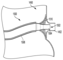
환자의 정지되지 않은 박동 심장을 통해 약물을 관류함으로써 심장 질환을 치료하는 방법이 개시된다. 환자의 관상동맥 및 관상정맥계를 통한 폐쇄 회로는 우관상동맥에 포지셔닝된 제1 약물 전달 카테터(1822), 좌관상동맥에 포지셔닝된 제2 약물 전달 카테터(1824), 관상정맥동에 포지셔닝된 약물 회수 카테터(1826), 및 다양한 카테터에 유체 결합된 외부 막 산소화 시스템(1820)으로 형성될 수 있다. 심장 질환을 치료하기 위한 약물은 폐쇄 회로를 통해 관류될 수 있다.A method of treating heart disease by perfusing a drug through a patient's non-stop beating heart is disclosed. A closed circuit through the patient's coronary and coronary venous systems includes a first drug delivery catheter 1822 positioned in the right coronary artery, a second drug delivery catheter 1824 positioned in the left coronary artery, and a drug withdrawal catheter positioned in the coronary sinus. 1826, and an external membrane oxygenation system 1820 fluidly coupled to various catheters. Drugs to treat heart disease can be perfused through a closed circuit.

Description
관련 출원(들)에 대한 상호 참조Cross-reference to related application(s)
본 출원은 2022년 2월 20일자로 출원된 미국 임시 특허 출원 번호 제63/312,029호, 및 2021년 2월 22일자로 출원된 미국 임시 특허 출원 일련 번호 제63/151,938호의 우선권 혜택을 주장하며, 그 개시 내용은 그 전체가 본원에 참조로 통합된다. This application claims the benefit of U.S. Provisional Patent Application Serial No. 63/312,029, filed February 20, 2022, and U.S. Provisional Patent Application Serial No. 63/151,938, filed February 22, 2021; The disclosure is incorporated herein by reference in its entirety.
기술분야Technology field
본 발명은 심장 질환의 치료에 관한 것으로, 특히 환자의 심장에 치료제를 국소적으로 전달하는 것에 관한 것이다.The present invention relates to the treatment of heart disease, and in particular to the local delivery of therapeutic agents to the patient's heart.
심부전과 같은 다양한 심장 질환 치료의 약리학적 발전에도 불구하고, 사망률 및 이환율은 여전히 받아들일 수 없을 정도로 높다. 더욱이, 특정 치료 접근법은 많은 환자(예를 들어, 다른 동반질환과 관련된 진행성 심부전 상태를 앓고 있는 환자)에게 적합하지 않다. 유전자 치료 및 세포 치료와 같은 대체 접근법은 많은 심장 질환의 근본 원인 병인을 해결하는 데 고유하게 맞춤화되고 효과적일 수 있는 잠재력으로 인해 관심이 높아졌다.Despite pharmacological advances in the treatment of various cardiac diseases such as heart failure, mortality and morbidity rates remain unacceptably high. Moreover, certain treatment approaches are not suitable for many patients (e.g., those suffering from advanced heart failure conditions associated with other comorbidities). Alternative approaches, such as gene therapy and cell therapy, have gained increased interest due to their potential to be uniquely tailored and effective in addressing the underlying etiology of many heart diseases.
그럼에도 불구하고 벡터 효율성, 용량, 특이성, 안전성 등을 포함한 전달과 관련된 문제는 여전히 남아 있다. 이와 같이, 효과적이고 내약성이 뛰어나며 최소 침습성인 다양한 심장 질환의 치료에 적합한 약물의 보다 표적화하고 균일한 전달을 달성하는 방법에 대한 추가 연구가 필요하다.Nevertheless, challenges related to delivery, including vector efficiency, dosage, specificity, and safety, still remain. As such, further research is needed on how to achieve more targeted and uniform delivery of drugs suitable for the treatment of a variety of cardiac diseases that are effective, well-tolerated, and minimally invasive.
본 발명의 목적은 최소 침습 방식으로 환자의 정지되지 않은 박동 심장에 약물을 관류시키는 방법을 제공하는 것이다.The object of the present invention is to provide a method for perfusing a drug into a patient's non-quitting beating heart in a minimally invasive manner.
본 발명의 목적은 관류액이 환자의 전신 순환계로부터 분리되도록 환자의 정지되지 않은 박동 심장을 통해 관류액(혈액 또는 약물 중 하나 이상을 함유할 수 있음)을 순환시키는 방법을 제공하는 것이다.It is an object of the present invention to provide a method of circulating perfusate (which may contain one or more of blood or drugs) through the non-stop beating heart of a patient such that the perfusate is isolated from the patient's systemic circulation.
본 발명의 목적은 약물 유전자 치료법의 국소 영역 전달을 제공하는 것이다.The object of the present invention is to provide localized delivery of drug gene therapy.
본 발명의 목적은 심장 질환을 치료하기 위해 환자에게 전달되는 약물의 전체 용량을 감소시키는 것이다.The objective of the present invention is to reduce the overall dose of drugs delivered to patients to treat heart disease.
본 발명의 목적은 심장 질환 치료에 적합한 약물 투여에 대한 위험 및/또는 불리한 면역 반응을 감소시키는 것이다.The aim of the present invention is to reduce the risk and/or adverse immune response to the administration of drugs suitable for the treatment of heart disease.
본 발명의 목적은 중화 항체, 예를 들어 유전자 치료 벡터에 대한 항체를 보유한 환자에게 약물 유전자 치료 약물을 재투여 및/또는 투여하는 것을 가능하게 하는 것인데, 그렇지 않으면 이러한 약물을 수용하기에 부적합한 후보가 될 수 있다.The aim of the present invention is to make it possible to re-administer and/or administer drug gene therapy drugs to patients who possess neutralizing antibodies, e.g. antibodies against the gene therapy vector, who would otherwise be unsuitable candidates for receiving these drugs. It can be.
본 발명의 목적은 심장에 산소를 공급하고 관상동맥 순환계를 환자의 전신 순환계로부터 격리하여 심장에 대한 약물의 노출을 방지하거나 감소시키면서 잠재적으로 심장 독성이 있는 약물이 전신 순환계로 유입되는 것을 방지하거나 감소시키기 위해 정지되지 않은 박동 심장을 통해 관류액을 순환시키는 것이다.The object of the present invention is to oxygenate the heart and isolate the coronary circulation from the patient's systemic circulation, thus preventing or reducing exposure of drugs to the heart, while preventing or reducing the entry of potentially cardiotoxic drugs into the systemic circulation. To achieve this, the perfusate is circulated through the non-stop beating heart.
상기 목적 및 기타 목적은 특정 실시양태에서 환자의 정지되지 않은 심장 박동에 약물을 관류시키는 방법에 관한 본 발명에 의해 충족된다. 일부 실시예들에서, 방법은 심장의 우관상동맥에 제1 약물 전달 카테터를 포지셔닝시키는 단계를 포함한다. 방법은 심장의 좌주관상동맥에 제2 약물 전달 카테터를 포지셔닝시키는 단계를 더 포함한다. 방법은 심장의 관상정맥동에 약물 전달 카테터를 포지셔닝시키는 단계를 더 포함한다. 일부 실시예들에서제1 약물 전달 카테터, 제2 약물 전달 카테터 및 약물 회수 카테터는 심장의 관상동맥, 심장의 관상정맥계 및 막 산소화 장치와 함께 폐쇄 회로를 형성한다. 방법은 환자의 전신 순환계로부터 환자의 관상동맥 순환계를 격리시키는 폐쇄 회로를 통해 약물을 관류시키는 단계를 더 포함한다. 일부 실시예들에서, 관류된 약물의 적어도 약 50%는 적어도 45분 동안 폐쇄 회로에 남아 있다. 일부 실시예들에, 약물은 관류 동안 심장 조직의 적어도 30%로 전달된다.These and other objects are met in certain embodiments by the present invention, which relates to a method of perfusing a drug into a patient's non-stop heartbeat. In some embodiments, the method includes positioning a first drug delivery catheter in the right coronary artery of the heart. The method further includes positioning the second drug delivery catheter in the left main coronary artery of the heart. The method further includes positioning the drug delivery catheter in the coronary sinus of the heart. In some embodiments the first drug delivery catheter, the second drug delivery catheter, and the drug withdrawal catheter form a closed circuit with the coronary arteries of the heart, the coronary venous system of the heart, and the membrane oxygenation device. The method further includes perfusing the drug through a closed circuit that isolates the patient's coronary circulation from the patient's systemic circulation. In some embodiments, at least about 50% of the perfused drug remains in closed circuit for at least 45 minutes. In some embodiments, the drug is delivered to at least 30% of the heart tissue during perfusion.
일부 실시예들에서, 방법은 약물 회수 카테터에 음압을 인가하는 단계를 더 포함한다. 일부 실시예들에서, 음압의 범위는 약 -100mmHg 내지 0mmHg이다.In some embodiments, the method further includes applying negative pressure to the drug withdrawal catheter. In some embodiments, the sound pressure ranges from about -100 mmHg to 0 mmHg.
일부 실시예들에서, 폐쇄 회로는 테베시안(Thebesian) 정맥을 통해 폐쇄 회로를 통해 순환하는 혈액 및/또는 약물의 누출을 방지 및/또는 최소화하기 위해 약물 회수 카테터에 음의 흡입 압력을 추가로 적용할 수 있게 하는 하나 이상의 흡입 메커니즘을 더 포함할 수 있다.In some embodiments, the closed circuit further applies negative suction pressure to the drug withdrawal catheter to prevent and/or minimize leakage of blood and/or drug circulating through the closed circuit via the Thebesian vein. It may further include one or more suction mechanisms that enable it.
일부 실시예들에서, 제1 약물 전달 카테터, 제2 약물 전달 카테터, 또는 약물 회수 카테터 중 하나 이상이 경피적으로 도입된다. 일부 실시예들에서, 제1 약물 전달 카테터 및/또는 제2 약물 전달 카테터는 제방향 삽관을 통해 포지셔닝된다. 일부 실시예들에서, 제1 약물 전달 카테터 및/또는 제2 약물 전달 카테터는 대동맥 대퇴골 및/또는 대동맥 요골에 접근함으로써 환자의 대동맥을 통해 포지셔닝된다. 일부 실시예들에서, 약물 회수 카테터는 환자의 대정맥을 통해 관상정맥동에 포지셔닝된다. 일부 실시예들에서, 약물 회수 카테터는 환자의 경정맥 또는 대퇴정맥을 통해 포지셔닝된다. 일부 실시예들에서, 막 산소화 장치는 회수 카테터와 제1 약물 전달 카테터 및 제2 약물 전달 카테터 중 하나 이상 사이에 포지셔닝된다. 일부 실시예들에서, 제1 약물 전달 카테터, 제2 약물 전달 카테터, 또는 약물 회수 카테터 중 하나 이상이 벌룬에 의해 밀봉되어 누출을 감소 또는 방지한다.In some embodiments, one or more of the first drug delivery catheter, the second drug delivery catheter, or the drug withdrawal catheter is introduced percutaneously. In some embodiments, the first drug delivery catheter and/or the second drug delivery catheter are positioned via directional cannulation. In some embodiments, the first drug delivery catheter and/or the second drug delivery catheter are positioned through the patient's aorta by accessing the aortafemoral and/or aortoradial. In some embodiments, the drug withdrawal catheter is positioned into the coronary sinus through the patient's vena cava. In some embodiments, the drug withdrawal catheter is positioned through the patient's jugular or femoral vein. In some embodiments, the membrane oxygenation device is positioned between the withdrawal catheter and one or more of the first drug delivery catheter and the second drug delivery catheter. In some embodiments, one or more of the first drug delivery catheter, the second drug delivery catheter, or the drug withdrawal catheter is sealed by the balloon to reduce or prevent leakage.
일부 실시예들에서, 방법은 폐쇄 회로를 통해 벌룬을 순환시키는 단계를 더 포함한다. 일부 실시예들에서, 혈액은 자가 혈액, 기증자로부터의 매칭 혈액 또는 이들의 조합을 포함한다. 일부 실시예들에서, 혈청이나 혈장과 같은 혈액 성분은 하나 이상의 파라미터에 따라 선택된다. 일부 실시예들에서, 하나 이상의 파라미터들은 선택된 항체의 존재 또는 부재를 포함한다. 일부 실시예들에서, 혈액의 약 1000 mL, 약 800 mL, 약 600 mL, 약 400 mL, 약 200 mL, 약 100 mL, 또는 약 50 mL가 상기 폐쇄 회로를 통해 순환된다.In some embodiments, the method further includes circulating the balloon through a closed circuit. In some embodiments, the blood includes autologous blood, matched blood from a donor, or a combination thereof. In some embodiments, blood components, such as serum or plasma, are selected according to one or more parameters. In some embodiments, one or more parameters include the presence or absence of a selected antibody. In some embodiments, about 1000 mL, about 800 mL, about 600 mL, about 400 mL, about 200 mL, about 100 mL, or about 50 mL of blood circulates through the closed circuit.
일부 실시예들에서, 관류시키는 단계는 약 5분 내지 약 5시간, 약 15분 내지 약 4시간, 약 30분 내지 약 3시간, 또는 약 1시간 내지 약 2시간의 기간에 걸쳐 발생한다. 일부 실시예들에서, 관류시키는 단계는 적어도 60분 동안 발생한다. 일부 실시예들에서, 관류시키는 단계는 약 75mL/분 내지 약 750mL/분, 약 150mL/분 내지 약 500mL/분, 또는 약 200mL/분 내지 약 300mL/분의 유량으로 발생한다.In some embodiments, the perfusing step occurs over a period of about 5 minutes to about 5 hours, about 15 minutes to about 4 hours, about 30 minutes to about 3 hours, or about 1 hour to about 2 hours. In some embodiments, the perfusion step occurs for at least 60 minutes. In some embodiments, the perfusing step occurs at a flow rate of about 75 mL/min to about 750 mL/min, about 150 mL/min to about 500 mL/min, or about 200 mL/min to about 300 mL/min.
일부 실시예들에서, 약물은 심장 질환의 치료에 적합하다. 일부 실시예들에서, 심장 질환은 심부전이다. 일부 실시예들에서, 심장 질환은 유전적으로 결정된 심장 질병이다. 일부 실시예들에서, 유전적으로 결정된 심장 질병은 유전적으로 결정된 심근병증이다.In some embodiments, the drug is suitable for treating heart disease. In some embodiments, the heart disease is heart failure. In some embodiments, the heart disease is a genetically determined heart disease. In some embodiments, the genetically determined heart disease is genetically determined cardiomyopathy.
일부 실시예들에서, 약물은 치료 폴리뉴클레오티드 서열을 포함한다. 일부 실시예들에서, 치료 폴리뉴클레오티드 서열은 하나 이상의 바이러스 벡터에 존재한다. 일부 실시예들에서, 하나 이상의 바이러스 벡터는 아데노 관련 바이러스, 아데노바이러스, 레트로바이러스, 단순 헤르페스 바이러스, 소 유두종 바이러스, 렌티바이러스 벡터, 우두 바이러스, 폴리오마 바이러스, 센다이 바이러스, 오르토믹소바이러스, 파라믹소바이러스, 파포바바이러스, 피코르나바이러스, 폭스 바이러스, 알파바이러스, 이들의 변이체 및 이들의 조합으로 이루어진 군으로부터 선택된다.In some embodiments, the drug comprises a therapeutic polynucleotide sequence. In some embodiments, the therapeutic polynucleotide sequence is present in one or more viral vectors. In some embodiments, the one or more viral vectors are adeno-associated virus, adenovirus, retrovirus, herpes simplex virus, bovine papillomavirus, lentiviral vector, vaccinia virus, polyoma virus, Sendai virus, orthomyxovirus, paramyxovirus. , papovavirus, picornavirus, poxvirus, alphavirus, variants thereof, and combinations thereof.
일부 실시예들에서, 바이러스 벡터는 아데노 관련 바이러스(adeno-associated virus; AAV)이다. 일부 실시예들에서, AAV는 AAV1, AAV2, AAV3, AAV4, AAV5, AAV6, AAV7, AAV8, AAV9, AAV10, AAV11, AAV12, 이들의 변이체, 및 이들의 조합 중 하나 이상이다. In some embodiments, the viral vector is an adeno-associated virus (AAV). In some embodiments, the AAV is one or more of AAV1, AAV2, AAV3, AAV4, AAV5, AAV6, AAV7, AAV8, AAV9, AAV10, AAV11, AAV12, variants thereof, and combinations thereof.
일부 실시예들에서, 치료 폴리뉴클레오티드 서열은 심장 질환 치료를 위한 단백질, 안티센스 RNA, ncRNA 또는 miRNA를 인코딩하는 핵산 서열을 포함한다. 일부 실시예들에서, 단백질은 인간 심장에서 발현되는 유전자에 상응한다. 일부 실시예들에서, 단백질은 SERCA2, MyBPC3, MYH7, PKP2, 디스트로핀, FKRP, 또는 이들의 조합 또는 이들의 변이체 중 하나 이상이다. 일부 실시예들에서, 치료 폴리뉴클레오티드 서열은 프로모터를 포함한다.In some embodiments, the therapeutic polynucleotide sequence comprises a nucleic acid sequence encoding a protein, antisense RNA, ncRNA, or miRNA for treating heart disease. In some embodiments, the protein corresponds to a gene expressed in the human heart. In some embodiments, the protein is one or more of SERCA2, MyBPC3, MYH7, PKP2, dystrophin, FKRP, or a combination thereof or a variant thereof. In some embodiments, the therapeutic polynucleotide sequence includes a promoter.
일부 실시예들에서, 폐쇄 회로를 통해 순환되는 약 20% v/v 미만, 약 15% v/v 미만, 약 10% v/v 미만, 약 5% v/v 미만, 약 4% v/v 미만, 약 3 % v/v 미만, 약 2% v/v 미만, 약 1% v/v 미만, 약 0.5% v/v 미만, 또는 실질적으로 없는(0% v/v) 혈액이 상기 폐쇄 회로 외부로 누출된다. 일부 실시예들에서, 폐쇄 회로를 통해 관류되는 약 20% v/v 미만, 약 15% v/v 미만, 약 10% v/v 미만, 약 5% v/v 미만, 약 4% v/v 미만, 약 3% v/v 미만, 약 2% v/v 미만, 약 1% v/v 미만, 약 0.5% v/v 미만, 또는 실질적으로 없는(0% v/v) 약물이 상기 폐쇄 회로 외부로 누출된다.In some embodiments, less than about 20% v/v, less than about 15% v/v, less than about 10% v/v, less than about 5% v/v, less than about 4% v/v circulated through a closed circuit. less than, less than about 3% v/v, less than about 2% v/v, less than about 1% v/v, less than about 0.5% v/v, or substantially no (0% v/v) blood in the closed circuit. leaks to the outside. In some embodiments, less than about 20% v/v, less than about 15% v/v, less than about 10% v/v, less than about 5% v/v, less than about 4% v/v perfused through the closed circuit. Less than, less than about 3% v/v, less than about 2% v/v, less than about 1% v/v, less than about 0.5% v/v, or substantially no (0% v/v) drug is present in the closed circuit. leaks to the outside.
일부 실시예들에서, 제1 약물 전달 카테터, 제2 약물 전달 카테터, 또는 약물 회수 카테터 중 하나 이상은 벌룬 카테터이다.In some embodiments, one or more of the first drug delivery catheter, the second drug delivery catheter, or the drug withdrawal catheter is a balloon catheter.
상기 목적 및 다른 목적은 특정 실시예들에서 관류 동안 정지되지 않고 박동하는 환자의 심장의 폐쇄 회로를 통해 관류액의 관류를 유지하는 방법에 관한 본 발명에 의해 추가로 충족된다. 일부 실시예들에서, 방법은 심장의 우관상동맥에 제1 카테터를 포지셔닝시키는 단계를 포함한다. 일부 실시예들에서, 방법은 심장의 좌주관상동맥에 제2 카테터를 포지셔닝시키는 단계를 더 포함한다. 일부 실시예들에서, 방법은 심장의 관상정맥동에 회수 카테터를 포지셔닝시키는 단계를 더 포함한다. 일부 실시예들에서, 제1 카테터, 제2 카테터 및 회수 카테터는 관상동맥, 관상정맥계 및 막 산소화 장치와 함께 심장을 통해 폐쇄 회로를 형성한다. 일부 실시예들에서, 방법은 관류액을 제1 카테터 및 제2 카테터를 통해 심장에 도입하고 회수 카테터를 통해 관류액을 수집함으로써 폐쇄 회로를 통해 관류액을 유동하게 하는 단계를 더 포함한다. 일부 실시예들에서, 폐쇄 회로는 환자의 전신 순환계로부터 환자의 관상동맥 순환계를 격리시킨다.The above and other objects are further met by the present invention, which in certain embodiments relates to a method of maintaining perfusion of perfusate through a closed circuit of a non-quiescent, beating heart of a patient during perfusion. In some embodiments, the method includes positioning the first catheter in the right coronary artery of the heart. In some embodiments, the method further includes positioning the second catheter in the left main coronary artery of the heart. In some embodiments, the method further includes positioning the retrieval catheter in the coronary sinus of the heart. In some embodiments, the first catheter, the second catheter, and the return catheter form a closed circuit through the heart with the coronary artery, coronary venous system, and membrane oxygenation device. In some embodiments, the method further includes flowing the perfusate through a closed circuit by introducing the perfusate into the heart through the first catheter and the second catheter and collecting the perfusate through the return catheter. In some embodiments, the closed circuit isolates the patient's coronary circulation from the patient's systemic circulation.
일부 실시예들에서, 관류는 적어도 60분 동안 유지된다. 일부 실시예들에서, 관류는 적어도 120분 동안 유지된다.In some embodiments, perfusion is maintained for at least 60 minutes. In some embodiments, perfusion is maintained for at least 120 minutes.
일부 실시예들에서, 방법은 회수 카테터에 음압을 인가하는 단계를 더 포함하며, 음압의 범위는 약 -100 mmHg 내지 0 mmHg이다.In some embodiments, the method further includes applying negative pressure to the retrieval catheter, where the negative pressure ranges from about -100 mmHg to 0 mmHg.
일부 실시예들에서, 제1 카테터, 제2 카테터, 또는 회수 카테터 중 하나 이상이 경피적으로 도입된다.In some embodiments, one or more of the first catheter, second catheter, or retrieval catheter is introduced percutaneously.
일부 실시예들에서, 막 산소화 장치는 회수 카테터와 제1 약물 전달 카테터 및 제2 약물 전달 카테터 중 하나 이상 사이에 포지셔닝된다. In some embodiments, the membrane oxygenation device is positioned between the withdrawal catheter and one or more of the first drug delivery catheter and the second drug delivery catheter.
일부 실시예들에서, 방법은 폐쇄 회로를 통해 혈액을 순환시키는 단계를 더 포함하며, 혈액은 자가 혈액, 기증자로부터의 매칭 혈액, 또는 이들의 조합을 포함한다. 일부 실시예들에서, 혈액의 약 1000 mL, 약 800 mL, 약 600 mL, 약 400 mL, 약 200 mL, 약 100 mL, 또는 약 50 mL가 상기 폐쇄 회로를 통해 순환된다.In some embodiments, the method further includes circulating blood through a closed circuit, wherein the blood includes autologous blood, matched blood from a donor, or a combination thereof. In some embodiments, about 1000 mL, about 800 mL, about 600 mL, about 400 mL, about 200 mL, about 100 mL, or about 50 mL of blood circulates through the closed circuit.
일부 실시예들에서, 관류시키는 단계는 약 75mL/분 내지 약 750mL/분, 약 150mL/분 내지 약 500mL/분, 또는 약 200mL/분 내지 약 300mL/분의 유량으로 발생한다. 일부 실시예들에서, 폐쇄 회로를 통해 순환되는 약 20% v/v 미만, 약 15% v/v 미만, 약 10% v/v 미만, 약 5% v/v 미만, 약 4% v/v 미만, 약 3 % v/v 미만, 약 2% v/v 미만, 약 1% v/v 미만, 약 0.5% v/v 미만, 또는 실질적으로 없는(0% v/v) 혈액이 상기 폐쇄 회로 외부로 누출된다.In some embodiments, the perfusing step occurs at a flow rate of about 75 mL/min to about 750 mL/min, about 150 mL/min to about 500 mL/min, or about 200 mL/min to about 300 mL/min. In some embodiments, less than about 20% v/v, less than about 15% v/v, less than about 10% v/v, less than about 5% v/v, less than about 4% v/v circulated through a closed circuit. less than, less than about 3% v/v, less than about 2% v/v, less than about 1% v/v, less than about 0.5% v/v, or substantially no (0% v/v) blood in the closed circuit. leaks to the outside.
일부 실시예들에서, 제1 카테터, 제2 카테터, 또는 회수 카테터 중 하나 이상은 벌룬 카테터이다.In some embodiments, one or more of the first catheter, second catheter, or retrieval catheter is a balloon catheter.
상기 목적 및 기타 목적은 특정 실시예에서 유체 연결될 때 환자의 심장 내에서 국소 영역 관류를 수행하기 위한 시스템에 관한 본 발명에 의해 추가로 충족된다. 일부 실시예들에서, 시스템은, 심장의 우관상동액에 삽입을 위해 적응된 제1 카테터; 심장의 좌주관상동맥에 삽입을 위해 적응된 제2 카테터; 심장의 관상정맥동에 삽입을 위해 적응된 회수 카테터; 제1 카테터, 제2 카테터, 회수 카테터, 및 산소 공급원에 유체 결합된 막 산소화 장치; 및 제1 카테터와 제2 카테터를 통해 유체 유동을 구동하도록 구성된 펌프를 포함한다. 일부 실시예들에서, 제1 카테터, 제2 카테터, 회수 카테터 및 막 산소화 장치는 제1 카테터가 우관상동맥에 삽입되고, 제2 카테터가 좌주관상독맥에 삽입되고, 회수 카테터가 관상정맥동에 삽입될 때 환자의 전신 순환계로부터 격리되는 심장을 통해 폐쇄 회로를 함께 형성한다. 일부 실시예들에서, 관류된 약물의 적어도 약 50%는 적어도 45분 동안 폐쇄 회로에 남아 있다.These and other objects are further met by the present invention, which in certain embodiments relates to a system for performing localized regional perfusion within a patient's heart when fluidly connected. In some embodiments, the system includes a first catheter adapted for insertion into the right coronary sinus of the heart; a second catheter adapted for insertion into the left main coronary artery of the heart; Salvage catheter adapted for insertion into the coronary sinus of the heart; a membrane oxygenation device fluidly coupled to a first catheter, a second catheter, a withdrawal catheter, and an oxygen source; and a pump configured to drive fluid flow through the first catheter and the second catheter. In some embodiments, the first catheter, the second catheter, the retrieval catheter, and the membrane oxygenation device are configured such that the first catheter is inserted into the right coronary artery, the second catheter is inserted into the left main coronary artery, and the retrieval catheter is inserted into the coronary sinus. When these occur, they together form a closed circuit with the heart isolated from the patient's systemic circulatory system. In some embodiments, at least about 50% of the perfused drug remains in closed circuit for at least 45 minutes.
상기 목적 및 다른 목적은 특정 실시예들에서 국소 여역 관류 시스템에 관한 것인 본 발명에 의해 추가로 충족되여, 국소 영역 관류 시스템은, 환자의 심장의 우관상동액에 삽입된 제1 카테터; 심장의 좌주관상동맥에 삽입된 제2 카테터; 심장의 관상정맥동에 삽입된 회수 카테터; 제1 카테터, 제2 카테터, 회수 카테터 및 산소 공급원에 유체 결합된 막 산소화 장치; 및 제1 카테터와 제2 카테터를 통해 그리고 회수 카테터를 통해 심장 밖으로 체 유동을 구동하도록 구성된 펌프를 포함한다. 일부 실시예들에서, 제1 카테터, 제2 카테터, 회수 카테터 및 막 산소화 장치는 심장의 관상동맥 및 관상정맥계와 함께 환자의 전신 순환계로부터 격리되는 심장을 통해 폐쇄 회로를 형성한다. 일부 실시예들에서, 관류된 약물의 적어도 약 50%는 적어도 45분 동안 폐쇄 회로에 남아 있다.The above objects and other objects are further met by the present invention, which in certain embodiments relates to a regional region perfusion system, the regional region perfusion system comprising: a first catheter inserted into the right coronary sinus fluid of the patient's heart; a second catheter inserted into the left main coronary artery of the heart; A retrieval catheter inserted into the coronary sinus of the heart; a membrane oxygenation device fluidly coupled to the first catheter, the second catheter, the return catheter, and the oxygen source; and a pump configured to drive body flow through the first and second catheters and out of the heart through the return catheter. In some embodiments, the first catheter, the second catheter, the recovery catheter, and the membrane oxygenation device, together with the heart's coronary and venous systems, form a closed circuit through the heart that is isolated from the patient's systemic circulation. In some embodiments, at least about 50% of the perfused drug remains in closed circuit for at least 45 minutes.
일부 실시예들에서, 막 산소화 장치는 관류 동안 폐쇄 회로에 약물을 주입하도록 구성된 저장소를 포함한다.In some embodiments, the membrane oxygenation device includes a reservoir configured to infuse drugs in a closed circuit during perfusion.
일부 실시예들에서, 음압의 범위는 약 -100mmHg 내지 0mmHg이다.In some embodiments, the sound pressure ranges from about -100 mmHg to 0 mmHg.
일부 실시예들에서, 제1 카테터, 제2 카테터, 또는 회수 카테터 중 하나 이상이 경피적으로 도입된다. 일부 실시예들에서, 제1 카테터 및/또는 제2 카테터는 제방향 삽관을 통해 포지셔닝된다. 일부 실시예들에서, 회수 카테터는 환자의 대정맥을 통해 관상정맥동에 포지셔닝된다.In some embodiments, one or more of the first catheter, second catheter, or retrieval catheter is introduced percutaneously. In some embodiments, the first catheter and/or the second catheter are positioned via directional cannulation. In some embodiments, the retrieval catheter is positioned into the coronary sinus through the patient's vena cava.
상기 목적 및 다른 목적은 특정 실시예들에서 환자의 전신 순환계로부터 환장의 심장을 격리시키는 방법에 관한 것인 본 발명에 의해 추가로 충족되며, 상기 방법은, 심장의 우관상동맥에 제1 카테터를 포지셔닝시키는 단계; 심장의 좌주관상동맥에 제2 카테터를 포지셔닝시키는 단계; 심장의 관상정맥동에 회수 카테터를 포지셔닝시키는 단계로서, 제1 카테터, 제2 카테터, 및 회수 카테터는 심장의 관상동맥, 심장의 관상정맥계, 및 막 산소화 장치아 함께 폐쇄 회로를 형성하는, 상기 포지셔닝시키는 단계; 산소화된 혈액이 상기 폐쇄 회로를 통해 유동하도록 하는 단계; 및 약물을 환자의 전신 순환계에 도입하는 단계를 포함한다. 일부 실시예들에서, 폐쇄 회로는 환자의 전신 순환계로부터 환자의 관상동맥 순환계를 격리시킨다. 일부 실시예들에서, 약물은 심장독성 약물이고, 폐쇄 회로가 없는 상태에서 심장독성 약물을 투여하는 것에 비해 심장에 대한 심장독성 약물의 노출이 방지되거나 감소된다.These and other objects are further met by the present invention, which in certain embodiments relates to a method of isolating a spared heart from the systemic circulation of a patient, the method comprising: introducing a first catheter into the right coronary artery of the heart; positioning; positioning the second catheter in the left main coronary artery of the heart; Positioning a retrieval catheter in the coronary sinus of the heart, wherein the first catheter, the second catheter, and the retrieval catheter together form a closed circuit with the coronary arteries of the heart, the coronary venous system of the heart, and the membrane oxygenation device. step; allowing oxygenated blood to flow through the closed circuit; and introducing the drug into the patient's systemic circulation. In some embodiments, the closed circuit isolates the patient's coronary circulation from the patient's systemic circulation. In some embodiments, the drug is a cardiotoxic drug and exposure of the cardiotoxic drug to the heart is prevented or reduced compared to administering the cardiotoxic drug in the absence of a closed loop.
상기 목적 및 다른 목적은 특정 실시예들에서 전술한 방법 중 임의의 방법을 수행하도록 구성된 국소 역 관류 시스템에 관한 본 발명에 의해 추가로 충족된다.These and other objects are further met by the present invention, which in certain embodiments relates to a local counter perfusion system configured to perform any of the methods described above.
본 개시의의 상기 특징 및 다른 특징, 그 특징 및 다양한 이점은 첨부 도면과 함께 하기의 상세한 설명을 고려하면 더욱 명백해질 것이다:
도 1은 적어도 하나의 실시예에 따른 단일 벌룬 구조를 갖는 제1 예시적인 회수 카테터의 개략도를 예시한다;
도 2는 제1 예시적인 회수 카테터의 일 실시예에 따른 제작된 회수 카테터의 사진이다;
도 3은 적어도 하나의 실시예에 따른 제1 예시적인 회수 카테터의 배치를 예시한다;
도 4는 적어도 하나의 실시예에 따른 단일 벌룬 구조를 갖는 제2 예시적인 회수 카테터의 배치를 예시한다;
도 5는 적어도 하나의 실시예에 따른 단일 벌룬 구조를 각각 갖는 제3 예시적인 회수 카테터 및 제4 예시적인 회수 카테터의 배치를 예시한다;
도 6은 적어도 일 실시예에 따른 단일 벌룬 구조를 갖는 제5 예시적인 회수 카테터 및 벌룬 구조가 없는 제6 예시적인 회수 카테터의 배치를 예시한다;
도 7은 적어도 하나의 실시예에 따른 다중 벌룬 구조를 갖는 제7 예시적인 회수 카테터의 배치를 예시한다;
도 8은 적어도 하나의 실시예에 따른 부분적으로 덮여있고 재포획 가능한 스텐트 구조를 갖는 제8 예시적인 회수 카테터의 배치를 예시한다;
도 9는 적어도 일 실시예에 따른 전개 가능하고 수축 가능한 스텐트 구조를 갖는 제9 예시적인 회수 카테터의 배치를 예시한다;
도 10은 적어도 하나의 실시예에 따른 덮힌 디스크형 스텐트 구조를 갖는 제10 예시적인 회수 카테터의 배치를 예시한다;
도 11a는 적어도 하나의 실시예에 따른 단일 벌룬 구조를 갖는 제1 예시적인 관류 카테터의 개략도이다;
도 11b는 적어도 하나의 실시예에 따른 팽창 상태의 제1 예시적인 관류 카테터의 벌룬 구조의 개략도이다;
도 11c는 적어도 하나의 실시예에 따른 수축 상태의 제1 예시적인 관류 카테터의 벌룬 구조의 개략도이다;
도 11d는 적어도 하나의 실시예에 따른 대동맥 내 제1 예시적인 관류 카테터의 배치를 예시한다;
도 12a는 적어도 하나의 실시예에 따른 원위 플러그를 갖는 제2 예시적인 관류 카테터의 개략도이다;
도 12b는 적어도 하나의 실시예에 따른 제2 예시적인 관류 카테터의 플러그의 개략도이다;
도 12c는 적어도 하나의 실시예에 따른 팽창 상태의 제2 예시적인 관류 카테터의 플러그의 개략도이다;
도 12d는 적어도 하나의 실시예에 따른 대동맥 내 제2 예시적인 관류 카테터의 배치를 예시한다;
도 13a는 적어도 하나의 실시예에 따른 원위 웨지를 갖는 제3 예시적인 관류 카테터의 개략도이다;
도 13b는 적어도 하나의 실시예에 따른 제3 예시적인 관류 카테터의 웨지의 개략도이다;
도 13c는 적어도 하나의 실시예에 따른 팽창 상태의 제3 예시적인 관류 카테터의 원위 단부의 추가 개략도이다;
도 13d는 적어도 하나의 실시예에 따른 대동맥 내 제3 예시적인 관류 카테터의 배치를 예시한다;
도 14a는 적어도 하나의 실시예에 따른 부분적으로 덮여있고 재포획 가능한 스텐트 구조를 갖는 제4 예시적인 관류 카테터의 배치를 예시한다;
도 14b는 적어도 하나의 실시예에 따른 수축 상태의 제4 예시적인 관류 카테터의 스텐트 구조를 예시한다;
도 14c는 적어도 하나의 실시예에 따른 전개 상태의 제4 예시적인 관류 카테터의 스텐트 구조를 예시한다;
도 15a는 적어도 하나의 실시예에 따른 해체 가능한 덮여있는 편조 디스크를 갖는 제5 예시적인 관류 카테터의 배치를 예시한다;
도 15b는 적어도 하나의 실시예에 따른 전개 상태의 제5 예시적인 관류 카테터의 편조 디스크를 예시한다;
도 16a는 적어도 하나의 실시예에 따른 테이퍼형 루멘 샤프트를 갖는 제6 예시적인 관류 카테터의 개략도이다;
도 16b는 적어도 하나의 실시예에 따른 제6 예시적인 관류 카테터의 배치를 예시한다;
도 16c는 적어도 하나의 실시예에 따른 대동맥 내 제6 예시적인 관류 카테터의 배치를 예시한다;
도 16d는 적어도 하나의 실시예에 따른 제6 예시적인 관류 카테터의 사전 형상 루멘 샤프트를 예시한다;
도 17은 다양한 실시예에 따른 예시적인 카테터를 위한 예시적인 미리 형성된 루멘 샤프트를 예시한다;
도 18a는 본 개시의 실시예에 따른 예시적인 국소 영역(loco-regional) 관류 시스템을 도시한다;
도 18b는 본 개시의 실시예에 따른 예시적인 국소 영역 관류 장치의 개략도이다;
도 19는 좌주관상동맥 카테터, 우관상동맥 카테터 및 관상정맥동 벌룬의 위치를 보여주는, 정지되지 않은 돼지 심장의 국소 영역 관류 동안 캡처된 방사선 사진이다; 그리고
도 20은 국소 영역 관류 동안 측정된 펌프 속도, 유량 및 압력의 플롯이다.The above and other features, features and various advantages of the present disclosure will become more apparent upon considering the following detailed description in conjunction with the accompanying drawings:
1 illustrates a schematic diagram of a first exemplary retrieval catheter having a single balloon structure according to at least one embodiment;
Figure 2 is a photograph of a retrieval catheter fabricated according to one embodiment of a first exemplary retrieval catheter;
3 illustrates the placement of a first exemplary retrieval catheter according to at least one embodiment;
4 illustrates the arrangement of a second exemplary retrieval catheter having a single balloon structure according to at least one embodiment;
5 illustrates an arrangement of a third exemplary withdrawal catheter and a fourth exemplary withdrawal catheter each having a single balloon structure according to at least one embodiment;
6 illustrates an arrangement of a fifth exemplary retrieval catheter with a single balloon structure and a sixth exemplary retrieval catheter without a balloon structure, according to at least one embodiment;
Figure 7 illustrates the arrangement of a seventh exemplary retrieval catheter having a multiple balloon structure according to at least one embodiment;
8 illustrates the placement of an eighth exemplary retrieval catheter having a partially covered and recaptureable stent structure according to at least one embodiment;
9 illustrates the placement of a ninth exemplary retrieval catheter having a deployable and retractable stent structure according to at least one embodiment;
Figure 10 illustrates the placement of a tenth exemplary retrieval catheter having a covered disc-shaped stent structure according to at least one embodiment;
FIG. 11A is a schematic diagram of a first exemplary perfusion catheter with a single balloon structure according to at least one embodiment;
FIG. 11B is a schematic diagram of a balloon structure of a first exemplary perfusion catheter in an expanded state according to at least one embodiment;
FIG. 11C is a schematic diagram of a balloon structure of a first exemplary perfusion catheter in a deflated state according to at least one embodiment;
11D illustrates placement of a first example perfusion catheter in the aorta according to at least one embodiment;
Figure 12A is a schematic diagram of a second exemplary perfusion catheter with a distal plug according to at least one embodiment;
Figure 12B is a schematic diagram of a plug of a second exemplary perfusion catheter according to at least one embodiment;
FIG. 12C is a schematic diagram of a plug of a second exemplary perfusion catheter in an expanded state according to at least one embodiment;
12D illustrates placement of a second example perfusion catheter in the aorta according to at least one embodiment;
Figure 13A is a schematic diagram of a third exemplary perfusion catheter with a distal wedge according to at least one embodiment;
Figure 13B is a schematic diagram of a wedge of a third exemplary perfusion catheter according to at least one embodiment;
Figure 13C is a further schematic diagram of the distal end of a third exemplary perfusion catheter in an expanded state according to at least one embodiment;
13D illustrates placement of a third example perfusion catheter within the aorta according to at least one embodiment;
FIG. 14A illustrates the placement of a fourth exemplary perfusion catheter with a partially covered and recaptureable stent structure according to at least one embodiment;
14B illustrates the stent structure of a fourth exemplary perfusion catheter in a deflated state according to at least one embodiment;
FIG. 14C illustrates the stent structure of a fourth exemplary perfusion catheter in a deployed state according to at least one embodiment;
FIG. 15A illustrates the arrangement of a fifth exemplary perfusion catheter with a removable covered braided disk according to at least one embodiment;
FIG. 15B illustrates the braided disk of a fifth exemplary perfusion catheter in a deployed state according to at least one embodiment;
Figure 16A is a schematic diagram of a sixth exemplary perfusion catheter with a tapered lumen shaft according to at least one embodiment;
Figure 16B illustrates the placement of a sixth exemplary perfusion catheter according to at least one embodiment;
FIG. 16C illustrates placement of a sixth exemplary perfusion catheter within the aorta according to at least one embodiment;
16D illustrates a pre-shaped lumen shaft of a sixth exemplary perfusion catheter according to at least one embodiment;
17 illustrates an example preformed lumen shaft for an example catheter according to various embodiments;
FIG. 18A illustrates an exemplary loco-regional perfusion system according to embodiments of the present disclosure;
Figure 18B is a schematic diagram of an exemplary local area perfusion device according to embodiments of the present disclosure;
Figure 19 is a radiograph captured during regional regional perfusion of a resting porcine heart, showing the positions of the left main coronary artery catheter, right coronary artery catheter, and coronary sinus balloon; and
Figure 20 is a plot of pump speed, flow rate and pressure measured during local area perfusion.
본원에 사용된 바와 같이, 단수 형태인 "a", "an" 및 "the"는 문맥에서 달리 나타내지 않는 한 복수 지시어를 포함한다. 따라서, 예를 들어, "약물"에 대한 언급에는 단일 약물뿐만 아니라 두 가지 이상의 서로 다른 약물의 혼합물이 포함되며, "바이러스 벡터"에 대한 언급은 단일 바이러스 벡터뿐만 아니라 두 가지 이상의 서로 다른 바이러스 벡터의 혼합물 등을 포함한다.As used herein, the singular forms “a”, “an” and “the” include plural referents unless the context clearly indicates otherwise. Thus, for example, a reference to a “drug” includes not only a single drug but also a mixture of two or more different drugs, and a reference to a “viral vector” includes not only a single viral vector but also a mixture of two or more different viral vectors. Including mixtures, etc.
또한 본원에 사용된 바와 같이, 측정된 양과 관련하여 사용될 때 "약"은 측정을 수행하고 측정 장비의 정확성과 목적에 상응하는 수준의 주의를 기울이는 데 있어 당업자가 예상하는 측정된 양의 정상적인 변화를 의미한다. 특정 실시예들에서, "약"이라는 용어는 인용된 숫자 ±10%를 포함하므로, "약 10"은 9 내지 11을 포함하게 된다.Also, as used herein, when used in reference to a measured quantity, "about" means the normal variation in the measured quantity that would be expected by a person skilled in the art in making the measurement and exercising a level of care commensurate with the accuracy and purpose of the measuring equipment. it means. In certain embodiments, the term “about” includes the recited number plus or minus 10%, such that “about 10” includes 9 to 11.
또한 본원에 사용된 바와 같이, "폴리뉴클레오타이드"는 당업계에서 일반적이고 관례적인 의미를 가지며, DNA 또는 RNA 분자와 같은 임의의 중합체성 핵산뿐만 아니라 당업자에게 공지된 화학적 유도체도 포함한다. 폴리뉴클레오티드에는 치료 단백질을 코딩하는 것들이 포함될 뿐만 아니라, 당업계에 공지된 기술(예를 들어, 안티센스, 간섭 또는 소형 간섭 핵산)을 사용하여 표적 핵산 서열의 발현을 감소시키는 데 사용될 수 있는 서열도 포함된다. 폴리뉴클레오티드는 또한 심혈관계 세포 내에서 표적화된 핵산 서열의 발현 또는 표적화된 단백질의 생산을 개시하거나 증가시키는 데 사용될 수 있다. 표적 핵산 및 단백질은, 이에 제한되는 것은 아니나, 표적 조직에서 통상적으로 발견되는 핵산 및 단백질, 이러한 자연적으로 발생하는 핵산 또는 단백질의 유도체, 표적 조직에서 통상적으로 발견되지 않는 자연적으로 발생하는 핵산 또는 단백질, 또는 합성 핵산 또는 단백질을 포함한다. 하나 이상의 폴리뉴클레오티드는 하나 이상의 표적화된 핵산 서열 또는 단백질을 증가 및/또는 감소시키기 위해 조합되어 사용될 수 있고, 동시에 및/또는 순차적으로 투여될 수 있다.Also, as used herein, “polynucleotide” has its ordinary and customary meaning in the art and includes any polymeric nucleic acid, such as a DNA or RNA molecule, as well as chemical derivatives known to those skilled in the art. Polynucleotides include those that encode therapeutic proteins, but also sequences that can be used to reduce expression of target nucleic acid sequences using techniques known in the art (e.g., antisense, interfering, or small interfering nucleic acids). do. Polynucleotides can also be used to initiate or increase expression of a targeted nucleic acid sequence or production of a targeted protein in cardiovascular cells. Target nucleic acids and proteins include, but are not limited to, nucleic acids and proteins normally found in target tissues, derivatives of such naturally occurring nucleic acids or proteins, naturally occurring nucleic acids or proteins not normally found in target tissues, or synthetic nucleic acids or proteins. One or more polynucleotides can be used in combination to increase and/or decrease one or more targeted nucleic acid sequences or proteins and can be administered simultaneously and/or sequentially.
또한 본원에 사용된 바와 같이, "침투", "사용하지 않음" 및 "사용하지 않음"은 당해 기술분야에서 통상적이고 관습적인 의미를 가지며, 기술분야에서 인정되는 용어인 "주사" 또는 "볼루스 주입"(통상적으로 1분 미만)보다 실질적으로 긴 기간(통상적으로 1분 이상) 동안의 투여를 의미한다. 관류의 유량은 투여된 용량에 따라 적어도 부분적으로 달라질 것이다.Also, as used herein, “infiltration,” “no use,” and “no use” have their usual and customary meanings in the art, and “injection” or “bolus” are art-recognized terms. "infusion" means administration over a period of time (typically greater than 1 minute) substantially longer than that of "infusion" (typically less than 1 minute). The flow rate of perfusion will depend, at least in part, on the administered dose.
또한 본원에 사용된 바와 같이, "외인성" 핵산 또는 유전자는 핵산 전달에 사용되는 벡터에서 자연적으로 발생하지 않는 것이다; 예를 들어, 바이러스 벡터에서 자연적으로 발견되지는 않지만, 이 용어는 환자 또는 숙주에서 자연적으로 발생하는 단백질 또는 폴리펩티드를 코딩하는 핵산을 배제하려는 의도는 아니다.Also as used herein, an “exogenous” nucleic acid or gene is one that does not occur naturally in the vector used to transfer the nucleic acid; The term is not intended to exclude nucleic acids encoding proteins or polypeptides that occur naturally in a patient or host, for example, but not naturally found in viral vectors.
또한 본원에 사용된 바와 같이, "심장 세포"에는 심근 세포, 심장 혈관계 세포 또는 심장 판막에 존재하는 세포와 같이 심장의 구조를 유지하거나 기능을 제공하는 데 관여하는 심장의 모든 세포가 포함된다. 심장 세포에는 심장 근세포(정상 및 비정상 전기적 특성을 모두 가짐), 상피 세포, 내피 세포, 섬유아세포, 전도 조직 세포, 심장 박동 조절 세포 및 뉴런이 포함된다.Also, as used herein, “cardiac cell” includes all cells of the heart that are involved in maintaining the structure or providing function of the heart, such as cardiomyocytes, cardiovascular cells, or cells present in the heart valves. Cardiac cells include cardiac myocytes (with both normal and abnormal electrical properties), epithelial cells, endothelial cells, fibroblasts, conducting tissue cells, pacemaker cells, and neurons.
또한 본원에 사용된 바와 같이, "격리된", "실질적으로 격리된", "대부분 격리된" 및 이들의 변형은 관상정맥, 심장, 체정맥 또는 체순환계의 완전하거나 절대적인 격리를 요구하지 않는 용어이며, 오히려, 이는 지정된 순환계의 대부분, 바람직하게는 주요 부분 또는 심지어 실질적으로 전부가 격리된다는 것을 의미하도록 의도된다. 또한 본원에 사용된 바와 같이, "부분적으로 격리된"이라는 것은 특정 순환계의 중요하지 않은 부분이 고립되어 있음을 의미한다.Also, as used herein, “isolated,” “substantially isolated,” “substantially isolated,” and variations thereof are terms that do not require complete or absolute isolation of the coronary veins, heart, systemic veins, or systemic circulation. Rather, it is intended to mean that most, preferably a major part or even substantially all, of the designated circulatory system is isolated. Also as used herein, “partially isolated” means that a non-critical portion of a particular circulatory system is isolated.
또한 본원에 사용된 바와 같이, "비자연적으로 제한되는"에는 혈관을 통한 체액의 유동을 제한하는 모든 방법(예를 들어, 벌룬 카테터, 봉합사 등)이 포함되지만, 자연적으로 발생하는 제한(예를 들어, 플라크 축적(협착증))은 포함되지 않는다. 비자연적 제한에는 관상동맥 순환계 등의 실질적 또는 전체적 격리가 포함된다.Also, as used herein, “unnaturally restrictive” includes any method of restricting the flow of fluid through a blood vessel (e.g., balloon catheters, sutures, etc.), but not a naturally occurring restriction (e.g. For example, plaque buildup (stenosis) is not included. Unnatural restrictions include substantial or total isolation of the coronary circulation, etc.
또한 본원에 사용된 바와 같이, "최소 침습"은 심장이나 심장과 밀접하게 연결된 혈관에 대한 개방적인 수술적 접근을 필요로 하지 않는 모든 시술을 포함하도록 의도된다. 이러한 시술에는 심장에 접근하기 위한 내시경 수단과 큰 동맥 및 정맥을 통한 접근에 의존하는 카테터 기반 수단의 사용이 포함된다.Also, as used herein, “minimally invasive” is intended to include any procedure that does not require open surgical access to the heart or blood vessels closely connected to the heart. These procedures include the use of endoscopic means to access the heart and catheter-based means that rely on access through large arteries and veins.
또한 본원에 사용된 바와 같이, "아데노 관련 바이러스" 또는 "AAV"는 모든 아형, 혈청형, 유사형은 물론 자연적으로 발생하는 형태와 재조합 형태를 포함한다. 다양한 AAV 혈청형 및 균주가 당업계에 공지되어 있으며 ATCC 및 학술적 또는 상업적 소스와 같은 소스로부터 공개적으로 이용 가능하다. 대안으로, 공개되고/되거나 다양한 데이터베이스로부터 이용 가능한 AAV 혈청형 및 균주로부터의 서열은 공지된 기술을 사용하여 합성될 수 있다.Also, as used herein, “adeno-associated virus” or “AAV” includes all subtypes, serotypes, and pseudotypes, as well as naturally occurring and recombinant forms. A variety of AAV serotypes and strains are known in the art and are publicly available from sources such as ATCC and academic or commercial sources. Alternatively, sequences from AAV serotypes and strains that are published and/or available from various databases can be synthesized using known techniques.
또한 본원에 사용된 바와 같이, "혈청형"은 정의된 항혈청과의 캡시드 단백질 반응성을 기반으로 다른 AAV에 의해 식별되고 구별되는 AAV를 의미한다. AAV1부터 AAV12까지를 포함하여 적어도 12개의 인간 AAV 혈청형이 알려져 있지만, 추가 혈청형이 계속해서 발견되고 있으며 새로 발견된 혈청형의 사용이 고려된다.Also as used herein, “serotype” refers to an AAV that is identified and distinguished from other AAVs based on capsid protein reactivity with a defined antiserum. At least 12 human AAV serotypes are known, including AAV1 through AAV12, but additional serotypes continue to be discovered and newly discovered serotypes are considered for use.
또한 본원에 사용된 바와 같이, "의사형(pseudotyped)" AAV는 하나의 혈청형으로부터의 캡시드 단백질 및 상이한 또는 이종 혈청형의 5' 및 3' 역위 말단 반복부(ITR)를 포함하는 바이러스 게놈을 함유하는 AAV를 의미한다. 의사형 재조합 AAV(rAAV)는 캡시드 혈청형의 세포 표면 결합 특성과 ITR 혈청형과 일치하는 유전적 특성을 가질 것으로 예상된다. 의사형 rAAV는 VP1, VP2 및 VP3 캡시드 단백질을 포함하는 AAV 캡시드 단백질과 AAV1부터 AAV12까지의 영장류 AAV 혈청형을 포함하는 임의의 혈청형 AAV로부터의 ITR을 포함할 수 있다. 단, 캡시드 단백질은 ITR의 혈청형(들)에 이종성인 혈청형이어야 한다. 의사형 rAAV에서, 5' 및 3' ITR은 동일하거나 이종일 수 있다. 의사형 rAAV는 해당 분야에 설명된 표준 기술을 사용하여 생성된다.Also, as used herein, “pseudotyped” AAV refers to a viral genome comprising the capsid protein from one serotype and the 5' and 3' inverted terminal repeats (ITRs) from a different or heterologous serotype. It means that it contains AAV. Pseudotyped recombinant AAV (rAAV) is expected to have cell surface binding properties of capsid serotypes and genetic characteristics consistent with ITR serotypes. Pseudotyped rAAV may include AAV capsid proteins, including VP1, VP2, and VP3 capsid proteins, and ITRs from any serotype AAV, including primate AAV serotypes AAV1 through AAV12. However, the capsid protein must be a serotype heterologous to the serotype(s) of the ITR. In pseudotyped rAAV, the 5' and 3' ITRs may be identical or heterologous. Pseudotyped rAAV is generated using standard techniques described in the art.
또한 본원에 사용된 바와 같이, "키메라" rAAV 벡터는 이종 캡시드 단백질을 포함하는 AAV 벡터를 포함한다; 즉, rAAV 벡터는 캡시드 단백질 VP1, VP2 및 VP3과 관련하여 키메라일 수 있으므로 VP1, VP2 및 VP3은 모두 동일한 혈청형 AAV가 아니다. 본원에 사용된 키메라 AAV는 캡시드 단백질 VP1, VP2 및 VP3가 AAV1 및 AAV2로부터의 캡시드 단백질을 포함하지만 이에 제한되지 않는 혈청형이 상이하도록 AAV를 포함하며; 다른 파보 바이러스 캡시드 단백질의 혼합물이거나 다른 바이러스 단백질 또는 다른 단백질, 예를 들어 AAV를 원하는 세포 또는 조직으로 전달하는 것을 목표로 하는 단백질을 포함한다. 본원에 사용된 키메라 rAAV는 또한 키메라 5' 및 3' ITR을 포함하는 rAAV를 포함한다. Also as used herein, “chimeric” rAAV vectors include AAV vectors that contain heterologous capsid proteins; That is, rAAV vectors may be chimeric with respect to the capsid proteins VP1, VP2, and VP3, so VP1, VP2, and VP3 are not all the same serotype AAV. As used herein, chimeric AAV includes AAV such that the capsid proteins VP1, VP2, and VP3 are of different serotypes, including but not limited to capsid proteins from AAV1 and AAV2; It may be a mixture of different parvovirus capsid proteins or include other viral proteins or other proteins, such as proteins aimed at delivering AAV to the desired cell or tissue. As used herein, chimeric rAAV also includes rAAV containing chimeric 5' and 3' ITRs.
또한 본원에 사용된 바와 같이, "약제학적으로 허용되는 부형제 또는 담체"는 제형 내 활성제와 조합되는 조성물 내 임의의 불활성 성분을 의미한다. 약제학적으로 허용되는 부형제에는, 이에 제한되는 것은 아니나, 탄수화물(예를 들어, 포도당, 수크로스 또는 덱스트란), 항산화제(예를 들어, 아스코르브산 또는 글루타티온), 킬레이트제, 저분자량 단백질, 고분자량 중합체, 겔 형성제 또는 기타 안정제 및 첨가제가 포함될 수 있다. 약제학적으로 허용되는 담체의 다른 예에는 미생물의 성장 또는 작용을 방지하는 데 특히 유용한 습윤제, 유화제, 분산제 또는 방부제가 포함된다. 다양한 방부제가 잘 알려져 있으며, 예를 들어 페놀 및 아스코르브산이 포함된다. 담체, 안정제 또는 아주반트의 예는 Remington's Pharmaceutical Sciences, Mack Publishing Company, Philadelphia, Pa., 17th ed에서 확인할 수 있다. (1985). Also as used herein, “pharmaceutically acceptable excipient or carrier” means any inactive ingredient in a composition that is combined with an active agent in a formulation. Pharmaceutically acceptable excipients include, but are not limited to, carbohydrates (e.g., glucose, sucrose, or dextran), antioxidants (e.g., ascorbic acid or glutathione), chelating agents, low molecular weight proteins, high molecular weight proteins, Molecular weight polymers, gel formers or other stabilizers and additives may be included. Other examples of pharmaceutically acceptable carriers include wetting agents, emulsifying agents, dispersing agents, or preservatives that are particularly useful in preventing the growth or action of microorganisms. Various preservatives are well known and include, for example, phenol and ascorbic acid. Examples of carriers, stabilizers, or adjuvants can be found in Remington's Pharmaceutical Sciences, Mack Publishing Company, Philadelphia, Pa., 17th ed. (1985).
또한 본원에 사용된 바와 같이, "환자"는 특히 이간(그러나 비인간도 포괄할 수 있음), 특정 증상 또는 치료의 필요성을 암시하는 증상의 임상적 징후를 나타내는 자, 상태에 대해 예방적으로 치료되는 자, 또는 치료가 필요한 질환으로 진단받은 자를 말한다. Also, as used herein, "patient" refers specifically to a person (but can also include non-humans), a person who presents with a specific symptom or clinical signs of a condition suggestive of the need for treatment, and a person who is being treated prophylactically for a condition. This refers to a person who has been diagnosed with a disease that requires treatment.
또한 본원에 사용된 바와 같이, "피험자"는 "환자"라는 용어의 정의를 포함하며 달리 건강한 개인을 제외하지 않는다.Also, as used herein, “subject” includes the definition of the term “patient” and does not exclude otherwise healthy individuals.
또한 본원에 사용된 바와 같이, "의 치료" 및 "치료하는"에는 질환(예를 들어, 심장병)의 중증도를 완화하거나 예방하려는 의도로 약물을 투여하는 것이 포함된다.Also, as used herein, “treatment of” and “treating” include administering a drug with the intent of preventing or ameliorating the severity of a disease (e.g., heart disease).
또한 본원에 사용된 바와 같이, "의 예방" 및 "예방하는"에는 질환(예를 들어, 심장병)의 발병을 피하는 것이 포함된다.Also, as used herein, “prevention of” and “preventing” include avoiding the development of a disease (e.g., heart disease).
또한 본원에 사용된 바와 같이, "상태" 또는 "상태들"에는 대상에게 유효량의 약물을 투여함으로써 치료, 완화 또는 예방될 수 있는 심장병과 같은 의학적 상태를 의미한다.Also, as used herein, “condition” or “conditions” refers to a medical condition, such as heart disease, that can be treated, alleviated, or prevented by administering an effective amount of a drug to a subject.
또한 본원에 사용된 바와 같이, "유효량"은 이러한 효과를 검출하기 위해 일반적으로 사용되는 방법에 의해 쉽게 검출될 수 있는 수준에서 유익하거나 원하는 효과를 생성하기에 충분한 약물의 양을 의미한다. 일부 실시예들에서, 이러한 효과는 약물을 투여하지 않은 기초 수준의 값으로부터 적어도 10%의 변화를 가져온다. 다른 실시예들에서, 변화는 기본 수준에서 최소 20%, 50%, 80% 또는 그보다 더 높은 비율이다. 아래에 설명되는 바와 같이, 약물의 유효량은 대상체마다, 연령, 대상체의 일반적인 상태, 치료되는 상태의 중증도, 투여되는 특정 약물 등에 따라 달라질 수 있다. 임의의 개별적인 경우에 적절한 "유효량"은 관련 문헌 및 문헌을 참조하고/하거나 일상적인 실험을 사용하여 당업자에 의해 결정될 수 있다.Also as used herein, “effective amount” means an amount of drug sufficient to produce a beneficial or desired effect at a level that is readily detectable by methods commonly used to detect such effect. In some embodiments, this effect results in a change of at least 10% from the baseline level without drug administration. In other embodiments, the change is at least 20%, 50%, 80% or higher from the baseline level. As explained below, the effective amount of drug may vary from subject to subject, depending on age, the subject's general condition, the severity of the condition being treated, the specific drug administered, etc. The appropriate “effective amount” in any individual case can be determined by the person skilled in the art by reference to the relevant literature and literature and/or using routine experimentation.
또한 본원에 사용된 바와 같이, "활성제"는 정부 기관의 승인 여부에 관계없이 치료, 예방 또는 기타 의도된 효과를 생성하도록 의도된 모든 물질을 의미한다.Also, as used herein, “active agent” means any substance intended to produce a therapeutic, prophylactic or other intended effect, whether or not approved by any governmental agency.
본원에서 값의 범위를 언급하는 것은 본원에서 다르게 표시되지 않는 한 단지 범위 내에 속하는 각각의 개별 값을 개별적으로 언급하는 약식 방법의 역할을 하도록 의도되었으며, 각각의 개별 값은 본원에서 개별적으로 언급된 것처럼 명세서에 포함된다. 본원에 설명된 모든 방법은 본원에서 달리 명시되지 않거나 문맥상 명확하게 모순되지 않는 한 임의의 적합한 순서로 수행될 수 있다. 본원에 제공된 모든 예 또는 예시적인 언어(예를 들어, "와 같은")의 사용은 단지 특정 자료 및 방법을 설명하기 위한 것이며 범위를 제한하지 않는다. 명세서의 어떤 언어도 청구되지 않은 요소가 개시된 재료 및 방법의 실시에 필수적인 것으로 나타내는 것으로 해석되어서는 안 된다.References to ranges of values herein are intended to serve as a shorthand method of referring individually to each individual value falling within the range, unless otherwise indicated herein, and each individual value is as if it were individually recited herein. included in the specification. All methods described herein can be performed in any suitable order unless otherwise indicated herein or otherwise clearly contradicted by context. Any use of examples or exemplary language (e.g., “such as”) provided herein is intended to describe specific materials and methods only and not to limit their scope. No language in the specification should be construed as indicating that non-claimed elements are essential to the practice of the disclosed materials and methods.
상세한 설명details
본 발명은 최소 침습 방식으로 심장 질환을 치료하는 방법에 관한 것이다. 본 방법은 환자의 관상동맥 순환계를 환자의 전신 순환계로부터 격리시키고 약물 함유 유체와 같은 유체를 환자의 격리 또는 실질적으로 격리된 관상동맥 순환계로 관류시키는 것을 포함할 수 있다. 관류는 환자의 정지되지 않은 박동 심장으로 수행될 수 있다. 본 방법은 또한 환자의 심장 순환계를 격리하여 예를 들어 심장 독성 약물(또는 환자의 심장에 잠재적으로 유해한 임의의 조성물)을 환자의 전신 순환계에 투여하여 심장을 부작용으로부터 보호할 수 있도록 하는 데 사용될 수 있다. 환자의 관상동맥 순환계의 격리는 도 1a 및 1b를 참조하여 아래에서 더 자세히 설명된다.The present invention relates to methods of treating heart disease in a minimally invasive manner. The method may include isolating the patient's coronary circulation from the patient's systemic circulation and perfusing a fluid, such as a drug-containing fluid, into the patient's isolated or substantially isolated coronary circulation. Perfusion can be performed with the patient's non-quitting beating heart. The method can also be used to isolate the patient's cardiac circulatory system so that, for example, a cardiotoxic drug (or any composition potentially harmful to the patient's heart) can be administered into the patient's systemic circulation to protect the heart from adverse effects. there is. Isolation of the patient's coronary circulation is described in more detail below with reference to Figures 1A and 1B.
관상동맥 순환계는 심장 조직에 혈액 공급을 제공한다. 여러 개의 관상동맥이 있다. 일반적으로, 4개의 주관상동맥(좌주관상동맥과 우관상동맥, 좌전하행동맥, 죄회선동맥)이 심장 조직 전체에 분배하기 위해 심장에 산소가 공급된 혈액을 제공한다. 관상정맥동을 통해 산소가 고갈된 혈액이 흐른다.The coronary circulation provides blood supply to heart tissue. There are several coronary arteries. Normally, the four main coronary arteries (left main coronary artery, right coronary artery, left anterior descending artery, and sinus circumflex artery) provide oxygenated blood to the heart for distribution throughout the heart tissue. Oxygen-depleted blood flows through the coronary sinuses.
본원에 개시된 실시예들은 제1 약물 전달 카테터, 제2 약물 전달 카테터, 약물 회수 카테터, 관상 동맥, 관상 정맥 시스템 및 외부 막 산소기를 포함하는(이들로 구성된 또는 본질적으로 구성된) 폐쇄 회로를 형성함으로써 환자의 관상동맥 순환계를 환자의 전신 순환계로부터 격리하거나 실질적으로 격리하는 것을 고려한다. 본 개시는 특정 실시예에서 위에서 설명된 폐쇄 회로를 사용하여 환자의 전신 순환계로부터 환자의 관상동맥 순환계를 실질적으로 격리시키면서 심장 질환 치료에 적합한 약물을 심장 근육에 관류시키는 것을 추가로 고려한다. 일부 실시예들에서, 본원에 개시된 방법은 심장 내의 격리된 영역과 반대로 심장 근육 전체에 약물을 전달한다. 본원에 개시된 방법으로 심장 근육에 전달되는 약물은 심장 전체에 균질하게 분포될 수 있다. Embodiments disclosed herein provide a method for treating a patient by forming a closed circuit comprising (consisting of or essentially consisting of) a first drug delivery catheter, a second drug delivery catheter, a drug withdrawal catheter, a coronary artery, a coronary venous system, and an external membrane oxygenator. Consider isolating or substantially isolating the patient's coronary circulation from the patient's systemic circulation. The present disclosure further contemplates perfusing the heart muscle with a drug suitable for treating heart disease while substantially isolating the patient's coronary circulation from the patient's systemic circulation using the closed circuit described above in certain embodiments. In some embodiments, the methods disclosed herein deliver drugs throughout the heart muscle as opposed to isolated regions within the heart. Drugs delivered to the heart muscle by the methods disclosed herein can be distributed homogeneously throughout the heart.
심장 질환을 치료할 때 환자의 전신 순환계로부터 환자의 관상동맥 순환계를 격리하는 데 여러 가지 이점이 있다. 이러한 이점에는, 이에 제한되는 것은 아니나, (1) 약물의 국소 영역 전달, 다른 기관으로의 약물 누출 최소화, 및 전체 약물 용량 감소; (2) 표적 약물 용량 증가; (3) 위험 및 부작용 감소; 및 (4) 특정 치료법(예컨대 바이러스 벡터에 대한 항체가 있는 환자에 대한 바이러스 벡터를 이용한 유전자 치료법)에 적합한 치료법 후보가 아닌 환자를 선택하여 다시 투여하거나 환자 모집단에 투여할 가능성이 포함된다.When treating heart disease, there are several advantages to isolating the patient's coronary circulation from the patient's systemic circulation. These advantages include, but are not limited to: (1) local delivery of drug, minimizing drug leakage to other organs, and reducing overall drug dose; (2) increasing target drug dose; (3) reduced risks and side effects; and (4) the possibility of selecting and re-dosing or administering to a patient population patients who are not suitable treatment candidates for a particular treatment (e.g., gene therapy using a viral vector for patients with antibodies to the viral vector).
예시적인 카테터 실시예들Exemplary Catheter Embodiments
예시적인 회수 카테터 및 관류 카테터가 이제 설명된다. 카테터는 당업자가 이해하는 바와 같이 LRP가 수행될 임의의 표적 기관(예를 들어, 심장)의 해부학적 구조를 위해 구성될 수 있다. 게다가, "회수 카테터"로 기술된 임의의 카테터는 "관류 카테터"로도 사용될 수 있고 그 반대도 가능하다는 것을 이해해야 한다. 본원에 설명된 실시예들은 심장의 LRP에 제한되지 않고, 예를 들어 심장에 해로운 영향을 미칠 수 있는 전신 순환계에 도입되는 약물 또는 기타 제제에 대한 심장의 노출을 줄이거나 방지하기 위해 전신 순환계로부터 심장의 순환계를 격리하는 데에도 사용될 수 있다. 당업자는 예를 들어 혈관의 밀봉이 요구되는 응용 분야에서 본원에 설명된 카테터 실시예의 다른 용도를 인식할 것이다.Exemplary retrieval catheters and perfusion catheters are now described. The catheter may be configured for the anatomy of any target organ (e.g., heart) on which LRP will be performed, as will be understood by those skilled in the art. Furthermore, it should be understood that any catheter described as a “retrieval catheter” may also be used as a “perfusion catheter” and vice versa. Embodiments described herein are not limited to LRP of the heart, but are intended to remove the heart from the systemic circulation, for example, to reduce or prevent exposure of the heart to drugs or other agents entering the systemic circulation that may have detrimental effects on the heart. It can also be used to isolate the circulatory system. Those skilled in the art will recognize other uses for the catheter embodiments described herein, for example in applications where sealing of blood vessels is desired.
LRP 시스템에서 회수 카테터로서 사용하기 위한 예시적인 카테터의 실시예들이 이제 설명된다. 적어도 하나의 실시예에서, 회수 카테터는 약 400mL/분 이상(예를 들어, 약 700mL/분 이상)의 액체 흡입 유량을 지원하도록 설계된다. 예를 들어, 특정 실시예들에서, 예시적인 카테터는 약 -80 mmHg에서 약 800 mL/분의 체외 흡입 유량을 지원할 수 있다.Exemplary catheter embodiments for use as a retrieval catheter in an LRP system are now described. In at least one embodiment, the withdrawal catheter is designed to support a liquid aspiration flow rate of at least about 400 mL/min (e.g., at least about 700 mL/min). For example, in certain embodiments, an exemplary catheter can support an extracorporeal intake flow rate of about 800 mL/min at about -80 mmHg.
회수 카테터의 특정 실시예들은 관상정맥동에 삽입될 때 정지되지 않은 박동 심장 내에서 폐쇄 회로를 형성하는 데 사용되는 LRP 시스템의 복귀 라인에 사용하기에 유리하다. 본원에 설명된 카테터는 다음의 기준인, 우측 내경정맥을 통해 관상정맥동에 접근하는 능력; 내경이 24 Fr 이하인 도입기 외피와의 호환성; 0.035 인치 이하의 가이드와이어와의 호환성; 인간 피험자의 경우 혈관 내경이 6 내지 20 mm, 또는 돼지 동물 모델의 경우 최대 30 mm인 관상정맥동에 접근, 밀봉, 폐색할 수 있는 능력; 두드러진 측면 정맥(예를 들어, 중간 심장 정맥)의 폐색을 방지하는 능력; 및 LRP 시술 중 적어도 60분 동안 안정적인 포지션을 유지하는 능력을 충족하도록 설계될 수 있다.Certain embodiments of the return catheter are advantageous for use in the return line of an LRP system when inserted into the coronary sinus and used to form a closed circuit within a non-quiescent beating heart. The catheters described herein meet the following criteria: the ability to access the coronary sinus via the right internal jugular vein; Compatibility with introducer shells with an internal diameter of 24 Fr or less; Compatibility with guidewires of 0.035 inches or smaller; Ability to access, seal, and occlude coronary sinuses with an internal diameter of 6 to 20 mm in human subjects, or up to 30 mm in porcine animal models; Ability to prevent occlusion of prominent lateral veins (e.g., middle cardiac vein); and the ability to maintain a stable position for at least 60 minutes during an LRP procedure.
도 1 내지 10은 LRP 시스템에서 유체 회수에 적합한 다양한 카테터 실시예들을 도시한다. 도 1 내지 10에 도시된 임의의 카테터는 적어도 약 400mL/분, 적어도 약 450 mL/분, 적어도 약 500 mL/분, 적어도 약 550 mL/분, 적어도 약 600 mL/분, 적어도 약 650 mL/분, 적어도 약 700 mL/분, 적어도 약 750 mL/분, 적어도 약 800 mL/분, 적어도 약 850 mL/분, 적어도 약 900 mL/분, 적어도 약 950 mL/분, 또는 적어도 약 1000 mL/분의 액체 유량(흡입 또는 관류)을 지원하도록 구성될 수 있다. 각 카테터는 안정성을 제공하고 카테터의 원위 단부를 향하게 하며 카테터가 지향된 방향으로 미는 힘을 생성할 수 있게 하는 조종 가능한 도입기 외피와 호환될 수 있다. 각 카테터에는 또한 샤프트 어셈블리로 통합된 풀 와이어(pull wire)가 있어, 폐색 구조에 가까운 섹션이 최대 120° 각도로 구부러지고 폐색 구조의 더 나은 추적 및 센터링을 달성할 수 있다.1-10 illustrate various catheter embodiments suitable for fluid recovery in an LRP system. Any of the catheters shown in FIGS. 1-10 can provide a flow rate of at least about 400 mL/min, at least about 450 mL/min, at least about 500 mL/min, at least about 550 mL/min, at least about 600 mL/min, or at least about 650 mL/min. min, at least about 700 mL/min, at least about 750 mL/min, at least about 800 mL/min, at least about 850 mL/min, at least about 900 mL/min, at least about 950 mL/min, or at least about 1000 mL/min. Can be configured to support multiple fluid flow rates (suction or perfusion). Each catheter may be compatible with a steerable introducer sheath that provides stability, directs the distal end of the catheter, and allows the catheter to generate a push force in the direction in which it is oriented. Each catheter also has a pull wire integrated into the shaft assembly, which allows the section close to the occluding structure to be bent at an angle of up to 120° and achieve better tracking and centering of the occluding structure.
특정 실시예들에서, 카테터 중 하나 이상은 이중 내강(double-lumen) 카테터와 같은 다중 내강(multi-lumen) 카테터일 수 있다. 특정 실시예들에서, 다중 내강 카테터는 액체 유동(예를 들어, 관류액)을 허용하고 하나 이상의 벌룬을 팽창시킬 수 있다. 특정 실시예들에서, 카테터 중 하나 이상은 둘 이상의 벌룬을 갖는 다중 벌룬 카테터일 수 있다. 특정 실시예들에서, 벌룬 중 하나 이상은 독립적으로 전개(deployed)되거나 수축(deflated)될 수 있다.In certain embodiments, one or more of the catheters may be a multi-lumen catheter, such as a double-lumen catheter. In certain embodiments, a multi-lumen catheter can allow liquid flow (e.g., perfusate) and expand one or more balloons. In certain embodiments, one or more of the catheters may be a multi-balloon catheter having two or more balloons. In certain embodiments, one or more of the balloons can be deployed or deflated independently.
도 1은 근위 단부(101) 및 원위 단부(102)를 갖는 루멘 샤프트(104/106)를 갖는 예시적인 카테터(100)를 예시한다. 루멘 샤프트(104/106)는 원위 단부(102) 근처의 내부 루멘 샤프트(106)의 원위 부분을 노출시키기 위해 내부 루멘 샤프트(106)를 적어도 부분적으로 둘러싸는 외부 루멘 샤프트(104)로 형성될 수 있다. 근위 단부(101)는 LRP 시스템에 유체 결합될 수 있는 유출구 구조를 포함한다. 외부 루멘 샤프트(104) 또는 내부 루멘 샤프트(106) 중 하나 이상은 폴리에테르 블록 아미드(PEBA) 재료(예를 들어, PEBAX®로 시판됨)와 같은 내구성 중합체 재료로 형성될 수 있다. 적어도 하나의 실시예에서, 내부 루멘 샤프트(106)의 최내측 직경("내경")은 액체 유동 경로를 제공하기 위해 적어도 약 4mm이다. 적어도 하나의 실시예에서, 카테터(100)는 추가적인 루멘 샤프트를 포함하도록 설계될 수 있다.1 illustrates an
카테터(100)는 원위 단부(102)에 있는 팁 부분(108)과 내부 루멘 샤프트(106)의 부분(112)을 따라 배치된 팽창 가능한 벌룬 구조(110)를 포함한다. 적어도 하나의 실시예에서, 팁 부분(108)은 벌룬 구조(110)로부터 원위 단부(102)로 연장되는 세장형 샤프트를 포함한다. 적어도 하나의 실시예에서, 팁 부분의 세장형 샤프트의 길이는 약 2 mm 내지 약 35 mm, 약 5 mm 내지 약 30 mm, 약 10 mm 내지 약 25 mm, 약 15 mm 내지 25 mm, 또는 (예를 들어, 약 2mm 내지 약 5mm) 사이에 정의된 임의의 서브 범위 내에 있다. 적어도 하나의 실시예에서, 팁 부분(108)은 원위 단부(102)의 개구 및 세장형 샤프트를 따른 하나 이상의 천공을 포함한다. 적어도 하나의 실시예에서, 팁 부분은 내부 루멘 샤프트(106)의 재료보다 더 유연한 연성 재료로 형성된다.The
적어도 하나의 실시예에서, 내부 루멘 샤프트(106)는 액체 유동 경로를 둘러싸는 동심 내부 유동 경로를 포함한다. 동심 내부 유동 경로는 벌룬 구조(110)로부터 포트(114)까지의 가스 유동용 경로를 제공하며, 이는 포트(114)에서 인가되는 압력에 따라 벌룬을 팽창시키거나 수축시키는 데 사용될 수 있다. 적어도 하나의 실시예에서, 부분(112)이 벌룬 구조(110)에 의해 밀봉되어 동심 내부 유동 경로로부터 벌룬 구조(110)로의 가스 유동을 격리시키도록 부분(112)에서 내부 루멘 샤프트(106)의 최외측 표면이 제거된다. 적어도 하나의 실시예에서, 벌룬 구조의 팽창된 직경은 약 15mm 내지 약 30mm, 약 15mm 내지 약 20mm, 약 20mm 내지 약 25mm, 약 24mm 내지 약 28mm, 또는 약 25mm 내지 약 30mm이다.In at least one embodiment, the
도 2는 벌룬이 전개 상태에 있는 카테터(100)와 유사한 구조를 갖는 카테터의 이미지이다. 카테터의 치수에는 19 Fr(6.3 mm)의 교차 프로파일; 12 Fr(4.0 mm)의 최내측 직경; 80cm의 사용 가능한 길이; 25mm의 벌룬 직경(전개 시); 20mm의 팁 부분 길이가 포함된다. 루멘 샤프트는 강력한 스테인리스 스틸 편조에 의해 지지되는 PEBAX® 63과 같은 중합체 재료로 형성될 수 있다. 벌룬은 ChronoPreneTM 25A와 같은 열가소성/탄성중합체 재료로 형성될 수 있다. 팁 부분은 PEBAX® 35와 같은 중합체 재료로 형성될 수 있으며, BaSO4와 같은 방사선 불투과성 필러 조성물 또는 무선 마커가 탑재될 수 있다.Figure 2 is an image of a catheter having a structure similar to the
도 3은 적어도 하나의 실시예에 따라 우심방(350)을 통해 관상정맥동(352)으로의 예시적인 카테터(300)의 삽입을 예시한다. 카테터(300)는 근위 단부(301), 원위 단부(302), 내부 루멘 샤프트(304), 외부 루멘 샤프트(306), 팁 부분(308), 및 내부 루멘 샤프트(304)의 부분(312)에 배치된 벌룬 구조(310)를 갖는 카테터(100)와 동일하거나 유사할 수 있다. 전개 시 벌룬 구조(310)는 관상정맥동(352)의 해부학적 구조에 적합하고 조직에 과도한 힘을 발생시키지 않고 관상정맥동(352)을 통해 우심방(350)으로의 혈류를 차단시킬할 만큼 유연하다. 도 3에 예시된 바와 같이, 카테터(300)는 중간 심장 정맥(middle cardiac vein; MCV)(354)으로부터 심방(350)으로의 유동을 차단하는 것을 방지하기 위해 MCV(354)을 지나 삽입된다.3 illustrates insertion of an
도 4 내지 10은 본 개시의 다양한 실시예들에 따른 다른 폐색 기술을 예시한다. 도 4 내지 10에 도시된 카테터는 특정 양태에서 도 1 내지 3에 도시된 카테터와 유사할 수 있으며, 예를 들어 치수, 재료 또는 구조 측면에서 유사할 수 있다. 4-10 illustrate different occlusion techniques according to various embodiments of the present disclosure. The catheters shown in Figures 4-10 may be similar in certain respects to the catheters shown in Figures 1-3, for example in terms of dimensions, materials or structure.
도 4는 관상정맥동(352)의 소공과 접하도록 관상정맥동(352) 내로 부분적으로만 삽입되는 적어도 하나의 실시예에 따른 카테터(400)를 예시한다. 카테터(400)는 근위 단부(401), 원위 단부(402), 내부 루멘 샤프트(404), 외부 루멘 샤프트(406), 팁 부분(408), 및 내부 루멘 샤프트(404)의 부분(412)에 배치된 벌룬 구조(410)를 포함한다. 적어도 하나의 실시예에서, 벌룬 구조(410)의 직경은 전개 시 약 15mm 초과, 약 20mm 초과, 약 25mm 초과, 또는 약 30mm 초과이다. 팁 부분(408)은, 원위 단부(402)에서의 개구 외에, 관상정맥동맥동(352) 및 MCV(354)로부터 카테터(400) 내로 혈액의 유동을 촉진하기 위한 하나 이상의 천공을 포함할 수 있다.4 illustrates a
적어도 하나의 실시예에서, 전개 동안, 외부 루멘 샤프트(406)는 전개된 벌룬 구조(410)에 접하도록 말단으로 이동될 수 있으며, 결과적으로 관상정맥동(352)의 소공에 대한 벌룬 구조(410)에 의한 추가 압력이 발생하여 카테터(400)의 포지션이 더욱 안정화될 수 있다. 적어도 다른 실시예에서, 벌룬 구조(410)에 압력을 가하기 위해 와이어 구조가 활용될 수 있다. 예를 들어, 와이어 구조는 외부 루멘 샤프트(406) 또는 내부 루멘 샤프트(404)로부터 방사상으로 연장되는 팽창된 꽃형(flower-like) 구조로 전개될 수 있는 사인파 형상을 가질 수 있다. 벌룬 구조(410)와 접촉하게 되면, 와이어 구조는 벌룬 구조(410)의 표면에 걸쳐 더욱 균일한 압력 프로파일을 생성할 수 있다. 전개 전에, 와이어 구조는 외부 루멘 샤프트(406)에 의해 덮일 수 있거나, 외부 루멘 샤프트(406) 외부의 추가 루멘에 의해 덮일 수 있다.In at least one embodiment, during deployment, the
도 5는 적어도 하나의 실시예에 따라 각각 관상정맥동(352) 및 MCV(354)를 개별적으로 폐색 및 배출하기 위한 제1 카테터(500) 및 제2 카테터(550)의 사용을 예시한다. 제1 카테터(500)는 근위 단부(501), 원위 단부(502), 루멘 샤프트(504), 팁 부분(508), 및 루멘 샤프트(504)의 부분(512)에 배치된 벌룬 구조(510)를 포함한다. 마찬가지로, 제2 카테터(550)는 근위 단부(551), 원위 단부(552), 루멘 샤프트(554), 팁 부분(558), 및 루멘 샤프트(554)의 부분(562)에 배치된 벌룬 구조(560)를 포함한다. 이 구성에서, 제1 카테터(500)는 벌룬 구조(510)가 MCV(354)를 폐색하지 않도록 관상정맥동(352)에 삽입되는 반면, 제2 카테터(550)는 MCV(354)로 직접 삽입된다. 제1 카테터(500) 및 제2 카테터(550)의 치수는 각각 관상정맥동(352) 및 MCV(354)의 안전하고 효과적인 폐색을 제공하도록 선택될 수 있다.5 illustrates the use of
도 6은 적어도 하나의 실시예에 따라 벌룬 구조를 갖는 하나의 카테터만을 갖는 두 개의 카테터를 사용하는 도 5의 변형예를 예시한다. 제1 카테터(600)는 근위 단부(601), 원위 단부(602), 루멘 샤프트(604), 팁 부분(608), 및 루멘 샤프트(604)의 부분(612)에 배치된 벌룬 구조(610)를 포함한다. 제2 카테터(650)는 근위 단부(651), 원위 단부(652), 루멘 샤프트(654) 및 팁 부분(658)을 포함하며, 벌룬 구조를 포함하지 않는다. 제1 카테터(600)는 벌룬(610)의 일부가 MCV(354)를 폐색하고 부분적으로 심방(350) 및 관상정맥동(352) 내에 있도록 관상정맥동(352)에 삽입된다. 제2 카테터(650)는 MCV(354)에 직접 삽입되고 혈관 벽과 벌룬(610) 사이에 배치되며, 이는 MCV(354)를 적어도 부분적으로 폐색한다.FIG. 6 illustrates a variation of FIG. 5 using two catheters with only one catheter having a balloon configuration according to at least one embodiment. The
도 7은 적어도 일 실시예에 따른 다수의 벌룬을 포함하는 단일 카테터(700)의 사용을 예시한다. 카테터(700)는 근위 단부(701), 원위 단부(702), 루멘 샤프트(704), 팁 부분(708), 루멘 샤프트(704)의 제1 부분(712) 상에 배치된 제1 벌룬 구조(710), 및 루멘 샤프트(704)의 제2 부분(722) 상에 배치된 제2 벌룬 구조(720)를 포함한다. 적어도 하나의 실시예에서, 카테터(700)는 제1 벌룬 구조(710)가 관상정맥동(352)을 폐색하고, 제2 벌룬 구조(720)가 관상정맥동(352)의 소공에 인접하여 MCV(354)를 폐색(그리고 관상정맥동(352)을 추가로 폐색)하도록 관상정맥동(352) 내로 삽입되도록 설계된다. 제1 벌룬 구조(710)와 제2 벌룬 구조(720) 사이의 루멘 샤프트(704)의 중간 부분(724)은 MCV(354)의 배출을 허용하기 위한 하나 이상의 천공을 포함한다. 적어도 하나의 실시예에서, 제2 벌룬 구조체(720)의 팽창 직경은 제1 벌룬 구조(710)의 팽창 직경보다 크다. 적어도 하나의 실시예에서, 카테터(700)는 각 벌룬이 서로 독립적으로 전개되고 수축될 수 있도록 설계된 다중 내강 카테터이다.Figure 7 illustrates the use of a
도 8은 적어도 하나의 실시예에 따라 부분적으로 덮여 있고 재포획 가능한 스텐트 구조(810)를 포함하는 카테터(800)를 예시한다. 카테터(800)는 근위 단부(801)와 원위 단부(802), 스텐트 구조(810)에 결합된 내부 루멘 샤프트(804), 및 외부 루멘 샤프트(806)를 포함한다. 외부 루멘 샤프트(806)의 일부는 내부 루멘 샤프트(804)를 예시하기 위한 절개도로서 도시된다. 스텐트 구조(810)는 전개 상태로 도시되어 있지만 전개 전에 외부 루멘 샤프트(806) 내에 포함될 수 있다. 스텐트 구조(810)는 가요성 및 내구성이 있는 중합체 재료로 형성될 수 있는 근위 피복 부분(810A)과 원위 피복되지 않은 부분(810B)을 갖는 것으로 추가로 도시된다. 도시된 바와 같이 관상정맥동(352)으로 삽입될 때, 덮여 있는 부분(810A)은 관상정맥동(352)으로부터의 혈류를 폐색하는 반면, 덮여 있지 않은 부분(810B)은 관상정맥동(352)과 MCV(354) 둘 모두로부터 직접 카테터(800)로의 혈류 유동을 허용하면서 관상정맥동(352) 내에 구조적 지지를 제공한다. 적어도 하나의 실시예에서, 카테터(800)는 공급 라인에 연결된 관류 카테터로 사용될 수 있다.8 illustrates a
도 9는 적어도 하나의 실시예에 따라 전개 가능하고 재포획 가능한 스텐트 구조(920)를 포함하는 카테터(900)를 예시한다. 카테터(900)는 근위 단부(901), 원위 단부(902), 루멘 샤프트(906), 팁 부분(908), 및 루멘 샤프트(906)의 부분(912)에 배치된 벌룬 구조(910)를 더 포함한다. 카테터(900)는 전개 전에 스텐트 구조(920) 및 벌룬 구조(910)를 실질적으로 캡슐화하는 외부 루멘 샤프트(미도시)를 더 포함할 수 있다. 스텐트 구조(920)의 전개는 외부 루멘 샤프트를 근위 방향으로 이동함으로써 수행될 수 있고, 스텐트 구조(920)의 수축은 외부 루멘 샤프트를 원위 방향으로 이동함으로써 수행될 수 있다. 스텐트 구조(920)는 예를 들어 스테인레스 스틸로 형성될 수 있고, 벌룬 구조(910)와 팁 부분(908) 사이에 배치된다. 적어도 하나의 실시예에서, 루멘 샤프트(906)는 MCV(354)가 카테터(900) 내로 배출되도록 벌룬 구조(910)와 스텐트 구조(920) 사이의 부분(922)을 따라 적어도 하나의 천공을 포함한다. 관상정맥동(352)에 삽입될 때, 벌룬 구조(910)는 관상정맥동(352)의 소공과 접한다.9 illustrates a
도 10은 적어도 하나의 실시예에 따라 덮여 있는 디스크형(disk-shaped) 스텐트 구조(1010)를 포함하는 카테터(1000)를 예시한다. 카테터(1000)는 근위 단부(1001), 원위 단부(1002), 외부 루멘 샤프트(1006), 내부 루멘 샤프트(1004), 및 팁 부분(1008)을 더 포함한다. 스텐트 구조(1010)는 예를 들어 내구성 있는 중합체 커버를 갖는 스테인레스 스틸 스텐트로 형성될 수 있다. 외부 루멘 샤프트(1006)는 전개 전에 스텐트 구조(1010)를 덮을 수 있다. 카테터(1000)가 적절하게 포지셔닝되면, 외부 루멘 샤프트(1006)는 근위 방향으로 이동되어 스텐트 구조(1010)의 전개를 가능하게 할 수 있다. 적어도 하나의 실시예에서, 스텐트 구조(1010)는 내부 루멘 샤프트(1004) 내에 부분적으로 포함될 수 있고 근위 방향으로 이동될 때 스텐트 구조(1010)를 전개하고 원위 방향으로 이동될 때 스텐트 구조(1010)를 수축하도록 (와이어를 사용하여) 작동될 수 있는 팁 부분(1008)에 결합된다. 적어도 하나의 실시예에서, 전개 시 스텐트 구조(1010)는 관상정맥동(352)의 소공에 인접할 때 관상정맥동(352) 및 MCV(354)를 폐색할 정도로 충분히 크다. 적어도 하나의 실시예에서, 스텐트 구조(1010)의 직경은 약 10mm 내지 약 30mm이다.FIG. 10 illustrates a
LRP 시스템에서 관류 카테터로서 사용하기 위한 예시적인 카테터의 실시예들이 이제 설명된다. 적어도 하나의 실시예에서, 관류 카테터는 약 400mL/분 이상(예를 들어, 약 700mL/분 이상)의 액체 관류 유량을 지원하도록 설계된다. 다중 관류 카테터(예를 들어, 우관상동맥으로 제1 카테터를 삽입하고 좌관상동맥으로 제2 카테터를 삽입)를 활용하는 실시예에서 700mL/분 이상의 결합된 유동 용량을 지원할 수 있다.Exemplary catheter embodiments for use as perfusion catheters in an LRP system are now described. In at least one embodiment, the perfusion catheter is designed to support a liquid perfusion flow rate of at least about 400 mL/min (e.g., at least about 700 mL/min). Embodiments utilizing multiple perfusion catheters (e.g., a first catheter inserted into the right coronary artery and a second catheter inserted into the left coronary artery) may support a combined flow capacity of greater than 700 mL/min.
회수 카테터의 특정 실시예들은 관상동맥으로 삽입될 시 정지되지 않은 박동 심장 내에서 폐쇄 회로를 형성하는 데 사용되는 LRP 시스템의 공급 라인에 사용하기에 유리하다. 본원에 설명된 카테터는 다음의 기준인, 관상동맥에 대한 대퇴부 접근 능력; 8 Fr 이하의 관상동맥 진입을 위한 외경; 약 6mm 내지 약 8mm의 폐색용 외경; 0.018인치 가이드와이어 및 0.014인치 압력 와이어와의 호환성; 및LRP 시술 중 적어도 60분 동안 안정된 자세를 유지할 수 있는 능력을 충족시키도록 설계될 수 있다.Certain embodiments of the retrieval catheter are advantageous for use in the supply line of an LRP system that, when inserted into a coronary artery, is used to form a closed circuit within a non-quiescent beating heart. The catheters described herein meet the following criteria: femoral access to the coronary artery; External diameter for coronary artery entry less than 8 Fr; an occlusive outer diameter of about 6 mm to about 8 mm; Compatible with 0.018 inch guidewire and 0.014 inch pressure wire; and the ability to maintain a stable posture for at least 60 minutes during an LRP procedure.
도 11 내지 16은 LRP 시스템에서 유체 관류에 적합한 다양한 카테터 실시예들을 도시한다. 도 11 내지 16에 도시된 임의의 카테터는 적어도 약 400mL/분, 적어도 약 450 mL/분, 적어도 약 500 mL/분, 적어도 약 550 mL/분, 적어도 약 600 mL/분, 적어도 약 650 mL/분, 적어도 약 700 mL/분, 적어도 약 750 mL/분, 적어도 약 800 mL/분, 적어도 약 850 mL/분, 적어도 약 900 mL/분, 적어도 약 950 mL/분, 또는 적어도 약 1000 mL/분의 액체 유량(흡입 또는 관류)을 지원하도록 구성될 수 있다. 각 카테터는 예를 들어 하나 이상의 동심 루멘 샤프트를 사용하여 근위 카테터 본체에서 낮은 원위 프로파일까지 소프트 프로파일을 갖도록 설계될 수 있다.Figures 11-16 illustrate various catheter embodiments suitable for fluid perfusion in an LRP system. Any of the catheters shown in Figures 11-16 can provide a flow rate of at least about 400 mL/min, at least about 450 mL/min, at least about 500 mL/min, at least about 550 mL/min, at least about 600 mL/min, or at least about 650 mL/min. min, at least about 700 mL/min, at least about 750 mL/min, at least about 800 mL/min, at least about 850 mL/min, at least about 900 mL/min, at least about 950 mL/min, or at least about 1000 mL/min. Can be configured to support multiple fluid flow rates (suction or perfusion). Each catheter may be designed to have a soft profile from the proximal catheter body to a low distal profile using, for example, one or more concentric lumen shafts.
특정 실시예들에서, 카테터 중 하나 이상은 이중 내강 카테터와 같은 다중 내강 카테터일 수 있다. 특정 실시예들에서, 다중 내강 카테터는 액체 유동(예를 들어, 관류액)을 허용하고 하나 이상의 벌룬을 팽창시킬 수 있다. 특정 실시예들에서, 카테터 중 하나 이상은 둘 이상의 벌룬을 갖는 다중 벌룬 카테터일 수 있다. 특정 실시예들에서, 벌룬 중 하나 이상은 독립적으로 전개(deployed)되거나 수축(deflated)될 수 있다.In certain embodiments, one or more of the catheters may be a multi-lumen catheter, such as a dual lumen catheter. In certain embodiments, a multi-lumen catheter can allow liquid flow (e.g., perfusate) and expand one or more balloons. In certain embodiments, one or more of the catheters may be a multi-balloon catheter having two or more balloons. In certain embodiments, one or more of the balloons can be deployed or deflated independently.
도 11a 내지 11c는 관류액이 흐를 수 있는 개구를 갖는 근위 단부(1101) 및 원위 단부(1102)를 갖는 루멘 샤프트(1104/1106)를 갖는 예시적인 카테터(1100)를 예시한다. 루멘 샤프트(1104/1106)는 원위 단부(1102) 근처의 내부 루멘 샤프트(1106)의 원위 부분을 노출시키기 위해 내부 루멘 샤프트(1106)를 적어도 부분적으로 둘러싸는 외부 루멘 샤프트(1104)로 형성될 수 있다. 근위 단부(1101)는 LRP 시스템에 유체 결합될 수 있는 유출구 구조를 포함한다. 외부 루멘 샤프트(1104) 또는 내부 루멘 샤프트(1106) 중 하나 이상은 폴리에테르 블록 아미드(PEBA) 재료(예를 들어, PEBAX®로 시판됨)와 같은 내구성 중합체 재료로 형성될 수 있다. 적어도 하나의 실시예에서, 내부 루멘 샤프트(1106)의 최내측 직경은 액체 유동 경로를 제공하기 위해 적어도 약 2mm, 적어도 약 2.5mm, 적어도 약 3mm, 적어도 약 3.5mm, 적어도 약 4mm, 적어도 약 4.5mm, 또는 적어도 약 5mm이다.11A-11C illustrate an
카테터(1100)는 내부 루멘 샤프트(1106)에 대응하는 부분(1112) 및 추가 루멘에 의해 형성된 팁 부분을 따라 배치된 팽창 가능한 벌룬 구조(1110)를 포함한다. 적어도 하나의 실시예에서, 내부 루멘 샤프트(1106)는 액체 유동 경로를 둘러싸는 동심 내부 유동 경로를 포함한다. 동심 내부 유동 경로는 벌룬 구조(1110)로부터 포트(1114)까지의 가스 유동용 경로를 제공하며, 이는 포트(1114)에서 인가되는 압력에 따라 벌룬 구조(1110)를 팽창시키거나 수축시키는 데 사용될 수 있다. 적어도 하나의 실시예에서, 부분(1112)이 벌룬 구조(1110)에 의해 밀봉되어 동심 내부 유동 경로로부터 벌룬 구조(1110)로의 가스 유동을 격리시키도록 부분(1112)에서 내부 루멘 샤프트(1106)의 최외측 표면이 제거된다. 적어도 하나의 실시예에서, 벌룬 구조(1110)의 팽창된 직경은 약 15mm 내지 약 30mm, 약 15mm 내지 약 20mm, 약 20mm 내지 약 25mm, 약 24mm 내지 약 28mm, 또는 약 25mm 내지 약 30mm이거나 (예를 들어, 약 20mm 내지 약 28mm) 사이에 정의된 임의의 서브 범위 내에 있다. 도 11b 및 11c는 전개 및 수축 상태의 벌룬 구조(1110)를 예시한다.
도 11d는 적어도 하나의 실시예에 따른 대동맥(1150) 내 카테터(1100)의 배치를 예시한다; 도시된 바와 같이, 카테터(1100)는 탐색의 용이성을 위해 대동맥(1150)으로 삽입되도록 미리 형상화되어 있다. 더욱이, 이 모양은 대동맥 벽의 백업력을 활용하여 관상동맥의 폐색 및 관류 중에 안정성을 더욱 향상시킬 수 있다.FIG. 11D illustrates placement of a
도 12 및 13은 각각 혈관 또는 소공에 그 모양을 유리하게 적응시키고, 안전한 도입 및 배치를 위해 고도의 압축성 및 비외상성 재료로 형성되고, 벌룬 구조에 비해 길이가 더 짧고, 벌룬 구조처럼 팽창을 위해 추가 루멘이 필요하지 않은 플러그 및 웨지 폐색 구조를 포함하는 카테터를 예시한다.12 and 13, respectively, advantageously adapt their shape to the blood vessel or ostium, are formed of highly compressible and atraumatic material for safe introduction and placement, have a shorter length compared to the balloon structure, and, like the balloon structure, allow for inflation. Illustrative catheters include plug and wedge occlusion structures that do not require additional lumens.
도 12a 내지 12c는 관류액이 흐를 수 있는 개구를 갖는 근위 단부(1201) 및 원위 단부(1202)를 갖는 루멘 샤프트(1204/1206)를 갖는 예시적인 카테터(1200)를 예시한다. 루멘 샤프트(1204/1206)는 원위 단부(1202) 근처의 내부 루멘 샤프트(1206)의 원위 부분을 노출시키기 위해 내부 루멘 샤프트(1206)를 적어도 부분적으로 둘러싸는 외부 루멘 샤프트(1204)로 형성될 수 있다. 근위 단부(1201)는 LRP 시스템에 유체 결합될 수 있는 유출구 구조를 포함한다. 외부 루멘 샤프트(1204) 또는 내부 루멘 샤프트(1206) 중 하나 이상은 폴리에테르 블록 아미드(PEBA) 재료(예를 들어, PEBAX®로 시판됨)와 같은 내구성 중합체 재료로 형성될 수 있다. 적어도 하나의 실시예에서, 내부 루멘 샤프트(1206)의 최내측 직경은 액체 유동 경로를 제공하기 위해 적어도 약 2mm, 적어도 약 2.5mm, 적어도 약 3mm, 적어도 약 3.5mm, 적어도 약 4mm, 적어도 약 4.5mm, 또는 적어도 약 5mm이다.12A-12C illustrate an
카테터(1200)는 원위 단부(1202) 근처에 플러그(1210)를 더 포함한다. 적어도 하나의 실시예에서, 플러그(1210)는 실리콘 또는 폼 재료와 같은 가요성 재료로 형성된다. 적어도 하나의 실시예에서, 플러그(1210)는 내부 루멘 샤프트(1206)에 끼워지는 내부 부분(1210A) 및 수축 상태(도 12a)와 외부 부분(1210B)이 원위 단부(1202)로부터 원위로 연장되는 확장 상태(도 12c) 사이에서 구성 가능하도록 형성된 가요성 외부 부분(1210B)을 포함한다. 도 12a의 플러그(1210)는 원위 방향으로 테이퍼링되는 것으로 예시된다. 적어도 하나의 실시예에서, 플러그(1210)는 근위 방향으로 테이퍼지도록 반전될 수 있다. 적어도 하나의 실시예에서, 외부 루멘 샤프트(1204)는 전개 전에 플러그(1210)를 덮도록 구성될 수 있다.
도 12d는 적어도 하나의 실시예에 따른 대동맥(1150) 내 카테터(1200)의 배치를 예시한다. 플러그(1210)의 내부 부분(1210A)과 외부 부분(1210B) 사이의 중공 공간으로의 동맥성 혈류의 압력은 관상동맥 내에서 카테터(1200)의 밀봉을 개선하는 데 도움이 될 수 있다. 도시된 바와 같이, 카테터(1200)는 탐색의 용이성을 위해 대동맥(1150)으로 삽입되도록 미리 형상화되어 있다. 더욱이, 이 모양은 대동맥 벽의 백업력을 활용하여 관상동맥의 폐색 및 관류 중에 안정성을 더욱 향상시킬 수 있다.FIG. 12D illustrates placement of
도 13a 내지 13c는 관류액이 흐를 수 있는 개구를 갖는 근위 단부(1301) 및 원위 단부(1302)를 갖는 루멘 샤프트(1304/1306)를 갖는 예시적인 카테터(1300)를 예시한다. 루멘 샤프트(1304/1306)는 원위 단부(1302) 근처의 내부 루멘 샤프트(1306)의 원위 부분을 노출시키기 위해 내부 루멘 샤프트(1306)를 적어도 부분적으로 둘러싸는 외부 루멘 샤프트(1304)로 형성될 수 있다. 근위 단부(1301)는 LRP 시스템에 유체 결합될 수 있는 유출구 구조를 포함한다. 외부 루멘 샤프트(1304) 또는 내부 루멘 샤프트(1306) 중 하나 이상은 폴리에테르 블록 아미드(PEBA) 재료(예를 들어, PEBAX®로 시판됨)와 같은 내구성 중합체 재료로 형성될 수 있다. 적어도 하나의 실시예에서, 내부 루멘 샤프트(1306)의 최내측 직경은 액체 유동 경로를 제공하기 위해 적어도 약 2mm, 적어도 약 2.5mm, 적어도 약 3mm, 적어도 약 3.5mm, 적어도 약 4mm, 적어도 약 4.5mm, 또는 적어도 약 5mm이다.13A-13C illustrate an
카테터(1300)는 원위 단부(1302) 근처에 웨지(1310)를 더 포함하며, 이는 혈관 또는 소공에 적응하도록 형상화될 수 있다. 적어도 하나의 실시예에서, 웨지(1310)는 실리콘 또는 폼 재료와 같은 가요성 재료로 형성된다. 적어도 하나의 실시예에서, 외부 루멘 샤프트(1304)는 전개 전에 플러그(1310)를 덮도록 구성될 수 있다.
도 13d는 적어도 하나의 실시예에 따른 대동맥(1150) 내 카테터(1300)의 배치를 예시한다. 도시된 바와 같이, 카테터(1300)는 탐색의 용이성을 위해 대동맥(1150)으로 삽입되도록 미리 형상화되어 있다. 더욱이, 이 모양은 대동맥 벽의 백업력을 활용하여 관상동맥의 폐색 및 관류 중에 안정성을 더욱 향상시킬 수 있다.FIG. 13D illustrates placement of a
도 14a 내지 14c는 도 8과 관련하여 설명된 카테터(800)와 유사하게, 적어도 하나의 실시예에 따라 부분적으로 덮여 있고 재포획 가능한 스텐트 구조(1406)를 포함하는 예시적인 카테터(1400)를 예시한다. 카테터(1400)는 대동맥(1450)을 통해 관상동맥(1452) 내로 삽입되는 것으로 예시된다. 카테터(1400)는 특정 실시예들에서 스텐트 구조(1406)에 결합되는 외부 루멘 샤프트(1402) 및 내부 루멘 샤프트(1404)를 포함한다. 스텐트 구조(1406)는 가요성 및 내구성이 있는 중합체 재료로 형성될 수 있는 근위 피복 부분과 원위 피복되지 않은 부분을 갖는 것으로 추가로 도시된다. 도 14b 및 14c는 관상동맥(1452)에 삽입될 때 스텐트 구조(1406)의 배치 및 전개를 각각 예시한다. 스텐트 구조(1406)의 전개는 외부 루멘 샤프트(1402)를 근위 방향으로 이동시킴으로써 수행된다.14A-14C illustrate an
도 15a 및 15b는 적어도 하나의 실시예에 따라 해체 가능하고 덮힌 편조 디스크(1510)를 포함하는 예시적인 카테터(1500)를 예시한다. 카테터(1500)는 외부 루멘 샤프트(1506) 및 내부 루멘 샤프트(1504)를 포함한다. 편조 디스크(1510)는 카테터(1500)의 배치 동안 외부 루멘 샤프트(1506) 내에 포함되고, 외부 루멘 샤프트(1506)를 근위 방향으로 이동시킴으로써 전개될 수 있다. 특정 실시예들에서, 전개 시, 편조 디스크(1510)는 원위 단부(1502)를 지나 팽창되지 않으며, 관상 동맥(1452)의 폐색 동안 협착의 위험을 감소시키기 위해 관상 동맥(1452)의 소공에 대해 카테터(1500)를 안정화하는 데 사용되는 동시에, 원위 단부(1502)가 관상동맥(1452)으로 팽창하도록 허용한다.15A and 15B illustrate an
도 16a 내지 16d는 관류액이 흐를 수 있는 개구를 갖는 근위 단부(1601) 및 원위 단부(1602)를 갖는 루멘 샤프트(1606)를 갖는 예시적인 카테터(1600)를 예시한다. 근위 단부(1601)는 LRP 시스템에 유체 결합될 수 있는 유출구 구조를 포함한다. 루멘 샤프트(1604)는 폴리에테르 블록 아미드(PEBA) 재료(예를 들어, PEBAX®로 시판됨)와 같은 내구성 중합체 재료로 형성될 수 있다. 적어도 하나의 실시예에서, 루멘 샤프트(1606)의 최내측 직경은 액체 유동 경로를 제공하기 위해 적어도 약 2mm, 적어도 약 2.5mm, 적어도 약 3mm, 적어도 약 3.5mm, 적어도 약 4mm, 적어도 약 4.5mm, 또는 적어도 약 5mm이다. 적어도 하나의 실시예에서, 루멘 샤프트(1606)의 근위 부분(1606A)은 루멘 샤프트(1606)의 원위 부분(1606B)보다 더 큰 직경을 가질 수 있고, 루멘 샤프트(1606)의 길이에 걸쳐 점진적으로 테이퍼질 수 있다.16A-16D illustrate an
도 16c는 적어도 하나의 실시예에 따른 대동맥(1150) 내 카테터(1600)의 배치를 예시한다. 도시된 바와 같이, 카테터(1600)는 탐색의 용이성을 위해 대동맥(1150)으로 삽입되도록 미리 형상화되어 있다. 더욱이, 이 모양은 대동맥 벽의 백업력을 활용하여 관상동맥의 폐색 및 관류 중에 안정성을 더욱 향상시킬 수 있다. 카테터(1600) 외에, 본원에 설명된 다른 카테터는 LRP 시술이 수행되는 해부학적 구조에 따라 미리 형상화된 루멘 샤프트를 갖도록 설계될 수 있으며, 이는 사용 중 전반적인 안정성을 향상시킬 수 있다. 미리 형상화된 카테터 루멘의 예가 도 17에 예시되어 있다.FIG. 16C illustrates placement of a
예시적인 LRP 시스템 실시예Exemplary LRP System Embodiment
도 18a는 본 개시의 실시예들에 따른 예시적인 국소 영역 관류(loco-regional perfusion; LRP) 시스템(1800)을 도시한다. LRP 시스템(1800)은 심장(1810)을 갖는 폐쇄 회로 구성으로 도시되어 있다(명확성을 위해 전면도(1810A) 및 후면도(1810B)가 모두 도시되어 있음). LRP 시스템(1800)은 막 산소화 장치(1820), 혈액 가스 분석(BGA) 모니터(1830) 및 압력 모니터(1840)를 포함한다. LRP 시스템(1800)은 심장(1810)의 우관상동맥(1812)에 제1 카테터(1822)를 포지셔닝시키고, 심장(1810)의 좌주관상동맥(1814)에 제2 카테터(1824)를 포지셔닝시키고, 심장의 관상정맥동(1816)에 회수 카테터(1826)를 포지셔닝시킴으로써 조립될 수 있다. 제1 카테터(1822), 제2 카테터(1824) 및 회수 카테터(1826)는 관상동맥, 관상정맥계, 막 산소화 장치(1820) 및 하나 이상의 선택적인 추가 구성요소와 함께 폐쇄 회로를 형성한다. 이 폐쇄 회로는 환자의 전신 순환계로부터 환자의 관상동맥 순환계를 격리시키거나 실질적으로 격리시킬 수 있다.FIG. 18A illustrates an example loco-regional perfusion (LRP)
제1 카테터(1822), 제2 카테터(1824) 및 회수 카테터(1826)는 경피적으로 최소 침습 방식으로 도입될 수 있다. 일부 실시예들에서, 제1 카테터(1822) 및/또는 제2 카테터(1824)는 제방향 삽관(antegrade intubation)을 통해 도입될 수 있다. 다른 실시예들에서, 제1 카테터(1822) 및/또는 제2 카테터(1824)는 역행 삽관(retrograde intubation)을 통해 도입될 수 있다. 제1 카테터(1822) 및 제2 카테터(1824)는 본원에서 "약물 전달 카테터"로 지칭될 수 있고, 회수 카테터(1826)는 본원에서는 카테터가 심장으로의 약물 전달에 사용될 때 "약물 수집 카테터" 또는 "약물 회수 카테터"로 지칭될 수 있다.The
제1 카테터(1822) 및/또는 제2 카테터(1824)는 표준 가이드와이어 및 주입 펌프를 선택적으로 포함할 수 있는 표준 주입 카테터일 수 있다. 각 카테터는 예를 들어 국소-영역 관류 동안 심장(1810)에 전달될 약물을 함유할 수 있는 관류액을 심장(1810)에 전달할 수 있다. 특정 실시예들에서, 제1 카테터(1822) 및 제2 카테터(1824)는 예시적인 예에서 아래에 설명되는 예시적인 관류 카테터 실시예에 각각 대응할 수 있다.
제1 카테터(1822) 및/또는 제2 카테터(1824)는 예를 들어 대동맥 대퇴골 및/또는 대동맥 요골에 접근함으로써 환자의 대동맥을 통해 포지셔닝될 수 있다. 일 실시예에서, 제1 카테터(1822)는 대동맥 대퇴골에 접근함으로써 환자의 대동맥을 통해 포지셔닝될 수 있다. 또 다른 실시예에서, 제1 카테터(1822)는 대동맥 요골에 접근함으로써 환자의 대동맥을 통해 포지셔닝될 수 있다. 일 실시예에서, 제2 카테터(1824)는 대동맥 대퇴골에 접근함으로써 환자의 대동맥을 통해 포지셔닝될 수 있다. 또 다른 실시예에서, 제2 카테터(1824)는 대동맥 요골에 접근함으로써 환자의 대동맥을 통해 포지셔닝될 수 있다.
회수 카테터(1826)는 폐쇄 회로를 통해 순환되는 모든 혈액이 회수 카테터(1826)를 통해 흐르는 것을 보장하기 위해 벌룬이 관상정맥동(1816) 내에서 팽창될 수 있도록 벌룬 카테터일 수 있다. 벌룬 카테터는 Fogarty® 카테터일 수도 있고, 당업자가 이해하는 바와 같이 본원에 논의된 의도된 목적에 적합한 임의의 다른 카테터일 수도 있다. 특정 실시예들에서, 회수 카테터(1826)는 예시적인 예에서 아래에 설명된 예시적인 회수 카테터 실시예에 대응한다. 특정 실시예들에서, 회수 카테터(1826)는 환자의 대정맥을 통해 포지셔닝될 수 있다. 일 실시예에서, 회수 카테터(1826)는 환자의 경정맥을 통해 포지셔닝될 수 있다. 또 다른 실시예에서, 회수 카테터(1826)는 대동맥 요골에 접근함으로써 환자의 대퇴정맥을 통해 포지셔닝될 수 있다. 일부 실시예들에서, 제1 카테터(1822), 제2 카테터(1824), 회수 카테터(1826), 또는 이들의 조합은 각각 누출을 줄이는 데 도움이 되는 벌룬 카테터일 수 있다. 일부 실시예들에서, 임의의 카테터는 도 1 내지 17과 관련하여 논의된 카테터 중 하나 이상으로부터 선택될 수 있다.
LRP 시스템(1800)은 제한 없이, 하나 이상의 펌프, 하나 이상의 흡입 메커니즘, 하나 이상의 관류액 및 이들의 조합과 같은 하나 이상의 추가 구성요소를 더 포함할 수 있다. 예를 들어, LRP 시스템(1800)은 압력 모니터(1840)를 포함하는 것으로 도시되어 있으며, 이는 일부 실시예들에서 막 산소화 장치(1820)에 또는 그 일부에 동작 가능하게 결합된다. 압력 모니터(1840)는 관류 속도(즉, 유속)를 제어하여 관상동맥압을 지속적으로 모니터링함으로써 안전성을 보장하는 데 사용될 수 있다. 예를 들어, 제1 압력 센서(1842) 및 제2 압력 센서(1844)는 각각 우관상동맥 및 좌주관상동맥 내의 압력을 측정하기 위해 각각 제1 카테터(1822) 및 제2 카테터(1824)와 함께 삽입될 수 있다. LRP 시스템(1800)은 예를 들어, 제1 카테터(1822) 및 제2 카테터(1824)를 통해 관류하기 전에 및/또는 관류액이 회수 카테터(1826)에 의해 수집된 후 관류액 내의 가스 농도(예를 들어, 관류액이 혈액을 포함하는 경우)를 측정하기 위해 막 산소화 장치(1820)에 동작 가능하게 결합된 BGA 모니터를 포함하는 것으로 추가로 도시되어 있다. 막 산소화 장치(1820) 및 하나 이상의 추가 구성요소는 회수 카테터(1826)와 제1 카테터(1822) 또는 제2 카테터(1824) 중 하나 이상 사이에 배치될 수 있다.
일부 실시예들에서, 폐쇄 회로가 확립되는 동안, 하나 이상의 약물이 환자의 전신 순환계를 통해 관류될 수 있다. 예를 들어, 약물이 심장독성이 있거나 심장에 잠재적으로 유해하지만 전신 전달이 바람직한 경우, 전신 관류로부터 관상동맥 관류를 격리하기 위한 폐쇄 회로를 설정하는 것이 심장에 대한 약물 노출을 방지하거나 줄이는 데 유리하다. 이러한 실시예들에서, 막 산소화 장치(1820)는 전신 순환계로부터 격리되는 동안 관상동맥 순환계를 생리학적 산소화 레벨로 관류하는데 사용될 수 있다. 예를 들어, 유방암 치료를 위한 안트라사이클린(예컨대, 독소루비신) 화학요법 제제는 심장 미세순환의 비가역적인 손상을 유발하여 안트라사이클린-심근병증을 유발한다.In some embodiments, one or more drugs may be perfused through the patient's systemic circulation while a closed circuit is being established. For example, when a drug is cardiotoxic or potentially harmful to the heart but systemic delivery is desirable, establishing a closed circuit to isolate coronary perfusion from systemic perfusion is advantageous to prevent or reduce drug exposure to the heart. . In these embodiments,
도 18b는 관류액을 산소화하고, 관류액을 다른 성분(예를 들어, 약물)과 혼합하고, 관류액으로부터 이산화탄소를 제거하고, 및/또는 관류액을 제1 카테터(1822)(우관상동맥(1812) 내) 및/또는 제2 카테터(1824)(좌주관상동맥(1814) 내) 중 하나 이상으로 밀어 넣는 데 사용될 수 있는 막 산소화 장치(1820)의 개략도이다. 막 산소화 장치(1820)는 혈액에 포함된 이산화탄소와 산소를 교환하기 위한 임의의 상업적으로 이용 가능한 체외 막 산소화(ECMO) 장치일 수 있다.18B shows oxygenating the perfusate, mixing the perfusate with other components (e.g., drugs), removing carbon dioxide from the perfusate, and/or directing the perfusate to the first catheter 1822 (right coronary artery). Schematic diagram of a
도 18b에 예시된 바와 같이, 막 산소화 장치(1820)는 열 교환기(1856)(이를 통해 관류액이 유출구(1852)를 떠나 제1 카테터(1822) 및 제2 카테터(1824)에 들어가기 전에 통과함), 전달 펌프(1858), 저장소(1860)(유입구(1854)를 통해 회수 카테터(1826)를 통해 돌아오는 관류액에 혈액 및/또는 약물과 같은 성분을 추가용), 폐쇄 회로의 다양한 단계에 있는 센서(1862 및 1864)(예를 들어, 압력 및/또는 혈액 가스 함량 측정용), 및 막 산소공급기(1866)를 포함하는 다양한 구성요소들을 포함한다. 일부 실시예들에서, 탈산소화된 혈액은 막 산소공급기(1866)로 들어가고 산소가 풍부한 가스와 혼합된다. 산소가 풍부한 가스는 산소를 이산화탄소 및 질소 가스와 다양한 비율로 혼합할 수 있는 가스 블렌더(1868)로부터 공급될 수 있으며, 가스 조절기(1870)에 의해 조절된다.As illustrated in FIG. 18B ,
관류액은 혈액(또는 혈장이나 혈청과 같은 그 성분) 및/또는 심장 질환의 치료에 적합한 약물 및/또는 식염수나 포도당 용액과 같은 운반체 중 하나 이상을 포함할 수 있다. 전달 펌프(1858)는 관류액을 제1 카테터(1822) 및/또는 제2 카테터(1824)로 전달할 수 있다. 일부 실시예들에서, 관류액은 IV 백 또는 주사기에 포함될 수 있으며, 전달 펌프(1858)를 사용하거나 사용하지 않고 제1 카테터(1822) 및/또는 제2 카테터(1824)에 직접 투여될 수 있다.The perfusate may contain one or more of blood (or its components, such as plasma or serum) and/or drugs suitable for the treatment of heart disease and/or a carrier, such as saline or glucose solution.
흡입 메커니즘은 테베시안(Thebesian) 정맥계를 통한 혈액 및/또는 약물 누출을 최소화하기 위해 회수 카테터(1826)에 음의 흡입 압력을 가하는 데 사용될 수 있다. 음의 흡입 압력은 약 -150mmHg, 약 -100mmHg, 약 -50mmHg, 약 -20mmHg, 약 -15mmHg, 약 -10mmHg, 약 -5mmHg, 0mmHg이거나 이들 지점 중 어느 하나에 의해 정의된 서브 범위 내에 있을 수 있다.The suction mechanism may be used to apply negative suction pressure to the
폐쇄 회로를 통해 순환되는 혈액은 자가 혈액, 기증자로부터의 매칭 혈액 또는 이들의 조합일 수 있다. 일부 실시예들에서, 혈청이나 혈장과 같은 혈액 성분은 하나 이상의 파라미터에 따라 선택된다. 파라미터 중 하나는 선택된 항체의 유무일 수 있다. 예를 들어, 약물이 치료 핵산 서열을 포함하는 하나 이상의 바이러스 벡터인 경우, 환자의 자가 혈액은 하나 이상의 바이러스 벡터에 대한 항체가 존재하는지 여부를 결정하기 위해 스크리닝될 수 있다. 환자의 자가 혈액에 항체가 존재하면 치료 효과가 감소 및/또는 무효화되거나 및/또는 바람직하지 않은 면역 반응이 나타날 수 있다. 이와 같이, 환자의 자가 혈액을 기증자로부터의 혈청 음성 매칭 혈액으로 희석하거나 대체하여, 약물에 대한 환자의 면역 반응을 감소시키고 약물의 효과를 높이는 것이 가능할 수 있다.The blood circulating through the closed circuit may be autologous blood, matched blood from a donor, or a combination thereof. In some embodiments, blood components, such as serum or plasma, are selected according to one or more parameters. One of the parameters may be the presence or absence of the selected antibody. For example, if the drug is one or more viral vectors containing therapeutic nucleic acid sequences, the patient's autologous blood can be screened to determine whether antibodies to the one or more viral vectors are present. The presence of antibodies in the patient's autologous blood may reduce and/or negate the effectiveness of the treatment and/or result in an undesirable immune response. In this way, by diluting or replacing the patient's autologous blood with seronegative matched blood from a donor, it may be possible to reduce the patient's immune response to the drug and increase the drug's effectiveness.
도 18b에 예시된 다양한 구성요소들은 막 산소화 장치(1820)의 일부이거나 그와 별개인 구성요소를 도시하지만, 구성요소 중 하나 이상이 막 산소화 장치(1820)에 포함되거나 별개(외부)일 수 있으므로 이 개략도는 단지 예시일 뿐임을 이해해야 한다.The various components illustrated in FIG. 18B illustrate components that are either part of or separate from
LRP 시스템(1800)은 다음과 같이 설정되고 동작될 수 있다: (1) 회수 카테터(1826)는 관상정맥동(1816)에 조심스럽게 배치되고 단단히 밀봉되어 유일한 심장 정맥(탈산소화된) 혈액의 수집을 가능하게 하고; (2) 제1 카테터(1822) 및 제2 카테터(1824)는 우관상동맥(RCA) 및 좌주관상동맥(RCA)에 밀봉 방식으로 배치되고; (3) 그런 다음 카테터는 표준 튜브를 사용하여 막 산소화 장치(1820)의 동맥 및 정맥 라인에 연결되고; (4) LRP 시스템(1800)의 덩직이 시작되고, 관상동맥에 산소가 공급된 혈액이 전방향으로 관류되는 동시에, 돌아오는 탈산소화된 혈액은 완만한 음의 압력을 사용하여 회수 카테터(1826)를 통해 정맥 심장 시스템으로부터 수집되고; (5) 그런 다음 혈액은 저장소(1860)로 향하고 이어서 막 산소공급기(1866)에 의해 산소가 공급되고 제1 카테터(1822) 및 제2 카테터(1824)를 통해 심장에 전향적으로 재주입(전달 펌프(1858)에 의해 구동)된다. 약물(예를 들어, 벡터)이 투여되는 경우, 이는 혈액 또는 혈장으로 프라이밍한 후 저장소(1860)를 통해 관류액에 추가될 수 있고, 전체 관류 과정 동안 혈액 샘플을 채취하거나 저장소(1860)를 통해 약물이 적용될 수 있다. The
일부 실시예들에서, 환자의 항체 함유 자가 혈액을 기증자로부터의 혈청 음성 매칭 혈액으로 희석하거나 교체하면 부작용 면역 반응이 감소하거나 및/또는 약물 효능이 향상될 수 있다. 예를 들어, 환자의 면역 반응의 역경은 약 10%, 약 20%, 약 30%, 약 40%, 약 50%, 약 60%, 약 70%, 약 80%, 약 90%만큼 감소될 수 있거나, 자가 혈액 희석 또는 교체없이 환자의 면역 반응과 비교하여 기증자로부터의 혈청 음성 매칭 혈액과 자가 혈액의 희석 또는 교체 시 모두 완화될 수 있다. 투여되는 약물의 효능은 자가 혈액 희석 또는 교체없이 환자의 약물 효능과 비교하여 기증자로부터의 혈청 음성 매칭 혈액과 자가 혈액의 희석 또는 교체 시, 약 10%, 약 20%, 약 30%, 약 40%, 약 50%, 약 60%, 약 70%, 약 80%, 약 90%, 약 100%, 약 150%, 약 200%, 약 300%, 약 400%, 또는 약 500%만큼 증가될 수 있다. In some embodiments, diluting or replacing a patient's antibody-containing autologous blood with seronegative matched blood from a donor may reduce adverse immune responses and/or improve drug efficacy. For example, the adversity of a patient's immune response can be reduced by about 10%, about 20%, about 30%, about 40%, about 50%, about 60%, about 70%, about 80%, about 90%. Alternatively, it can be alleviated both with dilution or replacement of autologous blood with seronegative matched blood from a donor compared to the patient's immune response without autologous blood dilution or replacement. The efficacy of the administered drug is approximately 10%, approximately 20%, approximately 30%, and approximately 40% with dilution or replacement of autologous blood with seronegative matched blood from a donor, compared to drug efficacy in patients without autologous blood dilution or replacement. , may be increased by about 50%, about 60%, about 70%, about 80%, about 90%, about 100%, about 150%, about 200%, about 300%, about 400%, or about 500%. .
일부 실시예들에서, 관류액의 혈액 부분은 약 5 mL 내지 약 5000 mL, 약 50 mL 내지 약 2500 mL, 약 100 mL 내지 약 1000 mL, 약 150 mL 내지 약 500 mL, 약 50 mL, 약 75 mL, 약 100 mL, 약 125 mL, 약 150 mL, 약 175 mL, 약 200 mL, 약 225 mL, 약 250 mL, 약 275 mL, 약 300 mL, 약 325 mL, 약 350 mL, 약 375 mL, 약 400 mL, 약 425 mL, 약 450 mL, 약 475 mL, 약 500 mL, 약 550 mL, 약 600 mL, 약 650 mL, 약 700 mL, 약 750 mL, 약 800 mL, 약 850 mL, 약 900 mL, 약 950 mL, 또는 약 1000 mL 범위일 수 있다. In some embodiments, the blood portion of the perfusate is about 5 mL to about 5000 mL, about 50 mL to about 2500 mL, about 100 mL to about 1000 mL, about 150 mL to about 500 mL, about 50 mL, about 75 mL. mL, about 100 mL, about 125 mL, about 150 mL, about 175 mL, about 200 mL, about 225 mL, about 250 mL, about 275 mL, about 300 mL, about 325 mL, about 350 mL, about 375 mL, About 400 mL, about 425 mL, about 450 mL, about 475 mL, about 500 mL, about 550 mL, about 600 mL, about 650 mL, about 700 mL, about 750 mL, about 800 mL, about 850 mL, about 900 mL, about 950 mL, or about 1000 mL.
폐쇄 회로를 통해 순환되는 혈액 중 기증자로부터 매칭되는 혈액에 대한 자가 혈액의 비율은 필요에 따라 조절되어 약물에 가장 잘 수용되고 약물 도입 시 최소한의 면역 반응을 생성하는 혈액 혼합물을 얻을 수 있다. 일부 실시예들에서, 비율은 (자가 혈액량):(기증자로부터 매칭되는 혈액량)이 약 1:100 내지 약 100:1, 약 1:80 내지 약 80:1, 약 1:50 내지 약 50:1, 약 1:30 내지 약 30:1, 약 1:20 내지 약 20:1, 약 1:10 내지 약 10:1, 약 1:8 내지 약 8:1, 약 1:5 내지 약 5:1, 약 1:3 내지 약 3:1, 또는 약 1:2 내지 약 2:1의 범위일 수 있다.The ratio of autologous blood to matching blood from a donor among the blood circulating through the closed circuit can be adjusted as needed to obtain a blood mixture that is most receptive to the drug and generates the least immune response upon introduction of the drug. In some embodiments, the ratio (autologous blood volume):(matched blood volume from donor) is about 1:100 to about 100:1, about 1:80 to about 80:1, about 1:50 to about 50:1. , about 1:30 to about 30:1, about 1:20 to about 20:1, about 1:10 to about 10:1, about 1:8 to about 8:1, about 1:5 to about 5:1. , from about 1:3 to about 3:1, or from about 1:2 to about 2:1.
폐쇄 회로를 통한 관류액의 유량은 환자의 혈류량과 매칭되도록 조절될 수 있다. 당업자가 이해하는 바와 같이, 혈류량은 환자마다 다르며, 임의의 정해진 환자에 대해서도 하루 내내 변한다. 따라서, 폐쇄 회로를 통해 순환되는 관류액의 유량은 현장에서 조절될 수 있다. 유량은 폐쇄 회로를 통해 측정될 수 있다. 특정 실시예들에서, 유량은 천음속 프로브(예컨대 클램프 오버 튜빙(clamp over tubing))로 측정될 수 있다. 일부 실시예들에서, 관류 중 임의의 주어진 시간에 관류액의 유량은 mL/분 단위를 기준으로 환자의 혈류량의 약 20% 이내, 약 15% 이내, 약 10% 이내, 약 8% 이내, 약 5% 이내, 약 3% 이내, 약 2% 이내, 약 1% 이내 또는 약 0.5% 이내이다. 허혈 및/또는 관류를 피하기 위해 폐쇄 회로를 통해 순환되는 관류액의 유량이 환자 자신의 혈류량에서 크게 벗어나지 않는 것이 중요하다.The flow rate of perfusate through the closed circuit can be adjusted to match the patient's blood flow. As those skilled in the art will understand, blood flow varies from patient to patient and varies throughout the day for any given patient. Accordingly, the flow rate of perfusate circulating through the closed circuit can be adjusted on site. The flow rate can be measured through a closed circuit. In certain embodiments, flow rate may be measured with a transonic probe (eg, clamp over tubing). In some embodiments, the flow rate of perfusate at any given time during perfusion is within about 20%, within about 15%, within about 10%, within about 8%, or within mL/minute of the patient's blood flow. Within 5%, within about 3%, within about 2%, within about 1%, or within about 0.5%. It is important that the flow rate of perfusate circulating through the closed circuit does not deviate significantly from the patient's own blood flow to avoid ischemia and/or perfusion.
폐쇄 회로를 통해 순환되는 관류액에 대한 예시적인 유량은, 제한 없이, 약 75mL/분 내지 약 750mL/분, 약 100mL/분 내지 약 650mL/분, 약 125 mL/분 내지 약 600 mL/분, 약 150 mL/분 내지 약 500 mL/분, 약 175 mL/분 내지 약 400 mL/분, 약 200 mL/분 내지 약 300 mL/분, 약 150 mL/분, 약 175 mL/분, 약 200 mL/분, 약 225 mL/분, 약 250 mL/분, 약 275 mL/분, 약 300 mL/분, 약 325 mL/분, 또는 약 350 mL/분 범위일 수 있다.Exemplary flow rates for perfusate circulating through the closed circuit include, but are not limited to, about 75 mL/min to about 750 mL/min, about 100 mL/min to about 650 mL/min, about 125 mL/min to about 600 mL/min, About 150 mL/min to about 500 mL/min, about 175 mL/min to about 400 mL/min, about 200 mL/min to about 300 mL/min, about 150 mL/min, about 175 mL/min, about 200 mL/min, about 225 mL/min, about 250 mL/min, about 275 mL/min, about 300 mL/min, about 325 mL/min, or about 350 mL/min.
관류액은, 제한 없이, 약 5분 내지 약 5시간, 약 15분 내지 약 4시간, 약 30분 내지 약 3시간, 또는 약 1시간 내지 약 2시간 범위의 기간 동안 폐쇄 회로를 통해 순환될 수 있다. 일부 실시예들에서, 치료 기간은 수일, 예를 들어 1일, 2일, 3일, 4일, 5일, 6일, 7일 등에 걸쳐 발생할 수 있다.The perfusate may be circulated through the closed circuit for a period of time ranging, without limitation, from about 5 minutes to about 5 hours, from about 15 minutes to about 4 hours, from about 30 minutes to about 3 hours, or from about 1 hour to about 2 hours. there is. In some embodiments, the treatment period may occur over several days, such as 1, 2, 3, 4, 5, 6, 7, etc.
본원에 개시된 시스템을 사용하면, 일부 실시예들에서, 전신 전달을 통해 안전하게 투여될 수 있는 것보다 더 많은 용량의 약물이 심장에만 직접 투여될 수 있다. 일부 실시예들에서, 실질적으로 심장으로부터의 및/또는 테베시안 정맥 시스템으로의 관류액 누출이 없을 수 있기 때문에, 동일한 치료 효과를 얻으려면(전신 순환계에 적용받거나 관상동맥 순환계를 부분적으로만 격리한 더 많은 용량으로 얻은 것과 같이) 더 적은 전체 용량의 약물이 필요할 수 있다.Using the systems disclosed herein, in some embodiments, larger doses of drug may be administered directly to the heart alone than can be safely administered via systemic delivery. In some embodiments, because there may be substantially no leakage of perfusate from the heart and/or into the Thebesian venous system, the same therapeutic effect may be obtained when applied to the systemic circulation or only partially isolated from the coronary circulation. A smaller total dose of the drug may be needed (as obtained in larger doses).
일부 실시예들에서, 폐쇄 회로를 통해 순환된 약 50% v/v 미만, 약 40% v/v 미만, 약 30% v/v 미만, 약 20% v/v 미만, 약 15% v/v 미만, 약 10% v/v 미만, 약 5% v/v 미만, 약 4% v/v 미만, 약 3% v/v 미만, 약 2% v/v 미만, 약 1% v/v 미만, 약 0.5% v/v 미만, 또는 실질적으로 없는(0% v/v) 관류액(예를 들어, 혈액 및/또는 약물)은 관류 과정 동안 폐쇄 회로 밖으로 누출된다.In some embodiments, less than about 50% v/v, less than about 40% v/v, less than about 30% v/v, less than about 20% v/v, less than about 15% v/v less than about 10% v/v, less than about 5% v/v, less than about 4% v/v, less than about 3% v/v, less than about 2% v/v, less than about 1% v/v, Less than about 0.5% v/v, or substantially no (0% v/v) perfusate (e.g., blood and/or drug) leaks out of the closed circuit during the perfusion process.
(당업계에 개시된 다른 방법과 비교하여) 폐쇄 회로 밖으로의 관류액 누출 감소는 폐쇄 회로 및 폐쇄 회로에서 활용되는 각각의 개별 구성요소 내에 형성된 긴밀한 밀봉으로 인한 것일 수 있다. The reduction in perfusate leakage out of the closed circuit (compared to other methods disclosed in the art) may be due to the tight seal formed within the closed circuit and each individual component utilized in the closed circuit.
특정 실시예들에서, 폐쇄 회로에서 일부 관류 누출이 남아 있을 수 있다. 예를 들어, 폐쇄 회로를 통해 순환되는 관류액의 최대 약 0.5% v/v, 약 1% v/v, 약 2% v/v, 약 3% v/v, 약 4% v/v, 약 5% v/v, 약 10% v/v, 약 15% v/v, 약 20% v/v, 약 30% v/v, 약 40% v/v, 또는 약 50% v/v가 폐쇄 회로 외부로 누출될 수 있다. 관류액 누출을 통해 손실된 약물 양은 계산된 노출 시간 에 걸쳐 심장에 대한 약물 노출을 일정하게 유지하기 위해 관류액에서 교체될 수 있다. 계산된 노출 시간은, 특정 실시예들에서, 약 5분 내지 약 5시간, 약 15분 내지 약 4시간, 약 30분 내지 약 3시간, 약 1시간 내지 약 2시간, 또는 이들 사이의 임의의 서브 범위일 수 있다.In certain embodiments, some perfusion leakage may remain in the closed circuit. For example, up to about 0.5% v/v, about 1% v/v, about 2% v/v, about 3% v/v, about 4% v/v, about 5% v/v, about 10% v/v, about 15% v/v, about 20% v/v, about 30% v/v, about 40% v/v, or about 50% v/v closed. It may leak outside the circuit. The amount of drug lost through perfusate leakage can be replaced in the perfusate to keep drug exposure to the heart constant over the calculated exposure time. The calculated exposure time is, in certain embodiments, from about 5 minutes to about 5 hours, from about 15 minutes to about 4 hours, from about 30 minutes to about 3 hours, from about 1 hour to about 2 hours, or any time in between. It may be a sub range.
치료 조성물therapeutic composition
심장 질환 치료에 적합한 약물(즉, 관류액에 포함된 약물)은 치료 폴리뉴클레오티드 서열을 포함할 수 있다. 일부 실시예들에서, 치료 폴리뉴클레오티드 서열은 심장 질환 치료용 단백질을 인코딩할 수 있다. 심장 질환 치료용 단백질은 인간에게서 유래될 수도 있고, 다른 종(예를 들어, 제한 없이, 생쥐, 고양이, 돼지 또는 원숭이)에서 유래될 수도 있다. 일부 실시예들에서, 치료 폴리뉴클레오티드 서열에 의해 인코딩된 단백질은 인간 심장에서 발현되는 유전자에 사응할 수 있다.A drug suitable for treating heart disease (i.e., a drug included in the perfusate) may comprise a therapeutic polynucleotide sequence. In some embodiments, the therapeutic polynucleotide sequence may encode a protein for treating heart disease. Proteins for treating heart disease may be derived from humans or from other species (e.g., without limitation, mice, cats, pigs, or monkeys). In some embodiments, the protein encoded by the therapeutic polynucleotide sequence may correspond to a gene expressed in the human heart.
예시적인 단백질은, 제한 없이, SERCA2, MYBPC3, MYH7, PKP2, MYL3, MYL2, ACTC1, TPM1, TNNT2, TNNI3, TTN, FHL1, ALPK3, 디스트로핀, FKRP, 이들의 변이체 또는 이들의 조합 중 하나 이상을 포함할 수 있다. 사용된 단백질 또는 단백질들은 또한 본원에 언급된 단백질의 기능적 변이체일 수 있으며, 원래의 단백질과 비교하여 상당한 아미노산 서열 일체성을 나타낼 수 있다. 예를 들어, 아미노산 일체성은 적어도 약 30%, 적어도 약 35%, 적어도 약 40%, 적어도 약 45%, 적어도 약 50%, 적어도 약 55%, 적어도 약 60%, 적어도 약 65%, 적어도 약 70%, 적어도 약 75%, 적어도 약 80%, 적어도 약 85%, 적어도 약 90%, 적어도 약 95%, 적어도 약 96%, 적어도 약 97%, 적어도 약 98%, 또는 적어도 약 99%에 달할 수 있다. 이러한 맥락에서, "기능적 변이체"라는 용어는 단백질의 변이체가 자연적으로 발생하는 상응하는 단백질의 기능을 부분적으로 또는 완전히 수행할 수 있음을 의미한다. 단백질의 기능적 변이체에는 예를 들어, 하나 이상의 아미노산 치환, 결실 또는 첨가에 의해 자연적으로 발생하는 대응물과 다른 단백질이 포함될 수 있다.Exemplary proteins include, without limitation, one or more of SERCA2, MYBPC3, MYH7, PKP2, MYL3, MYL2, ACTC1, TPM1, TNNT2, TNNI3, TTN, FHL1, ALPK3, dystrophin, FKRP, variants thereof, or combinations thereof. can do. The protein or proteins used may also be functional variants of the proteins mentioned herein and may exhibit significant amino acid sequence identity compared to the original protein. For example, amino acid integrity can be at least about 30%, at least about 35%, at least about 40%, at least about 45%, at least about 50%, at least about 55%, at least about 60%, at least about 65%, at least about 70%. %, at least about 75%, at least about 80%, at least about 85%, at least about 90%, at least about 95%, at least about 96%, at least about 97%, at least about 98%, or at least about 99%. there is. In this context, the term “functional variant” means that a variant of a protein can partially or fully perform the function of the corresponding naturally occurring protein. Functional variants of proteins may include proteins that differ from their naturally occurring counterparts, for example, by one or more amino acid substitutions, deletions, or additions.
아미노산 치환은 보존적이거나 비보존적일 수 있다. 치환은 보존적 치환, 즉 기능적 등가물로서 작용하는 유사한 극성의 아미노산에 의한 아미노산 잔기의 치환인 것이 바람직하다. 바람직하게는, 치환체로서 사용되는 아미노산 잔기는 치환될 아미노산 잔기와 동일한 아미노산 군으로부터 선택된다. 예를 들어, 소수성 잔기는 다른 소수성 잔기로 치환될 수 있거나, 극성 잔기는 동일한 전하를 갖는 다른 극성 잔기로 치환될 수 있다. 보존적 치환에 사용될 수 있는 기능적으로 상동성 아미노산은 예를 들어 글리신, 발린, 알라닌, 이소류신, 류신, 메티오닌, 프롤린, 페닐알라닌 및 트립토판과 같은 비극성 아미노산을 포함한다. 전하를 띠지 않은 극성 아미노산의 예에는 세린, 트레오닌, 글루타민, 아스파라긴, 티로신 및 시스테인이 포함된다. 하전된 극성(염기성) 아미노산의 예는 히스티딘, 아르기닌 및 리신을 포함한다. 하전된 극성(산성) 아미노산의 예는 아스파르트산 및 글루탐산을 포함한다.Amino acid substitutions may be conservative or non-conservative. It is preferred that the substitution is a conservative substitution, i.e. replacement of an amino acid residue by an amino acid of similar polarity that acts as a functional equivalent. Preferably, the amino acid residue used as a substitute is selected from the same amino acid group as the amino acid residue to be replaced. For example, a hydrophobic residue may be substituted for another hydrophobic residue, or a polar residue may be substituted for another polar residue with the same charge. Functionally homologous amino acids that can be used for conservative substitutions include, for example, nonpolar amino acids such as glycine, valine, alanine, isoleucine, leucine, methionine, proline, phenylalanine, and tryptophan. Examples of uncharged polar amino acids include serine, threonine, glutamine, asparagine, tyrosine, and cysteine. Examples of charged polar (basic) amino acids include histidine, arginine, and lysine. Examples of charged polar (acidic) amino acids include aspartic acid and glutamic acid.
자연적으로 발생하는 대응물과 하나 이상(예를 들어, 2개, 3개, 4개, 5개, 10개 또는 15개)의 추가 아미노산이 다른 단백질도 변이체로서 간주된다. 이러한 추가 아미노산은 원래 단백질의 아미노산 서열 내에 존재할 수 있거나(즉, 삽입으로서), 단백질의 하나 또는 둘 모두의 말단에 추가될 수 있다. 기본적으로, 아미노산 첨가가 치료 대상에서 자연적으로 발생하는 단백질의 기능을 수행하는 폴리펩티드의 능력을 손상시키지 않는다면 삽입은 어느 포지션에서든 일어날 수 있다. 더욱이, 단백질의 변이체는 또한 원래의 폴리펩티드와 비교하여 하나 이상의 아미노산이 부족한 단백질을 포함한다. 이러한 결실은 단백질의 정상적인 기능을 수행하는 능력을 손상시키지 않는 한 모든 아미노산 포지션에 영향을 미칠 수 있다.Proteins that differ from their naturally occurring counterparts by one or more additional amino acids (e.g., 2, 3, 4, 5, 10, or 15) are also considered variants. These additional amino acids may be within the amino acid sequence of the original protein (i.e., as insertions) or may be added to one or both ends of the protein. Basically, insertion can occur at any position, provided that the addition of amino acids does not impair the ability of the polypeptide to perform the functions of the naturally occurring protein in the subject of treatment. Moreover, variants of a protein also include proteins that lack one or more amino acids compared to the original polypeptide. These deletions can affect any amino acid position as long as they do not impair the protein's ability to perform its normal functions.
마지막으로, 심장 육종 단백질의 변이체는 또한 변형된 아미노산과 같은 구조적 변형에 의해 자연적으로 발생하는 단백질과 다른 단백질을 말한다. 변형된 아미노산은 가공 또는 번역 후 변형(post-translational modification)과 같은 자연적 과정에 의해 또는 당업계에 공지된 화학적 변형 과정에 의해 변형된 아미노산이다. 전형적인 아미노산 변형은 인산화, 글리코실화, 아세틸화, O-연결된 N-아세틸글루코사미화, 글루타티오닐화, 아실화, 분지화, ADP 리보실화, 가교결합, 이황화물 가교 형성, 포르밀화, 수산화, 카르복실화, 메틸화, 탈메틸화, 아미드화, 고리화 및/또는 포스포티딜이노시톨, 플라빈 유도체, 리포테이콘산, 지방산 또는 지질에 대한 공유 또는 비공유 결합을 포함한다. Finally, variants of cardiac sarcoma proteins also refer to proteins that differ from naturally occurring proteins by structural modifications, such as altered amino acids. Modified amino acids are amino acids that have been modified by natural processes, such as processing or post-translational modification, or by chemical modification processes known in the art. Typical amino acid modifications include phosphorylation, glycosylation, acetylation, O-linked N-acetylglucosamilation, glutathionylation, acylation, branching, ADP ribosylation, cross-linking, disulfide bridge formation, formylation, hydroxylation, Carboxylation, methylation, demethylation, amidation, cyclization and/or covalent or non-covalent linkage to phosphotidilinositol, flavin derivatives, lipoteiconic acid, fatty acids or lipids.
표적 단백질을 인코딩하는 치료 폴리뉴클레오티드 서열은 유전자 치료 벡터, 즉 프로모터, 코자크 서열, 폴리A 신호 등과 같은 외인성 핵산의 발현을 제공하는 데 필요한 다른 서열 옆에 번역 및 종결 코돈을 포함하는 코딩 서열을 포함하는 핵산 구성체의 형태로 치료할 대상에게 투여될 수 있다. The therapeutic polynucleotide sequence encoding the target protein contains the gene therapy vector, i.e. the coding sequence containing the translation and stop codons next to other sequences necessary to provide expression of exogenous nucleic acids such as promoters, Kozak sequences, polyA signals, etc. It can be administered to the subject to be treated in the form of a nucleic acid construct.
예를 들어, 유전자 치료 벡터는 포유류 발현 시스템의 일부일 수 있다. 유용한 포유류 발현 시스템 및 발현 구성체는 시판 중이다. 또한, 플라스미드- 또는 바이러스 벡터 기반 시스템, 예를 들어 LENTI-SmartTM(InvivoGen), GenScriptTM 발현 벡터, pAdVAntageTM(Promega), ViraPowerTM 렌티바이러스, 아데노바이러스 발현 시스템(Invitrogen) 및 아데노 관련 바이러스 발현 시스템(Cell Biolabs)와 같은, 몇몇 포유류 발현 시스템은 서로 다른 제조사에 의해 분포되고 본 발명에 사용될 수 있다.For example, a gene therapy vector can be part of a mammalian expression system. Useful mammalian expression systems and expression constructs are commercially available. Additionally, plasmid- or viral vector-based systems, such as LENTI-Smart TM (InvivoGen), GenScript TM expression vector, pAdVAntage TM (Promega), ViraPower TM lentivirus, adenovirus expression system (Invitrogen), and adeno-associated virus expression system. Several mammalian expression systems, such as (Cell Biolabs), are distributed by different manufacturers and can be used in the present invention.
본 발명의 외인성 치료 폴리뉴클레오티드 서열을 발현하기 위한 유전자 치료 벡터는 예를 들어, 바이러스 또는 비-바이러스 발현 벡터일 수 있으며, 이는 상기 핵산에 의해 인코딩된 단백질의 후속 발현을 위해 외인성 치료 폴리뉴클레오티드 서열을 세포 내로 도입하는데 적합하다. 발현 벡터는 에피솜 벡터, 즉 숙주 세포 내에서 자율적으로 자가 복제할 수 있는 벡터, 또는 통합 벡터, 즉 세포의 게놈에 안정적으로 통합되는 벡터일 수 있다. 숙주 세포에서의 발현은 구성적이거나 조절될 수 있다(예를 들어, 유도성). A gene therapy vector for expressing an exogenous therapeutic polynucleotide sequence of the invention may be, for example, a viral or non-viral expression vector, which stores the exogenous therapeutic polynucleotide sequence for subsequent expression of the protein encoded by the nucleic acid. Suitable for introduction into cells. The expression vector may be an episomal vector, i.e. a vector capable of autonomous self-replication within the host cell, or an integrative vector, i.e. a vector that is stably integrated into the genome of the cell. Expression in the host cell may be constitutive or regulated (eg, inducible).
특정 실시예에서, 유전자 치료 벡터는 바이러스 발현 벡터이다. 본 발명에 사용하기 위한 바이러스 벡터는 바이러스의 감염성을 파괴하지 않고 이종 폴리뉴클레오티드를 도입하기 위해 천연 서열의 일부가 결실된 바이러스 게놈을 포함할 수 있다. 바이러스 구성요소와 숙주 세포 수용체 사이의 특정 상호 작용으로 인해, 바이러스 벡터는 유전자를 표적 세포로 효율적으로 전달하는 데 매우 적합하다. 포유류 세포로의 유전자 전달을 촉진하는데 적합한 바이러스 벡터는 다양한 유형의 바이러스로부터, 예를 들어 AAV, 아데노바이러스, 레트로바이러스, 단순 헤르페스 바이러스, 소 유두종 바이러스, 렌티바이러스, 우두 바이러스, 폴리오마 바이러스, 센다이 바이러스, 오르토믹소바이러스, 파라믹소바이러스, 파포바바이러스, 피코르나바이러스, 폭스 바이러스, 알파바이러스, 또는 유전자 치료에 적합한 임의의 기타 바이러스 셔틀, 이의 변이체, 및 이들의 조합으로부터 유래될 수 있다.In certain embodiments, the gene therapy vector is a viral expression vector. The viral vector for use in the present invention may include a viral genome in which part of the natural sequence has been deleted in order to introduce a heterologous polynucleotide without destroying the infectivity of the virus. Due to specific interactions between viral components and host cell receptors, viral vectors are well suited for efficient delivery of genes to target cells. Viral vectors suitable for facilitating gene transfer into mammalian cells are from various types of viruses, such as AAV, adenovirus, retrovirus, herpes simplex virus, bovine papilloma virus, lentivirus, vaccinia virus, polyoma virus, Sendai virus. , orthomyxoviruses, paramyxoviruses, papovaviruses, picornaviruses, poxviruses, alphaviruses, or any other viral shuttle suitable for gene therapy, variants thereof, and combinations thereof.
"아데노바이러스 발현 벡터" 또는 "아테노바이러스"는 (a) 치료 폴리뉴클레오티드 서열 구성체의 패키징을 지지하고, 및/또는 (b) 궁극적으로 내부에서 복제된 조직 및/또는 세포별 구성체를 발현하기에 충분한 아데노바이러스 서열을 함유하는 구성체를 포함하는 것을 의미한다. 본 발명의 일 실시예에서, 발현 벡터는 유전적으로 조작된 형태의 아데노바이러스를 포함한다. 36킬로베이스(kb)의 선형 이중 가닥 DNA 바이러스인 아데노바이러스의 유전적 구성에 대한 지식을 통해 큰 조각의 아데노바이러스 DNA를 최대 7kb의 외부 서열로 대체할 수 있다. An “adenovirus expression vector” or “adenovirus” is a vector that (a) supports packaging a therapeutic polynucleotide sequence construct, and/or (b) ultimately expresses the cloned tissue- and/or cell-specific construct. It is meant to include a construct containing sufficient adenovirus sequence. In one embodiment of the invention, the expression vector comprises a genetically engineered form of adenovirus. Knowledge of the genetic makeup of adenovirus, a 36 kilobase (kb) linear double-stranded DNA virus, allows replacement of large pieces of adenovirus DNA with foreign sequences of up to 7 kb.
아데노바이러스 성장 및 조작은 당업자에게 공지되어 있으며, 체외 및 체내에서 광범위한 숙주 범위를 나타낸다. 이 바이러스 그룹은 높은 역가(예를 들어, mL당 109 내지 1011 플라크 형성 단위)로 얻을 수 있으며, 감염성이 매우 높다. 아데노바이러스의 수명 주기는 숙주 세포 게놈으로의 통합이 필요하지 않다. 아데노바이러스 벡터에 의해 전달되는 외래 유전자는 에피솜이며, 따라서 숙주 세포에 대한 유전독성이 낮다. 야생형 아데노바이러스를 이용한 백신접종 연구에서는 부작용이 보고되지 않았으며, 이는 생체 내 유전자 전달 벡터로서의 안전성 및/또는 치료 가능성을 입증한다.Adenovirus growth and manipulation are well known to those skilled in the art and exhibit a broad host range both in vitro and in vivo. This group of viruses can be obtained in high titers (eg, 10 9 to 10 11 plaque forming units per mL) and are highly infectious. The life cycle of adenoviruses does not require integration into the host cell genome. The foreign genes delivered by adenovirus vectors are episomal and therefore have low genotoxicity to host cells. No side effects were reported in vaccination studies using wild-type adenovirus, demonstrating its safety and/or therapeutic potential as an in vivo gene delivery vector.
레트로바이러스("레트로바이러스 벡터"라고도 함)는 유전자를 숙주 게놈에 통합하여, 다량의 외부 유전 물질을 전달하고, 광범위한 종과 세포 유형을 감염시키는 능력으로 인해 그리고 특별한 세포주에 패키징되기 위해 유전자 전달 벡터로서 선택될 수 있다. Retroviruses (also called “retroviral vectors”) are gene transfer vectors, due to their ability to integrate genes into the host genome, transfer large amounts of foreign genetic material, infect a wide range of species and cell types, and for packaging into special cell lines. It can be selected as .
레트로바이러스 게놈에는 캡시드 단백질, 폴리메라아제 효소 및 외피(envelop) 구성요소를 각각 인코딩하는 세 가지 유전자인, gag, pol 및 env가 포함되어 있다. gag 유전자의 상류에서 발견된 서열은 게놈을 비리온으로 패키징하기 위한 신호를 포함한다. 두 개의 긴 말단 반복(long terminal repeat; LTR) 서열이 바이러스 게놈의 5' 및 3' 말단에 존재한다. 이들은 강력한 프로모터 및 인핸서 서열을 포함하며, 숙주 세포 게놈에 통합하는데도 필요하다.The retroviral genome contains three genes, gag, pol, and env, which encode the capsid protein, polymerase enzyme, and envelope components, respectively. The sequence found upstream of the gag gene contains signals for packaging the genome into virions. Two long terminal repeat (LTR) sequences are present at the 5' and 3' ends of the viral genome. They contain strong promoter and enhancer sequences and are also required for integration into the host cell genome.
레트로바이러스 벡터를 구축하기 위해, 관심 유전자를 인코딩하는 핵산을 특정 바이러스 서열 대신 바이러스 게놈에 삽입하여 복제 결함이 있는 바이러스를 생성한다. 비리온을 생산하기 위해, gag, pol 및/또는 env 유전자를 포함하지만 LTR 및/또는 패키징 구성요소는 포함하지 않는 패키징 세포주를 구성한다. 레트로바이러스 LTR 및 패키징 서열과 함께 cDNA를 함유한 재조합 플라스미드가 이 세포주에 도입되면(예를 들어 인산칼슘 침전에 의해), 패키징 서열은 재조합 플라스미드의 RNA 전사체가 바이러스 입자로 패키징되도록 하며, 이는 그런 다음 배양 배지로 분비된다. 그런 다음 재조합 레트로바이러스가 포함된 배지가 수집되고, 선택적으로 농축되며, 유전자 전달에 사용된다. 레트로바이러스 벡터는 다양한 세포 유형을 감염시킬 수 있다. 그러나, 통합 및 안정적인 발현에는 숙주 세포의 분열이 필요하다.To construct a retroviral vector, a nucleic acid encoding the gene of interest is inserted into the viral genome in place of a specific viral sequence, resulting in a replication-defective virus. To produce virions, packaging cell lines containing the gag, pol and/or env genes but not the LTR and/or packaging components are constructed. When a recombinant plasmid containing cDNA together with a retroviral LTR and packaging sequences is introduced into this cell line (e.g. by calcium phosphate precipitation), the packaging sequences cause the RNA transcripts of the recombinant plasmid to be packaged into viral particles, which are then Secreted into the culture medium. The medium containing the recombinant retrovirus is then collected, selectively concentrated, and used for gene transfer. Retroviral vectors can infect a variety of cell types. However, integration and stable expression require division of the host cell.
레트로바이러스는 어떠한 아과에서도 유래될 수 있다. 예를 들어, 쥐 육종 바이러스, 소 백혈병, 바이러스 라우스 육종 바이러스, 쥐 백혈병 바이러스, 밍크 세포 초점 유도 바이러스, 세망내피증 바이러스 또는 조류 백혈병 바이러스의 벡터가 사용될 수 있다. 당업자는 LTR, tRNA 결합 부위 및 패키징 신호와 같은 다양한 레트로바이러스로부터 유래된 부분을 결합하여 재조합 레트로바이러스를 제공할 수 있을 것이다. 이러한 레트로바이러스는 일반적으로 형질도입 능력이 있는 레트로바이러스 벡터 입자를 생산하는 데 사용된다. 이를 위해, 벡터가 적합한 패키징 세포주에 도입된다. 레트로바이러스는 또한 키메라 인테그라제 효소를 레트로바이러스 입자에 통합함으로써 숙주 세포의 DNA에 부위별 통합을 위해 구성될 수 있다.Retroviruses can come from any subfamily. For example, vectors of murine sarcoma virus, bovine leukemia virus, Rous sarcoma virus, murine leukemia virus, mink cell focus inducing virus, reticuloendotheliosis virus or avian leukemia virus can be used. One skilled in the art will be able to combine parts derived from various retroviruses, such as LTRs, tRNA binding sites, and packaging signals, to provide recombinant retroviruses. These retroviruses are generally used to produce retroviral vector particles with transduction capacity. For this purpose, the vector is introduced into a suitable packaging cell line. Retroviruses can also be constructed for site-specific integration into the DNA of host cells by incorporating chimeric integrase enzymes into retroviral particles.
단순 헤르페스 바이러스(HSV)는 향신경성이기 때문에, 신경계 장애 치료에 상당한 관심을 불러일으켰다. 더욱이, 숙주 세포 염색체에 통합되거나 숙주 세포의 대사를 변경하지 않고 비분열 신경 세포에서 잠복 감염을 확립하는 HSV의 능력과 잠복기 동안 활성화되는 프로모터의 존재는 HSV를 매력적인 벡터로 만든다. HSV의 향신경성 적용에 많은 관심이 집중되었지만, 이 벡터는 넓은 숙주 범위를 고려할 때 다른 조직에도 활용될 수 있다.Because herpes simplex virus (HSV) is neurotropic, it has generated considerable interest in the treatment of neurological disorders. Moreover, the ability of HSV to establish latent infection in nondividing neuronal cells without integrating into host cell chromosomes or altering host cell metabolism and the presence of a promoter that is active during latency make HSV an attractive vector. Although much attention has been focused on the neurotropic application of HSV, this vector may also be utilized in other tissues given its wide host range.
HSV를 매력적인 벡터로 만드는 또 다른 요인은 게놈의 크기와 구성이다. HSV는 크기가 크기 때문에, 여러 유전자 또는 발현 카세트를 통합하는 것은 다른 작은 바이러스 시스템보다 문제가 적다. 또한, 성능(시간적, 세기 등)이 다양한 서로 다른 바이러스 제어 서열의 가용성은 다른 시스템보다 더 큰 범위로 발현을 제어하는 것을 가능하게 한다. 또한 바이러스에는 스플라이스된 메시지가 비교적 적어, 유전자 조작이 더욱 용이하다는 장점도 있다.Another factor that makes HSV an attractive vector is the size and composition of its genome. Because HSV is large, integrating multiple genes or expression cassettes is less problematic than other small viral systems. Additionally, the availability of different viral control sequences with varying performance (temporal, intensity, etc.) makes it possible to control expression to a greater extent than other systems. Viruses also have the advantage of having relatively few spliced messages, making genetic manipulation easier.
HSV는 또한 상대적으로 조작하기 쉽고 높은 역가로 성장할 수 있다. 따라서, 충분한 감염 다중도(MOI)를 달성하는 데 필요한 용량과 반복 투여 필요성 감소 측면에서 전달은 문제가 되지 않는다. HSV의 비병원성 변이체가 개발되었으며 유전자 치료 환경에서 쉽게 사용할 수 있다.HSV is also relatively easy to manipulate and can be grown to high titers. Therefore, delivery is not an issue in terms of the dose required to achieve a sufficient multiplicity of infection (MOI) and reducing the need for repeated dosing. Non-pathogenic variants of HSV have been developed and can be readily used in the gene therapy setting.
렌티바이러스는 일반적인 레트로바이러스 유전자인 gag, pol 및 env 외에도 조절 또는 구조적 기능을 가진 다른 유전자를 포함하는 복잡한 레트로바이러스이다. 복잡성이 높을수록 바이러스는 잠복 감염 과정에서처럼 수명 주기를 조절할 수 있다. 렌티바이러스의 일부 예에는 인간 면역결핍 바이러스(HIV-1, HIV-2)와 유인원 면역결핍 바이러스(SIV)가 포함된다. 렌티바이러스 벡터는 HIV 독성 유전자를 다중적으로 약화시킴으로써 생성되었으며, 예를 들어 유전자인 env, vif, vpr, vpu 및 nef가 결실되어 벡터가 생물학적으로 안전해진다.Lentiviruses are complex retroviruses that, in addition to the common retroviral genes gag, pol, and env, contain other genes with regulatory or structural functions. The higher the complexity, the more likely the virus is to regulate its life cycle, such as during latent infection. Some examples of lentiviruses include human immunodeficiency virus (HIV-1, HIV-2) and simian immunodeficiency virus (SIV). Lentiviral vectors were created by multiple attenuation of HIV virulence genes, for example, deletion of the genes env, vif, vpr, vpu and nef, making the vector biologically safe.
렌티바이러스 벡터는 플라스미드 기반 또는 바이러스 기반이며, 외래 핵산을 통합하고, 핵산을 선택하고, 핵산을 숙주 세포로 전달하기 위한 필수 서열을 운반하도록 구성된다. 관심 벡터의 gag, pol 및 env 유전자도 당업계에 공지되어 있다. 따라서, 관련 유전자는 선택된 벡터에 복제된 다음 관심 표적 세포를 변형시키는 데 사용된다.Lentiviral vectors are plasmid-based or virus-based and are constructed to incorporate foreign nucleic acids, select nucleic acids, and carry the necessary sequences to deliver the nucleic acids to host cells. The gag, pol and env genes of the vector of interest are also known in the art. Accordingly, the relevant gene is cloned into the selected vector and then used to transform the target cell of interest.
우두 바이러스 벡터는 구성의 용이성, 상대적으로 높은 수준의 발현 획득, 넓은 숙주 범위 및 DNA 운반을 위한 대용량으로 인해 광범위하게 사용되어 왔다. 우두 바이러스는 뚜렷한 "A-T"선호도를 나타내는 약 186kb의 선형 이중 가닥 DNA 게놈을 포함한다. 약 10.5kb의 반전된 말단 반복이 게놈 옆에 배치된다. 필수 유전자의 대부분은 폭스바이러스 중에서 가장 잘 보존되어 있는 중앙 지역 내에서 매핑되는 것으로 보인다. 우두 바이러스 수의 추정된 개방형 판독 프레임은 150 내지 200이다. 두 가닥 모두 코딩되지만, 판독 프레임의 광범위한 중첩은 일반적이지 않다.Vaccinia virus vectors have been used extensively due to their ease of construction, acquisition of relatively high levels of expression, wide host range, and large capacity for DNA delivery. Vaccinia virus contains a linear double-stranded DNA genome of approximately 186 kb that exhibits a distinct “A-T” preference. Inverted terminal repeats of approximately 10.5 kb are located next to the genome. Most of the essential genes appear to map within the central region, which is the most conserved among poxviruses. The estimated open reading frame for vaccinia virus count is 150 to 200. Although both strands are coded, extensive overlap of reading frames is not common.
우두 바이러스 게놈으로 적어도 25kb가 삽입될 수 있다. 원형 우두 벡터는 상동성 재조합을 통해 바이러스 티미딘 키나제 유전자에 삽입된 이식유전자를 함유한다. 벡터는 tk-표현형(tk-phenotype)을 기준으로 선택된다. 뇌심근염 바이러스의 번역되지 않은 리더 서열을 포함하면 기존 벡터의 발현 수준보다 발현 수준이 높아져 24시간 내에 감염된 세포 단백질의 10% 이상을 이식유전자가 축적하게 된다.At least 25 kb can be inserted into the vaccinia virus genome. The circular vaccinia vector contains a transgene inserted into the viral thymidine kinase gene through homologous recombination. Vectors are selected based on the tk-phenotype. Inclusion of the untranslated leader sequence of the encephalomyocarditis virus increases the expression level compared to that of existing vectors, allowing the transgene to accumulate more than 10% of the protein of infected cells within 24 hours.
쥐 폴리오마 바이러스와 같은 파포바바이러스의 빈 캡시드는 유전자 전달을 위한 가능한 벡터로서 주목을 받았다. 빈 폴리오마의 사용은 폴리오마 DNA와 정제된 빈 캡시드를 무세포 시스템에서 배양했을 때 처음 설명되었다. 새로운 입자의 DNA는 췌장 DNase의 작용으로부터 보호되었다. 재구성된 입자는 형질전환 폴리오마 DNA 단편을 쥐 FIII 세포로 전달하기 위해 사용되었다. 빈 캡시드와 재구성된 입자는 폴리오마 캡시드 항원(VP1, VP2 및 VP3) 세 가지 모두로 구성된다.Empty capsids of papovaviruses, such as murine polyomavirus, have attracted attention as possible vectors for gene transfer. The use of empty polyomas was first described when polyoma DNA and purified empty capsids were cultured in a cell-free system. The DNA of the new particles was protected from the action of pancreatic DNase. The reconstituted particles were used to deliver transgenic polyoma DNA fragments into rat FIII cells. Empty capsids and reconstituted particles are composed of all three polyoma capsid antigens (VP1, VP2, and VP3).
AAV는 데펜도바이러스 속에 속하는 파보바이러스이다. 이들은 복제를 위해 보조 바이러스가 필요한 작고 외피가 없는(nonenveloped) 단일 가닥 DNA 바이러스이다. 기능적으로 완전한 AAV 비리온을 형성하기 우해 보조 바이러스(예를 들어, 아데노바이러스, 헤르페스 바이러스 또는 우두 바이러스)와의 동시 감염이 필요하다. 체외에서, 보조 바이러스와의 동시 감염이 없는 경우, AAV는 바이러스 게놈이 에피솜 형태로 존재하지만 감염성 비리온이 생성되지 않는 잠복 상태를 설정한다. 보조 바이러스에 의한 후속 감염은 게놈을 "구출"하여, 게놈이 복제되고 바이러스 캡시드로 패키징되도록 하여, 감염성 비리온을 재구성할 수 있다. 최근 데이터는 체내에서 야생형 AAV와 재조합 AAV 둘 모두는 주로 큰 에피솜 연쇄체로 존재한다. 일 실시예에서, 본원에서 사용된 유전자 치료 벡터는 AAV 벡터이다. AAV 벡터는 정제되고, 복제 불능하며, 위형이 지정된 rAAV 입자일 수 있다.AAV is a parvovirus belonging to the genus Dependovirus. These are small, nonenveloped, single-stranded DNA viruses that require a helper virus for replication. To form functionally complete AAV virions, co-infection with a helper virus (e.g., adenovirus, herpes virus, or vaccinia virus) is required. In vitro, in the absence of coinfection with a helper virus, AAV establishes a latent state in which the viral genome is present in episomal form but no infectious virions are produced. Subsequent infection with a helper virus can "rescue" the genome, allowing it to be replicated and packaged into a viral capsid, thereby reconstituting the infectious virion. Recent data show that in vivo, both wild-type AAV and recombinant AAV exist primarily as large episomal concatemers. In one embodiment, the gene therapy vector used herein is an AAV vector. AAV vectors can be purified, replication-deficient, pseudotyped rAAV particles.
AAV는 임의의 알려진 인간 질병과 관련이 없으며, 일반적으로 병원성으로 간주되지 않으며, 통합 시 숙주 세포의 생리학적 특성을 변경하지 않는 것으로 보인다. AAV는 비분열 세포를 포함하여 광범위한 숙주 세포를 감염시킬 수 있으며, 서로 다른 종의 세포도 감염시킬 수 있다. 세포 및 체액 반응 모두에 의해 신속하게 제거되거나 비활성화되는 일부 벡터와는 달리, AAV 벡터는 생체 내 다양한 조직에서 지속적인 이식유전자 발현을 유도하는 것으로 나타났다. 생체 내 비잠수(non-diving) 세포에서 재조합 AAV 매개 이식유전자의 지속성은 천연 AAV 바이러스 유전자의 부족과 에피솜 연쇄체를 형성하는 벡터의 ITR 연결 능력에 기인할 수 있다.AAV is not associated with any known human disease, is not generally considered pathogenic, and does not appear to alter the physiological properties of host cells upon integration. AAV can infect a wide range of host cells, including non-dividing cells, and can also infect cells of different species. Unlike some vectors that are rapidly cleared or inactivated by both cellular and humoral responses, AAV vectors have been shown to induce sustained transgene expression in a variety of tissues in vivo. Persistence of recombinant AAV-mediated transgenes in non-diving cells in vivo may be due to the lack of native AAV viral genes and the ITR-ligating ability of the vector to form episomal concatemers.
AAV는 에피솜 연쇄체로서 지속 빈도가 높고 심근세포를 포함한 비분열 세포를 감염시킬 수 있어, 예를 들어, 조직 배양 및 생체 내에서 포유류 세포 내로 유전자를 전달하는 데 유용하므로 본 발명의 세포 형질도입에 사용하기에 매력적인 벡터 시스템이다. AAV is an episomal cascade with a high persistence frequency and can infect non-dividing cells, including cardiomyocytes, making it useful for gene transfer into mammalian cells, for example, in tissue culture and in vivo, and thus the cell transduction method of the present invention. It is an attractive vector system to use.
전형적으로, rAAV는 두 개의 AAV 말단 반복이 측면에 있는 관심 유전자를 함유하는 플라스미드 및/또는 말단 반복이 없는 야생형 AAV 코딩 서열, 예를 들어 pIM45를 함유하는 발현 플라스미드를 동시형질감염시킴으로써 제조된다. 세포는 또한 AAV 보조 기능에 필요한 아데노바이러스 유전자를 운반하는 아데노바이러스 및/또는 플라스미드로 감염 및/또는 형질감염된다. 이러한 방식으로 만들어진 rAAV 스톡은 아데노바이러스로 오염되어 있으며, 이는 (예를 들어, 염화세슘 밀도 원심분리 또는 컬럼 크로마토그래피에 의해) rAAV 입자로부터 물리적으로 분리되어야 한다. 대안으로, AAV 코딩 영역을 포함하는 아데노바이러스 벡터 및/또는 AAV 코딩 영역을 포함하는 세포주 및/또는 아데노바이러스 보조 유전자의 일부 또는 전부가 사용될 수 있다. 통합된 프로바이러스로서 rAAV DNA를 운반하는 세포주도 사용돌 수 있다.Typically, rAAV is produced by cotransfecting a plasmid containing the gene of interest flanked by two AAV terminal repeats and/or an expression plasmid containing a wild-type AAV coding sequence lacking the terminal repeats, such as pIM45. Cells are also infected and/or transfected with adenovirus and/or plasmids carrying adenovirus genes required for AAV accessory functions. rAAV stocks made in this manner are contaminated with adenovirus, which must be physically separated from the rAAV particles (e.g., by cesium chloride density centrifugation or column chromatography). Alternatively, some or all of an adenovirus vector comprising an AAV coding region and/or a cell line comprising an AAV coding region and/or an adenovirus accessory gene may be used. Cell lines carrying rAAV DNA as an integrated provirus can also be used.
AAV의 여러 혈청형은 자연계에 존재하며, 적어도 12개의 혈청형(AAV1-AAV12)을 가지고 있다. 높은 수준의 상동성에도 불구하고, 서로 다른 혈청형은 서로 다른 조직에 대해 향성(tropism)을 갖는다. 형질감염 시, AAV는 숙주에서 약간의 면역 반응(있는 경우)만 유도한다. 따라서, AAV는 유전자 치료 접근법에 매우 적합하다. Several serotypes of AAV exist in nature, with at least 12 serotypes (AAV1-AAV12). Despite the high level of homology, different serotypes have tropism for different tissues. Upon transfection, AAV induces only a small, if any, immune response in the host. Therefore, AAV is well suited for gene therapy approaches.
본 개시는 일부 구현예들에서 AAV1, AAV2, AAV3, AAV4, AAV5, AAV6, AAV7, AAV8, AAV9, AAV10, AAV11, AAV12, ANC AAV, 이로부터 유래된 키메라 AAV, 이들의 변이체 및 이들의 조합 중 하나 이상인 AAV 벡터를 포함하는 약물에 관한 것일 수 있으며, 이는 관심 조직에서의 고효율 형질도입에 훨씬 더 적합할 것이다. 특정 실시예들에서, 유전자 치료 벡터는 AAV 혈청형 1 벡터이다. 특정 실시예들에서, 유전자 치료 벡터는 AAV 혈청형 2 벡터이다. 특정 실시예들에서, 유전자 치료 벡터는 AAV 혈청형 3 벡터이다. 특정 실시예들에서, 유전자 치료 벡터는 AAV 혈청형 4 벡터이다. 특정 실시예들에서, 유전자 치료 벡터는 AAV 혈청형 5 벡터이다. 특정 실시예들에서, 유전자 치료 벡터는 AAV 혈청형 6 벡터이다. 특정 실시예들에서, 유전자 치료 벡터는 AAV 혈청형 7 벡터이다. 특정 실시예들에서, 유전자 치료 벡터는 AAV 혈청형 8 벡터이다. 특정 실시예들에서, 유전자 치료 벡터는 AAV 혈청형 9 벡터이다. 특정 실시예들에서, 유전자 치료 벡터는 AAV 혈청형 10 벡터이다. 특정 실시예들에서, 유전자 치료 벡터는 AAV 혈청형 11 벡터이다. 특정 실시예들에서, 유전자 치료 벡터는 AAV 혈청형 12 벡터이다.In some embodiments, the present disclosure provides among AAV1, AAV2, AAV3, AAV4, AAV5, AAV6, AAV7, AAV8, AAV9, AAV10, AAV11, AAV12, ANC AAV, chimeric AAVs derived therefrom, variants thereof, and combinations thereof. It may be directed to a drug containing more than one AAV vector, which would be much more suitable for high-efficiency transduction in the tissue of interest. In certain embodiments, the gene therapy vector is an AAV serotype 1 vector. In certain embodiments, the gene therapy vector is an AAV serotype 2 vector. In certain embodiments, the gene therapy vector is an AAV serotype 3 vector. In certain embodiments, the gene therapy vector is an AAV serotype 4 vector. In certain embodiments, the gene therapy vector is an AAV serotype 5 vector. In certain embodiments, the gene therapy vector is an AAV serotype 6 vector. In certain embodiments, the gene therapy vector is an AAV serotype 7 vector. In certain embodiments, the gene therapy vector is an AAV serotype 8 vector. In certain embodiments, the gene therapy vector is an AAV serotype 9 vector. In certain embodiments, the gene therapy vector is an AAV serotype 10 vector. In certain embodiments, the gene therapy vector is an AAV serotype 11 vector. In certain embodiments, the gene therapy vector is an AAV serotype 12 vector.
인간을 위한 AAV의 적합한 용량은 체중 킬로그램당 약 1x10⁸ 벡터 게놈(vg/kg) 내지 약 3×10¹⁴vg/kg, 약 1×10⁸vg/kg, 약 1×10⁹vg/kg, 약 1×10¹⁰vg/kg, 약 1×10¹¹ vg/kg, 약 1×10¹² vg/kg, 약 1×10¹³ vg/kg, 또는 약 1×10¹⁴ vg/kg의 범위일 수 있다. 바이러스 입자 또는 DRP의 총량은 5×10¹⁵vg/kg, 4×10¹⁵ vg/kg, 3×10¹⁵ vg/kg, 2×10¹⁵ vg/kg, 1×10¹⁵ vg/kg, 9×10¹⁴ vg/kg, 8×10¹⁴ vg/kg, 7×10¹⁴ vg/kg, 6×10¹⁴ vg/kg, 5×10¹⁴ vg/kg, 4×10¹⁴ vg/kg, 3×10¹⁴ vg/kg, 2×10¹⁴ vg/kg, 1×10¹⁴ vg/kg, 9×10¹³ vg/kg, 8×10¹³ vg/kg, 7×10¹³ vg/kg, 6×10¹³ vg/kg, 5×10¹³ vg/kg, 4×10¹³ vg/kg, 3×10¹³ vg/kg, 2×10¹³ vg/kg, 1×10¹³ vg/kg, 9×10¹² vg/kg, 8×10¹² vg/kg, 7×10¹² vg/kg, 6×10¹² vg/kg, 5×10¹² vg/kg, 4×10¹² vg/kg, 3×10¹² vg/kg, 2×10¹² vg/kg, 1×10¹² vg/kg, 9×10¹¹ vg/kg, 8×10¹¹ vg/kg, 7×10¹¹ vg/kg, 6×10¹¹ vg/kg, 5×10¹¹ vg/kg, 4×10¹¹ vg/kg, 3×10¹¹ vg/kg, 2×10¹¹ vg/kg, 1×10¹¹ vg/kg, 9×10¹⁰ vg/kg, 8×10¹⁰ vg/kg, 7×10¹⁰ vg/kg, 6×10¹⁰ vg/kg, 5×10¹⁰ vg/kg, 4×10¹⁰ vg/kg, 3×10¹⁰ vg/kg, 2×10¹⁰ vg/kg, 1×10¹⁰ vg/kg, 9×10⁹ vg/kg, 8×10⁹ vg/kg, 7×10⁹ vg/kg, 6×10⁹ vg/kg, 5×10⁹ vg/kg, 4×10⁹ vg/kg, 3×10⁹ vg/kg, 2×10⁹ vg/kg, 1×10⁹ vg/kg, 9×10⁸ vg/kg, 8×10⁸ vg/kg, 7×10⁸ vg/kg, 6×10⁸ vg/kg, 5×10⁸ vg/kg, 4×10⁸ vg/kg, 3×10⁸ vg/kg, 2×10⁸ vg/kg, 또는 1×10⁸ vg/kg이거나, 대략 이러한 값들이거나, 적어도 이러한 값들이거나, 적어도 대략 이러한 값들이거나, 이러한 값들 미만이거나, 대략 이러한 값들 미만이거나, 또는 이러한 값들 중 임의의 두 가지로 정의된 범위 내에 속한다. 위에 나열된 복용량은 vg/kg 심장 조직 단위이다.Suitable doses of AAV for humans range from about 1×10 8 vector genome per kilogram of body weight (vg/kg) to about 3×10 14 vg/kg, about 1×10 8 vg/kg, about 1×10 9 vg/kg, about It may range from 1×10 10 vg/kg, about 1×10 11 vg/kg, about 1×10 12 vg/kg, about 1×10 13 vg/kg, or about 1×10 14 vg/kg. The total amount of virus particles or DRP is 5×10 15 vg/kg, 4×10 15 vg/kg, 3×10 15 vg/kg, 2×10 15 vg/kg, 1×10 15 vg/kg, 9×10 14 vg/kg, 8×10 14 vg/kg, 7×10 14 vg/kg, 6×10 14 vg/kg, 5×10 14 vg/kg, 4×10 14 vg/kg, 3×10 14 vg /kg, 2×10 14 vg/kg, 1×10 14 vg/kg, 9×10 13 vg/kg, 8×10 13 vg/kg, 7×10 13 vg/kg, 6×10 13 vg/kg , 5×10 13 vg/kg, 4×10 13 vg/kg, 3×10 13 vg/kg, 2×10 13 vg/kg, 1×10 13 vg/kg, 9×10 12 vg/kg, 8 ×10 12 vg/kg, 7×10 12 vg/kg, 6×10 12 vg/kg, 5×10 12 vg/kg, 4×10 12 vg/kg, 3×10 12 vg/kg, 2×10 12 vg/kg, 1×10 12 vg/kg, 9×10 11 vg/kg, 8×10 11 vg/kg, 7×10 11 vg/kg, 6×10 11 vg/kg, 5×10 11 vg /kg, 4×10 11 vg/kg, 3×10 11 vg/kg, 2×10 11 vg/kg, 1×10 11 vg/kg, 9×10 10 vg/kg, 8×10 10 vg/kg , 7×10 10 vg/kg, 6×10 10 vg/kg, 5×10 10 vg/kg, 4×10 10 vg/kg, 3×10 10 vg/kg, 2×10 10 vg/kg, 1 ×10 10 vg/kg, 9×10 9 vg/kg, 8×10 9 vg/kg, 7×10 9 vg/kg, 6×10 9 vg/kg, 5× 10 9 vg /kg, 4×10 9 vg/kg, 3×10 9 vg/kg, 2×10 9 vg/kg, 1×10 9 vg/kg, 9×10 8 vg/kg, 8×10 8 vg/kg, 7×10 8 vg /kg, 6×10 8 vg/kg, 5×10 8 vg/kg, 4×10 8 vg/kg, 3×10 8 vg/kg, 2×10 8 vg/kg, or 1×10 8 vg/ kg, is approximately these values, is at least approximately these values, is at least approximately these values, is less than these values, is approximately less than these values, or falls within a defined range of any two of these values. The doses listed above are in vg/kg heart tissue units.
본원에 개시된 시스템 및 방법을 사용하면, 일부 실시예들에서, 전신 전달을 통해 안전하게 투여될 수 있는 것보다 더 높은 용량의 약물이 심장에만 직접 투여될 수 있는데, 그 이유는 관류액이 심장 외부로 및/또는 테베시아 정맥계로 실질적으로 누출되지 않기 때문이다. 제한하는 것으로 해석되지 않는 한, AAV 독성은 간독성, 혈소판 활성화 및 손실, 그리고 보체 활성화 및 손실과 같은 전신 효과로 인한 것일 수 있는 것으로 여겨진다. 이러한 모든 독성 및 기타 독성은 본원에 개시된 방법 및 시스템에 설명된 국소 영역 관류액 적용을 통해 감소, 최소화 또는 완전히 방지될 수 있다. 이와 같이, 최대 약 5x10¹⁵ vg/kg 심장 조직의 용량은 잘 견딜 수 있다. 특정 실시예들에서, vg/kg 심장 조직으로 표현되는 심장에 대한 AAV 용량은 최고 전신 투여 용량을 약 2배 내지 약 200배, 약 5배 내지 약 150배, 약 10배 내지 약 100배, 또는 그 안의 임의의 서브 범위만큼 초과할 수 있다.Using the systems and methods disclosed herein, in some embodiments, higher doses of drug may be administered directly to the heart than can be safely administered via systemic delivery because the perfusate is directed outside the heart. and/or because there is substantially no leakage into the Thevesia venous system. Unless interpreted as limiting, it is believed that AAV toxicity may be due to systemic effects such as hepatotoxicity, platelet activation and loss, and complement activation and loss. All of these and other toxicities can be reduced, minimized, or completely prevented through topical area perfusate application as described in the methods and systems disclosed herein. Likewise, doses of up to about 5x10 15 vg/kg heart tissue are well tolerated. In certain embodiments, the AAV dose to the heart, expressed as vg/kg heart tissue, is about 2 times to about 200 times, about 5 times to about 150 times, about 10 times to about 100 times the highest systemic administered dose, or It may be exceeded by any subrange within it.
바이러스 벡터와는 별도로, 비바이러스 발현 구성체는 표적 단백질을 인코딩하는 유전자 또는 그 기능적 변이체 또는 그 단편을 환자의 세포에 도입하는데 사용될 수도 있다. 표적 세포에서 단백질의 생체내 발현을 허용하는 비-바이러스 발현 벡터에는 예를 들어 플라스미드, 변형된 RNA, mRNA, cDNA, 안티센스 올리고머, DNA-지질 복합체, 나노입자, 엑소좀, 유전자 치료에 적합한 임의의 비-바이러스 셔틀, 그 변이체 및 이들의 조합이 포함된다. Apart from viral vectors, non-viral expression constructs can also be used to introduce a gene encoding a target protein, or a functional variant or fragment thereof, into a patient's cells. Non-viral expression vectors that allow in vivo expression of proteins in target cells include, for example, plasmids, modified RNA, mRNA, cDNA, antisense oligomers, DNA-lipid complexes, nanoparticles, exosomes, and any vector suitable for gene therapy. Non-viral shuttles, variants thereof, and combinations thereof are included.
바이러스 벡터 및 비-바이러스 발현 벡터 외에도, 뉴클레아제 시스템은 벡터 및/또는 전기천공 시스템과 함께 사용되어 환자의 세포에 들어가 그 안에 표적 단백질을 인코딩하는 유전자 또는 기능성 변이체 또는 그 단편을 도입하는 데 사용될 수도 있다. 예시적인 뉴클레아제 시스템에는, 제한 없이, CRISPR(clustered regularly interspaced short palindromic repeat), DNA 절단 효소(예를 들어, Cas9), 메가뉴클레아제, TALEN, 아연 핑거 뉴클레아제, 유전자 치료에 적합한 임의의 다른 뉴클레아제 시스템, 그 변이체, 및 이들의 조합이 포함될 수 있다. 예를 들어, 일 실시예에서, 하나의 바이러스 벡터(예를 들어, AAV)는 뉴클레아제(예를 들어, CRISPR)에 사용될 수 있고, 다른 바이러스 벡터(예를 들어, AAV)는 DNA 절단 효소(예를 들어, Cas9)에 사용되어 둘 모두(뉴클레아제 및 DNA 절단 효소)를 표적 세포에 도입한다.In addition to viral vectors and non-viral expression vectors, nuclease systems can be used in conjunction with vectors and/or electroporation systems to enter a patient's cells and introduce therein a gene encoding a target protein or a functional variant or fragment thereof. It may be possible. Exemplary nuclease systems include, without limitation, clustered regularly interspaced short palindromic repeat (CRISPR), DNA cutting enzymes (e.g., Cas9), meganucleases, TALENs, zinc finger nucleases, and any nuclease suitable for gene therapy. Other nuclease systems, variants thereof, and combinations thereof may be included. For example, in one embodiment, one viral vector (e.g., AAV) may be used for a nuclease (e.g., CRISPR) and another viral vector (e.g., AAV) may be used for a DNA cleavage enzyme. (e.g. Cas9) is used to introduce both (nuclease and DNA cutting enzyme) into target cells.
치료 유전자를 인코딩하는 치료 폴리뉴클레오티드 서열을 세포 내로 전달하기 위해 사용될 수 있는 기타 벡터 전달 시스템은 수용체 매개 전달 운반체이다. 이는 거의 모든 진핵 세포에서 수용체 매개 세포내이입에 의한 거대분자의 선택적 흡수를 이용한다. 다양한 수용체의 세포 유형별 분포로 인해, 전달은 매우 특정적일 수 있다. 수용체 매개 유전자 표적화 운반체는 세포 수용체별 리간드 및 DNA 결합제라는 두 가지 구성요소를 포함할 수 있다. Other vector delivery systems that can be used to deliver therapeutic polynucleotide sequences encoding therapeutic genes into cells are receptor-mediated delivery vehicles. It utilizes the selective uptake of macromolecules by receptor-mediated endocytosis in almost all eukaryotic cells. Due to the cell type-specific distribution of the various receptors, delivery can be very specific. Receptor-mediated gene targeting carriers can contain two components: a cell receptor-specific ligand and a DNA binding agent.
비-바이러스 벡터를 표적 세포로 전달하는 데 적합한 방법에는 예를 들어 리포펙션 방법, 인산칼슘 공침 방법, DEAE-덱스트란 방법 및 미세유리관, 초음파, 전기천공법 을 사용한 직접 DNA 도입 방법 등이 있다. 벡터 도입 전에, 심근 세포는 포스파티딜콜린, 스트렙톨리신, 나트륨 카프레이트, 데카노일카르니틴, 타르타르산, 리소레시틴, 트리톤 X-100 등과 같은 투과화제로 처리될 수 있다. 엑소좀은 또한 네이키드(naked) DNA 또는 AAV로 캡슐화된 DNA를 전달하는 데 사용될 수도 있다.Suitable methods for delivering non-viral vectors to target cells include, for example, lipofection methods, calcium phosphate coprecipitation methods, DEAE-dextran methods, and direct DNA introduction methods using microglass tubes, ultrasound, and electroporation. Prior to vector introduction, cardiomyocytes can be treated with permeabilizing agents such as phosphatidylcholine, streptolysin, sodium caprate, decanoylcarnitine, tartaric acid, lysolecithin, Triton X-100, etc. Exosomes can also be used to deliver naked DNA or AAV-encapsulated DNA.
본 발명의 유전자 치료 벡터는 표적 단백질을 인코딩하는 핵산 서열에 기능적으로 연결된 프로모터를 포함할 수 있다. 프로모터 서열은 콤팩트해야하고 강력한 발현을 보장해야 한다. 바람직하게는, 프로모터는 유전자 치료 벡터로 치료받은 환자의 심근에서 표적 단백질의 발현을 제공한다. 일부 실시예에서, 유전자 치료 벡터는 표적 단백질을 인코딩하는 핵산 서열에 동작 가능하게 연결된 심장별 프로모터를 포함한다. 본원에 사용된 바와 같이, "심장별 프로모터"는 심장 세포에서의 활성이 임의의 기타 비심장 세포 유형에서보다 적어도 2배 더 높은 프로모터를 말한다. 바람직하게는, 본 발명의 벡터에 사용되기에 적합한 심장별 프로모터는 비심장 세포 유형에서의 활성과 비교하여 적어도 5배, 적어도 10배, 적어도 15배, 적어도 20배, 적어도 25배, 또는 적어도 50배 더 높은 심장 세포에서의 활성을 갖는다.The gene therapy vector of the present invention may include a promoter functionally linked to a nucleic acid sequence encoding a target protein. The promoter sequence must be compact and ensure robust expression. Preferably, the promoter provides for expression of the target protein in the myocardium of a patient treated with the gene therapy vector. In some embodiments, the gene therapy vector includes a heart-specific promoter operably linked to a nucleic acid sequence encoding the target protein. As used herein, “cardiac-specific promoter” refers to a promoter whose activity in cardiac cells is at least two-fold higher than in any other non-cardiac cell type. Preferably, cardiac-specific promoters suitable for use in the vectors of the invention have at least 5-fold, at least 10-fold, at least 15-fold, at least 20-fold, at least 25-fold, or at least 50-fold activity compared to their activity in non-cardiac cell types. Has a times higher activity in cardiac cells.
심장별 프로모터는 선택된 인간 프로모터, 또는 선택된 인간 프로모터와 적어도 약 80%, 적어도 약 90%, 적어도 약 95%, 적어도 약 96%, 적어도 약 97%, 적어도 약 98%, 또는 적어도 약 99% 서열 일체성을 갖는 기능적으로 동등한 서열을 포함하는 프로모터일 수 있다. 사용될 수 있는 예시적인 비제한적 프로모터는 심장 트로포닌 T 프로모터(TNNT2)이다. 프로모터의 다른 비제한적인 예에는 알파 미오신 중쇄 프로모터, 미오신 경쇄 2v 프로모터, 알파 미오신 중쇄 프로모터, 알파-심장 액틴 프로모터, 알파-트로포미오신 프로모터, 심장 트로포닌 C 프로모터, 심장 트로포닌 I 프로모터, 심장 미오신 결합 단백질 C 프로모터 및 SERCA(sarco/endoplasmic reticulum Ca2+-ATPase) 프로모터(예를 들어, 이 프로모터의 아이소폼 2(SERCA2))이 포함된다.The heart-specific promoter may be at least about 80%, at least about 90%, at least about 95%, at least about 96%, at least about 97%, at least about 98%, or at least about 99% sequence identical to the selected human promoter. It may be a promoter containing a functionally equivalent sequence. An exemplary non-limiting promoter that can be used is the cardiac troponin T promoter (TNNT2). Other non-limiting examples of promoters include alpha myosin heavy chain promoter, myosin light chain 2v promoter, alpha myosin heavy chain promoter, alpha-cardiac actin promoter, alpha-tropomyosin promoter, cardiac troponin C promoter, cardiac troponin I promoter, cardiac myosin binding. Protein C promoter and SERCA (sarco/endoplasmic reticulum Ca 2+ -ATPase) promoter (e.g., isoform 2 (SERCA2) of this promoter).
본 발명에 유용한 벡터는 다양한 형질도입 효율을 가질 수 있다. 결과적으로, 바이러스 또는 비-바이러스 벡터는 표적 혈관 영역의 세포 중 적어도 약 10%, 약 20%, 약 30%, 약 40%, 약 50%, 약 55%, 약 60%, 약 65%, 약 70%, 약 75%, 약 80%, 약 85%, 약 90%, 약 95%, 약 99%, 또는 100%를 초과하거나, 이러한 퍼센트와 같거나, 또는 적어도 이러한 퍼센트의 세포를 형질도입한다. 둘 이상의 벡터(바이러스성, 비-바이러스성 또는 이들의 조합)가 동시에 또는 순차적으로 사용될 수 있다. 이는 둘 이상의 폴리뉴클레오티드를 전달하고, 및/또는 둘 이상의 세포 유형을 표적으로 삼는 데 사용될 수 있다. 여러 벡터나 여러 제제가 사용되는 경우, 둘 이상의 형질도입/형질감염 효율성이 발생할 수 있다.Vectors useful in the present invention can have varying transduction efficiencies. As a result, the viral or non-viral vector can target at least about 10%, about 20%, about 30%, about 40%, about 50%, about 55%, about 60%, about 65%, about Transduce more than 70%, about 75%, about 80%, about 85%, about 90%, about 95%, about 99%, or 100%, equal to, or at least such a percentage of cells. . Two or more vectors (viral, non-viral, or a combination thereof) may be used simultaneously or sequentially. It can be used to deliver more than one polynucleotide, and/or target more than one cell type. If multiple vectors or multiple agents are used, more than one transduction/transfection efficiency may occur.
유전자 치료 벡터를 함유하는 약제학적 조성물은 액체 용액 또는 현탁액으로 제조될 수 있다. 본 발명의 약제학적 조성물은 희석제, 담체와 같은 일반적으로 사용되는 약제학적으로 허용되는 부형제를 포함할 수 있다. 특히, 조성물은 약제학적으로 허용되는 담체, 예를 들어 물, 식염수, 링거 용액 또는 덱스트로스 용액을 포함한다. 담체 이외에, 약제학적 조성물은 유화제, pH 완충제, 안정화제, 염료 등을 함유할 수도 있다.Pharmaceutical compositions containing gene therapy vectors can be prepared as liquid solutions or suspensions. The pharmaceutical composition of the present invention may contain commonly used pharmaceutically acceptable excipients such as diluents and carriers. In particular, the composition includes a pharmaceutically acceptable carrier such as water, saline, Ringer's solution or dextrose solution. In addition to the carrier, the pharmaceutical composition may contain emulsifiers, pH buffering agents, stabilizers, dyes, etc.
특정 실시예들에서, 약제학적 조성물은 대상체에게 독성을 나타내지 않으면서 대상체의 심근병증을 예방 또는 치료할 수 있는 용량인 치료적으로 유효한 유전자 용량을 포함할 것이다. 심근병증의 예방 또는 치료는 심근병증과 관련된 표현형 특성의 변화로 평가될 수 있으며, 이러한 변화는 심근병증을 예방하거나 치료하는 데 효과적이다. 따라서, 치료적으로 유효한 유전자 용량은 전형적으로 생리학적으로 허용되는 조성물로 투여될 때 치료 대상의 병원성 심장 표현형을 개선하거나 예방하기에 충분한 용량이다.In certain embodiments, the pharmaceutical composition will comprise a therapeutically effective gene dose that is a dose that prevents or treats cardiomyopathy in a subject without being toxic to the subject. Prevention or treatment of cardiomyopathy can be assessed by changes in phenotypic characteristics associated with cardiomyopathy, and these changes are effective in preventing or treating cardiomyopathy. Accordingly, a therapeutically effective gene dose is typically a dose sufficient to ameliorate or prevent a pathogenic cardiac phenotype in the subject being treated when administered in a physiologically acceptable composition.
본원에 개시된 방법에 의해 치료될 수 있는 심장 질환에는, 제한 없이, 유전적으로 결정된 심장 질환(예를 들어, 유전적으로 결정된 심근병증), 부정맥성 심장 질환, 심부전, 허혈, 부정맥, 심근경색, 울혈성 심부전, 이식 거부, 비정상적인 심장 수축, 비허혈성 심근병증, 승모판 역류, 대동맥 협착 또는 역류, 비정상적인 Ca2+ 대사, 선천성 심장 질환, 일차적 또는 이차적 심장 종양 및 이들의 조합 중 하나 이상이 포함될 수 있다.Cardiac diseases that can be treated by the methods disclosed herein include, without limitation, genetically determined heart disease (e.g., genetically determined cardiomyopathy), arrhythmogenic heart disease, heart failure, ischemia, arrhythmia, myocardial infarction, and congestive heart disease. It may include one or more of the following: heart failure, transplant rejection, abnormal heart contraction, non-ischemic cardiomyopathy, mitral regurgitation, aortic stenosis or regurgitation, abnormal Ca 2+ metabolism, congenital heart disease, primary or secondary cardiac tumor, and combinations thereof.
예시적인 예들Illustrative examples
하기의 예들은 본 개시의 이해를 돕기 위해 제시된 것이며, 물론, 본원에 기술되고 청구된 실시예들을 구체적으로 제한하는 것으로 해석되어서는 안 된다. 당업자의 권한 내에 있는 현재 알려져 있거나 향후 개발될 모든 등가물의 대체물을 포함하는 실시예들의 이러한 변형과 실험 설계의 사소한 변경 또는 공식 변경은 본원에 포함된 실시예들의 범위 내에 속하는 것으로 간주된다.The following examples are presented to aid understanding of the present disclosure and, of course, should not be construed as specifically limiting the embodiments described and claimed herein. Any such modifications of the embodiments, including the substitution of any equivalents now known or hereafter developed that are within the purview of those skilled in the art, as well as minor changes in experimental design or formulation, are considered to be within the scope of the embodiments included herein.
예 1: 돼지 3마리의 국소 영역 관류의 타당성 연구Example 1: Feasibility Study of Regional Perfusion in 3 Pigs
돼지(sus scrofa Domestica) 3마리에서 60분간의 시술을 성공적으로 수행하여 확립된 LRP 시스템의 타당성. 두 마리의 돼지에서는, 감시 목적으로 개흉술을 시행했지만, 모든 카테터가 경피적으로 도입되었다. 세 번째 돼지에게는, 개흉술을 시행하지 않았으며 전체 LRP 시술이 경피적으로 시행되었다. Feasibility of the LRP system established by successfully performing a 60-minute procedure in three pigs (sus scrofa domestica). In two pigs, a thoracotomy was performed for surveillance purposes, but all catheters were introduced percutaneously. In the third pig, no thoracotomy was performed and the entire LRP procedure was performed percutaneously.
LRP는 도 18a 및 18b에 도시되고 이와 관련하여 설명된 LRP 시스템(1800)을 이용하여 3마리의 동물에 대해 수행되었다. 세 마리 모두에서, 심장이 60분 동안 자발적으로 박동하는 동안 아무런 기술적 문제 없이 LRP 시술이 유지될 수 있었다. LRP 동안, 모든 동물(n = 3)은 어떠한 수축촉진제도 필요로 하지 않고 혈역학적으로 안정적이었다. LRP 이후 심장 기능은 놀랄만한 수준이 아니고 기준치에 필적할마나 했다. 관상동맥의 전반적인 폐색은 허용 가능했다: 동물 1에서는 좌관상동맥(LCA)을 완전히 폐색할 수 없었고(누출은 경미한 것으로 간주됨), 동물 2에서는 우관상동맥(RCA)을 완전히 폐색할 수 없었으며(누출이 트레이스되는 것으로 간주됨); 동물 3에서는 두 관상동맥이 모두 폐색될 수 있었다.LRP was performed on three animals using the
관상정맥동(CS)의 긴밀한 폐색은 인간과 달리, 대정맥이 관상정맥동에 직접 삽입되고 인간의 상황을 시뮬레이션하기 위해 폐색되어야 하는 돼지의 다양한 해부학적 구조로 인해 기술적으로 더 어려웠다. 동물 3(Reliant 벌룬 사용)에서는 완전 폐색이 달성되었고, 동물 1 및 동물 2(ProPledge 카테터)에서는 부분 폐색이 달성되었다. LRP 시술의 60분 동안 166mL/분 내지 최대 244mL/분 범위의 유량을 달성할 수 있다. 이 예에서 사용된 액세서리 장치는 의도된 용도와 본 개시의 실시예들에 따른 LRP 시스템에서의 용도를 포함하여 표 1에 나열되어 있다.Tight occlusion of the coronary sinus (CS) was technically more difficult due to the different anatomy in pigs, where, unlike in humans, the vena cava is inserted directly into the coronary sinus and must be occluded to simulate the human situation. Complete occlusion was achieved in animal 3 (using Reliant balloon), and partial occlusion was achieved in animal 1 and animal 2 (ProPledge catheter). Flow rates ranging from 166 mL/min up to 244 mL/min can be achieved during the 60 minutes of the LRP procedure. Accessory devices used in this example are listed in Table 1, including their intended use and use in an LRP system according to embodiments of the present disclosure.
도 19는 대표적인 LRP 현장 설정의 방사선 사진이며, 여기서 관상동맥 카테터는 화살표로 표시되고 관상정맥동 벌룬(회수 카테터)은 삼각형으로 표시된다.Figure 19 is a radiograph of a representative LRP field setting, where the coronary artery catheter is indicated by an arrow and the coronary sinus balloon (retrieval catheter) is indicated by a triangle.
3마리의 동물에 대한 LRP 파라미터(펌프 속도, 유량 및 압력)의 평균값은 도 20에 요약되어 있다(바(bar)는 표준 편차를 나타냄).The average values of LRP parameters (pump speed, flow rate and pressure) for three animals are summarized in Figure 20 (bars indicate standard deviation).
동물 2
동물 3animal 1
animals 2
animals 3
동물 3Animal 1Animal 2
animals 3
동물 2
동물 3animal 1
animals 2
animals 3
Pediatric cardiopulmonary bypass (CPB) set
Dideco-Livanova
동물 2
동물 3animal 1
animals 2
animals 3
동물 3Animal 1Animal 2
animals 3
동물 3Animal 1Animal 2
animals 3
동물 3Animal 1Animal 2
animals 3
동물 3Animal 1Animal 2
animals 3
표 1: LRP 시술에 사용된 장치Table 1: Devices used in LRP procedures
표 2: LRP 수술의 60분 동안의 유동 및 압력 특성Table 2: Flow and pressure characteristics during 60 minutes of LRP surgery.
예 2: 두 마리의 돼지를 대상으로 한 국소 영역 관류 시스템의 안전성 연구Example 2: Safety study of a regional perfusion system in two pigs
LRP 시스템의 안전성은 돼지(sus scrofa Domestica) 2마리에 경피적 접근법을 사용하여 60분 동안 LRP 시술을 수행하고 동물을 마취시킨 상태에서 시술 후 24시간 동안 동물을 추적함으로써 확립되었다. 24시간 후에, 동물을 희생시키고 그들의 심장에 대한 육안 및 현미경 검사를 수행했다. 추가로, 심장의 조직 손상을 평가하기 위해 혈액 바이오마커를 얻었다.The safety of the LRP system was established by performing a 60-minute LRP procedure using a percutaneous approach in two pigs (sus scrofa domestica) and following the animals for 24 hours after the procedure while the animals were anesthetized. After 24 hours, the animals were sacrificed and gross and microscopic examination of their hearts was performed. Additionally, blood biomarkers were obtained to assess tissue damage in the heart.
두 실험 모두에서, LRP는 심각한 부작용 없이 성공적으로 수행될 수 있었다. 동물 5에서 기술적 문제가 발생하여 10분 후에 펌프 헤드 튜브 연결이 실패했다. LRP를 즉시 중단하고, 모든 카테터를 분리하고 수축시켰다. 펌프 헤드는 즉시 교체되었으며, LRP 시스템은 다시 연결되고, 공기가 제거된 후, 다시 시작되었다. 이 조작 동안, 동물은 혈역학적으로 안정되었으며, 심각한 부작용은 관찰되지 않았다. 그런 다음 LRP 시술을 60분간 유지해, 경미한 장비 고장에도 안전성 유효성을 입증했다.In both experiments, LRP could be performed successfully without serious side effects. In animal 5, a technical problem occurred and the pump head tubing connection failed after 10 minutes. LRP was stopped immediately, and all catheters were disconnected and deflated. The pump head was immediately replaced, the LRP system was reconnected, air purged, and restarted. During this manipulation, the animal was hemodynamically stable and no serious adverse effects were observed. The LRP procedure was then maintained for 60 minutes, proving its safety and effectiveness even with minor equipment failure.
LRP의 시작, 재개 및 최대 24시간을 포함한 시술 전반에 걸쳐 동물은 수축촉진제가 필요 없이 혈역학적으로 안정되었다. Animals were hemodynamically stable throughout the procedure, including initiation, resumption, and up to 24 hours of LRP, without the need for inotropic agents.
두 동물 모두 좌주관상동맥과 우관상동맥이 완전히 폐색되고 관상정맥동이 부분적으로 폐색(누출이 중간정도)되는 동안 평균 173mL/분의 LRP 유동을 달성할 수 있었다. 수술 후 및 24시간 심장 기능은 놀랄만한 수준이 아니고 기준치에 필적할만 했다. 심장 바이오마커(미오글로빈 및 트로포닌)는 LRP 시술 도중과 시술 직후에 약간만 증가했지만 24시간 추적 관찰 동안 즉시 기준치로 떨어졌다. 전체 시술에 걸쳐 동물의 지속적인 혈역학적 안정성, 심각한 부작용의 부재, 심장 바이오마커의 경미하고 일시적인 증가뿐 아니라 24시간 동안 즉각적인 정상화와 함께 일시적인 심전도 변화만 고려하여, LRP 시술이 안전한 것으로 입증되었다. 표 3은 안전성 연구에 사용된 동물 4와 동물 5의 LRP 시술차의 60분에 걸친 유동 및 압력 특성을 정리한 것이다.Both animals were able to achieve an average LRP flow of 173 mL/min during complete occlusion of the left and right coronary arteries and partial occlusion of the coronary sinus (moderate leakage). Postoperative and 24-hour cardiac function was unremarkable and comparable to baseline. Cardiac biomarkers (myoglobin and troponin) increased only slightly during and immediately after the LRP procedure but immediately fell to baseline levels during the 24-hour follow-up. Considering the continued hemodynamic stability of the animals throughout the entire procedure, the absence of serious side effects, mild and transient increases in cardiac biomarkers, as well as only transient electrocardiographic changes with immediate normalization over 24 hours, the LRP procedure was proven to be safe. Table 3 summarizes the flow and pressure characteristics over 60 minutes of LRP treatment for Animal 4 and Animal 5 used in the safety study.
표 3: 안정성 연구를 위한 파라미터Table 3: Parameters for stability studies
동물 4와 동물 5의 심장을 희생시킨 후 육안으로 검사했다.The hearts of Animal 4 and Animal 5 were sacrificed and visually inspected.
동물 4의 심장 무게는 312g이었다. 육안적인 병리는 관찰되지 않았으며, 특히 심근허혈이나 심근경색의 징후도 없었다. 동물 4의 심장 뒤쪽에서, 우관상동맥 부위에 국소적인 혈종이 관찰되었는데, 이는 시술 중 와이어 손상으로 인한 것일 가능성이 높다.The heart weight of animal 4 was 312 g. No gross pathology was observed, and in particular, there were no signs of myocardial ischemia or myocardial infarction. Posterior to the heart of animal 4, a localized hematoma was observed in the right coronary artery region, most likely due to wire damage during the procedure.
동물 5의 심장 무게는 293g이었다. 육안적인 병리는 관찰되지 않았으며, 특히 심근허혈이나 심근경색의 징후도 없었다. 동물 5의 심장 뒤쪽에서는, 원위 우관상동맥과 원위 좌 회선지 관상동맥 부위에 국부적인 혈종이 관찰되었는데, 이는 시술 중 와이어 손상으로 인한 것일 가능성이 가장 높다.The heart weight of animal 5 was 293 g. No gross pathology was observed, and in particular, there were no signs of myocardial ischemia or myocardial infarction. Posterior to the heart of animal 5, localized hematomas were observed in the distal right coronary artery and distal left circumflex coronary artery, most likely due to wire damage during the procedure.
심장 조직의 생화학적 완전성을 확인하기 위해, 혈청 심장 바이오마커를 획득하고 이를 표 4에 요약했다. 크레아틴 키나제(CK) 레벨은 LRP 시술 동안 안정적으로 유지되었지만 동물이 앙와위 포지션으로 누워 있기 때문에 LRP 이후 지속적인 상승을 보였다. 미오글로빈 레벨은 LRP 시술 동안 안정적으로 유지되었으며 그 이후에는 최소한의 증가만 나타냈으며 여전히 동물 4의 경우 기준 레벨 내에 있었고 동물 5의 경우 기준 레벨보다 아주 약간 높았다. 트로포닌 T는 LRP 시술 동안 최소로 증가했으며 60분에 최고치에 도달한 후 추적 관찰 동안 기준치 값으로 떨어졌다.To confirm the biochemical integrity of cardiac tissue, serum cardiac biomarkers were obtained and summarized in Table 4. Creatine kinase (CK) levels remained stable during the LRP procedure but showed a persistent elevation after LRP as the animals were lying in the supine position. Myoglobin levels remained stable during the LRP procedure and showed only a minimal increase thereafter, still within baseline levels for animal 4 and only slightly above baseline levels for animal 5. Troponin T increased minimally during the LRP procedure, reaching a peak at 60 minutes and then falling to baseline values during follow-up.
30분LRP
30 minutes
60분LRP
60 minutes
시간4
hour
시간8
hour
시간12
hour
시간16
시간20
hour
시간24
hour
30분LRP
30 minutes
60분LRP
60 minutes
시간4
hour
시간8
hour
시간12
hour
시간16
시간20
hour
시간24
hour
표 4: 혈청 바이오마커(크레아틴 키나제(CK), 미오글로빈(Myo) 및 트로포닌 T(Trop)) 값은 기준치, LRP 중 30분 및 60분, LRP 후 다양한 간격으로 얻어졌다.Table 4: Serum biomarkers (creatine kinase (CK), myoglobin (Myo), and troponin T (Trop)) values were obtained at baseline, 30 and 60 min during LRP, and at various intervals after LRP.
예 3: 벡터 생체분포 연구Example 3: Vector biodistribution study
유사한 프로토콜과 장비를 사용하여 위에서 설명한 대로 LRP 시술을 수행한 돼지 3마리에서 생체분포 연구를 수행했다. 첫 번째 돼지("돼지 1")와 두 번째 돼지("돼지 2")에게 60분 동안 LRP를 실시하고, 이들의 관상동맥 순환계는 거대세포바이러스(cytomegalovirus; CMV) 프로모터(AAV9 CMV-GFP)를 사용하여 녹색 형광 단백질(green fluorescent protein; GFP)을 인코딩하는 구성체를 포함하는 AAV9의 킬로그램 심장 무게당 1014 벡터 게놈(vg)의 총 용량으로 관류되었다. 세 번째 돼지("돼지 3")에게는 관상동맥 내(intra-coronary; IC) 주입을 통해 동일한 용량의 AAV9 CMV-GFP를 투여했다. 연구를 위해 고려된 다양한 돼지 중에서, AAV9-항체 음성인 돼지는 확인되지 않았다. 따라서, 가장 낮은 항체 역가를 나타내는 돼지가 연구를 위해 선택되었다(돼지 1: 항-AAV9 1:20; 돼지 2: 항-AAV9 > 1:100; 돼지 3: 항-AAV9 > 1:100).Biodistribution studies were performed in three pigs that underwent LRP procedures as described above using similar protocols and equipment. The first pig (“Pig 1”) and the second pig (“Pig 2”) underwent LRP for 60 minutes, and their coronary circulation was transfected with the cytomegalovirus (CMV) promoter (AAV9 CMV-GFP). were perfused with a total dose of 10 14 vector genomes (vg) per kilogram heart weight of AAV9 containing a construct encoding green fluorescent protein (GFP). A third pig (“Pig 3”) was administered the same dose of AAV9 CMV-GFP via intra-coronary (IC) injection. Among the various pigs considered for the study, no AAV9-antibody negative pigs were identified. Therefore, the pig showing the lowest antibody titer was selected for the study (Pig 1: anti-AAV9 1:20; Pig 2: anti-AAV9 >1:100; Pig 3: anti-AAV9 > 1:100).
폐쇄 회로로부터의 벡터 쉐딩은 LRP 시술 기간 동안 낮게 유지되었다(아래 표 5 및 6 참조). 돼지 1과 2의 경우, LRP 시술 시작 5분에 혈장 샘플에서 각각 98.7%와 81.8%의 벡터가 검출되었고, 30분에는 60.1%와 52.9%가 검출되어, 벡터가 LRP 시술 초기에 적어도 45분 동안 폐쇄 회로 내에서 대부분 유지되었음을 입증했다. 정량화의 하한은 5.33 x 10³ vg/mL였다.Vector shedding from the closed circuit remained low throughout the LRP procedure (see Tables 5 and 6 below). For pigs 1 and 2, 98.7% and 81.8% of the vectors were detected in the plasma samples at 5 minutes, respectively, and 60.1% and 52.9% were detected at 30 minutes, indicating that the vectors were present for at least 45 minutes at the beginning of the LRP procedure. It was demonstrated that it was largely maintained within a closed circuit. The lower limit of quantification was 5.33 x 10 3 vg/mL.
표 5: LRP 시술의 다양한 시점에서 폐쇄 회로 및 전신 순환계(말초)에서 채취한 혈장 샘플에서 검출된 벡터(혈장 밀리리터당 벡터 게놈 단위, vg/mL)Table 5: Vectors (vector genome units per milliliter of plasma, vg/mL) detected in plasma samples collected from closed circuit and systemic circulation (peripheral) at various time points of LRP procedure.
표 6: LRP 폐쇄 회로 대 주변부에서 검출된 벡터 레벨 간의 비율Table 6: Ratio between vector levels detected in LRP closed circuit versus peripheral.
벡터 생체분포는 정량적 중합효소 연쇄 반응(qPCR) 분석을 사용하여 vg/g을 결정하고, 면역형광을 사용하여 각 돼지에 대해 미리 결정된 26개의 심장 절편에서 발현을 시각적으로 검출함으로써 평가되었다. qPCR 분석(표 7에 요약됨)을 사용하면, 전체 검출 수준은 낮았지만, 돼지 1의 심장 절편 26개 중 22개에서 벡터가 검출되어, 광범위한 벡터 분포를 나타내었다. 돼지 2(항-AAV Ab 역가가 더 높음)에서는 26개 섹션 중 8개에서 벡터가 검출되었다. 대조적으로, 돼지 3에서는 관상동맥 내 주사 후, 26개의 심장 절편 중 3개에서만 벡터가 검출되었다. 정량화의 하한은 이배체 게놈당 < 0.004 벡터 게놈(vg/dg)이다. Vector biodistribution was assessed by determining vg/g using quantitative polymerase chain reaction (qPCR) analysis and visually detecting expression in 26 predetermined heart sections for each pig using immunofluorescence. Using qPCR analysis (summarized in Table 7), vector was detected in 22 of 26 heart sections from Pig 1, indicating broad vector distribution, although overall detection levels were low. In pig 2 (higher anti-AAV Ab titer), vector was detected in 8 of 26 sections. In contrast, in Pig 3, after intracoronary injection, vector was detected in only 3 of 26 heart sections. The lower limit of quantification is <0.004 vector genomes per diploid genome (vg/dg).
표 7: qPCR 분석을 통해 조직 샘플에서 검출된 벡터Table 7: Vectors detected in tissue samples via qPCR analysis
면역형광(IF) 분석은 정량 방법으로 GFP 검출을 사용하여 수행되었다. 조직 샘플은 10mm 두께의 샘플을 얻기 위해 냉동 조직 절편을 사용하여 준비되었다. GFP 검출에는 1차 단클론 항체가 사용되었고, 마우스 항체 검출에는 2차 Alexa-555 결합 항체가 사용되었다. 밀배아 응집소(Wheat germ agglutinin; WGA)는 결합 조직을 검출하는 데 사용되었다. 카운터 염색은 4',6-디아미디노-2-페닐린돌(DAPI)를 사용하여 수행되었다.Immunofluorescence (IF) analysis was performed using GFP detection as a quantitative method. Tissue samples were prepared using frozen tissue sections to obtain 10 mm thick samples. A primary monoclonal antibody was used to detect GFP, and a secondary Alexa-555-conjugated antibody was used to detect mouse antibodies. Wheat germ agglutinin (WGA) was used to detect connective tissue. Counter staining was performed using 4',6-diamidino-2-phenylindole (DAPI).
조직 절편에 대해 자동 면역형광 정량화가 수행(ZEISS Axio Scan 현미경을 사용하여 전체 절편 스캐닝)되었다. GFP 양성 심근세포는 돼지 1의 25개 섹션 중 8개(단지 25개의 섹션만 조사됨), 돼지 2의 26개 섹션 중 4개, 돼지 3의 섹션에서 0개가 검출되었다. 형질도입된 심근세포의 전체 수는 1% 미만이었다.Automated immunofluorescence quantification was performed on tissue sections (whole section scanning using a ZEISS Axio Scan microscope). GFP-positive cardiomyocytes were detected in 8 of 25 sections from Pig 1 (only 25 sections were examined), 4 of 26 sections from
전술한 설명에서, 본 발명의 철저한 이해를 제공하기 위해 특정 재료, 치수, 프로세스 파라미터 등과 같은 다양한 특정 세부 사항이 설명되어 있다. 특정 특징, 구조, 재료 또는 특성은 하나 이상의 실시예에서 임의의 적절한 방식으로 결합될 수 있다. 본원에서 "예" 또는 "예시적인"이라는 단어는 예, 실례 또는 예시로서의 역할을 의미하는 것으로 사용된다. "예" 또는 "예시적인 예"로서 본원에 설명된 임의의 양태 또는 설계들은 반드시 다른 양태들 또는 설계들보다 선호되거나 유리한 것으로 해석될 필요는 없다. 오히려 "예" 또는 "예시적인 예"이라는 단어를 사용하는 것은 단순히 개념을 구체적인 방식으로 제시하기 위한 것이다. 본 출원에 사용된 바와 같이, "또는"이라는 용어는 배타적인 "또는"이 아닌 포괄적인 "또는"을 의미하도록 의도되었다. 즉, 달리 명시하지 않거나 문맥에서 명확하지 않는 한, "X는 A 또는 B를 포함한다"는 모든 자연적인 포함 순열을 의미하도록 의도된다. 즉, X에 A가 포함되거나; X에 B가 포함되거나; X에 A와 B가 모두 포함되는 경우, 전술한 경우 중 어느 하나에서 "X가 A 또는 B를 포함"이 충족된다. 또한, 본 명세서 전반에 걸쳐 "실시예", "특정 실시예" 또는 "일 실시예"에 대한 참조는 실시예들과 관련하여 설명된 특정 특징, 구조, 재료 또는 특성이 적어도 하나의 실시예에 포함됨을 의미한다. 따라서, 본 명세서 전반에 걸쳐 다양한 위치에서 "실시예", "특정 실시예" 또는 "일 실시예"라는 문구가 나타나는 것은 반드시 모두 동일한 실시예를 지칭하는 것은 아니다.In the foregoing description, various specific details, such as specific materials, dimensions, process parameters, etc., are set forth to provide a thorough understanding of the invention. Particular features, structures, materials or properties may be combined in any suitable way in one or more embodiments. The word “example” or “exemplary” is used herein to mean serving as an example, instance, or illustration. Any aspect or design described herein as an “example” or “illustrative example” is not necessarily to be construed as preferred or advantageous over other aspects or designs. Rather, use of the word “example” or “illustrative example” is simply intended to present the concept in a concrete way. As used in this application, the term “or” is intended to mean an inclusive “or” and not an exclusive “or.” That is, unless otherwise specified or clear from context, “X includes A or B” is intended to mean all natural inclusion permutations. That is, X contains A; X contains B; If X contains both A and B, then “X contains A or B” is satisfied in any of the preceding cases. Additionally, throughout this specification, references to “an embodiment,” “a particular embodiment,” or “an embodiment” mean that a particular feature, structure, material, or characteristic described in connection with the embodiment is present in at least one embodiment. It means included. Accordingly, the appearances of the phrases “an embodiment,” “a particular embodiment,” or “one embodiment” in various places throughout this specification are not necessarily all referring to the same embodiment.
본 발명은 특정 예시적인 실시예를 참조하여 설명되었다. 따라서, 본 명세서 및 도면들은 제한적인 의미가 아니라 예시적인 의미로 간주되어야 한다. 본원에 도시되고 설명된 것 이외에 본 발명의 다양한 변형은 당업자에게 명백할 것이며 첨부된 청구항들의 범위 내에 속하도록 의도된다.The invention has been described with reference to specific exemplary embodiments. Accordingly, the specification and drawings are to be regarded in an illustrative rather than a restrictive sense. Various modifications of the invention in addition to those shown and described herein will be apparent to those skilled in the art and are intended to fall within the scope of the appended claims.
Claims (54)
상기 심장의 우관상동맥에 제1 약물 전달 카테터를 포지셔닝시키는 단계;
상기 심장의 좌주관상동맥에 제2 약물 전달 카테터를 포지셔닝시키는 단계;
상기 심장의 관상정맥동에 약물 회수 카테터를 포지셔닝시키는 단계로서, 상기 제1 약물 전달 카테터, 상기 제2 약물 전달 카테터 및 상기 약물 회수 카테터는 상기 심장의 관상동맥, 상기 심장의 관상정맥계 및 막 산소화 장치와 함께 폐쇄 회로를 형성하는, 상기 포지셔닝시키는 단계; 및
상기 폐쇄 회로를 통해 상기 약물을 관류시키는 단계로서, 상기 폐쇄 회로는 상기 환자의 전신 순환계로부터 상기 환자의 관상동맥 순환계를 격리시키고, 상기 관류된 약물의 적어도 약 50%는 적어도 45분 동안 상기 폐쇄 회로에 남아 있는, 상기 관류시키는 단계를 포함하는, 방법.In the method of perfusing a drug into a patient's non-stop beating heart,
positioning a first drug delivery catheter in the right coronary artery of the heart;
positioning a second drug delivery catheter in the left main coronary artery of the heart;
Positioning a drug withdrawal catheter in the coronary sinus of the heart, wherein the first drug delivery catheter, the second drug delivery catheter, and the drug withdrawal catheter are connected to a coronary artery of the heart, a coronary venous system of the heart, and a membrane oxygenation device. the positioning steps, together forming a closed circuit; and
Perfusing the drug through the closed circuit, the closed circuit isolating the patient's coronary circulation from the patient's systemic circulation, and at least about 50% of the perfused drug is in the closed circuit for at least 45 minutes. A method comprising the step of perfusing.
상기 심장의 우관상동맥에 제1 카테터를 포지셔닝시키는 단계;
상기 심장의 좌주관상동맥에 제2 카테터를 포지셔닝시키는 단계;
상기 심장의 관상정맥동에 회수 카테터를 포지셔닝시키는 단계로서, 상기 제1 카테터, 상기 제2 카테터 및 상기 회수 카테터는 관상동맥, 관상정맥계 및 막 산소화 장치와 함께 상기 심장을 통해 상기 폐쇄 회로를 형성하는, 상기 포지셔닝시키는 단계; 및
상기 관류액을 상기 제1 카테터 및 상기 제2 카테터를 통해 상기 심장에 도입시키고 상기 관류액을 상기 회수 카테터를 통해 수집함으로써 상기 폐쇄 회로를 통해 상기 관류액을 유동시키는 단계로서, 상기 폐쇄 회로는 상기 환자의 전신 순환계로부터 상기 환자의 관상동맥 순환계를 격리시키고, 상기 관류된 약물의 적어도 약 50%는 적어도 45분 동안 상기 폐쇄 회로에 남아 있는, 상기 유동시키는 단계를 포함하는, 방법.A method of maintaining perfusion of a perfusate through a closed circuit of a patient's heart, wherein the heart beats without stopping during the perfusion, comprising:
positioning a first catheter in the right coronary artery of the heart;
positioning a second catheter in the left main coronary artery of the heart;
positioning a retrieval catheter in the coronary sinus of the heart, wherein the first catheter, the second catheter and the retrieval catheter together with the coronary arteries, coronary venous system and membrane oxygenation device form the closed circuit through the heart. The positioning step; and
flowing the perfusate through the closed circuit by introducing the perfusate into the heart through the first catheter and the second catheter and collecting the perfusate through the return catheter, wherein the closed circuit is defined as: Isolating the patient's coronary circulation from the patient's systemic circulation and flowing, wherein at least about 50% of the perfused drug remains in the closed circuit for at least 45 minutes.
상기 심장의 우관상동맥에 제1 카테터를 포지셔닝시키는 단계;
상기 심장의 좌주관상동맥에 제2 카테터를 포지셔닝시키는 단계;
상기 심장의 관상정맥동에 회수 카테터를 포지셔닝시키는 단계로서, 상기 제1 카테터, 상기 제2 카테터 및 상기 회수 카테터는 상기 심장의 관상동맥, 상기 심장의 관상정맥계 및 막 산소화 장치와 함께 폐쇄 회로를 형성하는, 상기 포지셔닝시키는 단계;
산소화된 혈액이 상기 폐쇄 회로를 통해 유동하도록 하는 단계로서, 상기 폐쇄 회로는 상기 환자의 상기 전신 순환계로부터 상기 환자의 관상동맥 순환계를 격리시키는, 상기 유동하도록 하는 단계; 및
상기 환자의 전신 순환계에 약을 도입시키는 단계를 포함하는, 방법.A method of isolating a patient's heart from the patient's systemic circulation, comprising:
positioning a first catheter in the right coronary artery of the heart;
positioning a second catheter in the left main coronary artery of the heart;
Positioning a retrieval catheter in the coronary sinus of the heart, wherein the first catheter, the second catheter and the retrieval catheter form a closed circuit with the coronary arteries of the heart, the coronary venous system of the heart and a membrane oxygenation device. , the positioning step;
causing oxygenated blood to flow through the closed circuit, the closed circuit isolating the patient's coronary circulation from the systemic circulation of the patient; and
A method comprising introducing a drug into the systemic circulation of the patient.
상기 심장의 우관상동맥으로의 삽입을 위해 적응된 제1 카테터;
상기 심장의 좌주관상동맥으로의 삽입을 위해 적응된 제2 카테터;
상기 심장의 관상정맥동으로의 삽입을 위해 적응된 회수 카테터;
상기 제1 카테터, 상기 제2 카테터, 상기 회수 카테터, 및 산소 공급원에 유체 결합된 막 산소화 장치로서, 상기 제1 카테터, 상기 제2 카테터, 상기 회수 카테터 및 상기 막 산소화 장치는 상기 제1 카테터가 상기 우관상동맥에 삽입되고, 상기 제2 카테터가 상기 좌주관상동맥에 삽입되고, 상기 회수 카테터가 상기 관상정맥동에 삽입될 때 상기 환자의 전신 순환계로부터 격리되는 상기 심장을 통해 폐쇄 회로를 함께 형성하는, 상기 막 산소화 장치; 및
상기 제1 카테터와 상기 제2 카테터를 통해 유체 유동을 구동하도록 구성된 펌프로서, 상기 폐쇄 회로가 확립되고 약물이 이를 통해 관류될 때, 상기 시스템은 상기 약물의 적어도 약 50%를 적어도 45분 동안 상기 폐쇄 회로에 유지하도록 적응되는, 상기 펌프를 포함하는, 시스템.A system for performing localized regional perfusion within a patient's heart when fluidly coupled, the system comprising:
a first catheter adapted for insertion into the right coronary artery of the heart;
a second catheter adapted for insertion into the left main coronary artery of the heart;
a retrieval catheter adapted for insertion into the coronary sinus of the heart;
a membrane oxygenation device fluidly coupled to the first catheter, the second catheter, the recovery catheter, and an oxygen source, wherein the first catheter, the second catheter, the recovery catheter, and the membrane oxygenation device are connected to the first catheter. When inserted into the right coronary artery, the second catheter is inserted into the left main coronary artery, and the recovery catheter is inserted into the coronary sinus, together forming a closed circuit through the heart that is isolated from the systemic circulation of the patient. , the membrane oxygenation device; and
A pump configured to drive fluid flow through the first catheter and the second catheter, wherein when the closed circuit is established and a drug is perfused therethrough, the system delivers at least about 50% of the drug to the drug for at least 45 minutes. A system, comprising the pump, adapted to be maintained in a closed circuit.
환자의 심장의 우관상동맥에 삽입된 제1 카테터;
상기 심장의 좌주관상동맥에 삽입된 제2 카테터;
상기 심장의 관상정맥동에 삽입된 회수 카테터; 및
상기 제1 카테터, 상기 제2 카테터, 상기 회수 카테터, 및 산소 공급원에 유체 결합된 막 산소화 장치로서, 상기 제1 카테터, 상기 제2 카테터, 상기 회수 카테터 및 상기 막 산소화 장치는 상기 심장의 관상동맥 및 관상정맥계와 함께 상기 환자의 전신 순환계로부터 격리되는 상기 심장을 통해 폐쇄 회로를 형성하는, 상기 막 산소화 장치; 및
상기 제1 카테터와 상기 제2 카테터를 통해 상기 심장으로 그리고 상기 회수 카테터를 통해 상기 심장 밖으로 유체 유동을 구동하도록 구성된 펌프로서, 상기 시스템은 약물의 적어도 약 50%를 적어도 45분 동안 상기 폐쇄 회로에 유지하도록 적응되는, 상기 펌프를 포함하는, 국소 영역 관류 시스템.In the local area perfusion system,
A first catheter inserted into the right coronary artery of the patient's heart;
a second catheter inserted into the left main coronary artery of the heart;
a retrieval catheter inserted into the coronary sinus of the heart; and
a membrane oxygenation device fluidly coupled to the first catheter, the second catheter, the recovery catheter, and an oxygen source, wherein the first catheter, the second catheter, the recovery catheter, and the membrane oxygenation device are connected to a coronary artery of the heart. and the membrane oxygenation device forming a closed circuit through the heart, isolated from the patient's systemic circulation along with the coronary venous system; and
A pump configured to drive fluid flow into the heart through the first catheter and the second catheter and out of the heart through the return catheter, wherein the system is capable of injecting at least about 50% of the drug into the closed circuit for at least 45 minutes. A local area perfusion system, comprising the pump, adapted to maintain.
Applications Claiming Priority (5)
| Application Number | Priority Date | Filing Date | Title |
|---|---|---|---|
| US202163151938P | 2021-02-22 | 2021-02-22 | |
| US63/151,938 | 2021-02-22 | ||
| US202263312029P | 2022-02-20 | 2022-02-20 | |
| US63/312,029 | 2022-02-20 | ||
| PCT/EP2022/054361 WO2022175547A1 (en) | 2021-02-22 | 2022-02-22 | Loco-regional perfusion of an unarrested beating heart |
Publications (1)
| Publication Number | Publication Date |
|---|---|
| KR20230173088A true KR20230173088A (en) | 2023-12-26 |
Family
ID=80735797
Family Applications (1)
| Application Number | Title | Priority Date | Filing Date |
|---|---|---|---|
| KR1020237032643A KR20230173088A (en) | 2021-02-22 | 2022-02-22 | Local-regional perfusion of the non-quiescent beating heart. |
Country Status (9)
| Country | Link |
|---|---|
| EP (1) | EP4294496A1 (en) |
| JP (1) | JP2024510107A (en) |
| KR (1) | KR20230173088A (en) |
| AU (1) | AU2022223654A1 (en) |
| BR (1) | BR112023016791A2 (en) |
| CA (1) | CA3208568A1 (en) |
| IL (1) | IL305233A (en) |
| TW (1) | TW202245861A (en) |
| WO (1) | WO2022175547A1 (en) |
Families Citing this family (1)
| Publication number | Priority date | Publication date | Assignee | Title |
|---|---|---|---|---|
| EP4103214A2 (en) | 2020-02-13 | 2022-12-21 | Tenaya Therapeutics, Inc. | Gene therapy vectors for treating heart disease |
Family Cites Families (8)
| Publication number | Priority date | Publication date | Assignee | Title |
|---|---|---|---|---|
| US5957879A (en) * | 1997-01-24 | 1999-09-28 | Heartport, Inc. | Methods and devices for maintaining cardiopulmonary bypass and arresting a patient's heart |
| US6722370B1 (en) * | 1998-07-17 | 2004-04-20 | Corgentech, Inc. | Delivery of a composition to the liver by utilizing the portal vein |
| US20050004503A1 (en) * | 1998-09-01 | 2005-01-06 | Samson Wilfred J. | Method and apparatus for treating acute myocardial infarction with hypothermic perfusion |
| CA2356717A1 (en) * | 2000-09-07 | 2002-03-07 | Richard Brewer | Method and apparatus for delivery of therapeutic agents to the heart |
| AU2003251567A1 (en) * | 2002-06-19 | 2004-01-06 | The Board Of Regents Of The University Of Texas System | Dialysis system for treatment of vulnerable patients and methods of use |
| US20070203445A1 (en) * | 2004-02-26 | 2007-08-30 | V-Kardia Pty Ltd | Isolating cardiac circulation |
| US20050256441A1 (en) * | 2004-04-26 | 2005-11-17 | Hadasit Medical Research Services And Development; Ltd. | Device for renal protection |
| CA3149444A1 (en) * | 2019-08-27 | 2021-03-04 | Johannes HOLZMEISTER | Drug perfusion into an unarrested beating heart |
-
2022
- 2022-02-22 KR KR1020237032643A patent/KR20230173088A/en unknown
- 2022-02-22 BR BR112023016791A patent/BR112023016791A2/en unknown
- 2022-02-22 IL IL305233A patent/IL305233A/en unknown
- 2022-02-22 WO PCT/EP2022/054361 patent/WO2022175547A1/en active Application Filing
- 2022-02-22 TW TW111106375A patent/TW202245861A/en unknown
- 2022-02-22 EP EP22709659.1A patent/EP4294496A1/en active Pending
- 2022-02-22 AU AU2022223654A patent/AU2022223654A1/en active Pending
- 2022-02-22 JP JP2023550181A patent/JP2024510107A/en active Pending
- 2022-02-22 CA CA3208568A patent/CA3208568A1/en active Pending
Also Published As
| Publication number | Publication date |
|---|---|
| BR112023016791A2 (en) | 2023-12-12 |
| IL305233A (en) | 2023-10-01 |
| EP4294496A1 (en) | 2023-12-27 |
| JP2024510107A (en) | 2024-03-06 |
| TW202245861A (en) | 2022-12-01 |
| WO2022175547A1 (en) | 2022-08-25 |
| CA3208568A1 (en) | 2022-08-25 |
| AU2022223654A1 (en) | 2023-09-14 |
Similar Documents
| Publication | Publication Date | Title |
|---|---|---|
| US20060148742A1 (en) | Polynucleotide delivery to cardiac tissue | |
| US20080014246A1 (en) | Methods and compounds for controlled release of recombinant parvovirus vectors | |
| KR20230170652A (en) | Local-regional perfusion of the kidney | |
| EP4021527B1 (en) | Drug perfusion into an unarrested beating heart | |
| KR20230173088A (en) | Local-regional perfusion of the non-quiescent beating heart. | |
| KR20230173087A (en) | Local-regional perfusion of the liver | |
| PT2186530E (en) | Promoters exhibiting endothelial cell specificity and methods of using same | |
| US20170252462A1 (en) | Extended antegrade epicardial coronary infusion of adeno-associated viral vectors for gene therapy | |
| US20240131238A1 (en) | Loco-regional perfusion of a kidney | |
| CN117241854A (en) | Local regional perfusion of heart without stopping beating | |
| WO2024042022A1 (en) | Loco-regional perfusion of a kidney | |
| US20210393805A1 (en) | Perfusion-based delivery of recombinant aav vectors for expression of secreted proteins | |
| WO2024074464A2 (en) | Loco-regional perfusion of a kidney for localized gene therapy |