KR20230122048A - Systems and methods for processing electronic images of slides for digital pathology workflows - Google Patents
Systems and methods for processing electronic images of slides for digital pathology workflows Download PDFInfo
- Publication number
- KR20230122048A KR20230122048A KR1020237022730A KR20237022730A KR20230122048A KR 20230122048 A KR20230122048 A KR 20230122048A KR 1020237022730 A KR1020237022730 A KR 1020237022730A KR 20237022730 A KR20237022730 A KR 20237022730A KR 20230122048 A KR20230122048 A KR 20230122048A
- Authority
- KR
- South Korea
- Prior art keywords
- slides
- machine learning
- learning model
- suspicious
- determining
- Prior art date
Links
Images


Classifications
-
- G—PHYSICS
- G06—COMPUTING; CALCULATING OR COUNTING
- G06T—IMAGE DATA PROCESSING OR GENERATION, IN GENERAL
- G06T7/00—Image analysis
- G06T7/0002—Inspection of images, e.g. flaw detection
- G06T7/0012—Biomedical image inspection
- G06T7/0014—Biomedical image inspection using an image reference approach
-
- G—PHYSICS
- G06—COMPUTING; CALCULATING OR COUNTING
- G06T—IMAGE DATA PROCESSING OR GENERATION, IN GENERAL
- G06T11/00—2D [Two Dimensional] image generation
- G06T11/60—Editing figures and text; Combining figures or text
-
- G—PHYSICS
- G06—COMPUTING; CALCULATING OR COUNTING
- G06T—IMAGE DATA PROCESSING OR GENERATION, IN GENERAL
- G06T7/00—Image analysis
- G06T7/60—Analysis of geometric attributes
-
- G—PHYSICS
- G06—COMPUTING; CALCULATING OR COUNTING
- G06V—IMAGE OR VIDEO RECOGNITION OR UNDERSTANDING
- G06V10/00—Arrangements for image or video recognition or understanding
- G06V10/10—Image acquisition
- G06V10/12—Details of acquisition arrangements; Constructional details thereof
-
- G—PHYSICS
- G06—COMPUTING; CALCULATING OR COUNTING
- G06V—IMAGE OR VIDEO RECOGNITION OR UNDERSTANDING
- G06V10/00—Arrangements for image or video recognition or understanding
- G06V10/20—Image preprocessing
- G06V10/25—Determination of region of interest [ROI] or a volume of interest [VOI]
-
- G—PHYSICS
- G06—COMPUTING; CALCULATING OR COUNTING
- G06V—IMAGE OR VIDEO RECOGNITION OR UNDERSTANDING
- G06V10/00—Arrangements for image or video recognition or understanding
- G06V10/70—Arrangements for image or video recognition or understanding using pattern recognition or machine learning
- G06V10/77—Processing image or video features in feature spaces; using data integration or data reduction, e.g. principal component analysis [PCA] or independent component analysis [ICA] or self-organising maps [SOM]; Blind source separation
- G06V10/7715—Feature extraction, e.g. by transforming the feature space, e.g. multi-dimensional scaling [MDS]; Mappings, e.g. subspace methods
-
- G—PHYSICS
- G16—INFORMATION AND COMMUNICATION TECHNOLOGY [ICT] SPECIALLY ADAPTED FOR SPECIFIC APPLICATION FIELDS
- G16H—HEALTHCARE INFORMATICS, i.e. INFORMATION AND COMMUNICATION TECHNOLOGY [ICT] SPECIALLY ADAPTED FOR THE HANDLING OR PROCESSING OF MEDICAL OR HEALTHCARE DATA
- G16H10/00—ICT specially adapted for the handling or processing of patient-related medical or healthcare data
- G16H10/40—ICT specially adapted for the handling or processing of patient-related medical or healthcare data for data related to laboratory analysis, e.g. patient specimen analysis
-
- G—PHYSICS
- G16—INFORMATION AND COMMUNICATION TECHNOLOGY [ICT] SPECIALLY ADAPTED FOR SPECIFIC APPLICATION FIELDS
- G16H—HEALTHCARE INFORMATICS, i.e. INFORMATION AND COMMUNICATION TECHNOLOGY [ICT] SPECIALLY ADAPTED FOR THE HANDLING OR PROCESSING OF MEDICAL OR HEALTHCARE DATA
- G16H15/00—ICT specially adapted for medical reports, e.g. generation or transmission thereof
-
- G—PHYSICS
- G16—INFORMATION AND COMMUNICATION TECHNOLOGY [ICT] SPECIALLY ADAPTED FOR SPECIFIC APPLICATION FIELDS
- G16H—HEALTHCARE INFORMATICS, i.e. INFORMATION AND COMMUNICATION TECHNOLOGY [ICT] SPECIALLY ADAPTED FOR THE HANDLING OR PROCESSING OF MEDICAL OR HEALTHCARE DATA
- G16H30/00—ICT specially adapted for the handling or processing of medical images
- G16H30/40—ICT specially adapted for the handling or processing of medical images for processing medical images, e.g. editing
-
- G—PHYSICS
- G16—INFORMATION AND COMMUNICATION TECHNOLOGY [ICT] SPECIALLY ADAPTED FOR SPECIFIC APPLICATION FIELDS
- G16H—HEALTHCARE INFORMATICS, i.e. INFORMATION AND COMMUNICATION TECHNOLOGY [ICT] SPECIALLY ADAPTED FOR THE HANDLING OR PROCESSING OF MEDICAL OR HEALTHCARE DATA
- G16H50/00—ICT specially adapted for medical diagnosis, medical simulation or medical data mining; ICT specially adapted for detecting, monitoring or modelling epidemics or pandemics
- G16H50/20—ICT specially adapted for medical diagnosis, medical simulation or medical data mining; ICT specially adapted for detecting, monitoring or modelling epidemics or pandemics for computer-aided diagnosis, e.g. based on medical expert systems
-
- G—PHYSICS
- G16—INFORMATION AND COMMUNICATION TECHNOLOGY [ICT] SPECIALLY ADAPTED FOR SPECIFIC APPLICATION FIELDS
- G16H—HEALTHCARE INFORMATICS, i.e. INFORMATION AND COMMUNICATION TECHNOLOGY [ICT] SPECIALLY ADAPTED FOR THE HANDLING OR PROCESSING OF MEDICAL OR HEALTHCARE DATA
- G16H70/00—ICT specially adapted for the handling or processing of medical references
- G16H70/60—ICT specially adapted for the handling or processing of medical references relating to pathologies
-
- G—PHYSICS
- G16—INFORMATION AND COMMUNICATION TECHNOLOGY [ICT] SPECIALLY ADAPTED FOR SPECIFIC APPLICATION FIELDS
- G16H—HEALTHCARE INFORMATICS, i.e. INFORMATION AND COMMUNICATION TECHNOLOGY [ICT] SPECIALLY ADAPTED FOR THE HANDLING OR PROCESSING OF MEDICAL OR HEALTHCARE DATA
- G16H80/00—ICT specially adapted for facilitating communication between medical practitioners or patients, e.g. for collaborative diagnosis, therapy or health monitoring
-
- G—PHYSICS
- G06—COMPUTING; CALCULATING OR COUNTING
- G06T—IMAGE DATA PROCESSING OR GENERATION, IN GENERAL
- G06T2207/00—Indexing scheme for image analysis or image enhancement
- G06T2207/10—Image acquisition modality
- G06T2207/10004—Still image; Photographic image
-
- G—PHYSICS
- G06—COMPUTING; CALCULATING OR COUNTING
- G06T—IMAGE DATA PROCESSING OR GENERATION, IN GENERAL
- G06T2207/00—Indexing scheme for image analysis or image enhancement
- G06T2207/30—Subject of image; Context of image processing
- G06T2207/30004—Biomedical image processing
-
- G—PHYSICS
- G06—COMPUTING; CALCULATING OR COUNTING
- G06T—IMAGE DATA PROCESSING OR GENERATION, IN GENERAL
- G06T2207/00—Indexing scheme for image analysis or image enhancement
- G06T2207/30—Subject of image; Context of image processing
- G06T2207/30004—Biomedical image processing
- G06T2207/30024—Cell structures in vitro; Tissue sections in vitro
-
- G—PHYSICS
- G06—COMPUTING; CALCULATING OR COUNTING
- G06V—IMAGE OR VIDEO RECOGNITION OR UNDERSTANDING
- G06V2201/00—Indexing scheme relating to image or video recognition or understanding
- G06V2201/03—Recognition of patterns in medical or anatomical images
Abstract
기계 학습 모델을 사용하여 디지털 병리학(digital pathology)에서 표본을 카테고리화하는 컴퓨터 구현 방법은 병리학적 검체의 디지털 이미지들과 각각 연관된 하나 이상의 케이스를 수신하는 단계; 기계 학습 모델을 사용하여, 케이스를 관찰할 준비가 된 것으로 식별하는 단계; 케이스의 선택을 수신하는 단계 ― 케이스는 복수의 파트들을 포함함 ―; 기계 학습 모델을 사용하여, 복수의 파트들이 의심스러운지 또는 의심스럽지 않은지를 결정하는 단계; 복수의 파트들 중의 파트의 선택을 수신하는 단계; 파트와 연관된 복수의 슬라이드들이 의심스러운지 또는 의심스럽지 않은지를 결정하는 단계; 기계 학습 모델을 사용하여, 복수의 슬라이드들 중 의심스러운 슬라이드들의 집합을 결정하는 단계 ― 기계 학습 모델은 복수의 트레이닝 이미지들을 처리함으로써 트레이닝됨 ―; 및 의심스러운 슬라이드들의 집합에 주석을 다는 단계 및/또는 의심스러운 슬라이드들의 집합에 기초하여 리포트를 생성하는 단계를 포함할 수 있다.A computer implemented method for categorizing a specimen in digital pathology using a machine learning model includes receiving one or more cases each associated with digital images of a pathological specimen; identifying, using a machine learning model, a case as ready to be observed; receiving a selection of a case, where the case includes a plurality of parts; determining, using a machine learning model, whether the plurality of parts are suspect or not suspicious; receiving a selection of a part from among a plurality of parts; determining whether a plurality of slides associated with the part are suspect or not suspicious; determining, using a machine learning model, a set of suspicious slides from among the plurality of slides, wherein the machine learning model is trained by processing a plurality of training images; and annotating the set of questionable slides and/or generating a report based on the set of questionable slides.

Description
관련 출원(들)Related Application(s)
본 출원은 2020년 12월 18일자로 출원된 미국 가출원 제63/127,846 호에 대한 우선권을 주장하며, 이의 전문이 여기에 참조로 원용된다.This application claims priority to U.S. Provisional Application No. 63/127,846, filed on December 18, 2020, which is hereby incorporated by reference in its entirety.
기술분야technology field
본 개시의 다양한 실시예들은 일반적으로 이미지 처리 방법들에 관한 것이다. 더 구체적으로, 본 개시의 특정 실시예들은 고유한 인공 지능(artificial intelligence, AI)-인에이블드 시각화 및 상호작용을 위한 재사용 가능하고 확장 가능한 프레임워크를 생성하여, 병리학자들이 진단을 렌더링하는 데 유용한 AI-구동 디지털 워크플로우를 얻기 위해, AI 시각화 속성들을 카테고리화하기 위한 시스템들 및 방법들에 관한 것이다.Various embodiments of the present disclosure relate generally to image processing methods. More specifically, certain embodiments of the present disclosure create a reusable and extensible framework for unique artificial intelligence (AI)-enabled visualization and interaction to help pathologists render diagnoses. Systems and methods for categorizing AI visualization attributes to obtain useful AI-driven digital workflows.
병리학적 검토 및 진단을 필요로 할 수 있는 광범위한 조직 및 수술 검체 유형들이 있다. 병리학자에 의해 관찰, 검토, 및 진단될 필요가 있을 수 있는 리포트 가능한 피처들 및 인스턴스들의 예는 방광, 결장, 폐, 피부, 및 위와 같은 영역들 내의 피처들 및 인스턴스들을 포함할 수 있다. 병리학자에게 다수의 영역들(예를 들어, 방광, 결장, 폐, 피부, 및 위의 피처들)의 시각화를 제공하는 것이 유용할 수 있다.There are a wide range of tissue and surgical specimen types that may require pathological review and diagnosis. Examples of reportable features and instances that may need to be observed, reviewed, and diagnosed by a pathologist may include features and instances in areas such as the bladder, colon, lungs, skin, and stomach. It may be useful to provide the pathologist with visualization of multiple regions (eg, features of the bladder, colon, lungs, skin, and stomach).
여기서 제공된 배경기술 설명은 일반적으로 본 개시의 맥락을 제시하기 위한 것이다. 여기서 달리 나타내어지지 않는 한, 본 섹션에서 설명된 소재가 본 섹션에 포함된 것에 의해, 본 출원의 청구범위에 대한 종래기술인 것은 아니고, 종래기술 또는 종래기술이 시사하는 것으로 인정되는 것도 아니다.The background description provided herein is intended to generally present the context of the present disclosure. Unless otherwise indicated herein, inclusion in this section of material described in this section is not prior art to the claims in this application, nor is it prior art or an admission that it is prior art.
본 개시의 특정 양태들에 따르면, 디지털 병리학적 워크플로우를 위한 슬라이드들의 전자 이미지들을 처리하기 위한 시스템들 및 방법들이 개시된다.According to certain aspects of the present disclosure, systems and methods are disclosed for processing electronic images of slides for a digital pathology workflow.
기계 학습 모델을 사용하여 디지털 병리학(digital pathology)에서 표본을 카테고리화하는 컴퓨터 구현 방법은 디지털 저장 디지털에서, 병리학적 검체의 디지털 이미지들과 각각 연관된 하나 이상의 케이스를 수신하는 단계, 기계 학습 모델을 사용하여, 하나 이상의 케이스 중의 케이스를 관찰할 준비가 된 것으로 식별하는 단계, 케이스의 선택을 수신하는 단계 ― 케이스는 복수의 파트들을 포함함 ―, 기계 학습 모델을 사용하여, 복수의 파트들이 의심스러운지 또는 의심스럽지 않은지를 결정하는 단계, 복수의 파트들 중의 파트의 선택을 수신하는 단계, 파트와 연관된 복수의 슬라이드들이 의심스러운지 또는 의심스럽지 않은지를 결정하는 단계, 기계 학습 모델을 사용하여, 복수의 슬라이드들 중 의심스러운 슬라이드들의 집합을 결정하는 단계 ― 기계 학습 모델은 복수의 트레이닝 이미지들을 처리함으로써 트레이닝됨 ―, 및 의심스러운 슬라이드들의 집합에 주석을 다는 단계 및/또는 의심스러운 슬라이드들의 집합에 기초하여 리포트를 생성하는 단계를 포함할 수 있다.A computer-implemented method for categorizing a specimen in digital pathology using a machine learning model includes receiving, in a digital storage digital, one or more cases each associated with digital images of a pathological specimen, using the machine learning model. and identifying a case of the one or more cases as being ready for observation, receiving a selection of the case, where the case contains a plurality of parts, using a machine learning model, determining whether the plurality of parts are suspicious or determining whether not suspicious, receiving a selection of a part from among the plurality of parts, determining whether a plurality of slides associated with the part are suspicious or not suspicious, using a machine learning model, the plurality of slides determining a set of questionable slides - a machine learning model is trained by processing a plurality of training images - and annotating the set of questionable slides and/or generating a report based on the set of questionable slides. It may include generating steps.
케이스를 관찰할 준비가 된 것으로 식별하는 단계는 케이스 내의 모든 슬라이드들이 처리되고 디지털 저장 디바이스에 업로드된 것을 검증하는 단계를 포함할 수 있다. 여기서 제시되는 기술들에서, 의심스러운 슬라이드들의 집합에 주석을 다는 단계는 의심스러운 조직(또는, 이에 대안적으로 또는 추가적으로, 의심스럽지 않은 조직) 주위의 적어도 하나의 영역을 윤곽화하는 단계, 의심스러운 조직(또는 의심스럽지 않은 조직)의 길이 및/또는 면적을 측정하는 단계, 및 의심스러운 슬라이드들의 집합 상에 주석을 출력하는 단계를 더 포함할 수 있다.Identifying the case as ready for viewing may include verifying that all slides in the case have been processed and uploaded to the digital storage device. In the techniques presented herein, annotating a set of suspicious slides includes outlining at least one region around the suspicious tissue (or, alternatively or additionally, the non-suspicious tissue); It may further include measuring the length and/or area of the tissue (or nonsuspicious tissue), and outputting an annotation on the set of suspect slides.
본 방법은 의심스러운 슬라이드들(및/또는 의심스럽지 않은 슬라이드들)의 집합에 관한 정보로 리포트를 채우는 단계 및 리포트를 사용자에게 출력하는 단계를 더 포함할 수 있다. 의심스러운 슬라이드들의 집합에 관한 정보는 포커스 영역, 컨텍스트 영역, 의심스러운 조직의 하나 이상의 측정치, 영숫자 출력, 및/또는 포커스 영역들, 컨텍스트 영역들, 및/또는 측정치들 모두에 기초한 컴파일된 리포트를 포함할 수 있다. 영숫자 출력은 조직 내의 하나 이상의 바이오마커의 존재의 이원 표시를 포함할 수 있다. 의심스러운 조직의 리포트 및/또는 시각화는 검출 패널, 정량화 패널, 및/또는 주석 로그를 포함할 수 있다. 주석 로그는 케이스 수준에서, 파트 수준에서, 그리고/또는 슬라이드 수준에서 검색 가능할 수 있다.The method may further include populating the report with information about the set of suspect slides (and/or non-suspect slides) and outputting the report to the user. The information regarding the set of suspect slides includes a focus area, context area, one or more measures of suspicious tissue, an alphanumeric output, and/or a compiled report based on all of the focus areas, context areas, and/or measures. can do. The alphanumeric output may include a binary indication of the presence of one or more biomarkers in the tissue. Reports and/or visualizations of suspicious tissue may include detection panels, quantification panels, and/or annotation logs. The annotation log may be searchable at the case level, the part level, and/or the slide level.
본 방법은 하나 이상의 케이스 각각과 연관된 복잡도를 결정하는 단계 및 결정된 복잡도에 기초하여 하나 이상의 케이스를 우선순위화하는 단계를 더 포함할 수 있다. 본 방법은 기계 학습 모델을 사용하여, 복수의 파트들이 의심스러운지 또는 의심스럽지 않은지의 결정에 기초하여 복수의 파트들을 디스플레이를 위해 소팅(sorting) 및/또는 필터링하는 단계를 더 포함할 수 있다. 본 방법은 병리학적 유형을 결정하는 단계 및 결정된 병리학적 유형에 기초하여 리포트를 생성하는 단계를 더 포함할 수 있다.The method may further include determining a complexity associated with each of the one or more cases and prioritizing the one or more cases based on the determined complexity. The method may further include sorting and/or filtering the plurality of parts for display based on the determination of whether the plurality of parts are suspect or not suspicious using the machine learning model. The method may further include determining a pathological type and generating a report based on the determined pathological type.
본 방법은 기계 학습 모델을 사용하여, 케이스와 연관된 디지털 이미지들에 기초하여 복수의 피처들을 결정하는 단계, 기계 학습 모델을 사용하여, 복수의 피처들을 사용하여 복수의 부분적 리포트들을 결정하는 단계, 및 복수의 부분적 리포트들에 기초하여 리포트를 결정하는 단계를 더 포함할 수 있다.The method includes determining, using a machine learning model, a plurality of features based on digital images associated with the case; using the machine learning model, determining a plurality of partial reports using the plurality of features; and It may further include determining a report based on the plurality of partial reports.
기계 학습 모델을 사용하여 디지털 병리학에서 표본을 카테고리화하기 위한 시스템은 명령어들을 저장하는 적어도 하나의 메모리 및 명령어들을 실행하여 동작들을 수행하도록 구성된 적어도 하나의 프로세서를 포함할 수 있다. 동작들은 디지털 저장 디지털에서, 병리학적 검체의 디지털 이미지들과 각각 연관된 하나 이상의 케이스를 수신하는 동작, 기계 학습 모델을 사용하여, 하나 이상의 케이스 중의 케이스를 관찰할 준비가 된 것으로 식별하는 동작, 케이스의 선택을 수신하는 동작 ― 케이스는 복수의 파트들을 포함함 ―, 기계 학습 모델을 사용하여, 복수의 파트들이 의심스러운지 또는 의심스럽지 않은지를 결정하는 동작, 복수의 파트들 중의 파트의 선택을 수신하는 동작, 파트와 연관된 복수의 슬라이드들이 의심스러운지 또는 의심스럽지 않은지를 결정하는 동작, 기계 학습 모델을 사용하여, 복수의 슬라이드들 중 의심스러운 슬라이드들의 집합을 결정하는 동작 ― 기계 학습 모델은 복수의 트레이닝 이미지들을 처리함으로써 트레이닝됨 ―, 및 의심스러운 슬라이드들의 집합에 주석을 다는 동작 및/또는 의심스러운 슬라이드들의 집합에 기초하여 리포트를 생성하는 동작을 포함할 수 있다.A system for categorizing a specimen in digital pathology using a machine learning model may include at least one memory storing instructions and at least one processor configured to execute the instructions to perform operations. The operations include receiving, from a digital storage digital, one or more cases each associated with digital images of a pathological specimen, using a machine learning model to identify a case of the one or more cases as being ready to be observed; Receiving a selection - the case includes a plurality of parts - determining, using a machine learning model, whether the plurality of parts are suspect or not suspicious; receiving a selection of a part from among the plurality of parts; , determining whether a plurality of slides associated with the part are suspicious or not suspicious, and using a machine learning model to determine a set of suspicious slides among the plurality of slides - the machine learning model uses a plurality of training images trained by processing - and annotating the set of questionable slides and/or generating a report based on the set of questionable slides.
케이스를 관찰할 준비가 된 것으로 식별하는 동작은 케이스 내의 모든 슬라이드들이 처리되고 디지털 저장 디바이스에 업로드된 것을 검증하는 동작을 포함할 수 있다. 의심스러운 슬라이드들의 집합에 주석을 다는 동작은 의심스러운 조직 주위의 적어도 하나의 영역을 윤곽화하는 동작, 의심스러운 조직의 길이 및/또는 면적을 측정하는 동작, 및 의심스러운 슬라이드들의 집합 상에 주석을 출력하는 동작을 더 포함할 수 있다. 동작들은 의심스러운 슬라이드들의 집합에 관한 정보로 리포트를 채우는 동작 및 리포트를 사용자에게 출력하는 동작을 더 포함할 수 있다. 의심스러운 슬라이드들의 집합에 관한 정보는 포커스 영역, 컨텍스트 영역, 의심스러운 조직의 하나 이상의 측정치, 및/또는 영숫자 출력을 포함할 수 있다.Identifying the case as ready for viewing may include verifying that all slides in the case have been processed and uploaded to the digital storage device. Annotating the set of suspect slides may include outlining at least one area around the suspect tissue, measuring the length and/or area of the suspect tissue, and placing an annotation on the set of suspect slides. An outputting operation may be further included. The actions may further include filling the report with information about the set of questionable slides and outputting the report to the user. Information regarding the set of suspect slides may include a focus area, a context area, one or more measures of suspicious tissue, and/or an alphanumeric output.
동작들은 하나 이상의 케이스 각각과 연관된 복잡도를 결정하는 동작 및 결정된 복잡도에 기초하여 하나 이상의 케이스를 우선순위화하는 동작을 더 포함할 수 있다. 동작들은 기계 학습 모델을 사용하여, 케이스와 연관된 디지털 이미지들에 기초하여 복수의 피처들을 결정하는 동작, 기계 학습 모델을 사용하여, 복수의 피처들을 사용하여 복수의 부분적 리포트들을 결정하는 동작, 및 복수의 부분적 리포트들에 기초하여 리포트를 결정하는 동작을 더 포함할 수 있다.Operations may further include determining a complexity associated with each of the one or more cases and prioritizing the one or more cases based on the determined complexity. The operations may include determining a plurality of features based on digital images associated with the case using a machine learning model, determining a plurality of partial reports using a plurality of features using a machine learning model, and a plurality of partial reports. It may further include an operation of determining a report based on partial reports of .
비일시적인 컴퓨터 판독가능 매체는 프로세서에 의해 실행될 때, 기계 학습 모델을 사용하여 작업별 예측을 출력하는 방법을 수행하는 명령어들을 저장할 수 있다. 방법은 디지털 저장 디지털에서, 병리학적 검체의 디지털 이미지들과 각각 연관된 하나 이상의 케이스를 수신하는 단계, 기계 학습 모델을 사용하여, 하나 이상의 케이스 중의 케이스를 관찰할 준비가 된 것으로 식별하는 단계, 케이스의 선택을 수신하는 단계 ― 케이스는 복수의 파트들을 포함함 ―, 기계 학습 모델을 사용하여, 복수의 파트들이 의심스러운지 또는 의심스럽지 않은지를 결정하는 단계, 복수의 파트들 중의 파트의 선택을 수신하는 단계, 파트와 연관된 복수의 슬라이드들이 의심스러운지 또는 의심스럽지 않은지를 결정하는 단계, 기계 학습 모델을 사용하여, 복수의 슬라이드들 중 의심스러운 슬라이드들의 집합을 결정하는 단계 ― 기계 학습 모델은 복수의 트레이닝 이미지들을 처리함으로써 트레이닝됨 ―, 및 의심스러운 슬라이드들의 집합에 주석을 다는 단계 및/또는 의심스러운 슬라이드들의 집합에 기초하여 리포트를 생성하는 단계를 포함할 수 있다.The non-transitory computer readable medium may store instructions that, when executed by a processor, perform a method of outputting a task-specific prediction using a machine learning model. The method comprises receiving, in a digital storage digital, one or more cases each associated with digital images of a pathological specimen, identifying, using a machine learning model, a case of the one or more cases as being ready for observation; Receiving a selection, wherein the case includes a plurality of parts, determining, using a machine learning model, whether the plurality of parts are suspect or not suspicious, Receiving a selection of a part of the plurality of parts. , determining whether a plurality of slides associated with the part are suspicious or not suspicious, using a machine learning model, determining a set of suspicious slides among the plurality of slides - the machine learning model is configured to scan a plurality of training images trained by processing - and annotating the set of questionable slides and/or generating a report based on the set of questionable slides.
의심스러운 슬라이드들의 집합에 주석을 다는 단계는 의심스러운 조직 주위의 적어도 하나의 영역을 윤곽화하는 단계, 의심스러운 조직의 길이 및/또는 면적을 측정하는 단계, 및 의심스러운 슬라이드들의 집합 상에 주석을 출력하는 단계를 더 포함할 수 있다.Annotating the set of suspect slides includes outlining at least one area around the suspect tissue, measuring the length and/or area of the suspect tissue, and placing an annotation on the set of suspect slides. An outputting step may be further included.
전술한 설명과 후술하는 상세한 설명 둘 모두는 단지 예시적이고 설명적인 것이며, 개시된 실시예들을 제한하지 않는다는 것을 이해해야 한다.It should be understood that both the foregoing description and the following detailed description are illustrative and explanatory only and do not limit the disclosed embodiments.
본 명세서에 통합되고 그 일부를 구성하는 첨부 도면들은 다양한 예시적인 실시예들을 예시하고, 상세한 설명과 함께, 개시된 실시예들의 원리들을 설명하는 역할을 한다.
도 1a는 본 개시의 예시적인 실시예에 따른, 인공 지능(AI)-구동 디지털 워크플로우를 위한 프레임워크를 생성하기 위해 AI 시각화 속성들을 카테고리화하기 위한 시스템 및 네트워크의 예시적인 블록도를 예시한다.
도 1b는 본 개시의 예시적인 실시예에 따른, 질병 검출 플랫폼의 예시적인 블록도를 예시한다.
도 1c는 본 개시의 예시적인 실시예에 따른, 슬라이드 분석 툴의 예시적인 블록도를 예시한다.
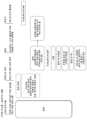
도 2는 본 개시의 예시적인 실시예에 따른, 예시적인 AI 사용자 임상 워크플로우를 예시한다.
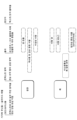
도 3은 본 개시의 예시적인 실시예에 따른, 슬라이드 수준 시각화의 예시적인 도해를 예시한다.
도 4a 내지 도 4e는 본 개시의 예시적인 실시예에 따른, 병리학자에 의해 관찰, 검토, 및 진단될 필요가 있을 수 있는 다양한 리포트 가능한 피처들 및 인스턴스들에 대한 예시적인 리포트들을 예시한다.
도 5는 본 개시의 예시적인 실시예에 따른, AI-인에이블드 시각화를 위한 전체 프레임워크의 예시적인 도해를 예시한다.
도 6a 내지 도 6d는 본 개시의 예시적인 실시예에 따른, 상이한 조직 유형들에 대한 공통 시각화 및 상호작용이 어떻게 적용될 수 있는지에 대한 예시적인 도해를 예시한다.
도 7은 본 개시의 예시적인 실시예에 따른, 리포트 가능한 피처들의 예시적인 도해를 예시한다.
도 8은 여기서 제시된 기술들을 실행할 수 있는 예시적인 시스템을 예시한다.The accompanying drawings, which are incorporated in and constitute a part of this specification, illustrate various exemplary embodiments and, together with the detailed description, serve to explain the principles of the disclosed embodiments.
1A illustrates an example block diagram of a system and network for categorizing AI visualization properties to create a framework for artificial intelligence (AI)-driven digital workflows, in accordance with an example embodiment of the present disclosure. .
1B illustrates an example block diagram of a disease detection platform, in accordance with an example embodiment of the present disclosure.
1C illustrates an example block diagram of a slide analysis tool, in accordance with an example embodiment of the present disclosure.
2 illustrates an example AI user clinical workflow, in accordance with an example embodiment of the present disclosure.
3 illustrates an example diagram of slide-level visualization, in accordance with an example embodiment of the present disclosure.
4A-4E illustrate example reports for various reportable features and instances that may need to be observed, reviewed, and diagnosed by a pathologist, according to an example embodiment of the present disclosure.
5 illustrates an example diagram of an overall framework for AI-enabled visualization, according to an example embodiment of the present disclosure.
6A-6D illustrate example diagrams of how common visualization and interaction for different tissue types may be applied, according to an example embodiment of the present disclosure.
7 illustrates an example diagram of reportable features, in accordance with an example embodiment of the present disclosure.
8 illustrates an example system capable of practicing the techniques presented herein.
이제 본 개시의 예시적인 실시예들이 상세히 참조될 것이며, 이의 예들은 첨부 도면들에 예시되어 있다. 가능한 한, 동일한 참조 번호들은 동일하거나 유사한 부분들을 지칭하기 위해 도면들 전체에 걸쳐 사용될 것이다.Reference will now be made in detail to exemplary embodiments of the present disclosure, examples of which are illustrated in the accompanying drawings. Wherever possible, the same reference numbers will be used throughout the drawings to refer to the same or like parts.
여기서 개시되는 시스템들, 디바이스들, 및 방법들은 예들을 통해 그리고 도면들을 참조하여 상세히 설명된다. 여기서 논의되는 예들은 단지 예들이고, 여기서 설명되는 장치들, 디바이스들, 시스템들, 및 방법들에 대한 설명을 돕기 위해 제공된다. 도면들에 도시되거나 아래에서 논의되는 피처들 또는 구성요소들 중 어느 것도, 특별히 필수적인 것으로 지정되지 않는 한, 이들 디바이스들, 시스템들, 또는 방법들 중 임의의 것의 임의의 특정 구현예에 대해 필수적인 것으로 취해지지 않아야 한다.The systems, devices, and methods disclosed herein are described in detail through examples and with reference to the drawings. The examples discussed herein are examples only and are provided to assist in explaining the apparatus, devices, systems, and methods described herein. None of the features or components shown in the drawings or discussed below are considered essential for any particular implementation of any of these devices, systems, or methods unless specifically designated as essential. should not be taken
또한, 설명되는 임의의 방법들에 대해, 그 방법이 흐름도와 함께 설명되는지 여부에 관계없이, 문맥에 의해 달리 지정되거나 요구되지 않는 한, 방법의 실행 시 수행되는 단계들의 임의의 명시적 또는 암시적 순서는 이러한 단계들이 제시된 순서로 수행되어야 한다는 것을 암시하지 않는 것이 아니라, 상이한 순서로 또는 병렬로 수행될 수 있다는 것을 이해해야 한다.Further, for any method described, whether or not the method is described in conjunction with a flowchart, any explicit or implicit implied steps performed in execution of the method unless otherwise specified or required by context. The order does not imply that these steps must be performed in the order presented, but it is to be understood that they may be performed in a different order or in parallel.
여기서 사용될 때, "예시적인"이라는 용어는 "이상적인"이 아니라, "예"의 의미로 사용된다. 또한, 여기서 단수 표현은 언급되는 항목들의 수량의 제한을 나타내는 것이 아니라, 하나 이상의 존재를 나타낸다.As used herein, the term "exemplary" is used in the sense of "example" and not "ideal". Also, the singular expression herein does not indicate a limitation in the number of items referred to, but indicates the presence of one or more.
도 1a는 본 개시의 예시적인 실시예에 따른, 인공 지능(AI)-구동 디지털 워크플로우를 위한 프레임워크를 생성하기 위해 AI 시각화 속성들을 카테고리화하기 위한 시스템 및 네트워크의 예시적인 블록도를 예시한다.1A illustrates an example block diagram of a system and network for categorizing AI visualization properties to create a framework for artificial intelligence (AI)-driven digital workflows, in accordance with an example embodiment of the present disclosure. .
구체적으로, 도 1a는 병원, 실험실, 및/또는 의원 등에서의 서버들에 연결될 수 있는 전자 네트워크(120)를 예시한다. 예를 들어, 의료진 서버들(121), 병원 서버(122), 임상 시험 서버들(123), 연구실 서버들(124), 및/또는 실험실 정보 시스템들(125) 등은 각각 하나 이상의 컴퓨터, 서버 및/또는 핸드헬드 모바일 디바이스를 통해 인터넷과 같은 전자 네트워크(120)에 각각 연결될 수 있다. 본 출원의 예시적인 실시예에 따르면, 전자 네트워크(120)는 본 개시의 예시적인 실시예에 따라, 디지털 병리학적 이미지(들)에 관한 검체 속성 또는 이미지 속성 정보를 결정하고, 기계 학습을 사용하여 질병 또는 감염체가 존재하는지 여부를 결정하기 위해 슬라이드 분석 툴(101)을 포함하는 질병 검출 플랫폼(100)을 구현하도록 구성된 처리 디바이스를 포함할 수 있는 서버 시스템(110)에 또한 연결될 수 있다. 슬라이드 분석 툴(101)은 액상 종양 제제의 '적절성'의 신속한 평가를 가능하게 할 수 있고; 액상 종양 제제의 진단(세포학, 혈액학/혈액병리학)을 용이하게 할 수 있고; 액상 제제에 의해 검출되는 다양한 종양들에서 발견될 가능성이 가장 높은 분자 발견물을 예측할 수 있다.Specifically, FIG. 1A illustrates an
의료진 서버들(121), 병원 서버들(122), 임상 시험 서버들(123), 연구실 서버들(124) 및/또는 실험실 정보 시스템들(125)은 하나 이상의 환자의 세포학적 검체(들), 조직병리학적 검체(들), 세포학적 검체(들)의 슬라이드(들), 조직병리학적 검체(들)의 슬라이드(들)의 디지털화된 이미지, 또는 이들의 임의의 조합의 이미지들을 생성하거나 또는 달리 획득할 수 있다. 의료진 서버들(121), 병원 서버들(122), 임상 시험 서버들(123), 연구실 서버들(124) 및/또는 실험실 정보 시스템들(125)은 연령, 병력, 암 치료 이력, 가족력, 과거 생검 또는 세포학 정보 등과 같은 환자 특정 정보의 임의의 조합을 또한 획득할 수 있다. 의료진 서버들(121), 병원 서버들(122), 임상 시험 서버들(123), 연구실 서버들(124) 및/또는 실험실 정보 시스템들(125)은 디지털화된 슬라이드 이미지들 및/또는 환자 특정 정보를 전자 네트워크(120)를 통해 서버 시스템(110)에 송신할 수 있다. 서버 시스템(들)(110)은 의료진 서버들(121), 병원 서버들(122), 임상 시험 서버들(123), 연구실 서버들(124) 및/또는 실험실 정보 시스템들(125) 중 적어도 하나로부터 수신되는 이미지들 및 데이터를 저장하기 위한 하나 이상의 저장 디바이스(109)를 포함할 수 있다. 서버 시스템들(110)은 저장 디바이스들(109)에 저장된 이미지들 및 데이터를 처리하기 위한 처리 디바이스들을 또한 포함할 수 있다. 서버 시스템들(110)은 하나 이상의 기계 학습 툴(들) 또는 능력을 더 포함할 수 있다. 예를 들어, 처리 디바이스들은 일 실시예에 따른, 질병 검출 플랫폼(100)을 위한 기계 학습 툴을 포함할 수 있다. 대안적으로 또는 추가적으로, 본 개시(또는 본 개시의 시스템 및 방법의 부분들)은 로컬 처리 디바이스(예를 들어, 랩탑) 상에서 수행될 수 있다.The
의료진 서버들(121), 병원 서버들(122), 임상 시험 서버들(123), 연구실 서버들(124) 및/또는 실험실 정보 시스템들(125)은 슬라이드들의 이미지들을 검토하기 위해 병리학자들에 의해 사용되는 시스템들을 지칭한다. 병원 환경에서, 조직 유형 정보가 실험실 정보 시스템(125)에 저장될 수 있다.
도 1b는 기계 학습을 사용하여, 디지털 병리학적 이미지(들)에 관한 검체 속성 또는 이미지 속성 정보를 결정하기 위한 질병 검출 플랫폼(100)의 예시적인 블록도를 예시한다. 질병 검출 플랫폼(100)은 슬라이드 분석 툴(101), 데이터 수집 툴(102), 슬라이드 수용 툴(103), 슬라이드 스캐너(104), 슬라이드 매니저(105), 스토리지(106), 실험실 정보 시스템(예를 들어, 실험실 정보 시스템(125)), 및 관찰 애플리케이션 툴(108)을 포함할 수 있다.1B illustrates an exemplary block diagram of a
슬라이드 분석 툴(101)은 아래에서 설명되는 바와 같이, 디지털 병리학적 이미지(들)에 관한 데이터 가변 속성 또는 건강 가변 속성 정보를 결정하기 위한 프로세스 및 시스템을 지칭한다. 기계 학습은 예시적인 실시예에 따라, 이미지를 분류하기 위해 사용될 수 있다. 슬라이드 분석 툴(101)은 아래의 실시예들에서 설명되는 바와 같이, 미래 관계들을 또한 예측할 수 있다.
데이터 수집 툴(102)은 예시적인 실시예에 따라, 디지털 병리학적 이미지들을 분류 및 처리하는 데 사용되는 다양한 툴들, 모듈들, 구성요소들, 및 디바이스들로의 디지털 병리학적 이미지들의 전송을 가능하게 할 수 있다.
슬라이드 수용 툴(103)은 예시적인 실시예에 따라, 병리학적 이미지들을 스캔하고 이들을 디지털 형태로 변환할 수 있다. 슬라이드들은 슬라이드 스캐너(104)로 스캔될 수 있고, 슬라이드 매니저(105)는 슬라이드들 상의 이미지들을 디지털화된 병리학적 이미지들로 처리하고 디지털화된 이미지들을 스토리지(106)에 저장할 수 있다.The slide receiving tool 103 may scan pathological images and convert them into digital form, according to an exemplary embodiment. The slides may be scanned with
관찰 애플리케이션 툴(108)은 예시적인 실시예에 따라, 디지털 병리학적 이미지(들)에 관한 검체 속성 또는 이미지 속성 정보를 사용자에게 제공할 수 있다. 정보는 다양한 출력 인터페이스들(예를 들어, 스크린, 모니터, 저장 디바이스 및/또는 웹 브라우저 등)을 통해 제공될 수 있다.The
슬라이드 분석 툴(101) 및 이의 구성요소들 중 하나 이상은 디지털화된 슬라이드 이미지들 및/또는 환자 정보를 네트워크(120)를 통해 서버 시스템들(110), 의료진 서버들(121), 병원 서버들(122), 임상 시험 서버들(123), 연구실 서버들(124) 및/또는 실험실 정보 시스템들(125)에 송신 및/또는 수신할 수 있다. 또한, 서버 시스템들(110)은 슬라이드 분석 툴(101), 데이터 수집 툴(102), 슬라이드 수용 툴(103), 슬라이드 스캐너(104), 슬라이드 매니저(105), 및 관찰 애플리케이션 툴(108) 중 적어도 하나로부터 수신된 이미지들 및 데이터를 저장하기 위한 저장 디바이스들을 포함할 수 있다. 서버 시스템들(110)은 저장 디바이스들에 저장된 이미지들 및 데이터를 처리하기 위한 처리 디바이스들을 또한 포함할 수 있다. 서버 시스템들(110)은 예를 들어, 처리 디바이스들로 인해, 하나 이상의 기계 학습 툴(들) 또는 능력을 더 포함할 수 있다. 대안적으로, 또는 추가적으로, 본 개시(또는 본 개시의 시스템 및 방법의 부분들)은 로컬 처리 디바이스(예를 들어, 랩탑) 상에서 수행될 수 있다.The
상기한 디바이스들, 툴들 및 모듈들 중 임의의 것은 하나 이상의 컴퓨터, 서버 및/또는 핸드헬드 모바일 디바이스를 통해, 인터넷 또는 클라우드 서비스 제공자와 같은 전자 네트워크에 연결될 수 있는 디바이스 상에 위치할 수 있다.Any of the above devices, tools and modules may be located on a device that may be connected to an electronic network, such as the Internet or a cloud service provider, via one or more computers, servers and/or handheld mobile devices.
도 1c는 본 개시의 예시적인 실시예에 따른, 슬라이드 분석 툴(101)의 예시적인 블록도를 예시한다. 슬라이드 분석 툴(101)은 트레이닝 이미지 플랫폼(131) 및/또는 타겟 이미지 플랫폼(135)을 포함할 수 있다.1C illustrates an example block diagram of a
일 실시예에 따르면, 트레이닝 이미지 플랫폼(131)은 트레이닝 이미지 수용 모듈(132), 데이터 분석 모듈(133), 및 조직 식별 모듈(134)을 포함할 수 있다.According to an embodiment, the training image platform 131 may include a training image acceptance module 132 , a data analysis module 133 , and a tissue identification module 134 .
트레이닝 데이터 플랫폼(131)은 일 실시예에 따르면, 기계 학습 모델을 트레이닝하여 디지털 병리학적 이미지들을 효과적으로 분석 및 분류하는 데 사용되는 트레이닝 이미지들을 생성 또는 수신할 수 있다. 예를 들어, 트레이닝 이미지들은 서버 시스템들(110), 의료진 서버들(121), 병원 서버들(122), 임상 시험 서버들(123), 연구실 서버들(124) 및/또는 실험실 정보 시스템들(125) 중 임의의 것 또는 임의의 조합으로부터 수신될 수 있다. 트레이닝에 사용되는 이미지들은 실제 소스들(예를 들어, 인간, 동물 등)로부터 나올 수 있거나, 합성 소스들(예를 들어, 그래픽 렌더링 엔진들, 3D 모델들 등)로부터 비롯될 수 있다. 디지털 병리학적 이미지들의 예들은 (a) H&E, 헤마톡실린 단독, IHC, 분자 병리학 등과 같은(그러나 이에 제한되지 않음) 다양한 착색제로 착색된 디지털화된 슬라이드들; 및/또는 (b) 마이크로CT와 같은 3D 이미징 디바이스로부터의 디지털화된 조직 표본들을 포함할 수 있다.According to an embodiment, the training data platform 131 may generate or receive training images used to effectively analyze and classify digital pathological images by training a machine learning model. For example, the training images may be
트레이닝 이미지 수용 모듈(132)은 하나 이상의 검체 조직에 대응하는 하나 이상의 트레이닝 데이터세트를 포함하는 데이터세트를 생성 또는 수신할 수 있다. 예를 들어, 트레이닝 데이터세트들은 서버 시스템들(110), 의료진 서버들(121), 병원 서버들(122), 임상 시험 서버들(123), 연구실 서버들(124) 및/또는 실험실 정보 시스템들(125) 중 임의의 것 또는 임의의 조합으로부터 수신될 수 있다. 이러한 데이터세트는 디지털 저장 디바이스 상에 유지될 수 있다. 데이터 분석 모듈(133)은 개별 세포들의 세트가 관심 세포에 속하는지 또는 디지털화된 이미지의 배경에 속하는지를 식별할 수 있다. 조직 식별 모듈(134)은 디지털화된 이미지들을 분석하고, 세포학적 표본 내의 개별 세포가 추가 분석을 필요로 하는지 여부를 결정할 수 있다. 개별 세포가 추가 분석을 필요로 하는지 여부를 식별하고 이러한 영역들을 종합하는 것이 유용하고, 이러한 것의 식별은 사용자에게 알림을 트리거할 수 있다.The training image acceptance module 132 may generate or receive a dataset including one or more training datasets corresponding to one or more specimen tissues. For example, training datasets may be
일 실시예에 따르면, 타겟 이미지 플랫폼(135)은 타겟 이미지 수용 모듈(136), 검체 검출 모듈(137), 및 출력 인터페이스(138)를 포함할 수 있다. 타겟 이미지 플랫폼(135)은 타겟 이미지를 수신하고, 타겟 데이터세트의 특성을 결정하기 위한 기계 학습 모델을 수신된 타겟 이미지에 적용할 수 있다. 예를 들어, 타겟 데이터는 서버 시스템들(110), 의료진 서버들(121), 병원 서버들(122), 임상 시험 서버들(123), 연구실 서버들(124) 및/또는 실험실 정보 시스템들(125) 중 임의의 것 또는 임의의 조합으로부터 수신될 수 있다. 타겟 이미지 수용 모듈(136)은 타겟 조직 검체에 대응하는 타겟 데이터세트를 수신할 수 있다. 검체 검출 모듈(137)은 조직 검체의 특성을 결정하기 위한 기계 학습 모델을 타겟 데이터세트에 적용할 수 있다. 예를 들어, 검체 검출 모듈(137)은 조직 검체에서 의심스러운(그리고/또는 의심스럽지 않은) 조직 영역을 검출할 수 있다. 검체 검출 모듈(137)은 다양한 피처들의 존재 및/또는 부재를 식별하도록 트레이닝된 인공 지능(AI) 시스템을 사용할 수 있다. AI 시스템은 식별된 피처들 또는 피처의 인스턴스들에 관련된 모든 영역들을 강조하는 맵(예를 들어, 조직 맵)을 출력할 수 있다. 미국 출원 제17/313,617호는 이에 의해 전문이 참조로 원용된다. 검체 검출 모듈(137)은 식별된 피처들이 크기, 형상, 위치, 다른 식별된 피처들에 대한 근접성, 질병 유형, 색상, 착색 유형, 바이오마커 유형, 유전자 서명, 단백질 유형, 혈액 마커, 조직 유형, 조직 텍스처, 석회화 존재 또는 수준, 염증 존재 또는 수준 등과 같은 특정 특성들에 기초하여 의심스러운지 또는 의심스럽지 않은지를 결정할 수 있다.According to one embodiment, the target image platform 135 may include a target image receiving module 136 , a specimen detection module 137 , and an output interface 138 . The target image platform 135 may receive a target image and apply a machine learning model to the received target image to determine characteristics of the target dataset. For example, the target data may be
검체 검출 모듈(137)은 타겟 조직 검체에 대한 품질 스코어를 결정하기 위한 기계 학습 모델을 타겟 데이터세트에 또한 적용할 수 있다. 또한, 검체 검출 모듈(137)은 조직 검체 내에 타겟 요소가 존재하는지 여부를 결정하기 위한 기계 학습 모델을 타겟 이미지들에 적용할 수 있다.The specimen detection module 137 may also apply a machine learning model to the target dataset to determine a quality score for the target tissue specimen. In addition, the specimen detection module 137 may apply a machine learning model for determining whether a target element exists in a tissue specimen to target images.
출력 인터페이스(138)는 타겟 조직 검체 및 관심 조직 영역에 관한 정보를 출력하는 데 사용될 수 있다. (예를 들어, 스크린, 모니터, 저장 디바이스, 웹 브라우저 등에).Output interface 138 may be used to output information regarding a target tissue sample and a tissue region of interest. (eg screen, monitor, storage device, web browser, etc.).
아래에서 설명되는 예시적인 실시예들 중 하나 이상은 AI 시각화 유형들의 세트 및 상호작용 유형들의 세트를 제공할 수 있다. 하나 이상의 예시적인 실시예는 조직 유형에 의해 소팅되고 시각화 유형이 부여된 예들, 및 CAP(College of American Pathologists) 종관(synoptic)으로부터 추출된 리포트 가능한 피처들에 요구되는 임의의 가능한 상호작용 유형을 제공할 수 있다. 하나 이상의 예시적인 실시예는 임상 사용 케이스들을 확보할 수 있지만, 하나 이상의 정의된 출력을 레버리징할 수 있는 바이오마커 제품들에 또한 적용될 수 있다.One or more of the example embodiments described below may provide a set of AI visualization types and a set of interaction types. One or more illustrative embodiments provide examples sorted by tissue type and assigned a visualization type, and any possible interaction type required for reportable features extracted from a College of American Pathologists (CAP) synoptic. can do. One or more illustrative embodiments may capture clinical use cases, but may also be applied to biomarker products capable of leveraging one or more defined outputs.
병리학적 워크플로우를 볼 때 파트 및 케이스 수준 종합이 고려사항일 수 있다. 병리학자들은 파트 수준 종합을 사용하여 파트 수준에서 조직 검체를 리포트 및 진단할 수 있다. 그러나, 병리학적 리포트들은 슬라이드 수준 관측을 포함하지 않을 수 있다. 일부 경우들에서, 리포팅 필드들은 슬라이드 수준에서 보이지 않을 수 있다. 일부 리포팅 필드들은 예를 들어, 유방관내제자리암종(ductal carcinoma in situ, DCIS)을 갖는 파트에서의 슬라이드들의 수와 같이, 파트 수준에서만 보일 수 있다.Part and case level synthesis may be a consideration when looking at pathology workflows. Pathologists can use part-level synthesis to report and diagnose tissue samples at the part level. However, pathology reports may not include slide level observations. In some cases, reporting fields may not be visible at the slide level. Some reporting fields may only be visible at the part level, such as, for example, the number of slides in the part with ductal carcinoma in situ (DCIS).
아래의 정의는 단지 설명을 위한 것이고, 제한하는 것으로 의도되지 않는다. 전체 슬라이드 이미지(whole slide image, WSI)는 착색되거나 착색되지 않은 조직의 하나 이상의 이미지를 지칭할 수 있다. 리포트 가능한 피처는 진단에 사용되는 조직의 임의의 관측뿐만 아니라 의심스러운 조직의 분류 또는 라벨링된 영역 또는 포커스를 지칭할 수 있다. 피처 인스턴스는 리포트 가능한 피처에 고유할 수 있는 진단 및 해부학적 특성에 의해 정의되는 리포트 가능한 피처의 예증일 수 있다. 시각화 유형은 피처 인스턴스의 디스플레이 카테고리를 지칭할 수 있다. 상호작용 유형은 사용자가 둘 이상의 피처 인스턴스 사이에서 내비게이팅할 수 있는 방식을 지칭할 수 있다. 인스턴스 정의는 인스턴스 디스플레이에 포함될 필요가 있을 수 있는 하나 이상의 해부학적 피처를 지칭할 수 있다. AI 시스템 또는 모듈은 AI, 기계 학습 기술, 및/또는 기계 학습 알고리즘을 구현하는 하나 이상의 모듈을 지칭할 수 있고, 반드시 단일 모듈, 디바이스, 시스템, 플랫폼 등으로 제한되는 것은 아닐 수 있다. 여기서 개시되는 양태들은 임의의 유형 또는 배열의 AI 또는 기계 학습 시스템들, 모듈들, 플랫폼들 및/또는 이미지 분석 및/또는 프로세스 시스템들 상에서 사용될 수 있다. 다양한 출력들은 종합될 수 있다. 기계 학습 모델은 (예를 들어, 패턴들을 인식하기 위한) 기계 학습 기술을 구현하는 모델 또는 프로세스를 지칭할 수 있고, 하나의 모델로 제한되지 않을 수 있다. 여기서 개시되는 양태들은 이후에 조합 및/또는 종합될 수 있는 개별 출력들을 조합 및/또는 생성할 수 있는 다수의 기계 학습 모델들을 사용할 수 있다.The definitions below are for illustrative purposes only and are not intended to be limiting. A whole slide image (WSI) can refer to one or more images of stained or unstained tissue. A reportable feature may refer to any observation of tissue used for diagnosis, as well as a classified or labeled area or focus of suspicious tissue. A feature instance may be an instance of a reportable feature defined by diagnostic and anatomical characteristics that may be unique to the reportable feature. A visualization type can refer to a display category of a feature instance. An interaction type can refer to a way in which a user can navigate between two or more feature instances. An instance definition may refer to one or more anatomical features that may need to be included in an instance display. An AI system or module may refer to one or more modules that implement AI, machine learning techniques, and/or machine learning algorithms, and may not necessarily be limited to a single module, device, system, platform, or the like. Aspects disclosed herein may be used on any type or arrangement of AI or machine learning systems, modules, platforms and/or image analysis and/or process systems. The various outputs can be aggregated. A machine learning model may refer to a model or process that implements a machine learning technique (eg, to recognize patterns) and may not be limited to one model. Aspects disclosed herein may use multiple machine learning models capable of combining and/or generating discrete outputs that may then be combined and/or aggregated.
도 2는 예시적인 실시예에 따른, 임상 환경에서 예시적인 AI 사용자 워크플로우(200)를 예시한다. 예시적인 워크플로우는 케이스 트리아지 및/또는 우선순위화가 일어날 수 있는 단계(202)로 시작할 수 있다(임상 사용자 워크플로우 설명). 사용자는 케이스들이 관찰할 준비가 될 때, 즉 케이스와 연관된 모든 슬라이드들이 처리되고/거나 시스템에 업로드되었을 때 알림을 받기를 원할 수 있다. 사용자는 , 예를 들어, 복잡도에 기초하여 어느 케이스들이 우선순위화되어야 하는지, 그리고/또는 어느 케이스들이 분자 시험을 위해 보내지거나 다시 나누어질 것과 같이, 추가적인 오더를 필요로 할 수 있는지 알기를 추가적으로 원할 수 있다.2 illustrates an example AI user workflow 200 in a clinical environment, according to an example embodiment. An exemplary workflow may begin with step 202 where case triage and/or prioritization may occur (Clinical User Workflow Description). A user may want to be notified when cases are ready to be viewed, that is, when all slides associated with a case have been processed and/or uploaded to the system. The user may additionally want to know which cases should be prioritized based on complexity, for example, and/or which cases may require additional orders, such as being sent for molecular testing or being subdivided. can
복잡도는 진단(예를 들어, AI 시스템이 비교적 낮은 신뢰도로 슬라이드를 Gleason 3+4=7로서 분류할 수 있거나, 또는 Gleason 패턴 3의 공산 백분율이 49.9%이고 Gleason 패턴 4의 백분율이 50.1%일 수 있어, 경계선 케이스를 나타내는 것으로 결정할 수 있음), 및/또는 진단을 결정하기 위해 H&E 슬라이드를 넘어서는 추가적인 시험들에 대한 필요(예를 들어, 병태가 침윤성으로 보이고 검토를 위해 에스트로겐 수용체(ER), 프로게스테론 수용체(PR), 인간 경막외 성장 인자 수용체 1(HER2)과 같은 추가적인 착색된 이미지들을 필요로 하는지)에 의해 결정될 수 있다.Complexity is a diagnostic (e.g., an AI system can classify a slide as
복잡도는 AI 출력들 및 알려져 있는 워크플로우들 및 병리학에 의해 요구되는 시험 단계들의 조합에 기초하여 결정될 수 있다. 복잡한 케이스들은 초기에 우선순위화될 수 있고/거나 더 많은 경험을 가진 병리학자들에게 배포될 수 있다.Complexity can be determined based on a combination of AI outputs and known workflows and test steps required by the pathology. Complex cases can be prioritized early and/or distributed to more experienced pathologists.
단계(204)에서, 워크플로우는 예를 들어, 사용자로부터의 케이스의 선택을 포함할 수 있다.At step 204, the workflow may include, for example, selection of a case from a user.
단계(206)에서, 워크플로우는 트리아지 및/또는 케이스 개관을 포함할 수 있다. 단계(202)와 유사하게, 사용자는 케이스 또는 트리아지 정보의 간략한 개관을 보기를 원할 수 있다. 개관 단계(202) 동안 그리고/또는 그 전에, 식별된 피처들 및 임의의 다른 메타데이터(예를 들어, 사용자 생성 주석들)가 요약될 수 있다.At step 206, the workflow may include triage and/or case overview. Similar to step 202, the user may wish to view a brief overview of case or triage information. During and/or prior to the overview step 202, the identified features and any other metadata (eg, user-generated annotations) may be summarized.
단계(208)에서, 워크플로우는 파트의 선택을 포함할 수 있다. 선택은 파트가 의심스러운 것으로 보이는지, 의심스럽지 않은 것으로 보이는지, 또는 "기타"로 보이는지 ― 여기서 "기타"는 사전 프로그래밍된 품질 또는 알려져 있지 않은 변수를 나타낼 수 있다 ― 를 시그널링할 수 있는 표시자에 의해 수반될 수 있다.At step 208, the workflow may include selection of a part. The selection is made by an indicator that can signal whether a part appears suspicious, appears non-suspicious, or appears "other" - where "other" may represent a pre-programmed quality or unknown variable. may accompany
단계(210)에서, 워크플로우는 트리아지 및/또는 파트 개관 단계를 포함할 수 있다. 이러한 개관은 파트 인덱스를 포함할 수 있다. 사용자는 파트 내에 무엇이 있는지의 개관을 보기를 원할 수 있지만, 슬라이드들을 보는 것은 필요로 하지 않을 수 있다. 파트 수준에 집중함으로써, 사용자는 포커스의 키 영역들 및/또는 키 영역들의 이미지화/시각화를 보는 것이 가능할 수 있다.At step 210, the workflow may include triage and/or part overview steps. This overview may include a part index. A user may want to see an overview of what is within a part, but may not need to see the slides. By focusing on the part level, the user may be able to see key areas in focus and/or an imaging/visualization of key areas.
단계(212)에서, 워크플로우는 슬라이드를 선택하는 것을 포함할 수 있다. 파트가 의심스러운 것으로 보이는지, 의심스럽지 않은 것으로 보이는지, 또는 "기타"로 보이는지 ― 여기서 "기타"는 사전 프로그래밍된 품질 또는 알려져 있지 않은 변수를 나타낼 수 있다 ― 를 시그널링할 수 있는 연관 표시자가 이용 가능할 수 있다.At step 212, the workflow may include selecting a slide. An association indicator may be available that may signal whether a part appears suspicious, appears non-suspicious, or appears "other", where "other" may represent a pre-programmed quality or unknown variable. there is.
단계(214)에서, 워크플로우는 파트 수준에서의 진단을 포함할 수 있다. 진단은 진단과 함께 파트 인덱스를 포함할 수 있다. AI는 파트 인덱스를 생성하기 위해 병리학적 검체의 파트들을 종합할 수 있다. 사용자가 파트 수준에서의 리포트를 만들 필요가 있을 수 있음에 따라, 종합된 파트 수준 인덱스는 리포트를 생성하기 위한 사용자의 프로세스를 간소화할 수 있다.At step 214, the workflow may include diagnosis at the part level. The diagnosis may include a part index along with the diagnosis. AI can synthesize parts of a pathological specimen to create a part index. As users may need to create reports at the part level, aggregated part level indexes can simplify the user's process for generating reports.
단계(216)에서, 워크플로우는 케이스 사인 아웃(case sign out)으로 끝날 수 있다.At step 216, the workflow may end with case sign out.
임상 워크플로우가 실행되고 있는 동안, AI는 검사된 슬라이드 및 결과적인 진단에 대한 리포트를 추가적으로 생성할 수 있다. 단계(218)에서, 워크플로우는 파트 수준 진단의 결과들에 기초하여 시각화 및/또는 정량화 패널을 디스플레이하는 것을 포함할 수 있다.While the clinical workflow is running, the AI can additionally generate reports on the slides examined and the resulting diagnoses. At step 218 , the workflow may include displaying a visualization and/or quantification panel based on the results of the part level diagnosis.
단계(220)에서, 시각화 및/또는 정량화 패널이 리포트에 편집 및/또는 추가될 수 있다.At step 220, visualization and/or quantification panels may be edited and/or added to the report.
단계(222)에서, 워크플로우는 리포트를 사용자에게 보낼 수 있다. 모든 리포팅은 파트 수준에서 일어날 수 있다. 각 조직 유형에 대해 그리고 조직 유형 내에서의 생검과 절제 사이에서, 리포트 가능한 피처들은 달라질 수 있다. 조직의 유형 및 절차는 어떤 슬라이드 수준 데이터가 파트 수준 리포트에 실릴 수 있는지를 결정할 수 있다. 리포팅을 위해, 슬라이드에 걸쳐 달라지는 데이터를 평균화함으로써 파트들에 걸쳐 종합하는 것이 필요할 수 있다. 추가적으로, 피처의 존재 또는 부재가 리포팅될 수 있다. 슬라이드 수준에 존재하지 않는 리포팅의 일부 피처들이 있을 수 있다; 예를 들어, 유방 조직 검사에서, 병리학자는 파트 내의 슬라이드들 중 얼마나 많은 슬라이드가 DCIS를 갖는지를 리포팅할 수 있다. AI 워크플로우의 주석들 또는 출력들은 관련 스크린샷들 및/또는 식별된 영역들과 같이, 리포트에 추가될 수 있다.At step 222, the workflow may send the report to the user. All reporting can happen at the part level. For each tissue type and between biopsies and excisions within a tissue type, reportable features may vary. Organizational types and procedures can determine which slide-level data can be included in part-level reports. For reporting purposes, it may be necessary to aggregate across parts by averaging data that varies across slides. Additionally, the presence or absence of features may be reported. There may be some features of reporting that do not exist at the slide level; For example, in a breast biopsy, a pathologist can report how many of the slides within a part have DCIS. Annotations or outputs of the AI workflow may be added to the report, such as relevant screenshots and/or identified areas.
추가적으로, 단계(224)에서, 리포트는 PDF 등과 같은 내보내기 가능한 포맷으로 실험실 정보 시스템(laboratory information system, LIS) 리포트를 채우기 위해 사용될 수 있다.Additionally, at step 224, the report may be used to populate a laboratory information system (LIS) report in an exportable format such as PDF.
표 1은 슬라이드 수준 시각화 프레임워크의 예를 예시한다. 슬라이드의 포커스는 슬라이드 자체 상의 직사각형 표시자 오버레이를 사용하여 설명될 수 있으며, 컨텍스트 영역이 조직 맵 또는 다른 윤곽 영역을 사용하여 설명된다. 포커스 내의 해부학적 요소들은 포커스의 영역과 연관된 좌표 세트 및 크기 및/또는 줌 수준뿐만 아니라, 디스플레이의 유효성에도 유용할 수 있다. 메타데이터는 슬라이드 수준 시각화를 수반할 수 있고, 포커스 영역에 대한 명칭(이를테면 "석회화"), 메트릭 및/또는 텍스트 출력, 및 포커스 영역의 크기를 포함할 수 있다.Table 1 illustrates an example of a slide-level visualization framework. The focus of a slide can be described using a rectangular indicator overlay on the slide itself, and the context area is described using an organization map or other outlined area. Anatomical elements within focus may be useful for the effectiveness of the display, as well as the size and/or zoom level and set of coordinates associated with the area of focus. The metadata may accompany the slide-level visualization, and may include a name for the focus area (such as “calcification”), metrics and/or text output, and size of the focus area.
일부 시각화 유형들에 인스턴스들의 카운트가 중요할 수 있음에 따라, 컨텍스트 영역은 슬라이드 전체에 걸쳐 임의의 또는 모든 인스턴스들을 종합할 수 있다. 가능한 수반되는 메타데이터는 컨텍스트 영역에 대한 명칭(이를테면, "유방 아형"), 및 메트릭 및/또는 텍스트 출력을 포함할 수 있으며, 이는 아형의 명칭, 컨텍스트 영역의 크기 및/또는 측정치, 컨텍스트 영역과 연관된 등급, 컨텍스트 영역의 아키텍처 등을 포함할 수 있다.As the count of instances can be important for some visualization types, the context area can aggregate any or all instances across the slide. Possible accompanying metadata may include a name for the context region (eg, “breast subtype”), and metrics and/or text output, which may include the name of the subtype, the size and/or measurements of the context region, the context region and It may include the associated class, the architecture of the context area, etc.
조직 검체의 측정치는 시각화 시, 명확하게 라벨링된 종점들을 갖는 라인으로서 제시될 수 있다. 측정치에는 (예를 들어, "전립선" 또는 "종양 길이"와 같은) 명칭뿐만 아니라, 메트릭 및/또는 텍스트 출력(이를테면, 라인의 수치 측정치)이 수반될 수 있다.Measurements of a tissue sample, when visualized, can be presented as a line with clearly labeled endpoints. Measurements may be accompanied by names (eg, such as “prostate” or “tumor length”), as well as metrics and/or text output (eg, numerical measurements of a line).
슬라이드 수준 시각화에는 텍스트 또는 수치 출력이 또한 포함될 수 있고, 이는 시각화 시 직접 디스플레이될 수 있다. 텍스트 또는 수치 출력은 메타데이터, 이를테면 조직 검체와 연관된 명칭 또는 다른 메트릭 및/또는 텍스트 출력 또는 검토 동안 사용자에 의해 추가된 독립적인 주석들을 포함할 수 있다. 메타데이터는 예를 들어, 검출된 바이오마커를 포함할 수 있다.Slide-level visualizations can also include text or numeric output, which can be displayed directly in the visualization. The textual or numeric output may include metadata, such as a name or other metric associated with the tissue sample and/or independent annotations added by the user during textual output or review. Metadata may include, for example, detected biomarkers.
슬라이드 수준 시각화 시 시각화의 컴파일된 리포트가 포함될 수 있고, 이는 외부 벤더에 의해 포맷팅될 수 있다. 컴파일된 리포트는 WSI 또는 관심 있는 특정 영역과 연관될 수 있다. 리포트, 예를 들어 PDF 리포트는 다른 관련 정보 중에서도, 슬라이드 ID 및 슬라이드에 대한 전체 스코어를 포함할 수 있다.In a slide-level visualization, a compiled report of the visualization may be included, which may be formatted by an external vendor. Compiled reports can be associated with WSI or specific areas of interest. A report, for example a PDF report, may include, among other pertinent information, a slide ID and an overall score for the slide.
표 1: 슬라이드 수준 시각화 프레임워크Table 1: Slide-level visualization framework
도 3은 AI-인에이블드 시각화를 위한 전체 프레임워크의 예시적인 도해를 도시한다. 슬라이드 수준 시각화(300)는 케이스 개관(302), 파트 인덱스(304), 검출 패널(306), 정량화 패널(308), 하나 이상의 내보내기 가능한 리포트(310)를 포함할 수 있다. 추가적으로, 프레임워크는 주석 로그(312) 및 리포트 빌더(316)를 포함할 수 있으며, 이들은 함께 리포트 인에이블러를 형성할 수 있다.3 shows an example diagram of an overall framework for AI-enabled visualization. The slide level visualization 300 may include a case overview 302 , a parts index 304 , a detection panel 306 , a quantification panel 308 , and one or more exportable reports 310 . Additionally, the framework may include an annotation log 312 and a report builder 316, which together may form a report enabler.
케이스 개관(302)에서, 프레임워크는 사용자가 슬라이드들을 검토하기 시작하기 전에 사용자가 이용 가능한 AI-구동 케이스 개관 및 트리아지 정보를 포함할 수 있다. 케이스 개관은 예를 들어, 접근 식별 번호 또는 다른 부류의 식별자, 상태(예를 들어, 준비됨, 검토됨, 준비 안 됨), 접근 날짜, 환자 이름, 의료 기록 번호 또는 MRN, 조직의 유형(예를 들어, 유방, 피부 또는 진피, 위장관 또는 GI, 또는 전립선), 검체 유형(예를 들어, 생검), 및 다수의 슬라이드들을 포함할 수 있다. 케이스 개관의 다른 부분은 식별자들(예를 들어, "우측 유방"), 착색의 유형(예를 들어, H&E), 및 다른 정보를 포함할 수 있는 슬라이드들의 시각화, 스냅샷들, 또는 "슬라이드 트레이"를 포함할 수 있다.At case overview 302 , the framework may include an AI-driven case overview and triage information available to the user before the user begins reviewing the slides. A case overview may include, for example, an access identification number or other class of identifier, status (e.g., ready, reviewed, not ready), date of access, patient name, medical record number or MRN, type of organization (e.g., eg, breast, skin or dermis, gastrointestinal or GI, or prostate), specimen type (eg, biopsy), and multiple slides. Another part of the case overview is a visualization of slides, snapshots, or "slide trays" that can include identifiers (eg, "right breast"), type of staining (eg, H&E), and other information. " may be included.
파트 인덱스(304)는 사용자가 슬라이드들을 검토하기 시작하기 전에 사용자가 이용 가능한 AI-구동 파트 개관 및 트리아지 정보를 포함할 수 있다.Part index 304 may include an AI-driven part overview and triage information available to the user before the user begins reviewing the slides.
검출 패널(306)은 조직의 이원 분류를 의심스러운 것 또는 의심스럽지 않은 것으로서 가능하게 할 수 있는 템플릿화된 패널일 수 있다.The detection panel 306 may be a templated panel that may enable binary classification of tissue as suspicious or not suspicious.
정량화 패널(308)은 조직 피처들 및/또는 다른 리포트 가능한 피처들의 정량화, 소팅, 및/또는 필터링을 포함할 수 있다. 정량화, 소팅, 및/또는 필터링은 의심스러운 것 및 의심스럽지 않은 것의 결정 및/또는 다른 결정에 기초할 수 있다. 정량화의 예로서, 전립선 케이스에서, 종양의 양이 백분율로서 그리고/또는 예를 들어, 밀리미터(mm) 단위의 거리 또는 면적 메트릭으로서 정량화될 수 있다. 피처의 별개의 인스턴스들(예를 들어, 신경주위 침윤)이 AI 시스템이 인스턴스를 검출하는 것에 기초하여 심각도 또는 가능성에 따라 또한 소팅 및/또는 필터링될 수 있다. 예를 들어, 필터링 동안, 낮은(예를 들어, 임계치보다 더 낮은) 심각도를 갖는 별개의 인스턴스들은 소팅 및/또는 필터링된 피처들이 출력되기 전에 제거될 수 있다. 정량화 패널(308)은 검출 패널(306)과 하나의 패널로서 결합될 수 있다.The quantification panel 308 may include quantification, sorting, and/or filtering of tissue features and/or other reportable features. Quantification, sorting, and/or filtering may be based on determining what is suspect and what is not suspicious and/or other determinations. As an example of quantification, in prostate cases, tumor volume can be quantified as a percentage and/or as a distance or area metric, eg in millimeters (mm). Discrete instances of the feature (eg, perineural infiltrate) may also be sorted and/or filtered according to severity or likelihood based on the AI system detecting the instance. For example, during filtering, discrete instances with low (eg, lower than a threshold) severity may be removed before the sorted and/or filtered features are output. The quantification panel 308 can be combined with the detection panel 306 as one panel.
하나 이상의 내보내기 가능한 리포트(310)는 정량화 패널(308) 및 검출 패널(306)로부터 출력될 수 있다. 내보내기 가능한 리포트들(310)은 PDF의 형태일 수 있다.One or more exportable reports 310 may be output from the quantification panel 308 and detection panel 306 . Exportable reports 310 may be in the form of a PDF.
주석 로그(312)는 조직 검체와 연관된 모든 사용자 생성 노트들의 활동 로그를 포함할 수 있다. 주석 로그(312)는 케이스 수준과 파트 수준 둘 모두에서 검색 가능할 수 있고, 각 주석 또는 사용자 생성 노트는 타임 스탬프, 사용자 이름 및 역할, 썸네일 이미지, 및/또는 하나 이상의 추가적인 사용자 코멘트를 포함할 수 있다.Annotation log 312 may include an activity log of all user-generated notes associated with a tissue sample. The annotation log 312 may be searchable at both the case level and the part level, and each annotation or user created note may include a timestamp, user name and role, thumbnail image, and/or one or more additional user comments. .
주석 로그(312)는 리포트 빌더(316)와 조합되어 리포트 인에이블러(314)를 생성할 수 있다. 리포트 빌더(316)는 프리필드(prefilled)될 수 있으므로, AI 구성요소로 가치가 있을 수 있다. 추가적으로, 리포트 빌더(316)는 편집 가능할 수 있고, 주석 로그(312)로부터 정보 및 주석들을 풀링할 수 있다.Annotation log 312 can be combined with report builder 316 to create report enabler 314 . Because report builder 316 can be prefilled, it can be valuable as an AI component. Additionally, report builder 316 may be editable and may pull information and annotations from annotations log 312 .
리포트 인에이블러(314)는 사용자 생성 주석들 및 AI-프리필드 리포트의 파트 수준 종합을 포함할 수 있다. 파트 수준 종합 및/또는 AI-프리필드 리포트는 임상 시스템과 통합될 수 있고 환자 의료 기록에 로그될 수 있다. 병리학자는 파트 수준 종합 및/또는 AI-프리필드 리포트를 검토하고, 편집본들 또는 주석들을 온 캔버스 시각화에 추가할 수 있다. 병리학자가 이러한 편집본들 또는 주석들을 만들 때, 리포트 가능한 피처 카테고리들이 종관 리포트 필드들과 연관될 수 있으므로, AI-프리필드 리포트는 편집본들에 기초하여 자동으로 업데이트될 수 있다. 또한, 이러한 편집본들 또는 주석들을 만들 때, 편집된 피처와 관련된 다른 피처들(예를 들어, 자식 피처들)이 편집본들에 기초하여 자동으로 업데이트 또는 조정될 수 있다.Report enabler 314 may include part-level aggregation of user-generated annotations and AI-prefield reports. Part-level synthetic and/or AI-prefilled reports can be integrated with clinical systems and logged in patient medical records. The pathologist can review the part-level synthesis and/or AI-prefield report and add edits or annotations to the on-canvas visualization. When the pathologist makes these edits or annotations, the AI-prefield report can be automatically updated based on the edits, as reportable feature categories can be associated with synoptic report fields. Also, when making these compilations or annotations, other features (eg, child features) related to the edited feature may be automatically updated or adjusted based on the compilations.
병리학적 검토 및 진단을 필요로 할 수 있는 광범위한 조직 및 수술 검체 유형들이 있다. 도 4a 내지 도 4e는 병리학자에 의해 관찰, 검토, 및 진단될 필요가 있을 수 있는 다양한 리포트 가능한 피처들 및 인스턴스들에 대한 예시적인 리포트들을 예시한다.There are a wide range of tissue and surgical specimen types that may require pathological review and diagnosis. 4A-4E illustrate example reports for various reportable features and instances that may need to be observed, reviewed, and diagnosed by a pathologist.
도 4a는 방광 조직 검체에 대한 예시적인 리포트를 예시한다. 적어도 하나의 제1 피처가 리포트된다; 예를 들어, 분류, 침윤의 존재 또는 부존재, 및 조직 검체가 양성인지, 제자리성인지, 침윤성인지 등을 포함하는 아형. 하나 이상의 제2 피처, 이를테면 종양 등급, 근고유층의 이원 표시, 및/또는 침윤의 깊이가 리포트에 또한 포함될 수 있다. 사용자 노트들이 예시적인 리포트에 또한 포함될 수 있다.4A illustrates an exemplary report for bladder tissue specimens. at least one first feature is reported; Subtype including, for example, classification, presence or absence of invasion, and whether the tissue sample is benign, in situ, or invasive. One or more secondary features, such as tumor grade, binary representation of lamina propria, and/or depth of invasion may also be included in the report. User notes may also be included in the example report.
마찬가지로, 도 4b는 제1 및 제2 피처들을 리포트하는, 결장 조직 검체에 대한 예시적인 리포트를 도시한다. 제1 피처들은 암을 검출하는 것, 육아종 및 급성 염증의 식별, 및 비암성 아형을 포함할 수 있다. 비암성 아형은 정상, 용종, 또는 염증으로서 또한 리포트될 수 있고, 아형 용종과 같은 추가적인 정보가 리포트 상에 또한 포함될 수 있다. 제2 피처는 H&E 상의 MMR 상태의 예측을 포함할 수 있다.Similarly, FIG. 4B shows an exemplary report for a colon tissue specimen reporting first and second features. First features may include detecting cancer, identifying granulomas and acute inflammation, and non-cancerous subtypes. Noncancerous subtypes may also be reported as normal, polyp, or inflammatory, and additional information such as subtype polyp may also be included on the report. A second feature may include prediction of MMR status on H&E.
도 4c, 도 4d 및 도 4e는 각각, 폐 조직, 피부 조직, 및 위 조직에 대한 예시적인 리포트들을 예시한다. 예시적인 리포트들은 조직 검체 및 조직의 카테고리화 또는 아형에 따라, 다양한 리포트된 제1 및 제2 피처들을 포함할 수 있다.4C, 4D and 4E illustrate exemplary reports for lung tissue, skin tissue, and stomach tissue, respectively. Exemplary reports may include various reported first and second features, depending on the tissue sample and categorization or subtype of the tissue.
도 5는 AI-인에이블드 시각화를 위한 전체 프레임워크의 예시적인 도해를 예시한다. 전체 프레임워크는 두 개의 별개의 분할분들, 다수의 슬라이드 수준 발견물들, 및 AI 시스템에 의한 발견물들의 종합을 포함하여 전체 케이스 리포트를 생성할 수 있다.5 illustrates an example diagram of an overall framework for AI-enabled visualization. The overall framework can generate a full case report, including two separate segments, multiple slide-level findings, and a synthesis of findings by the AI system.
케이스 리포트(500)는 당해 특정 케이스에서 n개의 파트들을 포함할 수 있다. 각 파트는 각자의 대응하는 파트 리포트(502)를 가질 수 있다. 파트 리포트는 슬라이드 수준 발견물들을 포함할 수 있고, 몇몇 수의 리포트 가능한 피처들(504)을 연관된 메타데이터와 포함할 수 있다. 병리학자에 대한 검토를 최적화할 수 있는 리포트 가능한 피처들 간의 관계들이 있다면, 시각화 상에 디스플레이하기 위해 노트될 수 있다. 리포트 가능한 피처는 하나 이상의 피처 인스턴스(506)를 연관 메타데이터와 포함할 수 있다. 각 피처 인스턴스는 시각화 유형(508) 및/또는 상호작용 유형(510)을 포함할 수 있다.A
피처 인스턴스(506)는 조직에 의해 다른 리포트 가능한 피처에 연결되지 않은 리포트 가능한 피처(504)의 인스턴스일 수 있다. 각 리포트 가능한 피처(504)마다의 인스턴스들은 진단 및 해부학에 의해 정의될 수 있다. 슬라이드에서의 피처 인스턴스들(506)은 몇몇 형태들로 종합될 수 있다. 사용자는 리포트 가능한 피처(504)의 일부 또는 모든 인스턴스들을 한 자리에서(예를 들어, 조직 맵으로서 총 종양 및 침윤성 유관 암종(IDC)을 조직 맵으로서, 또는 DCIS의 모든 포커스들을 갤러리 뷰에서) 볼 수 있다. 피처 인스턴스(506)가 메타데이터와 연관된다면, 메타데이터는 리포트 가능한 피처에 대한 메타데이터를 생성하기 위해 종합될 수 있다(예를 들어, 총 종양 선형 범위(10mm)에 대해 5mm의 패턴 3 및 5mm의 패턴 4가 종합된다).
표 2는 피처 인스턴스들(506)과 같은 피처 인스턴스들 및 인스턴스 종합에 관한 연관된 정보를 또한 예시한다. 피처 인스턴스 내에서, 사용자는 인스턴스를 통해 "조직 홉(tissue hop)"을 할 수 있거나, 슬라이드 상의 인스턴스들 사이에서 점핑할 수 있다. 슬라이드의 컨텍스트에서 구분된 인스턴스들로서 보여지는 인스턴스들은 고출력 설정으로 보여질 수 있다. 대응하는 시각화는 두 개 이상의 포커스 영역들(다수의 요소들이 인스턴스를 포함할 필요가 있는 경우) 및 주변 컨텍스트 영역들일 수 있다. 예는 조직 상의 질병의 침윤일 수 있으며, 여기서 다수의 피처 인스턴스들이 보여질 필요가 있을 수 있다. 사용자는 갤러리 뷰를 사용하여 피처 인스턴스를 또한 볼 수 있다. 갤러리는 빠른 검토를 위해 디스플레이되는 피처 인스턴스들의 썸네일을 포함할 수 있으며, 사용자가 썸네일을 선택함으로써 특정 인스턴스로 점핑하는 옵션을 갖는다. 갤러리 뷰는 (단지 하나의 포커스 영역만이 인스턴스를 포함할 필요가 있을 수 있지만) 하나 이상의 포커스 영역에 대응할 수 있다.Table 2 also illustrates feature instances, such as
인스턴스 종합의 경우들에서, 조직의 유형의 모든 영역들은 하나의 종합 그룹에 포함될 수 있다. 해당 유형의 모든 인스턴스들은 저출력으로 함께 보여질 수 있다. 대응하는 시각화 유형은 컨텍스트 영역들을 포함할 수 있다.In instance aggregate cases, all areas of a type of organization may be included in one aggregate group. All instances of that type can be shown together in low output. A corresponding visualization type may include context areas.
표 2Table 2
파트(또는 검체) 수준 종합은 특히 리포트 인에이블먼트를 고려할 때, 병리학 워크플로우에 중요할 수 있다.Part (or specimen) level aggregation can be important for pathology workflows, especially when considering report enablement.
모든 시각화는 표 2에서 설명된 바와 같이, 조직 홉을 가질 수 있다. 조직 홉은 사용자가 파트에 걸쳐 발견되는 조직 유형의 임의의 시각화 사이에서 "홉"을 할 수 있게 할 수 있다. 예를 들어, 하나의 파트가 IDC의 세 개의 인스턴스들을 포함한다면, IDC가 종합본이더라도, 조직 홉은 사용자를 이들 종합 인스턴스들 사이에서 이동시킬 수 있다.All visualizations can have tissue hops, as described in Table 2. Tissue hops may allow users to “hop” between any visualization of the tissue types found across parts. For example, if one part contains three instances of IDC, even if the IDC is composite, an organization hop can move a user between these composite instances.
케이스 리포트(500)는 편집 가능할 수 있다. 임의의 바이오마커와 별도로, 모든 영숫자 출력들(메타데이터 또는 다른 것)은 편집 가능할 수 있다. 예를 들어, 측정 종료점이 편집 가능할 수 있고, 측정은 리포팅 및/또는 편집 상태 동안 업데이트될 수 있다. 마찬가지로, 미래 상태 조직 맵 영역도 편집 가능할 수 있다. 계산된 백분율 또는 측정된 길이는 리포팅 및/또는 편집 상태 동안 업데이트될 수 있다. 다른 메타데이터는 재계산되지 않을 수 있다.
도 5에 도시된 전체 프레임워크는 특정 AI 또는 이미지 분석 및/또는 처리 시스템들에 제한되지 않고, 여기서 개시된 양태들이 임의의 유형의 AI 또는 이미지 분석 시스템에 사용될 수 있다. 예를 들어, 전체 프레임워크에서 다수의 시스템들 및/또는 모듈들이 사용될 수 있고, 다수의 시스템 출력들이 이미지, 파트, 케이스 및/또는 환자에 대해 시각화되고 종합될 수 있다.The overall framework shown in FIG. 5 is not limited to specific AI or image analysis and/or processing systems, and the aspects disclosed herein may be used in any type of AI or image analysis system. For example, multiple systems and/or modules may be used in an overall framework, and multiple system outputs may be visualized and aggregated for an image, part, case, and/or patient.
도 6a 내지 도 6d는 상이한 조직 유형들에 대한 공통 시각화 및 상호작용이 어떻게 적용될 수 있는지에 대한 예시적인 도해를 예시한다.6A-6D illustrate example diagrams of how common visualization and interaction for different tissue types can be applied.
도 6a는 일반 조직 검체 시각화 및 전립선 조직 검체 시각화의 예를 예시한다. 일반 조직 검체 내에서, 작은 관심 영역이 포커스로서 식별된다. 대안적으로, 작은 관심 영역 및 큰 관심 영역이 포커스로서 식별될 수 있다. 포커스는 컨텍스트의 일부 주변 영역을 갖는 단일 관심 지점일 수 있다. 일반 조직 검체에 대한 컨텍스트 영역은 상당한 관심 영역 및/또는 조직 맵을 포함할 수 있다. 이는 큰 관심 영역, 예를 들어, 슬라이드 상의 연접한 종양 길이로서 제시될 수 있다(이는 조직 생검에 대해 더 우수할 수 있음). 큰 또는 작은 관심 영역을 수치 출력으로 측정하기 위해 룰러 툴이 사용될 수 있다. 룰러는 검체 조직 내의 하나 이상의 바이오마커를 측정하기 위해 사용될 수 있다. 하나의 슬라이드 수준 시각화, 영숫자 디스플레이는 슬라이드에 오버레이되는 텍스트 및 수치 출력을 포함할 수 있다. 이들 출력들은 이원, 이산 스코어, 연속 스코어, 카테고리, 확률, 정량화(백분율 포함), 및/또는 PDF(portable document format) 또는 다른 포맷 리포트 출력일 수 있다. PDF 리포트의 경우, PDF 리포트는 쉽게 편집되지 않을 수 있는 정적 리포트일 수 있다. PDF 리포트는 예를 들어, 편집되지 않거나 조작되지 않은 채로 남아있도록 의도된 분자 시험, CLIA-lab 시험 결과, 또는 다른 시험 또는 실험 결과들과 연관될 수 있다.6A illustrates examples of normal tissue specimen visualization and prostate tissue specimen visualization. Within normal tissue specimens, small regions of interest are identified as foci. Alternatively, a small region of interest and a large region of interest may be identified as a focus. The focus may be a single point of interest with some surrounding area of context. Context regions for a general tissue specimen may include significant regions of interest and/or tissue maps. This can be presented as a large region of interest, eg, contiguous tumor length on a slide (which may be better for a tissue biopsy). Ruler tools can be used to measure large or small regions of interest with numerical output. A ruler may be used to measure one or more biomarkers in a subject tissue. One slide-level visualization, an alphanumeric display, can include text and numerical output that is overlaid on the slide. These outputs may be binary, discrete scores, continuous scores, categories, probabilities, quantifications (including percentages), and/or portable document format (PDF) or other format report output. In the case of a PDF report, a PDF report may be a static report that may not be easily edited. A PDF report can be associated with, for example, molecular test results, CLIA-lab test results, or other test or laboratory results that are intended to remain unedited or manipulated.
도 6a에서의 전립선 조직 검체 예에 대해, 포커스 시각화는 총 종양뿐만 아니라 하나 이상의 패턴을 포함할 수 있다. 컨텍스트 영역은 문제의 포커스에 따라 달라질 수 있다; 예를 들어, 총 종양은 종양의 전체 길이를 포함할 수 있고, 사용자는 필요한 경우 함께 추가되는, 종양의 길이를 측정할 수 있다. 패턴은 패턴에 의해 커버되는 조직의 백분율을 포함하도록 사용자에 의해 설명되거나 측정될 수 있다. 추가적으로, 영숫자 디스플레이는 일차 및/또는 이차 Gleason 스코어를 출력할 수 있다.For the prostate tissue specimen example in FIG. 6A , focus visualization may include one or more patterns in addition to the total tumor. The context area may vary depending on the focus of the problem; For example, the total tumor can include the entire length of the tumor, and the user can measure the length of the tumor, added together if necessary. The pattern may be described or measured by the user to include the percentage of tissue covered by the pattern. Additionally, the alphanumeric display may output primary and/or secondary Gleason scores.
도 6b는 유방 조직 검체의 예시적인 시각화를 도시한다. 도 6a에서와 같이, 포커스 영역들이 선택되고, 측정되고, 영숫자 출력들이 획득된다. 방광, 결장, 및 폐 조직들의 추가 예들이 도 6c 및 도 6d에 예시된다.6B shows an exemplary visualization of a breast tissue sample. As in Fig. 6a, focus areas are selected, measured, and alphanumeric outputs obtained. Additional examples of bladder, colon, and lung tissues are illustrated in FIGS. 6C and 6D .
도 7은 본 개시의 예시적인 실시예에 따른, 리포트 가능한 피처들의 예시적인 도해를 예시한다. 예를 들어, 침윤성 암 피처에 대해, 리포트 가능한 특성들은 해당 등급이 양호, 중등, 또는 불량인 것으로 고려되는 범위 내에 있는지 여부의 표시를 포함하는 등급뿐만 아니라, 측정된 피처의 길이를 포함할 수 있다. 결과적인 절제 표시자는 사용자에게 조직 검체 마진만을 절제할 것을 가리킬 수 있다. 도 7은 다양한 다른 예시적인 리포트 가능한 피처들을 포함하지만, 더 상세한 것은 아래의 표 3 내지 표 6에서 설명된다.7 illustrates an example diagram of reportable features, in accordance with an example embodiment of the present disclosure. For example, for an invasive cancer feature, reportable characteristics may include the measured length of the feature as well as a rating including an indication of whether the rating is within a range considered to be good, moderate, or poor. . The resulting ablation indicator may indicate to the user to resect only the tissue sample margin. 7 includes various other exemplary reportable features, but more details are set forth in Tables 3-6 below.
도 8에 도시된 바와 같이, 디바이스(800)는 중앙 처리 유닛(CPU)(820)을 포함할 수 있다. CPU(820)는 예를 들어, 임의의 유형의 특수 목적 또는 범용 마이크로프로세서 디바이스를 포함하여, 임의의 유형의 프로세서 디바이스일 수 있다. 관련 기술분야의 통상의 기술자들에 의해 이해될 바와 같이, CPU(820)는 멀티 코어/멀티프로세서 시스템, 단독으로 동작하는 이러한 시스템, 또는 클러스터 또는 서버 팜에서 동작하는 컴퓨팅 디바이스들의 클러스터 내의 단일 프로세서일 수도 있다. CPU(820)는 데이터 통신 인프라스트럭처(810), 예를 들어, 버스, 메시지 큐, 네트워크, 또는 멀티 코어 메시지 전달 스킴에 연결될 수 있다.As shown in FIG. 8 , the
디바이스(800)는 메인 메모리(840), 예를 들어, 랜덤 액세스 메모리(RAM)를 또한 포함할 수 있고, 또한 보고 메모리(830)를 포함할 수 있다. 보조 메모리(830), 예를 들어, 판독 전용 메모리(ROM)는 예를 들어, 하드 디스크 드라이브 또는 탈착 가능 저장 드라이브일 수 있다. 이러한 탈착 가능 저장 드라이브는 예를 들어, 플로피 디스크 드라이브, 자기 테이프 드라이브, 광학 디스크 드라이브, 플래시 메모리 등을 포함할 수 있다. 이 예에서의 탈착 가능 저장 드라이브는 주지되어져 있는 방식으로 탈착 가능 저장 유닛으로부터 판독하고/거나 이에 기록한다. 탈착 가능 스토리지는 플로피 디스크, 자기 테이프, 광학 디스크 등을 포함할 수 있으며, 이는 탈착 가능 스토리지 드라이브에 의해 판독되고 이에 의해 기록된다. 관련 기술분야의 통상의 기술자들에 의해 이해될 바와 같이, 이러한 탈착 가능 스토리지 유닛은 일반적으로 컴퓨터 소프트웨어 및/또는 데이터가 저장된 컴퓨터 사용 가능 저장 매체를 포함한다.
대안적인 구현예들에서, 보조 메모리(830)는 컴퓨터 프로그램들 또는 다른 명령어들이 디바이스(800)에 로딩될 수 있게 하기 위한 유사한 수단들을 포함할 수 있다. 이러한 수단들의 예들은 프로그램 카트리지 및 카트리지 인터페이스(이를테면, 비디오 게임 디바이스들에서 발견되는 것), 탈착 가능 메모리 칩(이를테면, EPROM 또는 PROM) 및 연관된 소켓, 및 다른 탈착 가능 스토리지 유닛들 및 인터페이스들을 포함할 수 있으며, 이들은 소프트웨어 및 데이터가 탈착 가능 스토리지 유닛으로부터 디바이스(800)로 전송될 수 있게 한다.In alternative implementations,
디바이스(800)는 통신 인터페이스("COM")(860)를 또한 포함할 수 있다. 통신 인터페이스(860)는 소프트웨어 및 데이터가 디바이스(800)와 외부 디바이스들 사이에서 전송될 수 있게 한다. 통신 인터페이스(860)는 모뎀, 네트워크 인터페이스(예를 들어, 이더넷 카드), 통신 포트, PCMCIA 슬롯 및 카드 등을 포함할 수 있다. 통신 인터페이스(860)를 통해 전송되는 소프트웨어 및 데이터는 ― 통신 인터페이스(860)에 의해 수신될 수 있는 전자, 전자기, 광학, 또는 다른 신호들일 수 있는 ― 신호들의 형태일 수 있다. 이러한 신호들은 ― 예를 들어, 와이어 또는 케이블, 광섬유, 전화선, 셀룰러 전화 링크, RF 링크 또는 다른 통신 채널들을 사용하여 구현될 수 있는 ― 디바이스(800)의 통신 경로를 통해 통신 인터페이스(860)에 제공될 수 있다.
하드웨어 요소들, 운영 시스템들 및 이러한 장비의 프로그래밍 언어들은 본질적으로 통상적인 것이고, 통상의 기술자들은 이들에 충분히 익숙할 것으로 여겨진다. 디바이스(800)는 키보드들, 마우스들, 터치스크린들, 모니터들, 디스플레이들 등과 같은 입력 및 출력 디바이스들과 연결하기 위한 입력 및 출력 포트들(850)을 또한 포함할 수 있다. 물론, 다양한 서버 기능들은 처리 로드를 분산시키기 위해, 다수의 유사한 플랫폼들 상에서 분산된 방식으로 구현될 수 있다. 대안적으로, 서버들은 하나의 컴퓨터 하드웨어 플랫폼의 적절한 프로그래밍에 의해 구현될 수 있다.It is believed that the hardware elements, operating systems and programming languages of such equipment are conventional in nature and that skilled persons will be sufficiently familiar with them.
본 개시 전반에 걸쳐, 구성요소들 또는 모듈들의 지칭은 일반적으로 기능 또는 관련 기능들의 그룹을 수행하기 위해 논리적으로 함께 그룹화될 수 있는 항목들을 지칭한다. 유사한 참조 번호들은 일반적으로 동일하거나 유사한 구성요소들을 지칭하는 것으로 의도된다. 구성요소들 및 모듈들은 소프트웨어, 하드웨어 또는 소프트웨어와 하드웨어의 조합으로 구현될 수 있다.Throughout this disclosure, references to components or modules generally refer to items that can be logically grouped together to perform a function or group of related functions. Like reference numbers are generally intended to refer to the same or similar elements. Components and modules may be implemented in software, hardware or a combination of software and hardware.
위에서 설명된 툴들, 모듈들, 및 기능들은 하나 이상의 프로세서에 의해 수행될 수 있다. "스토리지"형 매체는 컴퓨터들, 프로세서들 등의 유형의 메모리, 또는 이의 연관된 모듈들, 이를테면 다양한 반도체 메모리들, 테이프 드라이브들, 디스크 드라이브들 등 중 임의의 것 또는 전부를 포함할 수 있으며, 이는 소프트웨어 프로그래밍을 위해 언제든 비일시적인 스토리지를 제공할 수 있다.The tools, modules, and functions described above may be performed by one or more processors. A "storage" type medium may include any or all of the tangible memory of computers, processors, etc., or its associated modules, such as various semiconductor memories, tape drives, disk drives, etc., which Non-transitory storage can be provided at any time for software programming.
소프트웨어는 인터넷, 클라우드 서비스 제공자, 또는 다른 텔레통신 네트워크들을 통해 통신될 수 있다. 예를 들어, 통신은 하나의 컴퓨터 또는 프로세서로부터 다른 컴퓨터 또는 프로세서로 소프트웨어를 로딩하는 것을 가능하게 할 수 있다. 여기서 사용될 때, 비일시적인 유형의 "저장" 매체로 국한되지 않는 한, 컴퓨터 또는 기계 "판독 가능 매체"와 같은 용어들은 프로세서에 실행을 위한 명령어들을 제공하는 데 참여하는 임의의 매체를 지칭한다.The software may be communicated over the Internet, a cloud service provider, or other telecommunications networks. For example, communication may enable loading of software from one computer or processor to another computer or processor. As used herein, terms such as computer or machine "readable medium", unless limited to a non-transitory tangible "storage" medium, refer to any medium that participates in providing instructions to a processor for execution.
전술한 일반적인 설명은 단지 예시적이고 설명적인 것이며, 본 개시를 제한하지 않는다. 본 발명의 다른 실시예들은 여기서 개시된 본 발명의 명세서 및 실행을 고려하여 당업자들에게 명백할 것이다. 본 명세서 및 예시는 단지 예시적인 것으로 고려되어야 하는 것으로 의도된다.The foregoing general description is illustrative and explanatory only and does not limit the present disclosure. Other embodiments of the invention will be apparent to those skilled in the art in light of the specification and practice of the invention disclosed herein. It is intended that the specification and examples be considered illustrative only.
표 3Table 3
표 4table 4
표 5table 5
표 6table 6
Claims (20)
디지털 저장 디바이스에서, 병리학적 검체의 디지털 이미지들과 각각 연관된 하나 이상의 케이스를 수신하는 단계;
상기 기계 학습 모델을 사용하여, 상기 하나 이상의 케이스 중의 케이스를 관찰할 준비가 된 것으로 식별하는 단계;
상기 케이스의 선택을 수신하는 단계 ― 상기 케이스는 복수의 파트들을 포함함 ―;
상기 기계 학습 모델을 사용하여, 상기 복수의 파트들이 의심스러운지 또는 의심스럽지 않은지를 결정하는 단계;
상기 복수의 파트들 중의 파트의 선택을 수신하는 단계;
상기 파트와 연관된 복수의 슬라이드들이 의심스러운지 또는 의심스럽지 않은지를 결정하는 단계;
상기 기계 학습 모델을 사용하여, 상기 복수의 슬라이드들 중 의심스러운 슬라이드들의 집합을 결정하는 단계 ― 상기 기계 학습 모델은 복수의 트레이닝 이미지들을 처리함으로써 트레이닝됨 ―; 및
상기 의심스러운 슬라이드들의 집합에 주석을 다는(annotating) 단계 및/또는 상기 의심스러운 슬라이드들의 집합에 기초하여 리포트를 생성하는 단계를 포함하는, 컴퓨터 구현 방법.A computer implemented method for categorizing a specimen in digital pathology using at least one machine learning model, comprising:
receiving, at the digital storage device, one or more cases each associated with the digital images of the pathological specimen;
identifying, using the machine learning model, a case of the one or more cases as being ready for observation;
receiving a selection of the case, the case including a plurality of parts;
determining, using the machine learning model, whether the plurality of parts are suspect or not suspicious;
receiving a selection of a part from among the plurality of parts;
determining whether a plurality of slides associated with the part are suspect or not suspicious;
determining, using the machine learning model, a set of suspicious slides of the plurality of slides, wherein the machine learning model is trained by processing a plurality of training images; and
annotating the set of questionable slides and/or generating a report based on the set of questionable slides.
의심스러운 조직 주위의 적어도 하나의 영역을 윤곽화하는 단계;
상기 의심스러운 조직의 길이 및/또는 면적을 측정하는 단계; 및
상기 의심스러운 슬라이드들의 집합 상에 주석을 출력하는 단계를 더 포함하는 것인, 컴퓨터 구현 방법.2. The method of claim 1, wherein annotating the set of questionable slides comprises:
outlining at least one area around the suspect tissue;
measuring the length and/or area of the suspect tissue; and
and outputting an annotation on the set of questionable slides.
상기 의심스러운 슬라이드들의 집합에 관한 정보로 상기 리포트를 채우는 단계; 및
상기 리포트를 사용자에게 출력하는 단계를 더 포함하는 것인, 컴퓨터 구현 방법.According to claim 1,
filling the report with information about the set of questionable slides; and
The computer-implemented method further comprising outputting the report to a user.
상기 하나 이상의 케이스 각각과 연관된 복잡도를 결정하는 단계; 및
상기 결정된 복잡도에 기초하여 상기 하나 이상의 케이스를 우선순위화하는 단계를 더 포함하는, 컴퓨터 구현 방법.According to claim 1,
determining a complexity associated with each of the one or more cases; and
prioritizing the one or more cases based on the determined complexity.
상기 기계 학습 모델을 사용하여, 상기 복수의 파트들이 의심스러운지 또는 의심스럽지 않은지의 상기 결정에 기초하여 상기 복수의 파트들을 디스플레이를 위해 소팅(sorting) 및/또는 필터링하는 단계를 더 포함하는, 컴퓨터 구현 방법.According to claim 1,
sorting and/or filtering the plurality of parts for display based on the determination of whether the plurality of parts are suspicious or not suspicious using the machine learning model; method.
병리학적 유형을 결정하는 단계; 및
상기 결정된 병리학적 유형에 기초하여 상기 리포트를 생성하는 단계를 더 포함하는, 컴퓨터 구현 방법.According to claim 1,
determining the pathological type; and
generating the report based on the determined pathological type.
상기 기계 학습 모델을 사용하여, 상기 케이스와 연관된 상기 디지털 이미지들에 기초하여 복수의 피처들을 결정하는 단계;
상기 기계 학습 모델을 사용하여, 상기 복수의 피처들을 사용하여 복수의 부분적 리포트들을 결정하는 단계; 및
상기 복수의 부분적 리포트들에 기초하여 상기 리포트를 결정하는 단계를 더 포함하는, 컴퓨터 구현 방법.According to claim 1,
determining, using the machine learning model, a plurality of features based on the digital images associated with the case;
using the machine learning model, determining a plurality of partial reports using the plurality of features; and
determining the report based on the plurality of partial reports.
명령어들을 저장하는 적어도 하나의 메모리; 및
상기 명령어들을 실행하여 동작들을 수행하도록 구성된 적어도 하나의 프로세서를 포함하며, 상기 동작들은:
디지털 저장 디바이스에서, 병리학적 검체의 디지털 이미지들과 각각 연관된 하나 이상의 케이스를 수신하는 동작;
상기 기계 학습 모델을 사용하여, 상기 하나 이상의 케이스 중의 케이스를 관찰할 준비가 된 것으로 식별하는 동작;
상기 케이스의 선택을 수신하는 동작 ― 상기 케이스는 복수의 파트들을 포함함 ―;
상기 기계 학습 모델을 사용하여, 상기 복수의 파트들이 의심스러운지 또는 의심스럽지 않은지를 결정하는 동작;
상기 복수의 파트들 중의 파트의 선택을 수신하는 동작;
상기 파트와 연관된 복수의 슬라이드들이 의심스러운지 또는 의심스럽지 않은지를 결정하는 동작;
상기 기계 학습 모델을 사용하여, 상기 복수의 슬라이드들 중 의심스러운 슬라이드들의 집합을 결정하는 동작 ― 상기 기계 학습 모델은 복수의 트레이닝 이미지들을 처리함으로써 트레이닝됨 ―; 및
상기 의심스러운 슬라이드들의 집합에 주석을 다는 동작 및/또는 상기 의심스러운 슬라이드들의 집합에 기초하여 리포트를 생성하는 동작을 포함하는, 시스템.A system for categorizing a specimen in digital pathology using at least one machine learning model, comprising:
at least one memory to store instructions; and
and at least one processor configured to execute the instructions to perform operations comprising:
receiving, at the digital storage device, one or more cases each associated with the digital images of the pathological specimen;
identifying, using the machine learning model, a case of the one or more cases as being ready for observation;
receiving a selection of the case, wherein the case includes a plurality of parts;
determining, using the machine learning model, whether the plurality of parts are suspect or not suspicious;
receiving a selection of a part from among the plurality of parts;
determining whether a plurality of slides associated with the part are suspicious or not suspicious;
determining, using the machine learning model, a set of suspicious slides among the plurality of slides, the machine learning model being trained by processing a plurality of training images; and
Annotating the set of questionable slides and/or generating a report based on the set of questionable slides.
의심스러운 조직 주위의 적어도 하나의 영역을 윤곽화하는 동작;
상기 의심스러운 조직의 길이 및/또는 면적을 측정하는 동작; 및
상기 의심스러운 슬라이드들의 집합 상에 주석을 출력하는 동작을 더 포함하는 것인, 시스템.14. The method of claim 13, wherein annotating the set of questionable slides:
outlining at least one area around the suspect tissue;
measuring the length and/or area of the suspect tissue; and
and outputting an annotation on the set of questionable slides.
상기 의심스러운 슬라이드들의 집합에 관한 정보로 상기 리포트를 채우는 동작; 및
상기 리포트를 사용자에게 출력하는 동작을 더 포함하며, 상기 의심스러운 슬라이드들의 집합에 관한 정보는 포커스 영역, 컨텍스트 영역, 상기 의심스러운 조직의 하나 이상의 측정치, 및/또는 영숫자 출력을 포함하는 것인, 시스템.14. The method of claim 13, wherein the operations are:
populating the report with information about the set of questionable slides; and
and outputting the report to a user, wherein the information about the set of questionable slides includes a focus region, a context region, one or more measurements of the suspicious tissue, and/or an alphanumeric output. .
상기 하나 이상의 케이스 각각과 연관된 복잡도를 결정하는 동작; 및
상기 결정된 복잡도에 기초하여 상기 하나 이상의 케이스를 우선순위화하는 동작을 더 포함하는 것인, 시스템.14. The method of claim 13, wherein the operations are:
determining a complexity associated with each of the one or more cases; and
Further comprising prioritizing the one or more cases based on the determined complexity.
상기 기계 학습 모델을 사용하여, 상기 케이스와 연관된 상기 디지털 이미지들에 기초하여 복수의 피처들을 결정하는 동작;
상기 기계 학습 모델을 사용하여, 상기 복수의 피처들을 사용하여 복수의 부분적 리포트들을 결정하는 동작; 및
상기 복수의 부분적 리포트들에 기초하여 상기 리포트를 결정하는 동작을 더 포함하는 것인, 시스템.14. The method of claim 13, wherein the operations are:
determining, using the machine learning model, a plurality of features based on the digital images associated with the case;
determining, using the machine learning model, a plurality of partial reports using the plurality of features; and
and determining the report based on the plurality of partial reports.
디지털 저장 디바이스에서, 병리학적 검체의 디지털 이미지들과 각각 연관된 하나 이상의 케이스를 수신하는 단계;
상기 기계 학습 모델을 사용하여, 상기 하나 이상의 케이스 중의 케이스를 관찰할 준비가 된 것으로 식별하는 단계;
상기 케이스의 선택을 수신하는 단계 ― 상기 케이스는 복수의 파트들을 포함함 ―;
상기 기계 학습 모델을 사용하여, 상기 복수의 파트들이 의심스러운지 또는 의심스럽지 않은지를 결정하는 단계;
상기 복수의 파트들 중의 파트의 선택을 수신하는 단계;
상기 파트와 연관된 복수의 슬라이드들이 의심스러운지 또는 의심스럽지 않은지를 결정하는 단계;
상기 기계 학습 모델을 사용하여, 상기 복수의 슬라이드들 중 의심스러운 슬라이드들의 집합을 결정하는 단계 ― 상기 기계 학습 모델은 복수의 트레이닝 이미지들을 처리함으로써 트레이닝됨 ―; 및
상기 의심스러운 슬라이드들의 집합에 주석을 다는 단계 및/또는 상기 의심스러운 슬라이드들의 집합에 기초하여 리포트를 생성하는 단계를 포함하는, 비일시적인 컴퓨터 판독 가능 매체.A non-transitory computer-readable medium storing instructions that, when executed by a processor, perform a method of outputting a task-by-task prediction using at least one machine learning model, the method comprising:
receiving, at the digital storage device, one or more cases each associated with the digital images of the pathological specimen;
identifying, using the machine learning model, a case of the one or more cases as being ready for observation;
receiving a selection of the case, the case including a plurality of parts;
determining, using the machine learning model, whether the plurality of parts are suspect or not suspicious;
receiving a selection of a part from among the plurality of parts;
determining whether a plurality of slides associated with the part are suspect or not suspicious;
determining, using the machine learning model, a set of suspicious slides of the plurality of slides, wherein the machine learning model is trained by processing a plurality of training images; and
annotating the set of questionable slides and/or generating a report based on the set of questionable slides.
의심스러운 조직 주위의 적어도 하나의 영역을 윤곽화하는 단계;
상기 의심스러운 조직의 길이 및/또는 면적을 측정하는 단계; 및
상기 의심스러운 슬라이드들의 집합 상에 주석을 출력하는 단계를 더 포함하는 것인, 비일시적인 컴퓨터 판독 가능 매체.20. The method of claim 19, wherein annotating the set of questionable slides comprises:
outlining at least one area around the suspect tissue;
measuring the length and/or area of the suspect tissue; and
and outputting an annotation on the set of questionable slides.
Applications Claiming Priority (3)
| Application Number | Priority Date | Filing Date | Title |
|---|---|---|---|
| US202063127846P | 2020-12-18 | 2020-12-18 | |
| US63/127,846 | 2020-12-18 | ||
| PCT/US2021/063982 WO2022133183A1 (en) | 2020-12-18 | 2021-12-17 | Systems and methods for processing electronic images of slides for a digital pathology workflow |
Publications (1)
| Publication Number | Publication Date |
|---|---|
| KR20230122048A true KR20230122048A (en) | 2023-08-22 |
Family
ID=80112208
Family Applications (1)
| Application Number | Title | Priority Date | Filing Date |
|---|---|---|---|
| KR1020237022730A KR20230122048A (en) | 2020-12-18 | 2021-12-17 | Systems and methods for processing electronic images of slides for digital pathology workflows |
Country Status (8)
| Country | Link |
|---|---|
| US (3) | US11710235B2 (en) |
| EP (1) | EP4264616A1 (en) |
| JP (1) | JP2024500100A (en) |
| KR (1) | KR20230122048A (en) |
| CN (1) | CN116868276A (en) |
| AU (1) | AU2021401312A1 (en) |
| CA (1) | CA3202030A1 (en) |
| WO (1) | WO2022133183A1 (en) |
Family Cites Families (13)
| Publication number | Priority date | Publication date | Assignee | Title |
|---|---|---|---|---|
| US8970618B2 (en) * | 2011-06-16 | 2015-03-03 | University Of Leeds | Virtual microscopy |
| US20140233826A1 (en) | 2011-09-27 | 2014-08-21 | Board Of Regents Of The University Of Texas System | Systems and methods for automated screening and prognosis of cancer from whole-slide biopsy images |
| US10049447B2 (en) * | 2013-11-06 | 2018-08-14 | H. Lee Moffitt Cancer Center and Research Insititute, Inc. | Pathology case review, analysis and prediction |
| US20220172809A9 (en) * | 2017-05-25 | 2022-06-02 | Enlitic, Inc. | Report generating system and methods for use therewith |
| CA3081643A1 (en) * | 2017-11-06 | 2019-05-09 | University Health Network | Platform, device and process for annotation and classification of tissue specimens using convolutional neural network |
| JP7076698B2 (en) | 2017-11-17 | 2022-05-30 | 国立研究開発法人国立がん研究センター | Image analysis method, image analysis device, program, learned deep learning algorithm manufacturing method and learned deep learning algorithm |
| US10748040B2 (en) | 2017-11-20 | 2020-08-18 | Kavya Venkata Kota Sai KOPPARAPU | System and method for automatic assessment of cancer |
| WO2019102043A1 (en) * | 2017-11-27 | 2019-05-31 | Deciphex | Automated screening of histopathology tissue samples via analysis of a normal model |
| US20200372638A1 (en) * | 2017-11-27 | 2020-11-26 | Deciphex | Automated screening of histopathology tissue samples via classifier performance metrics |
| WO2020075172A1 (en) * | 2018-10-10 | 2020-04-16 | Ibex Medical Analytics Ltd. | System and method for personalization and optimization of digital pathology analysis |
| WO2020132363A1 (en) * | 2018-12-19 | 2020-06-25 | Strata Oncology, Inc. | Methods of detecting and treating subjects with checkpoint inhibitor-responsive cancer |
| WO2020190851A1 (en) * | 2019-03-15 | 2020-09-24 | Splntellx, Inc. | An explainable ai (xai) platform for computational pathology |
| WO2021171281A1 (en) * | 2020-02-26 | 2021-09-02 | Ibex Medical Analytics Ltd. | System and method of managing workflow of examination of pathology slides |
-
2021
- 2021-12-16 US US17/552,438 patent/US11710235B2/en active Active
- 2021-12-17 KR KR1020237022730A patent/KR20230122048A/en unknown
- 2021-12-17 EP EP21844136.8A patent/EP4264616A1/en active Pending
- 2021-12-17 JP JP2023536426A patent/JP2024500100A/en active Pending
- 2021-12-17 AU AU2021401312A patent/AU2021401312A1/en active Pending
- 2021-12-17 CA CA3202030A patent/CA3202030A1/en active Pending
- 2021-12-17 WO PCT/US2021/063982 patent/WO2022133183A1/en active Application Filing
- 2021-12-17 CN CN202180094040.5A patent/CN116868276A/en active Pending
- 2021-12-30 US US17/565,681 patent/US11538162B2/en active Active
-
2023
- 2023-06-05 US US18/329,024 patent/US20230351599A1/en active Pending
Also Published As
| Publication number | Publication date |
|---|---|
| US11538162B2 (en) | 2022-12-27 |
| US20220198666A1 (en) | 2022-06-23 |
| CN116868276A (en) | 2023-10-10 |
| CA3202030A1 (en) | 2022-06-23 |
| US11710235B2 (en) | 2023-07-25 |
| US20220199255A1 (en) | 2022-06-23 |
| EP4264616A1 (en) | 2023-10-25 |
| AU2021401312A1 (en) | 2023-07-13 |
| WO2022133183A1 (en) | 2022-06-23 |
| JP2024500100A (en) | 2024-01-04 |
| US20230351599A1 (en) | 2023-11-02 |
Similar Documents
| Publication | Publication Date | Title |
|---|---|---|
| KR102488986B1 (en) | Systems and methods for processing images of slides to automatically prioritize the processed images of slides for digital pathology | |
| US11551357B2 (en) | Systems and methods for processing images of slides for digital pathology | |
| JP7228210B2 (en) | Systems and methods for the communication of digital biomarkers and genomic panels | |
| CN115210817A (en) | System and method for analyzing electronic images for quality control | |
| US20240112338A1 (en) | Systems and methods to process electronic images to produce a tissue map visualization | |
| US20230360414A1 (en) | Systems and methods for processing images to determine image-based computational biomarkers from liquid specimens | |
| US20230147471A1 (en) | Systems and methods to process electronic images to determine salient information in digital pathology | |
| US11710235B2 (en) | Systems and methods for processing electronic images of slides for a digital pathology workflow | |
| US20230245430A1 (en) | Systems and methods for processing electronic images for auto-labeling for computational pathology |