KR20230051223A - Methods of treating WWOX-related diseases - Google Patents
Methods of treating WWOX-related diseases Download PDFInfo
- Publication number
- KR20230051223A KR20230051223A KR1020237008228A KR20237008228A KR20230051223A KR 20230051223 A KR20230051223 A KR 20230051223A KR 1020237008228 A KR1020237008228 A KR 1020237008228A KR 20237008228 A KR20237008228 A KR 20237008228A KR 20230051223 A KR20230051223 A KR 20230051223A
- Authority
- KR
- South Korea
- Prior art keywords
- wwox
- promoter
- gene
- expression
- mice
- Prior art date
Links
- 238000000034 method Methods 0.000 title claims abstract description 74
- 208000037265 diseases, disorders, signs and symptoms Diseases 0.000 title claims description 37
- 108010036639 WW Domain-Containing Oxidoreductase Proteins 0.000 title abstract description 305
- 102000012163 WW Domain-Containing Oxidoreductase Human genes 0.000 title description 300
- 201000010099 disease Diseases 0.000 title description 31
- 208000024240 developmental and epileptic encephalopathy, 28 Diseases 0.000 claims abstract description 155
- 101150062400 WWOX gene Proteins 0.000 claims abstract description 150
- 210000002569 neuron Anatomy 0.000 claims abstract description 112
- 210000004556 brain Anatomy 0.000 claims abstract description 99
- 238000011282 treatment Methods 0.000 claims abstract description 27
- 208000015114 central nervous system disease Diseases 0.000 claims abstract description 11
- 230000014509 gene expression Effects 0.000 claims description 122
- 108090000623 proteins and genes Proteins 0.000 claims description 110
- 210000004248 oligodendroglia Anatomy 0.000 claims description 58
- 210000001130 astrocyte Anatomy 0.000 claims description 37
- 238000012384 transportation and delivery Methods 0.000 claims description 36
- 108020005004 Guide RNA Proteins 0.000 claims description 34
- 230000035772 mutation Effects 0.000 claims description 33
- 108050009621 Synapsin Proteins 0.000 claims description 28
- 102000001435 Synapsin Human genes 0.000 claims description 27
- 230000001037 epileptic effect Effects 0.000 claims description 25
- 230000001105 regulatory effect Effects 0.000 claims description 24
- 206010003591 Ataxia Diseases 0.000 claims description 20
- 239000002773 nucleotide Substances 0.000 claims description 19
- 125000003729 nucleotide group Chemical group 0.000 claims description 19
- 239000002299 complementary DNA Substances 0.000 claims description 14
- 239000002105 nanoparticle Substances 0.000 claims description 12
- 238000002347 injection Methods 0.000 claims description 11
- 239000007924 injection Substances 0.000 claims description 11
- 102000040430 polynucleotide Human genes 0.000 claims description 10
- 108091033319 polynucleotide Proteins 0.000 claims description 10
- 239000002157 polynucleotide Substances 0.000 claims description 10
- 239000013603 viral vector Substances 0.000 claims description 10
- 101710193519 Glial fibrillary acidic protein Proteins 0.000 claims description 9
- 210000005046 glial fibrillary acidic protein Anatomy 0.000 claims description 9
- 108020004999 messenger RNA Proteins 0.000 claims description 9
- 208000011580 syndromic disease Diseases 0.000 claims description 9
- 210000001175 cerebrospinal fluid Anatomy 0.000 claims description 8
- 208000024891 symptom Diseases 0.000 claims description 8
- 208000014644 Brain disease Diseases 0.000 claims description 7
- 241000702421 Dependoparvovirus Species 0.000 claims description 7
- 208000032274 Encephalopathy Diseases 0.000 claims description 7
- 210000004498 neuroglial cell Anatomy 0.000 claims description 7
- 208000035475 disorder Diseases 0.000 claims description 6
- 238000007914 intraventricular administration Methods 0.000 claims description 6
- 208000004141 microcephaly Diseases 0.000 claims description 6
- 239000008194 pharmaceutical composition Substances 0.000 claims description 6
- FWMNVWWHGCHHJJ-SKKKGAJSSA-N 4-amino-1-[(2r)-6-amino-2-[[(2r)-2-[[(2r)-2-[[(2r)-2-amino-3-phenylpropanoyl]amino]-3-phenylpropanoyl]amino]-4-methylpentanoyl]amino]hexanoyl]piperidine-4-carboxylic acid Chemical compound C([C@H](C(=O)N[C@H](CC(C)C)C(=O)N[C@H](CCCCN)C(=O)N1CCC(N)(CC1)C(O)=O)NC(=O)[C@H](N)CC=1C=CC=CC=1)C1=CC=CC=C1 FWMNVWWHGCHHJJ-SKKKGAJSSA-N 0.000 claims description 5
- 108010078286 Ataxins Proteins 0.000 claims description 5
- 102000014461 Ataxins Human genes 0.000 claims description 5
- 206010003805 Autism Diseases 0.000 claims description 5
- 208000020706 Autistic disease Diseases 0.000 claims description 5
- 206010008025 Cerebellar ataxia Diseases 0.000 claims description 5
- 208000009415 Spinocerebellar Ataxias Diseases 0.000 claims description 5
- 201000004562 autosomal dominant cerebellar ataxia Diseases 0.000 claims description 5
- 208000024827 Alzheimer disease Diseases 0.000 claims description 4
- 102000004190 Enzymes Human genes 0.000 claims description 4
- 108090000790 Enzymes Proteins 0.000 claims description 4
- 206010021750 Infantile Spasms Diseases 0.000 claims description 4
- 208000035899 Infantile spasms syndrome Diseases 0.000 claims description 4
- 208000036626 Mental retardation Diseases 0.000 claims description 4
- 102000006890 Methyl-CpG-Binding Protein 2 Human genes 0.000 claims description 4
- 108010072388 Methyl-CpG-Binding Protein 2 Proteins 0.000 claims description 4
- 102000017299 Synapsin-1 Human genes 0.000 claims description 4
- 108050005241 Synapsin-1 Proteins 0.000 claims description 4
- 201000006791 West syndrome Diseases 0.000 claims description 4
- 210000001808 exosome Anatomy 0.000 claims description 4
- 201000006417 multiple sclerosis Diseases 0.000 claims description 4
- 108091027963 non-coding RNA Proteins 0.000 claims description 4
- 102000042567 non-coding RNA Human genes 0.000 claims description 4
- 208000027877 Disorders of Sex Development Diseases 0.000 claims description 3
- 108091092195 Intron Proteins 0.000 claims description 3
- 208000017442 Retinal disease Diseases 0.000 claims description 3
- 206010038923 Retinopathy Diseases 0.000 claims description 3
- 208000010877 cognitive disease Diseases 0.000 claims description 3
- 230000004064 dysfunction Effects 0.000 claims description 3
- 238000003780 insertion Methods 0.000 claims description 3
- 230000037431 insertion Effects 0.000 claims description 3
- 108700028146 Genetic Enhancer Elements Proteins 0.000 claims description 2
- 101000982010 Homo sapiens Myelin proteolipid protein Proteins 0.000 claims description 2
- 101000904152 Homo sapiens Transcription factor E2F1 Proteins 0.000 claims description 2
- 101001129124 Mannheimia haemolytica Outer membrane lipoprotein 1 Proteins 0.000 claims description 2
- 102100026784 Myelin proteolipid protein Human genes 0.000 claims description 2
- 101000761187 Odontomachus monticola U-poneritoxin(01)-Om1a Proteins 0.000 claims description 2
- 102100024026 Transcription factor E2F1 Human genes 0.000 claims description 2
- 108091026838 U1 spliceosomal RNA Proteins 0.000 claims description 2
- 239000003937 drug carrier Substances 0.000 claims description 2
- 108091006047 fluorescent proteins Proteins 0.000 claims description 2
- 102000034287 fluorescent proteins Human genes 0.000 claims description 2
- 238000001476 gene delivery Methods 0.000 claims description 2
- 238000007913 intrathecal administration Methods 0.000 claims description 2
- 102100039289 Glial fibrillary acidic protein Human genes 0.000 claims 1
- 102000004316 Oxidoreductases Human genes 0.000 claims 1
- 108090000854 Oxidoreductases Proteins 0.000 claims 1
- 125000003275 alpha amino acid group Chemical group 0.000 claims 1
- 208000037824 growth disorder Diseases 0.000 claims 1
- 208000021267 infertility disease Diseases 0.000 claims 1
- 239000000203 mixture Substances 0.000 abstract description 8
- 201000010155 autosomal recessive spinocerebellar ataxia 12 Diseases 0.000 abstract description 2
- 102100027534 WW domain-containing oxidoreductase Human genes 0.000 abstract 2
- 208000037670 Autosomal recessive cerebellar ataxia-epilepsy-intellectual disability syndrome due to WWOX deficiency Diseases 0.000 abstract 1
- 241000699670 Mus sp. Species 0.000 description 161
- 210000002220 organoid Anatomy 0.000 description 99
- 210000004027 cell Anatomy 0.000 description 96
- 230000001537 neural effect Effects 0.000 description 72
- 239000002609 medium Substances 0.000 description 60
- 230000006870 function Effects 0.000 description 47
- 206010010904 Convulsion Diseases 0.000 description 44
- 241000282414 Homo sapiens Species 0.000 description 41
- 238000012360 testing method Methods 0.000 description 36
- 210000003169 central nervous system Anatomy 0.000 description 35
- 238000004458 analytical method Methods 0.000 description 34
- 230000023105 myelination Effects 0.000 description 32
- 210000003050 axon Anatomy 0.000 description 31
- 230000000694 effects Effects 0.000 description 30
- 230000004069 differentiation Effects 0.000 description 29
- 230000001965 increasing effect Effects 0.000 description 29
- LFQSCWFLJHTTHZ-UHFFFAOYSA-N Ethanol Chemical compound CCO LFQSCWFLJHTTHZ-UHFFFAOYSA-N 0.000 description 27
- 210000000877 corpus callosum Anatomy 0.000 description 27
- 230000001054 cortical effect Effects 0.000 description 27
- 238000012217 deletion Methods 0.000 description 27
- 230000037430 deletion Effects 0.000 description 27
- 230000018109 developmental process Effects 0.000 description 27
- 241001465754 Metazoa Species 0.000 description 26
- 238000011161 development Methods 0.000 description 26
- 238000010186 staining Methods 0.000 description 25
- 238000003559 RNA-seq method Methods 0.000 description 24
- 230000002829 reductive effect Effects 0.000 description 24
- 206010001497 Agitation Diseases 0.000 description 23
- 241000699666 Mus <mouse, genus> Species 0.000 description 23
- LOKCTEFSRHRXRJ-UHFFFAOYSA-I dipotassium trisodium dihydrogen phosphate hydrogen phosphate dichloride Chemical compound P(=O)(O)(O)[O-].[K+].P(=O)(O)([O-])[O-].[Na+].[Na+].[Cl-].[K+].[Cl-].[Na+] LOKCTEFSRHRXRJ-UHFFFAOYSA-I 0.000 description 23
- 239000002953 phosphate buffered saline Substances 0.000 description 23
- 206010015037 epilepsy Diseases 0.000 description 22
- 239000000243 solution Substances 0.000 description 21
- 108091032973 (ribonucleotides)n+m Proteins 0.000 description 20
- 102000006386 Myelin Proteins Human genes 0.000 description 20
- 108010083674 Myelin Proteins Proteins 0.000 description 20
- 108050003627 Wnt Proteins 0.000 description 20
- 230000002490 cerebral effect Effects 0.000 description 20
- 239000003550 marker Substances 0.000 description 20
- 210000005012 myelin Anatomy 0.000 description 20
- 101000687905 Homo sapiens Transcription factor SOX-2 Proteins 0.000 description 19
- 102100024270 Transcription factor SOX-2 Human genes 0.000 description 19
- 102000013814 Wnt Human genes 0.000 description 19
- 230000002269 spontaneous effect Effects 0.000 description 19
- 108010042407 Endonucleases Proteins 0.000 description 18
- 210000004940 nucleus Anatomy 0.000 description 18
- 238000002679 ablation Methods 0.000 description 17
- 230000007547 defect Effects 0.000 description 17
- 230000035800 maturation Effects 0.000 description 17
- 238000011002 quantification Methods 0.000 description 17
- 239000013598 vector Substances 0.000 description 17
- 102100031780 Endonuclease Human genes 0.000 description 16
- 230000001771 impaired effect Effects 0.000 description 16
- 238000000338 in vitro Methods 0.000 description 16
- 210000001519 tissue Anatomy 0.000 description 16
- 108020004414 DNA Proteins 0.000 description 15
- 238000010304 firing Methods 0.000 description 15
- 239000000523 sample Substances 0.000 description 15
- 108091033409 CRISPR Proteins 0.000 description 14
- 101000650154 Homo sapiens WW domain-containing oxidoreductase Proteins 0.000 description 14
- 238000010162 Tukey test Methods 0.000 description 14
- 230000008859 change Effects 0.000 description 14
- 230000034994 death Effects 0.000 description 14
- 238000002474 experimental method Methods 0.000 description 14
- 102000051242 human WWOX Human genes 0.000 description 14
- 238000010166 immunofluorescence Methods 0.000 description 14
- 238000001727 in vivo Methods 0.000 description 14
- 238000001543 one-way ANOVA Methods 0.000 description 14
- 210000001328 optic nerve Anatomy 0.000 description 14
- 230000028617 response to DNA damage stimulus Effects 0.000 description 14
- 230000003595 spectral effect Effects 0.000 description 14
- 238000011529 RT qPCR Methods 0.000 description 13
- FAPWRFPIFSIZLT-UHFFFAOYSA-M Sodium chloride Chemical compound [Na+].[Cl-] FAPWRFPIFSIZLT-UHFFFAOYSA-M 0.000 description 13
- 230000004913 activation Effects 0.000 description 13
- 230000000453 cell autonomous effect Effects 0.000 description 13
- 230000037361 pathway Effects 0.000 description 13
- HJCMDXDYPOUFDY-WHFBIAKZSA-N Ala-Gln Chemical compound C[C@H](N)C(=O)N[C@H](C(O)=O)CCC(N)=O HJCMDXDYPOUFDY-WHFBIAKZSA-N 0.000 description 12
- 102100035902 Glutamate decarboxylase 1 Human genes 0.000 description 12
- 238000000692 Student's t-test Methods 0.000 description 12
- 241000700605 Viruses Species 0.000 description 12
- 230000002159 abnormal effect Effects 0.000 description 12
- 230000008499 blood brain barrier function Effects 0.000 description 12
- 108010082117 matrigel Proteins 0.000 description 12
- 230000001423 neocortical effect Effects 0.000 description 12
- 230000002028 premature Effects 0.000 description 12
- 238000002360 preparation method Methods 0.000 description 12
- 230000004044 response Effects 0.000 description 12
- UCSJYZPVAKXKNQ-HZYVHMACSA-N streptomycin Chemical compound CN[C@H]1[C@H](O)[C@@H](O)[C@H](CO)O[C@H]1O[C@@H]1[C@](C=O)(O)[C@H](C)O[C@H]1O[C@@H]1[C@@H](NC(N)=N)[C@H](O)[C@@H](NC(N)=N)[C@H](O)[C@H]1O UCSJYZPVAKXKNQ-HZYVHMACSA-N 0.000 description 12
- 108010022794 2',3'-Cyclic-Nucleotide Phosphodiesterases Proteins 0.000 description 11
- 102100040458 2',3'-cyclic-nucleotide 3'-phosphodiesterase Human genes 0.000 description 11
- 208000012902 Nervous system disease Diseases 0.000 description 11
- 108091006283 SLC17A7 Proteins 0.000 description 11
- 102100038039 Vesicular glutamate transporter 1 Human genes 0.000 description 11
- 230000015572 biosynthetic process Effects 0.000 description 11
- 210000001218 blood-brain barrier Anatomy 0.000 description 11
- 238000000635 electron micrograph Methods 0.000 description 11
- KISWVXRQTGLFGD-UHFFFAOYSA-N 2-[[2-[[6-amino-2-[[2-[[2-[[5-amino-2-[[2-[[1-[2-[[6-amino-2-[(2,5-diamino-5-oxopentanoyl)amino]hexanoyl]amino]-5-(diaminomethylideneamino)pentanoyl]pyrrolidine-2-carbonyl]amino]-3-hydroxypropanoyl]amino]-5-oxopentanoyl]amino]-5-(diaminomethylideneamino)p Chemical compound C1CCN(C(=O)C(CCCN=C(N)N)NC(=O)C(CCCCN)NC(=O)C(N)CCC(N)=O)C1C(=O)NC(CO)C(=O)NC(CCC(N)=O)C(=O)NC(CCCN=C(N)N)C(=O)NC(CO)C(=O)NC(CCCCN)C(=O)NC(C(=O)NC(CC(C)C)C(O)=O)CC1=CC=C(O)C=C1 KISWVXRQTGLFGD-UHFFFAOYSA-N 0.000 description 10
- 102100022983 B-cell lymphoma/leukemia 11B Human genes 0.000 description 10
- 239000006144 Dulbecco’s modified Eagle's medium Substances 0.000 description 10
- 206010053759 Growth retardation Diseases 0.000 description 10
- 101000903697 Homo sapiens B-cell lymphoma/leukemia 11B Proteins 0.000 description 10
- 241001529936 Murinae Species 0.000 description 10
- 229930006000 Sucrose Natural products 0.000 description 10
- CZMRCDWAGMRECN-UGDNZRGBSA-N Sucrose Chemical compound O[C@H]1[C@H](O)[C@@H](CO)O[C@@]1(CO)O[C@@H]1[C@H](O)[C@@H](O)[C@H](O)[C@@H](CO)O1 CZMRCDWAGMRECN-UGDNZRGBSA-N 0.000 description 10
- 150000001413 amino acids Chemical group 0.000 description 10
- 230000002068 genetic effect Effects 0.000 description 10
- 231100000001 growth retardation Toxicity 0.000 description 10
- 239000005720 sucrose Substances 0.000 description 10
- WQZGKKKJIJFFOK-GASJEMHNSA-N Glucose Natural products OC[C@H]1OC(O)[C@H](O)[C@@H](O)[C@@H]1O WQZGKKKJIJFFOK-GASJEMHNSA-N 0.000 description 9
- LUWJPTVQOMUZLW-UHFFFAOYSA-N Luxol fast blue MBS Chemical compound [Cu++].Cc1ccccc1N\C(N)=N\c1ccccc1C.Cc1ccccc1N\C(N)=N\c1ccccc1C.OS(=O)(=O)c1cccc2c3nc(nc4nc([n-]c5[n-]c(nc6nc(n3)c3ccccc63)c3c(cccc53)S(O)(=O)=O)c3ccccc43)c12 LUWJPTVQOMUZLW-UHFFFAOYSA-N 0.000 description 9
- 102000047918 Myelin Basic Human genes 0.000 description 9
- 101710107068 Myelin basic protein Proteins 0.000 description 9
- 230000007423 decrease Effects 0.000 description 9
- 230000007812 deficiency Effects 0.000 description 9
- 208000013257 developmental and epileptic encephalopathy Diseases 0.000 description 9
- 239000008103 glucose Substances 0.000 description 9
- 210000004263 induced pluripotent stem cell Anatomy 0.000 description 9
- 230000007246 mechanism Effects 0.000 description 9
- 210000000478 neocortex Anatomy 0.000 description 9
- 210000003124 radial glial cell Anatomy 0.000 description 9
- 230000036390 resting membrane potential Effects 0.000 description 9
- 230000004083 survival effect Effects 0.000 description 9
- DGVVWUTYPXICAM-UHFFFAOYSA-N β‐Mercaptoethanol Chemical compound OCCS DGVVWUTYPXICAM-UHFFFAOYSA-N 0.000 description 9
- 102000053171 Glial Fibrillary Acidic Human genes 0.000 description 8
- 101000873546 Homo sapiens Glutamate decarboxylase 1 Proteins 0.000 description 8
- 208000025966 Neurological disease Diseases 0.000 description 8
- 108090000631 Trypsin Proteins 0.000 description 8
- 102000004142 Trypsin Human genes 0.000 description 8
- 230000036982 action potential Effects 0.000 description 8
- 210000001638 cerebellum Anatomy 0.000 description 8
- 230000000875 corresponding effect Effects 0.000 description 8
- 230000003247 decreasing effect Effects 0.000 description 8
- 210000002242 embryoid body Anatomy 0.000 description 8
- 230000012010 growth Effects 0.000 description 8
- 210000001178 neural stem cell Anatomy 0.000 description 8
- 102000004169 proteins and genes Human genes 0.000 description 8
- 230000000638 stimulation Effects 0.000 description 8
- 229960005322 streptomycin Drugs 0.000 description 8
- 239000012588 trypsin Substances 0.000 description 8
- 239000012583 B-27 Supplement Substances 0.000 description 7
- 230000005778 DNA damage Effects 0.000 description 7
- 231100000277 DNA damage Toxicity 0.000 description 7
- 102100032883 DNA-binding protein SATB2 Human genes 0.000 description 7
- 101000655236 Homo sapiens DNA-binding protein SATB2 Proteins 0.000 description 7
- 230000035508 accumulation Effects 0.000 description 7
- 238000009825 accumulation Methods 0.000 description 7
- 238000013459 approach Methods 0.000 description 7
- 210000004369 blood Anatomy 0.000 description 7
- 239000008280 blood Substances 0.000 description 7
- 210000005013 brain tissue Anatomy 0.000 description 7
- 230000001413 cellular effect Effects 0.000 description 7
- 238000012512 characterization method Methods 0.000 description 7
- 230000003828 downregulation Effects 0.000 description 7
- 230000002518 glial effect Effects 0.000 description 7
- 238000012744 immunostaining Methods 0.000 description 7
- 210000002966 serum Anatomy 0.000 description 7
- 210000003625 skull Anatomy 0.000 description 7
- 239000011780 sodium chloride Substances 0.000 description 7
- 210000003594 spinal ganglia Anatomy 0.000 description 7
- 210000000130 stem cell Anatomy 0.000 description 7
- 108010051219 Cre recombinase Proteins 0.000 description 6
- 206010012559 Developmental delay Diseases 0.000 description 6
- 229930182555 Penicillin Natural products 0.000 description 6
- JGSARLDLIJGVTE-MBNYWOFBSA-N Penicillin G Chemical compound N([C@H]1[C@H]2SC([C@@H](N2C1=O)C(O)=O)(C)C)C(=O)CC1=CC=CC=C1 JGSARLDLIJGVTE-MBNYWOFBSA-N 0.000 description 6
- NIJJYAXOARWZEE-UHFFFAOYSA-N Valproic acid Chemical compound CCCC(C(O)=O)CCC NIJJYAXOARWZEE-UHFFFAOYSA-N 0.000 description 6
- 230000005856 abnormality Effects 0.000 description 6
- 230000006399 behavior Effects 0.000 description 6
- 230000000295 complement effect Effects 0.000 description 6
- 230000003111 delayed effect Effects 0.000 description 6
- 210000001222 gaba-ergic neuron Anatomy 0.000 description 6
- 238000010199 gene set enrichment analysis Methods 0.000 description 6
- 238000001415 gene therapy Methods 0.000 description 6
- 210000001320 hippocampus Anatomy 0.000 description 6
- 238000003125 immunofluorescent labeling Methods 0.000 description 6
- 238000013507 mapping Methods 0.000 description 6
- 239000002245 particle Substances 0.000 description 6
- 229940049954 penicillin Drugs 0.000 description 6
- 210000003819 peripheral blood mononuclear cell Anatomy 0.000 description 6
- 230000008569 process Effects 0.000 description 6
- 230000035755 proliferation Effects 0.000 description 6
- 210000004129 prosencephalon Anatomy 0.000 description 6
- 230000008672 reprogramming Effects 0.000 description 6
- 230000011664 signaling Effects 0.000 description 6
- JKMHFZQWWAIEOD-UHFFFAOYSA-N 2-[4-(2-hydroxyethyl)piperazin-1-yl]ethanesulfonic acid Chemical compound OCC[NH+]1CCN(CCS([O-])(=O)=O)CC1 JKMHFZQWWAIEOD-UHFFFAOYSA-N 0.000 description 5
- 108020005345 3' Untranslated Regions Proteins 0.000 description 5
- 206010002091 Anaesthesia Diseases 0.000 description 5
- 239000007995 HEPES buffer Substances 0.000 description 5
- 101000572986 Homo sapiens POU domain, class 3, transcription factor 2 Proteins 0.000 description 5
- 101000954762 Homo sapiens Proto-oncogene Wnt-3 Proteins 0.000 description 5
- 101000821100 Homo sapiens Synapsin-1 Proteins 0.000 description 5
- 102100026459 POU domain, class 3, transcription factor 2 Human genes 0.000 description 5
- 102000001393 Platelet-Derived Growth Factor alpha Receptor Human genes 0.000 description 5
- 108010068588 Platelet-Derived Growth Factor alpha Receptor Proteins 0.000 description 5
- 102100021487 Protein S100-B Human genes 0.000 description 5
- 102000052549 Wnt-3 Human genes 0.000 description 5
- 230000037005 anaesthesia Effects 0.000 description 5
- 239000001961 anticonvulsive agent Substances 0.000 description 5
- 230000000903 blocking effect Effects 0.000 description 5
- 230000008496 central nervous system homeostasis Effects 0.000 description 5
- 230000001086 cytosolic effect Effects 0.000 description 5
- -1 e.g. Proteins 0.000 description 5
- 238000001493 electron microscopy Methods 0.000 description 5
- 230000007831 electrophysiology Effects 0.000 description 5
- 238000002001 electrophysiology Methods 0.000 description 5
- 239000003623 enhancer Substances 0.000 description 5
- 230000000763 evoking effect Effects 0.000 description 5
- 239000012634 fragment Substances 0.000 description 5
- 239000011521 glass Substances 0.000 description 5
- 102000056115 human SYN1 Human genes 0.000 description 5
- 238000010172 mouse model Methods 0.000 description 5
- 230000000926 neurological effect Effects 0.000 description 5
- 230000036961 partial effect Effects 0.000 description 5
- 239000013612 plasmid Substances 0.000 description 5
- 230000000750 progressive effect Effects 0.000 description 5
- 102000005962 receptors Human genes 0.000 description 5
- 108020003175 receptors Proteins 0.000 description 5
- 230000009467 reduction Effects 0.000 description 5
- 238000002560 therapeutic procedure Methods 0.000 description 5
- 239000003981 vehicle Substances 0.000 description 5
- FPIPGXGPPPQFEQ-UHFFFAOYSA-N 13-cis retinol Natural products OCC=C(C)C=CC=C(C)C=CC1=C(C)CCCC1(C)C FPIPGXGPPPQFEQ-UHFFFAOYSA-N 0.000 description 4
- 108700028369 Alleles Proteins 0.000 description 4
- CIWBSHSKHKDKBQ-JLAZNSOCSA-N Ascorbic acid Chemical compound OC[C@H](O)[C@H]1OC(=O)C(O)=C1O CIWBSHSKHKDKBQ-JLAZNSOCSA-N 0.000 description 4
- 241000283707 Capra Species 0.000 description 4
- 108010078791 Carrier Proteins Proteins 0.000 description 4
- 108091026890 Coding region Proteins 0.000 description 4
- 101150014889 Gad1 gene Proteins 0.000 description 4
- 206010018341 Gliosis Diseases 0.000 description 4
- 102100035857 Glutamate decarboxylase 2 Human genes 0.000 description 4
- 101000873786 Homo sapiens Glutamate decarboxylase 2 Proteins 0.000 description 4
- 101000781955 Homo sapiens Proto-oncogene Wnt-1 Proteins 0.000 description 4
- 101000595526 Homo sapiens T-box brain protein 1 Proteins 0.000 description 4
- 239000012580 N-2 Supplement Substances 0.000 description 4
- 108091028043 Nucleic acid sequence Proteins 0.000 description 4
- 102000043322 Reelin Human genes 0.000 description 4
- 108700038365 Reelin Proteins 0.000 description 4
- 101150057388 Reln gene Proteins 0.000 description 4
- 108091006162 SLC17A6 Proteins 0.000 description 4
- 108091027544 Subgenomic mRNA Proteins 0.000 description 4
- 102100036083 T-box brain protein 1 Human genes 0.000 description 4
- 108091028113 Trans-activating crRNA Proteins 0.000 description 4
- 102100038036 Vesicular glutamate transporter 2 Human genes 0.000 description 4
- FPIPGXGPPPQFEQ-BOOMUCAASA-N Vitamin A Natural products OC/C=C(/C)\C=C\C=C(\C)/C=C/C1=C(C)CCCC1(C)C FPIPGXGPPPQFEQ-BOOMUCAASA-N 0.000 description 4
- 102000052547 Wnt-1 Human genes 0.000 description 4
- QTBSBXVTEAMEQO-UHFFFAOYSA-N acetic acid Substances CC(O)=O QTBSBXVTEAMEQO-UHFFFAOYSA-N 0.000 description 4
- FPIPGXGPPPQFEQ-OVSJKPMPSA-N all-trans-retinol Chemical compound OC\C=C(/C)\C=C\C=C(/C)\C=C\C1=C(C)CCCC1(C)C FPIPGXGPPPQFEQ-OVSJKPMPSA-N 0.000 description 4
- 208000029560 autism spectrum disease Diseases 0.000 description 4
- 230000003376 axonal effect Effects 0.000 description 4
- 230000005540 biological transmission Effects 0.000 description 4
- 210000004958 brain cell Anatomy 0.000 description 4
- 239000000872 buffer Substances 0.000 description 4
- 230000002999 depolarising effect Effects 0.000 description 4
- 229940079593 drug Drugs 0.000 description 4
- 239000003814 drug Substances 0.000 description 4
- 239000000834 fixative Substances 0.000 description 4
- 210000004602 germ cell Anatomy 0.000 description 4
- 210000001362 glutamatergic neuron Anatomy 0.000 description 4
- 238000011534 incubation Methods 0.000 description 4
- 230000006698 induction Effects 0.000 description 4
- NOESYZHRGYRDHS-UHFFFAOYSA-N insulin Chemical compound N1C(=O)C(NC(=O)C(CCC(N)=O)NC(=O)C(CCC(O)=O)NC(=O)C(C(C)C)NC(=O)C(NC(=O)CN)C(C)CC)CSSCC(C(NC(CO)C(=O)NC(CC(C)C)C(=O)NC(CC=2C=CC(O)=CC=2)C(=O)NC(CCC(N)=O)C(=O)NC(CC(C)C)C(=O)NC(CCC(O)=O)C(=O)NC(CC(N)=O)C(=O)NC(CC=2C=CC(O)=CC=2)C(=O)NC(CSSCC(NC(=O)C(C(C)C)NC(=O)C(CC(C)C)NC(=O)C(CC=2C=CC(O)=CC=2)NC(=O)C(CC(C)C)NC(=O)C(C)NC(=O)C(CCC(O)=O)NC(=O)C(C(C)C)NC(=O)C(CC(C)C)NC(=O)C(CC=2NC=NC=2)NC(=O)C(CO)NC(=O)CNC2=O)C(=O)NCC(=O)NC(CCC(O)=O)C(=O)NC(CCCNC(N)=N)C(=O)NCC(=O)NC(CC=3C=CC=CC=3)C(=O)NC(CC=3C=CC=CC=3)C(=O)NC(CC=3C=CC(O)=CC=3)C(=O)NC(C(C)O)C(=O)N3C(CCC3)C(=O)NC(CCCCN)C(=O)NC(C)C(O)=O)C(=O)NC(CC(N)=O)C(O)=O)=O)NC(=O)C(C(C)CC)NC(=O)C(CO)NC(=O)C(C(C)O)NC(=O)C1CSSCC2NC(=O)C(CC(C)C)NC(=O)C(NC(=O)C(CCC(N)=O)NC(=O)C(CC(N)=O)NC(=O)C(NC(=O)C(N)CC=1C=CC=CC=1)C(C)C)CC1=CN=CN1 NOESYZHRGYRDHS-UHFFFAOYSA-N 0.000 description 4
- 238000002955 isolation Methods 0.000 description 4
- 231100000225 lethality Toxicity 0.000 description 4
- 230000004048 modification Effects 0.000 description 4
- 238000012986 modification Methods 0.000 description 4
- 230000010355 oscillation Effects 0.000 description 4
- 230000008506 pathogenesis Effects 0.000 description 4
- 230000007170 pathology Effects 0.000 description 4
- 238000000513 principal component analysis Methods 0.000 description 4
- 238000003908 quality control method Methods 0.000 description 4
- 238000011160 research Methods 0.000 description 4
- 230000002441 reversible effect Effects 0.000 description 4
- 238000005070 sampling Methods 0.000 description 4
- DAEPDZWVDSPTHF-UHFFFAOYSA-M sodium pyruvate Chemical compound [Na+].CC(=O)C([O-])=O DAEPDZWVDSPTHF-UHFFFAOYSA-M 0.000 description 4
- 238000001228 spectrum Methods 0.000 description 4
- 230000008685 targeting Effects 0.000 description 4
- 230000001225 therapeutic effect Effects 0.000 description 4
- 238000011491 transcranial magnetic stimulation Methods 0.000 description 4
- 238000013518 transcription Methods 0.000 description 4
- 230000035897 transcription Effects 0.000 description 4
- 238000010361 transduction Methods 0.000 description 4
- 230000026683 transduction Effects 0.000 description 4
- 230000002861 ventricular Effects 0.000 description 4
- 230000003612 virological effect Effects 0.000 description 4
- 235000019155 vitamin A Nutrition 0.000 description 4
- 239000011719 vitamin A Substances 0.000 description 4
- 229940045997 vitamin a Drugs 0.000 description 4
- 210000004885 white matter Anatomy 0.000 description 4
- CDOVNWNANFFLFJ-UHFFFAOYSA-N 4-[6-[4-(1-piperazinyl)phenyl]-3-pyrazolo[1,5-a]pyrimidinyl]quinoline Chemical compound C1CNCCN1C1=CC=C(C2=CN3N=CC(=C3N=C2)C=2C3=CC=CC=C3N=CC=2)C=C1 CDOVNWNANFFLFJ-UHFFFAOYSA-N 0.000 description 3
- 102100040743 Alpha-crystallin B chain Human genes 0.000 description 3
- 208000019901 Anxiety disease Diseases 0.000 description 3
- 206010003694 Atrophy Diseases 0.000 description 3
- 241000972773 Aulopiformes Species 0.000 description 3
- 102100035683 Axin-2 Human genes 0.000 description 3
- 102000015735 Beta-catenin Human genes 0.000 description 3
- 108060000903 Beta-catenin Proteins 0.000 description 3
- 108060005980 Collagenase Proteins 0.000 description 3
- 102000029816 Collagenase Human genes 0.000 description 3
- 108090000379 Fibroblast growth factor 2 Proteins 0.000 description 3
- PEDCQBHIVMGVHV-UHFFFAOYSA-N Glycerine Chemical compound OCC(O)CO PEDCQBHIVMGVHV-UHFFFAOYSA-N 0.000 description 3
- 102100030234 Homeobox protein cut-like 1 Human genes 0.000 description 3
- 101000891982 Homo sapiens Alpha-crystallin B chain Proteins 0.000 description 3
- 101000726740 Homo sapiens Homeobox protein cut-like 1 Proteins 0.000 description 3
- 101000972291 Homo sapiens Lymphoid enhancer-binding factor 1 Proteins 0.000 description 3
- 101001103036 Homo sapiens Nuclear receptor ROR-alpha Proteins 0.000 description 3
- 101000761460 Homo sapiens Protein CASP Proteins 0.000 description 3
- 101000804792 Homo sapiens Protein Wnt-5a Proteins 0.000 description 3
- 101000650149 Homo sapiens Protein Wnt-8b Proteins 0.000 description 3
- 101000713575 Homo sapiens Tubulin beta-3 chain Proteins 0.000 description 3
- 101001103033 Homo sapiens Tyrosine-protein kinase transmembrane receptor ROR2 Proteins 0.000 description 3
- WHUUTDBJXJRKMK-VKHMYHEASA-N L-glutamic acid Chemical compound OC(=O)[C@@H](N)CCC(O)=O WHUUTDBJXJRKMK-VKHMYHEASA-N 0.000 description 3
- 102100022699 Lymphoid enhancer-binding factor 1 Human genes 0.000 description 3
- 108010088225 Nestin Proteins 0.000 description 3
- 102000008730 Nestin Human genes 0.000 description 3
- 108010032788 PAX6 Transcription Factor Proteins 0.000 description 3
- 102100037506 Paired box protein Pax-6 Human genes 0.000 description 3
- 229930040373 Paraformaldehyde Natural products 0.000 description 3
- 102100027542 Protein Wnt-8b Human genes 0.000 description 3
- 229920004890 Triton X-100 Polymers 0.000 description 3
- 239000013504 Triton X-100 Substances 0.000 description 3
- 102100036790 Tubulin beta-3 chain Human genes 0.000 description 3
- 102000001742 Tumor Suppressor Proteins Human genes 0.000 description 3
- 108010040002 Tumor Suppressor Proteins Proteins 0.000 description 3
- 102100039616 Tyrosine-protein kinase transmembrane receptor ROR2 Human genes 0.000 description 3
- 230000004156 Wnt signaling pathway Effects 0.000 description 3
- 102000043366 Wnt-5a Human genes 0.000 description 3
- 102000044880 Wnt3A Human genes 0.000 description 3
- 108700013515 Wnt3A Proteins 0.000 description 3
- JLCPHMBAVCMARE-UHFFFAOYSA-N [3-[[3-[[3-[[3-[[3-[[3-[[3-[[3-[[3-[[3-[[3-[[5-(2-amino-6-oxo-1H-purin-9-yl)-3-[[3-[[3-[[3-[[3-[[3-[[5-(2-amino-6-oxo-1H-purin-9-yl)-3-[[5-(2-amino-6-oxo-1H-purin-9-yl)-3-hydroxyoxolan-2-yl]methoxy-hydroxyphosphoryl]oxyoxolan-2-yl]methoxy-hydroxyphosphoryl]oxy-5-(5-methyl-2,4-dioxopyrimidin-1-yl)oxolan-2-yl]methoxy-hydroxyphosphoryl]oxy-5-(6-aminopurin-9-yl)oxolan-2-yl]methoxy-hydroxyphosphoryl]oxy-5-(6-aminopurin-9-yl)oxolan-2-yl]methoxy-hydroxyphosphoryl]oxy-5-(6-aminopurin-9-yl)oxolan-2-yl]methoxy-hydroxyphosphoryl]oxy-5-(6-aminopurin-9-yl)oxolan-2-yl]methoxy-hydroxyphosphoryl]oxyoxolan-2-yl]methoxy-hydroxyphosphoryl]oxy-5-(5-methyl-2,4-dioxopyrimidin-1-yl)oxolan-2-yl]methoxy-hydroxyphosphoryl]oxy-5-(4-amino-2-oxopyrimidin-1-yl)oxolan-2-yl]methoxy-hydroxyphosphoryl]oxy-5-(5-methyl-2,4-dioxopyrimidin-1-yl)oxolan-2-yl]methoxy-hydroxyphosphoryl]oxy-5-(5-methyl-2,4-dioxopyrimidin-1-yl)oxolan-2-yl]methoxy-hydroxyphosphoryl]oxy-5-(6-aminopurin-9-yl)oxolan-2-yl]methoxy-hydroxyphosphoryl]oxy-5-(6-aminopurin-9-yl)oxolan-2-yl]methoxy-hydroxyphosphoryl]oxy-5-(4-amino-2-oxopyrimidin-1-yl)oxolan-2-yl]methoxy-hydroxyphosphoryl]oxy-5-(4-amino-2-oxopyrimidin-1-yl)oxolan-2-yl]methoxy-hydroxyphosphoryl]oxy-5-(4-amino-2-oxopyrimidin-1-yl)oxolan-2-yl]methoxy-hydroxyphosphoryl]oxy-5-(6-aminopurin-9-yl)oxolan-2-yl]methoxy-hydroxyphosphoryl]oxy-5-(4-amino-2-oxopyrimidin-1-yl)oxolan-2-yl]methyl [5-(6-aminopurin-9-yl)-2-(hydroxymethyl)oxolan-3-yl] hydrogen phosphate Polymers Cc1cn(C2CC(OP(O)(=O)OCC3OC(CC3OP(O)(=O)OCC3OC(CC3O)n3cnc4c3nc(N)[nH]c4=O)n3cnc4c3nc(N)[nH]c4=O)C(COP(O)(=O)OC3CC(OC3COP(O)(=O)OC3CC(OC3COP(O)(=O)OC3CC(OC3COP(O)(=O)OC3CC(OC3COP(O)(=O)OC3CC(OC3COP(O)(=O)OC3CC(OC3COP(O)(=O)OC3CC(OC3COP(O)(=O)OC3CC(OC3COP(O)(=O)OC3CC(OC3COP(O)(=O)OC3CC(OC3COP(O)(=O)OC3CC(OC3COP(O)(=O)OC3CC(OC3COP(O)(=O)OC3CC(OC3COP(O)(=O)OC3CC(OC3COP(O)(=O)OC3CC(OC3COP(O)(=O)OC3CC(OC3COP(O)(=O)OC3CC(OC3CO)n3cnc4c(N)ncnc34)n3ccc(N)nc3=O)n3cnc4c(N)ncnc34)n3ccc(N)nc3=O)n3ccc(N)nc3=O)n3ccc(N)nc3=O)n3cnc4c(N)ncnc34)n3cnc4c(N)ncnc34)n3cc(C)c(=O)[nH]c3=O)n3cc(C)c(=O)[nH]c3=O)n3ccc(N)nc3=O)n3cc(C)c(=O)[nH]c3=O)n3cnc4c3nc(N)[nH]c4=O)n3cnc4c(N)ncnc34)n3cnc4c(N)ncnc34)n3cnc4c(N)ncnc34)n3cnc4c(N)ncnc34)O2)c(=O)[nH]c1=O JLCPHMBAVCMARE-UHFFFAOYSA-N 0.000 description 3
- 230000036506 anxiety Effects 0.000 description 3
- 230000006907 apoptotic process Effects 0.000 description 3
- 208000037875 astrocytosis Diseases 0.000 description 3
- 230000007341 astrogliosis Effects 0.000 description 3
- 230000037444 atrophy Effects 0.000 description 3
- 230000003542 behavioural effect Effects 0.000 description 3
- 239000011575 calcium Substances 0.000 description 3
- 230000001684 chronic effect Effects 0.000 description 3
- 238000003501 co-culture Methods 0.000 description 3
- 229960002424 collagenase Drugs 0.000 description 3
- 238000007405 data analysis Methods 0.000 description 3
- 230000002950 deficient Effects 0.000 description 3
- 238000005516 engineering process Methods 0.000 description 3
- 230000003371 gabaergic effect Effects 0.000 description 3
- 229960003692 gamma aminobutyric acid Drugs 0.000 description 3
- BTCSSZJGUNDROE-UHFFFAOYSA-N gamma-aminobutyric acid Chemical compound NCCCC(O)=O BTCSSZJGUNDROE-UHFFFAOYSA-N 0.000 description 3
- 238000010362 genome editing Methods 0.000 description 3
- 229930195712 glutamate Natural products 0.000 description 3
- 230000000848 glutamatergic effect Effects 0.000 description 3
- 230000036541 health Effects 0.000 description 3
- 238000003119 immunoblot Methods 0.000 description 3
- 208000015181 infectious disease Diseases 0.000 description 3
- 239000003112 inhibitor Substances 0.000 description 3
- 230000003834 intracellular effect Effects 0.000 description 3
- 238000002595 magnetic resonance imaging Methods 0.000 description 3
- 239000000463 material Substances 0.000 description 3
- 230000001404 mediated effect Effects 0.000 description 3
- 230000005012 migration Effects 0.000 description 3
- 238000013508 migration Methods 0.000 description 3
- 238000012544 monitoring process Methods 0.000 description 3
- 230000004973 motor coordination Effects 0.000 description 3
- 210000005055 nestin Anatomy 0.000 description 3
- 230000003955 neuronal function Effects 0.000 description 3
- 229920002866 paraformaldehyde Polymers 0.000 description 3
- 230000007310 pathophysiology Effects 0.000 description 3
- 230000035479 physiological effects, processes and functions Effects 0.000 description 3
- 230000004983 pleiotropic effect Effects 0.000 description 3
- 239000002243 precursor Substances 0.000 description 3
- 238000012545 processing Methods 0.000 description 3
- 238000011084 recovery Methods 0.000 description 3
- 230000011514 reflex Effects 0.000 description 3
- 235000019515 salmon Nutrition 0.000 description 3
- IHQKEDIOMGYHEB-UHFFFAOYSA-M sodium dimethylarsinate Chemical compound [Na+].C[As](C)([O-])=O IHQKEDIOMGYHEB-UHFFFAOYSA-M 0.000 description 3
- 238000010183 spectrum analysis Methods 0.000 description 3
- 238000007619 statistical method Methods 0.000 description 3
- 238000001356 surgical procedure Methods 0.000 description 3
- 238000001890 transfection Methods 0.000 description 3
- 238000002604 ultrasonography Methods 0.000 description 3
- 238000012762 unpaired Student’s t-test Methods 0.000 description 3
- 229960000604 valproic acid Drugs 0.000 description 3
- 101150068520 wnt3a gene Proteins 0.000 description 3
- 239000013607 AAV vector Substances 0.000 description 2
- 101150108004 ADAMTS4 gene Proteins 0.000 description 2
- 229920000936 Agarose Polymers 0.000 description 2
- 102000004219 Brain-derived neurotrophic factor Human genes 0.000 description 2
- 108090000715 Brain-derived neurotrophic factor Proteins 0.000 description 2
- 101150008834 CLDN11 gene Proteins 0.000 description 2
- 238000010354 CRISPR gene editing Methods 0.000 description 2
- CURLTUGMZLYLDI-UHFFFAOYSA-N Carbon dioxide Chemical compound O=C=O CURLTUGMZLYLDI-UHFFFAOYSA-N 0.000 description 2
- 102000003952 Caspase 3 Human genes 0.000 description 2
- 108090000397 Caspase 3 Proteins 0.000 description 2
- 206010008096 Cerebral atrophy Diseases 0.000 description 2
- VEXZGXHMUGYJMC-UHFFFAOYSA-M Chloride anion Chemical compound [Cl-] VEXZGXHMUGYJMC-UHFFFAOYSA-M 0.000 description 2
- HEDRZPFGACZZDS-UHFFFAOYSA-N Chloroform Chemical compound ClC(Cl)Cl HEDRZPFGACZZDS-UHFFFAOYSA-N 0.000 description 2
- 206010010947 Coordination abnormal Diseases 0.000 description 2
- ZZZCUOFIHGPKAK-UHFFFAOYSA-N D-erythro-ascorbic acid Natural products OCC1OC(=O)C(O)=C1O ZZZCUOFIHGPKAK-UHFFFAOYSA-N 0.000 description 2
- 208000016192 Demyelinating disease Diseases 0.000 description 2
- 206010012305 Demyelination Diseases 0.000 description 2
- 108050002772 E3 ubiquitin-protein ligase Mdm2 Proteins 0.000 description 2
- 102000012199 E3 ubiquitin-protein ligase Mdm2 Human genes 0.000 description 2
- KCXVZYZYPLLWCC-UHFFFAOYSA-N EDTA Chemical compound OC(=O)CN(CC(O)=O)CCN(CC(O)=O)CC(O)=O KCXVZYZYPLLWCC-UHFFFAOYSA-N 0.000 description 2
- 101150105814 ERMN gene Proteins 0.000 description 2
- 102000004533 Endonucleases Human genes 0.000 description 2
- 102100030751 Eomesodermin homolog Human genes 0.000 description 2
- OHCQJHSOBUTRHG-KGGHGJDLSA-N FORSKOLIN Chemical compound O=C([C@@]12O)C[C@](C)(C=C)O[C@]1(C)[C@@H](OC(=O)C)[C@@H](O)[C@@H]1[C@]2(C)[C@@H](O)CCC1(C)C OHCQJHSOBUTRHG-KGGHGJDLSA-N 0.000 description 2
- 102100024785 Fibroblast growth factor 2 Human genes 0.000 description 2
- 108010005551 GABA Receptors Proteins 0.000 description 2
- 102000005915 GABA Receptors Human genes 0.000 description 2
- SXRSQZLOMIGNAQ-UHFFFAOYSA-N Glutaraldehyde Chemical compound O=CCCCC=O SXRSQZLOMIGNAQ-UHFFFAOYSA-N 0.000 description 2
- 102100021519 Hemoglobin subunit beta Human genes 0.000 description 2
- 108091005904 Hemoglobin subunit beta Proteins 0.000 description 2
- HTTJABKRGRZYRN-UHFFFAOYSA-N Heparin Chemical compound OC1C(NC(=O)C)C(O)OC(COS(O)(=O)=O)C1OC1C(OS(O)(=O)=O)C(O)C(OC2C(C(OS(O)(=O)=O)C(OC3C(C(O)C(O)C(O3)C(O)=O)OS(O)(=O)=O)C(CO)O2)NS(O)(=O)=O)C(C(O)=O)O1 HTTJABKRGRZYRN-UHFFFAOYSA-N 0.000 description 2
- 102100027849 Homeobox protein GBX-2 Human genes 0.000 description 2
- 102100030634 Homeobox protein OTX2 Human genes 0.000 description 2
- 101000874569 Homo sapiens Axin-2 Proteins 0.000 description 2
- 101001064167 Homo sapiens Eomesodermin homolog Proteins 0.000 description 2
- 101000859754 Homo sapiens Homeobox protein GBX-2 Proteins 0.000 description 2
- 101000584400 Homo sapiens Homeobox protein OTX2 Proteins 0.000 description 2
- 101001053430 Homo sapiens Iroquois-class homeodomain protein IRX-3 Proteins 0.000 description 2
- 101000619914 Homo sapiens LIM/homeobox protein Lhx5 Proteins 0.000 description 2
- 101000804728 Homo sapiens Protein Wnt-2b Proteins 0.000 description 2
- 101001112424 Homo sapiens RB1-inducible coiled-coil protein 1 Proteins 0.000 description 2
- 101001092197 Homo sapiens RNA binding protein fox-1 homolog 3 Proteins 0.000 description 2
- 102000004877 Insulin Human genes 0.000 description 2
- 108090001061 Insulin Proteins 0.000 description 2
- 201000006347 Intellectual Disability Diseases 0.000 description 2
- 102100024374 Iroquois-class homeodomain protein IRX-3 Human genes 0.000 description 2
- YQEZLKZALYSWHR-UHFFFAOYSA-N Ketamine Chemical compound C=1C=CC=C(Cl)C=1C1(NC)CCCCC1=O YQEZLKZALYSWHR-UHFFFAOYSA-N 0.000 description 2
- 238000012313 Kruskal-Wallis test Methods 0.000 description 2
- 102100022139 LIM/homeobox protein Lhx5 Human genes 0.000 description 2
- 241000713666 Lentivirus Species 0.000 description 2
- SBANPBVRHYIMRR-UHFFFAOYSA-N Leu-Ser-Pro Natural products CC(C)CC(N)C(=O)NC(CO)C(=O)N1CCCC1C(O)=O SBANPBVRHYIMRR-UHFFFAOYSA-N 0.000 description 2
- 238000000585 Mann–Whitney U test Methods 0.000 description 2
- 101150045217 Mobp gene Proteins 0.000 description 2
- 101100439303 Mus musculus Ugt8 gene Proteins 0.000 description 2
- 101150056901 NPPC gene Proteins 0.000 description 2
- 206010028980 Neoplasm Diseases 0.000 description 2
- 206010060860 Neurological symptom Diseases 0.000 description 2
- 108020004485 Nonsense Codon Proteins 0.000 description 2
- 108091034117 Oligonucleotide Proteins 0.000 description 2
- 101150082519 PLP1 gene Proteins 0.000 description 2
- 102000035195 Peptidases Human genes 0.000 description 2
- 108091005804 Peptidases Proteins 0.000 description 2
- ISWSIDIOOBJBQZ-UHFFFAOYSA-N Phenol Chemical compound OC1=CC=CC=C1 ISWSIDIOOBJBQZ-UHFFFAOYSA-N 0.000 description 2
- 208000016012 Phenotypic abnormality Diseases 0.000 description 2
- 102000004160 Phosphoric Monoester Hydrolases Human genes 0.000 description 2
- 108090000608 Phosphoric Monoester Hydrolases Proteins 0.000 description 2
- 229920001213 Polysorbate 20 Polymers 0.000 description 2
- 239000004365 Protease Substances 0.000 description 2
- 102100035289 Protein Wnt-2b Human genes 0.000 description 2
- 102100023588 RB1-inducible coiled-coil protein 1 Human genes 0.000 description 2
- 102100035530 RNA binding protein fox-1 homolog 3 Human genes 0.000 description 2
- 238000002123 RNA extraction Methods 0.000 description 2
- 241000700159 Rattus Species 0.000 description 2
- 229940122975 Rho-associated kinase inhibitor Drugs 0.000 description 2
- 108091027967 Small hairpin RNA Proteins 0.000 description 2
- 102100021796 Sonic hedgehog protein Human genes 0.000 description 2
- 101710113849 Sonic hedgehog protein Proteins 0.000 description 2
- 102100030742 Transforming growth factor beta-1 proprotein Human genes 0.000 description 2
- 108700019146 Transgenes Proteins 0.000 description 2
- 101710202239 Tubulin beta-3 chain Proteins 0.000 description 2
- ISAKRJDGNUQOIC-UHFFFAOYSA-N Uracil Chemical compound O=C1C=CNC(=O)N1 ISAKRJDGNUQOIC-UHFFFAOYSA-N 0.000 description 2
- 229930003268 Vitamin C Natural products 0.000 description 2
- 238000001790 Welch's t-test Methods 0.000 description 2
- 238000010171 animal model Methods 0.000 description 2
- 229940125681 anticonvulsant agent Drugs 0.000 description 2
- 238000013528 artificial neural network Methods 0.000 description 2
- 238000003556 assay Methods 0.000 description 2
- 238000003149 assay kit Methods 0.000 description 2
- 230000008901 benefit Effects 0.000 description 2
- 230000037396 body weight Effects 0.000 description 2
- 210000000988 bone and bone Anatomy 0.000 description 2
- 239000005388 borosilicate glass Substances 0.000 description 2
- 230000004641 brain development Effects 0.000 description 2
- 239000007978 cacodylate buffer Substances 0.000 description 2
- 238000004364 calculation method Methods 0.000 description 2
- 201000011510 cancer Diseases 0.000 description 2
- 235000011089 carbon dioxide Nutrition 0.000 description 2
- UBAZGMLMVVQSCD-UHFFFAOYSA-N carbon dioxide;molecular oxygen Chemical compound O=O.O=C=O UBAZGMLMVVQSCD-UHFFFAOYSA-N 0.000 description 2
- 239000000969 carrier Substances 0.000 description 2
- 238000004113 cell culture Methods 0.000 description 2
- 210000003855 cell nucleus Anatomy 0.000 description 2
- 239000006285 cell suspension Substances 0.000 description 2
- 238000005119 centrifugation Methods 0.000 description 2
- 210000003710 cerebral cortex Anatomy 0.000 description 2
- 230000002759 chromosomal effect Effects 0.000 description 2
- 238000003776 cleavage reaction Methods 0.000 description 2
- 101150017692 cnp gene Proteins 0.000 description 2
- 230000003920 cognitive function Effects 0.000 description 2
- 150000001875 compounds Chemical class 0.000 description 2
- 230000001276 controlling effect Effects 0.000 description 2
- 238000012937 correction Methods 0.000 description 2
- 238000007428 craniotomy Methods 0.000 description 2
- 230000006735 deficit Effects 0.000 description 2
- 238000013461 design Methods 0.000 description 2
- 238000001514 detection method Methods 0.000 description 2
- 208000030280 developmental and epileptic encephalopathy 28 Diseases 0.000 description 2
- 238000010494 dissociation reaction Methods 0.000 description 2
- 230000005593 dissociations Effects 0.000 description 2
- XHBVYDAKJHETMP-UHFFFAOYSA-N dorsomorphin Chemical compound C=1C=C(C2=CN3N=CC(=C3N=C2)C=2C=CN=CC=2)C=CC=1OCCN1CCCCC1 XHBVYDAKJHETMP-UHFFFAOYSA-N 0.000 description 2
- 238000004520 electroporation Methods 0.000 description 2
- 210000001671 embryonic stem cell Anatomy 0.000 description 2
- 210000002257 embryonic structure Anatomy 0.000 description 2
- 229940088598 enzyme Drugs 0.000 description 2
- DEFVIWRASFVYLL-UHFFFAOYSA-N ethylene glycol bis(2-aminoethyl)tetraacetic acid Chemical compound OC(=O)CN(CC(O)=O)CCOCCOCCN(CC(O)=O)CC(O)=O DEFVIWRASFVYLL-UHFFFAOYSA-N 0.000 description 2
- 230000002964 excitative effect Effects 0.000 description 2
- 230000028023 exocytosis Effects 0.000 description 2
- 206010016165 failure to thrive Diseases 0.000 description 2
- 230000035558 fertility Effects 0.000 description 2
- 210000002950 fibroblast Anatomy 0.000 description 2
- 235000013305 food Nutrition 0.000 description 2
- 229940050410 gluconate Drugs 0.000 description 2
- 108010049041 glutamylalanine Proteins 0.000 description 2
- VPZXBVLAVMBEQI-UHFFFAOYSA-N glycyl-DL-alpha-alanine Natural products OC(=O)C(C)NC(=O)CN VPZXBVLAVMBEQI-UHFFFAOYSA-N 0.000 description 2
- 238000010438 heat treatment Methods 0.000 description 2
- 229960002897 heparin Drugs 0.000 description 2
- 229920000669 heparin Polymers 0.000 description 2
- 230000000971 hippocampal effect Effects 0.000 description 2
- 230000003284 homeostatic effect Effects 0.000 description 2
- 230000013632 homeostatic process Effects 0.000 description 2
- 208000013403 hyperactivity Diseases 0.000 description 2
- 230000002218 hypoglycaemic effect Effects 0.000 description 2
- 230000005746 immune checkpoint blockade Effects 0.000 description 2
- 238000010185 immunofluorescence analysis Methods 0.000 description 2
- 238000010921 in-depth analysis Methods 0.000 description 2
- 230000002401 inhibitory effect Effects 0.000 description 2
- 229940125396 insulin Drugs 0.000 description 2
- 238000011835 investigation Methods 0.000 description 2
- 229960003299 ketamine Drugs 0.000 description 2
- 208000028756 lack of coordination Diseases 0.000 description 2
- 210000002332 leydig cell Anatomy 0.000 description 2
- 150000002632 lipids Chemical class 0.000 description 2
- 230000004807 localization Effects 0.000 description 2
- 239000012139 lysis buffer Substances 0.000 description 2
- 238000012423 maintenance Methods 0.000 description 2
- 239000012120 mounting media Substances 0.000 description 2
- 230000004770 neurodegeneration Effects 0.000 description 2
- 230000017511 neuron migration Effects 0.000 description 2
- 201000001119 neuropathy Diseases 0.000 description 2
- 230000007823 neuropathy Effects 0.000 description 2
- 230000037434 nonsense mutation Effects 0.000 description 2
- 102000039446 nucleic acids Human genes 0.000 description 2
- 108020004707 nucleic acids Proteins 0.000 description 2
- 150000007523 nucleic acids Chemical class 0.000 description 2
- 230000008520 organization Effects 0.000 description 2
- 230000003534 oscillatory effect Effects 0.000 description 2
- 229910052760 oxygen Inorganic materials 0.000 description 2
- WEXRUCMBJFQVBZ-UHFFFAOYSA-N pentobarbital Chemical compound CCCC(C)C1(CC)C(=O)NC(=O)NC1=O WEXRUCMBJFQVBZ-UHFFFAOYSA-N 0.000 description 2
- 230000002093 peripheral effect Effects 0.000 description 2
- 208000033808 peripheral neuropathy Diseases 0.000 description 2
- 229950007002 phosphocreatine Drugs 0.000 description 2
- 230000004962 physiological condition Effects 0.000 description 2
- 239000013600 plasmid vector Substances 0.000 description 2
- 239000000256 polyoxyethylene sorbitan monolaurate Substances 0.000 description 2
- 235000010486 polyoxyethylene sorbitan monolaurate Nutrition 0.000 description 2
- 229920001184 polypeptide Polymers 0.000 description 2
- 108090000765 processed proteins & peptides Proteins 0.000 description 2
- 102000004196 processed proteins & peptides Human genes 0.000 description 2
- 230000002062 proliferating effect Effects 0.000 description 2
- 230000001737 promoting effect Effects 0.000 description 2
- RXWNCPJZOCPEPQ-NVWDDTSBSA-N puromycin Chemical compound C1=CC(OC)=CC=C1C[C@H](N)C(=O)N[C@H]1[C@@H](O)[C@H](N2C3=NC=NC(=C3N=C2)N(C)C)O[C@@H]1CO RXWNCPJZOCPEPQ-NVWDDTSBSA-N 0.000 description 2
- 239000002096 quantum dot Substances 0.000 description 2
- 239000003642 reactive oxygen metabolite Substances 0.000 description 2
- 230000000306 recurrent effect Effects 0.000 description 2
- 231100000272 reduced body weight Toxicity 0.000 description 2
- 238000012552 review Methods 0.000 description 2
- 238000011808 rodent model Methods 0.000 description 2
- 238000005096 rolling process Methods 0.000 description 2
- 102200027076 rs587777127 Human genes 0.000 description 2
- 230000007017 scission Effects 0.000 description 2
- 230000019491 signal transduction Effects 0.000 description 2
- 239000004055 small Interfering RNA Substances 0.000 description 2
- 239000011734 sodium Substances 0.000 description 2
- 229940054269 sodium pyruvate Drugs 0.000 description 2
- 230000008925 spontaneous activity Effects 0.000 description 2
- 230000010009 steroidogenesis Effects 0.000 description 2
- 238000013517 stratification Methods 0.000 description 2
- 230000035882 stress Effects 0.000 description 2
- 230000002739 subcortical effect Effects 0.000 description 2
- 210000000225 synapse Anatomy 0.000 description 2
- 238000012385 systemic delivery Methods 0.000 description 2
- 238000012353 t test Methods 0.000 description 2
- 238000012546 transfer Methods 0.000 description 2
- 230000009261 transgenic effect Effects 0.000 description 2
- 238000011830 transgenic mouse model Methods 0.000 description 2
- 230000007704 transition Effects 0.000 description 2
- 230000007306 turnover Effects 0.000 description 2
- 238000010200 validation analysis Methods 0.000 description 2
- 235000019154 vitamin C Nutrition 0.000 description 2
- 239000011718 vitamin C Substances 0.000 description 2
- XLYOFNOQVPJJNP-UHFFFAOYSA-N water Substances O XLYOFNOQVPJJNP-UHFFFAOYSA-N 0.000 description 2
- 238000001262 western blot Methods 0.000 description 2
- 229960001600 xylazine Drugs 0.000 description 2
- GUAHPAJOXVYFON-ZETCQYMHSA-N (8S)-8-amino-7-oxononanoic acid zwitterion Chemical compound C[C@H](N)C(=O)CCCCCC(O)=O GUAHPAJOXVYFON-ZETCQYMHSA-N 0.000 description 1
- 102000040650 (ribonucleotides)n+m Human genes 0.000 description 1
- WGNUTGFETAXDTJ-OOJXKGFFSA-N 2'-O-methylpseudouridine Chemical compound CO[C@@H]1[C@H](O)[C@@H](CO)O[C@H]1C1=CNC(=O)NC1=O WGNUTGFETAXDTJ-OOJXKGFFSA-N 0.000 description 1
- 238000012605 2D cell culture Methods 0.000 description 1
- KPPPLADORXGUFI-KCRXGDJASA-N 4-amino-1-[(2r,3r,4s,5r)-3,4-dihydroxy-5-(1-hydroxyethyl)oxolan-2-yl]pyrimidin-2-one Chemical compound O[C@@H]1[C@H](O)[C@@H](C(O)C)O[C@H]1N1C(=O)N=C(N)C=C1 KPPPLADORXGUFI-KCRXGDJASA-N 0.000 description 1
- 102100024959 5-hydroxytryptamine receptor 2C Human genes 0.000 description 1
- 102100026802 72 kDa type IV collagenase Human genes 0.000 description 1
- 230000005607 ATP synthesis coupled electron transport Effects 0.000 description 1
- 101150096411 AXIN2 gene Proteins 0.000 description 1
- 229930024421 Adenine Natural products 0.000 description 1
- GFFGJBXGBJISGV-UHFFFAOYSA-N Adenine Chemical compound NC1=NC=NC2=C1N=CN2 GFFGJBXGBJISGV-UHFFFAOYSA-N 0.000 description 1
- 241000972680 Adeno-associated virus - 6 Species 0.000 description 1
- 229940122614 Adenosine receptor agonist Drugs 0.000 description 1
- JBVSSSZFNTXJDX-YTLHQDLWSA-N Ala-Ala-Thr Chemical compound C[C@@H](O)[C@@H](C(O)=O)NC(=O)[C@H](C)NC(=O)[C@H](C)N JBVSSSZFNTXJDX-YTLHQDLWSA-N 0.000 description 1
- JAMAWBXXKFGFGX-KZVJFYERSA-N Ala-Arg-Thr Chemical compound [H]N[C@@H](C)C(=O)N[C@@H](CCCNC(N)=N)C(=O)N[C@@H]([C@@H](C)O)C(O)=O JAMAWBXXKFGFGX-KZVJFYERSA-N 0.000 description 1
- FXKNPWNXPQZLES-ZLUOBGJFSA-N Ala-Asn-Ser Chemical compound [H]N[C@@H](C)C(=O)N[C@@H](CC(N)=O)C(=O)N[C@@H](CO)C(O)=O FXKNPWNXPQZLES-ZLUOBGJFSA-N 0.000 description 1
- WJRXVTCKASUIFF-FXQIFTODSA-N Ala-Cys-Arg Chemical compound [H]N[C@@H](C)C(=O)N[C@@H](CS)C(=O)N[C@@H](CCCNC(N)=N)C(O)=O WJRXVTCKASUIFF-FXQIFTODSA-N 0.000 description 1
- MVBWLRJESQOQTM-ACZMJKKPSA-N Ala-Gln-Ser Chemical compound [H]N[C@@H](C)C(=O)N[C@@H](CCC(N)=O)C(=O)N[C@@H](CO)C(O)=O MVBWLRJESQOQTM-ACZMJKKPSA-N 0.000 description 1
- PCIFXPRIFWKWLK-YUMQZZPRSA-N Ala-Gly-Leu Chemical compound CC(C)C[C@@H](C(O)=O)NC(=O)CNC(=O)[C@H](C)N PCIFXPRIFWKWLK-YUMQZZPRSA-N 0.000 description 1
- DPNZTBKGAUAZQU-DLOVCJGASA-N Ala-Leu-His Chemical compound C[C@@H](C(=O)N[C@@H](CC(C)C)C(=O)N[C@@H](CC1=CN=CN1)C(=O)O)N DPNZTBKGAUAZQU-DLOVCJGASA-N 0.000 description 1
- OYJCVIGKMXUVKB-GARJFASQSA-N Ala-Leu-Pro Chemical compound C[C@@H](C(=O)N[C@@H](CC(C)C)C(=O)N1CCC[C@@H]1C(=O)O)N OYJCVIGKMXUVKB-GARJFASQSA-N 0.000 description 1
- AJBVYEYZVYPFCF-CIUDSAMLSA-N Ala-Lys-Asn Chemical compound [H]N[C@@H](C)C(=O)N[C@@H](CCCCN)C(=O)N[C@@H](CC(N)=O)C(O)=O AJBVYEYZVYPFCF-CIUDSAMLSA-N 0.000 description 1
- NINQYGGNRIBFSC-CIUDSAMLSA-N Ala-Lys-Ser Chemical compound NCCCC[C@H](NC(=O)[C@@H](N)C)C(=O)N[C@@H](CO)C(O)=O NINQYGGNRIBFSC-CIUDSAMLSA-N 0.000 description 1
- DWYROCSXOOMOEU-CIUDSAMLSA-N Ala-Met-Glu Chemical compound C[C@@H](C(=O)N[C@@H](CCSC)C(=O)N[C@@H](CCC(=O)O)C(=O)O)N DWYROCSXOOMOEU-CIUDSAMLSA-N 0.000 description 1
- XSTZMVAYYCJTNR-DCAQKATOSA-N Ala-Met-Leu Chemical compound [H]N[C@@H](C)C(=O)N[C@@H](CCSC)C(=O)N[C@@H](CC(C)C)C(O)=O XSTZMVAYYCJTNR-DCAQKATOSA-N 0.000 description 1
- WEZNQZHACPSMEF-QEJZJMRPSA-N Ala-Phe-Lys Chemical compound NCCCC[C@@H](C(O)=O)NC(=O)[C@@H](NC(=O)[C@@H](N)C)CC1=CC=CC=C1 WEZNQZHACPSMEF-QEJZJMRPSA-N 0.000 description 1
- CYBJZLQSUJEMAS-LFSVMHDDSA-N Ala-Phe-Thr Chemical compound C[C@H]([C@@H](C(=O)O)NC(=O)[C@H](CC1=CC=CC=C1)NC(=O)[C@H](C)N)O CYBJZLQSUJEMAS-LFSVMHDDSA-N 0.000 description 1
- MSWSRLGNLKHDEI-ACZMJKKPSA-N Ala-Ser-Glu Chemical compound [H]N[C@@H](C)C(=O)N[C@@H](CO)C(=O)N[C@@H](CCC(O)=O)C(O)=O MSWSRLGNLKHDEI-ACZMJKKPSA-N 0.000 description 1
- JJHBEVZAZXZREW-LFSVMHDDSA-N Ala-Thr-Phe Chemical compound C[C@@H](O)[C@H](NC(=O)[C@H](C)N)C(=O)N[C@@H](Cc1ccccc1)C(O)=O JJHBEVZAZXZREW-LFSVMHDDSA-N 0.000 description 1
- AOAKQKVICDWCLB-UWJYBYFXSA-N Ala-Tyr-Asn Chemical compound C[C@@H](C(=O)N[C@@H](CC1=CC=C(C=C1)O)C(=O)N[C@@H](CC(=O)N)C(=O)O)N AOAKQKVICDWCLB-UWJYBYFXSA-N 0.000 description 1
- REWSWYIDQIELBE-FXQIFTODSA-N Ala-Val-Ser Chemical compound [H]N[C@@H](C)C(=O)N[C@@H](C(C)C)C(=O)N[C@@H](CO)C(O)=O REWSWYIDQIELBE-FXQIFTODSA-N 0.000 description 1
- 208000009575 Angelman syndrome Diseases 0.000 description 1
- VSPLYCLMFAUZRF-GUBZILKMSA-N Arg-Cys-Met Chemical compound CSCC[C@@H](C(=O)O)NC(=O)[C@H](CS)NC(=O)[C@H](CCCN=C(N)N)N VSPLYCLMFAUZRF-GUBZILKMSA-N 0.000 description 1
- NKNILFJYKKHBKE-WPRPVWTQSA-N Arg-Gly-Val Chemical compound [H]N[C@@H](CCCNC(N)=N)C(=O)NCC(=O)N[C@@H](C(C)C)C(O)=O NKNILFJYKKHBKE-WPRPVWTQSA-N 0.000 description 1
- OOIMKQRCPJBGPD-XUXIUFHCSA-N Arg-Ile-Leu Chemical compound [H]N[C@@H](CCCNC(N)=N)C(=O)N[C@@H]([C@@H](C)CC)C(=O)N[C@@H](CC(C)C)C(O)=O OOIMKQRCPJBGPD-XUXIUFHCSA-N 0.000 description 1
- COXMUHNBYCVVRG-DCAQKATOSA-N Arg-Leu-Ser Chemical compound [H]N[C@@H](CCCNC(N)=N)C(=O)N[C@@H](CC(C)C)C(=O)N[C@@H](CO)C(O)=O COXMUHNBYCVVRG-DCAQKATOSA-N 0.000 description 1
- FSNVAJOPUDVQAR-AVGNSLFASA-N Arg-Lys-Arg Chemical compound NC(=N)NCCC[C@H](N)C(=O)N[C@@H](CCCCN)C(=O)N[C@@H](CCCNC(N)=N)C(O)=O FSNVAJOPUDVQAR-AVGNSLFASA-N 0.000 description 1
- JOTRDIXZHNQYGP-DCAQKATOSA-N Arg-Ser-Lys Chemical compound C(CCN)C[C@@H](C(=O)O)NC(=O)[C@H](CO)NC(=O)[C@H](CCCN=C(N)N)N JOTRDIXZHNQYGP-DCAQKATOSA-N 0.000 description 1
- FBXMCPLCVYUWBO-BPUTZDHNSA-N Arg-Ser-Trp Chemical compound C1=CC=C2C(=C1)C(=CN2)C[C@@H](C(=O)O)NC(=O)[C@H](CO)NC(=O)[C@H](CCCN=C(N)N)N FBXMCPLCVYUWBO-BPUTZDHNSA-N 0.000 description 1
- AOJYORNRFWWEIV-IHRRRGAJSA-N Arg-Tyr-Asp Chemical compound NC(N)=NCCC[C@H](N)C(=O)N[C@H](C(=O)N[C@@H](CC(O)=O)C(O)=O)CC1=CC=C(O)C=C1 AOJYORNRFWWEIV-IHRRRGAJSA-N 0.000 description 1
- 239000004475 Arginine Substances 0.000 description 1
- VYLVOMUVLMGCRF-ZLUOBGJFSA-N Asn-Asp-Ser Chemical compound NC(=O)C[C@H](N)C(=O)N[C@@H](CC(O)=O)C(=O)N[C@@H](CO)C(O)=O VYLVOMUVLMGCRF-ZLUOBGJFSA-N 0.000 description 1
- PAXHINASXXXILC-SRVKXCTJSA-N Asn-Asp-Tyr Chemical compound C1=CC(=CC=C1C[C@@H](C(=O)O)NC(=O)[C@H](CC(=O)O)NC(=O)[C@H](CC(=O)N)N)O PAXHINASXXXILC-SRVKXCTJSA-N 0.000 description 1
- JREOBWLIZLXRIS-GUBZILKMSA-N Asn-Glu-Leu Chemical compound [H]N[C@@H](CC(N)=O)C(=O)N[C@@H](CCC(O)=O)C(=O)N[C@@H](CC(C)C)C(O)=O JREOBWLIZLXRIS-GUBZILKMSA-N 0.000 description 1
- SUEIIIFUBHDCCS-PBCZWWQYSA-N Asn-His-Thr Chemical compound [H]N[C@@H](CC(N)=O)C(=O)N[C@@H](CC1=CNC=N1)C(=O)N[C@@H]([C@@H](C)O)C(O)=O SUEIIIFUBHDCCS-PBCZWWQYSA-N 0.000 description 1
- KMCRKVOLRCOMBG-DJFWLOJKSA-N Asn-Ile-His Chemical compound CC[C@H](C)[C@@H](C(=O)N[C@@H](CC1=CN=CN1)C(=O)O)NC(=O)[C@H](CC(=O)N)N KMCRKVOLRCOMBG-DJFWLOJKSA-N 0.000 description 1
- QDXQWFBLUVTOFL-FXQIFTODSA-N Asn-Met-Ala Chemical compound C[C@@H](C(=O)O)NC(=O)[C@H](CCSC)NC(=O)[C@H](CC(=O)N)N QDXQWFBLUVTOFL-FXQIFTODSA-N 0.000 description 1
- IDUUACUJKUXKKD-VEVYYDQMSA-N Asn-Pro-Thr Chemical compound [H]N[C@@H](CC(N)=O)C(=O)N1CCC[C@H]1C(=O)N[C@@H]([C@@H](C)O)C(O)=O IDUUACUJKUXKKD-VEVYYDQMSA-N 0.000 description 1
- BFOYULZBKYOKAN-OLHMAJIHSA-N Asp-Asp-Thr Chemical compound [H]N[C@@H](CC(O)=O)C(=O)N[C@@H](CC(O)=O)C(=O)N[C@@H]([C@@H](C)O)C(O)=O BFOYULZBKYOKAN-OLHMAJIHSA-N 0.000 description 1
- XJQRWGXKUSDEFI-ACZMJKKPSA-N Asp-Glu-Asn Chemical compound [H]N[C@@H](CC(O)=O)C(=O)N[C@@H](CCC(O)=O)C(=O)N[C@@H](CC(N)=O)C(O)=O XJQRWGXKUSDEFI-ACZMJKKPSA-N 0.000 description 1
- NRIFEOUAFLTMFJ-AAEUAGOBSA-N Asp-Gly-Trp Chemical compound [H]N[C@@H](CC(O)=O)C(=O)NCC(=O)N[C@@H](CC1=CNC2=C1C=CC=C2)C(O)=O NRIFEOUAFLTMFJ-AAEUAGOBSA-N 0.000 description 1
- TVIZQBFURPLQDV-DJFWLOJKSA-N Asp-His-Ile Chemical compound CC[C@H](C)[C@@H](C(=O)O)NC(=O)[C@H](CC1=CN=CN1)NC(=O)[C@H](CC(=O)O)N TVIZQBFURPLQDV-DJFWLOJKSA-N 0.000 description 1
- JNNVNVRBYUJYGS-CIUDSAMLSA-N Asp-Leu-Ala Chemical compound [H]N[C@@H](CC(O)=O)C(=O)N[C@@H](CC(C)C)C(=O)N[C@@H](C)C(O)=O JNNVNVRBYUJYGS-CIUDSAMLSA-N 0.000 description 1
- IVPNEDNYYYFAGI-GARJFASQSA-N Asp-Leu-Pro Chemical compound CC(C)C[C@@H](C(=O)N1CCC[C@@H]1C(=O)O)NC(=O)[C@H](CC(=O)O)N IVPNEDNYYYFAGI-GARJFASQSA-N 0.000 description 1
- RPUYTJJZXQBWDT-SRVKXCTJSA-N Asp-Phe-Ser Chemical compound C1=CC=C(C=C1)C[C@@H](C(=O)N[C@@H](CO)C(=O)O)NC(=O)[C@H](CC(=O)O)N RPUYTJJZXQBWDT-SRVKXCTJSA-N 0.000 description 1
- ZKAOJVJQGVUIIU-GUBZILKMSA-N Asp-Pro-Arg Chemical compound OC(=O)C[C@H](N)C(=O)N1CCC[C@H]1C(=O)N[C@@H](CCCNC(N)=N)C(O)=O ZKAOJVJQGVUIIU-GUBZILKMSA-N 0.000 description 1
- BRRPVTUFESPTCP-ACZMJKKPSA-N Asp-Ser-Glu Chemical compound OC(=O)C[C@H](N)C(=O)N[C@@H](CO)C(=O)N[C@H](C(O)=O)CCC(O)=O BRRPVTUFESPTCP-ACZMJKKPSA-N 0.000 description 1
- 238000012935 Averaging Methods 0.000 description 1
- 108091032955 Bacterial small RNA Proteins 0.000 description 1
- 102100021277 Beta-secretase 2 Human genes 0.000 description 1
- 241000283690 Bos taurus Species 0.000 description 1
- 101800004538 Bradykinin Proteins 0.000 description 1
- 101150017285 CSPG4 gene Proteins 0.000 description 1
- AQGNHMOJWBZFQQ-UHFFFAOYSA-N CT 99021 Chemical compound CC1=CNC(C=2C(=NC(NCCNC=3N=CC(=CC=3)C#N)=NC=2)C=2C(=CC(Cl)=CC=2)Cl)=N1 AQGNHMOJWBZFQQ-UHFFFAOYSA-N 0.000 description 1
- OYPRJOBELJOOCE-UHFFFAOYSA-N Calcium Chemical group [Ca] OYPRJOBELJOOCE-UHFFFAOYSA-N 0.000 description 1
- 244000025254 Cannabis sativa Species 0.000 description 1
- 101710167800 Capsid assembly scaffolding protein Proteins 0.000 description 1
- 208000005623 Carcinogenesis Diseases 0.000 description 1
- 108091007854 Cdh1/Fizzy-related Proteins 0.000 description 1
- 102000038594 Cdh1/Fizzy-related Human genes 0.000 description 1
- 102100025064 Cellular tumor antigen p53 Human genes 0.000 description 1
- 208000031976 Channelopathies Diseases 0.000 description 1
- 108091006146 Channels Proteins 0.000 description 1
- 108020004705 Codon Proteins 0.000 description 1
- 206010010356 Congenital anomaly Diseases 0.000 description 1
- 241000766026 Coregonus nasus Species 0.000 description 1
- 206010070666 Cortical dysplasia Diseases 0.000 description 1
- 229920000742 Cotton Polymers 0.000 description 1
- TVYMKYUSZSVOAG-ZLUOBGJFSA-N Cys-Ala-Ala Chemical compound [H]N[C@@H](CS)C(=O)N[C@@H](C)C(=O)N[C@@H](C)C(O)=O TVYMKYUSZSVOAG-ZLUOBGJFSA-N 0.000 description 1
- HRJLVSQKBLZHSR-ZLUOBGJFSA-N Cys-Asn-Ala Chemical compound [H]N[C@@H](CS)C(=O)N[C@@H](CC(N)=O)C(=O)N[C@@H](C)C(O)=O HRJLVSQKBLZHSR-ZLUOBGJFSA-N 0.000 description 1
- SMYXEYRYCLIPIL-ZLUOBGJFSA-N Cys-Cys-Cys Chemical compound SC[C@H](N)C(=O)N[C@@H](CS)C(=O)N[C@@H](CS)C(O)=O SMYXEYRYCLIPIL-ZLUOBGJFSA-N 0.000 description 1
- 102000004127 Cytokines Human genes 0.000 description 1
- 108090000695 Cytokines Proteins 0.000 description 1
- KDXKERNSBIXSRK-RXMQYKEDSA-N D-lysine Chemical compound NCCCC[C@@H](N)C(O)=O KDXKERNSBIXSRK-RXMQYKEDSA-N 0.000 description 1
- 101150027068 DEGS1 gene Proteins 0.000 description 1
- 102000053602 DNA Human genes 0.000 description 1
- 101710135281 DNA polymerase III PolC-type Proteins 0.000 description 1
- 101150108602 DVL1 gene Proteins 0.000 description 1
- SUZLHDUTVMZSEV-UHFFFAOYSA-N Deoxycoleonol Natural products C12C(=O)CC(C)(C=C)OC2(C)C(OC(=O)C)C(O)C2C1(C)C(O)CCC2(C)C SUZLHDUTVMZSEV-UHFFFAOYSA-N 0.000 description 1
- 102100037698 Dorsal root ganglia homeobox protein Human genes 0.000 description 1
- 208000001654 Drug Resistant Epilepsy Diseases 0.000 description 1
- 101150002058 EML1 gene Proteins 0.000 description 1
- 201000008009 Early infantile epileptic encephalopathy Diseases 0.000 description 1
- 102100031785 Endothelial transcription factor GATA-2 Human genes 0.000 description 1
- 102100022466 Eukaryotic translation initiation factor 4E-binding protein 1 Human genes 0.000 description 1
- 102100037122 Extracellular matrix organizing protein FRAS1 Human genes 0.000 description 1
- 101150042613 FA2H gene Proteins 0.000 description 1
- 102000003974 Fibroblast growth factor 2 Human genes 0.000 description 1
- 229920001917 Ficoll Polymers 0.000 description 1
- 241001076388 Fimbria Species 0.000 description 1
- 102100021259 Frizzled-1 Human genes 0.000 description 1
- 102100021261 Frizzled-10 Human genes 0.000 description 1
- 102100021265 Frizzled-2 Human genes 0.000 description 1
- 102000017701 GABRB2 Human genes 0.000 description 1
- 102000017707 GABRB3 Human genes 0.000 description 1
- 101150045574 GSN gene Proteins 0.000 description 1
- 102100039928 Gamma-interferon-inducible protein 16 Human genes 0.000 description 1
- 102100021337 Gap junction alpha-1 protein Human genes 0.000 description 1
- 206010064571 Gene mutation Diseases 0.000 description 1
- 208000031448 Genomic Instability Diseases 0.000 description 1
- 101150061558 Gjc2 gene Proteins 0.000 description 1
- 102000034615 Glial cell line-derived neurotrophic factor Human genes 0.000 description 1
- 108091010837 Glial cell line-derived neurotrophic factor Proteins 0.000 description 1
- IXFVOPOHSRKJNG-LAEOZQHASA-N Gln-Asp-Val Chemical compound [H]N[C@@H](CCC(N)=O)C(=O)N[C@@H](CC(O)=O)C(=O)N[C@@H](C(C)C)C(O)=O IXFVOPOHSRKJNG-LAEOZQHASA-N 0.000 description 1
- NVEASDQHBRZPSU-BQBZGAKWSA-N Gln-Gln-Gly Chemical compound [H]N[C@@H](CCC(N)=O)C(=O)N[C@@H](CCC(N)=O)C(=O)NCC(O)=O NVEASDQHBRZPSU-BQBZGAKWSA-N 0.000 description 1
- DRDSQGHKTLSNEA-GLLZPBPUSA-N Gln-Glu-Thr Chemical compound [H]N[C@@H](CCC(N)=O)C(=O)N[C@@H](CCC(O)=O)C(=O)N[C@@H]([C@@H](C)O)C(O)=O DRDSQGHKTLSNEA-GLLZPBPUSA-N 0.000 description 1
- XFAUJGNLHIGXET-AVGNSLFASA-N Gln-Leu-Leu Chemical compound [H]N[C@@H](CCC(N)=O)C(=O)N[C@@H](CC(C)C)C(=O)N[C@@H](CC(C)C)C(O)=O XFAUJGNLHIGXET-AVGNSLFASA-N 0.000 description 1
- KKCUFHUTMKQQCF-SRVKXCTJSA-N Glu-Arg-Leu Chemical compound [H]N[C@@H](CCC(O)=O)C(=O)N[C@@H](CCCNC(N)=N)C(=O)N[C@@H](CC(C)C)C(O)=O KKCUFHUTMKQQCF-SRVKXCTJSA-N 0.000 description 1
- ILGFBUGLBSAQQB-GUBZILKMSA-N Glu-Glu-Arg Chemical compound [H]N[C@@H](CCC(O)=O)C(=O)N[C@@H](CCC(O)=O)C(=O)N[C@@H](CCCNC(N)=N)C(O)=O ILGFBUGLBSAQQB-GUBZILKMSA-N 0.000 description 1
- LGYZYFFDELZWRS-DCAQKATOSA-N Glu-Glu-Lys Chemical compound NCCCC[C@@H](C(O)=O)NC(=O)[C@H](CCC(O)=O)NC(=O)[C@@H](N)CCC(O)=O LGYZYFFDELZWRS-DCAQKATOSA-N 0.000 description 1
- PHONAZGUEGIOEM-GLLZPBPUSA-N Glu-Glu-Thr Chemical compound [H]N[C@@H](CCC(O)=O)C(=O)N[C@@H](CCC(O)=O)C(=O)N[C@@H]([C@@H](C)O)C(O)=O PHONAZGUEGIOEM-GLLZPBPUSA-N 0.000 description 1
- QYPKJXSMLMREKF-BPUTZDHNSA-N Glu-Glu-Trp Chemical compound C1=CC=C2C(=C1)C(=CN2)C[C@@H](C(=O)O)NC(=O)[C@H](CCC(=O)O)NC(=O)[C@H](CCC(=O)O)N QYPKJXSMLMREKF-BPUTZDHNSA-N 0.000 description 1
- LRPXYSGPOBVBEH-IUCAKERBSA-N Glu-Gly-Leu Chemical compound [H]N[C@@H](CCC(O)=O)C(=O)NCC(=O)N[C@@H](CC(C)C)C(O)=O LRPXYSGPOBVBEH-IUCAKERBSA-N 0.000 description 1
- VGOFRWOTSXVPAU-SDDRHHMPSA-N Glu-His-Pro Chemical compound C1C[C@@H](N(C1)C(=O)[C@H](CC2=CN=CN2)NC(=O)[C@H](CCC(=O)O)N)C(=O)O VGOFRWOTSXVPAU-SDDRHHMPSA-N 0.000 description 1
- UGSVSNXPJJDJKL-SDDRHHMPSA-N Glu-Leu-Pro Chemical compound CC(C)C[C@@H](C(=O)N1CCC[C@@H]1C(=O)O)NC(=O)[C@H](CCC(=O)O)N UGSVSNXPJJDJKL-SDDRHHMPSA-N 0.000 description 1
- 108091022930 Glutamate decarboxylase Proteins 0.000 description 1
- 102000008214 Glutamate decarboxylase Human genes 0.000 description 1
- 102100029458 Glutamate receptor ionotropic, NMDA 2A Human genes 0.000 description 1
- FKJQNJCQTKUBCD-XPUUQOCRSA-N Gly-Ala-His Chemical compound NCC(=O)N[C@@H](C)C(=O)N[C@@H](CC1=CNC=N1)C(=O)O FKJQNJCQTKUBCD-XPUUQOCRSA-N 0.000 description 1
- CLODWIOAKCSBAN-BQBZGAKWSA-N Gly-Arg-Asp Chemical compound NC(N)=NCCC[C@H](NC(=O)CN)C(=O)N[C@@H](CC(O)=O)C(O)=O CLODWIOAKCSBAN-BQBZGAKWSA-N 0.000 description 1
- FMVLWTYYODVFRG-BQBZGAKWSA-N Gly-Asn-Met Chemical compound CSCC[C@@H](C(=O)O)NC(=O)[C@H](CC(=O)N)NC(=O)CN FMVLWTYYODVFRG-BQBZGAKWSA-N 0.000 description 1
- QPDUVFSVVAOUHE-XVKPBYJWSA-N Gly-Gln-Val Chemical compound CC(C)[C@H](NC(=O)[C@H](CCC(N)=O)NC(=O)CN)C(O)=O QPDUVFSVVAOUHE-XVKPBYJWSA-N 0.000 description 1
- QPCVIQJVRGXUSA-LURJTMIESA-N Gly-Gly-Met Chemical compound CSCC[C@@H](C(O)=O)NC(=O)CNC(=O)CN QPCVIQJVRGXUSA-LURJTMIESA-N 0.000 description 1
- HMHRTKOWRUPPNU-RCOVLWMOSA-N Gly-Ile-Gly Chemical compound NCC(=O)N[C@@H]([C@@H](C)CC)C(=O)NCC(O)=O HMHRTKOWRUPPNU-RCOVLWMOSA-N 0.000 description 1
- MHXKHKWHPNETGG-QWRGUYRKSA-N Gly-Lys-Leu Chemical compound [H]NCC(=O)N[C@@H](CCCCN)C(=O)N[C@@H](CC(C)C)C(O)=O MHXKHKWHPNETGG-QWRGUYRKSA-N 0.000 description 1
- CSMYMGFCEJWALV-WDSKDSINSA-N Gly-Ser-Gln Chemical compound NCC(=O)N[C@@H](CO)C(=O)N[C@H](C(O)=O)CCC(N)=O CSMYMGFCEJWALV-WDSKDSINSA-N 0.000 description 1
- LCRDMSSAKLTKBU-ZDLURKLDSA-N Gly-Ser-Thr Chemical compound C[C@@H](O)[C@@H](C(O)=O)NC(=O)[C@H](CO)NC(=O)CN LCRDMSSAKLTKBU-ZDLURKLDSA-N 0.000 description 1
- 208000034308 Grand mal convulsion Diseases 0.000 description 1
- QXZGBUJJYSLZLT-UHFFFAOYSA-N H-Arg-Pro-Pro-Gly-Phe-Ser-Pro-Phe-Arg-OH Natural products NC(N)=NCCCC(N)C(=O)N1CCCC1C(=O)N1C(C(=O)NCC(=O)NC(CC=2C=CC=CC=2)C(=O)NC(CO)C(=O)N2C(CCC2)C(=O)NC(CC=2C=CC=CC=2)C(=O)NC(CCCN=C(N)N)C(O)=O)CCC1 QXZGBUJJYSLZLT-UHFFFAOYSA-N 0.000 description 1
- JBCLFWXMTIKCCB-UHFFFAOYSA-N H-Gly-Phe-OH Natural products NCC(=O)NC(C(O)=O)CC1=CC=CC=C1 JBCLFWXMTIKCCB-UHFFFAOYSA-N 0.000 description 1
- 101710113864 Heat shock protein 90 Proteins 0.000 description 1
- 102100034051 Heat shock protein HSP 90-alpha Human genes 0.000 description 1
- 101710164669 Hedgehog-interacting protein Proteins 0.000 description 1
- 102100035960 Hedgehog-interacting protein Human genes 0.000 description 1
- 229920000209 Hexadimethrine bromide Polymers 0.000 description 1
- PDSUIXMZYNURGI-AVGNSLFASA-N His-Arg-Arg Chemical compound NC(N)=NCCC[C@@H](C(O)=O)NC(=O)[C@H](CCCN=C(N)N)NC(=O)[C@@H](N)CC1=CN=CN1 PDSUIXMZYNURGI-AVGNSLFASA-N 0.000 description 1
- CJGDTAHEMXLRMB-ULQDDVLXSA-N His-Arg-Phe Chemical compound [H]N[C@@H](CC1=CNC=N1)C(=O)N[C@@H](CCCNC(N)=N)C(=O)N[C@@H](CC1=CC=CC=C1)C(O)=O CJGDTAHEMXLRMB-ULQDDVLXSA-N 0.000 description 1
- PGRPSOUCWRBWKZ-DLOVCJGASA-N His-Lys-Ala Chemical compound OC(=O)[C@H](C)NC(=O)[C@H](CCCCN)NC(=O)[C@@H](N)CC1=CN=CN1 PGRPSOUCWRBWKZ-DLOVCJGASA-N 0.000 description 1
- FFYYUUWROYYKFY-IHRRRGAJSA-N His-Val-Leu Chemical compound [H]N[C@@H](CC1=CNC=N1)C(=O)N[C@@H](C(C)C)C(=O)N[C@@H](CC(C)C)C(O)=O FFYYUUWROYYKFY-IHRRRGAJSA-N 0.000 description 1
- 241000282412 Homo Species 0.000 description 1
- 101000761348 Homo sapiens 5-hydroxytryptamine receptor 2C Proteins 0.000 description 1
- 101000627872 Homo sapiens 72 kDa type IV collagenase Proteins 0.000 description 1
- 101000952934 Homo sapiens Atrial natriuretic peptide-converting enzyme Proteins 0.000 description 1
- 101000880911 Homo sapiens Dorsal root ganglia homeobox protein Proteins 0.000 description 1
- 101001066265 Homo sapiens Endothelial transcription factor GATA-2 Proteins 0.000 description 1
- 101000678280 Homo sapiens Eukaryotic translation initiation factor 4E-binding protein 1 Proteins 0.000 description 1
- 101001029168 Homo sapiens Extracellular matrix organizing protein FRAS1 Proteins 0.000 description 1
- 101000932480 Homo sapiens Fms-related tyrosine kinase 3 ligand Proteins 0.000 description 1
- 101000819438 Homo sapiens Frizzled-1 Proteins 0.000 description 1
- 101000819451 Homo sapiens Frizzled-10 Proteins 0.000 description 1
- 101000819477 Homo sapiens Frizzled-2 Proteins 0.000 description 1
- 101001001378 Homo sapiens Gamma-aminobutyric acid receptor subunit beta-2 Proteins 0.000 description 1
- 101001073597 Homo sapiens Gamma-aminobutyric acid receptor subunit beta-3 Proteins 0.000 description 1
- 101000960209 Homo sapiens Gamma-interferon-inducible protein 16 Proteins 0.000 description 1
- 101000894966 Homo sapiens Gap junction alpha-1 protein Proteins 0.000 description 1
- 101001125242 Homo sapiens Glutamate receptor ionotropic, NMDA 2A Proteins 0.000 description 1
- 101001035232 Homo sapiens Integrin alpha-9 Proteins 0.000 description 1
- 101001033279 Homo sapiens Interleukin-3 Proteins 0.000 description 1
- 101001076408 Homo sapiens Interleukin-6 Proteins 0.000 description 1
- 101001006782 Homo sapiens Kinesin-associated protein 3 Proteins 0.000 description 1
- 101000716729 Homo sapiens Kit ligand Proteins 0.000 description 1
- 101001043562 Homo sapiens Low-density lipoprotein receptor-related protein 2 Proteins 0.000 description 1
- 101001043598 Homo sapiens Low-density lipoprotein receptor-related protein 4 Proteins 0.000 description 1
- 101000990902 Homo sapiens Matrix metalloproteinase-9 Proteins 0.000 description 1
- 101000979333 Homo sapiens Neurofilament light polypeptide Proteins 0.000 description 1
- 101000614332 Homo sapiens P2X purinoceptor 3 Proteins 0.000 description 1
- 101000613575 Homo sapiens Paired box protein Pax-1 Proteins 0.000 description 1
- 101000613577 Homo sapiens Paired box protein Pax-2 Proteins 0.000 description 1
- 101000605639 Homo sapiens Phosphatidylinositol 4,5-bisphosphate 3-kinase catalytic subunit alpha isoform Proteins 0.000 description 1
- 101001126417 Homo sapiens Platelet-derived growth factor receptor alpha Proteins 0.000 description 1
- 101000685712 Homo sapiens Protein S100-A1 Proteins 0.000 description 1
- 101000972806 Homo sapiens Protein naked cuticle homolog 1 Proteins 0.000 description 1
- 101001000998 Homo sapiens Protein phosphatase 1 regulatory subunit 12C Proteins 0.000 description 1
- 101000742859 Homo sapiens Retinoblastoma-associated protein Proteins 0.000 description 1
- 101000704151 Homo sapiens Sarcoplasmic reticulum histidine-rich calcium-binding protein Proteins 0.000 description 1
- 101000801481 Homo sapiens Tissue-type plasminogen activator Proteins 0.000 description 1
- 101000637726 Homo sapiens Toll/interleukin-1 receptor domain-containing adapter protein Proteins 0.000 description 1
- 101000819111 Homo sapiens Trans-acting T-cell-specific transcription factor GATA-3 Proteins 0.000 description 1
- 101000653540 Homo sapiens Transcription factor 7 Proteins 0.000 description 1
- 101000596772 Homo sapiens Transcription factor 7-like 1 Proteins 0.000 description 1
- 101000596771 Homo sapiens Transcription factor 7-like 2 Proteins 0.000 description 1
- 101000701142 Homo sapiens Transcription factor ATOH1 Proteins 0.000 description 1
- 101000664703 Homo sapiens Transcription factor SOX-10 Proteins 0.000 description 1
- 101000753286 Homo sapiens Transcription intermediary factor 1-beta Proteins 0.000 description 1
- 208000013016 Hypoglycemia Diseases 0.000 description 1
- 108010091358 Hypoxanthine Phosphoribosyltransferase Proteins 0.000 description 1
- 102100029098 Hypoxanthine-guanine phosphoribosyltransferase Human genes 0.000 description 1
- YGDWPQCLFJNMOL-MNXVOIDGSA-N Ile-Leu-Gln Chemical compound CC[C@H](C)[C@@H](C(=O)N[C@@H](CC(C)C)C(=O)N[C@@H](CCC(=O)N)C(=O)O)N YGDWPQCLFJNMOL-MNXVOIDGSA-N 0.000 description 1
- IOVUXUSIGXCREV-DKIMLUQUSA-N Ile-Leu-Phe Chemical compound CC[C@H](C)[C@H](N)C(=O)N[C@@H](CC(C)C)C(=O)N[C@H](C(O)=O)CC1=CC=CC=C1 IOVUXUSIGXCREV-DKIMLUQUSA-N 0.000 description 1
- 206010061218 Inflammation Diseases 0.000 description 1
- 108090000723 Insulin-Like Growth Factor I Proteins 0.000 description 1
- 102000004218 Insulin-Like Growth Factor I Human genes 0.000 description 1
- 102100039903 Integrin alpha-9 Human genes 0.000 description 1
- 102000010638 Kinesin Human genes 0.000 description 1
- 108010063296 Kinesin Proteins 0.000 description 1
- 102100035792 Kininogen-1 Human genes 0.000 description 1
- PWKSKIMOESPYIA-BYPYZUCNSA-N L-N-acetyl-Cysteine Chemical compound CC(=O)N[C@@H](CS)C(O)=O PWKSKIMOESPYIA-BYPYZUCNSA-N 0.000 description 1
- ZDXPYRJPNDTMRX-VKHMYHEASA-N L-glutamine Chemical compound OC(=O)[C@@H](N)CCC(N)=O ZDXPYRJPNDTMRX-VKHMYHEASA-N 0.000 description 1
- 229930182816 L-glutamine Natural products 0.000 description 1
- 102000007330 LDL Lipoproteins Human genes 0.000 description 1
- 108010007622 LDL Lipoproteins Proteins 0.000 description 1
- 108091026898 Leader sequence (mRNA) Proteins 0.000 description 1
- LJHGALIOHLRRQN-DCAQKATOSA-N Leu-Ala-Arg Chemical compound CC(C)C[C@H](N)C(=O)N[C@@H](C)C(=O)N[C@H](C(O)=O)CCCN=C(N)N LJHGALIOHLRRQN-DCAQKATOSA-N 0.000 description 1
- CUXRXAIAVYLVFD-ULQDDVLXSA-N Leu-Arg-Tyr Chemical compound CC(C)C[C@H](N)C(=O)N[C@@H](CCCN=C(N)N)C(=O)N[C@H](C(O)=O)CC1=CC=C(O)C=C1 CUXRXAIAVYLVFD-ULQDDVLXSA-N 0.000 description 1
- NFHJQETXTSDZSI-DCAQKATOSA-N Leu-Cys-Arg Chemical compound CC(C)C[C@H](N)C(=O)N[C@@H](CS)C(=O)N[C@@H](CCCN=C(N)N)C(O)=O NFHJQETXTSDZSI-DCAQKATOSA-N 0.000 description 1
- DKEZVKFLETVJFY-CIUDSAMLSA-N Leu-Cys-Asn Chemical compound CC(C)C[C@@H](C(=O)N[C@@H](CS)C(=O)N[C@@H](CC(=O)N)C(=O)O)N DKEZVKFLETVJFY-CIUDSAMLSA-N 0.000 description 1
- ZFNLIDNJUWNIJL-WDCWCFNPSA-N Leu-Glu-Thr Chemical compound [H]N[C@@H](CC(C)C)C(=O)N[C@@H](CCC(O)=O)C(=O)N[C@@H]([C@@H](C)O)C(O)=O ZFNLIDNJUWNIJL-WDCWCFNPSA-N 0.000 description 1
- VBZOAGIPCULURB-QWRGUYRKSA-N Leu-Gly-His Chemical compound CC(C)C[C@@H](C(=O)NCC(=O)N[C@@H](CC1=CN=CN1)C(=O)O)N VBZOAGIPCULURB-QWRGUYRKSA-N 0.000 description 1
- AUBMZAMQCOYSIC-MNXVOIDGSA-N Leu-Ile-Gln Chemical compound [H]N[C@@H](CC(C)C)C(=O)N[C@@H]([C@@H](C)CC)C(=O)N[C@@H](CCC(N)=O)C(O)=O AUBMZAMQCOYSIC-MNXVOIDGSA-N 0.000 description 1
- DSFYPIUSAMSERP-IHRRRGAJSA-N Leu-Leu-Arg Chemical compound CC(C)C[C@H](N)C(=O)N[C@@H](CC(C)C)C(=O)N[C@H](C(O)=O)CCCN=C(N)N DSFYPIUSAMSERP-IHRRRGAJSA-N 0.000 description 1
- YWKNKRAKOCLOLH-OEAJRASXSA-N Leu-Phe-Thr Chemical compound CC(C)C[C@H](N)C(=O)N[C@H](C(=O)N[C@@H]([C@@H](C)O)C(O)=O)CC1=CC=CC=C1 YWKNKRAKOCLOLH-OEAJRASXSA-N 0.000 description 1
- SBANPBVRHYIMRR-GARJFASQSA-N Leu-Ser-Pro Chemical compound CC(C)C[C@@H](C(=O)N[C@@H](CO)C(=O)N1CCC[C@@H]1C(=O)O)N SBANPBVRHYIMRR-GARJFASQSA-N 0.000 description 1
- CNWDWAMPKVYJJB-NUTKFTJISA-N Leu-Trp-Ala Chemical compound C1=CC=C2C(C[C@H](NC(=O)[C@@H](N)CC(C)C)C(=O)N[C@@H](C)C(O)=O)=CNC2=C1 CNWDWAMPKVYJJB-NUTKFTJISA-N 0.000 description 1
- 102100021922 Low-density lipoprotein receptor-related protein 2 Human genes 0.000 description 1
- 102100021918 Low-density lipoprotein receptor-related protein 4 Human genes 0.000 description 1
- SWWCDAGDQHTKIE-RHYQMDGZSA-N Lys-Arg-Thr Chemical compound [H]N[C@@H](CCCCN)C(=O)N[C@@H](CCCNC(N)=N)C(=O)N[C@@H]([C@@H](C)O)C(O)=O SWWCDAGDQHTKIE-RHYQMDGZSA-N 0.000 description 1
- 108010062166 Lys-Asn-Asp Proteins 0.000 description 1
- LOGFVTREOLYCPF-RHYQMDGZSA-N Lys-Pro-Thr Chemical compound C[C@@H](O)[C@@H](C(O)=O)NC(=O)[C@@H]1CCCN1C(=O)[C@@H](N)CCCCN LOGFVTREOLYCPF-RHYQMDGZSA-N 0.000 description 1
- DYJOORGDQIGZAS-DCAQKATOSA-N Lys-Ser-Met Chemical compound CSCC[C@@H](C(=O)O)NC(=O)[C@H](CO)NC(=O)[C@H](CCCCN)N DYJOORGDQIGZAS-DCAQKATOSA-N 0.000 description 1
- IKXQOBUBZSOWDY-AVGNSLFASA-N Lys-Val-Val Chemical compound CC(C)[C@@H](C(=O)N[C@@H](C(C)C)C(=O)O)NC(=O)[C@H](CCCCN)N IKXQOBUBZSOWDY-AVGNSLFASA-N 0.000 description 1
- 208000000676 Malformations of Cortical Development Diseases 0.000 description 1
- 241000283923 Marmota monax Species 0.000 description 1
- 102100030412 Matrix metalloproteinase-9 Human genes 0.000 description 1
- YRAWWKUTNBILNT-FXQIFTODSA-N Met-Ala-Ala Chemical compound CSCC[C@H](N)C(=O)N[C@@H](C)C(=O)N[C@@H](C)C(O)=O YRAWWKUTNBILNT-FXQIFTODSA-N 0.000 description 1
- QQPMHUCGDRJFQK-RHYQMDGZSA-N Met-Thr-Leu Chemical compound CSCC[C@H](N)C(=O)N[C@@H]([C@@H](C)O)C(=O)N[C@H](C(O)=O)CC(C)C QQPMHUCGDRJFQK-RHYQMDGZSA-N 0.000 description 1
- PNHRPOWKRRJATF-IHRRRGAJSA-N Met-Tyr-Ser Chemical compound CSCC[C@H](N)C(=O)N[C@H](C(=O)N[C@@H](CO)C(O)=O)CC1=CC=C(O)C=C1 PNHRPOWKRRJATF-IHRRRGAJSA-N 0.000 description 1
- 102100038294 Metabotropic glutamate receptor 7 Human genes 0.000 description 1
- VKEQBMCRQDSRET-UHFFFAOYSA-N Methylone Chemical compound CNC(C)C(=O)C1=CC=C2OCOC2=C1 VKEQBMCRQDSRET-UHFFFAOYSA-N 0.000 description 1
- 108091022875 Microtubule Proteins 0.000 description 1
- 102000029749 Microtubule Human genes 0.000 description 1
- 108020005196 Mitochondrial DNA Proteins 0.000 description 1
- 241000711408 Murine respirovirus Species 0.000 description 1
- 241000699660 Mus musculus Species 0.000 description 1
- 101100347623 Mus musculus Myrf gene Proteins 0.000 description 1
- 208000008238 Muscle Spasticity Diseases 0.000 description 1
- YBAFDPFAUTYYRW-UHFFFAOYSA-N N-L-alpha-glutamyl-L-leucine Natural products CC(C)CC(C(O)=O)NC(=O)C(N)CCC(O)=O YBAFDPFAUTYYRW-UHFFFAOYSA-N 0.000 description 1
- PESQCPHRXOFIPX-UHFFFAOYSA-N N-L-methionyl-L-tyrosine Natural products CSCCC(N)C(=O)NC(C(O)=O)CC1=CC=C(O)C=C1 PESQCPHRXOFIPX-UHFFFAOYSA-N 0.000 description 1
- 108010074223 Netrin-1 Proteins 0.000 description 1
- 102000009065 Netrin-1 Human genes 0.000 description 1
- 102100023057 Neurofilament light polypeptide Human genes 0.000 description 1
- 108091093105 Nuclear DNA Proteins 0.000 description 1
- CTQNGGLPUBDAKN-UHFFFAOYSA-N O-Xylene Chemical compound CC1=CC=CC=C1C CTQNGGLPUBDAKN-UHFFFAOYSA-N 0.000 description 1
- 101150115192 OLIG1 gene Proteins 0.000 description 1
- 108700026244 Open Reading Frames Proteins 0.000 description 1
- 241001635529 Orius Species 0.000 description 1
- 102100040460 P2X purinoceptor 3 Human genes 0.000 description 1
- 101150038994 PDGFRA gene Proteins 0.000 description 1
- 102100040851 Paired box protein Pax-1 Human genes 0.000 description 1
- 102100040852 Paired box protein Pax-2 Human genes 0.000 description 1
- 108010069873 Patched Receptors Proteins 0.000 description 1
- 102000000017 Patched Receptors Human genes 0.000 description 1
- 102000012850 Patched-1 Receptor Human genes 0.000 description 1
- 108010065129 Patched-1 Receptor Proteins 0.000 description 1
- 108091093037 Peptide nucleic acid Proteins 0.000 description 1
- CYZBFPYMSJGBRL-DRZSPHRISA-N Phe-Ala-Glu Chemical compound [H]N[C@@H](CC1=CC=CC=C1)C(=O)N[C@@H](C)C(=O)N[C@@H](CCC(O)=O)C(O)=O CYZBFPYMSJGBRL-DRZSPHRISA-N 0.000 description 1
- BFYHIHGIHGROAT-HTUGSXCWSA-N Phe-Glu-Thr Chemical compound [H]N[C@@H](CC1=CC=CC=C1)C(=O)N[C@@H](CCC(O)=O)C(=O)N[C@@H]([C@@H](C)O)C(O)=O BFYHIHGIHGROAT-HTUGSXCWSA-N 0.000 description 1
- GRVMHFCZUIYNKQ-UFYCRDLUSA-N Phe-Phe-Val Chemical compound [H]N[C@@H](CC1=CC=CC=C1)C(=O)N[C@@H](CC1=CC=CC=C1)C(=O)N[C@@H](C(C)C)C(O)=O GRVMHFCZUIYNKQ-UFYCRDLUSA-N 0.000 description 1
- BSKMOCNNLNDIMU-CDMKHQONSA-N Phe-Thr-Gly Chemical compound [H]N[C@@H](CC1=CC=CC=C1)C(=O)N[C@@H]([C@@H](C)O)C(=O)NCC(O)=O BSKMOCNNLNDIMU-CDMKHQONSA-N 0.000 description 1
- ZYNBEWGJFXTBDU-ACRUOGEOSA-N Phe-Tyr-Leu Chemical compound CC(C)C[C@@H](C(=O)O)NC(=O)[C@H](CC1=CC=C(C=C1)O)NC(=O)[C@H](CC2=CC=CC=C2)N ZYNBEWGJFXTBDU-ACRUOGEOSA-N 0.000 description 1
- 102100038332 Phosphatidylinositol 4,5-bisphosphate 3-kinase catalytic subunit alpha isoform Human genes 0.000 description 1
- 102100030485 Platelet-derived growth factor receptor alpha Human genes 0.000 description 1
- 239000004952 Polyamide Substances 0.000 description 1
- ZLMJMSJWJFRBEC-UHFFFAOYSA-N Potassium Chemical compound [K] ZLMJMSJWJFRBEC-UHFFFAOYSA-N 0.000 description 1
- FFSLAIOXRMOFIZ-GJZGRUSLSA-N Pro-Gly-Trp Chemical compound N([C@@H](CC=1C2=CC=CC=C2NC=1)C(=O)O)C(=O)CNC(=O)[C@@H]1CCCN1 FFSLAIOXRMOFIZ-GJZGRUSLSA-N 0.000 description 1
- SPLBRAKYXGOFSO-UNQGMJICSA-N Pro-Phe-Thr Chemical compound C[C@H]([C@@H](C(=O)O)NC(=O)[C@H](CC1=CC=CC=C1)NC(=O)[C@@H]2CCCN2)O SPLBRAKYXGOFSO-UNQGMJICSA-N 0.000 description 1
- AJJDPGVVNPUZCR-RHYQMDGZSA-N Pro-Thr-Lys Chemical compound C[C@H]([C@@H](C(=O)N[C@@H](CCCCN)C(=O)O)NC(=O)[C@@H]1CCCN1)O AJJDPGVVNPUZCR-RHYQMDGZSA-N 0.000 description 1
- 101710130420 Probable capsid assembly scaffolding protein Proteins 0.000 description 1
- GOOHAUXETOMSMM-UHFFFAOYSA-N Propylene oxide Chemical compound CC1CO1 GOOHAUXETOMSMM-UHFFFAOYSA-N 0.000 description 1
- 102100023097 Protein S100-A1 Human genes 0.000 description 1
- 101710122255 Protein S100-B Proteins 0.000 description 1
- 102100022560 Protein naked cuticle homolog 1 Human genes 0.000 description 1
- 102100035620 Protein phosphatase 1 regulatory subunit 12C Human genes 0.000 description 1
- 230000004570 RNA-binding Effects 0.000 description 1
- 244000088415 Raphanus sativus Species 0.000 description 1
- 235000006140 Raphanus sativus var sativus Nutrition 0.000 description 1
- 101001111316 Rattus norvegicus Nestin Proteins 0.000 description 1
- 101000713578 Rattus norvegicus Tubulin beta-3 chain Proteins 0.000 description 1
- 102100029986 Receptor tyrosine-protein kinase erbB-3 Human genes 0.000 description 1
- 101710100969 Receptor tyrosine-protein kinase erbB-3 Proteins 0.000 description 1
- 102100038042 Retinoblastoma-associated protein Human genes 0.000 description 1
- 208000006289 Rett Syndrome Diseases 0.000 description 1
- 241000283984 Rodentia Species 0.000 description 1
- 101700004678 SLIT3 Proteins 0.000 description 1
- 238000010818 SYBR green PCR Master Mix Methods 0.000 description 1
- 102100031875 Sarcoplasmic reticulum histidine-rich calcium-binding protein Human genes 0.000 description 1
- 101710204410 Scaffold protein Proteins 0.000 description 1
- BRKHVZNDAOMAHX-BIIVOSGPSA-N Ser-Ala-Pro Chemical compound C[C@@H](C(=O)N1CCC[C@@H]1C(=O)O)NC(=O)[C@H](CO)N BRKHVZNDAOMAHX-BIIVOSGPSA-N 0.000 description 1
- PVDTYLHUWAEYGY-CIUDSAMLSA-N Ser-Glu-Arg Chemical compound [H]N[C@@H](CO)C(=O)N[C@@H](CCC(O)=O)C(=O)N[C@@H](CCCNC(N)=N)C(O)=O PVDTYLHUWAEYGY-CIUDSAMLSA-N 0.000 description 1
- VQBCMLMPEWPUTB-ACZMJKKPSA-N Ser-Glu-Ser Chemical compound [H]N[C@@H](CO)C(=O)N[C@@H](CCC(O)=O)C(=O)N[C@@H](CO)C(O)=O VQBCMLMPEWPUTB-ACZMJKKPSA-N 0.000 description 1
- BSXKBOUZDAZXHE-CIUDSAMLSA-N Ser-Pro-Glu Chemical compound [H]N[C@@H](CO)C(=O)N1CCC[C@H]1C(=O)N[C@@H](CCC(O)=O)C(O)=O BSXKBOUZDAZXHE-CIUDSAMLSA-N 0.000 description 1
- LLSLRQOEAFCZLW-NRPADANISA-N Ser-Val-Gln Chemical compound [H]N[C@@H](CO)C(=O)N[C@@H](C(C)C)C(=O)N[C@@H](CCC(N)=O)C(O)=O LLSLRQOEAFCZLW-NRPADANISA-N 0.000 description 1
- 102100023085 Serine/threonine-protein kinase mTOR Human genes 0.000 description 1
- 241000700584 Simplexvirus Species 0.000 description 1
- 102100025490 Slit homolog 1 protein Human genes 0.000 description 1
- 108020004459 Small interfering RNA Proteins 0.000 description 1
- 241000194020 Streptococcus thermophilus Species 0.000 description 1
- 241001633172 Streptococcus thermophilus LMD-9 Species 0.000 description 1
- 108010065917 TOR Serine-Threonine Kinases Proteins 0.000 description 1
- 241001000594 Tanna Species 0.000 description 1
- 206010043276 Teratoma Diseases 0.000 description 1
- LHUBVKCLOVALIA-HJGDQZAQSA-N Thr-Arg-Gln Chemical compound [H]N[C@@H]([C@@H](C)O)C(=O)N[C@@H](CCCNC(N)=N)C(=O)N[C@@H](CCC(N)=O)C(O)=O LHUBVKCLOVALIA-HJGDQZAQSA-N 0.000 description 1
- GKMYGVQDGVYCPC-IUKAMOBKSA-N Thr-Asp-Ile Chemical compound CC[C@H](C)[C@@H](C(=O)O)NC(=O)[C@H](CC(=O)O)NC(=O)[C@H]([C@@H](C)O)N GKMYGVQDGVYCPC-IUKAMOBKSA-N 0.000 description 1
- DXNUZQGVOMCGNS-SWRJLBSHSA-N Thr-Gln-Trp Chemical compound C[C@H]([C@@H](C(=O)N[C@@H](CCC(=O)N)C(=O)N[C@@H](CC1=CNC2=CC=CC=C21)C(=O)O)N)O DXNUZQGVOMCGNS-SWRJLBSHSA-N 0.000 description 1
- MPUMPERGHHJGRP-WEDXCCLWSA-N Thr-Gly-Lys Chemical compound C[C@H]([C@@H](C(=O)NCC(=O)N[C@@H](CCCCN)C(=O)O)N)O MPUMPERGHHJGRP-WEDXCCLWSA-N 0.000 description 1
- ZSPQUTWLWGWTPS-HJGDQZAQSA-N Thr-Lys-Asp Chemical compound [H]N[C@@H]([C@@H](C)O)C(=O)N[C@@H](CCCCN)C(=O)N[C@@H](CC(O)=O)C(O)=O ZSPQUTWLWGWTPS-HJGDQZAQSA-N 0.000 description 1
- PZSDPRBZINDEJV-HTUGSXCWSA-N Thr-Phe-Gln Chemical compound [H]N[C@@H]([C@@H](C)O)C(=O)N[C@@H](CC1=CC=CC=C1)C(=O)N[C@@H](CCC(N)=O)C(O)=O PZSDPRBZINDEJV-HTUGSXCWSA-N 0.000 description 1
- STUAPCLEDMKXKL-LKXGYXEUSA-N Thr-Ser-Asn Chemical compound [H]N[C@@H]([C@@H](C)O)C(=O)N[C@@H](CO)C(=O)N[C@@H](CC(N)=O)C(O)=O STUAPCLEDMKXKL-LKXGYXEUSA-N 0.000 description 1
- CSNBWOJOEOPYIJ-UVOCVTCTSA-N Thr-Thr-Lys Chemical compound [H]N[C@@H]([C@@H](C)O)C(=O)N[C@@H]([C@@H](C)O)C(=O)N[C@@H](CCCCN)C(O)=O CSNBWOJOEOPYIJ-UVOCVTCTSA-N 0.000 description 1
- JAWUQFCGNVEDRN-MEYUZBJRSA-N Thr-Tyr-Leu Chemical compound C[C@H]([C@@H](C(=O)N[C@@H](CC1=CC=C(C=C1)O)C(=O)N[C@@H](CC(C)C)C(=O)O)N)O JAWUQFCGNVEDRN-MEYUZBJRSA-N 0.000 description 1
- BPGDJSUFQKWUBK-KJEVXHAQSA-N Thr-Val-Tyr Chemical compound C[C@@H](O)[C@H](N)C(=O)N[C@@H](C(C)C)C(=O)N[C@H](C(O)=O)CC1=CC=C(O)C=C1 BPGDJSUFQKWUBK-KJEVXHAQSA-N 0.000 description 1
- 108091036066 Three prime untranslated region Proteins 0.000 description 1
- 102100033571 Tissue-type plasminogen activator Human genes 0.000 description 1
- 102100021386 Trans-acting T-cell-specific transcription factor GATA-3 Human genes 0.000 description 1
- 102100030627 Transcription factor 7 Human genes 0.000 description 1
- 102100035097 Transcription factor 7-like 1 Human genes 0.000 description 1
- 102100035101 Transcription factor 7-like 2 Human genes 0.000 description 1
- 102100029373 Transcription factor ATOH1 Human genes 0.000 description 1
- 102100038808 Transcription factor SOX-10 Human genes 0.000 description 1
- 102100022012 Transcription intermediary factor 1-beta Human genes 0.000 description 1
- 108700029229 Transcriptional Regulatory Elements Proteins 0.000 description 1
- 102000004887 Transforming Growth Factor beta Human genes 0.000 description 1
- 108090001012 Transforming Growth Factor beta Proteins 0.000 description 1
- 206010044565 Tremor Diseases 0.000 description 1
- 239000007983 Tris buffer Substances 0.000 description 1
- BOBZBMOTRORUPT-XIRDDKMYSA-N Trp-Ser-Leu Chemical compound C1=CC=C2C(C[C@H](N)C(=O)N[C@@H](CO)C(=O)N[C@@H](CC(C)C)C(O)=O)=CNC2=C1 BOBZBMOTRORUPT-XIRDDKMYSA-N 0.000 description 1
- GLNADSQYFUSGOU-GPTZEZBUSA-J Trypan blue Chemical compound [Na+].[Na+].[Na+].[Na+].C1=C(S([O-])(=O)=O)C=C2C=C(S([O-])(=O)=O)C(/N=N/C3=CC=C(C=C3C)C=3C=C(C(=CC=3)\N=N\C=3C(=CC4=CC(=CC(N)=C4C=3O)S([O-])(=O)=O)S([O-])(=O)=O)C)=C(O)C2=C1N GLNADSQYFUSGOU-GPTZEZBUSA-J 0.000 description 1
- 108010078814 Tumor Suppressor Protein p53 Proteins 0.000 description 1
- ULHJJQYGMWONTD-HKUYNNGSSA-N Tyr-Gly-Trp Chemical compound [H]N[C@@H](CC1=CC=C(O)C=C1)C(=O)NCC(=O)N[C@@H](CC1=CNC2=C1C=CC=C2)C(O)=O ULHJJQYGMWONTD-HKUYNNGSSA-N 0.000 description 1
- BGFCXQXETBDEHP-BZSNNMDCSA-N Tyr-Phe-Asn Chemical compound [H]N[C@@H](CC1=CC=C(O)C=C1)C(=O)N[C@@H](CC1=CC=CC=C1)C(=O)N[C@@H](CC(N)=O)C(O)=O BGFCXQXETBDEHP-BZSNNMDCSA-N 0.000 description 1
- MWUYSCVVPVITMW-IGNZVWTISA-N Tyr-Tyr-Ala Chemical compound C([C@@H](C(=O)N[C@@H](C)C(O)=O)NC(=O)[C@@H](N)CC=1C=CC(O)=CC=1)C1=CC=C(O)C=C1 MWUYSCVVPVITMW-IGNZVWTISA-N 0.000 description 1
- 108090000848 Ubiquitin Proteins 0.000 description 1
- 102000044159 Ubiquitin Human genes 0.000 description 1
- 108091023045 Untranslated Region Proteins 0.000 description 1
- COQLPRJCUIATTQ-UHFFFAOYSA-N Uranyl acetate Chemical compound O.O.O=[U]=O.CC(O)=O.CC(O)=O COQLPRJCUIATTQ-UHFFFAOYSA-N 0.000 description 1
- ASQFIHTXXMFENG-XPUUQOCRSA-N Val-Ala-Gly Chemical compound CC(C)[C@H](N)C(=O)N[C@@H](C)C(=O)NCC(O)=O ASQFIHTXXMFENG-XPUUQOCRSA-N 0.000 description 1
- GNWUWQAVVJQREM-NHCYSSNCSA-N Val-Asn-His Chemical compound CC(C)[C@@H](C(=O)N[C@@H](CC(=O)N)C(=O)N[C@@H](CC1=CN=CN1)C(=O)O)N GNWUWQAVVJQREM-NHCYSSNCSA-N 0.000 description 1
- HZYOWMGWKKRMBZ-BYULHYEWSA-N Val-Asp-Asp Chemical compound CC(C)[C@@H](C(=O)N[C@@H](CC(=O)O)C(=O)N[C@@H](CC(=O)O)C(=O)O)N HZYOWMGWKKRMBZ-BYULHYEWSA-N 0.000 description 1
- XGJLNBNZNMVJRS-NRPADANISA-N Val-Glu-Ala Chemical compound CC(C)[C@H](N)C(=O)N[C@@H](CCC(O)=O)C(=O)N[C@@H](C)C(O)=O XGJLNBNZNMVJRS-NRPADANISA-N 0.000 description 1
- YTUABZMPYKCWCQ-XQQFMLRXSA-N Val-His-Pro Chemical compound CC(C)[C@@H](C(=O)N[C@@H](CC1=CN=CN1)C(=O)N2CCC[C@@H]2C(=O)O)N YTUABZMPYKCWCQ-XQQFMLRXSA-N 0.000 description 1
- FTKXYXACXYOHND-XUXIUFHCSA-N Val-Ile-Leu Chemical compound CC(C)[C@H](N)C(=O)N[C@@H]([C@@H](C)CC)C(=O)N[C@@H](CC(C)C)C(O)=O FTKXYXACXYOHND-XUXIUFHCSA-N 0.000 description 1
- RYQUMYBMOJYYDK-NHCYSSNCSA-N Val-Pro-Glu Chemical compound CC(C)[C@@H](C(=O)N1CCC[C@H]1C(=O)N[C@@H](CCC(=O)O)C(=O)O)N RYQUMYBMOJYYDK-NHCYSSNCSA-N 0.000 description 1
- NHXZRXLFOBFMDM-AVGNSLFASA-N Val-Pro-Leu Chemical compound CC(C)C[C@@H](C(O)=O)NC(=O)[C@@H]1CCCN1C(=O)[C@@H](N)C(C)C NHXZRXLFOBFMDM-AVGNSLFASA-N 0.000 description 1
- YQYFYUSYEDNLSD-YEPSODPASA-N Val-Thr-Gly Chemical compound CC(C)[C@H](N)C(=O)N[C@@H]([C@@H](C)O)C(=O)NCC(O)=O YQYFYUSYEDNLSD-YEPSODPASA-N 0.000 description 1
- BGTDGENDNWGMDQ-KJEVXHAQSA-N Val-Tyr-Thr Chemical compound C[C@H]([C@@H](C(=O)O)NC(=O)[C@H](CC1=CC=C(C=C1)O)NC(=O)[C@H](C(C)C)N)O BGTDGENDNWGMDQ-KJEVXHAQSA-N 0.000 description 1
- LLJLBRRXKZTTRD-GUBZILKMSA-N Val-Val-Ser Chemical compound CC(C)[C@@H](C(=O)N[C@@H](C(C)C)C(=O)N[C@@H](CO)C(=O)O)N LLJLBRRXKZTTRD-GUBZILKMSA-N 0.000 description 1
- 108091093126 WHP Posttrascriptional Response Element Proteins 0.000 description 1
- 208000027418 Wounds and injury Diseases 0.000 description 1
- 101100347624 Xenopus laevis myrf gene Proteins 0.000 description 1
- 108010016200 Zinc Finger Protein GLI1 Proteins 0.000 description 1
- 102100035535 Zinc finger protein GLI1 Human genes 0.000 description 1
- JAWMENYCRQKKJY-UHFFFAOYSA-N [3-(2,4,6,7-tetrahydrotriazolo[4,5-c]pyridin-5-ylmethyl)-1-oxa-2,8-diazaspiro[4.5]dec-2-en-8-yl]-[2-[[3-(trifluoromethoxy)phenyl]methylamino]pyrimidin-5-yl]methanone Chemical compound N1N=NC=2CN(CCC=21)CC1=NOC2(C1)CCN(CC2)C(=O)C=1C=NC(=NC=1)NCC1=CC(=CC=C1)OC(F)(F)F JAWMENYCRQKKJY-UHFFFAOYSA-N 0.000 description 1
- 229960004308 acetylcysteine Drugs 0.000 description 1
- 230000009692 acute damage Effects 0.000 description 1
- 230000001154 acute effect Effects 0.000 description 1
- 229960000643 adenine Drugs 0.000 description 1
- PPQRONHOSHZGFQ-LMVFSUKVSA-N aldehydo-D-ribose 5-phosphate Chemical group OP(=O)(O)OC[C@@H](O)[C@@H](O)[C@@H](O)C=O PPQRONHOSHZGFQ-LMVFSUKVSA-N 0.000 description 1
- 230000004075 alteration Effects 0.000 description 1
- 125000000539 amino acid group Chemical group 0.000 description 1
- 230000001773 anti-convulsant effect Effects 0.000 description 1
- 229960003965 antiepileptics Drugs 0.000 description 1
- ODKSFYDXXFIFQN-UHFFFAOYSA-N arginine Natural products OC(=O)C(N)CCCNC(N)=N ODKSFYDXXFIFQN-UHFFFAOYSA-N 0.000 description 1
- 108010043240 arginyl-leucyl-glycine Proteins 0.000 description 1
- 108010084758 arginyl-tyrosyl-aspartic acid Proteins 0.000 description 1
- 108010060035 arginylproline Proteins 0.000 description 1
- 108010069205 aspartyl-phenylalanine Proteins 0.000 description 1
- 230000003140 astrocytic effect Effects 0.000 description 1
- 230000004900 autophagic degradation Effects 0.000 description 1
- 230000004009 axon guidance Effects 0.000 description 1
- TZCXTZWJZNENPQ-UHFFFAOYSA-L barium sulfate Chemical compound [Ba+2].[O-]S([O-])(=O)=O TZCXTZWJZNENPQ-UHFFFAOYSA-L 0.000 description 1
- 239000007640 basal medium Substances 0.000 description 1
- 239000011324 bead Substances 0.000 description 1
- 230000027455 binding Effects 0.000 description 1
- 230000031018 biological processes and functions Effects 0.000 description 1
- 230000033228 biological regulation Effects 0.000 description 1
- 239000000090 biomarker Substances 0.000 description 1
- 238000001574 biopsy Methods 0.000 description 1
- HOQPTLCRWVZIQZ-UHFFFAOYSA-H bis[[2-(5-hydroxy-4,7-dioxo-1,3,2$l^{2}-dioxaplumbepan-5-yl)acetyl]oxy]lead Chemical compound [Pb+2].[Pb+2].[Pb+2].[O-]C(=O)CC(O)(CC([O-])=O)C([O-])=O.[O-]C(=O)CC(O)(CC([O-])=O)C([O-])=O HOQPTLCRWVZIQZ-UHFFFAOYSA-H 0.000 description 1
- 230000036760 body temperature Effects 0.000 description 1
- 230000008468 bone growth Effects 0.000 description 1
- 230000004097 bone metabolism Effects 0.000 description 1
- QXZGBUJJYSLZLT-FDISYFBBSA-N bradykinin Chemical class NC(=N)NCCC[C@H](N)C(=O)N1CCC[C@H]1C(=O)N1[C@H](C(=O)NCC(=O)N[C@@H](CC=2C=CC=CC=2)C(=O)N[C@@H](CO)C(=O)N2[C@@H](CCC2)C(=O)N[C@@H](CC=2C=CC=CC=2)C(=O)N[C@@H](CCCNC(N)=N)C(O)=O)CCC1 QXZGBUJJYSLZLT-FDISYFBBSA-N 0.000 description 1
- 230000005821 brain abnormality Effects 0.000 description 1
- 230000007177 brain activity Effects 0.000 description 1
- 210000000133 brain stem Anatomy 0.000 description 1
- 238000009395 breeding Methods 0.000 description 1
- 230000001488 breeding effect Effects 0.000 description 1
- CJGYSWNGNKCJSB-YVLZZHOMSA-N bucladesine Chemical compound C([C@H]1O2)OP(O)(=O)O[C@H]1[C@@H](OC(=O)CCC)[C@@H]2N1C(N=CN=C2NC(=O)CCC)=C2N=C1 CJGYSWNGNKCJSB-YVLZZHOMSA-N 0.000 description 1
- 229960005263 bucladesine Drugs 0.000 description 1
- 238000010805 cDNA synthesis kit Methods 0.000 description 1
- 229910052791 calcium Inorganic materials 0.000 description 1
- 230000036952 cancer formation Effects 0.000 description 1
- 231100000504 carcinogenesis Toxicity 0.000 description 1
- 210000001159 caudate nucleus Anatomy 0.000 description 1
- 230000021164 cell adhesion Effects 0.000 description 1
- 230000022131 cell cycle Effects 0.000 description 1
- 230000030833 cell death Effects 0.000 description 1
- 230000032823 cell division Effects 0.000 description 1
- 230000004663 cell proliferation Effects 0.000 description 1
- 230000003822 cell turnover Effects 0.000 description 1
- 230000017455 cell-cell adhesion Effects 0.000 description 1
- 230000019522 cellular metabolic process Effects 0.000 description 1
- 230000004640 cellular pathway Effects 0.000 description 1
- 210000003850 cellular structure Anatomy 0.000 description 1
- 230000021617 central nervous system development Effects 0.000 description 1
- 208000023397 cerebral cortical dysplasia Diseases 0.000 description 1
- 239000003153 chemical reaction reagent Substances 0.000 description 1
- 239000003795 chemical substances by application Substances 0.000 description 1
- 210000000349 chromosome Anatomy 0.000 description 1
- 210000003703 cisterna magna Anatomy 0.000 description 1
- 230000002566 clonic effect Effects 0.000 description 1
- 238000010367 cloning Methods 0.000 description 1
- 239000012881 co-culture medium Substances 0.000 description 1
- OHCQJHSOBUTRHG-UHFFFAOYSA-N colforsin Natural products OC12C(=O)CC(C)(C=C)OC1(C)C(OC(=O)C)C(O)C1C2(C)C(O)CCC1(C)C OHCQJHSOBUTRHG-UHFFFAOYSA-N 0.000 description 1
- 230000001447 compensatory effect Effects 0.000 description 1
- 238000011109 contamination Methods 0.000 description 1
- 230000008602 contraction Effects 0.000 description 1
- 239000003179 convulsant agent Substances 0.000 description 1
- 239000013256 coordination polymer Substances 0.000 description 1
- 230000002596 correlated effect Effects 0.000 description 1
- 210000003618 cortical neuron Anatomy 0.000 description 1
- 210000005257 cortical tissue Anatomy 0.000 description 1
- 230000008878 coupling Effects 0.000 description 1
- 238000010168 coupling process Methods 0.000 description 1
- 238000005859 coupling reaction Methods 0.000 description 1
- 210000000805 cytoplasm Anatomy 0.000 description 1
- 230000003436 cytoskeletal effect Effects 0.000 description 1
- 230000006378 damage Effects 0.000 description 1
- 239000000412 dendrimer Substances 0.000 description 1
- 229920000736 dendritic polymer Polymers 0.000 description 1
- 239000003479 dental cement Substances 0.000 description 1
- 210000001947 dentate gyrus Anatomy 0.000 description 1
- 230000009547 development abnormality Effects 0.000 description 1
- 238000010586 diagram Methods 0.000 description 1
- 235000015872 dietary supplement Nutrition 0.000 description 1
- 108010054813 diprotin B Proteins 0.000 description 1
- 238000002224 dissection Methods 0.000 description 1
- 230000005782 double-strand break Effects 0.000 description 1
- 239000012636 effector Substances 0.000 description 1
- 239000012149 elution buffer Substances 0.000 description 1
- 230000013020 embryo development Effects 0.000 description 1
- 239000008393 encapsulating agent Substances 0.000 description 1
- 108010048367 enhanced green fluorescent protein Proteins 0.000 description 1
- 238000010201 enrichment analysis Methods 0.000 description 1
- 230000008579 epileptogenesis Effects 0.000 description 1
- 239000003822 epoxy resin Substances 0.000 description 1
- 238000011067 equilibration Methods 0.000 description 1
- 239000003797 essential amino acid Substances 0.000 description 1
- DNJIEGIFACGWOD-UHFFFAOYSA-N ethyl mercaptane Natural products CCS DNJIEGIFACGWOD-UHFFFAOYSA-N 0.000 description 1
- 238000011156 evaluation Methods 0.000 description 1
- 230000001747 exhibiting effect Effects 0.000 description 1
- 239000002360 explosive Substances 0.000 description 1
- 239000013604 expression vector Substances 0.000 description 1
- 239000011536 extraction buffer Substances 0.000 description 1
- 230000001605 fetal effect Effects 0.000 description 1
- 210000003754 fetus Anatomy 0.000 description 1
- 238000001914 filtration Methods 0.000 description 1
- ODKNJVUHOIMIIZ-RRKCRQDMSA-N floxuridine Chemical compound C1[C@H](O)[C@@H](CO)O[C@H]1N1C(=O)NC(=O)C(F)=C1 ODKNJVUHOIMIIZ-RRKCRQDMSA-N 0.000 description 1
- 238000005194 fractionation Methods 0.000 description 1
- 239000012737 fresh medium Substances 0.000 description 1
- 108010063718 gamma-glutamylaspartic acid Proteins 0.000 description 1
- 230000030279 gene silencing Effects 0.000 description 1
- 238000010353 genetic engineering Methods 0.000 description 1
- 238000003205 genotyping method Methods 0.000 description 1
- 101150089746 gjb1 gene Proteins 0.000 description 1
- 230000007387 gliosis Effects 0.000 description 1
- 230000004110 gluconeogenesis Effects 0.000 description 1
- 230000014101 glucose homeostasis Effects 0.000 description 1
- 108010078144 glutaminyl-glycine Proteins 0.000 description 1
- 230000034659 glycolysis Effects 0.000 description 1
- HPAIKDPJURGQLN-UHFFFAOYSA-N glycyl-L-histidyl-L-phenylalanine Natural products C=1C=CC=CC=1CC(C(O)=O)NC(=O)C(NC(=O)CN)CC1=CN=CN1 HPAIKDPJURGQLN-UHFFFAOYSA-N 0.000 description 1
- 108010054666 glycyl-leucyl-glycyl-glycine Proteins 0.000 description 1
- 108010081551 glycylphenylalanine Proteins 0.000 description 1
- 210000004884 grey matter Anatomy 0.000 description 1
- 239000003102 growth factor Substances 0.000 description 1
- LNEPOXFFQSENCJ-UHFFFAOYSA-N haloperidol Chemical class C1CC(O)(C=2C=CC(Cl)=CC=2)CCN1CCCC(=O)C1=CC=C(F)C=C1 LNEPOXFFQSENCJ-UHFFFAOYSA-N 0.000 description 1
- 208000006454 hepatitis Diseases 0.000 description 1
- 231100000283 hepatitis Toxicity 0.000 description 1
- 108010036413 histidylglycine Proteins 0.000 description 1
- 108010025306 histidylleucine Proteins 0.000 description 1
- 102000055276 human IL3 Human genes 0.000 description 1
- 102000052611 human IL6 Human genes 0.000 description 1
- 102000055151 human KITLG Human genes 0.000 description 1
- 210000005260 human cell Anatomy 0.000 description 1
- 238000009396 hybridization Methods 0.000 description 1
- 230000001990 hyperexcitatory effect Effects 0.000 description 1
- 230000002102 hyperpolarization Effects 0.000 description 1
- 238000003384 imaging method Methods 0.000 description 1
- 230000006872 improvement Effects 0.000 description 1
- 230000004054 inflammatory process Effects 0.000 description 1
- 210000001926 inhibitory interneuron Anatomy 0.000 description 1
- 230000005764 inhibitory process Effects 0.000 description 1
- 208000014674 injury Diseases 0.000 description 1
- 230000003993 interaction Effects 0.000 description 1
- 238000011813 knockout mouse model Methods 0.000 description 1
- 238000003475 lamination Methods 0.000 description 1
- 108010073472 leucyl-prolyl-proline Proteins 0.000 description 1
- 239000003446 ligand Substances 0.000 description 1
- 239000002502 liposome Substances 0.000 description 1
- 239000007788 liquid Substances 0.000 description 1
- 239000007791 liquid phase Substances 0.000 description 1
- XGZVUEUWXADBQD-UHFFFAOYSA-L lithium carbonate Chemical compound [Li+].[Li+].[O-]C([O-])=O XGZVUEUWXADBQD-UHFFFAOYSA-L 0.000 description 1
- 229910052808 lithium carbonate Inorganic materials 0.000 description 1
- 239000003589 local anesthetic agent Substances 0.000 description 1
- 230000007774 longterm Effects 0.000 description 1
- 210000003141 lower extremity Anatomy 0.000 description 1
- 230000002132 lysosomal effect Effects 0.000 description 1
- 108010003700 lysyl aspartic acid Proteins 0.000 description 1
- 210000005171 mammalian brain Anatomy 0.000 description 1
- 210000004962 mammalian cell Anatomy 0.000 description 1
- 238000004519 manufacturing process Methods 0.000 description 1
- 238000005259 measurement Methods 0.000 description 1
- HRLIOXLXPOHXTA-UHFFFAOYSA-N medetomidine Chemical compound C=1C=CC(C)=C(C)C=1C(C)C1=CN=C[N]1 HRLIOXLXPOHXTA-UHFFFAOYSA-N 0.000 description 1
- 229960002140 medetomidine Drugs 0.000 description 1
- 238000010197 meta-analysis Methods 0.000 description 1
- 230000007102 metabolic function Effects 0.000 description 1
- 230000006609 metabolic stress Effects 0.000 description 1
- 108010038449 metabotropic glutamate receptor 7 Proteins 0.000 description 1
- 229910052751 metal Inorganic materials 0.000 description 1
- 239000002184 metal Substances 0.000 description 1
- 108010085203 methionylmethionine Proteins 0.000 description 1
- VHGXRGXCDVQIKS-KRWDZBQOSA-N methyl (2s)-3-(4-methylphenyl)sulfonyloxy-2-(phenylmethoxycarbonylamino)propanoate Chemical compound C([C@@H](C(=O)OC)NC(=O)OCC=1C=CC=CC=1)OS(=O)(=O)C1=CC=C(C)C=C1 VHGXRGXCDVQIKS-KRWDZBQOSA-N 0.000 description 1
- PQLXHQMOHUQAKB-UHFFFAOYSA-N miltefosine Chemical compound CCCCCCCCCCCCCCCCOP([O-])(=O)OCC[N+](C)(C)C PQLXHQMOHUQAKB-UHFFFAOYSA-N 0.000 description 1
- 230000003278 mimic effect Effects 0.000 description 1
- 230000002438 mitochondrial effect Effects 0.000 description 1
- 101150022309 mog gene Proteins 0.000 description 1
- 238000007479 molecular analysis Methods 0.000 description 1
- 239000002052 molecular layer Substances 0.000 description 1
- 230000007659 motor function Effects 0.000 description 1
- 230000002151 myoclonic effect Effects 0.000 description 1
- 230000032929 negative regulation of cell cycle Effects 0.000 description 1
- 230000007830 nerve conduction Effects 0.000 description 1
- 210000000933 neural crest Anatomy 0.000 description 1
- 210000001020 neural plate Anatomy 0.000 description 1
- 210000005155 neural progenitor cell Anatomy 0.000 description 1
- 230000004766 neurogenesis Effects 0.000 description 1
- 230000004031 neuronal differentiation Effects 0.000 description 1
- 230000008587 neuronal excitability Effects 0.000 description 1
- 230000008062 neuronal firing Effects 0.000 description 1
- 238000010606 normalization Methods 0.000 description 1
- 235000015097 nutrients Nutrition 0.000 description 1
- 229920002113 octoxynol Polymers 0.000 description 1
- 230000009437 off-target effect Effects 0.000 description 1
- 210000000956 olfactory bulb Anatomy 0.000 description 1
- 210000000056 organ Anatomy 0.000 description 1
- 239000012285 osmium tetroxide Substances 0.000 description 1
- 229910000489 osmium tetroxide Inorganic materials 0.000 description 1
- 238000007500 overflow downdraw method Methods 0.000 description 1
- 230000010627 oxidative phosphorylation Effects 0.000 description 1
- 230000036542 oxidative stress Effects 0.000 description 1
- 238000004806 packaging method and process Methods 0.000 description 1
- 231100000915 pathological change Toxicity 0.000 description 1
- 230000036285 pathological change Effects 0.000 description 1
- 230000001575 pathological effect Effects 0.000 description 1
- 239000013610 patient sample Substances 0.000 description 1
- 238000000059 patterning Methods 0.000 description 1
- 239000008188 pellet Substances 0.000 description 1
- 230000035515 penetration Effects 0.000 description 1
- 229960001412 pentobarbital Drugs 0.000 description 1
- 108010024607 phenylalanylalanine Proteins 0.000 description 1
- 108010051242 phenylalanylserine Proteins 0.000 description 1
- XEBWQGVWTUSTLN-UHFFFAOYSA-M phenylmercury acetate Chemical compound CC(=O)O[Hg]C1=CC=CC=C1 XEBWQGVWTUSTLN-UHFFFAOYSA-M 0.000 description 1
- 230000010399 physical interaction Effects 0.000 description 1
- INAAIJLSXJJHOZ-UHFFFAOYSA-N pibenzimol Chemical compound C1CN(C)CCN1C1=CC=C(N=C(N2)C=3C=C4NC(=NC4=CC=3)C=3C=CC(O)=CC=3)C2=C1 INAAIJLSXJJHOZ-UHFFFAOYSA-N 0.000 description 1
- 108010017843 platelet-derived growth factor A Proteins 0.000 description 1
- 238000007747 plating Methods 0.000 description 1
- 229920002647 polyamide Polymers 0.000 description 1
- 229920000647 polyepoxide Polymers 0.000 description 1
- 230000001124 posttranscriptional effect Effects 0.000 description 1
- 229910052700 potassium Inorganic materials 0.000 description 1
- 239000011591 potassium Substances 0.000 description 1
- 230000007425 progressive decline Effects 0.000 description 1
- 230000002035 prolonged effect Effects 0.000 description 1
- 238000000164 protein isolation Methods 0.000 description 1
- 230000004850 protein–protein interaction Effects 0.000 description 1
- 239000003379 purinergic P1 receptor agonist Substances 0.000 description 1
- 229950010131 puromycin Drugs 0.000 description 1
- 238000011552 rat model Methods 0.000 description 1
- 229960003614 regadenoson Drugs 0.000 description 1
- LZPZPHGJDAGEJZ-AKAIJSEGSA-N regadenoson Chemical compound C1=C(C(=O)NC)C=NN1C1=NC(N)=C(N=CN2[C@H]3[C@@H]([C@H](O)[C@@H](CO)O3)O)C2=N1 LZPZPHGJDAGEJZ-AKAIJSEGSA-N 0.000 description 1
- 230000029964 regulation of glucose metabolic process Effects 0.000 description 1
- 230000008439 repair process Effects 0.000 description 1
- 230000003252 repetitive effect Effects 0.000 description 1
- 230000010076 replication Effects 0.000 description 1
- 230000003362 replicative effect Effects 0.000 description 1
- 229920005989 resin Polymers 0.000 description 1
- 239000011347 resin Substances 0.000 description 1
- 238000003757 reverse transcription PCR Methods 0.000 description 1
- 230000033764 rhythmic process Effects 0.000 description 1
- 229920002477 rna polymer Polymers 0.000 description 1
- 238000007480 sanger sequencing Methods 0.000 description 1
- 230000001953 sensory effect Effects 0.000 description 1
- 238000012163 sequencing technique Methods 0.000 description 1
- 108010048818 seryl-histidine Proteins 0.000 description 1
- 108010048397 seryl-lysyl-leucine Proteins 0.000 description 1
- 210000002027 skeletal muscle Anatomy 0.000 description 1
- 210000001082 somatic cell Anatomy 0.000 description 1
- 230000000392 somatic effect Effects 0.000 description 1
- 208000018198 spasticity Diseases 0.000 description 1
- 241000894007 species Species 0.000 description 1
- 230000037436 splice-site mutation Effects 0.000 description 1
- 230000008064 spontaneous neuronal effect Effects 0.000 description 1
- 238000010561 standard procedure Methods 0.000 description 1
- 230000003068 static effect Effects 0.000 description 1
- 230000004936 stimulating effect Effects 0.000 description 1
- 238000006467 substitution reaction Methods 0.000 description 1
- 239000013589 supplement Substances 0.000 description 1
- 230000009469 supplementation Effects 0.000 description 1
- 230000008961 swelling Effects 0.000 description 1
- 238000007910 systemic administration Methods 0.000 description 1
- 230000009885 systemic effect Effects 0.000 description 1
- 230000002381 testicular Effects 0.000 description 1
- 230000000542 thalamic effect Effects 0.000 description 1
- 108010071097 threonyl-lysyl-proline Proteins 0.000 description 1
- 230000001256 tonic effect Effects 0.000 description 1
- 230000002103 transcriptional effect Effects 0.000 description 1
- 238000011222 transcriptome analysis Methods 0.000 description 1
- 230000005945 translocation Effects 0.000 description 1
- 238000011277 treatment modality Methods 0.000 description 1
- 238000011269 treatment regimen Methods 0.000 description 1
- 238000009966 trimming Methods 0.000 description 1
- LENZDBCJOHFCAS-UHFFFAOYSA-N tris Chemical compound OCC(N)(CO)CO LENZDBCJOHFCAS-UHFFFAOYSA-N 0.000 description 1
- 230000010415 tropism Effects 0.000 description 1
- 108010029384 tryptophyl-histidine Proteins 0.000 description 1
- 108010045269 tryptophyltryptophan Proteins 0.000 description 1
- 230000005748 tumor development Effects 0.000 description 1
- 230000005740 tumor formation Effects 0.000 description 1
- WFKWXMTUELFFGS-UHFFFAOYSA-N tungsten Chemical compound [W] WFKWXMTUELFFGS-UHFFFAOYSA-N 0.000 description 1
- 229910052721 tungsten Inorganic materials 0.000 description 1
- 239000010937 tungsten Substances 0.000 description 1
- 108010005834 tyrosyl-alanyl-glycine Proteins 0.000 description 1
- 238000005199 ultracentrifugation Methods 0.000 description 1
- 238000011144 upstream manufacturing Methods 0.000 description 1
- 229940035893 uracil Drugs 0.000 description 1
- 238000012795 verification Methods 0.000 description 1
- 230000000007 visual effect Effects 0.000 description 1
- 238000011179 visual inspection Methods 0.000 description 1
- 238000012800 visualization Methods 0.000 description 1
- 238000003260 vortexing Methods 0.000 description 1
- 230000010356 wave oscillation Effects 0.000 description 1
- 230000004584 weight gain Effects 0.000 description 1
- 235000019786 weight gain Nutrition 0.000 description 1
- 230000004580 weight loss Effects 0.000 description 1
- BPICBUSOMSTKRF-UHFFFAOYSA-N xylazine Chemical compound CC1=CC=CC(C)=C1NC1=NCCCS1 BPICBUSOMSTKRF-UHFFFAOYSA-N 0.000 description 1
- 239000008096 xylene Substances 0.000 description 1
Images
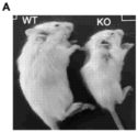


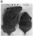


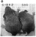


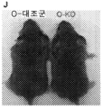


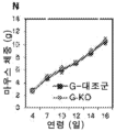


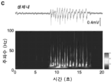


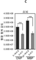
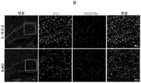
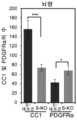
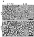


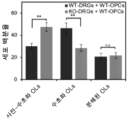

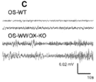

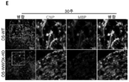


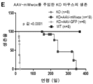
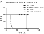


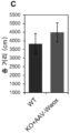
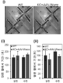


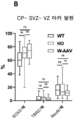


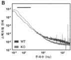
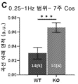
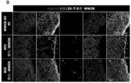
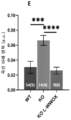
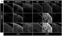
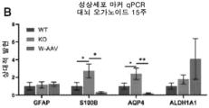
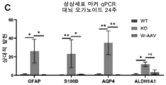

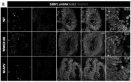


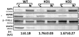

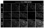

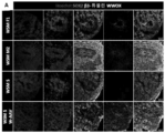
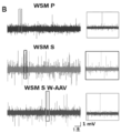


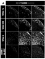


Classifications
-
- A—HUMAN NECESSITIES
- A61—MEDICAL OR VETERINARY SCIENCE; HYGIENE
- A61K—PREPARATIONS FOR MEDICAL, DENTAL OR TOILETRY PURPOSES
- A61K48/00—Medicinal preparations containing genetic material which is inserted into cells of the living body to treat genetic diseases; Gene therapy
-
- C—CHEMISTRY; METALLURGY
- C12—BIOCHEMISTRY; BEER; SPIRITS; WINE; VINEGAR; MICROBIOLOGY; ENZYMOLOGY; MUTATION OR GENETIC ENGINEERING
- C12N—MICROORGANISMS OR ENZYMES; COMPOSITIONS THEREOF; PROPAGATING, PRESERVING, OR MAINTAINING MICROORGANISMS; MUTATION OR GENETIC ENGINEERING; CULTURE MEDIA
- C12N15/00—Mutation or genetic engineering; DNA or RNA concerning genetic engineering, vectors, e.g. plasmids, or their isolation, preparation or purification; Use of hosts therefor
- C12N15/09—Recombinant DNA-technology
- C12N15/63—Introduction of foreign genetic material using vectors; Vectors; Use of hosts therefor; Regulation of expression
- C12N15/79—Vectors or expression systems specially adapted for eukaryotic hosts
- C12N15/85—Vectors or expression systems specially adapted for eukaryotic hosts for animal cells
- C12N15/86—Viral vectors
-
- A—HUMAN NECESSITIES
- A61—MEDICAL OR VETERINARY SCIENCE; HYGIENE
- A61K—PREPARATIONS FOR MEDICAL, DENTAL OR TOILETRY PURPOSES
- A61K48/00—Medicinal preparations containing genetic material which is inserted into cells of the living body to treat genetic diseases; Gene therapy
- A61K48/005—Medicinal preparations containing genetic material which is inserted into cells of the living body to treat genetic diseases; Gene therapy characterised by an aspect of the 'active' part of the composition delivered, i.e. the nucleic acid delivered
- A61K48/0058—Nucleic acids adapted for tissue specific expression, e.g. having tissue specific promoters as part of a contruct
-
- A—HUMAN NECESSITIES
- A61—MEDICAL OR VETERINARY SCIENCE; HYGIENE
- A61P—SPECIFIC THERAPEUTIC ACTIVITY OF CHEMICAL COMPOUNDS OR MEDICINAL PREPARATIONS
- A61P25/00—Drugs for disorders of the nervous system
-
- C—CHEMISTRY; METALLURGY
- C12—BIOCHEMISTRY; BEER; SPIRITS; WINE; VINEGAR; MICROBIOLOGY; ENZYMOLOGY; MUTATION OR GENETIC ENGINEERING
- C12N—MICROORGANISMS OR ENZYMES; COMPOSITIONS THEREOF; PROPAGATING, PRESERVING, OR MAINTAINING MICROORGANISMS; MUTATION OR GENETIC ENGINEERING; CULTURE MEDIA
- C12N9/00—Enzymes; Proenzymes; Compositions thereof; Processes for preparing, activating, inhibiting, separating or purifying enzymes
- C12N9/0004—Oxidoreductases (1.)
- C12N9/0006—Oxidoreductases (1.) acting on CH-OH groups as donors (1.1)
-
- C—CHEMISTRY; METALLURGY
- C12—BIOCHEMISTRY; BEER; SPIRITS; WINE; VINEGAR; MICROBIOLOGY; ENZYMOLOGY; MUTATION OR GENETIC ENGINEERING
- C12N—MICROORGANISMS OR ENZYMES; COMPOSITIONS THEREOF; PROPAGATING, PRESERVING, OR MAINTAINING MICROORGANISMS; MUTATION OR GENETIC ENGINEERING; CULTURE MEDIA
- C12N2750/00—MICROORGANISMS OR ENZYMES; COMPOSITIONS THEREOF; PROPAGATING, PRESERVING, OR MAINTAINING MICROORGANISMS; MUTATION OR GENETIC ENGINEERING; CULTURE MEDIA ssDNA viruses
- C12N2750/00011—Details
- C12N2750/14011—Parvoviridae
- C12N2750/14111—Dependovirus, e.g. adenoassociated viruses
- C12N2750/14141—Use of virus, viral particle or viral elements as a vector
- C12N2750/14143—Use of virus, viral particle or viral elements as a vector viral genome or elements thereof as genetic vector
-
- C—CHEMISTRY; METALLURGY
- C12—BIOCHEMISTRY; BEER; SPIRITS; WINE; VINEGAR; MICROBIOLOGY; ENZYMOLOGY; MUTATION OR GENETIC ENGINEERING
- C12N—MICROORGANISMS OR ENZYMES; COMPOSITIONS THEREOF; PROPAGATING, PRESERVING, OR MAINTAINING MICROORGANISMS; MUTATION OR GENETIC ENGINEERING; CULTURE MEDIA
- C12N2830/00—Vector systems having a special element relevant for transcription
- C12N2830/008—Vector systems having a special element relevant for transcription cell type or tissue specific enhancer/promoter combination
Abstract
본 개시내용은 WW 도메인-함유 산화환원효소(WWOX)-연관 CNS 질병의 치료를 위한 방법 및 조성물을 제공한다. 다양한 구현양태에서, 본 발명은 대상체의 뇌에서 이종 WWOX 유전자를 발현시키는 것을 포함하고, 다양한 구현양태에서 WOREE 증후군 및 SCAR12와 같은 상태를 치료하거나 개선하기 위해 뉴런에서 이종 WWOX 유전자를 발현시키는 것을 포함한다.The present disclosure provides methods and compositions for the treatment of WW domain-containing oxidoreductase (WWOX)-associated CNS diseases. In various embodiments, the invention comprises expressing a heterologous WWOX gene in the brain of a subject, and in various embodiments, expressing a heterologous WWOX gene in neurons to treat or ameliorate conditions such as WOREE Syndrome and SCAR12 .
Description
WW 도메인-함유 산화환원효소(WWOX)의 생식계열 돌연변이는 간질, 운동실조증 및 성 발달 장애(DSD) 환자에서 기록되었다. 보다 최근에는 진단된 알츠하이머병에 대한 유전적 메타-분석에서 WWOX 유전자를 새로운 위험 유전자좌로 식별했다. 몇 가지 증거는 WWOX 발현이 중추신경계(CNS)의 정상적인 발달과 기능에 필요하며 WWOX의 돌연변이가 오늘날 WWOX-연관 간질성 뇌병증(WOREE) 증후군으로 알려진 유아의 신경 장애를 초래한다는 것을 강력하게 시사한다.Germline mutations in the WW domain-containing oxidoreductase (WWOX) have been documented in patients with epilepsy, ataxia and sexual development disorder (DSD). More recently, a genetic meta-analysis of diagnosed Alzheimer's disease identified the WWOX gene as a new risk locus. Several lines of evidence strongly suggest that WWOX expression is required for normal development and function of the central nervous system (CNS) and that mutations in WWOX result in a neurological disorder in infants known today as WWOX-associated epileptic encephalopathy (WOREE) syndrome.
WOREE에서는 상염색체 열성 WWOX 넌센스 돌연변이, 부분 및 완전 결손이 매우 심각한 질병과 연관되어 매우 이른 발병 사망을 초래한다. WWOX의 단일 돌연변이 대립유전자를 지닌 이형접합 부모는 표현형 증상을 나타내지 않는다. 이들 보유자가 성인 간질에 더 취약한지는 아직 알려지지 않았다. WOREE를 특징으로 하는 대부분의 환자는 WWOX의 복합 이형접합 돌연변이를 품고 있어 각 돌연변이를 개별적으로 표적으로 삼기가 어렵다. 더 가벼운 형태의 질병은 WWOX 미스센스 돌연변이와 연관이 있으며 척수소뇌성 운동실조증, 상염색체 열성, 12(SCAR12)라고 지칭된다. WWOX가 CNS의 항상성을 조절하는 메커니즘은 거의 알려져 있지 않다. WWOX가 간질 및 다른 형태의 신경병증을 길항하는 다른 주요 이펙터의 다운스트림인지 여부도 알려져 있지 않다.In WOREE, autosomal recessive WWOX nonsense mutations, partial and complete deletions, are associated with very severe disease, resulting in very early onset death. Heterozygous parents carrying the single mutant allele of WWOX show no phenotypic symptoms. It is not yet known whether these carriers are more susceptible to adult epilepsy. Most patients characterized by WOREE harbor compound heterozygous mutations in WWOX, making it difficult to target each mutation individually. A milder form of the disease is associated with WWOX missense mutations and is referred to as spinocerebellar ataxia, autosomal recessive, 12 (SCAR12). Little is known about the mechanisms by which WWOX regulates CNS homeostasis. It is also unknown whether WWOX is downstream of other key effectors that antagonize epilepsy and other forms of neuropathy.
최근의 증거는 또한 WWOX의 미묘한 돌연변이를 자폐 스펙트럼 장애(ASD)와 연관시킨다. 덜 심각한 표현형과 IQ 수준이 WWOX를 ASD 후보 유전자로 정의하는 정상 범위에 근접한 특징이 있는 많은 ASD 영향을 받은 개체에서 중첩되는 WWOX 커피 수 변이체(CNV)가 보고되었다. WWOX의 유전된 CNV는 ASD에 대한 낮은 침투 위험 인자로 결정되었다. 또한 다발성 경화증 환자 샘플에 대한 대규모 분석에서 WWOX의 것을 포함하여 200개 이상의 상염색체 감수성 변이체가 밝혀졌다. 따라서 WWOX의 섭동은 단일 신경 질병을 넘어 WWOX가 여러 신경 장애에서 중요한 역할을 함을 시사한다.Recent evidence also links subtle mutations in WWOX to autism spectrum disorders (ASD). Overlapping WWOX coffee number variants (CNVs) have been reported in many ASD-affected individuals characterized by less severe phenotypes and IQ levels close to the normal range that define WWOX as an ASD candidate gene. Inherited CNV of WWOX was determined to be a low penetrant risk factor for ASD. In addition, a large-scale analysis of multiple sclerosis patient samples revealed more than 200 autosomal susceptibility variants, including those in WWOX. Thus, perturbation of WWOX goes beyond a single neurological disease, suggesting that WWOX plays an important role in multiple neurological disorders.
본 개시내용은 WWOX-연관 CNS 질병의 치료를 위한 방법 및 조성물을 제공한다. 다양한 구현양태에서, 본 발명은 대상체의 뇌에서 이종 WWOX 유전자를 발현하는 것을 포함하고, 다양한 구현양태에서 WOREE 증후군 및 SCAR12와 같은 상태를 치료하거나 개선하기 위해 뉴런에서 이종 WWOX 유전자를 발현하는 것을 포함한다.The present disclosure provides methods and compositions for the treatment of WWOX-associated CNS diseases. In various embodiments, the invention includes expressing a heterologous WWOX gene in the brain of a subject, and in various embodiments, expressing a heterologous WWOX gene in neurons to treat or ameliorate conditions such as WOREE syndrome and SCAR12 .
일부 구현양태에서, WWOX 유전자는 뉴런에서 WWOX 유전자의 발현을 지시하는 프로모터를 포함하는 하나 이상의 조절 요소를 포함한다. 예를 들어, 프로모터는 범용 프로모터 또는 뉴런-특이적 프로모터일 수 있다. 예시적인 뉴런-특이적 프로모터는 시냅신 I 프로모터이다. 다양한 구현양태에서, WWOX 유전자는 야생형 유전자 또는 기능적 유도체이고 mRNA 안정성을 향상시키는 비번역 서열(예를 들어, 3'-UTR에서)을 포함한다.In some embodiments, a WWOX gene comprises one or more regulatory elements comprising a promoter that directs expression of the WWOX gene in a neuron. For example, the promoter may be a universal promoter or a neuron-specific promoter. An exemplary neuron-specific promoter is the synapsin I promoter. In various embodiments, the WWOX gene is a wild-type gene or functional derivative and includes an untranslated sequence (eg, in the 3'-UTR) that enhances mRNA stability.
일부 구현양태에서, 치료될 개체는 소아 또는 신생아 환자(예를 들어, WOREE 또는 SCAR12를 갖는 환자)이다. 일부 구현양태에서, 조기 치료는 질병의 일부 임상적 매개변수의 징후를 예방한다. 일부 구현양태에서, 개체는 성인 환자(예를 들어, WOREE 또는 SCAR12를 갖는다)이고, 치료는 간질 에피소드와 같은 하나 이상의 임상 매개변수를 개선할 수 있다. 다양한 구현양태에서, 치료는 간질 에피소드의 빈도 및/또는 중증도를 실질적으로 감소시킨다.In some embodiments, the individual to be treated is a pediatric or neonatal patient (eg, a patient with WOREE or SCAR12). In some embodiments, early treatment prevents symptoms of some clinical parameters of the disease. In some embodiments, the individual is an adult patient (eg, has WOREE or SCAR12), and the treatment can improve one or more clinical parameters, such as an epileptic episode. In various embodiments, the treatment substantially reduces the frequency and/or severity of epileptic episodes.
예를 들어, 일부 구현양태에서, 본 발명은 WOREE 증후군 또는 SCAR12의 치료 방법을 제공하며, 이 방법은 이러한 치료를 필요로 하는 환자의 뇌에 시냅신 I 프로모터 제어 하에 WWOX 야생형 유전자를 포함하는 AAV9 유전자 전달 시스템을 투여하는 것을 포함한다. AAV9 전달 시스템은 실질적으로 서열번호: 1, 서열번호: 3, 서열번호: 4, 및/또는 서열번호: 5에 제시된 바와 같은 뉴클레오티드 서열을 포함할 수 있다.For example, in some embodiments, the present invention provides methods of treating WOREE syndrome or SCAR12, which methods include injecting an AAV9 gene, including a WWOX wild-type gene, under the control of the Synapsin I promoter into the brain of a patient in need of such treatment. and administering the delivery system. The AAV9 delivery system may comprise a nucleotide sequence substantially as set forth in SEQ ID NO: 1, SEQ ID NO: 3, SEQ ID NO: 4, and/or SEQ ID NO: 5.
다른 측면에서, 본 개시내용은 뉴런-특이적 프로모터의 발현 제어 하에 WWOX 야생형 유전자 또는 이의 기능적 유도체를 포함하는 발현 작제물을 제공한다. 예시적인 뉴런-특이적 프로모터는 시냅신 I 프로모터이다. 일부 구현양태에서, 뉴클레오티드 서열은 실질적으로 서열번호: 1, 3, 4, 및/또는 5에 제시된 바와 같은 서열을 포함한다. 다양한 구현양태에서, 발현 작제물은 아데노-연관 바이러스(AAV) 전달 시스템과 같은 바이러스 벡터이다. 일부 구현양태에서, 발현 작제물은 AAV9 전달 시스템이다.In another aspect, the present disclosure provides an expression construct comprising a WWOX wild-type gene or functional derivative thereof under the control of expression of a neuron-specific promoter. An exemplary neuron-specific promoter is the synapsin I promoter. In some embodiments, the nucleotide sequence comprises a sequence substantially as set forth in SEQ ID NO: 1, 3, 4, and/or 5. In various embodiments, the expression construct is a viral vector, such as an adeno-associated virus (AAV) delivery system. In some embodiments, the expression construct is an AAV9 delivery system.
본 개시내용의 다른 측면 및 구현양태는 다음의 상세한 설명 및 구현양태로부터 명백해질 것이다.Other aspects and implementations of the present disclosure will become apparent from the detailed description and implementations that follow.
도 1a-1m은 뇌 세포에서 뮤린 Wwox의 조건부 결실의 표현형을 보여주는 연구의 요약을 제공한다. (a) P18에서 Wwox null(KO) 및 야생형(WT) 마우스의 대표 이미지. (b) 시간의 함수로서 체중(그램, g) 증가를 보여주는 그래프. Wwox null에서 지연된 성장은 야생형에 비해 4일 이후부터 명백하다. 데이터 포인트는 마우스의 평균 체중을 나타낸다(WT, n = 3; KO, n = 3). 오차 막대는 ±SEM을 나타낸다(** P < 0.01, *** P< 0.001, 스튜던트 t 테스트). (c) 카플란 마이어(Kaplan Meier) 생존 곡선은 3-4주령까지 Wwox null의 출생 후 치사율을 보여준다(WT, n = 10; KO, n = 11). (P = 0.0023, 로그-순위 Mantel-Cox 테스트). (d) 신경 줄기/전구 세포에서 Wwox 유전자의 조건부 결실은 대조군 마우스(N-대조군)와 비교하여 N-KO에서 전체적인 발달 지연을 나타낸다. P17에 표시된 이 마우스의 대표 이미지. (e) N-KO 마우스는 N-대조군에 상응하는 감소된 체중을 나타낸다. 데이터 포인트는 마우스의 평균 체중, 유전자형당 4마리 마우스를 나타낸다. 오차 막대는 ±SEM을 나타낸다(** P < 0.01, *** P < 0.001, 스튜던트 t 테스트). (f) N-대조군(n=12)과 비교하여 N-KO(n=14)에서 3-4주령까지 출생 후 치사율을 나타내는 카플란 마이어 생존 곡선. (P =0.0002, 로그-순위 Mantel-Cox 테스트). (g-i) 다음을 나타내는 뉴런(S-KO)에서 Wwox의 조건부 절제가 있는 마우스(P17)의 대표 이미지 (g) 성장 지연 및 (h) 감소된 체중(데이터 포인트는 마우스의 평균 체중, 유전자형당 4마리의 마우스를 나타낸다) 오차 막대는 ±SEM ** P < 0.01, *** P < 0.001, 스튜던트 t 테스트) 및 (i) 조기 사망(S-대조군 n = 13, S-KO n = 15)을 나타낸다. (P 값 0.0001, 로그-순위 Mantel-Cox 테스트). (j-l) 희소돌기아교세포(O-KO) 또는 성상세포(G-KO)에서 Wwox의 조건부 절제는 O-KO 또는 G-KO 마우스에서 해당 대조군(G-대조군, O-대조군)과 비교하여 발달 지연(j 및 m, P17에 표시), 체중 감소(k 및 n) 및 출생 후 치사율(l 및 o)와 같은 표현형 이상을 유발하지 않는다(k 및 l에서, O-대조군 n = 11 및 O-KO n = 10, P 값 1.0, 유의성 없음, 로그-순위 Mantel-Cox 테스트) (G-대조군 n = 8, G-KO n = 9 마우스는 N 및 M에서 사용, P 값 1.0, 유의성 없음, 로그-순위 Mantel-Cox 테스트).
도 2a-f는 S-KO 신피질에서 신피질의 과흥분성을 보여주는 연구의 요약을 제공한다. (a) S-대조군, S-HT 및 S-KO의 생체 내 기록(P13-P17)은 이형접합체(S-HT, 파란색으로 표시) 및 S-대조군(검은색으로 표시)과 비교하여 S-KO(빨간색으로 표시)에서 폭발 활성을 나타낸다. (b) 분리된 신피질 절편 제제에서 자발적인 신피질 폭발을 나타내는 표면 신피질로부터의 시험관내 기록(P13-P17). 인셋(inset)은 S-KO 사례에서 하나의 폭발 이벤트의 확장된 트레이스를 보여준다. 전체 뇌의 인셋은 생체내 기록 전극의 위치를 보여준다. 자발적 활동 및 7 S-대조군, 14 S-HT 및 24 S-KO 동물로부터의 11 S-대조군, 23 S-HT 및 42 S-KO 절편에서 수득한 전기 자극 인공물을 갖는 데이터를 모두 포함하는 모든 데이터 평가 중에서, 0개의 S-대조군 절편은 폭발(0%)을 보였고, 3마리의 S-HT 동물의 4개의 절편은 폭발(약 20%)을 보였고, 20마리의 S-KO 동물의 36개의 절편은 폭발(약 84%)을 보였다. (c) 생체내 기록 및 시간-주파수 스펙트로그램의 두 번째 예. (d) 자극 인공물이 없는 데이터에서 얻은 S-대조군, S-HT 및 S-KO에 대한 생체내 및 시험관내 데이터 세트에 대한 전력 스펙트럼 분석. 회색 막대는 주파수별로 식별된 중요 영역을 나타낸다. 생체내 12-20 Hz는 S-HT에 비해 S-KO에서, S-대조군에 비해 S-KO에 대해 7-15 Hz에서 높은 전력을 나타낸다. 시험관내 3-13 Hz는 S-HT 및 S-대조군에 비해 S-KO의 상승된 전력을 보였다. 시험관내 데이터 z-점수는 320-400 Hz의 평균 및 표준 편차로 정규화되었다. 박스 플롯은 가로축에 주파수 대역이 표시된 정규화된 전력을 보여준다. 0-4.9 Hz 및 5-9 Hz의 경우, S-KO 전력은 S-대조군 단독과 비교하여 상당히 상승했다(생체내, S-대조군, n = 5 대상체; S-KO, n = 7 대상체; S-HT, n= 7 대상체, * P < 0.05; 시험관내 * P < 0.05, ** P < 0.01 스튜던트 t 테스트, S-대조군 및 S-HT와 비교한 S-KO; S-대조군, n = 11 절편, 7 동물, S-HT, n = 11 절편, 8 동물; S-KO, n = 34 절편, 20 대상체). (e) 신피질의 층 II/III에서 기록되는 층 V의 전기 자극에 대한 반응. 화살표는 100 uA 자극 강도에서 1차 피크 반응의 중지 및 시작을 나타내고 캐럿 기호는 2차 피크 반응의 중지 및 시작을 나타낸다. (f) 1차 및 2차 피크 반응의 진폭(1차 피크: * P = 0.0127. 2차 피크: ** P = 0.008, * P = 0.0202, 윌콕슨(Wilcoxon) 순위 합계 테스트 S-대조군 n = 6 절편, 3 대상체, S -HT, n = 8 절편, 5 대상체; S-KO, n = 8 절편, 6 대상체).
도 3a-3e는 Wwox의 뉴런 결실이 수초화 및 희소돌기아교세포 성숙을 손상시킨다는 것을 보여주는 연구의 요약을 제공한다. (a) 피질 영역의 3개의 동일한 섹션에서 CNP 및 MBP의 형광 합계 강도의 정량화는 S-대조군(n = 3)에 비해 S-KO(n = 3)에서 감소된 강도를 나타낸다. (b) 항-CNP 및 항-MBP로 면역염색된 뇌 섹션의 대표적인 이미지가 소뇌에서 나타난다. (c) CNP 및 MBP의 형광 강도 정량화는 S-대조군에 비해 S-KO에서 감소된 강도를 표시한다. (d) CC1 및 항-PDGFRα에 대해 면역염색된 뇌 조직의 시상 섹션. P17에서 S-대조군과 비교하여 S-KO의 뇌량(흰색 점선으로 표시되고 확대 영역은 흰색 사각형으로 표시)에서 성숙한 희소돌기아교세포의 수가 감소한 것을 보여주는 이미지. (e) S-대조군과 비교하여 SK-O에서 CC1 양성 세포의 현저한 감소 및 PDGFRα 양성 세포의 수 증가를 나타내는 뇌량에서 CC1 및 PDGFRα 양성 세포의 정량화. 데이터 포인트는 S-대조군(n = 3) 및 S-KO 마우스(n = 3)의 3개의 독립적인 섹션에서 영역(0.5mm2)에서 계수된 세포 수를 나타낸다. 오차 막대는 ±SEM을 나타낸다(* P ≤ 0.01, ** P ≤ 0.001). 축척 막대 A) 50μm, B) 2 μm 및 C) 50 μm.
도 4a-4d는 Wwox의 뉴런 결실이 수초화 및 축삭 전도도를 감소시킨다는 것을 보여주는 연구의 요약을 제공한다. (a) P17에서 S-대조군(n = 3)과 비교하여 S-KO(n = 3)의 시신경에서 더 적은 수의 수초화 축삭 및 더 많은 무수초화 축삭을 보여주는 뇌량의 중간 시상 섹션의 전자 현미경 사진(EM) 사진. (b) S-대조군 대 S-KO에서 뇌량의 수초화 축삭 및 시야당 시신경에서 무수초화 축삭의 수의 정량화. 그래프는 뇌량에서 수초화 축삭(S-대조군, n = 2500 및 S-KO, n = 1200)과 시신경에서 무수초화 축삭(S-대조군 n = 500 및 S-KO, n = 3000)을 FOV(시야)당 EM 이미지로 나타낸다. 오차 막대는 ±SEM을 나타낸다(*** P ≤ 0.001, 스튜던트 t 테스트). (c) S-대조군(n = 3)과 비교하여 S-KO(n = 3)에서 뇌량 및 시신경의 축삭의 미엘린 두께 감소를 나타내는 g-비율 분석. 뇌량(각 유전자형당 n = 300) 및 시신경(각 유전자형당 n = 300)의 전자현미경 이미지로부터 축삭 직경 및 미엘린 두께를 계산하였다(*** P ≤ 0.001, 스튜던트 t 테스트). (d) 뇌량 자극으로부터 유발된 반응의 대표적인 예, (i) 자극기로부터 250 μm의 수직 거리에서 기록. 인셋은 전극 및 자극의 배치를 보여준다. (N1 *P = 0.0104; N2 = 0.5737 Wilcoxon 순위 합계 테스트) (ii) 자극 시작부터 N1 및 N2까지의 지연 시간. (ii) N1 진폭과 N2 진폭의 비율. (N1/N2). (** P = 0.0030; 윌콕슨(Wilcoxon) 순위 합계 n = 8 절편, 5 대상체, S-대조군, n = 8 절편 6 대상체 S-KO).
도 5는 OPC 분화에서 WWOX의 비-세포 자율 기능을 보여주는 연구의 요약을 제공한다. 계산된 사전-수초화, 수초화 및 분해된 희소돌기아교세포의 정량화(직경 14 mm이 커버된 면적). 결과는 두 개의 독립적인 실험(WT-DRGs+WT-OPCs, n = 4; KO-DRGs+WT-OPCs, n = 4)의 상자 플롯으로 표시된다. (**P< 0.01. n.s 유의미하지 않음). 축척 막대 A) 100 μm, D) 50 μm.
도 6a-6g는 WWOX-결핍 희소피질 회전타원체가 과흥분성 및 저수초화를 나타낸다는 것을 보여주는 연구의 요약을 제공한다. (a) 희소피질 회전타원체 절편 설정의 개략도. 전극은 로컬 필드 전위 기록(LFP)에 사용되는 반면 두 번째 전극은 전체-셀 패치 기록에 사용된다. 전극은 절편 가장자리로부터 150 μm에 위치해 있고 10-15μm 떨어져 있다. (b) 15주차에 OS-WT 및 OS-WWOX-KO 오르가노이드에 대한 전체-세포 패치 클램프 기록에서 세포의 휴지 막 전위(OS-WT n = 3, OS-WWOX-KO n = 4; OS-WT RMP = -52.1 ± 0.90 mV, OS-WWWOX-KO RMP = -21.28 ± 6.91 mV). (*P < 0.01, 스튜던트 t 테스트). (c) 기준선 조건에서 15주차에 OS-WT 및 OS-WWOX-KO 오르가노이드의 평균 스펙트럼 전력(n = 9 절편, OS-WT의 경우 3 오르가노이드, n = 6 절편, OS-WWOX-KO의 경우 2 오르가노이드; OS-WT AUC = 0.0285 ± 0.0097, OS-WWWOX-KO AUC = 0.0541 ± 0.0093.(*P < 0.05, 스튜던트 t 테스트).(d) 델타 및 세타 범위(0.5-7.9 Hz)에 대한 (c)에서 평균 스펙트럼 전력 곡선 아래의 면적.(*P < 0.05, 스튜던트 t 테스트).(e) CNP(OLs) 및 MBP(OLs)에 대해 염색된 30주 OS. 오른쪽의 이미지는 왼쪽 상자 영역의 확대이다.(OS-WT, n = 5; OS-WWOX-KO, n = 5).(f) 37주차 오르가노이드에서 OS-WWOX-KO(n = 3)에 비해 더 많은 수초화 축삭 OS-WT(n = 3)를 보여주는 대표적인 전자 현미경 이미지. 상자에 표시된 확대된 영역. (g) 막대 그래프는 OS-WT(n = 2) 및 OS-WWOX-KO(n = 3)에서 수초 및 무수초 축삭의 백분율을 나타낸다. 스케일 바 (e) 및 (f) 100 μm, (g) 40 μm 및 (h) 왼쪽 패널 500 nm 및 오른쪽 패널 200 nm.
도 7a-7f는 시냅신 I-양성 뉴런에서 WWOX의 복원이 Wwox null 마우스의 성장을 개선하고 수명을 연장함을 보여주는 연구 요약을 제공한다. (a) 인간 시냅신 I 프로모터 하에 뮤린 Wwox 유전자를 함유하는 플라스미드 벡터 작제물의 예시. Wwox 유전자 서열 다음에 IRES 프로모터 및 EGFP 유전자 서열이 온다. (b) P17에서 AAV9-hSynI-GFP(대조군 바이러스) 또는 AAV9-hSynI-mWwox-IRES-GFP 바이러스로 주입된 야생형(WT), Wwox null의 물리적 외관. 지정된 날짜에 마우스의 체중(c) 및 혈당 수치(d)를 보여주는 그래프. 오차 막대는 ±SEM, 유전자형당 n=4마리 마우스를 나타낸다. (e) 카플란-마이어(Kaplan-Meier) 생존 그래프는 AAV9-hSynI-GFP(n=6)를 주사한 마우스 또는 비주사(n =10) 마우스에 비해 AAV9-hSynI-mWwox(n=18)를 주사한 Wwox 녹아웃 마우스의 연장된 수명을 나타낸다.(p 값 <0.0001, 로그 순위 Mantel-Cox 테스트). (f) 카플란-마이어(Kaplan-Meier) 생존 그래프는 AAV9-hSynI-GFP(n=4) 또는 비주사(n=8) 마우스에 비해 AAV9-hSynI-hWWOX (n=6) [중앙값 92 일]를 주사한 Wwox 녹아웃 마우스의 연장된 수명을 나타낸다(p 값 = 0.0001, 로그 순위 Mantel-Cox 테스트).
도 8a 및 8b는 WWOX의 뉴런 회복이 신피질에서 간질 활동을 감소시킨다는 것을 보여주는 연구의 요약을 제공한다. (a) P20-21일에 AAV9-hSynI-mWwox [KO+A-Wwox] 새끼로 처리된 WT, KO 및 KO에서 수행된 세포-부착 기록의 대표적인 트레이스. 트레이스은 자발적인 신피질 활동을 나타낸다(활동 전위가 표시). 패널은 0.5초 간격의 확대를 나타내는 인셋으로 12초 기록을 나타낸다. KO가 활동 전위의 폭발을 나타내는 대표적인 트레이스에서 KO 뇌의 명백한 과잉 활동이 관찰된다. 그래프는 WT 새끼(n=2)의 20개 뉴런, KO+A-Wwox 새끼(n=2)의 20개 뉴런 및 KO 새끼(n=2)의 30개 뉴런에 대한 평균을 보여준다(****p 값 < 0.0001, 스튜던트 t 테스트). (b) WT 및 KO+A-Wwox 성체 마우스(6개월)에서 수행된 세포-부착 기록의 대표적인 트레이스. 트레이스은 자발적인 신피질 활동(활동 전위)을 나타낸다. 패널은 0.5초 간격의 확대를 나타내는 인셋과 함께 12초 기록을 제공한다. 그래프는 WT 성체 마우스(n=3)의 60개 뉴런과 KO+A-Wwox 성체 마우스(n=3)의 60개 뉴런 이상의 평균을 보여준다. WT와 KO+A-Wwox 성체 마우스의 평균 발화율 사이에는 유의한 변화가 관찰되지 않았다.
도 9a-9c는 뉴런에서의 WWOX 복원이 Wwox null에서 OPC 분화를 촉진함으로써 수초화를 개선함을 보여주는 연구의 요약을 제공한다. (a) 지시된 마우스(각 군에 대해 n=3)로부터 P17에서 항-MBP로 면역표지된 전체 뇌 시상 섹션의 이미지. 피질, 해마 및 소뇌의 MBP 염색이 제시된다(상단 패널에서 확대). (b) CC1 및 PDGFRα에 대해 면역염색된 뇌(P17) 조직의 시상 섹션. AAV9-hSynI-GFP 바이러스가 주입된 Wwox null과 비교하여 AAV9-hSynI-mWwox로 처리한 후 Wwox null의 뇌량에서 성숙한 희소돌기아교세포의 증가된 수를 보여주는 이미지. (c) 그래프는 각 유전자형에서 마우스당 3개의 유사한 뇌 시상 섹션에서 계수된 WT(n=3), KO(n=3) 및 KO+A-Wwox(n=3)의 뇌량(면적 0.5 mm2)에서 CC1 및 PDGFRα 양성 세포의 정량화를 나타낸다. 오차 막대는 ±SEM을 나타낸다. (*p 값 <0.01, **p 값 <0.001, ***p 값 <0.0001). 스케일 바(a) 2 mm(상단 패널), 250 μm(중간 및 하단 패널), (b) 250 μm(상단 패널), 50 μm(중간 및 하단 패널).
도 10a-10e는 뉴런에서의 WWOX 복원이 Wwox null 마우스의 비정상적인 행동 표현형을 역전시킨다는 것을 보여주는 연구의 요약을 제공한다. (a) 8-10주에서 WT(암컷, n=7, 수컷 n=6) 및 AAVmWwox-주입 KO 마우스(암컷 n=7, 수컷 n=5)의 트레이스 패턴(주변 및 중앙)을 보여주는 야외 테스트의 대표 이미지. (b) 및 (c) 그래프는 야외 트레이스에서 이동한 속도와 거리(cm)를 나타낸다. (d) WT 및 KO+AAV-Wwox 마우스(연령 8-10주)로부터 고가식 십자 미로 테스트의 마우스 트레이스 이미지. O.A.(열린 팔) C.O. (닫힌 팔)은 왼쪽 마우스 트레이스 이미지에 점선 흰색 상자로 표시된다. 그래프는 WT(암컷 n=7, 수컷 n=6) 및 KO+AAV-Wwox(암컷 n=7, 수컷 n=5)의 닫힌 팔(i) 및 열린 팔(ii)의 기간(s)을 나타낸다. (e) 그래프는 다른 시험에서 회전막대(rotarod)에서 떨어지는 마우스(8-10주에 WT 및 구조된 마우스)의 지연 시간(들)을 나타낸다. 암컷(WT, n=7; KO+-mWwox, n=5)과 수컷(WT, n=5; KO+-mWwox, n=5)은 별도로 표시된다(***p 값 <0.0001).
도 11a-11f는 WWOX 녹아웃 대뇌 오가노이드의 생성 및 특성화에 관한 연구의 요약을 제공한다. (a) WWOX 및 범-방사형 아교세포 마커 SOX2와 함께 심실 방사형 아교세포(vRG) 마커 CRYAB에 대해 염색된 10주 CO. 오른쪽 이미지는 박스 영역을 확대한 것이다. 파선은 심실 공간을 정의한다(WT: 3 배치에서 n = 8, KO: 3 배치에 n = 8). 축척 = 50 μm(왼쪽), 25 μm(오른쪽). (b)심실-유사 구역(VZ), 심실하 구역(SVZ) 및 피질판(CP)을 구성하는 다양한 집단을 나타내는 마커의 정량화. NeuN은 성숙한 뉴런을 표시하고, TBR2는 중간 전구체(IP)를 표시하고, SOX2는 심실 방사형 아교세포(vRG)를 표시한다. 상자플롯은 1사분위수와 3사분위수를 나타내며 수염은 최소 및 최대 포인트를 나타내고 중앙 밴드는 중앙값을 나타낸다. 투키(Tukey)의 다중 비교 테스트(WT: 3개 배치에서 n = 8, KO: 3개 배치에서 n = 8, W-AAV: 1개 배치에서 n = 4)와 일원 ANOVA를 사용하여 통계적 유의성을 결정했다. (c) 15주 CO에서 다양한 신경 마커의 발현 수준 평가를 위한 qPCR 분석: SOX2 및 PAX6(전구 세포), TUBB3(범-뉴런), SLC17A6 및 SLC17A7(VGLUT2 및 VGLUT1; 글루타메이트성 뉴런), 및 GAD1 및 GAD2(GAD67 및 GAD65; GABAergic 뉴런). y축은 상대 발현 배수 변화를 나타낸다. 데이터는 평균 SEM으로 표시된다. 투키(Tukey)의 다중 비교 테스트(WT: 1배치에서 n = 4, KO: 1배치에서 n = 4, W-AAV: 1배치에서 n = 4)와 함께 일원 ANOVA를 사용하여 통계적 유의성을 결정했다. (d) 10주차 CO에서 글루타메이트성 뉴런 마커 VGLUT1 및 GABAergic 뉴런 마커 GAD67(GAD1)에 대한 면역형광(IF) 염색(WT: 3배치에서 n = 8, KO: 3배치에서 n = 8, W-AAV: n = 1 배치에서 4). 축척 = 100 μm. 이미지의 정량화가 표시된다. VGLUT1 및 GAD67은 염색으로 덮인 표면적으로 정량화되었으며 각 이미지의 핵 수로 정규화되었다. 상자플롯은 1사분위수와 3사분위수를 나타내며 수염은 최소 및 최대 포인트를 나타내고 중앙 밴드는 중앙값을 나타낸다. 샘플이 정상적으로 분포되지 않았기 때문에 던(Dunn)의 다중 비교 테스트와 함께 크루스칼-왈리스(Kruskal-Wallis) 테스트를 사용하여 통계적 유의성을 결정했다. ROUT 테스트를 사용하여 이상값을 제거했다(Q = 1%). 데이터 정보: n.s(유의하지 않음), *P ≤ 0.05 및 ****P ≤ 0.0001.
도 12a-12c는 WWOX-KO 대뇌 오가노이드가 과흥분성 및 간질형 활성을 나타냄을 보여주는 연구의 요약을 제공한다. 7주 된 hESC-유래 대뇌 오가노이드(CO)의 샘플 기록. (a) 샘플 트레이스은 기준선 조건(왼쪽) 및 100 μM 4-AP(오른쪽)의 존재에서 WT와 비교하여 증가된 활성을 나타내는 WWOX-KO CO와 함께 로컬 필드 전위의 눈에 띄는 차이를 보여준다. (b) 기준선 조건에서 7주차 WT 및 KO CO의 평균 스펙트럼 전력. 선은 0.25-1 Hz 주파수 범위를 표시한다. 양측 언페어드 웰치(Welch)의 t-테스트를 사용하여 통계적 유의성을 결정했다(WT: n = 14 절편, 5 오르가노이드 및 3 배치, KO: n = 14 절편, 8 오르가노이드 및 3 배치). (c) 0.25-1 Hz 주파수 범위에 대한 (b)의 평균 스펙트럼 전력 곡선 아래 정규화된 면적. 평균 SEM으로 표시되는 데이터. 통계적 유의성은 양측 언페어드 웰치(Welch)의 t-테스트를 사용하여 결정되었다. 모든 막대의 숫자는 분석된 절편 및 오르가노이드(즉, 절편(오가노이드))의 수를 나타낸다. (d) 및 (e) WWOX의 코딩 서열은 렌티바이러스 형질도입(렌티-WWOX)을 사용하여 6주차 WWOX-KO CO에 다시 도입되었다. (d) 렌티바이러스 감염 후 WWOX-KO 오가노이드에서 상이한 집단에서 WWOX 발현을 나타내는 면역형광 염색. NT = 미처리. 축척 = 50 μm. (e) 0.25-1 Hz 주파수 범위에 대해 기준선 조건에서 7주차에 WT 계통, 2 KO 계통 및 렌티-WWOX에 감염된 2 KO 계통의 평균 스펙트럼 전력 곡선 아래 정규화된 면적. 평균 SEM으로 표시되는 데이터. 모든 막대의 숫자는 분석된 절편 및 오르가노이드(즉, 절편(오가노이드))의 수를 나타낸다. 통계적 유의성은 투키(Tukey)의 다중 비교 테스트와 함께 일원 ANOVA를 사용하여 결정되었다. 데이터 정보: ***P ≤ 0.001 및 ****P ≤ 0.0001.
도 13a-13f는 WWOX-KO 대뇌 오가노이드가 손상된 성상세포형성 및 DNA 손상 반응을 나타냄을 보여주는 연구의 요약을 제공한다. (a) 15주 및 24주 CO는 성상 세포 및 방사형 아교세포 마커 GFAP 및 성상세포-특이적 마커 S100b(WT W15: 3개 배치에서 n = 9; KO W15: 3개 배치에서 n = 16; W-AAV W15: 1 배치에서 n = 4 오르가노이드; WT W24: 4 배치에서 n = 10; KO W24: 3 배치에서 n = 9; 및 W-AAV W24: 1 배치에서 n = 4)에 대해 염색되었다. 축척 = 100 μm. (b) 15주째 CO의 성상세포 마커의 qPCR 분석. y축은 상대적인 발현 배수 변화를 나타낸다. 데이터는 평균 ± SEM으로 표시된다. 투키(Tukey)의 다중 비교 테스트(WT: 1배치에서 n = 4; KO: 1배치에서 n = 4; W-AAV: 1배치에서 n = 4)와 함께 일원 ANOVA를 사용하여 통계적 유의성을 결정했다. (c) 24주째 CO의 성상세포 마커의 qPCR 분석. y축은 상대적인 발현 배수 변화를 나타낸다. 데이터는 평균 ± SEM으로 표시된다. 투키(Tukey)의 다중 비교 테스트(WT: 2배치에서 n = 4; KO: 2배치에서 n = 3; W-AAV 1배치에서 n = 3)와 함께 일원 ANOVA를 사용하여 통계적 유의성을 결정했다. (d) VZ 주변의 성상세포 마커에 대한 6주 CO의 IF 염색(WT: 2배치에서 n = 6; 및 KO: 2배치에서 n = 6). 축척 = 100 μm(왼쪽) 및 50 μm(오른쪽). € 생리학적 조건에서 범-방사형 아교 세포 마커 SOX2와 함께 6주차 CO의 VZ에 있는 세포 핵의 DNA 손상 마커 cH2AX 및 53BP1에 대한 염색(WT: 3개의 개별 배치에서 n = 8; KO: 3개의 개별 배치에서 n = 12개 및 W-AAV: 1개 배치에서 n = 4). 축척 = 50 μm(왼쪽), 25 μm(오른쪽). (f) VZ의 가장 안쪽 층을 구성하는 세포의 핵에서 이 층의 총 핵 수로 정규화된cH2AX(상단) 및 53BP1 병소(foci)(하단)의 정량화. 상자플롯은 1사분위수와 3사분위수를 나타내며 수염은 최소 및 최대 포인트를 나타내고 중앙 밴드는 중앙값을 나타낸다. 통계적 유의성은 투키(Tukey)의 다중 비교 테스트(WT: 3배치에서 n = 8개 오르가노이드; KO: 3개 배치에서 n = 12개 오르가노이드; W-AAV: 1개 배치에서 n = 4)와 함께 일원 ANOVA를 사용하여 결정되었다. 데이터 정보: n.s(유의하지 않음), *P ≤ 0.05, **P ≤ 0.01 및 ****P ≤ 0.0001.
도 14a-14e는 대뇌 오가노이드 RNA 시퀀싱이 주요 분화 결함을 드러냄을 보여주는 연구의 요약을 제공한다. 15주 CO의 RNA 시퀀싱(RNA-seq) 및 전사체 분석(WT: n = 2, KO: n = 4). (a) RNA-seq의 결과를 검증하는 선택된 Wnt 표적 유전자에 대한 qPCR 분석. y축은 상대 발현 배수 변화를 나타낸다. 데이터는 평균 _ SEM으로 표시된다. 투키(Tukey)의 다중 비교 테스트(WT: 1배치에서 n = 4; KO: 1배치에서 n = 4; W-AAV: 1배치에서 n = 4)와 함께 일원 ANOVA를 사용하여 통계적 유의성을 결정했다. (b) 16주차 CO는 세포질(c) 및 핵(N) 분획으로 세분화되었다. 실험은 총 2개의 WT 오가노이드와 4개의 KO 오가노이드(각 KO 계통당 2개)로 두 번 실행되었다. KAP-1은 핵을 표시하고 HSP90은 세포질을 표시한다. 하단의 숫자는 세포질 분획[b-C(N/C)]으로 정규화된 b-카테닌 밴드 강도의 핵 분획의 정량화이다. (c) TBR1, BCL11B(CTIP2), SATB2, POU3F2(BRN2), CUX1 및 RELN과 같이 가장 깊은 것부터 가장 표면적인 것까지 15주차 오가노이드에서 인간 피질의 6개 층의 마커 발현 수준을 보여주는 히트맵. (d) 깊은 층 피질 마커 CTIP2(BCL11B) 및 TBR1 및 표면층 마커 SATB2(WT: n = 3; KO: n = 4; 및 W-AAV: n= 4)의 감소된 수준을 검증하는 15주 CO의 IF 염색. 축척 = 50 μm. (e) 총 핵 수로 정규화된 G에서 보이는 피질 마커의 정량화. y축은 WT CO의 평균과 비교하여 배수 변화를 나타낸다. 상자플롯은 1사분위수와 3사분위수를 나타내며 수염은 최소 및 최대 포인트를 나타내고 중앙 밴드는 중앙값을 나타낸다. 통계적 유의성은 투키(Tukey)의 다중 비교 테스트(WT: 3개 배치에서 n = 9, KO: 3개 배치에서 n = 16, WAAV: 1개 배치에서 n = 4 오르가노이드)와 함께 일원 ANOVA를 사용하여 결정되었다. 데이터 정보: *P ≤ 0.05, **P ≤ 0.01, ***P ≤ 0.001 및 ****P ≤ 0.0001.
도 15a-15e는 WWOX-관련 간질성 뇌병증 대뇌 오가노이드가 뉴런 이상을 반복한다는 것을 보여주는 연구의 요약을 제공한다. WOREE 증후군 환자와 그의 건강한 부모로부터 말초 혈액 단핵 세포(PBMC)를 분리하고 iPSC로 재프로그래밍한 후 CO로 분화시켰다. (a) 선조 마커 SOX2, 뉴런 마커 b3-튜불린 및 WWOX(WSM F1: 1개 배치에서 n = 2; WSM M2: 1개 배치에서 n = 2; WSM S2: 1개 배치에서 n = 2; WSM S5: 1개 배치에서 n = 2; WSM S5 W-AAV3: 1개 배치에서 n = 2개; 및 WSM S5 W-AAV6: 1개 배치에서 n = 2)에서 염색된 10주차 WSM CO. 축척 = 50 μm. (b) WSM P, WSM S 및 WSM S W-AAV 7주 CO의 뉴런에서 기록된 자발적 스파이크의 대표적인 트레이스. 각 기록의 길이는 12초이고 확대는 0.5초이다(오른쪽 상자). (c) WSM P CO(4 오르가노이드)의 24개 뉴런, WSM S CO(3 오르가노이드)의 41개 뉴런, WSM S W-AAV 오르가노이드(3 오르가노이드)의 40개 뉴런에 대한 평균 발화율. 통계적 유의성은 투키(Tukey)의 다중 비교 테스트와 함께 일원 ANOVA를 사용하여 결정되었다. 막대는 평균 _ SEM을 나타낸다. (d) 10주째 WSM CO에서 GAD67 및 VGLUT1 면역염색(WSM F1: 1개 배치에서 n = 2; WSM M2: 1개 배치에서 n = 2; WSM S2: 1개 배치에서 n = 2; WSM S5: 1개 배치에서 n = 2; WSM S5 W-AAV3: 1개 배치에서 n = 2; WSM S5 W-AAV6: 1개 배치에서 n = 2). 축척 = 50 μm. (e) 도 15d에 나타낸 데이터의 정량화. 통계적 유의성은 투키(Tukey)의 다중 비교 테스트와 함께 일원 ANOVA를 사용하여 결정되었다(WSM F1: 1개 배치에서 n = 2; WSM M2: 1개 배치에서 n = 2; WSM S2: 1개 배치에서 n = 2; WSM S5: 1개 배치에서 n = 2개; WSM S5 W-AAV3: 1개 배치에서 n = 2개; WSM S5 W-AAV6: 1개 배치에서 n = 2개). 데이터 정보: ns(유의하지 않음), *P ≤ 0.05, **P ≤ 0.01, ***P ≤ 0.001 및 ****P ≤ 0.0001.
도 16a-16f는 WWOX-관련 간질성 뇌병증이 완전한 WWOX 손실과 유사한 분자 이상을 묘사함을 보여주는 연구의 요약을 제공한다. (a)성상세포 마커 GFAP 및 S100b에 대해 염색된 15주차 WSM CO. (WSM F1: 1개 배치에서 n = 2; WSM M2: 1개 배치에서 n = 3; WSM S2: 1개 배치에서 n = 2; WSM S5: 1개 배치에서 n = 2; WSM S5 W-AAV3: 1개 배치에서 n = 2 및 WSM S5 W-AAV6: 1개 배치에서 n = 2). 축척 = 50 μm. (b) WOREE- 유래 오가노이드에서 손상된 DNA 손상 반응. SOX2는 VZ의 방사형 아교세포를 표시한다(WSM F1: 1개 배치에서 n = 4; WSM M2: 1개 배치에서 n = 4; WSM S2: 1개 배치에서 n = 4; WSM S5: 1개 배치에서 n = 4; WSM S5 W-AAV3: 1개 배치에서 n = 4; 및 WSM S5 W-AAV6: 1개 배치에서 n = 4). 축척 = 50 μm(왼쪽) 및 25 μm(오른쪽). (c) 6주차 WSM CO의 VZ에 있는 세포 핵에서 cH2AX(왼쪽) 및 53BP1(오른쪽)으로 표시된 DNA 손상 병소의 정량화. 상자플롯은 1사분위수와 3사분위수를 나타내며 수염은 최소 및 최대 포인트를 나타내고 중앙 밴드는 중앙값을 나타낸다. 통계적 유의성은 투키(Tukey)의 다중 비교 테스트와 함께 일원 ANOVA를 사용하여 결정되었다.(WSM F1: 1개 배치에서 n = 4; WSM M2: 1개 배치에서 n = 4; WSM S2: 1개 배치에서 n = 4; WSM S5: 1개 배치에서 n = 4; WSM S5 W-AAV3: 1개 배치에서 n = 4; WSM S5 W-AAV6: 1개 배치에서 n = 4) (d) 10주차 WSM CO에서 선택된 Wnt 표적 유전자에 대한 qPCR 분석. y축은 상대 표현 배수 변화를 나타낸다. 데이터는 평균 SEM으로 표시된다. 통계적 유의성은 투키(Tukey)의 다중 비교 테스트와 일원 ANOVA를 사용하여 결정되었다(WSM P: 1개 배치에서 n = 6; WSM S: 1개 배치에서 n = 4; WSM S W-AAV: 1개 배치에서 n = 4). (e) 피질층의 마커 CTIP2 및 SATB2에 대해 염색된 15주차 WSM CO. (WSM F1: 1개 배치에서 n = 2; WSM M2: 1개 배치에서 n = 3; WSM S2: 1개 배치에서 n = 2; WSM S5: 1개 배치에서 n = 2; WSM S5 W-AAV3: 1 배치에서 n = 2; 및 WSM S5 W-AAV6: 1 배치에서 n = 2). 축척 = 50 μm. (f) e에 제시된 염색의 정량화. 상자플롯은 1사분위수와 3사분위수를 나타내며 수염은 최소 및 최대 포인트를 나타내고 중앙 밴드는 중앙값을 나타낸다. 통계적 유의성은 투키(Tukey)의 다중 비교 테스트와 함께 일원 ANOVA를 사용하여 결정되었다(WSM F1: 1개 배치에서 n = 2; WSM M2: 1개 배치에서 n = 3; WSM S2: 1개 배치에서 n = 2; WSM S5: 1개 배치에서 n = 2; WSM S5 W-AAV3: 1개 배치에서 n = 2; WSM S5 W-AAV6: 1개 배치에서 n = 2). 데이터 정보: *P ≤ 0.05, **P ≤ 0.01, ***P ≤ 0.001 및 ****P ≤ 0.0001.1A-1M provide a summary of studies showing the phenotype of conditional deletion of murine Wwox in brain cells. (a) Representative images of Wwox null (KO) and wild-type (WT) mice at P18. (b) A graph showing weight gain in grams (g) as a function of time. Delayed growth in Wwox null is evident from
Figures 2a-f provide a summary of studies showing neocortical hyperexcitability in S-KO neocortex. (a) In vivo recordings (P13-P17) of S-control, S-HT and S-KO compared to heterozygous (S-HT, shown in blue) and S-control (shown in black) S- Shows explosive activity in KO (marked in red). (b) In vitro recordings from superficial neocortex showing spontaneous neocortical bursts in isolated neocortical slice preparations (P13-P17). The inset shows an extended trace of one blast event in the S-KO case. The inset of the whole brain shows the position of the recording electrodes in vivo. All data including spontaneous activity and data with electrical stimulation artefacts obtained in 11 S-control, 23 S-HT and 42 S-KO sections from 7 S-control, 14 S-HT and 24 S-KO animals. Among the evaluations, 0 S-control sections showed bursts (0%), 4 sections from 3 S-HT animals showed bursts (approximately 20%), and 36 sections from 20 S-KO animals. showed a burst (approximately 84%). (c) Second example of in vivo recording and time-frequency spectrogram. (d) Power spectral analysis for in vivo and in vitro data sets for S-control, S-HT and S-KO obtained from data without stimulation artifacts. Gray bars represent critical regions identified by frequency. 12-20 Hz in vivo shows higher power in S-KO compared to S-HT and 7-15 Hz for S-KO compared to S-control. 3-13 Hz in vitro showed elevated power of S-KO compared to S-HT and S-control. In vitro data z-scores were normalized to mean and standard deviation of 320-400 Hz. The box plot shows normalized power with frequency bands on the horizontal axis. For 0-4.9 Hz and 5-9 Hz, S-KO power was significantly elevated compared to S-control alone (in vivo, S-control, n = 5 subjects; S-KO, n = 7 subjects; S -HT, n = 7 subjects, * P < 0.05 in vitro * P < 0.05, ** P < 0.01 Student's t test, S-KO compared to S-Control and S-HT; S-Control, n = 11 Slices, 7 animals, S-HT, n = 11 slices, 8 animals; S-KO, n = 34 slices, 20 subjects). (e) Responses to electrical stimulation in layer V recorded in layers II/III of the neocortex. Arrows indicate stop and start of the first peak response at 100 uA stimulation intensity and caret symbols indicate stop and start of the second peak response. (f) Amplitude of primary and secondary peak responses (primary peak: * P = 0.0127. Second peak: ** P = 0.008, * P = 0.0202, Wilcoxon rank sum test S-control n = 6 segments, 3 subjects, S-HT, n = 8 segments, 5 subjects; S-KO, n = 8 segments, 6 subjects).
3A-3E provide a summary of studies showing that neuronal deletion in Wwox impairs myelination and oligodendrocyte maturation. (A) Quantification of fluorescence summed intensity of CNP and MBP in three identical sections of a cortical region showing reduced intensity in S-KO (n = 3) compared to S-control (n = 3). (B) Representative images of brain sections immunostained with anti-CNP and anti-MBP are shown in the cerebellum. (c) Fluorescence intensity quantification of CNP and MBP displays reduced intensity in S-KO compared to S-control. (d) Sagittal sections of brain tissue immunostained for CC1 and anti-PDGFRα. Images showing a decrease in the number of mature oligodendrocytes in the corpus callosum (indicated by white dotted lines and magnified areas by white squares) of S-KO compared to S-controls at P17. (e) Quantification of CC1 and PDGFRα positive cells in the corpus callosum showing a significant decrease in CC1 positive cells and an increase in the number of PDGFRα positive cells in SK-O compared to S-control. Data points represent the number of cells counted in an area (0.5 mm 2 ) in three independent sections of S-control (n = 3) and S-KO mice (n = 3). Error bars represent ±SEM (* P ≤ 0.01, ** P ≤ 0.001). Scale bars A) 50 μm,
Figures 4a-4d provide a summary of studies showing that neuronal deletion in Wwox reduces myelination and axonal conductance. (a) Electron micrographs of mid-thalamic sections of the corpus callosum showing fewer myelinated and more unmyelinated axons in the optic nerve of S-KO (n = 3) compared to S-controls (n = 3) at P17. (EM) Pictures. (b) Quantification of the number of myelinated axons in the corpus callosum and unmyelinated axons in the optic nerve per field of view in S-controls versus S-KO. The graph shows the field of view (FOV) of myelinated axons in the corpus callosum (S-control, n = 2500 and S-KO, n = 1200) and unmyelinated axons in the optic nerve (S-control, n = 500 and S-KO, n = 3000). It is shown as per EM image. Error bars represent ±SEM (***P ≤ 0.001, Student's t test). (c) g-ratio analysis showing reduction in corpus callosum and myelin thickness of optic nerve axons in S-KO (n = 3) compared to S-control group (n = 3). Axon diameter and myelin thickness were calculated from electron microscopy images of corpus callosum (n = 300 for each genotype) and optic nerve (n = 300 for each genotype) (***P ≤ 0.001, Student's t test). (d) Representative examples of responses evoked from corpus callosum stimulation, (i) recorded at a vertical distance of 250 μm from the stimulator. Insets show the placement of electrodes and stimuli. (N1 *P = 0.0104; N2 = 0.5737 Wilcoxon rank sum test) (ii) Latency from stimulus onset to N1 and N2. (ii) Ratio of N1 and N2 amplitudes. (N1/N2). (** P = 0.0030; Wilcoxon rank sum n = 8 intercepts, 5 subjects, S-control, n = 8
5 provides a summary of studies demonstrating the non-cell autonomous function of WWOX in OPC differentiation. Quantification of counted pre-myelinated, myelinated and degraded oligodendrocytes (area covered with a diameter of 14 mm). Results are presented as box plots of two independent experiments (WT-DRGs+WT-OPCs, n = 4; KO-DRGs+WT-OPCs, n = 4). (**P < 0.01. ns not significant). Scale bars A) 100 μm, D) 50 μm.
6A-6G provide a summary of studies showing that WWOX-deficient oligocortical spheroids exhibit hyperexcitability and hypomyelination. (a) Schematic diagram of the sparse cortical spheroid sectioning setup. One electrode is used for local field potential writing (LFP) while the second electrode is used for full-cell patch writing. The electrodes were positioned 150 μm from the edge of the slice and spaced 10–15 μm apart. (b) Resting membrane potential of cells in whole-cell patch clamp recordings for OS-WT and OS-WWOX-KO organoids at week 15 (OS-WT n = 3, OS-WWOX-KO n = 4; OS- WT RMP = -52.1 ± 0.90 mV, OS-WWWOX-KO RMP = -21.28 ± 6.91 mV). (*P < 0.01, Student's t test). (c) Mean spectral power of OS-WT and OS-WWOX-KO organoids at
7A-7F provide a summary of studies showing that restoration of WWOX in synapsin I-positive neurons improves growth and extends lifespan in Wwox null mice. (a) Illustration of a plasmid vector construct containing the murine Wwox gene under the human Synapsin I promoter. The Wwox gene sequence is followed by the IRES promoter and EGFP gene sequences. (b) Physical appearance of wild type (WT), Wwox null injected with AAV9-hSynI-GFP (control virus) or AAV9-hSynI-mWwox-IRES-GFP virus at P17. Graphs showing body weight (c) and blood glucose levels (d) of mice on designated days. Error bars represent ±SEM, n=4 mice per genotype. (e) Kaplan-Meier survival plots of AAV9-hSynI- mWwox (n=18) compared to mice injected with AAV9-hSynI-GFP (n=6) or non-injected (n=10) mice. Shows the prolonged lifespan of injected Wwox knockout mice. (p-value <0.0001, log-rank Mantel-Cox test). (f) Kaplan-Meier survival graphs of AAV9-hSynI- hWWOX (n=6) compared to AAV9-hSynI -GFP (n=4) or uninjected (n=8) mice [median 92 days] ( p -value = 0.0001, log-rank Mantel-Cox test).
8A and 8B provide summaries of studies showing that WWOX neuronal recovery reduces epileptic activity in the neocortex. (A) Representative traces of cell-attachment recordings performed in WT, KO and KO treated with AAV9-hSynI-mWwox [KO+A-Wwox] pups at P20-21 days. Traces represent spontaneous neocortical activity (action potentials shown). Panels show 12 s recordings with insets showing magnifications at 0.5 s intervals. Clear hyperactivity of the KO brain is observed in representative traces where the KO shows bursts of action potentials. The graph shows averages for 20 neurons in WT pups (n = 2), 20 neurons in KO+A-Wwox pups (n = 2) and 30 neurons in KO pups (n = 2) (*** *p value < 0.0001, Student's t test). (b) Representative traces of cell-attached recordings performed in WT and KO+A-Wwox adult mice (6 months old). Traces represent spontaneous neocortical activity (action potentials). Panels present 12 s recordings with insets showing magnifications at 0.5 s intervals. Graphs show averages over 60 neurons from WT adult mice (n=3) and over 60 neurons from KO+A-Wwox adult mice (n=3). No significant change was observed between the average firing rates of WT and KO+A-Wwox adult mice.
9A-9C provide a summary of studies showing that WWOX restoration in neurons improves myelination by promoting OPC differentiation in Wwox null. (A) Images of whole brain sagittal sections immunolabeled with anti-MBP at P17 from the indicated mice (n=3 for each group). MBP staining of the cortex, hippocampus and cerebellum is shown (enlarged in upper panel). (b) Sagittal sections of brain (P17) tissue immunostained for CC1 and PDGFRα. Images showing increased numbers of mature oligodendrocytes in the corpus callosum of Wwox nulls after treatment with AAV9-hSynI-mWwox compared to Wwox nulls injected with AAV9 -hSynI- GFP virus. (c) Graph shows corpus callosum (area 0.5 mm 2 ) of WT (n = 3), KO (n = 3) and KO+A-Wwox (n = 3) counted in three similar brain sagittal sections per mouse in each genotype. ) shows the quantification of CC1 and PDGFRα positive cells in. Error bars represent ±SEM. (*p value <0.01, **p value <0.001, ***p value <0.0001). Scale bars (a) 2 mm (top panel), 250 μm (middle and bottom panels), (b) 250 μm (top panel), 50 μm (middle and bottom panels).
10A-10E provide a summary of studies showing that WWOX restoration in neurons reverses the abnormal behavioral phenotype of Wwox null mice. (a) Field tests showing trace patterns (peripheral and central) of WT (females, n=7, males n=6) and AAVmWwox -injected KO mice (females n=7, males n=5) at 8–10 weeks of age. Representative image of . Graphs (b) and (c) show the speed and distance (cm) traveled on the outdoor trace. (d) Image of mouse traces of the elevated cross maze test from WT and KO+AAV-Wwox mice (8–10 weeks of age). OA (open arm) CO (closed arm) are indicated as dotted white boxes in the left mouse trace image. Graphs represent duration (s) of closed arms (i) and open arms (ii) of WT (female n=7, male n=6) and KO+AAV-Wwox (female n=7, male n=5) . (E) Graph shows the latency(es) of mice (WT and rescued mice at 8-10 weeks) falling off the rotarod in different trials. Females (WT, n=7; KO+-mWwox, n=5) and males (WT, n=5; KO+-mWwox, n=5) are shown separately (***p value <0.0001).
11A-11F provide a summary of studies on the generation and characterization of WWOX knockout cerebral organoids. (A) 10-week CO stained for the ventricular radial glial (vRG) marker CRYAB along with WWOX and the pan-radial glial marker SOX2. The image on the right is an enlargement of the boxed area. Dashed lines define ventricular space (WT: n = 8 in 3 batches, KO: n = 8 in 3 batches). Scale = 50 μm (left), 25 μm (right). (B) Quantification of markers representing the diverse populations that make up the ventricular-like zone (VZ), subventricular zone (SVZ) and cortical plate (CP). NeuN marks mature neurons, TBR2 marks intermediate progenitors (IP), and SOX2 marks ventricular radial glial cells (vRG). The box plot shows the 1st and 3rd quartiles, the whiskers represent the minimum and maximum points and the central band represents the median. Statistical significance was determined using Tukey's multiple comparison test (WT: n = 8 in 3 batches, KO: n = 8 in 3 batches, W-AAV: n = 4 in 1 batch) and one-way ANOVA. decided. (c) qPCR analysis to evaluate expression levels of various neuronal markers in CO at 15 weeks: SOX2 and PAX6 (progenitors), TUBB3 (pan-neurons), SLC17A6 and SLC17A7 (VGLUT2 and VGLUT1; glutamatergic neurons), and GAD1 and GAD2 (GAD67 and GAD65; GABAergic neurons). The y-axis represents the relative expression fold change. Data are presented as mean SEM. Statistical significance was determined using one-way ANOVA with Tukey's multiple comparison test (WT: n = 4 in
12A-12C provide a summary of studies showing that WWOX-KO cerebral organoids exhibit hyperexcitatory and epileptic-like activity. Sample recording of 7-week-old hESC-derived cerebral organoids (CO). (a) Sample traces show striking differences in local field potential with WWOX-KO CO exhibiting increased activity compared to WT in baseline conditions (left) and in the presence of 100 μM 4-AP (right). (b) Mean spectral power of WT and KO COs at
13A-13F provide a summary of studies showing that WWOX-KO cerebral organoids exhibit impaired astrocyte formation and DNA damage response. (a)
14A-14E provide a summary of studies showing that cerebral organoid RNA sequencing revealed major differentiation defects. RNA sequencing (RNA-seq) and transcriptome analysis of COs at 15 weeks (WT: n = 2, KO: n = 4). (a) qPCR analysis for selected Wnt target genes validating the results of RNA-seq. The y-axis represents the relative expression fold change. Data are presented as Mean_SEM. Statistical significance was determined using one-way ANOVA with Tukey's multiple comparison test (WT: n = 4 in
15A-15E provide a summary of studies showing that WWOX-related epileptic encephalopathy cerebral organoids recapitulate neuronal abnormalities. Peripheral blood mononuclear cells (PBMCs) were isolated from patients with WOREE syndrome and their healthy parents, reprogrammed into iPSCs, and then differentiated into CO. (a) Progenitor marker SOX2, neuronal marker b3-tubulin and WWOX (WSM F1: n = 2 in 1 batch; WSM M2: n = 2 in 1 batch; WSM S2: n = 2 in 1 batch; WSM S5: WSM S5 W-AAV3: n = 2 in 1 batch; and WSM S5 W-AAV6: n = 2 in 1 batch) at
16A-16F provide a summary of studies showing that WWOX-related epileptic encephalopathy describes molecular abnormalities similar to complete WWOX loss. (A) WSM CO at
본 개시내용은 WW 도메인-함유 산화환원효소(WWOX)-연관 CNS 질병의 치료를 위한 방법 및 조성물을 제공한다. 다양한 구현양태에서, 본 발명은 대상체의 뇌에서 이종 WWOX 유전자를 발현하는 것을 포함하고, 다양한 구현양태에서 WOREE 증후군 및 SCAR12와 같은 상태를 치료하거나 개선하기 위해 뉴런에서 이종 WWOX 유전자를 발현하는 것을 포함한다.The present disclosure provides methods and compositions for the treatment of WW domain-containing oxidoreductase (WWOX)-associated CNS diseases. In various embodiments, the invention includes expressing a heterologous WWOX gene in the brain of a subject, and in various embodiments, expressing a heterologous WWOX gene in neurons to treat or ameliorate conditions such as WOREE syndrome and SCAR12 .
"WWOX-연관 CNS 질병"이라는 용어는 돌연변이된 WWOX 유전자 또는 비정상적인 WWOX 발현으로 인해 발생하거나 이와 연관된 질병을 의미한다. 돌연변이는 완전한 또는 부분적인 게놈 결실로 이어져 단백질 손실 또는 절단(넌센스 돌연변이) 또는 더 가벼운 조건에서 미스센스 돌연변이로 이어질 수 있다. 이러한 질병은 CNS에서 나타나는 질병이다. 이러한 질병의 예는 다음과 같다: WWOX-연관 간질성 뇌병증(WOREE) 증후군; 척수소뇌성 운동실조증, 상염색체 열성, 12(SCAR12), 다발성 경화증, 알츠하이머병, 웨스트 증후군, 자폐증, 성적 발달 장애(DSD).The term “WWOX-associated CNS disease” refers to a disease resulting from or associated with mutated WWOX genes or abnormal WWOX expression. Mutations can lead to complete or partial genomic deletions resulting in protein loss or truncation (nonsense mutations) or, in milder conditions, missense mutations. These diseases are diseases of the CNS. Examples of such diseases are: WWOX-Associated Interstitial Encephalopathy (WOREE) Syndrome; Spinocerebellar ataxia, autosomal recessive, 12 (SCAR12), multiple sclerosis, Alzheimer's disease, West syndrome, autism, sexual development disorder (DSD).
특정 측면에서, 본 발명은 WWOX-연관 CNS 질병의 치료 방법을 제공한다. 상기 방법은 뇌에서 WWOX의 발현을 초래하는 조절 요소(들)의 제어 하에 WWOX 야생형 유전자 또는 이의 기능적 유도체를 이러한 치료를 필요로 하는 환자의 뇌에 투여하는 것을 포함한다. 다양한 구현양태에서, WWOX-연관 CNS 질병은 WOREE 증후군, SCAR12, 알츠하이머병, 웨스트 증후군, 자폐증, 다발성 경화증 및 DSD로부터 선택된다.In certain aspects, the present invention provides methods of treating WWOX-associated CNS disorders. The method comprises administering to the brain of a patient in need of such treatment a WWOX wild-type gene or a functional derivative thereof under the control of the regulatory element(s) that results in the expression of WWOX in the brain. In various embodiments, the WWOX-associated CNS disease is selected from WOREE syndrome, SCAR12, Alzheimer's disease, West syndrome, autism, multiple sclerosis and DSD.
일부 구현양태에서, WWOX-연관 CNS 질병은 WOREE 증후군 또는 SCAR12이다. 이러한 일부 구현양태에서, 환자는 WWOX의 복합 이형접합 돌연변이를 갖는다.In some embodiments, the WWOX-associated CNS disease is WOREE syndrome or SCAR12. In some such embodiments, the patient has a compound heterozygous mutation of WWOX.
본 개시내용에서 "치료" 또는 "치료하는"이라는 용어는 질병과 관련된 적어도 하나의 임상적 매개변수를 개선하는 것을 말하며, 질병(또는 질병의 하나 이상의 임상 매개변수)이 발현되는 것을 예방하는 것을 추가로 포함한다. "치료" 또는 "치료하는"이라는 용어는 또한 생존, 성장, 간질 에피소드의 수 또는 빈도, 인지 기능, 사회적 기능(예를 들어, 자폐증), 생식력, 운동실조증, 망막병증, 정신지체 및 소두증과 같은 (치료되지 않은 대상체와 비교하여) 질병의 적어도 하나의 증상 또는 양상을 개선하는 것을 의미한다.The term "treatment" or "treating" in this disclosure refers to improving at least one clinical parameter associated with a disease, and further to preventing a disease (or one or more clinical parameters of a disease) from manifesting. to include The term “treatment” or “treating” also includes conditions such as survival, growth, number or frequency of epileptic episodes, cognitive function, social functioning (eg, autism), fertility, ataxia, retinopathy, mental retardation, and microcephaly. means improving at least one symptom or aspect of a disease (compared to an untreated subject).
용어 "WWOX 야생형 유전자"는 서열번호: 1로 표시되는 WWOX 코딩 서열을 포함하거나 서열번호: 2의 아미노산 서열을 코딩하는 유전자를 지칭한다. 용어 "WWOX 야생형 유전자"는 추가로 cDNA 서열(서열번호: 1로 표시)을 포함하거나, 하나 이상의 인트론을 갖는 유전자 서열을 포함한다. 예를 들어, 인트론이 있는 전체 유전자 서열은 NCBI 참조 서열: NC_000016.10으로 표시된다. 용어 "WWOX 야생형 유전자"는 질병 또는 WWOX의 기능 또는 발현의 상실과 연관되지 않은 인간 집단에서 자연적으로 발생하는 뉴클레오티드 다형성 또는 아미노산 변형(각각 서열번호: 1 또는 서열번호: 2와 관련하여)을 추가로 포함한다. 다양한 구현양태에서, WWOX 유전자는 WWOX 야생형 유전자의 기능적 등가물일 수 있으며, 즉 WWOX 유전자는 삽입, 결실 또는 치환으로부터 독립적으로 선택되며 WWOX 활성 또는 발현(예를 들어, 뉴런에서)에 상당한 영향을 미치지 않는 하나 이상의 아미노산 변형(예를 들어, 1, 2, 3, 4 또는 5개의 아미노산 변형)을 코딩할 수 있다. 일반적으로, 기능성 유도체는 서열번호: 2의 아미노산 서열과 적어도 95% 서열 동일성, 또는 적어도 96% 서열 동일성, 또는 적어도 97% 서열 동일성, 또는 적어도 98% 서열 동일성, 또는 적어도 99% 서열 동일성을 갖는 아미노산 서열을 코딩할 것이다. WWOX 야생형 유전자는 프로모터 및 5'- 및 3'-비번역 영역을 포함하는 조절 요소를 추가로 포함할 수 있지만, 이러한 조절 요소는 자연 발생 WWOX 유전자에 제한되지 않으며, 대신에 원하는 수준의 mRNA 발현 또는 회전율, 및/또는 원하는 유전자 발현 세포-특이성을 달성하기 위해 선택될 수 있다. 일부 구현양태에서, WWOX 야생형 유전자는 실질적인 비번역 영역을 포함하지 않으며, 즉 WWOX 코딩 서열로 본질적으로 구성되거나 구성될 수 있다. 본 발명의 구현양태에 따르면, WWOX 야생형 유전자는 적어도 이종 프로모터를 포함할 것이다. 본원에서 사용되는 바와 같이, "이종 프로모터"는 자연에서 제어하지 않는 코딩 서열의 발현을 제어하는 위치와 같은 비-천연 위치에 배치된 프로모터이다.The term "WWOX wild-type gene" refers to a gene comprising the WWOX coding sequence represented by SEQ ID NO: 1 or encoding the amino acid sequence of SEQ ID NO: 2. The term "WWOX wild-type gene" further includes a cDNA sequence (denoted by SEQ ID NO: 1) or includes a gene sequence having one or more introns. For example, the entire gene sequence with introns is represented by NCBI Reference Sequence: NC_000016.10. The term "WWOX wild-type gene" further refers to a nucleotide polymorphism or amino acid modification (with respect to SEQ ID NO: 1 or SEQ ID NO: 2, respectively) that occurs naturally in the human population that is not associated with disease or loss of function or expression of WWOX. include In various embodiments, a WWOX gene can be a functional equivalent of a WWOX wild-type gene, i.e., the WWOX gene is independently selected from insertions, deletions or substitutions and does not significantly affect WWOX activity or expression (eg, in neurons). may encode more than one amino acid modification (eg, 1, 2, 3, 4 or 5 amino acid modifications). Generally, a functional derivative is an amino acid having at least 95% sequence identity, or at least 96% sequence identity, or at least 97% sequence identity, or at least 98% sequence identity, or at least 99% sequence identity to the amino acid sequence of SEQ ID NO: 2 will code the sequence. The WWOX wild-type gene may additionally include regulatory elements comprising a promoter and 5'- and 3'-untranslated regions, but these regulatory elements are not limited to naturally occurring WWOX genes, but instead, a desired level of mRNA expression or turnover, and/or desired gene expression cell-specificity. In some embodiments, the WWOX wild-type gene does not contain substantial untranslated regions, ie, consists essentially of or may consist of the WWOX coding sequence. According to embodiments of the present invention, the WWOX wildtype gene will include at least a heterologous promoter. As used herein, a “heterologous promoter” is a promoter placed in a non-native location, such as a location that controls expression of a coding sequence not under natural control.
일부 구현양태에서, WWOX 야생형 유전자는 서열번호: 2의 아미노산 서열을 코딩한다. 일부 구현양태에서, WWOX 야생형 유전자는 cDNA 서열이고, 일부 구현양태에서는 서열번호: 1 또는 서열번호: 3의 뉴클레오티드 서열을 포함한다.In some embodiments, the WWOX wild type gene encodes the amino acid sequence of SEQ ID NO:2. In some embodiments, the WWOX wild type gene is a cDNA sequence, and in some embodiments comprises the nucleotide sequence of SEQ ID NO: 1 or SEQ ID NO: 3.
용어 "프로모터"는 WWOX 야생형 유전자의 전사와 같은 RNA의 발현을 제어할 수 있는 DNA 서열을 의미한다. 프로모터 서열은 유전자 발현을 제어하기 위한 적어도 근위 요소를 함유하고, 임의로 보다 원위의 업스트림 요소를 추가로 포함할 수 있으며, 후자의 요소는 종종 인핸서로 지칭된다. 따라서, "인핸서"는 프로모터 활성을 자극할 수 있는 DNA 서열이거나, 프로모터의 선천적 요소 또는 프로모터의 수준 또는 조직 특이성을 향상시키기 위해 삽입된 이종 요소이다. 프로모터는 천연 유전자로부터 전체적으로 유래될 수 있거나, 자연에서 발견되는 상이한 프로모터로부터 유래된 상이한 요소로 구성될 수 있거나, 심지어 합성 DNA 세그먼트를 포함할 수 있다. 또한 조절 서열의 정확한 경계가 완전히 정의되지 않을 수 있으므로, 일부 변이의 DNA 단편이 동일한 프로모터 활성을 가질 수 있음이 인식된다.The term "promoter" refers to a DNA sequence capable of controlling the expression of RNA, such as the transcription of a WWOX wild-type gene. A promoter sequence contains at least proximal elements for controlling gene expression and may optionally further include more distal upstream elements, the latter elements often being referred to as enhancers. Thus, an "enhancer" is a DNA sequence capable of stimulating promoter activity, or a native element of a promoter or a heterologous element inserted to enhance the level or tissue specificity of the promoter. A promoter may be derived entirely from a natural gene, or may be composed of different elements derived from different promoters found in nature, or may even include synthetic DNA segments. It is also recognized that the exact boundaries of regulatory sequences may not be fully defined, so that some variants of DNA fragments may have the same promoter activity.
일부 구현양태에서, 조절 요소는 뉴런에서 WWOX 유전자의 발현을 지시하는 프로모터를 포함한다. 예를 들어, 프로모터는 범용 프로모터일 수 있다. 범용 프로모터의 예는 CMV 프로모터, E2F1 프로모터 및 U1snRNA 프로모터 또는 이들의 유도체를 포함한다. 일부 구현양태에서, 프로모터는 범용 프로모터이고, 작제물은 뉴런에 특이적으로 또는 선택적으로 전달된다. 일부 구현양태에서, 조절 요소는 뉴런에서 특이적으로 발현되는 프로모터(뉴런-특이적 프로모터)이다. 예시적인 뉴런-특이적 프로모터는 시냅신 I 프로모터, CamKII 프로모터, MeCP2 프로모터, NSE 프로모터, 및 Hb9 프로모터, 또는 이들의 유도체를 포함한다. 일부 구현양태에서, 프로모터는 신경아교세포에서 발현되지 않거나 더 낮은 수준으로 발현된다. 일부 구현양태에서, 프로모터는 희소돌기아교세포 및/또는 성상세포에서 발현되지 않거나 더 낮은 수준으로 발현된다.In some embodiments, the regulatory element comprises a promoter that directs expression of a WWOX gene in a neuron. For example, the promoter may be a universal promoter. Examples of universal promoters include the CMV promoter, the E2F1 promoter and the U1snRNA promoter or derivatives thereof. In some embodiments, the promoter is a universal promoter and the construct is specifically or selectively delivered to neurons. In some embodiments, the regulatory element is a promoter specifically expressed in neurons (neuron-specific promoter). Exemplary neuron-specific promoters include the Synapsin I promoter, CamKII promoter, MeCP2 promoter, NSE promoter, and Hb9 promoter, or derivatives thereof. In some embodiments, the promoter is not expressed or expressed at a lower level in glial cells. In some embodiments, the promoter is not expressed or expressed at a lower level in oligodendrocytes and/or astrocytes.
일부 구현양태에서, 조절 요소는 뉴런-특이적 발현을 지시하기 위한 시냅신 I 프로모터, 또는 이의 기능적 유도체이다. 일부 구현양태에서, 프로모터는 예를 들어 GeneBank 수탁 번호 NM_006950(서열번호: 4)으로 표시되는 시냅신 I 프로모터이며, 이는 고도의 뉴런-특이적 및 장기간 전이유전자 발현을 부여한다. 시냅신 I 프로모터의 구조는 문헌에 기재되어 있다(Schloch 등, Neutron-specific Gene Expression of 시냅신 I, J. Biol. Chem. 271(6): 3317-3323 (1996)). 다양한 구현양태에서, 시냅신 I 프로모터는 뉴런-특이적 발현을 부여하는 NRSE/RE-1 서열을 포함하는(즉, 유지하는) 이의 기능적 유도체이다. 다양한 구현양태에서, 시냅신 I 프로모터는 서열번호: 4의 3' 말단의 적어도 약 250개 뉴클레오티드, 또는 적어도 약 300개 뉴클레오티드, 또는 적어도 약 350개 뉴클레오티드의 뉴클레오티드 서열을 포함한다. 프로모터의 뉴런-특이적 또는 뉴런-선택적 발현이 유지되는 한, 시냅신 I 프로모터(또는 일부)는 약 20% 이하, 또는 약 10% 이하, 또는 약 5% 이하의 뉴클레오티드 변형을 가질 수 있다.In some embodiments, the regulatory element is a synapsin I promoter, or functional derivative thereof, for directing neuron-specific expression. In some embodiments, the promoter is the Synapsin I promoter, eg, represented by GeneBank Accession Number NM_006950 (SEQ ID NO: 4), which confers highly neuron-specific and long-term transgene expression. The structure of the Synapsin I promoter has been described in the literature (Schloch et al., Neutron-specific Gene Expression of Synapsin I , J. Biol. Chem. 271(6): 3317-3323 (1996)). In various embodiments, the Synapsin I promoter is a functional derivative thereof that contains (ie, maintains) the NRSE/RE-1 sequence that confers neuron-specific expression. In various embodiments, the Synapsin I promoter comprises a nucleotide sequence of at least about 250 nucleotides, or at least about 300 nucleotides, or at least about 350 nucleotides at the 3' end of SEQ ID NO:4. A synapsin I promoter (or portion) may have no more than about 20%, or no more than about 10%, or no more than about 5% nucleotide modifications, as long as neuron-specific or neuron-selective expression of the promoter is maintained.
일부 구현양태에서, 조절 요소의 제어 하에 있는 WWOX 야생형 유전자 또는 이의 기능적 유도체는 실질적으로 서열번호: 5에 제시된 바와 같은 뉴클레오티드 서열을 포함한다.In some embodiments, the WWOX wild-type gene or functional derivative thereof under the control of a regulatory element comprises a nucleotide sequence substantially as set forth in SEQ ID NO:5.
다른 구현양태에서, 조절 요소는 희소돌기아교세포에서 WWOX 유전자의 발현을 지시하는 프로모터이다. 예시적인 프로모터는 MBP 프로모터, PLP1 프로모터 및 CNP 프로모터 또는 이들의 유도체를 포함한다. 일부 구현양태에서, 조절 요소는 성상세포에서 WWOX 유전자의 발현을 지시하는 프로모터이다. 예시적인 프로모터는 GFAP 프로모터 또는 S100b 프로모터, 또는 이들의 유도체를 포함한다.In another embodiment, the regulatory element is a promoter directing expression of the WWOX gene in oligodendrocytes. Exemplary promoters include the MBP promoter, PLP1 promoter and CNP promoter or derivatives thereof. In some embodiments, the regulatory element is a promoter that directs expression of the WWOX gene in astrocytes. Exemplary promoters include the GFAP promoter or the S100b promoter, or derivatives thereof.
일부 구현양태에서, WWOX 야생형 유전자 또는 프로모터는 시냅신 I 프로모터 또는 다른 뉴런-특이적 프로모터의 원위 부분을 포함할 수 있는 하나 이상의 인핸서 서열을 추가로 포함한다. 일부 구현양태에서, 프로모터는 뉴런에서 발현 수준을 증가시키기 위해 하나 이상의 뉴런-특이적 또는 뉴런-선택적 인핸서를 포함한다(Charron G. 등, Multiple Neuron-specific Enhancers in the Gene Coding for the Human Neurofilament Light Chain. J. Biol. Chem. 270(51):3064-30610 (1995)). In some embodiments, the WWOX wild-type gene or promoter further comprises one or more enhancer sequences, which may include a distal portion of a synapsin I promoter or other neuron-specific promoter. In some embodiments, the promoter comprises one or more neuron-specific or neuron-selective enhancers to increase expression levels in neurons (Charron G. et al., Multiple Neuron-specific Enhancers in the Gene Coding for the Human Neurofilament Light Chain). J. Biol. Chem. 270(51):3064-30610 (1995)).
다양한 구현양태에서, WWOX 야생형 유전자 또는 기능적 유도체는 mRNA 안정성을 향상시키는 비번역 서열(예를 들어, 3'-UTR)을 포함한다. 예를 들어, 일부 구현양태에서 WWOX 야생형 유전자는 β-글로빈 mRNA 3'-UTR, 또는 mRNA 안정성을 부여하는 β-글로빈 mRNA 3'-UTR의 성분을 함유할 수 있다. 일부 구현양태에서, WWOX 야생형 유전자는 뉴런에서 낮은 회전율을 나타내는 mRNA로부터의 3' 비번역 서열을 포함한다. 일부 구현양태에서, 전사된 WWOX 뉴클레오티드 서열은 안정성을 향상시킬 수 있는 하나 이상의 우드척 간염 전사 후 조절 요소(WPRE)를 포함한다. 일부 구현양태에서 WPRE는 WWOX 유전자의 3' UTR에 포함된다.In various embodiments, the WWOX wild-type gene or functional derivative includes an untranslated sequence that enhances mRNA stability (eg, a 3'-UTR). For example, in some embodiments the WWOX wild-type gene may contain a β-globin mRNA 3'-UTR, or a component of the β-globin mRNA 3'-UTR that confers mRNA stability. In some embodiments, the WWOX wild-type gene comprises a 3' untranslated sequence from an mRNA that exhibits low turnover in neurons. In some embodiments, the transcribed WWOX nucleotide sequence includes one or more Woodchuck Hepatitis Post Transcriptional Regulatory Elements (WPREs) that can enhance stability. In some embodiments the WPRE is included in the 3' UTR of the WWOX gene.
일부 구현양태에서, WWOX 야생형 유전자는 GFP 또는 RFP와 같은 형광 단백질을 포함하나 이에 제한되지 않는 하나 이상의 검출가능한 표지와 함께 전달되어 WWOX-함유 발현 작제물의 발현을 시각화할 수 있다.In some embodiments, the WWOX wild-type gene can be delivered with one or more detectable labels, including but not limited to fluorescent proteins such as GFP or RFP, to visualize the expression of the WWOX-containing expression construct.
일부 측면에서, WWOX 야생형 유전자 또는 이의 기능적 유도체(또는 이의 단편)는 Cas 엔도뉴클레아제 효소 또는 Cas 엔도뉴클레아제 효소를 코딩하는 폴리뉴클레오티드, 및 가이드 RNA(gRNA) 또는 gRNA를 코딩하는 폴리뉴클레오티드와 함께 전달되어 WWOX 야생형 유전자 또는 이의 일부의 삽입을 지시하거나 향상시킨다. 일부 구현양태에서, WWOX-연관 CNS 질병은 WWOX에서 알려진 돌연변이를 특징으로 한다. 그러한 구현양태에서, 돌연변이된 영역에 상보적인 gRNA 또는 상기 gRNA를 코딩하는 DNA 서열은 Cas 엔도뉴클레아제와 함께 전달된다. 이들 구현양태에서, WWOX 야생형 유전자는 Cas 엔도뉴클레아제 효소(예를 들어, Cas9)에 의해 절단되는 돌연변이된 서열을 대체하기 위한 WWOX 야생형 유전자의 단편일 수 있다.In some aspects, the WWOX wild-type gene or functional derivative (or fragment thereof) thereof is a Cas endonuclease enzyme or a polynucleotide encoding a Cas endonuclease enzyme, and a guide RNA (gRNA) or a polynucleotide encoding a gRNA and delivered together to direct or enhance insertion of the WWOX wild-type gene or portion thereof. In some embodiments, a WWOX-associated CNS disease is characterized by a known mutation in WWOX. In such embodiments, a gRNA complementary to the mutated region or a DNA sequence encoding the gRNA is delivered with the Cas endonuclease. In these embodiments, the WWOX wild-type gene may be a fragment of the WWOX wild-type gene to replace a mutated sequence that is cleaved by a Cas endonuclease enzyme (eg, Cas9).
예를 들어, 개체에서 WWOX 유전자의 정확한 돌연변이가 알려진 경우, 개체의 뇌에 Cas 엔도뉴클레아제(예를 들어, Cas9) 효소 및 돌연변이된 서열을 표적으로 하는 gRNA를 투여하여 개체를 치료하며, 돌연변이된 서열은 돌연변이된 영역을 절단함으로써 및/또는 돌연변이된 영역의 서열을 야생형 영역의 서열로 교체함으로써 야생형 형태로 되돌아가도록 편집될 수 있다. 일부 돌연변이의 경우, Cas 엔도뉴클레아제에 의한 돌연변이된 서열의 단순한 절단은 기능적인 WWOX 유전자를 생성할 것이다. 다른 돌연변이의 경우, 돌연변이된 서열을 절단할 뿐만 아니라 야생형 서열(WWOX 야생형 유전자의 단편)로 교체하는 것도 필요할 것이다. 그러한 경우에, 방법은 돌연변이된 서열을 대체하기 위해 WWOX 야생형 유전자의 단편에 상응하는 공여체 DNA 서열의 투여를 또한 포함할 것이다.For example, if the exact mutation of the WWOX gene in an individual is known, the individual is treated by administering a Cas endonuclease (eg, Cas9) enzyme and a gRNA targeting the mutated sequence to the brain of the individual, and the mutation A mutated sequence can be edited back to its wild-type form by excising the mutated region and/or replacing the sequence of the mutated region with the sequence of the wild-type region. For some mutations, simple cleavage of the mutated sequence by Cas endonuclease will result in a functional WWOX gene. For other mutations, it will be necessary to not only truncate the mutated sequence, but also replace it with the wild-type sequence (a fragment of the WWOX wild-type gene). In such cases, the method will also include administration of a donor DNA sequence corresponding to a fragment of the WWOX wild-type gene to replace the mutated sequence.
일부 구현양태에서, Cas 엔도뉴클레아제(예를 들어, Cas9)는 단백질로서 뇌 세포에 투여될 수 있거나 효소를 코딩하고 뇌 세포(예를 들어, 뉴런에서)에서 발현될 수 있는 폴리뉴클레오티드로서 투여될 수 있다. 일부 구현양태에서, Cas 엔도뉴클레아제는 본원에 기재된 바와 같이 뉴런-특이적 프로모터(예를 들어, 시냅신 I 프로모터)를 통해 발현된다. 일부 구현양태에서, Cas 엔도뉴클레아제는 mRNA로 전달되므로 형질주입된 세포에서 전사를 필요로 하지 않는다.In some embodiments, the Cas endonuclease (eg, Cas9) can be administered to brain cells as a protein or as a polynucleotide that encodes an enzyme and can be expressed in brain cells (eg, in neurons). It can be. In some embodiments, the Cas endonuclease is expressed via a neuron-specific promoter (eg, Synapsin I promoter) as described herein. In some embodiments, the Cas endonuclease is delivered as mRNA and therefore does not require transcription in the transfected cell.
Cas 엔도뉴클레아제를 코딩하는 폴리뉴클레오티드가 사용되는 경우, WWOX 유전자에 대해 본원에 기재된 바와 같은 프로모터 및 전달 벡터가 사용될 수 있거나, AAV6 및 렌티바이러스 벡터와 같은 더 긴 폴리뉴클레오티드(Cas 엔도뉴클레아제-코딩 폴리뉴클레오티드 전달에 더 적합할 수 있다)를 전달할 수 있는 전달 벡터가 사용될 수 있다. Cas 엔도뉴클레아제가 단백질로서 전달되는 경우, 전달 입자, 리포좀 등이 뇌(예를 들어, 뉴런)로의 전달을 위해 사용될 수 있다.When a polynucleotide encoding a Cas endonuclease is used, a promoter and transfer vector as described herein for the WWOX gene can be used, or longer polynucleotides such as AAV6 and lentiviral vectors (Cas endonuclease -which may be more suitable for delivery of the encoding polynucleotide) may be used. When the Cas endonuclease is delivered as a protein, delivery particles, liposomes, and the like can be used for delivery to the brain (eg, neurons).
마찬가지로, gRNA는 WWOX 유전자에 대해 본원에 기재된 전달 및 프로모터 시스템을 사용하여 RNA 분자 또는 gRNA를 코딩하는 DNA 분자로서 전달될 수 있다. 일부 구현양태에서, gRNA는 뉴런-특이적 프로모터를 통해 발현된다.Likewise, the gRNA can be delivered as an RNA molecule or as a DNA molecule encoding the gRNA using the delivery and promoter systems described herein for the WWOX gene. In some embodiments, the gRNA is expressed via a neuron-specific promoter.
돌연변이된 DNA를 대체하기 위한 WWOX 폴리뉴클레오티드는 별도의 서열로 투여될 수 있거나 Cas 엔도뉴클레아제 및 gRNA를 코딩하는 서열을 함유하는 벡터의 일부로 전달될 수 있다. 그러한 경우에 공여체 DNA는, 예를 들어 Cas 엔도뉴클레아제의 도움으로 공여체를 평활 말단(blunt ends)으로 벡터 밖으로 절단할 수 있는 "공여체-특이적" 가이드 RNA의 두 번째 세트를 사용하여 벡터에서 절단될 수 있다.WWOX polynucleotides to replace mutated DNA can be administered as separate sequences or delivered as part of a vector containing sequences encoding the Cas endonuclease and gRNA. In such cases the donor DNA is extracted from the vector using a second set of "donor-specific" guide RNAs that can cleave the donor out of the vector with blunt ends, for example with the help of a Cas endonuclease. can be cut
S. 파이오게네스(pyogenes) 및 S. 써모필러스(thermophilus) Cas9를 포함하여 다양한 종의 Cas 엔도뉴클레아제(예를 들어, Cas9) 분자가 본원에 기재된 방법 및 조성물에 사용될 수 있다. 다른 Cas 엔도뉴클레아제는 미국 특허 공개 번호 20160010076(이는 전체 내용에 참조로 포함된다)에 기재되어 있다. 본원에 기술된 작제물 및 방법은 Cas9 효소를 포함하는 임의의 Cas 엔도뉴클레아제 및 이들의 상응하는 gRNA 또는 호환가능한 다른 gRNA의 사용을 포함할 수 있다. 스트렙토코쿠스써모필러스(Streptococcus thermophilus) LMD-9 CRISPR1 시스템의 Cas9는 인간 세포에서 기능하는 것으로 나타났다(Cong 등, Science 339, 819 (2013)). S. Various species of Cas endonuclease (eg, Cas9) molecules can be used in the methods and compositions described herein, including pyogenes and S. thermophilus Cas9. Other Cas endonucleases are described in US Patent Publication No. 20160010076, which is incorporated by reference in its entirety. The constructs and methods described herein may include the use of any Cas endonuclease, including the Cas9 enzyme, and their corresponding gRNAs or other compatible gRNAs. Cas9 of the Streptococcus thermophilus LMD-9 CRISPR1 system has been shown to function in human cells (Cong et al., Science 339, 819 (2013)).
가이드 RNA는 일반적으로 두 가지 다른 시스템으로 나온다: Cas9에 의한 절단을 가이드하기 위해 함께 기능하는 별도의 crRNA 및 tracrRNA를 사용하는 시스템 1, 및 단일 시스템(단일 가이드 RNA 또는 sgRNA라고 지칭, Jinek 등, Science 2012; 337:816-821 참조)에서 두 개의 별도의 gRNA를 조합하는 키메라 crRNA-tracrRNA 하이브리드를 사용하는 시스템 2. tracrRNA는 다양하게 절단될 수 있으며 길이 범위는 별도의 시스템(시스템 1)과 키메라 gRNA 시스템(시스템 2) 모두에서 기능하는 것으로 나타났다.Guide RNAs typically come in two different systems:
Cas 엔도뉴클레아제는 게놈 DNA 표적 부위의 상보적 가닥에 상보적인 5' 말단에서 17-20 nts를 보유하는, gRNA, 예를 들어 sgRNA 또는 tracrRNA/crRNA를 사용하여, 예를 들어 서열 NGG의 추가 근위 프로토스페이서 인접 모티프(PAM)를 보유하는 특정 17-20 nt 게놈 표적으로 유도될 수 있다. 따라서, 구현양태는 일반적으로 트랜스-코딩된 tracrRNA, 예를 들어 문헌(in Mali 등, Science 2013 Feb. 15; 339(6121):823-6)에 기재된 바와 같은, 프로토스페이서 인접 모티프(PAM), 예를 들어 NGG, NAG 또는 NNGG의 바로 5'에 있는 표적 서열에 대한 상보적 가닥의 표적 서열, 예를 들어 25-17개, 임의로 20개 이하의 뉴클레오티드(nts), 예를 들어 20, 19, 18, 17 nt, 바람직하게는 17 또는 18 nt에 상보적인 5' 말단에서 서열을 갖는 단일 Cas9 가이드 RNA에 융합된 crRNA를 포함하는 단일 가이드 RNA를 사용할 수 있다. gRNA는 임의의 서열일 수 있는 X.N을 포함할 수 있으며, 여기서 N(RNA에서)은 리보핵산의 Cas9에 결합을 방해하지 않는 0-200, 예를 들어 0-100, 0-50 또는 0-20일 수 있다.Cas endonucleases are produced using gRNAs, e.g., sgRNAs or tracrRNA/crRNAs, with 17-20 nts at the 5' end complementary to the complementary strand of the genomic DNA target site, e.g., by the addition of the sequence NGG. It can be directed to specific 17-20 nt genomic targets that possess a proximal protospacer adjacent motif (PAM). Thus, embodiments generally include trans-coded tracrRNA, e.g., a protospacer adjacent motif (PAM), as described in Mali et al., Science 2013 Feb. 15; 339(6121):823-6; For example, the target sequence of the complementary strand to the target sequence immediately 5' of NGG, NAG or NNGG, e.g. 25-17, optionally up to 20 nucleotides (nts), e.g. 20, 19, A single guide RNA comprising a crRNA fused to a single Cas9 guide RNA having a sequence at the 5' end complementary to 18, 17 nt, preferably 17 or 18 nt can be used. The gRNA may comprise X.N, which can be of any sequence, where N (in RNA) is 0-200, such as 0-100, 0-50 or 0-20, which does not interfere with the binding of ribonucleic acid to Cas9. can be
일부 구현양태에서, gRNA는 3' 말단에 하나 이상의 아데닌(A) 또는 우라실(U) 뉴클레오티드를 포함한다. 일부 구현양태에서 gRNA는 RNA PolIII 전사를 종결시키기 위한 종결 신호로서 사용되는 하나 이상의 T의 임의의 존재의 결과로서 하나 이상의 U, 예를 들어 분자의 3' 말단에 1-8개 이상의 U를 포함한다.In some embodiments, the gRNA comprises one or more adenine (A) or uracil (U) nucleotides at the 3' end. In some embodiments the gRNA comprises at least one U as a result of the optional presence of at least one T used as a termination signal to terminate RNA PolIII transcription, for example 1-8 or more U at the 3' end of the molecule. .
일부 구현양태에서, gRNA는 표적외 효과를 최소화하기 위해 나머지 게놈의 임의의 서열과 상이한 적어도 3개 이상의 미스매치인 부위를 표적으로 한다. 잠금 핵산(LNA)과 같은 변형된 RNA 올리고뉴클레오티드는 변형된 올리고뉴클레오티드를 보다 바람직한(안정된) 형태로 고정함으로써 RNA-DNA 혼성화의 특이성을 증가시키는 것으로 입증되었다. 따라서, 본원에 개시된 gRNA는 하나 이상의 변형된 RNA 올리고뉴클레오티드를 포함할 수 있다. 예를 들어, 본원에 기재된 절단된 가이드 RNA 분자는 표적 서열에 상보적인 가이드 RNA의 영역 중 하나, 일부 또는 전부가 변형된, 예를 들어 잠금(2'-O-4'-C 메틸렌 브리지), 5'-메틸시티딘, 2'-O-메틸-슈도우리딘, 또는 리보스 포스페이트 백본이 폴리아미드 사슬(펩티드 핵산)로 대체된 것, 예를 들어 합성 리보핵산를 가질 수 있다.In some embodiments, the gRNA targets a site that is at least 3 or more mismatches that differ from any sequence in the rest of the genome to minimize off-target effects. Modified RNA oligonucleotides such as locked nucleic acids (LNAs) have been demonstrated to increase the specificity of RNA-DNA hybridization by fixing the modified oligonucleotides in a more preferred (stable) conformation. Thus, a gRNA disclosed herein may include one or more modified RNA oligonucleotides. For example, a truncated guide RNA molecule described herein may be a truncated guide RNA molecule in which one, some or all of the regions of the guide RNA complementary to the target sequence are modified, e.g., locks (2'-0-4'-C methylene bridges), 5'-methylcytidine, 2'-O-methyl-pseudouridine, or one in which the ribose phosphate backbone is replaced with a polyamide chain (peptide nucleic acid), e.g., a synthetic ribonucleic acid.
gRNA는 그 자체로 또는 발현 벡터로 제공될 수 있다. gRNA를 발현하기 위한 벡터는 gRNA의 발현을 유도하기 위한 RNA Pol III 프로모터, 예를 들어 H1, U6 또는 7SK 프로모터를 포함할 수 있다. 이러한 인간 프로모터는 플라스미드 형질주입 후 포유동물 세포에서 gRNA의 발현을 가능하게 한다. 대안적으로, 예를 들어 시험관내 전사를 위해 T7 프로모터가 사용될 수 있고, RNA는 시험관내에서 전사되고 정제될 수 있다. 짧은 RNA, 예를 들어 siRNA, shRNA 또는 기타 작은 RNA의 발현에 적합한 벡터를 사용할 수 있다.The gRNA may be provided as such or as an expression vector. A vector for expressing a gRNA may include an RNA Pol III promoter for driving expression of the gRNA, such as a H1, U6 or 7SK promoter. These human promoters allow expression of gRNAs in mammalian cells after plasmid transfection. Alternatively, the T7 promoter can be used, eg for in vitro transcription, and the RNA can be transcribed and purified in vitro. Vectors suitable for the expression of short RNAs, such as siRNA, shRNA or other small RNAs, may be used.
서열(프로모터 및 유전자 및 임의로 추가 서열)의 전달은 CNS로의 직접 전달 또는 전신 전달에 의해 CNS로의 전달에 적합한 임의의 전달 시스템에 의해 수행될 수 있다. 다양한 구현양태에서, WWOX 야생형 유전자 또는 이의 기능적 유도체는 바이러스 벡터, 중합체 나노입자, 무기 나노입자, 지질 나노입자 또는 엑소좀을 사용하여 전달된다. 일부 구현양태에서, WWOX 야생형 유전자 또는 이의 기능적 유도체는 바이러스 벡터에 의해 전달된다. 바이러스 벡터는 아데노-연관 바이러스(AAV) 전달 시스템일 수 있다. 일부 구현양태에서, AAV 전달 시스템은 다른 AAV 혈청형보다 혈액-뇌 장벽을 더 잘 통과하는 AAV9이다. 사용될 수 있는 추가 바이러스 전달 시스템에는 렌티바이러스 및 단순 헤르페스 바이러스 전달 시스템이 포함된다.Delivery of the sequences (promoter and gene and optionally additional sequences) can be effected by any delivery system suitable for delivery to the CNS, either directly to the CNS or by systemic delivery. In various embodiments, the WWOX wild-type gene or functional derivative thereof is delivered using viral vectors, polymeric nanoparticles, inorganic nanoparticles, lipid nanoparticles or exosomes. In some embodiments, the WWOX wild-type gene or functional derivative thereof is delivered by a viral vector. The viral vector may be an adeno-associated virus (AAV) delivery system. In some embodiments, the AAV delivery system is AAV9, which crosses the blood-brain barrier better than other AAV serotypes. Additional viral delivery systems that may be used include lentiviral and herpes simplex virus delivery systems.
CNS에 대한 또 다른 적합한 전달 비히클은 전형적으로 200 nm 미만, 또는 약 150 nm 미만, 또는 약 100 nm 미만의 크기를 갖는 나노입자를 포함한다. 여기에는 지질-기반 나노입자, 중합체 나노입자, 덴드리머 및 무기 나노입자가 포함될 수 있으며, 이들 중 일부는 혈액 뇌 장벽(BBB)을 통과하도록 조정될 수 있다. 일부 구현양태에서, 전달 시스템은 BBB를 통한 나노입자 흡수를 향상시키기 위해 수송체 또는 수용체의 리간드를 사용함으로써 전달을 능동적으로 표적화한다. 이 접근법에 대한 바람직한 경로는 화물(예를 들어, 나노입자)이 뇌 EC의 정점과 기저측 표면 사이를 수송하는 수용체(또는 수송체)-매개 통과세포외배출이다. 예를 들어, 저밀도 지단백질은 리소좀 구획을 우회하고 뇌 측의 기저측 표면에서 방출하는 수용체-매개 과정에 의해 EC를 통해 통과세포외배출을 겪는다. 또한, BBB는 아미노산에 대한 수송체를 함유하고 있기 때문에, 전달을 위해 자연적으로 존재하는 아르기닌 수송체를 사용하는 것이 뇌로의 전달을 위한 하나의 접근법이다.Another suitable delivery vehicle for the CNS includes nanoparticles typically having a size of less than 200 nm, or less than about 150 nm, or less than about 100 nm. These can include lipid-based nanoparticles, polymeric nanoparticles, dendrimers and inorganic nanoparticles, some of which can be tailored to cross the blood brain barrier (BBB). In some embodiments, the delivery system actively targets delivery by using a ligand of a transporter or receptor to enhance nanoparticle uptake across the BBB. A preferred route for this approach is receptor (or transporter)-mediated passage exocytosis, in which cargo (eg, nanoparticles) is transported between the apical and basolateral surfaces of brain ECs. For example, low-density lipoproteins undergo passage exocytosis through ECs by a receptor-mediated process that bypasses the lysosomal compartment and releases at the basolateral surface on the brain side. In addition, since the BBB contains transporters for amino acids, using the naturally occurring arginine transporter for delivery is one approach for delivery to the brain.
뇌 전달을 위한 또 다른 비히클은 세포에서 분비되는 작은 세포외 소포인 엑소좀이다. 다른 합성 나노입자에 비해 엑소좀의 주요 이점은 비-면역성 특성으로 길고 안정적인 순환을 유도한다.Another vehicle for brain delivery is exosomes, which are small extracellular vesicles secreted by cells. A major advantage of exosomes over other synthetic nanoparticles is their non-immune nature, leading to long and stable circulation.
일부 구현양태에서, 뇌로의 전달은 BBB를 일시적으로 열고 고농도의 전신 투여된 폴리뉴클레오티드가 뇌에 도달하도록 하는 화합물 또는 전기 자극을 사용한다. 그러한 화합물의 예는 세레포트(브래디키닌 유사체) 또는 레가데노손(아데노신 수용체 작용제)이다. 침투를 증가시키는 또 다른 방법은 약물이 BBB를 통과하도록 촉진하는 매력적인 기술인 초음파를 이용하는 것이다. 비침습적 기법인 미세기포-강화 진단 초음파(MEUS)는 약물이 BBB를 통과하도록 효과적으로 돕는다. 또 다른 접근법은 뉴런 활동을 자극하고 글루타메이트 방출을 증가시켜 BBB를 통한 전달을 촉진하는 경두개 자기 자극(TMS)이다(Review by Xiaowei Don. 2018; 8(6): 1481-149- 전체 내용이 참조로 여기에 포함된다).In some embodiments, delivery to the brain uses a compound or electrical stimulation that temporarily opens the BBB and allows high concentrations of systemically administered polynucleotides to reach the brain. Examples of such compounds are sereport (a bradykinin analog) or regadenoson (adenosine receptor agonist). Another way to increase penetration is through the use of ultrasound, an attractive technique that promotes the passage of drugs across the BBB. Microbubble-enhanced diagnostic ultrasound (MEUS), a non-invasive technique, effectively helps drugs cross the BBB. Another approach is transcranial magnetic stimulation (TMS), which stimulates neuronal activity and increases glutamate release, facilitating transmission across the BBB (Review by Xiaowei Don. 2018; 8(6): 1481-149 - see full text). are included here).
원하는 전달 비히클의 투여 경로는 추가 조작 없이 전신 전달일 수 있고(본질적으로 BBB에 들어가는 입자 또는 바이러스 벡터 사용); 또는 BBB를 일시적으로 개방하는 다양한 조작(예를 들어, 미세기포-강화 진단 초음파(MEUS), 경두개 자기 자극(TMS))과 관련하여 전신적일 수 있다. 다른 구현양태에서, 전달은 비강 투여에 의한 것이다.The route of administration of the desired delivery vehicle can be systemic delivery without further manipulation (essentially using particles or viral vectors that enter the BBB); or systemic in association with various manipulations that temporarily open the BBB (eg, microbubble-enhanced diagnostic ultrasound (MEUS), transcranial magnetic stimulation (TMS)). In another embodiment, delivery is by nasal administration.
또 다른 구현양태에서, 뇌로의 전달은 뇌실내 경로를 통해 뇌척수액으로 전달된다. 또 다른 옵션은 뇌척수액(CSF)으로의 전달을 위한 대체 방법인 대조(cisterna magna) 주사 경로로의 전달이며, 이는 CNS 전체에 광범위하게 유전자 전달을 초래한다. 일부 구현양태에서, 투여는 실질로의 직접 주사 또는 뇌실내를 통한 뇌척수액으로의 주사 및 척수강내(수조 또는 요추) 경로에 의한 것이다.In another embodiment, delivery to the brain is delivered to the cerebrospinal fluid via the intraventricular route. Another option is delivery by the control (cisterna magna) injection route, an alternative method for delivery to the cerebrospinal fluid (CSF), which results in widespread gene delivery throughout the CNS. In some embodiments, administration is by direct injection into the parenchyma or injection into cerebrospinal fluid via intraventricular and intrathecal (cistern or lumbar) routes.
일부 구현양태에서, 치료될 개체는 소아 또는 신생아 환자(예를 들어, WOREE 또는 SCAR12를 갖는 환자)이다. 일부 구현양태에서, 조기 치료는 성장 장애, 간질 에피소드, 인지 기능 장애 및 정신 지체와 같은 질병의 일부 임상적 매개변수의 징후를 예방한다. 일부 구현양태에서, 개체는 성인 환자(예를 들어, WOREE 또는 SCAR12를 갖는)이고, 치료는 간질 에피소드와 같은 하나 이상의 임상 매개변수를 개선할 수 있다. 다양한 구현양태에서, 개체 또는 환자는 성장 장애, 간질 에피소드, 인지 기능 장애, 사회적 기능 장애, 생식 능력 장애, 운동 실조증, 망막병증, 정신 지체 및 소두증으로부터 선택되는 하나 이상의 증상을 나타낸다. 다양한 구현양태에서, 치료는 WOREE 또는 SCAR12 환자에 대한 간질 에피소드의 빈도 및/또는 중증도를 실질적으로 감소시킨다.In some embodiments, the individual to be treated is a pediatric or neonatal patient (eg, a patient with WOREE or SCAR12). In some embodiments, early treatment prevents symptoms of some clinical parameters of the disease, such as growth failure, epileptic episodes, cognitive dysfunction, and mental retardation. In some embodiments, the individual is an adult patient (eg, with WOREE or SCAR12), and the treatment can improve one or more clinical parameters, such as an epileptic episode. In various embodiments, the individual or patient exhibits one or more symptoms selected from growth failure, epileptic episodes, cognitive dysfunction, social dysfunction, fertility impairment, ataxia, retinopathy, mental retardation, and microcephaly. In various embodiments, the treatment substantially reduces the frequency and/or severity of epileptic episodes for the WOREE or SCAR12 patient.
다양한 구현양태에서, 개체에게 10, 9, 8, 7, 6, 5 또는 4회 이하의 투여 에피소드가 있다. 다양한 구현양태에서, 3회 이하의 투여 에피소드가 있다. 일부 구현양태에서, 2회 이하의 투여 에피소드가 있다. 예를 들어, 다양한 구현양태에서 하나의 투여 에피소드가 있다.In various embodiments, the individual has no more than 10, 9, 8, 7, 6, 5 or 4 administration episodes. In various embodiments, there are no more than 3 administration episodes. In some embodiments, there are no more than 2 administration episodes. For example, in various embodiments there is one administration episode.
예를 들어, 일부 구현양태에서, 본 발명은 WOREE 증후군 또는 SCAR12의 치료 방법을 제공하며, 이 방법은 이러한 치료가 필요한 환자의 뇌에 시냅신 I 프로모터의 제어 하에 WWOX 야생형 유전자(즉, 서열번호: 2의 폴리펩티드를 코딩하는)를 포함하는 AAV9 유전자 전달 시스템을 투여하는 것을 포함한다. AAV9 전달 시스템은 실질적으로 서열번호: 1, 서열번호: 3, 서열번호: 4, 및/또는 서열번호: 5에 제시된 바와 같은 뉴클레오티드 서열을 포함할 수 있다.For example, in some embodiments, the present invention provides methods of treating WOREE syndrome or SCAR12, which methods place the WWOX wild-type gene (i.e., SEQ ID NO: encoding the polypeptide of 2). The AAV9 delivery system may comprise a nucleotide sequence substantially as set forth in SEQ ID NO: 1, SEQ ID NO: 3, SEQ ID NO: 4, and/or SEQ ID NO: 5.
다른 측면에서, 본 개시내용은 뉴런-특이적 프로모터의 발현 제어 하에 WWOX 야생형 유전자 또는 이의 기능적 유도체를 포함하는 발현 작제물을 제공한다. 예시적인 뉴런-특이적 프로모터는 시냅신 I 프로모터, CamKII 프로모터, MeCP2 프로모터, NSE 프로모터, 및 Hb9 프로모터, 또는 이들의 유도체를 포함한다. 다양한 구현양태에서, 프로모터는 이미 기재된 바와 같은 시냅신 I 또는 이의 유도체이다. 일부 구현양태에서, 뉴클레오티드 서열은 실질적으로 서열번호: 1, 3, 5 및/또는 5에 제시된 서열을 포함한다.In another aspect, the present disclosure provides an expression construct comprising a WWOX wild-type gene or functional derivative thereof under the control of expression of a neuron-specific promoter. Exemplary neuron-specific promoters include the Synapsin I promoter, CamKII promoter, MeCP2 promoter, NSE promoter, and Hb9 promoter, or derivatives thereof. In various embodiments, the promoter is Synapsin I or a derivative thereof as previously described. In some embodiments, the nucleotide sequence comprises substantially the sequence set forth in SEQ ID NO: 1, 3, 5 and/or 5.
다양한 구현양태에서, 발현 작제물은 아데노-연관 바이러스(AAV) 전달 시스템과 같은 바이러스 벡터이다. 일부 구현양태에서, 발현 작제물은 AAV9 전달 시스템이다. 이러한 벡터에 대한 구체적인 예는 도 7a에 도시되어 있다. In various embodiments, the expression construct is a viral vector, such as an adeno-associated virus (AAV) delivery system. In some embodiments, the expression construct is an AAV9 delivery system. A specific example of such a vector is shown in FIG. 7A.
일부 구현양태에서, 발현 작제물은 뇌에 투여하기 위한 약제학적 조성물에 포함된다. 조성물은 뇌 또는 CNS로의 직접 주사 또는 전신 투여를 포함하는 주사에 적합한 약제학적으로 허용되는 담체를 추가로 포함할 것이다.In some embodiments, the expression construct is included in a pharmaceutical composition for administration to the brain. The composition will further comprise a pharmaceutically acceptable carrier suitable for injection including systemic administration or direct injection into the brain or CNS.
또 다른 측면에서, 본 발명은 약제학적 조성물을 필요로 하는 환자에게 투여하는 것을 포함하여, WOREE 증후군 또는 SCAR12를 치료하는 방법을 제공한다. 따라서, 본 발명은 WOREE 또는 SCAR12의 치료에서 약제학적 조성물의 용도를 제공한다.In another aspect, the invention provides a method of treating WOREE syndrome or SCAR12 comprising administering a pharmaceutical composition to a patient in need thereof. Accordingly, the present invention provides use of a pharmaceutical composition in the treatment of WOREE or SCAR12.
정의Justice
본 출원을 더 완전하게 이해할 수 있도록 하기 위해 몇 가지 정의가 아래에 제시되어 있다. 이러한 정의는 문법적 등가물을 포함하는 것을 의미한다.In order that this application may be more fully understood, several definitions are provided below. These definitions are meant to include grammatical equivalents.
"하나"라는 용어는 해당 엔티티 중 하나 이상을 의미한다. 즉, 복수 대상을 의미할 수 있다. 이와 같이, 용어 "하나", "하나 이상" 및 "적어도 하나"는 본 명세서에서 상호교환적으로 사용된다. 또한, 부정관사 "하나"에 의한 "하나의 요소"에 대한 언급은 문맥상 분명히 하나의 요소만 존재한다고 요구하지 않는 한, 하나 이상의 요소가 존재할 가능성을 배제하지 않는다.The term "a" means one or more of the entities in question. That is, it may mean a plurality of objects. As such, the terms "one", "one or more" and "at least one" are used interchangeably herein. Also, reference to "an element" by the indefinite article "an" does not exclude the possibility that more than one element is present unless the context clearly requires that only one element be present.
2개 이상의 핵산 또는 폴리펩티드 서열과 관련하여 퍼센트 "동일성" 또는 "서열 동일성"이라는 용어는 비교 및 하기 기술된 서열 비교 알고리즘(예를 들어, BLASTP 및 BLASTN 또는 당업자에게 이용가능한 다른 알고리즘) 중 하나를 사용하여 또는 육안 검사에 의해 측정된 바와 같이 최대 일치를 위해 정렬될 때, 동일한 뉴클레오티드 또는 아미노산 잔기의 특정 백분율을 갖는 2개 이상의 서열 또는 하위서열을 의미한다.The term "identity" or "sequence identity" in relation to two or more nucleic acid or polypeptide sequences is used to compare and use one of the sequence comparison algorithms described below (e.g., BLASTP and BLASTN or other algorithms available to those skilled in the art). means two or more sequences or subsequences that have a specified percentage of nucleotides or amino acid residues that are the same when aligned for maximum agreement, as determined by comparison or visual inspection.
문맥상 달리 요구되지 않는 한, 용어 "약"은 연관된 값의 ±10%를 의미한다.Unless the context requires otherwise, the term “about” means ±10% of the associated value.
실시예Example
실시예 1: WOREE 증후군과 연관된 Wwox의 뉴런 결실은 간질 및 미엘린 결함을 유발한다.Example 1: Neuronal Deletion of Wwox Associated with WOREE Syndrome Causes Epilepsy and Myelin Defects.
WW 도메인-함유 산화환원효소(WWOX) 유전자는 가장 활동적인 염색체 취약 부위 중 하나인 FRA16D1,2를 포함하는 염색체 16q23.1-q23.2에 매핑된다. WWOX는 여러 유형의 암에서 단백질-단백질 상호작용 능력을 통해 문서화된 종양 억제 기능을 가진 46kDa 단백질을 코딩한다3,4. 실제로 WWOX는 스캐폴드 단백질로 작용하여 파트너의 국소화, 안정성 및 기능을 조절한다.5 몇 가지 증거는 WWOX 기능을 게놈 안정성, 세포 대사 및 세포골격 구성 유지와 연결한다.1,3,6,7 놀랍게도 중추신경계(CNS) 세포에서 높은 수준의 WWOX가 관찰되며8,9, 이는 CNS 생물학에서 WWOX의 중요한 역할을 시사한다. 그러나 CNS 발달 및 질병에서 WWOX의 정확한 역할은 알려져 있지 않다.The WW domain-containing oxidoreductase (WWOX) gene is mapped to chromosome 16q23.1-q23.2, which contains one of the most active chromosomal fragile regions, FRA16D 1,2 . WWOX encodes a 46 kDa protein with documented tumor suppressor functions through its ability to protein-protein interactions in several types of cancer 3,4 . Indeed, WWOX acts as a scaffold protein, regulating the localization, stability and function of its partners. 5 Several lines of evidence link WWOX function to maintenance of genome stability, cellular metabolism, and cytoskeletal organization. 1,3,6,7 Surprisingly, high levels of WWOX are observed in central nervous system (CNS) cells 8,9 , suggesting an important role for WWOX in CNS biology. However, the precise role of WWOX in CNS development and disease is unknown.
최근 몇 년 동안 WWOX 유전자의 생식계 열성 돌연변이(미스센스, 넌센스 및 부분/완전 결실)는 SCAR12(척수소뇌 운동실조증, 상염색체 열성-12, OMIM 614322) 및 WOREE 증후군(WWOX-연관 간질성 뇌병증)과 관련이 있었고, 후자는 초기 영아 간질성 뇌병증-28(EIEE28, OMIM 616211)으로도 알려져 있다.10,11 이 질병의 중증도는 돌연변이 유형과 WWOX 발현에 미치는 영향에 크게 좌우되는 것으로 가정되었다. 예를 들어, 가장 심각한 표현형은 WWOX가 완전히 소실되어 난치성 발작과 출생 전 또는 출생 후 사망을 초래하는 WOREE 증후군을 보유하는 어린이에게서 관찰되었다.10 대부분 WWOX의 미스센스 돌연변이로 인해 SCAR12를 가진 개체는 운동 실조증 및 간질을 포함하여 더 가벼운 표현형을 보였다.10,11 최근, 고부정뇌파를 동반한 간질 경련을 특징으로 하는 웨스트 증후군12,13 환자에서도 WWOX 돌연변이가 발견되었다. WWOX에서 돌연변이를 지닌 어린이의 뇌 자기 공명 영상(MRI)은 뇌량의 형성 부전, 진행성 대뇌 위축, 지연된 수초화 및 시신경 위축을 포함한 대부분의 경우에서 이상을 나타냈다.10,14-18 WWOX의 결함이 어떻게 이러한 신경학적 이상을 초래하는지는 대체로 알려지지 않았다.In recent years, germline recessive mutations (missense, nonsense and partial/complete deletions) of the WWOX gene have been associated with SCAR12 (Spinocerebellar Ataxia, Autosomal Recessive-12, OMIM 614322) and WOREE Syndrome (WWOX-Associated Interstitial Encephalopathy). The latter is also known as early infantile epileptic encephalopathy-28 (EIEE28, OMIM 616211). 10,11 It has been hypothesized that the severity of the disease depends largely on the type of mutation and its effect on WWOX expression. For example, the most severe phenotype has been observed in children with WOREE syndrome, in which complete loss of WWOX results in refractory seizures and prenatal or postnatal death. 10 Individuals with SCAR12, mostly due to missense mutations in WWOX, showed milder phenotypes, including ataxia and epilepsy. 10,11 Recently, WWOX mutations were also found in a patient with West syndrome12,13 characterized by epileptic seizures accompanied by hypernegative EEG. Brain magnetic resonance imaging (MRI) of children with mutations in WWOX revealed abnormalities in most cases, including hypoplasia of the corpus callosum, progressive cerebral atrophy, delayed myelination, and optic nerve atrophy. 10,14–18 How defects in WWOX lead to these neurological abnormalities is largely unknown.
뮤린 Wwox의 표적 결실 및 Lde 랫트의 자발적인 Wwox 돌연변이는 간질 발작, 성장 지연, 운동 실조증 및 출생 후 치사율을 포함하는 복잡한 인간 신경학적 표현형을 표현형모사했다.11,19-21 Wwox 모델과 인간 질병의 주요 세포 및 분자 역할자에 대해 더 자세히 밝히기 위해, 신경 줄기 및 전구체(Nestin-Cre 사용, N-KO), 성숙한 뉴런(Synaspin I-Cre; S-KO), 희소돌기아교세포(Olig2-Cre; O-KO) 또는 성상세포(GFAP-Cre; G-KO)에서 Wwox 결실을 포함하는 마우스 모델을 조건부로 생성했고, 이전에 설명한 Wwox-null 마우스와 함께 그들의 표현형 및 질병 메커니즘을 연구했다.19,20 놀랍게도 신경 줄기 및 전구체(N-KO) 또는 신경 세포(S-KO 마우스)에서 Wwox를 삭제하면 3-4주까지 심각한 간질, 운동 실조증 및 조기 사망을 나타내며 Wwox-null 마우스에서 관찰된 표현형을 요약함이 밝혀졌다. S-KO 마우스의 분자 및 세포 변화를 특성화한 결과 WWOX의 비-세포 자율 기능으로 인해 현저한 저수초화 및 성숙한 희소돌기아교세포의 수의 감소가 밝혀졌다. 또한, 인간 배아 줄기 세포(hESC)에서 WWOX의 완전한 손실 및 인간 희소피질 회전타원체(OS)의 생성을 모델링하여 간질 및 저수초화에서 WWOX의 역할을 추가로 확인했다. 이러한 발견은 CNS 생물학에서 WWOX의 중심 역할과 WWOX-관련 신경 장애를 치료하는 치료 가능성을 강조한다.Targeted deletion of murine Wwox and spontaneous Wwox mutation in Lde rats phenotyped complex human neurological phenotypes including epileptic seizures, growth retardation, ataxia and postnatal lethality. 11,19-21 To further elucidate the key cellular and molecular players in the Wwox model and human disease, neural stems and progenitors (using Nestin-Cre , N-KO), mature neurons ( Synaspin I-Cre ; S-KO) ), a mouse model containing a Wwox deletion in oligodendrocytes ( Olig2-Cre ; O-KO) or astrocytes ( GFAP-Cre ; G-KO) was conditionally generated, along with previously described Wwox-null mice. Their phenotypes and disease mechanisms were studied. 19,20 Surprisingly, deletion of Wwox from neural stems and progenitors (N-KO) or neurons (S-KO mice) resulted in severe epilepsy, ataxia, and premature death by 3–4 weeks, a phenotype observed in Wwox-null mice. It was found to summarize Characterization of the molecular and cellular changes in S-KO mice revealed marked hypomyelination and a reduction in the number of mature oligodendrocytes due to the non-cell autonomous function of WWOX. In addition, we further confirmed the role of WWOX in epilepsy and hypomyelination by modeling complete loss of WWOX in human embryonic stem cells (hESC) and generation of human oligocortical spheroids (OS). These findings highlight the central role of WWOX in CNS biology and the therapeutic potential of treating WWOX-related neurological disorders.
재료 및 방법Materials and Methods
세포 배양 및 플라스미드Cell culture and plasmid
WiBR3 hES 세포는 FGF/KOSR 조건에서 조사된 DR4 마우스 배아 섬유아세포(MEF) 피더 플레이트에서 5% CO2 조건으로 유지되었다: 15% 녹아웃 혈청 대체물(KOSR, Gibco; 10828-028), 1% 글루타맥스(GlutaMax)(Gibco; 35050-038), 1% MEM 비-필수 아미노산(NEAA, Biological Industries; 01-340-1B), 1% 나트륨-피루베이트(Biological Industries; 03-042-1B), 1% 페니실린-스트렙토마이신(P/S, Biological Industries; 03-031-113) 및 8 ng/mL bFGF(Peprotech; 100-18B)이 보충된 DMEM-F12(Gibco; 21331-020 또는 Biological Industries; 01-170-1A). 배지는 매일 갈아주었으며 배양액은 트립신 유형 C(Biological Industries; 03-053-1B)로 트립신 처리하여 5-7일마다 계대했다. 트립신 처리 후 24-48시간 동안 hESCs를 Rho -연관 키나아제 억제제(ROCKi, Y27632로도 알려짐)(Cayman; 10005583)로 10 μM 농도로 처리했다. hESC의 형질주입을 위해, 세포를 전기천공 24시간 전에 10 μM ROCKi에서 배양하였다. 트립신 C 용액을 사용하여 세포를 분리하고 총 100 μg DNA 작제물(pNTK-GFP와 1:5 비율로 혼합된 sgRNA 표적 엑손 1을 함유하는 px330 플라스미드)과 혼합된 PBS(Ca2+ 및 Mg2+ 포함)에 재현탁하고 진 펄서 엑셀(Gene Pulser Xcell) 시스템(Bio-Rad; 250V, 500 μF, 0.4 cm 큐벳)에서 전기천공되었다. 이어서 상기 언급된 조건에서 세포를 MEF 피더 층 상에 플레이팅하였다. 48시간 후, GFP-양성 세포를 분류하고 약 10일 후 집락 분리를 위해 MEF 피더 플레이트 상에 드문드문(10 cm 플레이트당 2,000개 세포) 플레이팅했다.WiBR3 hES cells were maintained in 5% CO 2 conditions on DR4 mouse embryonic fibroblast (MEF) feeder plates irradiated in FGF/KOSR conditions: 15% knockout serum replacement (KOSR, Gibco; 10828-028), 1% glutamate. GlutaMax (Gibco; 35050-038), 1% MEM Non-Essential Amino Acids (NEAA, Biological Industries; 01-340-1B), 1% Sodium-Pyruvate (Biological Industries; 03-042-1B), 1 DMEM-F12 (Gibco; 21331-020 or Biological Industries; 01- 170-1A). The medium was changed daily and the cultures were trypsinized with trypsin type C (Biological Industries; 03-053-1B) and passaged every 5-7 days. For 24-48 hours after trypsinization, hESCs were treated with a Rho-associated kinase inhibitor (ROCKi, also known as Y27632) (Cayman; 10005583) at a concentration of 10 μM. For transfection of hESCs, cells were cultured in 10
마우스mouse
Wwox null 마우스의 생성은 이전에 문서화되었으며19 이 마우스는 FVB 배경에서 유지되었다. Wwox 게놈 유전자좌의 엑손 1에 플랭킹하는 두 개의 loxp 부위(Wwoxflox/flox)가 있는 마우스는 이전에 문서화되었다22. 현재 연구에서, 이 마우스를 사용하여 CNS 세포에서 조건부로 WWOX 발현을 제거했다. 네스틴(Nestin)(원래 Jax 라인 스톡: 003771, B6.Cg-Tg(Nes-cre)1Kln/J)의 프로모터 하에 트랜스제닉 Cre 재조합효소를 보유하는 마우스는 히브리 대학-하다사 치과 의과 대학의 탈 버스틴-코헨(Tal Burstyn-Cohen) 박사의 관대한 선물이다. 시냅신(시냅신) I-Cre(스톡: 003966, B6.Cg-Tg(Syn1-cre)671Jxm/J), Olig2-Cre(스톡:025567, B6.129-Olig2 tm1.1(cre)Wdr /J) 및 GFAP-Cre(B6.Cg-Tg(Gfap-cre)77.6Mvs/2J) 마우스 계통은 미국 잭슨 연구소에서 구입하였다. 트랜스제닉 Cre 재조합효소를 보유하는 Wwoxflox/flox 마우스는 Wwox의 동형접합성 조건부 결실로 간주되고 N-KO(Wwoxflox/flox; Nestin-Cre + ), S-KO(Wwoxflox/flox; 시냅신-Cre + ), O-KO(Wwoxflox /flox; Olig2 cre/+ ) 및 G-KO(Wwoxflox/flox; Gfap-Cre + )로서 표현되었다. 모든 마우스는 꼬리/귀 DNA를 추출하여 특정 프라이머를 사용하여 PCR-유전자형을 결정했다. 모든 조건부 모델은 유전적 배경이 혼합된 C57BL6/J;129sv에서 유지되었다. 모든 조건부 모델은 Rosa26-loxp-STOP-tdTomato 리포터 대립유전자를 함유했다. 동물은 음식과 물에 자유롭게 접근할 수 있는 12시간-명/암 주기로 SPF 장치에서 유지되었다. 모든 동물 관련 실험은 히브리 대학 기관 동물 관리 사용 위원회(HU-IACUC)의 사전 승인에 따라 수행되었다.The generation of Wwox null mice has been previously documented 19 and these mice were maintained on a FVB background. Mice with two loxp sites (Wwox flox/flox ) flanking
DRG-OPC 공동-배양DRG-OPC co-culture
DRG 뉴런과 OPC의 공동-배양 실험은 이전에 발표된 프로토콜23에 따라 수행되었다. 간단히 말해서, DRG 뉴런은 E13.5에서 마우스 배아로부터 분리되었다. 배아는 유전자형이 결정되었고 DRG는 차가운 L-15 배지에서 수집되었다. 조직을 0.25% 트립신에서 해리하고 분쇄하고 원심분리하고 NB 배지(Neurobasal, B27 보충제, 0.5 mM L-글루타민 및 페니실린-스트렙토마이신)에 재현탁했다. 사전-세척된 13 mm 직경의 유리 커버슬립을 4-웰 접시에 놓고, 해부 전에 마트리겔(Matrigel)(RT에서 1시간)로, 이어서 폴리-D-라이신(RT에서 30분)으로 코팅했다. 세포를 NB 배지에 40,000개 세포/13 mm 커버슬립의 밀도로 플레이팅하고 37℃ 및 5% CO2의 가습 인큐베이터에서 유지했다. 배양물을 DIV2, 4 및 6에서 플루오로데옥시우리딘으로 처리하여 비-신경세포를 제거하였다. 세포 배지의 50%를 3일마다 교체하고 OPC를 DIV15에 첨가했다. OPC는 각각 P0-P2 마우스 새끼로부터 분리되었다. 피질을 얼음처럼 차가운 L-15 배지에서 분리하고 주사기(마우스 조직의 경우 19G에 이어 21G)를 사용하여 해리하고 분쇄하고 원심분리한 다음 PDL-코팅된 플라스크에서 신경아교 플레이팅 배지(10% 태아 소 혈청, 페니실린 스트렙토마이신을 함유하는 DMEM)에 재현탁했다. 신경아교세포는 37℃, 5% CO2의 가습 인큐베이터에서 유지되었고 세포 배지의 50%는 3일마다 교체되었다. DIV10에서 플라스크를 격렬하게 흔들어 OPC를 분리한 다음 배양 접시에 빠르게 부착하여 성상세포를 고갈시키거나(37℃x3에서 10분), 정제된 OPC(200,000/커버슬립)를 DRG 뉴런 배양물에 시딩하고 공동-배양 배지(B27 및 N2 보충제 함유 DMEM , 5 mg/ml N-아세틸-시스테인, 5 mM 포스콜린, 페니실린-스트렙토마이신)에서 유지했다. 9-11일 동안 격일로 배지를 교체한 후 배양액을 고정하고 염색하여 분석하였다.Co-culture experiments of DRG neurons and OPCs were performed according to a previously published protocol 23 . Briefly, DRG neurons were isolated from mouse embryos at E13.5. Embryos were genotyped and DRGs were collected in cold L-15 medium. Tissues were dissociated in 0.25% trypsin, ground, centrifuged and resuspended in NB medium (Neurobasal, B27 supplement, 0.5 mM L-glutamine and penicillin-streptomycin). Pre-cleaned 13 mm diameter glass coverslips were placed in 4-well dishes and coated prior to dissection with Matrigel (1 hour at RT) followed by poly-D-lysine (30 minutes at RT). Cells were plated in NB medium at a density of 40,000 cells/13 mm coverslip and maintained in a humidified incubator at 37° C. and 5% CO 2 . Cultures were treated with fluorodeoxyuridine at DIV2, 4 and 6 to remove non-neuronal cells. 50% of the cell medium was changed every 3 days and OPC was added at DIV15. OPCs were isolated from P0-P2 mouse pups, respectively. The cortex was isolated from ice-cold L-15 medium, dissociated using a syringe (19G followed by 21G for mouse tissue), triturated, centrifuged, and glial plating medium (10% fetal bovine) in PDL-coated flasks. serum, DMEM containing penicillin streptomycin). Glial cells were maintained in a humidified incubator at 37°C, 5% CO 2 and 50% of the cell medium was replaced every 3 days. At DIV10, the OPCs were detached by vigorous shaking of the flask and then rapidly attached to a culture dish to deplete astrocytes (10 min at 37 °C x 3), or the purified OPCs (200,000/coverslip) were seeded onto DRG neuron cultures It was maintained in co-culture medium (DMEM with B27 and N2 supplements, 5 mg/ml N-acetyl-cysteine, 5 mM forskolin, penicillin-streptomycin). After changing the medium every other day for 9-11 days, the cultures were fixed, stained and analyzed.
희소피질 회전타원체 생성 및 배양Rare cortical spheroid generation and culture
이전에 기술된24 바와 같이 hESC로부터 대뇌 오가노이드를 생성하였다. 간단히 말해서, 인간 WiBR3 세포는 유사분열적으로 불활성화된 MEF에서 유지되었다. 프로토콜 시작 4-7일 전에 세포를 MEF-코팅된 60 mm 플레이트에 계대하고 70-80% 컨플루언시(confluency)에 도달할 때까지 성장시켰다. 0일에 hESC 집락을 0.7 mg/ml 콜라게나제 D 용액(Sigma; 11088858001)을 사용하여 MEF에서 분리하고 2분 동안 트립신 유형 C를 사용하여 단일 세포 현탁액으로 해리했다. 해리 후, 세포를 계수하고 20% KOSR, 1% 글루타맥스(GlutaMax), 1% NEAA, 1% P/S 및 100 μM 2-머캅토에탄올(Sigma; M3148)이 보충된 DMEM/F12와 10 μM 돌소모르핀(Dorsomorphin)(Sigma; P5499) 또는 100 nm LDN-193189(Axon medchem; Axon1509), 및 10 μM SB-431542(Sigma; S4317) 및 10μM Rocki가 보충된 hESCs 배지에 현탁시키고, 0.22 μm 필터를 통해 멸균시켰다. 배아체(EBs) 형성을 위해 10,000개의 세포를 초-저 부착(ULA, ultra-low attachment) 96 v-웰 플레이트(S-Bio Prime; MS-9096VZ)의 각 웰에 시딩하였다. 돌소모르핀/LDN-193189 및 SB-431542를 새로 첨가하여 6일까지 배지의 절반(웰당 약 100 μl)을 흡인 및 교체하여 매일 EB에 공급했다.Cerebral organoids were generated from hESCs as previously described 24 . Briefly, human WiBR3 cells were maintained on mitotically inactivated MEFs. Cells were passaged to MEF-coated 60 mm plates 4-7 days prior to the start of the protocol and grown until reaching 70-80% confluency. On
7-50일에 사용된 모든 배지는 신경기저(Neurobasal) 배지(Gibco; 21103049 또는 Biological Industries; 06-1055-110-1A), 2% B27 보충제(Gibco; 17504044), 1% 글루타맥스(GlutaMax), 1% P/S로 구성된 신경 배지(NM)를 기반으로 및 27일부터 0.22μm 필터를 통해 멸균된 1% 마트리겔(Matrigel)(Corning; 356231)을 기반으로 했다. 7일째에 배지의 약 75%를 20 ng/ml FGF-2 및 20 ng/ml EGF(Peprotech; AF-100-15)로 보충된 NM으로 구성된 EB-확장(EBX) 배지로 교체했다. EBX 배지의 절반은 15일까지 매일 교체했고, 그 이후에는 배지의 절반을 격일로 교체했다. 20일경에, 회전타원체는 96-웰 플레이트를 과도하게 성장시켰고, 따라서 24-웰 ULA(Corning; 3473)로 옮겼고, 26일까지 격일로 배지의 반을 교체했다. 27일에 회전타원체는 90 mm 멸균, 무처리 배양 접시(Miniplast; 825-090-15-017)로 옮기고 배지를 20 ng/ml BDNF(Peprotech; 450-02) 및 20 ng/ml NT-3(Peprotech; 450-03)가 보충된 NM로 구성된 신경-분화배지(NDM)로 변경하였다. 41일째에 배지를 보충 없이 NM으로 변경했다.All media used on days 7-50 were Neurobasal media (Gibco; 21103049 or Biological Industries; 06-1055-110-1A), 2% B27 supplement (Gibco; 17504044), 1% GlutaMax ), based on neural media (NM) consisting of 1% P/S and 1% Matrigel (Corning; 356231) sterilized through a 0.22 μm filter from day 27. On
51일 이후부터 모든 배지는 1% B27 보충제, 1% 글루타맥스, 1% P/S 및 1% 마트리겔이 보충된 신경기저(Neurobasal) 배지를 포함하는 올리고 숙성 배지(OMM, Oligo Maturation Medium)을 기반으로 했으며 2일마다 절반의 배지를 교체했다. OPC 확장을 위해, 51일에 배지를 10 ng/ml PDGF-AA(R&D systems; 221-AA) 및 10 ng/ml IGF-1(R&D systems; 291-G1)이 보충된 OMM으로 구성된 OPC 확장 배지(OEM)로 변경했고, 2일마다 배지의 절반을 교체했다. 희소돌기아교세포 분화를 위해, OMM에 40 ng/ml T3(Sigma; T2877)를 보충하여 올리고 분화 배지(ODM)를 구성했다. 마지막으로, 71일 이후부터 회전타원체는 OMM에서 배양되었으며, 2일마다 완전한 배지로 교체했다.From day 51 onwards, all media are Oligo Maturation Medium (OMM) including Neurobasal media supplemented with 1% B27 supplement, 1% Glutamax, 1% P/S and 1% Matrigel. and half the medium was replaced every 2 days. For OPC expansion, on day 51 the medium was changed to OPC Expansion Medium consisting of OMM supplemented with 10 ng/ml PDGF-AA (R&D systems; 221-AA) and 10 ng/ml IGF-1 (R&D systems; 291-G1). (OEM), and half of the medium was replaced every 2 days. For oligodendrocyte differentiation, OMM was supplemented with 40 ng/ml T3 (Sigma; T2877) to constitute oligo differentiation medium (ODM). Finally, from day 71 onward, spheroids were cultured in OMM, replacing complete medium every 2 days.
프로토콜 전반에 걸쳐 회전타원체는 37℃ 및 5% CO2의 정적 조건에서 배양되었고, 성장 인자 및 사이토카인은 배지 변경 전에 신선하게 첨가되었으며, 회전타원체는 적어도 30일마다 한 번씩 새로운 플레이트로 옮겨졌다. 달리 명시되지 않는 한 모든 분석을 위해 동일한 배치의 오가노이드를 사용했다.Throughout the protocol, spheroids were cultured in static conditions at 37° C. and 5% CO 2 , growth factors and cytokines were added fresh prior to medium change, and spheroids were transferred to new plates at least once every 30 days. The same batch of organoids was used for all analyses, unless otherwise specified.
면역형광immunofluorescence
상이한 유전자형(P17-P18)의 마우스를 CO2로 안락사시키고 2% PFA/PBS로 경심 관류시켰다. 해부된 뇌는 30분 동안 얼음에 포스트 고정되었다. 면역형광을 위해 뇌를 4℃에서 밤새 30% 수크로스에서 인큐베이션한 다음 OCT에 임베딩하고 저온 유지 장치를 사용하여 절편화(12-14 μm)했다. 시상면 섹션을 PBS로 세척하고 0.5%의 트리톤 X-100을 함유하는 5% 염소 혈청으로 차단한 다음 실온에서 1시간 동안 인큐베이션한 다음 4℃에서 밤새 1차 항체와 함께 인큐베이션했다. 그런 다음 섹션을 PBS로 세척하고 알렉사(Alexa) 형광단으로 태그된 해당 2차 항체와 실온에서 1시간 동안 인큐베이션한 다음 PBS로 세척하고 봉입제(mounting medium)로 봉입했다.Mice of different genotypes (P17-P18) were euthanized with CO 2 and perfused transcardially with 2% PFA/PBS. Dissected brains were post-frozen on ice for 30 min. For immunofluorescence, brains were incubated in 30% sucrose at 4 °C overnight, then embedded in OCT and sectioned (12-14 μm) using a cryostat. Sagittal sections were washed in PBS, blocked with 5% goat serum containing 0.5% Triton X-100 and incubated for 1 hour at room temperature followed by incubation with primary antibodies overnight at 4°C. Sections were then washed in PBS and incubated with the corresponding secondary antibody tagged with an Alexa fluorophore at room temperature for 1 hour, then washed in PBS and mounted in mounting medium.
희소피질 회전타원체 고정 및 면역염색은 이전에 설명한 대로 수행되었다25. 간단히 말해서, 오가노이드를 PBS에서 3회 세척한 다음 4% 얼음처럼 차가운 파라포름알데히드에서 45분 동안 고정을 위해 옮기고 차가운 PBS에서 3회 세척하고 30% 슈크로스 용액에서 밤새 평형화에 의해 냉동 보호했다. 다음 날, 회전타원체를 OCT에 임베딩하고 드라이아이스에서 급속 냉동하고 라이카(Leica) CM1950 저온 유지 장치로 10 μm로 절단했다. 면역형광 염색을 위해, 섹션을 실온으로 가온하고 재수 화를 위해 PBS에서 세척하고 PBS 중 0.1% 트리톤 X(PBT)에서 투과한 다음 5% 정상 염소 혈청(NGS), PBT 중에서 0.5% BSA를 함유하는 차단 완충액에서 1 시간 동안 차단시켰다. 그런 다음 섹션을 차단 용액에 희석된 1차 항체와 함께 밤새 4℃에서 인큐베이션했다. 그 다음날, 섹션을 0.05% 트윈-20을 함유하는 PBS(PBST)에서 진탕하면서 3회 세척하고 2차 항체와 함께 1.5시간 동안 인큐베이션하였다. 슬라이드를 흔들면서 PBST에서 4회 세척한 다음 면역형광 봉입제(DAKO; s3023)를 사용하여 봉입했다.Rare cortical spheroid fixation and immunostaining were performed as previously described 25 . Briefly, organoids were washed 3 times in PBS, then transferred for fixation in 4% ice-cold paraformaldehyde for 45 min, washed 3 times in cold PBS and cryoprotected by equilibration in 30% sucrose solution overnight. The following day, spheroids were embedded in OCT, flash frozen in dry ice and sectioned at 10 μm with a Leica CM1950 cryostat. For immunofluorescence staining, sections were warmed to room temperature, washed in PBS for rehydration, permeabilized in 0.1% Triton X (PBT) in PBS, followed by 5% normal goat serum (NGS) containing 0.5% BSA in PBT. Blocked for 1 hour in blocking buffer. Sections were then incubated overnight at 4°C with primary antibodies diluted in blocking solution. The next day, sections were washed 3 times with shaking in PBS containing 0.05% Tween-20 (PBST) and incubated with secondary antibody for 1.5 hours. The slides were washed 4 times in PBST with shaking and then mounted using immunofluorescence encapsulant (DAKO; s3023).
룩솔 패스트 블루(Luxol Fast Blue) 염색Staining with Luxol Fast Blue
룩솔 패스트 블루(Luxol Fast Blue)(LFB) 염색은 노바 울트라 룩솔 패스트 블루(Nova Ultra luxol fast blue) 염색 키트를 사용하여 이전에 발표된 프로토콜23에 따라 수행되었다. 간략하게, 각 유전자형의 적어도 3마리 마우스의 파라핀 임베딩된 뇌 섹션(6 μm)을 탈납하고 95% 에탄올로 재수화한 후 섹션을 LFB 용액(95% 에탄올/0.5% 아세트산 중 0.1% LFB)에서 56℃에서 밤새 인큐베이션했다. 섹션을 95% 에탄올과 ddH2O에 이어 0.05% 탄산리튬으로 30초 동안 세척한 다음 70% 에탄올로 회백질이 무색이고 백색질이 파란색으로 나타날 때까지 세척했다. 그런 다음 섹션을 ddH2O로 헹구고 예열된 0.1% 크레실 바이올렛 아세테이트 용액으로 30-40초 동안 대비염색했다. 마지막으로 섹션을 ddH2O로 헹구고 100% 에탄올과 자일렌으로 탈수하고 수지 매체로 봉입했다.Luxol Fast Blue (LFB) staining was performed according to a previously published protocol 23 using the Nova Ultra luxol fast blue staining kit. Briefly, after dewaxing paraffin-embedded brain sections (6 μm) of at least 3 mice of each genotype and rehydrating in 95% ethanol, the sections were incubated in LFB solution (0.1% LFB in 95% ethanol/0.5% acetic acid) for 56 Incubated overnight at °C. Sections were washed with 95% ethanol and ddH 2 O followed by 0.05% lithium carbonate for 30 seconds and then with 70% ethanol until gray matter was colorless and white matter appeared blue. Sections were then rinsed with ddH 2 O and counterstained with a preheated 0.1% cresyl violet acetate solution for 30-40 seconds. Finally, sections were rinsed in ddH 2 O, dehydrated in 100% ethanol and xylene, and embedded in resin medium.
전자현미경electron microscope
마우스를 마취시키고 4% PFA, 2.5% 글루타르알데히드 및 0.1 M 카코딜레이트 완충액을 함유하는 고정액으로 관류시켰다. 뇌를 분리하고 고정액에서 밤새 실온에서 인큐베이션하고 이전에 설명한 대로 처리했다.26 샘플은 각각 XF416 TVIP 카메라 또는 US4000 가탄(Gatan) 카메라가 장착된 FEI 테크나이(Tecnai) T12 투과 전자 현미경 또는 테크나이(Tecnai) F20 S/TEM을 사용하여 검사했다. 컴퓨터-지원 ImageJ 분석 소프트웨어를 사용하여 EM 현미경 사진을 분석했다. g-비율을 계산하기 위해, 뇌량 또는 시신경의 EM 이미지에서 수초 축삭(~600, 마우스당 100개의 축삭, 유전자형당 n = 3)을 내부 축삭 직경을 전체 축삭 직경으로 나누어 분석했다.Mice were anesthetized and perfused with a fixative containing 4% PFA, 2.5% glutaraldehyde and 0.1 M cacodylate buffer. Brains were isolated and incubated in fixative overnight at room temperature and processed as previously described. 26 samples were examined using a FEI Tecnai T12 transmission electron microscope or Tecnai F20 S/TEM equipped with an XF416 TVIP camera or a US4000 Gatan camera, respectively. EM micrographs were analyzed using computer-aided ImageJ analysis software. To calculate the g-ratio, myelinated axons (~600, 100 axons per mouse, n = 3 per genotype) from EM images of the corpus callosum or optic nerve were analyzed by dividing the internal axon diameter by the total axon diameter.
이미지 획득 및 분석Image acquisition and analysis
LFB 염색된 섹션은 파노라마 디지털 슬라이드 스캐너(3DHISTECH)를 사용하여 이미지화되었다. 면역 염색 섹션은 파노라마 디지털 슬라이드 스캐너 또는 올림푸스 FV1000 공초점 레이저 스캐닝 현미경 또는 니콘 A1R+ 공초점 현미경을 사용하여 이미지화되었다. 피질 및 소뇌에서 CNP 및 MBP 염색의 형광 합 강도는 NIS 요소 소프트웨어를 사용하여 계산되었다. 획득한 이미지는 관련 현미경 소프트웨어 프로그램인 캐이스뷰어(CaseViewer), F-10-ASW 뷰어, NIS 요소를 사용하여 처리된다. ImageJ 소프트웨어를 사용하여 이미지를 분석했다. 유전자형에 대해 맹검 상태에서 이미지를 분석했으며 처리에는 밝기와 대비의 전반적인 변화가 포함된다.LFB stained sections were imaged using a panoramic digital slide scanner (3DHISTECH). Immunostained sections were imaged using a panoramic digital slide scanner or Olympus FV1000 confocal laser scanning microscope or Nikon A1R+ confocal microscope. The fluorescence sum intensities of CNP and MBP staining in cortex and cerebellum were calculated using NIS Elements software. Acquired images are processed using the relevant microscopy software programs CaseViewer, F-10-ASW Viewer, and NIS Elements. Images were analyzed using ImageJ software. Images were analyzed blinded to genotyping, and processing included global changes in brightness and contrast.
자발적인 발작 기록recording of spontaneous seizures
동물 시설에서 마우스를 모니터링할 때 자발적인 발작을 겪는 마우스를 모바일 카메라를 사용하여 기록했다. 자발적인 발작 또는 비정상적인 활동(야생 달리기)이 Wwox 돌연변이 마우스에서 관찰되었다. 자발 발작의 지속 시간(초)을 계산하여 제시했다, n = 6.Mice undergoing spontaneous seizures were recorded using a mobile camera when monitoring mice in an animal facility. Spontaneous seizures or abnormal activity (wild running) have been observed in Wwox mutant mice. The duration (in seconds) of spontaneous seizures was calculated and presented, n = 6.
전기생리학electrophysiology
시냅신-Cre 재조합효소를 사용하여 뉴런에서 Wwox를 조건부로 결실시킨 마우스에서 전기생리학적 기록을 수행했다. S-대조군(Wwox +/+; 시냅신-Cre + ), S-HT(Wwox +/flox; 시냅신-Cre + ) 및 S-KO(Wwox flox/flox; 시냅신-Cre + ) P13-P17의 수컷 또는 암컷 마우스를 캐나다 동물 관리 협의회(CCAC)에서 설명한 지침에 따라 이러한 실험을 위해 인도적으로 죽였다. 모든 수술 절차는 대학 건강 네트워크의 동물 관리 위원회의 지침에 따라 승인되고 수행되었다.Electrophysiological recordings were performed in mice in which Wwox was conditionally deleted in neurons using synapsin- Cre recombinase. S-Control ( Wwox +/+ ; Synapsin-Cre + ), S-HT ( Wwox +/flox ; Synapsin - Cre + ) and S-KO ( Wwox flox/flox ; Synapsin - Cre + ) P13-P17 of male or female mice were humanely killed for these experiments according to guidelines outlined by the Canadian Council on Animal Care (CCAC). All surgical procedures were approved and performed in accordance with the guidelines of the University Health Network's Animal Care Committee.
생체내 준비. 인산염-완충 식염수(PBS)에 용해된 케타민-자일라진(10 mg/kg 자일라진이 포함된 100 mg/kg 케타민)을 마우스에 복강내 주사했다. 페달 반사는 마취의 깊이를 결정하는 데 사용되었다. 마우스가 깊이 마취되면, 정위 프레임에 배치시켰다. 절개 부위의 두개골 바로 위에 국소 마취제(리도칸)를 주입한 다음, 짧은 시간(약 5분) 후에 피부를 제거하고 두개골을 노출시켰다. 드릴을 사용하여 두개골에 스코어링(score)한 다음 겸자 세트를 사용하여 껍질을 벗기고 피질 조직을 드러냈다. 벽이 얇은 유리 전극(직경 1.5, World Precision Instruments)을 수직 풀러를 사용하여 당겼다. 이들은 PBS로 채워졌다. 전극은 브레그마(bregma)의 후방 1.6-2 mm와 정중선의 측면 4 mm에 위치했다. 신피질 피질 하부 뇌 및 해마 활동을 기록하기 위해 전극을 각 깊이에서 3분 동안 여러 깊이에 배치했다. preparation in vivo . Mice were intraperitoneally injected with ketamine-xylazine (100 mg/kg ketamine with 10 mg/kg xylazine) dissolved in phosphate-buffered saline (PBS). The pedal reflex was used to determine the depth of anesthesia. Once the mouse is deeply anesthetized, it is placed in a stereotactic frame. A local anesthetic (lidocan) was injected just above the skull at the incision site, and after a short time (approximately 5 minutes) the skin was removed and the skull was exposed. The skull was scored using a drill and then peeled away using a set of forceps to expose the cortical tissue. Thin-walled glass electrodes (diameter 1.5, World Precision Instruments) were pulled using a vertical puller. They were filled with PBS. Electrodes were placed 1.6–2 mm posterior to bregma and 4 mm lateral to the midline. Electrodes were placed at multiple depths for 3 min at each depth to record neocortical subcortical brain and hippocampal activity.
시험관내 준비. 마우스를 펜토바르비탈(50 mg/kg)로 마취시켰다. 마취의 깊이는 페달 반사를 사용하여 테스트되었다. 마우스를 깊이 마취시킨 후 신속하게 목을 베고 뇌를 제거했다. 소뇌와 후각 구근을 제거하고 조직의 나머지 부분을 248 수크로스, 26 NaHCO3, 10 글루코스, 2 KCl, 3 MgSO4-7H2O, 1.25NaH2PO4, 1CaCl2-2H2O를 함유하는(mM로) 얼음처럼 차가운 수크로스 용액의 플랫폼 위에 꼬리 쪽이 아래로 향하게 놓았다. 신피질은 라이카 1200 진동기(vibratome)를 사용하여 400-500 um 두께(0.6 mm/s 속도, 1mm 진폭)로 관상 절개되었다. 그 후, 절편는 123 NaCl, 25 NaHCO3, 10 글루코스, 3.5 KCl, 1.3 MgSO4-7H2O, 1.2 NaH2PO4, 1.5 CaCl2-2H2O, pH 7.3-7.4를 함유하는(mM 단위) 인공 뇌척수액(ACSF)에서 인큐베이션되었다. 절편를 34℃에서 30분 동안 유지한 후, 실험 전에 적어도 60분 동안 실온에서 제거했다. ACSF가 함유된 로칼 필드 de전위(LFP, Local Field Potential) 유리 전극(1.5 mm, World Precision Instruments)을 수직 풀러(Narishige, Japan PP-83)를 사용하여 당겨 신피질 층 II 및 III 또는 해마의 CA3 영역에 위치시켰다. 적절한 전극 배치를 위한 지침으로 올림푸스(Olympus) BX51 현미경(OLY-150IR 카메라-비디오 모니터 장치)을 사용했다. 네트워크 흥분성을 평가하기 위해, 층 II/III의 기록과 동일한 수직 열을 따라 배치된 양극성 동심원 텅스텐 전극을 사용하여 층 V 피질 또는 치상회 자극을 수행했다. 광전 자극 격리 장치에 연결된 GRASS S88 자극기를 사용하여 30초마다 다양한 강도로 0.1 ms 지속 시간의 전류 펄스를 적용했다. S-대조군, S-HT 및 S-KO 마우스(100 μA)에 대한 최대 정상 상태 반응의 진폭을 비교했다. In vitro preparation . Mice were anesthetized with pentobarbital (50 mg/kg). Depth of anesthesia was tested using the pedal reflex. After the mice were deeply anesthetized, their heads were quickly decapitated and their brains removed. The cerebellum and olfactory bulb were removed and the rest of the tissue was incubated in a solution containing 248 sucrose, 26 NaHCO 3 , 10 glucose, 2 KCl, 3 MgSO 4 -7H 2 O, 1.25 NaH 2 PO 4 , 1 CaCl 2 -2H 2 O ( mM) was placed tail side down on a platform of ice-cold sucrose solution. The neocortex was coronally sectioned at 400-500 um thickness (0.6 mm/s speed, 1 mm amplitude) using a Leica 1200 vibratome. Then, the fraction contains 123 NaCl, 25 NaHCO 3 , 10 glucose, 3.5 KCl, 1.3 MgSO 4 -7H 2 O, 1.2 NaH 2 PO 4 , 1.5 CaCl 2 -2H 2 O, pH 7.3-7.4 (in mM). Incubated in artificial cerebrospinal fluid (ACSF). Sections were held at 34° C. for 30 minutes and then removed at room temperature for at least 60 minutes prior to testing. ACSF-containing Local Field Potential (LFP) glass electrodes (1.5 mm, World Precision Instruments) were pulled using a vertical puller (Narishige, Japan PP-83) in neocortical layers II and III or the CA3 region of the hippocampus. placed on An Olympus BX51 microscope (OLY-150IR camera-video monitor unit) was used as a guide for proper electrode placement. To assess network excitability, layer V cortical or dentate gyrus stimulation was performed using bipolar concentric tungsten electrodes placed along the same vertical row as the recordings in layer II/III. Current pulses of 0.1 ms duration were applied at varying intensities every 30 seconds using a GRASS S88 stimulator connected to a photoelectric stimulation isolator. Amplitudes of maximal steady-state responses were compared for S-control, S-HT and S-KO mice (100 μA).
전력 스펙트럼 분석power spectrum analysis
먼저 최종 샘플링 주파수가 1000 Hz가 되도록 데이터를 데시메이트했다. 다음으로, 데이터는 60 Hz 및 해당 고조파에서 노치 필터링되었다. 스펙트럼 전력은 빠른 푸리에 변환과 MATLAB에서 1 Hz의 빈 크기를 사용하여 분석되었다. 각 동물에 대해 5초의 중첩으로 10초 창을 사용하여 300 μm 깊이에서 2.5분 동안 파워 스펙트럼을 평균화했다. 파워 스펙트럼은 모든 대상체의 평균으로 플롯되었다.First, the data was decimated so that the final sampling frequency was 1000 Hz. Next, the data were notch filtered at 60 Hz and its harmonics. Spectral power was analyzed using a fast Fourier transform and a bin size of 1 Hz in MATLAB. Power spectra were averaged over 2.5 min at a depth of 300 μm using a 10 s window with an overlap of 5 s for each animal. Power spectra were plotted as the average of all subjects.
희소피질 회전타원체의 전기생리학 기록Recording Electrophysiology of Rare Cortical Spheroids
지정된 시점의 오가노이드를 3% 저온 겔화 아가로스(약 36℃)에 임베딩하고 얼음 위에서 5분 동안 인큐베이션한 후, 4℃에서 수크로스 용액(mM: 87 NaCl, 25 NaHCO3, 2.5 KCl, 25 글루코스, 0.5 CaCl2, 7 MgCl2, 1.25 NaHPO4, 75 수크로스)에서라이카 1200 진동기(vibratome)를 사용하여 400 μm로 절편화하였다. 절편를 인공 뇌척수액(ACSF, mM: 125 NaCl, 25 NaHCO3, 2.5 KCl, 10 글루코스, 2.5 CaCl2, 1.5 MgCl2; pH 7.38, 300mOsm)에서 37℃에서 30분 동안 인큐베이션한 다음 실온에서 1시간 동안 인큐베이션했다. 기록하는 동안, 절편는 기준선 조건에서 관류된 카르보겐(95% O2, 5% CO2)과 함께 37℃에서 동일한 ACSF에서 인큐베이션되었다. 로컬 필드 전위(LFP) 및 전체-셀 패치 클램프 기록은 보로실리케이트 모세관 유리에서 끌어온 전극을 사용하여 수행되었으며 각 절편의 외부 테두리에서 150 μm 깊이에 위치했다(도 6a 참조). LFP 전극은 ACSF로 채우고 패치 전극은 내부 용액으로 채웠다. 데이터는 25,000 Hz의 샘플링 속도에서 멀티클램프(MultiClamp) 소프트웨어를 사용하여 기록되었다. MATLAB 소프트웨어를 사용하여 데이터를 분석했다. 노이즈를 제거하기 위해 (1) 60 노치 필터(고조파 5개 포함)와 (2) 기록 설정에서 변동을 제거하기 위해 0.1Hz 고역-통과 IIR 필터를 사용하여 트레이스를 필터링했다.Organoids at designated time points were embedded in 3% cold-gelled agarose (approximately 36°C) and incubated on ice for 5 minutes, followed by sucrose solution (mM: 87 NaCl, 25 NaHCO 3 , 2.5 KCl, 25 glucose at 4°C). , 0.5 CaCl 2 , 7 MgCl 2 , 1.25 NaHPO 4 , 75 sucrose) and sectioned at 400 μm using a Leica 1200 vibratome. Sections were incubated in artificial cerebrospinal fluid (ACSF, mM: 125 NaCl, 25 NaHCO 3 , 2.5 KCl, 10 glucose, 2.5 CaCl 2 , 1.5 MgCl 2 ; pH 7.38, 300 mOsm) for 30 min at 37° C. followed by incubation for 1 h at room temperature. did. During recording, sections were incubated in the same ACSF at 37° C. with carbogen (95% O 2 , 5% CO 2 ) perfused at baseline conditions. Local field potential (LFP) and whole-cell patch-clamp recordings were performed using electrodes drawn from borosilicate capillary glass and positioned 150 μm deep from the outer rim of each section (see Figure 6a). The LFP electrode was filled with ACSF and the patch electrode was filled with internal solution. Data were recorded using MultiClamp software at a sampling rate of 25,000 Hz. Data were analyzed using MATLAB software. Traces were filtered using (1) a 60 notch filter (including 5 harmonics) to remove noise and (2) a 0.1 Hz high-pass IIR filter to remove fluctuations in the recording setup.
통계 분석statistical analysis
모든 그래프 또는 통계 분석은 엑셀 또는 그래프패드 프리즘(GraphPad Prism) 5를 사용하여 수행되었다. 실험 결과는 평균 ± SEM으로 표시되었다. 통계적 유의성을 테스트하기 위해 양측 언페어드 스튜던트 t-테스트를 사용했다. 결과는 P<0.05일 때 유의한 것으로 간주하고, 그렇지 않으면 ns(유의성 없음)로 표시했다. 데이터 분석은 유전자형에 대해 맹검 상태에서 수행되었다.All graphical or statistical analyzes were performed using Excel or
결과result
신경 줄기/전구 세포 또는 뉴런 세포에서 in neural stem/progenitor cells or neuronal cells WwoxWwox 의 조건부 결실은 The conditional deletion of WwoxWwox null 표현형을 재현한다.Reproduce the null phenotype.
Wwox-null 마우스는 멘델 비율로 태어나고 야생형 한배새끼와 구별할 수 없다.19,20 생후 며칠 내에 마우스는 생후 3-4주에 굴복할 때까지 성장 지연과 발작의 징후를 보이기 시작한다(도 1a-c). 이러한 표현형은 주로 2-4세 사이에 사망하는 WOREE/EIEE28 환자에서 관찰되는 것과 매우 유사하다.10 CNS에서 WWOX의 정확한 기능을 더 잘 이해하기 위해, 다른 영역에서 배아 뇌 발달 동안 WWOX의 높은 발현을 고려하여 뇌의 조건부 마우스 모델을 생성했다.8 이를 달성하기 위해, Wwox fl/fl 마우스(loxP 부위 보유)22를 래트 네스틴(Nes)27의 프로모터 및 인핸서 하에서 Cre 재조합효소를 보유하는 트랜스제닉 마우스와 교배했고, 이는 배아 단계 E10.5에서 발현되고 N-KO 마우스를 생성하는 뮤린 뇌의 신경 줄기/전구 세포에서 WWOX 절제를 촉진한다. WWOX 절제는 면역형광법을 사용하여 N-KO 뇌에서 검증되었다. N-KO 마우스를 모니터링한 결과 Wwox null 마우스 표현형과 유사함이 드러났다. N-KO 마우스는 멘델 비율로 태어났고 생후 2-3일까지 전체 발달 지연(도 1d, e)이 P7에서 명백하게 나타나기 시작할 때까지 육안적 이상을 나타내지 않았다. 또한, N-KO 마우스는 Wwox null 마우스에서 관찰된 바와 같이 떨림/발작 및 운동실조(뒷다리 걸쇠 테스트에서 조정 부족)를 보였다. 모든(100%) N-KO 마우스는 출생 후 4주령에 사망했다(도 1f). Wwox(Wwox +/fl, 네스틴-Cre+)의 하나의 온전한 대립유전자를 보유하는 마우스는 인간 개체의 이형접합 보유자에서 신경학적 증상의 부재에 따른 가시적인 비정상적인 표현형을 나타내지 않았다. 이러한 표현형은 CNS에서 WWOX의 중요한 역할을 나타내며 네스틴-양성 세포 및 그 자손에서의 이의 절제가 WWOX-설치류 모델 및 인간 환자에서 관찰되는 복잡한 표현형에 책임이 있음을 암시한다. Wwox -null mice are born with Mendelian ratio and are indistinguishable from wild-type littermates. 19,20 Within a few days of age, mice begin to show signs of growth retardation and seizures until succumbing at 3-4 weeks of age (Figures 1a-c). This phenotype is very similar to that observed in patients with WOREE/EIEE28 who die primarily between the ages of 2 and 4 years. To better understand the precise function of WWOX in the 10 CNS, we generated a conditional mouse model of the brain considering the high expression of WWOX during embryonic brain development in different regions. 8 To achieve this, Wwox fl/fl mice (carrying loxP sites) 22 were crossed with transgenic mice carrying Cre recombinase under the promoter and enhancer of rat Nestin ( Nes ) 27 , which at embryonic stage E10.5 expressed and promotes WWOX ablation in neural stem/progenitor cells of the murine brain generating N-KO mice. WWOX ablation was validated in N-KO brains using immunofluorescence. Monitoring of N-KO mice revealed similarities to the Wwox null mouse phenotype. N-KO mice were born in Mendelian proportions and did not show gross abnormalities until 2–3 days after birth, when a global developmental delay (Fig. 1d, e) began to become apparent at P7. In addition, N-KO mice exhibited tremors/seizures and ataxia (lack of coordination in the hind limb clasp test) as observed in Wwox null mice. All (100%) N-KO mice died at 4 weeks of age after birth (Fig. 1f). Mice carrying one intact allele of Wwox ( Wwox +/fl , Nestin- Cre+ ) did not show a visibly abnormal phenotype in the absence of neurological symptoms in heterozygous carriers of human subjects. These phenotypes indicate an important role for WWOX in the CNS and suggest that its ablation in nestin-positive cells and their progeny is responsible for the complex phenotypes observed in WWOX-rodent models and human patients.
네스틴 프로모터는 뉴런 및 신경아교세포 전구체에서 발현되기 때문에, 뉴런 및 신경아교세포에서 개별적으로 WWOX 절제의 효과를 해부했다. 먼저, E12.5에서 발현되고 특히 대부분의 분화된 뉴런에서 Wwox 결실을 초래하는 시냅신 I 유전자28의 프로모터 하에 Cre 재조합효소를 보유하는 트랜제닉 마우스 계통을 사용하여 뉴런 세포에서 Wwox를 조건부로 결실시켰다. 다른 세포가 아닌 뉴런의 특정 WWOX 절제는 대조군 마우스에 필적하는 S-KO 뇌 조직의 희소돌기아교세포(OL)(CC1로 염색)에서 온전한 WWOX 수준을 관찰함으로써 검증되었다. 뉴런 세포에서 WWOX의 조건부 절제에 대한 표현형 분석은 N-KO 및 Wwox null 마우스를 표현형 분석하여 3-4주령까지 성장 지연(도 1g, h) 및 조기 사망(도 1I)을 나타냈다. S-KO 마우스는 또한 P9에서 시작하여 몇 초에서 몇 분까지 통제되지 않는 자발적 강직성-간대성 발작을 보였다(68.5 ±13.4초; P14-P18의 n = 6). 또한, 이들 마우스는 조정 및 운동실조 표현형의 결여를 나타냈다. Wwox의 온전한 대립유전자 하나를 보유하는 이형접합체(Wwox +/fl, 시냅신 Cre+)는 어떠한 비정상적인 표현형도 나타내지 않았고 행동적으로 대조군 마우스와 구별할 수 없었다. S-KO 모델의 표현형은 WWOX가 뉴런에서 필수적인 역할을 하고 그 결핍이 극적인 신경학적 표현형으로 이어진다는 것을 강력하게 나타낸다.Because the nestin promoter is expressed in neurons and glial precursors, we dissected the effects of WWOX ablation in neurons and glial cells separately. First, Wwox was conditionally deleted in neuronal cells using a transgenic mouse strain carrying a Cre recombinase under the promoter of synapsin I gene 28 , which is expressed at E12.5 and specifically results in Wwox deletion in most differentiated neurons. . Specific WWOX ablation of neurons but not other cells was validated by observing intact WWOX levels in oligodendrocytes (OLs) (stained with CC1) of S-KO brain tissue comparable to control mice. Phenotypic analysis of conditional ablation of WWOX in neuronal cells showed growth retardation (Fig. 1g, h) and premature death (Fig. 1I) by 3–4 weeks of age by phenotyping N-KO and Wwox null mice. S-KO mice also exhibited uncontrolled spontaneous tonic-clonic seizures starting at P9 from seconds to minutes (68.5 ± 13.4 seconds; n = 6 from P14-P18). Additionally, these mice exhibited a lack of coordination and an ataxia phenotype. Heterozygotes carrying one intact allele of Wwox ( Wwox +/fl , synapsin Cre+ ) did not show any abnormal phenotype and were behaviorally indistinguishable from control mice. The phenotype of the S-KO model strongly indicates that WWOX plays an essential role in neurons and its deficiency leads to dramatic neurological phenotypes.
특정 뉴런 WWOX 기능을 확인하기 위해, 희소돌기아교세포(Olig2-Cre +/- 사용)29 및 성상세포(GFAP-Cre 사용)30에서 WWOX 발현 제거의 결과를 조사했으며 표현형 이상은 관찰되지 않았다(도 1j-o). 희소돌기아교세포 및 성상세포에서 WWOX의 조건부 결실 검증은 각각 CC1 및 GFAP에 대한 면역형광 염색에 의해 나타난다. 전체적으로, 이러한 발견은 WWOX가 성상세포, 희소돌기아교세포 및 뉴런에 의해 발현되지만, 뉴런에서의 기능이 동물 생존 및 CNS 항상성에 중요하다는 것을 나타낸다.To confirm neuronal specific WWOX function, we investigated the consequences of ablation of WWOX expression in oligodendrocytes (using Olig2-Cre +/- ) 29 and astrocytes (using GFAP-Cre ) 30 and no phenotypic abnormalities were observed (Fig. 1j-o). Verification of conditional deletion of WWOX in oligodendrocytes and astrocytes is shown by immunofluorescence staining for CC1 and GFAP, respectively. Overall, these findings indicate that WWOX is expressed by astrocytes, oligodendrocytes and neurons, but its function in neurons is important for animal survival and CNS homeostasis.
WwoxWwox 의 뉴런 결실은 간질 발작을 일으킨다Neuronal deletion causes epileptic seizures
S-KO 뇌의 간질 활동을 특성화하기 위해, 대조군(S-대조군) 및 이형접합체(S-HT)와 함께 뇌에서 전기생리학적 기록을 수행했다(도 2a-f). 이를 위해 마우스를 마취 상태에 놓고 감각 피질 위의 뇌 영역을 노출시키기 위해 개두술을 수행했다. 식염수를 함유하는 로컬 필드 전위(LFP) 전극을 사용하여 피질의 다양한 깊이에서 수동적으로 기록했다. 구체적으로, 신피질의 표면층에서 S-KO는 S-대조군에 존재하지 않는 큰 진폭 폭발 활동을 나타냈다(도 2a). 이러한 폭발은 피질하 구조에서는 관찰되지 않았으며, 이는 폭발 생성의 신피질 기원을 암시한다. 생체내에서 300 μm 기록 깊이에서 활동의 추가 특성화는 2.06 mV(2.01-3.09 mV, 25번째 백분위수 및 75번째 백분위수)의 피크-대-최저 진폭과 함께 350 ms(206-472 ms, 25번째 및 75번째 백분위수)의 중앙 간질형 폭발 지속 시간을 보여주었고, 폭발 간 간격은 8.62초(6.25-21.27초, 25-75번째 백분위수)이다.To characterize epileptic activity in S-KO brains, electrophysiological recordings were performed in the brains of control (S-control) and heterozygous (S-HT) individuals (Fig. 2a-f). To this end, mice were placed under anesthesia and a craniotomy was performed to expose the brain region above the sensory cortex. Passive recordings were made at various depths in the cortex using local field potential (LFP) electrodes containing saline. Specifically, in the superficial layer of neocortex, S-KO exhibited large-amplitude burst activity not present in S-control (Fig. 2a). These bursts were not observed in subcortical structures, suggesting a neocortical origin for blast production. Further characterization of activity at 300 μm recording depth in vivo was performed at 350 ms (206–472 ms, 25th and 75th percentile), and the inter-burst interval was 8.62 seconds (6.25-21.27 seconds, 25-75th percentile).
이러한 폭발은 급성 신피질 뇌 절편에서 얻은 LFP 기록에서도 관찰되었으며(도 2b), 신피질이 이러한 비정상적인 폭발 방전의 생성자임을 보여준다. 전체적으로, 0% S-대조군 절편은 폭발을 보였고(7마리 동물에 걸쳐 n=11 절편 중 0), S-HT 동물로부터 절편의 17%는 폭발을 보였고(14마리 동물에 걸쳐 n=23 절편 중 4개), S-KO 동물로부터 절편의 86%는 폭발이 나타났다(24마리 동물에 걸쳐 n=42 절편 중 36개).These bursts were also observed in LFP recordings from acute neocortical brain slices (Fig. 2b), indicating that the neocortex is the generator of these abnormal burst discharges. Overall, 0% S-control slices showed bursts (0 of n=11 slices across 7 animals), 17% of slices from S-HT animals showed bursts (n=23 slices across 14 animals) and 4), 86% of the sections from S-KO animals showed blasts (36 of n=42 sections across 24 animals).
S-KO 마우스는 신피질 회로의 흥분성 증가로 인해 큰 진폭 활동을 보였다. 이것을 평가하기 위해, 자발적 필드 활동에 대한 전력 스펙트럼 분석을 수행하고(도 2c, d), 층 V에서 전기적으로 유발된 자극에 대한 신피질의 층 II/III 필드 반응을 기록했다(도 2e, f). 자발적인 활동이 있는 트레이스는 생체내 및 시험관내 모두에서 델타(<5Hz), 세타(5-9Hz), 알파(10-15Hz) 및 베타(15-30Hz) 리듬을 포함하는 넓은 주파수 대역 내에서 증가된 스펙트럼 전력을 보여주었다(도 2c, d). 또한 시험관내 전기생리학적 유발된 필드 반응은 S-대조군 및 S-HT와 비교하여 S-KO에서 더 컸다(도 2e, f). 더 큰 유발 반응과 증가된 스펙트럼 전력이 과흥분성에 대한 바이오마커이기 때문에, 이러한 결과는 S-KO 마우스의 향상된 신피질 흥분성을 입증했으며, 시험관내 데이터는 증가된 선천적 피질 과흥분성을 시사한다.S-KO mice showed large amplitude activity due to increased excitability of neocortical circuits. To evaluate this, we performed power spectral analysis of spontaneous field activity (Fig. 2c, d) and recorded layer II/III field responses of the neocortex to electrically evoked stimuli in layer V (Fig. 2e, f). . Traces with spontaneous activity show increased frequency within a broad frequency band, including delta (<5 Hz), theta (5–9 Hz), alpha (10–15 Hz), and beta (15–30 Hz) rhythms both in vivo and in vitro. showed the spectral power (Fig. 2c, d). In vitro electrophysiologically evoked field responses were also greater in S-KO compared to S-control and S-HT (Fig. 2e, f). Since greater evoked response and increased spectral power are biomarkers for hyperexcitability, these results demonstrated enhanced neocortical excitability in S-KO mice, and in vitro data suggest increased congenital cortical hyperexcitability.
RNA-seq 및 단일 핵 RNA-seq(snRNA-seq)는 RNA-seq and single nuclear RNA-seq (snRNA-seq) WwoxWwox 돌연변이 모델에서 수 number in the mutation model 초화 및 세포 교체의 전사체 변화를 밝혀냈다.Transcriptome changes of initialization and cell turnover were revealed.
WWOX의 뉴런 결실 시 관찰된 표현형의 기초가 되는 분자 변경을 해부하기 위해, P17에서 S-KO 및 S-대조군 마우스의 전체 피질 및 해마에서 대량 RNA 시퀀싱(RNA-seq)을 수행했다. 이 분석은 희소돌기아교세포에 특이적인 몇 가지 알려진 유전자 및 성상세포의 활성화를 포함한 간질 손상 동안 유발된 유전자의 차등 발현을 강조하는 두 유전자형 사이에서 총 730개의 상향조절된 유전자와 579개의 하향 조절된 유전자(P 값 <0.01, 배수 변화 >1.5)를 밝혀냈다31,32. 중요하게도, 유전자 온톨로지(Gene Ontology, GO) 용어 분석은 뉴런의 수초화 및 외피화와 관련된 유전자의 현저한 하향조절을 나타냈다. 유전자 집합 농축 분석(GSEA)은 또한 수초화와 관련된 유전자의 농축이 S-대조군 마우스와 비교하여 S-KO에서 하향조절된다는 것을 밝혔다. S-KO 마우스의 피질과 해마로부터의 RNA-seq 데이터를 S-대조군과 비교하여 심층 분석한 결과, 희소돌기아교세포(OLs)의 성숙(Gjb1, Gjc2 및 Olig1) 미엘린 발달, 유지 및 기능에 관여하는 유전자의 상당한 하향 조절이 나타났다.31,33-36 (Ermn, Ugt8a, Plp1, Otud7b, Mal, Eml1, Mobp, Hist1h2be, Cldn11, Mbp, Gal3st1, Fa2h, Gsn, Adamts4, Cnp, Mog, Oplalin, Enpp, Mag 및 Myrf). 이들 연구 결과는 WWOX의 뉴런 절제가 수초화 과정에 영향을 미칠 수 있음을 암시했다.To dissect the molecular alterations underlying the observed phenotype upon neuronal deletion in WWOX, bulk RNA sequencing (RNA-seq) was performed in the whole cortex and hippocampus of S-KO and S-control mice at P17. This analysis revealed a total of 730 upregulated genes and 579 downregulated genes between the two genotypes, highlighting the differential expression of several known genes specific to oligodendrocytes and genes induced during epileptic injury, including activation of astrocytes. genes (P value <0.01, fold change >1.5) were identified 31,32 . Importantly, Gene Ontology (GO) term analysis revealed significant downregulation of genes associated with myelination and enveloping neurons. Gene set enrichment analysis (GSEA) also revealed that the enrichment of genes associated with myelination was downregulated in S-KO compared to S-control mice. In-depth analysis of RNA-seq data from the cortex and hippocampus of S-KO mice compared to S-control showed that oligodendrocytes (OLs) maturation ( Gjb1, Gjc2 and Olig1 ) are involved in myelin development, maintenance and function. Significant down-regulation of genes with 31,33–36 (Ermn, Ugt8a, Plp1, Otud7b, Mal, Eml1, Mobp, Hist1h2be, Cldn11, Mbp, Gal3st1, Fa2h, Gsn, Adamts4, Cnp, Mog, Oplalin, Enpp, Mag and Myrf). These findings implied that neuron ablation in WWOX could affect the myelination process.
관찰된 분자 변화가 WWOX 기능과 직접 연결되어 있는지 여부를 테스트하고 결과를 추가로 검증하기 위해, Wwox-null 및 N-KO 모델의 전체 해마 조직에서 대량 RNA-seq를 수행하고 S-KO 모델과 비교했다. 세 가지 모델의 감독되지 않은 클러스터링 분석은 미엘린 연관-유전자 및 OL 발달, 성숙 및 수초화 과정에 관련된 유전자의 상당한 하향조절을 나타냈으며, 샘플을 Cre-재조합효소의 프로모터가 아닌 WWOX 상태에 따라 그룹화했다. 상위 25개 DEG는 실제로 Cnp, Ugt8a, Plp1, Ermn, Otud7b, Mettl7a1, Prr18, Adamts4, Klhdc7a, Mobp, Cldn11 및 Mbp를 포함한 수초화와 관련된 전사물의 하향조절을 보여주었다. GSEA 및 GO 용어 분석은 수초화, 뉴런의 외피화 및 축삭 외피화와 관련된 상당한 음의 FDR 값을 보여주었다.To test whether the observed molecular changes are directly linked to WWOX function and to further validate the results, bulk RNA-seq was performed on whole hippocampal tissue from Wwox -null and N-KO models and compared to S-KO models did. Unsupervised clustering analysis of the three models revealed significant downregulation of myelin-associated-genes and genes involved in OL development, maturation and myelination processes, and samples were grouped according to WWOX status rather than the promoter of Cre-recombinase. The top 25 DEGs indeed showed downregulation of transcripts associated with myelination, including Cnp, Ugt8a, Plp1, Ermn, Otud7b, Mettl7a1, Prr18, Adamts4, Klhdc7a, Mobp, Cldn11 and Mbp . GSEA and GO term analysis showed significant negative FDR values associated with myelination, enveloping neurons and enveloping axons.
WWOX의 신경 손실의 분자 효과를 더 분석하기 위해, S-KO 및 S-대조군 마우스의 해마에서 단일-핵 RNA-seq(snRNA-seq) 분석을 수행했다. 각 세포 유형 또는 아형에 특이적인 유전자 세트의 발현 수준에 기반하여 상이한 세포 집단 클러스터를 확인하였다37-41. 균일 다양체 근사화 및 투영(UMAP, Uniform Manifold Approximation and Projection) 분석은 S-대조군과 비교하여 S-KO에서 성숙한 수초성 희소돌기아교세포 세포(15%), COP(committed oligodendrocyte progenitors)(68%)의 수가 감소하고 희소돌기아교세포 전구세포(OPC, 150%)의 수가 더 많은 것으로 나타났다. 종합적으로, 대량 RNA-seq 및 snRNA-seq 분석은 모두 WWOX의 뉴런 결실의 잠재적인 비-세포 자율 효과를 나타내는 희소돌기아교세포의 손상된 성숙을 밝혀냈다.To further analyze the molecular effects of WWOX on neuronal loss, single-nuclear RNA-seq (snRNA-seq) analysis was performed in the hippocampus of S-KO and S-control mice. Different cell population clusters were identified based on the expression levels of gene sets specific to each cell type or subtype 37-41 . Uniform Manifold Approximation and Projection (UMAP) analysis showed the percentage of mature myelin oligodendrocyte cells (15%) and committed oligodendrocyte progenitors (COPs) (68%) in S-KO compared to S-control. The number decreased and the number of oligodendrocyte progenitor cells (OPCs, 150%) was higher. Collectively, both bulk RNA-seq and snRNA-seq analyzes revealed impaired maturation of oligodendrocytes indicating potential non-cell autonomous effects of neuronal deletion of WWOX.
뉴런 WWOX 절제는 저수초화, 희소돌기아교세포 성숙 감소 및 축삭 전도도 손상을 초래한다.Neuronal WWOX ablation results in hypomyelination, reduced oligodendrocyte maturation and impaired axonal conductance.
다음으로 OL 특이적 유전자의 전사체의 변화가 S-KO 마우스 뇌와 다른 Wwox 돌연변이 모델의 수초화에 영향을 미치는지 조사했다. 이를 테스트하기 위해, P18에서 얻은 뇌의 시상면 섹션을 CNPase(CNP) 및 미엘린 염기성 단백질(MBP)과 같은 미엘린 마커에 대해 면역염색했다. 면역형광 분석은 연령-일치 S-대조군과 비교하여 S-KO 뇌 조직에서 저수초화를 나타내는 두 미엘린 마커의 두드러진 감소된 염색을 보여주었다. 감소된 미엘린 염색은 S-대조군과 비교하여 S-KO의 피질, 소뇌, 미상핵 조가비핵, 핌브리아 및 뇌궁을 포함하는 뇌의 다른 영역에서 관찰되었다. CNP 및 MBP 염색의 형광 강도의 정량화는 S-KO의 피질 및 소뇌에서 현저한 감소를 나타냈다(도 3a, c). 또한, P18 연령에서 S-KO 뇌의 룩솔 패스트 블루(LFB) 염색은 연령 일치 대조군과 비교하여 백질 트랙에서 저수초화를 보였다. 또한, OL 성숙 및 수초화 유전자의 발현 수준은 상응하는 대조군 조직과 비교하여 P18에서 피질의 정량적 실시간 PCR(qRT-PCR)에 의해 평가된 바와 같이 S-KO에서 감소하였다. 특히, Pdgfra 및 Cspg4와 같은 초기 OPC 마커 유전자는 약간 더 높았으며, 이는 OL의 성숙에 특정한 결함을 시사한다.Next, we investigated whether changes in the transcriptome of OL-specific genes affect myelination in S-KO mouse brains and other Wwox mutant models. To test this, sagittal sections of brains obtained at P18 were immunostained for myelin markers such as CNPase (CNP) and myelin basic protein (MBP). Immunofluorescence analysis showed markedly reduced staining of two myelin markers indicative of hypomyelination in S-KO brain tissue compared to age-matched S-controls. Reduced myelin staining was observed in different regions of the brain including the cortex, cerebellum, caudate nucleus accumbens, fimbria and fornix in S-KO compared to S-controls. Quantification of fluorescence intensities of CNP and MBP staining revealed significant decreases in the cortex and cerebellum of S-KO (Fig. 3a,c). In addition, Luxol Fast Blue (LFB) staining of S-KO brains at P18 age showed hypomyelination in white matter tracts compared to age-matched controls. In addition, expression levels of OL maturation and myelination genes were decreased in S-KO as assessed by quantitative real-time PCR (qRT-PCR) of cortex at P18 compared to corresponding control tissues. In particular, early OPC marker genes such as Pdgfra and Cspg4 were slightly higher, suggesting defects specific to maturation of OLs.
다음으로, S-KO의 뇌 조직에서 저수초화와 연관된 세포 변화를 평가했다. CC1(성숙한 OL의 마커) 및 항-PDGFRα(OPC)를 사용한 면역염색은 연령 일치 대조군과 비교하여 CC1 양성 세포에서 2배 감소를 보였고 S-KO 뇌의 뇌량(도 3e)에서 훨씬 더 많은 OPC를 나타냈다. 또한, 성숙한 OL로의 OPC 전이를 테스트하기 위해 NG2 및 CC1에 대한 뇌 섹션을 면역염색하고 S-KO 조직의 뇌량 및 소뇌에서 이중 양성 세포의 수가 감소하여 OPC가 성숙한 OL로 분화되는 결함을 추가로 확인했다. 주목할 점은 CC1 및 절단된 카스파제 3에 대해 염색되었을 때 S-KO 조직에서 상당한 OL 세포 사멸이 관찰되지 않았다는 것이다. N-KO 뇌 조직 및 Wwox null의 뇌 조직에서 저수초화 및 OL 성숙 결함의 유사한 결과가 관찰되었다.Next, cellular changes associated with hypomyelination were evaluated in the brain tissue of S-KO. Immunostaining with CC1 (a marker of mature OL) and anti-PDGFRα (OPCs) showed a 2-fold decrease in CC1 positive cells compared to age-matched controls and significantly more OPCs in the corpus callosum of S-KO brains (Figure 3e). showed up Furthermore, to test for OPC transition to mature OLs, brain sections were immunostained for NG2 and CC1, and the number of double-positive cells was reduced in the corpus callosum and cerebellum of S-KO tissues, further confirming the defect in the differentiation of OPCs into mature OLs. did. Of note, no significant OL cell death was observed in S-KO tissue when stained for CC1 and cleaved
S-KO 마우스에서 관찰된 저수초화 표현형을 더 평가하기 위해, P17에서 뇌량과 시신경의 전자 현미경 검사를 수행했다. 전자현미경 이미지는 연령 일치 S-대조군과 비교하여 S-KO의 뇌량의 수초화 축삭 수의 실질적인 감소(약 3.5-4배) 및 시신경에서 무수 축삭의 상당히 더 큰 수(약 6배)를 보인다(도 4a). 뇌량에서 수초화축삭 수(S-대조군, 평균=180±40, S-KO, 평균=55±35) 및 시신경에서 무수초화 축삭 수(S-대조군, 평균=55±20, S-KO, 평균=270±60)는 시야(FOV)당 계산되며 도 4b에 표시된다. 또한, 수초화 축삭의 g-비율을 계산하고 연령 일치 대조군과 비교하여 S-KO 뇌량과 시신경에서 상당히 높은 비율을 관찰하여 미엘린 두께가 감소했음을 의미한다(도 4c).To further evaluate the hypomyelination phenotype observed in S-KO mice, electron microscopy of the corpus callosum and optic nerve was performed at P17. Electron microscopic images show a substantial reduction in the number of myelinated axons in the corpus callosum (approximately 3.5-4 fold) and a significantly greater number of unmyelinated axons in the optic nerve (approximately 6 fold) in S-KOs compared to age-matched S-controls (Fig. 4a). The number of myelinated axons in the corpus callosum (S-control, mean=180±40, S-KO, mean=55±35) and the number of unmyelinated axons in the optic nerve (S-control, mean=55±20, S-KO, mean= 270±60) is calculated per field of view (FOV) and is displayed in FIG. 4B. In addition, we calculated the g-ratio of myelinated axons and observed significantly higher ratios in the S-KO corpus callosum and optic nerve compared to age-matched controls, implying a decrease in myelin thickness (Fig. 4c).
관찰된 저수초화가 축삭 전도도를 지연시키고 기능적 결손을 유발42,43할 수 있는지 여부를 테스트하기 위해, 뇌량을 자극하고 시험관내 절편 준비에서 신피질 층 V에서 기록했다. 손상된 수초화와 일치하는 대조군의 것(도 4d-i)과 비교하여 S-KO에서 신경 전도도의 상당한 지연 시간을 발견했다. 도 4d에서 볼 수 있듯이, N1과 N2의 진폭 비는 축삭 전파의 응답 지연 시간이 더 길고(도 4d-ii, iii) 유발 전위의 피크 진폭이 상당히 낮다.To test whether the observed hypomyelination could delay axonal conductance and cause functional deficits 42,43 , corpus callosum was stimulated and recorded in neocortical layer V in an in vitro section preparation. We found significant latencies in nerve conduction in S-KO compared to those in controls consistent with impaired myelination (Fig. 4d-i). As can be seen in Fig. 4d, the amplitude ratio of N1 and N2 has a longer response delay time of axon propagation (Fig. 4d-ii, iii) and the peak amplitude of the evoked potential is significantly lower.
뉴런 WWOX는 OPC의 성숙 희소돌기아교 세포로의 분화를 촉진한다Neuronal WWOX promotes the differentiation of OPCs into mature oligodendrocytes
지금까지 결과는 성숙한 뉴런에서 WWOX의 절제가 OPC의 분화 장애로 인해 저수초화를 초래한다는 것을 의미한다. 뉴런 WWOX의 비-세포 자율 기능이 OPC의 성숙한 OL로의 분화를 시험관내에서 조절하는지 여부를 추가로 테스트하기 위해, WT 또는 Wwox null 마우스에서 분리된 후근 신경절(DRG) 뉴런이 있는 야생형 OPC 간의 공동 배양 검정을 수행했다. 놀랍게도, Wwox null DRG로 배양된 OPC는 WT-DRG에 시딩된 OPC에 비해 수초화 OL로의 분화가 현저히 감소한 것으로 나타났다(도 5). 흥미롭게도, 보상 효과를 암시하는 이러한 조건에서 사전-수초화 OL의 수가 증가하는 것을 관찰했다(도 5).The results so far imply that ablation of WWOX in mature neurons results in hypomyelination due to impaired differentiation of OPCs. To further test whether the non-cell autonomous functions of neuronal WWOX regulate the differentiation of OPCs into mature OLs in vitro, co-cultures between wild-type OPCs with dorsal root ganglion (DRG) neurons isolated from WT or Wwox null mice were performed. test was performed. Surprisingly, OPCs cultured with Wwox null DRG showed significantly reduced differentiation into myelinated OLs compared to OPCs seeded with WT-DRG (FIG. 5). Interestingly, we observed an increase in the number of pre-myelinated OLs under these conditions suggesting a compensatory effect (FIG. 5).
이러한 결과는 희소돌기아교세포(O-KO)에서 Wwox 특이적 결실이 출생 후 초기에 수초화의 변화를 일으키는지 여부를 결정하도록 촉진했다. 이를 위해, P17에서 O-KO 및 대조군 한배새끼를 MBP 염색을 조사했으며 큰 변화를 발견하지 못했다. 전반적으로, 이러한 결과는 뉴런 WWOX 절제가 저수초화를 초래하며, 이는 OPC가 성숙한 희소돌기아교세포로 분화 장애를 일으킨 결과일 가능성이 있음을 나타낸다.These results prompted us to determine whether Wwox- specific deletion in oligodendrocytes (O-KO) causes changes in myelination in the early postnatal period. To this end, O-KO and control littermates were examined for MBP staining at P17 and no significant changes were found. Overall, these results indicate that neuronal WWOX ablation results in hypomyelination, which is likely the result of impaired differentiation of OPCs into mature oligodendrocytes.
인간 희소피질 회전타원체에서 WWOX 결실 모델링은 과흥분성 및 저수초화를 나타낸다.WWOX deletion modeling in human sparse cortical spheroids reveals hyperexcitability and hypomyelination.
다음으로 배아 줄기 세포(hESC)에서 WWOX 손실을 모델링하고 희소피질 회전타원체(OS)로 알려진 인간 뇌 오가노이드를 생성하여 연구 결과의 인간 관련성을 평가하고자 했다. 이 회전타원체는 뇌의 세포 구조와 집단-간 상호 작용을 모델링하고 인간에서 관찰되는 자연적인 수초화 과정을 모방하는 기능적 뉴런 및 아교세포로 구성된다. 인간 뉴런 활동 및 수초화에 대한 WWOX 손실의 영향을 연구하기 위해, CRISPR/Cas9 시스템을 활용하여 WiBR3 hESC 계통에서 WWOX를 녹아웃했다. WT(OS-WT) 및 WWOX-KO hESC(OS-WWOX-KO) 모두로부터의 OS는 최근 발표된 프로토콜24에 따라 생성되었다.We next sought to assess the human relevance of our findings by modeling WWOX loss in embryonic stem cells (hESC) and generating human brain organoids known as oligocortical spheroids (OS). These spheroids are composed of functional neurons and glial cells that model the brain's cellular structure and population-to-population interactions and mimic the natural myelination process observed in humans. To study the effect of WWOX loss on human neuronal activity and myelination, we knocked out WWOX in the WiBR3 hESC line using the CRISPR/Cas9 system. OS from both WT (OS-WT) and WWOX-KO hESCs (OS-WWOX-KO) were generated according to a recently published protocol 24 .
OS의 기능적 특성을 특성화하기 위해, 15주 된 오가노이드 절편에서 전체-셀 패치 및 로컬 필드 전위 기록(LFP)을 수행했다(방법에서 설명한 대로). LFP 및 패치 전극은 필드 및 단일-셀 기록을 비교하기 위해 10-15 μm 떨어져 배치되었다(도 6a). OS-WT 및 OS-WWOX-KO 계통 모두에서 패치된 세포의 휴식 막 전위(RMP)가 전형적인 성숙한 뉴런보다 덜 음성임을 발견했으며, 이는 이러한 세포가 여전히 발달 과정에 있음을 시사한다(도 6b). 프레 등(Pre 등)44이 이전에 언급한 바와 같이, iPSC-유래된 뉴런의 RMP는 2D 배양에서 성장할 때 48일에서 55일 사이에 건강한 세포에서 -50mV에 도달하는 성숙 동안 더 음수가 된다. 상대적으로, 우리의 회전타원체는 105일(15주)경에 유사한 RMP를 보여주었다. -50 mV에 도달하는 데 걸리는 시간의 차이는 3D 회전타원체와 2D 배양 간의 발달 차이로 인한 것 같다. OS-WWOX-KO 세포는 WT보다 훨씬 더 탈분극된 RMP를 나타내어, OS-WWOX-KO의 발달 지연을 시사한다. LFP 기록에서, 저주파수 범위인 0.5-7.9 Hz에서 눈에 띄는 피크를 관찰했다. 진동 활동은 곡선 아래 면적으로 정량화되었으며(도 6c, d), 이는 관심 주파수에 비해 OS-WWOX-KO의 경우 훨씬 더 높았다. 샘플 기록 및 해당 시간-주파수 스펙트로그램에서 볼 수 있듯이 신호 특성의 차이도 관찰되었다. 전반적으로, 저-주파수 활동, 특히 OS-WT에는 없는 OS-WWOX-KO의 델타 및 세타 범위에서 증가가 있다. 증가된 저-주파수 활동은 일반적으로 발작과 같은 간질 활동45에 기인하며 발달하는 회전타원체에서 OS-WWOX-KO 표현형의 초기 징후를 밝히고 인간 환자 및 Wwox-null 마우스 표현형의 데이터와 일치한다.To characterize the functional properties of OS, whole-cell patching and local field potential recordings (LFP) were performed on 15-week-old organoid sections (as described in Methods). LFP and patch electrodes were placed 10–15 μm apart to compare field and single-cell recordings (Fig. 6a). We found that the resting membrane potential (RMP) of patched cells in both OS-WT and OS-WWOX-KO lines was less negative than typical mature neurons, suggesting that these cells were still developing (Fig. 6b). As previously noted by Pre et al. 44 , the RMP of iPSC-derived neurons becomes more negative during maturation reaching −50 mV in healthy cells between days 48 and 55 when grown in 2D culture. Relatively, our spheroids showed similar RMP around day 105 (week 15). The difference in time to reach -50 mV is likely due to developmental differences between 3D spheroids and 2D cultures. OS-WWOX-KO cells exhibit much more depolarized RMP than WT, suggesting a developmental delay in OS-WWOX-KO. In the LFP recordings, we observed a prominent peak in the low frequency range of 0.5–7.9 Hz. Oscillatory activity was quantified as the area under the curve (Fig. 6c,d), which was significantly higher for OS-WWOX-KO compared to the frequency of interest. Differences in signal characteristics were also observed, as shown in the sample recordings and corresponding time-frequency spectrograms. Overall, there is an increase in low-frequency activity, especially the delta and theta ranges of OS-WWOX-KO that are absent in OS-WT. Increased low-frequency activity is usually attributed to seizure-like epileptic activity 45 and reveals early signs of the OS-WWOX-KO phenotype in the developing spheroid and is consistent with data from human patients and Wwox -null mouse phenotypes.
희소돌기아교세포 상태를 평가하기 위해, 이전에 설명한 대로 희소돌기아교세포와 미엘린 발달 및 성숙에 대한 타임라인을 따랐다.24 먼저, 성숙한 OL이 관찰되는 첫 번째 시점인 OS 14주에 CC1 및 항-PDGFRα에 대해 면역염색했다. 염색 결과 비슷한 비율의 OPC가 나타났지만, CC1+의 수는 OS-WT에 비해 OS-WWOX-KO에서 감소했다. 다음으로 미엘린 단백질 CNP 및 OPC 마커 NG2에 대한 염색에 의해 미엘린이 예상되는 첫 번째 시점인 20주에 OL 및 OPC를 검사했다. NG2+ 세포에서는 명백하지 않은 CNP+ 세포의 현저한 감소를 발견했으며, 이는 감소된 수의 OL 및 저수초화 둘 다 시사한다. 마지막으로, 콤팩트하고 성숙한 미엘린이 기술된 시점인 30주 OS를 조사했다.24 OS-WT에서, WWOX 발현은 Tuj+ 세포에서 두드러졌다. 이 단계에서 OS-WWOX-KO는 MBP 및 CNP 모두의 염색이 현저히 감소한 것으로 나타났으며(도 6e), 이는 전사 수준으로도 뒷받침되었다. 흥미롭게도, SOX10 및 PDGFRα와 같은 OPC에 대한 초기 마커의 RNA 수준이 OS-WWOX-KO에서 약간 증가한다는 것을 발견했다. 또한, 전자 현미경 이미지는 37주에 조사했을 때 OS-WWOX-KO에 비해 OS-WT에서 더 많은 수초화 축삭을 나타냈다(도 6f, g). 전반적으로, 인간 모델에서의 이러한 발견은 두뇌의 WWOX 손실의 의미에 관한 마우스 데이터와 일치하며, 미엘린의 생리학적 발달 및 간질의 병리학적 발달에서 중요한 역할을 지원한다.To assess oligodendrocyte status, the timeline for oligodendrocyte and myelin development and maturation was followed as previously described. 24 First, we immunostained for CC1 and anti-PDGFRα at
논의Argument
WWOX가 뇌의 항상성을 유지하는 데 중요한 역할을 한다는 몇 가지 증거가 있지만, CNS에서 WWOX의 정확한 세포 역할은 아직 밝혀지지 않았다. 현재 연구에서, 복잡한 표현형에 기인하는 WWOX의 이전에 알려지지 않은 세포 역할을 공개했다.Although there is some evidence that WWOX plays an important role in maintaining brain homeostasis, the precise cellular role of WWOX in the CNS remains to be elucidated. In the current study, we revealed a previously unknown cellular role of WWOX that results in a complex phenotype.
WWOX 발현은 CNS의 뉴런, 희소돌기아교세포 및 성상세포에서 발견된다.46 Wwox null 마우스에서 결함에 기여하는 세포 유형을 확인하기 위해, 다른 신경 집단에서 유전자를 체계적으로 돌연변이시키기로 결정했다. 신경 줄기 세포 및 전구체 또는 성숙한 뉴런에서 WWOX의 조건부 결실은 성장 지연, 간질 발작, 운동 실조 및 조기 사망을 포함하는 Wwox null 표현형을 재현했다. WWOX 환자10,13-18의 문서화된 EEG 기록과 일치하여, WWOX의 뉴런 결실이 신피질에서 과흥분성 및 자발적인 간질 활동과 관련이 있음을 발견했다.WWOX expression is found in neurons, oligodendrocytes and astrocytes of the CNS. To identify the cell types contributing to the defect in 46 Wwox null mice, we decided to systematically mutate genes in different neuronal populations. Conditional deletion of WWOX in neural stem cells and progenitors or mature neurons recapitulated the Wwox null phenotype, including growth retardation, epileptic seizures, ataxia and premature death. Consistent with documented EEG recordings from WWOX patients 10,13–18 , we found that neuronal deletion in WWOX was associated with hyperexcitable and spontaneous epileptic activity in the neocortex.
WWOX의 뉴런 결실 분석에서 가장 눈에 띄는 발견 중 하나는 현저한 저수초화 표현형이다. 이 놀라운 표현형은 처음에 대형 RNA-seq 및 snRNA-seq를 사용하여 관찰되었으며 나중에는 풍부한 핵 전사체로 제한되었다. 이러한 관찰은 다른 조직 구획의 면역 형광 및 전자 현미경 사진을 사용하여 추가로 확인되었다. 전반적으로, 이러한 결과는 MRI 이미지에서 관찰된 대부분의 WOREE 어린이의 백질 트랙 및 얇은 뇌량에서 문서화된 지연된 수초화와 일치한다.10,13-18 세포 수준에서, 이 수초화 결함이 성숙한 OL의 수가 감소된 결과임을 발견했다. 뉴런-특이적 Wwox 녹아웃 뇌에서 증가된 OPC 수를 감지했기 때문에, 이 감소는 분화 결함을 나타낼 가능성이 높다. 흥미롭게도, OPC의 OL로의 성숙 및 수초화 분화를 긍정적으로 조절하는 WWOX의 비-세포 자율 기능에 대한 시험관내 및 생체내 증거를 제시한다.One of the most striking findings from WWOX's neuron deletion analysis is a marked hypomyelination phenotype. This surprising phenotype was initially observed using large RNA-seq and snRNA-seq and was later restricted to abundant nuclear transcripts. These observations were further confirmed using immunofluorescence and electron micrographs of different tissue compartments. Overall, these results are consistent with the documented delayed myelination in the white matter tracks and thin corpus callosum of most WOREE children observed on MRI images. 10,13–18 At the cellular level, we found that this myelination defect resulted in a reduced number of mature OLs. Since we detected increased numbers of OPCs in neuron-specific Wwox knockout brains, this decrease likely represents a differentiation defect. Interestingly, we provide in vitro and in vivo evidence for a non-cell autonomous function of WWOX to positively regulate maturation and myelination differentiation of OPCs into OLs.
WWOX 기능이 주로 뉴런에 중요하다는 것을 추가로 검증하기 위해, 특히 희소돌기아교세포 또는 성상세포에서 Wwox를 제거했다. 흥미롭게도, 어느 세포 유형에서든 Wwox 결실는 이러한 표현형으로 이어지지 않았다. 함께 이러한 발견은 뉴런에서 WWOX의 비-세포 자율적 역할이 OPC의 희소돌기아교세포로의 분화에 어떤 식으로든 영향을 미친다는 것을 나타내지만, 다른 신경학적 장애에서 희소돌기아교세포 또는 성상세포에서 WWOX의 세포 자율 기능을 배제할 수는 없다. 전체적으로 본 발견은 CNS 생물학에서 뉴런 WWOX의 중요하고 새로운 역할을 강조한다.To further verify that WWOX function is primarily important for neurons, Wwox was removed specifically from oligodendrocytes or astrocytes. Interestingly, Wwox deletion in either cell type did not lead to these phenotypes. Together these findings indicate that the non-cell-autonomous role of WWOX in neurons does in some way affect the differentiation of OPCs into oligodendrocytes, but the role of WWOX in oligodendrocytes or astrocytes in other neurological disorders is irrelevant. A cell-autonomous function cannot be ruled out. Overall, these findings highlight an important and novel role of neuronal WWOX in CNS biology.
본 결과는 Wwox 돌연변이 마우스에서 뉴런의 과흥분성과 저수초화가 주요 결함임을 분명히 보여준다. 이 두 가지 표현형이 반드시 상호 배타적인 것은 아니다. 간질의 백질 영상화에 대한 최근 검토에서, 비정상적인 미엘린 함량과 연관된 신경학적 장애는 간질 발작에 대한 더 높은 감수성을 동반한다고 제안되었다.47 탈수초화 또는 저수초화는 실제로 난치성 소아 간질과 동물 간질 모델에서 흔히 발견되는 현상이다.48 저수초 또는 탈수초화가 국소 과흥분을 유발하는 이유는 아직 명확하지 않다. WWOX 결핍이 신경 활동의 불균형과 WOREE 증후군의 관찰된 복합 표현형으로 이어지는 수초화를 포함하여 CNS의 여러 기능에 영향을 미칠 수 있다고 믿는다.These results clearly show that neuronal hyperexcitability and hypomyelination are major defects in Wwox mutant mice. These two phenotypes are not necessarily mutually exclusive. In a recent review of white matter imaging of epilepsy, it has been suggested that neurological disorders associated with abnormal myelin content accompany a higher susceptibility to epileptic seizures. 47 Demyelination or hypomyelination is indeed a common phenomenon found in refractory pediatric epilepsy and animal epilepsy models. 48 It is not yet clear why hypomyelination or demyelination causes local hyperexcitation. We believe that WWOX deficiency may affect multiple functions of the CNS, including imbalances in neural activity and myelination leading to the observed complex phenotype of WOREE syndrome.
OPC로부터 희소돌기아교세포의 분화는 CNS에서 다수의 내인성 및 외인성 요인에 의해 조율된다.49-51 증가하는 증거는 뉴런 활동 및 글루타메이트 신호가 발달 동안 OPC 이동, 증식, 분화 및 수초화를 촉진할 수 있음을 보여준다.52,53 뉴런 WWOX가 뉴런 활동 또는 대체 메커니즘에 의해 희소돌기아교세포 분화에 영향을 미치는지 확인해야 한다. WWOX는 주요 단백질과의 물리적 상호 작용을 통해 Wnt/β-카테닌54-56, TGFβ/SMAD57,58 및 DNA 손상 반응6,59을 포함한 많은 신호 경로를 조절하는 것으로 밝혀졌고 WWOX 기능 상실이 이러한 중요한 경로를 탈조절하고 CNS 항상성에 영향을 미칠 수 있는지 여부는 아직 탐구해야 할 부분으로 남아있다.Differentiation of oligodendrocytes from OPCs is orchestrated by a number of intrinsic and extrinsic factors in the CNS. 49-51 Increasing evidence indicates that neuronal activity and glutamate signaling can promote OPC migration, proliferation, differentiation and myelination during development. 52,53 It remains to be determined whether neuronal WWOX affects oligodendrocyte differentiation by neuronal activity or alternative mechanisms. WWOX has been shown to regulate many signaling pathways, including Wnt/β-catenin 54–56 , TGFβ/SMAD 57,58 and DNA damage response 6,59 through physical interactions with key proteins, and loss of WWOX function is one of these important Whether it is possible to deregulate the pathway and affect CNS homeostasis remains to be explored.
최근 몇 년 동안, 뇌 오가노이드는 인간 질병을 모델링할 수 있는 능력으로 인해 과학계에서 많은 관심을 받았다.60-64 뇌 오가노이드에서 WWOX 결핍 모델링은 Wwox 돌연변이 마우스에서 관찰된 표현형 중 몇 가지를 요약했다. 이를 통해 저-주파수 범위에서 증가된 전력으로 나타나는 간질 활동을 모델링할 수 있었다. 이 범위는 간질과 관련된 델타파 및 세타파에 해당한다.45 셀-패치(Cell-patch) 기록은 OS-WWOX-KO 뉴런에서 탈분극된 RMP를 나타냈으며 이는 지연된 발달을 암시하고 흥분 증가를 설명할 수 있다. 흥미롭게도, 뉴런 변화는 이르면 15주차에 관찰되었는데, 이 시점은 문헌에서 여전히 미엘린이 부족한 것으로 기술되었던 시점이었다. 이러한 데이터는 질병 발달에서 뉴런 WWOX 발현의 중요한 역할 개념을 뒷받침하는 과흥분성 조절에서 WWOX의 역할을 시사한다.In recent years, brain organoids have received much attention from the scientific community due to their ability to model human diseases. Modeling WWOX deficiency in 60–64 brain organoids recapitulated some of the phenotypes observed in Wwox mutant mice. This allowed modeling of epileptic activity with increased power in the low-frequency range. This range corresponds to delta and theta waves associated with epilepsy. 45 cell-patch recordings revealed depolarized RMP in OS-WWOX-KO neurons, suggesting delayed development and possibly explaining increased excitability. Interestingly, neuronal changes were observed as early as
WWOX 결핍 뇌 오가노이드는 또한 Wwox-돌연변이 마우스에서 관찰된 저수초화 결함을 재현했다. 표현형은 진행성인 것으로 밝혀졌고, 결국 미엘린 염색이 명백히 감소하고 무수초화 축삭이 많아져 저수초화를 암시한다. 전반적으로, 이 시스템의 결과는 OL에서 WWOX의 기능과 인간의 수초화 기능을 추가로 지원한다.WWOX-deficient brain organoids also reproduced the hypomyelination defects observed in Wwox -mutant mice. The phenotype was found to be progressive, eventually with an apparent decrease in myelin staining and an abundance of unmyelinated axons, suggesting hypomyelination. Overall, the results of this system further support the function of WWOX in OL and human myelination.
전체적으로 본 연구 결과는 뉴런 WWOX 결핍이 과흥분성과 수초화 결함을 초래한다는 것을 나타낸다.Overall, our findings indicate that neuronal WWOX deficiency results in hyperexcitability and myelination defects.
참조문헌References


실시예 2: 신생아 뉴런 WWOX 유전자 치료는 Wwox null 표현형을 구제한다.Example 2: Neonatal Neuronal WWOX Gene Therapy Rescues the Wwox Null Phenotype.
최근 몇 년 동안, 중추 신경계(CNS)의 항상성 조절에 WWOX 기능을 연결하는 증거가 제안되었다.9,10 WWOX 유전자의 생식계열 열성 돌연변이(미스센스, 넌센스 및 부분/완전 결실)는 두 가지 주요 표현형, 즉 SCAR12(척수소뇌 운동실조증, 상염색체 열성-12, OMIM 614322) 및 WOREE 증후군(WWOX-관련 간질성 뇌병증)과 관련이 있는 것으로 밝혀졌고, 후자는 발달 및 간질성 뇌병증-28(DEE28, OMIM 616211)로도 알려져 있다.9 WOREE는 조기 미성숙 정지 코돈 또는 WWOX의 완전한 소실을 보유하는 어린이에게서 관찰되는 복잡하고 파괴적인 신경 장애이다.11 WOREE의 임상 스펙트럼에는 심한 발달 지연, 다양한 발작 증상(강직성, 간대성, 강직성-간대성, 근간대성, 영아 경련 및 부재)을 동반한 중증 간질의 조기-발병이 포함된다. 영향을 받은 환자의 대부분은 눈을 마주치지 않고 앉거나 말하거나 걸을 수 없다.9 WOREE 증후군은 현재의 항경련제에 반응하지 않으므로, WOREE 증후군이 있는 어린이를 돕기 위한 대체 치료법을 개발하는 것이 시급하다. 주로 WWOX의 미스센스 돌연변이로 인해 SCAR12가 있는 어린이는 운동 실조증과 간질을 포함한 더 가벼운 표현형을 나타낸다.12 SCAR12의 간질은 항경련제로 치료할 수 있지만, 어린이는 여전히 운동 실조증을 나타내고 지적 장애가 있다. 또한, WWOX 돌연변이는 고부정뇌파을 동반한 간질 경련이 특징인 웨스트 증후군 환자에게서 기록되었다.13 WWOX 유전자 돌연변이를 지닌 어린이의 뇌는 자기공명영상(MRI)으로 평가한 결과 비정상적인 것으로 밝혀졌다. 뇌량의 저형성, 진행성 대뇌 위축, 수초화 지연 및 시신경 위축과 같은 뇌 이상이 대부분의 경우에 보고되었다. WWOX의 돌연변이 또는 WWOX 기능의 상실이 어떻게 이러한 CNS-연관 이상을 유발할 수 있는지는 거의 알려져 있지 않다.In recent years, evidence linking WWOX function to homeostatic regulation of the central nervous system (CNS) has been proposed. 9,10 Germline recessive mutations (missense, nonsense, and partial/complete deletions) in the WWOX gene have two major phenotypes: SCAR12 (Spinocerebellar Ataxia, Autosomal Recessive-12, OMIM 614322) and WOREE Syndrome (WWOX- related epileptic encephalopathy), the latter also known as developmental and epileptic encephalopathy-28 (DEE28, OMIM 616211). 9 WOREE is a complex and devastating neurological disorder observed in children with premature immature stop codons or complete loss of WWOX. 11 WOREE's clinical spectrum includes early-onset severe developmental delay, severe epilepsy with multiple seizure symptoms (tonic, clonic, tonic-clonic, myoclonic, infantile seizures and absence). Most affected patients are unable to sit, talk or walk without making eye contact. 9 WOREE syndrome does not respond to current anticonvulsants, so it is urgent to develop alternative therapies to help children with WOREE syndrome. Children with SCAR12, primarily due to missense mutations in WWOX, exhibit milder phenotypes including ataxia and epilepsy. 12 Epilepsy in SCAR12 can be treated with anticonvulsants, but children still display ataxia and have intellectual disability. In addition, WWOX mutations have been documented in patients with West syndrome characterized by epileptic seizures with hypernegative EEG. 13 The brains of children with WWOX gene mutations were found to be abnormal when evaluated by magnetic resonance imaging (MRI). Brain abnormalities such as hypoplasia of the corpus callosum, progressive cerebral atrophy, delayed myelination, and optic nerve atrophy have been reported in most cases. Little is known how mutations in WWOX or loss of WWOX function can lead to these CNS-associated abnormalities.
인간 WWOX(hWWOX)와 뮤린 Wwox(mWwox) 사이에는 현저한 유사성이 있다. 사실, 인간 WWOX 단백질 서열은 뮤린 WWOX 단백질 서열과 93% 동일하고 95% 유사하다. 놀랍게도, 설치류 모델(마우스 및 래트)에서 Wwox 기능의 표적 손실은 심각한 간질 발작, 성장 지연, 운동 실조 및 조기 사망을 포함하는 복합 인간 신경학적 표현형을 나타낸다.12,14,15 Wwox null 마우스는 손상된 뼈 대사 및 스테로이드생성과 연관된 표현형도 나타낸다.16,17 본 개시내용의 실시예 1은 신경 줄기 세포 및 전구체(N-KO) 또는 뉴런 세포(S-KO 마우스)에서 뮤린 Wwox의 조건부 절제가 심각한 간질, 운동실조 및 3-4주에서 조기 사망을 야기함을 보여주며, Wwox-null 마우스에서 관찰된 표현형을 요약한다. 이러한 결과는 뉴런 기능에서 WWOX의 중요한 역할을 강조하고 Wwox null 마우스의 뉴런 구획에서 WWOX 발현을 복원하면 관찰된 표현형을 역전시킬 수 있는지 여부를 테스트하도록 자극했다. 이를 위해, 아데노-연관 바이러스(AAV) 벡터를 사용하여 WWOX 발현을 복원했다. mWwox 또는 hWWOX 열린 해독틀을 보유하고 인간 뉴런 시냅신 I 프로모터에 의해 구동되는 AAV 벡터가 Wwox null 표현형을 역전시킬 수 있음을 입증했다. AAV9-시냅신 I-WWOX의 단일 뇌실내(ICV) 주사는 Wwox null 마우스의 성장 지연, 간질 발작, 운동실조 및 조기 사망을 구제했다. 또한, WWOX 복원은 수초화를 개선하고 Wwox null 마우스의 비정상적인 행동 변화를 역전시켰다. 전반적으로, 이러한 놀라운 결과는 WWOX 유전자 요법이 WOREE 및 SCAR12를 가진 어린이에게 유망한 치료 방법이 될 수 있음을 나타낸다.There are striking similarities between human WWOX (hWWOX) and murine Wwox ( mWwox ). In fact, the human WWOX protein sequence is 93% identical and 95% similar to the murine WWOX protein sequence. Surprisingly, targeted loss of Wwox function in rodent models (mouse and rat) exhibits a complex human neurological phenotype including severe epileptic seizures, growth retardation, ataxia and premature death. 12,14,15 Wwox null mice also display phenotypes associated with impaired bone metabolism and steroidogenesis. 16,17 Example 1 of the present disclosure demonstrates that conditional ablation of murine Wwox in neural stem cells and progenitors (N-KO) or neuronal cells (S-KO mice) results in severe epilepsy, ataxia and premature death at 3-4 weeks. , and summarizes the phenotypes observed in Wwox-null mice. These results highlight the important role of WWOX in neuronal function and prompted us to test whether restoring WWOX expression in the neuronal compartment of Wwox null mice could reverse the observed phenotype. To this end, WWOX expression was restored using an adeno-associated virus (AAV) vector. We demonstrated that AAV vectors carrying mWwox or hWWOX open reading frames and driven by the human neuronal synapsin I promoter could reverse the Wwox null phenotype. A single intraventricular (ICV) injection of AAV9-synapsin I-WWOX rescued growth retardation, epileptic seizures, ataxia and premature death in Wwox null mice. Moreover, WWOX restoration improved myelination and reversed abnormal behavioral changes in Wwox null mice. Overall, these surprising results indicate that WWOX gene therapy may be a promising treatment modality for children with WOREE and SCAR12.
결과result
뉴런 WWOX의 복원은 Restoration of neuronal WWOX WwoxWwox null 돌연변이 마우스의 성장 지연 및 출생 후 치사율을 구제한다 Rescues growth retardation and postnatal lethality in null mutant mice
실시예 1에서, 뉴런에서의 WWOX의 조건부 절제가 성장 지연, 자발적인 간질 발작, 운동실조 및 3-4주에서의 조기 사망을 포함하는 Wwox null 마우스를 표현형모사함을 보여준다. 이러한 결과는 WWOX가 CNS의 항상성을 조절하는 핵심 뉴런 유전자임을 의미한다. 이러한 놀라운 발견에 힘입어 Wwox-null 마우스에서 WWOX의 뉴런-특이적인 발현이 이들 마우스 및 연관 표현형의 치사율을 구제할 수 있는지 여부를 다루고 싶었다. 뮤린 Wwox(mWwox) 또는 인간 WWOX(hWWOX), 인간 시냅신-I(hSynI) 프로모터에 의해 구동되는 cDNA(도 8a)를 발현하기 위해 아데노-연관 바이러스(AAV) 벡터를 설계하고 이를 AAV9 혈청형으로 패키징했으며, 이는 높은 CNS 향성을 가지며 CNS-기반 유전자 치료 시험에 사용되었다.22,24 IRES-EGFP 서열은 Wwox/WWOX 서열의 다운스트림에 클로닝되어 발현 추적이 가능하다. AAV9-hSynI-mWwox-IRES-EGFP(AAV9-hSynI-mWwox)의 성공적인 전달은 시냅신-I-양성 비분열/성숙 뉴런에서 온전한 WWOX 단백질의 발현으로 이어져야 한다. 대조군으로, AAV9-hSynI-EGFP를 사용했다. WWOX 및 GFP의 발현은 일차 Wwox-null 후근 신경절(DRGs) 뉴런을 시험관내에서 바이러스 입자로 감염시킴으로써 초기에 검증되었다.In Example 1, we show that conditional ablation of WWOX in neurons mimics the phenotype of Wwox null mice, including growth retardation, spontaneous epileptic seizures, ataxia and premature death at 3-4 weeks. These results imply that WWOX is a key neuronal gene regulating CNS homeostasis. Driven by these surprising findings, we wanted to address whether neuron-specific expression of WWOX in Wwox -null mice could rescue the lethality of these mice and their associated phenotypes. Adeno-associated virus (AAV) vectors were designed to express murine Wwox ( mWwox ) or human WWOX (hWWOX), cDNA driven by the human Synapsin-I (hSynI) promoter (FIG. 8A) and converted to the AAV9 serotype. packaging, which has high CNS tropism and has been used in CNS-based gene therapy trials. 22,24 The IRES-EGFP sequence was cloned downstream of the Wwox/WWOX sequence to allow expression tracing. Successful delivery of AAV9-hSynI- mWwox- IRES-EGFP ( AAV9-hSynI-mWwox ) should lead to expression of intact WWOX protein in synapsin-I-positive non-dividing/mature neurons. As a control, AAV9-hSynI-EGFP was used. Expression of WWOX and GFP was initially verified by infecting primary Wwox -null dorsal root ganglion (DRGs) neurons with viral particles in vitro.
그런 다음 생체 내에서 AAV의 발현과 기능을 평가했다. AAV9-hSynI-mWwox 또는 AAV9-hSynI-EGFP의 바이러스 입자(2x10¹⁰/반구)를 출생 시(P0) Wwox null 마우스의 뇌실내 영역에 주입하여 뇌 전체에 걸쳐 뉴런의 광범위한 형질도입을 달성했다.25,26 희소돌기아교세포(CC1-양성 세포)가 아닌 뉴런의 전이유전자의 성공적인 발현은 항-NeuN 및 항-WWOX 항체를 사용한 면역형광에 의해 검증되었다.We then evaluated the expression and function of AAV in vivo. Viral particles (2x10 10 /hemisphere) of AAV9-hSynI-mWwox or AAV9-hSynI-EGFP were injected into the intraventricular region of Wwox null mice at birth (P0) to achieve extensive transduction of neurons throughout the brain. 25,26 Successful expression of the transgene in neurons but not oligodendrocytes (CC1-positive cells) was verified by immunofluorescence using anti-NeuN and anti-WWOX antibodies.
처리된 마우스의 모니터링은 AAV9-hSynI-mWwox를 주입한 마우스가 정상적으로 성장하고(도 7b), 점진적으로 체중이 증가하고(도 7c) 6-8주령까지 야생형과 구별할 수 없음을 보여주었다. AAV9-hSynI-EGFP-주입된 마우스는 Wwox null 마우스와 유사한 표현형을 나타냈다(도 7c). Wwox null 및 AAV9-hSynI-EGFP-주입된 마우스는 2주째부터 사망할 때까지 저혈당 상태인 반면, AAV9-hSynI-mWwox-주입된 마우스는 야생형 마우스와 비교했을 때 정상적인 혈당 수치를 보였다(도 7d). 놀랍게도, 구제된 모든 마우스는 Wwox null 또는 AAV9-hSynI-EGFP-주입된 Wwox null 마우스와 비교하여 중앙 생존 기간이 240일로 더 오래 살았다(p 값 <0.0001)(도 7e). mWwox를 hWWOX cDNA로 교체했을 때도 유사한 성과와 결과가 얻어졌지만, 지금까지 최대 100일 동안 이 마우스를 추적했다(도 7f). 특히, 구제된 마우스는 활동적이었고 수컷과 암컷 모두 번식력이 있었다. Wwox null 마우스는 이전에 고환 레이딕(Leydig) 세포가 부족한 것으로 나타났기 때문에16, 다음으로 WWOX 뉴런 복원이 이 표현형을 구제하고 실제로 P17 AAV9-hSynI-mWwox-처리된 마우스에서 손상되지 않은 레이딕(Leydig) 세포를 발견하는지 확인했다. 뼈 성장 결함은 또한 이전에 Wwox 돌연변이 마우스에서 문서화되었으므로17,27-30, 구제된 마우스의 뼈를 검사하고 피질 뼈가 WT 마우스와 비슷한 크기와 두께임을 관찰했다. 이러한 결과는 WWOX의 뉴런 복원이 Wwox null 마우스의 비정상적인 표현형을 구제하기에 충분할 수 있음을 의미한다.Monitoring of treated mice showed that mice injected with AAV9-hSynI-mWwox grew normally (FIG. 7B), gradually gained weight (FIG. 7C) and were indistinguishable from wild type by 6-8 weeks of age. AAV9-hSynI-EGFP-injected mice showed a similar phenotype to Wwox null mice (FIG. 7c). Wwox null and AAV9-hSynI-EGFP-injected mice were hypoglycemic from
WWOX의 뉴런 복원은 WWOX neuronal restoration WwoxWwox null 마우스의 과흥분성을 감소시킨다. Reduce hyperexcitability in null mice.
Wwox null 돌연변이는 자발적 재발성 발작을 나타낸다.12,14,18,31 구제된 마우스에서 자발적인 발작이 관찰되지 않았기 때문에, 다음으로 셀-부착 전기생리학 기록을 수행하여 P21-22 야생형(WT), Wwox null(KO) 및 AAV9-hSynI-mWwox-주입된 Wwox null 마우스(KO+AAV9-Wwox)의 뇌에서 간질 활동을 결정했다. 예상대로, KO 새끼는 심각한 과잉행동을 보였다. 활동 전위의 자발적 발화를 갖는 대표적인 트레이스가 도 8a에 나와 있다. 명확한 과흥분성은 대표적인 트레이스(WT, KO+AAV9-Wwox 및 KO)에서 확인할 수 있다. KO 두뇌의 활동은 일반적으로 활동 전위의 폭발을 초래했으며 전반적으로 발화율이 급격히 증가했다. 20 WT, 20 KO+AAV-Wwox 및 30 KO 기록된 뉴런에 대한 평균 발화율은 WT 새끼에 비해 KO 새끼에서 약 6배 더 높았다(p=2.6e-7). KO+AAV-Wwox와 WT 새끼 사이에 평균 발화율의 유의한 차이는 관찰되지 않았다. Wwox null mutants exhibit spontaneous recurrent seizures. 12,14,18,31 Since no spontaneous seizures were observed in the rescued mice, cell-attached electrophysiology recordings were next performed and P21-22 wild-type (WT), Wwox null (KO) and AAV9-hSynI-mWwox- Epileptic activity was determined in the brains of injected Wwox null mice (KO+ AAV9-Wwox ). As expected, the KO pups showed severe hyperactivity. A representative trace with spontaneous firing of action potentials is shown in FIG. 8A. Clear hyperexcitability can be seen in representative traces (WT, KO+ AAV9-Wwox and KO). Activity in the KO brain usually resulted in bursts of action potentials, with an overall sharp increase in firing rate. Average firing rates for 20 WT, 20 KO+AAV-Wwox and 30 KO recorded neurons were approximately 6-fold higher in KO pups compared to WT pups (p=2.6e-7). No significant differences in average firing rates were observed between KO+AAV-Wwox and WT pups.
KO 마우스가 4주 이내에 사망했기 때문에, 성체 KO 마우스에서 생체내 기록을 수행할 수 없었다. 따라서 성체 WT 및 KO+AAV9-Wwox 마우스에서만 셀 부착 생체내 기록을 수행했다(도 8b). WT 및 KO+AAV9-mWwox에 대한 대표적인 트레이스가 표시되고 60 WT 및 60 KO+AAV9-mWwox 뉴런에 대한 평균 발화율이 표시된다(도 8b). 성체 WT와 KO+AAV9-mWwox 대뇌 피질 뉴런의 발화율에는 유의한 차이가 없었다. 이러한 결과는 AAV9-hSynI-mWwox가 WWOX 손실로 인한 간질 발작을 예방할 수 있음을 나타낸다.In vivo recordings could not be performed in adult KO mice, as KO mice died within 4 weeks. Therefore, cell-attached in vivo recordings were performed only in adult WT and KO+AAV9-Wwox mice (FIG. 8B). Representative traces for WT and KO+AAV9-mWwox are shown and average firing rates for 60 WT and 60 KO+ AAV9-mWwox neurons are shown (FIG. 8B). There was no significant difference in firing rates between adult WT and KO+AAV9-mWwox cortical neurons. These results indicate that AAV9-hSynI-mWwox can prevent epileptic seizures caused by WWOX loss.
WWOX의 뉴런 복원은 OPC 분화를 촉진하여 Neuronal restoration of WWOX promotes OPC differentiation WwoxWwox null 마우스에서 수초화를 향상시킨다. Improves myelination in null mice.
실시예 1은 WWOX 손실과 저수초화를 연관시켰다. 사실, 뉴런 WWOX 절제는 희소돌기아교세포 전구세포(OPC)의 분화를 손상시키는 비-세포 자율 기능을 초래하는 것으로 나타났다. 따라서, 다음으로 AAV를 사용하여 WWOX의 뉴런 복원이 Wwox null 마우스에서 저수초화 표현형을 구제할 수 있는지 여부를 테스트했다. 항-MBP 항체를 사용한 P17 시상 뇌 조직의 면역형광 분석은 대조군 바이러스를 주사한 Wwox null 마우스와 비교하여 구제된 AAV9-hSynI-mWwox 처리된-마우스 뇌의 모든 부분(피질, 해마 및 소뇌)에서 개선된 수초화를 나타냈다(도 9a). 또한, 이 개선된 수초화가 뉴런 WWOX의 비-세포 자율 기능에 의해 OPC의 성숙한 희소돌기아교세포로의 분화 증가와 관련이 있는지 여부를 테스트했다. 예상한 바와 같이, 뉴런에서 AAV9-매개 WWOX 발현은 CC1(성숙한 희소돌기아교세포에 대한 마커) 및 항-PDGFRα(OPC에 대한 마커)로 면역염색에 의해 평가된 바와 같이 OPC의 성숙한 희소돌기아교세포로의 분화를 증가시켰다(도 9b). 뇌량에서 CC1 및 OPC의 정량화는 P17에 대조군 바이러스를 주사한 KO 마우스와 비교하여 구제된 마우스에서 성숙한 희소돌기아교세포의 수가 상당히 증가함을 보여주었다(도 9c).Example 1 correlated WWOX loss with hypomyelination. Indeed, neuronal WWOX ablation has been shown to result in non-cell autonomous functions that impair the differentiation of oligodendrocyte progenitors (OPCs). Therefore, we next tested whether neuronal restoration of WWOX using AAV could rescue the hypomyelination phenotype in Wwox null mice. Immunofluorescence analysis of P17 thalamic brain tissue using an anti-MBP antibody showed improvement in all parts of the brain (cortex, hippocampus and cerebellum) of rescued AAV9-hSynI-mWwox treated-mouse compared to Wwox null mice injected with control virus. showed myelination (Fig. 9a). We also tested whether this improved myelination was associated with increased differentiation of OPCs into mature oligodendrocytes by a non-cell autonomous function of neuronal WWOX. As expected, AAV9-mediated WWOX expression in neurons was significantly reduced in mature oligodendrocytes in OPCs as assessed by immunostaining with CC1 (a marker for mature oligodendrocytes) and anti-PDGFRα (a marker for OPCs). increased differentiation into (FIG. 9B). Quantification of CC1 and OPCs in the corpus callosum showed a significant increase in the number of mature oligodendrocytes in rescued mice compared to KO mice injected with control virus at P17 (FIG. 9c).
WWOX의 뉴런 복원 후 개선된 수초화의 발견을 추가로 검증하기 위해, P17 및 성체 마우스에서 뇌량에 대한 전자 현미경(EM) 분석을 수행했다. 놀랍게도, AAV9-hSynI-mWwox를 사용한 WWOX의 뉴런 복원은 뇌량의 P17에서 KO에 비해 수초화 축삭의 수를 증가시켰다. 또한, 계산된 g-비율은 대조군 KO 마우스와 비교하여 뉴런 WWOX 복원 시 미엘린 두께가 증가했음을 나타낸다. 또한 성체(6개월) 구제된 마우스의 뇌량과 시신경의 EM 이미지에서 수초화가 개선된 것으로 나타났다. P17 및 6개월에서 KO+AAV-Wwox 및 WT 뇌량의 미엘린 두께를 비교할 때 g-비율에서 약간의 차이를 관찰했다.To further validate the finding of improved myelination after neuronal restoration of WWOX, electron microscopy (EM) analysis of corpus callosum was performed in P17 and adult mice. Strikingly, neuronal restoration in WWOX with AAV9-hSynI-mWwox increased the number of myelinated axons compared to KO at P17 of the corpus callosum. In addition, the calculated g-ratio indicates increased myelin thickness upon neuronal WWOX restoration compared to control KO mice. In addition, EM images of the corpus callosum and optic nerve of adult (6-month-old) rescued mice showed improved myelination. When comparing myelin thickness in KO+AAV-Wwox and WT corpus callosum at P17 and 6 months, we observed slight differences in g-ratio.
WWOX 뉴런 복원은 불안을 감소시키고 운동 기능을 향상시킨다.Restoring WWOX neurons reduces anxiety and improves motor function.
다음으로 뉴런에서 WWOX를 복원한 후 Wwox null 마우스의 행동 변화를 조사했다. 안타깝게도 Wwox null 마우스의 열악한 상태와 조기 사망으로 인해 행동을 평가할 수 없었다. 구제된 마우스에서 불안과 운동 조정을 검사하기 위해, 야외, 고가식 십자 미로(EPM) 및 회전막대 테스트를 수행했다(도 10a-e). 놀랍게도, 8-10주에 구제된 마우스(수컷 및 암컷)의 야외에서 야생형에서 보이는 유사한 추적 패턴을 관찰했으며, 이는 WWOX 복원이 Wwox null 마우스의 불안을 감소시킨다는 것을 나타낸다. 또한 야외 트랙에서 이동한 속도와 총 거리는 WT 마우스와 매우 유사했다(도 10b, c). 또한, 구제된 암컷 및 수컷 마우스는 EPM에서 거의 정상적인 행동을 보였다(도 10d). 구제된 마우스의 운동 조정을 확인하기 위해 회전막대 테스트를 수행했다. 결과는 구제된 마우스가 학습 능력을 나타내는 시험 3에서 WT 생쥐와 유사한 운동 조정을 가지고 있음을 보여주었다. 전체적으로, 이러한 결과는 Wwox null 마우스에서 뉴런 WWOX 재-발현이 활동 및 정상적인 행동을 회복함을 시사한다.Next, we investigated behavioral changes in Wwox null mice after restoring WWOX in neurons. Unfortunately, the behavior could not be assessed due to the poor condition and premature death of Wwox null mice. To examine anxiety and motor coordination in rescued mice, outdoor, elevated cross maze (EPM) and rolling rod tests were performed (FIG. 10a-e). Surprisingly, we observed similar tracking patterns seen in wildtypes in rescued mice (male and female) at 8-10 weeks of age, indicating that WWOX restoration reduces anxiety in Wwox null mice. In addition, the speed and total distance traveled on the outdoor track were very similar to those of WT mice (Fig. 10b, c). In addition, rescued female and male mice exhibited nearly normal behavior in the EPM (FIG. 10D). A rolling rod test was performed to check motor coordination of the rescued mice. The results showed that the rescued mice had similar motor coordination as WT mice in
논의Argument
이 실시예에서 뉴런에서 WWOX를 복원하고 이 복원의 치료 가능성을 평가하는 것을 목표로 했다. 이 연구에서, Wwox-null 마우스 모델에서 복잡한 신경병증을 치료하기 위해 성숙한 뉴런에 WWOX의 표적 유전자 전달을 위해 AAV9 벡터를 활용했다. 뉴런 프로모터 시냅신-I 하에 mWwox 또는 hWWOX cDNA를 신생 Wwox null 마우스의 뇌에 주입했고 이 치료가 WWOX 결핍의 표현형을 역전시킬 수 있음을 보여주었다.In this example, we aimed to restore WWOX in neurons and evaluate the therapeutic potential of this restoration. In this study, an AAV9 vector was utilized for targeted gene transfer of WWOX to mature neurons to treat complex neuropathy in a Wwox -null mouse model. We injected mWwox or hWWOX cDNA under the neuronal promoter synapsin-I into the brains of neonatal Wwox null mice and showed that this treatment could reverse the phenotype of WWOX deficiency.
CNS 항상성 조절에서 WWOX의 역할은 WWOX 유전자의 핵심 기능으로 부상하고 있다. WWOX의 결핍은 많은 신경학적 장애와 관련이 있다.9,10 특히 흥미로운 것은 WOREE 증후군으로, 중앙 생존 기간이 1-4년인 조기 사망을 유발하는 파괴적이고 복잡한 신경계 질병이다.9,10 WOREE 어린이는 현재의 항간질제(AED)에 반응하지 않으므로 새로운 치료 전략을 개발하기 위해 의료 및 과학계에 도전하고 있다. AAV9-WWOX를 WOREE 증후군 환자의 뇌에 전달하는 것이 이러한 환자에게 도움이 될 새로운 유전자 치료법이 될 수 있다고 믿는다.The role of WWOX in regulating CNS homeostasis is emerging as a key function of WWOX genes. Deficiency of WWOX is associated with many neurological disorders. 9,10 Of particular interest is WOREE syndrome, a devastating and complex neurological disease that causes premature death with a median survival of 1–4 years. 9,10 WOREE children do not respond to current antiepileptic drugs (AEDs), challenging the medical and scientific community to develop new treatment strategies. We believe that delivering AAV9-WWOX into the brains of patients with WOREE syndrome could be a novel gene therapy that could benefit these patients.
Wwox null 마우스의 뇌에 AAV9-SynI-WWOX를 전달한 효과는 놀라웠다. 첫째, WWOX 뉴런 전달은 자발적인 발작 및 운동 실조 없이 마우스의 정상적인 성장과 생존을 회복했다. 또한, WWOX의 뉴런 복원이 셀-부착 기록에서 과흥분성을 감소시킨다는 것을 보여주었다. 두 번째로, 뉴런 WWOX 복원은 뇌의 모든 영역의 수초화를 개선하여 OPC 성숙에 대한 WWOX 뉴런 비-세포 자율 기능의 이전 관찰을 추가로 확인했다(실시예 1). 참고로, 수초화 과정을 조절하는 희소돌기아교세포-특이적인 WWOX 기능에 기인할 수 있는 구제된 마우스와 WT 마우스 사이에는 여전히 약간의 차이가 있다. 셋째, WWOX 복원은 구제된 마우스의 전반적인 행동을 개선했다. 이러한 발견은 자폐증9-11,33,34 및 아마도 다른 행동-연관 장애를 조절하는 WWOX의 제안된 역할이 WWOX의 적절한 뉴런 기능에 의해 주도된다는 것을 시사할 수 있다.The effect of delivering AAV9-SynI-WWOX to the brain of Wwox null mice was surprising. First, WWOX neuronal delivery restored normal growth and survival of mice without spontaneous seizures and ataxia. We also showed that neuronal restoration of WWOX reduces hyperexcitability in cell-attached recordings. Second, neuronal WWOX restoration improved myelination in all regions of the brain, further confirming previous observations of WWOX neuron non-cell autonomous function on OPC maturation (Example 1). Of note, there are still some differences between rescued and WT mice that can be attributed to oligodendrocyte-specific WWOX functions regulating the myelination process. Third, WWOX restoration improved the overall behavior of rescued mice. These findings may suggest that the proposed role of WWOX in regulating autism 9-11,33,34 and possibly other behavior-related disorders is driven by proper neuronal function of WWOX.
뉴런 WWOX 전달의 또 다른 흥미로운 결과는 Wwox null 마우스에서 WWOX 결핍과 관련된 저혈당증의 가역성이다.35,36 이러한 결과는 글루코스 대사 및 기타 대사 기능의 CNS 조절에서 WWOX의 중심 역할과 일치한다.37-40 흥미롭게도, 골격근에서 Wwox의 표적 결실은 글루코스 항상성 장애를 초래했으며41 이 효과는 WWOX의 세포- 자율 기능과 연결되었다.Another interesting consequence of neuronal WWOX transmission is the reversibility of hypoglycemia associated with WWOX deficiency in Wwox null mice. 35,36 These results are consistent with a central role for WWOX in CNS regulation of glucose metabolism and other metabolic functions. 37–40 Interestingly, targeted deletion of Wwox in skeletal muscle resulted in impaired glucose homeostasis , 41 and this effect was linked to cell-autonomous functions of WWOX.
또 다른 특이한 관찰은 구제된 마우스는 또한 번식력이 있었고 번식할 수 있었다는 것이다. Wwox null 마우스가 손상된 스테로이드생성을 나타내는 것으로 나타났음을 감안할 때16,29,42, 현재 발견은 CNS에서 WWOX의 기능이 조직 수준 기능을 중첩하고 있음을 의미한다. 전체적으로, 이러한 발견은 WWOX가 기관 수준과 유기체 수준 모두에서 다면적 기능을 가질 수 있음을 시사한다.Another unusual observation was that the rescued mice were also fertile and able to reproduce. Given that Wwox null mice have been shown to exhibit impaired steroidogenesis 16,29,42 , the present findings imply that the functions of WWOX in the CNS overlap tissue-level functions. Altogether, these findings suggest that WWOX may have pleiotropic functions at both the organ level and the organism level.
WWOX는 모든 뇌 영역에서 도처에 존재하는 식으로 발현된다.10,43,44 현재 관찰은 성상세포 및 희소돌기아교세포와 같은 다른 뇌 세포 유형에서 WWOX 발현이 필요하지 않다는 것을 의미하지 않는다. 희소돌기아교세포 병리와 WWOX 기능을 연결하는 증거가 나타나기 시작했지만45-49, 희소돌기아교세포에서 WWOX의 세포-자율 기능에 대해서는 알려진 바가 적다. 뉴런에서 WWOX 발현이 희소돌기아교세포 성숙을 조절하고 성상교세포증을 길항한다50는 사실은 심층 분석이 필요한 CNS 생리학 및 병리생리학에서 WWOX의 복잡한 기능을 암시한다.WWOX is expressed in a ubiquitous fashion in all brain regions. 10,43,44 Current observations do not imply that WWOX expression is not required in other brain cell types such as astrocytes and oligodendrocytes. Although evidence linking oligodendrocyte pathology and WWOX function is beginning to emerge 45-49 , little is known about the cell-autonomous function of WWOX in oligodendrocytes. The fact that WWOX expression in neurons regulates oligodendrocyte maturation and antagonizes astrocytosis 50 suggests a complex function of WWOX in CNS physiology and pathophysiology that warrants in-depth analysis.
WWOX 유전자는 처음에 추정되는 종양 억제자로 클로닝되었다.51,52 실제로 다양한 동물 모델(15에서 검토)에 대한 수많은 연구 작업과 인간 암 환자에 대한 관찰1,27,39,53-57에서 WWOX를 종양 억제자로 제안했다. WWOX의 복원이 뇌로 제한된다는 점을 감안할 때, WWOX 발현이 부족한 다른 조직이 종양 발달에 더 취약할 것이라고 가정했다. 주목할 점은 조사한 AAV9-hSynI-mWwox로 치료한 제한된 수의 성체 Wwox-null 마우스(6-8개월령, n=6)에서 총체적인 종양 형성을 감지하지 못했다는 것이다. 동물 모델에서 종양 형성을 촉진하기 위해 여러 조직에서 Wwox 체세포 제거가 필요하다는 점을 고려하면 이것은 놀라운 일이 아니다.28,39,58,59 The WWOX gene was initially cloned as a putative tumor suppressor. 51,52 Indeed, numerous studies in various animal models (reviewed in 15 ) and observations in human cancer patients 1,27,39,53-57 have suggested WWOX as a tumor suppressor. Given that restoration of WWOX is restricted to the brain, we hypothesized that other tissues lacking WWOX expression would be more susceptible to tumor development. Of note, we did not detect gross tumor formation in a limited number of adult Wwox -null mice (6-8 months old, n=6) treated with AAV9-hSynI-mWwox investigated. This is not surprising given that in animal models, Wwox somatic ablation is required in several tissues to promote tumorigenesis. 28,39,58,59
Wwox null 마우스의 제한된 수명과 열악한 조건으로 인해 수명 초기에 이 마우스를 치료해야 했다(P0). 그럼에도 불구하고, 출생 후 Wwox-null 마우스를 치료하려는 시도는 앞으로 탐색되어야 한다. 현재 연구 결과는 AAV 벡터를 사용하는 신생 마우스의 WWOX 복원이 WWOX 결핍과 관련된 표현형을 역전시킬 수 있음을 나타낸다. 이 개념-증명이 치명적이고 종종 불응성인 WOREE 증후군으로 고통받는 어린이에 대한 가능한 유전자 치료 임상 시험의 토대를 마련할 것이라고 생각한다.Due to the limited lifespan and poor condition of Wwox null mice, these mice had to be treated early in life (P0). Nonetheless, attempts to treat Wwox -null mice after birth should be explored in the future. The results of the current study indicate that WWOX restoration in neonatal mice using AAV vectors can reverse the phenotype associated with WWOX deficiency. We believe this proof-of-concept will pave the way for possible gene therapy clinical trials in children suffering from the fatal and often refractory WOREE syndrome.
재료 및 방법Materials and Methods
플라스미드 벡터plasmid vector
뮤린 Wwox 또는 인간 WWOX cDNA는 pAAV에서 인간 시냅신 I의 프로모터 하에 클로닝되었고 이 벡터는 AAV9 혈청형(Vector Biolabs, Philadelphia, USA)으로 패키징되었다. 맞춤형 AAV9-hSynI-mWwox-IRES-EGFP, AAV9-hSynI-hWWOX-2A-EGFP 및 AAV9-hSynI-EGFP 바이러스 입자는 벡터 바이오랩(Vector Biolabs) 또는 예루살렘 히브리 대학의 벡터 핵심 시설(Vector Core Facility)에서 입수하였다.Murine Wwox or human WWOX cDNA was cloned under the promoter of human synapsin I in pAAV and this vector was packaged into the AAV9 serotype (Vector Biolabs, Philadelphia, USA). Custom AAV9-hSynI-mWwox-IRES-EGFP, AAV9-hSynI-hWWOX-2A-EGFP and AAV9-hSynI-EGFP viral particles were obtained from Vector Biolabs or the Vector Core Facility of the Hebrew University of Jerusalem. Obtained.
마우스mouse
Wwox null(-/-) 마우스(KO)의 생성은 이전에 보고되었으며16 이 마우스는 FVB 배경에서 유지되었다. 이형접합체(+/-) 마우스는 Wwox null 마우스를 얻기 위한 번식에 사용되었다. 동물은 음식과 물에 자유롭게 접근할 수 있는 12시간 명/암 주기로 SPF 장치에서 유지되었다. 모든 동물-관련 실험은 히브리 대학-기관 동물 관리 사용 위원회(HU-IACUC)의 사전 승인에 따라 수행되었다.The generation of Wwox null (-/-) mice (KO) was previously reported 16 and these mice were maintained in a FVB background. Heterozygous (+/-) mice were used for breeding to obtain Wwox null mice. Animals were maintained in an SPF apparatus on a 12-h light/dark cycle with ad libitum access to food and water. All animal-related experiments were performed in accordance with the prior approval of the Hebrew University-Institutional Animal Care and Use Committee (HU-IACUC).
P0 P0 WwoxWwox null 마우스에 AAV 입자의 뇌실내(ICV) 주입 Intraventricular (ICV) injection of AAV particles into null mice
공개된 프로토콜25에 따라 AAV9-hSynI-mWwox-IRES-EGFP(AAV9-WWOX) 또는 AAV9-hSynI-EGFP(AAV9-GFP)를 Wwox null 마우스에 자유형 두개내 주사했다. 간단히 말해서, 신생이 태어난 후, Wwox null 마우스를 식별하기 위해 PCR 유전자형을 검사했다. Wwox null 신생을 건조된 평평하며 차가운 표면에 놓아 마취시켰다. 마취된 새끼의 머리를 70% 에탄올에 적신 면봉으로 부드럽게 닦았다. 분배된 액체의 가시화를 가능하게 하기 위해 트리판 블루 0.1%를 바이러스에 첨가하였다. 주사 부위는 람다 봉합선에서 각 눈까지 거리의 2/5 지점에 위치했다. 주사기(바이러스가 미리 로드됨)를 두개골 표면에 수직으로 잡고 바늘을 약 3mm 깊이로 삽입했다. 약 1 μl(2 x 10¹⁰ GC/반구) 바이러스를 33G 베벨 바늘(World Precision Instruments)이 있는 나노필(NanoFil) 주사기를 사용하여 분배했다. 다른 반구도 같은 방식으로 주입되었다. 주입된 새끼는 깨어날 때까지 온열 패드 위에 올려놓은 다음 어미 우리로 옮겼다. 표현형을 평가하기 위해 각각의 주사된 마우스를 성장, 이동성, 발작, 운동실조 및 일반적인 상태에 대해 주의 깊게 모니터링했다.AAV9-hSynI-mWwox-IRES-EGFP (AAV9-WWOX) or AAV9-hSynI-EGFP (AAV9-GFP) were injected free-form intracranially into Wwox null mice according to published protocols 25 . Briefly, after the neonates were born, they were PCR genotyped to identify Wwox null mice. Wwox null newborns were anesthetized by placing them on a dry, flat, cool surface. The heads of the anesthetized pups were gently wiped with a cotton swab soaked in 70% ethanol. Trypan blue 0.1% was added to the virus to enable visualization of the dispensed liquid. The injection site was located at 2/5 of the distance from the lambda suture to each eye. The syringe (preloaded with virus) was held perpendicular to the skull surface and the needle was inserted to a depth of about 3 mm. Approximately 1 μl (2 x 10 10 GC/hemisphere) virus was dispensed using a NanoFil syringe with a 33G bevel needle (World Precision Instruments). The other hemisphere was injected in the same way. Injected pups were placed on a heating pad until awakening and then transferred to the mother cage. To evaluate the phenotype, each injected mouse was carefully monitored for growth, mobility, seizures, ataxia and general condition.
체중 및 혈당 수치weight and blood sugar levels
도면에 표시된 대로 마우스의 체중을 정기적으로 측정했다. 혈당을 모니터링하기 위해 마우스 꼬리 끝을 가위로 찢고 아큐-첵크 혈당측정기(Accu-Check glucometer)(Roche Diagnostics, Mannheim, Germany)를 사용하여 소량의 혈액을 채취(mg/dL)하였다.Mice were weighed regularly as indicated in the figure. To monitor blood glucose, the tail end of the mouse was torn with scissors and a small amount of blood (mg/dL) was collected using an Accu-Check glucometer (Roche Diagnostics, Mannheim, Germany).
면역형광immunofluorescence
상이한 유전자형 및 처리 군(P17-P18)의 마우스를 CO2로 안락사시키고 2% PFA/PBS로 경심장 관류시켰다. 해부된 뇌는 30분 동안 얼음에 후고정된 후 4℃에서 밤새 30% 수크로스에서 밤새 인큐베이션되었다. 그런 다음 OCT에 임베딩하고 저온 유지 장치를 사용하여 절단(12-14 μm)했다. 시상면 섹션을 PBS로 세척하고 0.5% 트리톤 X-100을 함유한 5% 염소 혈청으로 차단한 다음 실온에서 1시간 동안 인큐베이션한 다음 4℃에서 밤새 일차 항체와 함께 인큐베이션했다. 그런 다음 섹션을 PBS로 세척하고 알렉사(Alexa) 형광단으로 태그된 해당 2차 항체와 실온에서 1시간 동안 인큐베이션한 다음 PBS로 세척하고 봉입제로 봉입했다.Mice of different genotypes and treatment groups (P17-P18) were euthanized with CO 2 and transcardial perfused with 2% PFA/PBS. Dissected brains were post-fixed on ice for 30 min and then incubated overnight in 30% sucrose at 4°C. They were then embedded in OCT and sectioned (12–14 μm) using a cryostat. Sagittal sections were washed in PBS, blocked with 5% goat serum containing 0.5% Triton X-100, and incubated for 1 hour at room temperature followed by incubation with primary antibodies overnight at 4°C. Sections were then washed in PBS and incubated with the corresponding secondary antibody tagged with an Alexa fluorophore at room temperature for 1 hour, then washed in PBS and sealed with mounting agent.
전기생리학 수술 절차Electrophysiological Surgical Procedures
케타민/메데토미딘(i.p; 100 및 83 mg/kg, 각각)을 사용하여 마우스를 마취시켰다. 마취의 효과는 발가락 핀치 반사의 부재로 확인되었다. 보충 용량은 전기 생리학 절차 동안 마취를 유지하기 위해 초기 용량의 1/4로 약 1시간마다 투여되었다. 모든 수술 및 실험 동안 발열 패드(37℃)를 사용하여 체온을 유지하였다. 두개골을 노출시키기 위해 피부를 제거했다. 맞춤형 금속 핀을 치과용 시멘트를 사용하여 두개골에 부착하고 맞춤형 스테이지에 연결했다. 생검 펀치(Miltex, PA)를 사용하여 두개골에 작은 구멍(직경 3 mm 개두술)을 만들었다.Mice were anesthetized using ketamine/medetomidine (i.p; 100 and 83 mg/kg, respectively). The effectiveness of anesthesia was confirmed by the absence of the toe pinch reflex. Supplementary doses were administered approximately every hour at 1/4 of the initial dose to maintain anesthesia during the electrophysiology procedure. Body temperature was maintained using a heating pad (37° C.) during all surgeries and experiments. The skin was removed to expose the skull. A custom metal pin was attached to the skull using dental cement and connected to a custom stage. A small hole (3 mm diameter craniotomy) was made in the skull using a biopsy punch (Miltex, PA).
셀 부착된 기록cell-attached record
블라인드 패치-클램프 기록으로 셀-부착 기록을 얻었다. 수직 2단계 풀러(PC-12, Narishige, EastMeadow, NY)에서 필라멘트형, 얇은 벽, 보로실리케이트 유리(외경, 1.5 mm, 내경, 0.86 mm, Hilgenberg GmbH, Malsfeld, Germany)에서 전극(약 7MOhm)을 당겼다. 전극을 다음을 함유하는 내부 용액으로 채웠다: 140 mM K-글루코네이트, 10 mM KCl, 10 mM HEPES, 10 mM Na2-포스포크레아틴 및 0.5 mM EGTA, KOH로 pH 7.25로 조정. 전극은 45도 각도로 삽입되었고 깊이는 300 μm에 도달했다. 전극 위치는 브레그마 후방 1.6-2 mm 및 정중선 측면 4 mm에 위치한 뇌 표면을 대상으로 했다. 전극을 배치하는 동안 피펫 저항이 10-200 MOhm으로 증가하면 대부분의 경우 활동 전위(스파이크)가 나타난다. 단일 스파이크의 감지가 기록을 시작하는 기준이었다. 모든 기록은 전류 클램프 모드(Multiclamp 700B, Molecular Devices)에서 세포내 증폭기로 획득하고 10 kHz(CED Micro 1401-3, Cambridge Electronic Design Limited)에서 획득하고 고역 통과 필터로 필터링했다. 평균 발화율의 계산을 위해, 기록된 각 셀에 대해 4분 기록 기간 동안의 발화율을 계산하였다. 기록된 군 간의 통계적 유의성을 평가하기 위해 2-샘플 t-테스트를 사용했다.Cell-attached recordings were obtained with blind patch-clamp recordings. Electrodes (approximately 7 MOhm) were pulled from filamentous, thin-walled, borosilicate glass (outer diameter, 1.5 mm; inner diameter, 0.86 mm; Hilgenberg GmbH, Malsfeld, Germany) on a vertical two-stage puller (PC-12, Narishige, EastMeadow, NY). Pulled. The electrode was filled with an internal solution containing: 140 mM K-gluconate, 10 mM KCl, 10 mM HEPES, 10 mM Na 2 -phosphocreatine and 0.5 mM EGTA, adjusted to pH 7.25 with KOH. The electrode was inserted at a 45 degree angle and reached a depth of 300 μm. The electrode location targeted the brain surface located 1.6-2 mm posterior to bregma and 4 mm lateral to the midline. During electrode placement, an increase in the pipette resistance from 10 to 200 MOhm results in action potentials (spikes) in most cases. Detection of a single spike was the criterion to start recording. All recordings were acquired with an intracellular amplifier in current clamp mode (Multiclamp 700B, Molecular Devices) and at 10 kHz (CED Micro 1401-3, Cambridge Electronic Design Limited) and filtered with a high pass filter. For calculation of the average firing rate, the firing rate during the 4-minute recording period was calculated for each recorded cell. A two-sample t-test was used to assess statistical significance between recorded groups.
전자현미경electron microscope
마우스를 마취시키고 0.1 M 나트륨 카코딜레이트 완충액, pH 7.3에 2% 파라포름알데히드 및 2.5% 글루타르알데히드(EM 등급)를 함유하는 고정액으로 관류시켰다. 뇌를 분리하고 실온에서 2시간 동안 동일한 고정액에서 인큐베이션한 다음 처리될 때까지 4℃에 보관했다. 채취한 조직(뇌량, 시신경)을 나트륨 카코딜레이트로 4회 세척하고 1% 사산화오스뮴, 1.5% 나트륨 카코딜레이트 중 칼륨 페리시아니드로 1시간 동안 후고정하고 동일한 완충액으로 4회 세척하였다. 그런 다음, 조직 샘플을 일련의 등급별 에탄올 용액(30, 50, 70, 80, 90, 95%)으로 각 10분 동안 탈수한 다음 100% 에탄올로 각 20분 동안 세 번 탈수한 다음 프로필렌 옥사이드를 두 번 교체했다. 그런 다음 조직 샘플에 일련의 에폭시 수지(25, 50, 75, 100%)를 각각 24시간 동안 침투시키고 60℃ 오븐에서 48시간 동안 중합했다. 블록을 울트라마이크로톰(Ultracut E, Riechert-Jung)으로 절단하고 80 nm의 섹션을 얻어 우라닐 아세테이트와 납 시트레이트로 염색했다. 섹션은 조엘(Jeol) JEM 1400 Plus 투과전자현미경을 이용하여 관찰하였고 사진은 가탄 오리우스(Gatan Orius) CCD 카메라를 이용하여 촬영하였다. 컴퓨터 지원 ImageJ 분석 소프트웨어를 사용하여 EM 현미경 사진을 분석했다. g-비율을 계산하기 위해 뇌량의 EM 이미지에서 수초화 축삭(약 300, 마우스당 100개의 축삭, 유전자형당 n = 3)을 내부 축삭 직경을 전체 축삭 직경으로 나누어 분석했다.Mice were anesthetized and perfused with a fixative containing 2% paraformaldehyde and 2.5% glutaraldehyde (EM grade) in 0.1 M sodium cacodylate buffer, pH 7.3. Brains were isolated and incubated in the same fixative for 2 hours at room temperature and then stored at 4°C until processing. The collected tissues (callosal corpus, optic nerve) were washed 4 times with sodium cacodylate, post-fixed with 1% osmium tetroxide and potassium ferricyanide in 1.5% sodium cacodylate for 1 hour, and washed 4 times with the same buffer. Tissue samples were then dehydrated in a series of graded ethanol solutions (30, 50, 70, 80, 90, 95%) for 10 min each, followed by 100% ethanol three times for 20 min each, followed by two drops of propylene oxide. replaced once. The tissue samples were then infiltrated with a series of epoxy resins (25, 50, 75, 100%) for 24 hours each and polymerized in an oven at 60 °C for 48 hours. Blocks were cut with an ultramicrotome (Ultracut E, Riechert-Jung), and 80 nm sections were obtained and stained with uranyl acetate and lead citrate. Sections were observed using a Jeol JEM 1400 Plus transmission electron microscope and photographs were taken using a Gatan Orius CCD camera. EM micrographs were analyzed using computer-assisted ImageJ analysis software. To calculate the g-ratio, myelinated axons (approximately 300, 100 axons per mouse, n = 3 per genotype) from EM images of the corpus callosum were analyzed by dividing the internal axon diameter by the total axon diameter.
야외 테스트outdoor test
야외 테스트는 이전에 발표된 프로토콜에 따라 수행되었다.60 간단히 말해서, 마우스를 50 x 50 x 33cm 경기장의 모서리에 놓고 6분 동안 자유롭게 탐색할 수 있도록 했다. 경기장 중앙은 경기장 중앙에 있는 25 x 25 cm 정사각형으로 정의되었다. 중앙과 경기장 둘레에서 보낸 속도와 시간을 측정했다. 야외에서 테스트된 마우스는 추적 소프트웨어(Ethovision 12)가 있는 컴퓨터에 연결된 비디오 카메라를 사용하여 기록되었다.Field tests were performed according to previously published protocols. 60 Briefly, mice were placed at the corners of a 50 x 50 x 33 cm arena and allowed to explore freely for 6 min. The field center was defined as a 25 x 25 cm square in the center of the field. Speed and time spent in the center and around the arena were measured. Mice tested outdoors were recorded using a video camera connected to a computer with tracking software (Ethovision 12).
고가식 십자 미로 테스트Elevated Cross Maze Test
테스트 장치는 16 cm 테두리로 둘러싸인 두 개의 닫힌 팔에 수직이고 서로 가로질러 1cm 높이 테두리로 둘러싸인 두 개의 열린 팔(30 x 5 cm)로 구성되어 있으며 모두 바닥에서 75 cm 높이에 있다. 마우스를 미로에 넣고 5분 동안 탐색하도록 허용했다. 열린 팔과 닫힌 팔 모두에서 방문 기간이 기록되었다.60 The test apparatus consisted of two open arms (30 x 5 cm) perpendicular to each other and transverse to each other with two closed arms surrounded by 16 cm rims (30 x 5 cm), both at a height of 75 cm from the floor. Mice were placed in the maze and allowed to explore for 5 min. The duration of the visit was recorded in both the open and closed arms. 60
회전막대 테스트swivel bar test
각 동물은 회전 속도가 분당 5회(rpm)에서 40 rpm으로 99초 동안 증가한 회전 막대 위에 놓였다. 각 동물에 대한 테스트는 20분 간격으로 3회 시험으로 이루어졌다. 장치에서 떨어지는 시간(지연 시간)은 각 동물에 대한 각 시도에 대해 기록되었다. 실험 시작 후 240초까지 동물이 장치에서 떨어지지 않으면 실험을 종료했다.61 Each animal was placed on a rotating bar with rotation speed increased from 5 revolutions per minute (rpm) to 40 rpm for 99 seconds. Each animal was tested in triplicate at 20 min intervals. Time off the device (latency) was recorded for each trial for each animal. The experiment was terminated if the animal did not leave the apparatus by 240 seconds after the start of the experiment. 61
이미지 획득 및 분석Image acquisition and analysis
면역 염색된 섹션은 파노라마 디지털 슬라이드 스캐너 또는 올림푸스 FV1000 공초점 레이저 스캐닝 현미경 또는 니콘 A1R+ 공초점 현미경을 사용하여 이미지화되었다. 획득한 이미지는 각각 케이스뷰어(CaseViewer), F-10-ASW 뷰어 및 NIS 요소와 같은 관련 현미경 소프트웨어 프로그램을 사용하여 처리되었다. ImageJ 소프트웨어를 사용하여 이미지를 분석했다. 유전자형에 대해 맹검 상태에서 이미지를 분석했으며 처리에는 밝기와 대비의 전반적인 변화가 포함되었다.Immunostained sections were imaged using a panoramic digital slide scanner or Olympus FV1000 confocal laser scanning microscope or Nikon A1R+ confocal microscope. Acquired images were processed using relevant microscope software programs such as CaseViewer, F-10-ASW Viewer and NIS Elements, respectively. Images were analyzed using ImageJ software. Images were analyzed blinded to genotype, and processing included global changes in brightness and contrast.
통계 분석statistical analysis
모든 그래프 및 통계 분석은 엑셀 또는 그래프패드 프리즘(GraphPad Prism) 5를 사용하여 수행되었다. 실험 결과는 평균 ± SEM으로 표시되었다. 통계적 유의성을 테스트하기 위해 양측 언페어드 스튜던츠 t-테스트를 사용했다. 결과는 p<0.05일 때 유의한 것으로 간주하고, 그렇지 않으면 ns(유의성 없음)로 표시했다. 데이터 분석은 유전자형에 대해 맹검 상태에서 수행되었다. 샘플 크기와 p 값은 도면 범례에 표시된다.All graphical and statistical analyzes were performed using Excel or
참조문헌References


실시예 3: 뇌 오가노이드를 이용한 유전성 간질성 뇌병증 모델링Example 3: Modeling hereditary epileptic encephalopathy using brain organoids
도입introduction
간질은 재발성 발작의 발달에 대한 만성 소인을 특징으로 하는 신경 장애이다(Fisher 등, 2014; Aaberg 등, 2017). 간질은 전 세계적으로 약 5천만 명에게 영향을 미치며 소아에서 가장 빈번한 만성 신경 질병으로 간주된다(Aaberg 등, 2017; Blumcke 등, 2017). 생후 초기 발작의 약 40%는 이전에 초기 유아 간질 뇌병증(EIEE)으로 알려진 발달 및 간질 뇌병증(DEE)에 의해 설명된다(Howell 등, 2021). 이들은 난치성 간질 활동 및 손상된 대뇌 및 인지 기능을 특징으로 하는 발달 중인 뇌의 병리이다(Lado 등, 2013; Shao & Stafstrom, 2016; Nashabat 등, 2019; Howell 등, 2021). 여러 유전자가 DEE를 유발하는 것과 관련이 있다(McTague 등, 2016). 최근 몇 년 동안, WWOX 유전자의 상염색체 열성 돌연변이는 DEE의 발병기전에서의 역할에 대해 점점 더 인식되고 있다(Piard 등, 2018; Nashabat 등, 2019). 염색체 취약 부위 FRA16D에 걸쳐 있는 종양 억제자 WWOX는 뇌에서 높게 발현되어 중추신경계(CNS) 항상성에 중요한 역할을 한다(Abu-Remaileh 등, 2015). 2014년에, WWOX는 상염색체 열성 척수소뇌 실조증-12(SCAR12)(Gribaa 등, 2007; Mallaret 등, 2014) 및 WWOX-관련 간질성 뇌병증(WOREE 증후군, DEE28이라고도 함)(Abdel-Salam 등, 2014; Ben-Salem 등, 2015; Mignot 등, 2015)에 연루되었다. 두 장애 모두 발작, 지적 장애, 성장 지연 및 경직을 포함한 다양한 신경학적 증상과 관련이 있지만, 중증도, 발병 및 기저의 돌연변이 유형에 따라 다르다. WOREE 증후군은 더 공격적인 것으로 간주되며 이르면 1.5개월에 나타나고 더 극단적인 유전적 변화와 연관된다(Banne 등, 2021). 이 관찰은 두 증후군이 연속체로 간주될 수 있음을 의미할 수 있다. 발작과 함께 WOREE 증후군 환자는 전반적인 발달 지연, 진행성 소두증, 특정 CNS 구성 요소의 위축 및 조기 사망을 나타낼 수 있다. 그러나 WOREE 증후군의 표현형 스펙트럼은 광범위하며 환자마다 증상이 다르다. 예를 들어 일부 환자에서 소두증이 보이지만 다른 많은 환자는 이 상태를 나타내지 않는다(Piard 등, 2018).Epilepsy is a neurological disorder characterized by a chronic predisposition to the development of recurrent seizures (Fisher et al , 2014; Aaberg et al , 2017). Epilepsy affects approximately 50 million people worldwide and is considered the most frequent chronic neurological disease in children (Aaberg et al. , 2017; Blumcke et al. , 2017). Approximately 40% of early childhood seizures are explained by developmental and epileptic encephalopathy (DEE), formerly known as early infantile epileptic encephalopathy (EIEE) (Howell et al. , 2021). These are pathologies of the developing brain characterized by intractable epileptic activity and impaired cerebral and cognitive functions (Lado et al , 2013; Shao & Stafstrom, 2016; Nashabat et al , 2019; Howell et al , 2021). Several genes are involved in causing DEE (McTague et al ., 2016). In recent years, autosomal recessive mutations in the WWOX gene have increasingly been recognized for their role in the pathogenesis of DEE (Piard et al. , 2018; Nashabat et al. , 2019). The tumor suppressor WWOX, which spans the chromosomal fragile region FRA16D, is highly expressed in the brain and plays an important role in central nervous system (CNS) homeostasis (Abu-Remaileh et al. , 2015). In 2014, WWOX was associated with autosomal recessive spinocerebellar ataxia-12 ( SCAR12 ) (Gribaa et al ., 2007; Mallaret et al ., 2014) and WWOX-related epileptic encephalopathy (WOREE syndrome, also known as DEE28) (Abdel-Salam et al ., 2014). ; Ben-Salem et al , 2015; Mignot et al , 2015). Both disorders are associated with a variety of neurological symptoms, including seizures, intellectual disability, growth retardation and spasticity, but vary in severity, onset, and type of underlying mutation. WOREE syndrome is considered more aggressive, presents as early as 1.5 months and is associated with more extreme genetic changes (Banne et al ., 2021). This observation may mean that the two syndromes can be considered as a continuum. With seizures, patients with WOREE syndrome may exhibit global developmental delay, progressive microcephaly, atrophy of certain CNS components, and premature death. However, the phenotypic spectrum of WOREE syndrome is broad and the symptoms vary from patient to patient. For example, microcephaly is seen in some patients, but many others do not present with this condition (Piard et al., 2018).
설치류에서 WWOX 기능 손실의 모델링이 포유류 뇌에서 WWOX의 역할에 대해 약간의 희망을 주지만(Aqeilan 등, 2007, 2008; Suzuki 등, 2009; Mallaret 등, 2014; Tanna & Aqeilan, 2018; Tochigi 등, 2019), 특정 환자의 유전적 배경과 뇌 발달은 마우스에서 모델링할 수 없고, 고유하며 환자-유래 유도 만능 줄기 세포(iPSCs)에 유지된다. WWOX 돌연변이가 있는 환자를 포함하여 DEE 뇌 샘플의 가용성의 이해가능한 부족을 우회하기 위한 노력의 일환으로, 인간 PSC에서 WOREE 및 SCAR12 증후군이 있는 환자에서 볼 수 있는 유전적 변화를 요약하기 위해 게놈 편집 및 재프로그래밍 기술을 활용했다. 그런 다음 뇌의 공간 조직 및 세포 유형 형성의 많은 부분을 요약하고 시험관내에서 뉴런 기능을 갖는 뇌 오가노이드, 3D 뉴런 배양을 생성했다(Amin & Pasca, 2018; Sidhaye & Knoblich, 2020). 이를 통해 2D 세포 배양보다 생체내 인간 생리학을 더 잘 나타내는 시스템에서 CNS 및 복잡한 회로의 발달 및 성숙의 특징을 모델링할 수 있었다. 이 플랫폼을 사용하여 신경 세포 집단, 피질 형성 및 전기 활동의 심각한 결함을 식별하고 가능한 구제 전략을 테스트했다. 이 접근 방식은 CNS의 WWOX 생리학 및 병리생리학에 대한 더 깊은 이해로 이어져 보다 적절한 치료법을 개발하기 위한 토대를 마련하고 다른 인간 간질 질병을 모델링하기 위해 인간 뇌 오가노이드를 사용하는 개념을 지원한다.Although modeling of WWOX loss of function in rodents offers some hope for the role of WWOX in the mammalian brain (Aqeilan et al ., 2007, 2008; Suzuki et al. , 2009; Mallaret et al ., 2014; Tanna & Aqeilan, 2018; Tochigi et al. , 2019) , the genetic background and brain development of a particular patient cannot be modeled in mice, are unique and maintained in patient-derived induced pluripotent stem cells (iPSCs). In an effort to circumvent the understandable lack of availability of DEE brain samples, including those with WWOX mutations, genome editing and genome editing in human PSCs to recapitulate the genetic changes seen in patients with WOREE and SCAR12 syndrome. Reprogramming techniques were used. We then recapitulated much of the brain's spatial organization and cell type formation and generated brain organoids, 3D neuronal cultures, with neuronal function in vitro (Amin & Pasca, 2018; Sidhaye & Knoblich, 2020). This allowed us to model features of the development and maturation of the CNS and complex circuits in a system that better represents human physiology in vivo than in 2D cell culture. We used this platform to identify severe defects in neuronal populations, cortical formation and electrical activity and to test possible rescue strategies. This approach leads to a deeper understanding of the WWOX physiology and pathophysiology of the CNS, laying the groundwork for developing more appropriate therapies and supporting the concept of using human brain organoids to model other human epileptic diseases.
결과result
WWOXWWOX 녹아웃 대뇌 오가노이드의 생성 및 특성화 Generation and characterization of knockout cerebral organoids
DEE의 병인을 밝히기 위해, 뇌 오가노이드를 사용하여 WOREE 증후군을 프로토타입 모델로 연구했다. 제어된 유전적 배경에서 인간 두뇌의 발달에서 WWOX의 역할은 CRISPR/Cas9 시스템을 사용하여 WiBR3 hESC 계통의 WWOX 녹아웃(KO) 클론을 생성하여 조사되었다(Abdeen 등, 2018). 면역블롯 분석을 사용하여 이들 계통에서 WWOX 발현을 평가했다. 검증 과정에서 일관되게 감지할 수 없는 WWOX 단백질 수준을 보인 두 개의 클론, 즉 WWOX-KO 계통 1B(WKO-1B, 여기서 KO1) 및 WKO-A2(여기에서 KO2)를 선택했다. 이들 클론은 각각 핵형 분석 및 기형종 검정을 사용하여 유전적 안정성 및 만능성에 대해 평가되었다. 생어(Sanger) 시퀀싱은 엑손 1에서 WWOX의 편집을 확인했다. 또한 WWOX의 세포-자율 기능을 확인하기 위해, WWOX cDNA를 WWOX-KO1 hESC 계통의 내인성 AAVS 유전자좌로 복원하고 표현형의 가역성을 조사했다. KO1-AAV4 계통은 검증 과정에서 강력하고 안정적인 WWOX 발현을 위해 CO를 생성하기 위해 선택되었으며 여기서부터 W-AAV라고 지칭한다. 이들 계통은 배양 기간 전체에 걸쳐 형태 및 증식 면에서 부모 세포주(WiBR3 WT)와 실질적으로 구별할 수 없었다.To elucidate the pathogenesis of DEE, WOREE syndrome was studied as a prototype model using brain organoids. The role of WWOX in the development of the human brain in a controlled genetic background was investigated by generating WWOX knockout (KO) clones of the WiBR3 hESC line using the CRISPR/Cas9 system (Abdeen et al., 2018). WWOX expression was evaluated in these lines using immunoblot analysis. During validation, two clones were selected that consistently showed undetectable WWOX protein levels: WWOX-KO strain 1B (WKO-1B, here KO1) and WKO-A2 (here KO2). These clones were evaluated for genetic stability and pluripotency using karyotyping and teratoma assays, respectively. Sanger sequencing confirmed editing of WWOX in
WWOX의 고갈이 3D 맥락에서 대뇌 발달에 미치는 영향을 조사하기 위해 확립된 프로토콜을 사용하여 hESC를 대뇌 오가노이드(CO)로 분화시켰다(Lancaster 등, 2013; Lancaster & Knoblich, 2014). 모든 유전자형의 CO는 모든 단계에서 비슷한 총체적 형태와 발달을 보였다. 다음으로, 오가노이드에서 발견되는 두 가지 주요 개체군, 뉴런 전구 세포 및 뉴런의 마커로 공동 염색하여 서로 다른 시점에서 발달 중인 뇌에서 WWOX의 발현 패턴을 조사했다. 10주차에, WWOX 발현은 SOX2+ 세포로 구성된 뇌실-유사 영역(VZ)에 특이적으로 국한되었으며, 이는 주변 세포가 아닌 뇌의 전구체인 방사형 신경아교세포(RG)에 해당한다. 이 발견은 마우스 피질 발달의 초기 단계에서 제한된 WWOX 발현을 보여주는 이전 연구와 일치한다(Chen 등, 2004). VZ에서 WWOX-발현 세포의 정체를 확인하기 위해, 심실 방사형 신경아교세포(vRG)에서 특이적으로 발현되는 크리스탈린 알파 B(CRYAB)를 공동염색하여(Pollen 등, 2015) 이들 세포에서 WWOX 발현을 확인하였다(도 11b). 또한 VZ 구조가 소실된 24주와 같은 후기 시점에서도 WWOX 발현은 주로 SOX2+ 세포에서 발견된다. 중요한 것은 WWOX 발현이 WWOX-KO 계통에서 생성된 CO에서 관찰되지 않았지만, SOX2 및 뉴런-특이적 클래스 III β-튜불린(TUBB3 또는 TUJ1)과 같은 다른 마커의 유사한 수준의 발현이 관찰되었다(도 11b). 흥미롭게도, WWOX 발현이 인간 유비퀴틴 프로모터(UBP)에 의해 구동되는 W-AAV CO는 예상대로 VZ에서 높은 WWOX 수준을 나타내었지만, 다른 세포 집단도 두드러진 WWOX 발현을 보였다.To investigate the effect of WWOX depletion on cerebral development in a 3D context, hESCs were differentiated into cerebral organoids (CO) using an established protocol (Lancaster et al, 2013; Lancaster & Knoblich, 2014). COs of all genotypes showed similar gross morphology and development at all stages. Next, we investigated the expression patterns of WWOX in the developing brain at different time points by co-staining with markers of the two main populations found in organoids, neuronal progenitors and neurons. At
다음으로, 일부 환자에서 관찰된 소두증 표현형을 해결하기 위해, 배양 기간 동안 오가노이드의 직경을 측정했는데 큰 차이가 없었다. 이로 인해 조직학적 대뇌 구조의 발달을 조사하게 되었다. WT 오가노이드에서, SOX2+ 세포로 구성된 VZ는 중간 전구체(IP; TBR2+ 세포, EOEMS라고도 함)로 둘러싸여 뇌실하 영역(SVZ)의 존재를 표시한다. 이 층 외부에는 주로 뉴런(NeuN+ 세포)으로 구성된 피질판(CP)이 있다. 10주차 CO에서 VZ 및 주변 구조의 구성 또는 형성에 눈에 띄는 차이가 관찰되지 않았으며(도 11c), 이는 이러한 집단의 유사한 비율을 시사한다. 이것은 또한 전구체 마커(SOX2 및 PAX6) 및 뉴런 마커 TUBB3(도 11c)의 RNA 발현 수준을 측정함으로써 뒷받침되었다. 이 놀라운 관찰로 인해 CO에서 발견되는 두 가지 주요 뉴런 하위 집단인 글루타메이트성(glutamatergic)(소포성 글루타메이트 수송체 1, VGLUT1로 표시됨) 및 GABA성(GABAergic) 뉴런(글루탐산 탈카복실라아제 67, GAD67로 표시됨)을 추가로 조사하게 되었다. 면역염색은 VGLUT1 발현이 유사하게 유지되었지만, WT와 비교하여 KO CO에서 GAD67의 발현이 현저하게 증가한 것으로 나타났다. 대조적으로, WWOX 복원(W-AAV)은 이러한 불균형을 상당히 역전시켰다(도 11d). SLC17A6 (VGLUT2), SLC17A7 (VGLUT1), GAD1 (GAD67) 및 GAD2 (GAD65)의 RNA 수준은 동일한 경향을 따랐다.Next, in order to resolve the microcephaly phenotype observed in some patients, organoid diameters were measured during the culture period, and there was no significant difference. This led us to investigate the development of histological cerebral structures. In WT organoids, the VZ composed of SOX2 + cells is surrounded by intermediate progenitors (IP; TBR2 + cells, also called EOEMS), indicating the presence of a subventricular zone (SVZ). Outside this layer is the cortical plate (CP), which consists mainly of neurons (NeuN + cells). No appreciable differences were observed in the composition or formation of the VZ and surrounding structures in CO at 10 weeks (Figure 11c), suggesting a similar proportion of these populations. This was also supported by measuring RNA expression levels of precursor markers (SOX2 and PAX6) and neuronal marker TUBB3 (FIG. 11C). This surprising observation led to the identification of two major neuronal subpopulations found in CO: glutamatergic (represented by
이러한 결과는 인간 배아 발달 동안, WWOX 발현이 VZ의 정점 층의 세포로 제한되고 WWOX 고갈이 VZ-SVZ-CP 구조에 영향을 미치지 않았지만 글루타메이트성 및 GABAergic 뉴런 사이의 균형을 방해했음을 시사한다.These results suggest that during human embryonic development, WWOX expression was restricted to cells in the apical layer of the VZ and that WWOX depletion did not affect VZ-SVZ-CP structure, but disrupted the balance between glutamatergic and GABAergic neurons.
WWOX-고갈된 대뇌 오가노이드는 과흥분성과 간질 활성을 나타냈다WWOX-depleted cerebral organoids showed hyperexcitability and epileptic activity
대뇌 오가노이드는 이전에 전기생리학적 기능성을 보여준 뉴런을 생성한다(Trujillo 등, 2019). WWOX-KO CO의 기능적 특성을 특성화하기 위해, 7주 CO 절편에서 로컬 필드 전위 기록(LFP)을 수행했다. 전극은 절편 준비에 의해 잠재적으로 손상될 수 있는 영역을 피하기 위해 절편 가장자리에서 150 μm 떨어진 곳에 위치했다(데이터는 표시되지 않음). WT 및 KO CO의 샘플 트레이스는 기준선 조건 하에서 두 계통 사이의 눈에 띄는 차이를 나타내었다(도 12a, 왼쪽). 필드 기록의 평균 스펙트럼 전력은 0.25-1 Hz에서 KO CO의 전력이 전반적으로 증가했으며 일반적으로 느린 파장 진동(SWO, < 1Hz)(도 12b)으로 표시되었고 30-79.9 Hz 고주파수 범위에서 감소했다. 진동 전력(OP)은 기준선 조건 하에서 WT 계통보다 상당히 더 높은 곡선 아래 영역으로 정량화되었다(도 12c). 시간이 지남에 따라 KO 계통의 OP는 크게 감소한 반면, WT 계통의 OP는 그대로 유지되어 KO 계통의 발달 지연을 시사했다.Cerebral organoids generate neurons that have previously been shown to have electrophysiological functionality (Trujillo et al., 2019). To characterize the functional properties of WWOX-KO CO, local field potential recordings (LFP) were performed on 7-week CO sections. Electrodes were positioned 150 µm away from the edge of the slice to avoid areas potentially damaged by the slice preparation (data not shown). Sample traces of WT and KO COs showed striking differences between the two strains under baseline conditions (FIG. 12A, left). The mean spectral power of the field recordings showed an overall increase in the power of KO CO from 0.25–1 Hz, usually displayed as slow wave oscillations (SWO, < 1 Hz) (Fig. 12b), and decreased in the 30–79.9 Hz high frequency range. Oscillatory power (OP) was quantified as a significantly higher area under the curve than the WT strain under baseline conditions (FIG. 12c). Over time, the OP of the KO line decreased significantly, whereas the OP of the WT line remained intact, suggesting a developmental delay in the KO line.
KO 계통의 과흥분성을 추가로 측정하기 위해, 발작 유도에 일반적으로 사용되는 경련제인 100 μM 4-AP를 기록 중에 절편에 적용했다. 4-AP는 WT 및 KO 계통 모두에 대한 LFP 기록의 변화를 보여주었지만(도 12a, 오른쪽), KO 계통은 현저하게 증가된 활동을 나타냈으며, 이는 WT 트레이스에는 없었다. 스펙트럼 전력에 대한 4-AP의 효과는 첨가한 지 5분 후에 분명해졌다. 4-AP가 있는 상태에서 WT 및 KO 계통 모두에 대한 샘플 트레이스의 교차-주파수 커플링은 이전에 발작 하위상태를 특성화하고 분류하는 데 사용된 속성인 δ: HFO 주파수 쌍의 증가를 나타냈다(Guirgis 등, 2013). 중요하게도, WWOX cDNA를 함유하는 렌티바이러스의 형질도입은 평균 전력 스펙트럼 밀도와 관련하여 KO 계통의 회복을 가져왔다(도 12d 및 e).To further measure the hyperexcitability of the KO lines, 100 μM 4-AP, a convulsant commonly used for seizure induction, was applied to the sections during recording. 4-AP showed changes in the LFP recordings for both the WT and KO lines (Fig. 12a, right), but the KO line showed significantly increased activity, which was absent in the WT trace. The effect of 4-AP on spectral power became evident 5 min after addition. Cross-frequency coupling of sample traces for both WT and KO strains in the presence of 4-AP revealed an increase in the δ:HFO frequency pair, an attribute previously used to characterize and classify seizure substates (Guirgis et al. , 2013). Importantly, transduction of lentivirus containing the WWOX cDNA resulted in the recovery of the KO line with respect to average power spectral density (FIG. 12d and e).
WWOX-고갈된 대뇌 오가노이드는 손상된 성상세포생성 및 DNA 손상 반응을 나타냈다WWOX-depleted cerebral organoids showed impaired astrocytic and DNA damage responses
뇌에서 흥분성 활동과 억제성 활동 사이의 불균형이 발작의 주요 메커니즘이라는 것이 널리 받아들여지고 있지만, 이것이 반드시 뉴런이 관련된 유일한 집단이라는 것을 의미하지는 않다. 간질 환자의 뇌 샘플이 염증, 성상세포 활성화 및 신경아교증의 징후를 보인다는 것은 잘 알려져 있으며(Cohen-Gadol 등, 2004; Thom, 2009), 일부 경우에서 유일한 조직병리학적 소견일 수 있다(Blumcke 등, 2017 ). 이 현상이 급성 손상의 결과인지 발작의 원인인지는 여전히 논쟁의 여지가 있다(Vezzani 등, 2011; Robel 등, 2015; Rossini 등, 2017; Patel 등, 2019). 또한 최근 연구에서는 Wwox-null 마우스의 뇌에서 성상교세포증을 입증했다(Hussain 등, 2019).Although it is widely accepted that an imbalance between excitatory and inhibitory activity in the brain is a major mechanism of seizures, this does not necessarily mean that neurons are the only group involved. It is well known that brain samples from patients with epilepsy show signs of inflammation, astrocyte activation, and gliosis (Cohen-Gadol et al., 2004; Thom, 2009), and in some cases may be the only histopathological findings (Blumcke et al., 2017). Whether this phenomenon is a result of an acute injury or a cause of seizures remains controversial (Vezzani et al, 2011; Robel et al, 2015; Rossini et al, 2017; Patel et al, 2019). A recent study also demonstrated astrocytosis in the brain of Wwox -null mice (Hussain et al., 2019).
이를 해결하기 위해, 면역형광 염색을 사용하여 15주 및 24주 CO에서 성상아교세포 마커인 신경교 섬유성 산성 단백질(GFAP) 및 S100 칼슘-결합 단백질 B(S100β)를 시각화했다(도 13a-c). 이것은 시간이 지남에 따라 진행되고 W-AAV CO에서 부분적으로 역전된 WWOX-KO CO에서 성상세포의 현저한 증가를 나타냈다. 이는 20주 CO의 면역블롯 분석에 의해 추가로 뒷받침되었다. GFAP는 또한 15주차에 풍부하지만 24주차에는 감소하는 RG(Middeldorp 등, 2010)를 표시하여, 초기 단계에서 잡음의 원인이 될 수 있다는 점에 주목할 만하다.To address this, immunofluorescence staining was used to visualize the astrocyte markers glial fibrillary acidic protein (GFAP) and S100 calcium-binding protein B (S100β) at 15 and 24 weeks CO (FIG. 13a-c). . This showed a marked increase in astrocytes in WWOX-KO COs that progressed over time and was partially reversed in W-AAV COs. This was further supported by immunoblot analysis of CO at
성상세포는 뇌에 있는 두 가지 별개의 세포 집단, 즉 신경발생에서 성상세포발생으로 전환하는 RG 세포 또는 성상세포 전구 세포(APC)에서 발생한다(Zhang 등, 2016; Blair 등, 2018). 성상세포 마커의 이러한 차이를 추적하기 위해, 6주 및 10주 된 CO를 비교했다. WT 오가노이드에서 성상세포 마커가 검출되지 않는 6주차 CO에서 VZ에서 S100β의 상당한 발현이 관찰되었음을 발견했다(도 13d). 10주차에, WT와 KO 오가노이드 모두에서 S100β의 발현을 관찰했지만, 세포 증식 마커 Ki67로 공동 염색을 수행했을 때 유사한 아교세포 증식을 암시하는 이중-양성 세포에서 유의미한 차이를 감지하지 못했다. SOX2+ 핵과 함께 Ki67+ 핵의 정량화는 WWOX-KO와 비교하여 WT에서 SOX2의 비율이 온전하게 유지되었지만(WT에서 18.5%, 95% CI = 14.2-22.81; KO에서 19.5%, 95% CI = 15.29-22.81), 증식 세포의 비율(WT에서 9.5%, 95% CI = 6.63-12.35; KO에서 4.9%, 95% CI = 2.76-7.13) 및 Ki67+/SOX2+ 이중-양성 세포의 비율이 감소했다(WT에서 51.83%, 95% CI = 41.18-62.48; KO에서 27.09%, 95% CI = 14.1-40.1). 이러한 결과는 WWOX 손실 시 SOX2+ 세포의 전체 증식이 감소하지만, VZ 외부의 총 RG 양은 영향을 받지 않아 성상세포의 출처에 대한 의문을 제기한다는 것을 의미한다.Astrocytes arise from two distinct cell populations in the brain: RG cells or astrocyte progenitor cells (APCs) that transition from neurogenesis to astrocytosis (Zhang et al., 2016; Blair et al., 2018). To track these differences in astrocyte markers, 6- and 10-week-old COs were compared. We found that significant expression of S100β was observed in the VZ at CO at
KO CO에서 vRG의 이러한 독특한 행동은 WWOX가 직접 관여하는 것으로 알려진 신호 경로인 생리학적 DNA 손상 반응(DDR)을 조사하여 이의 기능을 자세히 살펴보도록 했다(Abu-Odeh 등, 2014b; Abu-odeh 등, 2016). 이를 위해, DNA 이중-가닥 파손에 대한 대리 마커인 γH2AX 및 53BP1에 대해 염색했다. VZ의 가장 안쪽 층에 있는 SOX2+ 세포의 핵에서 γH2AX 및 53BP1 병소의 현저한 축적을 발견했으며, 각각 WWOX-KO에서 평균 1.5 병소/핵[95% CI =1.33-1.74] 및 1.2 병소/핵[95% CI = 1-1.38]이었다. 이는 연령 일치 WT CO에서 0.78 γH2AX 병소/핵[95% CI = 0.55-1.02] 및 0.62 53BP1 병소/핵 및, 연령 일치 W-AAV CO에서 0.58 병소/핵[95% CI = 0.38-0.77] 및 0.56 병소/핵 [95% CI = 0.37-0.76]과 비교된다(도 13e 및 f). 이러한 결과는 DDR 신호에서 WWOX의 직접적인 역할과 일치한다(Aqeilan 등, 2014; Hazan 등, 2016). 중요한 것은 W-AAV CO가 향상된 DDR을 제공한다는 것이다. 흥미롭게도, VZ의 고도로 증식하는 세포에서 더 많은 수의 γH2AX 병소가 발견되었으며, 이는 Ki67과의 공동 염색에 의해 관찰되었고 - 11.9%[95% CI = 8-15%]와 비교하여 SOX2+ 세포의 18.6%가 KO CO에서 이중-양성이었다[95% CI = 15-22%]. 이로 인해 손상된 세포의 지속적인 증식이 감소된 아폽토시스를 동반할 수 있으며, 이는 관문 억제의 손실을 나타낼 수 있다고 추측하게 되었다. 이 가설은 VZ에서 절단된 카스파제-3에 대한 염색으로 해결되었으며, 이는 WWOX-KO에서 이들 세포의 아폽토시스 감소를 나타냈고 W-AAV CO에서 구제되었다.This unique behavior of vRG in KO CO prompted us to examine its function in detail by examining the physiological DNA damage response (DDR), a signaling pathway known to directly involve WWOX (Abu-Odeh et al ., 2014b; Abu-odeh et al ., 2016). To this end, we stained for γH2AX and 53BP1, surrogate markers for DNA double-strand breaks. We found significant accumulation of γH2AX and 53BP1 foci in the nuclei of SOX2 + cells in the innermost layer of the VZ, averaging 1.5 foci/nuclei [95% CI =1.33–1.74] and 1.2 foci/nucleus [95] in WWOX-KO, respectively. % CI = 1-1.38]. This was 0.78 γH2AX foci/nuclei [95% CI = 0.55–1.02] and 0.62 53BP1 foci/nuclei in age-matched WT COs and 0.58 foci/nuclei [95% CI = 0.38–0.77] and 0.56 53BP1 foci/nuclei in age-matched W-AAV COs. compared to foci/nuclei [95% CI = 0.37-0.76] (FIGS. 13e and f). These results are consistent with a direct role of WWOX in DDR signaling (Aqeilan et al. , 2014; Hazan et al. , 2016). Importantly, W-AAV CO provides improved DDR. Interestingly, a higher number of γH2AX foci were found in highly proliferating cells of the VZ, which was observed by co-staining with Ki67 - compared to 11.9% [95% CI = 8-15%] of SOX2 + cells. 18.6% were double-positive for KO CO [95% CI = 15-22%]. This led us to speculate that continued proliferation of damaged cells may be accompanied by reduced apoptosis, which may represent a loss of checkpoint inhibition. This hypothesis was resolved by staining for cleaved caspase-3 in VZ, which showed reduced apoptosis of these cells in WWOX-KO and rescued in W-AAV CO.
결론적으로, WWOX-KO CO는 향상된 분화 RG로 인해 성상세포 수의 점진적인 증가와 신경 전구 세포의 DNA 손상 증가를 나타낸다.In conclusion, WWOX-KO CO exhibits a gradual increase in the number of astrocytes and increased DNA damage in neural progenitor cells due to enhanced differentiation RG.
WWOX-고갈된 대뇌 오가노이드의 RNA-시퀀싱은 주요 분화 결함을 드러냈다RNA-sequencing of WWOX-depleted cerebral organoids revealed major differentiation defects
분자 프로필을 조사하기 위해 15주 WT 및 KO CO에서 전체-전사체 RNA 시퀀싱(RNA-seq) 분석을 수행했다. 뇌 오가노이드의 알려진 이질성에도 불구하고, 주요 성분 분석(PCA)은 샘플을 두 개의 별개의 클러스터로 분리했다. 분석 결과 15,370개의 차별적으로 발현된 유전자가 밝혀졌으며, 그 중 1,246개의 유전자가 WWOX-KO CO에서 상향조절되었고 1.2보다 큰 배수 변화(FC > 1.2)와 유의미한 P-값(P < 0.01)이 모두 나타났으며, 1,021개의 유전자가 하향조절되었다(FC < 1/1.2, P < 0.01). 상향조절된 상위 100개 유전자 중에서, GABA성(GABAergic) 뉴런(GAD1, GRM7, LHX5) 및 성상세포(AGT, S100A1, GJA1, OTX2)와 같은 신경 집단 및 칼슘 신호(HRC, GRIN2A, ERBB3, P2RX3, HTR2C, PDGFRA) 및 축삭 유도(GATA3, DRGX, ATOH1, NTN1, SHH, RELN, OTX2, SLIT3, GBX2, LHX5)와 관련된 유전자를 발견하였다. 하향조절된 상위 100개 유전자에는 GABA 수용체(GABRB3, GABRB2), 자가포식(IFI16, MDM2, RB1, PLAT, RB1CC1) 및 mTOR 경로(EIF4EBP1, PIK3CA, RB1CC1)와 관련된 유전자가 있었다.Whole-transcriptome RNA sequencing (RNA-seq) analysis was performed in 15-wk WT and KO COs to investigate their molecular profiles. Despite the known heterogeneity of brain organoids, principal component analysis (PCA) separated the samples into two distinct clusters. The analysis revealed 15,370 differentially expressed genes, of which 1,246 genes were upregulated in WWOX-KO CO, with both fold changes greater than 1.2 (FC > 1.2) and significant P-values (P < 0.01). and 1,021 genes were downregulated (FC < 1/1.2, P < 0.01). Among the top 100 upregulated genes, neuronal populations such as GABAergic neurons ( GAD1, GRM7, LHX5 ) and astrocytes ( AGT, S100A1, GJA1, OTX2 ) and calcium signals ( HRC, GRIN2A, ERBB3, P2RX3, HTR2C, PDGFRA ) and genes related to axon guidance ( GATA3, DRGX, ATOH1, NTN1, SHH, RELN, OTX2, SLIT3, GBX2, LHX5 ) were found. Among the top 100 downregulated genes were genes related to GABA receptors ( GABRB3, GABRB2 ), autophagy ( IFI16, MDM2, RB1, PLAT, RB1CC1 ) and the mTOR pathway ( EIF4EBP1, PIK3CA, RB1CC1 ).
차별적으로 발현된 상위 3,000개 유전자의 유전자 집합 농축 분석(GSEA) 및 유전자 온톨로지(GO) 농축 분석은 무엇보다도 ATP 합성-커플링된 전자 수송 및 산화적 인산화와 관련된 과정의 억제를 밝혀냈으며, 이 모든 것은 마우스 모델에서 WWOX의 기능과 이전에 보고된 것과 일치한다(Abu-Remaileh & Aqeilan, 2014, 2015; Abu-Remaileh 등, 2018). 이전에 보고된 감소된 관문 억제와 일치하는 세포 주기의 음성 조절과 관련된 유전자의 하향조절이 관찰되었다(Abu-Odeh 등, 2014b; Abu-odeh 등, 2016). 한편, 지역화, 뉴런의 운명 결정 및 특성화, 축 특성화(복부-등 및 전-후방), 해당작용 및 포도당신생합성과 관련된 경로에서 현저한 농축이 나타났으며, 이 중 일부는 과거 연구에서도 뒷받침된다(Wang 등, 2012, Abu-Remaileh & Aqeilan, 2014). 예상할 수 있듯이 상향조절된 유전자는 Wnt 경로(예를 들어, WNT1, WNT2B, WNT3, WNT3A, WNT5A, WNT8B, LEF1, AXIN2, GBX2, ROR2, LRP4, NKD1, IRX3, CDH1) 및 Shh 경로(예를 들어, SHH, GLI1, LRP2, PTCH1, HHIP, PAX1, PAX2)와 같은 발달 경로와 관련이 있었다.Gene set enrichment analysis (GSEA) and gene ontology (GO) enrichment analysis of the top 3,000 differentially expressed genes revealed inhibition of processes related to, among other things, ATP synthesis-coupled electron transport and oxidative phosphorylation, all of which This is consistent with the function of WWOX in mouse models and previously reported (Abu-Remaileh & Aqeilan, 2014, 2015; Abu-Remaileh et al. , 2018). Downregulation of genes associated with negative regulation of the cell cycle was observed, consistent with previously reported reduced checkpoint inhibition (Abu-Odeh et al., 2014b; Abu-odeh et al., 2016). On the other hand, significant enrichment was shown in pathways related to localization, neuronal fate determination and characterization, axial characterization (ventral-dorsal and anterior-posterior), glycolysis and gluconeogenesis, some of which are also supported by previous studies ( Wang et al., 2012; Abu-Remaileh & Aqeilan, 2014). As expected, the genes upregulated were in the Wnt pathway (eg WNT1, WNT2B, WNT3, WNT3A, WNT5A, WNT8B, LEF1, AXIN2, GBX2, ROR2, LRP4, NKD1, IRX3, CDH1) and the Shh pathway (eg WNT1, WNT2B, WNT3, WNT3A, WNT5A, WNT8B). For example, SHH, GLI1, LRP2, PTCH1, HHIP, PAX1, and PAX2) were associated with developmental pathways.
WWOX는 이전에 Wnt 신호전달 경로에 연루되었으므로(Bouteille 등, 2009; Wang 등, 2012; Abu-Odeh 등, 2014a; Cheng 등, 2020; Khawaled 등, 2020), 본 CO 모델에서 이것을 추가 탐색을 시작했다. 먼저 RNA-seq 데이터를 사용하여 표준 표적(예를 들어, Axin2, TCF7L2, LEF1, TCF7L1), 뇌-특이적 표적(IRX3, ITGA9, GATA2, FRAS1, SP5) 및 수용체(ROR2, FZD2, FZD10, FZD1)의 WNT 신호전달 경로(예를 들어, WNT1, WNT3, WNT5A, WNT8B)의 다른 구성원의 발현을 검사했다. 다음으로 qPCR을 사용하여 이들 유전자 중 일부를 확인했다(도 14a). 또한 WNT3, WNT3A, WNT1 및 ROR2를 포함하여 W-AAV CO에서 일부 Wnt-관련 유전자의 하향조절을 관찰했다(도 14a). 또한, Wnt 활성화를 증명하기 위해, 표준 Wnt 경로의 특징인 핵으로의 β-카테닌 전위를 시연했다. 이를 위해, 16주차 CO를 세포질 및 핵 분획으로 세분화하고 면역블로팅하였다(도 14b). WWOX-KO CO의 핵에서 정규화된 β-카테닌 강도가 약 1.7배 증가한 것을 발견했으며, 이는 WWOX 손실 후 Wnt 경로 활성화의 개념을 뒷받침한다. 이것은 또한 6-24주에 여러 Wnt-관련 유전자(WNT3, AXIN2, LEF1 및 TCF7)의 운동학적 발현을 조사하여 뒷받침되었으며, 이는 WT CO의 점진적인 감소와 비교하여 KO CO에서 만성 Wnt 활성화를 나타냈다.As WWOX has previously been implicated in the Wnt signaling pathway (Bouteille et al , 2009; Wang et al , 2012; Abu-Odeh et al , 2014a; Cheng et al , 2020; Khawaled et al , 2020), we set out to further explore this in our CO model. . RNA-seq data were first used to characterize standard targets (eg, Axin2, TCF7L2, LEF1, TCF7L1), brain-specific targets (IRX3, ITGA9, GATA2, FRAS1, SP5) and receptors (ROR2, FZD2, FZD10, FZD1). ) was examined for expression of other members of the WNT signaling pathway (eg, WNT1, WNT3, WNT5A, WNT8B). We next identified some of these genes using qPCR (FIG. 14A). We also observed downregulation of some Wnt-related genes in W-AAV CO, including WNT3, WNT3A, WNT1 and ROR2 (FIG. 14A). In addition, to demonstrate Wnt activation, we demonstrated β-catenin translocation to the nucleus, a hallmark of the canonical Wnt pathway. To this end, COs at
최근 증거에 따르면 발달 중 전뇌 오가노이드에서 Wnt가 활성화되면 뉴런 특성화 및 피질층 형성이 중단된다(Qian 등, 2020). 이것이 WWOX-KO CO에서 발생하는지 여부를 확인하기 위해, RNA-seq 데이터에서 피질층 마커(Qian 등, 2016)의 발현 수준을 조사했다. 흥미롭게도, 6개 층 모두에서 변화가 관찰되었으며, 층 I-IV(TBR1, BCL11B, SATB2, POU3F2로 표시)는 감소된 발현을 나타내고 표면층 V-VI(CUX1 및 RELN으로 표시)는 현저한 증가를 나타내었다. 이 패턴은 qPCR로도 확인되었다. 흥미롭게도, 면역형광 염색을 사용하여 단백질 수준을 조사했을 때, WWOX-KO CO에서 TBR1+, CTIP2+(BCL11B) 및 SATB2+ 뉴런이 혼합되어 손상된 발현 패턴 및 계층화도 관찰했다(도 17d 및 e). 이 결함은 진행성이었고 24주차에 악화되었다. 놀랍게도m 이소성 WWOX 발현의 효과를 조사할 때, 덜 명확한 표현형이 관찰되었다; WWOX-KO CO와 비교하여 W-AAV CO에서 CTIP2+ 세포 및 SATB2+ 세포 수가 회복되고 계층화가 개선되었지만 RNA 수준은 부분적으로만 회복되었다. 대조적으로, WWOX-KO에서 상향조절된 표면층 마커 CUX1 및 RELN의 발현은 상위층 마커 POU3F2(BRN2)와 함께 W-AAV에서 감소되었다. 중요한 것은 CO에서 등쪽 및 복부 유전자의 발현을 조사할 때, 높은 RNA 판독 수에 의해 평가된 바와 같이 오가노이드가 등쪽 정체성임을 관찰했다. 주목할 점은 이러한 마커의 발현에서 WT와 KO 사이에 통계적으로 유의미한 차이가 관찰되지 않았다는 것이다.Recent evidence suggests that activation of Wnt in forebrain organoids during development disrupts neuronal characterization and cortical layer formation (Qian et al., 2020). To determine whether this occurs in WWOX-KO COs, we examined the expression levels of cortical layer markers (Qian et al., 2016) in our RNA-seq data. Interestingly, changes were observed in all six layers, with layers I-IV (labeled TBR1, BCL11B, SATB2, POU3F2) showing reduced expression and superficial layers V-VI (labeled CUX1 and RELN) showing significant increases. was This pattern was also confirmed by qPCR. Interestingly, when examining protein levels using immunofluorescence staining, we also observed impaired expression patterns and stratification in mixed TBR1 + , CTIP2 + (BCL11B) and SATB2 + neurons in WWOX-KO CO (Figures 17d and e). . This defect was progressive and worsened at 24 weeks. Surprisingly, when examining the effect of ectopic WWOX expression, a less distinct phenotype was observed; Compared with WWOX-KO CO, CTIP2 + cell and SATB2 + cell numbers and stratification were improved in W-AAV CO, but RNA levels were only partially restored. In contrast, the expression of the superficial layer markers CUX1 and RELN, which were upregulated in WWOX-KO, was reduced in W-AAV along with the superficial marker POU3F2 (BRN2). Importantly, when examining the expression of dorsal and ventral genes in CO, we observed that organoids were dorsal identity as assessed by high RNA read counts. Of note, no statistically significant differences were observed between WT and KO in the expression of these markers.
전반적으로, RNA-seq는 WWOX-KO CO에서 손상된 공간 패터닝, 축 형성 및 피질 층 형성을 나타냈으며, 이는 세포 경로의 중단 및 Wnt 신호전달의 활성화와 관련이 있다. WWOX의 재도입은 이러한 변화를 어느 정도 방지하여 유전자 치료에 대한 가능성을 더욱 뒷받침한다.Overall, RNA-seq revealed impaired spatial patterning, axis formation and cortical layer formation in WWOX-KO COs, which were associated with disruption of cellular pathways and activation of Wnt signaling. Reintroduction of WWOX prevents some of these changes, further supporting the potential for gene therapy.
환자-유래 WWOX-관련 발달 및 간질성 뇌병증의 뇌 오가노이드Brain Organoids in Patient-Derived WWOX-Related Developmental and Interstitial Encephalopathy
CRISPR-편집 세포를 사용한 질병 모델링이 널리 사용되는 도구이지만, 비평가들은 인간 환자의 전체 유전적 배경을 모델링하지 않는다는 이유로 이에 반대한다. 따라서 심각도가 다른 WWOX-관련 질병을 앓고 있는 두 가족로부터 기증된 말초 혈액 단핵 세포(PBMC)를 재프로그래밍했다:첫 번째 가족은 동형접합 환자에서 WOREE 증후군(DEE28) 표현형을 초래하는 c.517-2A>G 스플라이스 부위 돌연변이(Weisz-Hubshman 등, 2019)를 가지고 있다(WSM 가족이라고 지칭); 두 번째 가족은 동형접합 환자에서 SCAR12 표현형을 초래하는 c.1114G>C(G372R) 돌연변이(Mallaret 등, 2014)를 가지고 있다(WPM 가족이라고 지칭). 모든 iPSC 계통은 프라이밍된 hPSC 및 자가-재생 능력에 대해 정상적인 형태를 보였고 다능성 마커의 발현에 대해 평가되었다.Although disease modeling using CRISPR-edited cells is a widely used tool, critics object to it because it does not model the entire genetic background of a human patient. Therefore, we reprogrammed peripheral blood mononuclear cells (PBMCs) donated from two families with WWOX-related disease of different severity: the first family c.517-2A resulting in the WOREE syndrome (DEE28) phenotype in a homozygous patient. >G splice site mutation (Weisz-Hubshman et al., 2019) (referred to as the WSM family); The second family carries the c.1114G>C(G372R) mutation (Mallaret et al., 2014) that results in a SCAR12 phenotype in homozygous patients (referred to as the WPM family). All iPSC lines showed normal morphology for primed hPSCs and self-renewal capacity and were evaluated for expression of pluripotency markers.
그런 다음 WSM 가족의 건강한 이형접합체 부모(WSM F1이라고 하는 아버지, WSM M2라고 하는 어머니, 총체적으로 WSM P라고 지칭) 및 아픈 동형접합 아들(WSM S2 및 S5 계통, 총칭하여 WSM S라고 지칭)로부터 분리된 iPSC에서 CO를 생성하는 작업을 진행했다. 또한 W-AAV CO에 대해 기재된 구제 접근 방식을 사용하고 WWOX를 WSM S5 게통(WSM S5 W-AAV3 및 W-AAV6, 통칭하여 WSM S W-AAV라고 지칭)에 다시 도입했다. 그런 다음 이들 오가노이드를 뉴런 분화 및 VZ 형성에 대해 검증하였다(도 15a). hESC-유래 CO와 유사하게 WWOX는 주로 WSM F1 및 WSM M2의 VZ에서 발현되었다. β3-튜불린+-양성 세포와 SOX2+-양성 세포의 수는 비슷했지만, WSM S CO는 검출가능한 수준의 WWOX를 나타내지 않았고, WSM S W-AAV CO는 WWOX를 전체적으로 발현했다.WSM families were then separated from healthy heterozygous parents (father, called WSM F1, mother, called WSM M2, collectively referred to as WSM P) and sick homozygous sons (WSM S2 and S5 strains, collectively referred to as WSM S). We proceeded to generate CO from iPSCs. We also used the rescue approach described for W-AAV CO and reintroduced WWOX into the WSM S5 strain (WSM S5 W-AAV3 and W-AAV6, collectively referred to as WSM S W-AAV). These organoids were then validated for neuronal differentiation and VZ formation (FIG. 15A). Similar to hESC-derived CO, WWOX was mainly expressed in the VZ of WSM F1 and WSM M2. Although the numbers of β3-tubulin + -positive and SOX2 + -positive cells were comparable, WSM S CO did not show detectable levels of WWOX, and WSM S W-AAV CO expressed WWOX throughout.
다음으로 WSM S CO의 뉴런 흥분성을 평가하기 위해, WSM P CO의 24개 뉴런 및 WSM S W-AAV 오가노이드의 40개 뉴런과 함께 41개의 WSM S CO 뉴런에서 셀-부착 기록을 수행했다. WSM S, WSM P 및 WSM S W-AAV CO에서 활동 전위의 자발적 발화를 갖는 샘플 트레이스는 동일한 조건에서 기록된 세 군 간에 눈에 띄는 차이를 나타냈다. WSM S CO는 WSM P 및 WSM S W-AAV 오가노이드와 비교하여 활동 전위의 폭발 및 전반적으로 증가된 뉴런 활동을 입증했다(도 15b). WSM S CO의 뉴런은 WSM P(P < 0.0001) 및 WSM S W-AAV(P < 0.0001) CO의 뉴런과 비교하여 발화율에서 급격한 증가(약 4배)를 보였다(도 15c). WSM P와 WSM S W-AAV CO의 뉴런의 발화율 사이에는 유의한 차이가 없었다(P = 0.7681). 중요한 것은 WSM F1 계통의 CO를 WSM M2 계통과 비교할 때 유의한 차이가 관찰되지 않았다는 것이다(P = 0.0952). 전반적으로, 본 결과는 부모와 비교하여 WOREE 증후군 환자에서 유래된 7주 된 오가노이드에서 뉴런의 과흥분성을 보여준다.Next, to evaluate the neuronal excitability of WSM S CO, we performed cell-attached recordings from 41 WSM S CO neurons along with 24 neurons from WSM P CO and 40 neurons from WSM S W-AAV organoids. Sample traces with spontaneous firing of action potentials in WSM S, WSM P and WSM S W-AAV CO showed striking differences between the three groups recorded under identical conditions. WSM S CO demonstrated bursts of action potentials and overall increased neuronal activity compared to WSM P and WSM S W-AAV organoids ( FIG. 15B ). Neurons from WSM S CO showed a dramatic increase (approximately 4-fold) in firing rate compared to neurons from WSM P (P < 0.0001) and WSM S W-AAV (P < 0.0001) CO (FIG. 15c). There was no significant difference between the firing rate of neurons in WSM P and WSM S W-AAV CO (P = 0.7681). Importantly, no significant difference was observed when comparing the CO of WSM F1 lines to WSM M2 lines (P = 0.0952). Overall, our results show hyperexcitability of neurons in 7-week-old organoids derived from patients with WOREE syndrome compared to their parents.
다른 연구 본 결과와 일관되게, 10주차 WSM S CO는 WSM F1 및 WSM M2와 비교하여 GAD67의 증가된 발현을 나타냈으며, 표현형은 WSM S W-AAV CO에서 역전되었다(도 15d 및 e). VGLUT1의 발현은 다른 계통 간에 변경되지 않았다. 다음으로 성상세포 마커의 발현을 평가하고 연령-일치 대조군과 비교하여 WSM S에서 GFAP 및 S100β의 증가된 발현을 발견했다(도 16a). WSM S CO의 vRG에서 DDR 결함도 명백했다(도 16b 및 c). Wnt 경로의 상태는 또한 qPCR을 사용하여 평가되었으며, 연령-일치된 WSM P 및 WSM S W-AAV와 비교하여 10주차 WSM S CO에서 활성화를 시사하는 발견이 있었다(도 16d). 마지막으로, 면역형광법을 사용하여 피질 적층을 평가하고 WSM S CO에서 피질 마커 CTIP2+ 및 SATB2+의 감소된 발현을 발견했다(도 16e 및 f).Consistent with our results from other studies, 10-week WSM S COs showed increased expression of GAD67 compared to WSM F1 and WSM M2, and the phenotype was reversed in WSM S W-AAV COs (FIG. 15d and e). Expression of VGLUT1 was not altered between different strains. We next evaluated the expression of astrocyte markers and found increased expression of GFAP and S100β in WSM S compared to age-matched controls (FIG. 16A). DDR defects were also evident in the vRG of WSM S CO (FIGS. 16b and c). The status of the Wnt pathway was also assessed using qPCR, with findings suggesting activation in WSM S CO at 10 weeks compared to age-matched WSM P and WSM S W-AAV (FIG. 16D). Finally, we assessed cortical lamination using immunofluorescence and found reduced expression of the cortical markers CTIP2 + and SATB2 + in WSM S CO (Figures 16e and f).
표현형의 주요 부분이 CO의 피질 영역에서 관찰되었고 피질에서 WWOX에 대한 입증된 역할이 주어졌기 때문에, 피질-특이적 프로토콜을 사용하고 전뇌 오가노이드(FO)를 생성하기로 결정했다(Qian 등, 2016 , 2018). 첫째, 재현성을 검증하기 위해, WSM F1 및 WSM S5에서 FO를 생성하고 WSM CO에 대해 유사한 표현형을 찾았다.Since a major part of the phenotype was observed in the cortical region of CO and given a demonstrated role for WWOX in the cortex, we decided to use a cortex-specific protocol and generate forebrain organoids (FO) (Qian et al., 2016 , 2018). First, to verify reproducibility, we generated FOs from WSM F1 and WSM S5 and found a similar phenotype for WSM CO.
다음으로 이의 환자가 온화한 표현형을 갖는 WPM SCAR12 계열이 WOREE 증후군의 우리의 CO 및 FO와 유사한 표현형을 나타내는지 연구하고자 했다. 건강한 이형접합 아버지와 어머니(WPM F2 및 WPM M3) 및 이들의 영향을 받은 동형접합 딸과 아들(WPM D1 및 WPM S1)로부터 FO를 생성했다. 예상대로, FO는 β3-튜불린 및 SOX2의 형태, 성장 및 발현 측면에서 구분할 수 없었고, WPM F2 및 WPM M3의 VZ에서는 WWOX가 검출되었지만, WPM D1 및 S1에서는 신호가 거의 관찰되지 않았으며 iPSC에서 WWOX 수준과 일치했다. 등쪽 전뇌 정체성은 PAX6에 대한 염색을 통해 확인되었다. 놀랍게도, 뉴런 마커의 전사체 발현 수준은 글루타메이트성 및 GABA성(GABAergic) 뉴런 사이의 비율에서 명확한 차이를 나타내지 않았다. 유전자형이 유사한 계통의 FO 간에 약간의 차이가 있었지만, 건강한 iPSC 계통(WPM F2 및 WPM M3)과 질병-보유 계통(WPM D1 및 WPM S1) 사이의 비슷한 수준의 피질층 마커 발현은 정상적인 뉴런 및 피질 발달의 개념을 뒷받침했다. 흥미롭게도, Wnt 유전자의 RNA 수준은 Wnt 경로 활성화를 암시하는 패턴을 보여주었고, 이는 경미한 질병의 병인에 대한 역할에 대한 의문을 제기한다. 또한 성상세포 수준에 대한 면역염색 및 qPCR 분석은 유의미한 차이를 나타내지 않았다. 마지막으로, FO의 VZ에서 DDR 신호를 분석한 결과 건강한 SCAR12 개체과 아픈 SCAR12 개체 간의 DNA 손상 병소 축적의 주요 차이점을 관찰하지 못했다. 전체적으로, 이 데이터는 SCAR12와 WOREE 증후군-유래 오가노이드 사이의 다른 발달 결과를 시사한다.Next, we wanted to study whether the WPM SCAR12 family, whose patients had a mild phenotype, showed a similar phenotype to our CO and FO of WOREE syndrome. FOs were generated from healthy heterozygous fathers and mothers (WPM F2 and WPM M3) and their affected homozygous daughters and sons (WPM D1 and WPM S1). As expected, FO was indistinguishable from β3-tubulin and SOX2 in terms of morphology, growth and expression, and WWOX was detected in the VZ of WPM F2 and WPM M3, but little signal was observed in WPM D1 and S1, and in iPSCs were consistent with WWOX levels. Dorsal forebrain identity was confirmed through staining for PAX6. Surprisingly, transcript expression levels of neuronal markers did not show clear differences in the ratio between glutamatergic and GABAergic neurons. Similar levels of cortical layer marker expression between healthy iPSC lines (WPM F2 and WPM M3) and disease-bearing lines (WPM D1 and WPM S1) suggest normal neuronal and cortical development, although there were some differences between FOs of similar genotyped lines. supported the concept of Interestingly, RNA levels of Wnt genes showed a pattern suggestive of Wnt pathway activation, which raises questions about its role in the pathogenesis of minor diseases. In addition, immunostaining and qPCR analysis for the astrocyte level did not show a significant difference. Finally, analysis of the DDR signal in the VZ of FOs did not observe any major differences in DNA damage foci accumulation between healthy and sick SCAR12 individuals. Overall, these data suggest different developmental outcomes between SCAR12 and WOREE syndrome-derived organoids.
논의Argument
DEE는 기저의 분자 병리가 알려지지 않은 중증 신경 증후군 군이다(Howell 등, 2021). 인간 샘플에 대한 접근성 부족과 함께 현재의 의학적 치료가 부족한 것은 놀라운 일이 아니다. 본 연구는 조직-관련 맥락에서 인간 난치성 DEE를 모델링하기 위해 중증 WOREE 증후군에서 WWOX의 역할과 함께 발달 생물학의 주요 기술 발전을 활용하기 시작했다. 전기 생리학과 함께 유전자 조작 및 재프로그래밍을 활용하여, WWOX CRISPR-편집 및 환자-유래 뇌 오가노이드 모두에서 과흥분성을 관찰하여 간질 활동을 성공적으로 입증했다. 그런 다음 질병 병리생리학에 대한 가능한 메커니즘을 강조하는 세포 및 분자 변화를 추가로 조사했다. 첫째, 뉴런 집단은 양적으로는 대체로 손상되지 않았지만 GABA성(GABAergic) 마커가 현저하게 증가한 것을 발견했다. 이러한 발견은 RNA-seq에 의해 관찰되는 GABA 수용체 성분의 감소를 고려할 때 더욱 놀라운 것이다. 이것은 이러한 오가노이드에서 관찰되는 증가된 전기 활동을 지원하는 정상적이고 균형잡힌 뉴런 네트워크의 발달이 중단되었음을 나타낼 수 있다. 발달 과정에서 GABA성(GABAergic) 시냅스가 탈분극 효과를 갖는다는 몇 가지 증거가 있음을 주목해야 한다(Obata 등, 1978; Ben-Ari 등, 2007; Murata & Colonnese, 2020). 발달 간질의 발작 역학은 탈분극 GABA 반응에 의존하는 것으로 알려져 있고, 특히 세포 내 염화물의 축적으로 인해 탈분극 염화물 역전 전위가 생겨 GABAA 수용체 활성화 시 과분극 대신 흥분성이 증가한다(Khalilov 등, 2005; Ben-Ari 등, 2007). WWOX-고갈된 CO 및 WSM FO에서 증가된 평균 스펙트럼 전력의 증거 및 WWOX를 함유하는 렌티바이러스의 존재 하에서의 회복은 탈분극 GABA가 발작 감수성에 중요한 역할을 한다는 생각을 더욱 강화한다. 이러한 발견은 미성숙 뉴런에 대한 일반적인 항경련제 요법의 효능 부족에 대한 새로운 시각을 제시하며(Khalilov 등, 2005; Murata & Colonnese, 2020), WWOX-고갈된 CO를 흥분성 GABA성(GABAergic) 반응을 표적으로 하는 새로운 요법을 테스트하고 연구하는 데 유용한 모델로 만든다. SWO의 증가는 이전에 발작 주기의 다양한 단계, 시작, 발작 전체 및 종료와 관련이 있었다(Bragin & Engel, 2008). 이전 연구는 또한 SWO가 피질의 흥분성을 조절하고(Vanhatalo 등, 2004) 발작 전 기간 동안 발작-개시 영역을 국소화할 수 있음을 입증했다(Miller 등, 2007). 또한 느린 파동은 만삭아와 조산아에서 EEG 발작 활동의 특징으로 확인되었다(Patrizi 등, 2003). 발작 발생 중 SWO의 메커니즘은 잘 알려져 있지 않다. 그러나 몇 가지 가설은 세포외 칼륨의 증가, pH의 변화, 아교세포 기능장애 및/또는 혈액-뇌 장벽 기능을 제안한다(Bragin & Engel, 2008). 메커니즘을 탐색하는 것은 본원의 범위를 벗어나지만 향후 탐색할 흥미로운 방향이다.DEE is a group of severe neurological syndromes in which the underlying molecular pathology is unknown (Howell et al., 2021). The lack of access to human samples coupled with the current lack of medical treatment is not surprising. This study sets out to leverage key technological advances in developmental biology with the role of WWOX in severe WOREE syndrome to model human refractory DEE in a tissue-relevant context. Utilizing genetic manipulation and reprogramming in conjunction with electrophysiology, we successfully demonstrated epileptic activity by observing hyperexcitability in both WWOX CRISPR-edited and patient-derived brain organoids. We then further investigated cellular and molecular changes highlighting possible mechanisms for disease pathophysiology. First, we found that the neuronal population was largely intact in terms of quantity, but markedly increased in GABAergic markers. This finding is all the more surprising given the reduction of GABA receptor components observed by RNA-seq. This may indicate a disruption in the development of normal, balanced neuronal networks that support the increased electrical activity observed in these organoids. It should be noted that there is some evidence that GABAergic synapses have depolarizing effects during development (Obata et al., 1978; Ben-Ari et al., 2007; Murata & Colonnese, 2020). Seizure mechanics in developing epilepsy are known to depend on the depolarizing GABA response, and in particular, the accumulation of intracellular chloride causes a depolarizing chloride reversal potential, resulting in increased excitability instead of hyperpolarization upon activation of GABA A receptors (Khalilov et al., 2005; Ben-Ari et al., 2007). The evidence of increased mean spectral power in WWOX-depleted CO and WSM FO and recovery in the presence of lentivirus containing WWOX further strengthens the idea that depolarizing GABA plays an important role in seizure susceptibility. These findings shed new light on the lack of efficacy of common anticonvulsant therapies on immature neurons (Khalilov et al, 2005; Murata & Colonnese, 2020), targeting WWOX-depleted CO to excitatory GABAergic responses. makes it a useful model for testing and researching new therapies. Increases in SWO have previously been associated with various stages of the seizure cycle, onset, seizure total, and termination (Bragin & Engel, 2008). Previous studies have also demonstrated that SWO can modulate cortical excitability (Vanhatalo et al., 2004) and localize seizure-initiation regions during the pre-seizure period (Miller et al., 2007). Slow waves have also been identified as a hallmark of EEG seizure activity in full-term and preterm infants (Patrizi et al., 2003). The mechanisms of SWO during seizure development are not well understood. However, several hypotheses suggest increased extracellular potassium, changes in pH, glial cell dysfunction, and/or blood-brain barrier function (Bragin & Engel, 2008). Exploring the mechanism is beyond the scope of this application, but is an interesting direction to explore in the future.
한편, 도 12a-c는 베타(12-30 Hz) 및 감마(>30 Hz) 진동과 같은 더 높은 주파수 활동의 감소를 보여준다. 더 높은 주파수 활동, 특히 감마 진동은 특정 병소 영역에서 발작이 시작되기 전에 증가하는 것으로 알려져 있고 간질발생의 가능한 결정 요인으로 연구되었지만(Medvedev 등, 2011), 본 데이터는 WWOX-관련 발작에 대해 이를 지원하지 않는다. 이에 대한 한 가지 가능한 설명은 7주 된 CO의 성숙도이다. 감마 진동은 기능적 연결성과 연관되며 뇌 구조 내외의 신경망을 통합한다(Kheiri 등, 2013; Ahnaou 등, 2017). 이러한 고주파수 범위에서 낮은 전력은 WWOX-KO 오가노이드의 지연된 발달 및 저하된 기능적 연결성으로 인한 것일 수 있다. 12주 WSM FO에서 흥미로운 관찰은 WSM S5 FO의 활동이 고주파수 범위에서 향상되었다는 것이다. 이러한 대조는 연령, 발달 프로토콜 또는 감지된 간질 활동에 대한 기저의 메커니즘으로 인한 것일 수 있다. 특히 기저 메커니즘은 심층 단일-세포 분석과 표적 채널 차단제 및 약물 사용을 통해 추가 조사가 필요하다.On the other hand, Figures 12a-c show a decrease in higher frequency activity such as beta (12-30 Hz) and gamma (>30 Hz) oscillations. Higher frequency activity, especially gamma oscillations, are known to increase before seizure onset in certain focal regions and have been studied as possible determinants of epileptogenesis (Medvedev et al., 2011), but our data support this for WWOX-related seizures. I never do that. One possible explanation for this is the maturity of 7-week-old COs. Gamma oscillations are associated with functional connectivity and integrate neural networks within and outside brain structures (Kheiri et al., 2013; Ahnaou et al., 2017). The low power in this high frequency range may be due to delayed development and reduced functional connectivity of WWOX-KO organoids. An interesting observation in the 12-week WSM FO was that the activity of the WSM S5 FO was enhanced in the high frequency range. This contrast may be due to age, developmental protocol, or underlying mechanisms for the epileptic activity detected. In particular, the underlying mechanisms require further investigation through in-depth single-cell analysis and the use of targeted channel blockers and drugs.
둘째, 뇌 오가노이드에서 관찰되는 다른 집단을 면밀히 조사하여 성상세포 마커의 증가를 발견한 반면, 높은 수준의 WWOX를 발현하는 RG 집단은 정상적인 비율을 유지하는 것으로 보였다. 이 패턴은 초기에 감지되었으며 APC가 아닌 vRG에서 비롯된 것으로 나타났다. 가능한 설명은 WWOX-고갈된 오가노이드에서 관찰되는 손상된 DDR 신호이다; ESC-유래 및 일차 뮤린 신경 줄기 세포(NSC)에 대한 이전 연구에서는 핵 또는 미토콘드리아 DNA에 DNA 손상 병소가 축적되면 NSC가 성상세포 분화로 이동하는 것으로 나타났다(Wang 등, 2011; Schneider 등, 2013). CNS에서, 생리적 DNA 절단은 복제 스트레스(주로 전구 세포 분열에서), 반응성 산소 종(ROS)의 축적으로 인한 산화 및 대사 스트레스, 및 심지어 뉴런 활동(발달 과정및 학습의 일부)에 의해 형성될 수 있다(Suberbielle 등, 2013; Madabhushi 등, 2014; Madabhushi 등, 2015). 이러한 파손의 수리 장애는 CNS 병리 및 신경변성 생성과 관련이 있다(Suberbielle 등, 2013; Madabhushi 등, 2014; Shanbhag 등, 2019). 본 연구 결과는 vRG에서 WWOX의 항상성 역할을 제안하며,여기서 WWOX는 생리학적 조건에서 적절한 DDR 신호를 유지하고 손상된 분화와 관련된 DNA 손상의 축적을 방지한다. DDR 분석을 위해 vRG에 초점을 맞추기로 선택했지만 WWOX를 높게 발현하기 때문에, 본 데이터에 따르면 이러한 파손이 vRG에 특별히 축적되어 자손에서 지속될 수 있음을 시사하지 않는다.Second, another population observed in brain organoids was closely examined and an increase in astrocyte markers was found, whereas the RG population expressing high levels of WWOX seemed to maintain normal proportions. This pattern was detected early and appeared to originate from vRG rather than APC. A possible explanation is the impaired DDR signal observed in WWOX-depleted organoids; Previous studies of ESC-derived and primary murine neural stem cells (NSCs) have shown that accumulation of DNA damage foci in nuclear or mitochondrial DNA shifts NSCs towards astrocyte differentiation (Wang et al., 2011; Schneider et al., 2013). In the CNS, physiological DNA breaks can be shaped by replication stress (mainly in progenitor cell division), oxidative and metabolic stress due to accumulation of reactive oxygen species (ROS), and even neuronal activity (as part of developmental processes and learning). (Suberbielle et al, 2013; Madabhushi et al, 2014; Madabhushi et al, 2015). Impaired repair of these breaks has been implicated in generating CNS pathology and neurodegeneration (Suberbielle et al, 2013; Madabhushi et al, 2014; Shanbhag et al, 2019). Our findings suggest a homeostatic role for WWOX in the vRG, where WWOX maintains proper DDR signaling under physiological conditions and prevents the accumulation of DNA damage associated with impaired differentiation. Although we chose to focus on the vRG for DDR analysis, because it highly expresses WWOX, our data do not suggest that these breaks specifically accumulate in the vRG and persist in the progeny.
기능적 시냅스 및 복잡한 신경망 역학을 발달시키는 뇌 오가노이드의 능력이 집중적인 연구를 통해 빠르게 확립되고 있지만(Trujillo 등, 2019; Sidhaye & Knoblich, 2020), 간질 활동을 모델링하는 능력은 최근에야 연구되고 있다(사전인쇄: Samarasinghe 등, 2019; Sun 등, 2019). Sun 등(2019)은 UBE3A-KO hESC를 사용하여 엔젤맨(Angelman) 증후군을 모델링하기 위해 뇌 오가노이드를 활용했으며, 2D 및 마우스 모델에서 관찰된 과잉 활성 뉴런 발화, 비정상적인 네트워크 동기화 및 기저 채널병증을 요약했다(Sun 등, 2019). 사마라싱헤 등(Samarasinghe 등(2019))은 오가노이드 융합 방법을 활용하고 레트 증후군 환자의 iPSC에서 억제성 인터뉴런이 풍부한 오가노이드를 생성했다. 질병-보유 오가노이드에서, 그들은 과흥분성에 대한 민감성, 미세회로 클러스터의 감소, 반복적인 간질 스파이크 및 변경된 주파수 진동을 관찰했으며, 이는 기능장애 억제 뉴런으로 거슬러 올라간다(사전 인쇄: Samarasinghe 등, 2019). 또한, 이 모델은 돌연변이된 오가노이드를 발프로산(VPA) 또는 TP53 억제제인 피피트린-α(PFT)로 처리하는 치료 옵션을 테스트하는 데 사용되었으며, 비히클 처리에 비해 뉴런 활동이 개선되었으며 VPA보다는 PFT를 사용한 결과가 더 우수했다. 선구적이긴 하지만, 이 연구는 질병-모델링 오가노이드에서 보이는 전기생리학적 변화에 초점을 맞췄다. 기계론적 연구(Blumcke 등, 2017)를 지시하기 위한 간질 환자의 총 신경조직학적 변화의 부족을 고려하여, 본 연구는 간질의 분자 연구를 위한 뇌 오가노이드의 활용을 강화하고자 했다. 이 목적은 대량 RNA-seq 분석에 의해 강조되었으며 결함이 있는 지역 정체성 획득, 피질층 파괴 및 Wnt 신호 활성화를 보여준다. 후자는 발작-유도된 뇌 결과의 조절자로서 Wnt 신호전달 경로의 의도된 역할에 비추어 특히 관심이 있으며, 따라서 치료 표적이 될 수 있다(Yang 등, 2016; Qu 등, 2017; Hodges & Lugo, 2018). 앞서 언급한 피질 이상적층(dyslamination)은 약물-내성 간질의 병인에서 잘 알려진 역할을 하는 피질 이형성증(dysplasia)을 연상시킨다(Tassi 등, 2002; Fauser 등, 2006; Kobow 등, 2019).While the ability of brain organoids to develop functional synapses and complex neural network dynamics is rapidly being established through intensive research (Trujillo et al., 2019; Sidhaye & Knoblich, 2020), their ability to model epileptic activity has only recently been explored ( Preprint: Samarasinghe et al., 2019; Sun et al., 2019). Sun et al (2019) utilized brain organoids to model Angelman syndrome using UBE3A-KO hESCs, and demonstrated hyperactive neuronal firing, abnormal network synchronization, and basal channelopathy observed in 2D and mouse models. summarized (Sun et al., 2019). Samarasinghe et al. (2019) utilized an organoid fusion method and generated organoids rich in inhibitory interneurons from iPSCs from patients with Rett Syndrome. In disease-bearing organoids, they observed susceptibility to hyperexcitability, reduced clusters of microcircuits, repetitive epileptic spikes, and altered frequency oscillations, which can be traced back to dysfunctional inhibitory neurons (preprint: Samarasinghe et al., 2019). In addition, this model was used to test treatment options of treating mutated organoids with valproic acid (VPA) or the TP53 inhibitor pipethrin-α (PFT), which improved neuronal activity compared to vehicle treatment and improved neuronal activity rather than VPA. Results using PFT were better. Although pioneering, this study focused on the electrophysiological changes seen in disease-modeling organoids. Considering the lack of gross neurohistological changes in patients with epilepsy to direct mechanistic studies (Blumcke et al., 2017), this study sought to enhance the utilization of brain organoids for molecular studies of epilepsy. This purpose was highlighted by bulk RNA-seq analysis, which shows defective regional identity acquisition, cortical layer disruption, and Wnt signaling activation. The latter is of particular interest in light of the intended role of the Wnt signaling pathway as a modulator of seizure-induced brain outcomes, and thus may be a therapeutic target (Yang et al, 2016; Qu et al, 2017; Hodges & Lugo, 2018 ). The aforementioned cortical dyslamination is reminiscent of cortical dysplasia, which plays a well-known role in the pathogenesis of drug-resistant epilepsy (Tassi et al., 2002; Fauser et al., 2006; Kobow et al., 2019).
본 연구 결과와 일치하게, WOREE 증후군을 앓고 있는 태아의 뇌 조직학을 조사한 최근 연구는 자발적인 WWOX 돌연변이가 있는 래트 모델에서도 검증된 표현형인, 피질의 분자층 내 외부 과립층의 비정상적인 이동을 보고했다(Iacomino 등, 2020). 이 관찰은 shRNA를 사용하여 WWOX를 침묵시킨 후 인간 뉴런 전구 세포(hNPC)에 대해 코슬라 등(Kosla 등(2019))이 수행한 전사체의 분석에 의해 추가로 뒷받침된다. 이 연구는 WWOX를 녹다운하면 hNPC가 WT hNPC에 존재하는 신경 능선 분화 및 이동 및 세포-세포 접착과 관련된 유전자의 강화를 잃게 된다는 것을 발견했다. 저자는 또한 감소된 미토콘드리아 산화환원 전위, 성장 표면에 대한 세포 부착력 향상, MMP2 및 MMP9 발현 감소를 보고했다. 이아코미노 등(Iacomino 등(2020))은 뉴런 이동 및 분화와 관련된 유전자에 초점을 맞춰 이 전사체 데이터를 재분석했으며, 미세소관 단백질 및 키네신 계열 단백질과 같은 일부 신경 이동 -관련 유전자의 발현 감소를 발견했다. 특히, 피질 층형성은 WWOX가 결합 파트너를 통해 연루된 경로인 Wnt 경로(Qian 등, 2020)의 상태에 의해 영향을 받는 것으로 밝혀졌다. 예를 들어, WWOX는 흐트러진(Disheveled) 단백질 Dvl1 및 Dvl2에 결합하는 것으로 밝혀졌으며, 후자는 WWOX에 의해 억제되어 Wnt 경로를 약화시킨다(Bouteille 등, 2009; Abu-Odeh 등, 2014a). 본 연구는 Wnt 활성화와 이전에 설명된 현상인 DNA 손상 사이의 가능한 크로스토크를 더욱 강조한다(Elyada 등, 2011). 이것은 이전에 설명한 WWOX의 다면발현성 기능(Abu-Remaileh 등, 2015)과 매우 일치하며 RNA-seq에서 볼 수 있는 세포 주기 및 MDM2 수준의 감소된 음성 조절과도 일치한다. KO CO의 VZ에 있는 Ki67+ 세포에서 DNA 파손의 축적을 발견했으며, 이는 Wnt 활성화로 설명될 수 있으며 증식을 촉진하고 복제 스트레스 가능성이 있다.Consistent with our findings, a recent study examining brain histology of fetuses with WOREE syndrome reported abnormal migration of the outer granular layer into the molecular layer of the cortex, a phenotype also validated in a rat model with spontaneous WWOX mutations (Iacomino et al. , 2020). This observation is further supported by analysis of the transcriptome performed by Kosla et al (2019) on human neuronal progenitor cells (hNPCs) after silencing WWOX using shRNA. This study found that knocking down WWOX caused hNPCs to lose the enhancement of genes related to neural crest differentiation and migration and cell-cell adhesion present in WT hNPCs. The authors also reported reduced mitochondrial redox potential, improved cell adhesion to growth surfaces, and reduced MMP2 and MMP9 expression. Iacomino et al (2020) reanalyzed these transcriptome data, focusing on genes involved in neuron migration and differentiation, and found decreased expression of some neuronal migration-related genes, such as microtubule proteins and kinesin family proteins. found. In particular, cortical layer formation was found to be influenced by the status of the Wnt pathway (Qian et al., 2020), a pathway in which WWOX has been implicated through its binding partners. For example, WWOX has been shown to bind the disheveled proteins Dvl1 and Dvl2, the latter of which is inhibited by WWOX attenuating the Wnt pathway (Bouteille et al., 2009; Abu-Odeh et al., 2014a). The present study further highlights the possible crosstalk between Wnt activation and DNA damage, a previously described phenomenon (Elyada et al., 2011). This is very consistent with the previously described pleiotropic function of WWOX (Abu-Remaileh et al., 2015) and the reduced negative regulation of cell cycle and MDM2 levels seen in RNA-seq. We found an accumulation of DNA breaks in Ki67 + cells in the VZ of KO COs, which can be explained by Wnt activation, promoting proliferation and likely replicative stress.
뇌 오가노이드의 질병 모델링 외에도, WWOX를 hESCs 게놈에 다시 도입함으로써 나타나는 표현형을 구제하려고 시도했다. 이로 인해 CO 및 부분 구제에서 보이는 모든 세포 집단에서 WWOX의 초생리학적 발현이 발생했다. 이러한 결과는 표현형을 교정하고 가능하면 치료 개입을 위한 수단으로서 WWOX의 성공적인 재도입에 대한 개념 증명을 제공한다. 그러나 본 연구 결과는 WOREE 증후군 환자의 성공적인 유전 치료 접근법을 위해 집단-표적 전달 최적화 및 발현 수준 미세 조정의 중요성을 시사한다.In addition to disease modeling in brain organoids, we attempted to rescue the emerging phenotype by reintroducing WWOX into the hESCs genome. This resulted in supraphysiological expression of WWOX in all cell populations seen in CO and partial rescue. These results provide proof-of-concept for successful reintroduction of WWOX as a vehicle for correcting the phenotype and possibly for therapeutic intervention. However, our findings suggest the importance of optimizing population-targeted delivery and fine-tuning expression levels for a successful genetic therapy approach in patients with WOREE syndrome.
마지막으로, WOREE 증후군(WSM)과 비교적 경미한 표현형인 SCAR12(WPM)를 앓고 있는 환자로부터 FO를 생성했다. 본 연구 결과는 두가지 뇌 오가노이드 배양 프로토콜(CO 및 FO)이 유사한 결과를 초래하여 WWOX 결핍의 표현형을 확인하고 그것이 피질에서 유래한다는 것을 나타낸다. 흥미롭게도 SCAR12 계열을 모델링할 때, WOREE 오가노이드에서와 동일한 발달 이상을 관찰하지 못했다. SCAR12 FO는 전뇌 뉴런 집단 발달, 성상세포 발달 및 DDR 신호전달에서 매우 약한 차이를 보였다. 이는 증후군 간에 나타나는 차이를 모델링하는 시스템의 기능을 강화하고 희귀 SCAR12 증후군과 WWOX의 다면발현성 기능에 대한 면밀한 조사의 필요성을 지적한다(Abu-Remaileh 등, 2015; Banne 등, 2021). 다른 가족의 건강한 이형접합 부모에서 WWOX 발현에 현저한 차이가 있지만, 영향을 받은 동형접합 환자에서 관찰된 수준에는 매우 작은 차이가 있다는 것은 주목할 만하다. 이러한 결과는 질병 중증도가 총 발현 수준이 아니라 WWOX의 기능적 수준과 상관관계가 있는지에 대한 의문을 제기한다.Finally, we generated FOs from patients suffering from WOREE syndrome (WSM) and a relatively mild phenotype, SCAR12 (WPM). The present findings indicate that the two brain organoid culture protocols (CO and FO) lead to similar results, confirming the phenotype of WWOX deficiency and that it originates in the cortex. Interestingly, when modeling the SCAR12 family, we did not observe the same developmental abnormalities as in WOREE organoids. SCAR12 FO showed very weak differences in forebrain neuron population development, astrocyte development and DDR signaling. This strengthens the system's ability to model differences between syndromes and points to the need for closer investigation of the rare SCAR12 syndrome and the pleiotropic function of WWOX (Abu-Remaileh et al., 2015; Banne et al., 2021). It is noteworthy that while there are marked differences in WWOX expression in healthy heterozygous parents from different families, there are very small differences in levels observed in affected homozygous patients. These results raise the question of whether disease severity correlates with functional levels of WWOX rather than total expression levels.
전반적으로, 본 데이터는 소아기 간질성 뇌병증을 모델링하는 뇌 오가노이드의 능력을 입증하는 동시에 WWOX의 생식계열 돌연변이 환자에게서 나타나는 병리학적 변화와 치료 개발을 위한 가능한 접근 방식을 설명한다.Overall, these data demonstrate the ability of brain organoids to model childhood epileptic encephalopathy, while elucidating pathological changes seen in patients with germline mutations in WWOX and possible approaches for treatment development.
재료 및 방법Materials and Methods
세포 배양 및 플라스미드Cell culture and plasmid
WiBR3 hES 세포주 및 생성된 iPS 세포주는 FGF/KOSR 조건에서 조사된 DR4 마우스 배아 섬유아세포(MEF) 영양 층에서 5% CO2 조건으로 유지되었다: 15% 녹아웃 혈청 대체물(KOSR, Gibco; 10828-028), 1% 글루타맥스(GlutaMAX)(Gibco; 35050-038), 1% MEM 비필수 아미노산(NEAA, Biological Industries; 01-340-1B), 1% 나트륨 피루베이트(Biological Industries; 03-042-1B), 1% 페니실린-스트렙토마이신(Biological Industries; 03-031-113) 및 8 ng/ml bFGF(PeproTech; 100-18B)로 보충된 DMEM/F12(Gibco; 21331-020 또는 Biological Industries; 01-170-1A). 배지를 매일 교체하고 배양액을 수동으로 또는 트립신 유형 C(Biological Industries, 03-053-1B)으로 트립신화하여 5-7일마다 계대했다. Rho-연관 키나아제 억제제(ROCKi, Y27632로도 알려짐)(Cayman; 10005583)를 10 μM 농도에서 계대 후 처음 24-48시간 동안 첨가했다.The WiBR3 hES cell line and the resulting iPS cell line were maintained in 5% CO 2 conditions in an irradiated DR4 mouse embryonic fibroblast (MEF) feeder layer in FGF/KOSR conditions: 15% knockout serum replacement (KOSR, Gibco; 10828-028). , 1% GlutaMAX (Gibco; 35050-038), 1% MEM Nonessential Amino Acids (NEAA, Biological Industries; 01-340-1B), 1% sodium pyruvate (Biological Industries; 03-042-1B) ), DMEM/F12 (Gibco; 21331-020 or Biological Industries; 01-170; -1A). Medium was changed daily and cultures were passaged every 5-7 days either manually or trypsinized with trypsin type C (Biological Industries, 03-053-1B). A Rho-associated kinase inhibitor (ROCKi, also known as Y27632) (Cayman; 10005583) was added at a concentration of 10 μM for the first 24-48 hours after passaging.
hESC의 형질주입을 위해, 세포를 전기천공 24시간 전에 10 μM ROCKi에서 배양하였다. 트립신 C 용액을 사용하여 세포를 분리하고 총 100 μg DNA 작제물과 혼합된 PBS(Ca2+ 및 Mg2+ 포함)에 재현탁하고 진 펄서(Gene Pulser) Xcell 시스템(Bio-Rad; 250 V, 500 μF, 0.4 cm 큐벳)에서 전기천공했다. 이어서 ROCKi가 보충된 FGF/KOSR 배지에서 MEF 영양 층 상에 세포를 플레이팅하였다. WWOX-KO의 경우, 엑손 1을 표적으로 하는 sgRNA를 함유하는 px330 플라스미드를 pNTK-GFP와 1:5 비율로 공동 전기천공하고, 48시간 후에 GFP-양성 세포를 분류한 다음 집락 분리를 위한 MEF 영양 플레이트에서 약 10일 후 드물게 플레이팅했다(10 cm 플레이트당 2,000개 세포). WWOX 재도입을 위해, WWOX 코딩 서열을 보유하도록 클로닝된 pAAVS-2aNeo-UBp-IRES-GFP 플라스미드를 AAVS1 유전자좌를 표적으로 하는 px330과 함께 전기천공하고(Guernet 등, 2016), GFP에 대해 분류하고, 집락 분리를 위해 0.5 μg/ml 퓨로마이신으로 선별하였다. 유전자 편집은 웨스턴 블롯을 통해 검증되었다. sgRNA 서열은 표 EV3에 기록되어 있다.For transfection of hESCs, cells were cultured in 10
RNA 또는 단백질 분리를 위해, hPSC를 위에 표시된 대로 마트리겔(Matrigel) 코팅 플레이트(Corning; 356231)에 계대하고 뉴트리스템(NutriStem) hPSC XF 배지(Biological Industries; 05-100-1A)에서 배양했다.For RNA or protein isolation, hPSCs were passaged onto Matrigel coated plates (Corning; 356231) as indicated above and cultured in NutriStem hPSC XF medium (Biological Industries; 05-100-1A).
대뇌 오르가노이드 생성, 배양 및 렌티바이러스 감염Cerebral organoid generation, culture and lentiviral infection
대뇌 오가노이드는 이전에 설명한 대로 hESC에서 생성되었으며(Lancaster 등, 2013; Lancaster & Knoblich, 2014; Bagley 등, 2017; Lancaster 등, 2018) 다음과 같은 변경 사항이 있다:Cerebral organoids were generated from hESCs as previously described (Lancaster et al , 2013; Lancaster & Knoblich, 2014; Bagley et al , 2017; Lancaster et al , 2018) with the following changes:
인간 WiBR3 세포 및 WSM iPSC는 유사분열적으로 비활성화된 MEF에서 유지되었다. 프로토콜 시작 4-7일 전에, 세포를 MEF 또는 마트리겔(Matrigel)(Corning; FAL356231)로 코팅된 60 mm 플레이트에 계대하고 70-80% 컨플루언스에 도달할 때까지 성장시켰다. 0일에, hESC 집락를 0.7 mg/ml 콜라게나제 D 용액(Sigma; 11088858001)을 사용하여 MEF에서 분리하고 트립신 유형 C로 2분간 신속하게 처리하여 단일 세포 현탁액으로 분리했다. 마트리겔(Matrigel)에서 배양된 세포의 경우, 콜라게나제 D 치료를 건너뛰고, 세포를 트립신 유형 C로 즉시 해리시켰으며 이 시점부터 프로토콜의 다른 변경은 없었다. 경험적으로만 관찰되었지만 최종 결과에는 큰 차이가 없었다; 그러나 MEF-배양 hPSC는 신경 유도의 성공률이 더 나은 것으로 보였으므로 우선적으로 사용되었다.Human WiBR3 cells and WSM iPSCs were maintained on mitotically inactivated MEFs. 4-7 days prior to the start of the protocol, cells were passaged onto 60 mm plates coated with MEF or Matrigel (Corning; FAL356231) and grown until reaching 70-80% confluence. On
해리 후, 세포를 계수하고 DMEM/F12-보충된 20% KOSR, 3% USDA-인증 hESC-품질 FBS(Biological Industries), 1% 글루타맥스(GlutaMAX), 1% NEAA, 100 μM 2-메르캅토에탄올(Sigma; M3148), 4 ng/ml bFGF 및 10 μM Rocki로 구성된 hESC 배지에 현탁시켰다. 배아체(EB) 형성을 위해, 9,000개의 세포를 초저부착 V-바닥 96-웰 플레이트(S-Bio Prime; MS-9096VZ)의 각 웰에 시딩했다. EB는 추가 5일 동안 격일로 공급되었으며, 첫 번째 변경 시 신선한 bFGF와 ROCKi가 첨가되었다. 6일째에 배지를 DMEM/F12, 1% N2 보충제(Gibco; 17502048), 1% 글루타맥스(GlutaMAX), 1% MEM-NEAA 및 1 μg/ml 헤파린 용액(Sigma; H3149)으로 구성된 신경 유도(NI) 배지로 교체했다(Bagley 등, 2017). NI 배지는 신경상피가 형성될 때까지(보통 11-12일) 격일로 교체되었으며, 품질 관리는 표시된 대로 수행되었으며(Lacaster & Knoblich, 2014; Bagley 등, 2017), 잘 발달된 EB가 마트리겔(Matrigel) 액적에 임베딩되었다(Lacaster & Knoblich, 2014; Bagley 등, 2017). 액적을 DMEM/F12 및 신경-기저 배지(Neuro-basal Medium)(Gibco; 21103049 또는 Biological Industries; 06-1055110-1A), 0.5% N2 보충제, 비타민 A가 없는 1% B27 보충제(Gibco; 12587010), 1% 글루타맥스(GlutaMAX), 1% 페니실린/스트렙토마이신, 0.5% NEAA, 50 μM 2-메르캅토에탄올, 2.5 μg/ml 인간 재조합 인슐린(Biological Industries; 41-975-100) 및 3 μM CHIR-99021(Axon Medchem; 1386)의 1:1 혼합물로 구성된 대뇌 분화 배지(CDM, Cerebral Differentiation Medium)를 갖는 90-mm 멸균, 미처리 배양 접시(Miniplast; 825-090-15-017)로 옮겼다. 배지는 격일로 교체되었다. 16일부터, B27 보충제는 비타민 A가 함유된 B27 보충제(Gibco, 17504044)로 변경되고, CHIR-99021가 없고, 400μM 비타민 C(Sigma, A4403) 및 12.5 mM HEPES 완충액(Biological Industries, 03-025-1B)을 함유하는, CDM과 유사하게 구성된 대뇌 성숙 배지(CMM, Cerebral Maturation Medium)(Lacaster 등, 2018)에서 37℃ 및 5% CO2의 오비탈 셰이커에서 오가노이드를 배양했다. 배지는 2-4일마다 교체되었다. 6주부터, 1% 마트리겔(Matrigel)을 배지에 첨가했다. 오염 가능성을 줄이기 위해, 30일마다 오가노이드를 신선한 멸균 플레이트로 옮겼다. 설명된 모든 배지는 0.22-μm 필터를 통해 여과되었으며 사용 전까지 4℃에서 보관되었다. 달리 명시되지 않는 한 모든 분석에서 동일한 배치의 오가노이드를 사용했다.After dissociation, cells were counted and cultured in DMEM/F12-supplemented 20% KOSR, 3% USDA-certified hESC-quality Biological Industries (FBS), 1% GlutaMAX, 1% NEAA, 100 μM 2-mercaptoxine. They were suspended in hESC medium consisting of ethanol (Sigma; M3148), 4 ng/ml bFGF and 10 μM Rocki. For embryoid body (EB) formation, 9,000 cells were seeded into each well of an ultra-low attachment V-bottom 96-well plate (S-Bio Prime; MS-9096VZ). EB was fed every other day for an additional 5 days, with fresh bFGF and ROCKi added at the first change. On
WWOX의 렌티바이러스 형질도입은 이전에 발표된 대로 수행되었다(Deverman 등, 2016; Khawaled 등, 2019). 간단히 말해, WWOX를 운반하는 바이러스는 pDEST12.2TM 대상 벡터(Gateway Cloning Technology)에서 생성되었다. 초원심분리 후, 역가는 293T 세포를 감염시켜 경험적으로 결정하였다. 배양 35일째에 개별 CO를 1:100의 바이러스-함유 배지 및 5 μg/ml 폴리브렌(Merck; TR-1003-6)을 갖는 CMM을 함유하는 에펜도르프(Eppendorf) 튜브로 옮기고 밤새 인큐베이션했다. 다음날, 오가노이드를 신선한 배지로 진탕 배양에 다시 넣었다.Lentiviral transduction of WWOX was performed as previously published (Deverman et al , 2016; Khawaled et al , 2019). Briefly, a virus carrying WWOX was generated from the pDEST12.2TM destination vector (Gateway Cloning Technology). After ultracentrifugation, titers were determined empirically by infecting 293T cells. On day 35 of culture, individual COs were transferred to Eppendorf tubes containing 1:100 virus-containing medium and CMM with 5 μg/ml polybrene (Merck; TR-1003-6) and incubated overnight. The next day, the organoids were placed back into the shaking culture with fresh medium.
체세포의 재프로그래밍reprogramming of somatic cells
WOREE 및 SCAR12 증후군에 걸린 가족의 혈액 샘플은 카플란 메디컬센터 헬싱키 위원회(Kaplan Medical Center Helsinki Committee)의 승인 하에 연구 목적으로만 기증되었으며, 모든 대상체로부터 정보에 입각한 동의를 얻었으며 모든 실험은 헬싱키 및 보건 복지부 벨몬트 보고서의 WMA 선언에 명시된 원칙을 준수했다.Blood samples from families with WOREE and SCAR12 syndromes were donated for research purposes only under the approval of the Kaplan Medical Center Helsinki Committee, informed consent was obtained from all subjects, and all experiments were conducted in Helsinki and Health. The principles outlined in the WMA Declaration of the Belmont Report of the Ministry of Health and Welfare were followed.
PBMC에서 직접 iPSC를 유도하는 것은 제조업체의 지침에 따라 야마나카 인자(Yamanaka factor) 및 센다이(Sendai) 바이러스 CytoTune-iPS 2.0 키트로 감염시켜 수행했다. 간단히 말해서, PBMC의 혈액 샘플을 피콜(Ficoll) 구배로 분리하고 스템프로(StemPro)-34 영양보충제(Gibco; 10639-011), 100 ng/ml 인간 SCF(PeproTech; 300-07), 100 ng/ml 인간 FLT-3 리간드(R&D Systems; 308-FKE), 20 ng/ml 인간 IL-3(PeproTech; 200-03) 및 10 ng/ml 인간 IL-6(PeproTech; 200-06)이 보충된 StemPro-34TM 배지(Gibco; 10639-011)로 배양하였다. 24시간 후, 배지의 절반을 교체하였다. 프로토콜의 0일째인 추가 24시간 후, 세포를 6-웰 플레이트로 옮기고, 재프로그래밍 바이러스 혼합물을 첨가하고, 플레이트를 실온에서 30분 동안 1,000xg에서 원심분리하였다. 세포를 재현탁하고 인큐베이터에 밤새 다시 넣었다. 다음날, 남아있는 바이러스를 제거하기 위해, 세포를 원심분리 세척하고 완전히 보충된 스템프로(StemPro)-34 배지에 재현탁하고, 2일째 여분의 배지를 첨가하였다. 3일째, 세포를 10 cm MEF-코팅된 플레이트로 옮기고, 배지의 절반을 사이토카인이 없는 완전한 스템프로(StemPro)-34로 교체하고 절반의 배지를 격일로 교체했다. 7일까지, 재프로그래밍의 다른 단계에 있는 세포가 보였고, 재프로그래밍-관련 아폽토시스을 방지하기 위해 배지를 10 μM ROCKi가 보충된 mTeSR로 점진적으로 변경했다. 16일째에, 정상적인 형태 및 성장 속도를 갖는 집락를 선택하고, 확장하고, 다능성 마커의 발현에 대해 검증하고, WWOX 돌연변이에 대해 시퀀싱하였다.Induction of iPSCs directly from PBMCs was performed by infection with Yamanaka factor and Sendai virus CytoTune-iPS 2.0 kit according to the manufacturer's instructions. Briefly, blood samples of PBMCs were separated by Ficoll gradient and supplemented with StemPro-34 nutrient supplement (Gibco; 10639-011), 100 ng/ml human SCF (PeproTech; 300-07), 100 ng/ml StemPro supplemented with ml human FLT-3 ligand (R&D Systems; 308-FKE), 20 ng/ml human IL-3 (PeproTech; 200-03) and 10 ng/ml human IL-6 (PeproTech; 200-06) -34 TM medium (Gibco; 10639-011). After 24 hours, half of the medium was replaced. After an additional 24 hours on
전뇌 오르가노이드 생성 및 배양Whole-brain organoid generation and culture
전뇌 오가노이드는 이전에 설명한 대로 iPSC에서 생성되었으며(Qian 등, 2016, 2018), 변경 사항은 아래와 같다:Forebrain organoids were generated from iPSCs as previously described (Qian et al., 2016, 2018), with the following changes:
iPSC 세포는 유사분열적으로 비활성화된 MEF에서 유지되었다. 프로토콜 시작 4-7일 전에 세포를 MEF-코팅된 60 mm 플레이트에 계대하고 최대 70-80% 컨플루언시까지 배양했다. 0일에, iPSC 집락를 분리, 해리 및 CO와 동일하게 계수하고 DMEM/F12, 20% KOSR, 1% 글루타맥스(GlutaMax), 1% MEM-NEAA, 1% 페니실린/스트렙토마이신 및 100 μM 2-메르캅토에탄올을 함유하는 hPSC 배지에 재현탁시켰다. 웰당 9,000개의 세포를 V-바닥 96-웰 플레이트에 시딩하였다. 1일째, 배지를 2 μM A83(Axon Medchem; 1421) 및 100 nM LDN-193189(Axon Medchem; 1527)가 새로 보충된 hPSC 배지인 신경외배엽 배지(Neuroectoderm Medium, NEM)로 변경하고 격일로 교체했다. 5일과 6일에, 배지의 절반을 흡인하고 DMEM/F12, 1% N2 보충제, 1% 글루타맥스(GlutaMax), 1% 페니실린/스트렙토마이신, 1% NEAA, 10 μg/ml 헤파린, 1 μM CHIR-99021(Axon Medchem, 1386) 및 1 μM SB-431542(Sigma, S4317)로 구성된 신경 유도 배지(NIM)로 교체했다. 7일째에 표시된 대로 품질 관리 및 마트리겔(Matrigel) 임베딩을 수행하고(Qian 등, 2018), 격일로 배지를 교체하면서 NIM에서 EB를 계속 배양했다. 14일에, 마트리겔(Matrigel) 제거가 수행되었고(Qian 등, 2018), 배지는 DMEM/F12, 1% N2 보충제, 비타민 A를 갖는 1% B27, 1% NEAA, 1% 글루타맥스(GlutaMax), 1% 페니실린/스트렙토마이신, 50 μM 2-메르캅토에탄올 및 2.5 μg/ml 인슐린로 구성된 전뇌 분화 배지(Forebrain Differentiation Medium, FDM)으로 변경하고, 37℃ 및 5% CO2에서 오비탈 쉐이커로 옮겼다. 배지는 2-3일마다 교체했다. 71일째에, 배지를 신경기저 배지, 비타민 A가 포함된 1% B27 보충제, 1% 글루타맥스(GlutaMax), 1% 페니실린/스트렙토마이신, 50 μM 2-메르캅토에탄올, 200 μM 비타민 C, 20 ng/ml 인간 재조합 BDNF(Pepro-Tech; 450-02), 20 ng/ml 인간 재조합 GDNF(PeproTech; 450-10), 1 μM 디부티릴-cAMP(Sigma; D0627) 및 1 ng/mL TGF-β1(PeproTech; 100-21C)를 함유하는 전뇌 성숙 배지(FMM)로 변경했다. 배지는 2~3일마다 교체했다.iPSC cells were maintained on mitotically inactivated MEFs. Cells were passaged to MEF-coated 60 mm plates 4-7 days prior to the start of the protocol and cultured up to 70-80% confluency. On
면역형광immunofluorescence
오가노이드 고정 및 면역염색은 이전에 설명한 대로 수행되었다(Mansour 등, 2018). 간략하게, 오가노이드를 PBS에서 3회 세척한 다음, 4% 얼음처럼 차가운 파라포름알데히드에서 45분 동안 고정을 위해 옮겼고, 차가운 PBS에서 3회 세척하고, 30% 수크로스 용액에서 밤새 평형화하여 동결 보호했다. 다음날, 오가노이드를 OCT에 임베딩하고 드라이아이스에서 급속 냉동하고 라이카(Leica) CM1950 저온 유지 장치로 10 μm로 절단했다.Organoid fixation and immunostaining were performed as previously described (Mansour et al., 2018). Briefly, organoids were washed 3 times in PBS, then transferred for fixation in 4% ice-cold paraformaldehyde for 45 min, washed 3 times in cold PBS, and cryoprotected by equilibrating overnight in 30% sucrose solution. did. The next day, organoids were embedded in OCT, flash frozen on dry ice and cut at 10 μm with a Leica CM1950 cryostat.
면역형광 염색을 위해, 섹션을 실온으로 가온하고 재수화를 위해 PBS로 세척하고 PBS 중 0.1 % 트리톤 X-100(PBT)으로 투과시킨 다음 5% 정상 염소 혈청(NGS)및 PBT 중 0.5% BSA를 함유하는 차단 완충액에서 1 시간 동안 차단했다. 그런 다음 섹션을 차단 용액에 희석된 1차 항체와 함께 밤새 4℃에서 인큐베이션했다. 다음날, 섹션을 0.05% 트윈-20을 함유하는 PBS(PBST)에서 진탕하면서 3회 세척하고 2차 항체 및 차단 완충액에 희석된 훽스트(Hoechst) 33258 용액과 함께 실온에서 1.5시간 동안 인큐베이션하였다. 절편을 진탕하면서 PBST에서 4회 세척하고 커버슬립을 면역형광 봉입제(Immunofluorescence Mounting Medium)(Dako; s3023)을 사용하여 봉입했다. 섹션은 올림푸스 FLUOVIEW FV1000 공초점 레이저 스캐닝 현미경으로 이미지화되었으며 관련 올림푸스 FLUOVIEW 소프트웨어를 사용하여 처리되었다. γH2AX-양성 핵은 NIH ImageJ를 사용하여 수동으로 계산하고 나중에 설명하는 대로 통계적으로 분석했다.For immunofluorescence staining, sections were warmed to room temperature, washed in PBS for rehydration, permeabilized with 0.1% Triton X-100 (PBT) in PBS, followed by 5% normal goat serum (NGS) and 0.5% BSA in PBT. Blocked for 1 hour in blocking buffer containing Sections were then incubated overnight at 4°C with primary antibodies diluted in blocking solution. The following day, sections were washed 3 times with shaking in PBS containing 0.05% Tween-20 (PBST) and incubated for 1.5 hours at room temperature with a solution of secondary antibody and Hoechst 33258 diluted in blocking buffer. Sections were washed 4 times in PBST with shaking and coverslips were mounted using Immunofluorescence Mounting Medium (Dako; s3023). Sections were imaged on an Olympus FLUOVIEW FV1000 confocal laser scanning microscope and processed using the associated Olympus FLUOVIEW software. γH2AX-positive nuclei were manually counted using NIH ImageJ and statistically analyzed as described later.
전기생리학적 기록electrophysiological recording
오가노이드를 3% 저온 겔화 아가로스(약 36℃)에 임베딩하고 얼음 위에서 5분 동안 인큐베이션한 후, 4℃에서 수크로스 용액(mM: 87 NaCl, 25 NaHCO3, 2.5 KCl, 25 글루코스, 0.5 CaCl2, 7 MgCl2, 1.25 NaHPO4 및 75 수크로스)에서 라이카 1200S 진동마이크로톰(Vibratome)을 사용하여 400 μm로 절편화했다. 절편을 인공 뇌척수액(ACSF, mM: 125 NaCl, 25 NaHCO3, 2.5 KCl, 10 글루코스, 2.5 CaCl2, 1.5 MgCl2, pH 7.38, 및 300 mOsm)에서 30분 동안 37℃에서 인큐베이션한 후 RT에서 1시간 동안 인큐베이션했다. 기록하는 동안, 절편은 기준선 조건에서 관류된 카보겐(95% O2, 5% CO2)과 함께 37℃에서 동일한 ACSF에서 인큐베이션되었다. 로컬 필드 전위(LFP) 및 전체-셀 패치-클램프 기록은 보로실리케이트 모세관 유리에서 가져온 전극을 사용하여 수행되었으며 각 절편의 외부 테두리에서 150 μm 깊이에 위치했다. LFP 전극은 ACSF로 채우고 패치 전극은 내부 용액으로 채웠다. 데이터는 25,000 Hz의 샘플링 속도에서 멀티클램프(MultiClamp) 소프트웨어를 사용하여 기록되었다. MATLAB 소프트웨어를 사용하여 데이터를 분석했다. 노이즈를 제거하기 위해 (i) 60 노치 필터(고조파 5개 포함) 및 (ii) 기록 설정에서 변동을 제거하기 위해 0.1 Hz 고역 통과 IIR 필터를 사용하여 트레이스를 필터링했다. 그런 다음 추세가 제거된 기능(해밍 창 사용)을 사용하여 신호의 큰 변동을 제거하고 정규화된 스펙트럼 전력을 빠른 푸리에 변환을 사용하여 계산했다. 전력 스펙트럼 밀도 플롯의 곡선 아래 면적은 특정 주파수 범위에 대해 비닝된 주파수의 합을 취하여 계산되었다.Organoids were embedded in 3% cold gelling agarose (approximately 36°C) and incubated on ice for 5 minutes, followed by sucrose solution (mM: 87 NaCl, 25 NaHCO 3 , 2.5 KCl, 25 glucose, 0.5 CaCl) at 4°C. 2 , 7 MgCl 2 , 1.25 NaHPO 4 and 75 sucrose) and sectioned at 400 μm using a Leica 1200S Vibratome. Sections were incubated in artificial cerebrospinal fluid (ACSF, mM: 125 NaCl, 25 NaHCO 3 , 2.5 KCl, 10 glucose, 2.5 CaCl 2 , 1.5 MgCl 2 , pH 7.38, and 300 mOsm) for 30 min at 37° C. followed by 1 min at RT. incubated for hours. During recording, sections were incubated in the same ACSF at 37° C. with carbogen (95% O 2 , 5% CO 2 ) perfused at baseline conditions. Local field potential (LFP) and whole-cell patch-clamp recordings were performed using electrodes taken from borosilicate capillary glass and positioned 150 μm deep from the outer rim of each section. The LFP electrode was filled with ACSF and the patch electrode was filled with internal solution. Data were recorded using MultiClamp software at a sampling rate of 25,000 Hz. Data were analyzed using MATLAB software. Traces were filtered using (i) a 60 notch filter (including 5 harmonics) to remove noise and (ii) a 0.1 Hz high-pass IIR filter to remove fluctuations in the recording setup. A detrended function (using a Hamming window) was then used to remove large fluctuations in the signal, and the normalized spectral power was calculated using a fast Fourier transform. The area under the curve of the power spectral density plot was calculated by taking the sum of the binned frequencies for a specific frequency range.
셀-부착 기록cell-attached record
블라인드 패치-클램프 기록으로 셀-부착 기록을 얻었다. 오가노이드의 뉴런 집단에서 자발적인 뉴런 활동을 기록했다. 수직 2단계 풀러(PC-12, Narishige)의 필라멘트형, 얇은 벽, 보로실리케이트 유리(외경, 1.5 mm, 내경, 0.86 mm, Hilgenberg GmbH)에서 전극(약 7 MOhm)을 뽑았다. 전극은 다음을 함유하는 내부 용액으로 채워지고(mM): 140 K-글루코네이트, 10 KCl, 10 HEPES, 10 Na2-포스포크레아틴 및 0.5 EGTA, KOH로 pH 7.25로 조정되었다.Cell-attached recordings were obtained with blind patch-clamp recordings. Spontaneous neuronal activity was recorded in neuronal populations in organoids. Electrodes (ca. 7 MOhm) were drawn from filamentous, thin-walled, borosilicate glass (outer diameter, 1.5 mm; inner diameter, 0.86 mm, Hilgenberg GmbH) on a vertical two-stage puller (PC-12, Narishige). The electrode was filled (in mM) with an internal solution containing: 140 K-gluconate, 10 KCl, 10 HEPES, 10 Na 2 -phosphocreatine and 0.5 EGTA, adjusted to pH 7.25 with KOH.
전극은 오가노이드 표면에 45°로 삽입되었다. 기록하는 동안, 오가노이드는 35℃에서 마트리겔(Matrigel) 없이 CMM에 보관되었다. 피펫 저항이 10-200 MOhm으로 증가하면 대부분의 경우 스파이크가 나타났다. 단일 스파이크의 감지가 기록을 시작하는 기준이었다. 모든 기록은 전류-클램프 모드(MultiClamp 700B, Molecular Devices)에서 세포내 증폭기로 획득하고 10 kHz의 샘플링 속도(CED Micro 1401-3, Cambridge Electronic Design Limited)에서 획득하고 필드 전위를 제거하고 뉴런 스파이크를 유지하기 위해 고역 통과 필터로 필터링했다.Electrodes were inserted at 45° to the organoid surface. During recording, organoids were stored in CMM without Matrigel at 35 °C. Increasing the pipette resistance from 10 to 200 MOhm resulted in spikes in most cases. Detection of a single spike was the criterion for starting the recording. All recordings were acquired with an intracellular amplifier in current-clamp mode (MultiClamp 700B, Molecular Devices) and at a sampling rate of 10 kHz (CED Micro 1401-3, Cambridge Electronic Design Limited), field potentials were removed and neuronal spikes were maintained. was filtered with a high-pass filter.
셀-부착 기록의 데이터 분석은 MATLAB(The MathWorks)에서 사용자 정의 작성된 코드로 수행되었다. 셀-부착 모드에서 기록된 스파이크는 임계값을 적용하여 원시 전압 트레이스에서 추출되었다(스파이크의 임계값은 배경 노이즈 레벨의 피크보다 훨씬 위에 위치했다). 평균 발화율을 계산하기 위해, 기록된 각 세포에 대해 4분 기록 기간 동안의 발화율을 계산하였다.Data analysis of cell-attached recordings was performed with custom written code in MATLAB (The MathWorks). Spikes recorded in cell-attached mode were extracted from raw voltage traces by applying a threshold (the spike's threshold was well above the peak of the background noise level). To calculate the average firing rate, the firing rate during the 4-minute recording period was calculated for each recorded cell.
면역블롯 분석 및 세포내(subcellular) 분별Immunoblot analysis and subcellular fractionation
전체 단백질의 경우, 프로테아제 및 포스파타제 억제제가 보충된 50 mM 트리스(pH 7.5), 150 mM NaCl, 10% 글리세롤 및 0.5% 노니데트(Nonidet) P-40(NP-40)을 함유하는 용해 완충액에서 오가노이드를 균질화했다. 세포질 분획의 분리를 위해, 오가노이드를 1 mmol/l DTT 및 프로테아제 및 포스파타제 억제제가 보충된 저장성 용해 완충액[10 mmol/l HEPES(pH 7.9), 10 mmol/l KCl, 0.1 mmol/l EDTA]에서 분쇄하였다. 세포를 얼음 위에서 15분 동안 팽윤시킨 후, 0.5% NP-40을 첨가하고, 세포를 와류에 의해 용해시켰다. 원심분리 후, 세포질 분획을 수집하였다. 그 후, 남은 펠릿을 1 mmol/l DTT가 보충된 고장성 핵 추출 완충액[20 mmol/l HEPES(pH 7.9), 0.42 mol/l KCl, 1 mmol/l EDTA]에서 4℃에서 진탕하면서 15분 동안 인큐베이션하여 핵 분획을 얻었다. 샘플을 원심분리하고 액상을 수집했다.For whole protein, lyse in lysis buffer containing 50 mM Tris (pH 7.5), 150 mM NaCl, 10% glycerol and 0.5% Nonidet P-40 (NP-40) supplemented with protease and phosphatase inhibitors. The noids were homogenized. For isolation of the cytoplasmic fraction, organoids were lysed in hypotonic lysis buffer [10 mmol/l HEPES (pH 7.9), 10 mmol/l KCl, 0.1 mmol/l EDTA] supplemented with 1 mmol/l DTT and protease and phosphatase inhibitors. crushed. After swelling the cells on ice for 15 minutes, 0.5% NP-40 was added and the cells were lysed by vortexing. After centrifugation, the cytoplasmic fraction was collected. Then, the remaining pellet was incubated in hypertonic nuclear extraction buffer [20 mmol/l HEPES (pH 7.9), 0.42 mol/l KCl, 1 mmol/l EDTA] supplemented with 1 mmol/l DTT for 15 min with shaking at 4°C. During incubation, nuclear fractions were obtained. Samples were centrifuged and the liquid phase was collected.
웨스턴 블로팅은 각 샘플에 40-50 μg의 단백질을 사용하여 표준 조건에서 수행되었다. 블롯은 바이오-래드(Bio-Rad)의 이미지 랩(Image Lab) 소프트웨어에서 실험당 2-3회 반복 및 정량화되었다.Western blotting was performed under standard conditions using 40–50 μg of protein for each sample. Blots were replicated and quantified 2-3 times per experiment in Bio-Rad's Image Lab software.
RNA 추출, 역전사-PCR 및 qPCRRNA extraction, reverse transcription-PCR and qPCR
총 RNA는 페놀/클로로포름-기반 방법에 대해 제조업체에서 설명한 대로 바이오-트리(Bio-Tri) 시약(Biolab; 9010233100)을 사용하여 분리했다. qScript cDNA 합성 키트(QuantaBio; 95047)를 사용하여 cDNA를 합성하기 위해 0.5-1 μg의 RNA를 사용했다. qRT-PCR은 파워 SYBR 그린 PCR 매스터 믹스(Applied Biosystems, AB4367659)를 사용하여 수행되었다. 모든 측정은 삼중으로 수행되었으며 HPRT 또는 UBC 수준으로 표준화되었다.Total RNA was isolated using Bio-Tri reagent (Biolab; 9010233100) as described by the manufacturer for a phenol/chloroform-based method. 0.5-1 μg of RNA was used to synthesize cDNA using the qScript cDNA synthesis kit (QuantaBio; 95047). qRT-PCR was performed using Power SYBR Green PCR Master Mix (Applied Biosystems, AB4367659). All measurements were performed in triplicate and normalized to HPRT or UBC levels.
라이브러리 준비 및 RNA 시퀀싱Library preparation and RNA sequencing
라이브러리 준비 및 RNA 시퀀싱은 표준 절차에 따라 히브리 대학의 핵심 연구 시설에 있는 게놈 응용 연구소에서 수행했다. 간단히 말해서, RNA 스크린테이프 키트(ScreenTape Kit)(Agilent Technologies; 5067-5576), D1000 스크린테이프 키트(Agilent Technologies; 5067-5582), Qubit(r) RNA HS 검정 키트(Invitrogen; Q32852) 및 Qubit(r) DNA 검정 키트(Invitrogen; 32854)을 사용하여 품질을 평가했다.Library preparation and RNA sequencing were performed at the Institute for Genome Applications at the Hebrew University's Core Research Facility according to standard procedures. Briefly, RNA ScreenTape Kit (Agilent Technologies; 5067-5576), D1000 Screentape Kit (Agilent Technologies; 5067-5582), Qubit(r) RNA HS Assay Kit (Invitrogen; Q32852) and Qubit(r) ) DNA assay kit (Invitrogen; 32854) was used to assess quality.
mRNA 라이브러리 준비를 위해, 샘플당 1 μg의 RNA를 mRNA 캡처 비드가 포함된 KAPA 가닥 mRNA-Seq 키트(Kapa Biosystems, KK8421)를 사용하여 처리했다. 라이브러리는 20 μl의 용출 완충액에서 용출하고 10 mM로 조정한 후, 각 샘플에서 10 μl(50%)를 수집하여 하나의 튜브에 풀링했다. 멀티플렉스 샘플 풀(PhiX 1.5%를 포함하는 1.5 pM)을 NextSeq 500/550 고출력 v2 키트(75주기) 카트리지(Illumina, FC-404-1005)에 로드하고 NextSeq 500 시스템 기계(Illumina)에 75주기 및 단일-읽기 시퀀싱 조건으로 로드했다.For mRNA library preparation, 1 μg of RNA per sample was processed using the KAPA strand mRNA-Seq kit (Kapa Biosystems, KK8421) with mRNA capture beads. Libraries were eluted in 20 μl of elution buffer and adjusted to 10 mM, then 10 μl (50%) was collected from each sample and pooled into one tube. The multiplex sample pool (1.5 pM with PhiX 1.5%) was loaded onto a NextSeq 500/550 High Output v2 Kit (75 cycles) cartridge (Illumina, FC-404-1005) and cycled on a NextSeq 500 system machine (Illumina) for 75 cycles and 75 cycles. Loaded with single-read sequencing conditions.
라이브러리 품질 관리를 위해, Fastq 파일을 FastQC(버전 0.11.8)로 테스트하고 잔여 어댑터, 저품질 베이스(Q = 20) 및 읽기 길이(20 베이스)에 대해 트리밍했다. 트리밍은 trim galore(ver.0.6.1)로 수행되었다. 판독 횟수는 샘플당 약 30-50 M로 높았고 필터링 후 무시할 정도로 감소했다. 전사체 매핑은 매핑-기반 모드에서 salmon(ver.1.2.1)로 수행되었으며, 매핑 모드 확인 및 gc-바이어스 보정을 모두 켰다. 정렬 전에, 25의 kmer 크기를 사용하여 HS GRCh38 CDNA 릴리스 99(Nov 2019)를 기반으로 살몬(salmon) 색인이 생성되었다. 살몬 매핑은 원시 전사체 수와 TPM 수를 모두 보고한다. 생성된 매핑 비율은 80%에서 90% 사이로 높다. 총 8개의 CO 샘플이 시퀀싱되었다(4개의 WT CO 및 4개의 KO CO). 하나의 WT 샘플이 예비 품질 관리에 실패했다(낮은 판독 횟수 및 낮은 전사체 매핑 속도). 다른 샘플(WWOX-KO도 WT도 아님)과 군집하지 않은 또 다른 WT 샘플은 PCA 및 덴드로그램 분석 모두에서 분명했다. 이 두 샘플은 추가 분석에서 추출되었으며 추가 분석에 사용되는 총 6개의 샘플을 제공한다. 차별적으로 발현된 유전자 결정(KO 대 WT)을 위해, 원시 전사체 수는 6개 샘플 모두에서 최소 전체 수 10에 대해 필터링되었으며 DEeq2(ver.1.28.1)로 분석하기 위해 R 패키지 tximport(ver.1.16.1)로 가져왔다. 계수는 DESeq2에 의해 정규화되었고, 차별적으로 발현된 유전자는 알파를 0.01로 설정하여 필터링되었다. apeglm(ver.1.10.0)을 기반으로 평균-기반 배수 변화 및 축소-기반 배수 변화를 계산했다.For library quality control, Fastq files were tested with FastQC (version 0.11.8) and trimmed for residual adapters, low quality bases (Q = 20) and read length (20 bases). Trimming was performed with trim galore (ver.0.6.1). The number of reads was as high as about 30–50 M per sample and decreased negligibly after filtering. Transcriptome mapping was performed with salmon (ver.1.2.1) in mapping-based mode, with both mapping mode check and gc-bias correction turned on. Prior to alignment, a salmon index was generated based on HS GRCh38 CDNA release 99 (Nov 2019) using a kmer size of 25. Salmon mapping reports both native transcript numbers and TPM numbers. The generated mapping rate is high, between 80% and 90%. A total of 8 CO samples were sequenced (4 WT CO and 4 KO CO). One WT sample failed preliminary quality control (low read count and low transcript mapping rate). Another WT sample not clustered with the other samples (neither WWOX-KO nor WT) was evident in both PCA and dendrogram analysis. These two samples were extracted from further analysis, giving a total of 6 samples used for further analysis. For determination of differentially expressed genes (KO vs. WT), raw transcript counts were filtered for a minimum total count of 10 in all six samples and analyzed with the R package tximport (ver. 1.16.1). Coefficients were normalized by DESeq2, and differentially expressed genes were filtered out by setting alpha to 0.01. Average-based fold change and contraction-based fold change were calculated based on apeglm (ver.1.10.0).
도 16a 및 c에 도시된 히트맵의 준비를 위해, 차등적으로 발현된 유전자의 목록을 상향조절(WWOX-KO 발현이 WT 발현보다 높았다) 및 하향 조절된 하위목록으로 분리하였다. 각 하위목록은 배수 변화 값으로 정렬되었으며 각 하위목록에서 상위 100개의 유전자가 선택되었다. 선택된 각 유전자에 대해 log2-정규화 계수를 스케일링하고 R 패키지 gplots(ver.3.0.3)의 히트맵.2를 사용하여 히트맵으로 제시했다.For preparation of the heatmaps shown in Figures 16a and c, the list of differentially expressed genes was separated into upregulated (WWOX-KO expression was higher than WT expression) and downregulated sublists. Each sublist was sorted by fold change value and the top 100 genes from each sublist were selected. For each selected gene, the log2-normalization coefficient was scaled and presented as a heatmap using heatmap.2 of the R package gplots (ver.3.0.3).
도 14f에 나타난 히트맵의 경우, 6개의 피질층 유전자 마커 각각에 대한 log2-표준화 계수를 스케일링하고 R 패키지 gplots로부터의 히트맵.2를 사용하여 히트맵 형태로 제시하였다. 유전자 세트 강화 분석은 브로드 인스티튜트(Broad Institute) GSEA 소프트웨어(ver.4.0.3)로 수행되었다. 입력에는 배수 변화의 log2로 순위가 매겨진 15,348개의 유전자가 포함되었다. GO 세트는 브로드 인스티튜트(Broad Institute) 세트 c5.all.v7.0이다. 허용되는 세트는 유전자가 적어도 15개 및 500개 이하인 세트이다. 유전자 세트는 GO 생물학적 과정이다. 이 분석에서 허용되는 세트는 유전자가 적어도 10개 및 500개 이하인 세트이다. 처음 두 구성 요소의 PCA 플롯을 계산하고 기본 R 함수로 플롯했다. 계산은 슈도 계수 1을 첨가하는 log2-변환 및 log2-정규화 계수를 기반으로 한다.For the heatmap shown in Fig. 14f, log2-normalized coefficients for each of the six cortical layer genetic markers were scaled and presented in heatmap form using heatmap.2 from the R package gplots . Gene set enrichment analysis was performed with the Broad Institute GSEA software (ver.4.0.3). The input contained 15,348 genes ranked by log2 of fold change. The GO set is the Broad Institute set c5.all.v7.0. An acceptable set is a set with at least 15 genes and no more than 500 genes. A gene set is a GO biological process. Sets acceptable for this analysis are sets with at least 10 genes and no more than 500 genes. A PCA plot of the first two components was calculated and plotted as a basic R function. Calculations are based on log2-transformation and log2-regularization
통계statistics
실험 결과는 평균 ± SEM 또는 1사분위수와 3사분위수, 최소값과 최대값, 중앙값을 나타내는 상자플롯으로 표현되었다. 첫째, 윌크 샤피로(Wilk-Shapiro) 테스트를 사용하여 정규성을 결정했다: 정규 분포 샘플의 경우, 웰치(Welch) 보정을 사용한 양측 언페어드 스튜던트 t-테스트를 사용하여 테스트 샘플과 대조 샘플의 값을 비교했다. 비정규 분포 샘플의 경우, 비모수 맨-휘트니(Mann-Whitney) 테스트가 사용되었다. 두 개 이상의 샘플 간의 비교를 위해, 투키(Tukey)의 다중 비교 테스트로 다중 비교를 수정하여 일원 ANOVA를 사용했다. 정상적으로 분포되지 않은 샘플의 경우 던(Dunn)의 다중 비교 테스트와 함께 크루스칼-왈리스(Kruskal-Wallis) 테스트를 사용했다. 동역학 실험의 경우, 동일한 SD를 가정하지 않고 홀름시닥(Holm-Sidak) 방법을 사용하여 여러 t-테스트에 대해 분석을 수정했다. 통계적으로 유의한 결과에 대한 P-값 컷오프는 다음과 같다: n.s(유의하지 않음), *P ≤ 0.05, **P ≤ 0.01, ***P ≤ 0.001 및 ****P ≤ 0.0001. 그래프패드 프리즘(GraphPad Prism) 8을 사용하여 통계 분석 및 시각적 데이터 표시를 수행했다. 이 연구에서는 무작위화 또는 맹검을 적용하지 않았다. 실험은 각 유전자형에 대해 적어도 2개의 hPSC 계통을 사용하여 여러 생물학적 복제물에서 수행되었다(WiBR3 WT 계통 제외). 달리 명시되지 않는 한, 실험은 오가노이드의 여러 배치에서 수행되었다.Experimental results were expressed as mean ± SEM or as a box plot showing first and third quartiles, minimum and maximum values, and median values. First, normality was determined using the Wilk-Shapiro test: for normally distributed samples, a two-tailed unpaired Student's t-test with Welch's correction was used to compare the values of the test and control samples. did. For non-normally distributed samples, the non-parametric Mann-Whitney test was used. For comparisons between two or more samples, one-way ANOVA was used with multiple comparison corrected by Tukey's multiple comparison test. For samples that were not normally distributed, the Kruskal-Wallis test was used with Dunn's multiple comparison test. For kinetic experiments, analyzes were corrected for multiple t-tests using the Holm-Sidak method without assuming the same SD. P-value cutoffs for statistically significant results were as follows: ns (not significant), * P ≤ 0.05, ** P ≤ 0.01, *** P ≤ 0.001 and **** P ≤ 0.0001. Statistical analysis and visual data presentation was performed using
참조문헌References


서열order
서열번호: 1: 인간 WWOX cDNA(코딩) 서열(NCBI 참조 서열 NM_016373.4)SEQ ID NO: 1: human WWOX cDNA (coding) sequence (NCBI reference sequence NM_016373.4)

서열번호: 2: 인간 WWOX 아미노산 서열SEQ ID NO: 2: human WWOX amino acid sequence

서열 번호: 3: 인간 WWOX cDNA(전체)SEQ ID NO: 3: human WWOX cDNA (total)


서열번호: 4: 인간 시냅신 I 프로모터의 서열SEQ ID NO: 4: sequence of human synapsin I promoter

서열번호: 5: 시냅신 1/ WWOX cDNA 작제물(굵은 글꼴 - 최소 SynI 프로모터; 밑줄 - 인간 WWOX cDNA; 일반 글꼴 - AAV9 벡터)SEQ ID NO: 5: synapsin 1/ WWOX cDNA construct (bold font - minimal SynI promoter; underline - human WWOX cDNA; regular font - AAV9 vector)


SEQUENCE LISTING <110> Yissum Research Development Company of the Hebrew University of Jerusalem Ltd. Aqeilan, Rami Repudi, Srinivas <120> METHOD FOR THE TREATMENT OF WWOX ASSOCIATED DISEASES <130> 70-078 PCT <150> US 63/064,181 <151> 2020-08-11 <150> US 63/167,277 <151> 2021-03-29 <160> 5 <170> PatentIn version 3.5 <210> 1 <211> 1245 <212> DNA <213> Homo sapiens <400> 1 atggcagcgc tgcgctacgc ggggctggac gacacggaca gtgaggacga gctgcctccg 60 ggctgggagg agagaaccac caaggacggc tgggtttact acgccaatca caccgaggag 120 aagactcagt gggaacatcc aaaaactgga aaaagaaaac gagtggcagg agatttgcca 180 tacggatggg aacaagaaac tgatgagaac ggacaagtgt tttttgttga ccatataaat 240 aaaagaacca cctacttgga cccaagactg gcgtttactg tggatgataa tccgaccaag 300 ccaaccaccc ggcaaagata cgacggcagc accactgcca tggaaattct ccagggccgg 360 gatttcactg gcaaagtggt tgtggtcact ggagctaatt caggaatagg gttcgaaacc 420 gccaagtctt ttgccctcca tggtgcacat gtgatcttgg cctgcaggaa catggcaagg 480 gcgagtgaag cagtgtcacg cattttagaa gaatggcata aagccaaggt agaagcaatg 540 accctggacc tcgctctgct ccgtagcgtg cagcattttg ctgaagcatt caaggccaag 600 aatgtgcctc ttcatgtgct tgtgtgcaac gcagcaactt ttgctctacc ctggagtctc 660 accaaagatg gcctggagac cacctttcaa gtgaatcatc tggggcactt ctaccttgtc 720 cagctcctcc aggatgtttt gtgccgctca gctcctgccc gtgtcattgt ggtctcctca 780 gagtcccatc gatttacaga tattaacgac tccttgggaa aactggactt cagtcgcctc 840 tctccaacaa aaaacgacta ttgggcgatg ctggcttata acaggtccaa gctctgcaac 900 atcctcttct ccaacgagct gcaccgtcgc ctctccccac gcggggtcac gtcgaacgca 960 gtgcatcctg gaaatatgat gtactccaac attcatcgca gctggtgggt gtacacactg 1020 ctgtttacct tggcgaggcc tttcaccaag tccatgcaac agggagctgc caccaccgtg 1080 tactgtgctg ctgtcccaga actggagggt ctgggaggga tgtacttcaa caactgctgc 1140 cgctgcatgc cctcaccaga agctcagagc gaagagacgg cccggaccct gtgggcgctc 1200 agcgagaggc tgatccaaga acggcttggc agccagtccg gctaa 1245 <210> 2 <211> 414 <212> PRT <213> Homo sapiens <400> 2 Met Ala Ala Leu Arg Tyr Ala Gly Leu Asp Asp Thr Asp Ser Glu Asp 1 5 10 15 Glu Leu Pro Pro Gly Trp Glu Glu Arg Thr Thr Lys Asp Gly Trp Val 20 25 30 Tyr Tyr Ala Asn His Thr Glu Glu Lys Thr Gln Trp Glu His Pro Lys 35 40 45 Thr Gly Lys Arg Lys Arg Val Ala Gly Asp Leu Pro Tyr Gly Trp Glu 50 55 60 Gln Glu Thr Asp Glu Asn Gly Gln Val Phe Phe Val Asp His Ile Asn 65 70 75 80 Lys Arg Thr Thr Tyr Leu Asp Pro Arg Leu Ala Phe Thr Val Asp Asp 85 90 95 Asn Pro Thr Lys Pro Thr Thr Arg Gln Arg Tyr Asp Gly Ser Thr Thr 100 105 110 Ala Met Glu Ile Leu Gln Gly Arg Asp Phe Thr Gly Lys Val Val Val 115 120 125 Val Thr Gly Ala Asn Ser Gly Ile Gly Phe Glu Thr Ala Lys Ser Phe 130 135 140 Ala Leu His Gly Ala His Val Ile Leu Ala Cys Arg Asn Met Ala Arg 145 150 155 160 Ala Ser Glu Ala Val Ser Arg Ile Leu Glu Glu Trp His Lys Ala Lys 165 170 175 Val Glu Ala Met Thr Leu Asp Leu Ala Leu Leu Arg Ser Val Gln His 180 185 190 Phe Ala Glu Ala Phe Lys Ala Lys Asn Val Pro Leu His Val Leu Val 195 200 205 Cys Asn Ala Ala Thr Phe Ala Leu Pro Trp Ser Leu Thr Lys Asp Gly 210 215 220 Leu Glu Thr Thr Phe Gln Val Asn His Leu Gly His Phe Tyr Leu Val 225 230 235 240 Gln Leu Leu Gln Asp Val Leu Cys Arg Ser Ala Pro Ala Arg Val Ile 245 250 255 Val Val Ser Ser Glu Ser His Arg Phe Thr Asp Ile Asn Asp Ser Leu 260 265 270 Gly Lys Leu Asp Phe Ser Arg Leu Ser Pro Thr Lys Asn Asp Tyr Trp 275 280 285 Ala Met Leu Ala Tyr Asn Arg Ser Lys Leu Cys Asn Ile Leu Phe Ser 290 295 300 Asn Glu Leu His Arg Arg Leu Ser Pro Arg Gly Val Thr Ser Asn Ala 305 310 315 320 Val His Pro Gly Asn Met Met Tyr Ser Asn Ile His Arg Ser Trp Trp 325 330 335 Val Tyr Thr Leu Leu Phe Thr Leu Ala Arg Pro Phe Thr Lys Ser Met 340 345 350 Gln Gln Gly Ala Ala Thr Thr Val Tyr Cys Ala Ala Val Pro Glu Leu 355 360 365 Glu Gly Leu Gly Gly Met Tyr Phe Asn Asn Cys Cys Arg Cys Met Pro 370 375 380 Ser Pro Glu Ala Gln Ser Glu Glu Thr Ala Arg Thr Leu Trp Ala Leu 385 390 395 400 Ser Glu Arg Leu Ile Gln Glu Arg Leu Gly Ser Gln Ser Gly 405 410 <210> 3 <211> 2241 <212> DNA <213> Homo sapiens <400> 3 gcagtgcgca ggcgtgagcg gtcgggcccc gacgcgcgcg ggtctcgttt ggagcgggag 60 tgagttcctg agcgagtgga cccggcagcg ggcgataggg gggccaggtg cctccacagt 120 cagccatggc agcgctgcgc tacgcggggc tggacgacac ggacagtgag gacgagctgc 180 ctccgggctg ggaggagaga accaccaagg acggctgggt ttactacgcc aatcacaccg 240 aggagaagac tcagtgggaa catccaaaaa ctggaaaaag aaaacgagtg gcaggagatt 300 tgccatacgg atgggaacaa gaaactgatg agaacggaca agtgtttttt gttgaccata 360 taaataaaag aaccacctac ttggacccaa gactggcgtt tactgtggat gataatccga 420 ccaagccaac cacccggcaa agatacgacg gcagcaccac tgccatggaa attctccagg 480 gccgggattt cactggcaaa gtggttgtgg tcactggagc taattcagga atagggttcg 540 aaaccgccaa gtcttttgcc ctccatggtg cacatgtgat cttggcctgc aggaacatgg 600 caagggcgag tgaagcagtg tcacgcattt tagaagaatg gcataaagcc aaggtagaag 660 caatgaccct ggacctcgct ctgctccgta gcgtgcagca ttttgctgaa gcattcaagg 720 ccaagaatgt gcctcttcat gtgcttgtgt gcaacgcagc aacttttgct ctaccctgga 780 gtctcaccaa agatggcctg gagaccacct ttcaagtgaa tcatctgggg cacttctacc 840 ttgtccagct cctccaggat gttttgtgcc gctcagctcc tgcccgtgtc attgtggtct 900 cctcagagtc ccatcgattt acagatatta acgactcctt gggaaaactg gacttcagtc 960 gcctctctcc aacaaaaaac gactattggg cgatgctggc ttataacagg tccaagctct 1020 gcaacatcct cttctccaac gagctgcacc gtcgcctctc cccacgcggg gtcacgtcga 1080 acgcagtgca tcctggaaat atgatgtact ccaacattca tcgcagctgg tgggtgtaca 1140 cactgctgtt taccttggcg aggcctttca ccaagtccat gcaacaggga gctgccacca 1200 ccgtgtactg tgctgctgtc ccagaactgg agggtctggg agggatgtac ttcaacaact 1260 gctgccgctg catgccctca ccagaagctc agagcgaaga gacggcccgg accctgtggg 1320 cgctcagcga gaggctgatc caagaacggc ttggcagcca gtccggctaa gtggagctca 1380 gagcggatgg gcacacacac ccgccctgtg tgtgtcccct cacgcaagtg ccagggctgg 1440 gccccttcca aatgtccctc caacacagat ccgcaagagt aaaggaaata agagcagtca 1500 caacagagtg aaaaatctta agtaccaatg ggaagcaggg aattcctggg gtaaagtatc 1560 acttttctgg ggctgggcta ggcataggtc tctttgcttt ctggtggtgg cctgtttgaa 1620 agtaaaaacc tgcttggtgt gtaggttccg tatctccctg gagaagcacc agcaattctc 1680 tttcttttac tgttatagaa tagcctgagg tcccctcgtc ccatccagct accaccacgg 1740 ccaccactgc agccgggggc tggccttctc ctacttaggg aagaaaaagc aagtgttcac 1800 tgctccttgc tgcattgatc caggagataa ttgtttcatt catcctgacc aagactgagc 1860 cagcttagca actgctgggg agacaaatct cagaaccttg tcccagccag tgaggatgac 1920 agtgacaccc agagggagta gaatacgcag aactaccagg tggcaaagta cttgtcatag 1980 actcctttgc taatgctatg caaaaaattc tttagagatt ataacaaatt tttcaaatca 2040 ttccttagat accttgaaag gcaggaaggg aagcgtatat acttaagaat acacaggata 2100 ttttgggggg cagagaataa aacgttagtt aatccctttg tctgtcaatc acagtctcag 2160 ttctcttgct ttcacattgt acttaaacct cctgctgtgc ctcgcatcct atgcttaata 2220 aaagaacatg cttgaatatc a 2241 <210> 4 <211> 572 <212> DNA <213> Homo sapiens <400> 4 actacaaacc gagtatctgc agagggccct gcgtatgagt gcaagtgggt tttaggacca 60 ggatgaggcg gggtgggggt gcctacctga cgaccgaccc cgacccactg gacaagcacc 120 caacccccat tccccaaatt gcgcatcccc tatcagagag ggggagggga aacaggatgc 180 ggcgaggcgc gtgcgcactg ccagcttcag caccgcggac agtgccttcg cccccgcctg 240 gcggcgcgcg ccaccgccgc ctcagcactg aaggcgcgct gacgtcactc gccggtcccc 300 cgcaaactcc ccttcccggc caccttggtc gcgtccgcgc cgccgccggc ccagccggac 360 cgcaccacgc gaggcgcgag ataggggggc acgggcgcga ccatctgcgc tgcggcgccg 420 gcgactcagc gctgcctcag tctgcggtgg gcagcggagg agtcgtgtcg tgcctgagag 480 cgcagctgtg ctcctgggca ccgcgcagtc cgcccccgcg gctcctggcc agaccacccc 540 taggaccccc tgccccaagt cgcagccttc ga 572 <210> 5 <211> 6284 <212> DNA <213> Homo sapiens <400> 5 cctgcaggca gctgcgcgct cgctcgctca ctgaggccgc ccgggcaaag cccgggcgtc 60 gggcgacctt tggtcgcccg gcctcagtga gcgagcgagc gcgcagagag ggagtggcca 120 actccatcac taggggttcc tgcggcaatt cagtcgataa ctataacggt cctaaggtag 180 cgatttaaat acgcgctctc ttaaggtagc cccgggacgc gtcaattgac tacaaaccga 240 gtatctgcag agggccctgc gtatgagtgc aagtgggttt taggaccagg atgaggcggg 300 gtgggggtgc ctacctgacg accgaccccg acccactgga caagcaccca acccccattc 360 cccaaattgc gcatccccta tcagagaggg ggaggggaaa caggatgcgg cgaggcgcgt 420 gcgcactgcc agcttcagca ccgcggacag tgccttcgcc cccgcctggc ggcgcgcgcc 480 accgccgcct cagcactgaa ggcgcgctga cgtcactcgc cggtcccccg caaactcccc 540 ttcccggcca ccttggtcgc gtccgcgccg ccgccggccc agccggaccg caccacgcga 600 ggcgcgagat aggggggcac gggcgcgacc atctgcgctg cggcgccggc gactcagcgc 660 tgcctcagtc tgcggtgggc agcggaggag tcgtgtcgtg cctgagagcg cagctgtgct 720 cctgggcacc gcgcagtccg cccccgcggc tcctggccag accaccccta ggaccccctg 780 ccccaagtcg cagccttcga gctagcgttt aaacttaagc ttggtaccgg ccgctgcggc 840 cctcgagcaa gctggctagt taagctatca acaagtttgt acaaaaaagc aggctttaaa 900 ggaaccaatt cagtcgagaa ttcgtcgact ggatccggta ccgaggagat ctgccgccgc 960 gatcgccgca gtgcgcaggc gtgagcggtc gggccccgac gcgcgcgggt ctcgtttgga 1020 gcgggagtga gttcctgagc gagtggaccc ggcagcgggc gatagggggg ccaggtgcct 1080 ccacagtcag ccatggcagc gctgcgctac gcggggctgg acgacacgga cagtgaggac 1140 gagctgcctc cgggctggga ggagagaacc accaaggacg gctgggttta ctacgccaat 1200 cacaccgagg agaagactca gtgggaacat ccaaaaactg gaaaaagaaa acgagtggca 1260 ggagatttgc catacggatg ggaacaagaa actgatgaga acggacaagt gttttttgtt 1320 gaccatataa ataaaagaac cacctacttg gacccaagac tggcgtttac tgtggatgat 1380 aatccgacca agccaaccac ccggcaaaga tacgacggca gcaccactgc catggaaatt 1440 ctccagggcc gggatttcac tggcaaagtg gttgtggtca ctggagctaa ttcaggaata 1500 gggttcgaaa ccgccaagtc ttttgccctc catggtgcac atgtgatctt ggcctgcagg 1560 aacatggcaa gggcgagtga agcagtgtca cgcattttag aagaatggca taaagccaag 1620 gtagaagcaa tgaccctgga cctcgctctg ctccgtagcg tgcagcattt tgctgaagca 1680 ttcaaggcca agaatgtgcc tcttcatgtg cttgtgtgca acgcagcaac ttttgctcta 1740 ccctggagtc tcaccaaaga tggcctggag accacctttc aagtgaatca tctggggcac 1800 ttctaccttg tccagctcct ccaggatgtt ttgtgccgct cagctcctgc ccgtgtcatt 1860 gtggtctcct cagagtccca tcgatttaca gatattaacg actccttggg aaaactggac 1920 ttcagtcgcc tctctccaac aaaaaacgac tattgggcga tgctggctta taacaggtcc 1980 aagctctgca acatcctctt ctccaacgag ctgcaccgtc gcctctcccc acgcggggtc 2040 acgtcgaacg cagtgcatcc tggaaatatg atgtactcca acattcatcg cagctggtgg 2100 gtgtacacac tgctgtttac cttggcgagg cctttcacca agtccatgca acagggagct 2160 gccaccaccg tgtactgtgc tgctgtccca gaactggagg gtctgggagg gatgtacttc 2220 aacaactgct gccgctgcat gccctcacca gaagctcaga gcgaagagac ggcccggacc 2280 ctgtgggcgc tcagcgagag gctgatccaa gaacggcttg gcagccagtc cggctaagtg 2340 gagctcagag cggatgggca cacacacccg ccctgtgtgt gtcccctcac gcaagtgcca 2400 gggctgggcc ccttccaaat gtccctccaa cacagatccg caagagtaaa ggaaataaga 2460 gcagtcacaa cagagtgaaa aatcttaagt accaatggga agcagggaat tcctggggta 2520 aagtatcact tttctggggc tgggctaggc ataggtctct ttgctttctg gtggtggcct 2580 gtttgaaagt aaaaacctgc ttggtgtgta ggttccgtat ctccctggag aagcaccagc 2640 aattctcttt cttttactgt tatagaatag cctgaggtcc cctcgtccca tccagctacc 2700 accacggcca ccactgcagc cgggggctgg ccttctccta cttagggaag aaaaagcaag 2760 tgttcactgc tccttgctgc attgatccag gagataattg tttcattcat cctgaccaag 2820 actgagccag cttagcaact gctggggaga caaatctcag aaccttgtcc cagccagtga 2880 ggatgacagt gacacccaga gggagtagaa tacgcagaac taccaggtgg caaagtactt 2940 gtcatagact cctttgctaa tgctatgcaa aaaattcttt agagattata acaaattttt 3000 caaatcattc cttagatacc ttgaaaggca ggaagggaag cgtatatact taagaataca 3060 caggatattt tggggggcag agaataaaac gttagttaat ccctttgtct gtcaatcaca 3120 gtctcagttc tcttgctttc acattgtact taaacctcct gctgtgcctc gcatcctatg 3180 cttaataaaa gaacatgctt gaatatcatt gagtttaaac ccgctgatca gcctcgactg 3240 tgccttctag ttgccagcca tctgttgttt gcccctcccc cgtgccttcc ttgaccctgg 3300 aaggtgccac tcccactgtc ctttcctaat aaaatgagga aattgcatcg cattgtctga 3360 gtaggtgtca ttctattctg gggggtgggg tggggcagga cagcaagggg gaggattggg 3420 aagacaatag caggcatgct ggggatgcgg tgggctctat ggcttctgag gcggaaagaa 3480 ccagatcctc tcttaaggta gcatcgagat ttaaattagg gataacaggg taatggcgcg 3540 ggccgcagga acccctagtg atggagttgg ccactccctc tctgcgcgct cgctcgctca 3600 ctgaggccgg gcgaccaaag gtcgcccgac gcccgggctt tgcccgggcg gcctcagtga 3660 gcgagcgagc gcgcagctgc ctgcaggggc gcctgatgcg gtattttctc cttacgcatc 3720 tgtgcggtat ttcacaccgc atacgtcaaa gcaaccatag tacgcgccct gtagcggcgc 3780 attaagcgcg gcgggtgtgg tggttacgcg cagcgtgacc gctacacttg ccagcgccct 3840 agcgcccgct cctttcgctt tcttcccttc ctttctcgcc acgttcgccg gctttccccg 3900 tcaagctcta aatcgggggc tccctttagg gttccgattt agtgctttac ggcacctcga 3960 ccccaaaaaa cttgatttgg gtgatggttc acgtagtggg ccatcgccct gatagacggt 4020 ttttcgccct ttgacgttgg agtccacgtt ctttaatagt ggactcttgt tccaaactgg 4080 aacaacactc aaccctatct cgggctattc ttttgattta taagggattt tgccgatttc 4140 ggcctattgg ttaaaaaatg agctgattta acaaaaattt aacgcgaatt ttaacaaaat 4200 attaacgttt acaattttat ggtgcactct cagtacaatc tgctctgatg ccgcatagtt 4260 aagccagccc cgacacccgc caacacccgc tgacgcgccc tgacgggctt gtctgctccc 4320 ggcatccgct tacagacaag ctgtgaccgt ctccgggagc tgcatgtgtc agaggttttc 4380 accgtcatca ccgaaacgcg cgagacgaaa gggcctcgtg atacgcctat ttttataggt 4440 taatgtcatg ataataatgg tttcttagac gtcaggtggc acttttcggg gaaatgtgcg 4500 cggaacccct atttgtttat ttttctaaat acattcaaat atgtatccgc tcatgagaca 4560 ataaccctga taaatgcttc aataatattg aaaaaggaag agtatgagta ttcaacattt 4620 ccgtgtcgcc cttattccct tttttgcggc attttgcctt cctgtttttg ctcacccaga 4680 aacgctggtg aaagtaaaag atgctgaaga tcagttgggt gcacgagtgg gttacatcga 4740 actggatctc aacagcggta agatccttga gagttttcgc cccgaagaac gttttccaat 4800 gatgagcact tttaaagttc tgctatgtgg cgcggtatta tcccgtattg acgccgggca 4860 agagcaactc ggtcgccgca tacactattc tcagaatgac ttggttgagt actcaccagt 4920 cacagaaaag catcttacgg atggcatgac agtaagagaa ttatgcagtg ctgccataac 4980 catgagtgat aacactgcgg ccaacttact tctgacaacg atcggaggac cgaaggagct 5040 aaccgctttt ttgcacaaca tgggggatca tgtaactcgc cttgatcgtt gggaaccgga 5100 gctgaatgaa gccataccaa acgacgagcg tgacaccacg atgcctgtag caatggcaac 5160 aacgttgcgc aaactattaa ctggcgaact acttactcta gcttcccggc aacaattaat 5220 agactggatg gaggcggata aagttgcagg accacttctg cgctcggccc ttccggctgg 5280 ctggtttatt gctgataaat ctggagccgg tgagcgtggg tctcgcggta tcattgcagc 5340 actggggcca gatggtaagc cctcccgtat cgtagttatc tacacgacgg ggagtcaggc 5400 aactatggat gaacgaaata gacagatcgc tgagataggt gcctcactga ttaagcattg 5460 gtaactgtca gaccaagttt actcatatat actttagatt gatttaaaac ttcattttta 5520 atttaaaagg atctaggtga agatcctttt tgataatctc atgaccaaaa tcccttaacg 5580 tgagttttcg ttccactgag cgtcagaccc cgtagaaaag atcaaaggat cttcttgaga 5640 tccttttttt ctgcgcgtaa tctgctgctt gcaaacaaaa aaaccaccgc taccagcggt 5700 ggtttgtttg ccggatcaag agctaccaac tctttttccg aaggtaactg gcttcagcag 5760 agcgcagata ccaaatactg tccttctagt gtagccgtag ttaggccacc acttcaagaa 5820 ctctgtagca ccgcctacat acctcgctct gctaatcctg ttaccagtgg ctgctgccag 5880 tggcgataag tcgtgtctta ccgggttgga ctcaagacga tagttaccgg ataaggcgca 5940 gcggtcgggc tgaacggggg gttcgtgcac acagcccagc ttggagcgaa cgacctacac 6000 cgaactgaga tacctacagc gtgagctatg agaaagcgcc acgcttcccg aagggagaaa 6060 ggcggacagg tatccggtaa gcggcagggt cggaacagga gagcgcacga gggagcttcc 6120 agggggaaac gcctggtatc tttatagtcc tgtcgggttt cgccacctct gacttgagcg 6180 tcgatttttg tgatgctcgt caggggggcg gagcctatgg aaaaacgcca gcaacgcggc 6240 ctttttacgg ttcctggcct tttgctggcc ttttgctcac atgt 6284 SEQUENCE LISTING <110> Yissum Research Development Company of the Hebrew University of Jerusalem Ltd. Aqeilan, Rami Repudi, Srinivas <120> METHOD FOR THE TREATMENT OF WWOX ASSOCIATED DISEASES <130> 70-078 PCT <150> US 63/064,181 <151> 2020-08-11 <150> US 63/167,277 <151> 2021 -03-29 <160> 5 <170> PatentIn version 3.5 <210> 1 <211> 1245 <212> DNA <213> Homo sapiens <400> 1 atggcagcgc tgcgctacgc ggggctggac gacacggaca gtgaggacga gctgcctccg 60 ggctgggagg agagaaccac caaggacggc tgggtttact acgccaatca caccgaggag 120 aagactcagt gggaacatcc aaaaactgga aaaagaaaac gagtggcagg agatttgcca 180 tacggatggg aacaagaaac tgatgagaac ggacaagtgt tttttgttga ccatataaat 240 aaaagaacca cctacttgga cccaagactg gcgtttactg tggatgataa tccgaccaag 300 ccaaccaccc ggcaaagata cgacggcagc accactgcca tggaaattct ccagggccgg 360 gatttcactg gcaaagtggt tgtggtcact ggagctaatt caggaatagg gttcgaaacc 420 gccaagtctt ttgccctcca tggtgcacat gtgatcttgg cctgcaggaa catggcaagg 480 gcgagtgaag cagtgtcacg cattttagaa gaatggcata aagccaaggt agaagcaatg 540 accctggacc tcgctctgct ccgtagcgtg cagcattttg ctgaagcatt caaggccaag 600 aatgtgcctc ttcatgtgct tgtgtgcaac gcagcaactt ttgctctacc ctggagtctc 660 accaaagatg gcctggagac cacctttcaa gtgaatcatc tggggcactt ctaccttgtc 720 cagctcctcc aggatgtttt gtgccgctca gctcctgccc gtgtcattgt ggtctcctca 780 gagtcccatc gatttacaga tattaacgac tccttgggaa aactggactt cagtcgcctc 840 tctccaacaa aaaacgacta ttgggcgatg ctggcttata acaggtccaa gctctgcaac 900 atcctcttct ccaacgagct gcaccgtcgc ctctccccac gcggggtcac gtcgaacgca 960 gtgcatcctg gaaatatgat gtactccaac attcatcgca gctggtgggt gtacacactg 1020 ctgtttacct tggcgaggcc tttcaccaag tccatgcaac agggagctgc caccaccgtg 1080 tactgtgctg ctgtcccaga actggagggt ctgggaggga tgtacttcaa caactgctgc 1140 cgctgcatgc cctcaccaga agctcagagc gaagagacgg cccggaccct gtgggcgctc 1200 agcgagaggc tgatccaaga acggcttggc agccagtccg gctaa 1245 <210> 2 <211> 414 <212> PRT <213> Homo sapiens <400 > 2 Met Ala Ala Leu Arg Tyr Ala Gly Leu Asp Asp Thr Asp Ser Glu Asp 1 5 10 15 Glu Leu Pro Pro Gly Trp Glu Glu Arg Thr Thr Lys Asp Gly Trp Val 20 25 30 Tyr Tyr Ala Asn His Thr Glu Glu Lys Thr Gln Trp Glu His Pro Lys 35 40 45 Thr Gly Lys Arg Lys Arg Val Ala Gly Asp Leu Pro Tyr Gly Trp Glu 50 55 60 Gln Glu Thr Asp Glu Asn Gly Gln Val Phe Phe Val Asp His Ile Asn 65 70 75 80 Lys Arg Thr Thr Tyr Leu Asp Pro Arg Leu Ala Phe Thr Val Asp Asp 85 90 95 Asn Pro Thr Lys Pro Thr Thr Arg Gln Arg Tyr Asp Gly Ser Thr Thr Thr 100 105 110 Ala Met Glu Ile Leu Gln Gly Arg Asp Phe Thr Gly Lys Val Val Val 115 120 125 Val Thr Gly Ala Asn Ser Gly Ile Gly Phe Glu Thr Ala Lys Ser Phe 130 135 140 Ala Leu His Gly Ala His Val Ile Leu Ala Cys Arg Asn Met Ala Arg 145 150 155 160 Ala Ser Glu Ala Val Ser Arg Ile Leu Glu Glu Trp His Lys Ala Lys 165 170 175 Val Glu Ala Met Thr Leu Asp Leu Ala Leu Leu Arg Ser Val Gln His 180 185 190 Phe Ala Glu Ala Phe Lys Ala Lys Asn Val Pro Leu His Val Leu Val 195 200 205 Cys Asn Ala Ala Thr Phe Ala Leu Pro Trp Ser Leu Thr Lys Asp Gly 210 215 220 Leu Glu Thr Thr Phe Gln Val Asn His Leu Gly His Phe Tyr Leu Val 225 230 235 240 Gln Leu Leu Gln Asp Val Leu Cys Arg Ser Ala Pro Ala Arg Val Ile 245 250 255 Val Val Ser Ser Glu Ser His Arg Phe Thr Asp Ile Asn Asp Ser Leu 260 265 270 Gly Lys Leu Asp Phe Ser Arg Leu Ser Pro Thr Lys Asn Asp Tyr Trp 275 280 285 Ala Met Leu Ala Tyr Asn Arg Ser Lys Leu Cys Asn Ile Leu Phe Ser 290 295 300 Asn Glu Leu His Arg Arg Leu Ser Pro Arg Gly Val Thr Ser Asn Ala 305 310 315 320 Val His Pro Gly Asn Met Met Tyr Ser Asn Ile His Arg Ser Trp Trp 325 330 335 Val Tyr Thr Leu Leu Phe Thr Leu Ala Arg Pro Phe Thr Lys Ser Met 340 345 350 Gln Gln Gly Ala Ala Thr Thr Val Tyr Cys Ala Ala Val Pro Glu Leu 355 360 365 Glu Gly Leu Gly Gly Met Tyr Phe Asn Cys Cys Cys Arg Cys Met Pro 370 375 380 Ser Pro Glu Ala Gln Ser Glu Glu Thr Ala Arg Thr Leu Trp Ala Leu 385 390 395 400 Ser Glu Arg Leu Ile Gln Glu Arg Leu Gly Ser Gln Ser Gly 405 410 <210> 3 <211> 2241 <212> DNA <213> Homo sapiens <400 > 3 gcagtgcgca ggcgtgagcg gtcgggcccc gacgcgcgcg ggtctcgttt ggagcgggag 60 tgagttcctg agcgagtgga cccggcagcg ggcgataggg gggccaggtg cctccacagt 120 cagccatggc agcgctgcgc tacgcggggc tggacgacac ggacagtgag gacgagctgc 180 ctccgggctg ggaggagaga accaccaagg acggctgggt ttactacgcc aatcacaccg 240 aggagaagac tcagtgggaa catccaaaaa ctggaaaaag aaaacgagtg gcaggagatt 300 tgccatacgg atgggaacaa gaaactgatg agaacggaca agtgtttttt gttgaccata 360 taaataaaag aaccacctac ttggacccaa gactggcgtt tactgtggat gataatccga 420 ccaagccaac cacccggcaa agatacgacg gcagcaccac tgccatggaa attctccagg 480 gccgggattt cactggcaaa gtggttgtgg tcactggagc taattcagga atagggttcg 540 aaaccgccaa gtcttttgcc ctccatggtg cacatgtgat cttggcctgc aggaacatgg 600 caagggcgag tgaagcagtg tcacgcattt tagaagaatg gcataaagcc aaggtagaag 660 caatgaccct ggacctcgct ctgctccgta gcgtgcagca ttttgctgaa gcattcaagg 720 ccaagaatgt gcctcttcat gtgcttgtgt gcaacgcagc aacttttgct ctaccctgga 780 gtctcaccaa agatggcctg gagaccacct ttcaagtgaa tcatctgggg cacttctacc 840 ttgtccagct cctccaggat gttttgtgcc gctcagctcc tgcccgtgtc attgtggtct 900 cctcagagtc ccatcgattt acagatatta acgactcctt gggaaaactg gacttcagtc 960 gcctctctcc aacaaaaaac gactattggg cgatgctggc ttataacagg tccaagctct 1020 gcaacatcct cttctccaac gagctgcacc gtcgcctctc cccacgcggg gtcacgtcga 1080 acgcagtgca tcctggaaat atgatgtact ccaacattca tcgcagctgg tgggtgtaca 1140 cactgctgtt taccttggcg aggcctttca ccaagtccat gcaacaggga gctgccacca 1200 ccgtgtactg tgctgctgtc ccagaactgg agggtctggg agggatgtac ttcaacaact 1260 gctgccgctg catgccctca ccagaagctc agagcgaaga gacggcccgg accctgtggg 1320 cgctcagcga gaggctgatc caagaacggc ttggcagcca gtccggctaa gtggagctca 1380 gagcggatgg gcacacacac ccgccctgtg tgtgtcccct cacgcaagtg ccagggctgg 1440 gccccttcca aatgtccctc caacacagat ccgcaagagt aaaggaaata agagcagtca 1500 caacagagtg aaaaatctta agtaccaatg ggaagcaggg aattcctggg gtaaagtatc 1560 acttttctgg ggctgggcta ggcataggtc tctttgcttt ctggtggtgg cctgtttgaa 1620 agtaaaaacc tgcttggtgt gtaggttccg tatctccctg gagaagcacc agcaattctc 1680 tttcttttac tgttatagaa tagcctgagg tcccctcgtc ccatccagct accaccacgg 1740 ccaccactgc agccgggggc tggccttctc ctacttaggg aagaaaaagc aagtgttcac 1800 tgctccttgc tgcattgatc caggagataa ttgtttcatt catcctgacc aagactgagc 1860 cagcttagca actgctgggg agacaaatct cagaaccttg tcccagccag tgaggatgac 1920 agtgacaccc agagggagta gaatacgcag aactaccagg tggcaaagta cttgtcatag 1980 actcctttgc taatgctatg caaaaaattc tttagagatt ataacaaatt tttcaaatca 2040 ttccttagat accttgaaag gcaggaaggg aagcgtatat acttaagaat acacaggata 2100 ttttgggggg cagagaataa aacgttagtt aatccctttg tctgtcaatc acagtctcag 2160 ttctcttgct ttcacattgt acttaaacct cctgctgtgc ctcgcatcct atgcttaata 2220 aaagaacatg cttgaatatc a 2241 <210> 4 <211> 572 <212> DNA <213> Homo sapiens <400> 4 actacaaacc gagtatctgc agagggccct gcgtatgagt gcaagtgggt tttaggacca 60 ggatgaggcg gggtgggggt gcctacctga cgaccgaccc cgacccactg gacaagcacc 120 caacccccat tccccaaatt gcgcatcccc tatcagagag ggggagggga aacaggatgc 180 ggcgaggcgc gtgcgcactg ccagcttcag caccgcggac agtgccttcg cccccgcctg 240 gcggcgcgcg ccaccgccgc ctcagcactg aaggcgcgct gacgtcactc gccggtcccc 300 cgcaaactcc ccttcccggc caccttggtc gcgtccgcgc cgccgccggc ccagccggac 360 cgcaccacgc gaggcgcgag ataggggggc acgggcgcga ccatctgcgc tgcggcgccg 420 gcgactcagc gctgcctcag tctgcggtgg gcagcggagg agtcgtgtcg tgcctgagag 480 cgcagctgtg ctcctgggca ccgcgcagtc cgcccccgcg gctcctggcc agaccacccc 540 taggaccccc tgccccaagt cgcagccttc ga 572 <210> 5 <211> 6284 <212> DNA <213> Homo sapiens <400> 5 cctgcaggca gctgcgcgct cgctcgctca ctgaggccgc ccgggcaaag cccgggcgtc 60 gggcgacctt tggtcgcccg gcctcagtga gcgagcgagc gcgcagagag ggagtggcca 120 actccatcac taggggttcc tgcggcaatt cagtcgataa ctataacggt cctaaggtag 180 cgatttaaat acgcgctctc ttaaggtagc cccgggacgc gtcaattgac tacaaaccga 240 gtatctgcag agggccctgc gtatgagtgc aagtgggttt taggaccagg atgaggcggg 300 gtgggggtgc ctacctgacg accgaccccg acccactgga caagcaccca acccccattc 360 cccaaattgc gcatccccta tcagagaggg ggaggggaaa caggatgcgg cgaggcgcgt 420 gcgcactgcc agcttcagca ccgcggacag tgccttcgcc cccgcctggc ggcgcgcgcc 480 accgccgcct cagcactgaa ggcgcgctga cgtcactcgc cggtcccccg caaactcccc 540 ttcccggcca ccttggtcgc gtccgcgccg ccgccggccc agccggaccg caccacgcga 600 ggcgcgagat aggggggcac gggcgcgacc atctgcgctg cggcgccggc gactcagcgc 660 tgcctcagtc tgcggtgggc agcggaggag tcgtgtcgtg cctgagagcg cagctgtgct 720 cctgggcacc gcgcagtccg cccccgcggc tcctggccag accaccccta ggaccccctg 780 ccccaagtcg cagccttcga gctagcgttt aaacttaagc ttggtaccgg ccgctgcggc 840 cctcgagcaa gctggctagt taagctatca acaagtttgt acaaaaaagc aggctttaaa 900 ggaaccaatt cagtcgagaa ttcgtcgact ggatccggta ccgaggagat ctgccgccgc 960 gatcgccgca gtgcgcaggc gtgagcggtc gggccccgac gcgcgcgggt ctcgtttgga 1020 gcgggagtga gttcctgagc gagtggaccc ggcagcgggc gatagggggg ccaggtgcct 1080 ccacagtcag ccatggcagc gctgcgctac gcggggctgg acgacacgga cagtgaggac 1140 gagctgcctc cgggctggga ggagagaacc accaaggacg gctgggttta ctacgccaat 1200 cacaccgagg agaagactca gtgggaacat ccaaaaactg gaaaaagaaa acgagtggca 1260 ggagatttgc catacggatg ggaacaagaa actgatgaga acggacaagt gttttttgtt 1320 gaccatataa ataaaagaac cacctacttg gacccaagac tggcgtttac tgtggatgat 1380 aatccgacca agccaaccac ccggcaaaga tacgacggca gcaccactgc catggaaatt 1440 ctccagggcc gggatttcac tggcaaagtg gttgtggtca ctggagctaa ttcaggaata 1500 gggttcgaaa ccgccaagtc ttttgccctc catggtgcac atgtgatctt ggcctgcagg 1560 aacatggcaa gggcgagtga agcagtgtca cgcattttag aagaatggca taaagccaag 1620 gtagaagcaa tgaccctgga cctcgctctg ctccgtagcg tgcagcattt tgctgaagca 1680 ttcaaggcca agaatgtgcc tcttcatgtg cttgtgtgca acgcagcaac ttttgctcta 1740 ccctggagtc tcaccaaaga tggcctggag accacctttc aagtgaatca tctggggcac 1800 ttctaccttg tccagctcct ccaggatgtt ttgtgccgct cagctcctgc ccgtgtcatt 1860 gtggtctcct cagagtccca tcgatttaca gatattaacg actccttggg aaaactggac 1920 ttcagtcgcc tctctccaac aaaaaacgac tattgggcga tgctggctta taacaggtcc 1980 aagctctgca acatcctctt ctccaacgag ctgcaccgtc gcctctcccc acgcggggtc 2040 acgtcgaacg cagtgcatcc tggaaatatg atgtactcca acattcatcg cagctggtgg 2100 gtgtacacac tgctgtttac cttggcgagg cctttcacca agtccatgca acagggagct 2160 gccaccaccg tgtactgtgc tgctgtccca gaactggagg gtctgggagg gatgtacttc 2220 aacaactgct gccgctgcat gccctcacca gaagctcaga gcgaagagac ggcccggacc 2280 ctgtgggcgc tcagcgagag gctgatccaa gaacggcttg gcagccagtc cggctaagtg 2340 gagctcagag cggatgggca cacacacccg ccctgtgtgt gtcccctcac gcaagtgcca 2400 gggctgggcc ccttccaaat gtccctccaa cacagatccg caagagtaaa ggaaataaga 2460 gcagtcacaa cagagtgaaa aatcttaagt accaatggga agcagggaat tcctggggta 2520 aagtatcact tttctggggc tgggctaggc ataggtctct ttgctttctg gtggtggcct 2580 gtttgaaagt aaaaacctgc ttggtgtgta ggttccgtat ctccctggag aagcaccagc 2640 aattctcttt cttttactgt tatagaatag cctgaggtcc cctcgtccca tccagctacc 2700 accacggcca ccactgcagc cgggggctgg ccttctccta cttagggaag aaaaagcaag 2760 tgttcactgc tccttgctgc attgatccag gagataattg tttcattcat cctgaccaag 2820 actgagccag cttagcaact gctggggaga caaatctcag aaccttgtcc cagccagtga 2880 ggatgacagt gacacccaga gggagtagaa tacgcagaac taccaggtgg caaagtactt 2940 gtcatagact cctttgctaa tgctatgcaa aaaattcttt agagattata acaaattttt 3000 caaatcattc cttagatacc ttgaaaggca ggaagggaag cgtatatact taagaataca 3060 caggatattt tggggggcag agaataaaac gttagttaat ccctttgtct gtcaatcaca 3120 gtctcagttc tcttgctttc acattgtact taaacctcct gctgtgcctc gcatcctatg 3180 cttaataaaa gaacatgctt gaatatcatt gagtttaaac ccgctgatca gcctcgactg 3240 tgccttctag ttgccagcca tctgttgttt gcccctcccc cgtgccttcc ttgaccctgg 3300 aaggtgccac tcccactgtc ctttcctaat aaaatgagga aattgcatcg cattgtctga 3360 gtaggtgtca ttctattctg gggggtgggg tggggcagga cagcaagggg gaggattggg 3420 aagacaatag caggcatgct ggggatgcgg tgggctctat ggcttctgag gcggaaagaa 3480 ccagatcctc tcttaaggta gcatcgagat ttaaattagg gataacaggg taatggcgcg 3540 ggccgcagga acccctagtg atggagttgg ccactccctc tctgcgcgct cgctcgctca 3600 ctgaggccgg gcgaccaaag gtcgcccgac gcccgggctt tgcccgggcg gcctcagtga 3660 gcgagcgagc gcgcagctgc ctgcaggggc gcctgatgcg gtattttctc cttacgcatc 3720 tgtgcggtat ttcacaccgc atacgtcaaa gcaaccatag tacgcgccct gtagcggcgc 3780 attaagcgcg gcgggtgtgg tggttacgcg cagcgtgacc gctacacttg ccagcgccct 3840 agcgcccgct cctttcgctt tcttcccttc ctttctcgcc acgttcgccg gctttccccg 3900 tcaagctcta aatcgggggc tccctttagg gttccgattt agtgctttac ggcacctcga 3960 ccccaaaaaa cttgatttgg gtgatggttc acgtagtggg ccatcgccct gatagacggt 4020 ttttcgccct ttgacgttgg agtccacgtt ctttaatagt ggactcttgt tccaaactgg 4080 aacaacactc aaccctatct cgggctattc ttttgattta taagggattt tgccgatttc 4140 ggcctattgg ttaaaaaatg agctgattta acaaaaattt aacgcgaatt ttaacaaaat 4200 attaacgttt acaattttat ggtgcactct cagtacaatc tgctctgatg ccgcatagtt 4260 aagccagccc cgacacccgc caacacccgc tgacgcgccc tgacgggctt gtctgctccc 4320 ggcatccgct tacagacaag ctgtgaccgt ctccgggagc tgcatgtgtc agaggttttc 4380 accgtcatca ccgaaacgcg cgagacgaaa gggcctcgtg atacgcctat ttttataggt 4440 taatgtcatg ataataatgg tttcttagac gtcaggtggc acttttcggg gaaatgtgcg 4500 cggaacccct atttgtttat ttttctaaat acattcaaat atgtatccgc tcatgagaca 4560 ataaccctga taaatgcttc aataatattg aaaaaggaag agtatgagta ttcaacattt 4620 ccgtgtcgcc cttattccct tttttgcggc attttgcctt cctgtttttg ctcacccaga 4680 aacgctggtg aaagtaaaag atgctgaaga tcagttgggt gcacgagtgg gttacatcga 4740 actggatctc aacagcggta agatccttga gagttttcgc cccgaagaac gttttccaat 4800 gatgagcact tttaaagttc tgctatgtgg cgcggtatta tcccgtattg acgccgggca 4860 agagcaactc ggtcgccgca tacactattc tcagaatgac ttggttgagt actcaccagt 4920 cacagaaaag catcttacgg atggcatgac agtaagagaa ttatgcagtg ctgccataac 4980 catgagtgat aacactgcgg ccaacttact tctgacaacg atcggaggac cgaaggagct 5040 aaccgctttt ttgcacaaca tgggggatca tgtaactcgc cttgatcgtt gggaaccgga 5100 gctgaatgaa gccataccaa acgacgagcg tgacaccacg atgcctgtag caatggcaac 5160 aacgttgcgc aaactattaa ctggcgaact acttactcta gcttcccggc aacaattaat 5220 agactggatg gaggcggata aagttgcagg accacttctg cgctcggccc ttccggctgg 5280 ctggtttatt gctgataaat ctggagccgg tgagcgtggg tctcgcggta tcattgcagc 5340 actggggcca gatggtaagc cctcccgtat cgtagttatc tacacgacgg ggagtcaggc 5400 aactatggat gaacgaaata gacagatcgc tgagataggt gcctcactga ttaagcattg 5460 gtaactgtca gaccaagttt actcatatat actttagatt gatttaaaac ttcattttta 5520 atttaaaagg atctaggtga agatcctttt tgataatctc atgaccaaaa tcccttaacg 5580 tgagttttcg ttccactgag cgtcagaccc cgtagaaaag atcaaaggat cttcttgaga 5640 tccttttttt ctgcgcgtaa tctgctgctt gcaaacaaaa aaaccaccgc taccagcggt 5700 ggtttgtttg ccggatcaag agctaccaac tctttttccg aaggtaactg gcttcagcag 5760 agcgcagata ccaaatactg tccttctagt gtagccgtag ttaggccacc acttcaagaa 5820 ctctgtagca ccgcctacat acctcgctct gctaatcctg ttaccagtgg ctgctgccag 5880 tggcgataag tcgtgtctta ccgggttgga ctcaagacga tagttaccgg ataaggcgca 5940 gcggtcgggc tgaacggggg gttcgtgcac acagcccagc ttggagcgaa cgacctacac 6000 cgaactgaga tacctacagc gtgagctatg agaaagcgcc acgcttcccg aagggagaaa 6060 ggcggacagg tatccggtaa gcggcagggt cggaacagga gagcgcacga gggagcttcc 6120 agggggaaac gcctggtatc tttatagtcc tgtcgggttt cgccacctct gacttgagcg 6180 tcgatttttg tgatgctcgt caggggggcg gagcctatgg aaaaacgcca gcaacgcggc 6240ctttttacgg ttcctggcct tttgctggcc ttttgctcac atgt 6284
Claims (48)
Applications Claiming Priority (5)
| Application Number | Priority Date | Filing Date | Title |
|---|---|---|---|
| US202063064181P | 2020-08-11 | 2020-08-11 | |
| US63/064,181 | 2020-08-11 | ||
| US202163167277P | 2021-03-29 | 2021-03-29 | |
| US63/167,277 | 2021-03-29 | ||
| PCT/IL2021/050982 WO2022034591A1 (en) | 2020-08-11 | 2021-08-11 | Method for the treatment of wwox associated diseases |
Publications (1)
| Publication Number | Publication Date |
|---|---|
| KR20230051223A true KR20230051223A (en) | 2023-04-17 |
Family
ID=77910831
Family Applications (1)
| Application Number | Title | Priority Date | Filing Date |
|---|---|---|---|
| KR1020237008228A KR20230051223A (en) | 2020-08-11 | 2021-08-11 | Methods of treating WWOX-related diseases |
Country Status (8)
| Country | Link |
|---|---|
| US (1) | US20230293726A1 (en) |
| EP (1) | EP4196595A1 (en) |
| JP (1) | JP2023537093A (en) |
| KR (1) | KR20230051223A (en) |
| AU (1) | AU2021324538A1 (en) |
| CA (1) | CA3188895A1 (en) |
| IL (1) | IL300374A (en) |
| WO (1) | WO2022034591A1 (en) |
Families Citing this family (1)
| Publication number | Priority date | Publication date | Assignee | Title |
|---|---|---|---|---|
| CN116463413B (en) * | 2023-06-12 | 2023-10-03 | 佛山科学技术学院 | Molecular marker of WWOX gene related to pig venereal disease and application thereof |
Family Cites Families (3)
| Publication number | Priority date | Publication date | Assignee | Title |
|---|---|---|---|---|
| EP3741868A1 (en) | 2013-03-15 | 2020-11-25 | The General Hospital Corporation | Rna-guided targeting of genetic and epigenomic regulatory proteins to specific genomic loci |
| WO2017147720A1 (en) * | 2016-03-04 | 2017-09-08 | Exerkine Corporation | Method for treating a central nervous system disorder |
| KR20220094216A (en) * | 2019-11-08 | 2022-07-05 | 코브 테라퓨틱스 | Modified adeno-associated viral vectors and their delivery to the central nervous system |
-
2021
- 2021-08-11 AU AU2021324538A patent/AU2021324538A1/en active Pending
- 2021-08-11 US US18/020,734 patent/US20230293726A1/en active Pending
- 2021-08-11 EP EP21777373.8A patent/EP4196595A1/en active Pending
- 2021-08-11 JP JP2023509534A patent/JP2023537093A/en active Pending
- 2021-08-11 IL IL300374A patent/IL300374A/en unknown
- 2021-08-11 WO PCT/IL2021/050982 patent/WO2022034591A1/en active Application Filing
- 2021-08-11 CA CA3188895A patent/CA3188895A1/en active Pending
- 2021-08-11 KR KR1020237008228A patent/KR20230051223A/en unknown
Also Published As
| Publication number | Publication date |
|---|---|
| IL300374A (en) | 2023-04-01 |
| WO2022034591A1 (en) | 2022-02-17 |
| EP4196595A1 (en) | 2023-06-21 |
| US20230293726A1 (en) | 2023-09-21 |
| JP2023537093A (en) | 2023-08-30 |
| AU2021324538A1 (en) | 2023-03-09 |
| CA3188895A1 (en) | 2022-02-17 |
Similar Documents
| Publication | Publication Date | Title |
|---|---|---|
| Huang et al. | Neogenin promotes BMP2 activation of YAP and Smad1 and enhances astrocytic differentiation in developing mouse neocortex | |
| Lang et al. | Adenomatous polyposis coli regulates oligodendroglial development | |
| Meletis et al. | Spinal cord injury reveals multilineage differentiation of ependymal cells | |
| Zhu et al. | NG2 cells generate both oligodendrocytes and gray matter astrocytes | |
| Suzuki et al. | Teneurin-4 is a novel regulator of oligodendrocyte differentiation and myelination of small-diameter axons in the CNS | |
| Rastegar et al. | MECP2 isoform-specific vectors with regulated expression for Rett syndrome gene therapy | |
| Ng et al. | Class 3 semaphorin mediates dendrite growth in adult newborn neurons through Cdk5/FAK pathway | |
| Palazuelos et al. | TACE/ADAM17 is essential for oligodendrocyte development and CNS myelination | |
| Hirbec et al. | Emerging technologies to study glial cells | |
| CN112522271B (en) | sgRNA and application thereof | |
| KR20190096329A (en) | AAV2-mediated gene delivery of SFASL as a neuroprotective therapy in glaucoma | |
| BR112020023232A2 (en) | generation of expanded and primary knock-out human nk cells using cas9 ribonucleoproteins | |
| KR20200089289A (en) | Method and composition for non-myeloablative bone marrow reconstruction | |
| CN109055428A (en) | A kind of recombined glandulae correlation viral vectors and the preparation method and application thereof | |
| Wang et al. | Mural cell-specific deletion of cerebral cavernous malformation 3 in the brain induces cerebral cavernous malformations | |
| Xie et al. | α-TubK40me3 is required for neuronal polarization and migration by promoting microtubule formation | |
| Crowther et al. | An adeno-associated virus-based toolkit for preferential targeting and manipulating quiescent neural stem cells in the adult hippocampus | |
| Lee et al. | Role of IGF1R+ MSCs in modulating neuroplasticity via CXCR4 cross-interaction | |
| KR20230051223A (en) | Methods of treating WWOX-related diseases | |
| Trotter et al. | Compartment-specific neurexin nanodomains orchestrate tripartite synapse assembly | |
| CN110121555B (en) | Blood brain barrier comprising engineered endothelial cells | |
| KR20210095586A (en) | Off-the-shelf stem cell and immune cell, and a pharmaceutical composition | |
| CN100554429C (en) | The Glyt1 transgenic mice | |
| CN116670769A (en) | Compositions and methods for modulating TCF4 gene expression and treating petehypgold syndrome | |
| CN116457025A (en) | Methods for treating WWOX-related diseases |