KR20230029659A - low immunogenic cells - Google Patents
low immunogenic cells Download PDFInfo
- Publication number
- KR20230029659A KR20230029659A KR1020227045235A KR20227045235A KR20230029659A KR 20230029659 A KR20230029659 A KR 20230029659A KR 1020227045235 A KR1020227045235 A KR 1020227045235A KR 20227045235 A KR20227045235 A KR 20227045235A KR 20230029659 A KR20230029659 A KR 20230029659A
- Authority
- KR
- South Korea
- Prior art keywords
- cells
- hla
- chain
- gene encoding
- human
- Prior art date
Links
- 230000002163 immunogen Effects 0.000 title description 6
- 210000004027 cell Anatomy 0.000 claims abstract description 503
- 108090000623 proteins and genes Proteins 0.000 claims abstract description 415
- 230000014509 gene expression Effects 0.000 claims abstract description 123
- 210000005260 human cell Anatomy 0.000 claims abstract description 90
- 241000282414 Homo sapiens Species 0.000 claims abstract description 59
- 230000002950 deficient Effects 0.000 claims abstract description 40
- 239000000427 antigen Substances 0.000 claims abstract description 12
- 108091007433 antigens Proteins 0.000 claims abstract description 12
- 102000036639 antigens Human genes 0.000 claims abstract description 12
- 101001117317 Homo sapiens Programmed cell death 1 ligand 1 Proteins 0.000 claims abstract description 9
- 101001117312 Homo sapiens Programmed cell death 1 ligand 2 Proteins 0.000 claims abstract description 9
- 102000048776 human CD274 Human genes 0.000 claims abstract description 9
- 102000048119 human PDCD1LG2 Human genes 0.000 claims abstract description 9
- 210000000265 leukocyte Anatomy 0.000 claims abstract description 4
- 238000000034 method Methods 0.000 claims description 79
- 108010075704 HLA-A Antigens Proteins 0.000 claims description 37
- 108010058607 HLA-B Antigens Proteins 0.000 claims description 37
- 102100028972 HLA class I histocompatibility antigen, A alpha chain Human genes 0.000 claims description 36
- 102100028976 HLA class I histocompatibility antigen, B alpha chain Human genes 0.000 claims description 36
- 102100028971 HLA class I histocompatibility antigen, C alpha chain Human genes 0.000 claims description 36
- 108010052199 HLA-C Antigens Proteins 0.000 claims description 36
- 238000004519 manufacturing process Methods 0.000 claims description 34
- 101000937544 Homo sapiens Beta-2-microglobulin Proteins 0.000 claims description 30
- 101001075464 Homo sapiens DNA-binding protein RFXANK Proteins 0.000 claims description 25
- 102100028967 HLA class I histocompatibility antigen, alpha chain G Human genes 0.000 claims description 24
- 210000001778 pluripotent stem cell Anatomy 0.000 claims description 24
- 108010024164 HLA-G Antigens Proteins 0.000 claims description 23
- 102000047279 human B2M Human genes 0.000 claims description 8
- 108010058597 HLA-DR Antigens Proteins 0.000 claims description 7
- 102000006354 HLA-DR Antigens Human genes 0.000 claims description 7
- 108010010378 HLA-DP Antigens Proteins 0.000 claims description 6
- 102000015789 HLA-DP Antigens Human genes 0.000 claims description 6
- 108010062347 HLA-DQ Antigens Proteins 0.000 claims description 6
- 101001075432 Homo sapiens DNA-binding protein RFX5 Proteins 0.000 claims description 5
- 101001075466 Homo sapiens Regulatory factor X-associated protein Proteins 0.000 claims description 5
- 108700002010 MHC class II transactivator Proteins 0.000 claims description 5
- 102000051836 human RFX5 Human genes 0.000 claims description 4
- 102000047281 human RFXANK Human genes 0.000 claims description 4
- 102000048576 human RFXAP Human genes 0.000 claims description 4
- 102100035875 C-C chemokine receptor type 5 Human genes 0.000 claims 2
- 101710149870 C-C chemokine receptor type 5 Proteins 0.000 claims 2
- 102100028970 HLA class I histocompatibility antigen, alpha chain E Human genes 0.000 claims 2
- 101000986085 Homo sapiens HLA class I histocompatibility antigen, alpha chain E Proteins 0.000 claims 2
- 101001000998 Homo sapiens Protein phosphatase 1 regulatory subunit 12C Proteins 0.000 claims 2
- 102100035620 Protein phosphatase 1 regulatory subunit 12C Human genes 0.000 claims 2
- 108020004414 DNA Proteins 0.000 description 82
- 239000013598 vector Substances 0.000 description 76
- 108091033409 CRISPR Proteins 0.000 description 39
- 230000008685 targeting Effects 0.000 description 37
- 239000012634 fragment Substances 0.000 description 36
- 238000000684 flow cytometry Methods 0.000 description 28
- 102000004169 proteins and genes Human genes 0.000 description 27
- 235000018102 proteins Nutrition 0.000 description 25
- 238000010586 diagram Methods 0.000 description 24
- 230000004069 differentiation Effects 0.000 description 23
- 102100027314 Beta-2-microglobulin Human genes 0.000 description 22
- 210000001744 T-lymphocyte Anatomy 0.000 description 22
- 239000003814 drug Substances 0.000 description 22
- 108010074708 B7-H1 Antigen Proteins 0.000 description 21
- 102100021044 DNA-binding protein RFXANK Human genes 0.000 description 21
- 102000008096 B7-H1 Antigen Human genes 0.000 description 20
- 206010059866 Drug resistance Diseases 0.000 description 20
- 102000015736 beta 2-Microglobulin Human genes 0.000 description 20
- 108010081355 beta 2-Microglobulin Proteins 0.000 description 20
- 238000012224 gene deletion Methods 0.000 description 20
- 108020005004 Guide RNA Proteins 0.000 description 19
- 238000012217 deletion Methods 0.000 description 19
- 230000037430 deletion Effects 0.000 description 19
- 101100407308 Mus musculus Pdcd1lg2 gene Proteins 0.000 description 18
- 108700030875 Programmed Cell Death 1 Ligand 2 Proteins 0.000 description 18
- 102100024213 Programmed cell death 1 ligand 2 Human genes 0.000 description 18
- 108700008625 Reporter Genes Proteins 0.000 description 18
- 210000003958 hematopoietic stem cell Anatomy 0.000 description 17
- 230000035772 mutation Effects 0.000 description 17
- 210000000822 natural killer cell Anatomy 0.000 description 17
- 238000010362 genome editing Methods 0.000 description 16
- 108091028043 Nucleic acid sequence Proteins 0.000 description 15
- 239000013604 expression vector Substances 0.000 description 15
- 150000007523 nucleic acids Chemical class 0.000 description 15
- 210000000130 stem cell Anatomy 0.000 description 15
- 238000012216 screening Methods 0.000 description 14
- 239000003550 marker Substances 0.000 description 13
- 102000008579 Transposases Human genes 0.000 description 12
- 108010020764 Transposases Proteins 0.000 description 12
- 238000004458 analytical method Methods 0.000 description 12
- 238000004520 electroporation Methods 0.000 description 12
- 230000006870 function Effects 0.000 description 12
- 239000002609 medium Substances 0.000 description 11
- 230000006698 induction Effects 0.000 description 10
- 238000010899 nucleation Methods 0.000 description 10
- 239000000243 solution Substances 0.000 description 10
- 241000700605 Viruses Species 0.000 description 9
- 230000015572 biosynthetic process Effects 0.000 description 9
- 230000001472 cytotoxic effect Effects 0.000 description 9
- 230000000694 effects Effects 0.000 description 9
- 230000006801 homologous recombination Effects 0.000 description 9
- 238000002744 homologous recombination Methods 0.000 description 9
- 239000002773 nucleotide Substances 0.000 description 9
- 125000003729 nucleotide group Chemical group 0.000 description 9
- 108091032973 (ribonucleotides)n+m Proteins 0.000 description 8
- 210000001151 cytotoxic T lymphocyte Anatomy 0.000 description 8
- 210000004443 dendritic cell Anatomy 0.000 description 8
- 229940079593 drug Drugs 0.000 description 8
- 210000003494 hepatocyte Anatomy 0.000 description 8
- 230000028993 immune response Effects 0.000 description 8
- 238000003786 synthesis reaction Methods 0.000 description 8
- 238000002054 transplantation Methods 0.000 description 8
- 101000738771 Homo sapiens Receptor-type tyrosine-protein phosphatase C Proteins 0.000 description 7
- 102100037422 Receptor-type tyrosine-protein phosphatase C Human genes 0.000 description 7
- 238000012258 culturing Methods 0.000 description 7
- 239000002953 phosphate buffered saline Substances 0.000 description 7
- 210000001082 somatic cell Anatomy 0.000 description 7
- 238000012546 transfer Methods 0.000 description 7
- 241000701022 Cytomegalovirus Species 0.000 description 6
- 108091008874 T cell receptors Proteins 0.000 description 6
- 102000016266 T-Cell Antigen Receptors Human genes 0.000 description 6
- 108091028113 Trans-activating crRNA Proteins 0.000 description 6
- 238000003776 cleavage reaction Methods 0.000 description 6
- LOKCTEFSRHRXRJ-UHFFFAOYSA-I dipotassium trisodium dihydrogen phosphate hydrogen phosphate dichloride Chemical compound P(=O)(O)(O)[O-].[K+].P(=O)(O)([O-])[O-].[Na+].[Na+].[Cl-].[K+].[Cl-].[Na+] LOKCTEFSRHRXRJ-UHFFFAOYSA-I 0.000 description 6
- 210000002919 epithelial cell Anatomy 0.000 description 6
- 238000011156 evaluation Methods 0.000 description 6
- 238000000338 in vitro Methods 0.000 description 6
- 238000003780 insertion Methods 0.000 description 6
- 230000037431 insertion Effects 0.000 description 6
- ZAHRKKWIAAJSAO-UHFFFAOYSA-N rapamycin Natural products COCC(O)C(=C/C(C)C(=O)CC(OC(=O)C1CCCCN1C(=O)C(=O)C2(O)OC(CC(OC)C(=CC=CC=CC(C)CC(C)C(=O)C)C)CCC2C)C(C)CC3CCC(O)C(C3)OC)C ZAHRKKWIAAJSAO-UHFFFAOYSA-N 0.000 description 6
- 230000002829 reductive effect Effects 0.000 description 6
- 230000007017 scission Effects 0.000 description 6
- QFJCIRLUMZQUOT-HPLJOQBZSA-N sirolimus Chemical compound C1C[C@@H](O)[C@H](OC)C[C@@H]1C[C@@H](C)[C@H]1OC(=O)[C@@H]2CCCCN2C(=O)C(=O)[C@](O)(O2)[C@H](C)CC[C@H]2C[C@H](OC)/C(C)=C/C=C/C=C/[C@@H](C)C[C@@H](C)C(=O)[C@H](OC)[C@H](O)/C(C)=C/[C@@H](C)C(=O)C1 QFJCIRLUMZQUOT-HPLJOQBZSA-N 0.000 description 6
- 229960002930 sirolimus Drugs 0.000 description 6
- 238000013518 transcription Methods 0.000 description 6
- 230000035897 transcription Effects 0.000 description 6
- 101150089616 Rfxank gene Proteins 0.000 description 5
- 238000003556 assay Methods 0.000 description 5
- 230000008859 change Effects 0.000 description 5
- 238000006243 chemical reaction Methods 0.000 description 5
- 230000037433 frameshift Effects 0.000 description 5
- 210000000987 immune system Anatomy 0.000 description 5
- 230000010354 integration Effects 0.000 description 5
- 102000039446 nucleic acids Human genes 0.000 description 5
- 108020004707 nucleic acids Proteins 0.000 description 5
- 239000013612 plasmid Substances 0.000 description 5
- 230000008672 reprogramming Effects 0.000 description 5
- 239000000523 sample Substances 0.000 description 5
- 210000003556 vascular endothelial cell Anatomy 0.000 description 5
- 108700028369 Alleles Proteins 0.000 description 4
- 101150076800 B2M gene Proteins 0.000 description 4
- 101100382122 Homo sapiens CIITA gene Proteins 0.000 description 4
- 102100021244 Integral membrane protein GPR180 Human genes 0.000 description 4
- 108700005092 MHC Class II Genes Proteins 0.000 description 4
- 241000714474 Rous sarcoma virus Species 0.000 description 4
- XSQUKJJJFZCRTK-UHFFFAOYSA-N Urea Chemical compound NC(N)=O XSQUKJJJFZCRTK-UHFFFAOYSA-N 0.000 description 4
- 230000004913 activation Effects 0.000 description 4
- 210000000612 antigen-presenting cell Anatomy 0.000 description 4
- 210000003719 b-lymphocyte Anatomy 0.000 description 4
- 239000004202 carbamide Substances 0.000 description 4
- 238000010370 cell cloning Methods 0.000 description 4
- 230000003833 cell viability Effects 0.000 description 4
- 230000000295 complement effect Effects 0.000 description 4
- IRSCQMHQWWYFCW-UHFFFAOYSA-N ganciclovir Chemical compound O=C1NC(N)=NC2=C1N=CN2COC(CO)CO IRSCQMHQWWYFCW-UHFFFAOYSA-N 0.000 description 4
- 229960002963 ganciclovir Drugs 0.000 description 4
- 230000001965 increasing effect Effects 0.000 description 4
- 239000007788 liquid Substances 0.000 description 4
- 239000000463 material Substances 0.000 description 4
- 230000007246 mechanism Effects 0.000 description 4
- 108020004999 messenger RNA Proteins 0.000 description 4
- 230000008569 process Effects 0.000 description 4
- 239000000047 product Substances 0.000 description 4
- 230000035755 proliferation Effects 0.000 description 4
- RXWNCPJZOCPEPQ-NVWDDTSBSA-N puromycin Chemical compound C1=CC(OC)=CC=C1C[C@H](N)C(=O)N[C@H]1[C@@H](O)[C@H](N2C3=NC=NC(=C3N=C2)N(C)C)O[C@@H]1CO RXWNCPJZOCPEPQ-NVWDDTSBSA-N 0.000 description 4
- 238000011160 research Methods 0.000 description 4
- 210000001519 tissue Anatomy 0.000 description 4
- 238000011144 upstream manufacturing Methods 0.000 description 4
- 102100025597 Caspase-4 Human genes 0.000 description 3
- 102000053602 DNA Human genes 0.000 description 3
- 108010042407 Endonucleases Proteins 0.000 description 3
- 102000004533 Endonucleases Human genes 0.000 description 3
- 108010067770 Endopeptidase K Proteins 0.000 description 3
- 102000004190 Enzymes Human genes 0.000 description 3
- 108090000790 Enzymes Proteins 0.000 description 3
- 108700039691 Genetic Promoter Regions Proteins 0.000 description 3
- 101000933112 Homo sapiens Caspase-4 Proteins 0.000 description 3
- 108010025815 Kanamycin Kinase Proteins 0.000 description 3
- 206010028980 Neoplasm Diseases 0.000 description 3
- 238000002105 Southern blotting Methods 0.000 description 3
- 108091023040 Transcription factor Proteins 0.000 description 3
- 102000040945 Transcription factor Human genes 0.000 description 3
- 235000004279 alanine Nutrition 0.000 description 3
- 125000003295 alanine group Chemical group N[C@@H](C)C(=O)* 0.000 description 3
- DEGAKNSWVGKMLS-UHFFFAOYSA-N calcein Chemical compound O1C(=O)C2=CC=CC=C2C21C1=CC(CN(CC(O)=O)CC(O)=O)=C(O)C=C1OC1=C2C=C(CN(CC(O)=O)CC(=O)O)C(O)=C1 DEGAKNSWVGKMLS-UHFFFAOYSA-N 0.000 description 3
- 201000011510 cancer Diseases 0.000 description 3
- 230000030833 cell death Effects 0.000 description 3
- 239000002771 cell marker Substances 0.000 description 3
- 230000004663 cell proliferation Effects 0.000 description 3
- 239000006285 cell suspension Substances 0.000 description 3
- 238000005119 centrifugation Methods 0.000 description 3
- 238000011161 development Methods 0.000 description 3
- 230000018109 developmental process Effects 0.000 description 3
- 238000005516 engineering process Methods 0.000 description 3
- 210000002950 fibroblast Anatomy 0.000 description 3
- 238000012215 gene cloning Methods 0.000 description 3
- 230000005847 immunogenicity Effects 0.000 description 3
- 238000012744 immunostaining Methods 0.000 description 3
- 230000001939 inductive effect Effects 0.000 description 3
- 239000012139 lysis buffer Substances 0.000 description 3
- 210000002540 macrophage Anatomy 0.000 description 3
- 229910052754 neon Inorganic materials 0.000 description 3
- GKAOGPIIYCISHV-UHFFFAOYSA-N neon atom Chemical compound [Ne] GKAOGPIIYCISHV-UHFFFAOYSA-N 0.000 description 3
- 229960002378 oftasceine Drugs 0.000 description 3
- 230000002018 overexpression Effects 0.000 description 3
- 108090000765 processed proteins & peptides Proteins 0.000 description 3
- 238000003753 real-time PCR Methods 0.000 description 3
- 230000001172 regenerating effect Effects 0.000 description 3
- 230000004044 response Effects 0.000 description 3
- 238000007480 sanger sequencing Methods 0.000 description 3
- 238000001890 transfection Methods 0.000 description 3
- 108091005703 transmembrane proteins Proteins 0.000 description 3
- 102000035160 transmembrane proteins Human genes 0.000 description 3
- 241000701161 unidentified adenovirus Species 0.000 description 3
- 210000003359 CD4-positive helper T lymphocyte Anatomy 0.000 description 2
- 241000702421 Dependoparvovirus Species 0.000 description 2
- 108010053187 Diphtheria Toxin Proteins 0.000 description 2
- 102100030801 Elongation factor 1-alpha 1 Human genes 0.000 description 2
- 206010064571 Gene mutation Diseases 0.000 description 2
- 101150118346 HLA-A gene Proteins 0.000 description 2
- 101150024418 HLA-G gene Proteins 0.000 description 2
- 101000920078 Homo sapiens Elongation factor 1-alpha 1 Proteins 0.000 description 2
- 101100395318 Homo sapiens HLA-G gene Proteins 0.000 description 2
- FBOZXECLQNJBKD-ZDUSSCGKSA-N L-methotrexate Chemical compound C=1N=C2N=C(N)N=C(N)C2=NC=1CN(C)C1=CC=C(C(=O)N[C@@H](CCC(O)=O)C(O)=O)C=C1 FBOZXECLQNJBKD-ZDUSSCGKSA-N 0.000 description 2
- 241000713666 Lentivirus Species 0.000 description 2
- 101710135898 Myc proto-oncogene protein Proteins 0.000 description 2
- 102100038895 Myc proto-oncogene protein Human genes 0.000 description 2
- 241000588650 Neisseria meningitidis Species 0.000 description 2
- 229930193140 Neomycin Natural products 0.000 description 2
- 101710163270 Nuclease Proteins 0.000 description 2
- 238000011529 RT qPCR Methods 0.000 description 2
- 101150086694 SLC22A3 gene Proteins 0.000 description 2
- 241000700584 Simplexvirus Species 0.000 description 2
- 241000193996 Streptococcus pyogenes Species 0.000 description 2
- 101000910035 Streptococcus pyogenes serotype M1 CRISPR-associated endonuclease Cas9/Csn1 Proteins 0.000 description 2
- 241000194020 Streptococcus thermophilus Species 0.000 description 2
- 230000006052 T cell proliferation Effects 0.000 description 2
- 230000005867 T cell response Effects 0.000 description 2
- 102000006601 Thymidine Kinase Human genes 0.000 description 2
- 108020004440 Thymidine kinase Proteins 0.000 description 2
- 101710150448 Transcriptional regulator Myc Proteins 0.000 description 2
- 230000005856 abnormality Effects 0.000 description 2
- 230000002491 angiogenic effect Effects 0.000 description 2
- 210000004102 animal cell Anatomy 0.000 description 2
- 230000002238 attenuated effect Effects 0.000 description 2
- 239000000872 buffer Substances 0.000 description 2
- 238000004113 cell culture Methods 0.000 description 2
- 230000000052 comparative effect Effects 0.000 description 2
- 238000007796 conventional method Methods 0.000 description 2
- 238000005520 cutting process Methods 0.000 description 2
- 230000007812 deficiency Effects 0.000 description 2
- 239000000539 dimer Substances 0.000 description 2
- 230000005782 double-strand break Effects 0.000 description 2
- 239000012636 effector Substances 0.000 description 2
- 230000017188 evasion or tolerance of host immune response Effects 0.000 description 2
- 239000007850 fluorescent dye Substances 0.000 description 2
- 239000001963 growth medium Substances 0.000 description 2
- 210000002865 immune cell Anatomy 0.000 description 2
- 208000032839 leukemia Diseases 0.000 description 2
- 229960000485 methotrexate Drugs 0.000 description 2
- 239000000203 mixture Substances 0.000 description 2
- 210000001616 monocyte Anatomy 0.000 description 2
- 239000013642 negative control Substances 0.000 description 2
- 229960004927 neomycin Drugs 0.000 description 2
- 210000002569 neuron Anatomy 0.000 description 2
- 230000006780 non-homologous end joining Effects 0.000 description 2
- 238000007911 parenteral administration Methods 0.000 description 2
- 239000013641 positive control Substances 0.000 description 2
- 229950010131 puromycin Drugs 0.000 description 2
- 230000006798 recombination Effects 0.000 description 2
- 230000007115 recruitment Effects 0.000 description 2
- 230000008439 repair process Effects 0.000 description 2
- 108091008146 restriction endonucleases Proteins 0.000 description 2
- 210000000844 retinal pigment epithelial cell Anatomy 0.000 description 2
- 238000010839 reverse transcription Methods 0.000 description 2
- 238000003757 reverse transcription PCR Methods 0.000 description 2
- 230000002441 reversible effect Effects 0.000 description 2
- 238000010374 somatic cell nuclear transfer Methods 0.000 description 2
- 210000001988 somatic stem cell Anatomy 0.000 description 2
- 239000006228 supernatant Substances 0.000 description 2
- 239000000725 suspension Substances 0.000 description 2
- 238000012360 testing method Methods 0.000 description 2
- 101150024821 tetO gene Proteins 0.000 description 2
- 241001430294 unidentified retrovirus Species 0.000 description 2
- 239000013603 viral vector Substances 0.000 description 2
- SGKRLCUYIXIAHR-AKNGSSGZSA-N (4s,4ar,5s,5ar,6r,12ar)-4-(dimethylamino)-1,5,10,11,12a-pentahydroxy-6-methyl-3,12-dioxo-4a,5,5a,6-tetrahydro-4h-tetracene-2-carboxamide Chemical compound C1=CC=C2[C@H](C)[C@@H]([C@H](O)[C@@H]3[C@](C(O)=C(C(N)=O)C(=O)[C@H]3N(C)C)(O)C3=O)C3=C(O)C2=C1O SGKRLCUYIXIAHR-AKNGSSGZSA-N 0.000 description 1
- 102000040650 (ribonucleotides)n+m Human genes 0.000 description 1
- FWMNVWWHGCHHJJ-SKKKGAJSSA-N 4-amino-1-[(2r)-6-amino-2-[[(2r)-2-[[(2r)-2-[[(2r)-2-amino-3-phenylpropanoyl]amino]-3-phenylpropanoyl]amino]-4-methylpentanoyl]amino]hexanoyl]piperidine-4-carboxylic acid Chemical compound C([C@H](C(=O)N[C@H](CC(C)C)C(=O)N[C@H](CCCCN)C(=O)N1CCC(N)(CC1)C(O)=O)NC(=O)[C@H](N)CC=1C=CC=CC=1)C1=CC=CC=C1 FWMNVWWHGCHHJJ-SKKKGAJSSA-N 0.000 description 1
- BZTDTCNHAFUJOG-UHFFFAOYSA-N 6-carboxyfluorescein Chemical compound C12=CC=C(O)C=C2OC2=CC(O)=CC=C2C11OC(=O)C2=CC=C(C(=O)O)C=C21 BZTDTCNHAFUJOG-UHFFFAOYSA-N 0.000 description 1
- 108010085238 Actins Proteins 0.000 description 1
- 102000002260 Alkaline Phosphatase Human genes 0.000 description 1
- 108020004774 Alkaline Phosphatase Proteins 0.000 description 1
- 206010003445 Ascites Diseases 0.000 description 1
- DCXYFEDJOCDNAF-UHFFFAOYSA-N Asparagine Natural products OC(=O)C(N)CC(N)=O DCXYFEDJOCDNAF-UHFFFAOYSA-N 0.000 description 1
- 241000271566 Aves Species 0.000 description 1
- 210000002237 B-cell of pancreatic islet Anatomy 0.000 description 1
- 102100026189 Beta-galactosidase Human genes 0.000 description 1
- 102000001902 CC Chemokines Human genes 0.000 description 1
- 108010040471 CC Chemokines Proteins 0.000 description 1
- 108091079001 CRISPR RNA Proteins 0.000 description 1
- 108010040467 CRISPR-Associated Proteins Proteins 0.000 description 1
- 108010035563 Chloramphenicol O-acetyltransferase Proteins 0.000 description 1
- 108020004705 Codon Proteins 0.000 description 1
- 102220605874 Cytosolic arginine sensor for mTORC1 subunit 2_D10A_mutation Human genes 0.000 description 1
- 101150074155 DHFR gene Proteins 0.000 description 1
- 230000006820 DNA synthesis Effects 0.000 description 1
- 102100020986 DNA-binding protein RFX5 Human genes 0.000 description 1
- 229920002307 Dextran Polymers 0.000 description 1
- YQYJSBFKSSDGFO-UHFFFAOYSA-N Epihygromycin Natural products OC1C(O)C(C(=O)C)OC1OC(C(=C1)O)=CC=C1C=C(C)C(=O)NC1C(O)C(O)C2OCOC2C1O YQYJSBFKSSDGFO-UHFFFAOYSA-N 0.000 description 1
- 108050001049 Extracellular proteins Proteins 0.000 description 1
- 108010087819 Fc receptors Proteins 0.000 description 1
- 102000009109 Fc receptors Human genes 0.000 description 1
- 241000282326 Felis catus Species 0.000 description 1
- 102100020715 Fms-related tyrosine kinase 3 ligand protein Human genes 0.000 description 1
- 101710162577 Fms-related tyrosine kinase 3 ligand protein Proteins 0.000 description 1
- 108700007698 Genetic Terminator Regions Proteins 0.000 description 1
- 102000006395 Globulins Human genes 0.000 description 1
- 108010044091 Globulins Proteins 0.000 description 1
- 102100031181 Glyceraldehyde-3-phosphate dehydrogenase Human genes 0.000 description 1
- 108010017213 Granulocyte-Macrophage Colony-Stimulating Factor Proteins 0.000 description 1
- 102100039620 Granulocyte-macrophage colony-stimulating factor Human genes 0.000 description 1
- 102100028966 HLA class I histocompatibility antigen, alpha chain F Human genes 0.000 description 1
- 101150000578 HLA-B gene Proteins 0.000 description 1
- 101150035071 HLA-C gene Proteins 0.000 description 1
- 229920000209 Hexadimethrine bromide Polymers 0.000 description 1
- 241000282412 Homo Species 0.000 description 1
- 101000986080 Homo sapiens HLA class I histocompatibility antigen, alpha chain F Proteins 0.000 description 1
- 101000868279 Homo sapiens Leukocyte surface antigen CD47 Proteins 0.000 description 1
- 101001094700 Homo sapiens POU domain, class 5, transcription factor 1 Proteins 0.000 description 1
- 101000797623 Homo sapiens Protein AMBP Proteins 0.000 description 1
- 101000984042 Homo sapiens Protein lin-28 homolog A Proteins 0.000 description 1
- 101000713275 Homo sapiens Solute carrier family 22 member 3 Proteins 0.000 description 1
- 101000687905 Homo sapiens Transcription factor SOX-2 Proteins 0.000 description 1
- 102000008100 Human Serum Albumin Human genes 0.000 description 1
- 108091006905 Human Serum Albumin Proteins 0.000 description 1
- 108060003951 Immunoglobulin Proteins 0.000 description 1
- 206010061218 Inflammation Diseases 0.000 description 1
- 108700021430 Kruppel-Like Factor 4 Proteins 0.000 description 1
- DCXYFEDJOCDNAF-REOHCLBHSA-N L-asparagine Chemical compound OC(=O)[C@@H](N)CC(N)=O DCXYFEDJOCDNAF-REOHCLBHSA-N 0.000 description 1
- CKLJMWTZIZZHCS-REOHCLBHSA-N L-aspartic acid Chemical compound OC(=O)[C@@H](N)CC(O)=O CKLJMWTZIZZHCS-REOHCLBHSA-N 0.000 description 1
- 102100032913 Leukocyte surface antigen CD47 Human genes 0.000 description 1
- 108020005198 Long Noncoding RNA Proteins 0.000 description 1
- 108700005089 MHC Class I Genes Proteins 0.000 description 1
- 102100026371 MHC class II transactivator Human genes 0.000 description 1
- 241001465754 Metazoa Species 0.000 description 1
- -1 NANOG Proteins 0.000 description 1
- 108700026244 Open Reading Frames Proteins 0.000 description 1
- 102100035423 POU domain, class 5, transcription factor 1 Human genes 0.000 description 1
- 206010057249 Phagocytosis Diseases 0.000 description 1
- 102100032859 Protein AMBP Human genes 0.000 description 1
- 108010076504 Protein Sorting Signals Proteins 0.000 description 1
- 208000008425 Protein deficiency Diseases 0.000 description 1
- 102100025460 Protein lin-28 homolog A Human genes 0.000 description 1
- 238000003559 RNA-seq method Methods 0.000 description 1
- 101100247004 Rattus norvegicus Qsox1 gene Proteins 0.000 description 1
- 102100021043 Regulatory factor X-associated protein Human genes 0.000 description 1
- 108091081062 Repeated sequence (DNA) Proteins 0.000 description 1
- 206010038997 Retroviral infections Diseases 0.000 description 1
- 206010039491 Sarcoma Diseases 0.000 description 1
- 241000194017 Streptococcus Species 0.000 description 1
- 206010043276 Teratoma Diseases 0.000 description 1
- 239000004098 Tetracycline Substances 0.000 description 1
- 108010022394 Threonine synthase Proteins 0.000 description 1
- 102100024270 Transcription factor SOX-2 Human genes 0.000 description 1
- 108090000901 Transferrin Proteins 0.000 description 1
- 108700019146 Transgenes Proteins 0.000 description 1
- 108700005077 Viral Genes Proteins 0.000 description 1
- 208000027418 Wounds and injury Diseases 0.000 description 1
- 230000001919 adrenal effect Effects 0.000 description 1
- 238000000246 agarose gel electrophoresis Methods 0.000 description 1
- 230000004520 agglutination Effects 0.000 description 1
- 230000002776 aggregation Effects 0.000 description 1
- 238000004220 aggregation Methods 0.000 description 1
- 210000001552 airway epithelial cell Anatomy 0.000 description 1
- 238000011316 allogeneic transplantation Methods 0.000 description 1
- 235000001014 amino acid Nutrition 0.000 description 1
- 229940024606 amino acid Drugs 0.000 description 1
- 150000001413 amino acids Chemical class 0.000 description 1
- 230000003321 amplification Effects 0.000 description 1
- 230000003698 anagen phase Effects 0.000 description 1
- 230000019552 anatomical structure morphogenesis Effects 0.000 description 1
- 230000033115 angiogenesis Effects 0.000 description 1
- 230000030741 antigen processing and presentation Effects 0.000 description 1
- 230000006907 apoptotic process Effects 0.000 description 1
- 235000009582 asparagine Nutrition 0.000 description 1
- 229960001230 asparagine Drugs 0.000 description 1
- 235000003704 aspartic acid Nutrition 0.000 description 1
- 238000003149 assay kit Methods 0.000 description 1
- 210000001130 astrocyte Anatomy 0.000 description 1
- 230000001363 autoimmune Effects 0.000 description 1
- 210000003403 autonomic nervous system Anatomy 0.000 description 1
- 108010005774 beta-Galactosidase Proteins 0.000 description 1
- OQFSQFPPLPISGP-UHFFFAOYSA-N beta-carboxyaspartic acid Natural products OC(=O)C(N)C(C(O)=O)C(O)=O OQFSQFPPLPISGP-UHFFFAOYSA-N 0.000 description 1
- 230000003115 biocidal effect Effects 0.000 description 1
- 210000002459 blastocyst Anatomy 0.000 description 1
- 210000004369 blood Anatomy 0.000 description 1
- 239000008280 blood Substances 0.000 description 1
- 230000037396 body weight Effects 0.000 description 1
- 210000002798 bone marrow cell Anatomy 0.000 description 1
- BQRGNLJZBFXNCZ-UHFFFAOYSA-N calcein am Chemical compound O1C(=O)C2=CC=CC=C2C21C1=CC(CN(CC(=O)OCOC(C)=O)CC(=O)OCOC(C)=O)=C(OC(C)=O)C=C1OC1=C2C=C(CN(CC(=O)OCOC(C)=O)CC(=O)OCOC(=O)C)C(OC(C)=O)=C1 BQRGNLJZBFXNCZ-UHFFFAOYSA-N 0.000 description 1
- 229910000389 calcium phosphate Inorganic materials 0.000 description 1
- 239000001506 calcium phosphate Substances 0.000 description 1
- 235000011010 calcium phosphates Nutrition 0.000 description 1
- 238000004364 calculation method Methods 0.000 description 1
- 210000004413 cardiac myocyte Anatomy 0.000 description 1
- 101150038500 cas9 gene Proteins 0.000 description 1
- 230000005779 cell damage Effects 0.000 description 1
- 230000024245 cell differentiation Effects 0.000 description 1
- 230000006037 cell lysis Effects 0.000 description 1
- 238000002659 cell therapy Methods 0.000 description 1
- 238000012054 celltiter-glo Methods 0.000 description 1
- 230000036755 cellular response Effects 0.000 description 1
- 210000003169 central nervous system Anatomy 0.000 description 1
- 239000003153 chemical reaction reagent Substances 0.000 description 1
- 239000003795 chemical substances by application Substances 0.000 description 1
- 210000002932 cholinergic neuron Anatomy 0.000 description 1
- 239000013611 chromosomal DNA Substances 0.000 description 1
- 210000000349 chromosome Anatomy 0.000 description 1
- 210000000254 ciliated cell Anatomy 0.000 description 1
- 238000000975 co-precipitation Methods 0.000 description 1
- 239000002299 complementary DNA Substances 0.000 description 1
- 238000012790 confirmation Methods 0.000 description 1
- 238000010276 construction Methods 0.000 description 1
- 210000000555 contractile cell Anatomy 0.000 description 1
- 238000002784 cytotoxicity assay Methods 0.000 description 1
- 231100000263 cytotoxicity test Toxicity 0.000 description 1
- 210000005258 dental pulp stem cell Anatomy 0.000 description 1
- 229960003722 doxycycline Drugs 0.000 description 1
- 239000003937 drug carrier Substances 0.000 description 1
- 210000001671 embryonic stem cell Anatomy 0.000 description 1
- 210000002257 embryonic structure Anatomy 0.000 description 1
- 239000003623 enhancer Substances 0.000 description 1
- 230000002708 enhancing effect Effects 0.000 description 1
- 210000001339 epidermal cell Anatomy 0.000 description 1
- 230000003203 everyday effect Effects 0.000 description 1
- 238000002474 experimental method Methods 0.000 description 1
- 239000013613 expression plasmid Substances 0.000 description 1
- 210000001813 extracellular matrix secreting cell Anatomy 0.000 description 1
- 238000000605 extraction Methods 0.000 description 1
- 238000003633 gene expression assay Methods 0.000 description 1
- 238000012239 gene modification Methods 0.000 description 1
- 230000005017 genetic modification Effects 0.000 description 1
- 235000013617 genetically modified food Nutrition 0.000 description 1
- 230000008826 genomic mutation Effects 0.000 description 1
- 210000004602 germ cell Anatomy 0.000 description 1
- 210000001703 glandular epithelial cell Anatomy 0.000 description 1
- 108020004445 glyceraldehyde-3-phosphate dehydrogenase Proteins 0.000 description 1
- 230000003394 haemopoietic effect Effects 0.000 description 1
- 230000036541 health Effects 0.000 description 1
- 210000002443 helper t lymphocyte Anatomy 0.000 description 1
- 210000003677 hemocyte Anatomy 0.000 description 1
- 229940000351 hemocyte Drugs 0.000 description 1
- HNDVDQJCIGZPNO-UHFFFAOYSA-N histidine Natural products OC(=O)C(N)CC1=CN=CN1 HNDVDQJCIGZPNO-UHFFFAOYSA-N 0.000 description 1
- 229940088597 hormone Drugs 0.000 description 1
- 239000005556 hormone Substances 0.000 description 1
- 230000008348 humoral response Effects 0.000 description 1
- 108010002685 hygromycin-B kinase Proteins 0.000 description 1
- 230000005965 immune activity Effects 0.000 description 1
- 230000006058 immune tolerance Effects 0.000 description 1
- 230000036039 immunity Effects 0.000 description 1
- 102000018358 immunoglobulin Human genes 0.000 description 1
- 230000001771 impaired effect Effects 0.000 description 1
- 238000001727 in vivo Methods 0.000 description 1
- 210000004263 induced pluripotent stem cell Anatomy 0.000 description 1
- 230000004054 inflammatory process Effects 0.000 description 1
- 230000028709 inflammatory response Effects 0.000 description 1
- 238000001802 infusion Methods 0.000 description 1
- 230000002401 inhibitory effect Effects 0.000 description 1
- 238000002347 injection Methods 0.000 description 1
- 239000007924 injection Substances 0.000 description 1
- 208000014674 injury Diseases 0.000 description 1
- 238000001361 intraarterial administration Methods 0.000 description 1
- 230000003834 intracellular effect Effects 0.000 description 1
- 238000007918 intramuscular administration Methods 0.000 description 1
- 238000007912 intraperitoneal administration Methods 0.000 description 1
- 238000001990 intravenous administration Methods 0.000 description 1
- 101150066555 lacZ gene Proteins 0.000 description 1
- 238000001638 lipofection Methods 0.000 description 1
- 210000004185 liver Anatomy 0.000 description 1
- 238000004020 luminiscence type Methods 0.000 description 1
- 238000012423 maintenance Methods 0.000 description 1
- 210000001161 mammalian embryo Anatomy 0.000 description 1
- 210000005075 mammary gland Anatomy 0.000 description 1
- 239000012528 membrane Substances 0.000 description 1
- 230000002503 metabolic effect Effects 0.000 description 1
- 238000002493 microarray Methods 0.000 description 1
- 244000005700 microbiome Species 0.000 description 1
- 238000000520 microinjection Methods 0.000 description 1
- 238000002156 mixing Methods 0.000 description 1
- 230000000877 morphologic effect Effects 0.000 description 1
- 210000001178 neural stem cell Anatomy 0.000 description 1
- 210000004498 neuroglial cell Anatomy 0.000 description 1
- 238000003199 nucleic acid amplification method Methods 0.000 description 1
- 239000002245 particle Substances 0.000 description 1
- 244000052769 pathogen Species 0.000 description 1
- 230000037361 pathway Effects 0.000 description 1
- 239000008188 pellet Substances 0.000 description 1
- 230000002093 peripheral effect Effects 0.000 description 1
- 210000002856 peripheral neuron Anatomy 0.000 description 1
- 101150047627 pgk gene Proteins 0.000 description 1
- 210000001539 phagocyte Anatomy 0.000 description 1
- 230000008782 phagocytosis Effects 0.000 description 1
- VYMDGNCVAMGZFE-UHFFFAOYSA-N phenylbutazonum Chemical compound O=C1C(CCCC)C(=O)N(C=2C=CC=CC=2)N1C1=CC=CC=C1 VYMDGNCVAMGZFE-UHFFFAOYSA-N 0.000 description 1
- 239000002504 physiological saline solution Substances 0.000 description 1
- 210000004694 pigment cell Anatomy 0.000 description 1
- 230000008488 polyadenylation Effects 0.000 description 1
- 238000003752 polymerase chain reaction Methods 0.000 description 1
- 102000054765 polymorphisms of proteins Human genes 0.000 description 1
- 239000003755 preservative agent Substances 0.000 description 1
- 230000002335 preservative effect Effects 0.000 description 1
- 239000000700 radioactive tracer Substances 0.000 description 1
- 239000002994 raw material Substances 0.000 description 1
- 230000009257 reactivity Effects 0.000 description 1
- 108020003175 receptors Proteins 0.000 description 1
- 102000005962 receptors Human genes 0.000 description 1
- 230000009467 reduction Effects 0.000 description 1
- 238000009877 rendering Methods 0.000 description 1
- 230000010076 replication Effects 0.000 description 1
- 230000000717 retained effect Effects 0.000 description 1
- 230000003248 secreting effect Effects 0.000 description 1
- 230000035945 sensitivity Effects 0.000 description 1
- 230000001953 sensory effect Effects 0.000 description 1
- 210000000697 sensory organ Anatomy 0.000 description 1
- 210000002363 skeletal muscle cell Anatomy 0.000 description 1
- 210000000329 smooth muscle myocyte Anatomy 0.000 description 1
- 125000006850 spacer group Chemical group 0.000 description 1
- 230000006641 stabilisation Effects 0.000 description 1
- 238000011105 stabilization Methods 0.000 description 1
- 238000007447 staining method Methods 0.000 description 1
- 239000007858 starting material Substances 0.000 description 1
- 210000000352 storage cell Anatomy 0.000 description 1
- 238000007920 subcutaneous administration Methods 0.000 description 1
- 239000000126 substance Substances 0.000 description 1
- 230000001629 suppression Effects 0.000 description 1
- 229960002180 tetracycline Drugs 0.000 description 1
- 229930101283 tetracycline Natural products 0.000 description 1
- 235000019364 tetracycline Nutrition 0.000 description 1
- 150000003522 tetracyclines Chemical class 0.000 description 1
- 238000002560 therapeutic procedure Methods 0.000 description 1
- 230000002588 toxic effect Effects 0.000 description 1
- 239000003053 toxin Substances 0.000 description 1
- 231100000765 toxin Toxicity 0.000 description 1
- 108700012359 toxins Proteins 0.000 description 1
- 108091008023 transcriptional regulators Proteins 0.000 description 1
- 238000011222 transcriptome analysis Methods 0.000 description 1
- 238000010361 transduction Methods 0.000 description 1
- 230000026683 transduction Effects 0.000 description 1
- 238000013519 translation Methods 0.000 description 1
- 230000032258 transport Effects 0.000 description 1
- QORWJWZARLRLPR-UHFFFAOYSA-H tricalcium bis(phosphate) Chemical compound [Ca+2].[Ca+2].[Ca+2].[O-]P([O-])([O-])=O.[O-]P([O-])([O-])=O QORWJWZARLRLPR-UHFFFAOYSA-H 0.000 description 1
- 210000004881 tumor cell Anatomy 0.000 description 1
- 230000002792 vascular Effects 0.000 description 1
- 210000005166 vasculature Anatomy 0.000 description 1
- 108700026220 vif Genes Proteins 0.000 description 1
- 230000003612 virological effect Effects 0.000 description 1
- 238000005303 weighing Methods 0.000 description 1
Images


Classifications
-
- C—CHEMISTRY; METALLURGY
- C12—BIOCHEMISTRY; BEER; SPIRITS; WINE; VINEGAR; MICROBIOLOGY; ENZYMOLOGY; MUTATION OR GENETIC ENGINEERING
- C12N—MICROORGANISMS OR ENZYMES; COMPOSITIONS THEREOF; PROPAGATING, PRESERVING, OR MAINTAINING MICROORGANISMS; MUTATION OR GENETIC ENGINEERING; CULTURE MEDIA
- C12N5/00—Undifferentiated human, animal or plant cells, e.g. cell lines; Tissues; Cultivation or maintenance thereof; Culture media therefor
- C12N5/06—Animal cells or tissues; Human cells or tissues
- C12N5/0602—Vertebrate cells
- C12N5/0603—Embryonic cells ; Embryoid bodies
- C12N5/0606—Pluripotent embryonic cells, e.g. embryonic stem cells [ES]
-
- C—CHEMISTRY; METALLURGY
- C07—ORGANIC CHEMISTRY
- C07K—PEPTIDES
- C07K14/00—Peptides having more than 20 amino acids; Gastrins; Somatostatins; Melanotropins; Derivatives thereof
- C07K14/435—Peptides having more than 20 amino acids; Gastrins; Somatostatins; Melanotropins; Derivatives thereof from animals; from humans
- C07K14/705—Receptors; Cell surface antigens; Cell surface determinants
- C07K14/70503—Immunoglobulin superfamily
- C07K14/70539—MHC-molecules, e.g. HLA-molecules
-
- C—CHEMISTRY; METALLURGY
- C12—BIOCHEMISTRY; BEER; SPIRITS; WINE; VINEGAR; MICROBIOLOGY; ENZYMOLOGY; MUTATION OR GENETIC ENGINEERING
- C12N—MICROORGANISMS OR ENZYMES; COMPOSITIONS THEREOF; PROPAGATING, PRESERVING, OR MAINTAINING MICROORGANISMS; MUTATION OR GENETIC ENGINEERING; CULTURE MEDIA
- C12N5/00—Undifferentiated human, animal or plant cells, e.g. cell lines; Tissues; Cultivation or maintenance thereof; Culture media therefor
- C12N5/06—Animal cells or tissues; Human cells or tissues
- C12N5/0602—Vertebrate cells
- C12N5/0634—Cells from the blood or the immune system
- C12N5/0647—Haematopoietic stem cells; Uncommitted or multipotent progenitors
-
- A—HUMAN NECESSITIES
- A61—MEDICAL OR VETERINARY SCIENCE; HYGIENE
- A61K—PREPARATIONS FOR MEDICAL, DENTAL OR TOILETRY PURPOSES
- A61K35/00—Medicinal preparations containing materials or reaction products thereof with undetermined constitution
- A61K35/12—Materials from mammals; Compositions comprising non-specified tissues or cells; Compositions comprising non-embryonic stem cells; Genetically modified cells
- A61K35/48—Reproductive organs
- A61K35/54—Ovaries; Ova; Ovules; Embryos; Foetal cells; Germ cells
- A61K35/545—Embryonic stem cells; Pluripotent stem cells; Induced pluripotent stem cells; Uncharacterised stem cells
-
- A—HUMAN NECESSITIES
- A61—MEDICAL OR VETERINARY SCIENCE; HYGIENE
- A61K—PREPARATIONS FOR MEDICAL, DENTAL OR TOILETRY PURPOSES
- A61K39/00—Medicinal preparations containing antigens or antibodies
- A61K39/0005—Vertebrate antigens
- A61K39/0008—Antigens related to auto-immune diseases; Preparations to induce self-tolerance
-
- C—CHEMISTRY; METALLURGY
- C07—ORGANIC CHEMISTRY
- C07K—PEPTIDES
- C07K14/00—Peptides having more than 20 amino acids; Gastrins; Somatostatins; Melanotropins; Derivatives thereof
- C07K14/435—Peptides having more than 20 amino acids; Gastrins; Somatostatins; Melanotropins; Derivatives thereof from animals; from humans
- C07K14/705—Receptors; Cell surface antigens; Cell surface determinants
- C07K14/70503—Immunoglobulin superfamily
- C07K14/70532—B7 molecules, e.g. CD80, CD86
-
- C—CHEMISTRY; METALLURGY
- C12—BIOCHEMISTRY; BEER; SPIRITS; WINE; VINEGAR; MICROBIOLOGY; ENZYMOLOGY; MUTATION OR GENETIC ENGINEERING
- C12N—MICROORGANISMS OR ENZYMES; COMPOSITIONS THEREOF; PROPAGATING, PRESERVING, OR MAINTAINING MICROORGANISMS; MUTATION OR GENETIC ENGINEERING; CULTURE MEDIA
- C12N15/00—Mutation or genetic engineering; DNA or RNA concerning genetic engineering, vectors, e.g. plasmids, or their isolation, preparation or purification; Use of hosts therefor
- C12N15/09—Recombinant DNA-technology
- C12N15/11—DNA or RNA fragments; Modified forms thereof; Non-coding nucleic acids having a biological activity
-
- C—CHEMISTRY; METALLURGY
- C12—BIOCHEMISTRY; BEER; SPIRITS; WINE; VINEGAR; MICROBIOLOGY; ENZYMOLOGY; MUTATION OR GENETIC ENGINEERING
- C12N—MICROORGANISMS OR ENZYMES; COMPOSITIONS THEREOF; PROPAGATING, PRESERVING, OR MAINTAINING MICROORGANISMS; MUTATION OR GENETIC ENGINEERING; CULTURE MEDIA
- C12N15/00—Mutation or genetic engineering; DNA or RNA concerning genetic engineering, vectors, e.g. plasmids, or their isolation, preparation or purification; Use of hosts therefor
- C12N15/09—Recombinant DNA-technology
- C12N15/63—Introduction of foreign genetic material using vectors; Vectors; Use of hosts therefor; Regulation of expression
-
- C—CHEMISTRY; METALLURGY
- C12—BIOCHEMISTRY; BEER; SPIRITS; WINE; VINEGAR; MICROBIOLOGY; ENZYMOLOGY; MUTATION OR GENETIC ENGINEERING
- C12N—MICROORGANISMS OR ENZYMES; COMPOSITIONS THEREOF; PROPAGATING, PRESERVING, OR MAINTAINING MICROORGANISMS; MUTATION OR GENETIC ENGINEERING; CULTURE MEDIA
- C12N15/00—Mutation or genetic engineering; DNA or RNA concerning genetic engineering, vectors, e.g. plasmids, or their isolation, preparation or purification; Use of hosts therefor
- C12N15/09—Recombinant DNA-technology
- C12N15/87—Introduction of foreign genetic material using processes not otherwise provided for, e.g. co-transformation
- C12N15/90—Stable introduction of foreign DNA into chromosome
- C12N15/902—Stable introduction of foreign DNA into chromosome using homologous recombination
- C12N15/907—Stable introduction of foreign DNA into chromosome using homologous recombination in mammalian cells
-
- C—CHEMISTRY; METALLURGY
- C12—BIOCHEMISTRY; BEER; SPIRITS; WINE; VINEGAR; MICROBIOLOGY; ENZYMOLOGY; MUTATION OR GENETIC ENGINEERING
- C12N—MICROORGANISMS OR ENZYMES; COMPOSITIONS THEREOF; PROPAGATING, PRESERVING, OR MAINTAINING MICROORGANISMS; MUTATION OR GENETIC ENGINEERING; CULTURE MEDIA
- C12N5/00—Undifferentiated human, animal or plant cells, e.g. cell lines; Tissues; Cultivation or maintenance thereof; Culture media therefor
- C12N5/06—Animal cells or tissues; Human cells or tissues
- C12N5/0602—Vertebrate cells
- C12N5/0634—Cells from the blood or the immune system
- C12N5/0636—T lymphocytes
-
- C—CHEMISTRY; METALLURGY
- C12—BIOCHEMISTRY; BEER; SPIRITS; WINE; VINEGAR; MICROBIOLOGY; ENZYMOLOGY; MUTATION OR GENETIC ENGINEERING
- C12N—MICROORGANISMS OR ENZYMES; COMPOSITIONS THEREOF; PROPAGATING, PRESERVING, OR MAINTAINING MICROORGANISMS; MUTATION OR GENETIC ENGINEERING; CULTURE MEDIA
- C12N5/00—Undifferentiated human, animal or plant cells, e.g. cell lines; Tissues; Cultivation or maintenance thereof; Culture media therefor
- C12N5/06—Animal cells or tissues; Human cells or tissues
- C12N5/0602—Vertebrate cells
- C12N5/0696—Artificially induced pluripotent stem cells, e.g. iPS
-
- C—CHEMISTRY; METALLURGY
- C12—BIOCHEMISTRY; BEER; SPIRITS; WINE; VINEGAR; MICROBIOLOGY; ENZYMOLOGY; MUTATION OR GENETIC ENGINEERING
- C12N—MICROORGANISMS OR ENZYMES; COMPOSITIONS THEREOF; PROPAGATING, PRESERVING, OR MAINTAINING MICROORGANISMS; MUTATION OR GENETIC ENGINEERING; CULTURE MEDIA
- C12N9/00—Enzymes; Proenzymes; Compositions thereof; Processes for preparing, activating, inhibiting, separating or purifying enzymes
- C12N9/14—Hydrolases (3)
- C12N9/16—Hydrolases (3) acting on ester bonds (3.1)
- C12N9/22—Ribonucleases RNAses, DNAses
-
- C—CHEMISTRY; METALLURGY
- C12—BIOCHEMISTRY; BEER; SPIRITS; WINE; VINEGAR; MICROBIOLOGY; ENZYMOLOGY; MUTATION OR GENETIC ENGINEERING
- C12N—MICROORGANISMS OR ENZYMES; COMPOSITIONS THEREOF; PROPAGATING, PRESERVING, OR MAINTAINING MICROORGANISMS; MUTATION OR GENETIC ENGINEERING; CULTURE MEDIA
- C12N2310/00—Structure or type of the nucleic acid
- C12N2310/10—Type of nucleic acid
- C12N2310/20—Type of nucleic acid involving clustered regularly interspaced short palindromic repeats [CRISPRs]
-
- C—CHEMISTRY; METALLURGY
- C12—BIOCHEMISTRY; BEER; SPIRITS; WINE; VINEGAR; MICROBIOLOGY; ENZYMOLOGY; MUTATION OR GENETIC ENGINEERING
- C12N—MICROORGANISMS OR ENZYMES; COMPOSITIONS THEREOF; PROPAGATING, PRESERVING, OR MAINTAINING MICROORGANISMS; MUTATION OR GENETIC ENGINEERING; CULTURE MEDIA
- C12N2501/00—Active agents used in cell culture processes, e.g. differentation
- C12N2501/10—Growth factors
- C12N2501/125—Stem cell factor [SCF], c-kit ligand [KL]
-
- C—CHEMISTRY; METALLURGY
- C12—BIOCHEMISTRY; BEER; SPIRITS; WINE; VINEGAR; MICROBIOLOGY; ENZYMOLOGY; MUTATION OR GENETIC ENGINEERING
- C12N—MICROORGANISMS OR ENZYMES; COMPOSITIONS THEREOF; PROPAGATING, PRESERVING, OR MAINTAINING MICROORGANISMS; MUTATION OR GENETIC ENGINEERING; CULTURE MEDIA
- C12N2501/00—Active agents used in cell culture processes, e.g. differentation
- C12N2501/20—Cytokines; Chemokines
- C12N2501/22—Colony stimulating factors (G-CSF, GM-CSF)
-
- C—CHEMISTRY; METALLURGY
- C12—BIOCHEMISTRY; BEER; SPIRITS; WINE; VINEGAR; MICROBIOLOGY; ENZYMOLOGY; MUTATION OR GENETIC ENGINEERING
- C12N—MICROORGANISMS OR ENZYMES; COMPOSITIONS THEREOF; PROPAGATING, PRESERVING, OR MAINTAINING MICROORGANISMS; MUTATION OR GENETIC ENGINEERING; CULTURE MEDIA
- C12N2501/00—Active agents used in cell culture processes, e.g. differentation
- C12N2501/20—Cytokines; Chemokines
- C12N2501/26—Flt-3 ligand (CD135L, flk-2 ligand)
-
- C—CHEMISTRY; METALLURGY
- C12—BIOCHEMISTRY; BEER; SPIRITS; WINE; VINEGAR; MICROBIOLOGY; ENZYMOLOGY; MUTATION OR GENETIC ENGINEERING
- C12N—MICROORGANISMS OR ENZYMES; COMPOSITIONS THEREOF; PROPAGATING, PRESERVING, OR MAINTAINING MICROORGANISMS; MUTATION OR GENETIC ENGINEERING; CULTURE MEDIA
- C12N2502/00—Coculture with; Conditioned medium produced by
- C12N2502/13—Coculture with; Conditioned medium produced by connective tissue cells; generic mesenchyme cells, e.g. so-called "embryonic fibroblasts"
- C12N2502/1352—Mesenchymal stem cells
- C12N2502/1358—Bone marrow mesenchymal stem cells (BM-MSC)
-
- C—CHEMISTRY; METALLURGY
- C12—BIOCHEMISTRY; BEER; SPIRITS; WINE; VINEGAR; MICROBIOLOGY; ENZYMOLOGY; MUTATION OR GENETIC ENGINEERING
- C12N—MICROORGANISMS OR ENZYMES; COMPOSITIONS THEREOF; PROPAGATING, PRESERVING, OR MAINTAINING MICROORGANISMS; MUTATION OR GENETIC ENGINEERING; CULTURE MEDIA
- C12N2506/00—Differentiation of animal cells from one lineage to another; Differentiation of pluripotent cells
- C12N2506/45—Differentiation of animal cells from one lineage to another; Differentiation of pluripotent cells from artificially induced pluripotent stem cells
-
- C—CHEMISTRY; METALLURGY
- C12—BIOCHEMISTRY; BEER; SPIRITS; WINE; VINEGAR; MICROBIOLOGY; ENZYMOLOGY; MUTATION OR GENETIC ENGINEERING
- C12N—MICROORGANISMS OR ENZYMES; COMPOSITIONS THEREOF; PROPAGATING, PRESERVING, OR MAINTAINING MICROORGANISMS; MUTATION OR GENETIC ENGINEERING; CULTURE MEDIA
- C12N2510/00—Genetically modified cells
-
- C—CHEMISTRY; METALLURGY
- C12—BIOCHEMISTRY; BEER; SPIRITS; WINE; VINEGAR; MICROBIOLOGY; ENZYMOLOGY; MUTATION OR GENETIC ENGINEERING
- C12N—MICROORGANISMS OR ENZYMES; COMPOSITIONS THEREOF; PROPAGATING, PRESERVING, OR MAINTAINING MICROORGANISMS; MUTATION OR GENETIC ENGINEERING; CULTURE MEDIA
- C12N2800/00—Nucleic acids vectors
- C12N2800/80—Vectors containing sites for inducing double-stranded breaks, e.g. meganuclease restriction sites
Abstract
본 발명은 고기능의 저면역원성 세포, 즉 (1) 인간 백혈구형 항원(HLA) 클래스 Ia의 α 쇄를 코딩하는 내인성 유전자가 결핍되고, (2) HLA 클래스 II 또는 이의 발현 조절인자를 코딩하는 내인성 유전자가 결핍되고, (3) HLA 클래스 Ib의 α 쇄를 코딩하는 외인성 유전자를 포함하고, (4) 인간 PD-L1를 코딩하는 외인성 유전자를 포함하고, (5) 인간 PD-L2를 코딩하는 외인성 유전자를 포함하는, 저면역원성 인간 세포를 제공한다.The present invention relates to high-functioning hypoimmunogenic cells, namely (1) deficient in the endogenous gene encoding the α chain of human leukocyte antigen (HLA) class Ia, and (2) endogenous gene encoding HLA class II or its expression regulator. gene is deficient, (3) contains an exogenous gene encoding the α chain of HLA class Ib, (4) contains an exogenous gene encoding human PD-L1, and (5) contains an exogenous gene encoding human PD-L2. A hypoimmunogenic human cell comprising the gene is provided.
Description
본 개시는, 면역원성이 매우 낮은, 유전자 변형 인간 다능성 줄기 세포 및 당해 세포의 생산 방법에 관한 것이다.The present disclosure relates to genetically modified human pluripotent stem cells with very low immunogenicity and methods for producing such cells.
주요 조직적합 항원(MHC)은 인간에서는 인간 백혈구 항원(Human leukocyte antigen: HLA)으로서 공지되어 있고, 대부분의 세포나 조직에서 발현된다. HLA는 주로 A, B, C, DR, DQ, 및 DP의 6개 유전자좌 항원으로 이루어져 있고, 추가로 각각이 수십 종류의 상이한 유형(대립유전자)의 복잡한 조합으로 구성되어 있고, 수만건의 조합이 존재한다. HLA는 인간 체내의 면역계에서 중요한 역할을 하고, 이의 주요 역할은 자가-인식을 위한 항원 제시이다. 만약 타인에게 자기 이외의 세포 또는 조직을 이식(타가이식)한 경우, 이 HLA가 가장 중요한 항원(이종 물질)으로서 세포독성 T 세포(CTL) 등의 면역담당 세포에 의해 인식되고, 거부가 확립되고 이식편이 생착하지 않게 된다.Major histocompatibility antigen (MHC) is known as human leukocyte antigen (HLA) in humans and is expressed in most cells or tissues. HLA is mainly composed of six locus antigens, A, B, C, DR, DQ, and DP, and additionally, each is composed of dozens of different types (alleles) of complex combinations, and there are tens of thousands of combinations. do. HLA plays an important role in the immune system in the human body, and its main role is antigen presentation for self-recognition. If cells or tissues other than one's own are transplanted (allotransplantation) to another person, this HLA is recognized as the most important antigen (xenogeneic material) by immune cells such as cytotoxic T cells (CTL), and rejection is established. The graft will not engraft.
따라서, 현재 시판되고 있는 세포 의약품의 대부분은 자가 세포 제품이지만, 세포 의약품의 보급에는 타가 세포의 사용이 불가결한 것으로 간주되고, 이를 위해 면역 거부의 문제를 해결할 필요가 있다.Therefore, most of the currently marketed cell medicines are autologous cell products, but the use of other cells is considered indispensable for the dissemination of cell medicines, and it is necessary to solve the problem of immune rejection for this purpose.
쿄토 대학의 iPS 세포 연구소는 HLA 호모접합성 공여체로부터 수립한 iPS 세포를 수종류 스톡함으로써 이 문제의 해결을 시도하고 있다. 2019년 이래, 이미 일본인에게 통상적인 4종류의 HLA형 iPS 세포가 이미 생산되었으며, 이 스톡 iPS 세포에 의해 일본인의 약 40%를 커버할 수 있다고 한다. 그러나, 이 방법에서는 만명을 커버하는 세포를 제조하는 것은 매우 곤란하고, 막대한 비용도 필요로 한다. 또한, 면역 거부의 가능성을 완전히 배제할 수 있는 것은 아니다.Kyoto University's iPS Cell Laboratory is attempting to solve this problem by stocking several iPS cells established from HLA homozygous donors. Since 2019, four types of HLA-type iPS cells common to Japanese have already been produced, and it is said that about 40% of Japanese can be covered by these stock iPS cells. However, in this method, it is very difficult to produce cells that cover 10,000 people, and a huge cost is also required. Also, the possibility of immune rejection cannot be completely ruled out.
한편, HLA 유전자를 결실시킴으로써 저면역원성의 세포를 생산하려는 시도는 오래 동안 수행되고 있다. 예를 들면, 셀 제네시스, 인코포레이티드(Cell Genesys, Inc.)는, 적어도 하나의 MHC 항원이 결핍된 유전자 변형 세포를 개시하고 있다(특허문헌 1). 또한, 모르포제네시스, 인코포레이티드(Morphogenesis, Inc.)는, HLA-B 유전자 및 HLA-C 유전자를 결손시킨 인간 줄기 세포을 생산하기 위한 방법을 개시하고 있다(특허문헌 2).On the other hand, attempts to produce low-immunogenic cells by deleting the HLA gene have been conducted for a long time. For example, Cell Genesys, Inc. discloses genetically modified cells deficient in at least one MHC antigen (Patent Document 1). Moreover, Morphogenesis, Inc. discloses a method for producing human stem cells in which the HLA-B gene and the HLA-C gene are deleted (Patent Document 2).
또한, 최근의 게놈 편집 기술 등의 급속한 보급 발전에 의해, 세포의 유전자 변형이 용이하고 정확하게 수행할 수 있게 된 것, 또는 재생 의료·세포 의약품에 참가하는 기업이 증가하고 있는 것 등으로부터, 저면역원성의 다능성 줄기 세포를 생산하려는 시도가 급속히 진행되고 있다.In addition, due to the rapid spread and development of genome editing technology in recent years, it has become possible to carry out genetic modification of cells easily and accurately, and the number of companies participating in regenerative medicine and cell pharmaceuticals has increased. Attempts to produce primitive pluripotent stem cells are advancing rapidly.
유니버설 셀 인코포레이티드(Universal Cells Inc.)(아스텔라스 파르마 인코포레이티드(Astellas Pharma Inc.)가 취득) 및 워싱턴 대학은 다능성 줄기 세포에서 B2M 유전자와 RFXANK 유전자를 결실시킴으로써 수득된 범용 공여자 세포를 개발하고 있다(특허문헌 3 및 4). 또한, 캘리포니아 대학은 B2M 유전자와 CIITA 유전자를 결실시키는 것과 동시에 CD47를 과발현시킨 저면역원성 iPS 세포를 개발하고 있다(비특허문헌 1).Universal Cells Inc. (acquired by Astellas Pharma Inc.) and the University of Washington, a universal cell obtained by deleting the B2M gene and the RFXANK gene in pluripotent stem cells. Donor cells are being developed (
한편, 하버드 대학은 B2M 유전자 또는 CIITA 유전자의 결실, 또는 PD-L1 유전자 또는 HLA-G 유전자의 녹-인(knocking in)에 의해 수득된 치료용 세포를 개시하고 있다(특허문헌 5). 또한, 쿄토 대학은 HLA-A 및 HLA-B 각 유전자만을 개별적으로 결손시키는 것과 동시에, CIITA 유전자를 결손시킨 인간 iPS 세포를 생산하고 있다(비특허문헌 2).Meanwhile, Harvard University discloses cells for treatment obtained by deletion of the B2M gene or CIITA gene, or knocking in of the PD-L1 gene or HLA-G gene (Patent Document 5). In addition, Kyoto University produces human iPS cells in which only the HLA-A and HLA-B genes are individually deleted and the CIITA gene is deleted (Non-Patent Document 2).
이와 같이, HLA 유전자를 포함하는 다양한 유전자를 결실시키고 도입함으로써 다양한 저면역원성 세포가 생산되고 있다. 그러나, 어느 HLA 유전자를 결실시키면 면역 거부에 관여하는 다른 유전자의 발현에 어떠한 영향이 있는지를 상세하게 분석한 예는 거의 없다. 또한, 어느 유전자를 녹-인함으로써 보다 고기능의 저면역원성 세포가 수득되는지를 분석한 예도 거의 없다. 이러한 상황하에서, 여전히, 고기능의 저면역원성 세포가 요구되고 있다.In this way, various low-immunogenic cells are produced by deleting and introducing various genes including the HLA gene. However, few examples have analyzed in detail how deletion of a certain HLA gene affects the expression of other genes involved in immune rejection. In addition, there are few examples of analysis of which gene is knocked-in to obtain more highly functional, low-immunogenic cells. Under these circumstances, there is still a need for highly functional, low immunogenic cells.
[인용 목록][citation list]
[특허 문헌][Patent Literature]
[특허문헌 1] WO 1995/017911[Patent Document 1] WO 1995/017911
[특허문헌 2] WO 98/42838 [Patent Document 2] WO 98/42838
[특허문헌 3] WO 2016/183041[Patent Document 3] WO 2016/183041
[특허문헌 4] WO 2012/145384[Patent Document 4] WO 2012/145384
[특허문헌 5] WO 2013/158292[Patent Document 5] WO 2013/158292
[비특허 문헌][Non-patent literature]
[비특허문헌 1] Tobias Deuse et al., Nature Biotechnology, volume 37, pages 252-258, 2019[Non-Patent Document 1] Tobias Deuse et al., Nature Biotechnology, volume 37, pages 252-258, 2019
[비특허문헌 2] Huaigeng Xu et al., Cell Stem Cell, 24, 1-13, 2019[Non-Patent Document 2] Huaigeng Xu et al., Cell Stem Cell, 24, 1-13, 2019
고기능의 저면역원성 세포가 요구되고 있다.There is a need for highly functional hypoimmunogenic cells.
본 발명자 등은 스톡 iPS 세포를 대신하는 저면역원성 세포를 생산함으로써, 타가 세포 의약품의 개발에 공헌할 수 있다고 생각하고, 게놈 편집 툴을 사용하여 다양한 면역 거부 반응에 관여하는 단백질의 조합을 검토했다.The inventors of the present invention thought that by producing hypoimmunogenic cells in place of stock iPS cells, it could contribute to the development of other cell drugs, and examined protein combinations involved in various immune rejection responses using genome editing tools. .
먼저, 본 발명자 등은 CTL에 의한 면역 반응의 회피를 목적으로 하여, CTL의 T 세포 수용체(TCR)와 결합하는 HLA 클래스 Ia(HLA-A, HLA-B 및 HLA-C)의 각 α 쇄를 코딩하는(encoding) 내인성 유전자를 결실시켰다. 또한, 헬퍼 T 세포의 TCR와 결합하는 HLA 클래스 II를 결실시키기 위해, HLA 클래스 II 유전자의 전사 조절인자 중 하나인 RFXANK를 코딩하는 내인성 유전자를 결실시켰다. 이의 각 유전자가 결손된 세포는 통상 면역에 의해 거부되지 않는다고 생각되었다. 그런데, 예상외로, 상기 세포에 있어서, 무손상 HLA 클래스 Ib(HLA-E)의 발현까지 소실되는 것이 명백해졌다. HLA 클래스 Ib를 발현하지 않는 세포는 NK 세포에 의한 공격을 받기 때문에 이식용의 세포 공급원으로서의 사용에는 부적절하다. 따라서, HLA 클래스 Ib를 코딩하는 유전자를 도입할 필요성이 발생했다. First, the inventors of the present invention, for the purpose of avoiding the immune response by CTL, each α chain of HLA class Ia (HLA-A, HLA-B and HLA-C) binding to the T cell receptor (TCR) of CTL The endogenous gene encoding was deleted. In addition, in order to delete HLA class II that binds to the TCR of helper T cells, the endogenous gene encoding RFXANK, one of the transcriptional regulators of the HLA class II gene, was deleted. It was thought that cells lacking each of these genes would normally not be rejected by immunity. However, unexpectedly, it became clear that even the expression of intact HLA class Ib (HLA-E) was lost in these cells. Since cells that do not express HLA class Ib are attacked by NK cells, they are unsuitable for use as a cell source for transplantation. Therefore, a need has arisen to introduce a gene encoding HLA class Ib.
본 발명자 등은 상기 필요성으로부터 HLA 클래스 Ib를 코딩하는 유전자를 도입하는 것과 동시에, CTL 상의 PD-1 및 PD-2에 결합하고, CTL에 대해 억제적으로 작용하는 PD-L1 및 PD-L2를 코딩하는 각 유전자를 도입했다. 최종적으로는, HLA 클래스 Ia 및 HLA 클래스 Ib의 공통 경쇄 β2 마이크로글로불린(B2M)을 코딩하는 유전자도 도입함으로써, HLA 클래스 I의 발현량을 향상시킬 수 있음을 밝혀냈다. The inventors of the present invention introduce a gene encoding HLA class Ib from the above necessity, and at the same time encode PD-L1 and PD-L2 that bind to PD-1 and PD-2 on CTL and act suppressively on CTL. each gene was introduced. Finally, it was found that the expression level of HLA class I can be improved by introducing a gene encoding the common light chain β2 microglobulin (B2M) of HLA class Ia and HLA class Ib.
이의 결과로서, 인간 다능성 줄기 세포에 있어서 HLA 클래스 Ia 분자인 HLA-A, HLA-B 및 HLA-C의 각 α 쇄를 코딩하는 내인성 유전자를 결실시키고, RFXANK를 코딩하는 내인성 유전자를 결실시키고, PD-L1 및 PD-L2를 코딩하는 각 외인성 유전자, HLA 클래스 Ib 분자인 HLA-E 및/또는 HLA-G의 각 α 쇄를 코딩하는 각 외인성 유전자, β2 마이크로글로불린을 코딩하는 외인성 유전자를 게놈에 도입함으로써, 높은 안전성을 갖는 것과 동시에, 분화 유도 능력을 보유하는 인간 다능성 줄기 세포를 생산할 수 있는 것을 발견했다. 본 발명자 등은 이들 발견에 기반하여 추가로 예의 연구를 진행시켜 본 발명을 완성시켰다.As a result, in human pluripotent stem cells, the endogenous gene encoding each α chain of HLA class Ia molecules, HLA-A, HLA-B and HLA-C, was deleted, and the endogenous gene encoding RFXANK was deleted, Each exogenous gene encoding PD-L1 and PD-L2, each exogenous gene encoding each α chain of HLA class Ib molecule HLA-E and/or HLA-G, and the exogenous gene encoding β2 microglobulin are incorporated into the genome. It was found that by introduction, it is possible to produce human pluripotent stem cells having high safety and possessing differentiation-inducing ability. Based on these findings, the inventors of the present invention further conducted intensive research and completed the present invention.
따라서, 본 발명은 하기를 제공한다.Accordingly, the present invention provides the following.
[1] (1) 인간 백혈구형 항원(HLA) 클래스 Ia의 α 쇄를 코딩하는 내인성 유전자가 결핍되고, [1] (1) the endogenous gene encoding the α chain of human leukocyte antigen (HLA) class Ia is deficient;
(2) HLA 클래스 II 또는 이의 발현 조절인자(expression regulator)를 코딩하는 내인성 유전자가 결핍되고, (2) deficient in endogenous genes encoding HLA class II or expression regulators thereof;
(3) HLA 클래스 Ib의 α 쇄를 코딩하는 외인성 유전자를 포함하고,(3) contains an exogenous gene encoding the α chain of HLA class Ib;
(4) 인간 PD-L1를 코딩하는 외인성 유전자를 포함하고,(4) contains an exogenous gene encoding human PD-L1;
(5) 인간 PD-L2를 코딩하는 외인성 유전자를 포함하는, (5) containing an exogenous gene encoding human PD-L2;
저면역원성 인간 세포(hypoimmunogenic human cell).Hypoimmunogenic human cell.
[2] 상기 [1]에 있어서, 상기 세포가 내인성 HLA 클래스 Ib를 세포 표면에 발현하지 않는, 세포.[2] The cell according to [1] above, wherein the cell does not express endogenous HLA class Ib on the cell surface.
[3] 상기 [1] 또는 [2]에 있어서, HLA 클래스 Ia의 α 쇄를 코딩하는 내인성 유전자가 HLA-A의 α 쇄를 코딩하는 내인성 유전자, HLA-B의 α 쇄를 코딩하는 내인성 유전자, 및 HLA-C의 α 쇄를 코딩하는 내인성 유전자를 포함하는, 세포.[3] The endogenous gene encoding the α chain of HLA class Ia according to the above [1] or [2], the endogenous gene encoding the α chain of HLA-A, the endogenous gene encoding the α chain of HLA-B, and an endogenous gene encoding the α chain of HLA-C.
[4] 상기 [1] 내지 [3] 중의 어느 한 항에 있어서, HLA 클래스 II 또는 이의 발현 조절인자를 코딩하는 내인성 유전자가 하기의 (a) 또는 (b)를 포함하는, 세포: [4] The cell according to any one of [1] to [3] above, wherein the endogenous gene encoding HLA class II or an expression regulator thereof comprises the following (a) or (b):
(a) HLA-DP의 α 쇄 및/또는 β 쇄를 코딩하는 내인성 유전자, HLA-DQ의 α 쇄 및/또는 β 쇄를 코딩하는 내인성 유전자, HLA-DR의 α 쇄 및/또는 β 쇄를 코딩하는 내인성 유전자, HLA-DM의 α 쇄 및/또는 β 쇄를 코딩하는 내인성 유전자, 및 HLA-DO의 α 쇄 및/또는 β 쇄를 코딩하는 내인성 유전자, (a) endogenous genes encoding α chain and/or β chain of HLA-DP, endogenous gene encoding α chain and/or β chain of HLA-DQ, and encoding α chain and/or β chain of HLA-DR an endogenous gene encoding the α chain and/or β chain of HLA-DM, and an endogenous gene encoding the α chain and/or β chain of HLA-DO;
(b) 인간 RFXANK를 코딩하는 내인성 유전자, 인간 RFX5를 코딩하는 내인성 유전자, 인간 RFXAP를 코딩하는 내인성 유전자 또는 인간 CIITA를 코딩하는 내인성 유전자.(b) an endogenous gene encoding human RFXANK, an endogenous gene encoding human RFX5, an endogenous gene encoding human RFXAP, or an endogenous gene encoding human CIITA.
[5] 상기 [1] 내지 [4] 중의 어느 한 항에 있어서, HLA 클래스 Ib의 α 쇄를 코딩하는 외인성 유전자가 HLA-E의 α 쇄를 코딩하는 외인성 유전자, 및/또는 HLA-G의 α 쇄를 코딩하는 외인성 유전자를 포함하는, 세포.[5] The exogenous gene encoding the α chain of HLA class Ib according to any one of [1] to [4] above, the exogenous gene encoding the α chain of HLA-E, and/or the α chain of HLA-G A cell containing an exogenous gene encoding a chain.
[6] 상기 [1] 내지 [5] 중의 어느 한 항에 있어서, (6) 인간 β2 마이크로글로불린을 코딩하는 외인성 유전자를 포함하는, 세포.[6] The cell according to any one of [1] to [5] above, which comprises (6) an exogenous gene encoding human β2 microglobulin.
[7] 상기 [1] 내지 [6] 중의 어느 한 항에 있어서, (7) 자살 유전자(suicide gene)를 포함하는, 세포.[7] The cell according to any one of [1] to [6] above, which comprises (7) a suicide gene.
[8] 상기 [1] 내지 [7] 중의 어느 한 항에 있어서, 외인성 유전자 또는 자살 유전자가 포함되는 부위(site)가 게놈의 세이프 하버 영역(safe harbor region)인, 세포.[8] The cell according to any one of [1] to [7] above, wherein the site containing the exogenous gene or suicide gene is a safe harbor region of the genome.
[9] 상기 [8]에 있어서, 상기 세이프 하버 영역이 AAVS1 영역, CCR5 영역, 또는 ROSA26 영역인, 세포.[9] The cell according to [8], wherein the safe harbor region is an AAVS1 region, a CCR5 region, or a ROSA26 region.
[10] 상기 [1] 내지 [9] 중의 어느 한 항에 있어서, 상기 저면역원성 인간 세포가 다능성 줄기 세포 또는 이의 분화 세포인, 세포.[10] The cell according to any one of [1] to [9] above, wherein the low-immunogenic human cells are pluripotent stem cells or differentiated cells thereof.
[11] 하기의 단계를 포함하는 저면역원성 인간 세포를 생산하는 방법:[11] A method for producing low immunogenic human cells comprising the following steps:
(i) 인간 모 세포(human parental cell)의 HLA 클래스 Ia의 α 쇄를 코딩하는 내인성 유전자를 결실시키는 단계, (i) deleting the endogenous gene encoding the α chain of HLA class Ia in human parental cells;
(ii) 인간 모 세포로부터 HLA 클래스 II 또는 이의 발현 조절인자를 코딩하는 내인성 유전자를 결실시키는 단계, (ii) deleting the endogenous gene encoding HLA class II or its expression regulator from the human parental cell;
(iii) 인간 모 세포에 HLA 클래스 Ib의 α 쇄를 코딩하는 외인성 유전자를 도입하는 단계, (iii) introducing an exogenous gene encoding the α chain of HLA class Ib into human parental cells;
(iv) 인간 모 세포에 인간 PD-L1를 코딩하는 외인성 유전자를 도입하는 단계 및 (iv) introducing an exogenous gene encoding human PD-L1 into human parental cells; and
(v) 인간 모 세포에 인간 PD-L2를 코딩하는 외인성 유전자를 도입하는 단계.(v) introducing an exogenous gene encoding human PD-L2 into the human parental cells.
[12] 상기 [11]에 있어서, 상기 저면역원성 인간 세포가 내인성 HLA 클래스 Ib를 세포 표면에 발현하지 않는, 방법.[12] The method according to [11] above, wherein the hypoimmunogenic human cells do not express endogenous HLA class Ib on the cell surface.
[13] 상기 [11] 또는 [12]에 있어서, HLA 클래스 Ia의 α 쇄를 코딩하는 내인성 유전자가 HLA-A의 α 쇄를 코딩하는 내인성 유전자, HLA-B의 α 쇄를 코딩하는 내인성 유전자, 및 HLA-C의 α 쇄를 코딩하는 내인성 유전자를 포함하는, 방법.[13] The endogenous gene encoding the α chain of HLA class Ia according to the above [11] or [12], the endogenous gene encoding the α chain of HLA-A, the endogenous gene encoding the α chain of HLA-B, and an endogenous gene encoding the α chain of HLA-C.
[14] 상기 [11] 내지 [13] 중의 어느 한 항에 있어서, HLA 클래스 II 또는 이의 발현 조절인자를 코딩하는 내인성 유전자가 하기의 (a) 또는 (b)를 포함하는, 방법: [14] The method according to any one of [11] to [13], wherein the endogenous gene encoding HLA class II or an expression regulator thereof comprises the following (a) or (b):
(a) HLA-DP의 α 쇄 및/또는 β 쇄를 코딩하는 내인성 유전자, HLA-DQ의 α 쇄 및/또는 β 쇄를 코딩하는 내인성 유전자, HLA-DR의 α 쇄 및/또는 β 쇄를 코딩하는 내인성 유전자, HLA-DM의 α 쇄 및/또는 β 쇄를 코딩하는 내인성 유전자, 및 HLA-DO의 α 쇄 및/또는 β 쇄를 코딩하는 내인성 유전자, (a) endogenous genes encoding α chain and/or β chain of HLA-DP, endogenous gene encoding α chain and/or β chain of HLA-DQ, and encoding α chain and/or β chain of HLA-DR an endogenous gene encoding the α chain and/or β chain of HLA-DM, and an endogenous gene encoding the α chain and/or β chain of HLA-DO;
(b) 인간 RFXANK를 코딩하는 내인성 유전자, 인간 RFX5를 코딩하는 내인성 유전자, 인간 RFXAP를 코딩하는 내인성 유전자 또는 인간 CIITA를 코딩하는 내인성 유전자.(b) an endogenous gene encoding human RFXANK, an endogenous gene encoding human RFX5, an endogenous gene encoding human RFXAP, or an endogenous gene encoding human CIITA.
[15] 상기 [11] 내지 [14] 중의 어느 한 항에 있어서, HLA 클래스 Ib의 α 쇄를 코딩하는 외인성 유전자가 HLA-E의 α 쇄를 코딩하는 외인성 유전자, 및/또는 HLA-G의 α 쇄를 코딩하는 외인성 유전자를 포함하는, 방법.[15] The method according to any one of [11] to [14], wherein the exogenous gene encoding the α chain of HLA class Ib is an exogenous gene encoding the α chain of HLA-E, and/or the α chain of HLA-G. A method comprising an exogenous gene encoding a chain.
[16] 상기 [11] 내지 [15] 중의 어느 한 항에 있어서, 하기의 단계를 추가로 포함하는, 방법:[16] The method according to any one of [11] to [15] above, further comprising the following steps:
(vi) 인간 모 세포에 인간 β2 마이크로글로불린을 코딩하는 외인성 유전자를 도입하는 단계.(vi) introducing an exogenous gene encoding human β2 microglobulin into the human parental cells.
[17] 상기 [11] 내지 [16] 중의 어느 한 항에 있어서, 하기의 단계를 추가로 포함하는, 방법:[17] The method according to any one of [11] to [16] above, further comprising the following steps:
(vii) 인간 모 세포에 자살 유전자를 도입하는 단계.(vii) introducing a suicide gene into human parental cells.
[18] 상기 [11] 내지 [17] 중의 어느 한 항에 있어서, 외인성 유전자 또는 자살 유전자가 도입되는 부위가 게놈의 세이프 하버 영역인, 방법.[18] The method according to any one of [11] to [17] above, wherein the site into which the exogenous gene or suicide gene is introduced is a safe harbor region of the genome.
[19] 상기 [18]에 있어서, 상기 세이프 하버 영역이 AAVS1 영역, CCR5 영역, 또는 ROSA26 영역인, 방법.[19] The method according to [18], wherein the safe harbor area is an AAVS1 area, a CCR5 area, or a ROSA26 area.
[20] 상기 [11] 내지 [19] 중의 어느 한 항에 있어서, 인간 모 세포가 다능성 줄기 세포 또는 이의 분화 세포인, 방법.[20] The method according to any one of [11] to [19] above, wherein the human parental cells are pluripotent stem cells or differentiated cells thereof.
인간 다능성 줄기 세포에 있어서, HLA 클래스 Ia인 HLA-A, HLA-B 및 HLA-C의 각 α 쇄를 코딩하는 내인성 유전자를 결실시키고, HLA 클래스 II의 전사 인자인 RFXANK를 코딩하는 내인성 유전자를 결실시키고, 면역 체크포인트 단백질인 PD-L1 및 PD-L2를 코딩하는 각 외인성 유전자를 게놈에 도입하고, 내인성 HLA 클래스 Ib의 발현 부전을 보완하기 위해 HLA-E 및/또는 HLA-G의 각 α 쇄를 코딩하는 각 외인성 유전자를 게놈에 도입함으로써, 분화 유도 능력을 보유한 인간 다능성 줄기 세포로서, 저면역원성 세포를 취득할 수 있다. 또한, 본 발명자 등은 B2M를 코딩하는 외인성 유전자를 추가로 게놈에 도입함으로써 HLA 클래스 Ib의 발현량을 향상시킬 수 있고, 결과적으로 세포의 저면역원성을 기대 이상으로 향상시키는 것을 최초로 발견했다. 그리고, 본 발명자 등은 이러한 지식에 기반하여 본 발명의 저면역원성 세포 생산에 성공했다. 또한, 자살 유전자를 게놈에 도입함으로써 임의로 세포를 아폽토시스로 유도하는 것이 가능하게 되었다. 본 발명에 의해 수득되는 저면역원성 세포는 여전히 다능성을 유지하고, 임의의 세포로 분화 유도하는 것이 가능했다.In human pluripotent stem cells, the endogenous gene encoding each α chain of HLA class Ia, HLA-A, HLA-B and HLA-C, was deleted, and the endogenous gene encoding RFXANK, a transcription factor of HLA class II, was deleted. deletion, introduction of each exogenous gene encoding the immune checkpoint proteins PD-L1 and PD-L2 into the genome, and each α of HLA-E and/or HLA-G to compensate for the lack of expression of endogenous HLA class Ib. By introducing each exogenous gene encoding the chain into the genome, low immunogenic cells can be obtained as human pluripotent stem cells possessing differentiation-inducing ability. In addition, the inventors of the present invention discovered for the first time that the expression level of HLA class Ib can be improved by further introducing an exogenous gene encoding B2M into the genome, and as a result, the low immunogenicity of cells is improved beyond expectations. Based on this knowledge, the present inventors have succeeded in producing the hypoimmunogenic cell of the present invention. In addition, it has become possible to arbitrarily induce apoptosis in cells by introducing a suicide gene into the genome. Hypoimmunogenic cells obtained by the present invention still maintained pluripotency and were able to be induced to differentiate into any cell.
본 발명에 의해 수득되는 저면역원성 세포는 타가 세포 의약품의 원료로서 사용할 수 있다. 이 세포를 분화 유도하여 수득된 세포를 이식하여도, 수용자 측으로부터 T 세포 또는 NK 세포에 의한 거부 반응을 받지 않거나, 면역 거부의 정도가 매우 낮다. 또한, 상기 저면역원성 세포는 B 세포, 마크로파지, 단핵구 또는 수지상 세포 등의 항원-제시 세포 그룹의 활성화를 억제한다.Hypoimmunogenic cells obtained by the present invention can be used as raw materials for other cell pharmaceuticals. Even when cells obtained by inducing differentiation of these cells are transplanted, rejection by T cells or NK cells from the recipient is not received or the degree of immune rejection is very low. In addition, the hypoimmunogenic cells inhibit activation of antigen-presenting cell groups such as B cells, macrophages, monocytes or dendritic cells.
따라서, 이의 세포를 안전하게 이식 의료 또는 세포 치료에 제공할 수 있다.Therefore, its cells can be safely used for transplantation medicine or cell therapy.
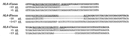
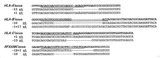
[도 1] HLA 클래스 Ia-결핍 iPS 세포 클론 2E1의 유전자 편집 결과를 나타내는 도면이다.
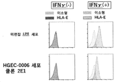
[도 2] 클론 2E1에서 HLA 클래스 I의 세포 표면 발현량을, 유세포 분석법을 사용하여 분석한 결과를 나타내는 도면이다.
[도 3] 클론 2E1에서 HLA-E의 세포 표면 발현량을, 유세포 분석법을 사용하여 분석한 결과를 나타내는 도면이다.
[도 4] 클론 2E1에서 미분화 마커의 세포 표면 발현량을, 유세포 분석법을 사용하여 분석한 결과를 나타내는 도면이다.
[도 5] 클론 2E1 및 제2 후보 클론(2H3)에서 RNA 발현 패턴을 분석한 결과를 나타내는 도면이다.
[도 6] HLA 클래스 Ia&II-결핍 iPS 세포 클론 6B7의 유전자 편집 결과를 나타내는 도면이다.
[도 7] 클론 6B7에서 HLA 클래스 I의 세포 표면 발현량을, 유세포 분석법을 사용하여 분석한 결과를 나타내는 도면이다.
[도 8] 클론 6B7에서 HLA 클래스 II의 세포 표면 발현량을, 유세포 분석법을 사용하여 분석한 결과를 나타내는 도면이다.
[도 9] 클론 6B7에서 HLA-E의 세포 표면 발현량을, 유세포 분석법을 사용하여 분석한 결과를 나타내는 도면이다.
[도 10] 클론 6B7에서 미분화 마커의 세포 표면 발현량을, 유세포 분석법을 사용하여 분석한 결과를 나타내는 도면이다.
[도 11] 클론 6B7에서 RNA 발현 패턴을 분석한 결과를 나타내는 도면이다.
[도 12] A: 클론 6B7로부터 분화된 간세포의 요소 합성량을 분석한 결과 및 B: 클론 6B7로부터 분화된 혈관 내피 세포의 맥관 형성 능력을 분석한 결과를 각각 나타내는 도면이다.
[도 13] HLA 클래스 Ia&II가 결실되고, 또한 PD-L1, PD-L2, HLA-G, B2M 및 iCasp9 유전자가 도입된 iPS 세포 클론 9G11에서 PD-L1, PD-L2, HLA-G 및 B2M의 세포 표면 발현량을, 유세포 분석법을 사용하여 분석한 결과를 나타내는 도면이다.
[도 14] 클론 9G11 iPS 세포-유래 혈구계 세포에서 HLA-A, HLA-B, HLA-C 및 HLA 클래스 II의 세포 표면 발현량을, 유세포 분석법을 사용하여 분석한 결과를 나타내는 도면이다.
[도 15] 클론 9G11 iPS 세포-유래 혈구계 세포에서 PD-L1, PD-L2, HLA-G 및 B2M의 세포 표면 발현량을, 유세포 분석법을 사용하여 분석한 결과를 나타내는 도면이다.
[도 16] 클론 9G11에서 미분화 마커의 세포 표면 발현량을, 유세포 분석법을 사용하여 분석한 결과를 나타내는 도면이다.
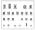
[도 17] 클론 9G11에서 핵형 분석 결과를 나타내는 도면이다.
[도 18] A: 클론 9G11로부터 분화된 간세포의 세포 형태를 나타내는 도면과 우레아 합성량을 분석한 결과를 나타내는 도, 및 B: 6B7로부터 분화된 혈관 내피 세포의 세포 형태를 나타내는 도면과 맥관 형성 능력을 분석한 결과를 나타내는 도면이다.
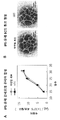
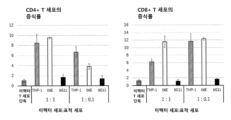
[도 19] 클론 9G11로부터 분화된 CD45 양성 세포에 대한 T 세포(CD4 양성 세포 및 CD8 양성 세포)의 증식률을 나타내는 도면이다.
[도 20] 클론 9G11로부터 분화된 CD45 양성 세포에 대한 T 세포(CD8 양성 세포)의 세포독성 활성을 나타내는 도면이다.
[도 21] 클론 9G11에 대한 NK 세포의 세포독성 활성을 나타내는 도면이다.
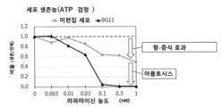
[도 22] 클론 9G11의 자살 유전자의 기능을 확인하기 위한 라파마이신 첨가에 의한 세포 생존률의 변화를 나타내는 도면이다.
[도 23] 외인성 B2M 유전자의 강제 발현이 HLA 클래스 Ia&II-결핍 iPS 세포의 HLA-G 유전자의 발현량을 상승시키는 것을 나타내는 도면이다.[Fig. 1] A diagram showing the results of gene editing of HLA class Ia-deficient iPS cell clone 2E1.
[Fig. 2] A diagram showing the results of analyzing the cell surface expression level of HLA class I in clone 2E1 using flow cytometry.
[Fig. 3] A diagram showing the results obtained by analyzing the cell surface expression level of HLA-E in clone 2E1 using flow cytometry.
[Fig. 4] A diagram showing the results obtained by analyzing the cell surface expression levels of undifferentiated markers in clone 2E1 using flow cytometry.
[Fig. 5] A diagram showing the results of analyzing RNA expression patterns in clone 2E1 and a second candidate clone (2H3).
[Fig. 6] A diagram showing the results of gene editing of HLA class Ia&II-deficient iPS cell clone 6B7.
[Fig. 7] A diagram showing the results obtained by analyzing the cell surface expression level of HLA class I in clone 6B7 using flow cytometry.
[Fig. 8] A diagram showing the results of analyzing the cell surface expression level of HLA class II in clone 6B7 using flow cytometry.
[Fig. 9] A diagram showing the results of analyzing the cell surface expression level of HLA-E in clone 6B7 using flow cytometry.
[Fig. 10] A diagram showing the results obtained by analyzing the cell surface expression levels of undifferentiated markers in clone 6B7 using flow cytometry.
[Fig. 11] is a diagram showing the results of analyzing RNA expression patterns in clone 6B7.
[Fig. 12] A: Results of analyzing the amount of urea synthesis in hepatocytes differentiated from clone 6B7 and B: Results of analyzing the angiogenic ability of vascular endothelial cells differentiated from clone 6B7.
[Fig. 13] PD-L1, PD-L2, HLA-G and B2M in iPS cell clone 9G11 in which HLA class Ia&II was deleted and PD-L1, PD-L2, HLA-G, B2M and iCasp9 genes were introduced. It is a figure which shows the result of analyzing the cell surface expression level using flow cytometry.
[Fig. 14] A diagram showing the results obtained by analyzing the cell surface expression levels of HLA-A, HLA-B, HLA-C and HLA class II in clone 9G11 iPS cell-derived hematopoietic cells using flow cytometry.
[Fig. 15] A diagram showing the results obtained by analyzing the cell surface expression levels of PD-L1, PD-L2, HLA-G, and B2M in clone 9G11 iPS cell-derived hematopoietic cells using flow cytometry.
[Fig. 16] A diagram showing the results obtained by analyzing the cell surface expression levels of undifferentiated markers in clone 9G11 using flow cytometry.
[Fig. 17] A diagram showing the results of karyotype analysis in clone 9G11.
[FIG. 18] A: A diagram showing the cell morphology of hepatocytes differentiated from clone 9G11 and a diagram showing the results of analyzing the amount of urea synthesis, and B: a diagram showing the cell morphology and vasculature of vascular endothelial cells differentiated from 6B7 It is a diagram showing the results of the analysis.
[Fig. 19] A diagram showing the proliferation rate of T cells (CD4-positive cells and CD8-positive cells) relative to CD45-positive cells differentiated from clone 9G11.
[Fig. 20] A diagram showing the cytotoxic activity of T cells (CD8-positive cells) against CD45-positive cells differentiated from clone 9G11.
[Fig. 21] A diagram showing the cytotoxic activity of NK cells against clone 9G11.
[Fig. 22] A diagram showing changes in cell viability by the addition of rapamycin to confirm the function of the suicide gene of clone 9G11.
[Fig. 23] A diagram showing that forced expression of exogenous B2M gene increases the expression level of HLA-G gene in HLA class Ia&II-deficient iPS cells.
이하, 본 발명의 내용을 상세하게 설명한다.Hereinafter, the contents of the present invention will be described in detail.
본 발명은, 하기 (1) 내지 (5)의 특징을 갖는 저면역원성 인간 세포(이하, 본 발명의 저면역원성 인간 세포)를 제공한다:The present invention provides hypoimmunogenic human cells (hereinafter referred to as hypoimmunogenic human cells of the present invention) having the following characteristics (1) to (5):
(1) HLA 클래스 Ia의 α 쇄를 코딩하는 내인성 유전자가 결핍되고,(1) the endogenous gene encoding the α chain of HLA class Ia is missing;
(2) HLA 클래스 II 또는 이의 발현 조절인자를 코딩하는 내인성 유전자가 결핍되고,(2) the endogenous gene encoding HLA class II or its expression regulator is deficient;
(3) HLA 클래스 Ib의 α 쇄를 코딩하는 외인성 유전자를 포함하고,(3) contains an exogenous gene encoding the α chain of HLA class Ib;
(4) 인간 PD-L1를 코딩하는 외인성 유전자를 포함하고,(4) contains an exogenous gene encoding human PD-L1;
(5) 인간 PD-L2를 코딩하는 외인성 유전자를 포함한다.(5) contains an exogenous gene encoding human PD-L2.
본 발명의 저면역원성 인간 세포는, 수용자의 체내에 도입했을 때에, 수용자에 의해 비자가로 인식됨으로써 통상 발생하는 면역 거부반응이 개시되지 않고, 수용자 측의 T 세포나 NK 세포에 의한 공격을 받지 않고, 항원 제시-세포 그룹의 활성화를 억제하지 않고, 또는 면역 거부반응이 발생하더라도 그 반응성이 현저하게 낮은 세포를 말한다. 환언하면, 본 발명의 저면역원성 인간 세포는 수용자의 면역계에 대해 면역 관용인 세포이다.When the low-immunogenic human cells of the present invention are introduced into the recipient's body, they are recognized by the recipient as non-autologous and do not initiate immune rejection, which usually occurs, and are not attacked by T cells or NK cells on the recipient's side. It refers to cells that do not inhibit the activation of the antigen-presenting cell group, or whose reactivity is remarkably low even when immune rejection occurs. In other words, the hypoimmunogenic human cells of the present invention are cells immune tolerant to the recipient's immune system.
본 발명의 저면역원성 인간 세포는, 상기 (1) 및 (2)의 내인성 유전자를 결실시키고 상기 (3), (4) 및 (5)의 외인성 유전자를 도입함으로써, 수용자의 면역계에 대한 면역 관용을 획득했다. 따라서, 본 발명의 저면역원성 인간 세포는 수용자에 대한 세포 의약품일 수 있다.The low-immunogenic human cells of the present invention are immune tolerant to the recipient's immune system by deleting the endogenous genes of (1) and (2) above and introducing the exogenous genes of (3), (4) and (5) above. has obtained Thus, the hypoimmunogenic human cells of the present invention may be cell pharmaceuticals for recipients.
본 발명의 저면역원성 인간 세포는 상기 (1) 및 (2)의 내인성 유전자를 결실시키고 상기 (3), (4) 및 (5)의 외인성 유전자를 도입할 수 있는 세포라면 특별히 제한되지 않는다. 이러한 세포로서는 다능성 줄기 세포를 들 수 있다.The hypoimmunogenic human cell of the present invention is not particularly limited as long as it is a cell capable of deleting the endogenous genes of (1) and (2) above and introducing the exogenous genes of (3), (4) and (5) above. Examples of such cells include pluripotent stem cells.
다능성 줄기 세포로서는 배아 줄기 세포(embryonic stem cell: ES 세포), 인공 다능성 줄기 세포(induced pluripotent stem cell: iPS 세포), 배아성 종양 세포(EC 세포), 배아성 생식 줄기 세포(EG 세포)를 들 수 있으나, 바람직하게는 ES 세포 또는 iPS 세포이다.Pluripotent stem cells include embryonic stem cells (ES cells), induced pluripotent stem cells (iPS cells), embryonic tumor cells (EC cells), and embryonic germ stem cells (EG cells). but are preferably ES cells or iPS cells.
다능성 줄기 세포가 ES 세포인 경우, 자체 공지된 방법으로 생산할 수 있다. ES 세포의 생산 방법으로는, 예를 들면, 인간의 배반포 스테이지에서 내부 세포괴를 배양하는 방법[참조: 예를 들면, Manipulating the Mouse Embryo; A Laboratory Manual, Second Edition, Cold Spring Harbor Laboratory Press(1994)], 체세포 핵 이식에 의해 생산된 초기 배아를 배양하는 방법[참조: Wilmut et al., Nature, 385, 810(1997); Cibelli et al.,Science, 280, 1256(1998); valley, protein nucleic acid enzyme, 44, 892 (1999); Baguisi et al., Nature Biotechnology, 17, 456 (1999); Wakayama et al., Nature, 394, 369 (1998); Wakayama et al., Nature Genetics, 22, 127 (1999); Wakayama et al., Proc. Natl. Acad. Sci. USA, 96, 14984 (1999); RideoutIII et al., Nature Genetics, 24, 109 (2000)] 등을 들 수 있으나, 이들로 한정되는 것은 아니다. 또한, ES 세포는 소정의 기관에서 입수할 수 있고, 또한 시판품을 구입할 수도 있다. 예를 들면, 인간 ES 세포주는 H1, H7, H9, H13 및 H14에 대해서는 미국의 위셀 리써치 인스티튜트(WiCell Research Institute)로부터 입수가능하고, HES1~6에 대해서는 오스트레일리아의 ES 셀 인터네셔널(Cell International)로부터 입수가능하고, SA002, SA181 및 SA611에 대해서는 스웨덴의 셀라르티스 에이비(Cellartis AB)로부터 입수가능하고, HUES1~17에 대해서는 미국의 HUES 셀 파실리티(Cell Facility)로부터 입수가능하고, KhES-1~KhES-5에 대해서는 쿄토 대학 재생 의과 학연 연구소(The Institute for Frontier Life and Medical Sciences, Kyoto University)로부터 입수가능하고, SEES1~SEES7에 대해서는 국립 아동 건강 및 개발 센터(National Center for Child Health and Development)로부터 입수가능하다. ES 세포를 체세포핵 이식에 의해 생산할 경우, 체세포의 종류나 체세포를 채취하는 공급원은 하기 iPS 세포 생산의 경우에 준한다.When the pluripotent stem cells are ES cells, they can be produced by a method known per se. As a method for producing ES cells, for example, a method of culturing an inner cell aggregation at the human blastocyst stage [Reference: For example, Manipulating the Mouse Embryo; A Laboratory Manual, Second Edition, Cold Spring Harbor Laboratory Press (1994)], methods for culturing early embryos produced by somatic cell nuclear transfer (Wilmut et al., Nature, 385, 810 (1997)); Cibelli et al., Science, 280, 1256 (1998); valley, protein nucleic acid enzyme, 44, 892 (1999); Baguisi et al., Nature Biotechnology, 17, 456 (1999); Wakayama et al., Nature, 394, 369 (1998); Wakayama et al., Nature Genetics, 22, 127 (1999); Wakayama et al., Proc. Natl. Acad. Sci. USA, 96, 14984 (1999); RideoutIII et al., Nature Genetics, 24, 109 (2000)] and the like, but are not limited thereto. In addition, ES cells can be obtained from certain institutions, or commercially available products can also be purchased. For example, human ES cell lines are available from WiCell Research Institute, USA for H1, H7, H9, H13 and H14, and ES Cell International, Australia for HES1-6. available from Cellartis AB, Sweden for SA002, SA181 and SA611, and from HUES Cell Facility, USA for HUES1-17, KhES-1- Available from The Institute for Frontier Life and Medical Sciences, Kyoto University for KhES-5, and from the National Center for Child Health and Development for SEES1-SEES7. it is obtainable When ES cells are produced by somatic cell nuclear transfer, the type of somatic cells and the source from which somatic cells are collected are in accordance with the following iPS cell production.
다능성 줄기 세포가 iPS 세포인 경우, iPS 세포는 체세포에 핵 초기화 물질을 도입함으로써 생산할 수 있다. iPS 세포 생산을 위한 출발 재료로서 사용할 수 있는 체세포는 인간의 생식 세포 이외의 어떠한 세포일 수 있다. 예를 들면, 각질화하는 상피 세포(예를 들면, 각질화 표피 세포), 점막 상피 세포(예를 들면, 혀 표층의 상피 세포), 외분비선 상피 세포(예를 들면, 유선 세포), 호르몬 분비 세포(예를 들면, 부신 골수질 세포), 대사/저장용 세포(예를 들면, 간세포), 경계면을 구성하는 내강 상피 세포(예를 들면, I형 폐포 세포), 혈관 내강 상피 세포(예를 들면, 혈관 내피 세포), 운반 능력을 갖는 섬모가 있는 세포(예를 들면, 기도 상피 세포), 세포외 매트릭스 분비용 세포(예를 들면, 섬유아세포), 수축성 세포(예를 들면, 평활근 세포), 혈액과 면역계의 세포(예를 들면, T 림프구), 감각과 관련된 세포(예를 들면, 막대 세포), 자율 신경계 뉴런(예를 들면, 콜린 작동성 뉴런), 감각 기관과 말초 뉴런의 지지 세포(예를 들면, 수반 세포), 중추신경계의 신경세포와 신경교 세포(예를 들면, 성상 신경교 세포), 색소 세포(예를 들면, 망막 색소 상피 세포) 및 이의 전구세포(조직 전구세포) 등을 들 수 있다. 세포의 분화 정도에 특별히 제한은 없고, 미분화 전구세포(체성 줄기 세포도 포함)라도, 최종 분화된 성숙 세포라도, 유사하게 본 발명에 있어서의 체세포의 공급원으로서 사용할 수 있다. 여기서 미분화 전구세포로서는, 예를 들면, 지방 유래 간질(줄기) 세포, 신경 줄기 세포, 조혈 줄기 세포, 간엽계 줄기 세포, 치수 줄기 세포 등의 조직 줄기 세포(체성 줄기 세포)를 들 수 있다.When the pluripotent stem cells are iPS cells, iPS cells can be produced by introducing a nuclear reprogramming material into somatic cells. A somatic cell that can be used as a starting material for producing iPS cells may be any cell other than human germ cells. For example, keratinizing epithelial cells (e.g., keratinized epidermal cells), mucosal epithelial cells (e.g., tongue superficial epithelial cells), exocrine glandular epithelial cells (e.g., mammary gland cells), hormone secreting cells ( For example, adrenal bone marrow cells), metabolic/storage cells (eg, hepatocytes), luminal epithelial cells constituting the interface (eg, type I alveolar cells), and vascular luminal epithelial cells (eg, vascular endothelial cells), ciliated cells with transport capacity (eg airway epithelial cells), extracellular matrix secreting cells (eg fibroblasts), contractile cells (eg smooth muscle cells), blood and cells of the immune system (eg T lymphocytes), sensory-related cells (eg rod cells), neurons of the autonomic nervous system (eg cholinergic neurons), support cells of sensory organs and peripheral neurons (eg For example, accompanying cells), neurons and glial cells of the central nervous system (eg, astrocytes), pigment cells (eg, retinal pigment epithelial cells) and progenitor cells thereof (tissue progenitor cells), and the like. there is. The degree of cell differentiation is not particularly limited, and both undifferentiated progenitor cells (including somatic stem cells) and terminally differentiated mature cells can similarly be used as a source of somatic cells in the present invention. Examples of the undifferentiated progenitor cells include tissue stem cells (somatic stem cells) such as adipose-derived stromal (stem) cells, neural stem cells, hematopoietic stem cells, mesenchymal stem cells, and dental pulp stem cells.
iPS 세포의 생산을 위해 체세포에 도입되는 핵 초기화 물질로서는 지금까지 보고되어 있는 다양한 초기화 유전자의 조합[참조: 예를 들면 WO2007/069666, Nature Biotechnology, 26, 101-106 (2008), Cell, 126, 663-676 (2006), Cell, 131, 861-872 (2007), Nat. Cell Biol., 11, 197-203 (2009), Nature, 451, 141-146 (2008), Science, 318, 1917-1920 (2007), Stem Cells, 26, 1998-2005 (2008), Cell Research (2008) 600-603, Nature 454: 646-650 (2008), Cell Stem Cell, 2: 525-528(2008), WO2008/118820, Nat. Cell Biol., 11, 197-203 (2009), Nat. Cell Biol., 11, 197-203 (2009), Science, 324: 797-801 (2009)]를 들 수 있다. 또한, 상기 초기화 유전자에 의해 코딩되는 단백질을 핵 초기화 물질로서 체세포에 도입할 수도 있다[참조: Cell Stem Cell, 4: 381-384(2009), Cell Stem Cell, doi:10.1016/j.stem.2009.05.005 (2009)].Nuclear reprogramming materials introduced into somatic cells for the production of iPS cells include combinations of various reprogramming genes that have been reported so far [see, for example, WO2007/069666, Nature Biotechnology, 26, 101-106 (2008), Cell, 126, 663-676 (2006), Cell, 131, 861-872 (2007), Nat. Cell Biol., 11, 197-203 (2009), Nature, 451, 141-146 (2008), Science, 318, 1917-1920 (2007), Stem Cells, 26, 1998-2005 (2008), Cell Research ( 2008) 600-603, Nature 454: 646-650 (2008), Cell Stem Cell, 2: 525-528 (2008), WO2008/118820, Nat. Cell Biol., 11, 197-203 (2009), Nat. Cell Biol., 11, 197-203 (2009), Science, 324: 797-801 (2009)]. In addition, proteins encoded by the reprogramming genes may be introduced into somatic cells as nuclear reprogramming materials [Reference: Cell Stem Cell, 4: 381-384 (2009), Cell Stem Cell, doi:10.1016/j.stem.2009.05 .005 (2009)].
iPS 세포 콜로니의 선택은 약물 내성과 리포터 활성을 지표로 하는 방법[참조: Cell, 126, 663-676 (2006), Nature, 448, 313-317 (2007)] 및 육안에 의한 형태 관찰에 의한 방법[참조: Cell, 131, 861-872 (2007)]에 의해 수행할 수 있다. iPS 세포는 각종 ES 세포 특이적 유전자의 발현 또는 테라토마 형성을 지표로서 사용하여 확인할 수 있다.Selection of iPS cell colonies is performed using drug resistance and reporter activity as indicators [Reference: Cell, 126, 663-676 (2006), Nature, 448, 313-317 (2007)] and morphological observation with the naked eye. [Reference: Cell, 131, 861-872 (2007)]. iPS cells can be identified using expression of various ES cell-specific genes or teratoma formation as indicators.
현재, iPS 세포(iPSC)의 생산 방법에는 다양한 것이 있고, 야마나카 등(Yamanaka et al.)에 의해 마우스 섬유아세포에 Oct3/4, Sox2, Klf4, 및 c-Myc의 4개 인자를 도입함으로써 수립된 iPSC의 생산 방법 뿐만 아니라[참조: Takahashi K, Yamanaka S., Cell, (2006) 126: 663-676], 동일한 4개 인자를 인간 섬유아세포에 도입하여 수립된 인간 세포 유래의 iPSC의 생산 방법[참조: Takahashi K, Yamanaka S., et al. Cell, (2007) 131: 861-872.], 상기 4개 인자 도입 후, Nanog의 발현을 지표로서 사용하여 선별함으로써 수립된 Nanog-iPSC의 생산 방법[참조: Okita, K., Ichisaka, T., and Yamanaka, S. (2007). Nature 448, 313-317.], c-Myc를 사용하지 않는 iPSC의 생산 방법[참조: Nakagawa M, Yamanaka S., et al. Nature Biotechnology, (2008) 26, 101-106], 바이러스-프리(free)법으로 6개 인자를 도입하여 수립된 iPSC의 생산 방법[참조: Okita K et al. Nat. Methods 2011 May;8(5):409-12, Okita K et al. Stem Cells. 31(3):458-66.] 등이 사용될 수 있다. 또한, OCT3/4, SOX2, NANOG, 및 LIN28의 4개 인자를 도입하여 톰슨 등(Thomson et al.)에 의해 수립된 iPSC의 생산 방법[참조: Yu J., Thomson JA. et al., Science (2007) 318: 1917-1920.], 달레이 등(Daley et al.)에 의한 iPSC의 생산 방법[참조: Park IH, Daley GQ. et al., Nature (2007) 451: 141-146], 사쿠라다 등(Sakurada et al.)에 의한 iPSC의 생산 방법(JP-A-2008/307007) 등 또한 사용될 수 있다.Currently, there are various methods for producing iPS cells (iPSC), which were established by introducing four factors of Oct3/4, Sox2, Klf4, and c-Myc into mouse fibroblasts by Yamanaka et al. As well as a method for producing iPSCs [Reference: Takahashi K, Yamanaka S., Cell, (2006) 126: 663-676], a method for producing iPSCs derived from human cells established by introducing the same four factors into human fibroblasts [ References: Takahashi K, Yamanaka S., et al. Cell, (2007) 131: 861-872.], and a method for producing Nanog-iPSCs established by screening using the expression of Nanog as an indicator after introduction of the above four factors [Reference: Okita, K., Ichisaka, T. , and Yamanaka, S. (2007). Nature 448, 313-317.], method for producing iPSCs without using c-Myc [Nakagawa M, Yamanaka S., et al. Nature Biotechnology, (2008) 26, 101-106], a method for producing iPSCs established by introducing six factors by a virus-free method [Okita K et al. Nat. Methods 2011 May; 8(5):409-12, Okita K et al. Stem Cells. 31(3):458-66.] and the like may be used. In addition, the production method of iPSC established by Thomson et al. by introducing four factors of OCT3/4, SOX2, NANOG, and LIN28 [Yu J., Thomson JA. et al., Science (2007) 318: 1917-1920.], the production method of iPSCs by Daley et al. [Park IH, Daley GQ. et al., Nature (2007) 451: 141-146], the production method of iPSCs by Sakurada et al. (JP-A-2008/307007), etc. can also be used.
인공 다능성 줄기 세포주로서는 NIH, 이화학 연구소(RIKEN), 쿄토 대학(Kyoto University) 등이 수립한 각종 인간 iPSC 균주 중 어느 것도 사용가능하다. 예를 들면, 이화학 연구소(RIKEN)의 HiPS-RIKEN-1A 균주, HiPS-RIKEN-2A 균주, HiPS-RIKEN-12A 균주, Nips-B2 균주 등, 쿄토 대학의 253G1 균주, 253G4 균주, 1201C1 균주, 1205D1 균주, 1210B2 균주, 1383D2 균주, 1383D6 균주, 201B7 균주, 409B2 균주, 454E2 균주, 606A1 균주, 610B1 균주, 648A1 균주, 1231A31 균주, FfI-01s04 균주 등을 들 수 있다.As an artificial pluripotent stem cell line, any of various human iPSC strains established by NIH, RIKEN, Kyoto University and the like can be used. For example, HiPS-RIKEN-1A strain, HiPS-RIKEN-2A strain, HiPS-RIKEN-12A strain, Nips-B2 strain from the Institute of Physical and Chemical Research (RIKEN), 253G1 strain, 253G4 strain, 1201C1 strain, 1205D1 from Kyoto University strain, 1210B2 strain, 1383D2 strain, 1383D6 strain, 201B7 strain, 409B2 strain, 454E2 strain, 606A1 strain, 610B1 strain, 648A1 strain, 1231A31 strain, FfI-01s04 strain and the like.
본 발명의 저면역원성 인간 세포는 또한 상기 (1) 및 (2)의 내인성 유전자를 결실하고, 상기 (3), (4) 및 (5)의 외인성 유전자를 도입함으로써 수득된 상기의 다능성 줄기 세포로부터 분화된 세포일 수 있다. 다능성 줄기 세포는 공지된 방법에 따라 특정 세포로 분화시킬 수 있다. 예를 들면, 다능성 줄기 세포를 T 세포(국제공개 제2016/076415호 또는 국제공개 제2017/221975호), 각막 상피 세포(WO 제2016/114285호), 심근 세포(WO 제2007/126077호, WO 제2016/049099호, WO 제2016/175303호 또는 WO 제2017/108705호), 췌장 β 세포(WO 제2019/208788호), 간세포(WO 제2013/183571호 또는 WO 제2019/073951호), 골격근 세포(WO 제2010/008100호, WO 제2014/533491호 또는 WO 제2017/188458호), 망막 색소 상피 세포(WO 제2015/053375호) 등으로 분화시킬 수 있다.The hypoimmunogenic human cell of the present invention is also the pluripotent stem obtained by deleting the endogenous genes of (1) and (2) above and introducing the exogenous genes of (3), (4) and (5) above. It may be a cell differentiated from a cell. Pluripotent stem cells can be differentiated into specific cells according to known methods. For example, pluripotent stem cells are T cells (International Publication No. 2016/076415 or International Publication No. 2017/221975), corneal epithelial cells (WO No. 2016/114285), cardiomyocytes (WO No. 2007/126077 , WO 2016/049099, WO 2016/175303 or WO 2017/108705), pancreatic β cells (WO 2019/208788), hepatocytes (WO 2013/183571 or WO 2019/073951) ), skeletal muscle cells (WO 2010/008100, WO 2014/533491, or WO 2017/188458), retinal pigment epithelial cells (WO 2015/053375), etc.
본 발명의 저면역원성 인간 세포는 HLA 클래스 Ia의 α 쇄를 코딩하는 내인성 유전자가 결핍되어 있다.The hypoimmunogenic human cells of the present invention lack the endogenous gene encoding the α chain of HLA class Ia.
HLA 클래스 Ia는 모든 유핵세포가 보유하는 막 관통 단백질이며, 수천 이상의 다양한 다형을 갖기 때문에, 개체에 의해 세포 표면에 발현된 HLA 클래스 Ia는 매우 다양성이 풍부하다. 그리고, 이러한 다양성이 자가와 비자가를 식별하는데 가장 중요한 역할을 한다. 세포 표면에 제시된 HLA 클래스 Ia는 CTL 표면의 T 세포 수용체(TCR)에 의해 인식된다. 인식된 HLA 클래스 Ia를 제시하는 세포는 CTL에 의해 이물로서 인식되고, 면역계에 의해 배제된다. 따라서, HLA 클래스 Ia가 발현 부전인 세포는 CTL에 의한 인식을 회피할 수 있고, 따라서 이식 세포 공급원으로서 적합하다.Since HLA class Ia is a transmembrane protein possessed by all nucleated cells and has more than thousands of various polymorphisms, HLA class Ia expressed on the cell surface by individuals is very diverse. And, this diversity plays the most important role in identifying self and non-self. HLA class Ia presented on the cell surface is recognized by the T cell receptor (TCR) on the CTL surface. Cells presenting recognized HLA class Ia are recognized as foreign by CTLs and excluded by the immune system. Thus, cells deficient in HLA class Ia can evade recognition by CTLs and are therefore suitable as a source of transplant cells.
HLA 클래스 Ia는 HLA 클래스 Ia의 각 유전자에 의해 코딩되는 α 쇄와 β2 마이크로글로불린인 β 쇄로 이루어진 이량체이다. 여기서, β2 마이크로글로불린은 HLA 클래스 Ia의 α 쇄 뿐만 아니라 HLA 클래스 Ib의 α 쇄와도 회합하는 공통의 성분이다. HLA 클래스 Ia의 발현 부전을 달성하기 위해 β2 마이크로글로불린을 코딩하는 내인성 유전자를 결실시키는 것은 다른 저면역원성 세포로 수행된다. 그러나, HLA 클래스 Ib도 발현 부전으로 되기 때문에, 불편하다. 따라서, 본 발명의 저면역원성 인간 세포에 있어서는 HLA 클래스 Ia만을 발현 부전으로 되도록 HLA 클래스 Ia의 α 쇄를 코딩하는 내인성 유전자를 결실시킨다.HLA class Ia is a dimer composed of an α chain encoded by each gene of HLA class Ia and a β chain, which is a β2 microglobulin. Here, β2 microglobulin is a common component that associates not only with the α chain of HLA class Ia but also with the α chain of HLA class Ib. Deletion of the endogenous gene encoding β2 microglobulin to achieve a lack of expression of HLA class Ia is performed with other hypoimmunogenic cells. However, since HLA class Ib also becomes deficient, it is inconvenient. Therefore, in the hypoimmunogenic human cells of the present invention, the endogenous gene encoding the α chain of HLA class Ia is deleted so that only HLA class Ia is deficient.
본 발명의 저면역원성 인간 세포에 있어서 결손되어 있는 HLA 클래스 Ia의 α 쇄를 코딩하는 내인성 유전자로서는 HLA-A의 α 쇄를 코딩하는 내인성 유전자, HLA-B의 α 쇄를 코딩하는 내인성 유전자, 및 HLA-C의 α 쇄를 코딩하는 내인성 유전자로 이루어진 그룹으로부터 선택되는 적어도 하나의 유전자, 바람직하게는 HLA-A의 α 쇄를 코딩하는 내인성 유전자, HLA-B의 α 쇄를 코딩하는 내인성 유전자, 및 HLA-C의 α 쇄를 코딩하는 내인성 유전자를 들 수 있다.As the endogenous gene encoding the α chain of HLA class Ia that is missing in the hypoimmunogenic human cells of the present invention, the endogenous gene encoding the α chain of HLA-A, the endogenous gene encoding the α chain of HLA-B, and At least one gene selected from the group consisting of an endogenous gene encoding the α chain of HLA-C, preferably an endogenous gene encoding the α chain of HLA-A, an endogenous gene encoding the α chain of HLA-B, and and an endogenous gene encoding the α chain of HLA-C.
본 명세서에 있어서 내인성 유전자를 결실시킨다는 것은, 내인성 유전자를 파괴하거나 제거함으로써 완전한 mRNA를 생산 불가능하게 하는 것을 의미한다.In the present specification, deleting an endogenous gene means making production of complete mRNA impossible by destroying or removing the endogenous gene.
내인성 유전자를 결실시키는 구체적 수단으로서는, 내인성 유전자를 결실시키는 표적 세포 유래의 게놈 DNA를 통상의 방법에 따라 단리하고, 예를 들면, (1) 내인성 유전자의 엑손 부분 또는 프로모터 영역에 다른 DNA 단편(예를 들면, 약물 내성 유전자, 리포터 유전자 등)을 삽입함으로써 엑손 또는 프로모터의 기능을 파괴하거나, (2) Cre-loxP계 또는 Flp-frt계를 사용하여 내인성 유전자의 전부 또는 일부를 절단하여 상기 유전자를 결실시키거나, (3) 단백질 코드 영역 내로 정지 코돈을 삽입하여 완전한 단백질의 번역을 불가능하게 하거나, 또는 (4) 전사 영역 내부로 유전자의 전사를 종결시키는 DNA 서열(예를 들면, poly A 부가 신호 등)을 삽입하여 완전한 mRNA의 합성을 불가능하게 함으로써, 결과적으로 유전자를 불활성화하도록 작제한 DNA 서열을 갖는 DNA 쇄(이하, 유전자 결실용 표적화 벡터라 약칭한다)을 상동성 재조합에 의해 표적 세포의 내인성 유전자좌에 도입하는 방법 등이 바람직하게 사용될 수 있다.As a specific means for deleting an endogenous gene, genomic DNA derived from a target cell for deleting an endogenous gene is isolated according to a conventional method, for example, (1) a DNA fragment other than the exon portion or promoter region of the endogenous gene (e.g. For example, drug resistance gene, reporter gene, etc.) is inserted to destroy the function of an exon or promoter, or (2) all or part of an endogenous gene is cut using the Cre-loxP system or the Flp-frt system to generate the gene. deletion, (3) insertion of a stop codon into the protein coding region, rendering translation of the complete protein impossible, or (4) DNA sequence that terminates transcription of the gene into the transcribed region (e.g., poly A addition signal). etc.) to make the synthesis of complete mRNA impossible, and as a result, a DNA chain having a DNA sequence designed to inactivate a gene (hereinafter, abbreviated as a targeting vector for gene deletion) is converted into target cells by homologous recombination. A method of introducing into an endogenous locus and the like can be preferably used.
상기 상동성 재조합 세포는, 예를 들면, 표적 세포로의 상기 표적화 벡터의 도입에 의해 수득할 수 있다.The homologous recombinant cells can be obtained, for example, by introduction of the targeting vector into target cells.
예를 들면, 유전자 결실용 표적화 벡터가, 내인성 유전자의 엑손 부분 또는 프로모터 영역에 다른 DNA 단편을 삽입함으로써, 상기 엑손 또는 프로모터의 기능을 파괴하기 위하여 설계되는 경우, 당해 벡터는, 예를 들면, 하기와 같은 구성을 취할 수 있다.For example, when a targeting vector for gene deletion is designed to disrupt the function of an exon or promoter by inserting another DNA fragment into the exon portion or promoter region of an endogenous gene, the vector can, for example, can take the same configuration.
먼저, 상동성 재조합에 의해 내인성 유전자의 엑손 또는 프로모터 부분에 다른 DNA 단편이 삽입되기 때문에, 유전자 결실용 표적화 벡터는 당해 다른 DNA 단편의 5' 상류 및 3' 하류에 각각 표적 부위와 상동성인 서열(5' 아암 및 3' 아암)을 포함할 필요가 있다.First, since another DNA fragment is inserted into the exon or promoter portion of the endogenous gene by homologous recombination, the targeting vector for gene deletion contains sequences homologous to the target site 5' upstream and 3' downstream of the other DNA fragment, respectively ( 5' arm and 3' arm).
삽입되는 다른 DNA 단편은 특별히 제한은 없지만, 약물 내성 유전자 또는 리포터 유전자를 사용하면, 유전자 결실용 표적화 벡터가 염색체로 통합된 표적 세포를 약물 내성 또는 리포터 활성을 지표로서 사용하여 선택할 수 있다. 여기서, 약물 내성 유전자에는, 예를 들면, 네오마이신 포스포트랜스퍼라제 II(nptII) 유전자, 하이그로마이신 포스포트랜스퍼라제(hpt) 유전자 등이 포함되지만 이들로 한정되지 않고, 리포터 유전자에는, 예를 들면, β-갈락토시다제(lacZ) 유전자, 클로람페니콜 아세틸트랜스퍼라제(cat) 유전자 등이 포함되지만 이들로 한정되는 것은 아니다.Other DNA fragments to be inserted are not particularly limited, but if a drug resistance gene or reporter gene is used, target cells into which a targeting vector for gene deletion has been chromosomally integrated can be selected using drug resistance or reporter activity as an indicator. Here, drug resistance genes include, for example, neomycin phosphotransferase II (nptII) gene, hygromycin phosphotransferase (hpt) gene, etc., but are not limited thereto, and reporter genes include, for example, Examples include, but are not limited to, β-galactosidase (lacZ) gene, chloramphenicol acetyltransferase (cat) gene, and the like.
약물 내성 또는 리포터 유전자는 표적 세포 내에서 기능할 수 있는 임의의 프로모터 제어하에 있는 것이 바람직하다. 예를 들면, SV40 유래 초기 프로모터, 사이토메갈로바이러스(CMV) 긴 말단 반복(LTR), 라우스 육종 바이러스(RSV) LTR, 마우스 백혈병 바이러스(MoMuLV) LTR, 아데노바이러스(AdV) 유래 초기 프로모터 등의 바이러스 프로모터, 및 β-액틴 유전자 프로모터, PGK 유전자 프로모터, 트랜스페린 유전자 프로모터 등을 들 수 있다. 그러나, 약물 내성 또는 리포터 유전자가 HLA 클래스 I 유전자의 내인성 프로모터의 제어하에 있도록 내인성 유전자 내에 삽입되는 경우는 유전자 결실용 표적화 벡터 중에 상기 유전자의 전사를 제어하는 프로모터는 필요하지 않다.The drug resistance or reporter gene is preferably under the control of any promoter capable of functioning in the target cell. Viral promoters such as, for example, SV40 derived early promoter, cytomegalovirus (CMV) long terminal repeat (LTR), Rous sarcoma virus (RSV) LTR, mouse leukemia virus (MoMuLV) LTR, adenovirus (AdV) early promoter, etc. , and β-actin gene promoter, PGK gene promoter, transferrin gene promoter and the like. However, when a drug resistance or reporter gene is inserted into an endogenous gene under the control of an endogenous promoter of an HLA class I gene, a promoter controlling transcription of the gene in a targeting vector for gene deletion is not required.
또한, 유전자 결실용 표적화 벡터는 약물 내성 또는 리포터 유전자의 하류에, 상기 유전자로부터의 mRNA의 전사를 종결시키는 서열(폴리아데닐화(poly A) 신호, 터미네이터라고도 함)을 갖는 것이 바람직하고, 예를 들면, 바이러스 유전자 유래, 또는 각종 포유동물 또는 조류 유전자 유래의 터미네이터 서열을 사용할 수 있다. 바람직하게는, SV40 유래의 터미네이터 등이 사용된다.In addition, the targeting vector for gene deletion preferably has a sequence that terminates transcription of mRNA from a drug resistance or reporter gene (also called a polyadenylation (poly A) signal, terminator) downstream of the gene, for example For example, terminator sequences derived from viral genes or derived from various mammalian or avian genes can be used. Preferably, a terminator derived from SV40 or the like is used.
통상, 세포에서 유전자 재조합은 대부분이 비상동적이고, 도입된 DNA는 염색체의 임의 위치에 랜덤하게 삽입된다. 따라서, 약물 내성 또는 리포터 유전자의 발현을 검출하는 등의 선택(포지티브 선택)에 따라서는 표적 부위에 상동성 재조합이 발생한 클론만을 효율적으로 선택하지 못하며, 선택된 모든 클론에 대해 써던 하이브리드화법 또는 PCR법에 의한 통합 부위의 확인이 필요하다. 따라서, 유전자 결실용 표적화 벡터의 표적 부위와 상동성인 서열의 외측에, 예를 들면, 간시클로비르 감수성을 부여하는 단순 헤르페스바이러스-유래 티미딘 키나제(HSV-tk) 유전자를 연결하면, 상기 벡터가 랜덤으로 삽입된 세포는 HSV-tk 유전자를 갖기 때문에, 간시클로비르 함유 배지에서는 생육할 수 없지만, 상동성 재조합에 의해 내인성 유전자좌에 표적화된 세포는 HSV-tk 유전자를 갖지 않기 때문에, 간시클로비르 내성이 되어 선택된다(네가티브 선택). 또는, HSV-tk 유전자 대신에, 예를 들면, 디프테리아 독소 유전자를 연결하면, 상기 벡터가 랜덤으로 삽입된 세포는 자신이 생산하는 상기 독소에 의해 사멸하기 때문에, 약물 부재하에서 상동성 재조합체를 선택할 수도 있다.Usually, most of genetic recombination in cells is non-homologous, and the introduced DNA is randomly inserted into a random position of the chromosome. Therefore, depending on selection (positive selection), such as detecting drug resistance or expression of a reporter gene, it is not possible to efficiently select only clones in which homologous recombination has occurred at the target site, and Southern hybridization or PCR method is used for all selected clones. confirmation of the site of integration by Therefore, if, for example, the herpes simplex virus-derived thymidine kinase (HSV-tk) gene conferring sensitivity to ganciclovir is ligated outside the sequence homologous to the target site of the targeting vector for gene deletion, the vector can Because the randomly inserted cells have the HSV-tk gene, they cannot grow on ganciclovir-containing media, whereas cells targeted to the endogenous locus by homologous recombination do not have the HSV-tk gene, and therefore are resistant to ganciclovir. is selected (negative selection). Alternatively, if, for example, the diphtheria toxin gene is ligated instead of the HSV-tk gene, the cells into which the vector has been randomly inserted are killed by the toxin produced by the vector, and thus homologous recombinants can be selected in the absence of drugs. may be
표적 세포로의 유전자 결실용 표적화 벡터의 도입에는 인산칼슘 공침전법, 전기천공(일렉트로포레이션)법, 리포펙션법, 레트로바이러스 감염법, 응집법, 미세주입법, 입자 총 방법, DEAE-덱스트란법 등의 어느 것도 사용할 수 있지만, 상기에서 설명한 바와 같이 세포의 유전자 재조합은 대부분이 비상동적이고 상동성 재조합체가 수득되는 빈도는 낮기 때문에 간편하게 다수의 세포를 처리할 수 있는 것 등의 점에서 전기천공법이 일반적으로 선택된다. 전기천공에는 통상의 동물 세포로의 유전자 도입에 사용되고 있는 조건을 그대로 사용할 수도 있고, 예를 들면, 대수 증식기에 있는 표적 세포를 트립신 처리하여 단일 세포에 분산시킨 후, 106 내지 108 세포/ml가 되도록 배지에 현탁하여 큐벳으로 옮기고, 유전자 결실용 표적화 벡터를 10 내지 100μg 첨가하고, 200 내지 600V/cm의 전기 펄스를 인가함으로써 수행할 수 있다.Introduction of targeting vectors for gene deletion into target cells include calcium phosphate co-precipitation, electroporation (electroporation), lipofection, retroviral infection, agglutination, microinjection, particle gun method, and DEAE-dextran method. Although any of the above can be used, as described above, most of the genetic recombination of cells is non-homologous and the frequency of obtaining homologous recombinants is low, so electroporation can be used to conveniently process a large number of cells. This is usually selected. For electroporation, conditions used for gene transfer into normal animal cells may be used as they are. For example, after trypsinizing target cells in the logarithmic growth phase and dispersing them into single cells, 10 6 to 10 8 cells/ml It can be carried out by suspending in a culture medium, transferring to a cuvette, adding 10 to 100 μg of a targeting vector for gene deletion, and applying an electric pulse of 200 to 600 V/cm.
유전자 결실용 표적화 벡터가 통합된 표적 세포는 단일 세포를 배양하여 수득되는 콜로니로부터 분리 추출한 염색체 DNA를 써던 하이브리드화 또는 PCR법에 의해 스크리닝함으로써 검정할 수도 있지만, 다른 DNA 단편으로서 약물 내성 유전자 또는 리포터 유전자를 사용한 경우는 이의 발현을 지표로서 사용하여 세포 단계에서 형질전환체를 선택할 수 있다. 예를 들면, 포지티브 선택용 마커 유전자로서 nptII 유전자를 포함하는 벡터를 사용한 경우, 유전자 도입 후의 표적 세포를 G418 등의 네오마이신계 항생물질을 함유하는 배지 중에서 배양하고, 출현한 내성 콜로니를 형질전환체의 후보로서 선택한다. 또한, 네가티브 선택용 마커 유전자로서 HSV-tk 유전자를 포함하는 벡터를 사용한 경우, 간시클로비르를 함유하는 배지 중에서 배양하고, 출현한 내성 콜로니를 상동성 재조합 세포의 후보로서 선택한다. 수득된 콜로니를 각각 배양 플레이트로 옮겨 트립신 처리, 배지 교환을 반복한 후, 일부를 배양용으로서 잔류시키고, 나머지를 PCR 또는 써던 하이브리드화에 제공하여 도입 DNA의 존재를 확인한다.Target cells into which the targeting vector for gene deletion has been integrated can be assayed by screening chromosomal DNA isolated and extracted from colonies obtained by culturing single cells by Southern hybridization or PCR, but other DNA fragments such as drug resistance genes or reporter genes In the case of using, the transformant can be selected at the cell stage using its expression as an index. For example, when a vector containing the nptII gene is used as a marker gene for positive selection, target cells after gene introduction are cultured in a medium containing a neomycin antibiotic such as G418, and the resistant colonies that emerge are transformed into transformants. selected as a candidate for Further, when a vector containing the HSV-tk gene is used as a marker gene for negative selection, it is cultured in a medium containing ganciclovir, and resistant colonies that emerge are selected as candidates for homologous recombinant cells. The obtained colonies are each transferred to a culture plate and trypsinization and medium exchange are repeated, then a part is retained for culture and the remainder is subjected to PCR or Southern hybridization to confirm the presence of the introduced DNA.
또한, 유전자 결실용 표적화 벡터로서 바이러스를 사용하는 경우, 포지티브 선택용 마커 유전자가 5' 및 3' 아암 사이에 삽입되고, 상기 아암의 외측에 네가티브 선택용 마커 유전자를 포함하는 DNA를 포함하는 바이러스로 표적 세포를 감염시키는 방법을 들 수 있다. 예를 들면, 레트로바이러스 또는 렌티바이러스를 사용하는 경우, 디쉬 등의 적당한 배양기에 세포를 파종하고, 배양액에 바이러스 벡터를 가하고(임의로 폴리브렌을 공존시킬 수 있다), 1 내지 2일간 배양 후, 상기에서 설명한 바와 같이 선택 약제를 첨가하고, 배양을 계속하고, 벡터가 통합된 세포를 선택한다.In addition, when using a virus as a targeting vector for gene deletion, a marker gene for positive selection is inserted between the 5' and 3' arms, and the virus contains DNA containing the marker gene for negative selection on the outside of the arm. A method of infecting a target cell may be mentioned. For example, in the case of using a retrovirus or lentivirus, cells are seeded in an appropriate incubator such as a dish, a viral vector is added to the culture medium (polybrene may optionally be coexisted), and after culturing for 1 to 2 days, the above As described above, the selection agent is added, culture is continued, and cells into which the vector is integrated are selected.
내인성 유전자를 결실시키는 또 다른 바람직한 일 실시양태로서는 CRISPR-Cas9(Clustered Regularly Interspaced Short Palindromic Repeats CRISPR-Associated proteins 9) 시스템을 들 수 있다. CRISPR-Cas9 시스템에 따르면, 게놈 DNA 절단 효소인 Cas9와 게놈 상의 표적 부위를 인식하는 RNA 분자인 sgRNA를 사용하여, 게놈 DNA 상의 임의의 영역을 절단함으로써 유전자 돌연변이를 도입할 수 있다. 게놈 DNA의 절단과 연관되는 생체내 수복 과정에서 발생하는 염기의 in/del에 의해, 아미노산을 코딩하는 DNA의 프레임시프트가 발생하고, 표적의 내인성 유전자가 결실된다.Another preferred embodiment for deleting endogenous genes includes the CRISPR-Cas9 (Clustered Regularly Interspaced Short Palindromic Repeats CRISPR-Associated proteins 9) system. According to the CRISPR-Cas9 system, a gene mutation can be introduced by cleaving an arbitrary region on genomic DNA using Cas9, a genomic DNA cutting enzyme, and sgRNA, an RNA molecule that recognizes a target site on the genome. By in/del of bases generated in the in vivo repair process associated with cleavage of genomic DNA, a frameshift of DNA encoding an amino acid occurs, and the endogenous gene of the target is deleted.
Cas9는 DNA 중의 프로토스페이서 인접 모티브(PAM) 서열을 인식하여 그 상류에서 절단하는 엔도뉴클레아제로서 기능한다. Cas9는 엔도뉴클레아제 활성을 갖는 기능성 도메인을 2개 포함하기 때문에, 이중-가닥 DNA를 평활 말단이 되도록 절단할 수 있다. 예를 들면, 구체적으로, Cas9는 내인성 유전자에 특이적으로 결합하는 염기 서열(CRISPR-RNA(crRNA))를 포함하는 단일-가닥 핵산(sgRNA)과 복합체를 형성하고, 내인성 유전자 중의 PAM의 5' 측에서 이중-가닥 절단(DSB)을 생성한다. 따라서, 본 발명에 있어서, Cas9는 가이드 RNA와 복합체를 형성하고, 이중-가닥 DNA 절단 활성을 갖는 단백질을 의미한다.Cas9 functions as an endonuclease that recognizes and cuts upstream protospacer adjacent motif (PAM) sequences in DNA. Since Cas9 contains two functional domains with endonuclease activity, double-stranded DNA can be cleaved into blunt ends. For example, specifically, Cas9 forms a complex with a single-stranded nucleic acid (sgRNA) containing a nucleotide sequence (CRISPR-RNA (crRNA)) that specifically binds to an endogenous gene, and forms a complex at the 5' end of the PAM in the endogenous gene. double-strand break (DSB) on the side. Accordingly, in the present invention, Cas9 is a protein that forms a complex with guide RNA and has double-stranded DNA cutting activity.
Cas9로서는, 예를 들면, 스트렙토코커스 피오게네스(Streptococcus pyogenes)로부터 유래하는 SpCas9, 스트렙토코커스 써모필러스(Streptococcus thermophilus)로부터 유래하는 StCas9, 네이세리아 메닌기티디스(Neisseria meningitidis)로부터 유래하는 NmCas9 등을 들 수 있으며, 이들로 한정되는 것은 아니다.Examples of Cas9 include SpCas9 derived from Streptococcus pyogenes, StCas9 derived from Streptococcus thermophilus, and NmCas9 derived from Neisseria meningitidis. may be mentioned, but is not limited thereto.
또한, Cas9는 돌연변이 Cas9일 수 있다. 돌연변이 Cas9는, 가이드 RNA와의 복합체의 형성 능력을 유지하면서, Cas9에 포함되는 2개의 엔도뉴클레아제 활성을 갖는 기능성 도메인 중 하나가 불활성화되어 있는 돌연변이를 갖는 단백질이라면 특별히 제한되지 않는다. 이러한 돌연변이 Cas9로서는, 예를 들면, Cas9의 10번째 아스파르트산이 알라닌으로 치환되는 돌연변이(D10A 돌연변이), 840번째 히스티딘이 알라닌으로 치환되는 돌연변이(H840A 돌연변이) 및/또는 863번째 아스파라긴이 알라닌으로 치환되는 돌연변이(N863A 돌연변이)로 이루어진 그룹으로부터 선택되는 하나의 돌연변이를 갖는 Cas9를 들 수 있다.Cas9 may also be a mutant Cas9. Mutant Cas9 is not particularly limited as long as it is a protein having a mutation in which one of the functional domains having two endonuclease activities included in Cas9 is inactivated while maintaining the ability to form a complex with guide RNA. As such mutant Cas9, for example, a mutant in which the 10th aspartic acid of Cas9 is substituted with alanine (D10A mutation), a mutant in which histidine at 840th position is substituted with alanine (H840A mutation), and/or a mutant in which asparagine at 863rd position is substituted with alanine. (N863A mutation).
crRNA는 내인성 유전자와 상보적인 염기 서열로서, 내인성 유전자에서 PAM의 5' 측에 인접하고 있는 염기 서열이라면 특별히 제한되지 않는다. crRNA의 염기 서열의 길이는 내인성 유전자로의 특이성을 확보할 수 있는 한 특별히 제한되지 않지만, 통상 10개 염기 길이로부터 30개 염기 길이, 바람직하게는 15개 염기 길이로부터 25개 염기 길이, 보다 바람직하게는 20개 염기 길이를 들 수 있다.The crRNA is a nucleotide sequence complementary to an endogenous gene, and is not particularly limited as long as it is a nucleotide sequence adjacent to the 5' side of PAM in the endogenous gene. The length of the base sequence of the crRNA is not particularly limited as long as the specificity to the endogenous gene can be secured, but is usually 10 bases to 30 bases in length, preferably 15 bases to 25 bases in length, more preferably may be 20 bases long.
PAM는 Cas9의 종류에 따라 상이하지만, 예를 들면, 스트렙토코커스 피오게네스(Streptococcus pyogenes) 유래의 Cas9(SpCas9)가 사용되는 경우 NGG(N은 A, G, T 또는 C. 이하 동일), 스트렙토코커스 써모필러스(Streptococcus thermophilus) 유래의 Cas9(StCas9)가 사용되는 경우 NNAGAAW, 나이세리아 메닌기티디스(Neisseria meningitidis) 유래의 Cas9(NmCas9)가 사용되는 경우 NNNNGATT 등을 들 수 있다.PAM is different depending on the type of Cas9, but, for example, when Cas9 (SpCas9) derived from Streptococcus pyogenes is used, NGG (N is A, G, T or C. hereinafter the same), streptococcus Examples include NNAGAAW when Cas9 (StCas9) derived from Streptococcus thermophilus is used, and NNNNGATT when Cas9 (NmCas9) derived from Neisseria meningitidis is used.
내인성 유전자에 특이적으로 결합하는 염기 서열을 포함하는 단일-가닥 핵산(sgRNA)은, 추가로 Cas9의 동원에 필요한 염기 서열(trans-activating RNA(tracrRNA))를 포함할 수 있다. tracrRNA의 염기 서열은 공지된 염기 서열을 사용할 수 있다. tracrRNA는 crRNA의 3' 말단에 직접 연결될 수 있고, 스페이서 서열을 통해 연결될 수 있다.The single-stranded nucleic acid (sgRNA) containing a nucleotide sequence that specifically binds to an endogenous gene may further include a nucleotide sequence required for Cas9 recruitment (trans-activating RNA (tracrRNA)). A known nucleotide sequence can be used as the nucleotide sequence of tracrRNA. The tracrRNA can be linked directly to the 3' end of the crRNA or via a spacer sequence.
또는, crRNA와 tracrRNA는 상보적 결합을 통해 회합할 수 있다(즉 이중-가닥 핵산을 형성할 수 있다). 또한, 이러한 이중-가닥 핵산을 사용하여 유전자 편집하는 경우에도, 하기에 기재하는 표적 세포로의 도입 방법은 단일-가닥 핵산을 도입하는 경우와 동일하다.Alternatively, crRNA and tracrRNA may associate through complementary bonds (ie, form a double-stranded nucleic acid). In addition, even in the case of gene editing using such a double-stranded nucleic acid, the introduction method into the target cell described below is the same as that in the case of introducing a single-stranded nucleic acid.
Cas9와 sgRNA의 표적 세포로의 도입은 공지된 수단에 따라 실시할 수 있다. 예를 들면, Cas9를 코딩하는 핵산 서열 및 sgRNA를 전사하는 핵산 서열을 적당한 발현 벡터에 삽입한 후, 발현 벡터를 표적 세포에 도입함으로써 Cas9 및 sgRNA를 표적 세포에 도입할 수 있다.Introduction of Cas9 and sgRNA into target cells can be performed according to known means. For example, Cas9 and sgRNA can be introduced into a target cell by inserting a nucleic acid sequence encoding Cas9 and a nucleic acid sequence transcribing the sgRNA into an appropriate expression vector, and then introducing the expression vector into the target cell.
Cas9를 코딩하는 핵산 서열로서는 게놈 DNA, 합성 DNA 등을 들 수 있다. Cas9를 코딩하는 게놈 DNA라면, 상기 미생물에 의해 조제한 게놈 DNA 분획을 주형으로서 사용하여, cas9 유전자에 상보적인 프라이머 세트에 의한 폴리머라제 연쇄 반응(이하, "PCR법"으로 약칭함)에 의해 직접 증폭할 수 있다. 또한, sgRNA를 전사하는 핵산 서열은 DNA 합성과 PCR법에 의해 생산될 수 있다.A nucleic acid sequence encoding Cas9 includes genomic DNA, synthetic DNA, and the like. If it is genomic DNA encoding Cas9, direct amplification by polymerase chain reaction (hereinafter abbreviated as "PCR method") with a primer set complementary to the cas9 gene using the genomic DNA fraction prepared by the microorganism as a template can do. In addition, a nucleic acid sequence transcribing the sgRNA can be produced by DNA synthesis and PCR.
Cas9를 코딩하는 핵산 서열 및 sgRNA를 전사하는 핵산 서열을 포함하는 발현 벡터는, 예를 들면, Cas9를 코딩하는 핵산 서열 단편 및 sgRNA를 전사하는 핵산 서열 단편을 적당한 발현 벡터 중의 프로모터의 하류에 연결함으로써 생산될 수 있다.An expression vector comprising a nucleic acid sequence encoding Cas9 and a nucleic acid sequence transcribing the sgRNA can be obtained, for example, by ligating a nucleic acid sequence fragment encoding Cas9 and a nucleic acid sequence fragment transcribing the sgRNA downstream of a promoter in an appropriate expression vector. can be produced
발현 벡터로서는 동물세포 발현 플라스미드(예: pA1-11, pXT1, pRc/CMV, pRc/RSV, pcDNAI/Neo); 레트로바이러스, 렌티바이러스, 아데노바이러스, 아데노 수반 바이러스 등의 동물 바이러스 벡터 등이 사용된다. Expression vectors include animal cell expression plasmids (eg pA1-11, pXT1, pRc/CMV, pRc/RSV, pcDNAI/Neo); Animal viral vectors such as retroviruses, lentiviruses, adenoviruses, and adeno-associated viruses are used.
프로모터로서는 유전자의 발현에 사용하는 숙주에 대응하여 적절한 프로모터라면 어떠한 프로모터도 사용할 수 있다. As the promoter, any promoter can be used as long as it is an appropriate promoter corresponding to the host used for gene expression.
예를 들면, SRα 프로모터, SV40 프로모터, LTR 프로모터, CMV(사이토메갈로바이러스) 프로모터, RSV(라우스 육종 바이러스) 프로모터, MoMuLV(몰로니 마우스 백혈병 바이러스) LTR, HSV-TK(단순 헤르페스 바이러스 티미딘 키나제) 프로모터 등이 사용된다. 그 중에서도, CMV 프로모터, SRα 프로모터 등이 바람직하다.For example, SRα promoter, SV40 promoter, LTR promoter, CMV (cytomegalovirus) promoter, RSV (Rouse sarcoma virus) promoter, MoMuLV (Moloney mouse leukemia virus) LTR, HSV-TK (herpes simplex virus thymidine kinase) promoters, etc. are used. Especially, CMV promoter, SRα promoter, etc. are preferable.
발현 벡터로서는, 상기 외에, 임의로 인핸서, 폴리A 부가 신호, 선택 마커, SV40 복제 기점(이하, SV40 ori로 약칭함) 등을 함유하고 있는 것을 사용할 수 있다. 선택 마커로서는, 예를 들면, 디하이드로엽산 리덕타제 유전자(이하, dhfr로 약칭하는 경우가 있음, 메소트렉세이트(MTX) 내성), 네오마이신 내성 유전자(이하, neor로 약칭하는 경우가 있음, G418 내성) 등을 들 수 있다.As the expression vector, in addition to the above, one optionally containing an enhancer, a polyA addition signal, a selection marker, an SV40 origin of replication (hereinafter abbreviated as SV40 ori), etc. can be used. Selectable markers include, for example, dihydrofolate reductase gene (hereinafter sometimes abbreviated as dhfr, methotrexate (MTX) resistance), neomycin resistance gene (hereinafter sometimes abbreviated as neor, G418) tolerance), etc.
상기한 Cas9를 코딩하는 핵산 서열 및 sgRNA를 전사하는 핵산 서열을 포함하는 발현 벡터를 표적 세포에 도입하고 배양함으로써, 표적 세포 내에서 Cas9와 sgRNA가 복합체를 형성하고, sgRNA의 표적이 되는 내인성 유전자를 절단한다. Cas9에 의해 절단된 내인성 유전자는 비상동성 말단 결합(NHEJ) 경로에 의한 DSB의 수복 중에 내인성 유전자에 작은 삽입 및/또는 결실(in/dels)이 도입되고, 프레임시프트가 발생하고 내인성 유전자의 부위-특이적 돌연변이 또는 파괴가 발생한다.By introducing an expression vector containing the nucleic acid sequence encoding the Cas9 and the nucleic acid sequence transcribing the sgRNA into a target cell and culturing it, a complex is formed between Cas9 and the sgRNA in the target cell, and the endogenous gene targeted by the sgRNA is produced. Cut it. Endogenous genes cleaved by Cas9 are introduced with small insertions and/or deletions (in/dels) in the endogenous gene during repair of DSBs by the non-homologous end-joining (NHEJ) pathway, frameshifts occur, and site- Specific mutations or disruptions occur.
또한, Cas9로서는 상기에서 예시한 것과 같은 발현 벡터를 사용하지 않고, Cas9 단백질 그 자체를 사용할 수 있다. Cas9 단백질을 sgRNA와 조합하여 복합체를 형성시키고, 이것을 표적 세포에 도입하는 것에 의해서도, sgRNA의 표적이 되는 내인성 유전자를 절단할 수 있다.In addition, as Cas9, Cas9 protein itself can be used without using an expression vector as exemplified above. The endogenous gene targeted by the sgRNA can also be cleaved by combining the Cas9 protein with the sgRNA to form a complex and introducing the complex into the target cell.
전술한 바와 같이, 본 발명의 저면역원성 인간 세포는 HLA 클래스 Ia의 α 쇄를 코딩하는 내인성 유전자가 결핍이다.As described above, the hypoimmunogenic human cells of the present invention are deficient in the endogenous gene encoding the α chain of HLA class Ia.
HLA 클래스 II는 마크로파지, 수지상 세포 또는 B 세포 등의 항원 제시 세포에서만 발견되는 막 관통 단백질이다.HLA class II is a transmembrane protein found only in antigen presenting cells such as macrophages, dendritic cells or B cells.
HLA 클래스 II는 식작용 등으로 면역 세포가 취입된 세포외 병원체의 단백질을 포함한 세포외 단백질 유래의 펩티드 항원을 세포 표면 상에 제시한다. HLA 클래스 II에 의해 제시된 펩티드 항원이 CD4 양성 헬퍼 T 세포의 TCR와 상호작용하고, CD4 양성 헬퍼 T 세포를 활성화한다. 활성화된 T세포는 또한 HLA 클래스 II에 의해 펩티드 항원을 제시하는 B 세포를 인식하고, 활성화함으로써 식세포의 동원, 국소 염증, 체액성 반응, CTL의 활성화 등의 이벤트가 유발된다. 따라서, HLA 클래스 II가 발현 부전인 세포는 상기의 이벤트 발생을 회피할 수 있기 때문에, 이식 세포 공급원으로서 적합하다.HLA class II presents peptide antigens derived from extracellular proteins, including proteins of extracellular pathogens taken up by immune cells by phagocytosis or the like, on the cell surface. Peptide antigens presented by HLA class II interact with the TCR of CD4 positive helper T cells and activate CD4 positive helper T cells. Activated T cells also recognize and activate B cells presenting peptide antigens by HLA class II, thereby triggering events such as phagocyte recruitment, local inflammation, humoral response, and CTL activation. Therefore, cells deficient in HLA class II expression can avoid the occurrence of the above events, and are therefore suitable as a source of cells for transplantation.
HLA 클래스 II는 2개의 상동성 서브유닛인 α 쇄와 β 쇄로 구성되는 이량체이다. 따라서, 본 발명의 저면역원성 인간 세포에 있어서는, HLA 클래스 II를 발현 부전으로 하기 위해 일 실시양태로서 HLA 클래스 II의 α 쇄 및/또는 β 쇄를 코딩하는 내인성 유전자를 결핍시킨다. 본 발명의 저면역원성 인간 세포에 있어서 결실되어 있는 HLA 클래스 II를 코딩하는 내인성 유전자로서는, HLA-DP의 α 쇄 및/또는 β 쇄를 코딩하는 내인성 유전자, HLA-DQ의 α 쇄 및/또는 β 쇄를 코딩하는 내인성 유전자, HLA-DR의 α 쇄 및/또는 β 쇄를 코딩하는 내인성 유전자, HLA-DM의 α 쇄 및/또는 β 쇄를 코딩하는 내인성 유전자, 및 HLA-DO의 α 쇄 및/또는 β 쇄를 코딩하는 내인성 유전자로 이루어진 그룹으로부터 선택되는 적어도 하나의 내인성 유전자, 바람직하게는 HLA-DP의 α 쇄 및/또는 β 쇄를 코딩하는 내인성 유전자, HLA-DQ의 α 쇄 및/또는 β 쇄를 코딩하는 내인성 유전자, HLA-DR의 α 쇄 및/또는 β 쇄를 코딩하는 내인성 유전자, HLA-DM의 α 쇄 및/또는 β 쇄를 코딩하는 내인성 유전자, 및 HLA-DO의 α 쇄 및/또는 β 쇄를 코딩하는 내인성 유전자를 들 수 있다.HLA class II is a dimer composed of two homologous subunits, the α chain and the β chain. Therefore, in the hypoimmunogenic human cells of the present invention, in one embodiment, in order to render HLA class II deficient, endogenous genes encoding the α chain and/or the β chain of HLA class II are deficient. As the endogenous gene encoding HLA class II deleted in the hypoimmunogenic human cells of the present invention, the endogenous gene encoding the α chain and/or β chain of HLA-DP, the α chain and/or β of HLA-DQ chain, an endogenous gene encoding the α chain and/or β chain of HLA-DR, an endogenous gene encoding the α chain and/or β chain of HLA-DM, and the α chain and/or β chain of HLA-DO. or at least one endogenous gene selected from the group consisting of endogenous genes encoding β chain, preferably endogenous genes encoding α chain and/or β chain of HLA-DP, α chain and/or β of HLA-DQ chain, an endogenous gene encoding the α chain and/or β chain of HLA-DR, an endogenous gene encoding the α chain and/or β chain of HLA-DM, and the α chain and/or β chain of HLA-DO. or an endogenous gene encoding the β chain.
또한, HLA 클래스 II의 각 유전자의 발현은 그 발현 조절인자에 의해 제어되어 있다. 발현 제어의 메커니즘은 특별히 제한되지 않으며, 표적 유전자에 직접 결합하여 이의 발현(예를 들면, 전사)을 제어할 수 있고, 간접적으로 결합함으로써 이의 발현(예를 들면, 전사)을 제어할 수 있다. 예를 들면, RFXANK는, DNA에 직접 결합함으로써 표적 유전자인 HLA 클래스 II 각 유전자의 전사를 제어한다. 따라서, 본 발명의 저면역원성 인간 세포에 있어서는, HLA 클래스 II를 발현 부전으로 하는 다른 실시양태로서 HLA 클래스 II의 발현 조절인자를 코딩하는 내인성 유전자가 결실되어 있다. HLA 클래스 II의 발현 조절인자를 코딩하는 내인성 유전자로서는, 예를 들면, RFXANK를 코딩하는 내인성 유전자, RFX5를 코딩하는 내인성 유전자, RFXAP를 코딩하는 내인성 유전자 또는 CIITA를 코딩하는 내인성 유전자, 바람직하게는 RFXANK를 코딩하는 내인성 유전자를 들 수 있다.In addition, the expression of each gene of HLA class II is controlled by its expression regulator. The mechanism of expression control is not particularly limited, and can directly bind to a target gene to control its expression (eg, transcription), or indirectly bind to control its expression (eg, transcription). For example, RFXANK controls the transcription of each HLA class II gene as a target gene by directly binding to DNA. Therefore, in the hypoimmunogenic human cells of the present invention, as another embodiment in which HLA class II is underexpressed, an endogenous gene encoding an expression regulator of HLA class II is deleted. As an endogenous gene encoding an expression regulator of HLA class II, for example, an endogenous gene encoding RFXANK, an endogenous gene encoding RFX5, an endogenous gene encoding RFXAP, or an endogenous gene encoding CIITA, preferably RFXANK Endogenous genes encoding .
HLA 클래스 II 또는 이의 발현 조절인자를 코딩하는 내인성 유전자를 결실시키는 구체적 수단은 상기의 HLA 클래스 Ia를 코딩하는 내인성 유전자를 결실시키는 수단과 동일할 수 있다.Specific means for deleting the endogenous gene encoding HLA class II or its expression regulator may be the same as the means for deleting the endogenous gene encoding HLA class Ia above.
본 발명의 저면역원성 인간 세포는 HLA 클래스 Ib의 α 쇄를 코딩하는 외인성 유전자를 포함한다.The hypoimmunogenic human cells of the present invention contain an exogenous gene encoding the α chain of HLA class Ib.
HLA 클래스 Ib는 다종 다양한 기능을 갖는다. 이러한 기능으로서는, 예를 들면, 특수한 항원의 제시, NK세포의 활성의 제어, Fc 수용체로서의 역할을 들 수 있다. 또한, B 세포, 마크로파지, 단핵구 또는 수지상 세포 등의 항원 제시 세포 그룹의 활성화를 억제하는 기능 등도 들 수 있다.HLA class Ib has a wide variety of functions. Examples of such functions include presentation of a specific antigen, control of NK cell activity, and a role as an Fc receptor. In addition, a function of inhibiting activation of a group of antigen-presenting cells such as B cells, macrophages, monocytes, or dendritic cells, and the like may be mentioned.
전술한 바와 같이, 본 발명의 저면역원성 인간 세포에 있어서는, HLA 클래스 Ia의 α 쇄를 코딩하는 내인성 유전자, 및 HLA 클래스 II 또는 이의 발현 조절인자를 코딩하는 내인성 유전자가 결핍되지만, β2 마이크로글로불린을 코딩하는 내인성 유전자는 결실되지 않았다. 즉, 내인성 β2 마이크로글로불린은 발현 부전이 아니고, 당연하게 내인성 HLA 클래스 Ib도 발현 부전이 아니다고 생각되었다. 그러나, 상기 세포의 생산 과정에서 무손상 HLA 클래스 Ib(HLA-E)의 발현까지 소실되어 있는 것이 명백해졌다. HLA 클래스 Ib(HLA-E)를 발현하지 않는 세포는, NK 세포에 의해 거부되기 때문에 이식용 세포 공급원으로서의 사용에는 불편하다. 따라서, 내인성 HLA 클래스 Ib의 발현을 보완하기 위해, 본 발명의 저면역원성 인간 세포는 HLA 클래스 Ib의 α 쇄를 코딩하는 외인성 유전자를 포함한다.As described above, in the hypoimmunogenic human cells of the present invention, the endogenous gene encoding the α chain of HLA class Ia and the endogenous gene encoding HLA class II or its expression regulator are deficient, but β2 microglobulin The endogenous gene encoding was not deleted. That is, it was thought that endogenous β2 microglobulin was not underexpressed, and naturally, endogenous HLA class Ib was also not underexpressed. However, it became clear that expression of intact HLA class Ib (HLA-E) was lost during the production process of the cells. Cells that do not express HLA class Ib (HLA-E) are inconvenient for use as a cell source for transplantation because they are rejected by NK cells. Thus, to complement the expression of endogenous HLA class Ib, the hypoimmunogenic human cells of the present invention contain an exogenous gene encoding the α chain of HLA class Ib.
본 발명의 저면역원성 인간 세포에 있어서 도입되는 HLA 클래스 Ib의 α 쇄를 코딩하는 외인성 유전자로서는, HLA-E의 α 쇄를 코딩하는 외인성 유전자, HLA-F의 α 쇄를 코딩하는 외인성 유전자, 및 HLA-G의 α 쇄를 코딩하는 외인성 유전자로 이루어진 그룹으로부터 선택되는 적어도 하나의 유전자, 바람직하게는 HLA-E의 α 쇄를 코딩하는 외인성 유전자, 및/또는 HLA-G의 α 쇄를 코딩하는 외인성 유전자를 들 수 있다. 추가로, HLA-G의 α 쇄 신호 펩티드가, HLA-E의 막 표면의 발현에 중요한 것으로부터, 본 발명의 저면역원성 인간 세포에 있어서 도입되는 HLA 클래스 Ib의 α 쇄를 코딩하는 외인성 유전자로서는, HLA-E의 α 쇄를 코딩하는 외인성 유전자, 및 HLA-G의 α 쇄를 코딩하는 외인성 유전자가 가장 바람직하다. 또한, 본 발명의 저면역원성 인간 세포를 이식용 세포 공급원으로서 사용하는 경우, 도입되는 HLA 클래스 Ib의 α 쇄를 코딩하는 외인성 유전자는 수용자의 HLA 클래스 Ib의 대립유전자의 α 쇄를 코딩하는 유전자와 동일한 것이 바람직하다.As the exogenous gene encoding the α chain of HLA class Ib introduced into the hypoimmunogenic human cells of the present invention, an exogenous gene encoding the α chain of HLA-E, an exogenous gene encoding the α chain of HLA-F, and At least one gene selected from the group consisting of an exogenous gene encoding the α chain of HLA-G, preferably an exogenous gene encoding the α chain of HLA-E, and/or an exogenous gene encoding the α chain of HLA-G genes can be found. Furthermore, since the α chain signal peptide of HLA-G is important for the expression of the HLA-E membrane surface, as an exogenous gene encoding the α chain of HLA class Ib introduced into the low immunogenic human cells of the present invention, , an exogenous gene encoding the α chain of HLA-E, and an exogenous gene encoding the α chain of HLA-G are most preferred. In addition, when the hypoimmunogenic human cells of the present invention are used as a cell source for transplantation, the exogenous gene encoding the α chain of HLA class Ib to be introduced is different from the gene encoding the α chain of the HLA class Ib allele of the recipient. The same is preferred.
본 명세서에 있어서 외인성 유전자를 도입한다는 것은, 외인성 유전자를 게놈의 표적 부위에 도입함으로써 외인성 유전자를 표적 세포에서 발현가능한 상태로 하는 것을 의미한다.In the present specification, introducing an exogenous gene means making the exogenous gene expressible in a target cell by introducing the exogenous gene into a target region of the genome.
외인성 유전자를 도입하는 구체적 수단으로서는, 외인성 유전자의 DNA를 통상의 방법에 따라 단리하고, 예를 들면, 표적 세포의 표적 부위에 외인성 유전자의 DNA 단편을 삽입함으로써, 결과적으로 외인성 유전자가 표적 세포 내에서 발현되도록 작제한 DNA 서열을 갖는 DNA 가닥(이하, 유전자 도입용 표적화 벡터라 약칭한다)을, 상동성 재조합에 의해 표적 세포의 표적 부위에 통합하는 방법 등이 바람직하게 사용될 수 있다. 상동성 재조합법에 따르면, 유전자 삽입 위치는 고정된다. 따라서, 랜덤 통합이 없으면, 클론 사이에서의 발현량의 차이 및 다른 유전자에 대한 영향이 적어지는 것이 기대된다.As a specific means of introducing an exogenous gene, DNA of the exogenous gene is isolated according to a conventional method, and, for example, a DNA fragment of the exogenous gene is inserted into a target site of the target cell, and as a result, the exogenous gene is introduced into the target cell. A method of integrating a DNA strand having a DNA sequence designed to be expressed (hereinafter abbreviated as a targeting vector for gene introduction) into a target site of a target cell by homologous recombination or the like can be preferably used. According to the homologous recombination method, the gene insertion site is fixed. Therefore, in the absence of random integration, differences in expression level between clones and less influence on other genes are expected.
상기 상동성 재조합 세포는, 예를 들면, 표적 세포로의 상기 표적화 벡터의 도입에 의해 취득할 수 있다.The homologous recombinant cells can be obtained, for example, by introduction of the targeting vector into target cells.
예를 들면, 유전자 도입용 표적화 벡터가, 표적 부위에 외인성 유전자의 DNA 단편을 삽입함으로써, 상기 외인성 유전자가 표적 세포 내에서 발현하도록 설계된 것일 경우, 당해 벡터는, 예를 들면, 하기와 같은 구성을 취할 수 있다.For example, when a targeting vector for gene introduction is designed to express an exogenous gene in a target cell by inserting a DNA fragment of an exogenous gene into a target site, the vector has, for example, the following structure can take
먼저, 상동성 재조합에 의해, 표적 부위에 외인성 유전자의 DNA 단편을 삽입하기 위해, 유전자 도입용 표적화 벡터는, 당해 외인성 유전자의 DNA 단편의 5' 상류 및 3' 하류에, 각각 표적 부위와 상동성인 서열(5' 아암 및 3' 아암)을 포함할 필요가 있다.First, in order to insert a DNA fragment of an exogenous gene into a target site by homologous recombination, a targeting vector for gene introduction is 5' upstream and 3' downstream of the DNA fragment of the exogenous gene, respectively, which is homologous to the target site. It is necessary to include the sequences (5' arm and 3' arm).
유전자 도입용 표적화 벡터가 염색체로 통합된 표적 세포를 선택하기 위해서는, 삽입되는 외인성 유전자 외에, 약물 내성 유전자 또는 리포터 유전자가 유전자 도입용 표적화 벡터에 포함되어 있는 것이 바람직하다. 여기서, 약물 내성 유전자 및 리포터 유전자는 유전자 결실용 표적화 벡터에 사용되는 것과 동일할 수 있다.In order to select target cells into which the targeting vector for gene transfer is chromosomally integrated, it is preferable that the targeting vector for gene transfer contains a drug resistance gene or a reporter gene in addition to the exogenous gene to be inserted. Here, the drug resistance gene and the reporter gene may be the same as those used in the targeting vector for gene deletion.
약물 내성 또는 리포터 유전자는, 표적 세포 내에서 기능할 수 있는 임의의 프로모터 제어하에 있는 것이 바람직하다. 여기서, 프로모터는 유전자 결실용 표적화 벡터에 사용되는 것과 동일할 수 있다.The drug resistance or reporter gene is preferably under the control of any promoter capable of functioning in the target cell. Here, the promoter may be the same as that used in the targeting vector for gene deletion.
또한, 유전자 도입용 표적화 벡터는 약물 내성 또는 리포터 유전자의 하류에 polyA 시그널을 갖고 있는 것이 바람직하고, 유전자 결실용 표적화 벡터에 사용되는 것과 동일할 수 있다.In addition, the targeting vector for gene introduction preferably has a polyA signal downstream of a drug resistance or reporter gene, and may be the same as that used for targeting vectors for gene deletion.
또한, 유전자 도입용 표적화 벡터의 표적 부위와 상동성인 서열의 외측에, HSV-tk 유전자 또는 디프테리아 독소 유전자를 연결하면, 상동성 재조합에 의해 표적 부위에 표적화된 세포를 선택할 수 있기 때문에 바람직하다.In addition, ligating the HSV-tk gene or the diphtheria toxin gene to the outside of the sequence homologous to the target site of the targeting vector for gene introduction is preferable because cells targeted to the target site can be selected by homologous recombination.
표적 세포로의 유전자 도입용 표적화 벡터의 도입에는 유전자 결실용 표적화 벡터에 사용되는 방법과 동일한 방법이 사용될 수 있다.The same method used for the targeting vector for gene deletion may be used to introduce the targeting vector for gene transfer into the target cell.
유전자 도입용 표적화 벡터가 통합된 상동성 재조합 세포는 유전자 결실용 표적화 벡터가 통합된 상동성 재조합 세포를 선발하는 방법과 동일한 방법으로 선발될 수 있다.Homologous recombinant cells into which the targeting vector for gene introduction has been integrated can be selected by the same method as the method for selecting homologous recombinant cells into which the targeting vector for gene deletion has been integrated.
또한, 유전자 도입용 표적화 벡터로서 바이러스를 사용하는 경우, 외인성 유전자 및 포지티브 선택용 마커 유전자가 5' 및 3' 아암 사이에 삽입되고, 상기 아암의 외측에 네가티브 선택용 마커 유전자를 포함하는 DNA를 포함하는 바이러스로 표적 세포를 감염시키는 방법을 들 수 있다. 바이러스, 세포의 감염 방법, 벡터가 통합된 세포의 선택 방법 등은 유전자 결실용 표적화 벡터로 사용되는 바이러스, 방법과 동일할 수 있다.In addition, when using a virus as a targeting vector for gene introduction, an exogenous gene and a marker gene for positive selection are inserted between the 5' and 3' arms, and DNA containing the marker gene for negative selection is included on the outside of the arm. A method of infecting a target cell with a virus that Viruses, methods for infecting cells, methods for selecting cells into which vectors are integrated, and the like may be the same as viruses and methods used as targeting vectors for gene deletion.
유전자 도입용 표적화 벡터의 표적 부위는, 외인성 유전자를 표적 세포에서 발현가능한 상태로 할 수 있는 한 특별히 제한되지 않지만, 이러한 부위로서는 게놈 내의 세이프 하버 영역을 들 수 있다. 여기서, 세이프 하버 영역은 외인성 유전자가 통합되어도 표현형 변화가 발생하지 않는 부위이며, 의약품으로서 사용되는 세포에 외인성 유전자를 통합하기 위한 표적 부위로서 선택된다. 저면역원성 인간 세포에 있어서 외인성 유전자에서 이러한 세이프 하버 영역으로서는 AAVS1(Adeno-associated virus integration site 1) 영역, CCR5(C-C chemokine receptor5) 영역, ROSA26 영역 등을 들 수 있다. 세이프 하버 영역 이외의 부위에 외인성 유전자를 도입한 경우, 도입된 부위의 유전자가 파괴됨으로써 기대하지 않는 표현형이 도출되거나, 또는 도입한 외인성 유전자의 발현이 억제될 수 있기 때문에 바람직하지 않다. 세이프 하버 영역에 외인성 유전자를 도입한 경우, 외인성 유전자의 통합 위치가 고정되기 때문에, 취득되는 상동성 재조합체 사이에서의 외인성 유전자의 발현량의 차이 및 다른 유전자에 대한 영향이 적을 것으로 기대된다.The target site of the targeting vector for gene introduction is not particularly limited as long as the exogenous gene can be expressed in the target cell, but examples of such site include safe harbor regions in the genome. Here, the safe harbor region is a region in which phenotypic change does not occur even when an exogenous gene is integrated, and is selected as a target site for incorporating the exogenous gene into cells used as pharmaceuticals. Such safe harbor regions in exogenous genes in low-immunogenic human cells include AAVS1 (Adeno-associated virus integration site 1) region, C-C chemokine receptor 5 (CCR5) region, ROSA26 region, and the like. When an exogenous gene is introduced into a site other than the safe harbor region, the gene at the introduced site is disrupted, resulting in an unexpected phenotype or suppression of the expression of the introduced exogenous gene, which is not preferable. When an exogenous gene is introduced into the safe harbor region, since the integration site of the exogenous gene is fixed, differences in the expression level of the exogenous gene between obtained homologous recombinants and influence on other genes are expected to be small.
외인성 유전자를 도입하기 위한 또 다른 바람직한 일 실시양태로서는 PiggyBac법을 들 수 있다. PiggyBac법에서는 외인성 유전자를 포함하는 DNA 단편이 통합된 트랜스포존 벡터 및 트랜스포사제를 발현하는 트랜스포사제 발현 벡터가 사용된다. 트랜스포존 벡터 및 트랜스포사제 발현 벡터에 포함되는 유전자 등은 상기의 또 다른 벡터에 별도로 존재할 수 있고, 단일 벡터에 포함될 수도 있다. 트랜스포존 벡터 및 트랜스포사제 발현 벡터는, 예를 들면, 하기와 같은 구성을 취할 수 있다.Another preferred embodiment for introducing an exogenous gene is the PiggyBac method. In the PiggyBac method, a transposon vector into which a DNA fragment containing an exogenous gene is integrated and a transposase expression vector expressing a transposase are used. Genes included in the transposon vector and the transposase expression vector may be separately present in another vector described above or included in a single vector. A transposon vector and a transposase expression vector can take the following structures, for example.
트랜스포사제에 의해 외인성 유전자를 포함하는 DNA 단편을 트랜스포존 벡터로부터 절단하기 위해, 트랜스포존 벡터는 당해 외인성 유전자를 포함하는 DNA 단편의 5' 상류 및 3' 하류에 말단 역위 반복 서열(Terminal Inverted Repeat)을 포함한다. 트랜스포사제는 트랜스포존 벡터에 포함되는 말단 역위 반복 서열을 인식하고, 말단 역위 반복 서열에 샌드위치된 외인성 유전자를 포함하는 DNA 단편을 트랜스포존 벡터에서 절단한다.In order to cleave a DNA fragment containing an exogenous gene from a transposon vector by a transposase, the transposon vector has terminal inverted repeat sequences 5' upstream and 3' downstream of the DNA fragment containing the exogenous gene. include The transposase recognizes the terminal inverted repeat sequence contained in the transposon vector and cuts the DNA fragment containing the exogenous gene sandwiched in the terminal inverted repeat sequence from the transposon vector.
외인성 유전자가 표적 부위로 통합된 표적 세포를 선택하기 위해서는, 외인성 유전자를 포함하는 DNA 단편에, 약물 내성 유전자 또는 리포터 유전자가 트랜스포존 벡터에 포함되어 있는 것이 바람직하다. 여기서, 약물 내성 유전자 및 리포터 유전자는 유전자 도입용 표적화 벡터에 사용되는 것과 동일할 수 있다.In order to select target cells into which the exogenous gene has been integrated into the target site, it is preferable that the DNA fragment containing the exogenous gene contains a drug resistance gene or a reporter gene in a transposon vector. Here, the drug resistance gene and the reporter gene may be the same as those used in the targeting vector for gene introduction.
약물 내성 및 리포터 유전자는, 표적 세포 내에서 기능할 수 있는 임의의 프로모터의 제어하에 있는 것이 바람직하다. 여기서, 프로모터는 유전자 도입용 표적화 벡터에 사용되는 것과 동일할 수 있다.The drug resistance and reporter genes are preferably under the control of any promoter capable of functioning in the target cell. Here, the promoter may be the same as that used in the targeting vector for gene introduction.
또한, 트랜스포존 벡터는, 약물 내성 또는 리포터 유전자의 하류에 polyA 신호를 갖고 있는 것이 바람직하고, 유전자 도입용 표적화 벡터에 사용되는 것과 동일할 수 있다.In addition, the transposon vector preferably has a polyA signal downstream of a drug resistance or reporter gene, and may be the same as that used for targeting vectors for gene introduction.
또한, 트랜스포사제 발현 벡터는, 트랜스포사제를 코딩하는 유전자 외에, 약물 내성 유전자, 리포터 유전자, 프로모터, polyA 신호 등을 포함할 수 있다. 약물 내성 유전자, 리포터 유전자, 프로모터, polyA 신호는 트랜스포존 벡터에 포함되는 것과 동일할 수도 있다.In addition, the transposase expression vector may include a drug resistance gene, a reporter gene, a promoter, a polyA signal, and the like, in addition to a gene encoding the transposase. The drug resistance gene, reporter gene, promoter, and polyA signal may be the same as those included in the transposon vector.
표적 세포로의 트랜스포존 벡터 및 트랜스포사제 발현 벡터의 도입에는 유전자 도입용 표적화 벡터에 사용되는 방법과 동일한 방법이 사용될 수 있다.The same method as used for the targeting vector for gene introduction may be used for introduction of the transposon vector and the transposase expression vector into the target cell.
외인성 유전자가 표적 부위로 통합된 세포는 유전자 도입용 표적화 벡터가 통합된 상동성 재조합 세포를 선발하는 방법과 동일한 방법으로 선발될 수도 있다.Cells into which the exogenous gene has been integrated into the target site may be selected by the same method as the method used to select homologous recombinant cells into which a targeting vector for gene transfer has been integrated.
전술한 바와 같이 도입된 외인성 유전자의 DNA 단편을 통합한 트랜스포존 벡터와 트랜스포사제 발현 벡터에 의해 표적 세포의 게놈의 트랜스포사제 표적 서열 TTAA에 상기 외인성 유전자의 DNA 단편을 통합시킬 수 있다. 이 방법에서는 상기의 유전자 도입용 표적화 벡터를 사용한 상동성 재조합법과 달리 표적 서열은 TTAA이기 때문에 외인성 유전자를 통합하는 부위를 한정할 수 없지만, 사후적으로 트랜스포사제를 발현시킴으로써 게놈에 통합된 외인성 유전자를 흔적을 잔류시키지 않고서 제거하는 것도 가능하다.The DNA fragment of the exogenous gene can be integrated into the transposase target sequence TTAA of the genome of a target cell by means of a transposon vector and a transposase expression vector incorporating the DNA fragment of the exogenous gene introduced as described above. In this method, unlike the homologous recombination method using a targeting vector for gene introduction, the target sequence is TTAA, so the site for integration of the exogenous gene cannot be limited. It is also possible to remove without leaving traces.
본 발명의 저면역원성 인간 세포는 또한, 인간 PD-L1를 코딩하는 외인성 유전자, 및 인간 PD-L2를 코딩하는 외인성 유전자를 각각 포함한다.The hypoimmunogenic human cells of the present invention also contain an exogenous gene encoding human PD-L1 and an exogenous gene encoding human PD-L2, respectively.
PD-L1 및 PD-L2는 면역글로불린 슈퍼패밀리에 속하는 막 관통형 단백질이며, 면역 관용 인자로서 공지되어 있다. 구체적으로는, PD-L1 및 PD-L2는, 말초의 면역 활성을 억제적으로 제어하기 때문에, 자가면역의 관용, 과도한 면역, 및 염증 반응에서 체크포인트로서 중요한 역할을 한다. PD-L1 및 PD-L2를 구성적으로 발현하는 세포는, T 세포의 증식 및 상해 기능을 억제하고, 면역 반응을 회피하는 것이 가능해진다. 따라서, 본 발명의 저면역원성 인간 세포는, 인간 PD-L1를 코딩하는 외인성 유전자, 및 인간 PD-L2를 코딩하는 외인성 유전자를 각각 포함한다.PD-L1 and PD-L2 are transmembrane proteins belonging to the immunoglobulin superfamily and are known as immune tolerance factors. Specifically, PD-L1 and PD-L2 play important roles as checkpoints in autoimmune tolerance, hyperimmunity, and inflammatory responses because they suppressively control peripheral immune activity. Cells constitutively expressing PD-L1 and PD-L2 can inhibit T cell proliferation and injury functions and evade immune responses. Thus, the hypoimmunogenic human cells of the present invention each contain an exogenous gene encoding human PD-L1 and an exogenous gene encoding human PD-L2.
PD-L1를 코딩하는 외인성 유전자, 및 PD-L2를 코딩하는 외인성 유전자를 도입하는 구체적 수단은 상기의 HLA 클래스 Ib의 α 쇄를 코딩하는 외인성 유전자를 도입하는 수단과 동일할 수 있다.Specific means for introducing the exogenous gene encoding PD-L1 and the exogenous gene encoding PD-L2 may be the same as the means for introducing the exogenous gene encoding the α chain of HLA class Ib described above.
전술한 바와 같이, 본 발명의 저면역원성 인간 세포를 취득할 수 있다. 상기한 바와 같이, 본 발명의 저면역원성 인간 세포는 면역 반응을 회피할 수 있다. 또한, 본 발명의 저면역원성 인간 세포는 모 세포의 특징을 유지하고 있다. 예를 들면, 상기 (1) 및 (2)의 내인성 유전자를 결실시키고, 상기 (3), (4) 및 (5)의 외인성 유전자를 도입하는 세포가 인간 다능성 줄기 세포인 경우, 수득된 본 발명의 저면역원성 인간 세포는, 원래의 인간 다능성 줄기 세포와 동일하게, 미분화 마커의 발현이 유지되고, 전체 유전자 발현 패턴도 유사하다. 추가로, 본 발명의 저면역원성 인간 세포는, 원래의 인간 다능성 줄기 세포와 동일하게, 다양한 세포로의 분화 능력도 유지되어 있다.As described above, the hypoimmunogenic human cells of the present invention can be obtained. As described above, hypoimmunogenic human cells of the present invention can evade an immune response. In addition, the hypoimmunogenic human cells of the present invention retain the characteristics of the parental cells. For example, when the cells in which the endogenous genes of (1) and (2) above are deleted and the exogenous genes of (3), (4) and (5) above are introduced are human pluripotent stem cells, the obtained pattern In the hypoimmunogenic human cells of the present invention, expression of undifferentiated markers is maintained, and overall gene expression patterns are similar to original human pluripotent stem cells. In addition, the low-immunogenic human cells of the present invention retain the ability to differentiate into various cells as well as the original human pluripotent stem cells.
이어서, 발명자 등은 β2 마이크로글로불린을 코딩하는 외인성 유전자를 추가로 도입한 본 발명의 저면역원성 인간 세포를 분석했다.Subsequently, the inventors and others analyzed hypoimmunogenic human cells of the present invention into which an exogenous gene encoding β2 microglobulin was further introduced.
본 발명의 저면역원성 인간 세포에 있어서는, 본래 β2 마이크로글로불린을 코딩하는 내인성 유전자는 발현 부전이 아니기 때문에, 당초, HLA 클래스 Ib의 본 발명의 저면역원성 인간 세포의 세포 표면으로의 발현에 β2 마이크로글로불린을 코딩하는 외인성 유전자의 도입은 현저히 영향을 주지 않는다고 생각되었다. 사실, HLA 클래스 Ib의 α 쇄를 코딩하는 외인성 유전자만을 도입한 세포의 예, 및 β2 마이크로글로불린을 코딩하는 유전자를 결실시켜 HLA 클래스 I의 발현을 부전으로 한 세포에서 HLA 클래스 Ib의 α 쇄와 β 쇄(β2 마이크로글로불린)을 연결하여 단일 분자로서 발현시킨 예는 보고되어 있다. 그러나, 내인성 β2 마이크로글로불린을 갖는 세포에 또한 β2 마이크로글로불린을 코딩하는 외인성 유전자까지 도입한다는 착상에 도달한 것은 없고, 실제 이러한 예는 전혀 보고되어 있지 않았다. 그러나, 예상외로, β2 마이크로글로불린을 코딩하는 외인성 유전자를 추가로 도입한 본 발명의 저면역원성 인간 세포에 있어서, 세포 표면에서의 HLA 클래스 Ib의 α 쇄 및 β2 마이크로글로불린의 분자수가 증가하는 것이 판명되었다. In the hypoimmunogenic human cells of the present invention, since the endogenous gene encoding β2 microglobulin is not defective in expression, initially, the expression of HLA class Ib on the cell surface of the hypoimmunogenic human cells of the present invention was β2 microglobulin. Introduction of exogenous genes encoding globulin was thought to have no significant effect. In fact, examples of cells into which only the exogenous gene encoding the α chain of HLA class Ib was introduced, and cells in which HLA class I expression was impaired by deletion of the gene encoding β2 microglobulin, showed that the α chain and β of HLA class Ib were An example in which chains (β2 microglobulin) are linked and expressed as a single molecule has been reported. However, no one has reached the idea of introducing an exogenous gene encoding β2 microglobulin into cells having endogenous β2 microglobulin, and in fact, no such example has been reported. However, unexpectedly, in the hypoimmunogenic human cells of the present invention into which an exogenous gene encoding β2 microglobulin was further introduced, it was found that the α chain of HLA class Ib and the number of molecules of β2 microglobulin on the cell surface increased. .
따라서, 세포 표면의 HLA 클래스 Ib의 분자수를 증가시킴으로써 본 발명의 저면역원성 인간 세포의 저면역원성을 증가시키기 위해, 본 발명의 저면역원성 인간 세포는, 인간 β2 마이크로글로불린을 코딩하는 외인성 유전자를 추가로 포함할 수 있다. 이러한 외인성 인간 β2 마이크로글로불린을 코딩하는 외인성 유전자를 추가로 포함하는 저면역원성 인간 세포는 지금까지 전혀 보고가 없고, 이러한 구성에 도달하는 데는 당업자에게 과도한 시행 착오를 요구한다. Therefore, in order to increase the hypoimmunogenicity of the hypoimmunogenic human cells of the present invention by increasing the number of molecules of HLA class Ib on the cell surface, the hypoimmunogenic human cells of the present invention contain an exogenous gene encoding human β2 microglobulin. can be further included. Hypoimmunogenic human cells further comprising an exogenous gene encoding such an exogenous human β2 microglobulin have not been reported to date and require undue trial and error for those skilled in the art to arrive at such a construction.
이상으로부터, 본 발명의 저면역원성 인간 세포는 종래의 발상으로부터 도달할 수 없는 완전히 새로운 저면역원성 세포라고 할 수 있다. 따라서, β2 마이크로글로불린을 코딩하는 외인성 유전자를 추가로 도입한 본 발명의 저면역원성 인간 세포는 세포 표면의 HLA 클래스 I의 분자수를 증가시킴으로써, 도입 전보다 높은 저면역원성을 기대할 수 있다.From the above, it can be said that the hypoimmunogenic human cells of the present invention are completely new hypoimmunogenic cells that cannot be reached from conventional ideas. Therefore, hypoimmunogenic human cells of the present invention into which an exogenous gene encoding β2 microglobulin has been additionally introduced can increase the number of HLA class I molecules on the cell surface, so that lower immunogenicity higher than before introduction can be expected.
또한, β2 마이크로글로불린은, HLA 클래스 I(HLA 클래스 Ia 및 HLA 클래스 Ib)의 α 쇄와 회합하는 공통의 성분이다. β2 마이크로글로불린을 코딩하는 외인성 유전자를 도입하는 구체적 수단은 상기의 HLA 클래스 Ib의 α 쇄를 코딩하는 외인성 유전자를 도입하는 수단과 동일할 수 있다.In addition, β2 microglobulin is a common component that associates with the α chain of HLA class I (HLA class Ia and HLA class Ib). A specific means for introducing an exogenous gene encoding β2 microglobulin may be the same as the means for introducing an exogenous gene encoding α chain of HLA class Ib described above.
본 발명의 저면역원성 인간 세포는 또한 자살 유전자를 포함할 수 있다. 특히, 상기 (1) 및 (2)의 내인성 유전자를 결실시키고, 상기 (3), (4) 및 (5)의 외인성 유전자를 도입하는 세포가 인간 다능성 줄기 세포인 경우, 수득된 본 발명의 저면역원성 인간 세포가 갖는 종양원성 등의 위험을 회피하기 위해, 자살 유전자를 도입하는 것이 바람직하다. 이에 의해 본 발명의 저면역원성 인간 세포가 이식 후에 암화 등의 바람직하지 않은 부작용이 발생하는 경우, 본 발명의 저면역원성 인간 세포만을 제거하는 것이 가능해진다.Hypoimmunogenic human cells of the present invention may also contain a suicide gene. In particular, when the endogenous genes of (1) and (2) above are deleted and the cells into which the exogenous genes of (3), (4) and (5) are introduced are human pluripotent stem cells, the obtained In order to avoid the risk of tumorigenicity and the like possessed by low-immunogenic human cells, it is preferable to introduce a suicide gene. This makes it possible to remove only the low-immunogenic human cells of the present invention when undesirable side effects such as cancerization occur after transplantation of the low-immunogenic human cells of the present invention.
본 발명의 저면역원성 인간 세포에 도입되는 자살 유전자로서는, 예를 들면, HSV-tk 유전자 및 이의 변이체(예를 들면, HSV-TK, HSV-TK39 등) 및 iCaspase9(예를 들면, AP1903 결합형, Rapamycin 결합형 등)를 들 수 있으나, 특별히 한정되지 않는다. 자살 유전자를 도입하는 구체적 수단은 상기의 HLA 클래스 Ib의 α 쇄를 코딩하는 외인성 유전자를 도입하는 수단과 동일할 수 있다. 단, 본 발명의 저면역원성 인간 세포에 도입된 자살 유전자는 임의로 발현이 조작된다. 따라서, 자살 유전자의 도입하기 위한 자살 유전자 도입용 표적화 벡터에 있어서, 자살 유전자는 조건부 프로모터에 연결된다. 조건부 프로모터로서는 Tet 오퍼레이터 DNA 서열(tetO)을 포함하는 프로모터를 들 수 있다. Tet 오퍼레이터 DNA 서열(tetO)을 포함하는 프로모터는 리버스 테트라사이클린-제어성 트랜스 활성화인자(rtTA)와 독시사이클린(Dox)의 복합체에 의해 구동된다.Examples of the suicide gene introduced into the hypoimmunogenic human cells of the present invention include the HSV-tk gene and its variants (eg, HSV-TK, HSV-TK39, etc.) and iCaspase9 (eg, AP1903 binding type). , Rapamycin binding type, etc.), but is not particularly limited. A specific means of introducing the suicide gene may be the same as the means of introducing an exogenous gene encoding the α chain of HLA class Ib described above. However, the expression of the suicide gene introduced into the hypoimmunogenic human cells of the present invention is arbitrarily manipulated. Therefore, in a targeting vector for introducing a suicide gene for introducing a suicide gene, the suicide gene is linked to a conditional promoter. Conditional promoters include promoters containing the Tet operator DNA sequence (tetO). A promoter containing the Tet operator DNA sequence (tetO) is driven by a complex of reverse tetracycline-controlled transactivator (rtTA) and doxycycline (Dox).
본 발명은 또한, 하기의 (i) 내지 (v)의 단계를 포함하는, 저면역원성 인간 세포를 생산하는 방법(이하, 본 발명의 저면역원성 인간 세포의 생산 방법)을 제공한다:The present invention also provides a method for producing low-immunogenicity human cells (hereinafter, the method for producing low-immunogenicity human cells of the present invention), comprising the following steps (i) to (v):
(i) 인간 모 세포의 HLA 클래스 Ia의 α 쇄를 코딩하는 내인성 유전자를 결실시키는 단계, (i) deleting the endogenous gene encoding the α chain of HLA class Ia in the human parental cell;
(ii) 인간 모 세포로부터 HLA 클래스 II 또는 이의 발현 조절인자를 코딩하는 내인성 유전자를 결실시키는 단계, (ii) deleting the endogenous gene encoding HLA class II or its expression regulator from the human parental cell;
(iii) 인간 모 세포로부터 HLA 클래스 Ib의 α 쇄를 코딩하는 외인성 유전자를 도입하는 단계, (iii) introducing an exogenous gene encoding the α chain of HLA class Ib from human parental cells;
(iv) 인간 모 세포에 인간 PD-L1를 코딩하는 외인성 유전자를 도입하는 단계, 및 (iv) introducing an exogenous gene encoding human PD-L1 into the human parental cell, and
(v) 인간 모 세포에 인간 PD-L2를 코딩하는 외인성 유전자를 도입하는 단계.(v) introducing an exogenous gene encoding human PD-L2 into the human parental cells.
본 발명의 저면역원성 인간 세포 생산 방법에 있어서, (i) 내지 (v)의 각 단계에서 사용되는 인간 모 세포는, 본 발명의 저면역원성 인간 세포 생산에 있어서 (1) 및 (2)의 내인성 유전자를 결실시키고, (3), (4) 및 (5)의 외인성 유전자를 도입할 수 있는 세포와 동일할 수 있다.In the low-immunogenic human cell production method of the present invention, the human parental cells used in each step of (i) to (v) are, in the production of low-immunogenic human cells of the present invention, (1) and (2) It may be the same as the cell capable of deleting the endogenous gene and introducing the exogenous gene of (3), (4) and (5).
본 발명의 저면역원성 인간 세포의 생산 방법에 있어서, (i) 내지 (v)의 각 단계에서 결실되는 내인성 유전자 및 도입되는 외인성 유전자는, 본 발명의 저면역원성 인간 세포에 있어서 결실되는 내인성 유전자 및 도입되는 외인성 유전자와 동일할 수 있다. 또한, 본 발명의 저면역원성 인간 세포의 생산 방법에 있어서, (i) 내지 (v)의 각 단계에서 내인성 유전자를 결실시키는 구체적 수단 및 외인성 유전자를 도입하는 구체적 수단은, 본 발명의 저면역원성 인간 세포에 있어서 내인성 유전자를 결실시키는 구체적 수단 및 외인성 유전자를 도입하는 구체적 수단과 동일할 수 있다. 본 발명의 저면역원성 인간 세포의 생산 방법에 있어서, (iii) 내지 (v)의 각 단계에서 외인성 유전자를 도입하는 표적 부위도 또한, 본 발명의 저면역원성 인간 세포에 있어서 외인성 유전자가 도입되는 표적 부위와 동일할 수 있다. 또한, 본 발명의 저면역원성 인간 세포의 생산 방법에 있어서, (i) 내지 (v)의 각 단계는 본 발명의 저면역원성 인간 세포가 취득될 수 있는 한, 임의의 순서로 수행될 수 있다.In the method for producing hypoimmunogenic human cells of the present invention, the endogenous gene deleted and the exogenous gene introduced in each step (i) to (v) are the endogenous gene deleted in the hypoimmunogenic human cells of the present invention. And it may be the same as the exogenous gene to be introduced. In addition, in the production method of low-immunogenicity human cells of the present invention, the specific means for deleting the endogenous gene and the specific means for introducing the exogenous gene in each step of (i) to (v) are Specific means for deleting endogenous genes and specific means for introducing exogenous genes in human cells may be the same. In the low-immunogenicity human cell production method of the present invention, the target site into which the exogenous gene is introduced in each step (iii) to (v) is also a low-immunogenicity human cell of the present invention where the exogenous gene is introduced It may be the same as the target site. In addition, in the method for producing low-immunogenic human cells of the present invention, steps (i) to (v) may be performed in any order as long as the low-immunogenic human cells of the present invention can be obtained. .
상술한 바와 같이 수득된 저면역원성 인간 세포는, 본 발명의 저면역원성 인간 세포와 동일하게, 면역 반응을 회피하고, 인간 모 세포의 특징을 유지하고 있다. 예를 들면, 인간 모 세포가 인간 다능성 줄기 세포인 경우, 수득된 저면역원성 인간 세포는 모 균주의 인간 다능성 줄기 세포와 동일하게, 미분화 마커의 발현량이 유지되고, 전체 유전자 발현 패턴도 매우 유사하고, 다양한 세포로의 분화 능력도 유지되어 있다.The low-immunogenic human cells obtained as described above evade the immune response and retain the characteristics of the human parental cells, similarly to the low-immunogenic human cells of the present invention. For example, when the human parental cells are human pluripotent stem cells, the obtained low-immunogenic human cells maintain the same expression level of undifferentiated markers as the human pluripotent stem cells of the parental strain, and the overall gene expression pattern is also very high. Similarly, the ability to differentiate into various cells is also maintained.
본 발명의 저면역원성 인간 세포의 생산 방법은 (vi) 인간 모 세포에 인간 β2 마이크로글로불린을 코딩하는 외인성 유전자를 도입하는 단계를 추가로 포함할 수 있다. 본 발명의 저면역원성 인간 세포의 생산 방법에 있어서, 단계(vi)에서 사용되는 인간 모 세포, 도입되는 인간 β2 마이크로글로불린을 코딩하는 외인성 유전자, 외인성 유전자를 도입하는 구체적 수단, 외인성 유전자를 도입하는 표적 부위 등은 본 발명의 저면역원성 인간 세포의 생산에서의 기재와 동일할 수 있다.The method for producing hypoimmunogenic human cells of the present invention may further include (vi) introducing an exogenous gene encoding human β2 microglobulin into human parental cells. In the method for producing hypoimmunogenic human cells of the present invention, the human parental cell used in step (vi), the exogenous gene encoding human β2 microglobulin to be introduced, the specific means for introducing the exogenous gene, and the introduction of the exogenous gene The target site and the like may be the same as described in the production of hypoimmunogenic human cells of the present invention.
상술한 바와 같이 수득된 β2 마이크로글로불린을 코딩하는 외인성 유전자를 추가로 도입한 본 발명의 저면역원성 인간 세포는, 세포 표면의 HLA 클래스 I의 분자수를 증가시킴으로써 보다 높은 저면역원성을 기대할 수 있다.The hypoimmunogenic human cells of the present invention further introduced with the exogenous gene encoding β2 microglobulin obtained as described above can be expected to have higher hypoimmunogenicity by increasing the number of HLA class I molecules on the cell surface.
본 발명의 저면역원성 인간 세포의 생산 방법은 또한, (vii) 인간 모 세포에 자살 유전자를 도입하는 단계를 추가로 포함할 수 있다. 본 발명의 저면역원성 인간 세포의 생산 방법에 있어서, 단계(vii)에서 사용되는 인간 모 세포, 도입되는 자살 유전자, 자살 유전자를 도입하는 구체적 수단, 자살 유전자를 도입하는 표적 부위 등은 본 발명의 저면역원성 인간 세포의 생산에서의 기재와 동일할 수 있다.The method for producing low-immunogenic human cells of the present invention may further include (vii) introducing a suicide gene into human parental cells. In the method for producing hypoimmunogenic human cells of the present invention, the human parental cell used in step (vii), the suicide gene to be introduced, the specific means for introducing the suicide gene, the target site for introducing the suicide gene, etc. It may be the same as described in the production of hypoimmunogenic human cells.
상술한 바와 같이 수득된 자살 유전자를 추가로 도입한 본 발명의 저면역원성 인간 세포는, 이식 후에 암화 등의 바람직하지 않은 부작용이 발생하는 경우, 본 발명의 저면역원성 인간 세포만을 제거하는 것이 가능해진다.The hypoimmunogenic human cells of the present invention into which the suicide gene obtained as described above is further introduced can remove only the hypoimmunogenic human cells of the present invention when undesirable side effects such as cancerization occur after transplantation. It happens.
본 발명은 또한, 본 발명의 저면역원성 인간 세포를 포함하는 의약(이하, 본 발명의 의약)을 제공한다. 본 발명의 저면역원성 인간 세포는, 면역 반응을 회피할 수 있고, 따라서 이식용 세포 공급원으로서 사용할 수 있다. 따라서, 본 발명의 저면역원성 인간 세포를 포함하는 의약은 재생 치료용 의약으로서 사용할 수 있다.The present invention also provides a medicament comprising the hypoimmunogenic human cells of the present invention (hereinafter, medicament of the present invention). The hypoimmunogenic human cells of the present invention can evade an immune response and can therefore be used as a cell source for transplantation. Therefore, a medicament comprising the hypoimmunogenic human cells of the present invention can be used as a medicament for regenerative therapy.
본 발명의 의약은, 비경구적으로 대상체에게 투여하여 사용하는 것이 바람직하다. 비경구적 투여 방법으로서는 정맥내, 동맥내, 근육내, 복강내 및 피하 투여 등의 방법을 들 수 있다. 투여량은 대상체의 상태, 체중, 연령 등에 따라 적절히 선택되지만, 통상, 세포수로서, 체중 60kg의 대상체에 대해, 1회당, 통상 1×10⁶ 내지 1×10¹⁰개가 되도록 투여된다. 또한, 1회 투여할 수 있고, 복수회에 걸쳐 투여할 수 있다. 본 발명의 의약은, 비경구 투여에 적합한 공지된 형태, 예를 들면, 주사 또는 주입제로 할 수 있다. 또한, 본 발명의 의약은, 세포를 안정적으로 유지하기 위해, 생리식염수, 인산 완충 생리식염수(PBS), 배지 등을 포함할 수 있다. 또한, 상기 의약에는 의약적으로 허용되는 담체(예: 인간 혈청 알부민), 보존제 등이 안정화의 목적으로 첨가될 수 있다.The medicament of the present invention is preferably used by administering it to a subject parenterally. Examples of parenteral administration methods include intravenous, intraarterial, intramuscular, intraperitoneal and subcutaneous administration. Although the dosage is appropriately selected depending on the subject's condition, body weight, age, etc., it is usually administered so as to be usually 1×10 6 to 1×10 10 per one time, as the number of cells, for a subject weighing 60 kg. Moreover, it can be administered once, and it can be administered over multiple times. The medicament of the present invention can be in a known form suitable for parenteral administration, such as injection or infusion. In addition, the medicine of the present invention may include physiological saline, phosphate buffered saline (PBS), a medium, and the like in order to stably maintain the cells. In addition, a pharmaceutically acceptable carrier (eg, human serum albumin), a preservative, etc. may be added to the drug for the purpose of stabilization.
실시예Example
이하, 실시예에 의해 본 개시를 추가로 상세하게 설명하지만, 이들은 예시적인 것으로서, 본 개시는 이들 실시예로 제한되는 것은 아니다.Hereinafter, the present disclosure will be further described in detail by examples, but these are illustrative, and the present disclosure is not limited to these examples.
[실시예 1] HLA 클래스 Ia-결핍 iPS 세포의 생산[Example 1] Production of HLA class Ia-deficient iPS cells
HLA 부정합에 기인하는 T 세포의 면역 거부 반응을 회피하기 위해, HLA 클래스 Ia에 속하는 HLA-A, HLA-B, HLA-C 각각의 α 쇄 유전자를 결실시켰다. 유전자 결실에는 CRISPR-Cas9법을 사용했다.In order to avoid immune rejection of T cells due to HLA mismatch, α chain genes of HLA-A, HLA-B, and HLA-C belonging to HLA class Ia were deleted. The CRISPR-Cas9 method was used for gene deletion.
HLA 클래스 Ia의 α 쇄 유전자 각각에 대한 가이드 RNA(gRNA)로서 하기 crRNA 서열을 사용했다. 밑줄은 PAM 서열이다.The following crRNA sequences were used as guide RNAs (gRNAs) for each α chain gene of HLA class Ia. Underlined is the PAM sequence.
#HLA-A: ACAGCGACGCCGCGAGCCAGAGG(서열번호 1) #HLA-A: ACAGCGACGCCGCGAGCCAGAGG (SEQ ID NO: 1)
#HLA-B: CCTCCTCCGCGGGTATGACCAGG(서열번호 2) #HLA-B: CCTCCTCCGCGGGTATGACCAGG (SEQ ID NO: 2)
#HLA-C: AGCGACGCCGCGAGTCCAAGAGG(서열번호 3)#HLA-C: AGCGACGCCGCGAGTCCAAGAGG (SEQ ID NO: 3)
각 crRNA 서열을 포함하는 gRNA를 합성하고, tracrRNA(Thermo Fisher Scientific)와 혼합하고, 이중-가닥 gRNA를 생산했다. 이 이중-가닥 gRNA를 Cas9 단백질(Alt-R S.p. HiFi Cas9 Nuclease V3, Integrated DNA Technologies)와 혼합하고, Cas9-gRNA 복합체를 생산했다. 이하 각각 "HLAA-gRNA-Cas9 복합체", "HLAB-gRNA-Cas9 복합체" 및 "HLAC-gRNA-Cas9 복합체"라고 한다. 각각의 복합체는 iPS 세포로의 도입 직전에 혼합하여 사용했다.A gRNA containing each crRNA sequence was synthesized, mixed with tracrRNA (Thermo Fisher Scientific), and double-stranded gRNA was produced. This double-stranded gRNA was mixed with Cas9 protein (Alt-R S.p. HiFi Cas9 Nuclease V3, Integrated DNA Technologies), and a Cas9-gRNA complex was produced. Hereinafter, they are referred to as "HLAA-gRNA-Cas9 complex", "HLAB-gRNA-Cas9 complex", and "HLAC-gRNA-Cas9 complex", respectively. Each complex was mixed and used immediately before introduction into iPS cells.
iPS 세포의 모 균주로서는 iPS 세포 클론 06E(TC-1133HKK_06E_MCB)를 사용했다. 이 모 균주는 이하, "미편집 iPS 세포"라고 한다. 먼저, iPS 세포 현탁액과 HLAB-gRNA-Cas9 복합체를 혼합하고, 전기천공법(Neon Transfection System, Thermo Fisher Scientific)을 사용하여 미편집 iPS 세포에 HLAB-gRNA-Cas9 복합체를 도입하고, 5일간 배양했다. 5일 후, HLAC-gRNA-Cas9 복합체를 도입하고, 추가로 5일간 배양했다. 그 후, HLAA-gRNA-Cas9 복합체를 도입하고, 추가로 5일간 배양했다. 이 gRNA-Cas9 복합체 도입 iPS 세포보다 단일-세포 클로닝을 수행하고, 유전자 편집된 세포를 단리했다.As the parent strain of iPS cells,
단일-세포 클로닝은 하기 방법으로 수행했다. 3종의 gRNA-Cas9 복합체를 도입하여 5일 후에, 96-웰 플레이트의 각 웰에 계산값으로 1세포가 포함되도록 세포를 파종했다. 96-웰 플레이트에 파종한지 12일 후에, 238 클론의 세포를 24-웰 플레이트 및 96-웰 플레이트의 각 웰에 계대했다. 이 96-웰 플레이트의 세포는 스크리닝에 사용했다. 한편, 24-웰 플레이트의 세포는 배양을 계속하고, 파종하여 1주간에, 스크리닝 결과에 기반하여 후보 클론만을 9cm 디쉬에 계대했다. 9cm 디쉬 상에서 배양한 세포는 동결보존했다.Single-cell cloning was performed by the following method. Five days after introducing the three types of gRNA-Cas9 complexes, cells were seeded so that each well of a 96-well plate contained one cell as a calculated value. Twelve days after seeding in the 96-well plate, cells of 238 clones were passaged into each well of a 24-well plate and a 96-well plate. Cells in this 96-well plate were used for screening. On the other hand, the cells in the 24-well plate were continued to be cultured, seeded, and only candidate clones based on the screening results were passaged into 9 cm dishes for one week. Cells cultured on 9 cm dishes were cryopreserved.
스크리닝은 하기 방법으로 수행했다. 96-웰 플레이트로부터 또 다른 96-웰 플레이트에 파종한지 2일 후에, 238개 클론에 대해 항HLA-A/B/C항체(클론 W6/32)를 사용하여 면역 염색을 수행하고, 대폭적으로 신호가 감소된 53개 클론을 선택했다. 이 53개 클론의 고정된 및 염색된 세포로부터 게놈을 추출하고, 시험관내 Cas9 절단 검정법에 의해 게놈 상의 돌연변이의 유무를 분석했다. 구체적으로, 먼저 세포를 배양한 96-웰 플레이트의 각 웰을 PBS 용액으로 세척했다. PBS 제거 후, 용해 완충액 100μL에 대해 1μL의 프로테이나제 K를 혼합한 용액(TAKARA, Lysis Buffer for PCR, 9170A) 101μL를 첨가했다. 프로테이나제 K를 갖는 용해 완충액로 용해된 세포를 0.2mL PCR 튜브로 옮기고, 열 순환기를 사용하여, 60℃에서 5분간 반응시키고, 세포를 완전히 용해시켰다. 이어서, 98℃에서 2분간 반응시키고, 프로테이나제 K를 불활성화시켰다. 22℃까지 온도를 저하시켜 이 반응을 종결시키고, 게놈 추출 용액을 취득했다. 이어서, 이 게놈을 주형으로서 PCR법에 의해 HLA-A, HLA-B 및 HLA-C에 대한 gRNA 표적 서열 부근의 DNA 단편을 증폭했다. 이어서, 이 PCR 단편과 각각의 유전자에 대한 gRNA-Cas9 복합체를 혼합하고, 37℃에서 60분간 반응시켰다. 그 후, 아가로스 겔 전기영동을 수행하고, DNA 단편의 길이를 분석했다. 게놈 편집된 PCR 단편은, gRNA-Cas9 복합체로 절단되지 않기 때문에, 1개의 DNA 단편이 관찰된다. 한편, 게놈 편집되지 않은 PCR 단편은 gRNA-Cas9 복합체로 절단되기 때문에, 2개의 DNA 단편이 관찰된다. 2개의 게놈 양쪽 모두가 유전자 편집되어 염기 삽입 또는 결실(Insertion or Deletion, In/Del)이 발생한 세포에서는 1개의 DNA 단편이 관찰된다. 한쪽의 게놈에서 In/Del가 발생하고, 다른 한쪽의 게놈에서 미편집의 경우에는, 3개의 DNA 단편이 관찰된다. 2개의 게놈 양쪽 모두가 미편집의 경우에는, 2개의 DNA 단편이 관찰된다. HLA-B 유전자좌 및 HLA-C 유전자좌도 동일한 방식으로 분석했다.Screening was performed in the following manner. Two days after seeding from the 96-well plate to another 96-well plate, immunostaining was performed using an anti-HLA-A/B/C antibody (clone W6/32) for 238 clones, and signal was significantly We selected 53 clones with reduced . Genomes were extracted from fixed and stained cells of these 53 clones and analyzed for the presence or absence of mutations on the genome by an in vitro Cas9 cleavage assay. Specifically, first, each well of a 96-well plate in which cells were cultured was washed with a PBS solution. After removing PBS, 101 μL of a solution (TAKARA, Lysis Buffer for PCR, 9170A) in which 1 μL of proteinase K was mixed with 100 μL of lysis buffer was added. Cells lysed with proteinase K-containing lysis buffer were transferred to a 0.2 mL PCR tube, reacted at 60° C. for 5 minutes using a thermal cycler, and the cells were completely lysed. Subsequently, the reaction was performed at 98° C. for 2 minutes, and proteinase K was inactivated. The reaction was terminated by lowering the temperature to 22°C, and a genome extraction solution was obtained. Then, using this genome as a template, DNA fragments in the vicinity of the gRNA target sequences for HLA-A, HLA-B and HLA-C were amplified by PCR. Next, this PCR fragment and the gRNA-Cas9 complex for each gene were mixed and reacted at 37°C for 60 minutes. Then, agarose gel electrophoresis was performed and the length of the DNA fragment was analyzed. Since the genome-edited PCR fragment is not digested by the gRNA-Cas9 complex, one DNA fragment is observed. On the other hand, since the genome-unedited PCR fragment is digested with the gRNA-Cas9 complex, two DNA fragments are observed. One DNA fragment is observed in cells in which both genomes are gene-edited and base insertion or deletion (In/Del) has occurred. When In/Del occurs in one genome and unedited in the other genome, three DNA fragments are observed. When both genomes are unedited, two DNA fragments are observed. The HLA-B locus and HLA-C locus were analyzed in the same way.
면역염색으로 HLA 클래스 I 단백질의 발현이 감소되어 있는 53개의 1-세포-유래 콜로니를 시험관내 Cas9 절단 검정법에 의해 분석한 결과, HLA-A, HLA-B, HLA-C, 3개 모든 유전자좌에서 2개의 유전자 쇄와 함께 유전자 돌연변이를 갖는 콜로니(합계 6개 대립유전자에서 돌연변이)는 관찰에 의해 동정되지 않았다. 따라서, 합계 5개 대립유전자의 돌연변이를 갖는 6개 클론을 단리했다.As a result of analyzing 53 1-cell-derived colonies with reduced HLA class I protein expression by immunostaining by in vitro Cas9 cleavage assay, HLA-A, HLA-B, HLA-C, all three loci No colonies with genetic mutations along the two gene chains (mutations in a total of six alleles) were identified by observation. Thus, 6 clones with mutations of a total of 5 alleles were isolated.
이어서, 이 6개의 후보 클론으로부터, gRNA의 표적 서열 부근을 포함하는 DNA 단편을 PCR법에 의해 증폭하고, 그 염기 서열을 생거-서열분석에 의해 분석했다. 2개의 게놈이 상이한 서열을 갖는 경우, gRNA의 표적 서열 부근에서 생거-서열의 파형이 중첩하고 불명료해진다. 이 중첨된 생거-서열의 파형을 온라인 소프트웨어(TIDE)(https://tide.deskgen.com/) 및 육안으로 분석하고, 각각의 유전자 쇄의 서열을 해독하고, 실제의 유전자 삽입/결실을 확인했다. 이 6개 클론 중 2개 클론(18B12 및 17E10)이 5개 부위에서 프레임시프트를 수반하는 돌연변이를 갖는 것을 확인했다.Then, from these 6 candidate clones, a DNA fragment containing the vicinity of the gRNA target sequence was amplified by the PCR method, and the nucleotide sequence was analyzed by Sanger-sequencing. When the two genomes have different sequences, the waveform of the Sanger-sequence overlaps and becomes indistinct near the target sequence of the gRNA. The waveform of this nested Sanger-sequence was analyzed with online software (TIDE) (https://tide.deskgen.com/) and the naked eye, the sequence of each gene chain was decoded, and the actual gene insertion/deletion was confirmed. did. Two of these six clones (18B12 and 17E10) were confirmed to have mutations involving frameshifts at five sites.
HLA-A(-/+):HLA-B(-/-):HLA-C(-/-) 세포 클론 18B12의 유전자 편집 결과를 하기에 나타낸다.The results of gene editing of HLA-A(-/+):HLA-B(-/-):HLA-C(-/-) cell clone 18B12 are shown below.
#HLA-A: +1nt/변이 없음 #HLA-A: +1nt/no mutation
#HLA-B: -10nt/-1+17nt#HLA-B: -10nt/-1+17nt
#HLA-C: +1nt/-13nt#HLA-C: +1nt/-13nt
gRNA의 표적 서열 부근을 포함하는 DNA 단편을 PCR법에 의해 증폭하기 위해, 하기 프라이머를 사용했다.In order to amplify the DNA fragment containing the vicinity of the gRNA target sequence by PCR, the following primers were used.
#HLA-A, Fwd: AATCAGTGTCGTCGCGGTCG(서열번호 4)#HLA-A, Fwd: AATCAGTGTCGTCGCGGTCG (SEQ ID NO: 4)
#HLA-A, Rev: AGTCTGTGAGTGGGCCTTCAC(서열번호 5)#HLA-A, Rev: AGTCTGTGAGTGGGCCTTCAC (SEQ ID NO: 5)
#HLA-B, Fwd: GAGACACAGATCTCCAAGACCAACA(서열번호 6)#HLA-B, Fwd: GAGACACAGATCTCCAAGACCAACA (SEQ ID NO: 6)
#HLA-B, Rev: CCTGAGAGGAAAAGTCACGGTTC(서열번호 7)#HLA-B, Rev: CCTGAGAGGAAAAGTCACGGTTC (SEQ ID NO: 7)
#HLA-C, Fwd: AGGGAAACGGCCTCTGCGGA(서열번호 8)#HLA-C, Fwd: AGGGAAACGGCCTCTGCGGA (SEQ ID NO: 8)
#HLA-C, Rev: TCTGTGCCTGGCGCTTGTAC(서열번호 9) #HLA-C, Rev: TCTGTGCCTGGCGCTTGTAC (SEQ ID NO: 9)
상기 프라이머를 사용했을 때의 PCR 산물의 길이는 다음과 같다. The length of the PCR product when using the above primers is as follows.
#HLA-A: 497bp#HLA-A: 497bp
#HLA-B: 889bp#HLA-B: 889bp
#HLA-C: 329bp#HLA-C: 329bp
HLA-A(-/+):HLA-B(-/-):HLA-C(-/-) iPS 세포 클론 18B12에서 돌연변이가 도입되지 않는 HLA-A 유전자에 돌연변이를 도입하기 위해, HLAA-gRNA-Cas9 복합체를 재도입하고, 상기와 동일하게 단일-세포 클로닝 및 스크리닝을 수행하고, HLA-A(-/-):HLA-B(-/-):HLA-C(-/-) iPS 세포를 단리했다.HLA-A(-/+):HLA-B(-/-):HLA-C(-/-) To introduce a mutation in the unmutated HLA-A gene in iPS cell clone 18B12, HLAA-gRNA -Cas9 complex was reintroduced, and single-cell cloning and screening were performed in the same manner as above, and HLA-A(-/-):HLA-B(-/-):HLA-C(-/-) iPS cells isolated
HLAA-gRNA-Cas9 복합체 도입 4일 후에, 96-웰 플레이트의 각 웰에 계산값으로 1개의 세포가 포함되도록 세포를 파종했다. 96-웰 플레이트에 파종한지 12일 후에, 세포를 24-웰 플레이트 및 96-웰 플레이트의 각 웰에 계대했다.Four days after introduction of the HLAA-gRNA-Cas9 complex, cells were seeded so that each well of a 96-well plate contained one cell by count. Twelve days after seeding in 96-well plates, cells were passaged into respective wells of 24-well plates and 96-well plates.
이 96-웰 플레이트의 세포는 계대 다음날에 회수하고, 게놈 DNA를 추출했다. 추출한 게놈 DNA를 사용하여 시험관내 Cas9 절단 검정법에 의해, 게놈 상의 돌연변이 유무를 분석했다. 75개의 단일-세포-유래 콜로니(클론)를 분석한 결과, 11개 클론의 HLA-A 유전자에서 1개의 DNA 단편이 관찰되었기 때문에, 게놈 DNA의 양 쇄가 유전자 편집된 후보 클론으로 선택했다. 이어서, 이 11개의 후보 클론으로부터, gRNA의 표적 서열 부근을 포함하는 DNA 단편을 PCR법에 의해 증폭하고, 그 염기 서열을 생거-서열분석법에 의해 분석했다. 단리된 모든 HLA-A 유전자의 양 쇄에서 프레임시프트를 수반하는 돌연변이가 도입된 것을 확인했다. 여기서 단리한, HLA-A(-/-):HLA-B(-/-):HLA-C(-/-) 세포를 이하 "HLA 클래스 Ia-결손 iPS 세포(HGEC-0006세포)"라고 한다.Cells in this 96-well plate were harvested the day after passaging, and genomic DNA was extracted. The presence or absence of mutations on the genome was analyzed by an in vitro Cas9 cleavage assay using the extracted genomic DNA. As a result of analyzing 75 single-cell-derived colonies (clones), one DNA fragment was observed in the HLA-A gene of 11 clones, and therefore both strands of genomic DNA were selected as candidate clones gene-edited. Next, from these 11 candidate clones, a DNA fragment containing the vicinity of the target sequence of the gRNA was amplified by the PCR method, and the nucleotide sequence was analyzed by the Sanger-sequencing method. It was confirmed that mutations accompanying frameshifts were introduced in both strands of all isolated HLA-A genes. The HLA-A(-/-):HLA-B(-/-):HLA-C(-/-) cells isolated here are hereinafter referred to as "HLA class Ia-defective iPS cells (HGEC-0006 cells)". .
수득된 11개 클론 중, HLA 클래스 Ia 결손 iPS 세포 클론 2E1의 유전자 편집 결과를 하기에 나타낸다(도 1).Of the 11 clones obtained, the results of gene editing of the HLA class Ia deficient iPS cell clone 2E1 are shown below (Fig. 1).
#HLA-A: -13nt/+1nt#HLA-A: -13nt/+1nt
#HLA-B: -10nt/-1+17nt#HLA-B: -10nt/-1+17nt
#HLA-C: +1nt/-13nt#HLA-C: +1nt/-13nt
HLA 클래스 Ia 결손 iPS 세포 클론 2E1에서 HLA 클래스 Ia 단백질의 세포 표면 발현량을, 유세포 분석법을 사용하여 분석했다. 200,000개의 iPS 세포 현탁액을, HLA 클래스 I를 인식하는 항체(클론 G46-2.6)과 혼합하고, 20μL의 용액량으로 아이스 상에서 30분간 반응시켰다. 그 후, 2% BSA 혼합 PBS 용액 1mL를 첨가하여 미결합의 항체를 용액 중에 확산시키고, 원심분리 후 펠릿만을 회수함으로써, 항체가 결합한 세포만을 회수했다. 항체로서 형광-표지 항체를 사용함으로써, 유세포 분석기(BD FACSVerse)를 사용하여, 항체가 결합하는 단백질의 세포 표면 발현량을 분석했다. 그 결과, 클래스 I 단백질의 세포 표면 발현량의 대폭 감소가 확인되었다(도 2).The cell surface expression levels of HLA class Ia proteins in the HLA class Ia deficient iPS cell clone 2E1 were analyzed using flow cytometry. A suspension of 200,000 iPS cells was mixed with an antibody (clone G46-2.6) recognizing HLA class I, and reacted in a solution amount of 20 µL for 30 minutes on ice. Thereafter, 1 mL of a 2% BSA mixed PBS solution was added to diffuse the unbound antibody in the solution, and only the pellet was collected after centrifugation to recover only the antibody-bound cells. By using a fluorescent-labeled antibody as an antibody, the cell surface expression level of a protein to which the antibody binds was analyzed using a flow cytometer (BD FACSVerse). As a result, a significant decrease in the cell surface expression level of class I proteins was confirmed (FIG. 2).
이어서, HLA-A, HLA-B, HLA-C 단백질의 결손이 HLA 클래스 Ib의 발현에 어떠한 영향을 주는지를 확인하는 목적으로, 클래스 Ia 결손 iPS 세포 클론 2E1에서 HLA-E의 세포 표면 발현을, 유세포 분석법을 사용하여 분석했다. HLA-E에 대한 항체로서는 항-HLA-E 항체(클론 3D2)를 사용했다. 그 결과, HLA-E의 발현은 확인할 수 있었지만, 미편집 iPS 세포와 비교하여 절반 정도로 감소했다(도 3).Then, for the purpose of confirming how the deletion of HLA-A, HLA-B, and HLA-C proteins affects the expression of HLA class Ib, cell surface expression of HLA-E in class Ia-deficient iPS cell clone 2E1, analyzed using flow cytometry. As an antibody against HLA-E, an anti-HLA-E antibody (clone 3D2) was used. As a result, although the expression of HLA-E was confirmed, it was reduced by about half compared to unedited iPS cells (FIG. 3).
HLA-A, HLA-B, HLA-C 단백질의 결손에 의해 iPS 세포의 미분화성에 변화가 발생했는지를 확인하기 위해, 유세포 분석법을 사용하여, HLA 클래스 Ia 결손 iPS 세포 클론 2E1에서 미분화 마커의 발현을 분석했다. 미분화 마커에 대한 항체로서, 항-SSEA-4 항체(클론 MC813-70) 및 항-TRA-1-60 항체(클론 TRA-1-60)를 사용했다. 그 결과, HLA 클래스 Ia 결손 iPS 세포 클론 2E1에서 미분화 마커의 발현량은, 모 균주인 미편집 iPS 세포와 거의 동등한 것이 확인되었다(도 4).Expression of undifferentiated markers in HLA class Ia-deficient iPS cell clone 2E1 was used to confirm whether changes in undifferentiated properties of iPS cells were caused by deletion of HLA-A, HLA-B, and HLA-C proteins. analyzed. As antibodies against the undifferentiated marker, anti-SSEA-4 antibody (clone MC813-70) and anti-TRA-1-60 antibody (clone TRA-1-60) were used. As a result, it was confirmed that the expression level of the undifferentiated marker in the HLA class Ia-deficient iPS cell clone 2E1 was almost equal to that of the unedited iPS cells of the parent strain (FIG. 4).
HLA-A, HLA-B, HLA-C 단백질의 결손에 의해 iPS 세포의 전체 유전자 발현 패턴에 변화가 발생했는지를 확인하기 위해, 전사체 분석(Takara Bio Inc, Agilent Array expression contract analysis)을 수행했다. 이 인간 SurePrint G3 인간 유전자 발현 8x60K v3는, 26,083 엔트레즈(Entrez) 유전자 RNA 및 30,606 lncRNAs를 커버하는 프로브를 탑재한다. HLA 클래스 Ia 결손 iPS 세포 클론 2E1 및 HLA 클래스 Ia 결손 iPS 세포 클론 2H3(제2 후보 클론) 및 대조군으로서 미편집 iPS 세포를 각각 6cm 디쉬에 배양했다. 이 배양 용기에 직접 2-ME를 갖는 완충액 RLT(QIAGEN) 600μL를 첨가하여 세포를 용해시키고, 1.5mL 튜브로 옮겼다. 피펫팅에 의해 추가로 세포를 용해시키고, 마이너스 80℃에서 동결했다. 이 샘플에 대해 아길런트 마이크로어레이(Agilent Microarray) 발현 수탁 분석을 수행한 결과, HLA 클래스 Ia 결손 iPS 세포 클론 2E1에서 RNA 발현 패턴이 모 균주인 미편집 iPS 세포 클론 06E와 거의 동등한 것을 확인했다(도 5). 제2 후보 클론 2H3의 RNA 발현 패턴도 또한, 미편집 iPS 세포 클론 06E에 근접했다.To confirm whether changes in the overall gene expression pattern of iPS cells were caused by the deletion of HLA-A, HLA-B, and HLA-C proteins, transcriptome analysis (Takara Bio Inc, Agilent Array expression contract analysis) was performed. . This Human SurePrint G3 Human Gene Expression 8x60K v3 carries probes covering 26,083 Entrez gene RNAs and 30,606 lncRNAs. HLA class Ia-defective iPS cell clones 2E1 and HLA class Ia-defective iPS cell clone 2H3 (second candidate clone) and unedited iPS cells as controls were each cultured in 6 cm dishes. 600 μL of buffer RLT (QIAGEN) with 2-ME was directly added to the culture vessel to lyse the cells, and transferred to a 1.5 mL tube. Cells were further lysed by pipetting and frozen at minus 80°C. As a result of performing an Agilent Microarray expression fiduciary analysis on this sample, it was confirmed that the RNA expression pattern in the HLA class Ia-deficient iPS cell clone 2E1 was almost identical to that of the parent strain, the unedited
[실시예 2]HLA 클래스 Ia&II-결손 iPS 세포의 생산[Example 2] Production of HLA class Ia & II-deficient iPS cells
HLA 부정합에 기인하는 T 세포의 면역 거부 반응을 억제하기 위해, 실시예 1에서 수득된 HLA 클래스 Ia 결손 iPS 세포 클론 2E1에서, 추가로 RFXANK 유전자를 결실시켰다. RFXANK 유전자는 HLA 클래스 II 유전자 그룹의 발현을 제어하는 전사 인자이기 때문에, RFXANK 유전자의 결실에 의해, HLA 클래스 II 유전자 그룹의 대폭적 발현량 감소가 기대된다. 유전자 결실에는 CRISPR-Cas9법을 사용했다.In order to suppress immune rejection of T cells due to HLA mismatch, the RFXANK gene was further deleted in the HLA class Ia deficient iPS cell clone 2E1 obtained in Example 1. Since the RFXANK gene is a transcription factor that controls the expression of the HLA class II gene group, a significant decrease in the expression level of the HLA class II gene group is expected by deletion of the RFXANK gene. The CRISPR-Cas9 method was used for gene deletion.
RFXANK 유전자에 대한 가이드 RNA(gRNA)로서 하기 crRNA 서열을 사용했다. 밑줄은 PAM 서열이다.The following crRNA sequence was used as a guide RNA (gRNA) for the RFXANK gene. Underlined is the PAM sequence.
#RFXANK: TGAGACCGTTCGCTTCCTGCTGG(서열번호 10)#RFXANK: TGAGACCGTTCGCTTCCTGC TGG (SEQ ID NO: 10)
RFXANK에 대한 crRNA 서열을 포함하는 gRNA를 합성하고, tracrRNA(Thermo Fisher Scientific)와 혼화하고, 이중-가닥 gRNA를 생산했다. 이 이중-가닥 gRNA를 Cas9 단백질(Alt-R S.p. HiFi Cas9 Nuclease V3, Integrated DNA Technologies)와 혼합하고, Cas9-gRNA 복합체를 생산했다. 이하 "RFXANK-gRNA-Cas9 복합체"라고 한다. 이 복합체를 iPS 세포로의 도입 직전에 혼합하고, 사용했다.A gRNA containing the crRNA sequence for RFXANK was synthesized, mixed with tracrRNA (Thermo Fisher Scientific), and double-stranded gRNA was produced. This double-stranded gRNA was mixed with Cas9 protein (Alt-R S.p. HiFi Cas9 Nuclease V3, Integrated DNA Technologies), and a Cas9-gRNA complex was produced. Hereinafter referred to as "RFXANK-gRNA-Cas9 complex". This complex was mixed immediately before introduction into iPS cells and used.
HLA 클래스 Ia 결손 iPS 세포 클론 2E1에, 전기천공법(Neon Transfection System, Thermo Fisher Scientific)을 사용하여, RFXANK-gRNA-Cas9 복합체를 도입했다. 실시예 1과 동일한 방법을 사용하여 단일-세포 클로닝 및 스크리닝을 수행하고, HLA-A(-/-):HLA-B(-/-):HLA-C(-/-):RFXANK(-/-) iPS 세포를 단리했다.The RFXANK-gRNA-Cas9 complex was introduced into the HLA class Ia deficient iPS cell clone 2E1 by electroporation (Neon Transfection System, Thermo Fisher Scientific). Single-cell cloning and screening were performed using the same method as in Example 1, HLA-A(-/-):HLA-B(-/-):HLA-C(-/-):RFXANK(-/ -) iPS cells were isolated.
RFXANK-gRNA-Cas9 복합체 도입 4일 후에, 96-웰 플레이트의 각 웰에 계산값으로 1개 세포가 포함되도록 세포를 파종했다. 96-웰 플레이트에 파종한지 12일 후에, 세포를 24-웰 플레이트 및 96-웰 플레이트의 각 웰에 계대했다. 이 96-웰 플레이트의 세포를 파종 다음날에 회수하고, 게놈을 추출했다. 추출한 게놈을 사용하여, 시험관내 Cas9 절단 검정법에 의해 게놈 상의 돌연변이 유무를 분석했다. 한편, 24-웰 플레이트의 세포는 배양을 계속하고, 파종하여 1주간에, 스크리닝 결과에 기반하여 후보 클론만을 9cm 디쉬에 계대했다. 9cm 디쉬 상에서 배양한 세포는 동결보존했다.Four days after introduction of the RFXANK-gRNA-Cas9 complex, cells were seeded so that each well of a 96-well plate contained one cell by count. Twelve days after seeding in 96-well plates, cells were passaged into respective wells of 24-well plates and 96-well plates. The cells in this 96-well plate were harvested the day after seeding, and the genome was extracted. Using the extracted genome, the presence or absence of mutations on the genome was analyzed by an in vitro Cas9 cleavage assay. On the other hand, the cells in the 24-well plate were continued to be cultured, seeded, and only candidate clones based on the screening results were passaged into 9 cm dishes for one week. Cells cultured on 9 cm dishes were cryopreserved.
스크리닝은 하기 방법으로 수행했다. 전술한 시험관내 Cas9 절단 검정법을 사용하여 256개의 단일-세포-유래 콜로니(클론)를 분석한 결과, 4개의 클론에서 1개의 DNA 단편이 관찰되었기 때문에, 2개의 게놈 양쪽 모두가 유전자 편집된 후보 클론으로 선택했다. 이어서, 이 4개의 후보 클론으로부터, gRNA의 표적 서열 부근을 포함하는 DNA 단편을 PCR로 증폭하고, 그 염기 서열을 생거-서열분석에 의해 분석했다. 이 후보 4개 클론 중, 2개 클론에서 2개의 RFXANK 유전자좌에서 프레임시프트를 수반하는 유전자 결실 돌연변이를 갖는 것을 확인했다. 여기서 단리한, HLA-A(-/-):HLA-B(-/-):HLA-C(-/-):RFXANK(-/-) 세포를 이하 "HLA 클래스 Ia&II-결손 iPS 세포(HGEC-0009 세포)"라고 한다.Screening was performed in the following manner. As a result of analyzing 256 single-cell-derived colonies (clones) using the in vitro Cas9 cleavage assay described above, 1 DNA fragment was observed in 4 clones, so candidate clones in which both genomes were gene-edited. selected as Next, from these four candidate clones, a DNA fragment containing the vicinity of the gRNA target sequence was amplified by PCR, and the base sequence was analyzed by Sanger-sequencing. Of these four candidate clones, two clones were confirmed to have gene deletion mutations involving frameshifts at two RFXANK loci. The isolated HLA-A(-/-):HLA-B(-/-):HLA-C(-/-):RFXANK(-/-) cells were hereinafter referred to as "HLA class Ia&II-deficient iPS cells (HGEC -0009 cells)".
수득된 2개 클론 중, HLA 클래스 Ia&II-결손 iPS 세포 클론 6B7의 유전자 편집 결과를 하기에 나타낸다(도 6).Of the two clones obtained, the results of gene editing of HLA class Ia&II-defective iPS cell clone 6B7 are shown below (Fig. 6).
#HLA-A: -13nt/+1nt#HLA-A: -13nt/+1nt
#HLA-B: -10nt/-17+1nt#HLA-B: -10nt/-17+1nt
#HLA-C: +1nt/-13nt#HLA-C: +1nt/-13nt
#RFXANK: -11nt/-10nt#RFXANK: -11nt/-10nt
gRNA의 표적 서열 부근을 포함하는 DNA 단편을 PCR법에 의해 증폭하기 때문에, 하기 프라이머를 사용했다.In order to amplify the DNA fragment containing the vicinity of the gRNA target sequence by the PCR method, the following primers were used.
#RFXANK, Fwd: ATACCCACTCATGACGTGACCTG(서열번호 11)#RFXANK, Fwd: ATACCCACTCATGACGTGACCTG (SEQ ID NO: 11)
#RFXANK, Rev: CAGCCGCATCTCAAAGACAAG(서열번호 12) #RFXANK, Rev: CAGCCGCATCTCAAAGACAAG (SEQ ID NO: 12)
상기 프라이머를 사용했을 때의 PCR 산물의 길이는 다음과 같다. The length of the PCR product when using the above primers is as follows.
#RFXANK: 410bp#RFXANK: 410bp
HLA 클래스 Ia&II-결손 iPS 세포 클론 6B7에서 HLA 클래스 Ia 단백질의 세포 표면 발현량을, 유세포 분석법을 사용하여, 실시예 1과 동일하게 분석했다. 그 결과, HLA 클래스 Ia&II-결손 iPS 세포 클론 6B7에 있어서, 실시예 1에서 수득된 세포와 동일하게, HLA 클래스 I 단백질의 세포 표면 발현량의 대폭 감소가 확인되었다(도 7).The cell surface expression level of the HLA class Ia protein in the HLA class Ia&II-defective iPS cell clone 6B7 was analyzed in the same manner as in Example 1 using flow cytometry. As a result, in the HLA class Ia&II-deficient iPS cell clone 6B7, as in the cells obtained in Example 1, a significant reduction in the cell surface expression level of the HLA class I protein was confirmed (FIG. 7).
HLA 클래스 Ia&II-결손 iPS 세포 클론 6B7에서 HLA 클래스 II 단백질의 세포 표면 발현량의 확인에는, 당해 iPS 세포를 HLA 클래스 II 발현 세포로 분화 유도할 필요가 있다. 따라서, 이러한 HLA 클래스 II 발현 세포로서, iPS 세포 유래의 수지상 세포형 세포를 포함하는 혈구계 세포를 사용하기로 했다. iPS 세포로부터 혈구계 전구세포로의 분화는 문헌[참조: Biochem Biophys Res Commun. 2019 Jul 12;515(1):1-8.]에 따랐다. 혈구계 분화 세포로부터 수지상 세포형 세포를 포함하는 혈구계 세포로의 분화 유도를 수행하기 위해, iPS 세포 유래의 혈구계 전구세포를 OP9 피더 세포 상에 파종하고, 100ng/mL FLT3L, 20ng/mL SCF, 및 20ng/mL GM-CSF를 첨가하고 4 내지 7일간 배양했다. 수지상 세포형 세포의 출현을 검출하기 위해, 수지상 세포 마커를 인식하는 항체, 항-CD11c 항체(클론 REA618, MiltenyBiotec130-114-110) 및 항-CD11b 항체(클론 ICRF44, BD Pharmingen, 558123)를 사용했다.To confirm the cell surface expression level of the HLA class II protein in the HLA class Ia&II-defective iPS cell clone 6B7, it is necessary to induce differentiation of the iPS cells into HLA class II expressing cells. Therefore, as these HLA class II expressing cells, it was decided to use hematopoietic cells including dendritic cell type cells derived from iPS cells. Differentiation from iPS cells to hematopoietic progenitor cells is described in Biochem Biophys Res Commun. 2019
RFXANK 결손에 의한 HLA 클래스 II 단백질의 발현량 감소를 확인하기 위해, 상기 방법에 의해 iPS 세포에서 수지상 세포를 포함하는 혈구계 세포로 분화시킨 후, 유세포 분석법을 사용하여 HLA 클래스 Ia&II-결손 iPS 세포 클론 6B7-유래 수지상 세포형 세포의 HLA 클래스 II 단백질의 세포 표면 발현량을 분석했다. 수지상 세포 마커를 인식하는 항체로서는, 항-CD11c 항체(클론 REA618, MiltenyBiotec, 130-114-110) 및 항-CD11b 항체(클론 ICRF44, BD Pharmingen, 558123)를 사용했다. 또한, HLA 클래스 II를 인식하는 항체로서는 항-HLA-DR/DQ/DP(클론 Tu39, BD Pharmingen, 557715)를 사용했다(도 8).In order to confirm the decrease in the expression level of HLA class II protein by RFXANK deficiency, iPS cells were differentiated into hematopoietic cells including dendritic cells by the above method, and then HLA class Ia & II-deficient iPS cell clones were obtained using flow cytometry. Cell surface expression levels of HLA class II proteins of 6B7-derived dendritic cells were analyzed. As antibodies recognizing dendritic cell markers, anti-CD11c antibody (clone REA618, Milteny Biotec, 130-114-110) and anti-CD11b antibody (clone ICRF44, BD Pharmingen, 558123) were used. In addition, as an antibody recognizing HLA class II, anti-HLA-DR/DQ/DP (clone Tu39, BD Pharmingen, 557715) was used (FIG. 8).
이어서, HLA-A, HLA-B, 및 HLA-C 단백질의 결실에 추가하여 RFXANK를 결실시킨 경우에서 HLA 클래스 Ib의 발현을 확인할 목적으로, HLA 클래스 Ia&II-결손 iPS 세포 클론 6B7에서 HLA-E의 세포 표면 발현을, 실시예 1과 동일하게 유세포 분석법에 의해 분석했다. HLA-E를 검출하는 항체로서 항-HLA-E 항체(클론 3D2)를 사용했다. 그 결과, HLA 클래스 Ia&II-결손 iPS 세포 클론 6B7에서 HLA-E의 발현은 검출할 수 없었다(도 9).Then, for the purpose of confirming the expression of HLA class Ib in the case of deletion of RFXANK in addition to deletion of HLA-A, HLA-B, and HLA-C proteins, HLA-E in HLA class Ia&II-deficient iPS cell clone 6B7 Cell surface expression was analyzed by flow cytometry in the same manner as in Example 1. An anti-HLA-E antibody (clone 3D2) was used as an antibody for detecting HLA-E. As a result, HLA-E expression could not be detected in the HLA class Ia&II-defective iPS cell clone 6B7 (FIG. 9).
HLA-A, HLA-B, 및 HLA-C 단백질의 결실에 추가하여 RFXANK의 결실에 의해 iPS 세포의 미분화 상태로의 변화가 발생했는지를 확인하기 위해, HLA 클래스 Ia&II-결손 iPS 세포 클론 6B7에서 미분화 마커의 세포 표면 발현을, 실시예 1과 동일한 유세포 분석법을 사용하여 분석했다. 그 결과, HLA 클래스 Ia&II-결손 iPS 세포 클론 6B7에서 미분화 마커의 발현량은 모 균주인 미편집 iPS 세포 클론 06E와 거의 동등한 것이 확인되었다(도 10).To determine whether a change to an undifferentiated state of iPS cells was caused by deletion of RFXANK in addition to deletion of HLA-A, HLA-B, and HLA-C proteins, undifferentiated HLA class Ia&II-deficient iPS cell clone 6B7 Cell surface expression of the markers was analyzed using the same flow cytometry as in Example 1. As a result, it was confirmed that the expression level of the undifferentiated marker in the HLA class Ia&II-defective iPS cell clone 6B7 was almost equal to that of the parent strain, unedited
HLA-A, HLA-B, 및 HLA-C 단백질의 결핍에 추가하여 RFXANK의 결핍에 의해 iPS 세포의 전체 유전자 발현 패턴으로 변화가 발생했는지를 실시예 1과 동일한 방법에 의해 분석했다. 그 결과, HLA 클래스 Ia&II-결손 iPS 세포 클론 6B7에서 RNA 발현 패턴은 모 균주인 미편집 iPS 세포 클론 06E와 동등한 것이 확인되었다(도 11).In addition to the HLA-A, HLA-B, and HLA-C protein deficiency, RFXANK deficiency caused changes in the overall gene expression pattern of iPS cells by the same method as in Example 1 was analyzed. As a result, it was confirmed that the RNA expression pattern in the HLA class Ia&II-deficient iPS cell clone 6B7 was equivalent to that of the parent strain, unedited
HLA 클래스 Ia&II-결손 iPS 세포 클론 6B7가 다계통으로의 분화 능력을 유지하고 있는 것을 확인했다. 구체적으로, 혈관 내피 세포로의 분화 유도, 혈구계 세포로의 분화 유도, 및 간 세포로의 분화 유도를 각각 실시하고, 분화 유도가 정상적으로 수행되는 것을 확인했다. 각 세포로의 분화 유도법은 하기에 나타낸다.It was confirmed that the HLA class Ia&II-defective iPS cell clone 6B7 maintained the ability to differentiate into multiple lineages. Specifically, induction of differentiation into vascular endothelial cells, induction of differentiation into hematopoietic cells, and induction of differentiation into hepatocytes were performed, respectively, and it was confirmed that the induction of differentiation was normally performed. The differentiation induction method into each cell is shown below.
iPS 세포로부터 혈구계 전구세포로의 분화 유도는 문헌[참조: Biochem Biophys Res Commun. 2019 Jul 12;515(1):1-8.]에 따랐다. 혈구계 전구세포로의 분화능 평가의 하나로서, 혈구계 전구세포-특이적 단백질의 세포 표면 발현을 유세포 분석법에 의해 측정했다. 혈구계 전구세포의 마커로서, 항-CD45 항체(클론 HI30, BD Pharmingen, 563880), 항-CD43 항체(클론 1G10, BD Pharmingen, 555475), 항-CD34 항체(클론 8G12, BD Pharmingen, 340441)를 사용했다. The induction of differentiation from iPS cells to hematopoietic progenitor cells is described in Biochem Biophys Res Commun. 2019
iPS 세포로부터 간세포로의 분화는 문헌[참조: PNAS. 2012 Jul 31;109(31):12538-43(전반의 DE 유도까지) 및 Hepatology, 2010 Jan; 51(1): 297-305및 Stembook, Cai J. et al., Protocol for directed differentiation of human pluripoteit stem cells toward a hepatocyte fate(후반의 HCM를 사용)]에 따랐다. 간세포로의 분화능 평가로서, 간-특이적 기능의 하나인 요소 합성 능력을 평가했다(도 12A). 결과, 클론 6B7에서 요소 합성 능력은 미편집 iPS 세포 클론 06E와 거의 동등했다. Differentiation from iPS cells to hepatocytes is described in PNAS. 2012 Jul 31;109(31):12538-43 (until DE induction of the first half) and Hepatology, 2010 Jan; 51(1): 297-305 and Stembook, Cai J. et al., Protocol for directed differentiation of human pluripotite stem cells toward a hepatocyte fate (using HCM later)]. As an evaluation of the ability to differentiate into hepatocytes, the ability to synthesize urea, one of liver-specific functions, was evaluated (FIG. 12A). As a result, the urea synthesis ability in clone 6B7 was almost equivalent to that of unedited
iPS 세포로부터 혈관 내피 세포로의 분화는 문헌[참조: Nat Cell Biol. 2015 Aug; 17(8): 994-1003]에 따랐다. 혈관내 세포의 마커로서, 항-CD31 항체(클론 WM59, BD PharmingenD, 555445) 및 항-CD144 항체(클론 55-7H1, BD Pharmingen, 560410)를 사용했다. 혈관내 세포로의 분화능 평가의 하나로서, 맥관 형성 능력을 평가했다(도 12B). 결과, 클론 6B7에서 맥관 형성 능력은 미편집 iPS 세포 클론 06E와 거의 동등했다. Differentiation from iPS cells to vascular endothelial cells is described by Nat Cell Biol. 2015 Aug; 17(8): 994-1003]. As markers of intravascular cells, anti-CD31 antibody (clone WM59, BD Pharmingen D, 555445) and anti-CD144 antibody (clone 55-7H1, BD Pharmingen, 560410) were used. As one of the evaluation of differentiation potential into intravascular cells, the ability to form angiogenesis was evaluated (FIG. 12B). As a result, the angiogenic ability of clone 6B7 was almost equivalent to that of unedited
이상의 결과로부터, HLA 클래스 Ia&II-결손 iPS 세포 클론 6B7의 분화 능력은 미편집 iPS 세포 클론 06E와 동일한 정도인 것이 확인되었다.From the above results, it was confirmed that the differentiation ability of the HLA class Ia&II-deficient iPS cell clone 6B7 was comparable to that of the unedited
[실시예 3] 범용 공여자 iPS 세포의 생산[Example 3] Production of universal donor iPS cells
<면역 회피 기능을 강화하는 유전자의 강제 발현><Forced expression of genes enhancing immune evasion function>
T 세포 및 NK 세포에 의한 면역 거부반응을 억제하기 위해, 실시예 2에서 수득된 HLA 클래스 Ia&II-결손 iPS 세포 클론 6B7에, 하기 인자를 강제 발현시킴으로써 범용 공여자 iPS 세포를 생산했다. 유전자 도입법으로서는 PiggyBac법을 사용했다. 또한, 각 인자의 cDNA 강제 발현에는 상시 활성형 EF1alpha 프로모터 영역을 사용했다.In order to suppress immune rejection by T cells and NK cells, universal donor iPS cells were produced by forcibly expressing the following factors in the HLA class Ia&II-deficient iPS cell clone 6B7 obtained in Example 2. As the gene transfer method, the PiggyBac method was used. In addition, the constantly active EF1alpha promoter region was used for forced cDNA expression of each factor.
PiggyBac법에 의한 유전자 도입에 사용하는 벡터로서, 하기의 벡터를 준비했다. 또한, PD-L1 및 PD-L2는 P2A 서열로 연결되고, 하나의 벡터에서 동시에 발현시켰다.As vectors used for gene introduction by the PiggyBac method, the following vectors were prepared. In addition, PD-L1 and PD-L2 were ligated by the P2A sequence and expressed simultaneously in one vector.
#PB_PD-L1_P2A_PD-L2_Puro_Vector#PB_PD-L1_P2A_PD-L2_Puro_Vector
#PB_B2M_Puro_Vector#PB_B2M_Puro_Vector
#PB_HLAG_Puro_Vector#PB_HLAG_Puro_Vector
#PB_iCasp9_Puro_Vector #PB_iCasp9_Puro_Vector
또한, PiggyBac 트랜스포사제로서 하기 벡터를 준비했다.In addition, the following vector was prepared as a PiggyBac transposase.
#hPBase_Hygro_Vector#hPBase_Hygro_Vector
PiggyBac법에 의한 유전자 도입에 사용한 각 벡터는 하기의 방법으로 작제했다. PiggyBac의 3' ITR 서열(서열번호 13)-제한 효소의 MCS-PiggyBac의 5' ITR 서열(서열번호 14)을 인공 합성하고, 이를 pHSG298 플라스미드의 PstI 및 EcoRI 제한 효소 부위에 삽입했다. MCS에, EF1A 프로모터(PCR에 의해 pBApo-EF1a Pur DNA 플라스미드로부터 증폭), 각 목적 유전자(PD-L1-P2A-PD-L2: 서열번호 15, HLAG: 서열번호 16, B2M: 서열번호 17, iCasp9의 CDS(인공 합성): 서열번호 19) 및 IRES-Puro-hGHpolyA(인공 합성): 서열번호 23을 삽입함으로써, 각 목적 유전자를 발현하는 PB_CDS_Puro_Vector를 작제했다.Each vector used for gene transfer by the PiggyBac method was constructed by the following method. PiggyBac's 3' ITR sequence (SEQ ID NO: 13)-restriction enzyme's MCS-PiggyBac's 5' ITR sequence (SEQ ID NO: 14) was artificially synthesized and inserted into the PstI and EcoRI restriction enzyme sites of the pHSG298 plasmid. In MCS, EF1A promoter (amplified from pBApo-EF1a Pur DNA plasmid by PCR), each target gene (PD-L1-P2A-PD-L2: SEQ ID NO: 15, HLAG: SEQ ID NO: 16, B2M: SEQ ID NO: 17, iCasp9 By inserting CDS (artificial synthesis): SEQ ID NO: 19) and IRES-Puro-hGHpolyA (artificial synthesis): SEQ ID NO: 23, PB_CDS_Puro_Vector expressing each target gene was constructed.
또한, hPBase_Hygro_Vector는 하기의 방법으로 작제했다. pHSG298 플라스미드의 SalI 및 KpnI 제한 효소 부위에, EF1A 프로모터(PCR에 의해 증폭), 인간 코돈-최적화된 PBase(인공 합성, 서열번호 18) 및 IRES-Hygro-hGHpolyA(인공 합성): 서열번호 24를 삽입함으로써, hPBase 벡터를 작제했다.In addition, hPBase_Hygro_Vector was created by the following method. Insertion of the EF1A promoter (amplified by PCR), human codon-optimized PBase (artificial synthesis, SEQ ID NO: 18) and IRES-Hygro-hGHpolyA (artificial synthesis): SEQ ID NO: 24 into the SalI and KpnI restriction enzyme sites of pHSG298 plasmid By doing so, the hPBase vector was constructed.
이어서, 실시예 2에서 수득된 HLA 클래스 Ia&II-결손 iPS 세포 클론 6B7에, 상기 5종류의 플라스미드 DNA를, 전기천공법(Neon Transfection System, Thermo Fisher Scientific)을 사용하여 도입했다. 여기서 형질감염된 세포를 "HGEC-0012 세포"로 명명했다.Subsequently, the above five plasmid DNAs were introduced into the HLA class Ia&II-deficient iPS cell clone 6B7 obtained in Example 2 by electroporation (Neon Transfection System, Thermo Fisher Scientific). Cells transfected here were named "HGEC-0012 cells".
플라스미드 DNA 도입 다음날에 푸로마이신(Puromycin) 및 하이그로마이신(Hygromycin) 함유 액체 배지로 교환하고, 약물 내성 세포의 선별을 개시했다. 전기천공 2일 후부터 5일 후까지 매일, 푸로마이신 함유 액체 배지로 교환하고, 약물 선별을 계속했다. 전기천공 6일 후에, 약물 비함유 액체 배지로 교환하고, 세포 배양을 지속했다. 전기천공 7일 후에, 96-웰 플레이트의 1웰당 계산값으로 1개 세포가 포함되도록 세포를 파종하고, 배양을 지속했다. 96-웰 플레이트에 파종한지 12일 후에, 세포를 24-웰 플레이트 및 96-웰 플레이트의 각 웰에 계대했다. 1웰당 계산값으로 1개 세포가 포함되도록 96-웰 플레이트에 파종한지 12+1일 후의 96-웰 플레이트 상의 세포는 계대 다음날에 회수하고, 정량 RT-PCR법에 의해 각 도입 인자의 발현량을 분석함으로써, 모든 도입 인자가 과발현되는 클론을 스크리닝했다.On the day following the introduction of the plasmid DNA, the medium was replaced with a liquid medium containing Puromycin and Hygromycin, and selection of drug-resistant cells was started. Every day from the 2nd day to the 5th day after the electroporation, the medium was replaced with a liquid medium containing puromycin, and the drug selection was continued. After 6 days of electroporation, the liquid medium was changed to drug-free, and cell culture was continued. After 7 days of electroporation, cells were seeded so that 1 cell was included per well of a 96-well plate, and the culture was continued. Twelve days after seeding in 96-well plates, cells were passaged into respective wells of 24-well plates and 96-well plates. The cells on the 96-
정량 RT-PCR법에 의한 1차 스크리닝은 하기 방법으로 수행했다. 96-웰 플레이트 상의 세포를 분석하기 위해 역전사 반응을 수행했다(SuperPrep Cell Lysis and RT Kit for qPCR, TOYOBO, SCQ-401). 이 역전사 산물을 주형으로 사용하여, 정량 PCR(StepOne Plus 실시간 PCR 시스템, ThermoFisher Scientific)를 수행하고, 도입 유전자의 발현량을 분석했다. PCR 반응에는 툰더버드(THUNDERBIRD) 프로브 qPCR 믹스(Mix)(TOYOBO, QPS-101), 또는 TaqMan 고속 고급 마스터 믹스(Fast Advanced Master Mix)(ThermoFisher Scientific, 4444557) 중 하나의 시약을 사용했다. 각 인자의 검출에는 TaqMan 유전자 발현 검정(FAM 및/또는 VIC)(ThermoFisher Scientific)를 사용하여, 각 인자; GAPDH(Hs02758991_g1), PD-L1(Hs00204257_m1), PD-L2(Hs00228839_m1), HLAG(Hs00365950_g1), B2M(Hs00187842_m1)를 검출했다. iCasp9를 검출하기 위한 형광 프로브 및 프라이머는 인공 합성했다(서열은 하기 참조).Primary screening by quantitative RT-PCR was performed in the following manner. A reverse transcription reaction was performed to analyze the cells on the 96-well plate (SuperPrep Cell Lysis and RT Kit for qPCR, TOYOBO, SCQ-401). Using this reverse transcript as a template, quantitative PCR (StepOne Plus real-time PCR system, ThermoFisher Scientific) was performed, and the expression level of the transgene was analyzed. PCR reactions were performed using either the THUNDERBIRD Probe qPCR Mix (TOYOBO, QPS-101) or the TaqMan Fast Advanced Master Mix (ThermoFisher Scientific, 4444557). Each factor was detected using TaqMan Gene Expression Assay (FAM and/or VIC) (ThermoFisher Scientific); GAPDH (Hs02758991_g1), PD-L1 (Hs00204257_m1), PD-L2 (Hs00228839_m1), HLAG (Hs00365950_g1), and B2M (Hs00187842_m1) were detected. Fluorescent probes and primers for detecting iCasp9 were artificially synthesized (see sequence below).
인공 합성한 형광 프로브(FAM) 및 프라이머의 서열을 하기에 나타낸다.The sequences of artificially synthesized fluorescent probes (FAM) and primers are shown below.
#iCasp9-fwd: GAACTGCTGAAGCTGGAATC(서열번호 20) #iCasp9-fwd: GAACTGCTGAAGCTGGAATC (SEQ ID NO: 20)
#iCasp9-rev: CATTTCCTCTCAGGCTTTCCAG(서열번호 21) #iCasp9-rev: CATTTCCTCTCAGGCTTTCCAG (SEQ ID NO: 21)
#iCasp9-프로브(5' 6-FAM, 3' BHQ1): ATCTGGCGTTGACGGCTTTGGAGATGTG(서열번호 22)#iCasp9-probe (5' 6-FAM, 3' BHQ1): ATCTGGCGTTGACGGCTTTGGAGATGTG (SEQ ID NO: 22)
96-웰 플레이트 20매로부터 242개의 단세포 유래 클론을 단리하여, 1개의 클론을 "24-웰 플레이트-지속 배양용" 및 "96-웰 플레이트-스크리닝용"의 2개 웰에 파종했다. "96-웰 플레이트-스크리닝용"의 세포를 파종 1일 후에 회수하고, 상기의 역전사 정량 PCR법에 의해 분석한 결과, 18개의 클론에서 PD-L1, HLA-G, B2M 및 iCasp9 mRNA의 과발현을 확인했다. 비교 대상으로서는, 유전자 도입을 하지 않은 HLA 클래스 Ia&II-결손 iPS 세포 클론 6B7를 사용했다. 한편, "24-웰 플레이트-지속 배양용"에 파종한 세포는 파종 1주일 후에 상기 18개의 클론에 대해서만 9cm 디쉬 2매에 계대했다. 파종 6일 후에, 9cm 디쉬 1매의 세포를 사용하여 유세포 분석법에 의해 단백질의 표면 발현을 정량함으로써, 2차 스크리닝을 수행했다. 나머지 9cm 디쉬 1매의 세포는 배양 후 동결보존했다.242 clones derived from single cells were isolated from 20 sheets of a 96-well plate, and one clone was seeded in two wells of "24-well plate-for continuous culture" and "96-well plate-for screening". Cells for "96-well plate-screening" were recovered 1 day after seeding, and as a result of analysis by the reverse transcription quantitative PCR method, overexpression of PD-L1, HLA-G, B2M and iCasp9 mRNA was found in 18 clones. Confirmed. As a comparison target, HLA class Ia&II-defective iPS cell clone 6B7 without gene introduction was used. On the other hand, the cells seeded in "24-well plate-for continuous culture" were passaged on two 9 cm dishes only for the above 18 clones one week after seeding. Six days after seeding, secondary screening was performed by quantifying the surface expression of proteins by flow cytometry using cells in one 9 cm dish. The remaining cells of one 9 cm dish were cryopreserved after culturing.
유세포 분석법에 의한 2차 스크리닝은 하기 방법으로 수행했다. 세포를 디쉬로부터 박리 후, 세포 현탁액을 형광 표지 항체와 혼합하고, 세포 표면에 발현된 단백질의 발현량을 FACSVerse 유세포 분석기(BD)를 사용하여 정량 분석했다. 사용한 항체를 하기에 기재한다: 항-PD-L1 항체-APC 표지(클론 MIH1, Invitrogen, 12-5888-42), 항-B2M 항체-PECy7 표지(클론 2M2, BioLegend, 316318), 항-HLAG 항체-PE 표지(클론 MEM-G/9, Abcam, ab24384). 이 결과, 18개 모든 클론에서, PD-L1, HLA-G 및 B2M의 세포 표면으로의 고발현을 확인했다. 상기 방법으로 범용 공여자 iPS 세포 클론을 단리했다.Secondary screening by flow cytometry was performed in the following manner. After peeling the cells from the dish, the cell suspension was mixed with a fluorescently labeled antibody, and the expression level of the protein expressed on the cell surface was quantitatively analyzed using a FACSVerse flow cytometer (BD). The antibodies used are described below: anti-PD-L1 antibody-APC label (clone MIH1, Invitrogen, 12-5888-42), anti-B2M antibody-PECy7 label (clone 2M2, BioLegend, 316318), anti-HLAG antibody -PE label (clone MEM-G/9, Abcam, ab24384). As a result, high expression of PD-L1, HLA-G and B2M on the cell surface was confirmed in all 18 clones. Universal donor iPS cell clones were isolated in this way.
<수득된 세포의 평가><Evaluation of obtained cells>
범용 공여자 iPS 세포 클론 9G11에서, HLA-G, B2M, PD-L1 및 PD-L2의 세포 표면으로의 과발현 수준을 상기와 동일한 방법으로 확인했다. 사용한 항체를 이하에 기재한다: 항-PD-L1 항체-APC 표지(클론 MIH1, Invitrogen, 12-5888-42), 항-PD-L2 항체-PE 표지(클론 MIH18, Invitrogen, 17-5983-42), 항-HLAG 항체-PE 표지(클론 MEM-G/9, Abcam, ab24384). 이 결과, HLA-G, B2M, PD-L1 및 PD-L2의 세포 표면으로의 고발현을 확인했다(도 13).In the universal donor iPS cell clone 9G11, the cell surface overexpression levels of HLA-G, B2M, PD-L1 and PD-L2 were confirmed in the same manner as above. The antibodies used are described below: anti-PD-L1 antibody-APC labeled (clone MIH1, Invitrogen, 12-5888-42), anti-PD-L2 antibody-PE labeled (clone MIH18, Invitrogen, 17-5983-42 ), anti-HLAG antibody-PE label (clone MEM-G/9, Abcam, ab24384). As a result, high expression of HLA-G, B2M, PD-L1 and PD-L2 on the cell surface was confirmed (Fig. 13).
범용 공여자 iPS 세포 클론 9G11를, 조혈 전구세포를 포함하는 혈구계 세포 모집단(CD45 양성 세포)으로 분화 후, HLA 클래스 Ia 단백질의 세포 표면 발현을 실시예 1과 동일하게 유세포 분석법에 의해 분석하고, 발현 수준을 확인했다. 또한, 범용 공여자 iPS 세포로부터 혈구계 전구세포로의 분화는 실시예 2와 동일하게 실시했다. 이 결과, HLA-A, HLA-B 및 HLA-C 단백질의 세포 표면 발현량의 대폭적 감소가 확인되었다(도 14). 또한, HLA 클래스 II 단백질의 세포 표면 발현 수준을 실시예 2와 동일하게 유세포 분석법에 의해 분석했다. 이 결과, HLA 클래스 II 단백질(HLA-DR, DP, DQ)의 세포 표면 발현량의 대폭적 감소가 확인되었다(도 14). 또한, HLA-G, PD-L1 및 PD-L2의 세포 표면으로의 과발현 수준을 상기와 동일한 방법으로 확인했다. 사용한 항체를 이하에 기재한다: 항-PD-L1 항체-APC 표지(클론 MIH1, Invitrogen, 12-5888-42), 항-PD-L2 항체-PE 표지(클론 MIH18, Invitrogen, 17-5983-42), 항-B2M 항체-PECy7 표지(클론 2M2, BioLegend, 316318), 항-HLAG 항체-PE 표지(클론 MEM-G/9, Abcam, ab24384). 이 결과, HLA-G의 세포 표면으로의 고발현을 확인했다. PD-L1 및 PD-L2에 대해서는 내인성 발현도 관찰되었지만, 약간의 발현 상승을 확인했다(도 15).After differentiation of the universal donor iPS cell clone 9G11 into a hematopoietic cell population (CD45-positive cells) including hematopoietic progenitor cells, the cell surface expression of HLA class Ia protein was analyzed by flow cytometry in the same manner as in Example 1, and the expression level was checked. Differentiation from universal donor iPS cells to hematopoietic progenitor cells was carried out in the same manner as in Example 2. As a result, it was confirmed that the cell surface expression levels of HLA-A, HLA-B and HLA-C proteins were significantly reduced (FIG. 14). In addition, the cell surface expression level of HLA class II protein was analyzed by flow cytometry in the same manner as in Example 2. As a result, it was confirmed that the cell surface expression level of HLA class II proteins (HLA-DR, DP, DQ) was significantly reduced (FIG. 14). In addition, the overexpression levels of HLA-G, PD-L1 and PD-L2 on the cell surface were confirmed in the same manner as described above. The antibodies used are described below: anti-PD-L1 antibody-APC labeled (clone MIH1, Invitrogen, 12-5888-42), anti-PD-L2 antibody-PE labeled (clone MIH18, Invitrogen, 17-5983-42 ), anti-B2M antibody-PECy7 label (clone 2M2, BioLegend, 316318), anti-HLAG antibody-PE label (clone MEM-G/9, Abcam, ab24384). As a result, high expression of HLA-G on the cell surface was confirmed. Endogenous expression was also observed for PD-L1 and PD-L2, but a slight increase in expression was confirmed (FIG. 15).
HLA-A, HLA-B, HLA-C, 및 RFXANK 단백질의 결손에 추가하여 HLA-G, B2M, PD-L1, PD-L2, 및 iCasp9의 강제 발현에 의해 iPS 세포의 미분화 상태의 변화가 발생했는지를 확인하기 위해, 범용 공여자 iPS 세포 클론 9G11의 iPS 세포 미분화 마커의 발현을 실시예 1과 동일하게 유세포 분석법에 의해 분석했다. 세포 표면의 미분화 마커에 대한 항체로서, 항-TRA-1-81 항체(클론 TRA-1-81), 항-SSEA-4 항체(클론 MC813-70) 및 항-TRA-1-60 항체(클론 TRA-1-60)를 사용했다. 또한, 세포내 Oct3/4단백질의 발현을 유세포 분석법을 사용하여 분석했다. 세포 내의 단백질을 염색하기 위해, 세포 현탁액을 투과 완충액(BD)으로 처리한 후에, 항-Oct3/4 항체(클론 C30A3)에서 염색했다. 그 결과, 범용 공여자 iPS 세포 클론 9G11는, 각 미분화 마커를 확고하게 발현하는 것이 확인되었다(도 16). 미분화 마커의 발현 확인은, 면역 염색법에서도 또한 확인되었다. 또한, 미분화 상태에 영향이 없는 것을 알칼리 포스파타제 염색법에 의해 확인했다.In addition to deletion of HLA-A, HLA-B, HLA-C, and RFXANK proteins, forced expression of HLA-G, B2M, PD-L1, PD-L2, and iCasp9 changes the undifferentiated state of iPS cells In order to confirm whether this was the case, the expression of the iPS cell undifferentiated marker of the universal donor iPS cell clone 9G11 was analyzed by flow cytometry in the same manner as in Example 1. Anti-TRA-1-81 antibody (clone TRA-1-81), anti-SSEA-4 antibody (clone MC813-70) and anti-TRA-1-60 antibody (clone TRA-1-60) was used. In addition, the expression of Oct3/4 protein in cells was analyzed using flow cytometry. To stain intracellular proteins, the cell suspension was treated with permeation buffer (BD) and then stained with an anti-Oct3/4 antibody (clone C30A3). As a result, it was confirmed that the universal donor iPS cell clone 9G11 firmly expressed each undifferentiated marker (FIG. 16). Expression of the undifferentiated marker was also confirmed by immunostaining. In addition, it was confirmed by the alkaline phosphatase staining method that the undifferentiated state was not affected.
유전자 편집 전후에서 유전자 발현 패턴이 크게 변화하지 않은 것을 확인하기 위해, 범용 공여자 iPS 세포 클론 9G11와 미편집 iPS 세포를 사용한 RNA-seq 비교 분석을 실시했다. 그 결과, 유전자 편집의 전후에서 유전자 발현 패턴은 크게 변화하지 않은 것이 확인되었다.To confirm that the gene expression pattern did not significantly change before and after gene editing, comparative RNA-seq analysis was performed using universal donor iPS cell clone 9G11 and unedited iPS cells. As a result, it was confirmed that the gene expression pattern did not change significantly before and after gene editing.
또한, 유전자 편집과 클로닝을 위한 배양 과정에서, 암화 위험을 증대시킬 가능성이 있는 공지된 유전자 돌연변이의 유무를 조사하기 위해, QIAseq 표적화 DNA 패널(Comprehensive Cancer Panel)를 사용한 고감도의 게놈 돌연변이 분석을 범용 공여자 iPS 세포 클론 9G11에서 실시했다. 그 결과, 유전자 편집과 클로닝을 위한 배양 과정에서 암화 위험이 높아지는 것으로 알려진 공지된 돌연변이는 발생하지 않은 것이 확인되었다.In addition, in order to investigate the presence or absence of known gene mutations that may increase the risk of cancer during the cultivation process for gene editing and cloning, high-sensitivity genomic mutation analysis using the QIAseq targeted DNA panel (Comprehensive Cancer Panel) was performed as a universal donor. It was performed on iPS cell clone 9G11. As a result, it was confirmed that known mutations known to increase the risk of cancer did not occur during the cultivation process for gene editing and cloning.
유전자 편집과 클로닝을 위한 배양 과정에서 핵형 이상의 유무를 조사하기 위해, 핵형 분석을 수행했다. 그 결과, 범용 공여자 iPS 세포 클론 9G11에서 핵형 이상은 감지되지 않았다(도 17).In order to investigate the presence or absence of karyotypic abnormalities during the culture process for gene editing and cloning, karyotype analysis was performed. As a result, no karyotypic abnormalities were detected in universal donor iPS cell clone 9G11 (FIG. 17).
HLA-A, HLA-B, HLA-C, 및 RFXANK 단백질의 결손에 추가하여 HLA-G, B2M, PD-L1, PD-L2, 및 iCasp9의 강제 발현에 의해 iPS 세포의 다계통으로의 분화능의 유지에서 변화가 발생했는지 확인하기 위해, 범용 공여자 iPS 세포 클론 9G11의 복수 계통으로의 분화 유도를 시도했다. 다분화 능력을 유지하고 있는 것을 실시예 2와 동일한 방법으로 확인했다(도 18).In addition to deletion of HLA-A, HLA-B, HLA-C, and RFXANK proteins, forced expression of HLA-G, B2M, PD-L1, PD-L2, and iCasp9 increases the differentiation potential of iPS cells into multiple lineages. To determine if a change occurred in maintenance, induction of differentiation into the ascites lineage of the universal donor iPS cell clone 9G11 was attempted. It was confirmed by the same method as in Example 2 that the multidifferentiation ability was maintained (FIG. 18).
실시예 4: 범용 공여자 iPS 세포의 면역 회피 메커니즘 및 안전 메커니즘의 평가Example 4: Evaluation of immune evasion mechanisms and safety mechanisms of universal donor iPS cells
<T 세포의 반응> <Response of T cells>
범용 공여자 iPS 세포 클론은, 이의 유전자 편집 결과로서 T 세포에 의한 면역 반응을 용이하게 회피하는 것이 기대된다. 범용 공여자 iPS 세포 클론 9G11에 대해, HLA 부정합에 기인하는 T 세포의 반응이 약독화하는 것을 확인하기 위해, T 세포의 증식을 지표로서 사용하여 유세포 분석법에 의한 분석을 실시했다. 표적 세포로서는, 범용 공여자 iPS 세포(클론 9G11) 유래 조혈 전구세포를 포함하는 세포 모집단(CD45 양성 세포)을 사용했다. 범용 공여자 iPS 세포로부터 혈구계 전구세포로의 분화는 실시예 2와 동일하게 실시했다. T 세포와 표적 세포를 5일 또는 7일간 공배양하고, EdU 시약을 사용하여 T 세포의 증식을 평가했다. T 세포는 CD4 양성 세포 및 CD8 양성 세포 각각의 EdU 양성률을 각 세포의 증식률로서 산출했다. 또한, 시험 시스템의 양성 대조군으로서, 표적 세포로서 THP-1 세포를 사용하여 T 세포의 증식을 평가했다.Universal donor iPS cell clones are expected to readily evade immune responses by T cells as a result of their gene editing. To confirm that the universal donor iPS cell clone 9G11 was attenuated in the T cell response due to the HLA mismatch, analysis by flow cytometry was performed using T cell proliferation as an indicator. As the target cells, a cell population (CD45 positive cells) containing hematopoietic progenitor cells derived from universal donor iPS cells (clone 9G11) was used. Differentiation from universal donor iPS cells to hematopoietic progenitor cells was performed in the same manner as in Example 2. T cells and target cells were co-cultured for 5 or 7 days, and proliferation of T cells was evaluated using EdU reagent. For T cells, the EdU positive rate of CD4-positive cells and CD8-positive cells, respectively, was calculated as the proliferation rate of each cell. In addition, as a positive control of the test system, proliferation of T cells was evaluated using THP-1 cells as target cells.
결과를 도 19에 나타낸다. T 세포 단독에서는, 세포 증식은 거의 관찰되지 않았다(EdU를 도입하지 않았다). 양성 대조군 THP-1 또는 미편집 iPS 세포 유래 CD45 양성 세포(06E)와 공배양하면, T 세포는 반응하여 세포 증식을 나타냈다(EdU를 도입했다). 이것에 대해, 범용 공여자 iPS 세포 클론 9G11는 공배양했을 때의 T 세포의 반응은 현저하게 감소했다(EdU 흡수가 감소되었다). 이 결과로부터, 유전자 편집 조작에 의해, 범용 공여자 iPS 세포 클론 유래의 분화 세포는 T 세포에 의한 면역 반응을 용이하게 회피하는 것이 확인되었다.The results are shown in FIG. 19 . In T cells alone, little cell proliferation was observed (EdU was not introduced). When co-cultured with positive control THP-1 or CD45-positive cells (06E) derived from unedited iPS cells, the T cells responded and showed cell proliferation (EdU was introduced). In contrast, the universal donor iPS cell clone 9G11 showed a marked decrease in T cell response when co-cultured (EdU uptake was reduced). From this result, it was confirmed that the differentiated cells derived from the universal donor iPS cell clone easily evaded the immune response by T cells by the gene editing operation.
<T 세포에 의한 세포 독성 활성의 회피><Evasion of cytotoxic activity by T cells>
범용 공여자 iPS 세포 클론 9G11에 대한 T세포의 상해 활성이 약독화하는 것을 확인하기 위해, 유세포 분석법에 의한 세포독성 검정을 실시했다. 이펙터 세포로서는 CD8 양성 세포를 사용하고, 표적 세포로서는 iPS 세포(클론 9G11 및 미편집 세포) 유래 조혈 전구세포를 포함하는 세포 모집단(CD45 양성 세포)을 사용했다. CD45 양성 세포를 50ng/mL IFNγ 존재하에서 2일간 배양하고, 최종 농도 5μM의 세포 트레이서 바이올렛(Cell Tracer Violet)(Thermo Fisher Scientific)를 사용하여 생세포를 염색했다. 프라이밍한 CD8 양성 세포를 세포수 비율을 변경하여 혼합하여 3시간 공배양한 후에, 상해 표적 세포를 표지하기 위해, 사세포 마커인 SYTOX의 0.1μM SYTOX 용액을 250μL 첨가했다. 그 후, 유세포 분석기(Miltenyi Biotec MACSQuant)로 세포 트레이서 바이올렛(Cell Tracer Violet) 및 SYTOX의 형광을 측정했다.In order to confirm that the toxic activity of T cells against the universal donor iPS cell clone 9G11 was attenuated, a cytotoxicity assay by flow cytometry was performed. CD8-positive cells were used as effector cells, and cell populations (CD45-positive cells) containing hematopoietic progenitor cells derived from iPS cells (clone 9G11 and unedited cells) were used as target cells. CD45-positive cells were cultured for 2 days in the presence of 50 ng/mL IFNγ, and viable cells were stained using Cell Tracer Violet (Thermo Fisher Scientific) at a final concentration of 5 µM. After mixing the primed CD8-positive cells at different cell number ratios and co-cultivating them for 3 hours, 250 µL of a 0.1 µM SYTOX solution of SYTOX, a dead cell marker, was added to label the injured target cells. Then, the fluorescence of Cell Tracer Violet and SYTOX was measured with a flow cytometer (Miltenyi Biotec MACSQuant).
전체 표적 세포 중(세포 트레이서 바이올렛 양성)에서 사세포(SYTOX 양성)의 비율을 계측하고, 세포 독성 활성 지표로서 사용했다. 프라이밍하지 않는 T 세포를 음성 대조군 반응으로서 시험하고, 백그라운드 값으로서 공제했다. The ratio of dead cells (SYTOX positive) to total target cells (cell tracer violet positive) was measured and used as a cytotoxic activity indicator. Unprimed T cells were tested as negative control responses and subtracted as background values.
그 결과, 미편집 세포는 프라이밍한 CD8 양성 세포에 의해 손상된 반면, 범용 공여자 iPS 세포 클론 9G11는 예상된 바와 같이 상해를 덜 받는 것을 확인할 수 있었다(도 20).As a result, it was confirmed that the unedited cells were damaged by the primed CD8-positive cells, whereas the universal donor iPS cell clone 9G11 was less damaged as expected (FIG. 20).
<NK 세포에 의한 세포 독성 활성의 회피><Evasion of cytotoxic activity by NK cells>
이어서, NK 세포에 의한 iPS 세포에 대한 세포 독성 활성을 다음 방법으로 평가했다. 범용 공여자 iPS 세포 클론은 그 유전자 편집 결과 NK세포에 의한 면역 반응을 용이하게 회피하는 것이 기대된다. 범용 공여자 iPS 세포 클론 9G11에 대해, NK 세포의 반응이 약독화하는 것을 확인하기 위해, NK 세포의 증식을 지표로서 사용하여 유세포 분석법에 의한 분석을 실시했다. 표적 세포로서는 미편집 iPS 세포 클론 06E; HLA 클래스 Ia&II-결손 iPS 세포 클론 6B7; 및 범용 공여자 iPS 세포 클론 9G11를 각각 사용했다.Subsequently, the cytotoxic activity of NK cells against iPS cells was evaluated by the following method. Universal donor iPS cell clones are expected to easily evade immune responses by NK cells as a result of gene editing. In order to confirm attenuation of the NK cell response to the universal donor iPS cell clone 9G11, analysis by flow cytometry was performed using NK cell proliferation as an indicator. Target cells include unedited
먼저, 표적 세포를 생세포 마커인 칼세인으로 표지하기 위해, 표적 세포의 현탁액(100,000세포/100μL)에 0.03mM 칼세인-AM 용액을, K562에는 최종 농도 500nM, 및 iPS 세포에는 최종 농도 1500nM가 되도록 첨가했다. 37℃에서 5% CO2 인큐베이터에서 30분간 정치했다. 5mL의 PBS를 첨가하고, 200xg에서 4분간 원심분리 후 상청액을 제거했다. 이펙터 세포(NK 세포) 250,000개 또는 500,000개 및 표적 세포 50,000개를 혼합하고, 37℃에서 5% CO2 인큐베이터에서 2시간 정치했다. 0.25% BSA-PBS 용액을 첨가하고, 300xg에서 5분간 원심분리 후 상청액을 제거했다. 이어서, 손상된 표적 세포를 표지하기 위해, 사세포 마커인 0.1nM SYTOX 용액을 250μL 첨가했다. 그 후, 유세포 분석기(Miltenyi Biotec MACSQuant)로 칼세인 및 SYTOX의 형광을 측정했다. 전체 표적 세포 중(칼세인 양성)에서 사세포(SYTOX 양성)의 비율을 계측하고, 세포 독성 활성 지표로 사용했다. 수치의 계산에는 적절한 음성 대조군 반응을 실시하고, 백그라운드 값으로서 공제했다.First, in order to label target cells with calcein, a viable cell marker, a 0.03 mM calcein-AM solution was added to a suspension of target cells (100,000 cells/100 μL), a final concentration of 500 nM for K562, and a final concentration of 1500 nM for iPS cells. added It was left still for 30 minutes in a 5% CO 2 incubator at 37°C. 5mL of PBS was added, and the supernatant was removed after centrifugation at 200xg for 4 minutes. 250,000 or 500,000 effector cells (NK cells) and 50,000 target cells were mixed and allowed to stand at 37°C in a 5% CO 2 incubator for 2 hours. A 0.25% BSA-PBS solution was added, and the supernatant was removed after centrifugation at 300xg for 5 minutes. Subsequently, 250 μL of 0.1 nM SYTOX solution, which is a dead cell marker, was added to label damaged target cells. Then, the fluorescence of calcein and SYTOX was measured with a flow cytometer (Miltenyi Biotec MACSQuant). The ratio of dead cells (SYTOX positive) to total target cells (calcein positive) was measured and used as an indicator of cytotoxic activity. Appropriate negative control reactions were performed for calculation of numerical values, and subtracted as background values.
NK 세포에 의한 세포 독성 활성을 측정한 결과, HLA 클래스 I 단백질을 세포 표면에 발현하는 미편집 iPS 세포와 비교하여, HLA 클래스 Ia&II-결손 iPS 세포 클론 6B7는 상술한 바와 같이 HLA 클래스 I 단백질을 세포 표면에서 거의 발현하지 않고, 예상된 바와 같이 미편집 iPS 세포보다 많은 세포가 NK 세포에 의해 손상되었다. 범용 공여자 iPS 세포 클론 9G11에서는, HLA-G 및 B2M를 강제 발현시킴으로써, 상술한 바와 같이 HLA 클래스 I 단백질이 다수 세포 표면에서 발현되고, 예상된 바와 같이 미편집 iPS 세포와 동일한 정도 또는 그것 이하의 세포만이 NK 세포에 의해 손상되었다(도 21). 따라서, 본 발명의 세포는 NK 세포에 의한 세포 독성 활성에 대한 회피 능력이 높은 세포인 것이 명백해졌다.As a result of measuring cytotoxic activity by NK cells, compared to unedited iPS cells expressing HLA class I proteins on the cell surface, HLA class Ia&II-defective iPS cell clone 6B7 expressed HLA class I proteins as described above. Little to no expression on the surface and, as expected, more cells than unedited iPS cells were damaged by NK cells. In the universal donor iPS cell clone 9G11, by forced expression of HLA-G and B2M, HLA class I proteins are expressed on the cell surface in many cells as described above, and as expected, cells to the same extent or less than unedited iPS cells. only were damaged by NK cells (FIG. 21). Accordingly, it was clarified that the cells of the present invention are cells with high ability to evade the cytotoxic activity by NK cells.
<자살 유전자의 기능 평가><Evaluation of suicide gene function>
이어서, 도입한 자살 유전자가 기능하는지를 확인하는 실험을 수행했다. 안전 메커니즘으로서 범용 공여자 iPS 세포에 도입한 자살 유전자 iCaspase9의 기능을 확인하기 위해, 시험관내에서 라파마이신 첨가에 의한 세포사 유도를 확인했다. 세포사 유도 효율을 평가할 때, 세포 생존성의 지표인 ATP의 양에 기반하여 세포 생존률을 발광 측정하는 셀티터 글로(CellTiter Glo) 분석 키트(Promega)를 사용했다. 상기 iCaspase9 도입 세포(clone B) 및 iCaspase9를 포함하지 않는 미편집 세포(Parental)를, 각 라파마이신 농도(0nM, 0.003nM, 0.01nM, 0.03nM, 0.1nM, 0.3nM, 1nM) 조건하에서 24시간 배양한 후의 생세포율을 측정한 결과, 라파마이신 농도 0.1nM 이상에서는 iCaspase9 도입 세포의 세포 생존률이 미편집 세포와 비교하여 현저하게 저하되는 것이 명백해졌다(도 22). 본 결과로부터, 0.1nM 이상의 라파마이신 농도 조건하에서 iCaspase9에 의한 세포사가 유도되는 것을 확인할 수 있었다.Next, an experiment was performed to confirm whether the introduced suicide gene was functional. To confirm the function of the suicide gene iCaspase9 introduced into universal donor iPS cells as a safety mechanism, cell death induction by addition of rapamycin was confirmed in vitro. When evaluating cell death induction efficiency, a CellTiter Glo assay kit (Promega) was used that measures cell viability by luminescence based on the amount of ATP, which is an indicator of cell viability. The iCaspase9-introduced cells (clone B) and the unedited cells without iCaspase9 (Parental) were treated with rapamycin concentrations (0nM, 0.003nM, 0.01nM, 0.03nM, 0.1nM, 0.3nM, 1nM) for 24 hours. As a result of measuring the viable cell rate after culturing, it was clarified that the cell viability of iCaspase9-introduced cells was markedly lowered compared to unedited cells at a rapamycin concentration of 0.1 nM or higher (FIG. 22). From this result, it was confirmed that cell death was induced by iCaspase9 under the condition of a rapamycin concentration of 0.1 nM or more.
[실시예 5] B2M의 존재 하의 또는 부재 하의 HLA 클래스 Ib의 발현 [Example 5] Expression of HLA class Ib with or without B2M
실시예 3에서 본 발명의 세포에 도입한 외인성 B2M가, 본 발명의 세포 생산에서 필요한지의 여부를 검토하기 위해, 외인성 B2M를 제외한 경우에 HLA 클래스 Ib의 발현이 유지되는지를 비교 시험했다.In order to examine whether the exogenous B2M introduced into the cells of the present invention in Example 3 is required for the production of the cells of the present invention, a comparative test was conducted to see if HLA class Ib expression was maintained when exogenous B2M was excluded.
"조합 1""
#PB_B2M_Puro_Vector#PB_B2M_Puro_Vector
#PB_HLAG_Puro_Vector #PB_HLAG_Puro_Vector
"조합 2""
#PB_HLAG_Puro_Vector#PB_HLAG_Puro_Vector
실시예 2에서 수득된 HLA 클래스 Ia&II-결손 iPS 세포에, 상기 "조합 1" 또는 "조합 2" PiggyBac 유전자 발현 벡터를, 전기천공법을 사용하여 도입했다. 약물 선택은 실시예 3에 기재된 방법으로 수행했다. 전기천공으로부터 6일 이후는, 약물 비함유 액체 배지로 교환하고, 세포 배양을 지속했다. 이 상태의 세포를 이하 "풀(pool) 세포"라고 지칭한다.Into the HLA class Ia&II-defective iPS cells obtained in Example 2, the "
강제 발현 인자의 세포 표면 발현은 유세포 분석법에 의해 분석했다. 세포를 디쉬로부터 박리 후, 형광 표지 항체와 혼합하고, 세포 표면에서 발현되는 단백질의 발현량을 유세포 분석기(BD FACSVerse)를 사용하여 정량 분석했다. 사용한 항체를 하기에 기재한다: 항-B2M 항체-PECy7 표지(클론 2M2, BioLegend, 316318), 항-HLA-G 항체-PE 표지(클론 MEM-G/9, Abcam, ab24384).Cell surface expression of forced expression factors was analyzed by flow cytometry. After peeling the cells from the dish, they were mixed with a fluorescently labeled antibody, and the expression level of the protein expressed on the cell surface was quantitatively analyzed using a flow cytometer (BD FACSVerse). The antibodies used are described below: anti-B2M antibody-PECy7 labeled (clone 2M2, BioLegend, 316318), anti-HLA-G antibody-PE labeled (clone MEM-G/9, Abcam, ab24384).
풀 세포에서 상기 2종류의 조합을 비교한 결과, B2M를 도입함으로써(조합 1), 보다 고발현의 HLA-G 및 B2M를 세포 표면에서 확인할 수 있었다(도 23). B2M의 존재하에서는 총 HLA 클래스 I 양(B2M의 세포 표면 발현에 의해 추측된다)이 증가하기 때문에, B2M를 도입함으로써 본 발명의 세포가 보다 고기능화하는 것이 기대되었다.As a result of comparing the above two types of combinations in pooled cells, by introducing B2M (combination 1), more highly expressed HLA-G and B2M could be confirmed on the cell surface (FIG. 23). Since the total amount of HLA class I (estimated by cell surface expression of B2M) increases in the presence of B2M, it was expected that the cells of the present invention achieve higher functionality by introducing B2M.
본 개시에 따른 세포는, 예를 들면, 재생 의료의 분야에 유용하다. 본 출원은 일본에서 출원된 특원2020-091787(출원일: 영화 2년 5월 26일)을 기초로 하며, 그 내용은 모두 본 명세서에 포함되는 것으로 한다.Cells according to the present disclosure are useful, for example, in the field of regenerative medicine. This application is based on Japanese Patent Application No. 2020-091787 (filing date: May 26, 2018) filed in Japan, the contents of which are all incorporated herein.
SEQUENCE LISTING <110> Healios K.K. <120> Low immunogenic cells <130> 093145 <150> JP 2020-091787 <151> 2020-05-26 <160> 24 <170> PatentIn version 3.5 <210> 1 <211> 23 <212> DNA <213> Artificial Sequence <220> <223> crRNA <400> 1 acagcgacgc cgcgagccag agg 23 <210> 2 <211> 23 <212> DNA <213> Artificial Sequence <220> <223> crRNA <400> 2 cctcctccgc gggtatgacc agg 23 <210> 3 <211> 23 <212> DNA <213> Artificial Sequence <220> <223> crRNA <400> 3 agcgacgccg cgagtccaag agg 23 <210> 4 <211> 20 <212> DNA <213> Artificial Sequence <220> <223> primer <400> 4 aatcagtgtc gtcgcggtcg 20 <210> 5 <211> 21 <212> DNA <213> Artificial Sequence <220> <223> primer <400> 5 agtctgtgag tgggccttca c 21 <210> 6 <211> 25 <212> DNA <213> Artificial Sequence <220> <223> primer <400> 6 gagacacaga tctccaagac caaca 25 <210> 7 <211> 23 <212> DNA <213> Artificial Sequence <220> <223> primer <400> 7 cctgagagga aaagtcacgg ttc 23 <210> 8 <211> 20 <212> DNA <213> Artificial Sequence <220> <223> primer <400> 8 agggaaacgg cctctgcgga 20 <210> 9 <211> 20 <212> DNA <213> Artificial Sequence <220> <223> primer <400> 9 tctgtgcctg gcgcttgtac 20 <210> 10 <211> 23 <212> DNA <213> Artificial Sequence <220> <223> crRNA <400> 10 tgagaccgtt cgcttcctgc tgg 23 <210> 11 <211> 23 <212> DNA <213> Artificial Sequence <220> <223> primer <400> 11 atacccactc atgacgtgac ctg 23 <210> 12 <211> 21 <212> DNA <213> Artificial Sequence <220> <223> primer <400> 12 cagccgcatc tcaaagacaa g 21 <210> 13 <211> 250 <212> DNA <213> Artificial Sequence <220> <223> 3'ITR <400> 13 ccctagaaag ataatcatat tgtgacgtac gttaaagata atcatgcgta aaattgacgc 60 atgtgtttta tcggtctgta tatcgaggtt tatttattaa tttgaataga tattaagttt 120 tattatattt acacttacat actaataata aattcaacaa acaatttatt tatgtttatt 180 tatttattaa aaaaaaacaa aaactcaaaa tttcttctat aaagtaacaa aacttttaaa 240 cattctctct 250 <210> 14 <211> 307 <212> DNA <213> Artificial Sequence <220> <223> 5'ITR <400> 14 tatctataac aagaaaatat atatataata agttatcacg taagtagaac atgaaataac 60 aatataatta tcgtatgagt taaatcttaa aagtcacgta aaagataatc atgcgtcatt 120 ttgactcacg cggtcgttat agttcaaaat cagtgacact taccgcattg acaagcacgc 180 ctcacgggag ctccaagcgg cgactgagat gtcctaaatg cacagcgacg gattcgcgct 240 atttagaaag agagagcaat atttcaagaa tgcatgcgtc aattttacgc agactatctt 300 tctaggg 307 <210> 15 <211> 1758 <212> DNA <213> Homo sapiens <400> 15 atgaggatat ttgctgtctt tatattcatg acctactggc atttgctgaa cgcatttact 60 gtcacggttc ccaaggacct atatgtggta gagtatggta gcaatatgac aattgaatgc 120 aaattcccag tagaaaaaca attagacctg gctgcactaa ttgtctattg ggaaatggag 180 gataagaaca ttattcaatt tgtgcatgga gaggaagacc tgaaggttca gcatagtagc 240 tacagacaga gggcccggct gttgaaggac cagctctccc tgggaaatgc tgcacttcag 300 atcacagatg tgaaattgca ggatgcaggg gtgtaccgct gcatgatcag ctatggtggt 360 gccgactaca agcgaattac tgtgaaagtc aatgccccat acaacaaaat caaccaaaga 420 attttggttg tggatccagt cacctctgaa catgaactga catgtcaggc tgagggctac 480 cccaaggccg aagtcatctg gacaagcagt gaccatcaag tcctgagtgg taagaccacc 540 accaccaatt ccaagagaga ggagaagctt ttcaatgtga ccagcacact gagaatcaac 600 acaacaacta atgagatttt ctactgcact tttaggagat tagatcctga ggaaaaccat 660 acagctgaat tggtcatccc agaactacct ctggcacatc ctccaaatga aaggactcac 720 ttggtaattc tgggagccat cttattatgc cttggtgtag cactgacatt catcttccgt 780 ttaagaaaag ggagaatgat ggatgtgaaa aaatgtggca tccaagatac aaactcaaag 840 aagcaaagtg atacacattt ggaggagacg ggaagcggag ctactaactt cagcctgctg 900 aagcaggcag gagacgtgga ggagaaccct gggcccatga tcttcctcct gctaatgttg 960 agcctggaat tgcagcttca ccagatagca gctttattca cagtgacagt ccctaaggaa 1020 ctgtacataa tagagcatgg cagcaatgtg accctggaat gcaactttga cactggaagt 1080 catgtgaacc ttggagcaat aacagccagt ttgcaaaagg tggaaaatga tacatcccca 1140 caccgtgaaa gagccacttt gctggaggag cagctgcccc tagggaaggc ctcgttccac 1200 atacctcaag tccaagtgag ggacgaagga cagtaccaat gcataatcat ctatggggtc 1260 gcctgggact acaagtacct gactctgaaa gtcaaagctt cctacaggaa aataaacact 1320 cacatcctaa aggttccaga aacagatgag gtagagctca cctgccaggc tacaggttat 1380 cctctggcag aagtatcctg gccaaacgtc agcgttcctg ccaacaccag ccactccagg 1440 acccctgaag gcctctacca ggtcaccagt gttctgcgcc taaagccacc ccctggcaga 1500 aacttcagct gtgtgttctg gaatactcac gtgagggaac ttactttggc cagcattgac 1560 cttcaaagtc agatggaacc caggacccat ccaacttggc tgcttcacat tttcatcccc 1620 ttctgcatca ttgctttcat tttcatagcc acagtgatag ccctaagaaa acaactctgt 1680 caaaagctgt attcttcaaa agacacaaca aaaagacctg tcaccacaac aaagagggaa 1740 gtgaacagtg ctatctga 1758 <210> 16 <211> 1017 <212> DNA <213> Homo sapiens <400> 16 atggtggtca tggcgccccg aaccctcttc ctgctactct cgggggccct gaccctgacc 60 gagacctggg cgggctccca ctccatgagg tatttcagcg ccgccgtgtc ccggcccagc 120 cgcggggagc cccgcttcat cgccatgggc tacgtggacg acacgcagtt cgtgcggttc 180 gacagcgact cggcgtgtcc gaggatggag ccgcgggcgc cgtgggtgga gcgggagggg 240 ccagagtatt gggaagagga gacacggaac accaaggccc acgcacagac tgacagaatg 300 aacctgcaga ccctgcgcgg ctactacaac cagagcgagg ccagttctca taccctccag 360 tggatgattg gctgcgacct ggggtccgac ggacgcctcc tccgcgggta tgaacagtat 420 gcctacgatg gcaaggatta cctcgccctg aacgaggacc tgcgctcctg gaccgcagcg 480 gacactgcgg ctcagatctc caagcgcaag tgtgaggcgg ccaatgtggc tgaacaaagg 540 agagcctacc tggagggcac gtgcgtggag tggctccaca gatacctgga gaacgggaag 600 gagatgctgc agcgcgcgga cccccccaag acacacgtga cccaccaccc tgtctttgac 660 tatgaggcca ccctgaggtg ctgggccctg ggcttctacc ctgcggagat catactgacc 720 tggcagcggg atggggagga ccagacccag gacgtggagc tcgtggagac caagcctgca 780 ggggatggaa ccttccagaa gtgggcagct gtggtggtgc cttctggaga ggagcagaga 840 tacacgtgcc atgtgcagca tgaggggctg ccggagcccc tcatgctgag atggaagcag 900 tcttccctgc ccaccatccc catcatgggt atcgttgctg gtctggttgt ccttgcagct 960 gtagtcactg gagctgcggt cgctgctgtg ctgtggagga agaagagctc agattga 1017 <210> 17 <211> 360 <212> DNA <213> Homo sapiens <400> 17 atgtctcgct ccgtggcctt agctgtgctc gcgctactct ctctttctgg cctggaggct 60 atccagcgta ctccaaagat tcaggtttac tcacgtcatc cagcagagaa tggaaagtca 120 aatttcctga attgctatgt gtctgggttt catccatccg acattgaagt tgacttactg 180 aagaatggag agagaattga aaaagtggag cattcagact tgtctttcag caaggactgg 240 tctttctatc tcttgtacta cactgaattc acccccactg aaaaagatga gtatgcctgc 300 cgtgtgaacc atgtgacttt gtcacagccc aagatagtta agtgggatcg agacatgtaa 360 <210> 18 <211> 1785 <212> DNA <213> Artificial Sequence <220> <223> PBase <400> 18 atgggaagct ccctggacga tgaacatatc ctctccgctc tgctccaaag cgatgacgaa 60 ctggtgggcg aggattccga ctctgagatt tctgaccatg tcagcgagga cgatgtgcag 120 tccgatactg aagaggcatt tatcgacgag gtgcatgaag tccagcccac ttcctctgga 180 agcgaaatcc tggacgagca gaatgtcatc gaacagcctg gctcctctct cgcttccaac 240 agaatcctga ccctccctca gaggacaatc agaggcaaga acaagcattg ctggtctaca 300 agcaaatcca ccaggagaag ccgggtgtct gccctcaata tcgtcagaag ccaaagggga 360 cccacccgga tgtgtaggaa catttacgat cctctgctct gcttcaagct gtttttcact 420 gacgagatca tttctgaaat tgtgaagtgg acaaacgccg aaatctctct caagagaagg 480 gaaagcatga caggggctac attcagggac acaaatgagg atgaaattta cgccttcttt 540 ggcattctgg tgatgacagc agtcagaaaa gataatcaca tgagcacaga tgatctcttc 600 gacagatccc tgagcatggt gtatgtctct gtgatgtcca gagatcggtt cgattttctc 660 attagatgcc tgcggatgga cgataaatcc atcaggccca ctctcagaga gaatgatgtg 720 ttcacccctg tgcggaagat ttgggatctg ttcatccacc agtgcattca aaattacacc 780 cccggcgctc atctcacaat cgacgaacaa ctcctgggat ttcggggcag atgtcccttc 840 agaatgtata ttcctaataa gccaagcaag tacgggatca aaattctgat gatgtgtgac 900 tctggaacca agtatatgat taacggaatg ccttatctcg gcagagggac ccaaactaac 960 ggcgtgccac tcggcgaata ctatgtcaaa gagctgtcca aacctgtcca cggaagctgt 1020 agaaatatca cctgcgataa ctggtttaca tccattcccc tcgccaagaa cctgctccaa 1080 gaaccttaca agctgaccat tgtcggcact gtcaggtcca acaagaggga gattccagaa 1140 gtgctcaaga actccagaag caggccagtc ggcacatcca tgttctgttt cgacggacca 1200 ctgactctcg tgtcttacaa gcccaaacct gctaagatgg tctatctgct cagctcctgt 1260 gatgaggacg cctctattaa cgagagcacc ggcaagccac aaatggtgat gtactataac 1320 cagaccaagg ggggagtcga tactctcgac cagatgtgtt ccgtgatgac atgctccaga 1380 aagactaatc ggtggcctat ggctctcctg tatggcatga ttaacattgc ctgtatcaac 1440 agcttcatca tttatagcca caacgtgtcc tctaagggag aaaaggtcca aagcagaaag 1500 aaattcatgc ggaacctgta tatgagcctc acctcctctt ttatgcggaa gaggctggag 1560 gcacctactc tcaaaagata cctgagggat aacatcagca acattctccc taatgaggtc 1620 cccggaacat ccgacgattc tactgaggaa cctgtgatga agaaacggac ctattgcaca 1680 tactgtccta gcaagatcag aaggaaggca aatgcttcct gcaagaaatg taagaaggtc 1740 atttgtagag aacataacat tgatatgtgt cagagctgct tctaa 1785 <210> 19 <211> 1554 <212> DNA <213> Homo sapiens <400> 19 atggccagca gaatcctgtg gcacgagatg tggcatgaag gcctggaaga ggccagcagg 60 ctgtacttcg gcgagagaaa cgtgaagggc atgttcgagg tgctggaacc cctgcacgcc 120 atgatggaaa gaggccctca gacactgaaa gagacaagct tcaaccaggc ctacggccgg 180 gatctgatgg aagcccaaga gtggtgccgg aagtacatga agtccggcaa tgtgaaggac 240 ctgctgcagg cctgggacct gtactaccac gtgttcagac ggatcagcaa gctcgagtac 300 agcggcggag gatctctgga aggcgtccag gtggaaacaa tctctcctgg cgacggccgg 360 acattcccta aaaggggcca gacatgcgtg gtgcactaca ccggcatgct ggaagatggc 420 aagaagttcg acagcagccg ggacagaaac aagcccttca agttcatgct gggcaagcaa 480 gaagtgatca gaggctggga agagggcgtc gcccagatgt ctgttggaca gagagccaag 540 ctgacaatca gccccgatta cgcctatggc gccacaggac accctggcat cattcctcca 600 catgccacac tggtgttcga cgtggaactg ctgaagctgg aatctggcgg aggtggaagc 660 ggaggcggag gttctggtgg tggtggatct ggcgttgacg gctttggaga tgtgggcgcc 720 ctggaaagcc tgagaggaaa tgccgatctg gcctacatcc tgagcatgga accttgcggc 780 cactgcctga ttatcaacaa cgtgaacttc tgcagagaga gcggcctgag aaccagaacc 840 ggcagcaaca tcgactgcga gaagctgcgg agaagattca gcagcctgca cttcatggtg 900 gaagtgaagg gcgacctgac cgccaagaaa atggtgctgg ctctgctgga actggcccag 960 caagatcatg gcgctctgga ttgctgcgtg gtcgtgatcc tgtctcatgg ctgtcaggcc 1020 agccatctgc aattccctgg cgccgtgtat ggcaccgatg gctgtcctgt gtccgtggaa 1080 aagatcgtga acatcttcaa cggcaccagc tgtcctagcc tcggcggaaa gcccaagctg 1140 ttcttcatcc aagcctgtgg cggcgagcag aaggatcacg gatttgaggt ggccagcaca 1200 agccccgagg atgagtctcc tggaagcaac cctgagcctg acgccacacc tttccaagag 1260 ggactgagaa ccttcgacca gctggacgct atcagctccc tgcctacacc tagcgacatc 1320 ttcgtgtcct acagcacatt ccccggcttt gtgtcttggc gggatcccaa gtctggctct 1380 tggtacgtgg aaaccctgga cgatatcttc gagcagtggg cccatagcga ggacctgcaa 1440 tctctgctgc tgagagtggc caatgccgtg tccgtgaagg gaatctacaa gcagatgcct 1500 ggctgcttca acttcctgcg gaagaagctg tttttcaaga ccagcgccag ctga 1554 <210> 20 <211> 20 <212> DNA <213> Artificial Sequence <220> <223> primer <400> 20 gaactgctga agctggaatc 20 <210> 21 <211> 22 <212> DNA <213> Artificial Sequence <220> <223> primer <400> 21 catttcctct caggctttcc ag 22 <210> 22 <211> 28 <212> DNA <213> Artificial Sequence <220> <223> probe <400> 22 atctggcgtt gacggctttg gagatgtg 28 <210> 23 <211> 1706 <212> DNA <213> Artificial Sequence <220> <223> IRES-Puro-hGHpolyA <400> 23 cgcccctctc cctccccccc ccctaacgtt actggccgaa gccgcttgga ataaggccgg 60 tgtgcgtttg tctatatgtg attttccacc atattgccgt cttttggcaa tgtgagggcc 120 cggaaacctg gccctgtctt cttgacgagc attcctaggg gtctttcccc tctcgccaaa 180 ggaatgcaag gtctgttgaa tgtcgtgaag gaagcagttc ctctggaagc ttcttgaaga 240 caaacaacgt ctgtagcgac cctttgcagg cagcggaacc ccccacctgg cgacaggtgc 300 ctctgcggcc aaaagccacg tgtataagat acacctgcaa aggcggcaca accccagtgc 360 cacgttgtga gttggatagt tgtggaaaga gtcaaatggc tctcctcaag cgtattcaac 420 aaggggctga aggatgccca gaaggtaccc cattgtatgg gatctgatct ggggcctcgg 480 tgcacatgct ttacatgtgt ttagtcgagg ttaaaaaaac gtctaggccc cccgaaccac 540 ggggacgtgg ttttcctttg aaaaacacga tgataatatg gccacaacca tgaccgaata 600 taaacctacg gttagattgg cgacaaggga cgacgtgcct agagctgtac gcacactggc 660 agccgcattt gcggactacc cggcaacacg acacaccgtg gaccctgatc gccacataga 720 gcgggtcact gaactccagg aattgtttct cacccgagta ggccttgaca ttggcaaggt 780 atgggtcgcg gacgacggtg ccgccgttgc tgtctggact accccggaaa gcgtagaagc 840 tggagctgta ttcgcagaga tcggtccaag gatggcagag ctttctggct ccagacttgc 900 cgcccaacaa cagatggaag gacttctggc gccccatagg ccgaaagagc ctgcgtggtt 960 tttggctaca gttggcgttt ccccagatca tcaaggtaaa gggctcggat ctgccgtggt 1020 gcttcctggg gttgaggcag ctgagagggc cggtgttcct gcgttccttg agacatccgc 1080 tcctcgcaac ttgcctttct atgaaaggct gggttttact gttacggccg atgtagaagt 1140 gccagaagga ccacgcacct ggtgcatgac tagaaagcct ggggcatgaa taacttcgta 1200 tagcatacat tatacgaagt tatctgcccg ggtggcatcc ctgtgacccc tccccagtgc 1260 ctctcctggc cctggaagtt gccactccag tgcccaccag ccttgtccta ataaaattaa 1320 gttgcatcat tttgtctgac taggtgtcct tctataatat tatggggtgg aggggggtgg 1380 tatggagcaa ggggcaagtt gggaagacaa cctgtagggc ctgcggggtc tattgggaac 1440 caagctggag tgcagtggca caatcttggc tcactgcaat ctccgcctcc tgggttcaag 1500 cgattctcct gcctcagcct cccgagttgt tgggattcca ggcatgcatg accaggctca 1560 gctaattttt gtttttttgg tagagacggg gtttcaccat attggccagg ctggtctcca 1620 actcctaatc tcaggtgatc tacccacctt ggcctcccaa attgctggga ttacaggcgt 1680 gaaccactgc tcccttccct gtcctt 1706 <210> 24 <211> 2144 <212> DNA <213> Artificial Sequence <220> <223> IRES-Hygro-hGHpolyA <400> 24 cgcccctctc cctccccccc ccctaacgtt actggccgaa gccgcttgga ataaggccgg 60 tgtgcgtttg tctatatgtg attttccacc atattgccgt cttttggcaa tgtgagggcc 120 cggaaacctg gccctgtctt cttgacgagc attcctaggg gtctttcccc tctcgccaaa 180 ggaatgcaag gtctgttgaa tgtcgtgaag gaagcagttc ctctggaagc ttcttgaaga 240 caaacaacgt ctgtagcgac cctttgcagg cagcggaacc ccccacctgg cgacaggtgc 300 ctctgcggcc aaaagccacg tgtataagat acacctgcaa aggcggcaca accccagtgc 360 cacgttgtga gttggatagt tgtggaaaga gtcaaatggc tctcctcaag cgtattcaac 420 aaggggctga aggatgccca gaaggtaccc cattgtatgg gatctgatct ggggcctcgg 480 tgcacatgct ttacatgtgt ttagtcgagg ttaaaaaaac gtctaggccc cccgaaccac 540 ggggacgtgg ttttcctttg aaaaacacga tgataatatg gccacaacca tgaaaaagcc 600 tgaactcacc gcgacgtctg tcgagaagtt tctgatcgaa aagttcgaca gcgtctccga 660 cctgatgcag ctctcggagg gcgaagaatc tcgtgctttc agcttcgatg taggagggcg 720 tggatatgtc ctgcgggtaa atagctgcgc cgatggtttc tacaaagatc gttatgttta 780 tcggcacttt gcatcggccg cgctcccgat tccggaagtg cttgacattg gggaattcag 840 cgagagcctg acctattgca tctcccgccg tgcacagggt gtcacgttgc aagacctgcc 900 tgaaaccgaa ctgcccgctg ttctgcagcc ggtcgcggag gccatggatg cgatcgctgc 960 ggccgatctt agccagacga gcgggttcgg cccattcgga ccgcaaggaa tcggtcaata 1020 cactacatgg cgtgatttca tatgcgcgat tgctgatccc catgtgtatc actggcaaac 1080 tgtgatggac gacaccgtca gtgcgtccgt cgcgcaggct ctcgatgagc tgatgctttg 1140 ggccgaggac tgccccgaag tccggcacct cgtgcacgcg gatttcggct ccaacaatgt 1200 cctgacggac aatggccgca taacagcggt cattgactgg agcgaggcga tgttcgggga 1260 ttcccaatac gaggtcgcca acatcttctt ctggaggccg tggttggctt gtatggagca 1320 gcagacgcgc tacttcgagc ggaggcatcc ggagcttgca ggatcgccgc ggctccgggc 1380 gtatatgctc cgcattggtc ttgaccaact ctatcagagc ttggttgacg gcaatttcga 1440 tgatgcagct tgggcgcagg gtcgatgcga cgcaatcgtc cgatccggag ccgggactgt 1500 cgggcgtaca caaatcgccc gcagaagcgc ggccgtctgg accgatggct gtgtagaagt 1560 actcgccgat agtggaaacc gacgccccag cactcgtggg gatcgggaga tgggggaggc 1620 taactgaata acttcgtata ggatacctta tacgaagtta tctgcccggg tggcatccct 1680 gtgacccctc cccagtgcct ctcctggccc tggaagttgc cactccagtg cccaccagcc 1740 ttgtcctaat aaaattaagt tgcatcattt tgtctgacta ggtgtccttc tataatatta 1800 tggggtggag gggggtggta tggagcaagg ggcaagttgg gaagacaacc tgtagggcct 1860 gcggggtcta ttgggaacca agctggagtg cagtggcaca atcttggctc actgcaatct 1920 ccgcctcctg ggttcaagcg attctcctgc ctcagcctcc cgagttgttg ggattccagg 1980 catgcatgac caggctcagc taatttttgt ttttttggta gagacggggt ttcaccatat 2040 tggccaggct ggtctccaac tcctaatctc aggtgatcta cccaccttgg cctcccaaat 2100 tgctgggatt acaggcgtga accactgctc ccttccctgt cctt 2144 SEQUENCE LISTING <110> Healios K.K. <120> Low immunogenic cells <130> 093145 <150> JP 2020-091787 <151> 2020-05-26 <160> 24 <170> PatentIn version 3.5 <210> 1 <211> 23 <212> DNA <213> Artificial Sequence <220> <223> crRNA <400> 1 acagcgacgc cgcgagccag agg 23 <210> 2 <211> 23 <212> DNA <213> Artificial Sequence <220> <223> crRNA <400> 2 cctcctccgc gggtatgacc agg 23 < 210> 3 <211> 23 <212> DNA <213> Artificial Sequence <220> <223> crRNA <400> 3 agcgacgccg cgagtccaag agg 23 <210> 4 <211> 20 <212> DNA <213> Artificial Sequence <220 > <223> primer <400> 4 aatcagtgtc gtcgcggtcg 20 <210> 5 <211> 21 <212> DNA <213> Artificial Sequence <220> <223> primer <400> 5 agtctgtgag tgggccttca c 21 <210> 6 <211 > 25 <212> DNA <213> Artificial Sequence <220> <223> primer <400> 6 gagacacaga tctccaagac caaca 25 <210> 7 <211> 23 <212> DNA <213> Artificial Sequence <220> <223> primer <400> 7 cctgagagga aaagtcacgg ttc 23 <210> 8 <211> 20 <212> DNA <213> Artificial Sequence <220> <223> primer <400> 8 agggaaacgg cctctgcgga 20 <210> 9 <211> 20 <212> D NA <213> Artificial Sequence <220> <223> primer <400> 9 tctgtgcctg gcgcttgtac 20 <210> 10 <211> 23 <212> DNA <213> Artificial Sequence <220> <223> crRNA <400> 10 tgagaccgtt cgcttcctgc tgg 23 <210> 11 <211> 23 <212> DNA <213> Artificial Sequence <220> <223> primer <400> 11 atacccactc atgacgtgac ctg 23 <210> 12 <211> 21 <212> DNA <213> Artificial Sequence <220> <223> primer <400> 12 cagccgcatc tcaaagacaa g 21 <210> 13 <211> 250 <212> DNA <213> Artificial Sequence <220> <223> 3'ITR <400> 13 ccctagaaag ataatcatat tgtgacgtac gttaaagata atcatgcgta aaattgacgc 60 atgtgtttta tcggtctgta tatcgaggtt tatttattaa tttgaataga tattaagttt 120 tattatattt acacttacat actaataata aattcaacaa acaatttatt tatgtttatt 180 tatttattaa aaaaaaacaa aaactcaaaa tttcttctat aaagtaacaa aacttttaaa 240 cattctctct 250 <210> 14 <211> 307 <212> DNA <213> Artificial Sequence <220> <223> 5 'ITR <400> 14 tatctataac aagaaaatat atatataata agttatcacg taagtagaac atgaaataac 60 aatataatta tcgtatgagt taaatcttaa aagtcacgta aaagataatc atgcgtc att 120 ttgactcacg cggtcgttat agttcaaaat cagtgacact taccgcattg acaagcacgc 180 ctcacgggag ctccaagcgg cgactgagat gtcctaaatg cacagcgacg gattcgcgct 240 atttagaaag agagagcaat atttcaagaa tgcatgcgtc aattttacgc agactatctt 300 tctaggg 307 <210> 15 <211> 1758 <212> DNA <213> Homo sapiens <400> 15 atgaggatat ttgctgtctt tatattcatg acctactggc atttgctgaa cgcatttact 60 gtcacggttc ccaaggacct atatgtggta gagtatggta gcaatatgac aattgaatgc 120 aaattcccag tagaaaaaca attagacctg gctgcactaa ttgtctattg ggaaatggag 180 gataagaaca ttattcaatt tgtgcatgga gaggaagacc tgaaggttca gcatagtagc 240 tacagacaga gggcccggct gttgaaggac cagctctccc tgggaaatgc tgcacttcag 300 atcacagatg tgaaattgca ggatgcaggg gtgtaccgct gcatgatcag ctatggtggt 360 gccgactaca agcgaattac tgtgaaagtc aatgccccat acaacaaaat caaccaaaga 420 attttggttg tggatccagt cacctctgaa catgaactga catgtcaggc tgagggctac 480 cccaaggccg aagtcatctg gacaagcagt gaccatcaag tcctgagtgg taagaccacc 540 accaccaatt ccaagagaga ggagaagctt ttcaatgtga ccagcacact gagaatcaac 600 acaacaacta at gagatttt ctactgcact tttaggagat tagatcctga ggaaaaccat 660 acagctgaat tggtcatccc agaactacct ctggcacatc ctccaaatga aaggactcac 720 ttggtaattc tgggagccat cttattatgc cttggtgtag cactgacatt catcttccgt 780 ttaagaaaag ggagaatgat ggatgtgaaa aaatgtggca tccaagatac aaactcaaag 840 aagcaaagtg atacacattt ggaggagacg ggaagcggag ctactaactt cagcctgctg 900 aagcaggcag gagacgtgga ggagaaccct gggcccatga tcttcctcct gctaatgttg 960 agcctggaat tgcagcttca ccagatagca gctttattca cagtgacagt ccctaaggaa 1020 ctgtacataa tagagcatgg cagcaatgtg accctggaat gcaactttga cactggaagt 1080 catgtgaacc ttggagcaat aacagccagt ttgcaaaagg tggaaaatga tacatcccca 1140 caccgtgaaa gagccacttt gctggaggag cagctgcccc tagggaaggc ctcgttccac 1200 atacctcaag tccaagtgag ggacgaagga cagtaccaat gcataatcat ctatggggtc 1260 gcctgggact acaagtacct gactctgaaa gtcaaagctt cctacaggaa aataaacact 1320 cacatcctaa aggttccaga aacagatgag gtagagctca cctgccaggc tacaggttat 1380 cctctggcag aagtatcctg gccaaacgtc agcgttcctg ccaacaccag ccactccagg 1440 acccctgaag gcctctacca ggt caccagt gttctgcgcc taaagccacc ccctggcaga 1500 aacttcagct gtgtgttctg gaatactcac gtgagggaac ttactttggc cagcattgac 1560 cttcaaagtc agatggaacc caggacccat ccaacttggc tgcttcacat tttcatcccc 1620 ttctgcatca ttgctttcat tttcatagcc acagtgatag ccctaagaaa acaactctgt 1680 caaaagctgt attcttcaaa agacacaaca aaaagacctg tcaccacaac aaagagggaa 1740 gtgaacagtg ctatctga 1758 <210> 16 <211> 1017 <212> DNA <213 > Homo sapiens <400> 16 atggtggtca tggcgccccg aaccctcttc ctgctactct cgggggccct gaccctgacc 60 gagacctggg cgggctccca ctccatgagg tatttcagcg ccgccgtgtc ccggcccagc 120 cgcggggagc cccgcttcat cgccatgggc tacgtggacg acacgcagtt cgtgcggttc 180 gacagcgact cggcgtgtcc gaggatggag ccgcgggcgc cgtgggtgga gcgggagggg 240 ccagagtatt gggaagagga gacacggaac accaaggccc acgcacagac tgacagaatg 300 aacctgcaga ccctgcgcgg ctactacaac cagagcgagg ccagttctca taccctccag 360 tggatgattg gctgcgacct ggggtccgac ggacgcctcc tccgcgggta tgaacagtat 420 gcctacgatg gcaaggatta cctcgccctg aacgaggacc tgcgctcctg gaccgcagcg 480 gacactgcgg ctcagatctc caagcg caag tgtgaggcgg ccaatgtggc tgaacaaagg 540 agagcctacc tggagggcac gtgcgtggag tggctccaca gatacctgga gaacgggaag 600 gagatgctgc agcgcgcgga cccccccaag acacacgtga cccaccaccc tgtctttgac 660 tatgaggcca ccctgaggtg ctgggccctg ggcttctacc ctgcggagat catactgacc 720 tggcagcggg atggggagga ccagacccag gacgtggagc tcgtggagac caagcctgca 780 ggggatggaa ccttccagaa gtgggcagct gtggtggtgc cttctggaga ggagcagaga 840 tacacgtgcc atgtgcagca tgaggggctg ccggagcccc tcatgctgag atggaagcag 900 tcttccctgc ccaccatccc catcatgggt atcgttgctg gtctggttgt ccttgcagct 960 gtagtcactg gagctgcggt cgctgctgtg ctgtggagga agaagagctc agattga 1017 <210> 17 <211> 360 <212> DNA <213> Homo sapiens <400> 17 atgtctcgct ccgtggcctt agctgtgctc gcgctactct ctctttctgg cctggaggct 60 atccagcgta ctccaaagat tcaggtttac tcacgtcatc cagcagagaa tggaaagtca 120 aatttcctga attgctatgt gtctgggttt catccatccg acattgaagt tgacttactg 180 aagaatggag agagaattga aaaagtggag cattcagact tgtctttcag caaggactgg 240 tctttctatc tcttgtacta cactgaattc acccccactg aaaaagatga gtatgcctg c 300 cgtgtgaacc atgtgacttt gtcacagccc aagatagtta agtgggatcg agacatgtaa 360 <210> 18 <211> 1785 <212> DNA <213> Artificial Sequence <220> <223> PBase <400> 18 atgggaagct ccctggacga tgaacatatc ctctccgctc tgctccaaag cgatgacgaa 60 ctggtgggcg aggattccga ctctgagatt tctgaccatg tcagcgagga cgatgtgcag 120 tccgatactg aagaggcatt tatcgacgag gtgcatgaag tccagcccac ttcctctgga 180 agcgaaatcc tggacgagca gaatgtcatc gaacagcctg gctcctctct cgcttccaac 240 agaatcctga ccctccctca gaggacaatc agaggcaaga acaagcattg ctggtctaca 300 agcaaatcca ccaggagaag ccgggtgtct gccctcaata tcgtcagaag ccaaagggga 360 cccacccgga tgtgtaggaa catttacgat cctctgctct gcttcaagct gtttttcact 420 gacgagatca tttctgaaat tgtgaagtgg acaaacgccg aaatctctct caagagaagg 480 gaaagcatga caggggctac attcagggac acaaatgagg atgaaattta cgccttcttt 540 ggcattctgg tgatgacagc agtcagaaaa gataatcaca tgagcacaga tgatctcttc 600 gacagatccc tgagcatggt gtatgtctct gtgatgtcca gagatcggtt cgattttctc 660 attagatgcc tgcggatgga cgataaatcc atcaggccca ctctcagaga 720gttgt cacccctg tgcggaagat ttgggatctg ttcatccacc agtgcattca aaattacacc 780 cccggcgctc atctcacaat cgacgaacaa ctcctgggat ttcggggcag atgtcccttc 840 agaatgtata ttcctaataa gccaagcaag tacgggatca aaattctgat gatgtgtgac 900 tctggaacca agtatatgat taacggaatg ccttatctcg gcagagggac ccaaactaac 960 ggcgtgccac tcggcgaata ctatgtcaaa gagctgtcca aacctgtcca cggaagctgt 1020 agaaatatca cctgcgataa ctggtttaca tccattcccc tcgccaagaa cctgctccaa 1080 gaaccttaca agctgaccat tgtcggcact gtcaggtcca acaagaggga gattccagaa 1140 gtgctcaaga actccagaag caggccagtc ggcacatcca tgttctgttt cgacggacca 1200 ctgactctcg tgtcttacaa gcccaaacct gctaagatgg tctatctgct cagctcctgt 1260 gatgaggacg cctctattaa cgagagcacc ggcaagccac aaatggtgat gtactataac 1320 cagaccaagg ggggagtcga tactctcgac cagatgtgtt ccgtgatgac atgctccaga 1380 aagactaatc ggtggcctat ggctctcctg tatggcatga ttaacattgc ctgtatcaac 1440 agcttcatca tttatagcca caacgtgtcc tctaagggag aaaaggtcca aagcagaaag 1500 aaattcatgc ggaacctgta tatgagcctc acctcctctt ttatgcggaa gaggctggag 1560 gcacctactc t caaaagata cctgagggat aacatcagca acattctccc taatgaggtc 1620 cccggaacat ccgacgattc tactgaggaa cctgtgatga agaaacggac ctattgcaca 1680 tactgtccta gcaagatcag aaggaaggca aatgcttcct gcaagaaatg taagaaggtc 1740 atttgtagag aacataacat tgatatgtgt cagagctgct tctaa 1785 <210> 19 <211> 1554 <212> DNA <213> Homo sapiens <400> 19 atggccagca gaatcctgtg gcacgagatg tggcatgaag gcctggaaga ggccagcagg 60 ctgtacttcg gcgagagaaa cgtgaagggc atgttcgagg tgctggaacc cctgcacgcc 120 atgatggaaa gaggccctca gacactgaaa gagacaagct tcaaccaggc ctacggccgg 180 gatctgatgg aagcccaaga gtggtgccgg aagtacatga agtccggcaa tgtgaaggac 240 ctgctgcagg cctgggacct gtactaccac gtgttcagac ggatcagcaa gctcgagtac 300 agcggcggag gatctctgga aggcgtccag gtggaaacaa tctctcctgg cgacggccgg 360 acattcccta aaaggggcca gacatgcgtg gtgcactaca ccggcatgct ggaagatggc 420 aagaagttcg acagcagccg ggacagaaac aagcccttca agttcatgct gggcaagcaa 480 gaagtgatca gaggctggga agagggcgtc gcccagatgt ctgttggaca gagagccaag 540 ctgacaatca gccccgatta cgcctatggc gccacaggac accctggcat ca ttcctcca 600 catgccacac tggtgttcga cgtggaactg ctgaagctgg aatctggcgg aggtggaagc 660 ggaggcggag gttctggtgg tggtggatct ggcgttgacg gctttggaga tgtgggcgcc 720 ctggaaagcc tgagaggaaa tgccgatctg gcctacatcc tgagcatgga accttgcggc 780 cactgcctga ttatcaacaa cgtgaacttc tgcagagaga gcggcctgag aaccagaacc 840 ggcagcaaca tcgactgcga gaagctgcgg agaagattca gcagcctgca cttcatggtg 900 gaagtgaagg gcgacctgac cgccaagaaa atggtgctgg ctctgctgga actggcccag 960 caagatcatg gcgctctgga ttgctgcgtg gtcgtgatcc tgtctcatgg ctgtcaggcc 1020 agccatctgc aattccctgg cgccgtgtat ggcaccgatg gctgtcctgt gtccgtggaa 1080 aagatcgtga acatcttcaa cggcaccagc tgtcctagcc tcggcggaaa gcccaagctg 1140 ttcttcatcc aagcctgtgg cggcgagcag aaggatcacg gatttgaggt ggccagcaca 1200 agccccgagg atgagtctcc tggaagcaac cctgagcctg acgccacacc tttccaagag 1260 ggactgagaa ccttcgacca gctggacgct atcagctccc tgcctacacc tagcgacatc 1320 ttcgtgtcct acagcacatt ccccggcttt gtgtcttggc gggatcccaa gtctggctct 1380 tggtacgtgg aaaccctgga cgatatcttc gagcagtggg cccatagcga ggacctgcaa 1440 tctctgctgc tgagagtggc caatgccgtg tccgtgaagg gaatctacaa gcagatgcct 1500 ggctgcttca acttcctgcg gaagaagctg tttttcaaga ccagcgccag ctga 1554 <210> 20 <211> 20 <212> DNA <213> Artificial Sequence <220> <223> primer <400> 20 gaactgctga agctggaatc 20 <210> 21 < 211> 22 <212> DNA <213> Artificial Sequence <220> <223> primer <400> 21 catttcctct caggctttcc ag 22 <210> 22 <211> 28 <212> DNA <213> Artificial Sequence <220> <223> probe <400> 22 atctggcgtt gacggctttg gagatgtg 28 <210> 23 <211> 1706 <212> DNA <213> Artificial Sequence <220> <223> IRES-Puro-hGHpolyA <400> 23 cgcccctctc cctccccccc ccctaacgttggccgaa gccgctttg attttccacc atattgccgt cttttggcaa tgtgagggcc 120 cggaaacctg gccctgtctt cttgacgagc attcctaggg gtctttcccc tctcgccaaa 180 ggaatgcaag gtctgttgaa tgtcgtgaag gaagcagttc ctctggaagc ttcttgaaga 240 caaacaacgt ctgtagcgac cctttgcagg cagcggaacc ccccacctgg cgacaggtgc 300 ctctgcggcc aaaagccacg tgtataagat acacctgcaa aggcggcaca accccagtgc 360 cacgttgtga gttgg atagt tgtggaaaga gtcaaatggc tctcctcaag cgtattcaac 420 aaggggctga aggatgccca gaaggtaccc cattgtatgg gatctgatct ggggcctcgg 480 tgcacatgct ttacatgtgt ttagtcgagg ttaaaaaaac gtctaggccc cccgaaccac 540 ggggacgtgg ttttcctttg aaaaacacga tgataatatg gccacaacca tgaccgaata 600 taaacctacg gttagattgg cgacaaggga cgacgtgcct agagctgtac gcacactggc 660 agccgcattt gcggactacc cggcaacacg acacaccgtg gaccctgatc gccacataga 720 gcgggtcact gaactccagg aattgtttct cacccgagta ggccttgaca ttggcaaggt 780 atgggtcgcg gacgacggtg ccgccgttgc tgtctggact accccggaaa gcgtagaagc 840 tggagctgta ttcgcagaga tcggtccaag gatggcagag ctttctggct ccagacttgc 900 cgcccaacaa cagatggaag gacttctggc gccccatagg ccgaaagagc ctgcgtggtt 960 tttggctaca gttggcgttt ccccagatca tcaaggtaaa gggctcggat ctgccgtggt 1020 gcttcctggg gttgaggcag ctgagagggc cggtgttcct gcgttccttg agacatccgc 1080 tcctcgcaac ttgcctttct atgaaaggct gggttttact gttacggccg atgtagaagt 1140 gccagaagga ccacgcacct ggtgcatgac tagaaagcct ggggcatgaa taacttcgta 1200 tagcatacat tatacgaagt tatctgcccg ggtggcatcc ctgtgacccc tccccagtgc 1260 ctctcctggc cctggaagtt gccactccag tgcccaccag ccttgtccta ataaaattaa 1320 gttgcatcat tttgtctgac taggtgtcct tctataatat tatggggtgg aggggggtgg 1380 tatggagcaa ggggcaagtt gggaagacaa cctgtagggc ctgcggggtc tattgggaac 1440 caagctggag tgcagtggca caatcttggc tcactgcaat ctccgcctcc tgggttcaag 1500 cgattctcct gcctcagcct cccgagttgt tgggattcca ggcatgcatg accaggctca 1560 gctaattttt gtttttttgg tagagacggg gtttcaccat attggccagg ctggtctcca 1620 actcctaatc tcaggtgatc tacccacctt ggcctcccaa attgctggga ttacaggcgt 1680 gaaccactgc tcccttccct gtcctt 1706 <210> 24 <211> 2144 <212> DNA <213> Artificial Sequence <220> <223> IRES-Hygro-hGHpolyA <400> 24 cgcccctctc cctccccccc ccctaacgtt actggccgaa gccgcttgga ataaggccgg 60 tgtgcgtttg tctatatgtg attttccacc atattgccgt cttttggcaa tgtgagggcc 120 cggaaacctg gccctgtctt cttgacgagc attcctaggg gtctttcccc tctcgccaaa 180 ggaatgcaag gtctgttgaa tgtcgtgaag gaagcagttc ctctggaagc ttcctaggg gtctttcccc ccttgacgg ccttgaccc caaacaacgt ctgtagcgacgg ccttgac cgacaggtgc 300 ctctgcggcc aaaagccacg tgtataagat acacctgcaa aggcggcaca accccagtgc 360 cacgttgtga gttggatagt tgtggaaaga gtcaaatggc tctcctcaag cgtattcaac 420 aaggggctga aggatgccca gaaggtaccc cattgtatgg gatctgatct ggggcctcgg 480 tgcacatgct ttacatgtgt ttagtcgagg ttaaaaaaac gtctaggccc cccgaaccac 540 ggggacgtgg ttttcctttg aaaaacacga tgataatatg gccacaacca tgaaaaagcc 600 tgaactcacc gcgacgtctg tcgagaagtt tctgatcgaa aagttcgaca gcgtctccga 660 cctgatgcag ctctcggagg gcgaagaatc tcgtgctttc agcttcgatg taggagggcg 720 tggatatgtc ctgcgggtaa atagctgcgc cgatggtttc tacaaagatc gttatgttta 780 tcggcacttt gcatcggccg cgctcccgat tccggaagtg cttgacattg gggaattcag 840 cgagagcctg acctattgca tctcccgccg tgcacagggt gtcacgttgc aagacctgcc 900 tgaaaccgaa ctgcccgctg ttctgcagcc ggtcgcggag gccatggatg cgatcgctgc 960 ggccgatctt agccagacga gcgggttcgg cccattcgga ccgcaaggaa tcggtcaata 1020 cactacatgg cgtgatttca tatgcgcgat tgctgatccc catgtgtatc actggcaaac 1080 tgtgatggac gacaccgtca gtgcgtccgt cgcgcaggct ctcgatgagc tgatgctttg 1140 g gccgaggac tgccccgaag tccggcacct cgtgcacgcg gatttcggct ccaacaatgt 1200 cctgacggac aatggccgca taacagcggt cattgactgg agcgaggcga tgttcgggga 1260 ttcccaatac gaggtcgcca acatcttctt ctggaggccg tggttggctt gtatggagca 1320 gcagacgcgc tacttcgagc ggaggcatcc ggagcttgca ggatcgccgc ggctccgggc 1380 gtatatgctc cgcattggtc ttgaccaact ctatcagagc ttggttgacg gcaatttcga 1440 tgatgcagct tgggcgcagg gtcgatgcga cgcaatcgtc cgatccggag ccgggactgt 1500 cgggcgtaca caaatcgccc gcagaagcgc ggccgtctgg accgatggct gtgtagaagt 1560 actcgccgat agtggaaacc gacgccccag cactcgtggg gatcgggaga tgggggaggc 1620 taactgaata acttcgtata ggatacctta tacgaagtta tctgcccggg tggcatccct 1680 gtgacccctc cccagtgcct ctcctggccc tggaagttgc cactccagtg cccaccagcc 1740 ttgtcctaat aaaattaagt tgcatcattt tgtctgacta ggtgtccttc tataatatta 1800 tggggtggag gggggtggta tggagcaagg ggcaagttgg gaagacaacc tgtagggcct 1860 gcggggtcta ttgggaacca agctggagtg cagtggcaca atcttggctc actgcaatct 1920 ccgcctcctg ggttcaagcg attctcctgc ctcagcctcc cgagttgttg ggattccagg 1980 catgcat gac caggctcagc taatttttgt ttttttggta gagacggggt ttcaccatat 2040 tggccaggct ggtctccaac tcctaatctc aggtgatcta cccaccttgg cctcccaaat 2100tgctgggatt acaggcgtga accactgctc ccttccctgt cctt 2144
Claims (20)
(2) HLA 클래스 II 또는 이의 발현 조절인자(expression regulator)를 코딩하는 내인성 유전자가 결핍되고,
(3) HLA 클래스 Ib의 α 쇄를 코딩하는 외인성 유전자를 포함하고,
(4) 인간 PD-L1를 코딩하는 외인성 유전자를 포함하고,
(5) 인간 PD-L2를 코딩하는 외인성 유전자를 포함하는,
저면역원성 인간 세포(hypoimmunogenic human cell).(1) deficient in an endogenous gene encoding the α chain of human leukocyte antigen (HLA) class Ia;
(2) deficient in endogenous genes encoding HLA class II or expression regulators thereof;
(3) contains an exogenous gene encoding the α chain of HLA class Ib;
(4) contains an exogenous gene encoding human PD-L1;
(5) containing an exogenous gene encoding human PD-L2;
Hypoimmunogenic human cell.
(a) HLA-DP의 α 쇄 및/또는 β 쇄를 코딩하는 내인성 유전자, HLA-DQ의 α 쇄 및/또는 β 쇄를 코딩하는 내인성 유전자, HLA-DR의 α 쇄 및/또는 β 쇄를 코딩하는 내인성 유전자, HLA-DM의 α 쇄 및/또는 β 쇄를 코딩하는 내인성 유전자, 및 HLA-DO의 α 쇄 및/또는 β 쇄를 코딩하는 내인성 유전자,
(b) 인간 RFXANK를 코딩하는 내인성 유전자, 인간 RFX5를 코딩하는 내인성 유전자, 인간 RFXAP를 코딩하는 내인성 유전자, 및 인간 CIITA를 코딩하는 내인성 유전자.The hypoimmunogenic human cell according to any one of claims 1 to 3, wherein the endogenous gene encoding HLA class II or an expression regulator thereof comprises the following (a) or (b):
(a) endogenous genes encoding α chain and/or β chain of HLA-DP, endogenous gene encoding α chain and/or β chain of HLA-DQ, and encoding α chain and/or β chain of HLA-DR an endogenous gene encoding the α chain and/or β chain of HLA-DM, and an endogenous gene encoding the α chain and/or β chain of HLA-DO;
(b) an endogenous gene encoding human RFXANK, an endogenous gene encoding human RFX5, an endogenous gene encoding human RFXAP, and an endogenous gene encoding human CIITA.
(i) 인간 모 세포(human parental cell)의 HLA 클래스 Ia의 α 쇄를 코딩하는 내인성 유전자를 결실시키는 단계,
(ii) 인간 모 세포로부터 HLA 클래스 II 또는 이의 발현 조절인자를 코딩하는 내인성 유전자를 결실시키는 단계,
(iii) 인간 모 세포에 HLA 클래스 Ib의 α 쇄를 코딩하는 외인성 유전자를 도입하는 단계,
(iv) 인간 모 세포에 인간 PD-L1를 코딩하는 외인성 유전자를 도입하는 단계 및
(v) 인간 모 세포에 인간 PD-L2를 코딩하는 외인성 유전자를 도입하는 단계.A method for producing hypoimmunogenic human cells comprising the following steps:
(i) deleting the endogenous gene encoding the α chain of HLA class Ia in human parental cells;
(ii) deleting the endogenous gene encoding HLA class II or its expression regulator from the human parental cell;
(iii) introducing an exogenous gene encoding the α chain of HLA class Ib into human parental cells;
(iv) introducing an exogenous gene encoding human PD-L1 into human parental cells; and
(v) introducing an exogenous gene encoding human PD-L2 into the human parental cells.
(a) HLA-DP의 α 쇄 및/또는 β 쇄를 코딩하는 내인성 유전자, HLA-DQ의 α 쇄 및/또는 β 쇄를 코딩하는 내인성 유전자, HLA-DR의 α 쇄 및/또는 β 쇄를 코딩하는 내인성 유전자, HLA-DM의 α 쇄 및/또는 β 쇄를 코딩하는 내인성 유전자, 및 HLA-DO의 α 쇄 및/또는 β 쇄를 코딩하는 내인성 유전자,
(b) 인간 RFXANK를 코딩하는 내인성 유전자, 인간 RFX5를 코딩하는 내인성 유전자, 인간 RFXAP를 코딩하는 내인성 유전자, 및 인간 CIITA를 코딩하는 내인성 유전자.The method according to any one of claims 11 to 13, wherein the endogenous gene encoding HLA class II or an expression regulator thereof comprises the following (a) or (b):
(a) endogenous genes encoding α chain and/or β chain of HLA-DP, endogenous gene encoding α chain and/or β chain of HLA-DQ, and encoding α chain and/or β chain of HLA-DR an endogenous gene encoding the α chain and/or β chain of HLA-DM, and an endogenous gene encoding the α chain and/or β chain of HLA-DO;
(b) an endogenous gene encoding human RFXANK, an endogenous gene encoding human RFX5, an endogenous gene encoding human RFXAP, and an endogenous gene encoding human CIITA.
(vi) 인간 모 세포에 인간 β2 마이크로글로불린을 코딩하는 외인성 유전자를 도입하는 단계.16. The method of any one of claims 11 to 15, further comprising the steps of:
(vi) introducing an exogenous gene encoding human β2 microglobulin into the human parental cells.
(vii) 인간 모 세포에 자살 유전자를 도입하는 단계.17. The method of any one of claims 11 to 16, further comprising the steps of:
(vii) introducing a suicide gene into human parental cells.
20. The method according to any one of claims 11 to 19, wherein the human parental cells are pluripotent stem cells or differentiated cells thereof.
Applications Claiming Priority (3)
| Application Number | Priority Date | Filing Date | Title |
|---|---|---|---|
| JPJP-P-2020-091787 | 2020-05-26 | ||
| JP2020091787 | 2020-05-26 | ||
| PCT/JP2021/020096 WO2021241658A1 (en) | 2020-05-26 | 2021-05-26 | Hypoimmunogenic cells |
Publications (1)
| Publication Number | Publication Date |
|---|---|
| KR20230029659A true KR20230029659A (en) | 2023-03-03 |
Family
ID=78744546
Family Applications (1)
| Application Number | Title | Priority Date | Filing Date |
|---|---|---|---|
| KR1020227045235A KR20230029659A (en) | 2020-05-26 | 2021-05-26 | low immunogenic cells |
Country Status (7)
| Country | Link |
|---|---|
| US (1) | US20230203445A1 (en) |
| EP (1) | EP4159846A1 (en) |
| JP (1) | JPWO2021241658A1 (en) |
| KR (1) | KR20230029659A (en) |
| CN (1) | CN115551998A (en) |
| CA (1) | CA3185067A1 (en) |
| WO (1) | WO2021241658A1 (en) |
Families Citing this family (4)
| Publication number | Priority date | Publication date | Assignee | Title |
|---|---|---|---|---|
| JPWO2022191216A1 (en) * | 2021-03-09 | 2022-09-15 | ||
| WO2023191063A1 (en) * | 2022-04-01 | 2023-10-05 | 株式会社Logomix | Cell suitable for gene engineering, cell engineering and cellular medicine, and method for producing same |
| WO2024047562A1 (en) * | 2022-09-02 | 2024-03-07 | Janssen Biotech, Inc. | Materials and processes for bioengineering cellular hypoimmunogenicity |
| CN116732099B (en) * | 2023-08-07 | 2023-11-24 | 北赛泓升(北京)生物科技有限公司 | Stem cell multiple CRISPR/Cas genome editing method |
Family Cites Families (25)
| Publication number | Priority date | Publication date | Assignee | Title |
|---|---|---|---|---|
| EP4223769A3 (en) | 2005-12-13 | 2023-11-01 | Kyoto University | Nuclear reprogramming factor |
| JP5149791B2 (en) | 2006-04-28 | 2013-02-20 | 第一三共株式会社 | Method for inducing differentiation of cardiomyocytes from pluripotent stem cells |
| EP2137296A2 (en) | 2007-03-23 | 2009-12-30 | Wisconsin Alumni Research Foundation | Somatic cell reprogramming |
| JP2008307007A (en) | 2007-06-15 | 2008-12-25 | Bayer Schering Pharma Ag | Human pluripotent stem cell induced from human tissue-originated undifferentiated stem cell after birth |
| WO2010008100A1 (en) | 2008-07-17 | 2010-01-21 | 国立大学法人名古屋大学 | Method for induction of skeletal muscle cell or osteocyte |
| WO2013183571A1 (en) | 2012-06-08 | 2013-12-12 | 公立大学法人名古屋市立大学 | Method for inducing differentiation of artificial pluripotent stem cell into hepatocyte |
| KR101923632B1 (en) * | 2012-07-31 | 2018-12-03 | 이스케이프 테라퓨틱스, 인코퍼레이티드 | Hla g-modified cells and methods |
| MD20150043A2 (en) | 2012-10-02 | 2015-08-31 | Epitherapeutics Aps | Inhibitors of histone demethylases |
| US11492593B2 (en) | 2013-10-09 | 2022-11-08 | Healios K.K. | Method for purification of retinal pigment epithelial cells |
| US10280402B2 (en) * | 2014-08-06 | 2019-05-07 | College Of Medicine Pochon Cha University Industry-Academic Cooperation Foundation | Immune-compatible cells created by nuclease-mediated editing of genes encoding HLA |
| WO2016045127A1 (en) | 2014-09-28 | 2016-03-31 | Merck Sharp & Dohme Corp. | Inhibitors of hif prolyl hydroxylase |
| WO2016076415A1 (en) | 2014-11-13 | 2016-05-19 | 国立大学法人京都大学 | Method for induction of t cells from pluripotent stem cells |
| US9888673B2 (en) * | 2014-12-10 | 2018-02-13 | Regents Of The University Of Minnesota | Genetically modified cells, tissues, and organs for treating disease |
| CN107208052A (en) | 2015-01-15 | 2017-09-26 | 国立大学法人大阪大学 | From multipotential stem cell to the induction of corneal epithelial cell |
| US11359180B2 (en) | 2015-04-28 | 2022-06-14 | Toagosei Co., Ltd. | Method for producing myocardial cells using synthetic peptide |
| WO2016183041A2 (en) * | 2015-05-08 | 2016-11-17 | President And Fellows Of Harvard College | Universal donor stem cells and related methods |
| AU2016349504B2 (en) * | 2015-11-04 | 2023-02-09 | Fate Therapeutics, Inc. | Genomic engineering of pluripotent cells |
| US9892318B2 (en) | 2015-12-22 | 2018-02-13 | Here Global B.V. | Method and apparatus for updating road map geometry based on received probe data |
| WO2017188458A1 (en) | 2016-04-27 | 2017-11-02 | 武田薬品工業株式会社 | Skeletal muscle precursor cells and production method for skeletal muscle cells |
| WO2017221975A1 (en) | 2016-06-23 | 2017-12-28 | 国立大学法人京都大学 | Method for producing cd4/cd8 double-positive t cells |
| WO2019073951A1 (en) | 2017-10-12 | 2019-04-18 | 国立大学法人東京工業大学 | Method for inducing differentiation of pluripotent stem cells into hepatocytes |
| KR20200122306A (en) * | 2018-02-16 | 2020-10-27 | 고쿠리츠 다이가쿠 호진 교토 다이가쿠 | Method for producing resistant cells |
| JP7311116B2 (en) | 2018-04-27 | 2023-07-19 | 株式会社カネカ | Method for producing pancreatic β-cells |
| US10724052B2 (en) * | 2018-09-07 | 2020-07-28 | Crispr Therapeutics Ag | Universal donor cells |
| JP7218164B2 (en) | 2018-12-07 | 2023-02-06 | キヤノン株式会社 | Communication device and its control method |
-
2021
- 2021-05-26 WO PCT/JP2021/020096 patent/WO2021241658A1/en unknown
- 2021-05-26 EP EP21812651.4A patent/EP4159846A1/en active Pending
- 2021-05-26 US US18/000,030 patent/US20230203445A1/en active Pending
- 2021-05-26 KR KR1020227045235A patent/KR20230029659A/en active Search and Examination
- 2021-05-26 CN CN202180037981.5A patent/CN115551998A/en active Pending
- 2021-05-26 CA CA3185067A patent/CA3185067A1/en active Pending
- 2021-05-26 JP JP2022526622A patent/JPWO2021241658A1/ja active Pending
Also Published As
| Publication number | Publication date |
|---|---|
| CN115551998A (en) | 2022-12-30 |
| CA3185067A1 (en) | 2021-12-02 |
| US20230203445A1 (en) | 2023-06-29 |
| JPWO2021241658A1 (en) | 2021-12-02 |
| WO2021241658A1 (en) | 2021-12-02 |
| EP4159846A1 (en) | 2023-04-05 |
Similar Documents
| Publication | Publication Date | Title |
|---|---|---|
| US20200309776A1 (en) | Hla g-modified cells and methods | |
| JP6933898B2 (en) | Application of induced pluripotent stem cells to manufacture adoptive cell therapy products | |
| KR20230029659A (en) | low immunogenic cells | |
| AU2016379393A1 (en) | Engineered meganucleases with recognition sequences found in the human beta-2 microglobulin gene | |
| JP2024009805A (en) | Primary cell gene editing | |
| US20230272429A1 (en) | Modification of blood type antigens | |
| US20210213062A1 (en) | Drug-Resistant Immune Cells and Methods of Use Thereof | |
| WO2022182797A1 (en) | Genetically modified induced pluripotent stem cells and methods of use thereof | |
| WO2022125982A1 (en) | Compositions and methods for reducing mhc class ii in a cell | |
| US20190010467A1 (en) | Method for preparing cultured cells or tissues for transplantation | |
| EP4219707A1 (en) | Method for producing effector cell having desired specificity | |
| Rive et al. | Selective B cell depletion upon infusion of replication-incompetent anti-CD19 CAR lentivirus | |
| Kildebeck | Gene correction for SCID-X1 in long-term hematopoietic stem cells | |
| WO2023164573A1 (en) | Genetically modified cells and methods of use thereof | |
| CN114746152A (en) | HLA-F modified cells and methods | |
| Liang | Development of Genome Engineering Strategies for Cell Therapy Safety | |
| CN116802274A (en) | Compositions and methods for reducing MHC class II in cells |
Legal Events
| Date | Code | Title | Description |
|---|---|---|---|
| A201 | Request for examination |