KR20230019453A - Double cell aggregates of retinal pigment epithelial cells and photoreceptors and methods of use thereof - Google Patents
Double cell aggregates of retinal pigment epithelial cells and photoreceptors and methods of use thereof Download PDFInfo
- Publication number
- KR20230019453A KR20230019453A KR1020227045873A KR20227045873A KR20230019453A KR 20230019453 A KR20230019453 A KR 20230019453A KR 1020227045873 A KR1020227045873 A KR 1020227045873A KR 20227045873 A KR20227045873 A KR 20227045873A KR 20230019453 A KR20230019453 A KR 20230019453A
- Authority
- KR
- South Korea
- Prior art keywords
- rpe
- cells
- prp
- composition
- cell
- Prior art date
Links
Images


Classifications
-
- A—HUMAN NECESSITIES
- A61—MEDICAL OR VETERINARY SCIENCE; HYGIENE
- A61K—PREPARATIONS FOR MEDICAL, DENTAL OR TOILETRY PURPOSES
- A61K35/00—Medicinal preparations containing materials or reaction products thereof with undetermined constitution
- A61K35/12—Materials from mammals; Compositions comprising non-specified tissues or cells; Compositions comprising non-embryonic stem cells; Genetically modified cells
- A61K35/30—Nerves; Brain; Eyes; Corneal cells; Cerebrospinal fluid; Neuronal stem cells; Neuronal precursor cells; Glial cells; Oligodendrocytes; Schwann cells; Astroglia; Astrocytes; Choroid plexus; Spinal cord tissue
-
- A—HUMAN NECESSITIES
- A61—MEDICAL OR VETERINARY SCIENCE; HYGIENE
- A61K—PREPARATIONS FOR MEDICAL, DENTAL OR TOILETRY PURPOSES
- A61K9/00—Medicinal preparations characterised by special physical form
- A61K9/0012—Galenical forms characterised by the site of application
- A61K9/0048—Eye, e.g. artificial tears
-
- A—HUMAN NECESSITIES
- A61—MEDICAL OR VETERINARY SCIENCE; HYGIENE
- A61P—SPECIFIC THERAPEUTIC ACTIVITY OF CHEMICAL COMPOUNDS OR MEDICINAL PREPARATIONS
- A61P27/00—Drugs for disorders of the senses
- A61P27/02—Ophthalmic agents
-
- C—CHEMISTRY; METALLURGY
- C12—BIOCHEMISTRY; BEER; SPIRITS; WINE; VINEGAR; MICROBIOLOGY; ENZYMOLOGY; MUTATION OR GENETIC ENGINEERING
- C12N—MICROORGANISMS OR ENZYMES; COMPOSITIONS THEREOF; PROPAGATING, PRESERVING, OR MAINTAINING MICROORGANISMS; MUTATION OR GENETIC ENGINEERING; CULTURE MEDIA
- C12N5/00—Undifferentiated human, animal or plant cells, e.g. cell lines; Tissues; Cultivation or maintenance thereof; Culture media therefor
- C12N5/06—Animal cells or tissues; Human cells or tissues
- C12N5/0602—Vertebrate cells
- C12N5/0618—Cells of the nervous system
- C12N5/062—Sensory transducers, e.g. photoreceptors; Sensory neurons, e.g. for hearing, taste, smell, pH, touch, temperature, pain
-
- C—CHEMISTRY; METALLURGY
- C12—BIOCHEMISTRY; BEER; SPIRITS; WINE; VINEGAR; MICROBIOLOGY; ENZYMOLOGY; MUTATION OR GENETIC ENGINEERING
- C12N—MICROORGANISMS OR ENZYMES; COMPOSITIONS THEREOF; PROPAGATING, PRESERVING, OR MAINTAINING MICROORGANISMS; MUTATION OR GENETIC ENGINEERING; CULTURE MEDIA
- C12N5/00—Undifferentiated human, animal or plant cells, e.g. cell lines; Tissues; Cultivation or maintenance thereof; Culture media therefor
- C12N5/06—Animal cells or tissues; Human cells or tissues
- C12N5/0602—Vertebrate cells
- C12N5/0618—Cells of the nervous system
- C12N5/0621—Eye cells, e.g. cornea, iris pigmented cells
-
- G—PHYSICS
- G01—MEASURING; TESTING
- G01N—INVESTIGATING OR ANALYSING MATERIALS BY DETERMINING THEIR CHEMICAL OR PHYSICAL PROPERTIES
- G01N33/00—Investigating or analysing materials by specific methods not covered by groups G01N1/00 - G01N31/00
- G01N33/48—Biological material, e.g. blood, urine; Haemocytometers
- G01N33/50—Chemical analysis of biological material, e.g. blood, urine; Testing involving biospecific ligand binding methods; Immunological testing
- G01N33/5005—Chemical analysis of biological material, e.g. blood, urine; Testing involving biospecific ligand binding methods; Immunological testing involving human or animal cells
- G01N33/5008—Chemical analysis of biological material, e.g. blood, urine; Testing involving biospecific ligand binding methods; Immunological testing involving human or animal cells for testing or evaluating the effect of chemical or biological compounds, e.g. drugs, cosmetics
- G01N33/502—Chemical analysis of biological material, e.g. blood, urine; Testing involving biospecific ligand binding methods; Immunological testing involving human or animal cells for testing or evaluating the effect of chemical or biological compounds, e.g. drugs, cosmetics for testing non-proliferative effects
-
- G—PHYSICS
- G01—MEASURING; TESTING
- G01N—INVESTIGATING OR ANALYSING MATERIALS BY DETERMINING THEIR CHEMICAL OR PHYSICAL PROPERTIES
- G01N33/00—Investigating or analysing materials by specific methods not covered by groups G01N1/00 - G01N31/00
- G01N33/48—Biological material, e.g. blood, urine; Haemocytometers
- G01N33/50—Chemical analysis of biological material, e.g. blood, urine; Testing involving biospecific ligand binding methods; Immunological testing
- G01N33/5005—Chemical analysis of biological material, e.g. blood, urine; Testing involving biospecific ligand binding methods; Immunological testing involving human or animal cells
- G01N33/5008—Chemical analysis of biological material, e.g. blood, urine; Testing involving biospecific ligand binding methods; Immunological testing involving human or animal cells for testing or evaluating the effect of chemical or biological compounds, e.g. drugs, cosmetics
- G01N33/5044—Chemical analysis of biological material, e.g. blood, urine; Testing involving biospecific ligand binding methods; Immunological testing involving human or animal cells for testing or evaluating the effect of chemical or biological compounds, e.g. drugs, cosmetics involving specific cell types
- G01N33/5058—Neurological cells
-
- C—CHEMISTRY; METALLURGY
- C12—BIOCHEMISTRY; BEER; SPIRITS; WINE; VINEGAR; MICROBIOLOGY; ENZYMOLOGY; MUTATION OR GENETIC ENGINEERING
- C12N—MICROORGANISMS OR ENZYMES; COMPOSITIONS THEREOF; PROPAGATING, PRESERVING, OR MAINTAINING MICROORGANISMS; MUTATION OR GENETIC ENGINEERING; CULTURE MEDIA
- C12N2500/00—Specific components of cell culture medium
- C12N2500/30—Organic components
- C12N2500/32—Amino acids
- C12N2500/33—Amino acids other than alpha-amino carboxylic acids, e.g. beta-amino acids, taurine
-
- C—CHEMISTRY; METALLURGY
- C12—BIOCHEMISTRY; BEER; SPIRITS; WINE; VINEGAR; MICROBIOLOGY; ENZYMOLOGY; MUTATION OR GENETIC ENGINEERING
- C12N—MICROORGANISMS OR ENZYMES; COMPOSITIONS THEREOF; PROPAGATING, PRESERVING, OR MAINTAINING MICROORGANISMS; MUTATION OR GENETIC ENGINEERING; CULTURE MEDIA
- C12N2501/00—Active agents used in cell culture processes, e.g. differentation
- C12N2501/02—Compounds of the arachidonic acid pathway, e.g. prostaglandins, leukotrienes
-
- C—CHEMISTRY; METALLURGY
- C12—BIOCHEMISTRY; BEER; SPIRITS; WINE; VINEGAR; MICROBIOLOGY; ENZYMOLOGY; MUTATION OR GENETIC ENGINEERING
- C12N—MICROORGANISMS OR ENZYMES; COMPOSITIONS THEREOF; PROPAGATING, PRESERVING, OR MAINTAINING MICROORGANISMS; MUTATION OR GENETIC ENGINEERING; CULTURE MEDIA
- C12N2501/00—Active agents used in cell culture processes, e.g. differentation
- C12N2501/30—Hormones
- C12N2501/38—Hormones with nuclear receptors
- C12N2501/395—Thyroid hormones
-
- C—CHEMISTRY; METALLURGY
- C12—BIOCHEMISTRY; BEER; SPIRITS; WINE; VINEGAR; MICROBIOLOGY; ENZYMOLOGY; MUTATION OR GENETIC ENGINEERING
- C12N—MICROORGANISMS OR ENZYMES; COMPOSITIONS THEREOF; PROPAGATING, PRESERVING, OR MAINTAINING MICROORGANISMS; MUTATION OR GENETIC ENGINEERING; CULTURE MEDIA
- C12N2501/00—Active agents used in cell culture processes, e.g. differentation
- C12N2501/70—Enzymes
- C12N2501/72—Transferases (EC 2.)
- C12N2501/727—Kinases (EC 2.7.)
-
- C—CHEMISTRY; METALLURGY
- C12—BIOCHEMISTRY; BEER; SPIRITS; WINE; VINEGAR; MICROBIOLOGY; ENZYMOLOGY; MUTATION OR GENETIC ENGINEERING
- C12N—MICROORGANISMS OR ENZYMES; COMPOSITIONS THEREOF; PROPAGATING, PRESERVING, OR MAINTAINING MICROORGANISMS; MUTATION OR GENETIC ENGINEERING; CULTURE MEDIA
- C12N2501/00—Active agents used in cell culture processes, e.g. differentation
- C12N2501/80—Neurotransmitters; Neurohormones
-
- C—CHEMISTRY; METALLURGY
- C12—BIOCHEMISTRY; BEER; SPIRITS; WINE; VINEGAR; MICROBIOLOGY; ENZYMOLOGY; MUTATION OR GENETIC ENGINEERING
- C12N—MICROORGANISMS OR ENZYMES; COMPOSITIONS THEREOF; PROPAGATING, PRESERVING, OR MAINTAINING MICROORGANISMS; MUTATION OR GENETIC ENGINEERING; CULTURE MEDIA
- C12N2501/00—Active agents used in cell culture processes, e.g. differentation
- C12N2501/999—Small molecules not provided for elsewhere
-
- C—CHEMISTRY; METALLURGY
- C12—BIOCHEMISTRY; BEER; SPIRITS; WINE; VINEGAR; MICROBIOLOGY; ENZYMOLOGY; MUTATION OR GENETIC ENGINEERING
- C12N—MICROORGANISMS OR ENZYMES; COMPOSITIONS THEREOF; PROPAGATING, PRESERVING, OR MAINTAINING MICROORGANISMS; MUTATION OR GENETIC ENGINEERING; CULTURE MEDIA
- C12N2502/00—Coculture with; Conditioned medium produced by
- C12N2502/08—Coculture with; Conditioned medium produced by cells of the nervous system
- C12N2502/083—Coculture with; Conditioned medium produced by cells of the nervous system sensory transducers
-
- C—CHEMISTRY; METALLURGY
- C12—BIOCHEMISTRY; BEER; SPIRITS; WINE; VINEGAR; MICROBIOLOGY; ENZYMOLOGY; MUTATION OR GENETIC ENGINEERING
- C12N—MICROORGANISMS OR ENZYMES; COMPOSITIONS THEREOF; PROPAGATING, PRESERVING, OR MAINTAINING MICROORGANISMS; MUTATION OR GENETIC ENGINEERING; CULTURE MEDIA
- C12N2502/00—Coculture with; Conditioned medium produced by
- C12N2502/08—Coculture with; Conditioned medium produced by cells of the nervous system
- C12N2502/085—Coculture with; Conditioned medium produced by cells of the nervous system eye cells
Abstract
망막 질환 연구를 위한 화합물 또는 모델의 스크리닝과 같은 연구 도구로서의 용도를 위한 망막 상피 세포 및 광수용체의 이중 세포 응집체 배양물 및 안구 질환 치료용 치료제가 본원에서 제공된다.Provided herein are dual cell aggregate cultures of retinal epithelial cells and photoreceptors for use as research tools, such as screening of compounds or models for retinal disease research, and therapeutic agents for the treatment of ocular diseases.

Description
[0001] 본 출원은 2020년 5월 29일자로 출원된 미국 가특허 출원 번호 제63/032,368호에 대한 우선권을 주장하며, 그 전체 내용은 본원에 참조로서 포함된다. [0001] This application claims priority to US Provisional Patent Application No. 63/032,368, filed May 29, 2020, the entire contents of which are incorporated herein by reference.
1. 분야 1. field
[0002] 본 개시 내용은 일반적으로 줄기 세포 생물학 분야에 관한 것이다. 보다 구체적으로, 이는 망막 상피 세포(RPE, retinal epithelial cell)와, 광수용체 세포(PR, photoreceptor) 및/또는 광수용체 전구체 세포(PRP, photoreceptor precursor)(본원에서 PR/PRP로 지칭됨)의 이중 세포 응집체 조성물을 포함하는 조성물에 관한 것이다. [0002] The present disclosure relates generally to the field of stem cell biology. More specifically, it is a duplex of retinal epithelial cells (RPEs), photoreceptor cells (PRs) and/or photoreceptor precursors (PRPs) (herein referred to as PR/PRPs). It relates to a composition comprising a cell aggregate composition.
2. 관련 기술에 대한 설명2. Description of related technologies
[0003] 노화-관련 황반 변성(AMD, age-related macular degeneration)은 2016년 현재 미국에서 1,100만 명, 전 세계적으로 1억 7천만 명에게 영향을 미치는 쇠약 상태이며, 전 세계 유병률은 2020년에 1억 9,600만 명에 이를 것으로 예상된다(Pennington and DeAngelis, 2016; Wong et al., 2014). 그 원인은 망막 색소 상피(RPE)의 기능 장애로 추정되며, 이는 광수용체의 사멸 및 기능 장애로 이어진다(Bhutto and Lutty, 2012). RPE를 사용하는 세포 요법은 AMD, 근시성 황반 변성, 또는 희귀한 형태의 유전성 황반 변성을 치료하는 데 효과적일 수 있으며, 시각 기능을 회복시키기 위해 여러 가지 줄기 세포 기반 임상 시험이 현재 진행 중이며 계획되어 있다(Oner, 2018). AMD는 실명의 가장 흔한 원인 중 하나이지만, 망막 색소 변성증(retinitis pigmentosa), 원추형-막대형 이상증(cone-rod dystrophies), 및 레베르 선천성 흑암시(Leber congenital amaurosis)와 같은 다른 기능 장애는 주로 광수용체 기능 장애로 인해 발생하며 광수용체(PR) 이식으로 해결할 수 있다(Barnea-Cramer et al., 2016; Zhou et al., 2015; Zhao et al., 2017). [0003] Age-related macular degeneration (AMD) is a debilitating condition affecting 11 million people in the United States and 170 million worldwide as of 2016, with a global prevalence of projected to reach 196 million (Pennington and DeAngelis, 2016; Wong et al., 2014). The cause is presumed to be a dysfunction of the retinal pigment epithelium (RPE), which leads to photoreceptor death and dysfunction (Bhutto and Lutty, 2012). Cell therapy using RPE may be effective in treating AMD, myopic macular degeneration, or a rare form of hereditary macular degeneration, and several stem cell-based clinical trials are currently underway and planned to restore visual function. Yes (Oner, 2018). AMD is one of the most common causes of blindness, but other functional disorders such as retinitis pigmentosa, cone-rod dystrophies, and Leber congenital amaurosis are predominantly optical. It is caused by receptor dysfunction and can be resolved by photoreceptor (PR) implantation (Barnea-Cramer et al., 2016; Zhou et al., 2015; Zhao et al., 2017).
[0004] 광수용체는 광 감지를 담당하는 외부 분절을 확장한다. RPE 세포는 떨어진 외부 분절 및 기타 광수용체 파편의 재활용을 지원하고, 전반적인 광수용체 상태를 지원한다(Strauss, 2005). 따라서, 이중층 배양 요법으로서 PR 및/또는 PRP(여기서 PR/PRP로 지칭됨)를 갖는 RPE의 둘 다의 전달은 RPE 또는 광수용체 기능 장애의 상태를 치료할 수 있는 잠재적 기회이며, 한 유형의 세포의 전달보다 더 광범위한 관련 적용이 가능하다. 또한, RPE와 PR/PRP 사이의 공생 관계는 이러한 치료를 보다 효과적으로 만들 수 있다. 따라서, 이들 질환의 치료를 위한 PR/PRP 및 RPE 세포로 구성된 이중 세포 요법에 대한 미충족된 요구가 존재한다. [0004] Photoreceptors extend the outer segment responsible for light sensing. RPE cells support recycling of fallen exosomes and other photoreceptor debris, and support overall photoreceptor status (Strauss, 2005). Thus, delivery of both PR and/or RPE with PRP (referred to herein as PR/PRP) as a bilayer culture therapy is a potential opportunity to treat the condition of RPE or photoreceptor dysfunction, and the A wider range of related applications than transmission is possible. In addition, the symbiotic relationship between RPE and PR/PRP may make these treatments more effective. Thus, there is an unmet need for dual cell therapy consisting of PR/PRP and RPE cells for the treatment of these diseases.
요약summary
[0005] 특정 구현예에서, 본 개시 내용은 광수용체 및/또는 광수용체 전구체 세포(PR/PRP)와 함께 망막 색소 상피 세포(RPE)를 포함하는 이중 세포 응집체 조성물을 제공한다. 특정 측면에서, 상기 조성물은 제노-프리(xeno-free), 피더-프리(feeder-free)이고, 규명된다. [0005] In certain embodiments, the present disclosure provides a dual cell aggregate composition comprising retinal pigment epithelial cells (RPE) together with photoreceptor and/or photoreceptor progenitor cells (PR/PRP). In certain aspects, the composition is xeno-free, feeder-free, and is characterized.
[0006] 일부 측면에서, 상기 RPE는 베스트로핀-1(BEST1) 및/또는 ZO-1을 발현하는 성숙한 RPE이다. 특정 측면에서, 상기 RPE는 본질적으로 BEST1 및/또는 ZO-1의 발현이 없는 미성숙 RPE이다. 특정 측면에서, 상기 RPE는 분극성이다. 다른 측면에서, 상기 RPE는 비분극성이다. [0006] In some aspects, the RPE is a mature RPE expressing Bestrophin-1 (BEST1) and/or ZO-1. In certain aspects, the RPE is immature RPE with essentially no expression of BEST1 and/or ZO-1. In certain aspects, the RPE is polarizable. In another aspect, the RPE is non-polarizable.
[0007] 특정 측면에서, 상기 조성물은 생체흡수성 스캐폴드 및/또는 세포외 기질(ECM, extracellular matrix) 단백질이 본질적으로 없다. 일부 측면에서, 상기 이중 세포 응집체 조성물의 조립시, PR/PRP 대 RPE의 비율은 약 2:1 내지 약 500:1, 예컨대 약 2:1 내지 약 10:1, 약 10:1 내지 50:1, 약 50:1 내지 약 100:1, 또는 약 100:1 내지 약 500:1이다. 특정 측면에서, 상기 이중 세포 응집체 조성물의 조립시, PR/PRP 대 RPE의 비율은 약 1:1 내지 약 100:1이다. [0007] In certain aspects, the composition is essentially free of bioresorbable scaffold and/or extracellular matrix (ECM) proteins. In some aspects, when assembling the double cell aggregate composition, the ratio of PR/PRP to RPE is about 2:1 to about 500:1, such as about 2:1 to about 10:1, about 10:1 to 50:1 , from about 50:1 to about 100:1, or from about 100:1 to about 500:1. In certain aspects, when assembling the double cell aggregate composition, the ratio of PR/PRP to RPE is from about 1:1 to about 100:1.
[0008] 일부 측면에서, 상기 RPE 및/또는 PR/PRP는 만능 줄기 세포(PSC, pluripotent stem cell)로부터 유도된다. 특정 측면에서, PSC는 유도 만능 줄기 세포(iPSC, induced pluripotent stem cell) 또는 배아 줄기 세포(ESC, embryonic stem cell)이다. 예를 들어, 상기 iPSC는 인간 iPSC(hiPSC)이다. 특정 측면에서, PR/PRP는 오가노이드로부터 유도되지 않았다. 일부 측면에서, 상기 RPE 및/또는 PR/PRP는 이전에 동결보존되었다. 특정 측면에서, 상기 동결보존된 RPE 및/또는 PR/PRP는 해동되고 적어도 1주 동안 배양되었다. [0008] In some aspects, the RPE and/or PR/PRP are derived from pluripotent stem cells (PSCs). In certain aspects, PSCs are induced pluripotent stem cells (iPSCs) or embryonic stem cells (ESCs). For example, the iPSCs are human iPSCs (hiPSCs). In certain aspects, PR/PRP are not derived from organoids. In some aspects, the RPE and/or PR/PRP have been previously cryopreserved. In certain aspects, the cryopreserved RPE and/or PR/PRP has been thawed and cultured for at least 1 week.
[0009] 특정 측면에서, 상기 RPE 및 PR/PRP는 약 100만 세포/mL 내지 약 1000만 세포/mL, 예컨데 약 100만, 200만, 300만, 400만, 500만, 600만, 700만, 800만, 900만, 또는 1000만 세포/mL의 밀도로 형성된다. 특정 측면에서, 상기 RPE 및 PR/PRP는 약 500만 세포/mL의 밀도로 형성된다 . [0009] In certain aspects, the RPE and PR/PRP are between about 1 million cells/mL and about 10 million cells/mL, such as about 1 million, 2 million, 3 million, 4 million, 5 million, 6 million, 7 million , formed at a density of 8 million, 9 million, or 10 million cells/mL. In certain aspects, the RPE and PR/PRP are formed at a density of about 5 million cells/mL.
[0010] 일부 측면에서, 상기 RPE 및/또는 상기 PR/PRP는 동일한 공여체로부터 유래한다. 특정 측면에서, 상기 PR/PRP는 막대형-성향이다. 일부 측면에서, 상기 PR/PRP는 원추형-성향이다. [0010] In some aspects, the RPE and/or the PR/PRP are from the same donor. In certain aspects, the PR/PRP is rod-like. In some aspects, the PR/PRP is cone-oriented.
[0011] 추가 구현예는 본원의 구현예 또는 그의 측면의 이중 세포 응집체 조성물을 포함하는 약제학적 조성물(예를 들어, PR/PRP와 함께 RPE를 포함하는 이중 세포 응집체 조성물)을 제공한다. 일부 측면에서, 상기 이중 세포 응집체 조성물은 200,000 내지 3,000,000개의 세포, 예컨대 300,000 내지 2,000,000개의 세포, 400,000 내지 1,500,000개의 세포, 500,000 내지 1,000,000개의 세포, 600,000 내지 750,000개의 세포, 또는 675,000 내지 725,000개의 세포를 포함한다. 특정 측면에서, 상기 이중 세포 응집체 조성물은 약 700,000개의 세포를 포함한다. 특정 측면에서, 상기 이중 세포 응집체 조성물은 700,000개의 세포를 포함한다. [0011] A further embodiment provides a pharmaceutical composition comprising the double cell aggregate composition of an embodiment or aspect thereof (eg, a double cell aggregate composition comprising RPE together with PR/PRP). In some aspects, the double cell aggregate composition comprises 200,000 to 3,000,000 cells, such as 300,000 to 2,000,000 cells, 400,000 to 1,500,000 cells, 500,000 to 1,000,000 cells, or 0,0070,000 cells. . In certain aspects, the double cell aggregate composition comprises about 700,000 cells. In certain aspects, the double cell aggregate composition comprises 700,000 cells.
[0012] 추가적인 측면에서, 상기 조성물은 히알루로네이트를 추가로 포함한다. 일부 측면에서, 상기 히알루로네이트는 약 0.5% 미만의 농도, 예컨데 약 0.4%, 0.3%, 0.2% 또는 약 0.1%의 농도로 첨가된다. [0012] In a further aspect, the composition further comprises hyaluronate. In some aspects, the hyaluronate is added at a concentration of less than about 0.5%, such as about 0.4%, 0.3%, 0.2% or about 0.1%.
[0013] 추가 측면에서, 상기 조성물은 중탄산나트륨, 염화칼슘, 염화칼륨, 제1인산칼륨, 염화마그네슘, 황산마그네슘, 염화나트륨 및/또는 제2인산나트륨을 추가로 포함한다. 일부 측면에서, 상기 이중 세포 응집체 조성물은 동결보존된다. [0013] In a further aspect, the composition further comprises sodium bicarbonate, calcium chloride, potassium chloride, monobasic potassium phosphate, magnesium chloride, magnesium sulfate, sodium chloride and/or dibasic sodium phosphate. In some aspects, the double cell aggregate composition is cryopreserved.
[0014] 다른 구현예는 ROCK 억제제를 포함하는 배양 배지에 RPE 및 PR/PRP를 씨딩(seeding)하는 단계 및 이중 세포 응집체 조성물을 생성하기에 충분한 시간 동안 배양하는 단계를 포함하는 본원의 구현예 또는 그의 측면의 이중 세포 응집체 조성물(예를 들어, RPE 및 PR/PRP를 포함 하는 이중 세포 응집체 조성물)의 제조 방법을 제공한다. [0014] Another embodiment is A double cell aggregate composition of an embodiment or aspect thereof comprising seeding RPE and PR/PRP in a culture medium comprising a ROCK inhibitor and culturing for a period of time sufficient to produce a double cell aggregate composition. (eg, a dual cell aggregate composition comprising RPE and PR/PRP).
[0015] 일부 측면에서, 상기 RPE 및 PR/PRP는 필수적으로 단일-세포 현탁액으로서 씨딩된다. 다른 측면에서, 상기 RPE는 필수적으로 단일-세포 현탁액으로서 씨딩되고 상기 PR/PRP는 응집체로 씨딩된다. [0015] In some aspects, the RPE and PR/PRP are seeded essentially as a single-cell suspension. In another aspect, the RPE is seeded essentially as a single-cell suspension and the PR/PRP are seeded as aggregates.
[0016] 특정 측면에서, 상기 ROCK 억제제는 Y-27632이다. 특정 측면에서, 상기 Y-27632는 10μM의 농도로 첨가된다. [0016] In certain aspects, the ROCK inhibitor is Y-27632. In a specific aspect, the Y-27632 is added at a concentration of 10 μM.
[0017] 일부 측면에서, 상기 이중 세포 응집체 조성물의 조립시, PR/PRP 대 RPE의 비율은 약 2:1 내지 약 500:1, 예컨대 약 2:1 내지 약 10:1, 약 10:1 내지 50:1, 약 50:1 내지 약 100:1, 또는 약 100:1 내지 약 500:1이다. 특정 측면에서, PR/PRP 대 RPE의 비율은 상기 이중 세포 응집체 조성물의 조립시 약 100:1이다. [0017] In some aspects, when assembling the double cell aggregate composition, the ratio of PR/PRP to RPE is from about 2:1 to about 500:1, such as from about 2:1 to about 10:1, from about 10:1 to about 10:1 50:1, about 50:1 to about 100:1, or about 100:1 to about 500:1. In certain aspects, the ratio of PR/PRP to RPE is about 100:1 when assembling the dual cell aggregate composition.
[0018] 추가 측면에서, 상기 배양 배지는 프로스타글란딘 E2(PGE-2)를 추가로 포함한다. 일부 측면에서, 상기 RPE는 이전에 PGE-2의 존재 하에 배양되었다. [0018] In a further aspect, the culture medium further comprises prostaglandin E2 (PGE-2). In some aspects, the RPE has been previously cultured in the presence of PGE-2.
[0019] 일부 측면에서, 상기 PR/PRP는 PRPH2를 발현한다. 특정 측면에서, 상기 PR/PRP는 막대형이다. 일부 측면에서, 상기 PR/PRP는 원추형이다. [0019] In some aspects, the PR/PRP expresses PRPH2. In certain aspects, the PR/PRP is rod-shaped. In some aspects, the PR/PRP is conical.
[0020] 특정 측면에서, 상기 RPE 및 PR/PRP는 약 100만 세포/mL 내지 약 1000만 세포/mL, 예컨데 약 100만, 200만, 300만, 400만, 500만, 600만, 700만, 800만, 900만, 또는 1000만 세포/mL의 밀도로 씨딩된다. 특정 측면에서, 상기 RPE 및 PR/PRP는 약 500만 세포/mL의 밀도로 씨딩된다. 일부 측면에서, 상기 RPE 및/또는 PR/PRP는 이전에 동결보존되었다. [0020] In certain aspects, the RPE and PR/PRP are between about 1 million cells/mL and about 10 million cells/mL, such as about 1 million, 2 million, 3 million, 4 million, 5 million, 6 million, 7 million , seeded at a density of 8, 9, or 10 million cells/mL. In certain aspects, the RPE and PR/PRP are seeded at a density of about 5 million cells/mL. In some aspects, the RPE and/or PR/PRP have been previously cryopreserved.
[0021] 추가 측면에서, 상기 배양 배지는 타우린 및 하이드로코르티손을 추가로 포함한다. 일부 측면에서, 상기 배양 배지는 트리요오드티로닌을 추가로 포함한다. 특정 측면에서, 상기 배양 배지는 배지 또는 무혈청 배지로 한정된다. 특정 측면에서, 상기 배양 배지는 혈청 대체물을 포함한다. 일부 측면에서, 상기 배양 배지는 RPE-MM 배지이다. [0021] In a further aspect, the culture medium further comprises taurine and hydrocortisone. In some aspects, the culture medium further comprises triiodothyronine. In certain aspects, the culture medium is defined as medium or serum-free medium. In certain aspects, the culture medium includes a serum replacement. In some aspects, the culture medium is RPE-MM medium.
[0022] 일부 측면에서, 상기 배양은 적어도 10일 동안, 예컨대 적어도 2주, 3주, 1개월 또는 2개월 동안이다. 특정 측면에서, 상기 배양 배지는 적어도 5일에 한 번, 예컨데 적어도 4일, 3일 또는 이틀에 한 번 교체된다. [0022] In some aspects, the culturing is for at least 10 days, such as for at least 2 weeks, 3 weeks, 1 month or 2 months. In certain aspects, the culture medium is changed at least once every 5 days, such as at least every 4, 3 or 2 days.
[0023] 추가 측면에서, 상기 방법은 상기 이중 세포 응집체 조성물을 동결보존하는 단계를 추가로 포함한다. [0023] In a further aspect, the method further comprises cryopreserving the double cell aggregate composition.
[0024] 다른 구현예는 본원의 구현예 또는 그의 측면의 이중 세포 응집체 조성물(예를 들어, RPE 및 PR/PRP를 포함하는 이중 세포 응집체 조성물)의 유효량을 대상체의 눈에 이식하는 단계를 포함하는, 대상체에서 안구 손상 또는 안구 장애를 치료하는 방법을 제공한다. [0024] Another embodiment is of an embodiment or aspect thereof of the present disclosure. Provided is a method of treating an ocular injury or ocular disorder in a subject comprising implanting an effective amount of a double cell aggregate composition (e.g., a double cell aggregate composition comprising RPE and PR/PRP) into the eye of the subject. .
[0025] 일부 측면에서, 상기 이중 세포 응집체 조성물은 200,000 내지 3,000,000개 세포의 용량, 예컨데 300,000 내지 2,000,000개 세포, 400,000 내지 1,500,000개 세포, 500,000 내지 1,000,000 세포, 600,000 내지 750,000 세포, 또는 675,000 내지 725,000개 세포의 용량으로 투여된다. 특정 측면에서, 상기 이중 세포 응집체 조성물은 약 700,000개 세포의 용량으로 투여된다. 특정 측면에서, 상기 이중 세포 응집체 조성물은 700,000개 세포의 용량으로 투여된다. 일부 측면에서, 상기 대상체는 이중 세포 응집체를 두 번 이상 투여받는다. [0025] In some aspects, the double cell aggregate composition comprises a dose of 200,000 to 3,000,000 cells, such as 300,000 to 2,000,000 cells, 400,000 to 1,500,000 cells, 500,000 to 750,050,000 cells, 600,000 to 750,050,000 cells. administered at a dose of In certain aspects, the double cell aggregate composition is administered at a dose of about 700,000 cells. In certain aspects, the double cell aggregate composition is administered at a dose of 700,000 cells. In some aspects, the subject receives two or more administrations of the dual cell aggregate.
[0026] 일부 측면에서, 상기 조성물은 눈의 망막하 공간에 이식된다. 특정 측면에서, 안구 장애는 RPE 기능장애 또는 광수용체 기능장애로 인한 것이다. 예를 들어, 상기 안구 장애는 노화-관련 황반 변성, 망막 색소 변성증, 원추형-막대형 이상증 또는 레베르 선천성 흑암시이다. 특정 측면에서, 상기 이중 세포 응집체 조성물은 대상체의 눈에서 막대형 및 원추형 광수용체 둘 다를 생성한다. [0026] In some aspects, the composition is implanted into the subretinal space of the eye. In certain aspects, the ocular disorder is due to RPE dysfunction or photoreceptor dysfunction. For example, the ocular disorder is age-related macular degeneration, retinitis pigmentosa, cone-rod dystrophy or Leber congenital amaurosis. In certain aspects, the double cell aggregate composition produces both rod and cone photoreceptors in the eye of the subject.
[0027] 본원의 구현예 또는 그의 측면의 이중 세포 응집체 조성물(예를 들어, RPE 및 PR/PRP를 포함하는 이중 세포 응집체 조성물)의 모델 망막으로서의 용도가 본원에서 추가로 제공된다. [0027] Further provided herein is the use of a dual cell aggregate composition of an embodiment or aspect thereof (eg, a dual cell aggregate composition comprising RPE and PR/PRP) as a model retina.
[0028] 추가 구현예는 하나 이상의 후보 화합물을 본원의 구현예 또는 그의 측면의 이중 세포 응집체 조성물(예를 들어, RPE 및 PR/PRP를 포함 하는 이중 세포 응집체 조성물)과 접촉시키는 단계 및 상기 RPE-PRP 이중 세포 응집체에 대한 효과를 검출하는 단계를 포함하는 화합물 스크리닝 방법을 제공한다. [0028] A further embodiment includes contacting one or more candidate compounds with a double cell aggregate composition of an embodiment or aspect thereof (eg, a double cell aggregate composition comprising RPE and PR/PRP) and the RPE- A compound screening method comprising detecting an effect on PRP double cell aggregates is provided.
[0029] 일부 측면에서, 상기 하나 이상의 후보 화합물은 화학적 화합물, 소분자, 폴리펩티드, 성장 인자, 용매, 올리고뉴클레오티드 및 사이토카인으로 이루어진 군으로부터 선택된다. 특정 측면에서, 효과를 검출하는 단계는 세포 증식, 세포 생존력, 세포 사멸, 약물 독성, 또는 망막 조직 유지 또는 복구를 측정하는 것을 포함한다. 일부 측면에서, 상기 방법은 생체내에서 수행된다. 다른 측면에서, 방법은 시험관내에서 수행된다. 일부 측면에서, 상기 방법은 고처리량 방식으로 수행된다. 특정 측면에서, 상기 이중 세포 응집체 조성물은 다중-웰 배양 플레이트에 배치된다. [0029] In some aspects, the one or more candidate compounds are selected from the group consisting of chemical compounds, small molecules, polypeptides, growth factors, solvents, oligonucleotides and cytokines. In certain aspects, detecting the effect includes measuring cell proliferation, cell viability, cell death, drug toxicity, or retinal tissue maintenance or repair. In some aspects, the method is performed in vivo . In another aspect, the method is performed in vitro . In some aspects, the method is performed in a high throughput fashion. In certain aspects, the double cell aggregate composition is placed in a multi-well culture plate.
[0030] 또 다른 구현예에서, 본원의 구현예 또는 그의 측면의 이중 세포 응집체 조성물(예를 들어, RPE 및 PR/PRP를 포함하는 이중 세포 응집체 조성물)을 포함하는 시험관내 망막 모델이 제공된다. [0030] In another embodiment, an in vitro retinal model comprising a dual cell aggregate composition of an embodiment or aspect thereof (eg, a dual cell aggregate composition comprising RPE and PR/PRP) is provided.
[0031] 일부 측면에서, 상기 RPE 및/또는 PR/PRP는 질병 세포주로부터 수득된다. 부분적 측면에서, 상기 질병은 노화-관련 황반 변성, 망막 색소 변성증, 원추형-막대형 이상증 또는 레베르 선천성 흑암시와 같은 안구 질환이다. [0031] In some aspects, the RPE and/or PR/PRP is obtained from a diseased cell line. In some aspects, the disease is an eye disease such as age-related macular degeneration, retinitis pigmentosa, cone-rod dystrophy or Leber congenital amaurosis.
[0032] 본 개시 내용의 다른 목적, 특징 및 이점은 하기 상세한 설명으로부터 명백해질 것이다. 그러나, 상세한 설명 및 본 발명의 바람직한 구현예를 나타내는 구체적인 실시예는, 상기 상세한 설명으로부터 본 발명의 취지 및 범위 내에서 다양한 변화 및 변형을 줄 수 있음이 통상의 기술자에게는 명백하기 때문에, 단지 예시로서만 제공되는 것으로 이해되어야만 한다. [0032] Other objects, features and advantages of the present disclosure will become apparent from the detailed description that follows. However, the detailed description and specific examples showing preferred embodiments of the present invention are merely illustrative, since it is clear to those skilled in the art that various changes and modifications can be made within the spirit and scope of the present invention from the above detailed description. It should be understood that only provided.
[0033] 하기의 도면은 본 명세서의 일부를 형성하고 본 발명의 특정 측면을 추가로 입증하기 위해 포함된다. 본 발명은 본원에 제공된 특정 구현예의 상세한 설명과 함께 이들 도면 중 하나 이상을 참조함으로써 보다 잘 이해될 수 있다.
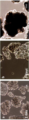
[00034] 도 1(a-c): 1 RPE : 3 PRP의 비율로 Y-27632를 포함하지 않는 RPE-MM에서 응집체 형성 후 2일(도 1a), 21일(도 1b) 및 69일(도 1c)의 이중 세포 응집체.
[0035] 도 2(a-c): 1 RPE : 8 PRP의 비율로 Y-27632를 포함하는 RPE-MM에서 응집체 형성 후 1일(도 2a), 3일(도 2b) 및 45일(도 2c)의 이중 세포 응집체.
[0036] 도 3: PGE2의 첨가 유무에 따른 응집체 형성 1개월 후 이중 세포 응집체(초기 비율 1 RPE : 8 PRP). PRP 조직에서의 차이는 분명했으며 PGE2가 없는 응집체 주변에 더 많은 PRP 제한이 있을 수 있다.
[0037] 도 4: PGE2를 첨가하거나 첨가하지 않은 1개월의 이중 세포 응집체 배양은 RPE 조직 또는 막대형 형성에서 현저한 차이를 나타내지 않았고, PRPH2 발현은 이 시점에서 낮거나 없는 것으로 나타났다.
[0038] 도 5: 응집체 형성 1, 2 및 3개월 후 PGE2를 첨가한 이중 세포 응집체 배양의 비교. 2개월 후 PRPH2의 존재는 외부 분절 형성 및 광수용체 성숙을 나타낸다.
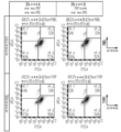
[0039] 도 6a 및 도 6b: 응집체 형성 2일 후 이중 세포 응집체의 유세포분석 평가. 응집체는 1 RPE : 30 PRP의 입력 비율로 형성되었다. 2일 후 측정된 RPE:PRP 비율은 대략 1:20이고, RPE 마커 PMEL 및 TYRP1과 PRP 마커 리커버린(Recoverin)을 사용하였다.
[0040] 도 7: 시험관 내 연장된 배양에서 이중 세포 응집체의 위상차 이미지. 착색된 RPE는 9일 후에 볼 수 있었고, RPE 영역은 시간이 지남에 따라 성장하는 것으로 나타났다.
[0041] 도 8: Hoechst 33342를 갖는 모든 세포를 나타내는 2일 후 이중 세포 응집체의 면역세포화학. RPE(PMEL, TYRP1)는 응집체의 작은 부분을 차지하는 반면, PRP(NRL, CRX)는 이 시점에서 응집체의 큰 부분을 차지한다.
[0042] 도 9(a-f): 시험관 내 배양 1개월 후 이중 세포 응집체의 면역세포화학. RPE[(도 9a, 도 9b) ZO-1, (도 9c, 도 9d) PMEL, (도 9e, 도 9f) TYRP1]은 2일 후에서보다 응집체의 더 큰 부분으로 나타나지만, PRP(리커버린, M/L 옵신, NRL, CRX, ARR3)은 여전히 우세한 부분이다. 원추형(M/L 옵신, ARR3) 및 막대형(NRL) 마커의 존재는 PRP가 두 세포 유형을 모두 형성할 수 있음을 확인하고, PRPH2의 초기 존재는 외부 분절 마커의 시작을 나타내고 PRP가 성숙하기 시작함을 시사한다.
[0043] 도 10(a-d): 응집체 형성 시 1:30의 RPE:PRP 비율을 갖는 응집체 형성 3개월 후의 RPE/PRP 공동-응집체. PRP 및 RPE는 (도 10a, 도 10b) RPE를 나타내는 ZO-1(녹색) 및 PRP를 나타내는 RCVRN(빨간색)을 도시하는 응집체에서 별개의 분절을 형성한다. 증식하는 세포(보라색)는 일반적으로 3개월 후에 나타나지 않는다. B)는 A)의 삽입 배율을 보여준다. RPE의 성숙(도 10c)은 PMEL-양성(빨간색) RPE의 영역에서 RPE65(녹색)로 표시되며, 막대형 PRP는 NRL(보라색)로 표시된다. 도 10d는 도 10c에서 확대된 삽입도를 나타낸다.
[0044] 도 11: 응집체 형성시 1:30의 RPE:PRP 비율을 갖는 응집체 형성 3개월 후 RPE/PRP 공동 응집체의 유동 세포측정 플롯. RPE(PMEL)와 PRP(RCVRN) 둘 모두 플로팅하면 이 시점에서 거의 2:1 비율로 PRP 셀보다 RPE가 더 많다는 것을 알 수 있다.
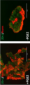
[0045] 도 12(a,b): 이중 세포 응집체 주입 1개월 후 래트(rat) 망막 절편 (조건 A; PRP 및 RPE가 단일 세포 현탁액으로서 결합되고 응집체 형성이 허용됨). 착색된 인간 세포(검정색/빨간색)는 생체내에서 RPE 층으로 이동하는 것으로 나타났고, 이식된 세포로부터의 (도 12a) 원추형(M/L 옵신) 및 (도 12b) 막대형(로돕신, NRL) 광수용체 둘 다의 증거가 있다. 착색된 세포의 일부 작은 클러스터가 PRP 층 내에서 선명하다.
[0046] 도 13(a-c): 이중 세포 응집체 주입 2개월 후 래트 망막 절편(조건 A). 이식된 세포로부터의 광수용체에서 (도 13a) NRL, (도 13b) M/L 옵신 및 (도 13c) 로돕신으로 표시되는 막대형 및 원추형 둘 다의 성숙의 증거가 있다. 착색된 세포의 일부 작은 클러스터가 PRP 층 내에서 선명하다.
[0047] 도 14(a-c): 이중 세포 응집체 주입 2개월 후 래트 망막 절편(조건 B). 이식된 세포로부터의 광수용체에서 (도 14a) NRL, (도 14b) M/L 옵신 및 (도 14c) 로돕신으로 표시되는 막대형 및 원추형 둘 다의 성숙의 증거가 있다. 착색된 세포의 일부 작은 클러스터가 PRP 층 내에서 명백하다. [0033] The following drawings form part of this specification and are included to further demonstrate certain aspects of the present invention. The invention may be better understood by reference to one or more of these drawings in conjunction with the detailed description of specific embodiments provided herein.
[00034] Fig. 1(ac): 2 days ( Fig. 1a ), 21 days ( Fig. 1b ) and 69 days ( Fig. 1c ) of double cell aggregates.
2(ac): 1 day ( FIG. 2a ), 3 days ( FIG. 2b ) and 45 days ( FIG. 2c ) after aggregate formation in RPE-MM containing Y-27632 at a ratio of 1 RPE : 8 PRP of double cell aggregates.
Figure 3 : Double cell aggregates after 1 month of aggregate formation with or without the addition of PGE2 (
[0037] Figure 4 : 1 month of double cell aggregate culture with or without the addition of PGE2 did not show significant differences in RPE tissue or rod formation, and PRPH2 expression was found to be low or absent at this time point.
[0038] Figure 5 : Comparison of double cell aggregate cultures with the addition of PGE2 after 1, 2 and 3 months of aggregate formation. Presence of PRPH2 after 2 months indicates external segment formation and photoreceptor maturation.
[0039] Figures 6a and 6b : Flow cytometry evaluation of double cell aggregates 2 days after aggregate formation. Aggregates were formed at an input ratio of 1 RPE:30 PRP. The RPE:PRP ratio measured after 2 days was approximately 1:20, and the RPE markers PMEL and TYRP1 and the PRP marker Recoverin were used.
[0040] Figure 7 : Phase-contrast image of double cell aggregates in extended culture in vitro. Pigmented RPE was visible after 9 days, and the RPE area appeared to grow over time.
[0041] Figure 8 : Immunocytochemistry of double cell aggregates after 2 days showing all cells with Hoechst 33342. RPE (PMEL, TYRP1) occupied a small fraction of the aggregates, whereas PRP (NRL, CRX) occupied a large fraction of the aggregates at this time point.
[0042] Fig. 9(af): Immunocytochemistry of double cell aggregates after 1 month of in vitro culture. RPE [( FIGS. 9A , 9B ) ZO-1, ( FIGS. 9C , 9D ) PMEL, ( FIGS. 9E , 9F ) TYRP1] appeared as a larger fraction of aggregates than after 2 days, but PRP (recovered, M/L opsin, NRL, CRX, ARR3) are still dominant. The presence of cone-shaped (M/L opsin, ARR3) and rod-shaped (NRL) markers confirms that PRP can form both cell types, while the early presence of PRPH2 indicates the initiation of extrinsic segmental markers and allows PRP to mature. indicates starting
10(ad) : RPE/PRP co-aggregates after 3 months of aggregate formation with an RPE:PRP ratio of 1:30 upon aggregate formation. PRP and RPE form distinct segments in aggregates ( FIGS. 10A , 10B ) showing ZO-1 (green) representing RPE and RCVRN (red) representing PRP. Proliferating cells (purple) usually do not appear after 3 months. B) shows the inset magnification of A). Maturation of RPE ( FIG. 10C ) is indicated by RPE65 (green) in areas of PMEL-positive (red) RPE, and rod PRP is indicated by NRL (purple). FIG. 10D shows an enlarged inset in FIG . 10C .
[0044] Figure 11 : Flow cytometric plot of RPE/
[0045] Figure 12(a, b) : Rat (rat)
[0046] Fig. 13(ac) : Rat
[0047] Fig. 14(ac) : Rat
I. 예시적인 구현예의 설명I. Description of Exemplary Embodiments
[0048] RPE와 신경 망막은 생체 내에서 정렬된 층에서 발달하고 존재한다. 순수 RPE:PR/PRP 구조적 관계의 발생 반복(recapitulation)은 치료에서 필수적일 수 있다. 또한, RPE:PR/PRP 구조의 시험관 내 발생 반복은 약물 또는 세포 질환 모델을 테스트하기 위한 플랫폼으로서 실행 가능할 수 있다. 따라서, 특정 구현예에서, 본 발명은 인간 유도 만능 줄기 세포(hiPSC)-유래 RPE(iRPE) 및 hiPSC-유래 PRP(iPRP)를 "이중-요법" 공동-응집체 배양에서 배양하는 방법을 제공한다. [0048] The RPE and neural retina develop and exist in an ordered layer in vivo . Recapitulation of the pure RPE:PR/PRP structural relationship may be essential in therapy. Additionally, in vitro generated iterations of the RPE:PR/PRP construct may be viable as a platform to test drugs or cellular disease models. Thus, in certain embodiments, the present invention provides a method of culturing human induced pluripotent stem cell (hiPSC)-derived RPE (iRPE) and hiPSC-derived PRP (iPRP) in a “dual-therapy” co-aggregate culture.
[0049] RPE는 예를 들어 PCT/US2016/050543호 및 PCT/US2016/050554호에 개시된 방법에 의해 hiPSC로부터 유래될 수 있으며, 둘 다 그 전체 내용이 본원에 참조로서 포함된다. PRP는 예를 들어 PCT/US2019/028557호에 개시된 방법에 의해 hiPSC로부터 유래될 수 있으며, 그 전체 내용이 참조로서 본원에 포함된다. RPE, PR 또는 PRP는 배아 줄기 세포와 같은 PSC로부터 유래될 수 있다. 막대형 및 원추형으로 성숙할 수 있는 미성숙한 PRP는 단일 세포 또는 응집체로서 RPE에 추가된다. PR/PRP는 해동되고 RPE와 함께 직접 씨딩되거나, RPE와 함께 씨딩하기 전에 일정 기간 동안 배양될 수 있다. 일부 측면에서, RPE-PRP 응집체 형성을 보충하기 위해 ROCK 억제제(예를 들어, Y-27632)와 같은 인자가 배양 시스템에 추가될 수 있다. [0049] RPEs can be derived from hiPSCs, for example, by methods disclosed in PCT/US2016/050543 and PCT/US2016/050554, both of which are incorporated herein by reference in their entirety. PRPs can be derived from hiPSCs, for example, by methods disclosed in PCT/US2019/028557, the entire contents of which are incorporated herein by reference. RPE, PR or PRP may be derived from PSCs such as embryonic stem cells. Immature PRP, which can mature into rods and cones, is added to RPE as single cells or aggregates. PR/PRP can be thawed and directly seeded with RPE, or cultured for a period of time prior to seeding with RPE. In some aspects, factors such as ROCK inhibitors (eg, Y-27632) can be added to the culture system to supplement RPE-PRP aggregate formation.
[0050] 본원에 제공된 RPE-PR/PRP 이중 세포 응집체는 생체내 및 시험관내의 다양한 방법에서 사용될 수 있다. 상기 RPE-PR/PRP 이중 세포 응집체는 추정 치료 또는 예방 치료 후보를 식별하기 위한 스크리닝 분석에서 시험관내에서 사용될 수 있다. 상기 RPE-PR/PRP 이중 세포 응집체는 생체내에서 노화-관련 또는 유전성 황반 변성, 및 망막 색소 변성증 또는 RPE 및/또는 PR/PRP의 기능 장애 및/또는 손실을 유발하는 기타 유전성 외부 망막 퇴행성 질환 또는 손상을 포함하지만 이에 제한되지 않는 망막의 병태를 치료하는데 사용할 수 있다. 본 개시 내용의 추가 구현예 및 이점이 이하 설명된다. [0050] The RPE-PR/PRP double cell aggregates provided herein are in vivo and in vitro various can be used in methods. The RPE-PR/PRP double cell aggregates can be used in vitro in screening assays to identify putative therapeutic or prophylactic treatment candidates. The RPE-PR/PRP double cell aggregates are resistant to age-related or hereditary macular degeneration in vivo , and retinitis pigmentosa or other hereditary outer retinal degenerative diseases that cause dysfunction and/or loss of RPE and/or PR/PRP; or It can be used to treat conditions of the retina including but not limited to damage. Additional embodiments and advantages of the present disclosure are described below.
I. 정의I. Definition
[0051] 용어 "정제된"은 절대적인 순도를 요하는 것이라기보다는; 오히려, 상대적인 용어로서 의도한 것이다. 따라서, 정제된 세포의 집단은 약 90%, 91%, 92%, 93%, 94%, 95%, 96%, 97%, 98%, 99% 초과 또는 100% 순수하거나, 또는, 가장 바람직하게는, 필수적으로 다른 세포 유형을 가지지 않는다. [0051] term “Refined” is not one that requires absolute purity; Rather, it is intended as a relative term. Thus, a population of purified cells is greater than about 90%, 91%, 92%, 93%, 94%, 95%, 96%, 97%, 98%, 99%, or 100% pure, or, most preferably, , does not necessarily have other cell types.
[0052] 본 명세서에서 사용된 바와 같이, 관사 ("a" 또는 "an")는 하나 이상을 의미할 수 있다. 본원 청구항에서 사용된 바와 같은, 단수형 관사 ("a" 또는 "an")는 "포함하는" 이라는 단어와 함께 사용되는 경우 하나 또는 하나 이상을 의미할 수 있다. [0052] As used herein, the article ("a" or "an") can mean one or more. As used in the claims herein, the singular article “a” or “an” when used with the word “comprising” can mean one or more than one.
[0053] 청구항에서 용어 "또는"의 사용은, 그것이 명시적으로 대안만을 언급하지 않는 한 또는 명세서가 대안 및 "및/또는"만을 언급하는 정의를 뒷받침하고 있더라도 그 대안이 상호 배타적이지 않는 한 "및/또는"을 의미하는 것으로 사용된다. 본원에서 "또 다른" 이라는 말은 적어도 두번째 또는 그 이상을 의미할 수 있다. [0053] The use of the term "or" in a claim may be used unless it explicitly refers only to the alternative, or even if the specification supports a definition referring only to the alternative and "and/or", unless the alternatives are mutually exclusive. and/or" is used. The word "another" as used herein may mean at least a second or more.
[0054] 용어 "본질적으로(essentially)"는 방법 또는 구성이 특정 단계 또는 재료만을 포함하고 이러한 방법 및 구성의 기본적이고 신규한 특성에 실질적으로 영향을 미치지 않는 것들을 포함하는 것으로 이해되어야 한다. [0054] Terminology "Essentially" is to be understood as including those in which a method or composition only includes certain steps or materials and does not materially affect the basic and novel characteristics of such method or composition.
[0055] 본원에 사용된 바와 같이, 특정 물질 또는 재료가 "실질적으로 없는" 조성물 또는 배지는 상기 물질 또는 재료의 ≤ 30%, ≤ 20%, ≤ 15%, 더 바람직하게는 ≤ 10%, 더욱 더 바람직하게는 ≤ 5%, 또는 가장 바람직하게는 ≤ 1%를 함유한다. [0055] As used herein, a composition or medium that is "substantially free" of a particular substance or material is ≤ 30%, ≤ 20%, ≤ 15%, more preferably ≤ 10%, even more of that substance or material. more preferably ≤ 5%, or most preferably ≤ 1%.
[0056] 본원에 사용된 바와 같이 용어 "실질적으로" 또는 "대략"은 관련된 기본 기능의 변화를 초래하지 않고 허용적으로 변할 수 있는 임의의 정량적 비교, 값, 측정 또는 기타 표현을 수정하기 위해 적용될 수 있다. [0056] As used herein, the terms "substantially" or "approximately" can be applied to modify any quantitative comparison, value, measurement, or other expression that can be changed acceptably without resulting in a change in the underlying function to which it relates. can
[0057] 용어 "약"은, 일반적으로, 명시된 값을 측정하기 위한 표준 분석 기술을 사용하여 결정된 명시된 값의 표준 편차 이내를 의미한다. 이 용어는 명시된 값의 플러스 또는 마이너스 5%를 참조하여 사용할 수도 있다. [0057] The term "about" generally means within a standard deviation of a specified value determined using standard analytical techniques for determining the specified value. The term may also be used in reference to plus or minus 5% of the stated value.
[0058] 본원에 사용된 바와 같이 "본질적으로 없는"은 특정 성분과 관련하여 본원에서 특정 성분이 의도적으로 조성물로 제형화되지 않았거나 및/또는 오염 물질로서 또는 미량으로만 존재함을 의미하는 데 사용된다. 따라서 조성물의 임의의 의도하지 않은 오염으로 인한 특정 성분의 총량은 0.05% 미만, 바람직하게는 0.01% 미만이다. 가장 바람직한 것은 상기 특정 성분의 양이 표준 분석 방법으로 검출될 수 없는 조성물이다. [0058] As used herein, "essentially free", in reference to a particular ingredient, herein means that the particular ingredient has not been intentionally formulated into a composition and/or is present as a contaminant or only in trace amounts. used Thus, the total amount of certain components due to any unintentional contamination of the composition is less than 0.05%, preferably less than 0.01%. Most preferred are compositions in which the amount of said particular component is undetectable by standard analytical methods.
[0059] 용어 "세포 집단"은 보통 통상적인 유형의 세포들의 그룹을 가리키는데 사용된다. 상기 세포 집단은 공통의 선조체로부터 유래할 수 있거나, 또는 하나 초과의 세포 유형을 포함할 수도 있다. "농축된" 세포 집단은 출발 세포 집단의 특정 세포 유형의 퍼센트보다 더 큰 퍼센트의 세포 유형을 포함하는 출발 세포 집단(예를 들어, 분획화되지 않은, 이종성 세포 집단)으로부터 유래된 세포집단을 지칭한다. 세포 집단은 하나 이상의 세포 유형에 대해 농축될 수 있고, 하나 이상의 세포 유형이 고갈될 수도 있다. [0059] Terminology A "cell population" is usually used to refer to a group of cells of a common type. The cell population may be derived from a common progenitor or may include more than one cell type. An "enriched" cell population refers to a cell population derived from a starting cell population (e.g., an unfractionated, heterogeneous cell population) that contains a percentage of a cell type that is greater than the percentage of a particular cell type in the starting cell population. do. A cell population may be enriched for one or more cell types, or one or more cell types may be depleted.
[0060] 용어 "줄기 세포"는 본원에서 적절한 조건 하에 다양한 범위의 분화 세포 유형으로 분화할 수 있는 세포, 한편으로는 적절한 다른 조건 하에 자기 재생 및 필수적으로 미분화된 만능 상태로 남아 있을 수 있는 세포를 지칭한다. 또한, 용어 "줄기 세포"는 만능 세포, 다능성 세포, 전구체 세포 및 선조체를 포괄한다. 전형적인 인간 줄기 세포는 골수 조직으로부터 수득된 조혈 또는 간엽 줄기 세포, 배아 조직으로부터 수득된 배아 줄기 세포, 또는 태아의 생식기 조직으로부터 수득된 배아 생식 세포로부터 수득될 수 있다. 또한, 전형적인 만능 줄기 세포는 다능성과 관련된 특정 전사 인자의 발현에 의해 만능 상태로 체세포를 재프로그래밍함으로써 해당 체세포로부터도 생성될 수 있으며; 이들 세포들은 “유도된 만능 줄기 세포” 또는 “iPSC”로 불린다. [0060] Terminology “Stem cell” refers herein to a cell capable of differentiating under appropriate conditions into a wide range of differentiated cell types, while remaining in a self-renewing and essentially undifferentiated pluripotent state under other appropriate conditions. Also, the term "Stem cell" encompasses pluripotent cells, pluripotent cells, progenitor cells and progenitors. Typical human stem cells may be obtained from hematopoietic or mesenchymal stem cells obtained from bone marrow tissue, embryonic stem cells obtained from embryonic tissue, or embryonic germ cells obtained from genital tissue of a fetus. In addition, typical pluripotent stem cells can also be generated from corresponding somatic cells by reprogramming somatic cells to a pluripotent state by expression of specific transcription factors associated with pluripotency; These cells are called “induced pluripotent stem cells” or “iPSCs”.
[0061] 용어 "만능"은 배아외, 또는 태반 세포를 제외하고 유기체 내에서 다른 모든 세포 유형으로 분화하는 세포의 특성을 가리킨다. 만능 줄기 세포는 장시간의 배양 이후에 조차도 세가지의 모든 배엽층(예컨대, 외배엽, 중배엽 및 내배엽 세포 유형)으로 분화할 수 있다. 만능 줄기 세포는 배반포의 내부 세포 매쓰에서 유래된 배아 줄기 세포이다. 다른 구현예에서, 만능 줄기 세포는 체세포를 재프로그래밍하여 유래된 유도된 만능 줄기 세포이다. [0061] term "Pluripotent" refers to the property of a cell to differentiate into all other cell types within an organism, except for extraembryonic or placental cells. Pluripotent stem cells are capable of differentiating into all three germ layers (eg, ectoderm, mesoderm and endoderm cell types) even after prolonged culture. Pluripotent stem cells are embryonic stem cells derived from the inner cell mass of the blastocyst. In another embodiment, the pluripotent stem cell is an induced pluripotent stem cell derived by reprogramming a somatic cell.
[0062] 용어 "분화"는 미분화된 세포가 구조적 및/또는 기능적 특성에 변화가 있는 보다 분화된 유형으로 되는 과정을 일컫는다. 성숙한 세포는 통상 변형된 세포 구조와 조직 특이적 단백질을 가진다. [0062] term "Differentiation" refers to the process by which an undifferentiated cell becomes a more differentiated type with changes in structural and/or functional properties. Mature cells usually have altered cellular structures and tissue-specific proteins.
[0063] 본원에 사용된 바와 같이, "미분화된"은 해당 미분화된 세포를 배아 또는 성체 기점의 말단 분화된 세포와 명백하게 구별되는 미분화된 세포의 특징적 마커 및 형태학적 특징을 나타내는 세포를 지칭한다. [0063] As used herein, "undifferentiated" refers to cells that display characteristic markers and morphological characteristics of undifferentiated cells that clearly distinguish the undifferentiated cells from terminally differentiated cells of embryonic or adult origin.
[0064] "배아체(EB)"는 내배엽, 중배엽 및 외배엽 배엽층의 세포로 분화할 수 있는 만능 줄기 세포의 응집물이다. 스페로이드 구조는 만능 줄기 세포가 비-접착 배양 조건하에 응집하도록 허용된 경우 형성되고 따라서 현탁액 중 EB를 형성한다. [0064] An "embryoid body (EB)" is an aggregate of pluripotent stem cells capable of differentiating into cells of the endoderm, mesoderm and ectodermal germ layers. Spheroid structures are formed when pluripotent stem cells are allowed to aggregate under non-adherent culture conditions and thus form EBs in suspension.
[0065] "단리된" 세포는 유기체 또는 배양액 중에서 다른 세포로부터 실질적으로 분리되거나 정제된다. 단리된 세포는, 예를 들어, 99% 이상, 98% 이상 순수, 95% 이상 순수 또는 90% 이상 순수할 수 있다. [0065] An "isolated" cell is substantially separated or purified from other cells in an organism or culture. Isolated cells can be, for example, greater than 99% pure, greater than 98% pure, greater than 95% pure, or greater than 90% pure.
[0066] "배아"는 접합체 또는 인위적으로 재프로그래밍된 핵으로 활성화된 난모세포의 1회 이상의 분열로 수득된 세포 매쓰를 지칭한다. [0066] "Embryo" refers to a cell mass obtained from one or more divisions of an activated oocyte into a zygote or artificially reprogrammed nucleus.
[0067] "배아 줄기(ES) 세포"는 초기 단계의 배아, 예컨대 배반포 단계에서 내부 세포 매쓰로부터 수득되거나 또는 인위적인 수단(예컨대, 핵 이식)에 의해 생성되어 생식 세포(예컨대 정자 및 난자)를 비롯한 배아 또는 성체에서 임의의 분화된 세포 유형을 유도할 수 있는 미분화된 만능 세포이다. [0067] "Embryonic stem (ES) cells" are those obtained from the internal cell mass of an early stage embryo, such as the blastocyst stage, or generated by artificial means (such as nuclear transfer) to produce reproductive cells (such as sperm and eggs). It is an undifferentiated pluripotent cell capable of inducing any differentiated cell type in an embryo or adult.
[0068] "유도된 만능 줄기 세포(iPSC)"는 인자들(본원에서 재프로그래밍 인자로 지칭함) 조합의 발현 또는 그 발현을 유도함으로써 체세포를 재프로그래밍하여 생성된 세포이다. iPSC는 태아, 출생후, 발육기, 또는 성체 체세포를 사용하여 생성될 수 있다. 특정 구현예에서, 체세포를 만능 줄기 세포로 재프로그래밍하는데 사용될 수 있는 인자들로는, 예를 들어, Oct4 (Oct 3/4로도 지칭함), Sox2, c-Myc, Klf4, Nanog 및 Lin28을 포함한다. 일부 구현예에서, 체세포는 2개 이상의 재프로그래밍 인자, 3개 이상의 재프로그래밍 인자, 또는 4개의 재프로그래밍 인자를 발현시켜 체세포를 만능 줄기 세포로 재프로그래밍함으로써 재프로그래밍된다. [0068] "Induced pluripotent stem cells (iPSCs)" are cells produced by reprogramming somatic cells by expressing or inducing expression of a combination of factors (referred to herein as reprogramming factors). iPSCs can be generated using fetal, postnatal, embryonic, or adult somatic cells. In certain embodiments, factors that can be used to reprogram somatic cells into pluripotent stem cells include, for example, Oct4 (also referred to as
[0069] "대립유전자"는 2 이상의 유전자 형태 중에서 하나를 가리킨다. 인간과 같은 이배체 유기체는 각 염색체를 두 카피물씩 포함하기 때문에, 각각 하나의 대립유전자를 수반한다. [0069] "Allele" refers to one of two or more forms of a gene. Diploid organisms such as humans contain two copies of each chromosome, so each carries one allele.
[0070] 용어 "동형접합성"은 특정 자리에서 2개의 동일한 대립유전자를 포함하는 것으로 정의된다. 용어 "이형접합성"은 특정 자리에서 2개의 서로 다른 대립유전자를 포함하는 것으로 정의된다. [0070] Terminology "Homozygous" is defined as having two identical alleles at a particular locus. Terms "Heterozygous" is defined as having two different alleles at a particular locus.
[0071] "반수체형"은 단일 염색체를 따라 다수의 유전자좌에서 대립유전자의 조합을 지칭한다. 반수체형은 단일 염색체 상의 한 세트의 단일 뉴클레오타이드 다형태(SNP) 및/또는 주요 조직적합성 복합체 내의 대립유전자를 기반으로 할 수 있다. [0071] "Haplotype" refers to a combination of alleles at multiple loci along a single chromosome. A haplotype can be based on a set of single nucleotide polymorphisms (SNPs) on a single chromosome and/or alleles within a major histocompatibility complex.
[0072] 본원에 사용된 바와 같이, 용어 "반수체형-일치된"은 처리되는 세포 (예컨대, iPSC 세포) 및 대상체가 하나 이상의 주요 조직적합성 유전자좌 반수체형을 공유하는 것으로 정의된다. 대상체의 반수체형은 본 기술분야에 잘 알려진 분석법을 사용하여 용이하게 결정될 수 있다. 반수체형-일치된 iPS 세포는 자가 또는 동종이계일 수 있다. 조직 배양물 중에서 성장하고 선천적으로 PRP 세포로 분화되는 자가 세포는 대상체와 반수체형 일치된다. [0072] As used herein, the term "haploid-matched" refers to a cell being treated (e.g., iPSC cells) and subjects share one or more major histocompatibility locus haplotypes. A subject's haplotype can be readily determined using assays well known in the art. Haplotype-matched iPS cells may be autologous or allogeneic. Autologous cells that grow in tissue culture and differentiate innately into PRP cells are haplotype-matched to the subject.
[0073] "실질적으로 동일한 HLA형"은 공여체의 인간 백혈구 항원(HLA) 유형이 공여체의 체세포로부터 유래된 iPCS의 분화를 유도함으로써 수득된 이식 세포가 이들이 환자에게 이식될 때 접목될 수 있을 정도로 환자의 HLA형과 일치함을 의미한다. [0073] “Substantially identical HLA type” means that the donor's human leukocyte antigen (HLA) type is such that the transplanted cells obtained by inducing differentiation of iPCS derived from the donor's somatic cells can be grafted when they are transplanted into the patient. It means that it matches the HLA type of
[0074] "슈퍼 공여체(Super donor)"는 본원에서 특정 MHC 부류 I 및 II 유전자에 대해 동형접합성인 개체로서 언급된다. 이러한 동형접합성 개체들은 슈퍼 공여체로서 작용할 수 있으며, 조직을 비롯한 이들의 세포 및 이들의 세포를 포함하는 기타 물질들은 해당 반수체형에 대해 동형접합성 또는 이형접합성인 개체에 이식될 수 있다. 상기 슈퍼 공여체는 각각 HLA-A, HLA-B, HLA-C, HLA-DR, HLA-DP 또는 HLA-DQ 유전자좌/유전자좌 대립유전자에 대해 동형접합성일 수 있다. [0074] A "Super donor" is referred to herein as an individual who is homozygous for certain MHC class I and II genes. Such homozygous individuals can act as super donors, and their cells, including tissues, and other materials comprising their cells can be transplanted into individuals that are either homozygous or heterozygous for the haplotype in question. The super donor may be homozygous for the HLA-A, HLA-B, HLA-C, HLA-DR, HLA-DP or HLA-DQ locus/locus alleles, respectively.
[0075] "피더-프리(feeder-free)" 또는 "피더-독립성(feeder-independent)"은 본원에서 피더 세포층에 대한 대체물로서 사이토킨과 성장 인자(예컨대, TGFβ, bFGF, LIF)로 보충된 배양액을 가리키는데 사용된다. 따라서, "피더-프리" 또는 피더-독립성 배양 시스템 및 배지는 만능 세포를 배양하여 미분화 및 증식 상태로 유지하는데 사용될 수 있다. 일부의 경우, 피더-프리 배양물은 동물계 기질(예컨대 MATRIGEL™)을 이용하거나, 또는 피브로넥틴, 콜라겐 또는 비트로넥틴과 같은 기질 상에서 성장한다. 이러한 접근법들은 인간 줄기 세포가 마우스 섬유아세포 "피더 층"에 대한 필요없이 필수적으로 미분화 상태로 남아있도록 해준다. [0075] "Feeder-free" or "feeder-independent" herein refers to a culture supplemented with cytokines and growth factors (e.g., TGFβ, bFGF, LIF) as a substitute for a feeder cell layer. is used to indicate Thus, “feeder-free” or feeder-independent culture systems and media can be used to culture and maintain pluripotent cells in an undifferentiated and proliferative state. In some cases, feeder-free cultures utilize animal-based substrates (such as MATRIGEL™) or are grown on substrates such as fibronectin, collagen, or vitronectin. These approaches allow human stem cells to remain essentially undifferentiated without the need for a mouse fibroblast “feeder layer”.
[0076] "피더 층"은 배양 접시의 바닥 상에서와 같은 세포의 코팅층으로 정의된다. 상기 피더 세포는 배양 배지로 영양분을 방출하여 만능 줄기 세포와 같은 다른 세포가 부착할 수 있는 표면을 제공할 수 있다. [0076] A "feeder layer" is defined as a coating layer of cells, such as on the bottom of a culture dish. The feeder cells can release nutrients into the culture medium to provide a surface to which other cells, such as pluripotent stem cells, can adhere.
[0077] 배지, 세포외 기질 또는 배양 조건과 관련하여 사용되는 "규명된(defined)" 또는 "완전히 규명된(fully defined)"이라는 용어는 거의 모든 성분들의 화학 조성과 양이 공지된 배지, 세포외 기질 또는 배양 조건을 가리킨다. 예를 들어, 규명된 배지는 소태아 혈청, 소 혈청 알부민 또는 인간 혈청 알부민에서와 같이 규명되지 않은 인자들은 포함하지 않는다. 일반적으로, 규명된 배지는 재조합 알부민, 화학적으로 규명된 지질 및 재조합 인슐린이 보충된 기본 배지(예컨대, 둘베코의 변형된 이글 배지(DMEM), F12, 또는 아미노산, 비타민, 무기염, 완충액, 산화방지제 및 에너지원을 포함하는 로스웰 파크 메모리얼 인스티튜트 (Roswell Park Memorial Institute, RPMI) 배지 1640)를 포함한다. 완전히 규명된 배지의 예는 Essential 8™ 배지이다. [0077] The term "defined" or "fully defined" when used in reference to a medium, extracellular matrix or culture conditions means a medium, cell in which the chemical composition and amount of almost all components are known. Refers to external substrate or culture conditions. For example, a defined medium does not contain uncharacterized factors such as fetal bovine serum, bovine serum albumin, or human serum albumin. Generally, the defined medium is a basal medium supplemented with recombinant albumin, chemically defined lipids and recombinant insulin (eg, Dulbecco's modified Eagle's medium (DMEM), F12, or Roswell Park Memorial Institute (RPMI) medium 1640 containing amino acids, vitamins, inorganic salts, buffers, antioxidants and energy sources). An example of a fully characterized medium is Essential 8™ medium.
[0078] 인간 세포와 함께 사용되는 배지, 세포외 기질 또는 배양 시스템에 대해, 용어 "제노-프리(Xeno-Free)(Xf)"는 사용되는 물질이 비-인간 동물-기점이 아닌 조건을 언급한다. [0078] For media, extracellular matrices or culture systems used with human cells, the term "Xeno-Free (Xf)" refers to conditions where the materials used are not of non-human animal origin. do.
[0079] "프리-컨플루언트(pre-confluent)"는 세포에 의해 커버(cover)되는 배양액 표면의 비율이 약 60-80%인 세포 배양액을 지칭한다. 보통, 프리-컨플루언트는 배양액 표면의 약 70%가 세포에 의해 커버된 배양액을 가리킨다. [0079] "Pre-confluent" refers to a cell culture in which the proportion of the culture surface covered by cells is about 60-80%. Usually, pre-confluent refers to a culture in which about 70% of the culture surface is covered by cells.
[0080] "망막 전구체 세포" 또는 "RPC"로 불리우는, 용어 "망막 선조체 세포"는 막대형, 원추형, 광수용체 전구체 세포, 및 RPE로 분화할 수 있는 세포를 포함하는 모든 세포 유형의 망막을 생성하기에 적격인 세포를 포함한다. [0080] The term "retinal progenitor cell", also called "retinal progenitor cell" or "RPC", refers to all cell types of the retina, including rod, cone, photoreceptor progenitor cells, and cells capable of differentiating into RPE. It includes cells that are eligible for the following.
[0081] 용어 "신경 망막 선조체" 또는 "NRP"는 신경 망막 세포 유형으로의 분화 잠재력에서 제한되는 세포를 의미한다. [0081] The term "neural retinal progenitor" or "NRP" refers to cells that are limited in differentiation potential into neural retinal cell types.
[0082] 용어 "광수용체" 또는 "PR" 세포는 로돕신(막대형) 또는 임의의 3개의 원추형 옵신(원추형) 발현의 상향조절 전 및 후 둘 다에, 광수용체 계통(즉, 성숙) 경로 내에 있는 세포를 지칭하며, 이것은 광수용체 세포(막대형, 원추형 또는 둘 다)의 초기 및 후기 마커를 둘 모두 포함한다. [0082] The term "photoreceptor" or "PR" cell refers to cells within the photoreceptor lineage (i.e., maturation) pathway both before and after upregulation of rhodopsin (rod) or any three cone opsin (cone) expression. cells that contain both early and late markers of photoreceptor cells (rods, cones, or both).
[0083] 용어 "광수용체 전구체 세포" 및 "PRP"는 세포 마커 로돕신 또는 3개의 원추형 옵신 중 임의의 것을 발현하는 광수용체 세포로 분화할 수 있는 배아 줄기 세포 또는 유도된 만능 줄기 세포로부터 분화된 세포를 의미한다. 상기 광수용체는 막대형 및/또는 원추형 광수용체일 수 있다. [0083] The terms "photoreceptor progenitor cell" and "PRP" refer to cells differentiated from embryonic stem cells or induced pluripotent stem cells capable of differentiating into photoreceptor cells expressing the cell marker rhodopsin or any of the three cone opsins. means The photoreceptors may be rod-shaped and/or cone-shaped photoreceptors.
[0084] "망막 색소 상피"는 혈관으로 채워진 층인 맥락막과 신경 망막 사이의 색소 침착된 세포의 층을 의미한다. [0084] "Retinal pigment epithelium" refers to the layer of pigmented cells between the choroid, which is a layer filled with blood vessels, and the neural retina.
[0085] 용어 "망막 변성-관련 질환"은 선천적 또는 출생 후 망막 변성 또는 이상으로 인해 발생하는 모든 질환을 의미한다. 망막 변성-관련 질환의 예는 망막이상증, 망막변성, 노화-관련 황반 변성, 스타가르트병, 베스트병, 맥락막결손, 유전성 황반 변성, 근시 변성, RPE 파열, 황반 원공, 당뇨망막병증, 망막 색소 변성증, 유전성 망막질환 또는 변성, 유전성 황반 변성, 원추형-막대형 이상증, 막대형-원추형 이상증, 선천성 망막 이상증, 레베르 선천성 흑암시, 망막 박리 및 망막 외상을 포함한다. [0085] Terminology "Retinal degeneration-related disease" means any disease caused by congenital or postnatal retinal degeneration or abnormality. Examples of retinal degeneration-related diseases include retinal dystrophy, retinal degeneration, age-related macular degeneration, Stargardt's disease, Best's disease, choroidal defect, hereditary macular degeneration, myopic degeneration, RPE rupture, macular hole, diabetic retinopathy, retinal pigment Degeneration, hereditary retinal disease or degeneration, hereditary macular degeneration, cone-rod dystrophy, rod-cone dystrophy, congenital retinal dystrophy, Leber congenital amaurosis, retinal detachment and retinal trauma.
[0086] 본원에 사용된 "치료적 유효량"은 질병 또는 병태의 치료를 위해 대상체에게 투여될 때 이러한 치료에 영향을 미치기에 충분한 화합물의 양을 의미한다. [0086] As used herein, "therapeutically effective amount" refers to an amount of a compound sufficient to effect treatment of a disease or condition when administered to a subject for treatment.
[0087] "성숙한" RPE 세포는 본원에서 Pax6과 같은 미성숙 RPE 마커의 발현이 하향 조절되고 RPE65와 같은 성숙 RPE 마커의 발현이 상향 조절된 RPE 세포를 의미한다. [0087] "Mature" RPE cells herein refer to RPE cells in which the expression of immature RPE markers such as Pax6 is downregulated and the expression of mature RPE markers such as RPE65 is upregulated.
[0088] RPE 세포 "성숙"은 본원에서 RPE 발달 경로가 조절되어 성숙한 RPE 세포를 생성하는 과정을 의미한다. 예를 들어, 섬모 기능의 조절은 RPE 성숙을 초래할 수 있다. [0088] RPE cell "maturation" refers herein to the process by which RPE developmental pathways are regulated to produce mature RPE cells. For example, modulation of ciliary function can lead to RPE maturation.
[0089] "인듀서"는 본원에서 세포 내 화설화 유전자와 같은 유전자 발현을 조절하는 분자로 정의된다. 인듀서는 억제제 또는 활성화제에 결합할 수 있다. 인듀서는 억제제를 비활성화함으로써 작동한다. [0089] An "inducer" is defined herein as a molecule that regulates the expression of a gene, such as a pyrexia gene in a cell. An inducer may bind to an inhibitor or an activator. An inducer works by inactivating an inhibitor.
[0090] 본원에 사용된 바와 같이, 용어 "생분해성"은 전달된 세포에 대한 초기 구조적 지지를 제공하지만, 시간이 지남에 따라 이식 숙주에 무독성이고 공여체 부위 이환율에 기여하지 않는 생성물로 분해되는 물질을 의미한다. [0090] As used herein, the term "biodegradable" refers to a substance that provides initial structural support for the transferred cells, but degrades over time into products that are non-toxic to the transplant host and do not contribute to donor site morbidity. means
II. 유도 만능 줄기 세포II. induced pluripotent stem cells
[0091] 만능성의 유도는 만능성과 연관된 전사 인자의 도입을 통한 체세포의 재프로그래밍에 의해 2006년에 마우스 세포를 사용하여 (Yamanaka et al. 2006), 그리고 2007년에 인간 세포를 사용하여 (Yu et al. 2007; Takahashi et al. 2007) 달성되었다. 만능 줄기 세포는 미분화 상태로 유지될 수 있어 임의의 성체 세포 유형으로 분화될 수 있다. [0091] Induction of pluripotency was achieved by reprogramming of somatic cells through introduction of transcription factors associated with pluripotency using mouse cells in 2006 (Yamanaka et al. 2006) and using human cells in 2007 (Yu et al. al. 2007; Takahashi et al . 2007) has been achieved. Pluripotent stem cells can be maintained in an undifferentiated state and differentiated into any adult cell type.
[0092] 생식계열 세포를 제외하고는, 어떤 체세포라도 iPCS를 위한 출발점으로 사용될 수 있다. 예를 들어, 세포 유형은 케라틴세포, 섬유아세포, 조혈 세포, 간엽 세포, 선조체 또는 위세포일 수 있다. T 세포는 또한 재프로그래밍을 위한 체세포 공급원으로도 사용될 수 있다(미국 특허 제8,741,648호). 세포 분화의 정도 또는 해당 세포가 수거되는 동물의 연령에는 제한이 없으며; 미분화 선조체 세포 (체세포 줄기 세포를 포함) 및 최종적으로 분화된 성숙한 세포 조차도 본원에 개시된 방법에서 체세포의 공급원으로 사용될 수 있다. 일 구현예에서, 체세포는 그 자체가 RPE 또는 PR/PRP 세포, 예를 들어, 인간 RPE 또는 PR/PRP 세포이다. RPE 또는 PR/PRP 세포는 성체 또는 태아 RPE 또는 PR/PRP 세포일 수 있다. iPSC는 인간 ES 세포를 특정 세포 유형으로 분화시키는 것으로 알려진 조건 하에 성장하여, SSEA-1, SSEA-3, SSEA-4, TRA-1-60 및 TRA-1-81를 비롯한 인간 ES 세포 마커를 발현할 수 있다. [0092] Except for germline cells, any somatic cell can be used as a starting point for iPCS. For example, the cell type can be keratinocytes, fibroblasts, hematopoietic cells, mesenchymal cells, progenitors or gastric cells. T cells can also be used as a source of somatic cells for reprogramming (US Pat. No. 8,741,648). There are no restrictions on the degree of cell differentiation or the age of animals from which the cells are harvested; Undifferentiated progenitor cells (including somatic stem cells) and even terminally differentiated mature cells can be used as a source of somatic cells in the methods disclosed herein. In one embodiment, the somatic cell is itself an RPE or PR/PRP cell, eg a human RPE or PR/PRP cell. The RPE or PR/PRP cells may be adult or fetal RPE or PR/PRP cells. iPSCs grow under conditions known to differentiate human ES cells into specific cell types and express human ES cell markers including SSEA-1, SSEA-3, SSEA-4, TRA-1-60 and TRA-1-81 can do.
A. 시작 세포의 HLAA. HLA of starting cells
[0093] 주요 조직 적합성 복합체(MHC, Major Histocompatibility Complex)는 동종이계 장기 이식의 면역 거부 반응의 주요 원인이다. 3개의 주요 클래스 I MHC 일배체형(A, B 및 C)과 3개의 주요 MHC 클래스 II 일배체형(DR, DP 및 DQ)이 있다. [0093] The major histocompatibility complex (MHC) is a major cause of immune rejection in allogeneic organ transplantation. There are three major class I MHC haplotypes (A, B and C) and three major MHC class II haplotypes (DR, DP and DQ).
[0094] 공여체 세포가 HLA 동형접합성인 경우, 즉 각 항원 제시 단백질에 대해 동일한 대립유전자를 함유하는 경우 공여체와 수용체 사이의 MHC 호환성이 상당히 증가한다. 대부분의 개체는 MHC 클래스 I 및 II 유전자에 대해 이형접합성이지만, 특정 개체는 이러한 유전자에 대해 동형접합성이다. 이러한 동형접합성 개체는 슈퍼 공여체 역할을 할 수 있으며, 세포로부터 생성된 이식편은 해당 일배체형에 대해 동형접합성 또는 이형접합성인 모든 개체에 이식될 수 있다. 또한, 동형접합성 공여체 세포가 집단에서 높은 빈도로 발견되는 일배체형을 갖는 경우, 이들 세포는 다수의 개체에 대한 이식 요법에 적용될 수 있다. [0094] MHC compatibility between the donor and the recipient is significantly increased when the donor cells are homozygous for HLA, ie, contain identical alleles for each antigen-presenting protein. Most individuals are heterozygous for MHC class I and II genes, but certain individuals are homozygous for these genes. Such homozygous individuals can serve as super donors, and grafts generated from the cells can be transplanted into any individual that is either homozygous or heterozygous for that haplotype. In addition, if the homozygous donor cells have a haplotype found in high frequency in the population, these cells can be applied in transplant therapy to a large number of individuals.
[0095] 따라서, 상기 iPSC는 치료 대상의 체세포 또는 환자와 동일하거나 실질적으로 동일한 HLA 유형을 가진 다른 대상의 체세포로부터 생산될 수 있다. 한 경우에, 공여체의 주요 HLA(예컨데, HLA-A, HLA-B 및 HLA-DR의 세 가지 주요 유전자좌)는 수용체의 주요 HLA와 동일하다. 어떤 경우에는, 체세포 공여체가 슈퍼 공여체일 수도 있다; 따라서, MHC 동형접합성 슈퍼 공여체로부터 유래된 iPSC는 RPE 또는 PR/PRP 세포를 생성하는 데 사용될 수 있다. 따라서, 슈퍼 공여체로부터 유래된 iPSC는 해당 일배체형에 대해 동형접합성 또는 이형접합성인 대상체에 이식될 수 있다. 예를 들어, 상기 iPSC는 HLA-A 및 HLA-B와 같은 두 개의 HLA 대립유전자에 동형접합성일 수 있다. 이와 같이, 슈퍼 공여체로부터 생성된 iPSC는 다수의 잠재적 수용체와 잠재적으로 "일치"할 수 있는 RPE 또는 PR/PRP 세포를 생성하기 위해, 본원에 개시된 방법에 사용될 수 있다. [0095] Accordingly, the iPSCs may be produced from somatic cells of a subject to be treated or from somatic cells of another subject having the same or substantially the same HLA type as the patient. In one case, the donor's primary HLA (eg, the three major loci of HLA-A, HLA-B and HLA-DR) is the same as the recipient's primary HLA. In some cases, a somatic cell donor may be a super donor; Thus, iPSCs derived from MHC homozygous super donors can be used to generate RPE or PR/PRP cells. Thus, iPSCs derived from super donors can be transplanted into subjects that are either homozygous or heterozygous for that haplotype. For example, the iPSCs may be homozygous for two HLA alleles, such as HLA-A and HLA-B. As such, iPSCs generated from super donors can be used in the methods disclosed herein to generate RPE or PR/PRP cells that can potentially "match" a number of potential recipients.
B. 리프로그래밍 요인B. Reprogramming factor
[0096] 체세포는 통상의 기술자에게 공지된 방법들을 사용하여 재프로그래밍되어 유도된 만능 줄기 세포(iPSC)를 생성할 수 있다. 통상의 기술자라면 유도된 만능 줄기 세포를 용이하게 생성할 수 있으며, 이에 대해서는, 예를 들어, 공개된 미국 특허출원 제20090246875호, 공개된 미국 특허출원 제2010/0210014호; 공개된 미국 특허출원 제20120276636호; 미국특허 제8,058,065호; 미국특허 제8,129,187호; 미국특허 제8,278,620호; PCT 공보 WO 2007/069666 A1호, 및 미국특허 8,268,620호를 참조하고, 이들은 본원에 참조로서 포함된다. 일반적으로, 핵 재프로그래밍 인자를 사용하여 체세포로부터 만능 줄기 세포를 생성한다. 일부 구현예에서, Klf4, c-Myc, Oct3/4, Sox2, Nanog 및 Lin28 중 적어도 2개, 적어도 3개 또는 적어도 4개를 사용한다. 다른 구현예에서, Oct3/4, Sox2, c-Myc 및 Klf4를 사용한다. [0096] Somatic cells can be reprogrammed to generate induced pluripotent stem cells (iPSCs) using methods known to those skilled in the art. The skilled person can readily generate induced pluripotent stem cells, which are described in, for example, published U.S. Patent Application No. 20090246875, published U.S. Patent Application No. 2010/0210014; Published US Patent Application Nos. 20120276636; U.S. Patent No. 8,058,065; U.S. Patent No. 8,129,187; U.S. Patent No. 8,278,620; See PCT Publication No. WO 2007/069666 A1, and US Pat. No. 8,268,620, incorporated herein by reference. Generally, nuclear reprogramming factors are used to generate pluripotent stem cells from somatic cells. In some embodiments, at least 2, at least 3 or at least 4 of Klf4, c-Myc, Oct3/4, Sox2, Nanog and Lin28 are used. In another embodiment, Oct3/4, Sox2, c-Myc and Klf4 are used.
[0097] 세포는 핵 재프로그래밍 물질로 처리하는데, 이는 일반적으로 체세포 또는 이러한 물질들을 암호화하는 핵산으로부터 iPCS를 유도할 수 있는 하나 이상의 인자들이다 (벡터 내에 통합된 형태를 포함함). 상기 핵 재프로그래밍 물질은 일반적으로 적어도 Oct3/4, Klf4 및 Sox2 또는 이러한 분자들을 암호화하는 핵산을 포함한다. p53의 기능적 억제제인 L-myc, 또는 L-myc를 암호화하는 핵산, 및 Lin28 또는 Lin28b, 또는 Lin28 또는 Lin28b를 암호화하는 핵산은 추가의 핵 재프로그래밍 물질로 사용될 수 있다. Nanog도 핵 재프로그래밍을 위해 사용될 수 있다. 공개된 미국 특허출원 제20120196360호에 개시된 바와 같이, iPCS의 생성을 위한 대표적인 재프로그래밍 인자는 (1) Oct3/4, Klf4, Sox2, L-Myc (Sox2는 Soxl, Sox3, Soxl5, Soxl7 또는 Soxl8로 대체될 수 있으며; Klf4는 Klfl, Klf2 또는 Klf5로 대체가능함); (2) Oct3/4, Klf4, Sox2, L-Myc, TERT, SV40 거대 T 항원 (SV40LT); (3) Oct3/4, Klf4, Sox2, L-Myc, TERT, 인간 유두종 바이러스 (HPV)16 E6; (4) Oct3/4, Klf4, Sox2, L-Myc, TERT, HPV16 E7; (5) Oct3/4, Klf4, Sox2, L-Myc, TERT, HPV16 E6, HPV16 E7; (6) Oct3/4, Klf4, Sox2, L-Myc, TERT, Bmil; (7) Oct3/4, Klf4, Sox2, L-Myc, Lin28; (8) Oct3/4, Klf4, Sox2, L-Myc, Lin28, SV40LT; (9) Oct3/4, Klf4, Sox2, L-Myc, Lin28, TERT, SV40LT; (10) Oct3/4, Klf4, Sox2, L-Myc, SV40LT; (11) Oct3/4, Esrrb, Sox2, L-Myc (Esrrb는 Esrrg로 대체가능함); (12) Oct3/4, Klf4, Sox2; (13) Oct3/4, Klf4, Sox2, TERT, SV40LT; (14) Oct3/4, Klf4, Sox2, TERT, HP VI 6 E6; (15) Oct3/4, Klf4, Sox2, TERT, HPV16 E7; (16) Oct3/4, Klf4, Sox2, TERT, HPV16 E6, HPV16 E7; (17) Oct3/4, Klf4, Sox2, TERT, Bmil; (18) Oct3/4, Klf4, Sox2, Lin28; (19) Oct3/4, Klf4, Sox2, Lin28, SV40LT; (20) Oct3/4, Klf4, Sox2, Lin28, TERT, SV40LT; (21) Oct3/4, Klf4, Sox2, SV40LT; 또는 (22) Oct3/4, Esrrb, Sox2 (Esrrb는 Esrrg로 대체가능함)를 포함한다. 비제한적인 일 예에서, Oct3/4, Klf4, Sox2 및 c-Myc가 사용된다. 다른 구현예에서, Oct4, Nanog 및 Sox2가 사용된다; 예를 들어, 본원에 참조로서 인용되는 미국 특허 제7,682,828호를 참조한다. 이러한 인자들은, 이에 제한되지는 않지만, Oct3/4, Klf4 및 Sox2를 포함한다. 다른 예에서, 상기 인자들은, 이에 제한되지는 않지만 Oct 3/4, Klf4 및 Myc를 포함한다. 일부 비제한적인 예에서, Oct3/4, Klf4, c-Myc 및 Sox2가 사용된다. 다른 비제한적인 예에서, Oct3/4, Klf4, Sox2 및 Sal4가 사용된다. Nanog, Lin28, Klf4 또는 c-Myc와 같은 인자들은 재프로그래밍 효율을 증가시킬 수 있으며, 수개의 서로 다른 발현 벡터들로부터 발현될 수 있다. 예를 들어, 통합 벡터, 예컨대 EBV 성분 기반 시스템이 사용될 수 있다(미국 특허 제8,546,140호). 추가적 일 측면에서, 재프로그래밍 단백질은 단백질 형질도입에 의해 체세포 내로 직접 도입될 수 있다. 재프로그래밍은 글리코겐 신타제 키나제 3(GSK-3) 억제제, 미토겐 활성화 단백질 키나제(MEK) 억제제, 형질전환 성장 인자 베타(TGF-β) 수용체 억제제 또는 신호전달 억제제, 백혈병 억제인자(LIF), p53 억제제, NF-카파 B 억제제 또는 이들의 조합을 비롯한 하나 이상의 신호전달 수용체와 세포를 접촉시키는 단계를 추가로 포함할 수 있다. 조절자는 소분자, 억제성 뉴클레오타이드, 발현 카세트, 또는 단백질 인자를 포함할 수 있다. 사실상 모든 임의의 iPS 세포 또는 세포주가 사용될 수 있을 것으로 예상된다. [0097] Cells are treated with nuclear reprogramming agents, which are usually one or more factors capable of inducing iPCS from somatic cells or nucleic acids encoding these agents (including integrated forms within vectors). The nuclear reprogramming agent typically includes at least Oct3/4, Klf4 and Sox2 or nucleic acids encoding these molecules. L-myc, a functional inhibitor of p53, or a nucleic acid encoding L-myc, and Lin28 or Lin28b, or a nucleic acid encoding Lin28 or Lin28b, can be used as additional nuclear reprogramming agents. Nanog can also be used for nuclear reprogramming. As disclosed in published US Patent Application No. 20120196360, representative reprogramming factors for the generation of iPCS are (1) Oct3/4, Klf4, Sox2, L-Myc (Sox2 is Soxl, Sox3, Soxl5, Soxl7 or Soxl8). can be replaced; Klf4 can be replaced with Klfl, Klf2 or Klf5); (2) Oct3/4, Klf4, Sox2, L-Myc, TERT, SV40 large T antigen (SV40LT); (3) Oct3/4, Klf4, Sox2, L-Myc, TERT, human papillomavirus (HPV) 16 E6; (4) Oct3/4, Klf4, Sox2, L-Myc, TERT, HPV16 E7; (5) Oct3/4, Klf4, Sox2, L-Myc, TERT, HPV16 E6, HPV16 E7; (6) Oct3/4, Klf4, Sox2, L-Myc, TERT, Bmil; (7) Oct3/4, Klf4, Sox2, L-Myc, Lin28; (8) Oct3/4, Klf4, Sox2, L-Myc, Lin28, SV40LT; (9) Oct3/4, Klf4, Sox2, L-Myc, Lin28, TERT, SV40LT; (10) Oct3/4, Klf4, Sox2, L-Myc, SV40LT; (11) Oct3/4, Esrrb, Sox2, L-Myc (Esrrb can be replaced with Esrrg); (12) Oct3/4, Klf4, Sox2; (13) Oct3/4, Klf4, Sox2, TERT, SV40LT; (14) Oct3/4, Klf4, Sox2, TERT, HP VI 6 E6; (15) Oct3/4, Klf4, Sox2, TERT, HPV16 E7; (16) Oct3/4, Klf4, Sox2, TERT, HPV16 E6, HPV16 E7; (17) Oct3/4, Klf4, Sox2, TERT, Bmil; (18) Oct3/4, Klf4, Sox2, Lin28; (19) Oct3/4, Klf4, Sox2, Lin28, SV40LT; (20) Oct3/4, Klf4, Sox2, Lin28, TERT, SV40LT; (21) Oct3/4, Klf4, Sox2, SV40LT; or (22) Oct3/4, Esrrb, Sox2 (Esrrb can be replaced with Esrrg). In one non-limiting example, Oct3/4, Klf4, Sox2 and c-Myc are used. In another embodiment, Oct4, Nanog and Sox2 are used; See, eg, US Patent No. 7,682,828, incorporated herein by reference. These factors include, but are not limited to, Oct3/4, Klf4 and Sox2. In another example, the factors include but are not limited to
[0098] 이러한 핵 재프로그래밍 물질의 마우스 및 인간 cDNA 서열은 본원에 참조로서 인용되는 WO 2007/069666호에 언급된 NCBI 등록 번호를 참조하여 이용가능하다. 하나 이상의 재프로그래밍 물질, 또는 이러한 재프로그래밍 물질을 암호화하는 핵산을 도입하는 방법은 본 기술분야에 공지되어 있으며, 예를 들어, 공개된 미국 특허 출원 제2012/0196360호 및 미국 특허 제8,071,369호에 개시되어 있고, 상기 2개의 문헌은 본원에 참조로서 인용된다. [0098] Mouse and human cDNA sequences of these nuclear reprogramming agents are available with reference to the NCBI accession numbers referenced in WO 2007/069666, incorporated herein by reference. Methods for introducing one or more reprogramming agents, or nucleic acids encoding such reprogramming agents, are known in the art and are disclosed, for example, in published U.S. Patent Application No. 2012/0196360 and U.S. Patent No. 8,071,369. and both documents are incorporated herein by reference.
[0099] iPSC는 일단 유도되면 만능성을 유지하기에 충분한 배지 중에서 배양될 수 있다. iPSC는 미국 특허 제7,442,548호 및 미국 특허 공개 공보 제2003/0211603호에 기재된 바와 같은 만능 줄기 세포, 보다 구체적으로, 배아 줄기 세포를 배양하기 위해 개발된 다양한 배지 및 기술과 함께 사용될 수 있다. 마우스 세포의 경우, 통상적인 배지에 분화 억제 인자로서 백혈병 억제인자(LIF)를 첨가하여 배양을 수행한다. 인간 세포의 경우, LIF 대신에 염기성 섬유아세포 성장 인자 (bFGF)를 첨가하는 것이 바람직하다. 본 기술분야의 통상의 기술자에게 공지된 바와 같이, iPSC의 배양 및 유지를 위한 다른 방법들도 사용될 수 있다. [0099] Once induced, iPSCs can be cultured in a medium sufficient to maintain pluripotency. iPSCs can be used with a variety of media and techniques developed for culturing pluripotent stem cells, more specifically, embryonic stem cells, as described in U.S. Patent No. 7,442,548 and U.S. Patent Publication No. 2003/0211603. In the case of mouse cells, culture is performed by adding leukemia inhibitory factor (LIF) as a differentiation inhibitor to a normal medium. For human cells, it is preferred to add basic fibroblast growth factor (bFGF) instead of LIF. As known to those skilled in the art, other methods for culturing and maintaining iPSCs may also be used.
[00100] 특정 구현예에서, 비규명된 조건이 사용될 수 있는데; 예를 들어, 만능 세포는 줄기 세포를 미분화 상태로 유지하기 위해 섬유아세포 피더 세포에 노출된 배지 또는 섬유아세포 피더 세포 상에서 배양될 수 있다. 일부 구현예에서, 세포는 피더 세포로서 세포 분열을 종료시키기 위해 방사선 또는 항생제로 처리된 마우스 배아 섬유아세포의 공존 하에 배양된다. 대안적으로, 만능 세포는 TESR™ 배지 (Ludwig et al., 2006a; Ludwig et al., 2006b) 또는 E8™ 배지 (Chen et al., 2011)와 같은 규명된 피더-독립성 배양 시스템을 사용하여 배양되고 필수적으로 미분화 상태로 유지될 수 있다. [00100] In certain embodiments, unspecified conditions may be used; For example, pluripotent cells can be cultured on medium or fibroblast feeder cells exposed to fibroblast feeder cells to maintain the stem cells in an undifferentiated state. In some embodiments, the cells are cultured in the coexistence of mouse embryonic fibroblasts treated with radiation or antibiotics to terminate cell division as feeder cells. Alternatively, pluripotent cells are cultured using an established feeder-independent culture system such as TESR™ medium (Ludwig et al. , 2006a; Ludwig et al. , 2006b) or E8™ medium (Chen et al. , 2011). and can remain essentially undifferentiated.
C. 플라스미드C. plasmid
[00101] 일부 구현예에서, iPSC는 프로모터 및 제1 마커를 암호화하는 핵산 서열에 작동가능하게 연결된 인핸서를 포함하는 것과 같이 외인성 핵산을 발현하도록 변형될 수 있다. 적합한 프로모터는, 이에 제한되지는 않지만, 로돕신 키나제 프로모터와 같은, 광수용체 세포에서 발현되는 임의의 프로모터를 포함한다. 또한, 구조체(construct)는 해독 개시를 위한 리보솜 결합 부위 (내부 리보솜 결합 서열) 및 전사/해독 종결자와 같은 기타 요소들도 포함할 수 있다. 일반적으로, 세포를 구조체로 형질감염시키는 것이 유리하다. 안정한 형질감염을 위해 적합한 벡터는, 이에 제한되지는 않지만, 레트로바이러스 벡터, 렌티바이러스 벡터 및 센다이 바이러스를 포함한다. [00101] In some embodiments, iPSCs can be modified to express an exogenous nucleic acid, such as comprising a promoter and an enhancer operably linked to a nucleic acid sequence encoding the first marker. Suitable promoters include, but are not limited to, any promoter expressed in photoreceptor cells, such as the rhodopsin kinase promoter. The construct may also contain other elements such as a ribosome binding site for translation initiation (internal ribosome binding sequence) and a transcription/translation terminator. Generally, it is advantageous to transfect cells with the construct. Vectors suitable for stable transfection include, but are not limited to, retroviral vectors, lentiviral vectors, and Sendai viruses.
[00102] 일부 구현예에서 마커를 암호화하는 플라스미드는 다음으로 구성된다: (1) 고카피수의 복제 기점, (2) 선택가능한 마커, 예컨대, 이에 제한되지는 않지만, 카나마이신을 이용한 항생제 선별을 위한 네오 유전자, (3) 티로시나제 인핸서를 포함하는, 전사 종결 서열, (4) 다양한 핵산 카세트의 도입을 위한 다중클로닝 부위; 및 (5) 티로시나제 프로모터에 작동가능하게 연결된 마커를 암호화하는 핵산 서열. 단백질을 암호화하는 핵산을 유도하기 위해 본 기술분야에 공지된 수많은 플라스미드 벡터들이 존재한다. 이들의 예로는, 이에 제한되지는 않지만, 미국특허 제6,103,470호; 미국특허 제7,598,364호; 미국특허 제7,989,425호; 및 미국특허 제6,416,998호에 개시된 벡터들을 포함하며, 상기 특허문헌들은 본원에 참조로서 포함된다. [00102] In some embodiments, the plasmid encoding the marker consists of: (1) a high copy number origin of replication, (2) a selectable marker, such as, but not limited to, kanamycin for antibiotic screening. Neo gene, (3) transcription termination sequence, including tyrosinase enhancer, (4) multiple cloning site for introduction of various nucleic acid cassettes; and (5) a nucleic acid sequence encoding a marker operably linked to a tyrosinase promoter. There are numerous plasmid vectors known in the art for deriving nucleic acids encoding proteins. Examples of these include, but are not limited to, U.S. Patent Nos. 6,103,470; U.S. Patent No. 7,598,364; U.S. Patent No. 7,989,425; and vectors disclosed in U.S. Patent No. 6,416,998, which are incorporated herein by reference.
[00103] 바이러스 유전자 전달 시스템은 RNA계 또는 DNA계 바이러스 벡터일 수 있다. 에피솜 유전자 전달 시스템은 플라스미드, 엡스타인-바르 바이러스(EBV)계 에피솜 벡터, 효모계 벡터, 아데노바이러스계 벡터, 유인원 바이러스 40 (SV40)계 에피솜 벡터, 소 유두종 바이러스(BPV)계 벡터, 또는 렌티바이러스 벡터일 수 있다. [00103] Viral gene delivery systems may be RNA-based or DNA-based viral vectors. Episomal gene delivery systems include plasmids, Epstein-Barr virus (EBV) based episomal vectors, yeast based vectors, adenovirus based vectors, simian virus 40 (SV40) based episomal vectors, bovine papilloma virus (BPV) based vectors, or It may be a lentiviral vector.
[00104] 마커로는, 이에 제한되지는 않지만, 형광 단백질 (예를 들어, 녹색 형광 단백질 또는 적색 형광 단백질), 효소 (예를 들어, 홀스 래디쉬 퍼옥시다제 또는 알칼라인 포스파타제 또는 파이어플라이/레닐라 루시퍼라제 또는 나노러크), 또는 기타 단백질을 포함한다. 마커는 단백질 (분비된, 세포 표면, 또는 내부 단백질을 포함; 합성되거나 또는 세포에 의해 취득됨); 핵산 (예컨대 mRNA, 또는 효소적으로 활성인 핵산 분자) 또는 폴리사카라이드일 수 있다. 항체, 렉틴, 프로브 또는 관심 세포 유형의 마커에 특이적인 핵산 증폭 반응에 의해 검출가능한 임의의 이러한 세포 성분들의 결정자도 포함된다. 상기 마커는 생화학적 또는 효소 검정, 또는 유전자 생성물의 기능에 의존하는 생물학적 반응에 의해 확인될 수도 있다. 이러한 마커를 암호화하는 핵산 서열은 티로신 인핸서에 작동가능하게 연결될 수 있다. 또한, 다른 유전자들은, 예를 들어, PRP 분화, 또는 광수용체 기능, 또는 생리학, 또는 병리학에 대해 줄기 세포에 영향을 줄 수 있는 유전자들이 포함될 수 있다. [00104] Markers include, but are not limited to, fluorescent proteins (eg, green fluorescent protein or red fluorescent protein), enzymes (eg, horseradish peroxidase or alkaline phosphatase or Firefly/Renilla luciferase or nanolucent), or other proteins. Markers include proteins (including secreted, cell surface, or internal proteins; synthesized or acquired by cells); It may be a nucleic acid (such as mRNA, or an enzymatically active nucleic acid molecule) or a polysaccharide. Determinants of any of these cellular components detectable by an antibody, lectin, probe, or nucleic acid amplification reaction specific for a marker of the cell type of interest are also included. Such markers may be identified by biochemical or enzymatic assays, or by biological reactions that depend on the function of the gene product. A nucleic acid sequence encoding such a marker may be operably linked to a tyrosine enhancer. Other genes may also be included, for example genes that can affect stem cells for PRP differentiation, or photoreceptor function, or physiology, or pathology.
D. 전달 시스템D. Delivery system
[00105] 본 개시내용의 RPE 또는 PRP로 프로그래밍될 만능 줄기 세포로의 DNA 또는 RNA와 같은 핵산의 도입은, 본원에 기재된 바와 같이 또는 본 기술분야의 통상의 기술자에게 공지된 바와 같이, 세포의 형질전환을 위한 핵산 전달을 위한 임의의 적합한 방법을 사용할 수 있다. 이러한 방법은, 이에 제한되지는 않지만, 마이크로주사 (Harland and Weintraub, 1985; 본원에 참조로서 포함된 미국 특허 번호 제5,789,215호)를 포함하는, 생체외 형질감염 (Wilson et al., 1989, Nabel et al., 1989), 주사 (각각 본원에 참조로서 포함된 미국 특허 번호 제5,994,624호, 제5,981,274호, 제5,945,100호, 제5,780,448호, 제5,736,524호, 제5,702,932호, 제5,656,610호, 제5,589,466호 및 제5,580,859호); 전기천공 (본원에 참조로서 포함된 미국 특허 제5,384,253호; Tur-Kaspa et al., 1986; Potter et al., 1984); 인산칼슘 침전 (Graham and Van Der Eb, 1973; Chen and Okayama, 1987; Rippe et al., 1990); DEAE-덱스트란 사용 후의 폴리에틸렌 글리콜 (Gopal, 1985); 직접적인 음파 부하 (Fechheimer et al., 1987); 리포솜 매개 형질감염 (Nicolau and Sene, 1982; Fraley et al., 1979; Nicolau et al., 1987; Wong et al., 1980; Kaneda et al., 1989; Kato et al., 1991) 및 수용체-매개 형질감염 (Wu 및 Wu, 1987; Wu 및 Wu, 1988); 미세투사물 포격 (각각 본원에 참조로서 포함된, PCT 출원 번호 WO 94/09699호 및 95/06128호; 미국 특허 번호 제5,610,042호; 제5,322,783호, 제5,563,055호, 제5,550,318호, 제5,538,877호 및 제5,538,880호); 실리콘 카바이드 섬유와의 교반 (Kaeppler et al., 1990; 각각 본원에 참조로서 포함된 미국 특허 번호 제5,302,523호 및 제5,464,765호); 아그로박테리움(Agrobacterium)-매개 형질전환 (각각 본원에 참조로서 포함된 미국 특허 번호 제5,591,616호 및 제5,563,055호); 건조/억제 매개 DNA 흡수 (Potrykus et al., 1985), 및 이러한 방법들의 조합에 의한 DNA 직접 전달을 포함한다. 이와 같은 기술의 적용을 통해, 소기관(들), 세포(들), 조직(들) 또는 유기체(들)가 안정적으로 또는 일시적으로 변형될 수 있다. [00105] The introduction of nucleic acids, such as DNA or RNA, into pluripotent stem cells to be programmed with RPE or PRP of the present disclosure, as described herein or known to those skilled in the art, transforms the cell. Any suitable method for delivering nucleic acids for conversion may be used. Such methods include, but are not limited to, microinjection (Harland and Weintraub, 1985; US Pat. No. 5,789,215, incorporated herein by reference), ex vivo transfection (Wilson et al ., 1989, Nabel et al. al. , 1989), injection (U.S. Patent Nos. 5,994,624, 5,981,274, 5,945,100, 5,780,448, 5,736,524, 5,702,932, 5,656,610, 5,589,466 and 5,580,859); electroporation (U.S. Patent No. 5,384,253, incorporated herein by reference; Tur-Kaspa et al. , 1986; Potter et al. , 1984); calcium phosphate precipitation (Graham and Van Der Eb, 1973; Chen and Okayama, 1987; Rippe et al., 1990); polyethylene glycol after DEAE-dextran (Gopal, 1985); direct sonic loading (Fechheimer et al., 1987); Liposome-mediated transfection (Nicolau and Sene, 1982; Fraley et al., 1979; Nicolau et al., 1987; Wong et al., 1980; Kaneda et al., 1989; Kato et al., 1991) and receptor-mediated transfection (Wu and Wu, 1987; Wu and Wu, 1988); Microprojectile bombardment (PCT Application Nos. WO 94/09699 and 95/06128; U.S. Patent Nos. 5,610,042; 5,322,783; 5,563,055; 5,550,318; 5,538,877; and 5,538,880); agitation with silicon carbide fibers (Kaepler et al., 1990; U.S. Patent Nos. 5,302,523 and 5,464,765, each incorporated herein by reference); Agrobacterium- mediated transformation (U.S. Patent Nos. 5,591,616 and 5,563,055, each incorporated herein by reference); desiccation/inhibition mediated DNA uptake (Potrykus et al., 1985), and direct delivery of DNA by combinations of these methods. Through the application of such techniques, organelle(s), cell(s), tissue(s) or organism(s) may be stably or temporarily modified.
1. 바이러스 벡터 1. Viral Vectors
[00106] 바이러스 벡터는 본 개시내용의 특정 측면에서 제공될 수 있다. 재조합 바이러스 벡터를 생성할 때, 비필수 유전자는 일반적으로 이종(또는 비천연) 단백질에 대한 유전자 또는 코딩 서열로 대체된다. 바이러스 벡터는 바이러스 서열을 이용하여 핵산 및 아마도 단백질을 세포 내로 도입하는 일종의 발현 구조체이다. 특정 바이러스가 세포를 감염시키거나 수용체 매개 엔도사이토시스를 통해 세포에 들어가고, 숙주 세포 게놈에 통합되고 바이러스 유전자를 안정적이고 효율적으로 발현하는 능력은 그들을 외부 핵산을 세포(예컨데, 포유동물 세포)로 전달하기 위한 매력적인 후보로 만든다. 본 개시내용의 특정 측면의 핵산을 전달하기 위해 사용될 수 있는 바이러스 벡터의 비제한적 예가 하기에 기재되어 있다. [00106] Viral vectors may be provided in certain aspects of the present disclosure. When creating recombinant viral vectors, non-essential genes are usually replaced with genes or coding sequences for heterologous (or non-naturally occurring) proteins. A viral vector is a type of expression construct that uses viral sequences to introduce nucleic acids and possibly proteins into cells. The ability of certain viruses to infect cells or enter cells through receptor-mediated endocytosis, integrate into the host cell genome, and express viral genes stably and efficiently makes them transfer foreign nucleic acids into cells (e.g., making it an attractive candidate for delivery into mammalian cells). Non-limiting examples of viral vectors that can be used to deliver the nucleic acids of certain aspects of the present disclosure are described below.
[00107] 레트로바이러스는 자신의 유전자를 숙주 게놈에 통합하고, 다량의 외래 유전 물질을 전달하고, 광범위한 종 및 세포 유형을 감염시키고, 특수 세포주 내에 패키징되는 능력으로 인해 유전자 전달 벡터로서 유망하다 (Miller, 1992). [00107] Retroviruses are promising gene transfer vectors due to their ability to integrate their genes into the host genome, deliver large amounts of foreign genetic material, infect a wide range of species and cell types, and be packaged into specialized cell lines (Miller , 1992).
[00108] 레트로바이러스 벡터를 구성하기 위해서는, 특정 바이러스 서열 대신 바이러스 게놈에 핵산을 삽입하여 복제-결함이 있는 바이러스를 제조한다. 비리온을 제조하기 위해, gag, pol 및 env 유전자를 포함하지만 - LTR 및 패키징 구성 요소는 포함하지 않는 - 패키징 세포주가 구성된다 (Mann et al., 1983). 레트로바이러스 LTR 및 패키징 서열과 함께 cDNA를 포함하는 재조합 플라스미드가 특수 세포주에 도입될 때(예를 들어, 인산칼슘 침전에 의해), 상기 패키징 서열은 재조합 플라스미드의 RNA 전사물이 바이러스 입자로 패키징되도록 하고, 그런 다음 배양 배지로 분비된다 (Nicolas and Rubenstein, 1988; Temin, 1986; Mann et al., 1983). 재조합 레트로바이러스를 포함하는 상기 배지는 수집되고, 선택적으로 농축되며, 유전자 전달에 사용된다. 레트로바이러스 벡터는 다양한 세포 유형을 감염시킬 수 있다. 그러나 통합 및 안정적인 발현을 위해서는 숙주 세포의 분열이 필요하다 (Paskind et al., 1975). [00108] To construct a retroviral vector, a replication-defective virus is created by inserting nucleic acids into the viral genome instead of specific viral sequences. To produce virions, packaging cell lines are constructed that contain the gag, pol, and env genes—but not the LTR and packaging components (Mann et al., 1983). When a recombinant plasmid containing cDNA together with a retroviral LTR and packaging sequence is introduced into a special cell line (e.g., by calcium phosphate precipitation), the packaging sequence allows the RNA transcript of the recombinant plasmid to be packaged into viral particles and , which is then secreted into the culture medium (Nicolas and Rubenstein, 1988; Temin, 1986; Mann et al., 1983). The medium containing the recombinant retrovirus is collected, optionally concentrated, and used for gene delivery. Retroviral vectors can infect a variety of cell types. However, division of the host cell is required for integration and stable expression (Paskind et al., 1975).
[00109] 렌티바이러스는, 일반적인 레트로바이러스 유전자인 gag, pol 및 env 외에도 조절 또는 구조적 기능을 가진 다른 유전자를 포함하는 복합 레트로바이러스이다. 렌티바이러스 벡터는 본 기술분야에 잘 알려져 있다(예를 들어, Naldini et al., 1996; Zufferey et al., 1997; Blomer et al., 1997; 미국 특허 제6,013,516호 및 제5,994,136호 참조). [00109] Lentivirus is a complex retrovirus that contains other genes with regulatory or structural functions in addition to the common retroviral genes gag, pol and env . Lentiviral vectors are well known in the art (see, eg, Naldini et al. , 1996; Zufferey et al. , 1997; Blomer et al. , 1997; US Pat. Nos. 6,013,516 and 5,994,136).
[00110] 재조합 렌티바이러스 벡터는 비분열 세포를 감염시킬 수 있으며 생체 내 및 생체 외 유전자 전달 및 핵산 서열의 발현 둘 모두에 사용될 수 있다. 예를 들어, 비분열 세포를 감염시킬 수 있는 재조합 렌티바이러스(적절한 숙주 세포는 패키징 기능, 즉 gag, pol 및 env 뿐만 아니라 rev 및 tat를 운반하는 2개 이상의 벡터로 형질감염됨)는 미국 특허 제5,994,136호에 개시되어 있으며, 본원에 참조로서 포함된다. [00110] Recombinant lentiviral vectors are capable of infecting non-dividing cells and can be used for both in vivo and ex vivo gene transfer and expression of nucleic acid sequences. For example, a recombinant lentivirus capable of infecting non-dividing cells (a suitable host cell is transfected with two or more vectors carrying the packaging function, i.e. gag, pol and env as well as rev and tat) is described in U.S. Patent Nos. 5,994,136, incorporated herein by reference.
2. 에피솜 벡터 2. Episomal Vectors
[00111] 플라스미드- 또는 리포솜-기반 염색체외(즉, 에피솜) 벡터의 사용이 본 개시내용의 특정 측면에서 또한 제공될 수 있다. 이러한 에피솜 벡터는, 예를 들어, oriP 기반 벡터 및/또는 EBNA-1의 유도체를 암호화하는 벡터를 포함할 수 있다. 이들 벡터는 DNA의 큰 단편이 세포에 도입되어 염색체 외적으로 유지되고, 세포 주기당 한 번 복제되고, 딸 세포로 효율적으로 분할되고, 실질적으로 면역 반응을 유발하지 않도록 할 수 있다. [00111] The use of plasmid- or liposome-based extrachromosomal (ie, episomal) vectors may also be provided in certain aspects of the present disclosure. Such episomal vectors may include, for example, oriP-based vectors and/or vectors encoding derivatives of EBNA-1. These vectors allow large fragments of DNA to be introduced into cells, remain extrachromosomal, replicate once per cell cycle, divide efficiently into daughter cells, and elicit virtually no immune response.
[00112] 특히, oriP 기반 발현 벡터의 복제에 필요한 유일한 바이러스 단백질인 EBNA-1은 MHC 클래스 I 분자(Levitskaya et al., 1997)에서 이의 항원을 제시하는 데 필요한 프로세스를 우회하는 효율적인 메커니즘을 개발했기 때문에 세포 면역 반응을 이끌어 내지 않는다. 또한, EBNA-1은 클로닝된 유전자의 발현을 향상시키기 위해 트랜스로 작용하여, 일부 세포주에서 클로닝된 유전자의 발현을 최대 100배까지 유도할 수 있다 (Langle-Rouault et al., 1998; Evans et al., 1997). 마지막으로, 그러한 oriP 기반 발현 벡터의 제조는 저렴하다. [00112] In particular, EBNA-1, the only viral protein required for replication of oriP-based expression vectors, has developed an efficient mechanism to bypass the process required to present its antigen on MHC class I molecules (Levitskaya et al ., 1997). Therefore, it does not elicit a cellular immune response. In addition, EBNA-1 can act in trans to enhance the expression of cloned genes, inducing expression of cloned genes up to 100-fold in some cell lines (Langle-Rouault et al ., 1998; Evans et al . ., 1997). Finally, the construction of such oriP-based expression vectors is inexpensive.
[00113] 특정 측면에서, 재프로그래밍 인자는 하나 이상의 외인성 에피솜 유전적 요소에 포함된 발현 카세트로부터 발현된다 (본원에 참조로서 포함되는 미국 특허공보 제2010/0003757호 참조). 따라서, iPSC는 레트로바이러스 또는 렌티바이러스 벡터 요소와 같은 외인성 유전적 요소가 필수적으로 없을 수 있다. 이러한 iPSC들은 외인성 벡터 또는 바이러스 요소가 필수적으로 없는 iPSC를 만들기 위해 에피솜 복제가 가능한 벡터인, 염색체외 복제 벡터 (즉, 에피솜 벡터)를 사용하여 생성된다 (본원에 참조로서 포함되는 미국특허 제8,546,140호; Yu et al., 2009). 다수의 DNA 바이러스, 예컨대 아데노바이러스, 유인원 액포형 바이러스 40(SV40) 또는 소 유두종 바이러스(BPV), 또는 출아 효모 ARS(자발적 복제 서열) 함유 플라스미드는 포유동물 세포 내에서 염색체외 또는 에피솜 복제를 한다. 이러한 에피솜 플라스미드는 필수적으로 통합 벡터와 관련된 이러한 모든 난점들(Bode et al., 2001)이 없다. 예를 들어, 상기 정의된 바와 같은 림프구친화성 헤르페스 바이러스계 포함 또는 엡스타인 바르 바이러스(EBV)는 염색체외 복제를 하여 재프로그래밍 유전자를 체세포로 전달하는데 도움을 줄 수 있다. 유용한 EBV 요소로는 OriP 및 EBNA-1, 또는 이들의 변이체 또는 기능적 균등물을 들 수 있다. 에피솜 벡터의 추가의 장점은 외인성 요소가 세포에 도입된 후 시간 경과에 따라 상실되어 필수적으로 이러한 요소가 없는 자립형 iPSC를 유도할 수 있다는 것이다. [00113] In certain aspects, the reprogramming factor is expressed from an expression cassette contained in one or more exogenous episomal genetic elements (see US Patent Publication No. 2010/0003757, incorporated herein by reference). Thus, iPSCs may be essentially free of exogenous genetic elements such as retroviral or lentiviral vector elements. These iPSCs are generated using extrachromosomal replicating vectors (i.e., episomal vectors), which are vectors capable of episomal replication, to create iPSCs that are essentially free of exogenous vectors or viral elements (U.S. Patent No. 8,546,140; Yu et al. , 2009). A number of DNA viruses, such as adenovirus, simian vacuolar virus 40 (SV40) or bovine papilloma virus (BPV), or budding yeast ARS (spontaneously replicating sequence) containing plasmids undergo extrachromosomal or episomal replication within mammalian cells . These episomal plasmids essentially lack all of these difficulties associated with integrating vectors (Bode et al ., 2001). For example, Epstein Barr virus (EBV) or lymphotropic herpes virus family as defined above can undergo extrachromosomal replication to help transfer reprogramming genes into somatic cells. Useful EBV components include OriP and EBNA-1, or variants or functional equivalents thereof. A further advantage of episomal vectors is that exogenous elements are lost over time after being introduced into cells, allowing the derivation of self-sustaining iPSCs that are essentially free of these elements.
[00114] 다른 염색체외 벡터로는 다른 림프구친화성 헤르페스 바이러스계 벡터를 포함한다. 림프구친화성 헤르페스 바이러스는 림프아구세포 (예컨대, 인간 B 림프아구세포) 내에서 복제하여 이의 자연적 수명-사이클의 일부에 대한 플라스미드가 되는 헤르페스 바이러스이다. 헤르페스 심플렉스 바이러스(HSV)는 "림프구친화성" 헤르페스 바이러스는 아니다. 예시적인 림프구친화성 헤르페스 바이러스는, 이에 제한되지는 않지만 EBV, 카포시 육종 헤르페스 바이러스(KSHV); 헤르페스 바이러스 사이미리(HS) 및 마렉병 바이러스(MDV)를 포함한다. 또한, 에피솜계 벡터의 다른 공급원이 고려되며, 예를 들어, 효모 ARS, 아데노바이러스, SV40, 또는 BPV가 있다. [00114] Other extrachromosomal vectors include other lymphotropic herpesvirus based vectors. A lymphotropic herpes virus is a herpes virus that replicates within lymphoblasts (eg, human B lymphoblasts) and becomes a plasmid for part of their natural life-cycle. Herpes simplex virus (HSV) is not a "lymphophilic" herpes virus. Exemplary lymphotropic herpes viruses include, but are not limited to, EBV, Kaposi's sarcoma herpes virus (KSHV); herpes virus simiri (HS) and Marek's disease virus (MDV). Other sources of episomal vectors are also contemplated, such as yeast ARS, adenovirus, SV40, or BPV.
[00115] 통상의 기술자는 표준 재조합 기술을 통해 벡터를 잘 구축할 수 있을 것이다(예를 들어, Maniatis et al., 1988 및 Ausubel et al., 1994 참조, 둘 다 참조로서 본 명세서에 포함됨). [00115] The skilled person will be well able to construct vectors via standard recombinant techniques (see, eg, Maniatis et al. , 1988 and Ausubel et al., 1994, both incorporated herein by reference).
[00116] 벡터는 또한 유전자 전달 및/또는 유전자 발현을 추가로 조절하거나 표적 세포에 유익한 특성을 제공하는 다른 성분 또는 기능을 포함할 수 있다. 이러한 다른 성분은, 예를 들어, 세포에 대한 결합 또는 표적화에 영향을 미치는 성분(세포 유형 또는 조직 특이 결합을 중재하는 성분 포함); 세포에 의한 벡터 핵산의 흡수에 영향을 미치는 성분; 흡수 후 세포 내 폴리뉴클레오타이드의 위치에 영향을 미치는 성분(예컨데, 핵 위치를 매개하는 제제); 및 폴리뉴클레오타이드의 발현에 영향을 미치는 성분을 포함한다. [00116] Vectors may also contain other components or functions that further regulate gene delivery and/or gene expression or provide beneficial properties to target cells. Such other components include, for example, components that affect binding or targeting to cells (including components that mediate cell type or tissue specific binding); components that affect uptake of vector nucleic acids by cells; substances that affect intracellular localization of polynucleotides after uptake (eg, agents that mediate nuclear localization); and components that affect the expression of polynucleotides.
[00117] 이러한 성분은 또한 벡터에 의해 전달된 핵산을 흡수하고 발현하고 있는 세포를 검출하거나 선별하는데 사용될 수 있는 검출가능한 마커 및/또는 선별 마커와 같은 마커를 포함할 수 있다. 이러한 성분은 벡터의 자연적인 특징(예컨데, 결합 및 흡수를 매개하는 성분 또는 기능을 갖는 특정 바이러스 벡터의 사용)으로 제공되거나, 벡터가 이러한 기능을 제공하도록 변형될 수 있다. 매우 다양한 이러한 벡터가 본 기술분야에 공지되어 있고 일반적으로 이용가능하다. 벡터가 숙주 세포에서 유지될 때, 상기 벡터는 자율적 구조로서 유사분열 동안 세포에 의해 안정적으로 복제되거나, 숙주 세포의 게놈 내에 통합되거나, 또는 숙주 세포의 핵 또는 세포질에서 유지될 수 있다. [00117] Such components may also include markers, such as detectable markers and/or selectable markers that can be used to detect or select cells that have taken up and expressed the nucleic acid delivered by the vector. These components may be provided as natural features of the vector (eg, the use of certain viral vectors having components or functions that mediate binding and uptake), or the vectors may be modified to provide these functions. A wide variety of such vectors are known in the art and are generally available. When the vector is maintained in the host cell, the vector may be stably replicated by the cell during mitosis as an autonomous structure, integrated into the genome of the host cell, or maintained in the nucleus or cytoplasm of the host cell.
3. 규제 요소 3 . regulatory factor
[00118] 본 개시내용에 유용한 리프로그래밍 벡터에 포함된 발현 카세트는 바람직하게는 (5'에서 3' 방향으로) 단백질 코딩 서열에 작동가능하게 연결된 진핵 전사 프로모터, 개재 서열을 포함하는 스플라이스 신호, 및 전사 종결/폴리아데닐화 서열을 포함한다. [00118] Expression cassettes included in reprogramming vectors useful in the present disclosure preferably include a eukaryotic transcriptional promoter operably linked (in 5' to 3' direction) to a protein coding sequence, a splice signal comprising intervening sequences, and transcription termination/polyadenylation sequences.
b. 프로모터/인핸서 b. Promoter/Enhancer
[00119] 본원에서 제공되는 발현 구조체는 프로그래밍 유전자의 발현을 유도하는 프로모터를 포함한다. 프로모터는 일반적으로 RNA 합성을 위한 개시 부위를 위치시키는 기능을 하는 서열을 포함한다. 이에 대한 가장 잘 알려진 예는 TATA 상자이지만, 일부 프로모터는, 예를 들어, 포유류 말단 데옥시뉴클레오타이딜 트랜스퍼라제 유전자에 대한 프로모터 및 SV40 후기 유전자에 대한 프로모터, 개시 부위 위에 있으며 그 자체로 개시 장소를 고정시키는 개별 요소과 같이 TATA 상자가 없다. 추가 프로모터 요소는 전사 개시 빈도를 조절한다. 일반적으로, 이들은 개시 부위의 30-110 bp 업스트림 영역에 위치하지만, 많은 프로모터가 개시 부위의 다운스트림에도 기능 요소를 포함하는 것으로 나타났다. 코딩 서열을 프로모터의 "제어 하에" 가져오기 위해, 선택된 프로모터의(즉, 3'의) 전사 판독 프레임 "다운스트림"의 전사 개시 부위의 5' 말단을 위치시킨다. "업스트림" 프로모터는 DNA의 전사를 자극하고 암호화된 RNA의 발현을 촉진한다. [00119] Expression constructs provided herein include a promoter that drives expression of a programming gene. A promoter generally contains a sequence that functions to locate an initiation site for RNA synthesis. The best-known example of this is the TATA box, but some promoters, for example the promoter for the mammalian terminal deoxynucleotide transferase gene and the promoter for the SV40 late gene, are above the start site and are themselves start sites. There is no TATA box like the individual elements that anchor the . Additional promoter elements control the frequency of transcription initiation. Typically, they are located in a region 30-110 bp upstream of the initiation site, but many promoters have been shown to contain functional elements also downstream of the initiation site. To bring a coding sequence "under the control" of a promoter, the 5' end of the transcription initiation site is placed "downstream" of ( i.e. , 3') the transcriptional reading frame of the selected promoter. An "upstream" promoter stimulates transcription of DNA and promotes expression of the encoded RNA.
[00120] 프로모터 요소들 사이의 간격은 종종 유연하므로, 요소가 반전되거나 서로 상대적으로 이동할 때 프로모터 기능이 보존된다. tk 프로모터에서, 프로모터 요소들 사이의 간격은 활성이 감소하기 시작하기 전에 50 bp 간격으로 증가될 수 있다. 프로모터에 따라, 개별 요소가 협력적으로 또는 독립적으로 기능하여 전사를 활성화할 수 있는 것으로 나타난다. 프로모터는, 핵산 서열의 전사 활성화에 관여하는 시스-작용 조절 서열을 의미하는 "인핸서"와 함께 사용되거나 사용되지 않을 수 있다. [00120] The spacing between promoter elements is often flexible, so promoter function is preserved when elements are inverted or moved relative to one another. In the tk promoter, the spacing between promoter elements can be increased by 50 bp intervals before activity begins to decrease. Depending on the promoter, it appears that individual elements can function cooperatively or independently to activate transcription. A promoter may or may not be used in conjunction with an “enhancer,” which refers to a cis-acting regulatory sequence involved in the transcriptional activation of a nucleic acid sequence.
[00121] 프로모터는, 코딩 분절 및/또는 엑손의 업스트림에 위치한 5' 비코딩 서열을 분리함으로써 얻을 수 있는 바와 같이, 핵산 서열과 자연적으로 결합된 것일 수 있다. 그러한 프로모터는 "내인성"으로 지칭될 수 있다. 유사하게, 인핸서는 그 서열의 다운스트림 또는 업스트림에 위치한 핵산 서열과 자연적으로 결합된 것일 수 있다. 대안적으로, 재조합 또는 이종 프로모터의 제어 하에 코딩 핵산 분절을 위치시킴으로써 특정 이점을 얻을 수 있을 것이며, 이는 그것의 자연적 환경에서 핵산 서열과 정상적으로 결합되지 않는 프로모터를 의미한다. 재조합 또는 이종 인핸서는 자연적 환경에서 핵산 서열과 정상적으로 결합되지 않는 인핸서를 의미하기도 한다. 이러한 프로모터 또는 인핸서는 다른 유전자의 프로모터 또는 인핸서, 임의의 다른 바이러스, 원핵 또는 진핵 세포로부터 분리된 프로모터 또는 인핸서, 및 "자연 발생"이 아닌 즉, 상이한 전사 조절 영역의 상이한 요소를 함유하는 프로모터 또는 인핸서 및/또는 발현을 변경하는 돌연변이를 포함할 수 있다. 예를 들어, 재조합 DNA 구성에 가장 일반적으로 사용되는 프로모터는 b-락타마제(페니실리나제), 락토스 및 트립토판(trp) 프로모터 시스템을 포함된다. 프로모터 및 인핸서의 핵산 서열을 합성적으로 생성하는 것 외에도, 서열은 본원에 개시된 조성물과 관련하여 PCR™을 포함하는 재조합 클로닝 및/또는 핵산 증폭 기술을 사용하여 제조될 수 있다(각각 본원에 참조로 포함된, 미국 특허 번호 제4,683,202호 및 제5,928,906호 참조). 또한, 미토콘드리아, 엽록체 등과 같은 비핵 소기관 내 서열의 전사 및/또는 발현을 지시하는 제어 서열도 사용될 수 있음이 고려된다. [00121] A promoter may be naturally associated with a nucleic acid sequence, as may be obtained by isolating a 5' non-coding sequence located upstream of a coding segment and/or exon. Such promoters may be referred to as "endogenous". Similarly, an enhancer may be naturally associated with a nucleic acid sequence located downstream or upstream of that sequence. Alternatively, certain advantages may be obtained by placing the coding nucleic acid segment under the control of a recombinant or heterologous promoter, meaning a promoter that does not normally associate with a nucleic acid sequence in its natural environment. A recombinant or heterologous enhancer also refers to an enhancer that does not normally associate with a nucleic acid sequence in its natural environment. Such promoters or enhancers include promoters or enhancers of other genes, promoters or enhancers isolated from any other virus, prokaryotic or eukaryotic cell, and promoters or enhancers that are not "naturally occurring", i.e., contain different elements of different transcriptional regulatory regions. and/or mutations that alter expression. For example, promoters most commonly used in recombinant DNA construction include the b-lactamase (penicillinase), lactose and tryptophan (trp) promoter systems. In addition to synthetically generating nucleic acid sequences of promoters and enhancers, sequences can be prepared using recombinant cloning and/or nucleic acid amplification techniques, including PCR™, in conjunction with the compositions disclosed herein (each incorporated herein by reference). See incorporated US Patent Nos. 4,683,202 and 5,928,906). It is also contemplated that control sequences that direct the transcription and/or expression of sequences within non-nuclear organelles such as mitochondria, chloroplasts, and the like may be used.
[00122] 당연히, 발현을 위해 선택된 소기관, 세포 유형, 조직, 기관 또는 유기체에서 DNA 분절의 발현을 효과적으로 지시하는 프로모터 및/또는 인핸서를 사용하는 것이 중요할 것이다. 분자 생물학 분야의 통상의 기술자는 일반적으로 단백질 발현을 위한 프로모터, 인핸서 및 세포 유형 조합의 사용을 알고 있다(예를 들어, Sambrook et al. 1989, 본원에 참조로 포함됨). 사용된 프로모터는 적절한 조건 하에서 구성적이고, 조직 특이적이고, 유도적이고 및/또는 유용하여, 재조합 단백질 및/또는 펩티드의 대규모 생산에 유리한 것과 같이, 도입된 DNA 분절의 높은 수준의 발현을 지시할 수 있다. 상기 프로모터는 이종성 또는 내인성일 수 있다. [00122] Naturally, it will be important to use promoters and/or enhancers that effectively direct expression of the DNA segment in the organelle, cell type, tissue, organ or organism selected for expression. One skilled in the art of molecular biology is generally aware of the use of combinations of promoters, enhancers and cell types for protein expression (eg, Sambrook et al. 1989, incorporated herein by reference). The promoter used may be constitutive, tissue-specific, inducible and/or useful under appropriate conditions to direct high-level expression of the introduced DNA segment, such as would be advantageous for large-scale production of recombinant proteins and/or peptides. . The promoter may be heterologous or endogenous.
[00123] 또한 임의의 프로모터/인핸서 조합(예를 들어, 진핵 프로모터 데이터베이스 EPDB에 따름)을 사용하여 발현을 유도할 수도 있다. T3, T7 또는 SP6 세포질 발현 시스템의 사용은 다른 가능한 구현예이다. 진핵 세포는 적절한 박테리아 중합효소가 전달 복합체의 일부 또는 추가적인 유전자 발현 구조체로서 제공되는 경우 특정 박테리아 프로모터로부터의 세포질 전사를 지원할 수 있다. [00123] Expression can also be driven using any promoter/enhancer combination (eg according to the eukaryotic promoter database EPDB). The use of a T3, T7 or SP6 cytoplasmic expression system is another possible embodiment. Eukaryotic cells are capable of supporting cytoplasmic transcription from certain bacterial promoters when an appropriate bacterial polymerase is provided as part of a transfer complex or as an additional gene expression construct.
[00124] 프로모터의 비제한적 예는 초기 또는 후기 바이러스 프로모터, 예컨대, SV40 초기 또는 후기 프로모터, 사이토메갈로바이러스(CMV) 가장 초기 프로모터, 라우스 육종 바이러스(RSV) 초기 프로모터; 진핵 세포 프로모터, 예를 들어, 베타 액틴 프로모터(Ng, 1989; Quitsche et al., 1989), GADPH 프로모터(Alexander et al., 1988, Ercolani et al., 1988), 메탈로티오네인 프로모터(Karin et al., 1988)., 1989; Richards et al., 1984); 및 최소 TATA 상자 근처의 사이클릭 AMP 반응 요소 프로모터(cre), 혈청 반응 요소 프로모터(sre), 포르볼 에스테르 프로모터(TPA) 및 반응 요소 프로모터(tre)와 같은 연결된 반응 요소 프로모터를 포함한다. 인간 성장 호르몬 프로모터 서열(예를 들어, Genbank에 기재된 인간 성장 호르몬 최소 프로모터, 수탁 번호 X05244, 뉴클레오타이드 283-341) 또는 마우스 유방 종양 프로모터(ATCC로부터 입수 가능, Cat. No. ATCC 45007)를 사용하는 것도 가능하다. [00124] of the promoter Non-limiting examples include early or late viral promoters such as the SV40 early or late promoter, the cytomegalovirus (CMV) earliest promoter, the Rous sarcoma virus (RSV) early promoter; Eukaryotic promoters such as the beta actin promoter (Ng, 1989; Quitsche et al ., 1989), the GADPH promoter (Alexander et al ., 1988, Ercolani et al ., 1988), the metallothionein promoter (Karin et al., 1988) al., 1988) ., 1989; Richards et al ., 1984); and linked response element promoters such as the cyclic AMP response element promoter (cre) near the minimal TATA box, the serum response element promoter (sre), the phorbol ester promoter (TPA) and the response element promoter (tre). It is also possible to use the human growth hormone promoter sequence (e.g., the human growth hormone minimal promoter described in Genbank, accession number X05244, nucleotides 283-341) or the mouse mammary tumor promoter (available from ATCC, Cat. No. ATCC 45007). possible.
[00125] 특히 프로그래밍에서 유래된 조혈 세포의 전구체 및 조혈 세포에서의 리포터 유전자 발현을 위한 조직 특이적 이식유전자 발현은 유래된 조혈 세포 및 전구체를 식별하는 방법으로서 바람직할 수 있다. 특이성과 활성을 모두 증가시키기 위해, 시스-작용 조절 요소의 사용이 고려되었다. 예를 들어, 조혈 세포 특이적 프로모터가 사용될 수 있다. 이러한 많은 조혈 세포 특이적 프로모터는 본 기술분야에 공지되어 있다. [00125] Tissue-specific transgene expression, particularly for progenitors of hematopoietic cells derived from programming and reporter gene expression in hematopoietic cells, may be desirable as a method for identifying derived hematopoietic cells and progenitors. To increase both specificity and activity, the use of cis-acting regulatory elements has been considered. For example, hematopoietic cell specific promoters can be used. Many such hematopoietic cell specific promoters are known in the art.
[00126] 특정 측면에서, 본 개시내용의 방법은 또한 인핸서 서열, 즉 프로모터의 활성을 증가시키고 그들의 배향, 심지어 상대적으로 긴 거리(표적 프로모터로부터 최대 수 킬로베이스 떨어진)에 관계 없이 시스로 작용할 가능성이 있는 핵산 서열에 관한 것이다. 그러나, 인핸서 기능은 주어진 프로모터에 근접하여 기능할 수도 있기 때문에 반드시 그러한 장거리로 제한되지는 않는다. [00126] In certain aspects, the methods of the present disclosure also increase the activity of enhancer sequences, i.e., promoters, and have the potential to act in cis regardless of their orientation, even relatively long distances (up to several kilobases away from the target promoter). It relates to nucleic acid sequences in However, enhancer function is not necessarily limited to such long distances as it may function in close proximity to a given promoter.
[00127] 많은 조혈 세포 프로모터 및 인핸서 서열이 확인되었으며, 현재 방법에서 유용할 수 있다. 예를 들어, 미국 특허 제5,556,954호; 미국 특허 출원번호 제20020055144호; 미국 특허 출원번호 제20090148425호 참조. [00127] A number of hematopoietic cell promoter and enhancer sequences have been identified and may be useful in current methods. See, for example, U.S. Patent Nos. 5,556,954; US Patent Application No. 20020055144; See US Patent Application No. 20090148425.
c. 개시 신호 및 연결된 발현 c. Initiation signals and linked expression
[00128] 특정 개시 신호는 또한 코딩 서열의 효율적인 해독을 위해 본 개시내용에서 제공되는 발현 구조체에서 사용될 수 있다. 이러한 신호에는 ATG 개시 코돈 또는 인접 서열이 포함된다. ATG 개시 코돈을 포함한 외인성 해독 제어 신호를 제공해야 할 수도 있다. 통상의 기술자는 이것을 쉽게 결정하고 필요한 신호를 제공할 수 있을 것이다. 전체 삽입물의 해독을 보장하기 위해 개시 코돈이 원하는 코딩 서열의 판독 프레임과 함께 "프레임 내"여야 한다는 것은 잘 알려져 있다. 외인성 해독 제어 신호 및 개시 코돈은 천연 또는 합성일 수 있다. 발현의 효율성은 적절한 전사 인핸서 요소를 포함함으로써 향상될 수 있다. [00128] Certain initiation signals may also be used in expression constructs provided in this disclosure for efficient translation of coding sequences. Such signals include the ATG initiation codon or adjacent sequences. It may be necessary to provide exogenous translational control signals, including the ATG initiation codon. A person skilled in the art will be able to easily determine this and provide the necessary signals. It is well known that the initiation codon must be "in frame" with the reading frame of the desired coding sequence to ensure translation of the entire insert. Exogenous translational control signals and initiation codons may be natural or synthetic. Efficiency of expression can be improved by including appropriate transcriptional enhancer elements.
[00129] 특정 구현예에서, 내부 리보솜 진입 부위(IRES) 요소는 다중유전자 또는 다중시스트론 메시지를 생성하는 데 사용된다. IRES 요소는 5' 메틸화 캡 의존적 해독의 리보솜 스캐닝 모델을 우회할 수 있고 내부 부위에서 해독을 개시할 수 있다 (Pelletier and Sonenberg, 1988). 피코르나바이러스 계열의 두 구성원(소아마비 및 뇌심근염)으로부터의 IRES 요소가 설명되었으며 (Pelletier and Sonenberg, 1988), 포유류 메시지의 IRES (Macejak and Sarnow, 1991)도 설명되었다. IRES 요소는 이종 개방 판독 프레임에 연결될 수 있다. 다중 개방 판독 프레임을 함께 전사할 수 있으며, 각각은 IRES에 의해 분리되어, 다중시스트론 메시지를 생성한다. IRES 요소 덕분에, 각각의 개방 판독 프레임은 효율적인 해독을 위한 리보솜에 접근할 수 있다. 다중 유전자는 단일 메시지를 전사하기 위해 단일 프로모터/인핸서를 사용하여 효율적으로 발현될 수 있다(미국 특허 번호 제5,925,565호 및 제5,935,819호 참조, 각각 본원에 참조로서 포함됨). [00129] In certain embodiments, internal ribosome entry site (IRES) elements are used to generate multigene or multicistronic messages. IRES elements can bypass the ribosome scanning model of 5' methylation cap dependent translation and initiate translation at internal sites (Pelletier and Sonenberg, 1988). IRES elements from two members of the picornavirus family (polio and encephalomyocarditis) have been described (Pelletier and Sonenberg, 1988), as well as the IRES of mammalian messages (Macejak and Sarnow, 1991). IRES elements can be linked to heterologous open reading frames. Multiple open reading frames can be transcribed together, each separated by an IRES, creating a multicistronic message. Thanks to the IRES element, each open reading frame is accessible to ribosomes for efficient translation. Multiple genes can be efficiently expressed using a single promoter/enhancer to transcribe a single message (see U.S. Patent Nos. 5,925,565 and 5,935,819, each incorporated herein by reference).
[00130] 추가로, 특정 2A 서열 요소는 본 개시내용에서 제공되는 구조체에서 프로그래밍 유전자의 연결된- 또는 공동-발현을 생성하는 데 사용될 수 있다. 예를 들어, 절단 서열은 개방 판독 프레임을 연결하여 단일 시스트론을 형성함으로써 유전자를 공동 발현하는 데 사용될 수 있다. 예시적인 절단 서열은 F2A(구제역 디아제 바이러스 2A) 또는 "2A-유사" 서열(예를 들어, 테세아 아시그나 바이러스 2A; T2A)이다 (Minskaia and Ryan, 2013). 특정 구현예에서, F2A-절단 펩티드는 다계통 구조체에서 유전자의 발현을 연결하는데 사용된다. [00130] Additionally, certain 2A sequence elements can be used to create linked- or co-expression of programming genes in constructs provided in this disclosure. For example, cleavage sequences can be used to co-express genes by joining open reading frames to form a single cistron. An exemplary cleavage sequence is F2A (foot-and-mouth disease diase virus 2A) or a "2A-like" sequence (eg, Thesea acigna virus 2A; T2A) (Minskaia and Ryan, 2013). In certain embodiments, F2A-truncated peptides are used to link expression of genes in multilineage constructs.
d. 복제의 기점 d. origin of replication
[00131] 숙주 세포에서 벡터를 증식시키기 위해, 하나 이상의 복제 부위 기점(종종 "ori"로 지칭됨), 예를 들어, 상기 기재된 바와 같은 EBV의 oriP에 상응하는 핵산 서열 또는 복제가 시작되는 특정 핵산 서열로서 프로그래밍에서 유사하거나 향상된 기능을 갖는 유전자 조작 oriP를 포함한다. 대안적으로 전술된 바와 같은 다른 염색체외 복제 바이러스의 복제 기점 또는 자율 복제 서열(ARS)이 사용될 수 있다. [00131] To propagate the vector in a host cell, one or more replication site origins (often referred to as "ori"), e.g., a nucleic acid sequence corresponding to the oriP of EBV as described above or a specific nucleic acid from which replication is initiated contains genetically engineered oriPs with similar or improved functions in programming as sequences. Alternatively, origins of replication or autonomously replicating sequences (ARS) of other extrachromosomal replicating viruses as described above may be used.
e. 선별 및 스크리닝 가능한 마커 e. Selectable and screenable markers
[00132] 특정 구현예에서, 핵산 구조체를 포함하는 세포는 발현 벡터에 마커를 포함함으로써 시험관내 또는 생체내에서 식별될 수 있다. 이러한 마커는 발현 벡터를 포함하는 세포를 쉽게 식별할 수 있도록 세포에 식별 가능한 변화를 부여할 것이다. 일반적으로, 선별 마커는 선별을 허용하는 속성을 부여하는 것이다. 양성 선별 마커는 그 안에서 마커의 존재가 선별을 허용하는 것인 반면, 음성 선별 마커는 그 안에서 마커의 존재가 선별을 방해하는 것이다. 양성 선별 마커의 예는 약물 내성 마커이다. [00132] In certain embodiments, cells comprising a nucleic acid construct can be identified in vitro or in vivo by including a marker in an expression vector. These markers will confer discernible changes to the cells so that cells containing the expression vector can be readily identified. Generally, selectable markers are those that confer properties that permit selection. A positive selectable marker is one in which the presence of the marker permits selection, whereas a negative selectable marker is one in which the presence of the marker prevents selection. An example of a positive selectable marker is a drug resistance marker.
[00133] 일반적으로 약물 선별 마커의 포함은 형질전환체의 클로닝 및 식별에 도움이 되며, 예를 들어, 네오마이신, 퓨로마이신, 히그로마이신, DHFR, GPT, 제오신 및 히스티디놀에 대한 저항성을 부여하는 유전자는 유용한 선별 마커이다. 조건의 구현에 기초하여 형질전환체의 구별을 가능하게 하는 표현형을 부여하는 마커에 더하여, 비색 분석을 기본으로 하는 GFP와 같은 스크리닝가능한 마커를 포함하는 다른 유형의 마커도 고려된다. 대안적으로, 단순 헤르페스 바이러스 티미딘 키나제(tk) 또는 클로람페니콜 아세틸트랜스퍼라제(CAT)와 같은 음성 선별 마커로서 스크리닝 가능한 효소를 사용할 수 있다. 통상의 기술자는 또한 가능하게는 FACS 분석과 함께 면역학적 마커를 사용하는 방법을 알고 있을 것이다. 사용된 마커는, 유전자 생성물을 암호화하는 핵산과 동시에 발현될 수 있는 한, 중요하지 않은 것으로 여겨진다. 선별 및 스크리닝 가능한 마커의 추가 예는 통상의 기술자에게 잘 알려져 있다. [00133] Generally, the inclusion of a drug selectable marker aids in the cloning and identification of transformants, e.g. resistance to neomycin, puromycin, hygromycin, DHFR, GPT, zeocin and histidinol. A gene conferring is a useful selectable marker. In addition to markers conferring a phenotype that allows differentiation of transformants based on the implementation of conditions, other types of markers are contemplated including screenable markers such as GFP based on colorimetric analysis. Alternatively, a screenable enzyme can be used as a negative selection marker, such as herpes simplex virus thymidine kinase ( tk ) or chloramphenicol acetyltransferase (CAT). The skilled person will also know how to use immunological markers, possibly with FACS analysis. The marker used is considered immaterial as long as it can be co-expressed with the nucleic acid encoding the gene product. Additional examples of selectable and screenable markers are well known to those skilled in the art.
III. 망막 색소 상피 세포, 광수용체 또는 광수용체 전구체 세포에 대한 iPSC의 분화III. Differentiation of iPSCs to retinal pigment epithelial cells, photoreceptors or photoreceptor progenitor cells
A. RPE 차별화A. RPE Differentiation
[00134] 일부 측면에서, RPE 세포는 iPSC로부터 본원에 개시된 방법으로 제조된다. 빛에 직접적으로 민감한 망막의 세포는 광수용체 세포이다. 광수용체는 망막 바깥 부분에 있는 감광성 뉴런이며 막대형 또는 원추형일 수 있다. 광변환 과정에서, 상기 광수용체 세포는 각막과 수정체에 의해 초점이 맞춰진 입사 광선 에너지를 전기 신호로 변환하여 궁극적으로 시신경을 통해 뇌로 보낸다. 척추 동물은 원추형과 막대형을 포함하여 2가지 유형의 광수용체 세포를 가지고 있다. 원추형은 미세한 세부적인 것들, 중심시야 및 색채시야를 감지하도록 적응되어 있어 밝은 빛에서 잘 기능한다. 막대형은 주변시와 약광시를 담당한다. 막대형과 원추형의 신경 신호는 망막의 다른 뉴런에 의한 처리과정을 겪게 된다. [00134] In some aspects, RPE cells are prepared by the methods disclosed herein from iPSCs. Cells in the retina that are directly sensitive to light are photoreceptor cells. Photoreceptors are light-sensitive neurons in the outer part of the retina and can be rod-shaped or cone-shaped. In the photoconversion process, the photoreceptor cells convert incident light energy focused by the cornea and lens into electrical signals and ultimately send them to the brain through the optic nerve. Vertebrate animals have two types of photoreceptor cells, including cones and rods. Cones are adapted to detect fine details, central and color vision, so they function well in bright light. The rod is responsible for peripheral and weak light vision. The rod- and cone-shaped nerve signals undergo processing by other neurons in the retina.
[00135] 상기 망막 색소 상피는 혈류와 망막 사이의 장벽 역할을 하며 시각 기능 유지에 있어 광수용체와 밀접하게 상호 작용한다. 망막 색소 상피는 망막에 도달하는 빛 에너지를 흡수하는 멜라닌 과립으로 조밀하게 채워진 육각형 세포의 단일 층으로 구성된다. 특화된 RPE 세포의 주요 기능은 다음과 같다: 포도당, 레티놀, 지방산과 같은 영양소를 혈액에서 광수용체로 운반; 망막하 공간에서 물, 대사 최종 산물 및 이온을 혈액으로 수송; 빛의 흡수 및 광산화에 대한 보호; 모든 트랜스-레티놀의 11-시스-레티날로의 재이성체화; 발산된 광수용체 막의 식균 작용; 및 망막의 구조적 온전성을 위한 다양한 필수 인자의 분비. [00135] above The retinal pigment epithelium serves as a barrier between blood flow and the retina and interacts closely with photoreceptors in maintaining visual function. The retinal pigment epithelium consists of a single layer of hexagonal cells densely packed with melanin granules that absorb light energy reaching the retina. The main functions of specialized RPE cells include: transporting nutrients such as glucose, retinol, and fatty acids from the blood to photoreceptors; transport of water, metabolic end products and ions from the subretinal space to the blood; protection against light absorption and photooxidation; reisomerization of all trans-retinol to 11-cis-retinal; phagocytosis of shed photoreceptor membranes; and secretion of various essential factors for the structural integrity of the retina.
[00136] 상기 망막 색소 상피는 세포 레틴알데히드 결합 단백질(CRALBP), RPE65, 베스트 난황형 황반부 이상증 유전자(VMD2), 및 색소 상피 유래 인자(PEDF)와 같은 마커를 발현한다. 망막 색소 상피의 기능 장애는 망막 색소 상피 박리, 이상증, 위축, 망막병증, 망막 색소 변성증, 황반부 이상증 또는 노화-관련 황반 변성을 포함하는 변성과 같은 여러 시력 변경 상태와 관련이 있다. [00136] The retinal pigment epithelium expresses markers such as cellular retinaldehyde binding protein (CRALBP), RPE65, best yolk-type macular dystrophy gene (VMD2), and pigment epithelium-derived factor (PEDF). Dysfunction of the retinal pigment epithelium is associated with several vision-altering conditions such as retinal pigment epithelium detachment, dystrophy, atrophy, retinopathy, retinitis pigmentosa, macular dystrophy, or degeneration including age-related macular degeneration.
[00137] 망막 색소 상피(RPE) 세포는 색소 침착, 상피 형태 및 정점-기저 분극성에 따라 특징지어질 수 있다. 분화된 RPE 세포는 조약돌 형태와 색소의 초기 모양으로 시각적으로 인식할 수 있다. 또한, 분화된 RPE 세포는 경상피 저항/TER 및 단층(TER >100 ohms.cm2; TEP >2 mV)을 가로지르는 경상피 전위/TEP를 가지며, 정단에서 기저측으로 유체 및 CO2를 운반하고, 사이토카인의 분극성 분비를 조절한다. [00137] Retinal pigment epithelial (RPE) cells can be characterized according to pigmentation, epithelial morphology, and apical-basal polarization. Differentiated RPE cells are visually recognizable by their cobblestone morphology and pigmented initial appearance. In addition, differentiated RPE cells have transepithelial resistance/TER and transepithelial potential/TEP across monolayers (TER >100 ohms.cm2; TEP >2 mV), and transport fluid and CO 2 from apical to basolateral; Regulates the polarized secretion of cytokines.
[00138] RPE 세포는 면역세포화학, 웨스턴 블롯 분석, 유세포 분석 및 효소 결합 면역분석(ELISA)과 같은 방법론의 사용에 의한 검출을 위한 마커 역할을 할 수 있는 여러 단백질을 발현한다. 예를 들어, RPE 특이적 마커는 다음을 포함할 수 있다: 세포 레틴알데히드 결합 단백질(CRALBP), 미세안구증 관련 전사 인자(MITF), 티로시나아제 관련 단백질 1(TYRP-1), 망막 색소 상피 특이적 65 kDa 단백질(RPE65), 프리멜라닌소체 단백질(PMEL17), 베스트로핀 1(BEST1) 및 c-mer 원종양 유전자 티로신 키나아제(MERTK). RPE 세포는 배아 줄기 세포 마커인 Oct-4, nanog 또는 Rex-1을 (임의의 검출 가능한 수준에서) 발현하지 않는다. 구체적으로, 이러한 유전자의 발현은 정량적 RT-PCR로 평가할 때 ES 세포 또는 iPSC 세포보다 RPE 세포에서 약 100-1000배 더 낮다. [00138] RPE cells express several proteins that can serve as markers for detection by use of methodologies such as immunocytochemistry, western blot analysis, flow cytometry and enzyme-linked immunoassay (ELISA). For example, RPE specific markers may include: cellular retinaldehyde binding protein (CRALBP), microphthalmia-related transcription factor (MITF), tyrosinase-related protein 1 (TYRP-1), retinal pigment epithelium specific 65 kDa protein (RPE65), premelanocyte protein (PMEL17), bestrophin 1 (BEST1) and c-mer proto-oncogene tyrosine kinase (MERTK). RPE cells do not express (at any detectable level) the embryonic stem cell markers Oct-4, nanog or Rex-1. Specifically, the expression of these genes is about 100-1000 fold lower in RPE cells than in ES cells or iPSC cells as assessed by quantitative RT-PCR.
[00139] RPE 세포 마커는 예를 들어 역전사 효소 폴리머라제 연쇄 반응(RT-PCR), 노던 블롯 분석 또는 공개적으로 이용 가능한 서열 데이터를 사용하는 표준 증폭 방법에서 서열 특이적 프라이머를 사용하는 도트-블롯 혼성화 분석에 의해 mRNA 수준에서 검출될 수 있다(GENBANK®). 단백질 또는 mRNA 수준에서 검출될 때 조직 특이적 마커의 발현은 그 수준이 적어도 또는 약 2배, 3배, 4배, 5배, 6배, 7배, 8배 또는 9배인 경우, 보다 구체적으로 미분화 만능 줄기 세포 또는 다른 비관련 세포 유형과 같은 대조군 세포보다 10배, 20배, 30배, 40배, 50배 이상인 경우 양성으로 간주된다. [00139] RPE cell markers can be detected by, for example, reverse transcriptase polymerase chain reaction (RT-PCR), northern blot analysis, or dot-blot hybridization using sequence-specific primers in standard amplification methods using publicly available sequence data. It can be detected at the mRNA level by an assay (GENBANK®). Expression of a tissue-specific marker when detected at the protein or mRNA level is at least or about 2-fold, 3-fold, 4-fold, 5-fold, 6-fold, 7-fold, 8-fold or 9-fold, more specifically undifferentiated It is considered positive if it is 10-fold, 20-fold, 30-fold, 40-fold, or 50-fold greater than control cells such as pluripotent stem cells or other unrelated cell types.
[00140] RPE 세포의 기능 장애, 손상 및 손실은 노화-관련 황반 변성(AMD); 베스트병, 스타르가르트병 및 맥락막결절을 포함한 유전성 황반 변성; 기타 형태의 유전성 망막 질환 및 RPE 파열/찢김을 포함하되 이에 제한되지 않는 후천성 망막 기능 장애, 질환 및 장애를 포함한 많은 안과 질환 및 장애의 요인이다. 이러한 질병에 대한 잠재적인 치료는 RPE 세포를 이러한 치료가 필요한 인간의 망막에 이식하는 것이다. 이식에 의한 RPE 세포의 보충은 저하를 지연, 정지 또는 역전시키고, 망막 기능을 개선하고, 그러한 병태로부터 기인된 실명을 예방할 수 있는 것으로 추측된다. 그러나 인간 공여체와 배아로부터 직접 RPE 세포를 얻는 것은 어려운 일이다. [00140] Dysfunction, damage and loss of RPE cells include age-related macular degeneration (AMD); hereditary macular degeneration including Best's disease, Stargardt's disease and choroidal nodules; It is a factor in many eye diseases and disorders, including other forms of hereditary retinal disease and acquired retinal dysfunction, diseases and disorders including, but not limited to, RPE tears/tears. A potential treatment for these diseases is the transplantation of RPE cells into the retina of humans in need of such treatment. It is speculated that supplementation of RPE cells by transplantation may delay, stop or reverse the decline, improve retinal function, and prevent blindness resulting from such conditions. However, obtaining RPE cells directly from human donors and embryos is challenging.
2. PSC의 배아체로부터 RPE 세포의 유도 2. Derivation of RPE cells from embryoid bodies of PSCs
[00141] 잘 알려진 재프로그래밍 인자를 사용하여 재프로그래밍된 iPSC는 RPE 세포를 포함하여 신경 계통의 안구 세포를 생성할 수 있다(Hirami et al., 2009). 전체 내용이 본원에 참조로서 포함된 PCT 공개 번호 제2014/121077호는 iPSC로부터 생성된 배아체(EB)를 현탁 배양에서 Wnt 및 Nodal 길항제로 처리하여 망막 전구 세포의 마커 발현을 유도하는 방법을 개시한다. 이 공보는 iPSC의 EB를 RPE 세포에 대해 고도로 농축된 배양물로 분화시키는 과정을 통해 RPE 세포가 iPSC로부터 유도되는 방법을 개시한다. 예를 들어, 배아체는 rho-관련 코일드-코일 키나제(ROCK) 억제제를 첨가하여 iPSC로부터 생성되고 2개의 WNT 경로 억제제 및 Nodal 경로 억제제를 포함하는 제1 배지에서 배양된다. 추가로, EB는 염기성 섬유모세포 성장 인자(bFGF)를 포함하지 않고, Nodal 경로 억제제를 포함하고, 약 20 ng 내지 약 90 ng의 Noggin을 포함하고, 분화 RPE 세포를 형성하기 위해 약 1 내지 약 5% 녹아웃(knock out) 혈청 교체물을 포함하는 제2 배지에서 MATRIGEL™ 코팅된 조직 배양 상에 플레이팅되어있다. 상기 분화 RPE 세포는 ACTIVIN 및 WNT3a를 포함하는 제3 배지에서 배양된다. 그런 다음 RPE 세포는 약 5% 태아 혈청, 표준 WNT 억제제, 비표준 WNT 억제제, Sonic Hedgehog 및 FGF 경로의 억제제를 포함하는 RPE 배지에서 배양되어 인간 RPE 세포를 제조한다. [00141] iPSCs reprogrammed using well-known reprogramming factors can give rise to ocular cells of the neural lineage, including RPE cells (Hirami et al., 2009). PCT Publication No. 2014/121077, the entire contents of which are incorporated herein by reference, discloses a method for inducing marker expression of retinal progenitor cells by treating embryoid bodies (EBs) generated from iPSCs with Wnt and Nodal antagonists in suspension culture. do. This publication discloses a method in which RPE cells are derived from iPSCs through a process of differentiating EBs of iPSCs into cultures highly enriched for RPE cells. For example, embryoid bodies are generated from iPSCs with the addition of a rho-related coiled-coil kinase (ROCK) inhibitor and cultured in a first medium containing two WNT pathway inhibitors and a Nodal pathway inhibitor. Additionally, EBs do not contain basic fibroblast growth factor (bFGF), contain Nodal pathway inhibitors, contain about 20 ng to about 90 ng Noggin, and contain about 1 to about 5 Plated on MATRIGEL™ coated tissue cultures in secondary medium containing % knock out serum replacement. The differentiated RPE cells are cultured in a third medium containing ACTIVIN and WNT3a. RPE cells are then cultured in RPE medium containing approximately 5% fetal serum, standard WNT inhibitors, non-standard WNT inhibitors, Sonic Hedgehog and inhibitors of the FGF pathway to produce human RPE cells.
[00142] 분화된 세포 유형의 생산을 위한 EB의 사용에는 몇 가지 단점이 있다. 예를 들어, iPSC로부터 EB를 생산하는 것은 효율성이 다양하고 EB의 크기 또는 모양이 다양하기 때문에 일관성이 없고 재현할 수 없는 프로세스이다. 본 개시내용은 EB와 독립적인 임상, 연구 또는 치료 적용에 필요한 iPSC- 또는 ES-유래 세포의 대규모 생산을 가능하게 하는 방법을 제공한다. [00142] The use of EBs for the production of differentiated cell types has several disadvantages. For example, producing EBs from iPSCs is an inconsistent and non-reproducible process due to varying efficiencies and varying sizes or shapes of EBs. The present disclosure provides methods that enable large-scale production of iPSC- or ES-derived cells for clinical, research, or therapeutic applications independent of EBs.
3. 본질적으로 단일 세포 PSC로부터 RPE 세포의 유도 3. Derivation of RPE cells from essentially single cell PSCs
[00143] 일부 구현예에서, 인간 iPSC와 같은 만능 줄기 세포(PSC)의 본질적으로 단일 세포 현탁액으로부터 RPE 세포를 제조하는 방법이 제공된다. 일부 구현예에서, 상기 PSC는 임의의 세포 응집체를 방지하기 위해 프리-컨플루언스까지 배양된다. 특정 측면에서, 상기 PSC는 TRYPSIN 또는 TRYPLE™에 의해 예시되는 것과 같이, 세포 해리 효소와의 인큐베이션에 의해 해리된다. PSC는 피펫팅에 의해 본질적으로 단일 세포 현탁액으로 해리될 수도 있다. 또한, 블레비스타틴(예를 들어, 약 2.5μM)을 배지에 첨가하여 세포가 배양 용기에 부착되지 않은 상태에서 단일 세포로 해리된 후 PSC 생존을 증가시킬 수 있다. 블레비스타틴 대신 ROCK 억제제를 대안적으로 사용하여 단일 세포로 해리한 후 PSC 생존을 증가시킬 수 있다. [00143] In some embodiments, methods of making RPE cells from essentially single cell suspensions of pluripotent stem cells (PSCs), such as human iPSCs, are provided. In some embodiments, the PSCs are cultured until pre-confluence to prevent any cell aggregation. In certain aspects, the PSCs are dissociated by incubation with a cell dissociation enzyme, such as exemplified by TRYPSIN or TRYPLE™. PSCs can also be dissociated essentially into a single cell suspension by pipetting. In addition, blebbistatin (e.g., about 2.5 μM) can be added to the medium to increase PSC survival after the cells dissociate into single cells without adherence to the culture vessel. A ROCK inhibitor can be used alternatively in place of blebbistatin to increase PSC survival after dissociation into single cells.
[00144] 일단 PSC의 단일 세포 현탁액이 수득되면, 상기 세포는 일반적으로 플라스크, 6-웰, 24-웰 또는 96-웰 플레이트와 같은 조직 배양 플레이트와 같은 적절한 배양 용기에 씨딩된다. 상기 세포(들)를 배양하기 위해 사용되는 배양 용기는 플라스크, 조직 배양용 플라스크, 접시, 페트리 접시, 조직 배양용 접시, 멀티 접시, 마이크로 플레이트, 마이크로-웰 플레이트, 멀티 플레이트, 멀티-웰 플레이트, 마이크로 슬라이드, 챔버 슬라이드, 튜브, 트레이, CELLSTACK® 챔버, 배양 백, 롤러 보틀 등 그 안에서 줄기 세포를 배양할 수 있는 것을 포함하나 특별히 제한되지 않는다. 상기 세포는 적어도 또는 약 0.2, 0.5, 1, 2, 5, 10, 20, 30, 40, 50 ml, 100 ml, 150 ml, 200 ml, 250 ml, 300 ml, 350 ml, 400 ml, 450 ml, 500 ml, 550 ml, 600 ml, 800 ml, 1000 ml, 1500 ml, 또는 배양의 필요에 따라 상기 수치에서 추론가능한 임의의 범위의 부피에서 배양될 수 있다. 특정 구현예에서, 상기 배양 용기는 세포가 증식될 수 있도록 생물학적 활성 환경을 지원하는 생체외 임의의 장치 또는 시스템을 지칭할 수 있는 생물반응기일 수 있다. 상기 생물반응기는 적어도 또는 약 2, 4, 5, 6, 8, 10, 15, 20, 25, 50, 75, 100, 150, 200, 500 리터, 1, 2, 4, 6, 8, 10, 15 세제곱미터 또는 상기 수치에서 추론가능한 임의의 범위의 부피를 가질 수 있다. [00144] Once a single cell suspension of PSCs is obtained, the cells are generally seeded in a suitable culture vessel such as a flask, tissue culture plate such as a 6-well, 24-well or 96-well plate. The culture vessel used for culturing the cell(s) is a flask, a tissue culture flask, a dish, a Petri dish, a tissue culture dish, a multi dish, a micro plate, a micro-well plate, a multi plate, a multi-well plate, Examples include, but are not particularly limited to, micro slides, chamber slides, tubes, trays, CELLSTACK® chambers, culture bags, roller bottles, etc. capable of culturing stem cells therein. The cells are at least or about 0.2, 0.5, 1, 2, 5, 10, 20, 30, 40, 50 ml, 100 ml, 150 ml, 200 ml, 250 ml, 300 ml, 350 ml, 400 ml, 450 ml , 500 ml, 550 ml, 600 ml, 800 ml, 1000 ml, 1500 ml, or any range of volumes deduced from the above figures according to the needs of culture. In certain embodiments, the culture vessel can be a bioreactor, which can refer to any device or system in vitro that supports a biologically active environment in which cells can proliferate. The bioreactor is at least or about 2, 4, 5, 6, 8, 10, 15, 20, 25, 50, 75, 100, 150, 200, 500 liters, 1, 2, 4, 6, 8, 10, It may have a volume of 15 cubic meters or any range deduced from the above figures.
[00145] 특정 측면에서, iPSC와 같은 PSC는 효율적인 분화에 적합한 세포 밀도로 플레이팅된다. 일반적으로, 상기 세포는 약 1,000 내지 약 75,000 세포/cm2, 예를 들어 약 5,000 내지 약 40,000 세포/cm2의 세포 밀도로 플레이팅 된다. 6 웰 플레이트에서, 상기 세포는 웰당 약 50,000 내지 약 400,000개의 세포 밀도로 씨딩될 수 있다. 예시적인 방법에서, 상기 세포는 웰당 약 100,000개, 약 150,000개, 약 200,000개, 약 250,000개, 약 300,000개 또는 약 350,000개의 세포, 예컨대 웰당 약 200,000개의 세포의 세포 밀도로 씨딩된다. [00145] In certain aspects, PSCs, such as iPSCs, are plated at a cell density suitable for efficient differentiation. Typically, the cells are plated at a cell density of about 1,000 to about 75,000 cells/cm 2 , for example about 5,000 to about 40,000 cells/cm 2 . In a 6 well plate, the cells may be seeded at a density of about 50,000 to about 400,000 cells per well. In an exemplary method, the cells are seeded at a cell density of about 100,000, about 150,000, about 200,000, about 250,000, about 300,000 or about 350,000 cells per well, such as about 200,000 cells per well.
[00146] iPSC와 같은 PSC는 일반적으로 세포 생존력을 유지하면서 세포 부착을 촉진하기 위해 하나 이상의 세포 부착 단백질로 코팅된 배양 플레이트에서 배양된다. 예를 들어, 바람직한 세포 부착 단백질은 만능 세포 성장을 위한 고체 지지체를 제공하는 수단으로서 배양 표면을 코팅하는데 사용될 수 있는 비트로넥틴, 라미닌, 콜라겐 및/또는 피브로넥틴과 같은 세포외 기질 단백질을 포함한다. 상기 용어 "세포외 기질"은 본 기술분야에서 인식된다. 그 성분은 하기 단백질 중 하나 이상을 포함한다: 피브로넥틴, 라미닌, 비트로넥틴, 테나신, 엔탁틴, 트롬보스폰딘, 엘라스틴, 젤라틴, 콜라겐, 피브릴린, 메로신, 앵커린, 콘드로넥틴, 링크 단백질, 본 시알로프로테인, 오스테오칼신, 오스테오폰틴, 에피넥틴, 히알루로넥틴, 운둘린, 에필리그린, 및 칼리닌. 예시적인 방법에서, 상기 PSC는 비트로넥틴 또는 피브로넥틴으로 코팅된 배양 플레이트 상에서 성장한다. 일부 구현예에서, 상기 세포 부착 단백질은 인간 단백질이다. [00146] PSCs, such as iPSCs, are generally cultured in culture plates coated with one or more cell adhesion proteins to promote cell attachment while maintaining cell viability. For example, preferred cell adhesion proteins include extracellular matrix proteins such as vitronectin, laminin, collagen and/or fibronectin that can be used to coat culture surfaces as a means of providing a solid support for pluripotent cell growth. The term “extracellular matrix” is art-recognized. Its components include one or more of the following proteins: fibronectin, laminin, vitronectin, tenascin, entactin, thrombospondin, elastin, gelatin, collagen, fibrillin, merosine, anchorin, chondronectin, link The proteins, present sialoprotein, osteocalcin, osteopontin, epinectin, hyaluronectin, undulin, epiligrin, and kalinin. In an exemplary method, the PSCs are grown on culture plates coated with vitronectin or fibronectin. In some embodiments, the cell adhesion protein is a human protein.
[00147] 세포외 기질(ECM) 단백질은 천연 기원이고 인간 또는 동물 조직으로부터 정제될 수 있고, 또는 대안적으로, 상기 ECM 단백질은 유전적으로 조작된 재조합 단백질이거나 자연에서 합성될 수 있다. 상기 ECM 단백질은 전체 단백질이거나 천연 또는 조작된 펩티드 단편의 형태일 수 있다. 세포 배양을 위한 기질에서 유용할 수 있는 ECM 단백질의 예는 라미닌, 콜라겐 I, 콜라겐 IV, 피브로넥틴 및 비트로넥틴을 포함한다. 일부 구현예에서, 상기 기질 조성물은 피브로넥틴 또는 재조합 피브로넥틴의 합성적으로 생성된 펩티드 단편을 포함한다. 일부 구현예에서, 상기 기질 조성물은 제노-프리(xeno-free)이다. 예를 들어, 인간 세포를 배양하기 위한 제노-프리(xeno-free) 기질은 인간 유래의 기질 성분을 사용할 수 있으며, 인간 이외의 동물성 성분은 배제할 수 있다. [00147] Extracellular matrix (ECM) proteins are of natural origin and may be purified from human or animal tissue, or alternatively, the ECM proteins may be genetically engineered recombinant proteins or synthesized in nature. The ECM proteins may be whole proteins or in the form of native or engineered peptide fragments. Examples of ECM proteins that may be useful in matrices for cell culture include laminin, collagen I, collagen IV, fibronectin and vitronectin. In some embodiments, the matrix composition comprises fibronectin or synthetically produced peptide fragments of recombinant fibronectin. In some embodiments, the matrix composition is xeno-free. For example, as a xeno-free substrate for culturing human cells, human-derived substrate components may be used, and non-human animal components may be excluded.
[00148] 일부 측면에서, 상기 기질 조성물 중 총 단백질 농도는 약 1 ng/mL 내지 약 1 mg/mL일 수 있다. 일부 바람직한 구현예에서, 상기 기질 조성물 중 총 단백질 농도는 약 1 μg/mL 내지 약 300 μg/mL이다. 더 바람직한 구현예에서, 상기 기질 조성물의 총 단백질 농도는 약 5μg/mL 내지 약 200μg/mL이다. [00148] In some aspects, the total protein concentration in the matrix composition can be between about 1 ng/mL and about 1 mg/mL. In some preferred embodiments, the total protein concentration in the matrix composition is between about 1 μg/mL and about 300 μg/mL. In a more preferred embodiment, the total protein concentration of the matrix composition is between about 5 μg/mL and about 200 μg/mL.
[00149] RPE 세포 또는 PSC와 같은 세포는 각 특정 세포 집단의 성장을 지원하는 데 필요한 영양소와 함께 배양될 수 있다. 일반적으로, 상기 세포는 탄소원, 질소원 및 pH를 유지하기 위한 완충액을 포함하는 성장 배지에서 배양된다. 상기 배지는 또한 지방산 또는 지질, 아미노산(예컨데, 비-필수 아미노산), 비타민(들), 성장 인자, 사이토카인, 항산화 물질, 피루브산, 완충제 및 무기염을 포함할 수 있다. 예시적인 성장 배지는 Dulbecco's Modified Eagle's 배지(DMEM) 또는 ESSENTIAL 8™(E8™) 배지와 같은 최소 필수 배지를 포함하며, 줄기 세포 성장을 향상시키기 위해 비-필수 아미노산 및 비타민과 같은 다양한 영양소가 보충된다. 최소 필수 배지의 예에는 Minimal Essential Medium Eagle(MEM), Alpha MEM, Dulbecco's Modified Eagle 배지(DMEM), RPMI-1640 배지, 199 배지, 및 F12 배지가 포함되나 이에 제한되지 않는다. 또한, 최소 필수 배지에는 말, 송아지 또는 태아 소 혈청과 같은 첨가제가 보충될 수 있다. 또는, 상기 배지는 무혈청일 수 있다. 다른 경우에, 상기 성장 배지는 줄기 세포와 같은 미분화된 세포를 배양에서 성장시키고 유지하도록 최적화된 무혈청 제형으로서 본원에서 지칭되는 "녹아웃 혈청 대체물"을 함유할 수 있다. KNOCKOUT™ 혈청 대체물은, 예를 들어, 본원에 참조로서 포함된 미국 특허 출원 번호 제2002/0076747호에 개시되어 있다. 바람직하게는, 상기 PSC는 완전히 규명된 피더가 없는 배지에서 배양된다. [00149] Cells such as RPE cells or PSCs can be cultured with nutrients necessary to support the growth of each particular cell population. Generally, the cells are cultured in a growth medium containing a carbon source, a nitrogen source and a buffer to maintain pH. The medium may also contain fatty acids or lipids, amino acids (eg, non-essential amino acids), vitamin(s), growth factors, cytokines, antioxidants, pyruvic acid, buffers and inorganic salts. Exemplary growth media include minimum essential media such as Dulbecco's Modified Eagle's Medium (DMEM) or
[00150] 따라서, 상기 단일 세포 PSC는 일반적으로 플레이팅 후 완전히 규명된 배양 배지에서 배양된다. 특정 측면에서, 씨딩 후 약 18-24시간 후, 상기 배지를 흡인하고 E8™ 배지와 같은 신선한 배지를 배양물에 첨가한다. 특정 측면에서, 상기 단일 세포 PSC는 플레이팅 후 약 1일, 2일 또는 3일 동안 완전히 규명된 배양 배지에서 배양된다. 바람직하게는, 상기 단일 세포 PSC는 분화 과정을 진행하기 전에 약 2일 동안 완전히 규명된 배양 배지에서 배양된다. [00150] Thus, the single cell PSCs are generally cultured in a fully defined culture medium after plating. In certain aspects, about 18-24 hours after seeding, the medium is aspirated and fresh medium, such as E8™ medium, is added to the culture. In certain aspects, the single cell PSCs are cultured in a fully defined culture medium for about 1 day, 2 days or 3 days after plating. Preferably, the single cell PSCs are cultured in a fully defined culture medium for about 2 days prior to undergoing the differentiation process.
[00151] 일부 실시예에서, 상기 배지는 혈청에 대한 임의의 대안을 함유하거나 함유하지 않을 수 있다. 혈청에 대한 대안은 알부민(예컨데, 지질이 풍부한 알부민, 재조합 알부민과 같은 알부민 대체물, 식물 전분, 덱스트란 및 단백질 가수분해물), 트랜스페린(또는 기타 철 수송체), 지방산, 인슐린, 콜라겐 전구체, 미량 원소, 2-메르캅토에탄올, 1'-티오글리세롤, 또는 이에 대한 균등물를 적절하게 함유하는 물질을 포함할 수 있다. 혈청에 대한 대체물은 예를 들어 국제 공개 번호 WO 98/30679호에 개시된 방법에 의해 제조될 수 있다. 또는, 보다 편리하게 상업적으로 이용 가능한 재료를 사용할 수 있다. 상업적으로 이용 가능한 물질에는 KNOCKOUT™ 혈청 대체물(KSR), 화학적으로 규명된 지질 농축(Gibco) 및 GLUTAMAX™(Gibco)가 포함된다. [00151] In some embodiments, the medium may or may not contain any alternative to serum. Alternatives to serum include albumin (eg, lipid-rich albumin, albumin substitutes such as recombinant albumin, plant starch, dextran and protein hydrolysates), transferrin (or other iron transporters), fatty acids, insulin, collagen precursors, trace elements , 2-mercaptoethanol, 1'-thioglycerol, or an equivalent thereof. Substitutes for serum can be prepared, for example, by the methods disclosed in International Publication No. WO 98/30679. Alternatively, more conveniently, commercially available materials may be used. Commercially available materials include KNOCKOUT™ Serum Replacement (KSR), Chemically Defined Lipid Concentrate (Gibco) and GLUTAMAX™ (Gibco).
[00152] 다른 배양 조건이 적절하게 규명될 수 있다. 예를 들어, 배양 온도는 약 30 내지 40℃, 예를 들어, 적어도 또는 약 31, 32, 33, 34, 35, 36, 37, 38, 39℃ 일 수 있으나, 특별히 이에 제한되지 않는다. 일 구현예에서, 상기 세포는 37℃에서 배양된다. CO2 농도는 약 1 내지 10%, 예를 들어, 약 2 내지 5%, 또는 상기 수치에서 추론가능한 임의의 범위일 수 있다. 산소 장력은 적어도, 최대, 또는 약 1, 2, 3, 4, 5, 6, 7, 8, 9, 10, 20%, 또는 상기 수치에서 추론가능한 임의의 범위일 수 있다. [00152] Other culture conditions may be identified as appropriate. For example, the incubation temperature may be about 30 to 40°C, for example, at least or about 31, 32, 33, 34, 35, 36, 37, 38, 39°C, but is not particularly limited thereto. In one embodiment, the cells are cultured at 37°C. The CO 2 concentration may be between about 1 and 10%, such as between about 2 and 5%, or any range deduced from the above figures. The oxygen tension can be at least, at most, or at about 1, 2, 3, 4, 5, 6, 7, 8, 9, 10, 20%, or any range deduced from the above figures.
4. 분화 배지 4. Differentiation Medium
망막 유도 배지retina induction medium
[00153] 단일 세포 PSC가 배양 플레이트에 부착된 후, 상기 세포는 바람직하게는 망막 계통 세포로의 분화 과정을 시작하기 위해 망막 유도 배지에서 배양된다. 상기 망막 유도 배지(RIM)는 WNT 경로 억제제를 포함하고 PSC를 망막 계통 세포로 분화시킬 수 있다. 상기 RIM은 추가로 TGFβ 경로 억제제 및 BMP 경로 억제제를 포함한다. [00153] After the single cell PSCs are attached to the culture plate, the cells are preferably cultured in a retinal induction medium to initiate the differentiation process into retinal lineage cells. The retinal induction medium (RIM) contains a WNT pathway inhibitor and can differentiate PSCs into retinal lineage cells. The RIM further includes a TGFβ pathway inhibitor and a BMP pathway inhibitor.
[00154] 상기 RIM은 DMEM과 F12를 약 1:1 비율로 포함할 수 있다. 예시적인 방법에서, CKI-7과 같은 WNT 경로 억제제가 RIM에 포함되고, LDN193189와 같은 BMP 경로 억제제가 포함되며, SB431542와 같은 TGFβ 경로 억제제가 포함된다. 예를 들어, 상기 RIM은 LDN193189 약 5 nM 내지 약 50 nM, 예컨대 약 10 nM의, CKI-7 약 0.1 μM 내지 약 5 μM, 예컨대 약 0.5 μM, 및 SB431542 약 0.5 μM 내지 약 10 μM, 예컨데 약 1μM을 포함한다. 추가로, 상기 RIM은 약 1% 내지 약 5%의, MEM 비필수 아미노산(NEAA), 피루브산나트륨, N-2 보충제, B-27 보충제, 아스코르브산 및 인슐린 성장 인자 1(IGF1)과 같은 녹아웃 혈청 대체물을 포함할 수 있다. 바람직하게는, 상기 IGF 1은 동물이 없는 IGF1(AF-IGF1)이고 약 0.1 ng/mL 내지 약 10 ng/mL, 예컨대 약 1 ng/mL로 상기 RIM에 포함된다. 상기 배지는 매일 흡인되고 신선한 RIM으로 교체된다. 상기 세포는 일반적으로 약 1일 내지 약 5일, 예를 들어 약 1, 2, 3, 4 또는 5일 동안, 예를 들어 약 2일 동안 RIM에서 배양되어 망막 계통 세포를 생성한다. [00154] above The RIM may contain DMEM and F12 in about a 1:1 ratio. In an exemplary method, a WNT pathway inhibitor such as CKI-7 is included in the RIM, a BMP pathway inhibitor such as LDN193189 is included, and a TGFβ pathway inhibitor such as SB431542 is included. For example, the RIM may comprise LDN193189 about 5 nM to about 50 nM, such as about 10 nM, CKI-7 about 0.1 μM to about 5 μM, such as about 0.5 μM, and SB431542 about 0.5 μM to about 10 μM, such as about contains 1 μM. Additionally, the RIM can be formulated into about 1% to about 5% knockout serum such as MEM nonessential amino acids (NEAA), sodium pyruvate, N-2 supplement, B-27 supplement, ascorbic acid and insulin growth factor 1 (IGF1). Substitutes may be included. Preferably, the
망막 분화 배지retinal differentiation medium
[00155] 상기 망막 계통 세포는 추가 분화를 위해 망막 분화 배지(RDM)에서 배양될 수 있다. 상기 RDM은 WNT 경로 억제제, BMP 경로 억제제, TGFβ 경로 억제제 및 MEK 억제제를 포함한다. 일 구현예에서, 상기 RDM은 CKI-7과 같은 WNT 경로 억제제, LDN193189와 같은 BMP 경로 억제제, SB431542와 같은 TGFβ 경로 억제제 및 PD0325901과 같은 MEK 억제제를 포함한다. 대안적으로, 상기 RDM은 WNT 경로 억제제, BMP 경로 억제제, TGFβ 경로 억제제 및 bFGF 억제제를 포함할 수 있다. 일반적으로, Wnt 경로 억제제, BMP 경로 억제제 및 TGFβ 경로 억제제의 농도는 RIM에 비해 RDM에서 더 높고, 예를 들어 약 9배 내지 약 11배 더 높으며, 예를 들어 약 10배 더 높다. 예시적인 방법에서, 상기 RDM은 LDN193189 약 50 nM 내지 약 200 nM, 예컨대 약 100 nM, CKI-7 약 1 μM 내지 약 10 μM, 예컨대 약 5 μM, SB431542 약 1 μM 내지 약 50 μM, 예컨데 약 10 μM, 및 PD0325901 약 0.1 μM 내지 약 10 μM, 예컨데 약 1 μM, 2 μM, 3 μM, 4 μM, 5 μM, 6 μM, 7 μM, 8 μM 또는 9 μM을 포함한다. [00155] above Retinal lineage cells can be cultured in retinal differentiation medium (RDM) for further differentiation. The RDM includes WNT pathway inhibitors, BMP pathway inhibitors, TGFβ pathway inhibitors and MEK inhibitors. In one embodiment, the RDM comprises a WNT pathway inhibitor such as CKI-7, a BMP pathway inhibitor such as LDN193189, a TGFβ pathway inhibitor such as SB431542 and a MEK inhibitor such as PD0325901. Alternatively, the RDM may include a WNT pathway inhibitor, a BMP pathway inhibitor, a TGFβ pathway inhibitor and a bFGF inhibitor. In general, the concentrations of Wnt pathway inhibitors, BMP pathway inhibitors and TGFβ pathway inhibitors are higher in RDM compared to RIM, for example about 9- to about 11-fold higher, for example about 10-fold higher. In an exemplary method, the RDM is LDN193189 about 50 nM to about 200 nM, such as about 100 nM, CKI-7 about 1 μM to about 10 μM, such as about 5 μM, SB431542 about 1 μM to about 50 μM, such as about 10 μM. μM, and from about 0.1 μM to about 10 μM PD0325901, such as about 1 μM, 2 μM, 3 μM, 4 μM, 5 μM, 6 μM, 7 μM, 8 μM or 9 μM.
[00156] 일반적으로, 상기 RDM은 약 1:1 비율의 DMEM 및 F12, 녹아웃 혈청 대체물(예를 들어, 약 1% 내지 약 5%, 예컨데 약 1.5%), MEM NEAA, 피루브산 나트륨, N-2 보충제, B-27 보충제, 아스코르브산 및 IGF1(예를 들어, 약 1 ng/mL 내지 약 50 ng/mL, 예컨대 약 10 ng/mL)을 포함한다. 특정 방법에서, 전날부터 상기 배지의 흡인 후 매일 신선한 RDM을 세포에 제공한다. 일반적으로 세포는 상기 RDM에서 약 2, 3, 4, 5, 6, 7, 8, 9, 10, 11, 12, 13, 14, 15 또는 16일, 예를 들어 약 7일 동안 배양되어 분화된 망막 세포를 유도한다. [00156] Typically, the RDM is about a 1:1 ratio of DMEM and F12, a knockout serum replacement (e.g., about 1% to about 5%, such as about 1.5%), MEM NEAA, sodium pyruvate, N-2 supplement, B-27 supplement, ascorbic acid and IGF1 (eg, about 1 ng/mL to about 50 ng/mL, such as about 10 ng/mL). In certain methods, cells are provided with fresh RDM each day after aspiration of the medium from the previous day. Typically, the cells are cultured in the RDM for about 2, 3, 4, 5, 6, 7, 8, 9, 10, 11, 12, 13, 14, 15 or 16 days, for example about 7 days, to differentiate. Induce retinal cells.
망막 배지retina badge
[00157] 다음으로, 상기 분화된 망막 세포는 망막 배지(RM)에서 세포를 배양함으로써 더욱 분화될 수 있다. 상기 망막 배지는 액티빈 A를 포함하고 추가로 니코틴아마이드를 포함할 수 있다. 상기 RM은 액티빈 A 약 50 내지 약 200 ng/mL, 예컨대 약 100 ng/mL, 및 니코틴아마이드 약 1 mM 내지 약 50 mM, 예컨대 약 10 mM을 포함할 수 있다. 대안적으로, 상기 RM은 GDF1과 같은 다른 TGF-β 경로 활성화제 및/또는 CHIR99021, WAY-316606, IQ1, QS11, SB-216763, BIO(6-브로모인디루빈-3'-옥심), 또는 2-아미노-4-[3,4-(메틸렌디옥시)벤질-아미노]-6-(3-메톡시페닐)피리미딘과 같은 WNT 경로 활성화제를 포함할 수 있다. 대안적으로, 상기 RM은 WNT3a를 추가로 포함할 수 있다. [00157] Next, the differentiated retinal cells can be further differentiated by culturing the cells in retina medium (RM). The retinal medium contains activin A and may further contain nicotinamide. The RM is about 50 to about 200 ng/mL of activin A, such as about 100 ng/mL, and about 1 mM to about 50 mM nicotinamide, such as about 10 mM. Alternatively, the RM can be used with other TGF-β pathway activators such as GDF1 and/or CHIR99021, WAY-316606, IQ1, QS11, SB-216763, BIO (6-bromoindirubin-3'-oxime), or WNT pathway activators such as 2-amino-4-[3,4-(methylenedioxy)benzyl-amino]-6-(3-methoxyphenyl)pyrimidine. Alternatively, the RM may further include WNT3a.
[00158] 상기 RM은 약 1:1 비율의 DMEM 및 F12, 약 1% 내지 약 5%, 예컨대 약 1.5%의 녹아웃 혈청 대체물, MEM 비필수 아미노산(NEAA), 피루브산 나트륨, N-2 보충제, B-27 보충제, 및 아스코르브산을 포함할 수 있다. 상기 배지는 실온의 RM으로 매일 교체될 수 있다. 상기 세포는 일반적으로 약 8, 9, 10, 11, 12, 13, 14, 15, 16 또는 17일 동안, 예컨대 약 10일 동안 상기 RM에서 배양되어 분화 RPE 세포를 유도할 수 있다. [00158] recall RMs are about 1:1 ratio of DMEM and F12, about 1% to about 5%, such as about 1.5% knockout serum replacement, MEM nonessential amino acids (NEAA), sodium pyruvate, N-2 supplement, B-27 supplement, and ascorbic acid. The medium can be replaced daily with room temperature RM. The cells can generally be cultured in the RM for about 8, 9, 10, 11, 12, 13, 14, 15, 16 or 17 days, such as about 10 days, to induce differentiated RPE cells.
RPE-성숙 배지RPE-maturation medium
[00159] 상기 RPE 세포의 추가 분화를 위해, 상기 세포는 바람직하게는 RPE 성숙 배지(RPE-MM)에서 배양된다. 예시적인 RPE-MM 배지는 표 3에 제시되어 있다. 상기 RPE-성숙 배지는 타우린 약 100 ㎍/mL 내지 약 300 ㎍/mL, 예컨대 약 250 ㎍/mL, 하이드로코르티손 약 10 ㎍/L 내지 약 30 ㎍/L, 예컨데 약 20 ㎍/L, 및 트리요오드티로닌 약 0.001 ㎍/L 내지 약 0.1 ㎍/L, 예컨데 약 0.013 ㎍/L를 포함할 수 있다. 추가로, 상기 RPE-MM은 MEM 알파, N-2 보충제, MEM 비필수 아미노산(NEAA) 및 피루브산 나트륨, 소 태아 혈청(또는 KnockOut™ 혈청 대체물)(예를 들어, 약 0.5% 내지 약 10%, 예를 들어 약 1% 내지 약 5%)을 포함할 수 있다. 상기 배지는 실온 RPE-MM으로 격일로 교체될 수 있다. 상기 세포는 일반적으로 약 5일 내지 약 10일, 예컨데 약 5일 동안 RPE-MM에서 배양된다. 이어서 상기 세포는 RPE로의 추가 분화를 위해 추가적으로 약 5일 내지 약 30일, 예컨데 약 15일 내지 20일의 추가 기간 동안 세포 해리 효소로 해리되고, 다시 씨딩되고, 배양될 수 있다. 추가 구현예에서, 상기 RPE-MM은 WNT 경로 억제제를 포함하지 않는다. RPE 세포는 이 단계에서 동결보존될 수 있다. [00159] For further differentiation of the RPE cells, the cells are preferably cultured in RPE maturation medium (RPE-MM). Exemplary RPE-MM media are presented in Table 3. The RPE-maturation medium contains about 100 μg/mL to about 300 μg/mL taurine, such as about 250 μg/mL, hydrocortisone about 10 μg/L to about 30 μg/L, such as about 20 μg/L, and triiodine. about 0.001 μg/L to about 0.1 μg/L tyronine, such as about 0.013 μg/L. Additionally, the RPE-MM contains MEM alpha, N-2 supplement, MEM nonessential amino acids (NEAA) and sodium pyruvate, fetal bovine serum (or KnockOut™ serum substitute) (e.g., about 0.5% to about 10%, for example from about 1% to about 5%). The medium can be replaced every other day with room temperature RPE-MM. The cells are generally cultured in RPE-MM for about 5 days to about 10 days, such as about 5 days. The cells may then be dissociated with cell dissociation enzymes, reseeded, and cultured for an additional period of about 5 to about 30 days, such as about 15 to 20 days, for further differentiation into RPE. In a further embodiment, the RPE-MM does not comprise a WNT pathway inhibitor. RPE cells can be cryopreserved at this stage.
b. RPE 세포의 성숙 b. Maturation of RPE cells
[00160] 그런 다음 상기 RPE 세포는 성숙을 위한 지속적인 기간 동안 상기 RPE-MM에서 배양할 수 있다. 일부 구현예에서, 상기 RPE 세포는 플라스크, 다층 플라스크, 6-웰, 12-웰, 24-웰 또는 10cm 플레이트와 같은 웰에서 성장한다. 상기 RPE 세포는 약 4주 내지 약 10주 동안, 예컨대 약 6주 내지 8주 동안, 예컨대 6주, 7주 또는 8주 동안 RPE 배지에서 유지될 수 있다. 상기 RPE 세포의 지속적인 성숙을 위한 예시적인 방법에서, 상기 세포는 TRYPLE™과 같은 세포 해리 효소에서 해리될 수 있고 RPE-MM에서 약 1주 내지 10주 동안, 예컨데 약 5주 동안 특수 SNAPWELL™ 디자인과 같은 분해성 스캐폴드 어셈블리에 다시 씨딩될 수 있다. 상기 RPE-MM은 bFGF 억제제 또는 MEK 억제제를 포함할 수 있다. 분해성 스캐폴드 상에서 RPE 세포를 배양하는 방법은 PCT 공개 번호 WO 2014/121077호에 교시되고 설명되어 있으며, 이는 그 전문이 본원에 참조로서 포함된다. 요약하면, 이 방법의 주요 구성 요소는 CORNING® COSTAR® SNAPWELL™ 플레이트, 생체 비활성 O-링 및 생분해성 스캐폴드이다. SNAPWELL™ 플레이트는 생분해성 스캐폴드를 위한 구조와 플랫폼을 제공한다. 정단면과 기저면을 생성하는 마이크로다공성 멤브레인은 스캐폴드에 대한 지지를 제공하고 세포의 편광층의 뚜렷한 측면을 분리하는 데 이상적이다. 멤브레인을 박리하는 SNAPWELL™ 인서트의 기능을 통해 인서트의 지지 링을 스캐폴드의 앵커로 사용할 수 있다. 이 단계에서(예를 들어, 제노프리 CS10 배지에서) 생성된 기능적 RPE 세포의 분화되고, 분극화되고, 컨플루언트된 단층은 동결보존될 수 있다. [00160] The RPE cells can then be cultured in the RPE-MM for a sustained period for maturation. In some embodiments, the RPE cells are grown in wells such as flasks, multilayer flasks, 6-well, 12-well, 24-well or 10 cm plates. The RPE cells may be maintained in RPE medium for about 4 to about 10 weeks, such as about 6 to 8 weeks, such as 6, 7 or 8 weeks. In an exemplary method for continued maturation of the RPE cells, the cells can be dissociated in a cell dissociation enzyme such as TRYPLE™ and in a RPE-MM for about 1 to 10 weeks, such as about 5 weeks, with a special SNAPWELL™ design. It can be re-seeded on the same degradable scaffold assembly. The RPE-MM may include a bFGF inhibitor or a MEK inhibitor. Methods of culturing RPE cells on degradable scaffolds are taught and described in PCT Publication No. WO 2014/121077, which is incorporated herein by reference in its entirety. In summary, the main components of this method are a CORNING® COSTAR® SNAPWELL™ plate, a bioinert O-ring, and a biodegradable scaffold. SNAPWELL™ plates provide a structure and platform for biodegradable scaffolds. Microporous membranes that create apical and basal surfaces are ideal for providing support for scaffolds and separating the distinct sides of the cell's polarizing layer. The ability of the SNAPWELL™ insert to peel off the membrane allows the insert's support ring to be used as an anchor for the scaffold. The differentiated, polarized, confluent monolayer of functional RPE cells generated at this stage (eg, in Genoprey CS10 medium) can be cryopreserved.
[00161] 일부 구현예에서, 성숙한 RPE 세포는 RPE 성숙을 촉진하는 추가 화학물질 또는 소분자와 함께 RPE-MM에서 계속된 배양에 의해 무손상 RPE 조직처럼 행동하는 기능적 RPE 세포 단층으로 추가로 발달될 수 있다. 예를 들어, 이러한 소분자는 프로스타글란딘 E2(PGE2) 또는 아피디콜린과 같은 1차 섬모 유도제이다. 상기 PGE2는 약 25 μM 내지 약 250 μM, 예컨대 약 50 μM 내지 약 100 μM의 농도로 배지에 첨가될 수 있다. 대안적으로, 상기 RPE-MM은 표준 WNT 경로 억제제를 포함할 수 있다. 예시적인 표준 WNT 경로 억제제는 N-(6-메틸-2-벤조티아졸릴)-2-[(3,4,6,7-테트라하이드로-4-옥소-3-페닐티에노[3,2-d]피리미딘-2-일)티오]-아세트아마이드(IWP2) 또는 4-(1,3,3a,4,7,7a-헥사하이드로-1,3-디옥소-4,7-메타노-2H-이소인돌-2-일)-N- 8-퀴놀리닐-벤즈아마이드(endo-IWR1)이다. 상기 세포는 성숙되고 기능적인 RPE 세포 단층을 수득하기 위해 추가적인 약 1주 내지 약 5주, 예컨데 약 2주 내지 4주와 같은 추가 기간 동안 이 배지에서 배양될 수 있다. 따라서, 현재 개시된 방법은 임상 적용을 위해 대규모로 일관되게 재생산될 수 있는 만능 세포의 단일 세포 현탁액으로부터 성숙한 RPE 세포를 제공한다. [00161] In some embodiments, mature RPE cells can be further developed into functional RPE cell monolayers that behave like intact RPE tissue by continued culture in RPE-MM with additional chemicals or small molecules that promote RPE maturation. there is. For example, such small molecules are primary cilia inducers such as prostaglandin E2 (PGE2) or aphidicolin. The PGE2 may be added to the medium at a concentration of about 25 μM to about 250 μM, such as about 50 μM to about 100 μM. Alternatively, the RPE-MM may contain a canonical WNT pathway inhibitor. An exemplary canonical WNT pathway inhibitor is N-(6-methyl-2-benzothiazolyl)-2-[(3,4,6,7-tetrahydro-4-oxo-3-phenylthieno[3,2- d]pyrimidin-2-yl)thio]-acetamide (IWP2) or 4-(1,3,3a,4,7,7a-hexahydro-1,3-dioxo-4,7-methano- 2H-isoindol-2-yl)-N-8-quinolinyl-benzamide (endo-IWR1). The cells may be cultured in this medium for an additional period of time, such as from about 1 week to about 5 weeks, such as about 2 to 4 weeks, to obtain a mature and functional RPE cell monolayer. Thus, the presently disclosed methods provide mature RPE cells from single cell suspensions of pluripotent cells that can be consistently reproduced on a large scale for clinical applications.
B. 광수용체 세포B. photoreceptor cells
[00162] 일부 구현예에서, PR 또는 PRP는 본원에 개시된 방법으로 제조된다. 빛에 직접적으로 감응하는 망막 내의 세포는 광수용체 세포이다. 광수용체는 망막 외부의 감광성 뉴런으로서 막대형 또는 원추형일 수 있다. 광변환의 과정에서, 광수용체 세포는 각막과 수정체에 의해 집중된 입사 광선 에너지를 전기적 신호로 전환시키고 궁극적으로 이를 시신경을 통해 뇌로 전송한다. 척추동물은 막대형 또는 원추형을 포함하는 2가지 유형의 광수용체 세포를 가진다. 원추형은 미세한 세부적인 것들, 중심시야 및 색채시야를 감지하도록 적응되어 있어 밝은 빛에서 잘 기능한다. 막대형은 주변시와 약광시를 담당한다. 막대형 및 원추형으로부터의 뉴런 신호는 망막의 다른 뉴런에 의한 처리과정을 겪게 된다. [00162] In some embodiments, PR or PRP is prepared by a method disclosed herein. Cells in the retina that directly respond to light are photoreceptor cells. Photoreceptors are light-sensitive neurons outside the retina that can be rod-shaped or cone-shaped. In the process of photoconversion, photoreceptor cells convert incident light energy focused by the cornea and lens into electrical signals and ultimately transmit them to the brain via the optic nerve. Vertebrate animals have two types of photoreceptor cells including rods and cones. Cones are adapted to detect fine details, central and color vision, so they function well in bright light. The rod is responsible for peripheral and weak light vision. Neuron signals from rods and cones undergo processing by other neurons in the retina.
[00163] 광수용체는 마커, 예컨데 OTX2, CRX, PRDM1(BLIMP1), NEUROD1, RCVRN, TUBB3 및 L1CAM(CD171)을 발현할 수 있다. 광수용체는 면역세포화학법, 웨스턴 블롯 분석법, 유동 세포 측정 및 또는 효소결합 면역검정법(ELISA)과 같은 방법을 사용함에 의해 검출용 마커로 기능할 수 있는 수개의 단백질을 발현한다. 예를 들어, 하나의 특징적인 PRP-마커는 RCVRN이다. PRP 세포는 배아 줄기 세포 마커 OCT-4, NANOG 또는 REX-1를 (임의의 검출가능한 수준으로) 발현할 수 없다. 구체적으로, 이러한 유전자의 발현은 정량적 RT-PCR로 평가시 ES 세포 또는 iPSC 세포에서 보다 PR/PRP 세포에서 약 100-1000배 낮다. [00163] Photoreceptors may express markers such as OTX2, CRX, PRDM1 (BLIMP1), NEUROD1, RCVRN, TUBB3 and L1CAM (CD171). Photoreceptors express several proteins that can serve as markers for detection by using methods such as immunocytochemistry, western blot analysis, flow cytometry and or enzyme-linked immunoassay (ELISA). For example, one characteristic PRP-marker is RCVRN. PRP cells cannot express (at any detectable level) the embryonic stem cell markers OCT-4, NANOG or REX-1. Specifically, the expression of these genes is about 100-1000 fold lower in PR/PRP cells than in ES cells or iPSC cells as assessed by quantitative RT-PCR.
[00164] 광수용체 세포 마커는, 예를 들어, 역전사효소 폴리머라제 연쇄반응(RT-PCR), 노던 블롯 분석법, 마이크로어레이, 또는 공개적으로 이용가능한 서열 데이터(GENBANK®)를 이용하는 표준 증폭 방법에서 서열 특이적 프라이머를 사용하는 단일 세포 RNA 시퀀싱을 포함한 RNA 시퀀싱 도트-블롯 혼성화 분석법에 의해 mRNA 수준으로 검출될 수 있다. 단백질 또는 mRNA 수준으로 검출되는 조직 특이적 마커의 발현은 해당 수준이 미분화 만능 줄기 세포 또는 관련되지 않은 다른 세포 유형과 같은 대조군 세포의 수준의 적어도 또는 약 2-, 3-, 4-, 5-, 6-, 7-, 8- 또는 9배 및 보다 구체적으로는 10-, 20-, 30, 40-, 50배 이상 또는 그 이상인 경우에 양성인 것로 간주된다. [00164] Photoreceptor cell markers can be sequenced in standard amplification methods using, for example, reverse transcriptase polymerase chain reaction (RT-PCR), northern blot analysis, microarrays, or publicly available sequence data (GENBANK®). It can be detected at the mRNA level by RNA sequencing dot-blot hybridization assays, including single cell RNA sequencing using specific primers. Expression of tissue-specific markers detected at the protein or mRNA level is at least or about 2-, 3-, 4-, 5-, 6-, 7-, 8- or 9-fold and more specifically 10-, 20-, 30, 40-, 50-fold or more are considered positive.
[00165] 광수용체 세포의 기능장애, 손상 및 손실은 노화-관련 황반 변성(AMD), 베스트병, 스타르가르트병 및 맥락막결핍증을 포함한 유전성 황반 변성, 망막 색소 변성증 및 기타 형태의 유전성 망막 질환 및 후천성 망막 기능장애, 질환, 및 부상을 포함하는 많은 안과 질환 및 장애의 요인이다. 이러한 질환에 대하여 잠재적 치료는 그러한 치료가 필요한 대상체의 망막에 PRP 또는 광수용체 세포를 이식하는 것이다. PRP 또는 광수용체 세포의 이식에 의한 PRP 또는 PR의 보충은 분해를 지연시키거나 중지시키거나 역전시킬 수 있고, 망막 기능을 개선시키고 상기 병태로부터 비롯되는 실명을 예방할 수 있는 것으로 추정된다. 그러나, 인간 공여체 및 배아로부터 직접 PRP 또는 PR을 수득하는 것이 난제이다. [00165] Dysfunction, damage and loss of photoreceptor cells is associated with age-related macular degeneration (AMD), hereditary macular degeneration including Best's disease, Stargardt's disease and choroid deficiency, retinitis pigmentosa and other forms of hereditary retinal diseases and acquired retinal dysfunctions, diseases, and injuries. A potential treatment for this disease is implantation of PRP or photoreceptor cells into the retina of a subject in need of such treatment. It is hypothesized that supplementation of PRP or PR by transplantation of PRP or photoreceptor cells may delay, stop or reverse degradation, improve retinal function and prevent blindness resulting from the condition. However, obtaining PRP or PR directly from human donors and embryos is challenging.
[00166] 일부 구현예에서, 인간 iPSC와 같은 PSC의 필수적 단일 세포 현탁액으로부터 PR/PRP 세포를 생성하기 위한 방법이 제공된다. 일부 구현예에서, PSC는 프리-컨플루언스까지 배양된다. 특정 측면에서, 상기 PSC는 세포 해리 용액 또는 효소, 예를 들어, 베르센, 트립신, ACCUTASE™ 또는 TRYPLE™를 사용하여 인큐베이팅에 의해 해리된다. PSC는 피펫팅에 의해 필수적으로 단일 세포 현탁액으로도 해리될 수 있다. [00166] In some embodiments, a method for generating PR/PRP cells from an essentially single cell suspension of PSCs, such as human iPSCs, is provided. In some embodiments, PSCs are cultured to pre-confluence. In certain aspects, the PSCs are dissociated by incubation using a cell dissociation solution or enzyme, such as Versen, Trypsin, ACCUTASE™ or TRYPLE™. PSCs can also be dissociated into essentially single cell suspensions by pipetting.
[00167] 또한, 블레비스타틴(예컨대, 약 2.5 μM)을 배지에 첨가하여 단일 세포로 해리된 후 세포들이 배양 용기에 부착되지 않으면서 PCS 생존을 증가시킬 수도 있다. 블레비스타틴 대신에 ROCK 억제제를 대안으로 사용하여 단일 세포로 해리된 후 PCS 생존을 증가시킬 수도 있다. [00167] In addition, blebbistatin (eg, about 2.5 μM) may be added to the medium to increase PCS survival without the cells attaching to the culture vessel after dissociation into single cells. A ROCK inhibitor may alternatively be used in place of blebbistatin to increase PCS survival after dissociation into single cells.
[00168] 일단 PSC의 단일 세포 현탁액을 수득하면, 상기 세포를 일반적으로 적절한 배양 용기, 예를 들어, 플라스크, 다중 층 프라스크, 6-웰, 12-웰, 24-웰, 96-웰 또는 10cm 플레이트와 같은 조직 배양 플레이트에 씨딩한다. 세포(들)를 배양하기 위해 사용되는 배양 용기로는, 줄기 세포를 배양할 수 있는 한, 특별히 제한되지는 않지만, 플라스크, 조직 배양용 플라스크, 접시, 페트리 접시, 조직 배양용 접시, 멀티 접시, 마이크로 플레이트, 마이크로웰 플레이트, 멀티플레이트, 멀티웰 플레이트, 마이크로 슬라이드, 챔버 슬라이드, 튜브, 트레이, CELLSTACK® 챔버, 배양 백 및 롤러 보틀을 포함할 수 있다. 세포는 배양의 필요에 따라 적어도 또는 약 0.2, 0.5, 1, 2, 5, 10, 20, 30, 40, 50 ml, 100 ml, 150 ml, 200 ml, 250 ml, 300 ml, 350 ml, 400 ml, 450 ml, 500 ml, 550 ml, 600 ml, 800 ml, 1000 ml, 1500 ml, 또는 상기 수치에서 추론가능한 임의의 범위의 부피 중에서 배양될 수 있다. 특정 구현예에서, 상기 배양 용기는 세포가 증식될 수 있도록 생물학적으로 활성인 환경을 지원하는 생체외 임의의 장치 또는 시스템을 지칭할 수 있는 생물반응기일 수 있다. 상기 생물반응기는 적어도 또는 약 2, 4, 5, 6, 8, 10, 15, 20, 25, 50, 75, 100, 150, 200, 500 리터, 1, 2, 4, 6, 8, 10, 15 세제곱 미터 또는 상기 수치에서 추론가능한 임의의 범위의 부피를 가질 수 있다. [00168] Once a single cell suspension of PSCs is obtained, the cells are generally cultured in a suitable culture vessel, such as a flask, multilayer flask, 6-well, 12-well, 24-well, 96-well or 10 cm. Seed in tissue culture plates such as plates. The culture vessel used for culturing the cell(s) is not particularly limited as long as stem cells can be cultured, but is not particularly limited to flasks, tissue culture flasks, dishes, Petri dishes, tissue culture dishes, multi dishes, microplates, microwell plates, multiplates, multiwell plates, microslides, chamber slides, tubes, trays, CELLSTACK® chambers, culture bags and roller bottles. Cells may be cultured at least or about 0.2, 0.5, 1, 2, 5, 10, 20, 30, 40, 50 ml, 100 ml, 150 ml, 200 ml, 250 ml, 300 ml, 350 ml, 400 It may be cultured in a volume of ml, 450 ml, 500 ml, 550 ml, 600 ml, 800 ml, 1000 ml, 1500 ml, or any range deduced from the above figures. In certain embodiments, the culture vessel may be a bioreactor, which may refer to any device or system in vitro that supports a biologically active environment in which cells may proliferate. The bioreactor is at least or about 2, 4, 5, 6, 8, 10, 15, 20, 25, 50, 75, 100, 150, 200, 500 liters, 1, 2, 4, 6, 8, 10, It may have a volume of 15 cubic meters or any range deduced from the above figures.
[00169] 특정 측면에서, PSC, 예컨대 iPSC를 효율적인 분화에 적절한 세포 밀도로 플레이팅한다. 일반적으로, 세포를 약 1,000 내지 약 75,000 세포/cm2, 예컨대 약 5,000 내지 약 40,000 세포/cm2의 세포 밀도로 플레이팅한다. 6 웰 플레이트에서, 세포를 웰당 약 50,000개 내지 약 400,000개 세포의 세포 밀도로 씨딩할 수 있다. 예시적인 방법에서, 세포를 웰당 약 100,000개, 약 150,000개, 약 200,000개, 약 250,000개, 약 300,000개 또는 약 350,000개의 세포, 예컨대 웰당 약 50,000개의 세포의 세포 밀도로 씨딩한다. [00169] In certain aspects, PSCs, such as iPSCs, are plated at cell densities appropriate for efficient differentiation. Generally, cells are plated at a cell density of about 1,000 to about 75,000 cells/cm 2 , such as about 5,000 to about 40,000 cells/cm 2 . In 6 well plates, cells can be seeded at a cell density of about 50,000 to about 400,000 cells per well. In an exemplary method, cells are seeded at a cell density of about 100,000, about 150,000, about 200,000, about 250,000, about 300,000 or about 350,000 cells per well, such as about 50,000 cells per well.
[00170] PSC, 예컨대 iPSC는 일반적으로 하나 이상의 세포 부착 단백질로 코팅된 배양 플레이트 상에 배양하여 세포 생존력을 유지하면서 세포 부착을 촉진시킨다. 예를 들어, 바람직한 세포 부착 단백질로는 만능 세포 성장을 위한 고형 지지체를 제공하는 수단으로 배양 표면을 코팅하는데 사용될 수 있는 세포외 기질 단백질, 예를 들어, 비트로넥틴, 라미닌, 콜라겐 및/또는 피브로넥틴을 포함한다. “세포외 기질(ECM)”이라는 용어는 본 기술분야에 주지되어 있다. 이의 성분들로는 하나 이상의 하기의 단백질을 포함할 수 있지만 이에 제한되지 않는다: 피브로넥틴, 라미닌, 비트로넥틴, 테나신, 엔탁틴, 트롬보스폰딘, 엘라스틴, 젤라틴, 콜라겐, 피브릴린, 메로신, 안코린, 콘드로넥틴, 링크 단백질, 본 시알로프로테인, 오스테오칼신, 오스테오폰틴, 에피넥틴, 히알루로넥틴, 언둘린, 에필리그린 및 칼리닌. 다른 ECM 성분들은 접착용 합성 펩타이드(예를 들어, RGD 또는 IKVAV 모티프), 합성 하이드로겔(예를 들어, PEG, PLGA, 등) 또는 천연 하이드로겔, 예를 들어, 알기네이트를 포함할 수 있다. 예시적인 방법에서, PSC를 비트로넥틴으로 코팅된 배양 플레이트 상에서 성장시킨다. 일부 구현예에서, 세포 접착 단백질은 인간 단백질이다. [00170] PSCs, such as iPSCs, are generally cultured on culture plates coated with one or more cell adhesion proteins to promote cell attachment while maintaining cell viability. For example, preferred cell adhesion proteins include extracellular matrix proteins such as vitronectin, laminin, collagen and/or fibronectin that can be used to coat culture surfaces as a means of providing a solid support for pluripotent cell growth. include The term “extracellular matrix (ECM)” is well known in the art. Its components may include, but are not limited to, one or more of the following proteins: fibronectin, laminin, vitronectin, tenascin, entactin, thrombospondin, elastin, gelatin, collagen, fibrillin, merocin, ancorin. , chondronectin, link protein, present sialoprotein, osteocalcin, osteopontin, epinectin, hyaluronectin, undulin, epiligrin and kalinin. Other ECM components may include synthetic peptides for adhesion (eg , RGD or IKVAV motifs), synthetic hydrogels (eg , PEG, PLGA, etc.) or natural hydrogels, such as alginates. In an exemplary method, PSCs are grown on culture plates coated with vitronectin. In some embodiments, the cell adhesion protein is a human protein.
[00171] 세포외 기질 단백질은 천연 기원일 수 있어 인간 또는 동물 조직으로부터 정제될 수 있거나, 대안적으로, ECM 단백질은 유전자 조작된 재조합 단백질이거나 또는 천연에서 합성될 수 있다. ECM 단백질은 전체 단백질이거나 또는 천연 또는 가공된 펩타이드 단편의 형태일 수 있다. 세포 배양용 기질에서 유용할 수 있는 ECM 단백질의 예로는 라미닌, 콜라겐 I, 콜라겐 IV, 피브로넥틴 및 비트로넥틴을 포함한다. 일부 구현예에서, 기질 조성물은 제노-프리이다. 예를 들어, 인간 세포를 배양하기 위한 제노-프리 기질에서, 인간 기원의 기질 성분이 사용될 수 있으며, 이 경우 인간이 아닌 임의의 동물 성분들은 배제될 수 있다. [00171] Extracellular matrix proteins may be of natural origin and may be purified from human or animal tissue, or alternatively, ECM proteins may be genetically engineered recombinant proteins or synthesized in nature. ECM proteins can be whole proteins or in the form of native or engineered peptide fragments. Examples of ECM proteins that may be useful in matrices for cell culture include laminin, collagen I, collagen IV, fibronectin and vitronectin. In some embodiments, the matrix composition is xeno-free. For example, in xeno-free substrates for culturing human cells, substrate components of human origin may be used, in which case any non-human animal components may be excluded.
[00172] 일부 측면에서, 기질 조성물 내의 총 단백질 농도는 약 1 ng/mL 내지 약 1 mg/mL일 수 있다. 일부 바람직한 구현예에서, 기질 조성물 내의 총 단백질 농도는 약 1 μg/mL 내지 약 300 μg/mL이다. 보다 바람직한 구현예에서, 기질 조성물 내의 총 단백질 농도는 약 5 μg/mL 내지 약 200 μg/mL이다. [00172] In some aspects, the total protein concentration in the matrix composition may be between about 1 ng/mL and about 1 mg/mL. In some preferred embodiments, the total protein concentration in the matrix composition is between about 1 μg/mL and about 300 μg/mL. In a more preferred embodiment, the total protein concentration in the matrix composition is between about 5 μg/mL and about 200 μg/mL.
[00173] PR/PRP 세포 또는 PSC와 같은 세포는 각각의 특정 세포 집단의 성장을 지원하는데 필요한 영양분으로 배양될 수 있다. 일반적으로, 세포는 탄소원, 질소원 및 pH를 유지하기 위한 완충액을 포함하는 성장 배지 중에서 배양한다. 배지는 지방산 또는 지질, 아미노산 (예컨데, 비필수 아미노산), 비타민(들), 성장 인자, 사이토킨, 항산화제 물질, 피루브산, 완충제, pH 지시제 및 무기염을 포함할 수도 있다. 예시적인 성장 배지는 줄기 세포 성장을 증진시키기 위해 다양한 영양분, 예컨대 비필수 아미노산 및 비타민이 보충된 최소 필수 배지, 예컨대 Dulbecco's modified Eagle 배지(DMEM) 또는 ESSENTIAL 8™(E8™) 배지를 포함한다. 최소 필수 배지의 예로는, 이에 제한되지는 않지만, Minimal Essential Medium Eagle(MEM) Alpha 배지, Dulbecco's modified Eagle 배지(DMEM), RPMI-1640 배지, 199 배지, 및 F12 배지를 포함한다. 추가로, 최소 필수 배지는 말, 송아지 또는 소 태아 혈청과 같은 첨가물로 보충될 수 있다. 다르게는, 배지는 무혈청일 수 있다. 다른 경우에 있어서, 증식 배지는 본원에서 배양액 중에서 줄기 세포와 같은 미분화 세포를 성장시켜 유지하는데 최적화된 무혈청 제형으로 지칭되는 "녹아웃 혈청 대체물"을 포함할 수 있다. KNOCKOUT™ 혈청 대체물은, 예를 들어, 본원에 참조로서 포함되는 미국 특허출원 제2002/0076747호에 개시되어 있다. 바람직하게는, 상기 PSC는 완전히 규명된 피더-프리 배지 중에서 배양된다. [00173] Cells such as PR/PRP cells or PSCs can be cultured with nutrients necessary to support the growth of each particular cell population. Generally, cells are cultured in a growth medium containing a carbon source, a nitrogen source and a buffer to maintain pH. The medium may also contain fatty acids or lipids, amino acids (eg, non-essential amino acids), vitamin(s), growth factors, cytokines, antioxidant substances, pyruvic acid, buffers, pH indicators and inorganic salts. Exemplary growth media include minimum essential media such as Dulbecco's modified Eagle medium (DMEM) or
[00174] 따라서, 단일 세포 PSC를 일반적으로 플레이팅 후 완전히 규명된 배양 배지 중에서 배양한다. 특정 측면에서, 씨딩 후 약 18-24시간 후, 배지를 흡인하고, 신선한 배지, 예컨대 E8™ 배지를 배양액에 첨가한다. 특정 측면에서, 단일 세포 PSC를 완전히 규명된 배양 배지 중에서 플레이팅 후 약 1일, 2일 또는 3일 동안 배양한다. 바람직하게는, 분화 과정을 진행하기 전에 상기 단일 세포 PSC를 완전히 규명된 배양 배지 중에서 약 2일 동안 배양한다. [00174] Thus, single cell PSCs are generally cultured in a fully defined culture medium after plating. In certain aspects, about 18-24 hours after seeding, the medium is aspirated and fresh medium, such as E8™ medium, is added to the culture. In certain aspects, single cell PSCs are cultured in a fully defined culture medium for about 1, 2, or 3 days after plating. Preferably, the single cell PSCs are cultured for about 2 days in a fully defined culture medium prior to proceeding with the differentiation process.
[00175] 일부 구현예에서, 상기 배지는 혈청에 대한 임의의 대체물을 포함하거나 포함하지 않을 수 있다. 혈청에 대한 대체물로는 알부민(예컨대, 지질이 풍부한 알부민, 알부민 대체물, 예컨대 재조합 알부민, 식물성 전분, 덱스트란 및 단백질 가수분해산물), 트랜스페린(또는 다른 철 운반체), 지방산, 인슐린, 콜라겐 전구체, 미량원소, 2-머캅토에탄올, 3'-티올글리세롤, 또는 이의 균등물을 적절하게 포함하는 물질을 포함할 수 있다. 혈청에 대한 대체물은, 예를 들어, 국제특허공보 WO 98/30679호에 개시된 방법에 의해 제조될 수 있다. 다르게는, 보다 편리함을 위해 임의의 임의의 시판되는 물질이 사용될 수 있다. 시판되는 물질로는 KNOCKOUT™ 혈청 대체물(KSR), 화학적-규명된 지질 농축물(Gibco) 및 GLUTAMAX™(Gibco)을 포함한다. [00175] In some embodiments, the medium may or may not include any substitute for serum. Substitutes for serum include albumin (e.g., lipid-rich albumin, albumin substitutes such as recombinant albumin, vegetable starch, dextran, and protein hydrolysates), transferrin (or other iron carriers), fatty acids, insulin, collagen precursors, trace amounts element, 2-mercaptoethanol, 3'-thiolglycerol, or an equivalent thereof as appropriate. Substitutes for serum can be prepared, for example, by the methods disclosed in WO 98/30679. Alternatively, for greater convenience, any commercially available material may be used. Commercially available materials include KNOCKOUT™ serum replacement (KSR), chemically-defined lipid concentrates (Gibco) and GLUTAMAX™ (Gibco).
[00176] 다른 배양 조건들은 적절하게 한정될 수 있다. 예를 들어, 배양 온도는 약 30 내지 40℃, 예를 들어, 적어도 또는 약 31, 32, 33, 34, 35, 36, 37, 38, 39℃일 수 있지만, 특별히 이에 제한되지 않는다. 일 구현예에서, 세포를 37℃에서 배양한다. CO2 농도는 약 1% 내지 10%, 예를 들어, 약 2% 내지 5% 또는 상기 수치에서 추론가능한 임의의 범위일 수 있다. 산소 분압은 적어도, 이하, 또는 약 1, 2, 3, 4, 5, 6, 7, 8, 9, 10, 20%, 또는 상기 수치에서 추론가능한 임의의 범위일 수 있다. [00176] Other culture conditions may be appropriately defined. For example, the incubation temperature may be about 30 to 40°C, for example, at least or about 31, 32, 33, 34, 35, 36, 37, 38, 39°C, but is not particularly limited thereto. In one embodiment, the cells are cultured at 37°C. The CO 2 concentration may be between about 1% and 10%, for example between about 2% and 5% or any range deduced from the above figures. The oxygen partial pressure can be at least, less than, or about 1, 2, 3, 4, 5, 6, 7, 8, 9, 10, 20%, or any range deduced from the above figures.
2. 분화 배지 2. Differentiation medium
망막 성숙 배지Retinal Maturation Medium
[00177] 전술된 바와 같이 RDM에서 배양된 분화된 망막 세포는 세포를 망막 성숙 배지(RM)에서 배양함에 의해 추가로 분화시키고 확장시켜 RPC를 생성할 수 있다. 상기 RM은 니코틴아마이드를 포함할 수 있다. 상기 RM은 니코틴아마이드 약 1 mM 내지 약 50 mM, 예를 들어, 약 10 mM을 포함할 수 있다. 상기 RM은 추가로 아스코르브산, 예를 들어, 50-500 μm, 특히 약 100-300 μm, 예컨데, 약 200 μm로 포함할 수 있다. 바람직하게, 상기 RM은 액티빈 A가 없거나 본질적으로 없다. 예시적인 RM 배지는 표 1에 나타내었다. 상기 RM(예컨데, RM2)은 추가로 γ-세크레타제 억제제, 예컨데 DAPT, 염기성 FGF, 및/또는 TGFβ 경로 억제제, 예컨데, SB431542를 포함할 수 있다. [00177] Differentiated retinal cells cultured in RDM as described above can be further differentiated and expanded to generate RPCs by culturing the cells in retinal maturation medium (RM). The RM may contain nicotinamide. The RM is from about 1 mM to about 50 mM nicotinamide, for example about 10 mM. The RM may further comprise ascorbic acid, eg 50-500 μm, in particular about 100-300 μm, eg about 200 μm. Preferably, the RM is free or essentially free of activin A. Exemplary RM media are shown in Table 1. The RM (e.g., RM2) may further include γ-secretase inhibitors such as DAPT, basic FGF, and/or TGFβ pathway inhibitors such as SB431542.
[00178] 상기 RM은 약 1:1 비율로 DMEM 및 F12를, 녹아웃 혈청 대체물 약 1% 내지 약 5%, 예컨대 약 1.5%, MEM 비필수 아미노산(NEAA), 피루브산나트륨, N-2 보충제, B-27 보충제, 및 아스코르브산을 포함할 수 있다. 배지는 실온의 RM으로 매일 갈아줄 수 있다. 세포를 일반적으로 약 8일, 9일, 10일, 11일, 12일, 13일, 14일, 15일, 16일 또는 17일, 예컨대 약 10일 동안 RM 중에서 배양하여 확장된 RPC를 유도한다. [00178] The RM is DMEM and F12 in an about 1:1 ratio, knockout serum replacement about 1% to about 5%, such as about 1.5%, MEM nonessential amino acids (NEAA), sodium pyruvate, N-2 supplement, B -27 supplement, and ascorbic acid. Medium can be changed daily with room temperature RM. Cells are generally cultured in RM for about 8, 9, 10, 11, 12, 13, 14, 15, 16 or 17 days, such as about 10 days, to induce expanded RPCs. .
PRP 성숙 배지(PM)PRP maturation medium (PM)
[00179] PRP는 PRP 성숙 배지(PM)에서 성숙될 수 있다. 예시적인 PM 배지는 표 1에 나타내었다. 상기 PM 배지는 아스코르브산, 니코틴아마이드 및 γ-세크레타제 억제제, 예컨데, DAPT(예를 들어, 약 1 μM 내지 약 10 μM, 예를 들어, 약 5 μM의 DAPT)를 포함한다. 상기 PM(예를 들어, PM2)은 또한 CDK 억제제, 예를 들어, PD0332991와 같은 CDK4/6 억제제(예를 들어, PD0332991 약 1 μM 내지 약 50 μM, 예를 들어, 약 10 μM)을 포함할 수 있다. [00179] PRP can be matured in PRP maturation medium (PM). Exemplary PM media are shown in Table 1. The PM medium contains ascorbic acid, nicotinamide and a γ-secretase inhibitor such as DAPT (eg , from about 1 μM to about 10 μM, eg, about 5 μM of DAPT). The PM (eg , PM2) may also include a CDK inhibitor, eg, a CDK4/6 inhibitor, such as PD0332991 (eg , about 1 μM to about 50 μM, eg, about 10 μM PD0332991). can
[00180] 상기 PM 배지는 약 1:1 비율의 DMEM 및 F12, 녹아웃 혈청 대체물 약 1% 내지 약 5%, 예컨대 약 1.5%, MEM 비필수 아미노산(NEAA), 나트륨 피루베이트, N-2 보충물, B-27 보충물, 및 아스코르브산을 포함할 수 있다. 상기 배지는 실온의 PM 배지로 매일 교체될 수 있다. 세포는 일반적으로 약 4일, 5일, 6일, 7일, 8일, 9일, 10일, 11일, 12일, 13일, 14일, 15일, 16일, 17일, 18일, 19일, 또는 20일 동안, 예컨데 약 10일 동안 실온의 PM 배지에서 배양하여 성숙 PRP 세포를 유도한다. [00180] The PM medium contains about 1:1 ratio of DMEM and F12, knockout serum replacement about 1% to about 5%, such as about 1.5%, MEM nonessential amino acids (NEAA), sodium pyruvate, N-2 supplement , B-27 supplement, and ascorbic acid. The medium can be replaced daily with PM medium at room temperature. Cells are typically harvested at about 4, 5, 6, 7, 8, 9, 10, 11, 12, 13, 14, 15, 16, 17, 18, Mature PRP cells are induced by culturing in PM medium at room temperature for 19 days or 20 days, for example, about 10 days.
[표 1][Table 1]


[00181] 추가로, 블레비스타틴(예를 들어, 약 2.5 μM)은 응집물 형성을 촉진시킴으로써 PR/PRP 생존률을 증가시키고 순도를 유지하기 위해 배지에 첨가될 수 있다. 블레비스타틴 대신 ROCK 억제제가 대안적으로 사용되어, 예컨데 TRYPLE™을 사용함으로써 단일 세포로의 해리 후 PR/PRP 생존률을 증가시킬 수 있다. [00181] Additionally, blebbistatin (eg , about 2.5 μM) may be added to the medium to maintain purity and increase PR/PRP viability by promoting aggregate formation. A ROCK inhibitor may alternatively be used instead of blebbistatin to increase PR/PRP survival after dissociation into single cells, eg by using TRYPLE™.
C. 세포의 동결보존C. Cryopreservation of cells
[00182] 본원에 기재된 방법에 의해 제조된 RPE 또는 PR/PRP 세포는 동결보존될 수 있고, 예를 들어, 본원에 참조로서 포함된 PCT 공개 공보 제2012/149484 A2호를 참조한다. 세포는 기질과 함께 또는 기질없이 동결보존될 수 있다. 여러 구현예에서, 저장 온도는 약 -50℃ 내지 약 -60℃, 약 -60℃ 내지 약 -70℃, 약 -70℃ 내지 약 -80℃, 약 -80℃ 내지 약 -90℃, 약 -90℃ 내지 약 -100℃ 범위이고, 이들의 중첩 범위도 포함한다. 일부 구현예에서, 동결보존된 세포의 저장(예컨대, 유지)을 위해 보다 낮은 온도를 사용한다. 여러 구현예에서, 액체 질소(또는 이와 유사한 다른 액체 냉각제)를 사용하여 세포를 저장한다. 추가의 구현예에서, 상기 세포를 약 6시간 이상 동안 저장한다. 추가의 구현예에서, 상기 세포를 약 72시간 저장한다. 여러 구현예에서, 상기 세포를 48시간 내지 약 1주 저장한다. 또 다른 구현예에서, 상기 세포를 약 1, 2, 3, 4, 5, 6, 7주, 또는 8주 동안 저장한다. 추가의 구현예에서, 상기 세포를 1, 2, 3, 4, 5, 6, 7, 8, 9, 10, 11 또는 12 개월 동안 저장한다. 더 긴 시간 동안 상기 세포를 저장할 수도 있다. 상기 세포를 별도로 또는 본원에 개시된 임의의 기질과 같은 기질 상에서 동결보존할 수 있다. [00182] RPE or PR/PRP cells produced by the methods described herein may be cryopreserved, see, eg, PCT Publication No. 2012/149484 A2, incorporated herein by reference. Cells can be cryopreserved with or without a substrate. In various embodiments, the storage temperature is about -50°C to about -60°C, about -60°C to about -70°C, about -70°C to about -80°C, about -80°C to about -90°C, about - 90°C to about -100°C, including overlapping ranges thereof. In some embodiments, lower temperatures are used for storage (eg, maintenance) of cryopreserved cells. In various embodiments, liquid nitrogen (or other similar liquid cooling agent) is used to store the cells. In a further embodiment, the cells are stored for at least about 6 hours. In a further embodiment, the cells are stored for about 72 hours. In various embodiments, the cells are stored for 48 hours to about 1 week. In another embodiment, the cells are stored for about 1, 2, 3, 4, 5, 6, 7, or 8 weeks. In a further embodiment, the cells are stored for 1, 2, 3, 4, 5, 6, 7, 8, 9, 10, 11 or 12 months. The cells may also be stored for longer periods of time. The cells can be cryopreserved separately or on a substrate such as any of the substrates disclosed herein.
[00183] 일부 구현예에서, 추가의 냉동보호제를 사용할 수 있다. 예를 들어, 세포는 하나 이상의 냉동보호제, 예컨대 DM80, 혈청 알부민, 예컨대 사람 또는 소 혈청 알부민을 포함하는 동결보존 용액 중에서 동결보존될 수 있다. 특정 구현예에서, 상기 용액은 약 1%, 약 1.5%, 약 2%, 약 2.5%, 약 3%, 약 4%, 약 5%, 약 6%, 약 7%, 약 8%, 약 9% 또는 약 10%의 DMSO를 포함한다. 다른 구현예에서, 상기 용액은 약 1% 내지 약 3%, 약 2% 내지 약 4%, 약 3% 내지 약 5%, 약 4% 내지 약 6%, 약 5% 내지 약 7%, 약 6% 내지 약 8%, 약 7% 내지 약 9%, 또는 약 8% 내지 약 10%의 디메틸설폭시드(DMSO) 또는 알부민을 포함한다. 구체적인 구현예에서, 상기 용액은 2.5%의 DMSO를 포함한다. 또 다른 구체적인 구현예에서, 상기 용액은 10%의 DMSO를 포함한다. [00183] In some embodiments, additional cryoprotectants may be used. For example, cells can be cryopreserved in a cryopreservation solution comprising one or more cryoprotectants, such as DM80, serum albumin, such as human or bovine serum albumin. In certain embodiments, the solution is about 1%, about 1.5%, about 2%, about 2.5%, about 3%, about 4%, about 5%, about 6%, about 7%, about 8%, about 9% % or about 10% DMSO. In another embodiment, the solution is about 1% to about 3%, about 2% to about 4%, about 3% to about 5%, about 4% to about 6%, about 5% to about 7%, about 6% % to about 8%, about 7% to about 9%, or about 8% to about 10% dimethylsulfoxide (DMSO) or albumin. In a specific embodiment, the solution includes 2.5% DMSO. In another specific embodiment, the solution comprises 10% DMSO.
[00184] 세포는 예를 들어, 동결보존을 하는 동안 약 1℃/분으로 냉각될 수 있다. 일부 구현예에서, 상기 동결보존 온도는 약 -80℃ 내지 약 -180℃, 또는 약 -125℃ 내지 약 -140℃이다. 일부 구현예에서, 세포는 약 1℃/분으로 냉각시키기 전에 4℃로 냉각된다. 동결보존된 세포의 사용을 위해 해동하기 전에 액체 질소의 증기상으로 해당 동결보존된 세포를 이동시킬 수 있다. 일부 구현예에서, 예를 들어, 세포가 일단 약 -80℃에 도달하면, 이들을 액체 질소 저장소로 이동시킨다. 동결보존은 제어된-속도의 동결기를 사용하여 수행될 수도 있다. 동결보존된 세포는, 예컨대 약 25℃ 내지 약 40℃의 온도에서, 통상 약 37℃의 온도에서 해동될 수 있다. [00184] Cells may be cooled, for example, at about 1 °C/min during cryopreservation. In some embodiments, the cryopreservation temperature is about -80°C to about -180°C, or about -125°C to about -140°C. In some embodiments, the cells are cooled to 4° C. before cooling at about 1° C./min. For use, the cryopreserved cells may be transferred to the vapor phase of liquid nitrogen prior to thawing. In some embodiments, for example, once the cells reach about -80°C, they are transferred to liquid nitrogen storage. Cryopreservation may also be performed using a controlled-rate freezer. Cryopreserved cells can be thawed, eg, at a temperature of about 25° C. to about 40° C., usually at a temperature of about 37° C.
D. 억제제D. Inhibitor
WNT 경로 억제제WNT pathway inhibitor
[00185] WNT는 세포와 세포간의 상호작용을 조절하는 고도로 보존된 분비성 신호전달 분자 계열로서, 초파리 분절 분극성(Drosophila segment polarity) 유전자인 윙리스(wingless)와 관련이 있다. 사람의 경우, WNT 유전자 계열은 38 내지 43 kDa의 시스테인이 풍부한 당단백질을 암호화한다. WNT 단백질은 소수성 신호 서열, 보존된 아스파라긴-링크된 올리고사카라이드 컨센서스 서열 (예컨대, Shimizu et al., Cell Growth Differ 8: 1349-1358 (1997) 참조) 및 22개의 보존된 시스테인 잔기를 갖는다. WNT 단백질은 세포질 베타-카테닌의 안정화를 촉진시키는 능력때문에 전사 활성화제로 작용하여 아폽토시스를 억제할 수 있다. 특정 WNT 단백질의 과발현은 특정 암과 관련이 있는 것으로 나타났다. [00185] WNTs are a family of highly conserved secretory signaling molecules that regulate cell-cell interactions, and are related to the Drosophila segment polarity gene, wingless. In humans, the WNT gene family encodes a cysteine-rich glycoprotein of 38 to 43 kDa. The WNT protein has a hydrophobic signal sequence, a conserved asparagine-linked oligosaccharide consensus sequence (see, eg, Shimizu et al ., Cell Growth Differ 8: 1349-1358 (1997)) and 22 conserved cysteine residues. WNT proteins can inhibit apoptosis by acting as transcriptional activators due to their ability to promote stabilization of cytoplasmic beta-catenin. Overexpression of certain WNT proteins has been shown to be associated with certain cancers.
[00186] 본원에서 WNT 억제제(WNT 경로 억제제로도 언급됨)는 일반적으로 WNT 억제제를 언급한다. 따라서, WNT 억제제는 Wnt1, Wnt2, Wnt2b, Wnt3, Wnt4, Wnt5A, Wnt6, Wnt7A, Wnt7B, Wnt8A, Wnt9A, Wnt10a, Wnt11 및 Wnt16를 포함하는 WNT 계열 단백질의 구성원의 임의의 억제제를 지칭한다. 본 발명의 방법의 특정 구현예는 분화 배지 중 WNT 억제제를 고려한다. 본 기술분야에 이미 공지된, 적합한 WNT 억제제의 예는 N-(2-아미노에틸)-5-클로로이소퀴놀린-8-설폰아마이드 디하이드로클로라이드(CKI-7), N-(6-메틸-2-벤조티아졸릴)-2-[(3,4,6,7-테트라하이드로-4-옥소-3-페닐티에노[3,2-d]피리미딘-2-일)티오]-아세트아마이드(IWP2), N-(6-메틸-2-벤조티아졸릴)-2-[(3,4,6,7-테트라하이드로-3-(2-메톡시페닐)-4-옥소티에노[3,2-d]피리미딘-2-일)티오]-아세트아마이드(IWP4), 2-페녹시벤조산-[(5-메틸-2-푸라닐)메틸렌]하이드라지드(PNU 74654) 2,4-디아미노-퀴나졸린, 퀘르세틴, 3,5,7,8-테트라하이드로-2-[4-(트리플루오로메틸)페닐]-4H-티오피라노[4,3-d]피리미딘-4-온(XAV939), 2,5-디클로로-N-(2-메틸-4-니트로페닐)벤젠설폰아마이드(FH 535), N-[4-[2-에틸-4-(3-메틸페닐)-5-티아졸릴]-2-피리디닐]벤즈아마이드(TAK 715), 딕코프(Dickkopf)-관련 단백질 1(DKK1) 및 분비성 프리즐드(Secreted frizzled)-관련 단백질 1(SFRP1)을 포함한다. 또한, WNT의 억제제는 WNT에 대한 항체, 우성적 음성 변이체, 및 WNT의 발현을 억제하는 siRNA 및 안티센스 핵산을 포함할 수 있다. WNT의 억제는 RNA 매개 간섭(RNAi)을 사용하여 달성할 수도 있다. [00186] A WNT inhibitor (also referred to as a WNT pathway inhibitor) herein generally refers to a WNT inhibitor. Accordingly, a WNT inhibitor refers to any inhibitor of a member of the WNT family of proteins including Wnt1, Wnt2, Wnt2b, Wnt3, Wnt4, Wnt5A, Wnt6, Wnt7A, Wnt7B, Wnt8A, Wnt9A, Wnt10a, Wnt11 and Wnt16. Certain embodiments of the methods of the invention contemplate a WNT inhibitor in the differentiation medium. Examples of suitable WNT inhibitors already known in the art are N-(2-aminoethyl)-5-chloroisoquinoline-8-sulfonamide dihydrochloride (CKI-7), N-(6-methyl-2 -Benzothiazolyl)-2-[(3,4,6,7-tetrahydro-4-oxo-3-phenylthieno[3,2-d]pyrimidin-2-yl)thio]-acetamide ( IWP2), N-(6-methyl-2-benzothiazolyl)-2-[(3,4,6,7-tetrahydro-3-(2-methoxyphenyl)-4-oxothieno[3, 2-d]pyrimidin-2-yl)thio]-acetamide (IWP4), 2-phenoxybenzoic acid-[(5-methyl-2-furanyl)methylene]hydrazide (PNU 74654) 2,4- Diamino-quinazoline, quercetin, 3,5,7,8-tetrahydro-2-[4-(trifluoromethyl)phenyl]-4H-thiopyrano[4,3-d]pyrimidine-4- one (XAV939), 2,5-dichloro-N-(2-methyl-4-nitrophenyl)benzenesulfonamide (FH 535), N-[4-[2-ethyl-4-(3-methylphenyl)-5 -thiazolyl]-2-pyridinyl]benzamide (TAK 715), Dickkopf-related protein 1 (DKK1) and Secreted frizzled-related protein 1 (SFRP1). Inhibitors of WNT may also include antibodies to WNT, dominant negative variants, and siRNAs and antisense nucleic acids that inhibit expression of WNT. Inhibition of WNTs can also be achieved using RNA-mediated interference (RNAi).
BMP 경로 억제제BMP pathway inhibitor
[00187] 뼈형성 단백질(BMP)은 형질전환 성장 인자 베타(TGFβ) 슈퍼패밀리에 속하는 다기능성 성장 인자이다. BMP는 일군의 중추적 형태발생 신호를 구성하여, 신체 전반에 걸쳐 구조를 조직화하는 것으로 고려된다. 생리학에서의 BMP 신호의 중요한 기능은 병리학적 과정에 있어서 조절기능부전 BMP 신호전달에 대한 여러가지 역할에 의해 두드러진다. [00187] Bone morphogenetic proteins (BMPs) are multifunctional growth factors belonging to the transforming growth factor beta (TGFβ) superfamily. BMPs are considered to constitute a group of central morphogenetic signals, organizing structures throughout the body. The important function of BMP signaling in physiology is highlighted by the multiple roles for dysregulated BMP signaling in pathological processes.
[00188] BMP 경로 억제제 (본원에서 BMP 억제제로도 언급됨)는 일반적으로 BMP 신호전달의 억제제 또는 BMP1, BMP2, BMP3, BMP4, BMP5, BMP6, BMP7, BMP8a, BMP8b, BMP10 또는 BMP15에 특이적인 억제제를 포함할 수 있다. 예시적인 BMP 억제제로는 4-(6-(4-(피페라진-1-일)페닐)피라졸로[1,5-a]피리미딘-3-일)퀴놀린 하이드로클로라이드(LDN193189), 6-[4-[2-(1-피페리디닐)에톡시]페닐]-3-(4-피리디닐)-피라졸로[1,5-a]피리미딘 디하이드로클로라이드(도르소모핀), 4-[6-[4-(1-메틸에톡시)페닐]피라졸로[1,5-a]피리미딘-3-일]-퀴놀린(DMH1), 4-[6-[4-[2-(4-모르폴리닐)에톡시]페닐]피라졸로[1,5-a]피리미딘-3-일]퀴놀린(DMH-2) 및 5-[6-(4-메톡시페닐)피라졸로[1,5-a]피리미딘-3-일]퀴놀린(ML 347)을 포함한다. [00188] BMP pathway inhibitors (also referred to herein as BMP inhibitors) are generally inhibitors of BMP signaling or inhibitors specific to BMP1, BMP2, BMP3, BMP4, BMP5, BMP6, BMP7, BMP8a, BMP8b, BMP10 or BMP15. can include Exemplary BMP inhibitors include 4-(6-(4-(piperazin-1-yl)phenyl)pyrazolo[1,5-a]pyrimidin-3-yl)quinoline hydrochloride (LDN193189), 6-[ 4-[2-(1-piperidinyl)ethoxy]phenyl]-3-(4-pyridinyl)-pyrazolo[1,5-a]pyrimidine dihydrochloride (dorsomorphin), 4-[ 6-[4-(1-methylethoxy)phenyl]pyrazolo[1,5-a]pyrimidin-3-yl]-quinoline (DMH1), 4-[6-[4-[2-(4-) Morpholinyl)ethoxy]phenyl]pyrazolo[1,5-a]pyrimidin-3-yl]quinoline (DMH-2) and 5-[6-(4-methoxyphenyl)pyrazolo[1,5 -a]pyrimidin-3-yl]quinoline (ML 347).
TGFβ 경로 억제제TGFβ pathway inhibitor
[00189] 형질전환 성장 인자 베타(TGFβ)는 대부분의 세포에서 증식, 세포성 분화, 및 기타 기능을 제어하는 분비 단백질이다. 이는 면역, 암, 기관지 천식, 폐 섬유증, 심장병, 당뇨병 및 다발성 경화증에서 역할을 하는 사이토킨의 한 유형이다. TGF-β는 TGF-β1, TGF-β2 및 TGF-β3으로 불리는 적어도 3개의 이소형으로 존재한다. TGF-β 계열은 인히빈, 액티빈, 항뮬러관 호르몬, 뼈형성 단백질, 데카펜타플레그익(decapentaplegic) 및 Vg-1을 포함하는 형질전환 성장 인자 베타 슈퍼패밀리로 알려진 단백질 슈퍼패밀리의 일부이다. [00189] Transforming growth factor beta (TGFβ) is a secreted protein that controls proliferation, cellular differentiation, and other functions in most cells. It is a type of cytokine that plays a role in immunity, cancer, bronchial asthma, pulmonary fibrosis, heart disease, diabetes and multiple sclerosis. TGF-β exists in at least three isoforms called TGF-β1, TGF-β2 and TGF-β3. The TGF-β family is part of a protein superfamily known as the transforming growth factor beta superfamily, which includes inhibins, activins, anti-Mullerian hormones, bone morphogenetic proteins, decapentaplegic and Vg-1.
[00190] TGFβ 경로 억제제(본원에서 TGFβ 억제제로도 언급됨)는 일반적으로 TGFβ 신호 전달의 임의의 억제제를 포함할 수 있다. 예를 들어, TGFβ 억제제는 4-[4-(1,3-벤조디옥솔-5-일)-5-(2-피리디닐)-1H-이미다졸-2-일]벤즈아마이드(SB431542), 6-[2-(1,1-디메틸에틸)-5-(6-메틸-2-피리디닐)-1H-이미다졸-4-일]퀴녹살린(SB525334), 2-(5- 벤조[1,3]디옥솔-5-일-이에리-부틸-3H-이미다졸-4-일)-6-메틸피리딘 하이드로클로라이드 하이드레이트(SB-505124), 4-(5-벤졸[1,3]디옥솔-5-일-4-피리딘-2-일-1H-이미다졸-2-일)-벤즈아마이드 하이드레이트, 4-[4-(1,3-벤조디옥솔-5-일)-5-(2-피리디닐)-1H-이미다졸-2-일]-벤즈아마이드 하이드레이트, 좌우 결정 인자(Lefty), 3-(6-메틸-2-피리디닐)-N-페닐-4-(4-퀴놀리닐)-1H-피라졸-1-카보티오아마이드(A 83-01), 4-[4-(2,3-디하이드로-1,4-벤조디옥신-6-일)-5-(2-피리디닐)-1H-이미다졸-2-일]벤즈아마이드(D 4476), 4-[4-[3-(2-피리디닐)-1H-피라졸-4-일]-2-피리디닐]-N-(테트라하이드로-2H-피란-4-일)-벤즈아마이드(GW 788388), 4-[3-(2-피리디닐)-1H-피라졸-4-일]-퀴놀린(LY 364847), 4-[2-플루오로-5-[3-(6-메틸-2-피리디닐)-1H-피라졸-4-일]페닐]-1H-피라졸-1-에탄올(R 268712) 또는 2-(3-(6-메틸피리딘-2-일)-1H-피라졸-4-일)-1,5-나프티리딘(RepSox)이다. [00190] A TGFβ pathway inhibitor (also referred to herein as a TGFβ inhibitor) may generally include any inhibitor of TGFβ signaling. For example, TGFβ inhibitors include 4-[4-(1,3-benzodioxol-5-yl)-5-(2-pyridinyl)-1H-imidazol-2-yl]benzamide (SB431542); 6-[2-(1,1-dimethylethyl)-5-(6-methyl-2-pyridinyl)-1H-imidazol-4-yl]quinoxaline (SB525334), 2-(5-benzo[1 ,3]dioxol-5-yl-ieri-butyl-3H-imidazol-4-yl)-6-methylpyridine hydrochloride hydrate (SB-505124), 4-(5-benzol[1,3]di Oxol-5-yl-4-pyridin-2-yl-1H-imidazol-2-yl)-benzamide hydrate, 4-[4-(1,3-benzodioxol-5-yl)-5-( 2-pyridinyl)-1H-imidazol-2-yl]-benzamide hydrate, Lefty, 3-(6-methyl-2-pyridinyl)-N-phenyl-4-(4-que Nolinyl)-1H-pyrazole-1-carbothioamide (A 83-01), 4-[4-(2,3-dihydro-1,4-benzodioxin-6-yl)-5-( 2-pyridinyl)-1H-imidazol-2-yl]benzamide (D 4476), 4-[4-[3-(2-pyridinyl)-1H-pyrazol-4-yl]-2-pyridine Denyl]-N-(tetrahydro-2H-pyran-4-yl)-benzamide (GW 788388), 4-[3-(2-pyridinyl)-1H-pyrazol-4-yl]-quinoline (LY 364847), 4-[2-fluoro-5-[3-(6-methyl-2-pyridinyl)-1H-pyrazol-4-yl]phenyl]-1H-pyrazole-1-ethanol (R 268712 ) or 2-(3-(6-methylpyridin-2-yl)-1H-pyrazol-4-yl)-1,5-naphthyridine (RepSox).
MEK 억제제MEK inhibitor
[00191] MEK 억제제는 미토겐 활성화 단백질 키나제 효소 MEK1 또는 MEK2를 억제하는 화학물질 또는 약물이다. 이들은 MAPK/ERK 경로에 영향을 주기 위해 사용될 수 있다. 예를 들어, MEK 억제제는 N-[(2R)-2,3-디하이드록시프로폭시]-3,4-디플루오로-2-[(2-플루오로-4-아이오도페닐)아미노]-벤즈아마이드(PD0325901), N-[3-[3-사이클로프로필-5-(2-플루오로-4-아이오도아닐리노)-6,8-디메틸-2,4,7-트리옥소피리도[4,3-d]피리미딘-1-일]페닐]아세트아마이드(GSK1120212), 6-(4-브로모-2-플루오로아닐리노)-7-플루오로-N-(2-하이드록시에톡시)-3-메틸벤즈이미다졸-5-카르복사마이드(MEK162), N-[3,4-디플루오로-2-(2-플루오로-4-아이오도아닐리노)-6-메톡시페닐]-1-(2,3-디하이드록시프로필)사이클로프로판-1-설폰아마이드(RDEA119) 및 6-(4-브로모-2-클로로아닐리노)-7-플루오로-N-(2-하이드록시에톡시)-3-메틸벤즈이미다졸-5-카르복사마이드(AZD6244)를 포함한다. [00191] A MEK inhibitor is a chemical or drug that inhibits the mitogen-activated protein kinase enzymes MEK1 or MEK2. They can be used to influence the MAPK/ERK pathway. For example, the MEK inhibitor is N-[(2R)-2,3-dihydroxypropoxy]-3,4-difluoro-2-[(2-fluoro-4-iodophenyl)amino] -Benzamide (PD0325901), N-[3-[3-cyclopropyl-5-(2-fluoro-4-iodoanilino)-6,8-dimethyl-2,4,7-trioxopyrido [4,3-d]pyrimidin-1-yl]phenyl]acetamide (GSK1120212), 6-(4-bromo-2-fluoroanilino)-7-fluoro-N-(2-hydroxy Ethoxy)-3-methylbenzimidazole-5-carboxamide (MEK162), N-[3,4-difluoro-2-(2-fluoro-4-iodoanilino)-6-methyl Toxyphenyl]-1-(2,3-dihydroxypropyl)cyclopropane-1-sulfonamide (RDEA119) and 6-(4-bromo-2-chloroanilino)-7-fluoro-N-( 2-hydroxyethoxy)-3-methylbenzimidazole-5-carboxamide (AZD6244).
감마-세크레타제 억제제gamma-secretase inhibitors
[00192] 감마 세크레타제는 막관통 도메인 내 잔기에서 단일-통과 막관통 단백질을 절단하는 통합 막 단백질 자체인 다중-서브유닛 프로테아제 복합체이다. 이러한 유형의 프로테아제는 막내 프로테아제로서 공지되어 있다. 감마 세크레타제의 가장 널리 공지된 기질은, 대형 통합 막 단백질인 아밀로이드 전구체 단백질이고, 이는 감마 및 베타 세크레타제 둘다에 의해 절단되는 경우 비정상적으로 폴딩된 섬유 형태가 알츠하이머 질환 환자의 뇌에서 발견되는 아밀로이드 플라크의 주요 성분인 아밀로이드 베타로 불리우는 짧은 아미노산 펩타이드를 생성한다. [00192] Gamma secretase is a multi-subunit protease complex that is itself an integral membrane protein that cleaves single-pass transmembrane proteins at residues in the transmembrane domain. Proteases of this type are known as intramembrane proteases. The most well-known substrate of gamma secretase is the amyloid precursor protein, a large integral membrane protein, which when cleaved by both gamma and beta secretase forms abnormally folded fibers found in the brains of patients with Alzheimer's disease. It produces a short amino acid peptide called amyloid beta, which is a major component of amyloid plaques.
[00193] 본원에서 감마 세크레타제 억제제는 일반적으로 γ-세크레타제 억제제를 언급한다. 예를 들어, γ-세크레타제 억제제는 N-[(3,5-디플루오로페닐)아세틸]-L-알라닐-2-페닐]글리신-1,1-디메틸에틸에틸 에스테르(DAPT), 5-클로로-N-[(1S)-3,3,3-트리플루오로-1-(하이드록시메틸)-2-(트리플루오로메틸)프로필]-2-티오펜설폰아마이드(베가세스타트), MDL-28170,3,5-비스(4-니트로페녹시)벤조산(화합물 W), 7-아미노-4-클로로-3-메톡시-1H-2-벤조피란(JLK6), (5S)-(tert-부톡시카르보닐아미노)-6-페닐-(4R)-하이드록시-(2R)-벤질헥사노일)-L-류시-L-페닐알라닌아마이드(L-685,485), (R)-2-플루오로-α-메틸[1,1'-바이페닐]-4-아세트산((R)-플러르바이프로펜; 플루리잔), N-[(1S)-2-[[(7S)-6,7-디하이드로-5-메틸-6-옥소-5H-디벤즈[b,d]아제핀-7-일]아미노]-1-메틸-2-옥소에틸]-3,5-디플루오로벤젠아세트아마이드(디벤즈아제핀; DBZ), N-[시스-4-[(4-클로로페닐)설포닐]-4-(2,5-디플루오로페닐)사이클로헥실]-1,1,1-트리플루오로메탄설폰아마이드(MRK560), (2S)-2-[[(2S)-6,8-디플루오로-1,2,3,4-테트라하이드로-2-나프탈레닐]아미노]-N-[1-[2-[(2,2-디메틸프로필)아미노]-1,1-디메틸에틸]-1H-이미다졸-4-일]펜탄아마이드 디하이드로브로마이드(PF3084014 하이드로브로마이드) 및 2-[(1R)-1-[[(4-클로로페닐)설포닐](2,5-디플루오로페닐)아미노]에틸-5-플루오로벤젠부탄산(BMS299897)을 포함하지만, 이에 제한되지 않는다. [00193] A gamma secretase inhibitor herein generally refers to a γ-secretase inhibitor. For example, γ-secretase inhibitors include N -[(3,5-difluorophenyl)acetyl]-L-alanyl-2-phenyl]glycine-1,1-dimethylethylethyl ester (DAPT); 5-Chloro- N -[(1 S )-3,3,3-trifluoro-1-(hydroxymethyl)-2-(trifluoromethyl)propyl]-2-thiophenesulfonamide (Vegase start), MDL-28170,3,5-bis(4-nitrophenoxy)benzoic acid (Compound W), 7-amino-4-chloro-3-methoxy-1 H -2-benzopyran (JLK6), ( 5 S )-( tert -butoxycarbonylamino)-6-phenyl-(4 R )-hydroxy-(2 R )-benzylhexanoyl)-L-leucy-L-phenylalaninamide (L-685,485); ( R )-2-fluoro-α-methyl[1,1′-biphenyl]-4-acetic acid ((R)-flurbiprofen; flurizan), N -[( 1S )-2 -[[(7 S )-6,7-dihydro-5-methyl-6-oxo-5 H -dibenz[ b , d ]azepin-7-yl]amino]-1-methyl-2-oxo Ethyl]-3,5-difluorobenzeneacetamide (dibenzazepine; DBZ), N- [ cis -4-[(4-chlorophenyl)sulfonyl]-4-(2,5-difluoro Phenyl)cyclohexyl]-1,1,1-trifluoromethanesulfonamide (MRK560), (2 S )-2-[[(2 S )-6,8-difluoro-1,2,3, 4-tetrahydro-2-naphthalenyl]amino]-N-[1-[2-[(2,2- dimethylpropyl )amino]-1,1-dimethylethyl] -1H -imidazole-4- yl]pentanamide dihydrobromide (PF3084014 hydrobromide) and 2-[(1 R )-1-[[(4-chlorophenyl)sulfonyl](2,5-difluorophenyl)amino]ethyl-5- fluorobenzenebutanoic acid (BMS299897), but is not limited thereto.
사이클린 의존성 키나제 억제제cyclin dependent kinase inhibitors
[00194] 사이클린 의존성 키나제(CDK)는 세포 주기를 조절하는 이들의 역할에 대해 처음 발견된 슈가 키나제 패밀리이다. 이들은 또한 전사, mRNA 프로세싱 및 신경 세포의 분화를 조절하는데 관여한다. 많은 사람의 암에서, CDK는 과활성이거나 CDK-억제 단백질은 기능하지 않는다. CDK 억제제는 CDK1, CDK2, CDK3, CDK4, CDK5, CDK6, CDK7, CDK8, 및/또는 CDK9 억제제일 수 있다. 특정 측면에서, CDK 억제제는 CDK4/6 억제제이다. [00194] Cyclin dependent kinases (CDKs) are a family of sugar kinases first discovered for their role in regulating the cell cycle. They are also involved in regulating transcription, mRNA processing and differentiation of neurons. In many people's cancers, CDKs are overactive or CDK-inhibitory proteins are not functional. A CDK inhibitor can be a CDK1, CDK2, CDK3, CDK4, CDK5, CDK6, CDK7, CDK8, and/or CDK9 inhibitor. In certain aspects, the CDK inhibitor is a CDK4/6 inhibitor.
[00195] CDK 억제제는 팔보시클립(PD-0332991) HCl, 로스코비틴(셀리시클립, CYC202), SNS-032(BMS-387032), 디나시클립(SCH727965), 플라보피리돌(알보시딥), MSC2530818, JNJ-7706621, AZD5438, MK-8776(SCH 900776), PHA-793887, BS-181 HCl, A-674563, 아베마시클립(LY2835219), BMS-265246, PHA-767491, 또는 밀시클립(PHA-848125)을 포함할 수 있지만, 이에 제한되지 않는다. [00195] CDK inhibitors include palbociclib (PD-0332991) HCl, roscovitine (celicilib, CYC202), SNS-032 (BMS-387032), dinaciclib (SCH727965), flavopiridol (albociclib) Dip), MSC2530818, JNJ-7706621, AZD5438, MK-8776 (SCH 900776), PHA-793887, BS-181 HCl, A-674563, Abemaciclib (LY2835219), BMS-265246, PHA-767491, or Milciclib (PHA-848125), but is not limited thereto.
bFGF 억제제bFGF inhibitor
[00196] 기본 섬유아세포 성장 인자(bFGF, FGF2 또는 FGF-β라고도 함)는 섬유아세포 성장 인자 계열의 구성원이다. bFGF는 기저막 내 및 혈관의 내피하 세포외 기질 내에 존재한다. 또한, bFGF는 세포가 미분화 상태로 유지되는 데 필요한 인간 ESC 배양 배지의 공통 구성 요소이다. [00196] Basic fibroblast growth factor (also referred to as bFGF, FGF2 or FGF-β) is a member of the fibroblast growth factor family. bFGF is present in the basement membrane and in the subendothelial extracellular matrix of blood vessels. In addition, bFGF is a common component of human ESC culture media required for cells to remain undifferentiated.
[00197] 본원에서 bFGF 억제제는 일반적으로 bFGF 억제제를 의미한다. 예를 들어, bFGF 억제제는 N-[2-[[4-(디에틸아미노)부틸]아미노-6-(3,5-디메톡시페닐)피리도[2,3-d]피리미딘-7-일]-N'-(l,l-디메틸에틸)우레아(PD173074), 2-(2-아미노-3-메톡시페닐)-4H-l-벤조피란-4-원(PD 98059), l-tert-부틸-3-[6-(2,6-디클로로페닐)-2-[[4-(디에틸아미노)부틸]아미노]피리도[2,3-d]피리미딘-7-일]우레아(PD161570), 6-(2,6-디클로로페닐)-2-[[4-[2-(디에틸아미노)에톡시]페닐]아미노]-8-메틸-피리도[2,3-d]피리미딘-7(8H)-원 디히드로클로라이드 하이드레이트(PD166285), N-[2-아미노-6-(3,5-디메톡시페닐)피리도[2,3-d]피리미딘-7-일]-N'-(1,1-디메틸에틸)-우레아(PD166866), 및 MK-2206을 포함하나, 이에 제한되지 않는다. [00197] As used herein, a bFGF inhibitor generally refers to a bFGF inhibitor. For example, a bFGF inhibitor is N-[2-[[4-(diethylamino)butyl]amino-6-(3,5-dimethoxyphenyl)pyrido[2,3-d]pyrimidine-7- yl]-N'-(l,l-dimethylethyl)urea (PD173074), 2-(2-amino-3-methoxyphenyl)-4H-l-benzopyran-4-one (PD 98059), l- tert-butyl-3-[6-(2,6-dichlorophenyl)-2-[[4-(diethylamino)butyl]amino]pyrido[2,3-d]pyrimidin-7-yl]urea (PD161570), 6-(2,6-dichlorophenyl)-2-[[4-[2-(diethylamino)ethoxy]phenyl]amino]-8-methyl-pyrido[2,3-d] Pyrimidine-7(8H)-one dihydrochloride hydrate (PD166285), N-[2-amino-6-(3,5-dimethoxyphenyl)pyrido[2,3-d]pyrimidin-7-yl ]-N'-(1,1-dimethylethyl)-urea (PD166866), and MK-2206.
IV. RPE-PR/PRP 이중 세포 응집 배양IV. RPE-PR/PRP double cell aggregation culture
[00198] 특정 측면에서, 상기 RPE는 분극화, 비분극화, 성숙, 미성숙되거나, 또는 이들의 조합일 수 있다. 상기 RPE는 본원에 기재된 RPE-MM 배지와 같이, 타우린, 하이드로코르티손 및 타우린을 포함하는 배지에서 배양될 수 있다. 특정 측면에서, 상기 배지는 무혈청 또는 규정된 배지이며 녹아웃 혈청 대체물을 포함할 수 있다. [00198] In certain aspects, the RPE can be polarized, unpolarized, mature, immature, or a combination thereof. The RPE can be cultured in a medium containing taurine, hydrocortisone and taurine, such as the RPE-MM medium described herein. In certain aspects, the medium is a serum-free or defined medium and may include a knockout serum replacement.
[00199] 일부 측면에서, 상기 RPE는 베스트로핀 및/또는 ZO1에 대해 양성인 분극화 RPE를 생성하기 위해 배양될 수 있다. 상기 분극화 RPE는 PRE65 및/또는 Ezrin에 대해 양성일 수 있다. 상기 RPE는 hiPSC에서 유래될 수 있으며 PMEL17, TYRP1 및 CRALBP에 대한 RPE 양성과 같이 성숙할 수 있다. 전술한 방법에 의한 42일 후 RPE와 같은 성숙한 RPE는 직접 배양되거나, 미리 동결보존된 경우 해동될 수 있다. 그런 다음 상기 RPE는 1주, 2주, 3주, 4주, 5주 또는 그 이상과 같이 RPE를 분극화하기에 충분한 시간 동안 배양될 수 있다. 특히, 상기 RPE는 분극화를 위해 20, 21, 22, 23, 24, 25, 26, 27, 28, 29 또는 30일 동안 배양될 수 있다. [00199] In some aspects, the RPE can be cultured to produce polarized RPE that is positive for bestrophin and/or ZO1. The polarizing RPE may be positive for PRE65 and/or Ezrin. The RPE can be derived from hiPSCs and can mature as RPE positive for PMEL17, TYRP1 and CRALBP. Mature RPE, such as RPE after 42 days by the method described above, can be cultured directly or thawed if previously cryopreserved. The RPE may then be cultured for a period of time sufficient to polarize the RPE, such as 1 week, 2 weeks, 3 weeks, 4 weeks, 5 weeks or more. In particular, the RPE can be cultured for 20, 21, 22, 23, 24, 25, 26, 27, 28, 29 or 30 days for polarization.
[00200] 상기 PRP는 막대형 또는 원추형으로 정해질 수 있는 미성숙 PRP일 수 있다. 구체적으로, 상기 PR/PRP는 본원에 기재된 하이브리드 분화 방법으로부터 유도될 수 있다. 상기 PR/PRP는 배양 또는 동결보존으로부터 RPE 배양물에 직접 첨가될 수 있거나, RPE로 씨딩하기 전에 재-플레이팅 및 배양될 수 있다. 특정 구현예에서, 상기 PR/PRP는 오가노이드의 분류로부터가 아니라 hiPSC로부터 유래된다. 씨딩된 상기 PR/PRP는 본질적으로 응집체가 없는 단일 세포일 수 있다. 다른 측면에서, 상기 PR/PRP는 응집체로서 첨가될 수 있다. 특정 측면에서, 상기 배지는 무혈청 또는 규명된 배지이며 녹아웃 혈청 대체물을 포함할 수 있다. [00200] above PRP can be immature PRP which can be shaped into rods or cones. Specifically, the PR/PRP can be derived from the hybrid differentiation method described herein. The PR/PRP can be added directly to RPE cultures from culture or cryopreservation, or can be re-plated and cultured prior to seeding with RPE. In certain embodiments, the PR/PRP is derived from hiPSCs and not from a class of organoids. The PR/PRP seeded may be single cells, essentially free of aggregates. In another aspect, the PR/PRP may be added as an aggregate. In certain aspects, the medium is a serum-free or defined medium and may include a knockout serum replacement.
[00201] RPE는 RPE-MM에서 해동되고 ViCELL XR을 사용하여 계산될 수 있다. PR/PRP는 RMN에서 해동될 수 있다. 응집체로 해동되는 경우, 상기 PR/PRP는 TrypLE와 분리될 수 있다. 상기 RPE 및 RP/PRP는 RPE-MM에서 원하는 세포 비율 및 세포 수로 결합될 수 있으며, 예를 들어 16-18 RPM의 벨리 댄서 상의 ULA T-75 플라스크 내에 배치되어 응집체를 형성하게 하여 응집체 형성을 허용한다. 대안적으로, PBS 미니 용기에서 응집체가 형성될 수 있다. 응집체 형성을 위해 Y-27632와 같은 ROCK 억제제를 배양액에 첨가할 수 있다. 상기 RPE는 약 100만 세포/mL 내지 약 1000만 세포/mL, 예컨대 200만 세포/mL, 300만 세포/mL, 400만 세포/mL, 500만 세포/mL, 600만 세포/mL, 700만 세포/mL, 800만 세포/mL, 900만 세포/mL 또는 그 이상의 밀도에서 배양될 수 있다. 이중 배양에서 해동된 PRP 대 RPE의 비율은 500:1, 450:1, 400:1, 350:1, 300:1, 250:1, 200:1, 150:1, 100:1, 또는 50:1 일 수 있다. [00201] RPE can be thawed in RPE-MM and calculated using ViCELL XR. PR/PRP can be thawed in RMN. When thawed into aggregates, the PR/PRP can be separated from TrypLE. The RPE and RP/PRP can be combined in the desired cell ratio and cell number in the RPE-MM, for example placed in a ULA T-75 flask on a belly dancer at 16-18 RPM and allowed to form aggregates to allow aggregate formation do. Alternatively, aggregates may form in PBS mini-containers. A ROCK inhibitor such as Y-27632 can be added to the culture for aggregate formation. The RPE is about 1 million cells/mL to about 10 million cells/mL, such as 2 million cells/mL, 3 million cells/mL, 4 million cells/mL, 5 million cells/mL, 6 million cells/mL, 7 million cells/mL. It can be cultured at a density of cells/mL, 8 million cells/mL, 9 million cells/mL or more. The ratio of thawed PRP to RPE in double cultures is 500:1, 450:1, 400:1, 350:1, 300:1, 250:1, 200:1, 150:1, 100:1, or 50: can be 1
[00202] 한 방법에서, PR/PRP 및 RPE는 단일 세포 현탁액으로서 결합될 수 있으며 Y-27632를 포함하는 RPE-MM에서 응집체를 형성하도록 허용된다. 또 다른 방법에서, RPE의 단일 세포 현탁액은 Y-27632를 포함하는 RPE-MM에서 PR/PRP 응집체와 결합될 수 있다. [00202] In one method, PR/PRP and RPE may be combined as a single cell suspension and allowed to form aggregates in RPE-MM comprising Y-27632. In another approach, a single cell suspension of RPE can be combined with PR/PRP aggregates in RPE-MM containing Y-27632.
[00203] 일부 측면에서, ROCK 억제제, ECM 단백질, 및/또는 프로스타글란딘 E2(PGE2)는 PRP를 첨가하기 전에 RPE 배양물에 첨가될 수 있다. 상기 ROCK 억제제는 Y-27632일 수 있다. 상기 ECM 단백질은 라미닌, 콜라겐 I, 콜라겐 IV, 피브로넥틴 또는 비트로넥틴, 특히 라미닌-521일 수 있다. ROCK 억제제, ECM 단백질 또는 PGE2는 PRP의 첨가 전 적어도 10분, 예를 들어, 30분, 60분 또는 90분 전에 첨가될 수 있다. [00203] In some aspects, a ROCK inhibitor, ECM protein, and/or prostaglandin E2 (PGE2) may be added to the RPE culture prior to adding PRP. The ROCK inhibitor may be Y-27632. The ECM protein may be laminin, collagen I, collagen IV, fibronectin or vitronectin, particularly laminin-521. The ROCK inhibitor, ECM protein or PGE2 may be added at least 10 minutes prior to the addition of PRP, eg 30 minutes, 60 minutes or 90 minutes.
V. PRP:RPE 이중 세포 응집체의 용도V. Uses of PRP:RPE Double Cell Aggregates
[00204] 특정 측면은 다수의 중요한 연구 목적에 사용할 수 있는 PR/PRP:RPE 이중 세포 응집체 배양을 제공한다. 동결보존된 이중 세포 응집체 배양은 세포 대체 요법과 같은 치료법으로서 사용될 수 있다. 상기 이중 세포 응집체 배양은 망막의 생물학을 연구하기 위한 체외 모델 또는 고처리량 약물 스크리닝에 사용될 수 있는 질병 세포주를 갖는 모델로서 약물 또는 독소 테스트에 사용될 수 있다. 현재 모델 시스템은 관련 RPE/광수용체 상호작용이 있는 동물 연구 대신 고처리량 실험에 사용될 수 있다. [00204] Certain aspects provide PR/PRP:RPE dual cell aggregate cultures that can be used for a number of important research purposes. The culture of cryopreserved double cell aggregates can be used as a therapy such as cell replacement therapy. The double cell aggregate culture can be used for drug or toxin testing as an in vitro model for studying the biology of the retina or as a model with diseased cell lines that can be used for high-throughput drug screening. The current model system can be used for high-throughput experiments instead of animal studies with relevant RPE/photoreceptor interactions.
[00205] 상기 PR/PRP:RPE 이중 세포 응집체 배양은 세포 구제 요법 또는 전체 조직 대체 요법과 같은 이식에 사용될 수 있다. 특정 구현예는, 망막 변성 또는 상당한 손상을 포함하는, 이를 필요로 하는 임의의 병태에 대한 안구 조직 유지 및 복구를 향상시키기 위한 PR/PRP:RPE 이중 세포 응집체 배양의 용도를 제공할 수 있다. 망막 변성은 노화-관련 황반 변성(AMD), 유전성 황반 변성, 스타가르트 황반 이상증, 베스트병, 맥락막결절증, 유전성 망막 변성(망막 색소 변성증, 원추형/막대형 및 막대형/원추형 이상증 포함), 당뇨병성 망막증, 망막 혈관 질병, 미숙아 망막증(ROP)으로 인한 손상, 눈의 바이러스 감염, 기타 망막/안구 질환 또는 부상/외상과 관련될 수 있다. [00205] The PR/PRP:RPE double cell aggregate culture can be used for transplantation such as cell salvage therapy or whole tissue replacement therapy. Certain embodiments may provide for the use of PR/PRP:RPE dual cell aggregate cultures to enhance ocular tissue maintenance and repair for any condition requiring it, including retinal degeneration or significant damage. Retinal degeneration includes age-related macular degeneration (AMD), hereditary macular degeneration, Stargardt's macular dystrophy, Best's disease, choroid nodule, hereditary retinal degeneration (including retinitis pigmentosa, cone/rod and rod/cone dystrophy), It may be associated with diabetic retinopathy, retinal vascular disease, damage due to retinopathy of prematurity (ROP), viral infections of the eye, other retinal/ocular diseases or injury/trauma.
[00206] 또 다른 측면에서, 본 개시내용은 상기 PR/PRP:RPE 이중 세포 응집체 배양물을 포함하는 조성물을 이를 필요로 하는 개체에게 이식하는 것을 포함하는, 상기 개체의 치료 방법을 제공한다. 상기 조성물은 눈, 망막하 공간 또는 정맥내로 투여될 수 있다. 상기 개체는 노화-관련 황반 변성을 포함하는 황반 변성을 가질 수 있고, 이러한 황반 변성은 초기 또는 후기 단계일 수 있다. 그러한 개체는 망막 색소 변성증, 원추형/막대형 또는 막대형/원추형 이상증과 같은 유전성 황반 또는 망막 변성, 스타가르트병, 베스트병, 맥락막결절증, 망막 이상증, 망막 변성, 당뇨병성 망막병증, 선천성 망막 이상증, 레베르 선천성 흑암시, 망막 박리, 미숙아 망막증(ROP) 또는 기타 망막 외상이나 부상으로 인한 손상을 가질 수 있다. [00206] In another aspect, the present disclosure provides a method of treating an individual in need thereof, comprising transplanting a composition comprising the PR/PRP:RPE double cell aggregate culture into the individual. The composition may be administered to the eye, subretinal space or intravenously. The subject may have macular degeneration, including age-related macular degeneration, which may be in an early or late stage. Such individuals may include hereditary macular or retinal degeneration such as retinitis pigmentosa, cone/rod or rod/conical dystrophy, Stargardt's disease, Best's disease, choroid nodule, retinal dystrophy, retinal degeneration, diabetic retinopathy, congenital retinal Dystrophy, Leber congenital amaurosis, retinal detachment, retinopathy of prematurity (ROP), or other retinal damage due to trauma or injury.
[00207] 본원에 개시된 방법에 의해 생성된 상기 PR/PRP:RPE 이중 세포 응집체 배양물은 RPE 또는 광수용체 세포에 대해 본 기술분야에 현재 공지된 임의의 방법 및 적용에 사용될 수 있다. 예를 들어, 상기 PR/PRP:RPE 이중 세포 응집체 배양물에 대한 화합물의 약리학적 또는 독성학적 특성을 분석하는 것을 포함하는 화합물 평가 방법이 제공될 수 있다. PR/PRP:RPE 이중 세포 응집체 배양물에 대한 효과에 대해 화합물을 평가하는 방법이 또한 제공될 수 있으며, 하기를 포함한다: a) 본원에 제공된 상기 PR/PRP:RPE 이중 세포 응집 배양물을 화합물과 접촉시키는 단계; 및 b) 상기 PR/PRP:RPE 이중 세포 응집체 배양물에 대한 화합물의 효과를 평가하는 단계. 본 구현예의 이중 세포 응집체는 또한 병태생리학 연구 및 약물 스크리닝을 위한 망막 질환 모델을 생성하는데 사용될 수 있다. [00207] The PR/PRP:RPE double cell aggregate cultures generated by the methods disclosed herein may be used in any method and application currently known in the art for RPE or photoreceptor cells. For example, a method for evaluating a compound comprising analyzing the pharmacological or toxicological properties of the compound on the PR/PRP:RPE double cell aggregate culture may be provided. A method of evaluating a compound for effect on a PR/PRP:RPE double cell aggregate culture may also be provided and includes: a) the PR/PRP:RPE double cell aggregate culture provided herein is used as a compound contacting with; and b) evaluating the effect of the compound on the PR/PRP:RPE double cell aggregate culture. The double cell aggregates of this embodiment can also be used to generate retinal disease models for pathophysiological studies and drug screening.
[00208] 상기 PR/PRP:RPE 이중 세포 응집체 배양물은 상기 세포 및 그의 다양한 자손의 특성에 영향을 미치는 요인(예컨데 용매, 소분자 약물, 펩티드, 올리고뉴클레오티드) 또는 환경 조건(예컨데 배양 조건 또는 조작)을 스크리닝하기 위해 상업적으로 사용될 수 있다. 예를 들어, 테스트 화합물은 화학적 화합물, 소분자, 폴리펩타이드, 성장 인자, 사이토카인 또는 기타 생물학적 제제일 수 있다. 상기 PR/PRP:RPE 이중 세포 응집체 배양물은 독성, 레티노이드 리사이클링, 식균 작용, 산화 스트레스 마커, 또는 세포 사멸을 감지하거나 스크리닝하는 데 사용할 수 있다. [00208] above PR/PRP:RPE double cell aggregate cultures are used to screen for factors (e.g. solvents, small molecule drugs, peptides, oligonucleotides) or environmental conditions (e.g. culture conditions or manipulations) that affect the properties of the cells and their various progeny. Can be used commercially. For example, a test compound can be a chemical compound, small molecule, polypeptide, growth factor, cytokine, or other biological agent. The PR/PRP:RPE double cell aggregate culture can be used to detect or screen for toxicity, retinoid recycling, phagocytosis, oxidative stress markers, or cell death.
[00209] 일 구현예에서, 방법은 PR/PRP:RPE 이중 세포 응집체 배양물을 시험 제제와 접촉시키는 단계 및 상기 시험 제제가 집단 내의 RPE 또는 PRP 세포의 활성 또는 기능을 조절할지 여부를 결정하는 단계를 포함한다. 일부 응용 프로그램에서, 스크리닝 분석은 세포 증식을 조절하거나, 세포 분화를 변경하거나, 세포 생존력에 영향을 미치는 제제를 식별하는 데 사용된다. 스크리닝 분석은 시험관 내 또는 생체 내에서 수행될 수 있다. 안구 제제 또는 RPE:PR/PRP 제제를 스크리닝하고 식별하는 방법은 고처리량 스크리닝에 적합한 것을 포함한다. 예를 들어, PR/PRP:RPE 이중 세포 응집체 배양물은 배양 접시, 플라스크, 롤러 보틀 또는 플레이트(예를 들어, 단일 멀티-웰 접시 또는, 예를 들어 8, 16, 32, 64, 96, 384 및 1536 멀티-웰 플레이트 또는 접시와 같은 접시) 상에 위치하거나 놓여지고, 선택적으로 규명된 위치에서 잠재적인 치료 분자를 식별한다. 스크리닝될 수 있는 라이브러리는, 예를 들어, 소분자 라이브러리, siRNA 라이브러리, 및 아데노바이러스 형질감염 벡터 라이브러리를 포함한다. [00209] In one embodiment, the method comprises contacting a PR/PRP:RPE double cell aggregate culture with a test agent and determining whether the test agent modulates the activity or function of RPE or PRP cells in the population. includes In some applications, screening assays are used to identify agents that modulate cell proliferation, alter cell differentiation, or affect cell viability. Screening assays can be performed in vitro or in vivo . Methods for screening and identifying ocular agents or RPE:PR/PRP agents include those suitable for high-throughput screening. For example, PR/PRP:RPE double cell aggregate cultures can be cultured in culture dishes, flasks, roller bottles or plates (eg, single multi-well dishes or, for example, 8, 16, 32, 64, 96, 384 and a dish, such as a 1536 multi-well plate or dish), and optionally identifies potential therapeutic molecules at defined locations. Libraries that can be screened include, for example, small molecule libraries, siRNA libraries, and adenoviral transfection vector libraries.
[00210] 다른 스크리닝 응용 분야는 망막 조직 유지 또는 복구에 미치는 영향에 대한 약제학적 화합물의 테스트와 관련이 있다. 스크리닝은 화합물이 세포에 약리학적 효과를 갖도록 설계되었기 때문에 또는 다른 곳에서 효과를 갖도록 설계된 화합물이 이 조직 유형의 세포에 의도하지 않은 부작용을 일으킬 수 있기 때문에 수행될 수 있다. [00210] Another screening application involves testing pharmaceutical compounds for their effects on retinal tissue maintenance or repair. Screening can be done because a compound is designed to have a pharmacological effect on cells, or because a compound designed to have an effect elsewhere may cause unintended side effects on cells of this tissue type.
[00211] 치료제 투여를 위한 세포 조성물의 적합성을 판단하기 위해, 세포를 먼저 적절한 동물 모델, 예컨데 돼지에서 시험할 수 있다. 일 측면에서, PR/PRP:RPE 이중 세포 응집체 배양물은 생체내에서 생존하여 표현형을 유지하는 그의 능력에 대해 평가된다. 상기 조성물을 면역결핍 동물 (예컨대, 누드 마우스 또는 래트 또는 화학적으로 또는 방사선에 의해 면역결핍된 동물)에 이식한다. 조직들을 성장 기간 이후에 수거하여 만능 줄기 세포 유래의 세포들이 여전히 존재하는지의 여부에 대해 평가한다. [00211] To determine the suitability of a cell composition for administration of a therapeutic, the cells may first be tested in an appropriate animal model, such as a pig. In one aspect, PR/PRP:RPE double cell aggregate cultures are evaluated for their ability to survive in vivo and maintain a phenotype. The composition is implanted into an immunodeficient animal (eg, a nude mouse or rat or an animal that has been chemically or irradiatedly immunodeficient). Tissues are harvested after the growth period and evaluated for whether cells derived from pluripotent stem cells are still present.
[00212] 본원에 기재된 상기 PR/PRP:RPE 이중 세포 응집체 배양물 또는 상기 배양물을 포함하는 약제학적 조성물은 이를 필요로 하는 환자의 병태를 치료하기 위한 약제의 제조에 사용될 수 있다. 상기 PR/PRP 또는 RPE 세포는 미리 동결보존될 수 있다. 특정 측면에서, 개시된 PR/PRP 또는 RPE 세포는 iPSC로부터 유래되며, 따라서 안과 질환이 있는 환자를 위한 "맞춤형 의약품"을 제공하는 데 사용될 수 있다. 일부 구현예에서, 환자로부터 수득한 체세포는 질병 유발 돌연변이를 교정하기 위해 유전적으로 조작되고, PR/PRP 또는 RPE로 분화되고, PR/PRP:RPE 이중 세포 응집체 배양물을 형성하도록 조작될 수 있다. 이 조직은 동일한 환자의 내인성 변성된 PR/PRP 및 RPE를 대체하는 데 사용할 수 있다. 또는, 건강한 공여체 또는 HLA 동형 접합체 "슈퍼 공여체"로부터 생성된 iPSC를 사용할 수 있다. [00212] The PR/PRP:RPE double cell aggregate culture described herein or a pharmaceutical composition comprising the culture can be used in the manufacture of a medicament for treating a condition in a patient in need thereof. The PR/PRP or RPE cells may be cryopreserved in advance. In certain aspects, the disclosed PR/PRP or RPE cells are derived from iPSCs and thus can be used to provide “personalized medicines” for patients with ophthalmic conditions. In some embodiments, somatic cells obtained from a patient can be genetically engineered to correct disease-causing mutations, differentiated into PR/PRP or RPE, and engineered to form PR/PRP:RPE dual cell aggregate cultures. This tissue can be used to replace endogenously altered PR/PRP and RPE from the same patient. Alternatively, iPSCs generated from healthy donors or HLA homozygous “super donors” may be used.
[00213] 본원에 개시된 방법을 사용하여 수득한 상기 PR/PRP:RPE 이중 세포 응집체 배양물의 도입에 의해 다양한 눈 병태가 치료되거나 예방될 수 있다. 상기 병태는 일반적으로 망막 기능이상 또는 저하, 망막 손상, 및/또는 망막 색소 상피 및/또는 광수용체의 손실과 관련된 망막 질환 또는 장애를 포함한다. 치료할 수 있는 병태에는 스타르가르트 황반 이상증, 망막 색소 변성증, 막대형/원추형 및 원추형/막대형 이상증과 같은 망막의 퇴행성 질환, 황반 변성(예컨데, 노화 관련 황반 변성, 근시성 황반 변성, 또는 기타 후천성 또는 유전성 황반 변성), 미숙아 망막증(ROP) 및 당뇨병성 망막증으로 인한 망막 손상이 포함되나, 이제 제한되지 않는다. 추가 조건에는 레베르 선천성 흑암시, 유전성 또는 후천성 황반 또는 망막 변성, 베스트병, 망막 박리, 망막 위축(gyrate atrophy), 맥락막결손증, 패턴 이상증, 광수용체 세포의 기타 이상증, 및 광, 레이저, 염증, 감염, 방사선, 신생혈관 또는 외상성 손상 중 어느 하나에 의한 손상으로 인한 망막 손상이 포함된다. 특정 구현예에서, 망막 변성을 특징으로 하는 병태를 치료 또는 예방하는 방법이 제공되며, 상기 방법은 상기 PR/PRP:RPE 이중 세포 응집체 배양물을 포함하는 유효량의 조성물을 이를 필요로 하는 대상체에게 투여하는 단계를 포함한다. 이 방법은 이들 병태 중 하나 이상을 갖는 대상체를 선택하는 단계, 및 상기 병태를 치료하기 위해 및/또는 상기 병태의 증상을 완화시키기에 충분한 치료적 유효량의 상기 PR/PRP:RPE 이중 세포 응집체 배양물을 투여하는 단계를 포함할 수 있다. 상기 PR/PRP:RPE 이중 세포 응집체 배양물은 다양한 형식으로 이식될 수 있다. 예를 들어, 상기 PR/PRP:RPE 이중 세포 응집체 배양물은 기질, 세포외 기질 또는 생분해성 중합체와 같은 기질 상에 부착된 표적 부위에 도입될 수 있다. [00213] A variety of ocular conditions can be treated or prevented by introduction of the PR/PRP:RPE double cell aggregate culture obtained using the methods disclosed herein. Such conditions generally include retinal diseases or disorders associated with retinal dysfunction or deterioration, retinal damage, and/or loss of retinal pigment epithelium and/or photoreceptors. Conditions that may be treated include degenerative diseases of the retina such as Stargardt's macular dystrophy, retinitis pigmentosa, rod/cone and cone/rod dystrophy, macular degeneration (e.g., age-related macular degeneration, myopic macular degeneration, or other conditions). acquired or hereditary macular degeneration), retinopathy of prematurity (ROP), and retinal damage due to diabetic retinopathy. Additional conditions include Leber's congenital amaurosis, hereditary or acquired macular or retinal degeneration, Best's disease, retinal detachment, gyrate atrophy, choroidal defects, pattern abnormalities, other abnormalities of photoreceptor cells, and light, laser, inflammation, Retinal damage due to damage from any of infection, radiation, neovascularization or traumatic injury is included. In certain embodiments, a method for treating or preventing a condition characterized by retinal degeneration is provided, comprising administering to a subject in need thereof an effective amount of a composition comprising the PR/PRP:RPE double cell aggregate culture. It includes steps to The method comprises the steps of selecting a subject having one or more of these conditions, and a therapeutically effective amount of said PR/PRP:RPE double cell aggregate culture sufficient to treat said condition and/or alleviate the symptoms of said condition. It may include the step of administering. The PR/PRP:RPE double cell aggregate culture can be transplanted in a variety of formats. For example, the PR/PRP:RPE double cell aggregate culture can be introduced to a target site attached to a substrate such as a substrate, an extracellular matrix or a biodegradable polymer.
[00214] 유리하게는, 본 개시의 약제학적 제제는 RPE 및/또는 광수용체 세포 기능의 부족 또는 감소를 보상하기 위해 사용될 수 있다. 본 발명의 망막 세포 집단 및 방법에 의해 치료될 수 있는 망막 기능 장애의 예는 하기를 포함하나 이에 제한되지 않는다: 광수용체 변성 (예를 들어, 망막 색소 변성증, 원추형 이상증, 원추형-막대형 및/또는 막대형-원추형 이상증, 및 유전성 및 노화-관련 또는 근시성 황반 변성에서 발생함); 망막 박리 및 망막 외상; 레이저 또는 햇빛에 의한 광 병변; 황반 구멍; 황반 부종; 야맹증 및 색맹; 당뇨병 또는 혈관 폐쇄로 인한 허혈성 망막병증; 조산/조기 출산으로 인한 망막병증/망막 손상; CMV, 망막염 및 톡소플라스마증과 같은 감염성 병태; 포도막염과 같은 염증성 병태; 망막모세포종 및 안구 흑색종과 같은 종양으로 인한 망막 손상. [00214] Advantageously, the pharmaceutical formulations of the present disclosure may be used to compensate for a lack or decrease in RPE and/or photoreceptor cell function. Examples of retinal dysfunction that can be treated by the retinal cell populations and methods of the present invention include, but are not limited to: photoreceptor degeneration (eg, as occurs in retinitis pigmentosa, cone dystrophy, cone-rod and/or rod-cone dystrophy, and hereditary and age-related or myopic macular degeneration); retinal detachment and retinal trauma; photic lesions from lasers or sunlight; macular hole; macular edema; night blindness and color blindness; ischemic retinopathy due to diabetes or vascular occlusion; retinopathy/retinal damage due to premature birth/premature birth; infectious conditions such as CMV, retinitis and toxoplasmosis; inflammatory conditions such as uveitis; Damage to the retina due to tumors such as retinoblastoma and ocular melanoma.
[00215] 일 측면에서, 상기 세포는 치료를 필요로 하는 환자의 망막 색소 변성증, 원추형/막대형 또는 막대형/원추형 이상증, 레베르 선천성 흑암시와 같은 유전성 망막 변성 또는 망막 부상/외상/손상의 증상을 치료하거나 완화시킬 수 있다. 또 다른 측면에서, 상기 세포는 이러한 치료를 필요로 하는 환자의 후천적 또는 유전적 형태의 황반 변성의 증상, 예컨데 노화-관련 황반 변성(습식성 또는 건성), 스타르가르트병, 베스트병, 근시성 황반 변성 등을 치료하거나 완화시킬 수 있다. 이러한 모든 치료에 대해, 상기 세포는 환자에게 자가 또는 동종이계일 수 있다. 추가적 측면에서, 본 개시내용의 세포는 다른 치료와 함께 투여될 수 있다. [00215] In one aspect, the cells are of a hereditary retinal degeneration or retinal injury/trauma/damage, such as retinitis pigmentosa, cone/rod or rod/cone dystrophy, Leber's congenital amaurosis, in a patient in need thereof. Symptoms can be treated or alleviated. In another aspect, the cells are a symptom of an acquired or genetic form of macular degeneration in a patient in need of such treatment, such as age-related macular degeneration (wet or dry), Stargardt's disease, Best's disease, myopia. It can treat or alleviate sexual macular degeneration and the like. For all of these treatments, the cells may be autologous or allogeneic to the patient. In a further aspect, cells of the present disclosure may be administered in combination with other treatments.
[00216] 일부 구현예에서, 상기 PR/PRP:RPE 이중 세포 응집체 배양물은 재생 의약을 투여받기에 적합한 대상체에 대한 자가 이식을 위해 사용될 수 있다. 상기 PR/PRP:RPE 이중 세포 응집체 배양물은 다른 망막 세포와 조합하여 이식될 수 있다. 기재된 방법에 의해 생성되는 상기 PR/PRP:RPE 이중 세포 응집체 배양물의 이식은 본 기술분야에 공지된 다양한 기술에 의해 수행될 수 있다. 한 구현예에 따르면, 상기 이식은 작은 망막 개구를 통한 망막하 공간으로의 세포의 전달 또는 직접 주입이 뒤따르는 파르스 플라나(pars plana) 수술 접근법을 통해 수행된다. 상기 PR/PRP:RPE 이중 세포 응집체 배양물은 기질, 예컨대 세포외 기질 상에 접착되어 표적 부위로 도입될 수 있거나, 또는 기질, 예컨대 생분해성 중합체 상에 제공될 수 있다. [00216] In some embodiments, the PR/PRP:RPE double cell aggregate culture can be used for autologous transplantation into subjects suitable for administration of regenerative medicine. The PR/PRP:RPE double cell aggregate culture can be transplanted in combination with other retinal cells. Transplantation of the PR/PRP:RPE double cell aggregate cultures generated by the methods described can be performed by a variety of techniques known in the art. According to one embodiment, the implantation is performed via a pars plana surgical approach followed by delivery or direct injection of the cells through a small retinal opening into the subretinal space. The PR/PRP:RPE double cell aggregate culture can be introduced to the target site by adherence to a substrate, such as an extracellular matrix, or it can be provided on a substrate, such as a biodegradable polymer.
[00217] 상기 PR/PRP:RPE 이중 세포 응집체 배양물은 신경 감각 망막 구조를 생성하는 데 사용할 수 있다. 이들 구조는 질환에 대한 모델로서 또는 약제학적 제제에서와 같이 약물 스크리닝을 위해 사용될 수 있다. 후자 경우에, 약제학적 제제는 "패치"처럼 이식될 수 있는 생적합성 고체 지지체 또는 기질 (바람직하게 생분해성 기질 또는 지지체) 상에 배치될 수 있는 RPE-광수용체 이식편일 수 있다. [00217] recall PR/PRP:RPE double cell aggregate cultures can be used to generate neurosensory retinal structures. These structures can be used as models for disease or for drug screening, such as in pharmaceutical preparations. In the latter case, the pharmaceutical preparation may be an RPE-photoreceptor graft that may be placed on a biocompatible solid support or substrate (preferably a biodegradable substrate or support) that may be implanted like a "patch".
[00218] 추가로 설명하기 위해, 세포에 대한 생적합성 지지체는 망막 선조체 세포에 대한 필름 지지체인 폴리에스테르와 같은 생분해성 합성물일 수 있다. 생분해성 폴리에스테르는 망막 선조체 세포의 증식 및 분화를 지지하기 위한 기질 또는 스캐폴드로서 사용하기에 적합한 임의의 생분해성 폴리에스테르일 수 있다. 상기 폴리에스테르는 박막, 바람직하게 미세 질감 필름을 형성할 수 있어야만 하고, 조직 또는 세포 이식을 위해 사용되는 경우 생분해성이어야만 한다. 본 발명에 사용하기 위해 적합한 생분해성 폴리에스테르는 폴리락트산(PLA), 폴리락티드, 폴리하이드록시알카노에이트, 동종중합체 및 공중합체 둘 모두, 예컨데 폴리하이드록시부티레이트(PHB), 폴리하이드록시부티레이트 코-하이드록시발레레이트(PHBV), 폴리하이드록시부티레이트 코-하이드록시헥사노트(PHBHx), 폴리하이드록시부티레이트 코-하이드록시옥토노에이트(PHBO) 및 폴리하이드록시부티레이트 코-하이드록시옥타데카노에이트(PHBOd), 폴리카프롤락톤(PCL), 폴리에스테르아마이드(PEA), 지방족 코폴리에스테르, 예컨데 폴리부틸렌 숙시네이트(PBS) 및 폴리부틸렌 숙시네이트/아디페이트(PBSA), 방향족 코폴리에스테르를 포함한다. 고분자량 및 저분자량 폴리에스테르 둘 모두, 치환 및 비치환 폴리에스테르, 블록, 분지형 또는 랜덤, 및 폴리에스테르 혼합물 및 블렌드를 사용할 수 있다. [00218] To further illustrate, a biocompatible scaffold for cells may be a biodegradable composite such as polyester, which is a film support for retinal progenitor cells. The biodegradable polyester can be any biodegradable polyester suitable for use as a substrate or scaffold to support the proliferation and differentiation of retinal progenitor cells. The polyester must be capable of forming a thin film, preferably a finely textured film, and must be biodegradable when used for tissue or cell transplantation. Biodegradable polyesters suitable for use in the present invention include polylactic acid (PLA), polylactide, polyhydroxyalkanoates, both homopolymers and copolymers such as polyhydroxybutyrate (PHB), polyhydroxybutyrate. Co-hydroxyvalerate (PHBV), polyhydroxybutyrate co-hydroxyhexanote (PHBHx), polyhydroxybutyrate co-hydroxyoctonoate (PHBO) and polyhydroxybutyrate co-hydroxyoctadecanoate ether (PHBOd), polycaprolactone (PCL), polyesteramide (PEA), aliphatic copolyesters such as polybutylene succinate (PBS) and polybutylene succinate/adipate (PBSA), aromatic copolyesters Contains esters. Both high and low molecular weight polyesters, substituted and unsubstituted polyesters, block, branched or random, and polyester mixtures and blends can be used.
[00219] 본원에 기재된 방법에 의해 제조된 상기 PR/PRP:RPE 이중 세포 응집체 배양물의 약제학적 조성물이 또한 제공된다. 이들 조성물은 적어도 약 1 x 10³개의 세포, 약 1 x 10⁴개의 세포, 약 1 x 10⁵개의 세포, 약 1 x 10⁶개의 세포, 약 1 x 10⁷개의 세포, 약 1 x 10⁸개의 세포 또는 약 1 x 10⁹개의 세포를 포함할 수 있다. 특정 구현예에서, 상기 조성물은 본원에 개시된 방법에 의해 생성된 분화된 PR/PRP:RPE 세포를 포함하는 실질적으로 정제된 (PR/PRP:RPE가 아닌 세포에 대해) 제제이다. 또한, 스캐폴드, 예컨대 중합체 담체 및/또는 세포외 기질, 및 본원에 개시된 방법에 의해 생성된 유효량의 PR/PRP:RPE 세포를 포함하는 조성물도 제공된다. 기질 물질은 일반적으로 생리학적으로 허용될 수 있고, 생체내 적용에 사용하기 위해 적합하다. 예를 들어, 상기 생리학적으로 허용가능한 물질은 흡수성 및/또는 비흡수성 고체 기질 물질, 예컨대 소장 점막하조직(SIS), 가교성 또는 비가교성 알기네이트, 하이드로콜로이드, 발포체, 콜라겐 겔, 콜라겐 스펀지, 폴리글리콜산(PGA) 메쉬, 플리스 및 생체접착제를 포함하나, 이에 제한되지는 않는다. [00219] The above prepared by the method described herein Pharmaceutical compositions of PR/PRP:RPE double cell aggregate cultures are also provided. These compositions contain at least about 1 x 10 3 cells, about 1 x 10 4 cells, about 1 x 10 5 cells, about 1 x 10 6 cells, about 1 x 10 7 cells, about 1 x 10 8 cells. cells or about 1 x 10 9 cells. In certain embodiments, the composition is a substantially purified (relative to non-PR/PRP:RPE cells) preparation comprising differentiated PR/PRP:RPE cells produced by a method disclosed herein. Also provided are compositions comprising a scaffold, such as a polymeric carrier and/or extracellular matrix, and an effective amount of PR/PRP:RPE cells produced by the methods disclosed herein. The matrix material is generally physiologically acceptable and suitable for use in in vivo applications. For example, the physiologically acceptable material may be absorbable and/or non-absorbable solid matrix material such as small intestine submucosa (SIS), cross-linked or non-cross-linked alginates, hydrocolloids, foams, collagen gels, collagen sponges, poly but is not limited to glycolic acid (PGA) meshes, fleeces and bioadhesives.
[00220] 적절한 중합체 담체는 합성 또는 천연 중합체 뿐만 아니라 중합체 용액으로 형성된 다공성 메쉬 또는 스펀지도 포함한다. 예를 들어, 상기 기질은 중합체 메쉬 또는 스펀지, 또는 중합체 하이드로겔이다. 사용될 수 있는 천연 중합체로는 콜라겐, 알부민 및 피브린과 같은 단백질; 및 알기네이트와 같은 다당류 및 히알루론산의 중합체를 포함한다. 합성 중합체는 생분해성 중합체와 비생분해성 중합체를 모두 포함한다. 예를 들어, 생분해성 중합체는 하이드록시산, 예컨대 폴리락트산(PLA), 폴리글리콜산(PGA) 및 폴리락트산-폴리글리콜산(PGLA)의 중합체, 폴리오르쏘에스테르, 폴리안하이드라이드, 폴리포스파젠, 및 이들의 조합을 포함한다. 비생분해성 중합체는 폴리아크릴레이트, 폴리메타크릴레이트, 에틸렌 비닐 아세테이트 및 폴리비닐 알콜을 포함한다. [00220] Suitable polymer carriers include synthetic or natural polymers as well as porous meshes or sponges formed from polymer solutions. For example, the substrate is a polymer mesh or sponge, or a polymer hydrogel. Natural polymers that can be used include proteins such as collagen, albumin and fibrin; and polysaccharides such as alginates and polymers of hyaluronic acid. Synthetic polymers include both biodegradable and non-biodegradable polymers. For example, biodegradable polymers include polymers of hydroxy acids such as polylactic acid (PLA), polyglycolic acid (PGA) and polylactic-polyglycolic acid (PGLA), polyorthoesters, polyanhydrides, polyphos phazenes, and combinations thereof. Non-biodegradable polymers include polyacrylates, polymethacrylates, ethylene vinyl acetate and polyvinyl alcohol.
[00221] 연성인 이온성 또는 공유결합성 가교 하이드로겔을 형성할 수 있는 중합체가 사용될 수 있다. 하이드로겔은 유기 중합체(천연 또는 합성)가 공유결합, 이온결합 또는 수소결합을 통해 가교결합되어 물분자를 가두어 겔을 형성하는 3차원 개방형 격자 구조를 생성하는 경우에 형성되는 물질이다. 하이드로겔을 형성하는데 사용될 수 있는 물질의 예는 폴리사카라이드, 예컨대 이온 가교결합성인 알기네이트, 폴리포스파진 및 폴리아크릴레이트, 또는 블록 공중합체, 예컨대 각각 온도 또는 H에 의해 가교결합되는 PLURON1CS™ 또는 TETRON1CS™, 폴리에틸렌 옥시드-폴리프로필렌 글리콜 블록 공중합체를 포함한다. 기타 물질은 피브린과 같은 단백질, 폴리비닐피롤리돈, 히알루론산 및 콜라겐과 같은 중합체를 포함한다. [00221] Polymers capable of forming soft, ionic or covalently cross-linked hydrogels may be used. A hydrogel is a material that forms when organic polymers (natural or synthetic) are cross-linked via covalent, ionic or hydrogen bonds to create a three-dimensional open lattice structure that traps water molecules to form a gel. Examples of materials that can be used to form the hydrogel are polysaccharides, such as alginates, polyphosphazines and polyacrylates, which are ionically crosslinkable, or block copolymers, such as PLURON1CS™, which are crosslinked by temperature or H, respectively, or TETRON1CS™, a polyethylene oxide-polypropylene glycol block copolymer. Other substances include proteins such as fibrin, polymers such as polyvinylpyrrolidone, hyaluronic acid and collagen.
[00222] 약제학적 조성물은 선택적으로 망막 조직의 질환 또는 이상을 개선하기 위한 PR/PRP 세포 기능의 재구성과 같은 원하는 목적에 대한 서면 지침서를 구비한 적절한 용기에 팩키징될 수 있다. 일부 구현예에서, 본원에 기재된 방법에 의해 생성된 PR/PRP 세포를 사용하여 이를 필요로 하는 대상체의 변성된 광수용체 세포를 대체할 수 있다. [00222] The pharmaceutical composition may optionally be packaged in a suitable container with written instructions for the desired purpose, such as reconstitution of PR/PRP cell function to ameliorate a disease or condition of retinal tissue. In some embodiments, PR/PRP cells generated by the methods described herein can be used to replace degenerated photoreceptor cells in a subject in need thereof.
VI. 키트 VI. kit
[00223] 일부 구현예에서, 예를 들어, PR/PRP:RPE 이중 세포 응집체 배양물의 생성을 위한 하나 이상의 배지 및 성분들을 포함할 수 있는 키트가 제공된다. 상기 제형은 광수용체 전구체 또는 광수용체 세포와 조합하기에 적합한 형태로, 망막 분화 및/또는 영양 인자의 칵테일을 포함할 수 있다. 시약 시스템은 수성 배지 중에 또는 동결건조된 형태로 적절히 포장될 수 있다. 키트 용기의 수단으로는 일반적으로 바이알, 시험관, 플라스크, 병, 주사기, 또는 성분이 배치될 수 있는 다른 용기 수단, 바람직하게는 적절히 분취될 수 있는 다른 용기 수단 중 하나 이상을 포함한다. 키트 내에 하나 이상의 성분이 존재하는 경우, 상기 키트는 일반적으로 추가의 성분들이 별도로 배치될 수 있는 제2, 제3 또는 다른 추가의 용기들을 포함할 것이다. 그러나, 성분들을 다양하게 조합하여 하나의 바이알에 포함시킬 수도 있다. 키트의 성분들은 건조 분말로 제공될 수도 있다. 시약 및/또는 성분들이 건조 분말로 제공되는 경우, 상기 분말은 적절한 용매를 첨가하여 재구성될 수 있다. 상기 용매는 또 다른 용기 수단에 제공될 수도 있다. 또한, 상기 키트는 통상적으로 상업적 판매를 위해 긴밀하게 밀폐된 상태로 키트 성분들을 포함하기 위한 수단을 포함할 것이다. 이러한 용기는 목적하는 바이알들을 보존하는 사출 성형 또는 취입 성형 플라스틱 용기를 포함할 수 있다. 또한, 상기 키트는 인쇄형 또는 전자 포맷, 예컨대 디지털 포맷의 사용지침서를 포함할 수도 있다. [00223] In some embodiments, kits are provided that may include one or more media and components, for example, for the production of PR/PRP:RPE double cell aggregate cultures. The formulation may include a cocktail of retinal differentiation and/or trophic factors in a form suitable for combination with photoreceptor progenitors or photoreceptor cells. The reagent system may be suitably packaged in an aqueous medium or in lyophilized form. Kit container means generally includes one or more of a vial, test tube, flask, bottle, syringe, or other container means into which the components can be placed, preferably other container means into which they can be properly aliquoted. When more than one component is present in a kit, the kit will generally include second, third or other additional containers in which the additional components may be separately placed. However, various combinations of components may be included in one vial. The components of the kit may be provided as a dry powder. When reagents and/or ingredients are provided as a dry powder, the powder can be reconstituted by adding a suitable solvent. The solvent may be provided in another container means. In addition, the kit will typically include means for including the kit components in a hermetically sealed condition for commercial sale. Such containers may include injection molded or blow molded plastic containers that hold the vials of interest. In addition, the kit may include instructions for use in printed or electronic format, such as digital format.
VII. 실시예VII. Example
[00224] 하기의 실시예를 들어 본 발명의 바람직한 구현예를 설명한다. 통상의 기술자라면, 하기의 실시예에 기술된 기법들이 본 발명을 잘 수행하기 위해 본 발명자들에 의해 발견된 기법들을 대표하는 것이기 때문에, 발명의 수행에 대한 바람직한 양태를 구성하는 것으로 간주될 수 있음을 알 수 있을 것이다. 그러나, 통상의 기술자라면, 본 명세서의 내용을 고려하여, 개시된 구체적인 구현예에 있어서 본 발명의 개념과 범위를 벗어나지 않으면서 여러가지 변화를 주어도 그와 비슷하거나 유사한 결과를 얻을 수 있다는 사실을 알 수 있을 것이다. [00224] Preferred embodiments of the present invention will be described with the following examples. To those skilled in the art, since the techniques described in the examples below are representative of techniques discovered by the inventors to practice the invention well, it may be considered to constitute a preferred mode for carrying out the invention. will be able to know However, those skilled in the art will be able to know that similar or similar results can be obtained even if various changes are made in the specific embodiments disclosed in consideration of the contents of this specification without departing from the concept and scope of the present invention. will be.
실시예 1 - 이중 세포 응집체 배양물Example 1 - Double Cell Aggregate Culture
[00225] 이중 세포 응집체 배양물을 특성화하기 위해, RPE를 RPE-MM에서 해동하고 PRP를 RMN에서 해동했다. PRP가 응집체로서 해동된 경우, 모든 응집체 또는 응집체 샘플이 TrypLE 해리되었다. 두 세포 유형 모두 ViCELL XR을 사용하여 계산되었다. RPE 및 PRP를 RPE-MM에서 원하는 세포 비율 및 세포 수로 조합하고 16-18 RPM의 벨리 댄서 상에서 ULA T-75 플라스크에 배치하여 응집체가 형성되도록 하였다. 또는, PBS 미니 용기에서 응집체를 형성할 수 있다. 생체내 기능을 테스트하기 위해, 일부 응집체를 응집체 형성 2일 후에 래트 망막의 망막하 공간에 이식하였다. 상기 망막을 분석하기 위해 수술을 수행했다. 응집체 또는 래트의 망막을 동결절단하고 면역세포화학법으로 분석했다. [00225] To characterize double cell aggregate cultures, RPE was thawed in RPE-MM and PRP was thawed in RMN. When PRP was thawed as aggregates, all aggregates or aggregate samples were TrypLE dissociated. Both cell types were counted using ViCELL XR. RPE and PRP were combined at desired cell ratios and cell numbers in RPE-MM and placed in ULA T-75 flasks on a belly dancer at 16-18 RPM to allow aggregate formation. Alternatively, aggregates can be formed in PBS mini-containers. To test the in vivo function, some aggregates were implanted into the subretinal space of the
[00226] 해동 배지에서 Y-27632 없이 이중 세포 응집체 형성: 응집체로서 RPE와 PRP를 공동 배양하기 위한 첫 번째 시도로서, RPE와 PRP를 단일 세포로 1 RPE:3 PRP의 비율로 해동하였다. 위상차 이미지가 다양한 시점에서 캡처되었다(도 1). 해동 2일 후, 응집체로 통합되지 않은 단일 세포가 여러 개 있었고, 세포 크기의 정성적 평가에 기초하여 이들 세포는 RPE로 나타났다. 그러나, 21일 및 69일 후에, PRP에 비해 높은 RPE 비율로 치우쳤지만 응집체가 형성되었고 상당히 규칙적인 것으로 나타났다. 이 실험을 기반으로, 향후의 모든 실험에서는 RPE를 해동할 때 ROCK 억제제 Y-27632를 사용했고 더 낮은 RPE:PRP 비율을 유지했다. [00226] Formation of double cell aggregates without Y-27632 in thaw medium : As a first attempt to co-culture RPE and PRP as aggregates, RPE and PRP were thawed as single cells at a ratio of 1 RPE:3 PRP. Phase-contrast images were captured at various time points (Fig. 1). After 2 days of thawing, there were several single cells that did not integrate into aggregates, and based on qualitative assessment of cell size, these cells appeared to be RPE. However, after 21 and 69 days, aggregates were formed and appeared to be fairly regular, although there was a bias toward a higher RPE ratio compared to PRP. Based on this experiment, all future experiments used the ROCK inhibitor Y-27632 when thawing RPE and maintained a lower RPE:PRP ratio.
[00227] Y-27632를 사용한 이중 세포 응집체 형성: 다음으로, Y-27632를 포함하는 RPE-MM에서 RPE 및 PRP를 1 RPE:8 PRP의 비율로 해동시켰다. 이 해동 배지에서, 응집체는 보다 균일하고 효율적으로 형성되는 것으로 나타났다(도 2). 해동 배지에 Y-27632를 사용하면, 응집체 형성이 질적으로 더 효율적이었다. 그러나, RPE는 45일까지 그 응집체를 초과 성장시키는 것으로 나타났다. [00227] Double cell aggregate formation using Y-27632 : RPE and PRP were then thawed in RPE-MM containing Y-27632 at a ratio of 1 RPE:8 PRP. In this thawed medium, aggregates were found to form more uniformly and efficiently (Fig. 2). Aggregate formation was qualitatively more efficient when Y-27632 was used in the thawing medium. However, RPE has been shown to outgrow its aggregates by 45 days.
[00228] 장기 이중 세포 응집체 배양에 대한 PGE-2의 영향: RPE 배양 동안, 프로스타글란딘 E2(PGE-2)를 분화 54일부터 68일까지 배양 배지에 첨가하였다. PGE-2는 RPE 1차 섬모 형성 및 분극화를 자극하는 호르몬으로, 이중 세포 응집체에 대한 효과를 실험하였다(도 3 내지 도 5). RPE 및 PRP의 이러한 제형으로, RPE는 일반적으로 배양 한 달 후에도 대부분의 응집체를 형성하는 것으로 나타났다. PRP는 2개월 후에 외부 분절 마커인 PRPH2의 존재에 의해 표시됨으로서 성숙하는 것으로 나타났다. [00228] Effect of PGE-2 on long-term double cell aggregate culture : During RPE culture, prostaglandin E2 (PGE-2) was added to the culture medium from day 54 to day 68 of differentiation. PGE-2 is a hormone that stimulates RPE primary cilia formation and polarization, and its effect on double cell aggregates was tested (FIGS. 3 to 5). With these formulations of RPE and PRP, RPE was generally found to form most aggregates even after one month of culture. PRP was shown to mature after 2 months as indicated by the presence of an extrinsic segmental marker, PRPH2.
[00229] 시험관 내 장기 이중 세포 응집체 평가: 다음 연구에서는, RPE:PRP 비율을 1:30으로 사용하여 응집체를 형성했다. 연구를 시작하기 위해, 두 가지 제형이 사용되었다. 먼저, PRP와 RPE를 단일 세포 현탁액으로서 결합시키고 Y-27632를 포함하는 RPE-MM에서 응집체를 형성하도록 허용했다(조건 A). 둘째, RPE의 단일 세포 현탁액을 Y-27632를 포함하는 RPE-MM에서 PRP 응집체와 결합시켰다(조건 B). 조건 A는 시험관 내에서 구체적으로 연구되었으며, 조건 A와 조건 B 모두 생체 내 연구를 위해 래트에게 이식되었다. [00229] Evaluation of long-term double cell aggregates in vitro : In the following study, an RPE:PRP ratio of 1:30 was used to form aggregates. To initiate the study, two formulations were used. First, PRP and RPE were combined as a single cell suspension and allowed to form aggregates in RPE-MM containing Y-27632 (Condition A). Second, single cell suspensions of RPE were combined with PRP aggregates in RPE-MM containing Y-27632 (Condition B). Condition A was specifically studied in vitro , and both Condition A and Condition B were transplanted into rats for in vivo studies.
[00230] 유세포 분석법에 의한 이중 세포 응집체 평가 : 먼저, 유세포 분석법으로 2일 후 응집체의 조성을 평가하였다. RPE 및 PRP 마커 비교에 기초하여, RPE의 측정된 비율이 응집체 형성에서 증가했지만 상기 2-일 응집체의 많은 부분이 PRP였다(도 6). RPE:PRP 비율은 조건 A와 조건 B 사이에서 거의 동일한 것으로 나타났다. [00230] Evaluation of double cell aggregates by flow cytometry: First, the composition of the aggregates was evaluated after 2 days by flow cytometry. Based on the comparison of RPE and PRP markers, the measured proportion of RPE increased in aggregate formation but the majority of the 2-day aggregates were PRP (FIG. 6). The RPE:PRP ratio was found to be almost identical between conditions A and B.
[00231] 면역세포화학에 의한 이중 세포 응집체 평가: 조건 A(RPE 및 PRP 둘 모두의 단일 세포 현탁액을 사용한 응집체 형성)를 시험관 내에서 추가로 평가했다. 위상차 현미경은 응집체가 상대적으로 균일하다는 것을 보여주었지만, 착색된 RPE는 공동 배양 약 9일 후에만 볼 수 있었다(도 7). 응집체는 연장된 배양 후 융합하는 것으로 나타났다. 응집체 형성 2일 후 응집체의 면역세포화학은 RPE가 초기에 공동 배양의 작은 분획임을 확인하였다(도 8). 1개월 후, RPE 및 PRP 둘 다 존재하였고, RPE 클러스터의 포켓이 뚜렸하였다(도 9). 단일 응집체에 다중 RPE 포켓을 생성하기 위해 여러 응집체가 함께 융합되었을 가능성이 있다. 시험관 내 배양 3개월 후, RPE와 PRP의 구별되는 영역이 선명해졌으며, RPE 포켓이 성숙하고 RPE65를 발현했다(도 10). 면역세포화학 결과는 유동 세포측정법에 PRP 세포보다 RPE가 더 많았고 미성숙 RPE로 보이는 Ki67-양성 증식성 세포는 소수인 것이 확인되었다(도 11). [00231] Assessment of double cell aggregates by immunocytochemistry : Condition A (formation of aggregates using single cell suspensions of both RPE and PRP) was further evaluated in vitro . Phase-contrast microscopy showed that the aggregates were relatively homogeneous, but colored RPE was only visible after about 9 days of co-culture (Figure 7). Aggregates appeared to fuse after prolonged culture. Immunocytochemistry of the
[00232] 래트 모델로의 이중 세포 응집체 이식: 이중 세포 응집체의 2가지 제형이 로얄 컬리지 오브 써젼(RCS) 래트에서 생체 내에서 평가되었다. 이 래트는 RPE 유전자 MERTK에 돌연변이가 있어 RPE 기능 장애와 그에 따른 광수용체 사멸을 초래한다. PRP와 RPE는 단일 세포 현탁액으로서 결합되었고 Y-27632를 포함하는 RPE-MM에서 응집체를 형성하도록 허용되었다(조건 A). 별도로, RPE의 단일 세포 현탁액은 Y-27632을 포함하는 RPE-MM에서 PRP 응집체와 결합되었다(조건 B). 응집체 형성 2일 후, 이중 세포 응집체(조건 A 및 조건 B 모두)를 래트 모델에 이식하였다. 이식 전 응집체 형태는 위상차 현미경에 의해 도 7에 나타내었고 면역세포화학에 의해 도 8에 나타내었다. [00232] Transplantation of double cell aggregates into rat models : Two formulations of double cell aggregates were evaluated in vivo in Royal College of Surgery (RCS) rats. These rats have a mutation in the RPE gene MERTK, resulting in RPE dysfunction and subsequent photoreceptor death. PRP and RPE were combined as a single cell suspension and allowed to form aggregates in RPE-MM containing Y-27632 (Condition A). Separately, single cell suspensions of RPE were combined with PRP aggregates in RPE-MM containing Y-27632 (Condition B). Two days after aggregate formation, double cell aggregates (both condition A and condition B) were transplanted into the rat model. Aggregate morphology before transplantation is shown in FIG. 7 by phase contrast microscopy and FIG. 8 by immunocytochemistry.
[00233] 1개월 생체 내 결과: 수술 1개월 후, 전반적으로 신경망막층에 약간의 작은 클러스터를 형성한 일부 RPE가 있었지만 RPE와 PRP는 전반적으로 올바른 망막층으로 이동한 것으로 나타났다. 조건 A에서, 막대형 및 원추형 광수용체가 모두 뚜렷했다. 조건 B는 광수용체에서 유사한 막대형 및 원추형 분포를 보였다. [00233] 1 month in vivo results : 1 month after surgery, it was found that overall RPE and PRP migrated to the correct retinal layer, although there were some RPEs that formed some small clusters in the neuroretinal layer. In condition A, both rod and cone photoreceptors were distinct. Condition B showed similar rod- and cone-shaped distributions in photoreceptors.
[00234] 2개월 생체 내 결과: 조건 A에서 2개월 후, 막대형 및 원추형 성숙이 뚜렸하고 광수용체 층이 전반적으로 일정하게 나타났다. 그러나, RPE의 작은 클러스터는 광수용체 층에 남아 있으며 일부 증식 세포도 있다(도 13). 조건 B에서, 막대형 및 원추형 성숙 또한 뚜렸하고 일부 Ki67- 및 Pax6-양성 세포도 존재한다(도 14). 따라서, RPE 및 PRP 세포는 응집체 형태로 성공적으로 결합되었다. 반복 실험은 RPE가 시간이 지남에 따라 증식하고 낮은 RPE:PRP 비율이 바람직할 수 있음을 보여준다. 복합적인 RPE:PRP 상호작용은 면역세포화학에 의해 분명해졌으며, 시험관 내 데이터는 접시에서의 망막 모델링에 대한 가능성을 보여준다. 이중 세포 응집체가 래트 망막의 망막하 공간으로 전달되었을 때, RPE와 PRP는 주로 올바른 망막층으로 통합되는 것으로 나타났다. 그러나, 광수용체 층에서 명백한 RPE의 일부 클러스터가 일관되게 있었고, 증식성 세포가 존재하였다. 함께 이러한 연구는 이중 세포 응집체가 망막의 시험관 내 모델링과 잠재적 치료제에 대하여 모두 실현 가능함을 시사한다. [00234] In vivo results at 2 months: After 2 months under condition A, rod-shaped and cone-shaped maturation was evident and the photoreceptor layer appeared uniform throughout. However, small clusters of RPE remain in the photoreceptor layer and there are also some proliferating cells (FIG. 13). In condition B, rod- and cone-shaped maturation was also evident and some Ki67- and Pax6-positive cells were present (FIG. 14). Thus, RPE and PRP cells were successfully combined in the form of aggregates. Repeat experiments show that RPE proliferates over time and that a low RPE:PRP ratio may be desirable. Complex RPE:PRP interactions were evident by immunocytochemistry, and in vitro data show promise for retinal modeling in dishes. When the double cell aggregates were delivered into the subretinal space of the rat retina, RPE and PRP were shown to integrate primarily into the correct retinal layer. However, there were consistently some clusters of RPE evident in the photoreceptor layer, and proliferative cells were present. Together, these studies suggest that dual cell aggregates are feasible both for in vitro modeling of the retina and for potential therapeutics.
********
[00235] 본원에 기재되고 청구된 모든 방법은 본 개시내용에 기초하여 과도한 실험 없이 만들어질 수 있고 수행될 수 있다. 본 발명의 조성물 및 방법은 바람직한 구현예의 관점에서 기술되었지만, 본 발명의 본 기술분야의 개념, 의미 및 범위를 벗어나지 않으면서 다양한 변화들을 본원에 기술된 방법들, 상기 방법의 단계들 또는 상기 방법 단계들의 순서에 적용할 수 있다는 사실은 본 기술분야의 통상의 기술자들에게는 명백할 것이다. 보다 구체적으로, 화학적 및 생리학적으로 모두 관련되어 있는 특정 제제의 경우 이들을 사용하여도 동일하거나 유사한 결과가 달성된다면 본원에 기술된 제제를 대체할 수 있음이 명백할 것이다. 본 기술분야의 통상의 기술자들에게 명백한 이러한 모든 유사한 대체물 및 변형은 첨부된 특허청구범위에 정의된 본 발명의 의미, 범위 및 개념에 속하는 것으로 간주된다. [00235] All methods described and claimed herein can be made and performed without undue experimentation based on the present disclosure. While the compositions and methods of the present invention have been described in terms of preferred embodiments, various changes may be made to the methods, steps of the methods, or steps of the methods described herein without departing from the concept, meaning and scope of the art of the present invention. It will be clear to those skilled in the art that the order of More specifically, it will be apparent that certain agents that are both chemically and physiologically related can be substituted for the agents described herein if the same or similar results are achieved using them. All such similar alternatives and modifications obvious to those skilled in the art are considered to fall within the meaning, scope and concept of the invention as defined in the appended claims.
참고문헌references
하기의 참고문헌들은, 본원에 기술된 내용에 대하여 예시적인 절차이거나 이를 보충하는 기타 상세한 설명을 제공하는 정도로, 본원에 참고로서 상세하게 포함된다.The following references are specifically incorporated herein by reference to the extent that they provide illustrative procedures or other details supplementary to those described herein.
Alexander et al., Proc. Nat. Acad. Sci. USA, 85:5092-5096,1988.Alexander et al. , Proc. Nat. Acad. Sci. USA , 85:5092-5096,1988.
Barnea-Cramer et al. Sci Rep. 6:29784, 2016.Barnea-Cramer et al. Sci Rep . 6:29784, 2016.
Bhutto and Lutty. Mol Aspects Med. 33(4):295-317, 2012.Bhutto and Lutty. Mol Aspects Med . 33(4):295-317, 2012.
Ercolani et al., J. Biol. Chem., 263:15335-15341,1988.Ercolani et al. , J. Biol. Chem ., 263:15335-15341, 1988.
Hirami et al., Neurosci. Lett., 48: 126-131, 2009.Hirami et al., Neurosci. Lett., 48: 126-131, 2009.
Hirami et al., Neurosci. Lett., 48: 126-131, 2009.Hirami et al., Neurosci. Lett., 48: 126-131, 2009.
국제 특허 번호 제PCT/US2016/050543호International Patent No. PCT/US2016/050543
국제 특허 번호 제PCT/US2016/050554호International Patent No. PCT/US2016/050554
국제 특허 번호 제PCT/US2019/028557호International Patent No. PCT/US2019/028557
국제 공개 번호 WO 98/30679호International Publication No. WO 98/30679
Karin et al. Cell, 36:371-379,1989.Karin et al. Cell , 36:371-379,1989.
Ludwig et al., Nat. Biotechnol., 24:185-187, 2006b.Ludwig et al., Nat. Biotechnol ., 24:185-187, 2006b.
Ludwig et al., Nat. Methods, 3:637-646, 2006a.Ludwig et al., Nat. Methods , 3:637-646, 2006a.
Macejak and Sarnow, Nature, 353:90-94, 1991.Macejak and Sarnow, Nature , 353:90-94, 1991.
Ng, Nuc. Acid Res., 17:601-615, 1989.Ng, Nuc. Acid Res ., 17:601-615, 1989.
Oner. Turk J Ophthalmol. 48(1):33-8, 2018.One. Turk J Ophthalmol. 48(1):33-8, 2018.
PCT 공개 번호 WO 2007/069666호PCT Publication No. WO 2007/069666
PCT 공개 번호 WO 2014/121077호 PCT Publication No. WO 2014/121077
Pelletier and Sonenberg, Nature, 334(6180):320-325, 1988.Pelletier and Sonenberg, Nature , 334(6180):320-325, 1988.
Pennington and DeAngelis. Eye Vis (Lond). 3:34, 2016.Pennington and DeAngelis. Eye Vis (Lond ). 3:34, 2016.
Richards et al., Cell, 37:263-272, 1984.Richards et al. , Cell , 37:263-272, 1984.
Sambrook and Russel, Molecular Cloning: A Laboratory Manual, 3rd Ed. Cold Spring Harbor Lab. Press, 2001.Sambrook and Russel, Molecular Cloning: A Laboratory Manual , 3rd Ed. Cold Spring Harbor Lab. Press, 2001.
Shimizu et al Cell Growth Differ 8: 1349-1358, 1997.Shimizu et al Cell Growth Differ 8: 1349-1358, 1997.
Strauss et al., Physiological Reviews, 85:845-881, 2005.Strauss et al., Physiological Reviews, 85:845-881, 2005.
Strauss. Physiol Rev. 85(3):845-81, 2005.Strauss. Physiol Rev. 85(3):845-81, 2005.
Takahashi et al., Cell, 126, 663-676, 2006.Takahashi et al., Cell , 126, 663-676, 2006.
Takahashi et al., Cell, 131, 861-872, 2007.Takahashi et al., Cell, 131, 861-872, 2007.
Thomson and Marshall, Curr. Top. Dev. Biol., 38:133-165, 1998.Thomson and Marshall, Curr. Top. Dev. Biol., 38:133-165, 1998.
Thomson and Odorico, Trends Biotechnol., 18(2):53-57, 2000.Thomson and Odorico, Trends Biotechnol ., 18(2):53-57, 2000.
Thomson et al. Proc. Natl. Acad. Scie. USA, 92:7844-7848, 1995.Thomson et al. Proc. Natl. Acad. Scie. USA, 92:7844-7848, 1995.
미국 특허 출원번호 제2002/0055144호US Patent Application No. 2002/0055144
미국 특허 출원번호 제2002/0076747호US Patent Application No. 2002/0076747
미국 특허 출원번호 제2009/0148425호US Patent Application No. 2009/0148425
미국 특허 출원번호 제2009/0246875호US Patent Application No. 2009/0246875
미국 특허 출원번호 제2010/0210014호US Patent Application No. 2010/0210014
미국 특허 출원번호 제2012/0196360호US Patent Application No. 2012/0196360
미국 특허 출원번호 제2012/0276636호US Patent Application No. 2012/0276636
미국 특허 번호 제4,683,202호U.S. Patent No. 4,683,202
미국 특허 번호 제5,556,954호U.S. Patent No. 5,556,954
미국 특허 번호 제5,843,780호U.S. Patent No. 5,843,780
미국 특허 번호 제5,925,565호U.S. Patent No. 5,925,565
미국 특허 번호 제5,928,906호U.S. Patent No. 5,928,906
미국 특허 번호 제5,935,819호U.S. Patent No. 5,935,819
미국 특허 번호 제6,103,470호U.S. Patent No. 6,103,470
미국 특허 번호 제6,200,806호U.S. Patent No. 6,200,806
미국 특허 번호 제6,416,998호U.S. Patent No. 6,416,998
미국 특허 번호 제6,833,269호U.S. Patent No. 6,833,269
미국 특허 번호 제7,029,913호U.S. Patent No. 7,029,913
미국 특허 번호 제7,442,548호U.S. Patent No. 7,442,548
미국 특허 번호 제7,598,364호U.S. Patent No. 7,598,364
미국 특허 번호 제7,682,828호U.S. Patent No. 7,682,828
미국 특허 번호 제7,989,425호U.S. Patent No. 7,989,425
미국 특허 번호 제8,058,065호U.S. Patent No. 8,058,065
미국 특허 번호 제8,071,369호U.S. Patent No. 8,071,369
미국 특허 번호 제8,129,187호U.S. Patent No. 8,129,187
미국 특허 번호 제8,268,620호U.S. Patent No. 8,268,620
미국 특허 번호 제8,278,620호U.S. Patent No. 8,278,620
미국 특허 번호 제8,546,140호U.S. Patent No. 8,546,140
미국 특허 번호 제8,741,648호U.S. Patent No. 8,741,648
미국 특허 공개번호 제2003/0211603호US Patent Publication No. 2003/0211603
미국 특허 공개번호 제2010/0003757호US Patent Publication No. 2010/0003757
Wong et al. The Lancet Global Health. 2(2):e106-e1, 2014.Wong et al. The Lancet Global Health. 2(2):e106-e1, 2014.
Yu et al., Science, 318: 1917-1920, 2007.Yu et al., Science, 318: 1917-1920, 2007.
Zhao et al. Development. 144(8):1368-81, 2017.Zhao et al. Development . 144(8):1368-81, 2017.
Zhou et al. Development. 142(19):3294-306, 2015.Zhou et al. Development . 142(19):3294-306, 2015.
Claims (73)
73. The model of claim 72, wherein the ocular disease is age-related macular degeneration, retinitis pigmentosa, cone-rod dystrophy, or Leber congenital amaurosis.
Applications Claiming Priority (3)
| Application Number | Priority Date | Filing Date | Title |
|---|---|---|---|
| US202063032368P | 2020-05-29 | 2020-05-29 | |
| US63/032,368 | 2020-05-29 | ||
| PCT/US2021/034930 WO2021243256A1 (en) | 2020-05-29 | 2021-05-28 | Retinal pigmented epithelium and photoreceptor dual cell aggregates and methods of use thereof |
Publications (1)
| Publication Number | Publication Date |
|---|---|
| KR20230019453A true KR20230019453A (en) | 2023-02-08 |
Family
ID=76601790
Family Applications (1)
| Application Number | Title | Priority Date | Filing Date |
|---|---|---|---|
| KR1020227045873A KR20230019453A (en) | 2020-05-29 | 2021-05-28 | Double cell aggregates of retinal pigment epithelial cells and photoreceptors and methods of use thereof |
Country Status (11)
| Country | Link |
|---|---|
| US (1) | US20230201267A1 (en) |
| EP (1) | EP4157294A1 (en) |
| JP (1) | JP2023528377A (en) |
| KR (1) | KR20230019453A (en) |
| CN (1) | CN116033912A (en) |
| AU (1) | AU2021280343A1 (en) |
| BR (1) | BR112022024031A2 (en) |
| CA (1) | CA3178949A1 (en) |
| IL (1) | IL298085A (en) |
| MX (1) | MX2022015012A (en) |
| WO (1) | WO2021243256A1 (en) |
Families Citing this family (1)
| Publication number | Priority date | Publication date | Assignee | Title |
|---|---|---|---|---|
| CN116445409B (en) * | 2023-06-16 | 2023-09-22 | 中国人民解放军总医院第三医学中心 | Preparation method and application of rod photoreceptor precursor cells |
Family Cites Families (56)
| Publication number | Priority date | Publication date | Assignee | Title |
|---|---|---|---|---|
| US4683202A (en) | 1985-03-28 | 1987-07-28 | Cetus Corporation | Process for amplifying nucleic acid sequences |
| US5703055A (en) | 1989-03-21 | 1997-12-30 | Wisconsin Alumni Research Foundation | Generation of antibodies through lipid mediated DNA delivery |
| US5302523A (en) | 1989-06-21 | 1994-04-12 | Zeneca Limited | Transformation of plant cells |
| US7705215B1 (en) | 1990-04-17 | 2010-04-27 | Dekalb Genetics Corporation | Methods and compositions for the production of stably transformed, fertile monocot plants and cells thereof |
| US5550318A (en) | 1990-04-17 | 1996-08-27 | Dekalb Genetics Corporation | Methods and compositions for the production of stably transformed, fertile monocot plants and cells thereof |
| US5322783A (en) | 1989-10-17 | 1994-06-21 | Pioneer Hi-Bred International, Inc. | Soybean transformation by microparticle bombardment |
| US5484956A (en) | 1990-01-22 | 1996-01-16 | Dekalb Genetics Corporation | Fertile transgenic Zea mays plant comprising heterologous DNA encoding Bacillus thuringiensis endotoxin |
| US5384253A (en) | 1990-12-28 | 1995-01-24 | Dekalb Genetics Corporation | Genetic transformation of maize cells by electroporation of cells pretreated with pectin degrading enzymes |
| WO1993004169A1 (en) | 1991-08-20 | 1993-03-04 | Genpharm International, Inc. | Gene targeting in animal cells using isogenic dna constructs |
| US5610042A (en) | 1991-10-07 | 1997-03-11 | Ciba-Geigy Corporation | Methods for stable transformation of wheat |
| US5556954A (en) | 1992-02-13 | 1996-09-17 | Beth Israel Hospital Boston Association | Hematopoietic stem cell specific gene expression |
| US6416998B1 (en) | 1992-09-02 | 2002-07-09 | Baylor College Of Medicine | Plasmid encoding a modified steroid hormone |
| DE69334225D1 (en) | 1992-07-07 | 2008-07-31 | Japan Tobacco Inc | METHOD FOR TRANSFORMING A MONOCOTYLEDONE PLANT |
| US5702932A (en) | 1992-07-20 | 1997-12-30 | University Of Florida | Microinjection methods to transform arthropods with exogenous DNA |
| BR9306802A (en) | 1992-07-27 | 1998-12-08 | Pioneer Hi Bred Int | Independent genotype process for the production of transgenic soybean plant and soybean plant regeneration process from cotyledon nodes |
| DE4228457A1 (en) | 1992-08-27 | 1994-04-28 | Beiersdorf Ag | Production of heterodimeric PDGF-AB using a bicistronic vector system in mammalian cells |
| GB9222888D0 (en) | 1992-10-30 | 1992-12-16 | British Tech Group | Tomography |
| US5656610A (en) | 1994-06-21 | 1997-08-12 | University Of Southern California | Producing a protein in a mammal by injection of a DNA-sequence into the tongue |
| FR2722208B1 (en) | 1994-07-05 | 1996-10-04 | Inst Nat Sante Rech Med | NEW INTERNAL RIBOSOME ENTRY SITE, VECTOR CONTAINING SAME AND THERAPEUTIC USE |
| US5736524A (en) | 1994-11-14 | 1998-04-07 | Merck & Co.,. Inc. | Polynucleotide tuberculosis vaccine |
| US5843780A (en) | 1995-01-20 | 1998-12-01 | Wisconsin Alumni Research Foundation | Primate embryonic stem cells |
| US5763270A (en) | 1995-06-07 | 1998-06-09 | Genemedicine, Inc. | Plasmid for delivery of nucleic acids to cells and methods of use |
| US6013516A (en) | 1995-10-06 | 2000-01-11 | The Salk Institute For Biological Studies | Vector and method of use for nucleic acid delivery to non-dividing cells |
| US5780448A (en) | 1995-11-07 | 1998-07-14 | Ottawa Civic Hospital Loeb Research | DNA-based vaccination of fish |
| AU5389096A (en) | 1996-04-11 | 1997-10-29 | Human Genome Sciences, Inc. | Human hematopoietic-specific protein |
| US5928906A (en) | 1996-05-09 | 1999-07-27 | Sequenom, Inc. | Process for direct sequencing during template amplification |
| US5945100A (en) | 1996-07-31 | 1999-08-31 | Fbp Corporation | Tumor delivery vehicles |
| US5981274A (en) | 1996-09-18 | 1999-11-09 | Tyrrell; D. Lorne J. | Recombinant hepatitis virus vectors |
| JP2001508302A (en) | 1997-01-10 | 2001-06-26 | ライフ テクノロジーズ,インコーポレイテッド | Embryonic stem cell serum replacement |
| US5994624A (en) | 1997-10-20 | 1999-11-30 | Cotton Incorporated | In planta method for the production of transgenic plants |
| US5994136A (en) | 1997-12-12 | 1999-11-30 | Cell Genesys, Inc. | Method and means for producing high titer, safe, recombinant lentivirus vectors |
| JP5943533B2 (en) | 2000-05-17 | 2016-07-06 | アステリアス バイオセラピューティクス インコーポレイテッド | Neural progenitor cell population |
| US20030211603A1 (en) | 2001-08-14 | 2003-11-13 | Earp David J. | Reprogramming cells for enhanced differentiation capacity using pluripotent stem cells |
| WO2004028563A1 (en) | 2002-09-27 | 2004-04-08 | Genexine Inc. | A vaccine enhancing the protective immunity to hepatitis c virus using plasmid dna and recombinant adenovirus |
| US7682828B2 (en) | 2003-11-26 | 2010-03-23 | Whitehead Institute For Biomedical Research | Methods for reprogramming somatic cells |
| NZ553235A (en) | 2004-09-08 | 2009-11-27 | Wisconsin Alumni Res Found | Culturing human pluripotent stem cells |
| EP1958648A4 (en) | 2005-10-28 | 2009-12-30 | Dnavec Corp | Therapeutic method for blood coagulation disorder |
| EP3147296A1 (en) | 2005-11-14 | 2017-03-29 | Merial, Inc. | Gene therapy for renal failure |
| US8129187B2 (en) | 2005-12-13 | 2012-03-06 | Kyoto University | Somatic cell reprogramming by retroviral vectors encoding Oct3/4. Klf4, c-Myc and Sox2 |
| US8278104B2 (en) | 2005-12-13 | 2012-10-02 | Kyoto University | Induced pluripotent stem cells produced with Oct3/4, Klf4 and Sox2 |
| EP4223769A3 (en) | 2005-12-13 | 2023-11-01 | Kyoto University | Nuclear reprogramming factor |
| US9683232B2 (en) | 2007-12-10 | 2017-06-20 | Kyoto University | Efficient method for nuclear reprogramming |
| ES2587395T3 (en) | 2008-06-04 | 2016-10-24 | Cellular Dynamics International, Inc. | Procedures for the production of IPS cells using a non-viral approach |
| CN102239249A (en) | 2008-10-24 | 2011-11-09 | 威斯康星校友研究基金会 | Pluripotent stem cells obtained by non-viral reprogramming |
| US8366563B2 (en) | 2009-02-27 | 2013-02-05 | John Kuhlman | Golf alignment device and method |
| CN102459575A (en) | 2009-06-05 | 2012-05-16 | 细胞动力国际有限公司 | Reprogramming t cells and hematophietic cells |
| US8900871B2 (en) | 2009-08-07 | 2014-12-02 | Kyoto University | Method of producing induced pluripotent stem cells using inhibitors of P53 |
| WO2011090221A1 (en) | 2010-01-22 | 2011-07-28 | Kyoto University | Method for improving induced pluripotent stem cell generation efficiency |
| US8278620B2 (en) | 2010-05-03 | 2012-10-02 | Thermo Finnigan Llc | Methods for calibration of usable fragmentation energy in mass spectrometry |
| US8944975B2 (en) | 2012-10-30 | 2015-02-03 | Li-Hsia Liu | Apparatus for exercise, body building and rehabiliation |
| AU2014212230B2 (en) | 2013-02-01 | 2019-07-04 | The United States Of America, As Represented By The Secretary, Department Of Health And Human Services | Method for generating retinal pigment epithelium (RPE) cells from induced pluripotent stem cells (iPSCs) |
| CA2906815A1 (en) * | 2013-03-15 | 2014-09-18 | Ocata Therapeutics, Inc. | Photoreceptors and photoreceptor progenitors produced from pluripotent stem cells |
| EA201890442A1 (en) * | 2015-08-05 | 2018-07-31 | Селл Кьюр Нейросайансес Лтд. | OBTAINING PHOTORECEPTORS FOR THE TREATMENT OF RETITAL DISEASES |
| KR102604618B1 (en) * | 2017-09-08 | 2023-11-22 | 고쿠리쓰 겐큐 가이하쓰 호징 리가가쿠 겐큐소 | Cell aggregates containing retinal tissue and method for producing the same |
| DE102017217738B3 (en) * | 2017-10-05 | 2018-10-04 | Eberhard Karls Universität Tübingen | Microphysiological Organoid Culture |
| KR20210005111A (en) * | 2018-04-20 | 2021-01-13 | 후지필름 셀룰러 다이내믹스, 인코포레이티드 | Method for differentiation of ocular cells and uses thereof |
-
2021
- 2021-05-28 CA CA3178949A patent/CA3178949A1/en active Pending
- 2021-05-28 IL IL298085A patent/IL298085A/en unknown
- 2021-05-28 WO PCT/US2021/034930 patent/WO2021243256A1/en unknown
- 2021-05-28 CN CN202180039046.2A patent/CN116033912A/en active Pending
- 2021-05-28 US US18/000,071 patent/US20230201267A1/en active Pending
- 2021-05-28 KR KR1020227045873A patent/KR20230019453A/en unknown
- 2021-05-28 MX MX2022015012A patent/MX2022015012A/en unknown
- 2021-05-28 AU AU2021280343A patent/AU2021280343A1/en active Pending
- 2021-05-28 BR BR112022024031A patent/BR112022024031A2/en unknown
- 2021-05-28 EP EP21734680.8A patent/EP4157294A1/en active Pending
- 2021-05-28 JP JP2022573161A patent/JP2023528377A/en active Pending
Also Published As
| Publication number | Publication date |
|---|---|
| IL298085A (en) | 2023-01-01 |
| CN116033912A (en) | 2023-04-28 |
| MX2022015012A (en) | 2023-03-03 |
| AU2021280343A1 (en) | 2022-12-08 |
| WO2021243256A1 (en) | 2021-12-02 |
| BR112022024031A2 (en) | 2023-01-31 |
| US20230201267A1 (en) | 2023-06-29 |
| JP2023528377A (en) | 2023-07-04 |
| EP4157294A1 (en) | 2023-04-05 |
| CA3178949A1 (en) | 2021-12-02 |
Similar Documents
| Publication | Publication Date | Title |
|---|---|---|
| US20220033770A1 (en) | Macs-based purification of stem cell-derived retinal pigment epithelium | |
| US20210139847A1 (en) | Method for reproducible differentiation of clinical-grade retinal pigment epithelium cells | |
| US20210238546A1 (en) | Method for differentiation of ocular cells and use thereof | |
| US20230201267A1 (en) | Retinal pigmented epithelium and photoreceptor dual cell aggregates and methods of use thereof | |
| US20230212509A1 (en) | Bilayer of retinal pigmented epithelium and photoreceptors and use thereof | |
| WO2023196577A1 (en) | Methods for production of ipscs |