KR20220148859A - Lymphocyte populations and methods of producing them - Google Patents
Lymphocyte populations and methods of producing them Download PDFInfo
- Publication number
- KR20220148859A KR20220148859A KR1020227033562A KR20227033562A KR20220148859A KR 20220148859 A KR20220148859 A KR 20220148859A KR 1020227033562 A KR1020227033562 A KR 1020227033562A KR 20227033562 A KR20227033562 A KR 20227033562A KR 20220148859 A KR20220148859 A KR 20220148859A
- Authority
- KR
- South Korea
- Prior art keywords
- cells
- nkt
- cell
- dexamethasone
- nkt cells
- Prior art date
Links
- 238000000034 method Methods 0.000 title claims abstract description 153
- 210000004698 lymphocyte Anatomy 0.000 title abstract description 22
- 208000037265 diseases, disorders, signs and symptoms Diseases 0.000 claims abstract description 58
- 201000010099 disease Diseases 0.000 claims abstract description 53
- 210000000581 natural killer T-cell Anatomy 0.000 claims description 401
- 206010028980 Neoplasm Diseases 0.000 claims description 356
- UREBDLICKHMUKA-CXSFZGCWSA-N dexamethasone Chemical compound C1CC2=CC(=O)C=C[C@]2(C)[C@]2(F)[C@@H]1[C@@H]1C[C@@H](C)[C@@](C(=O)CO)(O)[C@@]1(C)C[C@@H]2O UREBDLICKHMUKA-CXSFZGCWSA-N 0.000 claims description 190
- 201000011510 cancer Diseases 0.000 claims description 153
- -1 Ly6G Proteins 0.000 claims description 151
- 229960003957 dexamethasone Drugs 0.000 claims description 150
- 108091008874 T cell receptors Proteins 0.000 claims description 129
- 102000016266 T-Cell Antigen Receptors Human genes 0.000 claims description 128
- 210000004027 cell Anatomy 0.000 claims description 111
- 101000738771 Homo sapiens Receptor-type tyrosine-protein phosphatase C Proteins 0.000 claims description 98
- 102100037422 Receptor-type tyrosine-protein phosphatase C Human genes 0.000 claims description 98
- 239000003862 glucocorticoid Substances 0.000 claims description 92
- 102000003676 Glucocorticoid Receptors Human genes 0.000 claims description 88
- 108090000079 Glucocorticoid Receptors Proteins 0.000 claims description 88
- 101000581981 Homo sapiens Neural cell adhesion molecule 1 Proteins 0.000 claims description 81
- 241000282414 Homo sapiens Species 0.000 claims description 74
- 102100027347 Neural cell adhesion molecule 1 Human genes 0.000 claims description 64
- 208000015181 infectious disease Diseases 0.000 claims description 62
- 102100033467 L-selectin Human genes 0.000 claims description 61
- 101001049180 Mus musculus Killer cell lectin-like receptor subfamily B member 1C Proteins 0.000 claims description 55
- 230000014509 gene expression Effects 0.000 claims description 51
- 101150084229 ATXN1 gene Proteins 0.000 claims description 43
- 201000003624 spinocerebellar ataxia type 1 Diseases 0.000 claims description 43
- 208000035473 Communicable disease Diseases 0.000 claims description 37
- 239000012190 activator Substances 0.000 claims description 37
- 102100027754 Mast/stem cell growth factor receptor Kit Human genes 0.000 claims description 32
- 208000023275 Autoimmune disease Diseases 0.000 claims description 31
- 230000001154 acute effect Effects 0.000 claims description 26
- 108090000623 proteins and genes Proteins 0.000 claims description 23
- 230000003213 activating effect Effects 0.000 claims description 22
- 238000000684 flow cytometry Methods 0.000 claims description 22
- 102000004127 Cytokines Human genes 0.000 claims description 19
- 108090000695 Cytokines Proteins 0.000 claims description 19
- 210000002865 immune cell Anatomy 0.000 claims description 17
- 206010025323 Lymphomas Diseases 0.000 claims description 15
- 230000008595 infiltration Effects 0.000 claims description 15
- 238000001764 infiltration Methods 0.000 claims description 15
- 102000004169 proteins and genes Human genes 0.000 claims description 15
- 229960002537 betamethasone Drugs 0.000 claims description 14
- UREBDLICKHMUKA-DVTGEIKXSA-N betamethasone Chemical compound C1CC2=CC(=O)C=C[C@]2(C)[C@]2(F)[C@@H]1[C@@H]1C[C@H](C)[C@@](C(=O)CO)(O)[C@@]1(C)C[C@@H]2O UREBDLICKHMUKA-DVTGEIKXSA-N 0.000 claims description 14
- 206010067584 Type 1 diabetes mellitus Diseases 0.000 claims description 13
- 208000025721 COVID-19 Diseases 0.000 claims description 12
- 229940117965 Glucocorticoid receptor modulator Drugs 0.000 claims description 11
- 230000006907 apoptotic process Effects 0.000 claims description 11
- 210000001185 bone marrow Anatomy 0.000 claims description 11
- 241000721454 Pemphigus Species 0.000 claims description 10
- 210000004369 blood Anatomy 0.000 claims description 9
- 239000008280 blood Substances 0.000 claims description 9
- 239000003102 growth factor Substances 0.000 claims description 9
- JYGXADMDTFJGBT-VWUMJDOOSA-N hydrocortisone Chemical compound O=C1CC[C@]2(C)[C@H]3[C@@H](O)C[C@](C)([C@@](CC4)(O)C(=O)CO)[C@@H]4[C@@H]3CCC2=C1 JYGXADMDTFJGBT-VWUMJDOOSA-N 0.000 claims description 8
- 238000004519 manufacturing process Methods 0.000 claims description 8
- 102000005962 receptors Human genes 0.000 claims description 8
- 108020003175 receptors Proteins 0.000 claims description 8
- 208000003950 B-cell lymphoma Diseases 0.000 claims description 7
- 102000019034 Chemokines Human genes 0.000 claims description 7
- 108010012236 Chemokines Proteins 0.000 claims description 7
- 239000003814 drug Substances 0.000 claims description 7
- 238000002955 isolation Methods 0.000 claims description 7
- 239000002243 precursor Substances 0.000 claims description 7
- 229960004618 prednisone Drugs 0.000 claims description 7
- XOFYZVNMUHMLCC-ZPOLXVRWSA-N prednisone Chemical compound O=C1C=C[C@]2(C)[C@H]3C(=O)C[C@](C)([C@@](CC4)(O)C(=O)CO)[C@@H]4[C@@H]3CCC2=C1 XOFYZVNMUHMLCC-ZPOLXVRWSA-N 0.000 claims description 7
- 206010039710 Scleroderma Diseases 0.000 claims description 6
- 229960002344 dexamethasone sodium phosphate Drugs 0.000 claims description 6
- 206010025135 lupus erythematosus Diseases 0.000 claims description 6
- 201000006417 multiple sclerosis Diseases 0.000 claims description 6
- 208000001528 Coronaviridae Infections Diseases 0.000 claims description 5
- 208000015914 Non-Hodgkin lymphomas Diseases 0.000 claims description 5
- 206010054094 Tumour necrosis Diseases 0.000 claims description 5
- 210000004185 liver Anatomy 0.000 claims description 5
- 102000039446 nucleic acids Human genes 0.000 claims description 5
- 108020004707 nucleic acids Proteins 0.000 claims description 5
- 150000007523 nucleic acids Chemical class 0.000 claims description 5
- 229960005205 prednisolone Drugs 0.000 claims description 5
- OIGNJSKKLXVSLS-VWUMJDOOSA-N prednisolone Chemical compound O=C1C=C[C@]2(C)[C@H]3[C@@H](O)C[C@](C)([C@@](CC4)(O)C(=O)CO)[C@@H]4[C@@H]3CCC2=C1 OIGNJSKKLXVSLS-VWUMJDOOSA-N 0.000 claims description 5
- VOVIALXJUBGFJZ-KWVAZRHASA-N Budesonide Chemical compound C1CC2=CC(=O)C=C[C@]2(C)[C@@H]2[C@@H]1[C@@H]1C[C@H]3OC(CCC)O[C@@]3(C(=O)CO)[C@@]1(C)C[C@@H]2O VOVIALXJUBGFJZ-KWVAZRHASA-N 0.000 claims description 4
- 206010042971 T-cell lymphoma Diseases 0.000 claims description 4
- 208000027585 T-cell non-Hodgkin lymphoma Diseases 0.000 claims description 4
- 206010002026 amyotrophic lateral sclerosis Diseases 0.000 claims description 4
- 229940092705 beclomethasone Drugs 0.000 claims description 4
- NBMKJKDGKREAPL-DVTGEIKXSA-N beclomethasone Chemical compound C1CC2=CC(=O)C=C[C@]2(C)[C@]2(Cl)[C@@H]1[C@@H]1C[C@H](C)[C@@](C(=O)CO)(O)[C@@]1(C)C[C@@H]2O NBMKJKDGKREAPL-DVTGEIKXSA-N 0.000 claims description 4
- 229960004436 budesonide Drugs 0.000 claims description 4
- 229960000890 hydrocortisone Drugs 0.000 claims description 4
- 229960004584 methylprednisolone Drugs 0.000 claims description 4
- FUFLCEKSBBHCMO-UHFFFAOYSA-N 11-dehydrocorticosterone Natural products O=C1CCC2(C)C3C(=O)CC(C)(C(CC4)C(=O)CO)C4C3CCC2=C1 FUFLCEKSBBHCMO-UHFFFAOYSA-N 0.000 claims description 3
- MFYSYFVPBJMHGN-ZPOLXVRWSA-N Cortisone Chemical compound O=C1CC[C@]2(C)[C@H]3C(=O)C[C@](C)([C@@](CC4)(O)C(=O)CO)[C@@H]4[C@@H]3CCC2=C1 MFYSYFVPBJMHGN-ZPOLXVRWSA-N 0.000 claims description 3
- MFYSYFVPBJMHGN-UHFFFAOYSA-N Cortisone Natural products O=C1CCC2(C)C3C(=O)CC(C)(C(CC4)(O)C(=O)CO)C4C3CCC2=C1 MFYSYFVPBJMHGN-UHFFFAOYSA-N 0.000 claims description 3
- 201000009594 Systemic Scleroderma Diseases 0.000 claims description 3
- 206010042953 Systemic sclerosis Diseases 0.000 claims description 3
- 238000001574 biopsy Methods 0.000 claims description 3
- 229960004544 cortisone Drugs 0.000 claims description 3
- 229960003469 flumetasone Drugs 0.000 claims description 3
- WXURHACBFYSXBI-GQKYHHCASA-N flumethasone Chemical compound C1([C@@H](F)C2)=CC(=O)C=C[C@]1(C)[C@]1(F)[C@@H]2[C@@H]2C[C@@H](C)[C@@](C(=O)CO)(O)[C@@]2(C)C[C@@H]1O WXURHACBFYSXBI-GQKYHHCASA-N 0.000 claims description 3
- 238000000338 in vitro Methods 0.000 claims description 3
- 238000001727 in vivo Methods 0.000 claims description 3
- 229960001810 meprednisone Drugs 0.000 claims description 3
- PIDANAQULIKBQS-RNUIGHNZSA-N meprednisone Chemical compound C1CC2=CC(=O)C=C[C@]2(C)[C@@H]2[C@@H]1[C@@H]1C[C@H](C)[C@@](C(=O)CO)(O)[C@@]1(C)CC2=O PIDANAQULIKBQS-RNUIGHNZSA-N 0.000 claims description 3
- 229960001917 prednylidene Drugs 0.000 claims description 3
- WSVOMANDJDYYEY-CWNVBEKCSA-N prednylidene Chemical group O=C1C=C[C@]2(C)[C@H]3[C@@H](O)C[C@](C)([C@@](C(=C)C4)(O)C(=O)CO)[C@@H]4[C@@H]3CCC2=C1 WSVOMANDJDYYEY-CWNVBEKCSA-N 0.000 claims description 3
- 210000000577 adipose tissue Anatomy 0.000 claims description 2
- 238000012258 culturing Methods 0.000 claims description 2
- PLCQGRYPOISRTQ-FCJDYXGNSA-L dexamethasone sodium phosphate Chemical group [Na+].[Na+].C1CC2=CC(=O)C=C[C@]2(C)[C@]2(F)[C@@H]1[C@@H]1C[C@@H](C)[C@@](C(=O)COP([O-])([O-])=O)(O)[C@@]1(C)C[C@@H]2O PLCQGRYPOISRTQ-FCJDYXGNSA-L 0.000 claims description 2
- 102000017420 CD3 protein, epsilon/gamma/delta subunit Human genes 0.000 claims 14
- 108050005493 CD3 protein, epsilon/gamma/delta subunit Proteins 0.000 claims 14
- 102100036011 T-cell surface glycoprotein CD4 Human genes 0.000 claims 6
- 101001078133 Homo sapiens Integrin alpha-2 Proteins 0.000 claims 5
- 101001018097 Homo sapiens L-selectin Proteins 0.000 claims 5
- 101000716102 Homo sapiens T-cell surface glycoprotein CD4 Proteins 0.000 claims 5
- 102100025305 Integrin alpha-2 Human genes 0.000 claims 5
- 101000946843 Homo sapiens T-cell surface glycoprotein CD8 alpha chain Proteins 0.000 claims 4
- 102100034922 T-cell surface glycoprotein CD8 alpha chain Human genes 0.000 claims 4
- VHRSUDSXCMQTMA-PJHHCJLFSA-N 6alpha-methylprednisolone Chemical compound C([C@@]12C)=CC(=O)C=C1[C@@H](C)C[C@@H]1[C@@H]2[C@@H](O)C[C@]2(C)[C@@](O)(C(=O)CO)CC[C@H]21 VHRSUDSXCMQTMA-PJHHCJLFSA-N 0.000 claims 1
- 238000011282 treatment Methods 0.000 abstract description 27
- 210000001744 T-lymphocyte Anatomy 0.000 description 146
- 210000004970 cd4 cell Anatomy 0.000 description 91
- 230000006399 behavior Effects 0.000 description 77
- 210000004443 dendritic cell Anatomy 0.000 description 74
- 208000035269 cancer or benign tumor Diseases 0.000 description 61
- VQODGRNSFPNSQE-CXSFZGCWSA-N dexamethasone phosphate Chemical compound C1CC2=CC(=O)C=C[C@]2(C)[C@]2(F)[C@@H]1[C@@H]1C[C@@H](C)[C@@](C(=O)COP(O)(O)=O)(O)[C@@]1(C)C[C@@H]2O VQODGRNSFPNSQE-CXSFZGCWSA-N 0.000 description 57
- 229960004833 dexamethasone phosphate Drugs 0.000 description 50
- 206010060999 Benign neoplasm Diseases 0.000 description 46
- 108010064600 Intercellular Adhesion Molecule-3 Proteins 0.000 description 40
- 102100037871 Intercellular adhesion molecule 3 Human genes 0.000 description 40
- 241000699670 Mus sp. Species 0.000 description 40
- 229940068196 placebo Drugs 0.000 description 26
- 239000000902 placebo Substances 0.000 description 26
- 210000003491 skin Anatomy 0.000 description 26
- 229940037128 systemic glucocorticoids Drugs 0.000 description 25
- 208000031886 HIV Infections Diseases 0.000 description 24
- 108700018351 Major Histocompatibility Complex Proteins 0.000 description 23
- 230000020382 suppression by virus of host antigen processing and presentation of peptide antigen via MHC class I Effects 0.000 description 23
- 210000004475 gamma-delta t lymphocyte Anatomy 0.000 description 22
- 239000000427 antigen Substances 0.000 description 21
- 208000009458 Carcinoma in Situ Diseases 0.000 description 20
- 102000013691 Interleukin-17 Human genes 0.000 description 20
- 108050003558 Interleukin-17 Proteins 0.000 description 20
- 102000036639 antigens Human genes 0.000 description 20
- 108091007433 antigens Proteins 0.000 description 20
- 210000003719 b-lymphocyte Anatomy 0.000 description 20
- 201000004933 in situ carcinoma Diseases 0.000 description 20
- 230000004913 activation Effects 0.000 description 18
- 210000000056 organ Anatomy 0.000 description 18
- 244000052769 pathogen Species 0.000 description 18
- 102000049018 human NCAM1 Human genes 0.000 description 17
- 239000003550 marker Substances 0.000 description 17
- 241000700605 Viruses Species 0.000 description 16
- 210000000822 natural killer cell Anatomy 0.000 description 16
- 101001046686 Homo sapiens Integrin alpha-M Proteins 0.000 description 15
- 102100022338 Integrin alpha-M Human genes 0.000 description 15
- 230000002829 reductive effect Effects 0.000 description 15
- 230000001717 pathogenic effect Effects 0.000 description 14
- 208000011580 syndromic disease Diseases 0.000 description 14
- 230000004069 differentiation Effects 0.000 description 13
- 210000001541 thymus gland Anatomy 0.000 description 13
- 206010025652 Malignant melanoma in situ Diseases 0.000 description 12
- 230000000694 effects Effects 0.000 description 12
- 230000001939 inductive effect Effects 0.000 description 12
- 210000001616 monocyte Anatomy 0.000 description 12
- 235000018102 proteins Nutrition 0.000 description 12
- 210000004872 soft tissue Anatomy 0.000 description 12
- 210000000952 spleen Anatomy 0.000 description 12
- VQFKFAKEUMHBLV-BYSUZVQFSA-N 1-O-(alpha-D-galactosyl)-N-hexacosanoylphytosphingosine Chemical compound CCCCCCCCCCCCCCCCCCCCCCCCCC(=O)N[C@H]([C@H](O)[C@H](O)CCCCCCCCCCCCCC)CO[C@H]1O[C@H](CO)[C@H](O)[C@H](O)[C@H]1O VQFKFAKEUMHBLV-BYSUZVQFSA-N 0.000 description 11
- 206010027145 Melanocytic naevus Diseases 0.000 description 11
- 102000018697 Membrane Proteins Human genes 0.000 description 11
- 108010052285 Membrane Proteins Proteins 0.000 description 11
- 208000002474 Tinea Diseases 0.000 description 11
- 239000003795 chemical substances by application Substances 0.000 description 11
- 210000003414 extremity Anatomy 0.000 description 11
- 201000001441 melanoma Diseases 0.000 description 11
- 241000711573 Coronaviridae Species 0.000 description 10
- 241000725303 Human immunodeficiency virus Species 0.000 description 10
- 210000003958 hematopoietic stem cell Anatomy 0.000 description 10
- 230000001965 increasing effect Effects 0.000 description 10
- 210000001519 tissue Anatomy 0.000 description 10
- 208000009329 Graft vs Host Disease Diseases 0.000 description 9
- 101001057504 Homo sapiens Interferon-stimulated gene 20 kDa protein Proteins 0.000 description 9
- 101001055144 Homo sapiens Interleukin-2 receptor subunit alpha Proteins 0.000 description 9
- 108010002350 Interleukin-2 Proteins 0.000 description 9
- 102000000588 Interleukin-2 Human genes 0.000 description 9
- 102100026878 Interleukin-2 receptor subunit alpha Human genes 0.000 description 9
- 241000699666 Mus <mouse, genus> Species 0.000 description 9
- 230000008901 benefit Effects 0.000 description 9
- 230000027455 binding Effects 0.000 description 9
- 208000024908 graft versus host disease Diseases 0.000 description 9
- 230000002147 killing effect Effects 0.000 description 9
- 208000032839 leukemia Diseases 0.000 description 9
- 239000003446 ligand Substances 0.000 description 9
- 230000036210 malignancy Effects 0.000 description 9
- 210000000440 neutrophil Anatomy 0.000 description 9
- 210000005259 peripheral blood Anatomy 0.000 description 9
- 239000011886 peripheral blood Substances 0.000 description 9
- 108090000765 processed proteins & peptides Proteins 0.000 description 9
- 206010041823 squamous cell carcinoma Diseases 0.000 description 9
- 210000004881 tumor cell Anatomy 0.000 description 9
- 241001678559 COVID-19 virus Species 0.000 description 8
- 102100037850 Interferon gamma Human genes 0.000 description 8
- 108010074328 Interferon-gamma Proteins 0.000 description 8
- 229940126530 T cell activator Drugs 0.000 description 8
- 108060008682 Tumor Necrosis Factor Proteins 0.000 description 8
- 102000000852 Tumor Necrosis Factor-alpha Human genes 0.000 description 8
- 102100031988 Tumor necrosis factor ligand superfamily member 6 Human genes 0.000 description 8
- 230000003247 decreasing effect Effects 0.000 description 8
- 230000006870 function Effects 0.000 description 8
- 230000000813 microbial effect Effects 0.000 description 8
- 201000006000 skin carcinoma in situ Diseases 0.000 description 8
- 230000004614 tumor growth Effects 0.000 description 8
- 102100034452 Alternative prion protein Human genes 0.000 description 7
- 102100021943 C-C motif chemokine 2 Human genes 0.000 description 7
- 108010019670 Chimeric Antigen Receptors Proteins 0.000 description 7
- AKUJBENLRBOFTD-RPRRAYFGSA-N Dexamethasone acetate Chemical compound C1CC2=CC(=O)C=C[C@]2(C)[C@]2(F)[C@@H]1[C@@H]1C[C@@H](C)[C@@](C(=O)COC(C)=O)(O)[C@@]1(C)C[C@@H]2O AKUJBENLRBOFTD-RPRRAYFGSA-N 0.000 description 7
- 101000716149 Homo sapiens T-cell surface glycoprotein CD1b Proteins 0.000 description 7
- 101000716124 Homo sapiens T-cell surface glycoprotein CD1c Proteins 0.000 description 7
- 241000701806 Human papillomavirus Species 0.000 description 7
- 108090000978 Interleukin-4 Proteins 0.000 description 7
- 102000004388 Interleukin-4 Human genes 0.000 description 7
- 102100024219 T-cell surface glycoprotein CD1a Human genes 0.000 description 7
- 241000130764 Tinea Species 0.000 description 7
- 102000004887 Transforming Growth Factor beta Human genes 0.000 description 7
- 108090001012 Transforming Growth Factor beta Proteins 0.000 description 7
- 230000001363 autoimmune Effects 0.000 description 7
- 208000012080 benign lipomatous neoplasm Diseases 0.000 description 7
- 238000004820 blood count Methods 0.000 description 7
- 230000037396 body weight Effects 0.000 description 7
- 210000000988 bone and bone Anatomy 0.000 description 7
- 210000004556 brain Anatomy 0.000 description 7
- 230000001413 cellular effect Effects 0.000 description 7
- 238000002512 chemotherapy Methods 0.000 description 7
- 210000002808 connective tissue Anatomy 0.000 description 7
- 230000002458 infectious effect Effects 0.000 description 7
- 206010022000 influenza Diseases 0.000 description 7
- 238000013508 migration Methods 0.000 description 7
- 210000004761 scalp Anatomy 0.000 description 7
- 208000020352 skin basal cell carcinoma Diseases 0.000 description 7
- ZRKFYGHZFMAOKI-QMGMOQQFSA-N tgfbeta Chemical compound C([C@H](NC(=O)[C@H](C(C)C)NC(=O)CNC(=O)[C@H](CCC(O)=O)NC(=O)[C@H](CCCNC(N)=N)NC(=O)[C@H](CC(N)=O)NC(=O)[C@H](CC(C)C)NC(=O)[C@H]([C@@H](C)O)NC(=O)[C@H](CCC(O)=O)NC(=O)[C@H]([C@@H](C)O)NC(=O)[C@H](CC(C)C)NC(=O)CNC(=O)[C@H](C)NC(=O)[C@H](CO)NC(=O)[C@H](CCC(N)=O)NC(=O)[C@@H](NC(=O)[C@H](C)NC(=O)[C@H](C)NC(=O)[C@@H](NC(=O)[C@H](CC(C)C)NC(=O)[C@@H](N)CCSC)C(C)C)[C@@H](C)CC)C(=O)N[C@@H]([C@@H](C)O)C(=O)N[C@@H](C(C)C)C(=O)N[C@@H](CC=1C=CC=CC=1)C(=O)N[C@@H](C)C(=O)N1[C@@H](CCC1)C(=O)N[C@@H]([C@@H](C)O)C(=O)N[C@@H](CC(N)=O)C(=O)N[C@@H](CCC(O)=O)C(=O)N[C@@H](C)C(=O)N[C@@H](CC=1C=CC=CC=1)C(=O)N[C@@H](CCCNC(N)=N)C(=O)N[C@@H](C)C(=O)N[C@@H](CC(C)C)C(=O)N1[C@@H](CCC1)C(=O)N1[C@@H](CCC1)C(=O)N[C@@H](CCCNC(N)=N)C(=O)N[C@@H](CCC(O)=O)C(=O)N[C@@H](CCCNC(N)=N)C(=O)N[C@@H](CO)C(=O)N[C@@H](CCCNC(N)=N)C(=O)N[C@@H](CC(C)C)C(=O)N[C@@H](CC(C)C)C(O)=O)C1=CC=C(O)C=C1 ZRKFYGHZFMAOKI-QMGMOQQFSA-N 0.000 description 7
- 210000000115 thoracic cavity Anatomy 0.000 description 7
- 230000001988 toxicity Effects 0.000 description 7
- 231100000419 toxicity Toxicity 0.000 description 7
- MZOFCQQQCNRIBI-VMXHOPILSA-N (3s)-4-[[(2s)-1-[[(2s)-1-[[(1s)-1-carboxy-2-hydroxyethyl]amino]-4-methyl-1-oxopentan-2-yl]amino]-5-(diaminomethylideneamino)-1-oxopentan-2-yl]amino]-3-[[2-[[(2s)-2,6-diaminohexanoyl]amino]acetyl]amino]-4-oxobutanoic acid Chemical compound OC[C@@H](C(O)=O)NC(=O)[C@H](CC(C)C)NC(=O)[C@H](CCCN=C(N)N)NC(=O)[C@H](CC(O)=O)NC(=O)CNC(=O)[C@@H](N)CCCCN MZOFCQQQCNRIBI-VMXHOPILSA-N 0.000 description 6
- CJLHTKGWEUGORV-UHFFFAOYSA-N Artemin Chemical compound C1CC2(C)C(O)CCC(=C)C2(O)C2C1C(C)C(=O)O2 CJLHTKGWEUGORV-UHFFFAOYSA-N 0.000 description 6
- 208000026310 Breast neoplasm Diseases 0.000 description 6
- 241000282412 Homo Species 0.000 description 6
- 102100020989 Interferon lambda-2 Human genes 0.000 description 6
- 102000003814 Interleukin-10 Human genes 0.000 description 6
- 108090000174 Interleukin-10 Proteins 0.000 description 6
- 102000004889 Interleukin-6 Human genes 0.000 description 6
- 108090001005 Interleukin-6 Proteins 0.000 description 6
- 208000016604 Lyme disease Diseases 0.000 description 6
- 102100035304 Lymphotactin Human genes 0.000 description 6
- 206010028851 Necrosis Diseases 0.000 description 6
- 108010025020 Nerve Growth Factor Proteins 0.000 description 6
- 208000009077 Pigmented Nevus Diseases 0.000 description 6
- 206010060862 Prostate cancer Diseases 0.000 description 6
- 241000223238 Trichophyton Species 0.000 description 6
- AQOKCDNYWBIDND-FTOWTWDKSA-N bimatoprost Chemical compound CCNC(=O)CCC\C=C/C[C@H]1[C@@H](O)C[C@@H](O)[C@@H]1\C=C\[C@@H](O)CCC1=CC=CC=C1 AQOKCDNYWBIDND-FTOWTWDKSA-N 0.000 description 6
- 229960002470 bimatoprost Drugs 0.000 description 6
- 210000000481 breast Anatomy 0.000 description 6
- 102000012803 ephrin Human genes 0.000 description 6
- 108060002566 ephrin Proteins 0.000 description 6
- 210000000744 eyelid Anatomy 0.000 description 6
- 230000003394 haemopoietic effect Effects 0.000 description 6
- 230000028993 immune response Effects 0.000 description 6
- 238000002347 injection Methods 0.000 description 6
- 239000007924 injection Substances 0.000 description 6
- NOESYZHRGYRDHS-UHFFFAOYSA-N insulin Chemical compound N1C(=O)C(NC(=O)C(CCC(N)=O)NC(=O)C(CCC(O)=O)NC(=O)C(C(C)C)NC(=O)C(NC(=O)CN)C(C)CC)CSSCC(C(NC(CO)C(=O)NC(CC(C)C)C(=O)NC(CC=2C=CC(O)=CC=2)C(=O)NC(CCC(N)=O)C(=O)NC(CC(C)C)C(=O)NC(CCC(O)=O)C(=O)NC(CC(N)=O)C(=O)NC(CC=2C=CC(O)=CC=2)C(=O)NC(CSSCC(NC(=O)C(C(C)C)NC(=O)C(CC(C)C)NC(=O)C(CC=2C=CC(O)=CC=2)NC(=O)C(CC(C)C)NC(=O)C(C)NC(=O)C(CCC(O)=O)NC(=O)C(C(C)C)NC(=O)C(CC(C)C)NC(=O)C(CC=2NC=NC=2)NC(=O)C(CO)NC(=O)CNC2=O)C(=O)NCC(=O)NC(CCC(O)=O)C(=O)NC(CCCNC(N)=N)C(=O)NCC(=O)NC(CC=3C=CC=CC=3)C(=O)NC(CC=3C=CC=CC=3)C(=O)NC(CC=3C=CC(O)=CC=3)C(=O)NC(C(C)O)C(=O)N3C(CCC3)C(=O)NC(CCCCN)C(=O)NC(C)C(O)=O)C(=O)NC(CC(N)=O)C(O)=O)=O)NC(=O)C(C(C)CC)NC(=O)C(CO)NC(=O)C(C(C)O)NC(=O)C1CSSCC2NC(=O)C(CC(C)C)NC(=O)C(NC(=O)C(CCC(N)=O)NC(=O)C(CC(N)=O)NC(=O)C(NC(=O)C(N)CC=1C=CC=CC=1)C(C)C)CC1=CN=CN1 NOESYZHRGYRDHS-UHFFFAOYSA-N 0.000 description 6
- 229940043355 kinase inhibitor Drugs 0.000 description 6
- 210000000265 leukocyte Anatomy 0.000 description 6
- 150000002632 lipids Chemical class 0.000 description 6
- 210000003141 lower extremity Anatomy 0.000 description 6
- 230000005012 migration Effects 0.000 description 6
- 210000001167 myeloblast Anatomy 0.000 description 6
- 230000017074 necrotic cell death Effects 0.000 description 6
- 239000003757 phosphotransferase inhibitor Substances 0.000 description 6
- 230000001737 promoting effect Effects 0.000 description 6
- 210000002345 respiratory system Anatomy 0.000 description 6
- 230000004044 response Effects 0.000 description 6
- 201000010106 skin squamous cell carcinoma Diseases 0.000 description 6
- 241000894007 species Species 0.000 description 6
- 230000009747 swallowing Effects 0.000 description 6
- 206010006187 Breast cancer Diseases 0.000 description 5
- 101710155857 C-C motif chemokine 2 Proteins 0.000 description 5
- 102100032367 C-C motif chemokine 5 Human genes 0.000 description 5
- 206010009944 Colon cancer Diseases 0.000 description 5
- 208000001333 Colorectal Neoplasms Diseases 0.000 description 5
- 108020004414 DNA Proteins 0.000 description 5
- 108090000385 Fibroblast growth factor 7 Proteins 0.000 description 5
- 208000007465 Giant cell arteritis Diseases 0.000 description 5
- 108010017080 Granulocyte Colony-Stimulating Factor Proteins 0.000 description 5
- 102100039619 Granulocyte colony-stimulating factor Human genes 0.000 description 5
- 108010017213 Granulocyte-Macrophage Colony-Stimulating Factor Proteins 0.000 description 5
- 102100039620 Granulocyte-macrophage colony-stimulating factor Human genes 0.000 description 5
- 101000924727 Homo sapiens Alternative prion protein Proteins 0.000 description 5
- 101000573901 Homo sapiens Major prion protein Proteins 0.000 description 5
- 241000701085 Human alphaherpesvirus 3 Species 0.000 description 5
- 102000003816 Interleukin-13 Human genes 0.000 description 5
- 108090000176 Interleukin-13 Proteins 0.000 description 5
- 102000003810 Interleukin-18 Human genes 0.000 description 5
- 108090000171 Interleukin-18 Proteins 0.000 description 5
- 102100030703 Interleukin-22 Human genes 0.000 description 5
- 208000008839 Kidney Neoplasms Diseases 0.000 description 5
- 108091054437 MHC class I family Proteins 0.000 description 5
- 208000000112 Myalgia Diseases 0.000 description 5
- 208000007256 Nevus Diseases 0.000 description 5
- 229910019142 PO4 Inorganic materials 0.000 description 5
- 208000000236 Prostatic Neoplasms Diseases 0.000 description 5
- 206010037660 Pyrexia Diseases 0.000 description 5
- 241000893966 Trichophyton verrucosum Species 0.000 description 5
- 102100024598 Tumor necrosis factor ligand superfamily member 10 Human genes 0.000 description 5
- 208000037386 Typhoid Diseases 0.000 description 5
- 102000005789 Vascular Endothelial Growth Factors Human genes 0.000 description 5
- 108010019530 Vascular Endothelial Growth Factors Proteins 0.000 description 5
- 230000030741 antigen processing and presentation Effects 0.000 description 5
- 210000000436 anus Anatomy 0.000 description 5
- 210000001772 blood platelet Anatomy 0.000 description 5
- 210000001217 buttock Anatomy 0.000 description 5
- 230000020411 cell activation Effects 0.000 description 5
- 230000001684 chronic effect Effects 0.000 description 5
- 208000035250 cutaneous malignant susceptibility to 1 melanoma Diseases 0.000 description 5
- 210000001151 cytotoxic T lymphocyte Anatomy 0.000 description 5
- 230000001472 cytotoxic effect Effects 0.000 description 5
- 201000001981 dermatomyositis Diseases 0.000 description 5
- 238000011161 development Methods 0.000 description 5
- 230000018109 developmental process Effects 0.000 description 5
- 108010062214 gamma-delta T-Cell Antigen Receptors Proteins 0.000 description 5
- 102000011778 gamma-delta T-Cell Antigen Receptors Human genes 0.000 description 5
- 210000000987 immune system Anatomy 0.000 description 5
- 238000011065 in-situ storage Methods 0.000 description 5
- 210000004263 induced pluripotent stem cell Anatomy 0.000 description 5
- 230000003993 interaction Effects 0.000 description 5
- 108010074109 interleukin-22 Proteins 0.000 description 5
- 208000014018 liver neoplasm Diseases 0.000 description 5
- 210000004072 lung Anatomy 0.000 description 5
- 230000003211 malignant effect Effects 0.000 description 5
- 210000002752 melanocyte Anatomy 0.000 description 5
- 210000002418 meninge Anatomy 0.000 description 5
- DDOVBCWVTOHGCU-QMXMISKISA-N n-[(e,2s,3r)-3-hydroxy-1-[(2r,3r,4s,5r,6r)-3,4,5-trihydroxy-6-(hydroxymethyl)oxan-2-yl]oxynonadec-4-en-2-yl]octadecanamide Chemical compound CCCCCCCCCCCCCCCCCC(=O)N[C@H]([C@H](O)\C=C\CCCCCCCCCCCCCC)CO[C@@H]1O[C@H](CO)[C@H](O)[C@H](O)[C@H]1O DDOVBCWVTOHGCU-QMXMISKISA-N 0.000 description 5
- 239000010452 phosphate Substances 0.000 description 5
- 102000004196 processed proteins & peptides Human genes 0.000 description 5
- 230000009467 reduction Effects 0.000 description 5
- 230000008672 reprogramming Effects 0.000 description 5
- 230000000241 respiratory effect Effects 0.000 description 5
- 230000004083 survival effect Effects 0.000 description 5
- 206010043207 temporal arteritis Diseases 0.000 description 5
- 201000008297 typhoid fever Diseases 0.000 description 5
- 210000001364 upper extremity Anatomy 0.000 description 5
- 210000001635 urinary tract Anatomy 0.000 description 5
- 230000003612 virological effect Effects 0.000 description 5
- 108091032973 (ribonucleotides)n+m Proteins 0.000 description 4
- KDSNLYIMUZNERS-UHFFFAOYSA-N 2-methylpropanamine Chemical compound CC(C)CN KDSNLYIMUZNERS-UHFFFAOYSA-N 0.000 description 4
- 241000589968 Borrelia Species 0.000 description 4
- 102100025248 C-X-C motif chemokine 10 Human genes 0.000 description 4
- 102100036170 C-X-C motif chemokine 9 Human genes 0.000 description 4
- 102100032937 CD40 ligand Human genes 0.000 description 4
- 201000003075 Crimean-Congo hemorrhagic fever Diseases 0.000 description 4
- CMSMOCZEIVJLDB-UHFFFAOYSA-N Cyclophosphamide Chemical compound ClCCN(CCCl)P1(=O)NCCCO1 CMSMOCZEIVJLDB-UHFFFAOYSA-N 0.000 description 4
- 241000701022 Cytomegalovirus Species 0.000 description 4
- 108010039471 Fas Ligand Protein Proteins 0.000 description 4
- 102000001398 Granzyme Human genes 0.000 description 4
- 108060005986 Granzyme Proteins 0.000 description 4
- 101000797762 Homo sapiens C-C motif chemokine 5 Proteins 0.000 description 4
- 101000804764 Homo sapiens Lymphotactin Proteins 0.000 description 4
- 241000342334 Human metapneumovirus Species 0.000 description 4
- 108010047761 Interferon-alpha Proteins 0.000 description 4
- 102000006992 Interferon-alpha Human genes 0.000 description 4
- 102100033105 Interleukin-17C Human genes 0.000 description 4
- 108010002586 Interleukin-7 Proteins 0.000 description 4
- 102000000704 Interleukin-7 Human genes 0.000 description 4
- 108700005089 MHC Class I Genes Proteins 0.000 description 4
- 102000043129 MHC class I family Human genes 0.000 description 4
- 108010046938 Macrophage Colony-Stimulating Factor Proteins 0.000 description 4
- 102000007651 Macrophage Colony-Stimulating Factor Human genes 0.000 description 4
- 241001465754 Metazoa Species 0.000 description 4
- AFVFQIVMOAPDHO-UHFFFAOYSA-N Methanesulfonic acid Chemical compound CS(O)(=O)=O AFVFQIVMOAPDHO-UHFFFAOYSA-N 0.000 description 4
- 108010004217 Natural Cytotoxicity Triggering Receptor 1 Proteins 0.000 description 4
- 102100032870 Natural cytotoxicity triggering receptor 1 Human genes 0.000 description 4
- 241000243985 Onchocerca volvulus Species 0.000 description 4
- 206010033128 Ovarian cancer Diseases 0.000 description 4
- 206010061535 Ovarian neoplasm Diseases 0.000 description 4
- 102100033237 Pro-epidermal growth factor Human genes 0.000 description 4
- 101710098940 Pro-epidermal growth factor Proteins 0.000 description 4
- 201000004681 Psoriasis Diseases 0.000 description 4
- 206010037549 Purpura Diseases 0.000 description 4
- 241001672981 Purpura Species 0.000 description 4
- 230000006044 T cell activation Effects 0.000 description 4
- 208000031981 Thrombocytopenic Idiopathic Purpura Diseases 0.000 description 4
- 241000700647 Variola virus Species 0.000 description 4
- 206010047115 Vasculitis Diseases 0.000 description 4
- 230000003044 adaptive effect Effects 0.000 description 4
- 210000002203 alpha-beta t lymphocyte Anatomy 0.000 description 4
- 230000000259 anti-tumor effect Effects 0.000 description 4
- 210000000612 antigen-presenting cell Anatomy 0.000 description 4
- 230000000890 antigenic effect Effects 0.000 description 4
- 201000003710 autoimmune thrombocytopenic purpura Diseases 0.000 description 4
- 230000009286 beneficial effect Effects 0.000 description 4
- 201000010881 cervical cancer Diseases 0.000 description 4
- 210000001072 colon Anatomy 0.000 description 4
- 229960004397 cyclophosphamide Drugs 0.000 description 4
- 231100000433 cytotoxic Toxicity 0.000 description 4
- 210000002249 digestive system Anatomy 0.000 description 4
- 210000003372 endocrine gland Anatomy 0.000 description 4
- 229960000390 fludarabine Drugs 0.000 description 4
- GIUYCYHIANZCFB-FJFJXFQQSA-N fludarabine phosphate Chemical compound C1=NC=2C(N)=NC(F)=NC=2N1[C@@H]1O[C@H](COP(O)(O)=O)[C@@H](O)[C@@H]1O GIUYCYHIANZCFB-FJFJXFQQSA-N 0.000 description 4
- 102000054766 genetic haplotypes Human genes 0.000 description 4
- 229940124750 glucocorticoid receptor agonist Drugs 0.000 description 4
- 210000003714 granulocyte Anatomy 0.000 description 4
- 210000002443 helper t lymphocyte Anatomy 0.000 description 4
- 201000011066 hemangioma Diseases 0.000 description 4
- 239000000833 heterodimer Substances 0.000 description 4
- 238000003384 imaging method Methods 0.000 description 4
- 238000009169 immunotherapy Methods 0.000 description 4
- 102000006495 integrins Human genes 0.000 description 4
- 108010044426 integrins Proteins 0.000 description 4
- 108010074108 interleukin-21 Proteins 0.000 description 4
- 210000003734 kidney Anatomy 0.000 description 4
- 210000001165 lymph node Anatomy 0.000 description 4
- 210000002540 macrophage Anatomy 0.000 description 4
- 230000001483 mobilizing effect Effects 0.000 description 4
- 208000025113 myeloid leukemia Diseases 0.000 description 4
- 201000011519 neuroendocrine tumor Diseases 0.000 description 4
- 230000002093 peripheral effect Effects 0.000 description 4
- NBIIXXVUZAFLBC-UHFFFAOYSA-K phosphate Chemical compound [O-]P([O-])([O-])=O NBIIXXVUZAFLBC-UHFFFAOYSA-K 0.000 description 4
- 238000011084 recovery Methods 0.000 description 4
- 206010039073 rheumatoid arthritis Diseases 0.000 description 4
- 210000000130 stem cell Anatomy 0.000 description 4
- 238000002560 therapeutic procedure Methods 0.000 description 4
- 241001529453 unidentified herpesvirus Species 0.000 description 4
- PXGPLTODNUVGFL-BRIYLRKRSA-N (E,Z)-(1R,2R,3R,5S)-7-(3,5-Dihydroxy-2-((3S)-(3-hydroxy-1-octenyl))cyclopentyl)-5-heptenoic acid Chemical compound CCCCC[C@H](O)C=C[C@H]1[C@H](O)C[C@H](O)[C@@H]1CC=CCCCC(O)=O PXGPLTODNUVGFL-BRIYLRKRSA-N 0.000 description 3
- LIXVHWQNKBIZBR-ICAJWQFTSA-N 1-O-(6-acetamido-6-deoxy-alpha-D-galactosyl)-N-[(15Z)-tetracos-15-enoyl]phytosphingosine Chemical compound CCCCCCCC\C=C/CCCCCCCCCCCCCC(=O)N[C@H]([C@H](O)[C@H](O)CCCCCCCCCCCCCC)CO[C@H]1O[C@H](CNC(C)=O)[C@H](O)[C@H](O)[C@H]1O LIXVHWQNKBIZBR-ICAJWQFTSA-N 0.000 description 3
- IRPOZWRRAFKYMQ-LMIAXWKISA-N 1-O-(alpha-D-galactopyranuronosyl)-N-tetradecanoyldihydrosphingosine Chemical compound CCCCCCCCCCCCCCC[C@@H](O)[C@@H](NC(=O)CCCCCCCCCCCCC)CO[C@H]1O[C@H](C(O)=O)[C@H](O)[C@H](O)[C@H]1O IRPOZWRRAFKYMQ-LMIAXWKISA-N 0.000 description 3
- 108010082808 4-1BB Ligand Proteins 0.000 description 3
- 102000002627 4-1BB Ligand Human genes 0.000 description 3
- 102100032814 ATP-dependent zinc metalloprotease YME1L1 Human genes 0.000 description 3
- 102000014777 Adipokines Human genes 0.000 description 3
- 108010078606 Adipokines Proteins 0.000 description 3
- 108010076365 Adiponectin Proteins 0.000 description 3
- 102100031786 Adiponectin Human genes 0.000 description 3
- 201000004384 Alopecia Diseases 0.000 description 3
- 102000009840 Angiopoietins Human genes 0.000 description 3
- 108010009906 Angiopoietins Proteins 0.000 description 3
- 102000018746 Apelin Human genes 0.000 description 3
- 108010052412 Apelin Proteins 0.000 description 3
- 102100026376 Artemin Human genes 0.000 description 3
- 101710205806 Artemin Proteins 0.000 description 3
- 241000894006 Bacteria Species 0.000 description 3
- 102100023995 Beta-nerve growth factor Human genes 0.000 description 3
- 208000003508 Botulism Diseases 0.000 description 3
- 102000004219 Brain-derived neurotrophic factor Human genes 0.000 description 3
- 108090000715 Brain-derived neurotrophic factor Proteins 0.000 description 3
- 241001453380 Burkholderia Species 0.000 description 3
- 102100025277 C-X-C motif chemokine 13 Human genes 0.000 description 3
- 102100025221 CD70 antigen Human genes 0.000 description 3
- 108091011896 CSF1 Proteins 0.000 description 3
- 201000009030 Carcinoma Diseases 0.000 description 3
- 102100029391 Cardiotrophin-like cytokine factor 1 Human genes 0.000 description 3
- 206010008342 Cervix carcinoma Diseases 0.000 description 3
- 241000606161 Chlamydia Species 0.000 description 3
- 108010005939 Ciliary Neurotrophic Factor Proteins 0.000 description 3
- 102100031614 Ciliary neurotrophic factor Human genes 0.000 description 3
- 101000862089 Clarkia lewisii Glucose-6-phosphate isomerase, cytosolic 1A Proteins 0.000 description 3
- 241000193468 Clostridium perfringens Species 0.000 description 3
- 206010009900 Colitis ulcerative Diseases 0.000 description 3
- 102100030135 Complement C1q tumor necrosis factor-related protein 5 Human genes 0.000 description 3
- 208000003407 Creutzfeldt-Jakob Syndrome Diseases 0.000 description 3
- 102100035298 Cytokine SCM-1 beta Human genes 0.000 description 3
- 108010049207 Death Domain Receptors Proteins 0.000 description 3
- 102000009058 Death Domain Receptors Human genes 0.000 description 3
- 102000004237 Decorin Human genes 0.000 description 3
- 108090000738 Decorin Proteins 0.000 description 3
- 201000004624 Dermatitis Diseases 0.000 description 3
- 206010012735 Diarrhoea Diseases 0.000 description 3
- 238000002965 ELISA Methods 0.000 description 3
- 206010014733 Endometrial cancer Diseases 0.000 description 3
- 206010014759 Endometrial neoplasm Diseases 0.000 description 3
- 241000709661 Enterovirus Species 0.000 description 3
- 102000004190 Enzymes Human genes 0.000 description 3
- 108090000790 Enzymes Proteins 0.000 description 3
- 208000018428 Eosinophilic granulomatosis with polyangiitis Diseases 0.000 description 3
- 101710111526 Erythroferrone Proteins 0.000 description 3
- 102000003951 Erythropoietin Human genes 0.000 description 3
- 108090000394 Erythropoietin Proteins 0.000 description 3
- 208000010201 Exanthema Diseases 0.000 description 3
- 102000003972 Fibroblast growth factor 7 Human genes 0.000 description 3
- 102100026535 Fibronectin type III domain-containing protein 5 Human genes 0.000 description 3
- 102100020997 Fractalkine Human genes 0.000 description 3
- 102000034615 Glial cell line-derived neurotrophic factor Human genes 0.000 description 3
- 108091010837 Glial cell line-derived neurotrophic factor Proteins 0.000 description 3
- JZNWSCPGTDBMEW-UHFFFAOYSA-N Glycerophosphorylethanolamin Natural products NCCOP(O)(=O)OCC(O)CO JZNWSCPGTDBMEW-UHFFFAOYSA-N 0.000 description 3
- 102000003886 Glycoproteins Human genes 0.000 description 3
- 108090000288 Glycoproteins Proteins 0.000 description 3
- 102000004858 Growth differentiation factor-9 Human genes 0.000 description 3
- 108090001086 Growth differentiation factor-9 Proteins 0.000 description 3
- 102100039939 Growth/differentiation factor 8 Human genes 0.000 description 3
- 102100028972 HLA class I histocompatibility antigen, A alpha chain Human genes 0.000 description 3
- 102100028976 HLA class I histocompatibility antigen, B alpha chain Human genes 0.000 description 3
- 102100028971 HLA class I histocompatibility antigen, C alpha chain Human genes 0.000 description 3
- 102100029966 HLA class II histocompatibility antigen, DP alpha 1 chain Human genes 0.000 description 3
- 102100040482 HLA class II histocompatibility antigen, DR beta 3 chain Human genes 0.000 description 3
- 102100040485 HLA class II histocompatibility antigen, DRB1 beta chain Human genes 0.000 description 3
- 108010075704 HLA-A Antigens Proteins 0.000 description 3
- 108010058607 HLA-B Antigens Proteins 0.000 description 3
- 108010052199 HLA-C Antigens Proteins 0.000 description 3
- 108010058597 HLA-DR Antigens Proteins 0.000 description 3
- 102000006354 HLA-DR Antigens Human genes 0.000 description 3
- 108010039343 HLA-DRB1 Chains Proteins 0.000 description 3
- 108010061311 HLA-DRB3 Chains Proteins 0.000 description 3
- 208000030836 Hashimoto thyroiditis Diseases 0.000 description 3
- 102100029100 Hematopoietic prostaglandin D synthase Human genes 0.000 description 3
- 241000711549 Hepacivirus C Species 0.000 description 3
- 102000003745 Hepatocyte Growth Factor Human genes 0.000 description 3
- 108090000100 Hepatocyte Growth Factor Proteins 0.000 description 3
- 241000709721 Hepatovirus A Species 0.000 description 3
- 102000008949 Histocompatibility Antigens Class I Human genes 0.000 description 3
- 108010088652 Histocompatibility Antigens Class I Proteins 0.000 description 3
- 102000018713 Histocompatibility Antigens Class II Human genes 0.000 description 3
- 108010027412 Histocompatibility Antigens Class II Proteins 0.000 description 3
- 101000858064 Homo sapiens C-X-C motif chemokine 13 Proteins 0.000 description 3
- 101000947172 Homo sapiens C-X-C motif chemokine 9 Proteins 0.000 description 3
- 101000868215 Homo sapiens CD40 ligand Proteins 0.000 description 3
- 101000934356 Homo sapiens CD70 antigen Proteins 0.000 description 3
- 101000989964 Homo sapiens Cardiotrophin-like cytokine factor 1 Proteins 0.000 description 3
- 101000804771 Homo sapiens Cytokine SCM-1 beta Proteins 0.000 description 3
- 101000854520 Homo sapiens Fractalkine Proteins 0.000 description 3
- 101000988802 Homo sapiens Hematopoietic prostaglandin D synthase Proteins 0.000 description 3
- 101000599951 Homo sapiens Insulin-like growth factor I Proteins 0.000 description 3
- 101001050473 Homo sapiens Intelectin-1 Proteins 0.000 description 3
- 101001002470 Homo sapiens Interferon lambda-1 Proteins 0.000 description 3
- 101001002469 Homo sapiens Interferon lambda-2 Proteins 0.000 description 3
- 101001076386 Homo sapiens Interleukin-1 family member 10 Proteins 0.000 description 3
- 101000998122 Homo sapiens Interleukin-37 Proteins 0.000 description 3
- 101001109501 Homo sapiens NKG2-D type II integral membrane protein Proteins 0.000 description 3
- 101000595923 Homo sapiens Placenta growth factor Proteins 0.000 description 3
- 101000665882 Homo sapiens Retinol-binding protein 4 Proteins 0.000 description 3
- 101000830565 Homo sapiens Tumor necrosis factor ligand superfamily member 10 Proteins 0.000 description 3
- 101000830603 Homo sapiens Tumor necrosis factor ligand superfamily member 11 Proteins 0.000 description 3
- 101000830598 Homo sapiens Tumor necrosis factor ligand superfamily member 12 Proteins 0.000 description 3
- 101000830600 Homo sapiens Tumor necrosis factor ligand superfamily member 13 Proteins 0.000 description 3
- 101000830596 Homo sapiens Tumor necrosis factor ligand superfamily member 15 Proteins 0.000 description 3
- 101000764263 Homo sapiens Tumor necrosis factor ligand superfamily member 4 Proteins 0.000 description 3
- 241000700588 Human alphaherpesvirus 1 Species 0.000 description 3
- 241000701074 Human alphaherpesvirus 2 Species 0.000 description 3
- 241000046923 Human bocavirus Species 0.000 description 3
- 241000701044 Human gammaherpesvirus 4 Species 0.000 description 3
- XQFRJNBWHJMXHO-RRKCRQDMSA-N IDUR Chemical compound C1[C@H](O)[C@@H](CO)O[C@H]1N1C(=O)NC(=O)C(I)=C1 XQFRJNBWHJMXHO-RRKCRQDMSA-N 0.000 description 3
- 102000004877 Insulin Human genes 0.000 description 3
- 108090001061 Insulin Proteins 0.000 description 3
- 108090000723 Insulin-Like Growth Factor I Proteins 0.000 description 3
- 102000004218 Insulin-Like Growth Factor I Human genes 0.000 description 3
- 102100037852 Insulin-like growth factor I Human genes 0.000 description 3
- 102100023353 Intelectin-1 Human genes 0.000 description 3
- 102100026720 Interferon beta Human genes 0.000 description 3
- 102100022469 Interferon kappa Human genes 0.000 description 3
- 102100020990 Interferon lambda-1 Human genes 0.000 description 3
- 101710099622 Interferon lambda-2 Proteins 0.000 description 3
- 102100020992 Interferon lambda-3 Human genes 0.000 description 3
- 101710099621 Interferon lambda-3 Proteins 0.000 description 3
- 108090000467 Interferon-beta Proteins 0.000 description 3
- 102100026015 Interleukin-1 family member 10 Human genes 0.000 description 3
- 108090000177 Interleukin-11 Proteins 0.000 description 3
- 102000003815 Interleukin-11 Human genes 0.000 description 3
- 108090000172 Interleukin-15 Proteins 0.000 description 3
- 102000003812 Interleukin-15 Human genes 0.000 description 3
- 102100033101 Interleukin-17B Human genes 0.000 description 3
- 102100033096 Interleukin-17D Human genes 0.000 description 3
- 108010066979 Interleukin-27 Proteins 0.000 description 3
- 102100021596 Interleukin-31 Human genes 0.000 description 3
- 101710181613 Interleukin-31 Proteins 0.000 description 3
- 108010067003 Interleukin-33 Proteins 0.000 description 3
- 102100033502 Interleukin-37 Human genes 0.000 description 3
- 108010002335 Interleukin-9 Proteins 0.000 description 3
- 102000000585 Interleukin-9 Human genes 0.000 description 3
- 101800001026 Irisin Proteins 0.000 description 3
- 102100023678 Killer cell lectin-like receptor subfamily B member 1 Human genes 0.000 description 3
- 102000016267 Leptin Human genes 0.000 description 3
- 108010092277 Leptin Proteins 0.000 description 3
- 108090000581 Leukemia inhibitory factor Proteins 0.000 description 3
- 102000004058 Leukemia inhibitory factor Human genes 0.000 description 3
- 206010058467 Lung neoplasm malignant Diseases 0.000 description 3
- 208000028018 Lymphocytic leukaemia Diseases 0.000 description 3
- 102000004083 Lymphotoxin-alpha Human genes 0.000 description 3
- 108090000542 Lymphotoxin-alpha Proteins 0.000 description 3
- 108700005092 MHC Class II Genes Proteins 0.000 description 3
- 102100028123 Macrophage colony-stimulating factor 1 Human genes 0.000 description 3
- 241000712079 Measles morbillivirus Species 0.000 description 3
- FQISKWAFAHGMGT-SGJOWKDISA-M Methylprednisolone sodium succinate Chemical compound [Na+].C([C@@]12C)=CC(=O)C=C1[C@@H](C)C[C@@H]1[C@@H]2[C@@H](O)C[C@]2(C)[C@@](O)(C(=O)COC(=O)CCC([O-])=O)CC[C@H]21 FQISKWAFAHGMGT-SGJOWKDISA-M 0.000 description 3
- 208000025370 Middle East respiratory syndrome Diseases 0.000 description 3
- 241000186359 Mycobacterium Species 0.000 description 3
- 108010056852 Myostatin Proteins 0.000 description 3
- 102100022680 NKG2-D type II integral membrane protein Human genes 0.000 description 3
- 102000007072 Nerve Growth Factors Human genes 0.000 description 3
- 102000014413 Neuregulin Human genes 0.000 description 3
- 108050003475 Neuregulin Proteins 0.000 description 3
- 102000004230 Neurotrophin 3 Human genes 0.000 description 3
- 108090000742 Neurotrophin 3 Proteins 0.000 description 3
- 108090000630 Oncostatin M Proteins 0.000 description 3
- 102100031942 Oncostatin-M Human genes 0.000 description 3
- 108010077077 Osteonectin Proteins 0.000 description 3
- 102000009890 Osteonectin Human genes 0.000 description 3
- 206010061902 Pancreatic neoplasm Diseases 0.000 description 3
- 208000002606 Paramyxoviridae Infections Diseases 0.000 description 3
- 241000517307 Pediculus humanus Species 0.000 description 3
- 102100035194 Placenta growth factor Human genes 0.000 description 3
- 108010022233 Plasminogen Activator Inhibitor 1 Proteins 0.000 description 3
- 102100039418 Plasminogen activator inhibitor 1 Human genes 0.000 description 3
- 108010038512 Platelet-Derived Growth Factor Proteins 0.000 description 3
- 102000010780 Platelet-Derived Growth Factor Human genes 0.000 description 3
- 101800000795 Proadrenomedullin N-20 terminal peptide Proteins 0.000 description 3
- 102000019204 Progranulins Human genes 0.000 description 3
- 108010012809 Progranulins Proteins 0.000 description 3
- GKKWUSPPIQURFM-IGDGGSTLSA-N Prostaglandin E2 ethanolamide Chemical compound CCCCC[C@H](O)\C=C\[C@H]1[C@H](O)CC(=O)[C@@H]1C\C=C/CCCC(=O)NCCO GKKWUSPPIQURFM-IGDGGSTLSA-N 0.000 description 3
- 206010038389 Renal cancer Diseases 0.000 description 3
- 241000725643 Respiratory syncytial virus Species 0.000 description 3
- 102100038246 Retinol-binding protein 4 Human genes 0.000 description 3
- 102100036400 Serpin A12 Human genes 0.000 description 3
- 101710168285 Serpin A12 Proteins 0.000 description 3
- 102000046299 Transforming Growth Factor beta1 Human genes 0.000 description 3
- 102000011117 Transforming Growth Factor beta2 Human genes 0.000 description 3
- 102400001320 Transforming growth factor alpha Human genes 0.000 description 3
- 101800004564 Transforming growth factor alpha Proteins 0.000 description 3
- 101800002279 Transforming growth factor beta-1 Proteins 0.000 description 3
- 101800000304 Transforming growth factor beta-2 Proteins 0.000 description 3
- 102000056172 Transforming growth factor beta-3 Human genes 0.000 description 3
- 108090000097 Transforming growth factor beta-3 Proteins 0.000 description 3
- 241000223109 Trypanosoma cruzi Species 0.000 description 3
- 102100024568 Tumor necrosis factor ligand superfamily member 11 Human genes 0.000 description 3
- 102100024584 Tumor necrosis factor ligand superfamily member 12 Human genes 0.000 description 3
- 102100024585 Tumor necrosis factor ligand superfamily member 13 Human genes 0.000 description 3
- 102100036922 Tumor necrosis factor ligand superfamily member 13B Human genes 0.000 description 3
- 101710181056 Tumor necrosis factor ligand superfamily member 13B Proteins 0.000 description 3
- 102100024587 Tumor necrosis factor ligand superfamily member 15 Human genes 0.000 description 3
- 102100026890 Tumor necrosis factor ligand superfamily member 4 Human genes 0.000 description 3
- 201000006704 Ulcerative Colitis Diseases 0.000 description 3
- 208000006105 Uterine Cervical Neoplasms Diseases 0.000 description 3
- 241000607598 Vibrio Species 0.000 description 3
- 206010047741 Vulval cancer Diseases 0.000 description 3
- 230000004156 Wnt signaling pathway Effects 0.000 description 3
- 241000607479 Yersinia pestis Species 0.000 description 3
- JCTITVWENFVLBV-SIVNAAEPSA-N [2-[(8s,9r,10s,11s,13s,14s,16r,17r)-9-fluoro-11,17-dihydroxy-10,13,16-trimethyl-3-oxo-6,7,8,11,12,14,15,16-octahydrocyclopenta[a]phenanthren-17-yl]-2-oxoethyl] (9z,12z)-octadeca-9,12-dienoate Chemical compound C1CC2=CC(=O)C=C[C@]2(C)[C@]2(F)[C@@H]1[C@@H]1C[C@@H](C)[C@@](C(=O)COC(=O)CCCCCCC\C=C/C\C=C/CCCCC)(O)[C@@]1(C)C[C@@H]2O JCTITVWENFVLBV-SIVNAAEPSA-N 0.000 description 3
- 239000000478 adipokine Substances 0.000 description 3
- 210000004100 adrenal gland Anatomy 0.000 description 3
- 230000000840 anti-viral effect Effects 0.000 description 3
- BWVPHIKGXQBZPV-QKFDDRBGSA-N apelin Chemical compound NCC(=O)N[C@@H](CO)C(=O)N[C@@H](CC(C)C)C(=O)N[C@@H](CCSC)C(=O)N1CCC[C@H]1C(=O)N[C@@H](CC(C)C)C(=O)N1[C@H](C(=O)N[C@@H](CC(O)=O)C(=O)NCC(=O)N[C@@H](CC(N)=O)C(=O)NCC(=O)N[C@@H](CC(C)C)C(=O)N[C@@H](CCC(O)=O)C(=O)N[C@@H](CC(O)=O)C(=O)NCC(=O)N[C@@H](CC(N)=O)C(=O)N[C@@H](C(C)C)C(=O)N[C@@H](CCCNC(N)=N)C(=O)N[C@@H](CC=2NC=NC=2)C(=O)N[C@@H](CC(C)C)C(=O)N[C@@H](C(C)C)C(=O)N[C@@H](CCC(N)=O)C(=O)N2[C@@H](CCC2)C(=O)N[C@@H](CCCNC(N)=N)C(=O)NCC(=O)N[C@@H](CO)C(=O)N[C@@H](CCCNC(N)=N)C(=O)N[C@@H](CC(N)=O)C(=O)NCC(=O)N2[C@@H](CCC2)C(=O)NCC(=O)N2[C@@H](CCC2)C(=O)N[C@@H](CC=2C3=CC=CC=C3NC=2)C(=O)N[C@@H](CCC(N)=O)C(=O)NCC(=O)NCC(=O)N[C@@H](CCCNC(N)=N)C(=O)N[C@@H](CCCNC(N)=N)C(=O)N[C@@H](CCCCN)C(=O)N[C@@H](CC=2C=CC=CC=2)C(=O)N[C@@H](CCCNC(N)=N)C(=O)N[C@@H](CCCNC(N)=N)C(=O)N[C@@H](CCC(N)=O)C(=O)N[C@@H](CCCNC(N)=N)C(=O)N2[C@@H](CCC2)C(=O)N[C@@H](CCCNC(N)=N)C(=O)N[C@@H](CC(C)C)C(=O)N[C@@H](CO)C(=O)N[C@@H](CC=2NC=NC=2)C(=O)N[C@@H](CCCCN)C(=O)NCC(=O)N2[C@@H](CCC2)C(=O)N[C@@H](CCSC)C(=O)N2[C@@H](CCC2)C(=O)N[C@@H](CC=2C=CC=CC=2)C(O)=O)CCC1 BWVPHIKGXQBZPV-QKFDDRBGSA-N 0.000 description 3
- 210000001188 articular cartilage Anatomy 0.000 description 3
- 230000003305 autocrine Effects 0.000 description 3
- 210000003403 autonomic nervous system Anatomy 0.000 description 3
- YIGARKIIFOHVPF-CNUVFPMCSA-N beta-D-glucosyl-N-(docosanoyl)sphingosine Chemical compound CCCCCCCCCCCCCCCCCCCCCC(=O)N[C@H]([C@H](O)\C=C\CCCCCCCCCCCCC)CO[C@@H]1O[C@H](CO)[C@@H](O)[C@H](O)[C@H]1O YIGARKIIFOHVPF-CNUVFPMCSA-N 0.000 description 3
- 108010006025 bovine growth hormone Proteins 0.000 description 3
- 201000005389 breast carcinoma in situ Diseases 0.000 description 3
- 210000000621 bronchi Anatomy 0.000 description 3
- 239000002771 cell marker Substances 0.000 description 3
- 210000003169 central nervous system Anatomy 0.000 description 3
- 238000006243 chemical reaction Methods 0.000 description 3
- 239000003153 chemical reaction reagent Substances 0.000 description 3
- 235000012000 cholesterol Nutrition 0.000 description 3
- HVYWMOMLDIMFJA-DPAQBDIFSA-N cholesterol Natural products C1C=C2C[C@@H](O)CC[C@]2(C)[C@@H]2[C@@H]1[C@@H]1CC[C@H]([C@H](C)CCCC(C)C)[C@@]1(C)CC2 HVYWMOMLDIMFJA-DPAQBDIFSA-N 0.000 description 3
- 210000004081 cilia Anatomy 0.000 description 3
- KRKNYBCHXYNGOX-UHFFFAOYSA-N citric acid Chemical compound OC(=O)CC(O)(C(O)=O)CC(O)=O KRKNYBCHXYNGOX-UHFFFAOYSA-N 0.000 description 3
- 238000002591 computed tomography Methods 0.000 description 3
- 229960003657 dexamethasone acetate Drugs 0.000 description 3
- CIWBQSYVNNPZIQ-PKWREOPISA-N dexamethasone dipropionate Chemical compound C1CC2=CC(=O)C=C[C@]2(C)[C@]2(F)[C@@H]1[C@@H]1C[C@@H](C)[C@@](C(=O)COC(=O)CC)(OC(=O)CC)[C@@]1(C)C[C@@H]2O CIWBQSYVNNPZIQ-PKWREOPISA-N 0.000 description 3
- 229950000250 dexamethasone dipropionate Drugs 0.000 description 3
- XEYBRNLFEZDVAW-ARSRFYASSA-N dinoprostone Chemical compound CCCCC[C@H](O)\C=C\[C@H]1[C@H](O)CC(=O)[C@@H]1C\C=C/CCCC(O)=O XEYBRNLFEZDVAW-ARSRFYASSA-N 0.000 description 3
- 229960002986 dinoprostone Drugs 0.000 description 3
- 229940079593 drug Drugs 0.000 description 3
- 210000000613 ear canal Anatomy 0.000 description 3
- 206010014599 encephalitis Diseases 0.000 description 3
- 239000002158 endotoxin Substances 0.000 description 3
- 229940105423 erythropoietin Drugs 0.000 description 3
- 201000005884 exanthem Diseases 0.000 description 3
- 238000002474 experimental method Methods 0.000 description 3
- 230000001605 fetal effect Effects 0.000 description 3
- 201000003444 follicular lymphoma Diseases 0.000 description 3
- 210000001035 gastrointestinal tract Anatomy 0.000 description 3
- 208000005017 glioblastoma Diseases 0.000 description 3
- 230000012010 growth Effects 0.000 description 3
- 230000036541 health Effects 0.000 description 3
- 210000002216 heart Anatomy 0.000 description 3
- 230000002489 hematologic effect Effects 0.000 description 3
- 206010073071 hepatocellular carcinoma Diseases 0.000 description 3
- 210000003494 hepatocyte Anatomy 0.000 description 3
- 229940049765 house dust extract Drugs 0.000 description 3
- 230000036039 immunity Effects 0.000 description 3
- 238000003364 immunohistochemistry Methods 0.000 description 3
- 229940125396 insulin Drugs 0.000 description 3
- 108010080375 interferon kappa Proteins 0.000 description 3
- 108700027921 interferon tau Proteins 0.000 description 3
- 108090000681 interleukin 20 Proteins 0.000 description 3
- 108090000237 interleukin-24 Proteins 0.000 description 3
- 102000003898 interleukin-24 Human genes 0.000 description 3
- 210000000936 intestine Anatomy 0.000 description 3
- LNQCUTNLHUQZLR-OZJWLQQPSA-N iridin Chemical compound OC1=C(OC)C(OC)=CC(C=2C(C3=C(O)C(OC)=C(O[C@H]4[C@@H]([C@@H](O)[C@H](O)[C@@H](CO)O4)O)C=C3OC=2)=O)=C1 LNQCUTNLHUQZLR-OZJWLQQPSA-N 0.000 description 3
- 201000010982 kidney cancer Diseases 0.000 description 3
- 229940039781 leptin Drugs 0.000 description 3
- NRYBAZVQPHGZNS-ZSOCWYAHSA-N leptin Chemical compound O=C([C@H](CO)NC(=O)[C@H](CC(C)C)NC(=O)[C@H](CC(O)=O)NC(=O)[C@H](CC(C)C)NC(=O)[C@H](CCC(N)=O)NC(=O)[C@H](CC=1C2=CC=CC=C2NC=1)NC(=O)[C@H](CC(C)C)NC(=O)[C@@H](NC(=O)[C@H](CC(O)=O)NC(=O)[C@H](CCC(N)=O)NC(=O)[C@H](CC(C)C)NC(=O)[C@H](CO)NC(=O)CNC(=O)[C@H](CCC(N)=O)NC(=O)[C@@H](N)CC(C)C)CCSC)N1CCC[C@H]1C(=O)NCC(=O)N[C@@H](CS)C(O)=O NRYBAZVQPHGZNS-ZSOCWYAHSA-N 0.000 description 3
- 208000028454 lice infestation Diseases 0.000 description 3
- 229920006008 lipopolysaccharide Polymers 0.000 description 3
- 201000007270 liver cancer Diseases 0.000 description 3
- 201000005202 lung cancer Diseases 0.000 description 3
- 208000020816 lung neoplasm Diseases 0.000 description 3
- 230000001926 lymphatic effect Effects 0.000 description 3
- 208000003747 lymphoid leukemia Diseases 0.000 description 3
- 210000005210 lymphoid organ Anatomy 0.000 description 3
- 102000049853 macrophage stimulating protein Human genes 0.000 description 3
- 108010053292 macrophage stimulating protein Proteins 0.000 description 3
- 210000000260 male genitalia Anatomy 0.000 description 3
- 208000015486 malignant pancreatic neoplasm Diseases 0.000 description 3
- 210000001370 mediastinum Anatomy 0.000 description 3
- 210000000214 mouth Anatomy 0.000 description 3
- 206010065579 multifocal motor neuropathy Diseases 0.000 description 3
- 208000013465 muscle pain Diseases 0.000 description 3
- OHDXDNUPVVYWOV-UHFFFAOYSA-N n-methyl-1-(2-naphthalen-1-ylsulfanylphenyl)methanamine Chemical compound CNCC1=CC=CC=C1SC1=CC=CC2=CC=CC=C12 OHDXDNUPVVYWOV-UHFFFAOYSA-N 0.000 description 3
- 229940053128 nerve growth factor Drugs 0.000 description 3
- 239000003900 neurotrophic factor Substances 0.000 description 3
- 229940032018 neurotrophin 3 Drugs 0.000 description 3
- 208000002042 onchocerciasis Diseases 0.000 description 3
- 210000004798 organs belonging to the digestive system Anatomy 0.000 description 3
- 230000002188 osteogenic effect Effects 0.000 description 3
- PIRWNASAJNPKHT-SHZATDIYSA-N pamp Chemical compound C([C@@H](C(=O)N[C@@H](CCCNC(N)=N)C(=O)N[C@@H](CCCCN)C(=O)N[C@@H](CCCCN)C(=O)N[C@@H](CC=1C2=CC=CC=C2NC=1)C(=O)N[C@@H](CC(N)=O)C(=O)N[C@@H](CCCCN)C(=O)N[C@@H](CC=1C2=CC=CC=C2NC=1)C(=O)N[C@@H](C)C(=O)N[C@@H](CC(C)C)C(=O)N[C@@H](CO)C(=O)N[C@@H](CCCNC(N)=N)C(N)=O)NC(=O)[C@H](CCC(O)=O)NC(=O)[C@H](CO)NC(=O)[C@H](C)NC(=O)[C@@H](NC(=O)[C@H](CC(O)=O)NC(=O)[C@H](CC(C)C)NC(=O)[C@H](CCCNC(N)=N)NC(=O)[C@H](C)N)C(C)C)C1=CC=CC=C1 PIRWNASAJNPKHT-SHZATDIYSA-N 0.000 description 3
- 201000002528 pancreatic cancer Diseases 0.000 description 3
- 208000008443 pancreatic carcinoma Diseases 0.000 description 3
- 230000007170 pathology Effects 0.000 description 3
- 230000037361 pathway Effects 0.000 description 3
- 210000005105 peripheral blood lymphocyte Anatomy 0.000 description 3
- WTJKGGKOPKCXLL-RRHRGVEJSA-N phosphatidylcholine Chemical compound CCCCCCCCCCCCCCCC(=O)OC[C@H](COP([O-])(=O)OCC[N+](C)(C)C)OC(=O)CCCCCCCC=CCCCCCCCC WTJKGGKOPKCXLL-RRHRGVEJSA-N 0.000 description 3
- 150000008104 phosphatidylethanolamines Chemical class 0.000 description 3
- 150000003905 phosphatidylinositols Chemical class 0.000 description 3
- 208000005987 polymyositis Diseases 0.000 description 3
- OXCMYAYHXIHQOA-UHFFFAOYSA-N potassium;[2-butyl-5-chloro-3-[[4-[2-(1,2,4-triaza-3-azanidacyclopenta-1,4-dien-5-yl)phenyl]phenyl]methyl]imidazol-4-yl]methanol Chemical compound [K+].CCCCC1=NC(Cl)=C(CO)N1CC1=CC=C(C=2C(=CC=CC=2)C2=N[N-]N=N2)C=C1 OXCMYAYHXIHQOA-UHFFFAOYSA-N 0.000 description 3
- 230000008569 process Effects 0.000 description 3
- BHMBVRSPMRCCGG-OUTUXVNYSA-N prostaglandin D2 Chemical compound CCCCC[C@H](O)\C=C\[C@@H]1[C@@H](C\C=C/CCCC(O)=O)[C@@H](O)CC1=O BHMBVRSPMRCCGG-OUTUXVNYSA-N 0.000 description 3
- KEYDJKSQFDUAGF-YIRKRNQHSA-N prostaglandin D2 ethanolamide Chemical compound CCCCC[C@H](O)\C=C\[C@@H]1[C@@H](C\C=C/CCCC(=O)NCCO)[C@@H](O)CC1=O KEYDJKSQFDUAGF-YIRKRNQHSA-N 0.000 description 3
- XEYBRNLFEZDVAW-UHFFFAOYSA-N prostaglandin E2 Natural products CCCCCC(O)C=CC1C(O)CC(=O)C1CC=CCCCC(O)=O XEYBRNLFEZDVAW-UHFFFAOYSA-N 0.000 description 3
- XCVCLIRZZCGEMU-WLOFLUCMSA-N prostaglandin F2alpha 1-ethanolamide Chemical compound CCCCC[C@H](O)\C=C\[C@H]1[C@H](O)C[C@H](O)[C@@H]1C\C=C/CCCC(=O)NCCO XCVCLIRZZCGEMU-WLOFLUCMSA-N 0.000 description 3
- KAQKFAOMNZTLHT-OZUDYXHBSA-N prostaglandin I2 Chemical compound O1\C(=C/CCCC(O)=O)C[C@@H]2[C@@H](/C=C/[C@@H](O)CCCCC)[C@H](O)C[C@@H]21 KAQKFAOMNZTLHT-OZUDYXHBSA-N 0.000 description 3
- BHMBVRSPMRCCGG-UHFFFAOYSA-N prostaglandine D2 Natural products CCCCCC(O)C=CC1C(CC=CCCCC(O)=O)C(O)CC1=O BHMBVRSPMRCCGG-UHFFFAOYSA-N 0.000 description 3
- 150000003180 prostaglandins Chemical class 0.000 description 3
- 206010037844 rash Diseases 0.000 description 3
- 230000007115 recruitment Effects 0.000 description 3
- 210000000574 retroperitoneal space Anatomy 0.000 description 3
- 210000003079 salivary gland Anatomy 0.000 description 3
- 208000011571 secondary malignant neoplasm Diseases 0.000 description 3
- 230000003248 secreting effect Effects 0.000 description 3
- 210000000813 small intestine Anatomy 0.000 description 3
- 210000002784 stomach Anatomy 0.000 description 3
- 201000000596 systemic lupus erythematosus Diseases 0.000 description 3
- 230000001225 therapeutic effect Effects 0.000 description 3
- 210000001685 thyroid gland Anatomy 0.000 description 3
- 230000032258 transport Effects 0.000 description 3
- 241000712461 unidentified influenza virus Species 0.000 description 3
- 230000003827 upregulation Effects 0.000 description 3
- 210000000626 ureter Anatomy 0.000 description 3
- 201000005112 urinary bladder cancer Diseases 0.000 description 3
- 206010046766 uterine cancer Diseases 0.000 description 3
- 201000005102 vulva cancer Diseases 0.000 description 3
- 230000003442 weekly effect Effects 0.000 description 3
- JSPNNZKWADNWHI-PNANGNLXSA-N (2r)-2-hydroxy-n-[(2s,3r,4e,8e)-3-hydroxy-9-methyl-1-[(2r,3r,4s,5s,6r)-3,4,5-trihydroxy-6-(hydroxymethyl)oxan-2-yl]oxyoctadeca-4,8-dien-2-yl]heptadecanamide Chemical compound CCCCCCCCCCCCCCC[C@@H](O)C(=O)N[C@H]([C@H](O)\C=C\CC\C=C(/C)CCCCCCCCC)CO[C@@H]1O[C@H](CO)[C@@H](O)[C@H](O)[C@H]1O JSPNNZKWADNWHI-PNANGNLXSA-N 0.000 description 2
- RVBSTEHLLHXILB-QODHSQIYSA-N (6r,8s,9r,10s,11s,13s,14s,16r,17r)-9-fluoro-6,11,17-trihydroxy-17-(2-hydroxyacetyl)-10,13,16-trimethyl-6,7,8,11,12,14,15,16-octahydrocyclopenta[a]phenanthren-3-one Chemical compound C1([C@H](O)C2)=CC(=O)C=C[C@]1(C)[C@]1(F)[C@@H]2[C@@H]2C[C@@H](C)[C@@](C(=O)CO)(O)[C@@]2(C)C[C@@H]1O RVBSTEHLLHXILB-QODHSQIYSA-N 0.000 description 2
- OHILSKSRDDTCIR-WKSAPEMMSA-N (8s,9r,10s,11s,13s,14s,16r,17r)-9-fluoro-11,17-dihydroxy-17-(2-hydroxyacetyl)-10,13,16-trimethyl-6,7,8,11,12,14,15,16-octahydrocyclopenta[a]phenanthren-3-one;hydrochloride Chemical compound Cl.C1CC2=CC(=O)C=C[C@]2(C)[C@]2(F)[C@@H]1[C@@H]1C[C@@H](C)[C@@](C(=O)CO)(O)[C@@]1(C)C[C@@H]2O OHILSKSRDDTCIR-WKSAPEMMSA-N 0.000 description 2
- OIZOPHMIKLQWPS-OVEYIESNSA-N (8s,9r,10s,11s,13s,14s,16s,17r)-17-[3-[3-[4-(3-aminopropylamino)butylamino]propylamino]propanoyl]-9-fluoro-11,16-dihydroxy-10,13,16-trimethyl-6,7,8,11,12,14,15,17-octahydrocyclopenta[a]phenanthren-3-one Chemical compound C1CC2=CC(=O)C=C[C@]2(C)[C@]2(F)[C@@H]1[C@@H]1C[C@](C)(O)[C@H](C(=O)CCNCCCNCCCCNCCCN)[C@@]1(C)C[C@@H]2O OIZOPHMIKLQWPS-OVEYIESNSA-N 0.000 description 2
- JIPTVEJKXDVECY-GQCTYLIASA-N (e)-3-(2-chloro-3,4-dimethoxyphenyl)prop-2-enoic acid Chemical compound COC1=CC=C(\C=C\C(O)=O)C(Cl)=C1OC JIPTVEJKXDVECY-GQCTYLIASA-N 0.000 description 2
- XUXVVQKJULMMKX-UHFFFAOYSA-N 1,1,1-trimethoxypentane Chemical compound CCCCC(OC)(OC)OC XUXVVQKJULMMKX-UHFFFAOYSA-N 0.000 description 2
- PDWUPXJEEYOOTR-UHFFFAOYSA-N 2-[(3-iodophenyl)methyl]guanidine Chemical compound NC(=N)NCC1=CC=CC(I)=C1 PDWUPXJEEYOOTR-UHFFFAOYSA-N 0.000 description 2
- TWQWRHIQRAZHPR-XNXCGYEVSA-N 3-[2-[(8s,9r,10s,11s,13s,14s,16r,17r)-9-fluoro-11,17-dihydroxy-10,13,16-trimethyl-3-oxo-6,7,8,11,12,14,15,16-octahydrocyclopenta[a]phenanthren-17-yl]-2-oxoethoxy]carbonylbenzenesulfonic acid Chemical compound O=C([C@]1(O)[C@@]2(C)C[C@H](O)[C@]3(F)[C@@]4(C)C=CC(=O)C=C4CC[C@H]3[C@@H]2C[C@H]1C)COC(=O)C1=CC=CC(S(O)(=O)=O)=C1 TWQWRHIQRAZHPR-XNXCGYEVSA-N 0.000 description 2
- IEKLVCVEJCEIJD-LYRWTKHRSA-N 4-[2-[(8s,9r,10s,11s,13s,14s,16r,17r)-9-fluoro-11,17-dihydroxy-10,13,16-trimethyl-3-oxo-6,7,8,11,12,14,15,16-octahydrocyclopenta[a]phenanthren-17-yl]-2-oxoethoxy]-4-oxobutanoic acid Chemical compound C1CC2=CC(=O)C=C[C@]2(C)[C@]2(F)[C@@H]1[C@@H]1C[C@@H](C)[C@@](C(=O)COC(=O)CCC(O)=O)(O)[C@@]1(C)C[C@@H]2O IEKLVCVEJCEIJD-LYRWTKHRSA-N 0.000 description 2
- 208000030507 AIDS Diseases 0.000 description 2
- 208000024893 Acute lymphoblastic leukemia Diseases 0.000 description 2
- 208000014697 Acute lymphocytic leukaemia Diseases 0.000 description 2
- 208000031261 Acute myeloid leukaemia Diseases 0.000 description 2
- 208000026872 Addison Disease Diseases 0.000 description 2
- 208000008190 Agammaglobulinemia Diseases 0.000 description 2
- 208000032671 Allergic granulomatous angiitis Diseases 0.000 description 2
- 206010001935 American trypanosomiasis Diseases 0.000 description 2
- 206010061424 Anal cancer Diseases 0.000 description 2
- 241000606646 Anaplasma Species 0.000 description 2
- 206010073478 Anaplastic large-cell lymphoma Diseases 0.000 description 2
- 206010002556 Ankylosing Spondylitis Diseases 0.000 description 2
- 241001135700 Arcanobacterium haemolyticum Species 0.000 description 2
- 241000271566 Aves Species 0.000 description 2
- 241000193738 Bacillus anthracis Species 0.000 description 2
- 208000004926 Bacterial Vaginosis Diseases 0.000 description 2
- 241000606660 Bartonella Species 0.000 description 2
- 208000023328 Basedow disease Diseases 0.000 description 2
- 208000009137 Behcet syndrome Diseases 0.000 description 2
- 206010060993 Benign bone neoplasm Diseases 0.000 description 2
- 208000008439 Biliary Liver Cirrhosis Diseases 0.000 description 2
- 208000033222 Biliary cirrhosis primary Diseases 0.000 description 2
- 206010005003 Bladder cancer Diseases 0.000 description 2
- 208000003174 Brain Neoplasms Diseases 0.000 description 2
- 102100026008 Breakpoint cluster region protein Human genes 0.000 description 2
- 206010006189 Breast cancer in situ Diseases 0.000 description 2
- 208000011691 Burkitt lymphomas Diseases 0.000 description 2
- DKPFZGUDAPQIHT-UHFFFAOYSA-N Butyl acetate Natural products CCCCOC(C)=O DKPFZGUDAPQIHT-UHFFFAOYSA-N 0.000 description 2
- FERIUCNNQQJTOY-UHFFFAOYSA-M Butyrate Chemical compound CCCC([O-])=O FERIUCNNQQJTOY-UHFFFAOYSA-M 0.000 description 2
- FERIUCNNQQJTOY-UHFFFAOYSA-N Butyric acid Natural products CCCC(O)=O FERIUCNNQQJTOY-UHFFFAOYSA-N 0.000 description 2
- 102100035875 C-C chemokine receptor type 5 Human genes 0.000 description 2
- 101710149870 C-C chemokine receptor type 5 Proteins 0.000 description 2
- 102100031658 C-X-C chemokine receptor type 5 Human genes 0.000 description 2
- 101710098275 C-X-C motif chemokine 10 Proteins 0.000 description 2
- 102100040840 C-type lectin domain family 7 member A Human genes 0.000 description 2
- 108010017009 CD11b Antigen Proteins 0.000 description 2
- 102000004354 CD11b Antigen Human genes 0.000 description 2
- 102100024263 CD160 antigen Human genes 0.000 description 2
- 102100027207 CD27 antigen Human genes 0.000 description 2
- 101150013553 CD40 gene Proteins 0.000 description 2
- 101100342337 Caenorhabditis elegans klf-1 gene Proteins 0.000 description 2
- 101100257372 Caenorhabditis elegans sox-3 gene Proteins 0.000 description 2
- 235000006810 Caesalpinia ciliata Nutrition 0.000 description 2
- 241000059739 Caesalpinia ciliata Species 0.000 description 2
- 241000714198 Caliciviridae Species 0.000 description 2
- 241000222120 Candida <Saccharomycetales> Species 0.000 description 2
- 241000222122 Candida albicans Species 0.000 description 2
- 206010007134 Candida infections Diseases 0.000 description 2
- 108090000397 Caspase 3 Proteins 0.000 description 2
- 102100029855 Caspase-3 Human genes 0.000 description 2
- 208000024699 Chagas disease Diseases 0.000 description 2
- 241001647372 Chlamydia pneumoniae Species 0.000 description 2
- 241000606153 Chlamydia trachomatis Species 0.000 description 2
- 208000030939 Chronic inflammatory demyelinating polyneuropathy Diseases 0.000 description 2
- 208000006344 Churg-Strauss Syndrome Diseases 0.000 description 2
- 241001112695 Clostridiales Species 0.000 description 2
- 241000193155 Clostridium botulinum Species 0.000 description 2
- 208000015943 Coeliac disease Diseases 0.000 description 2
- 208000009802 Colorado tick fever Diseases 0.000 description 2
- 241000204955 Colorado tick fever virus Species 0.000 description 2
- 201000003874 Common Variable Immunodeficiency Diseases 0.000 description 2
- 206010011224 Cough Diseases 0.000 description 2
- 208000020406 Creutzfeldt Jacob disease Diseases 0.000 description 2
- 208000010859 Creutzfeldt-Jakob disease Diseases 0.000 description 2
- 241000223935 Cryptosporidium Species 0.000 description 2
- 229920000858 Cyclodextrin Polymers 0.000 description 2
- 206010011831 Cytomegalovirus infection Diseases 0.000 description 2
- 241000725619 Dengue virus Species 0.000 description 2
- PQVDYPCHUGFHAR-PKWREOPISA-N Dexamethasone Tebutate Chemical compound C1CC2=CC(=O)C=C[C@]2(C)[C@]2(F)[C@@H]1[C@@H]1C[C@@H](C)[C@@](C(=O)COC(=O)CC(C)(C)C)(O)[C@@]1(C)C[C@@H]2O PQVDYPCHUGFHAR-PKWREOPISA-N 0.000 description 2
- WDPYZTKOEFDTCU-WDJQFAPHSA-N Dexamethasone palmitate Chemical compound C1CC2=CC(=O)C=C[C@]2(C)[C@]2(F)[C@@H]1[C@@H]1C[C@@H](C)[C@@](C(=O)COC(=O)CCCCCCCCCCCCCCC)(O)[C@@]1(C)C[C@@H]2O WDPYZTKOEFDTCU-WDJQFAPHSA-N 0.000 description 2
- 208000021866 Dressler syndrome Diseases 0.000 description 2
- 241001115402 Ebolavirus Species 0.000 description 2
- 241001529459 Enterovirus A71 Species 0.000 description 2
- 206010064212 Eosinophilic oesophagitis Diseases 0.000 description 2
- 206010015150 Erythema Diseases 0.000 description 2
- 206010015226 Erythema nodosum Diseases 0.000 description 2
- 206010015548 Euthanasia Diseases 0.000 description 2
- 102100028071 Fibroblast growth factor 7 Human genes 0.000 description 2
- 206010017533 Fungal infection Diseases 0.000 description 2
- 241000233866 Fungi Species 0.000 description 2
- 206010017993 Gastrointestinal neoplasms Diseases 0.000 description 2
- 206010018691 Granuloma Diseases 0.000 description 2
- 208000015023 Graves' disease Diseases 0.000 description 2
- 241000190708 Guanarito mammarenavirus Species 0.000 description 2
- 102100031618 HLA class II histocompatibility antigen, DP beta 1 chain Human genes 0.000 description 2
- 102100036243 HLA class II histocompatibility antigen, DQ alpha 1 chain Human genes 0.000 description 2
- 102100036241 HLA class II histocompatibility antigen, DQ beta 1 chain Human genes 0.000 description 2
- 102100040505 HLA class II histocompatibility antigen, DR alpha chain Human genes 0.000 description 2
- 108010010378 HLA-DP Antigens Proteins 0.000 description 2
- 102000015789 HLA-DP Antigens Human genes 0.000 description 2
- 108010093061 HLA-DPA1 antigen Proteins 0.000 description 2
- 108010045483 HLA-DPB1 antigen Proteins 0.000 description 2
- 108010062347 HLA-DQ Antigens Proteins 0.000 description 2
- 108010086786 HLA-DQA1 antigen Proteins 0.000 description 2
- 108010065026 HLA-DQB1 antigen Proteins 0.000 description 2
- WZUVPPKBWHMQCE-UHFFFAOYSA-N Haematoxylin Chemical compound C12=CC(O)=C(O)C=C2CC2(O)C1C1=CC=C(O)C(O)=C1OC2 WZUVPPKBWHMQCE-UHFFFAOYSA-N 0.000 description 2
- 206010061192 Haemorrhagic fever Diseases 0.000 description 2
- 206010019143 Hantavirus pulmonary infection Diseases 0.000 description 2
- 241001335250 Heartland virus Species 0.000 description 2
- 108010054147 Hemoglobins Proteins 0.000 description 2
- 102000001554 Hemoglobins Human genes 0.000 description 2
- 208000032759 Hemolytic-Uremic Syndrome Diseases 0.000 description 2
- 206010019695 Hepatic neoplasm Diseases 0.000 description 2
- 241000700721 Hepatitis B virus Species 0.000 description 2
- 241000724675 Hepatitis E virus Species 0.000 description 2
- 208000007514 Herpes zoster Diseases 0.000 description 2
- 101000922405 Homo sapiens C-X-C chemokine receptor type 5 Proteins 0.000 description 2
- 101000858088 Homo sapiens C-X-C motif chemokine 10 Proteins 0.000 description 2
- 101000914511 Homo sapiens CD27 antigen Proteins 0.000 description 2
- 101000968009 Homo sapiens HLA class II histocompatibility antigen, DR alpha chain Proteins 0.000 description 2
- 101001049181 Homo sapiens Killer cell lectin-like receptor subfamily B member 1 Proteins 0.000 description 2
- 101000917858 Homo sapiens Low affinity immunoglobulin gamma Fc region receptor III-A Proteins 0.000 description 2
- 101000917839 Homo sapiens Low affinity immunoglobulin gamma Fc region receptor III-B Proteins 0.000 description 2
- 101000984042 Homo sapiens Protein lin-28 homolog A Proteins 0.000 description 2
- 101001100327 Homo sapiens RNA-binding protein 45 Proteins 0.000 description 2
- 241000701041 Human betaherpesvirus 7 Species 0.000 description 2
- 241000701027 Human herpesvirus 6 Species 0.000 description 2
- 241000702617 Human parvovirus B19 Species 0.000 description 2
- 241000829111 Human polyomavirus 1 Species 0.000 description 2
- 206010020983 Hypogammaglobulinaemia Diseases 0.000 description 2
- 201000009794 Idiopathic Pulmonary Fibrosis Diseases 0.000 description 2
- 206010021245 Idiopathic thrombocytopenic purpura Diseases 0.000 description 2
- DGAQECJNVWCQMB-PUAWFVPOSA-M Ilexoside XXIX Chemical compound C[C@@H]1CC[C@@]2(CC[C@@]3(C(=CC[C@H]4[C@]3(CC[C@@H]5[C@@]4(CC[C@@H](C5(C)C)OS(=O)(=O)[O-])C)C)[C@@H]2[C@]1(C)O)C)C(=O)O[C@H]6[C@@H]([C@H]([C@@H]([C@H](O6)CO)O)O)O.[Na+] DGAQECJNVWCQMB-PUAWFVPOSA-M 0.000 description 2
- 208000028622 Immune thrombocytopenia Diseases 0.000 description 2
- 206010061218 Inflammation Diseases 0.000 description 2
- 102000014150 Interferons Human genes 0.000 description 2
- 108010050904 Interferons Proteins 0.000 description 2
- 108010065805 Interleukin-12 Proteins 0.000 description 2
- 102000013462 Interleukin-12 Human genes 0.000 description 2
- 102100039879 Interleukin-19 Human genes 0.000 description 2
- 108050009288 Interleukin-19 Proteins 0.000 description 2
- 208000005615 Interstitial Cystitis Diseases 0.000 description 2
- XEEYBQQBJWHFJM-UHFFFAOYSA-N Iron Chemical compound [Fe] XEEYBQQBJWHFJM-UHFFFAOYSA-N 0.000 description 2
- 241000701460 JC polyomavirus Species 0.000 description 2
- 206010059176 Juvenile idiopathic arthritis Diseases 0.000 description 2
- 208000007766 Kaposi sarcoma Diseases 0.000 description 2
- 208000011200 Kawasaki disease Diseases 0.000 description 2
- 241000589014 Kingella kingae Species 0.000 description 2
- 101150072501 Klf2 gene Proteins 0.000 description 2
- 108010092694 L-Selectin Proteins 0.000 description 2
- CKLJMWTZIZZHCS-REOHCLBHSA-N L-aspartic acid Chemical compound OC(=O)[C@@H](N)CC(O)=O CKLJMWTZIZZHCS-REOHCLBHSA-N 0.000 description 2
- 208000032004 Large-Cell Anaplastic Lymphoma Diseases 0.000 description 2
- 241000589242 Legionella pneumophila Species 0.000 description 2
- 208000004023 Legionellosis Diseases 0.000 description 2
- 208000007764 Legionnaires' Disease Diseases 0.000 description 2
- 206010024305 Leukaemia monocytic Diseases 0.000 description 2
- 208000012309 Linear IgA disease Diseases 0.000 description 2
- 102100029185 Low affinity immunoglobulin gamma Fc region receptor III-B Human genes 0.000 description 2
- 206010025219 Lymphangioma Diseases 0.000 description 2
- 241000712899 Lymphocytic choriomeningitis mammarenavirus Species 0.000 description 2
- 241000712898 Machupo mammarenavirus Species 0.000 description 2
- 241000124008 Mammalia Species 0.000 description 2
- 208000000932 Marburg Virus Disease Diseases 0.000 description 2
- 201000011013 Marburg hemorrhagic fever Diseases 0.000 description 2
- 241001115401 Marburgvirus Species 0.000 description 2
- 208000003250 Mixed connective tissue disease Diseases 0.000 description 2
- 208000034578 Multiple myelomas Diseases 0.000 description 2
- 241000711386 Mumps virus Species 0.000 description 2
- 101100310657 Mus musculus Sox1 gene Proteins 0.000 description 2
- 101100257376 Mus musculus Sox3 gene Proteins 0.000 description 2
- 241000187479 Mycobacterium tuberculosis Species 0.000 description 2
- 241000202934 Mycoplasma pneumoniae Species 0.000 description 2
- 201000003793 Myelodysplastic syndrome Diseases 0.000 description 2
- 208000002033 Myoclonus Diseases 0.000 description 2
- 108700026495 N-Myc Proto-Oncogene Proteins 0.000 description 2
- 102100030124 N-myc proto-oncogene protein Human genes 0.000 description 2
- RWHPBLFPAUXKDB-FSBZVHFBSA-N N-octanoylisoglobotriaosyl ceramide Chemical compound O[C@@H]1[C@@H](O)[C@H](OC[C@@H]([C@H](O)/C=C/CCCCCCCCCCCCC)NC(=O)CCCCCCC)O[C@H](CO)[C@H]1O[C@H]1[C@H](O)[C@@H](O[C@@H]2[C@@H]([C@@H](O)[C@@H](O)[C@@H](CO)O2)O)[C@@H](O)[C@@H](CO)O1 RWHPBLFPAUXKDB-FSBZVHFBSA-N 0.000 description 2
- 206010052399 Neuroendocrine tumour Diseases 0.000 description 2
- MWUXSHHQAYIFBG-UHFFFAOYSA-N Nitric oxide Chemical compound O=[N] MWUXSHHQAYIFBG-UHFFFAOYSA-N 0.000 description 2
- 241001263478 Norovirus Species 0.000 description 2
- 208000010195 Onychomycosis Diseases 0.000 description 2
- 241000712464 Orthomyxoviridae Species 0.000 description 2
- 208000001132 Osteoporosis Diseases 0.000 description 2
- 208000000733 Paroxysmal Hemoglobinuria Diseases 0.000 description 2
- 241000606860 Pasteurella Species 0.000 description 2
- 208000029082 Pelvic Inflammatory Disease Diseases 0.000 description 2
- KHGNFPUMBJSZSM-UHFFFAOYSA-N Perforine Natural products COC1=C2CCC(O)C(CCC(C)(C)O)(OC)C2=NC2=C1C=CO2 KHGNFPUMBJSZSM-UHFFFAOYSA-N 0.000 description 2
- 208000031845 Pernicious anaemia Diseases 0.000 description 2
- 201000005702 Pertussis Diseases 0.000 description 2
- 102100036050 Phosphatidylinositol N-acetylglucosaminyltransferase subunit A Human genes 0.000 description 2
- 206010035148 Plague Diseases 0.000 description 2
- 206010035226 Plasma cell myeloma Diseases 0.000 description 2
- 208000009362 Pneumococcal Pneumonia Diseases 0.000 description 2
- 206010035664 Pneumonia Diseases 0.000 description 2
- 206010035728 Pneumonia pneumococcal Diseases 0.000 description 2
- 206010035737 Pneumonia viral Diseases 0.000 description 2
- 206010065159 Polychondritis Diseases 0.000 description 2
- 208000006664 Precursor Cell Lymphoblastic Leukemia-Lymphoma Diseases 0.000 description 2
- 208000009052 Precursor T-Cell Lymphoblastic Leukemia-Lymphoma Diseases 0.000 description 2
- 208000017414 Precursor T-cell acute lymphoblastic leukemia Diseases 0.000 description 2
- 208000012654 Primary biliary cholangitis Diseases 0.000 description 2
- 108091000054 Prion Proteins 0.000 description 2
- RJKFOVLPORLFTN-LEKSSAKUSA-N Progesterone Chemical compound C1CC2=CC(=O)CC[C@]2(C)[C@@H]2[C@@H]1[C@@H]1CC[C@H](C(=O)C)[C@@]1(C)CC2 RJKFOVLPORLFTN-LEKSSAKUSA-N 0.000 description 2
- 208000033766 Prolymphocytic Leukemia Diseases 0.000 description 2
- 108010029485 Protein Isoforms Proteins 0.000 description 2
- 102000001708 Protein Isoforms Human genes 0.000 description 2
- 102100025460 Protein lin-28 homolog A Human genes 0.000 description 2
- 241000517305 Pthiridae Species 0.000 description 2
- 102100038823 RNA-binding protein 45 Human genes 0.000 description 2
- 241000711798 Rabies lyssavirus Species 0.000 description 2
- 208000034541 Rare lymphatic malformation Diseases 0.000 description 2
- 208000015634 Rectal Neoplasms Diseases 0.000 description 2
- 208000009527 Refractory anemia Diseases 0.000 description 2
- 208000033501 Refractory anemia with excess blasts Diseases 0.000 description 2
- 206010072684 Refractory cytopenia with unilineage dysplasia Diseases 0.000 description 2
- 206010038486 Renal neoplasms Diseases 0.000 description 2
- 208000005793 Restless legs syndrome Diseases 0.000 description 2
- 208000000705 Rift Valley Fever Diseases 0.000 description 2
- 241000702670 Rotavirus Species 0.000 description 2
- 241000710799 Rubella virus Species 0.000 description 2
- 241000315672 SARS coronavirus Species 0.000 description 2
- 241000192617 Sabia mammarenavirus Species 0.000 description 2
- 201000003176 Severe Acute Respiratory Syndrome Diseases 0.000 description 2
- 241000607768 Shigella Species 0.000 description 2
- 208000021386 Sjogren Syndrome Diseases 0.000 description 2
- 206010041067 Small cell lung cancer Diseases 0.000 description 2
- 101150001847 Sox15 gene Proteins 0.000 description 2
- 206010042276 Subacute endocarditis Diseases 0.000 description 2
- QAOWNCQODCNURD-UHFFFAOYSA-L Sulfate Chemical compound [O-]S([O-])(=O)=O QAOWNCQODCNURD-UHFFFAOYSA-L 0.000 description 2
- 206010042742 Sympathetic ophthalmia Diseases 0.000 description 2
- 208000029052 T-cell acute lymphoblastic leukemia Diseases 0.000 description 2
- 108700012411 TNFSF10 Proteins 0.000 description 2
- 208000001106 Takayasu Arteritis Diseases 0.000 description 2
- 206010043561 Thrombocytopenic purpura Diseases 0.000 description 2
- 208000024770 Thyroid neoplasm Diseases 0.000 description 2
- 206010051526 Tolosa-Hunt syndrome Diseases 0.000 description 2
- 241000244031 Toxocara Species 0.000 description 2
- 241000244030 Toxocara canis Species 0.000 description 2
- 241000223997 Toxoplasma gondii Species 0.000 description 2
- 241000223229 Trichophyton rubrum Species 0.000 description 2
- 102100040245 Tumor necrosis factor receptor superfamily member 5 Human genes 0.000 description 2
- 206010064390 Tumour invasion Diseases 0.000 description 2
- 208000025851 Undifferentiated connective tissue disease Diseases 0.000 description 2
- 208000017379 Undifferentiated connective tissue syndrome Diseases 0.000 description 2
- XSQUKJJJFZCRTK-UHFFFAOYSA-N Urea Chemical compound NC(N)=O XSQUKJJJFZCRTK-UHFFFAOYSA-N 0.000 description 2
- 208000007097 Urinary Bladder Neoplasms Diseases 0.000 description 2
- 208000002495 Uterine Neoplasms Diseases 0.000 description 2
- 206010046851 Uveitis Diseases 0.000 description 2
- 208000037009 Vaginitis bacterial Diseases 0.000 description 2
- 208000018756 Variant Creutzfeldt-Jakob disease Diseases 0.000 description 2
- 208000004354 Vulvar Neoplasms Diseases 0.000 description 2
- 208000033559 Waldenström macroglobulinemia Diseases 0.000 description 2
- 241000710886 West Nile virus Species 0.000 description 2
- 240000001781 Xanthosoma sagittifolium Species 0.000 description 2
- 241000710772 Yellow fever virus Species 0.000 description 2
- 208000025087 Yersinia pseudotuberculosis infectious disease Diseases 0.000 description 2
- XMQRFWOHEPJZPS-JHUFMRCFSA-N [2-[(8s,9r,10s,11s,13s,14s,16r,17r)-9-fluoro-11,17-dihydroxy-10,13,16-trimethyl-3-oxo-6,7,8,11,12,14,15,16-octahydrocyclopenta[a]phenanthren-17-yl]-2-oxoethyl] 2,2-dimethylpropanoate Chemical compound C1CC2=CC(=O)C=C[C@]2(C)[C@]2(F)[C@@H]1[C@@H]1C[C@@H](C)[C@@](C(=O)COC(=O)C(C)(C)C)(O)[C@@]1(C)C[C@@H]2O XMQRFWOHEPJZPS-JHUFMRCFSA-N 0.000 description 2
- AKUJBENLRBOFTD-HIBZCRSPSA-N [2-[(9r,10s,11s,13s,16r,17r)-9-fluoro-11,17-dihydroxy-10,13,16-trimethyl-3-oxo-6,7,8,11,12,14,15,16-octahydrocyclopenta[a]phenanthren-17-yl]-2-oxoethyl] acetate Chemical compound C1CC2=CC(=O)C=C[C@]2(C)[C@]2(F)C1C1C[C@@H](C)[C@@](C(=O)COC(C)=O)(O)[C@@]1(C)C[C@@H]2O AKUJBENLRBOFTD-HIBZCRSPSA-N 0.000 description 2
- MFGSTSNUMVXOHJ-UHFFFAOYSA-M [I+].CC([O-])=O Chemical compound [I+].CC([O-])=O MFGSTSNUMVXOHJ-UHFFFAOYSA-M 0.000 description 2
- 210000001015 abdomen Anatomy 0.000 description 2
- 230000003187 abdominal effect Effects 0.000 description 2
- 230000002159 abnormal effect Effects 0.000 description 2
- 230000005856 abnormality Effects 0.000 description 2
- 230000033289 adaptive immune response Effects 0.000 description 2
- 230000004721 adaptive immunity Effects 0.000 description 2
- 208000009956 adenocarcinoma Diseases 0.000 description 2
- 239000000556 agonist Substances 0.000 description 2
- 231100000360 alopecia Toxicity 0.000 description 2
- NNISLDGFPWIBDF-MPRBLYSKSA-N alpha-D-Gal-(1->3)-beta-D-Gal-(1->4)-D-GlcNAc Chemical compound O[C@@H]1[C@@H](NC(=O)C)C(O)O[C@H](CO)[C@H]1O[C@H]1[C@H](O)[C@@H](O[C@@H]2[C@@H]([C@@H](O)[C@@H](O)[C@@H](CO)O2)O)[C@@H](O)[C@@H](CO)O1 NNISLDGFPWIBDF-MPRBLYSKSA-N 0.000 description 2
- WQZGKKKJIJFFOK-PHYPRBDBSA-N alpha-D-galactose Chemical compound OC[C@H]1O[C@H](O)[C@H](O)[C@@H](O)[C@H]1O WQZGKKKJIJFFOK-PHYPRBDBSA-N 0.000 description 2
- 235000001014 amino acid Nutrition 0.000 description 2
- 150000001413 amino acids Chemical class 0.000 description 2
- 210000002255 anal canal Anatomy 0.000 description 2
- 201000007538 anal carcinoma Diseases 0.000 description 2
- 238000004458 analytical method Methods 0.000 description 2
- 208000006730 anaplasmosis Diseases 0.000 description 2
- 230000003110 anti-inflammatory effect Effects 0.000 description 2
- 235000003704 aspartic acid Nutrition 0.000 description 2
- 208000006673 asthma Diseases 0.000 description 2
- 208000010668 atopic eczema Diseases 0.000 description 2
- 208000027625 autoimmune inner ear disease Diseases 0.000 description 2
- 201000008680 babesiosis Diseases 0.000 description 2
- 208000013733 benign neoplasm of pancreas Diseases 0.000 description 2
- 208000022897 benign urinary system neoplasm Diseases 0.000 description 2
- OQFSQFPPLPISGP-UHFFFAOYSA-N beta-carboxyaspartic acid Natural products OC(=O)C(N)C(C(O)=O)C(O)=O OQFSQFPPLPISGP-UHFFFAOYSA-N 0.000 description 2
- VEZXCJBBBCKRPI-UHFFFAOYSA-N beta-propiolactone Chemical compound O=C1CCO1 VEZXCJBBBCKRPI-UHFFFAOYSA-N 0.000 description 2
- 210000000013 bile duct Anatomy 0.000 description 2
- KDPAWGWELVVRCH-UHFFFAOYSA-M bromoacetate Chemical compound [O-]C(=O)CBr KDPAWGWELVVRCH-UHFFFAOYSA-M 0.000 description 2
- 244000309464 bull Species 0.000 description 2
- 230000009400 cancer invasion Effects 0.000 description 2
- 201000003984 candidiasis Diseases 0.000 description 2
- 238000002659 cell therapy Methods 0.000 description 2
- 230000017455 cell-cell adhesion Effects 0.000 description 2
- 229930183167 cerebroside Natural products 0.000 description 2
- RIZIAUKTHDLMQX-UHFFFAOYSA-N cerebroside D Natural products CCCCCCCCCCCCCCCCC(O)C(=O)NC(C(O)C=CCCC=C(C)CCCCCCCCC)COC1OC(CO)C(O)C(O)C1O RIZIAUKTHDLMQX-UHFFFAOYSA-N 0.000 description 2
- 210000003679 cervix uteri Anatomy 0.000 description 2
- 229940038705 chlamydia trachomatis Drugs 0.000 description 2
- 210000000349 chromosome Anatomy 0.000 description 2
- 201000005795 chronic inflammatory demyelinating polyneuritis Diseases 0.000 description 2
- 208000025302 chronic primary adrenal insufficiency Diseases 0.000 description 2
- 210000003109 clavicle Anatomy 0.000 description 2
- 201000003486 coccidioidomycosis Diseases 0.000 description 2
- 238000011284 combination treatment Methods 0.000 description 2
- 210000003792 cranial nerve Anatomy 0.000 description 2
- 230000016396 cytokine production Effects 0.000 description 2
- 206010052015 cytokine release syndrome Diseases 0.000 description 2
- 230000003013 cytotoxicity Effects 0.000 description 2
- 231100000135 cytotoxicity Toxicity 0.000 description 2
- 230000034994 death Effects 0.000 description 2
- 231100000517 death Toxicity 0.000 description 2
- 230000001934 delay Effects 0.000 description 2
- 230000000779 depleting effect Effects 0.000 description 2
- 238000001514 detection method Methods 0.000 description 2
- 229950002874 dexamethasone acefurate Drugs 0.000 description 2
- DDIWRLSEGOVQQD-BJRLRHTOSA-N dexamethasone acefurate Chemical compound O([C@]1([C@@]2(C)C[C@H](O)[C@]3(F)[C@@]4(C)C=CC(=O)C=C4CC[C@H]3[C@@H]2C[C@H]1C)C(=O)COC(C)=O)C(=O)C1=CC=CO1 DDIWRLSEGOVQQD-BJRLRHTOSA-N 0.000 description 2
- 229960000524 dexamethasone isonicotinate Drugs 0.000 description 2
- 229950000812 dexamethasone palmitate Drugs 0.000 description 2
- SNHRLVCMMWUAJD-OMPPIWKSSA-N dexamethasone valerate Chemical compound C1CC2=CC(=O)C=C[C@]2(C)[C@]2(F)[C@@H]1[C@@H]1C[C@@H](C)[C@@](C(=O)CO)(OC(=O)CCCC)[C@@]1(C)C[C@@H]2O SNHRLVCMMWUAJD-OMPPIWKSSA-N 0.000 description 2
- 206010012601 diabetes mellitus Diseases 0.000 description 2
- 206010013023 diphtheria Diseases 0.000 description 2
- BNIILDVGGAEEIG-UHFFFAOYSA-L disodium hydrogen phosphate Chemical compound [Na+].[Na+].OP([O-])([O-])=O BNIILDVGGAEEIG-UHFFFAOYSA-L 0.000 description 2
- 235000019800 disodium phosphate Nutrition 0.000 description 2
- 208000035475 disorder Diseases 0.000 description 2
- POULHZVOKOAJMA-UHFFFAOYSA-M dodecanoate Chemical compound CCCCCCCCCCCC([O-])=O POULHZVOKOAJMA-UHFFFAOYSA-M 0.000 description 2
- 210000000959 ear middle Anatomy 0.000 description 2
- 230000002357 endometrial effect Effects 0.000 description 2
- 201000000708 eosinophilic esophagitis Diseases 0.000 description 2
- 210000003743 erythrocyte Anatomy 0.000 description 2
- 210000003054 facial bone Anatomy 0.000 description 2
- 201000006061 fatal familial insomnia Diseases 0.000 description 2
- 210000001752 female genitalia Anatomy 0.000 description 2
- 210000005002 female reproductive tract Anatomy 0.000 description 2
- 238000000799 fluorescence microscopy Methods 0.000 description 2
- 229960003082 galactose Drugs 0.000 description 2
- 210000000232 gallbladder Anatomy 0.000 description 2
- 230000002068 genetic effect Effects 0.000 description 2
- 210000004392 genitalia Anatomy 0.000 description 2
- 210000004907 gland Anatomy 0.000 description 2
- 229930182478 glucoside Natural products 0.000 description 2
- 229930182480 glucuronide Natural products 0.000 description 2
- 201000005648 hantavirus pulmonary syndrome Diseases 0.000 description 2
- 210000003128 head Anatomy 0.000 description 2
- 201000010536 head and neck cancer Diseases 0.000 description 2
- 208000014829 head and neck neoplasm Diseases 0.000 description 2
- 208000006454 hepatitis Diseases 0.000 description 2
- 231100000283 hepatitis Toxicity 0.000 description 2
- 231100000844 hepatocellular carcinoma Toxicity 0.000 description 2
- FUZZWVXGSFPDMH-UHFFFAOYSA-M hexanoate Chemical compound CCCCCC([O-])=O FUZZWVXGSFPDMH-UHFFFAOYSA-M 0.000 description 2
- 230000001900 immune effect Effects 0.000 description 2
- 230000037451 immune surveillance Effects 0.000 description 2
- 230000006872 improvement Effects 0.000 description 2
- 201000008319 inclusion body myositis Diseases 0.000 description 2
- 230000006698 induction Effects 0.000 description 2
- 239000012678 infectious agent Substances 0.000 description 2
- 230000002757 inflammatory effect Effects 0.000 description 2
- 230000004054 inflammatory process Effects 0.000 description 2
- 208000037797 influenza A Diseases 0.000 description 2
- 239000003112 inhibitor Substances 0.000 description 2
- 229940079322 interferon Drugs 0.000 description 2
- 238000001990 intravenous administration Methods 0.000 description 2
- IPFXNYPSBSIFOB-UHFFFAOYSA-N isopentyl pyrophosphate Chemical compound CC(C)CCO[P@](O)(=O)OP(O)(O)=O IPFXNYPSBSIFOB-UHFFFAOYSA-N 0.000 description 2
- 210000000244 kidney pelvis Anatomy 0.000 description 2
- 210000000867 larynx Anatomy 0.000 description 2
- 229940070765 laurate Drugs 0.000 description 2
- 229940115932 legionella pneumophila Drugs 0.000 description 2
- 201000011486 lichen planus Diseases 0.000 description 2
- 230000007774 longterm Effects 0.000 description 2
- 201000005249 lung adenocarcinoma Diseases 0.000 description 2
- 108010019677 lymphotactin Proteins 0.000 description 2
- 238000002595 magnetic resonance imaging Methods 0.000 description 2
- 238000012423 maintenance Methods 0.000 description 2
- 201000004792 malaria Diseases 0.000 description 2
- 208000022006 malignant tumor of meninges Diseases 0.000 description 2
- 230000001404 mediated effect Effects 0.000 description 2
- 201000004015 melioidosis Diseases 0.000 description 2
- 210000004379 membrane Anatomy 0.000 description 2
- 239000012528 membrane Substances 0.000 description 2
- 206010063344 microscopic polyangiitis Diseases 0.000 description 2
- 208000008588 molluscum contagiosum Diseases 0.000 description 2
- 208000001725 mucocutaneous lymph node syndrome Diseases 0.000 description 2
- 210000003205 muscle Anatomy 0.000 description 2
- 206010028417 myasthenia gravis Diseases 0.000 description 2
- 208000016586 myelodysplastic syndrome with excess blasts Diseases 0.000 description 2
- 206010028537 myelofibrosis Diseases 0.000 description 2
- 230000001338 necrotic effect Effects 0.000 description 2
- 230000009826 neoplastic cell growth Effects 0.000 description 2
- 230000001537 neural effect Effects 0.000 description 2
- 208000016065 neuroendocrine neoplasm Diseases 0.000 description 2
- RJMUSRYZPJIFPJ-UHFFFAOYSA-N niclosamide Chemical compound OC1=CC=C(Cl)C=C1C(=O)NC1=CC=C([N+]([O-])=O)C=C1Cl RJMUSRYZPJIFPJ-UHFFFAOYSA-N 0.000 description 2
- 208000002154 non-small cell lung carcinoma Diseases 0.000 description 2
- 231100000252 nontoxic Toxicity 0.000 description 2
- 230000003000 nontoxic effect Effects 0.000 description 2
- 231100000862 numbness Toxicity 0.000 description 2
- 238000011275 oncology therapy Methods 0.000 description 2
- 201000005580 palindromic rheumatism Diseases 0.000 description 2
- 244000045947 parasite Species 0.000 description 2
- 201000003045 paroxysmal nocturnal hemoglobinuria Diseases 0.000 description 2
- 230000036961 partial effect Effects 0.000 description 2
- 210000003049 pelvic bone Anatomy 0.000 description 2
- 210000004197 pelvis Anatomy 0.000 description 2
- 208000030940 penile carcinoma Diseases 0.000 description 2
- 201000008174 penis carcinoma Diseases 0.000 description 2
- 229930192851 perforin Natural products 0.000 description 2
- 210000003819 peripheral blood mononuclear cell Anatomy 0.000 description 2
- 210000000578 peripheral nerve Anatomy 0.000 description 2
- 208000033808 peripheral neuropathy Diseases 0.000 description 2
- 210000004303 peritoneum Anatomy 0.000 description 2
- 201000002628 peritoneum cancer Diseases 0.000 description 2
- 210000003800 pharynx Anatomy 0.000 description 2
- 208000005814 piedra Diseases 0.000 description 2
- 210000005134 plasmacytoid dendritic cell Anatomy 0.000 description 2
- 210000004224 pleura Anatomy 0.000 description 2
- 201000006292 polyarteritis nodosa Diseases 0.000 description 2
- 102000054765 polymorphisms of proteins Human genes 0.000 description 2
- 238000002600 positron emission tomography Methods 0.000 description 2
- 238000004393 prognosis Methods 0.000 description 2
- 230000035755 proliferation Effects 0.000 description 2
- 210000003689 pubic bone Anatomy 0.000 description 2
- 208000005069 pulmonary fibrosis Diseases 0.000 description 2
- 230000006798 recombination Effects 0.000 description 2
- 206010038038 rectal cancer Diseases 0.000 description 2
- 210000000664 rectum Anatomy 0.000 description 2
- 201000001275 rectum cancer Diseases 0.000 description 2
- 210000003289 regulatory T cell Anatomy 0.000 description 2
- 230000001105 regulatory effect Effects 0.000 description 2
- 210000001525 retina Anatomy 0.000 description 2
- 210000000614 rib Anatomy 0.000 description 2
- 201000000306 sarcoidosis Diseases 0.000 description 2
- HFHDHCJBZVLPGP-UHFFFAOYSA-N schardinger α-dextrin Chemical compound O1C(C(C2O)O)C(CO)OC2OC(C(C2O)O)C(CO)OC2OC(C(C2O)O)C(CO)OC2OC(C(O)C2O)C(CO)OC2OC(C(C2O)O)C(CO)OC2OC2C(O)C(O)C1OC2CO HFHDHCJBZVLPGP-UHFFFAOYSA-N 0.000 description 2
- 201000000980 schizophrenia Diseases 0.000 description 2
- 230000011664 signaling Effects 0.000 description 2
- 231100000046 skin rash Toxicity 0.000 description 2
- 210000003625 skull Anatomy 0.000 description 2
- 208000000587 small cell lung carcinoma Diseases 0.000 description 2
- 150000003384 small molecules Chemical class 0.000 description 2
- 239000011734 sodium Substances 0.000 description 2
- 229910052708 sodium Inorganic materials 0.000 description 2
- VEMKNVZPCCPUIL-NJVDTJLMSA-M sodium;4-[2-[(8s,9r,10s,11s,13s,14s,16r,17r)-9-fluoro-11,17-dihydroxy-10,13,16-trimethyl-3-oxo-6,7,8,11,12,14,15,16-octahydrocyclopenta[a]phenanthren-17-yl]-2-oxoethoxy]-4-oxobutanoate Chemical compound [Na+].C1CC2=CC(=O)C=C[C@]2(C)[C@]2(F)[C@@H]1[C@@H]1C[C@@H](C)[C@@](C(=O)COC(=O)CCC([O-])=O)(O)[C@@]1(C)C[C@@H]2O VEMKNVZPCCPUIL-NJVDTJLMSA-M 0.000 description 2
- 239000007787 solid Substances 0.000 description 2
- 210000000278 spinal cord Anatomy 0.000 description 2
- 210000001562 sternum Anatomy 0.000 description 2
- 150000003431 steroids Chemical class 0.000 description 2
- 208000022218 streptococcal pneumonia Diseases 0.000 description 2
- 208000008467 subacute bacterial endocarditis Diseases 0.000 description 2
- 208000024891 symptom Diseases 0.000 description 2
- FYOWZTWVYZOZSI-UHFFFAOYSA-N thiourea dioxide Chemical compound NC(=N)S(O)=O FYOWZTWVYZOZSI-UHFFFAOYSA-N 0.000 description 2
- 201000003067 thrombocytopenia due to platelet alloimmunization Diseases 0.000 description 2
- 201000002510 thyroid cancer Diseases 0.000 description 2
- 201000005882 tinea unguium Diseases 0.000 description 2
- 230000001052 transient effect Effects 0.000 description 2
- SZHOJFHSIKHZHA-UHFFFAOYSA-N tridecanoic acid Chemical compound CCCCCCCCCCCCC(O)=O SZHOJFHSIKHZHA-UHFFFAOYSA-N 0.000 description 2
- 208000029729 tumor suppressor gene on chromosome 11 Diseases 0.000 description 2
- 210000004291 uterus Anatomy 0.000 description 2
- 229960005486 vaccine Drugs 0.000 description 2
- 208000009421 viral pneumonia Diseases 0.000 description 2
- 230000004580 weight loss Effects 0.000 description 2
- 230000029663 wound healing Effects 0.000 description 2
- 229940051021 yellow-fever virus Drugs 0.000 description 2
- MDSIZRKJVDMQOQ-GORDUTHDSA-N (2E)-4-hydroxy-3-methylbut-2-en-1-yl diphosphate Chemical compound OCC(/C)=C/COP(O)(=O)OP(O)(O)=O MDSIZRKJVDMQOQ-GORDUTHDSA-N 0.000 description 1
- KIUKXJAPPMFGSW-DNGZLQJQSA-N (2S,3S,4S,5R,6R)-6-[(2S,3R,4R,5S,6R)-3-Acetamido-2-[(2S,3S,4R,5R,6R)-6-[(2R,3R,4R,5S,6R)-3-acetamido-2,5-dihydroxy-6-(hydroxymethyl)oxan-4-yl]oxy-2-carboxy-4,5-dihydroxyoxan-3-yl]oxy-5-hydroxy-6-(hydroxymethyl)oxan-4-yl]oxy-3,4,5-trihydroxyoxane-2-carboxylic acid Chemical compound CC(=O)N[C@H]1[C@H](O)O[C@H](CO)[C@@H](O)[C@@H]1O[C@H]1[C@H](O)[C@@H](O)[C@H](O[C@H]2[C@@H]([C@@H](O[C@H]3[C@@H]([C@@H](O)[C@H](O)[C@H](O3)C(O)=O)O)[C@H](O)[C@@H](CO)O2)NC(C)=O)[C@@H](C(O)=O)O1 KIUKXJAPPMFGSW-DNGZLQJQSA-N 0.000 description 1
- PFAGPIFFRLDBRN-AFKBWYBQSA-N (8s,9r,10s,11s,13s,14s,16r,17r)-9-fluoro-11,17-dihydroxy-10,13,16-trimethyl-3-oxo-6,7,8,11,12,14,15,16-octahydrocyclopenta[a]phenanthrene-17-carboxylic acid Chemical compound C1CC2=CC(=O)C=C[C@]2(C)[C@]2(F)[C@@H]1[C@@H]1C[C@@H](C)[C@@](C(O)=O)(O)[C@@]1(C)C[C@@H]2O PFAGPIFFRLDBRN-AFKBWYBQSA-N 0.000 description 1
- XBVBOTITFSOHTL-UHFFFAOYSA-N 1-isothiocyanato-2-(2-isothiocyanatoethylsulfanyl)ethane Chemical compound S=C=NCCSCCN=C=S XBVBOTITFSOHTL-UHFFFAOYSA-N 0.000 description 1
- RNAMYOYQYRYFQY-UHFFFAOYSA-N 2-(4,4-difluoropiperidin-1-yl)-6-methoxy-n-(1-propan-2-ylpiperidin-4-yl)-7-(3-pyrrolidin-1-ylpropoxy)quinazolin-4-amine Chemical compound N1=C(N2CCC(F)(F)CC2)N=C2C=C(OCCCN3CCCC3)C(OC)=CC2=C1NC1CCN(C(C)C)CC1 RNAMYOYQYRYFQY-UHFFFAOYSA-N 0.000 description 1
- 208000010543 22q11.2 deletion syndrome Diseases 0.000 description 1
- VOUAQYXWVJDEQY-QENPJCQMSA-N 33017-11-7 Chemical compound OC(=O)CC[C@H](N)C(=O)N[C@@H](C)C(=O)N[C@@H](CCC(O)=O)C(=O)N[C@@H](CC(O)=O)C(=O)N[C@@H](CC(C)C)C(=O)N[C@@H](CCC(N)=O)C(=O)N[C@@H](C(C)C)C(=O)NCC(=O)N[C@@H](CCC(N)=O)C(=O)N[C@@H](C(C)C)C(=O)N[C@@H](CCC(O)=O)C(=O)N[C@@H](CC(C)C)C(=O)NCC(=O)NCC(=O)NCC(=O)N1CCC[C@H]1C(=O)NCC(=O)N[C@@H](C)C(=O)NCC(=O)N[C@@H](CO)C(=O)N[C@@H](CC(C)C)C(=O)N[C@@H](CCC(N)=O)C(=O)N1[C@H](C(=O)N[C@@H](CC(C)C)C(=O)N[C@@H](C)C(=O)N[C@@H](CC(C)C)C(=O)N[C@@H](CCC(O)=O)C(=O)NCC(=O)N[C@@H](CO)C(=O)N[C@@H](CC(C)C)C(=O)N[C@@H](CCC(N)=O)C(O)=O)CCC1 VOUAQYXWVJDEQY-QENPJCQMSA-N 0.000 description 1
- SFIRJLHBTOOZNB-JHUFMRCFSA-N 5-[2-[(8s,9r,10s,11s,13s,14s,16r,17r)-9-fluoro-11,17-dihydroxy-10,13,16-trimethyl-3-oxo-6,7,8,11,12,14,15,16-octahydrocyclopenta[a]phenanthren-17-yl]-2-oxoethoxy]-5-oxopentanoic acid Chemical compound C1CC2=CC(=O)C=C[C@]2(C)[C@]2(F)[C@@H]1[C@@H]1C[C@@H](C)[C@@](C(=O)COC(=O)CCCC(O)=O)(O)[C@@]1(C)C[C@@H]2O SFIRJLHBTOOZNB-JHUFMRCFSA-N 0.000 description 1
- 102100033793 ALK tyrosine kinase receptor Human genes 0.000 description 1
- 206010063409 Acarodermatitis Diseases 0.000 description 1
- QTBSBXVTEAMEQO-UHFFFAOYSA-M Acetate Chemical compound CC([O-])=O QTBSBXVTEAMEQO-UHFFFAOYSA-M 0.000 description 1
- 241000589291 Acinetobacter Species 0.000 description 1
- 208000034950 Acinetobacter Infections Diseases 0.000 description 1
- 241000186361 Actinobacteria <class> Species 0.000 description 1
- 241000186046 Actinomyces Species 0.000 description 1
- 241000186041 Actinomyces israelii Species 0.000 description 1
- 206010000807 Acute HIV infection Diseases 0.000 description 1
- 206010000890 Acute myelomonocytic leukaemia Diseases 0.000 description 1
- 208000036762 Acute promyelocytic leukaemia Diseases 0.000 description 1
- 206010001413 Adult T-cell lymphoma/leukaemia Diseases 0.000 description 1
- 208000000230 African Trypanosomiasis Diseases 0.000 description 1
- 108700028369 Alleles Proteins 0.000 description 1
- 101710193402 Alpha-defensin 1 Proteins 0.000 description 1
- 241000710929 Alphavirus Species 0.000 description 1
- 208000024827 Alzheimer disease Diseases 0.000 description 1
- 208000003829 American Hemorrhagic Fever Diseases 0.000 description 1
- 241000224489 Amoeba Species 0.000 description 1
- 206010002198 Anaphylactic reaction Diseases 0.000 description 1
- 241001465677 Ancylostomatoidea Species 0.000 description 1
- 201000002045 Ancylostomiasis Diseases 0.000 description 1
- 208000028185 Angioedema Diseases 0.000 description 1
- 241000244023 Anisakis Species 0.000 description 1
- 208000033211 Ankylostomiasis Diseases 0.000 description 1
- 208000003343 Antiphospholipid Syndrome Diseases 0.000 description 1
- 208000019901 Anxiety disease Diseases 0.000 description 1
- 206010002961 Aplasia Diseases 0.000 description 1
- 241000726096 Aratinga Species 0.000 description 1
- 241000712892 Arenaviridae Species 0.000 description 1
- 201000009695 Argentine hemorrhagic fever Diseases 0.000 description 1
- 208000006820 Arthralgia Diseases 0.000 description 1
- 206010003267 Arthritis reactive Diseases 0.000 description 1
- 241000244185 Ascaris lumbricoides Species 0.000 description 1
- 201000002909 Aspergillosis Diseases 0.000 description 1
- 241000228212 Aspergillus Species 0.000 description 1
- 208000036641 Aspergillus infections Diseases 0.000 description 1
- 206010003573 Astroviral infections Diseases 0.000 description 1
- 241001533362 Astroviridae Species 0.000 description 1
- 208000032116 Autoimmune Experimental Encephalomyelitis Diseases 0.000 description 1
- 206010003827 Autoimmune hepatitis Diseases 0.000 description 1
- 208000000659 Autoimmune lymphoproliferative syndrome Diseases 0.000 description 1
- 206010064539 Autoimmune myocarditis Diseases 0.000 description 1
- 206010069002 Autoimmune pancreatitis Diseases 0.000 description 1
- 208000031212 Autoimmune polyendocrinopathy Diseases 0.000 description 1
- 208000022106 Autoimmune polyendocrinopathy type 2 Diseases 0.000 description 1
- 102000019260 B-Cell Antigen Receptors Human genes 0.000 description 1
- 108010012919 B-Cell Antigen Receptors Proteins 0.000 description 1
- 208000004736 B-Cell Leukemia Diseases 0.000 description 1
- 208000025324 B-cell acute lymphoblastic leukemia Diseases 0.000 description 1
- 208000010839 B-cell chronic lymphocytic leukemia Diseases 0.000 description 1
- 102100038080 B-cell receptor CD22 Human genes 0.000 description 1
- 102100024222 B-lymphocyte antigen CD19 Human genes 0.000 description 1
- 102100022005 B-lymphocyte antigen CD20 Human genes 0.000 description 1
- 238000011725 BALB/c mouse Methods 0.000 description 1
- PCLCDPVEEFVAAQ-UHFFFAOYSA-N BCA 1 Chemical compound CC(CO)CCCC(C)C1=CCC(C)(O)C1CC2=C(O)C(O)CCC2=O PCLCDPVEEFVAAQ-UHFFFAOYSA-N 0.000 description 1
- 208000037398 BCR-ABL1 negative atypical chronic myeloid leukemia Diseases 0.000 description 1
- 208000032791 BCR-ABL1 positive chronic myelogenous leukemia Diseases 0.000 description 1
- 206010055181 BK virus infection Diseases 0.000 description 1
- 241000223836 Babesia Species 0.000 description 1
- 241000193755 Bacillus cereus Species 0.000 description 1
- 206010060976 Bacillus infection Diseases 0.000 description 1
- 201000001178 Bacterial Pneumonia Diseases 0.000 description 1
- 208000034974 Bacteroides Infections Diseases 0.000 description 1
- 241001148536 Bacteroides sp. Species 0.000 description 1
- 241001533461 Barnavirus Species 0.000 description 1
- 206010044583 Bartonella Infections Diseases 0.000 description 1
- 241000606685 Bartonella bacilliformis Species 0.000 description 1
- 206010004146 Basal cell carcinoma Diseases 0.000 description 1
- 206010004243 Benign breast neoplasm Diseases 0.000 description 1
- 206010004251 Benign duodenal neoplasm Diseases 0.000 description 1
- 206010060994 Benign endocrine neoplasm Diseases 0.000 description 1
- 206010004267 Benign gastric neoplasm Diseases 0.000 description 1
- 206010004411 Benign neoplasm of thymus Diseases 0.000 description 1
- 206010004412 Benign neoplasm of thyroid gland Diseases 0.000 description 1
- 206010004428 Benign oesophageal neoplasm Diseases 0.000 description 1
- 206010004433 Benign ovarian tumour Diseases 0.000 description 1
- 206010061007 Benign uterine neoplasm Diseases 0.000 description 1
- 102100026887 Beta-defensin 103 Human genes 0.000 description 1
- 101710125298 Beta-defensin 2 Proteins 0.000 description 1
- 101710125296 Beta-defensin 3 Proteins 0.000 description 1
- 102100038326 Beta-defensin 4A Human genes 0.000 description 1
- 101710176951 Beta-defensin 4A Proteins 0.000 description 1
- 206010005006 Bladder cancer stage 0, with cancer in situ Diseases 0.000 description 1
- 241000726108 Blastocystis Species 0.000 description 1
- 241000335423 Blastomyces Species 0.000 description 1
- 206010005098 Blastomycosis Diseases 0.000 description 1
- 206010005913 Body tinea Diseases 0.000 description 1
- 208000034200 Bolivian hemorrhagic fever Diseases 0.000 description 1
- 108030001720 Bontoxilysin Proteins 0.000 description 1
- 241000588832 Bordetella pertussis Species 0.000 description 1
- 241000589969 Borreliella burgdorferi Species 0.000 description 1
- 241001148605 Borreliella garinii Species 0.000 description 1
- 208000014644 Brain disease Diseases 0.000 description 1
- 206010006129 Brain neoplasm benign Diseases 0.000 description 1
- 206010006131 Brain neoplasm malignant Diseases 0.000 description 1
- 206010006448 Bronchiolitis Diseases 0.000 description 1
- 206010006500 Brucellosis Diseases 0.000 description 1
- 201000010717 Bruton-type agammaglobulinemia Diseases 0.000 description 1
- 241000589513 Burkholderia cepacia Species 0.000 description 1
- 206010073031 Burkholderia infection Diseases 0.000 description 1
- 206010069748 Burkholderia pseudomallei infection Diseases 0.000 description 1
- 241001260012 Bursa Species 0.000 description 1
- 208000006448 Buruli Ulcer Diseases 0.000 description 1
- 208000023081 Buruli ulcer disease Diseases 0.000 description 1
- 102100027157 Butyrophilin subfamily 2 member A1 Human genes 0.000 description 1
- 102100027138 Butyrophilin subfamily 3 member A1 Human genes 0.000 description 1
- 102100025430 Butyrophilin-like protein 3 Human genes 0.000 description 1
- 102100025371 Butyrophilin-like protein 8 Human genes 0.000 description 1
- 108010017533 Butyrophilins Proteins 0.000 description 1
- 102000004555 Butyrophilins Human genes 0.000 description 1
- 102100036301 C-C chemokine receptor type 7 Human genes 0.000 description 1
- 102100025074 C-C chemokine receptor-like 2 Human genes 0.000 description 1
- 102100032366 C-C motif chemokine 7 Human genes 0.000 description 1
- 101710155834 C-C motif chemokine 7 Proteins 0.000 description 1
- 108010075254 C-Peptide Proteins 0.000 description 1
- 102100028989 C-X-C chemokine receptor type 2 Human genes 0.000 description 1
- 102100028990 C-X-C chemokine receptor type 3 Human genes 0.000 description 1
- 102100031650 C-X-C chemokine receptor type 4 Human genes 0.000 description 1
- 102100025279 C-X-C motif chemokine 11 Human genes 0.000 description 1
- 102100036150 C-X-C motif chemokine 5 Human genes 0.000 description 1
- 101710085500 C-X-C motif chemokine 9 Proteins 0.000 description 1
- 102100032528 C-type lectin domain family 11 member A Human genes 0.000 description 1
- 101710167766 C-type lectin domain family 11 member A Proteins 0.000 description 1
- 102100028681 C-type lectin domain family 4 member K Human genes 0.000 description 1
- 101710183165 C-type lectin domain family 4 member K Proteins 0.000 description 1
- JTSSMMKHJYRYEG-UHFFFAOYSA-N C12=CC=CC=C2NC(=O)C2(C(=O)OC)C1(C13)CCN3CC=CC1(C=C)C2 Chemical compound C12=CC=CC=C2NC(=O)C2(C(=O)OC)C1(C13)CCN3CC=CC1(C=C)C2 JTSSMMKHJYRYEG-UHFFFAOYSA-N 0.000 description 1
- 108700012439 CA9 Proteins 0.000 description 1
- 238000011357 CAR T-cell therapy Methods 0.000 description 1
- 102100034808 CCAAT/enhancer-binding protein alpha Human genes 0.000 description 1
- 108700012434 CCL3 Proteins 0.000 description 1
- 102100038077 CD226 antigen Human genes 0.000 description 1
- 108010029697 CD40 Ligand Proteins 0.000 description 1
- 108010032795 CD8 receptor Proteins 0.000 description 1
- 210000001266 CD8-positive T-lymphocyte Anatomy 0.000 description 1
- 102100035793 CD83 antigen Human genes 0.000 description 1
- 201000002829 CREST Syndrome Diseases 0.000 description 1
- 108090000835 CX3C Chemokine Receptor 1 Proteins 0.000 description 1
- 102100039196 CX3C chemokine receptor 1 Human genes 0.000 description 1
- 241000244203 Caenorhabditis elegans Species 0.000 description 1
- 208000006339 Caliciviridae Infections Diseases 0.000 description 1
- 241000589876 Campylobacter Species 0.000 description 1
- 241000282461 Canis lupus Species 0.000 description 1
- 241000253350 Capillaria Species 0.000 description 1
- 241000467053 Capillaria aerophila Species 0.000 description 1
- 241000472096 Capillaria hepatica Species 0.000 description 1
- 102100024423 Carbonic anhydrase 9 Human genes 0.000 description 1
- 102100025475 Carcinoembryonic antigen-related cell adhesion molecule 5 Human genes 0.000 description 1
- 208000005623 Carcinogenesis Diseases 0.000 description 1
- 206010007368 Carcinoma in situ of eye Diseases 0.000 description 1
- 206010007401 Carcinoma in situ of trachea Diseases 0.000 description 1
- 102000016289 Cell Adhesion Molecules Human genes 0.000 description 1
- 108010067225 Cell Adhesion Molecules Proteins 0.000 description 1
- 206010007882 Cellulitis Diseases 0.000 description 1
- 241000282994 Cervidae Species 0.000 description 1
- 206010061809 Cervix carcinoma stage 0 Diseases 0.000 description 1
- 102000000013 Chemokine CCL3 Human genes 0.000 description 1
- 108010055166 Chemokine CCL5 Proteins 0.000 description 1
- 102100035294 Chemokine XC receptor 1 Human genes 0.000 description 1
- 102000009410 Chemokine receptor Human genes 0.000 description 1
- 108050000299 Chemokine receptor Proteins 0.000 description 1
- 201000006082 Chickenpox Diseases 0.000 description 1
- 206010008583 Chloroma Diseases 0.000 description 1
- 241000195628 Chlorophyta Species 0.000 description 1
- 206010008609 Cholangitis sclerosing Diseases 0.000 description 1
- 206010008631 Cholera Diseases 0.000 description 1
- 206010008748 Chorea Diseases 0.000 description 1
- 206010008761 Choriomeningitis lymphocytic Diseases 0.000 description 1
- 206010008803 Chromoblastomycosis Diseases 0.000 description 1
- 208000015116 Chromomycosis Diseases 0.000 description 1
- 206010008909 Chronic Hepatitis Diseases 0.000 description 1
- 208000010833 Chronic myeloid leukaemia Diseases 0.000 description 1
- 201000000724 Chronic recurrent multifocal osteomyelitis Diseases 0.000 description 1
- 206010009244 Claustrophobia Diseases 0.000 description 1
- 241000193163 Clostridioides difficile Species 0.000 description 1
- 241000193403 Clostridium Species 0.000 description 1
- 206010009657 Clostridium difficile colitis Diseases 0.000 description 1
- 241000223205 Coccidioides immitis Species 0.000 description 1
- 208000010007 Cogan syndrome Diseases 0.000 description 1
- 208000011038 Cold agglutinin disease Diseases 0.000 description 1
- 206010009868 Cold type haemolytic anaemia Diseases 0.000 description 1
- 108010048623 Collagen Receptors Proteins 0.000 description 1
- 206010010038 Colorectal carcinoma stage 0 Diseases 0.000 description 1
- 206010010099 Combined immunodeficiency Diseases 0.000 description 1
- 208000013586 Complex regional pain syndrome type 1 Diseases 0.000 description 1
- 206010010741 Conjunctivitis Diseases 0.000 description 1
- 241000186216 Corynebacterium Species 0.000 description 1
- 206010011258 Coxsackie myocarditis Diseases 0.000 description 1
- 208000011231 Crohn disease Diseases 0.000 description 1
- 208000019707 Cryoglobulinemic vasculitis Diseases 0.000 description 1
- 241001337994 Cryptococcus <scale insect> Species 0.000 description 1
- 241000179197 Cyclospora Species 0.000 description 1
- 101150044325 DRB1 gene Proteins 0.000 description 1
- 101150034979 DRB3 gene Proteins 0.000 description 1
- 101150082328 DRB5 gene Proteins 0.000 description 1
- 208000001490 Dengue Diseases 0.000 description 1
- 206010012310 Dengue fever Diseases 0.000 description 1
- 206010012438 Dermatitis atopic Diseases 0.000 description 1
- 206010012468 Dermatitis herpetiformis Diseases 0.000 description 1
- CTKMBLPPDDQOSU-JPYVRCQISA-N Dexamethasone Phenylpropionate Chemical compound O=C([C@]1(O)[C@@]2(C)C[C@H](O)[C@]3(F)[C@@]4(C)C=CC(=O)C=C4CC[C@H]3[C@@H]2C[C@H]1C)COC(=O)CCC1=CC=CC=C1 CTKMBLPPDDQOSU-JPYVRCQISA-N 0.000 description 1
- 208000000398 DiGeorge Syndrome Diseases 0.000 description 1
- 102100025012 Dipeptidyl peptidase 4 Human genes 0.000 description 1
- 206010061818 Disease progression Diseases 0.000 description 1
- 241001319090 Dracunculus medinensis Species 0.000 description 1
- 101100044298 Drosophila melanogaster fand gene Proteins 0.000 description 1
- 208000030453 Drug-Related Side Effects and Adverse reaction Diseases 0.000 description 1
- 206010013883 Dwarfism Diseases 0.000 description 1
- 208000000059 Dyspnea Diseases 0.000 description 1
- 206010013975 Dyspnoeas Diseases 0.000 description 1
- 101150029707 ERBB2 gene Proteins 0.000 description 1
- 201000011001 Ebola Hemorrhagic Fever Diseases 0.000 description 1
- 208000030820 Ebola disease Diseases 0.000 description 1
- 241000244160 Echinococcus Species 0.000 description 1
- 206010014611 Encephalitis venezuelan equine Diseases 0.000 description 1
- 208000032274 Encephalopathy Diseases 0.000 description 1
- 208000010334 End Stage Liver Disease Diseases 0.000 description 1
- 201000009273 Endometriosis Diseases 0.000 description 1
- 241000224432 Entamoeba histolytica Species 0.000 description 1
- 208000004232 Enteritis Diseases 0.000 description 1
- 241000588921 Enterobacteriaceae Species 0.000 description 1
- 241000991587 Enterovirus C Species 0.000 description 1
- 206010014909 Enterovirus infection Diseases 0.000 description 1
- 206010014954 Eosinophilic fasciitis Diseases 0.000 description 1
- 241001480035 Epidermophyton Species 0.000 description 1
- 108010066687 Epithelial Cell Adhesion Molecule Proteins 0.000 description 1
- 102000018651 Epithelial Cell Adhesion Molecule Human genes 0.000 description 1
- 206010015218 Erythema multiforme Diseases 0.000 description 1
- 208000031637 Erythroblastic Acute Leukemia Diseases 0.000 description 1
- 241000588724 Escherichia coli Species 0.000 description 1
- 241001036088 Escherichia coli O104:H4 Species 0.000 description 1
- 241000028466 Escherichia coli O111 Species 0.000 description 1
- 241001646719 Escherichia coli O157:H7 Species 0.000 description 1
- 208000000289 Esophageal Achalasia Diseases 0.000 description 1
- 240000002989 Euphorbia neriifolia Species 0.000 description 1
- 208000004332 Evans syndrome Diseases 0.000 description 1
- 101150064015 FAS gene Proteins 0.000 description 1
- 241000242711 Fasciola hepatica Species 0.000 description 1
- 241001126309 Fasciolopsis Species 0.000 description 1
- 241000282324 Felis Species 0.000 description 1
- 108090001047 Fibroblast growth factor 10 Proteins 0.000 description 1
- 102100028412 Fibroblast growth factor 10 Human genes 0.000 description 1
- 108090000379 Fibroblast growth factor 2 Proteins 0.000 description 1
- 102000003974 Fibroblast growth factor 2 Human genes 0.000 description 1
- 208000001640 Fibromyalgia Diseases 0.000 description 1
- 241000244009 Filarioidea Species 0.000 description 1
- 241000711950 Filoviridae Species 0.000 description 1
- 241000710781 Flaviviridae Species 0.000 description 1
- 241000710831 Flavivirus Species 0.000 description 1
- 208000004262 Food Hypersensitivity Diseases 0.000 description 1
- 206010016946 Food allergy Diseases 0.000 description 1
- 206010016952 Food poisoning Diseases 0.000 description 1
- 208000019331 Foodborne disease Diseases 0.000 description 1
- 208000007212 Foot-and-Mouth Disease Diseases 0.000 description 1
- 241000710198 Foot-and-mouth disease virus Species 0.000 description 1
- 108090000852 Forkhead Transcription Factors Proteins 0.000 description 1
- 102000004315 Forkhead Transcription Factors Human genes 0.000 description 1
- 102100027581 Forkhead box protein P3 Human genes 0.000 description 1
- 241000589602 Francisella tularensis Species 0.000 description 1
- 241000605909 Fusobacterium Species 0.000 description 1
- 206010017564 Fusobacterium infections Diseases 0.000 description 1
- 229910052688 Gadolinium Inorganic materials 0.000 description 1
- GYHNNYVSQQEPJS-UHFFFAOYSA-N Gallium Chemical compound [Ga] GYHNNYVSQQEPJS-UHFFFAOYSA-N 0.000 description 1
- 201000000628 Gas Gangrene Diseases 0.000 description 1
- 206010017916 Gastroenteritis staphylococcal Diseases 0.000 description 1
- 206010051066 Gastrointestinal stromal tumour Diseases 0.000 description 1
- 206010071602 Genetic polymorphism Diseases 0.000 description 1
- 206010018364 Glomerulonephritis Diseases 0.000 description 1
- 206010018372 Glomerulonephritis membranous Diseases 0.000 description 1
- 102100041003 Glutamate carboxypeptidase 2 Human genes 0.000 description 1
- 102100032530 Glypican-3 Human genes 0.000 description 1
- 206010018612 Gonorrhoea Diseases 0.000 description 1
- 208000024869 Goodpasture syndrome Diseases 0.000 description 1
- 206010072579 Granulomatosis with polyangiitis Diseases 0.000 description 1
- 208000035895 Guillain-Barré syndrome Diseases 0.000 description 1
- 102100028970 HLA class I histocompatibility antigen, alpha chain E Human genes 0.000 description 1
- 102100028966 HLA class I histocompatibility antigen, alpha chain F Human genes 0.000 description 1
- 102100028967 HLA class I histocompatibility antigen, alpha chain G Human genes 0.000 description 1
- 102100031547 HLA class II histocompatibility antigen, DO alpha chain Human genes 0.000 description 1
- 102100028636 HLA class II histocompatibility antigen, DR beta 4 chain Human genes 0.000 description 1
- 102100028640 HLA class II histocompatibility antigen, DR beta 5 chain Human genes 0.000 description 1
- 108010040960 HLA-DRB4 Chains Proteins 0.000 description 1
- 108010016996 HLA-DRB5 Chains Proteins 0.000 description 1
- 108010024164 HLA-G Antigens Proteins 0.000 description 1
- 206010018823 Haemangioma of skin Diseases 0.000 description 1
- 206010061190 Haemophilus infection Diseases 0.000 description 1
- 208000021727 Hantavirus hemorrhagic fever with renal syndrome Diseases 0.000 description 1
- 206010019263 Heart block congenital Diseases 0.000 description 1
- 241000222920 Hedeoma nana Species 0.000 description 1
- 206010019375 Helicobacter infections Diseases 0.000 description 1
- 241000590002 Helicobacter pylori Species 0.000 description 1
- 208000035186 Hemolytic Autoimmune Anemia Diseases 0.000 description 1
- 208000036066 Hemophagocytic Lymphohistiocytosis Diseases 0.000 description 1
- 208000032843 Hemorrhage Diseases 0.000 description 1
- 208000032982 Hemorrhagic Fever with Renal Syndrome Diseases 0.000 description 1
- 241000893570 Hendra henipavirus Species 0.000 description 1
- 241000700739 Hepadnaviridae Species 0.000 description 1
- 208000005176 Hepatitis C Diseases 0.000 description 1
- 208000005331 Hepatitis D Diseases 0.000 description 1
- 206010019755 Hepatitis chronic active Diseases 0.000 description 1
- 208000037262 Hepatitis delta Diseases 0.000 description 1
- 241000724709 Hepatitis delta virus Species 0.000 description 1
- 241001122120 Hepeviridae Species 0.000 description 1
- 208000009889 Herpes Simplex Diseases 0.000 description 1
- 102100026122 High affinity immunoglobulin gamma Fc receptor I Human genes 0.000 description 1
- 208000032672 Histiocytosis haematophagic Diseases 0.000 description 1
- 201000002563 Histoplasmosis Diseases 0.000 description 1
- 208000017604 Hodgkin disease Diseases 0.000 description 1
- 208000021519 Hodgkin lymphoma Diseases 0.000 description 1
- 208000010747 Hodgkins lymphoma Diseases 0.000 description 1
- 101000884305 Homo sapiens B-cell receptor CD22 Proteins 0.000 description 1
- 101000980825 Homo sapiens B-lymphocyte antigen CD19 Proteins 0.000 description 1
- 101000897405 Homo sapiens B-lymphocyte antigen CD20 Proteins 0.000 description 1
- 101000984926 Homo sapiens Butyrophilin subfamily 2 member A1 Proteins 0.000 description 1
- 101000984934 Homo sapiens Butyrophilin subfamily 3 member A1 Proteins 0.000 description 1
- 101000934741 Homo sapiens Butyrophilin-like protein 3 Proteins 0.000 description 1
- 101000934742 Homo sapiens Butyrophilin-like protein 8 Proteins 0.000 description 1
- 101000716068 Homo sapiens C-C chemokine receptor type 6 Proteins 0.000 description 1
- 101000716065 Homo sapiens C-C chemokine receptor type 7 Proteins 0.000 description 1
- 101000916050 Homo sapiens C-X-C chemokine receptor type 3 Proteins 0.000 description 1
- 101000922348 Homo sapiens C-X-C chemokine receptor type 4 Proteins 0.000 description 1
- 101000858060 Homo sapiens C-X-C motif chemokine 11 Proteins 0.000 description 1
- 101000947186 Homo sapiens C-X-C motif chemokine 5 Proteins 0.000 description 1
- 101000749325 Homo sapiens C-type lectin domain family 7 member A Proteins 0.000 description 1
- 101000945515 Homo sapiens CCAAT/enhancer-binding protein alpha Proteins 0.000 description 1
- 101000884298 Homo sapiens CD226 antigen Proteins 0.000 description 1
- 101000946856 Homo sapiens CD83 antigen Proteins 0.000 description 1
- 101000914324 Homo sapiens Carcinoembryonic antigen-related cell adhesion molecule 5 Proteins 0.000 description 1
- 101000914321 Homo sapiens Carcinoembryonic antigen-related cell adhesion molecule 7 Proteins 0.000 description 1
- 101000804783 Homo sapiens Chemokine XC receptor 1 Proteins 0.000 description 1
- 101000908391 Homo sapiens Dipeptidyl peptidase 4 Proteins 0.000 description 1
- 101000861452 Homo sapiens Forkhead box protein P3 Proteins 0.000 description 1
- 101000892862 Homo sapiens Glutamate carboxypeptidase 2 Proteins 0.000 description 1
- 101001014668 Homo sapiens Glypican-3 Proteins 0.000 description 1
- 101000986085 Homo sapiens HLA class I histocompatibility antigen, alpha chain E Proteins 0.000 description 1
- 101000986080 Homo sapiens HLA class I histocompatibility antigen, alpha chain F Proteins 0.000 description 1
- 101000866278 Homo sapiens HLA class II histocompatibility antigen, DO alpha chain Proteins 0.000 description 1
- 101000864089 Homo sapiens HLA class II histocompatibility antigen, DP alpha 1 chain Proteins 0.000 description 1
- 101000930802 Homo sapiens HLA class II histocompatibility antigen, DQ alpha 1 chain Proteins 0.000 description 1
- 101000968032 Homo sapiens HLA class II histocompatibility antigen, DR beta 3 chain Proteins 0.000 description 1
- 101000913074 Homo sapiens High affinity immunoglobulin gamma Fc receptor I Proteins 0.000 description 1
- 101000935040 Homo sapiens Integrin beta-2 Proteins 0.000 description 1
- 101000998139 Homo sapiens Interleukin-32 Proteins 0.000 description 1
- 101001008874 Homo sapiens Mast/stem cell growth factor receptor Kit Proteins 0.000 description 1
- 101001133056 Homo sapiens Mucin-1 Proteins 0.000 description 1
- 101000589305 Homo sapiens Natural cytotoxicity triggering receptor 2 Proteins 0.000 description 1
- 101001109719 Homo sapiens Nucleophosmin Proteins 0.000 description 1
- 101000617725 Homo sapiens Pregnancy-specific beta-1-glycoprotein 2 Proteins 0.000 description 1
- 101000610551 Homo sapiens Prominin-1 Proteins 0.000 description 1
- 101000932478 Homo sapiens Receptor-type tyrosine-protein kinase FLT3 Proteins 0.000 description 1
- 101000914484 Homo sapiens T-lymphocyte activation antigen CD80 Proteins 0.000 description 1
- 101100369992 Homo sapiens TNFSF10 gene Proteins 0.000 description 1
- 101000823316 Homo sapiens Tyrosine-protein kinase ABL1 Proteins 0.000 description 1
- 101000863873 Homo sapiens Tyrosine-protein phosphatase non-receptor type substrate 1 Proteins 0.000 description 1
- 206010020376 Hookworm infection Diseases 0.000 description 1
- 241000701024 Human betaherpesvirus 5 Species 0.000 description 1
- 208000003352 Hyper-IgM Immunodeficiency Syndrome Diseases 0.000 description 1
- 206010020751 Hypersensitivity Diseases 0.000 description 1
- 206010020772 Hypertension Diseases 0.000 description 1
- 206010020850 Hyperthyroidism Diseases 0.000 description 1
- 206010062767 Hypophysitis Diseases 0.000 description 1
- 229940097399 ICAM-3 antagonist Drugs 0.000 description 1
- 208000010159 IgA glomerulonephritis Diseases 0.000 description 1
- 206010021263 IgA nephropathy Diseases 0.000 description 1
- 102100026120 IgG receptor FcRn large subunit p51 Human genes 0.000 description 1
- 101710177940 IgG receptor FcRn large subunit p51 Proteins 0.000 description 1
- 208000014919 IgG4-related retroperitoneal fibrosis Diseases 0.000 description 1
- 208000022559 Inflammatory bowel disease Diseases 0.000 description 1
- 102100022297 Integrin alpha-X Human genes 0.000 description 1
- 102000000507 Integrin alpha2 Human genes 0.000 description 1
- 108010017642 Integrin alpha2beta1 Proteins 0.000 description 1
- 102100025390 Integrin beta-2 Human genes 0.000 description 1
- 102000000589 Interleukin-1 Human genes 0.000 description 1
- 108010002352 Interleukin-1 Proteins 0.000 description 1
- 102100020793 Interleukin-13 receptor subunit alpha-2 Human genes 0.000 description 1
- 108010032774 Interleukin-2 Receptor alpha Subunit Proteins 0.000 description 1
- 102000007351 Interleukin-2 Receptor alpha Subunit Human genes 0.000 description 1
- 108010065637 Interleukin-23 Proteins 0.000 description 1
- 102100036672 Interleukin-23 receptor Human genes 0.000 description 1
- 102100036705 Interleukin-23 subunit alpha Human genes 0.000 description 1
- 102100039064 Interleukin-3 Human genes 0.000 description 1
- 108010002386 Interleukin-3 Proteins 0.000 description 1
- 102100033501 Interleukin-32 Human genes 0.000 description 1
- 108010002616 Interleukin-5 Proteins 0.000 description 1
- 102000000743 Interleukin-5 Human genes 0.000 description 1
- 102100037792 Interleukin-6 receptor subunit alpha Human genes 0.000 description 1
- 108010018951 Interleukin-8B Receptors Proteins 0.000 description 1
- 206010022557 Intermediate uveitis Diseases 0.000 description 1
- 208000031238 Intracranial haemangioma Diseases 0.000 description 1
- 241001507061 Isopora Species 0.000 description 1
- 241000712890 Junin mammarenavirus Species 0.000 description 1
- 208000003456 Juvenile Arthritis Diseases 0.000 description 1
- 102000002698 KIR Receptors Human genes 0.000 description 1
- 108010043610 KIR Receptors Proteins 0.000 description 1
- 101150074862 KLRC3 gene Proteins 0.000 description 1
- 238000010824 Kaplan-Meier survival analysis Methods 0.000 description 1
- 101710131918 Killer cell lectin-like receptor subfamily B member 1A Proteins 0.000 description 1
- 108700021430 Kruppel-Like Factor 4 Proteins 0.000 description 1
- 201000010743 Lambert-Eaton myasthenic syndrome Diseases 0.000 description 1
- 208000031671 Large B-Cell Diffuse Lymphoma Diseases 0.000 description 1
- 206010023927 Lassa fever Diseases 0.000 description 1
- 241000712902 Lassa mammarenavirus Species 0.000 description 1
- 102000004856 Lectins Human genes 0.000 description 1
- 108090001090 Lectins Proteins 0.000 description 1
- 241000589248 Legionella Species 0.000 description 1
- 241000222722 Leishmania <genus> Species 0.000 description 1
- 208000004554 Leishmaniasis Diseases 0.000 description 1
- 206010024229 Leprosy Diseases 0.000 description 1
- 241000589902 Leptospira Species 0.000 description 1
- 206010024238 Leptospirosis Diseases 0.000 description 1
- 201000001779 Leukocyte adhesion deficiency Diseases 0.000 description 1
- 241000186781 Listeria Species 0.000 description 1
- 241000186779 Listeria monocytogenes Species 0.000 description 1
- 206010024641 Listeriosis Diseases 0.000 description 1
- 206010073099 Lobular breast carcinoma in situ Diseases 0.000 description 1
- 102100029193 Low affinity immunoglobulin gamma Fc region receptor III-A Human genes 0.000 description 1
- 208000019693 Lung disease Diseases 0.000 description 1
- 208000005777 Lupus Nephritis Diseases 0.000 description 1
- 208000008771 Lymphadenopathy Diseases 0.000 description 1
- 208000031422 Lymphocytic Chronic B-Cell Leukemia Diseases 0.000 description 1
- 208000030289 Lymphoproliferative disease Diseases 0.000 description 1
- 102100030301 MHC class I polypeptide-related sequence A Human genes 0.000 description 1
- 101710102605 MHC class I polypeptide-related sequence A Proteins 0.000 description 1
- 102000043131 MHC class II family Human genes 0.000 description 1
- 108091054438 MHC class II family Proteins 0.000 description 1
- 101710167887 Major outer membrane protein P.IA Proteins 0.000 description 1
- 101710141452 Major surface glycoprotein G Proteins 0.000 description 1
- 208000001634 Male Genital Neoplasms Diseases 0.000 description 1
- 206010061238 Malignant cranial nerve neoplasm Diseases 0.000 description 1
- 206010025910 Malignant neoplasm of eye Diseases 0.000 description 1
- 206010026426 Malignant neoplasm of renal pelvis Diseases 0.000 description 1
- 206010026472 Malignant neoplasm of spinal cord Diseases 0.000 description 1
- 208000025205 Mantle-Cell Lymphoma Diseases 0.000 description 1
- 101710087603 Mast/stem cell growth factor receptor Kit Proteins 0.000 description 1
- 201000005505 Measles Diseases 0.000 description 1
- 102000012750 Membrane Glycoproteins Human genes 0.000 description 1
- 108010090054 Membrane Glycoproteins Proteins 0.000 description 1
- 208000027530 Meniere disease Diseases 0.000 description 1
- 201000009906 Meningitis Diseases 0.000 description 1
- 208000002030 Merkel cell carcinoma Diseases 0.000 description 1
- 102100025096 Mesothelin Human genes 0.000 description 1
- 108090000015 Mesothelin Proteins 0.000 description 1
- 206010027406 Mesothelioma Diseases 0.000 description 1
- 241001660197 Metagonimus Species 0.000 description 1
- 206010066226 Metapneumovirus infection Diseases 0.000 description 1
- 206010027476 Metastases Diseases 0.000 description 1
- 206010027451 Metastases to adrenals Diseases 0.000 description 1
- 206010027452 Metastases to bone Diseases 0.000 description 1
- 206010027455 Metastases to kidney Diseases 0.000 description 1
- 206010027462 Metastases to ovary Diseases 0.000 description 1
- 206010027465 Metastases to skin Diseases 0.000 description 1
- 201000000090 Microsporidiosis Diseases 0.000 description 1
- 241000127282 Middle East respiratory syndrome-related coronavirus Species 0.000 description 1
- 206010049567 Miller Fisher syndrome Diseases 0.000 description 1
- 241000700627 Monkeypox virus Species 0.000 description 1
- 206010027906 Monocytosis Diseases 0.000 description 1
- 102100034256 Mucin-1 Human genes 0.000 description 1
- 241000235388 Mucorales Species 0.000 description 1
- 208000034486 Multi-organ failure Diseases 0.000 description 1
- 208000005647 Mumps Diseases 0.000 description 1
- 241000204801 Muraenidae Species 0.000 description 1
- MSFSPUZXLOGKHJ-UHFFFAOYSA-N Muraminsaeure Natural products OC(=O)C(C)OC1C(N)C(O)OC(CO)C1O MSFSPUZXLOGKHJ-UHFFFAOYSA-N 0.000 description 1
- 241000699660 Mus musculus Species 0.000 description 1
- 101100437921 Mus musculus Btnl1 gene Proteins 0.000 description 1
- 101001065566 Mus musculus Lymphocyte antigen 6A-2/6E-1 Proteins 0.000 description 1
- 101100477622 Mus musculus Skint2 gene Proteins 0.000 description 1
- 208000007101 Muscle Cramp Diseases 0.000 description 1
- 208000008238 Muscle Spasticity Diseases 0.000 description 1
- 208000021642 Muscular disease Diseases 0.000 description 1
- 241000186360 Mycobacteriaceae Species 0.000 description 1
- 206010062207 Mycobacterial infection Diseases 0.000 description 1
- 241000178382 Mycobacterium lepromatosis Species 0.000 description 1
- 206010066289 Mycobacterium ulcerans infection Diseases 0.000 description 1
- 241000204031 Mycoplasma Species 0.000 description 1
- 208000033761 Myelogenous Chronic BCR-ABL Positive Leukemia Diseases 0.000 description 1
- 208000033776 Myeloid Acute Leukemia Diseases 0.000 description 1
- 208000037379 Myeloid Chronic Atypical BCR-ABL Negative Leukemia Diseases 0.000 description 1
- 208000033835 Myelomonocytic Acute Leukemia Diseases 0.000 description 1
- 201000007224 Myeloproliferative neoplasm Diseases 0.000 description 1
- 201000009623 Myopathy Diseases 0.000 description 1
- 201000002481 Myositis Diseases 0.000 description 1
- 102100022701 NKG2-E type II integral membrane protein Human genes 0.000 description 1
- 241000588652 Neisseria gonorrhoeae Species 0.000 description 1
- 241000244206 Nematoda Species 0.000 description 1
- 206010029164 Nephrotic syndrome Diseases 0.000 description 1
- 208000012902 Nervous system disease Diseases 0.000 description 1
- 102000001068 Neural Cell Adhesion Molecules Human genes 0.000 description 1
- 108010069196 Neural Cell Adhesion Molecules Proteins 0.000 description 1
- 206010029266 Neuroendocrine carcinoma of the skin Diseases 0.000 description 1
- 208000025966 Neurological disease Diseases 0.000 description 1
- 241000187654 Nocardia Species 0.000 description 1
- 241000187678 Nocardia asteroides Species 0.000 description 1
- 241000714209 Norwalk virus Species 0.000 description 1
- 102100022678 Nucleophosmin Human genes 0.000 description 1
- 244000004005 Nypa fruticans Species 0.000 description 1
- 206010030113 Oedema Diseases 0.000 description 1
- 206010030136 Oesophageal achalasia Diseases 0.000 description 1
- 208000003435 Optic Neuritis Diseases 0.000 description 1
- 241000702259 Orbivirus Species 0.000 description 1
- 101100278514 Oryza sativa subsp. japonica DRB2 gene Proteins 0.000 description 1
- 101100117565 Oryza sativa subsp. japonica DRB4 gene Proteins 0.000 description 1
- 101100117569 Oryza sativa subsp. japonica DRB6 gene Proteins 0.000 description 1
- 206010031252 Osteomyelitis Diseases 0.000 description 1
- 208000025174 PANDAS Diseases 0.000 description 1
- 239000012270 PD-1 inhibitor Substances 0.000 description 1
- 239000012668 PD-1-inhibitor Substances 0.000 description 1
- 206010053869 POEMS syndrome Diseases 0.000 description 1
- 208000021155 Paediatric autoimmune neuropsychiatric disorders associated with streptococcal infection Diseases 0.000 description 1
- 206010050026 Panmyelopathy Diseases 0.000 description 1
- 241001631646 Papillomaviridae Species 0.000 description 1
- 206010033767 Paracoccidioides infections Diseases 0.000 description 1
- 201000000301 Paracoccidioidomycosis Diseases 0.000 description 1
- 241001057811 Paracoccus <mealybug> Species 0.000 description 1
- 241001480233 Paragonimus Species 0.000 description 1
- 241001480234 Paragonimus westermani Species 0.000 description 1
- 241000711504 Paramyxoviridae Species 0.000 description 1
- 208000030852 Parasitic disease Diseases 0.000 description 1
- 208000018737 Parkinson disease Diseases 0.000 description 1
- 201000011152 Pemphigus Diseases 0.000 description 1
- 206010034299 Penile cancer Diseases 0.000 description 1
- 108010013639 Peptidoglycan Proteins 0.000 description 1
- 206010069493 Perennial allergy Diseases 0.000 description 1
- 102000004503 Perforin Human genes 0.000 description 1
- 108010056995 Perforin Proteins 0.000 description 1
- 208000027190 Peripheral T-cell lymphomas Diseases 0.000 description 1
- OAICVXFJPJFONN-UHFFFAOYSA-N Phosphorus Chemical compound [P] OAICVXFJPJFONN-UHFFFAOYSA-N 0.000 description 1
- 108091000080 Phosphotransferase Proteins 0.000 description 1
- 241001326499 Piedraia hortae Species 0.000 description 1
- 241000224016 Plasmodium Species 0.000 description 1
- 208000035109 Pneumococcal Infections Diseases 0.000 description 1
- 241000233870 Pneumocystis Species 0.000 description 1
- 101100335198 Pneumocystis carinii fol1 gene Proteins 0.000 description 1
- 208000000474 Poliomyelitis Diseases 0.000 description 1
- 241001631648 Polyomaviridae Species 0.000 description 1
- 206010054161 Pontiac fever Diseases 0.000 description 1
- 208000031732 Post-Lyme Disease Syndrome Diseases 0.000 description 1
- 241000700625 Poxviridae Species 0.000 description 1
- 241000605861 Prevotella Species 0.000 description 1
- 206010036774 Proctitis Diseases 0.000 description 1
- 206010036783 Proctitis ulcerative Diseases 0.000 description 1
- 208000037534 Progressive hemifacial atrophy Diseases 0.000 description 1
- 102100040120 Prominin-1 Human genes 0.000 description 1
- 208000033826 Promyelocytic Acute Leukemia Diseases 0.000 description 1
- 241000186429 Propionibacterium Species 0.000 description 1
- 206010036912 Prostate cancer stage 0 Diseases 0.000 description 1
- 229940079156 Proteasome inhibitor Drugs 0.000 description 1
- 102000002727 Protein Tyrosine Phosphatase Human genes 0.000 description 1
- 108010026552 Proteome Proteins 0.000 description 1
- 241000125945 Protoparvovirus Species 0.000 description 1
- 208000003251 Pruritus Diseases 0.000 description 1
- 201000001263 Psoriatic Arthritis Diseases 0.000 description 1
- 208000036824 Psoriatic arthropathy Diseases 0.000 description 1
- 206010037596 Pyelonephritis Diseases 0.000 description 1
- 206010037688 Q fever Diseases 0.000 description 1
- 206010037742 Rabies Diseases 0.000 description 1
- 101100247004 Rattus norvegicus Qsox1 gene Proteins 0.000 description 1
- 208000012322 Raynaud phenomenon Diseases 0.000 description 1
- 102100020718 Receptor-type tyrosine-protein kinase FLT3 Human genes 0.000 description 1
- 206010038047 Rectal cancer stage 0 Diseases 0.000 description 1
- 201000001947 Reflex Sympathetic Dystrophy Diseases 0.000 description 1
- 241000702247 Reoviridae Species 0.000 description 1
- 206010061603 Respiratory syncytial virus infection Diseases 0.000 description 1
- 206010038979 Retroperitoneal fibrosis Diseases 0.000 description 1
- 241000712907 Retroviridae Species 0.000 description 1
- 241000711931 Rhabdoviridae Species 0.000 description 1
- 206010039085 Rhinitis allergic Diseases 0.000 description 1
- 206010061494 Rhinovirus infection Diseases 0.000 description 1
- 241000606701 Rickettsia Species 0.000 description 1
- 208000034712 Rickettsia Infections Diseases 0.000 description 1
- 241000606726 Rickettsia typhi Species 0.000 description 1
- 206010061495 Rickettsiosis Diseases 0.000 description 1
- 241000713124 Rift Valley fever virus Species 0.000 description 1
- 241001303601 Rosacea Species 0.000 description 1
- 206010067470 Rotavirus infection Diseases 0.000 description 1
- 101150036449 SIRPA gene Proteins 0.000 description 1
- 101150086694 SLC22A3 gene Proteins 0.000 description 1
- 101000718529 Saccharolobus solfataricus (strain ATCC 35092 / DSM 1617 / JCM 11322 / P2) Alpha-galactosidase Proteins 0.000 description 1
- 241000607142 Salmonella Species 0.000 description 1
- 206010039438 Salmonella Infections Diseases 0.000 description 1
- 241001138501 Salmonella enterica Species 0.000 description 1
- 241000369757 Sapovirus Species 0.000 description 1
- 241000509427 Sarcoptes scabiei Species 0.000 description 1
- 241000447727 Scabies Species 0.000 description 1
- 241000242678 Schistosoma Species 0.000 description 1
- 206010039705 Scleritis Diseases 0.000 description 1
- 206010048908 Seasonal allergy Diseases 0.000 description 1
- 101710120708 Selection and upkeep of intraepithelial T-cells protein 1 Proteins 0.000 description 1
- 206010040047 Sepsis Diseases 0.000 description 1
- 241000150288 Sin Nombre orthohantavirus Species 0.000 description 1
- 208000000453 Skin Neoplasms Diseases 0.000 description 1
- 206010040829 Skin discolouration Diseases 0.000 description 1
- 208000008582 Staphylococcal Food Poisoning Diseases 0.000 description 1
- 206010041925 Staphylococcal infections Diseases 0.000 description 1
- 241000191940 Staphylococcus Species 0.000 description 1
- 108010039445 Stem Cell Factor Proteins 0.000 description 1
- 206010061372 Streptococcal infection Diseases 0.000 description 1
- 241000193985 Streptococcus agalactiae Species 0.000 description 1
- 241000193998 Streptococcus pneumoniae Species 0.000 description 1
- 241000193996 Streptococcus pyogenes Species 0.000 description 1
- 241001505901 Streptococcus sp. 'group A' Species 0.000 description 1
- 241000193990 Streptococcus sp. 'group B' Species 0.000 description 1
- 208000037065 Subacute sclerosing leukoencephalitis Diseases 0.000 description 1
- 206010042297 Subacute sclerosing panencephalitis Diseases 0.000 description 1
- UCKMPCXJQFINFW-UHFFFAOYSA-N Sulphide Chemical compound [S-2] UCKMPCXJQFINFW-UHFFFAOYSA-N 0.000 description 1
- 208000004732 Systemic Vasculitis Diseases 0.000 description 1
- 230000033540 T cell apoptotic process Effects 0.000 description 1
- 230000005867 T cell response Effects 0.000 description 1
- 108010092262 T-Cell Antigen Receptors Proteins 0.000 description 1
- 208000031673 T-Cell Cutaneous Lymphoma Diseases 0.000 description 1
- 208000031672 T-Cell Peripheral Lymphoma Diseases 0.000 description 1
- 102100027222 T-lymphocyte activation antigen CD80 Human genes 0.000 description 1
- 206010043189 Telangiectasia Diseases 0.000 description 1
- 206010057644 Testis cancer Diseases 0.000 description 1
- 206010043376 Tetanus Diseases 0.000 description 1
- 241000710924 Togaviridae Species 0.000 description 1
- 201000005485 Toxoplasmosis Diseases 0.000 description 1
- 102100023935 Transmembrane glycoprotein NMB Human genes 0.000 description 1
- 206010052779 Transplant rejections Diseases 0.000 description 1
- 241000242541 Trematoda Species 0.000 description 1
- 241000589886 Treponema Species 0.000 description 1
- 206010044608 Trichiniasis Diseases 0.000 description 1
- 241000224526 Trichomonas Species 0.000 description 1
- 208000005448 Trichomonas Infections Diseases 0.000 description 1
- 206010044620 Trichomoniasis Diseases 0.000 description 1
- 241001045770 Trichophyton mentagrophytes Species 0.000 description 1
- 241001489145 Trichuris trichiura Species 0.000 description 1
- 241000223104 Trypanosoma Species 0.000 description 1
- 241000223105 Trypanosoma brucei Species 0.000 description 1
- 208000034784 Tularaemia Diseases 0.000 description 1
- 108050002568 Tumor necrosis factor ligand superfamily member 6 Proteins 0.000 description 1
- 102100036856 Tumor necrosis factor receptor superfamily member 9 Human genes 0.000 description 1
- 208000026928 Turner syndrome Diseases 0.000 description 1
- 102100022596 Tyrosine-protein kinase ABL1 Human genes 0.000 description 1
- 102100029948 Tyrosine-protein phosphatase non-receptor type substrate 1 Human genes 0.000 description 1
- 208000025865 Ulcer Diseases 0.000 description 1
- 241000202898 Ureaplasma Species 0.000 description 1
- 241000202921 Ureaplasma urealyticum Species 0.000 description 1
- 206010061398 Urinary tract neoplasm Diseases 0.000 description 1
- 208000006593 Urologic Neoplasms Diseases 0.000 description 1
- 201000003761 Vaginal carcinoma Diseases 0.000 description 1
- 206010046914 Vaginal infection Diseases 0.000 description 1
- 201000008100 Vaginitis Diseases 0.000 description 1
- 206010046980 Varicella Diseases 0.000 description 1
- 208000002687 Venezuelan Equine Encephalomyelitis Diseases 0.000 description 1
- 201000009145 Venezuelan equine encephalitis Diseases 0.000 description 1
- 241000710959 Venezuelan equine encephalitis virus Species 0.000 description 1
- 201000009693 Venezuelan hemorrhagic fever Diseases 0.000 description 1
- 241000607626 Vibrio cholerae Species 0.000 description 1
- 241000607272 Vibrio parahaemolyticus Species 0.000 description 1
- 206010047642 Vitiligo Diseases 0.000 description 1
- 208000025749 Vogt-Koyanagi-Harada disease Diseases 0.000 description 1
- 208000034705 Vogt-Koyanagi-Harada syndrome Diseases 0.000 description 1
- 208000010399 Wasting Syndrome Diseases 0.000 description 1
- 201000006449 West Nile encephalitis Diseases 0.000 description 1
- 206010057293 West Nile viral infection Diseases 0.000 description 1
- 208000016349 X-linked agammaglobulinemia Diseases 0.000 description 1
- 208000033779 X-linked lymphoproliferative disease Diseases 0.000 description 1
- 208000003152 Yellow Fever Diseases 0.000 description 1
- 241000606834 [Haemophilus] ducreyi Species 0.000 description 1
- 238000002679 ablation Methods 0.000 description 1
- 201000000621 achalasia Diseases 0.000 description 1
- 208000017733 acquired polycythemia vera Diseases 0.000 description 1
- 201000007691 actinomycosis Diseases 0.000 description 1
- 230000009471 action Effects 0.000 description 1
- 208000021841 acute erythroid leukemia Diseases 0.000 description 1
- 208000011912 acute myelomonocytic leukemia M4 Diseases 0.000 description 1
- 210000005006 adaptive immune system Anatomy 0.000 description 1
- 239000000853 adhesive Substances 0.000 description 1
- 230000001070 adhesive effect Effects 0.000 description 1
- 239000003470 adrenal cortex hormone Substances 0.000 description 1
- 230000001919 adrenal effect Effects 0.000 description 1
- 239000013566 allergen Substances 0.000 description 1
- 208000026935 allergic disease Diseases 0.000 description 1
- 230000000172 allergic effect Effects 0.000 description 1
- 201000010105 allergic rhinitis Diseases 0.000 description 1
- 230000007815 allergy Effects 0.000 description 1
- 208000004631 alopecia areata Diseases 0.000 description 1
- UCKDWANVYDOPEV-SVYNEFFASA-N alpha-glucuronosylceramide Chemical compound CCCCCCCCCCCCCCCCC[C@@H](O)C(=O)N[C@H]([C@H](O)CCCCCCCCCCCCCCCCC)CO[C@H]1O[C@H](C(O)=O)[C@@H](O)[C@H](O)[C@H]1O UCKDWANVYDOPEV-SVYNEFFASA-N 0.000 description 1
- 206010002022 amyloidosis Diseases 0.000 description 1
- 230000036783 anaphylactic response Effects 0.000 description 1
- 208000003455 anaphylaxis Diseases 0.000 description 1
- 208000007502 anemia Diseases 0.000 description 1
- 230000000844 anti-bacterial effect Effects 0.000 description 1
- 229940121363 anti-inflammatory agent Drugs 0.000 description 1
- 239000002260 anti-inflammatory agent Substances 0.000 description 1
- 230000000845 anti-microbial effect Effects 0.000 description 1
- 230000006023 anti-tumor response Effects 0.000 description 1
- 230000005875 antibody response Effects 0.000 description 1
- 230000014102 antigen processing and presentation of exogenous peptide antigen via MHC class I Effects 0.000 description 1
- 239000004599 antimicrobial Substances 0.000 description 1
- 230000036506 anxiety Effects 0.000 description 1
- 210000002413 aortic body Anatomy 0.000 description 1
- 238000013459 approach Methods 0.000 description 1
- 239000012736 aqueous medium Substances 0.000 description 1
- QVGXLLKOCUKJST-UHFFFAOYSA-N atomic oxygen Chemical compound [O] QVGXLLKOCUKJST-UHFFFAOYSA-N 0.000 description 1
- 201000008937 atopic dermatitis Diseases 0.000 description 1
- 208000019493 atypical carcinoid tumor Diseases 0.000 description 1
- 201000004892 atypical chronic myeloid leukemia Diseases 0.000 description 1
- 208000006424 autoimmune oophoritis Diseases 0.000 description 1
- 201000009780 autoimmune polyendocrine syndrome type 2 Diseases 0.000 description 1
- 230000006472 autoimmune response Effects 0.000 description 1
- 206010071578 autoimmune retinopathy Diseases 0.000 description 1
- 208000029407 autoimmune urticaria Diseases 0.000 description 1
- 230000005784 autoimmunity Effects 0.000 description 1
- 230000002567 autonomic effect Effects 0.000 description 1
- 230000003376 axonal effect Effects 0.000 description 1
- 229940065181 bacillus anthracis Drugs 0.000 description 1
- 230000001580 bacterial effect Effects 0.000 description 1
- 229940092528 bartonella bacilliformis Drugs 0.000 description 1
- 206010004145 bartonellosis Diseases 0.000 description 1
- 208000013355 benign neoplasm of brain Diseases 0.000 description 1
- 208000013285 benign neoplasm of esophagus Diseases 0.000 description 1
- 208000017268 benign neoplasm of lower jaw bone Diseases 0.000 description 1
- 208000017308 benign neoplasm of meninges Diseases 0.000 description 1
- 208000015460 benign neoplasm of middle ear Diseases 0.000 description 1
- 208000013808 benign neoplasm of renal pelvis Diseases 0.000 description 1
- 208000013214 benign neoplasm of stomach Diseases 0.000 description 1
- 208000016842 benign thyroid gland neoplasm Diseases 0.000 description 1
- 208000022960 benign urethral neoplasm Diseases 0.000 description 1
- 201000007050 bile duct carcinoma in situ Diseases 0.000 description 1
- 210000003445 biliary tract Anatomy 0.000 description 1
- 230000002902 bimodal effect Effects 0.000 description 1
- 230000004071 biological effect Effects 0.000 description 1
- 230000029918 bioluminescence Effects 0.000 description 1
- 238000005415 bioluminescence Methods 0.000 description 1
- 239000000090 biomarker Substances 0.000 description 1
- 230000015572 biosynthetic process Effects 0.000 description 1
- 206010004975 black piedra Diseases 0.000 description 1
- 201000007031 bladder benign neoplasm Diseases 0.000 description 1
- 201000000545 bladder carcinoma in situ Diseases 0.000 description 1
- 210000003969 blast cell Anatomy 0.000 description 1
- 230000000740 bleeding effect Effects 0.000 description 1
- 208000010217 blepharitis Diseases 0.000 description 1
- 208000002352 blister Diseases 0.000 description 1
- 238000004159 blood analysis Methods 0.000 description 1
- 210000004204 blood vessel Anatomy 0.000 description 1
- 210000004271 bone marrow stromal cell Anatomy 0.000 description 1
- 230000004097 bone metabolism Effects 0.000 description 1
- 238000007469 bone scintigraphy Methods 0.000 description 1
- 229940053031 botulinum toxin Drugs 0.000 description 1
- 201000007295 breast benign neoplasm Diseases 0.000 description 1
- 201000008275 breast carcinoma Diseases 0.000 description 1
- 238000000339 bright-field microscopy Methods 0.000 description 1
- 230000036952 cancer formation Effects 0.000 description 1
- 239000004202 carbamide Substances 0.000 description 1
- 231100000504 carcinogenesis Toxicity 0.000 description 1
- 210000001011 carotid body Anatomy 0.000 description 1
- 210000003700 cd11b-positive dendritic cell Anatomy 0.000 description 1
- 210000003068 cdc Anatomy 0.000 description 1
- 230000030833 cell death Effects 0.000 description 1
- 230000024245 cell differentiation Effects 0.000 description 1
- 230000006037 cell lysis Effects 0.000 description 1
- 210000000170 cell membrane Anatomy 0.000 description 1
- 230000012292 cell migration Effects 0.000 description 1
- 230000009087 cell motility Effects 0.000 description 1
- 230000004663 cell proliferation Effects 0.000 description 1
- 230000005859 cell recognition Effects 0.000 description 1
- 239000002458 cell surface marker Substances 0.000 description 1
- 230000005754 cellular signaling Effects 0.000 description 1
- 230000004637 cellular stress Effects 0.000 description 1
- 201000007455 central nervous system cancer Diseases 0.000 description 1
- 230000021617 central nervous system development Effects 0.000 description 1
- 208000025997 central nervous system neoplasm Diseases 0.000 description 1
- 208000025434 cerebellar degeneration Diseases 0.000 description 1
- 230000002490 cerebral effect Effects 0.000 description 1
- 210000001175 cerebrospinal fluid Anatomy 0.000 description 1
- 210000004720 cerebrum Anatomy 0.000 description 1
- 201000003565 cervix uteri carcinoma in situ Diseases 0.000 description 1
- 201000004308 chancroid Diseases 0.000 description 1
- 230000008859 change Effects 0.000 description 1
- 230000014564 chemokine production Effects 0.000 description 1
- 210000000038 chest Anatomy 0.000 description 1
- 208000012601 choreatic disease Diseases 0.000 description 1
- 210000003161 choroid Anatomy 0.000 description 1
- 208000017760 chronic graft versus host disease Diseases 0.000 description 1
- 208000024207 chronic leukemia Diseases 0.000 description 1
- 208000011444 chronic liver failure Diseases 0.000 description 1
- 208000032852 chronic lymphocytic leukemia Diseases 0.000 description 1
- 201000010902 chronic myelomonocytic leukemia Diseases 0.000 description 1
- 208000024376 chronic urticaria Diseases 0.000 description 1
- 201000010276 collecting duct carcinoma Diseases 0.000 description 1
- 150000001875 compounds Chemical class 0.000 description 1
- 201000004395 congenital heart block Diseases 0.000 description 1
- 238000011229 conventional cytotoxic chemotherapy Methods 0.000 description 1
- RKHQGWMMUURILY-UHRZLXHJSA-N cortivazol Chemical compound C([C@H]1[C@@H]2C[C@H]([C@]([C@@]2(C)C[C@H](O)[C@@H]1[C@@]1(C)C2)(O)C(=O)COC(C)=O)C)=C(C)C1=CC1=C2C=NN1C1=CC=CC=C1 RKHQGWMMUURILY-UHRZLXHJSA-N 0.000 description 1
- 230000000139 costimulatory effect Effects 0.000 description 1
- 201000004054 cranial nerve malignant neoplasm Diseases 0.000 description 1
- 238000004132 cross linking Methods 0.000 description 1
- LDHQCZJRKDOVOX-NSCUHMNNSA-M crotonate Chemical compound C\C=C\C([O-])=O LDHQCZJRKDOVOX-NSCUHMNNSA-M 0.000 description 1
- 201000003278 cryoglobulinemia Diseases 0.000 description 1
- 201000007241 cutaneous T cell lymphoma Diseases 0.000 description 1
- 208000030381 cutaneous melanoma Diseases 0.000 description 1
- 208000017763 cutaneous neuroendocrine carcinoma Diseases 0.000 description 1
- 230000001461 cytolytic effect Effects 0.000 description 1
- 210000004292 cytoskeleton Anatomy 0.000 description 1
- 230000001086 cytosolic effect Effects 0.000 description 1
- 238000011393 cytotoxic chemotherapy Methods 0.000 description 1
- 230000006378 damage Effects 0.000 description 1
- 108010025838 dectin 1 Proteins 0.000 description 1
- 230000003111 delayed effect Effects 0.000 description 1
- 230000008716 dendritic activation Effects 0.000 description 1
- 230000004041 dendritic cell maturation Effects 0.000 description 1
- 208000025729 dengue disease Diseases 0.000 description 1
- 230000001419 dependent effect Effects 0.000 description 1
- 206010012818 diffuse large B-cell lymphoma Diseases 0.000 description 1
- 239000001177 diphosphate Substances 0.000 description 1
- XPPKVPWEQAFLFU-UHFFFAOYSA-J diphosphate(4-) Chemical compound [O-]P([O-])(=O)OP([O-])([O-])=O XPPKVPWEQAFLFU-UHFFFAOYSA-J 0.000 description 1
- 235000011180 diphosphates Nutrition 0.000 description 1
- 230000005750 disease progression Effects 0.000 description 1
- 229910000397 disodium phosphate Inorganic materials 0.000 description 1
- 238000002224 dissection Methods 0.000 description 1
- POULHZVOKOAJMA-UHFFFAOYSA-N dodecanoic acid Chemical compound CCCCCCCCCCCC(O)=O POULHZVOKOAJMA-UHFFFAOYSA-N 0.000 description 1
- 208000001848 dysentery Diseases 0.000 description 1
- 230000004064 dysfunction Effects 0.000 description 1
- 210000005069 ears Anatomy 0.000 description 1
- 210000003162 effector t lymphocyte Anatomy 0.000 description 1
- 208000000292 ehrlichiosis Diseases 0.000 description 1
- 229940096118 ella Drugs 0.000 description 1
- 201000002491 encephalomyelitis Diseases 0.000 description 1
- 230000002121 endocytic effect Effects 0.000 description 1
- 230000012202 endocytosis Effects 0.000 description 1
- 239000012645 endogenous antigen Substances 0.000 description 1
- 201000003236 endometrium carcinoma in situ Diseases 0.000 description 1
- 210000002889 endothelial cell Anatomy 0.000 description 1
- 210000003038 endothelium Anatomy 0.000 description 1
- 229940007078 entamoeba histolytica Drugs 0.000 description 1
- YQGOJNYOYNNSMM-UHFFFAOYSA-N eosin Chemical compound [Na+].OC(=O)C1=CC=CC=C1C1=C2C=C(Br)C(=O)C(Br)=C2OC2=C(Br)C(O)=C(Br)C=C21 YQGOJNYOYNNSMM-UHFFFAOYSA-N 0.000 description 1
- 230000002327 eosinophilic effect Effects 0.000 description 1
- 208000028104 epidemic louse-borne typhus Diseases 0.000 description 1
- 102000052116 epidermal growth factor receptor activity proteins Human genes 0.000 description 1
- 108700015053 epidermal growth factor receptor activity proteins Proteins 0.000 description 1
- 230000001037 epileptic effect Effects 0.000 description 1
- 210000002919 epithelial cell Anatomy 0.000 description 1
- 231100000321 erythema Toxicity 0.000 description 1
- 201000004101 esophageal cancer Diseases 0.000 description 1
- 210000003238 esophagus Anatomy 0.000 description 1
- 238000011156 evaluation Methods 0.000 description 1
- 208000012997 experimental autoimmune encephalomyelitis Diseases 0.000 description 1
- 210000002603 extrahepatic bile duct Anatomy 0.000 description 1
- 201000005384 eye carcinoma in situ Diseases 0.000 description 1
- 208000011061 eyelid melanoma Diseases 0.000 description 1
- 208000002980 facial hemiatrophy Diseases 0.000 description 1
- 206010016256 fatigue Diseases 0.000 description 1
- 230000008175 fetal development Effects 0.000 description 1
- 229940125753 fibrate Drugs 0.000 description 1
- 108090000370 fibroblast growth factor 18 Proteins 0.000 description 1
- 102000003977 fibroblast growth factor 18 Human genes 0.000 description 1
- 239000012530 fluid Substances 0.000 description 1
- 230000003325 follicular Effects 0.000 description 1
- 235000020932 food allergy Nutrition 0.000 description 1
- 230000037406 food intake Effects 0.000 description 1
- 239000012634 fragment Substances 0.000 description 1
- 229940118764 francisella tularensis Drugs 0.000 description 1
- 238000002825 functional assay Methods 0.000 description 1
- 208000024386 fungal infectious disease Diseases 0.000 description 1
- UIWYJDYFSGRHKR-UHFFFAOYSA-N gadolinium atom Chemical compound [Gd] UIWYJDYFSGRHKR-UHFFFAOYSA-N 0.000 description 1
- 201000007051 gall bladder carcinoma in situ Diseases 0.000 description 1
- 229910052733 gallium Inorganic materials 0.000 description 1
- 210000000609 ganglia Anatomy 0.000 description 1
- 201000011243 gastrointestinal stromal tumor Diseases 0.000 description 1
- 238000003304 gavage Methods 0.000 description 1
- 239000000499 gel Substances 0.000 description 1
- 230000004545 gene duplication Effects 0.000 description 1
- 208000018090 giant cell myocarditis Diseases 0.000 description 1
- 201000006592 giardiasis Diseases 0.000 description 1
- 230000002518 glial effect Effects 0.000 description 1
- 230000004153 glucose metabolism Effects 0.000 description 1
- 210000002149 gonad Anatomy 0.000 description 1
- 208000001786 gonorrhea Diseases 0.000 description 1
- 208000017740 grade III prostatic intraepithelial neoplasia Diseases 0.000 description 1
- 208000024963 hair loss Diseases 0.000 description 1
- 230000003676 hair loss Effects 0.000 description 1
- 201000009277 hairy cell leukemia Diseases 0.000 description 1
- 229940037467 helicobacter pylori Drugs 0.000 description 1
- 201000011067 hemangioma of intra-abdominal structure Diseases 0.000 description 1
- 201000005787 hematologic cancer Diseases 0.000 description 1
- 208000024200 hematopoietic and lymphoid system neoplasm Diseases 0.000 description 1
- 238000011134 hematopoietic stem cell transplantation Methods 0.000 description 1
- 208000007475 hemolytic anemia Diseases 0.000 description 1
- 208000014752 hemophagocytic syndrome Diseases 0.000 description 1
- 230000002008 hemorrhagic effect Effects 0.000 description 1
- 230000002440 hepatic effect Effects 0.000 description 1
- 208000005252 hepatitis A Diseases 0.000 description 1
- 208000002672 hepatitis B Diseases 0.000 description 1
- 201000010284 hepatitis E Diseases 0.000 description 1
- 210000001624 hip Anatomy 0.000 description 1
- 230000001632 homeopathic effect Effects 0.000 description 1
- 208000029080 human African trypanosomiasis Diseases 0.000 description 1
- 102000049986 human NCR2 Human genes 0.000 description 1
- 229920002674 hyaluronan Polymers 0.000 description 1
- 229960003160 hyaluronic acid Drugs 0.000 description 1
- 208000014796 hyper-IgE recurrent infection syndrome 1 Diseases 0.000 description 1
- 206010051040 hyper-IgE syndrome Diseases 0.000 description 1
- 206010066130 hyper-IgM syndrome Diseases 0.000 description 1
- 238000011532 immunohistochemical staining Methods 0.000 description 1
- 230000008975 immunomodulatory function Effects 0.000 description 1
- 208000025095 immunoproliferative disease Diseases 0.000 description 1
- 230000001506 immunosuppresive effect Effects 0.000 description 1
- 238000002650 immunosuppressive therapy Methods 0.000 description 1
- 230000001024 immunotherapeutic effect Effects 0.000 description 1
- 230000001771 impaired effect Effects 0.000 description 1
- 238000009399 inbreeding Methods 0.000 description 1
- 201000001371 inclusion conjunctivitis Diseases 0.000 description 1
- 201000011422 infant botulism Diseases 0.000 description 1
- 201000006747 infectious mononucleosis Diseases 0.000 description 1
- 208000027866 inflammatory disease Diseases 0.000 description 1
- 208000037801 influenza A (H1N1) Diseases 0.000 description 1
- 238000001802 infusion Methods 0.000 description 1
- 208000019715 inherited Creutzfeldt-Jakob disease Diseases 0.000 description 1
- 230000002401 inhibitory effect Effects 0.000 description 1
- 230000005764 inhibitory process Effects 0.000 description 1
- 230000000977 initiatory effect Effects 0.000 description 1
- 230000015788 innate immune response Effects 0.000 description 1
- 210000005007 innate immune system Anatomy 0.000 description 1
- 238000011081 inoculation Methods 0.000 description 1
- 230000008611 intercellular interaction Effects 0.000 description 1
- 108040003607 interleukin-13 receptor activity proteins Proteins 0.000 description 1
- 102000008625 interleukin-18 receptor activity proteins Human genes 0.000 description 1
- 108040002014 interleukin-18 receptor activity proteins Proteins 0.000 description 1
- 108040001844 interleukin-23 receptor activity proteins Proteins 0.000 description 1
- 108040006858 interleukin-6 receptor activity proteins Proteins 0.000 description 1
- 208000028774 intestinal disease Diseases 0.000 description 1
- 230000000968 intestinal effect Effects 0.000 description 1
- 208000031159 intra-abdominal hemangioma Diseases 0.000 description 1
- 230000003834 intracellular effect Effects 0.000 description 1
- 230000010189 intracellular transport Effects 0.000 description 1
- 201000005739 intracranial structure hemangioma Diseases 0.000 description 1
- 239000007928 intraperitoneal injection Substances 0.000 description 1
- 238000007913 intrathecal administration Methods 0.000 description 1
- 230000002601 intratumoral effect Effects 0.000 description 1
- 238000010253 intravenous injection Methods 0.000 description 1
- 229910052742 iron Inorganic materials 0.000 description 1
- 210000004153 islets of langerhan Anatomy 0.000 description 1
- 201000002215 juvenile rheumatoid arthritis Diseases 0.000 description 1
- 206010023332 keratitis Diseases 0.000 description 1
- 201000010205 kidney benign neoplasm Diseases 0.000 description 1
- 208000017169 kidney disease Diseases 0.000 description 1
- 206010023497 kuru Diseases 0.000 description 1
- 230000001418 larval effect Effects 0.000 description 1
- 201000001211 larynx carcinoma in situ Diseases 0.000 description 1
- 201000010260 leiomyoma Diseases 0.000 description 1
- 230000003902 lesion Effects 0.000 description 1
- 231100001231 less toxic Toxicity 0.000 description 1
- 206010024378 leukocytosis Diseases 0.000 description 1
- 208000002741 leukoplakia Diseases 0.000 description 1
- 230000000670 limiting effect Effects 0.000 description 1
- 230000009021 linear effect Effects 0.000 description 1
- 239000007788 liquid Substances 0.000 description 1
- 201000000616 liver carcinoma in situ Diseases 0.000 description 1
- 208000019423 liver disease Diseases 0.000 description 1
- 230000005923 long-lasting effect Effects 0.000 description 1
- 201000003866 lung sarcoma Diseases 0.000 description 1
- 210000004324 lymphatic system Anatomy 0.000 description 1
- 208000018555 lymphatic system disease Diseases 0.000 description 1
- 208000001419 lymphocytic choriomeningitis Diseases 0.000 description 1
- 208000005158 lymphoid interstitial pneumonia Diseases 0.000 description 1
- 210000003563 lymphoid tissue Anatomy 0.000 description 1
- 230000000329 lymphopenic effect Effects 0.000 description 1
- 208000020984 malignant renal pelvis neoplasm Diseases 0.000 description 1
- 238000009607 mammography Methods 0.000 description 1
- 238000007726 management method Methods 0.000 description 1
- 201000006512 mast cell neoplasm Diseases 0.000 description 1
- 208000000516 mast-cell leukemia Diseases 0.000 description 1
- 208000008585 mastocytosis Diseases 0.000 description 1
- 230000035800 maturation Effects 0.000 description 1
- 210000001939 mature NK cell Anatomy 0.000 description 1
- 208000020968 mature T-cell and NK-cell non-Hodgkin lymphoma Diseases 0.000 description 1
- 210000003519 mature b lymphocyte Anatomy 0.000 description 1
- 210000003593 megakaryocyte Anatomy 0.000 description 1
- 201000008350 membranous glomerulonephritis Diseases 0.000 description 1
- 231100000855 membranous nephropathy Toxicity 0.000 description 1
- 210000003071 memory t lymphocyte Anatomy 0.000 description 1
- 208000037941 meningococcal disease Diseases 0.000 description 1
- 201000011475 meningoencephalitis Diseases 0.000 description 1
- 108020004999 messenger RNA Proteins 0.000 description 1
- 230000002503 metabolic effect Effects 0.000 description 1
- 239000002207 metabolite Substances 0.000 description 1
- 244000005700 microbiome Species 0.000 description 1
- 244000005706 microflora Species 0.000 description 1
- 230000027939 micturition Effects 0.000 description 1
- 201000003956 middle ear cancer Diseases 0.000 description 1
- 229960002744 mometasone furoate Drugs 0.000 description 1
- WOFMFGQZHJDGCX-ZULDAHANSA-N mometasone furoate Chemical compound O([C@]1([C@@]2(C)C[C@H](O)[C@]3(Cl)[C@@]4(C)C=CC(=O)C=C4CC[C@H]3[C@@H]2C[C@H]1C)C(=O)CCl)C(=O)C1=CC=CO1 WOFMFGQZHJDGCX-ZULDAHANSA-N 0.000 description 1
- 208000005871 monkeypox Diseases 0.000 description 1
- 238000010172 mouse model Methods 0.000 description 1
- 208000029744 multiple organ dysfunction syndrome Diseases 0.000 description 1
- 208000010805 mumps infectious disease Diseases 0.000 description 1
- 206010028320 muscle necrosis Diseases 0.000 description 1
- 230000035772 mutation Effects 0.000 description 1
- 208000027531 mycobacterial infectious disease Diseases 0.000 description 1
- 201000005962 mycosis fungoides Diseases 0.000 description 1
- 201000005987 myeloid sarcoma Diseases 0.000 description 1
- YOHYSYJDKVYCJI-UHFFFAOYSA-N n-[3-[[6-[3-(trifluoromethyl)anilino]pyrimidin-4-yl]amino]phenyl]cyclopropanecarboxamide Chemical compound FC(F)(F)C1=CC=CC(NC=2N=CN=C(NC=3C=C(NC(=O)C4CC4)C=CC=3)C=2)=C1 YOHYSYJDKVYCJI-UHFFFAOYSA-N 0.000 description 1
- 210000004296 naive t lymphocyte Anatomy 0.000 description 1
- 201000003631 narcolepsy Diseases 0.000 description 1
- 201000009240 nasopharyngitis Diseases 0.000 description 1
- 230000002956 necrotizing effect Effects 0.000 description 1
- 208000018026 neoplasm of middle ear Diseases 0.000 description 1
- 230000001613 neoplastic effect Effects 0.000 description 1
- 201000008383 nephritis Diseases 0.000 description 1
- 210000005036 nerve Anatomy 0.000 description 1
- 210000000653 nervous system Anatomy 0.000 description 1
- 201000011682 nervous system cancer Diseases 0.000 description 1
- 206010061311 nervous system neoplasm Diseases 0.000 description 1
- 230000000926 neurological effect Effects 0.000 description 1
- 210000002569 neuron Anatomy 0.000 description 1
- 230000002981 neuropathic effect Effects 0.000 description 1
- 201000001119 neuropathy Diseases 0.000 description 1
- 230000007823 neuropathy Effects 0.000 description 1
- 208000004235 neutropenia Diseases 0.000 description 1
- 238000012633 nuclear imaging Methods 0.000 description 1
- 238000011580 nude mouse model Methods 0.000 description 1
- 201000008106 ocular cancer Diseases 0.000 description 1
- 208000003177 ocular onchocerciasis Diseases 0.000 description 1
- 230000000174 oncolytic effect Effects 0.000 description 1
- 206010030861 ophthalmia neonatorum Diseases 0.000 description 1
- 210000001328 optic nerve Anatomy 0.000 description 1
- 208000023983 oral cavity neoplasm Diseases 0.000 description 1
- 235000015205 orange juice Nutrition 0.000 description 1
- 201000005737 orchitis Diseases 0.000 description 1
- 230000008816 organ damage Effects 0.000 description 1
- 230000008520 organization Effects 0.000 description 1
- 201000008482 osteoarthritis Diseases 0.000 description 1
- 201000008968 osteosarcoma Diseases 0.000 description 1
- 229950002610 otelixizumab Drugs 0.000 description 1
- 230000002611 ovarian Effects 0.000 description 1
- 201000008016 ovarian benign neoplasm Diseases 0.000 description 1
- 239000001301 oxygen Substances 0.000 description 1
- 229910052760 oxygen Inorganic materials 0.000 description 1
- 230000001769 paralizing effect Effects 0.000 description 1
- 230000003071 parasitic effect Effects 0.000 description 1
- 208000024011 parotid gland neoplasm Diseases 0.000 description 1
- 230000001314 paroxysmal effect Effects 0.000 description 1
- 229940121655 pd-1 inhibitor Drugs 0.000 description 1
- 201000001976 pemphigus vulgaris Diseases 0.000 description 1
- 210000001428 peripheral nervous system Anatomy 0.000 description 1
- 230000002085 persistent effect Effects 0.000 description 1
- 230000003285 pharmacodynamic effect Effects 0.000 description 1
- VYMDGNCVAMGZFE-UHFFFAOYSA-N phenylbutazonum Chemical compound O=C1C(CCCC)C(=O)N(C=2C=CC=CC=2)N1C1=CC=CC=C1 VYMDGNCVAMGZFE-UHFFFAOYSA-N 0.000 description 1
- 208000019899 phobic disease Diseases 0.000 description 1
- 229910052698 phosphorus Inorganic materials 0.000 description 1
- 239000011574 phosphorus Substances 0.000 description 1
- 102000020233 phosphotransferase Human genes 0.000 description 1
- 210000004560 pineal gland Anatomy 0.000 description 1
- 210000003635 pituitary gland Anatomy 0.000 description 1
- 201000008814 placenta cancer Diseases 0.000 description 1
- 230000003169 placental effect Effects 0.000 description 1
- 210000004180 plasmocyte Anatomy 0.000 description 1
- 206010035653 pneumoconiosis Diseases 0.000 description 1
- 201000000317 pneumocystosis Diseases 0.000 description 1
- 229920001481 poly(stearyl methacrylate) Polymers 0.000 description 1
- 208000037244 polycythemia vera Diseases 0.000 description 1
- 238000012809 post-inoculation Methods 0.000 description 1
- 208000017805 post-transplant lymphoproliferative disease Diseases 0.000 description 1
- 230000003389 potentiating effect Effects 0.000 description 1
- 230000035935 pregnancy Effects 0.000 description 1
- 230000002265 prevention Effects 0.000 description 1
- 208000025638 primary cutaneous T-cell non-Hodgkin lymphoma Diseases 0.000 description 1
- 201000000742 primary sclerosing cholangitis Diseases 0.000 description 1
- 230000037452 priming Effects 0.000 description 1
- 239000000186 progesterone Substances 0.000 description 1
- 229960003387 progesterone Drugs 0.000 description 1
- 206010036807 progressive multifocal leukoencephalopathy Diseases 0.000 description 1
- 230000002035 prolonged effect Effects 0.000 description 1
- 201000005484 prostate carcinoma in situ Diseases 0.000 description 1
- 239000003207 proteasome inhibitor Substances 0.000 description 1
- 230000009993 protective function Effects 0.000 description 1
- 238000000159 protein binding assay Methods 0.000 description 1
- 108020000494 protein-tyrosine phosphatase Proteins 0.000 description 1
- 208000020016 psychiatric disease Diseases 0.000 description 1
- 230000002685 pulmonary effect Effects 0.000 description 1
- 208000009954 pyoderma gangrenosum Diseases 0.000 description 1
- 238000011002 quantification Methods 0.000 description 1
- 230000005855 radiation Effects 0.000 description 1
- 238000003653 radioligand binding assay Methods 0.000 description 1
- 201000008158 rapidly progressive glomerulonephritis Diseases 0.000 description 1
- 208000002574 reactive arthritis Diseases 0.000 description 1
- 102000027426 receptor tyrosine kinases Human genes 0.000 description 1
- 108091008598 receptor tyrosine kinases Proteins 0.000 description 1
- 238000005215 recombination Methods 0.000 description 1
- 201000011164 rectum carcinoma in situ Diseases 0.000 description 1
- 230000000306 recurrent effect Effects 0.000 description 1
- 238000004064 recycling Methods 0.000 description 1
- 238000007670 refining Methods 0.000 description 1
- 208000007865 relapsing fever Diseases 0.000 description 1
- 201000007444 renal pelvis carcinoma Diseases 0.000 description 1
- 230000008521 reorganization Effects 0.000 description 1
- 238000011160 research Methods 0.000 description 1
- 208000030925 respiratory syncytial virus infectious disease Diseases 0.000 description 1
- 230000004043 responsiveness Effects 0.000 description 1
- 201000003068 rheumatic fever Diseases 0.000 description 1
- 201000004700 rosacea Diseases 0.000 description 1
- 201000005404 rubella Diseases 0.000 description 1
- 206010039447 salmonellosis Diseases 0.000 description 1
- 208000005687 scabies Diseases 0.000 description 1
- 201000004409 schistosomiasis Diseases 0.000 description 1
- 208000010157 sclerosing cholangitis Diseases 0.000 description 1
- 230000002784 sclerotic effect Effects 0.000 description 1
- 238000006748 scratching Methods 0.000 description 1
- 230000002393 scratching effect Effects 0.000 description 1
- 238000012216 screening Methods 0.000 description 1
- 210000004706 scrotum Anatomy 0.000 description 1
- 210000005212 secondary lymphoid organ Anatomy 0.000 description 1
- 208000029138 selective IgA deficiency disease Diseases 0.000 description 1
- 238000000926 separation method Methods 0.000 description 1
- 208000013220 shortness of breath Diseases 0.000 description 1
- 210000001599 sigmoid colon Anatomy 0.000 description 1
- 210000002027 skeletal muscle Anatomy 0.000 description 1
- 201000000849 skin cancer Diseases 0.000 description 1
- 230000037370 skin discoloration Effects 0.000 description 1
- 201000002612 sleeping sickness Diseases 0.000 description 1
- 239000000243 solution Substances 0.000 description 1
- 208000018198 spasticity Diseases 0.000 description 1
- 210000004267 spermatic cord Anatomy 0.000 description 1
- 208000014618 spinal cord cancer Diseases 0.000 description 1
- 230000003393 splenic effect Effects 0.000 description 1
- 208000022159 squamous carcinoma in situ Diseases 0.000 description 1
- 201000002190 staphyloenterotoxemia Diseases 0.000 description 1
- 230000000638 stimulation Effects 0.000 description 1
- 201000000618 stomach carcinoma in situ Diseases 0.000 description 1
- 229940031000 streptococcus pneumoniae Drugs 0.000 description 1
- 230000035882 stress Effects 0.000 description 1
- 238000007920 subcutaneous administration Methods 0.000 description 1
- 239000000126 substance Substances 0.000 description 1
- 201000009862 superficial mycosis Diseases 0.000 description 1
- 230000003319 supportive effect Effects 0.000 description 1
- 230000001629 suppression Effects 0.000 description 1
- 201000010740 swine influenza Diseases 0.000 description 1
- 238000002636 symptomatic treatment Methods 0.000 description 1
- 230000003977 synaptic function Effects 0.000 description 1
- 208000006379 syphilis Diseases 0.000 description 1
- 230000001839 systemic circulation Effects 0.000 description 1
- 230000009885 systemic effect Effects 0.000 description 1
- 239000003826 tablet Substances 0.000 description 1
- 230000008685 targeting Effects 0.000 description 1
- 208000009056 telangiectasis Diseases 0.000 description 1
- BWMISRWJRUSYEX-SZKNIZGXSA-N terbinafine hydrochloride Chemical compound Cl.C1=CC=C2C(CN(C\C=C\C#CC(C)(C)C)C)=CC=CC2=C1 BWMISRWJRUSYEX-SZKNIZGXSA-N 0.000 description 1
- 230000002381 testicular Effects 0.000 description 1
- 150000003568 thioethers Chemical class 0.000 description 1
- 206010043554 thrombocytopenia Diseases 0.000 description 1
- 230000001732 thrombotic effect Effects 0.000 description 1
- 208000013066 thyroid gland cancer Diseases 0.000 description 1
- 208000013076 thyroid tumor Diseases 0.000 description 1
- 230000036962 time dependent Effects 0.000 description 1
- 201000004647 tinea pedis Diseases 0.000 description 1
- 230000009258 tissue cross reactivity Effects 0.000 description 1
- 230000000451 tissue damage Effects 0.000 description 1
- 231100000827 tissue damage Toxicity 0.000 description 1
- 230000003614 tolerogenic effect Effects 0.000 description 1
- 201000006134 tongue cancer Diseases 0.000 description 1
- 230000001256 tonic effect Effects 0.000 description 1
- 210000003437 trachea Anatomy 0.000 description 1
- 201000004318 trachea carcinoma in situ Diseases 0.000 description 1
- 206010044325 trachoma Diseases 0.000 description 1
- 230000002103 transcriptional effect Effects 0.000 description 1
- 230000002463 transducing effect Effects 0.000 description 1
- 230000009466 transformation Effects 0.000 description 1
- 108091007466 transmembrane glycoproteins Proteins 0.000 description 1
- 102000035160 transmembrane proteins Human genes 0.000 description 1
- 108091005703 transmembrane proteins Proteins 0.000 description 1
- 238000002054 transplantation Methods 0.000 description 1
- 208000009174 transverse myelitis Diseases 0.000 description 1
- 229960002117 triamcinolone acetonide Drugs 0.000 description 1
- YNDXUCZADRHECN-JNQJZLCISA-N triamcinolone acetonide Chemical compound C1CC2=CC(=O)C=C[C@]2(C)[C@]2(F)[C@@H]1[C@@H]1C[C@H]3OC(C)(C)O[C@@]3(C(=O)CO)[C@@]1(C)C[C@@H]2O YNDXUCZADRHECN-JNQJZLCISA-N 0.000 description 1
- 208000003982 trichinellosis Diseases 0.000 description 1
- 201000007588 trichinosis Diseases 0.000 description 1
- 208000009920 trichuriasis Diseases 0.000 description 1
- 201000002311 trypanosomiasis Diseases 0.000 description 1
- 201000008827 tuberculosis Diseases 0.000 description 1
- 230000005748 tumor development Effects 0.000 description 1
- 208000025443 tumor of adipose tissue Diseases 0.000 description 1
- 206010061393 typhus Diseases 0.000 description 1
- 231100000397 ulcer Toxicity 0.000 description 1
- OOLLAFOLCSJHRE-ZHAKMVSLSA-N ulipristal acetate Chemical compound C1=CC(N(C)C)=CC=C1[C@@H]1C2=C3CCC(=O)C=C3CC[C@H]2[C@H](CC[C@]2(OC(C)=O)C(C)=O)[C@]2(C)C1 OOLLAFOLCSJHRE-ZHAKMVSLSA-N 0.000 description 1
- 238000012285 ultrasound imaging Methods 0.000 description 1
- 230000004222 uncontrolled growth Effects 0.000 description 1
- 241000701161 unidentified adenovirus Species 0.000 description 1
- 208000014492 ureter benign neoplasm Diseases 0.000 description 1
- 201000000365 urinary system benign neoplasm Diseases 0.000 description 1
- 208000029584 urinary system neoplasm Diseases 0.000 description 1
- 208000022625 uterine cervix carcinoma in situ Diseases 0.000 description 1
- 238000002255 vaccination Methods 0.000 description 1
- 206010046885 vaginal cancer Diseases 0.000 description 1
- 230000002792 vascular Effects 0.000 description 1
- 210000005167 vascular cell Anatomy 0.000 description 1
- 230000007810 virus-infected cell apoptotic process Effects 0.000 description 1
- 210000001835 viscera Anatomy 0.000 description 1
- 201000004916 vulva carcinoma Diseases 0.000 description 1
- 208000013013 vulvar carcinoma Diseases 0.000 description 1
- XRASPMIURGNCCH-UHFFFAOYSA-N zoledronic acid Chemical compound OP(=O)(O)C(P(O)(O)=O)(O)CN1C=CN=C1 XRASPMIURGNCCH-UHFFFAOYSA-N 0.000 description 1
- 229960004276 zoledronic acid Drugs 0.000 description 1
- KRJOFJHOZZPBKI-KSWODRSDSA-N α-defensin-1 Chemical compound C([C@H]1C(=O)N[C@H]2CSSC[C@H]3C(=O)N[C@H](C(N[C@@H](C)C(=O)NCC(=O)N[C@@H](CCC(O)=O)C(=O)N[C@@H](CCCNC(N)=N)C(=O)N[C@@H](CCCNC(N)=N)C(=O)N[C@@H](CC=4C=CC(O)=CC=4)C(=O)NCC(=O)N[C@H](C(=O)N[C@@H](CSSC[C@H](NC(=O)[C@H](CC=4C=CC(O)=CC=4)NC(=O)[C@H](CSSC[C@H](NC2=O)C(O)=O)NC(=O)[C@H](C)N)C(=O)N[C@@H](CCCNC(N)=N)C(=O)N[C@@H]([C@@H](C)CC)C(=O)N2[C@@H](CCC2)C(=O)N[C@@H](C)C(=O)N3)C(=O)N[C@H](C(=O)N[C@@H](CC=2C=CC(O)=CC=2)C(=O)N[C@@H](CCC(N)=O)C(=O)NCC(=O)N[C@@H](CCCNC(N)=N)C(=O)N[C@@H](CC(C)C)C(=O)N[C@@H](CC=2C3=CC=CC=C3NC=2)C(=O)N[C@@H](C)C(=O)N1)[C@@H](C)CC)[C@@H](C)O)=O)[C@@H](C)CC)C1=CC=CC=C1 KRJOFJHOZZPBKI-KSWODRSDSA-N 0.000 description 1
Images


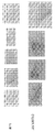


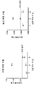


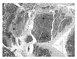

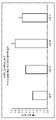

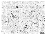


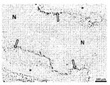


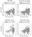
Classifications
-
- A—HUMAN NECESSITIES
- A61—MEDICAL OR VETERINARY SCIENCE; HYGIENE
- A61K—PREPARATIONS FOR MEDICAL, DENTAL OR TOILETRY PURPOSES
- A61K45/00—Medicinal preparations containing active ingredients not provided for in groups A61K31/00 - A61K41/00
- A61K45/06—Mixtures of active ingredients without chemical characterisation, e.g. antiphlogistics and cardiaca
-
- A—HUMAN NECESSITIES
- A61—MEDICAL OR VETERINARY SCIENCE; HYGIENE
- A61K—PREPARATIONS FOR MEDICAL, DENTAL OR TOILETRY PURPOSES
- A61K31/00—Medicinal preparations containing organic active ingredients
- A61K31/56—Compounds containing cyclopenta[a]hydrophenanthrene ring systems; Derivatives thereof, e.g. steroids
- A61K31/57—Compounds containing cyclopenta[a]hydrophenanthrene ring systems; Derivatives thereof, e.g. steroids substituted in position 17 beta by a chain of two carbon atoms, e.g. pregnane or progesterone
- A61K31/573—Compounds containing cyclopenta[a]hydrophenanthrene ring systems; Derivatives thereof, e.g. steroids substituted in position 17 beta by a chain of two carbon atoms, e.g. pregnane or progesterone substituted in position 21, e.g. cortisone, dexamethasone, prednisone or aldosterone
-
- A—HUMAN NECESSITIES
- A61—MEDICAL OR VETERINARY SCIENCE; HYGIENE
- A61K—PREPARATIONS FOR MEDICAL, DENTAL OR TOILETRY PURPOSES
- A61K35/00—Medicinal preparations containing materials or reaction products thereof with undetermined constitution
- A61K35/12—Materials from mammals; Compositions comprising non-specified tissues or cells; Compositions comprising non-embryonic stem cells; Genetically modified cells
- A61K35/14—Blood; Artificial blood
- A61K35/17—Lymphocytes; B-cells; T-cells; Natural killer cells; Interferon-activated or cytokine-activated lymphocytes
-
- A—HUMAN NECESSITIES
- A61—MEDICAL OR VETERINARY SCIENCE; HYGIENE
- A61K—PREPARATIONS FOR MEDICAL, DENTAL OR TOILETRY PURPOSES
- A61K39/00—Medicinal preparations containing antigens or antibodies
- A61K39/46—Cellular immunotherapy
- A61K39/461—Cellular immunotherapy characterised by the cell type used
- A61K39/4613—Natural-killer cells [NK or NK-T]
-
- A—HUMAN NECESSITIES
- A61—MEDICAL OR VETERINARY SCIENCE; HYGIENE
- A61K—PREPARATIONS FOR MEDICAL, DENTAL OR TOILETRY PURPOSES
- A61K39/00—Medicinal preparations containing antigens or antibodies
- A61K39/46—Cellular immunotherapy
- A61K39/462—Cellular immunotherapy characterized by the effect or the function of the cells
- A61K39/4621—Cellular immunotherapy characterized by the effect or the function of the cells immunosuppressive or immunotolerising
-
- A—HUMAN NECESSITIES
- A61—MEDICAL OR VETERINARY SCIENCE; HYGIENE
- A61K—PREPARATIONS FOR MEDICAL, DENTAL OR TOILETRY PURPOSES
- A61K39/00—Medicinal preparations containing antigens or antibodies
- A61K39/46—Cellular immunotherapy
- A61K39/464—Cellular immunotherapy characterised by the antigen targeted or presented
- A61K39/4643—Vertebrate antigens
- A61K39/46433—Antigens related to auto-immune diseases; Preparations to induce self-tolerance
-
- A—HUMAN NECESSITIES
- A61—MEDICAL OR VETERINARY SCIENCE; HYGIENE
- A61K—PREPARATIONS FOR MEDICAL, DENTAL OR TOILETRY PURPOSES
- A61K39/00—Medicinal preparations containing antigens or antibodies
- A61K39/46—Cellular immunotherapy
- A61K39/464—Cellular immunotherapy characterised by the antigen targeted or presented
- A61K39/4643—Vertebrate antigens
- A61K39/46434—Antigens related to induction of tolerance to non-self
-
- A—HUMAN NECESSITIES
- A61—MEDICAL OR VETERINARY SCIENCE; HYGIENE
- A61K—PREPARATIONS FOR MEDICAL, DENTAL OR TOILETRY PURPOSES
- A61K39/00—Medicinal preparations containing antigens or antibodies
- A61K39/46—Cellular immunotherapy
- A61K39/464—Cellular immunotherapy characterised by the antigen targeted or presented
- A61K39/4643—Vertebrate antigens
- A61K39/4644—Cancer antigens
-
- A—HUMAN NECESSITIES
- A61—MEDICAL OR VETERINARY SCIENCE; HYGIENE
- A61P—SPECIFIC THERAPEUTIC ACTIVITY OF CHEMICAL COMPOUNDS OR MEDICINAL PREPARATIONS
- A61P31/00—Antiinfectives, i.e. antibiotics, antiseptics, chemotherapeutics
-
- A—HUMAN NECESSITIES
- A61—MEDICAL OR VETERINARY SCIENCE; HYGIENE
- A61P—SPECIFIC THERAPEUTIC ACTIVITY OF CHEMICAL COMPOUNDS OR MEDICINAL PREPARATIONS
- A61P31/00—Antiinfectives, i.e. antibiotics, antiseptics, chemotherapeutics
- A61P31/12—Antivirals
- A61P31/14—Antivirals for RNA viruses
-
- A—HUMAN NECESSITIES
- A61—MEDICAL OR VETERINARY SCIENCE; HYGIENE
- A61P—SPECIFIC THERAPEUTIC ACTIVITY OF CHEMICAL COMPOUNDS OR MEDICINAL PREPARATIONS
- A61P31/00—Antiinfectives, i.e. antibiotics, antiseptics, chemotherapeutics
- A61P31/12—Antivirals
- A61P31/14—Antivirals for RNA viruses
- A61P31/16—Antivirals for RNA viruses for influenza or rhinoviruses
-
- A—HUMAN NECESSITIES
- A61—MEDICAL OR VETERINARY SCIENCE; HYGIENE
- A61P—SPECIFIC THERAPEUTIC ACTIVITY OF CHEMICAL COMPOUNDS OR MEDICINAL PREPARATIONS
- A61P35/00—Antineoplastic agents
-
- A—HUMAN NECESSITIES
- A61—MEDICAL OR VETERINARY SCIENCE; HYGIENE
- A61P—SPECIFIC THERAPEUTIC ACTIVITY OF CHEMICAL COMPOUNDS OR MEDICINAL PREPARATIONS
- A61P37/00—Drugs for immunological or allergic disorders
- A61P37/02—Immunomodulators
- A61P37/04—Immunostimulants
-
- C—CHEMISTRY; METALLURGY
- C12—BIOCHEMISTRY; BEER; SPIRITS; WINE; VINEGAR; MICROBIOLOGY; ENZYMOLOGY; MUTATION OR GENETIC ENGINEERING
- C12N—MICROORGANISMS OR ENZYMES; COMPOSITIONS THEREOF; PROPAGATING, PRESERVING, OR MAINTAINING MICROORGANISMS; MUTATION OR GENETIC ENGINEERING; CULTURE MEDIA
- C12N15/00—Mutation or genetic engineering; DNA or RNA concerning genetic engineering, vectors, e.g. plasmids, or their isolation, preparation or purification; Use of hosts therefor
- C12N15/09—Recombinant DNA-technology
- C12N15/11—DNA or RNA fragments; Modified forms thereof; Non-coding nucleic acids having a biological activity
- C12N15/62—DNA sequences coding for fusion proteins
- C12N15/625—DNA sequences coding for fusion proteins containing a sequence coding for a signal sequence
-
- C—CHEMISTRY; METALLURGY
- C12—BIOCHEMISTRY; BEER; SPIRITS; WINE; VINEGAR; MICROBIOLOGY; ENZYMOLOGY; MUTATION OR GENETIC ENGINEERING
- C12N—MICROORGANISMS OR ENZYMES; COMPOSITIONS THEREOF; PROPAGATING, PRESERVING, OR MAINTAINING MICROORGANISMS; MUTATION OR GENETIC ENGINEERING; CULTURE MEDIA
- C12N15/00—Mutation or genetic engineering; DNA or RNA concerning genetic engineering, vectors, e.g. plasmids, or their isolation, preparation or purification; Use of hosts therefor
- C12N15/09—Recombinant DNA-technology
- C12N15/63—Introduction of foreign genetic material using vectors; Vectors; Use of hosts therefor; Regulation of expression
- C12N15/79—Vectors or expression systems specially adapted for eukaryotic hosts
- C12N15/85—Vectors or expression systems specially adapted for eukaryotic hosts for animal cells
-
- C—CHEMISTRY; METALLURGY
- C12—BIOCHEMISTRY; BEER; SPIRITS; WINE; VINEGAR; MICROBIOLOGY; ENZYMOLOGY; MUTATION OR GENETIC ENGINEERING
- C12N—MICROORGANISMS OR ENZYMES; COMPOSITIONS THEREOF; PROPAGATING, PRESERVING, OR MAINTAINING MICROORGANISMS; MUTATION OR GENETIC ENGINEERING; CULTURE MEDIA
- C12N5/00—Undifferentiated human, animal or plant cells, e.g. cell lines; Tissues; Cultivation or maintenance thereof; Culture media therefor
- C12N5/06—Animal cells or tissues; Human cells or tissues
- C12N5/0602—Vertebrate cells
- C12N5/0634—Cells from the blood or the immune system
- C12N5/0646—Natural killers cells [NK], NKT cells
-
- A—HUMAN NECESSITIES
- A61—MEDICAL OR VETERINARY SCIENCE; HYGIENE
- A61K—PREPARATIONS FOR MEDICAL, DENTAL OR TOILETRY PURPOSES
- A61K2239/00—Indexing codes associated with cellular immunotherapy of group A61K39/46
- A61K2239/31—Indexing codes associated with cellular immunotherapy of group A61K39/46 characterized by the route of administration
-
- A—HUMAN NECESSITIES
- A61—MEDICAL OR VETERINARY SCIENCE; HYGIENE
- A61K—PREPARATIONS FOR MEDICAL, DENTAL OR TOILETRY PURPOSES
- A61K2239/00—Indexing codes associated with cellular immunotherapy of group A61K39/46
- A61K2239/38—Indexing codes associated with cellular immunotherapy of group A61K39/46 characterised by the dose, timing or administration schedule
-
- C—CHEMISTRY; METALLURGY
- C12—BIOCHEMISTRY; BEER; SPIRITS; WINE; VINEGAR; MICROBIOLOGY; ENZYMOLOGY; MUTATION OR GENETIC ENGINEERING
- C12N—MICROORGANISMS OR ENZYMES; COMPOSITIONS THEREOF; PROPAGATING, PRESERVING, OR MAINTAINING MICROORGANISMS; MUTATION OR GENETIC ENGINEERING; CULTURE MEDIA
- C12N2501/00—Active agents used in cell culture processes, e.g. differentation
- C12N2501/30—Hormones
- C12N2501/38—Hormones with nuclear receptors
- C12N2501/39—Steroid hormones
-
- C—CHEMISTRY; METALLURGY
- C12—BIOCHEMISTRY; BEER; SPIRITS; WINE; VINEGAR; MICROBIOLOGY; ENZYMOLOGY; MUTATION OR GENETIC ENGINEERING
- C12N—MICROORGANISMS OR ENZYMES; COMPOSITIONS THEREOF; PROPAGATING, PRESERVING, OR MAINTAINING MICROORGANISMS; MUTATION OR GENETIC ENGINEERING; CULTURE MEDIA
- C12N2800/00—Nucleic acids vectors
- C12N2800/10—Plasmid DNA
- C12N2800/106—Plasmid DNA for vertebrates
- C12N2800/107—Plasmid DNA for vertebrates for mammalian
Abstract
본 발명은 림프구의 신규 집단, 이들을 생산하는 방법, 및 질환의 치료에서 이의 용도에 관한 것이다. The present invention relates to novel populations of lymphocytes, methods of producing them, and their use in the treatment of diseases.
Description
본 개시내용은 림프구 및 면역 세포의 신규 집단, 이들을 생성하는 방법, 및 질환 치료에서의 이의 용도에 관한 것이다. 보다 구체적으로, 본 개시내용은 고용량 글루코코르티코이드 및 글루코코르티코이드 수용체 작용제를 사용하여 자연 살해 T 세포 (NKT 세포), T 세포, 및 수지상 세포의 신규 집단을 생산하는 방법에 관한 것이다.The present disclosure relates to novel populations of lymphocytes and immune cells, methods of generating them, and their use in the treatment of diseases. More specifically, the present disclosure relates to methods of producing new populations of natural killer T cells (NKT cells), T cells, and dendritic cells using high doses of glucocorticoids and glucocorticoid receptor agonists.
본 저자들은 이전에 고농도의 글루코코르티코이드가 적응 T 세포 요법과 같은 세포 면역 요법의 효능을 향상시키기 위해 환자를 조절하는 데 사용될 수 있음을 발견했고; 국제 특허 출원 PCT/US2018/025517 (WO2018/183927로 공개됨)에 기재되어 있다. 해당 출원에서, 저자는 화학 요법 및 방사선 매개 전처리와 관련된 독성에 주목했으며, 이는 비장의 세포성을 비선택적으로 파괴하는 것으로 믿어진다. 저자는 세포 면역 요법을 받는 암 환자에게 혜택을 주기 위해 글루코코르티코이드 (스테로이드의 하위 분류) 및 다른 비독성 림프구 감소제를 급성 용량으로 제공했다.We previously found that high concentrations of glucocorticoids could be used to modulate patients to enhance the efficacy of cellular immunotherapy, such as adaptive T cell therapy; International patent application PCT/US2018/025517 published as WO2018/183927. In that application, the authors noted the toxicity associated with chemotherapy and radiation-mediated pretreatment, which is believed to non-selectively destroy the cellularity of the spleen. The authors provided acute doses of glucocorticoids (a subclass of steroids) and other non-toxic lymphopenic agents to benefit cancer patients receiving cellular immunotherapy.
국제 특허 출원 PCT/US2019/054395에서 본 저자들은 또한 다른 세포의 세포 수에 실질적으로 영향을 미치지 않으면서 말초 혈액 림프구의 림프 고갈을 유발하기 위해 고농도의 글루코코르티코이드의 사용을 기술하였다. 해당 출원에서, 저자는 고농도의 글루코코르티코이드가 예를 들어 당뇨병 자가면역을 담당하는 섬 특이적 자가반응성 T 세포를 포함하는 말초 혈액 림프구를 고갈시킬 수 있지만 호중구, 혈소판, 적혈구 및 줄기 세포 (HSC 및 MSC 모두)는 보존한다고 보고했다. 저자는 화학요법에 필적하는 효능으로 안전한 면역학적 재설정을 수행할 수 있는 비골수파괴 요법으로 글루코코르티코이드를 제공했다.In International Patent Application PCT/US2019/054395, the authors also described the use of high concentrations of glucocorticoids to induce lymphatic depletion of peripheral blood lymphocytes without substantially affecting the cell number of other cells. In that application, the authors noted that high concentrations of glucocorticoids can deplete peripheral blood lymphocytes, including, for example, islet-specific autoreactive T cells responsible for diabetic autoimmunity, but neutrophils, platelets, red blood cells and stem cells (HSCs and MSCs). all) reported to preserve. The authors have offered glucocorticoids as a non-myelodestructive therapy that can perform safe immunological reprogramming with efficacy comparable to chemotherapy.
- - -- - -
세포독성 화학요법 사용을 줄이는 것이 국립암연구소의 최우선 목표이다. 종종 고형 종양이라고 불리는 암종은 전체 암의 80-90%를 차지하지만 새로운 암 치료법 개발로 표적화하기 어려운 것으로 입증되었다. 키메라 항원 수용체 (CAR) T-세포 요법은 CD-19 발현 B-세포 급성 림프구성 백혈병의 치료에서 놀라운 성공을 보여주었다. 그러나, 고형 종양에 대한 CAR T 세포 요법을 제한하는 많은 장애물이 있다: 종양으로의 비효율적인 트래피킹과 고형 종양의 면역억제성 미세 환경은 T 세포 효능을 제한한다. 또한, CAR T 요법은 사이토카인 방출 증후군 (CRS), 신경부종, 및 이식편대숙주병 (GvHD)을 비롯한 심각한 부작용과 관련이 있다.Reducing the use of cytotoxic chemotherapy is a top priority for the National Cancer Institute. Carcinomas, often called solid tumors, account for 80-90% of all cancers, but have proven difficult to target with the development of new cancer therapies. Chimeric antigen receptor (CAR) T-cell therapy has shown remarkable success in the treatment of CD-19 expressing B-cell acute lymphocytic leukemia. However, there are many obstacles limiting CAR T cell therapy for solid tumors: inefficient trafficking to the tumor and the immunosuppressive microenvironment of solid tumors limit T cell efficacy. In addition, CAR T therapy has been associated with serious side effects including cytokine release syndrome (CRS), neuroedema, and graft-versus-host disease (GvHD).
자연 살해 T 세포 (NKT)는 T 세포와 자연 살해 (NK) 세포의 특성을 공유하는 이종성 T 세포 그룹이다. 기존의 T 세포와 달리 NKT는 흉선에서 나올 때 기능적으로 성숙하고, 빠른 사이토카인 생산을 위해 프라이밍된다. NKT는 CD1d 발현 암세포 및 종양 미세환경 대식세포를 직접 죽이고, IFN감마 및 IL-4와 같은 면역 활성화 사이토카인을 빠르게 생성 및 방출하며, 수지상 세포 (DC), NK 세포, 및 B 및 T 림프구와 같은 다른 면역 세포를 활성화할 수 있다. 임상적으로, 불변 NKT (iNKT)는 '자가 배양 활성화 iNKT'의 주사, 알파 Gal Cer (NKT 활성화제) 로딩된 수지상 세포 또는 단핵구를 투여하여 내인성 NKT를 활성화하거나, 알파 Gal Cer의 합성 유사체인 KRN7000과 같은 NKT 활성화제 항체 또는 리간드를 투여함으로써 다양한 상이한 암에 대해 사용될 수 있다.Natural killer T cells (NKTs) are a heterogeneous group of T cells that share the characteristics of T cells and natural killer (NK) cells. Unlike conventional T cells, NKTs mature functionally when they emerge from the thymus and are primed for rapid cytokine production. NKT directly kills CD1d-expressing cancer cells and tumor microenvironment macrophages, rapidly produces and releases immune-activating cytokines such as IFNgamma and IL-4, dendritic cells (DC), NK cells, and B and T lymphocytes. It can activate other immune cells. Clinically, invariant NKT (iNKT) has been shown to activate endogenous NKT by injection of 'autologous culture activated iNKT', administration of alpha Gal Cer (NKT activator) loaded dendritic cells or monocytes, or KRN7000, a synthetic analogue of alpha Gal Cer. It can be used against a variety of different cancers by administering NKT activator antibodies or ligands such as
그러나, iNKT 생성을 유도하는 데 사용되는 이러한 방법 중 어느 것도 암 환자에게 효과적인 것으로 입증되지 않았다; iNKT 수준은 암 환자에서 감소했으며 임상 시험은 실망스러웠다. iNKT 수준은 노인에서도 유사하게 낮다 (Tarazona et al, 2003, 그 전체가 본원에 참고로 포함됨). 흑색종에서 '자가 배양 활성화 NKT'의 사용은 9명의 환자 중 3명에서 효과적이었으며, 결과는 종양 침윤 NKT의 수와 직접 관련이 있었다 (Wolf et al, 2018 및 Nair et al, 2017, 그 전체가 본원에 참고로 포함됨). 그러나, 이 접근 방식은 암 환자의 NKT 수가 적고, IFN 감마 1형과 종양 촉진 IL-4 2형 사이를 이동하는 iNKT의 가소성으로 인해 제한되었다.However, none of these methods used to induce iNKT production have proven effective in cancer patients; iNKT levels decreased in cancer patients and clinical trials were disappointing. iNKT levels are similarly low in the elderly (Tarazona et al, 2003, incorporated herein by reference in its entirety). The use of 'autologous activated NKT' in melanoma was effective in 3 out of 9 patients, and the outcome was directly related to the number of tumor-infiltrating NKTs (Wolf et al , 2018 and Nair et al , 2017, all of them). incorporated herein by reference). However, this approach was limited by the small number of NKTs in cancer patients and the plasticity of iNKTs migrating between IFN-
암 치료에서, 키나제 억제제 (KI)는 기존의 세포독성 화학요법에 비해 내약성이 우수하다. 그러나, 피로, 고혈압, 발진, 상처 치유 장애, 골수 억제, 및 설사, 갑상선 기능, 뼈 대사, 선형 성장, 생식선 기능, 태아 발달, 부신 기능, 및 포도당 대사의 이상을 포함하는 상당한 독성이 키나제 억제제와 여전히 연관되어 있다. 많은 환자들이 KI의 독성 때문에 용량 감량이 필요하며, 이는 만성적으로 복용해야 한다 (Lodisch et al, 2013, 그 전체가 본원에 참고로 포함됨). 또한, KI에 대한 내성은 일반적이며 치료에 따라 시간에 따라 달라진다 (Bhullar 2018, 그 전체가 본원에 참고로 포함됨). In cancer treatment, kinase inhibitors (KI) are well tolerated compared to conventional cytotoxic chemotherapy. However, significant toxicity, including fatigue, hypertension, rash, impaired wound healing, bone marrow suppression, and diarrhea, abnormalities of thyroid function, bone metabolism, linear growth, gonad function, fetal development, adrenal function, and glucose metabolism, is associated with kinase inhibitors. are still related Many patients require dose reduction due to the toxicity of KI, which must be taken chronically (Lodisch et al , 2013, incorporated herein by reference in its entirety). In addition, resistance to KI is general and time-dependent with treatment (Bhullar 2018, incorporated herein by reference in its entirety).
암 치료와 관련된 독성을 줄이려는 노력에도 불구하고, 이러한 독성을 관리하기 위한 물리적 비용과 의료 비용은 여전히 중요한 문제로 남아 있다. 예를 들어, 혈액암 환자의 최대 41%가 이들 약물과 관련된 물리적 및 재정적 독성 때문에 새로운 키나제/프로테아좀 억제제 또는 생물학적 제제 복용을 중단하기로 선택한다 (Mato 2018, Kadri 2017, Mato 2016 및 Barrett 2010, 각각 그 전체가 본원에 참고로 포함됨). Despite efforts to reduce the toxicities associated with cancer treatment, the physical and medical costs of managing these toxicities remain significant. For example, up to 41% of blood cancer patients choose to stop taking a new kinase/proteasome inhibitor or biologic because of the physical and financial toxicity associated with these drugs (Mato 2018, Kadri 2017, Mato 2016 and Barrett 2010) , each incorporated herein by reference in its entirety).
T 세포는 면역 반응에서 중요한 역할을 하는 일종의 림프구이다. T 세포는 세포 표면에 T 세포 수용체가 있다는 점에서 다른 유형의 림프구와 구별된다. T-세포 수용체 (TCR)는 주요 조직적합성 복합체 (MHC) 분자에 결합된 항원 단편을 인식하는 역할을 하며 두 개의 서로 다른 단백질 사슬의 이종이량체이다. 인간에서 T 세포의 95%에서 TCR은 알파 (α) 사슬 및 베타 (β) 사슬 (각각, TRA 및 TRB에 의해 암호화됨)로 구성되는 반면, T 세포의 5%에서 TCR은 감마 및 델타 (γ/δ) 사슬 (각각, TRG 및 TRD에 의해 암호화됨)로 구성된다. 이 비율은 병에 걸린 상태 (예를 들어 백혈병)에서 달라진다.T cells are a type of lymphocytes that play an important role in the immune response. T cells are distinguished from other types of lymphocytes in that they have T cell receptors on the cell surface. The T-cell receptor (TCR) is responsible for recognizing antigen fragments bound to major histocompatibility complex (MHC) molecules and is a heterodimer of two different protein chains. In 95% of T cells in humans, the TCR consists of an alpha (α) chain and a beta (β) chain (encoded by TRA and TRB, respectively), whereas in 5% of T cells the TCR is composed of gamma and delta (γ) /δ) chains (encoded by TRG and TRD, respectively). This rate varies in diseased conditions (eg leukemia).
MHC-제한된 알파 베타 T 세포와 대조적으로, 감마 델타 T 세포는 활성화를 위해 항원 처리 및 펩티드 에피토프의 주요 조직적합성 복합체 (MHC) 제시가 필요하지 않지만, 일부는 MHC 클래스 Ib 분자를 인식한다. 일부 감마 델타 T 세포는 감염 또는 종양 형성으로 인한 세포 스트레스의 마커를 인식한다. 감마 델타 T 세포는 지질 항원을 인식하는 역할을 하는 것으로 여겨진다.In contrast to MHC-restricted alpha beta T cells, gamma delta T cells do not require antigen processing and major histocompatibility complex (MHC) presentation of peptide epitopes for activation, although some recognize MHC class Ib molecules. Some gamma delta T cells recognize markers of cellular stress due to infection or tumorigenesis. Gamma delta T cells are believed to play a role in recognizing lipid antigens.
감마 델타 T 세포는 사이토카인 (IFN-γ, TNF-α, IL-17) 및 케모카인 (RANTES, IP-10, 림포탁틴)의 생성에 의한 감염/형질전환된 세포의 인식, 감염 또는 형질전환된 표적 세포 (퍼포린, 그랜자임, TRAIL)의 세포 용해, 및 다른 세포와의 상호작용 후 광범위한 기능적 가소성을 나타낸다. 감마 델타 T 세포는 MHC-비제한된 방식으로 다양한 암을 인식 및 용해할 수 있고, 감염성 질환에서 보호 기능을 갖고, 다양한 감염성 질환의 진행 및 예후와 관련이 있는 것으로 나타났다 (Gogoi et al, 2013; Pauza et al, 2018; Zheng et al, 2012; Dong et al, 2018; Zhao et al 2018; 모두 그 전체가 본원에 참고로 포함됨). 일부 감마 델타 T 세포는 또한 상황에 따라 항원 제시 세포로 작용할 수도 있다 (Himoudi et al, 2012). 따라서 감마 델타 T 세포는 면역 요법 개발에서 상당한 관심을 받고 있다.Gamma delta T cells are recognized, infected or transformed by the production of cytokines (IFN-γ, TNF-α, IL-17) and chemokines (RANTES, IP-10, lymphotactin). It exhibits extensive functional plasticity after cell lysis of target cells (Perforin, Granzyme, TRAIL) and interaction with other cells. Gamma delta T cells can recognize and lyse various cancers in an MHC-unlimited manner, have protective functions in infectious diseases, and have been shown to be associated with the progression and prognosis of various infectious diseases (Gogoi et al, 2013; Pauza). et al, 2018; Zheng et al, 2012; Dong et al, 2018; Zhao et al 2018; all incorporated herein by reference in their entirety). Some gamma delta T cells may also act as antigen presenting cells in some circumstances (Himoudi et al, 2012). Therefore, gamma delta T cells are of considerable interest in immunotherapeutic development.
수지상 세포는 골수 유래 백혈구이며, 포유동물 면역계의 가장 강력한 항원 제시 세포이다. 수지상 세포는 흔히 통상적인 수지상 세포 (cDC) 및 형질세포양 수지상 세포 (pDC) 서브셋으로 분류된다. 수지상 세포는 주로 "미성숙" 및 "성숙"의 두 가지 기본 기능 상태로 존재한다. 수지상 세포의 활성화 (성숙) 대사, 세포 및 유전자 전사 프로그램을 켜서 DC가 말초 조직에서 T 림프구 활성화 항원 제시가 발생할 수 있는, 2차 림프 기관의 T 의존 영역으로 이동할 수 있도록 한다 (Patente et al, 2018; 그 전체가 여기에 참고로 포함됨). Dendritic cells are bone marrow-derived leukocytes and are the most potent antigen presenting cells of the mammalian immune system. Dendritic cells are often classified into common dendritic cell (cDC) and plasmacytoid dendritic cell (pDC) subsets. Dendritic cells mainly exist in two basic functional states: "immature" and "mature". Turn on the activating (mature) metabolic, cellular and gene transcriptional programs of dendritic cells, allowing DCs to migrate from peripheral tissues to T-dependent regions of secondary lymphoid organs where T lymphocyte-activated antigen presentation can occur (Patente et al, 2018) ; incorporated herein by reference in its entirety).
수지상 세포의 주요 기능은 항원 물질을 처리하고 이를 세포 표면에서 T 세포에 제시하여 적응 면역 반응을 시작하는 것이다. 수지상 세포는 또한 병원체 특이적 효과기 T 세포 분화 및 활성화를 촉진하는 극성화 사이토카인을 생성하고, 조절 T 세포의 분화를 유도하는 관용원성 사이토카인을 분비함으로써 자가 관용을 촉진할 수 있다. 이러한 면역 조절 기능의 관점에서, 수지상 세포는 암, 자가면역 질환 및 감염을 포함한 상태의 치료를 위한 면역요법 개발에서 상당한 관심을 갖고 있다. 예를 들어, CD11b 양성 수지상 세포는 인플루엔자 A (H1N1) 감염, 및 호흡기 세포융합 바이러스의 중증도 감소, 또는 보호와 관련이 있다 (Lee et al, 2018; Malloy et al, 2017; 모두 그 전체가 여기에 참고로 포함됨). The main function of dendritic cells is to process antigenic substances and present them to T cells on the cell surface to initiate an adaptive immune response. Dendritic cells can also promote self-tolerance by producing polarizing cytokines that promote pathogen-specific effector T cell differentiation and activation, and secreting tolerogenic cytokines that induce the differentiation of regulatory T cells. In view of their immunomodulatory function, dendritic cells are of considerable interest in the development of immunotherapy for the treatment of conditions including cancer, autoimmune diseases and infections. For example, CD11b positive dendritic cells are associated with influenza A (H1N1) infection, and reduced severity, or protection, of respiratory syncytial virus (Lee et al, 2018; Malloy et al, 2017; all here in its entirety) incorporated by reference).
- - -- - -
코로나바이러스 질환 2019 (COVID-19)는 중증 급성 호흡기 증후군 코로나바이러스 2 (SARS-CoV-2)에 의해 유발되는 감염병이다. 대부분의 경우 가벼운 증상 (발열, 기침 및 숨가쁨을 포함할 수 있음)이 나타나지만, 일부는 바이러스성 폐렴 및 다기관 부전으로 진행된다. COVID-19 발병은 2020년 3월 세계보건기구 (WHO)의해 팬데믹이 선언됐다. 2020년 4월 기준으로, 전 세계적으로 확인된 사례 수가 100만 명을 넘어섰고, 그로 인해 50,000명 이상이 사망했다. 2020년 4월 기준으로, COVID-19에 대한 백신이나 특정 항바이러스 치료제는 존재하지 않으며, 질환 관리는 증상 치료 및 지지 요법에 중점을 두고 있다.Coronavirus disease 2019 (COVID-19) is an infectious disease caused by severe acute respiratory syndrome coronavirus 2 (SARS-CoV-2). In most cases, mild symptoms (which may include fever, cough, and shortness of breath) develop, but some progress to viral pneumonia and multiorgan failure. The COVID-19 outbreak was declared a pandemic by the World Health Organization (WHO) in March 2020. As of April 2020, there have been over 1 million confirmed cases worldwide, resulting in over 50,000 deaths. As of April 2020, there is no vaccine or specific antiviral treatment for COVID-19, and disease management is focused on symptomatic treatment and supportive care.
- - -- - -
현재 이용 가능한 치료법보다 더 안전하고 더 적은 독성 및/또는 더 큰 효능과 관련된 암, 자가면역 장애, 및 감염성 (미생물이라고도 함) 질환에 대한 추가 치료에 대한 필요성이 존재한다. 더 간단하고 독성이 적으며 비용이 덜 드는 치료가 필요하다.There is a need for additional treatments for cancer, autoimmune disorders, and infectious (also called microbial) diseases that are safer and are associated with less toxicity and/or greater efficacy than currently available therapies. A simpler, less toxic and less expensive treatment is needed.
본 발명은 고용량의 글루코코르티코이드가 많은 유형의 말초혈액 림프구의 림프고갈을 유발하는 작용을 하지만, 자연 살해 T (NKT) 세포의 신규 집단의 생산/활성화/동원을 유도한다는 놀라운 발견에 기초한다. 알려진 NKT 세포의 특성을 제시하는 것 외에도, 이 NKT 세포의 신규 집단은 암세포를 직접 삼킬 수 있으므로 고형암에 대한 치료제로서 고농도의 글루코코르티코이드의 가능성을 확장한다. The present invention is based on the surprising discovery that high doses of glucocorticoids act to induce lympho-depletion of many types of peripheral blood lymphocytes, but induce the production/activation/mobilization of new populations of natural killer T (NKT) cells. In addition to presenting known NKT cell properties, this novel population of NKT cells can directly engulf cancer cells, thus expanding the potential of high concentrations of glucocorticoids as therapeutics for solid cancers.
본 저자들은 또한 고용량 투여 후, 글루코코르티코이드 분자가 ICAM3와 같은 세포간 접착 분자에 결합하고 차단할 수 있음을 발견했다. 결합은 협력적이며 최대 26개의 분자가 ICAM3의 첫 번째 Ig 도메인에 결합한다. ICAM3는 림프구, 단핵구 및 호중구와 같은 세포뿐만 아니라 흑색종 및 골육종과 같은 암세포 유형에서 상당한 수준으로 발현된다.We also found that, after high dose administration, glucocorticoid molecules can bind and block intercellular adhesion molecules such as ICAM3. Binding is cooperative and up to 26 molecules bind to the first Ig domain of ICAM3. ICAM3 is expressed at significant levels in cells such as lymphocytes, monocytes and neutrophils, as well as cancer cell types such as melanoma and osteosarcoma.
따라서, 제1 양태에서, 본 발명은 자연 살해 T 세포 (NKT 세포)의 집단을 생산하는 방법을 제공하고, 상기 방법은 글루코코르티코이드 수용체 (GR) 조절제 또는 ICAM3 조절제 (덱사메타손과 같은 글루코코르티코이드일 수 있음)를 덱사메타손 염기의 대략 적어도 6 mg/kg 인간 등가 용량 (HED)과 동등한 용량으로 대상체에게 투여하는 것을 포함하고, 상기 글루코코르티코이드는 대상체에서 NKT 세포의 집단을 유도한다. 본 발명의 NKT 세포는 마커 발현의 신규 패턴을 나타낸다. 일부 구현예에서, NKT 세포의 집단은 적어도 60, 70, 80, 90, 95, 96, 97, 98, 또는 99 %의 세포가 CD3, CD4, CD8, CD45, CD49b, CD56, CD62L, NK1.1, Ly6G, Sca1, 및/또는 TCR 감마/델타를 발현하고/하거나; C-kit, B220, FoxP3, 및/또는 TCR 알파/베타를 발현하지 않는 것을 특징으로 한다. 일부 구현예에서, 상기 NKT 세포는 CD3, CD4, CD45, CD49b, CD56, CD62L, NK1.1, Ly6G, 및 Sca1을 발현한다. 일부 구현예에서, 상기 NKT 세포는 CD3, CD4, CD45, CD56, CD62L, NK1.1, Ly6G, 및 Sca1을 발현한다. 일부 구현예에서, 상기 NKT 세포는 CD3, CD4, CD45, CD49b, CD62L, NK1.1, Ly6G, 및 Sca1을 발현한다. 일부 구현예에서, 상기 NKT 세포는 CD3, CD4, CD45, CD56, CD62L, NK1.1, 및 Ly6G를 발현한다. 일부 구현예에서, 상기 NKT 세포는 CD3, CD4, CD45, CD49b, CD62L, NK1.1, 및 Ly6G를 발현한다. 일부 구현예에서, NKT 세포의 집단은 적어도 60, 70, 80, 90, 95, 96, 97, 98, 또는 99 %의 세포가 CD3, CD4, CD8, CD45, CD49b, CD56, CD62L, NK1.1, Ly6G, 및/또는 TCR 감마/델타 발현하고/하거나; C-kit, B220, FoxP3, 및/또는 TCR 알파/베타를 발현하지 않는 것을 특징으로 한다. 일부 구현예에서, NKT 세포의 집단은 적어도 60, 70, 80, 90, 95, 96, 97, 98, 또는 99 %의 세포가 CD3, CD4, CD8, CD45, CD56, CD62L, NK1.1, Ly6G, 및/또는 TCR 감마/델타를 발현하고/하거나; C-kit, B220, FoxP3, 및/또는 TCR 알파/베타를 발현하지 않는 것을 특징으로 한다. 일부 구현예에서, 상기 NKT 세포는 C-kit, B220, FoxP3, 또는 TCR 알파/베타를 발현하지 않는다. 일부 구현예에서, 상기 NKT 세포는 Sca1을 발현하지 않는다. 상기 NKT 세포는 CD8을 발현할 수 있다. 상기 NKT 세포는 CD8을 발현할 수 있다. 상기 NKT 세포는 CD4를 발현할 수 있다. 상기 NKT 세포는 CD4를 발현할 수 있다. 상기 NKT 세포는 CD4 및 CD8을 발현할 수 있고/거나; Ly6G를 발현할 수 있다. Accordingly, in a first aspect, the present invention provides a method for producing a population of natural killer T cells (NKT cells), wherein the method may be a glucocorticoid receptor (GR) modulator or an ICAM3 modulator (a glucocorticoid such as dexamethasone) ) to the subject at a dose equivalent to approximately at least 6 mg/kg human equivalent dose (HED) of dexamethasone base, wherein the glucocorticoid induces a population of NKT cells in the subject. The NKT cells of the present invention exhibit novel patterns of marker expression. In some embodiments, the population of NKT cells comprises at least 60, 70, 80, 90, 95, 96, 97, 98, or 99% of the cells are CD3, CD4, CD8, CD45, CD49b, CD56, CD62L, NK1.1 , Ly6G, Sca1, and/or TCR gamma/delta; It is characterized in that it does not express C-kit, B220, FoxP3, and/or TCR alpha/beta. In some embodiments, the NKT cells express CD3, CD4, CD45, CD49b, CD56, CD62L, NK1.1, Ly6G, and Sca1. In some embodiments, the NKT cells express CD3, CD4, CD45, CD56, CD62L, NK1.1, Ly6G, and Sca1. In some embodiments, the NKT cells express CD3, CD4, CD45, CD49b, CD62L, NK1.1, Ly6G, and Sca1. In some embodiments, the NKT cells express CD3, CD4, CD45, CD56, CD62L, NK1.1, and Ly6G. In some embodiments, the NKT cells express CD3, CD4, CD45, CD49b, CD62L, NK1.1, and Ly6G. In some embodiments, the population of NKT cells comprises at least 60, 70, 80, 90, 95, 96, 97, 98, or 99% of the cells are CD3, CD4, CD8, CD45, CD49b, CD56, CD62L, NK1.1 , Ly6G, and/or TCR gamma/delta expression; It is characterized in that it does not express C-kit, B220, FoxP3, and/or TCR alpha/beta. In some embodiments, the population of NKT cells comprises at least 60, 70, 80, 90, 95, 96, 97, 98, or 99% of the cells CD3, CD4, CD8, CD45, CD56, CD62L, NK1.1, Ly6G , and/or expressing TCR gamma/delta; It is characterized in that it does not express C-kit, B220, FoxP3, and/or TCR alpha/beta. In some embodiments, the NKT cell does not express C-kit, B220, FoxP3, or TCR alpha/beta. In some embodiments, the NKT cells do not express Sca1. The NKT cells may express CD8. The NKT cells may express CD8. The NKT cells may express CD4. The NKT cells may express CD4. said NKT cells may express CD4 and CD8; Ly6G can be expressed.
일부 바람직한 구현예에서 본 개시내용의 NKT 세포는 CD3, CD45, 및/또는 CD56을 발현할 수 있다. 일부 이러한 구현예에서, 본 개시내용의 NKT 세포는 CD3+/밝음 또는 CD3+/매우 밝음, 및/또는 CD45+/어두움, 및/또는 CD56+일 수 있다.In some preferred embodiments the NKT cells of the present disclosure are capable of expressing CD3, CD45, and/or CD56. In some such embodiments, the NKT cells of the present disclosure may be CD3+/bright or CD3+/very bright, and/or CD45+/dark, and/or CD56+.
NKT 세포는 다음과 같이 설명될 수 있다NKT cells can be described as
![]()
![]()
![]()
![]()
![]()
![]()
![]()
![]()
![]()
![]()
![]()
![]()
![]()
![]()
![]()
![]()
![]()
![]()
![]()
![]()
NKT 세포는 나이브 대상체에서 이러한 특성을 갖는 것으로 기술될 수 있다. NKT 세포는 종양/암성 또는 자가면역 상태에서 이러한 특성을 갖는 것으로 기술될 수 있다.NKT cells can be described as having these properties in naive subjects. NKT cells can be described as having these properties in a tumor/cancerous or autoimmune state.
세포 마커의 발현 수준은 글루코코르티코이드 수용체 (GR) 조절제 또는 ICAM3 조절제와 접촉되지 않은 공통 공급원으로부터 유래된 참조 NKT 세포 집단의 평균 발현 수준과 관련하여 결정할 수 있다. 마커의 발현은 예를 들어 본원에 기술된 장비, 시약 및/또는 조건 (단독 또는 조합으로 취함)을 사용하여 수행되는 유세포 분석에 의해 측정될 수 있다. 글루코코르티코이드 수용체 (GR) 조절제 또는 ICAM3 조절제는 글루코코르티코이드일 수 있다. 일부 구현예에서, 상기 글루코코르티코이드는 덱사메타손, 히드로코르티손, 메틸프레드니솔론, 프레드니손, 프레드니솔론, 프레드닐리덴, 코르티손, 부데소니드, 베타메타손, 플루메타손 및 베클로메타손으로 이루어진 군으로부터 선택된다.The expression level of a cellular marker can be determined in relation to the average expression level of a reference NKT cell population derived from a common source not contacted with a glucocorticoid receptor (GR) modulator or an ICAM3 modulator. Expression of a marker can be measured, for example, by flow cytometry performed using the equipment, reagents and/or conditions described herein (taken alone or in combination). The glucocorticoid receptor (GR) modulator or ICAM3 modulator may be a glucocorticoid. In some embodiments, the glucocorticoid is selected from the group consisting of dexamethasone, hydrocortisone, methylprednisolone, prednisone, prednisolone, prednylidene, cortisone, budesonide, betamethasone, flumethasone, and beclomethasone.
바람직한 구현예에서, 상기 글루코코르티코이드는 덱사메타손, 베타메타손, 및 메틸프레드니손 (바람직하게는 덱사메타손 또는 베타메타손)으로 이루어진 군으로부터 선택된다.In a preferred embodiment, the glucocorticoid is selected from the group consisting of dexamethasone, betamethasone, and methylprednisone (preferably dexamethasone or betamethasone).
일부 구현예에서, 상기 글루코코르티코이드는 덱사메타손 염기, 덱사메타손 소듐 포스페이트, 덱사메타손 헤미숙시네이트, 덱사메타손 나트륨 숙시네이트, 덱사메타손 숙시네이트, 덱사메타손 이소니코티네이트, 덱사메타손-21-아세테이트, 덱사메타손 포스페이트, 덱사메타손-21-포스페이트, 덱사메타손 테뷰테이트, 덱사메타손-17-발레레이트, 덱사메타손 아세테이트 일수화물, 덱사메타손 피발레이트, 덱사메타손 팔미테이트, 덱사메타손-21-팔미테이트, 덱사메타손 디프로피오네이트, 덱사메타손 프로피오네이트, 덱사메타손 아세테이트 무수물, 덱사메타손-21-페닐프로피오네이트, 덱사메타손-21-설포벤조에이트, 덱사메타손 헤모-설페이트, 덱사메타손 설페이트, 덱사메타손 벨록실, 덱사메타손산, 덱사메타손 아세퓨레이트, 덱사메타손 카르복시미드, 덱사메타손 시페실레이트, 덱사메타손 21-포스페이트 이나트륨염, 덱사메타손 메실레이트, 덱사메타손 리놀레이트, 덱사메타손 글루코시드, 덱사메타손 글루쿠로니드, 덱사메타손 요오드아세테이트, 덱사메타손 옥세타논, 카르복시메틸티오-덱사메타손, 덱사메타손비스에톡심, 덱사메타손 에폭시드, 덱사메타손리놀레레이데이트, 덱사메타손 메틸오르쏘발레레이트, 덱사메타손 스페르민, 6-히드록시 덱사메타손, 덱사메타손 트리부틸아세테이트, 덱사메타손 아스파르트산, 덱사메타손 갈락토피라노스, 덱사메타손 염산염, 히드록시 덱사메타손, 카르복시 덱사메타손, 데스옥시 덱사메타손, 덱사메타손 부타존, 덱사메타손 시클로덱스트린, 디히드로 덱사메타손, 옥소 덱사메타손, 프로피오닐옥시 덱사메타손, 덱사메타손 갈락토다이, 덱사메타손 이소니코티네이트, 덱사메타손 인산수소나트륨, 덱사메타손 알데하이드, 덱사메타손 피블레이트, 덱사메타손 트리데실레이트, 덱사메타손 크로토네이트, 덱사메타손 메탄술포네이트, 덱사메타손 부틸아세테이트, 데히드로 덱사메타손, 덱사메타손 이소티오시아나토에틸 티오에테르, 덱사메타손 브로모아세테이트, 덱사메타손 헤미글루타레이트, 데옥시 덱사메타손, 덱사메타손 클로람부실레이트, 덱사메타손 멜팔라네이트, 포르밀옥시 덱사메타손, 덱사메타손 부티레이트, 덱사메타손 라우레이트, 덱사메타손 아세테이트, 및 덱사메타손 형태를 함유하는 임의의 조합 치료로 이루어진 군으로부터 선택된다.In some embodiments, the glucocorticoid is dexamethasone base, dexamethasone sodium phosphate, dexamethasone hemisuccinate, dexamethasone sodium succinate, dexamethasone succinate, dexamethasone isonicotinate, dexamethasone-21-acetate, dexamethasone phosphate, dexamethasone phosphate-21- Phosphate, dexamethasone tebutate, dexamethasone-17-valerate, dexamethasone acetate monohydrate, dexamethasone pivalate, dexamethasone palmitate, dexamethasone-21-palmitate, dexamethasone dipropionate, dexamethasone propionate, dexamethasone acetate anhydrous, dexamethasone acetate 21-phenylpropionate, dexamethasone-21-sulfobenzoate, dexamethasone hemo-sulfate, dexamethasone sulfate, dexamethasone beloxyl, dexamethasone acid, dexamethasone acepurate, dexamethasone sodium carboxyimide, dexamethasone cifesylate, dexamethasone 21-phosphate Salt, dexamethasone mesylate, dexamethasone linoleate, dexamethasone glucoside, dexamethasone glucuronide, dexamethasone iodine acetate, dexamethasone oxetanone, carboxymethylthio-dexamethasone, dexamethasone bisethoxime, dexamethasone epoxide, dexamethasone linoleate Dexamethasone methylorthovalerate, dexamethasone spermine, 6-hydroxy dexamethasone, dexamethasone tributyl acetate, dexamethasone aspartic acid, dexamethasone galactopyranose, dexamethasone hydrochloride, hydroxy dexamethasone, carboxydexamethasone, desoxydexamethasone , dexamethasone cyclodextrin, dihydrodexamethasone, oxodexamethasone, propionyloxy dexamethasone, dexamethasone galactodi, dexamethasone isonicotinate, dexamethasone sodium hydrogen phosphate, dexamethasone aldehyde, dexamethasone cromethonate, dexamethasone tridecylate, Dexamethasone methanesulfonate, dexamethasone butyl acetate, dehydrodexamethasone, dexamethasone isothiocyanatoethyl thioether, dexamethasone bromoacetate, dexamethasone hemiglutarate, deoxydexamethasone, dexamethasone chlorambusylate, dexamethasone melpalanate, formyloxy dexamethasone, dexamethasone butyrate, dexamethasone laurate, dexamethasone acetate, and any combination treatment containing a form of dexamethasone.
일부 구현예에서, 상기 글루코코르티코이드는 덱사메타손 소듐 포스페이트인, 덱사메타손이다.In some embodiments, the glucocorticoid is dexamethasone, which is dexamethasone sodium phosphate.
본 발명의 방법은 특정 글루코코르티코이드 용량의 투여를 포함할 수 있다. 일부 구현예에서, 상기 글루코코르티코이드는 다음과 대략 동등한 용량으로 투여된다:The methods of the present invention may comprise the administration of specific glucocorticoid doses. In some embodiments, the glucocorticoid is administered at a dose approximately equivalent to:
![]()
![]()
![]()
![]()
![]()
![]()
![]()
![]()
![]()
![]()
![]()
![]()
![]()
![]()
![]()
![]()
일부 바람직한 구현예에서, 상기 글루코코르티코이드는 덱사메타손 염기의 대략 적어도 18 mg/kg 인간 등가 용량 (HED)과 동등한 용량으로 투여된다. 일부 다른 바람직한 구현예에서, 상기 글루코코르티코이드는 덱사메타손 염기의 대략 적어도 15-18 mg/kg 인간 등가 용량 (HED)과 동등한 용량으로 투여된다.In some preferred embodiments, the glucocorticoid is administered at a dose equivalent to approximately at least 18 mg/kg human equivalent dose (HED) of dexamethasone base. In some other preferred embodiments, the glucocorticoid is administered at a dose equivalent to approximately at least 15-18 mg/kg human equivalent dose (HED) of dexamethasone base.
글루코코르티코이드 용량은 mg/kg 값의 범위에서 mg/kg 값을 갖는 덱사메타손의 인간 등가 용량 (HED)으로 정의될 수 있고, 여기서 상기 범위는 상기 i) 내지 viii)에 제시된 mg/kg 값 중 2개로 제한된다. 예를 들어, 상기 글루코코르티코이드 용량은 6-45 mg/kg의 덱사메타손 HED로 정의될 수 있다. 다른 예에서, 상기 글루코코르티코이드 용량은 12-24 mg/kg의 덱사메타손 HED로 정의될 수 있다.The glucocorticoid dose may be defined as the human equivalent dose (HED) of dexamethasone having a mg/kg value in the range of mg/kg values, wherein the range is two of the mg/kg values given in i) to viii) above. Limited. For example, the glucocorticoid dose can be defined as 6-45 mg/kg of dexamethasone HED. In another example, the glucocorticoid dose may be defined as 12-24 mg/kg of dexamethasone HED.
글루코코르티코이드는 단일 급성 용량으로 투여하거나, 약 72시간 동안 총 용량으로 투여할 수 있다. 더욱이, 상기 방법은 글루코코르티코이드의 1회 이상의 추가 용량을 투여하는 것을 포함할 수 있다. 일부 구현예에서, 1회 이상의 추가 용량이 투여된다: 선행 글루코코르티코이드 투여 후 24시간 내지 120시간; 선행 글루코코르티코이드 투여 후 24시간 내지 48시간; 선행 글루코코르티코이드 투여 후 72시간 내지 120시간; 첫 번째 글루코코르티코이드 투여 후 24, 48, 72, 96, 120, 144 또는 168시간마다; 첫 번째 글루코코르티코이드 투여 후 매주 1회; 첫 번째 글루코코르티코이드 투여 후 매 2주에 1회; 첫 번째 글루코코르티코이드 투여 후 매월 1회; 또는 첫 번째 글루코코르티코이드 투여 후 매주 2회.The glucocorticoids can be administered as a single acute dose or as a total dose for about 72 hours. Moreover, the method may comprise administering one or more additional doses of a glucocorticoid. In some embodiments, one or more additional doses are administered: from 24 hours to 120 hours after the prior glucocorticoid administration; 24-48 hours after prior glucocorticoid administration; 72 to 120 hours after prior glucocorticoid administration; every 24, 48, 72, 96, 120, 144 or 168 hours after the first glucocorticoid administration; Once weekly after the first glucocorticoid administration; Once every 2 weeks after the first glucocorticoid administration; Once a month after the first glucocorticoid administration; or twice weekly after the first glucocorticoid administration.
본 발명은 NKT 세포 활성화를 위한 단계를 포함할 수 있다. 따라서, 일부 구현예에서, 상기 방법은 NKT 세포 활성화제를 대상체에게 투여하는 단계를 추가로 포함할 수 있다. 상기 NKT 세포 활성화제는 알파 GalCer, 설파티드, 또는 NKT-활성화 항체로 이루어진 군으로부터 선택될 수 있다. 상기 NKT 세포 활성화제는 알파 GalCer 로딩된 수지상 세포 또는 단핵구일 수 있다. 상기 NKT 세포 활성화제는 글루코코르티코이드 투여 후 1-48시간 이내에 또는 약 1-48시간에 투여될 수 있다. 상기 NKT 세포 활성화제는 글루코코르티코이드 투여 후 48시간 이내 또는 약 48시간 전후에 투여할 수 있다.The present invention may include a step for NKT cell activation. Accordingly, in some embodiments, the method may further comprise administering to the subject a NKT cell activator. The NKT cell activator may be selected from the group consisting of alpha GalCer, sulfide, or NKT-activating antibody. The NKT cell activator may be alpha GalCer loaded dendritic cells or monocytes. The NKT cell activator may be administered within 1-48 hours or about 1-48 hours after administration of the glucocorticoid. The NKT cell activator may be administered within 48 hours or about 48 hours after administration of the glucocorticoid.
일부 구현예에서, 대상체는 포유류, 예를 들어 인간이다.In some embodiments, the subject is a mammal, eg, a human.
대상체는 암, 자가면역 질환, 또는 감염성 질환 (미생물 질환이라고도 함)을 갖거나 앓는 것으로 의심되거나 의심될 수 있다 (또는 진단된 적이 있음). 암은 고형 종양일 수 있다. 대안적으로, 상기 암은 림프종, 바람직하게는 B 세포 림프종 또는 T 세포 림프종일 수 있다. 일부 바람직한 구현예에서, 상기 암은 비호지킨 림프종일 수 있다.The subject has or may be suspected or suspected of having (or has been diagnosed with) cancer, an autoimmune disease, or an infectious disease (also called a microbial disease). The cancer may be a solid tumor. Alternatively, the cancer may be a lymphoma, preferably a B-cell lymphoma or a T-cell lymphoma. In some preferred embodiments, the cancer may be non-Hodgkin's lymphoma.
암은 다음으로 이루어진 군으로부터 선택될 수 있다: 편평세포암 (예를 들어 상피편평세포암); 소세포 폐암, 비소세포 폐암, 폐의 선암종 및 폐의 편평암종을 포함하는 폐암; 복막암; 간세포암; 위장관암을 포함하는, 위 또는 복부암; 췌장암; 교모세포종; 자궁경부암; 난소암; 간암; 방광암; 간종양; 유방암; 대장암; 직장암; 결장직장암; 자궁내막암 또는 자궁암; 침샘암종; 신장 또는 신장암; 전립선암; 외음부암; 갑상선암; 간암종; 항문암종; 음경암종; 및 두경부암.The cancer may be selected from the group consisting of: squamous cell carcinoma (eg epithelial squamous cell carcinoma); lung cancer, including small cell lung cancer, non-small cell lung cancer, adenocarcinoma of the lung and squamous carcinoma of the lung; peritoneal cancer; hepatocellular carcinoma; stomach or abdominal cancer, including gastrointestinal cancer; pancreatic cancer; glioblastoma; cervical cancer; ovarian cancer; liver cancer; bladder cancer; liver tumor; breast cancer; colorectal cancer; rectal cancer; colorectal cancer; endometrial or uterine cancer; salivary adenocarcinoma; kidney or kidney cancer; prostate cancer; vulvar cancer; thyroid cancer; hepatocarcinoma; anal carcinoma; penile carcinoma; and head and neck cancer.
본 발명의 NKT 세포는 종양 침윤을 통해 암을 치료할 수 있다. 본 발명의 NKT 세포는 면역 활성화 사이토카인의 방출을 통해 암을 치료할 수 있다. 본 발명의 NKT 세포는 암을 치료할 수 있고 암 세포를 삼켜 죽일 수 있다. 본 발명의 NKT 세포는 종양 내로 다른 면역 세포의 침윤을 촉진함으로써 암을 치료할 수 있다. 본 발명의 NKT 세포는 CD1d-유도된 세포자멸사를 통해 암을 치료할 수 있다. 본 발명의 NKT 세포는 종양 괴사를 통해 암을 치료할 수 있다. 본 발명의 NKT 세포는 본 발명의 NKT 세포 상의 감마-델타 T 세포 수용체의 발현을 통해 종양 세포에 의해 생성된 높은 수준의 인산항원을 인식함으로써 암을 치료할 수 있다. 따라서, 일부 구현예에서, 본 발명은 본 발명의 NKT 세포를 유도 또는 투여하여 종양 괴사를 유발하는 방법을 제공한다. 일부 구현예에서, 본 발명은 본 발명의 NKT 세포를 유도 또는 투여함으로써 암 세포의 CD1d-유도된 세포자멸사를 유발하는 방법을 제공한다. 일부 구현예에서, 본 발명은 본 발명의 NKT 세포를 사용하여 암세포를 삼키고/거나 사멸시키는 방법을 제공한다. 일부 구현예에서, 본 발명은 NKT 세포 상의 NK 수용체(들)를 통해 암세포를 인식하고 사멸시키는 암 세포 인산항원에 의한 감마-델타 발현 NKT 세포의 활성화 방법을 제공한다.The NKT cells of the present invention can treat cancer through tumor infiltration. The NKT cells of the present invention can treat cancer through the release of immune activating cytokines. The NKT cells of the present invention can treat cancer and can engulf and kill cancer cells. The NKT cells of the present invention can treat cancer by promoting the infiltration of other immune cells into the tumor. The NKT cells of the present invention can treat cancer through CD1d-induced apoptosis. The NKT cells of the present invention can treat cancer through tumor necrosis. The NKT cells of the present invention can treat cancer by recognizing high levels of phosphate antigen produced by tumor cells through expression of a gamma-delta T cell receptor on the NKT cells of the present invention. Accordingly, in some embodiments, the present invention provides a method of inducing or administering the NKT cells of the present invention to induce tumor necrosis. In some embodiments, the present invention provides a method of inducing CD1d-induced apoptosis of cancer cells by inducing or administering the NKT cells of the present invention. In some embodiments, the invention provides a method of swallowing and/or killing cancer cells using the NKT cells of the invention. In some embodiments, the present invention provides a method of activating gamma-delta expressing NKT cells by a cancer cell phosphate antigen that recognizes and kills cancer cells via NK receptor(s) on NKT cells.
대상체가 자가면역 질환을 갖거나, 갖는 것으로 의심되는 (또는 진단된 적이 있는) 구현예에서, 상기 자가면역 질환은 다발성 경화증, 전신 경화증, 근위축성 측삭 경화증, 제1형 진성 당뇨병 (T1D), 경피증, 천포창, 또는 루푸스일 수 있다. 대상체가 감염성 질환을 갖고 있거나, 갖는 것으로 의심되는 (또는 진단된 적이 있는) 구현예에서, 상기 감염성 질환은 HIV, 헤르페스, 간염 또는 인유두종 바이러스일 수 있다. 일부 구현예에서, 상기 감염병은 HIV이다. 일부 바람직한 구현예에서, 상기 감염병은 COVID-19 (코로나바이러스 2019; 중증급성호흡기증후군 코로나바이러스 2, SARS-CoV-2에 의해 유발된 질환)일 수 있다.In embodiments wherein the subject has, is suspected of having (or has been diagnosed with) an autoimmune disease, the autoimmune disease is multiple sclerosis, systemic sclerosis, amyotrophic lateral sclerosis,
본 발명의 방법은 단리 및/또는 확장 단계를 포함할 수 있다. 예를 들어, 상기 방법은 대상체로부터 또는 대상체로부터 유래된 샘플로부터 NKT 세포 집단을 단리하는 단계를 포함할 수 있다. 선택적으로, 단리 단계는 글루코코르티코이드 투여 후 적어도 48시간 후; 글루코코르티코이드 투여 후 48시간에서 13일 사이; 또는 글루코코르티코이드 투여 후 6시간에서 48시간 사이에 수행될 수 있다. 일부 구현예 (예를 들어, 대상체가 암, 감염성 질환 또는 미생물 질환, 또는 자가면역 질환을 갖는 구현예)에서, NKT 세포를 단리하는 단계는 글루코코르티코이드 투여 후 3시간 이내에, 바람직하게는 글루코코르티코이드 투여 후 1시간 이내에 수행될 수 있다. 샘플은 혈액, 혈장, 종양 생검 또는 외과적으로 제거된 종양, 골수, 간, 비장 생검, 및 지방 또는 지방 조직으로 이루어진 군으로부터 선택될 수 있다. 일부 구현예에서, 상기 방법은 단리된 NKT 세포를 확장하는 단계를 추가로 포함한다. 일부 구현예에서, 상기 방법은 분리된 NKT 세포를 NKT 세포 활성화제로 활성화하는 단계를 포함한다. 상기 NKT 세포 활성화제는 사이토카인, 케모카인, 성장 인자, 및/또는 알파 GalCer (알파-갈락토실세라마이드; α-GalCer) 설파티드 (3-O-설포갈락토실세라마이드; SM4; 황산화 갈락토세레브로시드)와 같은 NKT 조절제일 수 있다.The methods of the present invention may include isolation and/or expansion steps. For example, the method can comprise isolating a population of NKT cells from the subject or from a sample derived from the subject. Optionally, the isolating step is at least 48 hours after administration of the glucocorticoid; 48 hours to 13 days after administration of glucocorticoids; Alternatively, it may be performed between 6 and 48 hours after administration of the glucocorticoid. In some embodiments (eg, embodiments in which the subject has cancer, an infectious disease or a microbial disease, or an autoimmune disease), isolating the NKT cells comprises administering the glucocorticoid within 3 hours after administration of the glucocorticoid, preferably administering the glucocorticoid This can be done within 1 hour. The sample may be selected from the group consisting of blood, plasma, tumor biopsy or surgically removed tumor, bone marrow, liver, spleen biopsy, and adipose or adipose tissue. In some embodiments, the method further comprises expanding the isolated NKT cells. In some embodiments, the method comprises activating the isolated NKT cells with an NKT cell activator. The NKT cell activator may include cytokines, chemokines, growth factors, and/or alpha GalCer (alpha-galactosylceramide; α-GalCer) sulfatide (3-O-sulfogalactosylceramide; SM4; galactosylsulfate). NKT modulators such as cerebroside).
본 발명의 단리된 NKT 세포는 예를 들어 세포를 핵산으로 형질감염시킴으로써 추가로 조작될 수 있다. 따라서, 일부 구현예에서, 상기 방법은 단백질을 암호화하는 핵산을 단리된 NKT 세포에 도입하는 단계, 및 상기 단백질의 발현을 촉진하는 조건하에 세포를 배양하는 단계를 추가로 포함한다. 상기 단백질은 T-세포 수용체 (TCR), 키메라 항원 수용체 (CAR), 스플릿, 유니버셜 및 프로그램 가능한 CAR (SUPRA-CAR) 중 하나 이상일 수 있다. 상기 CAR 및/또는 TCR은 다음으로 이루어진 군으로부터 선택된 항원에 결합하는 항원 결합 도메인을 포함한다: CD19, CD20, CD22, GD2, CD133, EGFR, GPC3, CEA, MUC1, 메소델린, IL-13R, PSMA, ROR1, CAIX, Her2.The isolated NKT cells of the invention can be further engineered, for example, by transfecting the cells with a nucleic acid. Accordingly, in some embodiments, the method further comprises introducing a nucleic acid encoding the protein into the isolated NKT cell, and culturing the cell under conditions that promote expression of the protein. The protein may be one or more of T-cell receptor (TCR), chimeric antigen receptor (CAR), split, universal and programmable CAR (SUPRA-CAR). The CAR and/or TCR comprises an antigen binding domain that binds to an antigen selected from the group consisting of: CD19, CD20, CD22, GD2, CD133, EGFR, GPC3, CEA, MUC1, mesothelin, IL-13R, PSMA , ROR1, CAIX, Her2.
본 발명의 NKT 세포는 의약에서 용도를 찾는다. 예를 들어, 본 발명의 단리된 NKT 세포는 의학적으로, 예를 들어 대상체에서 암, 자가면역 질환, 또는 감염성 질환 (미생물 질환으로도 불림)의 치료에 사용될 수 있다. 이들 구현예에서, 상기 방법은 본원에 개시된 방법을 통해 단리된 NKT 세포의 치료 유효량을 상기 언급된 질환 중 하나를 앓고 있는 대상체에게 투여하는 것을 포함할 수 있다. 일부 구현예에서, 분리된 NKT 세포가 투여된 대상체는 NKT 세포가 분리된 동일한 대상체이다. 대안적으로, 분리된 NKT 세포가 투여된 대상체는 NKT 세포가 분리된 대상체와 상이하다.The NKT cells of the present invention find use in medicine. For example, the isolated NKT cells of the invention can be used medically, for example, in the treatment of cancer, autoimmune disease, or infectious disease (also called microbial disease) in a subject. In these embodiments, the method may comprise administering to a subject suffering from one of the aforementioned diseases a therapeutically effective amount of NKT cells isolated via the methods disclosed herein. In some embodiments, the subject to which the isolated NKT cells have been administered is the same subject from which the NKT cells have been isolated. Alternatively, the subject to which the isolated NKT cells have been administered is different from the subject from which the NKT cells have been isolated.
NKT 세포는 다음으로 이루어진 군으로부터 선택된 방법에 의해 대상체에 투여된다: 정맥 내 주사, 복강 내 주사, 림프 내 주사, 척추강 내 주사, 뇌척수액 (CSF) 내 주사, 종양 내 직접 주사, 및 고형 종양 위 또는 근처에 젤.The NKT cells are administered to the subject by a method selected from the group consisting of: intravenous injection, intraperitoneal injection, intralymphatic injection, intrathecal injection, intracerebrospinal fluid (CSF) injection, intratumoral direct injection, and solid tumor over or gel nearby.
본 발명은 또한 본원에 개시된 치료 방법에 사용하기 위한 약제의 제조에서 글루코코르티코이드의 용도로 확장된다.The invention also extends to the use of glucocorticoids in the manufacture of a medicament for use in the methods of treatment disclosed herein.
본 발명은 NKT 세포의 집단을 유도하기 위한 덱사메타손 또는 다른 글루코코르티코이드의 사용으로 추가로 확장되며, 여기서 NKT 세포의 집단은 진술 101-148 중 어느 하나에 따른 방법에 의해 유도된다.The present invention further extends to the use of dexamethasone or other glucocorticoids to induce a population of NKT cells, wherein the population of NKT cells is induced by a method according to any one of statements 101-148.
본 발명은 NKT 세포의 집단을 동원하기 위한 덱사메타손 또는 다른 글루코코르티코이드의 사용으로 추가로 확장되며, 여기서 NKT 세포의 집단은 진술 101-148 중 어느 하나에 따른 방법에 의해 동원된다.The invention further extends to the use of dexamethasone or other glucocorticoids to recruit a population of NKT cells, wherein the population of NKT cells is recruited by a method according to any one of statements 101-148.
또한, 본 발명의 NKT 세포로부터 유래된 유도 만능 줄기 세포가 제공된다. 따라서, 한 양태에서, 본 발명은 유도 만능 줄기 세포 (iPSC)를 생산하는 방법을 제공하고, 상기 방법은 iPSC를 생산하기 위해 본원에 개시된 방법에 의해 단리된 NKT 세포를 재프로그래밍하는 것을 포함한다. 상기 재프로그래밍은 Oct3/4, Klf4, Sox2, 및 C-myc를 암호화하는 하나 이상의 핵산을 NKT 세포에 도입하는 것을 포함할 수 있다. 상기 핵산은 DNA (예를 들어 DNA 발현 카세트) 또는 RNA 분자일 수 있다. 상기 재프로그래밍은 다음 중 하나 이상을 암호화하는 하나 이상의 발현 카세트를 NKT 세포 내로 도입하는 것을 추가로 포함할 수 있다: Sox1, Sox3, Sox15, Klf1, Klf2, Klf5, L-myc, N-myc, Nanog, 및/또는 LIN28. 상기 재프로그래밍은 Sox1, Sox3, Sox15, Klf1, Klf2, Klf5, L-myc, N-myc, Nanog, 및/또는 LIN28 암호화 mRNA 중 하나 이상을 카세트를 NKT 세포 내로 도입하는 것을 추가로 포함할 수 있다. 그런 다음 상기 iPSC는 예를 들어 NKT 세포로 또는 NKT 세포 계통으로 분화하도록 유도될 수 있다.Also provided is an induced pluripotent stem cell derived from the NKT cell of the present invention. Accordingly, in one aspect, the present invention provides a method of producing an induced pluripotent stem cell (iPSC), the method comprising reprogramming the NKT cells isolated by the methods disclosed herein to produce an iPSC. The reprogramming may comprise introducing into the NKT cell one or more nucleic acids encoding Oct3/4, Klf4, Sox2, and C-myc. The nucleic acid may be a DNA (eg a DNA expression cassette) or an RNA molecule. The reprogramming may further comprise introducing into the NKT cell one or more expression cassettes encoding one or more of the following: Sox1, Sox3, Sox15, Klf1, Klf2, Klf5, L-myc, N-myc, Nanog , and/or LIN28. The reprogramming may further comprise introducing a cassette of one or more of Sox1, Sox3, Sox15, Klf1, Klf2, Klf5, L-myc, N-myc, Nanog, and/or LIN28 encoding mRNA into the NKT cell. . The iPSCs can then be induced to differentiate into, for example, NKT cells or into an NKT cell lineage.
본 발명은 또한 본원에 개시된 방법에 의해 생산되는 단리된 자연 살해 T 세포 (NKT 세포) 또는 자연 살해 T 세포 (NKT 세포)의 집단을 제공한다. 이와 관련하여, 본 발명의 NKT 세포는 이들의 발현 프로파일(들)에 의해 정의될 수 있으며, 이는 본원의 다른 곳에서 기술된 바와 같을 수 있다. 예를 들어, 본 발명은 세포는 CD3를 발현하고 임의로 CD4, CD8, CD45, CD49b, CD56, CD62L, NK1.1, Ly6G, Sca1, 및/또는 TCR 감마/델타 중 하나 이상을 발현하고/하거나; C-kit, B220, FoxP3, 및/또는 TCR 알파/베타를 발현하지 않는 것을 특징으로 하는 단리된 자연 살해 T 세포 (NKT 세포)를 제공한다. 단리된 자연 살해 T 세포 (NKT 세포)는 질환이 없는 대상체로부터 유래할 수 있다.The invention also provides an isolated population of natural killer T cells (NKT cells) or natural killer T cells (NKT cells) produced by the methods disclosed herein. In this regard, the NKT cells of the invention may be defined by their expression profile(s), which may be as described elsewhere herein. For example, the invention provides that a cell expresses CD3 and optionally expresses one or more of CD4, CD8, CD45, CD49b, CD56, CD62L, NK1.1, Ly6G, Sca1, and/or TCR gamma/delta; Provided is an isolated natural killer T cell (NKT cell) characterized in that it does not express C-kit, B220, FoxP3, and/or TCR alpha/beta. The isolated natural killer T cells (NKT cells) may be derived from a subject without disease.
NKT 세포 또는 그 전구체는 대상체로부터 단리될 수 있고, 상기 NKT 세포 또는 NKT 세포의 전구체는 단리 전 생체 내에서 또는 단리 후 시험관 내에서 고용량 글루코코르티코이드 수용체 (GR) 조절제 또는 ICAM3 조절제와 접촉되었고, 여기서 CD3 발현 수준은 대상체로부터의 참조 NKT 세포 집단에서 CD3 발현의 평균 수준보다 적어도 2배 더 높다. NKT 세포 또는 그 전구체는 대상체로부터 단리되었을 수 있으며, 상기 NKT 세포 또는 NKT 세포의 전구체는 단리 전 생체 내에서 또는 단리 후 시험관 내에서 고용량 글루코코르티코이드 수용체 (GR) 조절제 또는 ICAM3 조절제와 접촉되었고, 여기서 CD3 발현 수준은 GR 조절제 또는 ICAM3 조절제와 접촉되지 않은 대상체로부터의 참조 NKT 세포 집단에서 CD3 발현의 평균 수준보다 적어도 2배 더 높다.NKT cells or precursors thereof may be isolated from a subject, wherein the NKT cells or precursors of NKT cells are in vivo prior to isolation or in vitro after isolation. a high dose glucocorticoid receptor (GR) modulator or ICAM3 modulator, wherein the CD3 expression level is at least 2-fold higher than the average level of CD3 expression in a reference NKT cell population from the subject. The NKT cells or precursors thereof may have been isolated from the subject, and the NKT cells or precursors of the NKT cells may be in vivo prior to isolation or in vitro after isolation. A high dose glucocorticoid receptor (GR) modulator or ICAM3 modulator, wherein the CD3 expression level is at least 2-fold higher than the average level of CD3 expression in a reference NKT cell population from a subject not contacted with the GR modulator or ICAM3 modulator.
상기 단리된 NKT 세포 및 상기 참조 NKT 세포의 집단의 CD3 발현 수준은 당업계에 공지된 임의의 방법, 예를 들어 유세포 분석에 의해 측정될 수 있다. (상기 분리된 NKT 세포의 CD3 발현 수준과 상기 참조 NKT 세포 집단의 CD3 발현 수준은 둘 다 동일한 방법을 사용하여 측정되어야 한다.) CD3와 같은 마커의 발현 수준을 측정하기 위해 유세포 분석이 사용되는 경우, 본원에 기술된 장비, 시약 및/또는 조건은 당업계에 공지된 임의의 방법 및 프로토콜과 함께 사용될 수 있다.The CD3 expression level of the isolated NKT cells and the population of reference NKT cells can be measured by any method known in the art, for example by flow cytometry. (The CD3 expression level of the isolated NKT cells and the CD3 expression level of the reference NKT cell population should both be measured using the same method.) When flow cytometry is used to measure the expression level of a marker such as CD3 , the equipment, reagents, and/or conditions described herein may be used in conjunction with any methods and protocols known in the art.
본 발명의 단리된 NKT 세포는 참조 NKT 세포 집단에서 CD3 발현의 평균 수준보다 적어도 3배, 적어도 4배, 또는 적어도 5배 더 높은 CD3 발현 수준을 나타낼 수 있다. 참조 NKT 세포의 집단은 글루코코르티코이드에 노출되기 전에 동일한 대상체로부터 얻어졌을 수 있다.An isolated NKT cell of the invention may exhibit a CD3 expression level that is at least 3-fold, at least 4-fold, or at least 5-fold higher than the average level of CD3 expression in a reference NKT cell population. The population of reference NKT cells may have been obtained from the same subject prior to exposure to glucocorticoids.
본 발명은 또한 자연 살해 T 세포 (NKT 세포)의 단리된 집단을 제공한다. NKT 세포의 단리된 집단은 이들의 발현 프로파일(들)에 의해 정의될 수 있으며, 이는 다른 곳에서 기술된 바와 같을 수 있다. 예를 들어, 자연 살해 T 세포 (NKT 세포)의 단리된 집단은 적어도 60, 70, 80, 90, 95, 96, 97, 98, 또는 99 %의 세포는 CD3를 발현하고/하거나, CD4, CD8, CD45, CD49b, CD56, CD62L, NK1.1, Ly6G, Sca1, 및 TCR 감마/델타 중 하나 이상을 발현하고/하거나; C-kit, B220, FoxP3, 및/또는 TCR 알파/베타를 발현하지 않는 것을 특징으로 할 수 있다. 인간에서 상기 NKT 세포는 CD49b 대신에, 또는 이에 추가하여 CD56을 발현할 수 있다. NKT 세포는 Sca1을 발현하지 않을 수 있다. 따라서, NKT 세포는 CD3를 발현하고/하거나 CD4, CD8, CD45, CD49b, CD62L, NK1.1, Ly6G, Sca1, 및 TCR 감마/델타 중 하나 이상을 발현할 수 있다. NKT 세포는 CD3를 발현하고/하거나 CD4, CD8, CD45, CD56, CD62L, NK1.1, Ly6G, Sca1, 및 TCR 감마/델타 중 하나 이상을 발현할 수 있다. NKT 세포는 CD3를 발현하고/하거나 CD4, CD8, CD45, CD49b, CD62L, NK1.1, Ly6G, 및 TCR 감마/델타 중 하나 이상을 발현할 수 있다. NKT 세포는 CD3를 발현하고/하거나 CD4, CD8, CD45, CD56, CD62L, NK1.1, Ly6G, 및 TCR 감마/델타 중 하나 이상을 발현할 수 있다. 본 발명은 대상체에서 암, 자가면역 질환 또는 감염성 질환 (미생물 질환이라고도 함)의 치료 방법에 사용하기 위한 글루코코르티코이드를 제공하며, 상기 방법은 덱사메타손의 약 6 - 45 mg/kg 인간 등가 용량 (HED)과 동등한 용량으로 글루코코르티코이드를 대상체에게 투여하는 것을 포함하고, 상기 글루코코르티코이드는 본원에 정의된 바와 같은 본 발명의 NKT 세포의 집단을 유도/활성화/동원시킨다. 예를 들어, 본 발명은 종양 괴사 유도, NKT 종양 침윤 유발, 면역 활성화 사이토카인 방출, 종양 세포 삼킴과 사멸, 종양 내로의 다른 면역 세포 침윤 촉진 및/또는 암 환자에서 CD1d 유도된 세포자멸사 방법에 사용하기 위한 글루코코르티코이드를 제공하고, 상기 방법은 본원에 정의된 바와 같이 본 발명의 NKT 세포의 집단을 유도하기 위해 글루코코르티코이드를 대상체에게 덱사메타손의 약 6 - 45 mg/kg 인간 등가 용량 (HED)과 동등한 용량으로 투여하는 것을 포함한다. 예를 들어, 본 발명은 종양 괴사 유도, NKT 세포 종양 침윤 유발, 면역 활성화 사이토카인 방출, 종양 세포 삼킴과 사멸, 종양 내로의 다른 면역 세포 침윤 촉진 및/또는 암 환자에서 CD1d 유도된 세포자멸사 방법에 사용하기 위한 글루코코르티코이드를 제공하고, 상기 방법은 본원에 정의된 바와 같이 본 발명의 NKT 세포의 집단을 동원하기 위해 글루코코르티코이드를 대상체에게 덱사메타손의 약 6 - 45 mg/kg 인간 등가 용량 (HED)과 동등한 용량으로 투여하는 것을 포함한다. 예를 들어, 본 발명은 바이러스 사멸 유도, NKT 동원 유발, 면역 활성화 사이토카인 방출, 바이러스 감염 세포 삼킴과 사멸, 바이러스 감염 기관 내로의 다른 면역 세포 침윤 촉진 방법에 사용하기 위한 글루코코르티코이드를 제공하고, 상기 방법은 본원에 정의된 바와 같이 본 발명의 NKT 세포의 집단을 유도하기 위해 글루코코르티코이드를 대상체에게 덱사메타손의 약 6 - 45 mg/kg 인간 등가 용량 (HED)과 동등한 용량으로 투여하는 것을 포함한다. 덱사메타손의 HED는 본원에 공개된 값의 범위에서 임의의 값을 취할 수 있다.The invention also provides an isolated population of natural killer T cells (NKT cells). An isolated population of NKT cells may be defined by their expression profile(s), which may be as described elsewhere. For example, an isolated population of natural killer T cells (NKT cells) may include at least 60, 70, 80, 90, 95, 96, 97, 98, or 99% of the cells express CD3, and/or CD4, CD8 , CD45, CD49b, CD56, CD62L, NK1.1, Ly6G, Sca1, and TCR gamma/delta; It may be characterized as not expressing C-kit, B220, FoxP3, and/or TCR alpha/beta. In humans, the NKT cells may express CD56 in place of or in addition to CD49b. NKT cells may not express Sca1. Thus, NKT cells express CD3 and/or may express one or more of CD4, CD8, CD45, CD49b, CD62L, NK1.1, Ly6G, Sca1, and TCR gamma/delta. The NKT cells express CD3 and/or may express one or more of CD4, CD8, CD45, CD56, CD62L, NK1.1, Ly6G, Sca1, and TCR gamma/delta. The NKT cells express CD3 and/or may express one or more of CD4, CD8, CD45, CD49b, CD62L, NK1.1, Ly6G, and TCR gamma/delta. The NKT cells express CD3 and/or may express one or more of CD4, CD8, CD45, CD56, CD62L, NK1.1, Ly6G, and TCR gamma/delta. The present invention provides a glucocorticoid for use in a method of treating cancer, an autoimmune disease or an infectious disease (also referred to as a microbial disease) in a subject, the method comprising: about 6 - 45 mg/kg human equivalent dose (HED) of dexamethasone administering to the subject a glucocorticoid at a dose equivalent to For example, the present invention is used in a method for inducing tumor necrosis, inducing NKT tumor invasion, releasing immune activating cytokines, swallowing and killing tumor cells, promoting other immune cell infiltration into tumors and/or in a method for CD1d induced apoptosis in cancer patients. There is provided a glucocorticoid for: administering a glucocorticoid to a subject equivalent to about 6 - 45 mg/kg human equivalent dose (HED) of dexamethasone to induce a population of NKT cells of the invention as defined herein including administration in doses. For example, the present invention relates to a method for inducing tumor necrosis, inducing NKT cell tumor invasion, releasing immune activating cytokines, swallowing and killing tumor cells, promoting other immune cell infiltration into tumors and/or CD1d induced apoptosis in cancer patients. Provided is a glucocorticoid for use, the method comprising administering to a subject about 6 - 45 mg/kg human equivalent dose (HED) of dexamethasone and a glucocorticoid to recruit a population of NKT cells of the invention as defined herein; including administration in equivalent doses. For example, the present invention provides a glucocorticoid for use in a method of inducing virus death, inducing NKT recruitment, releasing immune activating cytokines, swallowing and killing virus-infected cells, and promoting other immune cell infiltration into virus-infected organs, said The method comprises administering to the subject a glucocorticoid at a dose equivalent to about 6 - 45 mg/kg human equivalent dose (HED) of dexamethasone to the subject to induce a population of NKT cells of the invention as defined herein. The HED of dexamethasone can take any value within the range of values disclosed herein.
본 개시내용의 원리를 예시하는 구현예 및 실험은 이제 첨부 도면을 참조하여 논의될 것이다:
도 1. 급성 고용량 덱사메타손은 마우스 림프구 수를 감소시킨다. 고용량 덱사메타손 (18 mg/kg HED 덱사메타손 포스페이트 (DP)) 6시간, 24시간, 48시간, 7일, 13일, 및 21일 후 전혈구 수 (세포/ul = CBC에서 얻은 절대 수)로 측정된 절대 림프구 수 (ALC - NK 및 NKT 세포)는 위약에 비해 유의하게 감소했다. 투여 후 6시간 및 48시간에 거의 완전한 림프절 절제가 관찰되었으며, 표준 Cy/Flu 화학요법 (13 mg/kg HED 시클로포스파미드 및 0.8 mg/kg HED 플루다라빈)으로 달성된 것과 유사한 효과이다.
도 2. 급성 고용량 덱사메타손은 마우스 B 림프구 수를 감소시킨다. 고용량 덱사메타손 (18 mg/kg HED DP) 6시간, 24시간, 48시간, 7일, 13일, 및 21일 후 전혈구 수 (세포/ul = CBC에서 얻은 절대 수)로 측정된 B 림프구 수는 위약에 비해 유의하게 감소했다. B 림프구에 대한 림프절제 효과는 표준 Cy/Flu 화학요법 (13 mg/kg HED 시클로포스파미드 및 0.8 mg/kg HED 플루다라빈)으로 달성된 것과 유사한 효과이다.
도 3. 급성 고용량 덱사메타손은 마우스 단핵구 수를 감소시킨다. 고용량 덱사메타손 (18 mg/kg HED DP) 후 6시간, 24시간, 및 48시간 전혈구 수 (세포/ul = CBC에서 얻은 절대 수)로 측정된 단핵구 수는 위약에 비해 유의하게 감소했다. 단핵구에 대한 절제 효과는 표준 Cy/Flu 화학요법 (13 mg/kg HED 시클로포스파미드 및 0.8 mg/kg HED 플루다라빈)에 의해 달성된 것보다 우수하다.
급성 고용량 덱사메타손은 마우스 호중구 수를 감소시킨다. 고용량 덱사메타손 (18 mg/kg HED DP) 후 6시간, 24시간, 및 48시간 전혈구 수 (세포/ul = CBC에서 얻은 절대 수)로 측정한 호중구 수는 위약에 비해 유의하게 감소했다.
도 5. 급성 고용량 덱사메타손은 마우스 혈소판을 보존한다. 급성 고용량 덱사메타손 (18mg/kg HED DP)은 전혈구 수 (세포/ul = CBC에서 얻은 절대 수)로 측정한 혈소판 수에 영향을 미치지 않는다. 따라서 급성 고용량 덱사메타손은 수혈의 필요성을 없애고, 화학요법 요법에 대한 더 안전하고 독성이 없는 대안을 제공한다.
도 6. 급성 고용량 덱사메타손은 조혈모세포를 보존한다. 위약 또는 급성 고용량 덱사메타손으로 처리한 나이브 마우스에서 6시간 내지 35일 사이 시점에 측정된 살아있는 조혈모세포의 수를 나타낸다. 급성 고용량 덱사메타손 (18 mg/kg HED DP)은 살아있는 조혈 줄기 세포의 수를 유의하게 변화시키지 않는다. 따라서, 급성 고용량 덱사메타손으로 대표되는 비골수파괴 요법은 면역 재설정 후 조혈 회복을 위해 줄기 세포를 수혈할 필요를 제거할 수 있다.
도 7. 급성 고용량 덱사메타손은 NKT 상향조절 (도 7) 및 NKT 세포의 신규 집단 (AVM-NKT) 생성을 유도한다. 고용량 덱사메타손 (18 X mg/kg HED DP) 후 6시간, 및 24시간에 전혈구 수 (세포/ul = CBC에서 얻은 절대 수)로 측정된 총 NKT 세포 수는 위약에 비해 감소했다. 놀랍게도, 고용량 덱사메타손 48시간 후 전혈구 수로 측정한 총 NKT 세포 수가 증가한 다음, 고용량 덱사메타손 치료 후 약 13일까지 점진적으로 감소한다. 표준 Cy/Flu 화학요법 (13 mg/kg HED 시클로포스파미드 및 0.8 mg/kg HED 플루다라빈)의 투여로 NKT 세포 수에서 이러한 증가는 치료 후 48시간에 관찰되지 않는다.
도 8. 고용량 덱사메타손 치료 후, 말초 혈액에서 2개의 NKT 집단을 확인할 수 있다. 급성 고용량 덱사메타손 후 유세포 분석에 의한 말초 혈액을 조사하여 2개의 NKT 세포 집단을 확인했다: 이전에 기술된 NKT 세포에 상응하는 CD3medCD49b+ (인간의 CD56)로 정의된 NKT 세포 (중앙 직사각형 게이트); 및, CD3highCD49b+로 정의된 NKT 세포의 신규 집단 (인간의 CD56; AVM-NKT 세포; 중앙 오른쪽 직사각형 게이트). AVM-NKT 세포는 AVM-NKT보다 1/2에서 1 로그 낮은 평균 형광 강도 (MFI)로 CD3를 발현하는 알려진 NKT 세포와 비교하여, CD49b+ (인간의 CD56) 및 CD3 매우 밝음이다.
도 9. AVM-NKT 상향조절의 시간 경과. CBC 및 유세포 분석 결과를 사용한 혈액 마이크로리터당 AVM-NKT 세포의 정량화. AVM-NKT 세포는 1회 고용량 덱사메타손 (HED 18.1 mg/kg DP PO) 처리 후 48시간 - 13일 사이에 나이브 마우스의 혈액에서 분명하다; * = 통계적으로 유의함.
도 10. 고용량 덱사메타손 (HED 18 mg/kg DP) 치료로 유발된 A20 종양 환경의 변화. 48시간 후, 위약과 비교하여 고용량 덱사메타손으로 치료한 마우스의 종양에서 괴사 증가가 분명했다.
도 11. 급성 고용량 덱사메타손 (AVM0703; HED 18.1 mg/kg PO)은 위약에 비해 A20 B 세포 림프종의 성장을 유의하게 지연시킨다. 고용량 덱사메타손 또는 위약 투여일은 화살표로 표시된다.
도 12. 세포 CD4 양성인 유세포분석으로 측정된 덱사메타손 염기 15 mg/kg의 단일 경구 투약 후 나이브 수컷 C57Bl/6 마우스의 전형적인 NKT (비-AVMNKT; 왼쪽) 또는 AVM-NKT (오른쪽) 세포의 백분율의 시간 경과. 막대는 각 시점의 평균을 나타낸다.
도 13. CD8 양성인 유세포분석으로 측정된 덱사메타손 염기 15 mg/kg의 단일 경구 투약 후 나이브 수컷 C57Bl/6 마우스의 전형적인 NKT (비-AVMNKT; 왼쪽) 또는 AVM-NKT (오른쪽) 세포의 백분율의 시간 경과. 막대는 각 시점의 평균을 나타낸다.
도 14. CD4 CD8 이중 양성 (왼쪽 상단), CD8 단일 양성 (오른쪽 상단), CD4 단일 양성 (왼쪽 하단) 또는 CD4 CD8 이중 음성 (오른쪽 하단)인 유세포분석으로 측정된 덱사메타손 염기 15 mg/kg의 단일 경구 투약 후 나이브 수컷 C57Bl/6 마우스의 전형적인 NKT (비-AVMNKT; 왼쪽) 또는 AVM-NKT (오른쪽) 세포의 백분율의 시간 경과. 막대는 각 시점의 평균을 나타낸다.
도 15. 유세포분석으로 측정된 덱사메타손 염기 15 mg/kg의 단일 경구 투약 후 나이브 수컷 C57Bl/6 마우스의 전형적인 NKT (왼쪽) 또는 AVM-NKT (오른쪽) 세포에 대한 CD3 양성 중앙값 형광 강도 (MFI) (상단 그래프) 및 산술 평균 형광 강도 (하단 그래프)의 시간 경과. 전형적인 NKT 세포는 AVM-NKT 세포와 동등한 MFI를 갖는 CD49b 양성 (인간의 CD56)이다. 막대는 각 시점의 평균을 나타낸다.
도 16. 유세포분석으로 측정된 덱사메타손 염기 15 mg/kg의 단일 경구 투약 후 나이브 수컷 C57Bl/6 마우스의 전형적인 NKT (왼쪽) 또는 AVM-NKT (오른쪽) 세포에 대한 CD4 양성 중앙값 형광 강도 (MFI) (상단 그래프) 및 산술 평균 형광 강도 (하단 그래프)의 시간 경과. 전형적인 NKT 세포는 AVM-NKT 세포와 동등한 MFI를 갖는 CD49b 양성 (인간의 CD56)이지만, AVM-NKT 세포보다 약 1 로그 낮은 MFI를 갖는 CD3 양성이다. 막대는 각 시점의 평균을 나타낸다.
도 17. 유세포분석으로 측정된 덱사메타손 염기 15 mg/kg의 단일 경구 투약 후 나이브 수컷 C57Bl/6 마우스의 전형적인 NKT (왼쪽) 또는 AVM-NKT (오른쪽) 세포에 대한 CD8 양성 중앙값 형광 강도 (MFI) (상단 그래프) 및 산술 평균 형광 강도 (하단 그래프)의 시간 경과. 전형적인 NKT 세포는 세포와 동등한 MFI를 갖는 CD49b 양성 (인간의 CD56)이지만, AVM-NKT 세포보다 약 1 로그 낮은 MFI를 갖는 CD3 양성이다. 막대는 각 시점의 평균을 나타낸다.
도 18. 투여 48시간 후 위약 또는 15 mg/kg HED 덱사메타손 염기 처리된 마우스로부터의 모든 CD45 희미하고 양성인 세포에 대한 Ly6G의 평균 형광 강도 발현을 유세포 분석법 (MacsQuant, Miltenyi)으로 측정하였다. 덱사메타손 처리된 마우스는 대다수의 CD45 양성 세포 (MFI 약 103)보다 훨씬 더 높은 수준 (MFI 약 104)으로 Ly6G를 발현하는 CD45 희미하고 양성인 세포의 집단을 갖는다. 덱사메타손 처리된 마우스의 104 MFI Ly6G 양성 세포는 또한 CD3를 매우 높게 발현하며 (T 림프구 및 다른 NKT 세포보다 약 1 로그 높은 MFI) 또한 CD49b 양성 (인간의 CD56)이다.
도 19. AVM_NKT는 항종양, 항바이러스 및 항박테리아 반응을 위해 Ly6G를 발현한다. 산점도는 AVM0703 HED 18.1 mg/kg 투약 48시간 후 모든 CD45 양성 세포에 대한 X 축의 CD3 형광 강도 대 Y 축의 Ly6G 형광 강도를 나타낸다. AVM_NKT는 검은색으로 강조 표시된다. 위약 CD3 대 Ly6G 산포는 비교를 위해 검은색 윤곽선으로 둘러싸인 영역 내에서 중첩된다
도 20. 고용량 덱사메타손으로 처리한 후, 말초 혈액에서 CD3가 매우 높은 T 세포의 새로운 집단을 확인할 수 있다. 급성 고용량 덱사메타손 후 유세포 분석에 의한 말초 혈액 조사는 2개의 NKT 세포 집단을 확인했다: 이전에 기술된 NKT 세포에 상응하는 CD3medCD49b+로 정의된 NKT 세포 (중앙 직사각형 게이트); CD3highCD49b+로 정의된 NKT 세포의 신규 집단 (AVM-NKT 세포; 중앙-오른쪽 직사각형 게이트), 뿐만 아니라 신규한 CD3가 매우 높은 T 세포 (검은색 원).
도 21. 고용량 덱사메타손은 CD11b가 매우 높은 수지상 세포의 새로운 집단을 유도/활성화/동원한다. CD11b가 매우 높은 수지상 세포는 기존 CD11b+ 수지상 세포보다 약 1 로그 높은 CD11b를 발현한다. 고용량 덱사메타손은 또한 기존 CD11b+ 수지상 세포의 수를 증가시킨다.
도 22. AVM0703 (18 mg/kg HED 덱사메타손 포스페이트 (DP))은 마우스 모델에서 A20 B-림프종 성장을 지연시킨다. 연구 과정 동안 그룹 평균 종양 부피를 나타낸다. 화살표는 마우스에게 투여된 날짜를 나타낸다. 그래프는 연구 과정에서 위약 (n = 4) 및 AVM0703-처리된 (n = 5) 마우스의 명확한 분리를 보여준다.
도 23. AVM0703은 마우스 림프종 모델에서 종말점을 지연시키고 A20 종양 세포를 근절한다. (A) 이미지는 AVM0703 처리 마우스에서 종양 성장이 가성 성장이었고 AVM0703-처리 마우스의 종양이 대부분 괴사되었고, 완전한 괴사가 없는 영역에서도 종양 세포가 분명하지 않아서 진정한 종양 성장이 아님을 보여주는 2X 명시야 현미경 이미지이다. (B) x축이 접종 후 일인 연구 마우스에 대한 종점 곡선. 위약-처리 마우스의 종점까지 시간 중앙값은 22일이었고 AVM 처리 마우스의 종점까지의 시간 중앙값은 41일이었다. 카플란-마이어 분석은 AVM 처리 종점까지의 시간이 상당히 더 긴 것으로 결정했다 (**p<0.01). (C) 두꺼운 단면 종양의 명시야 이미지. 종양의 육안 검사는 위약과 AVM0703 처리된 마우스 간의 차이를 나타냈다. 따라서, 종양을 두껍게 절단하고 명시야에서 현미경으로 검사했다. 위약 처리 마우스 (왼쪽편)의 종양은 작은 영역의 괴사와 함께 높은 세포성인 반면, AVM0703 처리 마우스 (오른쪽편)의 종양은 대부분 괴사성 및 무세포성이었다.
도 24. 반복 AVM0703 (18 mg/kg HED 덱사메타손 포스페이트 (DP)) 투여, 최대 7회 투여는 체중을 감소시키지 않는다. 연구 과정 동안 각 마우스 그룹 (n = 4 위약, n = 5 AVM0703)에 대한 평균 체중 그래프. x축 아래의 화살표는 투약일을 나타낸다. 점선은 연구 시작 시 모든 마우스의 평균 체중의 20% 손실을 나타낸다. 1마리의 마우스는 8회 투여 후 역치에 도달하여 안락사되었다.
도 25. 연구 종점에서의 장기 중량 대 체중 비율. 장기 중량 대 체중 비율의 그래프. 결장 무게는 위약 (n = 4)에 비해 AVM0703 처리 마우스 (n = 5)에서 유의하게 더 높았다; 그러나, 이는 안락사 시 AVM0703 처리 마우스의 나이가 상당히 증가했기 때문일 수 있다 (안락사 시 위약 마우스보다 14 내지 40일 더 오래됨). *p<0.1.
도 26. 비장 및 흉선 무게 감소에 의해 결정된 반복 AVM0703에 대한 반응성 기간 BALB/c 마우스를 2개의 그룹으로 무작위화하고 18.06 mg/kg AVM0703 HED DP (n = 5) 또는 위약 (n = 4)으로 경구 처리했다. AVM0703의 마지막 투여 이후의 일수를 기준으로 한 체중 대 비장 (왼쪽) 및 흉선 (오른쪽) 중량 비율의 그래프. 각 점 근처의 숫자는 마우스가 연구 종료점에 도달하기 전에 받은 AVM0703 용량의 총 수를 나타낸다. 점선은 위약 치료 후 체중에 대한 평균 비장 또는 흉선의 비율을 나타낸다. AVM0703은 최대 7회 투여까지 흉선과 비장 모두에 계속 영향을 미치며, 비장 및 흉선 중량은 투여 후 1일 및 3일에 위약에 비해 감소하고 7회 투여 후 6일까지 위약 값으로 거의 회복되었다. AVM0703 비장 및 흉선 중량 감소는 8회 투여 후 손실된 것으로 보인다. Avg. = 평균; DP = 덱사메타손 포스페이트; HED = 인간 등가 용량.
도 27. 헤마톡실린 및 에오신으로 염색하고 20X 배율로 이미징된 종양. 별은 괴사 영역을 나타낸다. 검은색 화살표는 화살표 방향으로 확장되는 종양 성장 영역을 나타낸다. A. 0 mg/kg DP; B. 7 mg/kg DP; C. 18 mg/kg DP; E. 25 mg/kg DP; 빨간색 영역은 출혈을 나타낸다; E. 종양 전체에 걸쳐 평균된 평균 병리학 점수 여기서 n = 2개의 종양, DP = 덱사메타손 포스페이트.
도 28. 종양 CD3 발현. CD3에 대한 면역조직화학을 통해 염색되고 100X 배율로 이미징된 종양의 이미지. 검은색 화살표는 화살표 방향으로 CD3+ 원형 세포의 침투를 나타낸다. 'N'은 종양 성장 영역을 나타낸다. A.0 mg/kg DP, 'BV'는 혈관을 나타낸다; B. 7 mg/kg DP; C. 18 mg/kg DP; D. 25 mg/kg DP. DP = 덱사메타손 포스페이트.
도 29. 종양 NKp46 발현. NK 세포 마커 NKp46에 대한 면역조직화학을 통해 염색되고 100X 배율로 이미징된 종양의 이미지. 별은 괴사 영역을 나타낸다. 검은색 화살표는 NKp46에 대해 양성인 세포의 예를 나타낸다. 'N'은 종양 성장 영역을 나타낸다. A. 0 mg/kg DP; B. 7 mg/kg DP; C. 18 mg/kg DP; D. 25 mg/kg DP. DP = 덱사메타손 포스페이트; NK = 자연 살해.
도 30. 종양 CD49b 발현. CD49b에 대한 면역조직화학염색을 통해 염색하고 100X 배율로 이미지징된 종양의 이미지. 검은색 화살표는 CD49b에 대해 양성인 세포의 예를 나타낸다. 파란색 화살표는 CD49b로 표지된 혈관 또는 내피 세포를 나타낸다. 'N'은 종양 성장 영역을 나타낸다. A. 0 mg/kg DP; B. 7 mg/kg DP; C. 18 mg/kg DP; D. 25 mg/kg DP. DP = 덱사메타손 포스페이트.
도 31. 종양 세포자멸사. 세포사멸 마커 카스파제 3에 대한 면역조직화학을 통해 염색되고 40X 배율로 이미징된 종양의 이미지. 별은 괴사 영역을 나타낸다. 검은색 화살표는 카스파제 3에 대해 양성인 세포의 예를 나타낸다. 'N'은 종양 성장 영역을 나타낸다. A. 0 mg/kg DP; B. 7 mg/kg DP; C. 18 mg/kg DP; D. 25 mg/kg DP. DP = 덱사메타손 포스페이트.
도 32. AVM0703 및 Cy/Flu 조합으로 처리된 마우스에서 흡수된 종양.
도 33. 연구 AVM_CANMOD_05의 종양 예시 - 림프구 고갈 서브셋. 왼쪽: 위약 종양 예시; 956 mm3, L 15.06, W 11.27 mm, 0.54 g; 오른쪽: AVM0703 (25 mg/kg) 종양 예시; 203.25 mm3, L 7.67, W 7.28 mm, 0.086 g.
도 34. 연구 AVM_CANMOD_05의 종양 예시 - 종점 분석 하위 집합. 왼쪽: 위약 종양 예시. 오른쪽: 18 mg/kg 마우스 11 AVM0703 종양 예시.
도 35. AVM0703으로 처리된 CCRF CEM 종양 보유 마우스에서 종양 용해 증후군의 증거. 종양 용해는 종양 내의 상당한 녹색, 기름진 영역으로 표시된다.
도 36. CCRF-CEM 세포를 접종하고 위약 (n = 2) 또는 18 mg/kg AVM0703 (n = 3)으로 처리한 마우스의 종양 부피 그래프. 접종 후 7일째에 시작하여 매주 1회 마우스에게 투여하였고, 투여 이벤트는 검은색 화살표로 표시하였다. 종양 부피가 1500 mm3를 초과하는 경우 마우스를 안락사시켰다.
도 37. CCRF-CEM 세포를 접종한 마우스의 종점 곡선. 종양 부피가 1500 mm3에서 종말점에 도달했을 때 마우스를 안락사시켰다. 위약 및 AVM0703 처리 그룹 모두 n = 2였다.
도 38. AVM0703은 NCR 누드 마우스에서 인간 T-ALL 이종이식편에 대해 장기간 면역을 유도한다. AVM0703 처리된 마우스는 118일째에 인간 T-ALL (CCRF-CEM 세포주)로 재챌린지되었고, 164일째까지 종양 성장이 관찰되지 않았으며, 이는 AVM0703이 장기 면역을 유발함을 나타낸다.
도 39. 3-6 mg/kg DSP로 치료한 골관절염 환자의 CD45/CD56 스캐터그램. AVM-NKT 세포 (사각형 상자로 표시)가 확인되었으며 마우스에서와 같이 CD45 어두움 및 CD56 매우 밝음 (마우스의 CD49b)이다.
도 40. 6 mg/kg AVM0703 투여 후 1시간 및 3시간 후의 건강한 헌혈자와 전립선암 환자의 유세포 분석 데이터. 전립선암 환자에서 신규한 CD45dim CD56bright 세포 집단 (원)은 주입 1시간 후에 명백하다. 이러한 데이터는 인간 환자가 마우스에서 확인된 AVM-NKT 세포에 해당하는 세포를 동원한다는 것을 나타낸다.Embodiments and experiments illustrating the principles of the present disclosure will now be discussed with reference to the accompanying drawings:
Figure 1 . Acute high-dose dexamethasone reduces mouse lymphocyte counts. As measured by whole blood count (cells/ul = absolute number obtained from CBC) after high-dose dexamethasone (18 mg/kg HED dexamethasone phosphate (DP)) 6 hours, 24 hours, 48 hours, 7 days, 13 days, and 21 days Absolute lymphocyte counts (ALC-NK and NKT cells) were significantly reduced compared to placebo. Near complete lymph node dissection was observed at 6 and 48 hours post-dose, an effect similar to that achieved with standard Cy/Flu chemotherapy (13 mg/kg HED cyclophosphamide and 0.8 mg/kg HED fludarabine).
2 . Acute high-dose dexamethasone reduces mouse B lymphocyte counts. B lymphocyte counts as measured by whole blood count (cells/ul = absolute number obtained from CBC) after high-dose dexamethasone (18 mg/kg HED DP) 6 hours, 24 hours, 48 hours, 7 days, 13 days, and 21 days decreased significantly compared to placebo. The lymphadenatic effect on B lymphocytes is similar to that achieved with standard Cy/Flu chemotherapy (13 mg/kg HED cyclophosphamide and 0.8 mg/kg HED fludarabine).
3 . Acute high-dose dexamethasone reduces mouse monocyte counts. After high-dose dexamethasone (18 mg/kg HED DP), monocyte counts as measured by whole blood counts (cells/ul = absolute number from CBC) were significantly reduced compared to placebo at 6, 24, and 48 hours. The ablation effect on monocytes is superior to that achieved by standard Cy/Flu chemotherapy (13 mg/kg HED cyclophosphamide and 0.8 mg/kg HED fludarabine).
Acute high-dose dexamethasone reduces mouse neutrophil counts. After high-dose dexamethasone (18 mg/kg HED DP), neutrophil counts as measured by whole blood counts (cells/ul = absolute number from CBC) were significantly reduced compared to placebo at 6, 24, and 48 hours.
Figure 5 . Acute high-dose dexamethasone preserves mouse platelets. Acute high-dose dexamethasone (18 mg/kg HED DP) had no effect on platelet count as measured by whole blood count (cells/ul = absolute number obtained from CBC). Thus, acute high-dose dexamethasone eliminates the need for blood transfusions and provides a safer, non-toxic alternative to chemotherapy.
6 . Acute high-dose dexamethasone preserves hematopoietic stem cells. The number of viable hematopoietic stem cells measured between 6 hours and 35 days in naive mice treated with placebo or acute high-dose dexamethasone is shown. Acute high-dose dexamethasone (18 mg/kg HED DP) does not significantly alter the number of live hematopoietic stem cells. Therefore, nonmyelodysplastic therapy, represented by acute high-dose dexamethasone, can eliminate the need for stem cell transfusion for hematopoietic recovery after immune reestablishment.
7 . Acute high-dose dexamethasone induces NKT upregulation ( FIG. 7 ) and generation of a new population of NKT cells (AVM-NKT). Total NKT cell counts as measured by whole blood counts (cells/ul = absolute number from CBC) were decreased compared to placebo at 6 and 24 hours after high-dose dexamethasone (18 X mg/kg HED DP). Surprisingly, the total number of NKT cells as measured by whole blood count increased 48 hours after high-dose dexamethasone, and then gradually decreased until about 13 days after high-dose dexamethasone treatment. With administration of standard Cy/Flu chemotherapy (13 mg/kg HED cyclophosphamide and 0.8 mg/kg HED fludarabine), no such increase in NKT cell count was observed 48 hours after treatment.
Figure 8 . After high-dose dexamethasone treatment, two NKT populations can be identified in the peripheral blood. Irradiation of peripheral blood by flow cytometry after acute high-dose dexamethasone identified two NKT cell populations: NKT cells (central rectangular gate) defined as CD3medCD49b+ (human CD56) corresponding to NKT cells previously described; and a novel population of NKT cells defined as CD3highCD49b+ (human CD56; AVM-NKT cells; center right rectangular gate). AVM-NKT cells are very bright for CD49b+ (human CD56) and CD3 compared to known NKT cells expressing CD3 with mean fluorescence intensity (MFI) 1/2 to 1 log lower than AVM-NKT.
Figure 9 . Time course of AVM-NKT upregulation. Quantification of AVM-NKT cells per microliter of blood using CBC and flow cytometry results. AVM-NKT cells are evident in the blood of naive mice between 48 hours and 13 days after a single high-dose dexamethasone (HED 18.1 mg/kg DP PO) treatment; * = Statistically significant.
Figure 10 . Changes in the A20 tumor environment induced by high-dose dexamethasone (HED 18 mg/kg DP) treatment. After 48 hours, an increase in necrosis was evident in the tumors of mice treated with high-dose dexamethasone compared to placebo.
11 . Acute high-dose dexamethasone (AVM0703; HED 18.1 mg/kg PO) significantly delayed the growth of A20 B-cell lymphoma compared to placebo. Days of high-dose dexamethasone or placebo administration are indicated by arrows.
12 . Time course of the percentage of typical NKT (non-AVMNKT; left) or AVM-NKT (right) cells in naive male C57Bl/6 mice after a single oral dose of 15 mg/kg of dexamethasone base as measured by flow cytometry that is cell CD4 positive. Bars represent the mean of each time point.
13 . Time course of the percentage of typical NKT (non-AVMNKT; left) or AVM-NKT (right) cells in naive male C57Bl/6 mice after a single oral dose of 15 mg/kg of dexamethasone base as measured by flow cytometry that was CD8 positive. Bars represent the mean of each time point.
14 . After a single oral dose of 15 mg/kg of dexamethasone base as measured by flow cytometry, CD4 CD8 double positive (top left), CD8 single positive (top right), CD4 single positive (bottom left), or CD4 CD8 double negative (bottom right). Time course of percentage of typical NKT (non-AVMNKT; left) or AVM-NKT (right) cells in naive male C57Bl/6 mice. Bars represent the mean of each time point.
15 . CD3 positive median fluorescence intensity (MFI) for typical NKT (left) or AVM-NKT (right) cells of naive male C57Bl/6 mice after a single oral dose of 15 mg/kg of dexamethasone base as measured by flow cytometry (top graph) and time course of arithmetic mean fluorescence intensity (bottom graph). Typical NKT cells are CD49b positive (human CD56) with an MFI equivalent to AVM-NKT cells. Bars represent the mean of each time point.
16 . CD4 positive median fluorescence intensity (MFI) for typical NKT (left) or AVM-NKT (right) cells of naive male C57Bl/6 mice after a single oral dose of 15 mg/kg of dexamethasone base as measured by flow cytometry (top graph) and time course of arithmetic mean fluorescence intensity (bottom graph). Typical NKT cells are CD49b positive (human CD56) with an MFI equivalent to AVM-NKT cells, but are CD3 positive with an MFI approximately 1 log lower than AVM-NKT cells. Bars represent the mean of each time point.
17 . CD8 positive median fluorescence intensity (MFI) for typical NKT (left) or AVM-NKT (right) cells of naive male C57Bl/6 mice after a single oral dose of 15 mg/kg of dexamethasone base as measured by flow cytometry (top graph) and time course of arithmetic mean fluorescence intensity (bottom graph). Typical NKT cells are CD49b positive (human CD56) with a cell-equivalent MFI, but are CD3 positive with an MFI about 1 log lower than AVM-NKT cells. Bars represent the mean of each time point.
18 . Mean fluorescence intensity expression of Ly6G on all CD45 faint and positive cells from placebo or 15 mg/kg HED dexamethasone base-treated
19 . AVM_NKT expresses Ly6G for antitumor, antiviral and antibacterial responses. The scatter plot shows the CD3 fluorescence intensity on the X-axis versus the Ly6G fluorescence intensity on the Y-axis for all CD45
Figure 20. After treatment with high-dose dexamethasone, a new population of T cells with very high CD3 in peripheral blood can be identified. Peripheral blood examination by flow cytometry after acute high-dose dexamethasone identified two NKT cell populations: NKT cells defined as CD3medCD49b+ corresponding to previously described NKT cells (central rectangular gate); A novel population of NKT cells defined as CD3highCD49b+ (AVM-NKT cells; center-right rectangular gate), as well as novel very high CD3 T cells (black circles).
Figure 21. High-dose dexamethasone induces/activates/mobilizes a new population of dendritic cells with very high CD11b. Dendritic cells with very high CD11b express CD11b approximately 1 log higher than conventional CD11b+ dendritic cells. High-dose dexamethasone also increases the number of pre-existing CD11b+ dendritic cells.
22. AVM0703 (18 mg/kg HED dexamethasone phosphate (DP)) delays A20 B-lymphoma growth in a mouse model. Group mean tumor volumes are shown during the course of the study. Arrows indicate the date of administration to mice. The graph shows a clear separation of placebo (n = 4) and AVM0703-treated (n = 5) mice over the course of the study.
23. AVM0703 delays endpoint and eradicates A20 tumor cells in a mouse lymphoma model. (A) Image is a 2X bright field microscopy image showing that tumor growth in AVM0703-treated mice was pseudogrowth and tumors in AVM0703-treated mice were mostly necrotic, and tumor cells were not evident even in areas without complete necrosis and thus not true tumor growth. to be. (B) Endpoint curves for study mice where the x-axis is one post-inoculation. The median time to endpoint for placebo-treated mice was 22 days and the median time to endpoint for AVM-treated mice was 41 days. Kaplan-Meier analysis determined that the time to endpoint of AVM treatment was significantly longer ( ** p<0.01). (C) Brightfield image of a thick-sectioned tumor. Macroscopic examination of the tumor revealed differences between placebo and AVM0703 treated mice. Therefore, the tumors were cut thickly and examined microscopically in bright field. Tumors in placebo-treated mice (left) were highly cellular with small areas of necrosis, whereas tumors in AVM0703-treated mice (right) were mostly necrotic and acellular.
24. Repeat AVM0703 (18 mg/kg HED dexamethasone phosphate (DP)) administration, up to 7 administrations, does not reduce body weight. Mean body weight graphs for each group of mice (n = 4 placebo, n = 5 AVM0703) over the course of the study. Arrows below the x-axis indicate dosing days. The dotted line represents a loss of 20% of the mean body weight of all mice at the start of the study. One mouse reached the threshold after 8 doses and was euthanized.
25. Organ weight to body weight ratio at study endpoints. Graph of organ weight to body weight ratio. Colon weight was significantly higher in AVM0703-treated mice (n = 5) compared to placebo (n = 4); However, this may be due to a significant increase in the age of the AVM0703-treated mice at the time of euthanasia (14-40 days older than the placebo mice at the time of euthanasia). * p<0.1.
Figure 26. Duration of responsiveness to repeat AVM0703 as determined by splenic and thymus weight loss BALB/c mice were randomized into two groups and orally with 18.06 mg/kg AVM0703 HED DP (n = 5) or placebo (n = 4). dealt with Graphs of body weight versus spleen (left) and thymus (right) weight ratios based on the number of days since the last dose of AVM0703. The number near each dot represents the total number of AVM0703 doses the mice received before reaching the study endpoint. The dotted line represents the mean spleen or thymus ratio to body weight after placebo treatment. AVM0703 continued to affect both the thymus and spleen until up to 7 doses, and spleen and thymus weight decreased relative to placebo on
27. Tumors stained with hematoxylin and eosin and imaged at 20X magnification. Stars indicate areas of necrosis. The black arrow indicates the tumor growth area extending in the direction of the arrow. A. 0 mg/kg DP; B. 7 mg/kg DP; C. 18 mg/kg DP; E. 25 mg/kg DP; Red areas indicate bleeding; E. Mean pathology score averaged across tumors where n = 2 tumors, DP = dexamethasone phosphate.
28. Tumor CD3 expression. Images of tumors stained via immunohistochemistry for CD3 and imaged at 100X magnification. The black arrow indicates the infiltration of CD3+ circular cells in the direction of the arrow. 'N' represents the tumor growth region. A.0 mg/kg DP, 'BV' represents blood vessels; B. 7 mg/kg DP; C. 18 mg/kg DP; D. 25 mg/kg DP. DP = dexamethasone phosphate.
29. Tumor NKp46 expression. Images of tumors stained via immunohistochemistry for the NK cell marker NKp46 and imaged at 100X magnification. Stars indicate areas of necrosis. Black arrows indicate examples of cells positive for NKp46. 'N' represents the tumor growth region. A. 0 mg/kg DP; B. 7 mg/kg DP; C. 18 mg/kg DP; D. 25 mg/kg DP. DP = dexamethasone phosphate; NK = natural killing.
30. Tumor CD49b expression. Images of tumors stained via immunohistochemical staining for CD49b and imaged at 100X magnification. Black arrows indicate examples of cells positive for CD49b. Blue arrows indicate vascular or endothelial cells labeled with CD49b. 'N' represents the tumor growth region. A. 0 mg/kg DP; B. 7 mg/kg DP; C. 18 mg/kg DP; D. 25 mg/kg DP. DP = dexamethasone phosphate.
Figure 31. Tumor apoptosis. Images of tumors stained via immunohistochemistry for the
Figure 32. Resorbed tumors in mice treated with AVM0703 and Cy/Flu combination.
Figure 33. Tumor Examples of Study AVM_CANMOD_05 - Lymphocyte Depleted Subset. Left: Example of a placebo tumor; 956 mm 3 , L 15.06, W 11.27 mm, 0.54 g; Right: AVM0703 (25 mg/kg) tumor example; 203.25 mm 3 , L 7.67, W 7.28 mm, 0.086 g.
Figure 34. Tumor Examples of Study AVM_CANMOD_05 - Endpoint Analysis Subset. Left: Example of a placebo tumor. Right: 18 mg/
Figure 35. Evidence of oncolytic syndrome in CCRF CEM tumor bearing mice treated with AVM0703. Oncolysis is indicated by significant green, oily areas within the tumor.
Figure 36. Tumor volume graph of mice seeded with CCRF-CEM cells and treated with placebo (n = 2) or 18 mg/kg AVM0703 (n = 3). Mice were administered once a week starting on
Figure 37. Endpoint curve of mice inoculated with CCRF-CEM cells. Mice were euthanized when tumor volume reached the endpoint at 1500 mm 3 . Both the placebo and AVM0703 treatment groups were n=2.
38. AVM0703 induces long-term immunity against human T-ALL xenografts in NCR nude mice. AVM0703-treated mice were rechallenged with human T-ALL (CCRF-CEM cell line) on day 118 and no tumor growth was observed until day 164, indicating that AVM0703 induces long-term immunity.
Figure 39. 3-6 mg/kg DSP CD45/CD56 scattergrams of treated osteoarthritis patients. AVM-NKT cells (indicated by square boxes) were identified and CD45 dark and CD56 very bright (CD49b in mice) as in mice.
Figure 40. Flow cytometry data of healthy donors and
본 개시내용은 다음에 관한 것이다: 자연 살해 T 세포 (NKT 세포)의 집단을 생산/활성화/동원시키는 방법, 이러한 방법에 의해 생성된 단리된 NKT 세포 또는 NKT 세포의 단리된 집단, 및 NKT 세포가 대상체에서 유도되거나, 대상체에 투여되는 치료 방법; T 세포의 집단을 생산/활성화/동원시키는 방법, 이러한 방법에 의해 단리된 T 세포 또는 T 세포의 단리된 집단, 및 T 세포가 대상체에서 유도되거나, 대상체에 투여되는 치료 방법; 수지상 세포의 집단을 생산/활성화/동원시키는 방법, 이러한 방법에 의해 생성된 단리된 수지상 세포 또는 수지상 세포의 단리된 집단, 및 수지상 세포가 대상체에서 유도되거나, 대상체에 투여되는 치료 방법; 및, 수지상 세포의 집단을 활성화하는 방법, 이러한 방법에 의해 단리된 수지상 세포 또는 수지상 세포의 단리된 집단, 및 수지상 세포가 대상체에서 유도되거나, 대상체에 투여되는 치료 방법.The present disclosure relates to: a method of producing/activating/mobilizing a population of natural killer T cells (NKT cells), an isolated NKT cell or an isolated population of NKT cells produced by the method, and wherein the NKT cells are a method of treatment induced in or administered to a subject; methods of producing/activating/mobilizing a population of T cells, T cells or isolated populations of T cells isolated by such methods, and methods of treatment wherein the T cells are induced in or administered to a subject; methods of producing/activating/mobilizing populations of dendritic cells, isolated dendritic cells or isolated populations of dendritic cells produced by such methods, and methods of treatment wherein the dendritic cells are induced in or administered to a subject; and methods of activating a population of dendritic cells, dendritic cells or isolated populations of dendritic cells isolated by such methods, and methods of treatment wherein the dendritic cells are induced in or administered to the subject.
일부 구현예에서, 개시된 방법은 자연 살해 T 세포 (NKT 세포) 및 T 세포의 집단을 생성하고, 수지상 세포의 집단을 활성화시키는 방법이다. 다른 구현예에서, 개시된 방법은 자연 살해 T 세포 (NKT 세포), T 세포, 및/또는 수지상 세포의 집단을 동원하는 방법이다. 본원에 사용된 바와 같이, 이러한 세포를 "동원하는" 것은 이들을 림프 기관/조직 (예를 들어, 흉선 및 비장)에서 전신 순환 (그 다음 다른 부위, 예를 들어 종양 부위)으로 이동을 촉진하는 것을 의미할 수 있다. 개시된 방법은 상기 양태의 다수를 포함할 수 있다. 예를 들어 본 개시내용의 방법은 본원에 기재된 바와 같은 NKT 세포 집단의 생성을 유도하고 본원에 기재된 바와 같은 NKT 세포 집단을 동원할 수 있다. 예를 들어, 본 개시내용의 방법은 흉선 및/또는 비장 및/또는 골수에서 본원에 기재된 바와 같은 NKT 세포 집단의 생성을 유도할 수 있고, 흉선 및/또는 비장 및/또는 골수에서 본원에 기재된 바와 같은 NKT 세포 집단을 동원할 수 있다.In some embodiments, the disclosed methods are methods of generating natural killer T cells (NKT cells) and a population of T cells, and activating the population of dendritic cells. In another embodiment, the disclosed method is a method of recruiting a population of natural killer T cells (NKT cells), T cells, and/or dendritic cells. As used herein, “mobilizing” these cells refers to promoting their migration from lymphoid organs/tissues (eg, thymus and spleen) to the systemic circulation (then other sites, eg, tumor sites). can mean The disclosed methods may include many of the above aspects. For example, the methods of the present disclosure can induce generation of a NKT cell population as described herein and recruit a NKT cell population as described herein. For example, the methods of the present disclosure can induce the production of a NKT cell population as described herein in the thymus and/or spleen and/or bone marrow, and in the thymus and/or spleen and/or bone marrow as described herein. The same NKT cell population can be recruited.
본원에 개시된 바와 같이, 자연 살해 T 세포 (NKT 세포)의 집단을 생성하는 방법, T 세포의 집단을 생성하는 방법, 및/또는 수지상 세포의 집단을 생성 또는 활성화하는 방법은 대상체에 글루코코르티코이드 수용체 (GR) 조절제 또는 ICAM3 조절제를 투여하는 것을 포함한다. 글루코코르티코이드 수용체 (GR) 조절제 또는 ICAM3 조절제는 대상체에서 NKT 세포의 집단을 유도하거나, T 세포의 집단을 유도하고/하거나, 수지상 세포의 집단을 활성화한다. 글루코코르티코이드 수용체 (GR) 조절제 또는 ICAM3 조절제는 대상체에서 NKT 세포의 집단을 동원하거나, T 세포의 집단을 동원하고/하거나, 수지상 집단을 활성화 또는 동원할 수 있다.As disclosed herein, a method of generating a population of natural killer T cells (NKT cells), a method of generating a population of T cells, and/or a method of generating or activating a population of dendritic cells is a method of generating a glucocorticoid receptor ( GR) a modulator or an ICAM3 modulator. A glucocorticoid receptor (GR) modulator or ICAM3 modulator induces a population of NKT cells, induces a population of T cells, and/or activates a population of dendritic cells in a subject. A glucocorticoid receptor (GR) modulator or ICAM3 modulator is capable of recruiting a population of NKT cells, recruiting a population of T cells, and/or activating or recruiting a dendritic population in a subject.
또한, 개시된 방법에 의해 생산될 수 있는 NKT 세포의 단리된 집단, 단리된 NKT 세포, T 세포의 단리된 집단, 단리된 T 세포, 수지상 세포의 단리된 집단, 및 단리된 수지상 세포가 개시된다.Also disclosed are isolated populations of NKT cells, isolated NKT cells, isolated populations of T cells, isolated T cells, isolated populations of dendritic cells, and isolated dendritic cells that can be produced by the disclosed methods.
개시된 NKT 세포는 그들이 발현하는 표면 단백질의 패턴을 특징으로 할 수 있다. 일부 구현예에서, 개시된 NKT 세포는 CD3, CD4, CD8, CD45, CD49b, CD56, CD62L, NK1.1, Ly6G, Sca1, 및/또는 TCR 감마/델타를 발현할 수 있다. 일부 구현예에서, 개시된 NKT 세포는 C-kit, B220, FoxP3, 및/또는 TCR 알파/베타를 발현하지 않을 수 있다. 인간에서 NKT 세포는 CD49b 대신에 또는 이에 추가하여 CD56을 발현할 수 있다. 일부 구현예에서 NKT 세포는 Sca1을 발현하지 않는다. 따라서, 개시된 NKT 세포는 CD3, CD4, CD8, CD45, CD56, CD62L, NK1.1, Ly6G, Sca1, 및/또는 TCR 감마/델타를 발현할 수 있다. 개시된 NKT 세포는 CD3, CD4, CD8, CD45, CD49b, CD62L, NK1.1, Ly6G, Sca1, 및/또는 TCR 감마/델타를 발현할 수 있다. 개시된 NKT 세포는 CD3, CD4, CD8, CD45, CD56, CD62L, NK1.1, Ly6G, 및/또는 TCR 감마/델타를 발현할 수 있다. 개시된 NKT 세포는 CD3, CD4, CD8, CD45, CD49b, CD62L, NK1.1, Ly6G, 및/또는 TCR 감마/델타를 발현할 수 있다. 일부 구현예에서, 개시된 NKT 세포는 CD44, CD69, 및/또는 CD25를 발현하거나 발현하지 않을 수 있다. 일부 구현예에서 개시된 NKT 세포는 CD56을 발현할 수 있다.Disclosed NKT cells can be characterized by the pattern of surface proteins they express. In some embodiments, the disclosed NKT cells are capable of expressing CD3, CD4, CD8, CD45, CD49b, CD56, CD62L, NK1.1, Ly6G, Sca1, and/or TCR gamma/delta. In some embodiments, the disclosed NKT cells may not express C-kit, B220, FoxP3, and/or TCR alpha/beta. In humans, NKT cells may express CD56 in place of or in addition to CD49b. In some embodiments the NKT cells do not express Sca1. Thus, the disclosed NKT cells are capable of expressing CD3, CD4, CD8, CD45, CD56, CD62L, NK1.1, Ly6G, Sca1, and/or TCR gamma/delta. The disclosed NKT cells may express CD3, CD4, CD8, CD45, CD49b, CD62L, NK1.1, Ly6G, Sca1, and/or TCR gamma/delta. The disclosed NKT cells may express CD3, CD4, CD8, CD45, CD56, CD62L, NK1.1, Ly6G, and/or TCR gamma/delta. The disclosed NKT cells may express CD3, CD4, CD8, CD45, CD49b, CD62L, NK1.1, Ly6G, and/or TCR gamma/delta. In some embodiments, the disclosed NKT cells may or may not express CD44, CD69, and/or CD25. In some embodiments the disclosed NKT cells are capable of expressing CD56.
개시된 NKT 세포의 집단과 관련된 구현예에서, NKT 세포의 집단은 적어도 60, 70, 80, 90, 95, 96, 97, 98, 또는 99 %의 NKT 세포가 CD3, CD4, CD8, CD45, CD49b, CD56, CD62L, NK1.1, Ly6G, Sca1, 및/또는 TCR 감마/델타를 발현하는 것을 특징으로 할 수 있다. NKT 세포는 CD3, CD4, CD8, CD45, CD49b, CD62L, NK1.1, Ly6G, Sca1, 및/또는 TCR 감마/델타를 발현할 수 있다. NKT 세포는 CD3, CD4, CD8, CD45, CD56, CD62L, NK1.1, Ly6G, Sca1, 및/또는 TCR 감마/델타를 발현한다. NKT 세포는 CD3, CD4, CD8, CD45, CD49b, CD62L, NK1.1, Ly6G, 및/또는 TCR 감마/델타를 발현할 수 있다. NKT 세포는 CD3, CD4, CD8, CD45, CD56, CD62L, NK1.1, Ly6G, 및/또는 TCR 감마/델타를 발현한다. 일부 이러한 구현예에서, NKT 세포의 집단은 적어도 60, 70, 80, 90, 95, 96, 97, 98, 또는 99 %의 NKT 세포가 C-kit, B220, FoxP3, 및/또는 TCR 알파/베타를 발현하지 않음을 특징으로 할 수 있다. 일부 구현예에서 NKT 세포의 집단은 적어도 60, 70, 80, 90, 95, 96, 97, 98, 또는 99 %의 NKT 세포가 CD56을 발현하는 것을 특징으로 할 수 있다.In embodiments related to the disclosed population of NKT cells, the population of NKT cells comprises at least 60, 70, 80, 90, 95, 96, 97, 98, or 99% of the NKT cells are CD3, CD4, CD8, CD45, CD49b, It may be characterized as expressing CD56, CD62L, NK1.1, Ly6G, Sca1, and/or TCR gamma/delta. NKT cells can express CD3, CD4, CD8, CD45, CD49b, CD62L, NK1.1, Ly6G, Sca1, and/or TCR gamma/delta. NKT cells express CD3, CD4, CD8, CD45, CD56, CD62L, NK1.1, Ly6G, Sca1, and/or TCR gamma/delta. NKT cells may express CD3, CD4, CD8, CD45, CD49b, CD62L, NK1.1, Ly6G, and/or TCR gamma/delta. NKT cells express CD3, CD4, CD8, CD45, CD56, CD62L, NK1.1, Ly6G, and/or TCR gamma/delta. In some such embodiments, the population of NKT cells comprises at least 60, 70, 80, 90, 95, 96, 97, 98, or 99% of the NKT cells are C-kit, B220, FoxP3, and/or TCR alpha/beta. It can be characterized in that it does not express. In some embodiments the population of NKT cells can be characterized as at least 60, 70, 80, 90, 95, 96, 97, 98, or 99% of the NKT cells express CD56.
개시된 T 세포는 그들이 발현하는 표면 단백질의 패턴에 의해 특징지어질 수 있다. 개시된 T 세포는 매우 높은 MFI로 CD3를 발현한다. 개시된 T 세포의 집단과 관련된 구현예에서, T 세포의 집단은 적어도 60, 70, 80, 90, 95, 96, 97, 98, 또는 99 %의 T 세포가 CD3를 발현하는 것을 특징으로 할 수 있다. 일부 구현예에서, 개시된 T 세포는 CD3, CD4, CD45, 및/또는 CD49b (인간의 CD56)를 발현할 수 있다. 일부 구현예에서, 개시된 T 세포는 TCR 감마/델타를 발현할 수 있다. 일부 구현예에서, 개시된 T 세포는 TCR 알파/베타를 발현할 수 있다. 일부 구현예에서, 개시된 T 세포는 CD8을 발현할 수 있다. 일부 구현예에서, 개시된 T 세포는 CD8을 발현하지 않을 수 있다. 개시된 T 세포의 집단과 관련된 구현예에서, T 세포의 집단은 적어도 60, 70, 80, 90, 95, 96, 97, 98, 또는 99 %의 NKT 세포가 CD3, CD4, CD45, 및/또는 CD49b (인간의 CD56)를 발현하는 것을 특징으로 할 수 있다. 개시된 T 세포의 집단과 관련된 일부 구현예에서, T 세포의 집단은 적어도 60, 70, 80, 90, 95, 96, 97, 98, 또는 99 %의 T 세포가 TCR 감마/델타를 발현하는 것을 특징으로 할 수 있다. 개시된 T 세포의 집단과 관련된 일부 구현예에서, T 세포의 집단은 적어도 60, 70, 80, 90, 95, 96, 97, 98, 또는 99 %의 T 세포가 TCR 알파/베타를 발현하는 것을 특징으로 할 수 있다. 일부 이러한 구현예에서, T 세포의 집단은 적어도 60, 70, 80, 90, 95, 96, 97, 98, 또는 99 %의 T 세포가 CD8을 발현하는 것을 특징으로 할 수 있다. 일부 이러한 구현예에서, T 세포의 집단은 적어도 60, 70, 80, 90, 95, 96, 97, 98, 또는 99 %의 T 세포가 CD8을 발현하는 것을 특징으로 할 수 있다. 바람직한 구현예에서 T 세포 또는 T 세포의 집단은 CD8을 발현하고/하거나 TCR 감마/델타를 발현한다.Initiated T cells can be characterized by the pattern of surface proteins they express. Initiated T cells express CD3 with very high MFI. In embodiments related to the disclosed population of T cells, the population of T cells can be characterized as at least 60, 70, 80, 90, 95, 96, 97, 98, or 99% of the T cells express CD3. . In some embodiments, the disclosed T cells are capable of expressing CD3, CD4, CD45, and/or CD49b (human CD56). In some embodiments, the disclosed T cells are capable of expressing TCR gamma/delta. In some embodiments, the disclosed T cells are capable of expressing TCR alpha/beta. In some embodiments, the disclosed T cells are capable of expressing CD8. In some embodiments, the disclosed T cells may not express CD8. In embodiments related to the disclosed population of T cells, the population of T cells comprises at least 60, 70, 80, 90, 95, 96, 97, 98, or 99% of the NKT cells are CD3, CD4, CD45, and/or CD49b (human CD56). In some embodiments related to the disclosed population of T cells, the population of T cells is characterized in that at least 60, 70, 80, 90, 95, 96, 97, 98, or 99% of the T cells express TCR gamma/delta. can be done with In some embodiments related to the disclosed population of T cells, the population of T cells is characterized in that at least 60, 70, 80, 90, 95, 96, 97, 98, or 99% of the T cells express TCR alpha/beta. can be done with In some such embodiments, the population of T cells can be characterized as at least 60, 70, 80, 90, 95, 96, 97, 98, or 99% of the T cells express CD8. In some such embodiments, the population of T cells can be characterized as at least 60, 70, 80, 90, 95, 96, 97, 98, or 99% of the T cells express CD8. In a preferred embodiment the T cell or population of T cells expresses CD8 and/or TCR gamma/delta.
개시된 수지상 세포는 그들이 발현하는 표면 단백질의 패턴을 특징으로 할 수 있다. 개시된 수지상 세포는 CD11b를 발현한다. 개시된 수지상 세포의 집단과 관련된 구현예에서, 수지상 세포의 집단은 적어도 60, 70, 80, 90, 95, 96, 97, 98, 또는 99 %의 수지상 세포가 CD11b를 발현하는 것을 특징으로 할 수 있다.The disclosed dendritic cells can be characterized by the pattern of surface proteins they express. The disclosed dendritic cells express CD11b. In embodiments related to the disclosed population of dendritic cells, the population of dendritic cells can be characterized by at least 60, 70, 80, 90, 95, 96, 97, 98, or 99% of the dendritic cells expressing CD11b. .
세포에서 표면 단백질의 발현은 당업자에게 잘 알려진 기술 - 예를 들어, 효소 결합 면역흡착 분석법 (ELISA), 자기 활성화 세포 분류법 (MACS), 또는 유세포 분석 기술을 사용하여 쉽게 결정할 수 있다. 유세포 분석은 관심 있는 표면 단백질을 발현하는 세포를 식별하기 위해 형광 태그가 붙은 항체에 의해 결합된 세포에서 산란된 빛 속성을 사용한다. 유세포 분석은 세포가 관심 단백질을 발현하는지를 결정할 수 있을 뿐만 아니라 형광 강도를 기반으로 세포에 의해 발현되는 단백질의 양을 나타낼 수 있다. 유세포 분석 판독에서, 그리고 본원에 사용된 바와 같이: "+" (또는 "양성")는 주어진 표면 단백질의 발현을 나타내고; "-" (또는 "음성")는 주어진 표면 단백질의 발현이 없음을 표시하고; 그리고 "+/-"는 주어진 표면 단백질의 바이모달 발현을 나타낸다. "밝음" (때로는 "높음" 또는 "++"), "어두움" (때로는 "낮음"), 및 "보통"과 같은 표현은 특정 세포 표면 단백질의 상대적인 양을 나타내는 데 사용된다.Expression of surface proteins in cells can be readily determined using techniques well known to those skilled in the art—eg, enzyme linked immunosorbent assay (ELISA), magnetically activated cell sorting (MACS), or flow cytometry techniques. Flow cytometry uses the properties of light scattered from cells bound by fluorescently tagged antibodies to identify cells expressing a surface protein of interest. Flow cytometry can determine whether a cell expresses a protein of interest, as well as indicate the amount of protein expressed by the cell based on fluorescence intensity. In flow cytometry readouts, and as used herein: “+” (or “positive”) indicates expression of a given surface protein; "-" (or "negative") indicates no expression of the given surface protein; and "+/-" indicates bimodal expression of a given surface protein. Expressions such as "light" (sometimes "high" or "++"), "dark" (sometimes "low"), and "normal" are used to indicate the relative amount of a particular cell surface protein.
CD3CD3
CD3 (분화 클러스터 3)는 세포독성 T 세포 (CD8+ 나이브 T 세포) 및 T 헬퍼 세포 (CD4+ 나이브 T 세포)를 활성화하는 데 도움이 되는 T 세포 공동 수용체이다. CD3가 T 세포 활성화에 필요하기 때문에, 이를 표적으로 하는 약물 (예를 들어 단일클론 항체)은 제1형 당뇨병 및 기타 자가면역 질환에 대한 면역억제 요법 (예를 들어 오텔릭시주맙)으로 조사되고 있다. 문헌에 기재된 공지된 NKT 세포는 본 개시내용의 NKT 세포보다 약 1 로그 낮은 평균 형광 강도 (MFI)로 CD3를 발현한다. 유사하게, 문헌에 기재된 공지된 T 세포 및 NKT 세포는 본 개시내용의 T 세포보다 약 1-1.5 로그 낮은 평균 형광 강도 (MFI)로 CD3를 발현한다.CD3 (Cluster of Differentiation 3) is a T cell co-receptor that helps activate cytotoxic T cells (CD8+ naive T cells) and T helper cells (CD4+ naive T cells). Because CD3 is required for T cell activation, drugs targeting it (eg monoclonal antibodies) have been investigated as immunosuppressive therapies (eg otelixizumab) for
일부 구현예에서, 본 개시내용의 NKT 세포는 CD3를 발현한다. 일부 구현예에서, 본 개시내용의 NKT 세포는 CD3+/매우 밝음이다. 본 개시내용의 NKT 세포의 집단과 관련된 구현예에서, 적어도 5, 10, 20, 30, 40, 50, 60, 70, 80, 90, 95, 96, 97, 98, 또는 99 %의 NKT 세포가 CD3를 발현할 수 있다. 일부 구현예에서 적어도 5, 10, 20, 30, 40, 50, 60, 70, 80, 90, 95, 96, 97, 98, 또는 99 %의 NKT 세포가 CD3+/매우 밝음일 수 있다.In some embodiments, the NKT cells of the present disclosure express CD3. In some embodiments, the NKT cells of the present disclosure are CD3+/very bright. In embodiments related to a population of NKT cells of the present disclosure, at least 5, 10, 20, 30, 40, 50, 60, 70, 80, 90, 95, 96, 97, 98, or 99% of the NKT cells are CD3 can be expressed. In some embodiments at least 5, 10, 20, 30, 40, 50, 60, 70, 80, 90, 95, 96, 97, 98, or 99% of the NKT cells can be CD3+/very bright.
본 개시내용의 T 세포는 CD3+/매우 밝음이다. 본 개시내용의 T 세포의 집단과 관련된 구현예에서, 적어도 5, 10, 20, 30, 40, 50, 60, 70, 80, 90, 95, 96, 97, 98, 또는 99 %의 T 세포는 CD3+/매우 밝음일 수 있다.T cells of the present disclosure are CD3+/very bright. In embodiments related to a population of T cells of the present disclosure, at least 5, 10, 20, 30, 40, 50, 60, 70, 80, 90, 95, 96, 97, 98, or 99% of the T cells are Can be CD3+/very bright.
CD4CD4
CD4 (분화 클러스터 4)는 T-헬퍼 세포 및 단핵구를 포함한 면역 세포의 표면에서 발견되는 당단백질이다. CD4는 T 세포 수용체 (TCR)의 공동 수용체로, 항원 유도 T 세포 활성화를 위해 항원 제시 세포와 통신하는 데 도움을 준다. CD4의 교차 연결은 Fas 리간드 경로를 통해 T 세포 세포자멸사를 유도할 수 있다.CD4 (Cluster of Differentiation 4) is a glycoprotein found on the surface of immune cells, including T-helper cells and monocytes. CD4 is a co-receptor of the T cell receptor (TCR), which helps to communicate with antigen presenting cells for antigen-induced T cell activation. Cross-linking of CD4 can induce T cell apoptosis via the Fas ligand pathway.
일부 구현예에서, 본 개시내용의 NKT 세포는 CD4를 발현한다. 일부 구현예에서, 본 개시내용의 NKT 세포는 CD4+/매우 밝음이다. 본 개시내용의 NKT 세포의 집단과 관련된 구현예에서, 적어도 5, 10, 20, 30, 40, 50, 60, 70, 80, 90, 95, 96, 97, 98, 또는 99 %의 NKT 세포가 CD4를 발현할 수 있다. 일부 구현예에서 적어도 5, 10, 20, 30, 40, 50, 60, 70, 80, 90, 95, 96, 97, 98, 또는 99 %의 NKT 세포는 CD4+/매우 밝음일 수 있다.In some embodiments, the NKT cells of the present disclosure express CD4. In some embodiments, the NKT cells of the present disclosure are CD4+/very bright. In embodiments related to a population of NKT cells of the present disclosure, at least 5, 10, 20, 30, 40, 50, 60, 70, 80, 90, 95, 96, 97, 98, or 99% of the NKT cells are CD4 can be expressed. In some embodiments at least 5, 10, 20, 30, 40, 50, 60, 70, 80, 90, 95, 96, 97, 98, or 99% of the NKT cells can be CD4+/very bright.
일부 구현예에서, 본 개시내용의 T 세포는 CD4를 발현한다. 본 개시내용의 T 세포의 집단과 관련된 구현예에서, 적어도 5, 10, 20, 30, 40, 50, 60, 70, 80, 90, 95, 96, 97, 98, 또는 99 %의 T 세포가 CD4를 발현할 수 있다.In some embodiments, the T cells of the present disclosure express CD4. In embodiments related to a population of T cells of the present disclosure, at least 5, 10, 20, 30, 40, 50, 60, 70, 80, 90, 95, 96, 97, 98, or 99% of the T cells are CD4 can be expressed.
CD8CD8
CD8 (분화 클러스터 8)은 T 세포 수용체 (TCR)에 대한 공동 수용체 역할을 하는 막횡단 당단백질이다. 그것은 주로 세포독성 T 세포의 표면에서 발현되지만, 자연 살해 세포에서도 발현된다. T 세포에서 T 세포 - 항원 상호작용 및 T 세포 신호전달에서 역할을 한다.CD8 (Cluster of Differentiation 8) is a transmembrane glycoprotein that serves as a co-receptor for the T cell receptor (TCR). It is mainly expressed on the surface of cytotoxic T cells, but is also expressed on natural killer cells. It plays a role in T cell-antigen interactions and T cell signaling in T cells.
일부 구현예에서, 본 개시내용의 NKT 세포는 CD8을 발현한다. 일부 구현예에서, 본 개시내용의 NKT 세포는 CD8+/어두움이다. 본 개시내용의 NKT 세포의 집단과 관련된 구현예에서, 적어도 5, 10, 20, 30, 40, 50, 60, 70, 80, 90, 95, 96, 97, 98, 또는 99 %의 NKT 세포는 CD8을 발현할 수 있다. 일부 구현예에서 적어도 5, 10, 20, 30, 40, 50, 60, 70, 80, 90, 95, 96, 97, 98, 또는 99 %의 NKT 세포는 CD8+/어두움일 수 있다. 일부 구현예에서, 개시된 NKT 세포는 CD8을 발현하지 않을 수 있다.In some embodiments, the NKT cells of the present disclosure express CD8. In some embodiments, the NKT cells of the present disclosure are CD8+/dark. In embodiments related to a population of NKT cells of the present disclosure, at least 5, 10, 20, 30, 40, 50, 60, 70, 80, 90, 95, 96, 97, 98, or 99% of the NKT cells are CD8 can be expressed. In some embodiments at least 5, 10, 20, 30, 40, 50, 60, 70, 80, 90, 95, 96, 97, 98, or 99% of the NKT cells may be CD8+/dark. In some embodiments, the disclosed NKT cells may not express CD8.
일부 바람직한 구현예에서, 본 개시내용의 NKT 세포는 CD4 및 CD8을 발현한다. 본 개시내용의 NKT 세포의 집단과 관련된 일부 바람직한 구현예에서, 적어도 5, 10, 20, 30, 40, 50, 60, 70, 80, 90, 95, 96, 97, 98, 또는 99 %의 NKT 세포는 CD4 및 CD8을 발현할 수 있다. 일부 구현예에서 본 개시내용의 NKT 세포는 CD4 및 CD8 이중 음성이 아니다. 본 개시내용의 NKT 세포의 집단과 관련된 일부 구현예에서, NKT 세포 중 어느 것도 CD4 및 CD8 이중 음성이 아니다.In some preferred embodiments, the NKT cells of the present disclosure express CD4 and CD8. In some preferred embodiments related to the population of NKT cells of the present disclosure, at least 5, 10, 20, 30, 40, 50, 60, 70, 80, 90, 95, 96, 97, 98, or 99% NKT Cells can express CD4 and CD8. In some embodiments the NKT cells of the disclosure are not CD4 and CD8 double negative. In some embodiments related to the population of NKT cells of the present disclosure, none of the NKT cells are CD4 and CD8 double negative.
일부 구현예에서, 본 개시내용의 T 세포는 CD8을 발현하지 않을 수 있다. 본 개시내용의 T 세포의 집단과 관련된 구현예에서, 적어도 5, 10, 20, 30, 40, 50, 60, 70, 80, 90, 95, 96, 97, 98, 또는 99 %의 T 세포는 CD8을 발현하지 않을 수 있다.In some embodiments, T cells of the present disclosure may not express CD8. In embodiments related to a population of T cells of the present disclosure, at least 5, 10, 20, 30, 40, 50, 60, 70, 80, 90, 95, 96, 97, 98, or 99% of the T cells are CD8 may not be expressed.
CD45CD45
CD45 (분화 클러스터 45; 단백질 티로신 포스파타제, 수용체 유형; PTPRC로도 알려짐)는 T-세포 및 B 세포 항원 수용체 신호 전달의 필수 조절자, 및 모든 백혈구의 마커이다. CD45 발현은 TCR에 의한 T 세포 활성화에 필수적이다. CD45는 CD26에 대한 수용체일 수 있다.CD45 (Cluster of
일부 구현예에서, 본 개시내용의 NKT 세포는 CD45를 발현한다. 일부 구현예에서, 본 개시내용의 NKT 세포는 CD45+/어두움이다. 본 개시내용의 NKT 세포의 집단과 관련된 구현예에서, 적어도 5, 10, 20, 30, 40, 50, 60, 70, 80, 90, 95, 96, 97, 98, 또는 99 %의 NKT 세포는 CD45를 발현할 수 있다. 일부 구현예에서 적어도 5, 10, 20, 30, 40, 50, 60, 70, 80, 90, 95, 96, 97, 98, 또는 99 %의 NKT 세포는 CD45+/어두움일 수 있다.In some embodiments, the NKT cells of the present disclosure express CD45. In some embodiments, the NKT cells of the present disclosure are CD45+/dark. In embodiments related to a population of NKT cells of the present disclosure, at least 5, 10, 20, 30, 40, 50, 60, 70, 80, 90, 95, 96, 97, 98, or 99% of the NKT cells are CD45 can be expressed. In some embodiments at least 5, 10, 20, 30, 40, 50, 60, 70, 80, 90, 95, 96, 97, 98, or 99% of the NKT cells may be CD45+/dark.
일부 구현예에서, 본 개시내용의 T 세포는 CD45를 발현할 수 있다. 일부 구현예에서, 본 개시내용의 T 세포는 CD45+/어두움이다. 본 개시내용의 T 세포의 집단과 관련된 구현예에서, 적어도 5, 10, 20, 30, 40, 50, 60, 70, 80, 90, 95, 96, 97, 98, 또는 99 %의 T 세포는 CD45를 발현할 수 있다. 일부 구현예에서 적어도 5, 10, 20, 30, 40, 50, 60, 70, 80, 90, 95, 96, 97, 98, 또는 99 %의 T 세포는 CD45+/어두움일 수 있다.In some embodiments, T cells of the present disclosure are capable of expressing CD45. In some embodiments, the T cells of the present disclosure are CD45+/dark. In embodiments related to a population of T cells of the present disclosure, at least 5, 10, 20, 30, 40, 50, 60, 70, 80, 90, 95, 96, 97, 98, or 99% of the T cells are CD45 can be expressed. In some embodiments at least 5, 10, 20, 30, 40, 50, 60, 70, 80, 90, 95, 96, 97, 98, or 99% of T cells may be CD45+/dark.
CD45는 CD45의 이소형, 예를 들어 CD45RA, CD45RO 및/또는 CD45RABC (CD45R로도 알려짐; B220으로도 알려짐)일 수 있다.CD45 may be an isoform of CD45, such as CD45RA, CD45RO and/or CD45RABC (also known as CD45R; also known as B220).
CD49bCD49b
CD49b (분화 클러스터 49b; 인테그린 알파-2로도 알려짐)는 인테그린 알파 서브유닛이다. 이는 α2β1 인테그린 듀플렉스의 절반을 구성한다. CD49b는 자연 살해 (NK) 세포의 마커로 사용된다; CD49b를 발현하는 NK 세포의 세포독성은 CD49b를 발현하지 않는 NK 세포보다 훨씬 더 큰 것으로 알려져 있다.CD49b (Cluster of Differentiation 49b; also known as Integrin Alpha-2) is an integrin alpha subunit. It constitutes half of the α2β1 integrin duplex. CD49b is used as a marker of natural killer (NK) cells; It is known that the cytotoxicity of NK cells expressing CD49b is much greater than that of NK cells not expressing CD49b.
일부 구현예에서, 본 개시내용의 NKT 세포는 CD49b를 발현한다. 본 개시내용의 NKT 세포의 집단과 관련된 구현예에서, 적어도 5, 10, 20, 30, 40, 50, 60, 70, 80, 90, 95, 96, 97, 98, 또는 99 %의 NKT 세포는 CD49b를 발현할 수 있다.In some embodiments, the NKT cells of the present disclosure express CD49b. In embodiments related to a population of NKT cells of the present disclosure, at least 5, 10, 20, 30, 40, 50, 60, 70, 80, 90, 95, 96, 97, 98, or 99% of the NKT cells are CD49b can be expressed.
일부 구현예에서, 본 개시내용의 T 세포는 CD49b를 발현할 수 있다. 본 개시내용의 T 세포의 집단과 관련된 구현예에서, 적어도 5, 10, 20, 30, 40, 50, 60, 70, 80, 90, 95, 96, 97, 98, 또는 99 %의 T 세포는 CD49b를 발현할 수 있다.In some embodiments, the T cells of the present disclosure are capable of expressing CD49b. In embodiments related to a population of T cells of the present disclosure, at least 5, 10, 20, 30, 40, 50, 60, 70, 80, 90, 95, 96, 97, 98, or 99% of the T cells are CD49b can be expressed.
CD56CD56
CD56 (분화 클러스터 56; 신경 세포 부착 분자, NCAM으로도 알려짐)은 뉴런, 신경교 및 골격근의 표면에서 발현되는 동형 결합 당단백질이다. CD56 발현은 자연 살해 세포와 관련이 있다.CD56 (Cluster of
일부 구현예에서, 본 개시내용의 NKT 세포는 CD56을 발현한다. 본 개시내용의 NKT 세포의 집단과 관련된 구현예에서, 적어도 5, 10, 20, 30, 40, 50, 60, 70, 80, 90, 95, 96, 97, 98, 또는 99 %의 NKT 세포는 CD56을 발현할 수 있다.In some embodiments, the NKT cells of the present disclosure express CD56. In embodiments related to a population of NKT cells of the present disclosure, at least 5, 10, 20, 30, 40, 50, 60, 70, 80, 90, 95, 96, 97, 98, or 99% of the NKT cells are CD56 can be expressed.
일부 구현예에서, 본 개시내용의 NKT 세포는 CD56+/밝음이다. 본 개시내용의 NKT 세포의 집단과 관련된 구현예에서, 적어도 5, 10, 20, 30, 40, 50, 60, 70, 80, 90, 95, 96, 97, 98, 또는 99 %의 NKT 세포는 CD56+/밝음일 수 있다.In some embodiments, the NKT cells of the present disclosure are CD56+/bright. In embodiments related to a population of NKT cells of the present disclosure, at least 5, 10, 20, 30, 40, 50, 60, 70, 80, 90, 95, 96, 97, 98, or 99% of the NKT cells are It can be CD56+/bright.
CD62LCD62L
CD62L (분화 클러스터 62L; L-셀렉틴으로도 알려짐)은 세포 활성화의 마커이다. CD62L은 또한, L-셀렉틴이라고도 하며 세포가 내피를 가로질러 혈액에서 조직 및 기관으로 이동하는 과정을 시작하는 세포-세포 접착을 매개할 수 있다.CD62L (Cluster of Differentiation 62L; also known as L-selectin) is a marker of cell activation. CD62L, also referred to as L-selectin, is capable of mediating cell-cell adhesion, which initiates the process by which cells cross the endothelium and migrate from the blood to tissues and organs.
일부 구현예에서, 본 개시내용의 NKT 세포는 CD62L을 발현한다. 본 개시내용의 NKT 세포의 집단과 관련된 구현예에서, 적어도 5, 10, 20, 30, 40, 50, 60, 70, 80, 90, 95, 96, 97, 98, 또는 99 %의 NKT 세포는 CD62L을 발현할 수 있다.In some embodiments, the NKT cells of the present disclosure express CD62L. In embodiments related to a population of NKT cells of the present disclosure, at least 5, 10, 20, 30, 40, 50, 60, 70, 80, 90, 95, 96, 97, 98, or 99% of the NKT cells are CD62L can be expressed.
NK1.1NK1.1
NK1.1 (살해 세포 렉틴-유사 수용체 서브패밀리 B, 구성원 1; KLRB1; NKR-P1A; CD161 (분화 클러스터 161)으로도 알려짐)은 성숙한 NK 세포의 마커이고 그것의 활성화는 NK 세포가 그렇지 않으면 둔감한 표적을 죽이도록 유도하고, 또한 NK 세포의 증식을 유도할 수 있다. NKT 세포는 이 범-NK 세포 마커를 발현하는 T 림프구의 집단으로서 처음 관찰되었다.NK1.1 (Killer Cell Lectin-Like Receptor Subfamily B,
일부 구현예에서, 본 개시내용의 NKT 세포는 NK1.1을 발현한다. 본 개시내용의 NKT 세포의 집단과 관련된 구현예에서, 적어도 5, 10, 20, 30, 40, 50, 60, 70, 80, 90, 95, 96, 97, 98, 또는 99 %의 NKT 세포는 NK1.1을 발현할 수 있다.In some embodiments, the NKT cells of the present disclosure express NK1.1. In embodiments related to a population of NKT cells of the present disclosure, at least 5, 10, 20, 30, 40, 50, 60, 70, 80, 90, 95, 96, 97, 98, or 99% of the NKT cells are It can express NK1.1.
Ly6GLy6G
Ly6G (림프구 항원 6 복합 유전자좌 G6D)는 완전히 성숙하고 분화된 호중구 또는 과립구에 대한 마커이며, 또한 항종양 반응과 관련이 있다. Ly6G는 일반적으로 단핵구 및 호중구 및 과립구에 대한 마커이며, 이는 본 개시내용의 NKT 세포가 공지된 NKT 세포와 구별되고, CD1d를 발현하는 암세포를 직접 죽일 수 있을 뿐만 아니라, 다른 NK 세포 및 B 및 T 림프구와 사이토카인을 분비할 수 있지만, 암세포와 병원체를 직접 삼킬 수도 있다는 것을 나타낸다.Ly6G (
일부 구현예에서, 본 개시내용의 NKT 세포는 Ly6G를 발현한다. 본 개시내용의 NKT 세포의 집단과 관련된 구현예에서, 적어도 5, 10, 20, 30, 40, 50, 60, 70, 80, 90, 95, 96, 97, 98, 또는 99 %의 NKT 세포는 Ly6G를 발현할 수 있다.In some embodiments, the NKT cells of the present disclosure express Ly6G. In embodiments related to a population of NKT cells of the present disclosure, at least 5, 10, 20, 30, 40, 50, 60, 70, 80, 90, 95, 96, 97, 98, or 99% of the NKT cells are Ly6G can be expressed.
CD1CD1
CD1 분자는 지질 제시 당단백질이다. 인간은 5개의 CD1 단백질 (CD1a-e)을 발현하며, 그 중 4개 (CD1a-d)는 세포 표면으로 이동하여 T 세포 수용체에 지질 항원을 표시할 수 있다. 이러한 상호작용은 비동족 및 동족 T 세포 모두를 B 세포에 도움을 줄 수 있으며, 후자는 항지질 항체 반응을 유도한다. 모든 CD1 단백질은 구조적으로 다른 광범위한 외인성 및 내인성 지질에 결합할 수 있지만, 각각은 하나 이상의 지질 부류를 선호한다 (Kaczmarek et al, 2017, 그 전체가 본원에 참고로 포함됨). 이 비정통적인 결합 거동은 CD1 결합 틈과 독특한 세포내 트래피킹 경로의 정교한 아키텍처의 결과이다. 이러한 기능을 함께 사용하면 CD1 시스템이 선천 면역과 적응 면역의 교차점에 있는 면역 반응에 있어 다재다능한 플레이어가 된다. CD1 시스템은 수많은 감염성, 염증성 및 자가면역 질환에 관여할 수 있지만 이의 관여는 다양한 병리에 따라 반대 결과를 초래할 수 있다 (Kaczmarek et al, 2017).The CD1 molecule is a lipid presenting glycoprotein. Humans express five CD1 proteins (CD1a-e), of which four (CD1a-d) can migrate to the cell surface and display lipid antigens on T cell receptors. This interaction can benefit both non-cognate and cognate T cells to B cells, the latter eliciting an anti-lipid antibody response. All CD1 proteins are capable of binding a wide range of structurally different exogenous and endogenous lipids, but each favors one or more lipid classes (Kaczmarek et al, 2017, incorporated herein by reference in its entirety). This unorthodox binding behavior is the result of the sophisticated architecture of the CD1 binding cleft and unique intracellular trafficking pathways. Taken together, these features make the CD1 system a versatile player in immune responses at the intersection of innate and adaptive immunity. The CD1 system may be involved in numerous infectious, inflammatory and autoimmune diseases, but its involvement may lead to opposite consequences depending on various pathologies (Kaczmarek et al , 2017).
CD11bCD11b
CD11b (분화 클러스터 분자 11b, CR3a 및 인테그린 알파 M, ITGAM으로도 알려짐)은 CD18과 쌍을 이루어 CR3 이종이량체를 형성하는 인테그린 계열 구성원이다. CD11b는 단핵구, 호중구, 자연 살해 세포, 과립구 및 대식세포를 비롯한 많은 백혈구의 표면에서 발현된다. 문헌에 기재된 공지된 수지상 세포는 본 개시내용의 수지상 세포보다 약 1 로드 낮은 평균 형광 강도 (MFI)로 CD11b를 발현한다. 본 개시내용의 수지상 세포는 CD11b+/매우 밝음이다. 본 개시내용의 수지상 세포의 집단과 관련된 구현예에서, 적어도 5, 10, 20, 30, 40, 50, 60, 70, 80, 90, 95, 96, 97, 98, 또는 99 %의 수지상 세포는 CD11b+/매우 밝음일 수 있다.CD11b (differentiation cluster molecule 11b, CR3a and integrin alpha M, also known as ITGAM) is a member of the integrin family that pairs with CD18 to form a CR3 heterodimer. CD11b is expressed on the surface of many leukocytes, including monocytes, neutrophils, natural killer cells, granulocytes and macrophages. The known dendritic cells described in the literature express CD11b with a mean fluorescence intensity (MFI) about one load lower than the dendritic cells of the present disclosure. Dendritic cells of the present disclosure are CD11b+/very bright. In embodiments related to the population of dendritic cells of the present disclosure, at least 5, 10, 20, 30, 40, 50, 60, 70, 80, 90, 95, 96, 97, 98, or 99% of the dendritic cells are CD11b+/very bright.
주요 조직적합성 복합체; MHCmajor histocompatibility complex; MHC
MHC는 1936년 Gorer 및 Snell et al에 의해 발견되었다. 마우스에 대한 피부 이식 실험에서 자기 인식과 비자기 인식이 유전적 배경에 달렸음이 밝혀졌다. Snell et al은 자기/비자기를 결정하는 마우스 유전자 그룹을 조직적합성-2 (H-2)로 명명했다. MHC의 게놈 유전자좌는 MHC 고전적 클래스 I 및 클래스 II 분자로 알려진 다형성 세포막 결합 당단백질을 암호화하며 (항원), 이는 각각 순환하는 세포독성 및 보조 T 림프구에 단편화된 단백질의 펩티드를 제시함으로써 면역 반응을 조절한다. 고전적 MHC 클래스 1 단백질은 HLA-A, HLA-B 및 HLA-C로 세분된다 (Nakamura et al, 2019, 그 전체가 본원에 참고로 포함됨). 반대로, HLA-E, HLA-F, HLA-G, MHC 클래스 I 폴리펩티드 관련 서열 A (MICA) 및 FcRn 등은 비-비고전적 MHC 클래스 I로 분류된다.MHC was discovered in 1936 by Gorer and Snell et al . Skin transplantation experiments in mice revealed that self-awareness and non-self-awareness depended on genetic background. Snell et al named a group of mouse genes that determine self/nonself as histocompatibility-2 (H-2). The genomic loci of MHC encode polymorphic cell membrane-bound glycoproteins (antigens) known as MHC classic class I and class II molecules, which modulate immune responses by presenting peptides of fragmented proteins to circulating cytotoxic and helper T lymphocytes, respectively. do.
MHC의 고전적인 클래스 I 분자는 대부분의 조직에서 발현되며 b2-마이크로글로불린과 비공유적으로 결합하여 이들의 활성화 및/또는 세포독성을 유도하기 위해 특이적 CD8+ T 세포의 T-세포 수용체에 대해 세포 내에서 처리된 펩티드 항원 (이는 8-11개 아미노산 길이임)을 제시한다 (Shiina et al 2016, 그 전체가 본원에 참고로 포함됨). 처리된 펩티드는 세포 자체의 프로테옴 또는 외부 세포내 병원체에서 발생할 수 있다. 성숙한 수지상 세포는 MHC 클래스 I 시스템을 사용하여 세포내 이입에 의해 포획된 항원에서 유래하는 펩티드를 제시한다. 교차 제시라고 하는 이 과정은 말초 림프 기관에서 특정 T CD8+ 림프구의 반응을 시작하는 데 중요한 역할을 한다 (Shiina et al). 또한, MHC 고전적 클래스 I 단백질은 세포독성 T 세포 및 자연 살해 세포의 세포독성 활성을 조절하는 살해-세포 면역글로불린-유사 수용체 및 골수단핵구 및 다른 백혈구 계통에서 발현되는 백혈구 면역글로불린-유사 수용체에 대한 리간드로서 작용할 수 있다. 고전적 클래스 I 항원과 대조적으로, 고전적 클래스 II 항원은 면역계의 CD4+ 헬퍼 T 림프구에 대한 림프 세포 표면의 외인성 펩티드 (15-25개 아미노산 길이)의 제시에 특화된 이종이량체 구조를 형성한다. 클래스 II 유전자 발현은 주로 B 세포, 단핵구, 대식세포, 상피 세포, 수지상 세포 및 활성화된 T 세포와 같은 림프 세포로 제한된다. MHC 클래스 II 단백질은 HLA-DR, HLA-DP 및 HLA-DQ로 확인되었다. MHC 클래스 II 유전자는 HLA-DRA1, HLA-DQA1, HLA-DPA1 암호화 α 사슬, HLA-DRB1, HLA-DRB3, HLA-DRB4, HLA-DRB5 (HLA-DRB3/4/5), HLA-DQB1, 및 HLA-DPB1 암호화 β 사슬을 포함한다. HLA-DRA1은 HLA-DRB1 또는 HLA-DRB3/4/5와 이종이량체를 형성한다 (Nakamura et al). 유사하게, HLA-DQA1 및 HLA-DPA1은 또한 각각, HLA-DQB1 및 HLA-DPB1 과 연관된다. HLA-DR은 항원 그룹에 따라 DR1, DR51, DR52, DR53 및 DR8로 구성된 5개 그룹으로 나뉜다. DR1 및 DR8 그룹은 모두 발현된 유전자로서 DRB1으로만 구성된다. 반대로, DR51, DR52, 및 DR53 그룹은 공통으로 DRB1을 함유하고 또한 DRB1 유전자로부터 유전자 복제에 의해 생성된 것으로 생각되는, DRB5, DRB3, 및 DRB4를 각각 발현된 유전자로 구성된다 (Nakamura et al).Classical class I molecules of MHC are expressed in most tissues and are intracellularly directed against T-cell receptors on specific CD8+ T cells to non-covalently bind b2-microglobulins to induce their activation and/or cytotoxicity. peptide antigens (which are 8-11 amino acids in length) processed in (Shiina et al 2016, incorporated herein by reference in its entirety). Treated peptides can arise from the cell's own proteome or from foreign intracellular pathogens. Mature dendritic cells present peptides derived from the captured antigen by endocytosis using the MHC class I system. This process, called cross-presentation, plays an important role in initiating the response of specific T CD8+ lymphocytes in peripheral lymphoid organs (Shiina et al ). In addition, MHC classical class I proteins are ligands for killer-cell immunoglobulin-like receptors that modulate the cytotoxic activity of cytotoxic T cells and natural killer cells and leukocyte immunoglobulin-like receptors expressed on myelomonocytes and other leukocyte lineages. can act as In contrast to classical class I antigens, classical class II antigens form heterodimeric structures specialized for the presentation of exogenous peptides (15-25 amino acids in length) on the surface of lymphatic cells to CD4+ helper T lymphocytes of the immune system. Class II gene expression is mainly restricted to lymphocytes such as B cells, monocytes, macrophages, epithelial cells, dendritic cells and activated T cells. MHC class II proteins were identified as HLA-DR, HLA-DP and HLA-DQ. MHC class II genes are HLA-DRA1, HLA-DQA1, HLA-DPA1 encoding α chain, HLA-DRB1, HLA-DRB3, HLA-DRB4, HLA-DRB5 (HLA-DRB3/4/5), HLA-DQB1, and It contains the HLA-DPB1 encoding β chain. HLA-DRA1 forms a heterodimer with HLA-DRB1 or HLA-DRB3/4/5 (Nakamura et al ). Similarly, HLA-DQA1 and HLA-DPA1 are also associated with HLA-DQB1 and HLA-DPB1, respectively. HLA-DRs are divided into five groups according to the antigenic group consisting of DR1, DR51, DR52, DR53 and DR8. Both the DR1 and DR8 groups consist of only DRB1 as expressed genes. In contrast, the DR51, DR52, and DR53 groups consist of genes that contain DRB1 in common and also expressed DRB5, DRB3, and DRB4, respectively, which are thought to have been generated by gene duplication from the DRB1 gene (Nakamura et al ).
고전적 클래스 I 및 클래스 II 유전자 모두 종종 항원 제시 능력의 개체 간 가변성을 보존하고 종이 다양한 감염원으로부터 자연 선택 압력을 방어하고 생존하는 데 도움을 주기 위해 고도로 다형성이다. 비-고전적 클래스 I 및 클래스 II 항원은 구조가 고전적 클래스 I 또는 클래스 II 대응물과 유사하지만 일반적으로 훨씬 덜 다형성이고, 고전적 클래스 I 또는 클래스 II 항원과 뚜렷하게 다른 가변적이거나 제한된 조직 발현 및 기능을 갖는다. 더욱이, 여러 비-고전적 MHC 클래스 I 유전자는 MHC 외부에 위치한다 (Shiina et al).Both classical class I and class II genes are often highly polymorphic to preserve interindividual variability in antigen presentation ability and to help species survive and defend natural selection pressures from a variety of infectious agents. Non-classical class I and class II antigens are similar in structure to their classical class I or class II counterparts, but are generally much less polymorphic, and have variable or limited tissue expression and function that differ markedly from classical class I or class II antigens. Moreover, several non-classical MHC class I genes are located outside the MHC (Shiina et al ).
HLA 복합체의 유전자좌 (예를 들어 HLA-A, HLA-B, HLA-C, HLA-DR, HLA-DQ, 및 HLA-DP)는 다형성이 많아, 조합 (일배체형)이 매우 크다. 그러나, MHC는 강한 연관 불균형을 나타내며, 이는 여러 유전자좌에서 대립 유전자의 비무작위 연관이 나타나는 것이다. MHC 영역에서 이러한 연관 불균형은 종종 MHC의 각 위치에 대해 특정 조합을 유발한다. 2개의 유전적 다형성이 동일한 염색체에 존재할 때, 2개의 다형성은 연결된 것으로 분류된다 (Nakamura et al). 유전적 재조합이 생물학적으로 관례적인 방식으로 일어났다는 점을 감안할 때, 서로 다른 부위의 다형성은 연결된 상태와 같이 결정될 수 없다. 그러나, 연관 불균형은 먼 부위의 다형성 정보를 기반으로 특정 유전자 다형성을 매우 높은 확률로 예측할 수 있는 상태이다. MHC에서, 유전자 좌위가 6번 염색체의 좁은 영역에 집중되어 있어 각 유전자 간의 재조합이 일어날 가능성이 적다. 따라서, HLA-A, HLA-B, HLA-C, 및 HLA-DRB1과 같은 유전자는 종종 연관 불균형 상태에서 유전된다. HLA 유전자 다형성 분석이 진행됨에 따라, 특정 종족에서 자주 발견되는 특정 질병과 관련된 일배체형이 밝혀지고 있다. 이러한 종족 집단 특유의 일배체형은 종족 집단을 형성하는 과정에 관여하는 것으로 생각된다. 따라서, 이러한 일배체형은 일반적으로 민족적 뿌리를 찾는 데 사용된다.The loci of the HLA complex (eg HLA-A, HLA-B, HLA-C, HLA-DR, HLA-DQ, and HLA-DP) are highly polymorphic, resulting in very large combinations (haplotypes). However, MHC exhibits strong linkage disequilibrium, indicating non-random association of alleles at multiple loci. This linkage disequilibrium in the MHC region often results in specific combinations for each position in the MHC. When two genetic polymorphisms are present on the same chromosome, the two polymorphisms are classified as linked (Nakamura et al ). Given that genetic recombination occurred in a biologically customary manner, polymorphisms at different sites cannot be determined as in the linked state. However, linkage disequilibrium is a condition in which a specific gene polymorphism can be predicted with a very high probability based on polymorphism information in a distant region. In MHC, loci are concentrated in a narrow region on
인간에서, MHC 고전적 클래스 I 유전자는 조혈 줄기 세포 이식 후 장기 이식 거부 및 이식편대숙주 질환에 결정적으로 관여한다. HLA 클래스 I 분자와 수많은 자가면역 질환, 뿐만 아니라 감염성 질환 및 약물 부작용 사이에 다양한 연관성이 입증되었다. 적응 면역 반응을 정교화하는 데 필수적인 역할 외에도, MHC 클래스 I 유전자의 역할은 임신 유지, 짝 선택 및 친족 인식과 같은 번식의 다양한 단계에서 입증되었다. MHC는 또한 2차적 역할을 수행하는 조직적합성으로 근친교배를 피하고 성적 선택을 주로 하는 시스템으로 간주되었다. MHC 클래스 I 유전자 산물은 또한 중추 신경계 발달 및 가소성, 신경 세포 상호 작용, 시냅스 기능 및 행동, 대뇌 반구 전문화, 신경 및 정신 장애에 영향을 미친다. 그러므로, 인간 MHC 클래스 I 영역은 가장 생물 의학적으로 다양하고 중요한 게놈 영역 중 하나이다 (Shiina et al).In humans, MHC classical class I genes are critically involved in organ transplant rejection and graft-versus-host disease after hematopoietic stem cell transplantation. Various associations have been demonstrated between HLA class I molecules and numerous autoimmune diseases, as well as infectious diseases and adverse drug reactions. In addition to their essential role in refining the adaptive immune response, the role of MHC class I genes has been demonstrated at various stages of reproduction, such as pregnancy maintenance, mate selection, and kin recognition. MHC was also regarded as a system that avoids inbreeding and primarily sexually selects due to histocompatibility to play a secondary role. MHC class I gene products also influence central nervous system development and plasticity, neuronal interactions, synaptic function and behavior, cerebral hemisphere specialization, and neurological and psychiatric disorders. Therefore, the human MHC class I region is one of the most biomedically diverse and important genomic regions (Shiina et al ).
TCR 감마/델타TCR Gamma/Delta
T-세포 수용체 감마 델타 (TCR 감마/델타; TCR γδ)는 하나의 γ (감마) 사슬 및 하나의 δ (델타) 사슬로 이루어진 T-세포 수용체이다. TCR 감마/델타 발현 T-세포 (감마 델타 T 세포)는 암세포뿐만 아니라 암세포, 미생물 및 바이러스 감염 세포 및 자가반응성 림프구와 같은 스트레스 세포에서 발현되는 지질 항원의 중요한 인식자이다. 감마 델타 T 세포는 다양한 외부 인자에 대한 신속한 유익한 반응을 허용하는 보다 진화적으로 원시적인 타고난 면역계와 B 세포 및 T 세포가 동일한 항원에 의한 후속적인 도전에 대해 오래 지속되는 기억으로 이어지는 느리지만 고도로 항원 특이적 면역 반응을 조정하는 적응 면역계 사이의 경계에 위치하는 몇 가지 특성을 나타낸다. 감마 델타 T 세포는 TCR 유전자를 재배열하여 접합 다양성을 생성하고 기억 표현형을 발달시킬 수 있다는 점에서 적응 면역의 구성 요소로 간주될 수 있다.T-cell receptor gamma delta (TCR gamma/delta; TCR γδ) is a T-cell receptor consisting of one γ (gamma) chain and one δ (delta) chain. TCR gamma/delta expressing T-cells (gamma delta T cells) are important recognizers of lipid antigens expressed on cancer cells as well as stress cells such as cancer cells, microbial and virally infected cells and autoreactive lymphocytes. Gamma delta T cells have a more evolutionarily primitive innate immune system that allows for a rapid beneficial response to a variety of external factors and a slow but highly antigenic B cell and T cell leading to long-lasting memory for subsequent challenge by the same antigen. It exhibits several properties that lie at the border between the adaptive immune systems that mediate specific immune responses. Gamma delta T cells can be considered a component of adaptive immunity in that they can rearrange the TCR gene to generate splicing diversity and develop a memory phenotype.
가장 흔한 인간의 감마 델타 변이체는 혈액의 Vgamma9/Vdelta2 변이체이며, 종양에서 Vdelta1 형 감마 델타 T 세포는 예후와 관련이 있다. Vdelta3 변이체는 또한, 암 위험을 감소시키는 CMV 감염 후 Vdelta2 음성 변이체가 있는 것과 같이 기술되었다. MHC-제한된 알파 베타 T 세포와 대조적으로, 감마 델타 T 세포는 MHC 클래스 Ib를 인식할 수 있지만 항원 처리 및 펩티드 에피토프의 MHC 제시가 필요하지 않다. 결과적으로, 종양 세포는 MHC 및 감마 델타 T 세포를 하향 조절하여 검출을 회피할 수 없고 따라서 또한 낮은 돌연변이 부하로 종양을 죽일 수 있는 동일한 잠재력을 가지며 내성 문제의 영향을 덜 받는다. 감마 델타 T 세포 종양 침윤도 생존율과 가장 높은 상관관계를 보였고 이식편대숙주병 발병률은 낮았다. 감마 델타 T 세포는 종양을 감지하기 위해 다양한 조직에 자연적으로 자리 잡고 있으며 알파 베타 T 세포보다 동종 요법에 선호된다.The most common human gamma delta variant is the blood Vgamma9/Vdelta2 variant, and Vdelta1-type gamma delta T cells in tumors are associated with prognosis. Vdelta3 variants have also been described as having Vdelta2 negative variants after CMV infection that reduce cancer risk. In contrast to MHC-restricted alpha beta T cells, gamma delta T cells can recognize MHC class Ib but do not require antigen processing and MHC presentation of peptide epitopes. Consequently, tumor cells downregulate MHC and gamma delta T cells to evade detection and thus also have the same potential to kill tumors with a low mutation load and are less affected by resistance issues. Gamma delta T cell tumor infiltration also showed the highest correlation with survival, and the incidence of graft-versus-host disease was low. Gamma delta T cells are naturally localized in various tissues for tumor detection and are preferred for homeopathic over alpha beta T cells.
일부 구현예에서, 본 개시내용의 NKT 세포는 TCR 감마/델타를 발현한다. 본 개시내용의 NKT 세포의 집단과 관련된 구현예에서, 적어도 5, 10, 20, 30, 40, 50, 60, 70, 80, 90, 95, 96, 97, 98, 또는 99 %의 NKT 세포는 TCR 감마/델타를 발현할 수 있다.In some embodiments, the NKT cells of the present disclosure express TCR gamma/delta. In embodiments related to a population of NKT cells of the present disclosure, at least 5, 10, 20, 30, 40, 50, 60, 70, 80, 90, 95, 96, 97, 98, or 99% of the NKT cells are TCR gamma/delta can be expressed.
일부 바람직한 구현예에서, 본 개시내용의 NKT 세포는 Ly6G 및 TCR 감마/델타를 발현한다. 일부 바람직한 구현예에서 본 개시내용의 NKT 세포의 집단과 관련된, 적어도 5, 10, 20, 30, 40, 50, 60, 70, 80, 90, 95, 96, 97, 98, 또는 99 %의 NKT 세포는 Ly6G 및 TCR 감마/델타를 발현할 수 있다. Ly6G 및 TCR 감마 델타 모두의 발현은 본 개시내용의 NKT 세포가 공지된 NKT 세포의 기능 외에도 암세포 또는 병원체를 삼킬 수 있음을 직접적으로 시사한다. In some preferred embodiments, the NKT cells of the present disclosure express Ly6G and TCR gamma/delta. In some preferred embodiments at least 5, 10, 20, 30, 40, 50, 60, 70, 80, 90, 95, 96, 97, 98, or 99% of the NKT associated with the population of NKT cells of the present disclosure Cells can express Ly6G and TCR gamma/delta. Expression of both Ly6G and TCR gamma delta directly suggests that the NKT cells of the present disclosure are capable of engulfing cancer cells or pathogens in addition to the known functions of NKT cells.
일부 구현예에서, 본 개시내용의 T 세포는 TCR 감마/델타를 발현할 수 있다. 본 개시내용의 T 세포의 집단과 관련된 구현예에서, 적어도 5, 10, 20, 30, 40, 50, 60, 70, 80, 90, 95, 96, 97, 98, 또는 99 %의 T 세포는 TCR 감마/델타를 발현할 수 있다.In some embodiments, T cells of the present disclosure are capable of expressing TCR gamma/delta. In embodiments related to a population of T cells of the present disclosure, at least 5, 10, 20, 30, 40, 50, 60, 70, 80, 90, 95, 96, 97, 98, or 99% of the T cells are TCR gamma/delta can be expressed.
Sca1Sca1
Sca1 (줄기 세포 항원-1; Ly6A로도 알려짐)은 다른 마커와 함께 조혈 줄기 세포 (HSC)를 식별하는 데 사용되는 일반적인 생물학적 마커이다. Sca-1은 조혈 전구/줄기 세포 계통 운명 및 C-kit 발현에서 역할을 한다. 본 개시내용의 NKT 세포에 대한 밝은 발현은 이것이 활성화된 기억 줄기 세포임을 나타낼 수 있다.Sca1 (stem cell antigen-1; also known as Ly6A), along with other markers, is a common biological marker used to identify hematopoietic stem cells (HSCs). Sca-1 plays a role in hematopoietic progenitor/stem cell lineage fate and C-kit expression. Bright expression on the NKT cells of the present disclosure may indicate that they are activated memory stem cells.
일부 구현예에서, 본 개시내용의 NKT 세포는 Sca1을 발현한다. 일부 구현예에서, 본 개시내용의 NKT 세포는 Sca1+/매우 밝음이다. 본 개시내용의 NKT 세포의 집단과 관련된 구현예에서, 적어도 5, 10, 20, 30, 40, 50, 60, 70, 80, 90, 95, 96, 97, 98, 또는 99 %의 NKT 세포는 Sca1을 발현할 수 있다. 일부 구현예에서 적어도 5, 10, 20, 30, 40, 50, 60, 70, 80, 90, 95, 96, 97, 98, 또는 99 %의 NKT 세포는 Sca1+/매우 밝음일 수 있다.In some embodiments, the NKT cells of the present disclosure express Sca1. In some embodiments, the NKT cells of the present disclosure are Sca1+/very bright. In embodiments related to a population of NKT cells of the present disclosure, at least 5, 10, 20, 30, 40, 50, 60, 70, 80, 90, 95, 96, 97, 98, or 99% of the NKT cells are It can express Sca1. In some embodiments at least 5, 10, 20, 30, 40, 50, 60, 70, 80, 90, 95, 96, 97, 98, or 99% of the NKT cells can be Sca1+/very bright.
C-kitC-kit
C-kit (티로신-단백질 키나제 KIT; CD117 (분화 클러스터 117); 비만/줄기 세포 성장 인자 수용체 (SCFR)로도 알려짐)는 조혈 줄기 세포의 표면에 발현되는 수용체 티로신 키나제 단백질이다. 본 개시내용의 NKT 세포가 C-kit를 발현하지 않는다는 것은 이들이 조혈 줄기 세포가 아님을 나타낸다.C-kit (tyrosine-protein kinase KIT; CD117 (differentiation cluster 117); also known as mast/stem cell growth factor receptor (SCFR)) is a receptor tyrosine kinase protein expressed on the surface of hematopoietic stem cells. The fact that the NKT cells of the present disclosure do not express C-kit indicates that they are not hematopoietic stem cells.
일부 구현예에서, 본 개시내용의 NKT 세포는 C-kit를 발현하지 않을 수 있다. 본 개시내용의 NKT 세포의 집단과 관련된 구현예에서, 적어도 5, 10, 20, 30, 40, 50, 60, 70, 80, 90, 95, 96, 97, 98, 또는 99 %의 NKT 세포는 C-kit를 발현하지 않을 수 있다.In some embodiments, the NKT cells of the present disclosure may not express C-kit. In embodiments related to a population of NKT cells of the present disclosure, at least 5, 10, 20, 30, 40, 50, 60, 70, 80, 90, 95, 96, 97, 98, or 99% of the NKT cells are C-kit may not be expressed.
B220B220
B220 (CD45의 이소형임)은 B 세포에 대한 마커이다.B220 (which is an isoform of CD45) is a marker for B cells.
일부 구현예에서, 본 개시내용의 NKT 세포는 B220을 발현하지 않을 수 있다. 본 개시내용의 NKT 세포의 집단과 관련된 구현예에서, 적어도 5, 10, 20, 30, 40, 50, 60, 70, 80, 90, 95, 96, 97, 98, 또는 99 %의 NKT 세포는 B220을 발현하지 않을 수 있다.In some embodiments, the NKT cells of the present disclosure may not express B220. In embodiments related to a population of NKT cells of the present disclosure, at least 5, 10, 20, 30, 40, 50, 60, 70, 80, 90, 95, 96, 97, 98, or 99% of the NKT cells are may not express B220.
FoxP3FoxP3
FoxP3 (포크헤드 박스 P3; 스커핀으로도 알려짐)는 포크헤드 박스 단백질 패밀리의 구성원이며, 조절 T 세포의 발달 및 기능에서 조절 경로의 주요 조절자로 기능하는 것으로 여겨진다. 본 개시내용의 NKT 세포가 FoxP3를 발현하지 않는다는 것은 이들이 조절 세포가 아니므로 암 또는 병원체에 대한 면역 반응을 약화시키지 않아야 함을 나타낸다.FoxP3 (forkhead box P3; also known as scuffin) is a member of the forkhead box protein family and is believed to function as a key regulator of regulatory pathways in the development and function of regulatory T cells. The fact that the NKT cells of the present disclosure do not express FoxP3 indicates that they are not regulatory cells and should not attenuate the immune response to cancer or pathogens.
일부 구현예에서, 본 개시내용의 NKT 세포는 FoxP3를 발현하지 않을 수 있다. 본 개시내용의 NKT 세포의 집단과 관련된 구현예에서, 적어도 5, 10, 20, 30, 40, 50, 60, 70, 80, 90, 95, 96, 97, 98, 또는 99 %의 NKT 세포는 FoxP3를 발현하지 않을 수 있다. In some embodiments, the NKT cells of the present disclosure may not express FoxP3. In embodiments related to a population of NKT cells of the present disclosure, at least 5, 10, 20, 30, 40, 50, 60, 70, 80, 90, 95, 96, 97, 98, or 99% of the NKT cells are It may not express FoxP3.
TCR 알파/베타TCR Alpha/Beta
T-세포 수용체 알파 베타 (TCR 알파/베타; TCR αβ)는 하나의 α (알파) 사슬 및 하나의 β (베타) 사슬로 구성된 우세한 TCR 이종이량체이다.T-cell receptor alpha beta (TCR alpha/beta; TCR αβ) is a predominantly TCR heterodimer consisting of one α (alpha) chain and one β (beta) chain.
일부 구현예에서, 본 개시내용의 NKT 세포는 TCR 알파/베타를 발현하지 않을 수 있다. 본 개시내용의 NKT 세포의 집단과 관련된 구현예에서, 적어도 5, 10, 20, 30, 40, 50, 60, 70, 80, 90, 95, 96, 97, 98, 또는 99 %의 NKT 세포는 TCR 알파/베타를 발현하지 않을 수 있다.In some embodiments, the NKT cells of the present disclosure may not express TCR alpha/beta. In embodiments related to a population of NKT cells of the present disclosure, at least 5, 10, 20, 30, 40, 50, 60, 70, 80, 90, 95, 96, 97, 98, or 99% of the NKT cells are may not express TCR alpha/beta.
일부 구현예에서, 본 개시내용의 T 세포는 TCR 알파/베타를 발현할 수 있다. 본 개시내용의 T 세포의 집단과 관련된 구현예에서, 적어도 5, 10, 20, 30, 40, 50, 60, 70, 80, 90, 95, 96, 97, 98, 또는 99 %의 T 세포는 TCR 알파/베타를 발현할 수 있다.In some embodiments, T cells of the present disclosure are capable of expressing TCR alpha/beta. In embodiments related to a population of T cells of the present disclosure, at least 5, 10, 20, 30, 40, 50, 60, 70, 80, 90, 95, 96, 97, 98, or 99% of the T cells are TCR alpha/beta can be expressed.
CD25CD25
CD25 (분화 클러스터 25; 인터루킨-2 수용체 알파 사슬로도 알려짐)는 활성화된 T 세포 및 B 세포에 존재하는 막횡단 단백질이며 세포 활성화의 마커이다.CD25 (Cluster of
일부 구현예에서, 본 개시내용의 NKT 세포는 CD25+/-일 수 있다. 본 개시내용의 NKT 세포의 집단과 관련된 구현예에서, 적어도 5, 10, 20, 30, 40, 50, 60, 70, 80, 90, 95, 96, 97, 98, 또는 99 %의 NKT 세포는 CD25+/-일 수 있다.In some embodiments, the NKT cells of the present disclosure may be CD25+/-. In embodiments related to a population of NKT cells of the present disclosure, at least 5, 10, 20, 30, 40, 50, 60, 70, 80, 90, 95, 96, 97, 98, or 99% of the NKT cells are CD25+/-.
CD44CD44
CD44 (분화 클러스터 44)는 세포-세포 상호작용, 세포 부착 및 이동에 관여하는 세포 표면 당단백질이다. 히알루론산에 대한 수용체이며 림프구 활성화, 림프구 귀환 및 재순환에 관여한다. CD44 발현은 효과기-기억 T 세포 - 감염 및 암과 싸우는 T 세포의 하위 집합에 대한 지표 마커이다. 기억 T 세포는 이전 감염, 암 발생 또는 이전 백신 접종 중에 항원과 조우함으로써 "경험"되었다.CD44 (Cluster of Differentiation 44) is a cell surface glycoprotein involved in cell-cell interactions, cell adhesion and migration. It is a receptor for hyaluronic acid and is involved in lymphocyte activation, lymphocyte return and recycling. CD44 expression is an indicator marker for effector-memory T cells - a subset of infection and cancer fighting T cells. Memory T cells have been “experienced” by encountering antigens during previous infections, cancer development, or previous vaccinations.
일부 구현예에서, 본 개시내용의 NKT 세포는 CD44+/-일 수 있다. 본 개시내용의 NKT 세포의 집단과 관련된 구현예에서, 적어도 5, 10, 20, 30, 40, 50, 60, 70, 80, 90, 95, 96, 97, 98, 또는 99 %의 NKT 세포는 CD44+/-일 수 있다.In some embodiments, the NKT cells of the present disclosure may be CD44+/-. In embodiments related to a population of NKT cells of the present disclosure, at least 5, 10, 20, 30, 40, 50, 60, 70, 80, 90, 95, 96, 97, 98, or 99% of the NKT cells are CD44+/-.
CD69CD69
CD69 (분화 클러스터 69)는 인간 막횡단 C형 렉틴 단백질이며 세포 활성화의 초기 마커이다. 그것은 조혈 줄기 세포, T 세포 및 기타 여러 면역 세포 유형에서 발현된다. CD69는 NKT 증식을 유도하고 NK 세포 및 림프구와 같은 다른 세포도 활성화할 수 있다.CD69 (Cluster of Differentiation 69) is a human transmembrane type C lectin protein and is an early marker of cell activation. It is expressed in hematopoietic stem cells, T cells and several other immune cell types. CD69 induces NKT proliferation and can also activate other cells such as NK cells and lymphocytes.
일부 구현예에서, 본 개시내용의 NKT 세포는 CD69+/-일 수 있다. 본 개시내용의 NKT 세포의 집단과 관련된 구현예에서, 적어도 5, 10, 20, 30, 40, 50, 60, 70, 80, 90, 95, 96, 97, 98, 또는 99 %의 NKT 세포는 CD69+/-일 수 있다.In some embodiments, the NKT cells of the present disclosure may be CD69+/-. In embodiments related to a population of NKT cells of the present disclosure, at least 5, 10, 20, 30, 40, 50, 60, 70, 80, 90, 95, 96, 97, 98, or 99% of the NKT cells are CD69+/-.
일부 구현예에서, 본 개시내용의 NKT 세포는 CD3, CD4, CD8, CD45, CD49b, CD56, CD62L, NK1.1, Ly6G, Sca1, 및/또는 TCR 감마/델타를 발현할 수 있다. 일부 구현예에서, 본 개시내용의 NKT 세포는 CD3, CD4, CD8, CD45, CD56, CD62L, NK1.1, Ly6G, Sca1, 및/또는 TCR 감마/델타를 발현할 수 있다. 일부 구현예에서, 본 개시내용의 NKT 세포는 CD3, CD4, CD8, CD45, CD49b, CD62L, NK1.1, Ly6G, Sca1, 및/또는 TCR 감마/델타를 발현할 수 있다. 일부 구현예에서, 본 개시내용의 NKT 세포는 CD3, CD4, CD8, CD45, CD56, CD62L, NK1.1, Ly6G, 및/또는 TCR 감마/델타를 발현할 수 있다. 일부 구현예에서, 본 개시내용의 NKT 세포는 CD3, CD4, CD8, CD45, CD49b, CD62L, NK1.1, Ly6G, 및/또는 TCR 감마/델타를 발현할 수 있다. 일부 바람직한 구현예에서, 본 개시내용의 NKT 세포는 CD4 및 CD8을 발현한다. 일부 바람직한 구현예에서, 본 개시내용의 NKT 세포는 CD3, CD4, CD8, 및 CD49b를 발현한다. 일부 바람직한 구현예에서, 본 개시내용의 NKT 세포는 CD3, CD4, CD8, 및 CD56을 발현한다. 일부 바람직한 구현예에서, 본 개시내용의 NKT 세포는 Ly6G 및 TCR 감마/델타를 발현한다. 일부 바람직한 구현예에서 본 개시내용의 NKT 세포는 CD3, CD49b, Ly6G, 및 TCR 감마/델타를 발현한다. 일부 바람직한 구현예에서 본 개시내용의 NKT 세포는 CD3, CD56, Ly6G, 및 TCR 감마/델타를 발현한다. 일부 바람직한 구현예에서 본 개시내용의 NKT 세포는 CD3, CD4, CD8, CD49b, Ly6G, 및 TCR 감마/델타를 발현한다. 일부 바람직한 구현예에서 본 개시내용의 NKT 세포는 CD3, CD4, CD8, CD56, Ly6G, 및 TCR 감마/델타를 발현한다. 일부 바람직한 구현예에서 본 개시내용의 NKT 세포는 CD3, CD4, CD8, CD45, CD49b, CD62L, NK1.1, Ly6G, Sca1, 및 TCR 감마/델타를 발현한다. 일부 바람직한 구현예에서 본 개시내용의 NKT 세포는 CD3, CD4, CD8, CD45, CD56, CD62L, NK1.1, Ly6G, Sca1, 및 TCR 감마/델타를 발현한다. 일부 바람직한 구현예에서 본 개시내용의 NKT 세포는 CD3, CD4, CD8, CD45, CD49b, CD62L, NK1.1, Ly6G, 및 TCR 감마/델타를 발현한다. 일부 바람직한 구현예에서 본 개시내용의 NKT 세포는 CD3, CD4, CD8, CD45, CD56, CD62L, NK1.1, Ly6G, 및 TCR 감마/델타를 발현한다.In some embodiments, the NKT cells of the present disclosure are capable of expressing CD3, CD4, CD8, CD45, CD49b, CD56, CD62L, NK1.1, Ly6G, Sca1, and/or TCR gamma/delta. In some embodiments, the NKT cells of the present disclosure are capable of expressing CD3, CD4, CD8, CD45, CD56, CD62L, NK1.1, Ly6G, Sca1, and/or TCR gamma/delta. In some embodiments, the NKT cells of the present disclosure are capable of expressing CD3, CD4, CD8, CD45, CD49b, CD62L, NK1.1, Ly6G, Sca1, and/or TCR gamma/delta. In some embodiments, the NKT cells of the present disclosure are capable of expressing CD3, CD4, CD8, CD45, CD56, CD62L, NK1.1, Ly6G, and/or TCR gamma/delta. In some embodiments, the NKT cells of the present disclosure are capable of expressing CD3, CD4, CD8, CD45, CD49b, CD62L, NK1.1, Ly6G, and/or TCR gamma/delta. In some preferred embodiments, the NKT cells of the present disclosure express CD4 and CD8. In some preferred embodiments, the NKT cells of the present disclosure express CD3, CD4, CD8, and CD49b. In some preferred embodiments, the NKT cells of the present disclosure express CD3, CD4, CD8, and CD56. In some preferred embodiments, the NKT cells of the present disclosure express Ly6G and TCR gamma/delta. In some preferred embodiments the NKT cells of the present disclosure express CD3, CD49b, Ly6G, and TCR gamma/delta. In some preferred embodiments the NKT cells of the present disclosure express CD3, CD56, Ly6G, and TCR gamma/delta. In some preferred embodiments the NKT cells of the present disclosure express CD3, CD4, CD8, CD49b, Ly6G, and TCR gamma/delta. In some preferred embodiments the NKT cells of the present disclosure express CD3, CD4, CD8, CD56, Ly6G, and TCR gamma/delta. In some preferred embodiments the NKT cells of the present disclosure express CD3, CD4, CD8, CD45, CD49b, CD62L, NK1.1, Ly6G, Sca1, and TCR gamma/delta. In some preferred embodiments the NKT cells of the present disclosure express CD3, CD4, CD8, CD45, CD56, CD62L, NK1.1, Ly6G, Sca1, and TCR gamma/delta. In some preferred embodiments the NKT cells of the present disclosure express CD3, CD4, CD8, CD45, CD49b, CD62L, NK1.1, Ly6G, and TCR gamma/delta. In some preferred embodiments the NKT cells of the present disclosure express CD3, CD4, CD8, CD45, CD56, CD62L, NK1.1, Ly6G, and TCR gamma/delta.
일부 특히 바람직한 구현예에서 본 개시내용의 NKT 세포는 CD3, CD45, 및/또는 CD56을 발현한다. 일부 이러한 구현예에서, 본 개시내용의 NKT 세포는 CD3+/밝음 또는 CD3+/매우 밝음, 및/또는 CD45+/어두움, 및/또는 CD56+이다.In some particularly preferred embodiments the NKT cells of the present disclosure express CD3, CD45, and/or CD56. In some such embodiments, the NKT cells of the disclosure are CD3+/bright or CD3+/very bright, and/or CD45+/dark, and/or CD56+.
일부 구현예에서, 본 개시내용의 NKT 세포는 C-kit, B220, FoxP3, 및/또는 TCR 알파/베타를 발현하지 않을 수 있다. 일부 구현예에서, 본 개시내용의 NKT 세포는 C-kit, B220, FoxP3, 또는 TCR 알파/베타를 발현하지 않는다. 일부 바람직한 구현예에서, 본 개시내용의 NKT 세포는 CD4 및 CD8을 발현하고 C-kit, B220, FoxP3, 및/또는 TCR 알파/베타를 발현하지 않는다. 일부 바람직한 구현예에서, 본 개시내용의 NKT 세포는 Ly6G 및 TCR 감마/델타를 발현하고 C-kit, B220, FoxP3, 및/또는 TCR 알파/베타를 발현하지 않는다.In some embodiments, the NKT cells of the present disclosure may not express C-kit, B220, FoxP3, and/or TCR alpha/beta. In some embodiments, the NKT cells of the disclosure do not express C-kit, B220, FoxP3, or TCR alpha/beta. In some preferred embodiments, the NKT cells of the present disclosure express CD4 and CD8 and do not express C-kit, B220, FoxP3, and/or TCR alpha/beta. In some preferred embodiments, the NKT cells of the present disclosure express Ly6G and TCR gamma/delta and do not express C-kit, B220, FoxP3, and/or TCR alpha/beta.
일부 구현예에서, 본 개시내용의 NKT 세포는 CD44+/-, CD69+/-, 및/또는 CD25+/-이다. 일부 구현예에서, 본 개시내용의 NKT 세포는 CD44+/-, CD69+/-, 및 CD25+/-이다. In some embodiments, the NKT cells of the disclosure are CD44+/-, CD69+/-, and/or CD25+/-. In some embodiments, the NKT cells of the disclosure are CD44+/-, CD69+/-, and CD25+/-.
본 개시내용의 NKT 세포의 집단과 관련된 구현예에서, NKT 세포의 집단은 적어도 60, 70, 80, 90, 95, 96, 97, 98, 또는 99 %의 NKT 세포가 CD3, CD4, CD8, CD45, CD49b, CD56, CD62L, NK1.1, Ly6G, Sca1, 및/또는 TCR 감마/델타를 발현하는 것을 특징으로 할 수 있다. 일부 이러한 구현예에서, NKT 세포의 집단은 적어도 60, 70, 80, 90, 95, 96, 97, 98, 또는 99 %의 NKT 세포가 C-kit, B220, FoxP3, 및/또는 TCR 알파/베타를 발현하지 않는 것을 특징으로 할 수 있다. 일부 구현예에서, NKT 세포의 집단은 적어도 60, 70, 80, 90, 95, 96, 97, 98, 또는 99 %의 NKT 세포는 CD3, CD4, CD8, CD45, CD49b, CD62L, NK1.1, Ly6G, Sca1, 및/또는 TCR 감마/델타를 발현하는 것을 특징으로 할 수 있다. 일부 구현예에서, NKT 세포의 집단은 적어도 60, 70, 80, 90, 95, 96, 97, 98, 또는 99 %의 NKT 세포가 CD3, CD4, CD8, CD45, CD56, CD62L, NK1.1, Ly6G, Sca1, 및/또는 TCR 감마/델타를 발현하는 것을 특징으로 할 수 있다. 일부 구현예에서, NKT 세포의 집단은 적어도 60, 70, 80, 90, 95, 96, 97, 98, 또는 99 %의 NKT 세포가 CD3, CD4, CD8, CD45, CD49b, CD62L, NK1.1, Ly6G, 및/또는 TCR 감마/델타를 발현하는 것을 특징으로 할 수 있다. 일부 구현예에서, NKT 세포의 집단은 적어도 60, 70, 80, 90, 95, 96, 97, 98, 또는 99 %의 NKT 세포가 CD3, CD4, CD8, CD45, CD56, CD62L, NK1.1, Ly6G, 및/또는 TCR 감마/델타를 발현하는 것을 특징으로 할 수 있다.In embodiments related to the population of NKT cells of the present disclosure, the population of NKT cells comprises at least 60, 70, 80, 90, 95, 96, 97, 98, or 99% of the NKT cells are CD3, CD4, CD8, CD45 , CD49b, CD56, CD62L, NK1.1, Ly6G, Sca1, and/or TCR gamma/delta. In some such embodiments, the population of NKT cells comprises at least 60, 70, 80, 90, 95, 96, 97, 98, or 99% of the NKT cells are C-kit, B220, FoxP3, and/or TCR alpha/beta. It may be characterized in that it does not express. In some embodiments, the population of NKT cells comprises at least 60, 70, 80, 90, 95, 96, 97, 98, or 99% of the NKT cells are CD3, CD4, CD8, CD45, CD49b, CD62L, NK1.1, It may be characterized as expressing Ly6G, Sca1, and/or TCR gamma/delta. In some embodiments, the population of NKT cells comprises at least 60, 70, 80, 90, 95, 96, 97, 98, or 99% of the NKT cells are CD3, CD4, CD8, CD45, CD56, CD62L, NK1.1, It may be characterized as expressing Ly6G, Sca1, and/or TCR gamma/delta. In some embodiments, the population of NKT cells comprises at least 60, 70, 80, 90, 95, 96, 97, 98, or 99% of the NKT cells are CD3, CD4, CD8, CD45, CD49b, CD62L, NK1.1, Ly6G, and/or TCR gamma/delta. In some embodiments, the population of NKT cells comprises at least 60, 70, 80, 90, 95, 96, 97, 98, or 99% of the NKT cells are CD3, CD4, CD8, CD45, CD56, CD62L, NK1.1, Ly6G, and/or TCR gamma/delta.
일부 바람직한 구현예에서, NKT 세포의 집단은 적어도 60, 70, 80, 90, 95, 96, 97, 98, 또는 99 %의 NKT 세포가 CD3, CD4, CD8, CD45, CD49b, CD56 CD62L, NK1.1, Ly6G, Sca1, 및 TCR 감마/델타를 발현하는 것을 특징으로 할 수 있다. 일부 바람직한 구현예에서, NKT 세포의 집단은 적어도 60, 70, 80, 90, 95, 96, 97, 98, 또는 99 %의 NKT 세포가 CD3, CD4, CD8, CD45, CD49b, CD62L, NK1.1, Ly6G, Sca1, 및 TCR 감마/델타를 발현하는 것을 특징으로 할 수 있다. 일부 바람직한 구현예에서, NKT 세포의 집단은 적어도 60, 70, 80, 90, 95, 96, 97, 98, 또는 99 %의 NKT 세포가 CD3, CD4, CD8, CD45, CD56 CD62L, NK1.1, Ly6G, Sca1, 및 TCR 감마/델타를 발현하는 것을 특징으로 할 수 있다. 일부 바람직한 구현예에서, NKT 세포의 집단은 적어도 60, 70, 80, 90, 95, 96, 97, 98, 또는 99 %의 NKT 세포가 CD3, CD4, CD8, CD45, CD49b, CD62L, NK1.1, Ly6G, 및 TCR 감마/델타를 발현하는 것을 특징으로 할 수 있다. 일부 바람직한 구현예에서, NKT 세포의 집단은 적어도 60, 70, 80, 90, 95, 96, 97, 98, 또는 99 %의 NKT 세포가 CD3, CD4, CD8, CD45, CD56 CD62L, NK1.1, Ly6G, 및 TCR 감마/델타를 발현하는 것을 특징으로 할 수 있다. 일부 이러한 구현예에서, NKT 세포의 집단은 적어도 60, 70, 80, 90, 95, 96, 97, 98, 또는 99 %의 NKT 세포가 C-kit, B220, FoxP3, 또는 TCR 알파/베타를 발현하지 않는 것을 특징으로 할 수 있다. 일부 바람직한 구현예에서, NKT 세포의 집단은 적어도 60, 70, 80, 90, 95, 96, 97, 98, 또는 99 %의 NKT 세포가 CD3, CD4, CD8, CD45, CD49b, CD56, CD62L, NK1.1, Ly6G, Sca1, 및 TCR 감마/델타를 발현하고, C-kit, B220, FoxP3, 또는 TCR 알파/베타를 발현하지 않는 것을 특징으로 할 수 있다. 일부 바람직한 구현예에서, NKT 세포의 집단은 적어도 60, 70, 80, 90, 95, 96, 97, 98, 또는 99 %의 NKT 세포가 CD3, CD4, CD8, CD45, CD49b, CD62L, NK1.1, Ly6G, Sca1, 및 TCR 감마/델타를 발현하고, C-kit, B220, FoxP3, 또는 TCR 알파/베타를 발현하지 않는 것을 특징으로 할 수 있다. 일부 바람직한 구현예에서, NKT 세포의 집단은 적어도 60, 70, 80, 90, 95, 96, 97, 98, 또는 99 %의 NKT 세포가 CD3, CD4, CD8, CD45, CD56, CD62L, NK1.1, Ly6G, Sca1, 및 TCR 감마/델타를 발현하고, C-kit, B220, FoxP3, 또는 TCR 알파/베타를 발현하지 않는 것을 특징으로 할 수 있다. 일부 바람직한 구현예에서, NKT 세포의 집단은 적어도 60, 70, 80, 90, 95, 96, 97, 98, 또는 99 %의 NKT 세포가 CD3, CD4, CD8, CD45, CD49b, CD62L, NK1.1, Ly6G, 및 TCR 감마/델타를 발현하고, C-kit, B220, FoxP3, 또는 TCR 알파/베타를 발현하지 않는 것을 특징으로 할 수 있다. 일부 바람직한 구현예에서, NKT 세포의 집단은 적어도 60, 70, 80, 90, 95, 96, 97, 98, 또는 99 %의 NKT 세포가 CD3, CD4, CD8, CD45, CD56, CD62L, NK1.1, Ly6G, 및 TCR 감마/델타를 발현하고, C-kit, B220, FoxP3, 또는 TCR 알파/베타를 발현하지 않는 것을 특징으로 할 수 있다.In some preferred embodiments, the population of NKT cells comprises at least 60, 70, 80, 90, 95, 96, 97, 98, or 99% of the NKT cells are CD3, CD4, CD8, CD45, CD49b, CD56 CD62L, NK1. 1, Ly6G, Sca1, and TCR may be characterized as expressing gamma/delta. In some preferred embodiments, the population of NKT cells comprises at least 60, 70, 80, 90, 95, 96, 97, 98, or 99% of the NKT cells are CD3, CD4, CD8, CD45, CD49b, CD62L, NK1.1 , Ly6G, Sca1, and TCR gamma/delta. In some preferred embodiments, the population of NKT cells comprises at least 60, 70, 80, 90, 95, 96, 97, 98, or 99% of the NKT cells are CD3, CD4, CD8, CD45, CD56 CD62L, NK1.1, It can be characterized as expressing Ly6G, Sca1, and TCR gamma/delta. In some preferred embodiments, the population of NKT cells comprises at least 60, 70, 80, 90, 95, 96, 97, 98, or 99% of the NKT cells are CD3, CD4, CD8, CD45, CD49b, CD62L, NK1.1 , Ly6G, and TCR gamma/delta. In some preferred embodiments, the population of NKT cells comprises at least 60, 70, 80, 90, 95, 96, 97, 98, or 99% of the NKT cells are CD3, CD4, CD8, CD45, CD56 CD62L, NK1.1, It can be characterized as expressing Ly6G, and TCR gamma/delta. In some such embodiments, the population of NKT cells comprises at least 60, 70, 80, 90, 95, 96, 97, 98, or 99% of the NKT cells expressing C-kit, B220, FoxP3, or TCR alpha/beta. It can be characterized as not doing. In some preferred embodiments, the population of NKT cells comprises at least 60, 70, 80, 90, 95, 96, 97, 98, or 99% of the NKT cells are CD3, CD4, CD8, CD45, CD49b, CD56, CD62L, NK1 It can be characterized as expressing .1, Ly6G, Sca1, and TCR gamma/delta and not expressing C-kit, B220, FoxP3, or TCR alpha/beta. In some preferred embodiments, the population of NKT cells comprises at least 60, 70, 80, 90, 95, 96, 97, 98, or 99% of the NKT cells are CD3, CD4, CD8, CD45, CD49b, CD62L, NK1.1 , Ly6G, Sca1, and TCR gamma/delta, and not C-kit, B220, FoxP3, or TCR alpha/beta. In some preferred embodiments, the population of NKT cells comprises at least 60, 70, 80, 90, 95, 96, 97, 98, or 99% of the NKT cells are CD3, CD4, CD8, CD45, CD56, CD62L, NK1.1 , Ly6G, Sca1, and TCR gamma/delta, and not C-kit, B220, FoxP3, or TCR alpha/beta. In some preferred embodiments, the population of NKT cells comprises at least 60, 70, 80, 90, 95, 96, 97, 98, or 99% of the NKT cells are CD3, CD4, CD8, CD45, CD49b, CD62L, NK1.1 , Ly6G, and TCR gamma/delta, and not C-kit, B220, FoxP3, or TCR alpha/beta. In some preferred embodiments, the population of NKT cells comprises at least 60, 70, 80, 90, 95, 96, 97, 98, or 99% of the NKT cells are CD3, CD4, CD8, CD45, CD56, CD62L, NK1.1 , Ly6G, and TCR gamma/delta, and not C-kit, B220, FoxP3, or TCR alpha/beta.
일부 특히 바람직한 구현예에서, NKT 세포의 집단은 적어도 60, 70, 80, 90, 95, 96, 97, 98, 또는 99 %의 NKT 세포가 CD3, CD45, 및/또는 CD56을 발현하는 것을 특징으로 할 수 있다. 일부 이러한 구현예에서, 적어도 60, 70, 80, 90, 95, 96, 97, 98, 또는 99 %의 NKT 세포는 CD3+/밝음 또는 CD3+/매우 밝음, 및/또는 CD45+/어두움, 및/또는 CD56+이다.In some particularly preferred embodiments, the population of NKT cells is characterized in that at least 60, 70, 80, 90, 95, 96, 97, 98, or 99% of the NKT cells express CD3, CD45, and/or CD56. can do. In some such embodiments, at least 60, 70, 80, 90, 95, 96, 97, 98, or 99% of the NKT cells are CD3+/bright or CD3+/very bright, and/or CD45+/dark, and/or CD56+ to be.
일부 구현예에서, 본 개시내용의 T 세포는 CD3, CD4, CD45, 및/또는 CD49b (인간의 CD56)를 발현할 수 있다. 일부 구현예에서, 본 개시내용의 T 세포는 CD3, CD4, CD45, CD49b (인간의 CD56), 및/또는 TCR 감마/델타를 발현할 수 있다. 일부 구현예에서, 본 개시내용의 T 세포는 CD3, CD4, CD45, CD49b (인간의 CD56), 및/또는 TCR 알파/베타를 발현할 수 있다. 일부 구현예에서, 본 개시내용의 T 세포는 CD3, CD4, CD45, CD49b (인간의 CD56), 및 TCR 감마/델타를 발현할 수 있다. 일부 구현예에서, 본 개시내용의 T 세포는 CD3, CD4, CD45, CD49b (인간의 CD56), 및 TCR 알파/베타를 발현할 수 있다. 일부 구현예에서, 본 개시내용의 T 세포는 CD8을 발현하지 않을 수 있다.In some embodiments, T cells of the present disclosure are capable of expressing CD3, CD4, CD45, and/or CD49b (human CD56). In some embodiments, T cells of the present disclosure are capable of expressing CD3, CD4, CD45, CD49b (human CD56), and/or TCR gamma/delta. In some embodiments, T cells of the disclosure are capable of expressing CD3, CD4, CD45, CD49b (human CD56), and/or TCR alpha/beta. In some embodiments, T cells of the present disclosure are capable of expressing CD3, CD4, CD45, CD49b (human CD56), and TCR gamma/delta. In some embodiments, T cells of the present disclosure are capable of expressing CD3, CD4, CD45, CD49b (human CD56), and TCR alpha/beta. In some embodiments, T cells of the present disclosure may not express CD8.
본 개시내용의 T 세포의 집단과 관련된 구현예에서, T 세포의 집단은 적어도 60, 70, 80, 90, 95, 96, 97, 98, 또는 99 %의 T 세포가 CD3, CD4, CD45, 및/또는 CD49b (인간의 CD56)를 발현하는 것을 특징으로 할 수 있다. 일부 구현예에서, T 세포의 집단은 적어도 60, 70, 80, 90, 95, 96, 97, 98, 또는 99 %의 T 세포가 CD3, CD4, CD45, CD49b (인간의 CD56), 및/또는 TCR 감마/델타를 발현하는 것을 특징으로 할 수 있다. 일부 구현예에서, T 세포의 집단은 적어도 60, 70, 80, 90, 95, 96, 97, 98, 또는 99 %의 T 세포가 CD3, CD4, CD45, CD49b (인간의 CD56), 및/또는 TCR 알파/베타를 발현하는 것을 특징으로 할 수 있다. 일부 이러한 구현예에서, T 세포의 집단은 적어도 60, 70, 80, 90, 95, 96, 97, 98, 또는 99 %의 T 세포가 CD8을 발현하지 않는 것을 특징으로 할 수 있다.In embodiments related to the population of T cells of the present disclosure, the population of T cells comprises at least 60, 70, 80, 90, 95, 96, 97, 98, or 99% of the T cells are CD3, CD4, CD45, and / or CD49b (human CD56). In some embodiments, the population of T cells comprises at least 60, 70, 80, 90, 95, 96, 97, 98, or 99% of the T cells are CD3, CD4, CD45, CD49b (human CD56), and/or It may be characterized by expressing TCR gamma/delta. In some embodiments, the population of T cells comprises at least 60, 70, 80, 90, 95, 96, 97, 98, or 99% of the T cells are CD3, CD4, CD45, CD49b (human CD56), and/or It may be characterized by expressing TCR alpha/beta. In some such embodiments, the population of T cells can be characterized as at least 60, 70, 80, 90, 95, 96, 97, 98, or 99% of the T cells do not express CD8.
일부 구현예에서, 표면 단백질의 발현 패턴은 대상체에게 글루코코르티코이드 수용체 (GR) 조절제를 투여한 후 24시간, 48시간, 72시간, 96시간, 또는 120시간에 유세포 분석에 의해 결정될 수 있다. 일부 구현예에서, 표면 단백질의 발현 패턴은 본원에 설명된 장비, 시약 및/또는 조건을 사용하여 (단독 또는 조합) 유세포 분석에 의해 결정될 수 있다.In some embodiments, the expression pattern of the surface protein can be determined by flow cytometry at 24 hours, 48 hours, 72 hours, 96 hours, or 120 hours after administration of a glucocorticoid receptor (GR) modulator to the subject. In some embodiments, the expression pattern of a surface protein can be determined by flow cytometry (alone or in combination) using the equipment, reagents and/or conditions described herein.
감마 델타 T 세포Gamma delta T cells
감마 델타 T 세포 표면 마커 특성은 CD3, CD4, CD8, CD69, CD56, CD27, CD40, CD40L, CD45RA, CD45, CD83, CD16, CD16a, CD16b, ICOS, CD161, Fas, CLEC7A/덱틴-1, FasL, 이카데린, IL-18R 알파, IL-23R, NKG2D/CD314, NKG2E, 오클루딘, TKR2, TRAIL, TCR-Vg9, TCR-Vd2, TCR-Vd1, TCR-Vd3, TCR-범 g/d,NKG2D, 단일클론 케모카인 수용체 항체 CCR5, CCR6, CCR7, CXCR3, CXCR4, 또는 CXCR5 또는 이들의 조합을 포함(하지만 이에 제한되지 않을) 수 있다. 본 발명의 세포의 표면 마커 특성은 이들 중 하나/이상을 포함할 수 있다. 감마 델타 T 세포는 CCL2/JE/MCP-1, CXCL13/BLC/BCA-1, 베타-디펜신 2, 베타-디펜신 3, 알파-디펜신 1, EGF, KGF/FGF-7, FGF-10, GM-CSF, 그래눌리신, 그랜자임 A, 그랜자임 B, IFN-감마, IGF-I/IGF-1, IL-2, IL-4, IL-5, IL-6, IL-10, IL-12, IL-12/IL-23 p40, IL-12 p70, IL-13, IL-17/IL-17A, IL-22, IL-6/IL-6R 알파 복합체, LAP (TGF-베타 1), TGF-베타, 및/또는 TNF-알파를 포함(하지만 이에 제한되지 않을) 수 있다. 본 발명의 세포는 이들 중 하나/이상을 분비할 수 있다.Gamma delta T cell surface marker properties include CD3, CD4, CD8, CD69, CD56, CD27, CD40, CD40L, CD45RA, CD45, CD83, CD16, CD16a, CD16b, ICOS, CD161, Fas, CLEC7A/Dectin-1, FasL, Icadherin, IL-18R alpha, IL-23R, NKG2D/CD314, NKG2E, ocludin, TKR2, TRAIL, TCR-Vg9, TCR-Vd2, TCR-Vd1, TCR-Vd3, TCR-pan g/d, NKG2D, monoclonal chemokine receptor antibodies CCR5, CCR6, CCR7, CXCR3, CXCR4, or CXCR5 or combinations thereof. The surface marker properties of the cells of the invention may include one/or more of these. Gamma delta T cells are CCL2/JE/MCP-1, CXCL13/BLC/BCA-1, beta-
본 개시내용의 맥락에서 ICAM3 조절제는 ICAM3에 결합하고 본 발명의 NKT 세포, T 세포, 및 수지상 세포의 유도 및/또는 동원을 촉진하는 것들이다. ICAM3 조절제는 ICAM3 길항체/ICAM3 억제제일 수 있거나, ICAM3 작용제/활성화제일 수 있다.ICAM3 modulators in the context of the present disclosure are those that bind ICAM3 and promote the induction and/or recruitment of NKT cells, T cells, and dendritic cells of the invention. The ICAM3 modulator may be an ICAM3 antagonist/ICAM3 inhibitor, or may be an ICAM3 agonist/activator.
이러한 ICAM3 조절제는 예를 들어, ICAM3 또는 이의 일부에 대해 생성된 항-ICAM3 항체, ICAM3의 소분자 조절제 (예를 들어 ICAM3의 활성화제 또는 억제제), 및 ICAM3에 결합하는 펩티드 작용제/단백질을 포함할 수 있다. ICAM3 조절제를 확인하는 적절한 수단은 당업자에게 잘 알려졌을 것이다 - 예를 들어, 항 ICAM3 항체는 항체 분자의 라이브러리와 ICAM3 에피토프를 접촉시키는 단계, 및 상기 에피토프에 결합할 수 있는 라이브러리의 하나 이상의 특이적 항체 분자를 선택하는 단계를 포함할 수 있는 방법에 의해 확인될 수 있다. 대안적으로, 이들은 예를 들어, ELISA 또는 유세포 분석을 사용하여 결정된 경쟁과 함께 공지된 항 ICAM3 항체를 사용하는 경쟁 결합 분석을 사용하여 확인될 수 있다. 유사하게, ICAM3의 소분자 조절자는 방사성 리간드 결합 분석 및 기능 분석과 같은 일상적인 스크리닝 실험에 의해 확인될 수 있다.Such ICAM3 modulators may include, for example, anti-ICAM3 antibodies directed against ICAM3 or a portion thereof, small molecule modulators of ICAM3 (eg activators or inhibitors of ICAM3), and peptide agonists/proteins that bind ICAM3. have. Appropriate means of identifying ICAM3 modulators will be well known to those skilled in the art - for example, an anti-ICAM3 antibody may be prepared by contacting an ICAM3 epitope with a library of antibody molecules, and one or more specific antibodies of the library capable of binding to said epitope. may be identified by a method that may include selecting the molecule. Alternatively, they can be identified using, for example, competition binding assays using known anti-ICAM3 antibodies with competition determined using ELISA or flow cytometry. Similarly, small molecule modulators of ICAM3 can be identified by routine screening experiments such as radioligand binding assays and functional assays.
위에서 이미 설명한 바와 같이, 본 저자는 ICAM3에 결합하고 ICAM3에 조절 작용을 발휘하는 글루코코르티코이드 수용체 조절제 (예를 들어 덱사메타손 및 다른 글루코코르티코이드)의 놀라운 능력을 발견했다. 따라서, 일부 구현예에서, ICAM3 조절제는 글루코코르티코이드 수용체 (GR) 조절제일 수 있다. 일부 구현예에서, ICAM3 조절제는 글루코코르티코이드, 예를 들어 덱사메타손 또는 베타메타손일 수 있다.As already described above, the authors discovered the surprising ability of glucocorticoid receptor modulators (eg dexamethasone and other glucocorticoids) to bind to and exert a modulatory action on ICAM3. Thus, in some embodiments, the ICAM3 modulator may be a glucocorticoid receptor (GR) modulator. In some embodiments, the ICAM3 modulator may be a glucocorticoid, such as dexamethasone or betamethasone.
본원에 사용된 바와 같이, 용어 글루코코르티코이드 수용체 (GR) 조절제는 글루코코르티코이드, 글루코코르티코이드 수용체 작용제, 및 글루코코르티코이드 수용체에 결합하는 임의의 화합물을 포함한다. 글루코코르티코이드와 같은 글루코코르티코이드 수용체 (GR) 조절제는 유전자 발현을 활성화하거나 억제하는 막 GR 및 세포질 GR을 통해 효과를 발휘한다. 글루코코르티코이드 및 GR 조절제의 바람직한 림프 고갈 효과 중 일부는 게놈 효과에 추가하여 막 GR 또는 기타 비-게놈 효과를 통해 매개되는 것으로 여겨진다. 글루코코르티코이드는 투여된 글루코코르티코이드의 농도와 치료 기간에 따라 림프구 수준에 다양한 영향을 미치는 것으로 보고되었다. 일반적으로, 만성 치료에 일반적으로 사용되는 저용량에서, 글루코코르티코이드는 말초혈액에서 골수로 림프구를 재분배하는 것으로 보고되었으며, 중간 용량에서 글루코코르티코이드는 골수, 비장과 흉선에서 말초혈액으로 유입되고 백혈구가 재분배되는 것으로 생각되는 백혈구 증가증을 유발하는 것으로 보고되었고, 고용량에서 글루코코르티코이드는 세포자멸사와 괴사를 유발하여 림프구에 림프독성 작용을 한다. 효과 지속 기간은 또한 복용량 수준에 따라 다르다; 예를 들어 Fauci et al (1976)은 단일 경구 0.24 mg/kg 덱사메타손 복용량이 말초 혈액 T 및 B 림프구를 80% 억제하고 12시간에 회복이 시작되고 24시간이 지나면 정상 수준이라고 보고한다. 본 저자들은 이전에 (국제 특허 출원 PCT/US2019/054395에서) 3 mg/kg 이상의 덱사메타손 급성 경구 투여량이 투여 후 24-48시간에 말초혈액 T 및 B 세포를 감소시키기 위해 필요하며, 투여 후 약 5-14일에 발생하는 기준선 수준으로 돌아감을 입증하였다.As used herein, the term glucocorticoid receptor (GR) modulator includes glucocorticoids, glucocorticoid receptor agonists, and any compound that binds to the glucocorticoid receptor. Glucocorticoid receptor (GR) modulators, such as glucocorticoids, exert their effects through membrane GR and cytoplasmic GR, which activate or inhibit gene expression. Some of the desirable lymphatic depleting effects of glucocorticoids and GR modulators are believed to be mediated through membrane GR or other non-genomic effects in addition to genomic effects. Glucocorticoids have been reported to have various effects on lymphocyte levels depending on the administered glucocorticoid concentration and treatment duration. In general, at low doses commonly used for chronic treatment, glucocorticoids have been reported to redistribute lymphocytes from the peripheral blood to the bone marrow, and at intermediate doses, glucocorticoids are reported to redistribute lymphocytes from the bone marrow, spleen and thymus into the peripheral blood and leukocyte redistribution. It has been reported to induce leukocytosis, which is believed to be The duration of effect also depends on the dosage level; For example, Fauci et al ( 1976) report that a single oral dose of 0.24 mg/kg dexamethasone suppressed peripheral blood T and B lymphocytes by 80%, recovery started at 12 hours and normal levels after 24 hours. We previously reported (in International Patent Application PCT/US2019/054395) that an acute oral dose of 3 mg/kg or more of dexamethasone was required to reduce peripheral blood T and B cells 24-48 hours after administration, and approximately 5 after administration. A return to baseline levels occurring on days −14 was demonstrated.
개시된 방법에 사용될 수 있는 글루코코르티코이드 수용체 (GR) 조절제는 예를 들어, 선택적 글루코코르티코이드 수용체 조절제 (SEGRM) 및 선택적 글루코코르티코이드 수용체 작용제 (SEGRA)를 포함한다. 개시된 방법에서 사용될 수 있는 글루코코르티코이드, 선택적 글루코코르티코이드 수용체 조절제, 및 선택적 글루코코르티코이드 수용체 작용제 (SEGRA)는 당업자에게 잘 알려져 있다.Glucocorticoid receptor (GR) modulators that may be used in the disclosed methods include, for example, selective glucocorticoid receptor modulators (SEGRM) and selective glucocorticoid receptor agonists (SEGRA). Glucocorticoids, selective glucocorticoid receptor modulators, and selective glucocorticoid receptor agonists (SEGRAs) that can be used in the disclosed methods are well known to those of skill in the art.
일부 이러한 글루코코르티코이드는 덱사메타손, 덱사메타손 함유제, 히드로코르티손, 메틸프레디손, 프레드니손, 코르티콘, 부데소니드, 베타메타손 및 베클로메타손을 포함하지만, 이에 제한되지 않는다. 다른 글루코코르티코이드는 프레드니솔론, 모메타손 푸로에이트, 트리암시놀론 아세토니드, 및 메틸프레드니솔론을 포함한다.Some such glucocorticoids include, but are not limited to, dexamethasone, dexamethasone containing agents, hydrocortisone, methylpredisone, prednisone, corticone, budesonide, betamethasone, and beclomethasone. Other glucocorticoids include prednisolone, mometasone furoate, triamcinolone acetonide, and methylprednisolone.
따라서, 본 개시내용의 방법의 일부 구현예에서, 글루코코르티코이드 수용체 (GR) 조절제는 글루코코르티코이드일 수 있다. 일부 이러한 구현예에서, 글루코코르티코이드는 다음으로 이루어진 군으로부터 선택될 수 있다: 덱사메타손, 히드로코르티손, 메틸프레드니솔론, 프레드니손, 프레드니솔론, 프레드닐리덴, 코르티손, 부데소니드, 베타메타손, 플루메타손 및 베클로메타손. 일부 바람직한 구현예에서, 글루코코르티코이드는 다음으로 이루어진 군으로부터 선택될 수 있다: 덱사메타손, 베타메타손, 및 메틸프레드니손. 일부 특히 바람직한 구현예에서 글루코코르티코이드는 덱사메타손 또는 베타메타손일 수 있다.Thus, in some embodiments of the methods of the present disclosure, the glucocorticoid receptor (GR) modulator may be a glucocorticoid. In some such embodiments, the glucocorticoid may be selected from the group consisting of: dexamethasone, hydrocortisone, methylprednisolone, prednisone, prednisolone, prednylidene, cortisone, budesonide, betamethasone, flumethasone and beclomethasone. In some preferred embodiments, the glucocorticoid may be selected from the group consisting of: dexamethasone, betamethasone, and methylprednisone. In some particularly preferred embodiments the glucocorticoid may be dexamethasone or betamethasone.
본 개시내용의 방법의 일부 구현예에서, 글루코코르티코이드는 다음으로 이루어진 군으로부터 선택될 수 있다: 덱사메타손 염기, 덱사메타손 소듐 포스페이트, 덱사메타손 헤미숙시네이트, 덱사메타손 나트륨 숙시네이트, 덱사메타손 숙시네이트, 덱사메타손 이소니코티네이트, 덱사메타손-21-아세테이트, 덱사메타손 포스페이트, 덱사메타손-21-포스페이트, 덱사메타손 테뷰테이트, 덱사메타손-17-발레레이트, 덱사메타손 아세테이트 일수화물, 덱사메타손 피발레이트, 덱사메타손 팔미테이트, 덱사메타손-21-팔미테이트, 덱사메타손 디프로피오네이트, 덱사메타손 프로피오네이트, 덱사메타손 아세테이트 무수물, 덱사메타손-21-페닐프로피오네이트, 덱사메타손-21-설포벤조에이트, 덱사메타손 헤모-설페이트, 덱사메타손 설페이트, 덱사메타손 벨록실, 덱사메타손산, 덱사메타손 아세퓨레이트, 덱사메타손 카르복시미드, 덱사메타손 시페실레이트, 덱사메타손 21-포스페이트 이나트륨염, 덱사메타손 메실레이트, 덱사메타손 리놀레이트, 덱사메타손 글루코시드, 덱사메타손 글루쿠로니드, 덱사메타손 요오드아세테이트, 덱사메타손 옥세타논, 카르복시메틸티오-덱사메타손, 덱사메타손비스에톡심, 덱사메타손 에폭시드, 덱사메타손리놀레레이데이트, 덱사메타손 메틸오르쏘발레레이트, 덱사메타손 스페르민, 6-히드록시 덱사메타손, 덱사메타손 트리부틸아세테이트, 덱사메타손 아스파르트산, 덱사메타손 갈락토피라노스, 덱사메타손 염산염, 히드록시 덱사메타손, 카르복시 덱사메타손, 데스옥시 덱사메타손, 덱사메타손 부타존, 덱사메타손 시클로덱스트린, 디히드로 덱사메타손, 옥소 덱사메타손, 프로피오닐옥시 덱사메타손, 덱사메타손 갈락토다이, 덱사메타손 이소니코티네이트, 덱사메타손 인산수소나트륨, 덱사메타손 알데하이드, 덱사메타손 피블레이트, 덱사메타손 트리데실레이트, 덱사메타손 크로토네이트, 덱사메타손 메탄술포네이트, 덱사메타손 부틸아세테이트, 데히드로 덱사메타손, 덱사메타손 이소티오시아나토에틸)티오에테르, 덱사메타손 브로모아세테이트, 덱사메타손 헤미글루타레이트, 데옥시 덱사메타손, 덱사메타손 클로람부실레이트, 덱사메타손 멜팔라네이트, 포르밀옥시 덱사메타손, 덱사메타손 부티레이트, 덱사메타손 라우레이트, 덱사메타손 아세테이트, 및 덱사메타손의 형태를 포함하는 임의의 조합 치료. 일부 바람직한 구현예에서, 글루코코르티코이드는 덱사메타손 염기 또는 덱사메타손 소듐 포스페이트일 수 있다.In some embodiments of the methods of the present disclosure, the glucocorticoid may be selected from the group consisting of: dexamethasone base, dexamethasone sodium phosphate, dexamethasone hemisuccinate, dexamethasone sodium succinate, dexamethasone succinate, dexamethasone isonicoti. Nate, dexamethasone-21-acetate, dexamethasone phosphate, dexamethasone-21-phosphate, dexamethasone tebutate, dexamethasone-17-valerate, dexamethasone acetate monohydrate, dexamethasone pivalate, dexamethasone palmitate, dexamethasone-21-palmitate Ropionate, dexamethasone propionate, dexamethasone acetate anhydrous, dexamethasone-21-phenylpropionate, dexamethasone-21-sulfobenzoate, dexamethasone hemo-sulfate, dexamethasone sulfate, dexamethasone beloxyl, dexamethasone acefurate, dexamethasone acefurate, Dexamethasone carboxyimide, dexamethasone cifesylate, dexamethasone 21-phosphate disodium salt, dexamethasone mesylate, dexamethasone linoleate, dexamethasone glucoside, dexamethasone glucuronide, dexamethasone iodine acetate, dexamethasone oxetanone, carboxymethylthio-dexamethasone, Dexamethasone bisethoxime, dexamethasone epoxide, dexamethasone linoleidate, dexamethasone methylorthovalerate, dexamethasone spermine, 6-hydroxy dexamethasone, dexamethasone tributylacetate, dexamethasone aspartic acid, dexamethasone galactopyranose, dexamethasone hydrochloride , hydroxy dexamethasone, carboxy dexamethasone, desoxy dexamethasone, dexamethasone butazone, dexamethasone cyclodextrin, dihydro dexamethasone, oxodexamethasone, propionyloxy dexamethasone, dexamethasone galactodi, dexamethasone alltamethasone, dexamethasone sodium dexamethasone al- , dexamethasone fibrate, dexamethasone tridecylate, dexamethasone crotonate, dexamethasone methanesulfonate, dexamethasone butyl acetate, dehydrodexamethasone, dexamethasone isothiocyanatoethyl)thioether, dexamethasone bromoacetate, dexamethasone he Any combination treatment, including the forms of miglutarate, deoxydexamethasone, dexamethasone chlorambusylate, dexamethasone melpalanate, formyloxy dexamethasone, dexamethasone butyrate, dexamethasone laurate, dexamethasone acetate, and dexamethasone. In some preferred embodiments, the glucocorticoid may be dexamethasone base or dexamethasone sodium phosphate.
본 개시내용의 일부 구현예에서, 글루코코르티코이드 수용체 조절제는 상기 언급된 제제 중 하나 이상이 아닐 수 있다.In some embodiments of the present disclosure, the glucocorticoid receptor modulator may not be one or more of the aforementioned agents.
본 개시내용의 방법에서, 글루코코르티코이드 수용체 (GR) 조절제 또는 ICAM3 조절제는 덱사메타손 염기의 대략 적어도 6 mg/kg 인간 등가 용량 (HED)과 동등한 용량으로 투여된다.In the methods of the present disclosure, the glucocorticoid receptor (GR) modulator or ICAM3 modulator is administered at a dose equivalent to approximately at least 6 mg/kg human equivalent dose (HED) of dexamethasone base.
다른 글루코코르티코이드 또는 글루코코르티코이드 수용체 조절제의 등가 용량은 공개적으로 사용 가능한 코르티코이드 변환 알고리즘, 바람직하게는 http://www.medcalc.com을 사용하여 쉽고 빠르게 계산할 수 있다. 예로서, 3 내지 12 mg/kg 덱사메타손은 19 내지 75 mg/kg 프레드니손으로 변환된다. 프레드니손의 생물학적 반감기는 약 20시간인 반면, 덱사메타손의 생물학적 반감기는 약 36 내지 54시간이므로 프레드니손은 동등한 생물학적 투여량으로 매 24시간마다 19 내지 75 mg/kg을 투여될 것이다. 보다 구체적으로, 12 mg/kg 용량의 덱사메타손은 매 24시간마다 약 2 내지 약 3회 반복 투여해야 하는 75 mg/kg 용량의 프레드니솔론에 해당한다. 10mg/kg 용량의 베타메타손은 약 12 mg/kg 덱사메타손이고 덱사메타손과 유사한 약력학적 (생물학적) 반감기를 갖는다.Equivalent doses of other glucocorticoids or glucocorticoid receptor modulators can be calculated quickly and easily using publicly available corticoid transformation algorithms, preferably http://www.medcalc.com. As an example, 3 to 12 mg/kg dexamethasone is converted to 19 to 75 mg/kg prednisone. Prednisone has a biological half-life of about 20 hours, whereas dexamethasone has a biological half-life of about 36-54 hours, so prednisone would be administered at an equivalent biological dose of 19-75 mg/kg every 24 hours. More specifically, dexamethasone at a dose of 12 mg/kg corresponds to prednisolone at a dose of 75 mg/kg, which should be repeated about 2 to about 3 times every 24 hours. Betamethasone at a dose of 10 mg/kg is about 12 mg/kg dexamethasone and has a pharmacodynamic (biological) half-life similar to that of dexamethasone.
본 출원의 실시예에서 덱사메타손 용량은 인간 등가 용량 (HED)으로 주어진다. 인간 등가 용량 (HED)을 계산하는 방법은 당업계에 알려져 있다. 예를 들어 FDA의 의약품평가연구센터 (CDER)는 2005년에 많이 인용된 지침 문서 (미국 보건부 CDER, 2005)를 발행했고, 이는 해당 문서 7페이지의 표 1에 체표면적을 기반으로 동물 투여량을 HED로 변환하기 위한 확립된 알고리즘 (종간 선량을 외삽하기 위해 일반적으로 허용되는 방법)을 제시한다. 참고로, 아래 표 1을 재생산하고 있다. 당업자는 아래에 설명된 mg/kg 단위인 동물 투여량, HED가 표 1의 오른쪽 열에 있는 표준 변환 계수를 사용하여 쉽게 계산된다는 것을 이해한다:In the examples of this application, dexamethasone doses are given as human equivalent doses (HED). Methods for calculating human equivalent dose (HED) are known in the art. For example, the FDA's Center for Drug Evaluation and Research (CDER) published a highly cited guidance document (U.S. Department of Health CDER, 2005) in 2005, which lists animal doses based on body surface area in Table 1 on
표 1: 체표면적을 기준으로 동물 투여량을 인간 등가 용량으로 변환Table 1: Conversion of animal doses to human equivalent doses based on body surface area

본 개시내용의 방법의 일부 구현예에서, 글루코코르티코이드 수용체 (GR) 조절제 또는 ICAM3 조절제는 덱사메타손 염기의 대략 적어도 12 mg/kg 인간 등가 용량 (HED)과 동등한 용량으로 투여된다. 다른 바람직한 구현예에서, 글루코코르티코이드 수용체 (GR) 조절제는 덱사메타손 염기의 대략 적어도 15 mg/kg 또는 대략 적어도 18 mg/kg 인간 등가 용량 (HED)과 동등한 용량으로 투여된다. 다른 바람직한 구현예에서, 글루코코르티코이드 수용체 (GR) 조절제는 덱사메타손 염기의 대략 적어도 21 mg/kg 또는 적어도 약 24 mg/kg 인간 등가 용량 (HED)과 동등한 용량으로 투여된다. 일부 바람직한 구현예에서, 글루코코르티코이드 수용체 (GR) 조절제는 덱사메타손 염기의 약 12 mg/kg 인간 등가 용량 (HED), 덱사메타손 염기의 약 15 mg/kg 인간 등가 용량 (HED), 또는 덱사메타손 염기의 약 18 mg/kg 인간 등가 용량 (HED), 또는 덱사메타손 염기의 약 21 mg/kg 인간 등가 용량 (HED) 또는 덱사메타손 염기의 약 24 mg/kg 인간 등가 용량 (HED), 또는 덱사메타손 염기의 약 30 mg/kg 인간 등가 용량 (HED), 또는 덱사메타손 염기의 약 45 mg/kg 인간 등가 용량 (HED)과 동등한 용량으로 투여된다.In some embodiments of the methods of the present disclosure, the glucocorticoid receptor (GR) modulator or ICAM3 modulator is administered at a dose equivalent to approximately at least 12 mg/kg human equivalent dose (HED) of dexamethasone base. In another preferred embodiment, the glucocorticoid receptor (GR) modulator is administered at a dose equivalent to about at least 15 mg/kg or about at least 18 mg/kg human equivalent dose (HED) of dexamethasone base. In another preferred embodiment, the glucocorticoid receptor (GR) modulator is administered at a dose equivalent to approximately at least 21 mg/kg or at least about 24 mg/kg human equivalent dose (HED) of dexamethasone base. In some preferred embodiments, the glucocorticoid receptor (GR) modulator is about 12 mg/kg human equivalent dose (HED) of dexamethasone base, about 15 mg/kg human equivalent dose (HED) of dexamethasone base, or about 18 dexamethasone base mg/kg human equivalent dose (HED), or about 21 mg/kg human equivalent dose of dexamethasone base (HED) or about 24 mg/kg human equivalent dose of dexamethasone base (HED), or about 30 mg/kg of dexamethasone base It is administered at a human equivalent dose (HED), or at a dose equivalent to about 45 mg/kg human equivalent dose (HED) of dexamethasone base.
본 개시내용의 방법의 일부 구현예에서, 글루코코르티코이드 수용체 (GR) 조절제 또는 ICAM3 조절제는 덱사메타손 염기의 대략 적어도 6-45 mg/kg 인간 등가 용량 (HED); 덱사메타손 염기의 대략 적어도 15-24 mg/kg 인간 등가 용량 (HED); 덱사메타손 염기의 대략 적어도 6-12 mg/kg 인간 등가 용량 (HED); 또는 덱사메타손 염기의 대략 적어도 12-15 mg/kg 인간 등가 용량 (HED); 또는 덱사메타손 염기의 대략 적어도 18-30 mg/kg 인간 등가 용량 (HED); 또는 덱사메타손 염기의 대략 적어도 15-18 mg/kg 인간 등가 용량 (HED)과 동등한 용량으로 투여된다. 감염성 질환이 코로나바이러스, 예를 들어 COVID-19 감염으로 인한 질환인 구현예에서, 글루코코르티코이드 수용체 (GR) 조절제는 바람직하게는 덱사메타손 염기의 약 18-30 mg/kg 인간 등가 용량 (HED)과 동등한 용량으로 투여될 수 있다.In some embodiments of the methods of the present disclosure, the glucocorticoid receptor (GR) modulator or ICAM3 modulator is administered at an approximately at least 6-45 mg/kg human equivalent dose (HED) of dexamethasone base; approximately at least 15-24 mg/kg human equivalent dose (HED) of dexamethasone base; approximately at least 6-12 mg/kg human equivalent dose (HED) of dexamethasone base; or approximately at least 12-15 mg/kg human equivalent dose (HED) of dexamethasone base; or approximately at least 18-30 mg/kg human equivalent dose (HED) of dexamethasone base; or at a dose equivalent to approximately at least 15-18 mg/kg human equivalent dose (HED) of dexamethasone base. In embodiments wherein the infectious disease is a disease caused by a coronavirus, eg, COVID-19 infection, the glucocorticoid receptor (GR) modulator is preferably equivalent to about 18-30 mg/kg human equivalent dose (HED) of dexamethasone base. It may be administered in a dose.
본 개시내용의 방법에서, 글루코코르티코이드 수용체 (GR) 조절제 또는 ICAM3 조절제는 단일 급성 용량으로, 또는 약 24, 48, 또는 72시간에 걸쳐 제공된 총 용량으로 투여될 수 있다. 일부 바람직한 구현예에서, 글루코코르티코이드 수용체 (GR) 조절제는 단일 급성 용량으로 투여된다. 다른 바람직한 구현예에서, 글루코코르티코이드 수용체 (GR) 조절제는 약 72시간의 기간에 걸쳐 제공되는 총 용량으로 투여된다.In the methods of the present disclosure, the glucocorticoid receptor (GR) modulator or ICAM3 modulator may be administered in a single acute dose, or in a total dose given over about 24, 48, or 72 hours. In some preferred embodiments, the glucocorticoid receptor (GR) modulator is administered in a single acute dose. In another preferred embodiment, the glucocorticoid receptor (GR) modulator is administered in a total dose provided over a period of about 72 hours.
코로나바이러스 (예를 들어 COVID-19) 감염으로 인한 질환과 같은 감염성 질환을 가지고 있거나, 가지고 있다고 의심되거나, 진단받은 적이 있는 일부 구현예에서, 글루코코르티코이드 수용체 조절제 (바람직하게는 덱사메타손 또는 베타메타손일 것임)는 수성 매질의 용액으로 투여할 수 있다. 일부 이러한 구현예에서, 글루코코르티코이드 수용체 조절제는 약 24 mg/ml 덱사메타손 포스페이트 (20 mg/ml 덱사메타손 염기; 26.2 mg/ml 덱사메타손 소듐 포스페이트)와 같은 농도로 제공되고, 덱사메타손 염기의 약 18 내지 30 mg/kg 인간 등가 용량 (HED)의 최대 목표 용량으로 약 1 내지 2시간의 기간에 걸쳐 정맥 내 (IV) 주입에 의해 투여될 수 있다. 다른 구현예에서, 글루코코르티코이드 수용체 조절제는 오렌지 주스 또는 구연산 (pH 3.3-4.2)에 용해된 덱사메타손 정제로 제공되고 덱사메타손 염기의 약 18 내지 30 mg/kg 인간 등가 용량 (HED)의 최대 목표 용량으로 경구 또는 위관을 통해 투여될 수 있다.In some embodiments having, suspected of having, or having been diagnosed with an infectious disease, such as a disease caused by a coronavirus (eg, COVID-19) infection, a glucocorticoid receptor modulator (preferably dexamethasone or betamethasone) can be administered as a solution in an aqueous medium. In some such embodiments, the glucocorticoid receptor modulator is provided at a concentration equal to about 24 mg/ml dexamethasone phosphate (20 mg/ml dexamethasone base; 26.2 mg/ml dexamethasone sodium phosphate), and about 18-30 mg/ml of dexamethasone base It may be administered by intravenous (IV) infusion over a period of about 1-2 hours with a maximum target dose of kg human equivalent dose (HED). In another embodiment, the glucocorticoid receptor modulator is provided as dexamethasone tablets dissolved in orange juice or citric acid (pH 3.3-4.2) and orally at a maximum target dose of about 18-30 mg/kg human equivalent dose (HED) of dexamethasone base. or via a gavage.
본 개시내용의 방법의 일부 구현예에서, 자연 살해 T 세포 (NKT 세포)의 집단을 생성하는 방법, T 세포의 집단을 생성하는 방법, 및/또는 수지상 세포의 집단을 생성 또는 활성화하는 방법은 대상체에게 글루코코르티코이드 수용체 (GR) 조절제 또는 ICAM3 조절제의 1회 이상의 추가 용량을 추가로 투여하는 단계를 포함할 수 있다.In some embodiments of the methods of the present disclosure, a method of generating a population of natural killer T cells (NKT cells), a method of generating a population of T cells, and/or a method of generating or activating a population of dendritic cells are provided in a subject. further administering to the patient one or more additional doses of a glucocorticoid receptor (GR) modulator or an ICAM3 modulator.
이러한 맥락에서, 1회 이상의 용량은 글루코코르티코이드 수용체 (GR) 조절제 또는 ICAM3 조절제의 첫 번째 또는 이전 용량에 대해 추가로 투여되고 따라서 후속 또는 두 번째, 세 번째, 네 번째 용량 등으로 명명될 수 있다. 따라서, 일부 구현예에서, 1회 이상의 추가 용량은 이전 용량 (투여) 후 약 24, 48, 72, 96, 120, 144, 또는 168시간에 투여될 수 있다. 일부 구현예에서, 1회 이상의 추가 용량은 이전 용량 (투여) 후 약 24, 48, 72, 96, 120, 144, 또는 168 시간마다 투여될 수 있다. 일부 다른 구현예에서, 1회 이상의 추가 용량은 이전 용량 (투여) 후 매주 1회, 2주마다 1회, 3주마다 1회, 또는 매월 1회 투여될 수 있다. 일부 다른 구현예에서, 1회 이상의 추가 용량은 이전 용량 (투여) 후 매주 2회 투여될 수 있다.In this context, one or more doses are administered in addition to the first or previous dose of the glucocorticoid receptor (GR) modulator or ICAM3 modulator and thus may be termed subsequent or second, third, fourth doses, etc. Thus, in some embodiments, one or more additional doses may be administered about 24, 48, 72, 96, 120, 144, or 168 hours after the previous dose (administration). In some embodiments, one or more additional doses may be administered about every 24, 48, 72, 96, 120, 144, or 168 hours after the previous dose (administration). In some other embodiments, the one or more additional doses may be administered once a week, once every two weeks, once every three weeks, or once a month after the previous dose (administration). In some other embodiments, one or more additional doses may be administered twice weekly after the previous dose (administration).
일부 구현예에서, 1회 이상의 추가 용량은 이전 용량 (투여) 후 약 24시간 내지 168시간 사이에 투여될 수 있다. 다른 구현예에서, 1회 이상의 추가 용량은 이전 용량 (투여) 후 약 24시간 내지 120시간, 약 24시간 내지 72시간, 또는 약 24시간 내지 48시간에 투여될 수 있다. 일부 다른 구현예에서, 1회 이상의 추가 용량은 이전 용량 (투여) 후 약 48시간 내지 168시간, 약 48시간 내지 120시간, 또는 약 48시간 내지 72시간에 투여될 수 있다. 일부 다른 구현예에서, 1회 이상의 추가 용량은 이전 용량 (투여) 후 약 72시간 내지 168시간, 또는 약 72시간 내지 120시간에 투여될 수 있다.In some embodiments, one or more additional doses may be administered between about 24 hours and 168 hours after the previous dose (administration). In other embodiments, the one or more additional doses may be administered between about 24 hours and 120 hours, between about 24 hours and 72 hours, or between about 24 hours and 48 hours after the previous dose (administration). In some other embodiments, the one or more additional doses may be administered between about 48 hours and 168 hours, between about 48 hours and 120 hours, or between about 48 hours and 72 hours after the previous dose (administration). In some other embodiments, the one or more additional doses may be administered between about 72 hours and 168 hours, or between about 72 hours and 120 hours after the previous dose (administration).
일부 구현예에서, 후속 용량은 초기 용량 후 7일 후에 주어진다. 일부 구현예에서, 후속 용량은 초기 용량 후 14일에 주어진다. 일부 구현예에서, 후속 용량은 초기 용량 후 21일에 주어진다.In some embodiments, the subsequent dose is given 7 days after the initial dose. In some embodiments, the subsequent dose is given 14 days after the initial dose. In some embodiments, the subsequent dose is given 21 days after the initial dose.
대상체가 T 세포 림프종을 가지고 있거나, 가지고 있다고 의심되거나, 진단을 받은 일부 구현예에서, 1회 이상의 추가 용량은 의사가 결정할 수 있는 기간 동안 21일마다, 또는 14일마다 또는 5-7일마다 투여할 수 있다.In some embodiments, the subject has, is suspected of having, or is diagnosed with T-cell lymphoma, one or more additional doses are administered every 21 days, or every 14 days, or every 5-7 days for a period as may be determined by the physician. can do.
대상체가 B 세포 림프종을 가지고 있거나, 가지고 있다고 의심되거나, 진단을 받은 일부 구현예에서, 1회 이상의 추가 용량은 의사가 결정할 수 있는 기간 동안 21일마다, 또는 14일마다 또는 5-7일마다 투여할 수 있다.In some embodiments, the subject has, is suspected of having, or is diagnosed with B-cell lymphoma, one or more additional doses are administered every 21 days, or every 14 days, or every 5-7 days for a period as may be determined by the physician. can do.
본 개시내용의 방법의 일부 구현예에서, 자연 살해 T 세포 (NKT 세포)의 집단을 생산하는 방법은 NKT 세포 활성화제를 대상체에게 투여하는 단계를 추가로 포함할 수 있다. 본원에 사용된 바와 같이, 용어 NKT 세포 활성화제는 NKT 세포의 활성화를 촉발하는 임의의 제제 또는 분자를 포함한다. NKT 세포의 활성화는 활성화 마커 및 Th1 및 Th2 사이토카인 및 케모카인의 상향조절과 관련이 있다. 개시된 방법에서 이용될 수 있는 NKT 세포 활성화제는 당업자에게 잘 알려져 있다.In some embodiments of the methods of the present disclosure, the method of producing a population of natural killer T cells (NKT cells) can further comprise administering to the subject a NKT cell activator. As used herein, the term NKT cell activator includes any agent or molecule that triggers the activation of NKT cells. Activation of NKT cells is associated with activation markers and upregulation of Th1 and Th2 cytokines and chemokines. NKT cell activators that can be used in the disclosed methods are well known to those of skill in the art.
일부 이러한 NKT 세포 활성화제는 아디포카인, 렙틴, 아디포넥틴, 아펠린, 케메린, MCP-1, PAI-1, RBP4, 비스파틴, 오멘틴, 바스핀, 프로그래뉼린, CTRP-4, 사이토카인, IL-1α, IL-1β, IL-1RA. IL-18, IL-33, IL-36α, IL-36β, IL-36γ. IL-36RA, IL-37, IL-38, IL-2, IL-4, IL-7, IL-9, IL-15, IL-21, IFN-α, IFN-β, IFN-δ, IFN-ε, IFN-κ, IFN-τ, IFN-ω, IFN-γ, IFN-λ1, IFN-λ2, IFN-λ3, IFN-λ4, IL-6, IL-11, IL-31, CLCF1, CNTF, 렙틴, LIF, OSM, iL-12, IL-17A, IL-17B, IL-17C, IL-17D, IL-17E, IL-17F, 4-1BBL, BAFF, CD40LG, CD70, CD95L/CD178, EDA-A1, LTA/TNF-β, TNF-α, TNFSF4, TNFS8, TNFSF10, TNFSF11, TNFSF12, TNFSF13, TNFSF15, TGF-β1, TGF-β2, TGF-β3, IL-13, G-CSF, GM-CSF, CSF1. 케모카인, CXCL1-CXCL17, CC, CCL1-CCL28, CX3CL1, XCL1, XCL2, 미오카인, BDNF, 데코린, 아이리신, 미오스타틴, 미오넥틴, 오스테오넥틴, 프로스타글란딘, PGI2, PGD2, PGE2, PGF2α, 프로스타미드, 프로스타미드 I2, 프로스타미드 D2, 프로스타미드 E2, 프로스타미드 F2α, 바이로킨, 성장 인자, 아드레노메둘린, 안지오포이에틴, 자가분비 운동 인자, 골형성 단백질, 섬모 신경영양 인자, 백혈병 억제 인자, M-CSF, EGF, 에프린 A1-A5, 에프린 B1-B3, 에리트로포이에틴, FGF1-FGF23, 소태아소마토트로핀, GDNF, 뉴르투린, 페르세핀, 아르테민, 성장 분화 인자-9, 간세포성장인자, 간세포유래성장인자, 인슐린, 인슐린 유사 성장 인자 ½, 각질세포 성장 인자, 이동촉진인자, 대식세포-자극 단백질, 뉴레귤린 1-4, 뉴로트로핀 ¾, 신경성장인자, 태반성장인자, 혈소판유래성장인자, 신장효소, T 세포 성장 인자, TGF-α, TGF-β, VEGF, Wnt 신호전달 경로, 항-NKG2D 항체 또는 이의 리간드 MICA (MHC 클래스 I 사슬 관련 서열 A), DNAM-1 결합, 4-1BB 결합, PD-1 억제제, NKT 활성화제, α-갈락토실세라마이드, α-글루코로노실세라마이드, α-갈투론실세라마이드, α-갈락토실디아실기로세롤, 포스파티딜이노시톨-마노시다제, α-글루토실디아실글리세롤, 콜레스테롤 α-글루코시드, β-글락토실세라마이드, 이소글로보트리헥소실세라마이드, 포스파티딜에탄올아민, 포스파티딜이노시톨, 포스파티딜콜린, 집먼지 추출물, GSL-1, NKp44L, ULBP, 병원체 유래 분자 구조, PAMP, LPS, 병원체 유래 RNA, 병원체 유래 DNA, 바이러스 리간드, 합성 α-갈라코실세라마이드, KRN7000, PBS44, PBS57, 항염증제, IL-10, IL-19, IL-20, IL-22, IL-24, IL-28A, IL-28B, IL-29를 포함하지만, 이에 제한되지 않는다.Some of these NKT cell activators include adipokine, leptin, adiponectin, apelin, kemerin, MCP-1, PAI-1, RBP4, bispartin, omentin, vaspin, progranulin, CTRP-4, cytomegalovirus. Cain, IL-1α, IL-1β, IL-1RA. IL-18, IL-33, IL-36α, IL-36β, IL-36γ. IL-36RA, IL-37, IL-38, IL-2, IL-4, IL-7, IL-9, IL-15, IL-21, IFN-α, IFN-β, IFN-δ, IFN- ε, IFN-κ, IFN-τ, IFN-ω, IFN-γ, IFN-λ1, IFN-λ2, IFN-λ3, IFN-λ4, IL-6, IL-11, IL-31, CLCF1, CNTF, Leptin, LIF, OSM, iL-12, IL-17A, IL-17B, IL-17C, IL-17D, IL-17E, IL-17F, 4-1BBL, BAFF, CD40LG, CD70, CD95L/CD178, EDA- A1, LTA/TNF-β, TNF-α, TNFSF4, TNFS8, TNFSF10, TNFSF11, TNFSF12, TNFSF13, TNFSF15, TGF-β1, TGF-β2, TGF-β3, IL-13, G-CSF, GM-CSF, CSF1. Chemokine, CXCL1-CXCL17, CC, CCL1-CCL28, CX3CL1, XCL1, XCL2, myokine, BDNF, decorin, irisin, myostatin, myonectin, osteonectin, prostaglandin, PGI2, PGD2, PGE2, PGF2α, pro Stamide, Prostamide I2, Prostamide D2, Prostamide E2, Prostamide F2α, Virokine, Growth Factor, Adrenomedulin, Angiopoietin, Autocrine Motor Factor, Osteogenic Protein, Cilia Neurotrophic factor, leukemia inhibitory factor, M-CSF, EGF, Ephrin A1-A5, Ephrin B1-B3, Erythropoietin, FGF1-FGF23, Fetal bovine somatotropin, GDNF, Neuroturin, Persepin, Arte Min, growth differentiation factor-9, hepatocyte growth factor, hepatocyte-derived growth factor, insulin, insulin-like growth factor ½, keratinocyte growth factor, migration promoter, macrophage-stimulating protein, neuregulin 1-4, neurotrophin ¾ , nerve growth factor, placental growth factor, platelet-derived growth factor, elongation enzyme, T cell growth factor, TGF-α, TGF-β, VEGF, Wnt signaling pathway, anti-NKG2D antibody or its ligand MICA (MHC class I chain Related sequence A), DNAM-1 binding, 4-1BB binding, PD-1 inhibitor, NKT activator, α-galactosylceramide, α-glucuronosylceramide, α-galturoncilceramide, α-galactosyldia Silgiocerol, phosphatidylinositol-manosidase, α-glutosyldiacylglycerol, cholesterol α-glucoside, β-glucosylceramide, isoglovotrihexosylceramide, phosphatidylethanolamine, phosphatidylinositol, phosphatidylcholine, house dust extract, GSL-1, NKp44L, ULBP, pathogen-derived molecular structure, PAMP, LPS, pathogen-derived RNA, pathogen-derived DNA, viral ligand, synthetic α-galacosylceramide, KRN7000, PBS44, PBS57, anti-inflammatory agent, IL-10, IL -19, IL-20, IL-22, IL-24, IL-28A, IL-28B, IL-29 goes
본 개시내용의 일부 구현예에서, NKT 세포 활성화제는 상기 인용된 제제 중 하나 이상이 아닐 수 있다.In some embodiments of the present disclosure, the NKT cell activator may not be one or more of the agents recited above.
활성화 후, NKT 세포는 NKp46 (인간의 NKp44)을 발현하고, 낮은 CD3 및 CD49b 발현 그리고 IL-10, TGF-β, IFN감마, IL-4 및 여러 Th1 및 Th2 사이토카인, 인간 클래스 I 제한 T 세포 관련 분자 (CRTAM), CCL3/MIP1a, CCL4/MIP1h 및 CCL5/란테스 및 XCL1/림포탁틴, 그랜자임, CD45RO+ CD62L+, CD25, IL2R베타, GM-CSF, IL-2, IL-13, TNF알파, IL-17, IL-21, CD44, CD69, 및 IL-22를 발현한다. 또한, 종양 환경에서, NKT 세포는 모든 측면에서 종양 세포를 향해 이동하는 라인으로 조직화된다.After activation, NKT cells express NKp46 (human NKp44), low CD3 and CD49b expression and IL-10, TGF-β, IFNgamma, IL-4 and several Th1 and Th2 cytokines, human class I restricted T cells Related Molecules (CRTAM), CCL3/MIP1a, CCL4/MIP1h and CCL5/Lantes and XCL1/Lymphotactin, Granzyme, CD45RO+ CD62L+, CD25, IL2Rbeta, GM-CSF, IL-2, IL-13, TNFalpha, It expresses IL-17, IL-21, CD44, CD69, and IL-22. Also, in the tumor environment, NKT cells are organized into lines migrating towards the tumor cells on all sides.
본 개시내용의 방법의 일부 바람직한 구현예에서, NKT 세포 활성화제는 알파 GalCer (알파-갈락토실세라마이드; α-GalCer) 설파티드 (3-O-설포갈락토실세라마이드; SM4; 황산화 갈락토세레브로시드), 또는 NKT-활성화 항체로 이루어진 군으로부터 선택될 수 있거나, 퍼포린, 산화질소, IL-2, 인터페론 알파 및 감마, TGF베타, TNF알파, TNF베타, G-CSF, VEGF, FGF-18, IL-17, CXCL5, CXCR2, CXCR5, CCR4-CCL17/22, CCR8-CCL1, CCR10-CCL28, 및 CXCR3-CCL9/10/11, CCL5, CXCR9, CCL2, CCL3, CCL4, CCL5, CXCL9 또는 CXCL10, 인터페론 (IFN) γ유도성 케모카인 CXCL9, CXCL10, 및 CXCL11, CCL5 및 CXCL9, CCR5, IL-32, IL-6, IL-7, IL-10, IL-18, G-CSF, M-CSF, MCP-1, MCP-3, IP-10, MIG, 또는 MIP-1α일 수 있다. 본 개시내용의 방법의 일부 다른 바람직한 구현예에서, NKT 세포 활성화제는 알파 GalCer 로딩된 수지상 세포 또는 단핵구일 수 있다. 본 개시내용의 방법의 일부 구현예에서, NKT 세포 활성화제는 글루코코르티코이드 수용체 (GR) 조절제 또는 ICAM3 조절제 용량의 투여 후 1,3, 24, 48, 72, 96, 120, 144, 또는 168시간 이내에 투여될 수 있다. 일부 바람직한 구현예에서 NKT 세포 활성화제는 글루코코르티코이드 수용체 (GR) 조절제 또는 ICAM3 조절제 용량의 투여 후 1, 3, 또는 48시간 이내에 또는 그 전후에 투여될 수 있다. 일부 특히 바람직한 구현예에서 NKT 세포 활성화제는 글루코코르티코이드 용량의 투여 후 1, 3, 또는 48시간 이내에 또는 그 전후에 투여될 수 있다.In some preferred embodiments of the methods of the present disclosure, the NKT cell activator is alpha GalCer (alpha-galactosylceramide; α-GalCer) sulfatide (3-O-sulfogalactosylceramide; SM4; galactosulfate) cerebroside), or NKT-activating antibody, or perforin, nitric oxide, IL-2, interferon alpha and gamma, TGFbeta, TNFalpha, TNFbeta, G-CSF, VEGF, FGF -18, IL-17, CXCL5, CXCR2, CXCR5, CCR4-CCL17/22, CCR8-CCL1, CCR10-CCL28, and CXCR3-CCL9/10/11, CCL5, CXCR9, CCL2, CCL3, CCL4, CCL5, CXCL9 or CXCL10, interferon (IFN) γ-inducible chemokines CXCL9, CXCL10, and CXCL11, CCL5 and CXCL9, CCR5, IL-32, IL-6, IL-7, IL-10, IL-18, G-CSF, M-CSF , MCP-1, MCP-3, IP-10, MIG, or MIP-1α. In some other preferred embodiments of the methods of the present disclosure, the NKT cell activator may be alpha GalCer loaded dendritic cells or monocytes. In some embodiments of the methods of the present disclosure, the NKT cell activator is within 1,3, 24, 48, 72, 96, 120, 144, or 168 hours after administration of a dose of a glucocorticoid receptor (GR) modulator or ICAM3 modulator. may be administered. In some preferred embodiments the NKT cell activator may be administered within 1, 3, or 48 hours or before or after administration of the glucocorticoid receptor (GR) modulator or ICAM3 modulator dose. In some particularly preferred embodiments the NKT cell activator may be administered within 1, 3, or 48 hours after administration of the glucocorticoid dose or before or after.
본 개시내용의 방법의 일부 구현예에서, T 세포의 집단을 생성하는 방법은 T 세포 활성화제를 대상체에게 투여하는 단계를 추가로 포함할 수 있다. 본원에 사용된 바와 같이, 용어 T 세포 활성화제는 T 세포의 활성화를 촉발하는 임의의 제제 또는 분자를 포함한다. T 세포는 TCR과 항원성 펩티드 및 MHC의 상호 작용을 통해 그리고 비-항원 특이적 공동자극제 (예를 들어 사이토카인 인터루킨 1)를 통해 활성화될 수 있다. T 세포의 활성화는 증가된 사이토카인 및 케모카인 생성, 수지상 세포 성숙 유도, 대식세포 모집, 및 증가된 세포용해 활성과 관련이 있다. 감마 델타 T 세포의 활성화는 또한 표피 완전성을 유지하는 성장 인자 (예를 들어 IGF-1, VEGF 및 FGF-2)의 생성 증가, 뿐만 아니라 알파 베타 T 세포에 대한 항원 제시와 관련될 수 있다. T 세포의 활성화는 또한 표면 마커의 발현 패턴의 변화와 연관될 수 있다. 감마 델타 T 세포의 경우, 이는 다음 마커 표현형 중 하나 이상을 포함할 수 있다: CD5-, CD4-/CD8- (이중 음성), CD3+, CD69, CD56, CD27, CD45RA+, CD45, TCR-Vg9+, TCR-Vd2+, TCR-Vd1+, 및/또는 TCR-Vd3+. 개시된 방법에서 사용될 수 있는 T 세포 활성화제는 당업자에게 잘 알려져 있다.In some embodiments of the methods of the present disclosure, the method of generating a population of T cells can further comprise administering to the subject a T cell activator. As used herein, the term T cell activator includes any agent or molecule that triggers the activation of T cells. T cells can be activated through the interaction of TCRs with antigenic peptides and MHCs and through non-antigen specific costimulatory agents (eg cytokine interleukin 1). Activation of T cells is associated with increased cytokine and chemokine production, induction of dendritic cell maturation, macrophage recruitment, and increased cytolytic activity. Activation of gamma delta T cells may also be associated with increased production of growth factors (eg IGF-1, VEGF and FGF-2) that maintain epidermal integrity, as well as antigen presentation to alpha beta T cells. Activation of T cells may also be associated with changes in the expression pattern of surface markers. For gamma delta T cells, this may comprise one or more of the following marker phenotypes: CD5-, CD4-/CD8- (double negative), CD3+, CD69, CD56, CD27, CD45RA+, CD45, TCR-Vg9+, TCR -Vd2+, TCR-Vd1+, and/or TCR-Vd3+. T cell activators that may be used in the disclosed methods are well known to those of skill in the art.
일부 이러한 T 세포 활성화제는 아디포카인, 렙틴, 아디포넥틴, 아펠린, 케메린, MCP-1, PAI-1, RBP4, 비스파틴, 오멘틴, 바스핀, 프로그래뉼린, CTRP-4, 사이토카인, IL-1α, IL-1β, IL-1RA. IL-18, IL-33, IL-36α, IL-36β, IL-36γ. IL-36RA, IL-37, IL-38, IL-2, IL-4, IL-7, IL-9, IL-15, IL-21, IFN-α, IFN-β, IFN-δ, IFN-ε, IFN-κ, IFN-τ, IFN-ω, IFN-γ, IFN-λ1, IFN-λ2, IFN-λ3, IFN-λ4, IL-6, IL-11, IL-31, CLCF1, CNTF, 렙틴, LIF, OSM, iL-12, IL-17A, IL-17B, IL-17C, IL-17D, IL-17E, IL-17F, 4-1BBL, BAFF, CD40LG, CD70, CD95L/CD178, EDA-A1, LTA/TNF-β, TNF-α, TNFSF4, TNFS8, TNFSF10, TNFSF11, TNFSF12, TNFSF13, TNFSF15, TGF-β1, TGF-β2, TGF-β3, IL-13, G-CSF, GM-CSF, CSF1. 케모카인, CXCL1-CXCL17, CC, CCL1-CCL28, CX3CL1, XCL1, XCL2, 미오카인, BDNF, 데코린, 아이리신, 미오스타틴, 미오넥틴, 오스테오넥틴, 프로스타글란딘, PGI2, PGD2, PGE2, PGF2α, 프로스타미드, 프로스타미드 I2, 프로스타미드 D2, 프로스타미드 E2, 프로스타미드 F2α, 바이로킨, 성장 인자, 아드레노메둘린, 안지오포이에틴, 자가분비 운동 인자, 골형성 단백질, 섬모 신경영양 인자, 백혈병 억제 인자, M-CSF, EGF, 에프린 A1-A5, 에프린 B1-B3, 에리트로포이에틴, FGF1-FGF23, 소태아소마토트로핀, GDNF, 뉴르투린, 페르세핀, 아르테민, 성장 분화 인자-9, 간세포성장인자, 간세포유래성장인자, 인슐린, 인슐린 유사 성장 인자 ½, 각질세포 성장 인자, 이동촉진인자, 대식세포-자극 단백질, 뉴레귤린 1-4, 뉴로트로핀 ¾, 신경성장인자, 태반성장인자, 혈소판유래성장인자, 신장효소, T 세포 성장 인자, TGF-α, TGF-β, VEGF, Wnt 신호전달 경로, NKT 활성화제, α-갈락토실세라마이드, α-글루코로노실세라마이드, α-갈투론실세라마이드, α-갈락토실디아실기로세롤, 포스파티딜이노시톨-마노시다제, α-글루토실디아실글리세롤, 콜레스테롤 α-글루코시드, β-글락토실세라마이드, 이소글로보트리헥소실세라마이드, 포스파티딜에탄올아민, 포스파티딜이노시톨, 포스파티딜콜린, 집먼지 추출물, GSL-1, NKp44L, ULBP, 병원체 유래 분자 구조, PAMP, LPS, 병원체 유래 RNA, 병원체 유래 DNA, 바이러스 리간드, 합성 α-갈라코실세라마이드, KRN7000, PBS44, PBS57, 항염증제, IL-10, IL-19, IL-20, IL-22, IL-24, IL-28A, IL-28B, IL-29를 포함하지만, 이에 제한되지 않는다.Some of these T cell activators are adipokine, leptin, adiponectin, apelin, kemerin, MCP-1, PAI-1, RBP4, bispartin, omentin, vaspin, progranulin, CTRP-4, cytotoxic. Cain, IL-1α, IL-1β, IL-1RA. IL-18, IL-33, IL-36α, IL-36β, IL-36γ. IL-36RA, IL-37, IL-38, IL-2, IL-4, IL-7, IL-9, IL-15, IL-21, IFN-α, IFN-β, IFN-δ, IFN- ε, IFN-κ, IFN-τ, IFN-ω, IFN-γ, IFN-λ1, IFN-λ2, IFN-λ3, IFN-λ4, IL-6, IL-11, IL-31, CLCF1, CNTF, Leptin, LIF, OSM, iL-12, IL-17A, IL-17B, IL-17C, IL-17D, IL-17E, IL-17F, 4-1BBL, BAFF, CD40LG, CD70, CD95L/CD178, EDA- A1, LTA/TNF-β, TNF-α, TNFSF4, TNFS8, TNFSF10, TNFSF11, TNFSF12, TNFSF13, TNFSF15, TGF-β1, TGF-β2, TGF-β3, IL-13, G-CSF, GM-CSF, CSF1. Chemokine, CXCL1-CXCL17, CC, CCL1-CCL28, CX3CL1, XCL1, XCL2, myokine, BDNF, decorin, irisin, myostatin, myonectin, osteonectin, prostaglandin, PGI2, PGD2, PGE2, PGF2α, pro Stamide, Prostamide I2, Prostamide D2, Prostamide E2, Prostamide F2α, Virokine, Growth Factor, Adrenomedulin, Angiopoietin, Autocrine Motor Factor, Osteogenic Protein, Cilia Neurotrophic factor, leukemia inhibitory factor, M-CSF, EGF, Ephrin A1-A5, Ephrin B1-B3, Erythropoietin, FGF1-FGF23, Fetal bovine somatotropin, GDNF, Neuroturin, Persepin, Arte Min, growth differentiation factor-9, hepatocyte growth factor, hepatocyte-derived growth factor, insulin, insulin-like growth factor ½, keratinocyte growth factor, migration promoter, macrophage-stimulating protein, neuregulin 1-4, neurotrophin ¾ , nerve growth factor, placental growth factor, platelet-derived growth factor, elongation enzyme, T cell growth factor, TGF-α, TGF-β, VEGF, Wnt signaling pathway, NKT activator, α-galactosylceramide, α- Glucoronosylceramide, α-galturoncilceramide, α-galactosyldiacyl grouprol, phosphatidylinositol-manosidase, α-glutosyldiacylglycerol, cholesterol α-glucoside, β-glucosylceramide, iso Globotrihexosylceramide, phosphatidylethanolamine, phosphatidylinositol, phosphatidylcholine, house dust extract, GSL-1, NKp44L, ULBP, pathogen-derived molecular structure, PAMP, LPS, pathogen-derived RNA, pathogen-derived DNA, viral ligand, synthetic α- Galcosylceramide, KRN7000, PBS44, PBS57, Anti-inflammatory, IL-10, IL-19, IL-20, IL-22, IL-24, IL-28A, IL-28B, IL-29 does not
본 개시내용의 일부 구현예에서, T 세포 활성화제는 상기 인용된 제제 중 하나 이상이 아닐 수 있다.In some embodiments of the present disclosure, the T cell activator may not be one or more of the agents recited above.
본 개시내용의 방법의 일부 구현예에서, T 세포 활성화제는 글루코코르티코이드 수용체 (GR) 조절제 또는 ICAM3 조절제 용량 투여 후 1, 3, 24, 48, 72, 96, 120, 144, 또는 168시간 이내에 투여될 수 있다. 일부 바람직한 구현예에서 T 세포 활성화제는 글루코코르티코이드 수용체 (GR) 조절제 또는 ICAM3 조절제 용량 투여 후 1, 3, 또는 48시간 이내에 또는 전후에 투여될 수 있다. 일부 특히 바람직한 구현예에서 T 세포 활성화제는 글루코코르티코이드 용량 투여 후 1, 3, 또는 48시간 이내에 또는 전후에 투여될 수 있다.In some embodiments of the methods of the present disclosure, the T cell activator is administered within 1, 3, 24, 48, 72, 96, 120, 144, or 168 hours after administration of the glucocorticoid receptor (GR) modulator or ICAM3 modulator dose. can be In some preferred embodiments the T cell activator may be administered within or before or after 1, 3, or 48 hours after administration of the glucocorticoid receptor (GR) modulator or ICAM3 modulator dose. In some particularly preferred embodiments the T cell activator may be administered within or before or after 1, 3, or 48 hours after administration of the glucocorticoid dose.
본 개시내용의 방법의 일부 구현예에서, 수지상 세포의 집단을 활성화하는 방법은 수지상 세포 활성화제를 대상체에게 투여하는 단계를 추가로 포함할 수 있다. 본원에 사용된 바와 같이, 용어 수지상 세포 활성화제는 수지상 활성화를 촉발하는 임의의 제제 또는 분자를 포함한다. 수지상 세포는 보존된 병원체 분자에 의해 직접적으로 활성화될 수 있고 염증 매개체 (예를 들어 그러한 분자를 인식하는 다른 세포 유형에 의해 생성되는 것)에 의해 간접적으로 활성화될 수 있다. 수지상 세포의 활성화는 접착 구조의 손실, 세포 골격의 재구성, 및 세포 운동성의 증가와 관련이 있다. 활성화는 또한 세포내 이입 활성의 감소와 연관되지만 T 세포 활성화에 필요한 MHC-II 및 공동-자극 분자의 증가된 발현과 관련이 있다. 수지상 세포의 활성화는 또한 표면 마커의 발현 패턴의 변화와 관련될 수 있다. CD11b+ 수지상 세포의 경우, 이는 다음 마커 표현형 중 하나 이상을 포함할 수 있다: CD4-, CD8-, CD11c+, CLEC9a-, CX3CR1+, EpCAM/TROP1-, F4/80+, Fcg RI/CD64+, 인테그린 aE/CD103-, 인테그린 aM/CD11b+, 랑게린/CD207-, MHC 클래스 II+, SIRPa/CD172a+, XCR1. 개시된 방법에서 이용될 수 있는 수지상 세포 활성화제는 당업자에게 잘 알려져 있다.In some embodiments of the methods of the present disclosure, the method of activating a population of dendritic cells can further comprise administering to the subject a dendritic cell activator. As used herein, the term dendritic cell activator includes any agent or molecule that triggers dendritic activation. Dendritic cells can be activated directly by conserved pathogen molecules and indirectly by inflammatory mediators (eg, those produced by other cell types that recognize such molecules). Activation of dendritic cells is associated with loss of adhesive structures, reorganization of the cytoskeleton, and increased cell motility. Activation is also associated with a decrease in endocytic activity but with increased expression of MHC-II and co-stimulatory molecules required for T cell activation. Activation of dendritic cells may also be associated with changes in the expression pattern of surface markers. For CD11b+ dendritic cells, this may comprise one or more of the following marker phenotypes: CD4-, CD8-, CD11c+, CLEC9a-, CX3CR1+, EpCAM/TROP1-, F4/80+, Fcg RI/CD64+, integrin aE/ CD103-, integrin aM/CD11b+, Langerin/CD207-, MHC class II+, SIRPa/CD172a+, XCR1. Dendritic cell activators that can be used in the disclosed methods are well known to those skilled in the art.
일부 이러한 수지상 세포 활성화제는 아디포카인, 렙틴, 아디포넥틴, 아펠린, 케메린, MCP-1, PAI-1, RBP4, 비스파틴, 오멘틴, 바스핀, 프로그래뉼린, CTRP-4, 사이토카인, IL-1α, IL-1β, IL-1RA. IL-18, IL-33, IL-36α, IL-36β, IL-36γ. IL-36RA, IL-37, IL-38, IL-2, IL-4, IL-7, IL-9, IL-15, IL-21, IFN-α, IFN-β, IFN-δ, IFN-ε, IFN-κ, IFN-τ, IFN-ω, IFN-γ, IFN-λ1, IFN-λ2, IFN-λ3, IFN-λ4, IL-6, IL-11, IL-31, CLCF1, CNTF, 렙틴, LIF, OSM, iL-12, IL-17A, IL-17B, IL-17C, IL-17D, IL-17E, IL-17F, 4-1BBL, BAFF, CD40LG, CD70, CD95L/CD178, EDA-A1, LTA/TNF-β, TNF-α, TNFSF4, TNFS8, TNFSF10, TNFSF11, TNFSF12, TNFSF13, TNFSF15, TGF-β1, TGF-β2, TGF-β3, IL-13, G-CSF, GM-CSF, CSF1. 케모카인, CXCL1-CXCL17, CC, CCL1-CCL28, CX3CL1, XCL1, XCL2, 미오카인, BDNF, 데코린, 아이리신, 미오스타틴, 미오넥틴, 오스테오넥틴, 프로스타글란딘, PGI2, PGD2, PGE2, PGF2α, 프로스타미드, 프로스타미드 I2, 프로스타미드 D2, 프로스타미드 E2, 프로스타미드 F2α, 바이로킨, 성장 인자, 아드레노메둘린, 안지오포이에틴, 자가분비 운동 인자, 골형성 단백질, 섬모 신경영양 인자, 백혈병 억제 인자, M-CSF, EGF, 에프린 A1-A5, 에프린 B1-B3, 에리트로포이에틴, FGF1-FGF23, 소태아소마토트로핀, GDNF, 뉴르투린, 페르세핀, 아르테민, 성장 분화 인자-9, 간세포성장인자, 간세포유래성장인자, 인슐린, 인슐린 유사 성장 인자 ½, 각질세포 성장 인자, 이동촉진인자, 대식세포-자극 단백질, 뉴레귤린 1-4, 뉴로트로핀 ¾, 신경성장인자, 태반성장인자, 혈소판유래성장인자, 신장효소, T 세포 성장 인자, TGF-α, TGF-β, VEGF, Wnt 신호전달 경로, NKT 활성화제, α-갈락토실세라마이드, α-글루코로노실세라마이드, α-갈투론실세라마이드, α-갈락토실디아실기로세롤, 포스파티딜이노시톨-마노시다제, α-글루토실디아실글리세롤, 콜레스테롤 α-글루코시드, β-글락토실세라마이드, 이소글로보트리헥소실세라마이드, 포스파티딜에탄올아민, 포스파티딜이노시톨, 포스파티딜콜린, 집먼지 추출물, GSL-1, NKp44L, ULBP, 병원체 유래 분자 구조, PAMP, LPS, 병원체 유래 RNA, 병원체 유래 DNA, 바이러스 리간드, 합성 α-갈라코실세라마이드, KRN7000, PBS44, PBS57, 항염증제, IL-10, IL-19, IL-20, IL-22, IL-24, IL-28A, IL-28B, IL-29를 포함하지만, 이에 제한되지 않는다.Some such dendritic cell activators include adipokine, leptin, adiponectin, apelin, kemerin, MCP-1, PAI-1, RBP4, bispartin, omentin, vaspin, progranulin, CTRP-4, cytomegalovirus. Cain, IL-1α, IL-1β, IL-1RA. IL-18, IL-33, IL-36α, IL-36β, IL-36γ. IL-36RA, IL-37, IL-38, IL-2, IL-4, IL-7, IL-9, IL-15, IL-21, IFN-α, IFN-β, IFN-δ, IFN- ε, IFN-κ, IFN-τ, IFN-ω, IFN-γ, IFN-λ1, IFN-λ2, IFN-λ3, IFN-λ4, IL-6, IL-11, IL-31, CLCF1, CNTF, Leptin, LIF, OSM, iL-12, IL-17A, IL-17B, IL-17C, IL-17D, IL-17E, IL-17F, 4-1BBL, BAFF, CD40LG, CD70, CD95L/CD178, EDA- A1, LTA/TNF-β, TNF-α, TNFSF4, TNFS8, TNFSF10, TNFSF11, TNFSF12, TNFSF13, TNFSF15, TGF-β1, TGF-β2, TGF-β3, IL-13, G-CSF, GM-CSF, CSF1. Chemokine, CXCL1-CXCL17, CC, CCL1-CCL28, CX3CL1, XCL1, XCL2, myokine, BDNF, decorin, irisin, myostatin, myonectin, osteonectin, prostaglandin, PGI2, PGD2, PGE2, PGF2α, pro Stamide, Prostamide I2, Prostamide D2, Prostamide E2, Prostamide F2α, Virokine, Growth Factor, Adrenomedulin, Angiopoietin, Autocrine Motor Factor, Osteogenic Protein, Cilia Neurotrophic factor, leukemia inhibitory factor, M-CSF, EGF, Ephrin A1-A5, Ephrin B1-B3, Erythropoietin, FGF1-FGF23, Fetal bovine somatotropin, GDNF, Neuroturin, Persepin, Arte Min, growth differentiation factor-9, hepatocyte growth factor, hepatocyte-derived growth factor, insulin, insulin-like growth factor ½, keratinocyte growth factor, migration promoter, macrophage-stimulating protein, neuregulin 1-4, neurotrophin ¾ , nerve growth factor, placental growth factor, platelet-derived growth factor, elongation enzyme, T cell growth factor, TGF-α, TGF-β, VEGF, Wnt signaling pathway, NKT activator, α-galactosylceramide, α- Glucoronosylceramide, α-galturoncilceramide, α-galactosyldiacyl grouprol, phosphatidylinositol-manosidase, α-glutosyldiacylglycerol, cholesterol α-glucoside, β-glucosylceramide, iso Globotrihexosylceramide, phosphatidylethanolamine, phosphatidylinositol, phosphatidylcholine, house dust extract, GSL-1, NKp44L, ULBP, pathogen-derived molecular structure, PAMP, LPS, pathogen-derived RNA, pathogen-derived DNA, viral ligand, synthetic α- Galcosylceramide, KRN7000, PBS44, PBS57, Anti-inflammatory, IL-10, IL-19, IL-20, IL-22, IL-24, IL-28A, IL-28B, IL-29 does not
본 개시내용의 일부 구현예에서, 수지상 세포 활성화제는 상기 인용된 제제 중 하나 이상이 아닐 수 있다.In some embodiments of the present disclosure, the dendritic cell activator may not be one or more of the agents recited above.
본 개시내용의 방법의 일부 구현예에서, 수지상 세포 활성화제는 글루코코르티코이드 수용체 (GR) 조절제 또는 ICAM3 조절제 용량의 투여 후 24, 48, 72, 96, 120, 144, 또는 168시간 이내에 투여될 수 있다. 일부 바람직한 구현예에서 수지상 세포 활성화제는 글루코코르티코이드 수용체 (GR) 조절제 또는 ICAM3 조절제 용량의 투여 후 48시간 이내 또는 전후에 투여될 수 있다. 일부 특히 바람직한 구현예에서 수지상 세포 활성화제는 글루코코르티코이드 용량의 투여 후 48시간 이내 또는 전후에 투여될 수 있다.In some embodiments of the methods of the present disclosure, the dendritic cell activator may be administered within 24, 48, 72, 96, 120, 144, or 168 hours after administration of a dose of a glucocorticoid receptor (GR) modulator or ICAM3 modulator. . In some preferred embodiments the dendritic cell activator may be administered within 48 hours or before or after administration of the glucocorticoid receptor (GR) modulator or ICAM3 modulator dose. In some particularly preferred embodiments the dendritic cell activator may be administered within 48 hours or before or after administration of the glucocorticoid dose.
용어 "대상체" 및 "환자"는 본원에서 상호교환 가능하게 사용되고, 인간 또는 동물을 지칭한다. 본 개시내용의 방법의 일부 구현예에서, 대상체는 포유동물일 수 있다. 일부 바람직한 구현예에서, 대상체는 성별이나 인종을 불문한 인간일 수 있다. 일부 구현예에서, 인간은 성인 인간이다. 본 개시내용의 방법의 일부 구현예에서, 대상체는 건강한 성인 인간 대상체와 같은 건강한 대상체일 수 있다. 이러한 맥락에서 건강한 대상체는 질환에 걸리지 않은 대상체이다.The terms “subject” and “patient” are used interchangeably herein and refer to a human or animal. In some embodiments of the methods of the present disclosure, the subject can be a mammal. In some preferred embodiments, the subject may be a human of any gender or race. In some embodiments, the human is an adult human. In some embodiments of the methods of the present disclosure, the subject may be a healthy subject, such as a healthy adult human subject. A healthy subject in this context is a subject that does not have the disease.
본 개시내용의 방법의 일부 구현예에서, 대상체는 암, 자가면역 질환 또는 감염성 질환 (미생물 질환이라고도 함)으로 이루어진 군으로부터 선택된 질환을 갖거나, 가질 것으로 의심되거나, 또는 진단을 받았을 수 있다.In some embodiments of the methods of the present disclosure, the subject may have, suspected of having, or been diagnosed with a disease selected from the group consisting of cancer, an autoimmune disease, or an infectious disease (also referred to as a microbial disease).
본원에 사용된 바와 같이, "암"은 비정상적인 세포의 제어되지 않은 성장을 특징으로 하는 질환을 지칭한다. 암 세포는 국소적으로 또는 혈류와 림프계를 통해 신체의 다른 부분으로 퍼질 수 있다. 다양한 암의 예가 본원에 기재되어 있으며 유방암, 전립선암, 난소암, 자궁경부암, 피부암, 췌장암, 결장직장암, 신암, 간암, 뇌암, 림프종, 백혈병, 폐암 등을 포함하지만 이에 제한되지 않는다. 용어 "종양" 및 "암"은 본원에서 상호교환 가능하게 사용되고, 예를 들어, 두 용어 모두 고체 및 액체, 예를 들어, 미만성 또는 순환성 종양을 포함한다. 본원에 사용된 바와 같이, 용어 "암" 또는 "종양"은 전악성, 뿐만 아니라 악성 암 및 종양을 포함한다. As used herein, “cancer” refers to a disease characterized by the uncontrolled growth of abnormal cells. Cancer cells can spread to other parts of the body either locally or through the bloodstream and lymphatic system. Examples of various cancers are described herein and include, but are not limited to, breast cancer, prostate cancer, ovarian cancer, cervical cancer, skin cancer, pancreatic cancer, colorectal cancer, renal cancer, liver cancer, brain cancer, lymphoma, leukemia, lung cancer, and the like. The terms “tumor” and “cancer” are used interchangeably herein, eg, both terms include solid and liquid, eg, diffuse or circulating tumors. As used herein, the term “cancer” or “tumor” includes premalignant as well as malignant cancers and tumors.
본 개시내용의 일부 구현예에서, 암은 다음과 같을 수 있다: 입술의 악성신생물, 식도의 악성신생물, 혀의 악성신생물, 잇몸의 악성신생물, 구강의 악성신생물, 이하선의 악성신생물, 침샘의 악성신생물, 인두의 악성신생물, 식도 악성신생물, 악성신생물, 소장 악성신생물, 결장 악성신생물, 직장 S자 결장 악성신생물, 직장 악성신생물, 항문 악성신생물, 간 악성신생물, 담낭의 악성신생물, 담도의 악성신생물, 췌장의 악성신생물, 장관의 악성신생물, 비장의 악성신생물, 비강 및 중이의 악성신생물, 부비동의 악성신생물, 후두의 악성신생물, 기관의 악성신생물 기관지 및 폐의 악성신생물, 흉선의 악성신생물, 심장, 종격동 및 흉막의 악성신생물, 호흡기 및 흉부 기관 부위의 악성신생물, 뼈 및 사지의 관절 연골의 악성신생물, 두개골 및 얼굴 뼈의 악성신생물, 척추의 악성신생물, 늑골, 흉골 및 쇄골의 악성신생물, 골반뼈, 천골 및 미저골의 악성신생물, 피부의 악성 흑색종, 입술의 악성 흑색종, 안각을 포함하는, 눈꺼풀의 악성 흑색종, 귀 및 외이도의 악성 흑색종, 얼굴의 악성 흑색종, 항문 피부의 악성 흑색종, 유방 피부의 악성 흑색종, 어깨를 포함한 팔다리의 악성 흑색종, 메르켈 세포 암종, 입술 피부의 기저세포암종, 입술 피부의 편평세포암종, 피부/안각을 포함하는 눈꺼풀 기타 및 상세불명의 악성신생물, 피부/귀 및 외이도 악성신생물, 피부/및 상세불명 얼굴 부위 기타 및 상세불명의 악성신생물, 기타 및 상세불명 얼굴 부위 피부의 기저세포암종, 얼굴 피부 및 얼굴 상세불명 부위의 편평세포암종, 두피 및 목 피부의 기저세포암종, 두피 및 목 피부의 편평세포암종, 몸통 피부의 기저세포암종, 항문 피부의 기저세포암종, 유방 피부의 기저세포암종, 몸통 피부의 편평세포암종, 항문 피부의 편평세포암종, 유방 피부의 편평세포암종, 몸통의 기타 부분 피부의 편평세포암종 피부/어깨를 포함하는 사지 기타 및 상세불명의 악성신생물, 피부/어깨를 포함하는 사지 기저세포암종, 피부/어깨를 포함하는 사지 편평세포암종, 엉덩이를 포함하는 사지 피부의 기저세포암종, 엉덩이를 포함하는 사지 피부의 편평세포암종, 중피종, 카포시 육종, 말초신경 및 자율신경계의 악성신생물, 후복막 및 복막의 악성신생물, 기타 결합 및 연조직의 악성신생물, 흉부 결합 및 연조직의 악성신생물, 복부의 결합 및 연조직의 악성신생물, 골반의 결합 및 연조직의 악성신생물, 상세불명의 몸통 및 연조직의 악성신생물, 결합 및 연조직의 중첩 부위의 악성신생물, 상세불명의 결합 및 연조직의 악성신생물, 위장관기질종양, 유방의 악성신생물, 외음부의 악성신생물, 질의 악성신생물, 자궁경부의 악성신생물, 자궁체의 악성신생물, 상세불명부 자궁의 악성신생물, 난소의 악성신생물, 기타 및 상세불명의 여성생식기의 악성신생물, 태반의 악성신생물, 음경의 악성신생물, 전립선의 악성신생물, 고환의 악성신생물, 기타 및 상세불명의 남성생식기의 악성신생물, 신장의 악성신생물, 신우의 악성신생물, 요관의 악성신생물, 방광의 악성신생물, 기타 및 상세불명의 비뇨기관의 악성신생물, 눈 및 부속기의 악성신생물, 수막의 악성신생물, 뇌의 악성신생물, 척수, 뇌신경의 악성신생물, 시신경의 악성신생물, 기타 및 상세불명 뇌신경의 악성신생물, 상세불명의 중추신경계의 악성신생물, 갑상선의 악성신생물, 부신의 악성신생물, 내분비샘 및 관련구조의 악성신생물, 악성신경내분비종양, 악성유암종, 이차신경내분비종양, 머리, 얼굴과 목의 악성신생물, 흉부의 악성신생물, 복부의 악성신생물, 골반의 악성신생물, 사지의 악성신생물, 하지의 악성신생물, 림프절의 이차성 및 상세불명의 악성신생물, 호흡기 및 소화기의 이차성 악성신생물, 신장 및 신우의 이차성 악성신생물, 방광 및 기타 상세불명의 비뇨기관의 이차성 악성신생물, 피부의 이차성 악성신생물, 뇌 및 뇌수막의 이차성 악성신생물, 신경계 및 상세불명의 부분의 이차성 악성신생물, 뼈 및 골수의 이차성 악성신생물, 난소의 이차성 악성신생물, 부신의 이차 악성신생물, 호지킨 림프종, 여포성 림프종, 비-여포성 림프종, 소세포 B-세포 림프종, 맨틀 세포 림프종, 미만성 거대 B-세포 림프종, 림프모구 (미만성) 림프종, 버킷 림프종, 기타 비-여포성 림프종, 비-여포성 (미만성) 림프종, 상세불명, 성숙 T/NK-세포 림프종, 세자리병, 말초 T-세포 림프종, 비분류, 역형성 대세포 림프종, ALK-양성, 역형성 대세포 림프종, ALK-음성, 상세불명의 피부 T-세포 림프종, 기타 성숙 T/NK-세포 림프종, 상세불명, 성숙 T/NK-세포 림프종, 기타 및 상세불명 유형의 비-호지킨 림프종, 악성 면역증식성 질환 및 기타 특정 B 세포 림프, 다발성 골수종 및 악성 형질 세포 신생물, 림프성 백혈병, 급성 림프구성 백혈병 [ALL], B 세포 유형의 만성 림프구성 백혈병 B 세포 유형의 전림프구성 백혈병, 헤어리 세포 백혈병, 성인 T-세포 림프종/벡혈병 (HTLV-1-관련), T 세포 유형의 전림프구성 백혈병, 성숙 B 세포 백혈병 버킷 유형, 기타 림프성 백혈병, 림프성 백혈병, 상세불명의, 골수성 백혈병, 급성 골수아구성 백혈병, 만성 골수성 백혈병, BCR/ABL-양성, 비정형 만성 골수성 백혈병, BCR/ABL-음성, 골수성 육종, 급성 전골수구성 백혈병, 급성 골수단구성 백혈병, 11q23-이상이 있는 급성 골수성 백혈병, 기타 골수성 백혈병, 상세불명의 골수성 백혈병, 단핵구성 백혈병, 만성 골수단핵구 백혈병, 소아 골수단구성 백혈병, 기타 단핵구 백혈병, 상세불명의 단핵구성 백혈병, 기타 특정 세포 유형의 백혈병, 급성 적혈구성 백혈병, 급성 거핵모구성 백혈병, 비만 세포 백혈병, 골수 섬유증을 동반한 급성 범골수증, 분류되지 않은 골수이형성 질환, 기타 특정 백혈병, 상세불명의 세포형 백혈병, 상세불명의 세포형 만성 백혈병, 상세불명의 백혈병 기타 및 상세불명의 림프양, 조혈 조직 악성신생물, 구강, 식도 및 위의 제자리암종, S자 결장직장 제자리암종, 직장 제자리암종, 항문 및 항문관의 제자리암종, 기타 및 장의 상세불명 부분의 제자리암종, 장의 상세불명 부분의 제자리암종, 기타 장의 제자리암종, 간, 담낭 및 담관의 제자리암종, 기타 특정 소화기관의 제자리암종, 상세불명의 소화기관의 제자리암종, 중이 및 호흡기계의 제자리암종, 후두의 제자리암종, 기관의 제자리암종, 기관지 및 폐의 제자리암종, 호흡기계의 다른 부위의 제자리암종, 제자리 흑색종, 입술의 제자리 흑색종, 안각을 포함한 눈꺼풀의 제자리 흑색종, 귀 및 외이도의 제자리 흑색종, 상세불명의 얼굴의 일부 제자리 흑색종, 두피와 목의 제자리 흑색종, 몸통의 제자리 흑색종, 항문 피부의 제자리 흑색종, 유방 (피부) (연조직)의 제자리 흑색종, 어깨를 포함한 상지의 제자리 흑색종, 엉덩이를 포함한 하지의 제자리 흑색종, 다른 부위의 제자리 흑색종, 피부의 제자리암종, 입술 피부의 제자리암종, 안각을 포함한 눈꺼풀 피부의 제자리암종, 귀 및 외이도의 제자리암종, 얼굴의 기타 및 상세불명 부위 피부의 제자리암종, 두피 및 목 피부의 제자리암종, 몸통 피부의 제자리암종, 어깨를 포함한 상지 피부의 제자리암종, 엉덩이를 포함한 하지 피부의 제자리암종, 다른 부위의 피부 제자리암종, 유방의 제자리암종, 유방의 소엽 제자리암종, 유방의 관내 제자리암종, 기타 상세불명 유형의 유방 제자리암종, 상세불명 유형의 유방 제자리암종, 자궁경부의 제자리암종, 자궁경관 다른 부분의 제자리암종, 상세불명의 자궁경관 제자리암종, 기타 및 상세불명의 생식기 제자리암종, 자궁내막 제자리암종, 외음부 제자리암종, 질내 제자리암종, 자궁내막암 기타 및 상세불명의 여성 생식기의 제자리암종, 음경의 제자리암종, 전립선의 제자리암종, 상세불명의 남성생식기의 제자리암종, 음낭의 제자리암종, 기타 남성생식기의 제자리암종, 방광의 제자리암종, 기타 및 상세불명의 비뇨기관의 제자리암종, 눈의 제자리암종, 갑상선 및 기타 내분비샘의 제자리암종, 구강 및 인두의 양성 신생물, 주요 침샘의 양성신생물, 결장, 직장, 항문 및 항문관의 양성신생물, 소화기 계통 및 불분명한 부분의 양성신생물, 식도의 양성신생물, 위의 양성신생물, 십이지장의 양성신생물, 기타 및 상세불명 소장 부분의 양성신생물, 간의 양성신생물, 간외 담관의 양성신생물, 췌장의 양성신생물, 내분비 췌장의 양성신생물, 소화계 내 불분명한 부위의 양성신생물, 중이 및 호흡기계의 양성신생물, 상세불명의 호흡기계의 양성신생물, 기타 및 상세불명의 흉부내 장기의 양성신생물, 흉선의 양성신생물, 심장의 양성신생물, 종격동의 양성신생물, 기타 특정 흉부내 장기의 양성신생물, 상세불명의 흉부내 장기의 양성신생물, 골 및 관절 연골의 양성신생물, 상지 단골의 양성신생물, 하지 장골의 양성신생물, 하지 단골의 양성신생물, 두개골과 얼굴 뼈의 양성신생물, 아래턱 뼈의 양성신생물, 척추의 양성신생물, 갈비뼈, 흉골 및 쇄골의 양성신생물, 골반 뼈, 천골 및 미추의 양성신생물, 상세불명의 뼈 및 관절연골의 양성신생물, 양성 지방종성 신생물, 피부, 머리, 얼굴 및 목의 피하의 벤 지방종성 신생물, 흉부 장기의 양성 지방종성 신생물, 복강내 장기의 양성 지방종성 신생물, 정삭의 양성 지방종성 신생물, 다른 부위의 양성 지방종성 신생물, 신장의 양성 지방종성 신생물, 기타 비뇨생식기 기관의 양성 지방종성 신생물, 혈관종 및 림프관종, 모든 부위, 혈관종, 상세불명의 혈관종, 피부 및 피하 조직의 혈관종, 두개내 구조의 혈관종, 복부내 구조의 혈관종, 다른 부위의 혈관종, 림프관종, 모든 부위, 중피 조직의 양성신생물, 후복막 및 복막 연조직의 양성신생물, 결합 및 기타 연조직의 기타 양성신생물, 멜라닌 세포 모반, 입술의 멜라닌 세포 모반, 안각을 포함한 눈꺼풀의 멜라닌 세포 모반, 안각을 포함한 불특정 눈꺼풀의 멜라닌 세포 모반, 귀 및 외이도의 멜라닌 세포 모반, 얼굴의 기타 및 상세불명 부분의 멜라닌 세포 모반, 두피 및 목의 멜라닌 세포 모반, 몸통의 멜라닌 세포 모반, 어깨를 포함한 상지의 멜라닌 세포 모반, 엉덩이를 포함한 하지의 멜라닌 세포 모반, 상세불명의 멜라닌 세포 모반, 안각을 포함한 눈꺼풀 피부의 기타 양성신생물, 피부/귀 및 외이도의 기타 양성신생물, 피부/왼쪽 귀 및 외이도의 기타 양성신생물, 얼굴 기타 및 상세불명 부위 피부의 기타 양성신생물, 얼굴 기타 피부의 기타 양성신생물, 두피 및 목 피부의 기타 양성신생물, 몸통 피부의 기타 양성신생물, 피부/어깨를 포함한 상지 기타 양성신생물, 엉덩이를 포함한 하지 피부의 기타 양성신생물, 상세불명의 피부의 기타 양성신생물, 유방의 양성신생물, 상세불명의 유방의 양성신생물, 자궁의 평활근종, 자궁의 기타 양성신생물, 난소의 양성신생물 기타 및 상세불명의 여성 생식기의 양성신생물, 남성 생식기의 양성신생물, 비뇨기의 양성신생물, 신장의 양성신생물, 신우의 양성신생물, 요관의 양성신생물, 방광의 양성신생물, 요도의 양성신생물, 기타 특정 비뇨기관의 양성신생물, 상세불명의 비뇨기관의 양성신생물, 눈 및 부속기의 양성신생물, 결막의 양성신생물, 각막의 양성신생물, 망막의 양성신생물, 맥락막의 양성신생물, 모양체의 양성신생물, 눈물샘 및 관의 양성신생물, 안와의 상세불명 부위의 양성신생물, 눈의 상세불명 부위의 양성신생물, 뇌막의 양성신생물, 뇌 및 중추신경계의 양성신생물, 갑상선의 양성신생물, 기타 및 상세불명의 내분비선의 양성신생물, 기타 및 상세불명의 부위의 양성신생물, 림프절의 양성신생물, 말초신경 및 자율신경계의 양성신생물, 기타 특정 부위의 양성신생물, 양성 신경내분비종양, 기타 신경내분비종양, 구강 및 소화기관의 행동양식 불명의 신생물, 주요 침샘의 행동양식 불명의 신생물, 인두의 행동양식 불명의 신생물, 구강 부위의 행동양식 불명의 신생물, 위의 행동양식 불명의 신생물, 소장의 행동양식 불명의 신생물, 맹장의 행동양식 불명의 신생물, 결장의 행동양식 불명의 신생물, 직장의 행동양식 불명의 신생물, 간, GB & 담관의 행동양식 불명의 신생물, 다른 소화기관의 행동양식 불명의 신생물, 소화기관의 행동양식 불명의 신생물, 중이 및 흉부 장기의 신생물, 후두의 행동양식 불명의 신생물, 기관, 기관지 및 폐의 행동양식 불명의 신생물, 흉막의 행동양식 불명의 신생물, 종격의 행동양식 불명의 신생물, 흉선의 행동양식 불명의 신생물, 기타 호흡 기관의 행동양식 불명의 신생물, 상세불명의 호흡기의 행동양식 불명의 신생물, 여성 생식기의 행동양식 불명의 신생물, 자궁의 행동양식 불명의 신생물, 난소의 행동양식 불명의 신생물, 상세불명의 난소 행동양식 불명의 신생물, 태반의 행동양식 불명의 신생물, 남성 생식기의 행동양식 불명의 신생물, 비뇨기의 행동양식 불명의 신생물, 신장의 행동양식 불명의 신생물, 상세불명의 신장의 행동양식 불명의 신생물, 신우의 행동양식 불명의 신생물, 요관의 행동양식 불명의 신생물, 방광의 행동양식 불명의 신생물, 기타 비뇨기관의 행동양식 불명의 신생물, 상세불명의 비뇨기관의 행동양식 불명의 신생물, 수막의 행동양식 불명의 신생물, 대뇌 수막의 행동양식 불명의 신생물, 척수 수막의 행동양식 불명의 신생물, 상세불명의 수막의 행동양식 불명의 신생물, 뇌의 행동양식 불명의 신생물, 뇌의 행동양식 불명의 신생물, 천막하, 뇌의 행동양식 불명의 신생물, 상세불명 뇌의 행동양식 불명의 신생물, 뇌신경의 행동양식 불명의 신생물, 척수의 행동양식 불명의 신생물, 중추신경계의 행동양식 불명의 신생물, 내분비선의 행동양식 불명의 신생물, 갑상선의 행동양식 불명의 신생물, 부신의 행동양식 불명의 신생물, 상세불명의 부신의 행동의 불확실한 신생물, 부갑상선의 행동양식 불명의 신생물, 뇌하수체의 행동양식 불명의 신생물, 두개인두관의 행동양식 불명의 신생물, 송과선의 행동양식 불명의 신생물, 경동맥체의 행동양식 불명의 신생물, 대동맥체 및 기타 부신경절의 행동양식 불명의 신생물, 상세불명의 내분비선의 행동양식 불명의 신생물, 진성다적혈구증, 골수이형성 증후군, 다음과 같이 명시된, 링 철모세포가 없는 불응성 빈혈, 링 철모세포가 있는 불응성 빈혈, 과잉 아세포가 있는 불응성 빈혈 [RAEB], 상세불명 골수이형성 증후군, 림프양, 조혈 조직의 행동양식 불명의 기타 신생물, 행동양식 불명의 조직구 및 비만 세포 종양, 만성 골수증식성 질환, 단클론성 감마병증, 본태성 (출혈) 혈소판 증가증, 골수섬유증, 림프양, 조혈 조직의 행동양식 불명의 기타 신생물, 상세불명의 림프양, 조혈 행동양식 불명의 신생물, 기타 및 상세불명의 부위의 행동양식 불명의 신생물, 뼈/관절 연골의 행동양식 불명의 신생물, 결합/연부조직의 행동양식 불명의 신생물, 말초신경 및 자율신경계의 행동양식 불명의 신생물, 후복막의 행동양식 불명의 신생물, 복막의 행동양식 불명의 신생물, 피부의 행동양식 불명의 신생물, 유방의 행동양식 불명의 신생물, 소화기계 행동양식 미상의 신생물, 호흡기계 행동양식 미상의 신생물, 뼈, 연조직, 피부의 행동양식 미상의 신생물, 유방의 행동양식 미상의 신생물, 방광의 행동양식 미상의 신생물, 기타 비뇨생식기 기관의 행동양식 미상의 신생물, 신장의 행동양식 미상의 신생물, 기타 GU 기관의 행동양식 미상의 신생물, 뇌의 행동양식 미상의 신생물, 내선 및 신경계의 기타 부분의 행동양식 미상의 신생물, 망막 및 맥락막의 행동양식 미상의 신생물, 또는 상세불명 부위의 행동양식 미상의 신생물. In some embodiments of the present disclosure, the cancer may be: malignant neoplasm of the lips, malignant neoplasm of the esophagus, malignant neoplasm of the tongue, malignant neoplasm of the gums, malignant neoplasm of the oral cavity, malignancy of the parotid gland Neoplasm, salivary gland malignant neoplasm, pharyngeal malignant neoplasm, esophageal malignant neoplasm, malignant neoplasm, small intestine malignant neoplasm, colon malignant neoplasm, rectal sigmoid colon malignancy, rectal malignant neoplasm, anal malignant neoplasm Organism, hepatic malignancy, gallbladder malignant neoplasm, biliary tract malignant neoplasm, pancreatic malignant neoplasm, intestinal malignant neoplasm, spleen malignant neoplasm, nasal and middle ear malignant neoplasm, sinus malignant neoplasm , malignant neoplasms of the larynx, malignant neoplasms of organs, malignant neoplasms of the bronchi and lungs, malignant neoplasms of the thymus, malignant neoplasms of the heart, mediastinum and pleura, malignant neoplasms of the respiratory and thoracic organs, bone and extremities Malignant neoplasms of articular cartilage, malignant neoplasms of skull and facial bones, malignant neoplasms of spine, malignant neoplasms of ribs, sternum and clavicle, malignant neoplasms of pelvic bones, sacrum and coccyx, malignant melanoma of skin, lips malignant melanoma of the, malignant melanoma of the eyelids, including canthus, malignant melanoma of the ear and external auditory meatus, malignant melanoma of the face, malignant melanoma of the skin of the anus, malignant melanoma of the skin of the breast, malignancy of the limbs including the shoulder Melanoma, Merkel cell carcinoma, basal cell carcinoma of the skin of the lips, squamous cell carcinoma of the skin of the lips, eyelid other and unspecified malignancies including skin/canthus, skin/ear and external auditory canal malignancies, skin/and details Malignant neoplasms of other and unspecified areas of the face, other and unspecified, basal cell carcinoma of the skin of other and unspecified areas of the face, squamous cell carcinoma of the skin of the face and unspecified areas of the face, basal cell carcinoma of the skin of the scalp and neck, of the skin of the scalp and neck squamous cell carcinoma, basal cell carcinoma of the skin of the trunk, basal cell carcinoma of the skin of the anus, basal cell carcinoma of the skin of the breast, squamous cell carcinoma of the skin of the trunk, squamous cell carcinoma of the skin of the anus, squamous cell carcinoma of the skin of the breast, other parts of the trunk squamous cell carcinoma of the skin extremity including skin/shoulder Other and unspecified malignant neoplasms, extremity basal cell carcinoma of the extremities including skin/shoulder, including skin/shoulder Squamous cell carcinoma of the extremities, basal cell carcinoma of the skin of the extremities including the buttocks, squamous cell carcinomas of the skin of the extremities including the buttocks, mesothelioma, Kaposi's sarcoma, malignant neoplasms of the peripheral and autonomic nervous system, malignant neoplasms of the retroperitoneum and peritoneum , other malignant neoplasms of connective and soft tissues, malignant neoplasms of thoracic connective and soft tissues, malignant neoplasms of connective and soft tissues of the abdomen, malignant neoplasms of connective and soft tissues of the pelvis, malignant neoplasms of trunk and soft tissues, unspecified; Malignant neoplasm of overlap of connective and soft tissue, unspecified connective and soft tissue malignant neoplasm, gastrointestinal stromal tumor, malignant neoplasm of breast, malignant neoplasm of vulva, malignant neoplasm of vagina, malignant neoplasm of cervix, Malignant neoplasm of the uterine body, malignant neoplasm of the uterus, unspecified, malignant neoplasm of the ovary, other and unspecified malignant neoplasm of the female genital tract, malignant neoplasm of the placenta, malignant neoplasm of the penis, malignant neoplasm of the prostate malignant neoplasm of testis, other and unspecified male genital malignancy, renal malignant neoplasm, renal pelvis malignant neoplasm, ureter malignant neoplasm, bladder malignant neoplasm, other and unspecified urinary tract malignancy Malignant neoplasm of organs, malignant neoplasm of eye and appendages, malignant neoplasm of meninges, malignant neoplasm of brain, malignant neoplasm of spinal cord, cranial nerve, malignant neoplasm of optic nerve, malignant neoplasm of cranial nerve other and unspecified, Unspecified central nervous system malignant neoplasm, thyroid malignant neoplasm, adrenal gland malignant neoplasm, endocrine gland and related structures malignant neoplasm, malignant neuroendocrine tumor, malignant carcinoid tumor, secondary neuroendocrine tumor, head, face and neck Malignant neoplasms, malignant neoplasms of the chest, malignant neoplasms of the abdomen, malignant neoplasms of the pelvis, malignant neoplasms of the extremities, malignant neoplasms of the lower extremities, secondary and unspecified malignant neoplasms of lymph nodes, secondary to respiratory and gastrointestinal tract Malignant neoplasm, secondary malignant neoplasm of kidney and renal pelvis, secondary malignant neoplasm of bladder and other unspecified urinary tract, secondary malignant neoplasm of skin, secondary malignant neoplasm of brain and meninges, nervous system and unspecified part Secondary malignant neoplasm, secondary malignant neoplasm of bone and bone marrow, secondary malignant neoplasm of ovary, secondary malignant neoplasm of adrenal gland, Hodgkin's lymphoma, follicular lymphoma, non -follicular lymphoma, small cell B-cell lymphoma, mantle cell lymphoma, diffuse large B-cell lymphoma, lymphoblastic (diffuse) lymphoma, Burkitt's lymphoma, other non-follicular lymphoma, non-follicular (diffuse) lymphoma, unspecified , mature T/NK-cell lymphoma, Sézary disease, peripheral T-cell lymphoma, unclassified, anaplastic large-cell lymphoma, ALK-positive, anaplastic large-cell lymphoma, ALK-negative, cutaneous T-cell lymphoma, unspecified, Other mature T/NK-cell lymphoma, unspecified, mature T/NK-cell lymphoma, other and unspecified types of non-Hodgkin's lymphoma, malignant immunoproliferative disease and certain other B-cell lymphoma, multiple myeloma and malignant plasma cells Neoplasia, lymphocytic leukemia, acute lymphocytic leukemia [ALL], chronic lymphocytic leukemia of B cell type Prolymphocytic leukemia of B cell type, Hairy cell leukemia, adult T-cell lymphoma/leukemia (HTLV-1- related), prolymphocytic leukemia of T cell type, mature B cell leukemia Burkitt type, other lymphocytic leukemia, lymphocytic leukemia, unspecified, myeloid leukemia, acute myeloblastic leukemia, chronic myelogenous leukemia, BCR/ABL-positive , atypical chronic myeloid leukemia, BCR/ABL-negative, myeloid sarcoma, acute promyelocytic leukemia, acute myelomonocytic leukemia, acute myeloid leukemia with 11q23- or higher, other myeloid leukemia, myeloid leukemia, unspecified myeloid leukemia, monocytic leukemia, Chronic myelomonocytic leukemia, childhood myelomonocytic leukemia, other monocytic leukemias, unspecified monocytic leukemia, other specific cell types leukemia, acute erythroblastic leukemia, acute megakaryocyte leukemia, mast cell leukemia, acute with myelofibrosis Panmyelopathy, myelodysplastic disease, unspecified, specific leukemia, unspecified cellular leukemia, unspecified cellular chronic leukemia, unspecified leukemia Other and unspecified lymphoma, hematopoietic tissue malignancy, oral cavity, esophagus and carcinoma in situ of the stomach, sigmoid colorectal carcinoma in situ, rectal carcinoma in situ, carcinoma in situ of the anus and anal canal, and carcinoma in situ of other and unspecified parts of the intestine , Carcinoma in situ of unspecified part of intestine, Carcinoma in situ of other intestine, Carcinoma in situ of liver, gallbladder and bile duct, Carcinoma in situ of certain other digestive organs, Carcinoma in situ of digestive tract, unspecified, Carcinoma in situ of middle ear and respiratory system, of the larynx Carcinoma in situ, carcinoma in situ of trachea, carcinoma in situ of bronchi and lungs, carcinoma in situ of other parts of the respiratory system, melanoma in situ, melanoma in situ, melanoma in situ, melanoma in situ of the eyelids including canthus, melanoma in situ of the ear and ear canal , Partial melanoma in situ of face, unspecified, melanoma in situ of scalp and neck, melanoma in situ of torso, melanoma in situ of anal skin, melanoma in situ of breast (skin) (soft tissue), upper extremity in situ including shoulder melanoma, melanoma in situ, including the buttocks, melanoma in situ, carcinoma in situ, carcinoma in situ of the skin of the lips, carcinoma in situ of the skin of the eyelids including canthus, carcinoma in situ of the ears and external auditory meatus, other and Carcinoma in situ of the skin of unspecified site, Carcinoma in situ of the skin of the scalp and neck, Carcinoma in situ of the skin of the trunk, Carcinoma in situ of the skin of the upper extremities including the shoulder, Carcinoma in situ of the skin of the lower extremities including the hip, Carcinoma in situ of the skin of the breast Carcinoma, lobular carcinoma in situ of breast, carcinoma in situ of breast, other types of breast carcinoma in situ, unspecified types, carcinoma in situ of breast, carcinoma in situ, carcinoma in situ of cervix, cervix in situ, cervix, unspecified Carcinoma in situ, other and unspecified genital carcinoma in situ, endometrial carcinoma in situ, vulvar carcinoma in situ, vaginal carcinoma in situ, endometrial cancer Other and unspecified carcinoma in situ of female genital tract, carcinoma in situ, carcinoma in situ of prostate, detailed Carcinoma in situ of male genitalia, unknown, carcinoma in situ of scrotum, other carcinoma in situ of male genitalia, carcinoma in situ of bladder, other and unspecified carcinoma in situ of urinary tract, carcinoma in situ of eye, carcinoma in situ of thyroid and other endocrine glands, oral cavity and benign neoplasms of the pharynx, benign neoplasms of the major salivary glands, benign neoplasms of the colon, rectum, anus and anal canals, benign neoplasms of the digestive system and ambiguity; Benign neoplasm of esophagus, benign neoplasm of stomach, benign neoplasm of duodenum, benign neoplasm of other and unspecified parts of small intestine, benign neoplasm of liver, benign neoplasm of extrahepatic bile duct, benign neoplasm of pancreas, benign neoplasm of endocrine pancreas Benign neoplasm, benign neoplasm of unknown site in digestive system, benign neoplasm of middle ear and respiratory system, benign neoplasm of respiratory system, unspecified benign neoplasm of other and unspecified internal thoracic organs, benign neoplasm of thymus Organisms, benign neoplasms of the heart, benign neoplasms of the mediastinum, benign neoplasms of certain other internal thoracic organs, benign neoplasms of unspecified internal thoracic organs, benign neoplasms of bone and articular cartilage, benign neoplasms of single bones of the upper extremities , benign neoplasm of long bone of lower extremity, benign neoplasm of single bone of lower extremity, benign neoplasm of skull and facial bones, benign neoplasm of lower jaw bone, benign neoplasm of spine, benign neoplasm of ribs, sternum and clavicle, pelvic bone, Benign neoplasm of sacrum and coccyx, benign neoplasm of bone and articular cartilage, unspecified, benign lipomatous neoplasm, subcutaneous Benn's lipomatous neoplasm of skin, head, face and neck, benign lipomatous neoplasm of thoracic organs , benign lipomatous neoplasm of internal organs, benign lipomatous neoplasm of spermatic cord, benign lipomatous neoplasm of other sites, benign lipomatous neoplasm of kidney, benign lipomatous neoplasm of other genitourinary organs, hemangioma and lymphangioma , all sites, hemangioma, hemangioma unspecified, hemangioma of skin and subcutaneous tissue, hemangioma of intracranial structure, hemangioma of intra-abdominal structure, hemangioma of other sites, lymphangioma, all sites, benign neoplasm of mesothelial tissue, retroperitoneum and Benign neoplasms of peritoneal soft tissue, other benign neoplasms of connective and other soft tissues, melanocyte nevus, melanocytic nevus of the lips, melanocytic nevus of the eyelid including canthus, melanocyte nevus of unspecified eyelids including canthus, ear and external auditory meatus melanocyte nevus, melanocyte nevus of other and unspecified parts of face, melanocytic nevus of scalp and neck, melanocytic nevus of trunk, melanocytic nevus of upper extremities including shoulder, melanocytic nevus of lower extremities including buttocks, unspecified Melanocyte nevus of the eye, including the canthus Other benign neoplasms of the eyelid skin, other benign neoplasms of the skin/ear and external auditory meatus, skin/other benign neoplasms of the left ear and external auditory meatus, other benign neoplasms of the skin other and unspecified areas of the face Other benign neoplasms of the skin other than the face Neoplasm, other benign neoplasms of the skin of the scalp and neck, other benign neoplasms of the skin of the trunk, other benign neoplasms of the upper extremities including skin/shoulder, other benign neoplasms of the skin of the lower extremities including the buttocks, other benign neoplasms of the skin, unspecified Neoplasia, benign neoplasm of breast, benign neoplasm of unspecified breast, leiomyoma of uterus, other benign neoplasm of uterus, benign neoplasm of ovary Other and unspecified benign neoplasm of female genitalia, benign male genitalia Neoplasm, benign neoplasm of the urinary tract, benign neoplasm of the kidney, benign neoplasm of the renal pelvis, benign neoplasm of the ureter, benign neoplasm of the bladder, benign neoplasm of the urethra, other benign neoplasms of the specific urinary tract, unspecified benign neoplasm of the urinary tract of Benign neoplasm, benign neoplasm of unspecified site of orbit, benign neoplasm of unspecified site of eye, benign neoplasm of meninges, benign neoplasm of brain and central nervous system, benign neoplasm of thyroid gland, other and unspecified Benign neoplasm of endocrine glands, benign neoplasms of other and unspecified sites, benign neoplasms of lymph nodes, benign neoplasms of peripheral nerves and autonomic nervous system, benign neoplasms of other specific sites, benign neuroendocrine tumors, other neuroendocrine tumors , neoplasm of unknown behavior of oral and digestive organs, neoplasm of unknown behavior of major salivary glands, neoplasm of unknown behavior of pharynx, neoplasm of unknown behavior of oral region, neoplasm of unknown behavior of stomach , neoplasm of unknown behavior of small intestine, neoplasm of unknown behavior of appendix, neoplasm of unknown behavior of colon, neoplasm of unknown behavior of rectum, neoplasm of unknown behavior of liver, GB & bile duct, Neoplasm of unknown behavior of other digestive organs, neoplasm of unknown behavior of digestive system, neoplasm of middle ear and thoracic organs, neoplasm of unknown behavior of larynx, trachea, Neoplasm of unknown behavior of bronchus and lungs, neoplasm of unknown behavior of pleura, neoplasm of unknown behavior of mediastinum, neoplasm of unknown behavior of thymus, neoplasm of unknown behavior of other respiratory organs, details Neoplasm of unknown respiratory behavior, unknown neoplasm of female genitalia, neoplasm of unknown behavior of uterus, neoplasm of ovary of unknown behavior, neoplasm of unknown ovarian behavior, Neoplasm of unknown placental behavior, male genital neoplasm of unknown behavior, urinary tract neoplasm of unknown behavior, renal neoplasm of unknown behavior, renal neoplasm of unknown behavior of unspecified Neoplasm of unknown behavior of ureter, neoplasm of unknown behavior of ureter, neoplasm of unknown behavior of bladder, other neoplasm of unknown behavior of urinary tract, Neoplasm of unknown behavior of the meninges, neoplasm of unknown behavior of the cerebral meninges, neoplasm of unknown behavior of the spinal meninges, neoplasm of unknown behavior of the meninges of unspecified, neoplasm of unknown brain behavior, Neoplasm of unknown behavior of brain, subtendium, neoplasm of unknown behavior of brain, neoplasm of unknown behavior of brain, neoplasm of unknown behavior of cranial nerve, neoplasm of unknown behavior of spinal cord, Neoplasm of unknown behavior of central nervous system, neoplasm of unknown behavior of endocrine gland, neoplasm of thyroid gland of unknown behavior, neoplasm of unknown behavior of adrenal gland, neoplasm of unknown behavior of adrenal gland unspecified Neoplasm of unknown behavior, neoplasm of unknown behavior of pituitary gland, neoplasm of unknown behavior of craniopharyngeal tube, neoplasm of unknown behavior of pineal gland, neoplasm of unknown behavior of carotid body, aortic body and others Neoplasm of unspecified behavior of paraganglion, neoplasm of unspecified behavior of endocrine gland, polycythemia vera, myelodysplastic syndrome, refractory anemia without ring iron blasts, specified as Refractory anemia, refractory anemia with excess blast cells [RAEB], unspecified myelodysplastic syndrome, lymphoid, hematopoietic tissue unknown Other neoplasms, histiocytic and mast cell tumors of unknown behavior, chronic myeloproliferative disease, monoclonal gamma pathology, essential (hemorrhagic) thrombocytopenia, myelofibrosis, lymphoma, other neoplasms of unknown hematopoietic tissue Organism, lymphoma, unspecified, neoplasm of unknown hematopoietic behavior, neoplasm of unknown behavior in other and unspecified sites, neoplasm of unknown bone/articular cartilage behavior, connective/soft tissue behavior unknown Neoplasm of unknown behavior of peripheral nerves and autonomic nervous system, neoplasm of unknown behavior of retroperitoneum, neoplasm of unknown behavior of peritoneum, neoplasm of unknown behavior of skin, unknown behavior of breast Neoplasm of unknown behavior of digestive system, neoplasm of unknown behavior of respiratory system, neoplasm of unknown behavior of bone, soft tissue and skin, neoplasm of unknown behavior of breast, bladder of unknown behavior Neoplasms, other genitourinary organs, neoplasms of unknown behavior, kidneys, neoplasms of unknown behavior, other GU organs neoplasms of unknown behavior, neoplasms of unknown behavior in brain, glands and other parts of the nervous system Neoplasm of unknown behavior of the retina, neoplasm of unknown behavior of retina and choroid, or neoplasm of unknown behavior of unspecified site.
본 개시내용의 일부 구현예에서, 암은 상기 언급된 암 중 하나가 아닐 수 있다.In some embodiments of the present disclosure, the cancer may not be one of the aforementioned cancers.
본 개시내용의 일부 바람직한 구현예에서, 암은 하기로 이루어진 군으로부터 선택될 수 있다: 림프종, 편평세포암 (예를 들어 상피편평세포암); 소세포 폐암, 비소세포 폐암, 폐의 선암종 및 폐의 편평암종을 포함하는 폐암; 복막암; 간세포암; 위장관암을 포함하는 위 또는 복부암; 췌장암; 교모세포종; 자궁경부암; 난소암; 간암; 방광암; 간종양; 유방암; 대장암; 직장암; 결장직장암; 자궁내막암 또는 자궁암; 침샘암종; 신장 또는 신장암; 전립선암; 외음부암; 갑상선암; 간암종; 항문암종; 음경암종; 및 두경부암. 본 개시내용의 일부 특히 바람직한 구현예에서 암은 림프종일 수 있다. 본 개시내용의 더욱 특히 바람직한 구현예에서 암은 B 세포 림프종 또는 T 세포 림프종일 수 있다. 본 개시내용의 일부 특히 바람직한 구현예에서 암은 비-호지킨 림프종일 수 있다. 다른 바람직한 구현예에서, 암은 이식후 림프증식성 장애일 수 있다. 본 개시내용의 일부 다른 특히 바람직한 구현예에서, 암은 고형 종양 암일 수 있다.In some preferred embodiments of the present disclosure, the cancer may be selected from the group consisting of: lymphoma, squamous cell carcinoma (eg epithelial squamous cell carcinoma); lung cancer, including small cell lung cancer, non-small cell lung cancer, adenocarcinoma of the lung and squamous carcinoma of the lung; peritoneal cancer; hepatocellular carcinoma; stomach or abdominal cancer, including gastrointestinal cancer; pancreatic cancer; glioblastoma; cervical cancer; ovarian cancer; liver cancer; bladder cancer; liver tumor; breast cancer; colorectal cancer; rectal cancer; colorectal cancer; endometrial or uterine cancer; salivary adenocarcinoma; kidney or kidney cancer; prostate cancer; vulvar cancer; thyroid cancer; hepatocarcinoma; anal carcinoma; penile carcinoma; and head and neck cancer. In some particularly preferred embodiments of the present disclosure the cancer may be lymphoma. In a more particularly preferred embodiment of the present disclosure the cancer may be a B cell lymphoma or a T cell lymphoma. In some particularly preferred embodiments of the present disclosure the cancer may be a non-Hodgkin's lymphoma. In another preferred embodiment, the cancer may be a post-transplant lymphoproliferative disorder. In some other particularly preferred embodiments of the present disclosure, the cancer may be a solid tumor cancer.
본 개시내용의 방법이 암을 갖고 있거나, 의심되거나, 암으로 진단된 적이 있는 대상체에서 수행되는 구현예에서, 이들 방법에 의해 생성된 NKT 세포, T 세포, 및/또는 수지상 세포는 암을 치료할 수 있다. 이러한 맥락에서, "치료하다"는 대상체에서 유익한 치료 효과를 발휘하는 것을 의미하며, 이는 본 개시내용의 방법으로부터 유래된 임의의 전반적인 임상적 이점일 수 있다. 이러한 전반적인 임상적 이점은 예를 들어: 연장된 생존, 부분적 또는 완전한 질환 관해 (예를 들어, % 골수모세포 및/또는 세포주의 정상 성숙에 의해 평가된 바와 같이), 질환 진행의 둔화 또는 부재 (예를 들어, 골수모세포 %의 변화로 평가된 바와 같이), 종양 수축 (예를 들어, 종양 부피의 5, 10, 20, 30, 40% 이상의 감소), 종양 부담의 감소 (예를 들어, 종양 부담의 5, 10, 20, 30, 40% 이상 감소), 종양 확대의 둔화 또는 부재, 종양 부담 증가의 둔화 또는 부재, 삶의 질 개선 (예를 들어, 암 요법의 기능적 평가 (FACT) 설문지와 같은 건강 관련 삶의 질 설문지로 평가된 바와 같이), 무진행 생존율, 전체 생존율, 혈액학적 개선 (예를 들어: 혈액 헤모글로빈, 혈소판 수 및/또는 호중구 수 증가), 골수 반응 (예를 들어: 골수아세포가 5% 이하인 골수; 골수모세포에서 30%, 40%, 50% 이상 감소; 아우어 막대가 있는 순환 골수아세포 및 골수아세포의 부재; 골수외 질환의 부재), 혈액학적 회복 (예를 들어: ≥11 g/dL 헤모글로빈, ≥100x109/L 혈소판, 및/또는 ≥1x10⁹/L 말초혈 호중구), 유전적 표지자에 대한 음성 반응 (예를 들어, CEBPA, NPM1, 또는 FLT3), 또는 임의의 다른 긍정적인 환자 결과 중 하나일 수 있다.In embodiments where the methods of the present disclosure are performed on a subject having, suspected, or ever diagnosed with cancer, the NKT cells, T cells, and/or dendritic cells produced by these methods are capable of treating cancer. have. In this context, "treat" means exerting a beneficial therapeutic effect in a subject, which may be any overall clinical benefit resulting from the methods of the present disclosure. These overall clinical benefits include, for example: prolonged survival, partial or complete disease remission (eg, as assessed by % myeloblasts and/or normal maturation of cell lines), slowing or absence of disease progression (eg, For example, as assessed by change in % myeloblasts), tumor shrinkage (eg, a reduction of at least 5, 10, 20, 30, 40% of tumor volume), reduction in tumor burden (eg, tumor burden) reduction of more than 5, 10, 20, 30, 40%), slowing or absence of tumor enlargement, slowing or absence of increased tumor burden, improving quality of life (e.g., as in the Functional Assessment of Cancer Therapy (FACT) questionnaire) As assessed by health-related quality of life questionnaire), progression-free survival, overall survival, hematological improvement (eg: increased blood hemoglobin, platelet count and/or neutrophil count), bone marrow response (eg: myeloblasts) bone marrow less than 5%; reduction of more than 30%, 40%, 50% in myeloblasts; circulating myeloblasts with Hour rods and absence of myeloblasts; absence of extramedullary disease), hematological recovery (e.g.: >11 g/dL hemoglobin, ≥100x109/L platelets, and/or ≥1x10⁹/L peripheral blood neutrophils), negative response to genetic markers (eg, CEBPA, NPM1, or FLT3), or any other positive patient It could be one of the results.
전반적인 임상 이점은 "항-종양 효과"일 수 있다. 본원에 사용된 바와 같이, "항-종양 효과"는 종양 부피의 감소, 종양 세포 수의 감소, 종양 세포 증식의 감소, 전이 수의 감소, 전체 또는 무진행 생존의 증가, 기대 수명의 증가, 또는 종양과 관련된 다양한 생리학적 증상의 개선으로 나타날 수 있는 생물학적 효과를 지칭한다. 항-종양 효과는 또한 종양 발생의 예방, 예를 들어, 백신을 지칭할 수 있다. 예를 들어, 컴퓨터 단층촬영 (CT), 또는 자기공명영상 (MRI) 이미징 기술; X-선 이미징, 예를 들어, 유방촬영술; 초음파 영상; 핵 영상, 예를 들어 양전자 방출 단층촬영 (PET), PET/CT 스캔, 뼈 스캔, 갈륨 스캔, 또는 메타요오도벤질구아니딘 (MIBG) 스캔; 생물발광 이미징 (BLI); 형광 이미징 (FLI); BD ToF (적외선 기반 3D 비행시간 카메라) 이미징을 사용하여 종양 부피/부담을 측정하는 적절한 방법은 당업자에게 잘 알려져 있다.The overall clinical benefit may be an “anti-tumor effect”. As used herein, an “anti-tumor effect” refers to a decrease in tumor volume, a decrease in the number of tumor cells, a decrease in tumor cell proliferation, a decrease in the number of metastases, an increase in overall or progression-free survival, an increase in life expectancy, or Refers to the biological effect that can be exhibited by the improvement of various physiological symptoms associated with tumors. An anti-tumor effect may also refer to the prevention of tumor development, eg, a vaccine. For example, computed tomography (CT), or magnetic resonance imaging (MRI) imaging techniques; X-ray imaging, such as mammography; ultrasound imaging; nuclear imaging such as positron emission tomography (PET), PET/CT scan, bone scan, gallium scan, or metaiodobenzylguanidine (MIBG) scan; bioluminescence imaging (BLI); fluorescence imaging (FLI); Appropriate methods for measuring tumor volume/burden using BD ToF (infrared-based 3D time-of-flight camera) imaging are well known to those skilled in the art.
따라서, 일부 구현예에서, 본 개시내용의 NKT 세포는 종양 침윤을 통해 암을 치료할 수 있다. 일부 구현예에서, 본 개시내용의 NKT 세포는 면역 활성화 사이토카인의 방출을 통해 암을 치료할 수 있다. 일부 구현예에서, 본 개시내용의 NKT 세포는 대상체에서 암세포를 삼켜서 죽일 수 있다. 일부 구현예에서, 본 개시내용의 NKT 세포는 종양 내로의 다른 면역 세포의 침윤을 촉진한다. 일부 구현예에서, 본 개시내용의 NKT 세포는 CD1d-유도된 세포자멸사를 통해 암세포를 직접 사멸시킨다.Thus, in some embodiments, the NKT cells of the present disclosure are capable of treating cancer through tumor infiltration. In some embodiments, the NKT cells of the present disclosure are capable of treating cancer through the release of immune activating cytokines. In some embodiments, the NKT cells of the present disclosure are capable of engulfing and killing cancer cells in a subject. In some embodiments, the NKT cells of the present disclosure promote infiltration of other immune cells into the tumor. In some embodiments, the NKT cells of the present disclosure directly kill cancer cells via CD1d-induced apoptosis.
일부 구현예에서, 본 개시내용의 T 세포는 종양 침윤을 통해 암을 치료할 수 있다. 일부 구현예에서, 본 개시내용의 T 세포는 면역 활성화 사이토카인의 방출을 통해 암을 치료할 수 있다. 일부 구현예에서, 본 개시내용의 T 세포는 종양 내로의 다른 면역 세포의 침윤을 촉진한다. 일부 구현예에서, 본 개시내용의 T 세포는 표적 세포 상의 사멸 수용체와 결합하는 리간드를 발현함으로써 세포자멸사를 유도함으로써 암세포를 직접 사멸시킨다. 일부 구현예에서, 본 개시내용의 T 세포는 대상체에서 암 세포를 섭취하거나 삼킬 수 있다. 일부 구현예에서, T 세포는 암세포를 죽이는 세포독성 분자를 분비할 수 있다.In some embodiments, the T cells of the present disclosure are capable of treating cancer through tumor infiltration. In some embodiments, the T cells of the present disclosure are capable of treating cancer through the release of immune activating cytokines. In some embodiments, the T cells of the present disclosure promote infiltration of other immune cells into the tumor. In some embodiments, T cells of the present disclosure directly kill cancer cells by inducing apoptosis by expressing a ligand that binds to a death receptor on a target cell. In some embodiments, T cells of the present disclosure are capable of ingesting or swallowing cancer cells in a subject. In some embodiments, T cells are capable of secreting cytotoxic molecules that kill cancer cells.
일부 구현예에서, 본 개시내용의 수지상 세포는 면역 감시를 통해 암을 치료할 수 있다. 수지상 세포 (DC)는 골수 전구체에서 유래한 항원 제시 세포이며 몸 전체에 널리 분포된 세포 시스템을 형성한다. DC는 외인성 및 내인성 항원에 대한 면역 감시 및 다양한 면역학적 반응을 일으키는 순진한 T 림프구의 후기 활성화를 발휘한다. DC는 병원체의 인식 및 조직 손상 신호를 담당하는 감시 세포로, 림프 기관으로의 이동을 유도하여 T, 자연 살해 (NK), NKT, 및 B 림프구의 다른 하위 집합의 활성화를 수행한다. 성숙한 표현형 cDC는 MHCII, CD80, CD86, 및 CD40의 증가를 특징으로 한다. 일부 구현예에서, 본 개시내용의 수지상 세포는 T 세포와 같은 다른 면역 세포의 종양 내로의 침윤을 촉진한다. 일부 구현예에서 본 개시내용의 수지상 세포는 암 항원을 T 세포에 제시함으로써 암에 대한 T 세포 반응을 향상시킨다. 일부 구현예에서 본 개시내용의 수지상 세포는 표적 세포 상 사멸 수용체와 결합하는 리간드를 발현함으로써 세포자멸사를 유도하여 암세포를 직접 사멸시킬 수 있다.In some embodiments, the dendritic cells of the present disclosure are capable of treating cancer through immune surveillance. Dendritic cells (DCs) are antigen-presenting cells derived from bone marrow precursors and form a widely distributed cellular system throughout the body. DCs exert a late activation of naive T lymphocytes that elicit immune surveillance and diverse immunological responses to exogenous and endogenous antigens. DCs are sentinel cells responsible for pathogen recognition and tissue damage signaling, which induce migration to lymphoid organs, where they carry out the activation of T, natural killer (NK), NKT, and other subsets of B lymphocytes. Mature phenotypic cDCs are characterized by increases in MHCII, CD80, CD86, and CD40. In some embodiments, dendritic cells of the present disclosure promote infiltration of other immune cells, such as T cells, into the tumor. In some embodiments the dendritic cells of the present disclosure enhance the T cell response to cancer by presenting the cancer antigen to the T cells. In some embodiments, the dendritic cells of the present disclosure can directly kill cancer cells by inducing apoptosis by expressing a ligand that binds to a death receptor on a target cell.
본원에 사용된 "자가면역 질환"은 면역계가 대상체 자신의 구성성분을 비정상적으로 공격하는 비정상적인 면역으로부터 발생하는 자가면역 장애 및 기타 질환을 지칭한다. (건강한 대상체에서 면역 체계는 대상체 자신의 구성 요소에 대한 내성을 확립하여 자가면역 반응 손상을 방지한다). 다양한 자가면역 질환의 예가 본원에 기재되어 있고 체강 질환, 당뇨병 1형, 그레이브스병, 염증성 장 질환, 일과성 골다공증, 다발성 경화증, 건선, 류마티스 관절염 및 전신 홍반성 루푸스를 포함하지만, 이에 제한되지 않는다.As used herein, “autoimmune disease” refers to autoimmune disorders and other diseases that result from abnormal immunity in which the immune system abnormally attacks a subject's own components. (In healthy subjects, the immune system establishes resistance to the subject's own components, preventing damage to the autoimmune response). Examples of various autoimmune diseases are described herein and include, but are not limited to, celiac disease,
자가반응성 면역 세포는 마이코박테리아, 대장균, 및 플라스모듐과 같은 스트레스를 받은 세포 및 미생물, 특히 (E)-4-히드록시-3-메틸-부트-2-에닐 피로포스페이트 (HMB-PP)에 의해 생성되는 인산항원과 마찬가지로 디포스페이트-함유 대사산물인, 인산항원을 높은 수준으로 발현한다. 인간은 HMB-PP를 생산하지 않는다. 그러나 마이코박테리움 투베르쿨로시스, 마이코박테리움 보부스, 클로스티드리움 디피실레, 리스테리아 모노사이토제네스, 말라리아 기생충, 및 톡소플라스마 곤디 및 키스토소마 자포니쿰을 포함하는 대부분의 그람 음성 박테리아는 그것을 생성한다. 감마 델타 T 세포/수용체는 HMB-PP, 졸레드로네이트 및 이소펜틸 피로포스페이트 (IPP), 미코릴라라비노갈락탄 펩티도글리칸 (mAGP), 및 이소-부틸아민 (IBA)에 매우 민감하다. BTN2A1, BTN3A1, BTNL3, BTNL8, BTNL1, BTNL6, Skint1, Skint2 와 같은 부티로필린 계열 구성원은 인광항원의 감마 델타 T 세포 인식에 중요한 역할을 한다. 말초혈액단핵구세포 (PBMC)의 아미노비스포스포네이트 자극도 감마 델타 T 세포 수용체를 활성화시킬 수 있다. IL-18은 인항원에 대한 감마 델타 T 세포 수용체의 반응을 향상시킬 수 있다. Autoreactive immune cells bind to stressed cells and microorganisms such as mycobacteria , Escherichia coli , and Plasmodium, particularly (E)-4-hydroxy-3-methyl-but-2-enyl pyrophosphate (HMB-PP). It expresses high levels of phosphate antigen, a diphosphate-containing metabolite, as well as phosphate antigen produced by Humans do not produce HMB-PP. However, most Gram-negative bacteria, including Mycobacterium tuberculosis, Mycobacterium bobus, Clostridium difficile, Listeria monocytogenes, malaria parasites, and Toxoplasma gondii and Kistosoma japonicum. creates it Gamma delta T cells/receptors are highly sensitive to HMB-PP, zoledronate and isopentyl pyrophosphate (IPP), mycorillarabinogalactan peptidoglycan (mAGP), and iso-butylamine (IBA). . Butyrophilin family members such as BTN2A1, BTN3A1, BTNL3, BTNL8, BTNL1, BTNL6, Skint1 and Skint2 play an important role in gamma delta T cell recognition of phosphorescent antigens. Aminobisphosphonate stimulation of peripheral blood mononuclear cells (PBMCs) can also activate gamma delta T cell receptors. IL-18 can enhance the response of gamma delta T cell receptors to phosphorus antigens.
본 개시내용의 일부 구현예에서 자가면역 질환은 다음과 같을 수 있다: 알레르기, 천식, 이식편대숙주병 (GvHD), 스테로이드 저항성 GvHD, 이완불능증, 애디슨병, 성인 스틸병, 무감마글로불린혈증, 원형 탈모증, 탈모증, 일과성 골다공증, 아밀로이드증, 강직성 척추염, 항-GBM/항-TBM 신염, 항인지질 증후군, 자가면역 혈관부종, 자가면역 자율신경이상증, 자가면역 뇌척수염, 자가면역 간염, 자가면역 내이 질환 (AIED), 자가면역 심근염, 자가면역 난소염, 자가면역 고환염, 자가면역 췌장염, 자가면역 망막증, 자가면역 두드러기, 축삭 및 신경 신경병증 (AMAN), 발로병, 베체트병, 양성 점막 천포창, 수포성 천포창, 캐슬만병 (CD), 복강병, 샤가스병, 만성 염증성 탈수초성 다발성 신경병증 (CIDP), 만성 재발성 다초점성 골수염 (CRMO 처그-스트라우스 증후군 (CSS) 또는 호산구성 육아종증 (EGPA), 간질 천포창, 코간 증후군, 한랭응집소병, 선천성 심장차단, 콕사키 심근염, CREST 증후군, 크론병, 포진성 피부염, 피부근염, 데빅병 (시신경척수염), 원판형 루푸스, 드레슬러 증후군, 자궁내막증, 호산구성 식도염 (EoE), 호산구성근막염, 결절성홍반, 본태성 혼합 한랭글로불린혈증, 에반스 증후군, 섬유근육통, 섬유화성 폐포염, 거대세포동맥염 (측두동맥염), 거대세포심근염, 사구체신염, 굿파스처 증후군, 다발혈관염을 동반한 육아종증, 그레이브스병, 길랭-바레 증후군, 하시모토 갑상선염, 용혈성 빈혈, 헤노흐-쇤라인 자반병 (HSP), 임신 포진 또는 천포창 임신포진 (PG), 화농성 한선염 (HS) (아크네 인버사), 저감마글로불린혈증, IgA 신병증, IgG4- 관련 경화 질환, 면역성 혈소판 감소증 (ITP), 봉입체 근염 (IBM), 간질성 방광염 (IC), 소아 관절염, 소아 당뇨병 (제1형 당뇨병), 소아 근염 (JM), 가와사키병, 램버트-이튼 증후군, 백혈구모세포혈관염, 편평태선, 경화태선, 목질결막염, 선형 IgA병 (LAD), 루푸스, 만성 라임병, 메니에르병, 현미경 다발혈관염 (MPA), 혼합 결합 조직 질환 (MCTD), 무렌 궤양, 무하-하버만병, 다초점 운동 신경병증 (MMN) 또는 MMNCB, 다발성 경화증, 중증 근무력증, 근염, 기면증, 신생아 루푸스, 시신경 척수염, 호중구 감소증, 안구 반점성 천포창, 시신경염, 회문형 류머티즘 (PR), PANDAS, 신생물성 소뇌변성 (PCD), 발작성 야간 혈색소뇨증 (PNH), 패리 롬버그 증후군, 파스 평면염 (말초 포도막염), 파소니지-터너 증후군, 천포창, 말초신경병증, 주변성 뇌수염, 악성 빈혈 (PA), POEMS 증후군, 결절성 다발동맥염, 다선 증후군 유형 I, II, III, 류마티스성 다발성 근육통, 다발성 근염, 심근경색후 증후군, 심낭절개 증후군, 원발성 담즙성 간경변증, 원발성 경화성 담관염, 프로게스테론 피부염, 건선, 건선성 관절염, 순수 적혈구 무형성증 (PRCA), 괴저성 농피증, 레이노 현상, 반응성 관절염, 반사교감신경성 이영양증, 재발성 다발연골염, 하지불안 증후군 (RLS), 복막후 섬유증, 류마티스열, 류마티스 관절염, 사르코이드증, 슈미트 증후군, 공막염, 경피증, 쇼그렌 증후군, 정자 및 고환 자가면역, 강직인 증후군 (SPS), 아급성 세균성 심내막염 (SBE), 수삭 증후군, 교감성안염 (SO), 다카야스 동맥염, 측두 동맥염/거대 세포 동맥염, 혈소판 감소성 자반병 (TTP), 톨로사 헌트 증후군 (THS), 횡단 척수염, 제1형 당뇨병, 궤양성 대장염 (UC), 미분화 결합 조직 질환 (UCTD), 포도막염, 혈관염, 백반증, 보그트-고야나기-하라다병, 혈구탐식성 림프조직구증가증, 다발성 골수종, 알레르겐 특이적 면역요법, 상염색체 우성 반수체 기능부전, 전방 골간 신경 증후군, 처그-스트라우스 증후군, 전신 혈관염, 만성 이식편대 숙주 질환, 안구간대경련-근간대경련 증후군, 괴사 자가면역 근병증 (NAM), 폐 육종암종, 발덴스트롬 거대글로불린혈증 (WM), 불임, 베체트병, 원형 탈모증 (AA), 급성-만성 간부전, 흑색종, '조직화 세기관지염 증후군', 또는 뇌염. 일부 구현예에서 자가면역 질환은 다음과 같을 수 있다: 류마티스 관절염, 류마티스열, 다발성 경화증, 실험적 자가면역 뇌척수염, 건선, 포도막염, 진성 당뇨병, 전신성 홍반성 루푸스 (SLE), 루푸스 신염, 습진, 피부경화증, 다발성 근염/경피증, 다발성근염/피부근염, 궤양성 직장염, 중증 복합면역결핍증 (SCID), 디조지 증후군, 모세혈관확장증, 계절성 알레르기, 다년생 알레르기, 음식 알레르기, 아나필락시스, 비만세포증, 알레르기성 비염, 아토피 피부염, 파킨슨병, 알츠하이머병, 비장과다증, 백혈구 부착 결핍증, X-연관 림프증식성 질환, X-연관 무감마글로불린혈증, 선택적 면역글로불린 A 결핍증, 고 IgM 증후군, HIV, 자가면역 림프증식성 증후군, 비스코트-올드리치 증후군, 만성 육아종병, 공통 가변성 면역 결핍증 (CVID), 고면역글로불린 E 증후군, 하시모토 갑상선염, 급성 특발성 혈소판 감소성 자반병, 만성 특발성 혈소판감소증 자반증, 피부근염, 시드넘 무도병, 중증 근무력증, 다선 증후군, 수포성 천포창, 헤노흐-쇤라인 자반병, 연쇄상구균성 자반병, 결절성홍반, 다형홍반, gA 신증, 타까야수 동맥염, 에디슨병, 유육종증, 궤양성 대장염, 결절성 다발동맥염, 강직성 척추염, 굿파스처 증후군, 혈전성 혈관염 자반염, 쇼그렌 증후군, 원발성 담즙성 간경변증, 하시모토 갑상선염, 갑상샘항진증, 만성 활동성 간염, 다연골염, 심상성 천포창, 베게너 육아종증, 막성 신장증, 근위축성 측삭 경화증, 척수로, 거대 세포 동맥염,/다발근육통, 충혈성 빈혈, 급속진행사구체신염, 건선, 섬유화 폐포염, 또는 암.In some embodiments of the present disclosure the autoimmune disease may be: allergy, asthma, graft-versus-host disease (GvHD), steroid resistant GvHD, achalasia, Addison's disease, adult Still's disease, agammaglobulinemia, protozoa Alopecia, alopecia, transient osteoporosis, amyloidosis, ankylosing spondylitis, anti-GBM/anti-TBM nephritis, antiphospholipid syndrome, autoimmune angioedema, autoimmune autonomic dystrophy, autoimmune encephalomyelitis, autoimmune hepatitis, autoimmune inner ear disease (AIED) ), autoimmune myocarditis, autoimmune oophoritis, autoimmune orchitis, autoimmune pancreatitis, autoimmune retinopathy, autoimmune urticaria, axonal and neuropathic neuropathy (AMAN), Valero's disease, Behcet's disease, benign mucosal pemphigus, pemphigus bullae, castle Chronic (CD), celiac disease, Chagas disease, chronic inflammatory demyelinating polyneuropathy (CIDP), chronic relapsing multifocal osteomyelitis (CRMO Churg-Strauss syndrome (CSS) or eosinophilic granulomatosis (EGPA)), epileptic pemphigus , Cogan's syndrome, cold agglutinin disease, congenital heart block, Coxsackie myocarditis, CREST syndrome, Crohn's disease, dermatitis herpetiformis, dermatomyositis, Debick's disease (optic neuromyelitis), disc lupus, Dressler's syndrome, endometriosis, eosinophilic esophagitis (EoE), eosinophilic fasciitis, erythema nodosum, essential mixed cryoglobulinemia, Evans syndrome, fibromyalgia, fibrosing alveolitis, giant cell arteritis (temporal arteritis), giant cell myocarditis, glomerulonephritis, Goodpasture's syndrome, multiple Granulomatosis with vasculitis, Graves' disease, Guillain-Barré syndrome, Hashimoto's thyroiditis, hemolytic anemia, Henoch-Schoonrein purpura (HSP), herpes gestational or pemphigus gestational erythematosus (PG), pyelonephritis (HS) (Acnein bursa), hypogammaglobulinemia, IgA nephropathy, IgG4-related sclerotic disease, immune thrombocytopenia (ITP), inclusion body myositis (IBM), interstitial cystitis (IC), juvenile arthritis, juvenile diabetes mellitus (type 1 diabetes), Juvenile myositis (JM), Kawasaki disease, Lambert-Eaton syndrome, leukoblastic vasculitis, lichen planus, lichen planus, ligamentous conjunctivitis, linear IgA disease (LAD), lupus, chronic Lyme disease, Meniere's disease, microscopic polyangiitis (MPA), mixed connective tissue disease (MCTD), Muren's ulcer, Mucha-Haberman's disease, multifocal motor neuropathy (MMN) or MMNCB, multiple sclerosis , myasthenia gravis, myositis, narcolepsy, neonatal lupus, optic neuromyelitis, neutropenia, ocular macular pemphigus, optic neuritis, palindromic rheumatism (PR), PANDAS, neoplastic cerebellar degeneration (PCD), paroxysmal nocturnal hemoglobinuria (PNH), paroxysmal Romberg syndrome, Pars flatulitis (peripheral uveitis), Pasonage-Turner syndrome, pemphigus, peripheral neuropathy, peripheral encephalitis, pernicious anemia (PA), POEMS syndrome, polyarteritis nodosa, polyline syndrome types I, II, III, Polymyalgia rheumatoid arthritis, polymyositis, post-myocardial infarction syndrome, pericardiotomy syndrome, primary biliary cirrhosis, primary sclerosing cholangitis, progesterone dermatitis, psoriasis, psoriatic arthritis, pure red blood cell aplasia (PRCA), pyoderma gangrenosum, Raynaud's phenomenon, reactive Arthritis, reflex sympathetic dystrophy, recurrent polychondritis, restless legs syndrome (RLS), retroperitoneal fibrosis, rheumatic fever, rheumatoid arthritis, sarcoidosis, Schmidt syndrome, scleritis, scleroderma, Sjogren's syndrome, sperm and testicular autoimmune, spasticity In Syndrome (SPS), Subacute Bacterial Endocarditis (SBE), Susak Syndrome, Sympathetic Ophthalmitis (SO), Takayasu Arteritis, Temporal Arteritis/Giant Cell Arteritis, Thrombocytopenic Purpura (TTP), Tolosa Hunt Syndrome (THS) , transverse myelitis, type 1 diabetes, ulcerative colitis (UC), undifferentiated connective tissue disease (UCTD), uveitis, vasculitis, vitiligo, Bogt-Koyanagi-Harada disease, hemophagocytic lymphohistiocytosis, multiple myeloma, Allergen-specific immunotherapy, autosomal dominant haploid dysfunction, anterior interosseous nerve syndrome, Chug-Strauss syndrome, systemic vasculitis, chronic graft-versus-host disease, myoclonus-myoclonus syndrome, necrotizing autoimmune myopathy (NAM), lung Sarcoma Carcinoma, Waldenstrom's Macroglobulinemia (WM), Bull Lyme, Behcet's disease, alopecia areata (AA), acute-chronic liver failure, melanoma, 'organized bronchiolitis syndrome', or encephalitis. In some embodiments the autoimmune disease may be: rheumatoid arthritis, rheumatoid fever, multiple sclerosis, experimental autoimmune encephalomyelitis, psoriasis, uveitis, diabetes mellitus, systemic lupus erythematosus (SLE), lupus nephritis, eczema, scleroderma , polymyositis/scleroderma, polymyositis/dermatomyositis, ulcerative proctitis, severe combined immunodeficiency (SCID), DiGeorge syndrome, telangiectasia, seasonal allergy, perennial allergy, food allergy, anaphylaxis, mastocytosis, allergic rhinitis, Atopic dermatitis, Parkinson's disease, Alzheimer's disease, hypersplenomegaly, leukocyte adhesion deficiency, X-linked lymphoproliferative disease, X-linked agammaglobulinemia, selective immunoglobulin A deficiency, hyper-IgM syndrome, HIV, autoimmune lymphoproliferative syndrome , Biscott-Aldrich syndrome, chronic granulomatosis, common variable immunodeficiency syndrome (CVID), hyperimmunoglobulin E syndrome, Hashimoto's thyroiditis, acute idiopathic thrombocytopenic purpura, chronic idiopathic thrombocytopenic purpura, dermatomyositis, Sidnum chorea, myasthenia gravis , multiple gland syndrome, pemphigus bullosa, Henoch-Schoonrein purpura, streptococcal purpura, erythema nodosum, erythema multiforme, gA nephropathy, Takayasu's arteritis, Addison's disease, sarcoidosis, ulcerative colitis, polyarteritis nodosa, ankylosing spondylitis, good Pascher's syndrome, thrombotic purpura, Sjogren's syndrome, primary biliary cirrhosis, Hashimoto's thyroiditis, hyperthyroidism, chronic active hepatitis, polychondritis, pemphigus vulgaris, Wegener's granulomatosis, membranous nephropathy, amyotrophic lateral sclerosis, spinal cord, Giant cell arteritis,/polymyalgia, congestive anemia, rapidly progressive glomerulonephritis, psoriasis, fibrosing alveolitis, or cancer.
본 개시내용의 일부 구현예에서, 자가면역 질환은 상기 언급된 자가면역 질환 중 하나가 아닐 수 있다.In some embodiments of the present disclosure, the autoimmune disease may not be one of the aforementioned autoimmune diseases.
본 개시내용의 일부 바람직한 구현예에서, 자가면역 질환은 다음으로 이루어진 군으로부터 선택될 수 있다: 다발성 경화증, 전신 경화증, 근위축성 측삭 경화증, 제1형 진성 당뇨병 (T1D), 경피증, 천포창, 및 루푸스. 본 개시내용의 일부 다른 바람직한 구현예에서 자가면역 질환은 다음으로 이루어진 군으로부터 선택될 수 있다: 이식편대 숙주 질환 (GvHD), 및 천식과 같은 알레르기 장애. 본 개시내용의 일부 특히 바람직한 구현예에서 자가면역 질환은 제1형 진성 당뇨병 (T1D)일 수 있다.In some preferred embodiments of the present disclosure, the autoimmune disease may be selected from the group consisting of multiple sclerosis, systemic sclerosis, amyotrophic lateral sclerosis,
본 개시내용의 방법이 자가면역 질환을 갖거나, 갖는 것으로 의심되거나, 또는 자가면역 질환으로 진단된 대상체에 대해 수행되는 구현예에서, 이들 방법에 의해 생성된 NKT 세포, T 세포, 및/또는 수지상 세포는 자가면역 질환을 치료할 수 있다. 이러한 맥락에서, "치료하다"는 대상체에서 유익한 치료 효과를 발휘하는 것을 의미하며, 이는 본 개시내용의 방법으로부터 유래된 임의의 전반적인 임상적 이점일 수 있다. 이 전반적인 임상적 이점은 예를 들어 다음 중 하나일 수 있다: 피로 감소, 근육통 감소, 부종 및 발적 감소, 미열 감소, 집중력 저하 감소, 손발 및 팔다리 마비 및 저림 감소, 배뇨 감소, 탈모 감소, 피부 발진 감소, 정상 혈당 회복, C 펩티드 증가, 상처 치유 개선, 설사 감소, 근육 경련 감소, 근긴장도 및 조절 개선, 피부 발진 또는 피부 변색 감소, 체중 유지 개선, 근육 또는 관절 통증 감소, 소화관의 편안함 개선, 정상 심박수, 불안 감소, 확장 장애 상태 척도 (EDSS) 점수 감소, 가돌리늄 강화 MRI로 측정한 뇌의 고유 활성 병변 감소.In embodiments where the methods of the present disclosure are performed on a subject having, suspected of having, or diagnosed with an autoimmune disease, the NKT cells, T cells, and/or dendritic cells produced by these methods Cells can treat autoimmune diseases. In this context, "treat" means exerting a beneficial therapeutic effect in a subject, which may be any overall clinical benefit resulting from the methods of the present disclosure. This overall clinical benefit can be, for example, one of the following: reduced fatigue, reduced muscle pain, reduced edema and redness, reduced low fever, reduced concentration, reduced numbness and numbness in limbs and limbs, reduced urination, reduced hair loss, skin rash Reduces, restores normal blood sugar, increases C-peptide, improves wound healing, reduces diarrhea, reduces muscle cramps, improves muscle tone and control, reduces skin rashes or skin discoloration, improves weight maintenance, reduces muscle or joint pain, improves digestive tract comfort, normal heart rate , decreased anxiety, decreased Expanded Disability Status Scale (EDSS) scores, and decreased intrinsic active lesions in the brain as measured by gadolinium-enhanced MRI.
일부 구현예에서, 본 개시내용의 NKT 세포는 자가반응성 T 및/또는 B 림프구의 직접 사멸, Treg : T 림프구 비율 증가, 자가반응성 T 및/또는 B 림프구의 활성 억제, 염증 감소, 또는 자가반응성 림프구의 트래피킹 감소를 통해 자가면역 질환을 치료할 수 있다.In some embodiments, the NKT cells of the present disclosure are capable of directly killing autoreactive T and/or B lymphocytes, increasing the Treg:T lymphocyte ratio, inhibiting the activity of autoreactive T and/or B lymphocytes, reducing inflammation, or autoreactive lymphocytes. It is possible to treat autoimmune diseases by reducing the trafficking of
일부 구현예에서, 본 개시내용의 T 세포는 자가반응성 T 및/또는 B 림프구의 직접 사멸, Treg : T 림프구 비율 증가, 자가반응성 T 및/또는 B 림프구의 활성 억제, 염증 감소, 또는 자가반응성 림프구의 트래피킹 감소를 통해 자가면역 질환을 치료할 수 있다.In some embodiments, the T cells of the present disclosure include direct killing of autoreactive T and/or B lymphocytes, increased Treg : T lymphocyte ratio, inhibition of activity of autoreactive T and/or B lymphocytes, reduced inflammation, or autoreactive lymphocytes. It is possible to treat autoimmune diseases by reducing the trafficking of
일부 구현예에서, 본 개시내용의 수지상 세포는 면역 활성화 사이토카인의 방출을 통해, 또는 자가반응성 T 및/또는 B 림프구의 T 세포 사멸을 촉진함으로써 자가면역 질환을 치료할 수 있다.In some embodiments, the dendritic cells of the present disclosure are capable of treating an autoimmune disease through the release of immune activating cytokines or by promoting T cell death of autoreactive T and/or B lymphocytes.
본원에 사용되는 "감염성 질환" (또는 "미생물 질환")은 바이러스, 박테리아 또는 진균과 같은 감염원 (병원체)에 의한 대상체의 신체 감염으로 인한 질환 또는 질환을 지칭한다. 본 개시내용의 일부 구현예에서 감염성 질환은 다음과 같을 수 있다: 아시네토박터 감염 (아시네토박터 바우마니), 방선균증 (악티노마이세스 이스라엘이, 악티노마이세스 게렌세리아이 및 프로피오니박테륨 프로피오니쿠스) 아프리카 수면병 또는 아프리카 트리파노소마증 (트리파노소마 브루세이), AIDS (후천성 면역결핍 증후군) (인체 면역 결핍 바이러스), 아메바증 (엔타모에바 히스톨리티카), 아나플라즈마증 (아나플라즈마 종), 안지오스트롱질루스증 (안지오스트롱질루스), 아니사키스증 (아니사키스), 탄저병 (바실러스 안트라시스), 아르카노박테리움 헤몰리티쿰 감염 (아르카노박테리움 헤몰리티쿰), 아르헨티나 출혈열 (쥬닌 비루스), 회충증 (아스카리스 럼브리코이드), 아스페르길루스증 (아스페르길루스 종), 아스트로바이러스 감염 (아스트로바이러스 계), 바베시아증 (바베시아 종), 바실러스 세레우스 감염 (바실러스 세레우스), 세균성 폐렴 (다중 세균), 세균성 질염 (세균성 질염 미생물총 목록), 박테로이데스 감염 (박테로이데스 종), 발란티다증 (발란티디움 콜라이), 바르토넬라증 (바르토넬라), 바일리자스카리스 감염 (바일리자스카리스 종), BK 바이러스 감염 (BK 바이러스), 블랙 피에드라 (피에드라 오르테), 배반포증 (블라스토시스티스 종), 모세포증 (블라스토마이세스 더마티티디스), 볼리비아 출혈열 (마추포 바이러스), 보툴리누스 중독 (및 유아 보툴리누스 중독) (클로스트리디움 보툴리눔; 참고: 보툴리누스 중독은 클로스트리디움 보툴리눔에 의한 감염이 아니라 보툴리눔 독소의 섭취로 인해 발생한다), 브라질 출혈열 (사비아 바이러스), 브루셀라병 (브루셀라 종), 림프절 페스트 (장내세균과), 버크홀데리아 감염, 일반적으로 버크홀데리아 세파시아 및 다른 버크홀데리아 종, 부룰리 궤양 (마이코박테리움 울세란스), 칼리시바이러스 감염 (노로바이러스 및 사포바이러스) (칼리시바이러스과), 캄필로박터증 (캄필로박터 종), 칸디다증 (모닐리아증; 질염) (일반적으로 칸디다 알비칸스 및 다른 칸디다 종), 모세선충증 (카필라리아 필리피네시스에 의한 장 질환, 카필라리아 헤파티카에 의한 간 질환 및 카필라리아 에어로필라에 의한 폐 질환), 카리온병 (바르토넬라 바실리포르미스), 고양이 긁힘병 (바르토넬라 헨셀라에), 봉와직염 (일반적으로 그룹 A 스트렙토코쿠스 및 포도상구균), 샤가스병 (미국 트리파노소마증) (트리파노소마 크루지), 연성하감 (헤모필루스 듀크레이), 수두 (바리셀라 조스터 바이러스 (VZV)), 치군군야 (알파바이러스), 클라미디아 (클라미디아 트라코마티스), 클라미도필라 뉴모니애 감염 (타이완 급성 호흡제 또는 TWAR) (클라미도필라 뉴모니애), 콜레라 (비브리오 콜레라), 색소모세포진균증 (일반적으로 폰세카에아 페드로소이), 백질균증 (바트라코키트리움 덴드라바티디스), 간흡충증 (클로노키스 시넨시스), 클로스트리듐 디피실리 콜리티스 (클로스트리듐 디피실리), 콕시디오이데스 진균증 (콕시디오이디즈 이미티스 및 콕시디오이디즈 포사다시), 콜로라도진드기열 (CTF) (콜로라도진드기열 바이러스 (CTFV)), 감기 (급성 바이러스성 비인두염; 코감기) (일반적으로 리노바이러스 및 코로나바이러스), 코로나바이러스, 크로이츠펠트-야콥병 (CJD) (PRNP), 크림-콩고 출혈열 (CCHF) (크림-콩고 출혈열 바이러스), 효모균증 (크립토코쿠스 네오포르만스), 와포자충증 (크립토스포리디움 종), 피부 유충 이동 (CLM) (일반적으로 안실로스토마 브라질리엔세; 여러 다른 기생충), 원포자충증 (사이클로스포라 카예타넨시스), 낭포자충증 (태니아 솔륨), 거대 세포 바이러스 감염 (거대 세포 바이러스), 뎅기열 (뎅기 바이러스 (DEN-1, DEN-2, DEN-3 및 DEN-4) - 플라비바이러스), 데스모데무스 감염 (녹조류 데스모데무스 아르마투스), 디엔타메바증 (디엔타메바 프라길리스), 디프테리아 코리네박테리움 디프테리아), 디필로보트리아증 (디필로보트리움), 매디나충증 (드라쿤쿨루스 메디넨시스), 에볼라 출혈열 (에볼라바이러스 (EBOV)), 포충증 (에키노코쿠스 종), 엘리히증 (에를리히아 종), 요충증 (요충 감염) (엔테로비우스 버미큘라리스), 엔테로코쿠스 감염 (엔테로코쿠스 종), 엔테로바이러스 감염 (엔테로바이러스 종), 유행성 티푸스 (리케차 프로바체키), 전염성 홍반 (5차 질환) (파르보바이러스 B19), 돌발발진 (6차 질환) (인간 헤르페스바이러스 6 (HHV-6) 및 인간 헤르페스바이러스 7 (HHV-7)), 근막증 (파스시올라 헤파티카 및 파스시올라 자이칸티카), 비대흡충증 (파스시오롭시스 버스키), 치명적 가족성 불면증 (FFI) (PRNP), 사상충증 (필라리오이데아 슈퍼패밀리), 클로스트리듐 퍼프린젠스에 의한 식중독 (클로스트리듐 퍼프린젠스), 자유 생활 아메바 감염 (다중), 푸소박테륨 감염 (푸소박테륨 종), 가스 괴저 (클로스트리듐근육괴사) (일반적으로 클로스트리듐 퍼프린젠스; 다른 클로스트리듐 종), 지오트리쿰증 (지오트리쿰 칸디둠), 게르스트만-슈트로이슬러-샤인커병 (GSS) (PRNP), 편모충증 (지아디아 람블리아) 마비저 (부르크홀데리아 말레이), 악구충증 (가나토스토마 스피니게룸 및 가나토스토마 히스피덤), 임질 (니세리아 고노르호애), 서혜부육아종 (도너반증) (크렙시엘라 육아종), 그룹 A 연쇄상구균 감염 (스트렙토코쿠스 피오게네스), 그룹 B 연쇄상구균 감염 (스트렙토코쿠스 아갈락티아에), 헤모필루스 인플루엔자 감염 (헤모필루스 인플루엔자) 수족구병 (HFMD) (엔테로바이러스, 주로 콕사키 A 바이러스 및 엔테로바이러스 71 (EV71)), 한타바이러스 폐 증후군 (HPS) (씬놈브레 바이러스), 하트랜드 바이러스 질환 (하트랜드 바이러스), 헬리코박터 파일로리 감염 (헬리코박터 파일로리), 용혈성 요독 증후군 (HUS), 대장균 O157:H7, O111 및 O104:H4, 신증후군 출혈열 (HFRS) (부니아바이러스과), A형 간염 (A형 간염 바이러스), B형 간염 (B형 간염 바이러스), C형 간염 (C형 간염 바이러스), D형 간염 (D형 간염 바이러스), E형 간염 (E형 간염 바이러스), 단순 포진 (단순 포진 바이러스 1 및 2 (HSV-1 및 HSV-2)), 히스토플라스마증 (히스토플라스마 캡슐라툼), 구충 감염 (안실로스토마 듀오데날레 및 네카토르 아메리카누스), 인간 보카바이러스 감염 (인간 보카바이러스 (HBoV)), 인간 에윙기 에를리히증 (에를리키아 에윙기), 인간 과립구 아나플라즈마증 (HGA) (아나플라즈마 파고사이토필리움), 인간 메타뉴모바이러스 감염, 인간 메타뉴모바이러스 (hMPV), 인간 단핵구 에를리히증 (에를리키아 샤펜시스), 인유두종바이러스 (HPV) 감염 (인유두종바이러스 (HPV)), 인간 파라인플루엔자 바이러스 감염 (인간 파라인플루엔자 바이러스 (HPIV)), 왜소조충증 (히메노렙시스 나나 및 히메노렙시스 디미누타), 엡스타인바 바이러스 감염성 단핵구증 (단일) (엡스타인바 바이러스 (EBV)), 인플루엔자 (플루) (오르토믹소바이러스과) 포자충증 (이소포라 벨리), 가와사키병 (알려지지 않음; 증거에 따르면 전염성이 있음) 각막염 (다중), 킹겔라 킹애 감염 (킹겔라 킹애), 쿠루병 (PRNP), 라사열 (라사 바이러스), 레지오넬라증 (레지오넬라병) (레지오넬라 뉴모필라), 레지오넬라증 (폰티악열) (레지오넬라 뉴모필라), 리슈만편모충증 (리슈마니아 종), 나병 (마이코박테리움 레프레 및 마이코박테리움 레프로마토시스), 렙토스피라증 (렙토스피라 종), 리스테리아증 (리스테리아 모노사이토제네스), 라임병 (라임 보렐리오증) (보렐리아 부르그도르페리, 보렐리아 가리니, 및 보렐리아 아프젤리), 림프사상충증 (상피병) (우체레리아 반크로프티 및 브루지아 말레이), 림프구성 맥락수막염 (림프구성 맥락수막염바이러스 (LCMV)), 말라이아 (플라스모듐 종), 마르부르그 출혈열 (MHF) (마르부르그 바이러스), 홍역 (홍역 바이러스), 중동호흡기증후군 (MERS) (중동호흡기증후군 코로나바이러스), 멜리오이드증 (휘트모어병) (버크홀데리아 슈도말레이), 수막염 (다발성), 수막구균병 (나이세리아 메닝지티디스), 요코가와흡충증 (일반적으로 메타고니무스 요카가와이), 미포자충증 (미세포자충문), 전염성 연속종 (MC) (몰루스쿰 콘타기오숨 바이러스 (MCV)), 원숭이두창 (원숭이두 바이러스), 볼거리(볼거리 바이러스), 발진티푸스 (풍토성 티푸스) (리케차 티피), 마이코플라스마 폐렴 (마이코플라스마 뉴모니애), 균종 (명확화) (여러 종의 세균 (방선균종) 및 진균 (진균종)), 승저증 (기생 파리 유충), 신생아 결막염 (신생아 안염) (가장 일반적으로 클라미디아 트라코마티스 및 나이세리아 고노르호애), 노로바이러스 (소아 및 유아) 신규) 변종 크로이츠펠트-야콥병 (vCJD, nvCJD), PRNP), 노카르디아증 (일반적으로 노카르디아 아스테로이데스 및 다른 노카르디아 종), 사상충증 (강변 실명증) (온코서카 볼부러스), 간흡충증 (오피스토르키스 바이베리니 및 오피스토르키스 펠리너스), 파라콕시디오이데스진균증 (남아메리카 분아균증) (파라콕시디오이디즈 브라질리엔시스), 폐흡충증 (일반적으로 파라고니무스 웨스터마니 및 다른 파라고니무스 종), 파스투렐라병 (파스투렐라 종), 두슬증 (머릿니) (페디큘러스 휴마누스 카피티스), 구슬증 (몸이) (페디큘러스 휴마누스 코포리스), 음모슬증 (사면발이, 게이) (프티루스 푸비스), 골반내 염증 질환 (PID) (다발성), 백일해 (백일해) (보르데텔라 페르투시스), 페스트 (예르시니아 페스티스), 폐렴구균 감염 (스트렙토코쿠스 뉴모니애), 뉴모시스티스성 폐렴 (PCP) (뉴모시스티스 지로베치), 폐렴 (다발성), 소아마비 (폴리오바이러스), 프레보텔라 감염 (프레보텔라 종), 원발성 아메바성 수막뇌염 (PAM) (일반적으로 네글레리아 파울러리), 진행성 다초점 백질뇌병증 (JC 바이러스), 앵무병 (클라미도필라 피시타시), Q 열 콕시엘라 버네티), 광견병 (광견병 바이러스), 재발성 열 (보렐리아 헤르미시, 보렐리아 리커렌티스, 및 다른 보렐리아 종), 호흡기 세포융합 바이러스 감염 (호흡세포융합 바이러스 (RSV)), 리노스포리듐증 (리노스포리듐 세베리), 리노바이러스 감염 (리노바이러스), 리케차 감염 (리케차 종), 리케차두 (리케차 아카리), 리프트 밸리 열 (RVF) (리프트 밸리 발열 바이러스), 로키산반점열 (RMSF) (리케차 리케치), 로타바이러스 감염 (로타바이러스), 풍진 (풍진 바이러스), 살모넬라증 (살모넬라 종), SARS 중증 급성 호흡기 증후군) (SARS 코로나바이러스), 옴 (사르콥테스 스카비에이), 주혈흡충증 (쉬스토소마 종), 패혈증 (다발성), 시겔라증 (세균성 이질) (시겔라 종), 대상포진 (대상 포진) (수두 대상포진 바이러스 (VZV)), 천연두 (천연두) (대두창 또는 소두창), 스포로트릭스증 (스포로트릭스 셴키), 포도상구균 식중독 (포도상구균 종), 포도상구균 감염 (포도상구균 종), 간충증 (스트롱길로이드 스테코랄리스), 아급성 경화성 범뇌염 (홍역 바이러스), 매독 (트레포네마 팔리듐), 조충증 (태니아 종), 파상풍 (개구장애) (클로스트리듐 테나니), 백선성모창 (이발소양진) (일반적으로 트리코피톤 종), 두부백선 (두피백선) (일반적으로 트리코피톤 톤수란), 체부백선 (몸의 백선) (일반적으로 트리코파이톤 종), 고부백선 (완선) (일반적으로 에피더모파이톤 플록코섬, 트리코파이톤 루브럼, 및 트리코파이톤 멘타그로파이트), 백선 (손의 백선) (트리코파이톤 루브럼), 흑색백선증 (일반적으로 오르테아 베르네키), 발백선증 (무좀) (일반적으로 트리코파이톤 종), 손발톱백선증 (조갑진균증) (일반적으로 트리코파이톤 종), 전풍 (어루러기) (말라세지아 종), 톡소카라증 (유충안구이행증 (OLM)) (톡소카라 캐니스 또는 톡소카라 카티), 톡소카라증 (장기유충이행충 (VLM)) (톡소카라 캐니스 또는 톡소카라 카티), 트라코마 (클라미디아 트라코마티스), 톡소플라스마증 (톡소플라스마 곤디), 선모충증 (트리키넬라 스피랄리스), 트리코모나스증 (트리코모나스 버지날리스), 편충증 (편충 감염) (트리추리스 트리치우라), 투베르쿨로시스 (일반적으로 마이코박테리움 투베르쿨로시스), 야토병 (프란시셀라 툴라렌시스), 장티푸스열 (살모넬라 엔테리카 아종 엔테리카, 혈청형 장티푸스), 장티푸스열 (이케시아), 우레아플라즈마 우레아리티쿰 감염 (우레아플라즈마 우레아리티쿰), 계곡열 (콕시디오이디즈 이미티스 또는 콕시디오이디즈 포사다시), 베네수엘라 말뇌염 (베네수엘라 말뇌염 바이러스), 베네수엘라 출혈열 (과나리토 바이러스), 비브리오 불니피쿠스 감염 (비브리오 불니피쿠스), 비브리오 부용혈성 장염 (비브리오 파라헤몰리티쿠스), 바이러스성 폐렴 (다발성 바이러스), 웨스트 나일 열병 (웨스트 나일 바이러스), 백색사모 (백선) (트리코스포론 베이겔리), 예르니시아 슈도투베르쿨로시스 감염 (예르니시아 슈도투베르쿨로시스), 에르시니아증 (에르시니아 엔테로콜리티카), 황열병 (황열 바이러스), 접합균증 (뮤코랄목 (모균증) 및 곤충곰팡이목 (곤충흉부진균증)) 인간면역결핍바이러스 [HIV] 질환, 감염 및 기생충 질환을 동반한 HIV 질환, 마이코박테리아 감염을 동반한 HIV 질환, 거대세포바이러스 질환을 동반한 HIV 질환, 기타 바이러스 질환을 동반한 HIV 질환, 칸디다증이 있는 HIV 질환, 다른 진균증을 동반한 HIV 질환, 기포낭종성 폐렴을 동반한 HIV 질환 악성신생물을 동반한 HIV 질환, 카포시 육종을 동반한 HIV 질환, 버킷 림프종을 동반한 HIV 질환, 다른 유형의 비호지킨 림프종을 동반한 HIV 질환, 기타 악성 림프구, 조혈 및 관련 조직 신생물을 동반한 HIV 질환, 다발성 악성신생물을 동반한 HIV 질환, 기타 악성신생물을 동반한 HIV 질환, 상세불명의 악성신생물을 동반한 HIV 질환, 뇌병증을 동반한 HIV 질환, 림프성 간질성 폐렴을 동반한 HIV 질환, 소모성 증후군을 동반한 HIV 질환, 다른 곳에서 분류된 여러 질환을 동반한 HIV 질환, 다른 병태를 동반한 HIV 질환, HIV 질환 급성 HIV 감염 증후군, (지속적인) 전신 림프절병증을 동반한 HIV 질환, 혈액학적 및 면역학적 이상을 동반한 HIV 질환, 기타 특정 병태를 동반한 HIV 질환, 또는 상세불명의 HIV 질환. 본 개시내용의 일부 구현예에서 감염성 질환은 다음 바이러스과 중 하나의 바이러스와 같은 바이러스에 의한 감염일 수 있다: a) 아데노바이러스과, 예를 들어 아데노바이러스 종; b) 단순 헤르페스 1형, 단순 헤르페스 2형, 수두 대상포진 바이러스, 엡스타인-바 바이러스, 인간 사이토메갈로바이러스, 인간 헤르페스바이러스 8형 종과 같은 헤르페스바이러스과; c) 유두종바이러스과, 예를 들어 인유두종바이러스 종; d) 폴리오마바이러스과, 예를 들어 BK 바이러스, JC 바이러스 종; e) 폭스바이러스과, 예를 들어 천연두 종: f) 헤파드나바이러스과, 예를 들어 B형 간염 바이러스 종:g) 파르보바이러스과, 예를 들어 인간 보카바이러스, 파르보바이러스 B19 종; h) 아스트로바이러스과, 예를 들어 인간 아스트로바이러스 종: i) 칼리시바이러스과, 예를 들어 노워크 바이러스 종;j) 플라비바이러스과, 예를 들어 C형 간염 바이러스 (HCV), 황열병 바이러스, 뎅기열 바이러스, 웨스트 나일 바이러스 종; k) 토가비리아과, 예를 들어 풍진 바이러스 종; 1) 헤페바이러스과, 예를 들어 E형 간염 바이러스 종; m) 레트로바이러스과, 예를 들어 인간 면역결핍 바이러스 (HIV) 종; n) 오르토믹소비리도과, 예를 들어 인플루엔자 바이러스 종; o) 아레나바이러스과, 예를 들어 구아나리토 바이러스, 후닌 바이러스, 라사 바이러스, 마추포 바이러스, 및/또는 사비아 바이러스 종; p) 분야바이러스과, 예를 들어 크림-콩고 출혈열 바이러스 종; q) 필로바이러스과, 예를 들어 에볼라 바이러스 및/또는 마르부르그 바이러스 종; 파라믹소바이러스과, 예를 들어 홍역 바이러스, 볼거리 바이러스, 파라인플루엔자 바이러스, 호흡기 세포융합 바이러스, 인간 메타뉴모바이러스, 헨드라 바이러스 및/또는 니파 바이러스 종; r) 랍도비리과 속, 예를 들어 광견병 바이러스 종; s) 레오바이러스과, 예를 들어 로타바이러스, 오르비바이러스, 콜티바이러스 및/또는 바나 바이러스 종. As used herein, “infectious disease” (or “microbial disease”) refers to a disease or disorder resulting from infection of a subject's body by an infectious agent (pathogen), such as a virus, bacteria, or fungus. In some embodiments of the present disclosure the infectious disease may be: Acinetobacter infection (Acinetobacter baumani), actinomycosis (Actinomyces israeli, Actinomyces gerenceriai, and Propionibacterium propioni) Onycus) African Sleeping Disease or African Trypanosomiasis (Trypanosoma brusei), AIDS (Acquired Immunodeficiency Syndrome) (Human Immunodeficiency Virus), Amebosis (Entamoeba histolytica), Anaplasmosis (Anaplasma spp.), Eye Geostrongilosis (angiostrongilus), anisakisis (anisakis), anthrax (Bacillus anthracis), Arcanobacterium haemolyticum infection (Arcanobacterium haemolyticum), Argentine hemorrhagic fever (Junin virus) , Roundworm (Ascaris lumbricoid), Aspergillosis (Aspergillus spp.), Astroviral infection (Astroviral spp.), Babesiosis (Babesia spp.), Bacillus cereus infection (Bacillus cereus) ), bacterial pneumonia (multi-bacterial), bacterial vaginosis (list of bacterial vaginosis microflora), Bacteroides infection (Bacteroides spp.), valantidiosis (Valantidium coli), bartonellosis (Bartonella), Weil Lijascaris infection (Bylizascaris spp.), BK virus infection (BK virus), black piedra (Piedra orte), blastocystosis (blastocystis spp.), blastomyosis (Blastomyces dermatitis) dis), Bolivian hemorrhagic fever (Machupo virus), botulism (and infant botulism) (Clostridium botulinum; Note: botulism is caused by ingestion of botulinum toxin, not infection by Clostridium botulinum), Brazil Hemorrhagic fever (Sabia virus), Brucellosis (Brusella spp.), Lymph node plague (Enterobacteriaceae), Burkholderia infection, usually Burkholderia cepacia and other Burkholderia spp., Buruli ulcer (Mycobacterium ulceae) lance), calicivirus infections (noroviruses and sapoviruses) (caliciviridae), campylobacteriasis (Campylobacter spp.), candidiasis (moniliosis; vaginitis) (commonly Candida) albicans and other Candida species), nematodes nematodes (intestinal disease caused by Capillaria philippinesis, liver disease caused by Capillaria hepatica and lung disease caused by Capillaria aerophila), carion disease (Bartonella bacilliformis) ), feline scratching disease (Bartonella hensellae), cellulitis (commonly group A streptococcus and staphylococcus), Chagas disease (trypanosomiasis in the United States) (trypanosoma cruzi), chancroid (Haemophilus ducreyi), Chickenpox (Varicella Zoster Virus (VZV)), Chigunya (Alphavirus), Chlamydia (Chlamydia trachomatis), Chlamydophila pneumoniae infection (Taiwan Acute Respiratory Agent or TWAR) (Chlamydophila pneumoniae), Cholera (Vibrio cholera), chromoblastomycosis (commonly Fonsecae pedrosois), leukomycosis (Batracochytrium dendravatidis), hepatochondriasis (Clonokis sinensis), Clostridium difficile colitis (Clo. Stridium difficile), Coccidioidomycosis (Coccidioidiz imitis and Coccidioidiz posadasi), Colorado tick fever (CTF) (Colorado tick fever virus (CTFV)), Cold (acute viral nasopharyngitis) ; cold) (usually rhinoviruses and coronaviruses), coronavirus, Creutzfeldt-Jakob disease (CJD) (PRNP), Crimea-Congo hemorrhagic fever (CCHF) (Crimea-Congo hemorrhagic fever virus), yeastomycosis (Cryptococcus neopor) mans), Cryptosporidium (Cryptosporidium spp.), cutaneous larval migration (CLM) (generally Ancilostoma brasiliense; several other parasites), Cystosporidiosis (Cyclospora calletanensis), Cystosporidiosis ( tania solium), cytomegalovirus infection (cytomegalovirus), dengue fever (dengue virus (DEN-1, DEN-2, DEN-3 and DEN-4) - flavivirus), desmodemus infection (green algae desmode moose armatus), dientamebosis (Dienthamoeba fragilis), diphtheria corynebacterium diphtheria), diphylobotrosis (diphylobotrium), medinaemia (Dracunculus medinensis), Ebola Hemorrhagic Fever (Ebolavirus (EBOV)), Echinococcus spp. , enterovirus infection (enterovirus spp.), typhoid (Rikecha provachecki), infectious erythema (5th disease) (parvovirus B19), breakthrough (6th disease) (human herpesvirus 6 (HHV-6) and Human Herpesvirus 7 (HHV-7)), Fasciola Hepatica and Fasciola zycantica), Hypertrophosis (Fasciolopsis bursky), Fatal Familial Insomnia (FFI) (PRNP) , Onchocerciasis (Filarioidea superfamily), Food poisoning caused by Clostridium perfringens (Clostridium perfringens), free-living amoeba infection (multiple), Fusobacterium infection (Fusobacterium spp.), gas gangrene ( Clostridial muscle necrosis) (usually Clostridium perfringens; other Clostridial species), geotricumosis (Giotricum candidum), Gerstmann-Streusler-Scheinker disease (GSS) (PRNP) , giardiasis (Giadia lamblia) paralytic (Burkholderia malaysia), hookworm (Ganatostoma spinigerum and Ganatostoma hispidum), gonorrhea (Niceria gonorhoae), inguinal granuloma (donovaniosis) (Krebsiella granuloma), group A streptococcal infection (Streptococcus pyogenes) , Group B Streptococcus infection (Streptococcus agalactiae), Haemophilus influenzae infection (Hamophilus influenzae) Hand, foot and mouth disease (HFMD) (enteroviruses, mainly Coxsackie A virus and Enterovirus 71 (EV71)), Hantavirus pulmonary syndrome (HPS) (Sinnombre virus), Heartland virus disease (Heartland virus), Helicobacter pylori infection (Helicobacter pylori), hemolytic uremic syndrome (HUS), E. coli O157:H7, O111 and O104:H4, Nephrotic Syndrome Hemorrhagic Fever (HFRS) ) (Buniaviridae), hepatitis A (hepatitis A virus), hepatitis B (hepatitis B virus), hepatitis C (hepatitis C virus), hepatitis D (hepatitis D virus), hepatitis E (Hepatitis E virus), Herpes simplex (Herpes simplex virus 1 and 2 (HSV-1 and HSV-2)), Histoplasmosis (Hitoplasma capsulatum), Hookworm infection (Ancilostoma duodenale and Neca) Thor americanus), Human Bocavirus Infection (Human Bocavirus (HBoV)), Human Ewingi Ehrlichosis (Ehrlicia Ewingi), Human Granulocyte Anaplasmosis (HGA) (Anaplasma phagocytophilium), Human Meta Pneumovirus infection, human metapneumovirus (hMPV), human mononuclear ehrlichiosis (Ehrlicia chaffeensis), human papillomavirus (HPV) infection (human papillomavirus (HPV)), human parainfluenza virus infection (human parainfluenza virus (HPIV) )), oncotic dwarfism (H. nana and Hymenorepsis diminuta), Epstein-Barr virus infectious mononucleosis (single) (Epstein-Barr virus (EBV)), Influenza (flu) (Orthomyxoviridae) Sporidiosis (iso pora valley), Kawasaki disease (unknown; Evidence shows that it is contagious) keratitis (multiple), Kingella kingae infection (kingella kingae), kuru disease (PRNP), Lassa fever (Lassa virus), Legionellosis (Legionella disease) (Legionella pneumophila), Legionellosis ( Pontiac fever) (Legionella pneumophila), leishmaniasis (Leishmania spp.), leprosy (Mycobacterium repre and Mycobacterium lepromatosis), leptospirosis (leptospira spp.), listeriosis (Listeria monocytosis) Genes), Lyme disease (Lyme borreliosis) (Borrelia burgdorferi, Borrelia garini, and Borrelia apjeli), Lymphofibrosis (epithelial disease) (Uchereria vancrofti and Brussia Malay), lymphocytic choriomeningitis (Lymphocytic Choriomeningitis Virus (LCMV)), Malaria (Psmodium spp.), Marburg Hemorrhagic Fever (MHF) (Marburg Virus), Measles (Measles Virus), Middle East Respiratory Syndrome (MERS) (Middle East Respiratory Syndrome Coronavirus) ), melioidosis (Whitmore's disease) (Burkholderia pseudomalei), meningitis (multiple), meningococcal disease (Nisseria meningitidis), yokogawa schizophrenia (commonly metagonimus yokagawai), microsporidiosis Pneumoconiosis (microcytoplasm), molluscum contagious (MC) (Moluscum contagiosum virus (MCV)), monkeypox (monkey pox virus), mumps (mumps virus), typhus (typhoid endemic) (rickettsia typhi) , Mycoplasma pneumoniae (Mycoplasma pneumoniae), Mycoplasma (clarified) (multiple species of bacteria (actinomycetes) and fungi (mycobacteriaceae)), parasitic fly larvae), neonatal conjunctivitis (neonatal blepharitis) (most common Chlamydia trachomatis and Neisseria gonorrhea), norovirus (children and infants) new) variant Creutzfeldt-Jakob disease (vCJD, nvCJD), PRNP), nocardiasis (usually Nocardia asteroides and other nocardia spp.), onchocerciasis (river blindness) (oncocirca bolburus), liver flukeosis (officetorchis biberini and officetorchis pelinus), paracoccidioidomycosis (South American blastomycosis) (paracoccus) cd's zbrasiliensis), pulmonary schizophrenia (usually Paragonimus westermani and other Paragonimus spp.), Pasteurella disease (Pasteurella spp.), claustrophobia (head lice) (Pediculus humanus capitis), beadism ( body) (Pediculus humanus corpus), pubic lice (Pubic lice, gay) (Ptyrus pubis), pelvic inflammatory disease (PID) (multiple), whooping cough (pertussis) (Bordetella pertussis) , plague (Yersinia pestis), pneumococcal infection (Streptococcus pneumoniae), pneumococcal pneumonia (PCP) (pneumocystis giroveci), pneumonia (multiple), polio (poliovirus), pre Botella infection (Prevotella spp.), primary amoebic meningoencephalitis (PAM) (commonly negleria fowleri), progressive multifocal leukoencephalopathy (JC virus), parakeet disease (Chlamydophila picitasi), Q fever cox ella verneti), rabies (rabies virus), relapsing fever (Borrelia hermici, Borrelia licerentis, and other Borrelia spp.), respiratory syncytial virus infection (respiratory syncytial virus (RSV)), linospor Lycheidosis (Rinosporidium severi), Rhinovirus infection (Rinovirus), Rickettsia infection (Rikecha spp.), Rickettsia dou (Rikecha akari), Rift Valley fever (RVF) (Rift Valley fever virus), Rocky Mountain Spot Fever ( RMSF) (Rikecha ricketts), rotavirus infection (rotavirus), rubella (rubella virus), salmonellosis (Salmonella spp.), SARS severe acute respiratory syndrome) (SARS coronavirus), scabies (Sarcoptes scabiei), Schistosomiasis (schistosoma spp.), sepsis (multiple), shigella spp. (bacterial dysentery) (shigella spp.), herpes zoster (herpes zoster) (varicella zoster virus (VZV)), smallpox (smallpox) (varicella or smallpox), sporotrixosis (Sporotrix shenki), staphylococcal food poisoning (staphylococcal spp.), staphylococcal infection (staphylococcal spp.), hepatoma (Strongiloid stecoralis), subacute sclerosing panencephalitis ( measles virus), syphilis (Treponema pallidium), Onchocerciasis (Tannia species) , tetanus (clostridium tenani), tinea tinea ganglia (barber pruritus) (commonly trichophyton species), tinea cap (tinea scalp) (usually trichophyton tonic), tinea corpuscle (tinea body tinea) ) (usually Trichophyton species), tinea pubis (choke) (usually epidermophyton floccosome, trichophyton rubrum, and trichophyton mentagrophyte), tinea (ringworm of the hand) (trichophyton rubrum), black Ringworm (commonly Ortea Bernecki), Ringworm (Athlete's Foot) (Commonly Trichophyton spp.), Ringworm Onychomycosis (Onychomycosis) (usually Trichophyton spp.), Trichophyton spp. , Toxocarasis (Larvae Ocular Dystrophy (OLM)) (Toxocara canis or Toxocara katii), toxocarasis (Lucifera larvae (VLM)) (Toxocara canis or Toxocara kati), trachoma (chlamydia) trachomatis), toxoplasmosis (Toxoplasma gondii), trichinosis (Tricinella spiralis), trichomoniasis (Trichomonas virginalis), whipworm ( whipworm infection) (Trichuris trichiura), tuberculosis rosacea (commonly Mycobacterium tuberculosis), tularemia (Francisella tularensis), typhoid fever (Salmonella enterica subspecies enterica, serotype typhoid), typhoid fever (ikesia), ureaplasma urea lichenicum infection (Ureaplasma urealyticum), valley fever (Coccidioidiz imitis or Coccidioidiz posadasi), Venezuelan equine encephalitis (Venezuelan equine encephalitis virus), Venezuelan hemorrhagic fever (Guanaritovirus), Vibrio bull nipicus infection (Vibrio vulnipicus), Vibrio parahaemolytic enteritis (Vibrio parahaemolyticus), viral pneumonia (multiple virus), West Nile fever (West Nile virus), leukoplakia (ringworm) (tricosporon bay) geli), Yernicia pseudotuberculosis infection (Yernicia pseudotuberculosis), arsiniasis (Ersinia enterocolica), yellow fever (yellow fever virus), zygotomycosis (Mucoral order (Mo. Mycosis) and Insomycetes (Insectomycosis)) Human Immunodeficiency Virus [HI] V] HIV disease with diseases, infections and parasitic diseases, HIV disease with mycobacterial infection, HIV disease with cytomegalovirus disease, HIV disease with other viral diseases, HIV disease with candidiasis, other mycoses HIV disease with cystic cystic pneumonia HIV disease with malignant neoplasm, HIV disease with Kaposi's sarcoma, HIV disease with Burkitt's lymphoma, HIV with other types of non-Hodgkin's lymphoma disease, HIV disease with other malignant lymphocytes, hematopoietic and related tissue neoplasms, HIV disease with multiple malignant neoplasms, HIV disease with other malignant neoplasms, HIV disease with unspecified malignant neoplasms; HIV disease with encephalopathy, HIV disease with lymphocytic interstitial pneumonia, HIV disease with wasting syndrome, HIV disease with multiple diseases classified elsewhere, HIV disease with other conditions, HIV disease acute HIV infection syndrome, HIV disease with (persistent) systemic lymphadenopathy, HIV disease with hematologic and immunological abnormalities, HIV disease with certain other conditions, or HIV disease, unspecified. In some embodiments of the present disclosure the infectious disease may be an infection by a virus, such as a virus of one of the following viridae: a) adenovirae, eg, adenovirus species; b) herpesviruses such as herpes simplex type 1, herpes simplex type 2, varicella zoster virus, Epstein-Barr virus, human cytomegalovirus, human herpesvirus type 8 species; c) papillomaviruses, for example human papillomavirus species; d) polyomaviridae, eg BK virus, JC virus species; e) poxviridae, eg smallpox species: f) hepadnaviridae, eg hepatitis B virus species: g) parvovirus family, eg human bocavirus, parvovirus B19 species; h) Astroviridae, eg human astroviral species: i) Caliciviridae, eg Norwalk virus species; j) Flaviviridae, eg hepatitis C virus (HCV), yellow fever virus, dengue virus, West Nile virus species; k) Togaviridae, eg rubella virus species; 1) Hepeviridae, for example hepatitis E virus species; m) retroviridae, for example human immunodeficiency virus (HIV) species; n) Orthomyxoviridae, eg influenza virus species; o) arenaviridae, eg, guanaritovirus, hunin virus, lasa virus, machupo virus, and/or sabia virus species; p) Bupuriviridae, eg Crimea-Congo hemorrhagic fever virus species; q) filoviridae, eg Ebola virus and/or Marburg virus species; Paramyxoviridae, eg, measles virus, mumps virus, parainfluenza virus, respiratory syncytial virus, human metapneumovirus, Hendra virus and/or nipa virus species; r) Rhabdoviridae, for example rabies virus species; s) Reoviridae, for example rotavirus, orbivirus, cortivirus and/or barnavirus species.
본 개시내용의 일부 구현예에서, 감염성 질환은 상기 언급된 감염성 질환 중 하나가 아닐 수 있다.In some embodiments of the present disclosure, the infectious disease may not be one of the aforementioned infectious diseases.
일부 구현예에서, 감염성 질환은 인플루엔자 A (플루 A) 바이러스에 의한 감염으로 유발되는 질환일 수 있다. 일부 구현예에서 인플루엔자 바이러스는 조류 또는 돼지 기원 판데믹 인플루엔자 바이러스, 예를 들어, H5N1, H7N3, H7N7, H7N9 및 H9N2 (조류 하위유형) 또는 H1N1, H1N2, H2N1, H3N1, H3N2, 또는 H2N3 (돼지 하위유형)일 수 있다.In some embodiments, the infectious disease may be a disease caused by infection with the influenza A (flu A) virus. In some embodiments the influenza virus is a pandemic influenza virus of avian or porcine origin, e.g., H5N1, H7N3, H7N7, H7N9 and H9N2 (avian subtype) or H1N1, H1N2, H2N1, H3N1, H3N2, or H2N3 (porcine subtype) ) can be
본 개시내용의 일부 바람직한 구현예에서, 감염성 질환은 잔류 HIV 질환, 헤르페스, 간염 또는 인유두종 바이러스와 같은, HIV일 수 있다. 다른 바람직한 구현예에서, 감염성 질환은 코로나바이러스, 예를 들어 COVID-19 (코로나바이러스 2019; 중증 급성 호흡기 증후군 코로나바이러스 2, SARS-CoV-2에 의해 유발되는 질환)에 의한 감염으로 인한 질환일 수 있다.In some preferred embodiments of the present disclosure, the infectious disease may be HIV, such as residual HIV disease, herpes, hepatitis or human papillomavirus. In another preferred embodiment, the infectious disease may be a disease caused by infection by a coronavirus, for example COVID-19 (coronavirus 2019; severe acute
본 개시내용의 방법이 감염성 질환을 갖거나, 갖는 것으로 의심되거나, 또는 진단된 대상체에 대해 수행되는 구현예에서, 이러한 방법에 의해 생성된 NKT 세포, T 세포, 및/또는 수지상 세포는 감염성 질환을 치료할 수 있다. 이러한 맥락에서, "치료하다"는 대상체에서 유익한 치료 효과를 발휘하는 것을 의미하며, 이는 본 개시내용의 방법으로부터 유래된 임의의 전반적인 임상적 이점일 수 있다. 이 전반적인 임상 이점은 예를 들어 다음 중 하나일 수 있다: 발열 감소, 설사 감소, 기침 감소, 근육통 감소, 피로 감소, CRP 감소, 인공호흡기 사용 시간 감소, 추가 산소 필요 감소, 회복 후 장기 손상 감소.In embodiments where the methods of the present disclosure are performed on a subject having, suspected of having, or diagnosed with an infectious disease, the NKT cells, T cells, and/or dendritic cells produced by such methods are capable of treating the infectious disease. can be treated In this context, "treat" means exerting a beneficial therapeutic effect in a subject, which may be any overall clinical benefit resulting from the methods of the present disclosure. This overall clinical benefit could be, for example, one of the following: reduced fever, reduced diarrhea, less cough, less muscle pain, less fatigue, less CRP, less time on ventilator, less need for additional oxygen, less organ damage after recovery.
일부 구현예에서, 본 개시내용의 NKT 세포는 감염성 유기체를 삼켜 죽이고, 다른 선천성 및 적응성 면역 세포를 활성화하고, 다른 면역 세포를 감염 부위 (예를 들어 바이러스에 의해 감염된 기관)로 모집하고, 바이러스에 감염된 면역 세포 (예를 들어 COVID-19에 의해 활성화된 단핵구)를 고갈시킴으로써 감염성 질환을 치료할 수 있다.In some embodiments, the NKT cells of the present disclosure engulf and kill infectious organisms, activate other innate and adaptive immune cells, recruit other immune cells to the site of infection (eg, an organ infected by a virus), and Infectious diseases can be treated by depleting infected immune cells (eg, monocytes activated by COVID-19).
일부 구현예에서, 본 개시내용의 T 세포는 면역 활성화 사이토카인의 방출을 통해 감염성 질환을 치료할 수 있다. 일부 구현예에서, 본 개시내용의 T 세포는 항-미생물 또는 항-바이러스 효과를 갖는 사이토카인 (예를 들어, TNF-알파, IFN-감마)의 방출을 통해 감염성 질환을 치료할 수 있다. 일부 구현예에서, 본 개시내용의 T 세포는 표적 세포 상의 사멸 수용체와 결합하는 리간드를 발현함으로써 세포자멸사를 유도함으로써 감염성 질환을 치료할 수 있다. 일부 구현예에서, T 세포는 감염성 유기체를 죽이는 세포독성 분자를 분비할 수 있다. 일부 구현예에서, 본 개시내용의 T 세포는 감염성 유기체를 섭취하거나 삼킬 수 있다. In some embodiments, the T cells of the present disclosure are capable of treating infectious diseases through the release of immune activating cytokines. In some embodiments, T cells of the present disclosure are capable of treating infectious diseases through the release of cytokines (eg, TNF-alpha, IFN-gamma) that have anti-microbial or anti-viral effects. In some embodiments, T cells of the present disclosure are capable of treating an infectious disease by inducing apoptosis by expressing a ligand that binds a death receptor on a target cell. In some embodiments, the T cell is capable of secreting a cytotoxic molecule that kills an infectious organism. In some embodiments, T cells of the present disclosure are capable of ingesting or swallowing an infectious organism.
일부 구현예에서, 본 개시내용의 수지상 세포는 면역 체계의 적응 부분에 병원체 관련 신호를 전달함으로써 감염성 질환을 치료할 수 있다. 일부 구현예에서, 본 개시내용의 수지상 세포는 감염 부위로의 T 세포 침윤을 촉진하고/하거나 감염성 유기체를 죽이기 위해 세포독성 T 세포를 프라이밍함으로써 감염성 질환을 치료할 수 있다.In some embodiments, dendritic cells of the present disclosure are capable of treating infectious diseases by transducing pathogen-associated signals to the adaptive portion of the immune system. In some embodiments, the dendritic cells of the present disclosure can treat an infectious disease by priming cytotoxic T cells to kill the infectious organism and/or promote T cell infiltration into the site of infection.
감염성 질환이 코로나바이러스, 예를 들어 COVID-19에 의한 감염으로 인한 질환인 구현예에서, 본 개시내용의 NKT 세포는 코로나바이러스를 삼켜 사멸시키고/거나, 다른 선천성 및 적응성 면역 세포를 활성화함으로써 질환을 치료할 수 있다.In embodiments where the infectious disease is a disease caused by infection by a coronavirus, e.g., COVID-19, the NKT cells of the present disclosure engulf and kill the coronavirus and/or activate other innate and adaptive immune cells to treat the disease. can be treated
따라서, 본 개시내용은 또한 대상체에게 코로나바이러스 감염으로 인한 질환을 치료하는 방법을 제공하며, 상기 방법은 글루코코르티코이드 수용체 (GR) 조절제를 대상체에게 덱사메타손 염기의 대략 적어도 6 mg/kg 인간 등가 용량 (HED)과 동등한 용량으로 투여하는 단계를 포함한다. 일부 구현예에서, 글루코코르티코이드 수용체 (GR) 조절제는 글루코코르티코이드, 바람직하게는 덱사메타손 또는 베타메타손일 수 있다. 일부 구현예에서, 글루코코르티코이드 수용체 (GR) 조절제는 덱사메타손 염기의 대략 적어도 15 mg/kg 인간 등가 용량 (HED)과 동등한 용량으로 투여될 수 있다. 일부 바람직한 구현예에서, 글루코코르티코이드 수용체 (GR) 조절제는 덱사메타손 염기의 약 18 mg/kg 내지 30 mg/kg 인간 등가 용량 (HED)과 동등한 용량으로 투여될 수 있다. 일부 바람직한 구현예에서, 질환은 COVID-19 (코로나바이러스 2019; 중증 급성 호흡기 증후군 코로나바이러스 2, SARS-CoV-2에 의해 유발된 질환) 또는 SARS-CoV 또는 MERS이다. 일부 구현예에서, 글루코코르티코이드 수용체 (GR) 조절제는 본원의 다른 부분에 개시된 바와 같이 NKT 세포 및/또는 T 세포의 집단을 유도한다. 일부 구현예에서, 글루코코르티코이드 수용체 (GR) 조절제는 본원의 다른 부분에 개시된 바와 같이 수지상 세포의 집단을 활성화한다.Accordingly, the present disclosure also provides a method of treating a disease due to a coronavirus infection in a subject, the method comprising administering to the subject a glucocorticoid receptor (GR) modulator at an approximately at least 6 mg/kg human equivalent dose of dexamethasone base (HED) ) and administering at an equivalent dose. In some embodiments, the glucocorticoid receptor (GR) modulator may be a glucocorticoid, preferably dexamethasone or betamethasone. In some embodiments, the glucocorticoid receptor (GR) modulator may be administered at a dose equivalent to approximately at least 15 mg/kg human equivalent dose (HED) of dexamethasone base. In some preferred embodiments, the glucocorticoid receptor (GR) modulator may be administered at a dose equivalent to about 18 mg/kg to 30 mg/kg human equivalent dose (HED) of dexamethasone base. In some preferred embodiments, the disease is COVID-19 (coronavirus 2019; severe acute
일부 바람직한 구현예에서, 본 개시내용은 대상체에서 COVID-19 (코로나바이러스 2019; 중증 급성 호흡기 증후군 코로나바이러스 2, SARS-CoV-2에 의해 유발된 질환)을 치료하는 방법을 제공하고, 상기 방법은 덱사메타손 또는 베타메타손을 대상체에게 덱사메타손 염기의 약 15 mg/kg 내지 30 mg/kg 인간 등가 용량 (HED)과 동등한 용량으로 투여하는 단계를 포함한다.In some preferred embodiments, the present disclosure provides a method of treating COVID-19 (coronavirus 2019; severe acute
코로나바이러스, 예를 들어 COVID-19에 의한 감염으로 인한 질환인 구현예에서, 글루코코르티코이드 수용체 조절제는 양성자 펌프 억제제 (예를 들어 오메프라졸) 및/또는 히드로코르티손과 병용하여 투여될 수 있다. 이러한 맥락에서, "~와 조합하여"는 동시 투여를 의미할 수 있거나 임의의 순서로 개별 및/또는 순차적 투여를 의미할 수 있다. In embodiments where the disease is due to infection by a coronavirus, eg, COVID-19, the glucocorticoid receptor modulator may be administered in combination with a proton pump inhibitor (eg omeprazole) and/or hydrocortisone. In this context, "in combination with" may mean simultaneous administration or may mean separate and/or sequential administration in any order.
본 개시내용의 방법의 일부 구현예에서, 자연 살해 T 세포 (NKT 세포)의 집단을 생산/동원하는 방법, T 세포의 집단을 생산/동원하는 방법, 및/또는 수지상 세포의 집단을 동원/활성화하는 방법은 대상체로부터 또는 대상체로부터 유래된 샘플로부터 NKT 세포, T 세포, 및/또는 수지상 세포, 또는 NKT 세포, T 세포, 및/또는 수지상 세포의 집단을 단리하는 단계를 추가로 포함할 수 있다. 따라서, 본 개시내용은 단리된 NKT 세포 단리된 T 세포, 및 단리된 수지상 세포, 뿐만 아니라 NKT 세포, T 세포, 및 수지상 세포의 단리된 집단을 제공한다. 단리된 세포 및 단리된 세포 집단은 상기 요약된 바와 같이 이들이 발현하는 표면 단백질의 패턴을 특징으로 할 수 있다.In some embodiments of the methods of the present disclosure, a method of producing/mobilizing a population of natural killer T cells (NKT cells), a method of producing/mobilizing a population of T cells, and/or recruiting/activating a population of dendritic cells The method may further comprise isolating NKT cells, T cells, and/or dendritic cells, or a population of NKT cells, T cells, and/or dendritic cells from the subject or from a sample derived from the subject. Accordingly, the present disclosure provides isolated NKT cells isolated T cells, and isolated dendritic cells, as well as isolated populations of NKT cells, T cells, and dendritic cells. Isolated cells and isolated cell populations can be characterized by the pattern of surface proteins they express as outlined above.
혼합 시료에서 세포 및 세포 집단을 단리하는 적절한 방법은 당업자에게 잘 알려져 있다 - 예를 들어, 유동 분류 (예를 들어 형광-활성화 세포 분류; FACS) 및 자성 입자 분류 (예를 들어 자기-활성화 세포 분류; MACS), 미세유체 세포 분류, 밀도 구배 원심분리, 면역밀도 세포 단리, 배지의 성장 인자 및 기타 구성 요소를 기반으로 한 세포 배양의 확장. 본 개시내용의 일부 바람직한 구현예에서, 단리하는 단계는 형광-활성화 세포 분류 (FACS) 또는 자기-활성화 세포 분류 (MACS)에 의해 수행된다.Appropriate methods for isolating cells and cell populations from mixed samples are well known to those skilled in the art - for example flow sorting (e.g. fluorescence-activated cell sorting; FACS) and magnetic particle sorting (e.g. magnetic-activated cell sorting) ; MACS), microfluidic cell sorting, density gradient centrifugation, immunodensity cell isolation, expansion of cell cultures based on growth factors and other components of the medium. In some preferred embodiments of the present disclosure, the isolating step is performed by fluorescence-activated cell sorting (FACS) or magnetic-activated cell sorting (MACS).
NKT 세포, T 세포, 및/또는 수지상 세포가 대상체로부터 유래된 샘플로부터 단리된 구현예에서, 샘플은 혈액, 혈장, 종양 생검 또는 외과적으로 제거된 종양, 골 골수, 간, 비장 생검 및 지방 또는 지방 조직으로 이루어진 군으로부터 선택될 수 있다.In embodiments in which the NKT cells, T cells, and/or dendritic cells are isolated from a sample derived from the subject, the sample is selected from blood, plasma, tumor biopsy or surgically removed tumor, bone marrow, liver, spleen biopsy and adipose or may be selected from the group consisting of adipose tissue.
일부 구현예에서, 단리 단계는 글루코코르티코이드 수용체 (GR) 조절제 또는 ICAM3 조절제의 투여 후 적어도 약 1, 3, 12, 24, 48, 72, 96, 120, 144, 또는 168시간에 수행될 수 있다. 일부 구현예에서, 단리 단계는 글루코코르티코이드 수용체 (GR) 조절제 또는 ICAM3 조절제의 투여 후 적어도 약 1, 3, 8, 9, 10, 11, 12, 13, 14, 또는 15일에 수행될 수 있다. 일부 바람직한 구현예에서, 단리 단계는 상기 투여 후 적어도 약 48시간에 수행된다. 일부 다른 바람직한 구현예에서, 단리 단계는 상기 투여 후 약 1, 3, 또는 48 시간에 수행된다. 일부 구현예에서, 단리 단계는 글루코코르티코이드 수용체 (GR) 조절제 또는 ICAM3 조절제의 투여 후 약 1, 3, 또는 48시간 내지 13일, 약 1, 3, 또는 48시간 내지 168시간, 약 1, 3, 또는 48시간 내지 120시간, 약 1, 3, 또는 48시간 내지 96시간, 또는 약 1, 3, 또는 48시간 내지 72시간에 수행될 수 있다. 일부 바람직한 구현예에서, 단리 단계는 상기 투여 후 약 1, 3, 또는 48시간 내지 72시간에 수행된다. 일부 구현예에서 단리 단계는 글루코코르티코이드 투여 후 1, 2, 3, 4, 5, 6, 7, 8, 9, 또는 10시간 이내에 수행될 수 있다. 일부 바람직한 구현예에서, 단리 단계는 글루코코르티코이드 투여 후 3시간 이내에 수행될 수 있다. 일부 특히 바람직한 구현예에서 단리 단계는 글루코코르티코이드 투여 후 1시간 이내에 수행될 수 있다. 대상체가 암, 감염성 질환 또는 자가면역 질환을 앓고 있는 일부 바람직한 구현예에서, NKT 세포를 단리하는 단계는 글루코코르티코이드 투여 후 3시간 이내에, 바람직하게는 글루코코르티코이드 투여 후 1시간 이내에 대상체의 혈액 시료에 대해 수행될 수 있다.In some embodiments, the isolating step can be performed at least about 1, 3, 12, 24, 48, 72, 96, 120, 144, or 168 hours after administration of the glucocorticoid receptor (GR) modulator or ICAM3 modulator. In some embodiments, the isolating step can be performed at least about 1, 3, 8, 9, 10, 11, 12, 13, 14, or 15 days after administration of the glucocorticoid receptor (GR) modulator or ICAM3 modulator. In some preferred embodiments, the isolating step is performed at least about 48 hours after said administration. In some other preferred embodiments, the isolating step is performed about 1, 3, or 48 hours after said administration. In some embodiments, the isolating step comprises about 1, 3, or 48 hours to 13 days, about 1, 3, or 48 hours to 168 hours, about 1, 3, after administration of the glucocorticoid receptor (GR) modulator or ICAM3 modulator, or from 48 hours to 120 hours, from about 1, 3, or from 48 hours to 96 hours, or from about 1, 3, or from 48 hours to 72 hours. In some preferred embodiments, the isolating step is performed about 1, 3, or 48 to 72 hours after said administration. In some embodiments, the isolating step can be performed within 1, 2, 3, 4, 5, 6, 7, 8, 9, or 10 hours after administration of the glucocorticoid. In some preferred embodiments, the isolation step can be performed within 3 hours after administration of the glucocorticoid. In some particularly preferred embodiments the isolating step may be performed within one hour after administration of the glucocorticoid. In some preferred embodiments, wherein the subject suffers from cancer, an infectious disease or an autoimmune disease, isolating the NKT cells is performed on a blood sample of the subject within 3 hours after administration of the glucocorticoid, preferably within 1 hour after administration of the glucocorticoid. can be performed.
단리 단계를 포함하는 방법의 일부 바람직한 구현예에서, 대상체는 건강한 성인 인간 대상체와 같은 건강한 대상체일 수 있다. 이러한 맥락에서 건강한 대상체는 질환에 걸리지 않은 대상체이다.In some preferred embodiments of methods comprising an isolating step, the subject may be a healthy subject, such as a healthy adult human subject. A healthy subject in this context is a subject that does not have the disease.
본 개시내용의 단리된 NKT 세포, T 세포, 및/또는 수지상 세포, 및 단리된 NKT 세포 집단, T 세포 집단, 및/또는 수지상 세포 집단은 배양물에서 확장될 수 있다. 세포 배양 및 확장에 적합한 방법 및 시약은 당업자에게 잘 알려져 있다. 예를 들어, IL-2, 가용성 항-CD28 항체, 항-CD3 입실론 항체, 항-TCR베타 항체, 및 KRN7000, PBS44, 또는 PBS57과 같은 당지질을 사용한 장기간 배양은 NKT 세포의 강력한 확장을 생성하는 것으로 나타났다 (Watarai et al 2008, 그 전체가 본원에 참고로 포함됨). 따라서, 본 개시내용의 방법의 일부 구현예에서, 자연 살해 T 세포 (NKT 세포)의 집단을 생성하는 방법, T 세포의 집단을 생성하는 방법, 및/또는 수지상 세포의 집단을 활성화하는 단계는 단리하는 단계에 의해 단리된 NKT 세포, T 세포, 수지상 세포 또는 NKT 세포, T 세포, 또는 수지상 세포를 확장시키는 단계를 추가로 포함할 수 있다. 본 개시내용의 방법의 일부 구현예에서, 방법은 NKT 세포 활성화제, T 세포 활성화제, 또는 수지상 세포 활성화제로 단리된 세포를 활성화하는 단계 (확장 단계 전 또는 후에)를 추가로 포함할 수 있으며, 이는 위에 자세히 설명되어 있어야 한다.The isolated NKT cells, T cells, and/or dendritic cells of the present disclosure, and the isolated NKT cell populations, T cell populations, and/or dendritic cell populations can be expanded in culture. Methods and reagents suitable for cell culture and expansion are well known to those skilled in the art. For example, long-term incubation with IL-2, soluble anti-CD28 antibody, anti-CD3 epsilon antibody, anti-TCRbeta antibody, and glycolipids such as KRN7000, PBS44, or PBS57 has been shown to produce robust expansion of NKT cells. (Watarai et al 2008, incorporated herein by reference in its entirety). Accordingly, in some embodiments of the methods of the present disclosure, the method of generating a population of natural killer T cells (NKT cells), a method of generating a population of T cells, and/or activating the population of dendritic cells comprises the steps of: It may further comprise the step of expanding the isolated NKT cells, T cells, dendritic cells or NKT cells, T cells, or dendritic cells. In some embodiments of the methods of the present disclosure, the method may further comprise activating (before or after the expansion step) the isolated cells with a NKT cell activator, a T cell activator, or a dendritic cell activator, This should be detailed above.
일부 구현예에서, 대상체로부터 또는 대상체로부터 유래된 샘플로부터 NKT 세포, T 세포, 또는 수지상 세포 또는 NKT 세포의 집단, T 세포의 집단, 또는 수지상 세포의 집단을 단리한 후, 본 개시내용의 방법 단백질을 암호화하는 핵산을 단리된 세포 또는 세포들 내로 도입하는 단계를 추가로 포함할 수 있다. 핵산을 세포에 도입하기 위한 적절한 방법은 당업자 에게 잘 알려져 있다 - 예를 들어, 전기천공법, 초음파천공법, 세포 미세주입, 미세입자 전달, 칼슘-포스페이트 매개 형질감염, 및 리포솜 기반 형질감염을 비롯한 물리적 또는 화학적 방법; 또는, 바이러스 형질도입. 단백질을 암호화하는 핵산의 도입 후, 세포 또는 세포들은 암호화된 단백질의 발현을 용이하게 하는 조건하에 배양될 수 있다. 세포를 배양하기 위한 적절한 방법, 시약 및 조건은 당업자에게 잘 알려져 있다. 단백질을 암호화하는 핵산이 도입된 세포 (NKT 세포, T 세포, 또는 수지상 세포) 또는 세포들 (NKT 세포, T 세포, 또는 수지상 세포)은 본원에서 형질감염 또는 형질전환된 세포로 지칭될 수 있다.In some embodiments, after isolating NKT cells, T cells, or dendritic cells or a population of NKT cells, a population of T cells, or a population of dendritic cells from a subject or a sample derived from a subject, a method protein of the present disclosure It may further comprise the step of introducing a nucleic acid encoding a nucleic acid into the isolated cell or cells. Appropriate methods for introducing nucleic acids into cells are well known to those skilled in the art - including, for example, electroporation, sonication, cell microinjection, microparticle delivery, calcium-phosphate mediated transfection, and liposome based transfection. physical or chemical methods; Alternatively, viral transduction. After introduction of the nucleic acid encoding the protein, the cell or cells may be cultured under conditions that facilitate expression of the encoded protein. Appropriate methods, reagents and conditions for culturing cells are well known to those skilled in the art. A cell (NKT cell, T cell, or dendritic cell) or cells (NKT cell, T cell, or dendritic cell) into which a nucleic acid encoding a protein has been introduced may be referred to herein as a transfected or transformed cell.
본 개시내용의 일부 구현예에서, 단백질을 암호화하는 핵산은 다음 중 하나 이상으로 구성된 군에서 선택된 단백질을 암호화하는 핵산이다: T-세포 수용체 (TCR), 키메라 항체 수용체 (CAR), 분할, 및 유니버셜 및 프로그래밍 가능한 CAR (SUPRA-CAR). In some embodiments of the present disclosure, the nucleic acid encoding the protein is a nucleic acid encoding a protein selected from the group consisting of one or more of: T-cell receptor (TCR), chimeric antibody receptor (CAR), cleavage, and universal and Programmable CAR (SUPRA-CAR).
단리 후, NKT 세포, T 세포, 및/또는 수지상 세포는 특정 표적에 대해 유전적으로 조작될 수 있다. 예를 들어, NKT는 IL-2에 의해 확장되고 GalCer (갈락토실세라마이드), 펄스된 자가 조사 PBMC로 활성화된 다음, 형질도입되어 CAR 또는 재조합 TCR (rTCR)을 발현할 수 있다. CAR 또는 rTCR은 GD2 (디시알로강글리오시드) 및 CD19로부터 선택된 표적에 특이적으로 결합할 수 있다. 예를 들어, CAR는 NCT03294954 (GD2에 특이적으로 결합) 또는 NCT03774654 (CD19에 특이적으로 결합)일 수 있다.After isolation, NKT cells, T cells, and/or dendritic cells can be genetically engineered for specific targets. For example, NKT can be expanded by IL-2 and activated with GalCer (galactosylceramide), pulsed self-irradiated PBMC, and then transduced to express CAR or recombinant TCR (rTCR). The CAR or rTCR can specifically bind a target selected from GD2 (disialoganglioside) and CD19. For example, the CAR can be NCT03294954 (binds specifically to GD2) or NCT03774654 (binds specifically to CD19).
더욱이, NKT 세포, T 세포, 및/또는 수지상 표적 활성화를 겪을 수 있다. 예를 들어, 다음 절차가 이용될 수 있다: 수동 및 능동 전달을 위한 나노벡터; 종양에 대한 NKT의 표적 활성화를 위한 a-GalCer-로딩 APC; α-GalCer의 i.v. 투여; 및/또는 배양된 세포에 α-GalCer를 추가하여 벌크 PBMC 자극 (2 내지 3회) (iNKT 세포-세포가 풍부한 집단을 생성한 다음 환자에게 다시 주입하기 위함)Moreover, it may undergo NKT cell, T cell, and/or dendritic target activation. For example, the following procedures may be used: nanovectors for passive and active delivery; a-GalCer-loading APC for targeted activation of NKT on tumors; i.v. of α-GalCer. administration; and/or adding α-GalCer to cultured cells to stimulate bulk PBMCs (2 to 3 times) (to generate an iNKT cell-cell-enriched population and then re-inject into the patient)
더욱이, NKT 세포, T 세포, 및/또는 수지상 세포 종양 표적화 모이어티 (종양 세포 또는 TME)에 직접 연결될 수 있다. NKT 세포 (α-GalCer 유사체에 의한 면역 반응의 극성화), T 세포, 및 수지상 세포에 대한 자극제의 화학적 변형도 사용할 수 있다.Moreover, NKT cells, T cells, and/or dendritic cells may be linked directly to a tumor targeting moiety (tumor cell or TME). Chemical modification of stimulants on NKT cells (polarization of immune response by α-GalCer analogs), T cells, and dendritic cells can also be used.
본원에 사용된 용어 "키메라 항체 수용체" (CAR)는 강력한 T-세포 활성화인자 도메인에 융합된 항체의 항원 결합 도메인을 함유하는 작제물에 비배타적으로 관련된다. CAR 작제물로 변형된 T-세포는 항원에 결합할 수 있고 결합된 세포를 공격하도록 자극될 수 있다. 인공 T 세포 수용체 (키메라 T 세포 수용체, 키메라 면역수용체, 키메라 항체 수용체 (CAR)로도 알려짐)는 임의의 특이성을 면역 효과기 세포에 이식하는 조작된 수용체이다. 수용체는 다른 출처의 부분으로 구성되어 있기 때문에 키메라라고 한다. 키메라 항체 수용체 T 세포 또는 세포 면역요법에 의해 발현되는 수용체/리간드 또는 항체는 단일특이성 또는 이중특이성 또는 다중특이성일 수 있다.The term "chimeric antibody receptor" (CAR), as used herein, relates non-exclusively to constructs containing the antigen binding domain of an antibody fused to a potent T-cell activator domain. T-cells modified with the CAR construct can bind antigen and can be stimulated to attack the bound cells. Artificial T cell receptors (also known as chimeric T cell receptors, chimeric immunoreceptors, chimeric antibody receptors (CARs)) are engineered receptors that implant any specificity into immune effector cells. Receptors are called chimeric because they are made up of parts from different sources. Chimeric Antibody Receptor T cells or receptors/ligands or antibodies expressed by cellular immunotherapy may be monospecific or bispecific or multispecific.
일부 구현예에서, TCR, CAR, 및/또는 SUPRA-CAR은 다음으로 이루어진 수용체/리간드/표적의 군으로부터 선택된 항원에 결합하는 항원 결합 도메인을 포함할 수 있다: 원종양유전자 티로신-단백질 키나제 ABL1, 시트룰린화 항원, ErbB2/HER2, CD16, WT-1, KRAS, 글리피칸 3, CD3, CD20, CD226, CD155, CD123, HPV-16 E6, Melan-A/MART-1, DR4 수용체에 결합된 TRAIL, LMP, MTCR, ESO, NY-ESO-1, gp100, 4SCAR-GD2/CD56, 메소델린 (CAK1 항원 또는 Pre 프리 프로 거핵구 강화 인자 또는 MSLN); DNA 합성 억제제; 히스타민 H1 수용체 (HRH1) 길항체; 프로스타글란딘 G/H 합성효소 2 (시클로옥시게나제 2 또는 COX2 또는 프로스타글란딘 엔도페록시드 합성효소 2 또는 PHS II 또는 프로스타글란딘 H2 합성효소 2 또는 PTGS2 또는 EC 1.14.99.1) 억제제, CD19 (B 림프구 표면 항원 B4 또는 분화 항원 CD19 또는 T 세포 표면 항원 Leu 12 또는 CD19), 세포 부착 분자 5 (암배아 항원 또는 CEA 또는 태변 항원 100 또는 CD66e 또는 CEACAM5); 인터루킨 2 수용체 (IL2R) 작용제, 표피 성장 인자 수용체 (원종양유전자 c ErbB 1 또는 수용체 티로신 단백질 키나제 erbB 1 또는 HER1 또는 ERBB1 또는 EGFR 또는 EC 2.7.10.1); DNA 리가제 (EC 6.5.1.) 억제제; DNA 리가제 (EC 6.5.1.), DNA 중합효소 알파 (POLA 또는 EC 2.7.7.7) 억제제; DNA 프리마제 (EC 2.7.7.6) 억제제; 리보뉴클레오시드 디포스페이트 환원효소 (리보뉴클레오티드 환원효소 또는 RRM 또는 EC 1.17.4.1) 억제제; RNA 중합효소 II (RNAP II 또는 Pol II 또는 EC 2.7.7.6) 억제제, DNA 중합효소 (EC 2.7.7.7) 억제제; DNA 토포이소머라제 II (EC 5.99.1.3) 억제제; CD22, 메소, DNA 프리마제 (EC 2.7.7.6); 프로그램된 세포 사멸 1 리간드 1 (PD L1 또는 B7 상동체 1 또는 CD274) 억제제; RNA 중합효소 II (RNAP II 또는 Pol II 또는 EC 2.7.7.6), 히스톤 리신 N 메틸트랜스퍼라제 EZH2 (ENX 1 또는 Zeste 상동체 2의 인핸서 또는 리신 N 메틸트랜스퍼라제 6 또는 EZH2 또는 EC 2.1.1.43) 억제제; 프로그램된 세포 사멸 1 리간드 1 (PD L1 또는 B7 상동체 1 또는 CD274), C-X-C 케모카인 수용체 유형 4 (FB22 또는 후신 또는 HM89 또는 LCR1 또는 백혈구 유래 7개 막횡단 도메인 수용체 또는 지질다당류 관련 단백질 3 또는 기질 세포 유래 인자 1 수용체 또는 NPYRL 또는 CD184 또는 CXCR4) 길항체; 과립구 콜로니 자극 인자 수용체 (CD114 또는 GCSFR 또는 CSF3R) 작용제, 아데노신 데아미나제 (아데노신 아미노하이드롤라제 또는 ADA 또는 EC 3.5.4.4) 억제제; 종양 괴사 인자 수용체 슈퍼패밀리 구성원 17 (B 세포 성숙 항원 또는 CD269 또는 TNFRSF17), 비활성 티로신 단백질 키나제 막횡단 수용체 ROR1 (신경영양성 티로신 키나제 수용체 관련 1 또는 ROR1 또는 EC 2.7.10.1)을 발현하는 세포에 대한 세포독성; T 세포 표면 당단백질 CD3 입실론 사슬 (T 세포 표면 항원 T3/Leu 4 입실론 사슬 또는 CD3E); 디히드로폴레이트 환원효소 (DHFR 또는 EC 1.5.1.3) 억제제; 에프린 유형 A 수용체 2 (상피 세포 키나제 또는 티로신 단백질 키나제 수용체 ECK 또는 EPHA2 또는 EC 2.7.10.1) 억제제; 글루코코르티코이드 수용체 (GR 또는 핵 서브패밀리 3 그룹 C 구성원 1 또는 NR3C1) 작용제; /줄기 세포 성장 인자 수용체 Kit (원종양유전자 c Kit 또는 티로신 단백질 키나제 Kit 또는 v Kit 하디 주커만 4 고양이 육종 바이러스성 종양유전자 상동체 또는 파이발트 특성 단백질 또는 p145 c Kit 또는 CD117 또는 KIT 또는 EC 2.7.10.1) 억제제; 혈소판 유래 성장 인자 수용체 베타 (베타 유형 혈소판 유래 성장 인자 수용체 또는 CD140 항원 유사 패밀리 구성원 B 또는 혈소판 유래 성장 인자 수용체 1 또는 CD140b 또는 PDGFRB 또는 EC 2.7.10.1) 억제제; 튜불린 억제제; 티로신 단백질 키나제 CSK (C Src 키나제 또는 단백질 티로신 키나제 CYL 또는 CSK 또는 EC 2.7.10.2) 억제제; 티로신 단백질 키나제 Fyn (원종양유전자 Syn 또는 원종양유전자 c Fyn 또는 Src 유사 키나제 또는 p59 Fyn 또는 FYN 또는 EC 2.7.10.2) 억제제; 티로신 단백질 키나제 Lck (백혈구 C 말단 Src 키나제 또는 단백질 YT16 또는 원종양유전자 Lck 또는 T 세포 특이적 단백질 티로신 키나제 또는 림프구 세포 특이적 단백질 티로신 키나제 또는 p56 LCK 또는 LCK 또는 EC 2.7.10.2) 억제제; 티로신 단백질 키나제 Yes (원종양유전자 c Yes 또는 p61 Yes 또는 YES1 또는 EC 2.7.10.2) 억제제, 종양 괴사 인자 (카켁틴 또는 TNF 알파 또는 종양 괴사 인자 리간드 슈퍼패밀리 구성원 2 또는 TNF a 또는 TNF) 억제제, 전사 3 (급성기 반응 인자 또는 DNA 결합 단백질 APRF 또는 STAT3) 억제제의 신호 변환기 및 활성화제, Bcr-Abl 티로신 키나제 (EC 2.7.10.2) 억제제; 디히드로폴레이트 환원효소 (DHFR 또는 EC 1.5.1.3); 에프린 유형 A 수용체 2 (상피 세포 키나제 또는 티로신 단백질 키나제 수용체 ECK 또는 EPHA2 또는 EC 2.7.10.1); 비만/줄기 세포 성장 인자 수용체 Kit (원종양유전자 c Kit 또는 티로신 단백질 키나제 Kit 또는 v Kit 하디 주커만 4 고양이 육종 바이러스성 종양유전자 상동체 또는 파이발트 특성 단백질 또는 p145 c Kit 또는 CD117 또는 KIT 또는 EC 2.7.10.1); 혈소판 유래 성장 인자 수용체 베타 (베타 유형 혈소판 유래 성장 인자 수용체 또는 CD140 항원 유사 패밀리 구성원 B 또는 혈소판 유래 성장 인자 수용체 1 또는 CD140b 또는 PDGFRB 또는 EC 2.7.10.1); 튜불린; 티로신 단백질 키나제 CSK (C Src 키나제 또는 단백질 티로신 키나제 CYL 또는 CSK 또는 EC 2.7.10.2) 억제제; 티로신 단백질 키나제 Fyn (원종양유전자 Syn 또는 원종양유전자 c Fyn 또는 Src 유사 키나제 또는 p59 Fyn 또는 FYN 또는 EC 2.7.10.2) 억제제; 티로신 단백질 키나제 Lck (백혈구 C 말단 Src 키나제 또는 단백질 YT16 또는 원종양유전자 Lck 또는 T 세포 특이적 단백질 티로신 키나제 또는 백혈구 세포 특이적 단백질 티로신 키나제 또는 p56 LCK 또는 LCK 또는 EC 2.7.10.2) 억제제; 티로신 단백질 키나제 Yes (원종양유전자 c Yes 또는 p61 Yes 또는 YES1 또는 EC 2.7.10.2) 억제제, 카스파제 9 (세포사멸 프로테아제 Mch 6 또는 세포사멸 프로테아제 활성화 인자 3 또는 ICE 유사 세포사멸 프로테아제 6 또는 CASP9 또는 EC 3.4.22.62) 활성화제; 전립선 줄기 세포 항원 (PSCA), 종양에서 우선적으로 발현되는 흑색종 항원 (암/고환 항원 130 또는 Opa 상호작용 단백질 4 또는 OIP4 또는 흑색종 또는 PRAME의 우선적으로 발현된 항원), 전사 3 (급성기 반응 인자 또는 DNA 결합 단백질 APRF 또는 STAT3) 억제제의 신호 변환기 및 활성화제, CD44 항원 (CDw44 또는 에피칸 또는 세포외 기질 수용체 III 또는 GP90 백혈구 귀소/접착 수용체 또는 HUTCH I 또는 헤파란 설페이트 프로테오글리칸 또는 헤르메스 항원 또는 히알루로네이트 수용체 또는 식세포성 당단백질 1 또는 CD44), AXL (아넥젤렉토) 수용체 티로신 키나제, GAS6, TAM 수용체 티로신 키나제, TYRO-3 (Brt, Dtk, Rse, Sky 및 Tif로도 알려짐), AXL (Ark, Tyro7 및 Ufo로도 알려짐), 및 MER (Eyk, Nym 및 Tyro12로도 알려짐), CTLA4, 종양 괴사 인자 수용체 슈퍼패밀리 구성원 8 (CD30L 수용체 또는 Ki 1 항원 또는 백혈구 활성화 항원 CD30 또는 CD30 또는 TNFRSF8), 카스파제 9 (세포사멸 프로테아제 Mch 6 또는 세포사멸 프로테아제 활성화 인자 3 또는 ICE 유사 세포사멸 프로테아제 6 또는 CASP9 또는 EC 3.4.22.62) 활성화제; 갱글리오시드 GD2 발현 세포에 대해 세포독성; 프로스타글란딘 G/H 합성효소 1 (시클로옥시게나제 1 또는 COX1 또는 프로스타글란딘 엔도페록시드 합성효소 1 또는 프로스타글란딘 H2 합성효소 1 또는 PTGS1 또는 EC 1.14.99.1) 억제제; 사이토카인, 인터루킨, 클라우딘 6 (스컬린 또는 CLDN6), NKG2D, MICA, MICB 및 ULBP 1-6, NKp30, B7H6 (NCR3LG1), Bag6, B7 패밀리, CD40 리간드 (T 세포 항원 Gp39 또는 TNF 관련 활성화 단백질 또는 종양 괴사 인자 리간드 슈퍼패밀리 구성원 5 또는 CD154 또는 CD40LG) 활성화제; 인터루킨 12 (IL12) 활성화제, 인터루킨 3 수용체 서브유닛 알파 (IL3RA비만/ 줄기 세포 성장 인자 수용체 Kit (원종양유전자 c Kit 또는 티로신 단백질 키나제 Kit 또는 v Kit 하디 주커만 4 고양이 육종 바이러스성 종양유전자 상동체 또는 파이발트 특성 단백질 또는 p145 c Kit 또는 CD117 또는 KIT 또는 EC 2.7.10.1) 길항체; 원종양유전자 티로신 단백질 키나제 수용체 Ret (카데린 패밀리 구성원 12 또는 원종양유전자 c Ret 또는 RET 또는 EC 2.7.10.1) 억제제; 수용체 유형 티로신 단백질 키나제 FLT3 (FMS 유사 티로신 키나제 3 또는 FL 사이토카인 수용체 또는 줄기 세포 티로신 키나제 1 또는 태아 간 키나제 2 또는 CD135 또는 FLT3 또는 EC 2.7.10.1) 길항체; 혈관 내피 성장 인자 수용체 1 (Fms 유사 티로신 키나제 1 또는 티로신 단백질 키나제 수용체 FLT 또는 티로신 단백질 키나제 FRT 또는 혈관 투과성 인자 수용체 또는 VEGFR1 또는 FLT1 또는 EC 2.7.10.1) 길항체; 혈관 내피 성장 인자 수용체 2 (태아 간 키나제 1 또는 키나제 삽입 도메인 수용체 또는 단백질 티로신 키나제 수용체 flk 1 또는 VEGFR2 또는 CD309 또는 KDR 또는 EC 2.7.10.1) 길항체; 혈관 내피 성장 인자 수용체 3 (Fms 유사 티로신 키나제 4 또는 티로신 단백질 키나제 수용체 FLT4 또는 VEGFR3 또는 FLT4 또는 EC 2.7.10.1) 길항체, 카스파제 9 (세포사멸 프로테아제 Mch 6 또는 세포사멸 프로테아제 활성화 인자 3 또는 ICE 유사 세포사멸 프로테아제 6 또는 CASP9 또는 EC 3.4.22.62) 활성화제, 세포독성 T 백혈구 단백질 4 (세포독성 T 백혈구 관련 항원 4 또는 CD152 또는 CTLA4) 길항체, 골수 세포 표면 항원 CD33 (시알산 결합 Ig 유사 렉틴 3 또는 gp67 또는 CD33), 간세포성장인자 수용체 (원종양유전자 c Met 또는 티로신 단백질 키나제 Met 또는 HGF/SF 수용체 또는 산란 인자 수용체 또는 MET 또는 EC 2.7.10.1), 상피 세포 부착 분자 (선암 관련 항원 또는 세포 표면 당단백질 Trop 1 또는 상피 세포 표면 항원 또는 상피 당단백질 314 또는 KS 1/4 항원 또는 KSA 또는 종양 관련 칼슘 신호 변환기 1 또는 CD326 또는 EPCAM), 갱글리오시드 GD2, 루이스 Y 항원 (CD174), 잠복막 단백질 1 (단백질 p63 또는 LMP1), 뮤신 1 (유방암 관련 항원 DF3 또는 에피시알린 또는 H23AG 또는 Krebs Von Den Lungen 6 또는 PEMT 또는 땅콩 반응성 요 뮤신 또는 다형성 상피 뮤신 또는 종양 관련 상피막 항원 또는 종양 관련 뮤신 또는 CD227 또는 MUC1), T 세포 수용체 베타 1 사슬 C 영역 (TRBC1), 혈관 내피 성장 인자 수용체 2 (태아 간 키나제 1 또는 키나제 삽입 도메인 수용체 또는 단백질 티로신 키나제 수용체 flk 1 또는 VEGFR2 또는 CD309 또는 KDR 또는 EC 2.7.10.1), BCMA, PD-1, 인터루킨-6 수용체, NKR2, CX-072, T 백혈구 단백질 4 (세포독성 T 백혈구 관련 항원 4 또는 CD152 또는 CTLA4) 길항체; 세린/트레오닌 단백질 키나제 B Raf (p94 또는 원종양유전자 B Raf 또는 v Raf 쥐 육종 바이러스성 종양유전자 상동체 B1 또는 BRAF 또는 EC 2.7.11.1) 억제제, 뮤신 16 (난소암 관련 종양 마커 CA125 또는 난소 암종 항원 CA125 또는 MUC16); Bcr-Abl 티로신 키나제 (EC 2.7.10.2) 억제제; 티로신 단백질 키나제 CSK (C Src 키나제 또는 단백질 티로신 키나제 CYL 또는 CSK 또는 EC 2.7.10.2) 억제제; 티로신 단백질 키나제 Fyn (원종양유전자 Syn 또는 원종양유전자 c Fyn 또는 Src 유사 키나제 또는 p59 Fyn 또는 FYN 또는 EC 2.7.10.2) 억제제; 티로신 단백질 키나제 Lck (백혈구 C 말단 Src 키나제 또는 단백질 YT16 또는 원종양유전자 Lck 또는 T 세포 특이적 단백질 티로신 키나제 또는 백혈구 세포 특이적 단백질 티로신 키나제 또는 p56 LCK 또는 LCK 또는 EC 2.7.10.2) 억제제; 티로신 단백질 키나제 Yes (원종양유전자 c Yes 또는 p61 Yes 또는 YES1 또는 EC 2.7.10.2) 억제제, 사이클린 의존 키나제 1 (p34 단백질 키나제 또는 세포 분열 단백질 키나제 1 또는 세포 분열 제어 단백질 2 상동체 또는 CDK1 또는 EC 2.7.11.22 또는 EC 2.7.11.23) 억제제; 사이클린 의존 키나제 2 (p33 단백질 키나제 또는 세포 분열 단백질 키나제 2 또는 CDK2 또는 EC 2.7.11.22) 억제제; 과립구 대식세포 콜로니 자극 인자 수용체 서브유닛 알파 (CDw116 또는 CD116 또는 CSF2RA) 작용제, EGFRVIII, 티로신 단백질 키나제 SYK (비장 티로신 키나제 또는 p72 Syk 또는 SYK 또는 EC 2.7.10.2) 억제제, 알파 태아단백질 (알파 1 태아단백질 또는 알파 페토글로불린 또는 AFP), 암/고환 항원 1 (자가면역원성 암/고환 항원 또는 암/고환 항원 6.1 또는 L 항원 패밀리 구성원 2 또는 CTAG1A 또는 CTAG1B); HBV 항원, EGFR 패밀리 구성원, 헤린, 티로신 단백질 키나제 BTK (브루톤 티로신 키나제 또는 B 세포 전구 키나제 또는 무감마글로불린혈증 티로신 키나제 또는 BTK 또는 EC 2.7.10.2) 억제제, CD4, 상피 세포 부착 분자 (선암 관련 항원 또는 세포 표면 당단백질 Trop 1 또는 상피 세포 표면 항원 또는 상피 당단백질 314 또는 KS 1/4 항원 또는 KSA 또는 종양 관련 칼슘 신호 변환기 1 또는 CD326 또는 EPCAM), 프롤릴 엔도펩티다제 FAP (170 kDa 흑색종 막 결합 젤라티나제 또는 디펩티딜 펩티다제 FAP 또는 통합막 세린 프로테아제 또는 섬유아세포 활성화 단백질 알파 또는 젤라틴 분해 프로테아제 FAP 또는 세프레제 또는 FAP 또는 EC 3.4.21.26 또는 EC 3.4.14.5), 신경 세포 부착 분자 1 (단클론 항체 5.1H11 또는 CD56 또는 NCAM1에 의해 인식되는 항원); 표피 성장 인자 수용체 (원종양유전자 c ErbB 1 또는 수용체 티로신 단백질 키나제 erbB 1 또는 HER1 또는 ERBB1 또는 EGFR 또는 EC 2.7.10.1) 길항체, 티로신 단백질 키나제 막관통 수용체 ROR1 (신경영양성 티로신 키나제 수용체 연관 1 또는 ROR1 또는 EC 2.7.10.1); 윌름스 종양 단백질 (WT33 또는 WT1); 인터루킨 13 수용체 서브유닛 알파 2 (인터루킨 13 결합 단백질 또는 CD213a2 또는 IL13RA2), 영양세포 당단백질 (M6P1 또는 5T4 종양태아 항원 또는 5T4 종양태아 영양세포 당단백질 또는 Wnt 활성화된 억제성 인자 1 또는 TPBG), SLAM 패밀리 구성원 7 (CD319 또는 막 단백질 FOAP 12 또는 CD2 유사 수용체 활성화 세포독성 세포 또는 Novel Ly9 또는 단백질 19A 또는 CD2 서브셋 1 또는 CS1 또는 SLAMF7), B 세포 림프종 2 (Bcl 2) 억제제; DNA (시토신 5) 메틸트랜스퍼라제 1 (CXXC 유형 징크 핑거 단백질 9 또는 DNA 메틸트랜스퍼라제 HsaI 또는 MCMT 또는 DNMT1 또는 EC 2.1.1.37) 억제제, ROR1, CD19&CD40L, 아비딘 (EGFRiiiv), 폴레이트 수용체, CD30, pmel CD*8 T, CD33, NKR2, 상피 종양 항원 (ETA), 티로시나제, 흑색종 관련 항원, ras의 이상 산물, p53, 알파태단백질 (AFP), CA-125, CA15-3, CA27-29, CA19-9, 칼시토닌, 칼레티닌, CD34, CD99MIC 2, CD117, 크로모그라닌, 사이토케라틴 (다양한 유형: TPA, TPS, Cyfra21-1), 데스민, 상피 막 항원 (EMA), 인자 VIII, CD31 FL1, 신경교 섬유성 산성 단백질 (GFAP), 대낭성 질환 유체 단백질 (GCDFP-15), HMB-45, 인간 융모막 성선 자극 호르몬 (hCG), 면역글로불린, 인히빈, 케라틴 (다양한 유형), 백혈구 마커 (다양한 유형), BCR-ABL, Myo D1, 근육 특이적 액틴 (MSA), 신경섬유, 뉴런 특이적 에놀라제 (NSE), 태반 알칼리 인산분해효소 (PLAP), 전립선 특이적 항원 (PSA), PTPRC (CD45), S100 단백질, 평활근 액틴 (SMA), 시냅토피신, 티미딘 키나제, 티로글로불린 (Tg), 갑상선 전사 인자-1 (TTF-1), 종양 M2-PK, 비멘틴, SV40, 아데노바이러스 E1b-58kd, IGF2B3, 유비쿼터스 (낮은 수준), 칼리크레인 4, KIF20A, 렝신, 멜로에, MUC5AC, 미성숙 라미닌 수용체, TAG-72, HPV E6, HPV E7, BING-4, 칼슘-활성화된 염화물 채널 2, 사이클린-B1, 9D7, Ep-CAM, EphA3, 텔로머라제, SAP-1, BAGE 패밀리, CAGE 패밀리, GAGE 패밀리, MAGE 패밀리, SAGE 패밀리, XAGE 패밀리, LAGE-1, PRAME, SSX-2, pmel17, 티로시나제, TRP-1/-2, P.폴리펩티드, MC1R, β-카테닌, 전립선 특이적 항원, BRCA1, BRCA2, CDK4, CML66, 피브로넥틴, MART-2, Ras, TGF-베타 수용체 II, T 세포 수용체 (TCR), BLOC1S6, CD10/네프릴리신, CD24, CD248, CD5/분화 클러스터 5, CD63/Tspan-30/테트라스파닌 -30, CEACAM5/CD66e, CT45A3, CTAG1A, CXORF61, DSE, GPA33, HPSE, KLK3, LCP1, LRIG3, LRRC15, 거핵구 강화 인자, MOK, MUC4, NDNL2, OCIAD1, PMPCB, PTOV1, RCAS1/EBAG9, RNF43, ROPN1, RPLP1, SARNP, SBEM/MUCL1, TRP1/TYRP1, CA19-9, 비활성 티로신 단백질 키나제 막관통 수용체 ROR1 (신경영양성 티로신 키나제 수용체 연관 1 또는 ROR1 또는 EC 2.7.10.1), ALK 티로신 키나제 수용체 (역형성 림프종 키나제 또는 CD246 또는 ALK 또는 EC 2.7.10.1), 전립선 줄기 세포 항원 (PSCA), 종양에서 우선적으로 발현된 흑색종 항원 (암/고환 항원 130 또는 Opa 상호작용 단백질 4 또는 OIP4 또는 흑색종 또는 PRAME의 우선적으로 발현된 항원), 전사 3 (급성기 반응 인자 또는 DNA 결합 단백질 APRF 또는 STAT3) 억제제의 신호 변환기 및 활성화제, CD44 항원 (CDw44 또는 에피칸 또는 세포외 기질 수용체 III 또는 GP90 백혈구 귀소/접착 수용체 또는 HUTCH I 또는 헤파란 설페이트 프로테오글리칸 또는 헤르메스 항원 또는 히알루로네이트 수용체 또는 식세포성 당단백질 1 또는 CD44), CD40 리간드 (T 세포 항원 Gp39 또는 TNF 연관 활성화 단백질 또는 종양 괴사 인자 리간드 슈퍼패밀리 구성원 5 또는 CD154 또는 CD40LG) 활성화제; 종양 괴사 인자 수용체 슈퍼패밀리 구성원 13B (막관통 활성화제 및 CAML 상호작용제 또는 CD267 또는 TACI 또는 TNFRSF13B); 종양 괴사 인자 수용체 슈퍼패밀리 구성원 17을 발현하는 세포에 세포독성 (B 세포 성숙 항원 또는 CD269 또는 TNFRSF17), CD276 항원 (B7 상동체 3 또는 4Ig B7 H3 또는 동시자극 분자 또는 CD276), 골수 세포 표면 항원 CD33 (시알산 결합 Ig 유사 렉틴 3 또는 gp67 또는 CD33), ADP 리보실 사이클라제/사이클릭 ADP 리보스 가수분해효소 1 (사이클릭 ADP 리보스 가수분해효소 1 또는 T10 또는 2' 포스포 ADP 리보실 사이클라제/2' 포스포 사이클릭 ADP 리보스 트랜스퍼라제 또는 ADP 리보실 사이클라제 1 또는 CD38 또는 EC 3.2.2.6 또는 EC 2.4.99.20), C 유형 렉틴 도메인 패밀리 14 구성원 A (표피 성장 인자 수용체 5 또는 EGFR5 또는 CLEC14A), 간세포성장인자 수용체 (원종양유전자 c Met 또는 티로신 단백질 키나제 Met 또는 HGF/SF 수용체 또는 산란 인자 수용체 또는 MET 또는 EC 2.7.10.1), 상피 세포 부착 분자 (선암 관련 항원 또는 세포 표면 당단백질 Trop 1 또는 상피 세포 표면 항원 또는 상피 당단백질 314 또는 KS 1/4 항원 또는 KSA 또는 종양 관련 칼슘 신호 변환기 1 또는 CD326 또는 EPCAM), 갱글리오시드 GD3, 인터루킨 13 수용체 서브유닛 알파 2 (인터루킨 13 결합 단백질 또는 CD213a2 또는 IL13RA2); 카파 골수종 항원 (KMA), 람다 골수종 항원 (LMA), 잠복막 단백질 1 (단백질 p63 또는 LMP1), 흑색종 관련 항원, T 백혈구 활성화 항원 CD80 (활성화 B7-1 항원 또는 CTLA 4 카운터 수용체 B7.1 또는 CD80)을 발현하는 세포에 대한 세포독성; T 백혈구 활성화 항원 CD86 (활성화 B7-2 항원 또는 CTLA 4 카운터 수용체 B7.2 또는 CD86)을 발현하는 세포에 대한 세포독성, 비활성 티로신 단백질 키나제 막관통 수용체 ROR1 (신경영양성 티로신 키나제 수용체 연관 1 또는 ROR1 또는 EC 2.7.10.1), Fas 세포자멸사 억제성 분자 3 (IgM Fc 단편 수용체 또는 Fas 유도 세포자멸사의 조절자 Toso 또는 TOSO 또는 FAIM3 또는 FCMR), T 세포 수용체 베타 1 사슬 C 영역 (TRBC1), 혈관 내피 성장 인자 수용체 2 (태아 간 키나제 1 또는 키나제 삽입 도메인 수용체 또는 단백질 티로신 키나제 수용체 flk 1 또는 VEGFR2 또는 CD309 또는 KDR 또는 EC 2.7.10.1), 알파 태아단백질 (알파 1 태아단백질 또는 알파 아글로불린 또는 AFP), 암/고환 항원 1 (자가면역원성 암/고환 항원 NY ESO 1 또는 암/고환 항원 6.1 또는 L 항원 패밀리 구성원 2 또는 CTAG1A 또는 CTAG1B), T 세포 표면 당단백질 CD5 (백혈구 항원 T1/Leu 1 또는 CD5), 프롤릴 엔도펩티다제 FAP (170 kDa 흑색종 막 결합 젤라티나제 또는 디펩티딜 펩티다제 FAP 또는 통합막 세린 프로테아제 또는 섬유아세포 활성화 단백질 알파 또는 젤라틴 분해 프로테아제 FAP 또는 세프라제 또는 FAP 또는 EC 3.4.21.26 또는 EC 3.4.14.5), 신경 세포 부착 분자 1 (단클론 항체 5.1H11 또는 CD56 또는 NCAM1에 의해 인식되는 항원), C 유형 렉틴 도메인 패밀리 12 구성원 A (골수성 억제성 C 유형 렉틴 유사 수용체 또는 수지상 세포 관련 렉틴 2 또는 C 유형 렉틴 유사 분자 1 또는 CLEC12A), 인테그린 알파 V (비트로넥틴 수용체 서브유닛 알파 또는 CD51 또는 ITGAV); 인테그린 베타 6 (ITGB6)을 발현하는 세포에 대해 세포독성, 인터루킨 13 수용체 서브유닛 알파 2 (인터루킨 13 결합 단백질 또는 CD213a2 또는 IL13RA2), 영양세포 당단백질 (M6P1 또는 5T4 종양태아 항원 또는 5T4 종양태아 영양세포 당단백질 또는 Wnt 활성화된 억제성 인자 1 또는 TPBG), 영양세포 당단백질 (M6P1 또는 5T4 종양태아 항원 또는 5T4 종양태아 영양세포 당단백질 또는 Wnt 활성화된 억제성 인자 1 또는 TPBG), C 유형 렉틴 도메인 패밀리 12 구성원 A (골수성 억제성 C 유형 렉틴 유사 수용체 또는 수지상 세포 관련 렉틴 2 또는 C 유형 렉틴 유사 분자 1 또는 CLEC12A), SLAM 패밀리 구성원 7 (CD319 또는 막 단백질 FOAP 12 또는 CD2 유사 수용체 활성화 세포독성 세포 또는 Novel Ly9 또는 단백질 19A 또는 CD2 서브셋 1 또는 CS1 또는 SLAMF7), SLAM 패밀리 구성원 7 (CD319 또는 막 단백질 FOAP 12 또는 CD2 유사 수용체 활성화 세포독성 세포 또는 Novel Ly9 또는 단백질 19A 또는 CD2 서브셋 1 또는 CS1 또는 SLAMF7), 면역글로불린, 다제 내성-관련 단백질 3 (MRP3), 원종양유전자 티로신-단백질 키나제 ABL1, 전립선산 포스파타제, OY-TES-1, ACSM2A, 알파-액티닌-4, 페리리핀-2, 알파-태아단백질, 림프모구 위기 발암유전자 (Lbc) 종양단백질, 알데하이드 데히드로게나제 1 패밀리 구성원 A1 (ALDH1A1), AML, ANKRD17, NY-BR-1, 아넥신 II, ARHGAP17, ARHGAP30, ARID1B, 소포체 상주 단백질, 5'-아미노이미다졸-4-카르복사미드-1-베타-d-리보뉴클레오티드 트랜스포밀라제/이노시니카제 (AICRT/I), ATR, ATXN2, ATXN2L, BAGE1, BCL11A, Bcl-xL, 중단점 클러스터 영역, 서바비빈, 리빈/ML-IAP, HM1.24, BTB 도메인 함유 2 (BTBD2), C6ORF89, 탄산탈수효소IX, CLCA2, CRT2, CAMEL, CAN 단백질, 카스파제-5, 카스파제-8, KM-HN-1, CCDC88B, 사이클린 B1, 사이클린 D1, CCNI, CDC2, CDC25A, CDC27, CDK12, 장내 카르복시레스테라제, CEP95, CHAF1A, 코액토신-유사 1, CPSF, CRYBA1, TRAG-3, 대식세포 콜로니 자극 인자, CSNK1A1, 흑색종 관련 콘드로이틴 설페이트 프로테오글리칸 (MCSP), 카텝신 H, 기타 큐수 폐암 항원 1, P450 1B1 또는 CYP1B1, DDR1, DEK 종양유전자, DEK-CAN, Dickkopf-1 (DKK1), DNAJC8, DSCAML1, EEF2, 신장 인자 Tu GTP 결합 도메인 함유 또는 SNRP116, EIF4EBP1, 인간 메나 단백질, EP300, ETV5, TEL1 또는 ETV6, zeste 상동체 2 (EZH2)의 폴리콤브 집단 단백질 인핸서, F2R, F4.2, FAM53C, 섬유아세포, 성장 인자 5 또는 FGF5, 백혈구 1의 포르민 관련 단백질 (FMNL1), 피브로모듈린 (FMOD), FNDC3B, FKHR, GDP-L-푸코스, GAS7, GFI1, GIGYF2, GPNMB, O, A1, GPSM3, GRK2, GRM5, H3F3A, HAUS3, HERC1, HERV-K-MEL, HIVEP2, HMGN, HMHA1, 헴 옥시게나제-1 (HO-1), HNRPL, 헤파라나아제, HMSD-v-인코딩 mHA, HSPA1A, Hsp70, HSPB1, 즉시 초기 반응 유전자 X-1 (IEX-1), 인슐린 유사 성장 인자 (IGF)-II mRNA 결합 단백질 3 (IMP-3), IP6K1, IRS2, ITGB8, JUP, RU2AS, KANSL3, KLF10, KLF2, KLK4, KMT2A, K-ras, 저밀도 지질 수용체 (LDLR), LDLR-FUT, Mac-2-결합 단백질, KIAA0205, LPP, LRP1, LRRC41, LSP1, LUZP1, 백혈구 항원 6 복합 유전자좌 K (LY6K), MACF1, MAP1A, MAP3K11, MAP7D1, 마트릴린-2, Mcl-1, MDM2, 말산효소, MEF2D, MEFV, 유지방구막 단백질 BA46 (락타데린), 멜라노트랜스페린, GNT-V 또는 N-아세틸글루코사미닌트랜스퍼라제 V, MIIP, MMP14, 매트릭스 메탈로프로테이나제-2, MORC2, 흑색종 항원 p15, MUC2, MUM, MYC, MYL9, 비전통적인 미오신 클래스 I 유전자, N4BP2, NCBP3, NCOA1, NCOR2, NFATC2, NFYC, NIFK, 니네인, NPM, NPM1-ALK1, N-ras, OAS3, P 폴리펩티드, OGT, OS-9, ErbB3-결합 단백질 1, PAGE-4, P21-활성화된 세린 키나제 2 (PAK2), 네오-PAP, PARP12, PAX3, PAX3-FKHR, PCBP2, 포스포글리세르산염 키나제 1 (PKG1), PLEKHM2, 전골수구백혈병 또는 PML, PML-RARA, POLR2A, 시클로필린 B, PPP1CA, PPP1R3B, 퍼옥시레독신 5, 프로테이나제 3, 부갑상선 호르몬 관련 단백질 (PTHrP), 수용체 유사 단백질 티로신 포스파타제 카파, MG50, NY-MEL-1 또는 RAB38, RAGE, RALGAPB, RAR 알파, RBM, RCSD1, 리커버인, RERE, RGS5, RHAMM/CD168, RPA1, 리보솜 단백질 L10a, 리보솜 단백질 S2, RREB1, RSRP1, RTCB, SART, SCAP, 맘마글로빈 A, 세체르닌 1, SDCBP, SETD2, SF3B1, 신장 유비쿼터스 단백질 1, SIK1, SIRT2, SKI, 헤어핀 결합 단백질, SLC35A4, 프로스테인, SLC46A1, SNRPD1, SOGA1, SON, SOX10, SOX11, SOX2, SOX-4, 정자 단백질 17, SPEN, SRRM2, SRSF7, SRSF8, SSX1, SSX2 또는 HOM-MEL-40, SSX4, STAT1, STEAP, STRAP, ART-1, SVIL, HOM-TES-14/SCP1, CD138, SYNM, SYNPO, SYT, SYT15, SYT-SSX1, SYT-SSX2, SZT2, TAPBP, TBC1D10C, TBC1D9B, hTERT, THNSL2, THOC6, TLK1, TNS3, TOP2A, TOP2B, ATP-의존 인터페론-반응성 (ADIR), TP53, 트리오스 포스테이트 이성화효소 TPI1, 트로포미오신-4, TPX2, TRG, T-세포 수용체 감마 대체 판독 프레임 단백질 (TARP), TRIM68, 전립선-특이적 단백질 일시적 수용체 전위-p8 (trp-p8), TSC22D4, TTK 단백질 키나제 (TTK), 티미딜레이트 합성효소 (TYMS), UBE2A, 유비퀴틴 접합 효소 변이체 Kua, COA-1, USB1, NA88-A, VPS13D, BING4, WHSC1L1, WHSC2, WNK2, WT1, XBP1, XPO1, ZC3H14, ZNF106, ZNF219, 유두종바이러스 결합 인자 (PBF), E3 유비퀴틴-단백질 리가제 UBR4. In some embodiments, the TCR, CAR, and/or SUPRA-CAR may comprise an antigen binding domain that binds an antigen selected from the group of receptor/ligand/target consisting of: proto-oncogene tyrosine-protein kinase ABL1, TRAIL bound to citrullated antigen, ErbB2/HER2, CD16, WT-1, KRAS, Glypican 3, CD3, CD20, CD226, CD155, CD123, HPV-16 E6, Melan-A/MART-1, DR4 receptor, LMP, MTCR, ESO, NY-ESO-1, gp100, 4SCAR-GD2/CD56, mesodellin (CAK1 antigen or Pre-pre-pro-megakaryocyte enhancing factor or MSLN); DNA synthesis inhibitors; histamine H1 receptor (HRH1) antagonist; Prostaglandin G/H synthase 2 (cyclooxygenase 2 or COX2 or prostaglandin endoperoxide synthetase 2 or PHS II or prostaglandin H2 synthetase 2 or PTGS2 or EC 1.14.99.1) inhibitor, CD19 (B lymphocyte surface antigen B4 or differentiation antigen CD19 or T cell surface antigen Leu 12 or CD19), cell adhesion molecule 5 (carcinoembryonic antigen or CEA or meconium antigen 100 or CD66e or CEACAM5); interleukin 2 receptor (IL2R) agonists, epidermal growth factor receptor (proto-oncogene c ErbB 1 or receptor tyrosine protein kinase erbB 1 or HER1 or ERBB1 or EGFR or EC 2.7.10.1); DNA ligase (EC 6.5.1.) inhibitors; DNA ligase (EC 6.5.1.), DNA polymerase alpha (POLA or EC 2.7.7.7) inhibitors; DNA primase (EC 2.7.7.6) inhibitors; ribonucleoside diphosphate reductase (ribonucleotide reductase or RRM or EC 1.17.4.1) inhibitors; RNA polymerase II (RNAP II or Pol II or EC 2.7.7.6) inhibitors, DNA polymerase (EC 2.7.7.7) inhibitors; DNA topoisomerase II (EC 5.99.1.3) inhibitors; CD22, meso, DNA primase (EC 2.7.7.6); programmed cell death 1 ligand 1 (PD L1 or B7 homologue 1 or CD274) inhibitors; RNA polymerase II (RNAP II or Pol II or EC 2.7.7.6), histone lysine N methyltransferase EZH2 (enhancer of ENX 1 or Zeste homolog 2 or lysine N methyltransferase 6 or EZH2 or EC 2.1.1.43) inhibitors ; programmed apoptosis 1 ligand 1 (PD L1 or B7 homologue 1 or CD274), C-X-C chemokine receptor type 4 (FB22 or metasynthesis or HM89 or LCR1 or 7 transmembrane domain receptors from leukocytes or lipopolysaccharide related protein 3 or stromal cells derived factor 1 receptor or NPYRL or CD184 or CXCR4) antagonist; granulocyte colony stimulating factor receptor (CD114 or GCSFR or CSF3R) agonists, adenosine deaminase (adenosine aminohydrolase or ADA or EC 3.5.4.4) inhibitors; Cells against cells expressing tumor necrosis factor receptor superfamily member 17 (B cell maturation antigen or CD269 or TNFRSF17), the inactive tyrosine protein kinase transmembrane receptor ROR1 (neurotrophic tyrosine kinase receptor related 1 or ROR1 or EC 2.7.10.1) toxicity; T cell surface glycoprotein CD3 epsilon chain (T cell surface antigen T3/Leu 4 epsilon chain or CD3E); dihydrofolate reductase (DHFR or EC 1.5.1.3) inhibitors; ephrin type A receptor 2 (epithelial cell kinase or tyrosine protein kinase receptor ECK or EPHA2 or EC 2.7.10.1) inhibitors; glucocorticoid receptor (GR or nuclear subfamily 3 group C member 1 or NR3C1) agonists; /Stem Cell Growth Factor Receptor Kit (Proto-Oncogene c Kit or Tyrosine Protein Kinase Kit or v Kit Hardy Zuckerman 4 Feline Sarcoma Viral Oncogene Homolog or Pybalt Characteristic Protein or p145 c Kit or CD117 or KIT or EC 2.7.10.1 ) inhibitors; platelet-derived growth factor receptor beta (beta type platelet-derived growth factor receptor or CD140 antigen-like family member B or platelet-derived growth factor receptor 1 or CD140b or PDGFRB or EC 2.7.10.1) inhibitors; tubulin inhibitors; tyrosine protein kinase CSK (C Src kinase or protein tyrosine kinase CYL or CSK or EC 2.7.10.2) inhibitors; tyrosine protein kinase Fyn (proto-oncogene Syn or proto-oncogene c Fyn or Src-like kinase or p59 Fyn or FYN or EC 2.7.10.2) inhibitors; tyrosine protein kinase Lck (leukocyte C-terminal Src kinase or protein YT16 or proto-oncogene Lck or T cell specific protein tyrosine kinase or lymphocyte cell specific protein tyrosine kinase or p56 LCK or LCK or EC 2.7.10.2) inhibitors; Tyrosine protein kinase Yes (proto-oncogene c Yes or p61 Yes or YES1 or EC 2.7.10.2) inhibitors, tumor necrosis factor (kakectin or TNF alpha or tumor necrosis factor ligand superfamily member 2 or TNF a or TNF) inhibitors, transcription signal transducer and activator of 3 (acute phase response factor or DNA binding protein APRF or STAT3) inhibitors, Bcr-Abl tyrosine kinase (EC 2.7.10.2) inhibitors; dihydrofolate reductase (DHFR or EC 1.5.1.3); ephrin type A receptor 2 (epithelial cell kinase or tyrosine protein kinase receptor ECK or EPHA2 or EC 2.7.10.1); Mast/Stem Cell Growth Factor Receptor Kit (Proto-oncogene c Kit or Tyrosine Protein Kinase Kit or v Kit Hardy Zuckerman 4 Feline Sarcoma Viral Oncogene Homolog or Pybalt Characteristic Protein or p145 c Kit or CD117 or KIT or EC 2.7. 10.1); platelet derived growth factor receptor beta (beta type platelet derived growth factor receptor or CD140 antigen-like family member B or platelet derived growth factor receptor 1 or CD140b or PDGFRB or EC 2.7.10.1); tubulin; tyrosine protein kinase CSK (C Src kinase or protein tyrosine kinase CYL or CSK or EC 2.7.10.2) inhibitors; tyrosine protein kinase Fyn (proto-oncogene Syn or proto-oncogene c Fyn or Src-like kinase or p59 Fyn or FYN or EC 2.7.10.2) inhibitors; tyrosine protein kinase Lck (leukocyte C-terminal Src kinase or protein YT16 or proto-oncogene Lck or T cell specific protein tyrosine kinase or leukocyte cell specific protein tyrosine kinase or p56 LCK or LCK or EC 2.7.10.2) inhibitors; Tyrosine protein kinase Yes (proto-oncogene c Yes or p61 Yes or YES1 or EC 2.7.10.2) inhibitor, caspase 9 (apoptotic protease Mch 6 or apoptosis protease activating factor 3 or ICE-like apoptotic protease 6 or CASP9 or EC 3.4.22.62) activators; Prostate stem cell antigen (PSCA), melanoma antigen preferentially expressed in tumor (cancer/testis antigen 130 or Opa interacting protein 4 or preferentially expressed antigen of OIP4 or melanoma or PRAME), transcription 3 (acute phase response factor) or signal transducer and activator of DNA binding protein APRF or STAT3) inhibitor, CD44 antigen (CDw44 or epican or extracellular matrix receptor III or GP90 leukocyte homing/adhesion receptor or HUTCH I or heparan sulfate proteoglycan or Hermes antigen or hyaluronan nate receptor or phagocytic glycoprotein 1 or CD44), AXL (annexellecto) receptor tyrosine kinase, GAS6, TAM receptor tyrosine kinase, TYRO-3 (also known as Brt, Dtk, Rse, Sky and Tif), AXL (Ark, Also known as Tyro7 and Ufo), and MER (also known as Eyk, Nym and Tyro12), CTLA4, tumor necrosis factor receptor superfamily member 8 (CD30L receptor or Ki 1 antigen or leukocyte activating antigen CD30 or CD30 or TNFRSF8), caspase 9 (apoptotic protease Mch 6 or apoptosis protease activator 3 or ICE-like apoptosis protease 6 or CASP9 or EC 3.4.22.62) activators; cytotoxicity to ganglioside GD2 expressing cells; prostaglandin G/H synthetase 1 (cyclooxygenase 1 or COX1 or prostaglandin endoperoxide synthetase 1 or prostaglandin H2 synthetase 1 or PTGS1 or EC 1.14.99.1) inhibitors; Cytokine, interleukin, claudin 6 (Sculin or CLDN6), NKG2D, MICA, MICB and ULBP 1-6, NKp30, B7H6 (NCR3LG1), Bag6, B7 family, CD40 ligand (T cell antigen Gp39 or TNF related activating protein or tumor necrosis factor ligand superfamily member 5 or CD154 or CD40LG) activators; Interleukin 12 (IL12) activator, Interleukin 3 Receptor Subunit Alpha (IL3RA Obesity/Stem Cell Growth Factor Receptor Kit (Proto-oncogene c Kit or Tyrosine Protein Kinase Kit or v Kit Hardy Zuckerman 4 Feline Sarcoma Viral Oncogene Homolog or Pybalt specific protein or p145 c Kit or CD117 or KIT or EC 2.7.10.1) antagonist; proto-oncogene tyrosine protein kinase receptor Ret (cadherin family member 12 or proto-oncogene c Ret or RET or EC 2.7.10.1) inhibitor ; receptor type tyrosine protein kinase FLT3 (FMS-like tyrosine kinase 3 or FL cytokine receptor or stem cell tyrosine kinase 1 or fetal liver kinase 2 or CD135 or FLT3 or EC 2.7.10.1) antagonist; vascular endothelial growth factor receptor 1 (Fms) tyrosine kinase 1 or tyrosine protein kinase receptor FLT or tyrosine protein kinase FRT or vascular permeability factor receptor or VEGFR1 or FLT1 or EC 2.7.10.1) antagonist;vascular endothelial growth factor receptor 2 (fetal liver kinase 1 or kinase insertion domain receptor or protein tyrosine kinase receptor flk 1 or VEGFR2 or CD309 or KDR or EC 2.7.10.1) antagonist; vascular endothelial growth factor receptor 3 (Fms-like tyrosine kinase 4 or tyrosine protein kinase receptor FLT4 or VEGFR3 or FLT4 or EC 2.7.10.1) Gil Antibody, caspase 9 (apoptotic protease Mch 6 or apoptosis protease activator 3 or ICE-like apoptosis protease 6 or CASP9 or EC 3.4.22.62) activator, cytotoxic T leukocyte protein 4 (cytotoxic T leukocyte associated antigen 4) or CD152 or CTLA4) antagonist, bone marrow cell surface antigen CD33 (sialic acid binding Ig-like lectin 3 or gp67 or CD33), hepatocytes growth factor receptor (proto-oncogene c Met or tyrosine protein kinase Met or HGF/SF receptor or scatter factor receptor or MET or EC 2.7.10.1), epithelial cell adhesion molecule (adenocarcinoma-associated antigen or cell surface glycoprotein Trop 1 or epithelial cells) surface antigen or epithelial glycoprotein 314 or KS 1/4 antigen or KSA or tumor-associated calcium signal transducer 1 or CD326 or EPCAM), ganglioside GD2, Lewis Y antigen (CD174), latent membrane protein 1 (protein p63 or LMP1) , mucin 1 (breast cancer-associated antigen DF3 or episialin or H23AG or Krebs Von Den Lungen 6 or PEMT or peanut reactive yomucin or polymorphic epithelial mucin or tumor-associated epithelial membrane antigen or tumor-associated mucin or CD227 or MUC1), T cell receptor beta 1 chain C region (TRBC1), vascular endothelial growth factor receptor 2 (fetal liver kinase 1 or kinase insertion domain receptor or protein tyrosine kinase receptor flk 1 or VEGFR2 or CD309 or KDR or EC 2.7.10.1), BCMA, PD-1 , interleukin-6 receptor, NKR2, CX-072, T leukocyte protein 4 (cytotoxic T leukocyte associated antigen 4 or CD152 or CTLA4) antagonist; Serine/threonine protein kinase B Raf (p94 or proto-oncogene B Raf or v Raf murine sarcoma viral oncogene homologue B1 or BRAF or EC 2.7.11.1) inhibitor, mucin 16 (ovarian cancer-associated tumor marker CA125 or ovarian carcinoma antigen) CA125 or MUC16); Bcr-Abl tyrosine kinase (EC 2.7.10.2) inhibitors; tyrosine protein kinase CSK (C Src kinase or protein tyrosine kinase CYL or CSK or EC 2.7.10.2) inhibitors; tyrosine protein kinase Fyn (proto-oncogene Syn or proto-oncogene c Fyn or Src-like kinase or p59 Fyn or FYN or EC 2.7.10.2) inhibitors; tyrosine protein kinase Lck (leukocyte C-terminal Src kinase or protein YT16 or proto-oncogene Lck or T cell specific protein tyrosine kinase or leukocyte cell specific protein tyrosine kinase or p56 LCK or LCK or EC 2.7.10.2) inhibitors; Tyrosine protein kinase Yes (proto-oncogene c Yes or p61 Yes or YES1 or EC 2.7.10.2) inhibitor, cyclin dependent kinase 1 (p34 protein kinase or cell division protein kinase 1 or cell division control protein 2 homolog or CDK1 or EC 2.7) .11.22 or EC 2.7.11.23) inhibitors; cyclin dependent kinase 2 (p33 protein kinase or cell division protein kinase 2 or CDK2 or EC 2.7.11.22) inhibitors; Granulocyte macrophage colony stimulating factor receptor subunit alpha (CDw116 or CD116 or CSF2RA) agonist, EGFRVIII, tyrosine protein kinase SYK (splenic tyrosine kinase or p72 Syk or SYK or EC 2.7.10.2) inhibitor, alpha fetoprotein (alpha 1 fetoprotein) or alpha fetoglobulin or AFP), cancer/testis antigen 1 (autoimmunogenic cancer/testis antigen or cancer/testis antigen 6.1 or L antigen family member 2 or CTAG1A or CTAG1B); HBV antigen, EGFR family member, herin, tyrosine protein kinase BTK (Bruton's tyrosine kinase or B cell progenitor kinase or agammaglobulinemia tyrosine kinase or BTK or EC 2.7.10.2) inhibitor, CD4, epithelial cell adhesion molecule (adenocarcinoma associated antigen) or cell surface glycoprotein Trop 1 or epithelial cell surface antigen or epithelial glycoprotein 314 or KS 1/4 antigen or KSA or tumor-associated calcium signal transducer 1 or CD326 or EPCAM), prolyl endopeptidase FAP (170 kDa melanoma) Membrane bound gelatinase or dipeptidyl peptidase FAP or integrated membrane serine protease or fibroblast activating protein alpha or gelatinase protease FAP or cepresse or FAP or EC 3.4.21.26 or EC 3.4.14.5), neuronal cell adhesion molecule 1 (antigen recognized by monoclonal antibody 5.1H11 or CD56 or NCAM1); Epidermal growth factor receptor (proto-oncogene c ErbB 1 or receptor tyrosine protein kinase erbB 1 or HER1 or ERBB1 or EGFR or EC 2.7.10.1) antagonist, tyrosine protein kinase transmembrane receptor ROR1 (neurotrophic tyrosine kinase receptor association 1 or ROR1) or EC 2.7.10.1); Wilms tumor protein (WT33 or WT1); Interleukin 13 receptor subunit alpha 2 (interleukin 13 binding protein or CD213a2 or IL13RA2), feeder glycoprotein (M6P1 or 5T4 tumor-fetal antigen or 5T4 tumor-fetal feeder glycoprotein or Wnt activated inhibitory factor 1 or TPBG), SLAM family member 7 (CD319 or membrane protein FOAP 12 or CD2-like receptor activated cytotoxic cell or Novel Ly9 or protein 19A or CD2 subset 1 or CS1 or SLAMF7), B cell lymphoma 2 (Bcl 2) inhibitor; DNA (cytosine 5) methyltransferase 1 (CXXC type zinc finger protein 9 or DNA methyltransferase HsaI or MCMT or DNMT1 or EC 2.1.1.37) inhibitor, ROR1, CD19&CD40L, avidin (EGFRiiiv), folate receptor, CD30, pmel CD*8 T, CD33, NKR2, epithelial tumor antigen (ETA), tyrosinase, melanoma-associated antigen, aberrant product of ras, p53, alpha fetal protein (AFP), CA-125, CA15-3, CA27-29, CA19 -9, calcitonin, calretinin, CD34, CD99MIC 2, CD117, chromogranin, cytokeratin (various types: TPA, TPS, Cyfra21-1), desmin, epithelial membrane antigen (EMA), factor VIII, CD31 FL1 , glial fibrotic acid protein (GFAP), cystic disease fluid protein (GCDFP-15), HMB-45, human chorionic gonadotropin (hCG), immunoglobulin, inhibin, keratin (various types), leukocyte markers (various types) type), BCR-ABL, Myo D1, muscle specific actin (MSA), nerve fibers, neuron specific enolase (NSE), placental alkaline phosphatase (PLAP), prostate specific antigen (PSA), PTPRC ( CD45), S100 protein, smooth muscle actin (SMA), synaptophysin, thymidine kinase, thyroglobulin (Tg), thyroid transcription factor-1 (TTF-1), tumor M2-PK, vimentin, SV40, adenovirus E1b -58kd, IGF2B3, ubiquitous (low level), kallikrein 4, KIF20A, lengthin, meloe, MUC5AC, immature laminin receptor, TAG-72, HPV E6, HPV E7, BING-4, calcium-activated chloride channel 2, cyclin-B1, 9D7, Ep-CAM, EphA3, telomerase, SAP-1, BAGE family, CAGE family, GAGE family, MAGE family, SAGE family, XAGE family, LAGE-1, PRAME, SSX-2, pmel17, Tyrosinase, TRP-1/-2, P. polypeptide, MC1R, β-catenin, prostate specific antigen, BRCA1, BRCA2, CDK4, CML66, fibronectin, MART-2, Ras, TGF-beta receptor II, T cell receptor ( TCR), BLOC1S6, CD10/neprilysin, CD24, CD248, CD5/differentiation cluster 5, CD63/Tspan-30/tetraspanin-30, CEACAM5/CD66e, CT45A3, CTAG1A, CXORF61, DSE, GPA33, HPSE, KLK3 , LCP1, LRIG3, LRRC15, megakaryocyte enhancing factor, MOK, MUC4, NDNL2, OCIAD1, PMPCB, PTOV1, RCAS1/EBAG9, RNF43, ROPN1, RPLP1, SARNP, SBEM/MUCL1, TRP1/TYRP1, CA19-9, inactive tyrosine protein kinase transmembrane receptor ROR1 (neurotrophic tyrosine kinase receptor association 1 or ROR1 or EC 2.7.10.1), ALK tyrosine kinase receptor (anaplastic lymphoma kinase or CD246 or ALK or EC 2.7.10.1), prostate stem cell antigen (PSCA), Melanoma antigen preferentially expressed in tumor (cancer/testis antigen 130 or Opa interacting protein 4 or OIP4 or preferentially expressed antigen of melanoma or PRAME), transcription 3 (acute phase response factor or DNA binding protein APRF or STAT3) Signal transducers and activators of inhibitors, CD44 antigen (CDw44 or epican or extracellular matrix receptor III or GP90 leukocyte homing/adhesion receptor or HUTCH I or heparan sulfate proteoglycan or Hermes antigen or hyaluronate receptor or phagocytic glycoprotein 1 or CD44), CD40 ligand (T cell antigen Gp39 or TNF associated activating protein or tumor necrosis factor ligand superfamily member 5 or CD154 or CD40LG) activator; tumor necrosis factor receptor superfamily member 13B (transmembrane activator and CAML interactor or CD267 or TACI or TNFRSF13B); Cytotoxic to cells expressing tumor necrosis factor receptor superfamily member 17 (B cell maturation antigen or CD269 or TNFRSF17), CD276 antigen (B7 homolog 3 or 4Ig B7 H3 or costimulatory molecule or CD276), bone marrow cell surface antigen CD33 (sialic acid binding Ig-like lectin 3 or gp67 or CD33), ADP ribosyl cyclase/cyclic ADP ribose hydrolase 1 (cyclic ADP ribose hydrolase 1 or T10 or 2' phospho ADP ribosyl cyclase) Second/2' phospho cyclic ADP ribose transferase or ADP ribosyl cyclase 1 or CD38 or EC 3.2.2.6 or EC 2.4.99.20), type C lectin domain family 14 member A (epidermal growth factor receptor 5 or EGFR5) or CLEC14A), hepatocyte growth factor receptor (proto-oncogene c Met or tyrosine protein kinase Met or HGF/SF receptor or scatter factor receptor or MET or EC 2.7.10.1), epithelial cell adhesion molecule (adenocarcinoma associated antigen or cell surface glycoprotein) Trop 1 or epithelial cell surface antigen or epithelial glycoprotein 314 or KS 1/4 antigen or KSA or tumor-associated calcium signal transducer 1 or CD326 or EPCAM), ganglioside GD3, interleukin 13 receptor subunit alpha 2 (interleukin 13 binding protein) or CD213a2 or IL13RA2); kappa myeloma antigen (KMA), lambda myeloma antigen (LMA), latent membrane protein 1 (protein p63 or LMP1), melanoma-associated antigen, T leukocyte activating antigen CD80 (activating B7-1 antigen or CTLA 4 counter receptor B7.1 or cytotoxicity to cells expressing CD80); Cytotoxic, inactive tyrosine protein kinase transmembrane receptor ROR1 (neurotrophic tyrosine kinase receptor association 1 or ROR1 or EC 2.7.10.1), Fas apoptosis inhibitory molecule 3 (IgM Fc fragment receptor or modulator of Fas-induced apoptosis Toso or TOSO or FAIM3 or FCMR), T cell receptor beta 1 chain C region (TRBC1), vascular endothelial growth factor receptor 2 (fetal liver kinase 1 or kinase insertion domain receptor or protein tyrosine kinase receptor flk 1 or VEGFR2 or CD309 or KDR or EC 2.7.10.1), alpha fetoprotein (alpha 1 fetoprotein or alpha aglobulin or AFP), cancer /testis antigen 1 (autoimmunogenic cancer/testis antigen NY ESO 1 or cancer/testis antigen 6.1 or L antigen family member 2 or CTAG1A or CTAG1B), T cell surface glycoprotein CD5 (leukocyte antigen T1/Leu 1 or CD5), prolyl endopeptidase FAP (170 kDa melanoma membrane bound gelatinase or dipeptidyl peptidase FAP or integrated membrane serine protease or fibroblast activating protein alpha or gelatinase protease FAP or seprase or FAP or EC 3.4. 21.26 or EC 3.4.14.5), neuronal cell adhesion molecule 1 (antigen recognized by monoclonal antibody 5.1H11 or CD56 or NCAM1), type C lectin domain family 12 member A (myeloid inhibitory type C lectin-like receptor or dendritic cell-associated) lectin 2 or type C lectin-like molecule 1 or CLEC12A), integrin alpha V (vitronectin receptor subunit alpha or CD51 or ITGAV); Cytotoxic to cells expressing integrin beta 6 (ITGB6), interleukin 13 receptor subunit alpha 2 (interleukin 13 binding protein or CD213a2 or IL13RA2), feeder glycoprotein (M6P1 or 5T4 tumor fetal antigen or 5T4 tumor fetal feeder cells) Glycoprotein or Wnt activated inhibitory factor 1 or TPBG), feeder glycoprotein (M6P1 or 5T4 oncofetal antigen or 5T4 oncofetal feeder glycoprotein or Wnt activated inhibitory factor 1 or TPBG), type C lectin domain family 12 member A (myeloid inhibitory type C lectin-like receptor or dendritic cell-associated lectin 2 or type C lectin-like molecule 1 or CLEC12A), SLAM family member 7 (CD319 or membrane protein FOAP 12 or CD2-like receptor activated cytotoxic cell or Novel Ly9 or protein 19A or CD2 subset 1 or CS1 or SLAMF7), SLAM family member 7 (CD319 or membrane protein FOAP 12 or CD2-like receptor activated cytotoxic cells or Novel Ly9 or protein 19A or CD2 subset 1 or CS1 or SLAMF7), immune globulin, multidrug resistance-associated protein 3 (MRP3), proto-oncogene tyrosine-protein kinase ABL1, prostatic acid phosphatase, OY-TES-1, ACSM2A, alpha-actinin-4, perilipin-2, alpha-fetoprotein, Lymphoblastic crisis oncogene (Lbc) oncoprotein, aldehyde dehydrogenase 1 family member A1 (ALDH1A1), AML, ANKRD17, NY-BR-1, Annexin II, ARHGAP17, ARHGAP30, ARID1B, endoplasmic reticulum resident protein, 5' -Aminoimidazole-4-carboxamide-1-beta-d-ribonucleotide transformylase/inosinicase (AICRT/I), ATR, ATXN2, ATXN2L, BAGE1, BCL11A, Bcl-xL, breakpoint cluster realm, survivin, livin/ML-IAP, HM1.24, BTB domain containing 2 (BTBD2), C6ORF89, carbonic anhydrase IX, CLCA2, CRT2, CAMEL, CAN protein, caspase-5, caspase-8, KM-HN-1, CCDC88B, cyclin B1, cyclin D1, CCNI, CDC2, CDC25A, CDC27, CDK12, intestinal carboxylesterase, CEP95, CHAF1A, coactosin-like 1, CPSF, CRYBA1, TRAG-3, macrophage colony stimulating factor, CSNK1A1, melanoma-associated chondroitin sulfate proteoglycan (MCSP) , cathepsin H, other cued lung cancer antigen 1, P450 1B1 or CYP1B1, DDR1, DEK oncogene, DEK-CAN, Dickkopf-1 (DKK1), DNAJC8, DSCAML1, EEF2, elongation factor containing Tu GTP binding domain or SNRP116, EIF4EBP1 , human mena protein, EP300, ETV5, TEL1 or ETV6, polycomb population protein enhancer of zeste homolog 2 (EZH2), F2R, F4.2, FAM53C, fibroblasts, growth factor 5 or FGF5, formin involvement of leukocyte 1 Protein (FMNL1), Fibromodulin (FMOD), FNDC3B, FKHR, GDP-L-fucose, GAS7, GFI1, GIGYF2, GPNMB, O, A1, GPSM3, GRK2, GRM5, H3F3A, HAUS3, HERC1, HERV- K-MEL, HIVEP2, HMGN, HMHA1, heme oxygenase-1 (HO-1), HNRPL, heparanase, HMSD-v-encoding mHA, HSPA1A, Hsp70, HSPB1, immediate early response gene X-1 (IEX -1), insulin-like growth factor (IGF)-II mRNA binding protein 3 (IMP-3), IP6K1, IRS2, ITGB8, JUP, RU2AS, KANSL3, KLF10, KLF2, KLK4, KMT2A, K-ras, low density lipid receptor (LDLR), LDLR-FUT, Mac-2-binding protein, KIAA0205, LPP, LR P1, LRRC41, LSP1, LUZP1, leukocyte antigen 6 complex locus K (LY6K), MACF1, MAP1A, MAP3K11, MAP7D1, matrilin-2, Mcl-1, MDM2, malate, MEF2D, MEFV, milk bladder membrane protein BA46 ( lactaderin), melanotransferrin, GNT-V or N-acetylglucosaminintransferase V, MIIP, MMP14, matrix metalloproteinase-2, MORC2, melanoma antigen p15, MUC2, MUM, MYC, MYL9, non classical myosin class I genes, N4BP2, NCBP3, NCOA1, NCOR2, NFATC2, NFYC, NIFK, ninein, NPM, NPM1-ALK1, N-ras, OAS3, P polypeptide, OGT, OS-9, ErbB3-binding protein 1, PAGE-4, P21-activated serine kinase 2 (PAK2), neo-PAP, PARP12, PAX3, PAX3-FKHR, PCBP2, phosphoglycerate kinase 1 (PKG1), PLEKHM2, promyelocytic leukemia or PML, PML- RARA, POLR2A, cyclophilin B, PPP1CA, PPP1R3B, peroxyredoxin 5, proteinase 3, parathyroid hormone related protein (PTHrP), receptor-like protein tyrosine phosphatase kappa, MG50, NY-MEL-1 or RAB38, RAGE , RALGAPB, RAR alpha, RBM, RCSD1, Recoverin, RERE, RGS5, RHAMM/CD168, RPA1, ribosomal protein L10a, ribosomal protein S2, RREB1, RSRP1, RTCB, SART, SCAP, mammaglobin A, secernin 1, SDCBP , SETD2, SF3B1, renal ubiquitous protein 1, SIK1, SIRT2, SKI, hairpin binding protein, SLC35A4, prosteine, SLC46A1, SNRPD1, SOGA1, SON, SOX10, SOX11, SOX2, SOX-4, sperm protein 17, SPEN, SRRM2 , SRSF7, SRSF8, SSX1, SSX2 or HOM -MEL-40, SSX4, STAT1, STEAP, STRAP, ART-1, SVIL, HOM-TES-14/SCP1, CD138, SYNM, SYNPO, SYT, SYT15, SYT-SSX1, SYT-SSX2, SZT2, TAPBP, TBC1D10C , TBC1D9B, hTERT, THNSL2, THOC6, TLK1, TNS3, TOP2A, TOP2B, ATP-dependent interferon-reactivity (ADIR), TP53, triose phosphate isomerase TPI1, tropomyosin-4, TPX2, TRG, T-cell receptor Gamma Alternate Reading Frame Protein (TARP), TRIM68, prostate-specific protein transient receptor potential-p8 (trp-p8), TSC22D4, TTK protein kinase (TTK), thymidylate synthetase (TYMS), UBE2A, ubiquitin conjugation enzyme variants Kua, COA-1, USB1, NA88-A, VPS13D, BING4, WHSC1L1, WHSC2, WNK2, WT1, XBP1, XPO1, ZC3H14, ZNF106, ZNF219, papillomavirus binding factor (PBF), E3 ubiquitin-protein ligase UBR4 .
본 개시내용의 일부 구현예에서, TCR, CAR, 및/또는 SUPRA-CAR은 수용체/리간드/표적의 상기 인용된 군으로부터 선택된 항원에 결합하는 항원-결합 도메인을 포함하지 않을 수 있다. In some embodiments of the present disclosure, the TCR, CAR, and/or SUPRA-CAR may not comprise an antigen-binding domain that binds an antigen selected from the above recited group of receptor/ligand/target.
일부 바람직한 구현예에서, TCR, CAR, 및/또는 SUPRA-CAR은 하기로 이루어진 군으로부터 선택된 항원에 결합하는 항원-결합 도메인을 포함할 수 있다: CD19, CD20, CD22, GD2, CD133, EGFR, GPC3, CEA, MUC1, 메소델린, IL-13R, PSMA, ROR1, CAIX, Her2.In some preferred embodiments, the TCR, CAR, and/or SUPRA-CAR may comprise an antigen-binding domain that binds to an antigen selected from the group consisting of: CD19, CD20, CD22, GD2, CD133, EGFR, GPC3 , CEA, MUC1, mesothelin, IL-13R, PSMA, ROR1, CAIX, Her2.
단백질을 암호화하는 핵산의 도입 후, NKT 세포, T 세포, 또는 수지상 세포 또는 NKT 세포, T 세포, 또는 수지상 세포는 배양물에서 확장될 수 있다. 세포 배양 및 확장에 적합한 방법 및 시약은 당업자에게 잘 알려져 있다. 확장 후 본 개시내용의 방법은 NKT 세포 활성화제, T 세포 활성화제, 또는 수지상 세포 활성화제로 세포를 활성화하는 단계를 추가로 포함할 수 있다. NKT 세포 활성화제 T 세포 활성화제, 또는 수지상 세포 활성화제는 위에서 자세히 기술될 수 있다.Following introduction of the nucleic acid encoding the protein, NKT cells, T cells, or dendritic cells or NKT cells, T cells, or dendritic cells can be expanded in culture. Methods and reagents suitable for cell culture and expansion are well known to those skilled in the art. After expansion, the methods of the present disclosure may further comprise activating the cells with an NKT cell activator, a T cell activator, or a dendritic cell activator. NKT Cell Activators T cell activators, or dendritic cell activators, may be described in detail above.
일부 구현예에서, AVM_NKT, AVM-T 세포, 및/또는 AVM-수지상 세포 또는 표적ed AVM_NKT, AVM-T 세포, 및/또는 AVM-수지상 세포는 핵산, dsRNA, siRNA, 마이크로 RNA, dsDNA, ssDNA, cDNA, rRNA, mRNA, tRNA, siRNA, dsRNAi, RNAi, 유기 화합물, 세포독성 약물, 항체, 베도틴, 오조가미신, 엠탄신, 데룩스테칸, 메르탄신, 마포도틴, 튜불린 억제제, 모노메틸 아우리스타틴-E (MMAE) 및 돌라스타틴-10의 펩티드 유사체인 모노메틸 아우리스타틴-F (MMAF), 메이탄시노이드, 빈카 알칼로이드, 칼리케아미신, 듀오카르마이신, 피롤로벤조디아제핀 이량체, 탈리린, 테시린, 인돌리노벤조디아제핀 유사이량체, 소라브탄신, DM1, DM4, 신경 전달 물질, DNA 삽입제, 항대사물질, 엔도스타틴, 뉴로트로핀, 화학요법, 또는 성장 인자, 또는 항체, 독소, 방사능, 항생제, 항진균제, 항-바이러스제, 수용체, 바이러스, 사이토카인, 지질, 케모카인, 펩티드 및 단백질, 항-기생충제, 호르몬, 항원, 신경활성제, 수용체 작용제 또는 길항체, 소분자, 또는 임의의 유형의 생물학적 페이로드 또는 생물학적 활성 페이로드와 같은 페이로드를 전달하기 위해 사용된다.In some embodiments, the AVM_NKT, AVM-T cell, and/or AVM-dendritic cell or targeted AVM_NKT, AVM-T cell, and/or AVM-dendritic cell is a nucleic acid, dsRNA, siRNA, micro RNA, dsDNA, ssDNA, cDNA, rRNA, mRNA, tRNA, siRNA, dsRNAi, RNAi, organic compound, cytotoxic drug, antibody, vedotin, ozogamicin, emtansine, deruxtecan, mertansine, mapodotin, tubulin inhibitor, mono Monomethyl auristatin-F (MMAF), a peptide analog of methyl auristatin-E (MMAE) and dolastatin-10, maytansinoid, vinca alkaloid, calicheamicin, duocarmycin, pyrrolobenzodiazepine dimer , thaliline, tesirin, indolinobenzodiazepine pseudodimer, sorabtansine, DM1, DM4, neurotransmitter, DNA intercalator, antimetabolite, endostatin, neurotrophin, chemotherapy, or growth factor, or antibody, toxin , radioactive, antibiotic, antifungal, anti-viral, receptor, virus, cytokine, lipid, chemokine, peptide and protein, anti-parasitic agent, hormone, antigen, neuroactive agent, receptor agonist or antagonist, small molecule, or any type used to deliver payloads such as biological payloads or biologically active payloads.
본 개시내용의 일부 구현예에서, 본 개시내용의 세포는 상기 인용된 페이로드 중 하나 이상이 아닌 페이로드를 전달하는 데 사용될 수 있다.In some embodiments of the present disclosure, cells of the present disclosure can be used to deliver a payload that is not one or more of the payloads recited above.
- - -- - -
또한 본 개시내용은 대상체에서 암, 자가면역 질환, 또는 감염성 질환 (미생물 질환이라고도 함)을 치료하는 방법을 제공한다. 일부 구현예에서, 치료 방법은 구체적으로 설명된 바와 같이 대상체에서 자연 살해 T 세포 (NKT 세포)의 집단을 생성하는 방법이다. 일부 구현예에서, 치료 방법은 본 명세서의 다른 곳에서 기술된 바와 같이 대상체에서 NKT 세포의 집단을 동원하는 방법이다. 일부 구현예에서, 치료 방법은 위에서 자세히 설명된 바와 같이 대상체에서 T 세포 집단을 생성하는 방법이다. 일부 구현예에서, 치료 방법은 위에서 자세히 설명된 바와 같이 대상에서 수지상 세포 집단을 생성하는 방법이다. 일부 구현예에서, 치료 방법은 위에 상세히 요약된 바와 같이 대상체에서 NKT 세포, T 세포, 및/또는 수지상 세포의 집단을 생성하는 방법이다. 다른 구현예에서, 치료하는 방법은 대상체에게 치료학적 유효 용량의 본 개시내용의 단리된 NKT 세포, T 세포, 및/또는 수지상 세포를 투여하는 단계를 포함하는 방법이다. 이들은 상기 요약된 임의의 단리된 NKT 세포 또는 NKT 세포의 집단, 단리된 T 세포 또는 T 세포의 집단, 및 단리된 수지상 세포 또는 수지상 세포의 집단이며, 상기 기술된 확장 및 비확장, 및/또는 활성화된 또는 비-활성화된 및/또는 형질감염 또는 비-형질감염 세포를 포함한다. 이들 구현예에서, 대상체, 암, 자가면역 질환, 감염성 질환, 및/또는 치료 효능의 기전은 상기에 상세히 기재된 바와 같을 수 있다.The disclosure also provides a method of treating cancer, an autoimmune disease, or an infectious disease (also referred to as a microbial disease) in a subject. In some embodiments, the method of treatment is a method of generating a population of natural killer T cells (NKT cells) in a subject as specifically described. In some embodiments, the method of treatment is a method of recruiting a population of NKT cells in a subject as described elsewhere herein. In some embodiments, the method of treatment is a method of generating a T cell population in a subject as detailed above. In some embodiments, the method of treatment is a method of generating a population of dendritic cells in a subject as detailed above. In some embodiments, the method of treatment is a method of generating a population of NKT cells, T cells, and/or dendritic cells in a subject as detailed above. In another embodiment, the method of treating is a method comprising administering to a subject a therapeutically effective dose of isolated NKT cells, T cells, and/or dendritic cells of the present disclosure. These are any of the isolated NKT cells or populations of NKT cells, isolated T cells or populations of T cells, and isolated dendritic cells or populations of dendritic cells summarized above, the expanded and non-expanded, and/or activation described above. inactivated or non-activated and/or transfected or non-transfected cells. In these embodiments, the subject, cancer, autoimmune disease, infectious disease, and/or mechanism of therapeutic efficacy may be as detailed above.
치료 방법이 본 개시내용의 단리된 NKT 세포, T 세포, 및/또는 수지상 세포의 치료적 유효 용량을 대상체에게 투여하는 것을 포함하는 방법인 구현예에서, 단리된 세포가 투여되는 대상체는 세포가 단리된 대상체와 동일할 수 있다. 이러한 구현예에서, 치료는 자가 세포 치료로 지칭될 수 있다. 용어 "자가유래"는 개체가 인간이든 다른 동물이든 나중에 다시 도입되는 동일한 개체에서 파생된 모든 물질을 지칭한다. 치료 방법이 본 개시내용의 단리된 NKT 세포, T 세포, 및/또는 수지상 세포의 치료적 유효 용량을 대상체에게 투여하는 것을 포함하는 방법인 다른 구현예에서, 단리된 세포가 투여되는 대상체는 세포가 단리된 대상체와 다를 수 있다. 이러한 구현예에서, 치료는 동종이계 세포 치료로 지칭될 수 있다. 용어 "동종이계"는 개체가 인간이든 다른 동물이든 개체에서 파생된 임의의 물질을 동일한 종의 다른 개체에게 도입하는 것을 지칭한다. 즉, 치료 방법이 본 개시내용의 단리된 NKT 세포, T 세포, 및/또는 수지상 세포의 치료적 유효 용량을 대상체에게 투여하는 것을 포함하는 방법인 구현예에서, 세포는 자가 유래 또는 동종이계 공급원일 수 있다.In embodiments where the method of treatment comprises administering to the subject a therapeutically effective dose of isolated NKT cells, T cells, and/or dendritic cells of the present disclosure, the subject to which the isolated cells are administered may be the same as the subject. In such embodiments, the treatment may be referred to as autologous cell therapy. The term "autologous" refers to any substance derived from the same subject, whether the subject is a human or another animal, which is later reintroduced. In another embodiment, wherein the method of treatment comprises administering to the subject a therapeutically effective dose of isolated NKT cells, T cells, and/or dendritic cells of the present disclosure, the subject to which the isolated cells are administered different from the isolated subject. In such embodiments, the treatment may be referred to as allogeneic cell therapy. The term “allogeneic” refers to the introduction of any substance derived from an individual, whether human or other animal, into another individual of the same species. That is, in embodiments wherein the method of treatment comprises administering to a subject a therapeutically effective dose of isolated NKT cells, T cells, and/or dendritic cells of the present disclosure, the cells are from an autologous or allogeneic source. can
본 개시내용에 따른 대상체에서 암, 자가면역 질환, 또는 감염성 질환을 치료하는 방법은 대상체에게 NKT 세포 활성화제, T 세포 활성화제, 및/또는 수지상 세포 활성화제를 투여하는 단계를 추가로 포함할 수 있다. 이들은 위에서 상세히 설명된 바와 같을 수 있다.A method of treating cancer, an autoimmune disease, or an infectious disease in a subject according to the present disclosure may further comprise administering to the subject a NKT cell activator, a T cell activator, and/or a dendritic cell activator. have. These may be as detailed above.
본원에 사용된 바와 같이, 용어 "투여하는"은 당업자에게 공지된 임의의 다양한 방법 및 전달 시스템을 사용하여 대상체에 제제를 물리적으로 도입하는 것을 지칭한다. 본원에 개시된 제제에 대한 예시적인 투여 경로는 정맥내, 근육내, 피하, 복강내, 척수 또는 기타 비경구 투여 경로, 예를 들어 주사 또는 주입을 포함한다. 본원에 사용된 바와 같은 구절 "비경구 투여"는 경장 및 국소 투여 이외의 투여, 일반적으로 주사에 의한 방식을 의미하고, 제한 없이, 정맥내, 근육내, 동맥내, 척수강내, 림프내, 병변내, 피막내, 안와내, 심장내, 피내, 복강내, 기관간, 피하, 표피하, 관절내, 피막하, 지주막하, 척수내, 경막외 및 흉골내 주사 및 주입, 뿐만 아니라 생체내 전기천공을 포함한다. 일부 구현예에서, 본원에 개시된 제제는 비-경구 경로, 예를 들어, 경구로 투여될 수 있다. 다른 비경구 경로는 국소, 표피, 또는 점막 투여 경로, 예를 들어, 비강내, 질내, 직장, 설하 또는 국소 투여를 포함한다.As used herein, the term “administering” refers to the physical introduction of an agent into a subject using any of a variety of methods and delivery systems known to those of skill in the art. Exemplary routes of administration for the formulations disclosed herein include intravenous, intramuscular, subcutaneous, intraperitoneal, spinal or other parenteral routes of administration, such as injection or infusion. The phrase “parenteral administration” as used herein means administration other than enteral and topical administration, usually by injection, and includes, without limitation, intravenous, intramuscular, intraarterial, intrathecal, intralymphatic, lesion. Intracapsular, intraorbital, intracardiac, intradermal, intraperitoneal, intertracheal, subcutaneous, subepidermal, intraarticular, subcapsular, subarachnoid, intrathecal, epidural and intrasternal injections and infusions, as well as in vivo electroporation including perforation. In some embodiments, the formulations disclosed herein can be administered by a non-oral route, eg, orally. Other parenteral routes include topical, epidermal, or mucosal routes of administration, eg, intranasal, intravaginal, rectal, sublingual or topical administration.
본원에 사용된 바와 같은 구절 "전신 주사"는 특히, 정맥내, 복강내, 피하, 비점하층을 통해, 설측, 기관지경술을 통해, 정맥내, 동맥내, 근육내, 안구내, 선조체내, 피하, 피내, 진피 패치에 의해, 피부 패치에 의해, 패치에 의해, 뇌척수액으로, 문맥으로, 뇌로, 림프계로, 흉막내, 안와후, 진피내, 비장내, 림프내로 비배타적으로 관련된다.The phrase “systemic injection” as used herein refers to, inter alia, intravenous, intraperitoneal, subcutaneous, subnasal, lingual, via bronchoscopy, intravenous, intraarterial, intramuscular, intraocular, intrastriatal, subcutaneous , intradermally, by dermal patches, by skin patches, by patches, into cerebrospinal fluid, into portal vein, into brain, lymphatic system, intrapleurally, retroorbitally, intradermally, intraspleen, intralymphatic.
본원에 사용된 용어 '주사 부위'는 특히, 종양내, 또는 신장 또는 간 또는 췌장 또는 심장 또는 폐 또는 뇌 또는 비장 또는 눈과 같은 기관내, 근육내, 안구내, 선조체 내, 피내, 진피 패치에 의해, 피부 패치에 의해, 패치에 의해, 뇌척수액으로, 뇌로 비배타적으로 관련된다.The term 'injection site' as used herein is particularly intended for intratumoral, or intramuscular, intraocular, intrastriatal, intradermal, intradermal patches such as kidney or liver or pancreas or heart or lung or brain or spleen or eye. , by skin patches, by patches, by cerebrospinal fluid, by the brain.
본 개시내용의 일부 바람직한 구현예에서, 글루코코르티코이드 수용체 조절제는 경구로 투여될 수 있다. 본 개시내용의 치료 방법이 본 개시내용의 단리된 NKT 세포, T 세포, 및/또는 수지상 세포의 치료적 유효 용량을 대상체에게 투여하는 것을 포함하는 방법인 구현예에서, 세포는 콜라겐 기질, 세포외 기질 조성물, 피브린 또는 기타 세포외 기질 물질로 만들어진 생체고분자 미세실, 세포외 기질 및 생분해성 물질을 함유하는 패치, 섬유소 패치, 알기네이트 또는 아가로스 기반 패치, 세포외 기질 물질 및 덱스트란과 같은 구성성분에 비배타적으로 관련될 수 있는 생분해성 생리 비활성 물질로 구성된 스캐폴드, 기관 특이적 항원 또는 결합 분자로 줄기 세포 코팅, 생체 외 소화된 장기 기증자 또는 사체 기관의 스캐폴드 또는 탈세포화된 기관으로 알려진 잔여 세포외 매트릭스, 및 콘택트 렌즈를 통해 기관 또는 종양에 직접적으로 적용될 수 있다. 바람직하게는 세포는 다음으로 이루어진 군으로부터 선택된 방법에 의해 대상체에게 투여된다: 정맥 내 주사, 복강 내 주사, 림프 내 주사, 척추강 내 주사, 뇌척수액 (CSF) 내 주사, 종양 내 직접 주사, 또는 고형 종양 위 또는 근처 젤.In some preferred embodiments of the present disclosure, the glucocorticoid receptor modulator may be administered orally. In embodiments wherein the method of treatment of the present disclosure is a method comprising administering to a subject a therapeutically effective dose of isolated NKT cells, T cells, and/or dendritic cells of the present disclosure, the cell is a collagen matrix, extracellular Compositions such as matrix compositions, biopolymer microfilaments made of fibrin or other extracellular matrix materials, patches containing extracellular matrix and biodegradable materials, fibrin patches, alginate or agarose-based patches, extracellular matrix materials and dextran Scaffolds composed of biodegradable, physiologically inactive substances that may be non-exclusively related to components, coating stem cells with organ-specific antigens or binding molecules, scaffolds from ex vivo digested organ donors or cadaveric organs or known as decellularized organs It can be applied directly to the organ or tumor through the residual extracellular matrix, and contact lenses. Preferably the cells are administered to the subject by a method selected from the group consisting of: intravenous injection, intraperitoneal injection, intralymphatic injection, intrathecal injection, intracerebrospinal fluid (CSF) injection, intratumoral direct injection, or solid Gel on or near the tumor.
본 개시내용의 일부 구현예에서, 본 명세서에 개시된 작용제 및 세포에 대한 투여 경로는 상기 인용된 경로 중 하나 이상이 아닐 수 있다.In some embodiments of the present disclosure, the route of administration for the agents and cells disclosed herein may not be one or more of the routes recited above.
- - -- - -
본 개시내용은 또한 위에서 상세히 설명한 바와 같이 자연 살해 T 세포 (NKT 세포)의 집단을 생산하는 방법, T 세포의 집단을 생산하는 방법, 및/또는 수지상 세포의 집단을 활성화하는 방법에서 사용하기 위한 글루코코르티코이드 수용체 (GR) 조절제 및 ICAM3 조절제를 제공한다. 본 개시내용은 또한 위에서 상세히 설명한 바와 같이 대상체에서 암, 자가면역 질환, 또는 감염성 질환 (미생물 질환이라고도 함)을 치료하는 방법에 사용하기 위한 글루코코르티코이드 수용체 (GR) 조절제 및 ICAM3 조절제를 제공하고, 여기서 치료 방법은 대상체에서 자연 살해 T 세포 (NKT 세포)의 집단을 생산/활성화/동원하는 방법이다. 바람직한 구현예는 위에서 상세히 기술한 바와 같이 자연 살해 T 세포 (NKT 세포)의 집단을 생산하는 방법, T 세포의 집단을 생산하는 방법, 및/또는 수지상 세포의 집단을 활성화하는 방법에 사용하기 위한 글루코코르티코이드, 및 대상체에서 암, 자가면역 질환, 또는 감염성 질환을 치료하는 방법에 사용하기 위한 글루코코르티코이드를 포함하고, 여기서 치료 방법은 위에서 상세히 기술한 바와 같이 대상체에서 자연 살해 T 세포 (NKT 세포)의 집단을 생산하는 방법, T 세포의 집단을 생산하는 방법, 및/또는 수지상 세포의 집단을 활성화하는 방법이다. 다른 바람직한 구현예는 위에서 상세히 기술한 바와 같이 NKT 세포의 집단을 동원하는 방법에 사용하기 위한 글루코코르티코이드를 포함한다. 일부 특히 바람직한 구현예에서, 글루코코르티코이드는 덱사메타손이다.The present disclosure also provides a method of producing a population of natural killer T cells (NKT cells), a method of producing a population of T cells, and/or a method of activating a population of dendritic cells, as detailed above. Corticoid receptor (GR) modulators and ICAM3 modulators are provided. The present disclosure also provides a glucocorticoid receptor (GR) modulator and an ICAM3 modulator for use in a method of treating cancer, an autoimmune disease, or an infectious disease (also referred to as a microbial disease) in a subject as detailed above, wherein A method of treatment is a method of producing/activating/mobilizing a population of natural killer T cells (NKT cells) in a subject. A preferred embodiment is a method for producing a population of natural killer T cells (NKT cells), a method for producing a population of T cells, and/or a method for activating a population of dendritic cells as detailed above. corticoids, and glucocorticoids for use in a method of treating cancer, an autoimmune disease, or an infectious disease in a subject, wherein the method of treatment comprises a population of natural killer T cells (NKT cells) in a subject as detailed above a method of producing a , a method of producing a population of T cells, and/or a method of activating a population of dendritic cells. Another preferred embodiment comprises a glucocorticoid for use in a method of recruiting a population of NKT cells as detailed above. In some particularly preferred embodiments, the glucocorticoid is dexamethasone.
또한, 본 개시내용은 위에서 상세히 기술한 바와 같이 자연 살해 T 세포 (NKT 세포)의 집단을 생성하는 방법, T 세포의 집단을 생성하는 방법, 및/또는 수지상 세포의 집단을 활성화하는 방법에 사용하기 위한 약제의 제조에서 글루코코르티코이드 수용체 (GR) 조절제 또는 ICAM3 조절제의 용도를 제공한다. 본 개시내용은 또한 대상체에서 암, 자가면역 질환, 또는 감염성 질환 (미생물 질환이라고도 함)을 치료하는 방법에 사용하기 위한 약제의 제조에서 글루코코르티코이드 수용체 (GR) 조절제 또는 ICAM3 조절제의 용도를 제공하고, 여기서 치료 방법은 위에서 상세히 기술한 바와 같이 대상체에서 자연 살해 T 세포 (NKT 세포)의 집단을 생산하는 방법, T 세포의 집단을 생산하는 방법, 및/또는 수지상 세포의 집단을 활성화하는 방법이다.The present disclosure also provides for use in a method of generating a population of natural killer T cells (NKT cells), a method of generating a population of T cells, and/or a method of activating a population of dendritic cells as detailed above. Provided is the use of a glucocorticoid receptor (GR) modulator or an ICAM3 modulator in the manufacture of a medicament for The present disclosure also provides the use of a glucocorticoid receptor (GR) modulator or ICAM3 modulator in the manufacture of a medicament for use in a method of treating cancer, an autoimmune disease, or an infectious disease (also referred to as a microbial disease) in a subject, wherein the method of treatment is a method of producing a population of natural killer T cells (NKT cells) in a subject, a method of producing a population of T cells, and/or a method of activating a population of dendritic cells as detailed above.
본 개시내용은 또한 자연 살해 T 세포 (NKT 세포)의 집단을 유도하기 위한 글루코코르티코이드 수용체 (GR) 조절제 또는 ICAM3 조절제의 용도를 제공하고, 여기서 천연 NKT 세포의 집단은 위에서 상세히 기술한 바와 같이 대상체에서 자연 살해 T 세포 (NKT 세포)의 집단을 생산하는 방법에 의해 유도된다. 본 개시내용은 또한 T 세포의 집단을 유도하기 위한 글루코코르티코이드 수용체 (GR) 조절제 또는 ICAM3 조절제의 용도를 제공하고, 여기서 T 세포의 집단은 위에서 상세히 기술한 바와 같이 대상체에서 T 세포의 집단을 생산하는 방법에 의해 유도된다. 본 개시내용은 또한 수지상 세포의 집단을 활성화하기 위한 글루코코르티코이드 수용체 (GR) 조절제 또는 ICAM3 조절제의 용도를 제공하고, 여기서 수지상 세포의 집단은 위에서 상세히 기술한 바와 같이 대상체에서 수지상 세포의 집단을 활성화하는 방법에 의해 활성화된다.The present disclosure also provides the use of a glucocorticoid receptor (GR) modulator or ICAM3 modulator to induce a population of natural killer T cells (NKT cells), wherein the population of natural NKT cells is in a subject as detailed above. induced by a method that produces a population of natural killer T cells (NKT cells). The present disclosure also provides the use of a glucocorticoid receptor (GR) modulator or ICAM3 modulator to induce a population of T cells, wherein the population of T cells produces a population of T cells in a subject as detailed above. induced by the method. The present disclosure also provides the use of a glucocorticoid receptor (GR) modulator or ICAM3 modulator for activating a population of dendritic cells, wherein the population of dendritic cells activates a population of dendritic cells in a subject as detailed above. activated by the method.
- - -- - -
본 개시내용은 또한 유도 만능 줄기 세포 (iPSC)를 생산하는 방법을 제공하고, 상기 방법은 본 개시내용의 NKT 세포, T 세포, 또는 수지상 세포를 iPSC를 생산하기 위해 재프로그래밍하는 것을 포함한다. iPSC의 생산 방법에 사용되는 본 개시내용의 NKT 세포, T 세포, 또는 수지상 세포는 위에서 상세히 기술한 바와 같이 대상체에서 자연 살해 T 세포 (NKT 세포), T 세포, 또는 수지상 세포의 집단을 생산하는 방법에 의해 생산되고 단리된 NKT 세포일 수 있다.The disclosure also provides a method of producing an induced pluripotent stem cell (iPSC), the method comprising reprogramming an NKT cell, T cell, or dendritic cell of the disclosure to produce an iPSC. The NKT cells, T cells, or dendritic cells of the present disclosure used in the method for producing iPSCs are methods of producing a population of natural killer T cells (NKT cells), T cells, or dendritic cells in a subject as detailed above. NKT cells produced and isolated by
iPSC를 생산하는 개시된 방법의 일부 구현예에서, 재프로그래밍은 Oct3/4, Klf4, Sox2, 및 C-myc를 암호화하는 하나 이상의 발현 카세트를 본 개시내용의 세포에 도입하는 것을 포함한다. 일부 구현예에서, 재프로그래밍은 Oct3/4, KLF4, Sox2, 및 c-myc 암호화 mRNA를 세포에 도입하는 것을 포함한다. iPSC를 생산하는 개시된 방법의 일부 다른 구현예에서, 재프로그래밍은 다음 중 하나 이상을 암호화하는 하나 이상의 발현 카세트를 세포로 도입하는 것을 추가로 포함할 수 있다: Sox1, Sox3, Sox15, Klf1, Klf2, Klf5, L-myc, N-myc, Nanog, 및/또는 LIN28. 다른 구현예에서, 재프로그래밍은 다음 중 하나 이상을 세포로 도입하는 것을 추가로 포함할 수 있다: Sox1, Sox3, Sox15, Klf1, Klf2, Klf5, L-myc, N-myc, Nanog, 및/또는 LIN28 암호화 mRNA. 발현 카세트 또는 암호화 mRNA를 세포에 도입하는 적합한 방법은 당업자에게 잘 알려져 있다 - 예를 들어 전기천공법, 세포 미세주입, 또는 리포솜 기반 형질감염 방법. 렌티바이러스 및 아데노바이러스 시스템을 포함한 레트로바이러스 시스템을 사용하여 iPSC에서 비다능성 세포를 재프로그래밍하는 것이 기술되었다 (Stadtfeld et al, 2008, 그 전체가 본원에 참고로 포함됨). iPSC로의 성체 세포의 재프로그래밍은 또한 바이러스 형질감염 시스템을 사용하지 않고 플라스미드를 통해 달성될 수 있다 (Okita et al, 2008, 그 전체가 본원에 참고로 포함됨).In some embodiments of the disclosed methods of producing iPSCs, reprogramming comprises introducing into a cell of the present disclosure one or more expression cassettes encoding Oct3/4, Klf4, Sox2, and C-myc. In some embodiments, reprogramming comprises introducing Oct3/4, KLF4, Sox2, and c-myc encoding mRNAs into the cell. In some other embodiments of the disclosed methods of producing iPSCs, reprogramming may further comprise introducing into the cell one or more expression cassettes encoding one or more of the following: Sox1, Sox3, Sox15, Klf1, Klf2, Klf5, L-myc, N-myc, Nanog, and/or LIN28. In other embodiments, reprogramming may further comprise introducing one or more of the following into the cell: Sox1, Sox3, Sox15, Klf1, Klf2, Klf5, L-myc, N-myc, Nanog, and/or LIN28 encoding mRNA. Suitable methods for introducing an expression cassette or encoding mRNA into cells are well known to those skilled in the art - for example electroporation, cell microinjection, or liposome based transfection methods. Reprogramming of non-pluripotent cells in iPSCs using retroviral systems, including lentiviral and adenoviral systems, has been described (Stadtfeld et al, 2008, incorporated herein by reference in its entirety). Reprogramming of adult cells into iPSCs can also be accomplished via plasmids without the use of viral transfection systems (Okita et al, 2008, incorporated herein by reference in its entirety).
Oct-3/4Oct-3/4
Oct-3/4 (Pou5f1; Bioclone, San Diego CA로부터 입수 가능한 cDNA)는 옥타머 ("Oct") 전사 인자 패밀리 중 하나이며, 다능성을 유지하는 데 중요한 역할을 한다. 할구 및 배아줄기세포와 같은 Oct-3/4+ 세포에서 Oct-3/4의 자발적인 영양세포 분화를 유도하고, 따라서 Oct-3/4의 존재는 배아 줄기 세포의 다능성 및 분화 잠재력을 발생시킨다. Oct-3/4의 가까운 친척인, Oct1 및 Oct6을 포함하는 "Oct" 패밀리의 다양한 다른 유전자는 유도를 유도하지 못하고, 따라서 유도 과정에서 Oct-3/4의 배타성을 입증한다.Oct-3/4 (Pou5f1; cDNA available from Bioclone, San Diego CA) is one of the octameric (“Oct”) transcription factors family and plays an important role in maintaining pluripotency. Induces spontaneous feeder differentiation of Oct-3/4 in Oct-3/4+ cells such as blastomeres and embryonic stem cells, and thus the presence of Oct-3/4 gives rise to the pluripotency and differentiation potential of embryonic stem cells . Various other genes of the "Oct" family, including Oct3/4's close relatives, Oct1 and Oct6, do not induce induction, thus demonstrating the exclusiveness of Oct-3/4 in the induction process.
Klf 패밀리:Klf Family:
유전자의 Klf 패밀리의 Klf4는 마우스 iPS 세포의 생성을 위한 인자이다. Klf2 (Bioclone, Inc., San Diego, CA로부터 입수 가능한 cDNA) 및 Klf4 (Bioclone, Inc., San Diego, CA로부터 입수 가능한 cDNA)는 iPS 세포를 생성할 수 있는 인자이고, 그리고 관련 유전자 Klf1 (Bioclone, Inc., San Diego, CA로부터 입수 가능한 cDNA) 및 Klf5 (Bioclone, Inc., San Diego, CA로부터 입수 가능한 cDNA)도 효율이 감소했지만 또한 그러하였다.Klf4 of the Klf family of genes is a factor for the generation of mouse iPS cells. Klf2 (cDNA available from Bioclone, Inc., San Diego, CA) and Klf4 (cDNA available from Bioclone, Inc., San Diego, CA) are factors capable of generating iPS cells, and the related gene Klf1 (Bioclone , Inc., cDNA available from San Diego, CA) and Klf5 (cDNA available from Bioclone, Inc., San Diego, CA) also had reduced efficiency but also did.
Sox 패밀리Sox family
유전자의 Sox 패밀리는 Oct-3/4와 유사한 다능성을 유지하는 것과 관련이 있지만, 만능 줄기 세포에서만 발현되는 Oct-3/4 와 달리 다능 및 단능 줄기 세포와 관련이 있다 (Bowles et al, 2000, 그 전체가 본원에 참고로 포함됨). Sox2 (Bioclone, San Diego, CA로부터 입수 가능한 cDNA)가 유도에 사용된 초기 유전자였지만, Sox 패밀리의 다른 유전자도 유도 과정에서 작동하는 것으로 밝혀졌다. Sox1 (Bioclone, Inc., San Diego, CA로부터 입수 가능한 cDNA)은 Sox2와 유사한 효율로 iPS 세포를 생성하고, 유전자 Sox3 (Bioclone, Inc., San Diego, CA로부터 입수 가능한 인간 cDNA), Sox15, 및 Sox18도 효율이 떨어졌지만 iPS 세포를 생성한다.The Sox family of genes is associated with maintaining pluripotency similar to Oct-3/4, but is associated with pluripotent and unipotent stem cells, unlike Oct-3/4, which is only expressed in pluripotent stem cells (Bowles et al, 2000). , which is incorporated herein by reference in its entirety). Although Sox2 (cDNA available from Bioclone, San Diego, CA) was the initial gene used for induction, other genes in the Sox family were also found to function in the induction process. Sox1 (cDNA available from Bioclone, Inc., San Diego, CA) generates iPS cells with similar efficiency to Sox2, and the genes Sox3 (human cDNA available from Bioclone, Inc., San Diego, CA), Sox15, and Sox18 is also less efficient, but produces iPS cells.
Myc 패밀리Myc family
유전자의 Myc 패밀리는 암과 관련된 원종양 유전자이다. C-myc (Bioclone, Inc., San Diego, CA로부터 입수 가능한 cDNA)는 마우스 iPS 세포의 생성에 연루된 인자이다. 그러나, c-myc는 인간 iPS 세포의 생성에 필요하지 않을 수 있다. iPS 세포의 유도에서 "myc" 유전자 패밀리의 사용은 c-myc-유도된 iPS 세포로 이식된 마우스의 25%가 치명적인 기형종을 발생시켰기 때문에 임상 요법으로서 iPS 세포의 궁극적인 가능성에 대해 문제가 되고 있다. N-myc (Bioclone, Inc., San Diego, CA로부터 입수 가능한 cDNA) 및 L-myc는 유사한 효율로 c-myc 대신 유도하는 것으로 확인되었다.The Myc family of genes are proto-oncogenes associated with cancer. C-myc (cDNA available from Bioclone, Inc., San Diego, CA) is a factor implicated in the generation of mouse iPS cells. However, c-myc may not be required for the generation of human iPS cells. The use of the "myc" gene family in the derivation of iPS cells is problematic for the ultimate potential of iPS cells as a clinical therapy, as 25% of mice transplanted with c-myc-derived iPS cells developed lethal teratoma. have. N-myc (cDNA available from Bioclone, Inc., San Diego, CA) and L-myc were found to induce instead of c-myc with similar efficiency.
NanogNanog
배아 줄기 세포에서, Nanog (Bioclone, Inc., San Diego, CA로부터 입수 가능한 cDNA)는 Oct-3/4 및 Sox2와 함께 다능성을 촉진하는 데 필요하다 (Chambers et al, 2003, 그 전체가 본원에 참고로 포함됨). In embryonic stem cells, Nanog (cDNA available from Bioclone, Inc., San Diego, CA) is required to promote pluripotency along with Oct-3/4 and Sox2 (Chambers et al, 2003, herein in its entirety) incorporated by reference).
LIN28LIN28
LIN28 (Bioclone, Inc., San Diego, CA로부터 입수 가능한 cDNA)은 분화 및 증식과 관련된 배아 줄기 세포 및 배아 암종 세포에서 발현되는 mRNA 결합 단백질이다 (Moss & Tang, 2003, 그 전체가 본원에 참고로 포함됨).LIN28 (cDNA available from Bioclone, Inc., San Diego, CA) is an mRNA binding protein expressed in embryonic stem cells and embryonic carcinoma cells involved in differentiation and proliferation (Moss & Tang, 2003, incorporated herein by reference in its entirety). included).
일부 구현예에서, 개시된 iPSC를 생산하는 방법은 본 개시내용의 iPSC의 분화를 유도하는 단계를 추가로 포함한다. 일부 바람직한 구현예에서, 개시된 방법은 본 개시내용의 iPSC의 NKT 세포로의 분화를 유도하는 단계를 추가로 포함할 수 있다. 따라서, 개시내용은 또한 NKT 세포의 집단을 생성하는 방법을 제공하고, 상기 방법은 본 개시내용에 따른 방법에 의해 생성된 iPSC를 NKT 계통으로 분화시키는 것을 포함한다. 일부 구현예에서, 개시된 방법은 본 개시내용의 iPSC의 T 세포로의 분화를 유도하는 단계를 추가로 포함할 수 있다. 따라서, 본 개시내용은 또한 T 세포의 집단을 생산하는 방법을 제공하고, 상기 방법은 본 개시내용에 따른 방법에 의해 생성된 iPSC를 T 세포 계통으로 분화시키는 것을 포함한다. 다른 구현예에서, 개시된 방법은 본 개시내용의 iPSC의 수지상 세포로의 분화를 유도하는 단계를 추가로 포함할 수 있다. 따라서, 본 개시내용은 또한 수지상 세포의 집단을 생산하는 방법을 제공하고, 상기 방법은 본 개시내용에 따른 방법에 의해 생성된 iPSC를 수지상 세포 계통으로 분화시키는 것을 포함한다. 이러한 분화된 세포는 본 개시내용에 따른 대상체에서 암, 자가면역 질환, 또는 감염성 질환 (미생물 질환이라고도 함)을 치료하는 방법에 사용될 수 있다.In some embodiments, the disclosed method of producing an iPSC further comprises inducing differentiation of the iPSC of the present disclosure. In some preferred embodiments, the disclosed methods may further comprise inducing differentiation of iPSCs of the present disclosure into NKT cells. Accordingly, the disclosure also provides a method for generating a population of NKT cells, said method comprising differentiating iPSCs produced by a method according to the present disclosure into an NKT lineage. In some embodiments, the disclosed methods may further comprise inducing differentiation of iPSCs of the present disclosure into T cells. Accordingly, the present disclosure also provides a method for producing a population of T cells, said method comprising differentiating iPSCs produced by a method according to the present disclosure into a T cell lineage. In other embodiments, the disclosed methods may further comprise inducing differentiation of iPSCs of the present disclosure into dendritic cells. Accordingly, the present disclosure also provides a method for producing a population of dendritic cells, said method comprising differentiating iPSCs produced by a method according to the present disclosure into a dendritic cell lineage. Such differentiated cells can be used in a method of treating cancer, an autoimmune disease, or an infectious disease (also referred to as a microbial disease) in a subject according to the present disclosure.
- - -- - -
또한 본 개시내용은 본원에 개시된 임의의 방법에 의해 생성되거나 동원된 단리된 NKT 세포, 단리된 T 세포, 및 단리된 수지상 세포, 뿐만 아니라 본원에 개시된 임의의 방법에 의해 생성되거나 동원된 NKT 세포, T 세포, 및 수지상 세포의 단리된 집단을 제공한다. 또한 본원의 다른 곳에서 상세히 기술된 표면 단백질의 패턴을 특징으로 하는 NKT 세포, T 세포, 및 수지상 세포, 및 NKT 세포, T 세포, 및 수지상 세포의 단리된 집단, 및 본 개시내용의 치료 방법에서 이러한 세포의 용도를 제공한다.The present disclosure also provides isolated NKT cells, isolated T cells, and isolated dendritic cells produced or recruited by any method disclosed herein, as well as NKT cells produced or recruited by any method disclosed herein; T cells, and an isolated population of dendritic cells are provided. Also in the isolated populations of NKT cells, T cells, and dendritic cells, and NKT cells, T cells, and dendritic cells characterized by a pattern of surface proteins detailed elsewhere herein, and in the methods of treatment of the present disclosure Uses of such cells are provided.
실시예Example
다음 실시예는 고용량 글루코코르티코이드 수용체 작용제가 (호중구, 혈소판, RBC 및 줄기 세포의 세포 수에 영향을 미치지 않고) 말초혈액 림프구의 거의 완전한 림프고갈을 유발하는 것 외에도, 신규한 NKT 세포의 집단 및 T 세포의 생산을 유도할 뿐만 아니라, 활성화된 수지상 세포의 신규 집단을 동원할 수 있음을 입증한다.The following examples show that, in addition to inducing near complete lymphocytic depletion of peripheral blood lymphocytes (without affecting cell numbers of neutrophils, platelets, RBCs and stem cells), high dose glucocorticoid receptor agonists, novel populations of NKT cells and T In addition to inducing the production of cells, we demonstrate that it is possible to recruit new populations of activated dendritic cells.
이들 실시예는 또한 NKT 세포의 알려진 특성을 제시하는 것 외에도, 고용량 글루코코르티코이드 수용체 작용제에 의해 유도된 NKT 세포 집단이 표면 단백질의 새로운 발현 패턴을 갖고, 그것이 이들이 암 세포를 직접적으로 삼키게 하고 고형암에 대한 강화된 세포독성 효능을 나타냄을 입증한다.In addition to presenting the known properties of NKT cells, these examples also show that NKT cell populations induced by high-dose glucocorticoid receptor agonists have novel expression patterns of surface proteins, which allow them to directly engulf cancer cells and to treat solid cancers. Demonstrates enhanced cytotoxic efficacy.
따라서 고용량의 글루코코르티코이드 작용제는 암 및 림프구와 같은 면역 세포에 의해 매개되는 질환의 치료에 사용하기 위한 유망한 치료법에 해당한다. Thus, high-dose glucocorticoid agonists represent a promising therapy for use in the treatment of diseases mediated by immune cells such as cancer and lymphocytes.
재료 및 방법Materials and Methods
급성 고용량 덱사메타손은 본원에서 또한 Dex, AugmenStem™, PlenaStem™ 또는 AVM0703으로 지칭될 수 있다. 급성 고용량 덱사메타손 (AVM0703)의 투여 후 유도된 신규한 NKT 세포의 집단은 본원에서 또한 AVM-NKT 세포로 지칭될 수 있다. 급성 고용량 덱사메타손 (AVM0703)의 투여 후 유도된 신규한 T 세포의 집단은 본원에서 또한 AVM-T 세포로 지칭될 수 있다. 급성 고용량 덱사메타손 (AVM0703)의 투여 후 유도된 신규한 수지상 세포의 집단은 본원에서 또한 AVM-수지상 세포로 지칭될 수 있다.Acute high dose dexamethasone may also be referred to herein as Dex, AugmenStem™, PlenaStem™ or AVM0703. The new population of NKT cells induced after administration of acute high dose dexamethasone (AVM0703) may also be referred to herein as AVM-NKT cells. The new population of T cells induced after administration of acute high dose dexamethasone (AVM0703) may also be referred to herein as AVM-T cells. The new population of dendritic cells induced after administration of acute high dose dexamethasone (AVM0703) may also be referred to herein as AVM-dendritic cells.
초기 림프 고갈 연구의 경우, 나이브 C57Bl/6 마우스를 경구 위관 영양법으로 18 mg/kg HED DP로 처리하였다. 수컷 C57BL/6 마우스를 Taconic Bioscience (Germantown, NY)로부터 입수하였고 적어도 1주일 동안 실험실 조건에 적응시켰다. 마우스를 18 mg/kg 덱사메타손 포스페이트 (DP) 또는 위약으로 경구로 1회 투여하고 특정 시점까지 유지했다. 각 투여 시점 그룹에는 표 3에 따라 동일한 연령 및 조건의 위약 그룹이 동반되었다. 시점 24시간, 48시간, 72시간, 5일, 7일, 11일, 13일에 GLP 등급 AVM0703 및 위약을 사용하여 투여했다. 시점 6시간, 21일, 28일, 35일에 GMP 등급 AVM0703 및 위약을 사용하여 투여했다. 마우스가 연구 시점에 도달했을 때, 다음과 같이 안락사시켰다. 마우스를 이소플루란 가스로 마취시켰다. 일단 마취되면, 심장 천자를 통해 혈액을 채취하고 즉시 헤파린이 들어간 마이크로튜브에 넣는다. 10 mL의 5 U/mL의 헤파린/PBS를 사용하여 복부 대동맥을 통한 역행 관류를 위한 느린 밀기에 의해 주입하여 혈관계에서 남아 있는 모든 혈액을 없앴다. 후속적으로, 250 uL의 혈액을 라벤더가 덧대어진 EDTA 라이닝된 마이크로튜브로 옮기고 유세포 분석에 의한 분석을 위해 Flow Contract Site Labs (Bothell, WA)의 Lynette Brown에 의해 운반했다. 나머지 혈액은 완전한 혈구 수 및 임상 화학을 위해 Phoenix Labs (Mukilteo, WA)로 보내졌다.For the initial lymphatic depletion study, naive C57Bl/6 mice were treated with 18 mg/kg HED DP by oral gavage. Male C57BL/6 mice were obtained from Taconic Bioscience (Germantown, NY) and acclimatized to laboratory conditions for at least 1 week. Mice were dosed orally once with 18 mg/kg dexamethasone phosphate (DP) or placebo and maintained until a specific time point. Each dosing group was accompanied by a placebo group of the same age and condition according to Table 3. GLP grades AVM0703 and placebo were administered at
유도된 NKT 세포의 집단 (AVM-NKT)의 특성화를 위해, 나이브 C57Bl/6 마우스를 경구 위관 영양법에 의해 12 내지 45 mg/kg HED DP에서 고용량 AVM0703으로 처리했다. 상이한 면역 집단을 특성화하기 위해 미리 결정된 시간 간격으로 유세포 분석에 의해 말초 혈액을 후속적으로 조사하였다. AVM0703으로 처리한 후 2개의 NKT 집단이 확인되었다: CD3medCD49b+로 정의된 NKT 세포 및 CD3highCD49b+로 정의된 신규한 AVM-NKT 집단. AVM-NKT 세포는 CD3highCD49b+에 대한 형광-활성화 세포 분류 (FACS)를 사용하여 단리할 수 있다 (CD3 높음 = 2 x 10⁴이상의 평균 형광 강도). For characterization of the population of induced NKT cells (AVM-NKT), naive C57Bl/6 mice were treated with high dose AVM0703 at 12-45 mg/kg HED DP by oral gavage. Peripheral blood was subsequently examined by flow cytometry at predetermined time intervals to characterize different immune populations. Two NKT populations were identified after treatment with AVM0703: NKT cells defined as CD3medCD49b+ and a novel AVM-NKT population defined as CD3highCD49b+. AVM-NKT cells can be isolated using fluorescence-activated cell sorting (FACS) for CD3highCD49b+ (CD3 high = 2 x 10 4 or greater mean fluorescence intensity).
실시예 1 - 급성 고용량의 글루코코르티코이드 수용체 작용제는 말초 혈액 림프구의 거의 완전한 림프고갈을 초래하지만, 독특한 NKT 세포의 집단을 유도한다Example 1 - Acute High Dose of Glucocorticoid Receptor Agonists Cause Near Complete Lymph Depletion of Peripheral Blood Lymphocytes, But Induce a Unique Population of NKT Cells
국제 특허 출원 PCT/US2019/054395에서 본 저자는 고용량 글루코코르티코이드 수용체 작용제가 말초 혈액 림프구의 거의 완전한 림프고갈을 유발할 뿐만 아니라 림프 기관의 배중심 수를 감소시키고 흉선 림프구를 고갈시킬 수 있음을 입증하는 일련의 실험을 제시했다. 이러한 효과는 호중구, 혈소판, RBC 및 줄기 세포 (조혈 줄기 세포, HSC, 및 중간엽 줄기 세포, MSC 모두)의 세포 수에 실질적으로 영향을 미치지 않고 달성된다.In the international patent application PCT/US2019/054395, the present authors present a series of demonstrations demonstrating that high-dose glucocorticoid receptor agonists can not only cause near complete lymphocytic depletion of peripheral blood lymphocytes, but also reduce the number of germinal centers of lymphoid organs and deplete thymic lymphocytes. presented the experiment. This effect is achieved without substantially affecting the cell number of neutrophils, platelets, RBCs and stem cells (both hematopoietic stem cells, HSCs, and mesenchymal stem cells, MSCs).
여기서, 나이브 마우스에 수행된 연구는 고용량의 글루코코르티코이드 수용체 작용제의 투여가 호중구, 혈소판, 적혈구 (RBC) 및 줄기 세포 (HSC 및 MSC 모두)의 세포 수에 실질적으로 영향을 미치지 않으면서 말초 혈액 림프구의 거의 완전한 림프고갈을 유발함을 보여준다. 흥미롭게도, 고용량 글루코코르티코이드 수용체 작용제는 또한 NKT 세포의 상향조절을 유도하는 것으로 밝혀졌다.Here, a study conducted in naive mice showed that administration of high doses of glucocorticoid receptor agonists did not substantially affect the cell counts of neutrophils, platelets, red blood cells (RBCs) and stem cells (both HSCs and MSCs), while It has been shown to induce near complete lymphatic depletion. Interestingly, high-dose glucocorticoid receptor agonists have also been shown to induce upregulation of NKT cells.
도 1에 나타낸 바와 같이 고용량 덱사메타손 (18 mg/kg HED DP)은 위약에 비해 절대 림프구 수 (ALC - NK 및 NKT 세포)를 현저히 감소시킨다 - 투여 후 최대 21일 동안 지속되는 효과. 투여 후 6시간 및 48시간에 거의 완전한 림프절 절제가 관찰되며, 표준 Cy/Flu 화학요법 (13 mg/kg HED 시클로포스파미드 및 0.8 mg/kg HED 플루다라빈)으로 달성된 것과 유사한 효과이다.As shown in Figure 1, high-dose dexamethasone (18 mg/kg HED DP) significantly reduced absolute lymphocyte counts (ALC-NK and NKT cells) compared to placebo - an effect lasting up to 21 days after administration. Near complete lymph node dissection was observed at 6 and 48 hours post-dose, an effect similar to that achieved with standard Cy/Flu chemotherapy (13 mg/kg HED cyclophosphamide and 0.8 mg/kg HED fludarabine).
고용량 덱사메타손은 표적 임상 용량에서 T 및 B 림프구 (표준 Cy/Flu 화학요법과 동등; 도 2), 단핵구 (Cy/Flu 화학요법보다 우수; 도 3)를 선택적으로 제거하고, 호중구를 림프구 고갈시킨다 (도 4). 호염기구 (6시간 시점에서만 감소), 호산구 (24 및 48시간 시점에서만 감소), 혈소판 (도 5 참조), 및 RBC는 모두 보존되는 반면, HSC (도 6) 및 MSC는 보존되거나 증가했다. (* p < 0.05; # p < 0.0001).High-dose dexamethasone selectively eliminates T and B lymphocytes (equivalent to standard Cy/Flu chemotherapy; FIG. 2), monocytes (better than Cy/Flu chemotherapy; FIG. 3) and lymphocyte depletion of neutrophils at target clinical doses ( Fig. 4). Basophils (decreased only at 6 h time points), eosinophils (decreased only at 24 and 48 h time points), platelets (see Fig. 5), and RBCs were all conserved, whereas HSCs (Fig. 6) and MSCs were either conserved or increased. ( * p <0.05;#p< 0.0001).
놀랍게도, 고용량 덱사메타손은 또한 NKT 상향조절 (도 7) 및 신규한 NKT 세포의 집단 (AVM-NKT)의 생성을 유도하는 것으로 나타났다. 유세포 분석으로 검사할 때 이들 신규한 AVM-NKT 세포는 CD49b+ 및 CD3 매우 밝음 (CD3highCD49b+)이다. 이전에 기술된 NKT 세포는 AVM-NKT 세포보다 1 로그 낮은 MFI로 CD3을 발현한다 (CD3medCD49b+; 도 8). AVM-NKT 세포는 고용량 (HED 18.1 mg/kg)의 글루코코르티코이드 수용체 작용제 덱사메타손 및 베타메타손의 투여 후 48시간에 마우스의 혈액에서 나타나지만, 표준 Cy/Flu 화학요법에 의해 유도되지 않는다.Surprisingly, it was shown that high dose dexamethasone also induces NKT upregulation ( FIG. 7 ) and generation of a novel NKT cell population (AVM-NKT). These novel AVM-NKT cells are CD49b+ and CD3 very bright (CD3highCD49b+) when examined by flow cytometry. The previously described NKT cells express CD3 with a 1 log lower MFI than AVM-NKT cells (CD3medCD49b+; FIG. 8). AVM-NKT cells appear in the blood of
용량 증량 연구에 따르면 6-12 mg/kg HED 덱사메타손 염기 사이의 단일 용량은 AVM-NKT 세포를 유도할 수 있다. 15 mg/kg HED 덱사메타손 염기는 6+6 mg/kg HED 투약 일정과 마찬가지로, AVM-NKT 세포의 특히 강력한 생산을 유도한다.Dose escalation studies have shown that a single dose between 6-12 mg/kg HED dexamethasone base can induce AVM-NKT cells. 15 mg/kg HED dexamethasone base induces particularly potent production of AVM-NKT cells, as with the 6+6 mg/kg HED dosing schedule.
실시예 2 - AVM-NKT 세포는 생체 내 T 및 B 림프절 절제를 담당한다Example 2 - AVM-NKT Cells Responsible for T and B Lymph Node Resection In Vivo
나이브 수컷 C57Bl/6 마우스 또는 단일 세포 비장세포의 말초 혈액에서 얻은 단핵 세포를 생체 내에서 급성 고용량 AVM0703의 최고 혈액 농도가 달성하는 것과 동일한 농도의 AVM0703과 함께 배양하였다. AVM0703을 시험관 내 말초 혈액 단핵 세포 또는 단일 세포 비장세포에 첨가한 후 72시간이 지나도록 세포 사멸이 관찰되지 않았다. 말초 혈액 단핵 세포 또는 비장세포에서 시험관 내 세포자멸사의 결여는 생체 내 림프절 절제가 주로 AVM-NKT 세포의 유도로 인한 것임을 나타낸다.Mononuclear cells obtained from the peripheral blood of naive male C57Bl/6 mice or single cell splenocytes were incubated with AVM0703 at the same concentration achieved by the highest blood concentration of acute high dose AVM0703 in vivo. No cell death was observed until 72 hours after AVM0703 was added to peripheral blood mononuclear cells or single cell splenocytes in vitro. The lack of in vitro apoptosis in peripheral blood mononuclear cells or splenocytes indicates that lymph node dissection in vivo is primarily due to induction of AVM-NKT cells.
실시예 3 -종양 부위의 AVM-NKT 세포 본거지Example 3 - AVM-NKT cells home to tumor sites
예비 연구에서, 나이브 C57Bl/6 마우스를 상이한 면역 집단을 특성화하기 위해 미리 결정된 시간 간격으로 유세포 분석법으로 검사한 말초혈액과 함께 고용량 덱사메타손으로 처리했다. 고용량 덱사메타손으로 처리한 후, 2개의 NKT 집단이 확인되었다: CD3medCD49b+로 정의된 NKT 세포 및 CD3highCD49b+로 정의된 AVM-NKT의 신규 집단 (도 8).In a preliminary study, naïve C57Bl/6 mice were treated with high-dose dexamethasone along with flow cytometrically tested peripheral blood at predetermined time intervals to characterize different immune populations. After treatment with high-dose dexamethasone, two NKT populations were identified: NKT cells defined as CD3medCD49b+ and a novel population of AVM-NKT defined as CD3highCD49b+ ( FIG. 8 ).
AVM-NKT 세포는 초약학적 투여량 (HED 18.1 mg/kg)의 덱사메타손 (AVM0703) 또는 베타메타손 48시간 후 나이브 마우스의 혈액에서 나타나는 것으로 밝혀졌다. 반대로, 이들 세포는 표준 Cy/Flu 화학요법 또는 상당한 정도까지 메틸프레드니손에 의해 유도되지 않는다.AVM-NKT cells were found to appear in the blood of
도 9 및 표 2에 나타낸 바와 같이, 정상 마우스에서 AVM-NKT 세포는 덱사메타손 투여 후 48시간 이내에 비장에서 유도되고, 덱사메타손 투여 후 48시간부터 말초혈액에서 명백하게 나타나며, 덱사메타손 투여 13일 후까지 혈류에서 명백하게 유지된다. AVM-NKT 세포는 나이브 위약 처리된 마우스의 비장에서 검출되지 않는다. 시클로포스파미드/플루다라빈 투여는 이러한 신규한 NKT 집단을 유도하지 않는다.As shown in Figure 9 and Table 2, in normal mice, AVM-NKT cells were induced in the spleen within 48 hours after dexamethasone administration, and clearly appeared in the peripheral blood from 48 hours after dexamethasone administration, and clearly in the bloodstream until 13 days after dexamethasone administration. maintain. AVM-NKT cells were not detected in the spleen of naive placebo-treated mice. Cyclophosphamide/fludarabine administration does not induce this novel NKT population.
표 2: AVM0703 처리 유무에 관계없이 나이브 및 A20 마우스의 혈액, 비장 및 종양에서 AVM-NKT 세포의 존재Table 2: Presence of AVM-NKT cells in blood, spleen and tumors of naive and A20 mice with and without AVM0703 treatment

질환이 없는 정상 마우스에서 관찰된 AVM-NKT 상향조절의 시간 경과와 대조적으로, A20 B 세포 림프종 종양 보유 마우스에서 AVM-NKT 세포의 정량화는 AVM-NKT 세포가 말초 혈액에 존재하지 않는다는 것을 발견했다. 대신, 이들 종양 보유 마우스에서 AVM-NKT 세포는 종양 부위 본거지가 있는 것으로 나타났다 - 덱사메타손 투여 후 48시간에 조사했을 때 증가된 괴사가 명백한 장소 (도 10).In contrast to the time course of AVM-NKT upregulation observed in disease-free normal mice, quantification of AVM-NKT cells in A20 B-cell lymphoma tumor bearing mice found that AVM-NKT cells were absent in peripheral blood. Instead, AVM-NKT cells in these tumor-bearing mice were shown to be home to the tumor site - a site where increased necrosis was evident when irradiated 48 hours after dexamethasone administration ( FIG. 10 ).
이와 일치하게, 고용량 덱사메타손은 A20 모델에서 종양 성장을 유의하게 지연시키는 것으로 나타났다 (도 11; 실시예 15). A20 세포는 시험관 내에서 고용량 덱사메타손 처리 후 72시간 동안 단지 약 30%의 세포자멸사를 겪기 때문에, AVM-NKT 세포가 종양 성장을 조절하는 역할을 하는 것으로 믿어진다.Consistent with this, high-dose dexamethasone was shown to significantly retard tumor growth in the A20 model ( FIG. 11 ; Example 15). As A20 cells undergo only about 30% apoptosis for 72 hours after high-dose dexamethasone treatment in vitro, it is believed that AVM-NKT cells have a role in regulating tumor growth.
수확 시 1.8e7 세포/mL의 밀도에서 2백만 개의 A20 B 림프종 세포를 동일한 부피의 메트리겔 (각각 100 ul)과 혼합하고 BALB/c 마우스의 왼쪽 옆구리 (200 ul 총 부피)에 피하 주사하여, B 세포 림프종의 고형 종양 모델을 생성했다. 종양이 확립된 후 (대략 7일 또는 약 ~100-150 mm3, 잘 확립된 종양임), 마우스를 하기에 나타낸 투여 표에 따라 처리하였다. 종양 부피는 일주일에 세 번 캘리퍼스로 측정하였고 종양 부피는 방정식 V=L x W2 x 0.5를 사용하여 계산했다. 체중은 또한 적절한 복용량을 결정하기 위해 주 3회 및 복용일에 측정되었다. 일단 종양 부피가 1500 mm3에 도달하거나 체중 손실이 20% 이상이면 마우스는 연구 종료점으로 간주되었다. 마우스가 연구 종료점에 도달했을 때, 마우스를 다음과 같이 안락사시켰다. 마우스를 이소플루란 가스로 마취시켰다. 일단 마취되면, 심장 천자를 통해 혈액을 채취한 다음 10 mL의 5 U/mL 헤파린/PBS로 관류했다. 마우스의 오른쪽 뒤쪽을 벗겨내어 오른쪽 옆구리에서 종양을 제거했다. 피부를 펴서 고정하고 메스로 부드럽게 긁어서 종양을 피부에서 분리했다. 종양을 48시간 동안 고정한 후 70% 에탄올로 옮기고 4℃에서 카세트에 보관하였다. 종양을 절단 및 염색을 위해 HistotoxLabs (Bolder, CO)로 운송하였다. 종양의 NKT 세포는 NKp46 염색에 의해 확인되었다.At harvest, 2 million A20 B lymphoma cells at a density of 1.8e7 cells/mL were mixed with an equal volume of Metrigel (100 ul each) and injected subcutaneously into the left flank of BALB/c mice (200 ul total volume), B A solid tumor model of cell lymphoma was generated. After tumors were established (approximately 7 days or about ˜100-150 mm 3 , which are well-established tumors), mice were treated according to the dosing table shown below. Tumor volume was measured with a caliper three times a week and tumor volume was calculated using the equation V=L x W2 x 0.5. Body weights were also measured 3 times a week and on dosing days to determine the appropriate dose. Mice were considered the study endpoint once tumor volume reached 1500 mm 3 or body weight loss was greater than 20%. When the mice reached the study endpoint, they were euthanized as follows. Mice were anesthetized with isoflurane gas. Once anesthetized, blood was drawn via cardiac puncture and then perfused with 10 mL of 5 U/mL heparin/PBS. The tumor was removed from the right flank by peeling the right back of the mouse. The skin was stretched and fixed and the tumor was isolated from the skin by gently scraping with a scalpel. After the tumor was fixed for 48 hours, it was transferred to 70% ethanol and stored in a cassette at 4°C. Tumors were shipped to HistotoxLabs (Bolder, CO) for excision and staining. Tumor NKT cells were identified by NKp46 staining.
실시예 4 - 혈액암은 말초 혈액에서 AVM-NKT 세포의 농도를 향상시킨다.Example 4 - Hematological Cancer Enhances the Concentration of AVM-NKT Cells in Peripheral Blood.
마우스를 로그 성장기에서 1-5M 림프종 세포의 꼬리 정맥 주사에 의해 T 또는 B 세포 림프종으로 접종했다. 6시간 내지 13일 후 마우스로부터 혈액을 채취하고 혈액 내 AVM-NKT 수를 CD3 매우 높음 (T 림프구보다 적어도 0.5 높은 MFI) 및 CD49b 양성 세포에 대한 유동 세포측정 게이팅 또는 NKp46에 대한 게이팅에 의해 측정한다. 매트리겔에 싸여 있고 옆구리에 피하 이식된 T 또는 B 림프종 세포와 같은, 나이브 또는 고형 종양 보유 마우스에 비해, 순환하는 T 또는 B 림프종 세포가 있는 마우스는 말초 혈액에서 AVM-NKT 수가 상당히 증가했다.Mice were inoculated with T or B cell lymphomas by tail vein injection of 1-5M lymphoma cells in log growth phase. After 6 hours to 13 days, blood is drawn from the mice and the number of AVM-NKT in the blood is measured by flow cytometric gating for CD3 very high (MFI at least 0.5 higher than T lymphocytes) and CD49b positive cells or by gating for NKp46. . Compared to mice bearing naive or solid tumors, such as T or B lymphoma cells encased in Matrigel and implanted subcutaneously in the flanks, mice with circulating T or B lymphoma cells had significantly increased AVM-NKT counts in the peripheral blood.
실시예 5 - 나이브 Balb/c 마우스에서 약 29 mg/kg 및 높은 (DP로 주어짐) AVM0703 투여 48시간 후 골수 및 지방 조직에서 AVM-NKT가 유도된다Example 5 -AVM-NKT is induced in bone marrow and
Balb/c 마우스는 MHC 일배체형 "d"를 갖는다: H-2K는 d (H-2Kd)이다. H-2D는 d (H-2Dd)이다. H2-L는 d (H-2Ld)이다. Aαβ는 d, d이다. Eαβ는 d, d이다. Mls1은 b이다. Mls 2는 a이다. I-A는 d (I-Ad)이다. I-E는 d (I-Ed)이다. Qa-1은 b (Qa-1b)이다. Qa-2는 a (Qa-2a)이다.Balb/c mice have MHC haplotype “d”: H-2K is d (H-2K d ). H-2D is d (H-2D d ). H2-L is d (H-2L d ). Aαβ is d, d . Eαβ is d, d . Mls1 is b .
C57Bl/6 마우스는 MHC 일배체형 "b"를 갖는다: H-2K는 b (H-2Kb)이다. H-2D는 b (H-2Db)이다. H2-L은 null이다 l. Aαβ는 b, b이다. Eαβ는 b, b이다. Mls1은 b이다. Mls 2는 b이다. I-A는 b (I-Ab)이다. I-E는 null이다. Qa-1은 b (Qa-1b)이다. Qa-2는 a (Qa-2a)이다.C57Bl/6 mice have MHC haplotype "b": H-2K is b (H-2K b ). H-2D is b (H-2D b ). H2-L is null l . Aαβ is b and b . Eαβ is b and b . Mls1 is b .
나이브 Balb/c 마우스에서 유도된 AVM NKT는 나이브 C57Bl/6 마우스에서 유도된 말초 혈액 AVM-NKT와 유사한 CD3 MFI 높음이고, 나이브 Balb/c 마우스의 AVM-NKT는 TCR감마/델타 양성이다. 다수의 세포가 활성화되지 않았음을 나타내는 NKp46 음성이다. 이 실시예는 MHC 발현이 표적 기관을 결정할 수 있음을 입증한다.AVM NKT induced in naive Balb/c mice is CD3 MFI high, similar to peripheral blood AVM-NKT induced in naive C57Bl/6 mice, and AVM-NKT in naive Balb/c mice is TCRgamma/delta positive. NKp46 negative indicating that many cells were not activated. This example demonstrates that MHC expression can determine target organs.
MHC는 AVM_NKT 세포의 트래피킹을 제어할 수 있다: AVM_NKT 세포는 나이브 AVM0703 처리된 수컷 C57Bl6 마우스의 혈액에 있다. AVM_NKT 세포는 나이브 AVM0703 처리된 수컷 Balb/c 마우스의 지방 및 골수에 있다. AVM_NKT 세포는 AVM0703 처리된 수컷 종양 보유 Balb/c 마우스의 종양에 있다. 나이브 Balb/c 마우스의 새로운 NKT는 또한 tCRgd 양성, B220-, NKp46+/-, Ly6G-, CD4-, CD8-, CD3high, MFI 10492, 및 CD49b+ 이다.MHC can control the trafficking of AVM_NKT cells: AVM_NKT cells are in the blood of naive AVM0703-treated male C57B16 mice. AVM_NKT cells are in the adipose and bone marrow of naive AVM0703 treated male Balb/c mice. AVM_NKT cells are present in the tumors of AVM0703-treated male tumor-bearing Balb/c mice. Novel NKTs in naive Balb/c mice are also tCRgd positive, B220-, NKp46+/-, Ly6G-, CD4-, CD8-, CD3high, MFI 10492, and CD49b+.
실시예 6 -AVM-NKT의 특성화Example 6 Characterization of AVM-NKT
초기 연구는 AVM-NKT 세포가 고용량의 글루코코르티코이드 수용체 작용제 (예를 들어 덱사메타손 및 베타메타손)를 처리한 동물의 말초혈액에서 치료 후 약 48시간 후에 나타나는 것을 발견했다. 유세포 분석으로 조사했을 때 고용량의 글루코코르티코이드 수용체 작용제에 의해 유도된 AVM-NKT 세포의 신규 집단은 CD49b+ 및 CD3 매우 밝음 (CD3highCD49b+)인 것으로 밝혀졌다. 대조적으로, 이전에 기술된 NKT 세포는 AVM-NKT 세포보다 1 로그 낮은 MFI로 CD3를 발현한다 (CD3medCD49b+; 도 8).Early studies found that AVM-NKT cells appeared about 48 hours after treatment in the peripheral blood of animals treated with high doses of glucocorticoid receptor agonists (eg dexamethasone and betamethasone). A novel population of AVM-NKT cells induced by high doses of glucocorticoid receptor agonists when examined by flow cytometry was found to be CD49b+ and CD3 very bright (CD3highCD49b+). In contrast, previously described NKT cells express CD3 with one log lower MFI than AVM-NKT cells (CD3medCD49b+; FIG. 8 ).
후속 실험에서, C57Bl/6 동물을 고용량 덱사메타손 (15 mg/kg HED 덱사메타손 염기)으로 처리하고 미리 결정된 시간 간격으로 유세포분석기로 말초혈액을 검사하여 상이한 면역 집단, 및 특히 AVM-NKT 세포의 신규 집단을 특성화했다. In a subsequent experiment, C57Bl/6 animals were treated with high-dose dexamethasone (15 mg/kg HED dexamethasone base) and peripheral blood was examined by flow cytometry at predetermined time intervals to reveal different immune populations, and particularly new populations of AVM-NKT cells. characterized.
CD4CD4
고용량의 글루코코르티코이드 수용체 작용제에 의해 유도된 AVM-NKT 세포는 CD4 매우 밝음 (CD4high)이다. CD4 중앙값 형광 강도는 다른 CD4+ T 세포의 전형적인 NKT 이벤트에 대해 CD4 MFI보다 높다. CD4 MFI는 15 mg/kg HED 덱사메타손 염기 후 6시간에서 13일 동안 일정하게 유지된다 (도 16).AVM-NKT cells induced by high doses of glucocorticoid receptor agonists are CD4 very bright (CD4high). CD4 median fluorescence intensity is higher than CD4 MFI for NKT events typical of other CD4+ T cells. CD4 MFI remains constant from 6 hours to 13 days after 15 mg/kg HED dexamethasone base ( FIG. 16 ).
CD8CD8
고용량의 글루코코르티코이드 수용체 작용제에 의해 유도된 AVM-NKT 세포는 CD8 어두움이다. CD8+ MFI는 6시간 시점에서 전형적인 NKT 세포보다 1 로그 높지 않고, 다음 5일 동안 선형으로 떨어진다. 50% 미만이 72시간 및 5일째에 CD8+로 남아있지만, 15 mg/kg HED 덱사메타손 염기 후 7-13일차에 CD8+를 회복한다 (도 17).AVM-NKT cells induced by high doses of glucocorticoid receptor agonists are CD8 dark. CD8+ MFI is not 1 log higher than typical NKT cells at the 6 h time point and drops linearly over the next 5 days. Less than 50% remain CD8+ at 72 hours and 5 days, but recover CD8+ on days 7-13 after 15 mg/kg HED dexamethasone base ( FIG. 17 ).
AVM-NKT 세포의 대부분은 이중 양성이 아닌 전형적인 NKT와 달리 CD4 및 CD8 이중 양성이다 (도 12, 13, 14). AVM NKT 세포 중 어느 것도 CD4 및 CD8에 대해 이중 음성이 아니다 (도 14). 알려진 NKT 세포 (CD3med)는 대개 이중 음성 또는 CD4+이고, 일부 CD8+ 세포이다 (도 14). 이러한 알려진 NKT 세포의 경우, CD4 및 CD8 발현 패턴은 덱사메타손 염기 이후 시간이 지남에 따라 변하지 않는다.The majority of AVM-NKT cells are CD4 and CD8 double positive, unlike typical NKT, which are not double positive (Figs. 12, 13, 14). None of the AVM NKT cells are double negative for CD4 and CD8 ( FIG. 14 ). Known NKT cells (CD3med) are usually double negative or CD4+, some are CD8+ cells ( FIG. 14 ). For these known NKT cells, CD4 and CD8 expression patterns do not change over time after dexamethasone base.
AVM-NKT는 15 mg/kg HED 덱사메타손 염기 후 48시간에 CD4+CD8+이고, 시간이 지남에 따라 CD8 양성을 잃고 그런 다음 나중에 다시 CD4+CD8+가 되는 것으로 보인다. 그것들은 AVM0703 이후 48시간에 명백하고 13일째에 발견되었다.AVM-NKT appears to be CD4+CD8+ at 48 h after 15 mg/kg HED dexamethasone base, loses CD8 positivity over time and then becomes CD4+CD8+ again later. They were evident 48 hours after AVM0703 and were detected on
CD3CD3
고용량의 글루코코르티코이드 수용체 작용제에 의해 유도된 AVM-NKT 세포는 CD3 매우 밝음 (CD3high)이고, 문헌에 기재된 공지된 NKT 세포보다 약 1 로그 높고 C57Bl/6 수컷 마우스에서 명백한 다른 NKT 세포보다 약 1 로그 높은 MFI로 CD3를 발현한다 (도 15).AVM-NKT cells induced by high doses of glucocorticoid receptor agonists are CD3 very bright (CD3high), about 1 log higher than known NKT cells described in the literature and about 1 log higher than other NKT cells evident in C57Bl/6 male mice. CD3 is expressed with MFI ( FIG. 15 ).
Ly6GLy6G
고용량의 글루코코르티코이드 수용체 작용제에 의해 유도된 AVM-NKT 세포는 Ly6G 양성이다 (도 18). Ly6G는 완전히 성숙하고 분화된 호중구 또는 과립구에 대한 마커이며, 또한 항종양 반응에 관련되어 있다. Ly6G는 일반적으로 단핵구 및 호중구 및 과립구에 대한 마커이며, AVM-NKT가 알려진 NKT 세포와 구별되며, CD1d를 발현하는 암 세포를 직접 죽일 수 있을 뿐만 아니라, 다른 NK 세포 및 B 및 T 림프구를 활성화하고 사이토카인을 분비할 뿐만 아니라, 암세포 및 병원체를 직접 삼킬 수도 있음을 나타낸다. AVM-NKT cells induced by high doses of glucocorticoid receptor agonists are Ly6G positive ( FIG. 18 ). Ly6G is a marker for fully mature and differentiated neutrophils or granulocytes and has also been implicated in antitumor responses. Ly6G is a marker for monocytes and neutrophils and granulocytes in general, which distinguishes AVM-NKT from known NKT cells, can directly kill cancer cells expressing CD1d, as well as activate other NK cells and B and T lymphocytes and It not only secretes cytokines, but also directly swallows cancer cells and pathogens.
TCR 감마/델타TCR Gamma/Delta
고용량의 글루코코르티코이드 수용체 작용제에 의해 유도된 AVM-NKT 세포는 TCR 감마 델타 양성이다.AVM-NKT cells induced by high doses of glucocorticoid receptor agonists are TCR gamma delta positive.
Ly6G 및 TCR 감마 델타의 발현은 AVM-NKT 세포가 NKT 세포의 알려진 기능 외에도 암세포나 병원체를 직접 삼킬 수 있음을 시사한다. Expression of Ly6G and TCR gamma delta suggests that AVM-NKT cells can directly engulf cancer cells or pathogens in addition to the known functions of NKT cells.
CD45CD45
고용량의 글루코코르티코이드 수용체 작용제에 의해 유도된 AVM-NKT 세포는 CD45 어두움이다.AVM-NKT cells induced by high doses of glucocorticoid receptor agonists are CD45 dark.
CD49bCD49b
고용량의 글루코코르티코이드 수용체 작용제에 의해 유도된 AVM-NKT 세포는 CD49b 양성이다. CD49b는 자연 살해 (NK) 세포의 마커이다; CD49b를 발현하는 NK 세포의 세포독성은 CD49b를 발현하지 않는 NK 세포보다 훨씬 더 크다.AVM-NKT cells induced by high doses of glucocorticoid receptor agonists are CD49b positive. CD49b is a marker of natural killer (NK) cells; The cytotoxicity of NK cells expressing CD49b is much greater than that of NK cells not expressing CD49b.
CD62LCD62L
고용량의 글루코코르티코이드 수용체 작용제에 의해 유도된 AVM-NKT 세포는 CD62L 양성이다. AVM-NKT cells induced by high doses of glucocorticoid receptor agonists are CD62L positive.
NK1.1NK1.1
C57Bl/6 마우스에서 고용량의 글루코코르티코이드 수용체 작용제에 의해 유도된 AVM-NKT 세포는 NK1.1 양성이다. NK1.1은 성숙한 NK 세포의 마커이고; 이의 활성화는 NK 세포가 그렇지 않으면 둔감한 표적을 죽이도록 유도하고 또한 NK 세포의 증식을 유도할 수 있다.AVM-NKT cells induced by high doses of glucocorticoid receptor agonists in C57Bl/6 mice are NK1.1 positive. NK1.1 is a marker of mature NK cells; Its activation induces NK cells to kill otherwise insensitive targets and can also induce proliferation of NK cells.
Sca1Sca1
고용량의 글루코코르티코이드 수용체 작용제에 의해 유도된 AVM-NKT 세포는 Sca1 매우 밝음이다. Sca1 (Ly6A)은 다른 마커와 함께 조혈 줄기 세포 (HSC)를 식별하는 데 사용되는 일반적인 생물학적 마커이다. 이의 AVM-NKT 세포에 대한 밝은 발현은 이것이 활성화된 기억 줄기 세포임을 나타낼 수 있다.AVM-NKT cells induced by high doses of glucocorticoid receptor agonists are Sca1 very bright. Sca1 (Ly6A), along with other markers, is a common biological marker used to identify hematopoietic stem cells (HSCs). Their bright expression on AVM-NKT cells may indicate that they are activated memory stem cells.
C-kitC-kit
고용량의 글루코코르티코이드 수용체 작용제에 의해 유도된 AVM-NKT 세포는 C-kit 음성이다. 따라서 이들은 조혈 줄기 세포가 아니다.AVM-NKT cells induced by high doses of glucocorticoid receptor agonists are C-kit negative. Therefore, they are not hematopoietic stem cells.
B220B220
고용량의 글루코코르티코이드 수용체 작용제에 의해 유도된 AVM-NKT 세포는 B220 음성이다. B220은 B 세포의 마커이다.AVM-NKT cells induced by high doses of glucocorticoid receptor agonists are B220 negative. B220 is a marker of B cells.
FoxP3FoxP3
고용량의 글루코코르티코이드 수용체 작용제에 의해 유도된 AVM-NKT 세포는 A FoxP3 음성이다. FoxP3는 조절 세포에 대한 마커이다 - 따라서, AVM-NKT는 조절 세포가 암이나 병원체에 대한 면역 반응을 약화시켜서는 안 된다.AVM-NKT cells induced by high doses of glucocorticoid receptor agonists are A FoxP3 negative. FoxP3 is a marker for regulatory cells - therefore, AVM-NKT should not attenuate the immune response of regulatory cells to cancer or pathogens.
TCR 알파/베타TCR Alpha/Beta
고용량의 글루코코르티코이드 수용체 작용제에 의해 유도된 AVM-NKT 세포는 TCR 알파/베타 음성이다.AVM-NKT cells induced by high doses of glucocorticoid receptor agonists are TCR alpha/beta negative.
CD44 +/-, CD69+/-, CD25+/-CD44+/-, CD69+/-, CD25+/-
고용량의 글루코코르티코이드 수용체 작용제에 의해 유도된 AVM-NKT 세포는 CD44 +/-, CD69+/-, 및 CD25 +/-이다.AVM-NKT cells induced by high doses of glucocorticoid receptor agonists are CD44 +/-, CD69 +/-, and CD25 +/-.
CD44 발현은 효과기-기억 T-세포에 대한 지표 마커이다. CD69 및 CD25는 세포 활성화의 마커이다.CD44 expression is an indicator marker for effector-memory T-cells. CD69 and CD25 are markers of cell activation.
상기 특성화 실험에 기초하여, 고용량 글루코코르티코이드 수용체 작용제에 의해 유도된 AVM-NKT 세포는 암 세포 및 병원체 (이들은 Ly6G 및 TCR 감마 델타 양성임)를 삼키고, 암 세포 및 CD1d 발현을 통해 지질에 존재하는 다른 세포를 직접 죽이고, CD4 및 CD8 변종 모두의 장기 T 림프구로서 기능할 수 있는 능력을 가질 수 있는 활성화된 효과기 기억 줄기 세포 유형인 것으로 보인다. 또한, 이들은 면역 반응에 중요한 다른 세포를 활성화할 수 있는 암 세포 또는 병원체에 반응하여 사이토카인을 신속하게 방출할 수 있다.Based on the above characterization experiments, AVM-NKT cells induced by high-dose glucocorticoid receptor agonists engulf cancer cells and pathogens (they are Ly6G and TCR gamma delta positive), and cancer cells and other present in lipids through CD1d expression. It appears to be an activated effector memory stem cell type that can kill cells directly and have the ability to function as long-term T lymphocytes of both CD4 and CD8 strains. In addition, they can rapidly release cytokines in response to cancer cells or pathogens that can activate other cells important for the immune response.
AVM-NKT 세포가 암세포를 직접 삼킬 수 있는 가능성을 추가하는 것으로 보인다는 사실은 고형암 치료제로서 고용량 글루코코르티코이드 수용체 작용제의 가능성을 확장한다. 기성 동종이계 치료제로서, 단독 또는 NKT 활성화제 또는 체크포인트 억제제와 조합하여 또는 AVM-NKT-CAR로서, AVM-NKT 세포는 고형 종양 치료에 광범위하게 적용될 수 있다. 또한, AVM-NKT 세포가 AVM0703 치료 후까지 관찰되지 않기 때문에, AVM-NKT 수치는 노인 및 암 환자에서 NKT의 낮은 수치가 자가 치료를 위한 iNKT 사용을 제한하는 것처럼 치료를 제한하지 않을 수 있다.The fact that AVM-NKT cells appear to add the potential to directly engulf cancer cells expands the potential of high-dose glucocorticoid receptor agonists as solid cancer therapeutics. As an off-the-shelf allogeneic therapeutic agent, alone or in combination with NKT activators or checkpoint inhibitors, or as AVM-NKT-CARs, AVM-NKT cells can be widely applied in the treatment of solid tumors. Furthermore, since AVM-NKT cells are not observed until after AVM0703 treatment, AVM-NKT levels may not limit treatment as low levels of NKT in the elderly and cancer patients limit the use of iNKT for self-treatment.
실시예 7 - 급성 고용량 덱사메타손은 T 세포 및 B 세포 림프종에서 종양 사멸 효과가 있다Example 7 - Acute High Dose Dexamethasone Has Tumor Killing Effect in T Cell and B Cell Lymphoma
고용량 덱사메타손은 A20 B 세포 림프종 종양 모델에서 종양 성장을 유의하게 지연시키는 것으로 나타났다 (도 11). A20 B 세포 림프종 종양 모델 및 T 세포 림프종의 이종이식 모델 (CCRF-CEM)에서 AVM-NKT 생성 및 종양 사멸 효과에 대한 최적의 투여 일정을 조사하기 위해 후속 투여 실험을 수행했다. 테스트된 투여 일정은 아래 표 3에 요약되어 있다.High-dose dexamethasone was shown to significantly retard tumor growth in an A20 B-cell lymphoma tumor model ( FIG. 11 ). Subsequent dosing experiments were performed to investigate the optimal dosing schedule for AVM-NKT generation and tumor killing effects in the A20 B-cell lymphoma tumor model and the xenograft model of T-cell lymphoma (CCRF-CEM). The tested dosing schedules are summarized in Table 3 below.
표 3: 예시적인 투여 일정Table 3: Exemplary dosing schedules

실시예 8 - AVM-NKT 세포는 생체 내 T 및 B 백혈구 절제를 담당한다.Example 8 - AVM-NKT cells are responsible for T and B leukocyte ablation in vivo.
혈액과 비장은 나이브 C57Bl6 마우스에서 채취했다. 혈액에서 단핵 세포를 분리하고 비장에서 단세포 비장 세포를 분리했다. 세포를 72시간 동안 덱사메타손 포스페이트 최대 500 마이크로몰 농도까지 배양하였으나, 시험관 내 세포자멸사는 관찰되지 않았다. 따라서 완전한 생체 내 T 및 B 백혈구 절제는 글루코코르티코이드 수용체의 활성화와 같은 수용체 기반 메커니즘이 아니라, AVM-NKT에 의해 매개되는 것으로 보인다.Blood and spleen were collected from naive C57Bl6 mice. Mononuclear cells were isolated from blood and unicellular splenocytes were isolated from the spleen. Cells were incubated for 72 hours to a concentration of up to 500 micromolar dexamethasone phosphate, but in vitro apoptosis was not observed. Thus, complete in vivo T and B leukocyte ablation appears to be mediated by AVM-NKT and not by receptor-based mechanisms such as activation of the glucocorticoid receptor.
실시예 9 - AVM-NKT 세포를 단리하고 확장한 다음 세포 치료 전에 환자를 사전 조건화하는 데 사용한다.Example 9 - AVM-NKT cells are isolated and expanded and then used to precondition patients prior to cell therapy.
자가 또는 동종이계 AVM-NKT 세포를 세포 요법이 투여되기 6 내지 96시간 전에 환자에게 IV 또는 IP로 투여한다. 세포 요법은 재생 목적, 암 치료, 자가면역 질환 치료 또는 감염 또는 세포 치료가 필요한 기타 의학적 상태를 치료하기 위한 것일 수 있다.Autologous or allogeneic AVM-NKT cells are administered IV or IP to the
실시예 10 - AVM-NKT는 종양 용해 증후군을 유도한다Example 10 - AVM-NKT induces oncolytic syndrome
AVM-NKT는 종양을 표적으로 삼아 사방에서 군대처럼 종양을 침범하는 공격 세포의 띠를 형성한다. 종양 용해 증후군이 발생하고, 마우스에서는, 치료할 수 없으며 사망에 이를 수 있다. 요산과 같은 종양 용해 증후군의 임상 화학 지표가 상승한다. 종양의 전체 검사는 종양 막에 싸인 슬러지 같은 오일을 보여준다.AVM-NKT targets the tumor, forming a band of attack cells that invade the tumor like an army from all sides. Oncolytic syndrome develops and, in mice, is incurable and can lead to death. Clinical chemistry markers of oncolytic syndrome, such as uric acid, are elevated. A full examination of the tumor reveals a sludge-like oil encased in the tumor membrane.
실시예 11 - AVM-NKT 세포는 암 또는 기타 심각한 의학적 치료를 위해 환자를 준비하는 데 사용된다Example 11 - AVM-NKT cells are used to prepare patients for cancer or other serious medical treatment
자가 또는 동종이계 AVM-NKT 세포를 화학요법, 세포 요법, 장기 또는 골수 이식과 같은 의학적 치료를 받지 못하는 수행 상태를 가진 환자에게 IV 또는 IP로 투여한다. 환자의 수행 상태가 개선되어 의학적 치료를 받을 수 있다.Autologous or allogeneic AVM-NKT cells are administered IV or IP to patients with performance conditions that do not receive medical treatment, such as chemotherapy, cell therapy, or organ or bone marrow transplantation. The patient's performance status is improved so that he can receive medical treatment.
실시예 12 - AVM-NKT 세포는 종양 가성 진행을 일으킨다Example 12 - AVM-NKT cells cause tumor pseudoprogression
AVM-NKT 세포로 치료된 종양은 계속해서 성장하는 것으로 보이지만 그러나 AVM-NKT 세포가 사이토카인 및 케모카인의 방출 또는 다른 면역 세포의 직접 결합을 통해 종양을 유인하는 다른 면역세포 때문에 성장은 종양의 가성진행이다. 결국, 종양은 완전히 무세포가 되어 재흡수된다.Tumors treated with AVM-NKT cells appear to continue to grow, however, growth is inhibited due to tumor pseudoprogression due to AVM-NKT cells attracting the tumor through the release of cytokines and chemokines or direct binding of other immune cells. to be. Eventually, the tumor becomes completely acellular and is resorbed.
실시예 13 - AVM-NKT 세포는 임의의 유형의 암, 이식편대 숙주 질환, 자가면역, 또는 면역 요법의 면역 관련 유해 사례를 치료하는 데 사용된다Example 13 - AVM-NKT cells are used to treat any type of cancer, graft versus host disease, autoimmune, or immune related adverse events of immunotherapy
AVM-NKT 세포는 혈액암 및 고형암, 및 섬유종 종양, 양성 종양, 및 자가반응성 T 및 B 림프구를 모두 표적으로 삼는다.AVM-NKT cells target both hematological and solid cancers, and fibroma tumors, benign tumors, and autoreactive T and B lymphocytes.
실시예 14 - 급성 고용량의 글루코코르티코이드 수용체 작용제는 또한 독특한 T 세포의 집단 및 수지상 세포를 유도한다Example 14 - Acute High Dose Glucocorticoid Receptor Agonists Also Induce Unique T Cell Populations and Dendritic Cells
실시예 1-6에 기술된 신규한 NKT 세포의 집단 (AVM-NKT)에 더하여 본 저자들은 고용량 덱사메타손이 CD3 매우 높음 T 세포 (AVM-T 세포; 도 20), 및 CD11b 매우 높음 수지상 세포 (AVM-수지상 세포; 도 21)의 신규 집단을 동원함을 보여주었다.In addition to the novel populations of NKT cells (AVM-NKT) described in Examples 1-6, the authors found that high-dose dexamethasone was very high in CD3 T cells (AVM-T cells; FIG. 20), and CD11b very high dendritic cells (AVM). -dendritic cells; Figure 21) showed recruitment of a new population.
고용량의 글루코코르티코이드 수용체 작용제에 의해 유도된 AVM-T 세포는 CD3 매우 밝음 (CD3high)이다. 신규한 AVM-NKT 세포와 같이, 신규한 AVM-T 세포는 전형적인 T 또는 NKT 세포보다 1-1.5 로그 높은 CD3를 발현한다 (도 20). 고용량의 글루코코르티코이드 수용체 작용제에 의해 유도된 AVM-T 세포는 CD4 양성이다. 고용량의 글루코코르티코이드 수용체 작용제에 의해 유도된 AVM-T 세포는 CD45 어두움이다. 고용량의 글루코코르티코이드 수용체 작용제에 의해 유도된 AVM-T 세포는 CD49b 양성 (인간의 CD56 양성)이다. 고용량의 글루코코르티코이드 수용체 작용제에 의해 유도된 AVM-T 세포는 CD8 양성이다.AVM-T cells induced by high doses of glucocorticoid receptor agonists are CD3 very bright (CD3high). Like novel AVM-NKT cells, novel AVM-T cells express 1-1.5 log higher CD3 than typical T or NKT cells ( FIG. 20 ). AVM-T cells induced by high doses of glucocorticoid receptor agonists are CD4 positive. AVM-T cells induced by high doses of glucocorticoid receptor agonists are CD45 dark. AVM-T cells induced by high doses of glucocorticoid receptor agonists are CD49b positive (human CD56 positive). AVM-T cells induced by high doses of glucocorticoid receptor agonists are CD8 positive.
고용량의 글루코코르티코이드 수용체 작용제에 의해 유도된 AVM-수지상 세포는 CD11b 매우 밝음 (CD11b 매우 높음)이다. CD11b 매우 높음 AVM-수지상 세포는 기존 CD11b+ 수지상 세포보다 약 1 로그 높은 CD11b를 발현한다. 고용량 덱사메타손은 또한 혈액 내 기존 CD11b+ 수지상 세포의 농도를 증가시킨다 (도 21).AVM-dendritic cells induced by high doses of glucocorticoid receptor agonists are CD11b very bright (CD11b very high). CD11b very high AVM-dendritic cells express about 1 log higher CD11b than conventional CD11b+ dendritic cells. High-dose dexamethasone also increases the concentration of pre-existing CD11b+ dendritic cells in the blood ( FIG. 21 ).
고용량 글루코코르티코이드 (덱사메타손)가 신규한 CD3 매우 높음 자연 살해 T 세포 (AVM-NKT), 신규한 CD3 매우 높음 T 세포 (AVM-T 세포), 및 CD11b 매우 높음 수지상 세포 (AVM-수지상 세포)를 동원하기 때문에, 덱사메타손 및 베타메타손과 같은 고용량 글루코코르티코이드가 암, 자가면역 질환, 및 감염성 질환의 치료에 임상적 유용성이 있을 것으로 믿어진다.High-dose glucocorticoids (dexamethasone) recruit novel CD3 very high natural killer T cells (AVM-NKT), novel CD3 very high T cells (AVM-T cells), and CD11b very high dendritic cells (AVM-dendritic cells) Therefore, it is believed that high-dose glucocorticoids such as dexamethasone and betamethasone will have clinical utility in the treatment of cancer, autoimmune diseases, and infectious diseases.
실시예 14 및 15에 제시된 데이터는 신규한 AVM-NKT가 종양 사멸을 위해 종양에 현저하게 영향을 미치고, 체크포인트 억제제가 효과가 없는 것으로 나타난 암 모델에서 효과적임을 입증한다. AVM-NKT 세포가 AVM0703 치료 후에만 동원되기 때문에 (지속적으로 순환하는 다른 NK 및 NKT와 대조적으로), AVM-NKT 수는 환자에서 제한되지 않을 수 있다.The data presented in Examples 14 and 15 demonstrate that the novel AVM-NKT significantly affects tumors for tumor killing and is effective in cancer models in which checkpoint inhibitors have been shown to be ineffective. Because AVM-NKT cells are recruited only after AVM0703 treatment (in contrast to other NK and NKTs that continuously circulate), AVM-NKT numbers may not be limited in patients.
iNKT 세포는 인플루엔자 A 매개 염증 및 질환 중증도를 감소시키는 것으로 나타났으며, CD11b+ DC는 호흡기 세포융합 바이러스 및 인플루엔자 A (H1N1)에 대한 보호와 관련이 있다. 세포 낮은 CD3 및 CD11b 발현 수준이 낮은 세포가 이들에 효과적인 것으로 알려져 있기 때문에, 덱사메타손 및 베타메타손과 같은 고용량 글루코코르티코이드는 CD3 매우 높음 NKT 및 T 세포가 동원된다는 점을 고려할 때 효과적일 가능성이 있으며, 기존 CD11b+ 수지상 세포의 수를 증가시킬 뿐만 아니라, 일반적으로 관찰되지 않는 매우 높은 발현의 CD11b 수지상 세포를 동원하기 때문이다.iNKT cells have been shown to reduce influenza A-mediated inflammation and disease severity, and CD11b+ DCs are implicated in protection against respiratory syncytial virus and influenza A (H1N1). High-dose glucocorticoids, such as dexamethasone and betamethasone, are likely to be effective given that CD3 very high NKT and T cells are recruited, as cells with low cell CD3 and low CD11b expression levels are known to be effective against them. This is because it not only increases the number of dendritic cells, but also recruits CD11b dendritic cells with very high expression, which is not normally observed.
실시예 15 - 급성 고용량 덱사메타손은 B 세포 림프종의 A20 모델에서 종양 부피를 감소시키고 전반적인 생존을 개선한다Example 15 - Acute High Dose Dexamethasone Reduces Tumor Volume and Improves Overall Survival in A20 Model of B Cell Lymphoma
마우스 A20 림프종 모델은 다중 직접 (면역억제성 분자 PD-L1, IDO, 및 IL-10의 발현, 및 CD80 공동자극 분자의 발현 부족) 및 간접 (APC 기능의 하향 조절 및 Treg 세포의 유도) 면역 회피 메커니즘을 사용하기 때문에 매우 공격적인 종양 모델이다. 또한, 다음에 설명된 연구에서, AVM0703은 A20 종양이 ~120 내지 400 mm3 부피에서 매우 잘 확립될 때까지 투여되지 않았다.The mouse A20 lymphoma model involves multiple direct (expression of the immunosuppressive molecules PD-L1, IDO, and IL-10, and lack of expression of the CD80 costimulatory molecule) and indirect (downregulation of APC function and induction of Treg cells) immune evasion. Because it uses a mechanism, it is a very aggressive tumor model. Also, in the study described below, AVM0703 was not administered until A20 tumors were very well established in ˜120 to 400 mm 3 volumes.
수컷 BALB/c 마우스를 매트리겔에 포매된 A20 B 림프종 세포를 옆구리에 피하 접종했다. 종양 부피는 캘리퍼스 측정에 의해 모니터링되었고, 종양이 약 150 mm3에서 잘 확립되었거나, 또는 연구 "AVM_CANMOD_05"에 대해 약 400 mm3에서 매우 잘 확립되었을 때, 마우스를 7, 18, 22, 또는 25 mg/kg의 HED 용량에서 AVM0703으로 처리하였다. 종점은 일반적으로 1500 mm3의 종양 부피로 정의된다.Male BALB/c mice were subcutaneously inoculated in the flank with A20 B lymphoma cells embedded in Matrigel. Tumor volume was monitored by caliper measurement, and when tumors were well established at about 150 mm 3 , or very well established at about 400 mm 3 for study "AVM_CANMOD_05", mice were treated at 7, 18, 22, or 25 mg Treated with AVM0703 at a HED dose of /kg. The endpoint is generally defined as a tumor volume of 1500 mm 3 .
HistoTox Labs (Boulder, CO)에 의한 괴사 또는 MetaMorph 분석 또는 현미경 명시야 평가를 사용하여 생존 가능한 종양에 대해 맹목적으로 점수를 매긴 종양 분석은 일부 AVM0703 처리 마우스가 측정 가능한 종양 부피에도 불구하고 완전히 괴사되고 심지어 완전히 재흡수된 종양을 가지고 있음을 보여주었다. HistoTox Labs에서 맹목적으로 채점한 괴사는 18, 22, 및 25 mg/kg 용량 결과를 결합했을 때 유의하게 높았으며, 또한 22 및 25 mg/kg AVM0703 처리 마우스를 별도로 분석했을 때도 유의하게 더 높았다.Necrosis by HistoTox Labs (Boulder, CO) or tumor analysis that was scored blindly for viable tumors using MetaMorph analysis or microscopic brightfield evaluation showed that some AVM0703-treated mice were completely necrotic despite measurable tumor volume and even showed that they had fully resorbed tumors. Blindly scored necrosis in HistoTox Labs was significantly higher when 18, 22, and 25 mg/kg dose results were combined, and also significantly higher when 22 and 25 mg/kg AVM0703 treated mice were analyzed separately.
일부 AVM0703 처리 마우스는 연구 종료 시 측정 가능한 종양이 없었거나, 종양이 MetaMorph 또는 현미경 명시야 검사에 의해 완전히 괴사 또는 재흡수되었거나, 종양이 HistoTox Labs에서 최대 괴사 점수 5를 받았다. 이들 마우스를 풀링하고 피셔 정확 검정을 사용하여 분할표 분석을 수행했다. 18 내지 25 mg/kg인 AVM0703로 처리된 마우스 52마리 중에, 10마리가 이전 기준에 따라 완전한 반응을 보였고; 21마리의 위약 처리된 마우스 중 0마리와 비교된다. Some AVM0703-treated mice had no measurable tumors at study end, tumors were either completely necrotic or resorbed by MetaMorph or microscopic brightfield examination, or tumors had a maximal necrosis score of 5 in HistoTox Labs. These mice were pooled and contingency table analysis was performed using Fisher's exact test. Of the 52 mice treated with AVM0703 at 18-25 mg/kg, 10 had a complete response according to previous criteria; compared to 0 of 21 placebo-treated mice.
종합하면, 이러한 데이터는 AVM0703이 18 mg/kg 이상의 HED에서 공격성 림프종에 대해 상당한 효능을 갖는다는 것을 나타낸다 (표 4 & 5). AVM0703 치료는 또한 파일럿 이종이식 모델에서 인간 T 세포 림프종 CCRF-CEM 성장에 대해 심각한 억제를 나타내는 것으로 밝혀졌다 (실시예 15 참조).Taken together, these data indicate that AVM0703 has significant efficacy against aggressive lymphoma in HEDs greater than or equal to 18 mg/kg (Tables 4 & 5). AVM0703 treatment was also found to show significant inhibition of human T cell lymphoma CCRF-CEM growth in a pilot xenograft model (see Example 15).
표 4: A20 B-세포 림프종 모델의 요약Table 4: Summary of the A20 B-cell lymphoma model

표 5: 괴사, 퍼센트 죽은 면적, 산 면적, 및 현미경적 괴사/퍼센트 무세포에 대한 개별 연구 결과Table 5: Individual study results for necrosis, percent dead area, live area, and microscopic necrosis/percent cell-free

AVM_CANMOD_01AVM_CANMOD_01
첫 번째 연구 ("AVM_CANMOD_01")에서, AVM0703이 종양 부피를 줄이는 능력과 전체 생존에 미치는 영향을 잘 확립된 피하 A20 종양이 있는 BALB/c 마우스 (11주령)에서 조사했다. 마우스를 2개의 그룹으로 무작위 배정하고 접종 후 7, 10, 18, 23, 24, 29, 36, 및 43일에 18.06 mg/kg AVM0703 HED DP (n = 5) 또는 위약 (n = 4)으로 경구 처리했다.In the first study ("AVM_CANMOD_01"), the effect of AVM0703 on the ability to reduce tumor volume and on overall survival was investigated in BALB/c mice (11 weeks of age) with well-established subcutaneous A20 tumors. Mice were randomized into two groups and administered orally with 18.06 mg/kg AVM0703 HED DP (n = 5) or placebo (n = 4) on
5마리의 AVM0703 처리된 마우스 중, 4마리의 마우스는 각각 7회 투여 후 종말점에 도달하였고 1마리의 마우스는 8회 투여 후 종말점에 도달하였다. 연구 종점은 1500 mm3의 종양 부피 또는 20% 이상의 체중 감소로 정의되었다. 각각 18.06 mg/kg의 8회 용량으로, 후자 마우스가 받은 총 용량은 36일 이내에 145 mg/kg HED였다. 마우스가 종말점에 도달하면 안락사시키고 기관 (결장, 비장, 췌장 및 흉선)을 부검하는 동안 검사하고 칭량한다.Of the 5 AVM0703 treated mice, 4 mice each reached the endpoint after 7 doses and 1 mouse reached the endpoint after 8 doses. The study endpoint was defined as a tumor volume of 1500 mm 3 or a weight loss of at least 20%. With 8 doses of 18.06 mg/kg each, the total dose received by the latter mice was 145 mg/kg HED within 36 days. When the mice reach the endpoint, they are euthanized and the organs (colon, spleen, pancreas, and thymus) are examined and weighed during necropsy.
AVM0703 처리된 마우스에서 종양 성장이 지연되었다 (도 22). AVM0703으로 처리된 마우스는 위약으로 처리된 마우스보다 종양 부피 종말점에 도달하는 데 약 2배 더 오래 걸렸다. AVM0703-처리 마우스는 종료 시점까지의 중앙값 시간이 41일인 반면 위약만을 투여받은 마우스는 투여 첫날부터 종료 시점까지의 중앙값 시간이 22일이었다 (도 23). Tumor growth was delayed in AVM0703 treated mice ( FIG. 22 ). Mice treated with AVM0703 took approximately 2-fold longer to reach the tumor volume endpoint than mice treated with placebo. AVM0703-treated mice had a median time to termination of 41 days, whereas mice receiving only placebo had a median time from the first day of dosing to termination of 22 days ( FIG. 23 ).
현미경 분석은 위약과 비교하여 AVM0703 처리 마우스에서 종양 구조의 상당한 차이를 보여주었다. 위약 종양은 내부가 개방된 구조를 가지고 있으며 내부에 명확한 세포성을 나타내어 종양의 중앙에 종양 세포가 밀집되어 있음을 알 수 있다. AVM0703으로 처리된 종양은 더 조밀한 구조를 가지고 있어 광범위한 괴사 영역을 보여주었다. 처리된 종양의 중앙에는 세포가 존재하지 않는 것으로 나타났다 (도 23).Microscopic analysis showed significant differences in tumor structure in AVM0703 treated mice compared to placebo. The placebo tumor has an open structure and shows clear cellularity inside, suggesting that the tumor cells are concentrated in the center of the tumor. Tumors treated with AVM0703 had a more compact structure, showing extensive areas of necrosis. There were no cells in the center of the treated tumors ( FIG. 23 ).
AVM 처리군의 한 마우스는 20% 체중 감소 역치에 도달했고 AVM0703의 46일 및 8회 투여 후에 안락사되었다 (도 24). 그룹 기관 중량 대 체중 비율은 췌장, 흉선 및 비장에 대한 연구 종료점에서 유의하게 다르지 않았다; 그러나, AVM0703 처리군에서 결장 대 체중 비율은 약간 증가하였다. AVM0703 처리 마우스는 위약 처리 마우스가 연구 종료점에 도달한 후 14일에서 40일 사이에 연구 종료점에 도달했기 때문에, 결장 중량의 증가는 마우스의 연령 증가로 인한 것일 수 있다 (도 25).One mouse in the AVM treatment group reached the 20% weight loss threshold and was euthanized after 46 days and 8 doses of AVM0703 ( FIG. 24 ). Group organ weight-to-body weight ratios were not significantly different at study endpoints for pancreas, thymus, and spleen; However, in the AVM0703 treatment group, the colon to body weight ratio increased slightly. Since AVM0703-treated mice reached the study endpoint between 14 and 40 days after placebo-treated mice reached the study endpoint, the increase in colonic weight may be due to the increasing age of the mice ( FIG. 25 ).
각 마우스의 비장 중량 또는 흉선 중량 대 체중 비율의 변화가 반복 투여로 AVM0703에 대한 지속적인 반응성을 나타낼 수 있는지를 결정하기 위해, 데이터를 AVM0703 투여 후 일수와 종점에 도달하기 전에 받은 AVM0703 투여 총 횟수를 기준으로 분리했다.To determine whether changes in the spleen weight or thymus weight-to-body weight ratio in each mouse could indicate a sustained responsiveness to AVM0703 with repeated dosing, the data were based on the number of days post-AVM0703 administration and the total number of AVM0703 administrations received before the endpoint was reached. separated into
결과는 흉선과 비장 모두가 7회 투여 동안 반복적인 AVM0703 투여에 대해 반응성을 유지하고, 8회 투여시 반응성이 상실됨을 나타낸다 (도 26). 7번째 AVM0703 투여 후 1일 및 3일에, 비장 및 흉선 무게가 모두 감소된 것으로 보이며, 예상대로 AVM0703 투여 후 회복된다.The results show that both the thymus and spleen remain responsive to repeated AVM0703 administrations for 7 doses, and lose responsiveness at 8 doses ( FIG. 26 ). On
AVM_CANMOD_03AVM_CANMOD_03
두 번째 연구 ("AVM_CANMOD_03")에서, BALB/c 마우스 (그룹당 n = 5) 종양 접종 후 9, 16, 24, 및 31일에 AVM0703 HED DP (7, 18, 및 26 mg/kg)의 농도를 증가시키면서 투여했다. 연구 AVM_CANMOD_01에서와 같이, 150 mm3의 잘 확립된 종양과 대조적으로 종양이 대략 500 mm3일 때, 치료를 시작했다.In the second study ("AVM_CANMOD_03"), concentrations of AVM0703 HED DP (7, 18, and 26 mg/kg) in BALB/c mice (n = 5 per group) at 9, 16, 24, and 31 days after tumor inoculation were measured. administered in increasing doses. As in study AVM_CANMOD_01, treatment was started when the tumor was approximately 500 mm 3 in contrast to a well-established tumor of 150 mm 3 .
위약군과 7 mg/kg 군에서 유사한 효능이 나타났다. 18 및 26 mg/kg 군은 유사한 효능을 보였고 저용량 및 위약군과 약간의 분리를 보였다. 안락사 시 마우스 그룹 간에 비장, 흉선, 결장 또는 췌장 무게에서 유의미한 차이가 발견되지 않았다. 18 및 26 mg/kg으로 처리된 마우스가 위약-처리 마우스보다 약 6일 더 많은 중앙 생존 기간을 가졌음에도 불구하고 종점 곡선은 중앙 생존에서 유의미한 차이를 나타내지 않았다. 쥐의 갑작스러운 죽음은 없었다.Similar efficacy was observed in the placebo and 7 mg/kg groups. The 18 and 26 mg/kg groups showed similar efficacy and some separation from the low-dose and placebo groups. No significant differences were found in spleen, thymus, colon, or pancreatic weights between groups of mice upon euthanasia. The endpoint curves showed no significant difference in median survival, although mice treated with 18 and 26 mg/kg had about 6 days more median survival than placebo-treated mice. There was no sudden death of the rat.
연구 AVM_CANMOD_01에서 AVM0703-처리 마우스의 종양에서 가성 성장의 증거를 기반으로 하여, 종양 (그룹당 n = 2)을 슬라이스 및 면역조직화학을 위해 HistoTox Labs으로 보냈다. 헤마톡실린 및 에오신 염색은 괴사의 여러 영역을 확인했으며 슬라이드는 심각도에 따라 0에서 5까지 점수를 매겼다. AVM0703으로 처리된 마우스로부터의 종양은 더 넓은 괴사 영역을 가졌고, 18 mg/kg DP가 투여된 마우스는 스코어링 그래프에 반영된 바와 같이 괴사의 정도가 가장 컸다 (도 27c 및 도 27e).Based on evidence of pseudogrowth in tumors of AVM0703-treated mice in study AVM_CANMOD_01, tumors (n = 2 per group) were sent to HistoTox Labs for slicing and immunohistochemistry. Hematoxylin and eosin staining identified multiple areas of necrosis and slides were scored from 0 to 5 according to severity. Tumors from mice treated with AVM0703 had wider areas of necrosis, and mice administered 18 mg/kg DP had the greatest degree of necrosis as reflected in the scoring graph ( FIGS. 27C and 27E ).
CD3 발현은 위약 마우스에서 가장 높았고 DP 농도가 증가함에 따라 시각적으로 감소하였다 (도 28). NK 세포 마커인 NKp46을 염색할 때 DP 농도가 증가함에 따라 세포 염색이 시각적으로 증가했다. 위약-처리된 종양에서 NK 세포는 종양의 혈관 주위에 집중되었다 (도 29a). 그러나, AVM0703 처리 시, NK 세포는 종양 성장과 괴사 영역 사이의 가장자리에 국한되었다. 이를 통해, NK 세포가 종양 내 괴사 확대에 기여한다는 결론을 내렸다. NK 세포 마커 CD49b에 대해 염색된 종양은 종양 미세 혈관을 둘러싼 상피 조직의 높은 배경 및 염색을 가졌다. AVM0703의 투여량이 증가함에 따라 검은색 화살표로 표시된 둥근 세포의 염색이 시각적으로 감소했다 (도 30). 마지막으로, 세포 사멸 세포 마커인 카스파제 3에 대해 종양 슬라이스를 염색했다. 위약과 비교하여 AVM0703의 모든 용량에서 세포자멸사 염색이 증가했다. 이는 괴사 영역 주변의 세포자멸사에 대한 강한 염색과 신생물 성장 영역의 고립된 세포자멸사를 포함한다 (도 31). CD3 expression was highest in placebo mice and visually decreased with increasing DP concentration ( FIG. 28 ). When staining the NK cell marker NKp46, there was a visual increase in cell staining with increasing DP concentration. In placebo-treated tumors, NK cells were concentrated around the blood vessels of the tumor ( FIG. 29A ). However, upon AVM0703 treatment, NK cells were localized at the edge between tumor growth and necrotic areas. From this, we concluded that NK cells contribute to necrotic expansion in tumors. Tumors stained for the NK cell marker CD49b had high background and staining of the epithelial tissue surrounding the tumor microvessels. As the dose of AVM0703 increased, the staining of the round cells indicated by the black arrows visually decreased ( FIG. 30 ). Finally, tumor slices were stained for
종합하면, AVM0703으로 치료하면 종양 내 괴사에 기여하고 국소화될 가능성이 가장 높은 NKp46 발현 세포가 증가했다. 활성화된 NKT 세포는 CD3 및 CD49b 발현을 모두 상실하는 것으로 알려져 있으며, 따라서, 상승된 NK 활성은 종양의 NK 및 NKT 세포 침윤의 조합일 가능성이 가장 높다. AVM0703은 또한 종양 내에서 증가된 세포자멸사를 유도하였다 (도 31). 이는 AVM0703이 하나 이상의 종양 사멸 메커니즘을 유발할 수 있음을 나타낸다.Taken together, treatment with AVM0703 increased NKp46-expressing cells, which were most likely to localize and contribute to intratumoral necrosis. Activated NKT cells are known to lose both CD3 and CD49b expression, and thus the elevated NK activity is most likely a combination of NK and NKT cell infiltration of the tumor. AVM0703 also induced increased apoptosis in tumors ( FIG. 31 ). This indicates that AVM0703 may induce more than one mechanism of tumor death.
AVM_CANMOD_04AVM_CANMOD_04
세 번째 연구 ("AVM_CANMOD_04")에서, 확립된 A20 종양이 있는 BALB/c 마우스를 AVM0703 및 화학요법 (시클로포스파미드/플루다라빈 [Cy/Flu]) 모두로 투여했다. 마우스를 다음 그룹으로 무작위화했다 (그룹당 n = 3 내지 5): 1) 위약; 2) AVM0703 18 mg/kg HED DP 경구 (PO) (11일 및 14일); 3) Cy/Flu (11일 및 14일) [13.5 mg/kg/0.8mg/kg, 복강내 (IP)/IP]; 4) AVM0703 18 mg/kg HED DP PO (11일)에 이어서 Cy/Flu [13.5 mg/kg/0.8 mg/kg, IP/IP] (14일).In a third study ("AVM_CANMOD_04"), BALB/c mice with established A20 tumors were administered both AVM0703 and chemotherapy (cyclophosphamide/fludarabine [Cy/Flu]). Mice were randomized into the following groups (n = 3 to 5 per group): 1) placebo; 2) AVM0703 18 mg/kg HED DP Oral (PO) (
Cy/Flu를 2회 투여한 마우스 및 AVM0703을 1회 투여한 후 Cy/Flu를 1회 조합 투여한 마우스에서 종양 성장이 감소했다. 조합-처리 마우스는 종양 접종 후 73일의 종점까지 중앙값 시간을 가졌던 반면, Cy/Flu 그룹의 마우스는 연구 종료 시점 (종양 접종 후 95일)에 종점에 도달하지 못했다.Tumor growth was reduced in mice administered twice with Cy/Flu and mice administered with Cy/Flu once combined with AVM0703 once. Combination-treated mice had a median time to endpoint of 73 days post tumor inoculation, whereas mice in the Cy/Flu group did not reach endpoint at study end point (95 days post tumor inoculation).
이 연구의 종양은 파라핀 포매 및 절단되었다. 각 종양의 2개의 절편 이미지를 AVM으로 전달했다. 종양 절편의 이미지는 후속적으로 MetaMorph 이미지 분석 소프트웨어에 업로드되었다. 죽은 종양의 백분율은 Image Thresholding 소프트웨어를 사용하여 측정되었다. 생존 가능한 종양 영역은 후속적으로 총 종양 영역에서 임계 영역을 빼서 계산되었다. 모든 작업은 이미지가 속한 그룹에 대해 맹검으로 수행되었다.The tumors in this study were paraffin-embedded and excised. Two section images of each tumor were transferred to the AVM. Images of tumor sections were subsequently uploaded to MetaMorph image analysis software. The percentage of dead tumors was determined using Image Thresholding software. The viable tumor area was subsequently calculated by subtracting the critical area from the total tumor area. All tasks were performed blinded to the group to which the image belonged.
11일차 및 14일차에 투여된 Cy/Flu 그룹의 경우, 3마리 마우스 중 2마리는 종말점에서 종양을 갖지 않았고; 그러나, 세 번째 마우스는 분명히 재발했고 생존 가능한 종양 면적이 169,362 임의 단위 (AU)인 매우 큰 종양을 가지고 있었다. AVM0703 (11일차) 및 Cy/Flu (14일차) 조합 그룹의 한 마우스는 완전히 흡수되었지만 (도 32) 종양의 면적이 182,279 AU인, 가성 진행의 예인 종양이 있었다.For the Cy/Flu groups administered on
조합 그룹의 다른 마우스 중 2마리는 90%가 괴사된 종양을 갖고 있었고 10,000 내지 25,000 AU의 생존 가능한 종양 영역만 있었다. 조합 그룹의 5마리 마우스에 대한 평균 생존 가능한 종양 면적은 단지 16,490 AU로, 위약 그룹의 평균 생존 종양 면적 104,318 AU, 또는 재발한 Cy/Flu 마우스의 생존 가능한 종양 면적 182,279 AU와 비교되었다. 이 소규모 연구에서, 18 mg/kg AVM0703 그룹은 생존 가능한 종양 부피가 더 작았지만 (94,305 AU), 이는 위약 마우스와 크게 다르지 않았다.Two of the other mice in the combination group had tumors that were 90% necrotic and had only viable tumor areas between 10,000 and 25,000 AU. The mean viable tumor area for 5 mice in the combination group was only 16,490 AU, compared to the mean viable tumor area of 104,318 AU for the placebo group, or 182,279 AU for the viable tumor area for relapsed Cy/Flu mice. In this small study, the 18 mg/kg AVM0703 group had a smaller viable tumor volume (94,305 AU), but this was not significantly different from placebo mice.
A20 모델에서 CHOP (시클로포스파미드, 히드록시다우노루비신 온코빈, 프레드니손) 화학요법으로 발표된 결과와 비교했을 때, Cy/Flu와 조합된 AVM0703은 CHOP 화학요법의 1주기보다 더 긴 관해를 유도했고, CHOP 화학요법의 2주기보다 더 긴 관해를 유지했고, 종양 탈출은 약 42일째에 목격되었다.When compared to published results with CHOP (cyclophosphamide, hydroxydaunorubicin oncobin, prednisone) chemotherapy in the A20 model, AVM0703 in combination with Cy/Flu resulted in a longer remission than 1 cycle of CHOP chemotherapy. induced, maintained remission longer than 2 cycles of CHOP chemotherapy, and tumor prolapse was observed at approximately day 42.
AVM_CANMOD_05AVM_CANMOD_05
네 번째 연구 ("AVM_CANMOD_05")를 고용량 (18, 22, 및 25 mg/kg HED DP)의 AVM0703이 A20 B 세포 림프종 마우스 모델에서 항종양 능력에 어떻게 영향을 미치는지 조사하기 위해 수행했다.A fourth study ("AVM_CANMOD_05") was conducted to investigate how high doses (18, 22, and 25 mg/kg HED DP) of AVM0703 affect antitumor capacity in the A20 B cell lymphoma mouse model.
연구 AVM_CANMOD_05를 2개의 서브셋으로 분할했다: 림프고갈 및 종점 분석. 이전의 사내 림프고갈 연구는 나이브 C57BL/6 마우스에서 수행되었으며, 건강한 마우스에서 AVM0703의 림프고갈 효과를 입증한다. AVM0703의 항-종양 효과를 시사하는 이전 데이터를 더 잘 이해하고 종양 모델에서 AVM0703의 작용 기전을 더 잘 이해하려면, 생체 내 종양 모델에서 림프고갈의 프로필을 설명할 필요가 있었다.The study AVM_CANMOD_05 was divided into two subsets: lymph depletion and endpoint analysis. A previous in-house lymphatic depletion study was performed in naive C57BL/6 mice, demonstrating the lymphatic depleting effect of AVM0703 in healthy mice. In order to better understand the previous data suggesting the anti-tumor effect of AVM0703 and to better understand the mechanism of action of AVM0703 in tumor models, it was necessary to elucidate the profile of lymphatic depletion in in vivo tumor models.
T 림프고갈 서브셋 마우스를 투여 48시간 후에 안락사시켰다. 두 번째 서브셋, 종점 분석은 연구 마우스의 종점까지의 시간에 대한 AVM0703의 반복된 고용량의 효과를 조사하는 데 초점을 맞췄다. 중요하게, 이 연구에서 마우스는 A20 종양이 약 390 mm3로 매우 클 때 투여되었다.T lymph-depleted subset mice were euthanized 48 hours after administration. The second subset, endpoint analysis, focused on examining the effect of repeated high doses of AVM0703 on time to endpoint in study mice. Importantly, mice in this study were dosed when the A20 tumor was very large, about 390 mm 3 .
항-PD-1 (예를 들어 KEYTRUDA)와 같은 체크포인트 억제제는 B 세포 림프종에서 임상적 사용을 승인받았다. 이러한 매우 공격적인 A20 B 세포 림프종 모델에서, 항PD 1은 종양이 ≥100 mm3에 도달한 후 치료를 시작하면 효과적이지 않다. 항 PD 1은 종양이 만져지기 전인 A20 접종 후 3일 이내에 치료를 시작한 경우에만 이 모델에서 효과적이다. Checkpoint inhibitors such as anti-PD-1 (eg KEYTRUDA) are approved for clinical use in B cell lymphoma. In this highly aggressive A20 B-cell lymphoma model,
HistoTox Labs는 림프고갈 서브셋에 점수를 매겼다. 림프고갈 서브셋에서, 9마리의 마우스 중 2마리는 괴사에 대해 5점을 받았다 (범위 0에서 5). 종점 분석 서브셋의 23마리 마우스 중 1마리는 종양이 완전히 사멸되고 재흡수되어 9%의 전체 완전 반응률을 보였다 - 이는 확립된 A20 종양에 대한 항-PD-1에 대한 0% 완전 반응률보다 양호하다.HistoTox Labs scored the lymphatic depletion subset. In the lymph-depleted subset, 2 out of 9 mice scored 5 for necrosis (
MetaMorph 이미지 분석 소프트웨어를 사용한 종양 분석은 18 및 25 mg/kg AVM0703 처리 마우스가 위약 처리 마우스보다 더 큰 종양 재흡수를 가짐을 입증했다 (도 33). 그러나, 이 연구에서, 종양이 99.5% 재흡수된 18 mg/kg으로 처리된 1마리의 마우스를 제외하고 종양이 탈출하여 성장하고 있었다. AVM과 같은 공격적인 방식으로 A20 모델을 사용하여 발표된 연구에 따르면 마우스의 18%를 직접 죽이는 CHOP의 2주기조차도 완전한 관해 후 20일 이내에 100% 재발을 보였다.Tumor analysis using MetaMorph image analysis software demonstrated that 18 and 25 mg/kg AVM0703 treated mice had greater tumor resorption than placebo treated mice ( FIG. 33 ). However, in this study, tumors were prolapsed and growing except for one mouse treated with 18 mg/kg where the tumors were 99.5% resorbed. A published study using the A20 model in an AVM-like aggressive manner showed that even 2 cycles of CHOP, which directly killed 18% of mice, showed 100% relapse within 20 days of complete remission.
T 림프고갈 서브셋의 HistoTox Labs 점수는 AVM0703 처리 마우스의 종양에서 종양 괴사 증가 (헤마톡실린 및 에오신), 활성화된 면역 세포를 나타낼 수 있는 CD3 & CD49b 표지 감소, 및 Ly6G 발현 증가 (AVM-NKT 세포의 마커)를 입증했다. 종점 분석 서브셋의 경우, AVM0703 처리 마우스의 종양은 CD3 및 CD49b 표지를 감소시키고, NKp46 세포 (NK 및 NKT 세포)의 조직을 증가시켰으며, Ly6G, Sca1, 및 콜라겐 표지를 감소시켰다. 림프고갈 서브셋에서, ALC는 AVM0703 용량과 반비례했다. 22 mg/kg 및 25 mg/kg HED DP 용량에서 절제되지 않은 림프구는 주로 NK 및 NKT 세포, 및 B 세포였다. 18 mg/kg HED DP 용량은 거의 완전하게 B 세포 림프구를 제거했지만 NK 및 NKT 세포는 제거하지 않았다. 상이한 림프고갈 프로필은 AVM0703에 대한 민감도의 마우스 계통 차이로 인한 것일 수 있으며 또는 나이브와 종양 모델 간의 차이로 인한 것일 수도 있다.The HistoTox Labs score of the T lymphodepletion subset showed increased tumor necrosis (hematoxylin and eosin), decreased CD3 & CD49b labeling, which may represent activated immune cells, and increased Ly6G expression (of AVM-NKT cells) in tumors of AVM0703-treated mice. marker) was demonstrated. For the endpoint analysis subset, tumors of AVM0703-treated mice reduced CD3 and CD49b labeling, increased tissue of NKp46 cells (NK and NKT cells), and decreased Ly6G, Sca1, and collagen labeling. In the lymph-depleted subset, ALC was inversely proportional to AVM0703 dose. At the 22 mg/kg and 25 mg/kg HED DP doses, the unresected lymphocytes were predominantly NK and NKT cells, and B cells. The 18 mg/kg HED DP dose almost completely eliminated B cell lymphocytes but not NK and NKT cells. The different lympholysis profiles may be due to mouse strain differences in sensitivity to AVM0703 or may be due to differences between naive and tumor models.
중요하게, 종양 모델에서, 글루코스 수준은 나이브 C57BL/6 마우스에서 이루어진 관찰과 대조적으로 상승하지 않았다. 18 mg/kg HED에서, 글루코스 수준은 저혈당 수준에 도달하지는 않았지만 유의하게 감소했다.Importantly, in the tumor model, glucose levels were not elevated in contrast to observations made in naive C57BL/6 mice. At 18 mg/kg HED, glucose levels did not reach hypoglycemic levels but decreased significantly.
실시예 16 -급성 고용량 덱사메타손은 CCRF-CEM 인간 T 세포 림프종 이종이식 모델에서 억제성 효과를 갖는다Example 16—Acute High Dose Dexamethasone Has Inhibitory Effect in CCRF-CEM Human T Cell Lymphoma Xenograft Model
파일럿 연구 ("AVM_CANMOD_06")를 인간 T 세포 림프종 모델, CCRF-CEM에서 AVM0703의 항-종양 효능을 조사하기 위해 수행했다. 암컷 NCr 누드 마우스를 CCRF CEM 인간 T-림프모세포를 접종하고 7일의 이식 기간 후 경구 위약 (n = 2) 또는 경구 18 mg/kg AVM0703 (n = 3)으로 매주 처리했다.A pilot study (“AVM_CANMOD_06”) was conducted to investigate the anti-tumor efficacy of AVM0703 in a human T cell lymphoma model, CCRF-CEM. Female NCr nude mice were inoculated with CCRF CEM human T-lymphoblasts and treated weekly with oral placebo (n = 2) or oral 18 mg/kg AVM0703 (n = 3) after a 7-day transplant period.
종양 부피는 주당 3회 평가되었고 종점은 1500 mm3 초과 또는 초기 체중 측정에서 20% 손실보다 큰 종양 부피로 정의되었다. CCRF-CEM 세포로 접종된 마우스는 위약과 비교하여 AVM0703으로 처리될 때 종말점까지의 시간 지연을 나타냈다 (도 36 및 도 37). 전반적으로, 위약과 비교하여 AVM0703으로 처리된 CCRF CEM 종양 보유 마우스에서 종양 성장이 지연되는 경향이 있다.Tumor volume was assessed 3 times per week and endpoint was defined as tumor volume greater than 1500 mm 3 or greater than 20% loss at initial body weight measurement. Mice inoculated with CCRF-CEM cells exhibited a time delay to endpoint when treated with AVM0703 compared to placebo ( FIGS. 36 and 37 ). Overall, there is a tendency for tumor growth to be delayed in CCRF CEM tumor bearing mice treated with AVM0703 compared to placebo.
AVM0703 처리군의 마우스 한 마리가 종양 접종 후 89일째에 죽은 채로 발견되었다 - 종양을 제거하고 사진을 찍었다 (도 35). 상당한 종양 용해가 명백하며 이 마우스의 죽음에 가장 큰 책임이 있다. AVM0703 처리된 마우스 3R을 118일에 인간 T-ALL (CCRF-CEM 세포주)로 재챌린지 (3L)되었고, 164일까지 종양 성장이 없었다 (도 38). 위약 마우스는 50-55일에 1500 mm3의 종양 부피 종점에 도달한다. AVM0703 처리 마우스는 종양 부피 종점에 도달하지 않았다.One mouse in the AVM0703 treatment group was found dead 89 days after tumor inoculation—the tumor was removed and photographed ( FIG. 35 ). Significant oncolysis is evident and is most responsible for the death of these mice. AVM0703-treated mice 3R were rechallenged (3L) with human T-ALL (CCRF-CEM cell line) on day 118 and there was no tumor growth until day 164 ( FIG. 38 ). Placebo mice reach a tumor volume endpoint of 1500 mm 3 on days 50-55. AVM0703 treated mice did not reach tumor volume endpoints.
실시예 17 -급성 고용량의 덱사메타손으로 치료된 인간 대상체에서 AVM-NKT 세포의 확인Example 17—Identification of AVM-NKT Cells in Human Subjects Treated with Acute High Dose of Dexamethasone
마우스에서 AVM-NKT 세포의 확인 후, 고용량의 덱사메타손으로 치료된 인간 대상체로부터의 파일의 데이터를 재분석하였다.Following identification of AVM-NKT cells in mice, data from files from human subjects treated with high doses of dexamethasone were reanalyzed.
골관절염 환자에서, "Physician Practice of Medicine"의 지침에 따라 3-6 mg/kg 제네릭 덱사메타손을 4명의 환자에게 투여하였다 (Dr. Loniewski, 고등 정형외과 전문의, Brighton, MI). In osteoarthritis patients, 3-6 mg/kg generic dexamethasone was administered to 4 patients according to the guidelines of "Physician Practice of Medicine" (Dr. Loniewski, Advanced Orthopedic Surgeon, Brighton, MI).
마우스에서 AVM_NKT를 최대로 유도하기 위해 사용된 용량보다 6배 더 낮은 덱사메타손 용량으로 처리된 4명의 환자로부터 48시간 후에 유세포 분석 데이터의 검토가 수행되었다. 4명의 환자 중 한 명의 CD45/CD56 산점도는 마우스에서 확인된 AVM-NKT 세포에 해당하는 새로운 세포 집단이 치료 후 ~48시간에 나타났음을 보여준다 (도 39).A review of flow cytometry data after 48 hours was performed from 4 patients treated with a dexamethasone dose 6-fold lower than the dose used to maximally induce AVM_NKT in mice. A CD45/CD56 scatterplot from one of four patients showed that a new cell population corresponding to the AVM-NKT cells identified in mice appeared ˜48 hours after treatment ( FIG. 39 ).
도 40에 나타낸 바와 같이, 신규한 CD56 매우 밝음 세포 집단이 그의 4번째 AVM0703 치료가 6 mg/kg으로 주입된 후 1시간 후에 전립선암 환자에서 관찰되었다. 전립선암 환자는 다년간의 암 치료 후 선택이 없는 환자였으며 최소 28일 간격으로 총 4회의 AVM0703 주입을 받았다.As shown in Figure 40, a novel CD56 very bright cell population was observed in
건강한 헌혈자와 비교하여, 전립선암 환자는 AVM0703 후 1시간에 더 이상 분명하지 않은 신규한 CD3 어두움 집단의 증거가 있었으나, 새로운 CD56 매우 밝음 세포 집단은 주입 후 3시간에 더 이상 관찰되지 않는 혈액에서 분명하게 나타났다.Compared with healthy donors, prostate cancer patients had evidence of a novel CD3 dark population that was no longer evident 1 hour after AVM0703, whereas a new CD56 very bright cell population was evident in the blood that was no longer observed 3 hours after injection. showed up
건강한 헌혈자와 비교하여 전립선암 환자는 주입 전 CD3 어두움 및 NKp46dim 세포 집단을 갖고, 6 mg/kg의 AVM0703의 주입 1시간 후 환자는 CD45 어두움/음성 및 CD4/CD8 이중 음성인 새로운 CD56 매우 밝음 CD3dim 집단을 가진다.Compared with healthy donors, prostate cancer patients had CD3 dark and NKp46dim cell populations before injection, and 1 hour after injection of 6 mg/kg AVM0703, patients had a new CD56 very bright CD3dim population with CD45 dark/negative and CD4/CD8 double negative. have
실시예 18 -인간화 마우스에서 인간 AVM-NKT 세포의 생산 및 동원Example 18 - Production and recruitment of human AVM-NKT cells in humanized mice
인간 제대혈 CD34+ 줄기 세포를 마우스 B 및 T 림프구 및 NK 세포가 결여되지만 기능적 마우스 보체 시스템이 있는 방사선 조사된 마우스에 이식하여 생성된 Genoway의 Balb/c 배경의 BRGSF 인간화 마우스를 HED 18-45 mg/kg DSP로 경구 투여한다. 24-48시간 후 인간 CD3high, 및/또는 인간 CD45dim, 및/또는 인간 CD56+ 세포는 유세포 분석에 의해 총 비장세포의 약 0.2-3%인 것으로 관찰될 수 있다. 인간 CD3high, 인간 CD45dim, 및 인간 CD56+ 세포는 약 36시간에서 13일 후까지 혈액에서 관찰될 수 있다.BRGSF humanized mice of Genoway's Balb/c background generated by transplantation of human cord blood CD34+ stem cells into irradiated mice lacking mouse B and T lymphocytes and NK cells but with a functional mouse complement system were HED 18-45 mg/kg. Administer orally with DSP. After 24-48 hours human CD3high, and/or human CD45dim, and/or human CD56+ cells can be observed to be about 0.2-3% of total splenocytes by flow cytometry. Human CD3high, human CD45dim, and human CD56+ cells can be observed in the blood from about 36 hours to 13 days later.
HuCD34-NCG 마우스 모델HuCD34-NCG mouse model
Charles River의 HuCD34-NCG 마우스는 CD34+ 줄기 세포의 입양 전달에 의해 생성된, 인간 유사 면역계 시스템을 갖춘 연구용 마우스 모델이다. HuCD34-NCG 마우스는 인간 면역계를 조절하는 화합물의 효과를 평가하기 위한 이상적인 생체 내 플랫폼이다. 인간화된 마우스에서 이식편대숙주병 (GvHD)이 없거나 늦게 발병하기 때문에 장기 연구에 이상적이다.Charles River's HuCD34-NCG mouse is a research mouse model with a human-like immune system generated by adoptive transfer of CD34+ stem cells. The HuCD34-NCG mouse is an ideal in vivo platform for evaluating the effects of compounds that modulate the human immune system. Since graft-versus-host disease (GvHD) is absent or late onset in humanized mice, it is ideal for long-term studies.
NCG 마우스는 골수 절제 처리 후 인간 제대혈 유래 CD34+ 줄기 세포를 사용하여 입양 전달에 의해 인간화된다. 4명의 공여자로부터의 NCG (공여자당 n=2)를 HED 18-45 mg/kg DSP로 경구 투여한다. 24-48시간 후 인간 CD3high, 및/또는 인간 CD45dim, 및/또는 인간 CD56+ 세포는 유세포 분석에 의해 총 비장세포의 약 0.2-3%인 것으로 관찰될 수 있다. 인간 CD3high, 인간 CD45dim, 및 인간 CD56+ 세포는 약 36시간에서 13일 후까지 혈액에서 관찰될 수 있다.NCG mice are humanized by adoptive transfer using human cord blood-derived CD34+ stem cells after bone marrow resection treatment. NCG from 4 donors (n=2 per donor) is administered orally at HED 18-45 mg/kg DSP. After 24-48 hours human CD3high, and/or human CD45dim, and/or human CD56+ cells can be observed to be about 0.2-3% of total splenocytes by flow cytometry. Human CD3high, human CD45dim, and human CD56+ cells can be observed in the blood from about 36 hours to 13 days later.
huNOG-EXL 마우스 모델huNOG-EXL mouse model
Taconic의 huNOG EXL은 인간 CD45에 대해 양성인 CD45 세포의 평균 54%를 가지고 있다. 3명의 공여자로부터 6마리의 huNOG EXL 인간화된 면역 시스템 마우스 (공여자당 n=2)를 HED 18-45 mg/kg DSP로 경구 투여한다. 24-48시간 후 인간 CD3high, 및/또는 인간 CD45dim, 및/또는 인간 CD56+ 세포는 유세포 분석에 의해 총 비장세포의 약 0.2-3%인 것으로 관찰될 수 있다. 인간 CD3high, 인간 CD45dim, 및 인간 CD56+ 세포는 약 36시간에서 13일 후까지 혈액에서 관찰될 수 있다.Taconic's huNOG EXL has an average of 54% of CD45 cells positive for human CD45. Six huNOG EXL humanized immune system mice (n=2 per donor) from 3 donors are orally administered with HED 18-45 mg/kg DSP. After 24-48 hours human CD3high, and/or human CD45dim, and/or human CD56+ cells can be observed to be about 0.2-3% of total splenocytes by flow cytometry. Human CD3high, human CD45dim, and human CD56+ cells can be observed in the blood from about 36 hours to 13 days later.
실시예 19 - 베네토클락스 전처리는 고용량 덱사메타손 투여 후 혈액으로 동원되는 AVM-NKT 세포의 수를 용량 의존적으로 감소시킨다Example 19 - Venetoclax pretreatment dose-dependently reduces the number of AVM-NKT cells recruited into blood after administration of high-dose dexamethasone
10주령의 암컷 NOD 마우스를 HED 30 mg/kg DSP 용량의 경구 위관영양법 6-18시간 전에 12.5 mg/kg에서 최대 50 mg/kg 베네토클락스로 처리하였다. 베네토클락스 전처리는 30 mg/kg DSP 투여 후 48시간 동안 혈액으로 동원된 CD3high, CD45dim, CD49b+ 세포의 수를 용량 의존적으로 감소시킨다. CD3high, CD45dim, CD49b+ 세포는 DSP 단독 사용 시 ~70 세포/마이크로리터에서 12.5 mg/kg 베네토클락스 전처리시 ~40 세포/마이크로리터로, 25 mg/kg 베네토클락스 전처리시 ~20 세포/마이크로리터로, 50 mg/kg 베네토클락스 전처리시 ~15 세포/마이크로리터로 감소된다. 베네토클락스는 Bcl-2 억제제이다.10-week-old female NOD mice were treated with venetoclax at 12.5 mg/kg up to 50 mg/kg 6-18 hours prior to oral gavage at a dose of 30 mg/kg DSP HED. Venetoclax pretreatment dose-dependently reduced the number of CD3high, CD45dim, and CD49b+ cells recruited into the blood for 48 hours after administration of 30 mg/kg DSP. CD3high, CD45dim, and CD49b+ cells increased from ~70 cells/microliter with DSP alone to ~40 cells/microliter with 12.5 mg/kg Venetoclax pretreatment and ~20 cells/microliter with 25 mg/kg Venetoclax pretreatment. , reduced to ~15 cells/microliter upon 50 mg/kg venetoclax pretreatment. Venetoclax is a Bcl-2 inhibitor.
실시예 20 - 급성 고용량 덱사메타손은 암컷 자연 당뇨병 NOD 마우스에서 고혈당을 예방하거나 지연시킨다Example 20 - Acute High Dose Dexamethasone Prevents or Delays Hyperglycemia in Female Spontaneous Diabetic NOD Mice
암컷 NOD 마우스를 9주령에 주문하였다. 췌장에서 췌도염이 완전히 침투한 10주령에, 마우스를 각 치료에 대해 적절한 위약, 또는 7주 동안 5mg/kg으로 주 2회 사이클로스포린을 투여한 다음 5개월 연구 나머지 동안 10mg/kg으로 주 2회, 또는 18 mg/kg 또는 30 mg/kg의 HED에서 단일 급성 경구 단일 용량 덱사메타손 (AVM0703), 또는 25 mg/kg에서 베네토클락스, 또는 25 mg/kg에서 베네토클락스에 이어서 18-24시간 후 HED 30 mg/kg 덱사메타손으로 투여하였다.Female NOD mice were ordered at 9 weeks of age. At 10 weeks of age, with complete infiltration of insulitis in the pancreas, mice were dosed with placebo as appropriate for each treatment, or cyclosporine twice weekly at 5 mg/kg for 7 weeks, followed by cyclosporine twice weekly at 10 mg/kg for the remainder of the 5-month study; or single acute oral single dose dexamethasone (AVM0703) at 18 mg/kg or 30 mg/kg HED, or venetoclax at 25 mg/kg, or venetoclax at 25 mg/kg followed by 18-24 hours HED 30 mg/kg dexamethasone was administered.
체중과 혈당 수치를 매주 모니터링했다. 일주일에 3회 신체 상태를 모니터링했다. 경구 글루코스 내성은 30주령에 도달한 모든 생존 마우스에서 결정되었다. 나머지 마우스를 부검하고 췌장 및 인접 림프절의 자가반응성 림프구를 유세포 분석으로 측정했다. 췌장염은 H&E 염색에 의해 결정되었다 (그룹당 15개 중 8개). 췌장 베타 세포 표면적은 인슐린 염색에 의해 측정되었다. 인슐린 분비 섬은 다음과 같이 점수가 매겨졌다: 1, 췌도염 없음 (침윤 없음); 2, 췌도 주위염 (섬 외부 또는 바로 근처에 있는 염증 세포); 3, 췌도염 (림프구-베타 세포와의 직접적인 접촉을 보여주는 명확하고 광범위한 섬 침윤). 사량체 시약과 함께 자기 농축 방법을 사용하여 췌장 및 췌장 림프절에서 자가반응성 인슐린 특이적 CD4+ T 세포를 검사했다.Body weight and blood sugar levels were monitored weekly. Physical condition was monitored 3 times a week. Oral glucose tolerance was determined in all surviving mice reaching 30 weeks of age. The remaining mice were necropsied and autoreactive lymphocytes from the pancreas and adjacent lymph nodes were determined by flow cytometry. Pancreatitis was determined by H&E staining (8 out of 15 per group). Pancreatic beta cell surface area was measured by insulin staining. Insulin secreting islets were scored as follows: 1, no insulitis (no infiltration); 2, peripancreatitis (inflammatory cells outside or in the immediate vicinity of the islets); 3, Insulitis (clear and extensive islet infiltration showing direct contact with lymphocyte-beta cells). Autoreactive insulin-specific CD4+ T cells were tested in the pancreas and pancreatic lymph nodes using a magnetic enrichment method with tetrameric reagents.
AVM0703 처리 마우스는 5개월 연구 전반에 걸쳐 다른 모든 그룹의 마우스에 비해 훨씬 더 나은 신체 상태를 보였다. 베네토클락스 단독은 당뇨병 발병을 가속화했으며, 이는 베네토클락스 투여 후 AVM0703을 투여했을 때 지연되었다. 단독, AVM0703은 마우스의 40%에서 당뇨병을 예방하고 나머지 60%의 마우스에서 발병을 유의하게 지연시켰다. 5개월 연구 종료 시 고혈당 없이 AVM0703으로 처리된 마우스는 정상 경구 글루코스 내성 검사 (OGTT)를 보인 반면, 다른 모든 그룹의 마우스는 공복 OGTT에 대한 반응으로 글루코스 수준이 상승했다.AVM0703-treated mice performed significantly better than mice in all other groups throughout the 5-month study. Venetoclax alone accelerated the onset of diabetes, which was delayed when Venetoclax was administered followed by AVM0703. Alone, AVM0703 prevented diabetes in 40% of mice and significantly delayed the onset in the remaining 60% of mice. At the end of the 5-month study, mice treated with AVM0703 without hyperglycemia showed a normal oral glucose tolerance test (OGTT), whereas mice in all other groups had elevated glucose levels in response to fasting OGTT.

실시예 21 - 급성 고용량 덱사메타손은 발병 초기 확립된 당뇨병 암컷 NOD 마우스에서 당뇨병을 역전시킨다Example 21 - Acute High-Dose Dexamethasone Reverses Diabetes in Female NOD Mice with Established Diabetic Early onset
암컷 NOD 마우스는 9주령에 주문한다. 혈당 수치는 10주령부터 매주 측정된다. 마우스의 비공복 혈당이 250 mg/dl 초과이면, 다음 날 또 다른 측정이 수행된다.Female NOD mice are ordered at 9 weeks of age. Blood sugar levels are measured weekly starting at 10 weeks of age. If the mouse's non-fasting blood glucose is greater than 250 mg/dl, another measurement is performed the next day.
새로 발병한 당뇨병의 역전을 위해 AVM0703 투여는 마우스가 2일 연속으로 높아진 비공복 혈당 수치를 가진 지 하루 후에 시작된다. 인슐린 펠렛은 혈당 수치가 상승한 둘째 날 피하 이식된다. 확립된 당뇨병의 역전을 위해, 2주 연속 상승된 혈당을 측정하고, 상승된 혈당 수준을 측정한 첫째 날로부터 8일째에 인슐린 펠릿을 이식하고 측정된 상승된 혈당 수준의 첫째 날로부터 14일째에 AVM0703을 투여한다.For reversal of newly onset diabetes, AVM0703 administration was started one day after the mice had elevated non-fasting blood glucose levels for two consecutive days. Insulin pellets are implanted subcutaneously on the second day when blood sugar levels rise. For reversal of established diabetes, elevated blood glucose was measured for two consecutive weeks, insulin pellets implanted on
항-CD3 또는 ATG 처리된 마우스와 비교하여, AVM0703은 항-CD3 또는 ATG 처리된 마우스에서 관찰되는 체중 감소 또는 열악한 신체 상태 없이 초기 발병 및 확립된 당뇨병을 동등하게 역전시킬 수 있다.Compared to anti-CD3 or ATG treated mice, AVM0703 can equally reverse early onset and established diabetes without the weight loss or poor physical condition observed in anti-CD3 or ATG treated mice.
참고문헌references
본 발명 및 본 발명이 속하는 기술의 상태를 보다 완전하게 설명하고 개시하기 위해 다수의 간행물이 위에서 인용되었다. 각 참고는 그 전체가 참조로 본원에 포함된다. 이러한 참고 문헌에 대한 전체 인용은 다음과 같다:Numerous publications have been cited above in order to more fully describe and disclose the present invention and the state of the art to which the present invention pertains. Each reference is incorporated herein by reference in its entirety. Full citations to these references are as follows:
Tarazona, Raquel & Peralbo, E. & Casado, Javier & Pena, Jose & Solana, Rafael. Human NKT cells in health and disease. Inmunologia. 2003; 22Watarai H, Nakagawa R., Omori-Miyake M, Dashtsoodol N, & Taniguchi M. Methods for detection, isolation and culture of mouse and human invariant NKT cells. Nat Protoc. 2008; 3, 70-78.Tarazona, Raquel & Peralbo, E. & Casado, Javier & Pena, Jose & Solana, Rafael. Human NKT cells in health and disease. Inmunologia. 2003; 22Watarai H, Nakagawa R., Omori-Miyake M, Dashtsoodol N, & Taniguchi M.Methods for detection, isolation and culture of mouse and human invariant NKT cells. Nat Protoc. 2008; 3, 70-78.
Wolf BJ, Choi JE, & Exley MA. Novel Approaches to Exploiting Invariant NKT Cells in Cancer Immunotherapy. Frontiers in immunology. 2018; 9, 384Nair S, & Dhodapkar MV. Natural Killer T Cells in Cancer Immunotherapy. Frontiers in immunology. 2017; 8, 1178Wolf BJ, Choi JE, & Exley MA. Novel Approaches to Exploiting Invariant NKT Cells in Cancer Immunotherapy. Frontiers in immunology. 2018; 9, 384 Nair S, & Dhodapkar MV. Natural Killer T Cells in Cancer Immunotherapy. Frontiers in immunology. 2017; 8, 1178
Lodisch M. B. Clinical review: kinase inhibitors: adverse effects related to the endocrine system. The Journal of clinical endocrinology and metabolism. 2013; 98(4), 1333-1342Lodisch M. B. Clinical review: kinase inhibitors: adverse effects related to the endocrine system. The Journal of clinical endocrinology and metabolism. 2013; 98(4), 1333-1342
Bhullar, KS, Lagaron NO, McGowan EM. et al. Kinase-targeted cancer therapies: progress, challenges and future directions. Mol Cancer. 2018; 17, 48Bhullar, KS, Lagaron NO, McGowan EM. et al. Kinase-targeted cancer therapies: progress, challenges and future directions. Mol Cancer. 2018; 17, 48
Mato AR, Thompson M, Allan JN, Brander DM, Pagel JM, Ujjani CS, Nabhan C. Real-world outcomes and management strategies for venetoclax-treated chronic lymphocytic leukemia patients in the United States. Haematologica. 2018; 103(9), 1511-1517Mato AR, Thompson M, Allan JN, Brander DM, Pagel JM, Ujjani CS, Nabhan C. Real-world outcomes and management strategies for venetoclax-treated chronic lymphocytic leukemia patients in the United States. Haematologica. 2018; 103(9), 1511-1517
Kadri S, Lee J, Fitzpatrick C, Galanina, N, Sukhanova, M, Venkataraman, G, Wang YL. Clonal evolution underlying leukemia progression and Richter transformation in patients with ibrutinib-relapsed CLL. Blood advances. 2017; 1(12), 715-727Kadri S, Lee J, Fitzpatrick C, Galanina, N, Sukhanova, M, Venkataraman, G, Wang YL. Clonal evolution underlying leukemia progression and Richter transformation in patients with ibrutinib-relapsed CLL. Blood advances. 2017; 1(12), 715-727
Mato AR, Nabhan C, Barr PM, Ujjani CS, Hill BT, Lamanna N, Skarbnik AP, Howlett C, Pu JJ, Sehgal AR, Strelec LE, Vandegrift A, Fitzpatrick DM, Zent CS, Feldman T, Goy A, Claxton DF, Bachow SH, Kaur G0, Svoboda J, Nasta SD, Porter D, Landsburg DJ, Schuster SJ, Cheson BD, Kiselev P, Evens AM. Outcomes of CLL patients treated with sequential kinase inhibitor therapy: a real world experience. Blood. 2016; 128(18):2199-2205Mato AR, Nabhan C, Barr PM, Ujjani CS, Hill BT, Lamanna N, Skarbnik AP, Howlett C, Pu JJ, Sehgal AR, Strelec LE, Vandegrift A, Fitzpatrick DM, Zent CS, Feldman T, Goy A, Claxton DF , Bachow SH, Kaur G0, Svoboda J, Nasta SD, Porter D, Landsburg DJ, Schuster SJ, Cheson BD, Kiselev P, Evens AM. Outcomes of CLL patients treated with sequential kinase inhibitor therapy: a real world experience. Blood. 2016; 128(18):2199-2205
Barrett AJ, & Battiwalla M. Relapse after allogeneic stem cell transplantation. Expert review of hematology. 2010; 3(4), 429-441Barrett AJ, & Battiwalla M. Relapse after allogeneic stem cell transplantation. Expert review of hematology. 2010; 3(4), 429-441
Stadtfeld et al, Science. 2008 Nov 7;322(5903):945-9 Stadtfeld et al, Science. 2008 Nov 7:322(5903):945-9
Okita et al, Science. 2008 Nov 7;322(5903):949-53Okita et al, Science. 2008 Nov 7:322(5903):949-53
Bowles et al, Dev Biol. 2000 Nov 15;227(2):239-55.Bowles et al, Dev Biol. 2000
Chambers et al, Cell. 2003 May 30;113(5):643-55.Chambers et al, Cell. 2003 May 30;113(5):643-55.
Moss & Tang, 2003, Dev Biol. 2003 Jun 15;258(2):432-42.Moss & Tang, 2003, Dev Biol. 2003
Shiina et al, Immunology 2016; 150:127-138Shiina et al, Immunology 2016; 150:127-138
Nakamura et al 2019 Int. J. Mol. Sci 20:4544Nakamura et al 2019 Int. J. Mol. Sci 20:4544
Kaczmarek et al 2017 Arch. Immunol. Ther. Exp. 65:201-214Kaczmarek et al 2017 Arch. Immunol. Ther. Exp. 65:201-214
WO 2018/183927WO 2018/183927
Patente et al; Front Immunol. 2018; 9: 3176.Patente et al; Front Immunol. 2018; 9: 3176.
Lee et al; PLoS One. 2018 Jan 2;13(1):e0190063.Lee et al; PLoS One. 2018
Malloy et al; J Immunol. 2017 Jan 1;198(1):394-403.Malloy et al; J Immunol. 2017
Zheng et al; Cell Mol Immunol. 2013 Jan;10(1):50-7.Zheng et al; Cell Mol Immunol. 2013 Jan;10(1):50-7.
Gogoi et al; Indian J Med Res. 2013 Nov; 138(5): 755-761.Gogoi et al; Indian J Med Res. 2013 Nov; 138(5): 755-761.
Dong et al; Front Immunol. 2018 Dec 4;9:2812. Dong et al; Front Immunol. 2018
Zhao et al, 2018; J Immunol Res. 2018 Jul 10;2018:5Zhao et al, 2018; J Immunol Res. 2018
Pauza et al, 2018; Front Immunol. 2018; 9: 1305.Pauza et al, 2018; Front Immunol. 2018; 9: 1305.
Himoudi et al, 2012; J Immunol. 2012 Feb 15;188(4):1708-16Himoudi et al, 2012; J Immunol. 2012
개시내용의 진술STATEMENT OF DISCLOSURE
본 개시내용의 양태를 요약하는 다음의 번호가 매겨진 진술은 설명의 일부이다. The following numbered statements that summarize aspects of the present disclosure are part of the description.
AVM-NKT 세포AVM-NKT cells
101. 자연 살해 T 세포 (NKT 세포)의 집단을 생산 및/또는 동원하는 방법으로서, 상기 방법은 대상체에게 덱사메타손 염기의 대략 적어도 6 mg/kg 인간 등가 용량 (HED)과 동등한 용량으로 글루코코르티코이드 수용체 (GR) 조절제 또는 ICAM3 조절제를 투여하는 것을 포함하고,101. A method of producing and/or recruiting a population of natural killer T cells (NKT cells), said method comprising administering to a subject a glucocorticoid receptor ( GR) administering a modulator or an ICAM3 modulator,
여기서 글루코코르티코이드 수용체 (GR) 조절제 또는 ICAM3 조절제는 대상체에서 NKT 세포의 집단을 유도 및/또는 동원한다.wherein the glucocorticoid receptor (GR) modulator or ICAM3 modulator induces and/or recruits a population of NKT cells in the subject.
NKT 세포 마커 발현NKT cell marker expression
102. 진술 101에 있어서, 상기 NKT 세포의 집단은 적어도 60, 70, 80, 90, 95, 96, 97, 98, 또는 99 %의 세포가:102. The method of
i) CD3, CD4, CD8, CD45, CD49b, CD56, CD62L, NK1.1, Ly6G, Sca1, 및/또는 TCR 감마/델타를 발현하고/하거나; i) express CD3, CD4, CD8, CD45, CD49b, CD56, CD62L, NK1.1, Ly6G, Sca1, and/or TCR gamma/delta;
ii) C-kit, B220, FoxP3, 및/또는 TCR 알파/베타를 발현하지 않는 것을 특징으로 하는 방법.ii) does not express C-kit, B220, FoxP3, and/or TCR alpha/beta.
103. 진술 102에 있어서, 상기 NKT 세포가 하기를 발현하는 방법:103. The method of
(i) CD3, CD4, CD45, CD49b, CD56, CD62L, NK1.1, Ly6G, 및 Sca1; 또는(i) CD3, CD4, CD45, CD49b, CD56, CD62L, NK1.1, Ly6G, and Sca1; or
(ii) CD3, CD45, 및 CD56.(ii) CD3, CD45, and CD56.
104. 진술 102 내지 103 중 어느 하나에 있어서, 상기 NKT 세포가 C-kit, B220, FoxP3, 또는 TCR 알파/베타를 발현하지 않는 방법.104. The method of any of statements 102-103, wherein said NKT cells do not express C-kit, B220, FoxP3, or TCR alpha/beta.
105. 진술 102 내지 104 중 어느 하나에 있어서, 상기 NKT 세포가 CD8을 발현하지 않는 방법.105. The method according to any one of
106. 진술 102 내지 104 중 어느 하나에 있어서, 상기 NKT 세포가:106. The NKT cell according to any one of
i) CD4 및 CD8를 발현하고/하거나;i) express CD4 and CD8;
ii) Ly6G를 발현하는 방법.ii) a method of expressing Ly6G.
107. 진술 102 내지 106 중 어느 하나에 있어서, 상기 NKT 세포가:107. The NKT cell according to any one of
i) CD4+/매우 밝음;i) CD4+/very bright;
ii) CD8+/어두움;ii) CD8+/dark;
iii) CD3+/매우 밝음;iii) CD3+/very bright;
iv) CD45+/어두움; iv) CD45+/dark;
v) Sca1+/매우 밝음; v) Sca1+/very bright;
vi) CD44+/-;vi) CD44+/-;
vii) CD69+/-; vii) CD69+/-;
viii) CD25+/-; 및/또는viii) CD25+/-; and/or
ix) CD3+/매우 밝음 및 CD45+어두움 및 CD56+이고;ix) CD3+/very bright and CD45+dark and CD56+;
선택적으로, 여기서 발현 수준은 공통 공급원으로부터 유래되고, 글루코코르티코이드 수용체 (GR) 조절제와 접촉되지 않은 참조 NKT 세포 집단의 평균 발현 수준에 대해 결정되는 것인 방법.Optionally, wherein the expression level is determined relative to the mean expression level of a reference NKT cell population derived from a common source and not contacted with a glucocorticoid receptor (GR) modulator.
108. 진술 102 내지 107 중 어느 하나에 있어서, 상기 발현은 유세포분석에 의해 측정되고, 선택적으로 상기 유세포분석이 본원에 기재된 장비, 시약 및/또는 조건 (단독 또는 조합으로 취함)을 사용하여 수행되는 것인 방법.108. The method of any one of statements 102-107, wherein said expression is measured by flow cytometry, optionally wherein said flow cytometry is performed using the equipment, reagents and/or conditions described herein (taken alone or in combination). how it is.
글루코코르티코이드glucocorticoids
109. 진술 101 내지 108 중 어느 하나에 있어서, 상기 글루코코르티코이드 수용체 (GR) 조절제는 글루코코르티코이드이고, 선택적으로 상기 글루코코르티코이드가 하기로 이루어진 군으로부터 선택되는 것인 방법: 덱사메타손, 히드로코르티손, 메틸프레드니솔론, 프레드니손, 프레드니솔론, 프레드닐리덴, 코르티손, 부데소니드, 베타메타손, 플루메타손 및 베클로메타손.109. The method of any one of
110. 진술 109에 있어서, 상기 글루코코르티코이드가 덱사메타손, 베타메타손, 및 메틸프레드니손으로 이루어진 군으로부터 선택되고, 바람직하게는 상기 글루코코르티코이드가 덱사메타손 또는 베타메타손인 방법.110. The method according to statement 109, wherein said glucocorticoid is selected from the group consisting of dexamethasone, betamethasone, and methylprednisone, preferably said glucocorticoid is dexamethasone or betamethasone.
111. 진술 108 내지 110 중 어느 하나에 있어서, 상기 글루코코르티코이드는 덱사메타손 염기, 덱사메타손 소듐 포스페이트, 덱사메타손 헤미숙시네이트, 덱사메타손 나트륨 숙시네이트, 덱사메타손 숙시네이트, 덱사메타손 이소니코티네이트, 덱사메타손-21-아세테이트, 덱사메타손 포스페이트, 덱사메타손-21-포스페이트, 덱사메타손 테뷰테이트, 덱사메타손-17-발레레이트, 덱사메타손 아세테이트 일수화물, 덱사메타손 피발레이트, 덱사메타손 팔미테이트, 덱사메타손-21-팔미테이트, 덱사메타손 디프로피오네이트, 덱사메타손 프로피오네이트, 덱사메타손 아세테이트 무수물, 덱사메타손-21-페닐프로피오네이트, 덱사메타손-21-설포벤조에이트, 덱사메타손 헤모-설페이트, 덱사메타손 설페이트, 덱사메타손 벨록실, 덱사메타손산, 덱사메타손 아세퓨레이트, 덱사메타손 카르복시미드, 덱사메타손 시페실레이트, 덱사메타손 21-포스페이트 이나트륨염, 덱사메타손 메실레이트, 덱사메타손 리놀레이트, 덱사메타손 글루코시드, 덱사메타손 글루쿠로니드, 덱사메타손 요오드아세테이트, 덱사메타손 옥세타논, 카르복시메틸티오-덱사메타손, 덱사메타손비스에톡심, 덱사메타손 에폭시드, 덱사메타손리놀레레이데이트, 덱사메타손 메틸오르쏘발레레이트, 덱사메타손 스페르민, 6-히드록시 덱사메타손, 덱사메타손 트리부틸아세테이트, 덱사메타손 아스파르트산, 덱사메타손 갈락토피라노스, 덱사메타손 염산염, 히드록시 덱사메타손, 카르복시 덱사메타손, 데스옥시 덱사메타손, 덱사메타손 부타존, 덱사메타손 시클로덱스트린, 디히드로 덱사메타손, 옥소 덱사메타손, 프로피오닐옥시 덱사메타손, 덱사메타손 갈락토다이, 덱사메타손 이소니코티네이트, 덱사메타손 인산수소나트륨, 덱사메타손 알데하이드, 덱사메타손 피블레이트, 덱사메타손 트리데실레이트, 덱사메타손 크로토네이트, 덱사메타손 메탄술포네이트, 덱사메타손 부틸아세테이트, 데히드로 덱사메타손, 덱사메타손 이소티오시아나토에틸)티오에테르, 덱사메타손 브로모아세테이트, 덱사메타손 헤미글루타레이트, 데옥시 덱사메타손, 덱사메타손 클로람부실레이트, 덱사메타손 멜팔라네이트, 포르밀옥시 덱사메타손, 덱사메타손 부티레이트, 덱사메타손 라우레이트, 덱사메타손 아세테이트, 및 덱사메타손의 형태를 함유하는 임의의 조합 치료로 이루어진 군으로부터 선택되는 방법.111. The method of any one of statements 108 to 110, wherein the glucocorticoid is dexamethasone base, dexamethasone sodium phosphate, dexamethasone hemisuccinate, dexamethasone sodium succinate, dexamethasone succinate, dexamethasone isonicotinate, dexamethasone-21-acetate, Dexamethasone phosphate, dexamethasone-21-phosphate, dexamethasone tebutate, dexamethasone-17-valerate, dexamethasone acetate monohydrate, dexamethasone pivalate, dexamethasone palmitate, dexamethasone-21-palmitate, dexamethasone dipropionate, dexamethasone dipropionate , dexamethasone acetate anhydrous, dexamethasone-21-phenylpropionate, dexamethasone-21-sulfobenzoate, dexamethasone hemo-sulfate, dexamethasone sulfate, dexamethasone beloxyl, dexamethasonic acid, dexamethasone acefurate, dexamethasone carboxylate, dexamethasone carboxylate , dexamethasone 21-phosphate disodium salt, dexamethasone mesylate, dexamethasone linoleate, dexamethasone glucoside, dexamethasone glucuronide, dexamethasone iodine acetate, dexamethasone oxetanone, carboxymethylthio-dexamethasone, dexamethasone epoxetoxime, dexamethasone epoxide , dexamethasone linoleidate, dexamethasone methylorthovalerate, dexamethasone spermine, 6-hydroxy dexamethasone, dexamethasone tributyl acetate, dexamethasone aspartic acid, dexamethasone galactopyranose, dexamethasone hydrochloride, hydroxydexamethasone, carboxydexamethasone Desoxydexamethasone, dexamethasone butazone, dexamethasone cyclodextrin, dihydrodexamethasone, oxo-dexamethasone, propionyloxy dexamethasone, dexamethasone galactodi, dexamethasone isonicotinate, dexamethasone sodium hydrogen phosphate tridecyldecyldecyldecyldecide, dexamethasone Late, dexamethasone crotonate, dexamethasone methanesulfonate, dexamethasone butyl acetate, dehydro dexamethasone, dexamethasone isothiocyanatoethyl) thioether, dexamethasone bromoacetate, dexamethasone hemiglutarate, deoxy A method selected from the group consisting of dexamethasone, dexamethasone chlorambusylate, dexamethasone melpalanate, formyloxy dexamethasone, dexamethasone butyrate, dexamethasone laurate, dexamethasone acetate, and any combination treatment containing the form of dexamethasone.
112. 진술 111에 있어서, 상기 덱사메타손은 덱사메타손 소듐 포스페이트인 방법.112. The method of statement 111, wherein said dexamethasone is dexamethasone sodium phosphate.
글루코코르티코이드 용량Glucocorticoid Dosage
113. 진술 101 내지 112 중 어느 하나에 있어서, 상기 글루코코르티코이드가 대략 다음과 동등한 용량으로 투여되고:113. The glucocorticoid of any one of
i) 덱사메타손 염기의 적어도 6-12 mg/kg 인간 등가 용량 (HED);i) at least 6-12 mg/kg human equivalent dose (HED) of dexamethasone base;
ii) 덱사메타손 염기의 적어도 6 mg/kg 인간 등가 용량 (HED);ii) at least 6 mg/kg human equivalent dose (HED) of dexamethasone base;
iii) 덱사메타손 염기의 적어도 12 mg/kg 인간 등가 용량 (HED);iii) at least 12 mg/kg human equivalent dose (HED) of dexamethasone base;
iv) 덱사메타손 염기의 적어도 15 mg/kg 인간 등가 용량 (HED);iv) at least 15 mg/kg human equivalent dose (HED) of dexamethasone base;
v) 덱사메타손 염기의 적어도 18 mg/kg 인간 등가 용량 (HED);v) at least 18 mg/kg human equivalent dose (HED) of dexamethasone base;
vi) 덱사메타손 염기의 적어도 24 mg/kg 인간 등가 용량 (HED);vi) at least 24 mg/kg human equivalent dose (HED) of dexamethasone base;
vii) 덱사메타손 염기의 15 mg/kg 인간 등가 용량 (HED); vii) 15 mg/kg human equivalent dose (HED) of dexamethasone base;
viii) 덱사메타손 염기의 24 mg/kg 인간 등가 용량 (HED);viii) 24 mg/kg human equivalent dose (HED) of dexamethasone base;
ix) 덱사메타손 염기의 30 mg/kg 인간 등가 용량 (HED);ix) 30 mg/kg human equivalent dose (HED) of dexamethasone base;
x) 덱사메타손 염기의 45 mg/kg 인간 등가 용량 (HED); 또는x) 45 mg/kg human equivalent dose (HED) of dexamethasone base; or
xi) 덱사메타손 염기의 인간 등가 용량 (HED)은 mg/kg 값 범위에서 mg/kg 값을 취하고, 상기 범위는 상기 부분 i) 내지 x)에 제시된 mg/kg 값 중 2개에 의해 제한되는 방법.xi) The human equivalent dose (HED) of dexamethasone base takes a mg/kg value in a range of mg/kg values, wherein the range is limited by two of the mg/kg values given in parts i) to x) above.
114. 진술 101 내지 113 중 어느 하나에 있어서, 상기 글루코코르티코이드는 단일 급성 용량으로, 또는 약 72시간에 걸쳐 제공된 총 용량으로 투여되는 것인 방법.114. The method of any one of
115. 진술 101 내지 114 중 어느 하나에 있어서, 상기 방법은 1회 이상의 추가 용량의 글루코코르티코이드를 투여하는 것을 포함하는 방법.115. The method of any of statements 101-114, wherein the method comprises administering one or more additional doses of the glucocorticoid.
116. 진술 115에 있어서, 상기 1회 이상의 추가 용량이 다음과 같이 투여되는 방법:116. The method of statement 115, wherein the one or more additional doses are administered as follows:
i) 선행 글루코코르티코이드 투여 후 24시간 내지 120시간;i) 24 to 120 hours after prior glucocorticoid administration;
ii) 선행 글루코코르티코이드 투여 후 24시간 내지 48시간;ii) 24 to 48 hours after prior glucocorticoid administration;
iii) 선행 글루코코르티코이드 투여 후 72시간 내지 120시간;iii) 72 to 120 hours after prior glucocorticoid administration;
iv) 첫 번째 글루코코르티코이드 투여 후 24, 48, 72, 96, 120, 144, 또는 168시간마다;iv) every 24, 48, 72, 96, 120, 144, or 168 hours after the first glucocorticoid administration;
v) 첫 번째 글루코코르티코이드 투여 후 2주마다 1회;v) once every 2 weeks after the first glucocorticoid administration;
vi) 첫 번째 글루코코르티코이드 투여 후 매월 1회; 또는vi) once a month after the first glucocorticoid administration; or
vii) 첫 번째 글루코코르티코이드 투여 후 매주 2회.vii) Twice weekly after the first glucocorticoid administration.
NKT 세포 활성화NKT cell activation
117. 진술 101 내지 116 중 어느 하나에 있어서, NKT 세포 활성화제를 대상체에게 투여하는 단계를 추가로 포함하는 방법.117. The method of any of statements 101-116, further comprising administering to the subject a NKT cell activator.
118. 진술 117에 있어서, 상기 NKT 세포 활성화제는 알파 GalCer, 설파티드, 또는 NKT-활성화 항체로 이루어진 군으로부터 선택되는 것인 방법.118. The method of statement 117, wherein said NKT cell activator is selected from the group consisting of alpha GalCer, sulfide, or NKT-activating antibody.
119. 진술 118에 있어서, 상기 NKT 세포 활성화제는 알파 GalCer 로딩된 수지상 세포 또는 단핵구인 방법.119. The method of statement 118, wherein the NKT cell activator is an alpha GalCer loaded dendritic cell or a monocyte.
120. 진술 117 내지 119 중 어느 하나에 있어서, 상기 NKT 세포 활성화제는 글루코코르티코이드의 투여 후 48시간 이내에 또는 48시간 전후에 투여되는 것인 방법.120. The method of any one of statements 117 to 119, wherein the NKT cell activator is administered within 48 hours or before or after 48 hours after administration of the glucocorticoid.
대상체object
121. 진술 101 내지 120 중 어느 하나에 있어서, 상기 대상체는 포유동물이고, 바람직하게 상기 대상체가 인간인 방법.121. The method according to any one of
122. 진술 101 내지 121 중 어느 하나에 있어서, 상기 대상체가 암, 자가면역 질환, 또는 감염성 질환으로 이루어진 군으로부터 선택된 질환을 갖거나, 가질 것으로 의심되거나, 또는 진단받은 적이 있는 방법.122. The method of any of statements 101-121, wherein the subject has, is suspected of having, or has been diagnosed with a disease selected from the group consisting of cancer, an autoimmune disease, or an infectious disease.
123. 진술 122에 있어서, 상기 암이 고형 종양 암인 방법.123. The method of statement 122, wherein said cancer is a solid tumor cancer.
124. 진술 122에 있어서, 상기 암이 하기로 이루어진 군으로부터 선택되는 방법: 편평세포암 (예를 들어 상피편평세포암); 소세포 폐암, 비소세포 폐암, 폐의 선암종 및 폐의 편평암종을 포함하는 폐암; 복막암; 간세포암; 위장관암을 포함하는, 위 또는 복부암; 췌장암; 교모세포종; 자궁경부암; 난소암; 간암; 방광암; 간종양; 유방암; 대장암; 직장암; 결장직장암; 자궁내막암 또는 자궁암; 침샘암종; 신장 또는 신장암; 전립선암; 외음부암; 갑상선암; 간암종; 항문암종; 음경암종; 및 두경부암.124. The method of statement 122, wherein said cancer is selected from the group consisting of: squamous cell carcinoma (eg epithelial squamous cell carcinoma); lung cancer, including small cell lung cancer, non-small cell lung cancer, adenocarcinoma of the lung and squamous carcinoma of the lung; peritoneal cancer; hepatocellular carcinoma; stomach or abdominal cancer, including gastrointestinal cancer; pancreatic cancer; glioblastoma; cervical cancer; ovarian cancer; liver cancer; bladder cancer; liver tumor; breast cancer; colorectal cancer; rectal cancer; colorectal cancer; endometrial or uterine cancer; salivary adenocarcinoma; kidney or kidney cancer; prostate cancer; vulvar cancer; thyroid cancer; hepatocarcinoma; anal carcinoma; penile carcinoma; and head and neck cancer.
125. 진술 122에 있어서, 상기 암이 림프종, 바람직하게는 B 세포 림프종, T 세포 림프종, 또는 비호지킨 림프종인 방법.125. The method according to statement 122, wherein the cancer is a lymphoma, preferably a B cell lymphoma, a T cell lymphoma, or a non-Hodgkin's lymphoma.
126. 진술 122 내지 125 중 어느 하나에 있어서, 상기 NKT 세포가 종양 침윤을 통해 암을 치료하는 방법.126. The method of any one of statements 122-125, wherein said NKT cells treat cancer through tumor infiltration.
127. 진술 126에 있어서, 상기 NKT 세포가 면역 활성화 사이토카인의 방출을 통해 암을 치료하는 방법.127. The method of statement 126, wherein said NKT cells treat cancer through the release of immune activating cytokines.
128. 진술 126 또는 127에 있어서, 상기 NKT 세포가 암세포를 삼켜 사멸시키는 것인 방법.128. The method of statements 126 or 127, wherein said NKT cells engulf and kill cancer cells.
129. 진술 126 내지 128 중 어느 하나에 있어서, 상기 NKT 세포가 다른 면역 세포의 종양 내로의 침윤을 촉진하는 방법.129. The method according to any one of statements 126-128, wherein said NKT cells promote infiltration of other immune cells into the tumor.
130. 진술 126 내지 129 중 어느 하나에 있어서, 상기 NKT 세포가 CD1d-유도 세포자멸사를 통해 암세포를 직접 사멸시키는 방법.130. The method according to any one of statements 126-129, wherein said NKT cells directly kill cancer cells via CD1d-induced apoptosis.
131. 진술 126 내지 130 중 어느 하나에 있어서, 상기 NKT 세포가 종양 괴사를 유발하는 방법.131. The method according to any one of statements 126-130, wherein said NKT cells cause tumor necrosis.
132. 진술 122에 있어서, 상기 자가면역 질환은 다발성 경화증, 전신 경화증, 근위축성 측삭 경화증, 제1형 진성 당뇨병 (T1D), 경피증, 천포창, 및 루푸스로 이루어진 군으로부터 선택되는 방법. 132. The method of statement 122, wherein the autoimmune disease is selected from the group consisting of multiple sclerosis, systemic sclerosis, amyotrophic lateral sclerosis,
133. 진술 122에 있어서, 상기 자가면역 질환은 제1형 진성 당뇨병 (T1D)인 방법.133. The method of statement 122, wherein the autoimmune disease is
134. 진술 122에 있어서, 상기 감염성 질환은 HIV 및 헤르페스, 간염, 인유두종 바이러스, 또는 COVID-19와 같은 코로나바이러스 감염으로 인한 질환으로 이루어진 군으로부터 선택되는 방법.134. The method of statement 122, wherein the infectious disease is selected from the group consisting of HIV and a disease caused by a coronavirus infection such as herpes, hepatitis, human papillomavirus, or COVID-19.
135. 진술 122에 있어서, 상기 감염성 질환이:135. The method of statement 122, wherein the infectious disease is:
i) HIV; 또는i) HIV; or
ii) COVID-19인 방법.ii) How it is COVID-19.
단리/확장 단계Isolation/Expansion Step
136. 진술 101 내지 135 중 어느 하나에 있어서, 대상체로부터 또는 대상체로부터 유래된 샘플로부터 NKT 세포의 집단을 단리하는 단계를 추가로 포함하고,136. The method of any one of
선택적으로, 단리 단계가 다음과 같이 수행되는 방법:Optionally, the isolating step is performed as follows:
i) 글루코코르티코이드 투여 후 적어도 48시간;i) at least 48 hours after administration of the glucocorticoid;
ii) 글루코코르티코이드 투여 후 48시간 내지 13일; 또는ii) 48 hours to 13 days after administration of glucocorticoids; or
iii) 글루코코르티코이드 투여 후 6시간 내지 48시간.iii) 6 to 48 hours after administration of the glucocorticoid.
137. 진술 136에 있어서, 상기 샘플이 혈액, 혈장, 종양 생검 또는 외과적으로 제거된 종양, 골수, 간, 및 지방 또는 지방 조직으로 이루어진 군으로부터 선택되는 방법.137. The method of statement 136, wherein said sample is selected from the group consisting of blood, plasma, tumor biopsy or surgically removed tumor, bone marrow, liver, and adipose or adipose tissue.
138. 진술 136 또는 137에 있어서, 단리된 NKT 세포를 확장시키는 단계를 추가로 포함하는 방법.138. The method of statements 136 or 137, further comprising expanding the isolated NKT cells.
139. 진술 136 내지 138 중 어느 하나에 있어서, 단리된 NKT 세포를 NKT 세포 활성화제로 활성화시키는 단계를 추가로 포함하고139. The method according to any one of statements 136 to 138, further comprising activating the isolated NKT cells with a NKT cell activator.
선택적으로 상기 NKT 세포 활성화제는 하기로부터 선택되는 방법:Optionally, the NKT cell activator is selected from:
i) 사이토카인, 케모카인, 성장 인자, 및/또는 NKT 조절제;i) cytokines, chemokines, growth factors, and/or NKT modulators;
ii) 알파 GalCer (알파-갈락토실세라마이드; α-GalCer) 설파티드 (3-O-설포갈락토실세라마이드; SM4; 황산화 갈락토세레브로시드).ii) alpha GalCer (alpha-galactosylceramide; α-GalCer) sulfatide (3-O-sulfogalactosylceramide; SM4; sulfated galactocerebroside).
단리된 NKT 세포의 형질감염Transfection of isolated NKT cells
140. 진술 136 내지 139 중 어느 하나에 있어서, 단백질을 암호화하는 핵산을 단리된 NKT 세포 내로 도입하는 단계, 및 상기 단백질의 발현을 촉진하는 조건하에서 세포를 배양하는 단계를 추가로 포함하는 방법.140. The method of any one of statements 136 to 139, further comprising introducing a nucleic acid encoding the protein into the isolated NKT cell, and culturing the cell under conditions that promote expression of the protein.
141. 진술 140에 있어서, 상기 단백질은 T-세포 수용체 (TCR), 키메라 항체 수용체 (CAR), 스플릿, 유니버셜 및 프로그램 가능한 CAR (SUPRA-CAR) 중 하나 이상으로 이루어진 군으로부터 선택되는 방법.141. The method of statement 140, wherein said protein is selected from the group consisting of one or more of T-cell receptor (TCR), chimeric antibody receptor (CAR), split, universal and programmable CAR (SUPRA-CAR).
142. 진술 141에 있어서, 상기 CAR 및/또는 TCR이 하기로 이루어진 군으로부터 선택되는 항원에 결합하는 항원 결합 도메인을 포함하는 방법: CD19, CD20, CD22, GD2, CD133, EGFR, GPC3, CEA, MUC1, 메소델린, IL-13R, PSMA, ROR1, CAIX, Her2.142. The method of statement 141, wherein said CAR and/or TCR comprises an antigen binding domain that binds to an antigen selected from the group consisting of: CD19, CD20, CD22, GD2, CD133, EGFR, GPC3, CEA, MUC1 , mesothelin, IL-13R, PSMA, ROR1, CAIX, Her2.
143. 진술 140 내지 142 중 어느 하나에 있어서, NKT 세포를 확장하는 단계를 추가로 포함하는 방법.143. The method of any one of statements 140-142, further comprising expanding the NKT cells.
144. 진술 140 내지 143 중 어느 하나에 있어서, NKT 세포를 NKT 세포 활성화제로 활성화시키는 단계를 추가로 포함하는 방법.144. The method of any of statements 140-143, further comprising activating the NKT cells with an NKT cell activator.
단리된 NKT 세포의 투여Administration of isolated NKT cells
145. 대상체에서 암, 자가면역 질환, 또는 감염성 질환을 치료하는 방법으로서, 상기 방법은 치료적 유효 용량의 진술 136 내지 144 중 어느 하나에 따라 단리된 NKT 세포, 진술 401 내지 406 중 어느 하나에 따라 단리된 NKT 세포, 또는 진술 407의 세포 집단을 대상체에게 투여하는 단계를 포함하는 방법.145. A method of treating cancer, an autoimmune disease, or an infectious disease in a subject, the method comprising NKT cells isolated according to any one of statements 136 to 144 of a therapeutically effective dose, according to any one of
146. 진술 145에 있어서, 상기 단리된 NKT 세포가 투여되는 대상체가 NKT 세포가 단리된 대상체와 동일한 대상체인 방법.146. The method of statement 145, wherein the subject to which the isolated NKT cells are administered is the same subject from which the NKT cells were isolated.
147. 진술 145에 있어서, 상기 단리된 NKT 세포가 투여되는 대상체가 NKT 세포가 단리된 대상체와 상이한 대상체인 방법.147. The method of statement 145, wherein the subject to which the isolated NKT cells are administered is different from the subject from which the NKT cells were isolated.
148. 진술 145 내지 147 중 어느 하나에 있어서, 상기 NKT 세포가 하기로 이루어진 군으로부터 선택된 방법에 의해 대상체에게 투여되는 것인 방법: 정맥 내 주사, 복강 내 주사, 림프 내 주사, 척추강 내 주사, 뇌척수액 (CSF) 내 주사, 종양 내 직접 주사, 고형 종양 위 또는 근처에 젤.148. The method according to any one of statements 145 to 147, wherein said NKT cells are administered to the subject by a method selected from the group consisting of: intravenous injection, intraperitoneal injection, intralymphatic injection, intrathecal injection, Injections in cerebrospinal fluid (CSF), direct injections into tumors, gels on or near solid tumors.
의학적 용도medical use
149. 진술 101 내지 148 중 어느 한 항에 따른 방법에 사용하기 위한 글루코코르티코이드.149. A glucocorticoid for use in a method according to any one of
150. 진술 101 내지 148 중 어느 한 항에 따른 방법에 사용하기 위한 약제의 제조를 위한 글루코코르티코이드의 용도.150. Use of a glucocorticoid for the manufacture of a medicament for use in a method according to any one of
151.
NKT 세포의 집단을 유도 및/또는 동원하기 위한 덱사메타손의 용도로서, 상기 NKT 세포의 집단은 진술 101 내지 148 중 어느 한 항에 따른 방법에 의해 유도 및/또는 동원하는 용도. 151.
Use of dexamethasone for inducing and/or recruiting a population of NKT cells, wherein the population of NKT cells is derived and/or recruited by a method according to any one of
AVM-NKT 유래 iPSCAVM-NKT-derived iPSCs
152. 유도 만능 줄기 세포 (iPSC)를 생산하는 방법으로서, 상기 방법은 iPSC를 생산하기 위해 진술 136 내지 138 중 어느 하나에 따른 방법에 의해 단리된 NKT 세포를 재프로그래밍하는 단계를 포함하는 방법.152. A method of producing induced pluripotent stem cells (iPSCs), the method comprising reprogramming the NKT cells isolated by the method according to any one of statements 136 to 138 to produce iPSCs.
153. 진술 152에 있어서, 상기 재프로그래밍이 Oct3/4, Klf4, Sox2, 및 C-myc를 암호화하는 하나 이상의 발현 카세트를 NKT 세포로 도입하는 단계를 포함하는 방법.153. The method of statement 152, wherein said reprogramming comprises introducing into the NKT cell one or more expression cassettes encoding Oct3/4, Klf4, Sox2, and C-myc.
154. 진술 152에 있어서, 상기 재프로그래밍이 Oct3/4, KLF4, Sox2, 및 c-myc 암호화 mRNA를 NKT 세포로 도입하는 단계를 포함하는 방법.154. The method of statement 152, wherein said reprogramming comprises introducing Oct3/4, KLF4, Sox2, and c-myc encoding mRNA into the NKT cell.
155. 진술 153 또는 154에 있어서, 상기 재프로그래밍이 Sox1, Sox3, Sox15, Klf1, Klf2, Klf5, L-myc, N-myc, Nanog, 및/또는 LIN28 중 하나 이상을 암호화하는 하나 이상의 발현 카세트를 NKT 세포로 도입하는 단계를 추가로 포함하는 방법.155. The method of statements 153 or 154, wherein said reprogramming comprises generating one or more expression cassettes encoding one or more of Sox1, Sox3, Sox15, Klf1, Klf2, Klf5, L-myc, N-myc, Nanog, and/or LIN28. A method further comprising introducing into NKT cells.
156. 진술 153 또는 154에 있어서, 상기 재프로그래밍이 Sox1, Sox3, Sox15, Klf1, Klf2, Klf5, L-myc, N-myc, Nanog, 및/또는 LIN28 암호화 mRNA 중 하나 이상을 NKT 세포로 도입하는 단계를 추가로 포함하는 방법.156. The NKT cell of statements 153 or 154, wherein said reprogramming introduces one or more of Sox1, Sox3, Sox15, Klf1, Klf2, Klf5, L-myc, N-myc, Nanog, and/or LIN28 encoding mRNA into the NKT cell. A method comprising additional steps.
157. 진술 152 내지 156 중 어느 하나에 있어서, iPSC의 분화를 유도하는 단계를 추가로 포함하는 방법.157. The method of any one of statements 152-156, further comprising inducing differentiation of iPSCs.
158. 진술 157에 있어서, 상기 iPSC가 NKT 세포로 분화되는 것인 방법.158. The method of statement 157, wherein said iPSCs differentiate into NKT cells.
159. NKT 세포의 집단을 생산하는 방법으로서, 상기 방법은 진술 152 내지 156 중 어느 하나에 따른 방법에 의해 생산된 iPSC를 NKT 세포 계통으로 분화시키는 단계를 포함하는 방법.159. A method of producing a population of NKT cells, said method comprising differentiating iPSCs produced by the method according to any one of statements 152-156 into a NKT cell lineage.
AVM-T 세포 및 AVM-수지상 세포AVM-T cells and AVM-dendritic cells
160. 진술 101 내지 135 중 어느 하나에 있어서, 상기 글루코코르티코이드 수용체 (GR) 조절제가 또한 대상체에서 T 세포의 집단을 유도하고,160. The method of any one of
선택적으로 상기 T 세포는 진술 202 내지 205 중 어느 하나에 의해 정의되는 방법.Optionally, said T cell is as defined by any one of statements 202-205.
161. 진술 101 내지 135 또는 160 중 어느 하나에 있어서, 상기 글루코코르티코이드 수용체 (GR) 조절제가 또한 대상체에서 수지상 세포의 집단을 활성화하고,161. The method of any one of
선택적으로 상기 수지상 세포는 진술 302 내지 304 중 어느 하나에 의해 정의되는 방법.Optionally, the dendritic cell is as defined by any one of statements 302-304.
- - -- - -
AVM-T 세포AVM-T cells
201. T 세포의 집단을 생산하는 방법으로서, 상기 방법은 글루코코르티코이드 수용체 (GR) 조절제 또는 ICAM3 조절제를 덱사메타손 염기의 대략 적어도 6 mg/kg 인간 등가 용량 (HED)과 동등한 용량으로 대상체에게 투여하는 단계를 포함하고,201. A method of producing a population of T cells, the method comprising administering to a subject a glucocorticoid receptor (GR) modulator or an ICAM3 modulator at a dose equivalent to approximately at least 6 mg/kg human equivalent dose (HED) of dexamethasone base. including,
여기서 글루코코르티코이드 수용체 (GR) 조절제 또는 ICAM3 조절제는 대상체에서 T 세포의 집단을 유도하는 방법.wherein the glucocorticoid receptor (GR) modulator or ICAM3 modulator induces a population of T cells in a subject.
T 세포 마커 발현T cell marker expression
202. 진술 201에 있어서, 상기 T 세포의 집단은 적어도 60, 70, 80, 90, 95, 96, 97, 98, 또는 99 %의 세포가:202. The method of statement 201, wherein said population of T cells comprises at least 60, 70, 80, 90, 95, 96, 97, 98, or 99% of the cells:
i) CD3, CD4, CD45, 및/또는 CD49b를 발현하고/하거나;i) express CD3, CD4, CD45, and/or CD49b;
ii) CDCD49b를 발현하지 않는 것을 특징지어지는 방법.ii) does not express CDCD49b.
203. 진술 202에 있어서, 상기 T 세포의 집단은 적어도 60, 70, 80, 90, 95, 96, 97, 98, 또는 99 %의 세포가 TCR 감마/델타를 발현하는 것을 특징으로 하는 방법.203. The method of statement 202, wherein at least 60, 70, 80, 90, 95, 96, 97, 98, or 99% of the population of T cells express TCR gamma/delta.
204. 진술 202 내지 203 중 어느 하나에 있어서, 상기 T 세포는 CD3+/매우 밝음이고, 204. The method of any one of statements 202-203, wherein the T cell is CD3+/very bright,
선택적으로, 여기서 발현 수준은 글루코코르티코이드 수용체 (GR) 조절제와 접촉되지 않은, 공통 공급원으로부터 유래된 참조 T 세포 집단에서의 평균 발현 수준에 대해 결정되는 것인 방법.Optionally, wherein the expression level is determined relative to the average expression level in a reference T cell population derived from a common source, not contacted with a glucocorticoid receptor (GR) modulator.
205. 진술 202 내지 204 중 어느 하나에 있어서, 상기 발현은 유세포분석에 의해 측정되고, 선택적으로 상기 유세포분석이 본원에 기술된 장비, 시약 및/또는 조건 (단독 또는 조합으로 취함)을 사용하여 수행되는 것인 방법.205. The method of any one of statements 202-204, wherein said expression is measured by flow cytometry, optionally wherein said flow cytometry is performed using the equipment, reagents and/or conditions described herein (taken alone or in combination). How to be.
글루코코르티코이드glucocorticoids
206. 진술 201 내지 205 중 어느 하나에 있어서, 상기 글루코코르티코이드 수용체 (GR) 조절제는 글루코코르티코이드이고, 선택적으로 상기 글루코코르티코이드는 덱사메타손, 히드로코르티손, 메틸프레드니솔론, 프레드니손, 프레드니솔론, 프레드닐리덴, 코르티손, 부데소니드, 베타메타손, 플루메타손 및 베클로메타손으로 이루어진 군으로부터 선택되는 방법.206. The method of any one of statements 201 to 205, wherein the glucocorticoid receptor (GR) modulator is a glucocorticoid, optionally wherein the glucocorticoid is dexamethasone, hydrocortisone, methylprednisolone, prednisone, prednisolone, prednylidene, cortisone, budesonide , betamethasone, flumethasone and beclomethasone.
207. 진술 206에 있어서, 상기 글루코코르티코이드는 덱사메타손, 베타메타손, 및 메틸프레드니손으로 이루어진 군으로부터 선택되고, 바람직하게는 상기 글루코코르티코이드는 덱사메타손 또는 베타메타손인 방법.207. The method of statement 206, wherein the glucocorticoid is selected from the group consisting of dexamethasone, betamethasone, and methylprednisone, preferably the glucocorticoid is dexamethasone or betamethasone.
208. 진술 206 내지 207 중 어느 하나에 있어서, 상기 글루코코르티코이드는 덱사메타손 염기, 덱사메타손 소듐 포스페이트, 덱사메타손 헤미숙시네이트, 덱사메타손 나트륨 숙시네이트, 덱사메타손 숙시네이트, 덱사메타손 이소니코티네이트, 덱사메타손-21-아세테이트, 덱사메타손 포스페이트, 덱사메타손-21-포스페이트, 덱사메타손 테뷰테이트, 덱사메타손-17-발레레이트, 덱사메타손 아세테이트 일수화물, 덱사메타손 피발레이트, 덱사메타손 팔미테이트, 덱사메타손-21-팔미테이트, 덱사메타손 디프로피오네이트, 덱사메타손 프로피오네이트, 덱사메타손 아세테이트 무수물, 덱사메타손-21-페닐프로피오네이트, 덱사메타손-21-설포벤조에이트, 덱사메타손 헤모-설페이트, 덱사메타손 설페이트, 덱사메타손 벨록실, 덱사메타손산, 덱사메타손 아세퓨레이트, 덱사메타손 카르복시미드, 덱사메타손 시페실레이트, 덱사메타손 21-포스페이트 이나트륨염, 덱사메타손 메실레이트, 덱사메타손 리놀레이트, 덱사메타손 글루코시드, 덱사메타손 글루쿠로니드, 덱사메타손 요오드아세테이트, 덱사메타손 옥세타논, 카르복시메틸티오-덱사메타손, 덱사메타손비스에톡심, 덱사메타손 에폭시드, 덱사메타손리놀레레이데이트, 덱사메타손 메틸오르쏘발레레이트, 덱사메타손 스페르민, 6-히드록시 덱사메타손, 덱사메타손 트리부틸아세테이트, 덱사메타손 아스파르트산, 덱사메타손 갈락토피라노스, 덱사메타손 염산염, 히드록시 덱사메타손, 카르복시 덱사메타손, 데스옥시 덱사메타손, 덱사메타손 부타존, 덱사메타손 시클로덱스트린, 디히드로 덱사메타손, 옥소 덱사메타손, 프로피오닐옥시 덱사메타손, 덱사메타손 갈락토다이, 덱사메타손 이소니코티네이트, 덱사메타손 인산수소나트륨, 덱사메타손 알데하이드, 덱사메타손 피블레이트, 덱사메타손 트리데실레이트, 덱사메타손 크로토네이트, 덱사메타손 메탄술포네이트, 덱사메타손 부틸아세테이트, 데히드로 덱사메타손, 덱사메타손 이소티오시아나토에틸)티오에테르, 덱사메타손 브로모아세테이트, 덱사메타손 헤미글루타레이트, 데옥시 덱사메타손, 덱사메타손 클로람부실레이트, 덱사메타손 멜팔라네이트, 포르밀옥시 덱사메타손, 덱사메타손 부티레이트, 덱사메타손 라우레이트, 덱사메타손 아세테이트, 및 덱사메타손 형태를 함유하는 임의의 조합 치료로 이루어진 군으로부터 선택되는 방법.208. The method of any one of statements 206 to 207, wherein the glucocorticoid is dexamethasone base, dexamethasone sodium phosphate, dexamethasone hemisuccinate, dexamethasone sodium succinate, dexamethasone succinate, dexamethasone isonicotinate, dexamethasone-21-acetate, Dexamethasone phosphate, dexamethasone-21-phosphate, dexamethasone tebutate, dexamethasone-17-valerate, dexamethasone acetate monohydrate, dexamethasone pivalate, dexamethasone palmitate, dexamethasone-21-palmitate, dexamethasone dipropionate, dexamethasone dipropionate , dexamethasone acetate anhydrous, dexamethasone-21-phenylpropionate, dexamethasone-21-sulfobenzoate, dexamethasone hemo-sulfate, dexamethasone sulfate, dexamethasone beloxyl, dexamethasonic acid, dexamethasone acefurate, dexamethasone carboxylate, dexamethasone carboxylate , dexamethasone 21-phosphate disodium salt, dexamethasone mesylate, dexamethasone linoleate, dexamethasone glucoside, dexamethasone glucuronide, dexamethasone iodine acetate, dexamethasone oxetanone, carboxymethylthio-dexamethasone, dexamethasone epoxetoxime, dexamethasone epoxide , dexamethasone linoleidate, dexamethasone methylorthovalerate, dexamethasone spermine, 6-hydroxy dexamethasone, dexamethasone tributyl acetate, dexamethasone aspartic acid, dexamethasone galactopyranose, dexamethasone hydrochloride, hydroxydexamethasone, carboxydexamethasone Desoxydexamethasone, dexamethasone butazone, dexamethasone cyclodextrin, dihydrodexamethasone, oxo-dexamethasone, propionyloxy dexamethasone, dexamethasone galactodi, dexamethasone isonicotinate, dexamethasone sodium hydrogen phosphate tridecyldecyldecyldecyldecide, dexamethasone Late, dexamethasone crotonate, dexamethasone methanesulfonate, dexamethasone butyl acetate, dehydro dexamethasone, dexamethasone isothiocyanatoethyl) thioether, dexamethasone bromoacetate, dexamethasone hemiglutarate, deoxy A method selected from the group consisting of dexamethasone, dexamethasone chlorambusylate, dexamethasone melpalanate, formyloxy dexamethasone, dexamethasone butyrate, dexamethasone laurate, dexamethasone acetate, and any combination treatment containing a form of dexamethasone.
209. 진술 208에 있어서, 상기 덱사메타손은 덱사메타손 소듐 포스페이트인 방법.209. The method of statement 208, wherein said dexamethasone is dexamethasone sodium phosphate.
글루코코르티코이드 용량Glucocorticoid Dosage
210. 진술 201 내지 209 중 어느 하나에 있어서, 상기 글루코코르티코이드는 다음과 동등한 용량으로 투여되고:210. The glucocorticoid of any one of statements 201 to 209, wherein the glucocorticoid is administered at a dose equivalent to:
i) 덱사메타손 염기의 적어도 6-12 mg/kg 인간 등가 용량 (HED);i) at least 6-12 mg/kg human equivalent dose (HED) of dexamethasone base;
ii) 덱사메타손 염기의 적어도 6 mg/kg 인간 등가 용량 (HED);ii) at least 6 mg/kg human equivalent dose (HED) of dexamethasone base;
iii) 덱사메타손 염기의 적어도 12 mg/kg 인간 등가 용량 (HED);iii) at least 12 mg/kg human equivalent dose (HED) of dexamethasone base;
iv) 덱사메타손 염기의 적어도 15 mg/kg 인간 등가 용량 (HED);iv) at least 15 mg/kg human equivalent dose (HED) of dexamethasone base;
v) 덱사메타손 염기의 적어도 18 mg/kg 인간 등가 용량 (HED);v) at least 18 mg/kg human equivalent dose (HED) of dexamethasone base;
vi) 덱사메타손 염기의 적어도 24 mg/kg 인간 등가 용량 (HED);vi) at least 24 mg/kg human equivalent dose (HED) of dexamethasone base;
vii) 덱사메타손 염기의 15 mg/kg 인간 등가 용량 (HED); vii) 15 mg/kg human equivalent dose (HED) of dexamethasone base;
viii) 덱사메타손 염기의 24 mg/kg 인간 등가 용량 (HED);viii) 24 mg/kg human equivalent dose (HED) of dexamethasone base;
ix) 덱사메타손 염기의 30 mg/kg 인간 등가 용량 (HED);ix) 30 mg/kg human equivalent dose (HED) of dexamethasone base;
x) 덱사메타손 염기의 45 mg/kg 인간 등가 용량 (HED); 또는x) 45 mg/kg human equivalent dose (HED) of dexamethasone base; or
xi) 덱사메타손 염기의 인간 등가 용량 (HED)은 mg/kg 값 범위에서 mg/kg 값을 취하고, 상기 범위는 상기 부분 i) 내지 x)에 제시된 mg/kg 값 중 2개에 의해 제한되는 방법.xi) The human equivalent dose (HED) of dexamethasone base takes a mg/kg value in a range of mg/kg values, wherein the range is limited by two of the mg/kg values given in parts i) to x) above.
211. 진술 201 내지 210 중 어느 하나에 있어서, 상기 글루코코르티코이드는 단일 급성 용량으로, 또는 약 72시간에 걸쳐 주어진 총 용량으로 투여되는 것인 방법.211. The method of any of statements 201 to 210, wherein the glucocorticoid is administered in a single acute dose, or in a total dose given over about 72 hours.
212. 진술 201 내지 211 중 어느 하나에 있어서, 상기 방법은 1회 이상의 추가 용량의 글루코코르티코이드를 투여하는 것을 포함하는 방법.212. The method of any of statements 201 to 211, wherein the method comprises administering one or more additional doses of the glucocorticoid.
213. 진술 212에 있어서, 상기 1회 이상의 추가 용량은 다음과 같이 투여되는 방법:213. The method of statement 212, wherein the one or more additional doses are administered as follows:
viii) 선행 글루코코르티코이드 투여 후 24시간 내지 120시간;viii) 24 to 120 hours after prior glucocorticoid administration;
ix) 선행 글루코코르티코이드 투여 후 24시간 내지 48시간;ix) 24 to 48 hours after prior glucocorticoid administration;
x) 선행 글루코코르티코이드 투여 후 72시간 내지 120시간;x) 72 hours to 120 hours after prior glucocorticoid administration;
xi) 첫 번째 글루코코르티코이드 투여 후 24, 48, 72, 96, 120, 144, 또는 168시간마다;xi) every 24, 48, 72, 96, 120, 144, or 168 hours after the first glucocorticoid administration;
xii) 첫 번째 글루코코르티코이드 투여 후 2주마다 1회;xii) once every 2 weeks after the first glucocorticoid administration;
xiii) 첫 번째 글루코코르티코이드 투여 후 매월 1회; 또는xiii) once monthly after the first glucocorticoid administration; or
xiv) 첫 번째 글루코코르티코이드 투여 후 매주 2회.xiv) Twice weekly after the first glucocorticoid administration.
T 세포 활성화T cell activation
214. 진술 201 내지 213 중 어느 하나에 있어서, 대상체에게 T 세포 활성화제를 투여하는 단계를 추가로 포함하는 방법.214. The method of any one of statements 201 to 213, further comprising administering to the subject a T cell activator.
215. 진술 214에 있어서, 상기 T 세포 활성화제는 T 세포 활성화 항체인 방법.215. The method of statement 214, wherein the T cell activating agent is a T cell activating antibody.
216. 진술 214 내지 215 중 어느 하나에 있어서, 상기 T 세포 활성화제는 글루코코르티코이드의 투여 후 48시간 이내 또는 약 48시간 내에 투여되는 것인 방법.216. The method of any one of statements 214 to 215, wherein the T cell activator is administered within 48 hours or within about 48 hours after administration of the glucocorticoid.
대상체object
217. 진술 201 내지 216 중 어느 하나에 있어서, 상기 대상체는 포유동물이고, 바람직하게 상기 대상체는 인간인 방법.217. The method according to any one of statements 201 to 216, wherein the subject is a mammal, preferably the subject is a human.
218. 진술 201 내지 217 중 어느 하나에 있어서, 상기 대상체가 암, 자가면역 질환, 또는 감염성 질환으로 이루어진 군으로부터 선택된 질환을 갖거나, 가질 것으로 의심되거나, 또는 진단된 적이 있는 방법.218. The method of any of statements 201 to 217, wherein the subject has, is suspected of having, or has been diagnosed with a disease selected from the group consisting of cancer, an autoimmune disease, or an infectious disease.
219. 진술 218에 있어서, 상기 암이 고형 종양 암인 방법.219. The method of statement 218, wherein said cancer is a solid tumor cancer.
220. 진술 218에 있어서, 상기 암이 다음으로 이루어진 군으로부터 선택되는 것인 방법: 편평세포암 (예를 들어 상피편평세포암); 소세포 폐암, 비소세포 폐암, 폐의 선암종 및 폐의 편평암종을 포함하는 폐암; 복막암; 간세포암; 위장관암을 포함하는, 위 또는 복부암; 췌장암; 교모세포종; 자궁경부암; 난소암; 간암; 방광암; 간종양; 유방암; 대장암; 직장암; 결장직장암; 자궁내막암 또는 자궁암; 침샘암종; 신장 또는 신장암; 전립선암; 외음부암; 갑상선암; 간암종; 항문암종; 음경암종; 및 두경부암.220. The method of statement 218, wherein said cancer is selected from the group consisting of: squamous cell carcinoma (eg epithelial squamous cell carcinoma); lung cancer, including small cell lung cancer, non-small cell lung cancer, adenocarcinoma of the lung and squamous carcinoma of the lung; peritoneal cancer; hepatocellular carcinoma; stomach or abdominal cancer, including gastrointestinal cancer; pancreatic cancer; glioblastoma; cervical cancer; ovarian cancer; liver cancer; bladder cancer; liver tumor; breast cancer; colorectal cancer; rectal cancer; colorectal cancer; endometrial or uterine cancer; salivary adenocarcinoma; kidney or kidney cancer; prostate cancer; vulvar cancer; thyroid cancer; hepatocarcinoma; anal carcinoma; penile carcinoma; and head and neck cancer.
221. 진술 218에 있어서, 상기 암이 림프종, 바람직하게는 B 세포 림프종, T 세포 림프종, 또는 비호지킨 림프종인 방법.221. The method according to statement 218, wherein said cancer is a lymphoma, preferably a B cell lymphoma, a T cell lymphoma, or a non-Hodgkin's lymphoma.
222. 진술 218 내지 221 중 어느 하나에 있어서, 상기 T 세포가 종양 침윤을 통해 암을 치료하는 방법.222. The method according to any one of statements 218 to 221, wherein said T cells are through tumor infiltration. How to treat cancer.
223. 진술 222에 있어서, 상기 T 세포가 면역 활성화 사이토카인의 방출을 통해 암을 치료하는 방법.223. The method of statement 222, wherein the T cell is activated through the release of an immune activating cytokine. How to treat cancer.
224. 진술 222 내지 223 중 어느 하나에 있어서, 상기 T 세포가 종양 내로의 다른 면역 세포의 침윤을 촉진하는 방법.224. The method according to any one of statements 222 to 223, wherein said T cells promote infiltration of other immune cells into the tumor.
225. 진술 222 내지 224 중 어느 하나에 있어서, 상기 T 세포가 세포자멸사를 유도함으로써 암세포를 직접 사멸시키는 방법.225. The method according to any one of statements 222 to 224, wherein said T cells directly kill cancer cells by inducing apoptosis.
226. 진술 222 내지 225 중 어느 하나에 있어서, 상기 T 세포가 종양 괴사를 유발하는 방법.226. The method according to any one of statements 222 to 225, wherein said T cells cause tumor necrosis.
227. 진술 218에 있어서, 상기 자가면역 질환이 하기로 이루어진 군으로부터 선택되는 방법: 다발성 경화증, 전신 경화증, 근위축성 측삭 경화증, 제1형 진성 당뇨병 (T1D), 경피증, 천포창, 및 루푸스. 227. The method of statement 218, wherein said autoimmune disease is selected from the group consisting of multiple sclerosis, systemic sclerosis, amyotrophic lateral sclerosis,
228. 진술 218에 있어서, 상기 자가면역 질환이 1형 진성 당뇨병 (T1D)인 방법.228. The method of statement 218, wherein said autoimmune disease is
229. 진술 218에 있어서, 상기 감염성 질환이 HIV 및 헤르페스, 간염, 인유두종 바이러스, 또는 COVID-19와 같은 코로나바이러스 감염으로 인한 질환으로 이루어진 군으로부터 선택되는 방법.229. The method of statement 218, wherein the infectious disease is selected from the group consisting of HIV and a disease caused by a coronavirus infection such as herpes, hepatitis, human papillomavirus, or COVID-19.
230. 진술 218에 있어서, 상기 감염성 질환은:230. The method of statement 218, wherein the infectious disease is:
i) HIV; 또는i) HIV; or
ii) COVID-19인 방법.ii) How it is COVID-19.
단리/확장 단계Isolation/Expansion Step
231. 진술 201 내지 230 중 어느 하나에 있어서, 대상체로부터 또는 대상체로부터 유래된 샘플로부터 T 세포의 집단을 단리하는 단계를 추가로 포함하고, 231. The method of any one of statements 201 to 230, further comprising isolating the population of T cells from the subject or from a sample derived from the subject,
선택적으로 상기 단리 단계가 하기에 수행되는 방법:Optionally, the isolating step is performed:
i) 글루코코르티코이드 투여 후 적어도 48시간;i) at least 48 hours after administration of the glucocorticoid;
ii) 글루코코르티코이드 투여 후 48시간 내지 13일; 또는ii) 48 hours to 13 days after administration of glucocorticoids; or
iii) 글루코코르티코이드 투여 후 6시간 내지 48시간.iii) 6 to 48 hours after administration of the glucocorticoid.
232. 진술 231에 있어서, 상기 샘플이 혈액, 혈장, 종양 생검 또는 외과적으로 제거된 종양, 골수, 간, 및 지방 또는 지방 조직으로 이루어진 군으로부터 선택되는 방법.232. The method of statement 231, wherein said sample is selected from the group consisting of blood, plasma, tumor biopsy or surgically removed tumor, bone marrow, liver, and adipose or adipose tissue.
233. 진술 231 또는 232에 있어서, 단리된 T 세포를 확장하는 단계를 추가로 포함하는 방법.233. The method of statements 231 or 232, further comprising expanding the isolated T cells.
234. 진술 231 내지 233 중 어느 하나에 있어서, T 세포 활성화제로 단리된 T 세포를 활성화하는 단계를 추가로 포함하고234. The method of any one of statements 231 to 233, further comprising activating the isolated T cell with a T cell activator;
선택적으로 상기 T 세포 활성화제는 T 세포 활성화 항체인 방법.optionally wherein said T cell activator is a T cell activator antibody.
단리된 T 세포의 형질감염Transfection of isolated T cells
235. 진술 231 내지 234 중 어느 하나에 있어서, 단백질을 암호화하는 핵산을 단리된 T 세포 내로 도입하는 단계, 및 상기 단백질의 발현을 촉진하는 조건하에 세포를 배양하는 단계를 추가로 포함하는 방법.235. The method of any one of statements 231-234, further comprising introducing a nucleic acid encoding the protein into the isolated T cell, and culturing the cell under conditions that promote expression of the protein.
236. 진술 235에 있어서, 상기 단백질은 T-세포 수용체 (TCR), 키메라 항체 수용체 (CAR), 스플릿, 유니버셜 및 프로그램 가능한 CAR (SUPRA-CAR) 중 하나로 이루어진 군으로부터 선택되는 것인 방법.236. The method of statement 235, wherein said protein is selected from the group consisting of one of T-cell receptor (TCR), chimeric antibody receptor (CAR), split, universal and programmable CAR (SUPRA-CAR).
237. 진술 236에 있어서, 상기 CAR 및/또는 TCR은 하기로 이루어진 군으로부터 선택된 항원에 결합하는 항원 결합 도메인을 포함하는 것인 방법: CD19, CD20, CD22, GD2, CD133, EGFR, GPC3, CEA, MUC1, 메소델린, IL-13R, PSMA, ROR1, CAIX, Her2.237. The method of statement 236, wherein said CAR and/or TCR comprises an antigen binding domain that binds to an antigen selected from the group consisting of: CD19, CD20, CD22, GD2, CD133, EGFR, GPC3, CEA, MUC1, mesothelin, IL-13R, PSMA, ROR1, CAIX, Her2.
238. 진술 235 내지 237 중 어느 하나에 있어서, T 세포를 확장하는 단계를 추가로 포함하는 방법.238. The method of any one of statements 235-237, further comprising expanding the T cells.
239. 진술 235 내지 238 중 어느 하나에 있어서, T 세포 활성화제로 T 세포를 활성화하는 단계를 추가로 포함하는 방법.239. The method of any one of statements 235-238, further comprising activating the T cell with a T cell activator.
단리된 T 세포의 투여Administration of isolated T cells
240. 대상체에서 암, 자가면역 질환, 또는 감염성 질환을 치료하는 방법으로서, 상기 방법은 대상체에게 치료적 유효 용량의 진술 231 내지 239 중 어느 하나에 따라 단리된 T 세포, 진술 408 내지 413 중 어느 하나의 단리된 T 세포, 또는 진술 414의 세포 집단을 투여하는 단계를 포함하는 방법.240. A method of treating cancer, an autoimmune disease, or an infectious disease in a subject, the method comprising a T cell isolated according to any one of statements 231-239, any one of statements 408-413, of a therapeutically effective dose to the subject. A method comprising administering the isolated T cells of, or the cell population of statement 414.
241. 진술 240에 있어서, 상기 단리된 T 세포가 투여되는 대상체는 T 세포가 단리된 대상체와 동일한 대상체인 방법.241. The method of statement 240, wherein the subject to which the isolated T cells are administered is the same subject as the subject from which the T cells were isolated.
242. 진술 240에 있어서, 상기 단리된 T 세포가 투여되는 대상체는 T 세포가 단리된 대상체와 상이한 대상체인 방법.242. The method of statement 240, wherein the subject to which the isolated T cells are administered is a different subject than the subject from which the T cells were isolated.
243. 진술 240 내지 242 중 어느 하나에 있어서, 상기 T 세포가 하기로 이루어진 군으로부터 선택된 방법에 의해 대상체에게 투여되는 방법: 정맥 내 주사, 복강 내 주사, 림프 내 주사, 척추강 내 주사, 뇌척수액 (CSF) 내 주사, 종양 내 직접 주사, 및 고형 종양 위 또는 근처에 젤.243. The method of any one of statements 240 to 242, wherein said T cells are administered to a subject by a method selected from the group consisting of: intravenous injection, intraperitoneal injection, intralymphatic injection, intrathecal injection, cerebrospinal fluid ( CSF) intra-injection, direct intra-tumor injection, and gel on or near solid tumors.
의학적 용도medical use
244. 진술 201 내지 243 중 어느 하나에 따른 방법에 사용하기 위한 글루코코르티코이드.244. A glucocorticoid for use in a method according to any one of statements 201 to 243.
245. 진술 201 내지 243 중 어느 하나에 따른 방법에 사용하기 위한 약제의 제조를 위한 글루코코르티코이드의 용도.245. Use of a glucocorticoid for the manufacture of a medicament for use in a method according to any one of statements 201 to 243.
246. T 세포의 집단을 유도하기 위한 덱사메타손의 용도로서, 상기 T 세포의 집단은 진술 201 내지 243 중 어느 하나에 따른 방법에 의해 유도된 것인 용도.246. Use of dexamethasone for inducing a population of T cells, wherein the population of T cells is derived by a method according to any one of statements 201 to 243.
AVM-T 세포 유래 iPSCAVM-T cell-derived iPSCs
247. 유도 만능 줄기 세포 (iPSC)를 생산하는 방법으로서, 상기 방법은 iPSC를 생산하기 위해 진술 231 내지 233 중 어느 하나에 따른 방법으로 단리된 T 세포를 재프로그래밍하는 것을 포함하는 방법.247. A method of producing induced pluripotent stem cells (iPSCs), the method comprising reprogramming the T cells isolated by the method according to any one of statements 231 to 233 to produce iPSCs.
248. 진술 247에 있어서, 상기 재프로그래밍은 Oct3/4, Klf4, Sox2, 및 C-myc를 암호화하는 하나 이상의 발현 카세트를 T 세포 내로 도입하는 단계를 포함하는 방법.248. The method of statement 247, wherein said reprogramming comprises introducing into the T cell one or more expression cassettes encoding Oct3/4, Klf4, Sox2, and C-myc.
249. 진술 247에 있어서, 상기 재프로그래밍은 Oct3/4, KLF4, Sox2, 및 c-myc암호화 mRNA를 T 세포 내로 도입하는 단계를 포함하는 방법.249. The method of statement 247, wherein said reprogramming comprises introducing mRNA encoding Oct3/4, KLF4, Sox2, and c-myc into the T cell.
250. 진술 248 또는 249에 있어서, 상기 재프로그래밍은 Sox1, Sox3, Sox15, Klf1, Klf2, Klf5, L-myc, N-myc, Nanog, 및/또는 LIN28 중 하나 이상을 암호화하는 하나 이상의 발현 카세트를 T 세포 내로 도입하는 단계를 추가로 포함하는 방법.250. The method of statements 248 or 249, wherein the reprogramming comprises one or more expression cassettes encoding one or more of Sox1, Sox3, Sox15, Klf1, Klf2, Klf5, L-myc, N-myc, Nanog, and/or LIN28. The method further comprising the step of introducing into the T cell.
251. 진술 248 또는 249에 있어서, 상기 재프로그래밍은 Sox1, Sox3, Sox15, Klf1, Klf2, Klf5, L-myc, N-myc, Nanog, 및/또는 LIN28 암호화 mRNA 중 하나 이상을 T 세포 내로 도입하는 단계를 추가로 포함하는 방법.251. The method of statements 248 or 249, wherein said reprogramming comprises introducing one or more of Sox1, Sox3, Sox15, Klf1, Klf2, Klf5, L-myc, N-myc, Nanog, and/or LIN28 encoding mRNA into a T cell. A method comprising additional steps.
252. 진술 247 내지 251 중 어느 하나에 있어서, iPSC의 분화를 유도하는 단계를 추가로 포함하는 방법.252. The method of any one of statements 247-251, further comprising inducing differentiation of iPSCs.
253. 진술 252에 있어서, 상기 iPSC는 T 세포로 분화되는 방법.253. The method of statement 252, wherein said iPSCs differentiate into T cells.
254. T 세포의 집단을 생산하는 방법으로서, 상기 방법은 진술 247 내지 251 중 어느 하나에 따른 방법에 의해 생산된 iPSC를 NKT 세포 계통으로 분화시키는 것을 포함하는 방법.254. A method of producing a population of T cells, said method comprising differentiating iPSCs produced by the method according to any one of statements 247 to 251 into a NKT cell lineage.
AVM-T 세포 및 AVM-수지상 세포AVM-T cells and AVM-dendritic cells
255. 진술 201 내지 230 중 어느 하나에 있어서, 상기 글루코코르티코이드 수용체 (GR) 조절제는 또한 대상체에서 NKT 세포의 집단을 유도하고,255. The method of any one of statements 201 to 230, wherein the glucocorticoid receptor (GR) modulator also induces a population of NKT cells in the subject,
선택적으로 상기 NKT 세포는 진술 102 내지 108 중 어느 하나에 정의되는 방법.Optionally, said NKT cell is a method as defined in any one of
256. 진술 201 내지 230 또는 255중 어느 하나에 있어서, 상기 글루코코르티코이드 수용체 (GR) 조절제는 또한 대상체에서 수지상 세포의 집단을 활성화하고,256. The method of any one of statements 201-230 or 255, wherein the glucocorticoid receptor (GR) modulator also activates a population of dendritic cells in the subject,
선택적으로 상기 수지상 세포는 진술 302 내지 304 중 어느 하나에 정의되는 방법.Optionally, said dendritic cell is a method as defined in any one of statements 302-304.
- - -- - -
AVM-수지상 세포AVM-dendritic cells
301. 활성화된 수지상 세포의 집단을 생산하는 방법으로서, 상기 방법은 대상체에게 덱사메타손 염기의 대략 적어도 6 mg/kg 인간 등가 용량 (HED)과 동등한 용량으로 글루코코르티코이드 수용체 (GR) 조절제 또는 ICAM3 조절제를 투여하는 단계를 포함하고,301. A method of producing a population of activated dendritic cells, the method comprising administering to a subject a glucocorticoid receptor (GR) modulator or an ICAM3 modulator at a dose equivalent to approximately at least 6 mg/kg human equivalent dose (HED) of dexamethasone base comprising the steps of
상기 글루코코르티코이드 수용체 (GR) 조절제 또는 ICAM3 조절제는 대상체에서 수지상 세포의 집단을 유도하는 방법.wherein said glucocorticoid receptor (GR) modulator or ICAM3 modulator induces a population of dendritic cells in a subject.
수지상 세포 마커 발현Dendritic cell marker expression
302. 진술 301에 있어서, 상기 수지상 세포의 집단은 적어도 60, 70, 80, 90, 95, 96, 97, 98, 또는 99 %의 세포가 CD11b를 발현하는 것을 특징으로 하는 방법. 302. The method of statement 301, wherein at least 60, 70, 80, 90, 95, 96, 97, 98, or 99% of the population of dendritic cells express CD11b.
303. 진술 301 내지 302 중 어느 하나에 있어서, 상기 수지상 세포는 CD11b+/매우 밝음이고,303. The method of any one of statements 301 to 302, wherein the dendritic cells are CD11b+/very bright,
선택적으로, 상기 발현 수준은 글루코코르티코이드 수용체 (GR) 조절제와 접촉하지 않은, 공통 공급원으로부터 유래된 참조 수지상 세포의 집단에서 평균 발현 수준에 비해 결정되는 것인 방법.Optionally, said expression level is determined relative to an average expression level in a population of reference dendritic cells derived from a common source that has not been contacted with a glucocorticoid receptor (GR) modulator.
304. 진술 302 내지 303 중 어느 하나에 있어서, 상기 발현은 유세포분석에 의해 측정되고, 선택적으로 상기 유세포분석이 본원에 기술된 장비, 시약 및/또는 조건 (단독 또는 조합으로 취함)을 사용하여 수행되는 것인 방법.304. The method of any one of statements 302-303, wherein said expression is measured by flow cytometry, optionally wherein said flow cytometry is performed using the equipment, reagents and/or conditions described herein (taken alone or in combination). How to be.
글루코코르티코이드glucocorticoids
305. 진술 301 내지 304 중 어느 하나에 있어서, 상기 글루코코르티코이드 수용체 (GR) 조절제는 글루코코르티코이드이고, 선택적으로 상기 글루코코르티코이드는 덱사메타손, 히드로코르티손, 메틸프레드니솔론, 프레드니손, 프레드니솔론, 프레드닐리덴, 코르티손, 부데소니드, 베타메타손, 플루메타손 및 베클로메타손으로 이루어진 군으로부터 선택되는 방법.305. The method of any one of statements 301-304, wherein the glucocorticoid receptor (GR) modulator is a glucocorticoid, optionally wherein the glucocorticoid is dexamethasone, hydrocortisone, methylprednisolone, prednisone, prednisolone, prednylidene, cortisone, budesonide , betamethasone, flumethasone and beclomethasone.
306. 진술 305에 있어서, 상기 글루코코르티코이드는 덱사메타손, 베타메타손, 및 메틸프레드니손으로 이루어진 군으로부터 선택되고, 바람직하게는 상기 글루코코르티코이드는 덱사메타손 또는 베타메타손인 방법.306. The method of statement 305, wherein the glucocorticoid is selected from the group consisting of dexamethasone, betamethasone, and methylprednisone, preferably the glucocorticoid is dexamethasone or betamethasone.
307. 진술 305 내지 306 중 어느 하나에 있어서, 상기 글루코코르티코이드는 덱사메타손 염기, 덱사메타손 소듐 포스페이트, 덱사메타손 헤미숙시네이트, 덱사메타손 나트륨 숙시네이트, 덱사메타손 숙시네이트, 덱사메타손 이소니코티네이트, 덱사메타손-21-아세테이트, 덱사메타손 포스페이트, 덱사메타손-21-포스페이트, 덱사메타손 테뷰테이트, 덱사메타손-17-발레레이트, 덱사메타손 아세테이트 일수화물, 덱사메타손 피발레이트, 덱사메타손 팔미테이트, 덱사메타손-21-팔미테이트, 덱사메타손 디프로피오네이트, 덱사메타손 프로피오네이트, 덱사메타손 아세테이트 무수물, 덱사메타손-21-페닐프로피오네이트, 덱사메타손-21-설포벤조에이트, 덱사메타손 헤모-설페이트, 덱사메타손 설페이트, 덱사메타손 벨록실, 덱사메타손산, 덱사메타손 아세퓨레이트, 덱사메타손 카르복시미드, 덱사메타손 시페실레이트, 덱사메타손 21-포스페이트 이나트륨염, 덱사메타손 메실레이트, 덱사메타손 리놀레이트, 덱사메타손 글루코시드, 덱사메타손 글루쿠로니드, 덱사메타손 요오드아세테이트, 덱사메타손 옥세타논, 카르복시메틸티오-덱사메타손, 덱사메타손비스에톡심, 덱사메타손 에폭시드, 덱사메타손리놀레레이데이트, 덱사메타손 메틸오르쏘발레레이트, 덱사메타손 스페르민, 6-히드록시 덱사메타손, 덱사메타손 트리부틸아세테이트, 덱사메타손 아스파르트산, 덱사메타손 갈락토피라노스, 덱사메타손 염산염, 히드록시 덱사메타손, 카르복시 덱사메타손, 데스옥시 덱사메타손, 덱사메타손 부타존, 덱사메타손 시클로덱스트린, 디히드로 덱사메타손, 옥소 덱사메타손, 프로피오닐옥시 덱사메타손, 덱사메타손 갈락토다이, 덱사메타손 이소니코티네이트, 덱사메타손 인산수소나트륨, 덱사메타손 알데하이드, 덱사메타손 피블레이트, 덱사메타손 트리데실레이트, 덱사메타손 크로토네이트, 덱사메타손 메탄술포네이트, 덱사메타손 부틸아세테이트, 데히드로 덱사메타손, 덱사메타손 이소티오시아나토에틸)티오에테르, 덱사메타손 브로모아세테이트, 덱사메타손 헤미글루타레이트, 데옥시 덱사메타손, 덱사메타손 클로람부실레이트, 덱사메타손 멜팔라네이트, 포르밀옥시 덱사메타손, 덱사메타손 부티레이트, 덱사메타손 라우레이트, 덱사메타손 아세테이트, 및 덱사메타손의 형태를 함유하는 임의의 조합 치료로 이루어진 군으로부터 선택되는 방법.307. The method of any one of statements 305-306, wherein the glucocorticoid is dexamethasone base, dexamethasone sodium phosphate, dexamethasone hemisuccinate, dexamethasone sodium succinate, dexamethasone succinate, dexamethasone isonicotinate, dexamethasone-21-acetate, Dexamethasone phosphate, dexamethasone-21-phosphate, dexamethasone tebutate, dexamethasone-17-valerate, dexamethasone acetate monohydrate, dexamethasone pivalate, dexamethasone palmitate, dexamethasone-21-palmitate, dexamethasone dipropionate, dexamethasone dipropionate , dexamethasone acetate anhydrous, dexamethasone-21-phenylpropionate, dexamethasone-21-sulfobenzoate, dexamethasone hemo-sulfate, dexamethasone sulfate, dexamethasone beloxyl, dexamethasonic acid, dexamethasone acefurate, dexamethasone carboxylate, dexamethasone carboxylate , dexamethasone 21-phosphate disodium salt, dexamethasone mesylate, dexamethasone linoleate, dexamethasone glucoside, dexamethasone glucuronide, dexamethasone iodine acetate, dexamethasone oxetanone, carboxymethylthio-dexamethasone, dexamethasone epoxetoxime, dexamethasone epoxide , dexamethasone linoleidate, dexamethasone methylorthovalerate, dexamethasone spermine, 6-hydroxy dexamethasone, dexamethasone tributyl acetate, dexamethasone aspartic acid, dexamethasone galactopyranose, dexamethasone hydrochloride, hydroxydexamethasone, carboxydexamethasone Desoxydexamethasone, dexamethasone butazone, dexamethasone cyclodextrin, dihydrodexamethasone, oxo-dexamethasone, propionyloxy dexamethasone, dexamethasone galactodi, dexamethasone isonicotinate, dexamethasone sodium hydrogen phosphate tridecyldecyldecyldecyldecide, dexamethasone Late, dexamethasone crotonate, dexamethasone methanesulfonate, dexamethasone butyl acetate, dehydro dexamethasone, dexamethasone isothiocyanatoethyl) thioether, dexamethasone bromoacetate, dexamethasone hemiglutarate, deoxy A method selected from the group consisting of dexamethasone, dexamethasone chlorambusylate, dexamethasone melpalanate, formyloxy dexamethasone, dexamethasone butyrate, dexamethasone laurate, dexamethasone acetate, and any combination treatment containing the form of dexamethasone.
308. 진술 307에 있어서, 상기 덱사메타손은 덱사메타손 소듐 포스페이트인 방법.308. The method of statement 307, wherein said dexamethasone is dexamethasone sodium phosphate.
글루코코르티코이드 용량Glucocorticoid Dosage
309. 진술 301 내지 308 중 어느 하나에 있어서, 상기 글루코코르티코이드는 약 하기와 동등한 용량으로 투여되고:309. The glucocorticoid of any one of statements 301 to 308, wherein the glucocorticoid is administered at a dose equivalent to about:
i) 덱사메타손 염기의 적어도 6-12 mg/kg 인간 등가 용량 (HED);i) at least 6-12 mg/kg human equivalent dose (HED) of dexamethasone base;
ii) 덱사메타손 염기의 적어도 6 mg/kg 인간 등가 용량 (HED);ii) at least 6 mg/kg human equivalent dose (HED) of dexamethasone base;
iii) 덱사메타손 염기의 적어도 12 mg/kg 인간 등가 용량 (HED);iii) at least 12 mg/kg human equivalent dose (HED) of dexamethasone base;
iv) 덱사메타손 염기의 적어도 15 mg/kg 인간 등가 용량 (HED);iv) at least 15 mg/kg human equivalent dose (HED) of dexamethasone base;
v) 덱사메타손 염기의 적어도 18 mg/kg 인간 등가 용량 (HED);v) at least 18 mg/kg human equivalent dose (HED) of dexamethasone base;
vi) 덱사메타손 염기의 적어도 24 mg/kg 인간 등가 용량 (HED);vi) at least 24 mg/kg human equivalent dose (HED) of dexamethasone base;
vii) 덱사메타손 염기의 15 mg/kg 인간 등가 용량 (HED); vii) 15 mg/kg human equivalent dose (HED) of dexamethasone base;
viii) 덱사메타손 염기의 24 mg/kg 인간 등가 용량 (HED);viii) 24 mg/kg human equivalent dose (HED) of dexamethasone base;
ix) 덱사메타손 염기의 30 mg/kg 인간 등가 용량 (HED);ix) 30 mg/kg human equivalent dose (HED) of dexamethasone base;
x) 덱사메타손 염기의 45 mg/kg 인간 등가 용량 (HED); 또는x) 45 mg/kg human equivalent dose (HED) of dexamethasone base; or
xi) 덱사메타손 염기의 인간 등가 용량 (HED)은 mg/kg 값 범위에서 mg/kg 값을 취하고, 상기 범위는 상기 부분 i) 내지 x)에 제시된 mg/kg 값 중 2개에 의해 제한되는 방법.xi) The human equivalent dose (HED) of dexamethasone base takes a mg/kg value in a range of mg/kg values, wherein the range is limited by two of the mg/kg values given in parts i) to x) above.
310. 진술 301 내지 309 중 어느 하나에 있어서, 상기 글루코코르티코이드는 단일 급성 용량으로, 또는 약 72시간에 걸쳐 제공된 총 용량으로 투여되는 방법.310. The method of any one of statements 301-309, wherein said glucocorticoid is administered in a single acute dose, or in a total dose given over about 72 hours.
311. 진술 301 내지 310 중 어느 하나에 있어서, 상기 방법은 1회 이상의 추가 용량의 글루코코르티코이드를 투여하는 것을 포함하는 방법.311. The method of any one of statements 301-310, wherein the method comprises administering one or more additional doses of the glucocorticoid.
312. 진술 311에 있어서, 상기 1회 이상의 추가 용량은 다음과 같이 투여되는 방법:312. The method of statement 311, wherein the one or more additional doses are administered as follows:
i) 선행 글루코코르티코이드 투여 후 24시간 내지 120시간;i) 24 to 120 hours after prior glucocorticoid administration;
ii) 선행 글루코코르티코이드 투여 후 24시간 내지 48시간;ii) 24 to 48 hours after prior glucocorticoid administration;
iii) 선행 글루코코르티코이드 투여 후 72시간 내지 120시간;iii) 72 to 120 hours after prior glucocorticoid administration;
iv) 첫 번째 글루코코르티코이드 투여 후 24, 48, 72, 96, 120, 144, 또는 168시간마다;iv) every 24, 48, 72, 96, 120, 144, or 168 hours after the first glucocorticoid administration;
v) 첫 번째 글루코코르티코이드 투여 후 2주마다 1회;v) once every 2 weeks after the first glucocorticoid administration;
vi) 첫 번째 글루코코르티코이드 투여 후 매월 1회; 또는vi) once a month after the first glucocorticoid administration; or
vii) 첫 번째 글루코코르티코이드 투여 후 매주 2회.vii) Twice weekly after the first glucocorticoid administration.
수지상 세포 활성화Dendritic cell activation
313. 진술 301 내지 312 중 어느 하나에 있어서, 대상체에게 수지상 세포 활성화제를 투여하는 단계를 추가로 포함하는 방법.313. The method of any one of statements 301-312, further comprising administering to the subject a dendritic cell activator.
314. 진술 313에 있어서, 상기 수지상 세포 활성화제는 글루코코르티코이드 투여 후 48시간 이내 또는 48시간 전후에 투여되는 방법.314. The method of statement 313, wherein the dendritic cell activator is administered within 48 hours or before or after 48 hours of administration of the glucocorticoid.
대상체object
315. 진술 301 내지 314 중 어느 하나에 있어서, 상기 대상체는 포유동물이고, 바람직하게는 상기 대상체는 인간인 방법.315. The method according to any one of statements 301 to 314, wherein the subject is a mammal, preferably the subject is a human.
316. 진술 301 내지 315 중 어느 하나에 있어서, 상기 대상체가 암, 자가면역 질환 또는 감염성 질환으로 이루어진 군으로부터 선택된 질환을 갖거나, 가질 것으로 의심되거나, 또는 진단된 적이 있는 방법.316. The method of any of statements 301-315, wherein the subject has, is suspected of having, or has been diagnosed with a disease selected from the group consisting of cancer, an autoimmune disease or an infectious disease.
317. 진술 316에 있어서, 상기 암이 고형 종양 암인 방법.317. The method of statement 316, wherein said cancer is a solid tumor cancer.
318. 진술 316에 있어서, 상기 암이 다음으로 이루어진 군으로부터 선택되는 것인 방법: 편평세포암 (예를 들어 상피편평세포암); 소세포 폐암, 비소세포 폐암, 폐의 선암종 및 폐의 편평암종을 포함하는 폐암; 복막암; 간세포암; 위장관암을 포함하는 위 또는 복부암; 췌장암; 교모세포종; 자궁경부암; 난소암; 간암; 방광암; 간종양; 유방암; 대장암; 직장암; 결장직장암; 자궁내막암 또는 자궁암; 침샘암종; 신장 또는 신장암; 전립선암; 외음부암; 갑상선암; 간암종; 항문암종; 음경암종; 및 두경부암.318. The method of statement 316, wherein said cancer is selected from the group consisting of: squamous cell carcinoma (eg epithelial squamous cell carcinoma); lung cancer, including small cell lung cancer, non-small cell lung cancer, adenocarcinoma of the lung and squamous carcinoma of the lung; peritoneal cancer; hepatocellular carcinoma; stomach or abdominal cancer, including gastrointestinal cancer; pancreatic cancer; glioblastoma; cervical cancer; ovarian cancer; liver cancer; bladder cancer; liver tumor; breast cancer; colorectal cancer; rectal cancer; colorectal cancer; endometrial or uterine cancer; salivary adenocarcinoma; kidney or kidney cancer; prostate cancer; vulvar cancer; thyroid cancer; hepatocarcinoma; anal carcinoma; penile carcinoma; and head and neck cancer.
319. 진술 318에 있어서, 상기 암은 림프종, 바람직하게는 B 세포 림프종, T 세포 림프종, 또는 비호지킨 림프종인 방법.319. The method of statement 318, wherein the cancer is a lymphoma, preferably a B cell lymphoma, a T cell lymphoma, or a non-Hodgkin's lymphoma.
320. 진술 317 내지 319 중 어느 하나에 있어서, 상기 수지상 세포가 종양 침윤을 통해 암을 치료하는 방법.320. The method of any one of statements 317 to 319, wherein said dendritic cells treat cancer through tumor infiltration.
321. 진술 320에 있어서, 상기 수지상 세포가 면역 활성화 사이토카인의 방출을 통해 암을 치료하는 방법.321. The method of statement 320, wherein said dendritic cells treat cancer through the release of immune activating cytokines.
322. 진술 320 내지 321 중 어느 하나에 있어서, 상기 수지상 세포가 T 세포와 같은 다른 면역 세포의 종양 내로의 침윤을 촉진하는 방법.322. The method according to any one of statements 320 to 321, wherein said dendritic cells promote infiltration of other immune cells, such as T cells, into the tumor.
323. 진술 320 내지 322 중 어느 하나에 있어서, 상기 수지상 세포가 종양 괴사를 촉진하는 방법.323. The method according to any one of statements 320 to 322, wherein said dendritic cells promote tumor necrosis.
324. 진술 316에 있어서, 상기 자가면역 질환이 다발성 경화증, 전신 경화증, 근위축성 측삭 경화증, 제1형 진성 당뇨병 (T1D), 경피증, 천포창, 및 루푸스로 이루어진 군으로부터 선택되는 방법. 324. The method of statement 316, wherein said autoimmune disease is selected from the group consisting of multiple sclerosis, systemic sclerosis, amyotrophic lateral sclerosis,
325. 진술 316에 있어서, 상기 자가면역 질환이 1형 진성 당뇨병 (T1D)인 방법.325. The method of statement 316, wherein said autoimmune disease is
326. 진술 316에 있어서, 상기 감염성 질환이 HIV 및 헤르페스, 간염, 인유두종 바이러스, 또는 COVID-19와 같은 코로나바이러스 감염으로 인한 질환으로 이루어진 군으로부터 선택되는 방법.326. The method of statement 316, wherein the infectious disease is selected from the group consisting of HIV and a disease caused by a coronavirus infection such as herpes, hepatitis, human papillomavirus, or COVID-19.
327. 진술 316에 있어서, 상기 감염성 질환이:327. The method of statement 316, wherein the infectious disease is:
i) HIV; 또는i) HIV; or
ii) COVID-19인 방법.ii) How it is COVID-19.
단리/확장 단계Isolation/Expansion Step
328. 진술 301 내지 327 중 어느 하나에 있어서, 대상체로부터 또는 대상체로부터 유래된 샘플로부터 수지상 세포의 집단을 단리하는 단계를 추가로 포함하고, 328. The method of any one of statements 301 to 327, further comprising isolating the population of dendritic cells from the subject or from a sample derived from the subject,
선택적으로 상기 단리 단계가 다음에 수행되는 방법:Optionally, the isolating step is performed by:
iv) 글루코코르티코이드 투여 후 적어도 48시간;iv) at least 48 hours after administration of the glucocorticoid;
v) 글루코코르티코이드 투여 후 48시간 내지 13일; 또는v) 48 hours to 13 days after administration of glucocorticoids; or
vi) 글루코코르티코이드 투여 후 6시간 내지 48시간.vi) 6 to 48 hours after administration of the glucocorticoid.
329. 진술 328에 있어서, 상기 샘플이 혈액, 혈장, 종양 생검 또는 외과적으로 제거된 종양, 골수, 간, 및 지방 또는 지방 조직으로 이루어진 군으로부터 선택되는 방법.329. The method of statement 328, wherein said sample is selected from the group consisting of blood, plasma, tumor biopsy or surgically removed tumor, bone marrow, liver, and adipose or adipose tissue.
330. 진술 328 또는 329에 있어서, 단리된 수지상 세포를 확장시키는 단계를 추가로 포함하는 방법.330. The method of statements 328 or 329, further comprising expanding the isolated dendritic cells.
331. 진술 328 내지 330 중 어느 하나에 있어서, 수지상 세포 활성화제로 단리된 수지상 세포를 확장시키는 단계를 추가로 포함하는 방법.331. The method of any one of statements 328-330, further comprising expanding the isolated dendritic cells with a dendritic cell activator.
단리된 수지상 세포의 형질감염Transfection of isolated dendritic cells
332. 진술 328 내지 331 중 어느 하나에 있어서, 단백질을 암호화하는 핵산을 단리된 수지상 세포 내로 도입하는 단계, 및 상기 단백질의 발현을 촉진하는 조건하에 세포를 배양하는 단계를 추가로 포함하는 방법.332. The method of any one of statements 328 to 331, further comprising introducing a nucleic acid encoding the protein into the isolated dendritic cell, and culturing the cell under conditions that promote expression of the protein.
333. 진술 332에 있어서, 상기 단백질은 T-세포 수용체 (TCR), 키메라 항체 수용체 (CAR), 스플릿, 유니버셜 및 프로그램 가능한 CAR (SUPRA-CAR) 중 하나 이상으로 이루어진 군으로부터 선택되는 방법.333. The method of statement 332, wherein said protein is selected from the group consisting of one or more of T-cell receptor (TCR), chimeric antibody receptor (CAR), split, universal and programmable CAR (SUPRA-CAR).
334. 진술 333에 있어서, 상기 CAR 및/또는 TCR은 하기로 이루어진 군으로부터 선택된 항원에 결합하는 항원 결합 도메인을 포함하는 것인 방법: CD19, CD20, CD22, GD2, CD133, EGFR, GPC3, CEA, MUC1, 메소델린, IL-13R, PSMA, ROR1, CAIX, Her2.334. The method of statement 333, wherein said CAR and/or TCR comprises an antigen binding domain that binds to an antigen selected from the group consisting of: CD19, CD20, CD22, GD2, CD133, EGFR, GPC3, CEA, MUC1, mesothelin, IL-13R, PSMA, ROR1, CAIX, Her2.
335. 진술 332 내지 334 중 어느 하나에 있어서, 수지상 세포를 확장하는 단계를 추가로 포함하는 방법.335. The method of any one of statements 332 to 334, further comprising expanding the dendritic cells.
336. 진술 332 내지 335 중 어느 하나에 있어서, 수지상 세포 활성화제로 수지상 세포를 활성화하는 단계를 추가로 포함하는 방법.336. The method of any one of statements 332 to 335, further comprising activating the dendritic cells with a dendritic cell activator.
단리된 수지상 세포의 투여Administration of isolated dendritic cells
337. 대상체에서 암, 자가면역 질환, 또는 감염성 질환을 치료하는 방법으로서, 상기 방법은 대상체에게 치료적 유효 용량의 진술 328 내지 336 중 어느 하나에 따라 단리된 수지상 세포, 진술 415 내지 420 중 어느 하나의 단리된 수지상 세포, 또는 진술 421의 세포 집단을 투여하는 것을 포함하는 방법.337. A method of treating cancer, an autoimmune disease, or an infectious disease in a subject, the method comprising: a dendritic cell isolated according to any one of statements 328-336 of a therapeutically effective dose to the subject, any one of statements 415-420 A method comprising administering the isolated dendritic cells of, or the cell population of statement 421.
338. 진술 337에 있어서, 상기 단리된 수지상 세포가 투여되는 대상체가 수지상 세포가 단리된 대상체와 동일한 대상체인 방법.338. The method of statement 337, wherein the subject to which the isolated dendritic cells are administered is the same subject from which the dendritic cells were isolated.
339. 진술 337에 있어서, 상기 단리된 수지상 세포가 투여되는 대상체가 수지상 세포가 단리된 대상체와 상이한 대상체인 방법.339. The method of statement 337, wherein the subject to which the isolated dendritic cells are administered is a different subject than the subject from which the dendritic cells were isolated.
340. 진술 337 내지 339 중 어느 하나에 있어서, 상기 수지상 세포는 대상체에게 하기로 이루어진 군으로부터 선택된 방법에 의해 투여되는 방법: 정맥 내 주사, 복강 내 주사, 림프 내 주사, 척추강 내 주사, 뇌척수액 (CSF) 내 주사, 종양 내 직접 주사, 및 고형 종양 위 또는 근처에 젤. 340. The method of any one of statements 337-339, wherein the dendritic cells are administered to the subject by a method selected from the group consisting of: intravenous injection, intraperitoneal injection, intralymphatic injection, intrathecal injection, cerebrospinal fluid ( CSF) intra-injection, direct intra-tumor injection, and gel on or near solid tumors.
의학적 용도medical use
341. 진술 301 내지 340 중 어느 하나에 따른 방법에 사용하기 위한 글루코코르티코이드.341. A glucocorticoid for use in a method according to any one of statements 301 to 340.
342. 진술 301 내지 340 중 어느 하나에 따른 방법에 사용하기 위한 약제의 제조를 위한 글루코코르티코이드의 용도.342. Use of a glucocorticoid for the manufacture of a medicament for use in a method according to any one of statements 301 to 340.
343. 수지상 세포의 집단을 유도하기 위한 덱사메타손의 용도로서, 상기 수지상 세포의 집단은 진술 301 내지 340 중 어느 하나에 따른 방법에 의해 유도된 것인 용도.343. Use of dexamethasone for inducing a population of dendritic cells, wherein the population of dendritic cells is derived by a method according to any one of statements 301 to 340.
AVM-수지상 세포 유래된 iPSCAVM-dendritic cell-derived iPSCs
344. 유도 다능 줄기 세포 (iPSC)를 생산하는 방법으로서, 상기 방법은 iPSC를 생산하기 위해 진술 328 내지 330 중 어느 하나에 따른 방법에 의해 단리된 수지상 세포를 재프로그래밍하는 것을 포함하는 방법.344. A method of producing induced pluripotent stem cells (iPSCs), the method comprising reprogramming the dendritic cells isolated by the method according to any one of statements 328 to 330 to produce iPSCs.
345. 진술 344에 있어서, 상기 재프로그래밍은 Oct3/4, Klf4, Sox2, 및 C-myc를 암호화하는 하나 이상의 발현 카세트를 수지상 세포 내로 도입하는 것을 포함하는 방법.345. The method of statement 344, wherein said reprogramming comprises introducing into the dendritic cell one or more expression cassettes encoding Oct3/4, Klf4, Sox2, and C-myc.
346. 진술 344에 있어서, 상기 재프로그래밍은 Oct3/4, KLF4, Sox2, 및 c-myc 암호화 mRNA를 수지상 세포 내로 도입하는 것을 포함하는 방법.346. The method of statement 344, wherein said reprogramming comprises introducing Oct3/4, KLF4, Sox2, and c-myc encoding mRNA into the dendritic cell.
347. 진술 345 또는 346에 있어서, 상기 재프로그래밍은 Sox1, Sox3, Sox15, Klf1, Klf2, Klf5, L-myc, N-myc, Nanog, 및/또는 LIN28 중 하나 이상을 암호화하는 하나 이상의 발현 카세트를 수지상 세포 내로 도입하는 것을 추가로 포함하는 방법.347. The method of statements 345 or 346, wherein the reprogramming comprises one or more expression cassettes encoding one or more of Sox1, Sox3, Sox15, Klf1, Klf2, Klf5, L-myc, N-myc, Nanog, and/or LIN28. A method further comprising introducing into a dendritic cell.
348. 진술 345 또는 346에 있어서, 상기 재프로그래밍은 Sox1, Sox3, Sox15, Klf1, Klf2, Klf5, L-myc, N-myc, Nanog, 및/또는 LIN28 암호화 mRNA 중 하나 이상을 수지상 세포 내로 도입하는 것을 추가로 포함하는 방법.348. The method of statements 345 or 346, wherein said reprogramming comprises introducing one or more of Sox1, Sox3, Sox15, Klf1, Klf2, Klf5, L-myc, N-myc, Nanog, and/or LIN28 encoding mRNA into a dendritic cell. How to include more.
349. 진술 344 내지 348 중 어느 하나에 있어서, iPSC의 분화를 유도하는 단계를 추가로 포함하는 방법.349. The method of any one of statements 344 to 348, further comprising inducing differentiation of the iPSCs.
350. 진술 349에 있어서, 상기 iPSC가 수지상 세포로 분화되는 것인 방법.350. The method of statement 349, wherein said iPSCs differentiate into dendritic cells.
351. 수지상 세포의 집단을 생산하는 방법으로서, 상기 방법은 진술 344 내지 348 중 어느 하나에 따른 방법에 의해 생산된 iPSC를 수지상 세포 계통으로 분화시키는 것을 포함하는 방법.351. A method of producing a population of dendritic cells, said method comprising differentiating iPSCs produced by the method according to any one of statements 344 to 348 into a dendritic cell lineage.
AVM-T 세포 및 AVM-수지상 세포AVM-T cells and AVM-dendritic cells
352. 진술 301 내지 327 중 어느 하나에 있어서, 상기 글루코코르티코이드 수용체 (GR) 조절제는 또한 대상체에서 NKT 세포의 집단을 유도하고,352. The method of any one of statements 301 to 327, wherein the glucocorticoid receptor (GR) modulator also induces a population of NKT cells in the subject,
선택적으로 상기 NKT 세포는 진술 102 내지 108 중 어느 하나에 의해 정의된 바와 같은 방법.Optionally, said NKT cells are as defined by any one of
353. 진술 301 내지 327 또는 352 중 어느 하나에 있어서, 상기 글루코코르티코이드 수용체 (GR) 조절제는 또한 대상체에서 T 세포의 집단을 활성화하고,353. The method of any one of statements 301 to 327 or 352, wherein the glucocorticoid receptor (GR) modulator also activates a population of T cells in the subject,
선택적으로 상기 T 세포는 진술 202 내지 205 중 어느 하나에 의해 정의된 바와 같은 방법.Optionally, said T cell is a method as defined by any one of statements 202-205.
- - -- - -
401. 진술 101 내지 159 중 어느 하나에 따른 방법에 의해 생산된 단리된 자연 살해 T 세포 (NKT 세포) 또는 자연 살해 T 세포 (NKT 세포)의 집단.401. An isolated natural killer T cell (NKT cell) or population of natural killer T cells (NKT cell) produced by the method according to any one of
402. 단리된 자연 살해 T 세포 (NKT 세포)로서, 상기 세포는 CD3를 발현하고, 그리고402. An isolated natural killer T cell (NKT cell), wherein the cell expresses CD3, and
i) CD4, CD8, CD45, CD49b, CD56, CD62L, NK1.1, Ly6G, Sca1, 및/또는 TCR 감마/델타를 발현하고/하거나; i) express CD4, CD8, CD45, CD49b, CD56, CD62L, NK1.1, Ly6G, Sca1, and/or TCR gamma/delta;
ii) C-kit, B220, FoxP3, 및/또는 TCR 알파/베타를 발현하지 않는 것을 특징으로 하는 단리된 NKT 세포.ii) an isolated NKT cell, characterized in that it does not express C-kit, B220, FoxP3, and/or TCR alpha/beta.
403. 진술 402에 있어서, 상기 NKT 세포 또는 그의 전구체가 대상체로부터 단리되었고, 상기 NKT 세포 또는 NKT 세포의 단리 전 생체 내 또는 단리 후 시험관 내에서 고용량 글루코코르티코이드 수용체 (GR) 조절제와 접촉하고, 상기 CD3 발현 수준이 GR 조절제와 접촉되지 않은 대상체로부터의 참조 NKT 세포 집단에서 CD3 발현의 평균 수준보다 적어도 2배 더 높은 단리된 NKT 세포.403. The NKT cell or precursor thereof of statement 402, wherein said NKT cell or precursor thereof was isolated from the subject, and wherein said NKT cell or NKT cell was isolated in vivo prior to isolation. or in vitro contacted with a high dose glucocorticoid receptor (GR) modulator after isolation, wherein the CD3 expression level is at least 2-fold higher than the mean level of CD3 expression in a reference NKT cell population from a subject not contacted with the GR modulator NKT cells.
404. 진술 403에 있어서, 상기 단리된 NKT 세포 및 상기 참조 NKT 세포 집단의 CD3 발현 수준이 유세포 분석에 의해 측정되는 것인 단리된 NKT 세포.404. The isolated NKT cell of statement 403, wherein the CD3 expression level of said isolated NKT cell and said reference NKT cell population is determined by flow cytometry.
405. 진술 404에 있어서, 상기 유세포 분석이 본원에 기재된 장비, 시약 및/또는 조건 (단리 또는 조합으로 취함)을 사용하여 수행되는 것인 단리된 NKT 세포.405. The isolated NKT cell of statement 404, wherein said flow cytometry is performed using the equipment, reagents and/or conditions (taken in isolation or in combination) described herein.
406. 진술 403 내지 405 중 어느 하나에 있어서, 상기 단리된 NKT 세포의 CD3 발현 수준은 글루코코르티코이드 수용체 (GR) 조절제와 접촉되지 않은 대상체로부터의 참조 NKT 세포의 집단에서 CD3 발현의 평균 수준보다 적어도 3배, 적어도 4배, 또는 적어도 5배 더 높은 단리된 NKT 세포.406. The method of any one of statements 403-405, wherein the CD3 expression level of the isolated NKT cells is at least 3 greater than the average level of CD3 expression in a population of reference NKT cells from a subject not contacted with a glucocorticoid receptor (GR) modulator. fold, at least 4 fold, or at least 5 fold higher isolated NKT cells.
407. 자연 살해 T 세포 (NKT 세포)의 단리된 집단으로서, 적어도 60, 70, 80, 90, 95, 96, 97, 98, 또는 99 %의 세포가:407. An isolated population of natural killer T cells (NKT cells), wherein at least 60, 70, 80, 90, 95, 96, 97, 98, or 99% of the cells comprise:
i) CD3, CD4, CD8, CD45, CD49b, CD56, CD62L, NK1.1, Ly6G, Sca1, 및/또는 TCR 감마/델타를 발현하고/하거나;i) express CD3, CD4, CD8, CD45, CD49b, CD56, CD62L, NK1.1, Ly6G, Sca1, and/or TCR gamma/delta;
ii) C-kit, B220, FoxP3, 및/또는 TCR 알파/베타를 발현하지 않는 것을 특징으로 하는 NKT 세포의 단리된 집단.ii) an isolated population of NKT cells characterized in that it does not express C-kit, B220, FoxP3, and/or TCR alpha/beta.
- - -- - -
408. 진술 201 내지 254 중 어느 하나에 따른 방법에 의해 생산된 단리된 T 세포 또는 T 세포의 집단.408. An isolated T cell or population of T cells produced by a method according to any one of statements 201 to 254.
409. 단리된 T 세포로서, 상기 세포는 CD3를 발현하고,409. An isolated T cell, wherein said cell expresses CD3,
i) CD4, CD45, 및/또는 CD49b를 발현하고/하거나i) express CD4, CD45, and/or CD49b and/or
ii) CD8을 발현하지 않고;ii) does not express CD8;
선택적으로 상기 세포가 TCR 감마/델타를 발현하는 것을 특징으로 하는 단리된 T 세포.optionally wherein said cell expresses TCR gamma/delta.
410. 진술 409에 있어서, 상기 T 세포 또는 그의 전구체가 대상체로부터 단리되었고, 상기 T 세포 또는 T 세포의 전구체가 생체 내 단리 전 또는 시험관 내 단리 고용량 글루코코르티코이드 수용체 (GR) 조절제와 접촉하고, 여기서 CD3 발현 수준은 적어도 GR 조절제와 접촉되지 않은 대상체로부터의 참조 T 세포 집단에서 CD3 발현의 평균 수준보다 적어도 2배 더 높은 단리된 T 세포.410. The method of statement 409, wherein said T cells or precursors thereof have been isolated from a subject, and wherein said T cells or precursors thereof are in vivo. pre-isolation or in vitro An isolated T cell contacted with an isolated high dose glucocorticoid receptor (GR) modulator, wherein the CD3 expression level is at least 2-fold higher than the average level of CD3 expression in a reference T cell population from a subject not contacted with the GR modulator.
411. 진술 410에 있어서, 상기 단리된 T 세포 및 상기 참조 T 세포의 집단의 CD3 발현 수준이 유세포 분석에 의해 측정되는 단리된 T 세포.411. The isolated T cell of statement 410, wherein the CD3 expression level of said isolated T cell and said population of reference T cells is determined by flow cytometry.
412. 진술 411에 있어서, 상기 유세포 분석이 본원에 기재된 장비, 시약 및/또는 조건 (단리 또는 조합으로 취함)을 사용하여 수행되는 것인 단리된 T 세포.412. The isolated T cell of statement 411, wherein said flow cytometry is performed using the equipment, reagents and/or conditions (taken in isolation or in combination) described herein.
413. 진술 410 내지 412 중 어느 하나에 있어서, 상기 단리된 T 세포의 CD3 발현 수준이 글루코코르티코이드 수용체 (GR) 조절제와 접촉되지 않은 대상체로부터의 참조 T 세포의 상기 집단에서의 CD3 발현 평균 수준보다 적어도 3배, 적어도 4배, 또는 적어도 5배 더 높은 단리된 T 세포.413. The CD3 expression level of any of statements 410-412, wherein the CD3 expression level of the isolated T cells is at least less than the average level of CD3 expression in the population of reference T cells from a subject not contacted with a glucocorticoid receptor (GR) modulator. 3 fold, at least 4 fold, or at least 5 fold higher isolated T cells.
414. 단리된 T 세포의 집단으로서, 적어도 60, 70, 80, 90, 95, 96, 97, 98, 또는 99 %의 세포가414. A population of isolated T cells, wherein at least 60, 70, 80, 90, 95, 96, 97, 98, or 99% of the cells are
i) CD3, CD4, CD45, 및/또는 CD49b를 발현하고/하거나;i) express CD3, CD4, CD45, and/or CD49b;
ii) CD8을 발현하지 않고; ii) does not express CD8;
여기서 CD3 발현 수준이 글루코코르티코이드 수용체 (GR) 조절제와 접촉되지 않은 대상체로부터의 참조 T 세포 집단에서의 CD3 발현 평균 수준보다 적어도 3배, 적어도 4배, 또는 적어도 5배 더 높고;wherein the CD3 expression level is at least 3-fold, at least 4-fold, or at least 5-fold higher than the average level of CD3 expression in a reference T cell population from a subject not contacted with a glucocorticoid receptor (GR) modulator;
선택적으로 여기서 적어도 60, 70, 80, 90, 95, 96, 97, 98, 또는 99 %의 세포가 TCR 감마/델타를 발현하는 것을 특징으로 하는 단리된 T 세포.optionally wherein at least 60, 70, 80, 90, 95, 96, 97, 98, or 99% of the cells express TCR gamma/delta.
- - -- - -
415. 진술 301 내지 351 중 어느 하나에 따른 방법에 의해 생산된 단리된 수지상 세포 또는 수지상 세포의 집단.415. An isolated dendritic cell or population of dendritic cells produced by a method according to any one of statements 301 to 351.
416. 단리된 수지상 세포로서, 상기 세포가 CD11b를 발현하는 것을 특징으로 하는 단리된 수지상 세포.416. An isolated dendritic cell, wherein said cell expresses CD11b.
417. 진술 416에 있어서, 상기 수지상 세포 또는 이의 전구체가 대상체로부터 단리되고, 여기서 수지상 세포 또는 수지상 세포의 전구체가 생체 내 단리 전 또는 시험관 내 단리 후 고용량 글루코코르티코이드 수용체 (GR) 조절제와 접촉되고, 상기 CD11b 발현 수준이 GR 조절제와 접촉되지 않은 대상체로부터의 참조 수지상 세포의 집단에서의 CD11b 발현의 평균 수준보다 적어도 2배 더 높은 단리된 수지상 세포.417. The dendritic cell or precursor thereof of statement 416 is isolated from the subject, wherein the dendritic cell or precursor of the dendritic cell is in vivo. pre-isolation or in vitro Isolated dendritic cells contacted with a high dose glucocorticoid receptor (GR) modulator after isolation, wherein said CD11b expression level is at least 2-fold higher than the average level of CD11b expression in a population of reference dendritic cells from a subject not contacted with a GR modulator .
418. 진술 417에 있어서, 상기 단리된 수지상 세포 및 상기 참조 수지상 세포의 집단의 CD11b 발현 수준이 유세포 분석에 의해 측정되는 것인 단리된 수지상 세포.418. The isolated dendritic cell of statement 417, wherein the CD11b expression level of the population of said isolated dendritic cells and said reference dendritic cells is determined by flow cytometry.
419. 진술 418에 있어서, 상기 유세포 분석이 본원에 기재된 장비, 시약 및/또는 조건 (단리 또는 조합으로 취함)을 사용하여 수행되는 것인 단리된 수지상 세포.419. The isolated dendritic cell of statement 418, wherein said flow cytometry is performed using the equipment, reagents and/or conditions (taken in isolation or in combination) described herein.
420. 진술 417 내지 419 중 어느 하나에 있어서, 상기 단리된 수지상 세포의 CD11b 발현 수준이 글루코코르티코이드 수용체 (GR) 조절제와 접촉되지 않은 대상체로부터의 참조 수지상 세포의 집단에서의 CD11b 발현 평균 수준보다 적어도 3배, 적어도 4배, 또는 적어도 5배 더 높은 단리된 수지상 세포.420. The CD11b expression level of any of statements 417-419, wherein the CD11b expression level of the isolated dendritic cells is at least 3 greater than the average level of CD11b expression in a population of reference dendritic cells from a subject not contacted with a glucocorticoid receptor (GR) modulator. an isolated dendritic cell that is fold, at least 4 fold, or at least 5 fold higher.
421. 단리된 수지상 세포의 집단으로서, 적어도 60, 70, 80, 90, 95, 96, 97, 98, 또는 99 %의 세포가 CD11b를 발현하고; 여기서 CD11b 발현이 세포 글루코코르티코이드 수용체 (GR) 조절제와 접촉되지 않은 대상체로부터의 참조 수지상 집단에서의 CD11b 발현의 평균 수준보다 수준이 적어도 3배, 적어도 4배, 또는 적어도 5배 더 높은 것을 특징으로 하는 단리된 수지상 세포의 집단.421. A population of isolated dendritic cells, wherein at least 60, 70, 80, 90, 95, 96, 97, 98, or 99% of the cells express CD11b; wherein the CD11b expression is at least 3-fold, at least 4-fold, or at least 5-fold higher than the average level of CD11b expression in a reference dendritic population from a subject not contacted with a cellular glucocorticoid receptor (GR) modulator An isolated population of dendritic cells.
- - -- - -
422. 대상체에서 암, 자가면역 질환, 또는 감염성 질환을 치료하는 방법에 사용하기 위한 글루코코르티코이드로서, 상기 방법은 글루코코르티코이드를 대상체에게 덱사메타손 염기의 약 6 - 45 mg/kg 인간 등가 용량 (HED)과 동등한 용량으로 투여하는 것을 포함하고,422. A glucocorticoid for use in a method of treating cancer, an autoimmune disease, or an infectious disease in a subject, comprising administering the glucocorticoid to the subject at about 6-45 mg/kg human equivalent dose (HED) of dexamethasone base; including administration in equivalent doses,
상기 글루코코르티코이드는 적어도 60, 70, 80, 90, 95, 96, 97, 98, 또는 99 %의 세포가:wherein said glucocorticoid has at least 60, 70, 80, 90, 95, 96, 97, 98, or 99% of cells:
i) CD3, CD4, CD8, CD45, CD49b, CD56, CD62L, NK1.1, Ly6G, Sca1, 및/또는 TCR 감마/델타를 발현하고/하거나;i) express CD3, CD4, CD8, CD45, CD49b, CD56, CD62L, NK1.1, Ly6G, Sca1, and/or TCR gamma/delta;
ii) C-kit, B220, FoxP3, 및/또는 TCR 알파/베타를 발현하지 않는 것을 특징으로 하는 NKT 세포의 집단을 유도하는 글루코코르티코이드.ii) a glucocorticoid inducing a population of NKT cells, characterized in that it does not express C-kit, B220, FoxP3, and/or TCR alpha/beta.
423. 대상체에서 암, 자가면역 질환, 또는 감염성 질환을 치료하는 방법에 사용하기 위한 글루코코르티코이드로서, 상기 방법은 글루코코르티코이드를 대상체에게 덱사메타손 염기의 약 6 - 45 mg/kg 인간 등가 용량 (HED)과 동등한 용량으로 투여하는 것을 포함하고,423. A glucocorticoid for use in a method of treating cancer, an autoimmune disease, or an infectious disease in a subject, comprising administering the glucocorticoid to the subject at about 6 - 45 mg/kg human equivalent dose (HED) of dexamethasone base; including administration in equivalent doses,
상기 글루코코르티코이드는 적어도 60, 70, 80, 90, 95, 96, 97, 98, 또는 99 %의 세포는 CD3를 발현하고, 여기서 CD3 발현 수준이 글루코코르티코이드 수용체 (GR) 조절제와 접촉되지 않은 대상체로부터의 참조 T 세포의 집단에서의 CD3 발현 평균 수준보다 적어도 3배, 적어도 4배, 또는 적어도 5배 더 높은 것을 특징으로 하는 T 세포의 집단을 유도하는 글루코코르티코이드.wherein at least 60, 70, 80, 90, 95, 96, 97, 98, or 99% of the cells express CD3, wherein the CD3 expression level is from a subject not contacted with a glucocorticoid receptor (GR) modulator. A glucocorticoid that induces a population of T cells, characterized in that it is at least 3-fold, at least 4-fold, or at least 5-fold higher than the average level of CD3 expression in the population of reference T cells.
424. 대상체에서 암, 자가면역 질환, 또는 감염성 질환을 치료하는 방법에 사용하기 위한 글루코코르티코이드로서, 상기 방법은 글루코코르티코이드 대상체에게 덱사메타손 염기의 약 6 - 45 mg/kg 인간 등가 용량 (HED)과 동등한 용량으로 투여하는 것을 포함하고,424. A glucocorticoid for use in a method of treating cancer, an autoimmune disease, or an infectious disease in a subject, wherein the method is equivalent to about 6 - 45 mg/kg human equivalent dose (HED) of dexamethasone base to a glucocorticoid subject. comprising administering in a dose,
상기 글루코코르티코이드 적어도 60, 70, 80, 90, 95, 96, 97, 98, 또는 99 %의 세포는 CD11b를 발현하고; 여기서 CD11b 발현 수준이 글루코코르티코이드 수용체 (GR) 조절제와 접촉되지 않은 대상체로부터의 참조 수지상 세포 집단에서의 CD11b 발현 평균 수준보다 적어도 3배, 적어도 4배, 또는 적어도 5배 더 높은 것을 특징으로 하는 수지상 세포의 집단을 활성화하는 글루코코르티코이드.at least 60, 70, 80, 90, 95, 96, 97, 98, or 99% of the cells of the glucocorticoid express CD11b; wherein the CD11b expression level is at least 3-fold, at least 4-fold, or at least 5-fold higher than the average level of CD11b expression in a reference dendritic cell population from a subject not contacted with a glucocorticoid receptor (GR) modulator. glucocorticoids that activate the population of
425. 대상체에서 암, 자가면역 질환, 또는 감염성 질환을 치료하는 방법에 사용하기 위한 글루코코르티코이드로서, 상기 방법은 글루코코르티코이드를 대상체에게 덱사메타손 염기의 약 6 - 45 mg/kg 인간 등가 용량 (HED)과 동등한 용량으로 투여하는 것을 포함하고,425. A glucocorticoid for use in a method of treating cancer, an autoimmune disease, or an infectious disease in a subject, comprising administering the glucocorticoid to the subject at about 6 - 45 mg/kg human equivalent dose (HED) of dexamethasone base; including administration in equivalent doses,
상기 글루코코르티코이드는:The glucocorticoid is:
i) 진술 101 내지 159 중 어느 하나에서 정의된 바와 같은 자연 살해 T 세포 (NKT 세포)의 집단을 유도하고;i) inducing a population of natural killer T cells (NKT cells) as defined in any one of
ii) 진술 201 내지 254 중 어느 하나에서 정의된 바와 같은 T 세포의 집단을 유도하고/하거나;ii) inducing a population of T cells as defined in any one of statements 201 to 254;
iii) 진술 301 내지 351 중 어느 하나에서 정의된 바와 같은 수지상 세포의 집단을 활성화하는 글루코코르티코이드.iii) a glucocorticoid that activates a population of dendritic cells as defined in any one of statements 301 to 351.
426. 대상체에서 암, 자가면역 질환, 또는 감염성 질환을 치료하는 방법으로서, 상기 방법은 하기의 치료적 유효 용량의:426. A method of treating cancer, an autoimmune disease, or an infectious disease in a subject, the method comprising:
i) 진술 136 내지 144 중 어느 하나에 따라 단리된 NKT 세포, 진술 401 내지 406 중 어느 하나의 단리된 NKT 세포, 또는 진술 407의 세포 집단;i) an NKT cell isolated according to any one of statements 136 to 144, an isolated NKT cell according to any one of
ii) 대상체에게 진술 231 내지 239 중 어느 하나에 따라 단리된 T 세포, 진술 408 내지 413 중 어느 하나의 단리된 T 세포, 또는 진술 414의 세포 집단; 및/또는ii) a T cell isolated according to any one of statements 231-239, an isolated T cell according to any one of statements 408-413, or a cell population of any one of statements 414; and/or
iii) 대상체에게 진술 328 내지 336 중 어느 하나에 따라 단리된 수지상 세포, 진술 415 내지 420 중 어느 하나의 단리된 수지상 세포, 또는 진술 421의 세포 집단을 투여하는 것을 포함하는 방법.iii) a method comprising administering to the subject an isolated dendritic cell according to any one of statements 328 to 336, an isolated dendritic cell according to any one of statements 415 to 420, or a cell population according to statement 421.
- - -- - -
501. 대상체에서 코로나바이러스 감염으로 인한 질환을 치료하는 방법으로서, 상기 방법은 글루코코르티코이드 수용체 (GR) 조절제를 대상체에게 덱사메타손 염기의 대략 적어도 6 mg/kg 인간 등가 용량 (HED)과 동등한 용량으로 투여하는 것을 포함하는 방법.501. A method of treating a disease due to a coronavirus infection in a subject, the method comprising administering to the subject a glucocorticoid receptor (GR) modulator at a dose equivalent to approximately at least 6 mg/kg human equivalent dose (HED) of dexamethasone base. how to include it.
502. 진술 501에 있어서, 상기 글루코코르티코이드 수용체 (GR) 조절제는 글루코코르티코이드인 방법.502. The method of statement 501, wherein the glucocorticoid receptor (GR) modulator is a glucocorticoid.
503. 진술 501에 있어서, 상기 글루코코르티코이드 수용체 (GR) 조절제는 덱사메타손 또는 베타메타손인 방법. 503. The method of statement 501, wherein the glucocorticoid receptor (GR) modulator is dexamethasone or betamethasone.
504. 진술 501 내지 503 중 어느 하나에 있어서, 상기 글루코코르티코이드 수용체 조절제는 덱사메타손 염기의 대략 적어도 18 mg/kg 인간 등가 용량 (HED)과 동등한 용량으로 투여되는 방법. 504. The method of any of statements 501-503, wherein the glucocorticoid receptor modulator is administered at a dose equivalent to approximately at least 18 mg/kg human equivalent dose (HED) of dexamethasone base.
505. 진술 501 내지 504 중 어느 하나에 있어서, 상기 글루코코르티코이드 수용체 조절제는 덱사메타손 염기의 약 18 mg/kg 내지 30 mg/kg 인간 등가 용량 (HED)과 동등한 용량으로 투여되는 방법. 505. The method of any one of statements 501 to 504, wherein the glucocorticoid receptor modulator is administered at a dose equivalent to about 18 mg/kg to 30 mg/kg human equivalent dose (HED) of dexamethasone base.
506. 진술 501 내지 505 중 어느 하나에 있어서, 상기 질환이 COVID-19인 방법.506. The method of any of statements 501 to 505, wherein the disease is COVID-19.
507. 진술 501 내지 506 중 어느 하나에 있어서, 상기 글루코코르티코이드 수용체 조절제는:507. The method of any one of statements 501 to 506, wherein the glucocorticoid receptor modulator is:
i) 진술 101 내지 159 중 어느 하나에서 정의된 바와 같은 자연 살해 T 세포 (NKT 세포) 집단을 유도하고;i) inducing a population of natural killer T cells (NKT cells) as defined in any one of
ii) 진술 201 내지 254 중 어느 하나에서 정의된 바와 같은 T 세포의 집단을 유도하고/하거나;ii) inducing a population of T cells as defined in any one of statements 201 to 254;
iii) 진술 301 내지 351 중 어느 하나에서 정의된 바와 같은 수지상 세포의 집단을 활성화하는 방법.iii) a method of activating a population of dendritic cells as defined in any one of statements 301 to 351.
508. 진술 507에 있어서, 상기 NKT 세포는 코로나바이러스를 삼켜 사멸시키고/거나 다른 선천성 및 적응성 면역 세포를 활성화함으로써 질환을 치료하는 방법.508. The method of statement 507, wherein the NKT cells engulf and kill the coronavirus and/or activate other innate and adaptive immune cells to treat a disease.
509. 진술 501 내지 508 중 어느 하나에 따른 방법에 사용하기 위한 글루코코르티코이드 수용체 (GR) 조절제.509. A glucocorticoid receptor (GR) modulator for use in a method according to any one of statements 501 to 508.
510. 진술 501 내지 508 중 어느 하나에 따른 방법에 사용하기 위한 약제의 제조를 위한 글루코코르티코이드 수용체 (GR) 조절제의 용도.510. Use of a glucocorticoid receptor (GR) modulator for the manufacture of a medicament for use in a method according to any one of statements 501 to 508.
Claims (40)
i) CD3을 발현하고; 그리고
ii) CD4, CD8, CD45, CD49b, CD56, CD62L, NK1.1, Ly6G, Sca1, 및/또는 TCR 감마/델타를 발현하고/하거나;
iii) C-kit, B220, FoxP3, 및/또는 TCR 알파/베타를 발현하지 않는 것을 특징으로 하는 방법.The method of claim 1 , wherein the population of NKT cells comprises at least 60, 70, 80, 90, 95, 96, 97, 98, or 99% of the cells:
i) express CD3; and
ii) express CD4, CD8, CD45, CD49b, CD56, CD62L, NK1.1, Ly6G, Sca1, and/or TCR gamma/delta;
iii) does not express C-kit, B220, FoxP3, and/or TCR alpha/beta.
i) CD3, CD4, CD45, CD49b, CD56, CD62L, NK1.1, Ly6G, Sca1, 및 TCR 감마/델타; 또는
ii) CD3, CD45, 및 CD56.3. The method of claim 2, wherein said NKT cells express:
i) CD3, CD4, CD45, CD49b, CD56, CD62L, NK1.1, Ly6G, Sca1, and TCR gamma/delta; or
ii) CD3, CD45, and CD56.
i) C-kit, B220, FoxP3, 또는 TCR 알파/베타를 발현하지 않고,
ii) CD8을 발현하지 않고,
iii) CD4 및 CD8을 발현하고;
iv) Ly6G 및 TCR 감마/델타를 발현하고/하거나;
v) CD3+매우 밝음 및/또는 CD45+/어두움 및/또는 CD56+인 방법.4. The method of claim 2 or 3, wherein the NKT cell comprises:
i) does not express C-kit, B220, FoxP3, or TCR alpha/beta;
ii) does not express CD8,
iii) express CD4 and CD8;
iv) express Ly6G and TCR gamma/delta;
v) CD3+very bright and/or CD45+/dark and/or CD56+.
i) 덱사메타손 염기의 적어도 6-12 mg/kg 인간 등가 용량 (HED);
ii) 덱사메타손 염기의 적어도 6 mg/kg 인간 등가 용량 (HED);
iii) 덱사메타손 염기의 적어도 12 mg/kg 인간 등가 용량 (HED);
iv) 덱사메타손 염기의 적어도 15 mg/kg 인간 등가 용량 (HED);
v) 덱사메타손 염기의 적어도 21 mg/kg 인간 등가 용량 (HED);
vi) 덱사메타손 염기의 적어도 24 mg/kg 인간 등가 용량 (HED);
vii) 덱사메타손 염기의 15 mg/kg 인간 등가 용량 (HED);
viii) 덱사메타손 염기의 24 mg/kg 인간 등가 용량 (HED); 또는
ix) 덱사메타손 염기의 45 mg/kg 인간 등가 용량 (HED).9. The method of any one of claims 1-8, wherein said glucocorticoid is administered at a dose equivalent to about:
i) at least 6-12 mg/kg human equivalent dose (HED) of dexamethasone base;
ii) at least 6 mg/kg human equivalent dose (HED) of dexamethasone base;
iii) at least 12 mg/kg human equivalent dose (HED) of dexamethasone base;
iv) at least 15 mg/kg human equivalent dose (HED) of dexamethasone base;
v) at least 21 mg/kg human equivalent dose (HED) of dexamethasone base;
vi) at least 24 mg/kg human equivalent dose (HED) of dexamethasone base;
vii) 15 mg/kg human equivalent dose (HED) of dexamethasone base;
viii) 24 mg/kg human equivalent dose (HED) of dexamethasone base; or
ix) 45 mg/kg human equivalent dose (HED) of dexamethasone base.
선택적으로 상기 단리 단계가
i) 글루코코르티코이드 투여 후 적어도 48시간; 또는
ii) 글루코코르티코이드 투여 후 48시간 내지 13일차에 수행되는 방법.23. The method of any one of claims 1-22, further comprising isolating the population of NKT cells from the subject or from a sample derived from the subject,
Optionally, the isolating step
i) at least 48 hours after administration of the glucocorticoid; or
ii) from 48 hours to 13 days after administration of the glucocorticoid.
선택적으로 상기 NKT 세포 활성화제는 사이토카인, 케모카인, 성장 인자, 및/또는 NKT 조절제로부터 선택되는 방법.26. The method of any one of claims 23-25, further comprising activating the isolated NKT cells with an NKT cell activator.
Optionally, said NKT cell activator is selected from cytokines, chemokines, growth factors, and/or NKT modulators.
i) CD4, CD8, CD45, CD49b, CD56, CD62L, NK1.1, Ly6G, Sca1, 및/또는 TCR 감마/델타를 발현하고/하거나;
ii) C-kit, B220, FoxP3, 및/또는 TCR 알파/베타를 발현하지 않으며;
선택적으로 상기 단리된 NKT 세포는 CD3+매우 밝음 및/또는 CD45+/어두움 및/또는 CD56+인 것을 특징으로 하는 방법.An isolated natural killer T cell (NKT cell), said cell expressing CD3,
i) express CD4, CD8, CD45, CD49b, CD56, CD62L, NK1.1, Ly6G, Sca1, and/or TCR gamma/delta;
ii) does not express C-kit, B220, FoxP3, and/or TCR alpha/beta;
Optionally, the isolated NKT cells are CD3+very bright and/or CD45+/dark and/or CD56+.
i) CD3, CD4, CD8, CD45, CD49b, CD56, CD62L, NK1.1, Ly6G, Sca1, 및/또는 TCR 감마/델타를 발현하고/하거나;
ii) C-kit, B220, FoxP3, 및/또는 TCR 알파/베타를 발현하지 않으며;
선택적으로 상기 적어도 60, 70, 80, 90, 95, 96, 97, 98, 또는 99 %의 세포는 CD3+매우 밝음 및/또는 CD45+/어두움 및/또는 CD56+인 것을 특징으로 하는, NKT 세포의 단리된 집단.An isolated population of natural killer T cells (NKT cells), wherein at least 60, 70, 80, 90, 95, 96, 97, 98, or 99% of the cells comprise:
i) express CD3, CD4, CD8, CD45, CD49b, CD56, CD62L, NK1.1, Ly6G, Sca1, and/or TCR gamma/delta;
ii) does not express C-kit, B220, FoxP3, and/or TCR alpha/beta;
optionally said at least 60, 70, 80, 90, 95, 96, 97, 98, or 99 % of the cells are CD3 + very bright and/or CD45 + / dark and/or CD56 + group.
상기 글루코코르티코이드는 적어도 60, 70, 80, 90, 95, 96, 97, 98, 또는 99 %의 세포가:
i) CD3, CD4, CD8, CD45, CD49b, CD56, CD62L, NK1.1, Ly6G, Sca1, 및/또는 TCR 감마/델타를 발현하고/하거나;
ii) C-kit, B220, FoxP3, 및/또는 TCR 알파/베타를 발현하지 않는 것을 특징으로 하는 NKT 세포의 집단을 유도하는, 글루코코르티코이드.A glucocorticoid for use in a method of treating cancer, an autoimmune disease, or an infectious disease in a subject, the method comprising administering the glucocorticoid to the subject at a dose equivalent to about 6 - 45 mg/kg human equivalent dose (HED) of dexamethasone base including administering with
wherein said glucocorticoid has at least 60, 70, 80, 90, 95, 96, 97, 98, or 99% of cells:
i) express CD3, CD4, CD8, CD45, CD49b, CD56, CD62L, NK1.1, Ly6G, Sca1, and/or TCR gamma/delta;
ii) a glucocorticoid that induces a population of NKT cells characterized in that it does not express C-kit, B220, FoxP3, and/or TCR alpha/beta.
Applications Claiming Priority (7)
| Application Number | Priority Date | Filing Date | Title |
|---|---|---|---|
| US202062983005P | 2020-02-28 | 2020-02-28 | |
| US62/983,005 | 2020-02-28 | ||
| US202063009050P | 2020-04-13 | 2020-04-13 | |
| US63/009,050 | 2020-04-13 | ||
| US202063123054P | 2020-12-09 | 2020-12-09 | |
| US63/123,054 | 2020-12-09 | ||
| PCT/US2021/019773 WO2021173900A1 (en) | 2020-02-28 | 2021-02-26 | Lypmphocyte population and methods for producing same |
Publications (1)
| Publication Number | Publication Date |
|---|---|
| KR20220148859A true KR20220148859A (en) | 2022-11-07 |
Family
ID=75108879
Family Applications (1)
| Application Number | Title | Priority Date | Filing Date |
|---|---|---|---|
| KR1020227033562A KR20220148859A (en) | 2020-02-28 | 2021-02-26 | Lymphocyte populations and methods of producing them |
Country Status (11)
| Country | Link |
|---|---|
| US (1) | US20230096897A1 (en) |
| EP (1) | EP4110902A1 (en) |
| JP (1) | JP2023514788A (en) |
| KR (1) | KR20220148859A (en) |
| CN (1) | CN115516085A (en) |
| AU (1) | AU2021227925A1 (en) |
| BR (1) | BR112022016803A2 (en) |
| CA (1) | CA3168378A1 (en) |
| IL (1) | IL295952A (en) |
| MX (1) | MX2022010674A (en) |
| WO (1) | WO2021173900A1 (en) |
Families Citing this family (2)
| Publication number | Priority date | Publication date | Assignee | Title |
|---|---|---|---|---|
| WO2023034377A1 (en) * | 2021-09-01 | 2023-03-09 | Avm Biotechnology, Llc | Lymphocyte population and methods for producing same |
| CN114561362B (en) * | 2021-12-28 | 2023-12-15 | 江西省疾病预防控制中心 | Separation method of human-derived kidney syndrome hemorrhagic fever Hantaan virus |
Family Cites Families (3)
| Publication number | Priority date | Publication date | Assignee | Title |
|---|---|---|---|---|
| EP2073009A1 (en) * | 2007-12-19 | 2009-06-24 | Cell Med Research GMBH | Method for T, NK and NKT cells |
| MX2019011679A (en) | 2017-04-01 | 2019-11-01 | Avm Biotechnology Llc | Replacement of cytotoxic preconditioning before cellular immunotherapy. |
| EP3488851A1 (en) * | 2018-10-03 | 2019-05-29 | AVM Biotechnology, LLC | Immunoablative therapies |
-
2021
- 2021-02-26 JP JP2022551251A patent/JP2023514788A/en active Pending
- 2021-02-26 KR KR1020227033562A patent/KR20220148859A/en unknown
- 2021-02-26 AU AU2021227925A patent/AU2021227925A1/en active Pending
- 2021-02-26 US US17/801,293 patent/US20230096897A1/en active Pending
- 2021-02-26 WO PCT/US2021/019773 patent/WO2021173900A1/en unknown
- 2021-02-26 MX MX2022010674A patent/MX2022010674A/en unknown
- 2021-02-26 BR BR112022016803A patent/BR112022016803A2/en unknown
- 2021-02-26 CN CN202180028199.7A patent/CN115516085A/en active Pending
- 2021-02-26 CA CA3168378A patent/CA3168378A1/en active Pending
- 2021-02-26 IL IL295952A patent/IL295952A/en unknown
- 2021-02-26 EP EP21713284.4A patent/EP4110902A1/en active Pending
Also Published As
| Publication number | Publication date |
|---|---|
| JP2023514788A (en) | 2023-04-10 |
| EP4110902A1 (en) | 2023-01-04 |
| BR112022016803A2 (en) | 2022-11-08 |
| MX2022010674A (en) | 2022-11-30 |
| US20230096897A1 (en) | 2023-03-30 |
| IL295952A (en) | 2022-10-01 |
| AU2021227925A1 (en) | 2022-09-15 |
| CA3168378A1 (en) | 2021-09-02 |
| WO2021173900A1 (en) | 2021-09-02 |
| CN115516085A (en) | 2022-12-23 |
Similar Documents
| Publication | Publication Date | Title |
|---|---|---|
| JP6588514B2 (en) | Use of chimeric antigen receptor modified T cells for the treatment of cancer | |
| JP2021192630A (en) | Delivery of biomolecules to immune cells | |
| CN113286811A (en) | Improving the efficacy and safety of adoptive cell therapy | |
| WO2021053667A2 (en) | Combination cancer therapy and cytokine control therapy for cancer treatment | |
| BR112019020214A2 (en) | replacement of cytotoxic preconditioning before cell immunotherapy | |
| CN107847585B (en) | NKT cell subsets for in vivo persistence and therapeutic activity and propagation thereof | |
| Lynes et al. | Current options and future directions in immune therapy for glioblastoma | |
| US20200276287A1 (en) | Immunogenic composition for the treatment of cancer | |
| KR20220148859A (en) | Lymphocyte populations and methods of producing them | |
| JP2023506764A (en) | Therapeutic cell compositions and methods of making and uses thereof | |
| JP2022502433A (en) | Method of treatment | |
| WO2018013589A1 (en) | Methods and compositions for thymic transplantation | |
| CN116249769A (en) | Immune cells with enhanced function | |
| KR20220098184A (en) | Chimeric Antigen Receptor T Cell Therapy | |
| AU2020350221A1 (en) | Combination cancer therapy and cytokine control therapy for cancer treatment | |
| JP2022502435A (en) | Method of treatment | |
| KR20240055044A (en) | Lymphocyte populations and methods of producing them | |
| WO2023034377A1 (en) | Lymphocyte population and methods for producing same | |
| WO2018013585A1 (en) | Methods and compositions for modulating thymic function | |
| US11925663B2 (en) | Methods of managing tumor flare in adoptive immunotherapy | |
| CA3200513A1 (en) | Tumor cell vaccines | |
| TW202334395A (en) | Methods of generating cells |