KR20220147693A - Improved Interferon Therapy - Google Patents
Improved Interferon Therapy Download PDFInfo
- Publication number
- KR20220147693A KR20220147693A KR1020227035962A KR20227035962A KR20220147693A KR 20220147693 A KR20220147693 A KR 20220147693A KR 1020227035962 A KR1020227035962 A KR 1020227035962A KR 20227035962 A KR20227035962 A KR 20227035962A KR 20220147693 A KR20220147693 A KR 20220147693A
- Authority
- KR
- South Korea
- Prior art keywords
- interferon
- agent
- human
- checkpoint
- patient
- Prior art date
Links
- 108010050904 Interferons Proteins 0.000 title claims abstract description 146
- 102000014150 Interferons Human genes 0.000 title claims abstract description 145
- 229940079322 interferon Drugs 0.000 title claims abstract description 128
- 238000002560 therapeutic procedure Methods 0.000 title abstract description 7
- 102100040678 Programmed cell death protein 1 Human genes 0.000 claims abstract description 42
- 101710089372 Programmed cell death protein 1 Proteins 0.000 claims abstract description 38
- 206010028980 Neoplasm Diseases 0.000 claims description 46
- 239000003795 chemical substances by application Substances 0.000 claims description 43
- 238000000034 method Methods 0.000 claims description 35
- 201000011510 cancer Diseases 0.000 claims description 23
- 108091036414 Polyinosinic:polycytidylic acid Proteins 0.000 claims description 20
- 230000000694 effects Effects 0.000 claims description 20
- 229940115272 polyinosinic:polycytidylic acid Drugs 0.000 claims description 20
- 230000001965 increasing effect Effects 0.000 claims description 18
- 239000000427 antigen Substances 0.000 claims description 14
- 108091007433 antigens Proteins 0.000 claims description 14
- 102000036639 antigens Human genes 0.000 claims description 14
- 210000000987 immune system Anatomy 0.000 claims description 14
- 108700019146 Transgenes Proteins 0.000 claims description 12
- 239000013598 vector Substances 0.000 claims description 12
- 241000700605 Viruses Species 0.000 claims description 11
- 230000003612 virological effect Effects 0.000 claims description 11
- 230000002401 inhibitory effect Effects 0.000 claims description 8
- 230000000890 antigenic effect Effects 0.000 claims description 7
- 239000003446 ligand Substances 0.000 claims description 7
- 230000007423 decrease Effects 0.000 claims description 5
- 150000001875 compounds Chemical class 0.000 claims description 4
- 229940045513 CTLA4 antagonist Drugs 0.000 claims description 3
- 230000001506 immunosuppresive effect Effects 0.000 claims description 3
- 230000002829 reductive effect Effects 0.000 claims description 3
- 230000001225 therapeutic effect Effects 0.000 claims description 3
- 230000001580 bacterial effect Effects 0.000 claims description 2
- 230000005965 immune activity Effects 0.000 claims description 2
- 230000036737 immune function Effects 0.000 claims 4
- 230000000813 microbial effect Effects 0.000 claims 2
- 102100039498 Cytotoxic T-lymphocyte protein 4 Human genes 0.000 claims 1
- 101000889276 Homo sapiens Cytotoxic T-lymphocyte protein 4 Proteins 0.000 claims 1
- 230000014509 gene expression Effects 0.000 abstract description 59
- 101000611936 Homo sapiens Programmed cell death protein 1 Proteins 0.000 abstract description 4
- 210000004027 cell Anatomy 0.000 description 48
- 108010074708 B7-H1 Antigen Proteins 0.000 description 42
- 102000008096 B7-H1 Antigen Human genes 0.000 description 42
- 238000011282 treatment Methods 0.000 description 41
- 230000004044 response Effects 0.000 description 32
- 108090000623 proteins and genes Proteins 0.000 description 22
- 108010047761 Interferon-alpha Proteins 0.000 description 20
- 102000006992 Interferon-alpha Human genes 0.000 description 20
- 210000001744 T-lymphocyte Anatomy 0.000 description 15
- 108091032973 (ribonucleotides)n+m Proteins 0.000 description 14
- 101000959820 Homo sapiens Interferon alpha-1/13 Proteins 0.000 description 14
- 102100040019 Interferon alpha-1/13 Human genes 0.000 description 14
- 230000004083 survival effect Effects 0.000 description 13
- 101000598002 Homo sapiens Interferon regulatory factor 1 Proteins 0.000 description 12
- 102100036981 Interferon regulatory factor 1 Human genes 0.000 description 12
- 229940047124 interferons Drugs 0.000 description 12
- 239000000041 non-steroidal anti-inflammatory agent Substances 0.000 description 12
- 229940021182 non-steroidal anti-inflammatory drug Drugs 0.000 description 12
- 102000004196 processed proteins & peptides Human genes 0.000 description 12
- 108090000765 processed proteins & peptides Proteins 0.000 description 12
- 241000699670 Mus sp. Species 0.000 description 10
- 102000005962 receptors Human genes 0.000 description 10
- 108020003175 receptors Proteins 0.000 description 10
- 238000002648 combination therapy Methods 0.000 description 9
- 238000001415 gene therapy Methods 0.000 description 9
- 239000000463 material Substances 0.000 description 9
- 229920001184 polypeptide Polymers 0.000 description 9
- 230000001105 regulatory effect Effects 0.000 description 9
- 102100029283 Hepatocyte nuclear factor 3-alpha Human genes 0.000 description 8
- 101001062353 Homo sapiens Hepatocyte nuclear factor 3-alpha Proteins 0.000 description 8
- 230000006870 function Effects 0.000 description 8
- 210000001165 lymph node Anatomy 0.000 description 8
- 208000005176 Hepatitis C Diseases 0.000 description 7
- 238000001574 biopsy Methods 0.000 description 7
- 201000010099 disease Diseases 0.000 description 7
- 208000037265 diseases, disorders, signs and symptoms Diseases 0.000 description 7
- 201000001441 melanoma Diseases 0.000 description 7
- 208000002154 non-small cell lung carcinoma Diseases 0.000 description 7
- 238000002271 resection Methods 0.000 description 7
- 208000029729 tumor suppressor gene on chromosome 11 Diseases 0.000 description 7
- 101100369992 Homo sapiens TNFSF10 gene Proteins 0.000 description 6
- 102100040018 Interferon alpha-2 Human genes 0.000 description 6
- 108010079944 Interferon-alpha2b Proteins 0.000 description 6
- 108700012411 TNFSF10 Proteins 0.000 description 6
- 102100024598 Tumor necrosis factor ligand superfamily member 10 Human genes 0.000 description 6
- 239000000654 additive Substances 0.000 description 6
- 229940111134 coxibs Drugs 0.000 description 6
- 239000003255 cyclooxygenase 2 inhibitor Substances 0.000 description 6
- 108020004999 messenger RNA Proteins 0.000 description 6
- 229960002621 pembrolizumab Drugs 0.000 description 6
- 102000004169 proteins and genes Human genes 0.000 description 6
- 238000012552 review Methods 0.000 description 6
- 102000040650 (ribonucleotides)n+m Human genes 0.000 description 5
- 241000282412 Homo Species 0.000 description 5
- 239000002609 medium Substances 0.000 description 5
- 238000012360 testing method Methods 0.000 description 5
- 229960005486 vaccine Drugs 0.000 description 5
- 102000007469 Actins Human genes 0.000 description 4
- 108010085238 Actins Proteins 0.000 description 4
- 206010005003 Bladder cancer Diseases 0.000 description 4
- 102000002227 Interferon Type I Human genes 0.000 description 4
- 108010014726 Interferon Type I Proteins 0.000 description 4
- 241001465754 Metazoa Species 0.000 description 4
- 241001529936 Murinae Species 0.000 description 4
- 241000699666 Mus <mouse, genus> Species 0.000 description 4
- FAPWRFPIFSIZLT-UHFFFAOYSA-M Sodium chloride Chemical compound [Na+].[Cl-] FAPWRFPIFSIZLT-UHFFFAOYSA-M 0.000 description 4
- 208000007097 Urinary Bladder Neoplasms Diseases 0.000 description 4
- 238000002679 ablation Methods 0.000 description 4
- 230000009471 action Effects 0.000 description 4
- 230000004913 activation Effects 0.000 description 4
- 230000008859 change Effects 0.000 description 4
- 238000011260 co-administration Methods 0.000 description 4
- 230000002950 deficient Effects 0.000 description 4
- 231100000673 dose–response relationship Toxicity 0.000 description 4
- 239000001963 growth medium Substances 0.000 description 4
- 208000010710 hepatitis C virus infection Diseases 0.000 description 4
- 210000005260 human cell Anatomy 0.000 description 4
- 108091070501 miRNA Proteins 0.000 description 4
- 230000010076 replication Effects 0.000 description 4
- 239000011780 sodium chloride Substances 0.000 description 4
- 201000005112 urinary bladder cancer Diseases 0.000 description 4
- 241000894006 Bacteria Species 0.000 description 3
- 206010061818 Disease progression Diseases 0.000 description 3
- 102000004190 Enzymes Human genes 0.000 description 3
- 108090000790 Enzymes Proteins 0.000 description 3
- 150000001413 amino acids Chemical group 0.000 description 3
- 230000006907 apoptotic process Effects 0.000 description 3
- 229960000190 bacillus calmette–guérin vaccine Drugs 0.000 description 3
- 238000002512 chemotherapy Methods 0.000 description 3
- 230000005750 disease progression Effects 0.000 description 3
- 229940079593 drug Drugs 0.000 description 3
- 239000003814 drug Substances 0.000 description 3
- 239000002158 endotoxin Substances 0.000 description 3
- 238000005516 engineering process Methods 0.000 description 3
- 210000002865 immune cell Anatomy 0.000 description 3
- 230000005746 immune checkpoint blockade Effects 0.000 description 3
- 210000005007 innate immune system Anatomy 0.000 description 3
- 230000003834 intracellular effect Effects 0.000 description 3
- 230000002601 intratumoral effect Effects 0.000 description 3
- 210000002540 macrophage Anatomy 0.000 description 3
- 230000001404 mediated effect Effects 0.000 description 3
- 230000026731 phosphorylation Effects 0.000 description 3
- 238000006366 phosphorylation reaction Methods 0.000 description 3
- 238000001243 protein synthesis Methods 0.000 description 3
- 230000011664 signaling Effects 0.000 description 3
- 238000009097 single-agent therapy Methods 0.000 description 3
- 241000894007 species Species 0.000 description 3
- 230000014616 translation Effects 0.000 description 3
- 108010021064 CTLA-4 Antigen Proteins 0.000 description 2
- 102000008203 CTLA-4 Antigen Human genes 0.000 description 2
- 208000006154 Chronic hepatitis C Diseases 0.000 description 2
- 102000004127 Cytokines Human genes 0.000 description 2
- 108090000695 Cytokines Proteins 0.000 description 2
- 108010088652 Histocompatibility Antigens Class I Proteins 0.000 description 2
- 102000008949 Histocompatibility Antigens Class I Human genes 0.000 description 2
- 101000599940 Homo sapiens Interferon gamma Proteins 0.000 description 2
- 229940076838 Immune checkpoint inhibitor Drugs 0.000 description 2
- 102000037982 Immune checkpoint proteins Human genes 0.000 description 2
- 108091008036 Immune checkpoint proteins Proteins 0.000 description 2
- 108090000467 Interferon-beta Proteins 0.000 description 2
- 102100034170 Interferon-induced, double-stranded RNA-activated protein kinase Human genes 0.000 description 2
- 101710089751 Interferon-induced, double-stranded RNA-activated protein kinase Proteins 0.000 description 2
- OUYCCCASQSFEME-QMMMGPOBSA-N L-tyrosine Chemical compound OC(=O)[C@@H](N)CC1=CC=C(O)C=C1 OUYCCCASQSFEME-QMMMGPOBSA-N 0.000 description 2
- 206010058467 Lung neoplasm malignant Diseases 0.000 description 2
- 108700018351 Major Histocompatibility Complex Proteins 0.000 description 2
- 206010027480 Metastatic malignant melanoma Diseases 0.000 description 2
- 101100407308 Mus musculus Pdcd1lg2 gene Proteins 0.000 description 2
- 108091028043 Nucleic acid sequence Proteins 0.000 description 2
- 108700030875 Programmed Cell Death 1 Ligand 2 Proteins 0.000 description 2
- 102100024213 Programmed cell death 1 ligand 2 Human genes 0.000 description 2
- 238000011529 RT qPCR Methods 0.000 description 2
- 241000700159 Rattus Species 0.000 description 2
- 108020004511 Recombinant DNA Proteins 0.000 description 2
- 102000008230 Toll-like receptor 3 Human genes 0.000 description 2
- 108010060885 Toll-like receptor 3 Proteins 0.000 description 2
- 230000000996 additive effect Effects 0.000 description 2
- 230000000259 anti-tumor effect Effects 0.000 description 2
- 239000002246 antineoplastic agent Substances 0.000 description 2
- 239000003443 antiviral agent Substances 0.000 description 2
- 230000005784 autoimmunity Effects 0.000 description 2
- 210000003719 b-lymphocyte Anatomy 0.000 description 2
- 230000009286 beneficial effect Effects 0.000 description 2
- 230000008901 benefit Effects 0.000 description 2
- LHHCSNFAOIFYRV-DOVBMPENSA-N boceprevir Chemical compound O=C([C@@H]1[C@@H]2[C@@H](C2(C)C)CN1C(=O)[C@@H](NC(=O)NC(C)(C)C)C(C)(C)C)NC(C(=O)C(N)=O)CC1CCC1 LHHCSNFAOIFYRV-DOVBMPENSA-N 0.000 description 2
- 210000004748 cultured cell Anatomy 0.000 description 2
- 230000016396 cytokine production Effects 0.000 description 2
- 230000034994 death Effects 0.000 description 2
- 210000004443 dendritic cell Anatomy 0.000 description 2
- 230000002222 downregulating effect Effects 0.000 description 2
- 238000011156 evaluation Methods 0.000 description 2
- 238000002474 experimental method Methods 0.000 description 2
- 238000010195 expression analysis Methods 0.000 description 2
- 239000012634 fragment Substances 0.000 description 2
- 108091008915 immune receptors Proteins 0.000 description 2
- 102000027596 immune receptors Human genes 0.000 description 2
- 239000012274 immune-checkpoint protein inhibitor Substances 0.000 description 2
- 238000002513 implantation Methods 0.000 description 2
- 238000000338 in vitro Methods 0.000 description 2
- 230000006698 induction Effects 0.000 description 2
- 208000015181 infectious disease Diseases 0.000 description 2
- 238000001802 infusion Methods 0.000 description 2
- 239000003112 inhibitor Substances 0.000 description 2
- 239000007924 injection Substances 0.000 description 2
- 238000002347 injection Methods 0.000 description 2
- 239000007928 intraperitoneal injection Substances 0.000 description 2
- 238000010253 intravenous injection Methods 0.000 description 2
- 108010042502 laminin A Proteins 0.000 description 2
- 208000032839 leukemia Diseases 0.000 description 2
- 229920006008 lipopolysaccharide Polymers 0.000 description 2
- 238000001325 log-rank test Methods 0.000 description 2
- 201000005202 lung cancer Diseases 0.000 description 2
- 208000020816 lung neoplasm Diseases 0.000 description 2
- 238000012423 maintenance Methods 0.000 description 2
- 238000004519 manufacturing process Methods 0.000 description 2
- 230000007246 mechanism Effects 0.000 description 2
- 208000021039 metastatic melanoma Diseases 0.000 description 2
- 239000002679 microRNA Substances 0.000 description 2
- 244000005700 microbiome Species 0.000 description 2
- OHDXDNUPVVYWOV-UHFFFAOYSA-N n-methyl-1-(2-naphthalen-1-ylsulfanylphenyl)methanamine Chemical compound CNCC1=CC=CC=C1SC1=CC=CC2=CC=CC=C12 OHDXDNUPVVYWOV-UHFFFAOYSA-N 0.000 description 2
- 229960003301 nivolumab Drugs 0.000 description 2
- 230000037361 pathway Effects 0.000 description 2
- 230000008569 process Effects 0.000 description 2
- 230000001737 promoting effect Effects 0.000 description 2
- 230000001681 protective effect Effects 0.000 description 2
- 230000000306 recurrent effect Effects 0.000 description 2
- 230000009467 reduction Effects 0.000 description 2
- 238000011160 research Methods 0.000 description 2
- 239000000523 sample Substances 0.000 description 2
- 210000002966 serum Anatomy 0.000 description 2
- 238000007920 subcutaneous administration Methods 0.000 description 2
- 230000020382 suppression by virus of host antigen processing and presentation of peptide antigen via MHC class I Effects 0.000 description 2
- 230000008685 targeting Effects 0.000 description 2
- 238000013518 transcription Methods 0.000 description 2
- 230000035897 transcription Effects 0.000 description 2
- 210000004881 tumor cell Anatomy 0.000 description 2
- OUYCCCASQSFEME-UHFFFAOYSA-N tyrosine Natural products OC(=O)C(N)CC1=CC=C(O)C=C1 OUYCCCASQSFEME-UHFFFAOYSA-N 0.000 description 2
- 241000701161 unidentified adenovirus Species 0.000 description 2
- 238000001262 western blot Methods 0.000 description 2
- GXAFMKJFWWBYNW-OWHBQTKESA-N 2-[3-[(1r)-1-[(2s)-1-[(2s)-3-cyclopropyl-2-(3,4,5-trimethoxyphenyl)propanoyl]piperidine-2-carbonyl]oxy-3-(3,4-dimethoxyphenyl)propyl]phenoxy]acetic acid Chemical compound C1=C(OC)C(OC)=CC=C1CC[C@H](C=1C=C(OCC(O)=O)C=CC=1)OC(=O)[C@H]1N(C(=O)[C@@H](CC2CC2)C=2C=C(OC)C(OC)=C(OC)C=2)CCCC1 GXAFMKJFWWBYNW-OWHBQTKESA-N 0.000 description 1
- 108700028369 Alleles Proteins 0.000 description 1
- 102100037435 Antiviral innate immune response receptor RIG-I Human genes 0.000 description 1
- 101710127675 Antiviral innate immune response receptor RIG-I Proteins 0.000 description 1
- 108091008875 B cell receptors Proteins 0.000 description 1
- 208000032791 BCR-ABL1 positive chronic myelogenous leukemia Diseases 0.000 description 1
- 102000000844 Cell Surface Receptors Human genes 0.000 description 1
- 108010001857 Cell Surface Receptors Proteins 0.000 description 1
- 208000010833 Chronic myeloid leukaemia Diseases 0.000 description 1
- 206010009944 Colon cancer Diseases 0.000 description 1
- 102000007644 Colony-Stimulating Factors Human genes 0.000 description 1
- 108010071942 Colony-Stimulating Factors Proteins 0.000 description 1
- 108090000288 Glycoproteins Proteins 0.000 description 1
- 102000003886 Glycoproteins Human genes 0.000 description 1
- 108010017213 Granulocyte-Macrophage Colony-Stimulating Factor Proteins 0.000 description 1
- 102100039620 Granulocyte-macrophage colony-stimulating factor Human genes 0.000 description 1
- 208000002250 Hematologic Neoplasms Diseases 0.000 description 1
- 108010027412 Histocompatibility Antigens Class II Proteins 0.000 description 1
- 102000018713 Histocompatibility Antigens Class II Human genes 0.000 description 1
- 101000840275 Homo sapiens Interferon alpha-inducible protein 27, mitochondrial Proteins 0.000 description 1
- 101001011382 Homo sapiens Interferon regulatory factor 3 Proteins 0.000 description 1
- 101001082073 Homo sapiens Interferon-induced helicase C domain-containing protein 1 Proteins 0.000 description 1
- 101001082058 Homo sapiens Interferon-induced protein with tetratricopeptide repeats 2 Proteins 0.000 description 1
- 101100519206 Homo sapiens PDCD1 gene Proteins 0.000 description 1
- 101000914514 Homo sapiens T-cell-specific surface glycoprotein CD28 Proteins 0.000 description 1
- 108060003951 Immunoglobulin Proteins 0.000 description 1
- 108091008026 Inhibitory immune checkpoint proteins Proteins 0.000 description 1
- 102000037984 Inhibitory immune checkpoint proteins Human genes 0.000 description 1
- 102100029604 Interferon alpha-inducible protein 27, mitochondrial Human genes 0.000 description 1
- 102100026720 Interferon beta Human genes 0.000 description 1
- 102100036479 Interferon omega-1 Human genes 0.000 description 1
- 102000003996 Interferon-beta Human genes 0.000 description 1
- 108010074328 Interferon-gamma Proteins 0.000 description 1
- 102000008070 Interferon-gamma Human genes 0.000 description 1
- 102100027353 Interferon-induced helicase C domain-containing protein 1 Human genes 0.000 description 1
- 102100027303 Interferon-induced protein with tetratricopeptide repeats 2 Human genes 0.000 description 1
- 102000000589 Interleukin-1 Human genes 0.000 description 1
- 108010002352 Interleukin-1 Proteins 0.000 description 1
- 102000013462 Interleukin-12 Human genes 0.000 description 1
- 108010065805 Interleukin-12 Proteins 0.000 description 1
- 102000000588 Interleukin-2 Human genes 0.000 description 1
- 108010002350 Interleukin-2 Proteins 0.000 description 1
- 102000015696 Interleukins Human genes 0.000 description 1
- 108010063738 Interleukins Proteins 0.000 description 1
- 102000015617 Janus Kinases Human genes 0.000 description 1
- 108010024121 Janus Kinases Proteins 0.000 description 1
- 206010067125 Liver injury Diseases 0.000 description 1
- 208000031422 Lymphocytic Chronic B-Cell Leukemia Diseases 0.000 description 1
- 206010052178 Lymphocytic lymphoma Diseases 0.000 description 1
- 206010025323 Lymphomas Diseases 0.000 description 1
- 102000018697 Membrane Proteins Human genes 0.000 description 1
- 108010052285 Membrane Proteins Proteins 0.000 description 1
- 206010027476 Metastases Diseases 0.000 description 1
- 108700011259 MicroRNAs Proteins 0.000 description 1
- 101100519207 Mus musculus Pdcd1 gene Proteins 0.000 description 1
- 241001467552 Mycobacterium bovis BCG Species 0.000 description 1
- 208000033761 Myelogenous Chronic BCR-ABL Positive Leukemia Diseases 0.000 description 1
- 108010057466 NF-kappa B Proteins 0.000 description 1
- 102000003945 NF-kappa B Human genes 0.000 description 1
- 102000002001 Non-Receptor Type 6 Protein Tyrosine Phosphatase Human genes 0.000 description 1
- 108010015793 Non-Receptor Type 6 Protein Tyrosine Phosphatase Proteins 0.000 description 1
- 102000007399 Nuclear hormone receptor Human genes 0.000 description 1
- 108091033760 Oncomir Proteins 0.000 description 1
- 239000012270 PD-1 inhibitor Substances 0.000 description 1
- 239000012668 PD-1-inhibitor Substances 0.000 description 1
- 101150087384 PDCD1 gene Proteins 0.000 description 1
- 206010061902 Pancreatic neoplasm Diseases 0.000 description 1
- 229940123066 Polymerase inhibitor Drugs 0.000 description 1
- 229940124158 Protease/peptidase inhibitor Drugs 0.000 description 1
- 238000012228 RNA interference-mediated gene silencing Methods 0.000 description 1
- 208000006265 Renal cell carcinoma Diseases 0.000 description 1
- 108091027981 Response element Proteins 0.000 description 1
- 108020004459 Small interfering RNA Proteins 0.000 description 1
- 108091008874 T cell receptors Proteins 0.000 description 1
- 230000005867 T cell response Effects 0.000 description 1
- 102000016266 T-Cell Antigen Receptors Human genes 0.000 description 1
- 208000031673 T-Cell Cutaneous Lymphoma Diseases 0.000 description 1
- 102100027213 T-cell-specific surface glycoprotein CD28 Human genes 0.000 description 1
- 102000002689 Toll-like receptor Human genes 0.000 description 1
- 108020000411 Toll-like receptor Proteins 0.000 description 1
- 102000040945 Transcription factor Human genes 0.000 description 1
- 108091023040 Transcription factor Proteins 0.000 description 1
- 102000000887 Transcription factor STAT Human genes 0.000 description 1
- 108050007918 Transcription factor STAT Proteins 0.000 description 1
- 108060008682 Tumor Necrosis Factor Proteins 0.000 description 1
- 102100021657 Tyrosine-protein phosphatase non-receptor type 6 Human genes 0.000 description 1
- 101710128901 Tyrosine-protein phosphatase non-receptor type 6 Proteins 0.000 description 1
- 102000006275 Ubiquitin-Protein Ligases Human genes 0.000 description 1
- 108010083111 Ubiquitin-Protein Ligases Proteins 0.000 description 1
- 108020000999 Viral RNA Proteins 0.000 description 1
- 208000036142 Viral infection Diseases 0.000 description 1
- 239000012190 activator Substances 0.000 description 1
- 229940021704 adenovirus vaccine Drugs 0.000 description 1
- 230000002411 adverse Effects 0.000 description 1
- 238000002266 amputation Methods 0.000 description 1
- 238000004458 analytical method Methods 0.000 description 1
- 238000010171 animal model Methods 0.000 description 1
- 230000001028 anti-proliverative effect Effects 0.000 description 1
- 229940125644 antibody drug Drugs 0.000 description 1
- 238000003782 apoptosis assay Methods 0.000 description 1
- 229960003852 atezolizumab Drugs 0.000 description 1
- 102000012740 beta Adrenergic Receptors Human genes 0.000 description 1
- 108010079452 beta Adrenergic Receptors Proteins 0.000 description 1
- 230000015572 biosynthetic process Effects 0.000 description 1
- 210000003443 bladder cell Anatomy 0.000 description 1
- 230000000903 blocking effect Effects 0.000 description 1
- 229960000517 boceprevir Drugs 0.000 description 1
- 230000000981 bystander Effects 0.000 description 1
- 230000003915 cell function Effects 0.000 description 1
- 230000001413 cellular effect Effects 0.000 description 1
- 238000006243 chemical reaction Methods 0.000 description 1
- 239000003153 chemical reaction reagent Substances 0.000 description 1
- 230000007882 cirrhosis Effects 0.000 description 1
- 208000019425 cirrhosis of liver Diseases 0.000 description 1
- 210000001072 colon Anatomy 0.000 description 1
- 208000029742 colonic neoplasm Diseases 0.000 description 1
- 229940047120 colony stimulating factors Drugs 0.000 description 1
- 238000010276 construction Methods 0.000 description 1
- 201000007241 cutaneous T cell lymphoma Diseases 0.000 description 1
- 108091007930 cytoplasmic receptors Proteins 0.000 description 1
- 210000005220 cytoplasmic tail Anatomy 0.000 description 1
- 210000001151 cytotoxic T lymphocyte Anatomy 0.000 description 1
- 230000006378 damage Effects 0.000 description 1
- 230000005860 defense response to virus Effects 0.000 description 1
- 238000012217 deletion Methods 0.000 description 1
- 230000037430 deletion Effects 0.000 description 1
- 230000001419 dependent effect Effects 0.000 description 1
- 238000001514 detection method Methods 0.000 description 1
- 238000011161 development Methods 0.000 description 1
- 241001493065 dsRNA viruses Species 0.000 description 1
- 230000009977 dual effect Effects 0.000 description 1
- 238000013399 early diagnosis Methods 0.000 description 1
- 210000003495 flagella Anatomy 0.000 description 1
- 230000003325 follicular Effects 0.000 description 1
- 230000009368 gene silencing by RNA Effects 0.000 description 1
- 230000012010 growth Effects 0.000 description 1
- 210000002216 heart Anatomy 0.000 description 1
- 210000002443 helper t lymphocyte Anatomy 0.000 description 1
- 231100000234 hepatic damage Toxicity 0.000 description 1
- 208000006454 hepatitis Diseases 0.000 description 1
- 231100000283 hepatitis Toxicity 0.000 description 1
- 208000002672 hepatitis B Diseases 0.000 description 1
- 206010073071 hepatocellular carcinoma Diseases 0.000 description 1
- 231100000844 hepatocellular carcinoma Toxicity 0.000 description 1
- 102000043557 human IFNG Human genes 0.000 description 1
- 230000002519 immonomodulatory effect Effects 0.000 description 1
- 230000028993 immune response Effects 0.000 description 1
- 102000018358 immunoglobulin Human genes 0.000 description 1
- 229960001438 immunostimulant agent Drugs 0.000 description 1
- 230000001939 inductive effect Effects 0.000 description 1
- 230000002757 inflammatory effect Effects 0.000 description 1
- 230000005764 inhibitory process Effects 0.000 description 1
- 230000000977 initiatory effect Effects 0.000 description 1
- 238000003780 insertion Methods 0.000 description 1
- 230000037431 insertion Effects 0.000 description 1
- 230000003993 interaction Effects 0.000 description 1
- 229960003130 interferon gamma Drugs 0.000 description 1
- 239000002799 interferon inducing agent Substances 0.000 description 1
- 108010045648 interferon omega 1 Proteins 0.000 description 1
- 230000010468 interferon response Effects 0.000 description 1
- 229960001388 interferon-beta Drugs 0.000 description 1
- 229940117681 interleukin-12 Drugs 0.000 description 1
- 229940047122 interleukins Drugs 0.000 description 1
- 230000031146 intracellular signal transduction Effects 0.000 description 1
- 238000007915 intraurethral administration Methods 0.000 description 1
- 238000001990 intravenous administration Methods 0.000 description 1
- 238000002955 isolation Methods 0.000 description 1
- 210000003734 kidney Anatomy 0.000 description 1
- 230000002147 killing effect Effects 0.000 description 1
- 230000003902 lesion Effects 0.000 description 1
- 230000008818 liver damage Effects 0.000 description 1
- 210000004072 lung Anatomy 0.000 description 1
- 201000001037 lung lymphoma Diseases 0.000 description 1
- 210000004324 lymphatic system Anatomy 0.000 description 1
- 210000004698 lymphocyte Anatomy 0.000 description 1
- 208000006178 malignant mesothelioma Diseases 0.000 description 1
- 208000015486 malignant pancreatic neoplasm Diseases 0.000 description 1
- 239000012528 membrane Substances 0.000 description 1
- 230000009401 metastasis Effects 0.000 description 1
- 230000001394 metastastic effect Effects 0.000 description 1
- 206010061289 metastatic neoplasm Diseases 0.000 description 1
- 239000003226 mitogen Substances 0.000 description 1
- 201000005962 mycosis fungoides Diseases 0.000 description 1
- 210000000822 natural killer cell Anatomy 0.000 description 1
- 125000003729 nucleotide group Chemical group 0.000 description 1
- 238000011275 oncology therapy Methods 0.000 description 1
- 210000000056 organ Anatomy 0.000 description 1
- 230000002018 overexpression Effects 0.000 description 1
- 201000002528 pancreatic cancer Diseases 0.000 description 1
- 208000008443 pancreatic carcinoma Diseases 0.000 description 1
- 244000045947 parasite Species 0.000 description 1
- 230000036961 partial effect Effects 0.000 description 1
- 239000002245 particle Substances 0.000 description 1
- 244000052769 pathogen Species 0.000 description 1
- 102000007863 pattern recognition receptors Human genes 0.000 description 1
- 108010089193 pattern recognition receptors Proteins 0.000 description 1
- 229940121655 pd-1 inhibitor Drugs 0.000 description 1
- 239000000137 peptide hydrolase inhibitor Substances 0.000 description 1
- 230000002093 peripheral effect Effects 0.000 description 1
- 239000008177 pharmaceutical agent Substances 0.000 description 1
- 229950010773 pidilizumab Drugs 0.000 description 1
- 239000000843 powder Substances 0.000 description 1
- 238000002360 preparation method Methods 0.000 description 1
- 208000025638 primary cutaneous T-cell non-Hodgkin lymphoma Diseases 0.000 description 1
- 210000001948 pro-b lymphocyte Anatomy 0.000 description 1
- 230000005522 programmed cell death Effects 0.000 description 1
- 230000035755 proliferation Effects 0.000 description 1
- 230000005855 radiation Effects 0.000 description 1
- 238000003753 real-time PCR Methods 0.000 description 1
- 210000003289 regulatory T cell Anatomy 0.000 description 1
- 208000015347 renal cell adenocarcinoma Diseases 0.000 description 1
- 230000003362 replicative effect Effects 0.000 description 1
- 238000012827 research and development Methods 0.000 description 1
- 230000000284 resting effect Effects 0.000 description 1
- 230000028327 secretion Effects 0.000 description 1
- 230000035945 sensitivity Effects 0.000 description 1
- 238000011270 sentinel node biopsy Methods 0.000 description 1
- 108091006024 signal transducing proteins Proteins 0.000 description 1
- 102000034285 signal transducing proteins Human genes 0.000 description 1
- 229960002063 sofosbuvir Drugs 0.000 description 1
- TTZHDVOVKQGIBA-IQWMDFIBSA-N sofosbuvir Chemical compound N1([C@@H]2O[C@@H]([C@H]([C@]2(F)C)O)CO[P@@](=O)(N[C@@H](C)C(=O)OC(C)C)OC=2C=CC=CC=2)C=CC(=O)NC1=O TTZHDVOVKQGIBA-IQWMDFIBSA-N 0.000 description 1
- 239000000243 solution Substances 0.000 description 1
- 210000000952 spleen Anatomy 0.000 description 1
- 230000004936 stimulating effect Effects 0.000 description 1
- 208000024891 symptom Diseases 0.000 description 1
- 230000002195 synergetic effect Effects 0.000 description 1
- 238000003786 synthesis reaction Methods 0.000 description 1
- 108010017101 telaprevir Proteins 0.000 description 1
- 229960002935 telaprevir Drugs 0.000 description 1
- BBAWEDCPNXPBQM-GDEBMMAJSA-N telaprevir Chemical compound N([C@H](C(=O)N[C@H](C(=O)N1C[C@@H]2CCC[C@@H]2[C@H]1C(=O)N[C@@H](CCC)C(=O)C(=O)NC1CC1)C(C)(C)C)C1CCCCC1)C(=O)C1=CN=CC=N1 BBAWEDCPNXPBQM-GDEBMMAJSA-N 0.000 description 1
- 210000001541 thymus gland Anatomy 0.000 description 1
- 230000001131 transforming effect Effects 0.000 description 1
- 230000001052 transient effect Effects 0.000 description 1
- 230000004614 tumor growth Effects 0.000 description 1
- 102000003390 tumor necrosis factor Human genes 0.000 description 1
- 108010077753 type II interferon receptor Proteins 0.000 description 1
- 231100000402 unacceptable toxicity Toxicity 0.000 description 1
- 230000003827 upregulation Effects 0.000 description 1
- 229940086210 victrelis Drugs 0.000 description 1
- 230000009385 viral infection Effects 0.000 description 1
Images


Classifications
-
- A—HUMAN NECESSITIES
- A61—MEDICAL OR VETERINARY SCIENCE; HYGIENE
- A61K—PREPARATIONS FOR MEDICAL, DENTAL OR TOILETRY PURPOSES
- A61K38/00—Medicinal preparations containing peptides
- A61K38/16—Peptides having more than 20 amino acids; Gastrins; Somatostatins; Melanotropins; Derivatives thereof
- A61K38/17—Peptides having more than 20 amino acids; Gastrins; Somatostatins; Melanotropins; Derivatives thereof from animals; from humans
- A61K38/19—Cytokines; Lymphokines; Interferons
- A61K38/21—Interferons [IFN]
- A61K38/212—IFN-alpha
-
- A—HUMAN NECESSITIES
- A61—MEDICAL OR VETERINARY SCIENCE; HYGIENE
- A61K—PREPARATIONS FOR MEDICAL, DENTAL OR TOILETRY PURPOSES
- A61K38/00—Medicinal preparations containing peptides
- A61K38/16—Peptides having more than 20 amino acids; Gastrins; Somatostatins; Melanotropins; Derivatives thereof
- A61K38/17—Peptides having more than 20 amino acids; Gastrins; Somatostatins; Melanotropins; Derivatives thereof from animals; from humans
- A61K38/19—Cytokines; Lymphokines; Interferons
- A61K38/21—Interferons [IFN]
-
- A—HUMAN NECESSITIES
- A61—MEDICAL OR VETERINARY SCIENCE; HYGIENE
- A61K—PREPARATIONS FOR MEDICAL, DENTAL OR TOILETRY PURPOSES
- A61K39/00—Medicinal preparations containing antigens or antibodies
- A61K39/0005—Vertebrate antigens
- A61K39/0011—Cancer antigens
-
- A—HUMAN NECESSITIES
- A61—MEDICAL OR VETERINARY SCIENCE; HYGIENE
- A61K—PREPARATIONS FOR MEDICAL, DENTAL OR TOILETRY PURPOSES
- A61K39/00—Medicinal preparations containing antigens or antibodies
- A61K39/02—Bacterial antigens
-
- A—HUMAN NECESSITIES
- A61—MEDICAL OR VETERINARY SCIENCE; HYGIENE
- A61K—PREPARATIONS FOR MEDICAL, DENTAL OR TOILETRY PURPOSES
- A61K39/00—Medicinal preparations containing antigens or antibodies
- A61K39/12—Viral antigens
-
- A—HUMAN NECESSITIES
- A61—MEDICAL OR VETERINARY SCIENCE; HYGIENE
- A61K—PREPARATIONS FOR MEDICAL, DENTAL OR TOILETRY PURPOSES
- A61K39/00—Medicinal preparations containing antigens or antibodies
- A61K39/39—Medicinal preparations containing antigens or antibodies characterised by the immunostimulating additives, e.g. chemical adjuvants
-
- A—HUMAN NECESSITIES
- A61—MEDICAL OR VETERINARY SCIENCE; HYGIENE
- A61K—PREPARATIONS FOR MEDICAL, DENTAL OR TOILETRY PURPOSES
- A61K39/00—Medicinal preparations containing antigens or antibodies
- A61K39/395—Antibodies; Immunoglobulins; Immune serum, e.g. antilymphocytic serum
- A61K39/39533—Antibodies; Immunoglobulins; Immune serum, e.g. antilymphocytic serum against materials from animals
- A61K39/3955—Antibodies; Immunoglobulins; Immune serum, e.g. antilymphocytic serum against materials from animals against proteinaceous materials, e.g. enzymes, hormones, lymphokines
-
- A—HUMAN NECESSITIES
- A61—MEDICAL OR VETERINARY SCIENCE; HYGIENE
- A61K—PREPARATIONS FOR MEDICAL, DENTAL OR TOILETRY PURPOSES
- A61K39/00—Medicinal preparations containing antigens or antibodies
- A61K39/395—Antibodies; Immunoglobulins; Immune serum, e.g. antilymphocytic serum
- A61K39/39533—Antibodies; Immunoglobulins; Immune serum, e.g. antilymphocytic serum against materials from animals
- A61K39/39558—Antibodies; Immunoglobulins; Immune serum, e.g. antilymphocytic serum against materials from animals against tumor tissues, cells, antigens
-
- A—HUMAN NECESSITIES
- A61—MEDICAL OR VETERINARY SCIENCE; HYGIENE
- A61K—PREPARATIONS FOR MEDICAL, DENTAL OR TOILETRY PURPOSES
- A61K47/00—Medicinal preparations characterised by the non-active ingredients used, e.g. carriers or inert additives; Targeting or modifying agents chemically bound to the active ingredient
- A61K47/50—Medicinal preparations characterised by the non-active ingredients used, e.g. carriers or inert additives; Targeting or modifying agents chemically bound to the active ingredient the non-active ingredient being chemically bound to the active ingredient, e.g. polymer-drug conjugates
- A61K47/51—Medicinal preparations characterised by the non-active ingredients used, e.g. carriers or inert additives; Targeting or modifying agents chemically bound to the active ingredient the non-active ingredient being chemically bound to the active ingredient, e.g. polymer-drug conjugates the non-active ingredient being a modifying agent
- A61K47/56—Medicinal preparations characterised by the non-active ingredients used, e.g. carriers or inert additives; Targeting or modifying agents chemically bound to the active ingredient the non-active ingredient being chemically bound to the active ingredient, e.g. polymer-drug conjugates the non-active ingredient being a modifying agent the modifying agent being an organic macromolecular compound, e.g. an oligomeric, polymeric or dendrimeric molecule
- A61K47/59—Medicinal preparations characterised by the non-active ingredients used, e.g. carriers or inert additives; Targeting or modifying agents chemically bound to the active ingredient the non-active ingredient being chemically bound to the active ingredient, e.g. polymer-drug conjugates the non-active ingredient being a modifying agent the modifying agent being an organic macromolecular compound, e.g. an oligomeric, polymeric or dendrimeric molecule obtained otherwise than by reactions only involving carbon-to-carbon unsaturated bonds, e.g. polyureas or polyurethanes
- A61K47/60—Medicinal preparations characterised by the non-active ingredients used, e.g. carriers or inert additives; Targeting or modifying agents chemically bound to the active ingredient the non-active ingredient being chemically bound to the active ingredient, e.g. polymer-drug conjugates the non-active ingredient being a modifying agent the modifying agent being an organic macromolecular compound, e.g. an oligomeric, polymeric or dendrimeric molecule obtained otherwise than by reactions only involving carbon-to-carbon unsaturated bonds, e.g. polyureas or polyurethanes the organic macromolecular compound being a polyoxyalkylene oligomer, polymer or dendrimer, e.g. PEG, PPG, PEO or polyglycerol
-
- A—HUMAN NECESSITIES
- A61—MEDICAL OR VETERINARY SCIENCE; HYGIENE
- A61K—PREPARATIONS FOR MEDICAL, DENTAL OR TOILETRY PURPOSES
- A61K48/00—Medicinal preparations containing genetic material which is inserted into cells of the living body to treat genetic diseases; Gene therapy
- A61K48/005—Medicinal preparations containing genetic material which is inserted into cells of the living body to treat genetic diseases; Gene therapy characterised by an aspect of the 'active' part of the composition delivered, i.e. the nucleic acid delivered
-
- A—HUMAN NECESSITIES
- A61—MEDICAL OR VETERINARY SCIENCE; HYGIENE
- A61P—SPECIFIC THERAPEUTIC ACTIVITY OF CHEMICAL COMPOUNDS OR MEDICINAL PREPARATIONS
- A61P35/00—Antineoplastic agents
-
- A—HUMAN NECESSITIES
- A61—MEDICAL OR VETERINARY SCIENCE; HYGIENE
- A61P—SPECIFIC THERAPEUTIC ACTIVITY OF CHEMICAL COMPOUNDS OR MEDICINAL PREPARATIONS
- A61P35/00—Antineoplastic agents
- A61P35/04—Antineoplastic agents specific for metastasis
-
- C—CHEMISTRY; METALLURGY
- C07—ORGANIC CHEMISTRY
- C07K—PEPTIDES
- C07K16/00—Immunoglobulins [IGs], e.g. monoclonal or polyclonal antibodies
- C07K16/18—Immunoglobulins [IGs], e.g. monoclonal or polyclonal antibodies against material from animals or humans
- C07K16/28—Immunoglobulins [IGs], e.g. monoclonal or polyclonal antibodies against material from animals or humans against receptors, cell surface antigens or cell surface determinants
- C07K16/2803—Immunoglobulins [IGs], e.g. monoclonal or polyclonal antibodies against material from animals or humans against receptors, cell surface antigens or cell surface determinants against the immunoglobulin superfamily
- C07K16/2818—Immunoglobulins [IGs], e.g. monoclonal or polyclonal antibodies against material from animals or humans against receptors, cell surface antigens or cell surface determinants against the immunoglobulin superfamily against CD28 or CD152
-
- C—CHEMISTRY; METALLURGY
- C07—ORGANIC CHEMISTRY
- C07K—PEPTIDES
- C07K16/00—Immunoglobulins [IGs], e.g. monoclonal or polyclonal antibodies
- C07K16/18—Immunoglobulins [IGs], e.g. monoclonal or polyclonal antibodies against material from animals or humans
- C07K16/28—Immunoglobulins [IGs], e.g. monoclonal or polyclonal antibodies against material from animals or humans against receptors, cell surface antigens or cell surface determinants
- C07K16/30—Immunoglobulins [IGs], e.g. monoclonal or polyclonal antibodies against material from animals or humans against receptors, cell surface antigens or cell surface determinants from tumour cells
- C07K16/3023—Lung
-
- C—CHEMISTRY; METALLURGY
- C07—ORGANIC CHEMISTRY
- C07K—PEPTIDES
- C07K16/00—Immunoglobulins [IGs], e.g. monoclonal or polyclonal antibodies
- C07K16/18—Immunoglobulins [IGs], e.g. monoclonal or polyclonal antibodies against material from animals or humans
- C07K16/28—Immunoglobulins [IGs], e.g. monoclonal or polyclonal antibodies against material from animals or humans against receptors, cell surface antigens or cell surface determinants
- C07K16/30—Immunoglobulins [IGs], e.g. monoclonal or polyclonal antibodies against material from animals or humans against receptors, cell surface antigens or cell surface determinants from tumour cells
- C07K16/3061—Blood cells
-
- C—CHEMISTRY; METALLURGY
- C12—BIOCHEMISTRY; BEER; SPIRITS; WINE; VINEGAR; MICROBIOLOGY; ENZYMOLOGY; MUTATION OR GENETIC ENGINEERING
- C12N—MICROORGANISMS OR ENZYMES; COMPOSITIONS THEREOF; PROPAGATING, PRESERVING, OR MAINTAINING MICROORGANISMS; MUTATION OR GENETIC ENGINEERING; CULTURE MEDIA
- C12N15/00—Mutation or genetic engineering; DNA or RNA concerning genetic engineering, vectors, e.g. plasmids, or their isolation, preparation or purification; Use of hosts therefor
- C12N15/09—Recombinant DNA-technology
- C12N15/63—Introduction of foreign genetic material using vectors; Vectors; Use of hosts therefor; Regulation of expression
- C12N15/79—Vectors or expression systems specially adapted for eukaryotic hosts
- C12N15/85—Vectors or expression systems specially adapted for eukaryotic hosts for animal cells
- C12N15/86—Viral vectors
-
- A—HUMAN NECESSITIES
- A61—MEDICAL OR VETERINARY SCIENCE; HYGIENE
- A61K—PREPARATIONS FOR MEDICAL, DENTAL OR TOILETRY PURPOSES
- A61K39/00—Medicinal preparations containing antigens or antibodies
- A61K2039/505—Medicinal preparations containing antigens or antibodies comprising antibodies
-
- A—HUMAN NECESSITIES
- A61—MEDICAL OR VETERINARY SCIENCE; HYGIENE
- A61K—PREPARATIONS FOR MEDICAL, DENTAL OR TOILETRY PURPOSES
- A61K39/00—Medicinal preparations containing antigens or antibodies
- A61K2039/545—Medicinal preparations containing antigens or antibodies characterised by the dose, timing or administration schedule
-
- A—HUMAN NECESSITIES
- A61—MEDICAL OR VETERINARY SCIENCE; HYGIENE
- A61K—PREPARATIONS FOR MEDICAL, DENTAL OR TOILETRY PURPOSES
- A61K39/00—Medicinal preparations containing antigens or antibodies
- A61K2039/555—Medicinal preparations containing antigens or antibodies characterised by a specific combination antigen/adjuvant
- A61K2039/55511—Organic adjuvants
- A61K2039/55561—CpG containing adjuvants; Oligonucleotide containing adjuvants
-
- A—HUMAN NECESSITIES
- A61—MEDICAL OR VETERINARY SCIENCE; HYGIENE
- A61K—PREPARATIONS FOR MEDICAL, DENTAL OR TOILETRY PURPOSES
- A61K39/00—Medicinal preparations containing antigens or antibodies
- A61K2039/555—Medicinal preparations containing antigens or antibodies characterised by a specific combination antigen/adjuvant
- A61K2039/55511—Organic adjuvants
- A61K2039/55583—Polysaccharides
-
- A—HUMAN NECESSITIES
- A61—MEDICAL OR VETERINARY SCIENCE; HYGIENE
- A61K—PREPARATIONS FOR MEDICAL, DENTAL OR TOILETRY PURPOSES
- A61K39/00—Medicinal preparations containing antigens or antibodies
- A61K2039/57—Medicinal preparations containing antigens or antibodies characterised by the type of response, e.g. Th1, Th2
-
- A—HUMAN NECESSITIES
- A61—MEDICAL OR VETERINARY SCIENCE; HYGIENE
- A61K—PREPARATIONS FOR MEDICAL, DENTAL OR TOILETRY PURPOSES
- A61K2300/00—Mixtures or combinations of active ingredients, wherein at least one active ingredient is fully defined in groups A61K31/00 - A61K41/00
-
- C—CHEMISTRY; METALLURGY
- C07—ORGANIC CHEMISTRY
- C07K—PEPTIDES
- C07K2317/00—Immunoglobulins specific features
- C07K2317/20—Immunoglobulins specific features characterized by taxonomic origin
- C07K2317/24—Immunoglobulins specific features characterized by taxonomic origin containing regions, domains or residues from different species, e.g. chimeric, humanized or veneered
-
- C—CHEMISTRY; METALLURGY
- C07—ORGANIC CHEMISTRY
- C07K—PEPTIDES
- C07K2317/00—Immunoglobulins specific features
- C07K2317/50—Immunoglobulins specific features characterized by immunoglobulin fragments
- C07K2317/52—Constant or Fc region; Isotype
-
- C—CHEMISTRY; METALLURGY
- C07—ORGANIC CHEMISTRY
- C07K—PEPTIDES
- C07K2317/00—Immunoglobulins specific features
- C07K2317/70—Immunoglobulins specific features characterized by effect upon binding to a cell or to an antigen
- C07K2317/73—Inducing cell death, e.g. apoptosis, necrosis or inhibition of cell proliferation
-
- C—CHEMISTRY; METALLURGY
- C12—BIOCHEMISTRY; BEER; SPIRITS; WINE; VINEGAR; MICROBIOLOGY; ENZYMOLOGY; MUTATION OR GENETIC ENGINEERING
- C12N—MICROORGANISMS OR ENZYMES; COMPOSITIONS THEREOF; PROPAGATING, PRESERVING, OR MAINTAINING MICROORGANISMS; MUTATION OR GENETIC ENGINEERING; CULTURE MEDIA
- C12N2710/00—MICROORGANISMS OR ENZYMES; COMPOSITIONS THEREOF; PROPAGATING, PRESERVING, OR MAINTAINING MICROORGANISMS; MUTATION OR GENETIC ENGINEERING; CULTURE MEDIA dsDNA viruses
- C12N2710/00011—Details
- C12N2710/10011—Adenoviridae
- C12N2710/10311—Mastadenovirus, e.g. human or simian adenoviruses
- C12N2710/10341—Use of virus, viral particle or viral elements as a vector
- C12N2710/10343—Use of virus, viral particle or viral elements as a vector viral genome or elements thereof as genetic vector
Abstract
인터페론 치료는 프로그램된 세포 사멸 단백질 1 (CD279라고도 알려짐)의 발현을 상향-조절하는 인터페론의 능력을 최소화하는 작용제를 병용 투여함으로써 향상된다.Interferon therapy is enhanced by concomitant administration of agents that minimize interferon's ability to up-regulate the expression of programmed cell death protein 1 (also known as CD279).
Description
출원인: 에프케이디 테라피스 리미티드, 치노르, 옥스퍼드셔 영국, 영국 시민.Applicant: FKD Therapy Limited, Chinor, Oxfordshire UK, UK Citizen.
관련 출원: 본 출원은 2016년 2월 15일자로 출원된, 임시 특허 (provisional patent) 출원 일련 번호 US 62/295268의 우선권을 주장하며, 그 내용은 본 명세서에 참고로 포함된다.RELATED APPLICATIONS: This application claims priority to Provisional Patent Application Serial No. US 62/295268, filed on February 15, 2016, the contents of which are incorporated herein by reference.
연방-후원 연구 및 개발: 없음.Federal-Sponsored Research and Development: None.
공동 연구 계약: 출원인은 인터 알리아 MD 앤더슨 암 센터 (텍사스 주, 휴스턴) 및 마요 클리닉(미네소타 주 로체스터)과` 이 출원 관련 업무에 관한 연구 계약을 맺고 있다.Joint Research Agreement: Applicants have research agreements for work related to this application with Inter Alia MD Anderson Cancer Center (Houston, Texas) and Mayo Clinic (Rochester, Minnesota).
서열 목록: 없음.Sequence Listing: None.
발명자에 의한 사전 공개: 없음.Prior Disclosure by Inventor: None.
인터페론은 많은 임상적 이점이 있다. 예를 들어, 인터페론은 면역계를 상향-조절하는 것으로 알려져 있다. 따라서 암세포를 확인하고 공격하기 위해 환자의 내재 면역계를 모집하는 데 잠재적으로 유용하다. 그러나 항암제로서 인터페론의 효능은 현재까지 입증되길 원해지고 있다. 이는 수수께끼였다.Interferon has many clinical benefits. For example, interferon is known to up-regulate the immune system. Therefore, it is potentially useful for recruiting the patient's innate immune system to identify and attack cancer cells. However, the efficacy of interferon as an anticancer agent is still being demonstrated. This was a riddle.
예를 들어, 미국에서 현재 승인된 가장 효과적인 방광암 치료는 요도내 바실러스 칼메트-게랭균(Bacillus Calmette-Guerin) 백신이다. 항원성 백신은 방광 세포를 자극하여 인터페론을 발현시켜, 내재 면역계를 모집함으로써 암세포 표면 항원을 보다 잘 인식하고 암세포를 공격할 수 있는 하는 것으로 생각된다. 그러나 3분의 1이 넘는 경우에서 백신은 효과적이지 않다.For example, the most effective treatment for bladder cancer currently approved in the United States is the intraurethral Bacillus Calmette-Guerin vaccine. Antigenic vaccines are thought to stimulate bladder cells to express interferon, which allows them to better recognize cancer cell surface antigens and attack cancer cells by recruiting the innate immune system. However, in more than one-third of cases, the vaccine is ineffective.
유사하게, 외인성으로 생산된 인터페론 폴리펩티드의 방광 내 주입은 방광암을 치료하기 위해 시험되었지만, 예상보다 덜 효과적이다. 이에 발명자는 그 이유를 발견하고, 해결 방법을 알아냈다.Similarly, intravesical infusion of exogenously produced interferon polypeptides has been tested to treat bladder cancer, but is less effective than expected. Accordingly, the inventor discovered the reason and found a solution.
발명자는 (외인성으로 투여되거나 내인성 발현을 상향 조절하는 백신 또는 다른 작용제의 반응에 의해 발현되는) 인터페론 및, 또한 인터페론 발현을 자극하는 것은 CD279라고도 알려진, 프로그램된 세포 사멸 단백질 1의 발현을 자극한다는 것을 발견하였다. 따라서 발명자는 이전에 인식되지 않았던 인터페론 치료법의 불리한 부작용을 확인했다: 인터페론은 환자 면역계의 특정 측면을 유리하게 자극하고, 또한 프로그램된 세포 사멸 단백질 1의 발현을 상향-조절한다. 결과적으로 프로그램된 세포 사멸 단백질 1의 증가는 보호 T세포 기능을 하향-조절한다. 이것은 암세포-표면 항원을 가지고 있는 세포를 확인하고 공격하는데 있어서 T 세포의 효율성을 손상시킨다. 따라서 인터페론은 두 가지 상반되는 행동을 일으킨다: 이는 면역계 활성을 증가시키면서, 암세포-표면 항원을 확인하는 면역계의 능력을 억제한다.The inventors found that interferon (administered exogenously or expressed in response to a vaccine or other agent that up-regulates endogenous expression) and that stimulating interferon expression also stimulates the expression of programmed cell death protein 1, also known as CD279. found The inventors have therefore identified a previously unrecognized adverse side effect of interferon therapy: interferon advantageously stimulates certain aspects of the patient's immune system, and also up-regulates the expression of programmed cell death protein 1. Consequently, an increase in programmed cell death protein 1 down-regulates protective T cell function. This impairs the effectiveness of T cells in identifying and attacking cells bearing cancer-surface antigens. Interferon thus produces two opposing actions: it suppresses the immune system's ability to identify cancer cell-surface antigens while increasing immune system activity.
따라서 발명자는 프로그램된 세포 사멸 단백질 1의 발현을 억제하는 작용제를 병용-투여함으로써 인터페론 치료법을 개선할 것을 제안한다. 이것은 인터페론이 치료학적 잠재성을 보다 완전하게 성취할 수 있도록 할 것이다.The inventors therefore propose to improve interferon therapy by co-administration of agents that inhibit the expression of programmed cell death protein 1. This will allow interferon to more fully achieve its therapeutic potential.
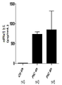
도 1은 RT112 및 SW780 인간 세포주에서 인터페론 노출에 대한 반응으로 PD-L1 발현을 측정한 차트이다. 수평축: 인터페론 양. 수직축: 발현된 폴리펩티드.
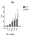
도 2는 RT112 및 SW780 인간 세포주에 대한 인터페론 노출에 대한 반응으로 TRAIL 발현을 측정한 차트이다. 수평축: 인터페론 양. 수직축: 발현된 폴리펩티드.
도 3은 RT112 및 SW780 인간 세포주에 대해 인터페론 노출에 대한 반응으로 IRF1 발현을 측정한 차트이다. 수평축: 인터페론 양. 수직축: 발현된 폴리펩티드.
도 4는 SW780 인간 암 세포주에서 증가하는 인터페론 알파에 대해 시험관 내 투여량 반응을 보여주는 PAGE 겔의 사진이다. 수평축: 인터페론 양. 수직축: 발현된 폴리펩티드.
도 5는 인터페론 노출에 대한 반응으로 IRF1, FOXA1 및 PD-L1의 RT12 세포에서 발현을 측정한다 (실시예 2 참조). IRF1은 인터페론-자극된 유전자 대조군으로 기능한다. FOXA1은 인터페론 노출 후에 발현이 변화하지 않는, 유형 I 인터페론 조절된 유전자의 하나의 예시이다.
도 6은 인터페론 노출에 대한 반응으로 IRF1, FOXA1 및 PD-L1의 UC3 세포에서 발현을 측정한다 (실시예 2 참조). IRF1은 인터페론-자극된 유전자 대조군으로 기능한다. FOXA1은 인터페론 노출 후에 발현이 변화하지 않는 유형 I 인터페론 조절된 유전자의 예이다.
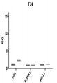
도 7은 인터페론 노출에 대한 반응으로 IRF1, FOXA1 및 PD-L1의 T24 세포에서 발현을 측정한다 (실시예 2 참조). IRF1은 인터페론-자극된 유전자 대조군으로 기능한다. FOXA1은 인터페론 노출 후에 발현이 변화하지 않는 유형 I 인터페론 조절된 유전자의 예이다.
도 8은 인터페론 노출에 대한 반응으로 IRF1, FOXA1 및 PD-L1의 UC14 세포에서 발현을 측정한다 (실시예 2 참조). IRF1은 인터페론-자극된 유전자 대조군으로 기능한다. FOXA1은 인터페론 노출 후에 발현이 변화하지 않는 유형 I 인터페론 조절된 유전자의 예이다.
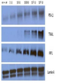
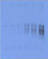
도 9는 6-레인 PAGE 겔의 사진이다. 그것은 BBN972 세포를 쥐 인터페론에 노출시킨 후, PD-L1 폴리펩티드의 존재를 측정한다. 레인 (왼쪽에서 오른쪽)은 배양배지의 0 (제로), 1×100, 1×101, 1×10², 1×10³, 및 1×10⁴ 국제 단위 인터페론/㎖이다.
도 10은 6-레인 PAGE 겔의 사진이다. MB49 #1 (MB49-luc) 세포를 쥐 인터페론에 노출시킨 후, PD-L1 폴리펩타이드의 존재를 측정한다. 레인 (왼쪽에서 오른쪽)은 배양배지의 0 (제로), 1×100, 1× 101, 1×10², 1×10³, 및 1×10⁴ 국제 단위 인터페론/㎖이다.
도 11은 6-레인 PAGE 겔의 사진이다. BBN972 세포를 쥐 인터페론에 노출시킨 후, 액틴 폴리펩타이드의 존재를 측정한다. 레인 (왼쪽에서 오른쪽)은 배양배지의 0 (제로), 1×100, 1× 101, 1×10², 1×10³, 및 1×10⁴ 국제 단위 인터페론/㎖이다.
도 12는 6-레인 PAGE 겔의 사진이다. MB49 #1 세포를 쥐 인터페론에 노출시킨 후, 액틴 폴리펩타이드의 존재를 측정한다. 레인 (왼쪽에서 오른쪽)은 배양배지의 0 (제로), 1×100, 1× 101, 1×10², 1×10³, 및 1×10⁴ 국제 단위 인터페론/㎖이다.
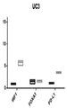
도 13은 폴리 I:C의 복막 내 주사에 대한 반응으로 마우스에서 혈청 인터페론 a를 측정한다.
도 14는 폴리 I:C를 6시간에 종양 내 주사에 대한 반응으로 마우스에서 혈청 인터페론 a를 측정한다
도 15는 폴리 I:C (500 meg)의 복막 내 주사 후, 종양 내 PD-L1 발현을 측정한다.
도 16은 인간 인터페론 알파 2B 전이 유전자를 갖는 (carrying), 재조합 복제-결핍 아데노 바이러스 유전자 치료 벡터인 INSTILADRIN™으로 치료한 사람에서 RNA 발현을 나타낸다.
도 17은 피하 C57BL6/J 종양 (n=5 그룹 당 암컷 쥐)에 대한 MB49 종양 크기 대 시간을 나타낸다. 치료는 종양 이식 후 10일에 시작하여 200 meg q3 일이다. Error bard는 SEM을 나타낸다.
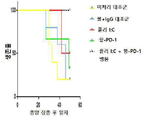
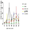
도 18은 암을 접종한 암컷 마우스에 식염수 (최하위 라인), IgG (다음 상위 라인), 항-PD1 단일클론 항체 (다음 상위 라인), 폴리 I:C (다음 상위 라인) 및 폴리 I:C 및 항-PD1 단일 클론 항체의 조합 (가장 높은 계통)을 처리한 케팔란-메이어(Kaplan-Meyer) 생존 곡선을 나타낸다.
도 19는 수컷 마우스의 시간에 따른 표준화된 (평균 +/- SD) 복사 강도를 비교한다. 로그-순위 테스트를 사용하여, 이들 데이터는 IgG 대조군 (p=0.06), 폴리 I:C 단독 요법 (p=0.32), 및 항-PD1 단일클론 항체 (p=0.14)보다 우수한 병용 치료법을 나타낸다.
도 20은 "생존 부분 (survival portions)", 즉 도 19마다 처리한 수컷 마우스에서 시간에 따른 살아남을 성향에 대한 생존을 보여주는 데이터를 나타낸다.1 is a chart measuring PD-L1 expression in response to interferon exposure in RT112 and SW780 human cell lines. Horizontal axis: amount of interferon. Vertical axis: expressed polypeptide.
2 is a chart measuring TRAIL expression in response to interferon exposure to RT112 and SW780 human cell lines. Horizontal axis: amount of interferon. Vertical axis: expressed polypeptide.
3 is a chart measuring IRF1 expression in response to interferon exposure for RT112 and SW780 human cell lines. Horizontal axis: amount of interferon. Vertical axis: expressed polypeptide.
4 is a photograph of a PAGE gel showing the in vitro dose response to increasing interferon alpha in the SW780 human cancer cell line. Horizontal axis: amount of interferon. Vertical axis: expressed polypeptide.
Figure 5 measures the expression of IRF1, FOXA1 and PD-L1 in RT12 cells in response to interferon exposure (see Example 2). IRF1 functions as an interferon-stimulated gene control. FOXA1 is one example of a type I interferon regulated gene whose expression does not change after interferon exposure.
Figure 6 measures the expression of IRF1, FOXA1 and PD-L1 in UC3 cells in response to interferon exposure (see Example 2). IRF1 functions as an interferon-stimulated gene control. FOXA1 is an example of a type I interferon regulated gene whose expression does not change after interferon exposure.
Figure 7 measures the expression of IRF1, FOXA1 and PD-L1 in T24 cells in response to interferon exposure (see Example 2). IRF1 functions as an interferon-stimulated gene control. FOXA1 is an example of a type I interferon regulated gene whose expression does not change after interferon exposure.
Figure 8 measures the expression of IRF1, FOXA1 and PD-L1 in UC14 cells in response to interferon exposure (see Example 2). IRF1 functions as an interferon-stimulated gene control. FOXA1 is an example of a type I interferon regulated gene whose expression does not change after interferon exposure.
9 is a photograph of a 6-lane PAGE gel. It measures the presence of PD-L1 polypeptide after exposing BBN972 cells to murine interferon. Lanes (left to right) are 0 (zero), 1×10 0 , 1×10 1 , 1×10 2 , 1×10 3 , and 1×10 4 international units of interferon/ml of culture medium.
10 is a photograph of a 6-lane PAGE gel. After exposing MB49 #1 (MB49- luc ) cells to murine interferon, the presence of PD-L1 polypeptide is measured. Lanes (left to right) are 0 (zero), 1×10 0 , 1×10 1 , 1×10 2 , 1×10 3 , and 1×10 4 international units of interferon/ml of culture medium.
11 is a photograph of a 6-lane PAGE gel. After exposing BBN972 cells to murine interferon, the presence of actin polypeptide is measured. Lanes (left to right) are 0 (zero), 1×10 0 , 1×10 1 , 1×10 2 , 1×10 3 , and 1×10 4 international units of interferon/ml of culture medium.
12 is a photograph of a 6-lane PAGE gel. After exposing MB49 #1 cells to murine interferon, the presence of actin polypeptide is measured. Lanes (left to right) are 0 (zero), 1×10 0 , 1×10 1 , 1×10 2 , 1×10 3 , and 1×10 4 international units of interferon/ml of culture medium.
Figure 13 Measures serum interferon a in mice in response to intraperitoneal injection of poly I:C.
Figure 14 Measures serum interferon a in mice in response to intratumoral injection of poly I:C at 6 hours.
Figure 15 Measures PD-L1 expression in tumors after intraperitoneal injection of poly I:C (500 meg).
16 shows RNA expression in humans treated with INSTILADRIN™, a recombinant replication-deficient adenovirus gene therapy vector carrying a human interferon alpha 2B transgene.
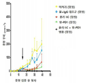
17 shows MB49 tumor size versus time for subcutaneous C57BL6/J tumors (female mice per n=5 groups). Treatment is 200 meg q3 days starting 10 days after tumor implantation. Error bard indicates SEM.
18 shows cancer-inoculated female mice with saline (lower line), IgG (next upper line), anti-PD1 monoclonal antibody (next upper line), poly I:C (next upper line) and poly I:C and Kaplan-Meyer survival curves treated with the combination of anti-PD1 monoclonal antibodies (highest strain) are shown.
19 compares normalized (mean +/- SD) radiant intensities over time in male mice. Using the log-rank test, these data indicate combination therapy superior to IgG control (p=0.06), poly I:C monotherapy (p=0.32), and anti-PD1 monoclonal antibody (p=0.14).
FIG. 20 presents data showing propensity to survive over time in "survival portions," ie male mice treated per FIG. 19 .
인터페론 치료법Interferon therapy
인터페론은 신호 단백질의 그룹이다. 그들은 예를 들어, 바이러스, 박테리아 및 기생충, 및 또한 암세포와 같은 몇가지 항원성 병원균의 존재에 대한 반응으로 인간 세포에 의해 발현되고 분비된다. 일반적으로 바이러스-감염된 세포는 인터페론을 방출하며, 근처의 방관자 세포 (bystander cell)에 신호를 보내 항-바이러스 방어를 강화한다. 또한 인터페론은 자연 살해 세포 및 대식세포와 같은 면역 세포를 활성화시킨다. 인터페론 주요 조직 적합성 복합 항원의 발현을 증가시켜, 면역계에 대한 외부 항원의 제시를 증가시킨다.Interferons are a group of signaling proteins. They are expressed and secreted by human cells in response to the presence of several antigenic pathogens, such as, for example, viruses, bacteria and parasites, and also cancer cells. Virus-infected cells normally release interferon and signal to nearby bystander cells to enhance anti-viral defenses. Interferon also activates immune cells such as natural killer cells and macrophages. Interferon increases the expression of major histocompatibility complex antigens, thereby increasing the presentation of foreign antigens to the immune system.
인터페론은 그들이 신호하는 수용체의 종류에 따라 정렬되거나 분류될 수 있다. 따라서, 인간에 있어서 인터페론은 종종 3 가지 종류로 분류된다: 유형 I (인간 IFN-α/β 수용체에 결합하는 인터페론), 유형 II (인간 IFN-γ수용체에 결합하는 인터페론) 및 유형 III (인간 IFN-λ수용체에 결합하는 인터페론).Interferons can be ordered or classified according to the type of receptor they signal. Thus, interferons in humans are often classified into three classes: type I (interferons that bind human IFN-α/β receptors), type II (interferons that bind human IFN-γ receptors) and type III (human IFNs). Interferon that binds to -λ receptors).
모든 인터페론은 몇 가지 공통적인 효과를 공유한다: 그들은 모두 항바이러스 제제이며, 면역계의 기능을 조절한다. 유형 I IFN의 투여는 실험 동물에서 종양 성장을 억제함을 보여주지만, 인간 종양에서 유익한 작용은 널리 문서화되지 않았다. 바이러스-감염된 세포는 주변 세포를 감염시킬 수 있는 바이러스 입자를 방출한다. 그러나, 감염된 세포는 방출된 인터페론에 의한 바이러스에 의해 잠재적 감염에 대해 주변 세포를 준비시킬 수 있다. 인터페론에 반응하여, 세포는 많은 양의 단백질 키나아제 R (PKR)로 알려진 효소를 생산한다. 이 효소는 새로운 바이러스 감염에 대한 반응으로, eIF-2로 알려진 단백질을 인산화시키고; 인산화된 eIF-2는 세포 내 단백질 합성을 감소시키기 위해, eIF2B라고 불리는 다른 단백질과 함께 불활성 복합체를 형성한다. 또 다른 세포 효소, RNAse L은, 또한 인터페론 작용에 의해 유도되어, 세포 내에서 RNA를 파괴하고, 나아가 바이러스 또는 숙주 유전자 모두의 단백질 합성을 감소시킨다. 억제된 단백질 합성은 바이러스와 감염된 숙주 세포 모두를 파괴한다. 또한, 인터페론은 인터페론-자극된 유전자 (ISGs)로서 집합적으로 알려진 수백 가지의 다른 단백질의 생산을 유도하고, 상기 단백질은 바이러스 및 인터페론에 의해 생성된 다른 작용들과 싸우는 역할을 한다. P53에 대한 인터페론의 효과는 또한 특정 암에 대한 그들의 보호 역할과 연결되어 있다. All interferons share some common effects: they are all antiviral agents, and they modulate the function of the immune system. Administration of type I IFNs has been shown to inhibit tumor growth in experimental animals, but beneficial effects in human tumors have not been widely documented. Virus-infected cells release viral particles that can infect surrounding cells. However, infected cells can prepare surrounding cells for potential infection by the virus by released interferon. In response to interferon, cells produce large amounts of an enzyme known as protein kinase R (PKR). This enzyme phosphorylates a protein known as eIF-2 in response to novel viral infection; Phosphorylated eIF-2 forms an inactive complex with another protein called eIF2B to reduce intracellular protein synthesis. Another cellular enzyme, RNAse L, also induced by interferon action, destroys RNA in the cell and further reduces protein synthesis of both viral or host genes. Inhibited protein synthesis destroys both the virus and infected host cells. In addition, interferon induces the production of hundreds of other proteins collectively known as interferon-stimulated genes (ISGs), which are responsible for combating viruses and other actions produced by interferon. The effects of interferons on P53 have also been linked to their protective role against certain cancers.
인터페론의 또 다른 기능은 주요 조직 적합성 복합체 분자인, MHC I 및 MHC II의 발현을 상향-조절하고, 면역-프로테아좀 활성을 증가시키는 것이다. 높은 MHC I 발현은 세포 독성 T세포에 대한 바이러스 펩타이드의 제시를 증가시키는 반면, 면역-프로테아좀은 MHC I 분자 상에 로딩하기 위한 바이러스 펩타이트를 처리하여, 감염된-세포의 인식 및 사멸을 증가시킨다. 더 높은 MHC II 발현은 도움 T 세포에 대한 바이러스 펩타이드의 제시를 증가시킨다; 이들 세포는 사이토카인 (예를 들어, 다른 분자들 사이에서 보다 많은 인터페론 및 인터루킨)을 방출하고, 상기 사이토카인은 다른 면역 세포의 작용에 신호를 보내고 이를 공동으로-조정한다.Another function of interferon is to up-regulate the expression of the major histocompatibility complex molecules, MHC I and MHC II, and to increase immune-proteasome activity. High MHC I expression increases the presentation of viral peptides to cytotoxic T cells, whereas the immune-proteasome processes viral peptides for loading on MHC I molecules, increasing recognition and killing of infected-cells. make it Higher MHC II expression increases the presentation of viral peptides to helper T cells; These cells release cytokines (eg, more interferons and interleukins, among other molecules), which signal and co-modulate the actions of other immune cells.
인터페론의 생산은 바이러스와 같은 미생물 및 박테리아 및 그들의 생산물에 대한 반응으로 주로 발생한다. 패턴 인식 수용체, 예를 들어 막 결합된 톨 유사 수용체 또는 세포질 수용체 RIG-I 또는 MDA5에 의한, 미생물에서 고유하게 발견되는 분자-바이러스성 글리코프로틴, 바이러스 RNA, 박테리아 내독소 (리포폴리사카라이드), 박테리아 편모, CpG 모티프-와의 결합은 IFN의 분비를 유도할 수 있다. 톨 유사 수용체 3 (TLR3)은 이중-가닥 RNA 바이러스 존재에 반응하여 인터페론을 유도하는데 중요하다; 이 수용체에 대한 리간드는 이중 가닥 RNA (dsRNA)이다. dsRNA에 결합한 후, 이 수용체는 많은 염증 단백질의 합성을 개시하는데 중요한 전사 인자 IRF3 및 NF-kB를 활성화시킨다. siRNA 또는 벡터-기반 반응제와 같은 RNA 간섭 기술 도구는 인터페론 경로를 침묵시키거나 자극할 수 있다. 세포 (특히 림프구 세포의 IFN)로부터 IFN의 방출은 또한 유사분열 물질에 의해 유도된다. 인터류킨 1, 인터류킨 2, 인터류킨-12, 종양 괴사 인자 및 기타 집락-자극 인자와 같은 다른 사이토카인은 또한 인터페론 생산을 향상시킬 수 있다.The production of interferon occurs mainly in response to microorganisms such as viruses and bacteria and their products. Molecular-viral glycoproteins, viral RNAs, bacterial endotoxins (lipopolysaccharides), found intrinsically in microorganisms by pattern recognition receptors such as membrane bound Toll-like receptors or cytoplasmic receptors RIG-I or MDA5, Binding to the bacterial flagella, CpG motif-can induce secretion of IFN. Toll-like receptor 3 (TLR3) is important for inducing interferon in response to the presence of double-stranded RNA viruses; The ligand for this receptor is double-stranded RNA (dsRNA). After binding to dsRNA, this receptor activates the transcription factors IRF3 and NF-kB, which are important for initiating the synthesis of many inflammatory proteins. RNA interference technology tools, such as siRNA or vector-based reagents, can silence or stimulate the interferon pathway. The release of IFN from cells (particularly IFN of lymphoid cells) is also induced by mitogens. Other cytokines such as interleukin 1, interleukin 2, interleukin-12, tumor necrosis factor and other colony-stimulating factors may also enhance interferon production.
인터페론 치료법은 (화학 요법 및 방사선과 함께) 몇몇 암의 치료법으로서 사용된다. 이 치료법은 혈액학적 악성 종양; 털색포 백혈병, 만성 골수성 백혈병, 결정성 림프종 및 피부 T-세포 림프종을 포함하는 백혈병 및 림프종에서 사용될 수 있다. 재발하는 흑색종 환자는 재조합 IFN-a2b를 받는다. B형 간염 및 C형 간염 모두는 종종 다른 항 바이러스 약물과 조합하여, IFN-b로 치료된다. 인터페론으로 치료받은 사람들 중 몇몇은 바이러스성 반응을 유지하고, 간염 바이러스를 제거할 수 있다. 가장 해로운 계통-간염 C 유전자형 I 바이러스는 현재 표준-치료의 인터페론인 RIBAVIRIN™ 및 최근에 승인된 프로테아제 억제제인 텔라프레비르 (Telaprevir) (Incivek™) 2011년 5월, 보세프레비르 (Boceprevir) (VICTRELIS™ 2011년 5월, 또는 뉴클레오티드 유사체 중합효소 억제제 소포스비르 (Sofosbuvir) (SOVALDI™ 2013년 12월과 함께 60-80 %의 성공률로 치료될 수 있다. 이 치료를 받은 환자의 생검은 간손상 및 간경변의 감소를 나타낸다. 비록 감염 초기에 진단하는 것은 간염 C 감염 초기에 신체적 증상이 드물기 때문에 어렵지만, 몇몇 증거는 감염 후 바로 인터페론을 제공하는 것은 만성 C형 간염을 예방할 수 있음을 보여준다. IFN에 의한 만성 C형 간염 조절은 감소된 간-세포성 암종과 관련이 있다. Interferon therapy (along with chemotherapy and radiation) is used as a treatment for some cancers. This treatment is for hematologic malignancies; It may be used in leukemias and lymphomas, including follicular leukemia, chronic myelogenous leukemia, lymphocytic lymphoma and cutaneous T-cell lymphoma. Patients with recurrent melanoma receive recombinant IFN-a2b. Both hepatitis B and C are treated with IFN-b, often in combination with other antiviral drugs. Some people treated with interferon maintain a viral response and are able to clear the hepatitis virus. The most harmful strain-hepatitis C genotype I viruses are RIBAVIRIN™, the current standard-of-care interferon, and Telaprevir (Incivek™), a recently approved protease inhibitor. Boceprevir (VICTRELIS), May 2011 ™ can be treated with a success rate of 60-80% in May 2011, or in combination with the nucleotide analogue polymerase inhibitor Sofosbuvir (SOVALDI™) December 2013. Biopsies of patients receiving this treatment showed liver damage and cirrhosis. Although early diagnosis of hepatitis C infection is difficult because physical symptoms are rare in the early stages of hepatitis C infection, some evidence shows that providing interferon immediately after infection can prevent chronic hepatitis C. Chronic hepatitis C caused by IFN Hepatitis C control is associated with reduced hepatocellular carcinoma.
이 기술은 인터페론이 외인성 폴리펩티드로 투여될 수 있음을 교시한다.This technology teaches that interferon can be administered as an exogenous polypeptide.
대안적으로, 천연 인터페론 유전자의 내인성 발현을 유도할 수 있다. 예를 들어, 이 기술은 예를 들어, 항원성 바실러스 칼메트-게랭균 (Bacillus Calmette-Guerin) 또는 마이코박테리아 또는 아데노 바이러스 백신을 교시한다. 이러한 항원성 제제는 환자 고유의 세포가 인터페론을 발현하도록 유도한다.Alternatively, endogenous expression of the native interferon gene can be induced. For example, the technology teaches, for example, an antigenic Bacillus Calmette-Guerin or mycobacterial or adenovirus vaccine. These antigenic agents induce the patient's native cells to express interferon.
대안적으로, 인터페론 전이 유전자를 갖는 벡터로 숙주 세포를 형질 형질전환 함으로써, 비-천연 인터페론 전이 유전자의 내인성 발현을 유도할 수 있다. 정말로, 심지어 외인성으로-투여된 인터페론 폴리펩타이드 자체가 인터페론 생산을 자극하기 위한 전달자로서 작용한다. Alternatively, endogenous expression of the non-native interferon transgene can be induced by transforming the host cell with a vector carrying the interferon transgene. Indeed, even exogenously-administered interferon polypeptides themselves act as messengers to stimulate interferon production.
여기에 사용된 용어 "인터페론"(약자로 "IFN")은 집합적으로 유형 1 및 유형 2 인터페론을 의미하며, 결실, 삽입 또는 이들의 치환 변이체, 생물학적 활성 단편 및 대립 형질을 포함한다. 여기에 사용된 용어 인터페론 ("IFN"으로 약칭 함)은 집합적으로 유형 1 및 유형 2 인터페론을 지칭한다. 유형 1 인터페론은 인터페론 -α, -β, 및 -ω 및 이들의 아형을 포함한다. 인간 인터페론-α는 적어도 14개의 확인된 아형을 가지며, 인터페론 -β는 3개의 확인된 아형을 가지고 있다. 특히, 바람직한 인터페론-α는 인간 인터페론 알파 아형 α-1 (GenBank Accession Number NP 076918), α -1b (GenBank Accession Number AAL35223), α-2, α-2a (GenBank Accession Number NP000596), α-2b (GenBank Accession Number AAP20099), α-4 (GenBank Accession Number NP066546), α-4b (GenBank Accesion Number CAA26701), α-5 (GenBank Accession Number NP 002160 및 CAA26702), α-6 (GenBank Accession Number CAA26704), α-7 (GenBank Accession Number NP 066401 및 CAA 26706), α-8 (GenBank Accession Number NP002161 및 CAA 26903), α-10 (GenBank Accession Number NP 002162), α13 (GenBank Accession Number NP 008831 및 CAA 53538), α-14 (GenBank Accession Number NP 002163 및 CAA 26705), α-16 (GenBank Accession Number NP 002164 및 CAA 26703), α-17 (GenBank Accession Number NP 067091), α-21 (GenBank Accession Number P01568 및 NP002166), 및 Stabinsky, 1996년 7월 30일에 허여된 US Pat. No. 5,541,293, Stabinsky, 1990년 1월 30일에 허여된 US Pat. No. 4,897,471, 및 Stabinsky, 1987년 9월 22일에 허여된 US Pat. No. 4,695,629에 개시된 것과 동일한 인터페론, 및 Goeddel et al., 1983년 11월 8일에 허여된 US Pat. No. 4,414,150에 개시된 하이브리드 인터페론을 포함하나 이에 제한되지 않으며, 상기 문헌의 교시는 본 명세서에 참고로 포함된다. 유형 2 인터페론은 인터페론 γ (EP 77,670 A 및 EP 146,354 A) 및 아형으로서 언급된다. 인간 인터페론 감마는 최소한 5개의 확인된 아형을 가지며, 인터페론 오메가 1 (GenBank Accession Number NP 002168)을 포함한다. 발현을 위한 인터페론을 암호화하는 DNA 서열의 구축은 공지된 아미노산 서열에 기반한 종래의 재조합 DNA 기술에 의해 달성될 수 있으며, 상기 공지된 아미노산 서열은 상기 문헌 및 Goeddel et al., 2002년 11월 19일에 허여된 US Pat. No. 6,482,613에 개시된 것에서 참조되며, 상기 문헌의 교시는 본 명세서에 참고로 포함된다. As used herein, the term “interferon” (abbreviated “IFN”) refers collectively to type 1 and type 2 interferon and includes deletions, insertions or substitutional variants thereof, biologically active fragments and alleles. As used herein, the term interferon (abbreviated "IFN") refers collectively to type 1 and type 2 interferon. Type 1 interferons include interferons -α, -β, and -ω and their subtypes. Human interferon-α has at least 14 identified subtypes and interferon-β has three identified subtypes. In particular, preferred interferon-α is human interferon alpha subtype α-1 (GenBank Accession Number NP 076918), α-1b (GenBank Accession Number AAL35223), α-2, α-2a (GenBank Accession Number NP000596), α-2b ( GenBank Accession Number AAP20099), α-4 (GenBank Accession Number NP066546), α-4b (GenBank Accession Number CAA26701), α-5 (GenBank Accession Number NP 002160 and CAA26702), α-6 (GenBank Accession Number CAA26704), α -7 (GenBank Accession Number NP 066401 and CAA 26706), α-8 (GenBank Accession Number NP002161 and CAA 26903), α-10 (GenBank Accession Number NP 002162), α13 (GenBank Accession Number NP 008831 and CAA 53538), α -14 (GenBank Accession Number NP 002163 and CAA 26705), α-16 (GenBank Accession Number NP 002164 and CAA 26703), α-17 (GenBank Accession Number NP 067091), α-21 (GenBank Accession Number P01568 and NP002166), and Stabinsky, US Pat, issued Jul. 30, 1996. No. 5,541,293, Stabinsky, US Pat, issued Jan. 30, 1990. No. 4,897,471, and Stabinsky, US Pat. No. 4,695,629, and in Goeddel et al., US Pat. No. 4,414,150, the teachings of which are incorporated herein by reference. Type 2 interferons are referred to as interferon γ (EP 77,670 A and EP 146,354 A) and subtypes. Human interferon gamma has at least five identified subtypes, including interferon omega 1 (GenBank Accession Number NP 002168). The construction of a DNA sequence encoding interferon for expression can be achieved by conventional recombinant DNA techniques based on a known amino acid sequence, and the known amino acid sequence is described above and in Goeddel et al., 19 November 2002. US Pat. No. 6,482,613, the teachings of which are incorporated herein by reference.
"생물학적으로 활성" 인터페론의 단편은 임의의 항-종양 또는 항-증식 활성을 갖는다고 확인된 것일 수 있으며, 이는 당업계에 공지된 기술 (예를 들어, Openakker et al., supra; Mossman, J. Immunol. Methods, 65: 55 (1983) 및 IFN 수용체 매개 메커니즘을 통한 IFN 반응성 유전자의 활성화 참고)에 의해 측정될 수 있다. 가용성 IFN-α 및 IFN-β 단백질은 일반적으로 유형 1 IFN 수용체 복합체 (GenBank Accession Number NP 000865)와 결합하는 것으로 확인되고, 세포 내의 신호 유사한 세포 내 신호 전달 경로를 활성화시킨다. IFN-γ는 일반적으로 유형 II IFN 수용체와 결합하는 것으로 확인된다. 리간드-유도된 양쪽 IFN 수용체 유형의 결합은 야누스 (Janus) 키나아제에 의해 수용체의 인산화를 가져오고, 이어서 STAT 단백질 (신호 변환기 및 전사의 활성화제)을 활성화시켜 추가적인 인산화 반응이 IFN-유도 가능한 전사 인자의 형성을 초래하고, 이는 IFN-유도 가능한 유전자에 존재하는 IFN 반응 요소에 결합하게 한다. 유형 1 및/또는 유형 2 IFN 수용체와 결합 후, IFN 경로 활성화로서 확인된 폴리펩타이드는 우리발명의 목적을 위한 인터페론으로 간주된다.A fragment of a “biologically active” interferon may be one that has been identified to have any anti-tumor or anti-proliferative activity, as described in the art (eg, Openakker et al., supra; Mossman, J. (see Immunol. Methods, 65: 55 (1983) and activation of IFN responsive genes through IFN receptor mediated mechanisms). Soluble IFN-α and IFN-β proteins are generally found to bind to the type 1 IFN receptor complex (GenBank Accession Number NP 000865) and activate signal-like intracellular signaling pathways within the cell. IFN-[gamma] is generally found to bind type II IFN receptors. Ligand-induced binding of both IFN receptor types results in phosphorylation of the receptor by Janus kinase, which in turn activates STAT proteins (signal transducers and activators of transcription), resulting in further phosphorylation of IFN-inducible transcription factors , which allows binding to IFN response elements present in IFN-inducible genes. Polypeptides identified as IFN pathway activation after binding to type 1 and/or type 2 IFN receptors are considered interferons for the purposes of our invention.
프로그램된 세포 사멸 단백질 1programmed cell death protein 1
프로그램된 세포 사멸 단백질 1 ("PD-1")은 CD279라고도 불리며, 이는 PDCD1 유전자에 의해 암호화되는 인간 내의 단백질이다. PD-1은 면역 글로블린 슈퍼 패밀리에 속하며, 2 개의 공지된 리간드인 PD-L1 및 PD-L2에 결합하는 세포 표면 수용체로서 기능한다.Programmed cell death protein 1 (“PD-1”), also called CD279, is a protein in humans encoded by the PDCD1 gene. PD-1 belongs to the immunoglobulin superfamily and functions as a cell surface receptor binding to two known ligands, PD-L1 and PD-L2.
PD-1은 T 세포의 활성화를 막아, 자가 면역을 감소시키고 "자가-내성 (self-tolerance)"을 촉진시킴으로써, 인간 면역계를 하향 조절하는데 중요한 역할을 한다. PD-1의 면역 조절 효과는 활성 T 세포를 도태시키고, 억제자 T 세포를 보호함으로써 초래된다. PD-1은 림프절에서 항원-특이적 T 세포의 사멸을 촉진하고, 그러나 조절 (“억제자”) T 세포에서 세포사멸을 감소시킨다.PD-1 plays an important role in downregulating the human immune system by blocking the activation of T cells, thereby reducing autoimmunity and promoting “self-tolerance”. The immunomodulatory effect of PD-1 results from the culling of active T cells and protection of suppressor T cells. PD-1 promotes the death of antigen-specific T cells in lymph nodes, but reduces apoptosis in regulatory (“suppressor”) T cells.
PD-L1은 특정 종양에서 고도로 발현될 수 있다. 이는 종양에서 면역 세포의 감소, 심지어 제거된 증식 또는 및 암세포-표면 항원을 인지하고 확인된 암세포와 싸우는 환자의 내재 면역계의 손상을 초래한다. PD-L1 can be highly expressed in certain tumors. This results in a decrease in the immune cells in the tumor, even abolished proliferation or damage to the patient's innate immune system that recognizes and fights cancer cell-surface antigens that have been identified.
PD-1은 T 세포 및 전-B 세포 (pro-B cell)에서 발현된다. 면역 체크 포인트로서 기능을 하는, PD-1은 T-세포의 활성화를 억제함으로써 면역계를 하향 조절하는데 중요한 역할을 하며, 이는 자가면역을 감소시키고, 자가-내성을 촉진하게 된다. PD-1의 억제성 효과는 림프절 내의 항원 특이적 T-세포에서 세포 사멸 (프로그램된 세포 사멸)을 촉진시키는 반면, 동시에 조절 T 세포 (억제자 T 세포)에서 세포 사멸을 감소시키는 이중 메커니즘을 통해 달성된다.PD-1 is expressed in T cells and pro-B cells. Functioning as an immune checkpoint, PD-1 plays an important role in downregulating the immune system by inhibiting the activation of T-cells, which reduces autoimmunity and promotes auto-resistance. The inhibitory effect of PD-1 is through a dual mechanism of promoting apoptosis (programmed cell death) in antigen-specific T-cells within the lymph node, while simultaneously reducing apoptosis in regulatory T cells (suppressor T cells). is achieved
프로그램된 사멸 1은 268개의 아미노산으로 구성된 유형 I 막 단백질이다. PD-1은 T 세포 조절 자의 확장된 CD28/CTLA-4 패밀리의 구성원이다. 단백질의 구조는 막을 가로지르는 영역 및 세포내 꼬리로 이어지는 세포 외 IgV 도메인을 포함한다. 세포 내 꼬리는 면역-수용체 티로신-기반 억제성 모티프 및 면역-수용체 티로신-기반 스위치 모티프 내에 위치하는 두 개의 인산화 부위를 함유하고, 이는 PD-1이 음성적으로 TCR 신호를 조절함을 제안한다. 리간드 결합에 따라 SHP-1 및 SHP-1 포스파타아제가 PD-1의 세포질 꼬리에 결합하는 것은 일관된다. 또한, PD-1 결합은 E3-유비퀴틴 리가아제 CBL-b 및 c-CBL을 상향 조절하고, 이는 T 세포 수용체 하양-조절을 유도한다. PD-1은 활성화된 T 세포, B 세포 및 대식세포의 표면 상에 발현되며, 이는 CTLA-4와 비교하여, PD-1이 보다 광범위하게 면역 반응을 음성적으로 조절함을 시사한다.Programmed death 1 is a type I membrane protein consisting of 268 amino acids. PD-1 is a member of the extended CD28/CTLA-4 family of T cell regulators. The structure of the protein includes an extracellular IgV domain leading to a transmembrane region and an intracellular tail. The intracellular tail contains two phosphorylation sites located within an immune-receptor tyrosine-based inhibitory motif and an immune-receptor tyrosine-based switch motif, suggesting that PD-1 negatively modulates TCR signaling. The binding of SHP-1 and SHP-1 phosphatase to the cytoplasmic tail of PD-1 following ligand binding is consistent. In addition, PD-1 binding up-regulates the E3-ubiquitin ligases CBL-b and c-CBL, leading to T cell receptor up-regulation. PD-1 is expressed on the surface of activated T cells, B cells and macrophages, suggesting that compared to CTLA-4, PD-1 negatively modulates the immune response more extensively.
PD-1에는 B7 패밀리의 구성원인 PD-L1 및 PD-L2의 두가지 리간드가 있다. PD-L1 단백질은 LPS, 및 GM-CSF 치료에 반응하여 대식세포 및 수지상 세포 (DC), 및 TCR 및 B 세포 수용체 신호 전달에 따른, T 세포 및 B 세포에서 상향 조절되며, 반면에 쉬고 있는 쥐에서는 PD-L1 mRNA는 심장, 폐, 흉선, 비장, 및 신장에서 검출될 수 있다.PD-1 has two ligands, PD-L1 and PD-L2, members of the B7 family. PD-L1 protein is upregulated in macrophages and dendritic cells (DC) in response to LPS, and GM-CSF treatment, and in T and B cells following TCR and B cell receptor signaling, whereas resting mice PD-L1 mRNA can be detected in the heart, lung, thymus, spleen, and kidney.
면역계를 활성화시키는, PD-1을 표적으로 하는 단일 클론 항체가 암 치료를 위해 개발되었다. 많은 종양 세포가 PD-L1, 면역 억제성 PD-1 리간드를 발현한다; 시험 관내에서 PD-1 및 PD-L1 사이의 상호 작용의 억제는 T 세포 반응을 향상시키고, 전임상 항종양 활성을 매개한다. 이것은 면역 체크 포인트 봉쇄로 알려져 있다. Monoclonal antibodies targeting PD-1, which activate the immune system, have been developed for the treatment of cancer. Many tumor cells express PD-L1, an immunosuppressive PD-1 ligand; Inhibition of the interaction between PD-1 and PD-L1 in vitro enhances T cell responses and mediates preclinical antitumor activity. This is known as immune checkpoint blockade.
하나의 그러한 항-PD-1 항체 약물인, 니볼루맙 (nivolumab) (OPDIVO™Bristol Myers Squibb Co., Princeton, NJ에서 상업적으로 수득 가능함)은 296명의 환자 전체의 임상시험에서 비-소-세포성 폐암, 흑색종 및 신-세포 암에서 완벽한 또는 부분적인 반응을 나타냈다. 결장 및 췌장암 환자는 반응이 없었다. 또한 PD-1 수용체를 표적으로 하는, 니볼루맙 (OPDIVO™Bristol-Myers Squibb)은 2014년 7월 일본에서, 2014년 12월 미국 FDA에서 전이성 흑색종 치료제로 승인받았다.One such anti-PD-1 antibody drug, nivolumab (commercially available from OPDIVO™ Bristol Myers Squibb Co., Princeton, NJ), was found to be non-small-cellular in a clinical trial of all 296 patients. It showed complete or partial response in lung cancer, melanoma and renal-cell cancer. Colon and pancreatic cancer patients did not respond. Nivolumab (OPDIVO™ Bristol-Myers Squibb), which also targets the PD-1 receptor, was approved in Japan in July 2014 and in the US FDA in December 2014 for the treatment of metastatic melanoma.
또한 팸브롤리주맙 (Pembrolizumab) (KEYTRUDA™ 또는 MK-3475, Merck & Co., Rahway, NJ에서 상업적으로 이용 가능함)은 PD-1 수용체를 표적으로 하고, 2014 년 9 월 FDA에서 전이성 흑색종 치료로 승인을 받았다. 팸브롤리주맙은 UK 의약품 조기 접근 제도 (Early Access to Medicines Scheme, EAMS) 통해 영국에서 진전된 (advanced) 흑색종 환자에게 접근 가능해졌다. 또한 이것은 미국에서 폐암, 림프종 및 악성중피종에 대한 임상 시험에서 사용되었다. 그것은 부작용이 거의 없이 성공적이었다. 2015년 10월 2일에 팸브롤리주맙은 다른 치료 후에 질병이 진전된 (전이성) 비-소세포 폐암 (NSCLC) 환자에 대해 FDA에서 승인을 받았다. In addition, Pembrolizumab (KEYTRUDA™ or MK-3475, commercially available from Merck & Co., Rahway, NJ) targets the PD-1 receptor and was approved by the FDA for the treatment of metastatic melanoma in September 2014. Approved. Pembrolizumab has been made accessible to patients with advanced melanoma in the UK through the UK Early Access to Medicines Scheme (EAMS). It has also been used in clinical trials for lung cancer, lymphoma and malignant mesothelioma in the United States. It was successful with few side effects. On October 2, 2015, pembrolizumab was approved by the FDA for patients with (metastatic) non-small cell lung cancer (NSCLC) whose disease had progressed after other treatments.
PD-1 수용체를 목표로 하는 초기 단계 개발에 있는 다른 약물 (종종 "체크 포인트 억제제"로 언금됨): 피딜리주맙 (Pidilizumab) (CT-011, Cure Tech), BMS 936559 (Bristol Myers Squibb), MPDL3280A (Roche) 및 아테졸리주맙 (atezolizumab) (Amgen).Other drugs in early stage development targeting the PD-1 receptor (often referred to as "checkpoint inhibitors"): Pidilizumab (CT-011, Cure Tech), BMS 936559 (Bristol Myers Squibb), MPDL3280A (Roche) and atezolizumab (Amgen).
병용 요법combination therapy
발명자는 인터페론으로 암을 치료하는 것은-인터페론 폴리펩티드를 투여하거나, 인터페론을 발현하는 세포를 유도하는 작용제를 처리함으로써-동시에 PD-1의 발현을 유도한다는 것을 발견하였다.The inventors have discovered that treating cancer with interferon - either by administering an interferon polypeptide or by treatment with an agent that induces cells expressing interferon - simultaneously induces the expression of PD-1.
따라서 발명자는 인터페론-기반 암 치료법의 효능을 개선하기 위해 PD-1의 활성을 억제하는 화합물 및 인터페론을 병용-투여하는 것을 제안한다.Therefore, the inventors propose co-administration of interferon and a compound that inhibits the activity of PD-1 to improve the efficacy of interferon-based cancer therapy.
이것은 예를 들어, 암 치료로서 유효한 양의 인터페론 폴리펩타이드를 정맥내로 주사하는 것 및 단일 클론 항체 체크 포인트 봉쇄 억제제를 PD-1 발현에서 인터페론-야기된 증가를 억제하기에 충분한 양, 바람직하게는 PD-1의 효과를 감소시키는 양을 정맥내로 투여하는 것을 포함한다. This includes, for example, intravenous injection of an interferon polypeptide effective as a cancer treatment and a monoclonal antibody checkpoint blockade inhibitor in an amount sufficient to inhibit the interferon-induced increase in PD-1 expression, preferably PD. and administering intravenously in an amount that reduces the effect of -1.
대안적으로, 이는 암 치료료서 유효한 양의 인터페론 발현을 유도하는, 작용제를 정맥내로 주사하는 것 및 예방적으로 단일 클론 항체 체크 포인트 봉쇄 억제제를 PD-1 발현에서 인터페론-야기된 증가를 억제하기에 충분한 양, 바람직하게는 PD-1의 효과를 감소시키는 양으로 정맥내로 투여하는 것을 포함한다. 상기 작용제는 인터페론 발현을 유도하는, 항원성 백신일 수 있다 (바이러스, 또는 BCG 백신 또는 마이코박테리아 백신). 대안적으로 상기 작용제는 발현가능한 인터페론 형질 전환 유전자로 숙주 세포를 형질전환시키는 전이 유전자 벡터일 수 있다. 대안적으로, 이는 또한 인터페론 전이 유전자를 갖는 항원성 바이러스 또는 박테리아일 수 있다. Alternatively, intravenous injection of an agent that induces expression of interferon in an effective amount in the treatment of cancer and prophylactically administering monoclonal antibody checkpoint blockade inhibitors to inhibit interferon-induced increases in PD-1 expression and administering intravenously in a sufficient amount, preferably in an amount that reduces the effect of PD-1. The agent may be an antigenic vaccine (virus, or BCG vaccine or mycobacterial vaccine), which induces interferon expression. Alternatively, the agent may be a transgene vector that transforms the host cell with an expressible interferon transgene. Alternatively, it may also be an antigenic virus or bacterium carrying an interferon transgene.
실시예 1-IFNα는 PD-L1 및 TRAIL 발현을 유도한다. Example 1- IFNα induces PD-L1 and TRAIL expression .
인터페론-알파 (IFNa)는 임상적으로 두드러지게 효과적이지 못했다. 발명자는 벡터-매개 IFNa 유전자 치료의 설정 보다 더 효과적일 수 있다는 것을 가정했다. 몇 년 전, 발명자는 인터페론 알파 2b로 매개되는 INSTILADRIN™브랜드 아데노 바이러스 벡터의 임상 2상 인간 임상 실험을 시작했다. 이 실험에서, 발명자는 인터페론 노출에 대한 반응으로 PD-L1, TRAIL, IRF1 및 라미닌 A의 발현을 측정하였다. Interferon-alpha (IFNa) was not significantly effective clinically. The inventors hypothesized that vector-mediated IFNa gene therapy could be more effective than the setup. A few years ago, the inventor began a phase 2 human clinical trial of the INSTILADRIN™ brand adenoviral vector mediated by interferon alpha 2b. In this experiment, the inventors measured the expression of PD-L1, TRAIL, IRF1 and laminin A in response to interferon exposure.
재료 및 방법: RT112 및 SW780 세포를 배지에서 배양한 다음, 인터페론 알파 폴리펩티드를 함유하는 배지에 노출시켰다. 인터페론의 양은 0 (대조군)에서 104 국제 단위/mL로 배열되었다. 유전자 발현은 상업적으로 이용 가능한 항체 및 프라이머를 사용하여, 웨스턴 블랏 및 정량적 실시간 PCR 를 사용하여 평가되었다. RNA는 MIRVANA™키트 (Thermo Fisher)를 사용하여 배양되는 세포로부터 분리되었다. TAQMAN™어레이 카드 (A 및 B) (Thermo Fisher)를 사용하여 RT112에서 mIR을 프로파일링 하였다. 전체 게놈 mRNA 발현 프로파일링을 Illumina HumanHT_12_v4 BEADCHIP™어레이 (47323 프로브)을 사용하여 RT112 및 UC3에서 수행하였다. Materials and Methods: RT112 and SW780 cells were cultured in medium and then exposed to medium containing interferon alpha polypeptide. The amount of interferon ranged from 0 (control) to 10 4 international units/mL. Gene expression was assessed using Western blot and quantitative real-time PCR using commercially available antibodies and primers. RNA was isolated from cultured cells using the MIRVANA™ kit (Thermo Fisher). mIRs were profiled at RT112 using a TAQMAN™ array card (A and B) (Thermo Fisher). Whole genome mRNA expression profiling was performed at RT112 and UC3 using an Illumina HumanHT_12_v4 BEADCHIP™ array (47323 probe).
결과: 결과는 도 1 내지 4에서 제공된다. 모든 세포주는 IFNa의 노출에 반응하여 PD-L1, TRAIL 및 IRF1 발현을 상향-조절 하였고, 라미닌 A 발현에는 측정가능한 효과가 없었다. PD-L1, TRAIL 및 IRF1 발현에 대해서, 효과는 다른 세포주에서 큰 차이였다. Results: Results are presented in Figures 1-4. All cell lines up-regulated PD-L1, TRAIL and IRF1 expression in response to exposure to IFNa, and had no measurable effect on laminin A expression. For PD-L1, TRAIL and IRF1 expression, the effect was significantly different in the different cell lines.
결론: 암세포주의 패널에서, 인터페론 노출은 PD-L1 면역 체크 포인트 발현에서 중대한 증가를 이끌었다. 발명자는 이는 인터페론이 당업계에서 효과적인 암 치료제로 사용하는데 실패한 이유를 암시하기 때문에, 이것이 이러한 발견이 놀랍다는 것을 발견하였다. 인터페론이 이론적으로 항암 치료제로서 효과적이여야 하는 반면, 인터페론은 또한 PD-L1을 상향-조절 하고, 이에 따라 인터페론의 치료학적 효과가 좌절될 것이다.Conclusion: In a panel of cancer cell lines, interferon exposure led to a significant increase in PD-L1 immune checkpoint expression. The inventors found that this finding was surprising, as it suggests why interferon has failed to be used as an effective cancer treatment in the art. While interferon should theoretically be effective as an anti-cancer therapeutic, interferon also up-regulates PD-L1, thus thwarting the therapeutic effect of interferon.
실시예 2-IFNα는 PD-L1 발현을 용량-의존적 방식으로 유도한다.Example 2-IFNα induces PD-L1 expression in a dose-dependent manner.
여기서 발명자는 면역 체크 포인트 PD-L1, 마이크로-RNA(miR) 및 mRNA의 발현을 측정하였다. Here we measured the expression of the immune checkpoint PD-L1, micro-RNA (miR) and mRNA.
재료 및 방법: RT112, T14, UC3 및 UC14 세포를 배지에서 배양한 후, 6시간 동안 다른 대조군 배지, 또는 1000 IU/ML의 인터페론 알파 폴리펩티드를 함유하는 배지에서 노출시켰다. Pd-L1의 발현은 상업적으로 이용 가능한 항체 및 프라이머를 사용하여, 웨스턴 블랏 및 정량적 실시간 PCR를 사용하여 평가되었다. RNA는 MIRVANA™키트 (Thermo Fisher)를 사용하여 배양되는 세포로부터 분리되었다. TAQMAN™어레이 카드 (A 및 B) (Thermo Fisher)를 사용하여 RT112에서 mIR을 프로파일링 하였다. 전체 게놈 mRNA 발현 프로파일링을 Illumina HumanHT_12_v4 BEADCHIP™어레이 (47323 프로브)을 사용하여 RT112 및 UC3에서 수행하였다. 전체 실험을 통계적 신뢰성을 증가시키기 위해 3번 반복 수행하였다.Materials and Methods: RT112, T14, UC3 and UC14 cells were cultured in medium and then exposed to another control medium or medium containing 1000 IU/ML of interferon alpha polypeptide for 6 hours. Expression of Pd-L1 was assessed using western blot and quantitative real-time PCR using commercially available antibodies and primers. RNA was isolated from cultured cells using the MIRVANA™ kit (Thermo Fisher). mIRs were profiled at RT112 using a TAQMAN™ array card (A and B) (Thermo Fisher). Whole genome mRNA expression profiling was performed at RT112 and UC3 using an Illumina HumanHT_12_v4 BEADCHIP™ array (47323 probe). The entire experiment was repeated 3 times to increase statistical reliability.
결과: 모든 세포주는 IFNa의 노출에 반응하여 PD-L1발현을 상향-조절하였다. 이러한 효과는 UC3 세포보다 RT112 세포에서 가장 두드려졌다 (도 5 참고). 대조적으로, 세가지 잠재적 oncomIR지역의 발현은 RT112:1233 세포 (p=0.0036), 19b-1# (p=0.0157), 및 222# (p=0.0061)에서 IFNa을 노출한 후, 유의하게 하향-조절되었다. IFNa 노출 후, log (발현) (거짓 발견율 <0.001)에서 최소 2배 이상 차이로 상이하게-발현된 유전자를 분석한 결과, 302 및 181은 RT112 및 UC3 세포 주에서 각각 상이하게 발현된 유전자였다. 두 세포주에서 최고 순위의 IFNα로 유도된 유전자에는 몇가지를 포함하였으나, 이는 이전에 방광암에서 개시되지 않았던 것이며, IFIT2 (전이의 음성적 조절자) 및 IFI27 (TRAIL에 대한 민감도와 관련됨)을 포함한다. IFNα- 유도된 PD-L1 발현을 또한 실시간 PCR 데이터와 평행한 배수-변화 (fold-changes)를 갖는 mRNA 유전자 칩에 대해 입증 가능하였다.Results: All cell lines up-regulated PD-L1 expression in response to exposure to IFNa. This effect was most pronounced in RT112 cells than in UC3 cells (see Fig. 5). In contrast, expression of three potential oncomIR regions was significantly down-regulated following exposure to IFNa in RT112:1233 cells (p=0.0036), 19b-1# (p=0.0157), and 222# (p=0.0061). became Analysis of differentially-expressed genes with at least 2-fold difference in log (expression) (false detection rate <0.001) after IFNa exposure revealed that 302 and 181 were differentially expressed genes in RT112 and UC3 cell lines, respectively. The top-ranked IFNα-induced genes in both cell lines included several, but not previously initiated in bladder cancer, including IFIT2 (negative regulator of metastasis) and IFI27 (associated with sensitivity to TRAIL). IFNα-induced PD-L1 expression was also verifiable for mRNA gene chips with fold-changes parallel to real-time PCR data.
결론: 암 세포주 패널에서, 인터페론 노출은 유의한 PD-L1 면역 체크 포인트 발현의 증가를 가져왔다. 어레이-기반 마이크로 RNA 및 mRNA 프로파일링은 방광암에서 IFNa 반응의 신규한 잠재적 매개자를 드러냈다. 이 방광 IFNa 프로파일은 아데노 바이러스성 IFNa 유전자 치료에 대한 반응을 측정하기 위한 중간 종말점으로서 유용할 수 있다. IFNa 치료에서 PD-L1의 발현의 나아간 예측은 면역 체크 포인트 억제제를 활용한 합리적인 병용 치료법으로 이끌 수 있다. Conclusion: In a panel of cancer cell lines, interferon exposure resulted in a significant increase in PD-L1 immune checkpoint expression. Array-based microRNA and mRNA profiling revealed novel potential mediators of the IFNa response in bladder cancer. This bladder IFNa profile may be useful as an intermediate endpoint to measure response to adenoviral IFNa gene therapy. Further prediction of PD-L1 expression in IFNa treatment may lead to rational combination therapy utilizing immune checkpoint inhibitors.
실시예 3-마우스 인터페론은 PD-L1 발현을 유도한다 Example 3- Mouse Interferon Induces PD-L1 Expression
재료 및 방법: BBN972 및 MB49 #1 (MB49-luc) 세포를 배양하고, 0 내지 1×10⁴개의 마우스 인터페론의 국제 단위를 함유하는 매체에 노출시켰다. 이어서 PD-L1 및 (대조군으로서) 액틴의 발현을 측정하였다.Materials and Methods: BBN972 and MB49 #1 (MB49- luc ) cells were cultured and exposed to media containing 0 to 1×10 4 international units of mouse interferon. The expression of PD-L1 and actin (as a control) was then measured.
결과: 마우스 인터페론은 양 세포주 모두에서 액틴의 발현에 영향을 미치지 않았다 (도 11, 12 참조). 대조적으로, 마우스 인터페론은 PD-L1 발현에 있어서 현저한, 용량-의존적 효과를 나타냈다 (도 9, 10을 참조).Results: Mouse interferon did not affect the expression of actin in both cell lines (see FIGS. 11 and 12 ). In contrast, mouse interferon showed a significant, dose-dependent effect on PD-L1 expression (see FIGS. 9 and 10 ).
결론: 이러한 데이터는 PD-L1 발현에서 인터페론의 효과가, 인간 인터페론 알파 2a 또는 정말로인간 인터페론에 한정되지 않음을 나타낸다. 오히려, PD-L1의 발현에서 인터페론의 효과가 일반적으로 인터페론에 대해 포괄적인 것으로 보인다.Conclusion: These data indicate that the effect of interferon on PD-L1 expression is not limited to human interferon alpha 2a or indeed human interferon. Rather, the effect of interferon on the expression of PD-L1 appears to be global for interferon in general.
실시예 4-폴리이노시닉: 폴리시티딜산 (폴리 I:C)은 PD-L1을 유도한다 Example 4 Polyinosinic: Polycytidylic Acid (Poly I:C) Induces PD-L1
재료 및 방법: 상기 데이터는 인터페론이 다른 종의 인터페론에 대한 반응에서 PD-L1을 용량 의존적인 방식으로, 빠르고, 명백하게 유도한다는 것을 나타낸다. 인터페론을 복용한 동물 종에 상관없이 효과를 감안할 때, 발명자는 효과가 인터페론에 한정되지 않으며, 다른 유형의 면역 자극제에 의해 더욱 일반적으로 유발될 수 있다고 가정하였다. 이러한 개념을 시험하기 위하여, 발명자는 폴리이노시닉:폴리시티딜산 (종종 “폴리 I:C'로 약식됨)을 평가하였다. 폴리 I:C는 면역자국제이다. 이는 바이러스 감염을 자극하기 위해 나트륨 염 형태로 사용된다. 폴리 I:C는 구조적으로 이중-가닥 RNA와 유사한다. dsRNA는 일부 바이러스에 존재한다. 발명자는 폴리 I:C를 plc 또는 ulc 종양을 이식한 실험용 마우스에 복막 내 주사로 투여하였다. Materials and Methods: The above data indicate that interferon induces PD-L1 in a dose-dependent manner, rapidly, and unambiguously in response to interferon of other species. Given the effect irrespective of the animal species in which interferon was administered, the inventors hypothesized that the effect was not limited to interferon, but could more commonly be induced by other types of immune stimulants. To test this concept, the inventors evaluated polyinosinic:polycytidylic acid (often abbreviated as "Poly I:C'). Poly I:C is an immunosuppressant. It is used in sodium salt form to stimulate viral infections. Poly I:C is structurally similar to double-stranded RNA. dsRNA is present in some viruses. The inventors administered poly I:C to experimental mice transplanted with plc or ulc tumors by intraperitoneal injection.
결과: 도 13은 대조군 마우스 (n=3)가 혈청 인터페론의 최소 기준선 측정을 나타낸 것을 보여준다. a. 대조적으로, 폴리 I:C의 복막 내 주사는 시간-의존적 혈청 인터페론 증가를 생산한다 a. 도 14는 6 시간에 종양 내 폴리 I:C 주사의 결과를 나타낸다. 상기 데이터 (각 시리즈에 대해 n=1)는 종양 내 인터페론이 plc 종양에서 유의하게 증가하고, ulc 종양에서 다소 증가하며, 대조군의 종양에서 측정 가능한 증가가 없음을 보여준다. 도 15는 폴리 I:C (500 meg) 또한 종양 내 PD-L1 발현을 유도함을 나타낸다 (24시간에)(Mann Whitney p=0.0495).Results: Figure 13 shows that control mice (n=3) showed minimal baseline measurements of serum interferon. a. In contrast, intraperitoneal injection of poly I:C produces a time-dependent increase in serum interferon a. 14 shows the results of intratumoral poly I:C injections at 6 hours. The data (n=1 for each series) show that intratumoral interferon was significantly increased in plc tumors, slightly increased in ulc tumors, and there was no measurable increase in tumors of the control group. 15 shows that poly I:C (500 meg) also induces intratumoral PD-L1 expression (at 24 h) (Mann Whitney p=0.0495).
결론: 이들 데이터는 PD-L1 발현이 단지 인터페론에 의해 유도되는 것이 아니라, dsRNA를 모방하고 인터페론을 유도하는 화합물인 폴리 I:C에 의해 유도되는 것임을 시사한다. Conclusion: These data suggest that PD-L1 expression is not induced only by interferon, but by poly I:C, a compound that mimics dsRNA and induces interferon.
실시예 5-인터페론 바이러스 유전자 치료는 사람에서 PD-L1을 유도한다 Example 5 Interferon Virus Gene Therapy Induces PD-L1 in Humans
재료 및 방법: 이 데이터는 BCG 치료 후, 반응성이 없거나 재발성인 환자에서 인간 인터페론 알파 2b 전이 유전자를 갖는, INSTILADRIN™ 복제-결핍된 아데노 바이러스 유전자 치료 벡터에 대한 인간의 임상 2단계에서 가져온 것이다. 이 연구 계획은 공개되었으며, 여기에 참조로 포함된다.Materials and Methods: These data are from a Phase 2 human clinical trial of the INSTILADRIN™ replication-deficient adenoviral gene therapy vector carrying the human interferon alpha 2b transgene in non-responsive or recurrent patients following BCG treatment. This study plan has been published and is incorporated herein by reference.
결과: 도 16은 인간 인터페론 알파 2b 전이 유전자를 갖는, INSTILADRIN™ 복제-결핍된 아데노 바이러스 유전자 치료 벡터로 인간에서 8 회의 치료 주기에서의 RNA 발현을 나타낸다. 홀수 (흰색으로 코드됨) 컬럼은 치료 전 RNA 전사를 측정한다; 짝수 (밝은 파란색으로 코드됨) 컬럼은 후에 측정한다. RNA 양은 정량적으로 표시되며, 밝은 녹색은 가장 적게 보이고 밝은 빨간색은 가장 많이 보인다. 컬럼 1 및 2는 PD-L1 RNA가 치료 전에 -2에서 후에 +2로 증가하는 것을 보여준다. 컬럼 3 및 4는 유사하게 PD-L1 RNA가 치료 전 -2에서 후에 +3로 증가하는 것을 보여준다. 모두에서, 치료 쌍의 3 분의 1은 치료 후, PD-L1 발현의 유의한 증가를 보여준다. 치료는 또한 다른 면역 체크 포인트 마커를 상향-조절했다.Results: Figure 16 shows RNA expression at 8 treatment cycles in humans with INSTILADRIN™ replication-deficient adenoviral gene therapy vector with human interferon alpha 2b transgene. Odd (coded in white) columns measure RNA transcription prior to treatment; Even (coded light blue) columns are measured later. The amount of RNA is expressed quantitatively, with light green showing the least and bright red showing the most. Columns 1 and 2 show that PD-L1 RNA increases from -2 before treatment to +2 after treatment.
결론: 이들 데이터는 환자의 3 분의 1이 인터페론 유전자 치료법의 치료 후에, T-세포 및 인터페론 체크 포인트 마커 (PD-L1 포함)를 유도하는 것을 입증함을 나타낸다.CONCLUSION: These data demonstrate that one-third of patients induce T-cells and interferon checkpoint markers (including PD-L1) after treatment with interferon gene therapy.
실시예 6-병용 요법은 생존을 증가시킨다 Example 6 - Combination Therapy Increases Survival
재료 및 방법: 암컷 실험실 래트에게 종양 세포를 접종하고, 세포가 측정 가능한 종양으로 발전하도록 허용했다. 그 후, 래트를 식염수 (대조군), IgG (대조군), 항-PD1 단일 클론 항체 (단독 요법), 폴리 I:C (인터페론 발현을 유도하기 위한 단일 요법) 및 폴리 I:C 및 항-PD1 단일 클론 항체의 병용 (병용 요법)로 처리하였다. Materials and Methods: Female laboratory rats were inoculated with tumor cells, and the cells were allowed to develop into measurable tumors. Rats were then treated with saline (control), IgG (control), anti-PD1 monoclonal antibody (monotherapy), poly I:C (monotherapy to induce interferon expression) and poly I:C and anti-PD1 single Treatment with a combination of clonal antibodies (combination therapy).
결과: 도 17은 피하 C57BL6/J 종양에 대한 MB49 종양 크기 대 시간을 나타낸다 (n=그룹당 5마리 암컷 마우스). 치료는 종양 이식 10일 후부터 시작하여, 200 meg q3 일이다. 에러 바드 (Error bard)는 SEM을 나타낸다. 40일 때에 가장 큰 종양 부피를 나타내는, 가장 높은 (노란색) 선이 대조군 (모든 그룹 n=5, 암컷만)이다. 다음으로 가장 낮은 (파란색) 선은 IgG 대조군이다. 다음으로 가장 낮은 (빨간색) 선은 폴리 I:C이다. 그 다음으로 낮은 (녹색) 선은 항-PDL 단일 클론 항체이다. 가장 낮은 (검정색) 선은 X 축 자체에 놓여 있으며, 이는 병용 요법이다.Results: FIG. 17 shows MB49 tumor size versus time for subcutaneous C57BL6/J tumors (n=5 female mice per group). Treatment starts 10 days after tumor implantation, 200 meg q3 days. Error bard indicates SEM. The highest (yellow) line, representing the largest tumor volume at 40, is the control (all groups n=5, females only). The next lowest (blue) line is the IgG control. The next lowest (red) line is poly I:C. The next lower (green) line is the anti-PDL monoclonal antibody. The lowest (black) line lies on the X axis itself, which is a combination therapy.
도 18은 암컷 마우스에 종양을 접종하고, 식염수 (최하위 선), IgG (다음 상위 선), 항-PD1 단일 클론 항체 (다음 상위 선), 폴리 I:C (다음 상위 선) 및 폴리 I:C 및 항-PD1 단일 클론 항체의 병용 (가장 높은 선)을 처리한 경우의 캐플란-메이어 생존 곡선을 나타낸다. 이러한 데이터는 인터페론-유도제 (폴리 I:C) 및 PD1 억제제 (항-PD1 단일 크론 항체)을 병용하는 것은 생존을 유의하게 증가시킴을 보여준다: 50 일에서, 대조군 동물의 ~20 %가 살아 있었고, 폴리 I:C 동물의 50 %가 살아 있으며, 병용 요법으로 처리된 동물의 100 %가 살아 있었다.Figure 18. Female mice were inoculated with tumors, saline (lower line), IgG (next upper line), anti-PD1 monoclonal antibody (next upper line), poly I:C (next upper line) and poly I:C and Caplan-Meier survival curves treated with the combination of anti-PD1 monoclonal antibody (highest line). These data show that the combination of an interferon-inducing agent (poly I:C) and a PD1 inhibitor (anti-PD1 monoclonal antibody) significantly increased survival: at
도 19는 수컷 마우스의 시간에 따른 표준화된 (평균 +/- SD) 복사 강도를 비교한다. 로그-순위 테스트를 사용하여, 이들 데이터는 IgG 대조군 (p=0.06), 폴리 I:C 단일 요법 (p=0.32), 및 항-PD1 단일클론 항체 (p=0.14)보다 우수한 병용 요법 (p=0.06)을 보여 준다.19 compares normalized (mean +/- SD) radiant intensities over time in male mice. Using the log-rank test, these data showed that the combination therapy (p= 0.06) is shown.
도 20은 "생존 부분 (survival portions)", 즉 시간이 지남에 따른 생존 경향성을 보여주는 데이터이다. 20 is data showing “survival portions,” ie, survival trends over time.
결론: 이러한 데이터는 병용 요법이 시너지적인 효과가 있고, 단지 부가적인 효과 이상을 부여함을 보여준다.CONCLUSION: These data show that the combination therapy has a synergistic effect, conferring more than just an additive effect.
실시예 7-표재성 확산 흑색종 Example 7 - Superficial diffuse melanoma
재료 및 방법: 표재성 확산 흑생종으로 진단된 인간 환자를 광범위한 국소 절제 및 전초 림프절 (sentinel node) 생검으로 치료하여, 림프계 또는 말초 기관으로 질병의 확산이 없음을 확인하였다. 그 다음 환자는 INSTILADRIN™ 및 KEYTRUDA™의 병용으로 치료하였다. 치료는 외과적 절제술 후 실용적으로 즉시 시작하였다. MATERIALS AND METHODS: Human patients diagnosed with superficial diffuse melanoma were treated with extensive local resection and sentinel node biopsy to confirm the absence of disease spread to the lymphatic system or peripheral organs. The patient was then treated with a combination of INSTILADRIN™ and KEYTRUDA™. Treatment was started practically immediately after surgical resection.
INSTILADRIN™ 브랜드 아데노 바이러스는 복제-결핍된, 인터페론 알파 2b 전이 유전자를 갖는 재조합 아데노 바이러스 유전자 치료 벡터이다. 그러한 유전자 치료 벡터의 제조는 예를 들어, Muralidhara Ramachandra et al., Selectively Replicating Viral Vecter, 미국 문자 특허 번호 7691370에 개시되어 있다. 인터페론 전이 유전자의 분리는 예를 들어, Charles Weissmann, DNA Sequences, Recombinant DNA Molecules and Process for Producing Human Interferon-Like Polypeptides, 미국 문자 특허 번호 6835557에 개시되어 있다. The INSTILADRIN™ brand adenovirus is a replication-deficient, recombinant adenoviral gene therapy vector with an interferon alpha 2b transgene. Preparation of such gene therapy vectors is disclosed, for example, in Muralidhara Ramachandra et al ., Selectively Replicating Viral Vecter , US Pat. No. 7691370. Isolation of interferon transgenes is disclosed, for example, in Charles Weissmann, DNA Sequences, Recombinant DNA Molecules and Process for Producing Human Interferon-Like Polypeptides , US Pat. No. 6835557.
KEYTRUDA™ 브랜드 팸브롤리주맙은 인간화된 단일 클론 항-프로그램된 세포 사멸-1 (PD-1)항체 (Fc 지역에서 서열 변이가 안정화된 IgG4/kappa 동형)이다.KEYTRUDA™ brand pambrolizumab is a humanized monoclonal anti-programmed cell death-1 (PD-1) antibody (IgG4/kappa isotype with stabilized sequence variation in the Fc region).
INSTILADRIN™은 단일-용량 바이알로 제공된다. INSTILADRIN™의 1 회 복용량은 주사용 멸균 생리 식염수로 재구성되었고, 절단 부위에 국소적으로 피하에 투여하였다. 투여를 4주에 한번씩 반복하였다. KEYTRUDA™ 파우더 한 바이알에는 50mg의 팸브롤리주맙이 들어 있다. KEYTRUDA™는 30분에 걸쳐 정맥 내 주입되며, 3 주마다 반복해 투여하였고, 환자는 질병의 진행 또는 허용할 수 없는 독성이 있을 때까지 치료받는다. 비정형 반응 (즉, 종양 수축 후 처음 몇 개월 내에 초기 일시적인 종양 크기 증가 및 작은 새로운 병변)이 관찰될 수 있다. 임상적으로-안정한 환자의 치료는 질병 진전이 확인될 때까지 질병 진행의 초기 증거와 함께 계속하는 것이 바람직하다.INSTILADRIN™ is provided as a single-dose vial. A single dose of INSTILADRIN™ was reconstituted with sterile saline for injection and administered subcutaneously topically at the amputation site. Dosing was repeated once every 4 weeks. One vial of KEYTRUDA™ powder contains 50 mg of pembrolizumab. KEYTRUDA™ is an intravenous infusion over 30 minutes, repeated every 3 weeks, and the patient is treated until disease progression or unacceptable toxicity. Atypical reactions (ie, initial transient tumor size increase and small new lesions within the first few months after tumor shrinkage) may be observed. Treatment of clinically-stable patients is preferably continued with early evidence of disease progression until disease progression is confirmed.
시험 대상을 등록한 후, 치료 그룹에 배정한다: 절제 후 KEYTRUDA™만 사용, 절제 후INSTILADRIN™만 사용, 절제 후 KEYTRUDA™ 및 INSTILADRIN™를 병용, 절제 후 INSTILADRIN™ 및 NSAID (COX-2 억제제)를 병용, 및 절제 후 KEYTRUDA™ 및 INSTILADRIN™ 및 NSAID를 병용.After enrollment, study subjects are assigned to treatment groups: post-ablation KEYTRUDA™ only, post-ablation INSTILADRIN™ only, post-ablation KEYTRUDA™ and INSTILADRIN™ concomitant, post excision INSTILADRIN™ and NSAID (COX-2 inhibitor) concomitantly , and post-ablation with KEYTRUDA™ and INSTILADRIN™ plus NSAIDs.
결과: 1차 유효성 결과 측정은 진행 자유 생존 (progression free survival) (예를 들어, 고형암에서 반응 평가 기준 [Response Evaluation Criteria in Solid Tumours, RECIST]을 사용하는 통합된 방사선학 (Integrated Radiology) 및 종양학 평가 검토 (Oncology Assessment review)에 의해 평가됨), 전체 생존율, 및 전초 림프절 생검이다. 다른 효능 결과 측정은 전반적인 반응율과 반응 지속이 될 수 있다. 이어지는 전초 림프절 생검은 병의 확산이 없음을 나타낼 것으로 예상된다.RESULTS: The primary efficacy outcome measure is progression free survival (e.g., Integrated Radiology and Oncology Review Review Using Response Evaluation Criteria in Solid Tumors, RECIST). (assessed by Oncology Assessment review), overall survival, and anterior lymph node biopsy. Other efficacy outcome measures could be overall response rate and response duration. Subsequent distal lymph node biopsy is expected to show no spread of the disease.
발명자는 INSTILADRIN™ 단독을 투여한 경우에 비하여, INSTILADRIN™ 및 COX-2 억제제를 병용투여는 상승된 효율을 입증할 것이라고 예상했다. 발명자는 작용제 단독 투여에 비하여, KEYTRUDA™ 및 INSTILADRIN™ 병용 투여가 상승된 효율 결과 측정을 입증할 것이라고 예상했고, 발명자는 이것이 단순한 첨가물 이상으로 유용할 것으로 예상했다. 발명자는 KEYTRUDA™ 단독 또는 INSTILADRIN™ 및 NSAID 단독을 투여한 경우에 비하여, KEYTRUDA™ 및 INSTILADRIN™ 및 NSAID의 병용 투여가 상승된 효율 결과 측정을 입증할 것이라고 예상했고, 발명자는 이것이 단순한 첨가물 이상으로 유용할 것으로 예상했다. The inventors expected that the co-administration of INSTILADRIN™ and a COX-2 inhibitor would demonstrate increased efficacy compared to the case of administering INSTILADRIN™ alone. The inventors expected that administration of KEYTRUDA™ and INSTILADRIN™ in combination would demonstrate an increased efficacy outcome measure compared to administration of the agents alone, and the inventors expected that this would be useful beyond simple additives. The inventors expected that the combined administration of KEYTRUDA™ and INSTILADRIN™ and NSAID would demonstrate an increased efficacy outcome measure compared to administration of KEYTRUDA™ alone or INSTILADRIN™ and NSAID alone, and the inventors expected that this would be useful beyond simple additives. expected that
실시예 8-표재성 확산 흑색종 Example 8 - Superficial diffuse melanoma
재료 및 방법: 앞의 실시예와 같은 KEYTRUDA™Materials and Methods: KEYTRUDA™ as in the previous example
인터페론의 공급원로서, SYLATRON™페길화된 인터페론 알파 2b을 6회 mcg/kg으로 1주일에 한번 8회 피하 투여하였고 (유도), 3 mcg/kg으로 1주일에 1번, 5년까지 (유지) 투여하였다. 만약 부작용으로 인해 SYLATRON™투여량 조절이 1-8주 치료 동안에 (유도) 필요하다면, 원래 투여량 (1주일에 한번 6 mcg/kg)에서 3 단계 감소가 바람직하다 (즉, 투여량을 1주일에 한번 3mcg/kg으로 감소시키고; 필요하다면 일주일에 한번 2mcg/kg으로 감소시킨다; 그 후, 필요한 경우 추가로 일주일에 한번 1 mcg/kg로 감소시킨다). 만약 부작용으로 인해 9-260주 치료 동안에 (유지) 투여량 조절이 필요한 경우, 원래 투여량 (일주일에 한번 3 mcg/kg) 보다 2 단계 감소가 바람직하다 (즉, 투여량을 일주일에 한번 2mcg/kg으로 감소시킨다; 필요한 경우 추가로 일주일에 한번 1 mcg/kg로 감소시킨다).As a source of interferon, SYLATRON™ pegylated interferon alpha 2b was administered subcutaneously 8 times a week at 6 mcg/kg once a week (induction), once a week at 3 mcg/kg, up to 5 years (maintenance) administered. If SYLATRON™ dosage adjustment is required (induction) during treatment for 1-8 weeks due to side effects, a 3-step reduction from the original dose (6 mcg/kg once a week) is desirable (i.e., reduce the dose by 1 week). Reduce to 3 mcg/kg once per day; If necessary, reduce to 2 mcg/kg once a week; Then, if necessary, further reduce to 1 mcg/kg once a week). If a (maintenance) dose adjustment is required during treatment for 9-260 weeks due to side effects, a 2-step reduction from the original dose (3 mcg/kg once a week) is preferable (i.e., the dose is reduced to 2 mcg/kg once a week). kg;
시험 대상을 등록한 후, 치료 그룹에 배정한다: 절제 후 KEYTRUDA™만 사용, 절제 후SYLATRON™만 사용, 절제 후 KEYTRUDA™ 및 SYLATRON™를 병용, 절제 후 SYLATRON™와 NSAID (COX-2 억제제)를 병용, 및 절제 후 KEYTRUDA™ 및 SYLATRON™ 및 NSAID를 병용.After enrolling the test subjects, they are assigned to treatment groups: KEYTRUDA™ only after resection, SYLATRON™ only after resection, KEYTRUDA™ and SYLATRON™ after resection, SYLATRON™ plus NSAID (COX-2 inhibitor) after resection. , and in combination with KEYTRUDA™ and SYLATRON™ and NSAIDs after resection.
결과: 1차 유효성 결과 측정은 진행 자유 생존 (progression free survival) (예를 들어, 고형암에서 반응 평가 기준 [Response Evaluation Criteria in Solid Tumours, RECIST]을 사용하는 통합된 방사선학 (Integrated Radiology) 및 종양학 평가 검토 (Oncology Assessment review)에 의해 평가됨), 전체 생존율, 및 전초 림프절 생검이다. 다른 효능 결과 측정은 전반적인 반응율과 반응 지속이 될 수 있다. 이어지는 전초 림프절 생검은 병의 확산이 없음을 나타낼 것으로 예상된다.RESULTS: The primary efficacy outcome measure is progression free survival (e.g., Integrated Radiology and Oncology Review Review Using Response Evaluation Criteria in Solid Tumors, RECIST). (assessed by Oncology Assessment review), overall survival, and anterior lymph node biopsy. Other efficacy outcome measures could be overall response rate and response duration. Subsequent distal lymph node biopsy is expected to show no spread of the disease.
발명자는 SYLATRON™ 단독을 투여한 경우에 비하여, SYLATRON™ 및 COX-2 억제제를 병용투여는 상승된 효율을 입증할 것이라고 예상했다. 발명자는 작용제 단독 투여에 비하여, KEYTRUDA™ 및 SYLATRON™ 병용 투여가 상승된 효율 결과 측정을 입증할 것이라고 예상했고, 발명자는 이것이 단순한 첨가물 이상으로 유용할 것으로 예상했다. 발명자는 KEYTRUDA™ 단독 또는 SYLATRON™ 및 NSAID 단독을 투여한 경우에 비하여, KEYTRUDA™ 및 SYLATRON™ 및 NSAID의 병용 투여가 상승된 효율 결과 측정을 입증할 것이라고 예상했고, 발명자는 이것이 단순한 첨가물 이상으로 유용할 것으로 예상했다. The inventors expected that the combined administration of SYLATRON™ and a COX-2 inhibitor would demonstrate an increased efficiency compared to the case of administering SYLATRON™ alone. The inventors expected that administration of KEYTRUDA™ and SYLATRON™ in combination would demonstrate an increased efficacy outcome measure compared to administration of the agent alone, and the inventors expected that it would be useful beyond just a simple additive. The inventors expected that the combined administration of KEYTRUDA™ and SYLATRON™ and NSAID would demonstrate an increased efficacy outcome measure compared to administration of KEYTRUDA™ alone or SYLATRON™ and NSAID alone, and the inventors predicted that this would be useful beyond simple additives. expected that
실시예 9-비-소 세포 폐암 Example 9- Non-Small Cell Lung Cancer
재료 및 방법: 상기 실시예 7에 따른 약학적 작용제. 인체 시험 대상은 비소 세포 폐암으로 진단된다. 환자는 Greene, Frederick L., Cancer Staging Manual (American Joint Committee on Cancer, publ., 6th edition) 에 따라 선별하여 필적하는 질병을 갖는 필적하는 시험 대상으로 확실히 하였다. 시험 대상은 PD-L1의 종양 발현을 기반으로 치료대상으로 선별되었다.Materials and Methods: A pharmaceutical agent according to Example 7 above. The human test subject is diagnosed with non-small cell lung cancer. Patients were screened according to Greene, Frederick L., Cancer Staging Manual (American Joint Committee on Cancer, publ ., 6th edition) to ensure comparable test subjects with comparable disease. Test subjects were selected for treatment based on the tumor expression of PD-L1.
KEYTRUDA의 권장 용량은 다음과 같다: 이전에 화학 요법으로 치료받지 않은 경우, NSCLC (비소 세포 폐암)에 대해 200mg, 및 화학 요법으로 이전에 치료받은 경우, NSCLC 또는 흑생종에 대해 2mg/kg.The recommended doses for KEYTRUDA are: 200 mg for NSCLC (non-small cell lung cancer) if not previously treated with chemotherapy, and 2 mg/kg for NSCLC or melanoma if previously treated with chemotherapy.
INSTILADRIN™은 늑막-내부로 주입된다. 이 방법은 미국 특허 공보 US2014/17202의 도 2에 설명되어 있다.INSTILADRIN™ is injected intrapleurally. This method is described in FIG. 2 of US Patent Publication US2014/17202.
시험 대상을 등록한 후, 치료 그룹에 배정한다: KEYTRUDA™만 사용, INSTILADRIN™만 사용, KEYTRUDA™ 및 INSTILADRIN™를 병용, INSTILADRIN™ 및 NSAID (COX-2 억제제)를 병용, KEYTRUDA™ 및 INSTILADRIN™ 및 NSAID를 병용.After enrollment of study subjects, they are assigned to treatment groups: KEYTRUDA™ only, INSTILADRIN™ only, KEYTRUDA™ and INSTILADRIN™ in combination, INSTILADRIN™ and NSAID (COX-2 inhibitor) in combination, KEYTRUDA™ and INSTILADRIN™ and NSAID use together.
결과: 1차 유효성 결과 측정은 진행 자유 생존 (progression free survival), 전체 생존율, 및 전초 림프절 생검이다. 다른 효능 결과 측정은 전반적인 반응율과 반응 지속이 될 수 있다. 이어지는 전초 림프절 생검은 병의 확산이 없음을 나타낼 것으로 예상된다.RESULTS: The primary efficacy outcome measures are progression free survival, overall survival, and anterior lymph node biopsy. Other efficacy outcome measures could be overall response rate and response duration. Subsequent distal lymph node biopsy is expected to show no spread of the disease.
발명자는 INSTILADRIN™ 단독을 투여한 경우에 비하여, INSTILADRIN™ 및 COX-2 억제제를 병용투여는 상승된 효율을 입증할 것이라고 예상했다. 발명자는 작용제 단독 투여에 비하여, KEYTRUDA™ 및 INSTILADRIN™ 병용 투여가 상승된 효율 결과 측정을 입증할 것이라고 예상했고, 발명자는 이것이 단순한 첨가물 이상으로 유용할 것으로 예상했다. 발명자는 KEYTRUDA™ 단독 또는 INSTILADRIN™ 및 NSAID 단독을 투여한 경우에 비하여, KEYTRUDA™ 및 INSTILADRIN™ 및 NSAID의 병용 투여가 상승된 효율 결과 측정을 입증할 것이라고 예상했고, 발명자는 이것이 단순한 첨가물 이상으로 유용할 것으로 예상했다. The inventors expected that the co-administration of INSTILADRIN™ and a COX-2 inhibitor would demonstrate increased efficacy compared to the case of administering INSTILADRIN™ alone. The inventors expected that administration of KEYTRUDA™ and INSTILADRIN™ in combination would demonstrate an increased efficacy outcome measure compared to administration of the agents alone, and the inventors expected that this would be useful beyond simple additives. The inventors expected that the combined administration of KEYTRUDA™ and INSTILADRIN™ and NSAID would demonstrate an increased efficacy outcome measure compared to administration of KEYTRUDA™ alone or INSTILADRIN™ and NSAID alone, and the inventors expected that this would be useful beyond simple additives. expected that
요약summary
상기 실시예는 특정 암 치료에 대해 논의한다. 그러나 우리의 발견은 인터페론 신호 전달에서 유익한 모든 상태 및 CD279의 과-발현으로 고통받는 상태를 치료하는데 보다 일반적으로 사용될 것이다. The above examples discuss specific cancer treatments. However, our findings will more generally be used to treat all conditions beneficial in interferon signaling and those suffering from over-expression of CD279.
첨부된 청구항에서, "치료"란 용어는 완전한 치료가 필요로 하는 것이 아닌, 경감시키는 것을 의미한다. 예를 들어, 암을 "치료하는 것"은 암을 완전히 제거함으로써 달성할 수 있으며, 또한, 예를 들어, 암의 성장을 늦추거나, 사망의 위험을 감소시키거나, 이러한 치료를 받은 적 없는 환자와 비교하여 병의 진전을 늦춤으로써, 달성할 수 있다. In the appended claims, the term "treatment" means alleviating, not requiring complete treatment. For example, "treating" cancer can be achieved by completely eliminating the cancer, and also, for example, slowing the growth of the cancer, reducing the risk of death, or a patient who has never received such treatment. can be achieved by slowing the progression of the disease compared to
상기 우리의 논의를 고려할 때, 당업자는 특정한 이의 응용 또는 변형을 쉽게 볼 수 있다. 예를 들어, 상기 논의는 인간 인터페론의 특정 종을 언급하지만, 비슷하게 기능하는 다른 종 및 인터페론 변이체 또는 유사체는 동일한 혜택을 제공할 것이다. 따라서, 발명자는 우리의 특허의 법적 적용 범위가 논의한 실시예에 의해서가 아니라, 첨부된 법적 청구항 및 그의 허용가능한 등가물에 의하여 결정될 것을 의도하였다.In view of our discussion above, one of ordinary skill in the art can readily see particular applications or variations thereof. For example, while the above discussion refers to a particular species of human interferon, other species and interferon variants or analogs that function similarly will provide the same benefits. Accordingly, the inventor intends that the legal scope of our patents be determined by the appended legal claims and their permissible equivalents, and not by the embodiments discussed.
첨부된 법적 청구항이 약 "동시에" 치료를 언급할 때 (원래 청구항 3 참조), 이는 동시에 환자에서 두 화합물이 작용하는 것을 요구한다. 이는 동시에 투여해야 하는 것을 요구하는 것은 아니다. 따라서 만약 제2 작용제의 효과가 최소한 일주일 지속될 경우, 제2 작용제를 투여하고 일주일 후에, 제1 작용제를 투여할 수 있다. When the appended statutory claims refer to treatment "at the same time" with drugs (see original claim 3), this requires both compounds to act in the patient at the same time. This does not require that they be administered simultaneously. Thus, if the effect of the second agent lasts at least one week, the first agent may be administered one week after administration of the second agent.
Claims (20)
a. 인간 환자에서 인터페론으로 치료할 수 있는 상태를 진단하는 단계;
b. 제1 작용제를 투여하는 단계로서, 상기 제1 작용제는 인간 면역계 체크 포인트의 기능에 영향을 미치기에 충분한 양으로 인간 환자의 인터페론 수준을 증가시킬 수 있으며, 상기 체크 포인트는 면역 기능을 감소시키는 것인, 단계; 및
c. 동일한 인간 면역계 체크 포인트의 기능에 영향을 미치는 제2 작용제를 인간에게 투여하는 단계로서, 상기 제 2 작용제는 상기 제 1 작용제가 상기 체크 포인트 상에 가질 수 있는 면역 기능의 감소를 실질적으로 개선시키는데 유효한 양으로 투여되는 것인, 단계를 포함하고,
상기 방법에서 상기 제2 화합물은 제1 작용제에 의해 야기된 면역 기능의 감소를 실질적으로 개선시키는 것인, 방법.A method of treating a human patient with interferon, comprising:
a. diagnosing a condition treatable with interferon in the human patient;
b. administering a first agent, wherein the first agent is capable of increasing interferon levels in the human patient in an amount sufficient to affect the function of a human immune system checkpoint, wherein the checkpoint decreases immune function. , step; and
c. administering to the human a second agent that affects the function of the same human immune system checkpoint, wherein the second agent is effective to substantially ameliorate a decrease in immune function that the first agent may have on the checkpoint. administered in an amount, comprising the step of
wherein the second compound substantially ameliorates the decrease in immune function caused by the first agent.
a. 인간 환자에서 인터페론으로 치료할 수 있는 상태를 진단하는 단계, 및
b. 억제성 인간 면역계 체크 포인트를 억제하는 제2 작용제를 인간에게 투여하는 단계로서, 상기 작용제를 면역 활성 감소에 대한 체크 포인트의 활성을 감소시키기에 충분한 양으로 투여하는 것인, 단계, 및
c. 상기 상태를 치료하는데 유효한 양으로 상기 인간 환자의 인터페론 수준을 증가시키는 제1 작용제를 인간에게 투여하는 단계를 포함하는, 방법.A method of improving the efficacy of interferon as a human therapeutic, comprising:
a. diagnosing a condition treatable with interferon in the human patient, and
b. administering to the human a second agent that inhibits an inhibitory human immune system checkpoint, wherein the agent is administered in an amount sufficient to reduce the activity of the checkpoint for reduced immune activity, and
c. and administering to the human a first agent that increases interferon levels in the human patient in an amount effective to treat the condition.
a. 억제성 인간 면역계 체크 포인트를 억제하는 제2 작용제를 인간에게 투여하는 단계, 및
b. 인간 환자의 인터페론 수준을 증가시키는 제1 작용제를 인간에게 투여하는 단계를 포함하는, 방법.A method comprising essentially the steps of:
a. administering to the human a second agent that inhibits an inhibitory human immune system checkpoint, and
b. A method comprising administering to the human a first agent that increases interferon levels in the human patient.
Applications Claiming Priority (4)
| Application Number | Priority Date | Filing Date | Title |
|---|---|---|---|
| US201662295268P | 2016-02-15 | 2016-02-15 | |
| US62/295,268 | 2016-02-15 | ||
| PCT/US2017/017568 WO2017142818A1 (en) | 2016-02-15 | 2017-02-11 | Improved interferon therapy |
| KR1020207018114A KR102518146B1 (en) | 2016-02-15 | 2017-02-11 | Improved Interferon Therapy |
Related Parent Applications (1)
| Application Number | Title | Priority Date | Filing Date |
|---|---|---|---|
| KR1020207018114A Division KR102518146B1 (en) | 2016-02-15 | 2017-02-11 | Improved Interferon Therapy |
Publications (1)
| Publication Number | Publication Date |
|---|---|
| KR20220147693A true KR20220147693A (en) | 2022-11-03 |
Family
ID=59559473
Family Applications (3)
| Application Number | Title | Priority Date | Filing Date |
|---|---|---|---|
| KR1020227035962A KR20220147693A (en) | 2016-02-15 | 2017-02-11 | Improved Interferon Therapy |
| KR1020187026724A KR102129195B1 (en) | 2016-02-15 | 2017-02-11 | Improved interferon treatment |
| KR1020207018114A KR102518146B1 (en) | 2016-02-15 | 2017-02-11 | Improved Interferon Therapy |
Family Applications After (2)
| Application Number | Title | Priority Date | Filing Date |
|---|---|---|---|
| KR1020187026724A KR102129195B1 (en) | 2016-02-15 | 2017-02-11 | Improved interferon treatment |
| KR1020207018114A KR102518146B1 (en) | 2016-02-15 | 2017-02-11 | Improved Interferon Therapy |
Country Status (7)
| Country | Link |
|---|---|
| US (2) | US10335461B2 (en) |
| EP (1) | EP3416727A4 (en) |
| JP (5) | JP6995061B2 (en) |
| KR (3) | KR20220147693A (en) |
| CN (2) | CN113925964A (en) |
| CA (2) | CA3089875A1 (en) |
| WO (1) | WO2017142818A1 (en) |
Families Citing this family (2)
| Publication number | Priority date | Publication date | Assignee | Title |
|---|---|---|---|---|
| US20210260093A1 (en) * | 2018-06-20 | 2021-08-26 | Yale University | RIG-I Agonists and Treatments Using Same |
| JP2023519709A (en) * | 2020-03-30 | 2023-05-12 | トライゼル リミテッド | Compositions and methods for treating cancer |
Family Cites Families (8)
| Publication number | Priority date | Publication date | Assignee | Title |
|---|---|---|---|---|
| AU2004298991A1 (en) * | 2003-12-10 | 2005-06-30 | Canji, Inc | Methods and compositions for treatment of interferon-resistant tumors |
| US20090028857A1 (en) * | 2007-07-23 | 2009-01-29 | Cell Genesys, Inc. | Pd-1 antibodies in combination with a cytokine-secreting cell and methods of use thereof |
| WO2009114335A2 (en) * | 2008-03-12 | 2009-09-17 | Merck & Co., Inc. | Pd-1 binding proteins |
| EP2504028A4 (en) * | 2009-11-24 | 2014-04-09 | Amplimmune Inc | Simultaneous inhibition of pd-l1/pd-l2 |
| CN103536917B (en) * | 2013-10-30 | 2015-03-11 | 苏州丁孚靶点生物技术有限公司 | Use of interferon in treatment of tumor, and related product and method |
| WO2015069770A1 (en) * | 2013-11-05 | 2015-05-14 | Cognate Bioservices, Inc. | Combinations of checkpoint inhibitors and therapeutics to treat cancer |
| NZ721908A (en) * | 2013-12-20 | 2022-12-23 | Massachusetts Gen Hospital | Combination therapy with neoantigen vaccine |
| PE20171067A1 (en) * | 2014-10-14 | 2017-07-24 | Novartis Ag | ANTIBODY MOLECULES BINDING AND USES OF PD-L1 |
-
2017
- 2017-02-11 WO PCT/US2017/017568 patent/WO2017142818A1/en active Application Filing
- 2017-02-11 JP JP2018561193A patent/JP6995061B2/en active Active
- 2017-02-11 CA CA3089875A patent/CA3089875A1/en active Pending
- 2017-02-11 US US15/430,456 patent/US10335461B2/en active Active
- 2017-02-11 CN CN202111214612.7A patent/CN113925964A/en active Pending
- 2017-02-11 KR KR1020227035962A patent/KR20220147693A/en not_active Application Discontinuation
- 2017-02-11 EP EP17753674.5A patent/EP3416727A4/en active Pending
- 2017-02-11 KR KR1020187026724A patent/KR102129195B1/en active IP Right Grant
- 2017-02-11 CA CA3014759A patent/CA3014759C/en active Active
- 2017-02-11 KR KR1020207018114A patent/KR102518146B1/en active Application Filing
- 2017-02-11 CN CN201780011635.3A patent/CN108697781A/en active Pending
-
2019
- 2019-02-20 US US16/280,174 patent/US20190255153A1/en active Pending
-
2020
- 2020-05-19 JP JP2020087334A patent/JP2020122019A/en active Pending
- 2020-05-19 JP JP2020087333A patent/JP7113864B2/en active Active
- 2020-05-19 JP JP2020087332A patent/JP7030156B2/en active Active
-
2022
- 2022-06-15 JP JP2022096410A patent/JP2022113832A/en active Pending
Also Published As
| Publication number | Publication date |
|---|---|
| US10335461B2 (en) | 2019-07-02 |
| CN113925964A (en) | 2022-01-14 |
| KR102129195B1 (en) | 2020-07-01 |
| KR20200078693A (en) | 2020-07-01 |
| JP2019508497A (en) | 2019-03-28 |
| JP7113864B2 (en) | 2022-08-05 |
| JP2020122019A (en) | 2020-08-13 |
| JP6995061B2 (en) | 2022-02-04 |
| CA3014759C (en) | 2020-09-22 |
| KR20180123676A (en) | 2018-11-19 |
| JP2020122017A (en) | 2020-08-13 |
| JP2020122018A (en) | 2020-08-13 |
| EP3416727A4 (en) | 2019-10-09 |
| JP2022113832A (en) | 2022-08-04 |
| CA3089875A1 (en) | 2017-08-24 |
| WO2017142818A1 (en) | 2017-08-24 |
| CN108697781A (en) | 2018-10-23 |
| EP3416727A1 (en) | 2018-12-26 |
| US20170232071A1 (en) | 2017-08-17 |
| US20190255153A1 (en) | 2019-08-22 |
| KR102518146B1 (en) | 2023-04-06 |
| CA3014759A1 (en) | 2017-08-24 |
| JP7030156B2 (en) | 2022-03-04 |
Similar Documents
| Publication | Publication Date | Title |
|---|---|---|
| Li et al. | PD-1/PD-L1 checkpoint inhibitors in advanced hepatocellular carcinoma immunotherapy | |
| KR20180120208A (en) | SMC Combination Therapy for Cancer | |
| JP7200093B2 (en) | Immunomodulation using TLR9 agonists for cancer therapy | |
| US20150004175A1 (en) | Compositions and Methods for Reducing CTL Exhaustion | |
| Wang et al. | The JAK inhibitor tofacitinib ameliorates immune‑mediated liver injury in mice | |
| JP2022113832A (en) | improved interferon therapy | |
| US10584341B2 (en) | Antagonistic PDL1 aptamers and their applications in cancer therapy | |
| US8987302B2 (en) | Treatment of hepatitis C virus related diseases using hydroxychloroquine or a combination of hydroxychloroquine and an anti-viral agent | |
| US20220175787A1 (en) | Patient selection for enhancement of anti-tumor immunity in cancer patients | |
| EP2782994B1 (en) | Small molecule enhancer for dendritic cell cancer vaccines | |
| Kong et al. | A combination of PD‑1/PD‑L1 inhibitors: The prospect of overcoming the weakness of tumor immunotherapy | |
| EP2739301B1 (en) | Hcv immunotherapy | |
| US20140205609A1 (en) | Methods for inducing systemic immune responses to cancer | |
| CA1331345C (en) | Double stranded rna correction of aberrant metabolic pathways associated with uncontrolled tumor cell and virus growth | |
| Temizoz et al. | Type I and II interferons toward ideal vaccine and immunotherapy | |
| US20200397738A1 (en) | Methods for treating cancer and protecting renewable tissues | |
| US20190083556A1 (en) | Analytical methods and arrays for use in the same | |
| Hu et al. | Combining a nanoparticle-mediated radioimmunotherapy with dual blockade of LAG3 and TIGIT improves the treatment efficacy in anti-PD1 resistant lung cancer | |
| WO2022109227A1 (en) | Combined cancer therapy of b7-h3 and cd47 immune checkpoint inhbitior and methods of use |
Legal Events
| Date | Code | Title | Description |
|---|---|---|---|
| A107 | Divisional application of patent | ||
| E902 | Notification of reason for refusal | ||
| E601 | Decision to refuse application |