KR20220047376A - How to produce T cells - Google Patents
How to produce T cells Download PDFInfo
- Publication number
- KR20220047376A KR20220047376A KR1020227009208A KR20227009208A KR20220047376A KR 20220047376 A KR20220047376 A KR 20220047376A KR 1020227009208 A KR1020227009208 A KR 1020227009208A KR 20227009208 A KR20227009208 A KR 20227009208A KR 20220047376 A KR20220047376 A KR 20220047376A
- Authority
- KR
- South Korea
- Prior art keywords
- cells
- tcr
- cell
- population
- progenitor
- Prior art date
Links
- 210000001744 T-lymphocyte Anatomy 0.000 title claims abstract description 218
- 210000004027 cell Anatomy 0.000 claims abstract description 235
- 238000000034 method Methods 0.000 claims abstract description 100
- 210000001978 pro-t lymphocyte Anatomy 0.000 claims abstract description 65
- 239000003446 ligand Substances 0.000 claims abstract description 31
- 230000001939 inductive effect Effects 0.000 claims abstract description 8
- 206010028980 Neoplasm Diseases 0.000 claims description 101
- 201000011510 cancer Diseases 0.000 claims description 63
- 239000000427 antigen Substances 0.000 claims description 53
- 108091007433 antigens Proteins 0.000 claims description 53
- 102000036639 antigens Human genes 0.000 claims description 53
- 150000007523 nucleic acids Chemical class 0.000 claims description 47
- 210000004263 induced pluripotent stem cell Anatomy 0.000 claims description 42
- 210000000130 stem cell Anatomy 0.000 claims description 37
- 230000003394 haemopoietic effect Effects 0.000 claims description 36
- 102000039446 nucleic acids Human genes 0.000 claims description 30
- 108020004707 nucleic acids Proteins 0.000 claims description 30
- 108090000765 processed proteins & peptides Proteins 0.000 claims description 26
- 238000011282 treatment Methods 0.000 claims description 22
- 102100031573 Hematopoietic progenitor cell antigen CD34 Human genes 0.000 claims description 20
- 101000777663 Homo sapiens Hematopoietic progenitor cell antigen CD34 Proteins 0.000 claims description 20
- 108010070047 Notch Receptors Proteins 0.000 claims description 19
- 238000012258 culturing Methods 0.000 claims description 18
- 230000011748 cell maturation Effects 0.000 claims description 16
- 108010033276 Peptide Fragments Proteins 0.000 claims description 14
- 102000007079 Peptide Fragments Human genes 0.000 claims description 14
- 238000004113 cell culture Methods 0.000 claims description 14
- 239000000203 mixture Substances 0.000 claims description 11
- 230000011664 signaling Effects 0.000 claims description 10
- 210000003171 tumor-infiltrating lymphocyte Anatomy 0.000 claims description 7
- 239000013598 vector Substances 0.000 claims description 7
- 239000000546 pharmaceutical excipient Substances 0.000 claims description 6
- 238000000338 in vitro Methods 0.000 claims description 5
- 239000008194 pharmaceutical composition Substances 0.000 claims description 5
- 230000003213 activating effect Effects 0.000 claims description 4
- 239000013604 expression vector Substances 0.000 claims description 3
- 239000008199 coating composition Substances 0.000 claims description 2
- 102000005650 Notch Receptors Human genes 0.000 claims 2
- 238000002826 magnetic-activated cell sorting Methods 0.000 claims 1
- 108091008874 T cell receptors Proteins 0.000 abstract description 139
- 102000016266 T-Cell Antigen Receptors Human genes 0.000 abstract description 129
- 210000003958 hematopoietic stem cell Anatomy 0.000 abstract description 35
- 238000004519 manufacturing process Methods 0.000 abstract description 14
- 238000009169 immunotherapy Methods 0.000 abstract description 8
- 230000035800 maturation Effects 0.000 abstract description 8
- 230000000139 costimulatory effect Effects 0.000 abstract description 4
- 239000002609 medium Substances 0.000 description 134
- 230000006698 induction Effects 0.000 description 53
- 230000004069 differentiation Effects 0.000 description 43
- 238000000576 coating method Methods 0.000 description 36
- 239000011248 coating agent Substances 0.000 description 35
- 108090000623 proteins and genes Proteins 0.000 description 30
- 230000001404 mediated effect Effects 0.000 description 29
- 230000019491 signal transduction Effects 0.000 description 28
- 102000018233 Fibroblast Growth Factor Human genes 0.000 description 26
- 108050007372 Fibroblast Growth Factor Proteins 0.000 description 26
- 210000001266 CD8-positive T-lymphocyte Anatomy 0.000 description 24
- 102100033177 Vascular endothelial growth factor receptor 2 Human genes 0.000 description 24
- 229940126864 fibroblast growth factor Drugs 0.000 description 24
- 108010073929 Vascular Endothelial Growth Factor A Proteins 0.000 description 23
- 230000020382 suppression by virus of host antigen processing and presentation of peptide antigen via MHC class I Effects 0.000 description 23
- 102000005789 Vascular Endothelial Growth Factors Human genes 0.000 description 22
- 108010019530 Vascular Endothelial Growth Factors Proteins 0.000 description 22
- 210000003716 mesoderm Anatomy 0.000 description 21
- 150000001413 amino acids Chemical class 0.000 description 20
- 102000017420 CD3 protein, epsilon/gamma/delta subunit Human genes 0.000 description 19
- 108050005493 CD3 protein, epsilon/gamma/delta subunit Proteins 0.000 description 19
- 210000004698 lymphocyte Anatomy 0.000 description 19
- 108700018351 Major Histocompatibility Complex Proteins 0.000 description 18
- 108010007726 Bone Morphogenetic Proteins Proteins 0.000 description 17
- 102000007350 Bone Morphogenetic Proteins Human genes 0.000 description 17
- 101000738771 Homo sapiens Receptor-type tyrosine-protein phosphatase C Proteins 0.000 description 17
- 102000014736 Notch Human genes 0.000 description 17
- 108091028043 Nucleic acid sequence Proteins 0.000 description 17
- 102100037422 Receptor-type tyrosine-protein phosphatase C Human genes 0.000 description 17
- 229940112869 bone morphogenetic protein Drugs 0.000 description 17
- 102000004169 proteins and genes Human genes 0.000 description 17
- 230000000694 effects Effects 0.000 description 16
- 108010059616 Activins Proteins 0.000 description 15
- 102100026818 Inhibin beta E chain Human genes 0.000 description 15
- 239000000488 activin Substances 0.000 description 15
- 102000004196 processed proteins & peptides Human genes 0.000 description 15
- 239000000243 solution Substances 0.000 description 15
- 235000015097 nutrients Nutrition 0.000 description 14
- 102000005962 receptors Human genes 0.000 description 13
- 108020003175 receptors Proteins 0.000 description 13
- 102100024505 Bone morphogenetic protein 4 Human genes 0.000 description 12
- 102100020715 Fms-related tyrosine kinase 3 ligand protein Human genes 0.000 description 12
- 101000762379 Homo sapiens Bone morphogenetic protein 4 Proteins 0.000 description 12
- 102100027754 Mast/stem cell growth factor receptor Kit Human genes 0.000 description 12
- 238000005516 engineering process Methods 0.000 description 12
- 239000003112 inhibitor Substances 0.000 description 12
- 102000010834 Extracellular Matrix Proteins Human genes 0.000 description 11
- 108010037362 Extracellular Matrix Proteins Proteins 0.000 description 11
- 101710162577 Fms-related tyrosine kinase 3 ligand protein Proteins 0.000 description 11
- 101001008874 Homo sapiens Mast/stem cell growth factor receptor Kit Proteins 0.000 description 11
- 108010002586 Interleukin-7 Proteins 0.000 description 11
- 102000015696 Interleukins Human genes 0.000 description 11
- 108010063738 Interleukins Proteins 0.000 description 11
- 108010023082 activin A Proteins 0.000 description 11
- 210000001704 mesoblast Anatomy 0.000 description 11
- 229920001184 polypeptide Polymers 0.000 description 11
- 102000004127 Cytokines Human genes 0.000 description 10
- 108090000695 Cytokines Proteins 0.000 description 10
- 102100037852 Insulin-like growth factor I Human genes 0.000 description 10
- 102000036693 Thrombopoietin Human genes 0.000 description 10
- 108010041111 Thrombopoietin Proteins 0.000 description 10
- 108010053099 Vascular Endothelial Growth Factor Receptor-2 Proteins 0.000 description 10
- 230000009258 tissue cross reactivity Effects 0.000 description 10
- AQGNHMOJWBZFQQ-UHFFFAOYSA-N CT 99021 Chemical compound CC1=CNC(C=2C(=NC(NCCNC=3N=CC(=CC=3)C#N)=NC=2)C=2C(=CC(Cl)=CC=2)Cl)=N1 AQGNHMOJWBZFQQ-UHFFFAOYSA-N 0.000 description 9
- 102000003951 Erythropoietin Human genes 0.000 description 9
- 108090000394 Erythropoietin Proteins 0.000 description 9
- 102100024785 Fibroblast growth factor 2 Human genes 0.000 description 9
- 108090000379 Fibroblast growth factor 2 Proteins 0.000 description 9
- 108010051975 Glycogen Synthase Kinase 3 beta Proteins 0.000 description 9
- 102000003693 Hedgehog Proteins Human genes 0.000 description 9
- 108090000031 Hedgehog Proteins Proteins 0.000 description 9
- 101000599951 Homo sapiens Insulin-like growth factor I Proteins 0.000 description 9
- 101000851007 Homo sapiens Vascular endothelial growth factor receptor 2 Proteins 0.000 description 9
- 102100025744 Mothers against decapentaplegic homolog 1 Human genes 0.000 description 9
- 101700032040 SMAD1 Proteins 0.000 description 9
- 102100026803 Type-1 angiotensin II receptor Human genes 0.000 description 9
- 108091008605 VEGF receptors Proteins 0.000 description 9
- 229940105423 erythropoietin Drugs 0.000 description 9
- 239000001963 growth medium Substances 0.000 description 9
- OXCMYAYHXIHQOA-UHFFFAOYSA-N potassium;[2-butyl-5-chloro-3-[[4-[2-(1,2,4-triaza-3-azanidacyclopenta-1,4-dien-5-yl)phenyl]phenyl]methyl]imidazol-4-yl]methanol Chemical compound [K+].CCCCC1=NC(Cl)=C(CO)N1CC1=CC=C(C=2C(=CC=CC=2)C2=N[N-]N=N2)C=C1 OXCMYAYHXIHQOA-UHFFFAOYSA-N 0.000 description 9
- CUKWUWBLQQDQAC-VEQWQPCFSA-N (3s)-3-amino-4-[[(2s)-1-[[(2s)-1-[[(2s)-1-[[(2s,3s)-1-[[(2s)-1-[(2s)-2-[[(1s)-1-carboxyethyl]carbamoyl]pyrrolidin-1-yl]-3-(1h-imidazol-5-yl)-1-oxopropan-2-yl]amino]-3-methyl-1-oxopentan-2-yl]amino]-3-(4-hydroxyphenyl)-1-oxopropan-2-yl]amino]-3-methyl-1-ox Chemical compound C([C@@H](C(=O)N[C@@H]([C@@H](C)CC)C(=O)N[C@@H](CC=1NC=NC=1)C(=O)N1[C@@H](CCC1)C(=O)N[C@@H](C)C(O)=O)NC(=O)[C@@H](NC(=O)[C@H](CCCN=C(N)N)NC(=O)[C@@H](N)CC(O)=O)C(C)C)C1=CC=C(O)C=C1 CUKWUWBLQQDQAC-VEQWQPCFSA-N 0.000 description 8
- CDKIEBFIMCSCBB-UHFFFAOYSA-N 1-(6,7-dimethoxy-3,4-dihydro-1h-isoquinolin-2-yl)-3-(1-methyl-2-phenylpyrrolo[2,3-b]pyridin-3-yl)prop-2-en-1-one;hydrochloride Chemical compound Cl.C1C=2C=C(OC)C(OC)=CC=2CCN1C(=O)C=CC(C1=CC=CN=C1N1C)=C1C1=CC=CC=C1 CDKIEBFIMCSCBB-UHFFFAOYSA-N 0.000 description 8
- 102100039064 Interleukin-3 Human genes 0.000 description 8
- 102100025751 Mothers against decapentaplegic homolog 2 Human genes 0.000 description 8
- 101710143123 Mothers against decapentaplegic homolog 2 Proteins 0.000 description 8
- 102100025748 Mothers against decapentaplegic homolog 3 Human genes 0.000 description 8
- 101710143111 Mothers against decapentaplegic homolog 3 Proteins 0.000 description 8
- 239000011324 bead Substances 0.000 description 8
- 238000001943 fluorescence-activated cell sorting Methods 0.000 description 8
- 230000001965 increasing effect Effects 0.000 description 8
- 239000003550 marker Substances 0.000 description 8
- 239000011159 matrix material Substances 0.000 description 8
- 238000012737 microarray-based gene expression Methods 0.000 description 8
- 238000012243 multiplex automated genomic engineering Methods 0.000 description 8
- 108091008598 receptor tyrosine kinases Proteins 0.000 description 8
- 102000027426 receptor tyrosine kinases Human genes 0.000 description 8
- 230000008672 reprogramming Effects 0.000 description 8
- 210000001519 tissue Anatomy 0.000 description 8
- -1 αFP Proteins 0.000 description 8
- 102400000345 Angiotensin-2 Human genes 0.000 description 7
- 101800000733 Angiotensin-2 Proteins 0.000 description 7
- 101000932478 Homo sapiens Receptor-type tyrosine-protein kinase FLT3 Proteins 0.000 description 7
- 102100030694 Interleukin-11 Human genes 0.000 description 7
- 108010002386 Interleukin-3 Proteins 0.000 description 7
- 108090001005 Interleukin-6 Proteins 0.000 description 7
- 102100030610 Mothers against decapentaplegic homolog 5 Human genes 0.000 description 7
- 101710143113 Mothers against decapentaplegic homolog 5 Proteins 0.000 description 7
- 102100030607 Mothers against decapentaplegic homolog 9 Human genes 0.000 description 7
- 102100020718 Receptor-type tyrosine-protein kinase FLT3 Human genes 0.000 description 7
- 101700031501 SMAD9 Proteins 0.000 description 7
- 102000004887 Transforming Growth Factor beta Human genes 0.000 description 7
- 108090001012 Transforming Growth Factor beta Proteins 0.000 description 7
- 229950006323 angiotensin ii Drugs 0.000 description 7
- 108010035532 Collagen Proteins 0.000 description 6
- 102000008186 Collagen Human genes 0.000 description 6
- 239000006144 Dulbecco’s modified Eagle's medium Substances 0.000 description 6
- 102100037362 Fibronectin Human genes 0.000 description 6
- 108010067306 Fibronectins Proteins 0.000 description 6
- 101000914514 Homo sapiens T-cell-specific surface glycoprotein CD28 Proteins 0.000 description 6
- 108090000177 Interleukin-11 Proteins 0.000 description 6
- 102000004889 Interleukin-6 Human genes 0.000 description 6
- 108010085895 Laminin Proteins 0.000 description 6
- 102000007547 Laminin Human genes 0.000 description 6
- 102100027213 T-cell-specific surface glycoprotein CD28 Human genes 0.000 description 6
- 108010062481 Type 1 Angiotensin Receptor Proteins 0.000 description 6
- 108010031318 Vitronectin Proteins 0.000 description 6
- 102100035140 Vitronectin Human genes 0.000 description 6
- 230000004913 activation Effects 0.000 description 6
- 239000007640 basal medium Substances 0.000 description 6
- 229920001436 collagen Polymers 0.000 description 6
- 238000001802 infusion Methods 0.000 description 6
- NOESYZHRGYRDHS-UHFFFAOYSA-N insulin Chemical compound N1C(=O)C(NC(=O)C(CCC(N)=O)NC(=O)C(CCC(O)=O)NC(=O)C(C(C)C)NC(=O)C(NC(=O)CN)C(C)CC)CSSCC(C(NC(CO)C(=O)NC(CC(C)C)C(=O)NC(CC=2C=CC(O)=CC=2)C(=O)NC(CCC(N)=O)C(=O)NC(CC(C)C)C(=O)NC(CCC(O)=O)C(=O)NC(CC(N)=O)C(=O)NC(CC=2C=CC(O)=CC=2)C(=O)NC(CSSCC(NC(=O)C(C(C)C)NC(=O)C(CC(C)C)NC(=O)C(CC=2C=CC(O)=CC=2)NC(=O)C(CC(C)C)NC(=O)C(C)NC(=O)C(CCC(O)=O)NC(=O)C(C(C)C)NC(=O)C(CC(C)C)NC(=O)C(CC=2NC=NC=2)NC(=O)C(CO)NC(=O)CNC2=O)C(=O)NCC(=O)NC(CCC(O)=O)C(=O)NC(CCCNC(N)=N)C(=O)NCC(=O)NC(CC=3C=CC=CC=3)C(=O)NC(CC=3C=CC=CC=3)C(=O)NC(CC=3C=CC(O)=CC=3)C(=O)NC(C(C)O)C(=O)N3C(CCC3)C(=O)NC(CCCCN)C(=O)NC(C)C(O)=O)C(=O)NC(CC(N)=O)C(O)=O)=O)NC(=O)C(C(C)CC)NC(=O)C(CO)NC(=O)C(C(C)O)NC(=O)C1CSSCC2NC(=O)C(CC(C)C)NC(=O)C(NC(=O)C(CCC(N)=O)NC(=O)C(CC(N)=O)NC(=O)C(NC(=O)C(N)CC=1C=CC=CC=1)C(C)C)CC1=CN=CN1 NOESYZHRGYRDHS-UHFFFAOYSA-N 0.000 description 6
- 239000013589 supplement Substances 0.000 description 6
- 102000019058 Glycogen Synthase Kinase 3 beta Human genes 0.000 description 5
- 101000622304 Homo sapiens Vascular cell adhesion protein 1 Proteins 0.000 description 5
- 239000007760 Iscove's Modified Dulbecco's Medium Substances 0.000 description 5
- 102100023543 Vascular cell adhesion protein 1 Human genes 0.000 description 5
- 230000027455 binding Effects 0.000 description 5
- 230000005907 cancer growth Effects 0.000 description 5
- 239000003814 drug Substances 0.000 description 5
- 230000003511 endothelial effect Effects 0.000 description 5
- 238000002955 isolation Methods 0.000 description 5
- 230000035755 proliferation Effects 0.000 description 5
- 238000002560 therapeutic procedure Methods 0.000 description 5
- 102100023635 Alpha-fetoprotein Human genes 0.000 description 4
- CIWBSHSKHKDKBQ-JLAZNSOCSA-N Ascorbic acid Chemical compound OC[C@H](O)[C@H]1OC(=O)C(O)=C1O CIWBSHSKHKDKBQ-JLAZNSOCSA-N 0.000 description 4
- IJGRMHOSHXDMSA-UHFFFAOYSA-N Atomic nitrogen Chemical compound N#N IJGRMHOSHXDMSA-UHFFFAOYSA-N 0.000 description 4
- 108091033409 CRISPR Proteins 0.000 description 4
- 108010075944 Erythropoietin Receptors Proteins 0.000 description 4
- 102100036509 Erythropoietin receptor Human genes 0.000 description 4
- 102100038104 Glycogen synthase kinase-3 beta Human genes 0.000 description 4
- 101001019455 Homo sapiens ICOS ligand Proteins 0.000 description 4
- 102100034980 ICOS ligand Human genes 0.000 description 4
- 102000038455 IGF Type 1 Receptor Human genes 0.000 description 4
- 108010031794 IGF Type 1 Receptor Proteins 0.000 description 4
- 102100025237 T-cell surface antigen CD2 Human genes 0.000 description 4
- 102000016549 Vascular Endothelial Growth Factor Receptor-2 Human genes 0.000 description 4
- 239000000556 agonist Substances 0.000 description 4
- 108010026331 alpha-Fetoproteins Proteins 0.000 description 4
- 239000005557 antagonist Substances 0.000 description 4
- 210000000612 antigen-presenting cell Anatomy 0.000 description 4
- 210000004369 blood Anatomy 0.000 description 4
- 239000008280 blood Substances 0.000 description 4
- 238000002591 computed tomography Methods 0.000 description 4
- 210000004443 dendritic cell Anatomy 0.000 description 4
- 238000011161 development Methods 0.000 description 4
- 230000018109 developmental process Effects 0.000 description 4
- 210000002950 fibroblast Anatomy 0.000 description 4
- 238000009472 formulation Methods 0.000 description 4
- 238000010362 genome editing Methods 0.000 description 4
- 229940088597 hormone Drugs 0.000 description 4
- 239000005556 hormone Substances 0.000 description 4
- 230000002401 inhibitory effect Effects 0.000 description 4
- 229940047122 interleukins Drugs 0.000 description 4
- 210000000265 leukocyte Anatomy 0.000 description 4
- PSIFNNKUMBGKDQ-UHFFFAOYSA-N losartan Chemical group CCCCC1=NC(Cl)=C(CO)N1CC1=CC=C(C=2C(=CC=CC=2)C=2NN=NN=2)C=C1 PSIFNNKUMBGKDQ-UHFFFAOYSA-N 0.000 description 4
- 108010082117 matrigel Proteins 0.000 description 4
- 239000013612 plasmid Substances 0.000 description 4
- 210000002966 serum Anatomy 0.000 description 4
- 230000000638 stimulation Effects 0.000 description 4
- UCSJYZPVAKXKNQ-HZYVHMACSA-N streptomycin Chemical compound CN[C@H]1[C@H](O)[C@@H](O)[C@H](CO)O[C@H]1O[C@@H]1[C@](C=O)(O)[C@H](C)O[C@H]1O[C@@H]1[C@@H](NC(N)=N)[C@H](O)[C@@H](NC(N)=N)[C@H](O)[C@H]1O UCSJYZPVAKXKNQ-HZYVHMACSA-N 0.000 description 4
- 210000002536 stromal cell Anatomy 0.000 description 4
- 230000003612 virological effect Effects 0.000 description 4
- 108010088751 Albumins Proteins 0.000 description 3
- 102000009027 Albumins Human genes 0.000 description 3
- 102100024506 Bone morphogenetic protein 2 Human genes 0.000 description 3
- 239000002083 C09CA01 - Losartan Substances 0.000 description 3
- 238000010354 CRISPR gene editing Methods 0.000 description 3
- 102100028071 Fibroblast growth factor 7 Human genes 0.000 description 3
- 102000003886 Glycoproteins Human genes 0.000 description 3
- 108090000288 Glycoproteins Proteins 0.000 description 3
- 101000762366 Homo sapiens Bone morphogenetic protein 2 Proteins 0.000 description 3
- 101000690425 Homo sapiens Type-1 angiotensin II receptor Proteins 0.000 description 3
- 102000004877 Insulin Human genes 0.000 description 3
- 108090001061 Insulin Proteins 0.000 description 3
- 102000003812 Interleukin-15 Human genes 0.000 description 3
- 108090000172 Interleukin-15 Proteins 0.000 description 3
- 108010002350 Interleukin-2 Proteins 0.000 description 3
- 102000000704 Interleukin-7 Human genes 0.000 description 3
- 102000004338 Transferrin Human genes 0.000 description 3
- 108090000901 Transferrin Proteins 0.000 description 3
- 241000700605 Viruses Species 0.000 description 3
- 230000009471 action Effects 0.000 description 3
- 239000012190 activator Substances 0.000 description 3
- 230000000735 allogeneic effect Effects 0.000 description 3
- 238000004458 analytical method Methods 0.000 description 3
- 210000000601 blood cell Anatomy 0.000 description 3
- 239000000872 buffer Substances 0.000 description 3
- 230000004663 cell proliferation Effects 0.000 description 3
- 230000001413 cellular effect Effects 0.000 description 3
- 239000003795 chemical substances by application Substances 0.000 description 3
- 150000001875 compounds Chemical class 0.000 description 3
- 230000001419 dependent effect Effects 0.000 description 3
- 230000005782 double-strand break Effects 0.000 description 3
- 229940079593 drug Drugs 0.000 description 3
- 210000002889 endothelial cell Anatomy 0.000 description 3
- 230000012010 growth Effects 0.000 description 3
- 230000028993 immune response Effects 0.000 description 3
- 229940125396 insulin Drugs 0.000 description 3
- 150000002632 lipids Chemical class 0.000 description 3
- 239000007788 liquid Substances 0.000 description 3
- 229960004773 losartan Drugs 0.000 description 3
- 238000012423 maintenance Methods 0.000 description 3
- 210000004962 mammalian cell Anatomy 0.000 description 3
- 210000001616 monocyte Anatomy 0.000 description 3
- 230000035772 mutation Effects 0.000 description 3
- 239000002245 particle Substances 0.000 description 3
- 230000037361 pathway Effects 0.000 description 3
- 210000004976 peripheral blood cell Anatomy 0.000 description 3
- 150000003384 small molecules Chemical class 0.000 description 3
- 230000008410 smoothened signaling pathway Effects 0.000 description 3
- 238000010561 standard procedure Methods 0.000 description 3
- 239000000126 substance Substances 0.000 description 3
- 230000004083 survival effect Effects 0.000 description 3
- 239000000725 suspension Substances 0.000 description 3
- 208000024891 symptom Diseases 0.000 description 3
- RMMXLENWKUUMAY-UHFFFAOYSA-N telmisartan Chemical compound CCCC1=NC2=C(C)C=C(C=3N(C4=CC=CC=C4N=3)C)C=C2N1CC(C=C1)=CC=C1C1=CC=CC=C1C(O)=O RMMXLENWKUUMAY-UHFFFAOYSA-N 0.000 description 3
- 238000012360 testing method Methods 0.000 description 3
- 230000001225 therapeutic effect Effects 0.000 description 3
- 238000010361 transduction Methods 0.000 description 3
- 230000026683 transduction Effects 0.000 description 3
- 238000001890 transfection Methods 0.000 description 3
- 239000012581 transferrin Substances 0.000 description 3
- 239000013603 viral vector Substances 0.000 description 3
- UUUHXMGGBIUAPW-UHFFFAOYSA-N 1-[1-[2-[[5-amino-2-[[1-[5-(diaminomethylideneamino)-2-[[1-[3-(1h-indol-3-yl)-2-[(5-oxopyrrolidine-2-carbonyl)amino]propanoyl]pyrrolidine-2-carbonyl]amino]pentanoyl]pyrrolidine-2-carbonyl]amino]-5-oxopentanoyl]amino]-3-methylpentanoyl]pyrrolidine-2-carbon Chemical compound C1CCC(C(=O)N2C(CCC2)C(O)=O)N1C(=O)C(C(C)CC)NC(=O)C(CCC(N)=O)NC(=O)C1CCCN1C(=O)C(CCCN=C(N)N)NC(=O)C1CCCN1C(=O)C(CC=1C2=CC=CC=C2NC=1)NC(=O)C1CCC(=O)N1 UUUHXMGGBIUAPW-UHFFFAOYSA-N 0.000 description 2
- JCSGFHVFHSKIJH-UHFFFAOYSA-N 3-(2,4-dichlorophenyl)-4-(1-methyl-3-indolyl)pyrrole-2,5-dione Chemical compound C12=CC=CC=C2N(C)C=C1C(C(NC1=O)=O)=C1C1=CC=C(Cl)C=C1Cl JCSGFHVFHSKIJH-UHFFFAOYSA-N 0.000 description 2
- DDLZLOKCJHBUHD-WAVHTBQISA-N 6-bromoindirubin-3'-oxime Chemical compound O=C/1NC2=CC(Br)=CC=C2C\1=C\1/C(=N/O)/C2=CC=CC=C2N/1 DDLZLOKCJHBUHD-WAVHTBQISA-N 0.000 description 2
- 102400000344 Angiotensin-1 Human genes 0.000 description 2
- 101800000734 Angiotensin-1 Proteins 0.000 description 2
- 102100030988 Angiotensin-converting enzyme Human genes 0.000 description 2
- 206010006187 Breast cancer Diseases 0.000 description 2
- 208000026310 Breast neoplasm Diseases 0.000 description 2
- 102100032912 CD44 antigen Human genes 0.000 description 2
- CURLTUGMZLYLDI-UHFFFAOYSA-N Carbon dioxide Chemical compound O=C=O CURLTUGMZLYLDI-UHFFFAOYSA-N 0.000 description 2
- 108060005980 Collagenase Proteins 0.000 description 2
- 102000029816 Collagenase Human genes 0.000 description 2
- 108010047041 Complementarity Determining Regions Proteins 0.000 description 2
- 108091008794 FGF receptors Proteins 0.000 description 2
- 102000044168 Fibroblast Growth Factor Receptor Human genes 0.000 description 2
- 102100028412 Fibroblast growth factor 10 Human genes 0.000 description 2
- 108090000385 Fibroblast growth factor 7 Proteins 0.000 description 2
- 102000001267 GSK3 Human genes 0.000 description 2
- 108060006662 GSK3 Proteins 0.000 description 2
- 208000022072 Gallbladder Neoplasms Diseases 0.000 description 2
- IKDOHQHEFPPGJG-FXQIFTODSA-N Gln-Asp-Glu Chemical compound [H]N[C@@H](CCC(N)=O)C(=O)N[C@@H](CC(O)=O)C(=O)N[C@@H](CCC(O)=O)C(O)=O IKDOHQHEFPPGJG-FXQIFTODSA-N 0.000 description 2
- PCBBLFVHTYNQGG-LAEOZQHASA-N Glu-Asn-Val Chemical compound CC(C)[C@@H](C(=O)O)NC(=O)[C@H](CC(=O)N)NC(=O)[C@H](CCC(=O)O)N PCBBLFVHTYNQGG-LAEOZQHASA-N 0.000 description 2
- 102000002254 Glycogen Synthase Kinase 3 Human genes 0.000 description 2
- 108010014905 Glycogen Synthase Kinase 3 Proteins 0.000 description 2
- 208000017604 Hodgkin disease Diseases 0.000 description 2
- 208000021519 Hodgkin lymphoma Diseases 0.000 description 2
- 208000010747 Hodgkins lymphoma Diseases 0.000 description 2
- 101000868273 Homo sapiens CD44 antigen Proteins 0.000 description 2
- 101000917237 Homo sapiens Fibroblast growth factor 10 Proteins 0.000 description 2
- 101001060261 Homo sapiens Fibroblast growth factor 7 Proteins 0.000 description 2
- 101001057504 Homo sapiens Interferon-stimulated gene 20 kDa protein Proteins 0.000 description 2
- 101001055144 Homo sapiens Interleukin-2 receptor subunit alpha Proteins 0.000 description 2
- 101000610551 Homo sapiens Prominin-1 Proteins 0.000 description 2
- 101000616465 Homo sapiens Sonic hedgehog protein Proteins 0.000 description 2
- 101000934346 Homo sapiens T-cell surface antigen CD2 Proteins 0.000 description 2
- 101000819088 Homo sapiens Transcription factor GATA-6 Proteins 0.000 description 2
- 101000890951 Homo sapiens Type-2 angiotensin II receptor Proteins 0.000 description 2
- 102000008100 Human Serum Albumin Human genes 0.000 description 2
- 108091006905 Human Serum Albumin Proteins 0.000 description 2
- 102100027268 Interferon-stimulated gene 20 kDa protein Human genes 0.000 description 2
- ZDXPYRJPNDTMRX-VKHMYHEASA-N L-glutamine Chemical compound OC(=O)[C@@H](N)CCC(N)=O ZDXPYRJPNDTMRX-VKHMYHEASA-N 0.000 description 2
- 241000880493 Leptailurus serval Species 0.000 description 2
- 241000124008 Mammalia Species 0.000 description 2
- 102000012750 Membrane Glycoproteins Human genes 0.000 description 2
- 108010090054 Membrane Glycoproteins Proteins 0.000 description 2
- 206010027476 Metastases Diseases 0.000 description 2
- 241001465754 Metazoa Species 0.000 description 2
- YBAFDPFAUTYYRW-UHFFFAOYSA-N N-L-alpha-glutamyl-L-leucine Natural products CC(C)CC(C(O)=O)NC(=O)C(N)CCC(O)=O YBAFDPFAUTYYRW-UHFFFAOYSA-N 0.000 description 2
- 108091007960 PI3Ks Proteins 0.000 description 2
- 102000036673 PRAME Human genes 0.000 description 2
- 108060006580 PRAME Proteins 0.000 description 2
- 229930182555 Penicillin Natural products 0.000 description 2
- JGSARLDLIJGVTE-MBNYWOFBSA-N Penicillin G Chemical compound N([C@H]1[C@H]2SC([C@@H](N2C1=O)C(O)=O)(C)C)C(=O)CC1=CC=CC=C1 JGSARLDLIJGVTE-MBNYWOFBSA-N 0.000 description 2
- 108090000882 Peptidyl-Dipeptidase A Proteins 0.000 description 2
- 102000003993 Phosphatidylinositol 3-kinases Human genes 0.000 description 2
- 108090000430 Phosphatidylinositol 3-kinases Proteins 0.000 description 2
- 102100040120 Prominin-1 Human genes 0.000 description 2
- 101100247004 Rattus norvegicus Qsox1 gene Proteins 0.000 description 2
- 208000007660 Residual Neoplasm Diseases 0.000 description 2
- PQCXVIPXISBFPN-UHFFFAOYSA-N SB 415286 Chemical compound C1=C(Cl)C(O)=CC=C1NC1=C(C=2C(=CC=CC=2)[N+]([O-])=O)C(=O)NC1=O PQCXVIPXISBFPN-UHFFFAOYSA-N 0.000 description 2
- BUGBHKTXTAQXES-UHFFFAOYSA-N Selenium Chemical compound [Se] BUGBHKTXTAQXES-UHFFFAOYSA-N 0.000 description 2
- 108091027967 Small hairpin RNA Proteins 0.000 description 2
- 102100021796 Sonic hedgehog protein Human genes 0.000 description 2
- 229940126530 T cell activator Drugs 0.000 description 2
- 102100021382 Transcription factor GATA-6 Human genes 0.000 description 2
- 102100040372 Type-2 angiotensin II receptor Human genes 0.000 description 2
- 102100039037 Vascular endothelial growth factor A Human genes 0.000 description 2
- 101001062354 Xenopus tropicalis Forkhead box protein A2 Proteins 0.000 description 2
- 210000005006 adaptive immune system Anatomy 0.000 description 2
- 238000011467 adoptive cell therapy Methods 0.000 description 2
- ORWYRWWVDCYOMK-HBZPZAIKSA-N angiotensin I Chemical compound C([C@@H](C(=O)N[C@@H]([C@@H](C)CC)C(=O)N[C@@H](CC=1NC=NC=1)C(=O)N1[C@@H](CCC1)C(=O)N[C@@H](CC=1C=CC=CC=1)C(=O)N[C@@H](CC=1NC=NC=1)C(=O)N[C@@H](CC(C)C)C(O)=O)NC(=O)[C@@H](NC(=O)[C@H](CCCN=C(N)N)NC(=O)[C@@H](N)CC(O)=O)C(C)C)C1=CC=C(O)C=C1 ORWYRWWVDCYOMK-HBZPZAIKSA-N 0.000 description 2
- 239000002333 angiotensin II receptor antagonist Substances 0.000 description 2
- 239000000400 angiotensin II type 1 receptor blocker Substances 0.000 description 2
- 230000000259 anti-tumor effect Effects 0.000 description 2
- 235000010323 ascorbic acid Nutrition 0.000 description 2
- 229960005070 ascorbic acid Drugs 0.000 description 2
- 239000011668 ascorbic acid Substances 0.000 description 2
- 210000003719 b-lymphocyte Anatomy 0.000 description 2
- 230000008901 benefit Effects 0.000 description 2
- 239000012620 biological material Substances 0.000 description 2
- 210000000988 bone and bone Anatomy 0.000 description 2
- 239000006143 cell culture medium Substances 0.000 description 2
- 230000024245 cell differentiation Effects 0.000 description 2
- 230000010261 cell growth Effects 0.000 description 2
- 238000002659 cell therapy Methods 0.000 description 2
- 238000005119 centrifugation Methods 0.000 description 2
- 230000008859 change Effects 0.000 description 2
- 239000003153 chemical reaction reagent Substances 0.000 description 2
- 229960002424 collagenase Drugs 0.000 description 2
- 206010052015 cytokine release syndrome Diseases 0.000 description 2
- 230000006378 damage Effects 0.000 description 2
- 210000001671 embryonic stem cell Anatomy 0.000 description 2
- 210000001900 endoderm Anatomy 0.000 description 2
- 230000002255 enzymatic effect Effects 0.000 description 2
- 238000000684 flow cytometry Methods 0.000 description 2
- 239000012737 fresh medium Substances 0.000 description 2
- 208000024908 graft versus host disease Diseases 0.000 description 2
- 238000003306 harvesting Methods 0.000 description 2
- 230000011132 hemopoiesis Effects 0.000 description 2
- 108010018006 histidylserine Proteins 0.000 description 2
- 210000005260 human cell Anatomy 0.000 description 2
- 210000002865 immune cell Anatomy 0.000 description 2
- 230000002163 immunogen Effects 0.000 description 2
- 230000001976 improved effect Effects 0.000 description 2
- 238000001727 in vivo Methods 0.000 description 2
- 238000011534 incubation Methods 0.000 description 2
- 239000004615 ingredient Substances 0.000 description 2
- 230000031146 intracellular signal transduction Effects 0.000 description 2
- 230000007774 longterm Effects 0.000 description 2
- 238000012986 modification Methods 0.000 description 2
- 230000004048 modification Effects 0.000 description 2
- NFVJNJQRWPQVOA-UHFFFAOYSA-N n-[2-chloro-5-(trifluoromethyl)phenyl]-2-[3-(4-ethyl-5-ethylsulfanyl-1,2,4-triazol-3-yl)piperidin-1-yl]acetamide Chemical compound CCN1C(SCC)=NN=C1C1CN(CC(=O)NC=2C(=CC=C(C=2)C(F)(F)F)Cl)CCC1 NFVJNJQRWPQVOA-UHFFFAOYSA-N 0.000 description 2
- 229910052757 nitrogen Inorganic materials 0.000 description 2
- 238000010899 nucleation Methods 0.000 description 2
- 239000002773 nucleotide Substances 0.000 description 2
- 125000003729 nucleotide group Chemical group 0.000 description 2
- 229910052760 oxygen Inorganic materials 0.000 description 2
- 238000007427 paired t-test Methods 0.000 description 2
- 229940049954 penicillin Drugs 0.000 description 2
- 210000003819 peripheral blood mononuclear cell Anatomy 0.000 description 2
- 210000001778 pluripotent stem cell Anatomy 0.000 description 2
- 229920000642 polymer Polymers 0.000 description 2
- 239000003755 preservative agent Substances 0.000 description 2
- 230000008569 process Effects 0.000 description 2
- 230000001737 promoting effect Effects 0.000 description 2
- 230000000069 prophylactic effect Effects 0.000 description 2
- 238000010188 recombinant method Methods 0.000 description 2
- 230000001177 retroviral effect Effects 0.000 description 2
- 238000012216 screening Methods 0.000 description 2
- 229910052711 selenium Inorganic materials 0.000 description 2
- 239000011669 selenium Substances 0.000 description 2
- 239000004017 serum-free culture medium Substances 0.000 description 2
- 210000001082 somatic cell Anatomy 0.000 description 2
- 241000894007 species Species 0.000 description 2
- 230000004936 stimulating effect Effects 0.000 description 2
- 238000003860 storage Methods 0.000 description 2
- 229960005322 streptomycin Drugs 0.000 description 2
- 210000001541 thymus gland Anatomy 0.000 description 2
- 238000013518 transcription Methods 0.000 description 2
- 230000035897 transcription Effects 0.000 description 2
- 238000012546 transfer Methods 0.000 description 2
- 210000004881 tumor cell Anatomy 0.000 description 2
- 230000004614 tumor growth Effects 0.000 description 2
- 238000002604 ultrasonography Methods 0.000 description 2
- ACWBQPMHZXGDFX-QFIPXVFZSA-N valsartan Chemical compound C1=CC(CN(C(=O)CCCC)[C@@H](C(C)C)C(O)=O)=CC=C1C1=CC=CC=C1C1=NN=NN1 ACWBQPMHZXGDFX-QFIPXVFZSA-N 0.000 description 2
- 239000003981 vehicle Substances 0.000 description 2
- DGVVWUTYPXICAM-UHFFFAOYSA-N β‐Mercaptoethanol Chemical compound OCCS DGVVWUTYPXICAM-UHFFFAOYSA-N 0.000 description 2
- SXXLKZCNJHJYFL-UHFFFAOYSA-N 4,5,6,7-tetrahydro-[1,2]oxazolo[4,5-c]pyridin-5-ium-3-olate Chemical compound C1CNCC2=C1ONC2=O SXXLKZCNJHJYFL-UHFFFAOYSA-N 0.000 description 1
- 102100022464 5'-nucleotidase Human genes 0.000 description 1
- 102100031585 ADP-ribosyl cyclase/cyclic ADP-ribose hydrolase 1 Human genes 0.000 description 1
- AAQGRPOPTAUUBM-ZLUOBGJFSA-N Ala-Ala-Asn Chemical compound [H]N[C@@H](C)C(=O)N[C@@H](C)C(=O)N[C@@H](CC(N)=O)C(O)=O AAQGRPOPTAUUBM-ZLUOBGJFSA-N 0.000 description 1
- JAMAWBXXKFGFGX-KZVJFYERSA-N Ala-Arg-Thr Chemical compound [H]N[C@@H](C)C(=O)N[C@@H](CCCNC(N)=N)C(=O)N[C@@H]([C@@H](C)O)C(O)=O JAMAWBXXKFGFGX-KZVJFYERSA-N 0.000 description 1
- BTYTYHBSJKQBQA-GCJQMDKQSA-N Ala-Asp-Thr Chemical compound C[C@H]([C@@H](C(=O)O)NC(=O)[C@H](CC(=O)O)NC(=O)[C@H](C)N)O BTYTYHBSJKQBQA-GCJQMDKQSA-N 0.000 description 1
- SUMYEVXWCAYLLJ-GUBZILKMSA-N Ala-Leu-Gln Chemical compound [H]N[C@@H](C)C(=O)N[C@@H](CC(C)C)C(=O)N[C@@H](CCC(N)=O)C(O)=O SUMYEVXWCAYLLJ-GUBZILKMSA-N 0.000 description 1
- DRARURMRLANNLS-GUBZILKMSA-N Ala-Met-Val Chemical compound [H]N[C@@H](C)C(=O)N[C@@H](CCSC)C(=O)N[C@@H](C(C)C)C(O)=O DRARURMRLANNLS-GUBZILKMSA-N 0.000 description 1
- XAXHGSOBFPIRFG-LSJOCFKGSA-N Ala-Pro-His Chemical compound C[C@H](N)C(=O)N1CCC[C@H]1C(=O)N[C@@H](Cc1cnc[nH]1)C(O)=O XAXHGSOBFPIRFG-LSJOCFKGSA-N 0.000 description 1
- ZVWXMTTZJKBJCI-BHDSKKPTSA-N Ala-Trp-Ala Chemical compound C1=CC=C2C(C[C@H](NC(=O)[C@@H](N)C)C(=O)N[C@@H](C)C(O)=O)=CNC2=C1 ZVWXMTTZJKBJCI-BHDSKKPTSA-N 0.000 description 1
- 102000002260 Alkaline Phosphatase Human genes 0.000 description 1
- 108020004774 Alkaline Phosphatase Proteins 0.000 description 1
- 102000008873 Angiotensin II receptor Human genes 0.000 description 1
- 108050000824 Angiotensin II receptor Proteins 0.000 description 1
- 102000004881 Angiotensinogen Human genes 0.000 description 1
- 108090001067 Angiotensinogen Proteins 0.000 description 1
- 102000006306 Antigen Receptors Human genes 0.000 description 1
- 108010083359 Antigen Receptors Proteins 0.000 description 1
- DPXDVGDLWJYZBH-GUBZILKMSA-N Arg-Asn-Arg Chemical compound NC(N)=NCCC[C@H](N)C(=O)N[C@@H](CC(N)=O)C(=O)N[C@@H](CCCN=C(N)N)C(O)=O DPXDVGDLWJYZBH-GUBZILKMSA-N 0.000 description 1
- YNDLOUMBVDVALC-ZLUOBGJFSA-N Asn-Ala-Ala Chemical compound C[C@@H](C(=O)N[C@@H](C)C(=O)O)NC(=O)[C@H](CC(=O)N)N YNDLOUMBVDVALC-ZLUOBGJFSA-N 0.000 description 1
- ZWASIOHRQWRWAS-UGYAYLCHSA-N Asn-Asp-Ile Chemical compound [H]N[C@@H](CC(N)=O)C(=O)N[C@@H](CC(O)=O)C(=O)N[C@@H]([C@@H](C)CC)C(O)=O ZWASIOHRQWRWAS-UGYAYLCHSA-N 0.000 description 1
- XQQVCUIBGYFKDC-OLHMAJIHSA-N Asn-Asp-Thr Chemical compound [H]N[C@@H](CC(N)=O)C(=O)N[C@@H](CC(O)=O)C(=O)N[C@@H]([C@@H](C)O)C(O)=O XQQVCUIBGYFKDC-OLHMAJIHSA-N 0.000 description 1
- NVWJMQNYLYWVNQ-BYULHYEWSA-N Asn-Ile-Gly Chemical compound [H]N[C@@H](CC(N)=O)C(=O)N[C@@H]([C@@H](C)CC)C(=O)NCC(O)=O NVWJMQNYLYWVNQ-BYULHYEWSA-N 0.000 description 1
- FHETWELNCBMRMG-HJGDQZAQSA-N Asn-Leu-Thr Chemical compound [H]N[C@@H](CC(N)=O)C(=O)N[C@@H](CC(C)C)C(=O)N[C@@H]([C@@H](C)O)C(O)=O FHETWELNCBMRMG-HJGDQZAQSA-N 0.000 description 1
- AYOAHKWVQLNPDM-HJGDQZAQSA-N Asn-Lys-Thr Chemical compound [H]N[C@@H](CC(N)=O)C(=O)N[C@@H](CCCCN)C(=O)N[C@@H]([C@@H](C)O)C(O)=O AYOAHKWVQLNPDM-HJGDQZAQSA-N 0.000 description 1
- MKJBPDLENBUHQU-CIUDSAMLSA-N Asn-Ser-Leu Chemical compound [H]N[C@@H](CC(N)=O)C(=O)N[C@@H](CO)C(=O)N[C@@H](CC(C)C)C(O)=O MKJBPDLENBUHQU-CIUDSAMLSA-N 0.000 description 1
- ICAYWNTWHRRAQP-FXQIFTODSA-N Asp-Arg-Cys Chemical compound C(C[C@@H](C(=O)N[C@@H](CS)C(=O)O)NC(=O)[C@H](CC(=O)O)N)CN=C(N)N ICAYWNTWHRRAQP-FXQIFTODSA-N 0.000 description 1
- ZBYLEBZCVKLPCY-FXQIFTODSA-N Asp-Ser-Arg Chemical compound [H]N[C@@H](CC(O)=O)C(=O)N[C@@H](CO)C(=O)N[C@@H](CCCNC(N)=N)C(O)=O ZBYLEBZCVKLPCY-FXQIFTODSA-N 0.000 description 1
- JGLWFWXGOINXEA-YDHLFZDLSA-N Asp-Val-Tyr Chemical compound OC(=O)C[C@H](N)C(=O)N[C@@H](C(C)C)C(=O)N[C@H](C(O)=O)CC1=CC=C(O)C=C1 JGLWFWXGOINXEA-YDHLFZDLSA-N 0.000 description 1
- 241000894006 Bacteria Species 0.000 description 1
- 206010005003 Bladder cancer Diseases 0.000 description 1
- 206010005949 Bone cancer Diseases 0.000 description 1
- 102100024504 Bone morphogenetic protein 3 Human genes 0.000 description 1
- 102100022526 Bone morphogenetic protein 5 Human genes 0.000 description 1
- 102100022525 Bone morphogenetic protein 6 Human genes 0.000 description 1
- 102100022544 Bone morphogenetic protein 7 Human genes 0.000 description 1
- 208000018084 Bone neoplasm Diseases 0.000 description 1
- 208000003174 Brain Neoplasms Diseases 0.000 description 1
- 102100031650 C-X-C chemokine receptor type 4 Human genes 0.000 description 1
- 239000004072 C09CA03 - Valsartan Substances 0.000 description 1
- 239000005537 C09CA07 - Telmisartan Substances 0.000 description 1
- 210000001239 CD8-positive, alpha-beta cytotoxic T lymphocyte Anatomy 0.000 description 1
- 108010083123 CDX2 Transcription Factor Proteins 0.000 description 1
- 102000006277 CDX2 Transcription Factor Human genes 0.000 description 1
- 101100342337 Caenorhabditis elegans klf-1 gene Proteins 0.000 description 1
- 101100510259 Caenorhabditis elegans klf-2 gene Proteins 0.000 description 1
- 102100025570 Cancer/testis antigen 1 Human genes 0.000 description 1
- 102100025064 Cellular tumor antigen p53 Human genes 0.000 description 1
- 206010008342 Cervix carcinoma Diseases 0.000 description 1
- 206010009944 Colon cancer Diseases 0.000 description 1
- 208000001333 Colorectal Neoplasms Diseases 0.000 description 1
- XRJFPHCGGQOORT-JBDRJPRFSA-N Cys-Cys-Ile Chemical compound CC[C@H](C)[C@@H](C(=O)O)NC(=O)[C@H](CS)NC(=O)[C@H](CS)N XRJFPHCGGQOORT-JBDRJPRFSA-N 0.000 description 1
- OHLLDUNVMPPUMD-DCAQKATOSA-N Cys-Leu-Val Chemical compound CC(C)C[C@@H](C(=O)N[C@@H](C(C)C)C(=O)O)NC(=O)[C@H](CS)N OHLLDUNVMPPUMD-DCAQKATOSA-N 0.000 description 1
- MBRWOKXNHTUJMB-CIUDSAMLSA-N Cys-Pro-Glu Chemical compound [H]N[C@@H](CS)C(=O)N1CCC[C@H]1C(=O)N[C@@H](CCC(O)=O)C(O)=O MBRWOKXNHTUJMB-CIUDSAMLSA-N 0.000 description 1
- 108020004414 DNA Proteins 0.000 description 1
- 208000006313 Delayed Hypersensitivity Diseases 0.000 description 1
- 229920002307 Dextran Polymers 0.000 description 1
- 101000902418 Dictyostelium discoideum Decaprenyl-diphosphate synthase Proteins 0.000 description 1
- BWGNESOTFCXPMA-UHFFFAOYSA-N Dihydrogen disulfide Chemical compound SS BWGNESOTFCXPMA-UHFFFAOYSA-N 0.000 description 1
- 102000006402 Endocrine-Gland-Derived Vascular Endothelial Growth Factor Human genes 0.000 description 1
- 108010044063 Endocrine-Gland-Derived Vascular Endothelial Growth Factor Proteins 0.000 description 1
- 241000027355 Ferocactus setispinus Species 0.000 description 1
- 108090000386 Fibroblast Growth Factor 1 Proteins 0.000 description 1
- 102000003971 Fibroblast Growth Factor 1 Human genes 0.000 description 1
- 102100035292 Fibroblast growth factor 14 Human genes 0.000 description 1
- 102100024802 Fibroblast growth factor 23 Human genes 0.000 description 1
- 102000003972 Fibroblast growth factor 7 Human genes 0.000 description 1
- MAGNEQBFSBREJL-DCAQKATOSA-N Gln-Glu-Lys Chemical compound C(CCN)C[C@@H](C(=O)O)NC(=O)[C@H](CCC(=O)O)NC(=O)[C@H](CCC(=O)N)N MAGNEQBFSBREJL-DCAQKATOSA-N 0.000 description 1
- ILKYYKRAULNYMS-JYJNAYRXSA-N Gln-Lys-Phe Chemical compound [H]N[C@@H](CCC(N)=O)C(=O)N[C@@H](CCCCN)C(=O)N[C@@H](CC1=CC=CC=C1)C(O)=O ILKYYKRAULNYMS-JYJNAYRXSA-N 0.000 description 1
- ININBLZFFVOQIO-JHEQGTHGSA-N Gln-Thr-Gly Chemical compound C[C@H]([C@@H](C(=O)NCC(=O)O)NC(=O)[C@H](CCC(=O)N)N)O ININBLZFFVOQIO-JHEQGTHGSA-N 0.000 description 1
- ARYKRXHBIPLULY-XKBZYTNZSA-N Gln-Thr-Ser Chemical compound [H]N[C@@H](CCC(N)=O)C(=O)N[C@@H]([C@@H](C)O)C(=O)N[C@@H](CO)C(O)=O ARYKRXHBIPLULY-XKBZYTNZSA-N 0.000 description 1
- LXAUHIRMWXQRKI-XHNCKOQMSA-N Glu-Asn-Pro Chemical compound C1C[C@@H](N(C1)C(=O)[C@H](CC(=O)N)NC(=O)[C@H](CCC(=O)O)N)C(=O)O LXAUHIRMWXQRKI-XHNCKOQMSA-N 0.000 description 1
- QOXDAWODGSIDDI-GUBZILKMSA-N Glu-Ser-Lys Chemical compound C(CCN)C[C@@H](C(=O)O)NC(=O)[C@H](CO)NC(=O)[C@H](CCC(=O)O)N QOXDAWODGSIDDI-GUBZILKMSA-N 0.000 description 1
- QCMVGXDELYMZET-GLLZPBPUSA-N Glu-Thr-Glu Chemical compound [H]N[C@@H](CCC(O)=O)C(=O)N[C@@H]([C@@H](C)O)C(=O)N[C@@H](CCC(O)=O)C(O)=O QCMVGXDELYMZET-GLLZPBPUSA-N 0.000 description 1
- MLILEEIVMRUYBX-NHCYSSNCSA-N Glu-Val-Arg Chemical compound OC(=O)CC[C@H](N)C(=O)N[C@@H](C(C)C)C(=O)N[C@@H](CCCN=C(N)N)C(O)=O MLILEEIVMRUYBX-NHCYSSNCSA-N 0.000 description 1
- WGYHAAXZWPEBDQ-IFFSRLJSSA-N Glu-Val-Thr Chemical compound [H]N[C@@H](CCC(O)=O)C(=O)N[C@@H](C(C)C)C(=O)N[C@@H]([C@@H](C)O)C(O)=O WGYHAAXZWPEBDQ-IFFSRLJSSA-N 0.000 description 1
- DHDOADIPGZTAHT-YUMQZZPRSA-N Gly-Glu-Arg Chemical compound NCC(=O)N[C@@H](CCC(O)=O)C(=O)N[C@H](C(O)=O)CCCN=C(N)N DHDOADIPGZTAHT-YUMQZZPRSA-N 0.000 description 1
- ZQIMMEYPEXIYBB-IUCAKERBSA-N Gly-Glu-Lys Chemical compound NCCCC[C@@H](C(O)=O)NC(=O)[C@H](CCC(O)=O)NC(=O)CN ZQIMMEYPEXIYBB-IUCAKERBSA-N 0.000 description 1
- UHPAZODVFFYEEL-QWRGUYRKSA-N Gly-Leu-Leu Chemical compound CC(C)C[C@@H](C(O)=O)NC(=O)[C@H](CC(C)C)NC(=O)CN UHPAZODVFFYEEL-QWRGUYRKSA-N 0.000 description 1
- YOBGUCWZPXJHTN-BQBZGAKWSA-N Gly-Ser-Arg Chemical compound NCC(=O)N[C@@H](CO)C(=O)N[C@H](C(O)=O)CCCN=C(N)N YOBGUCWZPXJHTN-BQBZGAKWSA-N 0.000 description 1
- OHUKZZYSJBKFRR-WHFBIAKZSA-N Gly-Ser-Asp Chemical compound [H]NCC(=O)N[C@@H](CO)C(=O)N[C@@H](CC(O)=O)C(O)=O OHUKZZYSJBKFRR-WHFBIAKZSA-N 0.000 description 1
- ABPRMMYHROQBLY-NKWVEPMBSA-N Gly-Ser-Pro Chemical compound C1C[C@@H](N(C1)C(=O)[C@H](CO)NC(=O)CN)C(=O)O ABPRMMYHROQBLY-NKWVEPMBSA-N 0.000 description 1
- 108700031316 Goosecoid Proteins 0.000 description 1
- 102000050057 Goosecoid Human genes 0.000 description 1
- 102100038367 Gremlin-1 Human genes 0.000 description 1
- 108010088652 Histocompatibility Antigens Class I Proteins 0.000 description 1
- 102000008949 Histocompatibility Antigens Class I Human genes 0.000 description 1
- 241000282412 Homo Species 0.000 description 1
- 101000678236 Homo sapiens 5'-nucleotidase Proteins 0.000 description 1
- 101000777636 Homo sapiens ADP-ribosyl cyclase/cyclic ADP-ribose hydrolase 1 Proteins 0.000 description 1
- 101000902409 Homo sapiens All trans-polyprenyl-diphosphate synthase PDSS1 Proteins 0.000 description 1
- 101000762375 Homo sapiens Bone morphogenetic protein 3 Proteins 0.000 description 1
- 101000899388 Homo sapiens Bone morphogenetic protein 5 Proteins 0.000 description 1
- 101000899390 Homo sapiens Bone morphogenetic protein 6 Proteins 0.000 description 1
- 101000899361 Homo sapiens Bone morphogenetic protein 7 Proteins 0.000 description 1
- 101000922348 Homo sapiens C-X-C chemokine receptor type 4 Proteins 0.000 description 1
- 101000856237 Homo sapiens Cancer/testis antigen 1 Proteins 0.000 description 1
- 101000721661 Homo sapiens Cellular tumor antigen p53 Proteins 0.000 description 1
- 101000878181 Homo sapiens Fibroblast growth factor 14 Proteins 0.000 description 1
- 101001051973 Homo sapiens Fibroblast growth factor 23 Proteins 0.000 description 1
- 101000932480 Homo sapiens Fms-related tyrosine kinase 3 ligand Proteins 0.000 description 1
- 101001032872 Homo sapiens Gremlin-1 Proteins 0.000 description 1
- 101001010568 Homo sapiens Interleukin-11 Proteins 0.000 description 1
- 101000716729 Homo sapiens Kit ligand Proteins 0.000 description 1
- 101001139134 Homo sapiens Krueppel-like factor 4 Proteins 0.000 description 1
- 101000946889 Homo sapiens Monocyte differentiation antigen CD14 Proteins 0.000 description 1
- 101001001487 Homo sapiens Phosphatidylinositol-glycan biosynthesis class F protein Proteins 0.000 description 1
- 101000595923 Homo sapiens Placenta growth factor Proteins 0.000 description 1
- 101001105486 Homo sapiens Proteasome subunit alpha type-7 Proteins 0.000 description 1
- 101000799461 Homo sapiens Thrombopoietin Proteins 0.000 description 1
- 101000819074 Homo sapiens Transcription factor GATA-4 Proteins 0.000 description 1
- 101000687905 Homo sapiens Transcription factor SOX-2 Proteins 0.000 description 1
- 206010020751 Hypersensitivity Diseases 0.000 description 1
- 101710093458 ICOS ligand Proteins 0.000 description 1
- 102000026633 IL6 Human genes 0.000 description 1
- UAVQIQOOBXFKRC-BYULHYEWSA-N Ile-Asn-Gly Chemical compound CC[C@H](C)[C@H](N)C(=O)N[C@@H](CC(N)=O)C(=O)NCC(O)=O UAVQIQOOBXFKRC-BYULHYEWSA-N 0.000 description 1
- RWYCOSAAAJBJQL-KCTSRDHCSA-N Ile-Gly-Trp Chemical compound CC[C@H](C)[C@@H](C(=O)NCC(=O)N[C@@H](CC1=CNC2=CC=CC=C21)C(=O)O)N RWYCOSAAAJBJQL-KCTSRDHCSA-N 0.000 description 1
- 229940076838 Immune checkpoint inhibitor Drugs 0.000 description 1
- 108060003951 Immunoglobulin Proteins 0.000 description 1
- 108010001127 Insulin Receptor Proteins 0.000 description 1
- 102100036721 Insulin receptor Human genes 0.000 description 1
- 208000008839 Kidney Neoplasms Diseases 0.000 description 1
- 102100020880 Kit ligand Human genes 0.000 description 1
- 101710177504 Kit ligand Proteins 0.000 description 1
- 102100020677 Krueppel-like factor 4 Human genes 0.000 description 1
- 108700021430 Kruppel-Like Factor 4 Proteins 0.000 description 1
- 229930182816 L-glutamine Natural products 0.000 description 1
- KFKWRHQBZQICHA-STQMWFEESA-N L-leucyl-L-phenylalanine Natural products CC(C)C[C@H](N)C(=O)N[C@H](C(O)=O)CC1=CC=CC=C1 KFKWRHQBZQICHA-STQMWFEESA-N 0.000 description 1
- 101710128836 Large T antigen Proteins 0.000 description 1
- 241000713666 Lentivirus Species 0.000 description 1
- KSZCCRIGNVSHFH-UWVGGRQHSA-N Leu-Arg-Gly Chemical compound [H]N[C@@H](CC(C)C)C(=O)N[C@@H](CCCNC(N)=N)C(=O)NCC(O)=O KSZCCRIGNVSHFH-UWVGGRQHSA-N 0.000 description 1
- PJYSOYLLTJKZHC-GUBZILKMSA-N Leu-Asp-Gln Chemical compound CC(C)C[C@H](N)C(=O)N[C@@H](CC(O)=O)C(=O)N[C@H](C(O)=O)CCC(N)=O PJYSOYLLTJKZHC-GUBZILKMSA-N 0.000 description 1
- KAFOIVJDVSZUMD-UHFFFAOYSA-N Leu-Gln-Gln Natural products CC(C)CC(N)C(=O)NC(CCC(N)=O)C(=O)NC(CCC(N)=O)C(O)=O KAFOIVJDVSZUMD-UHFFFAOYSA-N 0.000 description 1
- BOFAFKVZQUMTID-AVGNSLFASA-N Leu-Gln-His Chemical compound CC(C)C[C@@H](C(=O)N[C@@H](CCC(=O)N)C(=O)N[C@@H](CC1=CN=CN1)C(=O)O)N BOFAFKVZQUMTID-AVGNSLFASA-N 0.000 description 1
- SGIIOQQGLUUMDQ-IHRRRGAJSA-N Leu-His-Val Chemical compound CC(C)C[C@@H](C(=O)N[C@@H](CC1=CN=CN1)C(=O)N[C@@H](C(C)C)C(=O)O)N SGIIOQQGLUUMDQ-IHRRRGAJSA-N 0.000 description 1
- JNDYEOUZBLOVOF-AVGNSLFASA-N Leu-Leu-Gln Chemical compound [H]N[C@@H](CC(C)C)C(=O)N[C@@H](CC(C)C)C(=O)N[C@@H](CCC(N)=O)C(O)=O JNDYEOUZBLOVOF-AVGNSLFASA-N 0.000 description 1
- HDHQQEDVWQGBEE-DCAQKATOSA-N Leu-Met-Ser Chemical compound [H]N[C@@H](CC(C)C)C(=O)N[C@@H](CCSC)C(=O)N[C@@H](CO)C(O)=O HDHQQEDVWQGBEE-DCAQKATOSA-N 0.000 description 1
- BIZNDKMFQHDOIE-KKUMJFAQSA-N Leu-Phe-Asn Chemical compound CC(C)C[C@H](N)C(=O)N[C@H](C(=O)N[C@@H](CC(N)=O)C(O)=O)CC1=CC=CC=C1 BIZNDKMFQHDOIE-KKUMJFAQSA-N 0.000 description 1
- PTRKPHUGYULXPU-KKUMJFAQSA-N Leu-Phe-Ser Chemical compound [H]N[C@@H](CC(C)C)C(=O)N[C@@H](CC1=CC=CC=C1)C(=O)N[C@@H](CO)C(O)=O PTRKPHUGYULXPU-KKUMJFAQSA-N 0.000 description 1
- AKVBOOKXVAMKSS-GUBZILKMSA-N Leu-Ser-Gln Chemical compound [H]N[C@@H](CC(C)C)C(=O)N[C@@H](CO)C(=O)N[C@@H](CCC(N)=O)C(O)=O AKVBOOKXVAMKSS-GUBZILKMSA-N 0.000 description 1
- VDIARPPNADFEAV-WEDXCCLWSA-N Leu-Thr-Gly Chemical compound CC(C)C[C@H](N)C(=O)N[C@@H]([C@@H](C)O)C(=O)NCC(O)=O VDIARPPNADFEAV-WEDXCCLWSA-N 0.000 description 1
- ODRREERHVHMIPT-OEAJRASXSA-N Leu-Thr-Phe Chemical compound CC(C)C[C@H](N)C(=O)N[C@@H]([C@@H](C)O)C(=O)N[C@H](C(O)=O)CC1=CC=CC=C1 ODRREERHVHMIPT-OEAJRASXSA-N 0.000 description 1
- ISSAURVGLGAPDK-KKUMJFAQSA-N Leu-Tyr-Asp Chemical compound [H]N[C@@H](CC(C)C)C(=O)N[C@@H](CC1=CC=C(O)C=C1)C(=O)N[C@@H](CC(O)=O)C(O)=O ISSAURVGLGAPDK-KKUMJFAQSA-N 0.000 description 1
- 102100032352 Leukemia inhibitory factor Human genes 0.000 description 1
- 108090000581 Leukemia inhibitory factor Proteins 0.000 description 1
- WHXSMMKQMYFTQS-UHFFFAOYSA-N Lithium Chemical compound [Li] WHXSMMKQMYFTQS-UHFFFAOYSA-N 0.000 description 1
- 206010058467 Lung neoplasm malignant Diseases 0.000 description 1
- 206010025323 Lymphomas Diseases 0.000 description 1
- NCZIQZYZPUPMKY-PPCPHDFISA-N Lys-Ile-Thr Chemical compound [H]N[C@@H](CCCCN)C(=O)N[C@@H]([C@@H](C)CC)C(=O)N[C@@H]([C@@H](C)O)C(O)=O NCZIQZYZPUPMKY-PPCPHDFISA-N 0.000 description 1
- 206010064912 Malignant transformation Diseases 0.000 description 1
- 101710087603 Mast/stem cell growth factor receptor Kit Proteins 0.000 description 1
- 102000018697 Membrane Proteins Human genes 0.000 description 1
- 108010052285 Membrane Proteins Proteins 0.000 description 1
- DSWOTZCVCBEPOU-IUCAKERBSA-N Met-Arg-Gly Chemical compound CSCC[C@H](N)C(=O)N[C@H](C(=O)NCC(O)=O)CCCNC(N)=N DSWOTZCVCBEPOU-IUCAKERBSA-N 0.000 description 1
- WDTLNWHPIPCMMP-AVGNSLFASA-N Met-Arg-Leu Chemical compound [H]N[C@@H](CCSC)C(=O)N[C@@H](CCCNC(N)=N)C(=O)N[C@@H](CC(C)C)C(O)=O WDTLNWHPIPCMMP-AVGNSLFASA-N 0.000 description 1
- 102100035877 Monocyte differentiation antigen CD14 Human genes 0.000 description 1
- 208000034578 Multiple myelomas Diseases 0.000 description 1
- 241001529936 Murinae Species 0.000 description 1
- 101100390675 Mus musculus Fgf15 gene Proteins 0.000 description 1
- 101100335081 Mus musculus Flt3 gene Proteins 0.000 description 1
- 101100310657 Mus musculus Sox1 gene Proteins 0.000 description 1
- 101100310648 Mus musculus Sox17 gene Proteins 0.000 description 1
- 102100038895 Myc proto-oncogene protein Human genes 0.000 description 1
- 101710135898 Myc proto-oncogene protein Proteins 0.000 description 1
- 241000204031 Mycoplasma Species 0.000 description 1
- 101001055320 Myxine glutinosa Insulin-like growth factor Proteins 0.000 description 1
- SITLTJHOQZFJGG-UHFFFAOYSA-N N-L-alpha-glutamyl-L-valine Natural products CC(C)C(C(O)=O)NC(=O)C(N)CCC(O)=O SITLTJHOQZFJGG-UHFFFAOYSA-N 0.000 description 1
- 108700026495 N-Myc Proto-Oncogene Proteins 0.000 description 1
- 102100030124 N-myc proto-oncogene protein Human genes 0.000 description 1
- 108010069196 Neural Cell Adhesion Molecules Proteins 0.000 description 1
- 102100027347 Neural cell adhesion molecule 1 Human genes 0.000 description 1
- 108010065395 Neuropep-1 Proteins 0.000 description 1
- 101100342977 Neurospora crassa (strain ATCC 24698 / 74-OR23-1A / CBS 708.71 / DSM 1257 / FGSC 987) leu-1 gene Proteins 0.000 description 1
- 208000015914 Non-Hodgkin lymphomas Diseases 0.000 description 1
- 206010030155 Oesophageal carcinoma Diseases 0.000 description 1
- 241000283973 Oryctolagus cuniculus Species 0.000 description 1
- 206010033128 Ovarian cancer Diseases 0.000 description 1
- 206010061535 Ovarian neoplasm Diseases 0.000 description 1
- 101710126211 POU domain, class 5, transcription factor 1 Proteins 0.000 description 1
- 206010061902 Pancreatic neoplasm Diseases 0.000 description 1
- 101100424716 Patiria pectinifera TBXT gene Proteins 0.000 description 1
- RIYZXJVARWJLKS-KKUMJFAQSA-N Phe-Asp-Leu Chemical compound CC(C)C[C@@H](C(O)=O)NC(=O)[C@H](CC(O)=O)NC(=O)[C@@H](N)CC1=CC=CC=C1 RIYZXJVARWJLKS-KKUMJFAQSA-N 0.000 description 1
- UNLYPPYNDXHGDG-IHRRRGAJSA-N Phe-Gln-Glu Chemical compound OC(=O)CC[C@@H](C(O)=O)NC(=O)[C@H](CCC(N)=O)NC(=O)[C@@H](N)CC1=CC=CC=C1 UNLYPPYNDXHGDG-IHRRRGAJSA-N 0.000 description 1
- YTILBRIUASDGBL-BZSNNMDCSA-N Phe-Leu-Leu Chemical compound CC(C)C[C@@H](C(O)=O)NC(=O)[C@H](CC(C)C)NC(=O)[C@@H](N)CC1=CC=CC=C1 YTILBRIUASDGBL-BZSNNMDCSA-N 0.000 description 1
- MVIJMIZJPHQGEN-IHRRRGAJSA-N Phe-Ser-Val Chemical compound CC(C)[C@@H](C([O-])=O)NC(=O)[C@H](CO)NC(=O)[C@@H]([NH3+])CC1=CC=CC=C1 MVIJMIZJPHQGEN-IHRRRGAJSA-N 0.000 description 1
- 102100035194 Placenta growth factor Human genes 0.000 description 1
- 206010035226 Plasma cell myeloma Diseases 0.000 description 1
- 241000288906 Primates Species 0.000 description 1
- KIZQGKLMXKGDIV-BQBZGAKWSA-N Pro-Ala-Gly Chemical compound OC(=O)CNC(=O)[C@H](C)NC(=O)[C@@H]1CCCN1 KIZQGKLMXKGDIV-BQBZGAKWSA-N 0.000 description 1
- AHXPYZRZRMQOAU-QXEWZRGKSA-N Pro-Asn-Val Chemical compound CC(C)[C@H](NC(=O)[C@H](CC(N)=O)NC(=O)[C@@H]1CCCN1)C(O)=O AHXPYZRZRMQOAU-QXEWZRGKSA-N 0.000 description 1
- ODPIUQVTULPQEP-CIUDSAMLSA-N Pro-Gln-Asn Chemical compound NC(=O)C[C@@H](C(O)=O)NC(=O)[C@H](CCC(=O)N)NC(=O)[C@@H]1CCCN1 ODPIUQVTULPQEP-CIUDSAMLSA-N 0.000 description 1
- PRKWBYCXBBSLSK-GUBZILKMSA-N Pro-Ser-Val Chemical compound [H]N1CCC[C@H]1C(=O)N[C@@H](CO)C(=O)N[C@@H](C(C)C)C(O)=O PRKWBYCXBBSLSK-GUBZILKMSA-N 0.000 description 1
- FHJQROWZEJFZPO-SRVKXCTJSA-N Pro-Val-Val Chemical compound CC(C)[C@@H](C(O)=O)NC(=O)[C@H](C(C)C)NC(=O)[C@@H]1CCCN1 FHJQROWZEJFZPO-SRVKXCTJSA-N 0.000 description 1
- 206010060862 Prostate cancer Diseases 0.000 description 1
- 208000000236 Prostatic Neoplasms Diseases 0.000 description 1
- 102100021201 Proteasome subunit alpha type-7 Human genes 0.000 description 1
- 108010076504 Protein Sorting Signals Proteins 0.000 description 1
- 239000012980 RPMI-1640 medium Substances 0.000 description 1
- 102000004278 Receptor Protein-Tyrosine Kinases Human genes 0.000 description 1
- 108090000873 Receptor Protein-Tyrosine Kinases Proteins 0.000 description 1
- 206010038389 Renal cancer Diseases 0.000 description 1
- 239000008156 Ringer's lactate solution Substances 0.000 description 1
- 240000004808 Saccharomyces cerevisiae Species 0.000 description 1
- 206010039491 Sarcoma Diseases 0.000 description 1
- KCFKKAQKRZBWJB-ZLUOBGJFSA-N Ser-Cys-Ala Chemical compound [H]N[C@@H](CO)C(=O)N[C@@H](CS)C(=O)N[C@@H](C)C(O)=O KCFKKAQKRZBWJB-ZLUOBGJFSA-N 0.000 description 1
- QYSFWUIXDFJUDW-DCAQKATOSA-N Ser-Leu-Arg Chemical compound [H]N[C@@H](CO)C(=O)N[C@@H](CC(C)C)C(=O)N[C@@H](CCCNC(N)=N)C(O)=O QYSFWUIXDFJUDW-DCAQKATOSA-N 0.000 description 1
- XNCUYZKGQOCOQH-YUMQZZPRSA-N Ser-Leu-Gly Chemical compound [H]N[C@@H](CO)C(=O)N[C@@H](CC(C)C)C(=O)NCC(O)=O XNCUYZKGQOCOQH-YUMQZZPRSA-N 0.000 description 1
- AZWNCEBQZXELEZ-FXQIFTODSA-N Ser-Pro-Ser Chemical compound OC[C@H](N)C(=O)N1CCC[C@H]1C(=O)N[C@@H](CO)C(O)=O AZWNCEBQZXELEZ-FXQIFTODSA-N 0.000 description 1
- BMKNXTJLHFIAAH-CIUDSAMLSA-N Ser-Ser-Leu Chemical compound [H]N[C@@H](CO)C(=O)N[C@@H](CO)C(=O)N[C@@H](CC(C)C)C(O)=O BMKNXTJLHFIAAH-CIUDSAMLSA-N 0.000 description 1
- PIQRHJQWEPWFJG-UWJYBYFXSA-N Ser-Tyr-Ala Chemical compound [H]N[C@@H](CO)C(=O)N[C@@H](CC1=CC=C(O)C=C1)C(=O)N[C@@H](C)C(O)=O PIQRHJQWEPWFJG-UWJYBYFXSA-N 0.000 description 1
- 108010071390 Serum Albumin Proteins 0.000 description 1
- 102000007562 Serum Albumin Human genes 0.000 description 1
- 208000000453 Skin Neoplasms Diseases 0.000 description 1
- 208000005718 Stomach Neoplasms Diseases 0.000 description 1
- 230000024932 T cell mediated immunity Effects 0.000 description 1
- 230000006052 T cell proliferation Effects 0.000 description 1
- 210000000662 T-lymphocyte subset Anatomy 0.000 description 1
- 208000024313 Testicular Neoplasms Diseases 0.000 description 1
- 206010057644 Testis cancer Diseases 0.000 description 1
- UZJDBCHMIQXLOQ-HEIBUPTGSA-N Thr-Cys-Thr Chemical compound C[C@H]([C@@H](C(=O)N[C@@H](CS)C(=O)N[C@@H]([C@@H](C)O)C(=O)O)N)O UZJDBCHMIQXLOQ-HEIBUPTGSA-N 0.000 description 1
- VYVBSMCZNHOZGD-RCWTZXSCSA-N Thr-Val-Val Chemical compound [H]N[C@@H]([C@@H](C)O)C(=O)N[C@@H](C(C)C)C(=O)N[C@@H](C(C)C)C(O)=O VYVBSMCZNHOZGD-RCWTZXSCSA-N 0.000 description 1
- 102100034195 Thrombopoietin Human genes 0.000 description 1
- 208000000728 Thymus Neoplasms Diseases 0.000 description 1
- 208000024770 Thyroid neoplasm Diseases 0.000 description 1
- 102100021380 Transcription factor GATA-4 Human genes 0.000 description 1
- 102100024270 Transcription factor SOX-2 Human genes 0.000 description 1
- 101710150448 Transcriptional regulator Myc Proteins 0.000 description 1
- UMIACFRBELJMGT-GQGQLFGLSA-N Trp-Ser-Ile Chemical compound CC[C@H](C)[C@@H](C(=O)O)NC(=O)[C@H](CO)NC(=O)[C@H](CC1=CNC2=CC=CC=C21)N UMIACFRBELJMGT-GQGQLFGLSA-N 0.000 description 1
- 102000004142 Trypsin Human genes 0.000 description 1
- 108090000631 Trypsin Proteins 0.000 description 1
- WPXKRJVHBXYLDT-JUKXBJQTSA-N Tyr-His-Ile Chemical compound CC[C@H](C)[C@@H](C(=O)O)NC(=O)[C@H](CC1=CN=CN1)NC(=O)[C@H](CC2=CC=C(C=C2)O)N WPXKRJVHBXYLDT-JUKXBJQTSA-N 0.000 description 1
- AUZADXNWQMBZOO-JYJNAYRXSA-N Tyr-Pro-Arg Chemical compound C([C@H](N)C(=O)N1[C@@H](CCC1)C(=O)N[C@@H](CCCN=C(N)N)C(O)=O)C1=CC=C(O)C=C1 AUZADXNWQMBZOO-JYJNAYRXSA-N 0.000 description 1
- KUXCBJFJURINGF-PXDAIIFMSA-N Tyr-Trp-Ile Chemical compound CC[C@H](C)[C@@H](C(=O)O)NC(=O)[C@H](CC1=CNC2=CC=CC=C21)NC(=O)[C@H](CC3=CC=C(C=C3)O)N KUXCBJFJURINGF-PXDAIIFMSA-N 0.000 description 1
- 208000007097 Urinary Bladder Neoplasms Diseases 0.000 description 1
- 208000006105 Uterine Cervical Neoplasms Diseases 0.000 description 1
- 208000002495 Uterine Neoplasms Diseases 0.000 description 1
- 241000700618 Vaccinia virus Species 0.000 description 1
- 206010046865 Vaccinia virus infection Diseases 0.000 description 1
- FRUYSSRPJXNRRB-GUBZILKMSA-N Val-Cys-Arg Chemical compound CC(C)[C@@H](C(=O)N[C@@H](CS)C(=O)N[C@@H](CCCN=C(N)N)C(=O)O)N FRUYSSRPJXNRRB-GUBZILKMSA-N 0.000 description 1
- ROLGIBMFNMZANA-GVXVVHGQSA-N Val-Glu-Leu Chemical compound CC(C)C[C@@H](C(=O)O)NC(=O)[C@H](CCC(=O)O)NC(=O)[C@H](C(C)C)N ROLGIBMFNMZANA-GVXVVHGQSA-N 0.000 description 1
- LAYSXAOGWHKNED-XPUUQOCRSA-N Val-Gly-Ser Chemical compound CC(C)[C@H](N)C(=O)NCC(=O)N[C@@H](CO)C(O)=O LAYSXAOGWHKNED-XPUUQOCRSA-N 0.000 description 1
- OTJMMKPMLUNTQT-AVGNSLFASA-N Val-Leu-Arg Chemical compound CC(C)C[C@@H](C(=O)N[C@@H](CCCN=C(N)N)C(=O)O)NC(=O)[C@H](C(C)C)N OTJMMKPMLUNTQT-AVGNSLFASA-N 0.000 description 1
- BMOFUVHDBROBSE-DCAQKATOSA-N Val-Leu-Cys Chemical compound CC(C)C[C@@H](C(=O)N[C@@H](CS)C(=O)O)NC(=O)[C@H](C(C)C)N BMOFUVHDBROBSE-DCAQKATOSA-N 0.000 description 1
- BTWMICVCQLKKNR-DCAQKATOSA-N Val-Leu-Ser Chemical compound CC(C)[C@H]([NH3+])C(=O)N[C@@H](CC(C)C)C(=O)N[C@@H](CO)C([O-])=O BTWMICVCQLKKNR-DCAQKATOSA-N 0.000 description 1
- HJSLDXZAZGFPDK-ULQDDVLXSA-N Val-Phe-Leu Chemical compound CC(C)C[C@@H](C(=O)O)NC(=O)[C@H](CC1=CC=CC=C1)NC(=O)[C@H](C(C)C)N HJSLDXZAZGFPDK-ULQDDVLXSA-N 0.000 description 1
- GBIUHAYJGWVNLN-AEJSXWLSSA-N Val-Ser-Pro Chemical compound CC(C)[C@@H](C(=O)N[C@@H](CO)C(=O)N1CCC[C@@H]1C(=O)O)N GBIUHAYJGWVNLN-AEJSXWLSSA-N 0.000 description 1
- GBIUHAYJGWVNLN-UHFFFAOYSA-N Val-Ser-Pro Natural products CC(C)C(N)C(=O)NC(CO)C(=O)N1CCCC1C(O)=O GBIUHAYJGWVNLN-UHFFFAOYSA-N 0.000 description 1
- UJMCYJKPDFQLHX-XGEHTFHBSA-N Val-Ser-Thr Chemical compound C[C@H]([C@@H](C(=O)O)NC(=O)[C@H](CO)NC(=O)[C@H](C(C)C)N)O UJMCYJKPDFQLHX-XGEHTFHBSA-N 0.000 description 1
- DVLWZWNAQUBZBC-ZNSHCXBVSA-N Val-Thr-Pro Chemical compound C[C@H]([C@@H](C(=O)N1CCC[C@@H]1C(=O)O)NC(=O)[C@H](C(C)C)N)O DVLWZWNAQUBZBC-ZNSHCXBVSA-N 0.000 description 1
- LMVWCLDJNSBOEA-FKBYEOEOSA-N Val-Tyr-Trp Chemical compound CC(C)[C@@H](C(=O)N[C@@H](CC1=CC=C(C=C1)O)C(=O)N[C@@H](CC2=CNC3=CC=CC=C32)C(=O)O)N LMVWCLDJNSBOEA-FKBYEOEOSA-N 0.000 description 1
- LLJLBRRXKZTTRD-GUBZILKMSA-N Val-Val-Ser Chemical compound CC(C)[C@@H](C(=O)N[C@@H](C(C)C)C(=O)N[C@@H](CO)C(=O)O)N LLJLBRRXKZTTRD-GUBZILKMSA-N 0.000 description 1
- 108010073919 Vascular Endothelial Growth Factor D Proteins 0.000 description 1
- 102100038234 Vascular endothelial growth factor D Human genes 0.000 description 1
- 206010047139 Vasoconstriction Diseases 0.000 description 1
- 241000251539 Vertebrata <Metazoa> Species 0.000 description 1
- 241000711975 Vesicular stomatitis virus Species 0.000 description 1
- 108010003533 Viral Envelope Proteins Proteins 0.000 description 1
- 102000013814 Wnt Human genes 0.000 description 1
- 108050003627 Wnt Proteins 0.000 description 1
- 101100424717 Xenopus laevis tbxt gene Proteins 0.000 description 1
- 230000002159 abnormal effect Effects 0.000 description 1
- 230000005856 abnormality Effects 0.000 description 1
- 108010076089 accutase Proteins 0.000 description 1
- 201000005188 adrenal gland cancer Diseases 0.000 description 1
- 208000024447 adrenal gland neoplasm Diseases 0.000 description 1
- 230000002411 adverse Effects 0.000 description 1
- 108010078114 alanyl-tryptophyl-alanine Proteins 0.000 description 1
- SHGAZHPCJJPHSC-YCNIQYBTSA-N all-trans-retinoic acid Chemical compound OC(=O)\C=C(/C)\C=C\C=C(/C)\C=C\C1=C(C)CCCC1(C)C SHGAZHPCJJPHSC-YCNIQYBTSA-N 0.000 description 1
- 208000030961 allergic reaction Diseases 0.000 description 1
- 230000033115 angiogenesis Effects 0.000 description 1
- 229940125364 angiotensin receptor blocker Drugs 0.000 description 1
- 210000004102 animal cell Anatomy 0.000 description 1
- 239000003242 anti bacterial agent Substances 0.000 description 1
- 230000006023 anti-tumor response Effects 0.000 description 1
- 229940088710 antibiotic agent Drugs 0.000 description 1
- 239000003963 antioxidant agent Substances 0.000 description 1
- 235000006708 antioxidants Nutrition 0.000 description 1
- 108010057412 arginyl-glycyl-aspartyl-phenylalanine Proteins 0.000 description 1
- 108010043240 arginyl-leucyl-glycine Proteins 0.000 description 1
- QVGXLLKOCUKJST-UHFFFAOYSA-N atomic oxygen Chemical compound [O] QVGXLLKOCUKJST-UHFFFAOYSA-N 0.000 description 1
- 230000009286 beneficial effect Effects 0.000 description 1
- 210000003445 biliary tract Anatomy 0.000 description 1
- 208000020790 biliary tract neoplasm Diseases 0.000 description 1
- 230000004071 biological effect Effects 0.000 description 1
- 210000001124 body fluid Anatomy 0.000 description 1
- 239000010839 body fluid Substances 0.000 description 1
- 230000037396 body weight Effects 0.000 description 1
- 239000001506 calcium phosphate Substances 0.000 description 1
- 229910000389 calcium phosphate Inorganic materials 0.000 description 1
- 235000011010 calcium phosphates Nutrition 0.000 description 1
- BPKIGYQJPYCAOW-FFJTTWKXSA-I calcium;potassium;disodium;(2s)-2-hydroxypropanoate;dichloride;dihydroxide;hydrate Chemical compound O.[OH-].[OH-].[Na+].[Na+].[Cl-].[Cl-].[K+].[Ca+2].C[C@H](O)C([O-])=O BPKIGYQJPYCAOW-FFJTTWKXSA-I 0.000 description 1
- 238000002619 cancer immunotherapy Methods 0.000 description 1
- 239000001569 carbon dioxide Substances 0.000 description 1
- 229910002092 carbon dioxide Inorganic materials 0.000 description 1
- 239000000969 carrier Substances 0.000 description 1
- 239000012598 cell culture matrix Substances 0.000 description 1
- 230000032823 cell division Effects 0.000 description 1
- 239000006285 cell suspension Substances 0.000 description 1
- 230000007969 cellular immunity Effects 0.000 description 1
- 208000013557 cerebral hemisphere cancer Diseases 0.000 description 1
- 201000008860 cerebrum cancer Diseases 0.000 description 1
- 201000010881 cervical cancer Diseases 0.000 description 1
- 238000002512 chemotherapy Methods 0.000 description 1
- 238000003776 cleavage reaction Methods 0.000 description 1
- 230000001143 conditioned effect Effects 0.000 description 1
- 230000001276 controlling effect Effects 0.000 description 1
- 238000007796 conventional method Methods 0.000 description 1
- 238000005138 cryopreservation Methods 0.000 description 1
- 230000016396 cytokine production Effects 0.000 description 1
- 230000003247 decreasing effect Effects 0.000 description 1
- 230000003111 delayed effect Effects 0.000 description 1
- 230000002939 deleterious effect Effects 0.000 description 1
- 230000001627 detrimental effect Effects 0.000 description 1
- 206010012601 diabetes mellitus Diseases 0.000 description 1
- 238000010586 diagram Methods 0.000 description 1
- 239000003085 diluting agent Substances 0.000 description 1
- 201000010099 disease Diseases 0.000 description 1
- 208000037265 diseases, disorders, signs and symptoms Diseases 0.000 description 1
- 239000006185 dispersion Substances 0.000 description 1
- 239000002552 dosage form Substances 0.000 description 1
- 108700037326 eHAND helix-loop-helix Proteins 0.000 description 1
- 210000003981 ectoderm Anatomy 0.000 description 1
- 230000000235 effect on cancer Effects 0.000 description 1
- 239000012636 effector Substances 0.000 description 1
- 238000004520 electroporation Methods 0.000 description 1
- 210000003038 endothelium Anatomy 0.000 description 1
- 210000003743 erythrocyte Anatomy 0.000 description 1
- 238000002474 experimental method Methods 0.000 description 1
- 210000002744 extracellular matrix Anatomy 0.000 description 1
- 210000004700 fetal blood Anatomy 0.000 description 1
- 230000001605 fetal effect Effects 0.000 description 1
- 201000010175 gallbladder cancer Diseases 0.000 description 1
- 201000007487 gallbladder carcinoma Diseases 0.000 description 1
- 206010017758 gastric cancer Diseases 0.000 description 1
- 230000002068 genetic effect Effects 0.000 description 1
- 210000001654 germ layer Anatomy 0.000 description 1
- ZDXPYRJPNDTMRX-UHFFFAOYSA-N glutamine Natural products OC(=O)C(N)CCC(N)=O ZDXPYRJPNDTMRX-UHFFFAOYSA-N 0.000 description 1
- XBGGUPMXALFZOT-UHFFFAOYSA-N glycyl-L-tyrosine hemihydrate Natural products NCC(=O)NC(C(O)=O)CC1=CC=C(O)C=C1 XBGGUPMXALFZOT-UHFFFAOYSA-N 0.000 description 1
- 108010050475 glycyl-leucyl-tyrosine Proteins 0.000 description 1
- 108010087823 glycyltyrosine Proteins 0.000 description 1
- 230000005484 gravity Effects 0.000 description 1
- 239000003102 growth factor Substances 0.000 description 1
- 101150000808 hand1 gene Proteins 0.000 description 1
- 201000010536 head and neck cancer Diseases 0.000 description 1
- 208000014829 head and neck neoplasm Diseases 0.000 description 1
- 230000036541 health Effects 0.000 description 1
- 210000002443 helper t lymphocyte Anatomy 0.000 description 1
- 210000000777 hematopoietic system Anatomy 0.000 description 1
- 238000010562 histological examination Methods 0.000 description 1
- 239000012510 hollow fiber Substances 0.000 description 1
- 102000046492 human ICOSLG Human genes 0.000 description 1
- 238000003384 imaging method Methods 0.000 description 1
- 230000001900 immune effect Effects 0.000 description 1
- 239000012274 immune-checkpoint protein inhibitor Substances 0.000 description 1
- 229940124622 immune-modulator drug Drugs 0.000 description 1
- 229940045207 immuno-oncology agent Drugs 0.000 description 1
- 238000003119 immunoblot Methods 0.000 description 1
- 238000003365 immunocytochemistry Methods 0.000 description 1
- 238000010166 immunofluorescence Methods 0.000 description 1
- 230000005847 immunogenicity Effects 0.000 description 1
- 102000018358 immunoglobulin Human genes 0.000 description 1
- 239000002584 immunological anticancer agent Substances 0.000 description 1
- 230000006872 improvement Effects 0.000 description 1
- 230000002779 inactivation Effects 0.000 description 1
- 208000015181 infectious disease Diseases 0.000 description 1
- 230000002458 infectious effect Effects 0.000 description 1
- 230000005764 inhibitory process Effects 0.000 description 1
- 229940102223 injectable solution Drugs 0.000 description 1
- 239000007924 injection Substances 0.000 description 1
- 238000002347 injection Methods 0.000 description 1
- 230000003834 intracellular effect Effects 0.000 description 1
- 230000004068 intracellular signaling Effects 0.000 description 1
- 238000010255 intramuscular injection Methods 0.000 description 1
- 239000007927 intramuscular injection Substances 0.000 description 1
- 238000001990 intravenous administration Methods 0.000 description 1
- 230000007794 irritation Effects 0.000 description 1
- 239000007951 isotonicity adjuster Substances 0.000 description 1
- QQUXFYAWXPMDOE-UHFFFAOYSA-N kenpaullone Chemical compound C1C(=O)NC2=CC=CC=C2C2=C1C1=CC(Br)=CC=C1N2 QQUXFYAWXPMDOE-UHFFFAOYSA-N 0.000 description 1
- 201000010982 kidney cancer Diseases 0.000 description 1
- 108010051673 leucyl-glycyl-phenylalanine Proteins 0.000 description 1
- 108010044056 leucyl-phenylalanine Proteins 0.000 description 1
- 208000032839 leukemia Diseases 0.000 description 1
- 230000021633 leukocyte mediated immunity Effects 0.000 description 1
- 101150111214 lin-28 gene Proteins 0.000 description 1
- 239000002502 liposome Substances 0.000 description 1
- 229910052744 lithium Inorganic materials 0.000 description 1
- 201000007270 liver cancer Diseases 0.000 description 1
- 208000014018 liver neoplasm Diseases 0.000 description 1
- 201000005202 lung cancer Diseases 0.000 description 1
- 208000020816 lung neoplasm Diseases 0.000 description 1
- 210000001165 lymph node Anatomy 0.000 description 1
- 108010009298 lysylglutamic acid Proteins 0.000 description 1
- 239000006249 magnetic particle Substances 0.000 description 1
- 238000002595 magnetic resonance imaging Methods 0.000 description 1
- 230000036212 malign transformation Effects 0.000 description 1
- 230000003211 malignant effect Effects 0.000 description 1
- 208000015486 malignant pancreatic neoplasm Diseases 0.000 description 1
- 239000000463 material Substances 0.000 description 1
- 201000001441 melanoma Diseases 0.000 description 1
- 230000009401 metastasis Effects 0.000 description 1
- 208000037819 metastatic cancer Diseases 0.000 description 1
- 208000011575 metastatic malignant neoplasm Diseases 0.000 description 1
- 238000012544 monitoring process Methods 0.000 description 1
- 210000005087 mononuclear cell Anatomy 0.000 description 1
- PJUIMOJAAPLTRJ-UHFFFAOYSA-N monothioglycerol Chemical compound OCC(O)CS PJUIMOJAAPLTRJ-UHFFFAOYSA-N 0.000 description 1
- 230000000921 morphogenic effect Effects 0.000 description 1
- 210000003205 muscle Anatomy 0.000 description 1
- 108700024542 myc Genes Proteins 0.000 description 1
- 210000000581 natural killer T-cell Anatomy 0.000 description 1
- 210000000822 natural killer cell Anatomy 0.000 description 1
- 230000009826 neoplastic cell growth Effects 0.000 description 1
- 230000005305 organ development Effects 0.000 description 1
- 239000001301 oxygen Substances 0.000 description 1
- 238000004806 packaging method and process Methods 0.000 description 1
- 238000002559 palpation Methods 0.000 description 1
- 208000008443 pancreatic carcinoma Diseases 0.000 description 1
- 238000004091 panning Methods 0.000 description 1
- 230000036961 partial effect Effects 0.000 description 1
- 230000001575 pathological effect Effects 0.000 description 1
- 210000004180 plasmocyte Anatomy 0.000 description 1
- 239000004033 plastic Substances 0.000 description 1
- 229920003023 plastic Polymers 0.000 description 1
- 102000040430 polynucleotide Human genes 0.000 description 1
- 108091033319 polynucleotide Proteins 0.000 description 1
- 239000002157 polynucleotide Substances 0.000 description 1
- 238000002360 preparation method Methods 0.000 description 1
- 238000000746 purification Methods 0.000 description 1
- 239000013608 rAAV vector Substances 0.000 description 1
- 230000005855 radiation Effects 0.000 description 1
- 230000002829 reductive effect Effects 0.000 description 1
- 230000001105 regulatory effect Effects 0.000 description 1
- 230000008439 repair process Effects 0.000 description 1
- 238000011160 research Methods 0.000 description 1
- 229930002330 retinoic acid Natural products 0.000 description 1
- 108010056030 retronectin Proteins 0.000 description 1
- 238000003757 reverse transcription PCR Methods 0.000 description 1
- 230000007017 scission Effects 0.000 description 1
- 230000028327 secretion Effects 0.000 description 1
- 238000000926 separation method Methods 0.000 description 1
- 108010026333 seryl-proline Proteins 0.000 description 1
- 210000003491 skin Anatomy 0.000 description 1
- 238000007390 skin biopsy Methods 0.000 description 1
- 201000000849 skin cancer Diseases 0.000 description 1
- 239000004055 small Interfering RNA Substances 0.000 description 1
- 239000008354 sodium chloride injection Substances 0.000 description 1
- 239000007787 solid Substances 0.000 description 1
- 239000007790 solid phase Substances 0.000 description 1
- 230000009870 specific binding Effects 0.000 description 1
- 210000000952 spleen Anatomy 0.000 description 1
- 239000003381 stabilizer Substances 0.000 description 1
- 201000011549 stomach cancer Diseases 0.000 description 1
- 239000000758 substrate Substances 0.000 description 1
- 230000009469 supplementation Effects 0.000 description 1
- 239000000375 suspending agent Substances 0.000 description 1
- 230000002459 sustained effect Effects 0.000 description 1
- 230000009885 systemic effect Effects 0.000 description 1
- 230000008685 targeting Effects 0.000 description 1
- 229960005187 telmisartan Drugs 0.000 description 1
- 201000003120 testicular cancer Diseases 0.000 description 1
- 239000002562 thickening agent Substances 0.000 description 1
- 108010061238 threonyl-glycine Proteins 0.000 description 1
- 230000002992 thymic effect Effects 0.000 description 1
- 201000009377 thymus cancer Diseases 0.000 description 1
- 201000002510 thyroid cancer Diseases 0.000 description 1
- 230000001988 toxicity Effects 0.000 description 1
- 231100000419 toxicity Toxicity 0.000 description 1
- 230000002463 transducing effect Effects 0.000 description 1
- 230000009466 transformation Effects 0.000 description 1
- 229960001727 tretinoin Drugs 0.000 description 1
- QORWJWZARLRLPR-UHFFFAOYSA-H tricalcium bis(phosphate) Chemical compound [Ca+2].[Ca+2].[Ca+2].[O-]P([O-])([O-])=O.[O-]P([O-])([O-])=O QORWJWZARLRLPR-UHFFFAOYSA-H 0.000 description 1
- 239000012588 trypsin Substances 0.000 description 1
- 108010005834 tyrosyl-alanyl-glycine Proteins 0.000 description 1
- 210000003954 umbilical cord Anatomy 0.000 description 1
- 241001430294 unidentified retrovirus Species 0.000 description 1
- 201000005112 urinary bladder cancer Diseases 0.000 description 1
- 206010046766 uterine cancer Diseases 0.000 description 1
- 238000002255 vaccination Methods 0.000 description 1
- 208000007089 vaccinia Diseases 0.000 description 1
- 229960004699 valsartan Drugs 0.000 description 1
- 230000002792 vascular Effects 0.000 description 1
- 210000005166 vasculature Anatomy 0.000 description 1
- 230000004862 vasculogenesis Effects 0.000 description 1
- 230000025033 vasoconstriction Effects 0.000 description 1
Images
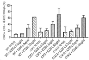


Classifications
-
- C—CHEMISTRY; METALLURGY
- C12—BIOCHEMISTRY; BEER; SPIRITS; WINE; VINEGAR; MICROBIOLOGY; ENZYMOLOGY; MUTATION OR GENETIC ENGINEERING
- C12N—MICROORGANISMS OR ENZYMES; COMPOSITIONS THEREOF; PROPAGATING, PRESERVING, OR MAINTAINING MICROORGANISMS; MUTATION OR GENETIC ENGINEERING; CULTURE MEDIA
- C12N5/00—Undifferentiated human, animal or plant cells, e.g. cell lines; Tissues; Cultivation or maintenance thereof; Culture media therefor
- C12N5/06—Animal cells or tissues; Human cells or tissues
- C12N5/0602—Vertebrate cells
- C12N5/0634—Cells from the blood or the immune system
- C12N5/0636—T lymphocytes
- C12N5/0638—Cytotoxic T lymphocytes [CTL] or lymphokine activated killer cells [LAK]
-
- A—HUMAN NECESSITIES
- A61—MEDICAL OR VETERINARY SCIENCE; HYGIENE
- A61K—PREPARATIONS FOR MEDICAL, DENTAL OR TOILETRY PURPOSES
- A61K35/00—Medicinal preparations containing materials or reaction products thereof with undetermined constitution
- A61K35/12—Materials from mammals; Compositions comprising non-specified tissues or cells; Compositions comprising non-embryonic stem cells; Genetically modified cells
- A61K35/14—Blood; Artificial blood
- A61K35/17—Lymphocytes; B-cells; T-cells; Natural killer cells; Interferon-activated or cytokine-activated lymphocytes
-
- A—HUMAN NECESSITIES
- A61—MEDICAL OR VETERINARY SCIENCE; HYGIENE
- A61K—PREPARATIONS FOR MEDICAL, DENTAL OR TOILETRY PURPOSES
- A61K39/00—Medicinal preparations containing antigens or antibodies
- A61K39/46—Cellular immunotherapy
- A61K39/461—Cellular immunotherapy characterised by the cell type used
- A61K39/4611—T-cells, e.g. tumor infiltrating lymphocytes [TIL], lymphokine-activated killer cells [LAK] or regulatory T cells [Treg]
-
- A—HUMAN NECESSITIES
- A61—MEDICAL OR VETERINARY SCIENCE; HYGIENE
- A61K—PREPARATIONS FOR MEDICAL, DENTAL OR TOILETRY PURPOSES
- A61K39/00—Medicinal preparations containing antigens or antibodies
- A61K39/46—Cellular immunotherapy
- A61K39/463—Cellular immunotherapy characterised by recombinant expression
- A61K39/4632—T-cell receptors [TCR]; antibody T-cell receptor constructs
-
- A—HUMAN NECESSITIES
- A61—MEDICAL OR VETERINARY SCIENCE; HYGIENE
- A61K—PREPARATIONS FOR MEDICAL, DENTAL OR TOILETRY PURPOSES
- A61K39/00—Medicinal preparations containing antigens or antibodies
- A61K39/46—Cellular immunotherapy
- A61K39/464—Cellular immunotherapy characterised by the antigen targeted or presented
- A61K39/4643—Vertebrate antigens
- A61K39/4644—Cancer antigens
- A61K39/464499—Undefined tumor antigens, e.g. tumor lysate or antigens targeted by cells isolated from tumor
-
- C—CHEMISTRY; METALLURGY
- C12—BIOCHEMISTRY; BEER; SPIRITS; WINE; VINEGAR; MICROBIOLOGY; ENZYMOLOGY; MUTATION OR GENETIC ENGINEERING
- C12N—MICROORGANISMS OR ENZYMES; COMPOSITIONS THEREOF; PROPAGATING, PRESERVING, OR MAINTAINING MICROORGANISMS; MUTATION OR GENETIC ENGINEERING; CULTURE MEDIA
- C12N5/00—Undifferentiated human, animal or plant cells, e.g. cell lines; Tissues; Cultivation or maintenance thereof; Culture media therefor
- C12N5/06—Animal cells or tissues; Human cells or tissues
- C12N5/0602—Vertebrate cells
- C12N5/0634—Cells from the blood or the immune system
- C12N5/0647—Haematopoietic stem cells; Uncommitted or multipotent progenitors
-
- C—CHEMISTRY; METALLURGY
- C12—BIOCHEMISTRY; BEER; SPIRITS; WINE; VINEGAR; MICROBIOLOGY; ENZYMOLOGY; MUTATION OR GENETIC ENGINEERING
- C12N—MICROORGANISMS OR ENZYMES; COMPOSITIONS THEREOF; PROPAGATING, PRESERVING, OR MAINTAINING MICROORGANISMS; MUTATION OR GENETIC ENGINEERING; CULTURE MEDIA
- C12N2501/00—Active agents used in cell culture processes, e.g. differentation
- C12N2501/10—Growth factors
- C12N2501/115—Basic fibroblast growth factor (bFGF, FGF-2)
-
- C—CHEMISTRY; METALLURGY
- C12—BIOCHEMISTRY; BEER; SPIRITS; WINE; VINEGAR; MICROBIOLOGY; ENZYMOLOGY; MUTATION OR GENETIC ENGINEERING
- C12N—MICROORGANISMS OR ENZYMES; COMPOSITIONS THEREOF; PROPAGATING, PRESERVING, OR MAINTAINING MICROORGANISMS; MUTATION OR GENETIC ENGINEERING; CULTURE MEDIA
- C12N2501/00—Active agents used in cell culture processes, e.g. differentation
- C12N2501/10—Growth factors
- C12N2501/125—Stem cell factor [SCF], c-kit ligand [KL]
-
- C—CHEMISTRY; METALLURGY
- C12—BIOCHEMISTRY; BEER; SPIRITS; WINE; VINEGAR; MICROBIOLOGY; ENZYMOLOGY; MUTATION OR GENETIC ENGINEERING
- C12N—MICROORGANISMS OR ENZYMES; COMPOSITIONS THEREOF; PROPAGATING, PRESERVING, OR MAINTAINING MICROORGANISMS; MUTATION OR GENETIC ENGINEERING; CULTURE MEDIA
- C12N2501/00—Active agents used in cell culture processes, e.g. differentation
- C12N2501/10—Growth factors
- C12N2501/155—Bone morphogenic proteins [BMP]; Osteogenins; Osteogenic factor; Bone inducing factor
-
- C—CHEMISTRY; METALLURGY
- C12—BIOCHEMISTRY; BEER; SPIRITS; WINE; VINEGAR; MICROBIOLOGY; ENZYMOLOGY; MUTATION OR GENETIC ENGINEERING
- C12N—MICROORGANISMS OR ENZYMES; COMPOSITIONS THEREOF; PROPAGATING, PRESERVING, OR MAINTAINING MICROORGANISMS; MUTATION OR GENETIC ENGINEERING; CULTURE MEDIA
- C12N2501/00—Active agents used in cell culture processes, e.g. differentation
- C12N2501/10—Growth factors
- C12N2501/16—Activin; Inhibin; Mullerian inhibiting substance
-
- C—CHEMISTRY; METALLURGY
- C12—BIOCHEMISTRY; BEER; SPIRITS; WINE; VINEGAR; MICROBIOLOGY; ENZYMOLOGY; MUTATION OR GENETIC ENGINEERING
- C12N—MICROORGANISMS OR ENZYMES; COMPOSITIONS THEREOF; PROPAGATING, PRESERVING, OR MAINTAINING MICROORGANISMS; MUTATION OR GENETIC ENGINEERING; CULTURE MEDIA
- C12N2501/00—Active agents used in cell culture processes, e.g. differentation
- C12N2501/10—Growth factors
- C12N2501/165—Vascular endothelial growth factor [VEGF]
-
- C—CHEMISTRY; METALLURGY
- C12—BIOCHEMISTRY; BEER; SPIRITS; WINE; VINEGAR; MICROBIOLOGY; ENZYMOLOGY; MUTATION OR GENETIC ENGINEERING
- C12N—MICROORGANISMS OR ENZYMES; COMPOSITIONS THEREOF; PROPAGATING, PRESERVING, OR MAINTAINING MICROORGANISMS; MUTATION OR GENETIC ENGINEERING; CULTURE MEDIA
- C12N2501/00—Active agents used in cell culture processes, e.g. differentation
- C12N2501/40—Regulators of development
- C12N2501/42—Notch; Delta; Jagged; Serrate
-
- C—CHEMISTRY; METALLURGY
- C12—BIOCHEMISTRY; BEER; SPIRITS; WINE; VINEGAR; MICROBIOLOGY; ENZYMOLOGY; MUTATION OR GENETIC ENGINEERING
- C12N—MICROORGANISMS OR ENZYMES; COMPOSITIONS THEREOF; PROPAGATING, PRESERVING, OR MAINTAINING MICROORGANISMS; MUTATION OR GENETIC ENGINEERING; CULTURE MEDIA
- C12N2501/00—Active agents used in cell culture processes, e.g. differentation
- C12N2501/50—Cell markers; Cell surface determinants
- C12N2501/51—B7 molecules, e.g. CD80, CD86, CD28 (ligand), CD152 (ligand)
-
- C—CHEMISTRY; METALLURGY
- C12—BIOCHEMISTRY; BEER; SPIRITS; WINE; VINEGAR; MICROBIOLOGY; ENZYMOLOGY; MUTATION OR GENETIC ENGINEERING
- C12N—MICROORGANISMS OR ENZYMES; COMPOSITIONS THEREOF; PROPAGATING, PRESERVING, OR MAINTAINING MICROORGANISMS; MUTATION OR GENETIC ENGINEERING; CULTURE MEDIA
- C12N2501/00—Active agents used in cell culture processes, e.g. differentation
- C12N2501/50—Cell markers; Cell surface determinants
- C12N2501/599—Cell markers; Cell surface determinants with CD designations not provided for elsewhere
-
- C—CHEMISTRY; METALLURGY
- C12—BIOCHEMISTRY; BEER; SPIRITS; WINE; VINEGAR; MICROBIOLOGY; ENZYMOLOGY; MUTATION OR GENETIC ENGINEERING
- C12N—MICROORGANISMS OR ENZYMES; COMPOSITIONS THEREOF; PROPAGATING, PRESERVING, OR MAINTAINING MICROORGANISMS; MUTATION OR GENETIC ENGINEERING; CULTURE MEDIA
- C12N2501/00—Active agents used in cell culture processes, e.g. differentation
- C12N2501/70—Enzymes
- C12N2501/72—Transferases (EC 2.)
- C12N2501/727—Kinases (EC 2.7.)
-
- C—CHEMISTRY; METALLURGY
- C12—BIOCHEMISTRY; BEER; SPIRITS; WINE; VINEGAR; MICROBIOLOGY; ENZYMOLOGY; MUTATION OR GENETIC ENGINEERING
- C12N—MICROORGANISMS OR ENZYMES; COMPOSITIONS THEREOF; PROPAGATING, PRESERVING, OR MAINTAINING MICROORGANISMS; MUTATION OR GENETIC ENGINEERING; CULTURE MEDIA
- C12N2506/00—Differentiation of animal cells from one lineage to another; Differentiation of pluripotent cells
- C12N2506/11—Differentiation of animal cells from one lineage to another; Differentiation of pluripotent cells from blood or immune system cells
-
- C—CHEMISTRY; METALLURGY
- C12—BIOCHEMISTRY; BEER; SPIRITS; WINE; VINEGAR; MICROBIOLOGY; ENZYMOLOGY; MUTATION OR GENETIC ENGINEERING
- C12N—MICROORGANISMS OR ENZYMES; COMPOSITIONS THEREOF; PROPAGATING, PRESERVING, OR MAINTAINING MICROORGANISMS; MUTATION OR GENETIC ENGINEERING; CULTURE MEDIA
- C12N2506/00—Differentiation of animal cells from one lineage to another; Differentiation of pluripotent cells
- C12N2506/45—Differentiation of animal cells from one lineage to another; Differentiation of pluripotent cells from artificially induced pluripotent stem cells
Landscapes
- Health & Medical Sciences (AREA)
- Life Sciences & Earth Sciences (AREA)
- Engineering & Computer Science (AREA)
- Immunology (AREA)
- Chemical & Material Sciences (AREA)
- Biomedical Technology (AREA)
- Cell Biology (AREA)
- General Health & Medical Sciences (AREA)
- Microbiology (AREA)
- Zoology (AREA)
- Biotechnology (AREA)
- Bioinformatics & Cheminformatics (AREA)
- Genetics & Genomics (AREA)
- Wood Science & Technology (AREA)
- Organic Chemistry (AREA)
- Hematology (AREA)
- Epidemiology (AREA)
- Animal Behavior & Ethology (AREA)
- Veterinary Medicine (AREA)
- Medicinal Chemistry (AREA)
- Public Health (AREA)
- Pharmacology & Pharmacy (AREA)
- Mycology (AREA)
- Biochemistry (AREA)
- General Engineering & Computer Science (AREA)
- Developmental Biology & Embryology (AREA)
- Oncology (AREA)
- Micro-Organisms Or Cultivation Processes Thereof (AREA)
- Medicines Containing Material From Animals Or Micro-Organisms (AREA)
- Peptides Or Proteins (AREA)
- Apparatus Associated With Microorganisms And Enzymes (AREA)
- Virology (AREA)
Abstract
본 발명은 다음을 포함하는 방법에 의한 TCR αβ+ T 세포의 집단의 생산에 관한 것이다: (i) 조혈 전구 세포(HPC)의 집단을 전구 T 세포로 분화시키는 단계 및 (ii) 전구 T 세포를 성숙시켜 TCR αβ+ T 세포 집단을 생산하는 단계. 단계 (i) 및 (ii) 중 하나 또는 둘 모두는 유도성 공동자극인자 리간드(ICOS-L)의 존재 하에 수행된다. 단계 (i) 및/또는 (ii)에서 ICOS-L의 존재는 αβ T 세포 수용체의 발현을 증가시키고/시키거나 TCR αβ+ 세포로 성숙하는 HPC 또는 전구 T 세포의 비율을 증가시킬 수 있다. 이는 예를 들어 면역요법을 위한 T 세포의 생산에 유용할 수 있다.The present invention relates to the production of a population of TCR αβ+ T cells by a method comprising: (i) differentiating the population of hematopoietic progenitor cells (HPC) into progenitor T cells and (ii) progenitor T cells; maturation to produce a TCR αβ+ T cell population. One or both of steps (i) and (ii) are performed in the presence of an inducible costimulatory factor ligand (ICOS-L). The presence of ICOS-L in step (i) and/or (ii) may increase the expression of the αβ T cell receptor and/or increase the proportion of HPC or progenitor T cells that mature into TCR αβ+ cells. This may be useful, for example, in the production of T cells for immunotherapy.
Description
본 발명은 예를 들어 면역요법에 사용하기 위한 T 세포의 생산에 관한 것이다.The present invention relates to the production of T cells, for example for use in immunotherapy.
면역치료제는 장기간 생존을 약속하면서 암 치료 분야를 변화시킬 준비가 되어 있다(McDermott et al., Cancer Treat Rev. 2014 Oct; 40(9): 1056-64). 환자 집단과 종양 유형의 범위를 확장하기 위한 새로운 면역조절 약물에 대한 분명한, 충족되지 않은 의학적 요구가 있다. 또한, 항종양 반응의 규모와 지속시간을 증진시키기 위한 새로운 작용제가 필요하다. 이러한 작용제의 개발은 지난 20년 동안 T 세포 면역을 제어하는 기본 원리에 대한 심층적인 이해로 인해 가능했다(Sharma and Allison, Cell. 2015 Apr 9; 161(2): 205-14). 이는 일반적으로 MHC 분자에 의해 제시되는 종양 관련 펩티드 항원을 인식하는 종양 특이적 CD4+ 및 CD8+ T 세포를 필요로 한다. 상이한 백신 접종 전략과 생체 외 확장된 종양 침윤 림프구의 입양 전달은 일부 경우에 종양 특이적 T 세포가 말기 암을 치료하는 능력을 증명하였다(Rosenberg et al., Nat Med. 2004 Sep; 10(9): 909-15). Immunotherapy is poised to change the field of cancer treatment, promising long-term survival (McDermott et al., Cancer Treat Rev. 2014 Oct; 40(9): 1056-64). There is a clear, unmet medical need for new immunomodulatory drugs to expand the range of patient populations and tumor types. There is also a need for new agents to enhance the magnitude and duration of anti-tumor responses. The development of these agents has been possible over the past two decades due to an in-depth understanding of the underlying principles controlling T cell immunity (Sharma and Allison, Cell. 2015 Apr 9; 161(2): 205-14). This requires tumor-specific CD4+ and CD8+ T cells that generally recognize tumor-associated peptide antigens presented by MHC molecules. Different vaccination strategies and adoptive transfer of ex vivo expanded tumor-infiltrating lymphocytes have demonstrated the ability of tumor-specific T cells to treat terminal cancer in some cases (Rosenberg et al., Nat Med. 2004 Sep; 10(9)). : 909-15).
그러나 현재의 입양 T 세포 요법은 적합한 환자 및 종양 특이적 T 세포의 부족으로 인해 제한되며, 면역 요법에서 효과적인 사용을 위해 치료적으로 충분하고 기능적인 항원 특이적 T 세포에 대한 요구가 있다.However, current adoptive T cell therapies are limited by the lack of suitable patient and tumor-specific T cells, and there is a need for therapeutically sufficient and functional antigen-specific T cells for effective use in immunotherapy.
본 발명자들은 유도성 공동자극인자 리간드(Inducible Co-stimulator ligand, ICOS-L)의 존재 하에 선행 세포(antecedent cell)로부터 T 세포가 생성될 때 αβ T 세포 수용체의 발현이 증가됨을 인식하였다. 이는 예를 들어 면역요법을 위한 T 세포의 생산에 유용할 수 있다.The present inventors recognized that the expression of αβ T cell receptor is increased when T cells are generated from antecedent cells in the presence of an inducible co-stimulator ligand (ICOS-L). This may be useful, for example, in the production of T cells for immunotherapy.
본 발명의 제1 양태는 TCR αβ+ T 세포의 집단(population)을 생산하는 방법으로서,A first aspect of the present invention is a method for producing a population of TCR αβ+ T cells,
(i) 조혈 전구 세포(haematopoietic progenitor cell, HPC)의 집단을 전구 T 세포(progenitor T cell)로 분화시키는 단계; 및(i) differentiating a population of haematopoietic progenitor cells (HPCs) into progenitor T cells; and
(ii) 전구 T 세포를 성숙시켜 TCR αβ+ T 세포 집단을 생산하는 단계를 포함하며, (ii) maturing the progenitor T cells to produce a TCR αβ+ T cell population;
여기서 (i) 및 (ii) 중 하나 또는 둘 모두는 유도성 공동자극인자 리간드(Inducible Co-stimulator ligand, ICOS-L)의 존재 하에 수행되는 것인 방법을 제공한다.wherein one or both of (i) and (ii) is performed in the presence of an inducible co-stimulator ligand (ICOS-L).
단계 (i) 및/또는 (ii)에서 ICOS-L의 존재는 TCR αβ+ 세포로 성숙하는 HPC 또는 전구 T 세포의 비율을 증가시킬 수 있다.The presence of ICOS-L in step (i) and/or (ii) may increase the proportion of HPC or progenitor T cells that mature into TCR αβ+ cells.
제1 양태의 일부 구현예에서, TCR αβ+T 세포의 집단을 생산하는 방법은In some embodiments of the first aspect, the method of producing a population of TCR αβ+T cells comprises:
(i) ICOS-L의 존재 하에 HPC 집단을 T 세포 전구 세포로 분화시키는 단계; 및(i) differentiating the HPC population into T cell progenitor cells in the presence of ICOS-L; and
(ii) 전구 T 세포를 성숙시켜 TCR αβ+ T 세포 집단을 생산하는 단계를 포함할 수 있다.(ii) maturing the progenitor T cells to produce a TCR αβ+ T cell population.
단계 (i)에서 ICOS-L의 존재는 TCR αβ+ 전구 T 세포, 예를 들어 TCR αβ+ CD45+CD3+ 세포로 분화되고 TCR αβ+ T 세포로 성숙되는 HPC의 비율을 증가시킬 수 있다.The presence of ICOS-L in step (i) can increase the proportion of HPCs that differentiate into TCR αβ+ progenitor T cells, eg, TCR αβ+ CD45+CD3+ cells and mature into TCR αβ+ T cells.
제1 양태의 다른 구현예에서, TCR αβ+T 세포의 집단을 생산하는 방법은In another embodiment of the first aspect, the method of producing a population of TCR αβ+T cells comprises:
(i) HPC 집단을 T 세포 전구 세포로 분화시키는 단계; 및(i) differentiating the HPC population into T cell progenitor cells; and
(ii) ICOS-L의 존재 하에 전구 T 세포를 성숙시켜 TCR αβ+ T 세포 집단을 생산하는 단계를 포함할 수 있다.(ii) maturing the progenitor T cells in the presence of ICOS-L to produce a TCR αβ+ T cell population.
단계 (ii)에서 ICOS-L의 존재는 TCR αβ+ 세포, 예를 들어 TCR αβ+ CD8+ CD4+ 세포로 성숙하는 전구 T 세포의 비율을 증가시킬 수 있다.The presence of ICOS-L in step (ii) may increase the proportion of progenitor T cells that mature into TCR αβ+ cells, eg TCR αβ+ CD8+ CD4+ cells.
세포는 ICOS-L을 포함하는 배지에서 또는 ICOS-L로 코팅된 배양 용기 표면과 같은 표면에서 분화되거나 성숙될 수 있다. Cells can be differentiated or matured in a medium comprising ICOS-L or on a surface, such as the surface of a culture vessel coated with ICOS-L.
바람직하게는, 제1 양태의 방법에 사용하기 위한 HPC는 유도 만능 줄기 세포(induced pluripotent stem cell, iPSC)의 집단을 HPC로 분화시켜 생산할 수 있다. Preferably, the HPC for use in the method of the first aspect can be produced by differentiating a population of induced pluripotent stem cells (iPSCs) into HPCs.
제1 양태의 일부 구현예에서, TCR αβ+ 세포의 집단을 생산하는 방법은In some embodiments of the first aspect, the method of producing a population of TCR αβ+ cells comprises:
(i) iPSC 집단을 HPC로 분화시키는 단계;(i) differentiating the iPSC population into HPCs;
(ii) HPC를 전구 T 세포로 분화시키는 단계; 및(ii) differentiating the HPC into progenitor T cells; and
(iii) 전구 T 세포를 TCR αβ+ T 세포의 집단으로 성숙시키는 단계를 포함할 수 있으며, (iii) maturing the progenitor T cells into a population of TCR αβ+ T cells,
여기서 단계 (ii) 및 (iii) 중 하나 또는 둘 모두는 유도성 공동자극인자 리간드(ICOS-L)의 존재 하에 수행된다.wherein one or both of steps (ii) and (iii) are performed in the presence of an inducible costimulatory factor ligand (ICOS-L).
제1 양태의 방법은 TCR αβ+ T 세포를 활성화 및 확장시켜 TCR αβ+ T 세포의 집단 또는 TCR αβ+ CD4+ T 세포의 집단을 생산하는 단계를 추가로 포함할 수 있다.The method of the first aspect may further comprise activating and expanding TCR αβ+ T cells to produce a population of TCR αβ+ T cells or a population of TCR αβ+ CD4+ T cells.
본 발명의 제2 양태는 제1 양태의 방법에 의해 생산된 TCR αβ+ T 세포의 집단을 제공한다. A second aspect of the invention provides a population of TCR αβ+ T cells produced by the method of the first aspect.
본 발명의 제3 양태는 제2 양태의 TCR αβ+ T 세포의 집단 및 약학적으로 허용 가능한 부형제를 포함하는 약학 조성물을 제공한다.A third aspect of the present invention provides a pharmaceutical composition comprising the population of TCR αβ+ T cells of the second aspect and a pharmaceutically acceptable excipient.
본 발명의 제4 양태는 치료를 필요로 하는 개체에게 치료적 유효량의 제2 양태의 TCR αβ+ T 세포의 집단을 투여하는 단계를 포함하는 치료 방법을 제공한다.A fourth aspect of the invention provides a method of treatment comprising administering to an individual in need thereof a therapeutically effective amount of a population of TCR αβ+ T cells of the second aspect.
제5 양태는 T 세포 배양 표면용 코팅 조성물로서, ICOS-L을 포함하는 조성물을 제공한다. A fifth aspect provides a composition comprising ICOS-L as a coating composition for a T cell culture surface.
제6 양태는 T 세포 배양용 배양 용기(culture vessel)로서, ICOS-L로 코팅된 표면을 포함하는 용기를 제공한다.A sixth aspect provides a culture vessel for culturing T cells, the vessel comprising a surface coated with ICOS-L.
본 발명의 이러한 양태와 다른 양태 및 구현예를 아래에서 더욱 상세하게 설명한다.These and other aspects and embodiments of the invention are described in greater detail below.
상세한 설명details
본 발명은 αβ T 세포 수용체의 발현을 증가시키기 위한 유도성 공동자극인자 리간드(ICOS-L)의 용도에 관한 것이다. 조혈 전구 세포의 전구 T 세포로의 분화 및/또는 전구 T 세포의 이중 양성 CD4+ CD8+ T 세포로의 성숙 중 ICOS-L의 존재는 αβ T 세포 수용체를 발현하는 집단에서 T 세포의 비율을 증가시키기 위해 본원에 제시되어 있다. 그러면 이중 양성 CD4+ CD8+ TCR αβ+ T 세포가 활성화되고 확장되어 단일 양성 CD8+ 또는 CD4+ TCR αβ+ T 세포를 생산할 수 있으며, 이는 예를 들어 면역요법에서 유용할 수 있다. The present invention relates to the use of inducible costimulatory factor ligands (ICOS-L) for increasing the expression of αβ T cell receptors. The presence of ICOS-L during differentiation of hematopoietic progenitor cells into progenitor T cells and/or maturation of progenitor T cells into double positive CD4+ CD8+ T cells is important to increase the proportion of T cells in a population expressing the αβ T cell receptor. presented herein. Double positive CD4+ CD8+ TCR αβ+ T cells can then be activated and expanded to produce single positive CD8+ or CD4+ TCR αβ+ T cells, which may be useful, for example, in immunotherapy.
TCR αβ+ T 세포의 집단은 (i) 조혈 전구 세포 집단을 전구 T 세포로 분화시키고, (ii) 전구 T 세포를 성숙시켜 CD4+ CD8+ T 세포의 집단을 생산함으로써 본원에 기재된 바와 같이 생산되며, 여기서 (i) 및 (ii) 중 하나 또는 둘 모두는 유도성 공동 자극인자 리간드(ICOS-L)의 존재 하에 수행된다. 두 단계 중 하나 또는 두 단계 모두에서 ICOS-L의 존재는 분화 및/또는 TCR αβ+ T 세포로 성숙하는 세포의 비율을 증가시킨다. TCR αβ+ 세포는 CD4+ CD8+ T 세포일 수 있으며, 이는 그 후 활성화되고 단일 양성 CD8+ 또는 단일 양성 CD4+ T 세포로 확장될 수 있다.The population of TCR αβ+ T cells is produced as described herein by (i) differentiating the hematopoietic progenitor cell population into progenitor T cells, and (ii) maturing the progenitor T cells to produce a population of CD4+ CD8+ T cells, wherein Either or both of (i) and (ii) are performed in the presence of an inducible co-stimulator ligand (ICOS-L). The presence of ICOS-L at either or both stages increases the proportion of cells that differentiate and/or mature into TCR αβ+ T cells. TCR αβ+ cells can be CD4+ CD8+ T cells, which can then be activated and expand into single positive CD8+ or single positive CD4+ T cells.
조혈 전구 세포(HPC)가 T 세포 전구 세포로 분화되는 동안 ICOS-L의 존재는 분화 중에 ICOS-L의 부재에 비해 전구 T 세포의 집단에서 TCR αβ+ 세포의 비율을 증가시킬 수 있다. 예를 들어, ICOS-L의 존재는 CD3+ 세포 집단에서 TCR αβ+ 세포의 비율을 증가시킬 수 있다. T 세포 전구 세포가 TCR αβ+ T 세포로 성숙하는 동안 ICOS-L의 존재는 성숙 중에 ICOS-L의 부재에 비해 TCR αβ+ T 세포로 성숙하는 전구 T 세포의 비율을 증가시킬 수 있다. 예를 들어, TCR αβ+ CD8+ 단일 양성(SP) T 세포의 비율 및/또는 TCR αβ+ CD4+ CD8+ 이중 양성(DP) T 세포의 비율은 ICOS-L에 의해 증가될 수 있다. The presence of ICOS-L during differentiation of hematopoietic progenitor cells (HPCs) into T cell progenitor cells may increase the proportion of TCR αβ+ cells in the population of progenitor T cells compared to the absence of ICOS-L during differentiation. For example, the presence of ICOS-L may increase the proportion of TCR αβ+ cells in the CD3+ cell population. The presence of ICOS-L during maturation of T-cell progenitor cells into TCR αβ+ T cells may increase the proportion of progenitor T cells that mature into TCR αβ+ T cells compared to the absence of ICOS-L during maturation. For example, the proportion of TCR αβ+ CD8+ single positive (SP) T cells and/or the proportion of TCR αβ+ CD4+ CD8+ double positive (DP) T cells can be increased by ICOS-L.
본원에 기재된 방법은 본원에 기재된 바와 같은 TCR αβ+ T 세포를 생산하는 방법에 사용하기 위한 조혈 전구 세포(HPC)의 집단을 제공하는 단계를 추가로 포함할 수 있다. The methods described herein may further comprise providing a population of hematopoietic progenitor cells (HPCs) for use in a method of producing TCR αβ+ T cells as described herein.
HPC(조혈 줄기 및 전구 세포 또는 HSPC라고도 함)는 조혈 계통에 관여하고 단핵구, B 세포, NK 세포, NKT 세포, TIL 및 T 세포를 포함하는, 골수계 및 림프계를 비롯한 모든 혈액 세포 유형으로 추가 조혈 분화할 수 있는 다능성 줄기 세포이다. HPC는 CD34를 발현할 수 있다. HPC는 CD45를 공동 발현할 수 있다. HPC는 또한 CD117, CD133, CD45 및 FLK1(KDR 또는 VEGFR2로도 알려짐)을 공동 발현할 수 있다. HPC는 CD38 및 기타 계통 특이적 마커의 발현에 대해 음성일 수 있다. 예를 들어, HPC는 CD34+ CD133+ CD45+ FLK1+ CD38- 중 하나 이상의, 바람직하게는 모두를 표시할 수 있다. HPCs (also called hematopoietic stem and progenitor cells or HSPCs) are involved in the hematopoietic lineage and further hematopoiesis with all blood cell types including myeloid and lymphoid lines, including monocytes, B cells, NK cells, NKT cells, TIL and T cells. It is a pluripotent stem cell capable of differentiating. HPC can express CD34. HPCs can co-express CD45. HPC can also co-express CD117, CD133, CD45 and FLK1 (also known as KDR or VEGFR2). HPCs may be negative for expression of CD38 and other lineage specific markers. For example, the HPC may display one or more, preferably all, of CD34+ CD133+ CD45+ FLK1+ CD38−.
일부 바람직한 구현예에서, HPC는 시험관 내에서 유도 만능 줄기 세포(iPSC)로부터 생산된다.In some preferred embodiments, HPCs are produced from induced pluripotent stem cells (iPSCs) in vitro.
예를 들어, TCR αβ+ T 세포의 집단은For example, a population of TCR αβ+ T cells is
(i) iPSC 집단을 HPC로 분화시키는 단계,(i) differentiating the iPSC population into HPCs;
(ii) HPC를 T 세포 전구 세포로 분화시키는 단계, 및(ii) differentiating the HPC into T cell progenitor cells, and
(ii) 전구 T 세포의 집단을 성숙시켜 TCR αβ+ T 세포의 집단을 생산하는 단계를 포함하며, (ii) maturing the population of progenitor T cells to produce a population of TCR αβ+ T cells;
여기서 단계 (ii) 및 단계 (iii) 중 하나 또는 둘 모두는 ICOS-L의 존재 하에 수행되는 방법에 의해 생산될 수 있다.wherein one or both of steps (ii) and (iii) may be produced by a method performed in the presence of ICOS-L.
iPSC는 중배엽 및 조혈 내피 단계를 포함하는 3단계 과정을 사용하여 HPC로 분화될 수 있다. 예를 들어, 방법은iPSCs can be differentiated into HPCs using a three-step process involving mesodermal and hematopoietic endothelial stages. For example, the method
(i) iPSC 집단을 중배엽 세포로 분화시키는 단계, (i) differentiating the iPSC population into mesodermal cells;
(ii) 중배엽 세포를 조혈 내피 세포로 분화시키는 단계, 및(ii) differentiating the mesoderm cells into hematopoietic endothelial cells, and
(iii) 조혈 내피 세포를 HPC 집단으로 분화시키는 단계를 포함할 수 있다. (iii) differentiating the hematopoietic endothelial cells into a HPC population.
유도 만능 줄기 세포(iPSC)는 비 만능성의, 완전히 분화된 공여자 또는 선행 세포에서 유래된 만능 세포이다. iPSC는 시험관 내에서 자가 재생이 가능하고 미분화 표현형을 나타내며 잠재적으로 임의의 3개의 배엽(내배엽, 중배엽 및 외배엽)의 임의의 태아 또는 성체 세포 유형으로 분화할 수 있다. iPSC의 집단은 클론성, 즉 단일 공통 조상 세포에서 유래한 유전적으로 동일한 세포일 수 있다.Induced pluripotent stem cells (iPSCs) are pluripotent cells derived from non-pluripotent, fully differentiated donor or progenitor cells. iPSCs are capable of self-renewal in vitro, exhibit an undifferentiated phenotype and can potentially differentiate into any fetal or adult cell type of any three germ layers (endoderm, mesoderm and ectoderm). A population of iPSCs may be clonal, ie, genetically identical cells derived from a single common progenitor cell.
iPSC는 다음의 다능성 관련 마커 중 하나 이상: POU5f1(Oct4), Sox2, 알칼리 포스파타제, SSEA-3, Nanog, SSEA-4, Tra-1-60, KLF4 및 c-myc, 바람직하게는 POU5f1, NANOG 및 SOX2 중 하나 이상을 발현할 수 있다. iPSC는 Bra, Sox17, FoxA2, αFP, Sox1, NCAM, GATA6, GATA4, Hand1 및 CDX2와 같은 특정 분화 운명과 관련된 마커가 부족할 수 있다. 특히, iPSC는 내배엽 운명과 관련된 마커가 부족할 수 있다. iPSCs have one or more of the following pluripotency related markers: POU5f1 (Oct4), Sox2, alkaline phosphatase, SSEA-3, Nanog, SSEA-4, Tra-1-60, KLF4 and c-myc, preferably POU5f1, NANOG and SOX2. iPSCs may lack markers associated with specific differentiation fates, such as Bra, Sox17, FoxA2, αFP, Sox1, NCAM, GATA6, GATA4, Hand1 and CDX2. In particular, iPSCs may lack markers related to endoderm fate.
바람직하게는, iPSC는 인간 IPSC(hiPSC)이다.Preferably, the iPSCs are human IPSCs (hiPSCs).
일부 구현예에서, iPSC는 예를 들어 HLA 유전자 또는 면역원성 또는 GVHD와 관련된 다른 유전자를 불활성화 또는 결실시키도록 유전자 편집될 수 있거나, 예를 들어 TCR, CAR 또는 NKCR과 같은 외인성 항원 수용체를 암호화하는 핵산을 포함하도록 유전자 편집될 수 있다. In some embodiments, iPSCs can be genetically edited to inactivate or delete, for example, the HLA gene or other genes associated with immunogenicity or GVHD, or that encodes an exogenous antigen receptor, such as, for example, TCR, CAR or NKCR. It can be genetically edited to include nucleic acids.
IPSC는 공여자 개체와 같은 공급원에서 얻은 체세포 또는 기타 선행 세포일 수 있는 공여자 세포로부터 유래되거나 재프로그래밍될 수 있다. 공여자 세포는 포유동물, 바람직하게는 인간 세포일 수 있다. 적합한 공여자 세포는 성체 섬유아세포 및 혈액 세포, 예를 들어 HPC 또는 단핵 세포와 같은 말초 혈액 세포를 포함한다.IPSCs may be derived or reprogrammed from a donor cell, which may be a somatic cell or other progenitor cell obtained from the same source as the donor subject. The donor cell may be a mammalian, preferably a human cell. Suitable donor cells include adult fibroblasts and blood cells, for example peripheral blood cells such as HPC or mononuclear cells.
본원에 기재된 바와 같이 iPSC로 재프로그래밍하기에 적합한 공여자 세포는 공여자 개체로부터 얻을 수 있다. 일부 구현예에서, 공여자 개체는 본원에 기재된 바와 같은 생산 후에 T 세포가 투여될 수용자 개체와 동일한 사람일 수 있다(자가 치료). 다른 구현예에서, 공여자 개체는 본원에 기재된 바와 같은 생산 후에 T 세포가 투여될 수용자 개체와 상이한 사람일 수 있다(동종 치료). 예를 들어, 공여자 개체는 암을 앓고 있는 수용자 개체와 (공여 전후에) 인간 백혈구 항원(HLA)이 일치하는 건강한 개체일 수 있다. 다른 구현예에서, 공여자 개체는 수용자 개체와 HLA가 일치하지 않을 수 있다. 바람직하게는, 공여자 개체는 신생아일 수 있으며, 예를 들어 공여자 세포는 제대혈 샘플에서 얻을 수 있다.Donor cells suitable for reprogramming into iPSCs as described herein can be obtained from a donor subject. In some embodiments, the donor subject may be the same person as the recipient subject to which the T cells will be administered after production as described herein (self-treatment). In another embodiment, the donor subject may be a different person from the recipient subject to which the T cells will be administered after production as described herein (allogeneic treatment). For example, a donor individual may be a healthy individual with human leukocyte antigen (HLA) matching (before and after donation) to a recipient individual suffering from cancer. In other embodiments, the donor subject may not have an HLA match with the recipient subject. Preferably, the donor subject may be a newborn, eg, the donor cells may be obtained from a cord blood sample.
적합한 공여자 개체는 바람직하게는 전염성 바이러스(예를 들어 HIV, HPV, CMV) 및 우발적 작용제(예를 들어 박테리아, 마이코플라스마)가 없고 알려진 유전적 이상이 없다. A suitable donor subject is preferably free of infectious viruses (eg HIV, HPV, CMV) and adventitious agents (eg bacteria, mycoplasma) and free of known genetic abnormalities.
일부 구현예에서, 재프로그래밍을 위한 말초 혈액 세포, 예컨대 HPC의 집단은 공여자 개체로부터 수득된 혈액 샘플, 바람직하게는 탯줄 샘플로부터 단리될 수 있다. HPC 및 기타 말초혈액 세포의 단리에 적합한 방법은 당해 분야에 잘 알려져 있으며, 예를 들어 자기 활성화 세포 분류(예를 들어, 문헌[Gaudernack et al 1986 J Immunol Methods 90 179] 참고), 형광 활성화 세포 분류(FACS: 예를 들어, 문헌[Rheinherz et al (1979) PNAS 76 4061] 참고) 및 세포 패닝(cell panning)(예를 들어, 문헌[Lum et al (1982) Cell Immunol 72 122] 참고)을 포함한다. HPC는 CD34의 발현에 의해 혈액 세포 샘플에서 확인될 수 있다. 다른 구현예에서, 재프로그래밍을 위한 섬유아세포의 집단은 콜라게나제 또는 트립신을 사용한 분산 및 적절한 세포 배양 조건에서의 성장 후 피부 생검으로부터 단리될 수 있다.In some embodiments, a population of peripheral blood cells, such as HPCs, for reprogramming can be isolated from a blood sample obtained from a donor subject, preferably from an umbilical cord sample. Methods suitable for the isolation of HPC and other peripheral blood cells are well known in the art and include, for example, magnetically activated cell sorting (see, eg, Gaudernack et al 1986 J Immunol Methods 90 179), fluorescence activated cell sorting. (FACS: see, e.g., Rheinherz et al (1979) PNAS 76 4061) and cell panning (see, e.g., Lum et al (1982) Cell Immunol 72 122). do. HPC can be identified in blood cell samples by expression of CD34. In another embodiment, a population of fibroblasts for reprogramming can be isolated from a skin biopsy after dispersion with collagenase or trypsin and growth in appropriate cell culture conditions.
일부 구현예에서, IPSC는 항원 특이적 T 세포로부터 유래될 수 있다. 예를 들어, T 세포는 클래스 1 MHC와 복합화되어 표시되는 종양 항원과 같은 항원에 결합하는 αβ TCR을 암호화하는 핵산을 포함할 수 있다. iPSC 생성에 사용하기 위한 항원 특이적 T 세포는 수지상 세포와 같은 항원 제시 세포 표면의 클래스 I 또는 II MHC 분자에 표시된 표적 항원으로부터의 펩티드 에피토프로 다양한 T 세포 집단을 스크리닝하거나, 또는 암 환자의 종양 샘플로부터 단리하여 수득될 수 있다. In some embodiments, IPSCs may be derived from antigen specific T cells. For example, a T cell may contain a nucleic acid encoding an αβ TCR that binds to an antigen, such as a tumor antigen, displayed in complex with class 1 MHC. Antigen-specific T cells for use in the generation of iPSCs include screening diverse T cell populations with peptide epitopes from target antigens displayed on class I or II MHC molecules on the surface of antigen-presenting cells, such as dendritic cells, or tumor samples from cancer patients. It can be obtained by isolation from
공여자 세포는 일반적으로 Oct4, Sox2 및 Klf4와 같은 재프로그래밍 인자를 세포에 도입함으로써 iPSC로 재프로그래밍된다. 재프로그래밍 인자는 단백질 또는 암호화 핵산일 수 있고, 플라스미드, 트랜스포존 또는 더욱 바람직하게는 바이러스 형질감염 또는 직접적인 단백질 전달을 포함하는 임의의 적합한 기술에 의해 분화된 세포 내로 도입될 수 있다. 기타 재프로그래밍 인자, 예를 들어 Klf-1, Klf-2, Klf-4 및 Klf-5와 같은 Klf 유전자; C-myc, L-myc 및 N-myc와 같은 Myc 유전자; 나노그(Nanog); SV40 대형 T 항원; Lin28; 및 p53과 같은 짧은 헤어핀(shRNA) 표적화 유전자는 또한 세포에 도입되어 유도 효율을 높일 수 있다. 재프로그래밍 인자를 도입한 후 공여자 세포를 배양할 수 있다. 다능성 마커를 발현하는 세포는 단리 및/또는 정제되어 iPSC 집단을 생산할 수 있다. iPSC의 생산 기술은 당해 분야에 잘 알려져 있다(Yamanaka et al Nature 2007; 448:313-7; Yamanaka 6 2007 Jun 7; 1(1):39-49; Kim et al Nature. 2008 Jul 31; 454(7204):646-50; Takahashi Cell. 2007 Nov 30; 131(5):861-72. Park et al Nature. 2008 Jan 10; 451(7175):141-6; Kimet et al Cell Stem Cell. 2009 Jun 5;4(6):472-6; Vallier, L., et al. Stem Cells, 2009. 9999(999A): p. N/A; Baghbaderani et al 2016; Stem Cell Rev. 2016 Aug; 12(4):394-420; Baghbaderani et al. (2015) Stem Cell Reports, 5(4), 647-659).Donor cells are generally reprogrammed into iPSCs by introducing reprogramming factors such as Oct4, Sox2 and Klf4 into the cells. The reprogramming factor may be a protein or an encoding nucleic acid and may be introduced into a differentiated cell by any suitable technique including plasmid, transposon or more preferably viral transfection or direct protein delivery. other reprogramming factors such as Klf genes such as Klf-1, Klf-2, Klf-4 and Klf-5; Myc genes such as C-myc, L-myc and N-myc; Nanog; SV40 large T antigen; Lin28; and short hairpin (shRNA) targeting genes such as p53 can also be introduced into cells to increase induction efficiency. After introduction of the reprogramming factor, the donor cells can be cultured. Cells expressing a pluripotency marker can be isolated and/or purified to produce a population of iPSCs. Techniques for the production of iPSCs are well known in the art (Yamanaka et al Nature 2007; 448:313-7; Yamanaka 6 2007
iPSC의 배양 및 유지를 위해 통상적인 기술이 사용될 수 있다(Vallier, L. et al Dev. Biol. 275, 403-421 (2004), Cowan, C.A. et al. N. Engl. J. Med. 350, 1353-1356 (2004), Joannides, A. et al. Stem Cells 24, 230-235 (2006) Klimanskaya, I. et al. Lancet 365, 1636-1641 (2005), Ludwig, T.E. et al. Nat. Biotechnol. 24, 185-187 (2006)). 본 방법에 사용하기 위한 iPSC는 정의된 조건에서 또는 공급자 세포(feeder cell)에서 성장시킬 수 있다. 예를 들어, iPSC는 적절한 밀도(예를 들어 105 내지 106개 세포/60 mm 접시)의 조사된 마우스 배아 섬유아세포(MEF)와 같은 공급자 세포 층의 배양 접시에서 또는 공급자 조정된(feeder conditioned) 또는 정의된 iPSC 유지 배지에서 적절한 기질에서 통상적으로 배양될 수 있다. 본 방법에 사용하기 위한 iPSC는 효소적 또는 기계적 수단에 의해 계대될 수 있다. 일부 구현예에서, iPSC는 mTeSRTM1 또는 TeSRTM2(StemCell Technologies) 또는 E8 플렉스(flex)(Life Thermo) 배양 배지와 같은 iPSC 유지 배지에서 마트리겔(matrigelTM) 또는 비브로넥틴과 같은 ECM 단백질 상에서 계대될 수 있다. Conventional techniques can be used for culturing and maintaining iPSCs (Vallier, L. et al Dev. Biol. 275, 403-421 (2004), Cowan, CA et al. N. Engl. J. Med. 350, 1353-1356 (2004), Joannides, A. et al. Stem Cells 24, 230-235 (2006) Klimanskaya, I. et al. Lancet 365, 1636-1641 (2005), Ludwig, TE et al. Nat. Biotechnol 24, 185-187 (2006)). iPSCs for use in this method can be grown under defined conditions or in feeder cells. For example, iPSCs can be grown in culture dishes of a feeder cell layer such as irradiated mouse embryonic fibroblasts (MEFs) at an appropriate density (
본원에 기재된 방법의 단계에서 세포 집단의 분화 및 성숙은 분화 인자 세트가 보충된 배양 배지에서 세포를 배양함으로써 유도된다. 각 배양 배지에 대해 열거된 분화 인자 세트는 바람직하게는 완전하고, 배지에는 다른 분화 인자가 없을 수 있다. 바람직한 구현예에서, 배양 배지는 화학적으로 규정된 배지이다. 예를 들어, 배양 배지는 후술하는 바와 같이 유효량의 하나 이상의 분화 인자가 보충된 화학적으로 규정된 영양 배지로 구성될 수 있다. 화학적으로 규정된 영양 배지는 하나 이상의 무혈청 배양 배지 보충제가 보충된 기본 배지를 포함할 수 있다.Differentiation and maturation of the cell population in the steps of the methods described herein is induced by culturing the cells in a culture medium supplemented with a set of differentiation factors. The set of differentiation factors listed for each culture medium is preferably complete, and the medium may be free of other differentiation factors. In a preferred embodiment, the culture medium is a chemically defined medium. For example, the culture medium may consist of a chemically defined nutrient medium supplemented with an effective amount of one or more differentiation factors as described below. The chemically defined nutrient medium may comprise a basal medium supplemented with one or more serum-free culture medium supplements.
분화 인자는 포유동물 세포에서 분화를 매개하는 신호전달 경로를 조절, 예를 들어 촉진 또는 억제하는 인자이다. 분화 인자는 성장 인자, 사이토카인 및 액티빈(Activin)/노달(Nodal), FGF, Wnt 또는 BMP 또는 이들의 신호전달 경로 중 하나 이상을 조절하는 소분자를 포함할 수 있다. 분화 인자의 예로는 액티빈/노달, FGF, BMP, 레티노산, 혈관 내피 성장 인자(VEGF), 줄기 세포 인자(SCF), TGFβ 리간드, GDF, LIF, 인터류킨, GSK-3 억제제 및 포스파티딜이노시톨 3-키나제(PI3K) 억제제가 포함된다.Differentiation factors are factors that regulate, eg, promote or inhibit, signaling pathways that mediate differentiation in mammalian cells. Differentiation factors may include growth factors, cytokines and small molecules that modulate one or more of Activin/Nodal, FGF, Wnt or BMP or their signaling pathways. Examples of differentiation factors include activin/nodal, FGF, BMP, retinoic acid, vascular endothelial growth factor (VEGF), stem cell factor (SCF), TGFβ ligand, GDF, LIF, interleukins, GSK-3 inhibitors and phosphatidylinositol 3- kinase (PI3K) inhibitors.
본원에 기재된 배지 중 하나 이상에서 사용되는 분화 인자에는 TGFβ 리간드, 예컨대, 액티빈, 섬유아세포 성장 인자(fibroblast growth factor, FGF), 골 형태형성 단백질(bone morphogenetic protein, BMP), 줄기 세포 인자(stem cell factor, SCF), 혈관 내피 성장 인자(vascular endothelial growth factor, VEGF), GSK-3 억제제(예를 들어 CHIR-99021), 인터류킨 및 호르몬, 예를 들어 IGF-1 및 안지오텐신 II가 포함된다. 분화 인자는 배지에서 배양된 세포에서 신호전달 경로를 조절하는데 효과적인 양으로 본원에 기재된 배지에 존재할 수 있다.Differentiation factors used in one or more of the media described herein include TGFβ ligands such as activin, fibroblast growth factor (FGF), bone morphogenetic protein (BMP), stem cell factor (stem). cell factor (SCF), vascular endothelial growth factor (VEGF), GSK-3 inhibitors (eg CHIR-99021), interleukins and hormones such as IGF-1 and angiotensin II. A differentiation factor may be present in the medium described herein in an amount effective to modulate a signaling pathway in cells cultured in the medium.
일부 구현예에서, 상기 또는 하기에 열거된 분화 인자는 동일한 신호전달 경로에 대해 동일한 효과(즉, 자극 또는 억제)를 갖는 인자에 의해 배양 배지에서 대체될 수 있다. 적합한 인자는 당해 분야에 공지되어 있으며 단백질, 핵산, 항체 및 소분자를 포함한다.In some embodiments, the differentiation factors listed above or below can be replaced in the culture medium by factors that have the same effect (ie, stimulate or inhibit) on the same signaling pathway. Suitable factors are known in the art and include proteins, nucleic acids, antibodies, and small molecules.
각 단계 동안 세포 집단의 분화 정도는 분화하는 세포 집단에서 하나 이상의 세포 마커의 발현을 모니터링 및/또는 검출함으로써 결정될 수 있다. 예를 들어, 더 분화된 세포 유형의 특징적인 마커의 발현의 증가 또는 덜 분화된 세포 유형의 특징적인 마커의 발현의 감소가 결정될 수 있다. 세포 마커의 발현은 면역세포화학, 면역형광, RT-PCR, 면역블롯팅, 형광 활성화 세포 분류(FACS), 및 효소 분석을 포함하는 임의의 적합한 기술에 의해 결정될 수 있다. 바람직한 구현예에서, 마커가 세포 표면에서 검출 가능한 경우 세포는 마커를 발현한다고 할 수 있다. 예를 들어, 본원에서 마커를 발현하지 않는 것으로 언급된 세포는 마커 유전자의 활성 전사 및 세포 내 발현을 표시할 수 있지만, 검출 가능한 수준의 마커는 세포 표면에 존재하지 않을 수 있다.The degree of differentiation of the cell population during each stage can be determined by monitoring and/or detecting the expression of one or more cell markers in the differentiating cell population. For example, an increase in expression of a marker characteristic of a more differentiated cell type or a decrease in expression of a marker characteristic of a less differentiated cell type can be determined. Expression of cellular markers can be determined by any suitable technique, including immunocytochemistry, immunofluorescence, RT-PCR, immunoblotting, fluorescence activated cell sorting (FACS), and enzymatic analysis. In a preferred embodiment, a cell is said to express a marker if the marker is detectable on the cell surface. For example, a cell referred to herein as not expressing a marker may display active transcription and intracellular expression of a marker gene, but no detectable level of the marker on the cell surface.
본원에 기재된 방법의 단계에 의해 생산되는 부분적으로 분화된 세포, 예를 들어 중배엽 세포, 조혈 내피(HE; 즉, 조혈 내피 세포 또는 HEC), HPC 또는 T 세포 전구 세포의 집단은 다음 분화 단계 전에 배양, 유지 또는 확장될 수 있다. 부분적으로 분화된 세포는 임의의 편리한 기술로 확장될 수 있다.The population of partially differentiated cells, e.g., mesodermal cells, hematopoietic endothelial (HE; i.e., hematopoietic endothelial cells or HEC), HPC or T cell progenitor cells, produced by the steps of the methods described herein are cultured prior to the next differentiation step. , can be maintained or expanded. Partially differentiated cells can be expanded by any convenient technique.
각 단계 후, 해당 단계에 의해 생산된 부분적으로 분화된 세포의 집단에는 다른 세포 유형이 없거나 실질적으로 없을 수 있다. 예를 들어, 집단은 배지에서 배양한 후 60% 이상, 70% 이상, 80% 이상 또는 90% 이상의 부분적으로 분화된 세포를 함유할 수 있다. 바람직하게는, 세포 집단에는 정제가 필요하지 않은 다른 세포 유형이 충분히 없다. 필요한 경우, 부분적으로 분화된 세포 집단은 MAC 또는 FACS와 같은 임의의 편리한 기술로 정제될 수 있다.After each step, the population of partially differentiated cells produced by that step may be free or substantially free of other cell types. For example, the population may contain at least 60%, at least 70%, at least 80%, or at least 90% of partially differentiated cells after culturing in the medium. Preferably, the cell population is sufficiently free of other cell types for which purification is not required. If desired, the partially differentiated cell population may be purified by any convenient technique, such as MAC or FACS.
세포는 피브로넥틴, 라미닌 또는 콜라겐과 같은 세포 외 기질 단백질로 코팅된 표면 또는 기질 상에서 공급자 세포의 부재 하에 단층에서 배양될 수 있다. 세포 배양에 적합한 기술은 당해 분야에 잘 알려져 있다(예를 들어, 문헌[Basic Cell Culture Protocols, C. Helgason, Humana Press Inc. U.S. (15 Oct 2004) ISBN: 1588295451]; 문헌[Human Cell Culture Protocols (Methods in Molecular Medicine S.) Humana Press Inc., U.S. (9 Dec 2004) ISBN: 1588292223]; 문헌[Culture of Animal Cells: A Manual of Basic Technique, R. Freshney, John Wiley & Sons Inc (2 Aug 2005) ISBN: 0471453293], 문헌[Ho WY et al J Immunol Methods. (2006) 310:40-52, Handbook of Stem Cells (ed. R. Lanza) ISBN: 0124366430]) Basic Cell Culture Protocols’ by J. Pollard and J. M. Walker (1997), ‘Mammalian Cell Culture: Essential Techniques’ by A. Doyle and J. B. Griffiths (1997), ‘Human Embryonic Stem Cells’ by A. Chiu and M. Rao (2003), Stem Cells: From Bench to Bedside’ by A. Bongso (2005), Peterson & Loring (2012)Human Stem Cell Manual: A Laboratory Guide Academic Press and ‘Human Embryonic Stem Cell Protocols’ by K. Turksen (2006)] 참고). 배지 및 이의 성분은 상업적 공급원(예를 들어, Gibco, Roche, Sigma, Europa bioproducts, R&D Systems)으로부터 얻을 수 있다. 표준 포유동물 세포 배양 조건, 예를 들어 37℃, 5% 또는 21%의 산소, 5%의 이산화탄소가 상기 배양 단계에 사용될 수 있다. 배지는 바람직하게는 2일마다 교체하고 세포는 중력에 의해 침전되도록 한다.Cells can be cultured in monolayers in the absence of donor cells on a surface or matrix coated with an extracellular matrix protein such as fibronectin, laminin or collagen. Suitable techniques for cell culture are well known in the art (e.g., Basic Cell Culture Protocols, C. Helgason, Humana Press Inc. U.S. (15 Oct 2004) ISBN: 1588295451; Human Cell Culture Protocols ( Methods in Molecular Medicine S.) Humana Press Inc., U.S. (9 Dec 2004) ISBN: 1588292223; Culture of Animal Cells: A Manual of Basic Technique, R. Freshney, John Wiley & Sons Inc (2 Aug 2005) ISBN: 0471453293], Ho WY et al J Immunol Methods. (2006) 310:40-52, Handbook of Stem Cells (ed. R. Lanza) ISBN: 0124366430) Basic Cell Culture Protocols' by J. Pollard and J. M. Walker (1997), 'Mammalian Cell Culture: Essential Techniques' by A. Doyle and J. B. Griffiths (1997), 'Human Embryonic Stem Cells' by A. Chiu and M. Rao (2003), Stem Cells: From Bench to Bedside ' by A. Bongso (2005), Peterson & Loring (2012) Human Stem Cell Manual: A Laboratory Guide Academic Press and 'Human Embryonic Stem Cell Protocols' by K. Turksen (2006)]). Media and components thereof can be obtained from commercial sources (eg, Gibco, Roche, Sigma, Europa bioproducts, R&D Systems). Standard mammalian cell culture conditions, such as 37° C., 5% or 21% oxygen, 5% carbon dioxide, can be used for this culturing step. The medium is preferably changed every two days and the cells are allowed to settle by gravity.
세포는 배양 용기에서 배양될 수 있다. 적합한 세포 배양 용기는 당해 분야에 잘 알려져 있고, 배양 플레이트, 접시, 플라스크, 생물반응기, 및 다중-웰 플레이트, 예를 들어 6-웰, 12-웰 또는 96-웰 플레이트를 포함한다. The cells may be cultured in a culture vessel. Suitable cell culture vessels are well known in the art and include culture plates, dishes, flasks, bioreactors, and multi-well plates such as 6-well, 12-well or 96-well plates.
배양 용기는 바람직하게는 예를 들어 피브로넥틴, 라미닌 또는 콜라겐과 같은 세포 외 기질 단백질로 용기의 하나 이상의 표면을 코팅함으로써 조직 배양을 위해 처리된다. 배양 용기는 표준 기술을 사용하여 조직 배양을 위해 처리될 수 있으며, 예를 들어 본원에 기재된 바와 같은 코팅 용액과 함께 인큐베이션함으로써 처리될 수 있거나, 또는 사전 처리된 상업적 공급업체로부터 얻을 수 있다. The culture vessel is preferably treated for tissue culture by coating one or more surfaces of the vessel with an extracellular matrix protein such as, for example, fibronectin, laminin or collagen. The culture vessel may be processed for tissue culture using standard techniques, eg, by incubation with a coating solution as described herein, or may be obtained from a pre-treated commercial supplier.
제1 단계에서, iPSC는 중배엽 분화를 촉진하기에 적합한 조건에서 iPSC의 집단을 배양함으로써 중배엽 세포로 분화될 수 있다. 예를 들어, iPSC 세포는 중배엽 세포로의 분화를 유도하기 위해 제1, 제2 및 제3 중배엽 유도 배지에서 순차적으로 배양될 수 있다.In a first step, iPSCs can be differentiated into mesodermal cells by culturing the population of iPSCs in conditions suitable for promoting mesodermal differentiation. For example, iPSC cells can be sequentially cultured in first, second and third mesoderm induction medium to induce differentiation into mesoderm cells.
적합한 제1 중배엽 유도 배지는 SMAD2 및 SMAD3 및/또는 SMAD2 및 SMAD3 매개 신호전달 경로를 자극할 수 있다. 예를 들어, 제1 중배엽 유도 배지는 액티빈을 포함할 수 있다. A suitable first mesoderm induction medium is capable of stimulating SMAD2 and SMAD3 and/or SMAD2 and SMAD3 mediated signaling pathways. For example, the first mesoderm induction medium may include activin.
적합한 제2 중배엽 유도 배지는 (i) SMAD1, SMAD2, SMAD3, SMAD5 및 SMAD9 및/또는 SMAD1, SMAD2, SMAD3, SMAD5 및 SMAD9 매개 신호전달 경로를 자극하고 (ii) 섬유아세포 성장 인자(FGF) 활성을 가질 수 있다. 예를 들어, 제2 중배엽 유도 배지는 액티빈, 바람직하게는 액티빈 A, BMP, 바람직하게는 BMP4 및 FGF, 바람직하게는 bFGF를 포함할 수 있다.A suitable second mesoderm induction medium (i) stimulates SMAD1, SMAD2, SMAD3, SMAD5 and SMAD9 and/or SMAD1, SMAD2, SMAD3, SMAD5 and SMAD9 mediated signaling pathways and (ii) stimulates fibroblast growth factor (FGF) activity. can have For example, the second mesoderm induction medium may comprise activin, preferably activin A, BMP, preferably BMP4 and FGF, preferably bFGF.
적합한 제3 중배엽 유도 배지는 (i) SMAD1, SMAD2, SMAD3, SMAD5 및 SMAD9 및/또는 SMAD1, SMAD2, SMAD3, SMAD5 및 SMAD9 매개 신호전달 경로를 자극하고 (ii) 섬유아세포 성장 인자(FGF) 활성을 갖고 (iii) 글리코겐 합성효소 키나제 3β를 억제할 수 있다. 예를 들어, 제3 중배엽 유도 배지는 액티빈, 바람직하게는 액티빈 A, BMP, 바람직하게는 BMP4, FGF, 바람직하게는 bFGF, 및 GSK3 억제제, 바람직하게는 CHIR99021을 포함할 수 있다.A suitable third mesoderm induction medium (i) stimulates SMAD1, SMAD2, SMAD3, SMAD5 and SMAD9 and/or SMAD1, SMAD2, SMAD3, SMAD5 and SMAD9 mediated signaling pathways and (ii) stimulates fibroblast growth factor (FGF) activity. and (iii) inhibit glycogen synthase kinase 3β. For example, the third mesoderm induction medium may comprise activin, preferably activin A, BMP, preferably BMP4, FGF, preferably bFGF, and a GSK3 inhibitor, preferably CHIR99021.
제1, 제2 및 제3 중배엽 유도 배지에는 위에 기재된 분화 인자 이외의 분화 인자가 결여될 수 있다.The first, second and third mesoderm induction medium may lack differentiation factors other than those described above.
SMAD2 및 SMAD3 및/또는 SMAD2 및 SMAD3 매개 세포 내 신호전달 경로는 제1 TGFβ 리간드의 배지 내 존재를 통해 제1, 제2 및 제3 중배엽 유도 배지에 의해 자극될 수 있다. 제1 TGFβ 리간드는 액티빈일 수 있다. 액티빈(액티빈 A: NCBI 유전자 ID: 3624 핵산 참조 서열 NM_002192.2 GI: 62953137, 아미노산 참조 서열 NP_002183.1 GI: 4504699)은 액티빈/노달 경로의 자극을 통해 다양한 세포 효과를 발휘하는 이량체성 폴리펩티드이다(Vallier et al., Cell Science 118:4495-4509 (2005)). 액티빈은 상업적 공급원(예를 들어, Stemgent Inc.(미국 매사추세츠 주); Miltenyi Biotec Gmbh(독일))으로부터 쉽게 이용할 수 있다. 편리하게는, 본원에 기재된 배지 중 액티빈의 농도는 1 내지 100 ng/ml, 바람직하게는 약 5 내지 50 ng/ml일 수 있다.SMAD2 and SMAD3 and/or SMAD2 and SMAD3 mediated intracellular signaling pathways can be stimulated by the first, second and third mesoderm induction medium through the presence of a first TGFβ ligand in the medium. The first TGFβ ligand may be activin. Activin (activin A: NCBI gene ID: 3624 nucleic acid reference sequence NM_002192.2 GI: 62953137, amino acid reference sequence NP_002183.1 GI: 4504699) is a dimeric that exerts various cellular effects through stimulation of the activin/nodal pathway. It is a polypeptide (Vallier et al., Cell Science 118:4495-4509 (2005)). Activin is readily available from commercial sources (eg, Stemgent Inc., Massachusetts; Miltenyi Biotec Gmbh, Germany). Conveniently, the concentration of activin in the medium described herein may be between 1 and 100 ng/ml, preferably between about 5 and 50 ng/ml.
제2 및 제3 중배엽 유도 배지의 섬유아세포 성장 인자(FGF) 활성은 배지 내 섬유아세포 성장 인자(FGF)의 존재에 의해 제공될 수 있다. 섬유아세포 성장 인자(FGF)는 섬유아세포 성장 인자 수용체(FGFR)에 결합하여 세포 성장, 증식 및 세포 분화를 자극하는 단백질 인자이다. 적합한 섬유아세포 성장 인자는 FGF 패밀리의 임의의 구성원, 예를 들어 FGF1 내지 FGF14 및 FGF15 내지 FGF23 중 어느 하나를 포함한다. 바람직하게는, FGF는 FGF2(bFGF로도 알려짐, NCBI GeneID: 2247, 핵산 서열 NM_002006.3 GI: 41352694, 아미노산 서열 NP_001997.4 GI: 41352695); FGF7(각질세포 성장 인자(또는 KGF)로도 알려짐, NCBI GeneID: 2247, 핵산 서열 NM_002006.3 GI: 41352694, 아미노산 서열 NP_001997.4 GI: 41352695); 또는 FGF10(NCBI GeneID: 2247, 핵산 서열 NM_002006.3 GI: 41352694, 아미노산 서열 NP_001997.4 GI: 41352695)이다. 가장 바람직하게는, 섬유아세포 성장 인자는 FGF2이다. The fibroblast growth factor (FGF) activity of the second and third mesoderm induction medium may be provided by the presence of fibroblast growth factor (FGF) in the medium. Fibroblast growth factor (FGF) is a protein factor that binds to fibroblast growth factor receptor (FGFR) and stimulates cell growth, proliferation and cell differentiation. Suitable fibroblast growth factors include any member of the FGF family, such as any one of FGF1 to FGF14 and FGF15 to FGF23. Preferably, the FGF is FGF2 (also known as bFGF, NCBI GeneID: 2247, nucleic acid sequence NM_002006.3 GI: 41352694, amino acid sequence NP_001997.4 GI: 41352695); FGF7 (also known as keratinocyte growth factor (or KGF), NCBI GeneID: 2247, nucleic acid sequence NM_002006.3 GI: 41352694, amino acid sequence NP_001997.4 GI: 41352695); or FGF10 (NCBI GeneID: 2247, nucleic acid sequence NM_002006.3 GI: 41352694, amino acid sequence NP_001997.4 GI: 41352695). Most preferably, the fibroblast growth factor is FGF2.
편리하게는, 본원에 기재된 배지 중 FGF2와 같은 FGF의 농도는 0.5 내지 50 ng/ml, 바람직하게는 약 5 ng/ml일 수 있다. FGF2, FGF7 및 FGF10과 같은 섬유아세포 성장 인자는 일상적인 재조합 기술을 사용하여 생산하거나 상업적 공급업체(예를 들어 R&D Systems(미국 미네소타 주, 미니애폴리스); Stemgent Inc(미국); Miltenyi Biotec Gmbh(독일))로부터 얻을 수 있다.Conveniently, the concentration of FGF, such as FGF2, in the medium described herein may be between 0.5 and 50 ng/ml, preferably about 5 ng/ml. Fibroblast growth factors such as FGF2, FGF7 and FGF10 can be produced using routine recombinant techniques or from commercial suppliers (e.g. R&D Systems, Minneapolis, Minn., USA; Stemgent Inc., USA; Miltenyi Biotec Gmbh, Germany). ) can be obtained from
SMAD1, SMAD5 및 SMAD9 및/또는 SMAD1, SMAD5 및 SMAD9 매개 세포 내 신호전달 경로는 제2 TGFβ 리간드의 배지 내 존재를 통해 제2 및 제3 중배엽 유도 배지에 의해 자극될 수 있다. SMAD1, SMAD5 and SMAD9 and/or SMAD1, SMAD5 and SMAD9 mediated intracellular signaling pathways can be stimulated by the second and third mesoderm induction medium through the presence in the medium of a second TGFβ ligand.
제2 TGFβ 리간드는 골 형태형성 단백질(BMP)일 수 있다. 골 형태형성 단백질(BMP)은 골 형태형성 단백질 수용체(Bone Morphogenic Protein Receptor, BMPR)에 결합하고, SMAD1, SMAD5 및 SMAD9에 의해 매개되는 경로를 통해 세포 내 신호전달을 자극한다. 적합한 골 형태형성 단백질은 BMP 패밀리의 임의의 구성원, 예를 들어 BMP2, BMP3, BMP4, BMP5, BMP6 또는 BMP7을 포함한다. 바람직하게는 제2 TGFβ 리간드는 BMP2(NCBI GeneID: 650, 핵산 서열 NM_001200.2 GI: 80861484; 아미노산 서열 NP_001191.1 GI: 4557369) 또는 BMP4(NCBI GeneID: 652, 핵산 서열 NM_001202.3 GI: 157276592; 아미노산 서열 NP_001193.2 GI: 157276593)이다. 적합한 BMP에는 BMP4가 포함된다. 편리하게는, 본원에 기재된 배지 중 BMP2 또는 BMP4와 같은 골 형태형성 단백질의 농도는 1 내지 500 ng/ml, 바람직하게는 약 10 ng/ml일 수 있다. BMP는 일상적인 재조합 기술을 사용하여 생산하거나 상업적 공급업체(예를 들어 R&D(미국 미네소타 주, 미니애폴리스), Stemgent Inc(미국); Miltenyi Biotec Gmbh(독일))로부터 얻을 수 있다.The second TGFβ ligand may be a bone morphogenetic protein (BMP). Bone morphogenetic protein (BMP) binds to the Bone Morphogenic Protein Receptor (BMPR) and stimulates intracellular signaling through pathways mediated by SMAD1, SMAD5 and SMAD9. Suitable bone morphogenetic proteins include any member of the BMP family, for example BMP2, BMP3, BMP4, BMP5, BMP6 or BMP7. Preferably the second TGFβ ligand is BMP2 (NCBI GeneID: 650, nucleic acid sequence NM_001200.2 GI: 80861484; amino acid sequence NP_001191.1 GI: 4557369) or BMP4 (NCBI GeneID: 652, nucleic acid sequence NM_001202.3 GI: 157276592; amino acid sequence NP_001193.2 GI: 157276593). Suitable BMPs include BMP4. Conveniently, the concentration of a bone morphogenetic protein such as BMP2 or BMP4 in the medium described herein may be between 1 and 500 ng/ml, preferably about 10 ng/ml. BMPs can be produced using routine recombinant techniques or obtained from commercial suppliers (eg, R&D (Minneapolis, Minnesota, USA), Stemgent Inc (USA); Miltenyi Biotec Gmbh (Germany)).
제3 중배엽 유도 배지의 GSK3β 억제 활성은 배지 내 GSK3β 억제제의 존재에 의해 제공될 수 있다. GSK3β 억제제는 글리코겐 합성효소 키나제 3β(유전자 ID 2932: EC2.7.11.26)의 활성을 억제한다. 바람직한 억제제는 글리코겐 합성효소 키나제 3β의 활성을 특이적으로 억제한다. 적합한 억제제는 CHIR99021(6-((2-((4-(2,4-디클로로페닐)-5-(4-메틸-1H-이미다졸-2-일)피리미딘-2-일)아미노)에틸)아미노)니코티노니트릴; Ring D. B. et al., Diabetes, 52:588-595 (2003)) 알스터폴론(alsterpaullone), 켄폴론(kenpaullone), BIO(6-브로모인디루빈-3'-옥심(Sato et al Nat Med. 2004 Jan;10(1):55-63), SB216763(3-(2,4-디클로로페닐)-4-(1-메틸-1H-인돌-3-일)-1H-피롤-2,5-디온), 리튬 및 SB415286(3-[(3-클로로-4-하이드록시페닐)아미노]-4-(2-니트로페닐)-1H-피롤-2,5-디온; Coghlan et al Chem Biol. 2000 Oct;7(10):793-803)을 포함한다. 일부 바람직한 구현예에서, GSK3β 억제제는 CHIR99021이다. 적합한 글리코겐 합성효소 키나제 3β 억제제는 상업적 공급업체(예를 들어 Stemgent Inc.(미국 매사추세츠 주); Cayman Chemical Co.(미국 미시건 주); Selleckchem(미국 매사추세츠 주))로부터 얻을 수 있다. 예를 들어, 제3 중배엽 유도 배지는 0.1 내지 100 μM, 바람직하게는 약 10 μM의 CHIR99021과 같은 GSK3β 억제제를 함유할 수 있다.The GSK3β inhibitory activity of the third mesoderm induction medium may be provided by the presence of a GSK3β inhibitor in the medium. GSK3β inhibitors inhibit the activity of glycogen synthase kinase 3β (gene ID 2932: EC2.7.11.26). Preferred inhibitors specifically inhibit the activity of glycogen synthase kinase 3β. A suitable inhibitor is CHIR99021 ( 6-((2-((4-(2,4-dichlorophenyl)-5-(4-methyl-1H-imidazol-2-yl)pyrimidin-2-yl)amino)ethyl ) amino) nicotinonitrile : Ring DB et al., Diabetes, 52:588-595 (2003)) alsterpaulone, kenpaullone, BIO (6-bromoindirubin-3'-oxime) (Sato et al Nat Med. 2004 Jan;10(1):55-63), SB216763 ( 3-(2,4-dichlorophenyl)-4-(1-methyl-1H-indol-3-yl)-1H -pyrrole-2,5-dione ), lithium and SB415286 ( 3-[(3-chloro-4-hydroxyphenyl)amino]-4-(2-nitrophenyl)-1H-pyrrole-2,5-dione; Coghlan et al Chem Biol. 2000 Oct;7(10):793-803). In some preferred embodiments, the GSK3β inhibitor is CHIR99021. Suitable glycogen synthase kinase 3β inhibitors can be obtained from commercial suppliers (eg, Stemgent Inc., MA; Cayman Chemical Co., Michigan, USA; Selleckchem, MA). For example, the third mesoderm induction medium may contain 0.1-100 μM, preferably about 10 μM of a GSK3β inhibitor such as CHIR99021.
바람직한 구현예에서, 제1, 제2 및 제3 중배엽 유도 배지는 화학적으로 규정된 배지이다. 예를 들어, 제1 중배엽 유도 배지는 유효량의 액티빈, 바람직하게는 액티빈 A, 예를 들어 50 ng/ml의 액티빈 A가 보충된 화학적으로 규정된 영양 배지로 구성될 수 있고, 제2 중배엽 유도 배지는 유효량의 액티빈, 바람직하게는 액티빈 A, 예를 들어 5 ng/ml의 액티빈 A, BMP, 바람직하게는 BMP4, 예를 들어 10 ng/ml의 BMP4; 및 FGF, 바람직하게는 bFGF(FGF2), 예를 들어 5 ng/ml의 bFGF가 보충된 화학적으로 규정된 영양 배지로 구성될 수 있고, 제3 중배엽 유도 배지는 유효량의 액티빈, 바람직하게는 액티빈 A, 예를 들어 5 ng/ml의 액티빈 A, BMP, 바람직하게는 BMP4, 예를 들어 10 ng/ml의 BMP4; FGF, 바람직하게는 bFGF(FGF2), 예를 들어 5 ng/ml의 bFGF; 및 GSK3 억제제, 바람직하게는 CHIR-99021, 예를 들어 10 μM의 CHIR-99021이 보충된 화학적으로 규정된 영양 배지로 구성될 수 있다.In a preferred embodiment, the first, second and third mesoderm induction medium is a chemically defined medium. For example, the first mesoderm induction medium may consist of a chemically defined nutrient medium supplemented with an effective amount of activin, preferably activin A, for example 50 ng/ml of activin A, and the second The mesoderm induction medium comprises an effective amount of activin, preferably activin A, eg 5 ng/ml of activin A, BMP, preferably BMP4, eg 10 ng/ml of BMP4; and a chemically defined nutrient medium supplemented with FGF, preferably bFGF (FGF2), for example 5 ng/ml bFGF, wherein the third mesoderm induction medium comprises an effective amount of activin, preferably liquid Activin A, eg 5 ng/ml of activin A, BMP, preferably BMP4, eg 10 ng/ml of BMP4; FGF, preferably bFGF (FGF2), for example bFGF at 5 ng/ml; and a chemically defined nutrient medium supplemented with a GSK3 inhibitor, preferably CHIR-99021, eg 10 μM of CHIR-99021.
화학적으로 규정된 배지(CDM)는 특정 구성요소, 바람직하게는 알려진 화학 구조의 구성요소만 함유하는 세포를 배양하기 위한 영양액이다. CDM에는 공급자 세포, 기질 세포, 혈청, 혈청 알부민 및 마트리겔(matrigelTM)과 같은 복잡한 세포 외 기질과 같은 정의되지 않은 구성요소를 포함하는 정의되지 않은 구성요소 또는 구성성분이 없다. 예를 들어 CDM은 DLL1 또는 DLL4와 같은, Notch 리간드를 발현하는, OP9 세포와 같은 기질 세포를 함유하지 않는다.Chemically defined medium (CDM) is a nutrient solution for culturing cells that contains only specific components, preferably components of a known chemical structure. CDM has no undefined components or components, including undefined components such as donor cells, stromal cells, serum, serum albumin and complex extracellular matrix such as matrigel ™ . For example, CDM does not contain stromal cells, such as OP9 cells, which express Notch ligands, such as DLL1 or DLL4.
CDM 또는 화학적으로 규정된 영양 배지는 화학적으로 규정된 기본 배지를 포함할 수 있다. 적합한 화학적으로 규정된 기초 배지는 이스코브(Iscove)의 변형 둘베코(Dulbecco) 배지(IMDM), Ham의 F12, 고급(advanced) 둘베코 변형 이글 배지(DMEM)(Price et al Focus (2003), 25 3-6), Williams E(Williams, G.M. et al Exp. Cell Research, 89, 139-142 (1974)), RPMI-1640(Moore, G.E. and Woods L.K., (1976) Tissue Culture Association Manual. 3, 503-508) 및 StemProTM-34(ThermoFisher Scientific)를 포함한다.The CDM or chemically defined nutrient medium may comprise a chemically defined basal medium. Suitable chemically defined basal media include Iscove's Modified Dulbecco's Medium (IMDM), Ham's F12, advanced Dulbecco's Modified Eagle's Medium (DMEM) (Price et al Focus (2003), 25 3-6), Williams E (Williams, GM et al Exp. Cell Research, 89, 139-142 (1974)), RPMI-1640 (Moore, GE and Woods LK, (1976) Tissue Culture Association Manual. 3, 503-508) and StemPro ™ -34 (ThermoFisher Scientific).
기본 배지는 무혈청 배양 배지 보충물 및/또는 배지 내의 추가 성분에 의해 보충될 수 있다. 적합한 보충물 및 추가 구성성분은 전술되어 있으며 L-글루타민 또는 대체물, 예를 들어 GlutaMAX-1TM, 아스코르브산, 모노티올글리세롤(MTG), 항생제, 예를 들어 페니실린 및 스트렙토마이신, 인간 혈청 알부민, 예를 들어 재조합 인간 혈청 알부민, 예를 들어 CellastimTM(Merck/Sigma) 및 RecombuminTM(albumedix.com), 인슐린, 트랜스페린 및 2-메르캅토에탄올을 포함할 수 있다. 기본 배지는 녹아웃 혈청 대체물(Knockout Serum Replacement, KOSR; Invitrogen)과 같은 혈청 대체물로 보충될 수 있다.The basal medium may be supplemented with serum-free culture medium supplements and/or additional components in the medium. Suitable supplements and additional ingredients are described above and include L-glutamine or substitutes such as GlutaMAX-1 ™ , ascorbic acid, monothiolglycerol (MTG), antibiotics such as penicillin and streptomycin, human serum albumin such as for example recombinant human serum albumin such as Cellastim ™ (Merck/Sigma) and Recombumin ™ (albumedix.com), insulin, transferrin and 2-mercaptoethanol. The basal medium may be supplemented with a serum replacement such as Knockout Serum Replacement (KOSR; Invitrogen).
iPSC는 제1 중배엽 유도 배지에서 1 내지 12시간, 예를 들어 1, 2, 3, 4, 5, 6, 7, 8, 9, 10, 11 또는 12시간 중 임의의 시간, 바람직하게는 약 4시간 동안 배양될 수 있고, 이어서, 제2 중배엽 유도 배지에서 30 내지 54시간, 예를 들어 35, 36, 37, 38, 39, 40, 41, 42, 43, 44, 45, 46, 47, 48, 49 또는 50시간 중 임의의 시간, 바람직하게는 약 44시간 동안 배양될 수 있고; 이어서, 제3 중배엽 유도 배지에서 36 내지 60시간, 예를 들어 37, 38, 39, 40, 41, 42, 43, 44, 45, 46, 47, 48, 49, 50, 51, 52 또는 53시간 중 임의의 시간, 바람직하게는 약 48시간 동안 배양되어 중배엽 세포 집단을 생산할 수 있다. iPSCs are incubated in the first mesoderm induction medium for 1 to 12 hours, for example any of 1, 2, 3, 4, 5, 6, 7, 8, 9, 10, 11 or 12 hours, preferably about 4 hours. hours, followed by 30 to 54 hours in a second mesoderm induction medium, for example 35, 36, 37, 38, 39, 40, 41, 42, 43, 44, 45, 46, 47, 48 , any of 49 or 50 hours, preferably about 44 hours; then 36 to 60 hours in a third mesoderm induction medium, for example 37, 38, 39, 40, 41, 42, 43, 44, 45, 46, 47, 48, 49, 50, 51, 52 or 53 hours can be cultured for any time, preferably about 48 hours, to produce a population of mesoderm cells.
중배엽 세포는 중배엽 계통에 관여하는 부분적으로 분화된 전구 세포이며, 적절한 조건에서 중간엽(섬유아세포), 근육, 뼈, 지방, 혈관 및 조혈계의 모든 세포 유형으로 분화할 수 있다. 중배엽 세포는 하나 이상의 중배엽 마커를 발현할 수 있다. 예를 들어, 중배엽 세포는 Brachyury, Goosecoid, Mixl1, KDR, FoxA2, GATA6 및 PDGFαR 중 임의의 1개, 2개, 3개, 4개, 5개, 6개 또는 7개 모두를 발현할 수 있다. Mesoderm cells are partially differentiated progenitor cells involved in the mesodermal lineage, and under appropriate conditions can differentiate into mesenchymal (fibroblasts), muscle, bone, adipose, vascular and all cell types of the hematopoietic system. The mesoderm cells may express one or more mesodermal markers. For example, a mesoderm cell may express any one, two, three, four, five, six or all seven of Brachyury, Goosecoid, Mixl1, KDR, FoxA2, GATA6 and PDGFαR.
제2 단계에서, 중배엽 세포는 조혈 내피(HE) 분화를 촉진하기에 적합한 조건 하에 중배엽 세포 집단을 배양함으로써 HE 세포로 분화될 수 있다. 예를 들어, 중배엽 세포 세포는 HE 유도 배지에서 배양될 수 있다.In a second step, the mesoderm cells can be differentiated into HE cells by culturing the mesoderm cell population under conditions suitable to promote hematopoietic endothelial (HE) differentiation. For example, mesoderm cells cells can be cultured in HE induction medium.
적합한 HE 유도 배지는 (i) cKIT 수용체(CD117; KIT 수용체 티로신 키나제) 및/또는 cKIT 수용체(CD117; KIT 수용체 티로신 키나제) 매개 신호전달 경로를 자극하고 (ii) VEGFR 및/또는 VEGFR 매개 신호전달 경로를 자극할 수 있다. 예를 들어, HE 유도 배지는 SCF 및 VEGF를 포함할 수 있다.A suitable HE induction medium (i) stimulates cKIT receptor (CD117; KIT receptor tyrosine kinase) and/or cKIT receptor (CD117; KIT receptor tyrosine kinase) mediated signaling pathways and (ii) VEGFR and/or VEGFR mediated signaling pathways can stimulate For example, the HE induction medium may include SCF and VEGF.
혈관 내피 성장 인자(VEGF)는 VEGFR 티로신 키나제 수용체에 결합하고 혈관형성(vasculogenesis) 및 혈관신생(angiogenesis)을 자극하는 PDGF 패밀리의 단백질 인자이다. 적합한 VEGF는 VEGF 패밀리의 임의의 구성원, 예를 들어 VEGF-A 내지 VEGF-D 및 PIGF 중 임의의 하나를 포함한다. 바람직하게는, VEGF는 VEGF-A(VEGF로도 공지됨, NCBI 유전자 ID: 7422, 핵산 서열 NM_001025366.2, 아미노산 서열 NP_001020537.2)이다. 바람직하게는, VEGFR 및/또는 VEGFR 매개 신호전달 경로는 VEGFR2(KDR/Flk-1) 및/또는 VEGFR2(KDR/Flk-1) 매개 신호전달 경로이다. VEGF는 상업적 공급원(예를 들어 R&D Systems(미국))으로부터 쉽게 이용 가능하다. 편리하게는, 본원에 기재된 HE 유도 배지 중 VEGF의 농도는 1 내지 100 ng/ml, 예를 들어 약 5, 7, 10, 12, 15, 17, 20, 25, 30, 35, 40, 45 또는 50 ng/ml 중 임의의 것, 바람직하게는 약 15 ng/ml일 수 있다.Vascular endothelial growth factor (VEGF) is a protein factor of the PDGF family that binds to the VEGFR tyrosine kinase receptor and stimulates vasculogenesis and angiogenesis. Suitable VEGFs include any member of the VEGF family, eg, any one of VEGF-A to VEGF-D and PIGF. Preferably, the VEGF is VEGF-A (also known as VEGF, NCBI gene ID: 7422, nucleic acid sequence NM_001025366.2, amino acid sequence NP_001020537.2). Preferably, the VEGFR and/or VEGFR mediated signaling pathway is a VEGFR2 (KDR/Flk-1) and/or VEGFR2 (KDR/Flk-1) mediated signaling pathway. VEGF is readily available from commercial sources (eg, R&D Systems, USA). Conveniently, the concentration of VEGF in the HE induction medium described herein is between 1 and 100 ng/ml, for example about 5, 7, 10, 12, 15, 17, 20, 25, 30, 35, 40, 45 or any of 50 ng/ml, preferably about 15 ng/ml.
HE 유도 배지의 일부 예에서, VEGF는 VEGFR(또는 VEGFR2(KDR/Flk-1)) 및/또는 VEGFR(또는 VEGFR2(KDR/Flk-1)) 매개 신호전달 경로를 자극하는 VEGF 활성화제 또는 효능제로 대체될 수 있다. 적합한 VEGF 활성화제는 당해 분야에 공지되어 있고 단백질, 예를 들어 그렘린(gremlin)(Mitola et al (2010) Blood 116(18) 3677-3680) 핵산, 예를 들어 shRNA(예를 들어 문헌[Turunen et al Circ Res. 2009 Sep 11; 105(6):604-9]), CRISPR 기반 플라스미드(예를 들어 VEGF CRISPR 활성화 플라스미드; Santa Cruz Biotech(미국)), 항체 및 소분자를 포함한다. In some examples of HE induction medium, VEGF is a VEGF activator or agonist that stimulates VEGFR (or VEGFR2(KDR/Flk-1)) and/or VEGFR (or VEGFR2(KDR/Flk-1)) mediated signaling pathways. can be replaced. Suitable VEGF activators are known in the art and include proteins, such as gremlin (Mitola et al (2010) Blood 116(18) 3677-3680) nucleic acids, such as shRNAs (eg, Turunen et al. al Circ Res. 2009 Sep 11; 105(6):604-9]), CRISPR-based plasmids (eg VEGF CRISPR activating plasmid; Santa Cruz Biotech, USA), antibodies and small molecules.
줄기 세포 인자(SCF)는 KIT 수용체(KIT 원종양 유전자, 수용체 티로신 키나제)(CD117; SCFR)에 결합하고 조혈에 관여하는 사이토카인이다. SCF(KITLG라고도 함, NCBI GeneID: 4254)는 참조 핵산 서열 NM_000899.5 또는 NM_03994.5 및 참조 아미노산 서열 NP_000890.1 또는 NP_003985.5를 가질 수 있다. SCF는 상업적 공급원(예를 들어 R&D Systems(미국))으로부터 쉽게 이용 가능하다. 편리하게는, 본원에 기재된 HE 유도 배지 중 SCF의 농도는 1 내지 1000 ng/ml, 예를 들어 약 10, 20, 30, 40, 50, 60, 70, 80, 90, 100, 110, 120, 130, 140, 150, 200, 250, 300, 350, 400, 450, 500, 600, 700, 800, 900 ng/ml 중 임의의 것, 바람직하게는 약 100 ng/ml일 수 있다. Stem cell factor (SCF) is a cytokine that binds to the KIT receptor (KIT proto-oncogene, receptor tyrosine kinase) (CD117; SCFR) and is involved in hematopoiesis. SCF (also called KITLG, NCBI GeneID: 4254) may have a reference nucleic acid sequence NM_000899.5 or NM_03994.5 and a reference amino acid sequence NP_000890.1 or NP_003985.5. SCF is readily available from commercial sources (eg R&D Systems, USA). Conveniently, the concentration of SCF in the HE induction medium described herein is between 1 and 1000 ng/ml, for example about 10, 20, 30, 40, 50, 60, 70, 80, 90, 100, 110, 120, 130, 140, 150, 200, 250, 300, 350, 400, 450, 500, 600, 700, 800, 900 ng/ml, preferably about 100 ng/ml.
바람직한 구현예에서, HE 유도 배지는 화학적으로 규정된 배지이다. 예를 들어, HE 유도 배지는 유효량의 VEGF, 예를 들어 15 ng/ml의 VEGF; 및 SCF, 예를 들어 100 ng/ml의 SCF가 보충된 화학적으로 규정된 영양 배지로 구성될 수 있다. 바람직하게는, 중배엽 세포는 화학적으로 규정된 영양 배지 및 2개의 분화 인자로 구성된 HE 유도 배지에서 배양되며, 여기서 2개의 분화 인자는 SCF 및 VEGF이다.In a preferred embodiment, the HE induction medium is a chemically defined medium. For example, the HE induction medium may contain an effective amount of VEGF, eg, 15 ng/ml of VEGF; and a chemically defined nutrient medium supplemented with SCF, for example 100 ng/ml of SCF. Preferably, the mesoderm cells are cultured in a chemically defined nutrient medium and an HE induction medium consisting of two differentiation factors, wherein the two differentiation factors are SCF and VEGF.
적합한 화학적으로 규정된 영양 배지는 전술되어 있으며 StemProTM-34(ThermoFisher Scientific) 또는 후술되는 알부민, 인슐린, 셀레늄 트랜스페린 및 지질이 보충된 IMDM과 같은 기본 배지를 포함한다.Suitable chemically defined nutrient media include basal media such as StemPro ™ -34 (ThermoFisher Scientific) described above and IMDM supplemented with albumin, insulin, selenium transferrin and lipids as described below.
중배엽 세포는 2 내지 6일 또는 3 내지 5일, 바람직하게는 약 4일 동안 HE 유도 배지에서 배양되어 HE 세포 집단을 생산할 수 있다. The mesoderm cells can be cultured in HE induction medium for 2 to 6 days or 3 to 5 days, preferably about 4 days to produce a HE cell population.
조혈 내피(HE)는 조혈 잠재력을 갖고 적절한 조건에서 조혈 계통으로 분화할 수 있는 부분적으로 분화된 내피 전구 세포이다. HE 세포는 CD34를 발현할 수 있고, 일부 구현예에서는 CD73 또는 CXCR4(CD184)를 발현하지 않을 수 있다. 일부 구현예에서, HE 세포는 표현형 D34+ CD73-, 또는 표현형 CD34+ CD73-CXCR4-를 가질 수 있다. Hematopoietic endothelium (HE) is a partially differentiated endothelial progenitor cell that has hematopoietic potential and is capable of differentiating into a hematopoietic lineage under appropriate conditions. HE cells may express CD34, and in some embodiments may not express CD73 or CXCR4 (CD184). In some embodiments, the HE cell may have the phenotype D34+ CD73-, or the phenotype CD34+ CD73-CXCR4-.
제3 단계에서, 조혈 분화를 촉진하기에 적합한 조건에서 HE 세포 집단을 배양함으로써 조혈 내피(HE) 세포가 조혈 전구 세포(HPC)로 분화될 수 있다. 예를 들어, HE 세포는 조혈 유도 배지에서 배양될 수 있다.In the third step, hematopoietic endothelial (HE) cells can be differentiated into hematopoietic progenitor cells (HPC) by culturing the HE cell population in conditions suitable for promoting hematopoietic differentiation. For example, HE cells can be cultured in hematopoietic induction medium.
적합한 조혈 유도 배지는 다음의 (i) cKIT 수용체(CD117; KIT 수용체 티로신 키나제) 및/또는 cKIT 수용체(CD117; KIT 수용체 티로신 키나제) 매개 신호전달 경로, (ii) VEGFR 및/또는 VEGFR 매개 신호전달 경로, 바람직하게는 VEGFR2 및/또는 VEGFR2 매개 신호전달 경로, (iii) MPL(CD110) 및/또는 MPL(CD110) 매개 신호전달 경로 (iv) FLT3 및/또는 FLT3 매개 신호전달 경로 (v) IGF1R 및/또는 IGF1R 매개 신호전달 경로 (vi) SMAD1, 5 및 9 및/또는 SMAD1, 5 및 9 매개 신호전달 경로 (vii) 헤지호그(Hedgehog) 및/또는 헤지호그 신호전달 경로 (viii) EpoR 및/또는 EpoR 매개 신호전달 경로 및 (ix) AGTR2 및/또는 AGTR2 매개 신호전달 경로를 자극할 수 있다. 적합한 조혈 유도 배지는 또한 AGTR1(안지오텐신 II 1형 수용체(AT1)) 및/또는 AGTR1(안지오텐신 II 1형 수용체(AT1)) 매개 신호전달 경로를 억제할 수 있다. 적합한 조혈 유도 배지는 또한 인터류킨(IL) 활성 및 FGF 활성을 가질 수 있다. A suitable hematopoietic induction medium comprises (i) cKIT receptor (CD117; KIT receptor tyrosine kinase) and/or cKIT receptor (CD117; KIT receptor tyrosine kinase) mediated signaling pathways, (ii) VEGFR and/or VEGFR mediated signaling pathways , preferably VEGFR2 and/or VEGFR2 mediated signaling pathway, (iii) MPL(CD110) and/or MPL(CD110) mediated signaling pathway (iv) FLT3 and/or FLT3 mediated signaling pathway (v) IGF1R and/or or IGF1R mediated signaling pathway (vi) SMAD1, 5 and 9 and/or SMAD1, 5 and 9 mediated signaling pathway (vii) Hedgehog and/or hedgehog signaling pathway (viii) EpoR and/or EpoR mediated signaling pathways and (ix) AGTR2 and/or AGTR2-mediated signaling pathways. A suitable hematopoietic induction medium may also inhibit AGTR1 (angiotensin II type 1 receptor (AT 1 )) and/or AGTR1 (angiotensin II type 1 receptor (AT 1 )) mediated signaling pathways. A suitable hematopoietic induction medium may also have interleukin (IL) activity and FGF activity.
예를 들어, 조혈 유도 배지는 분화 인자: VEGF, SCF, 트롬보포이에틴(TPO), Flt3 리간드(Flt3L), IL-3, IL-6, IL-7, IL-11, IGF-1, BMP, FGF, 소닉(Sonic) 헤지호그(SHH), 에리트로포이에틴(EPO), 안지오텐신 II 및 안지오텐신 II 1형 수용체(AT1) 길항제를 포함할 수 있다. 적합한 조혈 유도 배지의 예는 하기 표 1에 제시된 3단계 배지이다. For example, the hematopoietic induction medium includes differentiation factors: VEGF, SCF, thrombopoietin (TPO), Flt3 ligand (Flt3L), IL-3, IL-6, IL-7, IL-11, IGF-1, BMP. , FGF, Sonic hedgehog (SHH), erythropoietin (EPO), angiotensin II and angiotensin II type 1 receptor (AT 1 ) antagonists. An example of a suitable hematopoietic induction medium is the three-stage medium shown in Table 1 below.
트롬보포이에틴(TPO)은 혈소판 생산을 조절하는 당단백질 호르몬이다. TPO(THPO라고도 함, NCBI 유전자 ID: 7066)는 참조 핵산 서열 NM_000460.4 및 참조 아미노산 서열 NP_000451.1을 가질 수 있다. TPO는 상업적 공급원(예를 들어 R&D Systems(미국); Miltenyi Biotec Gmbh(독일))으로부터 쉽게 이용 가능하다. 편리하게는, 본원에 기재된 조혈 유도 배지 중 TPO의 농도는 3 내지 300 ng/ml, 예를 들어 약 0.1, 0.25, 0.5, 1, 2, 3, 4, 5, 6, 7, 8, 9, 10, 11, 12, 13, 14, 15, 20, 25, 30, 35, 40, 45, 50, 60, 70, 80, 90 또는 100, 110, 120, 130, 140, 150, 160, 170, 180, 190, 200, 210, 220, 230, 240, 250, 260, 270, 280 또는 290 ng/ml 중 임의의 것, 바람직하게는 약 30 ng/ml일 수 있다. Thrombopoietin (TPO) is a glycoprotein hormone that regulates platelet production. TPO (also called THPO, NCBI Gene ID: 7066) may have a reference nucleic acid sequence NM_000460.4 and a reference amino acid sequence NP_000451.1. TPO is readily available from commercial sources (eg R&D Systems (USA); Miltenyi Biotec Gmbh (Germany)). Conveniently, the concentration of TPO in the hematopoietic induction medium described herein is between 3 and 300 ng/ml, for example about 0.1, 0.25, 0.5, 1, 2, 3, 4, 5, 6, 7, 8, 9, 10, 11, 12, 13, 14, 15, 20, 25, 30, 35, 40, 45, 50, 60, 70, 80, 90 or 100, 110, 120, 130, 140, 150, 160, 170, any of 180, 190, 200, 210, 220, 230, 240, 250, 260, 270, 280 or 290 ng/ml, preferably about 30 ng/ml.
Flt3 리간드(Fms 관련 티로신 키나제 3 리간드 또는 FLT3L)는 FLT3 수용체에 결합하고 전구 세포의 증식 및 분화를 자극하는 조혈 활성이 있는 사이토카인이다. Flt3 리간드(FLT3LG라고도 함, NCBI GeneID: 2323)는 참조 핵산 서열 NM_001204502.2 및 참조 아미노산 서열 NP_001191431.1을 가질 수 있다. Flt3은 상업적 공급원(예를 들어 R&D Systems(미국); Miltenyi Biotec Gmbh(독일))에서 쉽게 이용할 수 있다. 편리하게는, 본원에 기재된 조혈 유도 배지 중 Flt3 리간드의 농도는 0.25 내지 250 ng/ml, 예를 들어 약 0.1, 0.25, 0.5, 1, 2, 3, 4, 5, 6, 7, 8, 9, 10, 11, 12, 13, 14, 15, 20, 25, 30, 35, 40, 45, 50, 60, 70, 80, 90 또는 100, 110, 120, 130, 140, 150, 160, 170, 180, 190, 200, 210, 220, 230 또는 240 ng/ml 중 임의의 것, 바람직하게는 약 25 ng/ml일 수 있다. Flt3 ligand (Fms-related tyrosine kinase 3 ligand or FLT3L) is a cytokine with hematopoietic activity that binds to the FLT3 receptor and stimulates proliferation and differentiation of progenitor cells. The Flt3 ligand (also called FLT3LG, NCBI GeneID: 2323) may have a reference nucleic acid sequence NM_001204502.2 and a reference amino acid sequence NP_001191431.1. Flt3 is readily available from commercial sources (eg R&D Systems (USA); Miltenyi Biotec Gmbh (Germany)). Conveniently, the concentration of Flt3 ligand in the hematopoietic induction medium described herein is between 0.25 and 250 ng/ml, for example about 0.1, 0.25, 0.5, 1, 2, 3, 4, 5, 6, 7, 8, 9 , 10, 11, 12, 13, 14, 15, 20, 25, 30, 35, 40, 45, 50, 60, 70, 80, 90 or 100, 110, 120, 130, 140, 150, 160, 170 , 180, 190, 200, 210, 220, 230 or 240 ng/ml, preferably about 25 ng/ml.
인터류킨(IL)은 면역 발달 및 기능에 중요한 역할을 하는 사이토카인이다. 조혈 유도 배지의 IL은 IL-3, IL-6, IL-7 및 IL-11을 포함할 수 있다. Interleukins (ILs) are cytokines that play an important role in immune development and function. The IL of the hematopoietic induction medium may include IL-3, IL-6, IL-7 and IL-11.
IL-3(IL3 또는 MCGF라고도 함, NCBI GeneID: 3562)은 참조 핵산 서열 NM_000588.4 및 참조 아미노산 서열 NP_000579.2를 가질 수 있다. IL-3은 상업적 공급원(예를 들어 R&D Systems(미국); Miltenyi Biotec Gmbh(독일))에서 쉽게 이용할 수 있다. 편리하게는, 본원에 기재된 조혈 유도 배지 중 IL-3의 농도는 0.25 내지 250 ng/ml, 예를 들어 약 0.1, 0.25, 0.5, 1, 2, 3, 4, 5, 6, 7, 8, 9, 10, 11, 12, 13, 14, 15, 20, 25, 30, 35, 40, 45, 50, 60, 70, 80, 90 또는 100, 110, 120, 130, 140, 150, 160, 170, 180, 190, 200, 210, 220, 230 또는 240 ng/ml 중 임의의 것, 바람직하게는 약 25 ng/ml일 수 있다. IL-3 (also referred to as IL3 or MCGF, NCBI GeneID: 3562) may have a reference nucleic acid sequence NM_000588.4 and a reference amino acid sequence NP_000579.2. IL-3 is readily available from commercial sources (eg R&D Systems (USA); Miltenyi Biotec Gmbh (Germany)). Conveniently, the concentration of IL-3 in the hematopoietic induction medium described herein is between 0.25 and 250 ng/ml, for example about 0.1, 0.25, 0.5, 1, 2, 3, 4, 5, 6, 7, 8, 9, 10, 11, 12, 13, 14, 15, 20, 25, 30, 35, 40, 45, 50, 60, 70, 80, 90 or 100, 110, 120, 130, 140, 150, 160, any of 170, 180, 190, 200, 210, 220, 230 or 240 ng/ml, preferably about 25 ng/ml.
IL-6(IL6 또는 HGF라고도 함, NCBI GeneID: 3569)은 참조 핵산 서열 NM_000600.5 및 참조 아미노산 서열 NP_000591.5를 가질 수 있다. IL-6은 상업적 공급원(예를 들어 R&D Systems(미국); Miltenyi Biotec Gmbh(독일))에서 쉽게 이용할 수 있다. 편리하게는, 본원에 기재된 조혈 유도 배지 중 IL-6의 농도는 0.1 내지 100 ng/ml, 예를 들어 약 0.1, 0.25, 0.5, 1, 2, 3, 4, 5, 6, 7, 8, 9, 10, 11, 12, 13, 14, 15, 20, 25, 30, 35, 40, 45, 50, 60, 70, 80, 90 또는 95 ng/ml 중 임의의 것, 바람직하게는 약 10 ng/ml일 수 있다. IL-6 (also called IL6 or HGF, NCBI GeneID: 3569) may have a reference nucleic acid sequence NM_000600.5 and a reference amino acid sequence NP_000591.5. IL-6 is readily available from commercial sources (eg R&D Systems, USA; Miltenyi Biotec Gmbh, Germany). Conveniently, the concentration of IL-6 in the hematopoietic induction medium described herein is between 0.1 and 100 ng/ml, for example about 0.1, 0.25, 0.5, 1, 2, 3, 4, 5, 6, 7, 8, Any of 9, 10, 11, 12, 13, 14, 15, 20, 25, 30, 35, 40, 45, 50, 60, 70, 80, 90 or 95 ng/ml, preferably about 10 ng/ml.
IL-7(IL7이라고도 함, NCBI GeneID: 3574)은 참조 핵산 서열 NM_000880.4 및 참조 아미노산 서열 NP_000871.1을 가질 수 있다. IL-7은 상업적 공급원(예를 들어 R&D Systems(미국); Miltenyi Biotec Gmbh(독일))에서 쉽게 이용할 수 있다. 편리하게는, 본원에 기재된 조혈 유도 배지 중 IL-7의 농도는 0.1 내지 100 ng/ml, 예를 들어 약 0.1, 0.25, 0.5, 1, 2, 3, 4, 5, 6, 7, 8, 9, 10, 11, 12, 13, 14, 15, 20, 25, 30, 35, 40, 45, 50, 60, 70, 80, 90 또는 95 ng/ml 중 임의의 것, 바람직하게는 약 10 ng/ml일 수 있다. IL-7 (also referred to as IL7, NCBI GeneID: 3574) may have a reference nucleic acid sequence NM_000880.4 and a reference amino acid sequence NP_000871.1. IL-7 is readily available from commercial sources (eg R&D Systems, USA; Miltenyi Biotec Gmbh, Germany). Conveniently, the concentration of IL-7 in the hematopoietic induction medium described herein is between 0.1 and 100 ng/ml, for example about 0.1, 0.25, 0.5, 1, 2, 3, 4, 5, 6, 7, 8, Any of 9, 10, 11, 12, 13, 14, 15, 20, 25, 30, 35, 40, 45, 50, 60, 70, 80, 90 or 95 ng/ml, preferably about 10 ng/ml.
IL-11(AGIF라고도 함, NCBI GeneID: 3589)은 참조 핵산 서열 NM_000641.4 및 참조 아미노산 서열 NP_000632.1을 가질 수 있다. IL-11은 상업적 공급원(예를 들어 R&D Systems(미국); Miltenyi Biotec Gmbh(독일))에서 쉽게 이용할 수 있다. 편리하게는, 본원에 기재된 조혈 유도 배지 중 IL-11 리간드의 농도는 0.5 내지 100 ng/ml, 예를 들어 약 0.1, 0.25, 0.5, 0.75, 1, 2, 3, 4, 5, 6, 7, 8, 9, 10, 11, 12, 13, 14, 15, 20, 25, 30, 35, 40, 45, 50, 60, 70, 80, 90 또는 95 ng/ml 중 임의의 것, 바람직하게는 약 5 ng/ml일 수 있다. IL-11 (also referred to as AGIF, NCBI GeneID: 3589) may have a reference nucleic acid sequence NM_000641.4 and a reference amino acid sequence NP_000632.1. IL-11 is readily available from commercial sources (eg R&D Systems, USA; Miltenyi Biotec Gmbh, Germany). Conveniently, the concentration of IL-11 ligand in the hematopoietic induction medium described herein is between 0.5 and 100 ng/ml, for example about 0.1, 0.25, 0.5, 0.75, 1, 2, 3, 4, 5, 6, 7 , any of 8, 9, 10, 11, 12, 13, 14, 15, 20, 25, 30, 35, 40, 45, 50, 60, 70, 80, 90 or 95 ng/ml, preferably may be about 5 ng/ml.
인슐린 유사 성장 인자 1(IGF-1)은 티로신 키나제 IGF-1 수용체(IGF1R) 및 인슐린 수용체에 결합하고 다중 신호전달 경로를 활성화하는 호르몬이다. IGF-1(IGF 또는 MGF라고도 함, NCBI GeneID: 3479)은 참조 핵산 서열 NM_000618.5 및 참조 아미노산 서열 NP_000609.1을 가질 수 있다. IGF-1은 상업적 공급원(예를 들어 R&D Systems(미국))으로부터 쉽게 이용 가능하다. 편리하게는, 본원에 기재된 조혈 유도 배지 중 IGF-1의 농도는 0.25 내지 250 ng/ml, 예를 들어 약 0.1, 0.25, 0.5, 1, 2, 3, 4, 5, 6, 7, 8, 9, 10, 11, 12, 13, 14, 15, 20, 23, 25, 27, 30, 35, 40, 45, 50, 60, 70, 80, 90 또는 100, 110, 120, 130, 140, 150, 160, 170, 180, 190, 200, 210, 220, 230 또는 240 ng/ml 중 임의의 것, 바람직하게는 약 25 ng/ml일 수 있다. Insulin-like growth factor 1 (IGF-1) is a hormone that binds to the tyrosine kinase IGF-1 receptor (IGF1R) and insulin receptor and activates multiple signaling pathways. IGF-1 (also called IGF or MGF, NCBI GeneID: 3479) may have a reference nucleic acid sequence NM_000618.5 and a reference amino acid sequence NP_000609.1. IGF-1 is readily available from commercial sources (eg R&D Systems, USA). Conveniently, the concentration of IGF-1 in the hematopoietic induction medium described herein is between 0.25 and 250 ng/ml, for example about 0.1, 0.25, 0.5, 1, 2, 3, 4, 5, 6, 7, 8, 9, 10, 11, 12, 13, 14, 15, 20, 23, 25, 27, 30, 35, 40, 45, 50, 60, 70, 80, 90 or 100, 110, 120, 130, 140, any of 150, 160, 170, 180, 190, 200, 210, 220, 230 or 240 ng/ml, preferably about 25 ng/ml.
소닉 헤지호그(Sonic hedgehog, SHH)는 척추동물의 기관 형성을 조절하는 헤지호그 신호전달 경로의 리간드이다. SHH(TPT 또는 HHG1이라고도 함, NCBI GeneID: 6469)는 참조 핵산 서열 NM_000193.4 및 참조 아미노산 서열 NP_000184.1을 가질 수 있다. SHH는 상업적 공급원(예를 들어 R&D Systems(미국); Miltenyi Biotec Gmbh(독일))으로부터 쉽게 이용 가능하다. 편리하게는, 본원에 기재된 조혈 유도 배지 중 SHH의 농도는 0.25 내지 250 ng/ml, 예를 들어 약 0.1, 0.25, 0.5, 1, 2, 3, 4, 5, 6, 7, 8, 9, 10, 11, 12, 13, 14, 15, 20, 23, 25, 27, 30, 35, 40, 45, 50, 60, 70, 80, 90 또는 100, 110, 120, 130, 140, 150, 160, 170, 180, 190, 200, 210, 220, 230 또는 240 ng/ml 중 임의의 것, 바람직하게는 약 25 ng/ml일 수 있다. Sonic hedgehog (SHH) is a ligand of the hedgehog signaling pathway that regulates organogenesis in vertebrates. SHH (also referred to as TPT or HHG1, NCBI GeneID: 6469) may have a reference nucleic acid sequence NM_000193.4 and a reference amino acid sequence NP_000184.1. SHH is readily available from commercial sources (eg R&D Systems (USA); Miltenyi Biotec Gmbh (Germany)). Conveniently, the concentration of SHH in the hematopoietic induction medium described herein is between 0.25 and 250 ng/ml, for example about 0.1, 0.25, 0.5, 1, 2, 3, 4, 5, 6, 7, 8, 9, 10, 11, 12, 13, 14, 15, 20, 23, 25, 27, 30, 35, 40, 45, 50, 60, 70, 80, 90 or 100, 110, 120, 130, 140, 150, any of 160, 170, 180, 190, 200, 210, 220, 230 or 240 ng/ml, preferably about 25 ng/ml.
에리트로포이에틴(EPO)은 에리트로포이에틴 수용체(EpoR)에 결합하고 적혈구 생성을 자극하는 당단백질 사이토카인이다. EPO(DBAL이라고도 함, NCBI GeneID: 2056)는 참조 핵산 서열 NM_000799.4 및 참조 아미노산 서열 NP_000790.2를 가질 수 있다. EPO는 상업적 공급원(예를 들어 R&D Systems(미국); PreproTech(미국))으로부터 쉽게 이용 가능하다. 편리하게는, 본원에 기재된 조혈 유도 배지 중 EPO의 농도는 0.02 내지 20 U/mL, 예를 들어 약 0.01, 0.025, 0.05, 0.075, 0.1, 0.5, 0.75, 1.0, 1.5, 2.0, 2.5, 3, 4, 5, 6, 7, 8, 9, 10 13, 15, 17, 또는 19 U/ml 중 임의의 것, 바람직하게는 약 2 U/ml일 수 있다. Erythropoietin (EPO) is a glycoprotein cytokine that binds to the erythropoietin receptor (EpoR) and stimulates red blood cell production. EPO (also referred to as DBAL, NCBI GeneID: 2056) may have a reference nucleic acid sequence NM_000799.4 and a reference amino acid sequence NP_000790.2. EPO is readily available from commercial sources (eg, R&D Systems, USA; PreproTech, USA). Conveniently, the concentration of EPO in the hematopoietic induction medium described herein is between 0.02 and 20 U/mL, for example about 0.01, 0.025, 0.05, 0.075, 0.1, 0.5, 0.75, 1.0, 1.5, 2.0, 2.5, 3, any of 4, 5, 6, 7, 8, 9, 10 13, 15, 17, or 19 U/ml, preferably about 2 U/ml.
안지오텐신 II는 안지오텐신 I에 대한 안지오텐신 전환 효소(angiotensin converting enzyme, ACE)의 작용에 의해 형성되는 헵타펩티드 호르몬이다. 안지오텐신 II는 혈관 수축을 자극한다. 안지오텐신 I 및 II는 참조 핵산 서열 NM_000029.4 및 참조 아미노산 서열 NP_000020.1을 가질 수 있는 안지오텐시노겐(AGT라고도 함, NCBI GeneID: 183)의 절단에 의해 형성된다. 안지오텐신 II는 상업적 공급원(예를 들어 R&D Systems(미국); Tocris(미국))으로부터 쉽게 이용 가능하다. 편리하게는, 본원에 기재된 조혈 유도 배지 중 안지오텐신 II의 농도는 0.05 내지 50 ng/ml, 예를 들어 약 0.01, 0.025, 0.05, 0.075, 0.1, 0.5, 0.75, 1.0, 1.5, 2.0, 2.5, 3, 4, 5, 6, 7, 8, 9, 10, 15, 20, 30, 40 또는 50 ng/ml 중 임의의 것, 바람직하게는 약 5 ng/ml일 수 있다. Angiotensin II is a heptapeptide hormone formed by the action of angiotensin converting enzyme (ACE) on angiotensin I. Angiotensin II stimulates vasoconstriction. Angiotensin I and II are formed by cleavage of angiotensinogen (also called AGT, NCBI GeneID: 183), which may have the reference nucleic acid sequence NM_000029.4 and the reference amino acid sequence NP_000020.1. Angiotensin II is readily available from commercial sources (eg R&D Systems, USA; Tocris, USA). Conveniently, the concentration of angiotensin II in the hematopoietic induction medium described herein is between 0.05 and 50 ng/ml, for example about 0.01, 0.025, 0.05, 0.075, 0.1, 0.5, 0.75, 1.0, 1.5, 2.0, 2.5, 3 , 4, 5, 6, 7, 8, 9, 10, 15, 20, 30, 40 or 50 ng/ml, preferably about 5 ng/ml.
안지오텐신 II 1형 수용체(AT1) 길항제(ARB)는 AT1 수용체(AGTR1; 유전자 ID 185)의 활성화를 선택적으로 차단하는 화합물이다. 적합한 AT1 길항제는 로사르탄(2-부틸-4-클로로-1-{[2'-(1H-테트라졸-5-일)-4-비페닐릴]메틸}-1H-이미다졸-5-일)메탄올), 발사르탄((2S)-3-메틸-2-(펜타노일{[2'-(1H-테트라졸-5-일)비페닐-4-일]메틸}아미노)부탄산), 및 텔미사르탄(4'[(1,4'-디메틸-2'-프로필[2,6'-비-1H-벤즈이미다졸]-1'-일)메틸][1,1'-비페닐]-2-카르복실산을 포함한다. 일부 바람직한 구현예에서, AT1 길항제는 로사르탄이다. 적합한 AT1 길항제는 상업적 공급업체(예를 들어 Tocris(미국); Cayman Chemical Co.(미국 미시건 주))로부터 얻을 수 있다. 편리하게는, 본원에 기재된 조혈 유도 배지 중 안지오텐신 II 1형 수용체(AT1) 길항제의 농도는 1 내지 1000 μM, 예를 들어 약 10, 20, 30, 40, 50, 60, 70, 80, 90, 100, 110, 120, 130, 140, 150, 160, 170, 180, 190, 200, 250, 300, 350, 400, 450, 500, 600, 700, 800, 900 μM 중 임의의 것, 바람직하게는 약 100 μM일 수 있다. Angiotensin II type 1 receptor (AT 1 ) antagonists (ARBs) are compounds that selectively block the activation of the AT 1 receptor (AGTR1; gene ID 185). A suitable AT 1 antagonist is losartan (2-butyl-4-chloro-1-{[2′-(1H-tetrazol-5-yl)-4-biphenylyl]methyl}-1H-imidazole-5- yl)methanol), valsartan ((2S)-3-methyl-2-(pentanoyl{[2'-(1H-tetrazol-5-yl)biphenyl-4-yl]methyl}amino)butanoic acid), and telmisartan (4′[(1,4′-dimethyl-2′-propyl[2,6′-bi-1H-benzimidazol]-1′-yl)methyl][1,1′-biphenyl ]-2-carboxylic acid. In some preferred embodiments, the AT 1 antagonist is losartan. Suitable AT 1 antagonists can be obtained from commercial suppliers (eg Tocris, USA; Cayman Chemical Co., Michigan, USA). Conveniently, the concentration of the angiotensin II type 1 receptor (AT 1 ) antagonist in the hematopoietic induction medium described herein is between 1 and 1000 μM, for example about 10, 20, 30, 40, 50, 60, 70, 80, 90 , 100, 110, 120, 130, 140, 150, 160, 170, 180, 190, 200, 250, 300, 350, 400, 450, 500, 600, 700, 800, 900 μM, preferably may be about 100 μM.
바람직한 구현예에서, 조혈 유도 배지는 화학적으로 규정된 배지이다. 예를 들어, 조혈 유도 배지는 유효량의 VEGF, 예를 들어 15 ng/ml; SCF, 예를 들어 100 ng/ml; 트롬보포이에틴(TPO), 예를 들어 30 ng/ml; Flt3 리간드(FLT3L), 예를 들어 25 ng./ml; IL-3, 예를 들어 25 ng/ml; IL-6, 예를 들어 10 ng/ml; IL-7, 예를 들어 10 ng/ml; IL-11, 예를 들어 5 ng/ml; IGF-1, 예를 들어 25 ng/ml; BMP, 예를 들어 10 ng/ml의 BMP4; FGF, 예를 들어 5 ng/ml의 bFGF; 소닉 헤지호그(SHH), 예를 들어 25 ng/ml; 에리트로포이에틴(EPO), 예를 들어 2 u/ml; 안지오텐신 II, 예를 들어 10 μg/ml 및 안지오텐신 II 1형 수용체(AT1) 길항제, 예를 들어 로사르탄, 100 μM이 보충된 화학적으로 규정된 영양 배지로 구성될 수 있다. In a preferred embodiment, the hematopoietic induction medium is a chemically defined medium. For example, the hematopoietic induction medium may contain an effective amount of VEGF, eg, 15 ng/ml; SCF, for example 100 ng/ml; thrombopoietin (TPO), for example 30 ng/ml; Flt3 ligand (FLT3L), for example 25 ng./ml; IL-3, for example 25 ng/ml; IL-6, for example 10 ng/ml; IL-7, for example 10 ng/ml; IL-11, for example 5 ng/ml; IGF-1, for example 25 ng/ml; BMP, eg 10 ng/ml of BMP4; FGF, eg bFGF at 5 ng/ml; sonic hedgehog (SHH), eg 25 ng/ml; erythropoietin (EPO), for example 2 u/ml; may consist of a chemically defined nutrient medium supplemented with angiotensin II, eg 10 μg/ml and an angiotensin II type 1 receptor (AT 1 ) antagonist, eg losartan, 100 μM.
적합한 화학적으로 규정된 영양 배지는 전술되어 있으며 StemProTM-34 PLUS(ThermoFisher Scientific) 또는 후술되는 알부민, 인슐린, 셀레늄 트랜스페린 및 지질이 보충된 IMDM과 같은 기본 배지를 포함한다.Suitable chemically defined nutrient media include basal media such as StemPro ™ -34 PLUS (ThermoFisher Scientific) described above and IMDM supplemented with albumin, insulin, selenium transferrin and lipids as described below.
HE 세포는 조혈 유도 배지에서 8 내지 21일, 예를 들어 약 9, 10, 11, 12, 13, 14, 15, 16, 17, 18, 19 또는 20일 중 임의의 수 일, 바람직하게는 약 16일 동안 배양되어 HPC의 집단을 생산할 수 있다. HE cells in the hematopoietic induction medium for 8 to 21 days, for example about any of 9, 10, 11, 12, 13, 14, 15, 16, 17, 18, 19 or 20 days, preferably about about It can be cultured for 16 days to produce a population of HPCs.
HE 세포로부터 HPC의 생성 후, CD34와 같은 하나 이상의 세포 표면 마커를 발현하는 HPC의 집단은 추가 분화를 거치기 전에 예를 들어 자기 활성화 세포 분류(MACS)에 의해 정제될 수 있다. 예를 들어, CD34+ HPC의 집단이 정제될 수 있다. CD34+ HPC는 HE 유도 배지에서 8일, 예를 들어 8, 9 또는 10일 배양 후에 정제될 수 있다. CD34+ HPC는 분화 16일 후, 예를 들어 분화 방법의 제16, 17 또는 18일에 정제될 수 있다. After generation of HPCs from HE cells, the population of HPCs expressing one or more cell surface markers such as CD34 can be purified, for example, by magnetically activated cell sorting (MACS) before undergoing further differentiation. For example, a population of CD34+ HPCs can be purified. CD34+ HPCs can be purified after 8 days,
제4 단계에서, 조혈 전구 세포(HPC)는 림프구 분화를 촉진하기에 적합한 조건에서 HPC의 집단을 배양함으로써 전구 T 세포로 분화될 수 있다. 예를 들어, 조혈 전구 세포는 림프구 확장 배지에서 배양될 수 있다.In the fourth step, hematopoietic progenitor cells (HPCs) can be differentiated into progenitor T cells by culturing the population of HPCs in conditions suitable to promote lymphocyte differentiation. For example, hematopoietic progenitor cells can be cultured in lymphocyte expansion medium.
조혈 전구 세포(HPC)는 기질 세포, 예컨대, OP9-D14 기질 세포, 공급자 세포 또는 혈청의 부재 하에 전구 T 세포 및 DP(이중 양성) T 세포로 분화될 수 있다.Hematopoietic progenitor cells (HPCs) can differentiate into stromal cells, such as OP9-D14 stromal cells, donor cells, or progenitor T cells and DP (double positive) T cells in the absence of serum.
림프구 확장 배지는 전구 T 세포로 HPC의 림프구 분화를 촉진하는 세포 배양 배지이다.The lymphocyte expansion medium is a cell culture medium that promotes the lymphocyte differentiation of HPCs into progenitor T cells.
적합한 림프구 확장 배지는 (i) cKIT 수용체(CD117; KIT 수용체 티로신 키나제) 및/또는 cKIT 수용체(CD117; KIT 수용체 티로신 키나제) 매개 신호전달 경로를 자극할 수 있고, (ii) MPL(CD110) 및/또는 매개 신호전달 경로 (iii) FLT3 및/또는 FLT3 매개 신호전달 경로를 자극할 수 있고, (iv) 인터류킨(IL) 활성을 가질 수 있다. 예를 들어, 림프구 확장 배지는 분화 인자 SCF, FLT3L, TPO 및 IL7을 포함할 수 있다. A suitable lymphocyte expansion medium is capable of (i) stimulating cKIT receptor (CD117; KIT receptor tyrosine kinase) and/or cKIT receptor (CD117; KIT receptor tyrosine kinase) mediated signaling pathways, (ii) MPL (CD110) and/or or a signaling pathway mediated by (iii) FLT3 and/or FLT3-mediated signaling pathways, and (iv) having interleukin (IL) activity. For example, the lymphocyte expansion medium may include differentiation factors SCF, FLT3L, TPO and IL7.
바람직한 구현예에서, 림프구 확장 배지는 화학적으로 규정된 배지이다. 예를 들어, 림프구 확장 배지는 유효량의 상기 분화 인자가 보충된 화학적으로 규정된 영양 배지로 구성될 수 있다. 적합한 림프구 확장 배지는 당해 분야에 잘 알려져 있으며, Stemspan™ 림프구 확장 보충제(Cat # 9915; StemCell Technologies Inc(미국 캘리포니아 주))가 있는 Stemspan™ SFEM II(Cat # 9605; StemCell Technologies Inc(미국 캘리포니아 주))를 포함한다.In a preferred embodiment, the lymphocyte expansion medium is a chemically defined medium. For example, the lymphocyte expansion medium may consist of a chemically defined nutrient medium supplemented with an effective amount of the differentiation factor. Suitable lymphocyte expansion media are well known in the art and include Stemspan™ SFEM II (Cat # 9605; StemCell Technologies Inc, CA) with Stemspan™ Lymphocyte Expansion Supplement (Cat # 9915; StemCell Technologies Inc, CA). ) is included.
HPC는 전구 T 세포로 분화하는 동안 표면에서 배양될 수 있다. 예를 들어, HPC는 배양 용기, 비드 또는 기타 생체 재료 또는 중합체의 표면에서 배양될 수 있다. HPCs can be cultured on the surface during differentiation into progenitor T cells. For example, HPCs can be cultured on the surface of culture vessels, beads or other biomaterials or polymers.
바람직하게는, 표면은 Notch 신호전달을 자극하는 인자, 예를 들어 델타-유사 1(DLL1) 또는 델타-유사 4(DLL4)와 같은 Notch 리간드로 코팅될 수 있다. 적합한 Notch 리간드는 당해 분야에 잘 알려져 있으며 상업적 공급업체로부터 이용할 수 있다. Preferably, the surface may be coated with a factor that stimulates Notch signaling, for example a Notch ligand such as delta-like 1 (DLL1) or delta-like 4 (DLL4). Suitable Notch ligands are well known in the art and are available from commercial suppliers.
표면은 또한 피브로넥틴, 비트로넥틴, 라미닌 또는 콜라겐과 같은 세포 외 기질 단백질 및/또는 VCAM1과 같은 하나 이상의 세포 표면 부착 단백질로 코팅될 수 있다. 일부 구현예에서, HPC 배양을 위한 표면은 세포 외 기질 단백질 또는 세포 표면 부착 단백질 없이 Notch 신호전달을 자극하는 인자, 예를 들어 DLL4와 같은 Notch 리간드를 포함하는 코팅을 가질 수 있다.The surface may also be coated with an extracellular matrix protein such as fibronectin, vitronectin, laminin or collagen and/or one or more cell surface adhesion proteins such as VCAM1. In some embodiments, a surface for culturing HPC may have a coating comprising a factor that stimulates Notch signaling, for example a Notch ligand such as DLL4, without extracellular matrix proteins or cell surface adhesion proteins.
일부 구현예에서, HPC 배양을 위한 표면은 Notch 신호전달을 자극하는 인자, 예를 들어 DLL4와 같은 Notch 리간드, 비트로넥틴과 같은 세포 외 기질 단백질, 및 VCAM1과 같은 세포 표면 부착 단백질을 포함하는 코팅을 가질 수 있다. 표면은 세포 외 기질 단백질, 코팅 용액과 표면을 접촉시켜 Notch 신호전달 및 세포 표면 부착 단백질을 자극하는 인자로 코팅될 수 있다. 예를 들어, 코팅 용액은 표면을 코팅하기에 적합한 조건 하에 표면 상에서 인큐베이션될 수 있다. 조건은 예를 들어 실온에서 약 2시간을 포함할 수 있다. 세포 외 기질 단백질 및 Notch 신호전달을 자극하는 인자를 포함하는 코팅 용액은 상업적 공급업체(StemSpan™ 림프구 분화 코팅 물질(Lymphoid Differentiation Coating Material); Cat # 9925; Stem Cell Technologies Inc(미국 캘리포니아 주))로부터 이용할 수 있으며, 본원에 기재된 바와 같이 사용하기 위해 ICOS-L이 보충될 수 있다. In some embodiments, a surface for culturing HPC is coated with a coating comprising factors that stimulate Notch signaling, for example, Notch ligands such as DLL4, extracellular matrix proteins such as vitronectin, and cell surface adhesion proteins such as VCAM1 can have The surface may be coated with extracellular matrix proteins, factors that stimulate Notch signaling and cell surface adhesion proteins by contacting the surface with a coating solution. For example, the coating solution can be incubated on the surface under conditions suitable for coating the surface. Conditions may include, for example, about 2 hours at room temperature. Coating solutions containing extracellular matrix proteins and factors that stimulate Notch signaling were obtained from commercial suppliers (StemSpan™ Lymphoid Differentiation Coating Material; Cat #9925; Stem Cell Technologies Inc, CA, USA). available and may be supplemented with ICOS-L for use as described herein.
HPC는 HPC가 전구 T 세포로 분화하기에 충분한 시간 동안 기질 상의 림프구 확장 배지에서 배양될 수 있다. 예를 들어, HPC는 2 내지 6주, 2 내지 5주 또는 2 내지 4주, 바람직하게는 3주 동안 배양될 수 있다.HPCs can be cultured in lymphocyte expansion medium on a matrix for a time sufficient for the HPCs to differentiate into progenitor T cells. For example, the HPC may be cultured for 2 to 6 weeks, 2 to 5 weeks or 2 to 4 weeks, preferably 3 weeks.
전구 T 세포는 αβ T 세포, γδ T 세포, 조직 상재 T 세포 및 NK T 세포를 생성할 수 있는 다능성 림프구 생성 전구 세포이다. 전구 T 세포는 흉선에서 사전 TCR 선택 후 αβ T 세포 계통에 관여할 수 있다. 전구 T 세포는 생체 내 흉선 집락화를 할 수 있고, 흉선에서 사전 TCR 선택 후 αβ T 세포 계통에 관여할 수 있다. 전구 T 세포는 또한 사이토카인 생산 CD3+ T 세포로 성숙할 수 있다.Progenitor T cells are pluripotent lymphocyte progenitor cells capable of generating αβ T cells, γδ T cells, tissue resident T cells and NK T cells. Progenitor T cells can participate in the αβ T cell lineage after prior TCR selection in the thymus. Progenitor T cells are capable of thymic colonization in vivo and can participate in the αβ T cell lineage after prior TCR selection in the thymus. Progenitor T cells can also mature into cytokine producing CD3+ T cells.
전구 T 세포는 CD5 및 CD7을 발현할 수 있다. 즉, 전구 T 세포는 CD5+CD7+ 표현형을 가질 수 있다. 전구 T 세포는 또한 CD44, CD25 및 CD2를 공동 발현할 수 있다. 예를 들어, 전구 T 세포는 CD5+, CD7+ CD44+, CD25+ CD2+ 표현형을 가질 수 있다. 전구 T 세포는 또한 CD45를 공동 발현할 수 있다. 전구 T 세포는 CD3, CD4 및 CD8의 발현, 예를 들어 세포 표면 발현이 부족할 수 있다.Progenitor T cells can express CD5 and CD7. That is, the progenitor T cells may have a CD5+CD7+ phenotype. Progenitor T cells can also co-express CD44, CD25 and CD2. For example, a progenitor T cell may have a CD5+, CD7+ CD44+, CD25+ CD2+ phenotype. Progenitor T cells can also co-express CD45. Progenitor T cells may lack the expression of CD3, CD4 and CD8, eg, cell surface expression.
제5 단계에서, 전구 T 세포는 T 세포 성숙을 촉진하기에 적합한 조건 하에 전구 T 세포 집단을 배양함으로써 TCR αβ+ T 세포로 성숙될 수 있다. 예를 들어, 전구 T 세포는 T 세포 성숙 배지에서 배양될 수 있다. In the fifth step, progenitor T cells can be matured into TCR αβ+ T cells by culturing the progenitor T cell population under conditions suitable to promote T cell maturation. For example, progenitor T cells can be cultured in T cell maturation medium.
T 세포 성숙 배지는 전구 T 세포의 성숙 T 세포로의 성숙을 촉진하는 세포 배양 배지이다. 적합한 T 세포 성숙 배지는 (i) cKIT 수용체(CD117; KIT 수용체 티로신 키나제) 및/또는 cKIT 수용체(CD117; KIT 수용체 티로신 키나제) 매개 신호전달 경로 (ii) FLT3 및/또는 FLT3 매개 신호전달 경로를 자극할 수 있고, (iii) 인터류킨(IL) 활성을 가질 수 있다. 예를 들어, T 세포 성숙 배지는 분화 인자 SCF, FLT3L, 및 IL7을 포함할 수 있다. The T cell maturation medium is a cell culture medium that promotes the maturation of progenitor T cells into mature T cells. Suitable T cell maturation media include (i) cKIT receptor (CD117; KIT receptor tyrosine kinase) and/or cKIT receptor (CD117; KIT receptor tyrosine kinase) mediated signaling pathways (ii) FLT3 and/or FLT3 may stimulate mediated signaling pathways, and (iii) may have interleukin (IL) activity. For example, the T cell maturation medium can include differentiation factors SCF, FLT3L, and IL7.
바람직한 구현예에서, T 세포 성숙 배지는 화학적으로 규정된 배지이다. 예를 들어, T 세포 성숙 배지는 유효량의 상기 분화 인자가 보충된 화학적으로 규정된 영양 배지로 구성될 수 있다. 적합한 T 세포 성숙 배지는 당해 분야에 잘 알려져 있으며, Stemspan™ T 세포 성숙 보충제(Cat # 9930; StemCell Technologies Inc(미국 캘리포니아 주))가 있는 Stemspan™ SFEM II(Cat # 9605; StemCell Technologies Inc(미국 캘리포니아 주)) 및 ExCellerate 인간 T 세포 확장 배지(R&D Systems(미국))와 같은 PBMC 및 CD3+ 세포의 확장에 적합한 기타 배지를 포함한다. 기타 적합한 T 세포 성숙 배지는 본원의 다른 곳에 기재된 바와 같이 ITS, 알부민 및 지질이 보충되고 유효량의 상기 분화 인자가 추가로 보충된 IMDM과 같은 기본 배지를 포함할 수 있다.In a preferred embodiment, the T cell maturation medium is a chemically defined medium. For example, the T cell maturation medium may consist of a chemically defined nutrient medium supplemented with an effective amount of the differentiation factor. Suitable T cell maturation media are well known in the art and include Stemspan™ SFEM II (Cat # 9605; StemCell Technologies Inc, CA) with Stemspan™ T cell maturation supplement (Cat # 9930; StemCell Technologies Inc, CA). Note) and other media suitable for expansion of PBMC and CD3+ cells, such as ExCellerate human T cell expansion medium (R&D Systems, USA). Other suitable T cell maturation media may include basal media such as IMDM supplemented with ITS, albumin and lipids and further supplemented with an effective amount of said differentiation factor as described elsewhere herein.
전구 T 세포는 표면에서 배양될 수 있다. 예를 들어, 전구 T 세포는 배양 용기, 비드 또는 기타 생체 재료 또는 중합체의 표면에서 배양될 수 있다. Progenitor T cells can be cultured on the surface. For example, progenitor T cells can be cultured on the surface of a culture vessel, beads, or other biomaterial or polymer.
바람직하게는, 표면은 Notch 신호전달을 자극하는 인자, 예를 들어 델타-유사 1(DLL1) 또는 델타-유사 4(DLL4)와 같은 Notch 리간드로 코팅될 수 있다. 적합한 Notch 리간드는 당해 분야에 잘 알려져 있으며 상업적 공급업체로부터 이용할 수 있다. 표면은 또한 피브로넥틴, 비트로넥틴, 라미닌 또는 콜라겐과 같은 세포 외 기질 단백질 및/또는 VCAM1과 같은 하나 이상의 세포 표면 부착 단백질로 코팅될 수 있다. 적합한 코팅은 당해 분야에 잘 알려져 있고 본원의 다른 곳에 기재되어 있다. Preferably, the surface may be coated with a factor that stimulates Notch signaling, for example a Notch ligand such as delta-like 1 (DLL1) or delta-like 4 (DLL4). Suitable Notch ligands are well known in the art and are available from commercial suppliers. The surface may also be coated with an extracellular matrix protein such as fibronectin, vitronectin, laminin or collagen and/or one or more cell surface adhesion proteins such as VCAM1. Suitable coatings are well known in the art and described elsewhere herein.
전구 T 세포는 전구 T 세포가 TCR αβ+ T 세포로 성숙하기에 충분한 시간 동안 기질 상의 T 세포 성숙 배지에서 배양될 수 있다. 예를 들어, 전구 T 세포는 1 내지 4주, 바람직하게는 2 또는 3주 동안 배양될 수 있다.Progenitor T cells can be cultured in T cell maturation medium on a matrix for a time sufficient for the progenitor T cells to mature into TCR αβ+ T cells. For example, progenitor T cells may be cultured for 1 to 4 weeks, preferably 2 or 3 weeks.
일부 구현예에서, 전구 T 세포의 성숙에 의해 생산된 TCR αβ+ T 세포는 이중 양성 CD4+CD8+ T 세포일 수 있다.In some embodiments, the TCR αβ+ T cells produced by maturation of progenitor T cells may be double positive CD4+CD8+ T cells.
본 발명의 방법에서, HPC는 유도성 공동자극인자 리간드(ICOS-L)의 존재 하에 전구 T 세포 및/또는 TCR αβ+ T 세포로 성숙된 전구 T 세포로 분화된다. 예를 들어, HPC 및/또는 전구 T 세포는 ICOS-L을 포함하는 배양 배지에서 또는 ICOS-L로 코팅된 표면에서 배양될 수 있다. In the method of the present invention, HPCs are differentiated into progenitor T cells which have matured into progenitor T cells and/or TCR αβ+ T cells in the presence of inducible costimulator ligand (ICOS-L). For example, HPC and/or progenitor T cells can be cultured in a culture medium comprising ICOS-L or on a surface coated with ICOS-L.
ICOS-L은 Ig-유사 C2형(면역글로불린-유사) 도메인 및 Ig-유사 V형(면역글로불린-유사) 도메인을 포함하는 면역글로불린 슈퍼패밀리의 단백질이다. ICOS-L은 T 세포 증식 및 사이토카인 분비에 대한 동시자극 신호이며, B 세포 증식 및 형질 세포로의 분화를 유도한다. ICOS-L은 림프절, 백혈구 및 비장에서 널리 발현되며, 활성화된 단핵구 및 수지상 세포에서도 검출될 수 있다. ICOS-L(유전자 ID 23308; ICOSLG라고도 함; B7-H2 또는 CD275)은 바람직하게는 인간 ICOS-L이고 데이터베이스 항목 O75144, NP_001269979.1, NP_001269980.1, NP_001269981.1, NP_001352688.1, 및 NP_056074.1의 아미노산 서열을 가질 수 있다. ICOS-L은 서열번호 1의 아미노산 서열을 가질 수 있다. 일부 바람직한 구현예에서, 본원에 기재된 ICOS-L은 ICOS-L의 세포 외 도메인, 예를 들어, 서열번호 1의 잔기 19 내지 256을 포함할 수 있다. ICOS-L is a protein of the immunoglobulin superfamily comprising an Ig-like type C2 (immunoglobulin-like) domain and an Ig-like type V (immunoglobulin-like) domain. ICOS-L is a costimulatory signal for T cell proliferation and cytokine secretion, and induces B cell proliferation and differentiation into plasma cells. ICOS-L is widely expressed in lymph nodes, leukocytes and spleen, and can also be detected in activated monocytes and dendritic cells. ICOS-L (gene ID 23308; also called ICOSLG; B7-H2 or CD275) is preferably human ICOS-L and has database entries O75144, NP_001269979.1, NP_001269980.1, NP_001269981.1, NP_001352688.1, and NP_056074. It may have an amino acid sequence of 1. ICOS-L may have the amino acid sequence of SEQ ID NO: 1. In some preferred embodiments, ICOS-L described herein may comprise an extracellular domain of ICOS-L, eg, residues 19-256 of SEQ ID NO:1.
ICOS-L은 합성에 의해 또는 재조합에 의해 생산될 수 있고, 상업적 공급업체(예를 들어, Creative Biomart, Novoprotein, R&D Systems, Abnova; Stratech Scientific Limited; Sino Biological)로부터 이용할 수 있다. ICOS-L can be produced synthetically or recombinantly and is available from commercial suppliers (eg, Creative Biomart, Novoprotein, R&D Systems, Abnova; Stratech Scientific Limited; Sino Biological).
표면은 코팅 용액을 이용하여 ICOS-L로 코팅될 수 있다. 예를 들어, 코팅 용액은 표면을 코팅하기에 적합한 조건 하에 표면 상에서 인큐베이션될 수 있다. 조건은 예를 들어 실온에서 약 2시간을 포함할 수 있다. 코팅 용액은 예를 들어 0.5 μg/ml 내지 50 μg/ml, 예를 들어 약 0.5, 1, 2, 3, 4, 5, 6, 7, 8, 9, 10, 11, 12, 13, 14, 15, 16, 17, 18, 19, 20, 21, 22, 23, 24, 25, 26, 27, 28, 29, 30, 35, 40, 45, 또는 50 μg/ml의 ICOS-L, 또는 1 μg/ml 내지 25 μg/ml의 ICOS-L, 예를 들어, 약 5 μg/ml의 ICOS-L을 포함할 수 있다. ICOS-L 보충에 적합한 코팅 용액은 본원의 다른 곳에 설명되어 있다.The surface can be coated with ICOS-L using a coating solution. For example, the coating solution can be incubated on the surface under conditions suitable for coating the surface. Conditions may include, for example, about 2 hours at room temperature. The coating solution may be, for example, from 0.5 μg/ml to 50 μg/ml, for example about 0.5, 1, 2, 3, 4, 5, 6, 7, 8, 9, 10, 11, 12, 13, 14, 15, 16, 17, 18, 19, 20, 21, 22, 23, 24, 25, 26, 27, 28, 29, 30, 35, 40, 45, or 50 μg/ml of ICOS-L, or 1 μg/ml to 25 μg/ml of ICOS-L, for example about 5 μg/ml of ICOS-L. Coating solutions suitable for ICOS-L supplementation are described elsewhere herein.
ICOS-L의 존재는 CD4+ CD8+ T 세포에 의한 T 세포 수용체(TCR)의 발현을 증가시키는 것으로 본원에서 제시된다. TCR은 불변 CD3 사슬 분자와의 복합체로서 발현되는 고도로 가변적인 알파(α) 및 베타(β) 사슬을 포함하는 이황화물 연결 막 고정 이종이량체 단백질이다. 이러한 유형의 TCR(αβ TCR)을 발현하는 T 세포는 αβ(또는 α:β) T 세포로 지칭될 수 있다.It is shown herein that the presence of ICOS-L increases expression of the T cell receptor (TCR) by CD4+ CD8+ T cells. TCRs are disulfide-linked membrane-anchored heterodimeric proteins comprising highly variable alpha (α) and beta (β) chains expressed as complexes with constant CD3 chain molecules. T cells expressing this type of TCR (αβ TCR) may be referred to as αβ (or α:β) T cells.
TCR은 표적 항원의 펩티드 단편을 표시하는 세포 표면의 주요 조직적합성 복합체(MHC)에 특이적으로 결합한다. 예를 들어, TCR은 종양 항원의 펩티드 단편을 표시하는 암 세포 표면의 주요 조직적합성 복합체(MHC)에 특이적으로 결합할 수 있다. 대안적으로, TCR은 MHC에 의한 제시와 무관하게 특정 항원 또는 이의 펩티드를 인식할 수 있다. 이러한 TCR을 포함하는 T 세포는 본 발명의 방법에 따라 생산될 수 있다. MHC는 후천적 면역계가 '외래' 분자를 인식할 수 있도록 하는 세포 표면 단백질 세트이다. 단백질은 세포 내에서 분해되고 MHC에 의해 세포 표면에 제시된다. 바이러스 또는 암 관련 펩티드와 같은 '외래' 펩티드를 표시하는 MHC는 적절한 TCR을 가진 T 세포에 의해 인식되어 세포 파괴 경로를 촉진한다. 암 세포 표면의 MHC는 종양 항원의 펩티드 단편, 즉 암 세포에는 존재하지만 상응하는 비 암성 세포에는 존재하지 않는 항원을 표시할 수 있다. 이러한 펩티드 단편을 인식하는 T 세포는 암 세포에 대해 CD4+ CD8+ 효과를 발휘할 수 있다. TCRs specifically bind to the major histocompatibility complex (MHC) on the cell surface, which displays peptide fragments of the target antigen. For example, TCRs can specifically bind to major histocompatibility complexes (MHCs) on the surface of cancer cells that display peptide fragments of tumor antigens. Alternatively, the TCR may recognize a particular antigen or peptide thereof independent of presentation by the MHC. T cells comprising such a TCR can be produced according to the method of the present invention. MHCs are a set of cell surface proteins that enable the adaptive immune system to recognize 'foreign' molecules. Proteins are degraded within the cell and presented to the cell surface by MHC. MHCs that display 'foreign' peptides, such as viruses or cancer-associated peptides, are recognized by T cells with the appropriate TCR to promote cell destruction pathways. MHCs on the surface of cancer cells can display peptide fragments of tumor antigens, ie antigens that are present on cancer cells but not on the corresponding non-cancerous cells. T cells that recognize these peptide fragments can exert a CD4+ CD8+ effect on cancer cells.
전구 T 세포는 전술한 방법에 의해 TCR αβ+ T 세포로 성숙될 수 있다. T 세포(T 림프구라고도 함)는 세포 매개 면역에서 중심 역할을 하는 백혈구이다. T 세포는 세포 표면의 T 세포 수용체(TCR)의 존재에 의해 다른 림프구와 구별될 수 있다. 여러 유형의 T 세포가 있으며, 각 유형은 고유한 기능을 가지고 있다. Progenitor T cells can be matured into TCR αβ+ T cells by the methods described above. T cells (also called T lymphocytes) are white blood cells that play a central role in cell-mediated immunity. T cells can be distinguished from other lymphocytes by the presence of the T cell receptor (TCR) on the cell surface. There are several types of T cells, each with a unique function.
T 헬퍼 세포(TH 세포)는 CD4 표면 당단백질을 발현하기 때문에 CD4+ T 세포로 알려져 있다. CD4+ T 세포는 적응 면역계에서 중요한 역할을 하며, T 세포 사이토카인을 방출하고 면역 반응을 억제하거나 조절하는 것을 도와 다른 면역 세포의 활동을 돕는다. 이들은 CD4+ CD8+ T 세포의 활성화와 성장에 필수적이다. CD4+ CD8+ T 세포(TC 세포, CTL, 킬러 T 세포, CD4+ CD8+ T 세포)는 CD8 표면 당단백질을 발현하기 때문에 CD8+ T 세포로 알려져 있다. CD8+ T 세포는 바이러스에 감염된 세포와 종양 세포를 파괴하는 작용을 한다. 대부분의 CD8+ T 세포는 감염되거나 클래스 I MHC 분자에 의해 손상된 세포 표면에 표시된 특정 항원을 인식할 수 있는 TCR을 발현한다. 항원 및 MHC 분자에 대한 TCR 및 CD8 당단백질의 특이적 결합은 감염되거나 손상된 세포의 T 세포 매개 파괴로 이어진다. T helper cells (T H cells) are known as CD4 + T cells because they express the CD4 surface glycoprotein. CD4 + T cells play an important role in the adaptive immune system, assisting the activity of other immune cells by releasing T cell cytokines and helping to suppress or modulate immune responses. They are essential for the activation and growth of CD4+ CD8+ T cells. CD4+ CD8+ T cells (T C cells, CTLs, killer T cells, CD4+ CD8+ T cells) are known as CD8 + T cells because they express the CD8 surface glycoprotein. CD8+ T cells act to destroy virus-infected cells and tumor cells. Most CD8 + T cells express TCRs capable of recognizing specific antigens displayed on the cell surface that are infected or damaged by class I MHC molecules. Specific binding of TCR and CD8 glycoproteins to antigens and MHC molecules leads to T cell mediated destruction of infected or damaged cells.
본원에 기재된 바와 같이 생산된 TCR αβ+ T 세포는 이중 양성 CD4+CD8+ T 세포 또는 단일 양성 CD4+ 또는 CD8+ T 세포일 수 있다. TCR αβ+ T cells produced as described herein may be double positive CD4+CD8+ T cells or single positive CD4+ or CD8+ T cells.
본원에 기재된 바와 같이 생산된 TCR αβ+ T 세포는 성숙한 CD3+ T 세포일 수 있다. 예를 들어, 세포는 αβTCR+ CD3+ CD45+ CD28+ 표현형을 가질 수 있다. The TCR αβ+ T cells produced as described herein may be mature CD3+ T cells. For example, the cell may have the αβTCR+ CD3+ CD45+ CD28+ phenotype.
본원에 기재된 ICOS-L을 사용하여 생산된 T 세포 집단은 ICOS-L의 부재 하에 생산된 집단에 비해 증가된 비율의 αβTCR을 발현하는 세포를 가질 수 있다. 예를 들어, ICOS-L을 사용하여 생산된 집단에서 αβTCR+ T 세포의 비율은 ICOS-L 없이 생산된 집단에서 αβTCR+ T 세포의 비율보다 적어도 10%, 적어도 20% 또는 적어도 30% 더 높을 수 있다. A population of T cells produced using ICOS-L described herein may have an increased proportion of cells expressing αβTCR compared to a population produced in the absence of ICOS-L. For example, the proportion of αβTCR+ T cells in a population produced using ICOS-L can be at least 10%, at least 20%, or at least 30% higher than the proportion of αβTCR+ T cells in a population produced without ICOS-L.
전구 T 세포의 성숙(5단계) 후, T 세포 집단은 주로 이중 양성 CD4+CD8+ T 세포일 수 있다.After maturation of progenitor T cells (stage 5), the T cell population may be predominantly double positive CD4+CD8+ T cells.
제6 단계에서, TCR αβ+ T 세포의 집단은 활성화 및/또는 확장되어 단일 양성 CD4+ T 세포, 또는 더욱 바람직하게는 단일 양성 CD8+ T 세포를 생산하거나 이의 비율을 증가시킬 수 있다. T 세포를 활성화하고 확장하기 위한 적합한 방법은 당해 분야에 잘 알려져 있다. 예를 들어, T 세포는 적절한 배양 조건 하에 T 세포 수용체(TCR) 효능제에 노출될 수 있다. 적합한 TCR 효능제는 비드 또는 수지상 세포와 같은 항원 제시 세포 표면의 클래스 I 또는 II MHC 분자에 표시된 펩티드(MHC-펩티드 복합체)와 같은 리간드, 및 항-TCR 항체, 예를 들어 항-CD28 항체와 같은 가용성 인자, 및 MHC-펩티드 사량체, 오량체 또는 덱스트라머와 같은 다량체 MHC-펩티드 복합체를 포함한다. In a sixth step, the population of TCR αβ+ T cells can be activated and/or expanded to produce or increase the proportion of single positive CD4+ T cells, or more preferably single positive CD8+ T cells. Suitable methods for activating and expanding T cells are well known in the art. For example, T cells can be exposed to T cell receptor (TCR) agonists under appropriate culture conditions. Suitable TCR agonists include ligands such as peptides (MHC-peptide complexes) displayed on class I or II MHC molecules on the surface of antigen presenting cells such as beads or dendritic cells, and anti-TCR antibodies, such as anti-CD28 antibodies. soluble factors, and multimeric MHC-peptide complexes such as MHC-peptide tetramers, pentamers or dextramers.
활성화는 충분히 자극되어 검출 가능한 세포 증식을 유도하는 T 세포의 상태를 지칭한다. 활성화는 또한 유도된 사이토카인 생산 및 검출 가능한 이펙터 기능과 연관될 수 있다. "활성화된 T 세포"라는 용어는 무엇보다도 세포 분열을 겪고 있는 T 세포를 지칭한다.Activation refers to the state of T cells that are sufficiently stimulated to induce detectable cell proliferation. Activation may also be associated with induced cytokine production and detectable effector function. The term “activated T cell” refers to, among other things, T cells that are undergoing cell division.
항-TCR 항체는 εCD3, αCD3 또는 αCD28과 같은 TCR의 성분에 특이적으로 결합할 수 있다. TCR 자극에 적합한 항-TCR 항체는 당해 분야에 잘 알려져 있고(예를 들어 OKT3), 상업적 공급업체(예를 들어 eBioscience CO USA)로부터 이용할 수 있다. 일부 구현예에서, T 세포는 항-αCD3 항체 및 IL2, IL-7 또는 IL15에 대한 노출에 의해 활성화될 수 있다. 더욱 바람직하게는, T 세포는 항-αCD3 항체 및 항-αCD28 항체에 대한 노출에 의해 활성화된다. 활성화는 CD14+ 단핵구의 존재 또는 부재 하에 일어날 수 있다. T 세포는 항-CD3 및 항-CD28 항체 코팅된 비드로 활성화될 수 있다. 예를 들어, CD4+ 및/또는 CD8+ 세포를 포함하는 PBMC 또는 T 세포 서브세트는 항체 코팅된 비드, 예를 들어 항-CD3 및 항-CD28 항체로 코팅된 자기 비드, 예컨대, 다이나비드(Dynabeads®) 인간 T-활성화제(Activator) CD3/CD28(ThermoFisher Scientific)을 사용하여, 공급자 세포(항원 제시 세포) 또는 항원 없이 활성화될 수 있다. 다른 구현예에서, ImmunoCult™ 인간 CD3/CD28/CD2 T 세포 활성화제 또는 인간 CD3/CD28 T 세포 활성화제와 같은 CD3, CD28 및 CD2 세포 표면 리간드와 결합하는 가용성 사량체성 항체 복합체를 사용하여 T 세포를 활성화할 수 있다. 다른 구현예에서, T 세포는 선택적으로 항-CD28 항체와 조합하여, MHC-펩티드 복합체, 바람직하게는 다량체 MHC-펩티드 복합체로 활성화될 수 있다.The anti-TCR antibody may specifically bind to a component of the TCR, such as εCD3, αCD3 or αCD28. Anti-TCR antibodies suitable for TCR stimulation are well known in the art (eg OKT3) and are available from commercial suppliers (eg eBioscience CO USA). In some embodiments, T cells can be activated by exposure to an anti-αCD3 antibody and IL2, IL-7 or IL15. More preferably, the T cells are activated by exposure to an anti-αCD3 antibody and an anti-αCD28 antibody. Activation can occur in the presence or absence of CD14 + monocytes. T cells can be activated with anti-CD3 and anti-CD28 antibody coated beads. For example, PBMCs or T cell subsets comprising CD4 + and/or CD8 + cells can be prepared using antibody coated beads, such as magnetic beads coated with anti-CD3 and anti-CD28 antibodies, such as Dynabeads. ®) can be activated with donor cells (antigen presenting cells) or without antigen, using the human T-Activator CD3/CD28 (ThermoFisher Scientific). In another embodiment, T cells are isolated using a soluble tetrameric antibody complex that binds CD3, CD28 and CD2 cell surface ligands, such as ImmunoCult™ human CD3/CD28/CD2 T cell activator or human CD3/CD28 T cell activator. can be activated. In another embodiment, T cells can be activated with an MHC-peptide complex, preferably a multimeric MHC-peptide complex, optionally in combination with an anti-CD28 antibody.
일부 구현예에서, TCR αβ+ T 세포, 예를 들어 이중 양성 CD4+CD8+ T 세포는 IL-15가 보충된 본원에 기재된 바와 같은 T 세포 성숙 배지에서 배양될 수 있다. 배지는 T 세포 수용체(TCR) 효능제, 예를 들어 전술한 바와 같은 항-αCD3 항체 및 항-αCD28 항체와 같은 하나 이상의 항-TCR 항체가 추가로 보충될 수 있다. In some embodiments, TCR αβ+ T cells, eg, double positive CD4+CD8+ T cells, can be cultured in a T cell maturation medium as described herein supplemented with IL-15. The medium may be further supplemented with one or more anti-TCR antibodies such as T cell receptor (TCR) agonists, for example anti-αCD3 antibodies and anti-αCD28 antibodies as described above.
TCR αβ+ T 세포는 임의의 편리한 기술을 사용하여 배양되어 확장된 집단을 생산할 수 있다. 적합한 배양 시스템은 교반 탱크 발효기, 에어리프트 발효기(airlift fermenter), 회전 병(roller bottle), 배양 백 또는 접시, 및 기타 생물반응기, 특히 중공사 생물반응기를 포함한다. 이러한 시스템의 사용은 당해 분야에 잘 알려져 있다.TCR αβ+ T cells can be cultured to produce an expanded population using any convenient technique. Suitable culture systems include stirred tank fermenters, airlift fermenters, roller bottles, culture bags or dishes, and other bioreactors, particularly hollow fiber bioreactors. The use of such systems is well known in the art.
본원에 기재된 바와 같이 생산된 TCR αβ+ T 세포는 표적 항원에 결합하는 αβ TCR을 발현할 수 있다. 예를 들어, αβ TCR은 종양 항원을 발현하는 암 세포에 특이적으로 결합할 수 있다. T 세포는 예를 들어 후술되는 바와 같이 면역요법에서 유용할 수 있다. TCR αβ+ T cells produced as described herein are capable of expressing an αβ TCR that binds to a target antigen. For example, an αβ TCR can specifically bind to cancer cells expressing a tumor antigen. T cells may be useful, for example, in immunotherapy as described below.
일부 구현예에서, T 세포에 의해 발현된 αβ TCR은 자연적으로 발현될 수 있다(즉, 내인성 TCR). 예를 들어, T 세포는 종양 침윤 림프구(TIL)로부터 유래된 iPSC로부터 본원에 기재된 바와 같이 생산될 수 있다. TIL, 예를 들어 종양 상재 CD3+ CD8+ 세포는 표준 기술을 사용하여 암 병태가 있는 개체로부터 얻을 수 있다. 대안적으로, T 세포는 항원 제시 세포, 예를 들어 수지상 세포의 표면의 클래스 I 또는 II MHC 분자 상에 표시된 표적 항원의 펩티드 단편에 결합하는 T 세포로부터 유래되는 iPSC로부터 본원에 기재된 바와 같이 생산될 수 있거나; 또는 본원에 기재된 바와 같이 생산된 T 세포 집단은 클래스 I 또는 II MHC 분자 상에 표시된 표적 항원의 펩티드 단편에 대한 결합에 대해 스크리닝될 수 있고, 표시된 펩티드 단편에 결합하는 T 세포가 확인될 수 있다. In some embodiments, an αβ TCR expressed by a T cell can be expressed naturally (ie, an endogenous TCR). For example, T cells can be produced as described herein from iPSCs derived from tumor infiltrating lymphocytes (TILs). TIL, eg, tumor-bearing CD3+ CD8+ cells, can be obtained from a subject with a cancerous condition using standard techniques. Alternatively, the T cells may be produced as described herein from antigen presenting cells, e.g., iPSCs derived from T cells that bind peptide fragments of target antigens displayed on class I or II MHC molecules on the surface of dendritic cells. can be; Alternatively, a population of T cells produced as described herein can be screened for binding to a peptide fragment of a target antigen displayed on a class I or II MHC molecule, and T cells that bind the indicated peptide fragment can be identified.
다른 구현예에서, αβ TCR은 세포에 의해 자연적으로 발현되지 않는다(즉, TCR은 외인성 또는 이종성이다). 적합한 이종성 αβ TCR은 표적 항원의 펩티드 단편을 표시하는 클래스 I 또는 II MHC 분자에 특이적으로 결합할 수 있다. 예를 들어, T 세포는 암 환자에서 암 세포에 의해 발현되는 종양 항원의 펩티드 단편을 표시하는 클래스 I 또는 II MHC 분자에 특이적으로 결합하는 이종성 αβ TCR을 발현하도록 변형될 수 있다. 일부 구현예에서, TCR은 MHC 제시와 독립적으로 암 세포 상의 표적 항원 또는 표적 항원의 펩티드 단편을 인식할 수 있다. 암 환자의 암 세포에 의해 발현되는 종양 항원은 표준 기술을 사용하여 확인될 수 있다. 바람직한 종양 항원은 NY-ESO1, PRAME, 알파-태아단백질(AFP), MAGE A4, MAGE A1, MAGE A10 및 MAGE B2, 가장 바람직하게는 NY-ESO-1, MAGE-A4 및 MAGE-A10을 포함한다.In other embodiments, the αβ TCR is not expressed naturally by the cell (ie, the TCR is exogenous or heterologous). Suitable heterologous αβ TCRs are capable of binding specifically to class I or II MHC molecules displaying peptide fragments of the target antigen. For example, T cells can be modified to express heterologous αβ TCRs that specifically bind class I or II MHC molecules that display peptide fragments of tumor antigens expressed by cancer cells in a cancer patient. In some embodiments, the TCR is capable of recognizing a target antigen or peptide fragment of a target antigen on a cancer cell independently of MHC presentation. Tumor antigens expressed by cancer cells of a cancer patient can be identified using standard techniques. Preferred tumor antigens include NY-ESO1, PRAME, alpha-fetoprotein (AFP), MAGE A4, MAGE A1, MAGE A10 and MAGE B2, most preferably NY-ESO-1, MAGE-A4 and MAGE-A10 .
이종성 TCR은 합성 또는 인공 TCR, 즉 자연계에 존재하지 않는 TCR일 수 있다. 예를 들어, 이종성 TCR은 종양 항원(즉, 친화도 증강된 TCR)에 대한 친화도 또는 결합력을 증가시키도록 조작될 수 있다. 친화도 증강된 TCR은 자연 발생 TCR에 대한 하나 이상의 돌연변이, 예를 들어 TCR α 및 β 사슬의 가변 영역의 초가변 상보성 결정 영역(CDR)에서의 하나 이상의 돌연변이를 포함할 수 있다. 이러한 돌연변이는 암 세포에 의해 발현되는 종양 항원의 펩티드 단편을 표시하는 MHC에 대한 TCR의 친화도를 증가시킨다. 생성된 친화도 증강된 TCR의 적합한 방법은 파지 또는 효모 표시를 사용한 TCR 돌연변이체의 스크리닝 라이브러리를 포함하며 당해 분야에 잘 알려져 있다(예를 들어, 문헌[Robbins et al J Immunol (2008) 180(9):6116]; 문헌[San Miguel et al (2015) Cancer Cell 28 (3) 281-283]; 문헌[Schmitt et al (2013) Blood 122 348-256; Jiang et al (2015) Cancer Discovery 5 901] 참고). 바람직한 친화도 증강된 TCR은 종양 항원 NY-ESO1, PRAME, 알파-태아단백질(AFP), MAGE A4, MAGE A1, MAGE A10 및 MAGE B2 중 하나 이상을 발현하는 암 세포에 결합할 수 있다.The heterologous TCR may be a synthetic or artificial TCR, ie a TCR that does not exist in nature. For example, a heterologous TCR can be engineered to increase affinity or avidity for a tumor antigen (ie, an affinity enhanced TCR). The affinity enhanced TCR may comprise one or more mutations to a naturally occurring TCR, for example, one or more mutations in the hypervariable complementarity determining regions (CDRs) of the variable regions of the TCR α and β chains. These mutations increase the affinity of the TCR for MHC, which displays peptide fragments of tumor antigens expressed by cancer cells. Suitable methods of generated affinity-enhanced TCRs include screening libraries of TCR mutants using phage or yeast markers and are well known in the art (see, e.g., Robbins et al J Immunol (2008) 180(9) ):6116; San Miguel et al (2015) Cancer Cell 28 (3) 281-283; Schmitt et al (2013) Blood 122 348-256; Jiang et al (2015)
이종성 αβ TCR의 발현은 본원에 기재된 바와 같이 생산된 T 세포의 면역원성 특이성을 변경하여 이들이 하나 이상의 표적 항원, 예를 들어, 암을 앓는 개체의 암 세포의 표면에 존재하는 종양 항원을 인식하거나 이에 대한 개선된 인식을 나타내도록 할 수 있다. 일부 구현예에서, 본원에 기재된 바와 같이 생산된 T 세포는 이종성 αβ TCR의 부재 하에 암 세포에 대해 감소된 결합을 나타내거나 결합하지 않을 수 있다. 예를 들어, 이종성 αβ TCR의 발현은 αβ TCR을 발현하지 않는 T 세포에 비해 T 세포의 암 세포 결합의 친화도 및/또는 특이성을 증가시킬 수 있다.Expression of heterologous αβ TCRs alters the immunogenic specificity of T cells produced as described herein such that they recognize or respond to one or more target antigens, eg, tumor antigens present on the surface of cancer cells of an individual suffering from cancer. It can lead to improved awareness of In some embodiments, T cells produced as described herein may or may not exhibit reduced binding to cancer cells in the absence of a heterologous αβ TCR. For example, expression of a heterologous αβ TCR can increase the affinity and/or specificity of cancer cell binding of T cells compared to T cells that do not express the αβ TCR.
"이종성"이라는 용어는 숙주 세포와 같은 특정 생물학적 시스템에 대해 이질적이며 해당 시스템에 자연적으로 존재하지 않는 폴리펩티드 또는 핵산을 지칭한다. 이종성 폴리펩티드 또는 핵산은 예를 들어 재조합 기술을 사용하여 인공적인 수단에 의해 생물학적 시스템에 도입될 수 있다. 예를 들어, 폴리펩티드를 암호화하는 이종성 핵산은 적합한 발현 구성체에 삽입될 수 있으며, 이는 결국 숙주 세포를 형질전환시켜 폴리펩티드를 생산하는 데 사용된다. 이종성 폴리펩티드 또는 핵산은 합성 또는 인공일 수 있거나, 또는 상이한 종 또는 세포 유형과 같은 상이한 생물학적 시스템에 존재할 수 있다. 내인성 폴리펩티드 또는 핵산은 숙주 세포와 같은 특정 생물학적 시스템에 고유하고 해당 시스템에 자연적으로 존재한다. 재조합 폴리펩티드는 인공적인 수단에 의해, 예를 들어 재조합 기술을 사용하여 세포 내로 도입된 이종성 핵산으로부터 발현된다. 재조합 폴리펩티드는 세포에 자연적으로 존재하는 폴리펩티드와 동일할 수 있거나, 또는 해당 세포에 자연적으로 존재하는 폴리펩티드와 상이할 수 있다.The term "heterologous" refers to a polypeptide or nucleic acid that is foreign to and does not naturally exist in a particular biological system, such as a host cell. A heterologous polypeptide or nucleic acid may be introduced into a biological system by artificial means using, for example, recombinant technology. For example, a heterologous nucleic acid encoding a polypeptide can be inserted into a suitable expression construct, which in turn is used to transform a host cell to produce the polypeptide. A heterologous polypeptide or nucleic acid may be synthetic or artificial, or may exist in different biological systems, such as in different species or cell types. An endogenous polypeptide or nucleic acid is native to and naturally present in a particular biological system, such as a host cell. A recombinant polypeptide is expressed from a heterologous nucleic acid introduced into a cell by artificial means, for example, using recombinant technology. The recombinant polypeptide may be identical to the polypeptide naturally present in the cell, or may be different from the polypeptide naturally present in the cell.
T 세포는 본원에 기재된 방법의 임의의 단계에서 이종성 암호화 핵산을 세포 내로 도입함으로써 이종성 αβ TCR을 발현하도록 변형될 수 있다. 예를 들어, 이종성 암호화 핵산은 iPSC, HPC 또는 전구 T 세포에 도입될 수 있다. 일부 바람직한 구현예에서, 세포는 림프구 확장 배지에서의 배양 후, 예를 들어 본원에 기재된 바와 같은 림프구 확장 배지에서 2주 배양(단계 4) 후, αβ TCR을 암호화하는 이종성 핵산으로 형질도입될 수 있다. TCR을 암호화하는 이종성 핵산은 수용체의 모든 하위 단위를 암호화할 수 있다. 예를 들어, TCR을 암호화하는 핵산은 TCR α 사슬을 암호화하는 뉴클레오티드 서열 및 TCR β 사슬을 암호화하는 뉴클레오티드 서열을 포함할 수 있다. A T cell can be modified to express a heterologous αβ TCR by introducing a heterologous encoding nucleic acid into the cell at any step of the methods described herein. For example, a heterologous encoding nucleic acid can be introduced into an iPSC, HPC or progenitor T cell. In some preferred embodiments, the cells can be transduced with a heterologous nucleic acid encoding an αβ TCR after culturing in a lymphocyte expansion medium, e.g., after two weeks of culturing in a lymphocyte expansion medium as described herein (step 4). . The heterologous nucleic acid encoding the TCR may encode any subunit of the receptor. For example, a nucleic acid encoding a TCR may comprise a nucleotide sequence encoding a TCR α chain and a nucleotide sequence encoding a TCR β chain.
핵산은 임의의 편리한 기술에 의해 세포 내로 도입될 수 있다. 이종성 핵산을 iPSC, HPC 또는 전구 T 세포에 도입하거나 통합할 때, 당업자에게 잘 알려진 특정 고려 사항을 고려해야 한다. 삽입하려는 핵산은 T 세포에서 전사를 유도하는 효과적인 조절 요소를 함유하는 구성체 또는 벡터 내에서 조립되어야 한다. 핵산의 조작 및 형질전환, 예를 들어, 핵산 구성체의 제조, 세포 내로의 DNA 도입 및 유전자 발현을 위한 많은 공지된 기술 및 프로토콜은 문헌[Protocols in Molecular Biology, Second Edition, Ausubel et al. eds. John Wiley & Sons, 1992]에 상세히 기재되어 있다. 일부 구현예에서, 핵산은 유전자 편집에 의해 세포 내로 도입될 수 있다. 예를 들어, 표적 부위의 DNA 이중 가닥 절단(double strand break, DSB)은 CRISPR/Cas9 시스템에 의해 유도될 수 있고, DSB의 수복은 표적 부위의 세포 게놈 내로 이종성 핵산을 도입할 수 있거나, 또는 핵산은 rAAV 벡터를 사용하여 도입될 수 있다(AAV 매개 유전자 편집; Hirsch et al 2014 Methods Mol Biol 1114 291-307).Nucleic acids can be introduced into cells by any convenient technique. When introducing or integrating heterologous nucleic acids into iPSCs, HPCs or progenitor T cells, certain considerations well known to those skilled in the art should be taken into account. The nucleic acid to be inserted must be assembled in a construct or vector containing effective regulatory elements to induce transcription in T cells. Many known techniques and protocols for the manipulation and transformation of nucleic acids, eg, preparation of nucleic acid constructs, introduction of DNA into cells, and expression of genes, are described in Protocols in Molecular Biology , Second Edition, Ausubel et al. eds. John Wiley & Sons, 1992]. In some embodiments, a nucleic acid can be introduced into a cell by gene editing. For example, a DNA double strand break (DSB) at a target site can be induced by the CRISPR/Cas9 system, and repair of the DSB can introduce a heterologous nucleic acid into the cellular genome of the target site, or a nucleic acid can be introduced using rAAV vectors (AAV mediated gene editing; Hirsch et al 2014 Methods Mol Biol 1114 291-307).
발현 벡터를 iPSC, HPC 또는 전구 T 세포에 도입하기 위한 적합한 기술은 당해 분야에 잘 알려져 있으며, 인산칼슘 형질감염, DEAE-덱스트란, 전기천공, 리포솜 매개 형질감염, 유전자 편집 및 레트로바이러스 또는 기타 바이러스, 예를 들어, 백시니아 또는 렌티바이러스를 사용한 형질도입을 포함한다. 바람직하게는, 이종성 αβ TCR을 암호화하는 핵산은 바이러스 벡터, 가장 바람직하게는 감마 레트로바이러스 벡터 또는 렌티바이러스 벡터, 예컨대, VSVg-유사형 렌티바이러스 벡터에 함유될 수 있다. 본원에 기재된 방법은 바이러스 벡터로 세포, 예를 들어 iPSC, HPC 또는 전구 T 세포의 집단을 형질도입하여 유전자 변형된 세포의 형질도입된 집단을 생산하는 단계를 포함할 수 있다. 세포는 핵산을 포함하는 바이러스 입자와의 접촉에 의해 형질도입될 수 있다. 형질도입용 바이러스 입자는 공지된 방법에 따라 제조될 수 있다. 예를 들어, HEK293T 세포는 암호화 핵산을 포함하는 렌티바이러스 벡터뿐만 아니라 바이러스 패키징 및 외피 요소를 암호화하는 플라스미드로 형질감염될 수 있다. VSVg-유사형 바이러스 벡터는 수포성 구내염 바이러스(VSVg)의 바이러스 외피 당단백질 G와 조합하여 생산되어 유사형 바이러스 입자를 생산할 수 있다. 예를 들어, 고상 형질도입은 레트로넥틴 코팅된, 레트로바이러스 벡터가 미리 로드된 조직 배양 플레이트에서 배양에 의해 선택 없이 수행될 수 있다.Suitable techniques for introducing expression vectors into iPSCs, HPCs or progenitor T cells are well known in the art, and include calcium phosphate transfection, DEAE-dextran, electroporation, liposome mediated transfection, gene editing, and retrovirus or other viruses. , eg, transduction with vaccinia or lentivirus. Preferably, the nucleic acid encoding the heterologous αβ TCR may be contained in a viral vector, most preferably a gamma retroviral vector or a lentiviral vector, such as a VSVg-like lentiviral vector. The methods described herein can include transducing a population of cells, eg, iPSCs, HPCs, or progenitor T cells, with a viral vector to produce a transduced population of genetically modified cells. A cell may be transduced by contact with a viral particle comprising the nucleic acid. Viral particles for transduction can be prepared according to a known method. For example, HEK293T cells can be transfected with plasmids encoding viral packaging and envelope elements as well as lentiviral vectors containing the encoding nucleic acids. A VSVg-like viral vector can be produced in combination with the viral envelope glycoprotein G of vesicular stomatitis virus (VSVg) to produce a pseudo-viral particle. For example, solid phase transduction can be performed without selection by culturing in retronectin-coated, retroviral vector preloaded tissue culture plates.
생산 후, TCR αβ+ T 세포, 예를 들어 DP CD4+CD8+ 세포, SP CD4+ 세포 또는 SP CD8+ 세포의 집단은 단리 및/또는 정제될 수 있다. 형광 활성화 세포 분류(FACS) 또는 항체 코팅 자성 입자를 사용한 자기 활성화 세포 분류(MACS)를 비롯한 임의의 편리한 기술이 사용될 수 있다. After production, the population of TCR αβ+ T cells, eg, DP CD4+CD8+ cells, SP CD4+ cells or SP CD8+ cells, can be isolated and/or purified. Any convenient technique may be used, including fluorescence activated cell sorting (FACS) or magnetically activated cell sorting using antibody coated magnetic particles (MACS).
TCR αβ+ T 세포, 예를 들어 DP CD4+CD8+ 세포, SP CD4+ 세포 또는 SP CD8+ 세포의 집단은 확장 및/또는 농축될 수 있다. 선택적으로, 본원에 기재된 바와 같이 생산된 TCR αβ+ T 세포의 집단은 사용 전에 예를 들어 동결보존에 의해 저장될 수 있다.A population of TCR αβ+ T cells, eg, DP CD4+CD8+ cells, SP CD4+ cells or SP CD8+ cells, may be expanded and/or enriched. Optionally, the population of TCR αβ+ T cells produced as described herein can be stored prior to use, eg, by cryopreservation.
TCR αβ+ T 세포 집단은 완충제, 담체, 희석제, 보존제 및/또는 약학적으로 허용 가능한 부형제와 같은 다른 시약과 혼합될 수 있다. 적합한 시약은 아래에 더욱 상세히 설명되어 있다. 본원에 기재된 방법은 TCR αβ+ T 세포 집단을 약학적으로 허용 가능한 부형제와 혼합하는 단계를 포함할 수 있다.The TCR αβ+ T cell population may be mixed with other reagents such as buffers, carriers, diluents, preservatives and/or pharmaceutically acceptable excipients. Suitable reagents are described in more detail below. The methods described herein may comprise admixing a TCR αβ+ T cell population with a pharmaceutically acceptable excipient.
(예를 들어, 주입에 의한) 투여에 적합한 약학 조성물은 항산화제, 완충제, 보존제, 안정화제, 정균제, 및 예정된 수용자의 혈액으로 제형을 등장성으로 만드는 용질을 함유할 수 있는 수성 및 비 수성 등장성, 무 발열원 멸균 주사 용액; 및 현탁제 및 증점제를 포함할 수 있는 수성 및 비 수성 멸균 현탁액을 포함한다. 이러한 제형에 사용하기에 적합한 등장성 비히클의 예는 염화나트륨 주사제, 링거 용액 또는 젖산 링거 주사제를 포함한다. 적합한 비히클은 표준 약학 교재, 예를 들어, 문헌[Remington’s Pharmaceutical Sciences, 18th edition, Mack Publishing Company, Easton, Pa., 1990]에서 찾을 수 있다.Pharmaceutical compositions suitable for administration (eg, by infusion) include aqueous and non-aqueous isotonic agents, which may contain antioxidants, buffers, preservatives, stabilizers, bacteriostats, and solutes that render the formulation isotonic into the blood of the intended recipient. sex, pyrogen-free sterile injectable solution; and aqueous and non-aqueous sterile suspensions which may contain suspending and thickening agents. Examples of suitable isotonic vehicles for use in such formulations include Sodium Chloride Injection, Ringer's Solution or Lactated Ringer's Injection. Suitable vehicles can be found in standard pharmaceutical textbooks, for example, Remington's Pharmaceutical Sciences, 18th edition, Mack Publishing Company, Easton, Pa., 1990.
일부 바람직한 구현예에서, DP CD4+CD8+ T 세포, SP CD4+ T 세포 또는 바람직하게는 SP CD8+ T 세포일 수 있는 TCR αβ+ T 세포는 개체로의 정맥 내 주입에 적합한 약학 조성물로 제형화될 수 있다.In some preferred embodiments, TCR αβ+ T cells, which may be DP CD4+CD8+ T cells, SP CD4+ T cells or preferably SP CD8+ T cells, may be formulated in a pharmaceutical composition suitable for intravenous infusion into a subject. .
본원에서 사용되는 용어 "약학적으로 허용 가능한"은 과도한 독성, 자극, 알레르기 반응 또는 기타 문제 또는 합병증 없이 건전한 의학적 판단의 범위 내에서 대상체(예를 들어, 인간)의 조직과 접촉하여 사용하기에 적합하며, 합리적인 이익/위험 비율에 상응하는 화합물, 물질, 조성물 및/또는 투여 형태에 관한 것이다. 각각의 담체, 부형제 등은 또한 제형의 다른 성분과 양립할 수 있다는 의미에서 "허용 가능"해야 한다. The term "pharmaceutically acceptable," as used herein, is suitable for use in contact with the tissues of a subject (eg, a human) within the scope of sound medical judgment without undue toxicity, irritation, allergic reaction, or other problems or complications. and to compounds, substances, compositions and/or dosage forms commensurate with a reasonable benefit/risk ratio. Each carrier, excipient, etc. must also be "acceptable" in the sense of being compatible with the other ingredients of the formulation.
본 발명의 일 양태는 전술한 방법에 의해 생산된, 예를 들어 DP CD4+CD8+ T 세포, SP CD4+ T 세포 또는 SP CD8+ T 세포일 수 있는 TCR αβ+ T 세포의 집단을 제공한다. One aspect of the invention provides a population of TCR αβ+ T cells produced by the method described above, which may be, for example, DP CD4+CD8+ T cells, SP CD4+ T cells or SP CD8+ T cells.
TCR αβ+ T 세포의 집단은 의약으로 사용하기 위한 것일 수 있다. 예를 들어, 본원에 기재된 바와 같은 성숙한 TCR αβ+ T 세포의 집단은 암 면역요법 요법, 예를 들어 입양 T 세포 요법에 사용될 수 있다. The population of TCR αβ+ T cells may be for use as a medicament. For example, a population of mature TCR αβ+ T cells as described herein can be used for cancer immunotherapy therapy, eg, adoptive T cell therapy.
입양 세포 요법 또는 입양 면역요법은 표적 세포 상에서 발현되는 항원 또는 이의 펩티드에 특이적인 TCR 및/또는 표적 세포 상에서 발현되는 펩티드 MHC 복합체에 특이적인 TCR을 발현하는 인간 T 림프구의 입양 전달을 지칭한다. Adoptive cell therapy or adoptive immunotherapy refers to the adoptive transfer of human T lymphocytes expressing a TCR specific for an antigen or peptide thereof expressed on a target cell and/or a TCR specific for a peptide MHC complex expressed on the target cell.
이는 선택된 표적, 예를 들어, 암을 치료하기 위한 종양 특이적 항원에 따라 다양한 질환을 치료하는 데 사용될 수 있다. 입양 세포 요법은 공여자 또는 환자의 세포, 예를 들어, 백혈구의 일부를 제거하는 것을 포함한다. 그 후, 세포는 시험관 내에서 iPSC를 생성하는 데 사용되며, 이들 iPSC는 표적 세포 상에서 발현되는 항원 또는 이의 펩티드에 특이적이고/거나 본원에 기재된 바와 같이 표적 세포 상의 펩티드 MHC 복합체에 특이적인 T 세포를 효율적으로 생성하는 데 사용된다. T 세포는 확장, 세척, 농축 및/또는 그 후에 동결되어 환자가 세포 주입을 받을 준비가 될 때까지 테스트, 운송 및 보관을 위한 시간을 허용할 수 있다.It can be used to treat a variety of diseases depending on the target selected, eg, a tumor specific antigen to treat cancer. Adoptive cell therapy involves removing a portion of a donor or patient's cells, eg, white blood cells. The cells are then used to generate iPSCs in vitro, which are used to generate T cells specific for an antigen or peptide thereof expressed on the target cell and/or specific for the peptide MHC complex on the target cell as described herein. used to create efficiently. T cells may be expanded, washed, concentrated, and/or frozen thereafter to allow time for testing, shipping, and storage until the patient is ready to receive cell infusion.
본 발명의 다른 양태는 암 치료용 의약의 제조를 위한 본원에 기재된 바와 같은 TCR αβ+ T 세포 집단의 용도, 암 치료를 위한 본원에 기재된 바와 같은 TCR αβ+ T 세포 집단, 및 본원에 기재된 바와 같은 TCR αβ+ T 세포 집단을 치료를 필요로 하는 개체에게 투여하는 단계를 포함하는 암 치료 방법을 제공한다.Another aspect of the invention relates to the use of the TCR αβ+ T cell population as described herein for the manufacture of a medicament for the treatment of cancer, the TCR αβ+ T cell population as described herein for the treatment of cancer, and as described herein. A method of treating cancer comprising administering a TCR αβ+ T cell population to a subject in need thereof is provided.
TCR αβ+ T 세포의 집단은 자가 조직일 수 있다. 즉, TCR αβ+ T 세포는 본래 이들이 이후에 투여되는 동일한 개체로부터 얻은 것이다(즉, 공여자와 수용자개체가 동일함).The population of TCR αβ+ T cells may be autologous. That is, TCR αβ+ T cells were originally obtained from the same individual to which they were subsequently administered (ie, the donor and recipient subjects are the same).
TCR αβ+T 세포의 집단은 동종(allogeneic)일 수 있다. 즉, TCR αβ+ T 세포는 본래 이들이 이후에 투여되는 개체와 상이한 개체으로부터 얻을 수 있다(즉, 공여자와 수용자 개체가 상이함). 동종은 동일한 종의 상이한 동물에서 유래된 이식편을 지칭한다.The population of TCR αβ+ T cells may be allogeneic. That is, TCR αβ+ T cells can be obtained from a subject different from the subject to which they are originally administered (ie, the donor and recipient subjects are different). Allogeneic refers to grafts derived from different animals of the same species.
공여자와 수용자 개체는 GVHD 및 거부와 같은 기타 바람직하지 않은 면역 효과를 피하기 위해 HLA 일치될 수 있다. 대안적으로, 공여자와 수용자 개체는 HLA가 일치하지 않을 수 있거나, 또는 공여자 개체의 세포에 있는 HLA 유전자는 수용자와의 임의의 HLA 불일치를 제거하기 위해, 예를 들어 유전자 편집에 의해, 변형될 수 있다. Donor and recipient subjects can be HLA matched to avoid GVHD and other undesirable immune effects such as rejection. Alternatively, the donor and recipient subject may have an HLA mismatch, or the HLA gene in the donor subject's cells may be modified, e.g., by gene editing, to eliminate any HLA mismatch with the recipient. there is.
수용자 개체에게 투여하기 위한 적합한 TCR αβ+ T 세포 집단은 공여자 개체로부터 얻은 세포, 바람직하게는 T 세포의 초기 집단을 제공하고, 세포를 iPSC로 재프로그래밍하고, 수용자 개체에서 암 세포 및/또는 선택적으로 MHC와 복합되어 암 세포에 의해 제시되는 항원 또는 이의 펩티드에 특이적으로 결합하는 αβ TCR을 발현하는 T 세포로 iPSC를 분화시키는 단계를 포함하는 방법에 의해 생산될 수 있다. A suitable TCR αβ+ T cell population for administration to a recipient subject provides an initial population of cells, preferably T cells, obtained from a donor subject, reprogramming the cells into iPSCs, and optionally cancer cells and/or optionally in the recipient subject. can be produced by a method comprising the step of differentiating iPSCs into T cells expressing an αβ TCR that specifically binds to an antigen or peptide thereof presented by cancer cells in complex with MHC.
TCR αβ+ T 세포의 투여 후, 수용자 개체는 수용자 개체의 암 세포에 대해 T 세포 매개 면역 반응을 나타낼 수 있다. 이는 개체의 암 병태에 유익한 영향을 미칠 수 있다. Following administration of TCR αβ+ T cells, the recipient subject may display a T cell mediated immune response against cancer cells of the recipient subject. This can have a beneficial effect on the cancer condition of the individual.
본원에서 사용되는 "암", "신생물" 및 "종양"이라는 용어는 상호교환적으로 사용되며, 단수 또는 복수 형태로 숙주 유기체에 대해 병리학적으로 만드는 악성 형질전환을 겪은 세포를 지칭한다.As used herein, the terms “cancer,” “neoplasia,” and “tumor,” are used interchangeably and refer to cells that have undergone a malignant transformation that makes them pathological to the host organism, in singular or plural form.
원발성 암 세포는 잘 확립된 기술, 특히 조직학적 검사에 의해 비 암성 세포와 쉽게 구별될 수 있다. 본원에서 사용되는 암 세포의 정의는 원발성 암 세포뿐만 아니라 암 세포의 조상으로부터 유래된 임의의 세포를 포함한다. 여기에는 전이된 암 세포 및 시험관 내 배양물 및 암 세포에서 유래한 세포주가 포함된다. 일반적으로 고형 종양으로 나타나는 암 유형을 언급할 때 "임상적으로 검출 가능한" 종양은 예를 들어, 컴퓨터 단층촬영(CT) 스캔, 자기 공명 영상(MRI), X선, 초음파 또는 신체 검사 촉진과 같은 절차에 의해 종양 질량을 기준으로 검출 가능한 종양 및/또는 환자로부터 얻을 수 있는 샘플에서 하나 이상의 암 특이적 항원의 발현으로 인해 검출 가능한 종양이다.Primary cancer cells can be easily distinguished from non-cancerous cells by well-established techniques, particularly histological examination. The definition of cancer cell as used herein includes primary cancer cells as well as any cell derived from the progenitor of cancer cells. This includes metastatic cancer cells and cell lines derived from in vitro cultures and cancer cells. A “clinically detectable” tumor, when referring to a type of cancer that usually appears as a solid tumor, is a tumor that is, for example, a computed tomography (CT) scan, magnetic resonance imaging (MRI), X-ray, ultrasound or physical examination palpation. A tumor that is detectable based on tumor mass by the procedure and/or a tumor that is detectable due to the expression of one or more cancer-specific antigens in a sample obtainable from a patient.
암 병태는 악성 암 세포의 비정상적인 증식을 특징으로 할 수 있으며, 백혈병, 예컨대, AML, CML, ALL 및 CLL, 림프종(lymphomas), 예컨대, 호지킨 림프종(Hodgkin lymphoma), 비 호지킨 림프종(non-Hodgkin lymphoma) 및 다발성 골수종(multiple myeloma), 및 고형암(solid cancers), 예컨대, 육종(sarcomas), 피부암(skin cancer), 흑색종(melanoma), 방광암(bladder cancer), 뇌암(brain cancer), 유방암(breast cancer), 자궁암(uterus cancer), 난소암(ovary cancer), 전립선암(prostate cancer), 폐암(lung cancer), 결장직장암(colorectal cancer), 자궁경부암(cervical cancer), 간암(liver cancer), 두경부암(head and neck cancer), 식도암(oesophageal cancer), 췌장암(pancreas cancer), 신장암(renal cancer), 부신암(adrenal cancer), 위암(stomach cancer), 고환암(testicular cancer), 담낭암 및 담도암(cancer of the gall bladder and biliary tracts), 갑상선암(thyroid cancer), 흉선암(thymus cancer), 골암(cancer of bone) 및 뇌암(cerebral cancer), 원발성 미상암(CUP)을 포함할 수 있다. Cancer conditions can be characterized by abnormal proliferation of malignant cancer cells, leukemias such as AML, CML, ALL and CLL, lymphomas such as Hodgkin lymphoma, non-Hodgkin lymphoma Hodgkin lymphoma and multiple myeloma, and solid cancers such as sarcomas, skin cancer, melanoma, bladder cancer, brain cancer, breast cancer Breast cancer, uterus cancer, ovarian cancer, prostate cancer, lung cancer, colorectal cancer, cervical cancer, liver cancer , head and neck cancer, oesophageal cancer, pancreas cancer, kidney cancer, adrenal cancer, stomach cancer, testicular cancer, gallbladder cancer and cancer of the gall bladder and biliary tracts, thyroid cancer, thymus cancer, cancer of bone and cerebral cancer, primary unknown cancer (CUP) .
개체 내의 암 세포는 개체의 정상 체세포와 면역학적으로 구별될 수 있다(즉, 암성 종양은 면역원성일 수 있음). 예를 들어, 암 세포는 암 세포에 의해 발현되는 하나 이상의 항원에 대해 개체에서 전신 면역 반응을 유발할 수 있다. 면역 반응을 유발하는 종양 항원은 암 세포에 특이적일 수 있거나 개체의 하나 이상의 정상 세포에 의해 공유될 수 있다.Cancer cells in an individual may be immunologically distinct from normal somatic cells of the individual (ie, the cancerous tumor may be immunogenic). For example, a cancer cell is capable of eliciting a systemic immune response in a subject against one or more antigens expressed by the cancer cell. A tumor antigen that elicits an immune response may be specific for cancer cells or may be shared by one or more normal cells of an individual.
본원에 기재된 치료에 적합한 개체의 암 세포는 항원을 발현할 수 있고/있거나, T 세포에 의해 발현되는 αβ TCR에 결합하는 정확한 HLA 유형일 수 있다.Cancer cells of an individual suitable for treatment as described herein may be of the correct HLA type that can express antigen and/or bind αβ TCRs expressed by T cells.
전술한 바와 같이 치료에 적합한 개체는 포유동물일 수 있다. 바람직한 구현예에서, 개체는 인간이다. 다른 바람직한 구현예에서, 비 인간 포유동물, 특히 인간에서 치료 효능을 증명하기 위한 모델로서 통상적으로 사용되는 포유동물(예를 들어, 뮤린, 영장류, 돼지, 개 또는 토끼 동물)이 사용될 수 있다. As described above, a subject suitable for treatment may be a mammal. In a preferred embodiment, the subject is a human. In other preferred embodiments, mammals commonly used as models for demonstrating therapeutic efficacy in non-human mammals, particularly humans (eg, murine, primate, porcine, dog or rabbit animals) may be used.
일부 구현예에서, 개체는 초기 암 치료 후 미세 잔존 질환(minimal residual disease, MRD)을 가질 수 있다.In some embodiments, the subject may have minimal residual disease (MRD) after initial cancer treatment.
암을 앓는 개체는 당해 분야에 공지된 임상 표준에 따라 암을 진단하기에 충분한 적어도 하나의 식별 가능한 징후, 증상 또는 실험실 결과를 나타낼 수 있다. 이러한 임상 표준의 예는 [Harrison’s Principles of Internal Medicine, 15th Ed., Fauci AS et al., eds., McGraw-Hill, New York, 2001]과 같은 의학 교재에서 찾을 수 있다. 일부 예에서, 개체의 암 진단은 개체로부터 얻은 체액 또는 조직의 샘플에서 특정 세포 유형(예를 들어, 암 세포)의 확인을 포함할 수 있다. A subject suffering from cancer may exhibit at least one identifiable sign, symptom, or laboratory result sufficient to diagnose cancer according to clinical standards known in the art. Examples of such clinical standards can be found in medical textbooks such as Harrison's Principles of Internal Medicine, 15th Ed., Fauci AS et al., eds., McGraw-Hill, New York, 2001. In some examples, diagnosing cancer in an individual may include identification of a particular cell type (eg, cancer cells) in a sample of a body fluid or tissue obtained from the individual.
항종양 효과는 종양 성장 속도의 감소, 종양 부피의 감소, 종양 세포 수의 감소, 전이 수의 감소, 기대 수명의 증가, 또는 암성 병태와 관련된 다양한 생리학적 증상의 개선에 의해 나타날 수 있는 생물학적 효과이다. "항종양 효과"는 또한 우선 종양의 발생을 예방하는 데 있어서 펩티드, 폴리뉴클레오티드, 세포, 특히 본 발명의 방법에 따라 생산된 T 세포 및 본원에 기재된 항체의 능력에 의해 나타날 수 있다.An anti-tumor effect is a biological effect that may be exhibited by a decrease in the rate of tumor growth, a decrease in tumor volume, a decrease in the number of tumor cells, a decrease in the number of metastases, an increase in life expectancy, or improvement of various physiological symptoms associated with a cancerous condition. . An “anti-tumor effect” may also be manifested by the ability of the peptides, polynucleotides, cells, in particular T cells produced according to the methods of the invention and the antibodies described herein, to primarily prevent the development of tumors.
치료는 인간이든 동물이든(예를 들어, 수의학적 적용에서) 임의의 치료 및/또는 요법일 수 있으며, 여기서 일부 원하는 치료 효과, 예를 들어 병태의 진행 억제 또는 지연이 달성되며, 진행 속도의 감소, 진행 속도의 정지, 병태의 개선, 병태의 치유 또는 관해(부분적이든 전체적이든), 병태의 하나 이상의 증상 및/또는 징후의 예방, 지연, 완화 또는 저지 또는 치료 부재 시에 기대되는 것 이상으로 대상체 또는 환자의 생존의 연장을 포함한다. Treatment can be any treatment and/or therapy, whether human or animal (eg, in veterinary applications), wherein some desired therapeutic effect is achieved, eg, inhibiting or delaying progression of a condition, and reducing the rate of progression. , arresting the rate of progression, ameliorating the condition, curing or remission (whether partial or total) of the condition, preventing, delaying, alleviating or arresting one or more symptoms and/or signs of the condition, or more than would be expected in the absence of treatment. or prolonging the patient's survival.
치료는 또한 예방적(즉, 예방)일 수 있다. 예를 들어, 암의 발생 또는 재발에 민감하거나 이러한 위험이 있는 개체는 본원에 기재된 바와 같이 치료될 수 있다. 이러한 치료는 개체에서 암의 발생 또는 재발을 예방하거나 지연시킬 수 있다.Treatment may also be prophylactic (ie, prophylactic). For example, an individual susceptible to or at risk of developing or recurring cancer can be treated as described herein. Such treatment can prevent or delay the development or recurrence of cancer in a subject.
특히, 치료는 완전한 암 관해를 포함하는 암 성장 억제 및/또는 암 전이 억제를 포함할 수 있다. 암 성장은 일반적으로 암 내에서 더 발달된 형태로의 변화를 나타내는 여러 지표 중 임의의 하나를 지칭한다. 따라서, 암 성장의 억제를 측정하기 위한 지표는 암 세포 생존의 감소, 종양 부피 또는 형태의 감소(예를 들어, 컴퓨터 단층촬영(CT), 초음파촬영 또는 기타 영상화 방법을 사용하여 결정됨), 지연된 종양 성장, 종양 혈관 구조의 파괴, 지연된 과민성 피부 테스트의 개선된 성능, T 세포의 활성 증가 및 종양 특이적 항원 수준의 감소를 포함한다. 본원에 기재된 바와 같이 변형된 T 세포의 투여는 암 성장, 특히 이미 대상체에 존재하는 암의 성장에 저항하는 개체의 능력을 개선하고/하거나 개체에서 암 성장의 경향을 감소시킬 수 있다.In particular, treatment may include inhibiting cancer growth and/or inhibiting cancer metastasis, including complete cancer remission. Cancer growth generally refers to any one of several indicators of a change within a cancer to a more advanced form. Thus, indicators for measuring inhibition of cancer growth are a decrease in cancer cell survival, a decrease in tumor volume or morphology (as determined using, for example, computed tomography (CT), ultrasonography, or other imaging methods), a delayed tumor growth, disruption of tumor vasculature, improved performance of delayed hypersensitivity skin tests, increased activity of T cells and decreased levels of tumor specific antigens. Administration of modified T cells as described herein may improve an individual's ability to resist cancer growth, particularly growth of a cancer already present in the subject, and/or reduce the propensity of cancer growth in the individual.
TCR αβ+ T 세포 또는 TCR αβ+ T 세포를 포함하는 약학 조성물은 전신/말초로 또는 비경구, 예를 들어, 주입에 의한 비경구를 포함하나 이에 한정되지 않는 원하는 작용 부위에서 임의의 편리한 투여 경로에 의해 대상체에게 투여될 수 있다. 주입은 바늘 또는 카테터를 통해 적합한 조성의 T 세포를 투여하는 것을 포함한다. T 세포는 근육 내 주사 및 경막 외 경로와 같은 다른 비경구 경로를 통해 주입될 수 있지만, 일반적으로 T 세포는 정맥 내 또는 피하로 주입된다. 적합한 주입 기술은 당해 분야에 공지되어 있고 치료에 일반적으로 사용된다(예를 들어, 문헌[Rosenberg et al., New Eng. J. of Med., 319:1676, 1988] 참고). TCR αβ+ T cells or pharmaceutical compositions comprising TCR αβ+ T cells can be administered by any convenient route of administration at the desired site of action, including, but not limited to, systemically/peripherally or parenterally, eg, parenterally by infusion. It can be administered to a subject by Infusion involves administering T cells of a suitable composition via a needle or catheter. T cells are typically injected intravenously or subcutaneously, although T cells can be injected via other parenteral routes, such as intramuscular injection and epidural routes. Suitable infusion techniques are known in the art and are commonly used in therapy (see, eg, Rosenberg et al., New Eng. J. of Med., 319:1676, 1988).
일반적으로, 투여되는 세포의 수는 일반적으로 30분에 걸쳐, 체중 kg당 약 105 내지 약 1010개, 예를 들어 개체당 약 1, 2, 3, 4, 5, 6, 7, 8, 또는 9, x 105, x 106, x 107, x 108, x 109, 또는 x 1010개 세포, 일반적으로 개체당 2x10⁸ 내지 2x10¹⁰개 세포로 투여되며, 필요에 따라, 치료는 예를 들어 수 일에서 수 주 간격으로 반복된다. TCR αβ+ T 세포, 및 TCR αβ+ T 세포를 포함하는 조성물의 적절한 투여량은 환자마다 다를 수 있음이 이해될 것이다. 최적 투여량을 결정하는 것은 일반적으로 본 발명의 치료의 임의의 위험 또는 유해한 부작용에 대한 치료 이익 수준의 균형을 맞추는 것을 포함할 것이다. 선택된 투여량 수준은 특정 세포의 활성, 사이토카인 방출 증후군(CRS), 투여 경로, 투여 시간, 세포의 소실 또는 불활성화 속도, 치료 기간, 조합하여 사용되는 기타 약물, 화합물 및/또는 물질 및 환자의 연령, 성별, 체중, 상태, 일반적인 건강 및 이전 병력을 포함하나 이에 한정되지 않는 다양한 요인에 따라 달라질 것이다. 세포의 양 및 투여 경로는 궁극적으로 의사의 재량에 달려 있지만, 일반적으로 투여량은 실질적인, 해롭거나 유해한 부작용을 일으키지 않으면서 원하는 효과를 달성하는 작용 부위에서 국소 농도를 달성하기 위한 것이다. In general, the number of cells administered will generally be from about 10 5 to about 10 10 cells per kg body weight over 30 minutes, for example about 1, 2, 3, 4, 5, 6, 7, 8, or 9, x 10 5 , x 10 6 , x 10 7 , x 10 8 , x 10 9 , or x 10 10 cells, typically 2x10 8 to 2x10 10 cells per subject, as needed, treatment is repeated, for example, at intervals of several days to several weeks. It will be understood that appropriate dosages of TCR αβ+ T cells, and compositions comprising TCR αβ+ T cells, may vary from patient to patient. Determining the optimal dosage will generally involve balancing the level of therapeutic benefit against any risk or adverse side effects of the treatment of the present invention. The selected dosage level will depend on the activity of the particular cell, cytokine release syndrome (CRS), route of administration, time of administration, rate of loss or inactivation of cells, duration of treatment, other drugs, compounds and/or substances used in combination, and the patient's will depend on a variety of factors including, but not limited to, age, sex, weight, condition, general health and previous medical history. The amount of cells and route of administration will ultimately be at the discretion of the physician, but generally the dosage is to achieve a local concentration at the site of action that achieves the desired effect without causing substantial, detrimental or deleterious side effects.
TCR αβ+ T 세포는 단독으로 투여될 수 있지만, 일부 상황에서 TCR αβ+ T 세포는 표적 항원, 표적 항원을 표시하는 APC, CD3/CD28 비드, IL-2, IL7 및/또는 IL15와 조합하여 투여되어 TCR αβ+ T 세포 집단의 생체 내 확장을 촉진할 수 있다. 조합 투여는 조합된 성분의 개별, 동시 또는 순차적 투여에 의할 수 있다.TCR αβ+ T cells can be administered alone, but in some situations TCR αβ+ T cells are administered in combination with a target antigen, an APC displaying the target antigen, CD3/CD28 beads, IL-2, IL7 and/or IL15 can promote the in vivo expansion of the TCR αβ+ T cell population. Combination administration may be by separate, simultaneous or sequential administration of the combined components.
TCR αβ+ T 세포의 집단은 하나 이상의 다른 치료법, 예컨대, 사이토카인, 예를 들어, IL-2, CD4+ CD8+ 화학요법, 방사선 및 체크포인트 억제제를 포함하는 면역-종양학 작용제, 예컨대, 항-B7-H3, 항-B7-H4, 항-TIM3, 항-KIR, 항-LAG3, 항-PD-1, 항-PD-L1 및 항-CTLA4 항체와 조합하여 투여될 수 있다. 조합 투여는 조합된 성분의 개별, 동시 또는 순차적 투여에 의할 수 있다.The population of TCR αβ+ T cells can be treated with one or more other therapies, such as immuno-oncology agents, including cytokines such as IL-2, CD4+ CD8+ chemotherapy, radiation, and checkpoint inhibitors, such as anti-B7- H3, anti-B7-H4, anti-TIM3, anti-KIR, anti-LAG3, anti-PD-1, anti-PD-L1 and anti-CTLA4 antibodies. Combination administration may be by separate, simultaneous or sequential administration of the combined components.
하나 이상의 다른 요법은 임의의 편리한 수단에 의해, 바람직하게는 TCR αβ+ T 세포의 투여 부위와 분리된 부위에서 투여될 수 있다. The one or more other therapies may be administered by any convenient means, preferably at a site separate from the site of administration of the TCR αβ+ T cells.
TCR αβ+ T 세포의 투여는 치료 과정 전반에 걸쳐 연속적으로 또는 간헐적으로(예를 들어, 적절한 간격으로 분할 용량으로) 1회 용량으로 수행될 수 있다. 가장 효과적인 투여 수단 및 투여량을 결정하는 방법은 당업자에게 잘 알려져 있으며 치료에 사용되는 제형, 치료의 목적, 치료되는 표적 세포 및 치료되는 대상체에 따라 달라질 것이다. 단일 또는 다중 투여는 치료하는 의사에 의해 선택되는 용량 수준 및 패턴으로 수행될 수 있다. 바람직하게는, TCR αβ+ T 세포는 단일 주입으로, 예를 들어 5억, 10억, 20억, 30억, 40억, 50억, 60억, 70억, 80억, 90억, 100억, 110억, 120억, 130억, 140억, 150억 개의 T 세포 중 임의의 것, 예를 들어 적어도 1 x 10⁹개의 T 세포가 투여된다.Administration of TCR αβ+ T cells may be performed as a single dose continuously or intermittently (eg, in divided doses at appropriate intervals) throughout the course of treatment. Methods of determining the most effective means of administration and dosage are well known to those skilled in the art and will depend on the formulation used for treatment, the purpose of the treatment, the target cell being treated and the subject being treated. Single or multiple administrations may be performed at dosage levels and patterns selected by the treating physician. Preferably, the TCR αβ+ T cells are administered in a single injection, for example 500 million, 1 billion, 2 billion, 3 billion, 4 billion, 5 billion, 6 billion, 7 billion, 8 billion, 9 billion, 10 billion, Any of 11 billion, 12 billion, 13 billion, 14 billion, 15 billion T cells, eg, at least 1×10 9 T cells are administered.
본 발명의 다른 양태는T 세포 배양 용기의 표면을 처리하기 위한 코팅 용액으로서, ICOS-L을 포함하는 코팅 용액을 제공한다.Another aspect of the present invention provides a coating solution comprising ICOS-L as a coating solution for treating the surface of a T cell culture vessel.
코팅 용액은 Notch 신호전달을 자극하는 인자, 예를 들어 델타-유사 1(DLL1) 또는 델타-유사 4(DLL4)와 같은 Notch 리간드를 추가로 포함할 수 있다.The coating solution may further comprise a factor that stimulates Notch signaling, for example a Notch ligand such as delta-like 1 (DLL1) or delta-like 4 (DLL4).
코팅 용액은 피브로넥틴, 비트로넥틴, 라미닌 또는 콜라겐과 같은 세포 외 기질 단백질 및/또는 VCAM1과 같은 세포 표면 부착 단백질을 추가로 포함할 수 있다. The coating solution may further comprise extracellular matrix proteins such as fibronectin, vitronectin, laminin or collagen and/or cell surface adhesion proteins such as VCAM1.
본 발명의 또 다른 양태는 ICOS-L로 코팅된 표면을 포함하는 T 세포 배양용 배양 용기를 제공한다.Another aspect of the present invention provides a culture vessel for T cell culture comprising a surface coated with ICOS-L.
ICOS-L 이외에도, 배양 용기의 표면의 코팅은 Notch 신호전달을 자극하는 인자, 예를 들어 델타-유사 1(DLL1) 또는 델타-유사 4(DLL4)와 같은 Notch 리간드를 추가로 포함할 수 있다. 코팅은 피브로넥틴, 비트로넥틴, 라미닌 또는 콜라겐과 같은 세포 외 기질 단백질 및/또는 VCAM1과 같은 세포 표면 부착 단백질을 추가로 포함할 수 있다. In addition to ICOS-L, the coating of the surface of the culture vessel may further comprise factors that stimulate Notch signaling, for example, Notch ligands such as delta-like 1 (DLL1) or delta-like 4 (DLL4). The coating may further comprise an extracellular matrix protein such as fibronectin, vitronectin, laminin or collagen and/or a cell surface adhesion protein such as VCAM1.
적합한 세포 배양 용기는 당해 분야에 잘 알려져 있고, 배양 플레이트, 접시, 플라스크, 생물반응기, 및 다중-웰 플레이트, 예를 들어 6-웰, 12-웰 또는 96-웰 플레이트를 포함한다. Suitable cell culture vessels are well known in the art and include culture plates, dishes, flasks, bioreactors, and multi-well plates such as 6-well, 12-well or 96-well plates.
배양 용기는 전술한 바와 같이 배양 배지, 예를 들어 림프구 확장 배지 또는 T 세포 성숙 배지를 추가로 포함할 수 있다. 일부 구현예에서, 조혈 전구 세포, 전구 T 세포 또는 T 세포와 같은 면역 세포는 용기 표면의 배양 배지에 존재할 수 있다.The culture vessel may further comprise a culture medium, such as a lymphocyte expansion medium or a T cell maturation medium, as described above. In some embodiments, immune cells such as hematopoietic progenitor cells, progenitor T cells or T cells may be present in the culture medium on the surface of the vessel.
적합한 코팅 용액 및 배양 용기는 전술한 T 세포의 생산 방법에 사용될 수 있다. Suitable coating solutions and culture vessels can be used in the above-described method for producing T cells.
본 발명의 다른 양태와 구현예는 "~로 구성된"이라는 용어로 대체된 "포함하는"이라는 용어가 있는 전술한 양태와 구현예 및 "본질적으로 ~로 구성된"이라는 용어로 대체된 "포함하는"이라는 용어가 있는 전술한 양태와 구현예를 제공한다.Other aspects and embodiments of the invention are those described above with the term "comprising" replaced by the term "consisting of" and "comprising" replaced by the term "consisting essentially of." Aspects and embodiments described above in which the term
문맥에서 달리 요구하지 않는 한, 본 출원은 서로 위에 기재된 임의의 상기 양태와 구현예의 모든 조합을 개시하는 것으로 이해되어야 한다. 유사하게, 본 출원은 문맥에서 달리 요구하지 않는 한, 단독으로 또는 임의의 다른 양태와 함께 바람직한 및/또는 선택적인 특징의 모든 조합을 개시한다.It is to be understood that, unless the context requires otherwise, this application discloses all combinations of any of the foregoing aspects and embodiments described above with each other. Similarly, this application discloses all combinations of preferred and/or optional features, alone or in combination with any other aspect, unless the context requires otherwise.
상기 구현예의 수정, 추가 구현예 및 이들의 수정은 본 개시 내용을 읽는 당업자에게 명백할 것이며, 따라서 이들은 본 발명의 범위 내에 있다. Modifications of the above embodiments, further embodiments, and modifications thereof will be apparent to those skilled in the art upon reading this disclosure, and thus fall within the scope of the present invention.
달리 정의되지 않는 한, 본원에서 사용되는 모든 기술 및 과학 용어는 본 발명이 속하는 기술 분야에서 당업자에 의해 일반적으로 이해되는 것과 동일한 의미를 갖는다. 본원에 기재된 것과 유사하거나 균등한 임의의 조성물 및 방법이 본 개시 내용의 방법의 실시 또는 시험에 사용될 수 있지만, 예시적 조성물 및 방법이 본원에 기재되어 있다. 본원에 기재된 개시 내용의 임의의 양태 및 구현예가 또한 조합될 수 있다. 예를 들어, 본원에 개시된 임의의 종속항 또는 독립항의 대상물은 다중 결합될 수 있다(예를 들어, 각 종속항의 하나 이상의 인용은 의존하는 독립항에 기반하여 단일 청구항으로 결합될 수 있음).Unless defined otherwise, all technical and scientific terms used herein have the same meaning as commonly understood by one of ordinary skill in the art to which this invention belongs. Although any compositions and methods similar or equivalent to those described herein can be used in the practice or testing of the methods of this disclosure, exemplary compositions and methods are described herein. Any aspects and embodiments of the disclosure described herein may also be combined. For example, any dependent or subject matter of an independent claim disclosed herein may be multiple combined (eg, one or more recitations of each dependent claim may be combined into a single claim based on the dependent independent claim).
본원에 제공된 범위는 기재된 특정 범위 내의 모든 값 및 특정 범위에 대한 종점에 대한 값을 포함한다. 본 개시 내용의 도면 및 표는 또한 본원에 개시된 임의의 방법의 요소를 구성할 수 있는 범위 및 이산 값을 설명한다. 본원에 기재된 농도는 주위 온도 및 압력에서 결정된다. 이는 예를 들어 실온 또는 공정 스트림의 특정 부분 내에서의 온도 및 압력일 수 있다. 바람직하게는, 농도는 25℃ 및 1 bar 압력의 표준 상태에서 결정된다. "약"이라는 용어는 임의의 특정 측정 값에 대한 평균의 2 표준편차 이내의 값을 의미한다.Ranges provided herein include all values within the recited specific ranges and values for the endpoints for the specific ranges. The drawings and tables of this disclosure also set forth ranges and discrete values that may constitute elements of any of the methods disclosed herein. The concentrations described herein are determined at ambient temperature and pressure. This may be, for example, the temperature and pressure at room temperature or within a certain portion of the process stream. Preferably, the concentration is determined at standard conditions of 25° C. and 1 bar pressure. The term "about" means a value within two standard deviations of the mean for any particular measured value.
본 명세서 및 청구범위에서 사용되는 단수형 "a", "and" 및 "the"는 문맥이 명백하게 달리 지시하지 않는 한 복수의 언급 대상을 포함한다. 따라서, 예를 들어, "펩티드 사슬"에 대한 언급은 하나 이상의 펩티드 사슬에 대한 언급 대상이며, 당업자에게 공지된 이의 균등물을 포함한다.As used herein and in the claims, the singular forms "a", "and" and "the" include plural referents unless the context clearly dictates otherwise. Thus, for example, reference to “a peptide chain” is a reference to one or more peptide chains and includes equivalents thereof known to those skilled in the art.
본 명세서에 언급된 모든 문서 및 서열 데이터베이스 항목은 모든 목적을 위해 전체가 참조로 본원에 포함된다. All documents and sequence database entries mentioned herein are incorporated herein by reference in their entirety for all purposes.
본원에서 사용되는 "및/또는"은 다른 것과 함께 또는 다른 것 없이 2개의 지정된 특징 또는 구성요소 각각의 특정 개시로 간주되어야 한다. 예를 들어, "A 및/또는 B"는 각각이 본원에 개별적으로 기재된 것처럼 (i) A, (ii) B 및 (iii) A 및 B 각각의 특정 개시로 간주되어야 한다.As used herein, “and/or” is to be regarded as a specific disclosure of each of the two specified features or elements, with or without the other. For example, "A and/or B" is to be considered the specific disclosure of each of (i) A, (ii) B, and (iii) A and B as if each were individually described herein.
도 1은 4단계 말기의 CD45+ CD3+ 집단(원시 데이터)에서 TCRαβ+ T 세포의 백분율 평가를 보여준다. CD34+ 세포는 처리되지 않았거나 ICOS-L(0.5, 5, 50 μg/ml)로 처리되었다. 데이터는 CD45+ CD3+ 집단에서 TCRαβ+ 세포의 백분율로서 제시된다. 데이터는 평균 +/- SD를 보여준다(WT 미처리 및 5 μg/ml의 경우 n=2 및 0.5 및 50 μg/ml의 경우 n=1, C3F3 미처리 및 5 μg/ml의 경우 n=3 및 0.5 및 50 μg/ml의 경우 n=2 및 C1A12 미처리 및 5 μg/ml의 경우 n=3 및 0.5 및 50 μg/ml의 경우 n=2).
도 2는 4단계 말기의 CD45+ CD3+ 집단에서 TCRαβ+ T 세포의 백분율 평가 - 통합 데이터를 보여준다. CD34+ 세포는 처리되지 않았거나 ICOS-L(0.5, 5, 50 μg/ml)로 처리되었다. 데이터는 CD45+ CD3+ 집단에서 TCRαβ+ 세포의 백분율로서 제시된다. 데이터는 평균 +/- SD를 보여준다(n=8, WT, C3F3 및 C1A12의 통합 데이터, 미처리 및 5 μg/ml의 경우 n=8, 0.5 및 50 μg/ml의 경우 n=5). 데이터는 대응표본 t 검정 **p<0.01 및 *** p<0.001을 사용하여 분석하였다.
도 3은 5단계 말기의 CD45+ CD8+ CD4+ 및 CD8+ CD4- 집단에서 TCRαβ+ T 세포의 백분율 평가를 보여준다. DD143, DD149, DD157-E17의 세포는 처리되지 않았거나 ICOS-L(0.5, 5, 50 μg/ml)로 처리되었다. 데이터는 평균 +/- SD(n=3)를 보여준다. (A) 데이터는 CD45+ CD4+ CD8+ 집단에서 TCRαβ+ 세포의 백분율로서 제시된다. (B) 데이터는 CD45+ CD4- CD8+ 집단에서 TCRαβ+ 세포의 백분율로서 제시된다. 데이터는 대응표본 t 검정 *p<0.05를 사용하여 분석하였다.
도 4는 iPSC에서 T 세포를 생성하기 위한 6단계 방법의 예에 대한 개략도를 보여준다1 shows the assessment of the percentage of TCRαβ+ T cells in the CD45+ CD3+ population (raw data) at the end of stage 4. CD34+ cells were either untreated or treated with ICOS-L (0.5, 5, 50 μg/ml). Data are presented as percentage of TCRαβ+ cells in the CD45+ CD3+ population. Data show mean +/- SD (n=2 and 0.5 for WT untreated and 5 μg/ml and n=1 for 50 μg/ml, n=3 and 0.5 for C3F3 untreated and 5 μg/ml and n=2 for 50 μg/ml and n=3 for C1A12 untreated and 5 μg/ml and n=2 for 0.5 and 50 μg/ml).
Figure 2 shows the percentage assessment of TCRαβ+ T cells in the CD45+ CD3+ population at the end of stage 4 - pooled data. CD34+ cells were either untreated or treated with ICOS-L (0.5, 5, 50 μg/ml). Data are presented as percentage of TCRαβ+ cells in the CD45+ CD3+ population. Data show mean +/- SD (n=8, pooled data for WT, C3F3 and C1A12, n=8 for untreated and 5 μg/ml, n=5 for 0.5 and 50 μg/ml). Data were analyzed using paired t-test **p<0.01 and ***p<0.001.
Figure 3 shows the assessment of the percentage of TCRαβ+ T cells in the CD45+ CD8+ CD4+ and CD8+ CD4- populations at the end of
Figure 4 shows a schematic diagram of an example of a six-step method for generating T cells from iPSCs.
실험 방법experimental method
hiPSC 배양hiPSC culture
iPSC는 37C에서 5% CO2, 5% O2에서 조직 배양 플라스틱을 사용하여 마트리겔(BD Corning)의 mTeSR1(SCT)에서 일상적으로 배양하였다. hiPSC는 EasyPassage 도구(Invitrogen)를 사용하여 수동으로 수확하였고, 세포를 배양의 처음 48시간 동안 10 uM의 Y27632(R&D Systems)가 포함된 배지에서 1:6 또는 1:12의 비율로 시딩하였다. 분화를 위해, hiPSC를 1:48 또는 1:98 분할 비율을 사용하여 저밀도 배양에서 마트리겔 또는 비트로넥틴에 계대하였다. 시딩 밀도는 시딩 후 24시간에 현미경에서 x4 배율로 볼 때 시야당 약 1 콜로니였다. hiPSC를 사용된 세포 배양 매트릭스에 따라 mTeSR2 또는 E8 플렉스(SCT)에서 콜로니가 압축되고 별개의 세포가 더 이상 보이지 않을 때까지 약 4~5일 동안 배양하였다.iPSCs were routinely cultured in mTeSR1 (SCT) from Matrigel (BD Corning) using tissue culture plastics in 5% CO 2 , 5% O 2 at 37C. hiPSCs were harvested manually using the EasyPassage tool (Invitrogen), and cells were seeded at a ratio of 1:6 or 1:12 in medium containing 10 uM of Y27632 (R&D Systems) for the first 48 hours of culture. For differentiation, hiPSCs were passaged into matrigel or vitronectin in low-density cultures using a 1:48 or 1:98 split ratio. The seeding density was approximately 1 colony per field of view when viewed under a microscope at x4 magnification 24 hours after seeding. hiPSCs were cultured in mTeSR2 or E8 plexes (SCT), depending on the cell culture matrix used, for approximately 4-5 days until colonies were compacted and distinct cells were no longer visible.
만능 줄기 세포로부터 T 세포의 분화Differentiation of T cells from pluripotent stem cells
HiPSC 유지 배지(mTeSR2 또는 E8 플렉스)를 제거하고 세포를 DMEM/F12로 2회 세척하였다. The HiPSC maintenance medium (mTeSR2 or E8 plex) was removed and the cells were washed twice with DMEM/F12.
2 mL의 StemPro34 PLUS(Invitrogen의 StemPro34; 보충제가 첨가된 StemPro34 기본 배지 및 페니실린 스트렙토마이신(1% v/v: Invitrogen) 및 글루타민(2 mM: Invitrogen), 아스코르브산(50 μg/ml: Sigma Aldrich) 및 모노티오글리세롤(100 μM: Sigma Aldrich))(50 ng/mL의 액티빈이 추가로 보충됨)을 첨가하고, 4시간 동안 인큐베이션하였다. 부피는 배양 플라스크 크기에 따라 다르며, 일반적으로 적어도 2 ml/9 cm2 및 20 ml/150 cm2이다.2 mL of StemPro34 PLUS (StemPro34 from Invitrogen; StemPro34 basal medium supplemented with supplements and penicillin streptomycin (1% v/v: Invitrogen) and glutamine (2 mM: Invitrogen), ascorbic acid (50 μg/ml: Sigma Aldrich) and monothioglycerol (100 μM: Sigma Aldrich)) (further supplemented with 50 ng/mL of activin) were added and incubated for 4 hours. The volume depends on the culture flask size and is usually at least 2 ml/9 cm 2 and 20 ml/150 cm 2 .
4시간 후, 배지를 제거하고 세포를 DMEM/F12로 2회 세척하여 잔류 고농도 액티빈 A를 제거하였다. 배지를 5 ng/mL의 액티빈 A, 10 ng/mL의 BMP4 및 5 ng/ml의 bFGF가 보충된 StemPro34 PLUS 2 mL로 교체하고, 44시간 동안 인큐베이션하였다(1단계 배지). 그 후, 배지를 신선한 1단계 배지로 교체하고, 10 μM CHIR-99021을 보충하고, 48시간 동안 추가 배양하였다. After 4 hours, the medium was removed and the cells were washed twice with DMEM/F12 to remove residual high concentration of activin A. The medium was replaced with 2 mL of StemPro34 PLUS supplemented with Activin A at 5 ng/mL, BMP4 at 10 ng/mL and bFGF at 5 ng/ml and incubated for 44 hours (step 1 medium). Then, the medium was replaced with fresh stage 1 medium, supplemented with 10 μM CHIR-99021, and further incubated for 48 hours.
제4일에, 배지를 제거하고 세포를 DMEM/F12로 2회 세척하여 잔류 1단계 사이토카인을 제거하였다. 그 후, 배지를 100 ng/mL의 SCF 및 15 ng/ml의 VEGF가 보충된 StemPro34 PLUS로 교체하고, 48시간 동안 인큐베이션하였다(2단계 배지). 그 후, 배지에 신선한 2단계 배지를 보충하고, 세포를 추가로 48시간 동안 배양하였다. On day 4, the medium was removed and the cells were washed twice with DMEM/F12 to remove residual stage 1 cytokines. Thereafter, the medium was replaced with StemPro34 PLUS supplemented with 100 ng/mL of SCF and 15 ng/ml of VEGF, and incubated for 48 hours (step 2 medium). Thereafter, the medium was supplemented with fresh second-stage medium, and the cells were cultured for an additional 48 hours.
그 후, 배지를 표 1에 제시된 3단계 배지로 교체하고, 세포를 16~18일 동안 배양하고, 48시간마다 1:1(v/v) 공급하였다. 일반적으로 이것에는 배지의 수확 및 원심분리(300 g에서 10분)에 의한 현탁액 중 세포 수집 및 현탁액 세포를 다시 신선한 배지(즉, T150 플라스크의 경우 20 ml)로 배양하는 것이 포함되었다.Thereafter, the medium was replaced with the three-step medium shown in Table 1, and the cells were cultured for 16-18 days, and supplied 1:1 (v/v) every 48 hours. Generally this involved harvesting the medium and collecting the cells in suspension by centrifugation (10 min at 300 g) and incubating the suspension cells back into fresh medium (
사용된 hiPSC 계통에 따라 대략 제16 내지 18일에(수확일 전에 유세포 분석에 의해 별도로 확인됨) 본 발명자들은 이후 배양을 위해 생성된 단층에서 CD34+ 세포를 단리한다. 여기에서 본 발명자들은 ChiPSC31(Takara), NIH2(WT: Lonza의 MR1.1의 하위 클론)로 지정된 hiPSC 계통 및 NIH2의 하위 클론: c3F3 및 c1A12를 일상적으로 포함한다. CD34+ 세포를 아큐타제(Accutase)(SCT: 37C에서 30분 동안) 및 그 후, 37C에서 30분 동안 콜라게나제(Collagenase) II(Invitrogen: 2 mg/ml)와 함께 순차적으로 인큐베이션하여 수확하였다. 자기 활성화 비드(MACS) 단리(Miltenyi: 제조업체의 지침에 따름)를 통한 CD34+ 세포 단리 이전에 세포 현탁액을 수집하고 세척하였다(DMEM/F12에서 12분 동안 300 g에서 2회 원심분리).At approximately days 16-18 depending on the hiPSC lineage used (identified separately by flow cytometry prior to the day of harvest) we isolated CD34+ cells from monolayers generated for subsequent culture. Here we routinely include a hiPSC lineage designated ChiPSC31 (Takara), NIH2 (WT: subclone of MR1.1 of Lonza) and subclones of NIH2: c3F3 and c1A12. CD34+ cells were harvested by sequential incubation with Accutase (SCT: 37C for 30 min) and then Collagenase II (Invitrogen: 2 mg/ml) at 37C for 30 min. Cell suspensions were collected and washed (2 centrifugation at 300 g for 12 min in DMEM/F12) prior to CD34+ cell isolation via magnetic activation beads (MACS) isolation (Miltenyi: according to manufacturer's instructions).
CD34+ 세포의 MACS 단리 후, 이들을 CS10(SCT)에서 2x10⁵개 세포/바이알로 동결보존하고, 먼저 -80C에서 느린 속도로 동결한 다음, 장기간 보관을 위해 액체 질소에서 동결보존하였다.After MACS isolation of CD34+ cells, they were cryopreserved at 2x10 5 cells/vial in CS10 (SCT), first frozen at -80 C at a slow rate, and then cryopreserved in liquid nitrogen for long-term storage.
지속적인 림프구 증식 및 분화를 위해, iCOS 리간드와 함께 아래에 요약된 바와 같이 Stem Cell Technologies의 독점적인 2단계(림프구 증식/T 세포 성숙) 배지를 사용하였다(제조업체 지침에 따름).For sustained lymphocyte proliferation and differentiation, Stem Cell Technologies' proprietary two-stage (lymphocyte proliferation/T cell maturation) medium was used (according to the manufacturer's instructions) with iCOS ligand as outlined below.
4단계에서 ICOS-L 자극ICOS-L stimulation in step 4
hiPSC 유래 CD34+ 조혈 전구 세포를 방법에 설명된 16일 분화 프로토콜 후에 단리하였다. CD34+ 조혈 전구 세포를 MACS 분리에 의해 단리하고 CS10(Stem Cell Technologies)에서 액체 질소에 저장하였다. 그 후, 동결보존된 hiPSC 유래 CD34+ 세포를 해동시키고, 기본 배지에서 세척하고, 림프구 증식 배지(LP 배지)에서 21일 동안 StemCell technologies(SCT) T 세포 생성 키트에서 배양하였다. 표준 SCT 코팅 또는 SCT 코팅 + ICOS-L(5 μg/ml)에서 1 ml의 LP 배지에서 웰당 10x10^4개 세포로 세포를 24웰 플레이트(NIH2 WT, C3F3 및 C1A12 클론의 DD166-A16)에 시딩하였다. 또는 표준 SCT 코팅 또는 SCT 코팅 + ICOS-L(0.5, 5, 50 μg/ml)에서 1 ml의 LP 배지에서 웰당 5x10^4개 세포로 세포를 24웰 플레이트(NIH2 WT, C3F3 및 C1A12 클론의 DD166-C16 및 DD166-D16)에 시딩하였다. hiPSC-derived CD34+ hematopoietic progenitor cells were isolated after the 16-day differentiation protocol described in Methods. CD34+ hematopoietic progenitor cells were isolated by MACS separation and stored in liquid nitrogen in a CS10 (Stem Cell Technologies). Thereafter, cryopreserved hiPSC-derived CD34+ cells were thawed, washed in basal medium, and cultured in StemCell technologies (SCT) T cell generation kit in lymphocyte proliferation medium (LP medium) for 21 days. Seed cells in 24-well plates (DD166-A16 of NIH2 WT, C3F3 and C1A12 clones) at 10x10^4 cells per well in 1 ml of LP medium in standard SCT coating or SCT coating + ICOS-L (5 µg/ml). did or standard SCT coating or SCT coating + ICOS-L (0.5, 5, 50 μg/ml) in 1 ml of LP medium at 5x10^4 cells per well in a 24-well plate (DD166 of NIH2 WT, C3F3 and C1A12 clones) -C16 and DD166-D16).
ICOS-L을 세포를 첨가하기 전 2시간 동안 코팅 매트릭스에 첨가하였다. 세포에 3~4일마다 신선한 배지를 공급하였다. 2주 후, 세포를 수확하고, 계수하고, 표준 SCT 코팅 또는 SCT 코팅 + ICOS-L(5 μg/ml)에서 1 ml의 LP 배지에서 웰당 0.5x10^6개 세포로 24웰 플레이트(DD166-A16)에 시딩하였다. 또는 세포를 수확하고, 계수하고, 표준 SCT 코팅 또는 SCT 코팅 + ICOS-L(0.5, 5, 50 μg/ml)에서 0.2 ml의 LP 배지에서 웰당 1x10^5개 세포로 96웰 플레이트(DD166-C16 및 DD166-D16)에 시딩하였다. 세포를 3~4일마다 공급하면서 LP 배지에서 추가로 일주일 동안 배양하고 1주일 후에 표현형을 결정하였다.ICOS-L was added to the coating matrix for 2 hours prior to the addition of cells. Cells were supplied with fresh medium every 3-4 days. After 2 weeks, cells are harvested, counted, and in 24-well plates (DD166-A16) at 0.5x10^6 cells per well in 1 ml of LP medium in standard SCT coating or SCT coating + ICOS-L (5 μg/ml). ) was seeded. Alternatively, cells are harvested, counted, and 96-well plates (DD166-C16) at 1x10^5 cells per well in 0.2 ml of LP medium in standard SCT coating or SCT coating + ICOS-L (0.5, 5, 50 μg/ml). and DD166-D16). The cells were cultured for an additional week in LP medium while supplying them every 3 to 4 days, and the phenotype was determined after 1 week.
5단계에서 ICOS-L 자극ICOS-L stimulation in
hiPSC 유래 CD34 세포(ChiPSC31 DD143, DD149 및 DD157-E17)를 이전과 같이 해동시키고, StemCell Technologies(SCT) T 세포 생성 키트에서 LP 배지에서 21 내지 25일 동안 배양하였다.hiPSC-derived CD34 cells (ChiPSC31 DD143, DD149 and DD157-E17) were thawed as before and cultured for 21-25 days in LP medium in StemCell Technologies (SCT) T cell generation kit.
그 후, 세포를 후속하여 표준 SCT 코팅 또는 SCT 코팅 + ICOS-L(0.5, 5, 50 μg/ml)에서 1 ml의 T 세포 성숙 배지(TM 배지)에서 웰당 5x10^6개 세포 또는 1x10^6개 세포로 24웰 플레이트에 시딩하였다. ICOS-L을 세포를 첨가하기 전 2시간 동안 코팅 매트릭스에 첨가하였다. 세포를 3~4일마다 공급하면서 TM 배지에서 추가로 2주일 동안 배양하고 2주일 후에 표현형을 결정하였다.Cells were then subsequently cultured 5x10^6 cells per well or 1x10^6 cells per well in 1 ml of T cell maturation medium (TM medium) in standard SCT coating or SCT coating + ICOS-L (0.5, 5, 50 μg/ml) Dog cells were seeded in 24-well plates. ICOS-L was added to the coating matrix for 2 hours prior to the addition of cells. The cells were cultured for an additional 2 weeks in TM medium while feeding every 3 to 4 days, and the phenotype was determined after 2 weeks.
세포 표현형 결정Cell phenotype determination
분석 당일에 Cellometer(Nexcelon Biosciences, LLC) 및 자동화 분석을 사용하여 세포를 계수하고 FACS 완충액에서 2회 세척하고, 도면에서 확인된 바와 같이 관련 T 세포 특이적 표면 마커를 사용하여 염색한 후 유세포 분석(BD Fortessa)에 의해 표현형을 결정하였다. 공급업체와 함께, 사용되는 항체 클론 및 형광색소의 목록은 표 2에서 확인할 수 있다. On the day of analysis, cells were counted using a Cellometer (Nexcelon Biosciences, LLC) and automated analysis, washed twice in FACS buffer, stained with relevant T cell specific surface markers as identified in the figure, followed by flow cytometry ( The phenotype was determined by BD Fortessa). A list of antibody clones and fluorochromes used, along with their suppliers, can be found in Table 2.
결과result
4단계 중에 SCT 코팅 매트릭스에 0.5 ug/ml의 ICOS-L을 첨가하면 CD45+ CD3+ 내의 TCRαβ+ 세포의 비율은 크게 증가되지 않지만, 4단계 중에 코팅 매트릭스에 5 및 50 ug/ml의 ICOS-L을 첨가하면 CD45+ CD3+ 내의 TCRαβ+ 세포의 비율이 상당히 증가한다. 이는 3개의 개별 세포주: WT, c3F3 및 c1A12에서 관찰되고(도 1), 또한 평균 데이터(도 2, n=3)로도 제시되는데, 여기서 **은 0.01의 P 값이고, ***은 P 값 0.001이다. Adding 0.5 ug/ml of ICOS-L to the SCT coated matrix during step 4 did not significantly increase the proportion of TCRαβ+ cells in CD45+ CD3+, but adding 5 and 50 ug/ml of ICOS-L to the coating matrix during step 4 , the proportion of TCRαβ+ cells in CD45+ CD3+ is significantly increased. This is observed in three separate cell lines: WT, c3F3 and c1A12 (Fig. 1) and also presented as mean data (Fig. 2, n=3), where ** is a P value of 0.01 and *** is a P value 0.001.
5단계 중에 코팅 매트릭스에 5 ug/ml의 ICOSL을 첨가하면 CD45+ CD8+ CD4+ 내의 TCRαβ+ 세포의 비율이 증가하고, CD45+ CD8+ CD4- T 세포 내의 TCRαβ+ 세포의 비율이 크게 증가한다(도 3). The addition of 5 ug/ml of ICOSL to the coating matrix during
서열order

서열번호 1 (ICOS-L; 신호 서열은 점선 밑줄; 막관통 도메인은 밑줄 표시됨)SEQ ID NO: 1 (ICOS-L; signal sequence is dashed underline; transmembrane domain underlined)
[표 1][Table 1]

[표 2][Table 2]

SEQUENCE LISTING <110> ADAPTIMMUNE LIMITED <120> Methods of T Cell Production <130> 007704729 <150> GB 1911958.5 <151> 2019-08-20 <160> 1 <170> PatentIn version 3.5 <210> 1 <211> 302 <212> PRT <213> Artificial Sequence <220> <223> ICOS-L <400> 1 Met Arg Leu Gly Ser Pro Gly Leu Leu Phe Leu Leu Phe Ser Ser Leu 1 5 10 15 Arg Ala Asp Thr Gln Glu Lys Glu Val Arg Ala Met Val Gly Ser Asp 20 25 30 Val Glu Leu Ser Cys Ala Cys Pro Glu Gly Ser Arg Phe Asp Leu Asn 35 40 45 Asp Val Tyr Val Tyr Trp Gln Thr Ser Glu Ser Lys Thr Val Val Thr 50 55 60 Tyr His Ile Pro Gln Asn Ser Ser Leu Glu Asn Val Asp Ser Arg Tyr 65 70 75 80 Arg Asn Arg Ala Leu Met Ser Pro Ala Gly Met Leu Arg Gly Asp Phe 85 90 95 Ser Leu Arg Leu Phe Asn Val Thr Pro Gln Asp Glu Gln Lys Phe His 100 105 110 Cys Leu Val Leu Ser Gln Ser Leu Gly Phe Gln Glu Val Leu Ser Val 115 120 125 Glu Val Thr Leu His Val Ala Ala Asn Phe Ser Val Pro Val Val Ser 130 135 140 Ala Pro His Ser Pro Ser Gln Asp Glu Leu Thr Phe Thr Cys Thr Ser 145 150 155 160 Ile Asn Gly Tyr Pro Arg Pro Asn Val Tyr Trp Ile Asn Lys Thr Asp 165 170 175 Asn Ser Leu Leu Asp Gln Ala Leu Gln Asn Asp Thr Val Phe Leu Asn 180 185 190 Met Arg Gly Leu Tyr Asp Val Val Ser Val Leu Arg Ile Ala Arg Thr 195 200 205 Pro Ser Val Asn Ile Gly Cys Cys Ile Glu Asn Val Leu Leu Gln Gln 210 215 220 Asn Leu Thr Val Gly Ser Gln Thr Gly Asn Asp Ile Gly Glu Arg Asp 225 230 235 240 Lys Ile Thr Glu Asn Pro Val Ser Thr Gly Glu Lys Asn Ala Ala Thr 245 250 255 Trp Ser Ile Leu Ala Val Leu Cys Leu Leu Val Val Val Ala Val Ala 260 265 270 Ile Gly Trp Val Cys Arg Asp Arg Cys Leu Gln His Ser Tyr Ala Gly 275 280 285 Ala Trp Ala Val Ser Pro Glu Thr Glu Leu Thr Gly His Val 290 295 300 SEQUENCE LISTING <110> ADAPTIMMUNE LIMITED <120> Methods of T Cell Production <130> 007704729 <150> GB 1911958.5 <151> 2019-08-20 <160> 1 <170> PatentIn version 3.5 <210> 1 <211> 302 <212> PRT <213> Artificial Sequence <220> <223> ICOS-L <400> 1 Met Arg Leu Gly Ser Pro Gly Leu Leu Phe Leu Leu Leu Phe Ser Ser Leu 1 5 10 15 Arg Ala Asp Thr Gln Glu Lys Glu Val Arg Ala Met Val Gly Ser Asp 20 25 30 Val Glu Leu Ser Cys Ala Cys Pro Glu Gly Ser Arg Phe Asp Leu Asn 35 40 45 Asp Val Tyr Val Tyr Trp Gln Thr Ser Glu Ser Lys Thr Val Val Thr 50 55 60 Tyr His Ile Pro Gln Asn Ser Ser Leu Glu Asn Val Asp Ser Arg Tyr 65 70 75 80 Arg Asn Arg Ala Leu Met Ser Pro Ala Gly Met Leu Arg Gly Asp Phe 85 90 95 Ser Leu Arg Leu Phe Asn Val Thr Pro Gln Asp Glu Gln Lys Phe His 100 105 110 Cys Leu Val Leu Ser Gln Ser Leu Gly Phe Gln Glu Val Leu Ser Val 115 120 125 Glu Val Thr Leu His Val Ala Ala Asn Phe Ser Val Pro Val Val Ser 130 135 140 Ala Pro His Ser Pro Ser Gln Asp Glu Leu Thr Phe Thr Cys Thr Ser 145 150 155 160 Ile Asn Gly Tyr Pro Arg Pro Asn Val Tyr Trp Ile Asn Lys Thr Asp 165 170 175 Asn Ser Leu Leu Asp Gln Ala Leu Gln Asn Asp Thr Val Phe Leu Asn 180 185 190 Met Arg Gly Leu Tyr Asp Val Val Ser Val Leu Arg Ile Ala Arg Thr 195 200 205 Pro Ser Val Asn Ile Gly Cys Cys Ile Glu Asn Val Leu Leu Gln Gln 210 215 220 Asn Leu Thr Val Gly Ser Gln Thr Gly Asn Asp Ile Gly Glu Arg Asp 225 230 235 240 Lys Ile Thr Glu Asn Pro Val Ser Thr Gly Glu Lys Asn Ala Ala Thr 245 250 255 Trp Ser Ile Leu Ala Val Leu Cys Leu Leu Val Val Val Ala Val Ala 260 265 270 Ile Gly Trp Val Cys Arg Asp Arg Cys Leu Gln His Ser Tyr Ala Gly 275 280 285 Ala Trp Ala Val Ser Pro Glu Thr Glu Leu Thr Gly His Val 290 295 300
Claims (39)
(i) 조혈 전구 세포(haematopoietic progenitor cell, HPC)의 집단을 전구 T 세포(progenitor T cell)로 분화시키는 단계; 및
(ii) 전구 T 세포를 성숙시켜 TCR αβ+ T 세포 집단을 생산하는 단계를 포함하며,
여기서 (i) 및 (ii) 중 하나 또는 둘 모두는 유도성 공동자극인자 리간드(Inducible Co-stimulator ligand, ICOS-L)의 존재 하에 수행되는 것인 방법.A method of producing a population of TCR αβ+ T cells, comprising:
(i) differentiating a population of haematopoietic progenitor cells (HPCs) into progenitor T cells; and
(ii) maturing the progenitor T cells to produce a TCR αβ+ T cell population;
wherein one or both of (i) and (ii) is performed in the presence of an Inducible Co-stimulator ligand (ICOS-L).
Applications Claiming Priority (3)
| Application Number | Priority Date | Filing Date | Title |
|---|---|---|---|
| GB1911958.5 | 2019-08-20 | ||
| GBGB1911958.5A GB201911958D0 (en) | 2019-08-20 | 2019-08-20 | Methods of t cell production |
| PCT/EP2020/073332 WO2021032836A1 (en) | 2019-08-20 | 2020-08-20 | Methods of t cell production |
Publications (1)
| Publication Number | Publication Date |
|---|---|
| KR20220047376A true KR20220047376A (en) | 2022-04-15 |
Family
ID=68099410
Family Applications (1)
| Application Number | Title | Priority Date | Filing Date |
|---|---|---|---|
| KR1020227009208A KR20220047376A (en) | 2019-08-20 | 2020-08-20 | How to produce T cells |
Country Status (9)
| Country | Link |
|---|---|
| US (1) | US20220267729A1 (en) |
| EP (1) | EP4017966A1 (en) |
| JP (1) | JP2022545416A (en) |
| KR (1) | KR20220047376A (en) |
| CN (1) | CN114364790A (en) |
| AU (1) | AU2020332059A1 (en) |
| CA (1) | CA3150425A1 (en) |
| GB (1) | GB201911958D0 (en) |
| WO (1) | WO2021032836A1 (en) |
Families Citing this family (3)
| Publication number | Priority date | Publication date | Assignee | Title |
|---|---|---|---|---|
| GB202205572D0 (en) | 2022-04-14 | 2022-06-01 | Adaptimmune Ltd | Engineered T cells |
| GB202208545D0 (en) | 2022-06-10 | 2022-07-27 | Adaptimmune Ltd | Production of immune cells |
| WO2024077158A1 (en) * | 2022-10-05 | 2024-04-11 | Garuda Therapeutics, Inc. | Pluripotent stem cell-derived t cell populations and progenitors thereof |
Family Cites Families (5)
| Publication number | Priority date | Publication date | Assignee | Title |
|---|---|---|---|---|
| US7575925B2 (en) * | 2002-12-10 | 2009-08-18 | Sunnybrook Health Sciences Centre | Cell preparations comprising cells of the T cell lineage and methods of making and using them |
| JP5894538B2 (en) * | 2010-02-04 | 2016-03-30 | ザ トラスティーズ オブ ザ ユニバーシティ オブ ペンシルバニア | ICOS critically regulates the proliferation and function of inflammatory human Th17 cells |
| AU2016343682A1 (en) * | 2015-10-30 | 2018-06-14 | The Regents Of The University Of California | Methods of generating T-cells from stem cells and immunotherapeutic methods using the T-cells |
| JP7136454B2 (en) * | 2017-01-20 | 2022-09-13 | 国立大学法人京都大学 | Method for producing CD8α+β+ cytotoxic T cells |
| WO2018176144A1 (en) * | 2017-03-29 | 2018-10-04 | Sunnybrook Research Institute | Engineered t-cell modulating molecules and methods of using same |
-
2019
- 2019-08-20 GB GBGB1911958.5A patent/GB201911958D0/en not_active Ceased
-
2020
- 2020-08-20 CN CN202080059142.9A patent/CN114364790A/en active Pending
- 2020-08-20 WO PCT/EP2020/073332 patent/WO2021032836A1/en unknown
- 2020-08-20 US US17/630,884 patent/US20220267729A1/en active Pending
- 2020-08-20 CA CA3150425A patent/CA3150425A1/en active Pending
- 2020-08-20 AU AU2020332059A patent/AU2020332059A1/en active Pending
- 2020-08-20 KR KR1020227009208A patent/KR20220047376A/en unknown
- 2020-08-20 JP JP2022510804A patent/JP2022545416A/en active Pending
- 2020-08-20 EP EP20761182.3A patent/EP4017966A1/en active Pending
Also Published As
| Publication number | Publication date |
|---|---|
| EP4017966A1 (en) | 2022-06-29 |
| WO2021032836A1 (en) | 2021-02-25 |
| JP2022545416A (en) | 2022-10-27 |
| CN114364790A (en) | 2022-04-15 |
| US20220267729A1 (en) | 2022-08-25 |
| AU2020332059A1 (en) | 2022-03-24 |
| CA3150425A1 (en) | 2021-02-25 |
| GB201911958D0 (en) | 2019-10-02 |
Similar Documents
| Publication | Publication Date | Title |
|---|---|---|
| US20220267733A1 (en) | Methods of producing haemogenic progenitor cells from pluripotent stem cells | |
| US20220275335A1 (en) | Culture medium for haematopoietic induction | |
| US20220275332A1 (en) | T cell production from rag inactivated ipscs | |
| US20220267729A1 (en) | Methods of t cell production | |
| CN115927199A (en) | Methods and compositions for inducing hematopoietic cell differentiation | |
| US20240124841A1 (en) | Methods of producing haemogenic endothelial cells from pluripotent stem cells | |
| US20220275395A1 (en) | Lentiviral transduction methods | |
| CN111164204A (en) | Method for preparing T cell population with genetic diversity from iPS cells | |
| TW202235094A (en) | Methods for making, compositions comprising and methods of using rejuvenated t cells | |
| CN117769592A (en) | T cell production method | |
| WO2023198849A1 (en) | T cells engineered to express interleukin 7 and c-c motif chemokine ligand 19 |