KR20210146983A - Detection of Pancreatic Coronary Adenocarcinoma in Plasma - Google Patents
Detection of Pancreatic Coronary Adenocarcinoma in Plasma Download PDFInfo
- Publication number
- KR20210146983A KR20210146983A KR1020217035037A KR20217035037A KR20210146983A KR 20210146983 A KR20210146983 A KR 20210146983A KR 1020217035037 A KR1020217035037 A KR 1020217035037A KR 20217035037 A KR20217035037 A KR 20217035037A KR 20210146983 A KR20210146983 A KR 20210146983A
- Authority
- KR
- South Korea
- Prior art keywords
- methylation
- sample
- dna
- nucleic acid
- bisulfite
- Prior art date
Links
Images


Classifications
-
- C—CHEMISTRY; METALLURGY
- C12—BIOCHEMISTRY; BEER; SPIRITS; WINE; VINEGAR; MICROBIOLOGY; ENZYMOLOGY; MUTATION OR GENETIC ENGINEERING
- C12Q—MEASURING OR TESTING PROCESSES INVOLVING ENZYMES, NUCLEIC ACIDS OR MICROORGANISMS; COMPOSITIONS OR TEST PAPERS THEREFOR; PROCESSES OF PREPARING SUCH COMPOSITIONS; CONDITION-RESPONSIVE CONTROL IN MICROBIOLOGICAL OR ENZYMOLOGICAL PROCESSES
- C12Q1/00—Measuring or testing processes involving enzymes, nucleic acids or microorganisms; Compositions therefor; Processes of preparing such compositions
- C12Q1/68—Measuring or testing processes involving enzymes, nucleic acids or microorganisms; Compositions therefor; Processes of preparing such compositions involving nucleic acids
- C12Q1/6876—Nucleic acid products used in the analysis of nucleic acids, e.g. primers or probes
- C12Q1/6883—Nucleic acid products used in the analysis of nucleic acids, e.g. primers or probes for diseases caused by alterations of genetic material
- C12Q1/6886—Nucleic acid products used in the analysis of nucleic acids, e.g. primers or probes for diseases caused by alterations of genetic material for cancer
-
- C—CHEMISTRY; METALLURGY
- C12—BIOCHEMISTRY; BEER; SPIRITS; WINE; VINEGAR; MICROBIOLOGY; ENZYMOLOGY; MUTATION OR GENETIC ENGINEERING
- C12Q—MEASURING OR TESTING PROCESSES INVOLVING ENZYMES, NUCLEIC ACIDS OR MICROORGANISMS; COMPOSITIONS OR TEST PAPERS THEREFOR; PROCESSES OF PREPARING SUCH COMPOSITIONS; CONDITION-RESPONSIVE CONTROL IN MICROBIOLOGICAL OR ENZYMOLOGICAL PROCESSES
- C12Q1/00—Measuring or testing processes involving enzymes, nucleic acids or microorganisms; Compositions therefor; Processes of preparing such compositions
- C12Q1/68—Measuring or testing processes involving enzymes, nucleic acids or microorganisms; Compositions therefor; Processes of preparing such compositions involving nucleic acids
- C12Q1/6806—Preparing nucleic acids for analysis, e.g. for polymerase chain reaction [PCR] assay
-
- G—PHYSICS
- G01—MEASURING; TESTING
- G01N—INVESTIGATING OR ANALYSING MATERIALS BY DETERMINING THEIR CHEMICAL OR PHYSICAL PROPERTIES
- G01N33/00—Investigating or analysing materials by specific methods not covered by groups G01N1/00 - G01N31/00
- G01N33/48—Biological material, e.g. blood, urine; Haemocytometers
- G01N33/50—Chemical analysis of biological material, e.g. blood, urine; Testing involving biospecific ligand binding methods; Immunological testing
- G01N33/53—Immunoassay; Biospecific binding assay; Materials therefor
- G01N33/574—Immunoassay; Biospecific binding assay; Materials therefor for cancer
- G01N33/57407—Specifically defined cancers
- G01N33/57438—Specifically defined cancers of liver, pancreas or kidney
-
- C—CHEMISTRY; METALLURGY
- C12—BIOCHEMISTRY; BEER; SPIRITS; WINE; VINEGAR; MICROBIOLOGY; ENZYMOLOGY; MUTATION OR GENETIC ENGINEERING
- C12Q—MEASURING OR TESTING PROCESSES INVOLVING ENZYMES, NUCLEIC ACIDS OR MICROORGANISMS; COMPOSITIONS OR TEST PAPERS THEREFOR; PROCESSES OF PREPARING SUCH COMPOSITIONS; CONDITION-RESPONSIVE CONTROL IN MICROBIOLOGICAL OR ENZYMOLOGICAL PROCESSES
- C12Q2600/00—Oligonucleotides characterized by their use
- C12Q2600/154—Methylation markers
-
- C—CHEMISTRY; METALLURGY
- C12—BIOCHEMISTRY; BEER; SPIRITS; WINE; VINEGAR; MICROBIOLOGY; ENZYMOLOGY; MUTATION OR GENETIC ENGINEERING
- C12Q—MEASURING OR TESTING PROCESSES INVOLVING ENZYMES, NUCLEIC ACIDS OR MICROORGANISMS; COMPOSITIONS OR TEST PAPERS THEREFOR; PROCESSES OF PREPARING SUCH COMPOSITIONS; CONDITION-RESPONSIVE CONTROL IN MICROBIOLOGICAL OR ENZYMOLOGICAL PROCESSES
- C12Q2600/00—Oligonucleotides characterized by their use
- C12Q2600/158—Expression markers
-
- C—CHEMISTRY; METALLURGY
- C12—BIOCHEMISTRY; BEER; SPIRITS; WINE; VINEGAR; MICROBIOLOGY; ENZYMOLOGY; MUTATION OR GENETIC ENGINEERING
- C12Q—MEASURING OR TESTING PROCESSES INVOLVING ENZYMES, NUCLEIC ACIDS OR MICROORGANISMS; COMPOSITIONS OR TEST PAPERS THEREFOR; PROCESSES OF PREPARING SUCH COMPOSITIONS; CONDITION-RESPONSIVE CONTROL IN MICROBIOLOGICAL OR ENZYMOLOGICAL PROCESSES
- C12Q2600/00—Oligonucleotides characterized by their use
- C12Q2600/16—Primer sets for multiplex assays
-
- G—PHYSICS
- G01—MEASURING; TESTING
- G01N—INVESTIGATING OR ANALYSING MATERIALS BY DETERMINING THEIR CHEMICAL OR PHYSICAL PROPERTIES
- G01N2400/00—Assays, e.g. immunoassays or enzyme assays, involving carbohydrates
Abstract
췌장 관상 선암종(PDAC) 스크리닝을 위한 기술 및 특히, 비제한적으로, PDAC의 존재를 검출하기 위한 방법, 조성물, 및 관련 용도가 본원에 제공된다.Provided herein are techniques for screening for pancreatic ductal adenocarcinoma (PDAC) and particularly, but not limited to, methods, compositions, and related uses for detecting the presence of PDAC.
Description
췌장 관상 선암종(PDAC) 스크리닝을 위한 기술 및 특히, 비제한적으로, PDAC의 존재를 검출하기 위한 방법, 조성물, 및 관련 용도가 본원에 제공된다.Provided herein are techniques for screening for pancreatic ductal adenocarcinoma (PDAC) and particularly, but not limited to, methods, compositions, and related uses for detecting the presence of PDAC.
췌장 관상 선암종 (PDAC)는 가장 공격적인 고체 악성종양 중 하나이다. 매우 낮은 발생률에도 불구하고, 이는 주로 음울한 진단으로 인해 현대 세계에서 암-관련 사망의 네 번째 주요 원인으로 남아있다(참고, Garrido-Laguna I. 등, Nat. Rev. Clin. Oncol. 2015;12:319-334). 지난 수십 년 동안, 상이한 고형 암의 스크리닝 및 요법에서 상당한 개선이 달성되었고, 환자 치료 기회가 크게 증가하였다. 그럼에도 불구하고, 췌장암 연구의 진보에도 불구하고, 사망률 대 발생률 비는 지난 수십 년에 걸쳐 상당한 수정을 경험하지 않았다. 5년 생존율은 약 5 내지 7%로 유지되고, 1년 생존은 20% 미만의 사례에서 달성된다(참고, Vincent A. 등, Lancet. 2011;378:607-620). 이러한 암울한 예후는 주로 치료에 대한 불량한 반응을 야기하는 공격적인 전이성 확산뿐만 아니라 조기 진단을 위한 가시적이고 독특한 증상 및 신뢰할 수 있는 바이오마커의 결여에 의해 야기된다(참고, Maitra A., Hruban R.H. Annu. Rev. Pathol. 2008;3:157-188).Pancreatic ductal adenocarcinoma (PDAC) is one of the most aggressive solid malignancies. Despite its very low incidence, it remains the fourth leading cause of cancer-related death in the modern world, primarily due to a grim diagnosis (cf. Garrido-Laguna I. et al., Nat. Rev. Clin. Oncol. 2015;12: 319-334). Over the past few decades, significant improvements have been achieved in the screening and therapy of different solid cancers, and the opportunities for treating patients have increased significantly. Nevertheless, despite advances in pancreatic cancer research, the mortality-to-incidence ratio has not experienced significant modifications over the past few decades. 5-year survival rates remain at about 5-7%, and 1-year survival is achieved in less than 20% of cases (cf. Vincent A. et al., Lancet. 2011;378:607-620). This grim prognosis is mainly caused by the lack of visible and distinctive symptoms and reliable biomarkers for early diagnosis, as well as aggressive metastatic spread leading to poor response to treatment (cf. Maitra A., Hruban RH Annu. Rev. (Pathol. 2008;3:157-188).
PDAC 및 다양한 하위유형의 PDAC를 검출하기 위한 개선된 방법이 필요하다.There is a need for improved methods for detecting PDACs and various subtypes of PDACs.
본 발명은 이들 필요성을 다룬다.The present invention addresses these needs.
메틸화 DNA는 대부분의 종양 유형의 조직에서 마이오마커의 잠재적인 부류로서 연구되어 왔다. 많은 경우에, DNA 메틸트랜스퍼라제는 유전자 발현의 후성유전적 제어로서 시토신-포스페이트-구아닌 (CpG) 해도 부위에서 메틸 기를 DNA에 추가한다. 생물학적으로 매력적인 메카니즘에서, 종양 억제 유전자의 프로모터 영역에서 획득된 메틸화 사건은 발현을 침묵시키고, 따라서 종양발생에 기여하는 것으로 생각된다. DNA 메틸화는 RNA 또는 단백질 발현보다 화학적으로 및 생물학적으로 안정적인 진단 도구일 수 있다(Laird (2010) Nat Rev Genet 11: 191-203). 또한, 다른 암, 예컨대 산발성 결장암에서, 메틸화 마커는 우수한 특이성을 제공하고, 개별 DNA 돌연변이보다 더 광범위하게 정보를 제공하고 민감하다(Zou 등 (2007) Cancer Epidemiol Biomarkers Prev 16: 2686-96).Methylated DNA has been studied as a potential class of myomarkers in tissues of most tumor types. In many cases, DNA methyltransferases add methyl groups to DNA at the cytosine-phosphate-guanine (CpG) sea-island site as an epigenetic control of gene expression. In a biologically attractive mechanism, an acquired methylation event in the promoter region of a tumor suppressor gene silences expression and is therefore thought to contribute to oncogenesis. DNA methylation may be a more chemically and biologically stable diagnostic tool than RNA or protein expression (Laird (2010) Nat Rev Genet 11: 191-203). In addition, in other cancers, such as sporadic colon cancer, methylation markers provide superior specificity and are more broadly informative and sensitive than individual DNA mutations (Zou et al. (2007) Cancer Epidemiol Biomarkers Prev 16: 2686-96).
CpG 해도의 분석은 동물 모델 및 인간 세포주에 적용될 때 중요한 발견을 산출하였다. 예를 들어, Zhang 및 동료들은 동일한 CpG 해도의 상이한 부분들로부터의 앰플리콘이 상이한 수준의 메틸화를 가질 수 있음을 발견하였다(Zhang 등 (2009) PLoS Genet 5: e1000438). 또한, 메틸화 수준은 고 메틸화와 비메틸화된 서열 사이에 양봉형으로 분포되었고, 이는 DNA 메틸트랜스퍼라제 활성의 2원 스위치-유사 패턴을 뒷받침한다(Zhang 등 (2009) PLoS Genet 5: e1000438). 생체내 뮤린 조직 및 시험관내 세포주의 분석은 단지 약 0.3%의 높은 CpG 밀도 프로모터 (HCP, 300개 염기쌍 영역 내에 >7% Cpg 서열을 갖는 것으로 정의됨)가 메틸화되는 반면, 낮은 CpP 밀도 영역 (LCP, 300개의 염기쌍의 영역 내에 <5% CP 서열을 갖는 것으로서 정의되었음)은 동적 조직 특이적 패턴으로 빈번하게 메틸화되는 경향이 있었다(Meissner 등 (2008) Nature 454: 766-70). HCP는 편재성 하우스키핑 유전자 및 매우 조절된 발달 유전자를 위한 프로모터를 포함한다. >50%로 메틸화된 HCP 부위 중에서 몇 개의 확립된 마커 예컨대 Wnt 2, NDRG2, SFRP2, 및 BMP3가 있었다(Meissner 등 (2008) Nature 454: 766-70).Analysis of the CpG chart yielded important findings when applied to animal models and human cell lines. For example, Zhang and co-workers found that amplicons from different parts of the same CpG chart can have different levels of methylation (Zhang et al. (2009) PLoS Genet 5: e1000438). In addition, methylation levels were bimodally distributed between highly methylated and unmethylated sequences, supporting a binary switch-like pattern of DNA methyltransferase activity (Zhang et al. (2009) PLoS Genet 5: e1000438). Analysis of murine tissues in vivo and cell lines in vitro showed that only about 0.3% of high CpG density promoters (HCP, defined as having >7% Cpg sequence within a 300 base pair region) were methylated, whereas regions of low CpP density (LCP) were methylated. , defined as having <5% CP sequence within a region of 300 base pairs) tended to be frequently methylated in a dynamic tissue-specific pattern (Meissner et al. (2008) Nature 454: 766-70). HCPs contain promoters for ubiquitous housekeeping genes and highly regulated developmental genes. Among HCP sites that were >50% methylated there were several established markers such as Wnt 2, NDRG2, SFRP2, and BMP3 (Meissner et al. (2008) Nature 454: 766-70).
DNA 메틸트랜스퍼라제에 의한 시토신-포스페이트-구아닌 (CpG) 해도 부위에서 DNA의 후성유전적 메틸화는 대부분의 종양 유형의 조직에서 잠재적인 부류의 바이오마커로서 연구되었다. 생물학적으로 매력적인 메카니즘에서, 종양 억제 유전자의 프로모터 영역에서 획득된 메틸화 사건은 발현을 침묵시키고, 따라서 종양발생에 기여하는 것으로 생각된다. DNA 메틸화는 RNA 또는 단백질 발현보다 더 많은 화학적으로 및 생물학적으로 안정적인 진단 도구일 수 있다. 또한, 다른 암 예컨대 산발성 결장암에서, 비정상적인 메틸화 마커는 개별 DNA 돌연변이보다 더 광범위하게 정보적이고 민감하며, 우수한 특이성을 제공한다..Epigenetic methylation of DNA at cytosine-phosphate-guanine (CpG) sea-island sites by DNA methyltransferases has been studied as a potential class of biomarkers in tissues of most tumor types. In a biologically attractive mechanism, an acquired methylation event in the promoter region of a tumor suppressor gene silences expression and is therefore thought to contribute to oncogenesis. DNA methylation may be a more chemically and biologically stable diagnostic tool than RNA or protein expression. Furthermore, in other cancers such as sporadic colon cancer, aberrant methylation markers are more broadly informative and sensitive than individual DNA mutations, and provide superior specificity.
신규한 메틸화 마커를 찾기 위해 몇몇 방법이 이용가능하다. CpG 메틸화의 마이크로어레이 기반 의문이 합리적인 고-처리량 접근법이지만, 이러한 전략은 관심 있는 공지된 영역, 주로 확립된 종양 억제자 프로모터 쪽으로 편향된다. DNA 메틸화의 게놈-전체 분석을 위한 대안적인 방법이 지난 10년간 개발되었다. 3가지 기본적인 접근법이 존재한다. 첫 번째는 특정 메틸화 부위를 인식하는 제한 효소에 의한 DNA의 소화, 이어서 정량화 단계에서 DNA를 증폭시키기 위해 사용되는 효소 인식 부위 또는 프라이머에 제한된 메틸화 데이터를 제공하는 여러 가능한 분석 기술 (예컨대 메틸화 특이적 PCR; MSP)을 이용한다. 제2 접근법은 메틸-시토신 또는 다른 메틸화 특이적 결합 도메인에 대한 항체를 사용하여 게놈 DNA의 메틸화된 분획을 농축시킨 후, 마이크로어레이 분석 또는 서열분석하여 단편을 참조 게놈에 맵핑한다. 이러한 접근법은 단편 내의 모든 메틸화 부위의 단일 뉴클레오타이드 분할을 제공하지 않는다. 제3 접근법은 모든 비메틸화된 시토신을 우라실로 전환시키기 위한 DNA의 비술파이트 처리로 시작하고, 이어서 제한 효소 소화 및 어댑터 리간드에의 커플링 후 모든 단편의 완전한 서열분석이 이어진다. 제한 효소의 선택은 CpG 밀집 영역에 대한 단편을 풍부하게 하여, 분석 동안 다중 유전자 위치로 맵핑될 수 있는 중복 서열의 수를 감소시킬 수 있다. Several methods are available for finding novel methylation markers. Although microarray-based interrogation of CpG methylation is a reasonable high-throughput approach, this strategy is biased towards known regions of interest, primarily established tumor suppressor promoters. Alternative methods for genome-wide analysis of DNA methylation have been developed over the past decade. There are three basic approaches. The first is digestion of DNA by restriction enzymes that recognize specific methylation sites, followed by several possible analytical techniques that provide limited methylation data to enzyme recognition sites or primers used to amplify the DNA in the quantification step (such as methylation-specific PCR). ; MSP) is used. A second approach uses antibodies to methyl-cytosine or other methylation specific binding domains to enrich the methylated fraction of genomic DNA, followed by microarray analysis or sequencing to map the fragments to a reference genome. This approach does not provide for single nucleotide cleavage of all methylation sites within the fragment. A third approach begins with bisulfite treatment of the DNA to convert all unmethylated cytosines to uracil, followed by restriction enzyme digestion and complete sequencing of all fragments after coupling to adapter ligands. Selection of restriction enzymes can enrich fragments for CpG dense regions, reducing the number of overlapping sequences that can be mapped to multiple loci during analysis.
RRBS는 모든 CpG 해도의 80-90%의 단일 뉴클레오타이드 해상도 및 중간 내지 높은 판독 커버율에서 대부분의 종양 억제자 프로모터에서 CpG 메틸화 상태 데이터를 산출한다. 암 사례 - 대조군 연구에서, 이들 판독의 분석은 차등적으로 메틸화된 영역 (DMR)의 확인을 초래한다. 췌장암 시료의 이전의 RRBS 분석에서, 수백 개의 DMR이 덮이지 않았고, 이들 중 다수는 발암과 결코 연관되지 않았고 이들 중 대다수는 암시되지 않았다. 독립적인 조직 샘플 세트에 대한 추가의 검증 연구는 성능 면에서 100% 민감하고 특이적인 마커 CpG를 확인하였다.RRBS yields CpG methylation status data in most tumor suppressor promoters at single nucleotide resolution of 80-90% of all CpG charts and medium to high read coverage. In cancer case-control studies, analysis of these reads results in the identification of differentially methylated regions (DMRs). In previous RRBS analyzes of pancreatic cancer samples, hundreds of DMRs were uncovered, many of which were never associated with carcinogenesis, many of which were not implied. A further validation study on an independent set of tissue samples identified a marker CpG that was 100% sensitive and specific for performance.
PDAC 스크리닝 및 특히, 비제한적으로, PDAC의 존재를 검출하기 위한 방법, 조성물, 및 관련 용도에 대한 기술이 본원에 제공된다.Described herein are methods, compositions, and related uses for PDAC screening and particularly, but not limited to, detecting the presence of PDACs.
실제로, 본 발명에 대한 구현예를 확인하기 위한 과정 동안 수행된 실시예 I 실험에 기재된 바와 같이, 조직 및 혈장 샘플 내의 비신생물성 대조군 DNA로부터 PDAC를 식별하기 위한 차등적 메틸화 영역(DMR)의 신규한 세트를 확인하였다.Indeed, as described in Example I experiments conducted during the course of identifying embodiments for the present invention, novel differential methylation regions (DMRs) for the identification of PDACs from non-neoplastic control DNA in tissue and plasma samples. One set was confirmed.
이러한 실험은 a) 혈장 샘플 내의 비-신생물성 대조군으로부터의 PDAC (참고, 표 3, 실시예 I), 및 b) 양성 췌장 조직으로부터의 PDAC 조직 (참고, 표 4, 실시예 1)을 구별하는 13개의 DNA 메틸화 마커 (AK055957, CD1D, CLEC11A, FER1L4, GRIN2D, HOXA1, LRRC4, MAX.chr5.4295, NTRK3, PRKCB, RYR2, SHISA9, 및 ZNF781)을 열겨하고 기재한다. This experiment was performed to distinguish a) PDAC from non-neoplastic controls (Ref, Table 3, Example I) in plasma samples, and b) PDAC tissue from positive pancreatic tissue (Ref. Table 4, Example 1). Thirteen DNA methylation markers (AK055957, CD1D, CLEC11A, FER1L4, GRIN2D, HOXA1, LRRC4, MAX.chr5.4295, NTRK3, PRKCB, RYR2, SHISA9, and ZNF781) are listed and described.
이러한 실험은 혈액 샘플 (예를 들어, 혈장 샘플, 전혈 샘플, 백혈구 샘플, 혈청 샘플)에서 PDAC를 검출하기 위한 다음의 마커 및/또는 마커의 패널을 확인하였다:These experiments have identified the following markers and/or panels of markers for detecting PDAC in blood samples (eg, plasma samples, whole blood samples, leukocyte samples, serum samples):
● AK055957, CD1D, CLEC11A, FER1L4, GRIN2D, HOXA1, LRRC4, MAX.chr5.4295, NTRK3, PRKCB, RYR2, SHISA9, 및 ZNF781 (참고, 표 3, 실시예 1).● AK055957, CD1D, CLEC11A, FER1L4, GRIN2D, HOXA1, LRRC4, MAX.chr5.4295, NTRK3, PRKCB, RYR2, SHISA9, and ZNF781 (see Table 3, Example 1).
이러한 실험은 구별되는 양성 췌장 조직으로부터의 PDAC 조직을 구별할 수 있는 다음의 마커 및/또는 마커의 패널을 확인하였다:These experiments identified the following markers and/or panels of markers capable of distinguishing PDAC tissue from distinct benign pancreatic tissues:
● AK055957, CD1D, CLEC11A, FER1L4, GRIN2D, HOXA1, LRRC4, MAX.chr5.4295, NTRK3, PRKCB, RYR2, SHISA9, 및 ZNF781 (참고, 표 4, 실시예 1).● AK055957, CD1D, CLEC11A, FER1L4, GRIN2D, HOXA1, LRRC4, MAX.chr5.4295, NTRK3, PRKCB, RYR2, SHISA9, and ZNF781 (see Table 4, Example 1).
본원에 기재된 바와 같이, 기술은 전반적으로 PDAC에 대한 높은 식별을 갖는 다수의 메틸화 DNA 마커 및 그의 하위세트 (예를 들어, 2, 3, 4, 5, 6, 7, 8, 또는 13개 마커의 세트)를 제공한다. 실험은 PDAC 스크리닝 또는 진단의 목적을 위한 높은 특이성을 제공하기 위해 높은 신호 대 노이즈 비 및 낮은 배경 수준을 제공하는 마커를 확인하기 위해 선택 필터를 후보 마커에 적용하였다.As described herein, the technique provides a number of methylated DNA markers and subsets thereof (e.g., of 2, 3, 4, 5, 6, 7, 8, or 13 markers) that have high overall identification for PDAC. set) is provided. Experiments applied selection filters to candidate markers to identify markers that provided high signal-to-noise ratios and low background levels to provide high specificity for the purposes of PDAC screening or diagnosis.
일부 구현예에서, 기술은 생물학적 샘플 (예를 들어, 췌장 조직 샘플, 혈액 샘플)에서 본원에서 확인된 마커의 중 하나 이상의 존재 및 메틸화 상태의 평가와 관련된다. 이들 마커는 예를 들어, 표 1에 제공된 바와 같이 본원에서 논의된 바와 같은 하나 이상의 차등적 메틸화 영역 (DMR)을 포함한다. 메틸화 상태는 기술의 구현예에서 평가된다. 이와 같이, 본원에 제공된 기술은 유전자의 메틸화 상태가 측정되는 방법에서 제한되지 않는다. 예를 들어, 일부 구현예에서 메틸화 상태는 게놈 스캐닝 방법에 의해 측정된다. 예를 들어, 하나의 방법은 제한 랜드마크 게놈 스캐닝을 수반하고 (Kawai 등 (1994) Mol. Cell. Biol. 14: 7421-7427), 또 다른 예는 메틸화 민감성 임의로 프라이밍된 PCR을 수반한다(Gonzalgo 등 (1997) Cancer Res. 57: 594-599). 일부 구현예에서, 특정 CpG 부위에서의 메틸화 패턴의 변화는 메틸화 민감성 제한 효소에 의한 게놈 DNA의 소화에 이어서 관심 영역의 서던 분석에 의해 모니터링된다(소화-서던 방법). 일부 구현예에서, 메틸화 패턴의 변화를 분석하는 것은 PCR 증폭 전에 메틸화-민감성 제한 효소 또는 메틸화-의존성 제한 효소로 게놈 DNA를 소화시키는 것을 포함하는 PCR 기반 과정을 포함한다(Singer-Sam 등 (1990) Nucl. Acids Res. 18: 687). 또한, 메틸화 분석을 위한 출발점으로서 DNA의 중아황산염 처리를 이용하는 다른 기술이 보고되었다. 이들은 메틸화 특이적 PCR (MSP) (Herman 등 (1992) Proc. Natl. Acad. Sci. USA 93: 9821-9826) 및 중아황산염-전환된 DNA로부터 증폭된 PCR 생성물의 제한 효소 소화 (Sadri 및 Hornsby (1996) Nucl. Acids Res. 24: 5058-5059; 및 Xiong 및 Laird (1997) Nucl. Acids Res. 25: 2532-2534)를 포함한다. PCR 기술은 유전자 돌연변이의 검출 (Kuppuswamy 등 (1991) Proc. Natl. Acad. Sci. USA 88: 1143-1147) 및 대립유전자 특정 발현의 정량화 (Szabo 및 Mann (1995) Genes Dev. 9: 3097-3108; 및 Singer-Sam 등 (1992) PCR Methods Appl. 1: 160-163)를 위해 개발되었다. 이러한 기술은 PCR-생성된 주형에 어닐링하고 분석될 단일 뉴클레오타이드의 5'를 즉시 종결시키는 내부 프라이머를 사용한다. 미국 특허 번호 7,037,650에 기재된 바와 같이 "정량적 Ms-SNuPE 검정"을 사용하는 방법은 일부 구현예에서 사용된다.In some embodiments, the technique involves assessing the presence and methylation status of one or more of the markers identified herein in a biological sample (eg, a pancreatic tissue sample, a blood sample). These markers include, for example, one or more differential methylation regions (DMRs) as discussed herein as provided in Table 1. Methylation status is assessed in embodiments of the technique. As such, the techniques provided herein are not limited in how the methylation status of a gene is determined. For example, in some embodiments the methylation status is determined by a genome scanning method. For example, one method involves restriction landmark genome scanning (Kawai et al. (1994) Mol. Cell. Biol. 14: 7421-7427), another example involves methylation sensitive optionally primed PCR (Gonzalgo). et al. (1997) Cancer Res. 57: 594-599). In some embodiments, changes in methylation patterns at specific CpG sites are monitored by digestion of genomic DNA with a methylation sensitive restriction enzyme followed by Southern analysis of the region of interest (digestion-Southern method). In some embodiments, analyzing for changes in methylation patterns comprises a PCR-based process comprising digesting genomic DNA with a methylation-sensitive restriction enzyme or a methylation-dependent restriction enzyme prior to PCR amplification (Singer-Sam et al. (1990)) Nucl. Acids Res. 18: 687). In addition, other techniques using bisulfite treatment of DNA as a starting point for methylation analysis have been reported. These were methylation-specific PCR (MSP) (Herman et al. (1992) Proc. Natl. Acad. Sci. USA 93: 9821-9826) and restriction enzyme digestion of PCR products amplified from bisulfite-converted DNA (Sadri and Hornsby (Sadri and Hornsby) 1996) Nucl. Acids Res. 24: 5058-5059; and Xiong and Laird (1997) Nucl. Acids Res. 25: 2532-2534). PCR techniques include detection of gene mutations (Kuppuswamy et al. (1991) Proc. Natl. Acad. Sci. USA 88: 1143-1147) and quantification of allele-specific expression (Szabo and Mann (1995) Genes Dev. 9: 3097-3108). and Singer-Sam et al. (1992) PCR Methods Appl. 1: 160-163). This technique uses an internal primer that anneals to a PCR-generated template and immediately terminates 5' of a single nucleotide to be analyzed. The method using a "quantitative Ms-SNuPE assay" as described in US Pat. No. 7,037,650 is used in some embodiments.
메틸화 상태를 평가할 때, 메틸화 상태는 종종 특정 부위를 포함하는 샘플 중 DNA의 총 집단에 비해 특정 부위 (예를 들어, 단일 뉴클레오타이드, 특정 영역 또는 유전자좌, 더 긴 관심 서열, 예를 들어, DNA의 최대 ~100-bp, 200-bp, 500-bp, 1000-bp 이상의 하위서열)에서 메틸화되는 DNA의 개별 가닥의 분율 또는 백분율로서 표현된다. 전통적으로, 비메틸화된 핵산의 양은 캘리브레이터를 사용하는 PCR에 의해 결정된다. 이어서, 알려진 양의 DNA는 중아황산염 처리된 및 생성된 메틸화 특이적 서열은 (예를 들어, 본원에 참조로 포함된 미국 특허 번호 8,361,720; 및 미국 특허 출원 공개 번호 2012/0122088 및 2012/0122106에 의해 제공된 바와 같이) 실시간 PCR 또는 다른 지수 증폭, 예를 들어, QuARTS 검정을 사용하여 결정된다. When assessing methylation status, the methylation status is often the maximum of a specific site (e.g., a single nucleotide, a specific region or locus, a longer sequence of interest, e.g., DNA) compared to the total population of DNA in a sample comprising the specific site. It is expressed as the fraction or percentage of individual strands of DNA that are methylated at ˜100-bp, 200-bp, 500-bp, 1000-bp or more subsequences). Traditionally, the amount of unmethylated nucleic acid is determined by PCR using a calibrator. A known amount of DNA is then bisulfite-treated and the resulting methylation specific sequence is described in (e.g., U.S. Patent No. 8,361,720, incorporated herein by reference; and U.S. Patent Application Publication Nos. 2012/0122088 and 2012/0122106). as provided) using real-time PCR or other exponential amplification, eg, a QuARTS assay.
예를 들어, 일부 구현예에서 방법은 외부 표준을 사용하여 비메틸화된 표적에 대한 표준 곡선을 생성하는 것을 포함한다. 표준 곡선은 적어도 2개의 점으로부터 구축되고, 비메틸화 DNA에 대한 실시간 Ct 값을 공지된 정량적 표준과 관련시킨다. 그 다음, 메틸화 표적에 대한 제2 표준 곡선은 적어도 2개의 점 및 외부 표준으로부터 구축된다. 이러한 제2 표준 곡선은 메틸화 DNA에 대한 Ct를 공지된 정량적 표준에 관한 것이다. 다음으로, 시험 샘플 Ct 값을 메틸화 및 비메틸화 집단에 대해 측정하고, DNA의 게놈 등가물을 처음 두 단계에 의해 생성된 표준 곡선으로부터 계산한다. 관심 부위에서 메틸화의 백분율은 집단 내의 DNA의 총량에 대한 메틸화 DNA의 양, 예를 들어, (메틸화 DNA의 수)/(메틸화 DNA의 수 + 비메틸화 DNA의 개수) x 100으로부터 계산된다.For example, in some embodiments a method comprises generating a standard curve for an unmethylated target using an external standard. A standard curve is constructed from at least two points and relates real-time Ct values for unmethylated DNA to known quantitative standards. A second standard curve for the methylation target is then constructed from at least two points and an external standard. This second standard curve relates to a known quantitative standard for Ct for methylated DNA. Next, test sample Ct values are determined for the methylated and unmethylated populations, and the genomic equivalent of DNA is calculated from the standard curve generated by the first two steps. The percentage of methylation at the site of interest is calculated from the amount of methylated DNA relative to the total amount of DNA in the population, e.g., (number of methylated DNA)/(number of methylated DNA + number of unmethylated DNA) x 100.
또한 방법을 실시하기 위한 조성물 및 키트가 본원에 제공된다. 예를 들어, 일부 구현예에서, 하나 이상의 마커에 대해 특이적인 시약 (예를 들어, 프라이머, 프로브)는 단독으로 또는 세트 (예를 들어, 복수의 마커을 증폭하기 위한 프라이머 쌍의 세트)로 제공된다. 검출 검정을 수행하기 위한 추가 시약이 또한 제공될 수 있다(예를 들어, QuARTS, PCR, 서열분석, 중아황산염, 또는 다른 검정을 수행하기 위한 효소, 완충액, 양성 및 음성 대조군). 일부 구현예에서, 키트는 메틸화 특이적 방식으로 DNA를 변형시킬 수 있는 시약 (예를 들어, 메틸화 민감성 제한 효소, 메틸화 의존 제한 효소, 및 중아황산염 시약)을 함유한다. 일부 구현예에서, 방법을 수행하는 데 필요하고, 충분하거나 유용한 키트는 제공된다. 또한 시약을 함유하는 반응 혼합물이 제공된다. 서로 및/또는 시험 샘플에 첨가되어 반응 혼합물을 완성할 수 있는 복수의 시약을 함유하는 마스터 혼합 시약 세트가 추가로 제공된다. Also provided herein are compositions and kits for practicing the methods. For example, in some embodiments, reagents (eg, primers, probes) specific for one or more markers are provided alone or in sets (eg, a set of primer pairs to amplify a plurality of markers). . Additional reagents for performing detection assays may also be provided (eg, enzymes, buffers, positive and negative controls to perform QuARTS, PCR, sequencing, bisulfite, or other assays). In some embodiments, the kit contains reagents capable of modifying DNA in a methylation specific manner (eg, methylation sensitive restriction enzymes, methylation dependent restriction enzymes, and bisulfite reagents). In some embodiments, kits necessary, sufficient or useful to perform the methods are provided. Also provided are reaction mixtures containing reagents. Further provided is a master mixing reagent set containing a plurality of reagents capable of being added to each other and/or to the test sample to complete the reaction mixture.
일부 구현예에서, 본원에 기재된 기술은 본원에 기재된 방법에 의해 제공된 바와 같은 일련의 산술적 또는 논리적 작동을 수행하도록 설계된 프로그램가능한 기계와 관련된다. 예를 들어, 기술의 일부 구현예는 컴퓨터 소프트웨어 및/또는 컴퓨터 하드웨어와 연관된다(예를 들어, 그 안에서 구현된다). 일 양태에서, 본 기술은 데이터의 판독, 조작 및 저장을 위해 일련의 명령(예를 들어, 본원에 제공된 바와 같은 방법)을 실행하기 위한 메모리, 산술적 및 논리적 작동을 수행하기 위한 요소, 및 프로세싱 요소(예컨대, 마이크로프로세서)를 포함하는 컴퓨터에 관한 것이다. 일부 구현예에서, 마이크로프로세서는 (예를 들어, 하나 이상의 DMR, 예를 들어, 표 1에 제공된 DMR 1-13의) 메틸화 상태의 결정; (예를 들어, 하나 이상의 DMR, 예를 들어, 표 1에 제공된 DMR 1-13의 메틸화 상태의 비교); 표준 곡선의 생성; Ct 값의 결정; (예를 들어, 하나 이상의 DMR, 예를 들어, 표 1에 제공된 DMR 1-13의)메틸화의 분율, 빈도, 또는 백분율의 계산; CpG 해도의 확인; 검정 또는 마커의 특이도 및/또는 민감도의 결정; ROC 곡선 및 연관된 AUC의 계산; 서열 분석을 위한 일부 시스템이고; 이들 모두는 본원에 기재된 바와 같거나 당해 기술에 알려져 있다.In some embodiments, the techniques described herein relate to programmable machines designed to perform a series of arithmetic or logical operations as provided by the methods described herein. For example, some implementations of the technology are associated with (eg, implemented within) computer software and/or computer hardware. In one aspect, the technology provides a memory for executing a set of instructions (eg, a method as provided herein) for reading, manipulating, and storing data, elements for performing arithmetic and logical operations, and a processing element It relates to a computer comprising (eg, a microprocessor). In some embodiments, the microprocessor is configured to determine the methylation status (eg, of one or more DMRs, eg, DMRs 1-13 provided in Table 1); (eg, comparison of the methylation status of one or more DMRs, eg, DMRs 1-13 provided in Table 1); generation of standard curves; determination of Ct values; calculation of the fraction, frequency, or percentage of methylation (eg, of one or more DMRs, eg, DMRs 1-13 provided in Table 1); Identification of CpG charts; determination of the specificity and/or sensitivity of an assay or marker; calculation of ROC curves and associated AUC; Some systems for sequencing; All of these are as described herein or known in the art.
일부 구현예에서, 마이크로프로세서 또는 컴퓨터는 암의 부위를 예측하기 위해 알고리즘에서 메틸화 상태 데이터를 사용한다.In some embodiments, the microprocessor or computer uses the methylation status data in an algorithm to predict a site of cancer.
일부 구현예에서, 소프트웨어 또는 하드웨어 구성요소는 다중 검정의 결과를 수용하고, 다중 검정 (예를 들어, 표 1에 제공된 바와 같은 다중 DMR의 메틸화 상태를 결정함)의 결과에 기초하여 암 위험을 나타내는 사용자에게 보고하기 위한 단일 값 결과를 결정한다. 관련 구현예는 다중 검정으로부터의 결과의 수학적 조합 (예를 들어, 가중 조합, 선형 조합), 예를 들어 다중 마커 (예컨대, 표 1에 제공된 바와 같은 다중 DMR)의 메틸화 상태를 결정함으로써 위험 인자를 계산한다. 일부 구현예에서, DMR의 메틸화 상태는 치수를 정의하고 다차원 공간에서 값을 가질 수 있고, 다중 DMR의 메틸화 상태에 의해 정의된 좌표는, 예를 들어, 암 위험과 관련된 사용자에게 보고한 결과이다.In some embodiments, the software or hardware component accepts the results of multiple assays and indicates cancer risk based on the results of multiple assays (eg, determining the methylation status of multiple DMRs as provided in Table 1). Determines a single-valued result to report to the user. Relevant embodiments include mathematical combinations of results from multiple assays (e.g., weighted combinations, linear combinations), e.g., determining the methylation status of multiple markers (e.g., multiple DMRs as provided in Table 1) to determine risk factors. Calculate. In some embodiments, the methylation status of a DMR can define dimensions and have values in multidimensional space, and the coordinates defined by the methylation status of multiple DMRs are, for example, user-reported results associated with cancer risk.
일부 구현예는 저장 매체 및 메모리 구성요소를 포함한다. 메모리 구성요소 (예를 들어, 휘발성 및/또는 비휘발성 메모리)는 명령 (예를 들어, 본원에서 제공된 바와 같은의 프로세스의 구현예) 및/또는 데이터 (예를 들어, 이와 연관된 워크 피스 예컨대 메틸화 측정, 시퀀스, 및 통계적 설명)를 저장하는데 사용된다. 일부 구현예는 CPU, 그래픽 카드, 및 사용자 인터페이스 (예를 들어, 출력 장치 예컨대 디스플레이 및 입력 장치 예컨대 키보드 포함) 중 하나 이상을 또한 포함하는 시스템에 관련된다. Some implementations include storage media and memory components. A memory component (eg, volatile and/or non-volatile memory) may include instructions (eg, implementations of a process as provided herein) and/or data (eg, work pieces associated therewith such as methylation measurements). , sequences, and statistical descriptions). Some implementations relate to a system that also includes one or more of a CPU, a graphics card, and a user interface (eg, including an output device such as a display and an input device such as a keyboard).
본 기술과 관련된 프로그램 가능한 기계는 개발 중 또는 아직 개발되지 않은 종래 기술 및 현존 기술을 포함한다(예를 들어, 양자 컴퓨터, 화학적 컴퓨터, DNA 컴퓨터, 광학 컴퓨터, 스핀트로닉스 기초 컴퓨터, 등).Programmable machines associated with the present technology include prior art and existing technologies that are under development or not yet developed (eg, quantum computers, chemical computers, DNA computers, optical computers, spintronics based computers, etc.).
일부 구현예에서, 기술은 데이터를 전송하기 위한 유선 (예를 들어, 금속 케이블, 광섬유) 또는 무선 전송 매체를 포함한다. 예를 들어, 일부 구현예는 네트워크 (예를 들어, 근거리 통신망 (LAN), 광역 네트워크 (WAN), ad-hoc 네트워크, 인터넷, 등)을 통한 데이터 전송에 관련된다. 일부 구현예에서, 프로그래밍가능한 기계는 이러한 네트워크 상에 동료로서 존재하고 일부 구현예에서 프로그래밍가능한 기계는 클라이언트/서버 관계를 갖는다. In some implementations, the technology includes wired (eg, metal cables, optical fibers) or wireless transmission media for transmitting data. For example, some implementations relate to data transmission over a network (eg, a local area network (LAN), a wide area network (WAN), an ad-hoc network, the Internet, etc.). In some implementations, the programmable machine exists as a peer on such a network and in some implementations the programmable machine has a client/server relationship.
일부 구현예에서, 데이터는 컴퓨터-판독가능한 저장 매체 예컨대 하드 디스크, 플래시 메모리, 광학 매체, 플로피 디스크, 등에 저장된다.In some implementations, data is stored on computer-readable storage media such as hard disks, flash memory, optical media, floppy disks, and the like.
일부 구현예에서, 본원에 제공된 기술은 방법 본원에 기재된 바와 같은 방법을 수행하기 위해 협력하여 작동하는 복수의 프로그래밍가능한 디바이스와 연관된다. 예를 들어, 일부 구현예에서, 복수의 컴퓨터(예를 들어, 네트워크에 의해 연결됨)는 예를 들어 종래의 네트워크 인터페이스, 예컨대 이더넷, 광섬유에 의해, 또는 무선 네트워크 기술에 의해 네트워크 (민간, 공공, 또는 인터넷)에 연결된 클러스터 컴퓨팅 또는 그리드 컴퓨팅 또는 완전 컴퓨터(온보드 CPU, 저장, 전력 공급, 네트워크 인터페이스 등을 가짐)에 의존하는 일부 다른 분산형 컴퓨터 구조의 실행에서, 데이터를 수집 및 처리하기 위해 병렬로 작동할 수 있다.In some implementations, the techniques provided herein involve a plurality of programmable devices working cooperatively to perform a method as described herein. For example, in some implementations, a plurality of computers (eg, connected by a network) may be networked (eg, private, public, or in the execution of cluster computing or grid computing or some other distributed computer architecture that relies on full computers (with onboard CPU, storage, power supply, network interfaces, etc.) connected to the Internet) in parallel to collect and process data. can work
예를 들어, 일부 구현예는 컴퓨터-판독가능 매체를 포함하는 컴퓨터를 제공한다. 구현예는 프로세서에 커플링된 랜덤 액세스 메모리 (RAM)를 포함한다. 프로세서는 메모리에 저장된 컴퓨터-실행가능 프로그램 명령을 실행한다. 이러한 프로세서는 마이크로프로세서, ASIC, 상태 기계, 또는 다른 프로세서를 포함할 수 있고, 임의의 수의 컴퓨터 프로세서, 예컨대 Intel Corporation of Santa Clara, California 및 Motorola Corporation of Schaumburg(Illinois 소재)로부터의 프로세서일 수 있다. 이러한 프로세서는, 프로세서에 의해 실행될 때, 프로세서로 하여금 본원에 기재된 단계들을 수행하게 하는 명령을 저장하는 매체, 예를 들어 컴퓨터-판독가능한 매체를 포함하거나 또는 그와 연통할 수 있다.For example, some implementations provide a computer comprising a computer-readable medium. An implementation includes a random access memory (RAM) coupled to a processor. The processor executes computer-executable program instructions stored in memory. Such processors may include microprocessors, ASICs, state machines, or other processors, and may be any number of computer processors, such as those from Intel Corporation of Santa Clara, California and Motorola Corporation of Schaumburg, Illinois. . Such a processor may include or be in communication with a medium storing instructions that, when executed by the processor, cause the processor to perform the steps described herein, such as a computer-readable medium.
컴퓨터-판독가능 매체의 구현예는 프로세서에 컴퓨터-판독가능한 명령을 제공할 수 있는 전자적, 광학, 자기, 또는 다른 저장 또는 전송 장치를 포함하지만 이에 제한되지 않는다. 적합한 매체의 다른 예는 플로피 디스크, CD-ROM, DVD, 자기 디스크, 메모리 칩, ROM, RAM, ASIC, 구성된 프로세서, 모든 광학 매체, 모든 자기 테이프 또는 다른 자기 매체, 또는 컴퓨터 프로세서가 명령을 판독할 수 있는 임의의 다른 매체을 포함하지만 이에 제한되지 않는다. 또한, 다양한 다른 형태의 컴퓨터-판독가능 매체는 라우터, 민간 또는 공공 네트워크, 또는 다른 전송 장치 또는 채널, 무선 및 무선 둘 다를 포함하여 명령을 컴퓨터에 전송하거나 전달할 수 있다. 명령은 예를 들어, C, C++, C#, Visual Basic, Java, Python, Perl, 및 JavaScript를 포함하는 임의의 적합한 컴퓨터-프로그래밍 언어의 코드를 포함할 수 있다.Implementations of computer-readable media include, but are not limited to, electronic, optical, magnetic, or other storage or transmission devices capable of providing computer-readable instructions to a processor. Other examples of suitable media include a floppy disk, CD-ROM, DVD, magnetic disk, memory chip, ROM, RAM, ASIC, configured processor, any optical medium, any magnetic tape or other magnetic medium, or any computer processor capable of reading the instructions. including, but not limited to, any other medium that may be Also, various other forms of computer-readable media can transmit or convey instructions to a computer, including routers, private or public networks, or other transmission devices or channels, both wirelessly and wirelessly. Instructions may include code in any suitable computer-programming language, including, for example, C, C++, C#, Visual Basic, Java, Python, Perl, and JavaScript.
컴퓨터는 일부 구현예에서 네트워크에 연결된다. 컴퓨터는 또한 다수의 외부 또는 내부 디바이스 예컨대 마우스, CD-ROM, DVD, 키보드, 디스플레이, 또는 다른 입력 또는 출력 장치를 포함할 수 있다. 컴퓨터의 예는 퍼스널 컴퓨터, 디지털 보조제, 개인 디지털 보조제, 셀룰러폰, 휴대폰, 스마트폰, 페이저, 디지털 정제, 랩톱 컴퓨터, 인터넷 교정장치, 및 다른 프로세서 기반 디바이스를 포함한다. 일반적으로, 본원에 제공된 기술의 양태에 관련된 컴퓨터는 본원에 제공된 기술을 포함하는 하나 이상의 프로그램을 지지할 수 있는 임의의 운영 체제, 예컨대 마이크로소프트 윈도우, 리눅스, 유니엑스(UNIX), Mac OS X 등에서 작동하는 임의의 유형의 프로세서 기반 플랫폼일 수 있다. 일부 구현예는 다른 적용 프로그램 (예를 들어, 적용)을 실행하는 퍼스널 컴퓨터를 포함한다. 상기 적용은 메모리에 포함될 수 있고, 예를 들어 워드 프로세싱 적용, 스프레드시트 적용, 이메일 적용, 인스턴트 메신저 적용, 프리젠테이션 적용, 인터넷 브라우저 적용, 캘린더/관리자 적용, 및 클라이언트 장치에 의해 실행될 수 있는 임의의 다른 적용을 포함할 수 있다.The computer is connected to a network in some implementations. A computer may also include a number of external or internal devices such as a mouse, CD-ROM, DVD, keyboard, display, or other input or output device. Examples of computers include personal computers, digital assistants, personal digital assistants, cellular phones, cellular phones, smartphones, pagers, digital tablets, laptop computers, Internet proofreaders, and other processor-based devices. In general, a computer related to aspects of the technology provided herein can support any operating system capable of supporting one or more programs comprising the technology provided herein, such as Microsoft Windows, Linux, UNIX, Mac OS X, etc. It can be any type of processor-based platform that works. Some implementations include personal computers running other application programs (eg, applications). The application may be contained in a memory and may include, for example, a word processing application, a spreadsheet application, an email application, an instant messenger application, a presentation application, an internet browser application, a calendar/administrator application, and any other executable executable by the client device. It may include other applications.
상기 기술과 연관된, 본원에 기재된 모든 그와 같은 구성요소, 컴퓨터, 및 시스템은 논리적이거나 가상적일 수 있다.All such components, computers, and systems described herein associated with the above technology may be logical or virtual.
따라서, 대상체로부터 수득된 샘플에서 PDAC를 스크리닝하는 방법과 관련된 기술이 본원에 제공되고, 상기 방법은 대상체로부터 수득된 샘플 (예를 들어, 췌장 조직) (예를 들어, 혈액 샘플)에서 마커의 메틸화 상태를 검정하는 단계 및 마커의 메틸화 상태가 PDAC를 갖지 않는 대상체에서 검정된 마커의 메틸화 상태와 상이할 때 PDAC를 갖는 것으로 대상체를 확인하는 단계를 포함하고, 마커는 표 1에 제공된 DMR 1-13로 이루어진 군으로부터 선택된 차등적 메틸화 영역 (DMR)에서 염기를 포함한다. Accordingly, provided herein are techniques related to a method of screening for PDAC in a sample obtained from a subject, the method comprising methylation of a marker in a sample obtained from a subject (eg, pancreatic tissue) (eg, a blood sample) assaying the status and identifying the subject as having PDAC when the methylation status of the marker differs from the methylation status of the assayed marker in the subject not having the PDAC, wherein the marker is DMR 1-13 provided in Table 1 and a base in a differential methylation region (DMR) selected from the group consisting of
대상체로부터 수득된 샘플은 혈액 샘플 (예를 들어, 혈장 샘플, 전혈 샘플, 백혈구 샘플, 혈청 샘플)이고 다음의 마커 중 하나 이상의 메틸화 상태는 PDAC를 갖지 않는 대상체에서 검정된 하나 이상의 마커의 메틸화 상태와 상이한 일부 구현예어서 대상체는 하기의 PDAC를 가짐을 나타낸다: AK055957, CD1D, CLEC11A, FER1L4, GRIN2D, HOXA1, LRRC4, MAX.chr5.4295, NTRK3, PRKCB, RYR2, SHISA9, 및 ZNF781 (참고, 표 3, 실시예 1).The sample obtained from the subject is a blood sample (eg, a plasma sample, a whole blood sample, a leukocyte sample, a serum sample) and the methylation status of one or more of the following markers is the methylation status of one or more markers assayed in a subject without PDAC and In some different embodiments, it is indicated that the subject has the following PDACs: AK055957, CD1D, CLEC11A, FER1L4, GRIN2D, HOXA1, LRRC4, MAX.chr5.4295, NTRK3, PRKCB, RYR2, SHISA9, and ZNF781 (see, Table 3). , Example 1).
대상체로부터 수득된 샘플은 췌장 조직이고 다음의 마커 중 하나 이상의 메틸화 상태는 PDAC를 갖지 않는 대상체에서 검정된 하나 이상의 마커의 메틸화 상태와 상이한 일부 구현예에서 대상체는 하기의 PDAC를 가짐을 나타낸다: AK055957, CD1D, CLEC11A, FER1L4, GRIN2D, HOXA1, LRRC4, MAX.chr5.4295, NTRK3, PRKCB, RYR2, SHISA9, 및 ZNF781 (참고, 표 4, 실시예 1).In some embodiments, the sample obtained from the subject is pancreatic tissue and the methylation status of one or more of the following markers differs from the methylation status of one or more markers assayed in a subject without PDAC, indicating that the subject has the following PDACs: AK055957, CD1D, CLEC11A, FER1L4, GRIN2D, HOXA1, LRRC4, MAX.chr5.4295, NTRK3, PRKCB, RYR2, SHISA9, and ZNF781 (Ref, Table 4, Example 1).
기술은 혈액 샘플 및/또는 조직 샘플로부터의 PDAC를 확인하고 식별하는 것과 추가로 관련된다. 일부 구현예는 복수의 마커의 검정 (예를 들어, 2 내지 13, 3 내지 13, 4 내지 13, 5 내지 13, 6 내지 13, 7 내지 13, 8 내지 13, 9 내지 13, 10 내지 13, 11 내지 13, 12 내지 13)의 검정 포함 (예를 들어, 13개 이하의 마커의 검정 포함; 13개 이상의 마커의 검정 포함) (예를 들어, 12 개 이하 마커, 11 개 이하 마커, 10 개 이하 마커, 9 개 이하 마커, 8 개 이하 마커, 7 개 이하 마커, 6 개 이하 마커, 5 개 이하 마커, 4 개 이하 마커, 3 개 이하 마커, 2개 이하 마커의 검정 포함)을 포함하는 방법을 제공한다. The technique further relates to identifying and identifying PDACs from blood samples and/or tissue samples. Some embodiments include assays of a plurality of markers (e.g., 2-13, 3-13, 4-13, 5-13, 6-13, 7-13, 8-13, 9-13, 10-13, 11-13, 12-13) (e.g., including assay of up to 13 markers; including assay of at least 13 markers) (e.g., up to 12 markers, up to 11 markers, 10 markers) (including assays of no more than 2 markers, no more than 9 markers, no more than 8 markers, no more than 7 markers, no more than 6 markers, no more than 5 markers, no more than 4 markers, no more than 3 markers, no more than 2 markers) provides
기술은 메틸화 상태 평가된 메틸화 상태에 제한되지 않는다. 일부 구현예에서 샘플에서 마커의 메틸화 상태의 평가는 하나의 염기의 메틸화 상태의 결정을 포함한다. 일부 구현예에서, 샘플에서 마커의 메틸화 상태의 검정은 복수의 염기에서 메틸화의 정도의 결정을 포함한다. 또한, 일부 구현예에서 마커의 메틸화 상태는 마커의 정상 메틸화 상태에 대한 마커의 증가된 메틸화를 포함한다. 일부 구현예에서, 마커의 메틸화 상태는 마커의 정상 메틸화 상태에 대한 마커의 줄어든 메틸화를 포함한다. 일부 구현예에서 마커의 메틸화 상태는 마커의 정상 메틸화 상태에 대한 상이한 패턴의 마커의 메틸화를 포함한다.The technique is not limited to methylation status assessed methylation status. In some embodiments assessing the methylation status of a marker in a sample comprises determining the methylation status of one base. In some embodiments, assaying the methylation status of a marker in a sample comprises determining the degree of methylation at a plurality of bases. Further, in some embodiments the methylation status of the marker comprises increased methylation of the marker relative to the normal methylation status of the marker. In some embodiments, the methylation status of the marker comprises reduced methylation of the marker relative to the normal methylation status of the marker. In some embodiments the methylation status of the marker comprises a different pattern of methylation of the marker relative to the normal methylation status of the marker.
또한, 일부 구현예에서 마커는 100개 이하의 염기의 영역이고, 마커는 500개 이하의 염기의 영역이고, 마커는 1000개 이하의 염기의 영역이고, 마커는 5000개 이하의 염기의 영역이거나, 또는, 일부 구현예에서, 마커는 하나의 염기이다. 일부 구현예에서 마커는 높은 CpG 밀도 프로모터 내에 있다.Also, in some embodiments, the marker is a region of 100 bases or less, the marker is a region of 500 bases or less, the marker is a region of 1000 bases or less, the marker is a region of 5000 bases or less, Or, in some embodiments, the marker is one base. In some embodiments the marker is within a high CpG density promoter.
기술은 샘플 유형에 의해 제한되지 않는다. 예를 들어, 일부 구현예에서 샘플은 대변 샘플, 조직 샘플 (예를 들어, 췌장 조직 샘플), 혈액 샘플 (예를 들어, 혈장, 백혈구, 혈청, 전혈), 배출, 또는 소변 샘플이다.The technique is not limited by sample type. For example, in some embodiments the sample is a stool sample, a tissue sample (eg, a pancreatic tissue sample), a blood sample (eg, plasma, leukocytes, serum, whole blood), a fecal sample, or a urine sample.
또한, 기술은 메틸화 상태를 결정하기 위해 사용되는 방법에서 제한된다. 일부 구현예에서 검정은 메틸화 특이적 중합효소 연쇄 반응, 핵산 서열분석, 질량 분광분석법, 메틸화 특이적 뉴클레아제, 질량 기반 분리, 또는 표적 포착의 사용을 포함한다. 일부 구현예에서, 검정은 메틸화 특이적 올리고뉴클레오타이드의 사용을 포함한다. 일부 구현예에서, 기술은 메틸화 상태, 예를 들어, 합성에 의한 서열분석, 실시간 (예를 들어, 단일-분자) 서열분석, 비드 에멀젼 서열분석, 나노포어 서열분석 등을 결정하기 위해 대량 병렬 시퀀싱 (예를 들어, 차세대 서열분석)을 사용한다.In addition, the technique is limited in the methods used to determine the methylation status. In some embodiments the assay comprises the use of methylation specific polymerase chain reaction, nucleic acid sequencing, mass spectrometry, methylation specific nuclease, mass based separation, or target capture. In some embodiments, the assay comprises the use of a methylation specific oligonucleotide. In some embodiments, the technique involves massively parallel sequencing to determine methylation status, e.g., synthetically sequencing, real-time (e.g., single-molecule) sequencing, bead emulsion sequencing, nanopore sequencing, etc. (eg, next-generation sequencing).
기술은 DMR을 검출하기 위한 시약을 제공하고, 예를 들어, 일부 구현예에서 서열번호: 1-13로 제공된 서열 (참고, 표 1)을 포함하는 올리고뉴클레오타이드의 세트가 제공된다. 일부 구현예에서 DMR에서 염기를 갖는 염색체 영역에 대해 상보적인 서열을 포함하는 올리고뉴클레오타이드, 예를 들어, DMR의 메틸화 상태에 민감한 올리고뉴클레오타이드가 제공된다.The technology provides reagents for detecting DMR, for example, in some embodiments a set of oligonucleotides comprising the sequence provided as SEQ ID NOs: 1-13 (see, Table 1). In some embodiments, an oligonucleotide comprising a sequence complementary to a chromosomal region having a base in DMR, eg, an oligonucleotide sensitive to the methylation status of DMR is provided.
기술은 PDAC를 확인하기 위한 마커 사용의 다양한 패널을 제공하고, 예를 들어, 일부 구현예에서 마커는 AK055957, CD1D, CLEC11A, FER1L4, GRIN2D, HOXA1, LRRC4, MAX.chr5.4295, NTRK3, PRKCB, RYR2, SHISA9, 및 ZNF781인 주석을 갖는 염색체 영역을 포함한다(참고, 표 3 및/또는 4, 실시예 1).The technology provides a diverse panel of use of markers to identify PDACs, e.g., in some embodiments the markers are AK055957, CD1D, CLEC11A, FER1L4, GRIN2D, HOXA1, LRRC4, MAX.chr5.4295, NTRK3, PRKCB, Contains chromosomal regions annotated with RYR2, SHISA9, and ZNF781 (see, Tables 3 and/or 4, Example 1).
키트 구현예, 예를 들어, 메틸화 특이적 방식으로 DNA를 변형시킬 수 있는 시약 (예를 들어, 메틸화 민감성 제한 효소, 메틸화 의존 제한 효소, 및 중아황산염 시약); 및 (표 1로부터) DMR 1-13으로 이루어진 군으로부터 선택된 DMR로부터의 서열을 포함하고 PDAC를 갖지 않는 대상체와 관련된 메틸화 상태를 갖는 대조 핵산을 포함하는 키트가 제공된다. 일부 구현예에서, 키트는 본원에 기재된 바와 같은 중아황산염 시약 및 올리고뉴클레오타이드를 포함한다. 일부 구현예에서, 키트는 메틸화 특이적 방식으로 DNA를 변형시킬 수 있는 시약 (예를 들어, 메틸화 민감성 제한 효소, 메틸화 의존 제한 효소, 및 중아황산염 시약); 및 (표 1로부터) DMR 1-13으로 이루어진 군으로부터 선택된 DMR로부터의 서열을 포함하고 PDAC를 갖는 대상체와 관련된 메틸화 상태를 갖는 대조 핵산을 포함한다. 일부 키트 구현예는 대상체로부터의 샘플 (예를 들어, 대변 샘플; 췌장 조직 샘플; 혈액 샘플)을 수득하기 위한 샘플 콜렉터; 메틸화 특이적 방식으로 DNA를 변형시킬 수 있는 시약 (예를 들어, 메틸화 민감성 제한 효소, 메틸화 의존 제한 효소, 및 중아황산염 시약); 및 본원에 기재된 바와 같은 올리고뉴클레오타이드를 포함한다.kit embodiments, eg, reagents capable of modifying DNA in a methylation specific manner (eg, methylation sensitive restriction enzymes, methylation dependent restriction enzymes, and bisulfite reagents); and (from Table 1) a control nucleic acid comprising a sequence from a DMR selected from the group consisting of DMR 1-13 and having a methylation status associated with a subject without PDAC. In some embodiments, the kit comprises a bisulfite reagent as described herein and an oligonucleotide. In some embodiments, the kit comprises reagents capable of modifying DNA in a methylation-specific manner (eg, methylation sensitive restriction enzymes, methylation dependent restriction enzymes, and bisulfite reagents); and (from Table 1) a control nucleic acid comprising a sequence from a DMR selected from the group consisting of DMR 1-13 and having a methylation status associated with a subject having PDAC. Some kit embodiments include a sample collector for obtaining a sample (eg, a stool sample; a pancreatic tissue sample; a blood sample) from a subject; reagents capable of modifying DNA in a methylation specific manner (eg, methylation sensitive restriction enzymes, methylation dependent restriction enzymes, and bisulfite reagents); and oligonucleotides as described herein.
기술은 조성물의 구현예 (예를 들어, 반응 혼합물) 와 관련된다. 일부 구현예에서 DMR을 포함하는 핵산과 메틸화 특이적 방식으로 DNA를 변형시킬 수 있는 시약 (예를 들어, 메틸화 민감성 제한 효소, 메틸화 의존 제한 효소, 및 중아황산염 시약)을 포함하는 조성물가 제공된다. 일부 구현예는 DMR을 포함하는 핵산과 본원에 기재된 바와 같은 올리고뉴클레오타이드를 포함하는 조성물을 제공한다. 일부 구현예는 DMR을 포함하는 핵산과 메틸화 민감성 제한 효소를 포함하는 조성물을 제공한다. 일부 구현예는 DMR을 포함하는 핵산과 중합효소를 포함하는 조성물을 제공한다.The description relates to an embodiment of the composition (eg, a reaction mixture). In some embodiments, a composition comprising a nucleic acid comprising DMR and a reagent capable of modifying DNA in a methylation-specific manner (e.g., a methylation sensitive restriction enzyme, a methylation dependent restriction enzyme, and a bisulfite reagent) is provided. Some embodiments provide a composition comprising a nucleic acid comprising DMR and an oligonucleotide as described herein. Some embodiments provide a composition comprising a nucleic acid comprising DMR and a methylation sensitive restriction enzyme. Some embodiments provide a composition comprising a nucleic acid comprising DMR and a polymerase.
추가의 관련된 방법 구현예는 대상체로부터 수득된 샘플 (예를 들어, 췌장 조직 샘플; 혈액 샘플; 대변 샘플)에서 PDAC를 스크리닝하기 위해 제공되고, 예를 들어, 상기 방법은 (표 1로부터의) DMR 1-13 중 하나 이상인 DMR에서 염기를 포함하는 샘플에서 마커의 메틸화 상태를 결정하는 단계; 대상체 샘플로부터의 마커의 메틸화 상태를 PDAC를 갖지 않는 대상체로부터의 정상 대조 샘플로부터의 마커의 메틸화 상태와 비교하는 단계; 및 대상체 샘플과 정상 대조 샘플의 메틸화 상태의 차이의 신뢰 구간 및/또는 p 값을 결정하는 단계를 포함한다. 일부 구현예에서, 신뢰 구간은 90%, 95%, 97.5%, 98%, 99%, 99.5%, 99.9% 또는 99.99%이고 p 값은 0.1, 0.05, 0.025, 0.02, 0.01, 0.005, 0.001, 또는 0.0001이다. 방법의 일부 구현예는 DMR을 포함하는 핵산을, 메틸화 특이적 방식으로 핵산을 변형시킬 수 있는 시약 (예를 들어, 메틸화 민감성 제한 효소, 메틸화 의존 제한 효소, 및 중아황산염 시약) 과 반응시켜, 예를 들어, 메틸화 특이적 방식으로 변형된 핵산을 생성하는 단계; 메틸화 특이적 방식으로 변형된 핵산을 서열분석하여 메틸화 특이적 방식으로 변형된 핵산의 뉴클레오타이드 서열을 제공하는 단계; 메틸화 특이적 방식으로 변형된 핵산의 뉴클레오타이드 서열을 PDAC를 갖지 않는 대상체로부터의 DMR을 포함하는 핵산의 뉴클레오타이드 서열과 비교하여 2개의 서열의 차이를 확인하는 단계; 및 차이가 있을 때 PDAC를 갖는 것으로 대상체를 확인하는 단계를 제공한다.Further related method embodiments are provided for screening for PDACs in a sample obtained from a subject (eg, a pancreatic tissue sample; a blood sample; a fecal sample), eg, the method comprising: DMR (from Table 1) determining the methylation status of the marker in the sample comprising the base in DMR of at least one of 1-13; comparing the methylation status of the marker from the subject sample to the methylation status of the marker from a normal control sample from a subject without PDAC; and determining a confidence interval and/or p value of the difference in methylation status of the subject sample and the normal control sample. In some embodiments, the confidence interval is 90%, 95%, 97.5%, 98%, 99%, 99.5%, 99.9%, or 99.99% and the p value is 0.1, 0.05, 0.025, 0.02, 0.01, 0.005, 0.001, or It is 0.0001. Some embodiments of the method include reacting a nucleic acid comprising DMR with a reagent capable of modifying the nucleic acid in a methylation specific manner (e.g., a methylation sensitive restriction enzyme, a methylation dependent restriction enzyme, and a bisulfite reagent), e.g. for example, generating a nucleic acid modified in a methylation specific manner; sequencing the nucleic acid modified in a methylation specific manner to provide a nucleotide sequence of the nucleic acid modified in a methylation specific manner; comparing the nucleotide sequence of the nucleic acid modified in a methylation specific manner with the nucleotide sequence of the nucleic acid comprising DMR from a subject without PDAC to identify differences in the two sequences; and identifying the subject as having PDAC when there is a difference.
대상체로부터 수득된 샘플에서 PDAC를 스크리닝하기 위한 시스템은 기술에 의해 제공된다. 시스템의 예시적인 구현예는, 예를 들어, 대상체로부터 수득된 샘플 (예를 들어, 췌장 조직 샘플; 혈장 샘플; 대변 샘플)에서 PDAC를 스크리닝하기 위한 시스템을 포함하고, 상기 시스템은 샘플의 메틸화 상태를 결정하기 위해 구성된 분석 구성요소, 샘플의 메틸화 상태를 데이터베이스에 기록된 대조 샘플 또는 참조 샘플 메틸화 상태와 비교하기 위해 구성된 소프트웨어 구성요소, 및 사용자에게 PDAC-연관된 메틸화 상태를 경고하기 위해 구성된 경보 구성요소를 포함한다. 경보는 일부 구현예에서, 다중 검정(예를 들어, 다중 마커, 예를 들어, 표 1에 제공된 바와 같은 DMR의 메틸화 상태를 결정하는 것)으로부터의 결과를 수용하고 다중 결과에 기초하여 보고하기 위한 값 또는 결과를 계산하는 소프트웨어 구성요소에 의해 결정된다. 일부 구현예는 사용자 (예를 들어, 의사, 간호사, 임상의 등)에게 보고하기 위한 값 또는 결과 및/또는 경고를 계산하는데 사용하기 위해 본원에 제공된 각각의 DMR과 연관된 가중 파라미터의 데이터베이스를 제공한다. 일부 구현예에서, 다중 검정으로부터의 모든 결과가 보고되고, 일부 구현예에서 하나 이상의 결과는 대상체에서 암 위험을 나타내는 다중 검정으로부터 하나 이상의 결과의 복합체에 기초하여 점수, 값 또는 결과를 제공하기 위해 사용된다.A system for screening for PDACs in a sample obtained from a subject is provided by the technology. Exemplary embodiments of the system include a system for screening for PDACs, eg, in a sample obtained from a subject (eg, a pancreatic tissue sample; a plasma sample; a fecal sample), wherein the system determines the methylation status of the sample. an analysis component configured to determine the methylation status of the sample, a software component configured to compare the methylation status of the sample to a control sample or reference sample methylation status recorded in a database, and an alert component configured to alert a user to a PDAC-associated methylation status includes Alerts, in some embodiments, are for accepting and reporting results from multiple assays (eg, determining the methylation status of multiple markers, eg, DMRs as provided in Table 1) and reporting based on multiple results. It is determined by the software component that calculates the value or result. Some embodiments provide a database of weighting parameters associated with each DMR provided herein for use in calculating values or outcomes and/or warnings for reporting to users (eg, doctors, nurses, clinicians, etc.) . In some embodiments, all results from multiplex assays are reported, and in some embodiments one or more results are used to provide a score, value, or outcome based on a complex of one or more results from multiplex assays indicative of cancer risk in a subject. do.
시스템의 일부 구현예에서, 샘플은 DMR을 포함하는 핵산을 포함한다. 일부 구현예에서 시스템은 핵산을 단리하기 위한 구성요소, 샘플을 수집하기 위한 구성요소 예컨대 대변 샘플을 수집하기 위한 구성요소를 추가로 포함한다. 일부 구현예에서, 시스템은 DMR을 포함하는 핵산 서열을 포함한다. 일부 구현예에서 데이터베이스는 PDAC를 갖는 않는 대상체로부터의 핵산 서열을 포함한다. 또한 핵산, 예를 들어, 핵산의 세트가 제공되고, 각각의 핵산은 DMR을 포함하는 서열을 갖는다. 일부 구현예에서 핵산의 세트로서, 각각의 핵산은 PDAC를 갖지 않는 대상체로부터의 서열을 갖는다. 관련된 시스템 구현예는 기재된 핵산의 세트 및 핵산의 세트와 관련된 핵산 서열의 데이터베이스를 포함한다. 일부 구현예는 메틸화 특이적 방식으로 DNA를 변형시킬 수 있는 시약 (예를 들어, 메틸화 민감성 제한 효소, 메틸화 의존 제한 효소, 및 중아황산염 시약)을 추가로 포함한다. 그리고, 일부 구현예는 핵산 서열분석기를 추가로 포함한다.In some embodiments of the system, the sample comprises a nucleic acid comprising DMR. In some embodiments the system further comprises a component for isolating the nucleic acid, a component for collecting a sample such as a component for collecting a fecal sample. In some embodiments, the system comprises a nucleic acid sequence comprising a DMR. In some embodiments the database comprises nucleic acid sequences from subjects who do not have PDAC. Also provided is a set of nucleic acids, eg, nucleic acids, each nucleic acid having a sequence comprising a DMR. In some embodiments as a set of nucleic acids, each nucleic acid having a sequence from a subject who does not have PDAC. A related system embodiment comprises a database of sets of nucleic acids described and nucleic acid sequences associated with the set of nucleic acids. Some embodiments further include reagents capable of modifying DNA in a methylation specific manner (eg, methylation sensitive restriction enzymes, methylation dependent restriction enzymes, and bisulfite reagents). And, some embodiments further comprise a nucleic acid sequencer.
특정 구현예에서, 인간 환자로부터의 샘플 (예를 들어, 췌장 조직 샘플; 혈액 샘플; 대변 샘플)을 특성화하는 방법이 제공된다. 예를 들어, 일부 구현예에서 이러한 구현예는 인간 환자의 샘플로부터 DNA를 수득하는 단계; 표 1의 DMR 1-13으로 이루어진 군으로부터 선택된 차등적 메틸화 영역 (DMR)에서 염기를 포함하는 DNA 메틸화 마커의 메틸화 상태를 검정하는 단계; 및 하나 이상의 DNA 메틸화 마커의 검정된 메틸화 상태를 PDAC을 가지고 있지 않은 인간 환자에 대한 하나 이상의 DNA 메틸화 마커에 대한 메틸화 수준 참조과 비교하는 단계를 포함한다.In certain embodiments, methods of characterizing a sample (eg, a pancreatic tissue sample; a blood sample; a stool sample) from a human patient are provided. For example, in some embodiments such embodiments include obtaining DNA from a sample of a human patient; assaying the methylation status of a DNA methylation marker including a base in a differential methylation region (DMR) selected from the group consisting of DMR 1-13 of Table 1; and comparing the assayed methylation status of the one or more DNA methylation markers to a methylation level reference for the one or more DNA methylation markers for a human patient not having PDAC.
그와 같은 방법은 인간 환자로부터의 샘플의 특정 유형으로 제한되지 않는다. 일부 구현예에서, 샘플은 췌장 조직 샘플이다. 일부 구현예에서, 샘플은 혈장 샘플이다. 일부 구현예에서, 샘플은 대변 샘플, 조직 샘플, 췌장 조직 샘플, 혈액 샘플 (예를 들어, 백혈구 샘플, 혈장 샘플, 전혈 샘플, 혈청 샘플), 또는 소변 샘플이다.Such methods are not limited to a particular type of sample from a human patient. In some embodiments, the sample is a pancreatic tissue sample. In some embodiments, the sample is a plasma sample. In some embodiments, the sample is a stool sample, a tissue sample, a pancreatic tissue sample, a blood sample (eg, a white blood cell sample, a plasma sample, a whole blood sample, a serum sample), or a urine sample.
일부 구현예에서, 이러한 방법은 복수의 DNA 메틸화 마커의 검정 (예를 들어, 2 내지 13, 3 내지 13, 4 내지 13, 5 내지 13, 6 내지 13, 7 내지 13, 8 내지 13, 9 내지 13, 10 내지 13, 11 내지 13, 12 내지 13의 검정 포함) (예를 들어, 13개 이하의 마커의 검정 포함; 13개 이상의 마커의 검정 포함) (예를 들어, 12개 이하의 마커, 11 개 이하의 마커, 10 개 이하의 마커, 9 개 이하의 마커, 8 개 이하의 마커, 7 개 이하의 마커, 6 개 이하의 마커, 5 개 이하의 마커, 4 개 이하의 마커, 3 개 이하의 마커, 2 개 이하의 마커의 검정 포함)을 포함한다. 일부 구현예에서, 이러한 방법은 샘플에서 하나 이상의 DNA 메틸화 마커의 메틸화 상태의 검정을 포함하고, 하나의 염기의 메틸화 상태의 결정을 포함한다. 일부 구현예에서, 이러한 방법은 샘플에서 하나 이상의 DNA 메틸화 마커의 메틸화 상태의 검정을 포함하고 복수의 염기에서 메틸화의 정도의 결정을 포함한다. 일부 구현예에서, 이러한 방법은 순방향 가닥의 메틸화 상태의 검정 또는 역방향 가닥의 메틸화 상태의 검정을 포함한다.In some embodiments, such methods include assaying a plurality of DNA methylation markers (e.g., 2-13, 3-13, 4-13, 5-13, 6-13, 7-13, 8-13, 9- 13, 10-13, 11-13, 12-13) (e.g., including assays of up to 13 markers; including assays of 13 or more markers) (e.g., comprising assays of up to 12 markers; 11 or fewer markers, 10 or fewer markers, 9 or fewer markers, 8 or fewer markers, 7 or fewer markers, 6 or fewer markers, 5 or fewer markers, 4 or fewer markers, 3 or fewer the following markers, including assays of no more than two markers). In some embodiments, such methods comprise assaying the methylation status of one or more DNA methylation markers in the sample, and determining the methylation status of one base. In some embodiments, such methods comprise assaying the methylation status of one or more DNA methylation markers in the sample and determining the degree of methylation at the plurality of bases. In some embodiments, such methods comprise assaying the methylation status of the forward strand or assaying the methylation status of the reverse strand.
일부 구현예에서, DNA 메틸화 마커는 100개 이하의 염기의 영역이다. 일부 구현예에서, DNA 메틸화 마커는 500개 이하의 염기의 영역이다. 일부 구현예에서, DNA 메틸화 마커는 1000개 이하의 염기의 영역이다. 일부 구현예에서, DNA 메틸화 마커는 5000개 이하의 염기의 영역이다. 일부 구현예에서, DNA 메틸화 마커는 하나의 염기이다. 일부 구현예에서, DNA 메틸화 마커는 높은 CpG 밀도 프로모터 내에 있다.In some embodiments, the DNA methylation marker is a region of 100 bases or less. In some embodiments, the DNA methylation marker is a region of 500 bases or less. In some embodiments, the DNA methylation marker is a region of 1000 bases or less. In some embodiments, the DNA methylation marker is a region of 5000 bases or less. In some embodiments, the DNA methylation marker is one base. In some embodiments, the DNA methylation marker is within a high CpG density promoter.
일부 구현예에서, 검정은 메틸화 특이적 중합효소 연쇄 반응, 핵산 서열분석, 질량 분광분석법, 메틸화 특이적 뉴클레아제, 질량 기반 분리, 또는 표적 포착의 사용을 포함한다.In some embodiments, the assay comprises the use of methylation specific polymerase chain reaction, nucleic acid sequencing, mass spectrometry, methylation specific nuclease, mass based separation, or target capture.
일부 구현예에서, 검정은 메틸화 특이적 올리고뉴클레오타이드의 사용을 포함한다. 일부 구현예에서, 메틸화 특이적 올리고뉴클레오타이드는 서열번호: 1-13 (표 1) 로 이루어진 군으로부터 선택된다.In some embodiments, the assay comprises the use of a methylation specific oligonucleotide. In some embodiments, the methylation specific oligonucleotide is selected from the group consisting of SEQ ID NOs: 1-13 (Table 1).
일부 구현예에서, AK055957, CD1D, CLEC11A, FER1L4, GRIN2D, HOXA1, LRRC4, MAX.chr5.4295, NTRK3, PRKCB, RYR2, SHISA9, 및 ZNF781(참고, 표 1, 실시예 1)로 이루어진 군으로부터 선택된 주석을 갖는 염색체 영역은 DNA 메틸화 마커를 포함한다. In some embodiments, selected from the group consisting of AK055957, CD1D, CLEC11A, FER1L4, GRIN2D, HOXA1, LRRC4, MAX.chr5.4295, NTRK3, PRKCB, RYR2, SHISA9, and ZNF781 (see, Table 1, Example 1). Annotated chromosomal regions contain DNA methylation markers.
일부 구현예에서, 이러한 방법은 2개의 DNA 메틸화 마커의 메틸화 상태의 결정을 포함한다. 일부 구현예에서, 이러한 방법은 표 1의 열에 제공된 한 쌍의 DNA 메틸화 마커의 메틸화 상태의 결정을 포함한다.In some embodiments, such methods comprise determining the methylation status of two DNA methylation markers. In some embodiments, such methods comprise determining the methylation status of a pair of DNA methylation markers provided in the rows of Table 1.
특정 구현예에서, 기술은 인간 환자로부터 수득된 샘플 (예를 들어, 췌장 조직 샘플; 백혈구 샘플; 혈장 샘플; 전혈 샘플; 혈청 샘플; 대변 샘플)을 특성화하기 위한 방법을 제공한다. 일부 구현예에서, 이러한 방법은 표 1의 DMR 1-13으로 이루어진 군으로부터 선택된 DMR에서 염기를 포함하는 샘플에서 DNA 메틸화 마커의 메틸화 상태를 결정하는 단계; 환자 샘플로부터의 DNA 메틸화 마커의 메틸화 상태을 인간 PDAC를 갖지 않는 대상체로부터의 정상 대조 샘플로부터의 DNA 메틸화 마커의 메틸화 상태와 비교하는 단계; 및 인간 환자 및 정상 대조 샘플의 메틸화 상태의 차이의 신뢰 구간 및/또는 p 값을 결정하는 단계를 포함한다. 일부 구현예에서, 신뢰 구간은 90%, 95%, 97.5%, 98%, 99%, 99.5%, 99.9% 또는 99.99%이고, p 값은 0.1, 0.05, 0.025, 0.02, 0.01, 0.005, 0.001, 또는 0.0001이다.In certain embodiments, the technology provides methods for characterizing a sample (eg, a pancreatic tissue sample; a leukocyte sample; a plasma sample; a whole blood sample; a serum sample; a stool sample) obtained from a human patient. In some embodiments, the method comprises determining the methylation status of a DNA methylation marker in a sample comprising a base in a DMR selected from the group consisting of DMR 1-13 of Table 1; comparing the methylation status of the DNA methylation marker from the patient sample to the methylation status of the DNA methylation marker from a normal control sample from a subject without human PDAC; and determining a confidence interval and/or p value of the difference in methylation status of the human patient and the normal control sample. In some embodiments, the confidence interval is 90%, 95%, 97.5%, 98%, 99%, 99.5%, 99.9% or 99.99%, and the p value is 0.1, 0.05, 0.025, 0.02, 0.01, 0.005, 0.001, or 0.0001.
특정 구현예에서, 기술은 인간 대상체로부터 수득된 샘플 (예를 들어, 췌장 조직 샘플; 백혈구 샘플; 혈장 샘플; 전혈 샘플; 혈청 샘플; 대변 샘플)을 특성화하기 위한 방법을 제공하고, 상기 방법은 DMR을 포함하는 핵산을 메틸화 특이적 방식으로 DNA를 변형시킬 수 있는 시약 (예를 들어, 메틸화 민감성 제한 효소, 메틸화 의존 제한 효소, 및 중아황산염 시약)과 반응시켜 메틸화 특이적 방식으로 변형된 핵산을 생성하는 단계; 메틸화 특이적 방식으로 변형된 핵산을 서열분석하여 메틸화 특이적 방식으로 변형된 핵산의 뉴클레오타이드 서열을 제공하는 단계; 메틸화 특이적 방식으로 변형된 핵산의 뉴클레오타이드 서열을 PDAC를 갖지 않는 대상체로부터의 DMR을 포함하는 핵산의 뉴클레오타이드 서열과 비교하여 2개의 서열의 차이를 확인하는 단계를 포함한다.In certain embodiments, the technology provides a method for characterizing a sample (eg, a pancreatic tissue sample; a leukocyte sample; a plasma sample; a whole blood sample; a serum sample; a stool sample) obtained from a human subject, the method comprising: reacting a nucleic acid containing to do; sequencing the nucleic acid modified in a methylation specific manner to provide a nucleotide sequence of the nucleic acid modified in a methylation specific manner; comparing the nucleotide sequence of the nucleic acid modified in a methylation specific manner to the nucleotide sequence of a nucleic acid comprising a DMR from a subject without PDAC to identify differences in the two sequences.
특정 구현예에서, 기술은 인간 대상체로부터 수득된 샘플 (예를 들어, 췌장 조직 샘플; 혈장 샘플; 대변 샘플)을 특성화하기 위한 시스템을 제공하고, 상기 시스템은 샘플의 메틸화 상태를 결정하기 위해 구성된 분석 구성요소, 샘플의 메틸화 상태를 데이터베이스에 기록된 대조 샘플 또는 참조 샘플 메틸화 상태와 비교하기 위해 구성된 소프트웨어 구성요소, 및 메틸화 상태의 조합을 기반으로 한 단일 값을 결정하기 위해 구성된 경보 구성요소를 포함하고 사용자에게 PDAC-연관된 메틸화 상태를 경고한다. 일부 구현예에서, 샘플은 DMR을 포함하는 핵산을 포함한다.In certain embodiments, the technology provides a system for characterizing a sample (eg, a pancreatic tissue sample; a plasma sample; a fecal sample) obtained from a human subject, the system comprising an assay configured to determine the methylation status of the sample a component, a software component configured to compare the methylation status of the sample to a control sample or reference sample methylation status recorded in a database, and an alert component configured to determine a single value based on the combination of methylation status; Alerts the user to PDAC-associated methylation status. In some embodiments, the sample comprises a nucleic acid comprising DMR.
일부 구현예에서, 이러한 시스템은 핵산을 단리하기 위한 구성요소를 추가로 포함한다. 일부 구현예에서, 이러한 시스템은 샘플을 수집하기 위한 구성요소를 추가로 포함한다.In some embodiments, such systems further comprise a component for isolating a nucleic acid. In some embodiments, such a system further comprises a component for collecting a sample.
일부 구현예에서, 샘플은 대변 샘플, 조직 샘플, 췌장 조직 샘플, 혈액 샘플 (예를 들어, 혈장 샘플, 백혈구 샘플, 전혈 샘플, 혈청 샘플), 또는 소변 샘플이다.In some embodiments, the sample is a stool sample, a tissue sample, a pancreatic tissue sample, a blood sample (eg, a plasma sample, a white blood cell sample, a whole blood sample, a serum sample), or a urine sample.
일부 구현예에서, 데이터베이스는 DMR을 포함하는 핵산 서열을 포함한다. 일부 구현예에서, 데이터베이스는 PDAC를 갖는 않는 대상체로부터의 핵산 서열을 포함한다.In some embodiments, the database comprises nucleic acid sequences comprising DMRs. In some embodiments, the database comprises nucleic acid sequences from subjects who do not have PDAC.
추가의 구현예는 본원에 포함된 교시에 기초하여 당업자에게 명백할 것이다.Additional embodiments will be apparent to those skilled in the art based on the teachings contained herein.
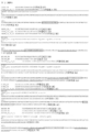
도 1: 표 1에 언급된 13 메틸화 DNA 마커 및 관련된 프라이머 및 프로브 정보에 사용된 마커 염색체 영역.
도 2: 92% 특이도에서 PDAC 단계에 걸친 메틸화 DNA 마커-CA 19-9 패널의 교차 입증된 민감도
도 3: 메틸화 DNA 마커 패널 단독, CA 19-9 단독, PDAC의 식별을 위한 조합 패널에 대한 교차 입증된 ROC 곡선.Figure 1: Marker chromosomal regions used for the 13 methylated DNA markers mentioned in Table 1 and related primer and probe information.
Figure 2: Cross-validated sensitivity of methylated DNA marker-CA 19-9 panel across PDAC stages at 92% specificity.
Figure 3: Cross-validated ROC curves for a panel of methylated DNA markers alone, CA 19-9 alone, and a panel of combinations for identification of PDACs.
정의Justice
본 기술의 이해를 용이하게 하기 위해, 다수의 용어 및 어구가 하기에 정의된다. 추가의 정의는 상세한 설명 전반에 걸쳐 제시된다.To facilitate understanding of the present technology, a number of terms and phrases are defined below. Additional definitions are set forth throughout the detailed description.
명세서 및 청구범위 전반에 걸쳐, 하기 용어는 문맥상 달리 명백하게 나타내지 않는 한 본원에 명백하게 연관된 의미를 갖는다. 본원에 사용된 어구 "일 구현예에서"는 반드시 동일한 구현예를 지칭할 필요는 없지만, 그럴 수도 있다. 또한, 본원에 사용된 어구 "다른 구현예에서"는 반드시 상이한 구현예를 지칭할 필요는 없지만, 그럴 수도 있다. 따라서, 하기 기재된 바와 같이, 본 발명의 다양한 구현예는 본 발명의 범주 또는 취지를 벗어나지 않으면서 용이하게 조합될 수 있다.Throughout the specification and claims, the following terms have the meanings expressly associated herein unless the context clearly indicates otherwise. As used herein, the phrase “in one embodiment” does not necessarily refer to the same embodiment, but it may. Also, as used herein, the phrase “in another embodiment” does not necessarily refer to a different embodiment, although it may. Accordingly, as described below, various embodiments of the present invention can be readily combined without departing from the scope or spirit of the present invention.
또한, 본원에서 사용된 바와 같이, 용어 "또는"은 포괄적인 "또는" 작업자이고, 문맥상 명백히 달리 지시되지 않는 한 "및/또는"이라는 용어와 동등하다. 용어 "에 기초한"은 배타적이지 않으며, 문맥상 달리 명백하게 지시되지 않는 한, 기재되지 않은 추가의 인자에 기초할 수 있게 한다. 또한, 명세서 전체에 걸쳐, "a", "an", 및 "the"의 의미는 복수의 언급을 포함한다. "in"의 의미는 "in" 및 "on"을 포함한다.Also, as used herein, the term “or” is an inclusive “or” operator and is equivalent to the term “and/or” unless the context clearly dictates otherwise. The term “based on” is not exclusive and allows for the basis of additional factors not described, unless the context clearly dictates otherwise. Also, throughout the specification, the meanings of “a,” “an,” and “the” include plural references. The meaning of “in” includes “in” and “on”.
본 출원의 청구범위에서 사용되는 바와 같은 전환 문구 "본질적으로 이루어진"은, 문헌[In re Herz, 537 F.2d 549, 551-52, 190 USPQ 461, 463 (CCPA 1976)]에서 논의된 바와 같이, 청구범위의 범주를 명시된 물질 또는 단계 "및 청구된 발명의 기본적이고 신규한 특성(들)에 실질적으로 영향을 미치지 않는 것들"로 제한한다. 예를 들어, 언급된 요소로 "본질적으로 이루어진" 조성물은, 존재할지라도, 오염물이 순수한 조성물, 즉 언급된 성분으로 "이루어진" 조성물과 비교하여 언급된 조성물의 기능을 변경시키지 않는 수준으로 언급되지 않은 오염물을 함유할 수 있다. The transition phrase “consisting essentially of” as used in the claims of this application is, as discussed in In re Herz, 537 F.2d 549, 551-52, 190 USPQ 461, 463 (CCPA 1976), , limits the scope of the claims to the specified materials or steps "and those that do not materially affect the basic and novel characteristic(s) of the claimed invention". For example, a composition "consisting essentially of" the recited elements, if present, is a composition that is free of contaminants, i.e., a composition "consisting of" the recited constituents at a level that does not alter the function of the recited composition. It may contain contaminants.
본원에서 사용된 바와 같이, "핵산" 또는 "핵산 분자"는 일반적으로 임의의 리보핵산 또는 데옥시리보핵산을 지칭하고, 이는 비변형된 또는 변형된 DNA 또는 RNA일 수 있다. "핵산"은, 비제한적으로, 단일- 및 이중-가닥 핵산을 포함한다. 본원에서 사용된 바와 같이, 용어 "핵산"은 또한 하나 이상의 변형된 염기를 함유하는 상기에 기재된 바와 같은 DNA를 포함한다. 따라서, 안정성을 위해 또는 다른 이유로 변형된 골격을 갖는 DNA는 "핵산"이다. 본원에서 사용된 바와 같은 용어 "핵산"은 이러한 화학적으로, 효소적으로, 또는 대사적으로 변형된 형태의 핵산뿐만 아니라 예를 들어, 단순 및 복합 세포를 포함하는 바이러스 및 세포의 화학적 형태의 DNA 특징을 포용한다.As used herein, “nucleic acid” or “nucleic acid molecule” generally refers to any ribonucleic acid or deoxyribonucleic acid, which may be unmodified or modified DNA or RNA. “Nucleic acid” includes, but is not limited to, single- and double-stranded nucleic acids. As used herein, the term “nucleic acid” also includes DNA as described above containing one or more modified bases. Thus, DNA having a backbone that has been modified for stability or for other reasons is a “nucleic acid”. As used herein, the term "nucleic acid" refers to the DNA characteristic of such chemically, enzymatically, or metabolically modified forms of nucleic acids as well as, for example, viruses and cells, including simple and complex cells, in their chemical forms. embrace
용어들 "올리고뉴클레오타이드" 또는 "폴리뉴클레오타이드" 또는 "뉴클레오타이드" 또는 "핵산"은 2개 이상, 바람직하게는 3개 초과, 및 통상적으로 10개 초과의 데옥시리보뉴클레오타이드 또는 리보뉴클레오타이드를 갖는 분자를 지칭한다. 정확한 크기는, 올리고뉴클레오타이드의 궁극적인 기능 또는 용도에 따라 달라지는 많은 인자에 따라 달라질 것이고. 올리고뉴클레오타이드는 화학적 합성, DNA 복제, 역전사, 또는 그의 조합을 포함하는 임의의 방식으로 생성될 수 있다. DNA에 대한 전형적인 데옥시리보뉴클레오타이드는 티민, 아데닌, 시토신, 및 구아닌이다. RNA에 대한 전형적인 리보뉴클레오타이드는 우라실, 아데닌, 시토신, 및 구아닌이다.The terms “oligonucleotide” or “polynucleotide” or “nucleotide” or “nucleic acid” refer to a molecule having two or more, preferably more than three, and usually more than ten deoxyribonucleotides or ribonucleotides do. The exact size will depend on many factors that depend on the ultimate function or use of the oligonucleotide. Oligonucleotides can be produced in any manner, including chemical synthesis, DNA replication, reverse transcription, or combinations thereof. Typical deoxyribonucleotides for DNA are thymine, adenine, cytosine, and guanine. Typical ribonucleotides for RNA are uracil, adenine, cytosine, and guanine.
본원에서 사용된 바와 같이, 용어들 핵산 "유전자좌" 또는 "영역"은 핵산의 하위영역, 예를 들어 염색체 상의 유전자, 단일 뉴클레오타이드, CpG 섬 등을 지칭한다.As used herein, the terms nucleic acid “locus” or “region” refer to a subregion of a nucleic acid, eg, a gene, single nucleotide, CpG island, etc. on a chromosome.
용어들 "상보적" 및 "상보성"은 염기쌍 형성 규칙과 관련된 뉴클레오타이드 (예를 들어, 1 뉴클레오타이드) 또는 폴리뉴클레오타이드 (예를 들어, 뉴클레오타이드의 서열)을 지칭한다. 예를 들어, 서열 5'-A-G-T-3'는 서열 3'-T-C-A-5'에 대해 상보적이다. 상보성은 "부분적"일 수 있고, 핵산' 염기의 일부만이 염기쌍 형성 규칙에 따라 매칭된다. 또는, 핵산 사이에 "완전한" 또는 "총" 상보성이 있을 수 있다. 핵산 가닥 사이의 상보성 정도는 핵산 가닥들 사이의 혼성화의 효율 및 강도에 영향을 준다. 이는 핵산 사이의 결합에 의존하는 증폭 반응 및 검출 방법에서 특히 중요하다.The terms “complementary” and “complementarity” refer to a nucleotide (eg, 1 nucleotide) or polynucleotide (eg, a sequence of nucleotides) related to the base pairing rules. For example, SEQ ID NO: 5'-A-G-T-3' is complementary to SEQ ID NO: 3'-T-C-A-5'. Complementarity can be "partial", and only a portion of a nucleic acid' base matches according to the base pairing rules. Alternatively, there may be "complete" or "total" complementarity between nucleic acids. The degree of complementarity between nucleic acid strands affects the efficiency and strength of hybridization between nucleic acid strands. This is particularly important in amplification reactions and detection methods that rely on binding between nucleic acids.
용어 "유전자"는 RNA, 또는 폴리펩티드 또는 그의 전구체의 생산에 필요한 코딩 서열을 포함하는 핵산 (예를 들어, DNA 또는 RNA) 서열을 지칭한다. 기능적 폴리펩티드는 폴리펩티드의 원하는 활성 또는 기능적 특성 (예를 들어, 효소 활성, 리간드 결합, 신호 전달 등)이 유지되는 한 전장 코딩 서열에 의해 또는 코딩 서열의 임의의 부분에 의해 인코딩될 수 있다. 유전자와 관련하여 사용될 때 용어 "부분"은 그 유전자의 단편을 지칭한다. 단편은 수개의 뉴클레오타이드로부터 전체 유전자 서열에서 1개의 뉴클레오타이드를 뺀 크기 범위일 수 있다. The term “gene” refers to a nucleic acid (eg, DNA or RNA) sequence comprising a coding sequence necessary for the production of RNA, or a polypeptide or a precursor thereof. A functional polypeptide may be encoded by a full-length coding sequence or by any portion of a coding sequence as long as the desired activity or functional properties of the polypeptide (eg, enzymatic activity, ligand binding, signal transduction, etc.) are maintained. The term “portion” when used in reference to a gene refers to a fragment of that gene. A fragment can range in size from several nucleotides minus one nucleotide from the total gene sequence.
용어 "유전자"는 또한 구조 유전자의 코딩 영역을 포함하고, 5' 및 3' 말단 둘 다에서 코딩 영역에 인접하여, 예를 들어, 어느 한쪽 말단 상에서 약 1 kb의 거리로 위치하는 서열을 포함하여, 유전자가 전장 mRNA의 길이에 상응한다(예를 들어 코딩, 조절, 구조 및 다른 서열을 포함함). 코딩 영역의 5'에 위치하고 mRNA 상에 존재하는 서열은 5' 비-번역 또는 비번역 서열로 지칭된다. 코딩 영역의 3' 또는 하류에 위치하고 mRNA 상에 존재하는 서열은 3' 비-번역 또는 3' 비번역 서열로 지칭된다. 용어 "유전자"는 유전자의 cDNA 및 게놈 형태 둘 다를 포함한다. 일부 유기체 (예를 들어, 진핵생물)에서, 유전자의 게놈 형태 또는 클론은 "인트론" 또는 "개재 영역" 또는"개재 서열"로 지칭되는 비-코딩 서열로 중단된 코딩 영역을 함유한다. 인트론은 핵 RNA(hnRNA)로 전사되는 유전자 세그먼트이며; 인트론은 인핸서와 같은 조절 요소를 포함할 수 있다. 인트론은 핵 또는 1차 전사체로부터 제거되거나 "스플라이싱 아웃"되며; 따라서 인트론은 메신저 RNA(mRNA) 전사체에 존재하지 않는다. mRNA는 번역 동안 신생 폴리펩티드에서 아미노산의 서열 또는 순서를 명시하도록 작용한다. The term "gene" also includes the coding region of a structural gene and includes sequences located adjacent to the coding region at both the 5' and 3' ends, e.g., at a distance of about 1 kb on either end. , a gene corresponds to the length of a full-length mRNA (including, for example, coding, regulatory, structural and other sequences). Sequences located 5' of the coding region and present on the mRNA are referred to as 5' untranslated or untranslated sequences. Sequences located 3' or downstream of the coding region and present on the mRNA are referred to as 3' untranslated or 3' untranslated sequences. The term “gene” includes both cDNA and genomic forms of a gene. In some organisms (eg, eukaryotes), genomic forms or clones of genes contain coding regions interrupted by non-coding sequences referred to as “introns” or “intervening regions” or “intervening sequences”. Introns are gene segments that are transcribed into nuclear RNA (hnRNA); Introns may include regulatory elements such as enhancers. Introns are removed or “spliced out” from the nucleus or primary transcript; Thus, introns are not present in messenger RNA (mRNA) transcripts. mRNA serves to specify the sequence or sequence of amino acids in a neonatal polypeptide during translation.
인트론의 함유 외에, 유전자의 게놈 형태는 또한 RNA 전사체 상에 존재하는 서열의 5' 및 3' 말단 둘 다에 위치하는 서열을 포함할 수 있다. 이들 서열은 "플랭킹(flanking)" 서열 또는 영역(이러한 플랭킹 서열은 mRNA 전사체 상에 존재하는 비번역 서열에 대해 5' 또는 3'에 위치함)으로 지칭된다. 5' 플랭킹 영역은 유전자의 전사를 제어하거나 영향을 미치는 프로모터 및 인핸서와 같은 조절 서열을 함유할 수 있다. 3' 측면 영역은 전사 종결, 전사후 절단 및 폴리아데닐화를 지시하는 서열을 함유할 수 있다.In addition to intron inclusion, the genomic form of a gene may also include sequences located at both the 5' and 3' ends of the sequences present on the RNA transcript. These sequences are referred to as "flanking" sequences or regions (these flanking sequences are located 5' or 3' to untranslated sequences present on the mRNA transcript). The 5' flanking region may contain regulatory sequences such as promoters and enhancers that control or affect the transcription of the gene. The 3' flanking region may contain sequences directing transcription termination, post-transcriptional cleavage and polyadenylation.
용어 "야생형"은 유전자와 관련하여 만들어질 때 천연 발생 공급원으로부터 단리된 유전자의 특징을 갖는 유전자를 지칭한다. 용어 "야생형"은 유전자 생성물과 관련하여 만들어질 때 천연 발생 공급원으로부터 단리된 유전자 생성물의 특징을 갖는 유전자 생성물을 의미한다. 물체에 적용되는 용어 "천연 발생"은 물체가 자연에서 발견될 수 있다는 사실을 지칭한다. 예를 들어, 천연 공급원으로부터 단리될 수 있고 실험실에서 사람의 손에 의해 의도적으로 변형되지 않은 유기체 (바이러스 포함)에 존재하는 폴리펩티드 또는 폴리뉴클레오타이드 서열은 천연 발생이다. 야생형 유전자는 종종 집단에서 가장 빈번하게 관찰되는 유전자 또는 대립유전자이고, 따라서 임의로 유전자의 "정상" 또는 "야생형" 형태로 지칭된다. 대조적으로, 용어 "변형된" 또는 "돌연변이체"는 유전자 또는 유전자 생성물과 관련하여 이루어질 때 각각 야생형 유전자 또는 유전자의 생성물과 비교할 때 서열 및/또는 기능적 특성 (예를 들어, 변경된 특성)에서 변형을 나타내는 유전자 또는 유전자를 지칭한다. 천연-발생 돌연변이체가 분리될 수 있다는 것이 주지된다; 이들은 야생형 유전자 또는 유전자 생성물과 비교할 때 변경된 특징을 갖는다는 사실에 의해 확인된다. The term “wild-type” refers to a gene having the characteristics of a gene isolated from a naturally occurring source when made in relation to the gene. The term "wild type" when made in the context of a gene product means a gene product that has the characteristics of a gene product isolated from a naturally occurring source. The term “naturally occurring” as applied to an object refers to the fact that an object can be found in nature. For example, a polypeptide or polynucleotide sequence present in an organism (including viruses) that can be isolated from a natural source and that has not been intentionally modified by human hands in the laboratory is naturally occurring. A wild-type gene is often the most frequently observed gene or allele in a population and is therefore optionally referred to as a "normal" or "wild-type" form of a gene. In contrast, the terms "modified" or "mutant", when made in the context of a gene or gene product, refer to a modification in sequence and/or functional property (e.g., altered property) when compared to the wild-type gene or product of the gene, respectively. Refers to the gene or gene that represents it. It is noted that naturally-occurring mutants can be isolated; They are identified by the fact that they have altered characteristics when compared to the wild-type gene or gene product.
용어 "대립유전자"는 유전자의 변이를 지칭하고; 변이는 변이체 및 돌연변이체, 다형성 유전자좌, 및 단일 뉴클레오타이드 다형성 유전자좌, 틀이동, 및 스플라이스 돌연변이를 포함하지만 이에 제한되지 않는다. 대립유전자는 집단에서 자연적으로 발생할 수 있거나, 집단의 임의의 특정 개체의 수명 동안 발생할 수 있다. The term “allele” refers to a variation in a gene; Variants include, but are not limited to, variants and mutants, polymorphic loci, and single nucleotide polymorphic loci, frameshifts, and splice mutations. Alleles may occur naturally in a population, or may occur during the lifespan of any particular individual in the population.
따라서, 용어들 "변이체" 및 "돌연변이체"는, 뉴클레오타이드 서열와 관련하여 사용될 때, 또 다른, 통상적으로 관련된, 뉴클레오타이드 산 서열로부터 하나 이상의 뉴클레오타이드에 의해 차이가 나는 핵산 서열을 지칭한다. "변이"는 2개의 상이한 뉴클레오타이드 서열의 차이이고; 전형적으로, 하나의 서열은 참조 서열이다.Thus, the terms “variant” and “mutant,” when used in reference to a nucleotide sequence, refer to a nucleic acid sequence that differs by one or more nucleotides from another, ordinarily related, nucleotide acid sequence. "Variation" is the difference between two different nucleotide sequences; Typically, one sequence is a reference sequence.
"증폭"은 주형 특이성을 포함하는 핵산 복제의 특별한 경우이다. 이는 비-특이적 중형 복제(예를 들어, 주형 의존적이지만 특정 주형에 의존하지 않는 복제)와 대조된다. 주형 특이성은 여기서 복제의 충실도 (예를 들어, 적절한 폴리뉴클레오타이드 서열의 합성) 및 뉴클레오타이드 (리보- 또는 데옥시리보-) 특이성과 구별된다. 주형 특이성은 종종 "표적" 특이성의 관점에서 기술된다. 표적 서열은 다른 핵산으로부터 분류하고자 한다는 의미에서 "표적"이다. 증폭 기술은 주로 이러한 분류를 위해 설계되었다. "Amplification" is a special case of nucleic acid replication involving template specificity. This is in contrast to non-specific mesotype replication (eg, template dependent but not specific template dependent replication). Template specificity is here distinguished from fidelity of replication (eg, synthesis of an appropriate polynucleotide sequence) and nucleotide (ribo- or deoxyribo-) specificity. Template specificity is often described in terms of "target" specificity. A target sequence is a “target” in the sense that it is intended to be sorted from other nucleic acids. Amplification techniques are primarily designed for this classification.
핵산의 문맥에서 용어 "증폭하는" 또는 "증폭"은 증폭 생성물 또는 앰플리콘이 일반적으로 검출가능한, 전형적으로 소량의 폴리뉴클레오타이드 (예를 들어, 단일 폴리뉴클레오타이드 분자)로부터 시작하는 폴리뉴클레오타이드, 또는 폴리뉴클레오타이드의 일부의 다중 카피의 생산을 지칭한다. 폴리뉴클레오타이드의 증폭은 다양한 화학적 및 효소적 공정을 포함한다. 중합효소 연쇄 반응 (PCR) 또는 리가제 연쇄 반응 동안에 표적 또는 주형 DNA 분자의 하나 또는 몇 개의 카피로부터의 다수의 DNA 카피의 생성(LCR; 참조, 예를 들어, 미국 특허 번호 5,494,810; 그 전체 내용은 본원에 참조로 포함된다)은 증폭의 형태이다. 추가의 유형의 증폭은 하기를 포함하지만 이에 제한되지 않는다: 대립유전자 특정 PCR (참고, 예를 들어, 미국 특허 번호 5,639,611; 그 전체 내용은 본원에 참조로 포함된다), 어셈블리 PCR (참고, 예를 들어, 미국 특허 번호 5,965,408; 그 전체 내용은 본원에 참조로 포함된다), 헬리카제 의존 증폭 (참고, 예를 들어, 미국 특허 번호 7,662,594; 그 전체 내용은 본원에 참조로 포함된다), 핫-스타트 PCR (참고, 예를 들어, 미국 특허 번호 5,773,258 및 5,338,671; 이들 각각은 그 전체 내용이 참조로 본원에 포함된다), 서열간 특이적 PCR, 역 PCR (참고, 예를 들어, Triglia, 등(1988) Nucleic Acids Res., 16:8186; 그 전체 내용은 본원에 참조로 포함된다), 결찰-매개된 PCR (참고, 예를 들어, Guilfoyle, R. 등, Nucleic Acids Research, 25:1854-1858 (1997); 미국 특허 번호 5,508,169; 이들 각각은 그 전체 내용이 본원에 참조로 포함된다), 메틸화 특이적 PCR (참고, 예를 들어, Herman 등, (1996) PNAS 93(13) 9821-9826; 그 전체 내용은 본원에 참조로 포함된다), 미니프라이머 PCR, 다중 결찰 의존 프로브 증폭 (참고, 예를 들어, Schouten 등, (2002) Nucleic Acids Research 30(12): e57; 그 전체 내용은 본원에 참조로 포함된다), 다중 PCR (참고, 예를 들어, Chamberlain 등, (1988) Nucleic Acids Research 16(23) 11141-11156; Ballabio 등, (1990) Human Genetics 84(6) 571-573; Hayden 등, (2008) BMC Genetics 9:80; 이들 각각은 그 전체 내용이 본원에 참조로 포함된다), 내재된 PCR, 중첩-확대 PCR (참고, 예를 들어, Higuchi 등, (1988) Nucleic Acids Research 16(15) 7351-7367; 그 전체 내용은 본원에 참조로 포함된다), 실시간 PCR (참고, 예를 들어, Higuchi 등, (1992) Biotechnology 10:413-417; Higuchi 등, (1993) Biotechnology 11:1026-1030; 이들 각각은 그 전체 내용이 본원에 참조로 포함된다), 역전사 PCR (참고, 예를 들어, Bustin, S.A. (2000) J. Molecular Endocrinology 25:169-193; 그 전체 내용은 본원에 참조로 포함된다), 고상 PCR, 열 비대칭 인터레이싱 PCR, 및 Touchdown PCR (참고, 예를 들어, Don 등, Nucleic Acids Research (1991) 19(14) 4008; Roux, K. (1994) Biotechniques 16(5) 812-814; Hecker 등, (1996) Biotechniques 20(3) 478-485; 이들 각각은 그 전체 내용이 본원에 참조로 포함된다). 폴리뉴클레오타이드 증폭은 또한 디지털 PCR을 사용하여 달성될 수 있다(참고, 예를 들어, Kalinina 등, Nucleic Acids Research. 25; 1999-2004, (1997); Vogelstein 및 Kinzler, Proc Natl Acad Sci USA. 96; 9236-41, (1999); 국제 특허 공개 번호 WO05023091A2; US 특허 출원 공개 번호 20070202525; 이들 각각은 그 전체 내용이 본원에 참조로 포함된다).The term “amplifying” or “amplifying” in the context of nucleic acids refers to a polynucleotide, typically starting from a small amount of a polynucleotide (eg, a single polynucleotide molecule), for which the amplification product or amplicon is generally detectable, or polynucleotide refers to the production of multiple copies of a portion of Amplification of polynucleotides involves a variety of chemical and enzymatic processes. Generation of multiple DNA copies from one or several copies of a target or template DNA molecule during a polymerase chain reaction (PCR) or ligase chain reaction (LCR; see, e.g., U.S. Pat. No. 5,494,810; the entire contents of which are incorporated herein by reference) is a form of amplification. Additional types of amplification include, but are not limited to: allele specific PCR (see, e.g., U.S. Pat. No. 5,639,611; the entire contents of which are incorporated herein by reference), assembly PCR (see, e.g., For example, U.S. Pat. No. 5,965,408; incorporated herein by reference in its entirety), helicase dependent amplification (see, e.g., U.S. Pat. No. 7,662,594; incorporated herein by reference in its entirety), hot-start PCR (see, e.g., U.S. Patent Nos. 5,773,258 and 5,338,671; each of which is incorporated herein by reference in its entirety), intersequence specific PCR, reverse PCR (see, e.g., Triglia, et al. (1988) ) Nucleic Acids Res., 16:8186; the entire contents of which are incorporated herein by reference), ligation-mediated PCR (see, e.g., Guilfoyle, R. et al., Nucleic Acids Research, 25:1854-1858 ( 1997); The entire contents of which are incorporated herein by reference), miniprimer PCR, multiple ligation dependent probe amplification (see, e.g., Schouten et al., (2002) Nucleic Acids Research 30(12): e57; see herein in its entirety) ), multiplex PCR (see, e.g., Chamberlain et al., (1988) Nucleic Acids Research 16(23) 11141-11156; Ballabio et al., (1990) Human Genetics 84(6) 571-573; Hayden et al., (2008) BMC Genetics 9:80; each of which is incorporated herein by reference in its entirety), intrinsic PCR, overlap-extension PCR (see, e.g., Higu chi et al., (1988) Nucleic Acids Research 16(15) 7351-7367; The entire contents of which are incorporated herein by reference), real-time PCR (see, e.g., Higuchi et al., (1992) Biotechnology 10:413-417; Higuchi et al., (1993) Biotechnology 11:1026-1030; each of which is Incorporated herein by reference in its entirety), reverse transcription PCR (see, e.g., Bustin, SA (2000) J. Molecular Endocrinology 25:169-193; incorporated herein by reference in its entirety), solid phase PCR, thermal asymmetric interlacing PCR, and Touchdown PCR (see, eg, Don et al., Nucleic Acids Research (1991) 19(14) 4008; Roux, K. (1994) Biotechniques 16(5) 812-814; Hecker) et al., (1996) Biotechniques 20(3) 478-485; each of which is incorporated herein by reference in its entirety). Polynucleotide amplification can also be achieved using digital PCR (see, eg, Kalinina et al., Nucleic Acids Research. 25; 1999-2004, (1997); Vogelstein and Kinzler, Proc Natl Acad Sci USA. 96; 9236-41, (1999); International Patent Publication No. WO05023091A2; US Patent Application Publication No. 20070202525; each of which is incorporated herein by reference in its entirety).
용어 "중합효소 연쇄 반응" ("PCR")은 클로닝 또는 정제 없이 게놈 또는 다른 DNA 또는 RNA의 혼합물에서 표적 서열의 절편의 농도를 증가시키는 방법을 기재하는 K.B. Mullis 미국 특허 번호 4,683,195, 4,683,202, 및 4,965,188의 방법을 지칭한다. 표적 서열을 증폭시키기 위한 이러한 방법은 원하는 표적 서열을 함유하는 DNA 혼합물에 과량의 2개의 올리고뉴클레오타이드 프라이머를 도입한 후, DNA 중합효소의 존재 하에 정확한 열 사이클링 서열로 구성된다. 2개의 프라이머는 이중 가닥 표적 서열의 각각의 가닥에 상보적이다. 증폭을 수행하기 위해, 혼합물을 변성시킨 다음, 프라이머를 표적 분자 내의 이들의 상보적 서열로 어닐링한다. 어닐링 후, 프라이머를 중합효소로 연장시켜 새로운 쌍의 상보성 가닥을 형성한다. 변성, 프라이머 어닐링, 및 중합효소 연장의 단계들은 원하는 표적 서열의 증폭된 분절의 높은 농도를 얻기 위해 여러 번 반복될 수 있다(즉, 변성, 어닐링 및 연장은 하나의 "사이클"을 구성하고; 많은 "사이클들"이 있을 수 있다). 원하는 표적 서열의 증폭된 절편의 길이는 서로에 대한 프라이머의 상대 위치에 의해 결정되며, 따라서, 이 길이는 조절가능한 파라미터이다. 상기 과정의 반복 양태에 의해, 상기 방법은 "중합효소 연쇄 반응"("PCR")으로 지칭된다. 표적 서열의 원하는 증폭된 분절은 혼합물에서 (농도의 관점에서) 우세한 서열이 되기 때문에, 이들은 "PCR 증폭"되고 "PCR 생성물" 또는 "앰플리콘"이라고 한다. 당업자는 용어 "PCR"이, 예를 들어, 실시간 PCR, 내재된 PCR, 역전사 PCR (RT-PCR), 단일 프라이머 및 임의로 프라이밍된 PCR 등을 사용하는 원래 기재된 방법의 많은 변이체를 포함하는 것을 이해할 것이다. The term "polymerase chain reaction"("PCR") refers to KB Mullis U.S. Patent Nos. 4,683,195, 4,683,202, and 4,965,188, which describe methods for increasing the concentration of fragments of a target sequence in a genomic or other mixture of DNA or RNA without cloning or purification. refers to the method of This method for amplifying a target sequence consists of introducing an excess of two oligonucleotide primers into a DNA mixture containing the desired target sequence, followed by the correct thermal cycling sequence in the presence of DNA polymerase. The two primers are complementary to each strand of the double-stranded target sequence. To perform amplification, the mixture is denatured and then the primers are annealed to their complementary sequences in the target molecule. After annealing, the primers are extended with a polymerase to form a new pair of complementary strands. The steps of denaturation, primer annealing, and polymerase extension can be repeated multiple times to obtain high concentrations of amplified fragments of the desired target sequence (i.e., denaturation, annealing and extension constitute one “cycle”; many there may be "cycles"). The length of the amplified fragments of the desired target sequence is determined by the relative positions of the primers with respect to each other, and thus this length is a controllable parameter. By an iterative aspect of this process, the method is referred to as "polymerase chain reaction"("PCR"). Since the desired amplified segment of the target sequence is the predominant sequence (in terms of concentration) in the mixture, they are "PCR amplified" and are referred to as "PCR products" or "amplicons". Those of skill in the art will understand that the term "PCR" includes many variants of the originally described methods using, for example, real-time PCR, intrinsic PCR, reverse transcription PCR (RT-PCR), single primers and optionally primed PCR, and the like. .
주형 특이성은 효소의 선택에 의해 대부분의 증폭 기술에서 달성된다. 증폭 효소는 이들이 사용되는 조건 하에서, 핵산의 이종 혼합물에서 핵산의 특정 서열만을 처리하는 효소이다. 예를 들어, Q-베타 레플리카제의 경우, MDV-1 RNA는 레플리카제에 대한 특이적 주형이다(Kacian 등, Proc. Natl. Acad. Sci. USA, 69:3038 [1972]). 다른 핵산은 이러한 증폭 효소에 의해 복제되지 않을 것이다. 유사하게, T7 RNA 중합효소의 경우, 이 증폭 효소는 그 자체의 프로모터에 대해 엄격한 특이성을 갖는다(Chamberlin 등, Nature, 228:227 [1970]). T4 DNA 리가제의 경우에, 효소는 2개의 올리고뉴클레오타이드 또는 폴리뉴클레오타이드를 결찰시키지 않을 것이고, 여기서, 결찰 접합부에서 올리고뉴클레오타이드 또는 폴리뉴클레오타이드 기질과 주형 사이에 미스매치가 존재한다(Wu 및 Wallace (1989) Genomics 4:560). 마침내, 열안정성 주형 의존 DNA 중합효소 (예를 들어, Taq 및 Pfu DNA 중합효소), 고온에서 기능하는 이들의 능력에 의해, 프라이머에 의해 한정되고 이에 따라 정의되는 서열에 대해 높은 특이성을 나타내는 것으로 밝혀지고; 고온은 표적 서열과의 프라이머 혼성화 및 비-표적 서열과의 혼성화를 선호하지 않는 열역학적 조건을 초래한다(H. A. Erlich (ed.), PCR Technology, Stockton Press [1989]).Template specificity is achieved in most amplification techniques by selection of enzymes. Amplification enzymes are enzymes that only process a specific sequence of nucleic acids in a heterogeneous mixture of nucleic acids under the conditions in which they are used. For example, in the case of Q-beta replicases, MDV-1 RNA is a specific template for the replicases (Kacian et al., Proc. Natl. Acad. Sci. USA, 69:3038 [1972]). Other nucleic acids will not be replicated by this amplification enzyme. Similarly, in the case of T7 RNA polymerase, this amplification enzyme has stringent specificity for its promoter (Chamberlin et al., Nature, 228:227 [1970]). In the case of T4 DNA ligase, the enzyme will not ligate the two oligonucleotides or polynucleotides, where there is a mismatch between the oligonucleotide or polynucleotide substrate and the template at the ligation junction (Wu and Wallace (1989)) Genomics 4:560). Finally, it was found that thermostable template dependent DNA polymerases (eg Taq and Pfu DNA polymerases), due to their ability to function at high temperatures, exhibit high specificity for sequences defined and thus defined by primers. under; High temperatures result in thermodynamic conditions that do not favor primer hybridization with target sequences and hybridization with non-target sequences (H. A. Erlich (ed.), PCR Technology, Stockton Press [1989]).
본원에서 사용된 바와 같이, 용어 "핵산 검출 검정"은 관심 핵산의 뉴클레오타이드 조성을 결정하는 임의의 방법을 지칭한다. 핵산 검출 검정은DNA 서열분석 방법, 프로브 혼성화 방법, 구조 특정 절단 검정 (예를 들어, 인베이더 검정, (Hologic, Inc.)를 포함하지만 이에 제한되지 않고, 예를 들어, 하기에 기재되어 있다: 미국 특허 번호 5,846,717, 5,985,557, 5,994,069, 6,001,567, 6,090,543, 및 6,872,816; Lyamichev 등, Nat. Biotech., 17:292 (1999), Hall 등, PNAS, USA, 97:8272 (2000), 및 US 특허 번호 9,096,893, 이들 각각은 모든 목적을 위해 그 전체 내용이 본원에 참조된 포함된다); 효소 불일치 절단 방법 (예를 들어, Variagenics, 미국 특허 번호 6,110,684, 5,958,692, 5,851,770, 이는 그 전체 내용이 본원에 참조로 포함된다); 상기에 기재된 중합효소 연쇄 반응 (PCR); 분지형 혼성화 방법 (예를 들어, Chiron, 미국 특허 번호 5,849,481, 5,710,264, 5,124,246, 및 5,624,802, 이는 그 전체 내용이 본원에 참조로 포함된다); 회전환 복제 (예를 들어, 미국 특허 번호 6,210,884, 6,183,960 및 6,235,502, 이는 그 전체 내용이 본원에 참조로 포함된다); NASBA (예를 들어, 미국 특허 번호 5,409,818, 이는 그 전체 내용이 본원에 인용되어 포함된다); 분자 항로표지 기술 (예를 들어, 미국 특허 번호 6,150,097, 이는 그 전체 내용이 본원에 인용되어 포함된다); E-센서 기술 (Motorola, 미국 특허 번호 6,248,229, 6,221,583, 6,013,170, 및 6,063,573, 이는 그 전체 내용이 본원에 참조로 포함된다); 사이클링 프로브 기술 (예를 들어, 미국 특허 번호 5,403,711, 5,011,769, 및 5,660,988, 이는 그 전체 내용이 본원에 참조로 포함된다); Dade Behring 신호 증폭 방법 (예를 들어, 미국 특허 번호 6,121,001, 6,110,677, 5,914,230, 5,882,867, 및 5,792,614, 이는 그 전체 내용이 본원에 참조로 포함된다); 리가제 연쇄 반응 (예를 들어, Baranay Proc. Natl. Acad. Sci USA 88, 189-93 (1991)); 및 샌드위치 혼성화 방법 (예를 들어, 미국 특허 번호 5,288,609, 이는 그 전체 내용이 본원에 인용되어 포함된다).As used herein, the term “nucleic acid detection assay” refers to any method for determining the nucleotide composition of a nucleic acid of interest. Nucleic acid detection assays include, but are not limited to, DNA sequencing methods, probe hybridization methods, structure specific cleavage assays (e.g., Invader assays, (Hologic, Inc.), e.g., described in the United States Patent Nos. 5,846,717, 5,985,557, 5,994,069, 6,001,567, 6,090,543, and 6,872,816; Lyamichev et al., Nat. Biotech., 17:292 (1999), Hall et al., PNAS, USA, 97:8272 (2000), and US Pat. Nos. 9,096, 89 each of which is incorporated herein by reference in its entirety for all purposes); enzymatic mismatch cleavage methods (eg, Variagenics, US Pat. Nos. 6,110,684, 5,958,692, 5,851,770, which are incorporated herein by reference in their entirety); polymerase chain reaction (PCR) described above; branched hybridization methods (eg, Chiron, US Pat. Nos. 5,849,481, 5,710,264, 5,124,246, and 5,624,802, which are incorporated herein by reference in their entirety); rolling ring replication (eg, US Pat. Nos. 6,210,884, 6,183,960 and 6,235,502, which are incorporated herein by reference in their entirety); NASBA (eg, US Pat. No. 5,409,818, which is incorporated herein by reference in its entirety); molecular navigation technology (eg, US Pat. No. 6,150,097, which is incorporated herein by reference in its entirety); E-sensor technology (Motorola, US Pat. Nos. 6,248,229, 6,221,583, 6,013,170, and 6,063,573, which are incorporated herein by reference in their entirety); cycling probe technology (eg, US Pat. Nos. 5,403,711, 5,011,769, and 5,660,988, which are incorporated herein by reference in their entirety); Dade Behring signal amplification methods (eg, US Pat. Nos. 6,121,001, 6,110,677, 5,914,230, 5,882,867, and 5,792,614, which are incorporated herein by reference in their entirety); ligase chain reaction (eg, Baranay Proc. Natl. Acad. Sci USA 88, 189-93 (1991)); and sandwich hybridization methods (eg, US Pat. No. 5,288,609, which is incorporated herein by reference in its entirety).
용어 "증폭가능한 핵산"은 임의의 증폭 방법에 의해 증폭될 수 있는 핵산을 지칭한다. "증폭가능한 핵산"은 일반적으로 "샘플 주형"을 포함하는 것으로 고려된다.The term “amplifiable nucleic acid” refers to a nucleic acid that can be amplified by any amplification method. An “amplifiable nucleic acid” is generally considered to include a “sample template”.
용어 "샘플 주형"은 "표적" (하기 정의됨)의 존재에 대해 분석되는 샘플로부터 기원하는 핵산을 지칭한다. 대조적으로, "배경 주형"은 샘플에 존재하거나 존재하지 않을 수 있는 샘플 주형 이외의 핵산과 관련하여 사용된다. 배경 주형은 가장 종종 의도되지 않는다. 이는 캐리오버(carryover)의 결과일 수 있거나, 샘플로부터 정제하고자 하는 핵산 오염물의 존재로 인한 것일 수 있다. 예를 들어, 검출될 유기체 이외의 유기체로부터의 핵산은 시험 샘플에서 배경으로서 존재할 수 있다. The term “sample template” refers to a nucleic acid originating from a sample that is assayed for the presence of a “target” (defined below). In contrast, “background template” is used in reference to a nucleic acid other than a sample template, which may or may not be present in the sample. Background templates are most often not intended. This may be the result of carryover, or it may be due to the presence of a nucleic acid contaminant to be purified from the sample. For example, a nucleic acid from an organism other than the organism to be detected may be present as background in the test sample.
용어 "프라이머"는 핵산 주형 가닥에 상보적인 프라이머 연장 생성물의 합성이 유도되는 조건 하에 (예를 들어, 뉴클레오타이드 및 유도제, 예컨대 DNA 중합효소의 존재 하에, 그리고 적합한 온도 및 pH에서) 배치될 때 합성의 개시 지점으로서 작용할 수 있는, 예를 들어 제한 분해로부터의 핵산 단편으로서 자연적으로 발생하든지 또는 합성적으로 생성하든지 간에, 올리고뉴클레오타이드를 지칭한다. 프라이머는 바람직하게는 증폭에서 최대 효율을 위해 단일 가닥이지만, 대안적으로 이중 가닥일 수 있다. 이중 가닥인 경우, 프라이머를 먼저 처리하여 그의 가닥을 분리한 후 연장 생성물을 제조하는데 사용한다. 바람직하게는, 프라이머는 올리고데옥시리보뉴클레오타이드다. 프라이머는 유도제의 존재 하에 연장 생성물의 합성을 프라이밍하기에 충분히 길어야 한다. 프라이머의 정확한 길이는 온도, 프라이머의 공급원, 및 방법의 사용을 포함하는 많은 인자에 의존할 것이다. The term "primer" refers to a process of synthesis when placed under conditions that induce the synthesis of a primer extension product complementary to the nucleic acid template strand (e.g., in the presence of nucleotides and an inducer such as DNA polymerase, and at a suitable temperature and pH). refers to an oligonucleotide, whether naturally occurring or synthetically produced, for example, as a nucleic acid fragment from restriction digestion, that can serve as a point of initiation. Primers are preferably single stranded for maximum efficiency in amplification, but may alternatively be double stranded. In the case of double stranding, the primer is first treated to separate its strands and then used to prepare the extension product. Preferably, the primer is an oligodeoxyribonucleotide. The primer should be long enough to prime the synthesis of the extension product in the presence of an inducer. The exact length of the primer will depend on many factors including temperature, source of primer, and use of method.
용어 "프로브"는 정제된 제한 분해물에서와 같이 자연적으로 발생하든 합성적으로, 재조합적으로, 또는 PCR 증폭에 의해 생성되든 간에, 관심의 다른 올리고뉴클레오타이드에 혼성화할 수 있는 올리고뉴클레오타이드 (예를 들어, 뉴클레오타이드의 서열)를 지칭한다. 프로브는 단일 가닥 또는 이중 가닥일 수 있다. 프로브는 특정 유전자 서열 (예를 들어, "포획 프로브")의 검출, 확인 및 단리에 유용하다. 본 발명에 사용될 수 있는 임의의 프로브가 일부 구현예에서, 임의의 "리포터 분자"로 표지될 수 있어서, 효소 (예를 들어, ELISA, 뿐만 아니라 효소 기반 조직화학 검정), 형광, 방사성, 및 발광성 시스템을 포함하지만 이에 제한되지 않는 임의의 검출 시스템에서 검출가능한 것으로 고려된다. 본 발명이 임의의 특정 검출 시스템 또는 표지로 제한되는 것으로 의도되지 않는다.The term "probe" refers to an oligonucleotide (e.g., capable of hybridizing to other oligonucleotides of interest, whether naturally occurring, such as in purified restriction digests, or produced synthetically, recombinantly, or by PCR amplification). sequence of nucleotides). Probes may be single-stranded or double-stranded. Probes are useful for the detection, identification, and isolation of specific gene sequences (eg, “capture probes”). Any probe that may be used in the present invention may, in some embodiments, be labeled with any “reporter molecule” such that enzymes (eg, ELISAs, as well as enzyme-based histochemical assays), fluorescence, radioactivity, and luminescence It is contemplated as being detectable in any detection system, including but not limited to systems. It is not intended that the present invention be limited to any particular detection system or label.
본원에 사용된 용어 "표적"은, 예를 들어 프로브 결합, 증폭, 단리, 포획 등에 의해 다른 핵산으로부터 분류하고자 하는 핵산을 지칭한다. 예를 들어, 중합효소 연쇄 반응과 관련하여 사용되는 경우, "표적"은 중합효소 연쇄 반응에 사용되는 프라이머에 의해 결합된 핵산의 영역을 지칭하는 반면, 표적 DNA가 증폭되지 않는 검정, 예를 들어 침습성 절단 검정의 일부 구현예에서, 표적은 프로브 및 침습성 올리고뉴클레오타이드 (예를 들어, INVADER 올리고뉴클레오타이드)가 결합하여 침습성 분할 구조를 형성하는 부위를 포함하여, 표적 핵산의 존재가 검출될 수 있다. "절편"은 표적 서열 내의 핵산의 영역으로서 정의된다.As used herein, the term “target” refers to a nucleic acid to be sorted from other nucleic acids, for example, by probe binding, amplification, isolation, capture, and the like. For example, when used in the context of the polymerase chain reaction, "target" refers to the region of nucleic acid bound by the primers used in the polymerase chain reaction, whereas an assay in which the target DNA is not amplified, e.g. In some embodiments of an invasive cleavage assay, the target comprises a site where a probe and an invasive oligonucleotide (eg, INVADER oligonucleotide) bind to form an invasive cleavage structure, so that the presence of the target nucleic acid can be detected. A “fragment” is defined as a region of a nucleic acid within a target sequence.
본원에서 사용된 바와 같이, "메틸화"는 시토신의 위치 C5 또는 N4, 아데닌의 N6 위치에서의 시토신 메틸화, 또는 다른 유형의 핵산 메틸화를 지칭한다. 전형적인 시험관내 DNA 증폭 방법은 증폭 주형의 메틸화 패턴을 보유하지 않기 때문에, 시험관내 증폭된 DNA는 일반적으로 메틸화되지 않는다. 그러나, "비메틸화 DNA" 또는 "메틸화 DNA"는 또한 원래의 주형이 각각 비메틸화되거나 메틸화된 증폭된 DNA를 지칭할 수 있다.As used herein, “methylation” refers to cytosine methylation at position C5 or N4 of a cytosine, N6 position of an adenine, or other types of nucleic acid methylation. Since typical in vitro DNA amplification methods do not retain the methylation pattern of the amplification template, in vitro amplified DNA is generally not methylated. However, “unmethylated DNA” or “methylated DNA” may also refer to amplified DNA in which the original template is unmethylated or methylated, respectively.
따라서, 본원에서 사용된 바와 같이 "메틸화 뉴클레오타이드" 또는 "메틸화 뉴클레오타이드 염기"는 뉴클레오타이드 염기 상의 메틸 모이어티의 존재를 지칭하고, 메틸 모이어티는 인식된 전형적인 뉴클레오타이드 염기에 존재하지 않는다. 예를 들어, 시토신은 그것의 피리미딘 고리 상의 메틸 모이어티를 함유하지 않고, 그러나 5-메틸시토신은 그것의 피리미딘 고리의 위치 5에서 메틸 모이어티를 함유한다. 따라서, 시토신은 메틸화 뉴클레오타이드가 아니고 5-메틸시토신은 메틸화 뉴클레오타이드이다. 또 다른 예에서, 티민은 그것의 피리미딘 고리의 위치 5에서 메틸 모이어티를 함유하지만; 본원의 목적을 위해, 티민이 메틸화 뉴클레오타이드인 것으로 고려되지 않는 것은, DNA에 존재할 때 티민이 DNA의 전형적인 뉴클레오타이드 염기이기 때문이다.Thus, "methylated nucleotide" or "methylated nucleotide base" as used herein refers to the presence of a methyl moiety on a nucleotide base, a methyl moiety that is not present in a recognized typical nucleotide base. For example, cytosine does not contain a methyl moiety on its pyrimidine ring, but 5-methylcytosine contains a methyl moiety at
본원에서 사용된 바와 같이, "메틸화된 핵산 분자"는 하나 이상의 메틸화 뉴클레오타이드를 함유하는 핵산 분자를 지칭한다.As used herein, “methylated nucleic acid molecule” refers to a nucleic acid molecule that contains one or more methylated nucleotides.
본원에서 사용된 바와 같이, 핵산 분자의 "메틸화 상태", "메틸화 프로파일", 및 "메틸화 상태"는 핵산 분자에서 하나 이상의 메틸화 뉴클레오타이드 염기가 없는 존재를 지칭한다. 예를 들어, 메틸화된 시토신을 함유하는 핵산 분자는 메틸화된 것을 간주된다(예를 들어, 핵산 분자의 메틸화 상태는 메틸화된다). 임의의 메틸화 뉴클레오타이드를 함유하지 않는 핵산 분자는 비메틸화된 것으로 간주된다. As used herein, “methylation status”, “methylation profile”, and “methylation status” of a nucleic acid molecule refer to the absence of one or more methylated nucleotide bases in a nucleic acid molecule. For example, a nucleic acid molecule containing a methylated cytosine is considered methylated (eg, the methylation status of the nucleic acid molecule is methylated). Nucleic acid molecules that do not contain any methylated nucleotides are considered unmethylated.
특정 핵산 서열 (예를 들어, 본원에 기재된 바와 같은 유전자 마커 또는 DNA 영역)의 메틸화 상태는 서열에서 모든 염기의 메틸화 상태를 나타낼 수 있거나 또는 서열 내의 하위세트의 염기 (예를 들어, 하나 이상의 시토신)의 메틸화 상태를 나타낼 수 잇거나, 또는 메틸화가 일어나는 서열 내의 위치의 정확한 정보를 제공하거나 제공하지 않으면서 서열 내의 영역 메틸화 밀도에 관한 정보를 나타낼 수 있다. The methylation status of a particular nucleic acid sequence (eg, a genetic marker or DNA region as described herein) may indicate the methylation status of all bases in the sequence or a subset of bases (eg, one or more cytosines) within the sequence. may indicate the methylation status of , or may present information about the methylation density of regions within a sequence, with or without providing precise information of the position within the sequence where methylation occurs.
핵산 분자에서 뉴클레오타이드 좌위의 메틸화 상태는 핵산 분자의 특정 유전자좌에서 메틸화 뉴클레오타이드의 존재 또는 부재를 지칭한다. 예를 들어, 핵산 분자 중의 7번째의 뉴클레오타이드에 존재하는 뉴클레오타이드가 5-메틸시토신인 경우, 상기 핵산 분자의 7번째 뉴클레오타이드에 있어서의 시토신의 메틸화 상태가 메틸화된다. 마찬가지로, 핵산 분자의 7번째 뉴클레오타이드에 존재하는 뉴클레오타이드가 시토신(5-메틸시토신이 아님)일 때, 핵산 분자 내의 7번째 뉴클레오타이드에서의 시토신의 메틸화 상태는 메틸화되지 않는다.The methylation status of a nucleotide locus in a nucleic acid molecule refers to the presence or absence of a methylated nucleotide at a particular locus in the nucleic acid molecule. For example, when the nucleotide present at the 7th nucleotide in the nucleic acid molecule is 5-methylcytosine, the methylation state of the cytosine at the 7th nucleotide of the nucleic acid molecule is methylated. Likewise, when the nucleotide present at the 7th nucleotide of the nucleic acid molecule is a cytosine (not 5-methylcytosine), the methylation status of the cytosine at the 7th nucleotide in the nucleic acid molecule is not methylated.
메틸화 상태는 선택적으로 "메틸화 값" (예를 들어, 메틸화 빈도, 분획, 비, 퍼센트, 등을 나타냄)에 의해 제시되거나 표시될 수 있다. 메틸화 값은, 예를 들어 메틸화 의존성 제한 효소로 제한 분해 후 존재하는 온전한 핵산의 양을 정량화함으로써, 또는 중아황산염 반응 후 증폭 프로파일을 비교함으로써, 또는 중아황산염 처리된 핵산 및 비처리된 핵산의 서열을 비교함으로써 생성될 수 있다. 따라서, 값, 예를 들어 메틸화 값은 메틸화 상태를 나타내며, 따라서 유전자좌의 다수의 카피에 걸친 메틸화 상태의 정량적 지표로서 사용될 수 있다. 이는 샘플에서 서열의 메틸화 상태를 역치 또는 참조 값과 비교하는 것이 바람직할 때 특히 사용된다.Methylation status may optionally be indicated or indicated by a “methylation value” (eg, indicating methylation frequency, fraction, ratio, percentage, etc.). The methylation value can be determined, for example, by quantifying the amount of intact nucleic acid present after restriction digestion with a methylation-dependent restriction enzyme, or by comparing the amplification profile after a bisulfite reaction, or by comparing the sequences of bisulfite-treated and untreated nucleic acids. can be generated by comparing. Thus, a value, eg, a methylation value, is indicative of methylation status and thus can be used as a quantitative indicator of methylation status across multiple copies of a locus. This is particularly used when it is desirable to compare the methylation status of a sequence in a sample to a threshold or reference value.
본원에서 사용된 바와 같이, "메틸화 빈도" 또는 "메틸화 백분율(%)"은 분자 또는 유전자좌가 메틸화되지 않은 경우의 수에 비해 분자 또는 유전자좌가 메틸화되는 경우의 수를 지칭한다.As used herein, “methylation frequency” or “percent methylation” refers to the number of instances in which a molecule or locus is methylated compared to the number of instances in which the molecule or locus is unmethylated.
이와 같이, 메틸화 상태는 핵산 (예를 들어, 게놈 서열)의 메틸화 상태를 나타낸다. 또한, 메틸화 상태는 메틸화와 관련된 특정 게놈 유전자좌에서의 핵산 절편의 특성을 지칭한다. 이러한 특징은 이러한 DNA 서열 내의 시토신 (C) 잔기 중 임의의 것이 메틸화되는지 여부, 메틸화 C 잔기(들)의 위치, 핵산의 임의의 특정 영역 전체에 걸친 메틸화 C의 빈도 또는 백분율, 및 예를 들어 대립유전자의 기원의 차이로 인한 메틸화에서의 대립유전자 차이를 포함하나, 이에 제한되지는 않는다. 용어 "메틸화 상태", "메틸화 프로파일" 및 "메틸화 상태"는 또한 생물학적 샘플에서 핵산의 임의의 특정 영역 전체에 걸친 메틸화 C 또는 비메틸화 C의 상대 농도, 절대 농도 또는 패턴을 지칭한다. 예를 들어, 핵산 서열 내의 시토신 (C) 잔기(들)가 메틸화되는 경우, 이는 "과메틸화" 또는 "증가된 메틸화"를 갖는 것으로 지칭될 수 있는 반면, DNA 서열 내에서 시토신(C) 잔기가 메틸화되지 않는 경우, 이것은 "저메틸화" 또는 "감소된 메틸화"로 지칭될 수 있다. 마찬가지로, 핵산 서열 내의 시토신 (C) 잔기(들)가 또 다른 핵산 서열 (예를 들어, 상이한 영역으로부터 또는 상이한 개체 등으로부터)과 비교하여 메틸화되는 경우, 그 서열은 다른 핵산 서열과 비교하여 과메틸화되거나 증가된 메틸화를 갖는 것으로 간주된다. 대안적으로, DNA 서열 내의 시토신 (C) 잔기(들)가 다른 핵산 서열 (예를 들어, 상이한 영역으로부터 또는 상이한 개체 등으로부터)과 비교하여 메틸화되지 않는 경우, 상기 서열은 다른 핵산 서열과 비교하여 저메틸화되거나 감소된 메틸화를 갖는 것으로 간주된다. 추가로, 본원에 사용된 용어 "메틸화 패턴"은 핵산의 영역에 걸친 메틸화 및 비메틸화 뉴클레오타이드의 집합적 부위를 지칭한다. 2개의 핵산은 동일하거나 유사한 메틸화 빈도 또는 메틸화 백분율을 가질 수 있지만, 메틸화 및 비메틸화 뉴클레오타이드의 수가 영역 전체에 걸쳐 동일하거나 유사하지만 메틸화 및 비메틸화 뉴클레오타이드의 위치가 상이한 경우 상이한 메틸화 패턴을 갖는다. 서열은 메틸화의 정도(예를 들어, 하나는 다른 것에 비해 메틸화가 증가 또는 감소됨), 빈도 또는 패턴이 상이할 때 "차등적 메틸화"되거나 "메틸화에서의 차이"를 갖거나 "상이한 메틸화 상태"를 갖는 것으로 언급된다. 용어 "차등 메틸화"는 암 음성 샘플에서의 핵산 메틸화의 수준 또는 패턴과 비교하여 암 양성 샘플에서의 핵산을 메틸화하는 수준 또는 패턴의 차이를 지칭한다. 이는 또한 수술 후 암의 재발을 갖는 환자 대 재발을 갖지 않는 환자 사이의 수준 또는 패턴의 차이를 지칭할 수 있다. 차등 메틸화 및 DNA 메틸화의 특정 수준 또는 패턴은, 예를 들어, 정확한 컷-오프 또는 예측 특성이 정의되면, 예후 및 예측 바이오마커이다.As such, the methylation status refers to the methylation status of a nucleic acid (eg, a genomic sequence). Methylation status also refers to the properties of a nucleic acid segment at a particular genomic locus associated with methylation. Such characteristics are characterized by whether any of the cytosine (C) residues in this DNA sequence are methylated, the location of the methylated C residue(s), the frequency or percentage of methylated C throughout any particular region of the nucleic acid, and, for example, alleles including, but not limited to, allelic differences in methylation due to differences in the origin of genes. The terms “methylation status”, “methylation profile” and “methylation status” also refer to the relative, absolute concentration or pattern of methylated or unmethylated C across any particular region of a nucleic acid in a biological sample. For example, if a cytosine (C) residue(s) in a nucleic acid sequence is methylated, it can be referred to as having "hypermethylation" or "increased methylation," whereas in a DNA sequence a cytosine (C) residue is methylated. When not methylated, this may be referred to as “hypomethylation” or “reduced methylation”. Likewise, if a cytosine (C) residue(s) in a nucleic acid sequence is methylated compared to another nucleic acid sequence (eg, from a different region or from a different individual, etc.), that sequence is hypermethylated compared to the other nucleic acid sequence. or increased methylation. Alternatively, if a cytosine (C) residue(s) in a DNA sequence is not methylated compared to another nucleic acid sequence (eg, from a different region or from a different individual, etc.), the sequence is compared to another nucleic acid sequence. It is considered to be hypomethylated or to have reduced methylation. Additionally, as used herein, the term “methylation pattern” refers to a collective site of methylated and unmethylated nucleotides spanning a region of a nucleic acid. Two nucleic acids can have the same or similar methylation frequency or percent methylation, but have different methylation patterns when the number of methylated and unmethylated nucleotides is the same or similar throughout the region, but the positions of methylated and unmethylated nucleotides are different. A sequence is "differentially methylated" or has "difference in methylation" or exhibits "different methylation status" when it differs in the degree, frequency, or pattern of methylation (e.g., one has increased or decreased methylation compared to the other). referred to as having The term “differential methylation” refers to a difference in the level or pattern of methylation of nucleic acids in a cancer positive sample compared to the level or pattern of nucleic acid methylation in a cancer negative sample. It may also refer to differences in level or pattern between patients with recurrence of cancer versus patients without recurrence after surgery. Certain levels or patterns of differential methylation and DNA methylation are prognostic and predictive biomarkers, for example, if precise cut-offs or predictive properties are defined.
메틸화 상태 빈도는 개체의 집단 또는 단일 개체로부터의 샘플을 설명하는 데 사용될 수 있다. 예를 들어, 메틸화 상태 빈도가 50%인 뉴클레오타이드 좌위는 50%의 경우에 메틸화되고 50%의 경우에는 메틸화되지 않는다. 이러한 빈도는, 예를 들어, 뉴클레오타이드 좌위 또는 핵산 영역이 개체의 집단 또는 핵산의 집합에서 메틸화되는 정도를 기술하기 위해 사용될 수 있다. 따라서, 핵산 분자들의 제1 집단 또는 풀에서의 메틸화가 핵산 분자들의 제2 집단 또는 풀에서의 메틸화와 상이할 때, 제1 집단을 또는 풀의 메틸화 상태 빈도는 제2 집단을 또는 풀의 메틸화상태 빈도와 상이할 것이다. 이러한 빈도는 또한, 예를 들어 뉴클레오타이드 좌위 또는 핵산 영역이 단일 개체에서 메틸화되는 정도를 기술하기 위해 사용될 수 있다. 예를 들어, 이러한 빈도는 조직 샘플로부터의 세포 군이 뉴클레오타이드 좌위 또는 핵산 영역에서 메틸화 또는 비메틸화되는 정도를 설명하는 데 사용될 수 있다.The methylation status frequency can be used to describe a sample from a single individual or a population of individuals. For example, a nucleotide locus with a methylation status frequency of 50% is methylated in 50% of cases and not methylated in 50% of cases. Such frequencies can be used, for example, to describe the extent to which a nucleotide locus or nucleic acid region is methylated in a population of individuals or a collection of nucleic acids. Thus, when methylation in a first population or pool of nucleic acid molecules differs from methylation in a second population or pool of nucleic acid molecules, the frequency of the methylation status of the first population or pool determines the methylation status of the second population or pool. frequency will be different. Such frequencies can also be used, for example, to describe the extent to which a nucleotide locus or nucleic acid region is methylated in a single individual. For example, such frequencies can be used to describe the extent to which a population of cells from a tissue sample is methylated or unmethylated at nucleotide loci or nucleic acid regions.
본원에서 사용된 바와 같이 "뉴클레오타이드 좌위"는 핵산 분자 중의 뉴클레오타이드의 위치를 지칭한다. 메틸화 뉴클레오타이드의 뉴클레오타이드 좌위는 핵산 분자 중의 메틸화 뉴클레오타이드의 위치를 지칭한다."Nucleotide locus" as used herein refers to the position of a nucleotide in a nucleic acid molecule. The nucleotide locus of a methylated nucleotide refers to the position of a methylated nucleotide in a nucleic acid molecule.
전형적으로, 인간 DNA의 메틸화는 인접한 구아닌 및 시토신을 포함하는 디뉴클레오타이드 서열 상에서 발생하며, 여기서 시토신은 구아닌의 5'에 위치한다(또한 CpG 디뉴클레오타이드 서열로 지칭됨). CpG 디뉴클레오타이드 내의 대부분의 시토신은 인간 게놈에서 메틸화되지만, 일부는 CpG 해도로 알려진 특정 Cpg 디뉴클레오타이드 풍부 게놈 영역에서 메틸화되지 않은 채로 남아 있다(참고, 예를 들어, Antequera 등 (1990) Cell 62: 503-514). Typically, methylation of human DNA occurs on a dinucleotide sequence comprising a contiguous guanine and a cytosine, where the cytosine is located 5' to the guanine (also referred to as the CpG dinucleotide sequence). Most cytosines within CpG dinucleotides are methylated in the human genome, but some remain unmethylated in certain Cpg dinucleotide-rich genomic regions known as CpG charts (see, e.g., Antequera et al. (1990) Cell 62 : 503). -514).
본원에서 사용된 바와 같이, "CpG 해도"는 총 게놈 DNA에 대한 증가된 수의 CpG 디뉴클레오타이드를 함유하는 게놈 DNA의 G:C-풍부 영역을 지칭한다. CpG 해도 적어도 100, 200, 또는 초과 개의 염기쌍의 길이일 수 있고, G:C 함량의 영역은 적어도 50%이고 예상된 빈도에 대한 관찰된 CpG 빈도의 비는 0.6이고; 일부 사례에서, CpG 해도는 적어도 500개의 염기쌍의 길이일 수 있고, G:C 함량의 영역은 적어도 55%임) 및 예상된 빈도에 대한 관찰된 CpG 빈도의 비는 0.65이다. 예상된 빈도에 대한 관찰된 CpG 빈도는 Gardiner-Garden 등 (1987) J. Mol. Biol. 196: 261-281에서 제공된 방법에 따라 계산될 수 있다. 예를 들어, 예상된 빈도에 대한 관찰된 CpG 빈도는 식 R = (A Х B) / (C Х D)에 따라 계산될 수 있고, R은 예상된 빈도에 대한 관찰된 CpG 빈도의 비이고, A는 분석 서열 중의 CpG 디뉴클레오타이드의 수이고, B는 분석 서열 중의 뉴클레오타이드의 총수이고, C는 분석 서열 중의 C 뉴클레오타이드의 총수이고, 그리고 D는 분석 서열 중의 G 뉴클레오타이드의 총수이다. 메틸화 상태는 전형적으로, 예를 들어, 프로모터 영역에서 CpG 해도에서 결정된다. 인간 게놈 중의 다른 서열은 DNA 메틸화 예컨대 CpA 및 CpT되기 쉬운 것으로 인정될 것이다(참고 Ramsahoye (2000) Proc. Natl. Acad. Sci. USA 97: 5237-5242; Salmon 및 Kaye (1970) Biochim. Biophys. Acta. 204: 340-351; Grafstrom (1985) Nucleic Acids Res. 13: 2827-2842; Nyce (1986) Nucleic Acids Res. 14: 4353-4367; Woodcock (1987) Biochem. Biophys. Res. Commun. 145: 888-894).As used herein, “CpG chart” refers to a G:C-rich region of genomic DNA that contains an increased number of CpG dinucleotides relative to total genomic DNA. The CpG chart may be at least 100, 200, or more base pairs in length, the region of G:C content being at least 50% and the ratio of the observed CpG frequency to the expected frequency being 0.6; In some instances, the CpG chart may be at least 500 base pairs in length and the region of G:C content is at least 55%) and the ratio of the observed CpG frequency to the expected frequency is 0.65. Observed CpG frequencies relative to expected frequencies are described in Gardiner-Garden et al. (1987) J. Mol. Biol. 196 : can be calculated according to the method provided in 261-281. For example, the observed CpG frequency to the expected frequency can be calculated according to the formula R = (A Х B) / (C Х D), where R is the ratio of the observed CpG frequency to the expected frequency, A is the number of CpG dinucleotides in the assay sequence, B is the total number of nucleotides in the assay sequence, C is the total number of C nucleotides in the assay sequence, and D is the total number of G nucleotides in the assay sequence. Methylation status is typically determined in the CpG chart, for example in the promoter region. It will be recognized that other sequences in the human genome are susceptible to DNA methylation such as CpA and CpT (see Ramsahoye (2000) Proc. Natl. Acad. Sci. USA 97: 5237-5242; Salmon and Kaye (1970) Biochim. Biophys. Acta). 204: 340-351; Grafstrom (1985) Nucleic Acids Res. 13: 2827-2842; Nyce (1986) Nucleic Acids Res. 14: 4353-4367; Woodcock (1987) Biochem. Biophys. Res. Commun. 145: 888 -894).
본원에서 사용된 바와 같이, "메틸화 특이적 시약"은 핵산 분자의 메틸화 상태의 함수로서 핵산 분자의 뉴클레오타이드를 변형시키는 시약을 지칭하거나, 메틸화 특이적 시약은 핵산 분자의 메틸화 상태를 반영하는 방식으로 핵산 분자의 뉴클레오타이드 서열을 변화시킬 수 있는 화합물 또는 조성물 또는 다른 제제를 지칭한다. 핵산 분자를 이러한 시약으로 처리하는 방법은, 원하는 경우, 뉴클레오타이드 서열의 원하는 변화를 달성하기 위한 추가의 단계와 함께, 핵산 분자를 시약과 접촉시키는 것을 포함할 수 있다. 이러한 방법은 뉴클레오타이드 (예를 들어, 각각의 비메틸화된 시토신)가 상이한 뉴클레오타이드로 변형되는 방식으로 적용될 수 있다. 예를 들어, 일부 구현예에서, 이러한 시약은 비메틸화된 시토신 뉴클레오타이드를 탈아민화하여 데옥시 우라실 잔기를 생성한다. 이러한 시약의 예는 메틸화 민감성 제한 효소, 메틸화 의존 제한 효소, 및 중아황산염 시약을 포함하지만 이에 제한되지 않는다. As used herein, a “methylation-specific reagent” refers to a reagent that modifies the nucleotides of a nucleic acid molecule as a function of the methylation state of the nucleic acid molecule, or a methylation-specific reagent refers to a nucleic acid molecule in a manner that reflects the methylation state of the nucleic acid molecule. Refers to a compound or composition or other agent capable of changing the nucleotide sequence of a molecule. Methods of treating a nucleic acid molecule with such a reagent may include contacting the nucleic acid molecule with the reagent, if desired, with an additional step to effectuate a desired change in the nucleotide sequence. This method can be applied in such a way that a nucleotide (eg, each unmethylated cytosine) is modified with a different nucleotide. For example, in some embodiments, such reagents deaminate unmethylated cytosine nucleotides to generate deoxyuracil residues. Examples of such reagents include, but are not limited to, methylation sensitive restriction enzymes, methylation dependent restriction enzymes, and bisulfite reagents.
메틸화에 의한 핵산 뉴클레오타이드 서열의 변화 특이적 시약은, 각각의 메틸화 뉴클레오타이드가 상이한 뉴클레오타이드로 변형되는 핵산 분자를 또한 생기게할 수 있다. Changes in Nucleic Acid Nucleotide Sequences by Methylation Specific reagents can also result in nucleic acid molecules in which each methylated nucleotide is modified into a different nucleotide.
용어 "메틸화 검정"는 핵산의 서열 내의 하나 이상의 CpG 디뉴클레오타이드 서열의 메틸화 상태의 결정에 대한 임의의 검정을 지칭한다.The term “methylation assay” refers to any assay for the determination of the methylation status of one or more CpG dinucleotide sequences within a sequence of a nucleic acid.
용어 "MS AP-PCR" (메틸화 민감성 임의로 프라이밍된 중합효소 연쇄 반응)은 CG-풍부 프라이머를 사용하여 게놈을 전체적으로 스캔하여 CpG 디뉴클레오타이드를 함유할 가능성이 가장 높은 영역에 초점을 맞출 수 있게 하고, 문헌 [Gonzalgo 등 (1997) Cancer Research 57: 594-599]에 의해 기재된 당업계에서 인식된 기술을 지칭한다.The term "MS AP-PCR" (methylation sensitive optionally primed polymerase chain reaction) allows a full scan of the genome using CG-rich primers to focus on the regions most likely to contain CpG dinucleotides, Refers to the art recognized technique described by Gonzalgo et al. (1997) Cancer Research 57:594-599.
용어 "MethyLightTM"는 문헌 [Eads 등 (1999) Cancer Res. 59: 2302-2306]에 기재된 당업계에서 인식된 형광 기반 실시간 PCR 기술을 지칭한다.The term “MethyLight ™ ” is described in Eads et al. (1999) Cancer Res. 59 : 2302-2306, referring to the art recognized fluorescence based real-time PCR technique described in
용어 "HeavyMethylTM"는 증폭 프라이머들 사이의 또는 이에 의해 덮인 CpG 위치를 덮는 메틸화 특이적 차단 프로브 (본원에서 차단제로도 지칭됨)가 핵산 샘플의 메틸화 특이적 선택적 증폭을 가능하게 하는 검정을 지칭한다.The term “HeavyMethyl ™ ” refers to an assay in which a methylation-specific blocking probe (also referred to herein as a blocking agent) covering a CpG site between or covered by amplification primers enables methylation-specific selective amplification of a nucleic acid sample. .
용어 "HeavyMethylTM MethyLightTM" 검정은 MethyLightTM 검정의 변형인 HeavyMethylTM MethyLightTM 검정을 지칭하고, MethyLightTM 검정은 증폭 프라이머 사이의 CpG 위치를 커버하는 메틸화 특이적 차단 프로브와 조합된다.The term "HeavyMethyl MethyLight TM TM" assay refers to a variation of the HeavyMethyl MethyLight TM TM TM MethyLight black and black, TM MethyLight assay is combined with methylation specific blocking probes covering CpG positions between the amplification primers.
용어 "Ms-SNuPE" (메틸화 민감성 단일 뉴클레오타이드 프라이머 확대)는 문헌 [Gonzalgo & Jones (1997) Nucleic Acids Res. 25: 2529-2531]에 의해 기재된 당업계에서 인식된 검정을 지칭한다.The term “Ms-SNuPE” (methylation sensitive single nucleotide primer extension) is described in Gonzalgo & Jones (1997) Nucleic Acids Res. 25: 2529-2531].
용어 "MSP" (메틸화 특이적 PCR)는 문헌 [Herman 등 (1996) Proc. Natl. Acad. Sci. USA 93: 9821-9826, 및 미국 특허 번호 5,786,146]에 의해 기재된 당업계에서 인식된 메틸화 검정을 지칭한다.The term “MSP” (methylation specific PCR) is described in Herman et al. (1996) Proc. Natl. Acad. Sci. USA 93 : 9821-9826, and US Pat. No. 5,786,146.
용어 "COBRA" (조합된 중아황산염 제한 분석)는 문헌 [Xiong & Laird (1997) Nucleic Acids Res. 25: 2532-2534]에 의해 기재된 당업계에서 인식된 메틸화 검정을 지칭한다.The term “COBRA” (combined bisulfite restriction assay) is described in Xiong & Laird (1997) Nucleic Acids Res. 25 : 2532-2534.
용어 "MCA" (메틸화된 CpG 섬 증폭)는 문헌 [Toyota 등 (1999) Cancer Res. 59: 2307-12, 및 WO 00/26401A1]에 의해 기재된 메틸화 검정을 지칭한다.The term “MCA” (methylated CpG island amplification) is described in Toyota et al. (1999) Cancer Res. 59 : 2307-12, and WO 00/26401A1.
본원에서 사용된 바와 같이, "선택된 뉴클레오타이드"는 핵산 분자에서 4개의 전형적으로 발생하는 뉴클레오타이드 중 하나의 뉴클레오타이드 (DNA의 경우 C, G, T, 및 A, 및 RNA의 경우에는 C, G, U, 및 A)를 지칭하고, 전형적으로 발생한 뉴클레오타이드의 메틸화 유도체를 포함할 수 있고 (예를 들어, C가 선택된 뉴클레오타이드인 경우, 메틸화 및 비메틸화 C 둘 모두는 선택된 뉴클레오타이드의 의미 내에 포함됨), 반면, 메틸화 선택된 뉴클레오타이드는 구체적으로 메틸화 전형적 발생 뉴클레오타이드를 지칭하고 비메틸화 선택된 뉴클레오타이드는 구체적으로 비메틸화 전형적 발생 뉴클레오타이드를 지칭한다.As used herein, a “selected nucleotide” refers to one of the four typically occurring nucleotides in a nucleic acid molecule (C, G, T, and A for DNA, and C, G, U, and A), and may include methylated derivatives of the typically occurring nucleotide (e.g., when C is a selected nucleotide, both methylated and unmethylated C are included within the meaning of the selected nucleotide), while methylated Selected nucleotides specifically refer to methylated typically occurring nucleotides and unmethylated selected nucleotides specifically refer to unmethylated typically occurring nucleotides.
용어 "메틸화 특이적 제한 효소"는 그의 인식 부위의 메틸화 상태에 따라 핵산을 선택적으로 분해하는 제한 효소를 지칭한다. 인식 부위가 메틸화되지 않거나 헤미-메틸화되는 경우 특이적으로 절단하는 제한 효소(메틸화-민감성 효소)의 경우, 인식 부위가 하나 또는 두 가닥 상에서 메틸화되는 경우 절단은 일어나지 않을 것이다(또는 상당히 감소된 효율로 일어날 것이다). 인식 부위가 메틸화되는 경우에만 특이적으로 절단하는 제한 효소(메틸화-의존성 효소)의 경우, 인식 부위가 메틸화되지 않는 경우 절단은 일어나지 않을 것이다(또는 상당히 감소된 효율로 일어날 것이다). 메틸화-특이적 제한 효소가 바람직하며, 이의 인식 서열은 CG 디뉴클레오타이드를 함유한다(예를 들어, 인식 서열, 예컨대 CGCG 또는 CCCGGG). 이러한 디뉴클레오타이드에서 시토신이 탄소 원자 C5에서 메틸화되는 경우 절단되지 않는 제한 효소가 일부 구현예에서 더욱 바람직하다.The term “methylation-specific restriction enzyme” refers to a restriction enzyme that selectively degrades a nucleic acid depending on the methylation status of its recognition site. For restriction enzymes (methylation-sensitive enzymes) that specifically cleave when the recognition site is unmethylated or hemi-methylated, cleavage will not occur (or with significantly reduced efficiency) if the recognition site is methylated on one or both strands. will happen). For restriction enzymes (methylation-dependent enzymes) that specifically cleave only when the recognition site is methylated, cleavage will not occur (or will occur with significantly reduced efficiency) if the recognition site is not methylated. Methylation-specific restriction enzymes are preferred, the recognition sequence of which contains a CG dinucleotide (eg, a recognition sequence such as CGCG or CCCGGG). Restriction enzymes that do not cleave when the cytosine is methylated at carbon atom C5 in such dinucleotides are more preferred in some embodiments.
본원에서 사용된 바와 같이, "상이한 뉴클레오타이드"는 전형적으로 상이한 뉴클레오타이드가 선택된 뉴클레오타이드와 상이한 왓슨-크릭 염기쌍 형성 특성을 갖도록 선택된 뉴클레오타이드로부터 화학적으로 상이한 뉴클레오타이드를 지칭하며, 이에 의해 선택된 뉴클레오타이드에 상보적인 전형적으로 발생하는 뉴클레오타이드는 상이한 뉴클레오타이드에 상보성인 전형적으로 발생한 뉴클레오타이드와 동일하지 않다. 예를 들어, C가 선택된 뉴클레오타이드인 경우, U 또는 T는 상이한 뉴클레오타이드일 수 있으며, 이는 C 대 G의 상보성 및 U 또는T 대 A의 상보성에 의해 예시된다. 본원에 사용된 바와 같이, 선택된 뉴클레오타이드에 상보적이거나 상이한 뉴클레오타이드에 상보적인 뉴클레오타이드는 높은 엄격성 조건 하에서 선택된 뉴클레오타이드 또는 상이한 뉴클레오타이드와 4개의 전형적으로 발생하는 뉴클레오타이드 중 3개와의 상보적인 뉴클레오타이드의 염기-패닝보다 더 높은 친화성을 갖는 염기-쌍을 형성하는 뉴클레오타이드를 지칭한다. 상보성의 예는 DNA(예를 들어, A-T 및 C-G) 및 RNA(예를 들면, A-U 및 C-G)에서의 왓슨-크릭 염기 쌍형성이다. 따라서, 예를 들어, 높은 엄격성 조건 하에서 G, A 또는 T에 대한 G 염기쌍보다 C에 대한 친화도가 더 높은 G 염기쌍의 경우, 따라서 C가 선택된 뉴클레오타이드인 경우, G는 선택된 뉴클레오타이드에 상보적인 뉴클레오타이드다.As used herein, "different nucleotide" refers to a nucleotide that is chemically different from a selected nucleotide, typically such that the different nucleotide has different Watson-Crick base pairing properties than the selected nucleotide, thereby typically occurring complementary to the selected nucleotide. nucleotides that are not identical to typically occurring nucleotides that are complementary to different nucleotides. For example, if C is a selected nucleotide, then U or T may be different nucleotides, exemplified by the complementarity of C to G and the complementarity of U or T to A. As used herein, a nucleotide complementary to a selected nucleotide or complementary to a different nucleotide is more than base-panning of a selected nucleotide or a different nucleotide with 3 of 4 typically occurring nucleotides under high stringency conditions. Refers to nucleotides that form base-pairing with higher affinity. An example of complementarity is Watson-Crick base pairing in DNA (eg, A-T and C-G) and RNA (eg, A-U and C-G). Thus, for example, for a G base pair having a higher affinity for C than a G base pair for G, A or T under high stringency conditions, and thus C is the selected nucleotide, then G is the nucleotide complementary to the selected nucleotide. all.
본원에서 사용된 바와 같이, 주어진 마커(또는 함께 사용되는 마커 세트)의 "민감성"은 신생물성 및 비-신생물성 샘플을 구별하는 역치 값 초과의 DNA 메틸화 값을 보고하는 샘플의 백분율을 지칭한다. 일부 구현예에서, 양성은 역치 값 초과의 DNA 메틸화 값(예를 들어, 질환과 연관된 범위)을 보고하는 조직학-확인된 신생물로서 정의되고, 거짓 음성은 역치 값보다 낮은 DNA 메틸화 값 (예컨대, 질환 없음과 연관된 영역)을 보고하는 조직학적-확인되는 신생물로서 정의된다. 따라서, 민감도의 값은 공지된 병든 샘플로부터 수득된 주어진 마커에 대한 DNA 메틸화 측정이 질환-연관 측정의 범위에 있을 확률을 반영한다. 본원에 정의된 바와 같이, 계산된 민감도 값의 임상적 관련성은 주어진 마커가 임상 상태를 갖는 대상체에 적용될 때 임상 상태의 존재를 검출할 확률의 추정을 나타낸다.As used herein, "sensitivity" of a given marker (or set of markers used together) refers to the percentage of samples that report a DNA methylation value above the threshold value that distinguishes neoplastic and non-neoplastic samples. In some embodiments, a positive is defined as a histology-confirmed neoplasm that reports a DNA methylation value (eg, a range associated with disease) above a threshold value, and a false negative is a DNA methylation value below the threshold value (e.g., Areas associated with no disease) are defined as histologically-confirmed neoplasms. Thus, the value of sensitivity reflects the probability that a measure of DNA methylation for a given marker obtained from a known diseased sample is in the range of a disease-associated measure. As defined herein, the clinical relevance of a calculated sensitivity value represents an estimate of the probability of detecting the presence of a clinical condition when a given marker is applied to a subject having the clinical condition.
본원에서 사용된 바와 같이, 주어진 마커 (또는 함께 사용되는 마커 세트)의 "특이성"은 신생물성 샘플과 비-종양성 샘플을 구별하는 역치 값 미만의 DNA 메틸화 값을 보고하는 비-신생물성 샘플의 백분율을 지칭한다. 일부 구현예에서, 음성은 역치 값 미만의 DNA 메틸화 값 (예를 들어, 질환 없음과 연관된 범위)을 보고하는 조직학-확인된 비-신생물성 샘플로서 정의되고, 거짓 양성은 역치 값을 초과하는 DNA 메틸화 값(예를 들면, 질환과 관련된 범위)을 보고하는 조직학적-확인되는 비-종양성 샘플로서 정의된다. 따라서, 특이성의 값은 공지된 비-신생물성 샘플로부터 수득된 주어진 마커에 대한 DNA 메틸화 측정이 비-질환 관련 측정의 범위에 있을 가능성을 반영한다. 본원에 정의된 바와 같이, 계산된 특이성 값의 임상적 관련성은 주어진 마커가 임상 상태가 없는 환자에게 적용될 때 임상 상태의 부재를 검출할 확률의 추정을 나타낸다.As used herein, the “specificity” of a given marker (or set of markers used together) refers to that of a non-neoplastic sample that reports a DNA methylation value below the threshold value that distinguishes a neoplastic sample from a non-neoplastic sample. refers to a percentage. In some embodiments, a negative is defined as a histology-confirmed non-neoplastic sample that reports a DNA methylation value (eg, a range associated with no disease) below a threshold value, and a false positive is DNA above the threshold value It is defined as a histologically-confirmed non-neoplastic sample that reports a methylation value (eg, a range associated with disease). Thus, the value of specificity reflects the likelihood that a DNA methylation measurement for a given marker obtained from a known non-neoplastic sample would be in the range of a non-disease related measurement. As defined herein, the clinical relevance of a calculated specificity value represents an estimate of the probability of detecting the absence of a clinical condition when a given marker is applied to a patient without a clinical condition.
본원에 사용된 용어 "AUC"는 "곡선 아래 면적"에 대한 약어이다. 특히, 이는 리시버 작동 특성(Receiver Operating Characteristic; ROC) 곡선 아래 면적을 지칭한다. ROC 곡선은 진단 시험의 상이한 가능한 절단 지점에 대한 거짓 양성률에 대한 진정한 양성률의 플롯이다. 이는 선택된 절단 지점에 따라 민감도와 특이도 사이의 트레이드-오프(trade-off)를 나타낸다(민감도의 임의의 증가는 특이도의 감소를 동반할 것이다). ROC 곡선 아래 면적 (AUC)는 진단 시험의 정확도의 측정이다(면적이 클수독 좋고; 최적은 1이고; 랜덤 시험은 0.5의 면적을 갖는 대각선 상에 놓인 ROC 곡선을 가질 것이다; 참조를 위해: J. P. Egan. (1975) Signal Detection Theory and ROC Analysis, Academic Press, New York).As used herein, the term “AUC” is an abbreviation for “area under the curve”. Specifically, it refers to the area under the Receiver Operating Characteristic (ROC) curve. The ROC curve is a plot of the true positive rate versus the false positive rate for the different possible cleavage points of a diagnostic test. This represents a trade-off between sensitivity and specificity depending on the selected cleavage point (any increase in sensitivity will be accompanied by a decrease in specificity). The area under the ROC curve (AUC) is a measure of the accuracy of a diagnostic test (the larger the better; the optimal is 1; the random test will have the ROC curve lying on the diagonal with an area of 0.5; for reference: JP (1975) Signal Detection Theory and ROC Analysis , Academic Press, New York).
본원에서 사용된 바와 같은 용어 "신생물"은 조직의 임의의 새로운 및 비정상 성장을 지칭한다. 따라서, 신생물은 전암성 신생물 또는 악성 신생물일 수 있다.The term “neoplasm” as used herein refers to any new and abnormal growth of tissue. Thus, the neoplasm may be a precancerous neoplasm or a malignant neoplasm.
본원에서 사용된 바와 같은 용어 "신생물 특정 마커"는, 신생물의 존재를 나타내기 위해 사용될 수 있는 임의의 생물학적 물질 또는 요소를 지칭한다. 생물학적 물질의 예는, 비제한적으로, 핵산, 폴리펩타이드, 탄수화물, 지방산, 세포 구성요소 (예를 들어, 세포막 및 미토콘드리아), 및 전체의 세포를 포함한다. 일부 사례에서, 마커는 특정 핵산 영역 (예를 들어, 유전자, 유전자내 영역, 특정 유전자좌, 등)이다. 마커인 핵산의 영역은 예를 들어, "마커 유전자", "마커 영역", "마커 서열", "마커 유전자좌" 등으로 지칭될 수 있다.The term “neoplastic specific marker” as used herein refers to any biological material or element that can be used to indicate the presence of a neoplasm. Examples of biological materials include, but are not limited to, nucleic acids, polypeptides, carbohydrates, fatty acids, cellular components (eg, cell membranes and mitochondria), and whole cells. In some instances, a marker is a specific nucleic acid region (eg, a gene, an intragenic region, a specific locus, etc.). A region of a nucleic acid that is a marker may be referred to as, for example, a “marker gene,” “marker region,” “marker sequence,” “marker locus,” and the like.
본원에서 사용된 바와 같이, 용어 "샘종"는 선상 기원의 양성 암종을 지칭한다. 이들 성장이 양성이지만, 시간 경과에 따라 진행되어 악성이 될 수 있다.As used herein, the term “adenoma” refers to a benign carcinoma of glandular origin. Although these growths are benign, they can progress over time and become malignant.
용어 "전-암성" 또는 "전-신생물성" 및 그의 등가물은 악성 형질전환을 겪고 있는 임의의 세포 증식성 장애를 지칭한다.The terms “pre-cancerous” or “pre-neoplastic” and equivalents thereof refer to any cell proliferative disorder that is undergoing malignant transformation.
신생물, 샘종, 암 등의 "부위"는 신생물, 샘종, 암 등이 위치하는 대상체 신체에서 조직, 기관, 세포 유형, 해부학적 영역, 신체 부분 등이다.A “site” of a neoplasm, adenoma, cancer, etc. is a tissue, organ, cell type, anatomical region, body part, etc. in the subject's body in which the neoplasm, adenoma, cancer, etc. is located.
본원에서 사용된 바와 같이, "진단" 시험 적용은 대상체의 질환 상태 또는 병태의 검출 또는 확인, 대상체가 주어진 질환 또는 병태를 수축시킬 가능성을 결정하는 것, 질환 또는 상태를 갖는 대상체가 요법에 반응할 가능성을 측정하는 것, 질병 또는 병태 (또는 그의 가능한 진행 또는 퇴행)를 갖는 대상체의 예후를 결정하는 것 및 질환 또는 증상을 갖는 대상체에 대한 치료의 효과를 결정하는 것을 포함한다. 예를 들어, 진단은 신생물에 걸릴 대상체의 존재 또는 가능성 또는 이러한 대상체가 화합물 (예를 들어, 제약, 예를 들어 약물) 또는 다른 치료에 유리하게 반응할 가능성을 검출하는데 사용될 수 있다.As used herein, a “diagnostic” test application is the detection or identification of a disease state or condition in a subject, determining the likelihood that a subject will contract a given disease or condition, whether a subject having the disease or condition will respond to therapy. including determining the likelihood, determining the prognosis of a subject having a disease or condition (or possible progression or regression thereof), and determining the effect of treatment on a subject having the disease or condition. For example, a diagnosis can be used to detect the presence or likelihood of a subject suffering from a neoplasia or the likelihood that the subject will respond favorably to a compound (eg, pharmaceutical, eg, drug) or other treatment.
"단리된 올리고뉴클레오타이드"에서와 같이, 핵산과 관련하여 사용되는 경우, 용어 "단리된"은 천연 공급원에서 통상적으로 연관되는 적어도 하나의 오염 핵산으로부터 확인되고 분리되는 핵산 서열을 지칭한다. 단리된 핵산은 자연에서 발견되는 것과 상이한 형태 또는 세팅으로 존재한다. 대조적으로, 비-단리된 핵산, 예컨대 DNA 및 RNA는 천연적으로 존재하는 상태로 발견된다. 비-단리된 핵산의 예는 인접 유전자에 근접한 숙주 세포 염색체 상에서 발견되는 주어진 DNA 서열 (예를 들어, 유전자); 다수의 단백질을 코딩하는 다수의 다른 mRNA와의 혼합물로서 세포에서 발견되는 RNA 서열, 예컨대 특정 단백질을 코딩하는 특정 mRNA 서열을 포함한다. 그러나, 특정 단백질을 코딩하는 단리된 핵산은, 예로서, 단백질을 통상적으로 발현하는 세포에서의 이러한 핵산을 포함하며, 여기서 핵산은 천연 세포의 것과 상이한 염색체 위치에 있거나, 그렇지 않으면 천연에서 발견되는 것과 상이한 핵산 서열에 의해 플랭킹된다. 단리된 핵산 또는 올리고뉴클레오타이드는 단일 가닥 또는 이중 가닥 형태로 존재할 수 있다. 단리된 핵산 또는 올리고뉴클레오타이드가 단백질을 발현하기 위해 사용되는 경우, 올리고뉴클레오타이드는 최소한 센스 또는 코딩 가닥을 함유할 것이지만 (즉, 올리고뉴클레오타이드가 단일 가닥일 수 있음), 센스 및 안티센스 가닥 둘 다를 함유할 수 있다(즉 올리고뉴클레오타이드가 이중 가닥일 수 있음). 단리된 핵산은 그의 천연 또는 전형적인 환경으로부터 단리된 후, 다른 핵산 또는 분자와 조합될 수 있다. 예를 들어, 단리된 핵산은 예를 들어 이종 발현을 위해 그 핵산이 배치된 숙주 세포에 존재할 수 있다.As in "isolated oligonucleotide", the term "isolated" when used in reference to a nucleic acid refers to a nucleic acid sequence that has been identified and separated from at least one contaminating nucleic acid with which it is ordinarily associated in a natural source. An isolated nucleic acid exists in a form or setting different from that found in nature. In contrast, non-isolated nucleic acids such as DNA and RNA are found in their naturally occurring state. Examples of non-isolated nucleic acids include a given DNA sequence (eg, a gene) found on a host cell chromosome in proximity to a contiguous gene; Contains RNA sequences found in cells as a mixture with a number of other mRNAs encoding a number of proteins, such as a particular mRNA sequence encoding a particular protein. However, an isolated nucleic acid encoding a particular protein includes, for example, a nucleic acid in a cell that ordinarily expresses the protein, wherein the nucleic acid is at a chromosomal location different from that of the native cell or is otherwise found in nature. flanked by different nucleic acid sequences. An isolated nucleic acid or oligonucleotide may exist in single-stranded or double-stranded form. When an isolated nucleic acid or oligonucleotide is used to express a protein, the oligonucleotide will contain at least the sense or coding strand (i.e., the oligonucleotide may be single stranded), but may contain both the sense and antisense strands. Yes (i.e. the oligonucleotide may be double-stranded). An isolated nucleic acid can be isolated from its natural or typical environment and then combined with other nucleic acids or molecules. For example, an isolated nucleic acid may be present in a host cell into which the nucleic acid is placed, for example, for heterologous expression.
용어 "정제된"는 그것의 천연 환경으로부터 제거되거나, 단리되거나 분리된 핵산 또는 아미노산 서열인 분자를 지칭한다. 따라서 "단리된 핵산 서열"은 정제된 핵산 서열일 수 있다. "실질적으로 정제된" 분자는 그의 적어도 60%, 바람직하게는 적어도 75%, 및 더 바람직하게는 적어도 90%가 자연적으로 연관된 다른 성분이 없다. 본원에서 사용된 바와 같이, 용어들 "정제된" 또는 "정제하기 위해"는 또한 샘플로부터 오염물질의 제거를 지칭한다. 오염 단백질의 제거는 샘플 중 관심 폴리펩타이드 또는 핵산의 퍼센트로 증가된다. 또 다른 예에서, 재조합 폴리펩타이드는 식물, 박테리아, 효모, 또는 포유동물 숙주 세포에서 발현되고 폴리펩타이드는 숙주 세포 단백질의 제거에 의해 정제되어; 재조합 폴리펩타이드의 퍼센트는 샘플에서 증가된다. The term “purified” refers to a molecule that is a nucleic acid or amino acid sequence that has been removed, isolated or isolated from its natural environment. Thus, an “isolated nucleic acid sequence” may be a purified nucleic acid sequence. A "substantially purified" molecule is at least 60%, preferably at least 75%, and more preferably at least 90% free of other components with which it is naturally associated. As used herein, the terms “purified” or “to purify” also refer to the removal of contaminants from a sample. Removal of contaminating proteins is increased by the percentage of polypeptide or nucleic acid of interest in the sample. In another example, the recombinant polypeptide is expressed in a plant, bacterial, yeast, or mammalian host cell and the polypeptide is purified by removal of the host cell protein; The percentage of recombinant polypeptide is increased in the sample.
주어진 폴리뉴클레오타이드 서열 또는 폴리펩타이드를 "포함하는 조성물"이라는 용어는 광범위하게 주어진 폴리뉴클레오타이드 서열 또는 폴리펩타이드를 함유하는 임의의 조성물을 지칭한다. 조성물은 염을 함유하는 수용액 (예를 들어, NaCl), 세제 (예를 들어, SDS), 및 다른 구성요소 (예를 들어, 덴하르트 용액, 건조 밀크, 연어 정자 DNA, 등)를 포함할 수 있다.The term “composition comprising a given polynucleotide sequence or polypeptide” broadly refers to any composition containing a given polynucleotide sequence or polypeptide. The composition may include an aqueous solution containing a salt (eg, NaCl), a detergent (eg, SDS), and other components (eg, Denhardt's solution, dry milk, salmon sperm DNA, etc.) have.
용어 "샘플"은 가장 넓은 의미로 사용된다. 하나의 의미에서, 이는 동물 세포 또는 조직을 지칭할 수 있다. 다른 의미에서, 이는 생물학적 및 환경적 샘플뿐만 아니라 임의의 공급원으로부터 수득된 표본 또는 배양물을 지칭한다. 생물학적 샘플은 식물 또는 동물 (인간 포함)로부터 수득될 수 있고, 유체, 고체, 조직 및 기체를 포함한다. 환경 샘플은 표면 물질, 토양, 물 및 산업 샘플과 같은 환경 물질을 포함한다. 이들 예는 본 발명에 적용가능한 샘플 유형을 제한하는 것으로 해석되어서는 안 된.The term “sample” is used in its broadest sense. In one sense, it may refer to an animal cell or tissue. In another sense, it refers to biological and environmental samples as well as specimens or cultures obtained from any source. Biological samples can be obtained from plants or animals (including humans) and include fluids, solids, tissues, and gases. Environmental samples include environmental materials such as surface materials, soil, water, and industrial samples. These examples should not be construed as limiting the sample types applicable to the present invention.
본원에서 사용된 바와 같이, 일부 상황에서 사용된 "원격 샘플"은 샘플의 세포, 조직, 또는 기관 공급원이 아닌 부위로부터 간적적으로 수집된 샘플과 관련된다. As used herein, "remote sample," as used in some contexts, refers to a sample collected intermittently from a site that is not a cell, tissue, or organ source of the sample.
본원에서 사용된 바와 같이, 용어들 "환자" 또는 "대상체"는 기술에 의해 제공된 다양한 시험을 받을 수 있는 유기체를 지칭한다. 용어 "대상체"는 인간을 포함하는 동물, 바람직하게는 포유동물을 포함한다. 바람직한 구현예에서, 대상체는 영장류이다. 또 더욱 더 바람직한 구현예에서, 대상체는 인간이다. 또한 진단 방법에 관하여, 바람직한 대상체는 척추동물 대상체이다. 바람직한 척추동물은 온혈동물이고; 바람직한 온혈 척추동물은 포유동물이다. 바람직한 포유동물은 가장 바람직하게는 인간이다. 본원에서 사용된 바와 같이, 용어 "대상체'는 인간 및 동물 대상체 둘 다를 포함한다. 따라서, 수의과 치료적 용도가 본원에 제공된다. 이와 같이, 본 기술은 인간과 같은 포유동물,뿐만 아니라 위험에 처해 있기 때문에 중요한 포유동물, 예컨대 시베리아 호랑이; 인간에 의한 소비를 위해 농장에서 사육된 동물과 같은 경제적 중요성; 및/또는 애완동물 또는 동물원과 같이 인간에게 사회적으로 중요한 동물의 진단을 제공한다. 이러한 동물의 예는 육식동물 예컨대 고양이 및 개; 피그, 수퇘지, 및 멧돼지를 포함하는 돼지; 반추동물 및/또는 유제류 예컨대 소, 소, 양, 기린, 사슴, 염소, 들소, 및 낙타; 기각류; 및 말을 포함하지만 이에 제한되지 않는다. 따라서, 또한 사육된 돼지, 반추동물, 유제류, 말 (경주 말 포함), 등을 포함하지만 이에 제한되지 않는 가축의 진단 및 치료가 제공된다. 현재-개시된 요지는 추가로 대상체에서 폐암을 진단하기 위한 시스템을 포함한다. 시스템은 예를 들어, 생물학적 샘플이 수집된 대상체에서 폐암의 위험을 스크리닝하거나 폐암을 진단하기 위해 사용될 수 있는 상업직 키트로서 제공될 수 있다. 본 기술에 따라 제공된 예시적인 시스템은 본원에 기재된 마커의 메틸화 상태의 평가를 포함한다.As used herein, the terms “patient” or “subject” refer to an organism capable of undergoing various tests provided by the technology. The term “subject” includes animals, including humans, preferably mammals. In a preferred embodiment, the subject is a primate. In yet an even more preferred embodiment, the subject is a human. Also with respect to the diagnostic method, the preferred subject is a vertebrate subject. Preferred vertebrates are warm-blooded animals; Preferred warm-blooded vertebrates are mammals. A preferred mammal is most preferably a human. As used herein, the term "subject" includes both human and animal subjects. Accordingly, provided herein is veterinary therapeutic use. As such, the present technology relates to mammals, such as humans, as well as those at risk. of important mammals, such as the Siberian tiger; Examples include predators such as cats and dogs; pigs, including pigs, boars, and wild boars; ruminants and/or ungulates such as cattle, cattle, sheep, giraffes, deer, goats, bison, and camels; pinnipeds; and horses. Also provided is diagnosis and treatment of livestock including, but not limited to, domesticated pigs, ruminants, ungulates, horses (including racing horses), etc. Presently-disclosed subject matter further provides It comprises a system for diagnosing lung cancer in a subject.The system can be provided as a commercial kit that can be used, for example, to screen the risk of lung cancer or to diagnose lung cancer in a subject from whom a biological sample is collected. An exemplary system provided in accordance with the present invention includes assessment of the methylation status of a marker described herein.
본원에서 사용된 바와 같이, 용어 "키트"는 물질을 전달하기 위한 임의의 전달 시스템을 지칭한다. 반응 검정의 문맥에서, 이러한 전달 시스템은 한 위치에서 다른 위치로 반응 시약 (예를 들어, 적절한 용기 내의 올리고뉴클레오타이드, 효소 등) 및/또는 지지 물질 (예를 들면, 완충제, 분석을 수행하기 위한 서면 지침서 등)의 저장, 수송 또는 전달을 가능하게 하는 시스템을 포함한다. 예를 들어, 키트는 관련된 반응 시약 및/또는 지지 물질을 함유하는 하나 이상의 봉입물 (예를 들어, 박스)을 포함한다. 본원에서 사용된 바와 같이, 용어 "단편화된 키트"는 총 키트 성분의 하위부분을 각각 함유하는 2개 이상의 별개의 용기를 포함하는 전달 시스템을 지칭한다. 용기는 의도된 수용자에게 함께 또는 개별적으로 전달될 수 있다. 예를 들어, 제1 용기는 검정에 사용하기 위한 효소를 함유할 수 있는 반면, 제2 용기는 올리고뉴클레오타이드를 함유한다. 용어 "단편화된 키트"는 Federal Food, Drug, and Cosmetic Act의 섹션 520(e) 하에 규제된 피분석물 특이적 시약 (ASR)을 함유하는 키트를 포함하는 것으로 의도되지만, 이에 제한되지 않는다. 실제로, 총 키트 성분의 하위 부분을 각각 함유하는 2개 이상의 별개의 용기를 포함하는 임의의 전달 시스템이 용어 "단편화된 키트"에 포함된다. 대조적으로, "조합된 키트"는 단일 용기(예를 들어, 원하는 성분을 각각을 수용하는 단일 상자)에 반응 검정의 모든 성분을 포함하는 전달 시스템을 지칭한다. 용어 "키트"는 단편화된 키트 및 조합된 키트 둘 다를 포함한다.As used herein, the term “kit” refers to any delivery system for delivering a substance. In the context of a reaction assay, such a delivery system may provide from one site to another reaction reagents (eg, oligonucleotides, enzymes, etc. in an appropriate container) and/or support materials (eg, buffers, documents for performing the assay). Including systems that enable the storage, transportation or delivery of instructions, etc.). For example, the kit includes one or more enclosures (eg, boxes) containing the associated reaction reagents and/or support materials. As used herein, the term “fragmented kit” refers to a delivery system comprising two or more separate containers, each containing sub-portions of the total kit components. Containers may be delivered together or separately to the intended recipient. For example, a first container may contain an enzyme for use in an assay, while a second container contains an oligonucleotide. The term "fragmented kit" is intended to include, but is not limited to, kits containing analyte-specific reagents (ASRs) regulated under section 520(e) of the Federal Food, Drug, and Cosmetic Act. Indeed, any delivery system comprising two or more separate containers each containing sub-portions of the total kit components is encompassed by the term “fragmented kit”. In contrast, a “combined kit” refers to a delivery system that contains all the components of a reaction assay in a single container (eg, a single box containing each of the desired components). The term “kit” includes both fragmented kits and combined kits.
본원에서 사용된 바와 같이, 용어 "정보"는 사실 또는 데이터의 임의의 수집을 지칭한다. 인터넷을 포함하지만 이에 제한되지 않는 컴퓨터 시스템(들)을 사용하여 저장되거나 가공된 정보와 관련하여, 상기 용어는 임의의 형식 (예를 들어, 유사체, 디지털, 광학, 등)으로 저장된 임의의 데이터를 지칭한다. 본원에서 사용된 바와 같이, 용어 "대상체와 관련된 정보"는 대상체 (예를 들어, 인간, 식물, 또는 동물)에 속하는 사실 또는 데이터를 지칭한다. 용어 "게놈 정보"는 핵산 서열, 유전자, 백분율 메틸화, 대립유전자 빈도, RNA 발현 수준, 단백질 발현, 유전자형와 관련된 표현형, 등을 포함하지만 이에 제한되지 않는 게놈에 속하는 정보를 지칭한다. "대립유전자 빈도 정보"는 대립유전자 동일성, 대립유전자의 존재와 대상체 (예를 들어, 인간 대상체)의 특징 사이의 통계적 상관, 개체 또는 집단에서의 대립유전자의 유무, 대립유전자가 하나 이상의 특정 특징을 갖는 개체에 존재할 가능성의 백분율 등을 포함하나 이에 제한되지 않는 대립유전자 빈도에 관한 사실 또는 데이터를 지칭한다. As used herein, the term “information” refers to any collection of facts or data. With respect to information stored or processed using computer system(s), including but not limited to the Internet, the term refers to any data stored in any format (eg, analog, digital, optical, etc.) refers to As used herein, the term “information related to a subject” refers to facts or data pertaining to a subject (eg, a human, plant, or animal). The term “genomic information” refers to information pertaining to the genome, including, but not limited to, nucleic acid sequences, genes, percent methylation, allele frequencies, RNA expression levels, protein expression, phenotypes associated with genotypes, and the like . "Allele frequency information" refers to allele identity, a statistical correlation between the presence of an allele and a characteristic of a subject (eg, a human subject), the presence or absence of an allele in an individual or population, an allele indicating one or more specific characteristics. which refers to the fact that opposing or data regarding gene frequency is not limited to include a percentage of the potential present on the object.
상세한 설명details
다양한 구현예의 이러한 상세한 설명에서, 설명의 목적을 위해, 개시된 구현예의 완전한 이해를 제공하기 위해 다수의 특정 세부사항들이 설명된다. 그러나, 당업자는 이들 다양한 구현예가 이들 구체적인 세부사항을 사용하거나 사용하지 않고 실시될 수 있음을 이해할 것이다. 다른 경우에, 구조 및 장치는 블록 다이어그램 형태로 도시되어 있다. 또한, 당업자는 방법이 제시되고 수행되는 구체적 서열이 예시적이라는 것을 용이하게 인지할 수 있고, 서열이 다양할 수 있고 여전히 본원에 개시된 다양한 구현예의 취지 및 범주 내에 있는 것으로 고려된다.In this detailed description of various implementations, for purposes of explanation, numerous specific details are set forth in order to provide a thorough understanding of the disclosed implementations. However, it will be understood by one of ordinary skill in the art that these various embodiments may be practiced with or without these specific details. In other instances, structures and devices are shown in block diagram form. In addition, those of ordinary skill in the art can readily appreciate that the specific sequences in which the methods are presented and performed are exemplary, and that sequences may vary and are still considered to be within the spirit and scope of the various embodiments disclosed herein.
PDAC 스크리닝을 위한 기술, 특히 PDAC의 존재를 검출하기 위한 방법, 조성물 및 관련 용도가 본원에 제공되지만 이에 제한되지 않는다. 본 명세서에 기술된 바와 같이, 사용된 섹션 제목은 단지 조직화 목적을 위한 것이며, 어떠한 방식으로도 본 발명을 제한하는 것으로 해석되어서는 안 된다.Techniques for screening for PDACs, in particular methods, compositions and related uses for detecting the presence of PDACs, are provided herein, but are not limited thereto. As described herein, the section headings used are for organizational purposes only and should not be construed as limiting the invention in any way.
실제로, 실시예 1에 기재된 바와 같이, 본 발명에 대한 구현예를 확인하기 위한 과정 동안 수행된 실험은 비-신생물성 대조군 DNA로부터 PDAC를 식별하기 위한 13개의 차등적 메틸화 영역 (DMR)을 확인하였다. Indeed, as described in Example 1, experiments performed during the course of identifying embodiments for the present invention identified 13 differential methylation regions (DMRs) for identifying PDACs from non-neoplastic control DNA. .
이러한 실험은 a) 혈장 샘플 내의 비-신생물성 대조군으로부터의 PDAC (참고, 표 3, 실시예 I), 및 b) 양성 췌장 조직으로부터의 PDAC 조직 (참고, 표 4, 실시예 1)을 구별하는 13개의 DNA 메틸화 마커 (AK055957, CD1D, CLEC11A, FER1L4, GRIN2D, HOXA1, LRRC4, MAX.chr5.4295, NTRK3, PRKCB, RYR2, SHISA9, 및 ZNF781)을 열거하고 기재한다. This experiment was performed to distinguish a) PDAC from non-neoplastic controls (Ref, Table 3, Example I) in plasma samples, and b) PDAC tissue from positive pancreatic tissue (Ref. Table 4, Example 1). Thirteen DNA methylation markers (AK055957, CD1D, CLEC11A, FER1L4, GRIN2D, HOXA1, LRRC4, MAX.chr5.4295, NTRK3, PRKCB, RYR2, SHISA9, and ZNF781) are listed and described.
이러한 실험은 혈액 샘플 (예를 들어, 혈장 샘플, 전혈 샘플, 백혈구 샘플, 혈청 샘플)에서 PDAC를 검출하기 위한 다음의 마커 및/또는 마커의 패널을 확인하였다:These experiments have identified the following markers and/or panels of markers for detecting PDAC in blood samples (eg, plasma samples, whole blood samples, leukocyte samples, serum samples):
·AK055957, CD1D, CLEC11A, FER1L4, GRIN2D, HOXA1, LRRC4, MAX.chr5.4295, NTRK3, PRKCB, RYR2, SHISA9, 및 ZNF781 (참고, 표 3, 실시예 1).AK055957, CD1D, CLEC11A, FER1L4, GRIN2D, HOXA1, LRRC4, MAX.chr5.4295, NTRK3, PRKCB, RYR2, SHISA9, and ZNF781 (see Table 3, Example 1).
이러한 실험은 구별되는 양성 췌장 조직으로부터의 PDAC 조직을 구별할 수 있는 다음의 마커 및/또는 마커의 패널을 확인하였다:These experiments identified the following markers and/or panels of markers capable of distinguishing PDAC tissue from distinct benign pancreatic tissues:
AK055957, CD1D, CLEC11A, FER1L4, GRIN2D, HOXA1, LRRC4, MAX.chr5.4295, NTRK3, PRKCB, RYR2, SHISA9, 및 ZNF781 (참고, 표 4, 실시예 1).AK055957, CD1D, CLEC11A, FER1L4, GRIN2D, HOXA1, LRRC4, MAX.chr5.4295, NTRK3, PRKCB, RYR2, SHISA9, and ZNF781 (see Table 4, Example 1).
본원의 개시내용은 특정한 예시된 구현예를 지칭하지만, 이들 구현예는 제한이 아닌 예로서 제시된다는 것을 이해하여야 한다.While the disclosure herein refers to specific illustrated embodiments, it is to be understood that these embodiments are presented by way of example and not limitation.
특정 양태에서, 본 기술은 암 예컨대 PDAC를 확인하고, 결정하고/거나 분류하기 위한 조성물 및 방법을 제공한다. 방법은 대상체로부터 단리된 생물학적 샘플 (예를 들어, 대변 샘플, 췌장 조직 샘플, 혈장 샘플)에서 적어도 하나의 메틸화 마커의 메틸화 상태의 결정을 포함하고, 마커의 메틸화 상태의 변화는 PDAC의 존재, 부류, 또는 부위를 나타낸다. 특정 구현예는 PDAC의 진단 (예를 들어, 스크리닝)에 사용된 차등적 메틸화 영역 (DMR, 예를 들어, DMR 1-13, 참고 표 1)을 포함하는 마커에 관련된다.In certain aspects, the present technology provides compositions and methods for identifying, determining and/or classifying cancer such as PDAC. The method comprises determining the methylation status of at least one methylation marker in a biological sample isolated from the subject (eg, a stool sample, a pancreatic tissue sample, a plasma sample), wherein the change in the methylation status of the marker is determined by the presence, class of PDACs. , or a region. Certain embodiments relate to markers comprising differential methylation regions (DMR, eg, DMR 1-13, Ref. Table 1) used in the diagnosis (eg, screening) of PDAC.
본원에 제공되고 표 1에 열거된 DMR (예를 들어, DMR, 예를 들어 DMR 1-13)을 포함하는 마커의 하나 이상의 마커, 마커의 영역, 또는 마커의 염기의 메틸화 분석이 분석되는 구현예에 더하여, 본 기술은 또한 암, 특히 PDAC의 검출에 유용성을 갖는 DMR을 포함하는 마커, 마커 영역 또는 마커의 염기를 포함하는 마커의 패널을 제공한다.Embodiments wherein a methylation analysis of one or more markers, regions of a marker, or bases of a marker is assayed for a marker comprising a DMR (eg, DMR, eg, DMR 1-13) provided herein and listed in Table 1 In addition, the present technology also provides a panel of markers comprising DMR, marker regions or bases of markers, which have utility in the detection of cancer, particularly PDAC.
기술의 일부 구현예는 DMR을 포함하는 적어도 하나의 마커, 마커의 영역, 또는 마커의 염기의 CpG 메틸화 상태의 분석에 기초한다.Some embodiments of the technology are based on analysis of the CpG methylation status of at least one marker, region of the marker, or base of the marker, including DMR.
일부 구현예에서, 본 기술은 적어도 하나의 DMR (예를 들어, DMR 1-13, 참고 표 1)을 포함하는 마커 내에서 CpG 디뉴클레오타이드 서열의 메틸화 상태를 결정하기 위해 하나 이상의 메틸화 검정과 조합하여 메틸화 특이적 방식으로 DNA를 변형시키는 시약 (예를 들어, 메틸화 민감성 제한 효소, 메틸화 의존 제한 효소, 및 중아황산염 시약)의 사용을 제공한다. 게놈 CpG 디뉴클레오타이드는 메틸화되거나 비메틸화될 수 있다(대안적으로 각각 업- 및 다운-메틸화된 것으로 알려져 있다). 그러나, 본 발명의 방법은 이질성의 생물학적 샘플, 예를 들어, 원격 샘플(예를 들어, 혈액, 기관 유출물, 또는 대변)의 배경 내에서 저농도의 종양 세포, 또는 그로부터의 생물학적 물질의 분석에 적합하다. 따라서, 이러한 샘플 내의 CpG 위치의 메틸화 상태를 분석하는 경우, 특정 CpG 위치에서의 메틸화의 수준 (예를 들어, 백분율, 분율, 비, 비율 또는 정도)을 결정하기 위한 정량적 검정을 사용할 수 있다.In some embodiments, the present technology comprises one or more methylation assays to determine the methylation status of a CpG dinucleotide sequence within a marker comprising at least one DMR (e.g., DMR 1-13, see Table 1) in combination with one or more methylation assays. Provided is the use of reagents that modify DNA in a methylation specific manner (eg, methylation sensitive restriction enzymes, methylation dependent restriction enzymes, and bisulfite reagents). Genomic CpG dinucleotides may be methylated or unmethylated (alternatively known as up- and down-methylated, respectively). However, the methods of the present invention are suitable for analysis of low concentrations of tumor cells, or biological material therefrom, within the background of a heterogeneous biological sample, eg, a remote sample (eg, blood, organ effluent, or feces). do. Thus, when analyzing the methylation status of CpG sites in such samples, quantitative assays to determine the level (eg, percentage, fraction, ratio, ratio, or degree) of methylation at a particular CpG site can be used.
본 기술에 따르면, DMR을 포함하는 마커에서 CpG 디뉴클레오타이드 서열의 메틸화 상태의 결정은 암 예컨대 PDAC의 진단 및 특성화에서 유용성 둘 다를 갖는다.According to the present technology, the determination of the methylation status of a CpG dinucleotide sequence in a marker comprising DMR has both utility in the diagnosis and characterization of cancers such as PDAC.
마커의 조합combination of markers
일부 구현예에서, 기술은 표 1의 DMR (예를 들어, DMR 번호 1-13)을 포함하는 마커의 조합의 메틸화 상태의 평가와 관련된다. 일부 구현예에서, 하나 초과의 마커의 메틸화 상태의 평가는 대상체의 신생물 (예를 들어, PDAC)의 확인을 위한 스크린 또는 진단의 특이도 및/또는 민감도를 증가시킨다. In some embodiments, the technique involves assessing the methylation status of a combination of markers comprising the DMRs of Table 1 (eg, DMR numbers 1-13). In some embodiments, assessing the methylation status of more than one marker increases the specificity and/or sensitivity of a screen or diagnosis for identification of a neoplasia (eg, PDAC) in a subject.
다양한 암은, 예를 들어, 예측의 특이도 및 민감도와 관련된 통계적 기술에 의해 확인된 바와 같이 다양한 마커의 조합에 의해 예측된다. 기술은 일부 암의 예측 조합 및 입증된 예측 조합을 확인하기 위한 방법을 제공한다.Various cancers are predicted, for example, by combinations of various markers as identified by statistical techniques related to the specificity and sensitivity of the prediction. The technology provides methods for identifying predictive combinations and validated predictive combinations of some cancers.
메틸화 상태를 검정하는 방법How to Assess Methylation Status
특정 구현예에서, 5-메틸시토신의 존재에 대해 핵산을 분석하는 방법은 메틸화 특이적 방식으로 DNA를 변형시키는 시약으로 DNA를 처리하는 것을 수반한다. 이러한 시약의 예는 메틸화 민감성 제한 효소, 메틸화 의존 제한 효소, 및 중아황산염 시약을 포함하지만 이에 제한되지 않는다.In certain embodiments, a method of assaying a nucleic acid for the presence of 5-methylcytosine involves treating the DNA with a reagent that modifies the DNA in a methylation specific manner. Examples of such reagents include, but are not limited to, methylation sensitive restriction enzymes, methylation dependent restriction enzymes, and bisulfite reagents.
5-메틸시토신의 존재에 대해 핵산을 분선하기 위해 흔히 사용된 방법은 DNA에서 5-메틸시토신의 검출 (Frommer 등 (1992) Proc. Natl. Acad. Sci. USA 89: 1827-31, 이는 모든 목적을 위해 그 전체 내용이 본원에 참조로 명백하게 포함된다) 또는 그의 변형에 대해 Frommer 등에 의해 기재된 중아황산염 방법에 기초한다. 5-메틸시토신을 맵핑하는 중아황산염 방법은 5-메틸시토신이 아닌 시토신이 아황산수소 이온(중아황산염으로도 알려짐)과 반응한다는 관찰에 기초한다. 반응은 일반적으로 다음 단계에 따라 수행된다: 먼저, 시토신은 아황산수소와 반응하여 설폰화 시토신을 형성한다. 다음으로, 설폰화 반응 중간체의 자발적 탈아민화는 설폰화 우라실을 생성한다. 마지막으로, 설폰화 우라실은 알칼리성 조건 하에서 탈설폰화되어 우라실을 형성한다. 검출은 우라실 염기가 아데닌과 짝짓기 때문에(따라서 티민과 같이 거동), 반면 5-메틸시토신 염기가 구아닌(따라서 시토신과 같이 거동)과 짝짓기 때문에 가능하다. 이는, 예를 들어, 중아황산염 게놈 서열분석 (Grigg G, & Clark S, Bioessays (1994) 16: 431-36; Grigg G, DNA Seq. (1996) 6: 189-98), 예를 들어, 미국 특허 번호 5,786,146에 개시된 메틸화 특이적 PCR (MSP)에 의해, 또는 서열 특이적 프로브 절단을 포함하는 검정, 예를 들어, QuARTS 플랩 엔도뉴클레아제 검정 (예를 들어, Zou 등 (2010) "Sensitive quantification of methylated markers with a novel methylation specific technology" Clin Chem 56: A199; 및 미국 특허 번호 8,361,720; 8,715,937; 8,916,344; 및 9,212,392을 참고한다)에 의해 비-메틸화 시토신으로부터 메틸화 시토신의 식별을 가능하게 한다.A commonly used method for screening nucleic acids for the presence of 5-methylcytosine is the detection of 5-methylcytosine in DNA (Frommer et al. (1992) Proc. Natl. Acad. Sci. USA 89: 1827-31, which serves all purposes for the purpose of which is expressly incorporated herein by reference in its entirety) or variations thereof, based on the bisulfite method described by Frommer et al. The bisulfite method of mapping 5-methylcytosine is based on the observation that cytosine, but not 5-methylcytosine, reacts with hydrogen sulfite ions (also known as bisulfite). The reaction is generally carried out according to the following steps: First, cytosine reacts with hydrogen sulfite to form sulfonated cytosine. Next, spontaneous deamination of the sulfonation reaction intermediate yields sulfonated uracil. Finally, sulfonated uracil is desulfonated under alkaline conditions to form uracil. Detection is possible because the uracil base pairs with adenine (and thus behaves like thymine), whereas the 5-methylcytosine base pairs with guanine (and thus behaves like cytosine). This has been done, for example, by bisulfite genome sequencing (Grigg G, & Clark S, Bioessays (1994) 16: 431-36; Grigg G, DNA Seq. (1996) 6: 189-98), e.g. by methylation specific PCR (MSP) disclosed in Patent No. 5,786,146, or by assays involving sequence specific probe cleavage, such as the QuARTS flap endonuclease assay (e.g., Zou et al. (2010) "Sensitive quantification of methylated markers with a novel methylation specific technology" Clin Chem 56 : A199; and U.S. Pat. Nos. 8,361,720; 8,715,937; 8,916,344; and 9,212,392) allow the identification of methylated cytosines from non-methylated cytosines.
일부 통상적인 기술은 분석하고자 하는 DNA를 아가로스 매트릭스 내에 봉입하여 DNA의 확산 및 복원(중아황산염만이 단일-가닥 DNA와 반응함)을 방지하고, 침전 및 정제 단계를 빠른 투석으로 대체하는 것을 포함하는 방법에 관한 것이다(Olek A, 등 (1996) "A modified and improved method for bisulfite based cytosine methylation analysis" Nucleic Acids Res. 24: 5064-6). 따라서 메틸화 상태에 대해 개별 세포를 분석할 수 있고, 이는 방법의 유용성 및 민감도를 설명한다. 5-메틸시토신를 검출하는 종래의 방법의 개요는 문헌[Rein, T., 등 (1998) Nucleic Acids Res. 26: 2255]에 의해 제공된다.Some conventional techniques include encapsulating the DNA to be analyzed in an agarose matrix to prevent DNA diffusion and restoration (only bisulfite reacts with single-stranded DNA), and replacing the precipitation and purification steps with rapid dialysis (Olek A, et al. (1996) "A modified and improved method for bisulfite based cytosine methylation analysis" Nucleic Acids Res. 24: 5064-6). It is thus possible to analyze individual cells for methylation status, which explains the utility and sensitivity of the method. An overview of conventional methods for detecting 5-methylcytosine can be found in Rein, T., et al. (1998) Nucleic Acids Res. 26: 2255].
중아황산염 기술은 전형적으로 중아황산염 처리에 이어서 공지된 핵산의 짧은 특정 단편을 증폭시킨 다음, 서열분석에 의해 생성물을 분석하여 (Olek & Walter (1997) Nat. Genet. 17: 275-6) 또는 프라이머 확대 반응 (Gonzalgo & Jones (1997) Nucleic Acids Res. 25: 2529-31; WO 95/00669; 미국 특허 번호 6,251,594) 개별 시토신 위치를 분석하는 것을 포함한다. 일부 방법은 효소 분해를 사용한다(Xiong & Laird (1997) Nucleic Acids Res. 25: 2532-4). 혼성화에 의한 검출은 또한 당업계에 기재되었다(Olek 등, WO 99/28498). 추가로, 개별 유전자에 관한 메틸화 검출을 위한 중아황산염 기술의 사용이 기재되었다(Grigg & Clark (1994) Bioessays 16: 431-6; Zeschnigk 등 (1997) Hum Mol Genet. 6: 387-95; Feil 등 (1994) Nucleic Acids Res. 22: 695; Martin 등 (1995) Gene 157: 261-4; WO 9746705; WO 9515373).The bisulfite technique typically involves bisulfite treatment followed by amplification of specific short fragments of a known nucleic acid, followed by analysis of the product by sequencing (Olek & Walter (1997) Nat. Genet. 17: 275-6) or primers. The amplification reaction (Gonzalgo & Jones (1997) Nucleic Acids Res. 25: 2529-31; WO 95/00669; US Pat. No. 6,251,594) involves analyzing individual cytosine positions. Some methods use enzymatic digestion (Xiong & Laird (1997) Nucleic Acids Res. 25: 2532-4). Detection by hybridization has also been described in the art (Olek et al., WO 99/28498). In addition, the use of bisulfite technology for methylation detection on individual genes has been described (Grigg & Clark (1994) Bioessays 16: 431-6; Zeschnigk et al. (1997) Hum Mol Genet. 6: 387-95; Feil et al. (1994) Nucleic Acids Res. 22: 695; Martin et al. (1995) Gene 157: 261-4; WO 9746705; WO 9515373).
다양한 메틸화 검정 절차는 본 기술에 따라 중아황산염 처리와 함께 사용될 수 있다. 이들 검정은 핵산 서열 내의 결정의 하나 또는 복수의 CpG 디뉴클레오타이드 (예를 들어, CpG 해도)의 메틸화 상태의 결정을 허용한다. 이러한 검정은 다른 기술 중에서, 중아황산염-처리된 핵산의 서열분석, PCR (서열 특이적 증폭에 대한), 서던 블랏 분석, 및 메틸화 특이적 제한 효소, 예를 들어, 메틸화 민감성 또는 메틸화 의존 효소의 사용을 수반한다.A variety of methylation assay procedures can be used in conjunction with bisulfite treatment in accordance with the present technology. These assays allow determination of the methylation status of one or a plurality of CpG dinucleotides (eg, CpG islands) of determination within a nucleic acid sequence. Such assays include, among other techniques, sequencing of bisulfite-treated nucleic acids, PCR (for sequence specific amplification), Southern blot analysis, and the use of methylation specific restriction enzymes, such as methylation sensitive or methylation dependent enzymes. is accompanied by
예를 들어, 게놈 서열분석은 중아황산염 처리를 사용하여 메틸화 패턴 및 5-메틸시토신 분포의 분석을 위해 단순화되었다(Frommer 등 (1992) Proc. Natl. Acad. Sci. USA 89: 1827-1831). 추가로, 중아황산염-전환된 DNA로부터 증폭된 PCR 생성물의 제한 효소 소화는, 예를 들어, Sadri & Hornsby (1997) Nucl. Acids Res. 24: 5058-5059에 기재된 바와 같이 또는 COBRA (조합된 중아황산염 제한 분석) (Xiong & Laird (1997) Nucleic Acids Res. 25: 2532-2534)으로 알려진 방법에서 구현된 바와 같이 메틸화 상태의 평가에 사용된다.For example, genomic sequencing was simplified for analysis of methylation patterns and 5-methylcytosine distribution using bisulfite treatment (Frommer et al. (1992) Proc. Natl. Acad. Sci. USA 89: 1827-1831). Additionally, restriction enzyme digestion of PCR products amplified from bisulfite-converted DNA is described, for example, in Sadri & Hornsby (1997) Nucl. Acids Res. 24: 5058-5059 or as embodied in a method known as COBRA (Combined Bisulfite Restriction Assay) (Xiong & Laird (1997) Nucleic Acids Res. 25: 2532-2534) for evaluation of methylation status. do.
COBRATM 분석은 소량의 게놈 DNA에서 특정 유전자좌에서 DNA 메틸화 수준을 결정하는데 유용한 정량적 메틸화 검정이다(Xiong & Laird, Nucleic Acids Res. 25:2532-2534, 1997). 간략하게, 제한 효소 소화는 아황산나트륨-처리된 DNA의 PCR 생성물에서 메틸화-의존성 서열 차이를 드러내기 위해 사용된다. 메틸화-의존적 서열 차이는 Frommer 등에 의해 먼저 절차에 따른 표준 중아황산염 처리에 의해 게놈 DNA 내로 도입된다(Proc. Natl. Acad. Sci. USA 89:1827-1831, 1992). 이어서, 관심 CpG 해도에 특이적인 프라이머를 사용하여 중아황산염 전환된 DNA의 PCR 증폭을 수행한 후, 제한 엔도뉴클레아제 소화, 겔 전기영동, 및 특정 표지된 혼성화 프로브를 사용한 검출을 수행한다. 원래의 DNA 샘플 중의 메틸화 수준은 광범위한 DNA 메틸화 수준에 걸쳐 선형 정량적 방식으로 소화된 PCR 생성물 및 소화되지 않은 PCR 생성물의 상대적인 양으로 나타낸다. 또한, 이러한 기술은 미세절개된 파라핀-포매된 조직 샘플로부터 수득된 DNA에 확실하게 적용될 수 있다.COBRA ™ analysis is a quantitative methylation assay useful for determining DNA methylation levels at specific loci in small amounts of genomic DNA (Xiong & Laird, Nucleic Acids Res. 25:2532-2534, 1997). Briefly, restriction enzyme digestion is used to reveal methylation-dependent sequence differences in PCR products of sodium sulfite-treated DNA. Methylation-dependent sequence differences are first introduced into genomic DNA by standard bisulfite treatment according to the procedure by Frommer et al. (Proc. Natl. Acad. Sci. USA 89:1827-1831, 1992). PCR amplification of the bisulfite converted DNA is then performed using primers specific for the CpG island of interest, followed by restriction endonuclease digestion, gel electrophoresis, and detection using specific labeled hybridization probes. The methylation level in the original DNA sample is expressed as the relative amount of digested and undigested PCR product in a linear quantitative manner over a wide range of DNA methylation levels. In addition, this technique can be reliably applied to DNA obtained from microdissected paraffin-embedded tissue samples.
COBRATM 분석을 위한 전형적인 시약 (예를 들어, 전형적인 COBRATM 기반 키트에서 찾을 수 있음)은 하기를 포함하지만 이에 제한되지 않을 수 있다: 특정 유전자좌에 대한 PCR 프라이머 (예를 들어, 특이 유전자, 마커, DMR, 유전자의 마커, 마커의 영역, 중아황산염 처리된 DNA 서열, CpG 해도, 등); 제한 효소 및 적절한 완충액; 유전자-혼성화 올리고뉴클레오타이드; 대조 혼성화 올리고뉴클레오타이드; 올리고뉴클레오타이드 프로브에 대한 키타제 표지; 및 표지된 뉴클레오타이드. 추가로, 중아황산염 전환 시약은 하기를 포함할 수 있다: DNA 변성 완충액; 설폰화 완충액; DNA 회수 시약 또는 키트 (예를 들어, 침전, 한외여과, 친화성 칼럼); 탈설폰화 완충액; 및 DNA 회수 구성요소. 검정 예컨대 "MethyLightTM" (형광 기반 실시간 PCR 기술) (Eads 등, Cancer Res. 59:2302-2306, 1999), Ms-SNuPETM (메틸화 민감성 단일 뉴클레오타이드 프라이머 확대) 반응 (Gonzalgo & Jones, Nucleic Acids Res. 25:2529-2531, 1997), 메틸화 특이적 PCR ("MSP"; Herman 등, Proc. Natl. Acad. Sci. USA 93:9821-9826, 1996; 미국 특허 번호 5,786,146), 및 메틸화된 CpG 해도 증폭 ("MCA"; Toyota 등, Cancer Res. 59:2307-12, 1999)는 단독으로 또는 이들 방법 중 하나 이상과 조합하여 사용된다.Typical reagents for COBRA ™ assays (eg, found in typical COBRA™ based kits) may include, but are not limited to: PCR primers for a specific locus (e.g., specific genes, markers, DMR, marker of gene, region of marker, bisulfite-treated DNA sequence, CpG chart, etc.); restriction enzymes and appropriate buffers; gene-hybridizing oligonucleotides; control hybridization oligonucleotides; ketase labeling for oligonucleotide probes; and labeled nucleotides. Additionally, the bisulfite conversion reagent may include: DNA denaturation buffer; sulfonation buffer; DNA recovery reagents or kits (eg, precipitation, ultrafiltration, affinity columns); desulfonation buffer; and a DNA recovery component. Assays such as “MethyLight ™ ” (fluorescence-based real-time PCR technology) (Eads et al., Cancer Res. 59:2302-2306, 1999), Ms-SNuPE ™ (methylation sensitive single nucleotide primer amplification) reactions (Gonzalgo & Jones, Nucleic Acids Res) 25:2529-2531, 1997), methylation specific PCR (“MSP”; Herman et al., Proc. Natl. Acad. Sci. USA 93:9821-9826, 1996; US Pat. No. 5,786,146), and methylated CpG plots. Amplification (“MCA”; Toyota et al., Cancer Res. 59:2307-12, 1999) is used alone or in combination with one or more of these methods.
"HeavyMethylTM" 검정, 기술은 중아황산염-처리된 DNA의 메틸화 특이적 증폭에 기초하여 메틸화 차이를 평가하기 위한 정량적 방법이다. 증폭 프라이머 사이의 또는 증폭 프라이머에 의해 커버된 CpG 위치를 커버하는 메틸화 특이적 차단 프로브 ("차단제")는 핵산 샘플의 메틸화-특이성 선택적 증폭을 가능하게 한다.The “HeavyMethyl ™ ” assay, a technique, is a quantitative method for assessing methylation differences based on methylation-specific amplification of bisulfite-treated DNA. Methylation-specific blocking probes (“blockers”) that cover CpG sites between or covered by the amplification primers allow for methylation-specific selective amplification of nucleic acid samples.
용어 "HeavyMethylTM MethyLightTM" 검정은 HeavyMethylTM MethyLightTM 검정을 지칭하고, 이는 MethyLightTM 검정의 변형이고, MethyLightTM 검정은 증폭 프라이머 사이의 CpG 위치를 커버하는 메틸화 특이적 차단 프로브와 조합된다. HeavyMethylTM 검정은 또한 메틸화 특이적 증폭 프라이머와 조합하여 사용될 수 있다.The term "HeavyMethyl MethyLight TM TM" assay refers to a HeavyMethyl MethyLight TM TM black, which is a variation of the MethyLight black TM, TM MethyLight assay is combined with methylation specific blocking probes covering CpG positions between the amplification primers. The HeavyMethyl ™ assay can also be used in combination with methylation specific amplification primers.
HeavyMethylTM 분석을 위한 전형적인 시약 (예를 들어, 전형적인 MethyLightTM 기반 키트에서 찾을 수 있음)은 하기를 포함하지만 이에 제한되지 않을 수 있다: 특정 유전자좌 (예를 들어, 특이 유전자, 마커, 유전자의 마커, 마커의 영역, 중아황산염 처리된 DNA 서열, CpG 해도, 또는 중아황산염 처리된 DNA 서열 또는 CpG 해도, 등)에 대한 PCR 프라이머; 차단 올리고뉴클레오타이드; 최적화된 PCR 완충액 및 데옥시뉴클레오타이드; 및 Taq 중합효소. MSP (메틸화-특이적 PCR)는 메틸화-민감성 제한 효소의 사용과는 독립적으로, CpG 해도 내의 CPG 부위의 사실상 임의의 그룹의 메틸화 상태를 평가할 수 있게 한다(Herman 등 Proc. Natl. Acad. Sci. USA 93:9821-9826, 1996; 미국 특허 번호 5,786,146). 간략하게, DNA는 메틸화되지 않지만 메틸화되지 않은 시토신을 우라실로 전환시키는 아황산수소나트륨에 의해 변형되고, 생성물은 후속으로 메틸화 대 비메틸화 DNA에 대해 특이적인 프라이머로 증폭된다. 단지 작은 양의 DNA를 필요로 하는 MSP는, 주어진 CpG 해도 유전자좌의 0.1% 메틸화된 대립유전자에 민감하고, 파라핀-포매된 샘플에서 추출된 DNA 상에서 수행될 수 있다. MSP 분석을 위한 전형적인 시약 (예를 들어, 전형적인 MSP 기반 키트에서 찾을 수 있음)은 하기를 포함하지만 이에 제한되지 않을 수 있다: 메틸화 및 비메틸화된 특정 유전자좌에 대한 PCR 프라이머 (예를 들어, 특이 유전자, 마커, 유전자의 마커, 마커의 영역, 중아황산염 처리된 DNA 서열, CpG 해도, 등); 최적화된 PCR 완충액 및 데옥시뉴클레오타이드, 및 특정 프로브.Typical reagents for HeavyMethyl TM assays (eg, found in typical MethyLight TM based kits) may include, but are not limited to: specific loci (eg, specific genes, markers, markers of genes, PCR primers for regions of the marker, bisulfite-treated DNA sequences, CpG charts, or bisulfite-treated DNA sequences or CpG charts, etc.); blocking oligonucleotides; optimized PCR buffer and deoxynucleotides; and Taq polymerase. MSP (methylation-specific PCR) makes it possible to assess the methylation status of virtually any group of CPG sites within the CpG chart, independently of the use of methylation-sensitive restriction enzymes (Herman et al. Proc. Natl. Acad. Sci. USA 93:9821-9826, 1996; US Pat. No. 5,786,146). Briefly, DNA is modified by sodium bisulfite which converts unmethylated but unmethylated cytosine to uracil, and the product is subsequently amplified with primers specific for methylated versus unmethylated DNA. MSP, which requires only a small amount of DNA, can be performed on DNA extracted from paraffin-embedded samples, sensitive to 0.1% methylated alleles of a given CpG island locus. Typical reagents for MSP analysis (eg, found in typical MSP-based kits) may include, but are not limited to: PCR primers for specific loci that are methylated and unmethylated (eg, specific genes) , markers, markers of genes, regions of markers, bisulfite-treated DNA sequences, CpG charts, etc.); Optimized PCR buffers and deoxynucleotides, and specific probes.
MethyLightTM 검정은 PCR 단계 후 추가 조작을 필요로 하지 않는 형광 기반 실시간 PCR (예를 들어, TaqMan®)을 이용하는 고-처리량 정량적 메틸화 검정이다(Eads 등, Cancer Res. 59:2302-2306, 1999). 간략하게, MethyLightTM 과정은 아황산수소나트륨 반응에서 표준 절차에 따라 메틸화 의존 서열 차이의 혼합 풀로 전환되는게놈 DNA의 혼합 샘플로 시작한다(중아황산염 과정은 비메틸화된 시토신 잔기를 우라실로 전환시킨다). 이어서, 형광 기반 PCR은, 예를 들어 공지된 알려진 CpG 디뉴클레오타이드와 중첩되는 PCR 프라이머를 사용하여 "편향된" 반응에서 수행된다. 서열 식별은 수준의 증폭 과정 및 수준의 형광 검출 과정 모두에서 일어난다.The MethyLight ™ assay is a high-throughput quantitative methylation assay using fluorescence-based real-time PCR (eg, TaqMan®) that does not require further manipulation after the PCR step (Eads et al., Cancer Res. 59:2302-2306, 1999). . Briefly, the MethyLight ™ process begins with a mixed sample of genomic DNA that is converted to a mixed pool of methylation-dependent sequence differences according to standard procedures in a sodium bisulfite reaction (the bisulfite process converts unmethylated cytosine residues to uracil). Fluorescence-based PCR is then performed in a “biased” reaction using, for example, PCR primers that overlap with known known CpG dinucleotides. Sequence identification occurs both during the level of amplification and during the level of fluorescence detection.
MethyLightTM 검정은 핵산, 예를 들어, 게놈 DNA 샘플에서 메틸화 패턴을 위한 정향적 시험으로서 사용되고, 서열 식별은 프로브 혼성화의 수준에서 일어난다. 정량적 버전에서, PCR 반응 특정 추정 메틸화 부위와 중첩되는 형광 프로브의 존재에서 메틸화 특이적 증폭을 제공한다. 입력 DNA의 양에 대한 편향되지 않은 대조군은 프라이머 또는 프로브가 임의의 CpG 디뉴클레오타이드 위에 놓이지 않는 반응에 의해 제공된다. 대안적으로, 게놈 메틸화에 대한 정성적 시험은 공지된 메틸화 부위를 커버하지 않는 대조 올리고뉴클레오타이드 (예를 들어, 형광 기반 버전의 HeavyMethylTM 및 MSP 기술) 또는 잠재적인 메틸화 부위를 커버하는 올리고뉴클레오타이드로 편향된 PCR 풀을 탐침검사함으로써 달성된다.The MethyLight ™ assay is used as a forward-looking test for methylation patterns in nucleic acid, eg, genomic DNA samples, where sequence identification occurs at the level of probe hybridization. In a quantitative version, the PCR reaction provides methylation-specific amplification in the presence of a fluorescent probe that overlaps with a specific putative methylation site. An unbiased control for the amount of input DNA is provided by reactions in which no primer or probe is overlaid on any CpG dinucleotide. Alternatively, qualitative tests for genomic methylation are biased with control oligonucleotides that do not cover known methylation sites (e.g., fluorescence-based versions of HeavyMethyl ™ and MSP technology) or oligonucleotides that cover potential methylation sites. This is accomplished by probing the PCR pool.
MethyLightTM 공정은 임의의 적합한 프로브 (예를 들어, "TaqMan®" 프로브, Lightcycler® 프로브, 등)와 함께 사용되며, 예를 들어, 일부 적용에서, 이중-가닥 게놈 DNA는 아황산수소나트륨으로 처리되고, TaqMan® 프로브, 예컨대, MSP 프라이머 및/또는 HeavyMethyl 블록커 올리고뉴클레오타이드 및 TaqMan® 프로브를 사용하여 2 세트의 PCR 반응 중 하나에 적용된다. TaqMan® 프로브는 형광 "리포터" 및 "켄처" 분자로 이중-표지되고, 정방향 또는 역방향 프라이머보다 PCR 사이클에서 약 10℃ 더 높은 온도에서 용융되도록 비교적 높은 GC 함량 영역에 특이적이도록 설계된다. 이는 TaqMan® 프로브가 PCR 어닐링/확대 단계 동안 완전히 혼성화된 채로 남아 있게 한다. Taq 중합효소가 PCR 동안 새로운 가닥을 효소적으로 합성하기 때문에, 결국 어닐링된 TaqMan® 프로브에 도달할 것이다. 그 후, Taq 중합효소 5'에서 3' 엔도뉴클레아제 활성은 TaqMan® 프로브를 소화시켜 형광 리포터 분자를 방출시켜, 실시간 형광 검출 시스템을 사용하여 현재 켄칭되지 않은 신호를 정량적으로 검출할 수 있게 함으로써 TaqMan® 프로브를 대체할 것이다.The MethyLight ™ process is used with any suitable probe (eg, “TaqMan®” probe, Lightcycler® probe, etc.), eg, in some applications, double-stranded genomic DNA is treated with sodium bisulfite and , TaqMan® probes such as MSP primers and/or HeavyMethyl blocker oligonucleotides and TaqMan® probes are used in one of two sets of PCR reactions. TaqMan® probes are dual-labeled with fluorescent "reporter" and "quencher" molecules and are designed to be specific for regions of relatively high GC content such that they melt at about 10° C. higher temperature in a PCR cycle than forward or reverse primers. This allows the TaqMan® probe to remain fully hybridized during the PCR annealing/expansion step. As Taq polymerase enzymatically synthesizes new strands during PCR, it will eventually arrive at the annealed TaqMan® probe. Thereafter, Taq polymerase 5' to 3' endonuclease activity digests the TaqMan® probe to release a fluorescent reporter molecule, enabling quantitative detection of the currently unquenched signal using a real-time fluorescence detection system. It will replace the TaqMan® probe.
MethyLightTM 분석을 위한 전형적인 시약 (예를 들어, 전형적인 MethyLightTM 기반 키트에서 찾을 수 있음)은 하기를 포함하지만 이에 제한되지 않을 수 있다: 특정 유전자좌에 대한 PCR 프라이머 (예를 들어, 특이 유전자, 마커, 유전자의 마커, 마커의 영역, 중아황산염 처리된 DNA 서열, CpG 해도, 등); TaqMan® 또는 Lightcycler® 프로브; 최적화된 PCR 완충액 및 데옥시뉴클레오타이드; 및 Taq 중합효소.Typical reagents for MethyLight ™ assays (eg, found in typical MethyLight™ based kits) may include, but are not limited to: PCR primers for a specific locus (e.g., specific genes, markers, markers of genes, regions of markers, bisulfite-treated DNA sequences, CpG charts, etc.); TaqMan® or Lightcycler® probes; optimized PCR buffer and deoxynucleotides; and Taq polymerase.
QMTM (정량적 메틸화) 검정은 게놈 DNA 샘플에서 메틸화 패턴에 대한 대안적인 정량적 시험이고, 서열 식별은 프로브 혼성화의 수준에서 일어난다. 이 정량적 버전에서, PCR 반응 특정 추정 메틸화 부위와 중첩되는 형광 프로브의 존재에서 비편향된 증폭을 제공한다. 입력 DNA의 양에 대한 편향되지 않은 대조군은 프라이머 또는 프로브가 임의의 CpG 디뉴클레오타이드 위에 놓이지 않는 반응에 의해 제공된다. 대안적으로, 게놈 메틸화에 대한 정성적 시험은 공지된 메틸화 부위를 커버하지 않는 대조 올리고뉴클레오타이드 (예를 들어, 형광 기반 버전의 HeavyMethylTM 및 MSP 기술) 또는 잠재적인 메틸화 부위를 커버하는 올리고뉴클레오타이드로 편향된 PCR 풀을 탐침검사함으로써 달성된다.The QM TM (quantitative methylation) assay is an alternative quantitative test for methylation patterns in genomic DNA samples, where sequence identification occurs at the level of probe hybridization. In this quantitative version, the PCR reaction provides unbiased amplification in the presence of a fluorescent probe that overlaps with specific putative methylation sites. An unbiased control for the amount of input DNA is provided by reactions in which no primer or probe is overlaid on any CpG dinucleotide. Alternatively, qualitative tests for genomic methylation are biased with control oligonucleotides that do not cover known methylation sites (e.g., fluorescence-based versions of HeavyMethyl ™ and MSP technology) or oligonucleotides that cover potential methylation sites. This is accomplished by probing the PCR pool.
QMTM 과정은 증폭 과정에서 임의의 적합한 프로브, 예를 들어, ""TaqMan®" 프로브, Lightcycler® 프로브와 함께 사용된다. 예를 들어, 이중-가닥 게놈 DNA는 아황산수소나트륨으로 처리되고, 비편향된 프라이머 및 TaqMan® 프로브를 거친다. TaqMan® 프로브는 형광 "리포터" 및 "켄처" 분자로 이중-표지되고, 정방향 또는 역방향 프라이머보다 PCR 사이클에서 약 10℃ 더 높은 온도에서 용융되도록 비교적 높은 GC 함량 영역에 특이적이도록 설계된다. 이는 TaqMan® 프로브가 PCR 어닐링/확대 단계 동안 완전히 혼성화된 채로 남아 있게 한다. Taq 중합효소가 PCR 동안 새로운 가닥을 효소적으로 합성하기 때문에, 결국 어닐링된 TaqMan® 프로브에 도달할 것이다. 그 후, Taq 중합효소 5'에서 3' 엔도뉴클레아제 활성은 TaqMan® 프로브를 소화시켜 형광 리포터 분자를 방출시켜, 실시간 형광 검출 시스템을 사용하여 현재 켄칭되지 않은 신호를 정량적으로 검출할 수 있게 함으로써 TaqMan® 프로브를 대체할 것이다. QMTM 분석을 위한 전형적인 시약 (예를 들어, 전형적인 QMTM 기반 키트에서 찾을 수 있음)은 하기를 포함하지만 이에 제한되지 않을 수 있다: 특정 유전자좌에 대한 PCR 프라이머 (예를 들어, 특이 유전자, 마커, 유전자의 마커, 마커의 영역, 중아황산염 처리된 DNA 서열, CpG 해도, 등); TaqMan® 또는 Lightcycler® 프로브; 최적화된 PCR 완충액 및 데옥시뉴클레오타이드; 및 Taq 중합효소.The QM TM procedure is used with any suitable probe in the amplification process, such as ""TaqMan®" probe, Lightcycler® probe. For example, double-stranded genomic DNA is treated with sodium bisulfite, unbiased Primer and TaqMan® probe.TaqMan® probe is double-labeled with fluorescent "reporter" and "quencher" molecules, and melts at about 10°C higher temperature in PCR cycle than forward or reverse primer in the region of relatively high GC content. Designed to be specific.This ensures that the TaqMan® probe remains fully hybridized during the PCR annealing/expansion step.Because Taq polymerase enzymatically synthesizes new strands during PCR, it will eventually reach the annealed TaqMan® probe. Thereafter, Taq polymerase 5' to 3' endonuclease activity digests the TaqMan® probe to release a fluorescent reporter molecule, enabling quantitative detection of the currently unquenched signal using a real-time fluorescence detection system. Exemplary reagents for QM TM analysis (eg, can be found in typical QM TM based kits) may include, but are not limited to: PCR primers for specific loci (e.g., specific genes, markers, markers of genes, regions of markers, bisulfite-treated DNA sequences, CpG charts, etc.); TaqMan® or Lightcycler® probes; optimized PCR buffers and deoxynucleotides; and Taq polymerization enzyme.
Ms-SNuPETM 기술은 DNA의 중아황산염 처리, 이어서 단일-뉴클레오타이드 프라이머 확대를 기반으로 특정 CpG 부위에서 메틸화 차이를 평가하는 정량적 방법이다(Gonzalgo & Jones, Nucleic Acids Res. 25:2529-2531, 1997). 간략하게, 게놈 DNA는 아황산수소나트륨과 반응되어 비메틸화된 시토신을 우라실로 전화하지만, 5-메틸시토신은 변함이 없다. 그 다음 증폭의 원하는 표적 서열은 중아황산염-전환된 DNA에 대해 특이적인 PCR 프라이머를 사용하여 수행되고, 생성된 생성물은 단리되고 관심 CpG 부위에서 메틸화 분석을 위한 주형으로서 사용된다. 소량의 DNA는 분석된 (예를 들어, 미세해부된 병리학 섹션) 및 CpG 부위에서 메틸화 상태의 결정을 위한 제한 효소의 이용을 피한다.Ms-SNuPE ™ technology is a quantitative method to evaluate methylation differences at specific CpG sites based on bisulfite treatment of DNA followed by single-nucleotide primer amplification (Gonzalgo & Jones, Nucleic Acids Res. 25:2529-2531, 1997). . Briefly, genomic DNA is reacted with sodium bisulfite to convert unmethylated cytosine to uracil, but 5-methylcytosine remains unchanged. The desired target sequence of amplification is then performed using PCR primers specific for the bisulfite-converted DNA, and the resulting product is isolated and used as a template for methylation analysis at the CpG site of interest. Small amounts of DNA are analyzed (eg, microdissected pathology sections) and avoid the use of restriction enzymes for determination of methylation status at CpG sites.
Ms-SNuPETM 분석을 위한 전형적인 시약 (예를 들어, 전형적인 Ms-SNuPETM 기반 키트에서 찾을 수 있음)은 하기를 포함하지만 이에 제한되지 않을 수 있다: 특정 유전자좌에 대한 PCR 프라이머 (예를 들어, 특이 유전자, 마커, 유전자의 마커, 마커의 영역, 중아황산염 처리된 DNA 서열, CpG 해도, 등); 최적화된 PCR 완충액 및 데옥시뉴클레오타이드; 겔 추출 키트; 양성 대조군 프라이머; 특정 유전자좌에 대한 Ms-SNuPETM 프라이머; 반응 완충액 (Ms-SNuPE 반응에 대한); 및 표지된 뉴클레오타이드. 추가로, 중아황산염 전환 시약은 하기를 포함할 수 있다: DNA 변성 완충액; 설폰화 완충액; DNA 회수 시약 또는 키트 (예를 들어, 침전, 한외여과, 친화성 칼럼); 탈설폰화 완충액; 및 DNA 회수 구성요소.Typical reagents for Ms-SNuPE TM analysis (eg, found in typical Ms-SNuPE TM based kits) may include, but are not limited to: PCR primers for a specific locus (eg, specific gene, marker, gene marker, marker region, bisulfite-treated DNA sequence, CpG chart, etc.); optimized PCR buffer and deoxynucleotides; gel extraction kit; positive control primer; Ms-SNuPE ™ primers for specific loci; reaction buffer (for Ms-SNuPE reactions); and labeled nucleotides. Additionally, the bisulfite conversion reagent may include: DNA denaturation buffer; sulfonation buffer; DNA recovery reagents or kits (eg, precipitation, ultrafiltration, affinity columns); desulfonation buffer; and a DNA recovery component.
감소된 표현 중아황산염 서열분석(RRBS)은 핵산의 중아황산염 처리로 시작하여 모든 비메틸화된 시토신을 우라실로 전환시킨 후, 제한 효소 분해(예를 들어, CG 서열, 예컨대 MspI를 포함하는 부위를 인식하는 효소에 의해)하고, 어댑터 리간드에 커플링시킨 후 단편의 완전한 서열분석을 수행한다. 제한 효소의 선택은 CpG 밀집 영역에 대한 단편을 풍부하게 하여, 분석 동안 다중 유전자 위치로 맵핑될 수 있는 중복 서열의 수를 감소시킨다. 이와 같이, RRBS는 서열분석을 위한 제한 단편의 하위세트를 선택함으로써 (예를 들어, 분취 겔 전기영동을 사용한 크기 선택에 의해) 핵산 샘플의 복잡성을 감소시킨다. 전체-게놈 중아황산염 서열분석과 대조적으로, 제한 효소 소화에 의해 생성된 모든 단편은 적어도 하나의 CpG 디뉴클레오타이드에 대한 DNA 메틸화 정보를 함유한다. 이와 같이, RRBS는 프로모터, CpG 해도, 및 다른 게놈 특징에 대한 샘플을 이들 영역에서 높은 빈도의 제한 효소 절단 부위로 풍부하게 하고, 따라서 하나 이상의 게놈 유전자좌의 메틸화 상태를 평가하기 위한 검정을 제공한다.Reduced expression bisulfite sequencing (RRBS) begins with bisulfite treatment of nucleic acids to convert all unmethylated cytosines to uracil, followed by restriction enzyme digestion (e.g., recognition of sites containing CG sequences such as MspI). ) and complete sequencing of the fragment after coupling to the adapter ligand. Selection of restriction enzymes enriches fragments for CpG dense regions, reducing the number of overlapping sequences that can be mapped to multiple gene locations during analysis. As such, RRBS reduces the complexity of nucleic acid samples by selecting a subset of restriction fragments for sequencing (eg, by size selection using preparative gel electrophoresis). In contrast to whole-genome bisulfite sequencing, all fragments produced by restriction enzyme digestion contain DNA methylation information for at least one CpG dinucleotide. As such, RRBS enriches samples for promoters, CpG charts, and other genomic features with high frequency restriction enzyme cleavage sites in these regions, thus providing an assay for assessing the methylation status of one or more genomic loci.
RRBS에 대한 전형적인 프로토콜은 핵산 샘플을 제한 효소, 예컨대 MspI로 소화시키는 단계, 오버행 및 A-테일링에 채우는 단계, 어댑터를 결찰하는 단계, 중아황산염 전환 및 PCR을 포함한다. 예를 들어, 문헌 [등 (2005) "Genome-scale DNA methylation mapping of clinical samples at single-nucleotide resolution" Nat Methods 7: 133-6; Meissner 등 (2005) "Reduced representation bisulfite sequencing for comparative high-resolution DNA methylation analysis" Nucleic Acids Res. 33: 5868-77]을 참조한다.A typical protocol for RRBS involves digesting the nucleic acid sample with a restriction enzyme such as MspI, filling overhangs and A-tailings, ligating adapters, bisulfite conversion, and PCR. See, e.g., et al. (2005) "Genome-scale DNA methylation mapping of clinical samples at single-nucleotide resolution" Nat Methods 7 : 133-6; Meissner et al. (2005) “Reduced representation bisulfite sequencing for comparative high-resolution DNA methylation analysis” Nucleic Acids Res. 33 : 5868-77].
일부 구현예에서, 정량적 대립유전자 특정 실시간 표적 및 신호 증폭 (QuARTS) 검정은 메틸화 상태를 평가하기 위해 사용된다. 3개의 반응은 일차 반응에서 증폭 (반응 1) 및 표적 프로브 절단 (반응 2); 및 이차 반응에서 FRET 절단 및 형광 신호 생성 (반응 3)을 포함하는 각각의 QuARTS 검정에서 순차적으로 일어난다. 표적 핵산이 특정 프라이머로 증폭되는 경우, 플랩 서열을 갖는 특정 검출 프로브는 앰플리콘에 느슨하게 결합된다. 표적 결합 부위에서의 특이적 침습성 올리고뉴클레오타이드의 존재는 5' 뉴클레아제, 예를 들어 FEN-1 엔도뉴클레아제가 검출 프로브와 플랩 서열 사이의 절단에 의해 플랩 서열을 방출하도록 한다. 플랩 서열은 상응하는 FRET 카세트의 비-헤어핀 부분에 상보적이다. 따라서, 플랩 서열은 FRET 카세트 상의 침습성 올리고뉴클레오타이드로서 기능하고, FRET 카셋트 형광단과 형광 신호를 생성하는 켄처 사이의 절단을 수행한다. 절단 반응은 표적 당 다중 프로브를 절단할 수 있고, 따라서 플랩 당 다중 형광단을 방출하여, 지수 신호 증폭을 제공할 수 있다. QuARTS는 상이한 염료를 갖는 FRET 카세트를 사용함으로써 단일 반응 웰에서 다수의 표적을 검출할 수 있다. 예를 들어, 문헌 [Zou 등 (2010) "Sensitive quantification of methylated markers with a novel methylation specific technology" Clin Chem 56: A199), 및 미국 특허 번호 8,361,720; 8,715,937; 8,916,344; 및 9,212,392]를 참조하고, 이들 각각은 모든 목적을 위해 본원에 참조로 포함된다.In some embodiments, quantitative allele specific real-time target and signal amplification (QuARTS) assays are used to assess methylation status. The three reactions were amplification (reaction 1) and target probe cleavage (reaction 2) in the primary reaction; and FRET cleavage and fluorescence signal generation in a secondary reaction (reaction 3) sequentially in each QuARTS assay. When a target nucleic acid is amplified with a specific primer, a specific detection probe with a flap sequence is loosely bound to the amplicon. The presence of a specific invasive oligonucleotide at the target binding site allows a 5' nuclease, such as a FEN-1 endonuclease, to release the flap sequence by cleavage between the detection probe and the flap sequence. The flap sequence is complementary to the non-hairpin portion of the corresponding FRET cassette. Thus, the flap sequence functions as an invasive oligonucleotide on the FRET cassette and performs a cleavage between the FRET cassette fluorophore and the quencher generating a fluorescence signal. The cleavage reaction can cleave multiple probes per target and thus release multiple fluorophores per flap, providing exponential signal amplification. QuARTS can detect multiple targets in a single reaction well by using FRET cassettes with different dyes. See, eg, Zou et al. (2010) “Sensitive quantification of methylated markers with a novel methylation specific technology” Clin Chem 56 : A199), and US Pat. No. 8,361,720; 8,715,937; 8,916,344; and 9,212,392, each of which is incorporated herein by reference for all purposes.
용어 "중아황산염 시약"은 메틸화와 비메틸화 CpG 디뉴클레오타이드 서열을 구별하기 위해 본원에 개시된 바와 같이 유용한 중아황산염, 디설파이트, 아황산수소, 또는 그의 조합을 포함하는 시약을 지칭한다. 상기 처리의 방법은 당해 기술에 알려져 있다(예를 들어, PCT/EP2004/011715 및 WO 2013/116375, 이들 각각은 그 전체 내용이 참조로 포함된다). 일부 구현예에서, 중아황산염 처리는 변성 용매 예컨대 비제한적인 n-알킬렌글라이콜 또는 디에틸렌 글라이콜 디메틸 에테르 (DME)의 존재에서, 또는 디옥산 또는 디옥산 유도체의 존재에서 수행된다. 일부 구현예에서 변성 용매는 1% 내지 35% (v/v)의 농도로 사용된다. 일부 구현예에서, 중아황산염 반응는 스캐빈저 예컨대 비제한적인 크로만 유도체, 예를 들어, 6-하이드록시-2,5,7,8,-테트라메틸크로만 2-카복실산 또는 트리하이드록시벤존 산 및 그의 유도체, 예를 들어, 갈산 (참고: PCT/EP2004/011715, 이는 그 전체 내용이 참조로 포함된다)의 존재에서 수행된다. 특정 바람직한 구현예에서, 중아황산염 반응은 예를 들어, WO 2013/116375에 기재된 바와 같이 암모늄 아황산수소에 의한 처리를 포함한다.The term "bisulfite reagent" refers to a reagent comprising bisulfite, disulfite, hydrogen sulfite, or combinations thereof useful as disclosed herein for distinguishing between methylated and unmethylated CpG dinucleotide sequences. Methods of such treatment are known in the art (eg PCT/EP2004/011715 and WO 2013/116375, each of which is incorporated by reference in its entirety). In some embodiments, the bisulfite treatment is performed in the presence of a denaturing solvent such as, but not limited to, n-alkylene glycol or diethylene glycol dimethyl ether (DME), or in the presence of dioxane or a dioxane derivative. In some embodiments the denaturing solvent is used at a concentration of 1% to 35% (v/v). In some embodiments, the bisulfite reaction is a scavenger such as but not limited to a chroman derivative such as 6-hydroxy-2,5,7,8,-tetramethylchroman 2-carboxylic acid or trihydroxybenzoic acid and derivatives thereof, for example gallic acid (see PCT/EP2004/011715, which is incorporated by reference in its entirety). In certain preferred embodiments, the bisulfite reaction comprises treatment with ammonium hydrogen sulfite, for example as described in WO 2013/116375.
일부 구현예에서, 처리된 DNA의 단편은 본 발명 (예를 들어, 참고 표 10, 19 및 20)에 따른 프라이머 올리고뉴클레오타이드의 세트 및 증폭 효소를 사용하여 증폭된다. 몇 개의 DNA 절편의 증폭은 하나 및 동일한 반응 용기에서 동시에 수행될 수 있다. 전형적으로, 증폭는 중합효소 연쇄 반응 (PCR)을 사용하여 수행된다. 앰플리콘은 전형적으로 그 길이가 100 내지 2000개의 염기쌍이다.In some embodiments, fragments of processed DNA are amplified using an amplification enzyme and a set of primer oligonucleotides according to the present invention (eg, see Tables 10, 19 and 20). Amplification of several DNA fragments can be performed simultaneously in one and the same reaction vessel. Typically, amplification is performed using polymerase chain reaction (PCR). Amplicons are typically 100 to 2000 base pairs in length.
또 방법의 다른 구현예에서, DMR (예를 들어, DMR 1-13, 표 1)을 포함하는 마커 내의 또는 근처의 CpG 위치의 메틸화 상태는 메틸화 특이적 프라이머 올리고뉴클레오타이드의 사용에 의해 검출될 수 있다. 이 기술 (MSP)는 미국 특허 번호 6,265,171(Herman)에 기재되었다. 중아황산염 처리된 DNA의 증폭을 위한 메틸화 상태 특이적 프라이머의 사용은 메틸화와 비메틸화 핵산 사이의 분화를 허용한다. MSP 프라이머 쌍은 중아황산염 처리된 CpG 디뉴클레오타이드에 혼성화되는 적어도 하나의 프라이머를 함유한다. 따라서, 상기 프라이머의 서열은 적어도 하나의 CpG 디뉴클레오타이드를 포함한다. 비-메틸화 DNA에 대해 특이적인 MSP 프라이머는 CpG의 위치의 C 위치에서 "T"를 함유한다.In yet another embodiment of the method, the methylation status of a CpG position in or near a marker comprising DMR (e.g., DMR 1-13, Table 1) can be detected by use of a methylation specific primer oligonucleotide. . This technique (MSP) is described in US Pat. No. 6,265,171 (Herman). The use of methylation state specific primers for amplification of bisulfite-treated DNA allows for differentiation between methylated and unmethylated nucleic acids. The MSP primer pair contains at least one primer that hybridizes to the bisulfite-treated CpG dinucleotide. Thus, the sequence of the primer comprises at least one CpG dinucleotide. MSP primers specific for non-methylated DNA contain a "T" at the C position of the position of CpG.
증폭의 수단에 의해 수득된 단편은 직접적으로 또는 간접적으로 검출가능한 표지를 가질 수 있다. 일부 구현예에서, 표지는 질량 분광계에서 검출될 수 있는 전형적인 질량을 갖는 형광 표지, 방사선핵종, 또는 탈착가능 분자 단편이다. 상기 표지가 질량 표지인 경우, 일부 구현예는, 표지된 앰플리콘이 단일 양성 또는 음성 순전하를 가지며, 이는 질량 분광계에서 더 나은 검출성을 가능하게 함을 제공한다. 검출은, 예를 들어, 매트릭스 보조된 레이저 탈착/이온화 질량 분광분석법 (MALDI)에 의해 또는 전자 분무 질량 분광분석법 (ESI)을 사용하여 수행되고 가시화될 수 있다.Fragments obtained by means of amplification may have a detectable label, either directly or indirectly. In some embodiments, the label is a fluorescent label, a radionuclide, or a removable molecular fragment having a typical mass that can be detected in a mass spectrometer. Where the label is a mass label, some embodiments provide that the labeled amplicon has a single positive or negative net charge, which allows for better detectability in a mass spectrometer. Detection can be performed and visualized, for example, by matrix assisted laser desorption/ionization mass spectrometry (MALDI) or using electron spray mass spectrometry (ESI).
이들 검정 기술에 적합한 DNA를 단리하는 방법은 당해 기술에 알려져 있다. 특히, 일부 구현예는 그 전체 내용이 본원에 참조로 포함되어 있는 미국 특허 출원 시리즈 번호 13/470,251 ("핵산의 단리")에 기재된 바와 같은 핵산의 단리를 포함한다.Methods for isolating DNA suitable for these assay techniques are known in the art. In particular, some embodiments include the isolation of nucleic acids as described in US Patent Application Serial No. 13/470,251 (“Isolation of Nucleic Acids”), which is incorporated herein by reference in its entirety.
일부 구현예에서, 본원에 기재된 마커는 대변 샘플에 대해 수행된 QUARTS 검정에 사용된다. 일부 구현예에서, DNA 샘플의 제조 방법, 및 특히, 소량 (예를 들어, 100 마이크로리터 미만, 60 마이크로리터 미만)의 고도로 정제된, 저-풍분 핵산을 포함하고, DNA 샘플을 시험하기 위해 사용되는 검정 (예를 들어, PCR, INVADER, QuARTS 검정, 등)을 억제하는 물질이 실질적으로 및/또는 효과적으로 없는 DNA 샘플을 제조하는 방법이 제공된다. 이러한 DNA 샘플은 환자에서 취한 샘플에 존재하는 유전자, 유전자 변이체 (예를 들어, 대립유전자), 또는 유전자 변형 (예를 들어, 메틸화)의 존재를 정량적으로 검출하거나 그의 활성, 발현, 또는 양을 정량적으로 측정하는 진단 검정에 사용된다. 예를 들어, 일부 암은 특정 돌연변이 대립유전자 또는 특정 메틸화 상태의 존재와 상관되고, 따라서 이러한 돌연변이 대립유전자 또는 메틸화 상태의 검출 또는 정량화는 암의 진단 및 치료에서 예측된 값을 갖는다.In some embodiments, the markers described herein are used in a QUARTS assay performed on a stool sample. In some embodiments, a method for preparing a DNA sample, and in particular, comprising small amounts (eg, less than 100 microliters, less than 60 microliters) of highly purified, low-abundance nucleic acid, and used to test a DNA sample Methods are provided for preparing a DNA sample that is substantially and/or effectively free of substances that inhibit the assay in question (eg, PCR, INVADER, QuARTS assay, etc.). Such DNA samples quantitatively detect the presence of, or quantitatively measure the activity, expression, or amount of, a gene, genetic variant (eg, allele), or genetic modification (eg, methylation) present in a sample taken from a patient. It is used in diagnostic assays that measure For example, some cancers are correlated with the presence of specific mutant alleles or specific methylation statuses, and thus detection or quantification of such mutant alleles or methylation statuses has predictive value in the diagnosis and treatment of cancer.
많은 귀중한 유전자 마커는 샘플에서 극히 적은 양으로 존재하며, 이러한 마커를 생산하는 많은 사건은 드물다. 결과적으로, PCR과 같은 민감성 검출 방법조차도 분석의 검출 역치를 충족시키거나 대신하기 위해 충분한 저-풍부 표적을 제공하기 위해 다량의 DNA를 필요로 한다. 더욱이, 심지어 적은 양의 억제 물질의 존재는 이러한 적은 양의 표적을 검출하는 것에 관한 이들 검정의 정확성 및 정확성을 손상시킨다. 따라서, 본원에서는 이러한 DNA 샘플을 생산하기 위한 부피 및 농도의 필수적인 관리를 제공하는 방법이 제공된다.Many valuable genetic markers are present in extremely small amounts in samples, and many events that produce such markers are rare. Consequently, even sensitive detection methods such as PCR require large amounts of DNA to provide a low-abundance target sufficient to meet or replace the detection threshold of the assay. Moreover, the presence of even small amounts of inhibitory substances impairs the accuracy and accuracy of these assays with respect to detecting such low amounts of the target. Accordingly, provided herein are methods that provide essential control of volume and concentration for producing such DNA samples.
일부 구현예에서, 샘플은 혈액, 혈청, 백혈구, 혈장, 또는 타액을 포함한다. 일부 구현예에서, 대상체는 인간. 이러한 샘플은 당업자에게 명백한 바와 같이 당업계에서 알려진 임의의 수의 수단에 의해 수득될 수 있다. 무세포 또는 실질적 무세포 샘플은 원심분리 및 여과 포함하지만 이에 제한되지 않는 당업자에게 알려진 다양한 기술에 샘플을 적용함으로써 수득될 수 있다. 샘플을 얻기 위해 침습성 기술을 사용하지 않는 것이 일반적으로 바람직하지만, 샘플 예컨대 조직 균질물, 조직 절편, 및 생검 시료를 얻는 것이 여전히 바람직할 수 있다. 기술은 시험을 위해 샘플을 제조하고 핵산을 제공하기 위새 사용되는 방법에 제한된다. 예를 들어, 일부 구현예에서, DNA는 예를 들어, 미국 특허 번호 8,808,990 및 9,169,511, 및 WO 2012/155072에 상술된 바와 같이, 또는 관련된 방법에 의해 직접적인 유전자 포획을 사용하여 대변 샘플로부터 또는 혈액으로터 또는 혈장 샘플로부터 단리된다.In some embodiments, the sample comprises blood, serum, white blood cells, plasma, or saliva. In some embodiments, the subject is a human. Such samples may be obtained by any number of means known in the art, as will be apparent to those skilled in the art. Cell-free or substantially cell-free samples can be obtained by subjecting the sample to a variety of techniques known to those skilled in the art, including, but not limited to, centrifugation and filtration. Although it is generally desirable not to use invasive techniques to obtain a sample, it may still be desirable to obtain samples such as tissue homogenates, tissue sections, and biopsy samples. Techniques are limited to the methods used to prepare samples and provide nucleic acids for testing. For example, in some embodiments, the DNA is obtained from a stool sample or into blood using direct gene capture, e.g., as detailed in U.S. Pat. Nos. 8,808,990 and 9,169,511, and WO 2012/155072, or by a related method. isolated from blood or plasma samples.
마커의 분석은 하나의 시험 샘플 내의 추가의 마커와 별도로 또는 동시에 수행될 수 있다. 예를 들어, 여러 마커는 다수의 샘플의 효율적인 가공을 위해 그리고 잠재적으로 더 큰 진단 및/또는 예후 정확도를 제공하기 위해 하나의 시험으로 조합될 수 있다. 또한, 당업자는 동일한 대상체로부터 (예를 들어, 연속적인 시점에) 다수의 샘플을 시험하는 값을 인식할 것이다. 연속 샘플의 이러한 시험은 시간에 따른 마커 메틸화 상태의 변화를 확인할 수 있게 한다. 메틸화 상태의 변화뿐만 아니라 메틸화 상태의 변화의 부재는 사건의 개시로부터의 대략적인 시간, 구제가능한 조직의 존재 및 양, 약물 요법의 적성, 다양한 요법의 유효성, 및 미래의 사건의 위험을 포함한 대상체의 결과의 확인을 포함하나 이에 제한되지는 않는 질환 상태에 대한 유용한 정보를 제공할 수 있다. 바이오마커의 분석은 다양한 물리적 포맷으로 수행될 수 있다. 예를 들어, 미세적정 플레이트 또는 자동화의 사용은 다수의 시험 샘플의 처리를 용이하게 하기 위해 사용될 수 있다. 대안적으로, 예를 들어, 보행 수송 또는 응급실 설정에서 즉시 치료 및 진단을 용이하게 하도록 단일 샘플 포맷이 개발될 수 있다.Analysis of markers may be performed separately or concurrently with additional markers in one test sample. For example, multiple markers can be combined into one test for efficient processing of multiple samples and potentially to provide greater diagnostic and/or prognostic accuracy. In addition, those skilled in the art will recognize the value of testing multiple samples from the same subject (eg, at consecutive time points). Such testing of serial samples makes it possible to identify changes in marker methylation status over time. Changes in methylation status, as well as absence of changes in methylation status, depend on the subject's approximate time from the onset of the event, the presence and amount of salvageable tissue, the aptitude for drug therapy, the effectiveness of various therapies, and the risk of future events. It can provide useful information about the disease state, including but not limited to confirmation of results. Analysis of biomarkers can be performed in a variety of physical formats. For example, the use of microtiter plates or automation can be used to facilitate processing of multiple test samples. Alternatively, a single sample format may be developed to facilitate immediate treatment and diagnosis, for example, in ambulatory transport or emergency room settings.
기술의 구현예는 키트의 형태로 제공되는 것으로 고려된다. 키트는 본원에 기재된 조성물, 장치, 장치 등의 구현예, 및 키트의 사용에 대한 지침서를 포함한다. 이러한 지침은 샘플로부터 분석물을 제조하기 위한, 예를 들어 샘플을 수집하고 샘플로부터 핵산을 제조하기 위한 적절한 방법을 기재한다. 키트의 개별 구성요소는 적절한 용기 및 포장 (예를 들어, 바이알, 박스, 블리스터 팩, 앰플, 단지, 병, 튜브 등)에 포장되고, 성분은 키트의 사용자에 의한 편리한 저장, 운송 및/또는 사용을 위해 적절한 용기 (예를 들면, 박스 또는 박스들)에 함께 포장된다. 액체 성분 (예를 들어, 완충제)은 사용자에 의해 재구성될 동결건조된 형태로 제공될 수 있는 것으로 이해된다. 키트는 키트의 성능을 평가, 검증 및/또는 보장하기 위한 대조군 또는 기준을 포함할 수 있다. 예를 들어, 샘플에 존재하는 핵산의 양을 검정하기 위한 키트는 비교를 위해 공지된 농도의 동일한 또는 또 다른 핵산, 및 일부 구현예에서, 대조군 핵산에 특이적인 검출 시약 (예를 들어, 프라이머)을 포함하는 대조군을 포함할 수 있다. 키트는 임상 환경에서의 사용에 적절하고, 일부 구현예에서, 사용자의 가정에서의 사용에 적절하다. 일부 구현예에서, 키트의 성분은 샘플로부터 핵산 용액을 제조하기 위한 시스템의 기능성을 제공한다. 일부 구현예에서, 시스템의 특정 구성요소는 사용자에 의해 제공된다.It is contemplated that embodiments of the technology are provided in the form of a kit. Kits include embodiments of the compositions, devices, devices, etc. described herein, and instructions for use of the kit. These instructions describe suitable methods for preparing an analyte from a sample, eg, collecting the sample and preparing nucleic acids from the sample. The individual components of the kit are packaged in suitable containers and packaging (eg, vials, boxes, blister packs, ampoules, jars, bottles, tubes, etc.), and the components are conveniently stored, transported and/or transported by the user of the kit Packed together in a container (eg, box or boxes) suitable for use. It is understood that the liquid component (eg, buffer) may be provided in lyophilized form to be reconstituted by the user. The kit may include controls or criteria for evaluating, validating and/or assuring the performance of the kit. For example, a kit for assaying the amount of nucleic acid present in a sample may include a known concentration of the same or another nucleic acid for comparison, and, in some embodiments, a detection reagent (e.g., a primer) specific for a control nucleic acid. A control group comprising The kit is suitable for use in a clinical setting and, in some embodiments, for use in a user's home. In some embodiments, the components of the kit provide the functionality of a system for preparing a nucleic acid solution from a sample. In some implementations, certain components of the system are provided by a user.
방법Way
기술의 일부 구현예에서, 다음의 단계들을 포함하는 방법이 제공된다:In some implementations of the technique, a method is provided comprising the steps of:
1) AK055957, CD1D, CLEC11A, FER1L4, GRIN2D, HOXA1, LRRC4, MAX.chr5.4295, NTRK3, PRKCB, RYR2, SHISA9, 및 ZNF781로 이루어진 군으로부터 선택된 주석을 갖는 염색체 영역으로부터 선택된 적어도 하나의 마커 내에서 핵산 (예를 들어, 게놈 DNA, 이는 예를 들어 대상체로부터 수득된 혈액 샘플 (예를 들어, 혈장 샘플, 전혈 샘플, 백혈구 샘플, 혈청 샘플로부터 단리됨)을 메틸화와 비-메틸화 CpG 디뉴클레오타이드를 구별하는 적어도 하나의 시약 또는 일련의 시약과 접촉시키는 단계, 및1) within at least one marker selected from an annotated chromosomal region selected from the group consisting of AK055957, CD1D, CLEC11A, FER1L4, GRIN2D, HOXA1, LRRC4, MAX.chr5.4295, NTRK3, PRKCB, RYR2, SHISA9, and ZNF781. Distinguish between methylated and non-methylated CpG dinucleotides of a nucleic acid (e.g., genomic DNA, which is isolated from, e.g., a blood sample obtained from a subject (e.g., a plasma sample, a whole blood sample, a leukocyte sample, a serum sample) contacting with at least one reagent or series of reagents, and
2) PDAC를 검출하는 단계 (예를 들어, 80% 이상의 민감도 및 80% 이상의 특이도로 제공됨).2) detecting the PDAC (eg provided with a sensitivity of 80% or greater and a specificity of 80% or greater).
기술의 일부 구현예에서, 다음의 단계들을 포함하는 방법이 제공된다:In some implementations of the technique, a method is provided comprising the steps of:
1) AK055957, CD1D, CLEC11A, FER1L4, GRIN2D, HOXA1, LRRC4, MAX.chr5.4295, NTRK3, PRKCB, RYR2, SHISA9, 및 ZNF781로 이루어진 군으로부터 선택된 주석을 갖는 염색체 영역으로부터 선택된 적어도 하나의 마커 내에서 대상체로부터 수득된 핵산 (예를 들어, 게놈 DNA, 이는 예를 들어 췌장 조직으로부터 단리됨)을 메틸화와 비-메틸화 CpG 디뉴클레오타이드를 구별하는 적어도 하나의 시약 또는 일련의 시약과 접촉시키는 단계, 및1) within at least one marker selected from an annotated chromosomal region selected from the group consisting of AK055957, CD1D, CLEC11A, FER1L4, GRIN2D, HOXA1, LRRC4, MAX.chr5.4295, NTRK3, PRKCB, RYR2, SHISA9, and ZNF781. contacting a nucleic acid obtained from the subject (e.g., genomic DNA, e.g., isolated from pancreatic tissue) with at least one reagent or series of reagents that distinguishes between methylated and non-methylated CpG dinucleotides, and
2) PDAC를 검출하는 단계 (예를 들어, 80% 이상의 민감도 및 80% 이상의 특이도로 제공됨).2) detecting the PDAC (eg provided with a sensitivity of 80% or greater and a specificity of 80% or greater).
기술의 일부 구현예에서, 다음의 단계들을 포함하는 방법이 제공된다:In some implementations of the technique, a method is provided comprising the steps of:
1) 하기를 통해 인간 개체의 생물학적 샘플에서 하나 이상의 유전자에 대한 메틸화 수준을 측정하는 단계: 메틸화 특이적 방식으로 DNA를 변형시키는 시약으로 생물학적 샘플의 게놈 DNA를 처리하는 단계 (예를 들어, 시약은 중아황산염 시약, 메틸화 민감성 제한 효소, 또는 메틸화 의존 제한 효소임)로서, 하나 이상의 유전자는 AK055957, CD1D, CLEC11A, FER1L4, GRIN2D, HOXA1, LRRC4, MAX.chr5.4295, NTRK3, PRKCB, RYR2, SHISA9, 및 ZNF781로부터 선택되는 단계;1) determining the level of methylation for one or more genes in a biological sample from a human subject by: treating the genomic DNA of the biological sample with a reagent that modifies the DNA in a methylation specific manner (e.g., the reagent is bisulfite reagent, methylation sensitive restriction enzyme, or methylation dependent restriction enzyme), wherein the one or more genes are AK055957, CD1D, CLEC11A, FER1L4, GRIN2D, HOXA1, LRRC4, MAX.chr5.4295, NTRK3, PRKCB, RYR2, SHISA9, and ZNF781;
2) 선택된 하나 이상의 유전자에 대한 프라이머의 세트를 사용하여 처리된 게놈 DNA를 증폭시키는 단계; 및2) amplifying the processed genomic DNA using a set of primers for one or more selected genes; and
3) 중합효소 연쇄 반응, 핵산 서열분석, 질량 분광분석법, 메틸화 특이적 뉴클레아제, 질량 기반 분리, 및 표적 포착으로 하나 이상의 유전자의 메틸화 수준을 결정하는 단계.3) determining the level of methylation of one or more genes by polymerase chain reaction, nucleic acid sequencing, mass spectrometry, methylation specific nuclease, mass based separation, and target capture.
기술의 일부 구현예에서, 다음의 단계들을 포함하는 방법이 제공된다:In some implementations of the technique, a method is provided comprising the steps of:
1) 샘플의 DNA에서 적어도 하나의 메틸화 마커 유전자의 양을 결정하는 단계로서, 하나 이상의 유전자는 AK055957, CD1D, CLEC11A, FER1L4, GRIN2D, HOXA1, LRRC4, MAX.chr5.4295, NTRK3, PRKCB, RYR2, SHISA9, 및 ZNF781로부터 선택되는 단계;1) determining the amount of at least one methylation marker gene in the DNA of the sample, wherein the one or more genes are AK055957, CD1D, CLEC11A, FER1L4, GRIN2D, HOXA1, LRRC4, MAX.chr5.4295, NTRK3, PRKCB, RYR2, SHISA9, and ZNF781;
2) DNA에서 적어도 하나의 참조 마커의 양을 측정하는 단계; 및2) determining the amount of at least one reference marker in the DNA; and
3) DNA에서 측정된 참조 마커 유전자의 양의 백분율로서 DNA에서 측정된 적어도 하나의 메틸화 마커 유전자의 양의 값을 계산하는 단계로서, 값은 샘플에서 측정된 적어도 하나의 메틸화 마커 DNA의 양을 나타내는 단계.3) calculating a value of the amount of the at least one methylation marker gene measured in the DNA as a percentage of the amount of the reference marker gene measured in the DNA, wherein the value represents the amount of the at least one methylation marker DNA measured in the sample step.
기술의 일부 구현예에서, 다음의 단계들을 포함하는 방법이 제공된다:In some implementations of the technique, a method is provided comprising the steps of:
1) 메틸화 특이적 방식으로 DNA를 변형시킬 수 있는 중아황산염 시약 (예를 들어, 메틸화 민감성 제한 효소, 메틸화 의존 제한 효소, 및 중아황산염 시약)으로 생물학적 샘플 중의 게놈 DNA의 처리를 통해 인간 개체의 생물학적 샘플에서 CpG 부위의 하나 이상의 유전자에 대한 메틸화 수준을 측정하는 단계;1) Biological of a human subject through treatment of genomic DNA in a biological sample with a bisulfite reagent capable of modifying DNA in a methylation-specific manner (e.g., methylation sensitive restriction enzymes, methylation dependent restriction enzymes, and bisulfite reagents) determining the level of methylation for one or more genes in the CpG region in the sample;
2) 선택된 하나 이상의 유전자에 대한 프라이머의 세트를 사용하여 처리된 게놈 DNA를 증폭하는 단계; 및2) amplifying the processed genomic DNA using a set of primers for one or more selected genes; and
3) 메틸화 특이적 PCR, 정량적 메틸화 특이적 PCR, 메틸화 민감성 DNA 제한 효소 분석, 정량적 중아황산염 파이로시퀀싱, 또는 중아황산염 게놈 서열분석 PCR에 의해 CpG 부위의 메틸화 수준을 결정하는 단계;3) determining the level of methylation of CpG sites by methylation-specific PCR, quantitative methylation-specific PCR, methylation-sensitive DNA restriction enzyme analysis, quantitative bisulfite pyrosequencing, or bisulfite genomic sequencing PCR;
하나 이상의 유전자는 AK055957, CD1D, CLEC11A, FER1L4, GRIN2D, HOXA1, LRRC4, MAX.chr5.4295, NTRK3, PRKCB, RYR2, SHISA9, 및 ZNF781로부터 선택된다.The one or more genes are selected from AK055957, CD1D, CLEC11A, FER1L4, GRIN2D, HOXA1, LRRC4, MAX.chr5.4295, NTRK3, PRKCB, RYR2, SHISA9, and ZNF781.
바람직하게는, 이러한 방법에 대한 민감도는 약 70% 내지 약 100%, 또는 약 80% 내지 약 90%, 또는 약 80% 내지 약 85%이다. 바람직하게는, 특이도는 약 70% 내지 약 100%, 또는 약 80% 내지 약 90%, 또는 약 80% 내지 약 85%이다.Preferably, the sensitivity for this method is from about 70% to about 100%, or from about 80% to about 90%, or from about 80% to about 85%. Preferably, the specificity is from about 70% to about 100%, or from about 80% to about 90%, or from about 80% to about 85%.
게놈 DNA는 상업적으로 입수가능한 키트의 사용을 포함하는 임의의 수단에 의해 단리될 수 있다. 간략하게, 관심 DNA가 세포막에 의해 캡슐화되는 경우, 생물학적 샘플은 효소적, 화학적 또는 기계적 수단에 의해 파괴되고 용해되어야 한다. 이어서, DNA 용액은, 예를 들어 프로테이나제 K로의 소화에 의해 단백질 및 다른 오염물이 제거될 수 있다. 이는 염석, 유기 추출, 또는 고체상 지지체에 대한 DNA의 결합을 포함하는 다양한 방법에 의해 수행될 수 있다. 방법의 선택은 시간, 비용 및 DNA의 필요량을 포함하는 여러 인자에 의해 영향을 받을 것이다. 모든 임상 샘플 유형은 본 발명에 사용하기에 적합한 신생물성 물질 또는 전-신생물성 물질, 예를 들어, 세포주, 조직학적 슬라이드, 생검, 파라핀-포매된 조직, 체액, 대변, 유방 조직, 췌장 조직, 백혈구, 결장 유출물, 소변, 혈장, 혈청, 전혈, 단리된 혈액 세포, 혈액으로부터 단리된 세포, 및 그의 조합을 포함한다.Genomic DNA can be isolated by any means, including the use of commercially available kits. Briefly, when the DNA of interest is encapsulated by the cell membrane, the biological sample must be disrupted and lysed by enzymatic, chemical or mechanical means. The DNA solution can then be free of proteins and other contaminants, for example, by digestion with proteinase K. This can be done by a variety of methods including salting out, organic extraction, or binding of the DNA to a solid phase support. The choice of method will be influenced by several factors including time, cost, and required amount of DNA. All clinical sample types include neoplastic or pre-neoplastic materials suitable for use in the present invention, e.g., cell lines, histological slides, biopsies, paraffin-embedded tissues, bodily fluids, feces, breast tissue, pancreatic tissue, leukocytes, colonic effluent, urine, plasma, serum, whole blood, isolated blood cells, cells isolated from blood, and combinations thereof.
기술은 시험을 위해 샘플을 제조하고 핵산을 제공하기 위해 사용되는 방법에서 제한된다. 예를 들어, 일부 구현예에서, DNA는 예를 들어, 미국 특허 출원 시리즈 번호 61/485386에 상술된 바와 같이 또는 관련된 방법에 의해 직접적인 유전자 포획을 사용하여 대변 샘플로부터 또는 혈액으로부터 또는 혈장 샘플로부터 단리된다.Techniques are limited in the methods used to prepare samples for testing and provide nucleic acids. For example, in some embodiments, DNA is isolated from a stool sample or from blood or plasma samples using direct gene capture, e.g., as detailed in U.S. Patent Application Serial No. 61/485386 or by a related method. do.
이어서 게놈 DNA 샘플은 적어도 하나의 DMR(예를 들어, 표 1에서 제공된 DMR 1-13)을 포함하는 마커 내의 메틸화와 비-메틸화 CpG 디뉴클레오타이드를 구별하는 적어도 하나의 시약, 또는 일련의 시약으로 처리된다.The genomic DNA sample is then treated with at least one reagent, or set of reagents, that distinguishes between methylated and non-methylated CpG dinucleotides in a marker comprising at least one DMR (eg, DMRs 1-13 provided in Table 1). do.
일부 구현예에서, 상기 시약은 5'-위치에서 메틸화되지 않은 시토신 염기를 우라실, 티민, 또는 혼성화 거동의 관점에서 시토신과 상이한 또 다른 염기로 전환시킨다. 그러나, 일부 구현예에서, 시약은 메틸화 민감성 제한 효소일 수 있다.In some embodiments, the reagent converts an unmethylated cytosine base at the 5'-position to uracil, thymine, or another base that differs from cytosine in terms of hybridization behavior. However, in some embodiments, the reagent may be a methylation sensitive restriction enzyme.
일부 구현예에서, 게놈 DNA 샘플은 5' 위치에서 메틸화되지 않은 시토신 염기가 우라실, 티민, 또는 혼성화 거동의 관점에서 시토신과 상이한 또 다른 염기로 전환되는 방식으로 처리된다. 일부 구현예에서, 이 처리는 중아황산염 (아황산수소, 디설파이트) 그것에 뒤따르는 알칼리성 가수분해로 수행된다. In some embodiments, the genomic DNA sample is processed in such a way that an unmethylated cytosine base at the 5' position is converted to uracil, thymine, or another base that differs from cytosine in terms of hybridization behavior. In some embodiments, this treatment is performed with bisulfite (hydrogen sulfite, disulfite) followed by alkaline hydrolysis.
이어서, 처리된 핵산은 표적 유전자 서열 (DMR을 포함하는 마커로부터의 적어도 하나의 유전자, 게놈 서열, 또는 뉴클레오타이드, 예를 들어, 표 1에서 제공된 DMR 1-13로부터 선택된 적어도 하나의 DMR)의 메틸화 상태를 결정하기 위해 분석된다. 분석 방법은 본원에 열거된 것들, 예를 들어, 본원에 기재된 바와 같은 QuARTS 및 MSP를 포함하는 당해 분야에서 알려진 것들 로부터 선택될 수 있다.The treated nucleic acid then determines the methylation status of the target gene sequence (at least one gene, genomic sequence, or nucleotide from a marker comprising a DMR, eg, at least one DMR selected from DMRs 1-13 provided in Table 1). is analyzed to determine Assay methods can be selected from those listed herein, eg, those known in the art, including QuARTS and MSP as described herein.
비정상적인 메틸화, 더 구체적으로 DMR(예를 들어, 표 1에서 제공된 DMR 1-13)을 포함하는 마커의 과메틸화는 PDAC와 관련된다.Aberrant methylation, more specifically hypermethylation of markers including DMR (eg, DMR 1-13 provided in Table 1), is associated with PDAC.
기술은 PDAC와 관련된 임의의 샘플의 분석과 관련된다. 예를 들어, 일부 구현예에서 샘플은 환자로부터 수득된 조직 및/또는 생물학적 유체를 포함한다. 일부 구현예에서, 샘플은 분비를 포함한다. 일부 구현예에서, 샘플은 혈액, 혈청, 혈장, 위 분비, 췌장 쥬스, 위장 생검 샘플, 유방 생검으로부터의 미세해부된 세포, 및/또는 대변에서 회수된 세포를 포함한다. 일부 구현예에서, 샘플은 췌장 조직. 일부 구현예에서, 대상체는 인간. 샘플은 자궁내막, 유방, 간, 담관, 췌장, 위, 결장, 직장, 식도, 소장, 맹장, 십이지장, 용종, 담낭, 항문, 및/또는 복막으로부터 수득된 세포, 분비물, 또는 조직을 포함할 수 있다. 일부 구현예에서, 샘플은 세포액, 복수, 소변, 분변, 췌장 유체, 내시경검사 동안에 수득된 유체, 혈액, 점액, 또는 타액. 일부 구현예에서, 샘플은 대변 샘플이다. 일부 구현예에서, 샘플은 췌장 조직 샘플을 포함한다.The technique relates to the analysis of any sample associated with PDAC. For example, in some embodiments a sample comprises tissue and/or biological fluid obtained from a patient. In some embodiments, the sample comprises secretion. In some embodiments, the sample comprises blood, serum, plasma, gastric secretion, pancreatic juice, a gastric biopsy sample, microdissected cells from a breast biopsy, and/or cells recovered from feces. In some embodiments, the sample is pancreatic tissue. In some embodiments, the subject is a human. A sample may include cells, secretions, or tissue obtained from the endometrium, breast, liver, bile duct, pancreas, stomach, colon, rectum, esophagus, small intestine, cecum, duodenum, polyp, gallbladder, anus, and/or peritoneum. have. In some embodiments, the sample is cellular fluid, ascites, urine, feces, pancreatic fluid, fluid obtained during endoscopy, blood, mucus, or saliva. In some embodiments, the sample is a stool sample. In some embodiments, the sample comprises a pancreatic tissue sample.
이러한 샘플은 당업자에게 명백한 바와 같이 당업계에서 알려진 임의의 수의 수단에 의해 수득될 수 있다. 예를 들어, 소변 및 분변 샘플은 쉽게 얻을 수 있지만, 혈액, 복수, 혈청, 또는 췌장 유체 샘플은 예를 들어 바늘 및 주사기를 사용하여 비경구로 수득될 수 있다. 무세포 또는 실질적으로 무세포 샘플은 원심분리 및 여과을 포함하지만 이에 제한되지 않는 당업자에게 알려진 다양한 기술에 샘플을 작용함으로써 수득될 수 있다. 일반적으로, 샘플을 얻기 위해 어떠한 침습 기술도 사용되지 않는 것이 바람직하지만, 여전히 샘플 예컨대 조직 균질물, 조직 절편, 및 생검 시료을 얻는 것이 바람직할 수 있다Such samples may be obtained by any number of means known in the art, as will be apparent to those skilled in the art. For example, urine and fecal samples are readily obtainable, while blood, ascites, serum, or pancreatic fluid samples can be obtained parenterally, for example using needles and syringes. Cell-free or substantially cell-free samples can be obtained by subjecting the sample to a variety of techniques known to those skilled in the art, including, but not limited to, centrifugation and filtration. In general, although it is preferred that no invasive techniques be used to obtain a sample, it may still be desirable to obtain samples such as tissue homogenates, tissue sections, and biopsy samples.
일부 구현예에서, 기술은 환자 (예를 들어, PDAC을 갖는 환자)를 치료하기 위한 방법에 관련되고, 상기 방법은 메틸화 상태의 결정의 결과를 기초로 본원에서 제공된 바와 같은 하나 이상의 DMR의 메틸화 상태를 결정하는 단계 및 치료를 환자에게 투여하는 단계를 포함한다. 치료는 약제학적 화합물의 투여, 백신, 수술 수행, 환자의 영상화, 또 다른 시험의 수행일 수 있다. 바람직하게는, 상기 용도는 임상 스크리닝 방법, 예후 평가 방법, 요법의 결과의 모니터링 방법, 특정 치료적 처치에 반응할 가능성이 큰 환자의 확인 방법, 환자 또는 대상체의 영상화 방법, 및 약물 스크리닝 및 개발 방법에서이다.In some embodiments, the technology relates to a method for treating a patient (eg, a patient having PDAC), the method comprising the methylation status of one or more DMRs as provided herein based on a result of the determination of the methylation status. determining and administering treatment to the patient. Treatment may be administration of a pharmaceutical compound, vaccine, performing surgery, imaging the patient, or performing another test. Preferably, the uses include clinical screening methods, prognostic evaluation methods, methods of monitoring the outcome of therapy, methods of identifying patients most likely to respond to a particular therapeutic treatment, methods of imaging a patient or subject, and methods of drug screening and development. is from
기술의 일부 구현예에서, 대상체에서 PDAC를 진단하는 방법이 제공된다. 본원에 사용된 용어 "진단하는" 및 "진단"은 당업자가 대상체가 주어진 질환 또는 병태를 앓고 있는지 또는 미래에 주어진 질환이나 병태가 발생할 수 있는지 여부를 추정 및 심지어 결정할 수 있는 방법을 지칭한다. 당업자는 종종 하나 이상의 진단 지표, 예를 들어 바이오마커 (예를 들어, 본원에 개시된 바와 같은 DMR)를 기초로 진단하는데, 그의 메틸화 상태는 상태의 존재, 중증도 또는 부재를 나타낸다.In some embodiments of the techniques, methods are provided for diagnosing PDAC in a subject. As used herein, the terms “diagnosing” and “diagnosing” refer to a method by which one of ordinary skill in the art can infer and even determine whether a subject is afflicted with a given disease or condition or may develop a given disease or condition in the future. One of ordinary skill in the art often makes a diagnosis based on one or more diagnostic indicators, eg, biomarkers (eg, DMR as disclosed herein), the methylation status of which indicates the presence, severity or absence of the condition.
진단과 함께, 임상 암 예후는 암의 공격성 및 가장 효과적인 요법을 계획하기 위한 종양 재발의 가능성을 결정하는 것과 관련된다. 보다 정확한 예후가 이루어질 수 있거나 또는 심지어 암의 발생에 대한 잠재적 위험이 평가될 수 있다면, 적절한 요법, 및 일부 경우에 환자에 대한 덜 중증인 요법이 선택될 수 있다. 암 바이오마커의 평가 (예를 들어, 메틸화 상태 결정)는 암을 발달시킬 가능성이 더 높거나 또는 보다 집중적인 치료로부터 이익을 얻을 수 있는 암의 재발을 겪을 가능성이 더 높은 대상체로부터 요법 또는 제한된 요법을 필요로 하지 않을 암 발달의 우수한 예후 및/또는 낮은 위험을 갖는 대상체를 분리하는데 유용하다.Together with diagnosis, clinical cancer prognosis is concerned with determining the aggressiveness of the cancer and the likelihood of tumor recurrence to plan the most effective therapy. If a more accurate prognosis can be made or even a potential risk for developing cancer can be assessed, an appropriate therapy, and in some cases a less severe therapy for the patient, can be selected. Assessment of cancer biomarkers (eg, determination of methylation status) may include therapy or limited therapy from subjects who are more likely to develop cancer or more likely to experience recurrence of cancer who may benefit from more intensive treatment. useful for isolating subjects with a good prognosis and/or low risk of cancer development who will not require
이와 같이, 본원에 사용된 "진단을 하는" 또는 "진단하는"은 본원에 개시된 진단 바이오마커 (예를 들어, DMR)의 척도에 기초하여, (의료적 치료의 존재 또는 부재 하에) 임상 결과를 예측하거나, 적절한 치료를 선택하거나 (또는 치료가 효과적일지의 여부), 또는 현행 치료를 모니터링하고 잠재적으로 치료를 변화시키는 것을 제공할 수 있는, 암의 발생 위험을 결정하거나 또는 예후를 결정하는 것을 추가로 포함한다. 추가로, 본원에 개시된 요지의 일부 구현예에서, 진단 및/또는 예후를 용이하게 하기 위해 시간에 따른 바이오마커의 다중 측정이 이루어질 수 있다. 바이오마커의 시간적 변화는 임상 결과를 예측하고, PDAC의 진행을 모니터링하고/하거나 암에 대해 지시된 적절한 요법의 효능을 모니터링하기 위해 사용될 수 있다. 이러한 구현예에서, 예를 들어, 효과적인 요법의 과정 동안 시간 경과에 따라 생물학적 샘플에서 본원에 개시된 하나 이상의 바이오마커 (예를 들어, DMR) (및 모니터링될 경우 잠재적으로 하나 이상의 추가의 바이오마커(들))의 메틸화 상태의 변화를 볼 것으로 예상할 수 있다.As such, as used herein, “making a diagnosis” or “diagnosing” means determining a clinical outcome (in the presence or absence of a medical treatment), based on a measure of a diagnostic biomarker (eg, DMR) disclosed herein. In addition to determining the prognosis or risk of developing cancer, which can provide for predicting, selecting an appropriate treatment (or whether a treatment will be effective), or monitoring current treatment and potentially changing treatment. include as Additionally, in some embodiments of the subject matter disclosed herein, multiple measurements of a biomarker over time can be made to facilitate diagnosis and/or prognosis. Temporal changes in biomarkers can be used to predict clinical outcome, monitor the progression of PDAC and/or monitor the efficacy of an appropriate therapy indicated for cancer. In such embodiments, one or more biomarkers (eg, DMRs) disclosed herein (and potentially one or more additional biomarker(s) if monitored, eg, in a biological sample over time during the course of effective therapy ))), it can be expected to see a change in the methylation state.
본원에 개시된 요지는 일부 구현예에서 대상체에서 암의 예방 또는 치료를 개시 또는 계속하는지 여부를 결정하는 방법을 추가로 제공한다. 일부 구현예에서, 상기 방법은 대상체로부터 일정 기간에 걸쳐 일련의 생물학적 샘플을 제공하는 단계; 생물학적 샘플 각각을 분석하여 본원에 개시된 하나 이상의 바이오마커의 메틸화 상태를 결정하는 단계; 및 생물학적 샘플 각각에서의 바이오마커 중 하나 이상의 메틸화 상태의 임의의 측정가능한 변화를 비교하는 단계를 포함한다. 상기 기간에 걸친 바이오마커의 메틸화 상태의 임의의 변화는 암이 발생할 위험을 예측하고, 임상 결과를 예측하며, 암의 예방 또는 요법을 개시 또는 계속하는지 여부, 및 현재의 요법이 암을 효과적으로 치료하는지 여부를 결정하기 위해 사용될 수 있다. 예를 들어, 제1 시점은 치료 개시 전에 선택될 수 있고, 제2 시점에는 치료 개시 후 어느 시점에 선택될 수 있다. 메틸화 상태는 상이한 시점 및 언급된 정성적 및/또는 정량적 차이로부터 취해진 샘플 각각에서 측정될 수 있다. 상이한 샘플로부터의 바이오마커 수준의 메틸화 상태의 변화는 대상체에서의 PDAC 위험, 예후, 치료 효능 결정 및/또는 암의 진행과 상관될 수 있다.The subject matter disclosed herein further provides, in some embodiments, a method of determining whether to initiate or continue prevention or treatment of cancer in a subject. In some embodiments, the method comprises providing a series of biological samples from the subject over a period of time; analyzing each of the biological samples to determine the methylation status of one or more biomarkers disclosed herein; and comparing any measurable change in the methylation status of one or more of the biomarkers in each of the biological samples. Any change in the methylation status of a biomarker over the above time period predicts the risk of developing cancer, predicts clinical outcome, whether to initiate or continue prevention or therapy for cancer, and whether current therapy effectively treats cancer. can be used to determine whether For example, a first time point may be selected prior to initiation of treatment and a second time point may be selected sometime after initiation of treatment. Methylation status can be determined in each of the samples taken from different time points and the stated qualitative and/or quantitative differences. Changes in the methylation status of biomarker levels from different samples can be correlated with PDAC risk, prognosis, determining efficacy of treatment, and/or progression of cancer in a subject.
바람직한 구현예에서, 본 발명의 방법 및 조성물은 예를 들어, 질환 증상이 나타나기 전에 초기에 질환 치료 또는 진단을 위한 것이다. 일부 구현예에서, 본 발명의 방법 및 조성물은 임상 단계에서 질환 치료 또는 진단을 위한 것이다.In a preferred embodiment, the methods and compositions of the present invention are for treating or diagnosing a disease early, eg, before disease symptoms appear. In some embodiments, the methods and compositions of the present invention are for the treatment or diagnosis of disease in the clinical stage.
언급된 바와 같이, 일부 구현예에서, 하나 이상의 진단적 또는 예후적 바이오마커의 다중 측정이 이루어질 수 있고, 마커의 일시적 변화가 진단 또는 예후를 결정하기 위해 사용될 수 있다 예를 들어, 진단 마커는 초기에, 그리고 다시 두 번째로 결정될 수 있다. 이러한 구현예에서, 초기 시간으로부터 두 번째 시간까지의 마커의 증가는 암의 특정한 유형 또는 중증도, 또는 주어진 예후의 진단일 수 있다. 마찬가지로, 초기 시간으로부터 두 번째 시간까지의 마커의 감소는 암의 특정 유형 또는 중증도, 또는 주어진 예후를 나타낼 수 있다. 또한, 하나 이상의 마커의 변화 정도는 암의 중증도 및 미래의 유해 사건과 관련될 수 있다. 통상의 기술자는, 특정 구현예에서 비교 측정이 다중 시점에 동일한 바이오마커로 이루어질 수 있지만, 한 시점에 주어진 바이오마커, 및 두 번째 시점에 제2 바이오마커를 또한 측정할 수 있고, 이들 마커의 비교가 진단 정보를 제공할 수 있음을 이해할 것이다.As noted, in some embodiments, multiple measurements of one or more diagnostic or prognostic biomarkers may be made, and transient changes in the marker may be used to determine a diagnosis or prognosis, for example, a diagnostic marker may be initially , and again can be determined a second time. In such embodiments, the increase in the marker from the initial time to the second time may be a diagnosis of a particular type or severity of cancer, or a given prognosis. Likewise, a decrease in a marker from an initial time to a second time may be indicative of a particular type or severity of cancer, or a given prognosis. In addition, the degree of change in one or more markers may be correlated with the severity of the cancer and future adverse events. The skilled artisan can also determine a given biomarker at one time point, and a second biomarker at a second time point, although, in certain embodiments, comparative measurements can be made with the same biomarker at multiple time points, the comparison of these markers It will be appreciated that may provide diagnostic information.
본원에서 사용된 바와 같이, 어구 "예후 결정"은 당업자가 대상체에서 병태의 과정 또는 결과를 예측할 수 있는 방법을 지칭한다. 용어 "예후"는 100% 정확도로 병태의 과정 또는 결과를 예측할 수 있는 능력, 또는 심지어 주어진 과정 또는 결과가 바이오마커 (예를 들어, DMR)의 메틸화 상태에 기초하여 예상할 정도로 더 많거나 더 적은 가능성을 나타내는 능력을 지칭하지 않는다. 대신에, 당업자는 용어 "예후"가 특정 과정 또는 결과가 발생할 증가된 확률을 지칭하고; 즉, 증상 또는 결과가, 증상을 나타내지 않는 개체와 비교할 때, 주어진 증상을 나타내는 대상체에서 발생할 가능성이 더 크다는 것을 이해할 것이다. 예를 들어, 상태를 나타내지 않는(예를 들어, 하나 이상의 DMR의 정상 메틸화 상태를 갖는) 개체에서, (예를 들면, PDAC를 앓고 있는) 주어진 결과의 기회는 매우 낮을 수 있다.As used herein, the phrase “determining the prognosis” refers to a method by which one of ordinary skill in the art can predict the course or outcome of a condition in a subject. The term “prognosis” refers to the ability to predict the course or outcome of a condition with 100% accuracy, or even more or less such that a given course or outcome would be expected based on the methylation status of a biomarker (eg, DMR). It does not refer to the ability to indicate possibility. Instead, one of ordinary skill in the art would know that the term “prognosis” refers to an increased probability that a particular process or outcome will occur; That is, it will be understood that a symptom or outcome is more likely to occur in a subject exhibiting a given symptom as compared to an individual not exhibiting the symptom. For example, in an individual who does not exhibit the condition (eg, has the normal methylation status of one or more DMRs), the chance of a given outcome (eg, suffering from PDAC) may be very low.
일부 구현예에서, 통계적 분석은 예후 지표를 불리한 결과에 대한 소인과 연관시킨다. 예를 들어, 일부 구현예에서, 암을 갖지 않는 환자로부터 수득된 정상 대조군 샘플에서의 메틸화 상태와 상이한 메틸화 상태는, 통계적 유의성의 수준에 의해 결정된 바와 같이, 대상체가 대조군 샘플 내의 메틸화 상태와 더 유사한 수준을 갖는 대상체보다 암을 앓을 가능성이 더 높다는 신호를 보낼 수 있다. 추가로, 기준선 (예를 들어, "정상") 수준으로부터의 메틸화 상태의 변화는 대상체 예후를 반영할 수 있고, 메틸화 상태의 변화 정도는 유해 사례의 중증도와 관련될 수 있다. 통계적 유의도는 2개 이상의 집단을 비교하고 신뢰 구간 및/또는 p 값을 결정함으로써 종종 결정된다. 예를 들어 문헌 [Dowdy 및 Wearden, Statistics for Research, John Wiley & Sons, New York, 1983]를 참조하고, 이는 그 전체 내용이 본원에 참조로 포함된다. 본 요지의 예시적인 신뢰 구간은 90%, 95%, 97.5%, 98%, 99%, 99.5%, 99.9% 및 99.99%이지만, 예시적인 p 값은 0.1, 0.05, 0.025, 0.02, 0.01, 0.005, 0.001, 및 0.0001이다.In some embodiments, the statistical analysis associates a prognostic indicator with a predisposition for adverse outcome. For example, in some embodiments, a methylation status different from a methylation status in a normal control sample obtained from a patient who does not have cancer, as determined by the level of statistical significance, causes the subject to be more similar to the methylation status in the control sample. It can signal that a subject is more likely to have cancer than a subject with a level. Additionally, a change in methylation status from a baseline (eg, “normal”) level may reflect a subject's prognosis, and the degree of change in methylation status may correlate with the severity of an adverse event. Statistical significance is often determined by comparing two or more populations and determining confidence intervals and/or p values. See, for example, Dowdy and Wearden, Statistics for Research, John Wiley & Sons, New York, 1983, which is incorporated herein by reference in its entirety. Exemplary confidence intervals of the present subject matter are 90%, 95%, 97.5%, 98%, 99%, 99.5%, 99.9% and 99.99%, although exemplary p values are 0.1, 0.05, 0.025, 0.02, 0.01, 0.005, 0.001, and 0.0001.
다른 구현예에서, 본원에 개시된 예후 또는 진단 바이오마커 (예를 들어, DMR)의 메틸화 상태의 역치 변화 정도가 확립될 수 있고, 생물학적 샘플에서의 비아마커의 메틸화 상태에서의 변화 정도가 메틸화 상태에서의 역치 변화 정도와 간단히 비교된다. 본원에 제공된 바이오마커에 대한 바람직한 역치 메틸화 상태의 변화는 약 5%, 약 10%, 약 15%, 약 20%, 약 25%, 약 30%, 약 50%, 약 75%, 약 100%, 및 약 150%이다. 또 다른 구현예에서, 예후 또는 진단 지표 (바이오마커 또는 바이오마커의 조합)의 메틸화 상태가 주어진 결과에 대한 연관된 성향와 직접적으로 관련되는 "노모그램"이 확립될 수 있다. 당업자는 이러한 노모그램을 사용하여 2개의 수치 값을 관련시키고, 이 측정치에서의 불확실성이 마커 농도의 불확실성과 동일하다는 것을 이해하는데, 이는 개별 샘플 측정치가 집단 평균이 아니라 참조되기 때문이다.In other embodiments, a threshold degree of change in the methylation status of a prognostic or diagnostic biomarker (eg, DMR) disclosed herein can be established, and the degree of change in the methylation status of the non-marker in a biological sample is determined by determining the degree of change in the methylation status of the non-marker in the biological sample. is simply compared with the degree of threshold change of A preferred change in threshold methylation status for a biomarker provided herein is about 5%, about 10%, about 15%, about 20%, about 25%, about 30%, about 50%, about 75%, about 100%, and about 150%. In another embodiment, a "nomogram" can be established in which the methylation status of a prognostic or diagnostic indicator (biomarker or combination of biomarkers) is directly related to an associated propensity for a given outcome. Those skilled in the art use such a nomogram to relate two numerical values and understand that the uncertainty in this measure is equal to the uncertainty in the marker concentration, since the individual sample measurements are referenced rather than the population mean.
일부 구현예에서, 대조군 샘플을 생물학적 샘플과 동시에 분석하여, 생물학적 샘플로부터 얻은 결과를 대조군 샘플로부터 얻은 결과와 비교할 수 있다. 추가로, 생물학적 샘플에 대한 검정 결과가 비교될 수 있는 표준 곡선이 제공될 수 있는 것으로 고려된다. 이러한 표준 곡선은 형광 표지가 사용되는 경우 검정 단위, 예를 들어 형광 신호 강도의 함수로서 바이오마커의 메틸화 상태를 제시한다. 다중 공여자로부터 취한 샘플을 사용하여, 정상 조직에서의 하나 이상의 바이오마커의 메틸화 상태의 제어,뿐만 아니라 화생을 갖는 공여자 또는 PDAC를 갖는 공여자로부터 취한 조직에서의 하나 이상의 바이오마커들의 "위험에서" 수준을 위한 표준 곡선을 제공할 수 있다. 상기 방법의 특정 구현예에서, 대상체는 대상체로부터 수득된 생물학적 샘플에서 본원에 제공된 하나 이상의 DMR의 비정상적인 메틸화 상태를 확인할 때 화생을 갖는 것으로 확인된다. 상기 방법의 다른 구현예에서, 대상체로부터 수득된 생물학적 샘플에서 상기 바이오마커 중 하나 이상의 비정상적인 메틸화 상태의 검출은 대상체가 암을 갖는 것으로 확인되게 된다.In some embodiments, a control sample can be analyzed concurrently with the biological sample, so that results obtained from the biological sample can be compared to results obtained from the control sample. Additionally, it is contemplated that a standard curve may be provided against which assay results for a biological sample may be compared. This standard curve presents the methylation status of the biomarker as a function of assay units, eg, fluorescence signal intensity, when a fluorescent label is used. Using samples taken from multiple donors, control of the methylation status of one or more biomarkers in normal tissue, as well as the "at risk" level of one or more biomarkers in tissues taken from donors with metaplasia or donors with PDAC A standard curve can be provided for In certain embodiments of the above methods, the subject is identified as having metabolites upon ascertaining an aberrant methylation status of one or more DMRs provided herein in a biological sample obtained from the subject. In another embodiment of the method, detection of the abnormal methylation status of one or more of the biomarkers in a biological sample obtained from the subject determines that the subject has cancer.
마커의 분석은 하나의 시험 샘플 내의 추가의 마커와 별도로 또는 동시에 수행될 수 있다. 예를 들어, 다수의 샘플의 효율적인 가공을 위해 그리고 잠재적으로 더 큰 진단 및/또는 예후 정확도를 제공하기 위해 여러 마커가 하나의 시험으로 조합될 수 있다. 또한, 당업자는 동일한 대상체로부터 (예를 들어, 연속적인 시점에) 다수의 샘플을 시험하는 값을 인식할 것이다. 연속 샘플의 이러한 시험은 시간에 따른 마커 메틸화 상태의 변화를 확인할 수 있게 한다. 메틸화 상태의 변화뿐만 아니라 메틸화 상태의 변화의 부재는 사건의 개시로부터의 대략적인 시간, 구제가능한 조직의 존재 및 양, 약물 요법의 적성, 다양한 요법의 유효성, 및 미래의 사건의 위험을 포함한 대상체의 결과의 확인을 포함하나 이에 제한되지는 않는 질환 상태에 대한 유용한 정보를 제공할 수 있다.Analysis of markers may be performed separately or concurrently with additional markers in one test sample. For example, multiple markers can be combined into one test for efficient processing of multiple samples and potentially to provide greater diagnostic and/or prognostic accuracy. In addition, those skilled in the art will recognize the value of testing multiple samples from the same subject (eg, at consecutive time points). Such testing of serial samples makes it possible to identify changes in marker methylation status over time. Changes in methylation status, as well as absence of changes in methylation status, depend on the subject's approximate time from the onset of the event, the presence and amount of salvageable tissue, the aptitude for drug therapy, the effectiveness of various therapies, and the risk of future events. It can provide useful information about the disease state, including but not limited to confirmation of results.
바이오마커의 분석은 다양한 물리적 포맷으로 수행될 수 있다. 예를 들어, 미세적정 플레이트 또는 자동화의 사용은 다수의 시험 샘플의 프로세싱을 용이하게 하기 위해 사용될 수 있다. 대안적으로, 단일 샘플 포맷은 용이하게 하다 시기적절하게 방식으로, 예를 들어, 보행 수송 또는 응급실 설정에서 즉시 치료 및 진단을 용이하게 하기 위해 개발될 수 있다.Analysis of biomarkers can be performed in a variety of physical formats. For example, the use of microtiter plates or automation can be used to facilitate processing of multiple test samples. Alternatively, a single sample format can be developed to facilitate immediate treatment and diagnosis in a timely manner, eg, in ambulatory transport or emergency department settings.
일부 구현예에서, 대조군 메틸화 상태와 비교할 때, 샘플에서 적어도 하나의 바이오마커의 메틸화 상태의 측정가능한 차이가 있다면, 대상체는 PDAC를 갖는 것으로 진단된다. 반대로, 생물학적 샘플에서 메틸화 상태의 변화가 확인되지 않는 경우, 대상체는 PDAC를 갖지 않거나, 암에 대한 위험이 없거나, 암의 낮은 위험을 갖는 것으로 확인될 수 있다. 이와 관련하여, 암을 갖는 대상체 또는 그의 위험은 암을 갖지 않거나 실질적으로 갖지 않는 대상체로부터 구별될 수 있다. PDAC가 발병할 위험을 갖는 대상체는 내시경 감시를 포함한 보다 집중적이고/거나 규칙적인 스크리닝 스케줄에 놓일 수 있다. 한편, 낮은 위험도 내지 실질적으로 위험도를 갖지 않는 대상체는 미래의 스크리닝, 예를 들어 본 기술에 따라 수행된 스크리닝과 같은 시간까지 PDAC에 대한 추가의 시험(예를 들어, 침습 절차)을 받는 것을 피할 수 있으며, 이는 PDAC의 위험도가 이러한 대상체에서 나타났음을 나타낸다.In some embodiments, a subject is diagnosed as having PDAC if there is a measurable difference in the methylation status of at least one biomarker in the sample as compared to a control methylation status. Conversely, if no change in methylation status is identified in the biological sample, the subject may be identified as having no PDAC, no risk for cancer, or a low risk of cancer. In this regard, a subject having cancer, or at risk thereof, can be distinguished from a subject not having or substantially not having cancer. Subjects at risk of developing PDAC may be placed on a more intensive and/or regular screening schedule, including endoscopic surveillance. On the other hand, subjects at low to substantially no risk may avoid undergoing further testing for PDAC (e.g., an invasive procedure) until the same time as future screening, e.g., screening performed in accordance with the present technology. , indicating that the risk of PDAC was seen in these subjects.
상기에서 언급된 바와 같이, 본 기술의 방법의 구현예에 따라, 하나 이상의 바이오마커의 메틸화 상태의 변화를 검출하는 것은 정성적 결정일 수 있거나, 또는 이는 정량적 측정일 수 있다. 이와 같이, PDAC를 갖거나 또는 PDAC가 발생할 위험이 있는 것으로 대상체를 진단하는 단계는 특정 역치 측정이 이루어진다는 것을 나타내며, 예를 들어, 생물학적 샘플 내의 하나 이상의 바이오마커의 메틸화 상태는 미리 결정된 대조군 메틸화 상태로부터 변한다. 방법의 일부 구현예에서, 대조 메틸화 상태는 바이오마커의 임의의 검출가능한 메틸화 상태이다. 대조 샘플이 생물학적 샘플과 함께 시험되는 방법의 다른 구현예에서, 소정의 메틸화 상태는 대조 샘플에서의 메틸화 상태이다. 방법의 다른 구현예에서, 소정의 메틸화 상태는 표준 곡선에 기초하고/거나 그것에 의해 확인된다. 방법의 다른 구현예에서, 소정의 메틸화 상태는 구체적 상태 또는 상태의 범위이다. 이와 같이, 소정의 메틸화 상태는, 실시되는 방법의 구현예 및 원하는 특이성 등에 부분적으로 기반으로, 당업자에게 명백할 허용가능한 한계 내에서 선택될 수 있다.As mentioned above, according to embodiments of the methods of the present technology, detecting a change in the methylation status of one or more biomarkers may be a qualitative determination, or it may be a quantitative measurement. As such, diagnosing the subject as having or at risk of developing PDAC indicates that a certain threshold measurement is made, eg, the methylation status of one or more biomarkers in the biological sample is a predetermined control methylation status. change from In some embodiments of the method, the control methylation status is any detectable methylation status of the biomarker. In another embodiment of the method in which the control sample is tested with a biological sample, the predetermined methylation status is a methylation status in the control sample. In another embodiment of the method, the predetermined methylation status is based on and/or ascertained by a standard curve. In another embodiment of the method, the predetermined methylation status is a specific condition or range of conditions. As such, a given methylation state can be selected within acceptable limits that will be apparent to those skilled in the art, based in part on the specificity desired and the embodiment of the method being practiced.
추가로 진단 방법에 관하여, 바람직한 대상체는 척추동물 대상체이다. 바람직한 척추동물은 온혈이고; 바람직한 온혈 척추동물은 포유동물이다. 바람직한 포유동물은 가장 바람직하게는 인간이다. 본원에서 사용된 바와 같이, 용어 "대상체"는 인간 및 동물 대상체 둘 다를 포함한다. 따라서, 수의과 치료적 용도가 본원에 제공된다. 이와 같이, 본 기술은 인간과 같은 포유동물,뿐만 아니라 위험에 처해 있기 때문에 중요한 포유동물, 예컨대 시베리아 호랑이; 인간에 의한 소비를 위해 농장에서 사육된 동물과 같은 경제적 중요성; 및/또는 애완동물 또는 동물원과 같이 인간에게 사회적으로 중요한 동물의 진단을 제공한다. 이러한 동물의 예는 육식동물 예컨대 고양이 및 개; 피그, 수퇘지, 및 멧돼지를 포함하는 돼지; 반추동물 및/또는 유제류 예컨대 소, 황소, 양, 기린, 사슴, 염소, 들소, 및 낙타; 및 말을 포함하지만 이에 제한되지 않는다. 따라서, 또한 사육된 돼지, 반추동물, 유제류, 말 (경주 말 포함) 등을 포함하지만 이에 제한되지 않는 가축의 진단 및 치료가 제공된다. Further with respect to the diagnostic method, the preferred subject is a vertebrate subject. Preferred vertebrates are warm-blooded; Preferred warm-blooded vertebrates are mammals. A preferred mammal is most preferably a human. As used herein, the term “subject” includes both human and animal subjects. Accordingly, provided herein is a veterinary therapeutic use. As such, the present technology can be applied to mammals such as humans, as well as mammals that are at risk, such as important mammals, such as the Siberian tiger; economic importance, such as animals raised on farms for consumption by humans; and/or provide a diagnosis of animals that are socially important to humans, such as pets or zoos. Examples of such animals include carnivores such as cats and dogs; pigs, including pigs, boars, and wild boars; ruminants and/or ungulates such as cattle, bulls, sheep, giraffes, deer, goats, bison, and camels; and horses. Accordingly, also provided is diagnosis and treatment of livestock including, but not limited to, domesticated pigs, ruminants, ungulates, horses (including racing horses), and the like.
현재-개시된 요지는 대상체에서 PDAC를 진단하기 위한 시스템을 추가로 포함한다. 시스템은 예를 들어, 생물학적 샘플이 수집된 대상체에서 PDAC의 위험을 스크리닝하거나 PDAC을 진단하기 위해 사용될 수 있는 상업직 키트로서 제공될 수 있다. 본 기술에 따라 제공된 예시적인 시스템은 표 1에 제공된 DMR의 메틸화 상태의 평가를 포함한다.The presently-disclosed subject matter further includes a system for diagnosing PDAC in a subject. The system can be provided as a commercial kit that can be used, for example, to diagnose PDAC or screen for risk of PDAC in a subject from which a biological sample has been collected. Exemplary systems provided in accordance with the present technology include assessment of the methylation status of DMRs provided in Table 1.
실시예Example
실시예 I.Example I.
본 실시예는 췌장 관상 선암종 (PDAC)을 검출하기 위한 혈장 마커의 확인을 기재한다.This example describes the identification of plasma markers for detecting pancreatic ductal adenocarcinoma (PDAC).
13 메틸화 DNA 마커 (MDM)는 췌장 관상 선암종 (PDAC)을 검출하기 위해 혈장 마커의 확인에 이용되었다(참고, 표 1).13 methylated DNA markers (MDM) were used for the identification of plasma markers to detect pancreatic ductal adenocarcinoma (PDAC) (Ref., Table 1).

표 1에 나타낸 13개의 MDM은 차세대 중아황산염 서열분석을 이용한 초기 췌장암 조직 실험으로부터 유래되었다(Kisiel JB 등, Clin Cancer Res. 2015 Oct 1;21(19):4473-81을 참고한다). 간략하게, 이들 데이터를 재-최소하는 것으로부터, ROC 곡선 아래 영역 (AUC), 잘못된 발견, 사례와 대조군 사이의 상대적 및 절대적 % 메틸화 차이, DMR 내의 CpG 밀도, 및 (경우에 따라) 인접 잔기의 균일한 인접 공동-메틸화의 존재를 포함하는 선택 기준의 조합에 기초하여 수백 개의 차등적 메틸화 영역 (DMR)을 확인하였다. 더 큰 세트의 독립적 조직 샘플에 대한 고도로 민감하고 특이적인 표적화된 화학(정량적 메틸화 특이적 PCR 등)을 사용한 후속 검증은 추가의 마커 정밀화를 허용하였다. 이러한 선택은 20-30개의 잠재적 MDM을 생성하였으며, 이들 대부분은 추정 또는 공지된 조절 영역에 맵핑되었다 - 게놈 브라우저 트랙으로부터 결정된 바와 같이. 다수의 유전자 생성물은 규정된 종양형성 경로에서 작동하였고, 프로모터-관련 전사 인자, 인핸서, 세포 신호전달 매개체, 성장 인자, 및 이온 채널 단백질로서 기능성을 가졌다. 추가의 실험을 수행하여 형식적 혈장 연구에서 사용될 수 있는 초고 성능 판별 검정(개별적이고 상보적)의 하위세트를 정의하였다. 이를 위해, 정상-상태 순환 cfDNA로부터 증폭된 MDM을 제거하기 위해 신조직형성 없는 대조 혈장의 풀에 대해 추가의 시험을 수행하였다; 절대적으로 중요한 단계. 이는 표 1에 나타낸 13개의 MDM을 생성하였다. 표 2는 표 1에 열거된 13개의 MDM에 대한 프라이머 및 프로브 정보를 제공하고, 도 1은 표 1에서 열거된 13개 MDM을 위해 사용된 마커 염색체 영역 및 관련 프라이머 및 탐침 정보를 추가로 제공한다.Thirteen MDMs shown in Table 1 were derived from early pancreatic cancer tissue experiments using next-generation bisulfite sequencing (see Kisiel JB et al., Clin Cancer Res. 2015 Oct 1:21(19):4473-81). Briefly, from re-minimizing these data, the area under the ROC curve (AUC), erroneous findings, relative and absolute % methylation differences between cases and controls, CpG density in DMR, and (in some cases) of adjacent residues Hundreds of differential methylation regions (DMRs) were identified based on a combination of selection criteria that included the presence of uniform contiguous co-methylation. Subsequent validation using highly sensitive and specific targeted chemistries (such as quantitative methylation specific PCR) on a larger set of independent tissue samples allowed for further marker refinement. This selection generated 20-30 potential MDMs, most of which mapped to putative or known regulatory regions - as determined from the genome browser track. A number of gene products have acted in defined oncogenic pathways and have functioned as promoter-associated transcription factors, enhancers, cellular signaling mediators, growth factors, and ion channel proteins. Additional experiments were performed to define a subset of ultra-high performance discriminant assays (individual and complementary) that could be used in formal plasma studies. To this end, additional tests were performed on a pool of control plasma without neoplasia to remove the amplified MDM from steady-state circulating cfDNA; Absolutely important step. This resulted in 13 MDMs shown in Table 1. Table 2 provides primer and probe information for the 13 MDMs listed in Table 1, and FIG. 1 further provides marker chromosomal regions and related primer and probe information used for the 13 MDMs listed in Table 1. .
표 2.Table 2.

이러한 13개의 MDM 패널을 PDAC로 진단된 26명의 환자 (N=26; 4 S-I, 11 S-II, 6 S-III, 5 S-IV) 및 정상 EDTA 혈장 샘플 (N=26)로부터의 혈장 샘플 세트에 대해 시험하였다. 표 3은 각 마커에 대한 곡선 아래 면적 (AUC), 변화 배수, p-값, 메틸화 백분율을 나타낸다. 100% 특이도에서, 13개의 마커 패널은 1기 및 4기 PDAC 암 모두를 검출하였고, PDAC 2기 및 3기 전립선암 각각에 대해 하나를 제외한 모두를 검출하였다. 또한, 13개의 MDM의 이러한 패널을 양성 조직과 비교하여 PDAC 조직 샘플 세트에 대해 시험하였고 (표 4), 버피 코트와 비교하여 PDAC 조직 샘플 세트에서 시험하였다(표 5).This panel of 13 MDMs was drawn from plasma samples from 26 patients diagnosed with PDAC (N=26; 4 SI, 11 S-II, 6 S-III, 5 S-IV) and normal EDTA plasma samples (N=26). tested on the set. Table 3 shows the area under the curve (AUC), fold change, p-value, percent methylation for each marker. At 100% specificity, a panel of 13 markers detected both
표 3.Table 3.

표 4.Table 4.

표 5.Table 5.

PDAC를 검출하기 위한 유일한 임상적으로 이용가능한 혈액 바이오마커는 CA 19-9이다. CA 19-9는 초기 PDAC 검출에 대해 신뢰할 수 없고 진행성 질환에서 정상일 수 있다. 이어서, PDAC 사례를 연령-성별 균형성 대조군 환자로부터 구별하기 위해 CA19-9 여부에 관계없이 표 1에 제시된 13개의 마커의 정확도를 시험하기 위해 실험을 수행하였다. The only clinically available blood biomarker for detecting PDAC is CA 19-9. CA 19-9 is unreliable for early PDAC detection and may be normal in progressive disease. Experiments were then performed to test the accuracy of the 13 markers presented in Table 1 with and without CA19-9 to distinguish PDAC events from age-gender balanced control patients.
13개의 마커를 사용한 모든 검정을 표적 풍부화 롱-프로브(long-probe) 정량적 증폭 신호 (TELQAS) 시험에 의해 맹검 방식으로 수행하였다(참고, Kisiel JB 등, Hepatology. 2018 Aug 31). 간략하게, TELQAS 올리고 (정방향 침습성 프라이머, 역방향 프라이머, 플랩 프로브)를 각각의 13개의 DMR (IDT, Coralville IA) 내에서 CpG 모티프로 설계하였다. 마커뿐만 아니라 B3GALT6 (참조 유전자) 및 RASSF1 (제브라피쉬 프로세싱 대조군)의 다중 증폭의 12개의 사이클을 수행하였다. 그 다음 생성물을 TE 완충액으로 10배 희석하였고; 10 μL의 희석된 앰플리콘을 트리플렉스 형식으로서 사용하였고 (FAM, HEX, Quasar 670), 2개의 마커 및 B3GALT6 참조 유전자를 증폭하고 정량화하였다. TELQAS 반응을 ABI 7500DX 장비 (Applied Biosystems, Foster City CA) 상에서 수행하였다.All assays with 13 markers were performed in a blinded fashion by target enrichment long-probe quantitative amplification signal (TELQAS) test (see Kisiel JB et al., Hepatology. 2018 Aug 31). Briefly, TELQAS oligos (forward invasive primers, reverse primers, flap probes) were designed with CpG motifs in each of 13 DMRs (IDT, Coralville IA). Twelve cycles of multiplex amplification of markers as well as B3GALT6 (reference gene) and RASSF1 (zebrafish processing control) were performed. The product was then diluted 10-fold with TE buffer; 10 μL of diluted amplicons were used as triplex format (FAM, HEX, Quasar 670), and two markers and the B3GALT6 reference gene were amplified and quantified. TELQAS reactions were performed on an ABI 7500DX instrument (Applied Biosystems, Foster City CA).
Luminex® MAGPIX® 분석기 상에서 MILLIPLEX® Map Kit (EMD 밀리포어)를 사용하여 혈장 생플로부터 CA 19-9를 정량화하였다. 간략하게, 희석제로서 키트에서 제공된 혈청 매트릭스를 사용하여 1:6로 혈장 샘플을 희석하였다. 유일한 CA19-9 항체-고정화된 자기 비드를 면역검정에 사용하였다. 키트 시약이 공급된 프로토콜을 사용하여 검정을 완료하였다. Luminex® xPONENT® 소프트웨어를 사용하여 중앙 형광 강도 신호로부터 각각의 샘플에 대한 정량적 결과를 얻었다. CA 19-9 was quantified from plasma live samples using the MILLIPLEX® Map Kit (EMD Millipore) on a Luminex® MAGPIX® analyzer. Briefly, plasma samples were diluted 1:6 using the serum matrix provided in the kit as a diluent. Only CA19-9 antibody-immobilized magnetic beads were used for the immunoassay. Assays were completed using the protocol supplied with kit reagents. Quantitative results were obtained for each sample from the central fluorescence intensity signal using Luminex® xPONENT® software.
340개의 혈장 샘플(170개의 PDAC 사례, 170개의 대조군)로부터 실험은 처음에 120개의 진행 단계 PDAC 사례(60개의 3기 및 60개의 4기) 및 120개의 건강한 대조군에서 정량적 MDM 및 CA19-9 수준을 사용하여 97.5% 특이성으로 무작위 포레스트(rForest) 모델링에 의한 예측 알고리즘을 훈련시켰다. 이어서, 잠금 알고리즘을 50개의 초기 단계 PDAC 사례 (5개의 1기, 45개의 2기) 및 50개의 대조군의 독립적 맹검 시험 세트에 적용시켰다. 후속적으로, 모든 340명의 환자로부터의 데이터를 조합하고 rForest를 사용하여 다시 맞추었다. 훈련 및 시험을 위해 전체 데이터 세트 2:1을 무작위로 분할함으로써 MDM 패널을 교차 입증하였다. 훈련 세트로부터 적합화된 rForest 모델을 사용하여 시험 세트에서 질환 상태를 예측하였고; 중앙값 AUC를 500회 반복 후 보고하였다.From 340 plasma samples (170 PDAC cases, 170 controls), the experiment initially determined quantitative MDM and CA19-9 levels in 120 advanced-stage PDAC cases (60
13개의 마커에 대한 곡선 아래 면적 결과는 표 6에 나타낸다. 초기 훈련 세트에서, MDM-CA19-9 패널은 97.5% 특이도로 54/60 (90%) 3기 및 59/60 (98%) 4기 PDAC를 검출하였다. 이들 진행-단계 사례로부터 유래되고 단계 1 및 2 PDAC 및 대조군에 적용된 곡선 아래의 면적 MDM 컷오프 값은 MDM 패널 단독에 의해 0.84 (95% CI 0.76-0.92) 대 조합된 MDM-CA19-9 패널에 의해 0.91 (0.84-0.97)의 AUC를 산출하였다(p=0.038). 모든 340개의 사례와 대조군을 조합하여, MDM-CA 19-9 패널의 교차 입증된 민감도는 92%의 특이도로 1기 PDAC에서 79%, 2기 PDAC에서 82%, 3기 PDAC에서 94% 그리고 4기 PDAC에서 99%였다(81-100%) (도 2). 교차 입증된 AUC는 MDM 패널 단독에 대해 0.9 (0.85-0.94) 대 조합된 MDM-CA 19-9 패널에 대해 0.97 (0.94-0.99)였다, p=<0.0001 (도 3). 전반적으로, PDAC에 대한 민감도는 92% 특이도로 92% (83-98%)였다. 이러한 결과는 CA19-9와 조합되어 또는 조합되지 않은 표 1에 제시된 13개의 MDM이 중간 내지 높은 정확도로 모든 단계에 걸쳐 PDAC를 검출함을 나타낸다. The area under the curve results for the 13 markers are shown in Table 6. In the initial training set, the MDM-CA19-9 panel detected 54/60 (90%) Stage III and 59/60 (98%) Stage IV PDACs with 97.5% specificity. The area MDM cutoff values derived from these progression-stage cases and applied to
표 6.Table 6.

각 대상체로부터의 10cc의 혈액을 K2EDTA 진공채혈기 (BD, Franklin Lakes NJ)에서 수집하였다. 4 시간 내에, 튜브를 1500xG (10 분)로 원심분리하고, 혈장을 제거하고 두 번째로 원심분리하고, 2mL 동결튜브에서 분취하고, 임의의 간헐적 해동 없이 -80℃에서 보관하였다. cfDNA를 정제하고 자동화 실리카 비드 방법을 사용하여 중아황산염 전환시켰다. 비-인간 DNA 스파이크를 사용하여 처리 이상을 제어하였다. 모든 샘플에 대해, 3.8 mL의 혈장을 초기에 프로테이나제 K 처리를 거치고, 그 다음 세제 및 무질서유발 시약으로 용해하였다. 실리카 코팅된 결합 비드 및 이소프로필 알코올을 함유하는 용해 완충액을 DNA 포획 및 DNA 침전을 위해 각 시료에 첨가하였다. 모든 샘플을 Hamilton STARlet 액체 취급 시스템 (Hamilton Company, Reno NV) 상에서 다중 라운드의 세척에 적용하고, 용리 완충제 중에서 DNA 샘플 용리 전에 결합 비드를 건조시켰다. 이어서, 샘플을 Hamilton STARlet 액체 취급 시스템을 사용하여 이전에 기재된 바와 같이 (참고, Lidgard 등, 2013;11:1313-1318) 중아황산염 전환시켰다. 간략하게, 수산화나트륨으로 샘플을 초기에 변성시켰다. 암모늄 중아황산염을 탈아미노화를 위해 각 시료에 첨가혔다. 샘플을 실리카 코팅된 결합 비드에 후속으로 결합하고 탈설폰화 전에 여러 라운드의 세척을 거쳤다. 샘플 세척을 반복하고 정제된 샘을 용리 완충액에서 용리시혔다.10 cc of blood from each subject was collected in a K2EDTA evacuator (BD, Franklin Lakes NJ). Within 4 hours, the tubes were centrifuged at 1500xG (10 min), plasma removed and centrifuged a second time, aliquoted in 2 mL cryotubes and stored at -80°C without any intermittent thawing. cfDNA was purified and converted to bisulfite using an automated silica bead method. Treatment abnormalities were controlled using non-human DNA spikes. For all samples, 3.8 mL of plasma was initially subjected to proteinase K treatment and then lysed with detergent and disordering reagents. A lysis buffer containing silica-coated binding beads and isopropyl alcohol was added to each sample for DNA capture and DNA precipitation. All samples were subjected to multiple rounds of washes on a Hamilton STARlet liquid handling system (Hamilton Company, Reno NV) and the binding beads were dried prior to DNA sample elution in elution buffer. Samples were then bisulfite converted as previously described (cf. Lidgard et al., 2013;11:1313-1318) using a Hamilton STARlet liquid handling system. Briefly, samples were initially denatured with sodium hydroxide. Ammonium bisulfite was added to each sample for deamination. Samples were subsequently bound to silica coated binding beads and subjected to several rounds of washing prior to desulfonation. Sample washes were repeated and the purified glands eluted in elution buffer.
매우 민감한 다중화된 검정 형식인 TELQAS (긴 프로브 정량적 증폭된 신호를 갖는 표적 풍부화)를 사용하여 샘플 cfDNA를 시험하였다. (참고, Kisiel JB 등, Hepatology. 2018 Aug 31). 간략하게, TELQAS 올리고 (정방향 침습성 프라이머, 역방향 프라이머, 플랩 프로브)를 각각의 13개의 DMR (IDT, Coralville IA) 내에서 CpG 모티프로 설계하였다. 마커뿐만 아니라 B3GALT6 (참조 유전자) 및 RASSF1 (제브라피쉬 프로세싱 대조군)의 다중 증폭의 12개의 사이클을 수행하였다. 생성물을 TE 완충액으로 10배 희석하였고; 10 μL의 희석된 앰플리콘을 트리플렉스 형식으로서 사용하였고 (FAM, HEX, Quasar 670), 2개의 마커 및 B3GALT6 참조 유전자를 증폭하고 정량화하였다. TELQAS 반응을 ABI 7500DX 장비 (Applied Biosystems, Foster City CA) 상에서 수행하였다. 표 7은 수행된 9개의 LQAS 검정을 나타낸다. 모든 LQAS 검정을 설정하였고, 이전에 공개된 표준 조건으로 수행하였다.Sample cfDNA was tested using TELQAS (Target Enrichment with Long Probe Quantitative Amplified Signal), a highly sensitive multiplexed assay format. (Ref., Kisiel JB et al., Hepatology. 2018 Aug 31). Briefly, TELQAS oligos (forward invasive primers, reverse primers, flap probes) were designed with CpG motifs in each of 13 DMRs (IDT, Coralville IA). Twelve cycles of multiplex amplification of markers as well as B3GALT6 (reference gene) and RASSF1 (zebrafish processing control) were performed. The product was diluted 10-fold with TE buffer; 10 μL of diluted amplicons were used as triplex format (FAM, HEX, Quasar 670), and two markers and the B3GALT6 reference gene were amplified and quantified. TELQAS reactions were performed on an ABI 7500DX instrument (Applied Biosystems, Foster City CA). Table 7 shows the nine LQAS assays performed. All LQAS assays were set up and performed under previously published standard conditions.
표 7. 9개의 바이플렉스/트리플렉스 마커 구성Table 7. 9 Biplex/Triplex Marker Configurations

실시예 II.Example II.
PDAC로 진단된 12명의 환자를 포함하는 LBgard (Biomatrica, San Diego, CA) 혈장 샘플 (N=12; 3 S-II, 1 S-III, 8 S-IV) 및 27개의 정상 비-PDAC 혈장 샘플 (N=27)의 수집에 대해 표 1에서 언급된 13개의 MDM의 패널의 시험을 추가로 시험하였다. 표 8은 샘플이 대조군 또는 PDAC 사례로부터의 것인지의 여부를 평가하기 위한 명목 로지스틱 피팅을 나타낸다.LBgard (Biomatrica, San Diego, CA) plasma samples including 12 patients diagnosed with PDAC (N=12; 3 S-II, 1 S-III, 8 S-IV) and 27 normal non-PDAC plasma samples Testing of a panel of 13 MDMs mentioned in Table 1 was further tested for a collection of (N=27). Table 8 shows the nominal logistic fit for assessing whether a sample is from a control or PDAC event.
표 8.Table 8.

이제 본 발명을 충분히 기술하였지만, 본 발명 또는 그의 임의의 구현예의 범주에 영향을 주지 않으면서 광범위하고 동등한 범위의 조건, 제형 및 다른 파라미터 내에서 동일한 것이 수행될 수 있음을 당업자는 이해할 것이다. 본원에 인용된 모든 특허, 특허 출원 및 공보는 그 전문이 본원에 참조로 포함된다.Having now fully described the invention, it will be understood by those skilled in the art that the same may be practiced within a wide and equivalent range of conditions, formulations and other parameters without affecting the scope of the invention or any embodiment thereof. All patents, patent applications and publications cited herein are incorporated herein by reference in their entirety.
참고에 의한 포함INCLUDING BY REFERENCE
본원에 언급된 각각의 특허 문헌 및 과학 논문의 전체 개시내용은 모든 목적을 위해 참조로 포함된다. The entire disclosure of each patent literature and scientific article mentioned herein is incorporated by reference for all purposes.
등가물equivalent
본 발명은 그의 정신 또는 본질적인 특징으로부터 벗어나지 않으면서 다른 구체적 형태로 구현될 수 있다. 따라서, 상기 구현예는 본원에 기재된 본 발명을 제한하기보다는 모든 면에서 예시적인 것으로 간주되어야 한다. 따라서, 본 발명의 범주는 상기 설명보다는 첨부된 특허청구범위에 의해 지시되고, 특허청구범위의 등가의 의미 및 범위 내에 있는 모든 변화는 그 안에 포괄되는 것으로 의도된다.The present invention may be embodied in other specific forms without departing from the spirit or essential characteristics thereof. Accordingly, the above embodiments are to be regarded in all respects as illustrative rather than limiting of the invention described herein. Accordingly, the scope of the present invention is indicated by the appended claims rather than the description above, and all changes that come within the meaning and scope of equivalents of the claims are intended to be embraced therein.
SEQUENCE LISTING <110> MAYO FOUNDATION FOR MEDICAL EDUCATION AND RESEARCH EXACT SCIENCES DEVELOPMENT COMPANY, LLC <120> DETECTING PANCREATIC DUCTAL ADENOCARCINOMA IN PLASMA <130> EXCTM-37547.601 <150> US 62/828,948 <151> 2019-04-03 <160> 65 <170> PatentIn version 3.5 <210> 1 <211> 20 <212> DNA <213> Artificial Sequence <220> <223> Synthetic <400> 1 gatgggtttt agaggggcgg 20 <210> 2 <211> 26 <212> DNA <213> Artificial Sequence <220> <223> Synthetic <400> 2 cgtacgactc ccattacctt taaacg 26 <210> 3 <211> 26 <212> DNA <213> Artificial Sequence <220> <223> Synthetic <400> 3 aggccacgga cggcgactct ccgccc 26 <210> 4 <211> 23 <212> DNA <213> Artificial Sequence <220> <223> Synthetic <400> 4 ggagaagagt gcgtaggtta gag 23 <210> 5 <211> 22 <212> DNA <213> Artificial Sequence <220> <223> Synthetic <400> 5 catatcgccc gacgtaaaaa cc 22 <210> 6 <211> 24 <212> DNA <213> Artificial Sequence <220> <223> Synthetic <400> 6 cgcgccgagg ctcgcgaaac gccg 24 <210> 7 <211> 17 <212> DNA <213> Artificial Sequence <220> <223> Synthetic <400> 7 gcgggagttt ggcgtag 17 <210> 8 <211> 21 <212> DNA <213> Artificial Sequence <220> <223> Synthetic <400> 8 cgcgcaaata ccgaataaac g 21 <210> 9 <211> 32 <212> DNA <213> Artificial Sequence <220> <223> Synthetic <400> 9 cgcgccgagg gtcggtagat cgttagtaga tg 32 <210> 10 <211> 19 <212> DNA <213> Artificial Sequence <220> <223> Synthetic <400> 10 cgttgacgcg tagttttcg 19 <210> 11 <211> 18 <212> DNA <213> Artificial Sequence <220> <223> Synthetic <400> 11 gtcgaccaaa aacgcgtc 18 <210> 12 <211> 26 <212> DNA <213> Artificial Sequence <220> <223> Synthetic <400> 12 cgcgccgagg cgtcccgcaa ctacaa 26 <210> 13 <211> 27 <212> DNA <213> Artificial Sequence <220> <223> Synthetic <400> 13 tcgattatgt cgttttagac gttatcg 27 <210> 14 <211> 28 <212> DNA <213> Artificial Sequence <220> <223> Synthetic <400> 14 tctacatcga cattctaaaa cgactaac 28 <210> 15 <211> 30 <212> DNA <213> Artificial Sequence <220> <223> Synthetic <400> 15 aggccacgga cgcgcatacc atcgacttca 30 <210> 16 <211> 26 <212> DNA <213> Artificial Sequence <220> <223> Synthetic <400> 16 agtcgttttt ttaggtagtt taggcg 26 <210> 17 <211> 19 <212> DNA <213> Artificial Sequence <220> <223> Synthetic <400> 17 cgacctttac aatcgccgc 19 <210> 18 <211> 25 <212> DNA <213> Artificial Sequence <220> <223> Synthetic <400> 18 cgcgccgagg ggcggtagtt gttgc 25 <210> 19 <211> 19 <212> DNA <213> Artificial Sequence <220> <223> Synthetic <400> 19 gcgtcggcgt taatttcgc 19 <210> 20 <211> 28 <212> DNA <213> Artificial Sequence <220> <223> Synthetic <400> 20 acaatactct tatatattaa cgccgctc 28 <210> 21 <211> 25 <212> DNA <213> Artificial Sequence <220> <223> Synthetic <400> 21 cgcgccgagg cgaggtaggc gacgg 25 <210> 22 <211> 25 <212> DNA <213> Artificial Sequence <220> <223> Synthetic <400> 22 gattcgcgtt ttttttcgga tggtc 25 <210> 23 <211> 25 <212> DNA <213> Artificial Sequence <220> <223> Synthetic <400> 23 tctcgaataa aaaaaacgac gcacg 25 <210> 24 <211> 35 <212> DNA <213> Artificial Sequence <220> <223> Synthetic <400> 24 aggccacgga cgcgattaga cggttttttg ttagt 35 <210> 25 <211> 22 <212> DNA <213> Artificial Sequence <220> <223> Synthetic <400> 25 agagttggcg agttggttgt ac 22 <210> 26 <211> 28 <212> DNA <213> Artificial Sequence <220> <223> Synthetic <400> 26 cgaattacaa caaaaccgaa taacgcga 28 <210> 27 <211> 26 <212> DNA <213> Artificial Sequence <220> <223> Synthetic <400> 27 cgcgccgagg cgatacggaa aggcgt 26 <210> 28 <211> 24 <212> DNA <213> Artificial Sequence <220> <223> Synthetic <400> 28 gttgttttat atatcggcgt tcgg 24 <210> 29 <211> 24 <212> DNA <213> Artificial Sequence <220> <223> Synthetic <400> 29 actacgacta tacacgctta accg 24 <210> 30 <211> 27 <212> DNA <213> Artificial Sequence <220> <223> Synthetic <400> 30 cgcgccgagg ggttatcgcg ggtttcg 27 <210> 31 <211> 21 <212> DNA <213> Artificial Sequence <220> <223> Synthetic <400> 31 ggaggtttcg cgtttcgatt a 21 <210> 32 <211> 18 <212> DNA <213> Artificial Sequence <220> <223> Synthetic <400> 32 cgaacgatcc ccgcctac 18 <210> 33 <211> 27 <212> DNA <213> Artificial Sequence <220> <223> Synthetic <400> 33 aggccacgga cgattcgcgt tcgagcg 27 <210> 34 <211> 24 <212> DNA <213> Artificial Sequence <220> <223> Synthetic <400> 34 tgttatgggt tagtgggatt cgtc 24 <210> 35 <211> 20 <212> DNA <213> Artificial Sequence <220> <223> Synthetic <400> 35 ccgaaaacca caaatcccgc 20 <210> 36 <211> 30 <212> DNA <213> Artificial Sequence <220> <223> Synthetic <400> 36 cgcgccgagg cgtttaattg tagttcgggc 30 <210> 37 <211> 23 <212> DNA <213> Artificial Sequence <220> <223> Synthetic <400> 37 cgtttttttg tttttcgagt gcg 23 <210> 38 <211> 23 <212> DNA <213> Artificial Sequence <220> <223> Synthetic <400> 38 tcaataacta aactcaccgc gtc 23 <210> 39 <211> 33 <212> DNA <213> Artificial Sequence <220> <223> Synthetic <400> 39 aggccacgga cggcggattt atcgggttat agt 33 <210> 40 <211> 97 <212> DNA <213> Artificial Sequence <220> <223> Synthetic <400> 40 cggcggggga gaagagtgcg caggtcagag ggcggcgcgc agcggcgctc cgcgaggtcc 60 ccacgccggg cgatatgggg tgcctgctgt ttctgct 97 <210> 41 <211> 98 <212> DNA <213> Artificial Sequence <220> <223> Synthetic <400> 41 cggcggggga gaagagtgcg taggttagag ggcggcgcgt agcggcgttt cgcgaggttt 60 ttacgtcggg cgatatgggg tgtttgttgt ttttgttg 98 <210> 42 <211> 118 <212> DNA <213> Artificial Sequence <220> <223> Synthetic <400> 42 cccgaatgga acgagcagct gagcttcgtg gagctcttcc cgccgctgac gcgcagcctc 60 cgcctgcagc tgcgggacga cgcgcccctg gtcgacgcgg cactcgctac gcacgtgc 118 <210> 43 <211> 118 <212> DNA <213> Artificial Sequence <220> <223> Synthetic <400> 43 ttcgaatgga acgagtagtt gagtttcgtg gagttttttt cgtcgttgac gcgtagtttt 60 cgtttgtagt tgcgggacga cgcgtttttg gtcgacgcgg tattcgttac gtacgtgt 118 <210> 44 <211> 194 <212> DNA <213> Artificial Sequence <220> <223> Synthetic <400> 44 ttcctgctct cgcgcgactt cgaagctcag gcggcggcgc aggcgcggtg cacggcgcgg 60 ggcgggagcc tggcgcagcc ggcagaccgc cagcagatgg aggcgctcac tcggtacctg 120 cgcgcggcgc tcgctcccta caactggccc gtgtggctgg gcgtgcacga tcggcgcgcc 180 gagggcctct acct 194 <210> 45 <211> 194 <212> DNA <213> Artificial Sequence <220> <223> Synthetic <400> 45 tttttgtttt cgcgcgattt cgaagtttag gcggcggcgt aggcgcggtg tacggcgcgg 60 ggcgggagtt tggcgtagtc ggtagatcgt tagtagatgg aggcgtttat tcggtatttg 120 cgcgcggcgt tcgtttttta taattggttc gtgtggttgg gcgtgtacga tcggcgcgtc 180 gagggttttt attt 194 <210> 46 <211> 97 <212> DNA <213> Artificial Sequence <220> <223> Synthetic <400> 46 ggcgcggcgc tggaaggcgc cggcgttaac cccgcgaggc aggcgacgga gggggagcgg 60 cgctaataca taagagcact gcatcacgct aatcttc 97 <210> 47 <211> 97 <212> DNA <213> Artificial Sequence <220> <223> Synthetic <400> 47 ggcgcggcgt tggaaggcgt cggcgttaat ttcgcgaggt aggcgacgga gggggagcgg 60 cgttaatata taagagtatt gtattacgtt aattttt 97 <210> 48 <211> 73 <212> DNA <213> Artificial Sequence <220> <223> Synthetic <400> 48 gacgcgcagt cgccccccca ggcagcctag gcggcggcag ctgctgcggc gactgcaaag 60 gccgatttgg agt 73 <210> 49 <211> 73 <212> DNA <213> Artificial Sequence <220> <223> Synthetic <400> 49 gacgcgtagt cgttttttta ggtagtttag gcggcggtag ttgttgcggc gattgtaaag 60 gtcgatttgg agt 73 <210> 50 <211> 97 <212> DNA <213> Artificial Sequence <220> <223> Synthetic <400> 50 aggccccgcc cagcgctgcc agctgcttta catatcggcg cccgggctac cgcgggcctc 60 gcggctaagc gtgcacagcc gcagctctgc agcgccg 97 <210> 51 <211> 97 <212> DNA <213> Artificial Sequence <220> <223> Synthetic <400> 51 aggtttcgtt tagcgttgtt agttgtttta tatatcggcg ttcgggttat cgcgggtttc 60 gcggttaagc gtgtatagtc gtagttttgt agcgtcg 97 <210> 52 <211> 95 <212> DNA <213> Artificial Sequence <220> <223> Synthetic <400> 52 gatgtcatgg gccagtggga cccgccgttc aactgcagct cgggcgactt catcttctgc 60 tgcgggactt gtggcttccg gttctgctgc acgtt 95 <210> 53 <211> 95 <212> DNA <213> Artificial Sequence <220> <223> Synthetic <400> 53 gatgttatgg gttagtggga ttcgtcgttt aattgtagtt cgggcgattt tattttttgt 60 tgcgggattt gtggttttcg gttttgttgt acgtt 95 <210> 54 <211> 162 <212> DNA <213> Artificial Sequence <220> <223> Synthetic <400> 54 cgaagggaaa aagttgcatt tgagattgcg agggtcgggg ctggggaggg agagttggcg 60 agctggctgc acgacacgga aaggcgctct cctttccact ttttggccct cgcgctaccc 120 ggttttgctg caatccggac cgcggtagga agtgaaatga ac 162 <210> 55 <211> 162 <212> DNA <213> Artificial Sequence <220> <223> Synthetic <400> 55 cgaagggaaa aagttgtatt tgagattgcg agggtcgggg ttggggaggg agagttggcg 60 agttggttgt acgatacgga aaggcgtttt tttttttatt ttttggtttt cgcgttattc 120 ggttttgttg taattcggat cgcggtagga agtgaaatga at 162 <210> 56 <211> 141 <212> DNA <213> Artificial Sequence <220> <223> Synthetic <400> 56 cgccccctca cctccccgat catgccgttc cagacgccat cgatcttctt tccgtgcttg 60 ccattggtga ccaggtagag gtcgtagctg aagccgatgg tatgcgccag ccgcttcaga 120 atgtcgatgc agaaaccctt g 141 <210> 57 <211> 141 <212> DNA <213> Artificial Sequence <220> <223> Synthetic <400> 57 cgttttttta ttttttcgat tatgtcgttt tagacgttat cgattttttt ttcgtgtttg 60 ttattggtga ttaggtagag gtcgtagttg aagtcgatgg tatgcgttag tcgttttaga 120 atgtcgatgt agaaattttt g 141 <210> 58 <211> 120 <212> DNA <213> Artificial Sequence <220> <223> Synthetic <400> 58 aagctgcgcc cggagacgtg ggagcgttct cttgttttcc gagtgcgcgg actcatcggg 60 tcacagttta tgcttttatg acgcggtgag tccagccact gattcctaac ggtttagagt 120 <210> 59 <211> 120 <212> DNA <213> Artificial Sequence <220> <223> Synthetic <400> 59 aagttgcgtt cggagacgtg ggagcgtttt tttgtttttc gagtgcgcgg atttatcggg 60 ttatagttta tgtttttatg acgcggtgag tttagttatt gatttttaac ggtttagagt 120 <210> 60 <211> 172 <212> DNA <213> Artificial Sequence <220> <223> Synthetic <400> 60 tgcggggctg cttccccgcg tcctccgggc ccgggccgcc ctcctcccgc acagtgcgga 60 gcagggaggc cccgcgcctc gaccacccgc gcccgagcgt ccgcgcctcc tcctccgctc 120 tgcaggcggg gaccgcccgg cgctcggcac ccggcagcgc ggccccctcc ag 172 <210> 61 <211> 172 <212> DNA <213> Artificial Sequence <220> <223> Synthetic <400> 61 tgcggggttg ttttttcgcg tttttcgggt tcgggtcgtt tttttttcgt atagtgcgga 60 gtagggaggt ttcgcgtttc gattattcgc gttcgagcgt tcgcgttttt tttttcgttt 120 tgtaggcggg gatcgttcgg cgttcggtat tcggtagcgc ggtttttttt ag 172 <210> 62 <211> 83 <212> DNA <213> Artificial Sequence <220> <223> Synthetic <400> 62 ggatgggtcc cagaggggcg ggtcggggcg gagagccgcg cccaaaggca atgggagccg 60 cacgctgcta ggcaacatgc tgt 83 <210> 63 <211> 83 <212> DNA <213> Artificial Sequence <220> <223> Synthetic <400> 63 ggatgggttt tagaggggcg ggtcggggcg gagagtcgcg tttaaaggta atgggagtcg 60 tacgttgtta ggtaatatgt tgt 83 <210> 64 <211> 130 <212> DNA <213> Artificial Sequence <220> <223> Synthetic <400> 64 tttctgatcc gcgctttctc ccggatggcc gaccagacgg ttttctgcta gttgtgctac 60 acatagtttc ctggtctccg tgcgccgctt tctccatccg agaccctcta gtgccggtgt 120 cacggtcgca 130 <210> 65 <211> 130 <212> DNA <213> Artificial Sequence <220> <223> Synthetic <400> 65 tttttgattc gcgttttttt tcggatggtc gattagacgg ttttttgtta gttgtgttat 60 atatagtttt ttggttttcg tgcgtcgttt tttttattcg agatttttta gtgtcggtgt 120 tacggtcgta 130 SEQUENCE LISTING <110> MAYO FOUNDATION FOR MEDICAL EDUCATION AND RESEARCH EXACT SCIENCES DEVELOPMENT COMPANY, LLC <120> DETECTING PANCREATIC DUCTAL ADENOCARCINOMA IN PLASMA <130> EXCTM-37547.601 <150> US 62/828,948 <151> 2019-04-03 <160> 65 <170> PatentIn version 3.5 <210> 1 <211> 20 <212> DNA <213> Artificial Sequence <220> <223> Synthetic <400> 1 gatgggtttt agaggggcgg 20 <210> 2 <211> 26 <212> DNA <213> Artificial Sequence <220> <223> Synthetic <400> 2 cgtacgactc ccattacctt taaacg 26 <210> 3 <211> 26 <212> DNA <213> Artificial Sequence <220> <223> Synthetic <400> 3 aggccacgga cggcgactct ccgccc 26 <210> 4 <211> 23 <212> DNA <213> Artificial Sequence <220> <223> Synthetic <400> 4 ggagaagagt gcgtaggtta gag 23 <210> 5 <211> 22 <212> DNA <213> Artificial Sequence <220> <223> Synthetic <400> 5 catatcgccc gacgtaaaaa cc 22 <210> 6 <211> 24 <212> DNA <213> Artificial Sequence <220> <223> Synthetic <400> 6 cgcgccgagg ctcgcgaaac gccg 24 <210> 7 <211> 17 <212> DNA <213> Artificial Sequence <220> <223> Synthetic <400> 7 gcgggagttt ggcgtag 17 <210> 8 <211> 21 <212> DNA <213> Artificial Sequence <220> <223> Synthetic <400> 8 cgcgcaaata ccgaataaac g 21 <210> 9 <211> 32 <212> DNA <213> Artificial Sequence <220> <223> Synthetic <400> 9 cgcgccgagg gtcggtagat cgttagtaga tg 32 <210> 10 <211> 19 <212> DNA <213> Artificial Sequence <220> <223> Synthetic <400> 10 cgttgacgcg tagttttcg 19 <210> 11 <211> 18 <212> DNA <213> Artificial Sequence <220> <223> Synthetic <400> 11 gtcgaccaaa aacgcgtc 18 <210> 12 <211> 26 <212> DNA <213> Artificial Sequence <220> <223> Synthetic <400> 12 cgcgccgagg cgtcccgcaa ctacaa 26 <210> 13 <211> 27 <212> DNA <213> Artificial Sequence <220> <223> Synthetic <400> 13 tcgattatgt cgttttagac gttatcg 27 <210> 14 <211> 28 <212> DNA <213> Artificial Sequence <220> <223> Synthetic <400> 14 tctacatcga cattctaaaa cgactaac 28 <210> 15 <211> 30 <212> DNA <213> Artificial Sequence <220> <223> Synthetic <400> 15 aggccacgga cgcgcatacc atcgacttca 30 <210> 16 <211> 26 <212> DNA <213> Artificial Sequence <220> <223> Synthetic <400> 16 agtcgttttt ttaggtagtt taggcg 26 <210> 17 <211> 19 <212> DNA <213> Artificial Sequence <220> <223> Synthetic <400> 17 cgacctttac aatcgccgc 19 <210> 18 <211> 25 <212> DNA <213> Artificial Sequence <220> <223> Synthetic <400> 18 cgcgccgagg ggcggtagtt gttgc 25 <210> 19 <211> 19 <212> DNA <213> Artificial Sequence <220> <223> Synthetic <400> 19 gcgtcggcgt taatttcgc 19 <210> 20 <211> 28 <212> DNA <213> Artificial Sequence <220> <223> Synthetic <400> 20 acaatactct tatatattaa cgccgctc 28 <210> 21 <211> 25 <212> DNA <213> Artificial Sequence <220> <223> Synthetic <400> 21 cgcgccgagg cgaggtaggc gacgg 25 <210> 22 <211> 25 <212> DNA <213> Artificial Sequence <220> <223> Synthetic <400> 22 gattcgcgtt ttttttcgga tggtc 25 <210> 23 <211> 25 <212> DNA <213> Artificial Sequence <220> <223> Synthetic <400> 23 tctcgaataa aaaaaacgac gcacg 25 <210> 24 <211> 35 <212> DNA <213> Artificial Sequence <220> <223> Synthetic <400> 24 aggccacgga cgcgattaga cggttttttg ttagt 35 <210> 25 <211> 22 <212> DNA <213> Artificial Sequence <220> <223> Synthetic <400> 25 agagttggcg agttggttgt ac 22 <210> 26 <211> 28 <212> DNA <213> Artificial Sequence <220> <223> Synthetic <400> 26 cgaattacaa caaaaccgaa taacgcga 28 <210> 27 <211> 26 <212> DNA <213> Artificial Sequence <220> <223> Synthetic <400> 27 cgcgccgagg cgatacggaa aggcgt 26 <210> 28 <211> 24 <212> DNA <213> Artificial Sequence <220> <223> Synthetic <400> 28 gttgttttat atatcggcgt tcgg 24 <210> 29 <211> 24 <212> DNA <213> Artificial Sequence <220> <223> Synthetic <400> 29 actacgacta tacacgctta accg 24 <210> 30 <211> 27 <212> DNA <213> Artificial Sequence <220> <223> Synthetic <400> 30 cgcgccgagg ggttatcgcg ggtttcg 27 <210> 31 <211> 21 <212> DNA <213> Artificial Sequence <220> <223> Synthetic <400> 31 gggaggtttcg cgtttcgatt a 21 <210> 32 <211> 18 <212> DNA <213> Artificial Sequence <220> <223> Synthetic <400> 32 cgaacgatcc ccgcctac 18 <210> 33 <211> 27 <212> DNA <213> Artificial Sequence <220> <223> Synthetic <400> 33 aggccacgga cgattcgcgt tcgagcg 27 <210> 34 <211> 24 <212> DNA <213> Artificial Sequence <220> <223> Synthetic <400> 34 tgttatgggt tagtgggatt cgtc 24 <210> 35 <211> 20 <212> DNA <213> Artificial Sequence <220> <223> Synthetic <400> 35 ccgaaaacca caaatcccgc 20 <210> 36 <211> 30 <212> DNA <213> Artificial Sequence <220> <223> Synthetic <400> 36 cgcgccgagg cgtttaattg tagttcgggc 30 <210> 37 <211> 23 <212> DNA <213> Artificial Sequence <220> <223> Synthetic <400> 37 cgtttttttg tttttcgagt gcg 23 <210> 38 <211> 23 <212> DNA <213> Artificial Sequence <220> <223> Synthetic <400> 38 tcaataacta aactcaccgc gtc 23 <210> 39 <211> 33 <212> DNA <213> Artificial Sequence <220> <223> Synthetic <400> 39 aggccacgga cggcggattt atcgggttat agt 33 <210> 40 <211> 97 <212> DNA <213> Artificial Sequence <220> <223> Synthetic <400> 40 cggcggggga gaagagtgcg caggtcagag ggcggcgcgc agcggcgctc cgcgaggtcc 60 ccacgccggg cgatatgggg tgcctgctgt ttctgct 97 <210> 41 <211> 98 <212> DNA <213> Artificial Sequence <220> <223> Synthetic <400> 41 cggcggggga gaagagtgcg taggttagag ggcggcgcgt agcggcgttt cgcgaggttt 60 ttacgtcggg cgatatgggg tgtttgttgt ttttgttg 98 <210> 42 <211> 118 <212> DNA <213> Artificial Sequence <220> <223> Synthetic <400> 42 cccgaatgga acgagcagct gagcttcgtg gagctcttcc cgccgctgac gcgcagcctc 60 cgcctgcagc tgcgggacga cgcgcccctg gtcgacgcgg cactcgctac gcacgtgc 118 <210> 43 <211> 118 <212> DNA <213> Artificial Sequence <220> <223> Synthetic <400> 43 ttcgaatgga acgagtagtt gagtttcgtg gagttttttt cgtcgttgac gcgtagtttt 60 cgtttgtagt tgcgggacga cgcgtttttg gtcgacgcgg tattcgttac gtacgtgt 118 <210> 44 <211> 194 <212> DNA <213> Artificial Sequence <220> <223> Synthetic <400> 44 ttcctgctct cgcgcgactt cgaagctcag gcggcggcgc aggcgcggtg cacggcgcgg 60 ggcgggagcc tggcgcagcc ggcagaccgc cagcagatgg aggcgctcac tcggtacctg 120 cgcgcggcgc tcgctcccta caactggccc gtgtggctgg gcgtgcacga tcggcgcgcc 180 gagggcctct acct 194 <210> 45 <211> 194 <212> DNA <213> Artificial Sequence <220> <223> Synthetic <400> 45 tttttgtttt cgcgcgattt cgaagtttag gcggcggcgt aggcgcggtg tacggcgcgg 60 ggcgggagtt tggcgtagtc ggtagatcgt tagtagatgg aggcgtttat tcggtatttg 120 cgcgcggcgt tcgtttttta taattggttc gtgtggttgg gcgtgtacga tcggcgcgtc 180 gagggttttt attt 194 <210> 46 <211> 97 <212> DNA <213> Artificial Sequence <220> <223> Synthetic <400> 46 ggcgcggcgc tggaaggcgc cggcgttaac cccgcgaggc aggcgacgga gggggagcgg 60 cgctaataca taagagcact gcatcacgct aatcttc 97 <210> 47 <211> 97 <212> DNA <213> Artificial Sequence <220> <223> Synthetic <400> 47 ggcgcggcgt tggaaggcgt cggcgttaat ttcgcgaggt aggcgacgga gggggagcgg 60 cgttaatata taagagtatt gtattacgtt aattttt 97 <210> 48 <211> 73 <212> DNA <213> Artificial Sequence <220> <223> Synthetic <400> 48 gacgcgcagt cgccccccca ggcagcctag gcggcggcag ctgctgcggc gactgcaaag 60 gccgatttgg agt 73 <210> 49 <211> 73 <212> DNA <213> Artificial Sequence <220> <223> Synthetic <400> 49 gacgcgtagt cgttttttta ggtagtttag gcggcggtag ttgttgcggc gattgtaaag 60 gtcgatttgg agt 73 <210> 50 <211> 97 <212> DNA <213> Artificial Sequence <220> <223> Synthetic <400> 50 aggccccgcc cagcgctgcc agctgcttta catatcggcg cccgggctac cgcgggcctc 60 gcggctaagc gtgcacagcc gcagctctgc agcgccg 97 <210> 51 <211> 97 <212> DNA <213> Artificial Sequence <220> <223> Synthetic <400> 51 aggtttcgtt tagcgttgtt agttgtttta tatatcggcg ttcgggttat cgcgggtttc 60 gcggttaagc gtgtatagtc gtagttttgt agcgtcg 97 <210> 52 <211> 95 <212> DNA <213> Artificial Sequence <220> <223> Synthetic <400> 52 gatgtcatgg gccagtggga cccgccgttc aactgcagct cgggcgactt catcttctgc 60 tgcgggactt gtggcttccg gttctgctgc acgtt 95 <210> 53 <211> 95 <212> DNA <213> Artificial Sequence <220> <223> Synthetic <400> 53 gatgttatgg gttagtggga ttcgtcgttt aattgtagtt cgggcgattt tattttttgt 60 tgcgggattt gtggttttcg gttttgttgt acgtt 95 <210> 54 <211> 162 <212> DNA <213> Artificial Sequence <220> <223> Synthetic <400> 54 cgaagggaaa aagttgcatt tgagattgcg agggtcgggg ctggggaggg agagttggcg 60 agctggctgc acgacacgga aaggcgctct cctttccact ttttggccct cgcgctaccc 120 ggttttgctg caatccggac cgcggtagga agtgaaatga ac 162 <210> 55 <211> 162 <212> DNA <213> Artificial Sequence <220> <223> Synthetic <400> 55 cgaagggaaa aagttgtatt tgagattgcg agggtcgggg ttggggaggg agagttggcg 60 agttggttgt acgatacgga aaggcgtttt tttttttatt ttttggtttt cgcgttattc 120 ggttttgttg taattcggat cgcggtagga agtgaaatga at 162 <210> 56 <211> 141 <212> DNA <213> Artificial Sequence <220> <223> Synthetic <400> 56 cgccccctca cctccccgat catgccgttc cagacgccat cgatcttctt tccgtgcttg 60 ccattggtga ccaggtagag gtcgtagctg aagccgatgg tatgcgccag ccgcttcaga 120 atgtcgatgc agaaaccctt g 141 <210> 57 <211> 141 <212> DNA <213> Artificial Sequence <220> <223> Synthetic <400> 57 cgttttttta ttttttcgat tatgtcgttt tagacgttat cgattttttt ttcgtgtttg 60 ttatggtga ttaggtagag gtcgtagttg aagtcgatgg tatgcgttag tcgttttaga 120 atgtcgatgt agaaattttt g 141 <210> 58 <211> 120 <212> DNA <213> Artificial Sequence <220> <223> Synthetic <400> 58 aagctgcgcc cggagacgtg ggagcgttct cttgttttcc gagtgcgcgg actcatcggg 60 tcacagttta tgcttttatg acgcggtgag tccagccact gattcctaac ggtttagagt 120 <210> 59 <211> 120 <212> DNA <213> Artificial Sequence <220> <223> Synthetic <400> 59 aagttgcgtt cggagacgtg ggagcgtttt tttgtttttc gagtgcgcgg atttatcggg 60 ttatagttta tgtttttatg acgcggtgag tttagttatt gatttttaac ggtttagagt 120 <210> 60 <211> 172 <212> DNA <213> Artificial Sequence <220> <223> Synthetic <400> 60 tgcggggctg cttccccgcg tcctccgggc ccgggccgcc ctcctcccgc acagtgcgga 60 gcagggaggc cccgcgcctc gaccacccgc gcccgagcgt ccgcgcctcc tcctccgctc 120 tgcaggcggg gaccgcccgg cgctcggcac ccggcagcgc ggccccctcc ag 172 <210> 61 <211> 172 <212> DNA <213> Artificial Sequence <220> <223> Synthetic <400> 61 tgcggggttg ttttttcgcg tttttcgggt tcgggtcgtt tttttttcgt atagtgcgga 60 gtagggaggt ttcgcgtttc gattattcgc gttcgagcgt tcgcgttttt tttttcgttt 120 tgtaggcggg gatcgttcgg cgttcggtat tcggtagcgc ggtttttttt ag 172 <210> 62 <211> 83 <212> DNA <213> Artificial Sequence <220> <223> Synthetic <400> 62 ggatgggtcc cagaggggcg ggtcggggcg gagagccgcg cccaaaggca atgggagccg 60 cacgctgcta ggcaacatgc tgt 83 <210> 63 <211> 83 <212> DNA <213> Artificial Sequence <220> <223> Synthetic <400> 63 ggatgggttt tagaggggcg ggtcggggcg gagagtcgcg tttaaaggta atgggagtcg 60 tacgttgtta ggtaatatgt tgt 83 <210> 64 <211> 130 <212> DNA <213> Artificial Sequence <220> <223> Synthetic <400> 64 tttctgatcc gcgctttctc ccggatggcc gaccagacgg ttttctgcta gttgtgctac 60 acatagtttc ctggtctccg tgcgccgctt tctccatccg agaccctcta gtgccggtgt 120 cacggtcgca 130 <210> 65 <211> 130 <212> DNA <213> Artificial Sequence <220> <223> Synthetic <400> 65 tttttgattc gcgttttttt tcggatggtc gattagacgg ttttttgtta gttgtgttat 60 atatagtttt ttggttttcg tgcgtcgttt tttttattcg agatttttta gtgtcggtgt 120 tacggtcgta 130
Claims (69)
하기를 통해 인간 개체의 생물학적 샘플에서 하나 이상의 유전자에 대한 메틸화 수준을 측정하는 단계:
메틸화 특이적 방식으로 DNA를 변형시키는 시약으로 생물학적 샘플의 게놈 DNA를 처리하는 단계;
상기 선택된 하나 이상의 유전자에 대한 프라이머의 세트를 사용하여 처리된 게놈 DNA를 증폭시키는 단계; 및
중합효소 연쇄 반응, 핵산 서열분석, 질량 분광분석법, 메틸화 특이적 뉴클레아제, 질량 기반 분리, 및 표적 포착으로 하나 이상의 유전자의 메틸화 수준을 결정하는 단계
를 포함하고;
하나 이상의 유전자는 AK055957, CD1D, CLEC11A, FER1L4, GRIN2D, HOXA1, LRRC4, MAX.chr5.4295, NTRK3, PRKCB, RYR2, SHISA9, 및 ZNF781로부터 선택되는, 방법.As a method,
determining the level of methylation for one or more genes in a biological sample from a human subject by:
treating the genomic DNA of the biological sample with a reagent that modifies the DNA in a methylation specific manner;
amplifying the processed genomic DNA using the set of primers for the selected one or more genes; and
determining the level of methylation of one or more genes by polymerase chain reaction, nucleic acid sequencing, mass spectrometry, methylation specific nuclease, mass based separation, and target capture;
comprising;
wherein the one or more genes are selected from AK055957, CD1D, CLEC11A, FER1L4, GRIN2D, HOXA1, LRRC4, MAX.chr5.4295, NTRK3, PRKCB, RYR2, SHISA9, and ZNF781.
a) 상기 샘플의 DNA에서 적어도 하나의 메틸화 마커 유전자의 양을 결정하는 단계로서, 적어도 하나의 메틸화 마커 유전자는 AK055957, CD1D, CLEC11A, FER1L4, GRIN2D, HOXA1, LRRC4, MAX.chr5.4295, NTRK3, PRKCB, RYR2, SHISA9, 및 ZNF781로부터 선택된 하나 이상의 유전자인, 단계;
b) DNA에서 적어도 하나의 참조 마커의 양을 측정하는 단계; 및
c) DNA에서 측정된 참조 마커 유전자의 양의 백분율로서 DNA에서 측정된 적어도 하나의 메틸화 마커 유전자의 양의 값을 계산하는 단계로서, 상기 값은 샘플에서 측정된 적어도 하나의 메틸화 마커 DNA의 양을 나타내는 단계
를 포함하는, 방법.A method of characterizing a sample, comprising:
a) determining the amount of at least one methylation marker gene in the DNA of the sample, wherein the at least one methylation marker gene is AK055957, CD1D, CLEC11A, FER1L4, GRIN2D, HOXA1, LRRC4, MAX.chr5.4295, NTRK3, one or more genes selected from PRKCB, RYR2, SHISA9, and ZNF781;
b) determining the amount of at least one reference marker in the DNA; and
c) calculating a value of the amount of the at least one methylation marker gene measured in the DNA as a percentage of the amount of the reference marker gene measured in the DNA, wherein the value is the amount of the at least one methylation marker DNA measured in the sample indicating step
A method comprising
(a) 하기를 통해 인간 개체의 생물학적 샘플에서 AK055957, CD1D, CLEC11A, FER1L4, GRIN2D, HOXA1, LRRC4, MAX.chr5.4295, NTRK3, PRKCB, RYR2, SHISA9, 및 ZNF781로부터 선택된 하나 이상의 유전자에 대한 CpG 부위의 메틸화 수준을 측정하는 단계:
상기 생물학적 샘플 중의 게놈 DNA를 중아황산염으로 처리하는 단계;
상기 선택된 하나 이상의 유전자에 대한 프라이머의 세트를 사용하여 중아황산염-처리된 게놈 DNA를 증폭시키는 단계; 및
메틸화 특이적 PCR, 정량적 메틸화 특이적 PCR, 메틸화 민감성 DNA 제한 효소 분석, 정량적 중아황산염 파이로시퀀싱, 또는 중아황산염 게놈 서열분석 PCR에 의해 CpG 부위의 메틸화 수준을 결정하는 단계;
(b) 메틸화 수준을 PDAC 없는 대조 샘플에서 상응하는 유전자 세트의 메틸화 수준과 비교하는 단계; 및
(c) 하나 이상의 유전자에서 측정된 메틸화 수준은 각각의 대조 샘플에서 측정된 메틸화 수준보다 더 높은 때 개체가 PDAC을 갖는 지를 결정하는 단계
를 포함하는, 방법.A method of characterizing a biological sample, comprising:
(a) CpG for one or more genes selected from AK055957, CD1D, CLEC11A, FER1L4, GRIN2D, HOXA1, LRRC4, MAX.chr5.4295, NTRK3, PRKCB, RYR2, SHISA9, and ZNF781 in a biological sample of a human subject via Determining the methylation level of the site:
treating the genomic DNA in the biological sample with bisulfite;
amplifying the bisulfite-treated genomic DNA using the set of primers for the selected one or more genes; and
determining the methylation level of the CpG site by methylation specific PCR, quantitative methylation specific PCR, methylation sensitive DNA restriction enzyme analysis, quantitative bisulfite pyrosequencing, or bisulfite genomic sequencing PCR;
(b) comparing the methylation level to the methylation level of the corresponding set of genes in a control sample without PDAC; and
(c) determining whether the subject has PDAC when the methylation level measured in the one or more genes is higher than the methylation level measured in the respective control sample.
A method comprising
(a) 하기를 통해 인간 개체의 생물학적 샘플에서 AK055957, CD1D, CLEC11A, FER1L4, GRIN2D, HOXA1, LRRC4, MAX.chr5.4295, NTRK3, PRKCB, RYR2, SHISA9, 및 ZNF781로부터 선택된 하나 이상의 유전자에 대한 CpG 부위의 메틸화 수준을 측정하는 단계를 포함하는, 방법:
상기 생물학적 샘플 중의 게놈 DNA를 중아황산염으로 처리하는 단계;
상기 선택된 하나 이상의 유전자에 대한 프라이머의 세트를 사용하여 중아황산염-처리된 게놈 DNA를 증폭시키는 단계; 및
메틸화 특이적 PCR, 정량적 메틸화 특이적 PCR, 메틸화 민감성 DNA 제한 효소 분석, 정량적 중아황산염 파이로시퀀싱, 또는 중아황산염 게놈 서열분석 PCR에 의해 CpG 부위의 메틸화 수준을 결정하는 단계.As a method,
(a) CpG for one or more genes selected from AK055957, CD1D, CLEC11A, FER1L4, GRIN2D, HOXA1, LRRC4, MAX.chr5.4295, NTRK3, PRKCB, RYR2, SHISA9, and ZNF781 in a biological sample of a human subject via A method comprising determining the level of methylation of the site:
treating the genomic DNA in the biological sample with bisulfite;
amplifying the bisulfite-treated genomic DNA using the set of primers for the selected one or more genes; and
determining the level of methylation of the CpG site by methylation specific PCR, quantitative methylation specific PCR, methylation sensitive DNA restriction enzyme analysis, quantitative bisulfite pyrosequencing, or bisulfite genomic sequencing PCR.
1) AK055957, CD1D, CLEC11A, FER1L4, GRIN2D, HOXA1, LRRC4, MAX.chr5.4295, NTRK3, PRKCB, RYR2, SHISA9, 및 ZNF781로 이루어진 군으로부터 선택된 주석을 갖는 염색체 영역을 포함하는 DNA 메틸화 마커의 메틸화 상태를 검정하는 단계, 및
2) 상기 마커의 메틸화 상태가 PDAC를 갖지 않는 대상체에서 검정된 마커의 메틸화 상태와 상이할 때 PDAC를 갖는 것으로 대상체를 확인하는 단계
를 포함하는, 방법. A method of screening for PDACs in a sample obtained from a subject, comprising:
1) Methylation of a DNA methylation marker comprising an annotated chromosomal region selected from the group consisting of AK055957, CD1D, CLEC11A, FER1L4, GRIN2D, HOXA1, LRRC4, MAX.chr5.4295, NTRK3, PRKCB, RYR2, SHISA9, and ZNF781 testing the condition, and
2) identifying the subject as having PDAC when the methylation status of the marker is different from the methylation status of the marker assayed in the subject without PDAC
A method comprising
a) 인간 환자의 샘플로부터 DNA를 수득하는 단계;
b) AK055957, CD1D, CLEC11A, FER1L4, GRIN2D, HOXA1, LRRC4, MAX.chr5.4295, NTRK3, PRKCB, RYR2, SHISA9, 및 ZNF781로 이루어진 군으로부터 선택된 주석을 갖는 염색체 영역을 포함하는 DNA 메틸화 마커의 메틸화 상태를 검정하는 단계;
c) 하나 이상의 DNA 메틸화 마커의 검정된 메틸화 상태를 PDAC을 가지고 있지 않은 인간 환자에 대한 하나 이상의 DNA 메틸화 마커에 대한 메틸화 수준 참조과 비교하는 단계
를 포함하는, 방법.A method of characterizing a sample from a human patient, comprising:
a) obtaining DNA from a sample of a human patient;
b) methylation of a DNA methylation marker comprising an annotated chromosomal region selected from the group consisting of AK055957, CD1D, CLEC11A, FER1L4, GRIN2D, HOXA1, LRRC4, MAX.chr5.4295, NTRK3, PRKCB, RYR2, SHISA9, and ZNF781 testing the condition;
c) comparing the assayed methylation status of one or more DNA methylation markers to a reference to a methylation level for one or more DNA methylation markers for a human patient without PDAC.
A method comprising
1) 중아황산염 시약; 및
2) 표 1의 DMR 1-13으로 이루어진 군으로부터 선택된 DMR의 서열을 포함하고 PDAC를 갖지 않는 대상체와 관련된 메틸화 상태를 갖는 대조 핵산
을 포함하는, 키트.As a kit,
1) bisulfite reagent; and
2) a control nucleic acid comprising a sequence of a DMR selected from the group consisting of DMR 1-13 of Table 1 and having a methylation status associated with a subject without PDAC
A kit comprising a.
상기 생물학적 샘플로부터 탄수화물 항원 19-9 (CA19-9)의 수준을 측정하는 단계;
상기 측정된 CA19-9의 수준을 대조 생물학적 샘플로부터의 CA19-9에 대한 참조 수준과 비교하는 단계; 및
상기 하나 이상의 유전자에서 측정된 메틸화 수준이 각각의 대조 샘플에서 측정된 메틸화 수준 보다 더 높고 CA19-9의 수준은 대조 생물학적 샘플로부터의 CA19-9에 대한 참조 수준 보다 더 높을 때 개체는 PDAC를 가짐을 결정하는 단계
를 추가로 포함하는, 방법.22. The method of claim 21,
measuring the level of carbohydrate antigen 19-9 (CA19-9) from the biological sample;
comparing the measured level of CA19-9 with a reference level for CA19-9 from a control biological sample; and
an individual has PDAC when the methylation level measured in said one or more genes is higher than the methylation level measured in each control sample and the level of CA19-9 is higher than the reference level for CA19-9 from the control biological sample step to decide
Further comprising, a method.
Applications Claiming Priority (3)
| Application Number | Priority Date | Filing Date | Title |
|---|---|---|---|
| US201962828948P | 2019-04-03 | 2019-04-03 | |
| US62/828,948 | 2019-04-03 | ||
| PCT/US2020/026581 WO2020206256A1 (en) | 2019-04-03 | 2020-04-03 | Detecting pancreatic ductal adenocarcinoma in plasma |
Publications (1)
| Publication Number | Publication Date |
|---|---|
| KR20210146983A true KR20210146983A (en) | 2021-12-06 |
Family
ID=72667410
Family Applications (1)
| Application Number | Title | Priority Date | Filing Date |
|---|---|---|---|
| KR1020217035037A KR20210146983A (en) | 2019-04-03 | 2020-04-03 | Detection of Pancreatic Coronary Adenocarcinoma in Plasma |
Country Status (8)
| Country | Link |
|---|---|
| US (1) | US20220162709A1 (en) |
| EP (1) | EP3948292A4 (en) |
| JP (1) | JP2022526415A (en) |
| KR (1) | KR20210146983A (en) |
| CN (1) | CN113811622A (en) |
| AU (1) | AU2020254760A1 (en) |
| CA (1) | CA3132182A1 (en) |
| WO (1) | WO2020206256A1 (en) |
Families Citing this family (1)
| Publication number | Priority date | Publication date | Assignee | Title |
|---|---|---|---|---|
| CN112176419B (en) * | 2019-10-16 | 2022-03-22 | 中国医学科学院肿瘤医院 | Method for detecting variation and methylation of tumor specific genes in ctDNA |
Family Cites Families (11)
| Publication number | Priority date | Publication date | Assignee | Title |
|---|---|---|---|---|
| CA2487578A1 (en) * | 2003-12-11 | 2005-06-11 | Epigenomics Ag | Prognostic markers for prediction of treatment response and/or survival of breast cell proliferative disorder patients |
| US8158356B2 (en) * | 2006-10-16 | 2012-04-17 | Agrigenomics, Inc. | Screening for the genetic defect causing tibial hemimelia in bovines |
| WO2010083179A2 (en) * | 2009-01-16 | 2010-07-22 | Monsanto Technology Llc | Isolated novel nucleic acid and protein molecules from soybeans and methods of using those molecules to generate transgenic plants with enhanced agronomic traits |
| WO2014159650A2 (en) * | 2013-03-14 | 2014-10-02 | Mayo Foundation For Medical Education And Research | Detecting neoplasm |
| US10184154B2 (en) * | 2014-09-26 | 2019-01-22 | Mayo Foundation For Medical Education And Research | Detecting cholangiocarcinoma |
| US10030272B2 (en) * | 2015-02-27 | 2018-07-24 | Mayo Foundation For Medical Education And Research | Detecting gastrointestinal neoplasms |
| CA2992790A1 (en) * | 2015-08-31 | 2017-03-09 | Mayo Foundation For Medical Education And Research | Detecting gastric neoplasm |
| CA3020628A1 (en) * | 2016-04-14 | 2017-10-19 | Mayo Foundation For Medical Education And Research | Detecting pancreatic high-grade dysplasia |
| CA3022911A1 (en) * | 2016-05-05 | 2017-11-09 | Exact Sciences Development Company, Llc | Detection of lung neoplasia by analysis of methylated dna |
| CN116064795A (en) * | 2016-09-02 | 2023-05-05 | 梅约医学教育与研究基金会 | Methods and kits for determining methylation status of differentially methylated regions |
| US11118228B2 (en) * | 2017-01-27 | 2021-09-14 | Exact Sciences Development Company, Llc | Detection of colon neoplasia by analysis of methylated DNA |
-
2020
- 2020-04-03 KR KR1020217035037A patent/KR20210146983A/en unknown
- 2020-04-03 AU AU2020254760A patent/AU2020254760A1/en active Pending
- 2020-04-03 CA CA3132182A patent/CA3132182A1/en active Pending
- 2020-04-03 JP JP2021559250A patent/JP2022526415A/en active Pending
- 2020-04-03 EP EP20782990.4A patent/EP3948292A4/en active Pending
- 2020-04-03 US US17/600,854 patent/US20220162709A1/en active Pending
- 2020-04-03 CN CN202080028382.2A patent/CN113811622A/en active Pending
- 2020-04-03 WO PCT/US2020/026581 patent/WO2020206256A1/en unknown
Also Published As
| Publication number | Publication date |
|---|---|
| WO2020206256A1 (en) | 2020-10-08 |
| EP3948292A1 (en) | 2022-02-09 |
| EP3948292A4 (en) | 2023-01-18 |
| JP2022526415A (en) | 2022-05-24 |
| CN113811622A (en) | 2021-12-17 |
| CA3132182A1 (en) | 2020-10-08 |
| AU2020254760A1 (en) | 2021-10-28 |
| US20220162709A1 (en) | 2022-05-26 |
Similar Documents
| Publication | Publication Date | Title |
|---|---|---|
| US11859254B2 (en) | Detecting gastric neoplasm | |
| US10934594B2 (en) | Detecting breast cancer | |
| JP7293109B2 (en) | Detection of hepatocellular carcinoma | |
| US11384401B2 (en) | Detecting gastrointestinal neoplasms | |
| US20220106644A1 (en) | Detecting endometrial cancer | |
| KR20220092561A (en) | Ovarian Cancer Detection | |
| US20220162709A1 (en) | Detecting pancreatic ductal adenocarcinoma in plasma | |
| KR20230049122A (en) | Detection of non-Hodgkin's lymphoma | |
| KR20230006567A (en) | melanoma detection |